differential localization of phosphoinositide-linked metabotropic glutamate receptor (mglur1) and...
TRANSCRIPT

The Journal of Neuroscience, May 1993, 13(5): 2001-2012
Differential Localization of Phosphoinositide-linked Metabotropic Glutamate Receptor (mGluR1) and the lnositol 1,4,5=Trisphosphate Receptor in Rat Brain
Majid Fotuhi,’ Alan H. Sharp,’ Charles E. Glatt,’ Paul M. Hwang,’ Marcus von Krosigk,2 Solomon H. Snyder,’ and Ted M. Dawsonl
‘Departments of Neuroscience, Neurology Pharmacology and Molecular Sciences, and Psychiatry, The Johns Hopkins University School of Medicine, Baltimore, Maryland 21205 and 3ection of Neurobiology, Yale University Medical School, New Haven, Connecticut 06511
The type 1 metabotropic glutamate receptor (mGluR1) is thought to act via the phosphoinositide (PI) system with the associated formation of inositol 1,4,5-trisphosphate (IP,) and Ca*+ release. Utilizing immunohistochemistry and in situ hy- bridization, we have localized protein and mRNA, respec- tively, for the mGluR1 and the IP, receptor (IP,R). We have also localized glutamate-linked PI turnover by autoradiog- raphy with 3H-cytidine. We observe a striking contrast in localizations of mGluR1 and IP,R both for protein and mRNA. For instance, mGluR1 occurs in the apparent absence of IP,R in neurons of the stratum oriens of the CA1 hippocam- pus, islands of Calleja, anterodorsal nucleus of thalamus, lateral nucleus of hypothalamus, and the granular cell layer and the deep nuclei of cerebellum. mGluR1 actions in these brain regions may primarily be mediated through the protein kinase C limb of the PI system, as they contain moderate amounts of 3H-phorbol ester binding. The subthalamic nu- cleus, red nucleus, and Darkshevich’s nucleus, which pos- sess high levels of mGluR1, are devoid of both IP,R immu- noreactivity and 3H-phorbol ester binding. These reciprocal localizations suggest that mGluR1 actions in many brain areas may not primarily involve IP,, reflecting instead influ- ences on protein kinase C or other second messengers.
[Key words: excitatory amino acid receptors, protein ki- nase C, phospholipase C, in situ hybridization, immunohis- tochemistry]
Glutamate, the major excitatory neurotransmitter in the brain, acts through two major classes of receptors (Mayer and West- brook, 1987; Collingridge and Lester, 1989; Monaghan et al., 1989; Miller, 199 la,b). At ionotropic receptors, glutamate di- rectly opens ion channels. More recently, glutamate has been
Received July 6, 1992; revised Oct. 26, 1992; accepted Nov. 13, 1992.
This work was supported by U.S. Public Health Service Grant MH-18501, Research Scientist Award DA-00074 to S.H.S., a grant from the International Life Sciences Institute, and a gift of the Bristol-Myers-Squibb Company, Postdoctoral Fellowship MH-09953 to A.H.S., Predoetoral Fellowship MH-1001702 to C.E.G., and Training Grant GM-07309 to P.M.H. M.F. is supported in part by FCAR of Ouebee. Canada. T.M.D. is a Pfizer Postdoctoral Fellow and is sunoorted bv the ,&e&n Academy of Neurology and the French Foundation f& Alzhe&e& Research, the Dana Foundation, and U.S. Public Health Service CIDA NS 01578- 01 _..
Correspondence should be addressed to Solomon H. Snyder, Department of Neuroscience, The Johns Hopkins University School of Medicine, 725 North Wolfe Street, Baltimore, MD 21205.
Copyright 0 1993 Society for Neuroscience 0270-6474/93/132001-12$05.00/O
shown to act through a metabotropic glutamate receptor (mGluR) whereby phosphoinositide (PI) turnover is enhanced (for review, see Schoepp et al., 1990, Miller, 199 la; Baskys, 1992) via a G-protein mechanism (Nicoletti et al., 1988). Localizing neu- rotransmitter receptors provides valuable clues to their function. The ionotropic glutamate receptors have been localized by au- toradiography with various ligands (for review, see Monaghan et al., 1989; Young and Fagg, 1990). Such localization has not been feasible for mGluR because of the lack of suitable ligands. Recently, we developed a technique to visualize PI turnover in brain slices utilizing 3H-cytidine as a precursor to the PI cycle and could demonstrate selective enhancement in discrete brain structures by glutamate derivatives (Hwang et al., 1990a). How- ever, the limited resolution of this technique has precluded de- tailed analysis of mGluR localizations. mGluR has been mo- lecularly cloned (Houamed et al., 199 1; Masu et al., 199 1) and four subtypes identified (Tanabe et al., 1992). mGluR1 is PI linked, and alternative splicing yields a long and short form designated mGluR 1 (Y and mGluR lp, respectively. In addition, mGluR1 receptor stimulation leads to increased CAMP for- mation and release of arachidonic acid (Aramori and Nakinishi, 1992), mGluR2 activation inhibits forskolin-induced CAMP formations, while mGluR3 and mGluR4 have no known func- tion (Tanabe et al., 1992). Recently, another mGluR coupled to PI hydrolysis, designated mGluR5, has been cloned (Abe et al., 1992).
Utilizing an antiserum generated against peptides from the mGluR1 amino acid sequence and four oligonucleotides derived from the mGluR1 cDNA that specifically recognize mGluR1cu and mGluRl& we have conducted immunohistochemical and in situ hybridization localization of mGluR1 protein and mRNA, respectively. We now report a striking contrast in the brain localizations of mGluR1 compared to PI turnover and inositol 1,4,Strisphosphate receptor (IP,R) protein and mRNA.
Materials and Methods Materials. The Vectastain immunohistochemistry kit was purchased from Vector. )H-cytidine (27.8 Ci/mmol) was obtained from New En- gland Nuclear/Du Font. Ail other materials were purchased from Sigma, unless otherwise specified.
Preparation of anti-mGluR1 antiserum. A synthetic peptide based on amino acids 141-154 of mGluR1 protein (Masu et al., 199 1) was made and conjugated to bovine serum albumin (BSA). To ensure selective coupling through one of the carboxyl terminal lysine residues, the amino terminus of the peptide was first blocked by reaction of the peptide at pH 7.4 with a twofold molar excess of citraconic anhydride for 3 hr at

2002 Fotuhi et al. - mGluR1 Metabotropic Glutamate Receptor
room temperature. BSA was then added to the peptide, to a ratio of approximately 1 BSA molecule per 10 peptide molecules, followed by addition of glutaraldehyde (final concentration, 0.1%) for 1 hr at room temperature. The conjugation reaction was stopped by incubation with excess glycine for 1 hr at room temperature. The conjugate was dialyzed first against 100 mM sodium acetate, pH 4.2, for 5 hr and then against phosphate-buffered saline (PBS) overnight. Antiserum was raised in rabbits injected with the above BSA-conjugated peptide (Cocalico Bi- ologicals, Inc., Reamstown, PA).
Antiserum solution was purified at three steps. First, it was adsorbed overnight, at 4”C, with an affinity matrix consisting ofproteins extracted from crude brain membrane using high pH (NaOH extract) and im- mobilized on cyanogen bromide (CNBr)-activated Sepharose. Prelim- inary results showed that the mGluR1 protein adheres to a heparin- agarose column (A. H. Sharp, T. M. Dawson, and S. H. Snyder, unpublished observations). Thus, the antiserum solution was further absorbed with an affinity matrix consisting of Triton X- lOO-solubilized cerebellar membranes that had been passed through a heparin-agarose column before immobilization on CNBr-activated Sepharose. Finally, it was affinity purified using a column consisting of an ovalbumin- mGluR 1 peptide conjugate immobilized on CNBr-activated Sepharose, batchwise, overnight at 4°C. The antiserum was eluted from the column with 4 M MgCl,, dialyzed first against PBS and then against PBS con- taining 20% sucrose, and stored in small aliquots at -70°C until use. IP,R goat affinity-purified antiserum was produced as described pre- viously (Peng et al., 199 1; Sharp et al., 1993).
Western blot analysis. Particulate fractions from different brain regions were prepared in 50 mM Tris HCI buffer containing 1 mM EDTA, 0.5 nM phenylmethysulfonyl fluoride, and 1 mM benzamidine. Proteins (150 pg per lane) were separated on a 7.5% SDS polyacrylamide gel, trans- ferred to Immobilon-P membranes (Millipore), and probed with affin- ity-purified antibody (1:lOO) overnight. Blots were then washed and incubated with peroxidase-linked goat anti-rabbit secondary antibody (1: 1500; Boehtinger Mannheim) for 2 hr at room temperature. Bands were visualized using the chromogen 4-chloro- 1 naphthol (Immuno- SELECT). For preadsorption experiments performed on fractions of different brain regions, antibody was preincubated with 20-fold excess of peptide antigen for 24 hr at 4°C.
Immunohistochemistry. Adult male Sprague-Dawley rats were per- fused with 2-4% freshly depolymerized paraformaldehyde in 50 mM PBS. Brains were removed and postfixed for 2 hr in 2-4% paraformal- dehyde followed by cryoprotection in 20% glycerol overnight. Sections were cut (40 pm) on a sliding microtome and transferred to 50 mM Tris H&buffered saline (TBS). They were permeabilized in 0.4% Triton X-100 for 30 min, blocked in 10% normal goat serum for 1 hr, and incubated in primary affinity-purified mGluR1 antibody (1: 1000) or affinity-purified IP,R antibody (1: 1000) (Peng et al., 1991) overnight. Immunostaining was visualized with an avidin-biotin kit (Vectastain ABC Kit) in which diaminobenzidine was the chromogen.
In situ hybridization. A pool of four antisense oligonucleotide probes, complementary tonucleotides 1779-1827,2067-2103,2181-2229,and 2472-2520 of the cloned mGluR1 cDNA (Masu et al., 1991), and a pool of three complementary oligonucleotide probes to nucleotides 1077- 1125, 327-375, and 1452-1500 of the cloned IP,R cDNA (Nordquist et al, 1988), were end labeled with 35S--y-ATP and terminal transferase (Bethesda Research Labs). In situ hybridization was carried out exactly as described previously (Ross et al., 1989). Briefly, 12-pm-thick brain sections were dehydrated, defatted, and then incubated with 1 x 10” cpm probe per 100 ~1 formamide hybridization buffer (50% formamide, 1 x Denhardt’s solution, 10 mM sodium phosphate pH 7.4,l mt+r EDTA, 100 &ml salmon sperm DNA, 100 &ml tRNA, 10% dextran sulfate, and 10 mM dithiothreitol) over 24 hr at 37°C. Sections were washed first for 15 min at room temperature and then for 1 hr at 55°C in 1 x saline-sodium citrate buffer containing 0.1% P-mercaptoethanol, briefly dipped in H,O, and dried. Sections were exposed to Beta-Max film (Amersham) or dipped in Kodak NTB2 emulsion (1: 1 with H,O) and allowed to expose for l-3 weeks at -70°C. Each individual probe gave identical distributions in the brain to other probes in that pool (data not shown).
Electron microscopy. Adult male Long-Evans Hooded and Sprague- Dawley rats (150-300 gm) were used for immunoelectron microscopic analysis. The animals were given a lethal dose of anesthetic and perfused transcardially with cold, oxygenated (95% 0,, 5% CO,) artificial cere- brospinal fluid (126 mM NaC1, 2.5 mM KCI, 1.25 mM NaH,PO,, 2 mM MgSO,, 26 mM NaHCO,) and immediately followed by 500 ml of cold
fixative [4% paraformaldehyde, 0.05% glutaraldehyde in 0.1 M phos- phate buffer (PB) uH 7.41. The brains were removed and Dlaced into the fixative solution overnight before being cut into blocks and sectioned to 50 pm on a vibrating microtome. The sections containing the striatum were placed into a cryoprotecting solution consisting of 25% sucrose and 5% glycerol in 50 mM PB (pH 7.4). Once the sections had sunk in the cryoprotectant, they were freeze thawed in isopentane that had been chilled in liquid nitrogen.
The immunohistochemical procedure was carried out as above. Sec- tions were then rinsed in phosphate buffer (pH 7.4) and postfixed in 1% osmium tetroxide for 30 min. They were then briefly rinsed in TBS before being incubated in a 2% uranyl acetate solution (aq) for 45-60 min. Following this, they were dehydrated through a graded series of alcohol, followed by propylene oxide prior to embedding in resin (Dur- cupan ACM, Fluka). Sections were placed in Durcopan overnight before being flat embedded between two silicon-coated (Sigmacote, Sigma) glass slides. The resin was then polymerized at 60°C for 48 hr. After light microscopic analysis, areas of the striatal matrix regions were se- lected and cut out from the slides and reembedded in blocks for further sectioning. These blocks were then sectioned for electron microscopy on a ultramicrotome and collected on either copper mesh grids or Piolo- form-coated copper slot grids. The ultrathin sections were then exam- ined with a JEOL 100s electron microscope, with some grids being stained with lead citrate.
Phosphoinositide (PI) turnover and imaging. Regions of rat brain cor- responding to those areas in the regional Western blot analysis were rapidly dissected and cross-chopped into 400 pm pieces of tissue. The cross-chopped tissue was first allowed to recover for 30 min in Krebs- bicarbonate buffer followed by incubating (50 ~1 ofgravitv-picked tissue) in 250 ~1 of Krebs-bicarbonate buffer containing 0.i pCi?ml 3H-cytidine (Du Porn/New Enaland Nuclear: 27.8 Ci/mmoB in an interface chamber (95% O,, 5% COZj at 37°C. LiCl (final concentration, 5 mM) was sub- sequently added followed 10 min later by 300 PM truns- 1 -aminocyclo- pentane- 1,3-dicarboxylate (t-ACPD). After 1 hr of incubation at 37°C the reaction was stopped on ice, membranes were lipid extracted, and the amount of radioactivity, that is, ‘H-cytidine diphosphate diacylgly- cerol (‘H-CDP-DAG) accumulation, was determined using a scintilla- tion counter (Godfrey, 1989).
The steps in PI imaging are similar and were carried out exactly as described (Hwang et al., 1990a). Briefly, rat brain slices (400 pm) of olfactory bulb, hippocampus, or cerebellum were allowed to recover for 1 hr at 20°C in an interface chamber (95% O,, 5% CO,). They were prelabeled on Whatman filter paper circles (2.1 cm in diameter) that were immersed in 0.1 ml of Krebs-bicarbonate buffer (on upside-down covers of 24-well tissue culture plates) containing 4.0 &i/ml 3H-cytidine (Du Pant/New England Nuclear; 27.8 Ci/mmol), 1 &ml actinomycin (Boehringer Mannheim), and 50 PM hydroxyurea (Sigma) for 1 hr at 30°C. LiCl(5 mM final concentration) was added beneath the Whatman filter paper‘with tissue on top 10 min prior to addition of 100-300 PM
t-ACPD in order to allow even diffusion of LiCI. Following 50-60 min of incubation at 37°C sections were transferred to plastic molds and embedded in O.C.T. medium (Tissue-Tek). Frozen sections were cut (16 pm) on gelatin-coated glass slides, treated in wash buffer (50 mM Tris HCl DH 4.2.2 mM EDTA. 10 mM LiCl. 1 mM cvtidine. 3% uolvethvlene glycol, 0.005% saponin, 20 &ml each of RN&e A and DNase I) for 2-5 min at 37°C quickly dried, and apposed to film (Hyperfilm-‘H, Amersham) or Kodak NTB emulsion-coated coverslips for 24 weeks.
Results
mGluR1 protein levels and ACPD-stimulated PI turnover direr in brain regions. A rabbit polyclonal antiserum corresponding to the peptides 14 l-l 54 of the N-terminal region of mGluR 1 (Masu et al., 1991) was developed. It recognizes mGluR1 by Western blot analysis. Interestingly, two immunoreactive bands are identified at about 140 kDa and 100 kDa, in all brain regions (Fig. 1). Preadsorption of the antiserum with excess peptide antigen completely eliminated staining, in cerebellum and other brain regions (data not shown) (Fig. 1). These bands correspond to the predicted molecular weights of the cloned mGluR 1 (Y and mGluR 10, respectively (Tanabe et al., 1992). The relative abun- dance of mGluR1a and mGluRlp differ in brain regions, with

The Journal of Neuroscience, May 1993, U(5) 2003
mGluR 1 (Y predominating in the cerebellum and olfactory bulb and mGluRlp as the major form in other areas. A
To assess the relationship between the amount of mGluR1 immunoreactivity and glutamate-mediated PI turnover, we measured t-ACPD (a selective ligand for the mGluRl)-stimu- lated PI turnover in various brain regions and conducted West- em blot analysis of mGluR1 protein in the same brain regions (Fig. 1). We detect t-ACPD-elicited PI turnover in all regions containing mGluR 1 protein. However, the relative intensity of mGluR1 staining is dissociated from the relative amount of t-ACPD-stimulated PI turnover. For instance, highest levels of mGluR1 protein are found in the cerebellum, which contains only moderate levels of t-ACPD-stimulated PI turnover (Fig. 1).
* OB CX CP HC TH BS CB Pb
mGluR1 localizations dlj$er from those of IP,R mRNA and protein. The PI cycle involves receptor-mediated stimulation of phospholipase C (PLC) activity generating diacylglycerol (DAG), which stimulates protein kinase C (PKC) activity, and IP,, which binds to the IP,R to evoke calcium release (Bet-ridge and Irvine, 1989; Ferris and Snyder, 1992). Localizations for protein and mRNA of PLC (Get-fen et al., 1988; Ross et al., 1989) PKC (Worley et al., 1986a,b, 1987; Huang et al., 1988; Saito ct al., 1988; Yoshihara et al., 1991), and IP,R (Worley et al., 1989; Nakagawa et al., 199 1 a,b; Nakanishi et al., 199 1) are closely sim- ilar, though not identical (Worley et al., 1987). There are also some differences in the disposition of subtypes of PLC (Gerfen et al., 1988; Ross et al., 1989) and PKC (Yoshihara et al., 199 1). Only one major form of IP,R has been identified by protein purification and molecular cloning, though alternatively spliced forms of this IP,R exist (Danoff et al., 199 1; Nakagawa et al., 199 la,b) and recently quantitatively minor forms of distinct sub- types of IP,R derived from different genes have been identified (Siidhof et al., 199 1; Ross et al., 1992). Because the distribution of the quantitatively major, first isolated form of the IP,R re- sembles other markers of the PI cycle, we have compared its localization to that of mGluR1. If mGluR1 is coupled to PI turnover, one would anticipate close similarities between the distribution of mGluR1 and IP,R.
B +
Figure 1. Regional distribution of PI turnover and mGluR1 protein in brain. PI turnover (A) and Western blot analysis (B) experiments were carried out as described in Materials and Methods. t-ACPD-stim- - _.--.. ulated PI turnover represents the mean of two to four experiments performed in triplicate in which the results varied less than 10%. Control levels of ‘H-CDP-DAG were consistently approximately 100 cnm/5o
- I - - - - - _
mg tissue. Preadsorption (PA) with excess peptide completely attenuates the cerebellar immunoreactive band observed on Western blot anatv& OB, olfactory bulb; CX, cortex; CP, caudate-putamen; HC, hippocam- pus; TH, thalamus-hypothalamus; BS, brainstem; CB, cerebellum.
In numerous areas, the localizations of mGluR1 and IP,R protein and mRNA differ strikingly (Figs. 2-5, Table 1; see also Fig. 8). For instance, in the cerebellum IP,R mRNA and protein are exclusively localized to Purkinje cells and their processes, while mGluR1 occurs both in Purkinje cells and granule cells. mGluR 1 mRNA is highly concentrated in deep cerebellar nuclei that are devoid of IP,R mRNA (Fig. 2). IP,R protein occurs in Purkinje cell terminals synapsing upon deep cerebellar nuclei, whereas mGluR 1 protein is apparent within perikarya of these nuclei (data not shown).
Table 1; see also Fig. 9). The molecular layer of the dentate gyrus displays very low levels of IP,R but high levels of mGluR 1. In contrast, the granule cell layer of dentate gyrus exhibits high IP,R and low mGluR1 levels.
The olfactory bulb provides further evidence for a dissocia- tion between mGluR1 and IP,R mRNA and protein (Figs. 2- 4, 7; Table 1; see also Fig. 9). Substantial levels of mGluR1 protein occur in the glomeruli, external plexiform, and mitral cells of the olfactory bulb, while IP,R is concentrated within the periglomerular and granule cells with moderate levels in tufted cells (Fig. 7A,B).
IP,R protein and mRNA are concentrated within pyramidal cells of all regions and lamina of the cerebral cortex (Figs. 2- 5). In contrast, mGluR 1 mRNA is confined to occasional non- pyramidal neurons throughout the cortex (Fig. 5C). Substantial mGluR1 protein is evident within the cortical neuropil, appar- ently reflecting terminal patterns that may arise from the thal- amus, where mGluR1 mRNA and protein levels are high.
In the hippocampus, IP,R and mGluR1 protein and mRNA (Figs. 2-4, 6) display notably reciprocal localizations. IP,R is concentrated in a dense band comprising the CA1 pyramidal layer with much lower levels in CA3, whereas mGluR1 is en- riched in CA3 with low levels in CA 1. mGluR 1 immunoreac- tivity is concentrated in fine fibers in the stratum oriens of CA 1 and the subiculum, where staining for IP,R is minimal (Fig. 6,
Divergent patterns in the thalamus (Figs. 24, Table 1) are evident for mGluR1 and IP,R mRNA and protein. In contrast to high mGluR1 levels, IP,R levels are much lower in most of the thalamus. The anterior dorsal thalamic nucleus contains abundant mGluR1 but no IP,R at all. The anterior ventral nucleus and the ventral lateral posterior nucleus are enriched in mGluR1 with low levels of IP,R. The medial and lateral geniculate nuclei contain high levels of mGluR 1, but only low to moderate levels of IP,R (Fig. 4). Similarly, the gelatinosum nucleus and subthalamic nucleus are enriched in mGluR1 but lack substantial staining for IP,R. In the hypothalamus, dra- matic reciprocity of mGluR1 and IP,R is evident in the lateral hypothalamic nucleus with high levels of mGluR 1 but no IP,R (Figs. 4, 7E,F). By contrast, the dorsal medial hypothalamic

Figure 2. Comparison of the localization of mGluR 1 (LY and p) and IP,R mRNAs. Pairs of adjacent thin (12 pm) sections of rat brain were processed for in situ hybridizations with 35S-labeled oligonucleotides specific for mGluR1 (left) and IP,R (right) cDNAs. Labeled structures appear white in these dark-field images. The relatively higher level of mGluR1 versus IP,R labeling is evident in the mitral layer (Mi) of the olfactory bulb, CA3 region of hippocampus (HC), dentate gyrus (DG), globus pallidus (GP), thalamic nuclei (7’) including medial geniculate nucleus (MG), and in the mammillary bodies (M). mGluR1 labeling also occurs in apparent absence of IP,R labeling in the Darkshevich’s nucleus (DK), deep cerebellar nuclei (DCN), lateral vestibular nucleus (L v), facial nucleus (7), and spinal motor nucleus of the trigeminal nerve (SW). Conversely, higher amounts of IP,R labeling appear in cortex (Cx), caudate-putamen (CPU), CA1 region of hippocampus, upper layer (pars compacta) of substantia nigra (J&V), and Purkinje layer of cerebellum (Cb). Amygdala (A) and ventral medial nucleus of hypothalamus (VMH) contains low levels of labeling for both mGluR1 and IP,R. Scale bar, 100 pm.

nucleus displays abundant IP,R with negligible mGluR1 (data not shown).
The striatum possesses low levels of mGluR1 mRNA but high levels of IP,R mRNA (Fig. 2). However, both mGluR1 and IP,R proteins are abundant (Figs. 3, 4). IP,R is primarily localized to perikarya and dendrites of medium spiny neurons (Figs. 3,4,8) and is enriched within the striosomal compartment (Fotuhi et al., 1991). In contrast, mGluR1 is enriched in the presynaptic terminal fields in neuropil within the matrix com- partment, although occasional postsynaptic neurons stain for mGluR1 (Figs. 3, 8, and data not shown). The nearby islands of Calleja are enriched in mGluR1 with no IP,R, whereas the surrounding olfactory tubercle contains abundant IP,R but is devoid of mGluR1 (Figs. 4, 7C,D).
The substantia nigra pars compacta contains low mGluR1 but high IP,R mRNA and protein (Figs. 24) in neurons with a pattern characteristic of dopamine-containing cell bodies. Within the substantia nigra pars reticula& both mGluR1 mRNA and protein are present. In contrast, IP,R mRNA is absent in pars reticulata, but IP,R protein is enriched, apparently reflect- ing terminal patterns from striatal projection neurons.
In the brainstem and midbrain regions (Figs. 2-4, Table 1) striking differences in mGluR1 and IP,R dispositions are evi- dent in the nucleus of Darkshevich, the lateral vestibular nuclei, the red nucleus, and the cranial nerve nuclei. mGluR1 mRNA and protein are highly enriched in these structures, but IP,R mRNA and protein are virtually absent (Figs. 2E,F, 4). The superior colliculus contains high levels of mGluR1 protein, but only moderate levels of IP,R. In contrast, the pontine nuclei are enriched in IP,R but low in mGluR1 (Fig. 3).
Electron microscopy reveals dissociation of mGluR1 and IP,R at the ultrastructural level. In the striatum, mGluR 1 is primarily localized to presynaptic terminals (Fig. S), although occasional postsynaptic densities do occur (data not shown). In contrast, IP,R immunoreactivity is predominantly enriched in all post- synaptic structures. These include somata, dendritic shafts, and spines (Fig. 8).
Dissociation of mGluR1 protein from IP,R protein and t-ACPD-stimulated PI turnover. To explore further the disso- ciation of mGluR1 and IP,R proteins, we compared the ana- tomical distribution of mGluR1 and IP,R proteins with t-ACPIL stimulated PI turnover in tissue slices (Fig. 9). In the olfactory bulb, both t-ACPD-stimulated PI turnover and mGluR1 im- munoreactivity are concentrated within the glomeruli. In con-
The Journal of Neuroscience, May 1993, f3(5) 2005
trast, IP,R is concentrated in periglomerular cells (Figs. 7, 9). The hippocampus displays striking differences between t-ACPD- elicited PI turnover, mGluR1, and IP,R protein (Fig. 9). Both t-ACPD-stimulated PI turnover and mGluR1 are enriched in CA3 with low levels of IP,R. In contrast, mGluR1 protein is abundant within the molecular layer of the dentate gyrus, where there are low amounts of IP,R and t-ACPD-stimulated PI tum- over. The CA 1 region contains low levels of t-ACPD-stimulated PI turnover, which might reflect mGluR1 within the stratum oriens. In the cerebellum all three markers are enriched within the molecular layer. However, there is virtually no t-ACPD- stimulated PI turnover in the deep cerebellar nuclei, where both IP,R and mGluR 1 proteins are present in high concentrations.
Discussion
mGluR1 is distributed distinctly throughout all regions of the brain, with highest levels in the olfactory bulb, CA3 and the dentate gyrus of the hippocampal formation, the thalamus, lat- eral hypothalamus, cranial nuclei of the brainstem, and the cer- ebellum. mGluR1 immunoreactivity in some areas (e.g., lateral hypothalamus) is limited to neuronal perikarya, while in other areas it is enriched in the neuropil (e.g., cerebral cortex).
Our antibody apparently recognizes both the long and short forms of mGluR1, mGluR 1 cu, and mGluRl& respectively. It is not known whether mGluR l/3 is a functional receptor (Tanabe et al., 1992), but the identification of a protein of appropriate molecular weight (100 kDa) by our antibody suggests that mGluR l/3 is expressed in several brain regions. The anatomical distribution of our antibody is much more extensive, although completely inclusive of the distribution of an antibody specific for mGluR ILY (Martin et al., 1992). .Presumably, the immuno- staining shown here represents both mGluRla and mGluRlp as staining is completely blocked by preadsorption with excess peptide. Interestingly, Western blot analysis reveals differences between the distribution of mGluR1ol and mGluR l& mGluR lcu is present in the olfactory bulb and is present in high amounts in the thalamus, and cerebellum, while mGluRlP is enriched in the cortex, caudate-putamen, and hippocampus as well as in the olfactory bulb, thalamus, and cerebellum (Fig. 1). mGluRlp may be located primarily presynaptically, as our electron mi- croscopy in the striatum shows a predominant presynaptic la- beling pattern and the Western blot analysis demonstrates that the striatum contains very low levels of mGluR1cu. Consistent with mGluR1 immunoreactivity primarily limited to presyn-
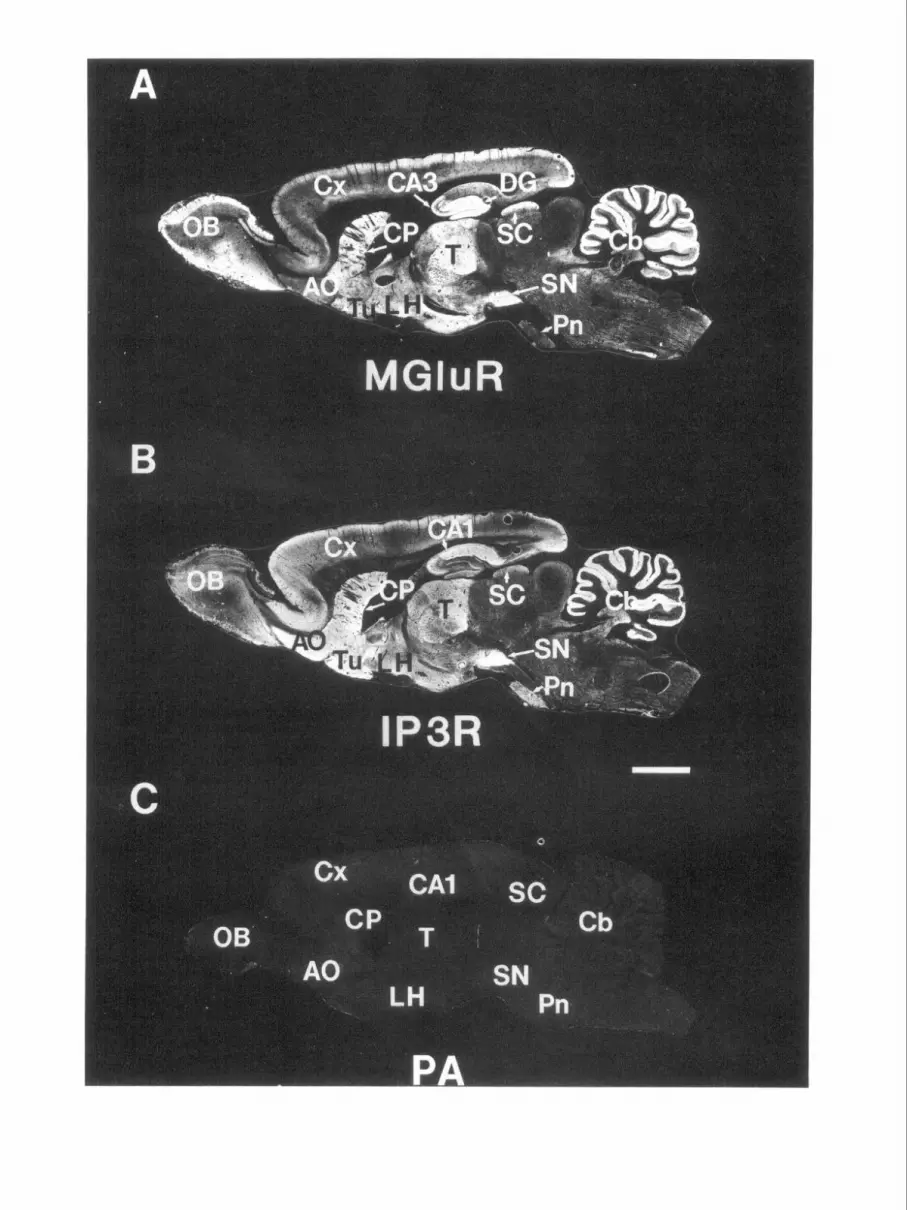
Figure 3. Immunohistochemical localization of mGluR1 and IP,R proteins in sagittal brain sections. Adjacent thick (40 pm) rat sagittal brain sections were stained with affinity-purified antibodies against mGluR1 (a and P) (A) and IP,R (B). C, Preadsorption (PA) of antibodies against mGluR 1 with its respective antigen completely abolishes immunostaining; similar results were obtained with the IP,R antibody (data not shown). Immunoreactive structures appear white in these dark-field images. Areas high in mGluR1 immunoreactivity but low in IP,R immunoreactivity include olfactory bulb (OS), CA3 of the hippocampus, dentate gyrus (DG), thalamus (T), granular layer of cerebellum (Cb), superior colliculus (SC), and lateral hypothalamus (LH). Areas low in mGluR 1 and high in IP,R immunostaining include the anterior olfactory nucleus (AO), olfactory tubercle (Tu), pyramidal cell layer of cortex (Cx), caudate-putamen (CP), and pontine nucleus (Pn). SN, substantia nigra. Scale bar, 2.5 mm. Figure 4. Contrasting localization of mGluR1 and IP,R immunoreactivity in coronal brain sections. Pairs of adjacent thick (40 pm) coronal rat brain sections were immunohistochemically processed with affinity-purified mGluR1 (left) and IP,R (right) antibodies. Positive staining appears white in these dark-field images. The contrasting distributions of these two proteins revealed by in situ hybridization in Figure 2 is more clearly apparent in these immunostained sections. Much higher amounts of mGluR1 staining are present in the olfactory bulb (OB) and in the following thalamic nuclei: anterodorsal (AD), anteroventral (A V), ventrolateral posterior (VEX), medial geniculate (MG), and nucleus gelatinosum (G). The lateral hypothalamus (LH), islands of Calleja (1Cj), mammillary bodies (M), and nucleus of Darkshevich (OK) also exhibit more mGluR1 than IP,R immunoreactivity. Similarly, in the brainstem, many of the cranial nuclei including the motor nucleus of the trigeminal nerve (5) as well as the substantia gelatinosa and ventral horn of spinal cord (SC) contain higher amounts of mGluR1 immunoreactivity. Areas with higher IP,R immunoreactivity depicted in these sections include the internal capsule (ic), CA1 of hippocampus, pars reticulata (the lower portion) of substantia nigra (SN), and Purkinje cell layer of cerebellum (Cb). DG, dentate gyrus; CP, caudate-putamen; Cx, cortex. Scale bar, 2.5 mm.

2008 Fotuhi et al. - mGluR1 Metabotropic Glutamate Receptor
MGIuR IP3R
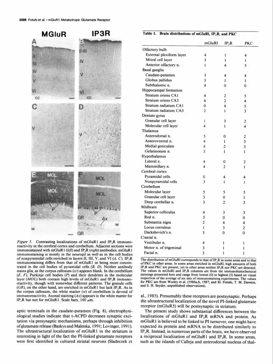
Figure 5. Contrasting localizations of mGluR1 and IP,R immuno- reactivity in the cerebral cortex and cerebellum. Adjacent sections were immunostained with mGluR 1 (left) and IP,R (right) antibodies. mGluR 1 immunostaining is mostly in the neuropil as well as in the cell bodies of nonpyramidal cells enriched in layers II, III, V, and VI (A, C). IP,R immunostaining differs from that of mGluR1 in being more concen- trated in the cell bodies of pyramidal cells (B, D). Neither antibody stains glia, as the corpus callosum (cc) appears blank. In the cerebellum (E, F), Purkinje cell bodies (P) and their dendrites in the molecular layer (MOL) both contain high levels of mGluR1 and IP,R immuno- reactivity, though with somewhat different patterns. The granule cells (CR), on the other hand, are enriched in mGluR1 but lack IP,R. As in the corpus callosum, the white matter (w) of cerebellum is devoid of immunoreactivity. Axonal staining (Ax) appears in the white matter for IP,R but not for mGluR1. Scale bars, 100 pm.
aptic terminals in the caudate-putamen (Fig. 8), electrophysi- ological studies indicate that t-ACPD decreases synaptic exci- tation via presynaptic mechanisms, perhaps through inhibition ofglutamate release (Baskys and Malenka, 199 1; Lovinger, 199 1). The ultrastructural localization of mGluR1 in the striatum is interesting in light of the fact the PI-linked glutamate receptors were first identified in cultured striatal neurons (Sladeczek et
Table 1. Brain distributions of mGluR1, IP,R, and PKC
mGluR1 IP,R PKC
Olfactory bulb External plexiform layer 4 1 4 Mitral cell layer 3 1 1 Anterior olfactory n. 1 4 5
Basal ganglia Caudate-putamen 3 4 4 Globus pallidus 3 1 1 Subthalamic n. 4 0 0
Hippocampal formation Stratum oriens CA 1 4 2 5 Stratum oriens CA3 4 2 4 Stratum radiatum CA 1 0 4 5 Stratum radiatum CA3 2 1 3
Dentate gyrus Granular cell layer 1 3 2 Molecular cell layer 4 1 4
Thalamus Anterodorsal n. 5 0 2 Anteroventral n. 4 1 3 Medial geniculate 4 2 3 Gelatinosum n. 3 1 1
Hypothalamus Lateral n. 4 0 2 Mammillary n. 4 2 1
Cerebral cortex Pyramidal cells 0 4 4 Nonpyramidal cells 3 1 1
Cerebellum Molecular layer 5 5 5 Granular cell layer 3 0 1 Deep cerebellar n. 3 2 3
Midbrain Superior colliculus 4 3 3 Red n. 3 0 0 Substantia nigra 2 4 4 Locus coeruleus 1 3 2 Darkshevich’s n. 5 0 0
Cranial n. Vestibular n. 4 1 1 Motor n. of trigeminal 3 1 1 Facial n. 3 1 1
The distribution of mGluR1 corresponds to that of IP,R in some areas and to that of PKC in other areas. In some areas enriched in mGluR1, high amounts of both IP,R and PKC are present, yet in other areas neither IP,R nor PKC are detected. The values in mGluRI and IP,R columns are from the immunohistochemical stainings presented here and range from lowest (0) to highest (5) based on visual inspection of the average of six sets of immunostaining experiments. The values for PKC are from Worley et aI. (1986a,b, 1987; and M. Fotuhi, T. M. Dawson, and S. H. Snyder, unpublished observations).
al., 1985). Presumably these receptors are postsynaptic. Perhaps the ultrastructural localization of the novel PI-linked glutamate receptor (mGluR5) will be postsynaptic in striatum.
The present study shows substantial differences between the localizations of mGluR1 and IP,R mRNA and protein. As mGluR 1 is believed to be linked to PI turnover, one might have expected its protein and mRNA to be distributed similarly to IP,R. Instead, in numerous parts of the brain, we have observed a reciprocal localization of mGluR1 and IP,R. In some areas, such as the islands of Calleja and anterodorsal nucleus of thal-

The Journal of Neuroscience, May 1993, 13(5) 2009
MGIuR IP3R
Fzgure 6. Reciprocal localizations of mGluR1 and IP,R immunoreactivity in the hiDDocamDa1 formation. In these bright-field images of an adjacent pair of sections immunostained with
amus, we observe high levels of mGluR1 in the complete ab- sence of IP,R. The PI cycle can be differentiated in terms of subtypes of PLC that have been selectively localized in the brain (Gerfen et al., 1988; Ross et al., 1989). Still, since the various forms of PLC all presumably generate IP,, one would not an- ticipate brain areas enriched in mGluR1 but devoid of IP,R. While other subtypes of IP,R have been identified, they appear to be of much lower abundance than the form of IP,R described here (Danoff et al., 1991; Nakagawa et al., 1991a,b; Stidhof et al., 1991; Ross et al., 1992). However, the minor subtypes of IP,R may be concentrated in certain brain regions that more closely parallel the distribution of mGluRla! and p. In such regions of dissociation of mGluR 1 from IP,R, it is conceivable that mGluR acts through the PI cycle primarily to activate PKC. Several areas that are devoid of IP,R possess high levels of both mGluR1 and PKC (Table 1). Consistent with this possibility, quisqualate, which potently activates mGluR, stimulates a rapid and transient translocation of PKC activity in striatal neurons (Manzoni et al., 1990). In addition, glutamate causes a transient phosphorylation of three PKC substrates in a time scale com- parable to DAG production (Scholz and Palfrey, 199 1).
Perhaps the distribution of mGluR5 may better fit with IP,R localizations in some brain regions. The existence of other sub- types of PI-linked mGluR as yet unidentified may also parallel the distribution IP,R better. For instance, in the molecular clon- ing studies, cross-hybridized cDNA clones were isolated from a cDNA library prepared from a certain size of mRNA (ap-
\ mGluR1 (left) and IP,R (right) anti- - bodies, durk areas represent positive
-- staining. Stratum oriens of CA1 and CA3 (Or in A and C) and CA4 contain an abundance of mGluR 1 -positive cells and processes in the absence of any ap- preciable IP,R immunostaining. The reverse occurs for pyramidal (Pv) cells and processes in stratum radiatum (Rad) of CAl, the lacunosum molec- ular (LMol), and granule cells of den- tate gyrus (GrDG). LD, lateral dorsal nucleus ofthalamus; DC, dentate gyrus; Mel, molecular layer of dentate gyrus. Scale bars, 100 pm.
proximately 3-4 kilonucleotides and greater than 4 kilonucleo- tides), and it is possible that a different size of cDNA may encode a distinct subtype of PI-linked mGluR. Furthermore, several groups have reported the possible existence of other subtypes of the PI-linked mGluR, such as the ibotenate-preferring mGluR (see Schoepp et al., 1990, for review).
We have also localized PI turnover by autoradiographical localization of 3H-CDP-DAG following the stimulation of mGluR1 by t-ACPD in the presence of 3H-cytidine. 3H-CDP- DAG accumulation is a reliable technique for visualization of PI cycle as it is stoichiometrically linked to agonist-stimulated inositol formation (Godfrey, 1989; Hwang et al., 1990a,b; Kennedy et al., 1990). Similar to contrasting localization be- tween mGluR1 and IP,R immunoreactivities, there are dis- crepancies in t-ACPD-stimulated PI turnover, mGluR 1, and IP,R immunoreactivity. For instance, 3H-CDP-DAG accu- mulation and mGluR1 are high in the external plexiform layer of the olfactory bulb, where minimal amounts of IP,R are de- tected (Fig. 9). Moreover, t-ACPD-stimulated PI turnover and IP,R are low in the molecular layer of the dentate gyrus, where there are high levels of mGluR1 protein. Both these areas con- tain abundant amounts of PKC, suggesting that the actions of mGluR1 may be mediated through the PKC limb of the PI cycle. We also noted a lack of correlation of mGluR1 protein levels as determined by Western blot analysis and t-ACPD- stimulated PI turnover assessed by 3H-CDP-DAG. Similar re- sults in the level of t-ACPD-stimulated PI turnover in various

2010 Fotuhi et al. l mGluR1 Metabotropic Glutamate Receptor
Figure 7. Reciprocal distribution of mGluR and IP,R immunoreactivity in other brain regions. Pairs of thick (40 pm) rat brain sections were immuno- stained with mGluR1 (left) and IP,R (right) antibodies. Positive staining ap- pears as dark areus in these bright-field images. In the olfactory bulb (A, B), mGluR1 immunostaining appears dense in the glomeruli (GZ), in the ex- ternal plexiform layer (EPZ), but low in the internal plexiform layer (ZPZ). In contrast, the IP,R immunostaining is enriched in periglomeruli cells (PC) and tufted cells (Z’ufl in the mitral cell layer (Mi), where mGluR1 protein level is low. In a pair of coronal sections (C, D) through rostra1 striatum, seen at high magnification, islands of Calleja (ZC’) and olfactory tubercle (Tu) contain ex- tremely high and low amounts of mGluR, respectively. The exact oppo- site occurs for IP,R immunoreactivity. Similarly, the arcuate nucleus (Arc), the lateral hypothalamic nucleus (HZ), subthalamic nucleus (STh), dorsal per- imammillary nucleus (PMD), fomix (A, and internal capsule (ic) exhibit recip- rocal immunostaining for mGluR and IP,R proteins. 3 V, third ventricle. Scale bar, 100 pm.
MGIuR IP3R
brain regions have been obtained by assessment of 3H-inositol phosphate accumulation, and there is also a marked discrepancy
depending on the relative distribution of the various compo- nents. Many other brain regions such as the subthalamic nu-
in these levels with the levels of mGluR1 mRNA (Condorelli et al., 1992).
cleus, red nucleus, and Darkshevich’s nucleus are highly en-
Together, these findings raise the possibility that mGluR1 riched in mGluR1 but are devoid of both IP,R and PKC,
may act through the PI cycle to activate primarily IP,R or PKC suggesting the possibility that mGluR1 may also act through other second messengers. For instance, activation of phospho-
Figure 8. Contrasting localization of mGluR 1 and IP,R viewed at the ultrastructural level. Electron micrographs show the pattern of mGluR l- immunoreactive (A) and IP,R-immunoreactive (B) structures. A depicts mGluR1 -immunoreactive terminal (star) forming an asymmetrical synaptic contact (region between arrowheads) with two postsynaptic structures (diamonds). B illustrates a dendrite containing IP,R immunoreactivity (star) receiving a synaptic contact (region between arrowheads) from a nonlabeled presynaptic terminal (diamond). Scale bar: 0.28 pm for A; 0.5 wm for R.
The Journal of Neuroscience, May 1993, 13(5) 2011
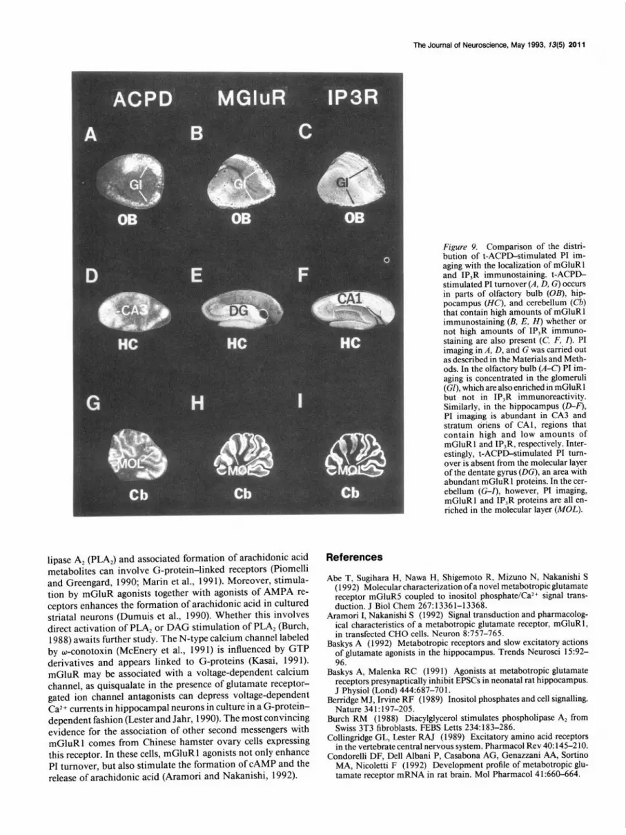
Fimre 9. Comparison of the distri- b&on of t-ACPD-stimulated PI im- aging with the localization of mGluR1 and IP,R immunostaining. t-ACPD- stimulated PI turnover (A, D, G) occurs in parts of olfactory bulb (OB), hip- pocampus (HC), and cerebellum (Cb) that contain high amounts of mGluR1 immunostaining (B, E, H) whether or not high amounts of IP,R immuno- staining are also present (C, F, I). PI imaging in A, D, and G was carried out as described in the Materials and Meth- ods. In the olfactory bulb (A-C) PI im- aging is concentrated in the glomeruli (GI). which are also enriched in mGluR1 \ - I I
but not in IP,R immunoreactivity. Similarly, in the hippocampus (D-F), PI imaging is abundant in CA3 and stratum oriens of CAl, regions that contain high and low amounts of mGluR1 and IP,R, respectively. Inter- estingly, t-ACPD-stimulated PI turn- over is absent from the molecular layer of the dentate gyrus (DC), an area with abundant mGluR1 proteins. In the cer- ebellum (G-Z), however, PI imaging, mGluR1 and IP,R proteins are all en- riched in the molecular layer (MOL).
lipase A, (PLA,) and associated formation of arachidonic acid metabolites can involve G-protein-linked receptors (Piomelli and Greengard, 1990; Marin et al., 199 1). Moreover, stimula- tion by mGluR agonists together with agonists of AMPA re- ceptors enhances the formation of arachidonic acid in cultured striatal neurons (Dumuis et al., 1990). Whether this involves direct activation of PLA, or DAG stimulation of PLA, (Burch, 1988) awaits further study. The N-type calcium channel labeled by w-conotoxin (McEnery et al., 1991) is influenced by GTP derivatives and appears linked to G-proteins (Kasai, 1991). mGluR may be associated with a voltage-dependent calcium channel, as quisqualate in the presence of glutamate receptor- gated ion channel antagonists can depress voltage-dependent Ca*+ currents in hippocampal neurons in culture in a G-protein- dependent fashion (Lester and Jahr, 1990). The most convincing evidence for the association of other second messengers with mGluR1 comes from Chinese hamster ovary cells expressing this receptor. In these cells, mGluR 1 agonists not only enhance PI turnover, but also stimulate the formation of CAMP and the release of arachidonic acid (Aramori and Nakanishi, 1992).
References
Abe T, Sugihara H, Nawa H. Shigemoto R, Mizuno N, Nakanishi S (1992) Molecular characterization of a novel metabotropic glutamate receptor mGluR5 coupled to inositol nhosnhate/Ca2+ signal trans- duction. J Biol Chem 267:13361-13368. -
Aramori I, Nakanishi S (1992) Signal transduction and pharmacolog- ical characteristics of a metabotropic glutamate receptor, mGluR 1, in transfected CHO cells. Neuron 8:757-765.
Baskys A (1992) Metabotropic receptors and slow excitatory actions of glutamate agonists in the hippocampus. Trends Neurosci 15:92- 96.
Baskys A, Malenka RC (1991) Agonists at metabotropic glutamate receptors presynaptically inhibit EPSCs in neonatal rat hippocampus. J Physiol (Lond) 444:687-701.
Berridge MJ, Irvine RF (1989) Inositol phosphates and cell signalling. Nature 341:197-205.
Burch RM (1988) Diacylglycerol stimulates phospholipase A, from Swiss 3T3 fibroblasts. FBBS Letts 234: 183-286.
Collingridge GL, Lester RAJ (1989) Excitatory amino acid receptors in the vertebrate central nervous system. Pharmacol Rev 40: 145-2 10.
Condorelli DF, Dell Albani P, Casabona AG, Genazzani AA, Sortino MA, Nicoletti F (1992) Development profile of metabotropic glu- tamate receptor mRNA in rat brain. Mol Pharmacol4 1:660-664.
2012 Fotuhi et al. * mGluR1 Metabotropic Glutamate Receptor
Danoff SK, Ferris CD, Donath C, Fischer G, Munemitsu S, Ullrich A, Snyder SH, Ross CA (199 1) Inositol 1,4,%trisphosphate receptors: distinct neuronal and non-neuronal forms derived by alternative splicing differ in phosphorylation. Proc Nat1 Acad Sci USA 88295 l- 2955.
Dumuis A, Pin JP, Oomagari K, Sebben M, Bockaert J (1990) Ara- chidonic acid released from striatal neurons by joint stimulation of ionotropic and metabotropic quisqualate receptors. Nature 347: 182- 184.
Ferris CD, Snyder SH (1992) Inositol 1,4,5-trisphosphate-activated calcium channels. Annu Rev Physiol 54:469+88.
Fotuhi M, Dawson TM, Sharp AH, Martin KJ, Graybiel AM, Snyder SH (199 1) The phosphoinositide second messenger system is en- riched in striosomes of the primate striatum. Sot Neurosci Abstr 17: 854.
Gerfen CR, Choi WC, Suh PG, Rhee SG (1988) Phospholipase C I and II brain isozymes: immunohistochemical localization in neuronal systems in rat brain. Proc Nat1 Acad Sci USA 85:3208-3212.
Godfrey PP (1989) Potentiation by lithium of CMP-phosphatidate formation in carbachol-stimulated rat cerebral-cortical slices and its reversal by myo-inositol. Biochem J 258:621-624.
Houamed KM, Kuijper JL, Gilbert TL, Haldeman BA, O’Hara PJ, Mulvihill ER, Almers W, Hagen FS (199 1) Cloning, expression, and gene structure of a G protein-coupled glutamate receptor from rat brain. Science 252:1318-1321.
Huang FL, Yoshida Y, Nakabayashi H, Young WS III, Huang K-P (1988) Immunocytochemical localization of protein kinase C iso- zymes in rat brain. J Neurosci 8:4734-4744.
Hwang PM, Bredt DS, Snyder SH (1990a) Autoradiographic imaging of phosphoinositide turnover in the brain. Science 249:802-804.
Hwang PM, Verma A, Bredt DS, Snyder SH (1990b) Localization of phosphatidylinositol signaling components in rat taste cells: role in bitter taste transduction. Proc Nat1 Acad Sci USA 87:7395-7399.
Kasai H (199 1) Tonic inhibition and rebound facilitation of a neuronal calcium channel by a GTP-binding protein. Proc Nat1 Acad Sci USA 88:8855-8859.
Kennedy ED, Cbalhss RAJ, Ragan CI, Nahorski SR (1990) Reduced inositol polyphosphate accumulation and inositol supply induced by lithium in stimulated cerebral cortex slices. Biochem J 267:781-786.
Lester RAJ, Jahr CE (1990) Quisqualate receptor-mediated depression of calcium currents in hippocampal neurons. Neuron 4:741-749.
Lovinger DM (199 1) Trans- 1 -aminocyclopentane- 1,3-dicarboxylic acid (t-ACPD) decreases synaptic excitation in rat striatal slices through a presynaptic action. Neurosci Lett 129: 17-2 1.
Manzoni OJJ, Finiels-Marlier F, Sassetti I, Blockaert J, le Peuch C, Sladeczek FAJ (1990) The glutamate receptor of the Qp-type acti- vates protein kinase C and is regulated by protein kinase C. Neurosci Lett l-09:146-151.
Marin P. Delumeau JC. Tence M. Cordier J. Glowinski J. Premont J (199 1) Somatostatin potentiates the oc,-adrenergic activation of phos- pholipase C in striatal astrocytes through a mechanism involving arachidonic acid and glutamate. Proc Nat1 Acad Sci USA 88:90 16- 9020.
Martin LJ, Blackstone CD, Huganir RL, Price DL (1992) Cellular localization of a metabotropic glutamate receptor in rat brain. Neuron 9~259-270.
Masu M, Tanabe Y, Tsuchida K, Shigemoto R and Nakanishi S (199 1) Sequence and expression of a metabotropic glutamate receptor. Na- ture 349~760-765.
Mayer ML, Westbrook GL (1987) The physiology of excitatory amino acids in the vertebrate central nervous system. Prog Neurobiol 28: 197-276.
McEnery MW, Snowman AM, Sharp AH, Adams ME, Snyder SH (1991) Purified w-conotoxin GVIA receptor of rat brain resembles a dihy- dropyridine-sensitive L-type calcium channel. Proc Nat1 Acad Sci USA 88:11095-l 1099.
Miller RJ (199 1 a) Metabotropic excitatory amino acid receptors reveal their true colors. Trends Pharmacol Sci 12:365-367.
Miller RJ (199 1 b) The revenge of the kainate receptor. Trends Neu- rosci 11:477-479.
Monaghan DT, Bridges RJ, Cotman CW (I 989) The excitatory amino acid receptors: their classes, pharmacology, and distinct properties in the function of the central nervous system. Annu Rev Pharmacol Toxicol 29:365402.
Nakagawa T, Okano H, Furuichi T, Aruga J, Mikoshiba K (1991a) The subtypes of the mouse inositol 1,4,5-trisphosphate receptor are expressed in tissue-specific and developmentally specific manner. Proc Nat1 Acad Sci USA 88:6244-6248.
Nakagawa T, Shiota C, Okano H, Mikoshiba K (199 1 b) Differential localization of alternative spliced transcripts encoding inositol 1,4,5- trisphosphate receptors in mouse cerebellum and hippocampus: in situ hybridization study. J Neurochem 57: 1807-l 8 10.
Nakanishi S, Maeda N, Mikoshiba K (199 1) Immunohistochemical localization of an inositol 1,4,5-triphosphate receptor, PdoO, in neural tissue: studies in developing and adult mouse brain. J Neurosci 11: 2075-2086.
Nicoletti F, Wroblewski JT, Fadda E, Costa E (1988) Pertussis toxin inhibits signal transduction at a specific metabotropic glutamate re- ceptor in primary cultures of cerebellar granule cells. Neurophar- macology 27:551-556.
Nordquist DT, Kozak CA, Orr HT (1988) cDNA cloning and char- acterization of three genes uniquely expressed in cerebellum by Pur- kinje neurons. J Neurosci 8:4780-4789.
Peng Y-W, Sharp AH, Snyder SH, Yau K-W (199 1) Localization of the inositol 1,4,5-trisphosphate receptor in synaptic terminals in the vertebrate retina. Neuron 6:525-53 1.
Piomelli D, Greengard P (1990) Lipoxygenase metabolites of arachi- donic acid in neuronal transmembrane signalling. Trends Pharmacol Sci 111367-373.
Ross CA, MacCumber MW, Glatt CE, Snyder SH (1989) Brain phos- pholipase C isozymes: differential mRNA localizations by in situ hybridization. Proc Nat1 Acad Sci USA 86~2923-2927.
Ross CA, Danoff SK, Schell MJ, Snyder SH, Ullrich A (1992) Novel inositol 1,4,5-trisphosphate receptors: molecular cloning and differ- ential localization in brain and peripheral tissues. Proc Nat1 Acad Sci USA 89:42654269.
Saito N, Kikkawa U, Nishizuka T, Tanaka C (1988) Distribution of protein kinase C-like immunoreactive neurons in rat brain. J Neurosci 8~369-382.
Schoepp D, Bockaert J, Sladeczek F (1990) Pharmacological and func- tional characteristics of metabotropic excitatory amino acid receptors. Trends Pharmacol Sci 11:508-515.
Scholz WK, Palfrey HC (199 1) Glutamate-stimulated protein phos- phorylation in cultured hippocampal pyramidal neurons. J Neurosci 11:2422-2432.
Sharp AH, Dawson TM, Ross CA, Fotuhi M, Mourey RJ, Snyder SH (1992) Inositol 1,4,%risphosphate receptors: immunohistochemical localization to discrete areas of rat brain. Neuroscience, in press.
Sladeczek F. Pin J-P. Rtcasens M. Bockaert J. Weiss S (1985) Glu- tamate stimulates inositol phosphate formation in striatal neurons. Nature 317:717-719.
Stidhof TC, Newton CL, Archer BT III, Mignery GA (199 1) Structure of a novel InsP, receptor. EMBO J 10:3 199-3206.
Tanabe Y, Masu M, Ishii T, Shigemoto R, Nakanishi S (1992) A family of metabotropic glutamate receptors. Neuron 8: 169-l 79.
Worley PF, Baraban JM, DeSouza EB, Snyder SH (1986a) Mapping second messenger systems in the brain: differential localizations of adenylate cyclase and protein kinase C. Proc Nat1 Acad Sci USA 83: 4053-4057.
Worley PF, Baraban JM, Snyder SH (1986b) Heterogeneous localiza- tion of protein kinase C in rat brain: autoradiographic analysis of phorbol ester receptor binding. J Neurosci 6: 199-207.
Worley PF, Baraban JM, Colvin JS, Snyder SH (1987) Inositol tris- phosphate receptor localization in brain: variable stoichiometry with protein kinase C. Nature 325: 159-l 6 1.
Worley PF, Baraban JM, Snyder SH (1989) Inositol 1,4,5-trisphos- phate receptor binding: autoradiographic localization in rat brain. J Neurosci 9:339-346.
Yoshihara C, Saito N, Taniyama K, Tanaka C (1991) Differential localization of four subspecies of protein kinase C in the rat striatum and substantia nigra. J Neurosci 11:690-700.
Young AB, Fagg GE (1990) Excitatory amino acid receptors in the brain: membrane binding and receptor autoradiographic approaches. In: The pharmacology of excitatory amino acids (Lodge D, Colling- ridge G, eds), pp 18-24. Cambridge: Elsevier.