developmental regulation of cryptdin, a corticostatin
TRANSCRIPT
Developmental Regulation of Cryptdin, a Corticostatin/Defensin Precursor mRNA in Mouse Small Intestinal Crypt Epithelium Andre J. Ouellette, * Rita M. Greco,* Marianne James,* Dana Frederick,* Janice Naflflan,* and John T. Fallon* • Cell Biology Unit and * Pathology Laboratory, Shriners Burns Institute, * Surgical Services and * Department of Pathology, Massachusetts General Hospital, Departments of * Surgery and * Pathology, Harvard Medical School, Boston, Massachusetts 02114
Abstract. Cryptdin mRNA codes for the apparent precursor to a corticostatin/defensin-related peptide that accumulates to high levels in mouse intestinal crypt epithelium during postnatal development. The primary structure, intestinal cell distribution, and de- velopmental appearance of cryptdin mRNA have been determined. Cryptdin mRNA is 450-480 nucleotides long. Translation of the partial cryptdin cDNA se- quence reveals a 70-amino acid open reading frame that includes 32 carboxy-terminal residues that align with those in the consensus sequence, C.CR... C....ER..G.C...G .... CCR, which is a common feature of leukocyte defensins and lung corticostatins (Selsted, M. E., D. M. Brown, R. J. DeLange, S. S. L. Harwig, and R. I. Lehrer. 1985. J. Biol. Chem. 260:4579-4584; Zhu, Q., J. Hu, S. Mulay, E Esch, S. Shimasaki, and S. Solomon. 1988. Proc. Natl.
Acad. Sci. USA. 85:592-596). In situ hybridization of cryptdin cDNA to paraformaldehyde-fixed, frozen sec- tions of adult jejunum and ileum showed intense and specific labeling of epithelial cells in the base of all crypts. Analysis of sections from suckling mice showed that cryptdin mRNA is detectable in 10-20% of crypts in 10-d-old mice, in •80% of crypts in 16- d-old mice, and in all crypts of mice 20 d and older. During the fourth week, the sequence accumulates in crypts to the maximal adult level. Cryptdin mRNA content in adult small intestine is independent both of T cell involvement and luminal bacteria. The role of cryptdin in small bowel physiology remains to be de- terrnined: cryptdin may inhibit bacterial translocation, modulate intestinal hormone synthesis, influence hor- monal sensitivity of the intestinal epithelium, or ex- hibit a multiplicity of related activities.
C ORTICOSTATINS (CS) j and defensins constitute a fam- ily of structurally related cysteine-rich peptides. Low molecular weight, cationic polypeptides termed "de-
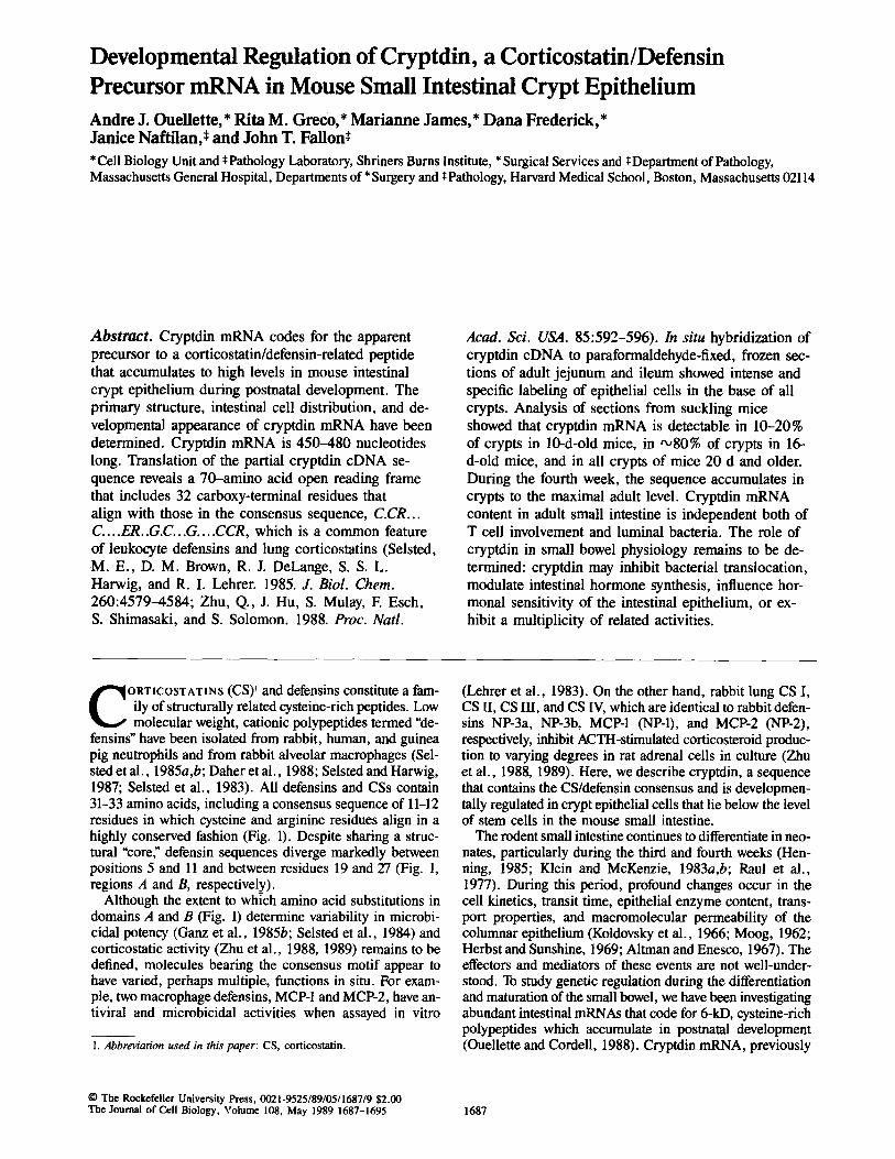
fensins" have been isolated from rabbit, human, and guinea pig neutrophils and from rabbit alveolar macrophages (Sel- sted et al., 1985a,b; Daher et al., 1988; Selsted and Harwig, 1987; Selsted et al., 1983). All defensins and CSs contain 31-33 amino acids, including a consensus sequence of 11-12 residues in which cysteine and arginine residues align in a highly conserved fashion (Fig. 1). Despite sharing a struc- tural "core; defensin sequences diverge markedly between positions 5 and 11 and between residues 19 and 27 (Fig. 1, regions A and B, respectively).
Although the extent to which amino acid substitutions in domains A and B (Fig. 1) determine variability in microbi- cidal potency (Ganz et al., 1985b; Selsted et al., 1984) and corticostatic activity (Zhu et al., 1988, 1989) remains to be defined, molecules bearing the consensus motif appear to have varied, perhaps multiple, functions in situ. For exam- ple, two macrophage defensins, MCP-1 and MCP-2, have an- tiviral and microbicidal activities when assayed in vitro
1. Abbreviation used in this paper: CS, corticostatin.
(Lehrer et al., 1983). On the other hand, rabbit lung CS I, CS II, CS III, and CS IV, which are identical to rabbit defen- sins NP-3a, NP-3b, MCP-1 (NP-1), and MCP-2 (NP-2), respectively, inhibit ACTH-stimulated corticosteroid produc- tion to varying degrees in rat adrenal cells in culture (Zhu et al., 1988, 1989). Here, we describe cryptdin, a sequence that contains the CS/defensin consensus and is developmen- tally regulated in crypt epithelial cells that lie below the level of stem cells in the mouse small intestine.
The rodent small intestine continues to differentiate in neo- nates, particularly during the third and fourth weeks (Hen- ning, 1985; Klein and McKenzie, 1983a,b; Raul et al., 1977). During this period, profound changes occur in the cell kinetics, transit time, epithelial enzyme content, trans- port properties, and macromolecular permeability of the columnar epithelium (Koldovsky et al., 1966; Moog, 1962; Herbst and Sunshine, 1969; Altman and Enesco, 1967). The effectors and mediators of these events are not well-under- stood. To study genetic regulation during the differentiation and maturation of the small bowel, we have been investigating abundant intestinal mRNAs that code for 6-kD, cysteine-rich polypeptides which accumulate in postnatal development (Ouellette and Cordell, 1988). Cryptdin mRNA, previously
© The Rockefeller University Press, 0021-9525/89/05/1687/9 $2.00 The Journal of Cell Biology, Volume 108, May 1989 1687-1695 1687
A B < . . . . . . . . . . . . . . . . . . . . . . . . . > < . . . . . . . . . . . . . . . . . . . . . . . . . . . . . . . . . >
CYS CYS ARG CIS GLU AIRG GLY CYS GLY CYS CYS ARG
Figure 1. The corticostatin/defensin consensus sequence. Residues shown denote amino acids of the CS/defensin consensus sequence. Un- derscored spaces indicate posit ions where sequences of CS/defensins diverge.
termed asb4/134, is one member of this class, and we now report on its coding function, site of intestinal expression, and aspects of its developmental regulation.
Materials and Methods
Animals Outbred Swiss mice [(CrI:CD-1)(ICR)BR] were 45-d-old males (30-35 g) or timed-pregnant dams from Charles River Breeding Laboratories, Inc. (Wilmington, MA); nude mice were Swiss-nu/nu purchased from Taconic Farms, Inc. (Germantown, NY); 28-d-old germ-free National Institutes of Health Swiss mice, shipped aseptically, also were purchased from Taconic Farms, Inc. Mice were housed under 12-h cycles of light and dark and had free access to standard rat chow and water.
Isolation of RNA from Mouse Small Intestine RNAs were deproteinized by selective precipitation from guanidine isothio- cyanate and guanidine hydrochloride (Chirgwin et al., 1979) followed by exhaustive extraction with phenol/chloruform/isoamyl alcohol (Perry et al., 1972; Ouellette and Cordell, 1988). After flushing with 50 ml ice-cold wa- ter, the small bowel was homogenized in 20 vol 4 M guanidine isothio- cyanate at room temperature. Except for in situ hybridization, samples of small bowel included the region from the pylorus to the ileocecal valve.
Blot Hybridization Hybridization experiments were conducted using DNA probes labeled with [32p]dCTP by nick-translation (Rigby et al., 1977) or with Klenow frag- ment after random oligonucleotide priming (Feinberg and Vogelstein, 1983, 1985). For RNA transfer blots (Northern blots), RNAs were separated in morpholino propane sulfonic acid (MOPS) formaldehyde-containing 2% agarose gels (Maniatis et al., 1982; .Rave et al., 1979) and transferred to Gene Screen Plus membranes (DuPont Co., Wilmington, DE; New En- gland Nuclear, Boston, MA) by capillarity (Thomas, 1980). Blots were hy- bridized and washed according to the recommendations of the manufac- turer.
In Situ Hybridization Freshly dissected mouse organs were fixed for 18 h at 4°C by immersion in 10 vol phosphate-buffered 4% paraformaldehyde solution containing 0.05% diethylpyrocarbonate, soaked overnight at 4°C in 30% sucrose in PBS, frozen in O.C.T. compound (Miles-Yeda Inc., Elkhart, IN) in dry ice- isopentane, and stored at -70°C before sectioning. Sections (5 #m) on slides coated with 50 #g/ml poly-L-lysine (Huang et al., 1983) were hybrid- ized 18 h to [35S]dCTP-labeled DNA probes (20,000-300,000 cpm) in 20 #1 buffer containing 0.3 M NaCI, 10 mM Tris (pH 7.5), 0.02 % Ficoll, 0.02 % polyvinyl pyrrolidone, 0.02% BSA, 1 mM EDTA, 10 mM DTT, 0.025% yeast tRNA, 10% dextran sulfate, and 50% deionized formamide (Angerer et al., 1987; Singer et al., 1986). Sections were sealed under coverslips using rubber cement, and slides were incubated overnight at 50°C in hu- midified polyethylene boxes. Slides were washed three times with gentle agitation, in 4x SSC at 37°C for 20 rain per wash, twice in 2x SSC for 30 min at 37°C, and once with 0.1x SSC for 30 min at 37°C. After dehydra- tion with successive ethanol washes in 0.3 M ammonium acetate, slides were coated with NTB-2 emulsion (Eastman Kodak Co., Rochester, NY), incubated at -20°C for 2-10 d, developed, and stained with hematoxylin and eosin.
DNA Sequence Analysis Cryptdin eDNA was subcloned into m13mp8/9 vectors and both strands
were sequenced by dideoxynucleotide random termination (Sanger et al., 1977) using [35S]dATP with modified Sequenase (United States Biochemi- cal Corp., Cleveland, OH) or Sequel SS (International Biotechnologies, Inc., New Haven, CT) DNA sequencing reagents. Sequencing gels were run according to the method of Biggin et al. (1983). Sequences were assembled and analyzed using the programs of Staden (1984, 1986) and the University of Wisconsin Genetics Computer Group (Devereux et al., 1985). The Na- tional Biomedical Research Foundation Protein Information Resource pro- tein sequence database (George et al., 1986) was searched for similarities to cryptdin and matches were aligned using IFIND (IntelliGenetics, Inc., Mountain View, CA) on the BIONET TM National Computer Resource for Molecular Biology (Kristofferson, 1987).
Results
Cryptdin cDNA corresponds to a developmentally regulated intestinal LMW mRNA. In a previous report, cryptdin (asb4/134) mRNA was shown to code for a 6-kD, cysteine- rich polypeptide, to accumulate in the third to fourth weeks of postnatal development, to be abundant in adult jejunum and ileum but not detectable in duodenum or colon, and to be present in testis and brain (Ouellette and CordeU, 1988).
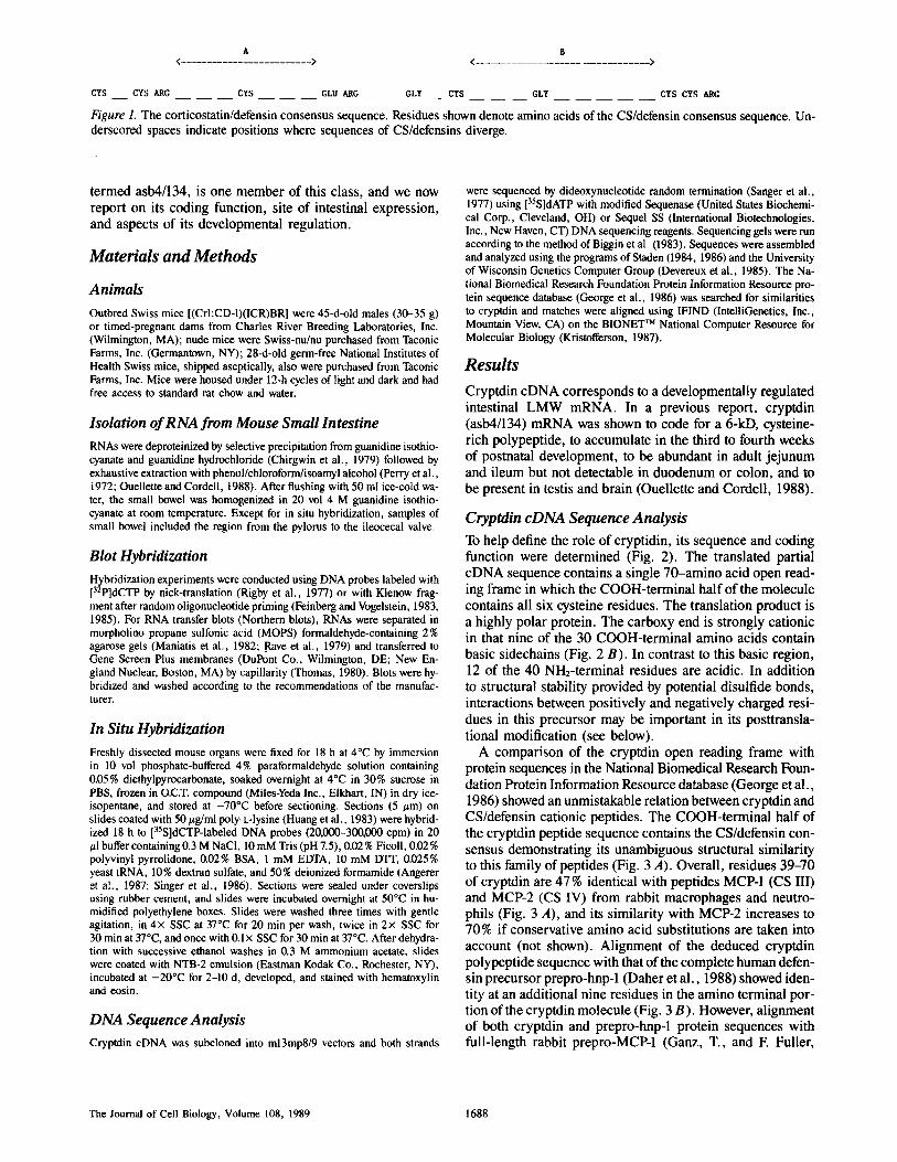
Cryptdin cDNA Sequence Analysis To help define the role of cryptidin, its sequence and coding function were determined (Fig. 2). The translated partial cDNA sequence contains a single 70-amino acid open read- ing frame in which the COOH-terminal half of the molecule contains all six cysteine residues. The translation product is a highly polar protein. The carboxy end is strongly cationic in that nine of the 30 COOH-terminal amino acids contain basic sidechains (Fig. 2 B). In contrast to this basic region, 12 of the 40 NH:-terminal residues are acidic. In addition to structural stability provided by potential disulfide bonds, interactions between positively and negatively charged resi- dues in this precursor may be important in its posttransla- tional modification (see below).
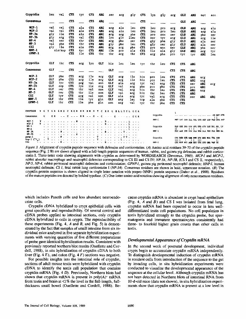
A comparison of the cryptdin open reading frame with protein sequences in the National Biomedical Research Foun- dation Protein Information Resource database (George et al., 1986) showed an unmistakable relation between cryptdin and CS/defensin cationic peptides. The COOH-terminal half of the cryptdin peptide sequence contains the CS/defensin con- sensus demonstrating its unambiguous structural similarity to this family of peptides (Fig. 3 A). Overall, residues 39-70 of cryptdin are 47 % identical with peptides MCP-1 (CS III) and MCP-2 (CS IV) from rabbit macrophages and neutro- phils (Fig. 3 A), and its similarity with MCP-2 increases to 70% if conservative amino acid substitutions are taken into account (not shown). Alignment of the deduced cryptdin polypeptide sequence with that of the complete human defen- sin precursor prepro-hnp-1 (Daher et al., 1988) showed iden- tity at an additional nine residues in the amino terminal por- tion of the cryptdin molecule (Fig. 3 B). However, alignment of both cryptdin and prepro-hnp-1 protein sequences with full-length rabbit prepro-MCP-1 (Ganz, T., and F. Fuller,
The Journal of Cell Biology, Volume 108, 1989 1688
1 P s t I 315
I I I
? ? . . .GGG GGA GAT GAA GAG ACT AAA ACT GAG GAG CAG CCA GGA GAA GAG GAC CAG GCC GTA TCT
. . . . . . . . . . . . + . . . . . . . . . . . . + . . . . . . . . . . . . + . . . . . . . . . . . . . + . . . . . . . . . . . . + . . . . . . . . . . . . +
. . . g l y g l y asp g l u g l u t h r l y s t h r g l u g l u g l n pro g l y g l u g l u asp g i n a l a v a l s e t
GTC TCC TTT GGA GAC CCA GAA GGC ACT TCT CTT CAA GAG ~AA TCG TTG AGA GAT CTG GTA . . . . . . . . . . . . + . . . . . . . . . . . . + . . . . . . . . . . . . + . . . . . . . . . . . . . + . . . . . . . . . . . . ÷ . . . . . . . . . . . . +
v a l s e t phe g l y asp pro g l u g l y t h r s e t l eu g i n g l u g l u s e t l eu a rg asp l eu v a l
TGC TAT TGT AGA TCA AGA GGC TGC AAA GGA AGA GAA CGC ATG AAT GGA ACC TGC AGA AAG . . . . . . . . . . . . + . . . . . . . . . . . . + . . . . . . . . . . . . + . . . . . . . . . . . . . + . . . . . . . . . . . . + . . . . . . . . . . . . +
CY$ t y r CYS ARG s e t a r g g l y CYS l y s g l y a r g GLU ARG met ash GLY t h r CYS a r g l y s
GGT CAT TTA TTG TAC ACG CTC TGC TGT CGC TGA ACA TGG AGA CCA CAG AGA ACA AGA CGA . . . . . . . . . . . . + . . . . . . . . . . . . + . . . . . . . . . . . . + . . . . . . . . . . . . + . . . . . . . . . . . . + . . . . . . . . . . . . +
GLY h i s l e u l e u t y r t h r l e u CYS CYS ARC End
GCA TGA GTA CTG AGG CCA CTG ATG CTG GTG CCT GAT GAC CAC TTC TCA ATA &AT TGT TCG . . . . . . . . . . . . + . . . . . . . . . . . + . . . . . . . . . . . + . . . . . . . . . . . . . + . . . . . . . . . . . + . . . . . . . . . . . . +
CAA TAT GAA AAA AA... . . . . . . . . . . . . + . . . .
Figure 2. Analysis of cryptdin cDNA sequence. Cryptdin cDNA was sequenced by dideoxynucleo- tide random termination (see Materials and Meth- ods) as illustrated in A. The sequence shown in B was assembled and translated (Materials and Meth- ods) to identify the single open reading frame shown. Residues corresponding to the CS/defensin consensus are shown in uppercase, bold face type. The first two glycine residues are not known with certainty, because the cDNA library was con- structed by homopolymeric tailing (Ouellette and Cordell, 1988).
personal communications) showed that only the asp-glu match at cryptdin residues 2-3 (HNP-1 residues 27-28) are con- served among these three sequences. As reported for HNP-I (Daher et al., 1988), comparisons of the amino terminal por- tion of cryptdin with sequences in Protein Information Re- source failed to identify any known related proteins (data not shown). RNA sequencing has shown that the 5' end of crypt- din mRNA extends an additional 115 nucleotides and codes, as in the case of HNP-1 (Daher et al., 1988) and MCP-1 (T. Ganz, personal communication) for a hydrophobic leader (data not shown). Although the cryptdin cDNA clone is not full-length, the existing data are unambiguous in demonstrat- ing that cryptdin mRNA codes for the propeptide form of a CS/defensin-related precursor of which the 32 COOH-ter- minal amino acids constitute the final product. The fact that the cryptdin gene locus maps to the proximal region of chro- mosome 8 (Ouellette, A. J., D. Pravtcheva, E H. Ruddle, and M. James, manuscript submitted for publication) at a ho- mologous position to the HNP-1 locus in the human (R. S. Sparkes, personal communication) provides further evidence of the relation of cryptdin to the defensins.
The similarity of cryptdin to other CS/defensins, is lower than that to MCP-1 and MCP-2 (Fig. 3 A). However, these estimates of apparent identity may be misleading, since, for several CS/defensins, the identity with cryptdin is almost wholly restricted to consensus residues. If consideration is limited to amino acids outside the consensus sequence, cryptdin and MCP-1 (CS III) and MCP-2 (CS IV) are identi- cal at only 2 of 19 and 3 of 19 positions, respectively (Fig. 3 C). Recently, several cryptdin-related cDNA clones have
been isolated whose DNA sequences are 90% identical with the 5' half of cryptdin mRNA but diverge in their 3' se- quences (Lualdi, J. C., and A. J. Ouellette, unpublished data). Thus, other cryptdin-related mRNAs exist in the small bowel.
Because analysis of whole organ RNA may be complicated by the dynamics and diversity of cell populations in complex organs, intestinal sections were hybridized to cryptdin cDNA probes to identify the intestinal cell population in which cryptdin mRNA is most abundant.
Cryptdin mRNA in Adult Small Bowel Crypts
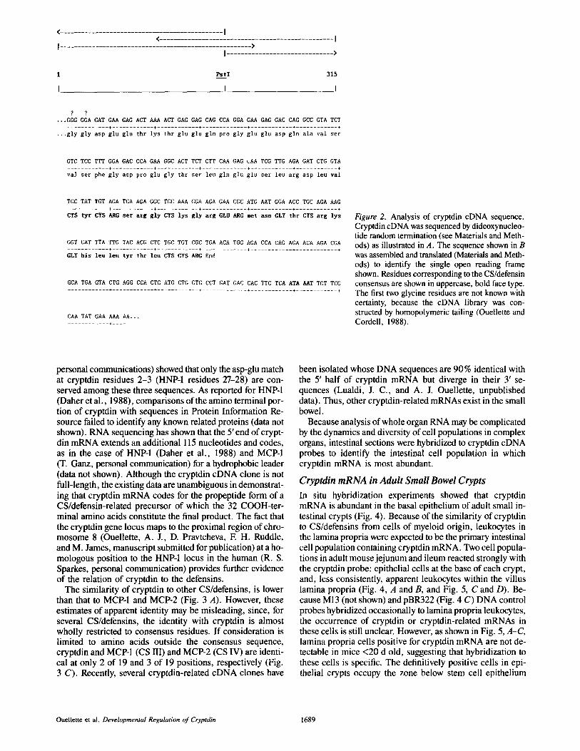
In situ hybridization experiments showed that cryptdin mRNA is abundant in the basal epithelium of adult small in- testinal crypts (Fig. 4). Because of the similarity of cryptdin to CS/defensins from cells of myeloid origin, leukocytes in the lamina propria were expected to be the primary intestinal cell population containing cryptdin mRNA. Two cell popula- tions in adult mouse jejunum and ileum reacted strongly with the cryptdin probe: epithelial cells at the base of each crypt, and, less consistently, apparent leukocytes within the villus lamina propria (Fig. 4, A and B, and Fig. 5, C and D). Be- cause M13 (not shown) and pBR322 (Fig. 4 C) DNA control probes hybridized occasionally to lamina propria leukocytes, the occurrence of cryptdin or cryptdin-related mRNAs in these cells is still unclear. However, as shown in Fig. 5, A-C, lamina propria cells positive for cryptdin mRNA are not de- tectable in mice <20 d old, suggesting that hybridization to these cells is specific. The definitively positive cells in epi- thelial crypts occupy the zone below stem cell epithelium
Ouellette et al. Developmental Regulation of Cryptdin 1689
C r y p t d i n l e u v a l CYS t y r CYS ARC s e r a rg g l y CYS l y s g l y a r g GLU ARG met asn
Consensus CYS CYS ARC CYS GLU ARG
MCP-I val val CYS ala CYS ARG arg ala leu CYS leu pro arg GLU ARG arg ala NCP-2 val val CYS ala CYS ARC arg ala leu CYS leu pro leu GLU ARG arg ala NP-3a gly ile CYS ala CYS ARG ar E arg phe CY$ pro asn ser GLU ARG phe ser NP-3b gly arg CYS val CY$ ARG lys gln 21eu CYS set tyr arg GLU ARG arg ile NP-4 val set CYS thr CYS ~ arg phe set CYS gly phe gly GLU ARG ala set NP-5 val phe CYS thr CYS ARG gly phe leu CYS gly ser gly GLU ~G ala set CSI gly ile CYS ala CY$ ARC ar E arg phe CYS pro asn ser GLU ARC phe ser IMP-I ala/asp CYS tyr CYS ARG ile pro ala CYS ile ala gly GLU ARG arg tyr GPNP-I CYS ile CYS thr thr ar E thr CYS ar E phe pro tyr tyr ARC leu
C r y p t d i n GLY th r CYS a rg lys GLY
Consensus GLY CYS
MCP-I GLY phe CYS arg ile arg MCP-2 GLY phe CYS arg ile ar E NP-3a GLY tyr CYS ar E val asn NP-3b GLY asp CYS lys ile a r g NP-4 GLY set CYS thr val asn NP-5 GLY set CYS thr ile asn CSI GLY tyr CYS are val asn • ~]P-I GLY thr CYS ile tyr gin GPNP-1 GLY th r CYS ile phe gln
his leu leu tyr thr leu CYS CYS ARC
GLY CYS CYS ARG
GLY arg ile his pro leu CYS CYS ~g a rg GLY arg ile his pro leu CYS CY$ ARC arg GL¥ ala arg tyr val arg CYS CYS set AP, G GLY v a l a rg phe pro phe ~J[S 6"IS pro b.RG GLY val a rg h i s t h r leu CYS CYS ARG a rg GLY val a rg his t h r leu CYS CYS ARG GL¥ ala arg tyr val arg CYS CYS set aRC GLY a r e leu t r p ala phe CYS CYS ash arg val tyr thr phe CYS CYS
a rg
ARG
C R Y F I g l N V C Y C R S R G C K G R E R M N G T C R K G H L L Y T L C C R
Consensus C - C R - - - C - - - E R - - g - C - - - g . . . . . C C R
N C P - I V . . . . . . . . . . R . . . . . . . R . . . . . . . L . . .
N C P - 2 V . . . . . . . . . . . . . . . . . . R . . . . . . . L . . ,
N P - 5 . . . . . . . . . . . . . . . . . . . . . . . . . . L . . . C r y p t d i n
NP-4 . . . . . . . . . . . . . . . . . . . . . . . . . . L . . . ~I'-I
NI'-3A . . . . . R . . . . . . . . . . . . R . . . . . Y . . . .
[qP-3B . . . . . . . . . . . R . . . . . . . . . . . . . . . . . .
g p n p - 1 . . . . . R . . . . . . . . . . T . . . . . . . Y T . • • C r y p t d i n h n p - 1 ( z / v ) . Y . . . . . . . . . . . . . . T . . . . . . L . . . . . .
C S I . . . . . . . R . . . . . . . . . . . . R . . . . . . . . . . . . . HNP-1
1 C r y p t d i n . . . . . . . . . . . . . . . . . . . . . . . . . GD E g g KTE
I I : ~ l F - 1 ~ T LAI LAA I L L VAL 0AO AEP LQA RAD EVA /tAP
EQP GEE DQA VSV SFG DPE GT$ L0E ESL RD LV
II I I I I I - EOI /tAD XPE VVV $LA ~/DE SLA PKH PGS RKN NA
C/C R.~t GC~ G~M i t l ~ GI~ m~G ~ L I TL CCR
I I I I I I I III II CYC RIP ACI A ~ ~dRY ~ IYQ ~ . L VAF CC
Figure 3. Alignment of cryptdin peptide sequence with defensins and corticostatins. (A) Amino acid residues 39-70 of the cryptdin peptide sequence (Fig. 2 B) are shown aligned with a full-length peptide sequences of human, rabbit, and guinea pig defensins and rabbit cortico- statin I. Three-letter code showing alignments with cryptdin as determined by WORDSEARCH (Devereux, 1985). MCP-1 and MCP-2, rabbit alveolar macrophage and neutrophil defensins corresponding to CS III and CS IV; NP-3A, NP-3B, (CS I and CS II, respectively), NP-5, NP-4, rabbit peritoneal neutrophil defensins and corticostatins; GPNP-1, guinea pig peritoneal neutrophil defensin; HNP-1, human neutrophil defensin; CS I, fetal rabbit lung corticostatin I (NP-3A). Consensus residues are shown in bold, uppercase notation. (B) The cryptdin protein sequence is shown aligned in single letter notation with prepro-HNP-1 protein sequence (Daher et al., 1988). Residues of the mature peptides are denoted by bolded typeface. (C) One-letter amino acid notation showing alignment of only nonconsensus residues.
which includes Paneth cells and less abundant neuroendo- crine cells.
Cryptdin cDNA hybridized to crypt epithelial cells with great specificity and reproducibility. Of several control and cDNA probes applied to intestinal sections, only cryptdin cDNA hybridized to cells in crypts. The reproducibility of these experiments (Fig. 4, A and B, and Fig. 5) is demon- strated by the fact that samples of small intestine from six in- dividual mice analyzed in five separate hybridization experi- ments with varying quantities of five different preparations of probe gave identical hybridization results. Consistent with previously reported northern blot results (Ouellette and Cor- dell, 1988), in situ hybridization of cryptdin cDNA to both liver (Fig. 4 E), and colon (Fig. 4 F) sections was negative.
For possible insights into the intestinal role of cryptdin, sections of adult mouse testis were hybridized with cryptdin cDNA to identify the testis cell population that contains cryptdin mRNA (Fig. 4 D). Previously, Northern blots had shown that cryptdin mRNA is present in poly(A) ÷ mRNA from testis and brain at <1% the level in the full-length, full- thickness small bowel (Ouellette and Cordell, 1988). Be-
cause cryptdin mRNA is abundant in crypt basal epithelium (Fig. 4, A and B) and CS I was isolated from fetal lung, cryptdin mRNA had been expected to occur in less well- differentiated testis cell populations. No cell population in testis hybridized strongly to the cryptdin probe, but sper- matogonia and immature spermatocytes consistently had three- to fourfold higher grain counts than other cells in testis.
Developmental Appearance of Cryptdin mRNA
In the second week of postnatal development, individual crypts begin to accumulate cryptdin mRNA independently. To distinguish developmental induction of cryptdin mRNA in resident cells from introduction of the sequence to the gut by invading cells, in situ hybridization experiments were conducted to visualize the developmental appearance of the sequence at the cellular level. Although cryptdin mRNA has not been detected in Northern blots of intestinal RNA from 10-d-old mice (data not shown), in situ hybridization experi- ments show that cryptdin mRNA is present at a low level in
The Journal of Cell Biology, Volume 108, 1989 1690
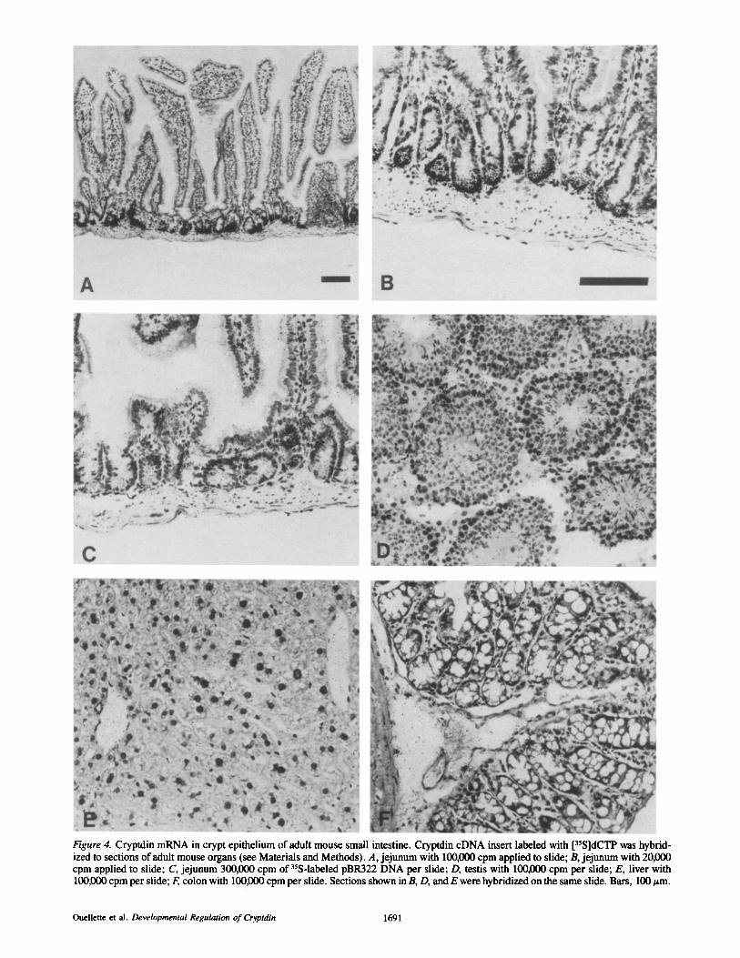
Figure 4. Cryptdin mRNA in crypt epithelium of adult mouse small intestine. Cryptdin cDNA insert labeled with [35S]dCTP was hybrid- ized to sections of adult mouse organs (see Materials and Methods). A, jejunum with 100,000 clam applied to slide; B, jejunum with 20,000 cpm applied to slide; C, jejunum 300,000 cpm of 35S-labeled pBR322 DNA per slide; D, testis with 100,000 cpm per slide; E, liver with 100,000 cpm per slide; F, colon with 100,000 cpm per slide. Sections shown in B, D, and E were hybridized on the same slide. Bars, 100 ~m.
Ouellette et al. Developmental Regulation of Cryptdin 1691
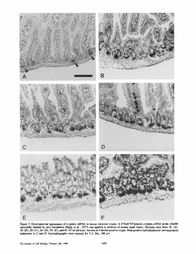
Figure 5. Developmental appearance of cryptdin mRNA in mouse intestinal crypts. A [3sS]dCTP-labeled cryptdin cDNA probe (50,000 cpm/slide) labeled by nick translation (Rigby et al., 1977) was applied to sections of mouse small bowel. Sections were from 10- (A), 16- (B), 20- (C), 24- (D), 30- (E), and 40- (F) d-old mice. Arrows in A denote positive crypts. Note positive hybridization to lamina propria leukocytes in C and D. Autoradiographs were exposed for 5 d. Bar, 100/~m.
The Journal of Cell Biology, Volume 108, 1989 1692
5-10% of crypts in 10-d-olds (Fig. 5 A). By 16 d of age, 70-80% of small intestinal crypts hybridize positively and with greater intensity than those of 10-d-old mice (Fig. 5 B). By day 20, all crypts are positive and more intensely labeled with the cryptdin cDNA probe (Fig. 5 C). During the re- mainder of postnatal development, cryptdin mRNA content increases to the maximal adult levels in all crypts (Fig. 5, D-F). Cryptdin mRNA has not been measured in aged mice, but 4-6-mo-old mice have the same cryptdin mRNA content as younger adults (unpublished data). The kinetics and anat- omy of cryptdin mRNA appearance in crypt epithelium are characteristic of Paneth development and consistent with the abundance of cryptdin mRNA in Paneth cells in the adult.
These results demonstrate that the cryptdin gene is in- duced in resident cells in the small bowel rather than being introduced by cells that migrate postnatally into the intestine. Because Paneth cells have been implicated in controlling mucosal microbes (Erlandsen and Chase, 1972) and differ morphologically in germ-free and naturally reared rodents (Satoh et al., 1986; Satoh and Vollrath, 1986), and because the cryptdin gene may be induced by local "factors" presented to individual crypts, the influence of luminal bacterial anti- gens and T cell-associated mediators were tested on cryptdin mRNA content in adult small bowel.
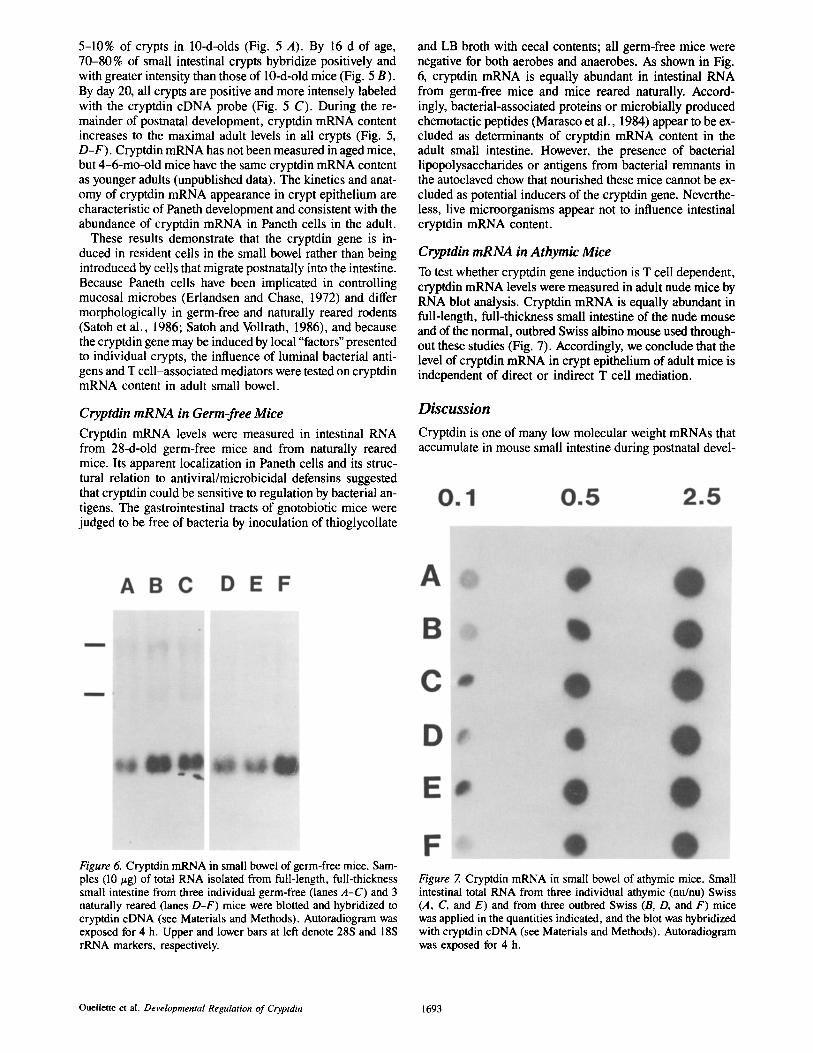
Cryptdin mRNA in Germ-free Mice
Cryptdin mRNA levels were measured in intestinal RNA from 28-d-old germ-free mice and from naturally reared mice. Its apparent localization in Paneth cells and its struc- tural relation to antiviral/microbicidal defensins suggested that cryptdin could be sensitive to regulation by bacterial an- tigens. The gastrointestinal tracts of gnotobiotic mice were judged to be free of bacteria by inoculation of thioglycollate
and LB broth with cecal contents; all germ-free mice were negative for both aerobes and anaerobes. As shown in Fig. 6, cryptdin mRNA is equally abundant in intestinal RNA from germ-free mice and mice reared naturally. Accord- ingly, bacterial-associated proteins or microbially produced chemotactic peptides (Marasco et al., 1984) appear to be ex- cluded as determinants of cryptdin mRNA content in the adult small intestine. However, the presence of bacterial iipopolysaccharides or antigens from bacterial remnants in the autoclaved chow that nourished these mice cannot be ex- cluded as potential inducers of the cryptdin gene. Neverthe- less, live microorganisms appear not to influence intestinal cryptdin mRNA content.
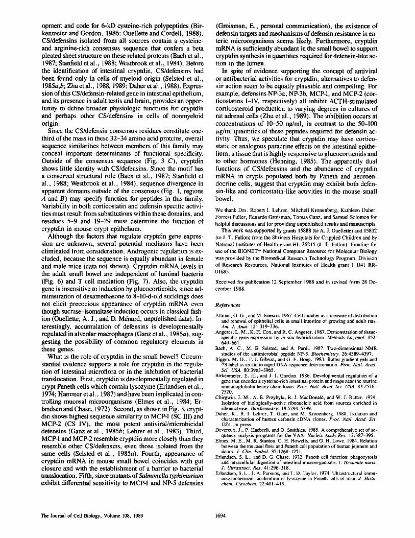
Cryptdin mRNA in Athymic Mice
To test whether cryptdin gene induction is T cell dependent, cryptdin mRNA levels were measured in adult nude mice by RNA blot analysis. Cryptdin mRNA is equally abundant in full-length, full-thickness small intestine of the nude mouse and of the normal, outbred Swiss albino mouse used through- out these studies (Fig. 7). Accordingly, we conclude that the level of cryptdin mRNA in crypt epithelium of adult mice is independent of direct or indirect T cell mediation.
Discussion
Cryptdin is one of many low molecular weight mRNAs that accumulate in mouse small intestine during postnatal devel-
Figure 6. Cryptdin mRNA in small bowel of germ-free mice. Sam- ples (i0 t~g) of total RNA isolated from full-length, full-thickness small intestine from three individual germ-free (lanes A-C) and 3 naturally reared (lanes D-F) mice were blotted and hybridized to cryptdin cDNA (see Materials and Methods). Autoradiogram was exposed for 4 h. Upper and lower bars at left denote 28S and 18S rRNA markers, respectively.
Figure 7. Cryptdin mRNA in small bowel of athymic mice. Small intestinal total RNA from three individual athymic (nu/nu) Swiss (A, C, and E) and from three outbred Swiss (B, D, and F) mice was applied in the quantities indicated, and the blot was hybridized with cryptdin cDNA (see Materials and Methods). Autoradiogram was exposed for 4 h.
Ouellette et al. Developmental Regulation of Cryptdin 1693
opment and code for 6-kD cysteine-rich polypeptides (Bir- kenmeier and Gordon, 1986; Ouellette and Cordell, 1988). CS/defensins isolated from all sources contain a cysteine- and arginine-rich consensus sequence that confers a beta pleated sheet structure on these related proteins (Bach et al., 1987; Stanfield et al., 1988; Westbrook et al., 1984). Before the identification of intestinal cryptdin, CS/defensins had been found only in cells of myeloid origin (Selsted et al., 1985a,b; Zhu et al., 1988, 1989; Daher et al., 1988). Expres- sion of this CS/defensin-related gene in intestinal epithelium, and its presence in adult testis and brain, provides an oppor- tunity to define broader physiologic functions for cryptdin and perhaps other CS/defensins in cells of nonmyeloid origin.
Since the CS/defensin consensus residues constitute one- third of the mass in these 32-34 amino acid proteins, overall sequence similarities between members of this family may conceal important determinants of functional specificity. Outside of the consensus sequence (Fig. 3 C), cryptdin shows little identity with CS/defensins. Since the motif has a conserved structural role (Bach et al., 1987; Stanfield et al., 1988; Westbrook et al., 1984), sequence divergence in apparent domains outside of the consensus (Fig. 1, regions A and B) may specify function for peptides in this family. Variability in both corticostatin and defensin specific activi- ties must result from substitutions within these domains, and residues 5-9 and 19-29 must determine the function of cryptdin in mouse crypt epithelium.
Although the factors that regulate cryptdin gene expres- sion are unknown, several potential mediators have been eliminated from consideration. Androgenic regulation is ex- cluded, because the sequence is equally abundant in female and male mice (data not shown). Cryptdin mRNA levels in the adult small bowel are independent of luminal bacteria (Fig. 6) and T cell mediation (Fig. 7). Also, the cryptdin gene is insensitive to induction by glucocorticoids, since ad- ministration of dexamethasone to 8-10-d-old sucklings does not elicit precocious appearance of cryptdin mRNA even though sucrase-isomaltase induction occurs in classical fash- ion (Ouellette, A. J., and D. M6nard, unpublished data). In- terestingly, accumulation of defensins is developmentally regulated in alveolar macrophages (Ganz et al., 1985a), sug- gesting the possibility of common regulatory elements in these genes.
What is the role of cryptdin in the small bowel? Circum- stantial evidence supports a role for cryptdin in the regula- tion of intestinal microflora or in the inhibition of bacterial translocation. First, cryptdin is developmentally regulated in crypt Paneth cells which contain lysozyme (Erlandsen et al., 1974; Hammer et al., 1987) and have been implicated in con- trolling mucosal microorganisms (Elmes et al., 1984; Er- landsen and Chase, 1972). Second, as shown in Fig. 3, crypt- din shows highest sequence similarity to MCP-1 (SC III) and MCP-2 (CS IV), the most potent antiviral/microbicidal defensins (Ganz et al., 1985b; Lehrer et al., 1983). Third, MCP-1 and MCP-2 resemble cryptdin more closely than they resemble other CS/defensins, even those isolated from the same cells (Selsted et al., 1985a). Fourth, appearance of cryptdin mRNA in mouse small bowel coincides with gut closure and with the establishment of a barrier to bacterial translocation. Fifth, since mutants of Salmonella typhimurium exhibit differential sensitivity to MCP-1 and NP-5 defensins
(Groisman, E., personal communication), the existence of defensin targets and mechanisms of defensin resistance in en- teric microorganisms seems likely. Furthermore, cryptdin mRNA is sufficiently abundant in the small bowel to support cryptdin synthesis in quantities required for defensin-like ac- tion in the lumen.
In spite of evidence supporting the concept of antiviral or antibacterial activities for cryptdin, alternatives to defen- sin action seem to be equally plausible and compelling. For example, defensins NP-3a, NP-3b, MCP-1, and MCP-2 (cor- ticostatins 1-IV, respectively) all inhibit ACTH-stimulated corticosteroid production to varying degrees in cultures of rat adrenal cells (Zhu et al., 1989). The inhibition occurs at concentrations of 10-50 ng/ml, in contrast to the 50-100 /~g/ml quantities of these peptides required for defensin ac- tivity. Thus, we speculate that cryptdin may have cortico- static or analogous paracrine effects on the intestinal epithe- lium, a tissue that is highly responsive to glucocorticoids and to other hormones (Henning, 1985). The apparently dual functions of CS/defensins and the abundance of cryptdin mRNA in crypts populated both by Paneth and neuroen- docrine cells, suggest that cryptdin may exhibit both defen- sin-like and corticostatin-like activities in the mouse small bowel.
We thank Drs. Robert I. Lehrer, Mitchell Kronenberg, Kathleen Daher, Forrest Fuller, Eduardo Groisman, Tomas Ganz, and Samuel Solomon for helpful discussions and for providing unpublished results and manuscripts.
This work was supported by grants 15888 (to A. J. Ouellette) and 15832 (to J. T. Fallon) from the Shriners Hospitals for Crippled Children and by National Institutes of Health grant HL-26215 (J. T. Fallon). Funding for use of the BIONET ~M National Computer Resource for Molecular Biology was provided by the Biomedical Research Technology Program, Division of Research Resources, National Institutes of Health grant 1 U41 RR- 01685.
Received for publication 12 September 1988 and in revised form 28 De- cember 1988.
References
AItman, G. G., and M. Enesco. 1967. Cell number as a measure of distribution and renewal of epithelial cells in small intestine of growing and adult rats. Am. J. Anat. 121:319-336.
Angerer, L. M., K. H. Cox, and R. C. Angerer. 1987. Demonstration of tissue- specific gene expression by in situ hybridization. Methods Enzymol. 152: 649-661.
Bach, A. C., M. E. Selsted, and A. Pardi. 1987. Two-dimensional NMR studies of the antimicrobial peptide NP-5. Biochemistry. 26:4389-4397.
Biggin, M. D., T. J. Gibson, and G. F. Hong. 1983. Buffer gradient gels and ~5S label as an aid to rapid DNA sequence determination. Proc. Natl. Acad. Sci. USA. 80:3963-3965.
Birkenmeier, E. H., and J. I. Gordon. 1986. Developmental regulation of a gene that encodes a cysteine-rich intestinal protein and maps near the murine immunoglobulin heavy chain locus. Proc. Natl. Acad. Sci. USA. 83:2516- 2520.
Chirgwin, J. M., A. E. Przybyla, R. J. MacDonald, and W. J. Rutter. 1979. Isolation of biologically-active ribonucleic acid from sources enriched in ribonuclease. Biochemistry. 18:5294-5299.
Daher, K., R. I. Lehrer, T. Ganz, and M. Kronenberg. 1988. Isolation and characterization of human defensin cDNA clones. Proc. Natl. Acad. Sci. USA. In press.
Devereux, J., P. Haeberli, and O. Smithies. 1985. A comprehensive set of se- quence analysis programs for the VAX. Nucleic Acids Res. 12:387-395.
Elmes, M. E., M. R. Stanton, C. H. Howells, and G. H. Lowe. 1984. Relation between the mucosal flora and Paneth cell population of human jejunum and ileum. J. Clin. PathoL 37:1268-1271.
Erlandsen, S. L., and D. G. Chase. 1972. Paneth cell function: phagocytosis and intracellular digestion of intestinal microorganisms. I. Hexamite nuris. J. Ultrastruct. Res. 41:296-318.
Erlandsen, S. L., J. A. Parsons, and T. D. Taylor. 1974. Ultrastructural immu- nocytochemical localization of lysozyme in Paneth cells of man. J. Histo- chem. Cytochem. 22:401-413.
The Journal of Cell Biology, Volume 108, 1989 1694
Feinberg, A. P., and B. Vogelstein. 1983. A technique for radiolabeling DNA restriction endonuclease fragments to high specific activity. Anal. Biochem. 132:6-13.
Feinberg, A. P., and B. Vogelstein. 1985. Addendum: a technique for radio- labeling DNA restriction endonuclease fragments to high specific activity. Anal. Biochem. 137:266-267.
Ganz, T., M. P. Sherman, M. E. Selsted, and R. I. Lehrer. 1985a. Newborn rabbit alveolar maerophages are deficient in two microbicidal cationic pep- tides, MCP-1 and MCP-2. Am. Rev. Respir. Dis. 132:901-904.
Ganz, T., M. E. Selsted, D. Szklarek, S. S. Harwig, K. Daher, D. F. Bainton, and R. I. Lehrer. 1985b. Defensins. Natural peptide antibiotics of human neutrophils. J. Clin. Invest. 76:1427-1435.
George, D. G., W. C. Barker, and L. T. Hunt. 1986. The Protein Identification Resource (PIR). Nucleic Acids Res. 14:11-15.
Guy-Grand, D., C. Griscelli, and P. Vassalli. 1978. The mouse gut T lympho- cyte, a novel type ofT cell. Nature, origin, and traffic in mice in normal and graft-versus-host conditions. ,I. Exp. Med. 148:1661-1677.
Hammer, M. F., J. W. Schilling, E. M. Prager, and A. C. Wilson. 1987. Recruitment of lysozyme as a major enzyme in the mouse gut: duplication, divergence, and regulatory evolution. J. Mol. Evol. 24:272-279.
Henning, S. J. 1985. Ontogeny of enzymes in the small intestine. Ann. Rev. Physiol. 47:231-245.
Herbst, J. J., and P. Sunshine. 1969. Postnatal development of the small intes- tine of the rat. Pediatr. Res. 3:27-33.
Huang, W. M., S. J. Gibson, P. Facer, J. Gu, andJ. M. Polak. 1983. Improved section adhesion for immunocytochemistry using high molecular weight polymers of L-lysine as a slide coating. Histochemistry. 77:275-279.
Klein, R. M., and J. C. McKenzie. 1983a. The role of cell renewal in the on- togeny of the intestine. I. Cell proliferation patterns in adult, fetal, and neo- natal intestine. J. Pediatr. Gastroenterol. Nutr. 2:10~.3.
Klein, R. M., and J. C. McKenzie. 1983b. The role of cell renewal in the on- togeny of the intestine. 11. Regulation of cell proliferation in adult, fetal, and neonatal intestine. J. Pediatr. Gastroenterol. Nutr. 2:204-228.
Koldovsky, O., P. Sunshine, and N. Kretchmer. 1966. Cellular migration of intestinal epithelia in suckling and weaned rats. Nature (Lond.). 212:1389- 1390.
Kristofferson, D. 1987. The BIONET electronic network. Nature (Lond.). 325: 555-556.
Lehrer, R. I., M. E. Selsted, D. Szklarek, and J. Fleischmann. 1983. Antibac- terial activity of microbicidal cationic proteins 1 and 2, natural peptide antibi- otics of rabbit lung macrophages. Infect. lmmun. 42:10-14.
Maniatis, T., E. F. Fritsch, and J. Sambrook. 1982. Molecular Cloning. A Lab- oratory Manual. Cold Spring Harbor Laboratory, Cold Spring Harbor, New York. 545 pp.
Marasco, W. A., S. H. Phan, H. Krutzsch, A. J. Showell, E. E. Fehner, R. Nairn, E. L. Becker, and P. A. Ward. 1984. Purification and identification of formyl-methionyl-leucylphenylalanine as the major peptide neutrophil chemotactic factor produced by Escherichia coil J. Biol. Chem. 259:5430- 5439.
Moog, F. 1962. Developmental adaptations of alkaline phosphatases in the small intestine. Fed. Proc. 21:51-56.
Ouellette, A. J., and B. Cordell. 1988. Accumulation of abundant messenger ribonucleic acids during postnatal development of mouse small intestine. Gastroenterology. 94:114-121.
Perry, R. P., J. LaTorre, D. E. Kelley, and J. R. Greenberg. 1972. On the labil- ity of poly(A) sequences during extraction of messenger RNA from polyribo- somes. Biochem. Biophys. Acta. 262:220-226.
Raul, R., P. Simon, M. Kedinger, and K. Haffen. 1977. Intestinal enzyme ac- tivities in isolated villus and crypt cells during postnatal development of the rat. Ceil. Tissue Res. 176:167-178.
Rave, N., R. Crkvenjakov, and H. Boedtker. 1979. Identification of procolla- gen mRNAs transferred to diaminobenzyloxymethyl paper from formalde- hyde gels. Nucleic Acids Res. 6:3559-3567.
Rigby, P. W. J., M. Deickmann, C. Rhodes, and P. Berg. 1977. Labeling DNA to high specific activity in vitro by nick translation with DNA polymerase I. J. Idol. Biol. 113:237-251.
Sanger, F., S. Nicklen, and A. R. Coulson. 1977. Proc. Natl. Acad. Sci. USA. 74:5463-5467.
Satoh, Y., and L. Vollrath. 1986. Quantitative electron microscopic observa- tions on Paneth cells of germfree and ex-germfree Wistar rats. Anat. Em- bryol. 173:317-322.
Satoh, Y., K. Ishikawa, K. Ono, and L. Vollrath. 1986. Quantitative light mi- croscopic observations on Paneth cells of germ-free and ex-germ-free Wistar rats. Digestion. 34:115-121.
Selsted, M. E., and S. S, Harwig. 1987. Purification, primary structure, and antimicrobial activities of a guinea pig neutrophil defensin. Infect. lmmun. 55:2281-2286.
Selsted, M. E., D. M. Brown, R. J. DeLange, and R. I. Lehrer. 1983. Primary structures of MCP-1 AND MCP-2, natural peptide antibiotics of rabbit lung macrophages. J. Biol. Chem. 258:14485-14489.
Selsted, M. E., D. Szklarek, and R. I. Lehrer. 1984. Purification and antibac- terial activity of antimicrobial peptides of rabbit granulocytes. Infect. lm- mun. 45:150-154.
Selsted, M. E., D. M. Brown, R. J. DeLange, S. S. L. Harwig, and R. I. Lehrer. 1985a. Primary structures of six antimicrobial peptides of rabbit peritoneal neutrophils. J. Biol. Chem. 260:4579-4584.
Selsted, M, E., S. S. Harwig, T. Ganz, J. W. Schilling, and R. I. Lehrer. 1985b. Primary structures of three human neutrophil defensins. J. Clin. In- vest. 76:1436-1439.
Singer, R. H., J. B. Lawrence, and C. Villnave. 1986. Optimization of in situ hybridization using isotopic and non-isotopic detection methods. Biotech- niques. 4:230-241.
Staden, R. 1984. Computer methods to aid the determination and analysis of DNA sequences. Biochem. Soc. Trans. 12:1005-1008.
Staden, R. 1986. The current status and portability of our sequence handling software. Nucleic Acids Res. 14:217-231.
Stanfield, R. L., E. M. Westbrook, and M. E. Selsted. 1988. Characterization of two crystal forms of human neutrophil peptide cationic peptide 1, a natu- rally occurring antimicrobial peptide of leukocytes. J. Biol. Chem. 263: 5933-5935.
Thomas, P. S. 1980. Hybridization of denatured RNA and small DNA frag- ments transferred to nitrocellulose. Proc. Natl. Acad. Sci. USA. 77:5201- 5205.
Westbrook, E., R. I. Lehrer, and M. E. Selsted. 1984. Characterization of two crystal forms of neutrophil cationic protein MCP-2, a naturally occurring broad-spectrum antimicrobial agent from leukocytes. J. Mol. Biol. 178: 783-785.
Zhu, Q., J. Hu, S. Mulay, F. Esch, S. Shimasaki, and S. Solomon. 1988. Iso- lation and structure of corticostatin peptides from rabbit fetal and adult lung. Proc. Natl. Acad. Sci. USA. 85:592-596.
Zhu, Q., A. Bateman, A. Singh, and S. Solomon. 1989. Isolation and biologi- cal activity of corticostatic peptides (anti-ACTH). Endocrin. Res. In press.
Ouellette et al. Developmental Regulation of Cryptdin 1695