developmental adipokines and maternal obesity interactions
TRANSCRIPT
ISSN 2347-6893
1189 | P a g e M a r c h 3 1 , 2 0 1 5
Developmental Adipokines and Maternal Obesity Interactions
R.G. Ahmed
Division of Anatomy and Embryology, Zoology Department, Faculty of Science, Beni-Suef University, Beni-Suef, Egypt
Email: [email protected] Tel. number: 002-010-9147-1828
Abstract
Adipokines are involved in the developmental programming during fetal development and early life, and might contribute to pregnancy complications. Maternal obesity has an impact on intrauterine fetal life that extends to abnormal changes in the adipose tissue and metabolic disorders in the newborns and even adulthood. This overview discusses the potential importance of adipokines during gestational diabetes mellitus (GDM) and in fetal metabolic programming. Various adipokines secreted from fat tissue as the key players in reprogramming maternal physiology to achieve an insulin-resistant state during pregnancy, especially when complicated by GDM. Indeed, this review hypothesized that the disturbance in adipokines may be associated with GDM in pregnant obese women or animals. This may influence, generally, on the health of the embryos, newborns and adulthood depending on proinflammatory markers. Finally, the obesity of mothers and disturbance in adipokines prior to pregnancy are a significant risk factor for the disturbed development of the pregnancy and the child, and develop metabolic syndrome. However, there are obvious species differences between pregnant women and animal models. Thus, maintaining normoglycaemia and adipokines levels during pregnancy may play an important role in a healthy life for the newborns.
Key words: Adipokines; obesity; mothers; development; offspring.
Council for Innovative Research
Peer Review Research Publishing System
Journal of Advances in Biology
Vol. 7, No. 1

ISSN 2347-6893
1190 | P a g e M a r c h 3 1 , 2 0 1 5
I- Introduction
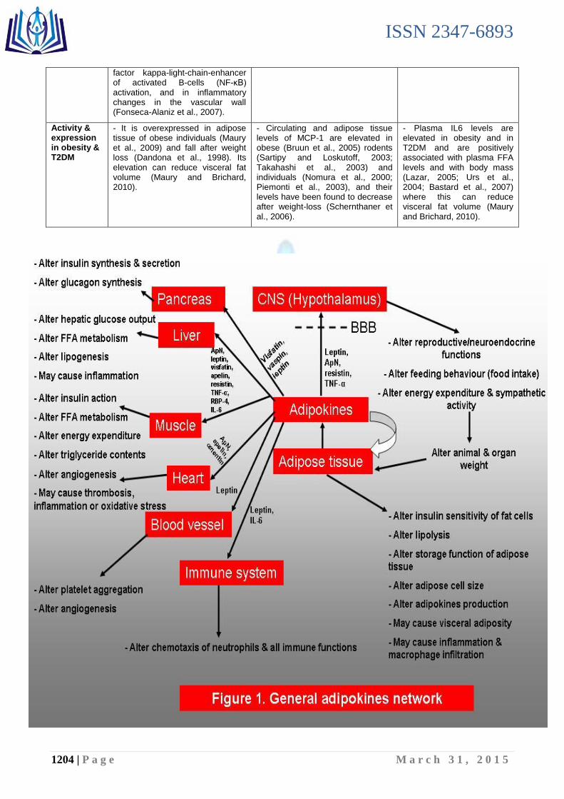
Obesity is powerfully linked with changes in the physiological function of adipose tissue, leading to insulin resistance, chronic inflammation, and altered secretion of adipokines (Bljajić et al., 2014; Prior et al., 2014; Ramirez et al., 2014). Indeed, adipose tissue dysfunction might play a critical role in the different obesity linked diseases including inflammation, insulin resistance and cancer. White adipose tissue (WAT) is a complex and metabolically active organ, with a relevant vital role in regulating whole-body metabolism (Houde et al., 2013). WAT is the largest energy storage organ, having an essential lipid storing capacity in periods when energy input exceeds energy expenditure and with a lipolytic function during energy deprivation (Jenum et al., 2013; Loy and Hamid Jan, 2014). In addition to its primary role as a fuel reservoir, WAT has been confirmed as a principal endocrine organ, since the tissue synthesizes and secretes an array of sex steroids, and bioactive peptides termed „adipokines‟, involved in the physiological regulation of fat storage, energy metabolism, food intake, insulin sensitivity, and immune function among others (Khalyfa et al., 2013). In this section, I summarized the general biological function of adipose tissue in table (1) and figure (1), and I compared between the secretion and action of potentially beneficial adipokines (leptin, adiponectin, apelin and visfatin) linking to obesity in table (2).
II- Summary about the different states of adiponectinaemia (Table 3).
III- Summary about the adipokines & insulin resistance interactions (figure 2).
IV- Role of inflammation in pathogenesis of insulin resistance, obesity and cardiovascular diseases (Table 4):
Increased adipose tissue mass that is linked with obesity and cardiovascular disease has been associated with a low-grade, chronic inflammatory response that is characterized by altered production of adipokines and increased markers of inflammation, such as TNF-α and IL6 (Stupin and Arabin, 2014) or MCP-1 (Harwood Jr, 2012). Over-stimulation of inflammatory pathways in insulin-sensitive tissues provides rise to local and systemic insulin resistance (Cai et al., 2005). Markers of systemic inflammation in humans are strongly connected with insulin resistance (de Rooij et al., 2009) and predict the development of diabetes type two (T2DM) (Ahmed, 2011; Miehle et al., 2012). Infact, acute TNF-α infusion induces skeletal muscle insulin resistance in humans (Plomgaard et al., 2005), while both acute and chronic MCP-1 infusion stimulates insulin resistance in rodents (Kalupahana et al., 2012). Notably, adipose tissue cells from the stromal vascular fraction, and in particular resident macrophages, are responsible for the chronic inflammatory responses recorded in obesity (Weisberg et al., 2003). These resident macrophages vary in their properties depending on whether they are contained in lean or fat-laden adipose tissue (Harwood Jr, 2012). For example, macrophages residing in lean adipose tissue are illustrated by increased expression of anti-inflammatory cytokines, such as interleukin 10 (IL-10), explain an increased capacity for tissue repair and angiogenesis, and are frequently referred to as M2, or alternatively-activated macrophages (Lumeng et al., 2007). However, expansion of adipose tissue in obesity is linked with an increased infiltration by circulating macrophages of the M1, or classically-activated, phenotype (Coenen et al., 2007). These macrophages are characteristically recruited to sites of tissue damage and are in a pro-inflammatory state with increased expression of proinflammatory cytokines, such as TNF-α and IL6, that can exert strong paracrine effects on a variety of adipose tissue functions (Lumeng et al., 2007). The local effects of proinflammatory cytokines on adipose tissue lipolysis may contribute to the development of insulin resistance by promoting the liberate of fatty acids from adipose tissue into the circulation, which may then lead to lipid accumulation and insulin resistance in other tissues such as skeletal muscle and liver (Goossens, 2008). Furthermore, reduced adiponectin concentrations may have harmful effects on fat oxidation, since it has been demonstrated that adiponectin increases fat oxidation via activation of AMP-activated protein kinase in rat skeletal muscle and myocytes (C2C12) (Yamauchi et al., 2002). Interestingly, in humans, low adiponectin serum levels at baseline independently predict future risk to develop T2DM (Spranger et al., 2003) and coronary artery disease (Fasshauer et al., 2004) and high plasma adiponectin predicts a lower risk of future myocardial infarction (Pischon et al., 2004). I summarized the different states for adiponectinaemia in table 3. It is obvious from these studies that there are linking between insulin receptor signaling and inflammatory pathways (Hirosumi et al., 2002). Generally, the detailed of pro-inflammatory cytokines and chemokines during obesity are discussed in table 4.
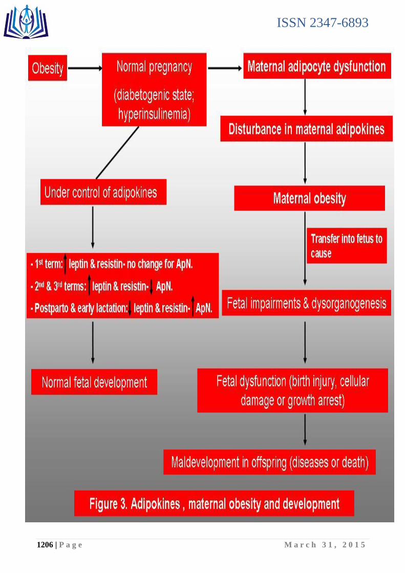
V- Adipokines, maternal obesity and development (figure 3):
Gestation is a stage that results in increased adipocyte volume, and it is recognized that adipose fat depots are the net balance of synthesis and hydrolysis of triacylglycerols via lipogenesis and lipolysis (Ramos et al., 2003). Also, several adipokines such as leptin (LEP; OMIM 164160), adiponectin (ApN; OMIM 605441),

ISSN 2347-6893
1191 | P a g e M a r c h 3 1 , 2 0 1 5
resistin (RETN; OMIM 605565) and inflammatory cytokines [tumor necrosis factor-α (TNF-α; OMIM 191160), and interleukin-6 (IL-6; OMIM 147620)], and chemokines [interleukin-8 (IL-8; OMIM 146930)] have been suggested as much stronger predictors of pregnancy-linked insulin resistance than gestational hormones, including human placental lactogen and steroids (Radaelli et al., 2003; Taylor et al., 2014). Recently, adiponectin secretion and adiponectin mRNA levels in WAT decline with the gestation progress suggesting that there are pregnancy-associated factors that reduce adiponectin levels (Catalano et al., 2006). In addition, in humans, the developing placenta expresses both leptin and its receptor, and placental resistin (Zavalza-Gómez et al., 2008). Their levels correlate with the condition of reduced insulin sensitivity often developed in the latter stages of pregnancy, thus contributing to successful development of the fetus (Cortelazzi et al., 2007). Furthermore, the human placenta has been found to express nearly all known cytokines. Increasing adiposity is associated with the secretion of proinflammatory cytokines from adipose tissue, suggesting that these cytokines may play an essential role in fuel availability during pregnancy (Lyon et al., 2003). Moreover, cytokine production is related to pathologic risk and to delivery mode itself (Malamitsi-Puchner et al., 2005). There have been only a few papers demonstrating cytokine profiles, including many kinds of cytokines in cord blood (Takahashi et al., 2010). IL-6 is a useful marker for a systemic fetal inflammatory response (Gomez et al., 1998). Also, the levels of IL-8, MCP-1 and MIP-1b were increased in preterm birth (Matoba et al., 2009) and serum levels of IL-8 and MCP-1 in cord blood significantly associated with gestational ages (Takahashi et al., 2010). However, IL-4 and IL-13 did not cross the placenta in measurable amounts (Lim et al., 2009). In previous investigations, circulating levels of leptin, adiponectin and TNF-α in the early pregnancy strongly predicted the development of GDM (Gao et al., 2008). Also, GDM elicits main variations in the expression profile of placental genes with a prominent increase in markers and mediators of inflammation (Radaelli et al., 2003).
Previous studies have reported a connection between childhood maltreatment and adulthood obesity (Midei et al., 2010). Obesity is also associated with lowered serum adiponectin levels which reflect a disturbed inflammatory state during childhood (Gustafson, 2010). Adiponectin inhibits the functions of TNF-α which associated with post-traumatic stress disorder (Gustafson, 2010) and inhibits the secretion of several other pro-inflammatory cytokines from endothelial cells (Gustafson, 2010). This can increase the childhood vulnerability to depression and permanent hyperactivity of the sympathetic nervous system, as well as a variety of conditions linked to disturbed inflammatory systems (Charmandari et al., 2003). Based on these observations, placental-derived hormones are supposed to be a main factor in reprogramming maternal physiology to achieve an insulin-resistant state. However, more studies are essential about this issue.
VI- Future directions
- Discover the ranges of several adipokines serum levels at which obesity-related disorders are observed with high frequency. These measurements could therefore be used as a biologic marker and/or pharmacologic agent in the management of obesity, inflammatory, metabolic, and cardiovascular disorders.
- Determine the complex nature of several adipokines signaling and their specific roles during pregnancy.
VII- Conflict of Interest: There is no conflict of interest.
VIII- References
1. Ahima RS. Adipose tissue as an endocrine organ. Obesity 2006;14:242S-249S.
2. Ahima RS. Revisiting leptin‟s role in obesity and weight loss. J. Clin. Invest. 2008;118:2380–2383.
3. Ahmed, R.G., 2011. Evolutionary interactions between diabetes and development. Diabetes Res. and Clin. Pract. J. 92, 153-167.
4. Badman MK, Flier JS. The adipocyte as an active participant in energy balance and metabolism. Gastroenterology 2007;132:2103–2115.
5. Baggiolini M. Chemokines and leukocyte traffic. Nature 1998;392(6676):565–8.
6. Bai Y, Zhang S, Kim KS, Lee JK, Kim KH. Obese gene expression alters the ability of 30A5 preadipocytes to respond to lipogenic hormones. J Biol Chem. 1996;271(24):13939-42.
7. Bastard JP, Lagathu C, Caron M, Capeau J. Point-counterpoint: interleukin-6 does/does not have a beneficial role in insulin sensitivity and glucose homeostasis. J. Appl. Physiol. 2007;102:821–822.
8. Berg AH, Combs TP, Du X, Brownlee M, Scherer PE. The adipocyte-secreted protein Acrp30 enhances hepatic insulin action. Nat Med. 2001;7(8):947-53.

ISSN 2347-6893
1192 | P a g e M a r c h 3 1 , 2 0 1 5
9. Bljajić D, Blajić J, Ivanišević M, Berberović E, Ɖelmiš J,Mayer D, Tuškan K. The impacts of gestational diabetes and obesity of mothers on insulin resistance and adipokines levels in the umbilical blood. Gynaecol Perinatol 2014;23(1):25–29.
10. Bogan JS, Lodish HF. Two compartments for insulin-stimulated exocytosis in 3T3–L1 adipocytes defined by endogenous ACRP30 and GLUT4. J Cell Biol 1999;146(3):609–20.
11. Boucher J, Masri B, Daviaud D, Gesta S, Guigne C, Mazzucotelli A, Castan-Laurell I, Tack I, Knibiehler B, Carpene C, Audigier Y, Saulnier-Blache JS, Valet P. Apelin, a newly identified adipokine up-regulated by insulin and obesity. Endocrinology 2005;146:1764–1771.
12. Brichard SM, Delporte ML, Lambert M. Adipocytokines in anorexia nervosa: a review focusing on leptin and adiponectin. Horm. Metab Res. 2003;35:337–342.
13. Bruun JM, Lihn AS, Pedersen SB, Richelsen B. Monocyte chemoattractant protein-1 release is higher in visceral than subcutaneous human adipose tissue (AT): implication of macrophages resident in the AT. J. Clin. Endocrinol. Metab. 2005;90:2282–2289.
14. Bruun JM, Lihn AS, Verdich C, Pedersen SB, Toubro S, Astrup A, Richelsen B. Regulation of adiponectin by adipose tissue-derived cytokines: in vivo and in vitro investigations in humans. Am. J. Physiol. Endocrinol. Metab. 2003;285:E527–E533.
15. Bruun JM, Pedersen SB, Richelsen B. Regulation of interleukin 8 production and gene expression in human adipose tissue in vitro. Journal of Clinical Endocrinology and Metabolism 2001;86:1267–1273.
16. Cai D, Yuan M, Frantz DF, Melendez PA, Hansen L, Lee J, Shoelson SE. Local and systemic insulin resistance resulting from hepatic activation of IKK-beta and NF-kappaB. Nat. Med. 2005;11 (2):183–190.
17. Cao YL, Hu CZ, Meng X, Wang DF, Zhang J. Expression of TNF-alpha protein in omental and subcutaneous adipose tissue in obesity. Diabetes Res Clin Pract 2008;79(2):214–9.
18. Capeau J. The story of adiponectin and its receptors AdipoR1 and R2: to follow. J. Hepatol. 2007;47:736–738.
19. Carbone F, La Rocca C, Matarese G. Immunological functions of leptin and adiponectin. Biochimie. 2012;94(10):2082-8.
20. Catalano PM, Hoegh M, Minium J, Huston-Presley L, Bernard S, Kalhan S, Hauguel-De Mouzon S. Adiponectin in human pregnancy: implications for regulation of glucose and lipid metabolism. Diabetologia. 2006;49(7):1677-85.
21. Cekmez F, Canpolat FE, Pirgon O, Çetinkaya M, Aydinoz S, Suleymanoglu S, Ipcioglu OM, Sarici SU. Apelin, vaspin, visfatin and adiponectin in large for gestational age infants with insulin resistance. Cytokine 2011;56:387–391.
22. Charmandari E, Kino T, Souvatzoglou E, Chrousos GP. Pediatric stress: hormonal mediators and human development. Horm Res 2003;59:161–79.
23. Christiansen T, Richelsen B, Bruun JM. Monocyte chemoattractant protein-1 is produced in isolated adipocytes, associated with adiposity and reduced after weight loss in morbid obese subjects. Int J Obes (Lond) 2005;29(1):146–50.
24. Coenen KR, Gruen ML, Chait A, Hasty AH. Diet-induced increases in adiposity, but not plasma lipids, promote macrophage infiltration into white adipose tissue. Diabetes 2007;56:564-573.
25. Cortelazzi D, Corbetta S, Ronzoni S, Pelle F, Marconi A, Cozzi V, Cetin I, Cortelazzi R, Beck-Peccoz P, Spada A. Maternal and foetal resistin and adiponectin concentrations in normal and complicated pregnancies. Clin Endocrinol (Oxf). 2007;66(3):447-53.
26. Delporte ML, Funahashi T, Takahashi M, Matsuzawa Y, Brichard SM. Pre-and post-translational negative effect of beta-adrenoceptor agonists on adiponectin secretion: in vitro and in vivo studies. Biochem. J. 2002;367:677–685.
27. Einstein FH, Atzmon G, Yang XM, Ma XH, Rincon M, Rudin E, Muzumdar R, Barzilai N.Differential responses of visceral and subcutaneous fat depots to nutrients. Diabetes. 2005;54(3):672-8.
28. Emilsson V, Liu YL, Cawthorne MA, Morton NM, Davenport M. Expression of the functional leptin receptor mRNA in pancreatic islets and direct inhibitory action of leptin on insulin secretion. Diabetes 1997;46:313-316.
29. Engeli S, Schling P, Gorzelniak K, Boschmann M, Janke J, Ailhaud G, Teboul M, Massiera F, Sharma AM. The adipose-tissue rennin–angiotensin– aldosterone system: role in the metabolic syndrome? Int. J. Biochem. Cell Biol. 2003;35:807–825.
30. Farooqi IS, Bullmore E, Keogh J, Gillard J, O‟Rahilly S, Fletcher PC. Leptin regulates striatal regions and human eating behavior. Science 2007;317:1355.
31. Flier JS. Clinical review 94: What‟s in a name? In search of leptin‟s physiologic role. J. Clin. Endocrinol. Metab. 1998;83:1407–1413.
32. Fonseca-Alaniz MH, Takada J, Alonso MIC, Lima FB. Adipose tissue as an endocrine organ: from theory to practice. J. Pediatria. 2007;83 (Suppl. 5):S192-S203.
ISSN 2347-6893
1193 | P a g e M a r c h 3 1 , 2 0 1 5
33. Funahashi T, Nakamura T, Shimomura I, Maeda K, Kuriyama H, Takahashi M, Arita Y, Kihara S, Matsuzawa Y. Role of adipocytokines on the pathogenesis of atherosclerosis in visceral obesity. Intern Med. 1999;38(2):202-6.
34. Furukawa S, Fujita T, Shimabukuro M, Iwaki M, Yamada Y, Nakajima Y, Nakayama O, Makishima M, Matsuda M, Shimomura I. Increased oxidative stress in obesity and its impact on metabolic syndrome. J. Clin. Invest. 2004;114:1752–1761.
35. Gao XL, Yang HX, Zhao Y. Variations of tumor necrosis factor-alpha, leptin and adiponectin in mid-trimester of gestational diabetes mellitus. Chin. Med. J. (Engl). 2008;121:701–705.
36. Garten A, Petzold S, Korner A, Imai SI, Kiess W. Nampt: linking NAD biology, metabolism and cancer. Trends Endocrinol. Metab. 2009;20: 130–138.
37. Gerhardt CC, Romero IA, Cancello R, Camoin L, Strosberg AD. Chemokines control fat accumulation and leptin secretion by cultured human adipocytes. Mol Cell Endocrinol 2001;175(1–2):81–92.
38. German AJ, Ryan VH, German AC, Wood IS, Trayhurn P. Obesity, its associated disorders and the role of inflammatory adipokines in companion animals. The Veterinary J. 2010;185:4-9.
39. Goossens GH. The role of adipose tissue dysfunction in the pathogenesis of obesity-related insulin resistance. Physiology & Behavior 2008;94:206–218.
40. Groeneveld MP, Huang-Doran I, Semple RK. Adiponectin and leptin in human severe insulin resistance e Diagnostic utility.
and biological insights. Biochimie. 2012, 94(10):2172-2179.
41. Guo K, McMinn JE, Ludwig T, Yu YH, Yang G, Chen L, Loh D, Li C, Chua Jr. S, Zhang Y. Disruption of peripheral leptin signaling in mice results in hyperleptinemia without associated metabolic abnormalities. Endocrinology 2007;148:3987–3997.
42. Gustafson B. Adipose tissue, inflammation and atherosclerosis. J Atheroscler Thromb 2010;17:332–41.
43. Hajer GR, van Haeften TW, Visseren FLJ. Adipose tissue dysfunction in obesity, diabetes and vascular diseases. Eur. Heart J. 2008;29:2959-2971.
44. Halleux CM, Takahashi M, Delporte ML, Detry R, Funahashi T, Matsuzawa Y, Brichard SM. Secretion of adiponectin and regulation of apM1 gene expression in human visceral adipose tissue. Biochem. Biophys. Res. Commun. 2001;288:1102–1107.
45. Harris RB. Acute and chronic effects of leptin on glucose utilization in lean mice. Biochem. Biophys. Res. Commun. 1998;245:502-509.
46. Harwood Jr. HJ. The adipocyte as an endocrine organ in the regulation of metabolic homeostasis. Neuropharmacol. 2012;63:57-75.
47. Heinonen MV, Purhonen AK, Miettinen P, Paakkonen M, Pirinen E, Alhava E, Akerman K, Herzig KH. Apelin, orexin-A and leptin plasma levels in morbid obesity and effect of gastric banding. Regul. Pept. 2005;130:7–13.
48. Hirosumi J, Tuncman G, Chang L, Görgün CZ, Uysal KT, Maeda K, Karin M, Hotamisligil GS. A central role for JNK in obesity and insulin resistance. Nature. 2002;420(6913):333-6.
49. Houde A-A, Hivert M-F, Bouchard L. Fetal epigenetic programming of adipokines. Adipocyte, 2013;2(1):41-46.
50. Imai S. Nicotinamide phosphoribosyltransferase (Nampt): a link between NAD biology, metabolism, and diseases. Curr. Pharm. Des 2009;15:20–28.
51. Japp AG, Cruden NL, Amer DA, Li VK, Goudie EB, Johnston NR, Sharma S, Neilson I, Webb DJ, Megson IL, Flapan AD, Newby DE. Vascular effects of apelin in vivo in man. J. Am. Coll. Cardiol. 2008;52:908–913.
52. Jenum AK, Sommer C, Sletner L, Mørkrid K, Bærug A, Mosdøl A. Adiposity and hyperglycaemia in pregnancy and related health outcomes in European ethnic minorities of Asian and African origin: a review. Food & Nutrition Research 2013. 57:
18889 - http://dx.doi.org/10.3402/fnr.v57i0.18889.
53. Kadowaki T, Yamauchi T, Kubota N, Hara K, Ueki K, Tobe K. Adiponectin and adiponectin receptors in insulin resistance, diabetes, and the metabolic syndrome. J. Clin. Invest. 2006;116:1784–1792.
54. Kadowaki T, Yamauchi T. Adiponectin and adiponectin receptors. Endocr Rev 2005;26:439–51.
55. Kalupahana NS, Moustaid-Moussa N, Claycombe KJ. Immunity as a link between obesity and insulin resistance. Mol Aspects Med. 2012;33(1):26-34.
56. Kamada Y, Tamura S, Kiso S, Matsumoto H, Saji Y, Yoshida Y, Fukui K, Maeda N, Nishizawa H, Nagaretani H, Okamoto Y, Kihara S, Miyagawa J, Shinomura Y, Funahashi T, Matsuzawa Y. Enhanced carbon tetrachloride-induced liver fibrosis in mice lacking adiponectin. Gastroenterology. 2003;125(6):1796-807.
57. Kamohara S, Burcelin R, Halaas JL, Friedman JM, Charron MJ. Acute stimulation of glucose metabolism in mice by leptin treatment. Nature 1997;389:374-377.

ISSN 2347-6893
1194 | P a g e M a r c h 3 1 , 2 0 1 5
58. Karastergiou K, Mohamed-Ali V. The autocrine and paracrine roles of adipokines. Mol. and Cell. Endocrinol. 2010;318:69-78.
59. Khalyfa A, Carreras A, Hakim F, Cunningham JM, Wang Y, Gozal D. Effects of late gestational high-fat diet on body weight, metabolic regulation and adipokine expression in offspring. International Journal of Obesity 2013;37:1481-1489.
60. Kim CS, Park HS, Kawada T, Kim JH, Lim D, Hubbard NE, Kwon BS, Erickson KL, Yu R. Circulating levels of MCP-1 and IL-8 are elevated in human obese subjects and associated with obesity-related parameters. Int J Obes (Lond). 2006;30(9):1347-55.
61. Kleiblova P, Dostalova I, Bartlova M, Lacinova Z, Ticha I, Krejci V, Springer D, Kleibl Z, Haluzik M. Expression of adipokines and estrogen receptors in adipose tissue and placenta of patients with gestational diabetes mellitus. Mol Cell Endocrinol. 2010;314(1):150-6.
62. Kobayashi H, Ouchi N, Kihara S, Walsh K, Kumada M, Abe Y, Funahashi, T, Matsuzawa Y. Selective suppression of endothelial cell apoptosis by the high molecular weight form of adiponectin. Circ. Res. 2004;94:E27eE31.
63. Kriegler M, Perez C, DeFay K, Albert I, Lu SD. A novel form of TNF/cachectin is a cell surface cytotoxic transmembrane protein: ramifications for the complex physiology of TNF. Cell 1988;53:45e53.
64. Lago F, Dieguez C, Gómez-Reino J, Gualillo O. The emerging role of adipokines as mediators of inflammation and immune responses. Cytokine & Growth Factor Reviews 2007;18:313–325.
65. Lago F, Gómez R, Gómez-Reino JJ, Dieguez C, Gualillo O. Adipokines as novel modulators of lipid metabolism. Trends in Biochemical Sci. 2009;34(10):500-510.
66. Lazar MA. How obesity causes diabetes: not a tall tale. Science 2005;307:373-375.
67. Lehto SM, Elomaa A-P, Niskanen L, Herzig K-H, Tolmunen T, Viinamäki H, Koivumaa-Honkanen H, Huotari A, Honkalampi K, Valkonen-Korhonen M, Sinikallio S, Ruotsalainen H, Hintikk J. Serum adipokine levels in adults with a history of childhood maltreatment. Progress in Neuro-Psychopharmacology & Biological Psychiatry 2012;37:217–221.
68. Lin HV, Kim JY, Pocai A, Rossetti L, Shapiro L, Scherer PE, Accili D. Adiponectin resistance exacerbates insulin resistance in insulin receptor transgenic/knockout mice. Diabetes. 2007;56(8):1969-76.
69. Loy SL, Hamid Jan JM. The Universiti Sains Malaysia Pregnancy Cohort Study: Maternal-infant Adiposity Development until the First Year of Life. Health and the Environment J. 2014;5( 1):50-64.
70. Lumeng CN, Bodzin JL, Saltiel AR. Obesity induces a phenotypic switch in adipose tissue macrophage polarization. J. Clin. Invest. 2007;117:175-184.
71. Lyon CJ, Law RE, Hsueh WA. Minireview: adiposity, inflammation, and atherogenesis. Endocrinology. 2003;144(6):2195-200.
72. Maeda K, Okubo K, Shimomura I, Funahashi T, Matsuzawa Y, Matsubara K. cDNA cloning and expression of a novel adipose specific collagen-like factor, apM1 (AdiPose Most abundant Gene transcript 1). Biochem. Biophys. Res. Commun. 1996;221:286–289.
73. Mao X, Kikani CK, Riojas RA, Langlais P, Wang L, Ramos FJ, Fang Q, Christ-Roberts CY, Hong JY, Kim RY, Liu F, Dong LQ. APPL1 binds to adiponectin receptors and mediates adiponectin signalling and function. Nat. Cell Biol. 2006;8:516–523.
74. Masaki T, Chiba S, Tatsukawa H, Yasuda T, Noguchi H, Seike M, Yoshimatsu H. Adiponectin protects LPS-induced liver injury through modulation of TNF-alpha in KK-Ay obese mice. Hepatology. 2004;40(1):177-84.
75. Maury E, Brichard SM. Adipokine dysregulation, adipose tissue inflammation and metabolic syndrome. Molecular and Cellular Endocrinology 2010;314:1–16.
76. Maury E, Noël L, Detry R, Brichard SM. In vitro hyper-responsiveness to TNF-alpha contributes to adipokine
dysregulation in omental adipocytes of obese subjects. J. Clin. Endocrinol. Metab. 2009;94(4):1393-400.
77. Midei AJ, Matthews KA, Bromberger JT. Childhood abuse is associated with adiposity in midlife women: possible pathways through trait anger and reproductive hormones. Psychosom Med 2010;72:215–23.
78. Miehle K, Stepan H, Fasshauer M. Leptin, Adiponectin andOther Adipokines in Gestational DiabetesMellitus and Pre-eclampsia. Clin Endocrinol. 2012;76(1):2-11.
79. Moller DE. Potential role of TNF-a in the pathogenesis of insulin resistance and type 2 diabetes. Trends Endocrinol. Metab. 2000;11:212-217.
80. Morton GJ, Blevins JE, Williams DL, Niswender KD, Gelling RW, Rhodes CJ, Baskin DG, Schwartz MW. Leptin action in the forebrain regulates the hindbrain response to satiety signals. J. Clin. Invest. 2005;115:703–710.
81. Nomura S, Shouzu A, Omoto S, Nishikawa M, Fukuhara S. Significance of chemokines and activated platelets in patients with diabetes. Clin Exp Immunol 2000;121(3):437–43.

ISSN 2347-6893
1195 | P a g e M a r c h 3 1 , 2 0 1 5
82. Otero M, Gomez Reino JJ, Gualillo O. Synergistic induction of nitric oxide synthase type II: in vitro effect of leptin and interferon-gamma in human chondrocytes and ATDC5 chondrogenic cells. Arthritis and Rheumatism 2003;48:404–409.
83. Ozcan L, Ergin AS, Lu A, Chung J, Sarkar S, Nie D, Myers Jr. MG, Ozcan U. Endoplasmic reticulum stress plays a central role in development of leptin resistance. Cell Metab. 2009;9:35–51.
84. Pajvani UB, Hawkins M, Combs TP, Rajala MW, Doebber T, Berger JP, Wagner JA, Wu M, Knopps A, Xiang AH, Utzschneider KM, Kahn SE, Olefsky JM, Buchanan TA, Scherer PE. Complex distribution, not absolute amount of adiponectin, correlates with thiazolidinedione: mediated improvement in insulin sensitivity. J. Biol. Chem. 2004;279:12152-12162.
85. Pang TT, Narendran P. The distribution of adiponectin receptors on human peripheral blood mononuclear cells. Ann. N. Y. Acad. Sci. 2008;1150:143-145.
86. Park HS, Park JY, Yu R. Relationship of obesity and visceral adiposity with serum concentrations of CRP, TNF-alpha and IL-6. Diabetes Res Clin Pract 2005;69(1):29–35.
87. Piemonti L, Calori G, Mercalli A, Lattuada G, Monti P, Garancini MP, Costantino F, Ruotolo G, Luzi L, Perseghin G. Fasting plasma leptin, tumor necrosis factor-alpha receptor 2, and monocyte chemoattracting protein 1 concentration in a population of glucose-tolerant and glucose-intolerant women: impact on cardiovascular mortality. Diabetes Care 2003;26(10):2883-2889.
88. Pischon T, Girman CJ, Hotamisligil GS, Rifai N, Hu FB, Rimm EB. Plasma adiponectin levels and risk of myocardial infarction in men. JAMA 2004;291(14):1730–7.
89. Plomgaard P, Bouzakri K, Krogh-Madsen R, Mittendorfer B, Zierath JR, Pedersen BK. Tumor necrosis factor-alpha induces skeletal muscle insulin resistance in healthy human subjects via inhibition of Akt substrate 160 phosphorylation. Diabetes 2005;54(10):2939–2945.
90. Prior LJ, Davern PJ, Burke SL, Lim K, Armitage JA, Head GA. Exposure to a high-fat diet during development alters leptin and ghrelin sensitivity and elevates renal sympathetic nerve activity and arterial pressure in rabbits. Hypertension.2014;63(2):338-45.
91. Punthakee Z, Delvin EE, O'loughlin J, Paradis G, Levy E, Platt RW, Lambert M. Adiponectin, adiposity, and insulin resistance in children and adolescents. J Clin Endocrinol Metab. 2006;91(6):2119-25.
92. Qian H, Azain MJ, Compton MM, Hartzell DL, Hausman GJ, Baile CA. Brain administration of leptin causes deletion of adipocytes by apoptosis. Endocrinology. 1998;139(2):791-4.
93. Radaelli T, Varastehpour A, Catalano P, Hauguel-de Mouzon S. Gestational diabetes induces placental genes for chronic stress and inflammatory pathways. Diabetes 2003;52:2951–2958.
94. Ramirez VI, Miller E, Meireles CL, Gelfond J, Krummel DA, Powell TL. Adiponectin and IGFBP-1 in the development of gestational diabetes in obese mothers. BMJ Open Diabetes Research and Care 2014;2:1-8.
95. Ramos MP, Crespo-Solans MD, del Campo S, Cacho J, Herrera E. Fat accumulation in the rat during early pregnancy is modulated by enhanced insulin responsiveness. Am J Physiol Endocrinol Metab. 2003;285(2):E318-28.
96. Rasouli N, Kern PA. Adipocytokines and the metabolic complications of obesity. J. Clin. Endocrinol. Metab. 2008;93:S64–S73.
97. Ronti T, Lupattelli G, Mannarino E. The endocrine function of adipose tissue: an update. Clin. Endocrinol. 2006;64:355-365.
98. Rosenbaum M, Sy M, Pavlovich K, Leibel RL, Hirsch J. Leptin reverses weight loss-induced changes in regional neural activity responses to visual food stimuli. J. Clin. Invest. 2008;118:2583–2591.
99. Samal B, Sun Y, Stearns G, Xie C, Suggs S, McNiece I. Cloning and characterization of the cDNA encoding a novel human pre-B-cell colony-enhancing factor. Mol. Cell Biol. 1994;14:1431–1437.
100. Sartipy P, Loskutoff DJ. Monocyte chemoattractant protein 1 in obesity and insulin resistance. Proc Natl Acad Sci U S A 2003;100(12):7265–70.
101. Scarpace PJ, Matheny M. Leptin induction of UCP1 gene expression is dependent on sympathetic innervation. Am J Physiol. 1998;275(2 Pt 1):E259-64.
102. Schernthaner GH, Kopp HP, Kriwanek S, Krzyzanowska K, Satler M, Koppensteiner R, Schernthaner G. Effect of massive weight loss induced by bariatric surgery on serum levels of interleukin-18 and monocyte-chemoattractant-protein-1 in morbid obesity. Obes Surg. 2006;16(6):709-15.
103. Semple RK, Halberg NH, Burling K, Soos MA, Schraw T, Luan J, Cochran EK, Dunger DB, Wareham NJ, Scherer PE, Gorden P, O‟Rahilly S. Paradoxical elevation of high-molecular weight adiponectin in acquired extreme insulin resistance due to insulin receptor antibodies. Diabetes 2007;56:1712-1717.

ISSN 2347-6893
1196 | P a g e M a r c h 3 1 , 2 0 1 5
104. Semple RK, Soos MA, Luan J, Mitchell CS, Wilson JC, Gurnell M, Cochran EK, Gorden P, Chatterjee VK, Wareham NJ, O‟Rahilly S. Elevated plasma adiponectin in humans with genetically defective insulin receptors. J. Clin. Endocrinol. Metab. 2006;91:3219-3223.
105. Sheng T, Yang K. Adiponectin and its association with insulin resistance and type 2 diabetes. J. Genet. Genomics 2008;35:321−326.
106. Stupin JH, Arabin B. Overweight and Obesity before, during and after Pregnancy: Part 1:
Pathophysiology, Molecular Biology and Epigenetic Consequences. Geburtshilfe Frauenheilkd.
2014, 74(7):639-645.
107. Takahashi K, Mizuarai S, Araki H, Mashiko S, Ishihara A, Kanatani A, Itadani H, Kotani H. Adiposity elevates plasma MCP-1 levels leading to the increased CD11b-positive monocytes in mice. J Biol Chem. 2003;278(47):46654-60.
108. Takekoshi K, Motooka M, Isobe K, Nomura F, Manmoku T, Ishii K, Nakai T. Leptin directly stimulates catecholamine secretion and synthesis in cultured porcine adrenal medullary chromaffin cells, Biochem. Biophys. Res. Commun. 1999;261:426-431.
109. Takemura Y, Ouchi N, Shibata R, Aprahamian T, Kirber MT, Summer RS, Kihara S, Walsh K. Adiponectin modulates inflammatory reactions via calreticulin receptor-dependent clearance of early apoptoticbodies. J Clin Invest. 2007;117(2):375-86.
110. Taylor PD, Samuelsson A-M, Poston L. Maternal obesity and the developmental programming of hypertension: a role for leptin. Acta Physiol 2014;210:508–523.
111. Tilg H, Wolf AM. Adiponectin: a key fat-derived molecule regulating inflammation. Expert Opinions in Therapeutic Targets 2005;9:245–251.
112. Trujillo ME, Lee MJ, Sullivan S, Feng J, Schneider SH, Greenberg AS, Fried SK. Tumor necrosis factor a and glucocorticoid synergistically increase leptin production in human adipose tissue: role for p38 mitogen activated protein kinase. J. Clin. Endocrinol. Metab. 2006;91: 1484-1490.
113. Trujillo ME, Scherer PE. Adipose tissue-derived factors: impact on health and disease. Endocr. Rev. 2006;27:762-778.
114. Vázquez-Vela MEF, Torres N, Tovar AR. White Adipose Tissue as Endocrine Organ and Its Role in Obesity. Archives of Med. Res. 2008;39: 715-728.
115. Weisberg SP, McCann D, Desai M, Rosenbaum M, Leibel RL, Ferrante Jr. AW. Obesity is associated with macrophage accumulation in adipose tissue. J. Clin. Invest. 2003;112:1796-1808.
116. Weyer C, Funahashi T, Tanaka S, Hotta K, Matsuzawa Y, Pratley RE, Tataranni PA. Hypoadiponectinemia in obesity and type 2 diabetes: close association with insulin resistance and hyperinsulinemia. J. Clin. Endocrinol. Metab. 2001;86:1930e1935.
117. Xu A, Wang Y, Keshaw H, Xu LY, Lam KS, Cooper GJ. The fat derived hormone adiponectin alleviates alcoholic and nonalcoholic fatty liver diseases in mice. J Clin Invest 2003;112(1):91–100.
118. Yamauchi T, Kamon J, Minokoshi Y, Ito Y, Waki H, Uchida S, Yamashita S, Noda M, Kita S, Ueki K, Eto K, Akanuma Y, Froguel P, Foufelle F, Ferre P, Carling D,Kimura S, Nagai R, Kahn BB, Kadowaki T. Adiponectin stimulates glucose utilization and fatty-acid oxidation by activating AMP-activated protein kinase. Nat Med. 2002;8(11):1288-95.
119. Zavalza-Gómez AB, Anaya-Prado R, Rincón-Sánchez AR, Mora-Martínez JM. Adipokines and insulin resistance during pregnancy. Diabetes Res Clin Pract. 2008; 80(1):8-15.
120. Zhang Y, Proenca R, Maffei M, Barone M, Leopold L, Friedman JM. Positional cloning of the mouse obese gene and its human homologue. Nature 1994;372:425–432.
121. Zou C, Shao J. Role of adipocytokines in obesity-associated insulin resistance. J.of Nutritional Biochem. 2008;19:277-286.
IX- Legends
- Table legends:
Table 1: Summary about the biological function of adipose tissue.
Table 2. General action of leptin, adiponectin, apelin and visfatin linking to obesity.
Table 3. Summary about the different states of adiponectinae.
Table 4. Pro-inflammatory cytokines and chemokines during obesity.
ISSN 2347-6893
1197 | P a g e M a r c h 3 1 , 2 0 1 5
- Figure legends:
Figure 1. General adipokines network.
Figure 2. Adipokines and insulin resistance interactions.
Figure 3. Adipokines, maternal obesity and development.
X- Abbreviations
ApN, adiponectin;
ApoE, apoliproprotein E;
ASP, acylation-stimulating protein;
AT, adipose tissue;
BAT, brown adipose tissue;
BBB, blood–brain barrier;
BMI, body mass index;
CETP, cholesterol ester transfer protein;
CRP, C-reactive protein;
CVD, cardiovascular disease;
ER, endoplasmic reticulum;
FABPs, fatty-acid-binding proteins;
FFA, free fatty acid;
GDM, gestational diabetes mellitus;
HMW, high molecular weight;
IGF-1, insulin like growth factor-1;
IL-6,8,10, interleukin-6,8,10;
LEP, leptin;
LPL, lipoprotein lipase;
MCP-1, monocyte chemo-attractant protein-1;
MIF, macrophage migration inhibitory factor;
NF-κB, nuclear factor kappa-light-chain-enhancer of activated B-cells;
NGF, nerve growth factor;
PAI-1, platelet (plasminogen) activator inhibitor-1;
RBP4, retinol binding protein-4;
RETN, resistin;
SAA, serum amyloid A;
Sc, Subcutaneous;
SVC, stromal-vascular cells;
T2DM, diabetes type two;
TGF-β, transforming growth factor-β;
TNF- β, tumor necrosis factor- β;
TNF-α, tumor necrosis factor-α;
VEGF, vascular endothelial growth factor;
WAT, White adipose tissue;

ISSN 2347-6893
1198 | P a g e M a r c h 3 1 , 2 0 1 5
Table 1. Summary about the biological function of adipose tissue.
Compare face Functions Increase food intake (obesity)
Increase food expenditure
Leptin - Appetite and energy balance
- Adipogenesis
- Metabolic functions
- Immune modulator
- Inflammatory responses
- Secretory functions
Increase Decrease
ApN - Appetite and energy balance
- Glucose homeostasis
- Fatty acid catabolism
- Adipogenesis
- Secretory functions
- Strong anti-inflammatory function
- Promotes the phagocytosis of apoptotic cells
- A potent antiatherogenic factor
- Specific marker for insulin sensitivity and metabolic processes
Decrease Increase
Resistin - Insulin resistance
- Metabolic functions
Increase Decrease
Visfatin -Insulin secretion
- Specific marker for insulin sensitivity and metabolic processes
Apelin - Blood pressure
- Modulation of food intake.
- Specific marker for insulin sensitivity and metabolic processes
Vaspin - Specific marker for insulin sensitivity and metabolic processes
RBP4 - Insulin resistance
- Increases insulin resistance
FFA - Insulin sensitivity and control of carbohydrate storage and oxidation.
Glycerol
Cholesterol
TNF-α - They are cytokines
- Inflammation
- Adipogenesis
- Metabolic functions
- Secretory functions
IL-6, -8
IL-10 - Anti-inflammatory role countering pro-inflammatory agents such as lipopolysaccharide (LPS) and TNF-α
TNF-β - It is cytokine

ISSN 2347-6893
1199 | P a g e M a r c h 3 1 , 2 0 1 5
- Adipogenesis
Adipsin -Stimulates triglyceride storage in adipocytes; activates alternate complement pathway
Omentin - Believed to enhance the actions of insulin Decrease Increase
VEGF - Stimulates angiogenesis (vascular proliferation) in white adipose tissue
Increase Decrease
NGF - It is neurotrophins
PAI-1 - Inhibits plasminogen activation; blocks fibrinolysis
- Haemostatic and haemodynamic factor
FABPs - Family of carrier proteins for fatty acids and other lipophilic substances such as eicosanoids and retinoids.
- These proteins are thought to facilitate the transfer of fatty acids between extra- and intra-cellular membranes.
Angiotensinogen - Haemostatic and haemodynamic factor
TGF-βa - Regulates preadipocyte proliferation and
differentiation and also adipocyte apoptosis
MIF - It is chemokine.
- Immunoregulator with paracrine actions in white adipose tissue
MCP-1 - It is chemokine.
- Recruits monocytes to sites of injury and inflammation
LPLa - Hydrolyzes triglycerides in triglyceride-rich
lipoproteins allowing cellular uptake
IGF-1 - Stimulates proliferation and differentiation of adipocytes
CETPa - Transfers cholesterol esters between
lipoproteins
ASP - Stimulates triglyceride synthesis in white adipose tissue; antilipolytic
ApoEa - Protein component of triglyceride-rich
lipoproteins
Tissue factor - Initiates the coagulation cascade
SAA - They are acute phase proteins
CRP
Metallotheinin
Haptoglobin
Steroid hormones
- Anti-inflammatory Change
Adapted from Otero et al. (2003), Kadowaki & Yamauchi (2005), Tilg and Wolf (2005), Ahima (2006), Ronti et al. (2006), Fonseca-Alaniz et al. (2007), Takemura et al. (2007), Hajer et al. (2008), Vázquez-Vela et al (2008), Zou & Shao (2008), Lagol et al. (2009), German et al. (2010), Karastergiou & Mohamed-Ali (2010), Cekmez et al. (2011) and Harwood Jr (2012). ApN, adiponectin; ApoE, apoliproprotein E; ASP, acylation-stimulating protein; CETP, cholesterol ester transfer protein; CRP, C-reactive protein; FABPs, fatty-acid-binding proteins; FFA, free fatty acid; IGF-1, insulin like growth factor-1; IL-6,8,10, interleukin-6,8,10; LPL, lipoprotein lipase; MCP-1, monocyte chemo-attractant protein-1; MIF, macrophage migration inhibitory factor; NGF, nerve growth factor; PAI-1, platelet (plasminogen) activator inhibitor-1; RBP4, retinol
ISSN 2347-6893
1200 | P a g e M a r c h 3 1 , 2 0 1 5
binding protein-4; SAA, serum amyloid A; TGF-β, transforming growth factor-β; TNF-α, tumor necrosis factor-α; TNF- β, tumor necrosis factor- β; VEGF, vascular endothelial growth factor.
a Secreted proteins without hormonal actions.
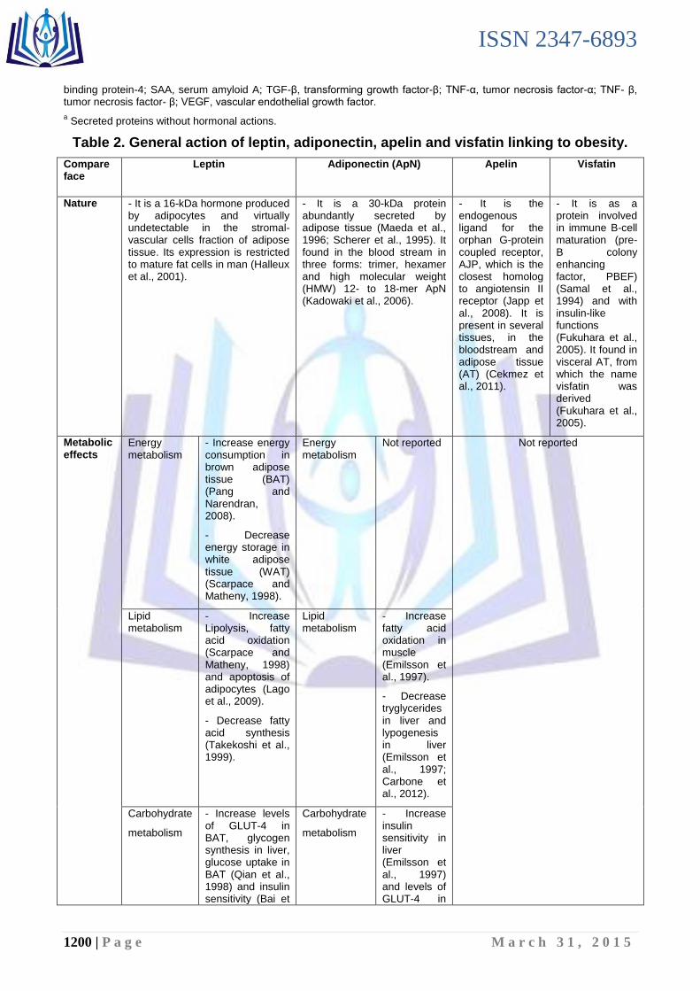
Table 2. General action of leptin, adiponectin, apelin and visfatin linking to obesity.
Compare face
Leptin Adiponectin (ApN)
Apelin Visfatin
Nature - It is a 16-kDa hormone produced by adipocytes and virtually undetectable in the stromal-vascular cells fraction of adipose tissue. Its expression is restricted to mature fat cells in man (Halleux et al., 2001).
- It is a 30-kDa protein abundantly secreted by adipose tissue (Maeda et al., 1996; Scherer et al., 1995). It found in the blood stream in three forms: trimer, hexamer and high molecular weight (HMW) 12- to 18-mer ApN (Kadowaki et al., 2006).
- It is the endogenous ligand for the orphan G-protein coupled receptor, AJP, which is the closest homolog to angiotensin II receptor (Japp et al., 2008). It is present in several tissues, in the bloodstream and adipose tissue (AT) (Cekmez et al., 2011).
- It is as a protein involved in immune B-cell maturation (pre-B colony enhancing factor, PBEF) (Samal et al., 1994) and with insulin-like functions (Fukuhara et al., 2005). It found in visceral AT, from which the name visfatin was derived (Fukuhara et al., 2005).
Metabolic effects
Energy metabolism
- Increase energy consumption in brown adipose tissue (BAT) (Pang and Narendran, 2008).
- Decrease energy storage in white adipose tissue (WAT) (Scarpace and Matheny, 1998).
Energy metabolism
Not reported Not reported
Lipid metabolism
- Increase Lipolysis, fatty acid oxidation (Scarpace and Matheny, 1998) and apoptosis of adipocytes (Lago et al., 2009).
- Decrease fatty acid synthesis (Takekoshi et al., 1999).
Lipid metabolism
- Increase fatty acid oxidation in muscle (Emilsson et al., 1997).
- Decrease tryglycerides in liver and lypogenesis in liver (Emilsson et al., 1997; Carbone et al., 2012).
Carbohydrate
metabolism
- Increase levels of GLUT-4 in BAT, glycogen synthesis in liver, glucose uptake in BAT (Qian et al., 1998) and insulin sensitivity (Bai et
Carbohydrate
metabolism
- Increase insulin sensitivity in liver (Emilsson et al., 1997) and levels of GLUT-4 in

ISSN 2347-6893
1201 | P a g e M a r c h 3 1 , 2 0 1 5
al., 1996).
- Decrease levels of GLUT-4 in WAT (Qian et al., 1998), insulin secretion by pancreatic islets (Harris, 1998) and glycogen synthesis in muscle (Kamohara et al., 1997; Carbone et al., 2012).
muscle (Berg et al., 2001).
- Decrease glucose haematic levels and glucose production in liver (Yamauchi et al., 2001 & 2003).
General action
- Central action:
It represents a signal to the brain (e.g., hypothalamus, cortex and limbic areas) to inhibit food intake and reduce weight (Zhang et al., 1994) because of humans and rodents lacking a functional leptin protein manifested insatiable feeding and obesity (Hajer et al., 2008). The vital action of leptin in the hypothalamus has been best described with regards to energy homeostasis and reproductive functions (Badman and Flier, 2007). Additionally to its action on the hypothalamus, leptin may also act on the cortex and limbic areas, which are regulated the cognitive and emotional feeding behavior (Farooqi et al., 2007; Rosenbaum et al., 2008). As predictable, leptin treatment successfully reversed the obesity and leptin resistance abnormalities (Ahima, 2008). Currently, leptin represents as a hormone responsible for signaling energy deficiency rather than a signal to lose weight (Badman and Flier, 2007; Kershaw and Flier, 2004).
- Peripheral action:
It has actions in a number of peripheral tissues (e.g., cells of the pancreas, liver and immune system) (Karastergiou and Mohamed-Ali, 2010). Moreover, disruption of peripheral leptin signaling in mice caused no significant variation in energy balance or glucose homeostasis (Guo et al., 2007).
Conversely to most adipokines, circulating ApN is negatively associated with the body mass index (BMI) (Arita et al., 1999; Brichard et al., 2003) and decreased in obese subjects, in type 2 diabetes or cardiovascular disease (CVD) (Ouchi and Walsh, 2007). This sub-regulation may involve the abnormal hormonal milieu (Delporte et al., 2002; Halleux et al., 2001), together with the enhanced oxidative stress (Furukawa et al., 2004) and the pro-inflammatory state (Bruun et al., 2003) that exist in obesity and the metabolic syndrome.
- AT apelin and plasma levels increased in obesity (Heinonen et al., 2005). Conversely, both circulating apelin and its expression in AT were reduced after weight loss consecutive to a hypocaloric diet in obese women (Castan-Laurell et al., 2008). Its mRNA expression was similar in adipocytes and stromal-vascular cells (SVC) isolated from human subcutaneous (sc) tissue and there was no difference in adipocyte apelin expression in intra-abdominal and sc fat pads in mice (Boucher et al., 2005).
- Possible relations between circulating visfatin and anthropometric or metabolic parameters in obesity and type 2 diabetes have been found in some but not all studies (Garten et al., 2009; Rasouli and Kern, 2008); the contradictory findings may be due in part to considerable differences in visfatin immunoassays (Garten et al., 2009; Imai, 2009).
Receptors & effects
Leptin from the periphery is transported into the brain, combines with its receptor b in the hypothalamus, and stimulates JAK-STAT3, leading to repression of “orexigenic peptides” (e.g.,
AdipoR1 and AdipoR2 serve as main receptors for ApN in vivo and belong to a new family of receptors (seven transmembrane domains) but to be structurally and
- It promising target in the management of insulin resistance (Cekmez et al.,
- It has vital roles in insulin sensitivity (Cekmez et al., 2011).
ISSN 2347-6893
1202 | P a g e M a r c h 3 1 , 2 0 1 5
neuropeptide Y and agouti-related protein), and increase in “anorexigenic peptides” (e.g., proopiomelanocortin and corticotrophin-releasing hormone) (Ahima, 2008), in that way curtailing food intake. In the ordinary form of obesity, resistance to leptin has been ascribed to reduced transport of leptin across the blood–brain barrier (BBB) and to increased hypothalamic levels of SOCS3 and endoplasmic reticulum (ER) stress, which inhibit leptin signaling (Flier, 1998; Morton et al., 2005; Ozcan et al., 2009). Notably, low leptin levels stimulate overfeeding and repress energy expenditure, thyroid and reproductive hormones, and immunity (Maury and Brichard, 2010).
functionally distinct from G-protein coupled receptors (Maury and Brichard, 2010). AdipoR1 is expressed in muscle, while AdipoR2 is expressed in liver (Yamauchi et al., 2003). AdipoR1 is more tightly associated to the activation of AMPK pathways that adjust the inhibition of gluconeogenesis together with increased fatty acid oxidation, while AdipoR2 is more concerned with the activation of the PPAR-α pathways, which stimulate energy dissipation by increasing fatty acid oxidation and reduce oxidative stress and inflammation (Capeau, 2007; Yamauchi et al., 2007). Interaction of an adaptor protein, APPL1 with AdipoR1 appears to play significant roles in ApN signaling (Mao et al., 2006). After binding, ApN showed insulin-sensitizing and fat-burning effects suggestive of those of leptin, but possesses anti-atherogenic, anti-inflammatory and anti-oxidant properties as well, thereby thwarting simultaneously several facets of the metabolic syndrome (Kadowaki and Yamauchi, 2005; Takemura et al., 2007).
2011).
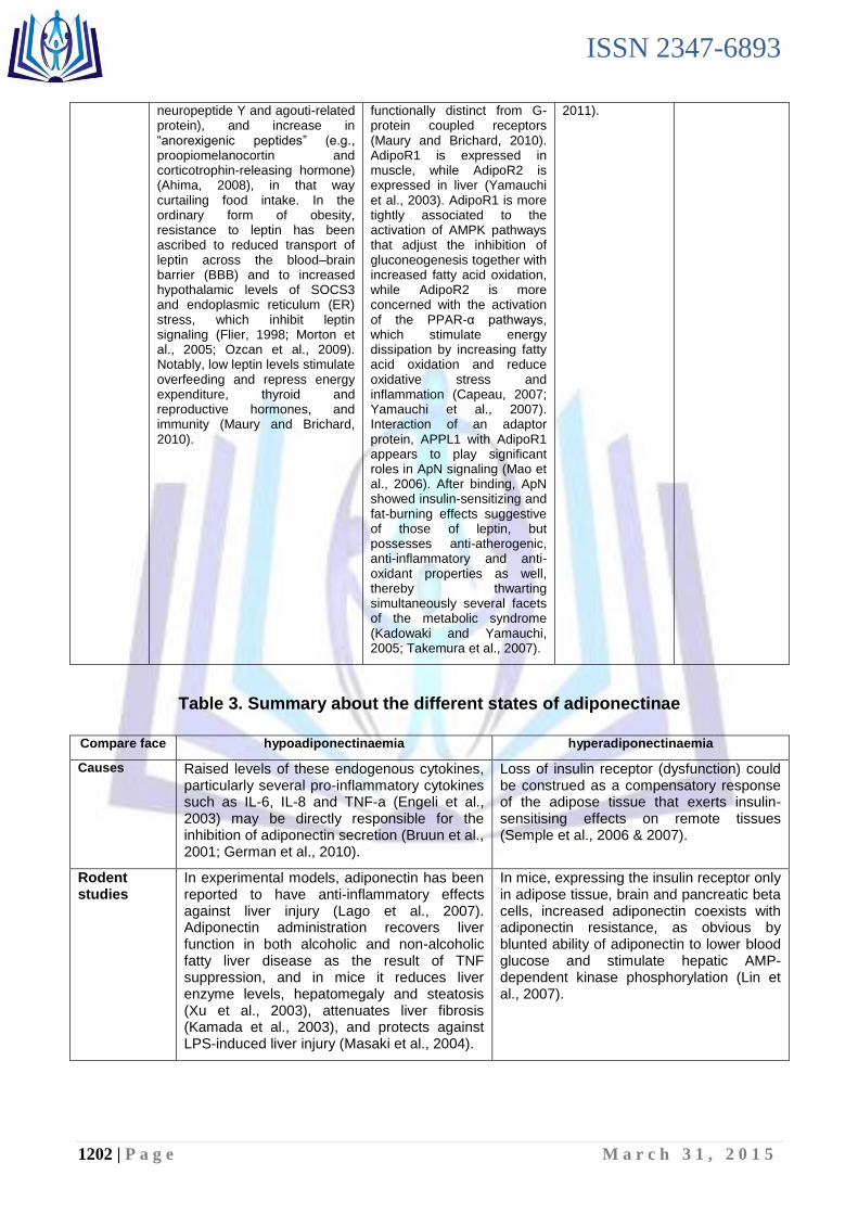
Table 3. Summary about the different states of adiponectinae
Compare face hypoadiponectinaemia hyperadiponectinaemia
Causes Raised levels of these endogenous cytokines, particularly several pro-inflammatory cytokines such as IL-6, IL-8 and TNF-a (Engeli et al., 2003) may be directly responsible for the inhibition of adiponectin secretion (Bruun et al., 2001; German et al., 2010).
Loss of insulin receptor (dysfunction) could be construed as a compensatory response of the adipose tissue that exerts insulin-sensitising effects on remote tissues (Semple et al., 2006 & 2007).
Rodent studies
In experimental models, adiponectin has been reported to have anti-inflammatory effects against liver injury (Lago et al., 2007). Adiponectin administration recovers liver function in both alcoholic and non-alcoholic fatty liver disease as the result of TNF suppression, and in mice it reduces liver enzyme levels, hepatomegaly and steatosis (Xu et al., 2003), attenuates liver fibrosis (Kamada et al., 2003), and protects against LPS-induced liver injury (Masaki et al., 2004).
In mice, expressing the insulin receptor only in adipose tissue, brain and pancreatic beta cells, increased adiponectin coexists with adiponectin resistance, as obvious by blunted ability of adiponectin to lower blood glucose and stimulate hepatic AMP-dependent kinase phosphorylation (Lin et al., 2007).
ISSN 2347-6893
1203 | P a g e M a r c h 3 1 , 2 0 1 5
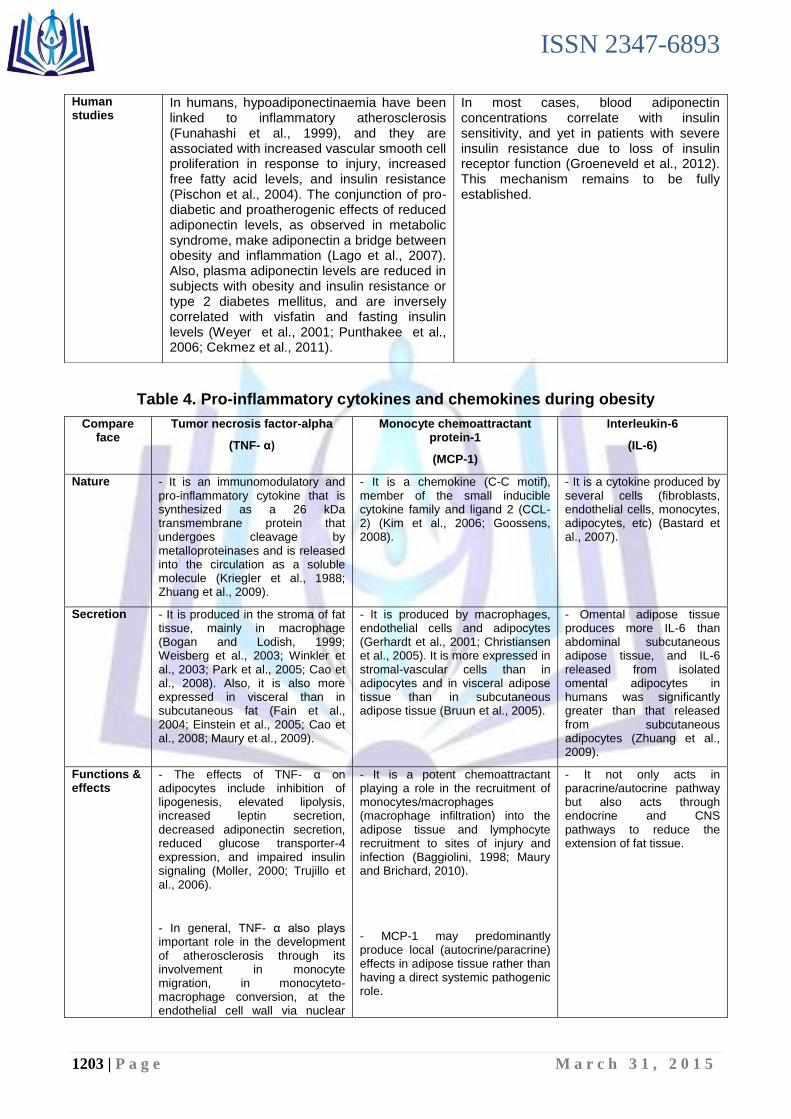
Table 4. Pro-inflammatory cytokines and chemokines during obesity
Compare face
Tumor necrosis factor-alpha
(TNF- α)
Monocyte chemoattractant protein-1
(MCP-1)
Interleukin-6
(IL-6)
Nature - It is an immunomodulatory and pro-inflammatory cytokine that is synthesized as a 26 kDa transmembrane protein that undergoes cleavage by metalloproteinases and is released into the circulation as a soluble molecule (Kriegler et al., 1988; Zhuang et al., 2009).
- It is a chemokine (C-C motif), member of the small inducible cytokine family and ligand 2 (CCL-2) (Kim et al., 2006; Goossens, 2008).
- It is a cytokine produced by several cells (fibroblasts, endothelial cells, monocytes, adipocytes, etc) (Bastard et al., 2007).
Secretion - It is produced in the stroma of fat tissue, mainly in macrophage (Bogan and Lodish, 1999; Weisberg et al., 2003; Winkler et al., 2003; Park et al., 2005; Cao et al., 2008). Also, it is also more expressed in visceral than in subcutaneous fat (Fain et al., 2004; Einstein et al., 2005; Cao et al., 2008; Maury et al., 2009).
- It is produced by macrophages, endothelial cells and adipocytes (Gerhardt et al., 2001; Christiansen et al., 2005). It is more expressed in stromal-vascular cells than in adipocytes and in visceral adipose tissue than in subcutaneous adipose tissue (Bruun et al., 2005).
- Omental adipose tissue produces more IL-6 than abdominal subcutaneous adipose tissue, and IL-6 released from isolated omental adipocytes in humans was significantly greater than that released from subcutaneous adipocytes (Zhuang et al., 2009).
Functions & effects
- The effects of TNF- α on adipocytes include inhibition of lipogenesis, elevated lipolysis, increased leptin secretion, decreased adiponectin secretion, reduced glucose transporter-4 expression, and impaired insulin signaling (Moller, 2000; Trujillo et al., 2006).
- In general, TNF- α also plays important role in the development of atherosclerosis through its involvement in monocyte migration, in monocyteto-macrophage conversion, at the endothelial cell wall via nuclear
- It is a potent chemoattractant playing a role in the recruitment of monocytes/macrophages (macrophage infiltration) into the adipose tissue and lymphocyte recruitment to sites of injury and infection (Baggiolini, 1998; Maury and Brichard, 2010).
- MCP-1 may predominantly produce local (autocrine/paracrine) effects in adipose tissue rather than having a direct systemic pathogenic role.
- It not only acts in paracrine/autocrine pathway but also acts through endocrine and CNS pathways to reduce the extension of fat tissue.
Human studies
In humans, hypoadiponectinaemia have been linked to inflammatory atherosclerosis (Funahashi et al., 1999), and they are associated with increased vascular smooth cell proliferation in response to injury, increased free fatty acid levels, and insulin resistance (Pischon et al., 2004). The conjunction of pro-diabetic and proatherogenic effects of reduced adiponectin levels, as observed in metabolic syndrome, make adiponectin a bridge between obesity and inflammation (Lago et al., 2007). Also, plasma adiponectin levels are reduced in subjects with obesity and insulin resistance or type 2 diabetes mellitus, and are inversely correlated with visfatin and fasting insulin levels (Weyer et al., 2001; Punthakee et al., 2006; Cekmez et al., 2011).
In most cases, blood adiponectin concentrations correlate with insulin sensitivity, and yet in patients with severe insulin resistance due to loss of insulin receptor function (Groeneveld et al., 2012). This mechanism remains to be fully established.
ISSN 2347-6893
1204 | P a g e M a r c h 3 1 , 2 0 1 5
factor kappa-light-chain-enhancer of activated B-cells (NF-κB) activation, and in inflammatory changes in the vascular wall (Fonseca-Alaniz et al., 2007).
Activity & expression in obesity & T2DM
- It is overexpressed in adipose tissue of obese individuals (Maury et al., 2009) and fall after weight loss (Dandona et al., 1998). Its elevation can reduce visceral fat volume (Maury and Brichard, 2010).
- Circulating and adipose tissue levels of MCP-1 are elevated in obese (Bruun et al., 2005) rodents (Sartipy and Loskutoff, 2003; Takahashi et al., 2003) and individuals (Nomura et al., 2000; Piemonti et al., 2003), and their levels have been found to decrease after weight-loss (Schernthaner et al., 2006).
- Plasma IL6 levels are elevated in obesity and in T2DM and are positively associated with plasma FFA levels and with body mass (Lazar, 2005; Urs et al., 2004; Bastard et al., 2007) where this can reduce visceral fat volume (Maury and Brichard, 2010).

ISSN 2347-6893
1205 | P a g e M a r c h 3 1 , 2 0 1 5
ISSN 2347-6893
1206 | P a g e M a r c h 3 1 , 2 0 1 5