development of novel methotrexate-loaded nanocochleates
TRANSCRIPT

Page 1/32
Development of novel methotrexate-loadednanocochleates for breast cancer targeting.Nikhil Rajnani ( [email protected] )
Principal K.M.Kundnani College of PharmacyNalini Kurup
Principal K.M.Kundnani College of Pharmacy
Article
Keywords: Methotrexate, liposomes, nanocochleates, cell line, carrier
Posted Date: March 2nd, 2022
DOI: https://doi.org/10.21203/rs.3.rs-1412975/v1
License: This work is licensed under a Creative Commons Attribution 4.0 International License. ReadFull License
Page 2/32
AbstractThe objective of this research was to develop stable methotrexate(MTX) loaded nanocochleates (MTX-NC)drug delivery system to achieve controlled drug release,targeted delivery and enhanced anticancer potencywith reduced dose and its effect on Breast cancer cell line. The development involved the loading of MTXto nanocochleates (MTX-NC) through conversion of phosphatidyl choline and cholesterol bearing liposomeon addition of calcium ions and compared with MTX and MTX-NC. Optimized NC under optimizedconditions showed particle size and zeta potential of 298.7nm and -12.9mv respectively, high entrapmente�ciency 75%, drug loading of 37.5% and drug content 99%. Investigation on the drug lipid interactionusing FTIR con�rmed the compatibility of drug with the lipid carrier. SEM and TEM images of NC indicatedthat the particles were in a rod cylindrical shape with a smooth surface.Formulated MTX-NC demonstratedhigher in-vitro anticancer activity in human breast cancer MCF-7 cells. The concentration of the drugneeded for growth inhibition of was 47% for free MTX(80µg/ml) while it was -35.6% for the MTX-NC(80µg/ml) . Furthermore, the LC50 value of free MTX and MTX-NC was NE and 75.3µg/mlrespectivelty.GI50 value of free MTX and MTX-NC was <10µg/ml. The results indicate that the MTX- NChas the potential to be applied for targeting anticancer drug delivery.
1. IntroductionBreast cancer is the most frequent cancer occurs in women worldwide [1]. Current treatment involveschemotherapy, hormone therapy and surgery. A drawback with these therapies is lack of speci�city, moreside effects and poor absorption. Nanocarrier based delivery systems provides many bene�t over currenttherapies such as active or passive drug targeting, increased encapsulation of drug, reduce side effects anddegradation of drug in blood circulation [2, 3].
Methotrexate (2,4-diamino-N10-methyl propyl glutamic acid, MTX) an anticancer agent belong toBiopharmaceutical Classi�cation System (BCS) class II used for the treatment of different tumors such aslung, osteosarcoma and breast cancer. Moreover, it is also used to treat in�ammatory and autoimmunediseases such as Crohnsdiseases, rheuma → rthritis and ps or iasis[4 - 6]. However, ∵ s clinicaluse is limited [6]. Therefore, there was need to develop a formulation which could improve therapeutice�cacy at low dose, thus reducing systemic toxicity too.
Vesicles have been the preferred route for medication delivery in recent years [6]. Encapsulation of the drugin these vesicular structures, prolong the existence of drug in the systemic circulation. The vesicularsystems are highly ordered assemblies of one or several concentric lipid bilayers [7]. The drug deliverysystem for cochleates and nanocochleates is based on encapsulating the medicine in a multilayered, lipidmatrix in order to administer the drug safely and effectively [8] .Cochleates were �rst discovered by Dr.Dimitrious Papahadjoupoulos and his co‐workers in 1975 as precipitates formed by the interaction ofnegatively charged phosphatidylserine and calcium. This was cylindrical in shape and he named thesecylindrical structures as "cochleate" means Shell” i.e. rolled-up form [9]
Page 3/32
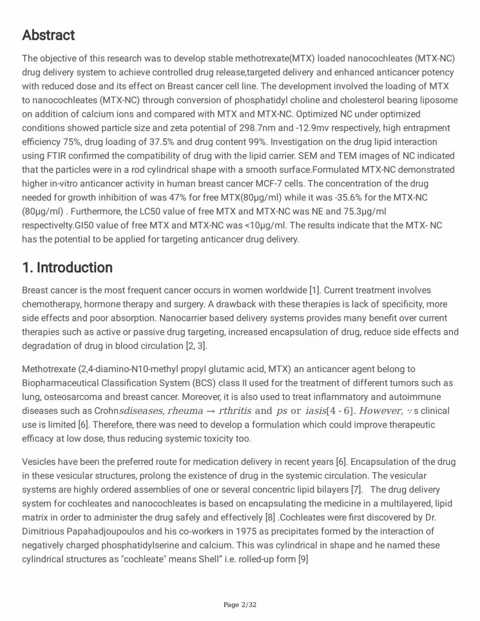
Nanocochleates are cigar-like structures made up of a sequence of lipid bilayers (Figures 1.1 and 1.2),which arise when small unilamellar negatively charged liposomes condense. The small phosphatidylserine(PS) or phosphatidyl choline (PC) liposomes merge and produce huge sheets in the presence of calcium.These sheets have hydrophobic surfaces and tend to coil up into a cigar-like cochleate (Figure 1.3) toreduce their interactions with water [9].
Cochleates differ from liposomes in having water-free interior, rod-shaped, and rigid stable structure asshown in Fig no.1.3
Nanocochleates formulation technology is particularly applicable to macromolecules as well as to drugsthat are hydrophobic and hydrophilic. It is also for drugs that undergo �rst pass metabolism and drug thatdegrades at physiological pH like proteins and peptides. These novel carriers have been used to deliverdrugs belong to class of antifungal, antibiotics, protein, anti-leprosy, anticancer, and DNA 70 subunit toincrease their therapeutic e�cacy [10,11].
To further advance the therapeutic utility of MTX, the present study investigates the potential ofnanocochleates as a vehicle for oral delivery of MTX [12]. Methotrexate-loaded nanocochleates (MTXNC)were developed through conversion of phosphatidylcholine (PC) and cholesterol bearing nanoliposomesinto the nanocochleates structures on addition of calcium ions by trapping method. Cholesterol wasincluded to stabilize the phospholipid membranes as well as to form complex with the drug [10]. The MTX-NC were characterized by their particle size, zetapotential, entrapment e�ciency, in-vitro drug release,compatibility studies (FTIR), SEM, TEM, stability studies and in vitro cell lines studies.
2. Materials & Methods2.1 Materials
Methotrexate was obtained as a gift sample from Neon Laboratories, India. Lipoid S75 and Phospholipon90H was obtained as generous gift from Lipoids, Germany and used as vesicle forming phospholipid,cholesterol (Sigma-Alderich) provides �exibility to the formed liposomal vesicles thus maintaining itsshape. Calcium chloride was used as the cochelation agent. All other Chemicals used were analyticalgrade.
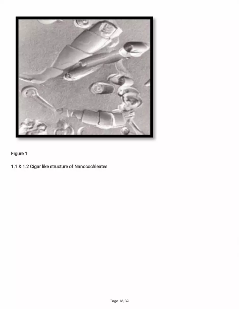
2.2 Preparation of liposomes by solvent injection method
MTX-loaded nanoliposomes (MTXNL) were prepared by ethanol injection method as shown in �g.no. 2.1.Brie�y, different amount of phospholipon 90H, Lipoid S-75 and, cholesterol were dissolved in 10 mL ofethanol as shown in tablet no. 2.1. Drug was dissolved in phosphate buffer (pH – 7.4) and kept on amagnetic stirrer under constant rpm. Ethanolic phase was rapidly injected into the aqueous phase underconstant magnetic stirrer for a speci�c time using Te�on-coated beads and stirring was continued untilcomplete evaporation of under vacuum. Further, phosphate buffer was added to adjust the volume of �nalnanoliposomes suspension to 20 mL. Finally, the nanoliposomes were puri�ed by passing through a 0.45

Page 4/32
mm membrane �lter to obtain a MTXNL suspension [15–17]. Batch which shows higher encapsulatione�ciency was selected for the preparation of MTX-Nanocochleates.
Table No. 2.1 Trial batches with combination of Phospholipids with different ratio
Sr.No
Lipid
Content
Phospholipid
90H
Lipoid S-75
Phospholipids:
Lipoid S-75
Lipid:
Cholesterol
Apperrance %EE
1 0.1% Yes - - 1:0.3 LessTurbid
25
2 0.1% - Yes - 1:0.3 LessTurbid
52
3 0.1% Yes Yes 1:1 (50:50) 1:0.3 LessTurbid
75
4 0.1% Yes Yes 0.6:0.4 (60:40) 1:0.3 LessTurbid
45
5 0.1% Yes Yes 0.4:0.6 (50:50) 1:0.3 LessTurbid
60
6 0.5% Yes Yes 1:1 (50:50) 1:0.3 Turbid 35
7 1% Yes Yes 1:1 (50:50) 1:0.3 Turbid 38
8 2% Yes Yes 1:1 (50:50) 1:0.3 Turbid 40
2.2 Formulation of MTX-loaded nanocochleates
MTX-loaded nanocochleates (MTXNC) were prepared by trapping method [14-15] as shown in �g.no. 1.2 . Fifty microliters of calcium chloride solution (0.1 M) was added dropwise into the prepared MTXNC understirring. The MTXNL phase immediately turned turbid because nanocochleates formation. The MTXNCdispersion was evaluated in terms of particle size, zeta potential, drug loading, in-vitro drugrelease, encapsulation e�ciency, SEM, and in-vitro cell line (MCF-7 breast cancer) study.
Table No. 2.2.1 Trial Batch by Trapping Method
Sr.
No.
%Lipid L:CH Speed Time %Transmittance Divalent %Transmittance
30mins 60mins CaCl2 30mins 60mins
1 0.1 1:0.3 1500 2 84.20 91.08 0.1M 83.02 82.02

Page 5/32
2.3 Experimental Design
Design-Expert software was utilized for experimental design and statistical analysis. General full factorialdesign with three independent variables was applied to determine the optimal levels. Each numeric factorwas varied over 2 levels thus by selecting general full factorial design 8 experiments were generated by thesoftware. Encapsulation e�ciency percentage of MTX-Liposomes and encapsulation MTX-NC values werechosen as quality attributes for optimizing the formulation.
The response was assessed by the nonlinear quadratic equation:
Y: β0 + β1A + β2B + β3C + β12AB + β13AC + β23BC + β11A2 + β22B2 + β33C2
where Y is the predicted response, A, B and C are independent variables, A2, B2 and C2 symbolize theinteraction and the formulations were prepared by same procedure as mentioned in the preliminarystudies.
All the 8 experiments in these experiments were prepared and evaluated for Nanocochleates prepared bytrapping method.
Table No. 2.3.1 Design Summary for Nanocochleates
File Version 11.0.5.0
Study Type Factorial Subtype Randomized
Design Type Full Factorial Runs 8
Design Model 2FI Blocks No Blocks
Center Points 0 Build Time (ms) 1
Table no. 2.3.2 Preoptimized batches for nanocochleates

Page 6/32
STD RUN L:CH SPEED TIME
1 1 1:03 1500 1
3 2 1:03 2000 1
2 3 1:0.5 1500 1
5 4 1:03 1500 2
6 5 1:0.5 1500 2
8 6 1:0.5 2000 2
7 7 1:03 2000 2
4 8 1:0.5 2000 1
Encapsulation e�ciency and transmittance percentage of MTX-NC values was chosen as quality attributesfor optimizing the formulation.
The impact of independent variables on responses was assessed using analysis of variance (ANOVA) andp<0.05 was accepted to be statistically meaningful. To evaluate the convenience of the model, thecalculated predicted and adjusted correlation coe�cient (R2) were analysed. The in�uences of selectedindependent variables on the quality of the nanocochleates formulation were assessed by �t statistics andco�ecient factors for the dependent variable. The optimum MTX-NC formulation was decided byconsidering responses having maximum encapsulation e�ciency and % transmittance
The optimized nanocochleate formulation was prepared three times and the achieved outcomes werematched with the predicted values.
2.4 Evaluation of optimized nanocochleates
2.4.1 Microscopic evaluation.
A drop of nanocochleates dispersion was observed under optical microscope (45X magni�cation) for anycluster formation.
2.4.2 In- vitro drug release.
The in-vitro release of MTX from MTX-NC was carried out in phosphate buffer (PBS, pH 7.4 and usingdialysis bag diffusion technique. Formulation equivalent to 2mg of MTX was introduced into a dialysis bag(cellulose membrane, molecular weight cut off 12, 0000 Da), hermetically sealed and immersed into 200 mlof release medium. The entire system was kept at 37 ± 0.5 _C with continuous magnetic stirring at 100rpm. At selected time intervals, 2 mL sample was removed and 2 mL phosphate buffer was added tomaintain sink condition. Absorbances of the solutions were recorded at λmax of 302 nm using theShimadzu Jasco UV-visible spectrophotometer (JASCO V-630, Japan) [6,18]

Page 7/32
2.4.3 Size measurement and Zeta potential of Cochleate dispersion.
The vesicle size and size distribution of nanocochleates were measured by Malvern Zetasizer 2000(Malvern, UK). Vesicular suspensions were mixed with the appropriate medium (PBS, pH 7.4) and themeasurements were taken in a multimodal mode. All measurements were performed at 250C after 5 min ofthermal equilibration. The Malvern Zetasizer 2000 (Malvern, UK) detects backscattering at an angle of1730 for an improved sizing of especially larger particles at higher concentrations. Viscosity of the mediumof 0.89 mPas and refractory index of 1.33 were assumed [19-26].
2.4.4 Entrapment e�ciency
2 ml of formulation was subjected to centrifugation for 45 minutes at 10000 rpm at °C, the supernatantand pellets were separated. The 0.5 ml of supernatant was withdrawn and volume was made upto 10 mlwith phosphate buffer and analyze[19-26].
2.4.5 Transmission Electron Microscopy (TEM):
TEM was employed to study the morphology of the nanocochleates. TEM images were formed usingPhilips Electron Optics transmission electron microscope, model no. CM200. The operating voltage was 20– 200 kv, with a resolution of 2.4 A° and magni�cation of 45,000X (66). The optimized formulation wasreconstituted in distilled water and a drop was introduced on a carbon coated mesh like grid and dried. Theimages were resolved on a �uorescent screen and the analysis of the x-rays produced by interactionbetween accelerated electrons with the sample allows determining elemental composition of sample withhigh spatial resolution [19-26].
2.4.6 Scanning Electron Microcopy (SEM):
Liposome and nanocochleates sample for SEM was prepared by dispersing sample in 5mL DDW. A drop ofnanoparticle dispersion was deposited on carbon tape, which was then air-dried after being glued to metalstubs with a double-sided adhesive tape. SEM was used to determine the morphology of nanoparticles(Zeiss DSM 940A SEM; Oberkochen, Germany). After covering the particles with a thin layer of gold undervacuum using a sputtering apparatus, imaging was done on SEM [19-26].
2.4.7 In-vitro cell line studies:
The cytotoxicity tests were carried out on the MCF7 human breast cancer cell line. RPMI 1640 medium with10% foetal bovine serum and 2 mM L-glutamine was used to culture the cell lines. Cells were injected into96 well microtiter plates in 90L for the screening experiment. Following cell inoculation, the microtiterplates were incubated for 24 hours at 37° C, 5% CO2, 95% air, and 100% relative humidity before addingexperimental samples.The cytotoxic activity was assessed using the Sulforhodamine B (SRB) assay. Oneplate of each cell line was �xed in situ with trichloroacetic acid (TCA) after 24 hours to re�ect ameasurement of the cell population at the moment of drug addition. The needed �nal drug concentrationswere achieved by adding aliquots of 10 l of various drug dilutions to appropriate micro-titer wells alreadyholding 90 l of medium. Plates were incubated at standard conditions for 48 hours after drug addition, and

Page 8/32
the test was �nished with the addition of cold TCA. The cells were �xed in place by gently adding 50 l ofcool 30% w/v) TCA (�nal concentration, 10% TCA) and incubating for 60 minutes at 4°C. The supernatantwas removed, and the plates were rinsed �ve times with tap water and air (w/v) TCA (�nal concentration,10% TCA) before incubation at 4°C for 60 minutes. The supernatant was discarded, and the plates wererinsed and air dried �ve times with tap water. Sulforhodamine B (SRB) solution (50 μl) at 0.4 % (w/v) in 1 %acetic acid was added to each of the wells, and plates were incubated for 20 minutes at room temperature.After staining, unbound dye was recovered and the residual dye was removed by washing �ve times with 1% acetic acid. The plates were air dried. Bound stain was subsequently eluted with 10 mM trizma base andthe absorbance was read on an Elisa plate reader at a wavelength of 540 nm with 690 nm referencewavelength. The optical density of drug-treated cells was compared to that of control cells, and thepercentage of growth inhibition was computed. At four distinct concentrations of 10-7M, 10-6M, 0-5M, and10-4M, each drug was evaluated in triplicate. For each experiment, an Adriamycin positive control wasemployed to check that the experimental setup was correct [6,27-29].
Formulations that are given for the in-vitro cell line studies are
1) Drug
2) Liposomes
3) Nanocochleates prepared by trapping method containing Ca+2
4) Nanocochleates prepared by trapping method containing Ba+2
5) Nanocochleates prepared by hydrogel method containing Ca+2
6) ADR (Adrinomycin)
Table No. 2.3.7.1 Concentration taken for the In-vitro Cell line studies
Sample Code Experiment μg/ml
1 2 3 4
All samples 10 20 40 80
ADR* 10 20 40 80
2.3.8 Stability studies
Stability testing is used to assess expiration dating and storage conditions for pharmaceutical products.Nanocochleates dispersion was packed glass vials and they were subjected to stability testing at variousstorage conditions as per ICH QA1 R2.

Page 9/32
Stability conditions:
Storage in refrigeration: 2 - 8 C ± 2 C for 2 months; sample withdrawn at 7, 14, 28, and 56 days.
3. Results And Discussion3.1 Experimental Design.
Table No. 3.1 Preoptimized batches for NC (Ca+2)
STD RUN L:CH SPEED Time EE (%) TT (%)
1 1 1:0.3 1500 1 61 93.87
3 2 1:0.3 2000 1 65 91.63
2 3 1:0.5 1500 1 50.68 81.63
5 4 1:0.3 1500 2 75 101.76
6 5 1:0.5 1500 2 51.36 90.63
8 6 1:0.5 2000 2 51.68 78.67
7 7 1:0.3 2000 2 50.83 80.78
4 8 1:0.5 2000 1 56.63 92.63
From the table 3.1 we can found out that batches with Run4 showed maximum entrapment e�ciency of75% and transmittance 101.76%.
3.1.1 Model analysis for Entrapment e�ciency
Anova for Entrapment e�ciency
Table No. 3.1.1.1 Anova for Entrapment E�ciency
Source Sum of Squares df Mean Square F-value p-value
Model 352.85 1 352.85 11.43 0.0148 Signi�cant
A-L:CH 352.85 1 352.85 11.43 0.0148
Residual 185.25 6 30.87
Cor Total 538.10 7
From the table no. 3.1.1.1 we can see that
Factor coding is Coded.

Page 10/32
Sum of squares is Type III – Partial paraphrase it
The Model F-value of 11.43 implies the model is signi�cant. There is only a 1.48% chance that an F-valuethis large could occur due to noise.
P-values less than 0.0500 indicate model terms are signi�cant. In this case A is a signi�cant model term.Values greater than 0.1000 indicate the model terms are not signi�cant. If there are many insigni�cantmodel terms (not counting those required to support hierarchy), model reduction may improve your model.
Fit Statistics
Table No. 3.1.2.1 Fit Statistics for Entrapment E�ciency
Std. Dev. 1.78 R² 0.8883
Mean 78.67 Adjusted R² 0.8045
C.V. % 2.50 Predicted R² 0.8254
Adeq Precision 19.248
The Adjusted R2 of 0.8045 is reasonably close to the Predicted R2 of 0.8254; that is, the difference is lessthan 0.2.
The signal-to-noise ratio is measured by Adeq Precision. It is preferable to have a ratio of more than four.Your signal strength is adequate, as indicated by your ratio of 19.248. The design space can be navigatedusing this concept.
Coe�cient in Terms of Coded Factors
Table No. 3.1.2.2 Coe�cient for Entrapment E�ciency
Factor Coe�cient Estimate df Standard Error 95% CI Low 95% CI High VIF
Intercept 57.77 1 1.37 53.96 61.57
A-L:CH -6.64 1 1.37 -10.45 -2.84 1.0000
C-Stirring Time 2.85 1 1.37 -0.9562 6.65 1.0000
AC -2.74 1 1.37 -6.55 1.06 1.0000
The coe�cient estimate provides the estimated change in response per unit change in factor value while allother factors are kept constant. The intercept in an orthogonal design is the overall average response of all

Page 11/32
the runs. The coe�cients are adjustments depending on the factor settings in the vicinity of the average.When the factors are orthogonal, the VIFs are 1; VIFs greater than 1 indicate multi-colinearity, and the higherthe VIF, the more severe the factor correlation. VIFs of less than ten are regarded as acceptable.
3.2. Evaluation of optimized formulation
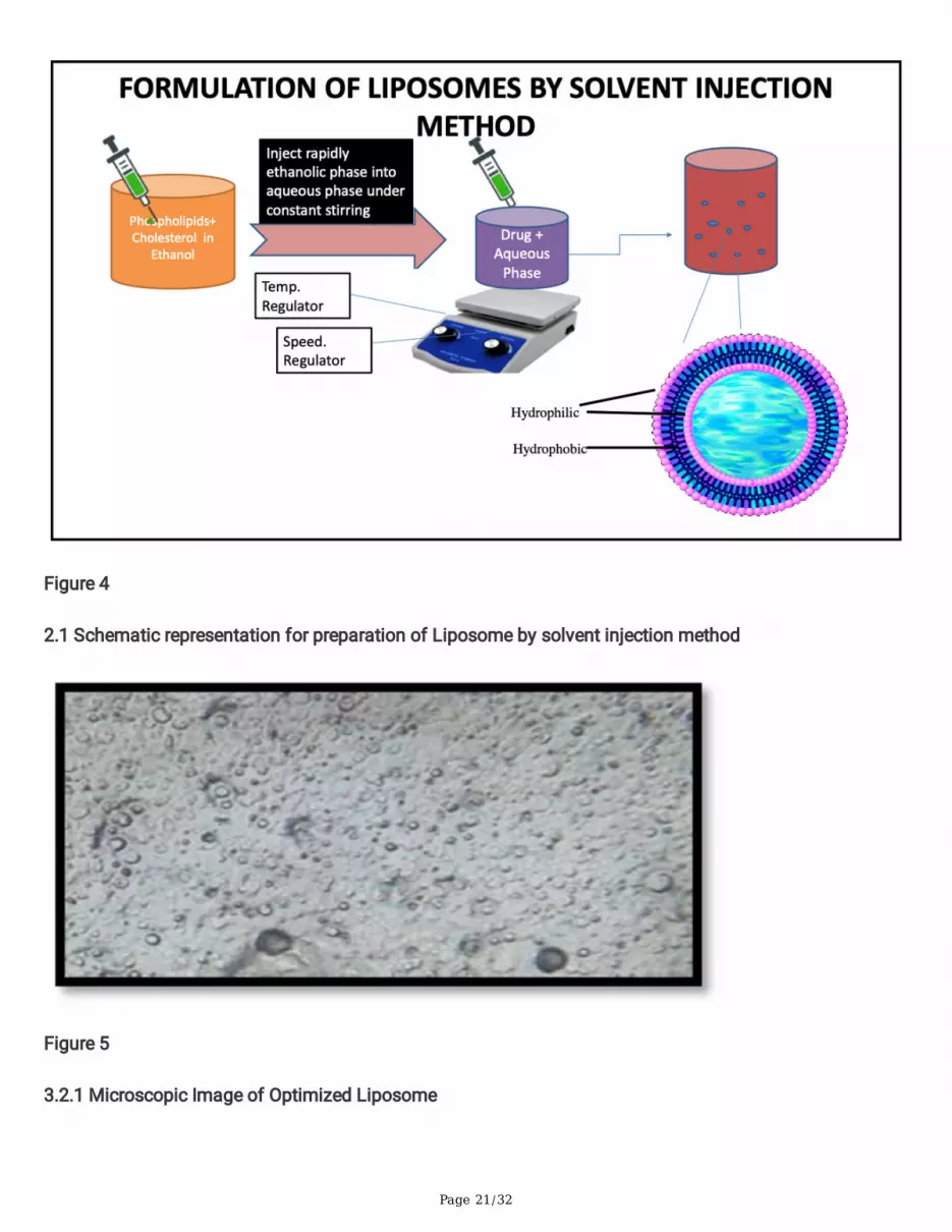
3.2.1. Microscopic evaluation.
From the �gure no. 3.2.1 Homogenous dispersion, with very few clusters was observed under microscope
3.2.2 In- vitro drug release
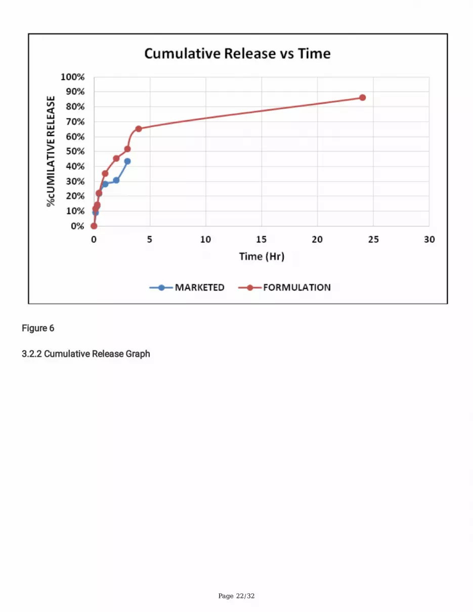
Table No. 3.2.2.1 In-vitro drug release of marketed and optimized formulation
Time(hrs)
%CUMULATIVE RELEASE
Marketed Formulation Nanocochleates
0 0% 0.12%
0.15 8.96% 11.52%
0.30 13.56% 14.25%
0.45 21.84% 22.36%
1 28.36% 35.36%
2 30.79% 45.36%
3 43.36% 51.85%
4 - 65.36%
24 - 86.23%
From the release studies it was observed that nanocochleate formulation led to prolong release of the drugform the system. The release form the system at the end of 24 hours was found to be 86.01%. for themarketed formulation the cumulative release at the end of 3 hrs was 43.36%. In nanocochleate formulationalthough a sustained release is achieved, at the start of studies the diffusion is higher than the latter half.This could be because of the drug at the surface of the nanocochleates which is �rst diffused due towetting.
From Fig No. 3.2.2 it can be seen that release of the Model Drug was sustained as compared to marketedformulation
1) Paired t test

Page 12/32
T test were applied to analyse a signi�cant change between the Formulation and the marketed. The t test isone type of inferential statistics. (p < .05 is a common value that is used). T test for all the mediums werefound to be 0.027 which is less than 0.05. Indicating signi�cant change in the drug release rate offormulation and marketed.
2) Mathematical Order Model
The model which drives the release of the drug from the formulation was evaluated. The best �t pro�le wasunderstood by plotting the graphs for zero order (% CR vs time), higuchi release model (%CR vs time1/2),hixon crowell release model, korsemeyer peppas release model and �rst order model. The results of thefollowing is shown in Table No 3.2.1
Table No. 3.2.2.2 R2 value for model in the in vitro diffusion studies
Formulation R2 value for model in the in vitro diffusion studies
Zero Order Higuchi Hixson Kors-peppars First Order
DRUG LOADED NC 0.8047 0.9484 0.8342 0.9361 0.8481
From table no. 3.2.1, it is understood that the release of the drug from the nanocochleates follows higuchimodel
3.2.3 Particle size measurement and polydispersity index (PDI):
Particle size of nanocochleate was found to be 298.7nm and 199.6nm respectivelywhile PDI ofnanocochleate was 0.295.
3.2.4 Zeta Potential
From the above �gure it was found that zeta potential of nanocochleate and was found to be -12.9mv
3.2.5 Transmission Electron Microscopy (TEM):
Surface morphology revealed nanocochleates showed rolled-up layers and rod structure
3.2.6 Scanning Electron Microscopy (SEM):
SEM analysis showed that the nanocochleate formulated had a cylindrical rod like shape.
3.2.7 Cytotoxicity Studies:

Page 13/32
Table No. 3.2.7.1 Control Growth Inhibition ValuesFormulation % Control Growth on MCF-7 (Average Values) Drug concentrations (µg/ml) 1 (10) 2 (20) 3 (30) 4 (40)Drug 43.0 50.1 47.1 47.9Liposomes 38.1 20.7 4.7 -20.8NC (Trapping) Ca+2 29.3 14.8 -3.3 -35.6
NC (Hydrgel) Ca+2 24.9 15.1 -3.1 -46.5
NC (Trapping) Ba+2 44.3 46.3 44.2 43.2
ADR -5.0 -11.3 -21.3 -39.9
Table No. 3.2.7.2 LC-50, TGI and GI50 Dose response parameter
LC-50 TGI GI-50Drug NE NE <10Liposomes 75.3 38.0 <10NC (Trapping) Ca+2 64.7 30.9 <10
NC (Hydrgel) Ca+2 59.6 28.5 <10
NC (Trapping) Ba+2 NE NE <10ADR NE NE <10
The minimum detectable anticancer activity on MCF7 cell line was observed in the Model Drug loadednanocochleateat a concentration of 40μg/mL, with an inhibition of -3.3 % cell death, and reaching amaximum inhibition of -35.6 % cell death at a concentration of 80μg/mL. Simultaneously Model Drugloaded liposome showed at a concentration of 80μg/mL, with an inhibition of -20.8% cell death. LC-50 ofModel Drug loaded nanocochleate and in Model Drug loaded liposome were found to be 75.3μg/mL and64.7μg/ mL respectively. TGI of Model Drug loaded nanocochleate and Model Drug loaded liposome werefound to be 38μg/mL and 30.9μg/ mL respectively.From the results, it clearly indicates Model Drug loadednanocochleate was showing better cancer cell death as compared to Model Drug loaded liposome(p<0.05).
The above signi�cant activity could be due to nanosizeand presence of lipid facilitating better penetrationinto the cancer cell and improved intracellular drug accumulation into MCF-7 cells.
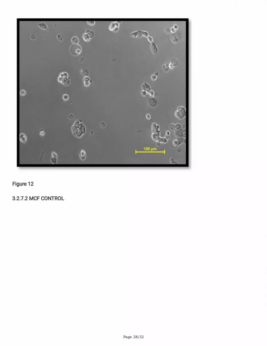
This is also indicated by shrinking of cell seen in �g.no 3.2.7.3.
3.2.8 Stability studies of optimized nanocochleates
Table No. 3.2.8.1: Stability Study Result for Nanocochleates at 2-8 C

Page 14/32
Parameters Results for Nanocochleates
Time (days) 0 7 14 28 56 84
Appearance Clear,translucent
Clear,translucent
Clear,translucent
Clear,translucent
Clear,translucent
Clear,translucent
%transmittance 90.63 89.75 91.23 90.74 90.53 90.52
% EntrapmentE�ciency
75 74.87 72.86 72.76 71.84 73.85
Particle Size(nm)
298 - - - - 268.7
Zeta Potential(mv)
-12.9 - - - - -9.1
Drug content(% w/w)
99 98.20 98.54 97.74 97.50 98.50
% Drug release 86.63 86.76 86.50 85.71 85.40 85.64
Appearance: The stability batch of optimized nanocochleates was found to be clear and translucent inappearance. No change in the appearance was observed over a period of three months at long termstability conditions of 5°C±3°C but at room temperature precipitation was observed after 1 month
Drug Content: Drug content was found to be in the range of 99%-98.77%. Initial drug content for batchnanocochleate was found to be 82.5% and 86.5% respectively. Further after 1, 2 and 3 months the drugcontent of nanocochleate was found to be 97.74%, 97.50 % and 98.50 % respectively at 5°C±3°C. Slightdecrease in the drug content was observed during the storage period. Drug content at Room temperaturewas not determine because precipitation was observed.
Drug Release: Drug release of nanocochleate was found to be in the range of 85.0% to 87% over a periodof 24 hours. Initial drug release of nanocochleate was found to be 86.63 and after 1, 2, 3 months drugrelease was 85.74%, 85.50%, 85.60% respectively. Slight decrease in the drug release was observed duringthe storage period. Drug release at Room temperature after 1 month was not determine becauseprecipitation was observed.
Entrapment E�ciency: Entrapment E�ciency of nanocochleate was found to be in the range of 71% to75%. Initial entrapment e�ciency of nanocochleate was found to be 75 and after 1, 2, 3 months drugrelease was 72.76%, 71.84%, 73.85% respectively. Slight decrease in the entrapment e�ciency wasobserved during the storage period. Entrapment e�ciency at Room temperature after 1 month was notdetermined because precipitation was observed.
From the above results, it can be seen that the optimized batches of nanocochleate at refrigerationcondition of 2°C-8°C showed minimum changes as compared to the results obtained for batches stored at

Page 15/32
room temperature. This may be due to destabilization of the nanocochleate surface at higher temperatures.Thus, refrigerated conditions were found to be optimum for storage of nanocochleate.
ConclusionWe have developed and investigated nanocochleates composed of phosphatidylcholine (PC), cholesteroland calcium ions as an effective oral nanocarrier for the delivery of a anticancer drug MTX. The developednanocochleates exhibited higher encapsulation e�ciency and sustained release of MTX. Moreover, theMTXNC demonstrated higher in vitro anticancer activity in human breast cancer MCF-7 cells. As a result,these nanocochleates could be used to augment PTX's anticancer potential and provide an alternative tothe current oral delivery. This approach could be applied to other anticancer medications with oral deliveryissues due to their low water solubility and bioavailability.
DeclarationsCon�icts of interest
The authors declare no competing interests.
Acknowledgement:
We, the authors are very much thankful to Neon Laboratories for providing gift sample of drug, LipoidGmbH Ludwigshafen, Germany for phospholipids samples, and also the Mumbai university for providingminor research proposal grant (Proposal No. 210). We also wants to acknowledges the FTIR facility underDST-FIST Grant (Letter SR/FST/College-264 dated 18th November 2015) sanctioned to Principal. K. M.Kundnani College of Pharmacy.
References1. Ataollahi MR, Shari� J, Paknahad MR, Paknahad A. Breast cancer and associated factors: a review.J Med Life. 2015;8(Spec Iss 4):6-11.
2. Tang X, Loc WS, Dong C, et al. The use of nanoparticulates to treat breast cancer. Nanomedicine.2017;12(19):2367-2388. doi:10.2217/nnm-2017-0202
3. Jadhav P, Bothiraja C, Pawar A. Methotrexate-Loaded Nanomixed Micelles: Formulation,Characterization, Bioavailability, Safety, and In Vitro Anticancer Study. J Pharm Innov. 2018;13(3):213-225.doi:10.1007/s12247-018-9314-4
4. Braun J, Rau R. An update on methotrexate. Curr Opin Rheumatol. 2009;21(3):216-223.doi:10.1097/BOR.0b013e328329c79d
5. Manjappa AS, Kumbhar PS, Kasabe R, Diwate SK, Disouza JI. Ameliorated in vitro anticancere�cacy of methotrexate d-α-Tocopheryl polyethylene glycol 1000 succinate ester against breast cancer

Page 16/32
cells. Futur J Pharm Sci. 2019;5(1). doi:10.1186/s43094-019-0013-x
6. Poudel I, Ahiwale R, Pawar A, Mahadik K, Bothiraja C. Development of novel biotinylated chitosan-decorated docetaxel-loaded nanocochleates for breast cancer targeting. Artif Cells, NanomedicineBiotechnol. 2018;46(sup2):229-240. doi:10.1080/21691401.2018.1453831
7. Nanotechnologies D. Carrier-Based Drug Delivery. :2-23.
8. Kale MR. Nanocochleate : A Novel Drug Delivery. 2016;2016(3):234-242.
9. Nadaf SJ, Killedar SG. Novel Liposome Derived Nanoparticulate Drug Delivery System : Fabricationand Novel Liposome Derived Nanoparticulate Drug Delivery System : Fabrication and Prospects. 2015;(January).
10. Pawar AP, Vinugala D, Bothiraja C. WITHDRAWN: Nanocochleates derived from nanoliposomes forpaclitaxel oral use: Preparation, characterization, in vitro anticancer testing, bioavailability andbiodistribution study in rats. Biomed Pharmacother. Published online 2014:1-9.doi:10.1016/j.biopha.2014.11.014
11. S. A. Sonwane, M. J. Chavan, D. P. Hase, D. S. Chumbhale, A. S. Ambare YTB. Preparation,characterization and in vitro anticancer testing of quercetin-loaded nanocochleates ~ PharmaceuticalResearch. Pharm Res. Published online 2017:1-7. https://research.pharmaguideline.com/2017/08/article-170812.html
12. Popovska O, Simonovska J, Kavrakovski Z, Rafajlovska V. An Overview : Methods for Preparationand Characterization of Liposomes as Drug Delivery Systems. Int J Pharm Phytopharm Res. 2013;3(3):182-189.
13. Dua JS, Rana PAC, Bhandari DAK. Liposome : methods of preparation and applications. Int J PharmStud Res. 2012;III(II):14-20. doi:10.1017/CBO9781107415324.004
14. Shashi K, Satinder K, Bharat P. a Complete Review on: Liposomes. Int Res J Pharm. 2012;3(7):10-16.
15. Bothiraja C, Yojana BD, Pawar AP, Shaikh KS, Thorat UH. Fisetin-loaded nanocochleates:Formulation, characterisation, in vitro anticancer testing, bioavailability and biodistribution study. ExpertOpin Drug Deliv. 2014;11(1):17-29. doi:10.1517/17425247.2013.860131
16. Sangita T, K. S. Salunkhe, M. J. Chavan, Dr. J. C. Hundiwale MH and MW. a Review onNanocochleates Novel Approach for Drug Delivery. World J Pharm Pharm Sci. 2018;7(7):284-294.doi:10.20959/wjpps20187-11847
17. Manuscript A, Society R, Manuscripts A, et al. RSC Advances.
18. Pawar A, Singh S, Rajalakshmi S, Shaikh K, Bothiraja C. Development of �setin-loaded folatefunctionalized pluronic micelles for breast cancer targeting. Artif Cells, Nanomedicine Biotechnol.

Page 17/32
2018;46(sup1):347-361. doi:10.1080/21691401.2018.1423991.
19. Dua, J. S.; Rana, P. A. C.; Bhandari, D. A. K. Liposome : Methods Of Preparation And Applications. Int. J.Pharm. Stud. Res. 2012, III (II), 14–20.
20. Bansal S, Kashyap CP, Aggarwal G, Harikumar S. A Comparative Review On Vesicular Drug DeliverySystem And Stability Issues. International Journal Of Research In Pharmacy And Chemistry. 2012;2(3):704-13.
21. Kale, M. R. Nanocochleate : A Novel Drug Delivery. 2016, 2016 (3), 234–242.
22. Nadaf, S.; Killedar, S. Novel Liposome Derived Nanoparticulate Drug Delivery System : Fabrication AndProspects. 2015, 1 (3), 117–128.
23. Yeole, S. E.; Pimple, S. S. A Review On Nanocochleate – A Novel Lipid Based Drug Delivery System.2013, 2 (1), 1–7.
24. Popescu C, Franzblau S, Zarif L. Cochleates Potentiate The E�cacy Of Antibacterial Drug, Clofazimine.Abstract Presented At 41st ICAAC December 16-19, 2001, In Chicago, IL.
25. Zarif, L., & Perlin, D., Amphotericin B Nanocochleates: From Formulation To Oral E�cacy, Drug DeliveryTechnology. 2002; 2(4):34-37.
26. https://www.jocpr.com/articles/design-synthesis-and-anticancer-activity-of-novel-hybrid-compounds-o�midazopyridine-and-quinolinecarbazole.pdf
27. https://www.jocpr.com/articles/synthesis-and-in-vitro-anticancer-evaluation-of-2methylphenyl-sydnone-derivatives-against-human-breast-cancer-cell-line.pdf
28. Nimbalkar UD, Seijas JA, Vazquez-Tato MP, Damale MG, Sangshetti JN, Nikalje APG. Ionic Liquid-catalyzed green protocol for Multi-component synthesis of dihydropyrano[2,3-c]pyrazoles as potentialanticancer scaffolds. Molecules. 2017;22(10). doi:10.3390/molecules22101628
29. Lee JS, Lee H, Jang H, et al. Targeting Oxidative Phosphorylation Reverses Drug Resistance in CancerCells by Blocking Autophagy Recycling. Cells. 2020;9(9):1-16. doi:10.3390/cells9092013
Figures
Page 18/32
Figure 1
1.1 & 1.2 Cigar like structure of Nanocochleates

Page 19/32
Figure 2
1.1 & 1.2 Cigar like structure of Nanocochleates

Page 20/32
Figure 3
1.3: Structural difference between liposomes and cochleates
Page 21/32
Figure 4
2.1 Schematic representation for preparation of Liposome by solvent injection method
Figure 5
3.2.1 Microscopic Image of Optimized Liposome
Page 22/32
Figure 6
3.2.2 Cumulative Release Graph

Page 23/32
Figure 7
3.2.3.1 P.S of nanocochleate

Page 24/32
Figure 8
3.2.4.1 Zeta of Nanocochleate

Page 25/32
Figure 9
3.2.5.1 TEM of Nanocochleate

Page 26/32
Figure 10
3.2.6.1 SEM of Nanocochleate

Page 27/32
Figure 11
3.2.7.1 Growth inhibition graph at various concentrations
Page 28/32
Figure 12
3.2.7.2 MCF CONTROL

Page 29/32
Figure 13
3.2.7.3 MCF NCT Ca

Page 30/32
Figure 14
3.2.7.4 MCF Liposomes

Page 31/32
Figure 15
3.2.7.5 MCF Drug
Figure 16

Page 32/32
3.2.8.1 Nanocochleate at 2-8 C on left and RT on right after 3 months