development of a multileaf collimator for proton radiotherapy
TRANSCRIPT
AD
Award Number: W81XWH-04-2-0022
TITLE: Development of a Multileaf Collimator for ProtonRadiotherapy
PRINCIPAL INVESTIGATOR: W. Gillies McKenna, M.D., Ph.D.
CONTRACTING ORGANIZATION: The University of PennsylvaniaPhiladelphia, PA 19104-6205
REPORT DATE: June 2005
TYPE OF REPORT: Annual
PREPARED FOR: U.S. Army Medical Research and Materiel CommandFort Detrick, Maryland 21702-5012
DISTRIBUTION STATEMENT: Approved for Public Release;Distribution Unlimited
The views, opinions and/or findings contained in this report arethose of the author(s) and should not be construed as an officialDepartment of the Army position, policy or decision unless sodesignated by other documentation.
Form ApprovedREPORT DOCUMENTATION PAGE OMB No. 0704-0188
Public reporting burden for this coltection of inforrnation Is estimated to average 1 hour per response, Inluding the time for reviewing Instructions, searching existing data sources, gathenring and maintaining thedata needed, end comrpeting and revieving this colletion of informiation. Send cone(nents regarding this burden estimate or arny other aspect of this collection of infTorrrtio, inLnuding Suggestios for red ingthis burden to Department of Defense, Washington Headquarters Services, Directorate for Information Operations and Reports (0704-0188), 1215 Jefferson Davs Highway, Suite 1204, A`ington. VA 22,02-4302 Respondents should be aware that notwithstanding any other provision of law, no person shall be subject to any penalty for failing to comply with a collection of information if it does not display a currentlyvalid OMB control nunter. PLEASE DO NOT RETURN YOUR FORM TO THE ABOVE ADDRESS.
1. REPORT DATE (DD-MM-YYYY) 2. REPORT TYPE 3. DATES COVERED (From - To)01-06-2005 Annual 17 May 2004 - 16 May 20054. TITLE AND SUBTITLE 5a. CONTRACT NUMBERDevelopment of a Multileaf Collimator for Proton
Radiotherapy 5b. GRANT NUMBERW81XWH-04-2-00225c. PROGRAM ELEMENT NUMBER
6. AUTHOR(S) 5d. PROJECT NUMBERW. Gillies McKenna, M.D., Ph.D.
5e. TASK NUMBER
5f. WORK UNIT NUMBER
7. PERFORMING ORGANIZATION NAME(S) AND ADDRESS(ES) 8. PERFORMING ORGANIZATION REPORTThe University of Pennsylvania NUMBERPhiladelphia, PA 19104-6205
9. SPONSORING / MONITORING AGENCY NAME(S) AND ADDRESS(ES) 10. SPONSOR/MONITOR'S ACRONYM(S)
U.S. Army Medical Research and Materiel CommandFort Detrick, Maryland 21702-5012
11. SPONSOR/MONITOR'S REPORT
NUMBER(S)
12. DISTRIBUTION I AVAILABILITY STATEMENTApproved for Public Release; Distribution Unlimited
13. SUPPLEMENTARY NOTESOriginal contains color plates: All DTIC reproductions will be in black and white.
14. ABSTRACT
This report describes the first year of a project to design and construct multileaf collimators (MLC) to be used in proton radiotherapy.This research project is a joint collaborative effort between the University of Pennsylvania (HUP) and the Walter Reed Army MedicalCenter (WRAMC) and is part of a larger project to build a state-of-the-art proton radiotherapy facility in Philadelphia in collaborationwith the Children's Hospital of Philadelphia (CHOP).
The accomplishmnents during the start-up phase in the first year of the project are described in this report. (1) Assemble personnelrequired to perform the tasks listed in the Statement of Work, (2) Establish an efficient working relationship with the Radiation Therapygroup at WRAMC, (3) Install the Monte Carlo simulation code GEANT4 and validate our use of it using published data, (4) Study, usingGEANT4 and published data, the neutron production from and activation of MLCs made of different materials (e.g. tungsten, iron, andbrass) to determine the optimal choice of material for patient and personnel safety, (5) Commence requirements definition process forremote telemedicine, and (6) Initial work on setting up a Web-based system to enroll patients into proton therapy clinical trials.
15. SUBJECT TERMSRadiation oncology, proton therapy, multileaf collimator, MLC, conformal radiotherapy
16. SECURITY CLASSIFICATION OF: 17. LIMITATION 18. NUMBER 19a. NAME OF RESPONSIBLE PERSONOF ABSTRACT OF PAGES
a. REPORT b. ABSTRACT c. THIS PAGE UU 19b. TELEPHONE NUMBER (include area
U U U 32 code)
Standard Form 298 (Rev. 8-98)Prescribed by ANSI Sid. Z39.18
Table of Contents
C o ve r ................................................................................................ 1
S F 298 ............................................................................................... 2
Table of Contents ............................................................................... 3
Introductio n ....................................................................................... 4
B o d y ................................................................................................. 5
Key Research Accomplishments .......................................................... 29
Reportable Outcomes ......................................................................... 30
C o nclusio ns ...................................................................................... 30
References ...................................................................................... 31
3
Introduction
The overall goal of this multi-year research project in collaboration with the Walter Reed Army Medical Centeris to develop the necessary tools to make the proton facility, which is to be constructed in Philadelphia as part ofjoint facility with the Children's Hospital of Philadelphia, the most advanced proton radiotherapy center. Thefirst tool being developed, and what will be described in this report, is the development of a multileaf collimator(MLC) for proton therapy. The use of multileaf collimators in conventional radiation therapy, initially as a timeand labor saving device, is the basis for a dramatic change in the delivery of radiation therapy. MLCs andadvances in computer-controlled systems allowed the intensity of radiation fields to be easily modulated in twodimensions and led to what is called intensity modulated radiation therapy (IMRT), where dozens or evenhundreds of sub-fields are used. IMRT has become the most widely available method to deliver conformalradiation therapy. Proton therapy has the potential to deliver more conformal treatment because of its lowentrance dose and sharp falloff beyond the Bragg peak. However, as is the case for photon treatments, higherconformation is achieved as the number of fields in a treatment plan increases. Without an MLC it is difficult todeliver a large number of proton fields efficiently. This research investigates the issues that must be resolved touse an MLC in proton therapy. This report describes the initial stages of that project, performed during the firstyear, including the following activities and achievements: (1) Assemble critical personnel required to performthe tasks listed in the Statement of Work, (2) Establish an efficient collaborative working relationship with theRadiation Therapy group at WRAMC, (3) Install the Monte Carlo simulation code GEANT4 and validate ouruse of it using published data, (4) Study, using GEANT4 and published data, the neutron production from andactivation of MLCs made of different materials (e.g. tungsten, iron, and brass) to determine the optimal choiceof material for patient and personnel safety, (5) Commence work on the requirements for the remote treatmentplanning capability needed once the proton facility is operational, and (6) Initial work on setting up a Web-based system to enroll patients into proton therapy clinical trials.
4
Body
Together, the Hospital of the University of Pennsylvania (H-UP) and The Children's Hospital ofPhiladelphia (CHOP) are building the most advanced cancer treatment facility in the world. Thiswill be a fully-integrated facility utilizing state-of-the-art imaging and conformal treatmenttechniques for both conventional x-ray therapy and proton beam therapy. The project involvesclose collaboration between the HUP and CHOP. HUP is planning to build its Center forAdvanced Medicine (CAM) on a site directly adjacent to a new CHOP building, which willhouse a proton therapy facility. The CAM building is estimated to cost approximately $230Mand a new HUP Radiation Oncology Department will be housed in one of the basement levels ofthis new building, where state-of-the-art conventional x-ray therapy and imaging equipmenttotaling approximately $20 M will be installed. This new Radiation Oncology Department willconnect seamlessly at this underground level with the proton therapy facility in the new CHOPbuilding. The proton therapy equipment will cost approximately $80-100M and the part of theCHOP building housing this equipment is estimated to cost a further $1OOM. In addition, HUPand the Walter Reed Army Medical Center (WRAMC) have formed a collaboration and haveinitiated research projects related to the new proton facility. The goal of the collaboration is toprovide the technology, infrastructure and funding so proton therapy can reach its full potentialof delivering the most conformal radiotherapy possible.
A project of this size and scope requires careful planning and equipment selection is a key issue.The original request for proposal (RFP) for supplying equipment was distributed to five majorproton equipment vendors in March 2003. An important element of the RFP was that a singlevendor should be responsible for supplying the proton therapy equipment, the imaging andconventional x ray therapy equipment. This vendor should also be responsible for connectivityissues. As no single vendor can supply all this equipment, this resulted in the formation ofconsortia, with one vendor taking responsibility for the whole project. There were four responsesand a preliminary review of these proposals, followed by an external advisory committeemeeting reduced the number of acceptable proposals to three. The three acceptable proposalswere from 1BA-Elekta, Hitachi-Varian and Siemens-Accel, (the first named vendor in eachconsortium taking overall responsibility for the project).
Some unexpected developments occurred and led to some delays with the vendor selectionprocess. Specifically, Siemens broke their relationship with Accel and deciding to enter theparticle therapy market offering a combined 12C/proton synchrotron. That decision led to areappraisal of the proposals. In the summer of 2004 it was decided to issue a clarification of theRFP and to form a final vendor selection committee, comprised of HUP and CHOP personnel.The request for clarification of proposals (RFP-C) was distributed to Accel, Hitachi, IBA andSiemens in November 2004. After reassessment of these RFP-Cs the following consortiaemerged as contenders for the final contact: Accel with Varian, Elekta or Siemens, [BA withVarian, Elekta or Siemens and Hitachi with Varian. During April and May members of thevendor selection committee made site visits to Hitachi in Japan, IBA in Belgium and Accel inGermany, to further refine technical specifications and enter into in-depth financial negotiations.At the present time the outcome of these negotiations is being analyzed and presented to theBoard of Trustees of both CHOP and UPHS with final funding approval anticipated this summerand final vendor selection and contract signing in the fall 2005. The project is a complex and
5
expensive undertaking, one of the single largest projects undertaken by HUP and CHOP. It is,therefore, important that all those involved demonstrate that due diligence has been exercised innegotiations between HUP and CHOP and in the process of vendor selection. This process hasproved to be more time consuming than at first expected but should reach conclusion in the laterhalf of 2005.
The rationale for the overall proton project lays in the fact that proton beams offer highlysignificant advantages over x-rays in the sparing of normal tissues. This is due to the physicalcharacteristics of the proton beam compared to x-rays. X-rays are electromagnetic waves that arehighly penetrating, and will deliver dose throughout any volume of tissue irradiated, regardlessof thickness. Thus x-rays always deliver substantial doses of irradiation both proximal and distalto the tumor volume. Furthermore, even for the most energetic x-ray beams available forpractice, the depth at which the maximum dose of radiation is delivered (Dmax) ranges from aslittle as 0.5 cm to a maximum of 3 cm depending on the energy utilized. Because a tumor isalmost always located deeper than these ranges, a higher dose is invariably delivered to thenormal tissues proximal to the tumor, and the tumor is always treated in the region of the beamwhere the energy deposition is falling off. To some extent this can be overcome by bringing inbeams from multiple directions, centered on the tumor, allowing the dose to sum within thetumor volume. However, since the beam travels throughout the entire thickness of the body, allnormal tissues from the entrance area to the exit of the beam will be affected.
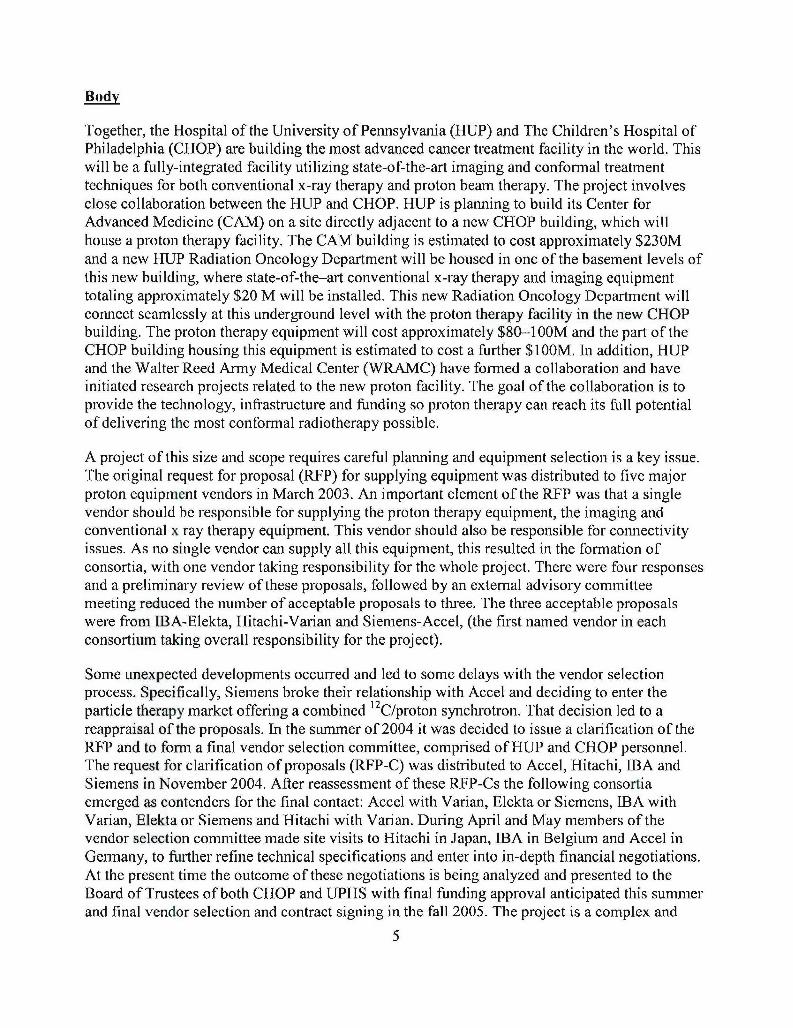
Unlike with x-rays, the absorbed dose of a proton beam increases very gradually with increasingdepth and then suddenly rises to a peak at the end of a proton range. This is known as the BraggPeak (Fig. 1). A proton beam can be directed so that the Bragg Peak occurs precisely within thetumor volume, something that can almost never be done with x-rays. The dose around the tumorvolume is much less than the tumor itself, thus sparing the normal tissue in this area. The doseimmediately beyond the Bragg Peak of a proton beam is essentially zero which allows for thesparing of all normal tissues beyond the tumor volume. Side effects, both acute and long-term,typically seen with x-ray therapy can thus be markedly reduced with proton beams by sparingnormal tissues that are situated around the tumor. These considerations are directly related to thephysical characteristics of the proton beam, and require no demonstration or study. Initial clinicalstudies demonstrate the efficacy of proton therapy. It should be remembered however that theavailable clinical data are somewhat limited because most proton facilities have treated only alimited number of patients.
6
0.8
0 .7 -. ... . . . . . . . . . . .I
0.6 a""'',.•
•" 0.5
0.3
02
I).
0.A
0 5 10 i5 20 25 30
DLpth (cm wsw)
Fig. 1. Comparison of the relative depth dose for proton and x-ray beams.
A number of published studies"6 have documented the clinical advantages of proton beams, andshown decreased normal tissue toxicity, compared to conventional photons. Numerous siteswithin the body have been shown to be more effectively treated with proton beam therapy. Bylimiting the dose to normal structures, higher doses can safely be delivered to the tumor itself.This should result in higher local control and ultimately increased survival while minimizing sideeffects of therapy.
The treatment of pediatric tumors with proton therapy provides a unique opportunity tosignificantly reduce the acute and long-term complications associated with conventionalradiation therapy. The pediatric population is exquisitely sensitive to the effects of radiationtherapy. Long-term sequelae including growth abnormalities, second malignancies, neurologiccomplications, cardiac and pulmonary toxicities, and infertility may all be reduced with the useof proton therapy. X-ray therapy causes effects on the hearts and lungs of pediatric patients,again due to the problem of "exit" dose. A study of long-term survivors of children treated withx-rays to the spinal axis showed that 31% had abnormal EKGs and 75% had reduced exercisecapacity. Jakacki et al.7 reported that 60% of patients treated to the spine showed restrictive lungdisease. Proton beams should be able to entirely avoid these complications since the uninvolvednormal structures can be totally avoided.
The research element of the proton facility has brought together the expertise of HUIP andWRMAC to initially identify five projects, to be started over a period of five years, that willresult in the technology and protocols to make the new center the most advanced cancer
7

treatment facility in the world. Each of these projects will help advance proton therapyworldwide and result in measurable benefits. The five projects are as follows:
(1) Multi-leaf collimator (MLC) for use on proton therapy gantries(2) Cone Beam CT on the Gantry for localization of target volumes(3) Proton Radiography to determine dose and stopping power of various tissues(4) Positron Emission Tomography (PET) imaging on the gantry to evaluate dosedeposition within tissues irradiated(5) Scanning proton beam using adaptive radiotherapy techniques based onimplementation of MLC, Cone Beam CT, PET imaging.
This report concentrates on the first year achievements of the multileaf collimator design anddevelopment project. This is the first of the five proposed projects to be approved and funded.The overall project is running approximately 4-6 months behind schedule. Most of this delay isattributable to the time it took to recruit staff. The second year of the proposal calls for workingdirectly with the proton therapy equipment vendor to develop a multileaf collimator prototype.If the vendor is selected in the fall of 2005 as projected no additional delays are anticipated.
The Statement of Work in the approved grant proposal included the following items to beinvestigated during the first year:
1. Leaf design: The specification of the leaf material and shape will be determined so thefinal design will: (1) reduce to permitted levels the leakage of radiation through the MLConto the patient; and (2) keep the activation of the MLC, and consequently the exposureto our radiation workers, to as low a level as can reasonably be achieved. This work willbe performed in consultation with our chosen vendor using a combination of publishedliterature and Monte Carlo simulations.
2. Joint Military/Civilian Proton Radiotherapy Center: The oversight and management forthis research will be coordinated through a Joint Military and Civilian ProtonRadiotherapy Center to be established at Walter Reed Army Medical Center.Approximately 5% of the total funding will be necessary for renovation of space atWRAMC to create this center. This center is necessary to provide working space for theproject administrator and scientific writer. This Center will also serve as the hub throughwhich the Walter Reed investigators will conduct their research on this proposal. Inaddition to the oversight and management to be provided through this center and theresearch performed by the Walter Reed investigators in this Center, a third purpose ofthis center will be life cycle management of the Center in order to secure continualfunding to guarantee this Center is transformed into the remote treatment planning andmanagement clinic envisioned in the preface [of the grant proposal].
3. Investigate the design factors affecting the lateral penumbra of the beam: The qualityof the dose distribution from a proton beam, particularly the lateral penumbra, directlydepends on the distance between the final collimator and the patient surface. Ideally wewant the MLC as close as possible, but that may limit the ability to rotate the gantryaround the patient. A compromise solution will be determined using Monte Carlosimulation to study the effect the position of the MLC has on the lateral penumbra.
8
4. Design of the MLC system: The electromechanical design and assembly of the MLCwill be done in consultation with the chosen vendor. The leaf drive mechanism must bedesigned to minimize the overall dimensions of the collimator. A high-precision leafposition setting and verification system must be designed. The mechanism for mountingthe collimator assembly on the proton beam delivery nozzle must be designed to avoidpatient-collimator interference problems and be adaptable to the specific requirements fortreating a wide range of anatomical sites. A suitable computer-based control system willbe designed, which will allow for the treatment of individual fields as a series of multiplesegments. We expect to take advantage of the experience gained from the manufacturersof x-ray MLCs.
In addition to the Statement of Work there was one major activity during the first year, whichwas to assemble the additional personnel both at HUP and at WRAMC as described below.
A. Personnel at HUP and WRAMC
Four new positions were approved in the grant proposal to facilitate the completion of theresearch. Two of these positions were at HUP - a postdoctoral fellow and a student. Thepostdoctoral fellow position was advertised in Physics Today, which is the monthly trademagazine for physicists. More than sixty applicants were reviewed and Dickson Goulart,PhD was hired and started in November 2004. Dr. Goulart has spent all of his timeinstalling and running the GEANT4 simulation code on the PCs purchased for thatpurpose.
At Walter Reed an Executive Director (Gary Ashton, PhD) and a Radiation Physicist(Dan Fry, PhD) were hired in 2005. Dr. Ashton coordinates the project on the WalterReed side and interfaces with the administration at HUIP. Dr. Fry is involved with theHUP physicists working on the simulation program and has joined with them on severalvisits to existing proton facilities.
The Walter Reed Army Medical Center Space Committee approved the first phase ofrenovation of space to accommodate the above staff. Contracts have been awarded forrenovation and the completion date is expected to be at the end of June 2005. The secondphase of the space renovation is on-hold subject to a consideration of the implications ofthe DoD BRAC announcement.
B. Remote Treatment Planning
One of the goals of this project is to establish a joint HUP/WRAMC proton radiotherapycenter to facilitate both patient treatment planning and further research. This Center willserve as the hub through which the Walter Reed investigators will conduct research onthis proposal aimed at developing generic remote treatment planning and comprehensivequality assurance systems not only for the collaboration with UPENN but to underpinDoD interests in this area more generally. Research performed to date includesvalidation of simulation tools needed for quality assurance of clinical dose estimates. Thetransport media modeled is being progressively modified from homogeneous soft tissueequivalent phantoms to inhomogeneous media that will be representative of the spatialand temporal complexity required for treatment planning with patients. The strategic
9
focus of the research is to inform the Center of the appropriate planning tools, methodsand validation strategies needed to support the development of high quality remoteclinical treatment plans. It is expected the complexity of this task will evolve as protonbeam facilities increasingly attempt to integrate and adopt a variety of treatment deliverymodalities and fuse data from advanced imaging techniques
A group from HUP visited Walter Reed to learn about their TELESYNERGY® systemthat was installed in the WRAMC Radiation Oncology department. This remote medicalconsultation workstation was developed through the National Cancer Institute and hasbeen installed in approximately twenty locations in the United States and in four othercountries. It is a very efficient way to remotely view a variety of patient image studiesand seems to be a natural way for staff at WRAMC and at the proton facility tocommunicate.
As a first step in this direction, HUP has installed a Tandberg 880 MPX that permits HUPand WRAMC to interact over an ISDN line capable of transmitting over 800 kilobytesper second. The immediate effect that this had was to allow the staff at the twoinstitutions to more easily participate in the research work being performed at the otherinstitution. We expect that this system will expand into a way for staff at the WRAMC tofully participate in the planning of their patients who are treated at the proton facility.
In addition to the hardware installed at HUP, a joint HUP/WRAMC Clinical Task Forcehas been formed to start the requirements-definition and process-mapping needed to plana pilot remote treatment planning and delivery system. This work will identify the risksand management implementation issues and define the technology to be employed for apilot trial using conventional photon treatments as a baseline for validation.
C. MLC design work using GEANT4 - validation
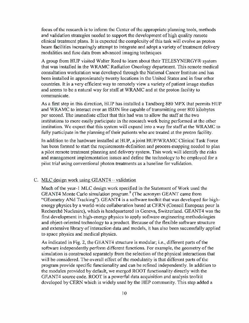
Much of the year-1 MLC design work specified in the Statement of Work used theGEANT4 Monte Carlo simulation program. 8 (The acronym GEANT came from"GEometry ANd Tracking"). GEANT4 is a software toolkit that was developed for high-energy physics by a world-wide collaboration based at CERN (Conseil European pour laRecherch6 Nucleaire), which is headquartered in Geneva, Switzerland. GEANT4 was thefirst development in high-energy physics to apply software engineering methodologiesand object-oriented technology to a product. Because of the flexible software structureand extensive library of interaction data and models, it has also been successfully appliedto space physics and medical physics.
As indicated in Fig. 2, the GEANT4 structure is modular; i.e., different parts of thesoftware independently perform different functions. For example, the geometry of thesimulation is constructed separately from the selection of the physical interactions thatwill be considered. The overall effect of the modularity is that different parts of theprogram provide specific functionality and can be refined independently. In addition tothe modules provided by default, we merged ROOT functionality directly with theGEANT4 source code. ROOT is a powerful data acquisition and analysis toolkitdeveloped by CERN which is widely used by the HEP community. This step added a
10
time-saving component to our framework in that a simulation can be run a single time butanalyzed or visualized multiple times without repeating the entire simulation.
Figure 2: GEANT4 modular structure simplifies application to specific cases.
The toolkit contains extensive models and, in some cases, cross-sectional data that can beused to select which physical interactions will be considered during the simulation. Foreach type of interaction (hadronic or electromagnetic) the user can specify a lower energy
limit below which particles will no longer be tracked. The user may also specify whatsecondary particles are of interest so that time can be used most effectively.
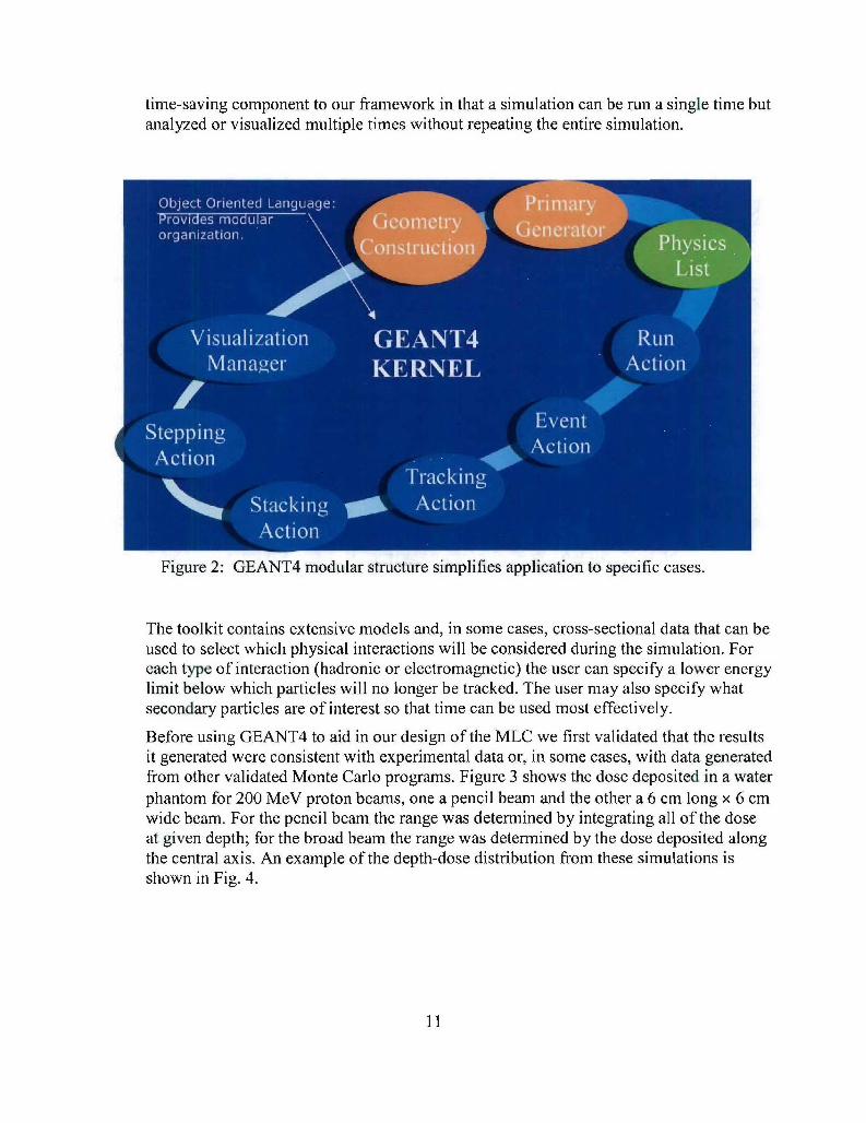
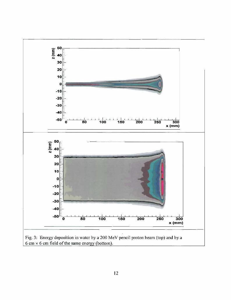
Before using GEANT4 to aid in our design of the MLC we first validated that the resultsit generated wcrc consistent with experimental data or, in some cases, with data generatedfrom other validated Monte Carlo programs. Figure 3 shows the dose deposited in a waterphantom for 200 MeV proton beams, one a pencil beam and the other a 6 cm long × 6 cmwide beam. For the pencil beam the range was determined by integrating all of the doseat given depth; for the broad beam the range was determined by the dose deposited alongthe central axis. An example of the depth-dose distribution from these simulations isshown in Fig. 4.
11
- 50E40N
30-
20
10 -.
0
-10•
-20
-30
-40
0 . I . , I i0 50 100 150 200 250 300
x (mm)
ýE 40-N
30520
10-10-
-30
-40
0 50 100 150 200 250 300x (ram)
Fig. 3: Energy deposition in water by a 200 MeV pencil proton beam (top) and by a6 cm x 6 cm field of the same energy (bottom).
12
,U.103
60
40-
0120-
lu
so -_
0050 100 150 200 250 300X (mm)
Fig. 4: Pristine Bragg peak for 200 MeV proton beam incident in water derived byintegration of the energy deposited at depth by the pencil beam in Fig. 3.
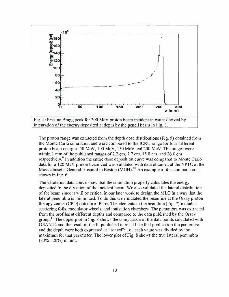
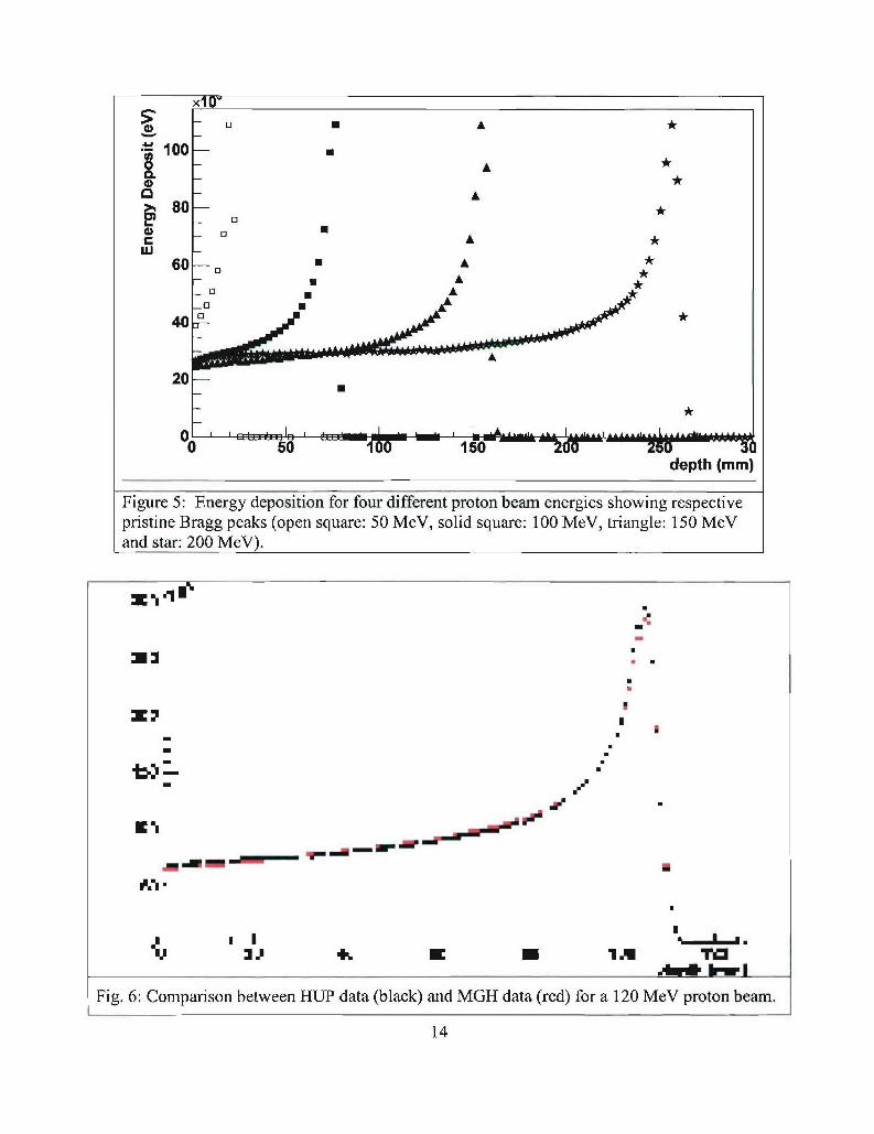
The proton range was extracted from the depth dose distributions (Fig. 5) obtained fromthe Monte Carlo simulation and were compared to the ICRU range for four differentproton beam energies 50 MeV, 100 MeV, 150 MeV and 200 MeV. The ranges werewithin 1 mm of the published ranges of 2.2 cm, 7.7 cm, 15.8 cm, and 26.0 cmrespectively. 9 In addition the entire dose deposition curve was compared to Monte Carlodata for a 120 MeV proton beam that was validated with data obtained at the NPTC at theMassachusetts General Hospital in Boston (MGH).10 An example of this comparison isshown in Fig. 6.
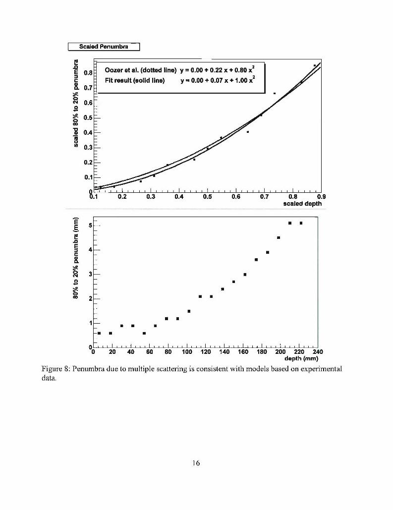
The validation data above show that the simulation properly calculates the energydeposited in the direction of the incident beam. We also validated the lateral distributionof the beam since it will be critical in our later work to design the MLC in a way that thelateral penumbra is minimized. To do this we simulated the beamline at the Orsay protontherapy center (CPO) outside of Paris. The elements in the beamline (Fig. 7) includedscattering foils, modulator wheels, and ionization chambers. The penumbra was extractedfrom the profiles at different depths and compared to the data published by the Orsaygroup. 1' The upper plot in Fig. 8 shows the comparison of the data points calculated withGEANT4 and the result of the fit published in ref. 11. In that publication the penumbraand the depth were both expressed as "scaled"; i.e., each value was divided by themaximum for that parameter. The lower plot of Fig. 8 shows the true lateral penumbra(80% - 20%) in mm.
13
xl0
A*100-
O A
80-
60 *
_ A *60 4-3
* A
2O-
9SE& 'R oo L -- A A -2.. u5o ......._0 -50 16- zu 505U' -30
depth (mm)
Figure 5: Energy deposition for four different proton beam energies showing respectivepristine Bragg peaks (open square: 50 MeV, solid square: 100 MeV, triangle: 150 MeVand star: 200 MeV).
z .1
3.P 4% z 0 1MTi
IARR I
Fde
14/
-
S .I | • *" . ".u. urI .qli
Fig. 6: Comparison between ITU data (black) and MGH data (red) for a 120 MeV proton beam.
14
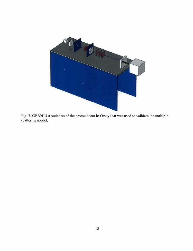
Fig. 7. GEANT4 simulation of the proton beam in Orsay that was used to validate the multiple
scattering model.
15
Scaled Penumbra
126
0.8 Oozer et al. (dotted line) y= 0.00 + 0.22 x + 0.80 x2
Fit result (solid line) y = 0.00 + 0.07 x0.7
S0.60'A 0.5-CO S0.4
• 0.3
0.2
0.1
0.1 0.2 0.3 0.4 0.5 0.6 0.7 0.8 0.9scaled depth
E
4 -
o 30U
2 UoU
0-1U
0 20 40 60 80 100 120 140 160 180 200 220 240depth (mm)
Figure 8: Penumbra due to multiple scattering is consistent with models based on experimental
data.
16
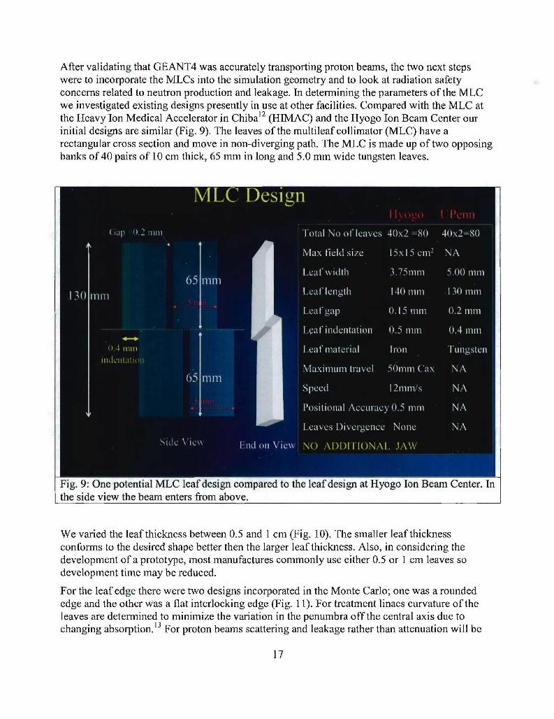
After validating that GEANT4 was accurately transporting proton beams, the two next stepswere to incorporate the MLCs into the simulation geometry and to look at radiation safetyconcerns related to neutron production and leakage. In determining the parameters of the MLCwe investigated existing designs presently in use at other facilities. Compared with the MLC atthe Heavy Ion Medical Accelerator in Chiba12 (HIMAC) and the Hyogo Ion Beam Center ourinitial designs are similar (Fig. 9). The leaves of the multileaf collimator (MLC) have arectangular cross section and move in non-diverging path. The MLC is made up of two opposingbanks of 40 pairs of 10 cm thick, 65 mm in long and 5.0 mm wide tungsten leaves.
Fig. 9: One potential MLC leaf design compared to the leaf design at Hyogo Ion Beam Center. Inthe side view the beam enters from above.
We varied the leaf thickness between 0.5 and 1 cm (Fig. 10). The smaller leaf thicknessconforms to the desired shape better then the larger leaf thickness. Also, in considering thedevelopment of a prototype, most manufactures commonly use either 0.5 or 1 cm leaves sodevelopment time may be reduced.
For the leaf edge there were two designs incorporated in the Monte Carlo; one was a roundededge and the other was a flat interlocking edge (Fig. 11). For treatment linacs curvature of theleaves are determined to minimize the variation in the penumbra off the central axis due tochanging absorption.' 3 For proton beams scattering and leakage rather than attenuation will be
17
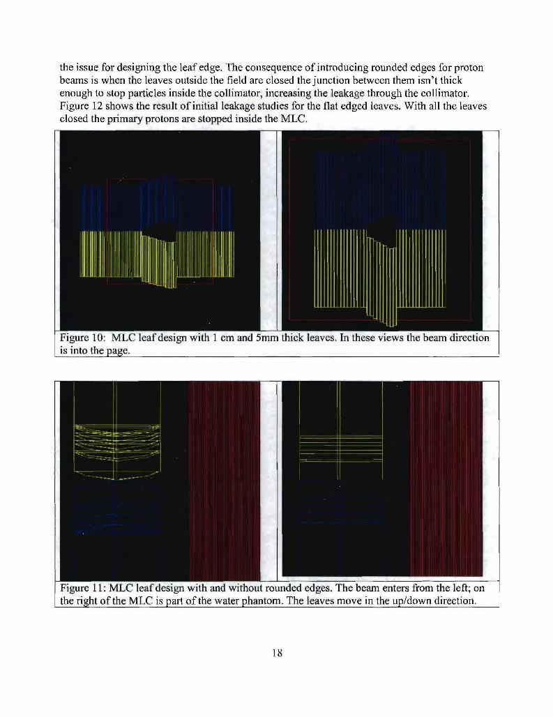
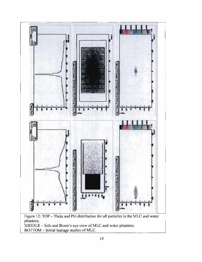
the issue for designing the leaf edge. The consequence of introducing rounded edges for protonbeams is when the leaves outside the field are closed the junction between them isn't thickenough to stop particles inside the collimator, increasing the leakage through the collimator.Figure 12 shows the result of initial leakage studies for the flat edged leaves. With all the leavesclosed the primary protons are stopped inside the MLC.
Figure 10: MLC leaf design with 1 cm and 5mm thick leaves. In these views the beam directionis into the page.
Figure 11: MLC leaf design with and without rounded edges. The beam enters from the left; onthe riot of the MLC is part of the water phantom. The leaves move in the up/down direction.
18
I.......... . ... . . ... .. ... . . ..... . . ... .. . .... ....... ..... . .. . . . .... ......
iit
I S B
Figure 12: TOP - Theta and Phi distribution for all particles in the MLC and waterphantom.MIDDLE - Side and Beam's eye view of MLC and water phantom.BOTTOM - Initial leakage studies of MLC.
19

In addition to leakage through the leaf edges, leakage between the leaves can be a problem. MLCleaves are machined to a high degree of precision but, because a small gap must be left betweenthe leaves to allow for movement, particles can still find their way through the MLC and to thepatient. To remedy the situation leaves can be designed to absorb particles that travel in the gapsby adding a stepped edge.14 Figure 13 illustrates different designs for stepped edges. We haveimplemented the single step design into the Monte Carlo, gaps between the leaves are keptwithin 0.2 mm with a step size of 0.4 mm.
(a)
Beam
(b)
S'1
(c)
Figure 13: Three potential MLC leaf designs used to study leaf leakage; (a) single step, (b)double step, and (c) corrugated. The beam enters from the left and the leaf movement is in andout of the page in these views.
D. Neutron production and MLC activation
The design of a multi-leaf collimator for a proton therapy facility includes considerationof leaf thickness and the neutrons and radioactive products generated by protoninteractions in the collimator material. The range of protons decreases with increasingdensity of the leaf material, which suggests fabricating the MLC with a high densitymaterial such as iron, brass or tungsten. However, the induced radioactive activitydepends on the material used to fabricate the MLC as does the rate of proton inducedneutron production. In this work we used both Monte Carlo simulations and published
20
data to understand the neutron production rate and radioactivity induced by high energyprotons in these potential materials for fabricating a proton MLC.
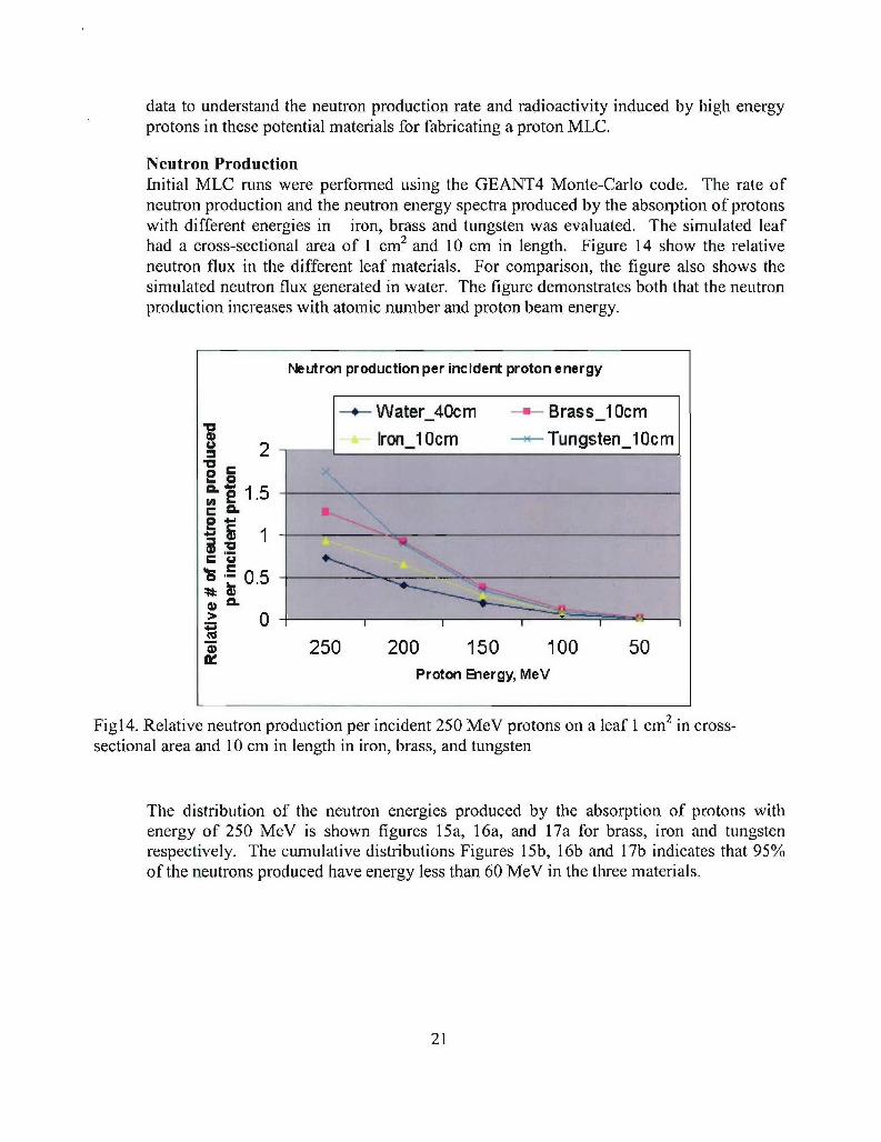
Neutron ProductionInitial MLC runs were performed using the GEANT4 Monte-Carlo code. The rate ofneutron production and the neutron energy spectra produced by the absorption of protonswith different energies in iron, brass and tungsten was evaluated. The simulated leafhad a cross-sectional area of 1 cm2 and 10 cm in length. Figure 14 show the relativeneutron flux in the different leaf materials. For comparison, the figure also shows thesimulated neutron flux generated in water. The figure demonstrates both that the neutronproduction increases with atomic number and proton beam energy.
Neutron production per incident proton energy
-*-W ater_4Ocm Brass-l OcmV Ion 1lOcm --- Tungsten l0cm
2
11.5
> 0:9
"a 250 200 150 100 50Proton Energy, MeV
Figl4. Relative neutron production per incident 250 MeV protons on a leaf 1 cm 2 in cross-sectional area and 10 cm in length in iron, brass, and tungsten
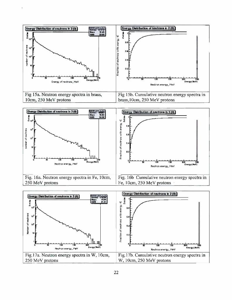
The distribution of the neutron energies produced by the absorption of protons withenergy of 250 MeV is shown figures 15a, 16a, and 17a for brass, iron and tungstenrespectively. The cumulative distributions Figures 15b, 16b and 17b indicates that 95%of the neutrons produced have energy less than 60 MeV in the three materials.
21
BMWDi~~t~n o nwmv n jB tnery Ds~iN o eutront enrg *M
10cm, ~ ~ F 252MV3roon
I?
OA -
Eneurgyon eneurons, MeV -yCMVNeutron energy,* MeV
Fig. 15a. Neutron energy spectra in Feas,10m Fig. 15b. Cumulative neutron energy spectra inI c,250 MeV protons Fes, 10cm, 250 MeV protons
________________d_____tw Inw L& O s D~bt~uio d npuftw. in 20
jNnM- 2, 20
0 ~ ~ ~ ~ ~ ~ O W W 1 0 2 W 1N1t0,nry e. E.yMY eto negM'I E.W(V
Fig17a Nutrn nery pecra n , 1cm Fi.1b. umlatve euronenrgyspctOzi
22
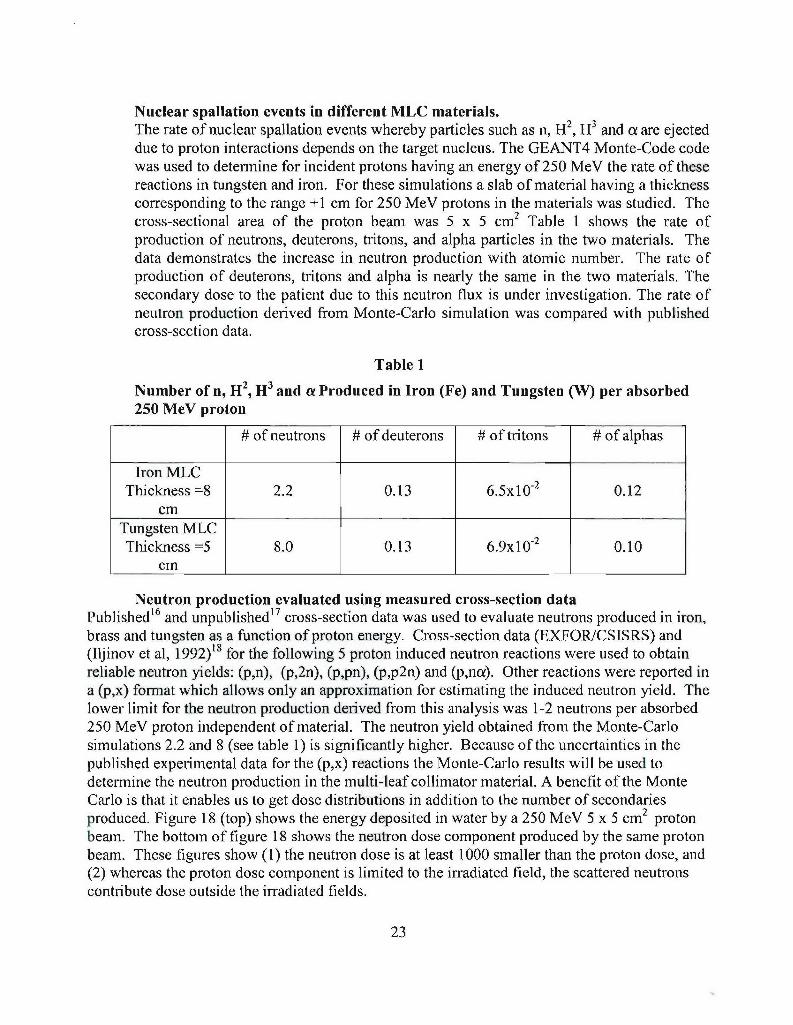
Nuclear spallation events in different MLC materials.The rate of nuclear spallation events whereby particles such as n, H2 , H3 and a are ejecteddue to proton interactions depends on the target nucleus. The GEANT4 Monte-Code codewas used to determine for incident protons having an energy of 250 MeV the rate of thesereactions in tungsten and iron. For these simulations a slab of material having a thicknesscorresponding to the range +1 cm for 250 MeV protons in the materials was studied. Thecross-sectional area of the proton beam was 5 x 5 cm 2 Table 1 shows the rate ofproduction of neutrons, deuterons, tritons, and alpha particles in the two materials. Thedata demonstrates the increase in neutron production with atomic number. The rate ofproduction of deuterons, tritons and alpha is nearly the same in the two materials. Thesecondary dose to the patient due to this neutron flux is under investigation. The rate ofneutron production derived from Monte-Carlo simulation was compared with publishedcross-section data.
Table 1
Number of n, H 2, H3 and oa Produced in Iron (Fe) and Tungsten (W) per absorbed250 MeV proton
# of neutrons # of deuterons # of tritons # of alphas
Iron MLCThickness =8 2.2 0.13 6.5x 10- 0.12
cmTungsten MLCThickness =5 8.0 0.13 6.9x 10.2 0.10
cm
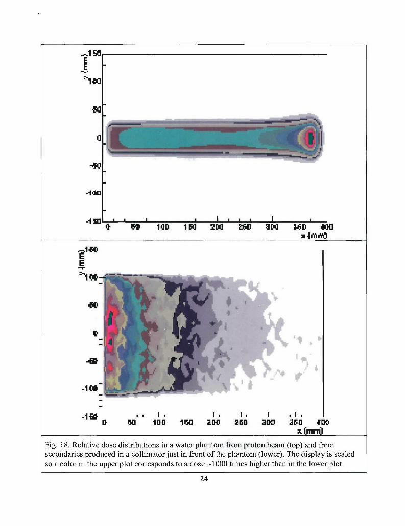
Neutron production evaluated using measured cross-section dataPublished16 and unpublished17 cross-section data was used to evaluate neutrons produced in iron,brass and tungsten as a function of proton energy. Cross-section data (EXFOR/CSISRS) and(Iljinov et al, 1992)18 for the following 5 proton induced neutron reactions were used to obtainreliable neutron yields: (p,n), (p,2n), (p,pn), (p,p2n) and (p,na). Other reactions were reported ina (p,x) format which allows only an approximation for estimating the induced neutron yield. Thelower limit for the neutron production derived from this analysis was 1-2 neutrons per absorbed250 MeV proton independent of material. The neutron yield obtained from the Monte-Carlosimulations 2.2 and 8 (see table 1) is significantly higher. Because of the uncertainties in thepublished experimental data for the (p,x) reactions the Monte-Carlo results will be used todetermine the neutron production in the multi-leaf collimator material. A benefit of the MonteCarlo is that it enables us to get dose distributions in addition to the number of secondariesproduced. Figure 18 (top) shows the energy deposited in water by a 250 MeV 5 x 5 cm2 protonbeam. The bottom of figure 18 shows the neutron dose component produced by the same protonbeam. These figures show (1) the neutron dose is at least 1000 smaller than the proton dose, and(2) whereas the proton dose component is limited to the irradiated field, the scattered neutronscontribute dose outside the irradiated fields.
23
1 fl lUaD I a 2[D3 M 1 M ND 4HI]
Er--
-1*Mi mI
N" 100 1 FPO 201 250 30' 3fl;0 40l',
Fig. 18. Relative dose distributions in a water phantom from proton beam (top) and fromsecondaries produced in a collimator just in front of the phantom (lower). The display is scaledso a color in the upper plot corresponds to a dose -1000 times higher than in the lower plot.
24
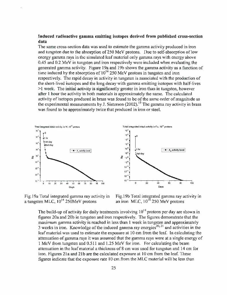
Induced radioactive gamma emitting isotopes derived from published cross-sectiondataThe same cross-section data was used to estimate the gamma activity produced in ironand tungsten due to the absorption of 250 MeV protons. Due to self-absorption of lowenergy gamma rays in the simulated leaf material only gamma rays with energy above0.45 and 0.2 MeV in tungsten and iron respectively were included when evaluating thegenerated gamma activity. Figure 19a and 19b shows the gamma activity as a function oftime induced by the absorption of 1014 250 MeV protons in tungsten and ironrespectively. The rapid decay in activity in tungsten is associated with the production ofthe short-lived isotopes and the long decay with gamma emitting isotopes with half-lives>1 week. The initial activity is significantly greater in iron than in tungsten, howeverafter 1 hour the activity in both materials is approximately the same. The calculatedactivity of isotopes produced in brass was found to be of the same order of magnitude asthe experimental measurements by J. Sisterson (2002).19 The gamma ray activity in brasswas found to be approximately twice that produced in iron or steel.
Total intetojted initial activity in W, 10" protons Total integrated initial activity in Fe. 10" protons
10' 10"..0
10' 1 0.1 hr 10da
62nd day 1o'
101 to' 1 1 hr -- A. activity levelo A d10' tay
1.0" 10•'
10 10'
to .10 "
10"to-"10-•"
0 10 20 30 40 50 60 70 80 1o o00 0 20 40 80 80 100
Days Days
Fig. 19a Total integrated gamma ray activity in Fig. 19b Total integrated gamma ray activity ina tungsten MLC, 1014 250MeV protons an iron MLC, 1014 250 MeV protons
The build-up of activity for daily treatments involving 1014 protons per day are shown infigures 20a and 20b in tungsten and iron respectively. The figures demonstrate that themaximum gamma activity is reached in less than 1 week in tungsten and approximately3 weeks in iron. Knowledge of the induced gamma ray energies°' 21 and activities in theleaf material was used to estimate the exposure at 10 cm from the leaf. In calculating theattenuation of gamma rays it was assumed that the gamma rays were at a single energy of1 MeV from tungsten and 0.511 and 1.25 MeV for iron. For calculating the beamattenuation in the leaf material a thickness of 8 cm was used for tungsten and 14 cm foriron. Figures 21a and 21b are the calculated exposure at 10 cm from the leaf. Thesefigures indicate that the exposure rate 10 cm from the MLC material will be less than
25
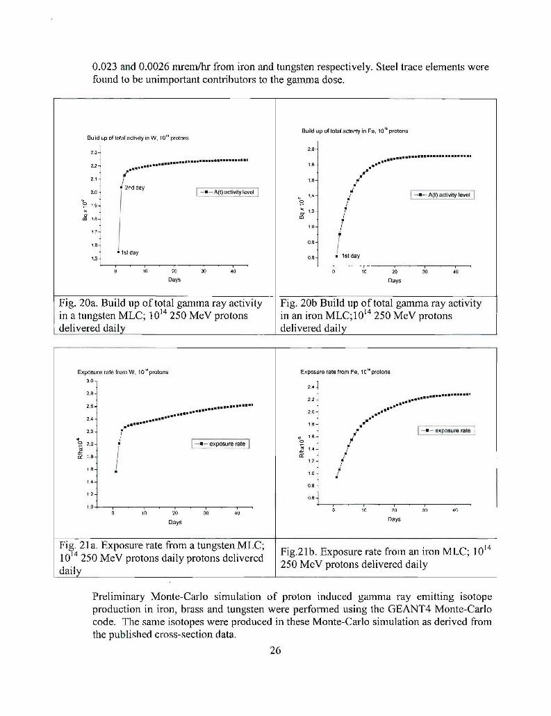
0.023 and 0.0026 mrem/hr from iron and tungsten respectively. Steel trace elements werefound to be unimportant contributors to the gamma dose.
Build up of total activity in Fe, 104 protonsBuild up of total activity in W. 104 protons
2.3- 2,0-
....................................................................212 - : :::: : :: : : ::: : : :: :: :::1.8.-= = == = = = == = = = = = = = =
21 i .e-.1 / 2nd day1.-i
2.0- Alt) activityylevel 1.4 -- _A~t)activitylevelj
1.4 A1.8-iit ev l
1.0-
17-
I6- 0.8 -
1.5- day- 1 st day
0 10 20 30 40 0 10 20 3'0 4o
Days Days
Fig. 20a. Build up of total gamma ray activity Fig. 20b Build up of total gamma ray activityin a tungsten MLC; 1014 250 MeV protons in an iron MLC;1014 250 MeV protonsdelivered daily delivered daily
Exposure rate from W, 10" protons Exposure rate from Fe, 10" protons
30-
2.42.8- ....2.6-2.2 n.j
. . . . .*2.0-
2.4-.~* u
220- -t exposure rate
1.2-16 I- 1.0 /
1.4-0.8-
2 -0.6
.,0 0 10 20 30 400 10 2•0 3•0 40
Days Days
Fig. 21a. Exposure rate from a tungsten MLC; gun14101 20 MV roonsdaly roon deivre Fig.21b. Exposure rate from aniron MLC;
10 250 MeV protons daily protons delivered 250 MeV protons delivered dailydaily ____________________
Preliminary Monte-Carlo simulation of proton induced gamma ray emitting isotopeproduction in iron, brass and tungsten were performed using the GEANT4 Monte-Carlocode. The same isotopes were produced in these Monte-Carlo simulation as derived fromthe published cross-section data.
26
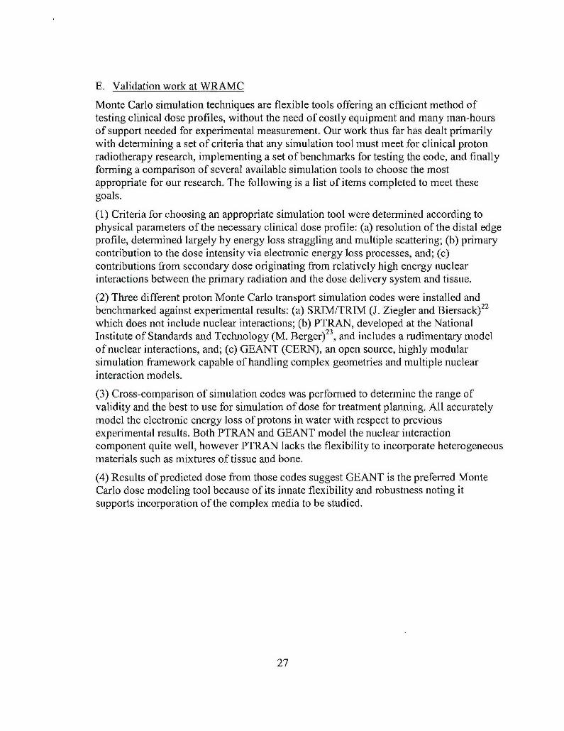
E. Validation work at WRAMC
Monte Carlo simulation techniques are flexible tools offering an efficient method oftesting clinical dose profiles, without the need of costly equipment and many man-hoursof support needed for experimental measurement. Our work thus far has dealt primarilywith determining a set of criteria that any simulation tool must meet for clinical protonradiotherapy research, implementing a set of benchmarks for testing the code, and finallyforming a comparison of several available simulation tools to choose the mostappropriate for our research. The following is a list of items completed to meet thesegoals.
(1) Criteria for choosing an appropriate simulation tool were determined according tophysical parameters of the necessary clinical dose profile: (a) resolution of the distal edgeprofile, determined largely by energy loss straggling and multiple scattering; (b) primarycontribution to the dose intensity via electronic energy loss processes, and; (c)contributions from secondary dose originating from relatively high energy nuclearinteractions between the primary radiation and the dose delivery system and tissue.
(2) Three different proton Monte Carlo transport simulation codes were installed andbenchmarked against experimental results: (a) SRIM/TRIM (J. Ziegler and Biersack)22
which does not include nuclear interactions; (b) PTRAN, developed at the NationalInstitute of Standards and Technology (M. Berger) 23, and includes a rudimentary modelof nuclear interactions, and; (c) GEANT (CERN), an open source, highly modularsimulation framework capable of handling complex geometries and multiple nuclearinteraction models.
(3) Cross-comparison of simulation codes was performed to determine the range ofvalidity and the best to use for simulation of dose for treatment planning. All accuratelymodel the electronic energy loss of protons in water with respect to previousexperimental results. Both PTRAN and GEANT model the nuclear interactioncomponent quite well, however PTRAN lacks the flexibility to incorporate heterogeneousmaterials such as mixtures of tissue and bone.
(4) Results of predicted dose from those codes suggest GEANT is the preferred MonteCarlo dose modeling tool because of its innate flexibility and robustness noting itsupports incorporation of the complex media to be studied.
27
1.0- -- SRIM Peak Normalized % Ionization
0 Experiment (MGH, Gottschalk, 1991)-.... NIST PTRAN3d
- Difference Between SRIM and Exp.
0. - - GEANT 4.5.0 Peak Normalized Eng. Deposition- No HadronicGEANT 4.5.0 Peak Normalized Erng Depostion - With Hadronic
_________________9.3% differenceJr0 - 17.36 g/cm• 2
le, - 15seis MeV
.6- 0-0.95%Target: 20cm Depth H20 SlabIsRIM az - lI ra, NP - 10 4
n GEANT4.5.0:NP-10S0.4-
0.2
I I
0.0 0.2 0.4 0a6 0.8 1.0Scaled Depth in Water, z/r,
-Stopping Power fromn GEANT 4.5.0 Simulation With Only Low Energy Ionization-e- ICRU-49 Electronic Stopping Power- ICRL-49 Total Stopping Power (Elec. + Nuct.)
8_
L -20cm H20 Cubical PhantomCoce.: GEANT 4.S.0Sampled from 10s protons
>•6-
4-
2-
0-
0.1 1 10 100 10(coProton Energy [MeV]
Fig. 22. Comparison of various simulation programs with data (top) and a comparison ofstopping power from ICRU 49 (Ref. 9) with that used in GEANT4 (lower).
28
F. Web-based clinical trials
Only 2-4% of adult cancer patients enroll in clinical trials in the United States and manypatients are never offered information on trials for which they may be eligible. Manypatients are now accessing the Internet to educate themselves on cancer clinical trials andare exploring the availability of proton therapy. OncoLink (http://www.oncolink.org) isthe web based educational resource from the University of Pennsylvania Cancer Centerand serves between 1.5-2 million pages per month to over 385,000 unique IP addresses.OncoLink launched one of the first clinical trials matching resources on the Internet thatallowed patients to enter demographic data through a secure Internet connection andmatch to clinical trials based on the inclusion and exclusion criteria of each trial.
Between 12/01 and 2/05, 4987 patients submitted online profiles to OncoLink and werematched for potential enrollment in clinical trials. The most common diagnoses ofpatients using this system included colorectal cancer (14%), breast cancer (13%), andlung cancer (10%). Of these patients, 548/4987 (11%) applied for trial enrollment afterreview of their matches to specific trials.
These data on conventional cancer treatments show that patients are willing to use theInternet for matching into clinical trials. We expect that the Internet will provide animportant means to recruit patients to proton therapy clinical trials in the future. Asregional clinical proton centers are constructed this resource could also serve as a centralregistry for proton therapy clinical trials.
Key Research Accomplishments
" Comparative analysis of proton transport simulation models has been completed.Current simulation work at WRAMC is concentrating on developing validatedestimates of dose in heterogeneous media and related design criteria needed forphantom measurements.
" Installation and validation of GEANT4 at HIUP using both RedHat and Suse Linuxoperating systems and the ROOT code for analysis. These programs were validatedby comparing results to published data.
"* Successful coding of Multileaf Collimator leaf designs in GEANT4 with thecapability to read an input file for changing leaf positions.
"* Monte Carlo code used to determine neutron production in various potential materialsused to fabricate proton MLC.
" Neutron energy spectra produced in iron, brass and tungsten calculated as a functionof proton energy and the dose in water due to the neutrons generated in the MLC byhigh energy protons is presently being evaluated.
" The radiation exposure associated with proton induced radioactive gamma emitters ina MLC has been evaluated in iron and tungsten. The calculated exposure rates 10 cm
29
from a proton MLC fabricated with these materials is low < 0.02 and 0.003 mremlhrfor iron and tungsten, respectively. Personnel exposure to individuals working withan iron MLC will be < 40 mrem/yr and 6 mremlyr with a tungsten MLC, which arevery low for radiation workers.
* Four abstracts accepted for 2005 AAPM annual meeting in Seattle and one at thePTCOG meeting in Tokyo. Additional papers will be presented at the PTCOGmeeting in December 2005.
Reportable Outcomes
The following abstracts based on work performed on this project have beenaccepted at scientific meetings:
1. Metz JM, McDonough J, Hampshire MK; "Utilization Of An Internet Based CancerClinical Trials Matching System: Implications For Proton Therapy". PTCOG meetingJune 2005, Tokyo, Japan.
2. Baldytchev M, Bloch P, Maughan R, McDonough J; "Activation induced by protoninteractions in a multi-leaf collimator in proton therapy". AAPM meeting July 2005,Seattle WA.
3. Avery S, Goulart D, Maughan R, McDonough J; "Design characteristics of a MLCfor proton therapy". AAPM meeting July 2005, Seattle WA.
4. McDonough J, Goulart D, Baldytchev M, Bloch P, Maughan R; "Monte-Carloinvestigation of proton-generated radioactivity in a multileaf collimator". AAPMmeeting July 2005, Seattle WA.
5. Goulart D, Avery S, Maughan R, McDonough J; "Validation of a Monte Carloalgorithm for simulation of dispersion due to scattering of a monoenergetic protonbeam". AAPM meeting July 2005, Seattle WA.
Conclusions
This report documents the work that has been accomplished during the first yearof the project to design an MLC for proton radiotherapy. Much of the first half ofthis initial year was spent organizing the necessary equipment and personnel toperform the tasks outlined in the Statement of Work. Once organized we validatedthe GEANT4 Monte Carlo simulation toolkit and demonstrated that it will proveto be a very powerful instrument to help us solve a variety of design questions.Some of those questions were addressed in this report including the production ofsecondary neutrons and radioactive isotopes from different potential materialsmaking up the MLC leaf.
Finally, collaboration has begun between HUP and WRAMC that will lead to thefull integration of the WRAMC staff in the treatment planning process that willoccur when the proton facility comes online.
30
References
1. Slater JD, Yonemoto LT, Tossi CJ, et al.: "Conformal proton therapy for prostatecarcinoma" Int J Radiat Oncol Biol Phys 42:299-304, 1998.
2. Bush DA, Dunbar RD, Bonnet R, et al.: "Pulmonary injury from proton andconventional radiotherapy as revealed by CT" AJR 172:735-739, 1999.
3. Isacsson U, Lennernas B, Grusell E, et al.: "Comparative treatment planning betweenproton and x-ray therapy in esophageal cancer" hIt J Radiat Oncol Biol Phys 41:441 -450, 1998.
4. Miralbell R, Crowell C, Suit H: "Potential improvement of three dimension treatmentplanning and proton therapy in the outcome of maxillary sinus cancer" Int J RadiatOncol Biol Phys 22:305-310, 1991.
5. Slater JM, Slater JD, Archambeau JO: "Carcinoma of the tonsillar region: potentialfor use of proton beam therapy" Int J Radiat Oncol Biol Phys 22:311-319, 1991.
6. Brown AP, Urie MM, Chisin R, et al.: "Proton therapy for carcinoma of thenasopharynx: a study in comparative treatment planning" Int J Radiat Oncol BiolPhys 19:1607-1614, 1989.
7. Jakacki RI, Schramm CM, Donahue BR, Haas F, Allen JC, "Restrictive lung diseasefollowing treatment for malignant brain tumors: a potential late effect of craniospinalirradiation" Journal of Clinical Oncology. 13:1478-85, 1995.
8. Agostinelli S, et al, "GEANT4: A simulation toolkit", Nucl. Instrum. and Meth.A506:250-303 (2003).
9. International Commission on Radiation Units and Measurements, "Stopping powersand ranges for protons and alpha particles," ICRU Report No. 49, ICRU, Bethesda,MD, 1993.
10. Paganetti H, personal communication.
11. Oozeer R, Mazal A, Rosenwald JC, Belshi R, Nauraye C, Ferrand R, Biensan S. "Amodel for the lateral penumbra in water of a 200-MeV proton beam devoted toclinical applications", Med Phys 24:1599-604 (1997).
12. Y. Futami, et al. "Broad-beam three-dimensional irradiation system for heavy-ionradiotherapy at HIIMAC", Nucl. Instr. and Meth. 403:143-153 (1999).
13. T. Jordan, P. Williams, "The design and performance characteristics of a multileafcollimator", Phys. Med. Biol. 39:231-251 (1994).
14. W.T. Chu, B.A. Ludewigt, T.R. Renner, "Instrumentation for treatment of cancerusing proton and light-ion beams", Rev. Sci. Instrum. 64:2055-2122 (1993).
15. M.M. Urie, J.M. Sisterson, A.M. Koehler, M. Goitein, J. Zoesman, "Proton beampenumbra: effects of separation between patient and beam modifying devices", Med.Phys. 13:734-741 (1986).
16. Atomic Data Nuclear Data Tables, 59, 185, 1995.31
17. Chadwick Mark group. Group X-5, Mail Stop B283. Los Alamos National
Laboratory. Los Alamos, NM 87545, USA private communication.
18. Iljinov A.S., Semenov V.G. et al. Nuclear and particle physics. 13 : Production ofradionuclides at intermediate energies. Editor H. Schopper, Springer-Verlag, Berlin1992.
19. Janet M Sisterson, "Selected Radiation Safety Issues at Proton Therapy Facilities",12th biennial RPSD Topical meeting, Santa Fe, New Mexico, April, 2002.
20. International Commission on Radiation Units and Measurements, "Nuclear data forneutron and proton radiotherapy and for radiation protection," ICRU Report No. 63,ICRU, Bethesda, MD, 2001.
21. Lederer,C.M. Hollander J.M, and Perlman I. Table of isotopes. 6 th ed. Berkeley,California: Wiley 1966.
22. SRIM: J. F. Ziegler, J. P, Biersack, U. Littmark, "The Stopping and Range of Ions inSolids," vol. 1 of series "Stopping and Ranges of Ions in Matter," Pergamon Press,New York (1984).
23. PTRAN: Martin J. Berger, NISTIR 5113, Proton Monte Carlo Tranport ProgramPTRAN, National Institute of Standards and Technology, U.S. Department ofCommerce (1993).
Databases used for reference:
1. Experimental Nuclear Reaction Data (EXFOR/CSISRS) http://www-nds.iaea.org/
2. LANL T-2 database: http://t2.lani.gov/data/data.html
3. Durham Database Group, at Durham University (UK). http://www-spires.dur.ac.uklhepdata/reac2.html
4. Radiological data at http://www.martindalecenter.com/ andhttp://education.jlab.org/itselemental/index.html.
5. Periodic table at http://pearll.lanl.gov/periodic/default.htm
Appendices
none
32