development and immunity-related micrornas of the lepidopteran model host galleria mellonella
TRANSCRIPT
Mukherjee and Vilcinskas BMC Genomics 2014, 15:705http://www.biomedcentral.com/1471-2164/15/705
RESEARCH ARTICLE Open Access
Development and immunity-related microRNAs ofthe lepidopteran model host Galleria mellonellaKrishnendu Mukherjee1 and Andreas Vilcinskas1,2*
Abstract
Background: MicroRNAs (miRNAs) are small non-coding RNAs that act as key players in the post-transcriptionalregulation of protein synthesis. Although little is known about their role in complex physiological processes such asdevelopment and immunity, our knowledge is expanding rapidly, thanks to the use of model systems. The larvae ofthe greater wax moth Galleria mellonella are now established as model hosts for pathogens that infect insects orhumans. To build on our previously-reported comprehensive G. mellonella transcriptome, here we describe theidentification and analysis of development and immunity-related miRNAs, thus providing valuable additional data topromote the use of this model host for the analysis of complex processes.
Results: To screen for miRNAs that are differentially expressed in G. mellonella (1) during metamorphosis or (2)following infection with the entomopathogenic bacterium Serratia entomophila or (3) with the parasitic fungusMetarhizium anisopliae, we designed a microarray containing more than 2000 insect miRNA probe sequences. Weidentified miRNAs that were significantly expressed in pre-pupae (16), pupae (22) and last-instar larvae infected withM. anisopliae (1) in comparison with untreated last-instar larvae which were used as a reference. We then used ourtranscriptomic database to identify potential 3′ untranslated regions that form miRNA–mRNA duplexes by consideringboth base pair complementarity and minimum free energy hybridization. We confirmed the co-expression of selectedmiRNAs (such as miR-71, miR-263a and miR-263b) with their predicted target mRNAs in last-instar larvae, pre-pupaeand pupae by RT-PCR. We also identified miRNAs that were expressed in response to infection with bacterial orfungal pathogens, and one miRNA that may act as a candidate mediator of trans-generational immune priming.
Conclusions: This is the first study to identify miRNAs that are predicted to regulate genes expressed duringmetamorphosis or in response to infection in the lepidopteran model host G. mellonella.
Keywords: MicroRNA, MicroRNA target prediction, Development, Metamorphosis, Immunity, Trans-generationalimmune priming, Galleria mellonella, Metarhizium anisopliae
BackgroundMicroRNAs (miRNAs) are small non-coding RNAs(~18–24 nucleotides in length) that can downregulateprotein synthesis at the post-transcriptional level by gen-erally base-pairing with the untranslated regions (UTRs)including, but not limited to, the 3′ UTRs of correspond-ing target messenger RNAs (mRNAs) [1]. Thousands ofmiRNAs have been identified or predicted in eukaryotesand their viruses since the first miRNA was shown toregulate development in the nematode Caenorhabditis
* Correspondence: [email protected] Institute for Molecular Biology and Applied Ecology, Departmentof Bioresources, Winchester Str. 2, 35395 Giessen, Germany2Institute of Phytopathology and Applied Zoology, Justus-Liebig University ofGiessen, Heinrich-Buff-Ring 26-32, 35392 Giessen, Germany
© 2014 Mukherjee and Vilcinskas; licensee Biothe Creative Commons Attribution License (htdistribution, and reproduction in any mediumDomain Dedication waiver (http://creativecomarticle, unless otherwise stated.
elegans [2]. The first evidence that miRNAs play a key rolein insect metamorphosis was reported in 2009, based onthe inhibition of metamorphosis in the cockroach Blat-tella germanica by using RNA interference (RNAi) to si-lence the dicer-1 ribonuclease, which is known totransform pre-miRNAs into mature miRNAs [3]. Theidentification and functional characterization of miRNAsis an emerging discipline in biological research, but theconsequences of disrupting miRNA expression are diffi-cult to predict because individual miRNAs can ultimatelymodulate the synthesis of hundreds of proteins if they tar-get mRNAs encoding regulatory proteins such as tran-scription factors. Therefore, it is unsurprising that manystudies provide evidence for a causal link between the al-tered expression of individual miRNAs and human
Med Central Ltd. This is an Open Access article distributed under the terms oftp://creativecommons.org/licenses/by/2.0), which permits unrestricted use,, provided the original work is properly credited. The Creative Commons Publicmons.org/publicdomain/zero/1.0/) applies to the data made available in this
Mukherjee and Vilcinskas BMC Genomics 2014, 15:705 Page 2 of 12http://www.biomedcentral.com/1471-2164/15/705
diseases including cancer, developmental abnormalitiesand malfunctions of the immune system [1,4]. Althoughthe role of miRNAs in vertebrate immunity is well estab-lished, there are few studies addressing the immunity-related functions of miRNAs in insects, as summarized ina recommended recent review [5].Here we screened directly for miRNAs in the greater
wax moth Galleria mellonella, focusing on genes thatare differentially expressed during development or inresponse to pathogens that are ingested or breach theintegument. The larvae of this species have becomeestablished as a classical model host for the analysis ofpathogenesis, particularly the virulence factors producedby entomopathogenic viruses, bacteria, fungi and proto-zoa. G. mellonella has been successfully used as a powerfuland reliable model host for human pathogens because itoffers an inexpensive and ethically acceptable alternativeto mammalian hosts in preclinical research [6,7]. Fur-thermore, G. mellonella is an efficient whole-animalhigh-throughput system for the in vivo testing of antibi-otics and as a source of novel leads for the developmentof anti-infectives [8].To compensate at least in part for the lack of a
complete genome sequence, we have recently describeda comprehensive transcriptomic database [9] that hasbeen exploited successfully e.g. to identify genes that areinduced in response to infection with Listeria monocyto-genes [10-12]. This Gram-positive bacterium causes thefood-borne disease listeriosis in humans, which often re-sults in fatal brainstem infections leading to meningitisand meningoencephalitis [13]. Furthermore, we have in-troduced G. mellonella as a model system to investigatethe role of epigenetic mechanisms that modulate insectdevelopment and immunity, e.g. the role of histoneacetylation in the regulation of transcriptional repro-gramming during metamorphosis and infections [14].This mechanism exerts its effects prior to transcriptionalinitiation because the acetylation of histones increasesDNA accessibility and promotes gene expression, whereasthe removal of acetyl groups has the opposite effect. Inthis study, we identified G. mellonella miRNAs that maycontribute to post-transcriptional gene regulation duringmetamorphosis and in response to infection. To maximizethe synergy between these investigations, we isolated totalRNA from G. mellonella at the corresponding develop-mental stages and following infection with the same ento-mopathogens such as M. anisopliae.Several approaches can be used to screen for miRNAs
in insects. For example, large scale Solexa sequencingwas used to identify miRNAs in the lepidopteran Bom-byx mori, which has a completed genome sequence [15].We designed a microarray imprinted with probes repre-senting 2064 insect miRNA sequences deposited in miR-Base (www.mirbase.org) because we have successfully
applied this microarray-based approach to identifydifferentially-expressed miRNAs related to systemic bac-terial infections or environmental stresses such as heator starvation in the model beetle Tribolium castaneum[16]. Microarrays provide a cost-efficient method for thehigh-throughput analysis of miRNAs, and using thesame experimental approach again ensures comparabil-ity between our most recent dataset and those publishedin earlier reports. However, in addition to screening fordifferentially-expressed miRNAs, we have also predictedthe corresponding target mRNAs using both empirical(RT-PCR) and theoretical (miRNA–mRNA minimumfree energy (MFE) hybridization) approaches.
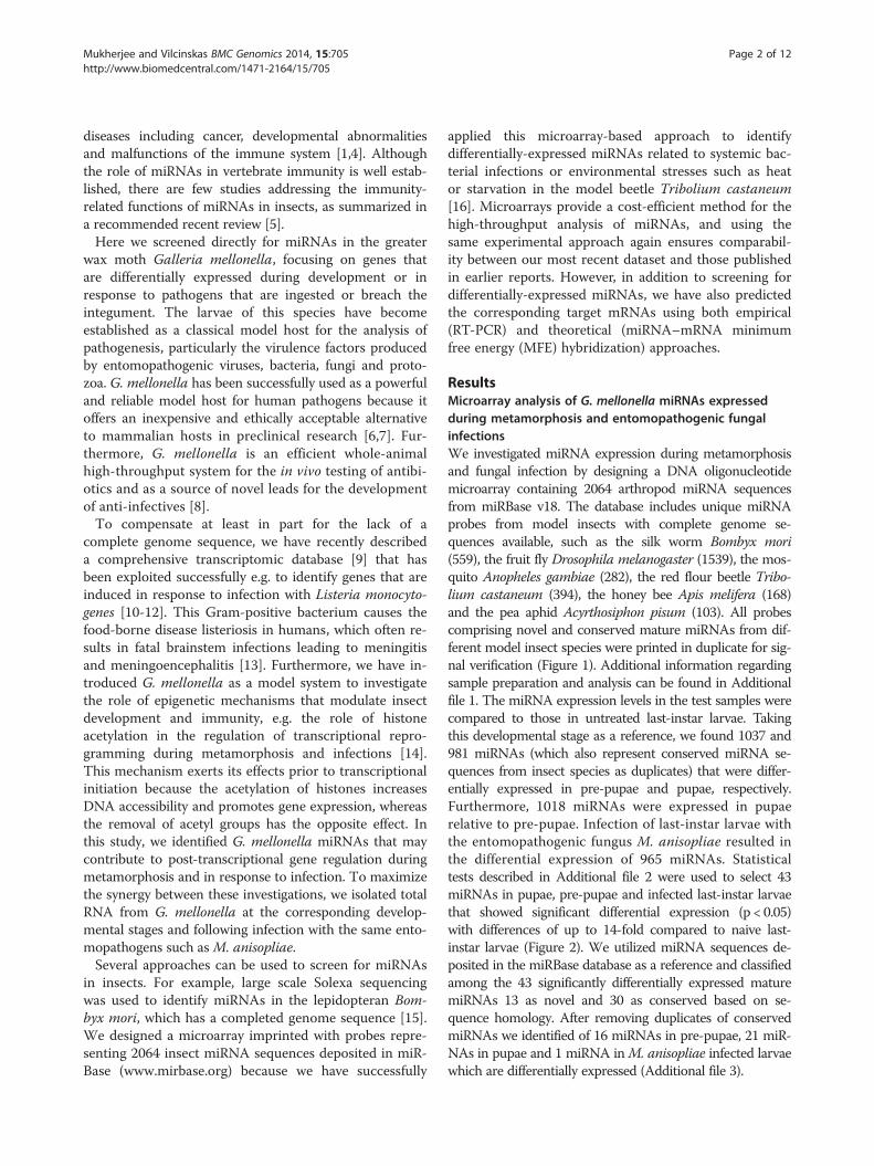
ResultsMicroarray analysis of G. mellonella miRNAs expressedduring metamorphosis and entomopathogenic fungalinfectionsWe investigated miRNA expression during metamorphosisand fungal infection by designing a DNA oligonucleotidemicroarray containing 2064 arthropod miRNA sequencesfrom miRBase v18. The database includes unique miRNAprobes from model insects with complete genome se-quences available, such as the silk worm Bombyx mori(559), the fruit fly Drosophila melanogaster (1539), the mos-quito Anopheles gambiae (282), the red flour beetle Tribo-lium castaneum (394), the honey bee Apis melifera (168)and the pea aphid Acyrthosiphon pisum (103). All probescomprising novel and conserved mature miRNAs from dif-ferent model insect species were printed in duplicate for sig-nal verification (Figure 1). Additional information regardingsample preparation and analysis can be found in Additionalfile 1. The miRNA expression levels in the test samples werecompared to those in untreated last-instar larvae. Takingthis developmental stage as a reference, we found 1037 and981 miRNAs (which also represent conserved miRNA se-quences from insect species as duplicates) that were differ-entially expressed in pre-pupae and pupae, respectively.Furthermore, 1018 miRNAs were expressed in pupaerelative to pre-pupae. Infection of last-instar larvae withthe entomopathogenic fungus M. anisopliae resulted inthe differential expression of 965 miRNAs. Statisticaltests described in Additional file 2 were used to select 43miRNAs in pupae, pre-pupae and infected last-instar larvaethat showed significant differential expression (p < 0.05)with differences of up to 14-fold compared to naive last-instar larvae (Figure 2). We utilized miRNA sequences de-posited in the miRBase database as a reference and classifiedamong the 43 significantly differentially expressed maturemiRNAs 13 as novel and 30 as conserved based on se-quence homology. After removing duplicates of conservedmiRNAs we identified of 16 miRNAs in pre-pupae, 21 miR-NAs in pupae and 1 miRNA inM. anisopliae infected larvaewhich are differentially expressed (Additional file 3).
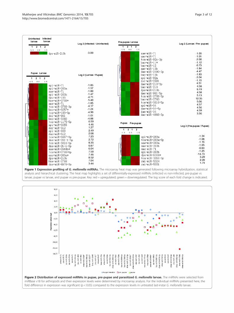
Figure 1 Expression profiling of G. mellonella miRNAs. The microarray heat map was generated following microarray hybridization, statisticalanalysis and hierarchical clustering. The heat map highlights a set of differentially-expressed miRNAs (infected vs non-infected, pre-pupae vslarvae, pupae vs larvae, and pupae vs pre-pupae. Key: red = upregulated; green = downregulated. The log score of each fold change is indicated.
Figure 2 Distribution of expressed miRNAs in pupae, pre-pupae and parasitized G. mellonella larvae. The miRNAs were selected frommiRBase v18 for arthropods and their expression levels were determined by microarray analysis. For the individual miRNAs presented here, thefold difference in expression was significant (p < 0.05) compared to the expression levels in untreated last-instar G. mellonella larvae.
Mukherjee and Vilcinskas BMC Genomics 2014, 15:705 Page 3 of 12http://www.biomedcentral.com/1471-2164/15/705
Mukherjee and Vilcinskas BMC Genomics 2014, 15:705 Page 4 of 12http://www.biomedcentral.com/1471-2164/15/705
Among the 42 significantly modulated miRNAs, wefound that 22 were specific for pupation, 16 were spe-cific for pre-pupation and 4 were expressed in both thestages (Figure 3). We found that dps-miR-210b was sig-nificantly overexpressed following fungal infection andduring metamorphosis. The transformation of last-instarlarvae into pre-pupae and pupae ultimately resulted inthe significant upregulation of 10 and 12 miRNAs, re-spectively, and the significant downregulation of 11 and15, respectively (Figure 1). Seven pupae-specific miRNAswere upregulated and three were downregulated com-pared to the expression levels observed in pre-pupae(Additional file 3). Infection with M. anisopliae sup-pressed the expression of dps-miR-210b relative to naivelast-instar larvae (Figure 1). Two-factorial ANOVA con-firmed the expression of G. mellonella miRNAs thatwere specific for metamorphosis and entomopathogenicfungal infection (Additional file 4).
MiRNA target predictionIn the absence of a complete G. mellonella genome se-quence, we used our comprehensive transcriptomic data-base to predict the putative targets of selected differentially-expressed miRNAs [9]. Nucleotide sequences at the 3′ endof individual contigs lying outside confirmed ORFs wereconsidered to be potential 3′ UTRs, and were aligned withthe mature miRNA sequences (Figure 4). This approachenabled us to determine multiple mRNA targets for at least15 miRNAs, with gene ontologies [9] as summarized inAdditional file 5: Table S1. We used an independent
Figure 3 Venn diagram showing the differential expression ofmiRNAs in G. mellonella pupae, pre-pupae and larvae infectedwith M. anisopliae, including the miRNAs that are unique toindividual to or shared among particular sample types. ThemiRNA sequences were sourced from miRBase v18 for arthropodsand differential expression was confirmed by microarray analysis.The fold-difference in expression level for all miRNAs presented here(compared to naïve last-instar G. mellonella larvae) was statisticallysignificant (p < 0.05).
BLAST search to detect mRNAs in other invertebrate spe-cies that matched those we had identified in G. mellonella,in order to investigate the potential functional conserva-tion of miRNAs among model insects (Additional file 6:Table S2).We validated our miRNA–mRNA target assignments
using the RNAhybrid program, which predicts multiplepotential binding sites for miRNAs in large target RNAs.Briefly, the program finds the energetically most favorablehybridization sites for miRNAs in a corresponding mRNAsequence, while eliminating intramolecular hybridizationi.e. base pairing between target mRNA nucleotides orbetween miRNA nucleotides [17]. The software indi-cated that complete seed sequence complementaritypreceded miRNA–mRNA duplex formation thus con-firming the targets we identified. We found 43 miRNA–mRNA duplexes using this approach, including ame-miR-71 (Figure 5A-C), api-miR-263a (Figure 6A-C),ame-miR-263b (Figure 7A-B) and dps-miR-210b (Figure 8A-C), shown as examples to highlight the significantoverexpression during pupation, pre-pupation and fun-gal infection. Duplex formation by the other signifi-cantly modulated miRNAs was also confirmed utilizingthe RNAhybrid software (Additional files 7–8: FiguresS1-S2).The majority of the modulated miRNAs were found to
target the transcriptional machinery, and mRNAs relatedto metabolism and antimicrobial responses (Additionalfiles 5 and 6: Tables S1 and S2). For example, dps-miR-210b was downregulated by fungal infection, and tar-geted mRNAs encoding RNA-binding motif protein 8a,transmembrane protein 201, 1-acylglycerol-3-phosphateacyltransferase and quiescin sulfhydryl oxidase. Simi-larly, ame-miR-263b was specifically induced in pupaebut not pre-pupae, and targeted mRNAs encoding deadbox polypeptide 1 and cd27-binding protein isoform 1(Additional file 6: Table S2).
MiRNAs regulate the expression of target mRNAs duringmetamorphosis and infectionWe next carried out RT-PCR experiments against selectedmiRNAs and their predicted targets to confirm the micro-array and bioinformatics results discussed above. Wefound that ame-miR-71 and api-miR-263a were stronglyupregulated in pupae and the predicted target contigs15133 and 21732 were strongly downregulated in pupaeand pre-pupae, respectively (Figure 9A-D). In contrast,whereas ame-miR-263b was also significantly upregulatedin pupae, its predicted target contig 20004 was upregu-lated rather than downregulated during metamorphosis(Figure 9E-F). We found that dps-miR-210b was downreg-ulated after 4 and 9 days exposure to M. anisopliae, andthat its predicted targets contigs 19765 and 15841 weresignificantly upregulated at the corresponding time points
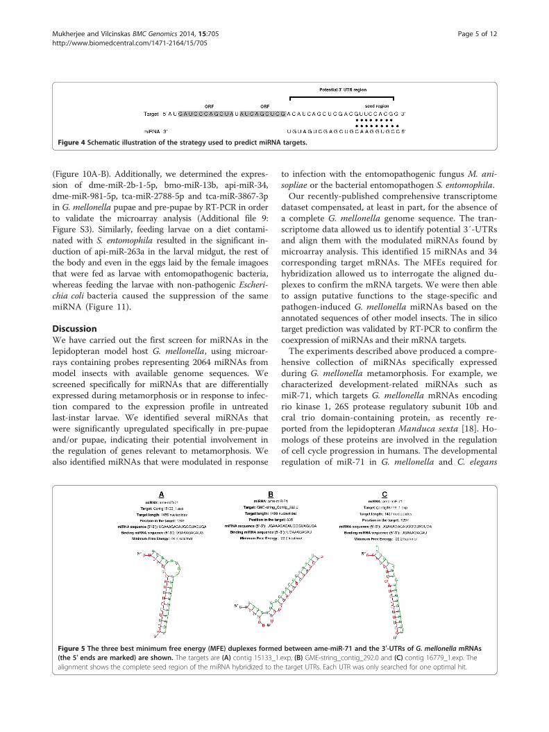
Figure 4 Schematic illustration of the strategy used to predict miRNA targets.
Mukherjee and Vilcinskas BMC Genomics 2014, 15:705 Page 5 of 12http://www.biomedcentral.com/1471-2164/15/705
(Figure 10A-B). Additionally, we determined the expres-sion of dme-miR-2b-1-5p, bmo-miR-13b, api-miR-34,dme-miR-981-5p, tca-miR-2788-5p and tca-miR-3867-3pin G. mellonella pupae and pre-pupae by RT-PCR in orderto validate the microarray analysis (Additional file 9:Figure S3). Similarly, feeding larvae on a diet contami-nated with S. entomophila resulted in the significant in-duction of api-miR-263a in the larval midgut, the rest ofthe body and even in the eggs laid by the female imagoesthat were fed as larvae with entomopathogenic bacteria,whereas feeding the larvae with non-pathogenic Escheri-chia coli bacteria caused the suppression of the samemiRNA (Figure 11).
DiscussionWe have carried out the first screen for miRNAs in thelepidopteran model host G. mellonella, using microar-rays containing probes representing 2064 miRNAs frommodel insects with available genome sequences. Wescreened specifically for miRNAs that are differentiallyexpressed during metamorphosis or in response to infec-tion compared to the expression profile in untreatedlast-instar larvae. We identified several miRNAs thatwere significantly upregulated specifically in pre-pupaeand/or pupae, indicating their potential involvement inthe regulation of genes relevant to metamorphosis. Wealso identified miRNAs that were modulated in response
Figure 5 The three best minimum free energy (MFE) duplexes formed(the 5′ ends are marked) are shown. The targets are (A) contig 15133_1alignment shows the complete seed region of the miRNA hybridized to th
to infection with the entomopathogenic fungus M. ani-sopliae or the bacterial entomopathogen S. entomophila.Our recently-published comprehensive transcriptome
dataset compensated, at least in part, for the absence ofa complete G. mellonella genome sequence. The tran-scriptome data allowed us to identify potential 3′-UTRsand align them with the modulated miRNAs found bymicroarray analysis. This identified 15 miRNAs and 34corresponding target mRNAs. The MFEs required forhybridization allowed us to interrogate the aligned du-plexes to confirm the mRNA targets. We were then ableto assign putative functions to the stage-specific andpathogen-induced G. mellonella miRNAs based on theannotated sequences of other model insects. The in silicotarget prediction was validated by RT-PCR to confirm thecoexpression of miRNAs and their mRNA targets.The experiments described above produced a compre-
hensive collection of miRNAs specifically expressedduring G. mellonella metamorphosis. For example, wecharacterized development-related miRNAs such asmiR-71, which targets G. mellonella mRNAs encodingrio kinase 1, 26S protease regulatory subunit 10b andcral trio domain-containing protein, as recently re-ported from the lepidopteran Manduca sexta [18]. Ho-mologs of these proteins are involved in the regulationof cell cycle progression in humans. The developmentalregulation of miR-71 in G. mellonella and C. elegans
between ame-miR-71 and the 3′-UTRs of G. mellonella mRNAs.exp, (B) GME-string_contig_292.0 and (C) contig 16779_1.exp. Thee target UTRs. Each UTR was only searched for one optimal hit.
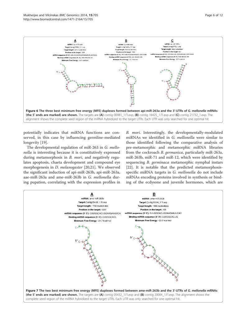
Figure 6 The three best minimum free energy (MFE) duplexes formed between api-miR-263a and the 3′-UTRs of G. mellonella mRNAs(the 5′ ends are marked) are shown. The targets are (A) contig 00981_1.f1.exp, (B) contig 16425_1.f1.exp and (C) contig 21732_1.exp. Thealignment shows the complete seed region of the miRNA hybridized to the target UTRs. Each UTR was only searched for one optimal hit.
Mukherjee and Vilcinskas BMC Genomics 2014, 15:705 Page 6 of 12http://www.biomedcentral.com/1471-2164/15/705
potentially indicates that miRNA functions are con-served, in this case by influencing germline-mediatedlongevity [19].The developmental regulation of miR-263 in G. mello-
nella is interesting because it is constitutively expressedduring metamorphosis in B. mori, and negatively regu-lates apoptosis, chaeta development and compound eyemorphogenesis in D. melanogaster [20,21]. We observedthe significant induction of api-miR-263b, api-miR-263a,aae-miR-263a and ame-miR-263b in G. mellonella dur-ing pupation, correlating with the expression profiles in
Figure 7 The two best minimum free energy (MFE) duplexes formed(the 5′ ends are marked) are shown. The targets are (A) contig 05432_1complete seed region of the miRNA hybridized to the target UTRs. Each UT
B. mori. Interestingly, the developmentally-modulatedmiRNAs we identified in G. mellonella were similar tothose identified following the comparative analysis ofpre-metamorphic and metamorphic miRNA librariesfrom the cockroach B. germanica, particularly miR-263a,miR-263b, miR-71 and miR-12, which were identified bysequencing B. germinaca metamorphic nymphal instars[22]. It is notable that the predicted metamorphosis-specific miRNA targets in G. mellonella do not includemRNAs encoding proteins involved in synthesis or bind-ing of the ecdysone and juvenile hormones, which are
between ame-miR-263b and the 3′-UTRs of G. mellonella mRNAs.f1.exp and (B) contig 20004_1.f1.exp. The alignment shows theR was only searched for one optimal hit.
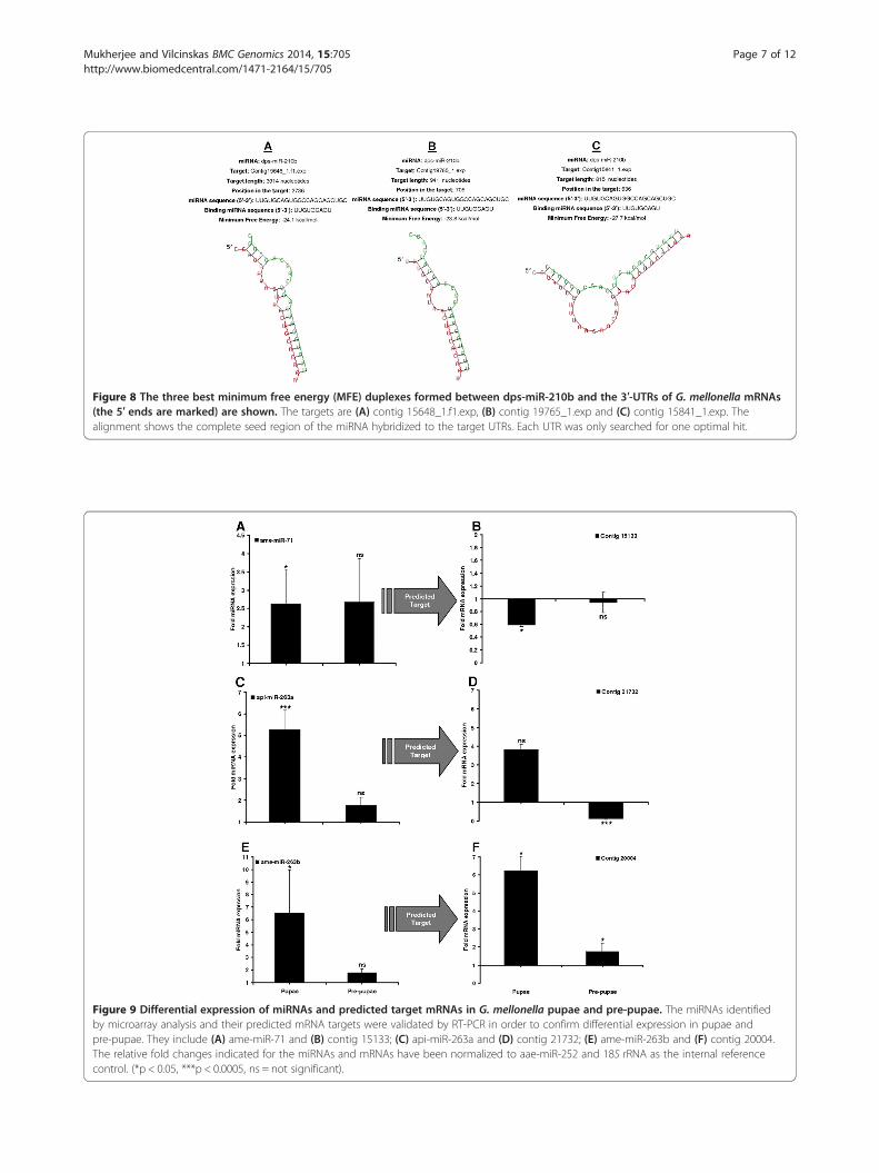
Figure 8 The three best minimum free energy (MFE) duplexes formed between dps-miR-210b and the 3′-UTRs of G. mellonella mRNAs(the 5′ ends are marked) are shown. The targets are (A) contig 15648_1.f1.exp, (B) contig 19765_1.exp and (C) contig 15841_1.exp. Thealignment shows the complete seed region of the miRNA hybridized to the target UTRs. Each UTR was only searched for one optimal hit.
Figure 9 Differential expression of miRNAs and predicted target mRNAs in G. mellonella pupae and pre-pupae. The miRNAs identifiedby microarray analysis and their predicted mRNA targets were validated by RT-PCR in order to confirm differential expression in pupae andpre-pupae. They include (A) ame-miR-71 and (B) contig 15133; (C) api-miR-263a and (D) contig 21732; (E) ame-miR-263b and (F) contig 20004.The relative fold changes indicated for the miRNAs and mRNAs have been normalized to aae-miR-252 and 18S rRNA as the internal referencecontrol. (*p < 0.05, ***p < 0.0005, ns = not significant).
Mukherjee and Vilcinskas BMC Genomics 2014, 15:705 Page 7 of 12http://www.biomedcentral.com/1471-2164/15/705
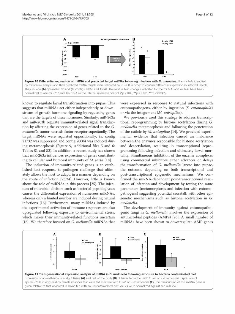
Figure 10 Differential expression of miRNA and predicted target mRNAs following infection with M. anisopliae. The miRNAs identifiedby microarray analysis and their predicted mRNA targets were validated by RT-PCR in order to confirm differential expression in infected insects.They include (A) dps-miR-210b and (B) contigs 19765 and 15841. The relative fold changes indicated for the miRNAs and mRNAs have beennormalized to aae-miR-252 and 18S rRNA as the internal reference control. (*p < 0.05, **p < 0.005, ***p < 0.0005).
Mukherjee and Vilcinskas BMC Genomics 2014, 15:705 Page 8 of 12http://www.biomedcentral.com/1471-2164/15/705
known to regulate larval transformation into pupae. Thissuggests that miRNAs act either independently or down-stream of growth hormone signaling by regulating genesthat are the targets of these hormones. Similarly, miR-263aand miR-263b regulate immunity-related signal transduc-tion by affecting the expression of genes related to the G.mellonella tumor necrosis factor receptor superfamily. Thetarget mRNAs were regulated oppositionally, i.e. contig21732 was suppressed and contig 20004 was induced dur-ing metamorphosis (Figure 9, Additional files 5 and 6:Tables S1 and S2). In addition, a recent study has shownthat miR-263a influences expression of genes contribut-ing to cellular and humoral immunity of M. sexta [18].The induction of immunity-related genes is an estab-
lished host response to pathogen challenge that ultim-ately allows the host to adapt, in a manner depending onthe route of infection [23,24]. However, little is knownabout the role of miRNAs in this process [25]. The injec-tion of microbial elicitors such as bacterial peptidoglycancauses the differential expression of numerous miRNAs,whereas only a limited number are induced during naturalinfections [16]. Furthermore, many miRNAs induced bythe experimental activation of immune responses are alsoupregulated following exposure to environmental stress,which makes their immunity-related functions uncertain[16]. We therefore focused on G. mellonella miRNAs that
Figure 11 Transgenerational expression analysis of miRNA in G. melloExpression of api-miR-263a in midgut tissue (A) and rest of the body (B) oapi-miR-263a in eggs laid by female imagoes that were fed as larvae with Egiven relative to that observed in larvae fed with an uncontaminated diet.
were expressed in response to natural infections withentomopathogens, either by ingestion (S. entomophila)or via the integument (M. anisopliae).We previously used this strategy to address transcrip-
tional reprogramming by histone acetylation during G.mellonella metamorphosis and following the penetrationof the cuticle by M. anisopliae [14]. We provided experi-mental evidence that infection caused an imbalancebetween the enzymes responsible for histone acetylationand deacetylation, resulting in transcriptional repro-gramming following infection and ultimately larval mor-tality. Simultaneous inhibition of the enzyme complexesusing commercial inhibitors either advances or delaysthe transformation of G. mellonella larvae into pupae,the outcome depending on both transcriptional andpost-transcriptional epigenetic mechanisms. We con-firmed the miRNA-dependent post-transcriptional regu-lation of infection and development by testing the sameparameters (metamorphosis and infection with entomo-pathogens) suggesting potential crosstalk with other epi-genetic mechanisms such as histone acetylation in G.mellonella.The development of immunity against entomopatho-
genic fungi in G. mellonella involves the expression ofantimicrobial peptides (AMPs) [26]. A small number ofmiRNAs have been shown to downregulate AMP genes
nella following exposure to bacteria contaminated diet.f larvae fed either with E. coli or S. entomophila. Expression of. coli or S. entomophila (C). The transcription of this miRNA gene isValues were normalized against aae-miR-252.
Mukherjee and Vilcinskas BMC Genomics 2014, 15:705 Page 9 of 12http://www.biomedcentral.com/1471-2164/15/705
in insects that are naturally infected with pathogens[25]. This is also evident from our data, e.g. the signifi-cant downregulation of dps-miR-210b in response to en-tomopathogenic fungi. NF-κB1 is a miR-210 target,which negatively regulates the LPS-induced productionof pro-inflammatory cytokines by macrophages [27]. Thedownregulation of dps-miR-210b in G. mellonella causedthe significant upregulation of targets represented bycontigs 19765 and 15841, which are functionally associ-ated with transcriptional repressor activity, metabolism,inflammation, and growth regulation (Additional file 5:Table S1). This clearly reflects the parasitic behavior ofM. anisopliae in this insect model, i.e. its ability to sup-press the immune response and thus exploit host re-sources during the infection cycle.In contrast to fungi which can infect insects via the
exoskeleton [26], bacterial pathogens are ingested withcontaminated food. The consumption of diets containingpathogenic bacteria results in the disruption of guthomeostasis, leading to dysbiosis and other gastrointes-tinal diseases [28]. RT-PCR experiments confirmed theexpression of selected miRNAs in the midgut, rest of thebody, and eggs of G. mellonella larvae fed on diets con-taminated either with the pathogenic bacterium S. ento-mophila or a non-pathogenic strain of E. coli. The latterresulted in the specific upregulation of api-miR-263awhereas the same miRNA was downregulated in re-sponse to S. entomophila. Given that api-miR-263a regu-lates a number of downstream targets, the oppositionalresponses to different organisms suggest that distincttranscriptomic programs are orchestrated against patho-gens such as S. entomophila and M. anisopliae comparedto non-pathogenic organisms such as E. coli (Figure 11).We have recently reported that contamination of the lar-val diet of G. mellonella with S. entomophila results inspecific immune responses both in the gut of fed larvaeand in the eggs laid by females that consumed these ento-mopathogenic bacteria when they were larvae implicatingspecific trans-generational immune priming [29]. Comple-mentarily, we report here expression of api-miR-263 bothin the midgut of larvae fed with S. entomophila and in theeggs of females that were fed with these bacteria whenthey were larvae. These data implicate a role of api-miR-263 in trans-generational immune priming. However, itremains to be elucidated whether this or other miRNAsare transferred from individuals that are exposed to apathogen to the next generation in order the mediatetransgenerational immune priming.
ConclusionsOur microarray-based screening approach identified sev-eral G. mellonella miRNAs that are differentially expressedduring metamorphosis or in response to natural entomo-pathogenic infections. Putative targets were predicted for
miRNAs showing the most significant modulation. Thesein silico predictions were then validated by quantifying thelevels of miRNAs and target mRNAs by RT-PCR. Weidentified numerous miRNAs that may contribute to theregulation of gene expression during metamorphosis andalso individual miRNAs that are modulated in response tothe parasitic fungus M. anisopliae and the bacterial patho-gen S. entomophila.
MethodsMaintenance and infection of insectsG. mellonella larvae were reared on an artificial diet(22% maize meal, 22% wheat germ, 11% dry yeast, 17.5%beeswax, 11% honey and 11% glycerin) at 32°C in dark-ness. Last-instar larvae, each weighing 250–350 mg,were used in all experiments. The transformation of lar-vae into pre-pupae and pupae was monitored and in-sects at the appropriate stages for analysis were selectedrandomly.The parasitic fungus M. anisopliae strain 43 was obtained
from the Julius-Kühn-Institute, Darmstadt, Germany andmaintained on potato dextran agar (Carl Roth, Germany) at27°C for 10 days to initiate conidiogenesis. Conidia werewashed with 0.02% Triton X-100, sonicated and filteredthrough miracloth to remove mycelia, and isolated conidia(3000/ml) were applied topically over the cuticle of the lastinstar larvae to mimic a natural infection. Inoculated larvaewere maintained at 27°C on the abovementioned artificialdiet.Entomopathogenic S. entomophila and non-pathogenic
E. coli were obtained from DSMZ and grown aerobicallyin Luria broth (LB; Carl Roth, Germany) at 37°C and onLB agar plates. Overnight cultures were washed threetimes with 1x PBS before each culture was added to separ-ate artificial diet preparations (~500 μl/g). Ten last-instarlarvae were presented with 5 g of the contaminated dietprior to RNA isolation after 24 hours of feeding.
Microarray analysisFor the analysis of developmentally-regulated gene expres-sion, RNA was isolated from G. mellonella last-instar lar-vae, pre-pupae and pupae as previously described [10].For the analysis of immunity-related genes, RNA was iso-lated from last-instar G. mellonella larvae 2, 4 and 9 daysafter inoculation with M. anisopliae strain 43. The lattersamples were used to represent maximum mortality [14].RNA was isolated from at least 10 animals per treat-ment for each experiment. The quantity was determinedusing a nanodrop spectrophotometer and the integritywas confirmed using an Agilent 2100 Bioanalyzer (AgilentTechnologies). Two biological replicates were used foreach sample and at each time point.Microarray analysis was carried out by LC Sciences,
USA using 2 μg total RNA samples that were extended
Mukherjee and Vilcinskas BMC Genomics 2014, 15:705 Page 10 of 12http://www.biomedcentral.com/1471-2164/15/705
with a 3′-polyadenylate tail using polyadenylate poly-merase. An oligonucleotide tag labeled with one of twofluorescent dyes was then ligated to this tail for subse-quent fluorescence detection in dual-sample experi-ments. The microarrays were hybridized overnight on aμParaflo microfluidic chip using a micro-circulationpump (Atactic Technologies) [30]. Each detection probecomprised a chemically-modified oligonucleotide com-plementary to a target miRNA (from miRBase, www.mirbase.org) or a control RNA, and a polyethylene glycolspacer segment to separate the coding segment from thesubstrate. The detection probes were generated by insitu synthesis using PGR (photogenerated reagent)chemistry. The hybridization melting temperatures werebalanced by the chemical modification of the detectionprobes. Hybridization was carried out in 100 μL 6x SSPEbuffer (0.90 M NaCl, 60 mM Na2HPO4, 6 mM EDTA,pH 6.8) containing 25% formamide at 34°C. Afterhybridization, Cy3 and Cy5 tags were circulated throughthe microfluidic chip for dye staining. Fluorescence im-ages were collected using a laser scanner (GenePix4000B, Molecular Device) and digitized using Array-Proimage analysis software (Media Cybernetics). The datawere processed by first subtracting the background andthen normalizing the signals using a locally-weighted re-gression (LOWESS) filter [31]. For the two-color experi-ments, the ratio of the two sets of log2 transformed andbalanced signals were used to calculate the p-values ofthe Student’s t-test. Differential expression was judged tobe significant at p < 0.05.
MiRNA target predictionOur transcriptomic database [9] was screened with the se-quence alignment editor BioEdit to identify open readingframes (ORFs) in all contigs. The 3′ ends of the contigsequences beyond the assigned ORFs were consideredas 3′ UTRs and screened for complementarity with theexpressed miRNA sequences identified by microarrayanalysis (Figure 4). Expressed miRNAs were defined asthose for which the average microarray signal was abovebackground in at least two different pools of the sametreatment group. The gene ontology of the target con-tigs were identified as previously described [9]. Thestructure of miRNA–mRNA duplexes was confirmedusing the RNAhybrid tool provided by the Bielefeld Bio-informatics Server, Germany [17].
RT-PCR analysisRelative miRNA and mRNA expression levels were de-termined by RT-PCR as previously described, using thesame RNA sources that were used for the microarray ex-periments [10]. For the analysis of miRNAs, cDNA wassynthesized using the miScript II miRNA first-strandsynthesis and qPCR kit (Qiagen) according to the
manufacturer’s instructions. Small RNA-enriched totalRNA was reverse transcribed using miScript HiSpec buf-fer, modified oligo-dT primers with 3′ degenerate an-chors and 5′ universal tag sequence for the specificsynthesis of mature miRNAs. The combination of polya-denylation and the universal tag ensures that miScriptprimer assays do not detect genomic DNA. Primers forthe selected miRNAs were designed using the miScriptmiRNA product-design webpage (Qiagen). CandidatemiRNA expression levels were normalized against aae-miR-252, which showed uniform expression across allsamples. Real-time RT-PCR was carried out using theBiorad (CFX 96) Mx3000P (Stratagene) system, startingwith a 15-min incubation at 95°C to activate the HotStart Polymerase followed by 40 cycles at 94°C for 15 s,55°C for 30 s and 70°C for 30 s. The following miRNAsequences were used for primer design: dps-miR-210b,5′-UUG UGC AGU GGC CAG CAG CUG C-3′; api-miR-263a 5′-AAU GGC ACU GAA AGA AUU CACGGG-3′; ame-miR-71 5′-UGA AAG ACA UGG GUAGUG A-3′; ame-miR-263b, 5′- CUU GGC ACU GGAAGA AUU CAC-3′; dme-miR-2b-1-5p 5′- GUC UUCAAA GUG GCA GUG ACA UG-3′; bmo-miR-13b 5′-UAU CAC AGC CAU UUU UGA CGA GU-3′; api-miR-34 5′ UGG CAG UGU GAU UAG CUG GUU-3′,dme-miR-981-5p 5′- CGG GUU UCG UUA GCA GCGGGC U-3′, tca-miR-2788-5p 5′- UGG GGU UUC UUAGCG GCA UUU-3′, and tca-miR-3867-3p 5′- UACACC GUU CCC GUU AUU UGC AGC GG-3′. Thecontrol miRNA sequence was aae-miR-252, 5′-UAAGUA CUA GUG CCG CAG GAG-3′.The amplification of specific target mRNAs by RT-
PCR was carried out as previously described [12] usingthe following primer sequences: contig19765_1.exp-fwd5′-CTT TCG AAA TTG CGC TGA GT-3′ and -rev 5′-GTT ACT CCC GGT CGT GTG TT-3′; contig15133_1.exp-fwd 5′-CAC GCA TGT CTT TCA GTC GT-3′ and-rev 5′-GGA GCG TCC CAG ATT TTC TT-3′; con-tig21732_1.exp-fwd 5′-CCA GAG ATC AGG GTTTGG AG-3′ and -rev 5′-TGG CAC TGA TTT TGTCTG CT-3′; contig15841_1.exp-fwd 5′-GCT GTT TGGCTT TTT CCA AG-3′ and -rev 5′-TTC CAC GACACC ATA AAC CA-3′; contig20004_1.f1.exp- fwd 5′-CAT TCA ACA CAG TGC CAA GG-3′ and -rev 5′-CAG CCT GCA AGT GTT TTT CA-3′; and the house-keeping gene 18S rRNA-fwd 5′-ATG GTT GCA AAGCTG AAA CT-3′ and -rev 5′-TCC CGT GTT GAGTCA AAT TA-3′.
Additional files
Additional file 1: Sample preparation and analysis.
Additional file 2: Sample analysis.
Mukherjee and Vilcinskas BMC Genomics 2014, 15:705 Page 11 of 12http://www.biomedcentral.com/1471-2164/15/705
Additional file 3: MicroRNAs showing stage specific expression.
Additional file 4: MicroRNAs showing expression duringentomopathogenic fungal infection and metamsorphosis, 2-wayANOVA with mean signal intensities (red p-values <0.0001, orangep-values <0.005, blue p-values <0.05).
Additional file 5: Table S1. Gene ontology (GO) analysis of miRNAtargets in G. mellonella.
Additional file 6: Table S2. Homology between selected G. mellonellamiRNA targets and related sequences in other arthropods.
Additional file 7: Figure S1. The best minimum free energy (MFE)duplexes formed between (1) bmo-miR-13b, (2) api-miR-92a, (3–4, 7)tca-miR-1175-5p, (5) dsi-miR-2581, (6) bmo-miR-2760, (8–10, 13, 14)der-miR-312, (11, 12, 15, 16) api-miR-71 and the 3′-UTRs of G. mellonellamRNAs (the 5′ ends are marked) are shown. The targets are (1) contig21322_1.exp, (2) contig 00597_1.f1.exp, (3) contig 00732_1.f1.exp, (4)contig 00462_1.f1.exp, (5) contig 09750_1.exp, (6) contig 01428_1.exp,(7) contig 18942_1.exp, contig 19122_1.exp, (8) contig 19122_1.f1.exp,(9) contig 02471_1.f1.exp, (10) GME-string-contig_3530.0, (11) GME-string-contig_292.0, (12) contig 15133_1.exp, (13) GME-string-contig_1146.0, (14)contig 15199_1.f1.exp, (15) contig 16779_1.exp and (16) contig 14917_2.r1.exp. The alignment shows the complete miRNAs hybridized to thetarget UTRs. Each UTR was only searched for one optimal hit.
Additional file 8: Figure S2. The best minimum free energy (MFE)duplexes formed between (1, 13, 14) aae-miR-2944b*, (2, 7) api-miR-263b,(3) tca-miR-263a-5p, (4) aae-miR-263a, (5, 6) dme-miR-2b-1-5p, (8) aae-miR-263a , (9) dme-miR-4976-3p, (10) api-miR-929, (11) tca-miR-92a-3p,(12) aae-miR-92a, aga-miR-92a, (15) bmo-miR-92b, (16) aae-miR-13 andthe 3′-UTRs of G. mellonella mRNAs (the 5′ ends are marked) are shown.The targets are (1) contig 02192_1.exp, (2) contig 00981_1.f1.exp, (3) contig21732_1.exp, (4) contig 00981_1.f1.exp, (5) GME-string_contig_1995.0, (6)contig 21936_1.f1.exp, (7, 8) contig 16425_1.f1.exp, (9) contig 21905_1.f1.exp, (10) contig 03661_1.exp, (11) contig 19992_1.exp, (12) GME-string-contig_3530.0, (13) GME-string-contig_1310.0, (14) contig 02672_1.f1.exp,(15) contig 17005_1.exp, and (16) contig 21322_1.exp. The alignment showsthe complete miRNAs hybridized to the target UTRs. Each UTR was onlysearched for one optimal hit.
Additional file 9: Figure S3. Differential expression of miRNAs in G.mellonella pupae and pre-pupae. The miRNAs identified by microarrayanalysis were validated by RT-PCR in order to confirm differential expressionin pupae and pre-pupae. They include dme-miR-2b-1-5p, bmo-miR-13b,api-miR-34, dme-miR-981-5p, tca-miR-2788-5p and tca-miR-3867-3p. Therelative fold changes indicated for the miRNAs have been normalized toaae-miR-252 as the internal reference control. (*p < 0.05, **p < 0.005,ns = not significant).
AbbreviationsmiRNA: microRNA; AMP: Antimicrobial peptide; MFE: Minimum free energy;UTR: Untranslated region.
Competing interestsThe authors declare that they have no competing interests.
Authors’ contributionsKM performed the experimental work. KM and AV contributed to dataanalysis and drafted the manuscript. Both authors contributed in theconception and design of the study, read and approved the final version ofthe manuscript.
AcknowledgmentsThe authors acknowledge financial support from the Hessen State Ministry ofHigher Education, Research and the Arts (HMWK) via the collaborativeresearch projects granted under the LOEWE programs “Insect Biotechnology”(Insektenbiotechnologie) and “Translational Pharmaceutical Research”(Angewandte Arzneimittelforschung). AV thanks the German ResearchFoundation for funding of the project “the role of epigenetics inhost-parasite-coevolution” (VI 219/3-2) which is embedded within theDFG Priority Programme 1399 “Host-Parasite Coevolution – rapid reciprocaladaptations and its genetic basis”.
Received: 24 December 2013 Accepted: 19 August 2014Published: 23 August 2014
References1. Bushati N, Cohen SM: microRNA functions. Annu Rev Cell Dev Biol 2007,
23:175–205.2. Lee RC, Feinbaum RL, Ambros V: The C. elegans heterochronic gene lin-4
encodes small RNAs with antisense complementarity to lin-14. Cell 1993,75:843–854.
3. Gomez-Orte E, Belles X: MicroRNA-dependent metamorphosis inhemimetabolan insects. Proc Natl Acad Sci U S A 2009, 106:21678–82.doi:10.1073/pnas.0907391106.
4. Xiao C, Rajewsky K: MicroRNA control in the immune system: basicprinciples. Cell 2009, 136:26–36.
5. Asgari S: MicroRNA functions in insects. Insect Biochem Mol Biol 2013,43:388–97. doi:10.1016/j.ibmb.2012.10.005.
6. Mylonakis E: Galleria mellonella and the study of fungal pathogenesis:making the case for another genetically tractable model host.Mycopathologia 2008, 165:1–3.
7. Glavis-Bloom J, Muhammed M, Mylonakis E: Of model hosts and man:using Caenorhabditis elegans, Drosophila melanogaster and Galleriamellonella as model hosts for infectious disease research. Adv Exp MedBiol 2012, 710:11–7. doi:10.1007/978-1-4419-5638-5_2.
8. Vilcinskas A: Anti-infective therapeutics from the Lepidopteran modelhost Galleria mellonella. Curr Pharm Des 2011, 7:1240–5.
9. Vogel H, Altincicek B, Glöckner G, Vilcinskas A: A comprehensivetranscriptome and immune-gene repertoire of the lepidopteranmodel host Galleria mellonella. BMC Genomics 2011, 12:308.doi:10.1186/1471-2164-12-308.
10. Mukherjee K, Altincicek B, Hain T, Domann E, Vilcinskas A, Chakraborty T:Galleria mellonella as a model system for studying Listeria pathogenesis.Appl Environ Microbiol 2010, 76:310–7. doi:10.1128/AEM.01301-09.
11. Mukherjee K, Abu Mraheil M, Silva S, Müller D, Cemic F, Hemberger J, HainT, Vilcinskas A, Chakraborty T: Anti-Listeria activities of Galleria mellonellahemolymph proteins. Appl Environ Microbiol 2011, 77:4237–40.doi:10.1128/AEM.02435-10.
12. Mukherjee K, Hain T, Fischer R, Chakraborty T, Vilcinskas A: Brain infectionand activation of neuronal repair mechanisms by the human pathogenListeria monocytogenes in the lepidopteran model host Galleriamellonella. Virulence 2013, 4:324–32. doi:10.4161/viru.23629.
13. Disson O, Lecuit M: Targeting of the central nervous system by Listeriamonocytogenes. Virulence 2012, 3:213–21.
14. Mukherjee K, Fischer R, Vilcinskas A: Histone acetylation mediatesepigenetic regulation of transcriptional reprogramming in insects duringmetamorphosis, wounding and infection. Front Zool 2012, 9:25.doi:10.1186/1742-9994-9-25.
15. Liu S, Li D, Li Q, Zhao P, Xiang Z, Xia Q: MicroRNAs of Bombyx moriidentified by Solexa sequencing. BMC Genomics 2010, 3:11–148.doi: 10.1186/1471-2164-11-148.
16. Freitak D, Knorr E, Vogel H, Vilcinskas A: Gender- and stressor-specificmicroRNA expression in Tribolium castaneum. Biol Lett 2012, 8:860–3.doi:10.1098/rsbl.2012.0273.
17. Rehmsmeier M, Steffen P, Hochsmann M, Giegerich R: Fast and effectiveprediction of microRNA/target duplexes. RNA 2004, 10:1507–17.
18. Zhang X, Zheng Y, Jagadeeswaran G, Ren R, Sunkar R, Jiang H:Identification of conserved and novel microRNAs in Manduca sexta andtheir possible roles in the expression regulation of immunity-relatedgenes. Insect Biochem Mol Biol 2014, 47:12–22. doi:10.1016/j.ibmb.2014.01.008.
19. Boulias K, Horvitz HR: The C. elegans microRNA mir-71 acts in neurons topromote germline-mediated longevity through regulation of DAF-16/FOXO. Cell Metab 2012, 15:439–50. doi:10.1016/j.cmet.2012.02.014.
20. Liu S, Gao S, Zhang D, Yin J, Xiang Z, Xia Q: MicroRNAs show diverse anddynamic expression patterns in multiple tissues of Bombyx mori. BMCGenomics 2010, 11:85. doi:10.1186/1471-2164-11-85.
21. Hilgers V, Bushati N, Cohen SM: Drosophila microRNAs 263a/b conferrobustness during development by protecting nascent sense organsfrom apoptosis. PLoS Biol 2010, 8:e1000396. doi:10.1371/journal.pbio.1000396.
22. Rubio M, de Horna A, Belles X: MicroRNAs in metamorphic andnon-metamorphic transitions in hemimetabolan insect metamorphosis.BMC Genomics 2012, 13:386. doi:10.1186/1471-2164-13-386.
Mukherjee and Vilcinskas BMC Genomics 2014, 15:705 Page 12 of 12http://www.biomedcentral.com/1471-2164/15/705
23. Martins NE, Faria VG, Teixeira L, Magalhães S, Sucena E: Host adaptation iscontingent upon the infection route taken by pathogens. PLoS Pathog2013, 9:e1003601. doi:10.1371/journal.ppat.1003601.
24. Vilcinskas A: Evolutionary plasticity of insect immunity. J Insect Physiol2013, 59:123–9. doi:10.1016/j.jinsphys.2012.08.018.
25. Choi IK, Hyun S: Conserved microRNA miR-8 in fat body regulates innateimmune homeostasis in Drosophila. Dev Comp Immunol 2012, 37:50–4.doi:10.1016/j.dci.2011.12.008.
26. Vilcinskas A: Coevolution between pathogen-derived proteinasesand proteinase inhibitors of host insects. Virulence 2010, 1:206–14.doi:10.4161/viru.1.3.12072.
27. Qi J, Qiao Y, Wang P, Li S, Zhao W, Gao C: microRNA-210 negativelyregulates LPS-induced production of proinflammatory cytokines bytargeting NF-κB1 in murine macrophages. FEBS Lett 2012, 586:1201–7.doi:10.1016/j.febslet.2012.03.011.
28. Maynard CL, Elson CO, Hatton RD, Weaver CT: Reciprocal interactions ofthe intestinal microbiota and immune system. Nature 2012, 489:231–41.
29. Freitak D, Schmidtberg H, Dickel F, Lochnit G, Vogel H, Vilcinskas A: Thematernal transfer of bacteria can mediate trans-generational immunepriming in insects. Virulence 2014, 5(4):547–54. doi:10.4161/viru.28367.
30. Gao X, Gulari E, Zhou X: In situ synthesis of oligonucleotide microarrays.Biopolymers 2004, 73:579–596.
31. Bolstad BM, Irizarry RA, Astrandand M, Speed TP: A comparison ofnormalization methods for high density oligonucleotide array databased on variance and bias. Biogeosciences 2003, 19:185–193.
doi:10.1186/1471-2164-15-705Cite this article as: Mukherjee and Vilcinskas: Development andimmunity-related microRNAs of the lepidopteran model host Galleriamellonella. BMC Genomics 2014 15:705.
Submit your next manuscript to BioMed Centraland take full advantage of:
• Convenient online submission
• Thorough peer review
• No space constraints or color figure charges
• Immediate publication on acceptance
• Inclusion in PubMed, CAS, Scopus and Google Scholar
• Research which is freely available for redistribution
Submit your manuscript at www.biomedcentral.com/submit