detection of leishmania dna in phlebotomines captured in campo grande, mato grosso do sul, brazil
TRANSCRIPT
Experimental Parasitology 119 (2008) 343–348
Contents lists available at ScienceDirect
Experimental Parasitology
journal homepage: www.elsevier .com/ locate/yexpr
Detection of Leishmania DNA in phlebotomines captured in Campo Grande,Mato Grosso do Sul, Brazil
Elaine A. Silva a, Renato Andreotti b,*, Edelberto S. Dias c, Jacqueline C. Barros b, Julia C.M. Brazuna a
a Centro de Controle de Zoonoses, Secretaria Municipal de Saúde, Campo Grande, MS, Brazilb Embrapa Gado de Corte, BR 262, km 4, Caixa Postal 154, Campo Grande, MS 79002-970, Brazilc Laboratório de Leishmanioses, Centro de Pesquisas René Rachou, Fundac�ão Oswaldo Cruz, Belo Horizonte, MG, Brazil
a r t i c l e i n f o a b s t r a c t
Article history:Received 23 October 2007Received in revised form 11 March 2008Accepted 13 March 2008Available online 30 March 2008
Index Descriptors and Abbreviations:LeishmaniaDNAPhlebotominesVector
0014-4894/$ - see front matter � 2008 Elsevier Inc. Adoi:10.1016/j.exppara.2008.03.011
* Corresponding author.E-mail address: [email protected] (R. An
Over the past years, leishmaniases have become a public health issue in the Brazilian state of Mato Grossodo Sul, particularly in Campo Grande, the state capital. The purpose of this study was to detect the pres-ence of Leishmania DNA in the population of phlebotomine sandflies using DNA amplification by PCR.Insect captures were carried out from 4 pm. to 7 am for 4 consecutive days each month from October2005 to September 2006 in 16 neighborhoods located in 7 urban regions of Campo Grande. Traps wereplaced indoors and in the vicinity of households. As many as 971 males and 203 females were collected.One hundred and five naturally fed females were identified and grouped as 1- to 4-specimen pools. DNAextraction was carried out using whole insects. Lutzomyia longipalpis predominated, accounting for99.15% of the phlebotomines captured. Also found was Nyssomyia whitmani, the vector of tegumentaryleishmaniasis. Abundance was greatest in the vicinity of households (69.8% of the phlebotomines cap-tured). As revealed by PCR, parasites were present in 1.9% of the Leishmania spp. specimens investigatedand confirmed for visceral leishmaniasis.
� 2008 Elsevier Inc. All rights reserved.
1. Introduction
Leishmaniases—zoonoses caused by protozoans of the genusLeishmania—have been the object of considerable attention of bothhuman and veterinary medicine (Barata et al., 2004). Visceral leish-maniasis is a serious chronic disease potentially fatal to humansand its lethality can be as high as 10%, unless suitable treatmentis provided (Gontijo and Melo, 2004).
In many areas, leishmaniasis involves a number of animal reser-voirs, whereas in some areas humans are the only infection reser-voirs, making the control of both vector and reservoirs costly andeven unfeasible. The geographical distribution of the disease haschanged in recent years, with cases being reported from previouslynon-endemic areas (World Health Organization, 2002).
Forest clear-cuttings, associated with urbanization, have had amarked influence on vector populations and disease transmission.While some species may have disappeared, others have becomemore abundant (Bejarano et al., 2002).
In the Americas, 15 New World Leishmania species, groupedinto three complexes, are responsible for tegumentary and diffusecutaneous leishmaniasis (Leishmania mexicana complex), tegumen-tary leishmaniasis and, frequently, mucocutaneous lesions (Leish-
ll rights reserved.
dreotti).
mania braziliensis complex), in addition to the visceral form ofthe disease (Leishmania donovani complex) (Grimaldi and Tesh,1993). Eco-epidemiological studies conducted in various regionsof Latin America, particularly in Brazil, culminated in the presentrecognition of no less than 20 named species of neotropicalLeishmania, 14 of which are known to infect humans (Lainson,1997).
Visceral leishmaniasis has a wide distribution in Latin America,from northern Mexico to southern Argentina (Lainson and Rangel,2005).
Brazil harbors six Leishmania species of subgenera Viannia andLeishmania, responsible for the tegumentary disease in humans.Subgenus Viannia encompasses the species Leishmania (Viannia)braziliensis, Leishmania (Viannia) guyanensis, Leishmania (Viannia)naiffi, Leishmania (Viannia) shawi, and Leishmania (Viannia) lainsoni;subgenus Leishmania is comprised of Leishmania (Leishmania) ama-zonensis (Gontijo and Carvalho, 2003). Recently, Leishmania (Vian-nia) lindenberg has been implicated as the etiologic agent ofAmerican tegumentary leishmaniasis (ATL) in the northern stateof Pará (Brasil, 2007).
In the southwestern state of Mato Grosso do Sul, tegumentaryleishmaniasis is caused by Leishmania (Viannia) braziliensis andLeishmania (Leishmania) amazonensis (Dorval et al., 2006; Brasil,2007); the etiologic agent of visceral leishmaniasis is Leishmania(Leishmania) chagasi (Oliveira et al., 2006a).
344 E.A. Silva et al. / Experimental Parasitology 119 (2008) 343–348
The development of accurate methods for identification ofLeishmania species in insect vectors is central to solid epidemiol-ogic studies and effective control measures and treatment (Harriset al., 1998; Paiva et al., 2006).
In recent years, molecular investigations have been conductedbased on the use of polymerase chain reaction (PCR) (Paiva et al.,2004), for which an important initial reference is the genome se-quence of the microorganism being investigated. The techniqueis used for diagnosis in humans (Silva et al., 2002), canines (And-rade et al., 2006), and phlebotomines (Aransay et al., 2000; Rama-lho-Ortigão et al., 2001; Miranda et al., 2002; Michalsky et al.,2002; Paiva et al., 2006).
In Campo Grande, the capital city of Mato Grosso do Sul, 92.9%of the phlebotomine sandflies captured from May 2003 to April2005 were Lutzomyia longipalpis, the vector of visceral leishmania-sis (Silva et al., 2007). The county is an endemic area for this formof the disease, with 165 cases confirmed in 2006 and 11 deaths, inaddition and 15 new cases of ATL notified in the same year (MatoGrosso do Sul, 2007). The purpose of this study was to determinethe relative abundance of the population of phlebotomines andthe presence of Leishmania species in the phlebotomines, basedon DNA amplification.
2. Materials and methods
2.1. Study area
The county of Campo Grande occupies 8096 km2 in the centralportion of Mato Grosso do Sul, near the watershed divide of the
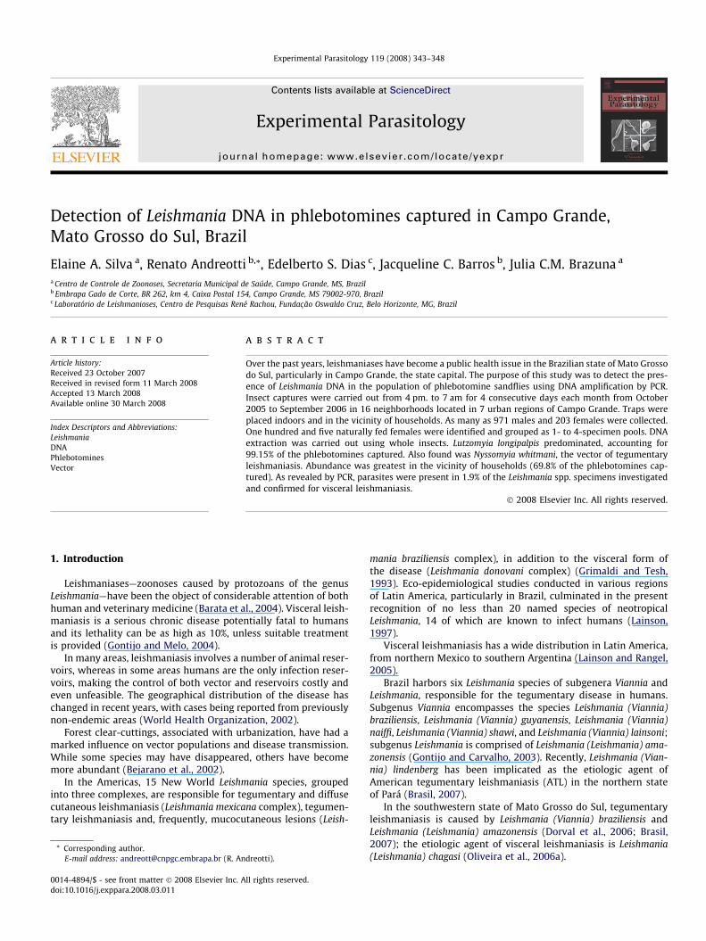
Fig. 1. Campo Grande County, MS, Brazil. Neighborhoods with permanent st
Paraná and the Paraguay basins. The coordinates of its centralmilestone are 20�2603400S, 54�3804700W. Altitudes range from 500to 675 m (Campo Grande, 2006). Population in 2006 was estimatedat 765,247 (Instituto Brasileiro de Geografia e Estatística, 2006).Climate is predominantly Aw (Köppen’s classification)—tropicalhumid, with wet summers and dry winters. Precipitation is heavi-est from October to March, the period when mean temperaturesare around 24 �C. June, July, and August are the driest months(Empresa Brasileira de Pesquisa Agropecuária Gado de Corte,2006).
2.2. Methods of capture
Capture sites were chosen so as to cover 16 neighborhoods inthe seven urban regions that comprise the entire county. Trapswere placed in households and also outdoors, in the vicinity(Fig. 1). Selection of neighborhoods was based on the occurrenceof human cases and higher prevalences. In the Segredo region,these were Vila Nasser and Margarida; in the Prosa region, Vera-neio; in the Bandeira region, Maria Aparecida; in the Central re-gion, Amambaí. Because the prevalence of human cases washighest in the Anhanduizinho region, as many as five of its neigh-borhoods were selected—UFMS, Guanandy, Aero-Rancho, Centená-rio, and Centro-Oeste—as were the Caiobá, Tarumã, and SãoConrado neighborhoods in the Lagoa region and Santo Antonioand Panamá in the Imbirussu region. Captures were carried outfrom 4 pm to 7 am for four consecutive days each month, fromOctober 2005 to September 2006, using CDC-type light traps(8 traps per neighborhood, accounting for 128 traps per month).
ations and number of phlebotomine specimens captured in each region.
E.A. Silva et al. / Experimental Parasitology 119 (2008) 343–348 345
Identification of specimens was based on Galati (2003). Femaleswere preserved in tubes containing 70% ethanol; engorged ones (asdetected by visual inspection) were selected for DNA extraction.Males were stored in the entomology laboratory of the Center forZoonosis Control in Campo Grande.
2.3. DNA extraction
Lutzomyia longipalpis females were grouped as 1- to 4-specimenpools, depending on neighborhood, capture site (indoors or in thevicinity of households), and month of capture (Table 2). Extractionwas performed using DNAzol kit (Invitrogen, Karlsruhe, Germany),as follows: homogenization of 0.1 lL of sample material and 1 lLDNAzol, 10-min centrifugation at 12,000 rpm, collection of super-natant, precipitation of supernatant with 500 lL 75% ethanol, sec-ond 10-min centrifugation at 12,000 rpm (supernatant discarded),and addition of 30 lL 8-mM NaOH.
2.4. DNA amplification
Amplification was carried out according to Michalsky et al.(2002). DNA was amplified in a Mastercycler Personal thermal cy-cler (Eppendorf, Hamburg, Germany). The assay material was pre-pared using a mixture of 10 lL PCR buffer (100-mM Tris–HCl, 500-mM KCl, 15-mM MgCl2, pH 9.0), 5 lL dNTPs (all of them 2 mM),2 lL of each primer (200 ng/lL), 0.5 lL Taq DNA polymerase(2.5 U/lL), and 26.5 lL ultrapure water. Two microliters of DNA(10 ng/lL) were added to each assay. DNA amplification was car-ried out as described. For Leishmania spp.: primers 50- GGG GAGGGG CGT TCT GCG AA-30, 50-CCG CCC CTA TTT TAC ACC AAC CCC-30, and 50-GGC CCA CTA TAT TAC ACC AAC CCC-30; thermocycling:95 �C for 5 min, followed by 35 cycles of denaturation (95 �C;1 min), annealing (60 �C; 1 min), and extension (72 �C; 1 min), witha final extension at 72 �C for 7 min. For the L. braziliensis complex:primers B1 (50-GGG GTT GGT GTA ATA TAG TGG-30) and B2 (50-CTAATT GTG CAC GGG GAG G-30); thermocycling: 95 �C for 5 min, fol-lowed by 35 cycles of denaturation (95 �C; 1 min), annealing(55 �C; 1 min), and extension (72 �C; 1 min), with a final extensionat 72 �C for 7 min. For the L. mexicana complex: primers M1 (50-CCA GTT TCG AGC CCC GGA G-30) and M2 (50-GGT GTA AAA TAGGGG CGG ATG CTC TG-30); thermocycling: 95 �C for 5 min, fol-
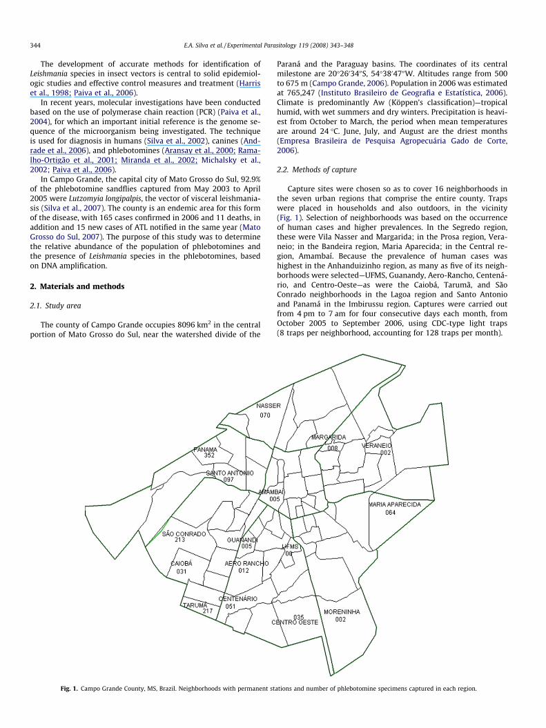
Table 1Monthly frequencies of phlebotomine species captured with CDC-type light traps
Period Wet season
Months O N D J F
Brumptomyia brumptiMF 1
Lutzomyia longipalpisM 112 132 177 108 41F 12 6 21 10 36
Micropygomyia longipennisMF
Nyssomyia whitmaniM 1F
Psathyromyia shannoniM 2F 1
Sciopemyia sordelliiMF
Total 124 138 200 121 77
Campo Grande, Mato Grosso do Sul, Brazil, October 2005–September 2006.
lowed by 35 cycles of denaturation (95 �C; 1 min), annealing(59 �C; 1 min), and extension (72 �C; 1 min), with a final extensionat 72 �C for 7 min.
To identify the L. donovani complex, the samples were also sub-jected to amplification using primers AJS31 (50-GGG GTT GGT GTAAAA TAG GGCC-30) and DBY (50-CCA GTT TCC CGC CCC GGA G-30);thermocycling: 95 �C for 5 min, followed by 40 cycles of denatur-ation (95 �C; 1 min), annealing (61 �C; 1 min), and extension(72 �C; 1 min), with a final extension at 72 �C for 8 min (Reithingeret al., 2002). They were also subjected to amplification using prim-ers RV1 (50-CTT TTC TGG TCC CGC GGG TAG G-30) and RV2 (50-CCACCT GGC TAT TTT ACA CCA-30); thermocycling: 94 �C for 4 min, fol-lowed by 40 cycles of denaturation (94 �C; 1 min), annealing(59 �C; 1 min), and extension (72 �C; 1 min), with a final extensionat 72 �C for 7 min (Lachaud et al., 2002).
DNA controls for all three Leishmania species were provided bythe Laboratory of Leishmaniases of Centro de Pesquisas René Rac-hou–Fiocruz (Belo Horizonte, Brazil)-namely, L. chagasi (MHOM/BR/74/PP/ 75), L. braziliensis (MHOM/BR/75/M2903), and L. ama-zonensis (IPLA/ BR/67/PH8) strains.
2.5. DNA electrophoresis
Amplification products were separated in 2% agarose gel(6.4 cm � 10 cm) (Lachaud et al., 2002) in PBE buffer (0.089-MTris–HCl, 0.089-M boric acid, 0.02-M EDTA) under 100 V for30 min. Bands were visualized under UV after ethidium bromidestaining. The gel was documented using a photographic system.
2.6. Minimum infection rate
Because the specimens were pooled, a minimum infection rate(MIR) for the insects was calculated as: MIR = number of positivegroups � 100/total number of insects (Paiva et al., 2006).
3. Results
Of the 1174 phlebotomine specimens captured, 203 (17.29%)were females and 971 (82.71%) males, with a mean capture rateof 0.76 phlebotomines per trap. Species distribution by monthand sex is shown in Table 1. The species collected were
Dry season
M A M J J A S
87 89 17 24 52 89 3729 33 7 5 13 18 9
1
2
2
118 123 24 29 65 107 48
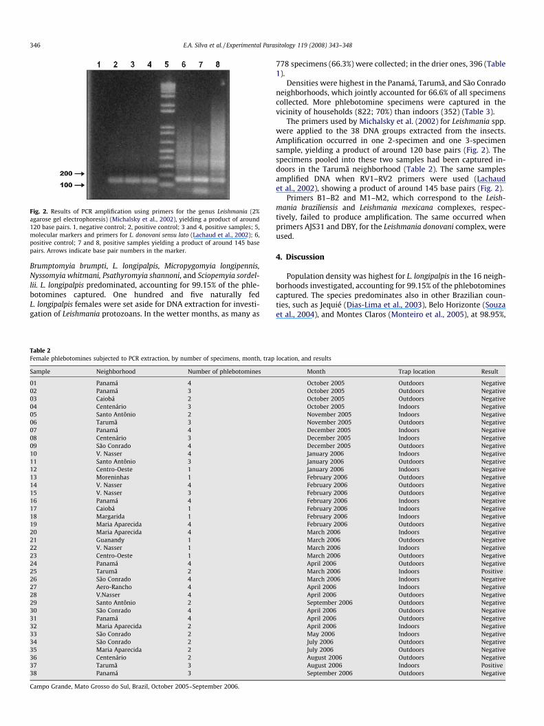
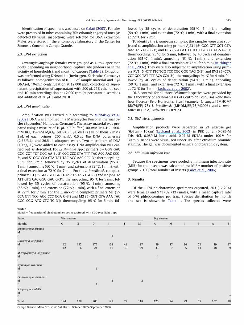
Fig. 2. Results of PCR amplification using primers for the genus Leishmania (2%agarose gel electrophoresis) (Michalsky et al., 2002), yielding a product of around120 base pairs. 1, negative control; 2, positive control; 3 and 4, positive samples; 5,molecular markers and primers for L. donovani sensu lato (Lachaud et al., 2002); 6,positive control; 7 and 8, positive samples yielding a product of around 145 basepairs. Arrows indicate base pair numbers in the marker.
346 E.A. Silva et al. / Experimental Parasitology 119 (2008) 343–348
Brumptomyia brumpti, L. longipalpis, Micropygomyia longipennis,Nyssomyia whitmani, Psathyromyia shannoni, and Sciopemyia sordel-lii. L. longipalpis predominated, accounting for 99.15% of the phle-botomines captured. One hundred and five naturally fedL. longipalpis females were set aside for DNA extraction for investi-gation of Leishmania protozoans. In the wetter months, as many as
Table 2Female phlebotomines subjected to PCR extraction, by number of specimens, month, trap
Sample Neighborhood Number of phlebotomines
01 Panamá 402 Panamá 303 Caiobá 204 Centenário 305 Santo Antônio 206 Tarumã 307 Panamá 408 Centenário 309 São Conrado 410 V. Nasser 411 Santo Antônio 312 Centro-Oeste 113 Moreninhas 114 V. Nasser 415 V. Nasser 316 Panamá 417 Caiobá 118 Margarida 119 Maria Aparecida 420 Maria Aparecida 421 Guanandy 122 V. Nasser 123 Centro-Oeste 124 Panamá 425 Tarumã 226 São Conrado 427 Aero-Rancho 428 V.Nasser 429 Santo Antônio 230 São Conrado 431 Panamá 432 Maria Aparecida 233 São Conrado 234 São Conrado 235 Maria Aparecida 236 Centenário 237 Tarumã 338 Panamá 3
Campo Grande, Mato Grosso do Sul, Brazil, October 2005–September 2006.
778 specimens (66.3%) were collected; in the drier ones, 396 (Table1).
Densities were highest in the Panamá, Tarumã, and São Conradoneighborhoods, which jointly accounted for 66.6% of all specimenscollected. More phlebotomine specimens were captured in thevicinity of households (822; 70%) than indoors (352) (Table 3).
The primers used by Michalsky et al. (2002) for Leishmania spp.were applied to the 38 DNA groups extracted from the insects.Amplification occurred in one 2-specimen and one 3-specimensample, yielding a product of around 120 base pairs (Fig. 2). Thespecimens pooled into these two samples had been captured in-doors in the Tarumã neighborhood (Table 2). The same samplesamplified DNA when RV1–RV2 primers were used (Lachaudet al., 2002), showing a product of around 145 base pairs (Fig. 2).
Primers B1–B2 and M1–M2, which correspond to the Leish-mania braziliensis and Leishmania mexicana complexes, respec-tively, failed to produce amplification. The same occurred whenprimers AJS31 and DBY, for the Leishmania donovani complex, wereused.
4. Discussion
Population density was highest for L. longipalpis in the 16 neigh-borhoods investigated, accounting for 99.15% of the phlebotominescaptured. The species predominates also in other Brazilian coun-ties, such as Jequié (Dias-Lima et al., 2003), Belo Horizonte (Souzaet al., 2004), and Montes Claros (Monteiro et al., 2005), at 98.95%,
location, and results
Month Trap location Result
October 2005 Outdoors NegativeOctober 2005 Outdoors NegativeOctober 2005 Outdoors NegativeOctober 2005 Indoors NegativeNovember 2005 Indoors NegativeNovember 2005 Outdoors NegativeDecember 2005 Indoors NegativeDecember 2005 Indoors NegativeDecember 2005 Outdoors NegativeJanuary 2006 Indoors NegativeJanuary 2006 Outdoors NegativeJanuary 2006 Indoors NegativeFebruary 2006 Outdoors NegativeFebruary 2006 Outdoors NegativeFebruary 2006 Outdoors NegativeFebruary 2006 Indoors NegativeFebruary 2006 Indoors NegativeFebruary 2006 Indoors NegativeFebruary 2006 Outdoors NegativeMarch 2006 Indoors NegativeMarch 2006 Outdoors NegativeMarch 2006 Indoors NegativeMarch 2006 Outdoors NegativeApril 2006 Outdoors NegativeMarch 2006 Indoors PositiveMarch 2006 Indoors NegativeApril 2006 Indoors NegativeApril 2006 Outdoors NegativeSeptember 2006 Outdoors NegativeApril 2006 Outdoors NegativeApril 2006 Outdoors NegativeApril 2006 Indoors NegativeMay 2006 Indoors NegativeJuly 2006 Outdoors NegativeJuly 2006 Outdoors NegativeAugust 2006 Outdoors NegativeAugust 2006 Indoors PositiveSeptember 2006 Outdoors Negative
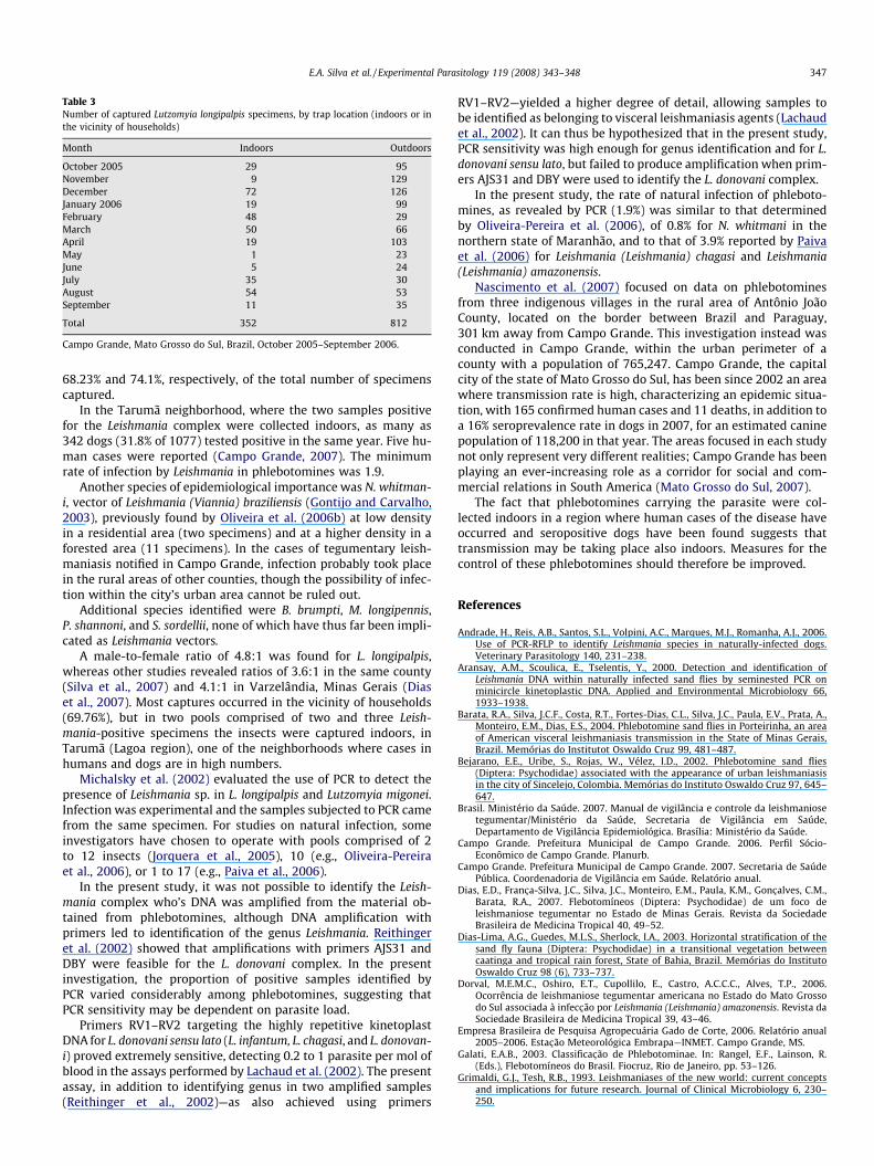
Table 3Number of captured Lutzomyia longipalpis specimens, by trap location (indoors or inthe vicinity of households)
Month Indoors Outdoors
October 2005 29 95November 9 129December 72 126January 2006 19 99February 48 29March 50 66April 19 103May 1 23June 5 24July 35 30August 54 53September 11 35
Total 352 812
Campo Grande, Mato Grosso do Sul, Brazil, October 2005–September 2006.
E.A. Silva et al. / Experimental Parasitology 119 (2008) 343–348 347
68.23% and 74.1%, respectively, of the total number of specimenscaptured.
In the Tarumã neighborhood, where the two samples positivefor the Leishmania complex were collected indoors, as many as342 dogs (31.8% of 1077) tested positive in the same year. Five hu-man cases were reported (Campo Grande, 2007). The minimumrate of infection by Leishmania in phlebotomines was 1.9.
Another species of epidemiological importance was N. whitman-i, vector of Leishmania (Viannia) braziliensis (Gontijo and Carvalho,2003), previously found by Oliveira et al. (2006b) at low densityin a residential area (two specimens) and at a higher density in aforested area (11 specimens). In the cases of tegumentary leish-maniasis notified in Campo Grande, infection probably took placein the rural areas of other counties, though the possibility of infec-tion within the city’s urban area cannot be ruled out.
Additional species identified were B. brumpti, M. longipennis,P. shannoni, and S. sordellii, none of which have thus far been impli-cated as Leishmania vectors.
A male-to-female ratio of 4.8:1 was found for L. longipalpis,whereas other studies revealed ratios of 3.6:1 in the same county(Silva et al., 2007) and 4.1:1 in Varzelândia, Minas Gerais (Diaset al., 2007). Most captures occurred in the vicinity of households(69.76%), but in two pools comprised of two and three Leish-mania-positive specimens the insects were captured indoors, inTarumã (Lagoa region), one of the neighborhoods where cases inhumans and dogs are in high numbers.
Michalsky et al. (2002) evaluated the use of PCR to detect thepresence of Leishmania sp. in L. longipalpis and Lutzomyia migonei.Infection was experimental and the samples subjected to PCR camefrom the same specimen. For studies on natural infection, someinvestigators have chosen to operate with pools comprised of 2to 12 insects (Jorquera et al., 2005), 10 (e.g., Oliveira-Pereiraet al., 2006), or 1 to 17 (e.g., Paiva et al., 2006).
In the present study, it was not possible to identify the Leish-mania complex who’s DNA was amplified from the material ob-tained from phlebotomines, although DNA amplification withprimers led to identification of the genus Leishmania. Reithingeret al. (2002) showed that amplifications with primers AJS31 andDBY were feasible for the L. donovani complex. In the presentinvestigation, the proportion of positive samples identified byPCR varied considerably among phlebotomines, suggesting thatPCR sensitivity may be dependent on parasite load.
Primers RV1–RV2 targeting the highly repetitive kinetoplastDNA for L. donovani sensu lato (L. infantum, L. chagasi, and L. donovan-i) proved extremely sensitive, detecting 0.2 to 1 parasite per mol ofblood in the assays performed by Lachaud et al. (2002). The presentassay, in addition to identifying genus in two amplified samples(Reithinger et al., 2002)—as also achieved using primers
RV1–RV2—yielded a higher degree of detail, allowing samples tobe identified as belonging to visceral leishmaniasis agents (Lachaudet al., 2002). It can thus be hypothesized that in the present study,PCR sensitivity was high enough for genus identification and for L.donovani sensu lato, but failed to produce amplification when prim-ers AJS31 and DBY were used to identify the L. donovani complex.
In the present study, the rate of natural infection of phleboto-mines, as revealed by PCR (1.9%) was similar to that determinedby Oliveira-Pereira et al. (2006), of 0.8% for N. whitmani in thenorthern state of Maranhão, and to that of 3.9% reported by Paivaet al. (2006) for Leishmania (Leishmania) chagasi and Leishmania(Leishmania) amazonensis.
Nascimento et al. (2007) focused on data on phlebotominesfrom three indigenous villages in the rural area of Antônio JoãoCounty, located on the border between Brazil and Paraguay,301 km away from Campo Grande. This investigation instead wasconducted in Campo Grande, within the urban perimeter of acounty with a population of 765,247. Campo Grande, the capitalcity of the state of Mato Grosso do Sul, has been since 2002 an areawhere transmission rate is high, characterizing an epidemic situa-tion, with 165 confirmed human cases and 11 deaths, in addition toa 16% seroprevalence rate in dogs in 2007, for an estimated caninepopulation of 118,200 in that year. The areas focused in each studynot only represent very different realities; Campo Grande has beenplaying an ever-increasing role as a corridor for social and com-mercial relations in South America (Mato Grosso do Sul, 2007).
The fact that phlebotomines carrying the parasite were col-lected indoors in a region where human cases of the disease haveoccurred and seropositive dogs have been found suggests thattransmission may be taking place also indoors. Measures for thecontrol of these phlebotomines should therefore be improved.
References
Andrade, H., Reis, A.B., Santos, S.L., Volpini, A.C., Marques, M.J., Romanha, A.J., 2006.Use of PCR-RFLP to identify Leishmania species in naturally-infected dogs.Veterinary Parasitology 140, 231–238.
Aransay, A.M., Scoulica, E., Tselentis, Y., 2000. Detection and identification ofLeishmania DNA within naturally infected sand flies by seminested PCR onminicircle kinetoplastic DNA. Applied and Environmental Microbiology 66,1933–1938.
Barata, R.A., Silva, J.C.F., Costa, R.T., Fortes-Dias, C.L., Silva, J.C., Paula, E.V., Prata, A.,Monteiro, E.M., Dias, E.S., 2004. Phlebotomine sand flies in Porteirinha, an areaof American visceral leishmaniasis transmission in the State of Minas Gerais,Brazil. Memórias do Institutot Oswaldo Cruz 99, 481–487.
Bejarano, E.E., Uribe, S., Rojas, W., Vélez, I.D., 2002. Phlebotomine sand flies(Diptera: Psychodidae) associated with the appearance of urban leishmaniasisin the city of Sincelejo, Colombia. Memórias do Instituto Oswaldo Cruz 97, 645–647.
Brasil. Ministério da Saúde. 2007. Manual de vigilância e controle da leishmaniosetegumentar/Ministério da Saúde, Secretaria de Vigilância em Saúde,Departamento de Vigilância Epidemiológica. Brasília: Ministério da Saúde.
Campo Grande. Prefeitura Municipal de Campo Grande. 2006. Perfil Sócio-Econômico de Campo Grande. Planurb.
Campo Grande. Prefeitura Municipal de Campo Grande. 2007. Secretaria de SaúdePública. Coordenadoria de Vigilância em Saúde. Relatório anual.
Dias, E.D., Franc�a-Silva, J.C., Silva, J.C., Monteiro, E.M., Paula, K.M., Gonc�alves, C.M.,Barata, R.A., 2007. Flebotomíneos (Diptera: Psychodidae) de um foco deleishmaniose tegumentar no Estado de Minas Gerais. Revista da SociedadeBrasileira de Medicina Tropical 40, 49–52.
Dias-Lima, A.G., Guedes, M.L.S., Sherlock, I.A., 2003. Horizontal stratification of thesand fly fauna (Diptera: Psychodidae) in a transitional vegetation betweencaatinga and tropical rain forest, State of Bahia, Brazil. Memórias do InstitutoOswaldo Cruz 98 (6), 733–737.
Dorval, M.E.M.C., Oshiro, E.T., Cupollilo, E., Castro, A.C.C.C., Alves, T.P., 2006.Ocorrência de leishmaniose tegumentar americana no Estado do Mato Grossodo Sul associada à infecc�ão por Leishmania (Leishmania) amazonensis. Revista daSociedade Brasileira de Medicina Tropical 39, 43–46.
Empresa Brasileira de Pesquisa Agropecuária Gado de Corte, 2006. Relatório anual2005–2006. Estac�ão Meteorológica Embrapa—INMET. Campo Grande, MS.
Galati, E.A.B., 2003. Classificac�ão de Phlebotominae. In: Rangel, E.F., Lainson, R.(Eds.), Flebotomíneos do Brasil. Fiocruz, Rio de Janeiro, pp. 53–126.
Grimaldi, G.J., Tesh, R.B., 1993. Leishmaniases of the new world: current conceptsand implications for future research. Journal of Clinical Microbiology 6, 230–250.
348 E.A. Silva et al. / Experimental Parasitology 119 (2008) 343–348
Gontijo, B., Carvalho, M.L.R., 2003. Leishmaniose tegumentar americana. Revista daSociedade Brasileira de Medicina Tropical 36, 71–80.
Gontijo, C.M.F., Melo, N.M., 2004. Leishmaniose visceral no Brasil: quadro atual,desafios e perspectivas. Revista Brasileira de Epidemiologia 7, 338–346.
Harris, E., Kropp, G., Bellli, A., Rodriguez, B., Agabian, N., 1998. Single-step multiplexPCR assay characterization of new world Leishmania complexes. Journal ofClinical Microbiology 36, 1989–1995.
Instituto Brasileiro de Geografia e Estatística. IBGE cidades 2006. Available from:http://www.ibge.gov.br/home/ Acess: 28/05/2007.
Jorquera, A., Gonc�alez, R., Marchan-Marcano, E., Oviedo, M., Matos, M., 2005.Multiplex-PCR for detection of natural Leishmania infection in Lutzomyia spp.Captures in an endemic region for cutaneous leishmaniasis in state of Sucre,Venezuela. Memórias do Instituto Oswaldo Cruz 100, 45–48.
Lachaud, L., Marchergui-Hammami, S., Chabbert, E., Dereure, J., Dedet, J.P., Bastien,P., 2002. Comparison of six PCR methods using peripheral blood for detection oncanine visceral leishmaniasis. Journal of Clinical Microbiology 40, 210–215.
Lainson, R., 1997. On Leishmania enriettii and other enigmatic Leishmania species ofthe Neotropics. Memórias do Instituto Oswaldo Cruz 92, 377–387.
Lainson, R., Rangel, E.F., 2005. Lutzomyia longipalpis and the eco-epidemiology ofAmerican visceral leishmaniasis (LVA) in Brazil. Memórias do InstitutotOswaldo Cruz 100, 811–827.
Mato Grosso do Sul. Governo do Estado de Mato Grosso do Sul. Secretaria de Estadode Saúde. SINAN/MS. Acess: 22/08/2007. Available from: http://www.saúde.ms.gov.br/.
Michalsky, E.M., Fortes-Dias, C.L., Pimenta, P.F.P., Secundino, N.F.C., Dias, E.S., 2002.Assessment of PCR in the detection of Leishmania spp in experimentally infectedindividual phlebotomine sandflies (Diptera: Psychodidae: Plebotominae).Revista do Instituto de Medicina Tropical de São Paulo 44, 255–259.
Miranda, J.C., Reis, E., Schriefer, A., Gonc�alves, M., Reis, M.G., Carvalho, L., Fernandes,O., Barral-Netto, M., Barral, A., 2002. Frequency of infection of LutzomyiaPhlebotomines with Leishmania braziliensis in a Brazilian endemic area asassessed by pinpoint capture and polymerase chain reaction. Memórias doInstituto t Oswaldo Cruz 97, 185–188.
Monteiro, E.M., Silva, J.C.F., Costa, R.T., Costa, D.C., Barata, R.A., Paula, E.V., Machado-Coelho, G.L.L., Rocha, M.F., Fortes-Dias, C.L., Dias, E.S., 2005. Leishmaniosevisceral: estudo de flebotomíneos e infecc�ão canina em Monte Claros, MinasGerais. Revista da Sociedade Brasisleira de Medicina Tropical 38, 147–152.
Nascimento, J.C., Paiva, B.R., Malafronte, R.S., Fernandes, W.D., Galati, E.A.B., 2007.Natural infection of phlebotomines (Díptera: Psychodidae) in a visceral-leishmaniasis focus in Mato Grosso do Sul, Brazil. Revista do Instituto deMedicina Tropical de São Paulo 49, 119–122.
Oliveira, A.L.L., Paniago, A.M.M., Dorval, M.E.C., Oshiro, E.T., Leal, C.R., Sanches, M.,Cunha, R.V., Bois, M.N., 2006a. Foco emergente de leishmaniose visceral em
Mato Grosso do Sul. Revista da Sociedade Brasileira de Medicina Tropical 39,446–450.
Oliveira, A.G., Galati, E.A.B., Oliveira, O., Oliveira, G.R., Espindola, I.A.C., Dorval,M.E.C., Brazil, R.P., 2006b. Abundance of Lutzomyia longipalpis (Diptera:Psychodidae: Phlebotominae) and urban transmission of visceralleishmaniasis in Campo Grande, state of Mato Grosso do Sul, Brazil. Memóriasdo Instituto Oswaldo Cruz 101, 869–874.
Oliveira-Pereira, Y.N., Rebelo, J.M.M., Moraes, J.L.P., Pereira, S.R.F., 2006. Diagnósticomolecular da taxa de infecc�ão natural de flebotomíneos (Psychodidae,Lutzomyia) por Leishmania sp. na Amazônia maranhense. Revista daSociedade Brasileira de Medicina Tropical 39, 540–543.
Paiva, B.R., Passos, L.N., Falqueto, A., Malafronte, R.S., Andrade-Junior, H.F., 2004.Single Step Polymerase Chain Reaction (PCR) for the diagnosis of the Leishmania(Viannia) Subgenus. Revista do Instituto de Medicina Tropical de São. Paulo 46,335–338.
Paiva, B.R., Secundino, N.F.C., Nascimento, J.C., Pimenta, P.F.P., Galati, E.A.B., AndradeJunior, H.F., Malafronte, R.S., 2006. Detection and identification of Leishmaniaspecies in field-captured phlebotomine sandflies based on mini-exon gene PCR.Acta Tropica 99, 252–259.
Ramalho-Ortigão, J.M., Temporal, P., Oliveira, S.M.P., Barbosa, A.F., Vilela, M.L.,Rangel, E.F., Brazil, R.P., Traub-Cseko, Y.M., 2001. Characterization ofconstitutive and putative differentially expressed mRNAs by means ofexpressed sequence tags, differential display reverse transcriptase-PCR andrandomly amplified polymorphic DNA-PCR from the sand fly vector Lutzomyialongipalpis. Memórias do Instituto Oswaldo Cruz 96, 105–111.
Reithinger, R., Quinnel, R.J., Alexander, B., Davies, C.R., 2002. Rapid detection ofLeishmania infantum infection in dogs: comparative study using animmunochromatographic dipstick test, enzyme-linked immunosorbent assay,and PCR. Journal of Clinical Microbiology 40, 2352–2356.
Silva, E.A., Andreotti, R., Honer, M.R., 2007. Comportamento de Lutzomyialongipalpis, vetor principal da leishmaniose visceral americana, em CampoGrande, Estado de Mato Grosso do Sul. Revista da Sociedade Brasileira deMedicina Tropical 40 (4), 420–425.
Silva, E.S., Pacheco, R.S., Gontijo, C.M.F., Carvalho, I.R., Brazil, R.P., 2002. VisceralLeishmaniasis caused by Leishmania (Viannia) braziliensis in a patient infectedwith human immunodeficiency virus. Revista do Instituto de Medicina Tropicalde São Paulo 44, 145–149.
Souza, C.M., Pessanha, J.E., Barata, R.A., Monteiro, E.M., Costa, D.C., Dias, E.S.,2004. Study on phlebotomine sand fly (Diptera: Psychodidae) fauna in BeloHorizonte, state of Minas Gerais, Brazil. Memórias do Instituto Oswaldo Cruz99, 795–803.
World Health Organization 2002. Weekly epidemiological record [Internet series].44 (77), 365–372. Available from: http//www.who.int/wer/.