despommier griffin gwadz hotez ... - parasites without borders
TRANSCRIPT
ParasiticDiseases
Parasites Without Borders, Inc. NY
DespommierGriffinGwadzHotezKnirsch
Seventh Edition

Dickson D. Despommier, Daniel O. Griffin, Robert W. Gwadz, Peter J. Hotez, Charles A. Knirsch
Parasitic DiseasesSeventh Edition
>400 illustrations in full color
>4,000 references
Parasites Without Borders, Inc. NY
Life Cycles byJohn Karapelou
Photographs by Dickson D. Despommier
and Daniel O. Griffin
Dickson D. Despommier, Ph.D. Professor Emeritus of Public Health (Parasitology) and Microbiology, The Joseph L. Mailman School of Public Health, Columbia University in the City of New York 10032, Adjunct Professor, Fordham University
Daniel O. Griffin, M.D., Ph.D. CTropMed® ISTM CTH© Department of Medicine-Division of Infectious Diseases, Department of Biochemistry and Molecular Biophysics, Columbia University Vagelos College of Physicians and Surgeons, Columbia University Irving Medical Center New York, New York, NY 10032, ProHealth Care, Plainview, NY 11803.
Robert W. Gwadz, Ph.D. Captain USPHS (ret), Visiting Professor, Collegium Medicum, The Jagiellonian University, Krakow, Poland, Fellow of the Hebrew University of Jerusalem, Fellow of the Ain Shams University, Cairo, Egypt, Chevalier of the Nation, Republic of Mali
Peter J. Hotez, M.D., Ph.D., FASTMH, FAAP, Dean, National School of Tropical Medicine, Professor, Pediatrics and Molecular Virology & Microbiology, Baylor College of Medicine, Texas Children’s Hospital Endowed Chair of Tropical Pediatrics, Co-Director, Texas Children’s Hospital Center for Vaccine Development, Baker Institute Fellow in Disease and Poverty, Rice University, University Professor, Baylor University, former United States Science Envoy
Charles A. Knirsch, M.D., M.P.H. Founding Director of Parasites Without Borders, Inc.
A number of the drawings utilized herein are printed with the permission of Karapelou Medical Art, with all rights reserved. 3739 Pendlestone Drive, Columbus, Ohio. 43230.
Cover Design: Daniel O. Griffin (Trichuris trichiura adult male)
Page layout and design: Dickson D. Despommier and Daniel O. Griffin
Editor: Angharad (Harrie) Bickle, Ph.D, PGCE.
Index: Angharad Bickle and Dickson D. Despommier
Library of Congress Cataloguing-in-Publication Data
Parasitic Diseases / Dickson D. Despommier, Daniel O. Griffin, Robert W. Gwadz, Peter J. Hotez, Charles A. Knirsch:
- 7th edition p. cm. Includes bibliographical references and index ISBN :978-0-578-41562-8 (Hardcover) ISBN: ############ (PDF version) ISBN :########### (Kindle version) ISBN:9781097115907 (KDP) ISBN:9781796457032 (Paperback version ISBN: ############# (iBook version) 1. Parasitic diseases / Dickson D. Despommier, Daniel O. Griffin, Robert W. Gwadz, Peter J. Hotez,
Charles A. Knirsch. IV. Title. Parasitic Diseases ©Printed on paper made of pulp from trees harvested in managed forests.
© 2019 Parasites Without Borders; 2017 Parasites Without Borders, 2005, 2000 Apple Tree Productions; 1995, 1989, 1982, Springer-Verlag, New York, Inc.
All rights reserved. This work may not be translated or copied in whole or in part without the written permission of Parasites Without Borders (www.parasiteswithoutborders.com) except for small portions quoted in reviews or academic works. Any other use (electronic information storage, retrieval, or adaptation, computer software, or by similar or dissimilar methodology) is strictly prohibited. Our use of names, trade names, or trademarks in Parasitic Diseases 7th ed., even if they are not identified as such, must not be interpreted to mean that such names, as defined by the Trade Marks and Merchandise Marks Act, may therefore be used by anyone else. Neither the authors nor the publisher accept legal responsibility for any errors or omissions that may be made. The publisher makes no warranty regarding the material contained herein.
Hardcover copy printing by Sentinel Printing, 250 North Highway 10, St. Cloud, MN 56304
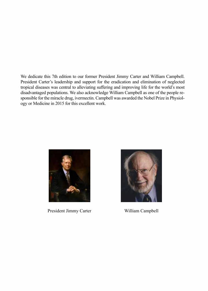
We dedicate this 7th edition to our former President Jimmy Carter and William Campbell. President Carter’s leadership and support for the eradication and elimination of neglected tropical diseases was central to alleviating suffering and improving life for the world’s most disadvantaged populations. We also acknowledge William Campbell as one of the people re-sponsible for the miracle drug, ivermectin. Campbell was awarded the Nobel Prize in Physiol-ogy or Medicine in 2015 for this excellent work.
William CampbellPresident Jimmy Carter
iv
Acknowledgements:
We acknowledge the contribution of John Karapelou for his elegant life cycle drawings. Thanks to David Scharf for granting us use of his stunning scanning electron micrographs of the very organisms, organelles and cellular environments that attract legions of new students to the field of tropical medicine. Thanks to all the course directors of parasitic diseases and parasitology who choose our book to guide their students through the complexities of life cycles, clinical presentations, and biology of parasites. We hope that the 7th edition proves even more useful for you and your students in the coming years.
Thanks to all the infectious disease experts (Justin Aaron, Sapha Barkati, Craig Boutlis, Mary Burgess, Matthew Cheng, Lucy Cheng, Elise O’Connell, Priya Kodiyanplakkal, Atul Kothari, Michael Libman, Tim McDonald, Juan Carlos Rico, Jordan Rupp, Keyur Vyas, Amy Murillo and Johnnie Yates) who volunteered their time and expertise to review this textbook prior to its publication. Appreciation to our entomology experts Jonathan Larson and Amy Murillo for many suggestions and additions to the Arthropod sections. Thank you to all our donors who continue to make it possible for us to get this book into the hands of those who need it the most.
v
Preface The accumulation of new knowledge over the last several years has provided the impetus for this, the 7th edition of Parasitic Diseases. By integrating all the salient information contained in over 1000 new references with the current descriptions of each pathogen, we continue the never-ending process of evaluating, revising, and improving our understanding of these parasitic organisms. New features to the 7th edition include a clinical summary section, addi-tional life cycle diagrams and a pronouncer’s guide to parasite names. Consideration of the biographies of those notable contributors to the field of parasitology has also been extended. Parasites Without Borders continues to make available, free of charge, the PDF version of Parasitic Diseases in both English and Spanish editions.
The number of eukaryotic parasites whose entire genomes are available since completion of The Human Genome Project in 2003, continues to grow and these are highlighted in our 7th edition. Genomic datasets hold great promise for the development of new vaccines, drugs, and control programs based on identifying unique molecular pathways essential to each pathogen. These on-going projects serve as a living testament to the perseverance of a small group of creative, highly trained researchers, whose discipline and dedication helps stem the spread of these life-threatening entities.
Progress in rapid identification of parasites in the diagnostic laboratory by targeting their genomic signatures in biological specimens has dramatically improved and simplified diag-nosis and is now the preferred approach in most hospitals throughout the developed world. While molecular-based testing has replaced the microscope as the method of identification in the vast majority of laboratories, in the less developed world traditional approaches to the identification of parasites remain the only technology available. Hence why we still include a diagnostic atlas in our text.
Recent advances have also improved our understanding as to the molecular mechanisms under-lying the subversion of our signaling pathways by some parasites, mainly the Leishmania spp. and Toxoplasma gondii. Thus, we now have a much clearer picture of how they survive so long within us without incurring harm to themselves and will undoubtedly inspire the develop-ment of the next generation of more effective chemotherapeutic agents that take advantage of these data. Unraveling the biochemical complexities of how some parasites manage to survive for long periods within the human host has also provided clues in the treatment of illnesses unrelated to the parasites themselves. “Swords into plowshares” molecular strategies, are pro-viding “parapharmaceuticals” for coronary artery disease, stroke, and autoimmune disorders. Clinical trials evaluating the prophylactic use of hookworm-based peptides and eicosanoids that block host clotting, host platelet aggregation, and host inflammation are underway at the time of publishing this 7th edition.
An explosion of new compounds due to advances in drug discovery and design using high-throughput screening algorithms, cryo-electron microscopy, and virtual reality visualization technologies now await testing. These approaches have already provided the clinician with a new generation of drugs, many with less harmful side-effects than the ones they replaced. Controlling parasite populations at the community level is an anticipated consequence of such
vi
developments, without the risk of harming the very ones we wish to help.
The remarkable success of programs such as those targeting river blindness, which has been controlled in many countries in West Africa, demonstrates that when political will and strong social support combine, we can successfully limit the spread of parasites. By targeting and managing drug use in such control strategies, it is possible to eradicate parasites by disrupting their life cycles in a localized manner. Almost all regions of Africa have brought dracunculia-sis under control. Similarly, the southern cone initiative of South America has succeeded in dramatically reducing cases of Chagas disease.
Primarily driven by the U.S. President’s Malaria Initiative and the Global Fund, the number of children who die from malaria has fallen by more than 50% since 2000, saving over 6 million lives. Despite all this progress, there are still high rates of morbidity and mortality through-out the tropics due to some of the most commonly occurring parasitic infections, especially malaria. While there are no new classes of drugs for treating resistant malaria, artemisinin-derivatives continue to be effective in reducing the mortality of the world’s most devastating infectious disease wherever that chemotherapeutic agent is available. For millennia, worm infections have exacted their toll on humanity, with children as their primary victims. Until basic sanitation is implemented this will, regrettably, be the norm for most underdeveloped countries and impoverished communities.
Due to the political instability of vast regions of Africa and the Middle East, the re-emergence of many infectious diseases, including leishmaniasis and African trypanosomiasis, has become a significant problem. This is primarily due to environmental destruction, abandonment of control programs, and forced migration of tens of thousands of individuals from regions that were relatively safe, to places that no one should have to occupy, no matter how short the dura-tion. These seemingly intractable situations require more than vaccines and drugs to affect a cure. Social stability, equity, economic development, and long-term planning are the “drugs of choice”.
The impact of HIV/AIDS in resource-constrained areas continues to reduce life expectancy significantly. While the immunosuppressive effects caused by this disease and the impact on other parasitic diseases is still poorly understood. Such effects require careful monitoring. As access to antiretroviral therapy improves due to the Global Fund and other non-governmental entities, new clinical syndromes are likely to emerge due to parasites behaving differently in hosts with an ever-changing immune status.
Diarrheal diseases caused by a variety of infectious agents, including Entamoeba histolytica, Giardia lamblia, Cryptosporidium parvum, and Cyclospora cayetanensis round out the list of miseries that those living in poverty in the less developed world endure. Where feces and urine serve as the best source of fertilizer, education as to how to apply basic sanitation to render them into safe manure, remains high on the list priorities.
Ultimately, it is through the education of students, clinicians, and parasitologists and vigilance of commitment that we can hope to improve the lives of hundreds of millions of less fortunate individuals by helping them live longer and more productive lives. The 7th edition of Parasitic Diseases is dedicated to this premise. We invite you to join us in this global effort.

vii
Dickson D. Despommier Daniel O. Griffin
Robert W. Gwadz Peter J. Hotez Charles A. Knirsch
viii
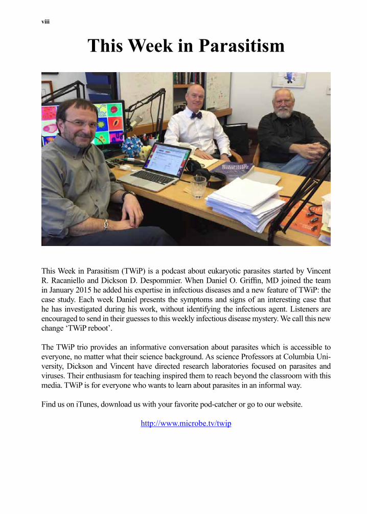
This Week in Parasitism (TWiP) is a podcast about eukaryotic parasites started by Vincent R. Racaniello and Dickson D. Despommier. When Daniel O. Griffin, MD joined the team in January 2015 he added his expertise in infectious diseases and a new feature of TWiP: the case study. Each week Daniel presents the symptoms and signs of an interesting case that he has investigated during his work, without identifying the infectious agent. Listeners are encouraged to send in their guesses to this weekly infectious disease mystery. We call this new change ‘TWiP reboot’.
The TWiP trio provides an informative conversation about parasites which is accessible to everyone, no matter what their science background. As science Professors at Columbia Uni-versity, Dickson and Vincent have directed research laboratories focused on parasites and viruses. Their enthusiasm for teaching inspired them to reach beyond the classroom with this media. TWiP is for everyone who wants to learn about parasites in an informal way.
Find us on iTunes, download us with your favorite pod-catcher or go to our website.
http://www.microbe.tv/twip
This Week in Parasitism
ix
ContentsI. Acknowledgements ivII. Preface v-xiiIII. Eukaryotic Parasites 1-8IV. The Protozoa 9-10 1. Giardia lamblia 11-20 2. Introduction to the Leishmania species 21-28 3. Cutaneous Leishmaniasis 29-38 Leishmania (L.) major Leishmania (L.) tropica Leishmania (L.) mexicana 4. Mucocutaneous Leishmaniasis 39-46 Leishmania (V.) braziliensis 5. Visceral Leishmaniasis 47-56 Leishmania (L.) donovani Leishmania (L.) infantum Leishmania (L.) infantum chagasi 6. African Trypanosomiasis 57-70 Trypanosoma brucei rhodesiense Trypanosoma brucei gambiense 7. American Trypanosomiasis 71-84 Trypanosoma cruzi 8. Trichomonas vaginalis 85-92 9. The Malarias 93-122 Plasmodium falciparum Plasmodium vivax Plasmodium ovale Plasmodium malariae Plasmodium knowlesi10. Cryptosporidium parvum 123-13211. Toxoplasma gondii 133-14612. Entamoeba histolytica 147-16013. Balantidium coli 161-16614. Other Protozoa of Medical Importance 167-184 Babesia spp. Cystoisospora belli Cyclospora cayetanensis Naeglaria fowleri Acanthamoeba spp. Balamuthia mandrillaris Blastocystis hominis Dientamoeba fragilis
x
15. Non-pathogenic Protozoa 185-188 Commensal Flagellates Commensal amoebaeV. The Nematodes 189-19016. Enterobius vermicularis 191-19617. Trichuris trichiura 197-20418. Ascaris lumbricoides 205-21419. The Hookworms 215-228 Necator americanus Ancylostoma duodenale Ancylostoma ceylanicum20. Strongyloides stercoralis 229-24021. Trichinella spiralis 241-25222. Lymphatic Filariae 253-264 Wuchereria bancrofti Brugia malayi23. Onchocerca volvulus 265-27624. Loa loa 277-28425. Dracunculus medinensis 285-29026. Other Nematodes of Medical Importance 291-298 Capillaria hepatica Capillaria philippinensis Dirofilaria immitis Mansonella ozzardi Mansonella perstans Mansonella streptocerca Oesophagostomum bifurcum Ternidens diminutus27. Aberrant Nematode Infections 299-314 Cutaneous Larva Migrans Visceral Larva MigransVI. The Cestodes 315-31628. Taenia saginata 317-32429. Taenia solium 325-34030. Diphyllobothrium latum 341-34831. Other Tapeworms of Medical Importance 349-358 Hymenolepis nana Hymenolepis diminuta Dipylidium caninum
xi
32. Juvenile Tapeworm infections of Humans 359-376 Echinococcus granulosus Echinococcus multilocularis Mesocestoides spp. Spirometra spp. Taenia spp. (other than saginata and solium)VII. The Trematodes 377-37833. The Schistosomes 379-404 Schistosoma mansoni Schistosoma japonicum Schistosoma haematobium Schistosoma mekongi Schistosoma intercalatum34. Clonorchis sinensis and Opisthorchis spp. 405-41235. Fasciola hepatica 413-42036. Paragonimus westermani 421-42837. Other Trematodes of Medical Importance 429-438 Fasciolopsis buski Echinostoma spp. Heterophyes heterophyes Metagonimus yokogawai Nanophyetes salmincolaVIII. The Arthropods 439-44038. The Insects 441-48039. The Arachnids 481-50040. Other Arthropods of Medical Importance 501-504Appendix A. Procedures for Collecting 505-508 Clinical Specimens for Diagnosing Protozoan and Helminthic ParasitesAppendix B. Laboratory Diagnostic Methods 509-516Appendix C. Diagnostic Color Atlas 517-534Parasite Identification Charts 535-538Clinical Appendix 539-578Pronunciation Guide 579-584Acronyms 585-588Index 589-602
xii
Parasites Without Borders was founded as a direct response to the question: “What can I do to help eliminate human suffering due to parasitic infections?” For us the choice was easy; more and better education for all those in a position to apply medical knowledge directly to popula-tions in need of relief from the burden of parasitic diseases. The three founders have a lifetime of experience in teaching parasitic diseases to students of medicine, both within the U.S.A. and abroad. Our mission statement is clear; we want to help bring the latest medical and basic biological information pertaining to diseases caused by eukaryotic parasites to every clinician and student throughout the world.
http://www.parasiteswithoutborders.com
Eukaryotic Parasites 1
ease-causing organism will be emphasized. The following topics are deemed medically relevant: 1. mechanisms of entry, 2. niche selection, 3. reproduction, 4. mechanisms of survival (i.e., virulence factors), and 5. mech-anisms of pathogenesis.
The last half of the twentieth century has been a remarkable one for the community-based control of pathogenic organisms. New vaccines and antibiotics have also helped reduce the incidence of numerous pathogenic organisms. At the same time, it has also her-alded the emergence and re-emergence of a wide spectrum of infectious agents: viruses (e.g., SARS, HIV, monkey pox, avian influ-enza, dengue, chikungunya, Zika), bacte-ria (e.g., Legionella pneumophila, Borrelia burgdorferi, Escherichia coli strain OH157), protozoa (e.g., Cryptosporidium parvum, Cyclospora cayetanensis), and helminths (e.g., Echinococcus multilocularis, Angio-strongylus cantonensis, Trichinella spiralis). Viewed from an evolutionary perspective, humans represent a highly successful system of essential niches, of which an astonishingly wide variety of eukaryotes have been able to take advantage.
It is difficult for even the most attentive stu-dent, to truly comprehend the prevalence of, and suffering caused, by parasites. This is especially true when hearing a very large number, as is the case for Ascaris lumbri-coides, which infects hundreds of millions of people around the world. So, when one hears for the first time that 100s of millions of people are infected with malaria each year, and over ½ a million children per year die, in Africa alone, from this infection, these facts seem somehow remote, even abstract. Yet, when a single child suffering from the cerebral form of this disease-causing entity is admitted into a modern hospital in critical condition, and, regardless of treatment, that young person dies, the health care community
III. Eukaryotic Parasites
Eukaryotic parasites encompass subsets of organisms within the protozoan and hel-minth (parasitic worm) groups. In addition, medically important arthropods have been included in discussions of eukaryotic para-sites, since so many of these pathogens are transmitted to humans by arthropod vectors. Besides, some medically relevant arthropods cause disease on their own.
From a biological perspective, a phylogenetic presentation of eukaryotic parasitic organ-isms would undoubtedly satisfy those spe-cialists who strictly adhere to the zoological literature, while most medical students and practicing clinicians would have little or no use for this information. The physician is more inclined to group them according to their syndromes, if they were to classify them at all. We have settled upon a compromise, in which these organisms are encountered by the reader in a somewhat biologically correct order, together with an outline of their clas-sification and clinical presentations. Nonethe-less, it is in some sense intellectually satisfy-ing to review parasitic organisms with a sem-blance of evolutionary precision, allowing each student to learn about them in a sequence that most experts in the field of parasitology have agreed upon, going from the single-cell parasites to the worms and beyond. We, there-fore, present protozoans first, followed by the helminths, and finally round out the synopsis with medically relevant arthropods.
The following sections are organized in such a way as to enable the student or clinician easy access to a highly distilled body of infor-mation. This relates to the general schemes employed when these organisms interact with the human host to produce disease. Thus, rather than being an exhaustive text, only biological information essential to the under-standing of clinical aspects of a given dis-
2 Eukaryotic Parasites
of that institution is put into collective shock. If the death occurred at a teaching hospital, a grand rounds is the usual outcome, perhaps motivated by some vague sense of guilt, in an attempt to see if anything could have been done to spare that life. Unfortunately, the most lethal species of malaria, Plasmodium falciparum, is evolving more and more resis-tance to the medications in our arsenal.
Parasitic Protozoa
What is a protozoan? Which ones cause dis-ease? How do those that are parasitic differ from their free-living counterparts? What are the pathogenic mechanism(s) by which they cause disease? There are over 200,000 named species of single-celled organisms that fall under the category protozoa, while many more, no doubt, await discovery. Only some small fraction of these is parasitic for the human host, yet some can cause great harm (e.g., malaria), especially when they are encountered for the first time.
Protozoans are single-cell organisms inside of which usually resides one membrane-bound nucleus, with a few exceptions, such as Giar-dia lamblia and Dientamoeba fragilis. Most protozoa have one type of organelle that aids in their movement (e.g., flagella, undulating membrane, cilia). Metabolic pathways vary from group to group, with both anaerobic and aerobic energy metabolisms being repre-sented among the parasites to be discussed. In the case of parasitic organisms, the host pro-vides the energy source. There are a variety of drugs that take advantage of the dependence of parasites on host energy metabolism.
All single-cell organisms have complex bio-chemistries, often employing unique path-ways that give some of them remarkable evolutionary advantages. These include the ability of a given population to vary their protein surfaces, edit their mRNA transcripts,
secrete peptides that prevent the fusion of lysosomal membranes to the parasitophorous vacuole, and give off substances that inhibit host protective immune responses. A plethora of unique molecular pathways have been described for this diverse group of parasites, but a comprehensive description of them is beyond the scope of this book. Some atten-tion to both the biochemical and molecular biological findings for a given organism will be presented whenever they have relevance to the understanding of the mechanisms of pathogenesis or parasite survival strategies.
Mechanisms of Entry
Protozoans gain entry into their host in one of several pathways: oral, sexual, inhalation, direct contact, and through the bites of blood-sucking vectors. Avoidance or prevention of infection requires an intimate knowledge of its transmission cycle and knowing the route of entry into the host is one of the most impor-tant aspects in that regard.
Many species of parasitic protozoa have evolved stages that facilitate their dispersal into the environment, increasing their chances of encountering a host. Some intestinal proto-zoa produce a resistant cyst enabling them to lie dormant in the environment for long peri-ods of time, months to years, in some cases. Others depend upon human activities for their dispersal, as in the case of Trichomonas vagi-nalis, which is sexually transmitted. Certain amoeba may infect humans through inhala-tion or direct contact.
Vector-borne organisms rely on the biology of blood sucking insects, for the most part. Mosquitoes transmit all species of malaria (Plasmodium spp.), tsetse flies transmit Afri-can Sleeping Sickness (Trypanosoma brucei spp.) and sandflies transmit all species of Leishmania. In these instances, the organ-ism is injected directly into the host’s blood
Eukaryotic Parasites 3
stream or interstitial tissue fluids where they proceed to undergo complex developmental life cycles culminating in numerous cycles of asexual division once they achieve their essential niche.
A more complex strategy is employed by Trypanosoma cruzi, an organism transmitted by a large hemipteran with ferocious look-ing biting mouthparts. In this instance, the organisms are excreted along with the fecal exudate at the time of the second blood feed-ing. Humans become infected unknowingly, by rubbing the organisms into the bite wound or into a mucous membrane after the insect withdraws its mouthparts.
Niche Selection
Each protozoan has been selected for life in a specific essential niche, which can only be defined by a comprehensive knowledge of the anatomical, physiological, and biochemi-cal features of that site. To gain some measure of the difficulties associated with attempting to describe the essential niche, be it that of a parasite or any other organism, let us consider the intracellular milieu of the normal red blood cell. This site represents one of the best stud-ied of all intracellular environments. Yet for the most part, we still do not understand pre-cisely how that anucleate cell’s membranes interact with vascular endothelial cells when the cell traverses the capillary and exchanges gases with the surrounding tissues. To make matters worse, a red blood cell infected with P. falciparum behaves quite differently from that of a normal one, failing to deform as it enters the capillary bed. This single aspect of the infection has serious pathological conse-quences for the host, as will be detailed in the section dealing with the clinical aspects of malaria.
The internal molecular environment of the infected red cell must be considered as a
“hybrid,” consisting of both host and parasite elements. Proteins, produced by the develop-ing merozoite, locate to the cytoplasm of the host cell, and some even integrate at the red cell membrane surface, forming complexes with host structural proteins such as spectrin and glycophoran. Others remain in the gen-eral region of the red cell cytoplasm. Over the entire period of the developmental cycle of the parasite, new proteins are produced that locate to specific regions of an ever-changing host cell environment. The infected red cell represents a very dynamic situation; even with the most sophisticated instrumentation, it has been impossible to fully appreciate the setting in which this important pathogen lives out its life.
Finally, no two species of Plasmodium behave the same in their erythrocytic niche, due largely to dramatic genetic differences between the major species infecting humans. Hence, it is likely that we will never gain a “full face-on” view of this or any other patho-gen to sufficiently design new therapeutics that would prevent the organism from taking full advantage of its ecological setting. The complexities presented to the research parasi-tologist by just this single organism continue to challenge them to design innovative exper-iments that may allow us one day more than a glimpse into its secret life.
At the other end of the scale is Toxoplasma gondii, a generalist protozoan capable of infecting virtually any mammalian cell and reproducing within it. T. gondii’s lack of host restriction makes it the most widely distrib-uted parasite on earth.
Migration to favorable sites within the host often requires an active role for the patho-gen, but frequently they “hitch a ride” in our bloodstream or through our intestinal tract. Some are capable of infecting cells that under most circumstances would serve to protect us
4 Eukaryotic Parasites
from these kinds of organisms. The macro-phage is a permissive host cell for T. gondii and for all species of Leishmania. In these infections, the very cell-type we depend upon for innate protection against invaders turns out to be the culprit, aiding in their dispersal throughout the body. Division and Reproduction
Multiplication within the human host is the rule for protozoans, in contrast to most hel-minth species, in which infection usually results in a single adult parasite. The defini-tive host is the one harboring the sexual stages or the adult stages of a given parasite. Hence, the human is not the definitive host for a wide range of protozoan infections, including the Plasmodium spp. and T. gondii. Female anopheline mosquitoes are the defini-tive hosts for all malaria species infecting humans, while the domestic cat and other Felidae are the permissive host for the sexual stages of T. gondii. Humans are the definitive host for Cryptosporidium parvum. It should be emphasized, however, that not all parasitic protozoa have sexual cycles.
All protozoans reproduce asexually after gaining entrance into the human host. Path-ological consequences result directly from their increasing numbers. During the height of the infection, they place ever-increasing demands upon their essential niches. The mechanisms by which protozoa divide asexu-ally are numerous, with binary fission being the most common. Malarial parasites repro-duce within the red cell by a process called schizogony, in which the organism under-goes nuclear division within a common cyto-plasm (karyokinesis). Just before rupturing out of the hemoglobin-depleted red cell, the parasite’s cytoplasm divides to accommodate each nucleus, leaving its toxic waste product, crystals of hemozoin, in the now empty red cell stroma.
Mechanisms of Survival
Each species of parasite has been selected for life within the human host by evolving strat-egies that; 1. inhibit or divert our immune system, 2. avoid or inhibit intracellular kill-ing mechanisms, and 3. infect regions of the body that are incapable of protective immune responses. For example, the African trypano-somes produce “smoke screens” of surface antigens whose sole purpose seems to be to keep the immune system busy, while a small select population changes its protein coat to a different antigenic variant, thus temporar-ily escaping the host’s immune surveillance system. Certain stages of the malaria parasite and Giardia lamblia can also vary their sur-face proteins, presenting our immune system with a bewildering array of antigenic determi-nants to deal with as an infection progresses. T. gondii inhibits the fusion of lysosomal ves-icles with the parasitophorous vacuole, thus escaping the killing effects of acid hydrolases. Cryptosporidium parvum and all species of malaria occupy immunologically “silent” niches. Trypanosoma cruzi actually escapes the parasitophorous vacuole into naked cyto-plasm, avoiding the ravages of lysosomal enzyme activity. There are numerous other examples and they will be discussed when-ever relevant. Independent of the biochemical strategy employed by the protozoan parasite, the result is tissue damage, often severe. Mechanisms of Pathogenesis
Regardless of the mechanisms employed by the parasite to escape being killed, the usual consequence of infection from the perspec-tive of the human host is tissue damage. The extent of cellular damage inflicted by a given parasite is related to the location of their essential niche, the metabolic requirements of the parasite, and their population density throughout the infection. Energy is derived from the host, placing a burden on infected
Eukaryotic Parasites 5
hosts for providing this essential ingredient. The penchant of the parasite for killing the cell it invades or eroding away the tissue it occupies while feeding on our cells, results in measurable pathological consequences that translate directly into clinical signs and symptoms.
For example, when the malaria parasite exits from the red cell at the end of its division cycle, the rupture of the stroma results in the release of toxic waste products (hemozoin) that elicit fever. Entamoeba histolytica, as its name implies, attaches to, then ingests living cells. It then digests them, using acid hydro-lases to do so, and in the process induces bloody diarrhea (dysentery).
Infection with T. gondii results in lymph-edema and fever due to the death of large numbers of host cells throughout the body. The molecular basis for these pathologi-cal effects will be discussed in detail at the appropriate time. Suffice it to state here that we do not know any parasite’s modus ope-randi completely, and scientific endeavor will undoubtedly continue to provide surprises and revelations in the near future.
Parasitic Helminths (worms)
Helminths belong to four phyla: Nematoda (roundworms), Platyhelminthes (flatworms), Acanthocephala (spiny-headed worms), and Nematophora (hairworms). Only worms belonging to the first two are endoparasitic to humans. Both the Nematoda and Platy-helminthes have many free-living species as well. A general description of each major group precedes each section. What follows is a general description of their biology.
Mechanisms of Entry
Helminths have evolved multiple strategies for entering the host and establishing infec-
tion. Among the nematodes, infection is usu-ally established by exposure to an environ-mentally resistant stage.
For many of the common intestinal nema-todes such as A. lumbricoides or Trichuris trichiura, this occurs via the ingestion of embryonated eggs in the soil, or on fecal-contaminated fruits and vegetables. In many tropical countries helminth eggs have been isolated from nearly all environments. They have even been recovered from paper cur-rency. For other nematodes, infection is estab-lished when larval stages, living in the soil, enter the host. Sometimes infection is strictly food-borne and occurs only when larvae are ingested in raw or undercooked meat. Many species of nematode are transmitted by arthro-pods, such as lymphatic filariasis (mosquito), loiasis (deer fly), onchocerciasis (black fly) and guinea worm infection (copepods).
Trematodes spend a portion of their life cycle in a wide variety of snail intermediate hosts. After exiting the snail, the larval stage, known as a cercaria, typically attaches to a second intermediate host, such as a fish, a crab, or aquatic vegetation. For this reason, most trematode infections are food-borne. The exception is the Schistosomes, which cause a spectrum of illnesses. The Schistosome cer-cariae can penetrate through the hair follicle, the invaginated epidermal layer that forms the hair shaft, due to the thinner epidermis.
Cestodes are acquired via the oral route, regardless of the stage that ends up causing the infection. Most adult tapeworm infec-tions of humans result from the ingestion of inadequately cooked contaminated fish, beef, or pork. Two clinically significant juvenile tapeworm infections, cysticercosis and echi-nococcosis, result from accidental ingestion of the eggs.
6 Eukaryotic Parasites
Niche Selection
Unlike protozoans, most species of parasitic helminths occupy more than a single niche in their human host during their life cycle. For example, although hookworms live as adults in the small intestine, in order to arrive there, the infective larvae frequently must first pass through the skin and lymphatics before spend-ing time in the bloodstream and lungs before migrating via the esophagus back to the gut. Similarly, Ascaris eggs hatch in the intestine before the emerging larval stage enters the portal circulation; the larvae enter liver and lungs prior to re-entry into the gut. As adults, helminths have been recovered from almost every organ including liver, lungs, lymphat-ics, bloodstream, muscle, skin, subcutaneous tissues, and brain.
Many species of parasitic helminths (nema-todes, cestodes, and trematodes) live as sexu-ally mature adults in the gastrointestinal (GI) tract. In many underdeveloped countries, it is common to find school-aged children who harbor three or four different species of hel-minths in their intestine, with each species occupying a different portion of the gut track. Symptoms arising from heavy infection with a given helminth are typically associated with a particular region of the GI tract.
Reproduction
Nematode parasites that live in the GI tract produce eggs or larvae that exit the host with the fecal mass. Nematodes living in blood or lymphatic vessels produce larvae that circu-late in the bloodstream and must be ingested by the appropriate arthropod vector in order to exit the host.
In the cestodes, the situation is somewhat different as each proglottid segment of the adult cestode tapeworm is hermaphroditic. Because there is usually only one adult worm
present, the worm self-fertilizes adjacent seg-ments. Adult tapeworms shed segments into the lumen of the small intestine and they can exit the host under their own power. Other adult tapeworms produce segments that then disintegrate releasing their eggs into the fecal mass for export. Juvenile tapeworm infec-tions remain as such and produce no diag-nostic stage. These infections present real problems for the clinician seeking a definitive diagnosis for their patient.
Except for the schistosomes, the trematodes (flukes) are all hermaphroditic. Despite this all-in-one reproductive arrangement, cross-fertilization between two trematodes of the same species is common. Intestinal trema-todes produce eggs that exit with the feces, as for example, with the eggs of Hetero-phyes heterophyes. Eggs of the lung fluke, Paragonimus westermani, exit the host either when they are coughed up in sputum or after they are swallowed, in which case they exit in the feces.
Some helminths have evolved elaborate adaptations in order to ensure that their eggs leave the human host. For instance, schistosome eggs are deposited against the inside wall of a blood vessel. These eggs are equipped with sharp spines and a battery of lytic enzymes that allow them to traverse the vessel endothelium and gut wall. The eggs traverse through the serosal surface of either the intestine or bladder (depending upon the species), before entering the muscularis layer and then the lumen. Adult Schistosomes and Paragonimus that locate to ectopic sites (e.g., nervous system) produce eggs that remain at the site of infection, often resulting in serious pathological consequences for the host.
Mechanisms of Survival
Like the protozoa, the helminths occupy hab-itats which most of us would consider highly
Eukaryotic Parasites 7
inhospitable. The selective pressures that led to their elaborate mechanisms for survival in these environments are still poorly under-stood. Adult Schistosomes live in the blood-stream, a place where one might expect to encounter the constant bombardment of the immune system’s slings and arrows of anti-body molecules and leukocytes of various types. Yet, there the worms can thrive for up to twenty years in that niche.
The molecular basis by which this happens is not known, although a number of immune evasion and immunological masking mecha-nisms have been described. Important for hel-minth survival is their unique array of natu-ral products elaborated and released into the host. Hookworms can freely ingest blood in the intestinal mucosa and submucosa because they produce peptides and eicosanoids that block host clotting, host platelet aggregation, and host inflammation. Many of these pep-tides themselves have proven to be useful as new potential therapeutic agents for human coronary artery disease, stroke and autoim-mune disorders. T. trichiura releases a pore-forming protein that promotes cell fusion around the anterior end of the organism, allowing it to become embedded in epithelial tunnels.
Indeed, the argument has been made that parasitic helminths are themselves equivalent to small biotechnology companies, which, through research and development in the form of millions of years of evolutionary selection, now produce a wide array of phar-macologically active compounds, which we may eventually find useful, as well.
Mechanisms of Pathogenesis
Helminths injure their human host both through mechanical and chemical mecha-nisms. Large helminths, such as A. lumbri-coides, can cause physical obstruction of the
intestine, or exert damage when they migrate into the biliary tree. As already noted, hel-minths release peptides and eicosanoids that down-regulate host inflammatory processes. In some cases, helminths bias host immunity to produce Th-2-like responses that may make the host less likely to eliminate the parasite.
Immune regulation on the part of the para-site may also have consequences for the host, regarding a wide variety of viral infections. There is some evidence to support the role of helminths as co-pathogens that promote susceptibility to HIV infection and AIDS. In many cases, some of the most important mechanisms of pathogenesis are still not known. Heavy infection with some intestinal nematodes (e.g., hookworm) are considered to be the major cause of stunted growth during childhood as well as inducing impaired cog-nitive behavior and intellectual development. While intuitively we might suspect that para-site-induced malnutrition plays an important role in this process, the true basis by which these processes occur is not known.
Host-mediated immunopathology accounts for a large measure of the damage that occurs during some helminth infections. This is par-ticularly true for infection with the Schisto-somes. However, current evidence suggests that in the case of infection with several filar-ial worm species, a bacterial endosymbiont, Wolbachia, may be responsible for most of the pathological consequences of the infec-tion. Brain parenchymal inflammation and seizures in cysticercosis are well documented.
The genomes of many of these important pathogens of humans are now available, so new approaches to the clinical management of patients suffering from them are sure to emerge from the laboratory and find their way to the bedside. At least that is the hoped-for outcome of such research.
8 Eukaryotic Parasites
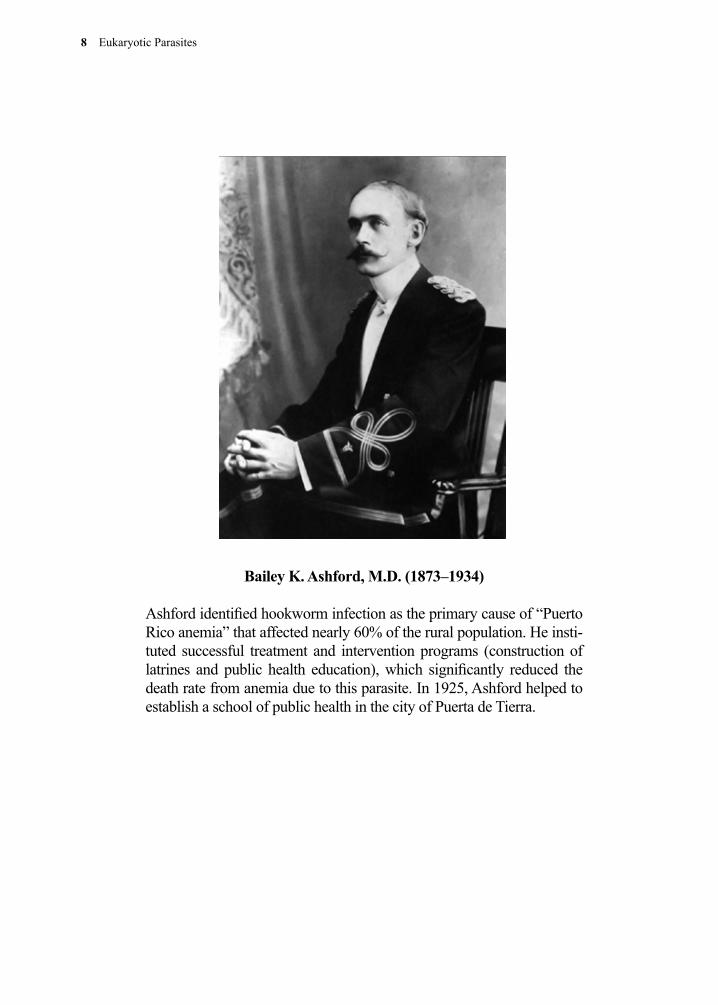
Bailey K. Ashford, M.D. (1873–1934)
Ashford identified hookworm infection as the primary cause of “Puerto Rico anemia” that affected nearly 60% of the rural population. He insti-tuted successful treatment and intervention programs (construction of latrines and public health education), which significantly reduced the death rate from anemia due to this parasite. In 1925, Ashford helped to establish a school of public health in the city of Puerta de Tierra.
The Protozoa 9
IV. The Protozoa
Over 200,000 species of protozoa have been described so far, of which more than half are represented in the fossil record. The reper-toire of known living species (approximately 35,000) includes more than 10,000 that have adapted for life as parasites. Regardless of their lifestyle, all protozoans are eukaryotic single-cell organisms. Free-living species occupy every conceivable ecological niche, including marine trenches, rainforests, arte-sian and thermal springs, salt lakes, ice flows, glaciers, and many others, while parasitic protozoans infect a wide spectrum of verte-brate and invertebrate life.
Unlike the great majority of parasitic hel-minth species, protozoan parasites are able to replicate within a given host, often resulting in hundreds of thousands of new individuals within just a few days of initial infection. This single feature of their life cycle frequently has grave consequences for the host.
Parasitic protozoans have played a major role in the evolution of the human species, mainly due to lethal consequences of infection, or limiting where people can live by adversely affecting their livestock. These very same selection pressures continue to play out in many parts of the world today. For example, malaria in all its forms, African trypanoso-miasis, and visceral leishmaniasis infect mil-lions of people and are responsible for untold numbers of deaths and debilitating chronic illnesses. Many others cause less severe dis-ease (e.g., chronic diarrhea) that nonethe-less results in lost time at work and school and loss of recreational activities we deem vital to living enriched, healthy, disease-free lives. This is due, in part, to the fact that some important species of parasitic protozoans are no longer susceptible to drugs that were once effective in limiting disease. There are no effective vaccines for the control of any pro-tozoan infection in humans.
While the biology of parasitic protozoa varies widely from group to group, these organisms share many common features. A unit mem-brane that functions in a similar fashion to all other eukaryotic cells binds them. Nutrients may be actively transported, phagocytized, or moved into the cell by pinocytosis. Diges-tion of particulate material is by lysosomal enzymes within the phagolysosome. Proto-zoans excrete wastes either by diffusion or by exocytosis. Mechanisms of motility take advantage of the presence of one of a vari-ety of structures (e.g., cilia, flagella, pseudo-pod). All species of protozoans can divide asexually, usually by binary fission. In some instances, the process is more complex, and includes multiple nuclear divisions followed by cytokinesis. Those capable of sexual reproduction do so within the definitive host, resulting in the formation of a zygote.
In addition, their cytoplasm may contain subcellular organelles, including Golgi appa-ratus, lysosomes, mitochondria, rough and smooth endoplasmic reticulum, and a wide variety of membrane-bound vesicles of spe-cialized function (e.g., the hydrogenosome of Trichomonas vaginalis, and the glycosome of kinetoplastidae). Collectively, these cytoplas-mic inclusions enable the organism to respire, digest food, generate energy, grow, and repro-duce.
Some species have evolved elaborate surface coats consisting of materials derived from the host or secreted by the parasite that offer some protection from host immune responses, thereby extending their life within a given individual and resulting in great damage to the host as well.
The fields of immunoparasitology, parasite genomics, and parasite proteomics have also matured over the past several years. New understanding regarding the role(s) of cyto-kines and interleukins in the pathogenesis of disease has led to new clinical approaches
10 The Protozoa
for several important protozoan diseases. In addition, the details of protective host mecha-nisms that counter the invasion process have been described, giving hope for the develop-ment of a new generation of drugs and per-haps even the first of many effective vaccines.
The following chapters are but a thumbnail sketch of some of the excitement generated in the field of protozoan parasitology. They are designed to present to the student and clini-cian useful and practical information specific to the diagnosis, treatment, and management of infections caused by these pathogens.
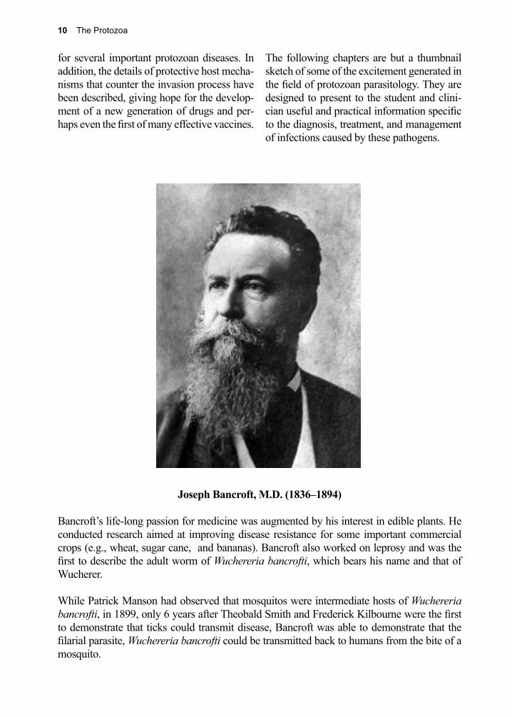
Joseph Bancroft, M.D. (1836–1894)
Bancroft’s life-long passion for medicine was augmented by his interest in edible plants. He conducted research aimed at improving disease resistance for some important commercial crops (e.g., wheat, sugar cane, and bananas). Bancroft also worked on leprosy and was the first to describe the adult worm of Wuchereria bancrofti, which bears his name and that of Wucherer.
While Patrick Manson had observed that mosquitos were intermediate hosts of Wuchereria bancrofti, in 1899, only 6 years after Theobald Smith and Frederick Kilbourne were the first to demonstrate that ticks could transmit disease, Bancroft was able to demonstrate that the filarial parasite, Wuchereria bancrofti could be transmitted back to humans from the bite of a mosquito.
1. Giardia lamblia 11
1. Giardia lamblia (also known as G. duodenalis or G. intestinalis)(Stiles 1915)
Pronunciation: \jē-är′dē-ə\\lăm′blē-ə\
Introduction
Giardia lamblia (\jee-ARE-dee-ah\\lam-BLEE-ah\), also known as G. duodenalis or G. intestinalis) is a flagellated protozoan that lacks a mitochondrion.1 It is aerotolerant, but respires as an anaerobe, and lives in the small intestine. Other protozoa sharing this metabolic strategy include Entamoeba histo-lytica and Trichomonas vaginalis. The genus Giardia is divided into eight genetic groups, with groups A and B infecting humans.2 G. lamblia produces a cyst stage that is environ-mentally resistant.3
Infection is via the fecal-oral route, typically through exposure to contaminated drinking water.4 G. lamblia has a worldwide distribu-tion, and is endemic in many regions.5 Giardi-asis frequently occurs in children (especially those attending daycare centers), travelers, immunocompromised individuals (including HIV-infected individuals), and patients with genetic disorders such as cystic fibrosis.6-11 G. lamblia is a common infection in humans and domestic animals in the United States12. It is likely that many infected individuals remain undiagnosed, harboring G. lamblia without obvious symptoms.13
Beavers are major reservoir hosts and are often responsible for contaminating public drinking water. This is why infection with G. lamblia is known in many parts of the United States by the common name of “beaver fever”.14, 15 Giardia is the subject of much intensive research, including the complete sequencing of its genome.16 A survey of its genome has revealed the presence of genes for meiosis, and population genetics suggest
some form of genetic exchange exists, pos-sibly during encystation or excystation, yet a sexual stage for this protozoan has not been established.17, 18 Although an excellent review of the biology of G. lamblia was published by Adam in 2001, much has been added to the field since this comprehensive report.19
Historical Information
Antonie van Leeuwenhoek (\LAY-vun-HOOK\, \lā-vən-ˌhu̇k\), the famous Dutch microscopist, in a letter written to Robert Hooke (\HOOK\, \hu̇k\) in 1681, described in detail the living trophozoite stage of Giardia, which he observed in a sample of his stool: “. . . animalcules a-moving very prettily. Their bodies were somewhat longer than broad, and their belly, which was flatlike, furnisht with sundry little paws. . . yet for all that they made but slow progress.”20
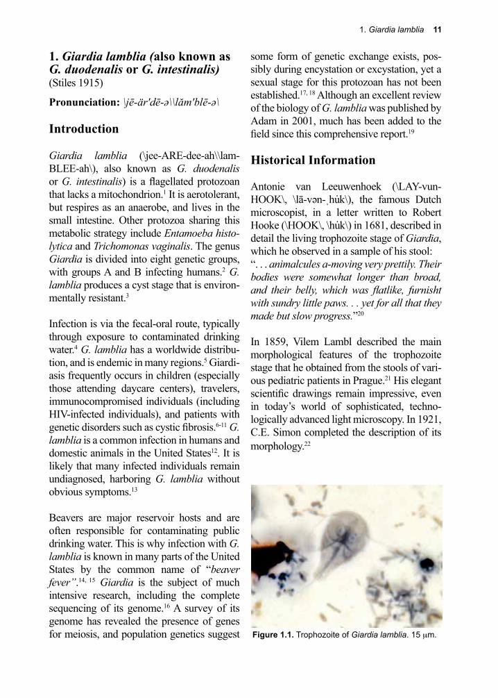
In 1859, Vilem Lambl described the main morphological features of the trophozoite stage that he obtained from the stools of vari-ous pediatric patients in Prague.21 His elegant scientific drawings remain impressive, even in today’s world of sophisticated, techno-logically advanced light microscopy. In 1921, C.E. Simon completed the description of its morphology.22
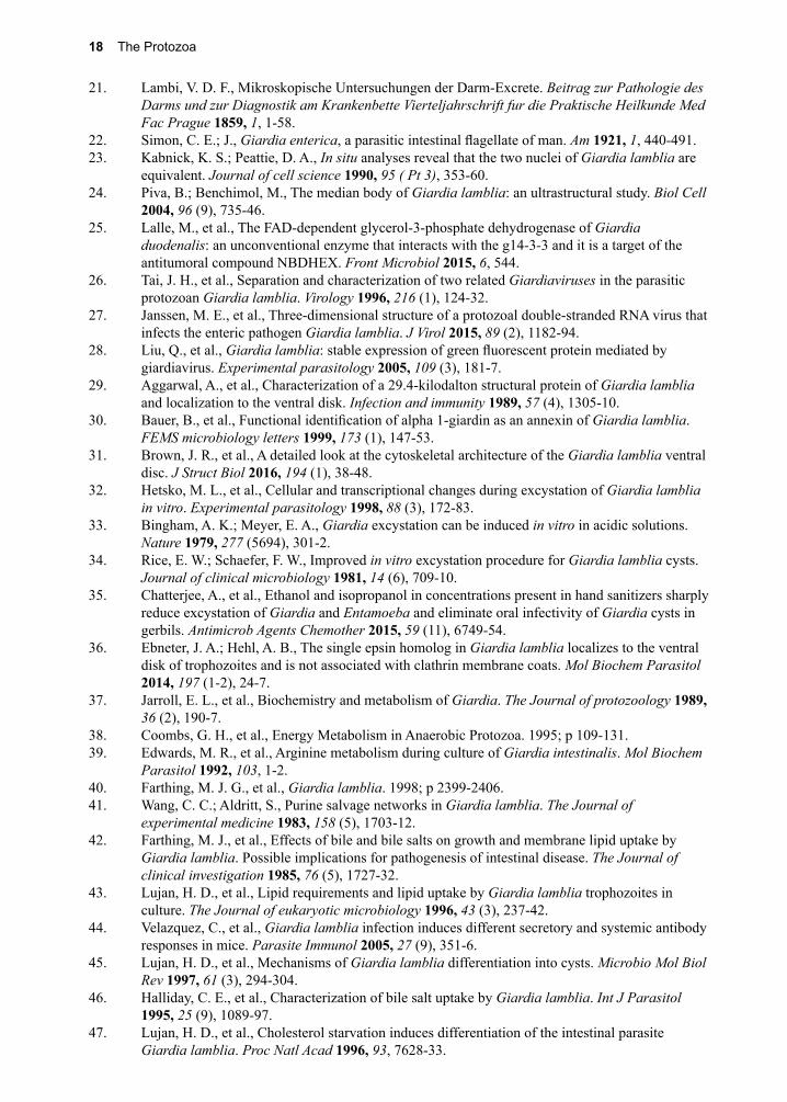
Figure 1.1. Trophozoite of Giardia lamblia. 15 µm.
12 The Protozoa
1. Giardia lamblia 13
Life Cycle
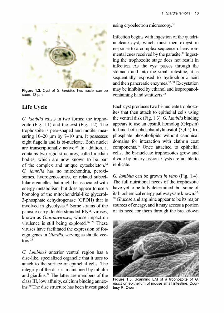
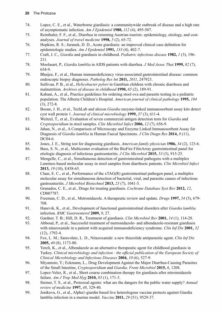
G. lamblia exists in two forms: the tropho-zoite (Fig. 1.1) and the cyst (Fig. 1.2). The trophozoite is pear-shaped and motile, mea-suring 10–20 µm by 7–10 µm. It possesses eight flagella and is bi-nucleate. Both nuclei are transcriptionally active.23 In addition, it contains two rigid structures, called median bodies, which are now known to be part of the complex and unique cytoskeleton.24 G. lamblia has no mitochondria, peroxi-somes, hydrogenosomes, or related subcel-lular organelles that might be associated with energy metabolism, but does appear to use a homolog of the mitochondrial-like glycerol-3-phosphate dehydrogenase (GPDH) that is involved in glycolysis.25 Some strains of the parasite carry double-stranded RNA viruses, known as Giardiaviruses, whose impact on virulence is still being explored.26, 27 These viruses have facilitated the expression of for-eign genes in Giardia, serving as shuttle vec-tors.28
G. lamblia’s anterior ventral region has a disc-like, specialized organelle that it uses to attach to the surface of epithelial cells. The integrity of the disk is maintained by tubulin and giardins.29 The latter are members of the class III, low affinity, calcium binding annex-ins.30 The disc structure has been investigated
using cryoelectron microscopy.31
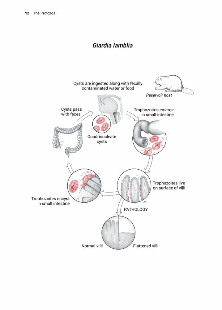
Infection begins with ingestion of the quadri-nucleate cyst, which must then excyst in response to a complex sequence of environ-mental cues received by the parasite.32 Ingest-ing the trophozoite stage does not result in infection. As the cyst passes through the stomach and into the small intestine, it is sequentially exposed to hydrochloric acid and then pancreatic enzymes.33, 34 Excystation may be inhibited by ethanol and isopropanol-containing hand sanitizers.35
Each cyst produces two bi-nucleate trophozo-ites that then attach to epithelial cells using the ventral disk (Fig. 1.3). G. lamblia binding appears to use an epsinR homolog (Glepsin) to bind both phosphatidylinositol (3,4,5)-tri-phosphate phospholipids without canonical domains for interaction with clathrin coat components.36 Once attached to epithelial cells, the bi-nucleate trophozoites grow and divide by binary fission. Cysts are unable to replicate.
G. lamblia can be grown in vitro (Fig. 1.4). The full nutritional needs of the trophozoite have yet to be fully determined, but some of its biochemical energy pathways are known.37,
38 Glucose and arginine appear to be its major sources of energy, and it may access a portion of its need for them through the breakdown
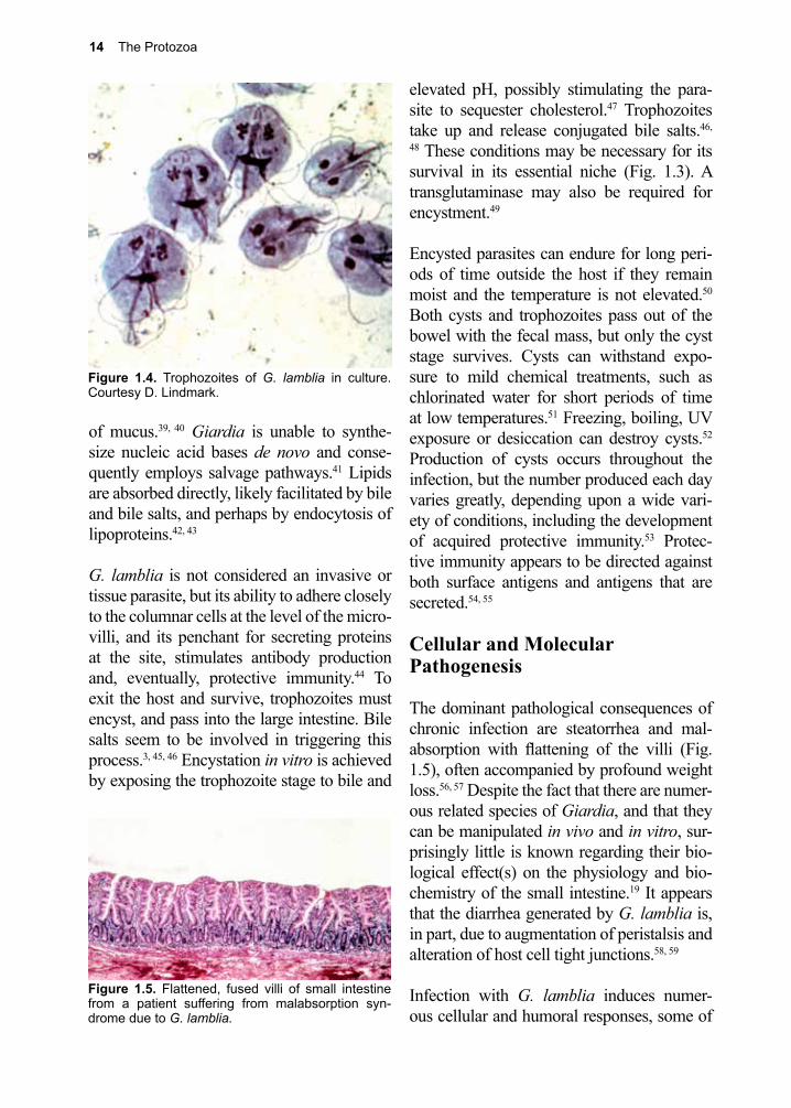
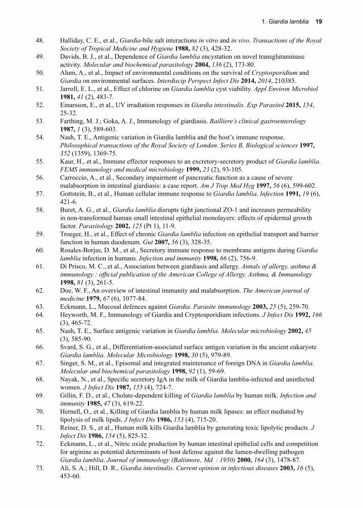
Figure 1.3. Scanning EM of a trophozoite of G. muris on epithelium of mouse small intestine. Cour-tesy R. Owen.
Figure 1.2. Cyst of G. lamblia. Two nuclei can be seen. 13 µm.
14 The Protozoa
elevated pH, possibly stimulating the para-site to sequester cholesterol.47 Trophozoites take up and release conjugated bile salts.46,
48 These conditions may be necessary for its survival in its essential niche (Fig. 1.3). A transglutaminase may also be required for encystment.49
Encysted parasites can endure for long peri-ods of time outside the host if they remain moist and the temperature is not elevated.50 Both cysts and trophozoites pass out of the bowel with the fecal mass, but only the cyst stage survives. Cysts can withstand expo-sure to mild chemical treatments, such as chlorinated water for short periods of time at low temperatures.51 Freezing, boiling, UV exposure or desiccation can destroy cysts.52 Production of cysts occurs throughout the infection, but the number produced each day varies greatly, depending upon a wide vari-ety of conditions, including the development of acquired protective immunity.53 Protec-tive immunity appears to be directed against both surface antigens and antigens that are secreted.54, 55
Cellular and Molecular Pathogenesis
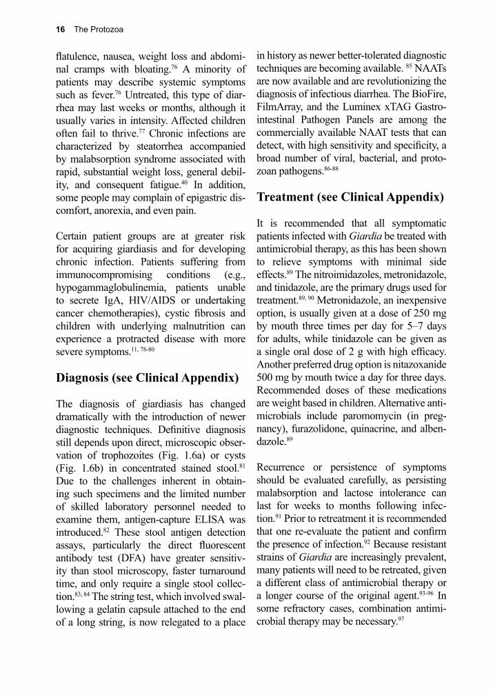
The dominant pathological consequences of chronic infection are steatorrhea and mal-absorption with flattening of the villi (Fig. 1.5), often accompanied by profound weight loss.56, 57 Despite the fact that there are numer-ous related species of Giardia, and that they can be manipulated in vivo and in vitro, sur-prisingly little is known regarding their bio-logical effect(s) on the physiology and bio-chemistry of the small intestine.19 It appears that the diarrhea generated by G. lamblia is, in part, due to augmentation of peristalsis and alteration of host cell tight junctions.58, 59
Infection with G. lamblia induces numer-ous cellular and humoral responses, some of
of mucus.39, 40 Giardia is unable to synthe-size nucleic acid bases de novo and conse-quently employs salvage pathways.41 Lipids are absorbed directly, likely facilitated by bile and bile salts, and perhaps by endocytosis of lipoproteins.42, 43
G. lamblia is not considered an invasive or tissue parasite, but its ability to adhere closely to the columnar cells at the level of the micro-villi, and its penchant for secreting proteins at the site, stimulates antibody production and, eventually, protective immunity.44 To exit the host and survive, trophozoites must encyst, and pass into the large intestine. Bile salts seem to be involved in triggering this process.3, 45, 46 Encystation in vitro is achieved by exposing the trophozoite stage to bile and
Figure 1.5. Flattened, fused villi of small intestine from a patient suffering from malabsorption syn-drome due to G. lamblia.
Figure 1.4. Trophozoites of G. lamblia in culture. Courtesy D. Lindmark.
1. Giardia lamblia 15
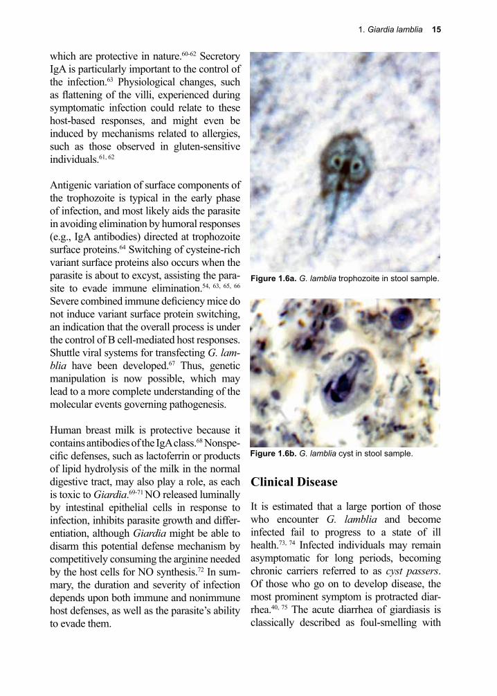
Figure 1.6a. G. lamblia trophozoite in stool sample.
Figure 1.6b. G. lamblia cyst in stool sample.
which are protective in nature.60-62 Secretory IgA is particularly important to the control of the infection.63 Physiological changes, such as flattening of the villi, experienced during symptomatic infection could relate to these host-based responses, and might even be induced by mechanisms related to allergies, such as those observed in gluten-sensitive individuals.61, 62
Antigenic variation of surface components of the trophozoite is typical in the early phase of infection, and most likely aids the parasite in avoiding elimination by humoral responses (e.g., IgA antibodies) directed at trophozoite surface proteins.64 Switching of cysteine-rich variant surface proteins also occurs when the parasite is about to excyst, assisting the para-site to evade immune elimination.54, 63, 65, 66 Severe combined immune deficiency mice do not induce variant surface protein switching, an indication that the overall process is under the control of B cell-mediated host responses. Shuttle viral systems for transfecting G. lam-blia have been developed.67 Thus, genetic manipulation is now possible, which may lead to a more complete understanding of the molecular events governing pathogenesis.
Human breast milk is protective because it contains antibodies of the IgA class.68 Nonspe-cific defenses, such as lactoferrin or products of lipid hydrolysis of the milk in the normal digestive tract, may also play a role, as each is toxic to Giardia.69-71 NO released luminally by intestinal epithelial cells in response to infection, inhibits parasite growth and differ-entiation, although Giardia might be able to disarm this potential defense mechanism by competitively consuming the arginine needed by the host cells for NO synthesis.72 In sum-mary, the duration and severity of infection depends upon both immune and nonimmune host defenses, as well as the parasite’s ability to evade them.
Clinical Disease
It is estimated that a large portion of those who encounter G. lamblia and become infected fail to progress to a state of ill health.73, 74 Infected individuals may remain asymptomatic for long periods, becoming chronic carriers referred to as cyst passers. Of those who go on to develop disease, the most prominent symptom is protracted diar-rhea.40, 75 The acute diarrhea of giardiasis is classically described as foul-smelling with
16 The Protozoa
flatulence, nausea, weight loss and abdomi-nal cramps with bloating.76 A minority of patients may describe systemic symptoms such as fever.76 Untreated, this type of diar-rhea may last weeks or months, although it usually varies in intensity. Affected children often fail to thrive.77 Chronic infections are characterized by steatorrhea accompanied by malabsorption syndrome associated with rapid, substantial weight loss, general debil-ity, and consequent fatigue.40 In addition, some people may complain of epigastric dis-comfort, anorexia, and even pain.
Certain patient groups are at greater risk for acquiring giardiasis and for developing chronic infection. Patients suffering from immunocompromising conditions (e.g., hypogammaglobulinemia, patients unable to secrete IgA, HIV/AIDS or undertaking cancer chemotherapies), cystic fibrosis and children with underlying malnutrition can experience a protracted disease with more severe symptoms.11, 78-80
Diagnosis (see Clinical Appendix)
The diagnosis of giardiasis has changed dramatically with the introduction of newer diagnostic techniques. Definitive diagnosis still depends upon direct, microscopic obser-vation of trophozoites (Fig. 1.6a) or cysts (Fig. 1.6b) in concentrated stained stool.81 Due to the challenges inherent in obtain-ing such specimens and the limited number of skilled laboratory personnel needed to examine them, antigen-capture ELISA was introduced.82 These stool antigen detection assays, particularly the direct fluorescent antibody test (DFA) have greater sensitiv-ity than stool microscopy, faster turnaround time, and only require a single stool collec-tion.83, 84 The string test, which involved swal-lowing a gelatin capsule attached to the end of a long string, is now relegated to a place
in history as newer better-tolerated diagnostic techniques are becoming available. 85 NAATs are now available and are revolutionizing the diagnosis of infectious diarrhea. The BioFire, FilmArray, and the Luminex xTAG Gastro-intestinal Pathogen Panels are among the commercially available NAAT tests that can detect, with high sensitivity and specificity, a broad number of viral, bacterial, and proto-zoan pathogens.86-88
Treatment (see Clinical Appendix)
It is recommended that all symptomatic patients infected with Giardia be treated with antimicrobial therapy, as this has been shown to relieve symptoms with minimal side effects.89 The nitroimidazoles, metronidazole, and tinidazole, are the primary drugs used for treatment.89, 90 Metronidazole, an inexpensive option, is usually given at a dose of 250 mg by mouth three times per day for 5–7 days for adults, while tinidazole can be given as a single oral dose of 2 g with high efficacy. Another preferred drug option is nitazoxanide 500 mg by mouth twice a day for three days. Recommended doses of these medications are weight based in children. Alternative anti-microbials include paromomycin (in preg-nancy), furazolidone, quinacrine, and alben-dazole.89
Recurrence or persistence of symptoms should be evaluated carefully, as persisting malabsorption and lactose intolerance can last for weeks to months following infec-tion.91 Prior to retreatment it is recommended that one re-evaluate the patient and confirm the presence of infection.92 Because resistant strains of Giardia are increasingly prevalent, many patients will need to be retreated, given a different class of antimicrobial therapy or a longer course of the original agent.93-96 In some refractory cases, combination antimi-crobial therapy may be necessary.97
1. Giardia lamblia 17
Prevention and Control
G. lamblia is primarily a water-borne infec-tion, although food handlers and infected children in daycare centers no doubt play important roles in transmission.4, 98 Prevention strategies include proper disposal of human waste, filtration of drinking water supplies, maintenance of buffer zones around water-sheds when filtration is not practiced (e.g., in
References
1. Roger, A. J., et al., A mitochondrial-like chaperonin 60 gene in Giardia lamblia: evidence that diplomonads once harbored an endosymbiont related to the progenitor of mitochondria. Proceedings of the National Academy of Sciences of the United States of America 1998, 95 (1), 229-34.
2. Vanni, I., et al., Detection of Giardia duodenalis assemblages A and B in human feces by simple, assemblage-specific PCR assays. PLoS Negl Trop Dis 2012, 6 (8), e1776.
3. Faso, C., et al., The proteome landscape of Giardia lamblia encystation. PLoS One 2013, 8 (12), e83207.
4. Levy, D. A., et al., Surveillance for waterborne-disease outbreaks--United States, 1995-1996. MMWR. CDC surveillance summaries : Morbidity and mortality weekly report. CDC surveillance summaries / Centers for Disease Control 1998, 47 (5), 1-34.
5. Feng, Y.; Xiao, L., Zoonotic potential and molecular epidemiology of Giardia species and giardiasis. Clin Microbiol Rev 2011, 24 (1), 110-40.
6. Sagi, E. F., et al., Giardia lamblia: prevalance, influence on growth, and symptomatology in healthy nursery children. Isr Sci 1983, 19, 815-817.
7. Pickering, L. K., et al., Occurrence of Giardia lamblia in children in day care centers. The Journal of pediatrics 1984, 104 (4), 522-6.
8. Daszak, P., Giardia, HIV, and nature’s horrifying beauty. Ecohealth 2014, 11 (2), 277-8.9. Cimino, A.; Ali, S. Z., Giardia intestinalis on anal PAP of an HIV-positive male. Diagn
Cytopathol 2010, 38 (11), 814-5.10. Jelinek, T.; Loscher, T., Epidemiology of giardiasis in German travelers. J Travel Med 2000, 7
(2), 70-3.11. Roberts, D. M., et al., Prevalence of giardiasis in patients with cystic fibrosis. The Journal of
pediatrics 1988, 112 (4), 555-9.12. Mohamed, A. S., et al., Temporal patterns of human and canine Giardia infection in the United
States: 2003-2009. Prev Vet Med 2014, 113 (2), 249-56.13. Nash, T. E., et al., Experimental human infections with Giardia lamblia. J Infect Dis 1987, 156
(6), 974-84.14. Carlson, D. W.; Finger, D. R., Beaver fever arthritis. J Clin Rheumatol 2004, 10 (2), 86-8.15. Taverne, J., Beaver fever and pinworm neuroses on the Net. Parasitol Today 1999, 15 (9), 363-4.16. Best, A. A., et al., Evolution of eukaryotic transcription: insights from the genome of Giardia
lamblia. Genome research 2004, 14 (8), 1537-47.17. Ramesh, M. A., et al., A phylogenomic inventory of meiotic genes; evidence for sex in Giardia
and an early eukaryotic origin of meiosis. Current biology : CB 2005, 15 (2), 185-91.18. Weedall, G. D.; Hall, N., Sexual reproduction and genetic exchange in parasitic protists.
Parasitology 2015, 142 Suppl 1, S120-7.19. Adam, R. D., Biology of Giardia lamblia. Clinical microbiology reviews 2001, 14 (3), 447-75.20. Leenwenhoek, A.; Dobell, C., Van Cited by In Antony van Leeuwen-hoek and His “Little
Animals”. Publications New Dover P 1960, 224.
New York City), and maintaining the high-est standards of hygiene in daycare centers and mental institutions. A murine model for a protective Giardia vaccine exists; however, efforts to develop clinical candidate vaccines, including work on canine vaccines, are ham-pered by the lack of a well-articulated medi-cal need for commitment of new resources in the setting of many effective therapeutic options.99
18 The Protozoa
21. Lambi, V. D. F., Mikroskopische Untersuchungen der Darm-Excrete. Beitrag zur Pathologie des Darms und zur Diagnostik am Krankenbette Vierteljahrschrift fur die Praktische Heilkunde Med Fac Prague 1859, 1, 1-58.
22. Simon, C. E.; J., Giardia enterica, a parasitic intestinal flagellate of man. Am 1921, 1, 440-491.23. Kabnick, K. S.; Peattie, D. A., In situ analyses reveal that the two nuclei of Giardia lamblia are
equivalent. Journal of cell science 1990, 95 ( Pt 3), 353-60.24. Piva, B.; Benchimol, M., The median body of Giardia lamblia: an ultrastructural study. Biol Cell
2004, 96 (9), 735-46.25. Lalle, M., et al., The FAD-dependent glycerol-3-phosphate dehydrogenase of Giardia
duodenalis: an unconventional enzyme that interacts with the g14-3-3 and it is a target of the antitumoral compound NBDHEX. Front Microbiol 2015, 6, 544.
26. Tai, J. H., et al., Separation and characterization of two related Giardiaviruses in the parasitic protozoan Giardia lamblia. Virology 1996, 216 (1), 124-32.
27. Janssen, M. E., et al., Three-dimensional structure of a protozoal double-stranded RNA virus that infects the enteric pathogen Giardia lamblia. J Virol 2015, 89 (2), 1182-94.
28. Liu, Q., et al., Giardia lamblia: stable expression of green fluorescent protein mediated by giardiavirus. Experimental parasitology 2005, 109 (3), 181-7.
29. Aggarwal, A., et al., Characterization of a 29.4-kilodalton structural protein of Giardia lamblia and localization to the ventral disk. Infection and immunity 1989, 57 (4), 1305-10.
30. Bauer, B., et al., Functional identification of alpha 1-giardin as an annexin of Giardia lamblia. FEMS microbiology letters 1999, 173 (1), 147-53.
31. Brown, J. R., et al., A detailed look at the cytoskeletal architecture of the Giardia lamblia ventral disc. J Struct Biol 2016, 194 (1), 38-48.
32. Hetsko, M. L., et al., Cellular and transcriptional changes during excystation of Giardia lamblia in vitro. Experimental parasitology 1998, 88 (3), 172-83.
33. Bingham, A. K.; Meyer, E. A., Giardia excystation can be induced in vitro in acidic solutions. Nature 1979, 277 (5694), 301-2.
34. Rice, E. W.; Schaefer, F. W., Improved in vitro excystation procedure for Giardia lamblia cysts. Journal of clinical microbiology 1981, 14 (6), 709-10.
35. Chatterjee, A., et al., Ethanol and isopropanol in concentrations present in hand sanitizers sharply reduce excystation of Giardia and Entamoeba and eliminate oral infectivity of Giardia cysts in gerbils. Antimicrob Agents Chemother 2015, 59 (11), 6749-54.
36. Ebneter, J. A.; Hehl, A. B., The single epsin homolog in Giardia lamblia localizes to the ventral disk of trophozoites and is not associated with clathrin membrane coats. Mol Biochem Parasitol 2014, 197 (1-2), 24-7.
37. Jarroll, E. L., et al., Biochemistry and metabolism of Giardia. The Journal of protozoology 1989, 36 (2), 190-7.
38. Coombs, G. H., et al., Energy Metabolism in Anaerobic Protozoa. 1995; p 109-131.39. Edwards, M. R., et al., Arginine metabolism during culture of Giardia intestinalis. Mol Biochem
Parasitol 1992, 103, 1-2.40. Farthing, M. J. G., et al., Giardia lamblia. 1998; p 2399-2406.41. Wang, C. C.; Aldritt, S., Purine salvage networks in Giardia lamblia. The Journal of
experimental medicine 1983, 158 (5), 1703-12.42. Farthing, M. J., et al., Effects of bile and bile salts on growth and membrane lipid uptake by
Giardia lamblia. Possible implications for pathogenesis of intestinal disease. The Journal of clinical investigation 1985, 76 (5), 1727-32.
43. Lujan, H. D., et al., Lipid requirements and lipid uptake by Giardia lamblia trophozoites in culture. The Journal of eukaryotic microbiology 1996, 43 (3), 237-42.
44. Velazquez, C., et al., Giardia lamblia infection induces different secretory and systemic antibody responses in mice. Parasite Immunol 2005, 27 (9), 351-6.
45. Lujan, H. D., et al., Mechanisms of Giardia lamblia differentiation into cysts. Microbio Mol Biol Rev 1997, 61 (3), 294-304.
46. Halliday, C. E., et al., Characterization of bile salt uptake by Giardia lamblia. Int J Parasitol 1995, 25 (9), 1089-97.
47. Lujan, H. D., et al., Cholesterol starvation induces differentiation of the intestinal parasite Giardia lamblia. Proc Natl Acad 1996, 93, 7628-33.
1. Giardia lamblia 19
48. Halliday, C. E., et al., Giardia-bile salt interactions in vitro and in vivo. Transactions of the Royal Society of Tropical Medicine and Hygiene 1988, 82 (3), 428-32.
49. Davids, B. J., et al., Dependence of Giardia lamblia encystation on novel transglutaminase activity. Molecular and biochemical parasitology 2004, 136 (2), 173-80.
50. Alum, A., et al., Impact of environmental conditions on the survival of Cryptosporidium and Giardia on environmental surfaces. Interdiscip Perspect Infect Dis 2014, 2014, 210385.
51. Jarroll, E. L., et al., Effect of chlorine on Giardia lamblia cyst viability. Appl Environ Microbiol 1981, 41 (2), 483-7.
52. Einarsson, E., et al., UV irradiation responses in Giardia intestinalis. Exp Parasitol 2015, 154, 25-32.
53. Farthing, M. J.; Goka, A. J., Immunology of giardiasis. Bailliere’s clinical gastroenterology 1987, 1 (3), 589-603.
54. Nash, T. E., Antigenic variation in Giardia lamblia and the host’s immune response. Philosophical transactions of the Royal Society of London. Series B, Biological sciences 1997, 352 (1359), 1369-75.
55. Kaur, H., et al., Immune effector responses to an excretory-secretory product of Giardia lamblia. FEMS immunology and medical microbiology 1999, 23 (2), 93-105.
56. Carroccio, A., et al., Secondary impairment of pancreatic function as a cause of severe malabsorption in intestinal giardiasis: a case report. Am J Trop Med Hyg 1997, 56 (6), 599-602.
57. Gottstein, B., et al., Human cellular immune response to Giardia lamblia. Infection 1991, 19 (6), 421-6.
58. Buret, A. G., et al., Giardia lamblia disrupts tight junctional ZO-1 and increases permeability in non-transformed human small intestinal epithelial monolayers: effects of epidermal growth factor. Parasitology 2002, 125 (Pt 1), 11-9.
59. Troeger, H., et al., Effect of chronic Giardia lamblia infection on epithelial transport and barrier function in human duodenum. Gut 2007, 56 (3), 328-35.
60. Rosales-Borjas, D. M., et al., Secretory immune response to membrane antigens during Giardia lamblia infection in humans. Infection and immunity 1998, 66 (2), 756-9.
61. Di Prisco, M. C., et al., Association between giardiasis and allergy. Annals of allergy, asthma & immunology : official publication of the American College of Allergy, Asthma, & Immunology 1998, 81 (3), 261-5.
62. Doe, W. F., An overview of intestinal immunity and malabsorption. The American journal of medicine 1979, 67 (6), 1077-84.
63. Eckmann, L., Mucosal defences against Giardia. Parasite immunology 2003, 25 (5), 259-70.64. Heyworth, M. F., Immunology of Giardia and Cryptosporidium infections. J Infect Dis 1992, 166
(3), 465-72.65. Nash, T. E., Surface antigenic variation in Giardia lamblia. Molecular microbiology 2002, 45
(3), 585-90.66. Svard, S. G., et al., Differentiation-associated surface antigen variation in the ancient eukaryote
Giardia lamblia. Molecular Microbiology 1998, 30 (5), 979-89.67. Singer, S. M., et al., Episomal and integrated maintenance of foreign DNA in Giardia lamblia.
Molecular and biochemical parasitology 1998, 92 (1), 59-69.68. Nayak, N., et al., Specific secretory IgA in the milk of Giardia lamblia-infected and uninfected
women. J Infect Dis 1987, 155 (4), 724-7.69. Gillin, F. D., et al., Cholate-dependent killing of Giardia lamblia by human milk. Infection and
immunity 1985, 47 (3), 619-22.70. Hernell, O., et al., Killing of Giardia lamblia by human milk lipases: an effect mediated by
lipolysis of milk lipids. J Infect Dis 1986, 153 (4), 715-20.71. Reiner, D. S., et al., Human milk kills Giardia lamblia by generating toxic lipolytic products. J
Infect Dis 1986, 154 (5), 825-32.72. Eckmann, L., et al., Nitric oxide production by human intestinal epithelial cells and competition
for arginine as potential determinants of host defense against the lumen-dwelling pathogen Giardia lamblia. Journal of immunology (Baltimore, Md. : 1950) 2000, 164 (3), 1478-87.
73. Ali, S. A.; Hill, D. R., Giardia intestinalis. Current opinion in infectious diseases 2003, 16 (5), 453-60.
20 The Protozoa
74. Lopez, C. E., et al., Waterborne giardiasis: a communitywide outbreak of disease and a high rate of asymptomatic infection. Am J Epidemiol 1980, 112 (4), 495-507.
75. Reinthaler, F. F., et al., Diarrhea in returning Austrian tourists: epidemiology, etiology, and cost-analyses. Journal of travel medicine 1998, 5 (2), 65-72.
76. Hopkins, R. S.; Juranek, D. D., Acute giardiasis: an improved clinical case definition for epidemiologic studies. Am J Epidemiol 1991, 133 (4), 402-7.
77. Craft, J. C., Giardia and giardiasis in childhood. Pediatric infectious disease 1982, 1 (3), 196-211.
78. Moolasart, P., Giardia lamblia in AIDS patients with diarrhea. J Med Assoc Thai 1999, 82 (7), 654-9.
79. Bhaijee, F., et al., Human immunodeficiency virus-associated gastrointestinal disease: common endoscopic biopsy diagnoses. Patholog Res Int 2011, 2011, 247923.
80. Sullivan, P. B., et al., Helicobacter pylori in Gambian children with chronic diarrhoea and malnutrition. Archives of disease in childhood 1990, 65 (2), 189-91.
81. Kabani, A., et al., Practice guidelines for ordering stool ova and parasite testing in a pediatric population. The Alberta Children’s Hospital. American journal of clinical pathology 1995, 104 (3), 272-8.
82. Boone, J. H., et al., TechLab and alexon Giardia enzyme-linked immunosorbent assay kits detect cyst wall protein 1. Journal of clinical microbiology 1999, 37 (3), 611-4.
83. Weitzel, T., et al., Evaluation of seven commercial antigen detection tests for Giardia and Cryptosporidium in stool samples. Clin Microbiol Infect 2006, 12 (7), 656-9.
84. Jahan, N., et al., A Comparison of Microscopy and Enzyme Linked Immunosorbent Assay for Diagnosis of Giardia lamblia in Human Faecal Specimens. J Clin Diagn Res 2014, 8 (11), DC04-6.
85. Jones, J. E., String test for diagnosing giardiasis. American family physician 1986, 34 (2), 123-6.86. Buss, S. N., et al., Multicenter evaluation of the BioFire FilmArray gastrointestinal panel for
etiologic diagnosis of infectious gastroenteritis. J Clin Microbiol 2015, 53 (3), 915-25.87. Mengelle, C., et al., Simultaneous detection of gastrointestinal pathogens with a multiplex
Luminex-based molecular assay in stool samples from diarrhoeic patients. Clin Microbiol Infect 2013, 19 (10), E458-65.
88. Claas, E. C., et al., Performance of the xTAG(R) gastrointestinal pathogen panel, a multiplex molecular assay for simultaneous detection of bacterial, viral, and parasitic causes of infectious gastroenteritis. J Microbiol Biotechnol 2013, 23 (7), 1041-5.
89. Granados, C. E., et al., Drugs for treating giardiasis. Cochrane Database Syst Rev 2012, 12, CD007787.
90. Freeman, C. D., et al., Metronidazole. A therapeutic review and update. Drugs 1997, 54 (5), 679-708.
91. Hanevik, K., et al., Development of functional gastrointestinal disorders after Giardia lamblia infection. BMC Gastroenterol 2009, 9, 27.
92. Gardner, T. B.; Hill, D. R., Treatment of giardiasis. Clin Microbiol Rev 2001, 14 (1), 114-28.93. Abboud, P., et al., Successful treatment of metronidazole- and albendazole-resistant giardiasis
with nitazoxanide in a patient with acquired immunodeficiency syndrome. Clin Inf Dis 2001, 32 (12), 1792-4.
94. Fox, L. M.; Saravolatz, L. D., Nitazoxanide: a new thiazolide antiparasitic agent. Clin Inf Dis 2005, 40 (8), 1173-80.
95. Yereli, K., et al., Albendazole as an alternative therapeutic agent for childhood giardiasis in Turkey. Clinical microbiology and infection : the official publication of the European Society of Clinical Microbiology and Infectious Diseases 2004, 10 (6), 527-9.
96. Miyamoto, Y.; Eckmann, L., Drug Development Against the Major Diarrhea-Causing Parasites of the Small Intestine, Cryptosporidium and Giardia. Front Microbiol 2015, 6, 1208.
97. Lopez-Velez, R., et al., Short course combination therapy for giardiasis after nitroimidazole failure. Am J Trop Med Hyg 2010, 83 (1), 171-3.
98. Steiner, T. S., et al., Protozoal agents: what are the dangers for the public water supply? Annual review of medicine 1997, 48, 329-40.
99. Jenikova, G., et al., Alpha1-giardin based live heterologous vaccine protects against Giardia lamblia infection in a murine model. Vaccine 2011, 29 (51), 9529-37.
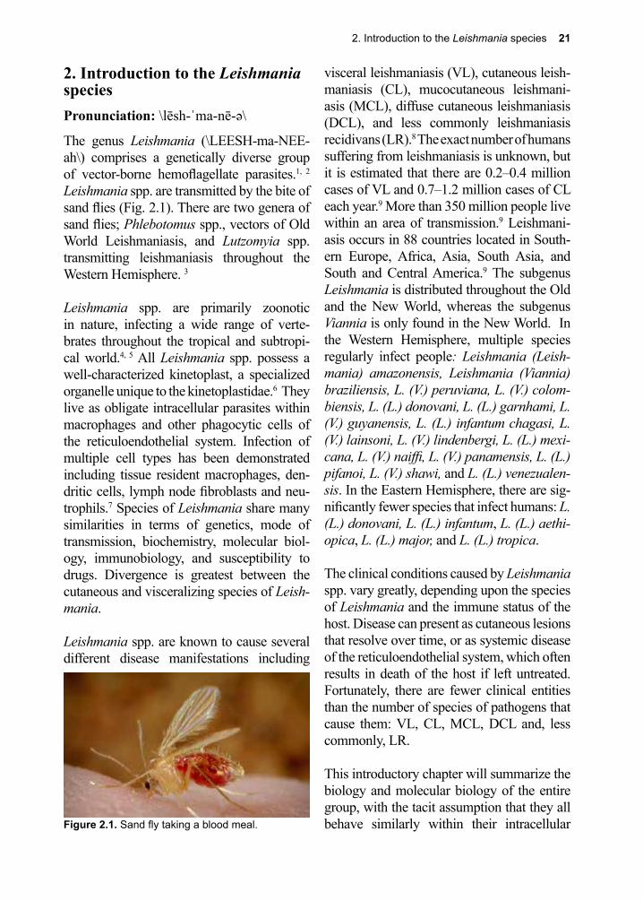
2. Introduction to the Leishmania species 21
Figure 2.1. Sand fly taking a blood meal.
2. Introduction to the Leishmania speciesPronunciation: \lēsh-ˈma-nē-ə\
The genus Leishmania (\LEESH-ma-NEE-ah\) comprises a genetically diverse group of vector-borne hemoflagellate parasites.1, 2 Leishmania spp. are transmitted by the bite of sand flies (Fig. 2.1). There are two genera of sand flies; Phlebotomus spp., vectors of Old World Leishmaniasis, and Lutzomyia spp. transmitting leishmaniasis throughout the Western Hemisphere. 3
Leishmania spp. are primarily zoonotic in nature, infecting a wide range of verte-brates throughout the tropical and subtropi-cal world.4, 5 All Leishmania spp. possess a well-characterized kinetoplast, a specialized organelle unique to the kinetoplastidae.6 They live as obligate intracellular parasites within macrophages and other phagocytic cells of the reticuloendothelial system. Infection of multiple cell types has been demonstrated including tissue resident macrophages, den-dritic cells, lymph node fibroblasts and neu-trophils.7 Species of Leishmania share many similarities in terms of genetics, mode of transmission, biochemistry, molecular biol-ogy, immunobiology, and susceptibility to drugs. Divergence is greatest between the cutaneous and visceralizing species of Leish-mania.
Leishmania spp. are known to cause several different disease manifestations including
visceral leishmaniasis (VL), cutaneous leish-maniasis (CL), mucocutaneous leishmani-asis (MCL), diffuse cutaneous leishmaniasis (DCL), and less commonly leishmaniasis recidivans (LR).8 The exact number of humans suffering from leishmaniasis is unknown, but it is estimated that there are 0.2–0.4 million cases of VL and 0.7–1.2 million cases of CL each year.9 More than 350 million people live within an area of transmission.9 Leishmani-asis occurs in 88 countries located in South-ern Europe, Africa, Asia, South Asia, and South and Central America.9 The subgenus Leishmania is distributed throughout the Old and the New World, whereas the subgenus Viannia is only found in the New World. In the Western Hemisphere, multiple species regularly infect people: Leishmania (Leish-mania) amazonensis, Leishmania (Viannia) braziliensis, L. (V.) peruviana, L. (V.) colom-biensis, L. (L.) donovani, L. (L.) garnhami, L. (V.) guyanensis, L. (L.) infantum chagasi, L. (V.) lainsoni, L. (V.) lindenbergi, L. (L.) mexi-cana, L. (V.) naiffi, L. (V.) panamensis, L. (L.) pifanoi, L. (V.) shawi, and L. (L.) venezualen-sis. In the Eastern Hemisphere, there are sig-nificantly fewer species that infect humans: L. (L.) donovani, L. (L.) infantum, L. (L.) aethi-opica, L. (L.) major, and L. (L.) tropica.
The clinical conditions caused by Leishmania spp. vary greatly, depending upon the species of Leishmania and the immune status of the host. Disease can present as cutaneous lesions that resolve over time, or as systemic disease of the reticuloendothelial system, which often results in death of the host if left untreated. Fortunately, there are fewer clinical entities than the number of species of pathogens that cause them: VL, CL, MCL, DCL and, less commonly, LR.
This introductory chapter will summarize the biology and molecular biology of the entire group, with the tacit assumption that they all behave similarly within their intracellular
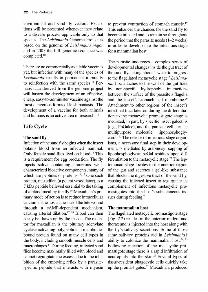
22 The Protozoa
environment and sand fly vectors. Excep-tions will be presented whenever they relate to a disease process applicable only to that species. The Leishmania Genome Project is based on the genome of Leishmania major and in 2005 the full genomic sequence was completed.10
There are no commercially available vaccines yet, but infection with many of the species of Leishmania results in permanent immunity to reinfection with the same species.11 Per-haps data derived from the genome project will hasten the development of an effective, cheap, easy-to-administer vaccine against the most dangerous forms of leishmaniasis. The development of a vaccine for both animals and humans is an active area of research. 12
Life Cycle
The sand flyInfection of the sand fly begins when the insect obtains blood from an infected mammal. Only female sand flies feed on blood.13 This is a requirement for egg production. The fly injects saliva containing numerous well-characterized bioactive components, many of which are peptides or proteins.14, 15 One such protein, maxadilan (a potent vasodilator), is a 7 kDa peptide believed essential to the taking of a blood meal by the fly.16 Maxadilan’s pri-mary mode of action is to reduce intracellular calcium in the host at the site of the bite wound through a cAMP-dependent mechanism, causing arterial dilation.17, 18 Blood can then easily be drawn up by the insect. The recep-tor for maxadilan is the pituitary adenylate cyclase-activating polypeptide, a membrane-bound protein found on many cell types in the body, including smooth muscle cells and macrophages.17 During feeding, infected sand flies become maximally filled with blood and cannot regurgitate the excess, due to the inhi-bition of the emptying reflex by a parasite-specific peptide that interacts with myosin
to prevent contraction of stomach muscle.19
This enhances the chances for the sand fly to become infected and to remain so throughout the period that the parasite needs (1–2 weeks) in order to develop into the infectious stage for a mammalian host.
The parasite undergoes a complex series of developmental changes inside the gut tract of the sand fly, taking about 1 week to progress to the flagellated metacyclic stage.2 Leishma-nia first attaches to the wall of the gut tract by non-specific hydrophobic interactions between the surface of the parasite’s flagella and the insect’s stomach cell membrane.20 Attachment to other regions of the insect’s intestinal tract later on during the differentia-tion to the metacyclic promastigote stage is mediated, in part, by specific insect galectins (e.g., PpGalec), and the parasite cell surface multipurpose molecule, lipophosphogly-can.21, 22 The release of infectious stage organ-isms, a necessary final step in their develop-ment, is mediated by arabinosyl capping of lipophosphoglycan scGal residues upon dif-ferentiation to the metacyclic stage.23 The lep-tomonad stage locates to the anterior region of the gut and secretes a gel-like substance that blocks the digestive tract of the sand fly, causing the infected insect to regurgitate its complement of infectious metacyclic pro-mastigotes into the host’s subcutaneous tis-sues during feeding.2
The mammalian hostThe flagellated metacyclic promastigote stage (Fig. 2.2) resides in the anterior midgut and thorax and is injected into the host along with the fly’s salivary secretions. Some of those same salivary proteins aid in Leishmania’s ability to colonize the mammalian host.24, 25 Following injection of the metacyclic pro-mastigote stage there is a rapid infiltration of neutrophils into the skin.26 Several types of tissue-resident phagocytic cells quickly take up the promastigotes.27 Maxadilan, produced
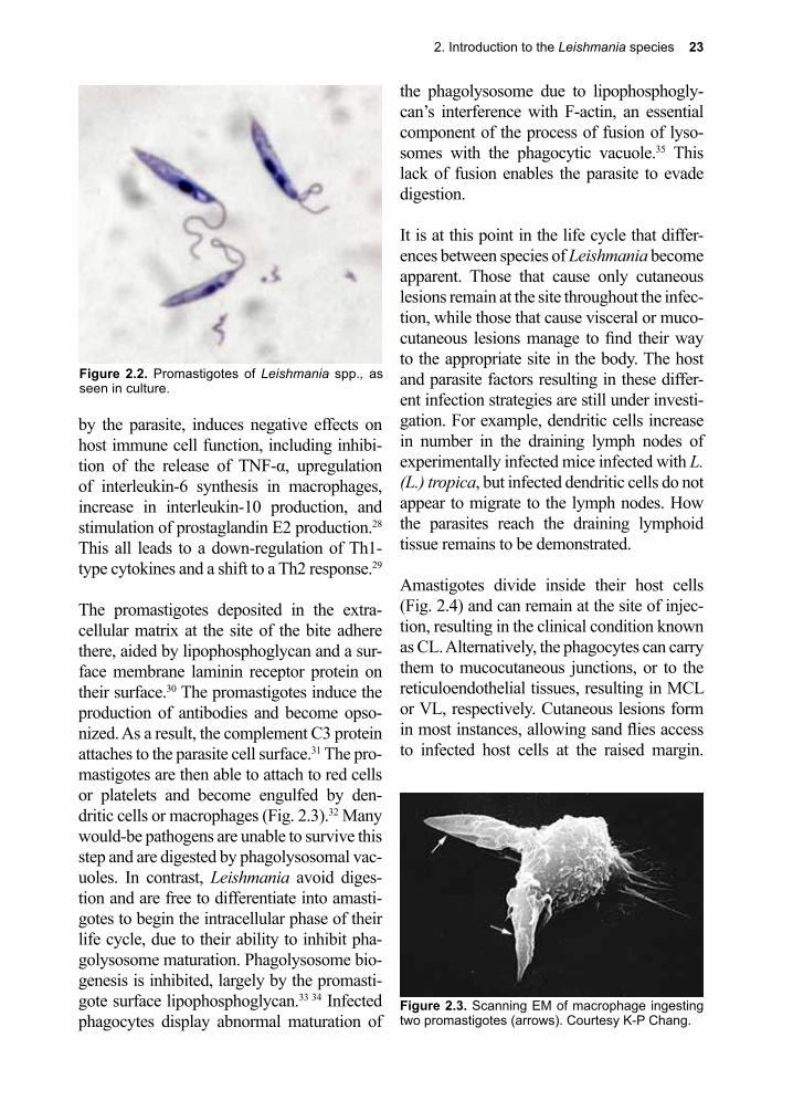
2. Introduction to the Leishmania species 23
by the parasite, induces negative effects on host immune cell function, including inhibi-tion of the release of TNF-α, upregulation of interleukin-6 synthesis in macrophages, increase in interleukin-10 production, and stimulation of prostaglandin E2 production.28 This all leads to a down-regulation of Th1-type cytokines and a shift to a Th2 response.29
The promastigotes deposited in the extra-cellular matrix at the site of the bite adhere there, aided by lipophosphoglycan and a sur-face membrane laminin receptor protein on their surface.30 The promastigotes induce the production of antibodies and become opso-nized. As a result, the complement C3 protein attaches to the parasite cell surface.31 The pro-mastigotes are then able to attach to red cells or platelets and become engulfed by den-dritic cells or macrophages (Fig. 2.3).32 Many would-be pathogens are unable to survive this step and are digested by phagolysosomal vac-uoles. In contrast, Leishmania avoid diges-tion and are free to differentiate into amasti-gotes to begin the intracellular phase of their life cycle, due to their ability to inhibit pha-golysosome maturation. Phagolysosome bio-genesis is inhibited, largely by the promasti-gote surface lipophosphoglycan.33 34 Infected phagocytes display abnormal maturation of
the phagolysosome due to lipophosphogly-can’s interference with F-actin, an essential component of the process of fusion of lyso-somes with the phagocytic vacuole.35 This lack of fusion enables the parasite to evade digestion.
It is at this point in the life cycle that differ-ences between species of Leishmania become apparent. Those that cause only cutaneous lesions remain at the site throughout the infec-tion, while those that cause visceral or muco-cutaneous lesions manage to find their way to the appropriate site in the body. The host and parasite factors resulting in these differ-ent infection strategies are still under investi-gation. For example, dendritic cells increase in number in the draining lymph nodes of experimentally infected mice infected with L. (L.) tropica, but infected dendritic cells do not appear to migrate to the lymph nodes. How the parasites reach the draining lymphoid tissue remains to be demonstrated.
Amastigotes divide inside their host cells (Fig. 2.4) and can remain at the site of injec-tion, resulting in the clinical condition known as CL. Alternatively, the phagocytes can carry them to mucocutaneous junctions, or to the reticuloendothelial tissues, resulting in MCL or VL, respectively. Cutaneous lesions form in most instances, allowing sand flies access to infected host cells at the raised margin.
Figure 2.2. Promastigotes of Leishmania spp., as seen in culture.
Figure 2.3. Scanning EM of macrophage ingesting two promastigotes (arrows). Courtesy K-P Chang.
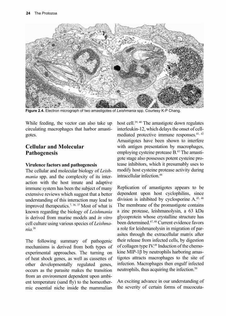
24 The Protozoa
While feeding, the vector can also take up circulating macrophages that harbor amasti-gotes.
Cellular and Molecular Pathogenesis
Virulence factors and pathogenesisThe cellular and molecular biology of Leish-mania spp. and the complexity of its inter-action with the host innate and adaptive immune system has been the subject of many extensive reviews which suggest that a better understanding of this interaction may lead to improved therapeutics.7, 36, 37 Most of what is known regarding the biology of Leishmania is derived from murine models and in vitro cell culture using various species of Leishma-nia.38
The following summary of pathogenic mechanisms is derived from both types of experimental approaches. The turning on of heat shock genes, as well as cassettes of other developmentally regulated genes, occurs as the parasite makes the transition from an environment dependent upon ambi-ent temperature (sand fly) to the homeother-mic essential niche inside the mammalian
host cell.39, 40 The amastigote down regulates interleukin-12, which delays the onset of cell-mediated protective immune responses.41, 42 Amastigotes have been shown to interfere with antigen presentation by macrophages, employing cysteine protease B.43 The amasti-gote stage also possesses potent cysteine pro-tease inhibitors, which it presumably uses to modify host cysteine protease activity during intracellular infection.44
Replication of amastigotes appears to be dependent upon host cyclophilins, since division is inhibited by cyclosporine A.45, 46 The membrane of the promastigote contains a zinc protease, leishmanolysin, a 63 kDa glycoprotein whose crystalline structure has been determined.47, 48 Current evidence favors a role for leishmanolysin in migration of par-asites through the extracellular matrix after their release from infected cells, by digestion of collagen type IV.49 Induction of the chemo-kine MIP-1β by neutrophils harboring amas-tigotes attracts macrophages to the site of infection. Macrophages then engulf infected neutrophils, thus acquiring the infection.50
An exciting advance in our understanding of the severity of certain forms of mucocuta-
Figure 2.4. Electron micrograph of two amastigotes of Leishmania spp. Courtesy K-P Chang.
2. Introduction to the Leishmania species 25
neous disease was the discovery of an RNA virus that infects the L. Viannia subgenus.51 It appears that this Leishmania RNA virus-1 is recognized by host toll-like receptors and induces an inflammatory response that leads to a hyper-inflammatory immune response with resultant destructive lesions.51
Protective immune mechanism(s) The mechanism(s) of protective immunity vary with clinical types of Leishmania.52 The cutaneous forms typically induce well-defined Th1 responses, which are T-cell mediated, and play a critical role in control-ling and finally eliminating the organism.53 Permanent immunity to reinfection with CL causing organisms is the rule, species-spe-cific, and depends upon inducing and main-taining high levels of cytotoxic and memory T cells.54 In addition, Langerhans cells are thought to play a major role in antigen pre-sentation and in the induction of interleu-kin-12 and interleukin-27.55-57 The main effec-tor mechanism involves cytotoxic T helper cell-dependent macrophage activation and subsequent killing of amastigotes by NO.58 Chemokines are also important for immu-nity, and include macrophage inflammatory
protein 1β-3b and interferon-g.59, 60 Antibod-ies appear to play no role in immunity to CL, and probably aid the parasite in gaining entrance into the macrophage.61 Protective immune mechanisms induced by infection with VL (L. (L.) donovani and L. (L.) infan-tum), include interleukin-12 and interferon-g. Immunity is suppressed by interleukin-10 and transforming growth factor-β.53
Further complicating the clinical spec-trum of diseases caused by Leishmania, is how long the Leishmania spp. have been around. They have, within the last 165 mil-lion years, diverged evolutionarily due to geographical isolation caused by continen-tal drift. Organisms in the New World have evolved somewhat differently from their ancestor species that continue to infect mammals in the Old World. The same is true for the hosts. Thus, when considering the type of disease and the immune responses to them, there exist many exceptions to the above summaries. For an excellent review on this aspect of the biology of Leishmania published in 2005, see McMahon-Pratt and Alexander.62
References
1. Mauricio, I. L., et al., Genomic diversity in the Leishmania donovani complex. Parasitology 1999, 119 ( Pt 3), 237-46.
2. Bates, P. A.; Rogers, M. E., New insights into the developmental biology and transmission mechanisms of Leishmania. Current molecular medicine 2004, 4 (6), 601-9.
3. Bates, P. A., Transmission of Leishmania metacyclic promastigotes by Phlebotomine sand flies. Int J Parasitol 2007, 37 (10), 1097-106.
4. Ashford, R. W., The leishmaniases as model zoonoses. Annals of tropical medicine and parasitology 1997, 91 (7), 693-701.
5. Quinnell, R. J.; Courtenay, O., Transmission, reservoir hosts and control of zoonotic visceral leishmaniasis. Parasitology 2009, 136 (14), 1915-34.
6. Moyersoen, J., et al., Biogenesis of peroxisomes and glycosomes: trypanosomatid glycosome assembly is a promising new drug target. FEMS microbiology reviews 2004, 28 (5), 603-43.
7. Kaye, P.; Scott, P., Leishmaniasis: complexity at the host-pathogen interface. Nat Rev Microbiol 2011, 9 (8), 604-15.
8. Marovich, M. A., et al., Leishmaniasis recidivans recurrence after 43 years: a clinical and immunologic report after successful treatment. Clin Infect Dis 2001, 33 (7), 1076-9.
9. Alvar, J., et al., Leishmaniasis worldwide and global estimates of its incidence. PLoS One 2012,
26 The Protozoa
7 (5), e35671.10. Ivens, A. C., et al., The genome of the kinetoplastid parasite, Leishmania major. Science 2005,
309 (5733), 436-42.11. Handman, E., Leishmaniasis: current status of vaccine development. Clinical microbiology
reviews 2001, 14 (2), 229-43.12. Kumar, R.; Engwerda, C., Vaccines to prevent leishmaniasis. Clin Transl Immunology 2014, 3
(3), e13.13. Lestinova, T., et al., Insights into the sand fly saliva: Blood-feeding and immune interactions
between sand flies, hosts, and Leishmania. PLoS Negl Trop Dis 2017, 11 (7), e0005600.14. Valenzuela, J. G., et al., Identification of the most abundant secreted proteins from the salivary
glands of the sand fly Lutzomyia longipalpis, vector of Leishmania chagasi. The Journal of experimental biology 2004, 207 (Pt 21), 3717-29.
15. Dominguez, M., et al., Early mechanisms of Leishmania infection in human blood. Microbes Infect 2003, 5, 507-13.
16. Jackson, T. S., et al., Vasodilatory properties of recombinant maxadilan. The American journal of physiology 1996, 271 (3 Pt 2), H924-30.
17. Moro, O.; Lerner, E. A., Maxadilan, the vasodilator from sand flies, is a specific pituitary adenylate cyclase activating peptide type I receptor agonist. The Journal of biological chemistry 1997, 272 (2), 966-70.
18. Uchida, D., et al., Maxadilan is a specific agonist and its deleted peptide (M65) is a specific antagonist for PACAP type 1 receptor. Annals of the New York Academy of Sciences 1998, 865, 253-8.
19. Vaidyanathan, R., Isolation of a myoinhibitory peptide from Leishmania major (Kinetoplastida: Trypanosomatidae) and its function in the vector sand fly Phlebotomus papatasi (Diptera: Psychodidae). Journal of medical entomology 2005, 42 (2), 142-52.
20. Wakid, M. H.; Bates, P. A., Flagellar attachment of Leishmania promastigotes to plastic film in vitro. Experimental parasitology 2004, 106 (3-4), 173-8.
21. Kamhawi, S., et al., A role for insect galectins in parasite survival. Cell 2004, 119 (3), 329-41.22. Beverley, S. M.; Dobson, D. E., Flypaper for parasites. Cell 2004, 119 (3), 311-2.23. Dobson, D. E., et al., Identification of genes encoding arabinosyltransferases (SCA) mediating
developmental modifications of lipophosphoglycan required for sand fly transmission of Leishmania major. The Journal of biological chemistry 2003, 278 (31), 28840-8.
24. Ribeiro, J. M., Blood-feeding arthropods: live syringes or invertebrate pharmacologists? Infectious agents and disease 1995, 4 (3), 143-52.
25. Nuttall, P. A., et al., Vector-host interactions in disease transmission. Journal of molecular microbiology and biotechnology 2000, 2 (4), 381-6.
26. Peters, N. C., et al., In vivo imaging reveals an essential role for neutrophils in leishmaniasis transmitted by sand flies. Science 2008, 321 (5891), 970-4.
27. Ng, L. G., et al., Migratory dermal dendritic cells act as rapid sensors of protozoan parasites. PLoS Pathog 2008, 4 (11), e1000222.
28. Soares, M. B., et al., The vasoactive peptide maxadilan from sand fly saliva inhibits TNF-alpha and induces IL-6 by mouse macrophages through interaction with the pituitary adenylate cyclase-activating polypeptide (PACAP) receptor. Journal of immunology (Baltimore, Md. : 1950) 1998, 160 (4), 1811-6.
29. Brodie, T. M., et al., Immunomodulatory effects of the Lutzomyia longipalpis salivary gland protein maxadilan on mouse macrophages. Infect Immun 2007, 75 (5), 2359-65.
30. Ghosh, A., et al., Isolation of a laminin-binding protein from the protozoan parasite Leishmania donovani that may mediate cell adhesion. The Biochemical journal 1999, 337 ( Pt 3), 551-8.
31. Antoine, J.-C., et al., Leishmania spp.: on the interactions they establish with antigen-presenting cells of their mammalian hosts. Advances in parasitology 2004, 58, 1-68.
32. Steigerwald, M.; Moll, H., Leishmania major modulates chemokine and chemokine receptor expression by dendritic cells and affects their migratory capacity. Infection and immunity 2005, 73 (4), 2564-7.
33. Moradin, N.; Descoteaux, A., Leishmania promastigotes: building a safe niche within
2. Introduction to the Leishmania species 27
macrophages. Front Cell Infect Microbiol 2012, 2, 121.34. Turco, S. J.; Descoteaux, A., The lipophosphoglycan of Leishmania parasites. Annual review of
microbiology 1992, 46, 65-94.35. Lodge, R.; Descoteaux, A., Modulation of phagolysosome biogenesis by the lipophosphoglycan
of Leishmania. Clinical immunology (Orlando, Fla.) 2005, 114 (3), 256-65.36. K.J., G., The biology of kinetoplastid parasites: insights and challenges from genomics and post-
genomics. Int 2001, 31, 443-52.37. Olivier, M., et al., Subversion mechanisms by which Leishmania parasites can escape the host
immune response: a signaling point of view. Clinical microbiology reviews 2005, 18 (2), 293-305.
38. Debrabant, A., et al., Generation of Leishmania donovani axenic amastigotes: their growth and biological characteristics. International journal for parasitology 2004, 34 (2), 205-17.
39. Bente, M., et al., Developmentally induced changes of the proteome in the protozoan parasite Leishmania donovani. Proteomics 2003, 3 (9), 1811-29.
40. Duncan, R. C., et al., The application of gene expression microarray technology to kinetoplastid research. Current molecular medicine 2004, 4 (6), 611-21.
41. Sutterwala, F. S.; Mosser, D. M., The taming of IL-12: suppressing the production of proinflammatory cytokines. Journal of leukocyte biology 1999, 65 (5), 543-51.
42. McDowell, M. A.; Sacks, D. L., Inhibition of host cell signal transduction by Leishmania: observations relevant to the selective impairment of IL-12 responses. Current opinion in microbiology 1999, 2 (4), 438-43.
43. Buxbaum, L. U., et al., Cysteine protease B of Leishmania mexicana inhibits host Th1 responses and protective immunity. Journal of immunology (Baltimore, Md. : 1950) 2003, 171 (7), 3711-7.
44. Besteiro, S., et al., A potential role for ICP, a Leishmanial inhibitor of cysteine peptidases, in the interaction between host and parasite. Molecular microbiology 2004, 54 (5), 1224-36.
45. Hoerauf, A., et al., Host-cell cyclophilin is important for the intracellular replication of Leishmania major. Molecular microbiology 1997, 24 (2), 421-9.
46. Meissner, U., et al., Cyclosporin A-mediated killing of Leishmania major by macrophages is independent of reactive nitrogen and endogenous TNF-alpha and is not inhibited by IL-10 and 13. Parasitol Res 2002, 89, 221-7.
47. Chaudhuri, G., et al., Surface acid proteinase (gp63) of Leishmania mexicana. A metalloenzyme capable of protecting liposome-encapsulated proteins from phagolysosomal degradation by macrophages. The Journal of biological chemistry 1989, 264 (13), 7483-9.
48. Schlagenhauf, E., et al., The crystal structure of the Leishmania major surface proteinase leishmanolysin (gp63). Structure (London, England : 1993) 1998, 6 (8), 1035-46.
49. McGwire, B. S., et al., Migration through the extracellular matrix by the parasitic protozoan Leishmania is enhanced by surface metalloprotease gp63. Infection and immunity 2003, 71 (2), 1008-10.
50. van Zandbergen, G., et al., Cutting edge: neutrophil granulocyte serves as a vector for Leishmania entry into macrophages. Journal of immunology (Baltimore, Md. : 1950) 2004, 173 (11), 6521-5.
51. Ives, A., et al., Leishmania RNA virus controls the severity of mucocutaneous leishmaniasis. Science 2011, 331 (6018), 775-8.
52. Wilson, M. E., et al., Immunopathogenesis of infection with the visceralizing Leishmania species. Microbial pathogenesis 2005, 38 (4), 147-60.
53. Scott, P., et al., The development of effector and memory T cells in cutaneous leishmaniasis: the implications for vaccine development. Immunological reviews 2004, 201, 318-38.
54. Gabaglia, C. R., et al., Life-long systemic protection in mice vaccinated with L. major and adenovirus IL-12 vector requires active infection, macrophages and intact lymph nodes. Vaccine 2004, 23 (2), 247-57.
55. Oliveira, M. A. P., et al., Macrophages at intermediate stage of maturation produce high levels of IL-12 p40 upon stimulation with Leishmania. Microbes and infection / Institut Pasteur 2005, 7
28 The Protozoa
(2), 213-23.56. Simin, M.; Shahriar, D., A quantitative study of epidermal Langerhans cells in cutaneous
Leishmaniasis caused by Leishmania tropica. International Journal of Dermatology 2004, 43, 819-823.
57. Hunter, C. A., et al., The role of IL-27 in the development of T-cell responses during parasitic infections. Immunological reviews 2004, 202, 106-14.
58. Awasthi, A., et al., Immune response to Leishmania infection. The Indian journal of medical research 2004, 119 (6), 238-58.
59. Roychoudhury, K.; Roy, S., Role of chemokines in Leishmania infection. Current molecular medicine 2004, 4 (6), 691-6.
60. Mitra, R., et al., Migration of antigen presenting cells from periphery to the peritoneum during an inflammatory response: role of chemokines and cytokines. FASEB journal : official publication of the Federation of American Societies for Experimental Biology 2004, 18 (14), 1764-6.
61. Miles, S. A., et al., A role for IgG immune complexes during infection with the intracellular pathogen Leishmania. The Journal of experimental medicine 2005, 201 (5), 747-54.
62. McMahon-Pratt, D.; Alexander, J., Does the Leishmania major paradigm of pathogenesis and protection hold for New World cutaneous leishmaniases or the visceral disease? Immunological reviews 2004, 201, 206-24.
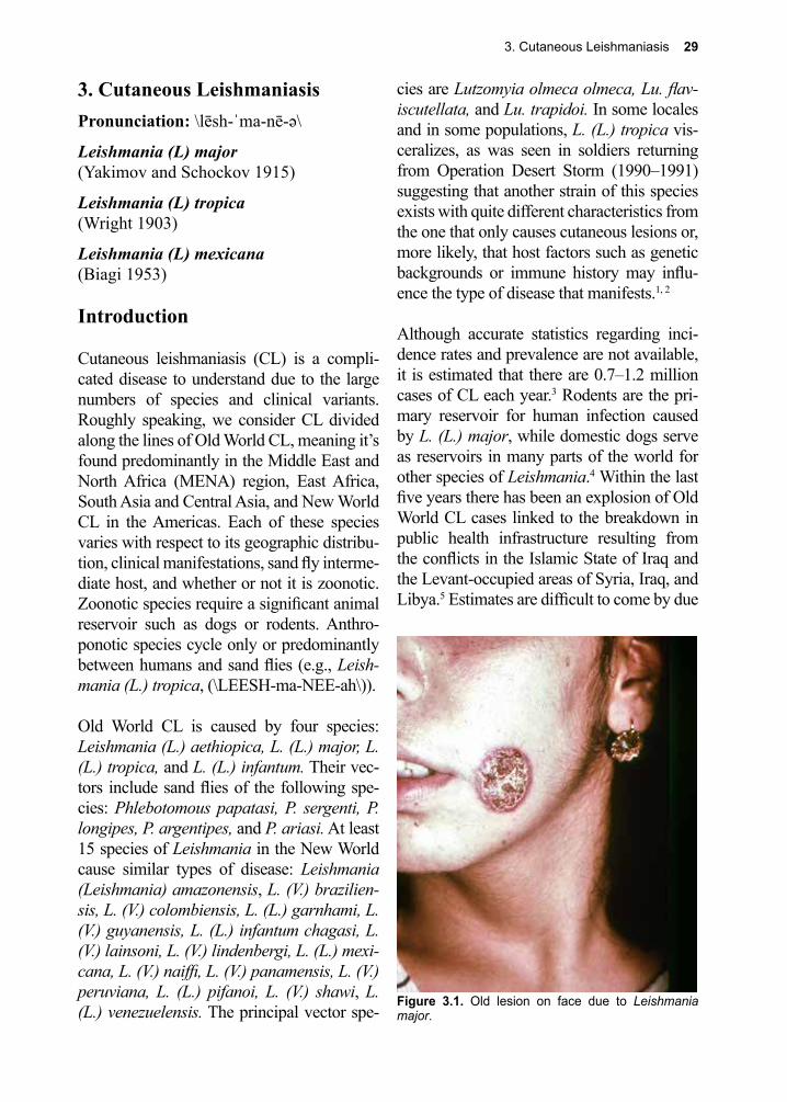
3. Cutaneous Leishmaniasis 29
cies are Lutzomyia olmeca olmeca, Lu. flav-iscutellata, and Lu. trapidoi. In some locales and in some populations, L. (L.) tropica vis-ceralizes, as was seen in soldiers returning from Operation Desert Storm (1990–1991) suggesting that another strain of this species exists with quite different characteristics from the one that only causes cutaneous lesions or, more likely, that host factors such as genetic backgrounds or immune history may influ-ence the type of disease that manifests.1, 2
Although accurate statistics regarding inci-dence rates and prevalence are not available, it is estimated that there are 0.7–1.2 million cases of CL each year.3 Rodents are the pri-mary reservoir for human infection caused by L. (L.) major, while domestic dogs serve as reservoirs in many parts of the world for other species of Leishmania.4 Within the last five years there has been an explosion of Old World CL cases linked to the breakdown in public health infrastructure resulting from the conflicts in the Islamic State of Iraq and the Levant-occupied areas of Syria, Iraq, and Libya.5 Estimates are difficult to come by due
Figure 3.1. Old lesion on face due to Leishmania major.
3. Cutaneous LeishmaniasisPronunciation: \lēsh-ˈma-nē-ə\
Leishmania (L) major(Yakimov and Schockov 1915)
Leishmania (L) tropica(Wright 1903)
Leishmania (L) mexicana(Biagi 1953)
Introduction
Cutaneous leishmaniasis (CL) is a compli-cated disease to understand due to the large numbers of species and clinical variants. Roughly speaking, we consider CL divided along the lines of Old World CL, meaning it’s found predominantly in the Middle East and North Africa (MENA) region, East Africa, South Asia and Central Asia, and New World CL in the Americas. Each of these species varies with respect to its geographic distribu-tion, clinical manifestations, sand fly interme-diate host, and whether or not it is zoonotic. Zoonotic species require a significant animal reservoir such as dogs or rodents. Anthro-ponotic species cycle only or predominantly between humans and sand flies (e.g., Leish-mania (L.) tropica, (\LEESH-ma-NEE-ah\)).
Old World CL is caused by four species: Leishmania (L.) aethiopica, L. (L.) major, L. (L.) tropica, and L. (L.) infantum. Their vec-tors include sand flies of the following spe-cies: Phlebotomous papatasi, P. sergenti, P. longipes, P. argentipes, and P. ariasi. At least 15 species of Leishmania in the New World cause similar types of disease: Leishmania (Leishmania) amazonensis, L. (V.) brazilien-sis, L. (V.) colombiensis, L. (L.) garnhami, L. (V.) guyanensis, L. (L.) infantum chagasi, L. (V.) lainsoni, L. (V.) lindenbergi, L. (L.) mexi-cana, L. (V.) naiffi, L. (V.) panamensis, L. (V.) peruviana, L. (L.) pifanoi, L. (V.) shawi, L. (L.) venezuelensis. The principal vector spe-
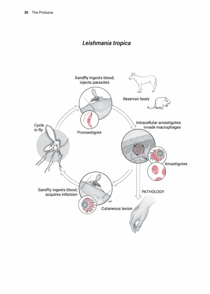
30 The Protozoa
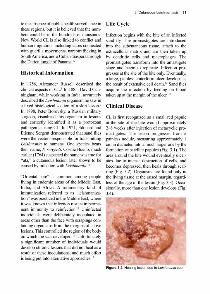
3. Cutaneous Leishmaniasis 31
to the absence of public health surveillance in these regions, but it is believed that the num-bers could be in the hundreds of thousands. New World CL is also linked to conflict and human migrations including cases connected with guerilla movements, narcotrafficking in South America, and a Cuban diaspora through the Darien jungle of Panama.6, 7
Historical Information
In 1756, Alexander Russell described the clinical aspects of CL.8 In 1885, David Cun-ningham, while working in India, accurately described the Leishmania organism he saw in a fixed histological section of a skin lesion.9 In 1898, Peter Borovsky, a Russian military surgeon, visualized this organism in lesions and correctly identified it as a protozoan pathogen causing CL. In 1921, Edouard and Etienne Sergent demonstrated that sand flies were the vectors responsible for transmitting Leishmania to humans. One species bears their name, P. sergenti. Cosme Bueno, much earlier (1764) suspected the same was true for “uta,” a cutaneous lesion, later shown to be caused by infection with Leishmania.10
“Oriental sore” is common among people living in endemic areas of the Middle East, India, and Africa. A rudimentary kind of immunization referred to as “leishmaniza-tion” was practiced in the Middle East, where it was known that infection results in perma-nent immunity to reinfection.11 Uninfected individuals were deliberately inoculated in areas other than the face with scrapings con-taining organisms from the margins of active lesions. This controlled the region of the body on which the scar developed.12 Unfortunately a significant number of individuals would develop chronic lesions that did not heal as a result of these inoculations, and much effort is being put into alternative approaches.13
Life Cycle
Infection begins with the bite of an infected sand fly. The promastigotes are introduced into the subcutaneous tissue, attach to the extracellular matrix and are then taken up by dendritic cells and macrophages. The promastigotes transform into the amastigote stage and begin to replicate. Infection pro-gresses at the site of the bite only. Eventually, a large, painless craterform ulcer develops as the result of extensive cell death.14 Sand flies acquire the infection by feeding on blood taken up at the margin of the ulcer. 15
Clinical Disease
CL is first recognized as a small red papule at the site of the bite wound approximately 2–8 weeks after injection of metacyclic pro-mastigotes. The lesion progresses from a painless nodule, measuring approximately 1 cm in diameter, into a much larger one by the formation of satellite papules (Fig. 3.1). The area around the bite wound eventually ulcer-ates due to intense destruction of cells, and becomes depressed, then heals through scar-ring (Fig. 3.2). Organisms are found only in the living tissue at the raised margin, regard-less of the age of the lesion (Fig. 3.3). Occa-sionally, more than one lesion develops (Fig. 3.4).
Figure 3.2. Healing lesion due to Leishmania spp.
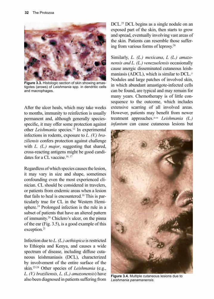
32 The Protozoa
Figure 3.4. Multiple cutaneous lesions due to Leishmania panamanensis.
After the ulcer heals, which may take weeks to months, immunity to reinfection is usually permanent and, although generally species-specific, it may offer some protection against other Leishmania species.12 In experimental infections in rodents, exposure to L. (V.) bra-ziliensis confers protection against challenge with L. (L.) major, suggesting that shared, cross-reacting antigens might be good candi-dates for a CL vaccine.16, 17
Regardless of which species causes the lesion, it may vary in size and shape, sometimes confounding even the most experienced cli-nician. CL should be considered in travelers, or patients from endemic areas when a lesion that fails to heal is encountered.18 This is par-ticularly true for CL in the Western Hemi-sphere.19 Prolonged infection is the rule in a subset of patients that have an altered pattern of immunity.20 Chiclero’s ulcer, on the pinna of the ear (Fig. 3.5), is a good example of this exception.21
Infection due to L. (L.) aethiopica is restricted to Ethiopia and Kenya, and causes a wide spectrum of disease, including diffuse cuta-neous leishmaniasis (DCL), characterized by involvement of the entire surface of the skin.22-24 Other species of Leishmania (e.g., L. (V.) braziliensis, L. (L.) amazonensis) have also been diagnosed in patients suffering from
DCL.25 DCL begins as a single nodule on an exposed part of the skin, then starts to grow and spread, eventually involving vast areas of the skin. Patients can resemble those suffer-ing from various forms of leprosy.26
Similarly, L. (L.) mexicana, L (L.) amazo-nensis and L. (L.) venezuelensis occasionally cause anergic disseminated cutaneous leish-maniasis (ADCL), which is similar to DCL.27 Nodules and large patches of involved skin, in which abundant amastigote-infected cells can be found, are typical and may remain for many years. Chemotherapy is of little con-sequence to the outcome, which includes extensive scarring of all involved areas. However, patients may benefit from newer treatment approaches.28-30 Leishmania (L.) infantum can cause cutaneous lesions but
Figure 3.3. Histologic section of skin showing amas-tigotes (arows) of Leishmania spp. in dendritic cells and macrophages.
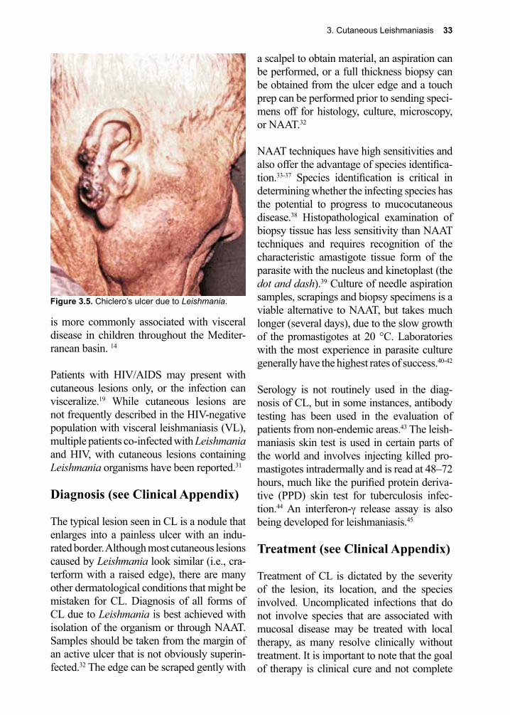
3. Cutaneous Leishmaniasis 33
Figure 3.5. Chiclero’s ulcer due to Leishmania.
a scalpel to obtain material, an aspiration can be performed, or a full thickness biopsy can be obtained from the ulcer edge and a touch prep can be performed prior to sending speci-mens off for histology, culture, microscopy, or NAAT.32
NAAT techniques have high sensitivities and also offer the advantage of species identifica-tion.33-37 Species identification is critical in determining whether the infecting species has the potential to progress to mucocutaneous disease.38 Histopathological examination of biopsy tissue has less sensitivity than NAAT techniques and requires recognition of the characteristic amastigote tissue form of the parasite with the nucleus and kinetoplast (the dot and dash).39 Culture of needle aspiration samples, scrapings and biopsy specimens is a viable alternative to NAAT, but takes much longer (several days), due to the slow growth of the promastigotes at 20 °C. Laboratories with the most experience in parasite culture generally have the highest rates of success.40-42
Serology is not routinely used in the diag-nosis of CL, but in some instances, antibody testing has been used in the evaluation of patients from non-endemic areas.43 The leish-maniasis skin test is used in certain parts of the world and involves injecting killed pro-mastigotes intradermally and is read at 48–72 hours, much like the purified protein deriva-tive (PPD) skin test for tuberculosis infec-tion.44 An interferon-g release assay is also being developed for leishmaniasis.45
Treatment (see Clinical Appendix)
Treatment of CL is dictated by the severity of the lesion, its location, and the species involved. Uncomplicated infections that do not involve species that are associated with mucosal disease may be treated with local therapy, as many resolve clinically without treatment. It is important to note that the goal of therapy is clinical cure and not complete
is more commonly associated with visceral disease in children throughout the Mediter-ranean basin. 14
Patients with HIV/AIDS may present with cutaneous lesions only, or the infection can visceralize.19 While cutaneous lesions are not frequently described in the HIV-negative population with visceral leishmaniasis (VL), multiple patients co-infected with Leishmania and HIV, with cutaneous lesions containing Leishmania organisms have been reported.31
Diagnosis (see Clinical Appendix)
The typical lesion seen in CL is a nodule that enlarges into a painless ulcer with an indu-rated border. Although most cutaneous lesions caused by Leishmania look similar (i.e., cra-terform with a raised edge), there are many other dermatological conditions that might be mistaken for CL. Diagnosis of all forms of CL due to Leishmania is best achieved with isolation of the organism or through NAAT. Samples should be taken from the margin of an active ulcer that is not obviously superin-fected.32 The edge can be scraped gently with
34 The Protozoa
clearance of parasites, as some parasites will persist even with successful therapy.46, 47 In the majority of cases spontaneous healing will occur with proper hygiene and wound care.48, 49
Cryotherapy using liquid nitrogen has been successfully employed.50-52 Treatment with thermotherapy delivered by radiofrequency, superficial heat at 50 °C, or application of heated prongs can result in clinical cure but must be used with caution to avoid nerve damage and burns.53, 54 Topical paromomy-cin and intra-lesional sodium stibogluco-nate (pentavalent antimony) are alternative but less-studied approaches. Intra-lesional injections of pentavalent antimony are not approved in the United States and are associ-ated with significant pain.55-57
Several systemic therapies are in use for the treatment of CL including: pentavalent anti-monials (sodium stibogluconate or meglu-mine antimoniate), amphotericin, miltefos-ine, pentamidine, and azole drugs.58 Systemic therapy is recommended if there is concern that the infecting species has the ability to cause mucosal disease, based on species identification or acquisition of the infection in what is referred to as the mucosal belt (Brazil, Bolivia, and portions of Peru).59 Sodium sti-bogluconate, an antimony-containing drug with many serious side effects, including rash, headache, arthralgias and myalgias, pancreatitis, transaminitis, and hematologic suppression, is the drug of choice in many parts of the world, in part due to its relatively low cost, but increasing rates of resistance has already limited its use.60 Amphotericin B and its liposomal form are alternative and highly effective options for treatment.61 In the developed world liposomal amphoteri-cin B is a frequent choice of therapy but its cost is prohibitive in many areas of the world where this disease is endemic. Miltefosine is an effective but expensive option that allows for systemic therapy, its oral dosing is cur-rently FDA approved in the United States for
treatment of leishmaniasis.29, 62-65 Pentamidine is a little-used therapy owing to concerning side effects and limited data on its efficacy.66 Azoles such as, ketoconazole, fluconazole, itraconazole, and posaconazole are alterna-tive therapies, but ones that show variable efficacy compared to other options based on the particular species of Leishmania parasite involved.55, 67-73
Prevention and Control
Since humans are a dead-end host in most cases, treatment of infected humans is not expected to have a significant impact on dis-ease transmission.74 Although dogs have been identified as a significant reservoir, infection in multiple mammals, both wild and domes-ticated, is not observed.75 Eradication of sand fly breeding sites near suburban and urban centers, attaching pyrethroid-impregnated collars to domestic dogs, and sleeping under insecticide-impregnated bed netting are cost-effective control measures.55 Sand flies only bite at certain times of the day in what is termed a crepuscular/nocturnal pattern: in the morning, late in the evening, and at night.76 Although avoidance of outdoor activities during these times reduces the chance of being bitten, this can be difficult advice to follow for travelers to endemic areas, who may have limited time to accomplish what they have on their itinerary and perhaps impossible for long-term residents who do not have the luxury to adjust their schedule. Additional, and perhaps more viable, approaches to avoid sand fly bites in high-risk areas are the use of insect repellents and insecticide-treated cloth-ing that covers most of the body.77, 78
Vaccines, employing combinations of para-site antigens, offer hope that a standard vac-cine for use against both CL and VL may eventually be available, but no human vac-cine is currently available.17, 79 A recent health economic study confirmed the cost-effective-ness of a CL vaccine.80
3. Cutaneous Leishmaniasis 35
References
1. Berman, J., Recent Developments in Leishmaniasis: Epidemiology, Diagnosis, and Treatment. Current infectious disease reports 2005, 7 (1), 33-38.
2. Magill, A. J., et al., Visceral infection caused by Leishmania tropica in veterans of Operation Desert Storm. N Engl J Med 1993, 328 (19), 1383-7.
3. Alvar, J., et al., Leishmaniasis worldwide and global estimates of its incidence. PLoS One 2012, 7 (5), e35671.
4. Rassi, Y., et al., Vectors and reservoirs of cutaneous leishmaniasis in Marvdasht district, southern Islamic Republic of Iran. East Mediterr Health J 2007, 13 (3), 686-93.
5. Hotez, P. J., Vaccine Science Diplomacy: Expanding Capacity to Prevent Emerging and Neglected Tropical Diseases Arising from Islamic State (IS)--Held Territories. PLoS Negl Trop Dis 2015, 9 (9), e0003852.
6. Beyrer, C., et al., Neglected diseases, civil conflicts, and the right to health. Lancet 2007, 370 (9587), 619-27.
7. Barry, M. A., et al., Cutaneous leishmaniasis in Cuban immigrants to Texas who traveled through the Darien Jungle, Panama. Am J Trop Med Hyg 2014, 91 (2), 345-7.
8. Russell, A.; Millar, A., Natural History of Aleppo and Parts Adjacent. 1856; p 262-266.9. Cunningham, D. D., On the presence of peculiar parasitic organisms in the tissue of a specimen
of Delhi boil. Sci Mem Med Officers Army India 1885, 1 21-31.10. Theodorides, J., Note historique sur la decouverte de la transmission de la leishmaniose cutanee
par les phlebotomes. Bull Soc Pathol Exot 1997, 90, 177-180.11. Handman, E., Leishmaniasis: current status of vaccine development. Clinical microbiology
reviews 2001, 14 (2), 229-43.12. McCall, L. I., et al., Leishmanization revisited: immunization with a naturally attenuated
cutaneous Leishmania donovani isolate from Sri Lanka protects against visceral leishmaniasis. Vaccine 2013, 31 (10), 1420-5.
13. Greenblatt, C. L., The present and future of vaccination for cutaneous leishmaniasis. Prog Clin Biol Res 1980, 47, 259-85.
14. del Giudice, P., et al., Cutaneous leishmaniasis due to Leishmania infantum. Case reports and literature review. Archives of dermatology 1998, 134 (2), 193-8.
15. Bates, P. A., Transmission of Leishmania metacyclic promastigotes by Phlebotomine sand flies. Int J Parasitol 2007, 37 (10), 1097-106.
16. Lima, H. C., et al., Resolution of an infection with Leishmania braziliensis confers complete protection to a subsequent challenge with Leishmania major in BALB/c mice. Memorias do Instituto Oswaldo Cruz 1999, 94 (1), 71-6.
17. Coler, R. N.; Reed, S. G., Second-generation vaccines against leishmaniasis. Trends in parasitology 2005, 21 (5), 244-9.
18. Mansueto, P., et al., Leishmaniasis in travelers: a literature review. Travel Med Infect Dis 2014, 12 (6 Pt A), 563-81.
19. Lainson, R., et al., New World Leishmaniasis-The Neotropical Leishmania Species. 2005.20. van Griensven, J., et al., Leishmaniasis in immunosuppressed individuals. Clin Microbiol Infect
2014, 20 (4), 286-99.21. Lianson, R.; Strangeways-Dixon, J., Leishmania mexicana: The epidemiology of dermal
Leishmaniasis in British Honduras. Trasn Roy Soc Trop Med Hyg 1963, 57, 242-265.22. Hailu, A., The use of direct agglutination test (DAT) in serological diagnosis of Ethiopian
cutaneous leishmaniasis. Diagnostic microbiology and infectious disease 2002, 42 (4), 251-6.23. Akuffo, H., et al., Leishmania aethiopica derived from diffuse leishmaniasis patients
preferentially induce mRNA for interleukin-10 while those from localized leishmaniasis patients induce interferon-gamma. The Journal of infectious diseases 1997, 175 (3), 737-41.
24. Bryceson, A. D., Diffuse cutaneous leishmaniasis in Ethiopia. I. The clinical and histological features of the disease. Transactions of the Royal Society of Tropical Medicine and Hygiene 1969, 63 (6), 708-37.
25. Turetz, M. L., et al., Disseminated leishmaniasis: a new and emerging form of leishmaniasis
36 The Protozoa
observed in northeastern Brazil. The Journal of infectious diseases 2002, 186 (12), 1829-34.26. Dassoni, F., et al., Cutaneous and mucocutaneous leishmaniasis resembling borderline-
tuberculoid leprosy: a new clinical presentation? Acta Derm Venereol 2013, 93 (1), 74-7.27. Bonfante-Garrido, R., et al., Disseminated American cutaneous leishmaniasis. International
journal of dermatology 1996, 35 (8), 561-5.28. Sharquie, K. E.; Najim, R. A., Disseminated cutaneous leishmaniasis. Saudi medical journal
2004, 25 (7), 951-4.29. Machado, P. R.; Penna, G., Miltefosine and cutaneous leishmaniasis. Curr Opin Infect Dis 2012,
25 (2), 141-4.30. Ordaz-Farias, A., et al., Case report: Transient success using prolonged treatment with
miltefosine for a patient with diffuse cutaneous leishmaniasis infected with Leishmania mexicana mexicana. Am J Trop Med Hyg 2013, 88 (1), 153-6.
31. Postigo, C., et al., Cutaneous lesions in patients with visceral leishmaniasis and HIV infection. The Journal of infection 1997, 35 (3), 265-8.
32. Handler, M. Z., et al., Cutaneous and mucocutaneous leishmaniasis: Differential diagnosis, diagnosis, histopathology, and management. J Am Acad Dermatol 2015, 73 (6), 911-26.
33. Mimori, T., et al., Rapid identification of Leishmania species from formalin-fixed biopsy samples by polymorphism-specific polymerase chain reaction. Gene 1998, 210 (2), 179-86.
34. Gangneux, J.-P., et al., Prospective value of PCR amplification and sequencing for diagnosis and typing of old world Leishmania infections in an area of nonendemicity. Journal of clinical microbiology 2003, 41 (4), 1419-22.
35. Mouttaki, T., et al., Molecular diagnosis of cutaneous leishmaniasis and identification of the causative Leishmania species in Morocco by using three PCR-based assays. Parasit Vectors 2014, 7, 420.
36. Nzelu, C. O., et al., A rapid molecular diagnosis of cutaneous leishmaniasis by colorimetric malachite green-loop-mediated isothermal amplification (LAMP) combined with an FTA card as a direct sampling tool. Acta Trop 2016, 153, 116-9.
37. Yehia, L., et al., Molecular diagnosis of cutaneous leishmaniasis and species identification: analysis of 122 biopsies with varied parasite index. J Cutan Pathol 2012, 39 (3), 347-55.
38. Palumbo, E., Treatment strategies for mucocutaneous leishmaniasis. J Glob Infect Dis 2010, 2 (2), 147-50.
39. Faber, W. R., et al., Value of diagnostic techniques for cutaneous leishmaniasis. Journal of the American Academy of Dermatology 2003, 49 (1), 70-4.
40. Dey, T., et al., Infectivity and virulence of Leishmania donovani promastigotes: a role for media, source, and strain of parasite. The Journal of eukaryotic microbiology 2002, 49 (4), 270-4.
41. Luz, Z. M., et al., Lesion aspirate culture for the diagnosis and isolation of Leishmania spp. from patients with cutaneous leishmaniasis. Mem Inst Oswaldo Cruz 2009, 104 (1), 62-6.
42. Castelli, G., et al., Evaluation of two modified culture media for Leishmania infantum cultivation versus different culture media. J Parasitol 2014, 100 (2), 228-30.
43. Szargiki, R., et al., Comparison of serological and parasitological methods for cutaneous leishmaniasis diagnosis in the state of Parana, Brazil. Braz J Infect Dis 2009, 13 (1), 47-52.
44. Skraba, C. M., et al., Evaluation of the reference value for the Montenegro skin test. Rev Soc Bras Med Trop 2015, 48 (4), 437-44.
45. Turgay, N., et al., Quantiferon-Leishmania as an epidemiological tool for evaluating the exposure to Leishmania infection. Am J Trop Med Hyg 2010, 83 (4), 822-4.
46. Bogdan, C., et al., Invasion, control and persistence of Leishmania parasites. Curr Opin Immunol 1996, 8 (4), 517-25.
47. Mendonca, M. G., et al., Persistence of Leishmania parasites in scars after clinical cure of American cutaneous leishmaniasis: is there a sterile cure? J Infect Dis 2004, 189 (6), 1018-23.
48. Willard, R. J., et al., Cutaneous leishmaniasis in soldiers from Fort Campbell, Kentucky returning from Operation Iraqi Freedom highlights diagnostic and therapeutic options. J Am Acad Dermatol 2005, 52 (6), 977-87.
49. Morizot, G., et al., Travelers with cutaneous leishmaniasis cured without systemic therapy. Clin
3. Cutaneous Leishmaniasis 37
Infect Dis 2013, 57 (3), 370-80.50. al-Majali, O., et al., A 2-year study of liquid nitrogen therapy in cutaneous leishmaniasis. Int J
Dermatol 1997, 36 (6), 460-2.51. Dobrev, H. P., et al., Cutaneous Leishmaniasis - Dermoscopic Findings And Cryotherapy. Folia
Med (Plovdiv) 2015, 57 (1), 65-8.52. Farajzadeh, S., et al., Comparison between Combination Therapy of Oral Terbinafine and
Cryotherapy versus Systemic Meglumine Antimoniate and Cryotherapy in Cutaneous Leishmaniasis: A Randomized Clinical Trial. Iran J Parasitol 2015, 10 (1), 1-8.
53. Pizinger, K., et al., Successful treatment of cutaneous leishmaniasis by photodynamic therapy and cryotherapy. Eur J Dermatol 2009, 19 (2), 172-3.
54. Sbeghen, M. R., et al., Topical and intradermal efficacy of photodynamic therapy with methylene blue and light-emitting diode in the treatment of cutaneous leishmaniasis caused by Leishmania braziliensis. J Lasers Med Sci 2015, 6 (3), 106-11.
55. Shazad, B., et al., Comparison of topical paromomycin sulfate (twice/day) with intralesional meglumine antimoniate for the treatment of cutaneous leishmaniasis caused by L. major. European journal of dermatology : EJD 2005, 15 (2), 85-7.
56. Soto, J., et al., Intralesional antimony for single lesions of bolivian cutaneous leishmaniasis. Clin Infect Dis 2013, 56 (9), 1255-60.
57. Shin, J. Y., et al., New world cutaneous leishmaniasis treated with intralesional injection of pentavalent antimony. Ann Dermatol 2013, 25 (1), 80-3.
58. Singh, N., et al., Leishmaniasis: current status of available drugs and new potential drug targets. Asian Pac J Trop Med 2012, 5 (6), 485-97.
59. Hodiamont, C. J., et al., Species-directed therapy for leishmaniasis in returning travellers: a comprehensive guide. PLoS Negl Trop Dis 2014, 8 (5), e2832.
60. Aronson, N. E., et al., Safety and efficacy of intravenous sodium stibogluconate in the treatment of leishmaniasis: recent U.S. military experience. Clin Inf Dis 1998, 27 (6), 1457-64.
61. Sundar, S.; Chakravarty, J., Liposomal amphotericin B and leishmaniasis: dose and response. J Glob Infect Dis 2010, 2 (2), 159-66.
62. Madke, B., et al., Successful treatment of multifocal cutaneous leishmaniasis with miltefosine. Indian J Dermatol 2011, 56 (5), 587-90.
63. Rubiano, L. C., et al., Noninferiority of miltefosine versus meglumine antimoniate for cutaneous leishmaniasis in children. J Infect Dis 2012, 205 (4), 684-92.
64. Monge-Maillo, B.; Lopez-Velez, R., Miltefosine for visceral and cutaneous leishmaniasis: drug characteristics and evidence-based treatment recommendations. Clin Infect Dis 2015, 60 (9), 1398-404.
65. Dorlo, T. P., et al., Dynamics of parasite clearance in cutaneous leishmaniasis patients treated with miltefosine. PLoS Negl Trop Dis 2011, 5 (12), e1436.
66. Jaureguiberry, S., et al., Efficacy of short-course intramuscular pentamidine isethionate treatment on Old World localized cutaneous leishmaniasis in 2 patients. Clin Infect Dis 2006, 42 (12), 1812-3.
67. Laffitte, E., et al., Cutaneous leishmaniasis caused by Leishmania tropica: treatment with oral fluconazole. Dermatology (Basel, Switzerland) 2005, 210 (3), 249-51.
68. Morizot, G., et al., Healing of Old World cutaneous leishmaniasis in travelers treated with fluconazole: drug effect or spontaneous evolution? Am J Trop Med Hyg 2007, 76 (1), 48-52.
69. Saenz, R. E., et al., Efficacy of ketoconazole against Leishmania braziliensis panamensis cutaneous leishmaniasis. Am J Med 1990, 89 (2), 147-55.
70. Firooz, A., et al., Itraconazole in the treatment of cutaneous leishmaniasis. Int J Dermatol 2006, 45 (12), 1446-7.
71. Baroni, A., et al., Cutaneous leishmaniasis treated with itraconazole. Dermatol Ther 2009, 22 Suppl 1, S27-9.
72. Saleem, K.; Rahman, A., Comparison of oral itraconazole and intramuscular meglumine antimoniate in the treatment of cutaneous leishmaniasis. J Coll Physicians Surg Pak 2007, 17 (12), 713-6.
73. Paniz Mondolfi, A. E., et al., Successful treatment of Old World cutaneous leishmaniasis caused
38 The Protozoa
by Leishmania infantum with posaconazole. Antimicrob Agents Chemother 2011, 55 (4), 1774-6.74. Maroli, M.; Khoury, C., Prevention and control of leishmaniasis vectors: current approaches.
Parassitologia 2004, 46 (1-2), 211-5.75. Quinnell, R. J.; Courtenay, O., Transmission, reservoir hosts and control of zoonotic visceral
leishmaniasis. Parasitology 2009, 136 (14), 1915-34.76. Rivas, G. B., et al., Effects of temperature and photoperiod on daily activity rhythms of
Lutzomyia longipalpis (Diptera: Psychodidae). Parasit Vectors 2014, 7, 278.77. Banks, S. D., et al., Insecticide-treated clothes for the control of vector-borne diseases: a review
on effectiveness and safety. Med Vet Entomol 2014, 28 Suppl 1, 14-25.78. Moore, S. J., et al., Insect bite prevention. Infect Dis Clin North Am 2012, 26 (3), 655-73.79. Kumar, R.; Engwerda, C., Vaccines to prevent leishmaniasis. Clin Transl Immunology 2014, 3
(3), e13.80. Bacon, K. M., et al., The potential economic value of a cutaneous leishmaniasis vaccine in seven
endemic countries in the Americas. Vaccine 2013, 31 (3), 480-6.
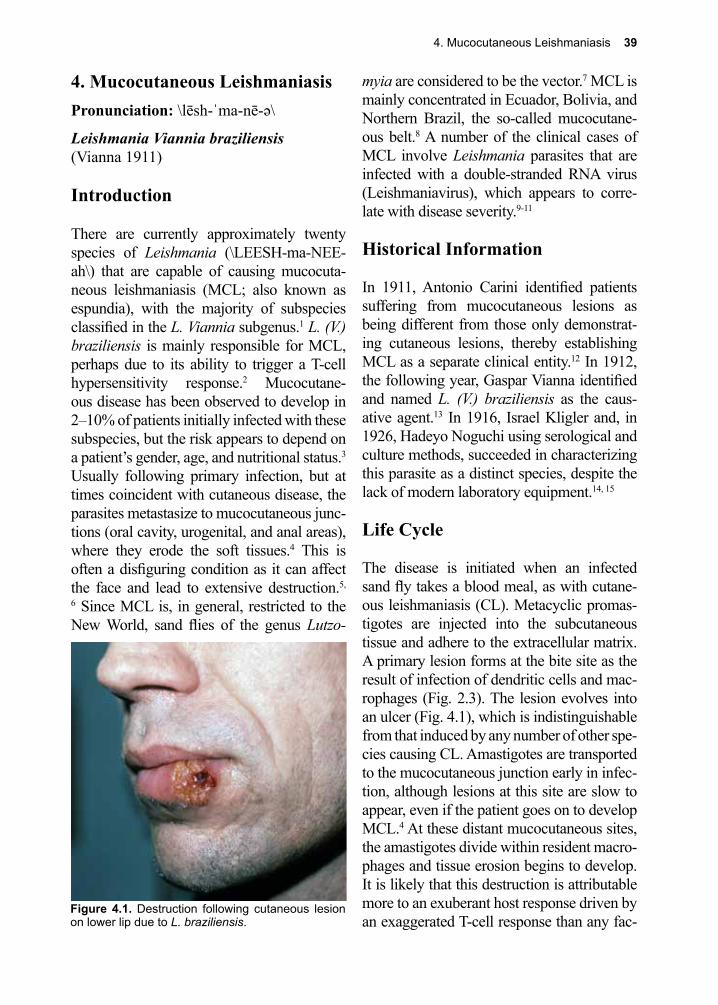
4. Mucocutaneous Leishmaniasis 39
Figure 4.1. Destruction following cutaneous lesion on lower lip due to L. braziliensis.
4. Mucocutaneous LeishmaniasisPronunciation: \lēsh-ˈma-nē-ə\
Leishmania Viannia braziliensis(Vianna 1911)
Introduction
There are currently approximately twenty species of Leishmania (\LEESH-ma-NEE-ah\) that are capable of causing mucocuta-neous leishmaniasis (MCL; also known as espundia), with the majority of subspecies classified in the L. Viannia subgenus.1 L. (V.) braziliensis is mainly responsible for MCL, perhaps due to its ability to trigger a T-cell hypersensitivity response.2 Mucocutane-ous disease has been observed to develop in 2–10% of patients initially infected with these subspecies, but the risk appears to depend on a patient’s gender, age, and nutritional status.3 Usually following primary infection, but at times coincident with cutaneous disease, the parasites metastasize to mucocutaneous junc-tions (oral cavity, urogenital, and anal areas), where they erode the soft tissues.4 This is often a disfiguring condition as it can affect the face and lead to extensive destruction.5,
6 Since MCL is, in general, restricted to the New World, sand flies of the genus Lutzo-
myia are considered to be the vector.7 MCL is mainly concentrated in Ecuador, Bolivia, and Northern Brazil, the so-called mucocutane-ous belt.8 A number of the clinical cases of MCL involve Leishmania parasites that are infected with a double-stranded RNA virus (Leishmaniavirus), which appears to corre-late with disease severity.9-11
Historical Information
In 1911, Antonio Carini identified patients suffering from mucocutaneous lesions as being different from those only demonstrat-ing cutaneous lesions, thereby establishing MCL as a separate clinical entity.12 In 1912, the following year, Gaspar Vianna identified and named L. (V.) braziliensis as the caus-ative agent.13 In 1916, Israel Kligler and, in 1926, Hadeyo Noguchi using serological and culture methods, succeeded in characterizing this parasite as a distinct species, despite the lack of modern laboratory equipment.14, 15
Life Cycle
The disease is initiated when an infected sand fly takes a blood meal, as with cutane-ous leishmaniasis (CL). Metacyclic promas-tigotes are injected into the subcutaneous tissue and adhere to the extracellular matrix. A primary lesion forms at the bite site as the result of infection of dendritic cells and mac-rophages (Fig. 2.3). The lesion evolves into an ulcer (Fig. 4.1), which is indistinguishable from that induced by any number of other spe-cies causing CL. Amastigotes are transported to the mucocutaneous junction early in infec-tion, although lesions at this site are slow to appear, even if the patient goes on to develop MCL.4 At these distant mucocutaneous sites, the amastigotes divide within resident macro-phages and tissue erosion begins to develop. It is likely that this destruction is attributable more to an exuberant host response driven by an exaggerated T-cell response than any fac-
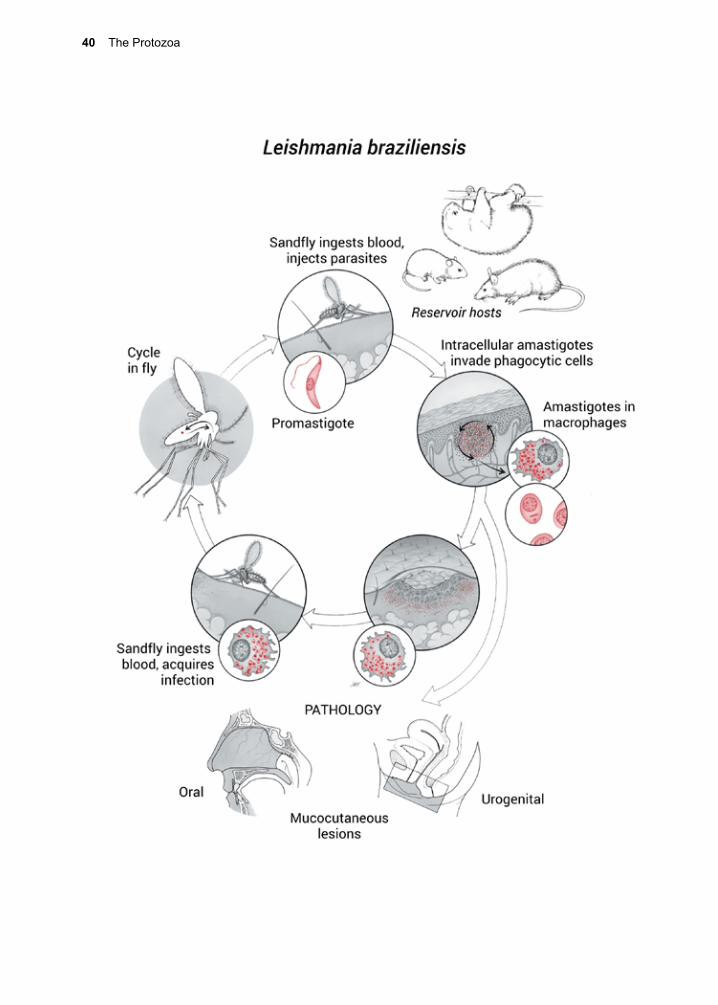
40 The Protozoa
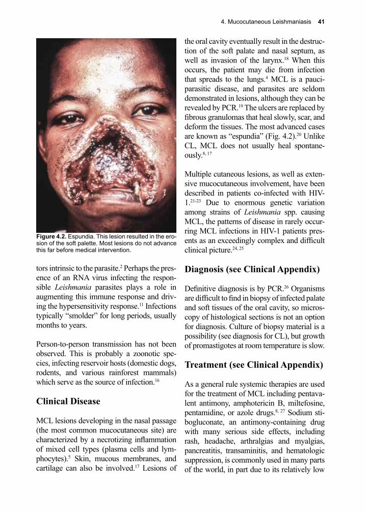
4. Mucocutaneous Leishmaniasis 41
Figure 4.2. Espundia. This lesion resulted in the ero-sion of the soft palette. Most lesions do not advance this far before medical intervention.
tors intrinsic to the parasite.2 Perhaps the pres-ence of an RNA virus infecting the respon-sible Leishmania parasites plays a role in augmenting this immune response and driv-ing the hypersensitivity response.11 Infections typically “smolder” for long periods, usually months to years.
Person-to-person transmission has not been observed. This is probably a zoonotic spe-cies, infecting reservoir hosts (domestic dogs, rodents, and various rainforest mammals) which serve as the source of infection.16
Clinical Disease
MCL lesions developing in the nasal passage (the most common mucocutaneous site) are characterized by a necrotizing inflammation of mixed cell types (plasma cells and lym-phocytes).5 Skin, mucous membranes, and cartilage can also be involved.17 Lesions of
the oral cavity eventually result in the destruc-tion of the soft palate and nasal septum, as well as invasion of the larynx.18 When this occurs, the patient may die from infection that spreads to the lungs.4 MCL is a pauci-parasitic disease, and parasites are seldom demonstrated in lesions, although they can be revealed by PCR.19 The ulcers are replaced by fibrous granulomas that heal slowly, scar, and deform the tissues. The most advanced cases are known as “espundia” (Fig. 4.2).20 Unlike CL, MCL does not usually heal spontane-ously.8, 17
Multiple cutaneous lesions, as well as exten-sive mucocutaneous involvement, have been described in patients co-infected with HIV-1.21-23 Due to enormous genetic variation among strains of Leishmania spp. causing MCL, the patterns of disease in rarely occur-ring MCL infections in HIV-1 patients pres-ents as an exceedingly complex and difficult clinical picture.24, 25
Diagnosis (see Clinical Appendix)
Definitive diagnosis is by PCR.26 Organisms are difficult to find in biopsy of infected palate and soft tissues of the oral cavity, so micros-copy of histological sections is not an option for diagnosis. Culture of biopsy material is a possibility (see diagnosis for CL), but growth of promastigotes at room temperature is slow.
Treatment (see Clinical Appendix)
As a general rule systemic therapies are used for the treatment of MCL including pentava-lent antimony, amphotericin B, miltefosine, pentamidine, or azole drugs.8, 27 Sodium sti-bogluconate, an antimony-containing drug with many serious side effects, including rash, headache, arthralgias and myalgias, pancreatitis, transaminitis, and hematologic suppression, is commonly used in many parts of the world, in part due to its relatively low
42 The Protozoa
cost.28 Amphotericin B and its liposomal form are highly effective options for treatment and many now consider the liposomal form the drug of choice for MCL as well as visceral leishmaniasis (VL). In the developing world, standard amphotericin is often employed for the treatment of MCL if antimonial therapy fails. Miltefosine is an effective but expensive option that allows for systemic therapy with oral dosing that is currently FDA approved in the United States for treatment of leishmani-asis.29-33 Pentamidine is another option that has demonstrated efficacy in the treatment of MCL.34, 35 Azoles such as, ketoconazole, fluconazole, itraconazole, and posaconazole are alternative oral therapies with variable efficacy compared to other options based on the particular species of Leishmania parasite involved.36-43
None of the above therapies is 100% effec-tive at eliminating all parasites in all patients. Persistence of infection beyond the treatment period has been recorded in longstanding infections, as demonstrated by PCR on exper-imentally infected monkeys and in follow up of some patients.44
Prevention and Control
Transmission of L. (V.) braziliensis and related species capable of causing MCL occurs through close contact of human popu-lations with reservoir hosts (wild and domes-tic mammals). Deforestation of vast regions of rainforest for agriculture, mining, and oil exploration, all of which leads to increased urbanization in South and Central America, has exposed large numbers of people to rain-forest edges. Edges of natural systems are special ecologically defined areas termed ecotones.34, 45 These zones constitute the bor-ders between ecosystems, and are further characterized by ecological stress on all plant and animal species in that zone due to intra and inter-specific competition for resources.
Distribution of sand fly species is determined by the availability of natural habitat and of that created by human encroachment. Some species favor unaltered forest, while others thrive in ecologically disturbed situations.34
It is unfortunately beyond the scope of this text to illustrate this concept with specific examples, but many could be given that would serve to reinforce the idea that when ecological change due to human settlement occurs, those living there are at greater risk from acquiring zoonotic infectious diseases (e.g., Yellow fever virus, Ebola virus, Mar-burg virus, Lassa fever virus).45 The number of species of Leishmania known to infect humans throughout South America alone has increased dramatically. 4 Avoidance of contact with vectors of MCL is an ineffective recommendation, given the lack of awareness of their presence on the part of those living in close proximity to them. For those traveling to endemic areas wishing to avoid the bites of sand flies, the following advice may be helpful. Sand flies are more prevalent during the rainy season.46 Sand flies only bite at certain times of the day in what is termed a crepuscular/noctur-nal pattern: in the morning, late in the eve-ning, and at night.7 Although avoidance of outdoor activities during these times reduces the chance of being bitten this can be dif-ficult advice for travelers to endemic areas, who may have limited time to accomplish what they have on their itinerary and per-haps impossible for long-term residents who do not have the luxury to adjust their sched-ule. Additional and perhaps more viable approaches to avoid sand fly bites that are in use by travelers and workers in high-risk areas are the use of insect repellents and insecticide-treated clothing that covers most of the body.47, 48 Growing rates of insecticide resistance in sand fly populations are already limiting the utility of this approach in many parts of the world.
4. Mucocutaneous Leishmaniasis 43
The use of insecticide-impregnated cloth-ing and bed netting has helped reduce dis-ease transmission for malaria, particularly in Africa, and has now been proven effective in reducing the transmission of Leishmania as well. Maintaining wide buffer zones between settlements and the surrounding native forest appears to be the best environmentally based long-term solution to controlling the inci-dence of infection in endemic transmission
zones, a recommendation originally made to those wishing to avoid becoming infected with the Yellow fever virus. However, curtail-ing this human imperative has, so far, been all but impossible to enforce.
Vaccines, using parasite antigens offer hope, but no human vaccine is currently available, despite continued efforts.49, 50
References
1. Herwaldt, B. L., Leishmaniasis. Lancet (London, England) 1999, 354 (9185), 1191-9.2. Silveira, F. T., et al., Immunopathogenic competences of Leishmania (V.) braziliensis and L. (L.)
amazonensis in American cutaneous leishmaniasis. Parasite Immunol 2009, 31 (8), 423-31.3. Machado-Coelho, G. L., et al., Risk factors for mucosal manifestation of American cutaneous
leishmaniasis. Trans R Soc Trop Med Hyg 2005, 99 (1), 55-61.4. Lainson, R., et al., New World Leishmaniasis-The Neotropical Leishmania Species. 2005.5. Crovetto-Martinez, R., et al., Mucocutaneous leishmaniasis must be included in the differential
diagnosis of midline destructive disease: two case reports. Oral Surg Oral Med Oral Pathol Oral Radiol 2015, 119 (1), e20-6.
6. Garcia, A. L., et al., American tegumentary leishmaniasis: antigen-gene polymorphism, taxonomy and clinical pleomorphism. Infection, genetics and evolution: journal of molecular epidemiology and evolutionary genetics in infectious diseases 2005, 5 (2), 109-16.
7. Rivas, G. B., et al., Effects of temperature and photoperiod on daily activity rhythms of Lutzomyia longipalpis (Diptera: Psychodidae). Parasit Vectors 2014, 7, 278.
8. Palumbo, E., Treatment strategies for mucocutaneous leishmaniasis. J Glob Infect Dis 2010, 2 (2), 147-50.
9. Chung, I. K., et al., Generation of the short RNA transcript in Leishmaniavirus correlates with the growth of its parasite host, Leishmania. Molecules and cells 1998, 8 (1), 54-61.
10. Ogg, M. M., et al., Short report: quantification of leishmaniavirus RNA in clinical samples and its possible role in pathogenesis. The American journal of tropical medicine and hygiene 2003, 69 (3), 309-13.
11. Ives, A., et al., Leishmania RNA virus controls the severity of mucocutaneous leishmaniasis. Science 2011, 331 (6018), 775-8.
12. Carini, A., Leishmaniose de Ia muqucuse rhinobuccopharyngee. Bull Soc Pathol Exot 1911, 4 289-291.
13. Vianna, G., Tratamento da leishmaniose tegumentar por injecoes intravenososas de tartaro emetico. Ann Congress Brasil Med Cirur 1912, 4 426-428.
14. Kligler, I. J., Some Regulating Factors in Bacterial Metabolism. Journal of bacteriology 1916, 1 (6), 663-71.
15. Noguchi, H.; S., Comparative studies on herpetomonads and Leishmaniasis. II Differentiation of the organisms by serological reactions and fermentation tests Med 1929, 44, 305-314.
16. Alexander, B., et al., Detection of Leishmania (Viannia) braziliensis complex in wild mammals from Colombian coffee plantations by PCR and DNA hybridization. Acta tropica 1998, 69 (1), 41-50.
17. Ahluwalia, S., et al., Mucocutaneous leishmaniasis: an imported infection among travellers to central and South America. BMJ 2004, 329 (7470), 842-4.
18. Lohuis, P. J., et al., Leishmania braziliensis presenting as a granulomatous lesion of the nasal septum mucosa. The Journal of laryngology and otology 1997, 111 (10), 973-5.
19. Belli, A., et al., Simplified polymerase chain reaction detection of new world Leishmania in
44 The Protozoa
clinical specimens of cutaneous leishmaniasis. The American journal of tropical medicine and hygiene 1998, 58 (1), 102-9.
20. Marsden, P. D., Mucosal leishmaniasis (“espundia” Escomel, 1911). Transactions of the Royal Society of Tropical Medicine and Hygiene 1986, 80 (6), 859-76.
21. Machado, E. S., et al., Disseminated American muco-cutaneous leishmaniasis caused by Leishmania braziliensis braziliensis in a patient with AIDS: a case report. Memorias do Instituto Oswaldo Cruz 1992, 87 (4), 487-92.
22. Mattos, M., et al., American cutaneous leishmaniasis associated with HIV infection: report of four cases. Journal of the European Academy of Dermatology and Venereology : JEADV 1998, 10 (3), 218-25.
23. Torrico, F., et al., Co-Infection of Leishmania (Viannia) braziliensis and HIV: report of a case of mucosal leishmaniasis in Cochabamba, Bolivia. Am J Trop Med Hyg 2009, 81 (4), 555-8.
24. Da-Cruz, A. M., et al., Atypical mucocutaneous leishmaniasis caused by Leishmania braziliensis in an acquired immunodeficiency syndrome patient: T-cell responses and remission of lesions associated with antigen immunotherapy. Memorias do Instituto Oswaldo Cruz 1999, 94 (4), 537-42.
25. Chouicha, N.; Lanotte, G., Phylogenetic taxonomy of Leishmania (viannia) braziliensis based on isoenzymatic study of 137 isolates. Parasitology 1998, 115, 343-8.
26. Olivier, M., et al., Subversion mechanisms by which Leishmania parasites can escape the host immune response: a signaling point of view. Clinical microbiology reviews 2005, 18 (2), 293-305.
27. Singh, N., et al., Leishmaniasis: current status of available drugs and new potential drug targets. Asian Pac J Trop Med 2012, 5 (6), 485-97.
28. Aronson, N. E., et al., Safety and efficacy of intravenous sodium stibogluconate in the treatment of leishmaniasis: recent U.S. military experience. Clin Inf Dis 1998, 27 (6), 1457-64.
29. Madke, B., et al., Successful treatment of multifocal cutaneous leishmaniasis with miltefosine. Indian J Dermatol 2011, 56 (5), 587-90.
30. Rubiano, L. C., et al., Noninferiority of miltefosine versus meglumine antimoniate for cutaneous leishmaniasis in children. J Infect Dis 2012, 205 (4), 684-92.
31. Monge-Maillo, B.; Lopez-Velez, R., Miltefosine for visceral and cutaneous leishmaniasis: drug characteristics and evidence-based treatment recommendations. Clin Infect Dis 2015, 60 (9), 1398-404.
32. Machado, P. R.; Penna, G., Miltefosine and cutaneous leishmaniasis. Curr Opin Infect Dis 2012, 25 (2), 141-4.
33. Dorlo, T. P., et al., Dynamics of parasite clearance in cutaneous leishmaniasis patients treated with miltefosine. PLoS Negl Trop Dis 2011, 5 (12), e1436.
34. Salomon, O. D., et al., Ecological aspects of phebotomine (Diptera, Psychodidae) in an endemic area of tegumentary leishmaniasis in the northeastern Argentina, 1993-1998. Memorias do Instituto Oswaldo Cruz 2002, 97 (2), 163-8.
35. Jaureguiberry, S., et al., Efficacy of short-course intramuscular pentamidine isethionate treatment on Old World localized cutaneous leishmaniasis in 2 patients. Clin Infect Dis 2006, 42 (12), 1812-3.
36. Laffitte, E., et al., Cutaneous leishmaniasis caused by Leishmania tropica: treatment with oral fluconazole. Dermatology (Basel, Switzerland) 2005, 210 (3), 249-51.
37. Shazad, B., et al., Comparison of topical paromomycin sulfate (twice/day) with intralesional meglumine antimoniate for the treatment of cutaneous leishmaniasis caused by L. major. European journal of dermatology : EJD 2005, 15 (2), 85-7.
38. Morizot, G., et al., Healing of Old World cutaneous leishmaniasis in travelers treated with fluconazole: drug effect or spontaneous evolution? Am J Trop Med Hyg 2007, 76 (1), 48-52.
39. Saenz, R. E., et al., Efficacy of ketoconazole against Leishmania braziliensis panamensis cutaneous leishmaniasis. Am J Med 1990, 89 (2), 147-55.
40. Firooz, A., et al., Itraconazole in the treatment of cutaneous leishmaniasis. Int J Dermatol 2006, 45 (12), 1446-7.
41. Baroni, A., et al., Cutaneous leishmaniasis treated with itraconazole. Dermatol Ther 2009, 22 Suppl 1, S27-9.
4. Mucocutaneous Leishmaniasis 45
42. Saleem, K.; Rahman, A., Comparison of oral itraconazole and intramuscular meglumine antimoniate in the treatment of cutaneous leishmaniasis. J Coll Physicians Surg Pak 2007, 17 (12), 713-6.
43. Paniz Mondolfi, A. E., et al., Successful treatment of Old World cutaneous leishmaniasis caused by Leishmania infantum with posaconazole. Antimicrob Agents Chemother 2011, 55 (4), 1774-6.
44. Osman, O. F., et al., Use of the polymerase chain reaction to assess the success of visceral leishmaniasis treatment. Transactions of the Royal Society of Tropical Medicine and Hygiene 1998, 92 (4), 397-400.
45. Despommier, D., et al., The role of ecotones in emerging infectious diseases. Ecohealth 2006.46. Souza, N. A., et al., Seasonality of Lutzomyia intermedia and Lutzomyia whitmani (Diptera:
Psychodidae: Phlebotominae), occurring sympatrically in area of cutaneous leishmaniasis in the State of Rio de Janeiro, Brazil. Memorias do Instituto Oswaldo Cruz 2002, 97 (6), 759-65.
47. Banks, S. D., et al., Insecticide-treated clothes for the control of vector-borne diseases: a review on effectiveness and safety. Med Vet Entomol 2014, 28 Suppl 1, 14-25.
48. Moore, S. J., et al., Insect bite prevention. Infect Dis Clin North Am 2012, 26 (3), 655-73.49. Coler, R. N.; Reed, S. G., Second-generation vaccines against leishmaniasis. Trends in
parasitology 2005, 21 (5), 244-9.50. Kumar, R.; Engwerda, C., Vaccines to prevent leishmaniasis. Clin Transl Immunology 2014, 3
(3), e13.
Amico Bignami, M.D. (1862–1929)
Bignami (\BEN-yah-meh\) collaborated with Ettore Marchiafava and Giovanni Grassi in 1892 and elucidated the main clinical features of “malignant” malaria (Plasmodium falciparum). Bignami also demonstrated that the “crescent” stage (gametocyte) did not cause illness. In 1898, Bignami, and Grassi worked with Antonio Dionisi and Guiseppe Bastianelli to prove that anopheline mosquitoes transmit human malaria.
46 The Protozoa
Theodor Maximilian Bilharz, M.D. (1825–1862)
Bilharz described the adult stage of Hymenolepis nana and Schistosoma haematobium in human patients that had come to autopsy while he was serving in the German Army medical corps as lieutenant colonel and chief of surgery at Kasr El Aini Hospital in Cairo, Egypt. In collabora-tion with Karl von Siebold, he made the connection between blood in the urine and infection with S. haematobium.
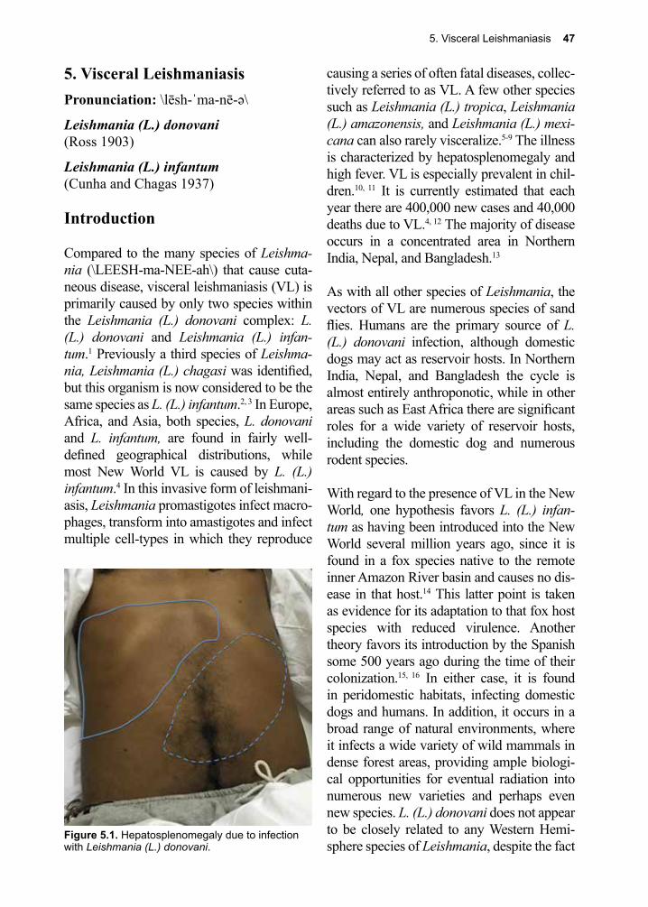
5. Visceral Leishmaniasis 47
Figure 5.1. Hepatosplenomegaly due to infection with Leishmania (L.) donovani.
5. Visceral LeishmaniasisPronunciation: \lēsh-ˈma-nē-ə\
Leishmania (L.) donovani(Ross 1903)
Leishmania (L.) infantum(Cunha and Chagas 1937)
Introduction
Compared to the many species of Leishma-nia (\LEESH-ma-NEE-ah\) that cause cuta-neous disease, visceral leishmaniasis (VL) is primarily caused by only two species within the Leishmania (L.) donovani complex: L. (L.) donovani and Leishmania (L.) infan-tum.1 Previously a third species of Leishma-nia, Leishmania (L.) chagasi was identified, but this organism is now considered to be the same species as L. (L.) infantum.2, 3 In Europe, Africa, and Asia, both species, L. donovani and L. infantum, are found in fairly well-defined geographical distributions, while most New World VL is caused by L. (L.) infantum.4 In this invasive form of leishmani-asis, Leishmania promastigotes infect macro-phages, transform into amastigotes and infect multiple cell-types in which they reproduce
causing a series of often fatal diseases, collec-tively referred to as VL. A few other species such as Leishmania (L.) tropica, Leishmania (L.) amazonensis, and Leishmania (L.) mexi-cana can also rarely visceralize.5-9 The illness is characterized by hepatosplenomegaly and high fever. VL is especially prevalent in chil-dren.10, 11 It is currently estimated that each year there are 400,000 new cases and 40,000 deaths due to VL.4, 12 The majority of disease occurs in a concentrated area in Northern India, Nepal, and Bangladesh.13
As with all other species of Leishmania, the vectors of VL are numerous species of sand flies. Humans are the primary source of L. (L.) donovani infection, although domestic dogs may act as reservoir hosts. In Northern India, Nepal, and Bangladesh the cycle is almost entirely anthroponotic, while in other areas such as East Africa there are significant roles for a wide variety of reservoir hosts, including the domestic dog and numerous rodent species.
With regard to the presence of VL in the New World, one hypothesis favors L. (L.) infan-tum as having been introduced into the New World several million years ago, since it is found in a fox species native to the remote inner Amazon River basin and causes no dis-ease in that host.14 This latter point is taken as evidence for its adaptation to that fox host species with reduced virulence. Another theory favors its introduction by the Spanish some 500 years ago during the time of their colonization.15, 16 In either case, it is found in peridomestic habitats, infecting domestic dogs and humans. In addition, it occurs in a broad range of natural environments, where it infects a wide variety of wild mammals in dense forest areas, providing ample biologi-cal opportunities for eventual radiation into numerous new varieties and perhaps even new species. L. (L.) donovani does not appear to be closely related to any Western Hemi-sphere species of Leishmania, despite the fact

48 The Protozoa
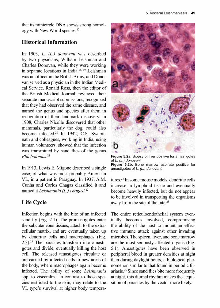
5. Visceral Leishmaniasis 49
Figure 5.2a. Biopsy of liver positive for amastigotes of L. (L.) donovani. Figure 5.2b. Bone marrow aspirate positive for amastigotes of L. (L.) donovani.
that its minicircle DNA shows strong homol-ogy with New World species.17
Historical Information
In 1903, L. (L.) donovani was described by two physicians, William Leishman and Charles Donovan, while they were working in separate locations in India.18, 19 Leishman was an officer in the British Army, and Dono-van served as a physician in the Indian Medi-cal Service. Ronald Ross, then the editor of the British Medical Journal, reviewed their separate manuscript submissions, recognized that they had observed the same disease, and named the genus and species after them in recognition of their landmark discovery. In 1908, Charles Nicolle discovered that other mammals, particularly the dog, could also become infected.20 In 1942, C.S. Swami-nath and colleagues, working in India, using human volunteers, showed that the infection was transmitted by sand flies of the genus Phlebotomus.21
In 1913, Lewis E. Migone described a single case, of what was most probably American VL, in a patient in Paraguay. In 1937, A.M. Cunha and Carlos Chagas classified it and named it Leishmania (L.) chagasi.22
Life Cycle
Infection begins with the bite of an infected sand fly (Fig. 2.1). The promastigotes enter the subcutaneous tissues, attach to the extra-cellular matrix, and are eventually taken up by dendritic cells and macrophages (Fig. 2.3).23 The parasites transform into amasti-gotes and divide, eventually killing the host cell. The released amastigotes circulate or are carried by infected cells to new areas of the body, where macrophages again become infected. The ability of some Leishmania spp. to visceralize, in contrast to those spe-cies restricted to the skin, may relate to the VL type’s survival at higher body tempera-
tures.24 In some mouse models, dendritic cells increase in lymphoid tissue and eventually become heavily infected, but do not appear to be involved in transporting the organisms away from the site of the bite.23
The entire reticuloendothelial system even-tually becomes involved, compromising the ability of the host to mount an effec-tive immune attack against other invading microbes. The spleen, liver, and bone marrow are the most seriously affected organs (Fig. 5.1). Amastigotes have been observed in peripheral blood in greater densities at night than during daylight hours, a biological phe-nomenon similar to that found in periodic fil-ariasis.25 Since sand flies bite more frequently at night, this diurnal rhythm makes the acqui-sition of parasites by the vector more likely.
50 The Protozoa
The cycle is completed when an infected human is bitten by an uninfected sand fly, which ingests macrophages containing the amastigotes or free amastigotes in the blood.25 The duration of the cycle within the fly is approximately 10 days. Rarely, L. (L.) don-ovani has been transmitted by organ trans-plantation.26
Immunity is dependent upon Th1 responses and loss of or decreased Th1 response to Leishmania spp. may be critical to develop-ment of VL.27 T cells producing interferon-g, TNF-α, and interleukin-2 activated macro-phages kill intracellular parasites by NO syn-thesis.28 Despite high levels of IgG and IgE being generated during infection, there is no evidence that these are functional or playing any significant role in protection.29
Clinical Disease
Kala-azar (Old World Visceral Leishmaniasis)Although not directly translatable from modern standardized Hindi, the term “Kala-azar” appears to have derived originally from the Hindustani (Assamese) words ‘kala’ referring to black and ‘azar’ referring to fever or sickness.30 Kala-azar is roughly translated as “black fever”, because of the appearance of the febrile patient’s skin at the height of the infection. Most cases of Kala-azar have an incubation period of 3–6 months.30 A large number of patients who are infected remain asymptomatic with variable frequency that may be due to parasite virulence, host genetics, nutritional status, and age of infected host.31-34 When disease does manifest, the onset can be gradual or sudden.30 In the former, the initial symptoms can be nonspecific, often relat-ing to splenomegaly, which may eventually occupy most of the left side of the abdomen. The clinical diagnosis is often missed, or is discovered by accident when patients present with other acute illnesses, such as a respira-
tory or gastrointestinal infection.35
Onset of disease is accompanied by high fever, which may be irregular, often giving rise to a characteristic double daily spike. Fever is intermittent, subsiding for days or weeks only to return, and resembles the pat-tern seen in patients suffering from undulant fever. The infected individual, though weak, does not necessarily feel ill, and tolerates bouts of fever without complaint, distin-guishing them from individuals with typhoid fever, tuberculosis, or malaria. Diagnosing the disease can require a high index of suspi-cion as many presentations are similar to the other febrile diseases common in the areas where VL is endemic.36 Generalized lymph-adenopathy, and later, hepatosplenomegaly eventually develop. In different areas where VL is seen, the classic darkening of the skin associated with this disease is seen at variable frequencies.37
VL is progressive, and the individual even-tually suffers the consequences of a compro-mised immune system. Many people die with Kala-azar rather than from it because of the acquisition of intercurrent infections. HIV-1 infection is notable in accelerating the over-all immune dysfunction when co-infection is present.38
As VL progresses patients may develop anemia, low platelet levels (thrombocyto-penia), hepatosplenomegaly, and a drop in hemoglobin.36 High concentrations of serum protein, approaching 10 g/dl, is due almost exclusively to a rise in non-protective IgG resulting from a polyclonal activation of B cells.29, 39 Patients infected with HIV/AIDS and VL due to L. (L.) donovani and L. (L.) infantum sometimes present with cutane-ous lesions in which organisms can be dem-onstrated, in addition to involvement of the reticuloendothelial system.40, 41
5. Visceral Leishmaniasis 51
Congenital Kala-azarInfants born to mothers with Kala-azar can acquire the infection in utero.42 Organisms are brought to the developing fetus by infected macrophages. The pathology and clinical presentation of congenital VL in newborns is similar to the adult form.43
Post-Kala-azar Dermal LeishmaniasisA number of treated patients develop a skin rash six months to several years after the treatment of VL.12 A hypo-pigmented or erythematous macular rash that eventually becomes papular and nodular is characteris-tic of this condition known as post-Kala-azar dermal leishmaniasis (PKDL).44 The rash and associated lesions are particularly prominent on the face and the upper parts of the body, resembling lesions of lepromatous leprosy. The lesions are filled with histiocytes con-taining amastigotes and serve as sources of infection for sand flies. There may be neu-rological damage.45 Among African patients, only 2% of those who recover develop such skin lesions. Involvement of the eye is also common.46 PKDL is strongly associated with an interleukin-10-driven (Th2) cytokine pat-tern.47-49 Individuals with PKDL are a sig-nificant concern in areas where anthroponotic transmission is significant, as these individu-als are felt to be highly efficient spreaders of disease.12
Atypical CL has also been described for infec-tions caused by L. (L.) infantum; it may either be a form of PKDL following a subclinical infection, or a form of Kala-azar that presents with solely cutaneous manifestations.50
Diagnosis (see Clinical Appendix)
Definitive diagnosis of VL depends upon iso-lation of the organism or NAATs.51-53 PKDL can be diagnosed by NAAT with high sensi-tivity.54 Specimens for testing can be obtained from either bone marrow or splenic aspira-
tion. The higher risk procedure of splenic aspiration is used less often since similar results can be achieved with large volume bone marrow aspirations and NAAT testing. Samples of bone marrow or splenic aspi-ration can be sent for culture, microscopic evaluation, and NAAT.55-58 Culture of bone marrow aspirates is effective at revealing the organisms, but it takes three to four days for the parasites to grow to significant numbers. Parasites can also be seen on microscopy of sectioned or biopsied material (Figs. 5.2a, 5.2b). Additional tests, such as serum anti-body, urine antigen, and the leishmanin test, are no longer routinely used. In their place, the highly sensitive and highly specific rK39 antigen ELISA is now widely available and used in many settings.59, 60 A simple point of care lateral flow rK39 assay is now utilized in many endemic areas.61
Treatment (see Clinical Appendix)
The treatment of choice for VL is systemic therapy. Liposomal amphotericin B is cur-rently the drug of choice for VL in India due to its safety profile, high efficacy, and the development of resistance to other therapies.62 The liposomal form of amphotericin is better tolerated, has a better safety profile, improved bioavailability compared to amphotericin, and is taken up by the phagocytic cells that are infected by the Leishmania amastigotes.63
Sodium stibogluconate is preferred for use in East Africa for Kala-azar patients, but resistance limits its use as an alternative in India and resistance rates must be monitored elsewhere.64, 65 Continued use of antimoni-als currently depends on regional resistance rates (high in India), and access to alterna-tive therapies.62 Miltefosine is an oral agent available as a secondary choice for systemic treatment of VL, and further studies will help define its role in treatment of childhood VL.66, 67 Miltefosine may be an important part
52 The Protozoa
of short course combination treatment with liposomal amphotericin B, and is being stud-ied.68 Pentamidine and sitamaquine (another oral agent) are alternatives that may end up having a role in therapy of VL.62 Response to any therapy is generally assessed clinically with resolution of fever, reduced splenic size, and weight gain.
Prevention and Control
Infection due to L. (L.) donovani and L. (L.) infantum differ from one another regarding the source or reservoir of infection. Trans-mission of L. (L.) donovani is largely anthro-ponotic, while mammals such as the domes-tic dog are major reservoirs of infection for L. infantum. There is limited evidence to date for the effective implementation of sand fly or reservoir control programs, emphasizing the neglected nature of this disease. India has had limited success in controlling VL despite progress against other neglected diseases, such as Yaws which has been eliminated.69, 70 Despite these challenges there is a Gates ini-tiative underway attempting to achieve elimi-nation in the Indian subcontinent.
Political unrest, forced migration due to wars (primarily in Africa and Afghanistan), and prolonged periods of drought (primar-ily in India) have resulted in several situa-tions where large numbers of patients have become afflicted with Kala-azar. These situations concentrate people into areas that favor high transmission rates. The unavail-ability of insecticides, insect repellents, and
bed netting, as well as the general lack of proper attention to health care needs during times of high stress, often allow vector-borne diseases to “have their way” with refugees, and Leishmania spp. are no exception.
Attempts to control L. (L.) infantum transmis-sion by controlling its spread in domestic dogs throughout the Mediterranean basin have met with a singular lack of success, as is also the case throughout South and Central America. Leishmaniasis due to L. (L.) infantum in domestic and wild dogs has been described in the United States.71 No human cases have been reported from this area of the world yet, but the proper vector species of sand flies are present, and perhaps it is just a matter of time before a human outbreak occurs.
Research to develop a human Leishmania vaccine is longstanding yet confounded by host status, multiple species and our lack of understanding of what constitutes immune protection.72 A canine vaccine exists and given recent advances in novel adjuvant develop-ment together with improved antigen prepa-rations, this may provide hope for progress toward a human vaccine.73 Remote sensing efforts have identified rainfall and altitude as the two most important environmental vari-ables in predicting outbreaks of VL in certain parts of the world.74 It is hoped that future studies using this powerful new technology will result in an even greater application of satellite data to augment the control of leish-maniasis in other parts of the world as well.
References
1. Chappuis, F., et al., Visceral leishmaniasis: what are the needs for diagnosis, treatment and control? Nat Rev Microbiol 2007, 5 (11), 873-82.
2. Dantas-Torres, F., Leishmania infantum versus Leishmania chagasi: do not forget the law of priority. Mem Inst Oswaldo Cruz 2006, 101 (1), 117-8; discussion 118.
3. Dantas-Torres, F., Final comments on an interesting taxonomic dilemma: Leishmania infantum versus Leishmania infantum chagasi. Mem Inst Oswaldo Cruz 2006, 101 (8), 929-30.
5. Visceral Leishmaniasis 53
4. Ready, P. D., Epidemiology of visceral leishmaniasis. Clin Epidemiol 2014, 6, 147-54.5. Berman, J., Recent Developments in Leishmaniasis: Epidemiology, Diagnosis, and Treatment.
Current infectious disease reports 2005, 7 (1), 33-38.6. Pastor-Santiago, J. A., et al., American visceral leishmaniasis in Chiapas, Mexico. Am J Trop
Med Hyg 2012, 86 (1), 108-14.7. Monroy-Ostria, A., et al., Aetiology of visceral leishmaniasis in Mexico. Acta Trop 2000, 75 (2),
155-61.8. Magill, A. J., et al., Visceral infection caused by Leishmania tropica in veterans of Operation
Desert Storm. N Engl J Med 1993, 328 (19), 1383-7.9. Magill, A. J., et al., Visceral infection caused by Leishmania tropica in veterans of Operation
Desert Storm. The New England journal of medicine 1993, 328 (19), 1383-7.10. Maltezou, H. C., et al., Visceral leishmaniasis during childhood in southern Greece. Clin Inf Dis
2000, 31 (5), 1139-43.11. Haidar, N. A., et al., Visceral Leishmaniasis in children in the Yemen. Saudi medical journal
2001, 22 (6), 516-9.12. Desjeux, P., et al., Report of the Post Kala-azar Dermal Leishmaniasis (PKDL) Consortium
Meeting, New Delhi, India, 27-29 June 2012. Parasit Vectors 2013, 6, 196.13. Alvar, J., et al., Leishmaniasis worldwide and global estimates of its incidence. PLoS One 2012,
7 (5), e35671.14. Lainson, R., et al., New World Leishmaniasis-The Neotropical Leishmania Species. 2005.15. Mauricio, I. L., et al., Genomic diversity in the Leishmania donovani complex. Parasitology
1999, 119 ( Pt 3), 237-46.16. I., J. M. A. M.; Mauricio, I. L., The strange case of Leishmania chagasi. Parasitol Today 2000,
16, 188-189.17. Singh, N., et al., Characterization of kinetoplast DNA minicircles of an Indian isolate of
Leishmania donovani. Acta tropica 1999, 73 (3), 313-9.18. Leishman, W. B., On the possibility of occurrence of trypanosomiasis in India. BMJ 1903, 1,
1252-1254.19. Donovan, C., On the possibility of occurrence of trypanosomiasis in India. BMJ 79 1903, 2 20. Nicolle, C., Nouvelles acquisitions sur kala-azar: cultures: inoculation au chien: etiologie. .
Hebdom Sci 1908, 146, 498-499.21. Swaminath, C. S., et al., Transmission of Indian kala-azar to man by the bites of Phlebotomus
argentipes. Ann Brun Indian Res 1942, 30, 473-477.22. Cunha, A. M.; Chagas, E., Nova especie de protozoario do genero Leishmania pathogenico para
o homen. Leishmania chagasi n spp Nota PreviaHospital Rio de Janerio 1937, 11, 3-9.23. Baldwin, T., et al., Dendritic cell populations in Leishmania major-infected skin and draining
lymph nodes. Infection and immunity 2004, 72 (4), 1991-2001.24. Callahan, H. L., et al., Leishmania spp: temperature sensitivity of promastigotes in vitro as a
model for tropism in vivo. Experimental parasitology 1996, 84 (3), 400-9.25. Saran, R., et al., Diurnal periodicity of Leishmania amastigotes in peripheral blood of Indian
Kala-azar patients. Acta tropica 1997, 68 (3), 357-60.26. Hernandez-Perez, J.; Yebra-Bango, M., Visceral Leishmaniasis (kala-azar) in solid organ
transplantation: report of five cases and review. Clin Infect Dis 1999, 29, 918-21.27. Bern, C., et al., Loss of leishmanin skin test antigen sensitivity and potency in a longitudinal
study of visceral leishmaniasis in Bangladesh. Am J Trop Med Hyg 2006, 75 (4), 744-8.28. Arora, S. K., et al., Leishmania donovani: identification of novel vaccine candidates using human
reactive sera and cell lines. Experimental parasitology 2005, 109 (3), 163-70.29. Ravindran, R., et al., Characterization of immunoglobulin G and its subclass response to Indian
kala-azar infection before and after chemotherapy. Infection and immunity 2004, 72 (2), 863-70.30. Herwaldt, B. L., Leishmaniasis. Lancet (London, England) 1999, 354 (9185), 1191-9.31. Singh, O. P., et al., Asymptomatic Leishmania infection: a new challenge for Leishmania control.
Clin Infect Dis 2014, 58 (10), 1424-9.32. Das, S., et al., Asymptomatic Leishmania infections in northern India: a threat for the elimination
programme? Trans R Soc Trop Med Hyg 2014, 108 (11), 679-84.
54 The Protozoa
33. Clemente, W. T., et al., High prevalence of asymptomatic Leishmania spp. infection among liver transplant recipients and donors from an endemic area of Brazil. Am J Transplant 2014, 14 (1), 96-101.
34. Picado, A., et al., Risk factors for visceral leishmaniasis and asymptomatic Leishmania donovani infection in India and Nepal. PLoS One 2014, 9 (1), e87641.
35. Badaro, R., et al., New perspectives on a subclinical form of visceral leishmaniasis. The Journal of infectious diseases 1986, 154 (6), 1003-11.
36. Tanoli, Z. M., et al., Clinical presentation and management of visceral leishmaniasis. J Ayub Med Coll Abbottabad 2005, 17 (4), 51-3.
37. Seaman, J., et al., Epidemic visceral leishmaniasis in southern Sudan: treatment of severely debilitated patients under wartime conditions and with limited resources. Ann Intern Med 1996, 124 (7), 664-72.
38. Ritmeijer, K., et al., Ethiopian visceral leishmaniasis: generic and proprietary sodium stibogluconate are equivalent; HIV co-infected patients have a poor outcome. Transactions of the Royal Society of Tropical Medicine and Hygiene 2001, 95 (6), 668-72.
39. Galvao-Castro, B., et al., Polyclonal B cell activation, circulating immune complexes and autoimmunity in human american visceral leishmaniasis. Clin Exp Immunol 1984, 56 (1), 58-66.
40. Alvar, J., et al., Leishmania and human immunodeficiency virus coinfection: the first 10 years. Clinical microbiology reviews 1997, 10 (2), 298-319.
41. Agostoni, C., et al., Mediterranean leishmaniasis in HIV-infected patients: epidemiological, clinical, and diagnostic features of 22 cases. Infection 1998, 26 (2), 93-9.
42. Eltoum, I. A., et al., Congenital kala-azar and leishmaniasis in the placenta. The American journal of tropical medicine and hygiene 1992, 46 (1), 57-62.
43. Meinecke, C. K., et al., Congenital transmission of visceral leishmaniasis (Kala Azar) from an asymptomatic mother to her child. Pediatrics 1999, 104 (5), e65.
44. Beena, K. R., et al., Identification of parasite antigen, correlation of parasite density and inflammation in skin lesions of post kala-azar dermal leishmaniasis. Journal of cutaneous pathology 2003, 30 (10), 616-20.
45. Khandpur, S., et al., Nerve involvement in Indian post kala-azar dermal leishmaniasis. Acta dermato-venereologica 2004, 84 (3), 245-6.
46. el Hassan, A. M., et al., Post kala-azar ocular leishmaniasis. Transactions of the Royal Society of Tropical Medicine and Hygiene 1998, 92 (2), 177-9.
47. Gasim, S., et al., High levels of plasma IL-10 and expression of IL-10 by keratinocytes during visceral leishmaniasis predict subsequent development of post-kala-azar dermal leishmaniasis. Clinical and experimental immunology 1998, 111 (1), 64-9.
48. Kharazmi, A., et al., T-cell response in human leishmaniasis. Immunology letters 1999, 65 (1-2), 105-8.
49. Atta, A. M., et al., Anti-leishmanial IgE antibodies: a marker of active disease in visceral leishmaniasis. The American journal of tropical medicine and hygiene 1998, 59 (3), 426-30.
50. Ponec, J., et al., Failure to demonstrate natriuretic activity in the posterior pituitary after immunoneutralization of the vasopressin content. Hormone and metabolic research = Hormon- und Stoffwechselforschung = Hormones et metabolisme 1991, 23 (10), 473-5.
51. Andresen, K., et al., Diagnosis of visceral leishmaniasis by the polymerase chain reaction using blood, bone marrow and lymph node samples from patients from the Sudan. Tropical medicine & international health : TM & IH 1997, 2 (5), 440-4.
52. Minodier, P., et al., Pediatric visceral leishmaniasis in southern France. The Pediatric infectious disease journal 1998, 17 (8), 701-4.
53. Sinha, P. K., et al., Diagnosis & management of leishmania/HIV co-infection. The Indian journal of medical research 2005, 121 (4), 407-14.
54. Osman, O. F., et al., Use of PCR for diagnosis of post-kala-azar dermal leishmaniasis. Journal of clinical microbiology 1998, 36 (6), 1621-4.
55. da Silva, M. R., et al., Sensitivity of bone marrow aspirates in the diagnosis of visceral
5. Visceral Leishmaniasis 55
leishmaniasis. Am J Trop Med Hyg 2005, 72 (6), 811-4.56. Lightner, L. K., et al., Comparison of microscopy and culture in the detection of Leishmania
donovani from splenic aspirates. Am J Trop Med Hyg 1983, 32 (2), 296-9.57. Chulay, J. D.; Bryceson, A. D., Quantitation of amastigotes of Leishmania donovani in smears
of splenic aspirates from patients with visceral leishmaniasis. Am J Trop Med Hyg 1983, 32 (3), 475-9.
58. Mbui, J., et al., A simple and inexpensive medium for culture of splenic aspirates in visceral leishmaniasis. East Afr Med J 1999, 76 (6), 358.
59. Quinnell, R. J., et al., Evaluation of rK39 rapid diagnostic tests for canine visceral leishmaniasis: longitudinal study and meta-analysis. PLoS Negl Trop Dis 2013, 7 (1), e1992.
60. Cunningham, J., et al., A global comparative evaluation of commercial immunochromatographic rapid diagnostic tests for visceral leishmaniasis. Clin Infect Dis 2012, 55 (10), 1312-9.
61. Boelaert, M., et al., Rapid tests for the diagnosis of visceral leishmaniasis in patients with suspected disease. Cochrane Database Syst Rev 2014, 6, CD009135.
62. Freitas-Junior, L. H., et al., Visceral leishmaniasis treatment: What do we have, what do we need and how to deliver it? Int J Parasitol Drugs Drug Resist 2012, 2, 11-9.
63. Torchilin, V. P., Recent advances with liposomes as pharmaceutical carriers. Nat Rev Drug Discov 2005, 4 (2), 145-60.
64. Karki, P., et al., A thirty day course of sodium stibogluconate for treatment of Kala-azar in Nepal. The Southeast Asian journal of tropical medicine and public health 1998, 29 (1), 154-8.
65. Lira, R., et al., Evidence that the high incidence of treatment failures in Indian kala-azar is due to the emergence of antimony-resistant strains of Leishmania donovani. The Journal of infectious diseases 1999, 180 (2), 564-7.
66. World Health, O., Control of the leishmaniases. World Health Organ Tech Rep Ser 2010, (949), xii-xiii, 1-186, back cover.
67. Bhattacharya, S. K., et al., Efficacy and tolerability of miltefosine for childhood visceral leishmaniasis in India. Clin Inf Dis 2004, 38 (2), 217-21.
68. Omollo, R., et al., Safety and efficacy of miltefosine alone and in combination with sodium stibogluconate and liposomal amphotericin B for the treatment of primary visceral leishmaniasis in East Africa: study protocol for a randomized controlled trial. Trials 2011, 12, 166.
69. Gonzalez, U., et al., Vector and reservoir control for preventing leishmaniasis. Cochrane Database Syst Rev 2015, 8, CD008736.
70. Gurunath, U., et al., An overview of visceral leishmaniasis elimination program in India: a picture imperfect. Expert Rev Anti Infect Ther 2014, 12 (8), 929-35.
71. Alvar, J., et al., Canine leishmaniasis. Advances in parasitology 2004, 57, 1-88.72. Coler, R. N.; Reed, S. G., Second-generation vaccines against leishmaniasis. Trends in
parasitology 2005, 21 (5), 244-9.73. Jain, K.; Jain, N. K., Vaccines for visceral leishmaniasis: A review. J Immunol Methods 2015,
422, 1-12.74. Elnaiem, D.-E. A., et al., Risk mapping of visceral leishmaniasis: the role of local variation in
rainfall and altitude on the presence and incidence of kala-azar in eastern Sudan. The American journal of tropical medicine and hygiene 2003, 68 (1), 10-7.
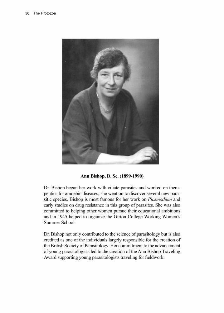
56 The Protozoa
Ann Bishop, D. Sc. (1899-1990)
Dr. Bishop began her work with ciliate parasites and worked on thera-peutics for amoebic diseases; she went on to discover several new para-sitic species. Bishop is most famous for her work on Plasmodium and early studies on drug resistance in this group of parasites. She was also committed to helping other women pursue their educational ambitions and in 1945 helped to organize the Girton College Working Women’s Summer School.
Dr. Bishop not only contributed to the science of parasitology but is also credited as one of the individuals largely responsible for the creation of the British Society of Parasitology. Her commitment to the advancement of young parasitologists led to the creation of the Ann Bishop Traveling Award supporting young parasitologists traveling for fieldwork.
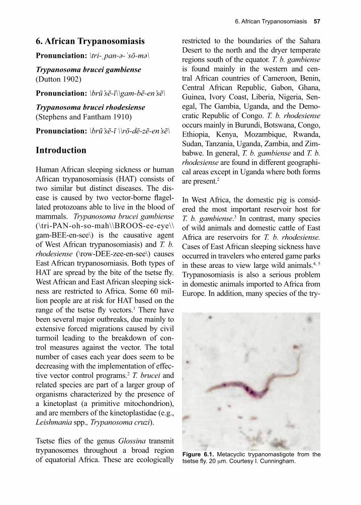
6. African Trypanosomiasis 57
Figure 6.1. Metacyclic trypanomastigote from the tsetse fly. 20 µm. Courtesy I. Cunningham.
6. African TrypanosomiasisPronunciation: \tri-ˌpan-ə-ˈsō-mə\
Trypanosoma brucei gambiense(Dutton 1902)
Pronunciation: \brū’sē-ī\\gam-bē-en’sē\
Trypanosoma brucei rhodesiense(Stephens and Fantham 1910)
Pronunciation: \brū’sē-ī \\rō-dē-zē-en’sē\
Introduction
Human African sleeping sickness or human African trypanosomiasis (HAT) consists of two similar but distinct diseases. The dis-ease is caused by two vector-borne flagel-lated protozoans able to live in the blood of mammals. Trypanosoma brucei gambiense (\tri-PAN-oh-so-mah\\BROOS-ee-eye\\gam-BEE-en-see\) is the causative agent of West African trypanosomiasis) and T. b. rhodesiense (\row-DEE-zee-en-see\) causes East African trypanosomiasis. Both types of HAT are spread by the bite of the tsetse fly. West African and East African sleeping sick-ness are restricted to Africa. Some 60 mil-lion people are at risk for HAT based on the range of the tsetse fly vectors.1 There have been several major outbreaks, due mainly to extensive forced migrations caused by civil turmoil leading to the breakdown of con-trol measures against the vector. The total number of cases each year does seem to be decreasing with the implementation of effec-tive vector control programs.2 T. brucei and related species are part of a larger group of organisms characterized by the presence of a kinetoplast (a primitive mitochondrion), and are members of the kinetoplastidae (e.g., Leishmania spp., Trypanosoma cruzi).
Tsetse flies of the genus Glossina transmit trypanosomes throughout a broad region of equatorial Africa. These are ecologically
restricted to the boundaries of the Sahara Desert to the north and the dryer temperate regions south of the equator. T. b. gambiense is found mainly in the western and cen-tral African countries of Cameroon, Benin, Central African Republic, Gabon, Ghana, Guinea, Ivory Coast, Liberia, Nigeria, Sen-egal, The Gambia, Uganda, and the Demo-cratic Republic of Congo. T. b. rhodesiense occurs mainly in Burundi, Botswana, Congo, Ethiopia, Kenya, Mozambique, Rwanda, Sudan, Tanzania, Uganda, Zambia, and Zim-babwe. In general, T. b. gambiense and T. b. rhodesiense are found in different geographi-cal areas except in Uganda where both forms are present.2
In West Africa, the domestic pig is consid-ered the most important reservoir host for T. b. gambiense.3 In contrast, many species of wild animals and domestic cattle of East Africa are reservoirs for T. b. rhodesiense. Cases of East African sleeping sickness have occurred in travelers who entered game parks in these areas to view large wild animals.4, 5 Trypanosomiasis is also a serious problem in domestic animals imported to Africa from Europe. In addition, many species of the try-
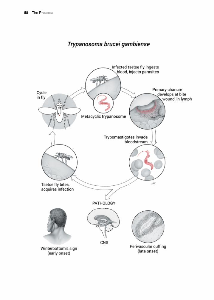
58 The Protozoa
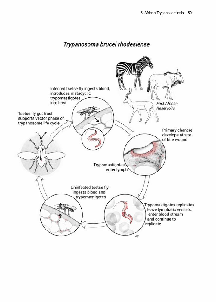
6. African Trypanosomiasis 59
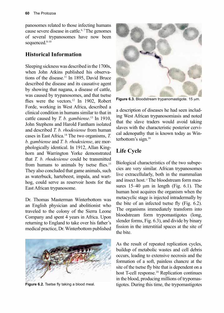
60 The Protozoa
Figure 6.3. Bloodstream trypanomastigote. 15 µm.
panosomes related to those infecting humans cause severe disease in cattle.6, 7 The genomes of several trypanosomes have now been sequenced.8-10
Historical Information
Sleeping sickness was described in the 1700s, when John Atkins published his observa-tions of the disease.11 In 1895, David Bruce described the disease and its causative agent by showing that nagana, a disease of cattle, was caused by trypanosomes, and that tsetse flies were the vectors.12 In 1902, Robert Forde, working in West Africa, described a clinical condition in humans similar to that in cattle caused by T. b. gambiense.13 In 1910, John Stephens and Harold Fantham isolated and described T. b. rhodesiense from human cases in East Africa.14 The two organisms, T. b. gambiense and T. b. rhodesiense, are mor-phologically identical. In 1912, Allan King-horn and Warrington Yorke demonstrated that T. b. rhodesiense could be transmitted from humans to animals by tsetse flies.15 They also concluded that game animals, such as waterbuck, hartebeest, impala, and wart-hog, could serve as reservoir hosts for the East African trypanosome.
Dr. Thomas Masterman Winterbottom was an English physician and abolitionist who traveled to the colony of the Sierra Leone Company and spent 4 years in Africa. Upon returning to England to take over his father’s medical practice, Dr. Winterbottom published
a description of diseases he had seen includ-ing West African trypanosomiasis and noted that the slave traders would avoid taking slaves with the characteristic posterior cervi-cal adenopathy that is known today as Win-terbottom’s sign.16
Life Cycle
Biological characteristics of the two subspe-cies are very similar. African trypanosomes live extracellularly, both in the mammalian and insect host.17 The bloodstream form mea-sures 15–40 µm in length (Fig. 6.1). The human host acquires the organism when the metacyclic stage is injected intradermally by the bite of an infected tsetse fly (Fig. 6.2). The organisms immediately transform into bloodstream form trypomastigotes (long, slender forms, Fig. 6.3), and divide by binary fission in the interstitial spaces at the site of the bite.
As the result of repeated replication cycles, buildup of metabolic wastes and cell debris occurs, leading to extensive necrosis and the formation of a soft, painless chancre at the site of the tsetse fly bite that is dependent on a host T-cell response.18 Replication continues in the blood, producing millions of trypomas-tigotes. During this time, the trypomastigotes Figure 6.2. Tsetse fly taking a blood meal.
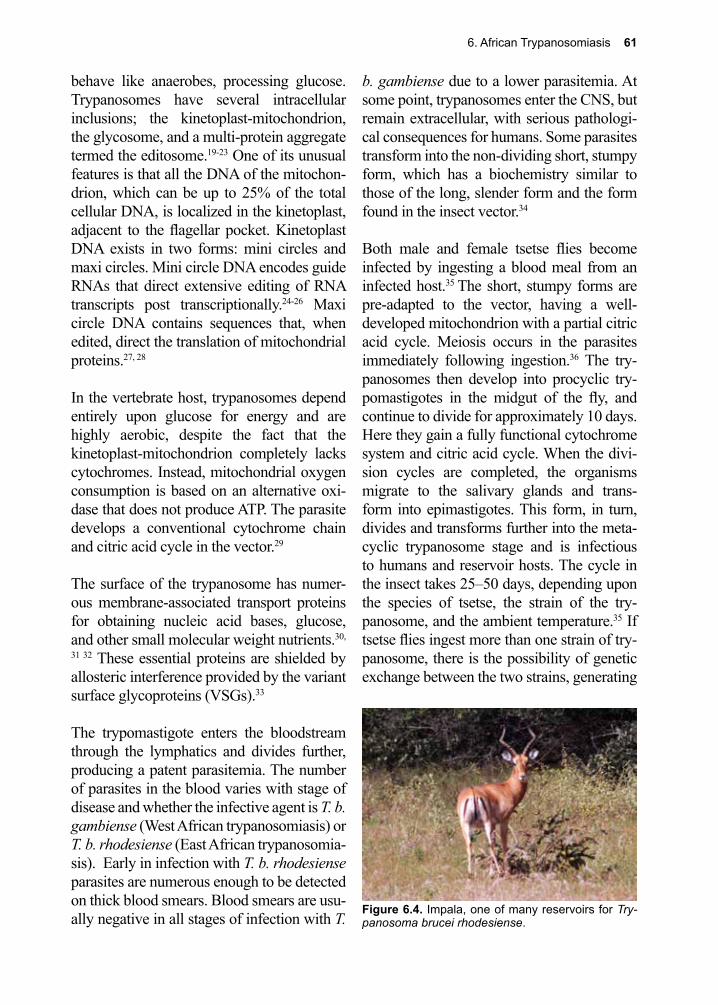
6. African Trypanosomiasis 61
behave like anaerobes, processing glucose. Trypanosomes have several intracellular inclusions; the kinetoplast-mitochondrion, the glycosome, and a multi-protein aggregate termed the editosome.19-23 One of its unusual features is that all the DNA of the mitochon-drion, which can be up to 25% of the total cellular DNA, is localized in the kinetoplast, adjacent to the flagellar pocket. Kinetoplast DNA exists in two forms: mini circles and maxi circles. Mini circle DNA encodes guide RNAs that direct extensive editing of RNA transcripts post transcriptionally.24-26 Maxi circle DNA contains sequences that, when edited, direct the translation of mitochondrial proteins.27, 28
In the vertebrate host, trypanosomes depend entirely upon glucose for energy and are highly aerobic, despite the fact that the kinetoplast-mitochondrion completely lacks cytochromes. Instead, mitochondrial oxygen consumption is based on an alternative oxi-dase that does not produce ATP. The parasite develops a conventional cytochrome chain and citric acid cycle in the vector.29
The surface of the trypanosome has numer-ous membrane-associated transport proteins for obtaining nucleic acid bases, glucose, and other small molecular weight nutrients.30,
31 32 These essential proteins are shielded by allosteric interference provided by the variant surface glycoproteins (VSGs).33
The trypomastigote enters the bloodstream through the lymphatics and divides further, producing a patent parasitemia. The number of parasites in the blood varies with stage of disease and whether the infective agent is T. b. gambiense (West African trypanosomiasis) or T. b. rhodesiense (East African trypanosomia-sis). Early in infection with T. b. rhodesiense parasites are numerous enough to be detected on thick blood smears. Blood smears are usu-ally negative in all stages of infection with T.
b. gambiense due to a lower parasitemia. At some point, trypanosomes enter the CNS, but remain extracellular, with serious pathologi-cal consequences for humans. Some parasites transform into the non-dividing short, stumpy form, which has a biochemistry similar to those of the long, slender form and the form found in the insect vector.34
Both male and female tsetse flies become infected by ingesting a blood meal from an infected host.35 The short, stumpy forms are pre-adapted to the vector, having a well-developed mitochondrion with a partial citric acid cycle. Meiosis occurs in the parasites immediately following ingestion.36 The try-panosomes then develop into procyclic try-pomastigotes in the midgut of the fly, and continue to divide for approximately 10 days. Here they gain a fully functional cytochrome system and citric acid cycle. When the divi-sion cycles are completed, the organisms migrate to the salivary glands and trans-form into epimastigotes. This form, in turn, divides and transforms further into the meta-cyclic trypanosome stage and is infectious to humans and reservoir hosts. The cycle in the insect takes 25–50 days, depending upon the species of tsetse, the strain of the try-panosome, and the ambient temperature.35 If tsetse flies ingest more than one strain of try-panosome, there is the possibility of genetic exchange between the two strains, generating
Figure 6.4. Impala, one of many reservoirs for Try-panosoma brucei rhodesiense.
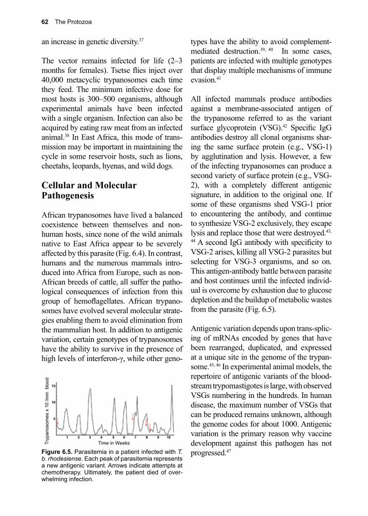
62 The Protozoa
an increase in genetic diversity.37
The vector remains infected for life (2–3 months for females). Tsetse flies inject over 40,000 metacyclic trypanosomes each time they feed. The minimum infective dose for most hosts is 300–500 organisms, although experimental animals have been infected with a single organism. Infection can also be acquired by eating raw meat from an infected animal.38 In East Africa, this mode of trans-mission may be important in maintaining the cycle in some reservoir hosts, such as lions, cheetahs, leopards, hyenas, and wild dogs.
Cellular and Molecular Pathogenesis
African trypanosomes have lived a balanced coexistence between themselves and non-human hosts, since none of the wild animals native to East Africa appear to be severely affected by this parasite (Fig. 6.4). In contrast, humans and the numerous mammals intro-duced into Africa from Europe, such as non-African breeds of cattle, all suffer the patho-logical consequences of infection from this group of hemoflagellates. African trypano-somes have evolved several molecular strate-gies enabling them to avoid elimination from the mammalian host. In addition to antigenic variation, certain genotypes of trypanosomes have the ability to survive in the presence of high levels of interferon-γ, while other geno-
types have the ability to avoid complement-mediated destruction.39, 40 In some cases, patients are infected with multiple genotypes that display multiple mechanisms of immune evasion.41
All infected mammals produce antibodies against a membrane-associated antigen of the trypanosome referred to as the variant surface glycoprotein (VSG).42 Specific IgG antibodies destroy all clonal organisms shar-ing the same surface protein (e.g., VSG-1) by agglutination and lysis. However, a few of the infecting trypanosomes can produce a second variety of surface protein (e.g., VSG-2), with a completely different antigenic signature, in addition to the original one. If some of these organisms shed VSG-1 prior to encountering the antibody, and continue to synthesize VSG-2 exclusively, they escape lysis and replace those that were destroyed.43,
44 A second IgG antibody with specificity to VSG-2 arises, killing all VSG-2 parasites but selecting for VSG-3 organisms, and so on. This antigen-antibody battle between parasite and host continues until the infected individ-ual is overcome by exhaustion due to glucose depletion and the buildup of metabolic wastes from the parasite (Fig. 6.5).
Antigenic variation depends upon trans-splic-ing of mRNAs encoded by genes that have been rearranged, duplicated, and expressed at a unique site in the genome of the trypan-some.45, 46 In experimental animal models, the repertoire of antigenic variants of the blood-stream trypomastigotes is large, with observed VSGs numbering in the hundreds. In human disease, the maximum number of VSGs that can be produced remains unknown, although the genome codes for about 1000. Antigenic variation is the primary reason why vaccine development against this pathogen has not progressed.47 Figure 6.5. Parasitemia in a patient infected with T.
b. rhodesiense. Each peak of parasitemia represents a new antigenic variant. Arrows indicate attempts at chemotherapy. Ultimately, the patient died of over-whelming infection.

6. African Trypanosomiasis 63
NeuropathologyTrypanosomes remain in the bloodstream and lymph nodes throughout the infec-tion period, which can last weeks to years, depending upon the subspecies of parasite and the immune capabilities of the infected individual. All nodes become enlarged, but enlargement of the posterior cervical nodes is the most noticeable.48 This cervical lymph node enlargement is known as “Winter-bottom’s sign”. The invasion of the CNS induces a lethargic condition, leading even-tually to coma and death.49 Organisms enter the CNS much earlier in the infection with T. b. rhodesiense than with T. b. gambiense. Replication of the parasite in the CSF results in leptomeningitis, cerebral edema, and encephalopathy.50 Dysregulated inflamma-tion is the chief pathological correlate, with perivascular cuffing consisting of infiltrates of glial cells, lymphocytes, and plasma cells (Fig. 6.6). Astrocytes are induced to release prostaglandin D2, a sleep-regulating mole-cule.51 Anti-inflammatory interleukins (inter-leukin-10 and transforming growth factor-β) are produced early on in the infection, but lose their effectiveness during the chronic and late phase.52
Clinical Disease
The African sleeping sickness caused by the two species of trypanosomes are different in certain ways. A large, painless chancre (Fig. 6.7) containing the dividing organisms usu-ally develops at the site of the bite within 2–5 days, and subsequently heals.18 The chancre is more often described in T. b. gambiense than in T. b. rhodesiense, but there may be recall bias based on time to diagnosis or a differ-ence in the reaction at the site of the tsetse fly bite, due to the differences in host response triggered by the two trypanosomes. Intermit-tent fever coincides with the organisms enter-ing the bloodstream. Some patients develop rash, generalized pruritus, weight loss, and facial swelling.53 Winterbottom’s sign is char-acteristic but not always present.48
Infection rapidly progresses on to disease with T. b. rhodesiense, with an incubation period of only 2–3 weeks, and a course of several weeks. CNS involvement occurs some 3–4 weeks after infection. In contrast, T. b. gambiense has an incubation period of several weeks to months and may not involve the brain for months or even years. Europeans exposed to T. b. gambiense tend to present with a more rapidly progressive course indi-
Figure 6.6. Perivascular cuffing around vein in brain of patient who died of sleeping sickness.
Figure 6.7. Chancre due to early infection with T. b. gambiense. Courtesy World Health Organization.
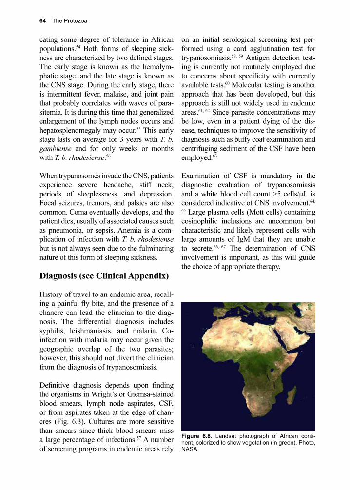
64 The Protozoa
cating some degree of tolerance in African populations.54 Both forms of sleeping sick-ness are characterized by two defined stages. The early stage is known as the hemolym-phatic stage, and the late stage is known as the CNS stage. During the early stage, there is intermittent fever, malaise, and joint pain that probably correlates with waves of para-sitemia. It is during this time that generalized enlargement of the lymph nodes occurs and hepatosplenomegaly may occur.55 This early stage lasts on average for 3 years with T. b. gambiense and for only weeks or months with T. b. rhodesiense.56
When trypanosomes invade the CNS, patients experience severe headache, stiff neck, periods of sleeplessness, and depression. Focal seizures, tremors, and palsies are also common. Coma eventually develops, and the patient dies, usually of associated causes such as pneumonia, or sepsis. Anemia is a com-plication of infection with T. b. rhodesiense but is not always seen due to the fulminating nature of this form of sleeping sickness.
Diagnosis (see Clinical Appendix)
History of travel to an endemic area, recall-ing a painful fly bite, and the presence of a chancre can lead the clinician to the diag-nosis. The differential diagnosis includes syphilis, leishmaniasis, and malaria. Co-infection with malaria may occur given the geographic overlap of the two parasites; however, this should not divert the clinician from the diagnosis of trypanosomiasis.
Definitive diagnosis depends upon finding the organisms in Wright’s or Giemsa-stained blood smears, lymph node aspirates, CSF, or from aspirates taken at the edge of chan-cres (Fig. 6.3). Cultures are more sensitive than smears since thick blood smears miss a large percentage of infections.57 A number of screening programs in endemic areas rely
on an initial serological screening test per-formed using a card agglutination test for trypanosomiasis.58, 59 Antigen detection test-ing is currently not routinely employed due to concerns about specificity with currently available tests.60 Molecular testing is another approach that has been developed, but this approach is still not widely used in endemic areas.61, 62 Since parasite concentrations may be low, even in a patient dying of the dis-ease, techniques to improve the sensitivity of diagnosis such as buffy coat examination and centrifuging sediment of the CSF have been employed.63
Examination of CSF is mandatory in the diagnostic evaluation of trypanosomiasis and a white blood cell count >5 cells/µL is considered indicative of CNS involvement.64,
65 Large plasma cells (Mott cells) containing eosinophilic inclusions are uncommon but characteristic and likely represent cells with large amounts of IgM that they are unable to secrete.66, 67 The determination of CNS involvement is important, as this will guide the choice of appropriate therapy.
Figure 6.8. Landsat photograph of African conti-nent, colorized to show vegetation (in green). Photo, NASA.
6. African Trypanosomiasis 65
Treatment (see Clinical Appendix)
Critical for selection of the appropriate treat-ment for both East African and West African trypanosomiasis is the determination of the clinical stage of disease: early (hemolym-phatic stage), versus late (encephalitic stage), and identification of the species of trypano-some. 68, 69
For treatment of West African sleeping sick-ness (T. b. gambiense) the current recom-mended therapy for early stage infection is IV or IM pentamidine.70 An alternative therapy for early hemolymphatic stage infection with T. b. gambiense is suramin, which is equal in efficacy to pentamidine.70 Suramin has been associated with a hypersensitivity reaction so a test dose should be given before start-ing treatment with this agent.71 Suramin as also the first line therapy for early stage East African sleeping sickness (T. b. rhodesiense). In early hemolymphatic stage East African sleeping sickness, suramin is the preferred therapeutic agent.72, 73 Suramin is only effec-tive in the early stages of the infection as it does not cross the blood-brain barrier.68
There is significantly more difference in rec-ommendations for therapy of late or encepha-litic stage infection for these two protozoan parasites. For West African trypanosomiasis, infection with T. b. gambiense, combination therapy with eflornithine and nifurtimox or monotherapy with eflornithine is recom-mended.74 For East African trypanosomiasis, infection with T. b. rhodesiense, melarsop-rol is the treatment of choice. Unfortunately, melarsoprol is the only effective drug for treatment of T. b. rhodesiense with CNS involvement, and this toxic drug is associated with encephalopathy in about 3% of cases.75 Eflornithine, although an effective therapy for late stage with T. b. gambiense infection, is not an effective therapy against T. b. rhod-esiense.76
Prevention and Control
Previously, periods of political upheaval in different parts of Africa have resulted in dra-matic increases in human cases of sleeping sickness.77 A 2005 World Health Organization report indicated at least 450,000 new cases that year, alone, while prior to 1995, the esti-mate was fewer than 70,000.78 Military action and civil unrest in the Sudan, Ethiopia, Sierra Leone, Congo, and Liberia have been respon-sible for the forced migration of millions of individuals, placing them at high risk from a number of parasitic infections. Tsetse fly con-trol programs are also compromised in these same regions due to political and economic instability, exacerbating an already intracta-ble situation. T. b. gambiense may exist in a latent state in humans and under conditions of stress there may be activation, playing a role in some outbreaks.79 In addition to all this tur-moil, HIV/AIDS has complicated the picture, adding new, dimensions to the general prob-lem of disease control. Limited resources in countries bordering conflicted areas cannot keep up with the need for vector control, due to large influxes of refugees. Tsetse flies and mosquitoes do not obey political boundar-ies and thrive in certain disturbed environ-ments.80 Despite these challenges there is an elimination initiative underway led by the World Health Organization.
Work on vaccines based on VSG antigens has not progressed; however, there are a number of active investigations into further under-standing the immune response and what con-stitutes protection from trypanosomiasis.81-83 Other protein antigens, particularly transport-ers on the membrane of the flagellar pocket and tubulin, offer promise.84 Diagnostic tests, other than microscopy, would help in earlier patient diagnosis and control efforts, partic-ularly when there are low parasite numbers and in latent infections.
66 The Protozoa
References
1. Simarro, P. P., et al., The Atlas of human African trypanosomiasis: a contribution to global mapping of neglected tropical diseases. Int J Health Geogr 2010, 9, 57.
2. Simarro, P. P., et al., The human African trypanosomiasis control and surveillance programme of the World Health Organization 2000-2009: the way forward. PLoS Negl Trop Dis 2011, 5 (2), e1007.
3. Schares, G.; Mehlitz, D., Sleeping sickness in Zaire: a nested polymerase chain reaction improves the identification of Trypanosoma (Trypanozoon) brucei gambiense by specific kinetoplast DNA probes. Tropical medicine & international health : TM & IH 1996, 1 (1), 59-70.
4. Migchelsen, S. J., et al., Human African trypanosomiasis: a review of non-endemic cases in the past 20 years. Int J Infect Dis 2011, 15 (8), e517-24.
5. Sinha, A., et al., African trypanosomiasis in two travelers from the United States. Clin Infect Dis 1999, 29 (4), 840-4.
6. Leak, S. G., et al., Use of insecticide-impregnated targets for the control of tsetse flies (Glossina spp.) and trypanosomiasis occurring in cattle in an area of south-west Ethiopia with a high prevalence of drug-resistant trypanosomes. Tropical medicine & international health : TM & IH 1996, 1 (5), 599-609.
7. Katakura, K., et al., Detection of Trypanosoma congolense and T. brucei subspecies in cattle in Zambia by polymerase chain reaction from blood collected on a filter paper. Parasitology research 1997, 83 (3), 241-5.
8. Ghedin, E., et al., Gene synteny and evolution of genome architecture in trypanosomatids. Molecular and biochemical parasitology 2004, 134 (2), 183-91.
9. Berriman, M., et al., The genome of the African trypanosome Trypanosoma brucei. Science 2005, 309 (5733), 416-22.
10. Kelly, S., et al., A draft genome for the African crocodilian trypanosome Trypanosoma grayi. Sci Data 2014, 1, 140024.
11. Atkins, J., The Navy Surgeon, or Practical System of Surgery with a Dissertation on Cold and Hot Mineral Springs and Physical Observations on the Coast of Guinea. J. Hodges, at the Looking-Glass, over-against St. Magnus Church, London-Bridge: 1742.
12. Bruce, D., Preliminary Report of the Tsetse Fly Disease or Nagana in Zululand. Bennet Davis Durban 1895.
13. Forde, R. M.; J., Some clinical notes on a European patient in whose blood a trypanosome was observed. Med 1902, 5 261-263.
14. Stephens, J. W. W., et al., On the peculiar morphology of a trypanosome from a case of sleeping sickness and the possibility of its being a new species (T. rhodesiense). Proc Lond [Biol] 1910, 83, 23-33.
15. Kinghorn, A.; Yorke, W., On the transmission of human trypanosomes by Glossina morsitans, and on the occurrence of human trypanosomes in game. Ann Trop Med Parasitol 1912, 6, 1-23.
16. Texas Southern University. Library.; Heartman Negro Collection., Catalog, Heartman Negro Collection. Library Staff, Texas Southern University: Houston, Texas, 1956; p 325 p.
17. Gull, K., The cell biology of parasitism in Trypanosoma brucei: insights and drug targets from genomic approaches? Current pharmaceutical design 2002, 8 (4), 241-56.
18. Naessens, J., et al., Local skin reaction (chancre) induced following inoculation of metacyclic trypanosomes in cattle by tsetse flies is dependent on CD4 T lymphocytes. Parasite Immunol 2003, 25 (8-9), 413-9.
19. Shlomai, J., The structure and replication of kinetoplast DNA. Current molecular medicine 2004, 4 (6), 623-47.
20. Opperdoes, F. R.; Borst, P., Localization of nine glycolytic enzymes in a microbody-like organelle in Trypanosoma brucei: the glycosome. FEBS letters 1977, 80 (2), 360-4.
21. Verlinde, C. L., et al., Glycolysis as a target for the design of new anti-trypanosome drugs. Drug resistance updates : reviews and commentaries in antimicrobial and anticancer chemotherapy 2001, 4 (1), 50-65.
6. African Trypanosomiasis 67
22. Parsons, M., Glycosomes: parasites and the divergence of peroxisomal purpose. Molecular microbiology 2004, 53 (3), 717-24.
23. Panigrahi, A. K., et al., Identification of novel components of Trypanosoma brucei editosomes. RNA (New York, N.Y.) 2003, 9 (4), 484-92.
24. Feagin, J. E.; Stuart, K., Differential expression of mitochondrial genes between life cycle stages of Trypanosoma brucei. Proceedings of the National Academy of Sciences of the United States of America 1985, 82 (10), 3380-4.
25. Stuart, K., et al., Investigating the mechanism and machinery of RNA editing. Methods (San Diego, Calif.) 1998, 15 (1), 3-14.
26. Simpson, L., et al., Mitochondrial proteins and complexes in Leishmania and Trypanosoma involved in U-insertion/deletion RNA editing. RNA (New York, N.Y.) 2004, 10 (2), 159-70.
27. Stuart, K., Mitochondrial DNA of an African trypanosome. J Cell Biochem 1983, 23 (1-4), 13-26.
28. Shapiro, T. A., Kinetoplast DNA maxicircles: networks within networks. Proceedings of the National Academy of Sciences of the United States of America 1993, 90 (16), 7809-13.
29. Walker, R., et al., The effect of over-expression of the alternative oxidase in the procyclic forms of Trypanosoma brucei. Mol Biochem Parasitol 2005, 139, 153-62.
30. Borst, P.; Fairlamb, A. H., Surface receptors and transporters of Trypanosoma brucei. Annual review of microbiology 1998, 52, 745-78.
31. Sanchez, M. A., et al., Cloning and functional expression of a gene encoding a P1 type nucleoside transporter from Trypanosoma brucei. The Journal of biological chemistry 1999, 274 (42), 30244-9.
32. Natto, M. J., et al., Trypanosoma brucei: expression of multiple purine transporters prevents the development of allopurinol resistance. Experimental parasitology 2005, 109 (2), 80-6.
33. Donelson, J. E., Antigenic variation and the African trypanosome genome. Acta tropica 2003, 85 (3), 391-404.
34. Kioy, D., et al., Human African trypanosomiasis. Nature reviews. Microbiology 2004, 2 (3), 186-7.
35. Vickerman, K., et al., Biology of African trypanosomes in the tsetse fly. Biology of the cell / under the auspices of the European Cell Biology Organization 1988, 64 (2), 109-19.
36. Peacock, L., et al., Meiosis and haploid gametes in the pathogen Trypanosoma brucei. Curr Biol 2014, 24 (2), 181-6.
37. Gibson, W., Genetic exchange in trypanosomes. Bulletin et memoires de l’Academie royale de medecine de Belgique 1996, 151 (2), 203-10.
38. Betram, B. C. R., Sleeping sickness survey in the Serengeti area (Tanzania) . III. Discussion of the relevance of the trypanosome survey to the biology of large mammals in the Serengeti. Acta Trop Basel 1973, 30, 36-48.
39. Donelson, J. E., et al., Multiple mechanisms of immune evasion by African trypanosomes. Molecular and biochemical parasitology 1998, 91 (1), 51-66.
40. Barry, J. D.; McCulloch, R., Antigenic variation in trypanosomes: enhanced phenotypic variation in a eukaryotic parasite. Advances in parasitology 2001, 49, 1-70.
41. Truc, P., et al., Genetic variability within Trypanosoma brucei gambiense: evidence for the circulation of different genotypes in human African trypanosomiasis patients in Cote d’Ivoire. Trans R Soc Trop Med Hyg 2002, 96 (1), 52-5.
42. Cross, G. A., Identification, purification and properties of clone-specific glycoprotein antigens constituting the surface coat of Trypanosoma brucei. Parasitology 1975, 71 (3), 393-417.
43. Pays, E., et al., Genetic controls for the expression of surface antigens in African trypanosomes. Annual review of microbiology 1994, 48, 25-52.
44. McCulloch, R., Antigenic variation in African trypanosomes: monitoring progress. Trends in parasitology 2004, 20 (3), 117-21.
45. Gray, A. R., Antigenic variation in a strain of Trypanosoma brucei transmitted by Glossina morsitans and G. palpalis. Journal of general microbiology 1965, 41 (2), 195-214.
46. Borst, P., et al., Control of VSG gene expression sites in Trypanosoma brucei. Molecular and biochemical parasitology 1998, 91 (1), 67-76.
68 The Protozoa
47. Barbour, A. G.; Restrepo, B. I., Antigenic variation in vector-borne pathogens. Emerging infectious diseases 2000, 6 (5), 449-57.
48. Winterbottom, T. M., An Account of the Native Africans in the Neighborhood of Sierra Leone. 1803; Vol. 2.
49. Mhlanga, J. D., et al., Neurobiology of cerebral malaria and African sleeping sickness. Brain research bulletin 1997, 44 (5), 579-89.
50. Odiit, M., et al., Duration of symptoms and case fatality of sleeping sickness caused by Trypanosoma brucei rhodesiense in Tororo, Uganda. East African medical journal 1997, 74 (12), 792-5.
51. Pentreath, V. W., et al., The somnogenic T lymphocyte suppressor prostaglandin D2 is selectively elevated in cerebrospinal fluid of advanced sleeping sickness patients. Transactions of the Royal Society of Tropical Medicine and Hygiene 1990, 84 (6), 795-9.
52. Sternberg, J. M., Human African trypanosomiasis: clinical presentation and immune response. Parasite immunology 2004, 26 (11-12), 469-76.
53. Uslan, D. Z., et al., A woman with fever and rash after African safari. Clin Infect Dis 2006, 43 (5), 609, 661-2.
54. Blum, J. A., et al., Human African trypanosomiasis in endemic populations and travellers. Eur J Clin Microbiol Infect Dis 2012, 31 (6), 905-13.
55. Boatin, B. A., et al., Use of symptoms and signs for diagnosis of Trypanosoma brucei rhodesiense trypanosomiasis by rural health personnel. Bull World Health Organ 1986, 64 (3), 389-95.
56. Malvy, D.; Chappuis, F., Sleeping sickness. Clin Microbiol Infect 2011, 17 (7), 986-95.57. Robays, J., et al., The effectiveness of active population screening and treatment for sleeping
sickness control in the Democratic Republic of Congo. Trop Med Int Health 2004, 9 (5), 542-50.58. Chappuis, F., et al., Card agglutination test for trypanosomiasis (CATT) end-dilution titer and
cerebrospinal fluid cell count as predictors of human African Trypanosomiasis (Trypanosoma brucei gambiense) among serologically suspected individuals in southern Sudan. Am J Trop Med Hyg 2004, 71 (3), 313-7.
59. Inojosa, W. O., et al., Diagnosing human African trypanosomiasis in Angola using a card agglutination test: observational study of active and passive case finding strategies. BMJ 2006, 332 (7556), 1479.
60. Asonganyi, T., et al., A multi-centre evaluation of the card indirect agglutination test for trypanosomiasis (TrypTect CIATT). Ann Trop Med Parasitol 1998, 92 (8), 837-44.
61. Deborggraeve, S., et al., Diagnostic accuracy of PCR in gambiense sleeping sickness diagnosis, staging and post-treatment follow-up: a 2-year longitudinal study. PLoS Negl Trop Dis 2011, 5 (2), e972.
62. Becker, S., et al., Real-time PCR for detection of Trypanosoma brucei in human blood samples. Diagnostic microbiology and infectious disease 2004, 50 (3), 193-9.
63. Bailey, J. W.; Smith, D. H., The use of the acridine orange QBC technique in the diagnosis of African trypanosomiasis. Trans R Soc Trop Med Hyg 1992, 86 (6), 630.
64. Kennedy, P. G., Human African trypanosomiasis of the CNS: current issues and challenges. J Clin Invest 2004, 113 (4), 496-504.
65. Rodgers, J., Human African trypanosomiasis, chemotherapy and CNS disease. J Neuroimmunol 2009, 211 (1-2), 16-22.
66. Bain, B. J., Russell bodies and Mott cells. Am J Hematol 2009, 84 (8), 516.67. Weinstein, T., et al., Electron microscopy study of Mott and Russell bodies in myeloma cells. J
Submicrosc Cytol 1987, 19 (1), 155-9.68. Jannin, J.; Cattand, P., Treatment and control of human African trypanosomiasis. Current opinion
in infectious diseases 2004, 17 (6), 565-71.69. Kennedy, P. G., The continuing problem of human African trypanosomiasis (sleeping sickness).
Ann Neurol 2008, 64 (2), 116-26.70. Barrett, M. P., et al., Human African trypanosomiasis: pharmacological re-engagement with a
neglected disease. Br J Pharmacol 2007, 152 (8), 1155-71.71. Thibault, A., et al., Anaphylactoid reaction with suramin. Pharmacotherapy 1993, 13 (6), 656-7.
6. African Trypanosomiasis 69
72. Woo, P.; Soltys, M. A., The effect of suramin on blood and tissue forms of Trypanosoma brucei and Trypanosoma rhodesiense. Ann Trop Med Parasitol 1971, 65 (4), 465-9.
73. Arroz, J.; Djedje, M., Suramin and metronidazole in the treatment of Trypanosoma brucei rhodesiense. Trans R Soc Trop Med Hyg 1988, 82 (3), 421.
74. Kennedy, P. G., Clinical features, diagnosis, and treatment of human African trypanosomiasis (sleeping sickness). Lancet Neurol 2013, 12 (2), 186-94.
75. Legros, D., et al., [Therapeutic failure of melarsoprol among patients treated for late stage T.b. gambiense human African trypanosomiasis in Uganda]. Bulletin de la Societe de pathologie exotique (1990) 1999, 92 (3), 171-2.
76. Bacchi, C. J., et al., Parasitic protozoa and polyamines. 1987; p 317-44.77. Chretien, J.-P.; Smoak, B. L., African Trypanosomiasis: Changing Epidemiology and
Consequences. Current infectious disease reports 2005, 7 (1), 54-60.78. WHO World Health Organization estimates for 2005.79. Welburn, S. C., et al., Beyond Tsetse - Implications for Research and Control of Human African
Trypanosomiasis Epidemics. Trends Parasitol 2016, 32 (3), 230-41.80. Molyneux, D. H., Vector-borne parasitic diseases--an overview of recent changes. International
journal for parasitology 1998, 28 (6), 927-34.81. Silva, M. S., et al., Trans-sialidase from Trypanosoma brucei as a potential target for DNA
vaccine development against African trypanosomiasis. Parasitol Res 2009, 105 (5), 1223-9.82. La Greca, F.; Magez, S., Vaccination against trypanosomiasis: can it be done or is the
trypanosome truly the ultimate immune destroyer and escape artist? Hum Vaccin 2011, 7 (11), 1225-33.
83. Magez, S., et al., Current status of vaccination against African trypanosomiasis. Parasitology 2010, 137 (14), 2017-27.
84. Lubega, G. W., et al., Immunization with a tubulin-rich preparation from Trypanosoma brucei confers broad protection against African trypanosomosis. Experimental parasitology 2002, 102 (1), 9-22.
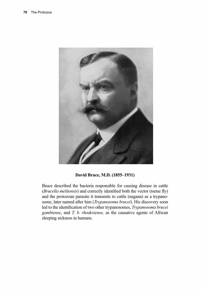
70 The Protozoa
David Bruce, M.D. (1855–1931)
Bruce described the bacteria responsible for causing disease in cattle (Brucella melitensis) and correctly identified both the vector (tsetse fly) and the protozoan parasite it transmits to cattle (nagana) as a trypano-some, later named after him (Trypanosoma brucei). His discovery soon led to the identification of two other trypanosomes, Trypanosoma brucei gambiense, and T. b. rhodesiense, as the causative agents of African sleeping sickness in humans.
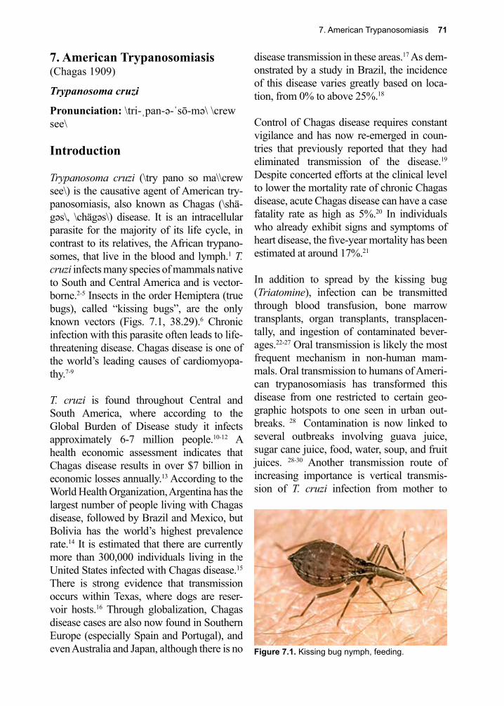
7. American Trypanosomiasis 71
Figure 7.1. Kissing bug nymph, feeding.
7. American Trypanosomiasis(Chagas 1909)
Trypanosoma cruzi
Pronunciation: \tri-ˌpan-ə-ˈsō-mə\ \crew see\
Introduction
Trypanosoma cruzi (\try pano so ma\\crew see\) is the causative agent of American try-panosomiasis, also known as Chagas (\shä-gəs\, \chägəs\) disease. It is an intracellular parasite for the majority of its life cycle, in contrast to its relatives, the African trypano-somes, that live in the blood and lymph.1 T. cruzi infects many species of mammals native to South and Central America and is vector-borne.2-5 Insects in the order Hemiptera (true bugs), called “kissing bugs”, are the only known vectors (Figs. 7.1, 38.29).6 Chronic infection with this parasite often leads to life-threatening disease. Chagas disease is one of the world’s leading causes of cardiomyopa-thy.7-9
T. cruzi is found throughout Central and South America, where according to the Global Burden of Disease study it infects approximately 6-7 million people.10-12 A health economic assessment indicates that Chagas disease results in over $7 billion in economic losses annually.13 According to the World Health Organization, Argentina has the largest number of people living with Chagas disease, followed by Brazil and Mexico, but Bolivia has the world’s highest prevalence rate.14 It is estimated that there are currently more than 300,000 individuals living in the United States infected with Chagas disease.15 There is strong evidence that transmission occurs within Texas, where dogs are reser-voir hosts.16 Through globalization, Chagas disease cases are also now found in Southern Europe (especially Spain and Portugal), and even Australia and Japan, although there is no
disease transmission in these areas.17 As dem-onstrated by a study in Brazil, the incidence of this disease varies greatly based on loca-tion, from 0% to above 25%.18
Control of Chagas disease requires constant vigilance and has now re-emerged in coun-tries that previously reported that they had eliminated transmission of the disease.19 Despite concerted efforts at the clinical level to lower the mortality rate of chronic Chagas disease, acute Chagas disease can have a case fatality rate as high as 5%.20 In individuals who already exhibit signs and symptoms of heart disease, the five-year mortality has been estimated at around 17%.21
In addition to spread by the kissing bug (Triatomine), infection can be transmitted through blood transfusion, bone marrow transplants, organ transplants, transplacen-tally, and ingestion of contaminated bever-ages.22-27 Oral transmission is likely the most frequent mechanism in non-human mam-mals. Oral transmission to humans of Ameri-can trypanosomiasis has transformed this disease from one restricted to certain geo-graphic hotspots to one seen in urban out-breaks. 28 Contamination is now linked to several outbreaks involving guava juice, sugar cane juice, food, water, soup, and fruit juices. 28-30 Another transmission route of increasing importance is vertical transmis-sion of T. cruzi infection from mother to
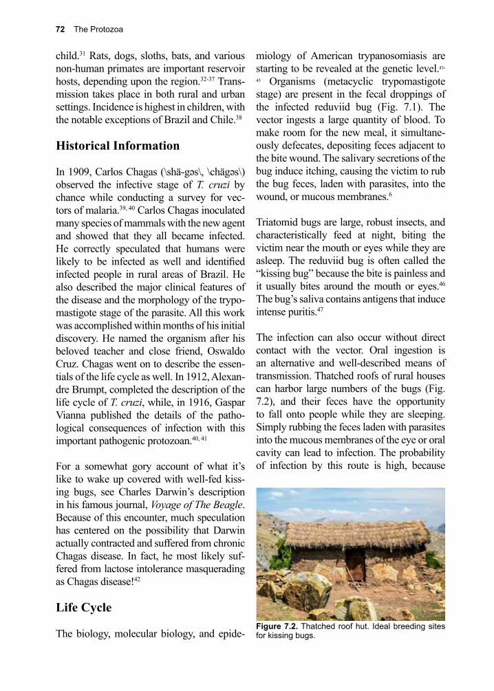
72 The Protozoa
child.31 Rats, dogs, sloths, bats, and various non-human primates are important reservoir hosts, depending upon the region.32-37 Trans-mission takes place in both rural and urban settings. Incidence is highest in children, with the notable exceptions of Brazil and Chile.38
Historical Information
In 1909, Carlos Chagas (\shä-gəs\, \chägəs\) observed the infective stage of T. cruzi by chance while conducting a survey for vec-tors of malaria.39, 40 Carlos Chagas inoculated many species of mammals with the new agent and showed that they all became infected. He correctly speculated that humans were likely to be infected as well and identified infected people in rural areas of Brazil. He also described the major clinical features of the disease and the morphology of the trypo-mastigote stage of the parasite. All this work was accomplished within months of his initial discovery. He named the organism after his beloved teacher and close friend, Oswaldo Cruz. Chagas went on to describe the essen-tials of the life cycle as well. In 1912, Alexan-dre Brumpt, completed the description of the life cycle of T. cruzi, while, in 1916, Gaspar Vianna published the details of the patho-logical consequences of infection with this important pathogenic protozoan.40, 41
For a somewhat gory account of what it’s like to wake up covered with well-fed kiss-ing bugs, see Charles Darwin’s description in his famous journal, Voyage of The Beagle. Because of this encounter, much speculation has centered on the possibility that Darwin actually contracted and suffered from chronic Chagas disease. In fact, he most likely suf-fered from lactose intolerance masquerading as Chagas disease!42
Life Cycle
The biology, molecular biology, and epide-
miology of American trypanosomiasis are starting to be revealed at the genetic level.43-
45 Organisms (metacyclic trypomastigote stage) are present in the fecal droppings of the infected reduviid bug (Fig. 7.1). The vector ingests a large quantity of blood. To make room for the new meal, it simultane-ously defecates, depositing feces adjacent to the bite wound. The salivary secretions of the bug induce itching, causing the victim to rub the bug feces, laden with parasites, into the wound, or mucous membranes.6
Triatomid bugs are large, robust insects, and characteristically feed at night, biting the victim near the mouth or eyes while they are asleep. The reduviid bug is often called the “kissing bug” because the bite is painless and it usually bites around the mouth or eyes.46 The bug’s saliva contains antigens that induce intense puritis.47
The infection can also occur without direct contact with the vector. Oral ingestion is an alternative and well-described means of transmission. Thatched roofs of rural houses can harbor large numbers of the bugs (Fig. 7.2), and their feces have the opportunity to fall onto people while they are sleeping. Simply rubbing the feces laden with parasites into the mucous membranes of the eye or oral cavity can lead to infection. The probability of infection by this route is high, because
Figure 7.2. Thatched roof hut. Ideal breeding sites for kissing bugs.
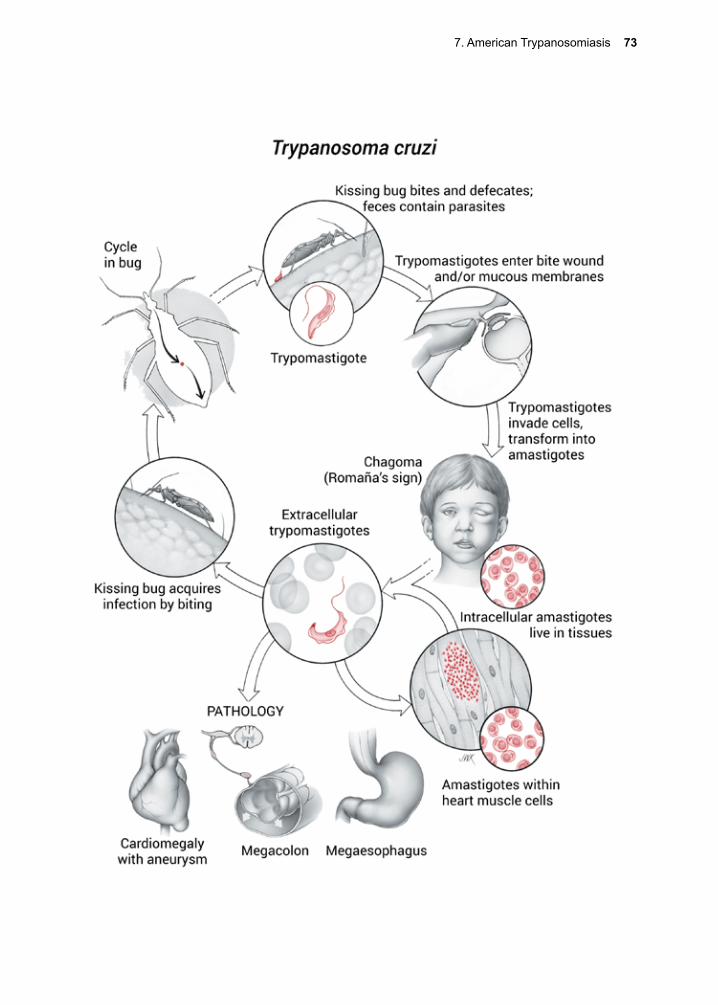
7. American Trypanosomiasis 73

74 The Protozoa
kissing bugs feed on many mammals, and rural peoples live in close proximity to their livestock and pets. Infection by transfusion, organ transplantation, or congenital transmis-sion introduces the parasite directly into the host.
Attachment is mediated through galectin-3 on the surface of host cells.48 The parasite protein that binds to galactin-3 has yet to be identified. The trypomastigote can penetrate a wide variety of cells, and the process is medi-ated by calcium ions and at least two para-site membrane proteins; a neuraminidase/trans-sialidase, which binds to sialic acid, and penetrin, which binds to heparin sulfate.49, 50 Another protein, gp82 might also be neces-sary for penetration into gastric epithelium if the metacyclic trypomastigote stage is swal-lowed.51 Just such an outbreak occurred in Santa Catarina, Brazil, involving the inges-tion of sugar cane juice contaminated with at least one infected kissing bug. Animals can become infected by ingesting infected kiss-ing bugs, and this might be the usual way for them to acquire the infection.52
After entering the parasitophorous vacuole, the trypomastigote enlists several mecha-nisms to aid in its survival there. It begins by neutralizing the pH of that intracellular space, thereby escaping the potentially dam-aging effects of exposure to the active forms of lysosomal enzymes.50 The organism also produces a number of proteins that offer it additional advantages once inside the host cell. Chagasin is a cysteine protease inhibitor and is apparently necessary for avoiding lyso-somal-derived cysteine protease activity and ensures that the parasite has the time needed to differentiate into the amastigote stage.53 Cruz-ipain is thought to play a major role in helping the parasite avoid being digested once inside the parasitophorous vacuole. Cruzipain also induces the upregulation of host-derived argi-nase-2, a known inhibitor of apoptosis.54 The parasite may be engineering the longevity of
its host cell, while at the same time, avoiding the ravages of lysosomal digestion.
The parasite then rapidly penetrates the cyto-sol and differentiates into the amastigote stage. This is the dividing form of T. cruzi and the one that inflicts cell damage on the host. After several division cycles, some of the parasites transform back into trypomastigotes. The affected cells die, releasing the parasites into the bloodstream, distributing them through-out the body. They infect cells in many types of tissues, including the CNS, heart muscle, the myenteric plexus, the urogenital tract, and the reticuloendothelial system.
Triatomines become infected by taking a blood meal from an infected individual.55 The trypomastigote migrates to the midgut of the insect, where it transforms into the epimas-tigote, and then undergoes many divisions. Thousands of organisms are produced within one insect without apparently significantly affecting it. The triatomines remain infected for life (~1–2 years). Epimastigotes maintain their place in the gut of the insect by specific receptor-ligand interactions involving at least one parasite surface glycoprotein and a carbo-hydrate lectin on the gut cells of the insect.56 Ultimately, epimastigotes transform into metacyclic trypomastigotes and migrate to the hindgut, and from there they are excreted with feces.
Cellular and Molecular Pathogenesis
Infection with T. cruzi results in partial immu-nosuppression that further aids the parasite in remaining inside the host cell for extended periods of time.57, 58 For example, in vitro cul-ture of human dendritic cells infected with T. cruzi resulted in a dramatic down-regulation of synthesis of: interleukins 6 and 12, TNF-α, HLA-DR, and CD40, and inhibited their mat-uration into antigen processing cells.59 Para-site-derived calreticulin may also be impor-
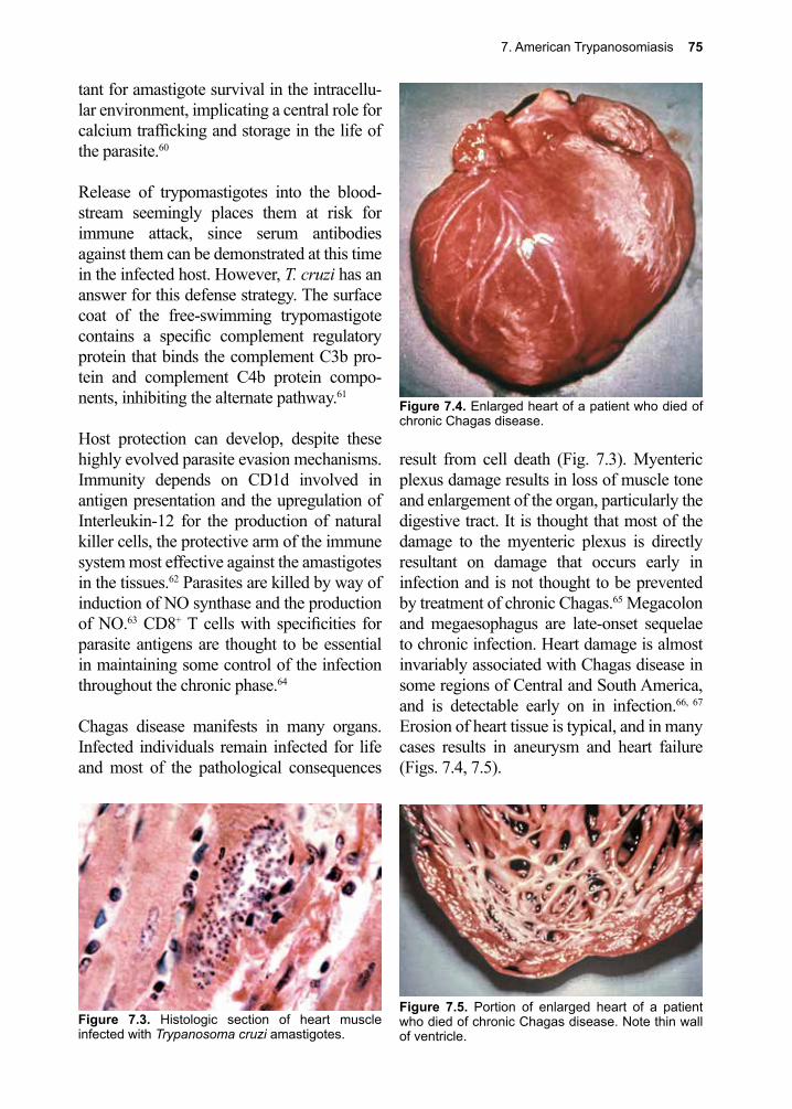
7. American Trypanosomiasis 75
tant for amastigote survival in the intracellu-lar environment, implicating a central role for calcium trafficking and storage in the life of the parasite.60
Release of trypomastigotes into the blood-stream seemingly places them at risk for immune attack, since serum antibodies against them can be demonstrated at this time in the infected host. However, T. cruzi has an answer for this defense strategy. The surface coat of the free-swimming trypomastigote contains a specific complement regulatory protein that binds the complement C3b pro-tein and complement C4b protein compo-nents, inhibiting the alternate pathway.61
Host protection can develop, despite these highly evolved parasite evasion mechanisms. Immunity depends on CD1d involved in antigen presentation and the upregulation of Interleukin-12 for the production of natural killer cells, the protective arm of the immune system most effective against the amastigotes in the tissues.62 Parasites are killed by way of induction of NO synthase and the production of NO.63 CD8+ T cells with specificities for parasite antigens are thought to be essential in maintaining some control of the infection throughout the chronic phase.64
Chagas disease manifests in many organs. Infected individuals remain infected for life and most of the pathological consequences
result from cell death (Fig. 7.3). Myenteric plexus damage results in loss of muscle tone and enlargement of the organ, particularly the digestive tract. It is thought that most of the damage to the myenteric plexus is directly resultant on damage that occurs early in infection and is not thought to be prevented by treatment of chronic Chagas.65 Megacolon and megaesophagus are late-onset sequelae to chronic infection. Heart damage is almost invariably associated with Chagas disease in some regions of Central and South America, and is detectable early on in infection.66, 67 Erosion of heart tissue is typical, and in many cases results in aneurysm and heart failure (Figs. 7.4, 7.5).
Figure 7.5. Portion of enlarged heart of a patient who died of chronic Chagas disease. Note thin wall of ventricle.
Figure 7.4. Enlarged heart of a patient who died of chronic Chagas disease.
Figure 7.3. Histologic section of heart muscle infected with Trypanosoma cruzi amastigotes.
76 The Protozoa
Current thinking regarding a dominant role for auto-antibodies inducing cardiomyopathy plays down this mechanism to account for heart damage during chronic infection.67 This is because PCR has demonstrated the pres-ence of T. cruzi in heart tissue, even at times when biopsy material used in conventional histological mode could not reveal the pres-ence of the parasite. Also, disease progresses rapidly when parasites are abundant and not as fast when they are hard to demonstrate on biopsy.67, 68 Thus parasite persistence leading to fibrosis and damage to the heart (including the cardiac conduction system) has emerged as the dominant theory to explain the patho-genesis of Chagas cardiomyopathy.69 None-theless, autoantibodies have been detected in many individuals suffering from long-term infection with T. cruzi.70, 71 Meningoen-cephalitis can occur during the acute phase and is characterized by infiltrates of CD8+ T cells.72 Meningoencephalitis can also occur in patients with underlying HIV/AIDS.73
Clinical Disease
Acute Chagas DiseaseThe incubation period of Chagas disease varies based on means of transmission. In acute Chagas disease acquired from the kiss-ing bug, the incubation period is from a few days to two weeks before the onset of symp-toms.74 With disease acquired through other means such as transfusion or transplant, the incubation period may be as long as sev-eral months.75 Acute disease can range from mild nonspecific symptoms such as malaise and fever to severe manifestations. Some patients are asymptomatic. Severe acute manifestations are estimated to develop in approximately 1% of cases and may involve acute myocarditis, pericardial effusion, and in some cases meningoencephalitis.76, 77 The risk of severe disease and mortality during the acute disease tends to be the highest with orally acquired infection.78
In a few patients, but clearly only the minor-ity, a characteristic hemi-facial edema or swelling called a chagoma develops at the site of the bite or inoculation within 2–4 days. If organisms are introduced into the body through mucous membranes by rubbing them into the eye, then the swelling associated with the chagoma is known as Romaña’s sign.79,
80 It occurs mostly as a unilateral swelling. The swollen eyelid is firm to the touch, and there may be associated conjunctivitis. If the bite occurs elsewhere, the adjacent area is erythematous, brawny, and firm to the touch. When the chagoma disappears after several weeks, it leaves an area of depigmentation. An associated neuropathy develops, and then disappears when the patient enters the chronic phase of the infection.
Chronic Chagas DiseaseMost patients survive the acute phase and become asymptomatic. Patients at this point enter the chronic phase and, although they may remain asymptomatic, they are capable of transmitting parasites to the insect vector. During the chronic phase of Chagas disease individuals enter an indeterminant or latent stage, which may continue for the life of most (70–80%) affected individuals, or which may later manifest with injury to the heart or gas-trointestinal system.
In as many as a third of patients, there is a progression of the disease that usually mani-fests as cardiac or gastrointestinal disease.81,
82 There continues to be much controversy over the exact mechanism that leads to this delayed appearance of symptoms, but there is evidence that parasite persistence may be involved in the development of cardiac mani-festations.81 Another factor is the genetics of the parasite. There are six different genetic ‘demes’ or strains of T. cruzi, and certain ones are more prone to cause heart disease than gastrointestinal disease. Ultrastructural
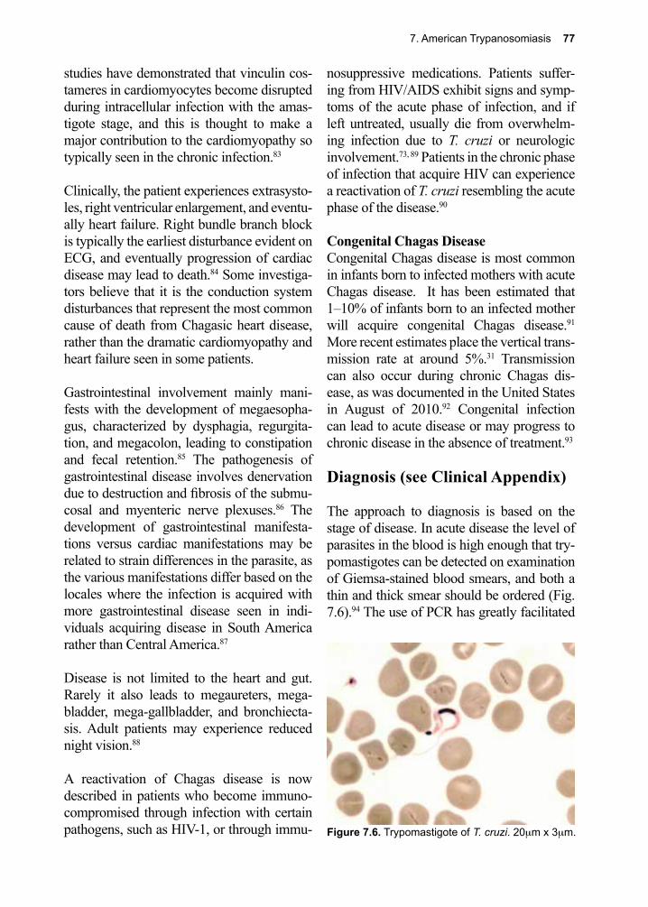
7. American Trypanosomiasis 77
studies have demonstrated that vinculin cos-tameres in cardiomyocytes become disrupted during intracellular infection with the amas-tigote stage, and this is thought to make a major contribution to the cardiomyopathy so typically seen in the chronic infection.83
Clinically, the patient experiences extrasysto-les, right ventricular enlargement, and eventu-ally heart failure. Right bundle branch block is typically the earliest disturbance evident on ECG, and eventually progression of cardiac disease may lead to death.84 Some investiga-tors believe that it is the conduction system disturbances that represent the most common cause of death from Chagasic heart disease, rather than the dramatic cardiomyopathy and heart failure seen in some patients.
Gastrointestinal involvement mainly mani-fests with the development of megaesopha-gus, characterized by dysphagia, regurgita-tion, and megacolon, leading to constipation and fecal retention.85 The pathogenesis of gastrointestinal disease involves denervation due to destruction and fibrosis of the submu-cosal and myenteric nerve plexuses.86 The development of gastrointestinal manifesta-tions versus cardiac manifestations may be related to strain differences in the parasite, as the various manifestations differ based on the locales where the infection is acquired with more gastrointestinal disease seen in indi-viduals acquiring disease in South America rather than Central America.87
Disease is not limited to the heart and gut. Rarely it also leads to megaureters, mega-bladder, mega-gallbladder, and bronchiecta-sis. Adult patients may experience reduced night vision.88
A reactivation of Chagas disease is now described in patients who become immuno-compromised through infection with certain pathogens, such as HIV-1, or through immu-
nosuppressive medications. Patients suffer-ing from HIV/AIDS exhibit signs and symp-toms of the acute phase of infection, and if left untreated, usually die from overwhelm-ing infection due to T. cruzi or neurologic involvement.73, 89 Patients in the chronic phase of infection that acquire HIV can experience a reactivation of T. cruzi resembling the acute phase of the disease.90
Congenital Chagas DiseaseCongenital Chagas disease is most common in infants born to infected mothers with acute Chagas disease. It has been estimated that 1–10% of infants born to an infected mother will acquire congenital Chagas disease.91 More recent estimates place the vertical trans-mission rate at around 5%.31 Transmission can also occur during chronic Chagas dis-ease, as was documented in the United States in August of 2010.92 Congenital infection can lead to acute disease or may progress to chronic disease in the absence of treatment.93
Diagnosis (see Clinical Appendix)
The approach to diagnosis is based on the stage of disease. In acute disease the level of parasites in the blood is high enough that try-pomastigotes can be detected on examination of Giemsa-stained blood smears, and both a thin and thick smear should be ordered (Fig. 7.6).94 The use of PCR has greatly facilitated
Figure 7.6. Trypomastigote of T. cruzi. 20µm x 3µm.
78 The Protozoa
diagnosis in patients, allowing earlier detec-tion with improved sensitivity over micro-scopic evaluation of blood smears.94-96
For chronic disease the diagnosis is usually based on the detection of serum IgG.97 Since no currently available tests for Chagas serol-ogy have the required specificity, a diagnosis is based on two positive diagnostic serology tests.98 It is of note that many patients pres-ent to their physician after a positive screen-ing test result when they are donating blood. This screening test is not a diagnostic test and would not count as one of the two diag-nostic test results in the current Chagas diag-nosis paradigm. PCR tests are not routinely employed to diagnose chronic disease. Para-sites can also be identified microscopically from biopsy samples of infected tissue. Inoc-ulating blood from suspected individuals into susceptible animals can reveal the organism, but this approach presents too much imprac-ticality for most diagnostic facilities. Xenodi-agnosis, employing uninfected kissing bugs, allowing them to feed on the patient, then dissecting the bugs some days later, can also reveal the presence of parasites in chronically infected individuals, but it is a special test requiring extensive laboratory infrastructure and technical assistance. Currently xenodiag-nosis is rarely used.
Treatment (see Clinical Appendix)
The only drugs with proven efficacy against T. cruzi are nifurtimox and benznidazole.98-100 Both drugs are associated with high toxicity and incomplete cure rates in adults, especially when they are used to treat the chronic phase of the infection.100 Benznidazole has been recommended for use in children who have either just acquired the disease (i.e., con-genitally), or who are in the chronic phase of their infection.101 Nifurtimox disrupts T. cruzi metabolism of carbohydrates, while benzni-dazole may promote lethal double-stranded
DNA breaks in T. cruzi.102, 103 Both drugs can cause severe side effects including skin rashes, and hematologic disturbances. Also, prolonged treatments are required lasting 1–2 months, and many patients (up to one in five) cannot tolerate full treatment courses.
Additional sobering information suggests that once heart involvement begins, it may be too late for the drugs to have a clinical impact.104 There is an urgent need to develop improved drugs and clinical algorithms. Newer drugs are being developed, some of which take advantage of known metabolic pathways.105,
106 Another approach involves the develop-ment of a therapeutic vaccine to treat patients in their indeterminant phase in order to pre-vent or delay the onset of Chagas cardiomy-opathy.107 Such a vaccine would be poten-tially both cost-effective and cost-saving.108
Heart transplantation as a treatment modality for the cardiomyopathic aspects of Chagas disease has been performed for as long as heart transplantation has been tried in humans.109,
110 In fact, the fourth recipient ever to receive a heart transplant suffered from chronic T. cruzi infection. Current immunosuppressive regi-mens have allowed for successful transplan-tation in a large number of patients that had developed cardiac manifestations of Chagas disease.109, 110
Despite a paucity of well-controlled trials on the efficacy of treatment for acute Chagas disease, there is a general consensus that treatment should be given for both acute and congenitally acquired disease.111 Treatment decisions for chronic Chagas are based on patient’s age and overall health. It is generally recommended that patients with indetermi-nant stage Chagas, who are under the age of 50, undergo treatment. Treatment for patients over the age of 50 should be individualized based on risks and benefits. Treatment for patients with advanced cardiomyopathy or
7. American Trypanosomiasis 79
gastrointestinal manifestations is currently not recommended. Unfortunately, these rec-ommendations for treatment of chronic dis-ease are not based on well-controlled trials and a study treating patients with established cardiomyopathy did not show any positive impact on disease progression.104
Prevention and Control
Control of Chagas disease depends upon interfering with two major routes of trans-mission: vector-borne and transfusion. Con-trol of vectors, by prudent use of insecticides (pyrethroids), has significantly reduced trans-mission of T. cruzi in Brazil and Chile.112, 113 Unfortunately, this trend has been slow to spread to neighboring countries and there is now evidence of insecticide resistance.114 Prevalence remains high throughout Central and South America. Transfusion-induced infection is a problem, especially in countries where T. cruzi is not vector-borne, compli-cating the control of disease.115 Blood bank screening for T. cruzi should be mandatory in all countries experiencing high rates of immi-gration from South and Central America. In the United States there is currently selective screening of blood using serological testing
to reduce the chance of transfusion-related transmission of Chagas disease.116 It is rec-ommended that paid blood donors be banned in all countries in which Chagas disease is endemic.115
A more permanent solution, and one that interfaces well with the concepts of medi-cal ecology, is building better housing for the poor.117, 118 Houses constructed without a thatched roof, with slat board wood siding, or rough textured wall surfaces inside the house are relatively safe from kissing bug coloni-zation. Keeping pet dogs and pigs out of the house further reduces the chances of acquir-ing Chagas disease.119
The risk to travelers is thought to be low, although there have been documented cases in travelers to endemic areas.120 It is recom-mended that travelers sleep under insecti-cide-treated bed nets and avoid sleeping in dwellings that provide habitats favoring the survival and reproduction of kissing bugs.121 Consuming sugar cane juice or fruit drinks while on vacation on the beaches of Brazil, Central America or other parts of South America are likely low-risk activities despite the occasional outbreaks reported.28-30
References
1. de Souza, W., et al., Review on Trypanosoma cruzi: Host Cell Interaction. Int J Cell Biol 2010, 2010.
2. De Araujo, V. A., et al., Mixed infection in the anteater Tamandua tetradactyla (Mammalia: Pilosa) from Para State, Brazil: Trypanosoma cruzi, T. rangeli and Leishmania infantum. Parasitology 2013, 140 (4), 455-60.
3. Rocha, F. L., et al., Trypanosoma cruzi TcI and TcII transmission among wild carnivores, small mammals and dogs in a conservation unit and surrounding areas, Brazil. Parasitology 2013, 140 (2), 160-70.
4. Rocha, F. L., et al., Trypanosoma cruzi infection in neotropical wild carnivores (Mammalia: Carnivora): at the top of the T. cruzi transmission chain. PLoS One 2013, 8 (7), e67463.
5. Roque, A. L., et al., Trypanosoma cruzi among wild and domestic mammals in different areas of the Abaetetuba municipality (Para State, Brazil), an endemic Chagas disease transmission area. Vet Parasitol 2013, 193 (1-3), 71-7.
6. Klotz, S. A., et al., Kissing bugs in the United States: risk for vector-borne disease in humans. Environ Health Insights 2014, 8 (Suppl 2), 49-59.
7. Bocchi, E. A., Heart failure in South America. Curr Cardiol Rev 2013, 9 (2), 147-56.
80 The Protozoa
8. Dokainish, H., et al., Heart Failure in Africa, Asia, the Middle East and South America: The INTER-CHF study. Int J Cardiol 2015, 204, 133-141.
9. Bocchi, E. A., et al., Cardiomyopathy, adult valve disease and heart failure in South America. Heart 2009, 95 (3), 181-9.
10. Schiffier, R. I.; Mansur, G. P., Indigenous Chagas’ disease (American trypanosomiasis) in California. JAMA 1984, 251 2983-2984.
11. Woody, N. C.; Woody, H. B., American trypanosomiasis (Chagas’ disease): first indigenous case in the USA. JAMA 1955, 159.
12. Global Burden of Disease Study, C., Global, regional, and national incidence, prevalence, and years lived with disability for 301 acute and chronic diseases and injuries in 188 countries, 1990-2013: a systematic analysis for the Global Burden of Disease Study 2013. Lancet 2015, 386 (9995), 743-800.
13. Lee, B. Y., et al., Global economic burden of Chagas disease: a computational simulation model. Lancet Infect Dis 2013, 13 (4), 342-8.
14. Arias-Negrete, S., et al., Expression of in vitro virulence by Entamoeba histolytica: effect of calmodulin inhibitors. APMIS : acta pathologica, microbiologica, et immunologica Scandinavica 1999, 107 (9), 875-81.
15. Hotez, P. J., et al., An unfolding tragedy of Chagas disease in North America. PLoS Negl Trop Dis 2013, 7 (10), e2300.
16. Garcia, M. N., et al., Evidence of autochthonous Chagas disease in southeastern Texas. Am J Trop Med Hyg 2015, 92 (2), 325-30.
17. Schmunis, G. A.; Yadon, Z. E., Chagas disease: a Latin American health problem becoming a world health problem. Acta Trop 2010, 115 (1-2), 14-21.
18. Martins-Melo, F. R., et al., Prevalence of Chagas disease in Brazil: a systematic review and meta-analysis. Acta Trop 2014, 130, 167-74.
19. Reiche, E. M., et al., Seropositivity for anti-Trypanosoma cruzi antibodies among blood donors of the “Hospital Universitário Regional do Norte do Paraná”, Londrina, Brazil. Revista do Instituto de Medicina Tropical de Sao Paulo 1996, 38 (3), 233-40.
20. Koberle, F., Chagas’ disease and Chagas’ syndromes: pathology of American trypanosomiasis. Adv Parasitol 1968, 6, 63-116.
21. Pecoul, B., et al., The BENEFIT Trial: Where Do We Go from Here? PLoS Negl Trop Dis 2016, 10 (2), e0004343.
22. Wendel, S., Transfusion-transmitted Chagas’ disease. Current opinion in hematology 1998, 5 (6), 406-11.
23. Blejer, J. L., et al., [Prevalence of Trypanosoma cruzi antibodies in blood donors]. Medicina 1999, 59 (2), 129-32.
24. Dictar, M., et al., Recipients and donors of bone marrow transplants suffering from Chagas’ disease: management and preemptive therapy of parasitemia. Bone marrow transplantation 1998, 21 (4), 391-3.
25. Carvalho, M. F., et al., Amastigotes forms of Trypanosoma cruzi detected in a renal allograft. Revista do Instituto de Medicina Tropical de Sao Paulo 1997, 39 (4), 223-6.
26. Altclas, J., et al., Chagas’ disease after bone marrow transplantation. Bone marrow transplantation 1996, 18 (2), 447-8.
27. Russomando, G., et al., Treatment of congenital Chagas’ disease diagnosed and followed up by the polymerase chain reaction. Am J Trop Med Hyg 1998, 59 (3), 487-91.
28. Shikanai-Yasuda, M. A.; Carvalho, N. B., Oral transmission of Chagas disease. Clinical infectious diseases : an official publication of the Infectious Diseases Society of America 2012, 54 (6), 845-52.
29. Benitez, J. A., et al., Urban outbreak of acute orally acquired Chagas disease in Tachira, Venezuela. J Infect Dev Ctries 2013, 7 (8), 638-41.
30. Alarcon de Noya, B., et al., Large urban outbreak of orally acquired acute Chagas disease at a school in Caracas, Venezuela. J Infect Dis 2010, 201 (9), 1308-15.
31. Gebrekristos, H. T.; Buekens, P., Mother-to-Child Transmission of Trypanosoma cruzi. J Pediatric Infect Dis Soc 2014, 3 Suppl 1, S36-40.
7. American Trypanosomiasis 81
32. Steindel, M., et al., Characterization of Trypanosoma cruzi isolated from humans, vectors, and animal reservoirs following an outbreak of acute human Chagas disease in Santa Catarina State, Brazil. Diagn Microbiol Infect Dis 2008, 60 (1), 25-32.
33. Yabsley, M. J., et al., Evaluation of the Chagas Stat-Pak assay for detection of Trypanosoma cruzi antibodies in wildlife reservoirs. J Parasitol 2009, 95 (3), 775-7.
34. Peterson, A. T., et al., Ecologic niche modeling and potential reservoirs for Chagas disease, Mexico. Emerg Infect Dis 2002, 8 (7), 662-7.
35. Lima, M. M., et al., Investigation of Chagas disease in four periurban areas in northeastern Brazil: epidemiologic survey in man, vectors, non-human hosts and reservoirs. Trans R Soc Trop Med Hyg 2012, 106 (3), 143-9.
36. Olsen, P. F., et al., Incidence of Trypanosoma Cruzi (Chagas) in Wild Vectors and Reservoirs in East-Central Alabama. J Parasitol 1964, 50, 599-603.
37. Zeledon, R., et al., Wild reservoirs of Trypanosoma cruzi with special mention of the opossum, Didelphis marsupialis, and its role in the epidemiology of Chagas’ disease in an endemic area of Costa Rica. J Parasitol 1970, 56 (1), 38.
38. Organization, W. H., Chile and Brazil to be certified free of transmission of Chagas’ Disease. Tropical Diseases. Research News (WHO publication) 1999, No. 59 June.
39. Chagas, C., Nova trypanozomiaze humans: estudos sobre a morfolojiia e o ciclo evalutivo do Schizotripanum cruzi. n genl n sp ajente etiolojico de nova entidade morbida do homen. Mem Inst Oswaldo Cruz 1909, 1, 159-218.
40. Brumpt, A. J. E., Le Trypanosoma cruzi evolue chez Conorhinus megistus. Cimex ectularius Cimex boueti et Ornithodorus moubata cycle evolutif de ce parasite Bull Soc Pathol Exot 1912, 5, 360-364.
41. Vianna, G., Contribuicao para o estudo da anatomia patolojica da anatomia patolojica ‘moletia de Carlos Chagas” (esquizotripanoze humana ou tireodite parazitaria). . Mem Inst Oswaldo Cruz 1916, 3, 276-294.
42. Campbell, A. K.; Matthews, S. B., Darwin’s illness revealed. Postgraduate medical journal 2005, 81 (954), 248-51.
43. Gull, K., The biology of kinetoplastid parasites: insights and challenges from genomics and post-genomics. International journal for parasitology 2001, 31 (5-6), 443-52.
44. Miles, M. A., et al., American trypanosomiasis (Chagas’ disease) and the role of molecular epidemiology in guiding control strategies. BMJ (Clinical research ed.) 2003, 326 (7404), 1444-8.
45. Mortara, R. A., et al., Mammalian cell invasion and intracellular trafficking by Trypanosoma cruzi infective forms. Anais da Academia Brasileira de Ciencias 2005, 77 (1), 77-94.
46. Senior, K., Chagas disease: moving towards global elimination. Lancet Infect Dis 2007, 7 (9), 572.
47. Nichols, N.; Green, T. W., Allergic reactions to “kissing bug” bites. Calif Med 1963, 98, 267-8.48. Kleshchenko, Y. Y., et al., Human galectin-3 promotes Trypanosoma cruzi adhesion to human
coronary artery smooth muscle cells. Infection and immunity 2004, 72 (11), 6717-21.49. Ortega-Barria, E.; Pereira, M. E., Entry of Trypanosoma cruzi into eukaryotic cells. Infectious
agents and disease 1992, 1 (3), 136-45.50. Herrera, E. M., et al., Mediation of Trypanosoma cruzi invasion by heparan sulfate receptors
on host cells and penetrin counter-receptors on the trypanosomes. Molecular and biochemical parasitology 1994, 65 (1), 73-83.
51. Neira, I., et al., Involvement of Trypanosoma cruzi metacyclic trypomastigote surface molecule gp82 in adhesion to gastric mucin and invasion of epithelial cells. Infection and immunity 2003, 71 (1), 557-61.
52. Calvo Mendez, M. L., et al., Calvo Nogueda Alejandre The oral route: an access port for Trypanosoma cruzi. Rev Latinoam Microbiol 1992, 34, 39-42.
53. Santos, C. C., et al., Chagasin, the endogenous cysteine-protease inhibitor of Trypanosoma cruzi, modulates parasite differentiation and invasion of mammalian cells. Journal of cell science 2005, 118 (Pt 5), 901-15.
82 The Protozoa
54. Aoki, M. P., et al., Cruzipain, a major Trypanosoma cruzi antigen, promotes arginase-2 expression and survival of neonatal mouse cardiomyocytes. Am Cell Physiol 2003, 286, C206-12.
55. Garcia, E. S.; Azambuja, P., Development and interactions of Trypanosoma cruzi within the insect vector. Parasitology today (Personal ed.) 1991, 7 (9), 240-4.
56. Pereira, M. E., et al., Lectin receptors as markers for Trypanosoma cruzi. Developmental stages and a study of the interaction of wheat germ agglutinin with sialic acid residues on epimastigote cells. The Journal of experimental medicine 1980, 152 (5), 1375-92.
57. Sher, A.; Snary, D., Specific inhibition of the morphogenesis of Trypanosoma cruzi by a monoclonal antibody. Nature London 1985, 300, 639-640.
58. Majumder, S.; Kierszenbaum, F., Mechanisms of Trypanosoma cruzi-induced down-regulation of lymphocyte function. Inhibition of transcription and expression of IL-2 receptor gamma (p64IL-2R) and beta (p70IL-2R) chain molecules in activated normal human lymphocytes. Journal of immunology (Baltimore, Md. : 1950) 1996, 156 (10), 3866-74.
59. Van Overtvelt, L., et al., Trypanosoma cruzi infects human dendritic cells and prevents their maturation: inhibition of cytokines, HLA-DR, and costimulatory molecules. Infection and immunity 1999, 67 (8), 4033-40.
60. Ferreira, V., et al., Role of calreticulin from parasites in its interaction with vertebrate hosts. Molecular immunology 2004, 40 (17), 1279-91.
61. Beucher, M., et al., Expression and purification of functional, recombinant Trypanosoma cruzi complement regulatory protein. Protein expression and purification 2003, 27 (1), 19-26.
62. Duthie, M. S., et al., Both CD1d antigen presentation and interleukin-12 are required to activate natural killer T cells during Trypanosoma cruzi infection. Infection and immunity 2005, 73 (3), 1890-4.
63. Vespa, G. N., et al., Nitric oxide is involved in control of Trypanosoma cruzi-induced parasitemia and directly kills the parasite in vitro. Infection and immunity 1994, 62 (11), 5177-82.
64. Martin, D. L.; Tarleton, R. L., Antigen-specific T cells maintain an effector memory phenotype during persistent Trypanosoma cruzi infection. Journal of immunology (Baltimore, Md. : 1950) 2005, 174 (3), 1594-601.
65. Bern, C., Antitrypanosomal therapy for chronic Chagas’ disease. N Engl J Med 2011, 364 (26), 2527-34.
66. Parada, H., et al., Cardiac involvement is a constant finding in acute Chagas’ disease: a clinical, parasitological and histopathological study. International journal of cardiology 1997, 60 (1), 49-54.
67. de Andrade, A. L., et al., Early electrocardiographic abnormalities in Trypanosoma cruzi-seropositive children. Am J Trop Med Hyg 1998, 59 (4), 530-4.
68. Tarleton, R. L., Chagas disease: a role for autoimmunity? Trends in parasitology 2003, 19 (10), 447-51.
69. Machado, F. S., et al., Pathogenesis of Chagas disease: time to move on. Front Biosci (Elite Ed) 2012, 4, 1743-58.
70. Vermelho, A. B., et al., Heart muscle cells share common neutral glycosphingolipids with Trypanosoma cruzi. Acta tropica 1997, 64 (3-4), 131-43.
71. Cunha-Neto, E., et al., Autoimmunity in Chagas’ disease. Identification of cardiac myosin-B13 Trypanosoma cruzi protein crossreactive T cell clones in heart lesions of a chronic Chagas’ cardiomyopathy patient. The Journal of clinical investigation 1996, 98 (8), 1709-12.
72. Roffe, E., et al., Essential role of VLA-4/VCAM-1 pathway in the establishment of CD8+ T-cell-mediated Trypanosoma cruzi-elicited meningoencephalitis. 2003, 142, 17-30.
73. Yasukawa, K., et al., Trypanosoma cruzi meningoencephalitis in a patient with acquired immunodeficiency syndrome. Am J Trop Med Hyg 2014, 91 (1), 84-5.
74. Anez, N., et al., Update on Chagas’ Disease in Venezuela-a review. Mem Inst Oswaldo Cruz 2004, 99, 781-7.
75. Kun, H., et al., Transmission of Trypanosoma cruzi by heart transplantation. Clin Infect Dis 2009, 48 (11), 1534-40.
7. American Trypanosomiasis 83
76. Tanowitz, H. B., et al., Perspectives on Trypanosoma cruzi-induced heart disease (Chagas disease). Prog Cardiovasc Dis 2009, 51 (6), 524-39.
77. Andrade, D. V., et al., Acute chagas disease: new global challenges for an old neglected disease. PLoS Negl Trop Dis 2014, 8 (7), e3010.
78. Beltrao Hde, B., et al., Investigation of two outbreaks of suspected oral transmission of acute Chagas disease in the Amazon region, Para State, Brazil, in 2007. Trop Doct 2009, 39 (4), 231-2.
79. Dias, J. C., [Cecilio Romana, Romana’s sign and Chagas’ disease]. Rev Soc Bras Med Trop 1997, 30 (5), 407-13.
80. Delaporte, F., Romana’s sign. J Hist Biol 1997, 30 (3), 357-66.81. Gutierrez, F. R., et al., The role of parasite persistence in pathogenesis of Chagas heart disease.
Parasite Immunol 2009, 31 (11), 673-85.82. Engman, D. M.; Leon, J. S., Pathogenesis of Chagas heart disease: role of autoimmunity. Acta
Trop 2002, 81 (2), 123-32.83. Melo, T. G., et al., Trypanosoma cruzi infection disrupts vinculin costameres in cardiomyocytes.
European journal of cell biology 2004, 83 (10), 531-40.84. Jorge, M. T., et al., Types of arrhythmia among cases of American trypanosomiasis, compared
with those in other cardiology patients. Annals of tropical medicine and parasitology 2003, 97 (2), 139-48.
85. Meneghelli, U. G., Chagasic enteropathy. Revista da Sociedade Brasileira de Medicina Tropical 2004, 37 (3), 252-60.
86. Jabari, S., et al., Chagasic megacolon: enteric neurons and related structures. Histochem Cell Biol 2014, 142 (3), 235-44.
87. Pinazo, M. J., et al., Characterization of digestive involvement in patients with chronic T. cruzi infection in Barcelona, Spain. PLoS Negl Trop Dis 2014, 8 (8), e3105.
88. Matsumoto, S. C., et al., Retinal dysfunction in patients with chronic Chagas’ disease is associated to anti-Trypanosoma cruzi antibodies that cross-react with rhodopsin. FASEB journal : official publication of the Federation of American Societies for Experimental Biology 2006, 20 (3), 550-2.
89. Antunes, A. C. M., et al., Cerebral trypanosomiasis and AIDS. Arquivos de neuro-psiquiatria 2002, 60 (3-B), 730-3.
90. Harms, G.; Feldmeier, H., The impact of HIV infection on tropical diseases. Infectious disease clinics of North America 2005, 19 (1), 121-35, ix.
91. Yadon, Z. E.; Schmunis, G. A., Congenital Chagas disease: estimating the potential risk in the United States. Am J Trop Med Hyg 2009, 81 (6), 927-33.
92. Centers for Disease, C.; Prevention, Congenital transmission of Chagas disease - Virginia, 2010. MMWR Morb Mortal Wkly Rep 2012, 61 (26), 477-9.
93. Carlier, Y., et al., Congenital Chagas disease: recommendations for diagnosis, treatment and control of newborns, siblings and pregnant women. PLoS Negl Trop Dis 2011, 5 (10), e1250.
94. Rosenblatt, J. E., Laboratory diagnosis of infections due to blood and tissue parasites. Clin Infect Dis 2009, 49 (7), 1103-8.
95. Britto, C., et al., Polymerase chain reaction detection of Trypanosoma cruzi in human blood samples as a tool for diagnosis and treatment evaluation. Parasitology 1995, 110 ( Pt 3), 241-7.
96. Junqueira, A. C., et al., Comparison of the polymerase chain reaction with two classical parasitological methods for the diagnosis of Chagas disease in an endemic region of north-eastern Brazil. Transactions of the Royal Society of Tropical Medicine and Hygiene 1996, 90 (2), 129-32.
97. Almeida, I. C., et al., A highly sensitive and specific chemiluminescent enzyme-linked immunosorbent assay for diagnosis of active Trypanosoma cruzi infection. Transfusion 1997, 37 (8), 850-7.
98. Bern, C., Chagas’ Disease. N Engl J Med 2015, 373 (5), 456-66.99. Kirchhoff, L. V., Changing Epidemiology and Approaches to Therapy for Chagas Disease.
Current infectious disease reports 2003, 5 (1), 59-65.
84 The Protozoa
100. Cerecetto, H.; González, M., Chemotherapy of Chagas’ disease: status and new developments. Current topics in medicinal chemistry 2002, 2 (11), 1187-213.
101. Schenone, H., et al., [Nifurtimox treatment of chronic Chagasic infection in children]. Revista medica de Chile 2003, 131 (9), 1089-90.
102. Diaz, E. G., et al., Reactions of nifurtimox with critical sulfhydryl-containing biomolecules: their potential toxicological relevance. Toxicol 2004, 24, 189-95.
103. Rajao, M. A., et al., Unveiling benznidazole’s mechanism of action through overexpression of DNA repair proteins in Trypanosoma cruzi. Environ Mol Mutagen 2014, 55 (4), 309-21.
104. Morillo, C. A., et al., Randomized Trial of Benznidazole for Chronic Chagas’ Cardiomyopathy. N Engl J Med 2015, 373 (14), 1295-306.
105. Engel, J. C., et al., Cysteine protease inhibitors cure an experimental Trypanosoma cruzi infection. The Journal of experimental medicine 1998, 188 (4), 725-34.
106. Choe, Y., et al., Development of alpha-keto-based inhibitors of cruzain, a cysteine protease implicated in Chagas disease. Bioorganic & medicinal chemistry 2005, 13 (6), 2141-56.
107. Dumonteil, E., et al., Accelerating the development of a therapeutic vaccine for human Chagas disease: rationale and prospects. Expert Rev Vaccines 2012, 11 (9), 1043-55.
108. Lee, B. Y., et al., Modeling the economic value of a Chagas’ disease therapeutic vaccine. Hum Vaccin Immunother 2012, 8 (9), 1293-301.
109. de Carvalho, V. B., et al., Heart transplantation in Chagas’ disease. 10 years after the initial experience. Circulation 1996, 94 (8), 1815-7.
110. Bocchi, E. A., et al., Heart transplantation for chronic Chagas’ heart disease. The Annals of thoracic surgery 1996, 61 (6), 1727-33.
111. Rodriques Coura, J.; de Castro, S. L., A critical review on Chagas disease chemotherapy. Mem Inst Oswaldo Cruz 2002, 97 (1), 3-24.
112. Schofield, C. J., et al., Advances in Parasitology. 1999; p 2-30.113. Moncayo, A., Chagas disease: current epidemiological trends after the interruption of vectorial
and transfusional transmission in the Southern Cone countries. Memorias do Instituto Oswaldo Cruz 2003, 98 (5), 577-91.
114. Zerba, E. N., Susceptibility and resistance to insecticides of Chagas disease vectors. Medicina (B Aires) 1999, 59 Suppl 2, 41-6.
115. Schmunis, G. A.; Cruz, J. R., Safety of the blood supply in Latin America. Clinical microbiology reviews 2005, 18 (1), 12-29.
116. Agapova, M., et al., Cost-effectiveness of screening the US blood supply for Trypanosoma cruzi. Transfusion 2010, 50 (10), 2220-32.
117. Chaudhuri, N., Interventions to improve children’s health by improving the housing environment. Reviews on environmental health 2004, 19 (3-4), 197-222.
118. Cecere, M. C., et al., Effects of partial housing improvement and insecticide spraying on the reinfestation dynamics of Triatoma infestans in rural northwestern Argentina. Acta tropica 2002, 84 (2), 101-16.
119. Cohen, J. E.; Gürtler, R. E., Modeling household transmission of American trypanosomiasis. Science (New York, N.Y.) 2001, 293 (5530), 694-8.
120. Carter, Y. L., et al., Acute Chagas disease in a returning traveler. Am J Trop Med Hyg 2012, 87 (6), 1038-40.
121. Kroeger, A., et al., Prevention of the transmission of Chagas’ disease with pyrethroid-impregnated materials. Am J Trop Med Hyg 2003, 68 (3), 307-11.
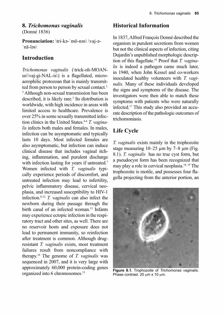
8. Trichomonas vaginalis 85
Figure 8.1. Trophozoite of Trichomonas vaginalis. Phase contrast. 20 µm x 10 µm.
8. Trichomonas vaginalis(Donné 1836)
Pronunciation: \tri-kə-ˈmō-nəs\ \vaj-ə-ˈnā-ləs\
Introduction
Trichomonas vaginalis (\trick-oh-MOAN-us\\vaj-gi-NAL-is\) is a flagellated, micro-aerophilic protozoan that is mainly transmit-ted from person to person by sexual contact.1,
2 Although non-sexual transmission has been described, it is likely rare.3 Its distribution is worldwide, with high incidence in areas with limited access to healthcare. Prevalence is over 25% in some sexually transmitted infec-tion clinics in the United States.4-6 T. vagina-lis infects both males and females. In males, infection can be asymptomatic and typically lasts 10 days. Most infected females are also asymptomatic, but infection can induce clinical disease that includes vaginal itch-ing, inflammation, and purulent discharge with infection lasting for years if untreated.7 Women infected with T. vaginalis typi-cally experience periods of discomfort, and untreated infection may lead to infertility, pelvic inflammatory disease, cervical neo-plasia, and increased susceptibility to HIV-1 infection.8-12 T. vaginalis can also infect the newborn during their passage through the birth canal of an infected woman.13 Infants may experience ectopic infection in the respi-ratory tract and other sites, as well. There are no reservoir hosts and exposure does not lead to permanent immunity, so reinfection after treatment is common. Although drug-resistant T. vaginalis exists, most treatment failures result from noncompliance with therapy.14 The genome of T. vaginalis was sequenced in 2007, and it is very large with approximately 60,000 protein-coding genes organized into 6 chromosomes.15
Historical Information
In 1837, Alfred François Donné described the organism in purulent secretions from women but not the clinical aspects of infection, citing Dujardin’s unpublished morphologic descrip-tion of this flagellate.16 Proof that T. vagina-lis is indeed a pathogen came much later, in 1940, when John Kessel and co-workers inoculated healthy volunteers with T. vagi-nalis. Many of these individuals developed the signs and symptoms of the disease. The investigators were then able to match these symptoms with patients who were naturally infected.17 This study also provided an accu-rate description of the pathologic outcomes of trichomoniasis.
Life Cycle
T. vaginalis exists mainly in the trophozoite stage measuring 10–25 µm by 7–8 µm (Fig. 8.1). T. vaginalis has no true cyst form, but a pseudocyst form has been recognized that may play a role in cervical neoplasia.18, 19 The trophozoite is motile, and possesses four fla-gella projecting from the anterior portion, an
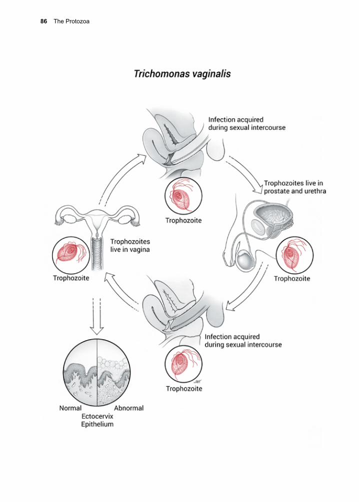
86 The Protozoa
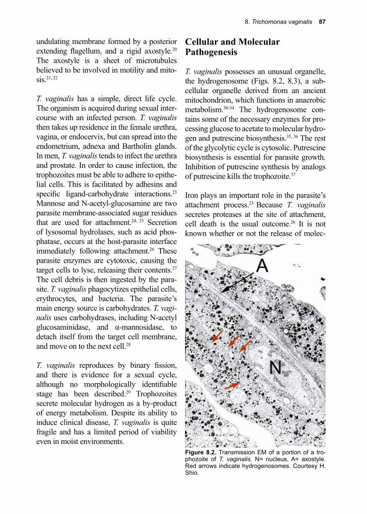
8. Trichomonas vaginalis 87
Figure 8.2. Transmission EM of a portion of a tro-phozoite of T. vaginalis. N= nucleus, A= axostyle. Red arrows indicate hydrogenosomes. Courtesy H. Shio.
undulating membrane formed by a posterior extending flagellum, and a rigid axostyle.20 The axostyle is a sheet of microtubules believed to be involved in motility and mito-sis.21, 22
T. vaginalis has a simple, direct life cycle. The organism is acquired during sexual inter-course with an infected person. T. vaginalis then takes up residence in the female urethra, vagina, or endocervix, but can spread into the endometrium, adnexa and Bartholin glands. In men, T. vaginalis tends to infect the urethra and prostate. In order to cause infection, the trophozoites must be able to adhere to epithe-lial cells. This is facilitated by adhesins and specific ligand-carbohydrate interactions.23 Mannose and N-acetyl-glucosamine are two parasite membrane-associated sugar residues that are used for attachment.24, 25 Secretion of lysosomal hydrolases, such as acid phos-phatase, occurs at the host-parasite interface immediately following attachment.26 These parasite enzymes are cytotoxic, causing the target cells to lyse, releasing their contents.27 The cell debris is then ingested by the para-site. T. vaginalis phagocytizes epithelial cells, erythrocytes, and bacteria. The parasite’s main energy source is carbohydrates. T. vagi-nalis uses carbohydrases, including N-acetyl glucosaminidase, and α-mannosidase, to detach itself from the target cell membrane, and move on to the next cell.28
T. vaginalis reproduces by binary fission, and there is evidence for a sexual cycle, although no morphologically identifiable stage has been described.29 Trophozoites secrete molecular hydrogen as a by-product of energy metabolism. Despite its ability to induce clinical disease, T. vaginalis is quite fragile and has a limited period of viability even in moist environments.
Cellular and Molecular Pathogenesis
T. vaginalis possesses an unusual organelle, the hydrogenosome (Figs. 8.2, 8.3), a sub-cellular organelle derived from an ancient mitochondrion, which functions in anaerobic metabolism.30-34 The hydrogenosome con-tains some of the necessary enzymes for pro-cessing glucose to acetate to molecular hydro-gen and putrescine biosynthesis.35, 36 The rest of the glycolytic cycle is cytosolic. Putrescine biosynthesis is essential for parasite growth. Inhibition of putrescine synthesis by analogs of putrescine kills the trophozoite.37
Iron plays an important role in the parasite’s attachment process.23 Because T. vaginalis secretes proteases at the site of attachment, cell death is the usual outcome.26 It is not known whether or not the release of molec-
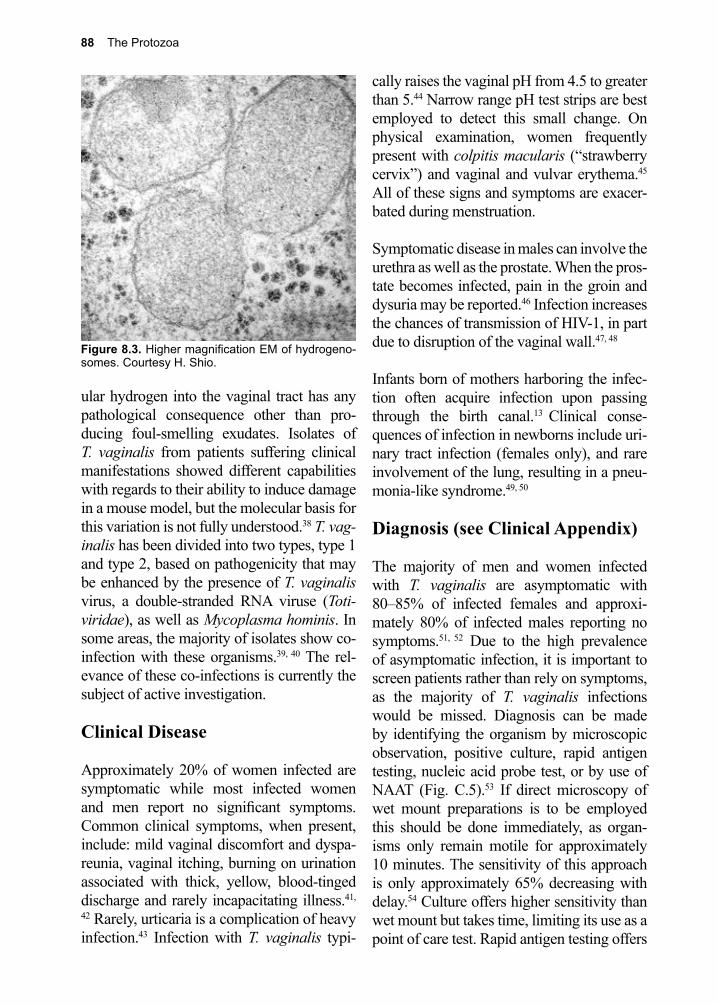
88 The Protozoa
ular hydrogen into the vaginal tract has any pathological consequence other than pro-ducing foul-smelling exudates. Isolates of T. vaginalis from patients suffering clinical manifestations showed different capabilities with regards to their ability to induce damage in a mouse model, but the molecular basis for this variation is not fully understood.38 T. vag-inalis has been divided into two types, type 1 and type 2, based on pathogenicity that may be enhanced by the presence of T. vaginalis virus, a double-stranded RNA viruse (Toti-viridae), as well as Mycoplasma hominis. In some areas, the majority of isolates show co-infection with these organisms.39, 40 The rel-evance of these co-infections is currently the subject of active investigation.
Clinical Disease
Approximately 20% of women infected are symptomatic while most infected women and men report no significant symptoms. Common clinical symptoms, when present, include: mild vaginal discomfort and dyspa-reunia, vaginal itching, burning on urination associated with thick, yellow, blood-tinged discharge and rarely incapacitating illness.41,
42 Rarely, urticaria is a complication of heavy infection.43 Infection with T. vaginalis typi-
cally raises the vaginal pH from 4.5 to greater than 5.44 Narrow range pH test strips are best employed to detect this small change. On physical examination, women frequently present with colpitis macularis (“strawberry cervix”) and vaginal and vulvar erythema.45 All of these signs and symptoms are exacer-bated during menstruation.
Symptomatic disease in males can involve the urethra as well as the prostate. When the pros-tate becomes infected, pain in the groin and dysuria may be reported.46 Infection increases the chances of transmission of HIV-1, in part due to disruption of the vaginal wall.47, 48
Infants born of mothers harboring the infec-tion often acquire infection upon passing through the birth canal.13 Clinical conse-quences of infection in newborns include uri-nary tract infection (females only), and rare involvement of the lung, resulting in a pneu-monia-like syndrome.49, 50
Diagnosis (see Clinical Appendix)
The majority of men and women infected with T. vaginalis are asymptomatic with 80–85% of infected females and approxi-mately 80% of infected males reporting no symptoms.51, 52 Due to the high prevalence of asymptomatic infection, it is important to screen patients rather than rely on symptoms, as the majority of T. vaginalis infections would be missed. Diagnosis can be made by identifying the organism by microscopic observation, positive culture, rapid antigen testing, nucleic acid probe test, or by use of NAAT (Fig. C.5).53 If direct microscopy of wet mount preparations is to be employed this should be done immediately, as organ-isms only remain motile for approximately 10 minutes. The sensitivity of this approach is only approximately 65% decreasing with delay.54 Culture offers higher sensitivity than wet mount but takes time, limiting its use as a point of care test. Rapid antigen testing offers
Figure 8.3. Higher magnification EM of hydrogeno-somes. Courtesy H. Shio.
8. Trichomonas vaginalis 89
a point of care option and currently these tests are commercially available, but may have variable degrees of sensitivity depending on the test selected.55 Nucleic acid probe testing is a highly sensitive option for diagnosis that has demonstrated efficacy for testing of vagi-nal swabs or urine.55 Diagnosis by NAAT is, by far, more sensitive than any other method, and is now the preferred method in most hos-pital parasitology diagnostic laboratories.53
Treatment (see Clinical Appendix)
Metronidazole continues to be the drug of choice.1 It can be given orally as a single 2 g dose but is often given as a 7-day course at 500 mg twice a day. Metronidazole is also available as a vaginal gel. Its use as an intra-vaginal suppository did poorly in a number of clinical trials.45 The drug is typically well tol-erated, but metallic taste, antabuse-like side effects with alcohol consumption and longer term treatment could have other toxicities.56 Metronidazole is converted to active interme-diates by hydrogenosome-associated pyru-vate ferredoxin oxidoreductase and hydrog-enase under anaerobic conditions. The para-site is inhibited from growing by exposure to those intermediates, but the precise biochem-ical mechanisms of the process are unknown.
Resistant strains (approximately 2–5% of all infections) have inactive forms of pyruvate ferridoxin oxidoreductase and hydrogenase, deriving all their energy from glucose by alternate pathways.57 Tinidazole, an alternate drug for treating infection, is now available and may be better tolerated as a single 2 g dose option, but may be more costly.1 Reinfection is likely if the infected sexual partner is not treated concurrently. Treatment of the partner, expedited partner therapy (EPT), results in a significant reduction in reinfection but in high prevalence settings, reinfection is still seen.58 Recurrence may represent reinfection rather than treatment failure but in refractory cases where drug resistance is suspected therapeu-tic options are available and drug sensitivity testing can be performed.
Prevention and Control
Use of a condom during sexual intercourse and limiting the number of sexual partners should reduce the risk of infection. Treating all sexual partners with metronidazole can be effective in some cases, particularly when the number of sexual partners is low. Active screening is essential with the high rate of asymptomatic infections.
References
1. Schwebke, J. R.; Burgess, D., Trichomoniasis. Clinical microbiology reviews 2004, 17 (4), 794-803, table of contents.
2. Lehker, M. W.; Alderete, J. F., Biology of trichomonosis. Current opinion in infectious diseases 2000, 13 (1), 37-45.
3. Crucitti, T., et al., Non-sexual transmission of Trichomonas vaginalis in adolescent girls attending school in Ndola, Zambia. PLoS One 2011, 6 (1), e16310.
4. Meites, E., et al., Trichomonas vaginalis in selected U.S. sexually transmitted disease clinics: testing, screening, and prevalence. Sex Transm Dis 2013, 40 (11), 865-9.
5. Johnston, V. J.; Mabey, D. C., Global epidemiology and control of Trichomonas vaginalis. Curr Opin Infect Dis 2008, 21 (1), 56-64.
6. Leitsch, D., Recent Advances in the Trichomonas vaginalis field. F1000Res 2016, 5.7. Krieger, J. N., Trichomoniasis in men: old issues and new data. Sex Transm Dis 1995, 22 (2), 83-
96.8. Sayed el-Ahl, S. A., et al., A preliminary study on the relationship between Trichomonas
vaginalis and cervical cancer in Egyptian women. J Egypt Soc Parasitol 2002, 32 (1), 167-78.
90 The Protozoa
9. Moodley, P., et al., Trichomonas vaginalis is associated with pelvic inflammatory disease in women infected with human immunodeficiency virus. Clin Infect Dis 2002, 34 (4), 519-22.
10. El-Shazly, A. M., et al., A study on Trichomoniasis vaginalis and female infertility. J Egypt Soc Parasitol 2001, 31 (2), 545-53.
11. Kissinger, P.; Adamski, A., Trichomoniasis and HIV interactions: a review. Sex Transm Infect 2013, 89 (6), 426-33.
12. Chesson, H. W., et al., Estimates of the annual number and cost of new HIV infections among women attributable to trichomoniasis in the United States. Sex Transm Dis 2004, 31 (9), 547-51.
13. Smith, L. M., et al., Trichomonas vaginalis infection in a premature newborn. Journal of perinatology : official journal of the California Perinatal Association 2002, 22 (6), 502-3.
14. Kulda, J., Trichomonads, hydrogenosomes and drug resistance. International journal for parasitology 1999, 29 (2), 199-212.
15. Carlton, J. M., et al., Draft genome sequence of the sexually transmitted pathogen Trichomonas vaginalis. Science 2007, 315 (5809), 207-12.
16. Donne, A.; R., C., Animalcules observes dans la matieres purulentes et le produit des secretions des or-ganes genitaux de l’homme et da Ia femme. Seanc Acad Sci 1837, 3, 385-386.
17. Kessel, I. F., et al., Observations on the pathology of Trichomonas vaginitis and on vaginal implants with Trichomonas vaginalis and Trichomonas intestinalis. Am Gynecol 1940, 39, 1005-1014.
18. Pereira-Neves, A., et al., Pseudocysts in trichomonads--new insights. Protist 2003, 154 (3-4), 313-29.
19. Afzan, M. Y.; Suresh, K., Pseudocyst forms of Trichomonas vaginalis from cervical neoplasia. Parasitol Res 2012, 111 (1), 371-81.
20. Kissinger, P., Trichomonas vaginalis: a review of epidemiologic, clinical and treatment issues. BMC Infect Dis 2015, 15, 307.
21. Benchimol, M., et al., The fine structure of the axostyle and its associations with organelles in Trichomonads. Tissue Cell 2000, 32 (2), 178-87.
22. Ribeiro, K. C., et al., Contributions of the axostyle and flagella to closed mitosis in the protists Tritrichomonas foetus and Trichomonas vaginalis. J Eukaryot Microbiol 2000, 47 (5), 481-92.
23. Garcia, A. F., et al., Iron and contact with host cells induce expression of adhesins on surface of Trichomonas vaginalis. Molecular microbiology 2003, 47 (5), 1207-24.
24. Mirhaghani, A.; Warton, A., Involvement of Trichomonas vaginalis surface-associated glycoconjugates in the parasite/target cell interaction. A quantitative electron microscopy study. Parasitology research 1998, 84 (5), 374-81.
25. Singh, B. N., et al., Adhesion of Tritrichomonas foetus to bovine vaginal epithelial cells. Infection and immunity 1999, 67 (8), 3847-54.
26. Lockwood, B. C., et al., The release of hydrolases from Trichomonas vaginalis and Tritrichomonas foetus. Molecular and biochemical parasitology 1988, 30 (2), 135-42.
27. Chen, W., et al., Study on ultrastructural cytochemistry and pathogenic mechanism of Trichomonas vaginalis. Chinese medical journal 1996, 109 (9), 695-9.
28. Savoia, D.; Martinotti, M. G., Secretory hydrolases of Trichomonas vaginalis. Microbiologica 1989, 12 (2), 133-8.
29. Conrad, M. D., et al., Extensive genetic diversity, unique population structure and evidence of genetic exchange in the sexually transmitted parasite Trichomonas vaginalis. PLoS Negl Trop Dis 2012, 6 (3), e1573.
30. Embley, T. M.; R., Giezen. M. Hydrogenosomes and mitochondria are two forms of the same fundamental organelle. Phil Trans Lond 2002, 358, 191-201.
31. Vanacova, S., et al., Molecular biology of the amitochondriate parasites, Giardia intestinalis, Entamoeba histolytica and Trichomonas vaginalis. International journal for parasitology 2003, 33 (3), 235-55.
32. PrDiaz, J. A.; Souza, W., De Purification and biochemical characterization of the hydrogenosomes of the flagellate protozoan Tritrichomonas foetus. Eur J Cell Biol 1997, 74 (1), 85-91.
8. Trichomonas vaginalis 91
33. Williams, B. A. P.; Keeling, P. J., Cryptic organelles in parasitic protists and fungi. Advances in parasitology 2003, 54, 9-68.
34. Lindmark, D. G.; Muller, M., Hydrogenosome, a cytoplasmic organelle of the anaerobic flagellate Tritrichomonas foetus, and its role in pyruvate metabolism. J Biol Chem 1973, 248 (22), 7724-8.
35. Wu, G.; Müller, M., Glycogen phosphorylase sequences from the amitochondriate protists, Trichomonas vaginalis, Mastigamoeba balamuthi, Entamoeba histolytica and Giardia intestinalis. The Journal of eukaryotic microbiology 2003, 50 (5), 366-72.
36. North, M. J., et al., Polyamine biosynthesis in trichomonads. Molecular and biochemical parasitology 1986, 19 (3), 241-9.
37. Reis, I. A., et al., Inhibition of polyamine synthesis arrests trichomonad growth and induces destruction of hydrogenosomes. Antimicrobial agents and chemotherapy 1999, 43 (8), 1919-23.
38. Hussien, E. M., et al., Biological variability of Trichomonas vaginalis clinical isolates from symptomatic and asymptomatic patients. Journal of the Egyptian Society of Parasitology 2004, 34 (3), 979-88.
39. da Luz Becker, D., et al., High rates of double-stranded RNA viruses and Mycoplasma hominis in Trichomonas vaginalis clinical isolates in South Brazil. Infect Genet Evol 2015, 34, 181-7.
40. Goodman, R. P., et al., Clinical isolates of Trichomonas vaginalis concurrently infected by strains of up to four Trichomonasvirus species (Family Totiviridae). J Virol 2011, 85 (9), 4258-70.
41. Deligeoroglou, E., et al., Infections of the lower female genital tract during childhood and adolescence. Clinical and experimental obstetrics & gynecology 2004, 31 (3), 175-8.
42. Petrin, D., et al., Clinical and microbiological aspects of Trichomonas vaginalis. Clinical microbiology reviews 1998, 11 (2), 300-17.
43. Purello-D’Ambrosio, F.; Gangemi, S., Urticaria from Trichomonas vaginalis infection. Allergol Clin Immunol 1999, 9 (2), 123-5.
44. Petrin, D., et al., Clinical and microbiological aspects of Trichomonas vaginalis. Clin Microbiol Rev 1998, 11 (2), 300-17.
45. Wolner-Hanssen, P., et al., Clinical manifestations of vaginal trichomoniasis. JAMA 1989, 26, 571-576.
46. Krieger, J. N.; Riley, D. E., Chronic prostatitis: Charlottesville to Seattle. The Journal of urology 2004, 172 (6 Pt 2), 2557-60.
47. Mason, P. R., et al., Seroepidemiology of Trichomonas vaginalis in rural women in Zimbabwe and patterns of association with HIV infection. Epidemiology and infection 2005, 133 (2), 315-23.
48. Moodley, P., et al., Trichomonas vaginalis is associated with pelvic inflammatory disease in women infected with human immunodeficiency virus. Clin Inf Dis 2002, 34 (4), 519-22.
49. Hoffman, D. J., et al., Urinary tract infection with Trichomonas vaginalis in a premature newborn infant and the development of chronic lung disease. Journal of perinatology : official journal of the California Perinatal Association 2003, 23 (1), 59-61.
50. Szarka, K., et al., Neonatal pneumonia caused by Trichomonas vaginalis. Acta microbiologica et immunologica Hungarica 2002, 49 (1), 15-9.
51. Sutton, M., et al., The prevalence of Trichomonas vaginalis infection among reproductive-age women in the United States, 2001-2004. Clin Infect Dis 2007, 45 (10), 1319-26.
52. Sena, A. C., et al., Trichomonas vaginalis infection in male sexual partners: implications for diagnosis, treatment, and prevention. Clin Infect Dis 2007, 44 (1), 13-22.
53. Negm, A. Y.; el-Haleem, D. A., Detection of trichomoniasis in vaginal specimens by both conventional and modern molecular tools. Journal of the Egyptian Society of Parasitology 2004, 34 (2), 589-600.
54. Krieger, J. N., et al., Diagnosis of trichomoniasis. Comparison of conventional wet-mount examination with cytologic studies, cultures, and monoclonal antibody staining of direct specimens. JAMA 1988, 259 (8), 1223-7.
92 The Protozoa
55. Andrea, S. B.; Chapin, K. C., Comparison of Aptima Trichomonas vaginalis transcription-mediated amplification assay and BD affirm VPIII for detection of T. vaginalis in symptomatic women: performance parameters and epidemiological implications. J Clin Microbiol 2011, 49 (3), 866-9.
56. Freeman, C. D., et al., Metronidazole. A therapeutic review and update. Drugs 1997, 54 (5), 679-708.
57. Muller, M.; Lossick, J. G., in vitro susceptibility of Trichomonas vaginalis to metronidazole and treatment outcome in vaginal trichomoniasis. Sex Trans Dis 1988, 15, 17-24.
58. Lyng, J.; Christensen, J., A double-blind study of the value of treatment with a single dose tinidazole of partners to females with trichomoniasis. Acta Obstet Gynecol Scand 1981, 60 (2), 199-201.
Alexandre Joseph Émil Brumpt, M.D. (1877–1951)
Brumpt was an accomplished zoologist, working on many species of parasites over his lifetime. He published papers on malaria in birds (Plas-modium gallinaceum), tsetse fly biology, leishmaniasis, Trypanosoma cruzi, Onchocerca volvulus, and Blastocystis hominis. He also made the distinction between pathogenic Entamoeba histolytica and its harmless protozoan doppelganger, Entamoeba dispar.
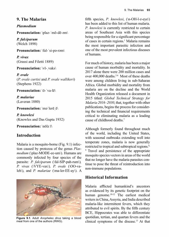
9. The Malarias 93
Figure 9.1. Adult Anopheles dirus taking a blood meal from one of the authors (RWG).
9. The MalariasPlasmodium
Pronunciation: \plaz-ˈmō-dē-əm\
P. falciparum(Welch 1898)
Pronunciation: \fal-ˈsi-pə-rəm\
P. vivax (Grassi and Filetti 1889)
Pronunciation: \vī-ˌvaks\
P. ovale (P. ovale curtisi and P. ovale wallikeri) (Stephens 1922)
Pronunciation: \ō-ˈva-lē\
P. malariae (Laveran 1880)
Pronunciation: \məˈlerēˌē\
P. knowlesi (Knowles and Das Gupta 1932)
Pronunciation: \nōlz ī\
Introduction
Malaria is a mosquito-borne (Fig. 9.1) infec-tion caused by protozoa of the genus Plas-modium (\plaz-MODE-ee-um\). Humans are commonly infected by four species of the parasite: P. falciparum (\fal-SIP-pah-rum\), P. vivax (\VYE-vax\), P. ovale (\OO-va-leh\), and P. malariae (\ma-ler-EE-ay\). A
fifth species, P. knowlesi, (\n-OH-l-z-eye\) has been added to this list of human malaria. P. knowlesi is currently restricted to certain areas of Southeast Asia with this species being responsible for a significant percentage of cases in certain regions.1 Malaria remains the most important parasitic infection and one of the most prevalent infectious diseases of humans.
For much of history, malaria has been a major cause of human morbidity and mortality. In 2017 alone there were 200 million cases and over 400,000 deaths.2-6 Most of these deaths were among children living in sub-Saharan Africa. Global morbidity and mortality from malaria are on the decline and the World Health Organization released a document in 2015 titled: Global Technical Strategy for Malaria 2016–2030, that, together with other publications, begins the process for consider-ing the technical and financial requirements critical to eliminating malaria as a leading cause of childhood deaths.7
Although formerly found throughout much of the world, including the United States, with seasonal outbreaks extending well into temperate zones, malaria is now generally restricted to tropical and subtropical regions.8,
9 Travel and persistence of the appropriate mosquito species vectors in areas of the world that no longer have the malaria parasites con-tinue to pose the threat of reintroduction into non-immune populations.
Historical Information
Malaria afflicted humankind’s ancestors as evidenced by its genetic footprint on the human genome.10-12 The earliest medical writers in China, Assyria, and India described malaria-like intermittent fevers, which they attributed to evil spirits. By the fifth century BCE, Hippocrates was able to differentiate quotidian, tertian, and quartan fevers and the clinical symptoms of the disease.13 At that
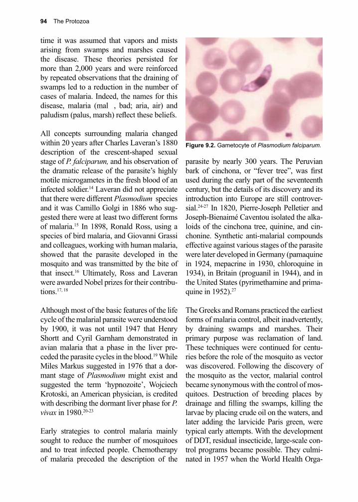
94 The Protozoa
time it was assumed that vapors and mists arising from swamps and marshes caused the disease. These theories persisted for more than 2,000 years and were reinforced by repeated observations that the draining of swamps led to a reduction in the number of cases of malaria. Indeed, the names for this disease, malaria (mal , bad; aria, air) and paludism (palus, marsh) reflect these beliefs.
All concepts surrounding malaria changed within 20 years after Charles Laveran’s 1880 description of the crescent-shaped sexual stage of P. falciparum, and his observation of the dramatic release of the parasite’s highly motile microgametes in the fresh blood of an infected soldier.14 Laveran did not appreciate that there were different Plasmodium species and it was Camillo Golgi in 1886 who sug-gested there were at least two different forms of malaria.15 In 1898, Ronald Ross, using a species of bird malaria, and Giovanni Grassi and colleagues, working with human malaria, showed that the parasite developed in the mosquito and was transmitted by the bite of that insect.16 Ultimately, Ross and Laveran were awarded Nobel prizes for their contribu-tions.17, 18
Although most of the basic features of the life cycle of the malarial parasite were understood by 1900, it was not until 1947 that Henry Shortt and Cyril Garnham demonstrated in avian malaria that a phase in the liver pre-ceded the parasite cycles in the blood.19 While Miles Markus suggested in 1976 that a dor-mant stage of Plasmodium might exist and suggested the term ‘hypnozoite’, Wojciech Krotoski, an American physician, is credited with describing the dormant liver phase for P. vivax in 1980.20-23
Early strategies to control malaria mainly sought to reduce the number of mosquitoes and to treat infected people. Chemotherapy of malaria preceded the description of the
parasite by nearly 300 years. The Peruvian bark of cinchona, or “fever tree”, was first used during the early part of the seventeenth century, but the details of its discovery and its introduction into Europe are still controver-sial.24-27 In 1820, Pierre-Joseph Pelletier and Joseph-Bienaimé Caventou isolated the alka-loids of the cinchona tree, quinine, and cin-chonine. Synthetic anti-malarial compounds effective against various stages of the parasite were later developed in Germany (pamaquine in 1924, mepacrine in 1930, chloroquine in 1934), in Britain (proguanil in 1944), and in the United States (pyrimethamine and prima-quine in 1952).27
The Greeks and Romans practiced the earliest forms of malaria control, albeit inadvertently, by draining swamps and marshes. Their primary purpose was reclamation of land. These techniques were continued for centu-ries before the role of the mosquito as vector was discovered. Following the discovery of the mosquito as the vector, malarial control became synonymous with the control of mos-quitoes. Destruction of breeding places by drainage and filling the swamps, killing the larvae by placing crude oil on the waters, and later adding the larvicide Paris green, were typical early attempts. With the development of DDT, residual insecticide, large-scale con-trol programs became possible. They culmi-nated in 1957 when the World Health Orga-
Figure 9.2. Gametocyte of Plasmodium falciparum.
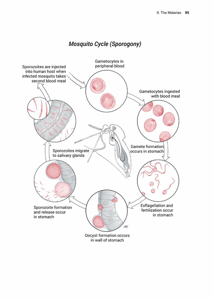
9. The Malarias 95
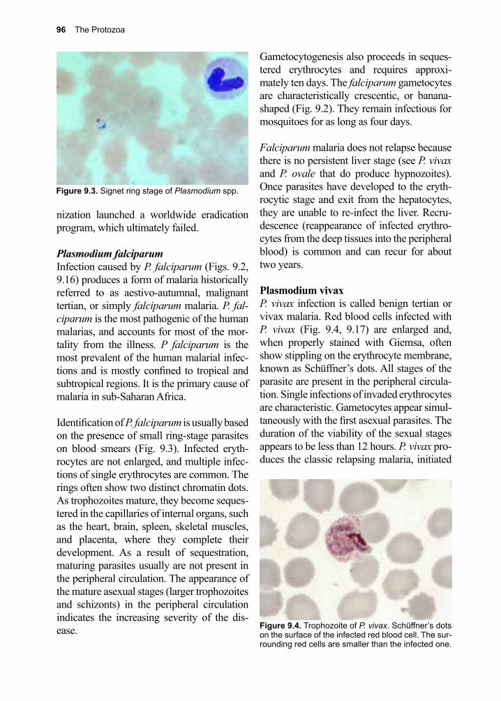
96 The Protozoa
nization launched a worldwide eradication program, which ultimately failed.
Plasmodium falciparumInfection caused by P. falciparum (Figs. 9.2, 9.16) produces a form of malaria historically referred to as aestivo-autumnal, malignant tertian, or simply falciparum malaria. P. fal-ciparum is the most pathogenic of the human malarias, and accounts for most of the mor-tality from the illness. P falciparum is the most prevalent of the human malarial infec-tions and is mostly confined to tropical and subtropical regions. It is the primary cause of malaria in sub-Saharan Africa.
Identification of P. falciparum is usually based on the presence of small ring-stage parasites on blood smears (Fig. 9.3). Infected eryth-rocytes are not enlarged, and multiple infec-tions of single erythrocytes are common. The rings often show two distinct chromatin dots. As trophozoites mature, they become seques-tered in the capillaries of internal organs, such as the heart, brain, spleen, skeletal muscles, and placenta, where they complete their development. As a result of sequestration, maturing parasites usually are not present in the peripheral circulation. The appearance of the mature asexual stages (larger trophozoites and schizonts) in the peripheral circulation indicates the increasing severity of the dis-ease.
Gametocytogenesis also proceeds in seques-tered erythrocytes and requires approxi-mately ten days. The falciparum gametocytes are characteristically crescentic, or banana-shaped (Fig. 9.2). They remain infectious for mosquitoes for as long as four days.
Falciparum malaria does not relapse because there is no persistent liver stage (see P. vivax and P. ovale that do produce hypnozoites). Once parasites have developed to the eryth-rocytic stage and exit from the hepatocytes, they are unable to re-infect the liver. Recru-descence (reappearance of infected erythro-cytes from the deep tissues into the peripheral blood) is common and can recur for about two years.
Plasmodium vivaxP. vivax infection is called benign tertian or vivax malaria. Red blood cells infected with P. vivax (Fig. 9.4, 9.17) are enlarged and, when properly stained with Giemsa, often show stippling on the erythrocyte membrane, known as Schüffner’s dots. All stages of the parasite are present in the peripheral circula-tion. Single infections of invaded erythrocytes are characteristic. Gametocytes appear simul-taneously with the first asexual parasites. The duration of the viability of the sexual stages appears to be less than 12 hours. P. vivax pro-duces the classic relapsing malaria, initiated
Figure 9.3. Signet ring stage of Plasmodium spp.
Figure 9.4. Trophozoite of P. vivax. Schüffner’s dots on the surface of the infected red blood cell. The sur-rounding red cells are smaller than the infected one.
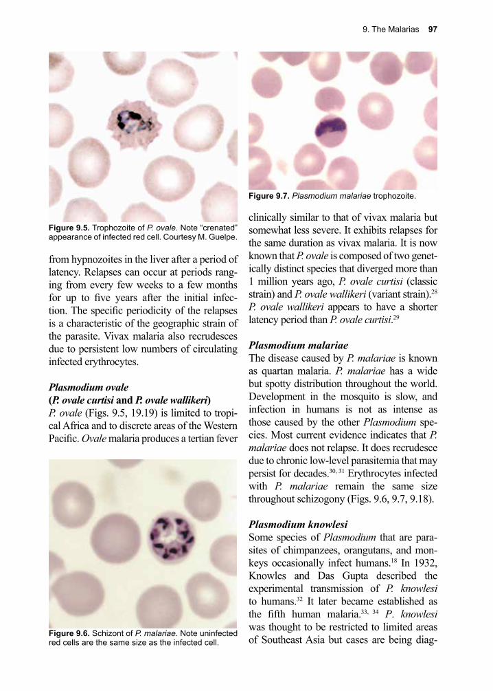
9. The Malarias 97
from hypnozoites in the liver after a period of latency. Relapses can occur at periods rang-ing from every few weeks to a few months for up to five years after the initial infec-tion. The specific periodicity of the relapses is a characteristic of the geographic strain of the parasite. Vivax malaria also recrudesces due to persistent low numbers of circulating infected erythrocytes.
Plasmodium ovale (P. ovale curtisi and P. ovale wallikeri)P. ovale (Figs. 9.5, 19.19) is limited to tropi-cal Africa and to discrete areas of the Western Pacific. Ovale malaria produces a tertian fever
clinically similar to that of vivax malaria but somewhat less severe. It exhibits relapses for the same duration as vivax malaria. It is now known that P. ovale is composed of two genet-ically distinct species that diverged more than 1 million years ago, P. ovale curtisi (classic strain) and P. ovale wallikeri (variant strain).28 P. ovale wallikeri appears to have a shorter latency period than P. ovale curtisi.29
Plasmodium malariaeThe disease caused by P. malariae is known as quartan malaria. P. malariae has a wide but spotty distribution throughout the world. Development in the mosquito is slow, and infection in humans is not as intense as those caused by the other Plasmodium spe-cies. Most current evidence indicates that P. malariae does not relapse. It does recrudesce due to chronic low-level parasitemia that may persist for decades.30, 31 Erythrocytes infected with P. malariae remain the same size throughout schizogony (Figs. 9.6, 9.7, 9.18).
Plasmodium knowlesiSome species of Plasmodium that are para-sites of chimpanzees, orangutans, and mon-keys occasionally infect humans.18 In 1932, Knowles and Das Gupta described the experimental transmission of P. knowlesi to humans.32 It later became established as the fifth human malaria.33, 34 P. knowlesi was thought to be restricted to limited areas of Southeast Asia but cases are being diag-
Figure 9.5. Trophozoite of P. ovale. Note “crenated” appearance of infected red cell. Courtesy M. Guelpe.
Figure 9.6. Schizont of P. malariae. Note uninfected red cells are the same size as the infected cell.
Figure 9.7. Plasmodium malariae trophozoite.
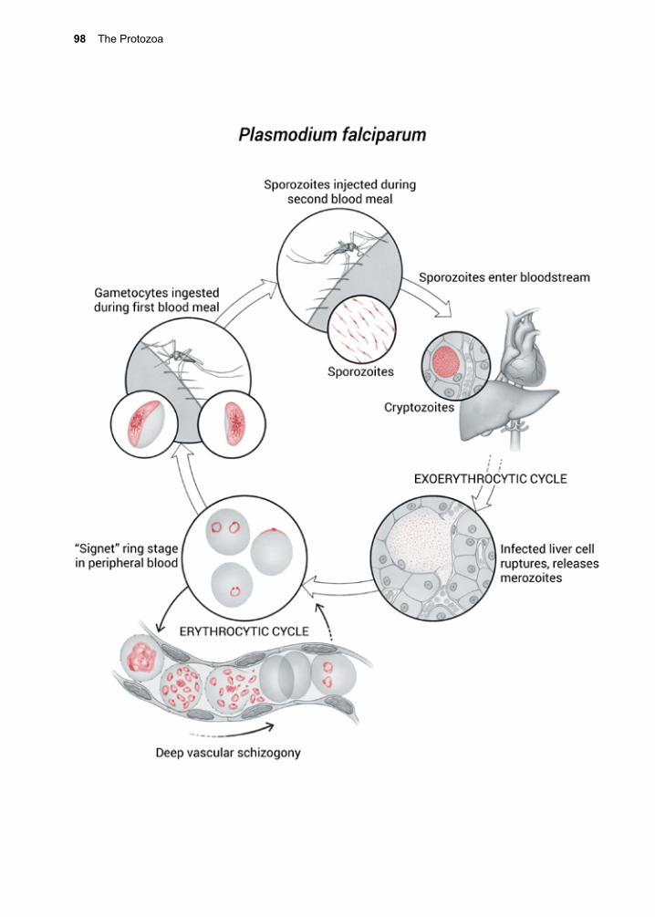
98 The Protozoa
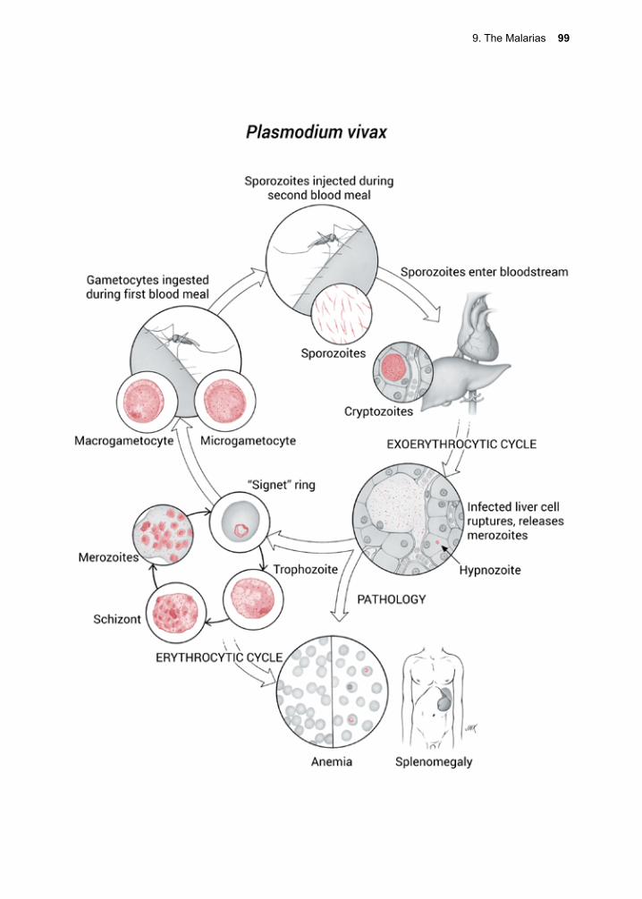
9. The Malarias 99
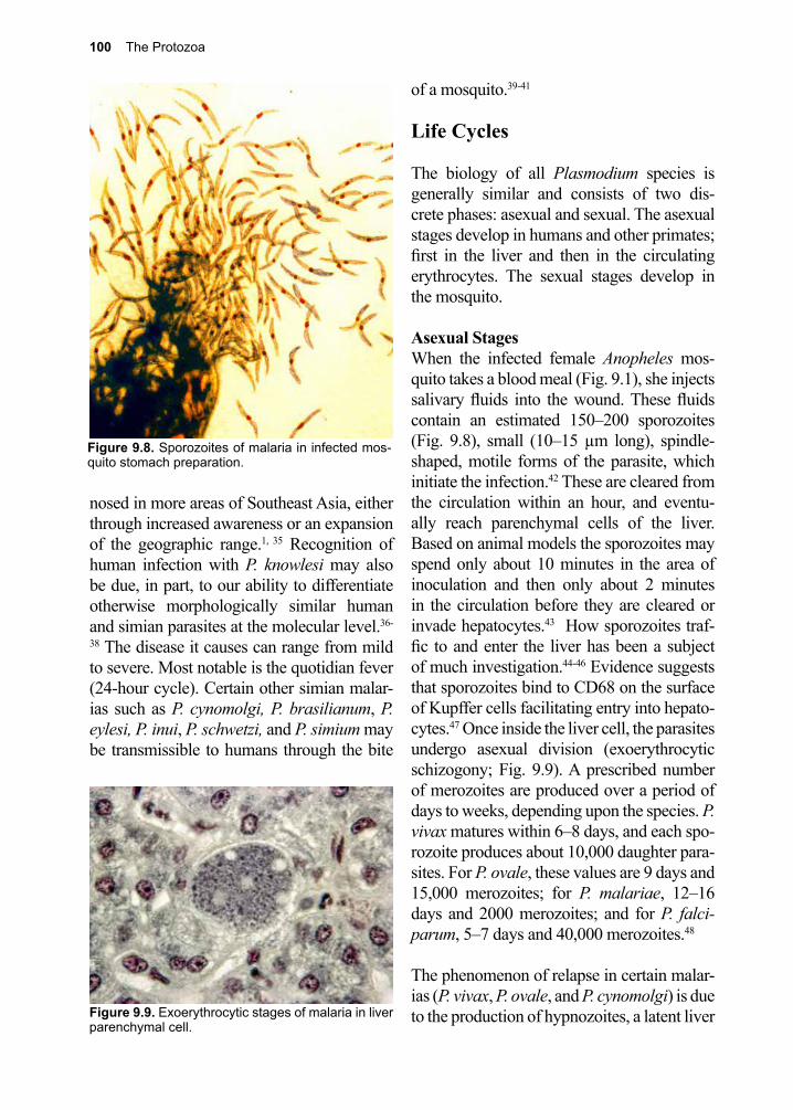
100 The Protozoa
nosed in more areas of Southeast Asia, either through increased awareness or an expansion of the geographic range.1, 35 Recognition of human infection with P. knowlesi may also be due, in part, to our ability to differentiate otherwise morphologically similar human and simian parasites at the molecular level.36-
38 The disease it causes can range from mild to severe. Most notable is the quotidian fever (24-hour cycle). Certain other simian malar-ias such as P. cynomolgi, P. brasilianum, P. eylesi, P. inui, P. schwetzi, and P. simium may be transmissible to humans through the bite
of a mosquito.39-41
Life Cycles
The biology of all Plasmodium species is generally similar and consists of two dis-crete phases: asexual and sexual. The asexual stages develop in humans and other primates; first in the liver and then in the circulating erythrocytes. The sexual stages develop in the mosquito.
Asexual StagesWhen the infected female Anopheles mos-quito takes a blood meal (Fig. 9.1), she injects salivary fluids into the wound. These fluids contain an estimated 150–200 sporozoites (Fig. 9.8), small (10–15 µm long), spindle-shaped, motile forms of the parasite, which initiate the infection.42 These are cleared from the circulation within an hour, and eventu-ally reach parenchymal cells of the liver. Based on animal models the sporozoites may spend only about 10 minutes in the area of inoculation and then only about 2 minutes in the circulation before they are cleared or invade hepatocytes.43 How sporozoites traf-fic to and enter the liver has been a subject of much investigation.44-46 Evidence suggests that sporozoites bind to CD68 on the surface of Kupffer cells facilitating entry into hepato-cytes.47 Once inside the liver cell, the parasites undergo asexual division (exoerythrocytic schizogony; Fig. 9.9). A prescribed number of merozoites are produced over a period of days to weeks, depending upon the species. P. vivax matures within 6–8 days, and each spo-rozoite produces about 10,000 daughter para-sites. For P. ovale, these values are 9 days and 15,000 merozoites; for P. malariae, 12–16 days and 2000 merozoites; and for P. falci-parum, 5–7 days and 40,000 merozoites.48
The phenomenon of relapse in certain malar-ias (P. vivax, P. ovale, and P. cynomolgi) is due to the production of hypnozoites, a latent liver
Figure 9.8. Sporozoites of malaria in infected mos-quito stomach preparation.
Figure 9.9. Exoerythrocytic stages of malaria in liver parenchymal cell.
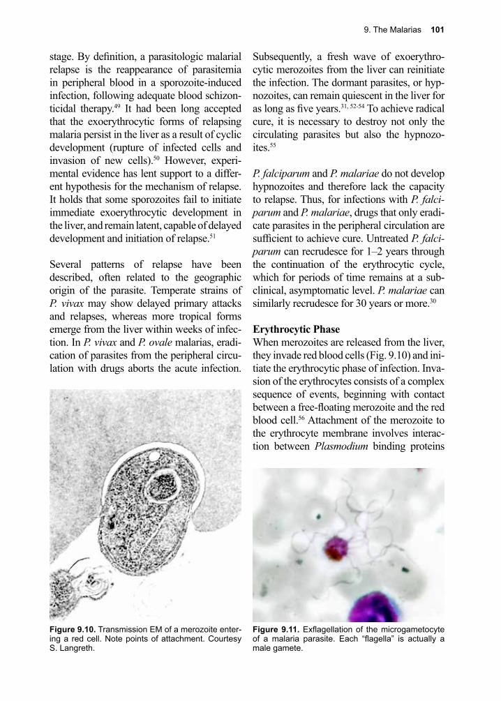
9. The Malarias 101
stage. By definition, a parasitologic malarial relapse is the reappearance of parasitemia in peripheral blood in a sporozoite-induced infection, following adequate blood schizon-ticidal therapy.49 It had been long accepted that the exoerythrocytic forms of relapsing malaria persist in the liver as a result of cyclic development (rupture of infected cells and invasion of new cells).50 However, experi-mental evidence has lent support to a differ-ent hypothesis for the mechanism of relapse. It holds that some sporozoites fail to initiate immediate exoerythrocytic development in the liver, and remain latent, capable of delayed development and initiation of relapse.51
Several patterns of relapse have been described, often related to the geographic origin of the parasite. Temperate strains of P. vivax may show delayed primary attacks and relapses, whereas more tropical forms emerge from the liver within weeks of infec-tion. In P. vivax and P. ovale malarias, eradi-cation of parasites from the peripheral circu-lation with drugs aborts the acute infection.
Subsequently, a fresh wave of exoerythro-cytic merozoites from the liver can reinitiate the infection. The dormant parasites, or hyp-nozoites, can remain quiescent in the liver for as long as five years.31, 52-54 To achieve radical cure, it is necessary to destroy not only the circulating parasites but also the hypnozo-ites.55
P. falciparum and P. malariae do not develop hypnozoites and therefore lack the capacity to relapse. Thus, for infections with P. falci-parum and P. malariae, drugs that only eradi-cate parasites in the peripheral circulation are sufficient to achieve cure. Untreated P. falci-parum can recrudesce for 1–2 years through the continuation of the erythrocytic cycle, which for periods of time remains at a sub-clinical, asymptomatic level. P. malariae can similarly recrudesce for 30 years or more.30
Erythrocytic PhaseWhen merozoites are released from the liver, they invade red blood cells (Fig. 9.10) and ini-tiate the erythrocytic phase of infection. Inva-sion of the erythrocytes consists of a complex sequence of events, beginning with contact between a free-floating merozoite and the red blood cell.56 Attachment of the merozoite to the erythrocyte membrane involves interac-tion between Plasmodium binding proteins
Figure 9.10. Transmission EM of a merozoite enter-ing a red cell. Note points of attachment. Courtesy S. Langreth.
Figure 9.11. Exflagellation of the microgametocyte of a malaria parasite. Each “flagella” is actually a male gamete.
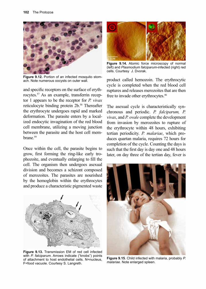
102 The Protozoa
and specific receptors on the surface of eryth-rocytes.57 As an example, transferrin recep-tor 1 appears to be the receptor for P. vivax reticulocyte binding protein 2b.58 Thereafter the erythrocyte undergoes rapid and marked deformation. The parasite enters by a local-ized endocytic invagination of the red blood cell membrane, utilizing a moving junction between the parasite and the host cell mem-brane.59
Once within the cell, the parasite begins to grow, first forming the ring-like early tro-phozoite, and eventually enlarging to fill the cell. The organism then undergoes asexual division and becomes a schizont composed of merozoites. The parasites are nourished by the hemoglobin within the erythrocytes and produce a characteristic pigmented waste
product called hemozoin. The erythrocytic cycle is completed when the red blood cell ruptures and releases merozoites that are then free to invade other erythrocytes.46
The asexual cycle is characteristically syn-chronous and periodic. P. falciparum, P. vivax, and P. ovale complete the development from invasion by merozoites to rupture of the erythrocyte within 48 hours, exhibiting tertian periodicity. P. malariae, which pro-duces quartan malaria, requires 72 hours for completion of the cycle. Counting the days is such that the first day is day one and 48 hours later, on day three of the tertian day, fever is
Figure 9.12. Portion of an infected mosquito stom-ach. Note numerous oocysts on outer wall.
Figure 9.13. Transmission EM of red cell infected with P. falciparum. Arrows indicate (“knobs”) points of attachment to host endothelial cells. N=nucleus, F=food vacuole. Courtesy S. Langreth.
Figure 9.15. Child infected with malaria, probably P. malariae. Note enlarged spleen.
Figure 9.14. Atomic force microscopy of normal (left) and Plasmodium falciparum-infected (right) red cells. Courtesy J. Dvorak.
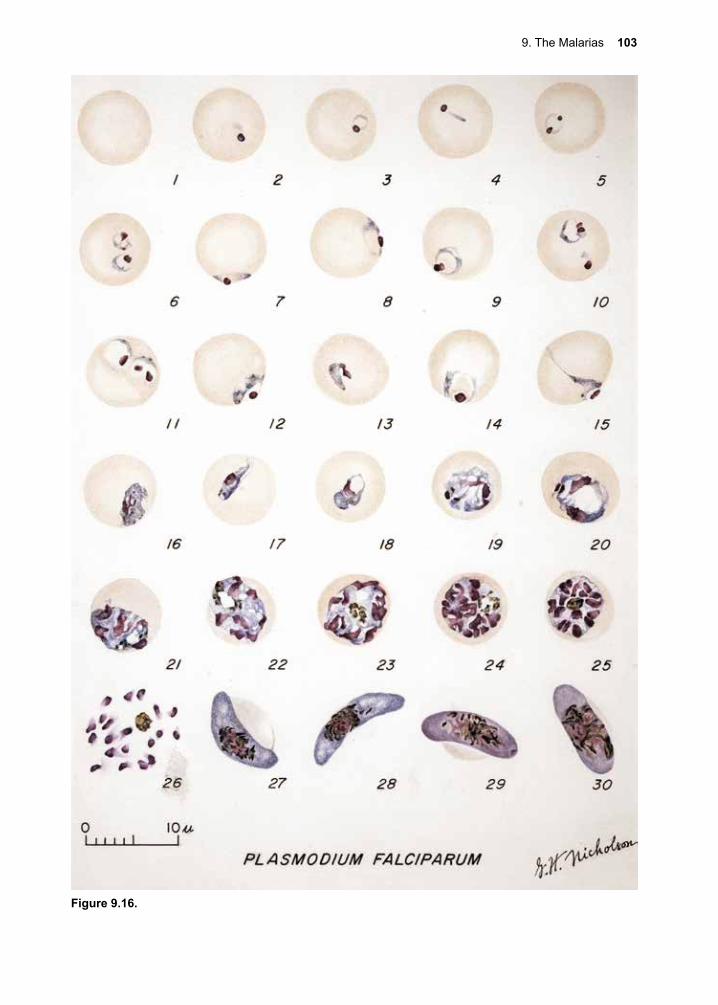
9. The Malarias 103
Figure 9.16.
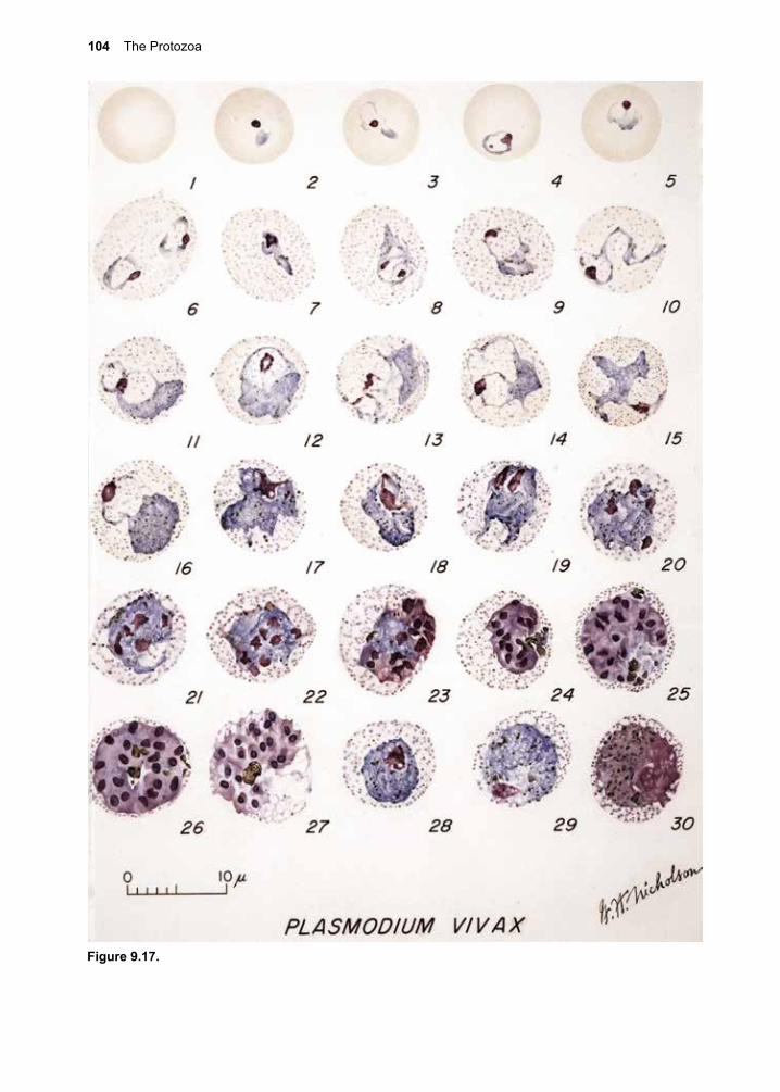
104 The Protozoa
Figure 9.17.
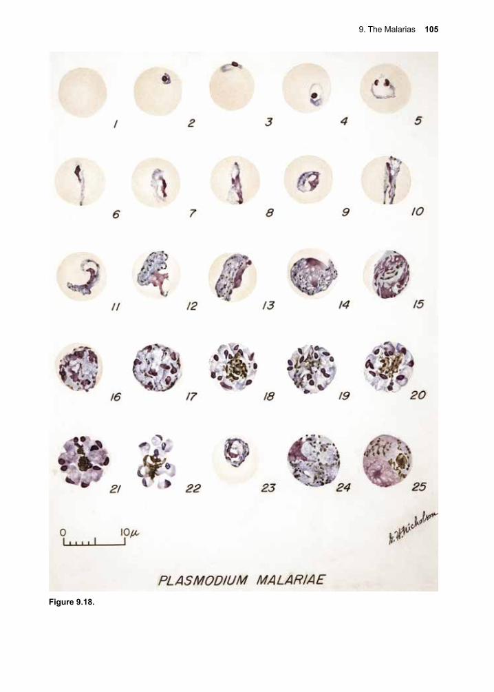
9. The Malarias 105
Figure 9.18.
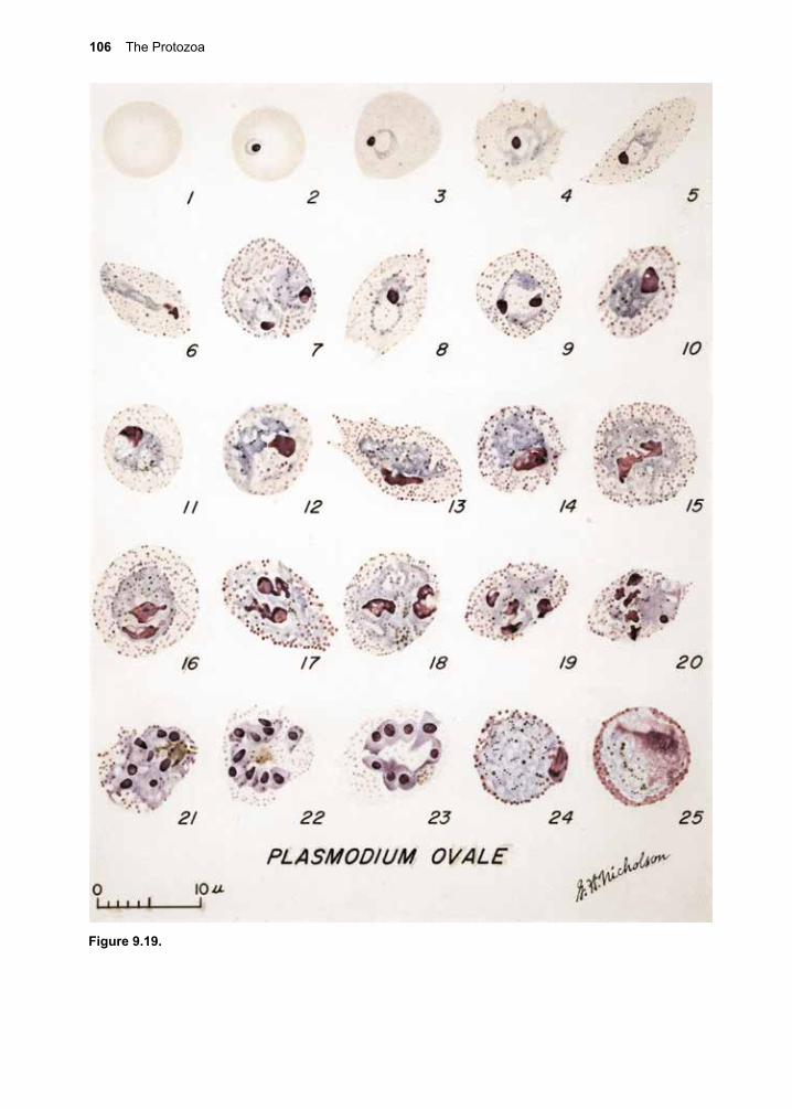
106 The Protozoa
Figure 9.19.
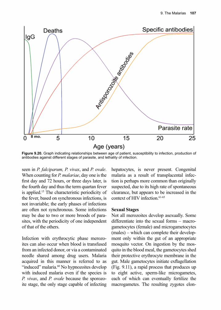
9. The Malarias 107
seen in P. falciparum, P. vivax, and P. ovale. When counting for P. malariae, day one is the first day and 72 hours, or three days later, is the fourth day and thus the term quartan fever is applied.15 The characteristic periodicity of the fever, based on synchronous infections, is not invariable; the early phases of infections are often not synchronous. Some infections may be due to two or more broods of para-sites, with the periodicity of one independent of that of the others.
Infection with erythrocytic phase merozo-ites can also occur when blood is transfused from an infected donor, or via a contaminated needle shared among drug users. Malaria acquired in this manner is referred to as “induced” malaria.60 No hypnozoites develop with induced malaria even if the species is P. vivax, and P. ovale because the sporozo-ite stage, the only stage capable of infecting
hepatocytes, is never present. Congenital malaria as a result of transplacental infec-tion is perhaps more common than originally suspected, due to its high rate of spontaneous clearance, but appears to be increased in the context of HIV infection.61-65
Sexual StagesNot all merozoites develop asexually. Some differentiate into the sexual forms – macro-gametocytes (female) and microgametocytes (males) – which can complete their develop-ment only within the gut of an appropriate mosquito vector. On ingestion by the mos-quito in the blood meal, the gametocytes shed their protective erythrocyte membrane in the gut. Male gametocytes initiate exflagellation (Fig. 9.11), a rapid process that produces up to eight active, sperm-like microgametes, each of which can eventually fertilize the macrogametes. The resulting zygotes elon-
Figure 9.20. Graph indicating relationships between age of patient, susceptibility to infection, production of antibodies against different stages of parasite, and lethality of infection.
108 The Protozoa
gate into diploid vermiform ookinetes (\ō-ə-ˈkī-ˌnēts\), which penetrate the gut wall and come to lie under the basement membrane (Fig. 9.12). The parasites then transform into oocysts (\ˈō-ə-ˌsists\) within 24 hours of ingestion of the blood meal. Development of sporozoites follows, leading to the produc-tion of more than 1,000 of these now-haploid forms in each oocyst. They mature within 10–14 days, escape from the oocyst, and invade the salivary glands. When the mos-quito bites another human host, a new cycle begins.
Although the different species have marked physiologic differences and some major dif-ferences in the pathologic course they pursue, they are most simply differentiated on the basis of their morphology. The blood smear, typically fixed and stained with Giemsa or Wright solution, is the basis of the fundamen-tal diagnostic test, although alternatives are now available. Commercially available meth-ods for malaria parasite detection and char-acterization are becoming increasingly sensi-tive, and are supplanting microscopy in more advanced laboratories (see Diagnosis).66
Cellular and Molecular Pathogenesis
The release of cytokines following rupture of infected erythrocytes induces fever and the consequent chills and sweating associated with malaria.67 The pathogenesis of general malaise, myalgia, and headache appear to be related to the release of certain cytokines and their levels correlate with disease severity.68
Cerebral malaria is the most devastating manifestation of severe P. falciparum infec-tion.69 Cerebral malaria is caused by a com-plex process involving sequestration of erythrocytes as well as cytokine release and endothelial activation.70, 71 The importance of various factors in the development of cerebral
malaria has been a subject of debate for many years with the development of the “school of sequestration” proponents as well as those more supportive of the “school of cytokines” providing a lively debate and contributing to an exciting number of publications advancing our understanding of this critical issue.72, 73 The mechanism of cytoadherence is thought to be related to the presence of histidine-rich “knobs” on the surface of the infected red blood cells that express strain-specific adhesive proteins (PfEMP1), and the sub-sequent attachment to appropriate receptors on the host endothelium (Figs. 9.13, 9.14).74,
75 Although not essential for cytoadherence, the knobs seem to enhance binding.76 These knobs are induced by the parasite and facili-tate endothelial cell binding by the infected erythrocytes to a number of endothelial tar-gets.77, 78 Binding to endothelial cells involves several host cell receptors, including CD36, intercellular adhesion molecule 1 (ICAM1), and chondroitin sulfate A (CSA). In the brain the intercellular adhesion molecule ICAM1 is likely the most important whilst CD36 is likely the most important in other organs.48 Cytoadherence in the placentae of women in their first pregnancies involves parasite bind-ing to CSA.79, 80 Sequestration of malaria par-asites in the placentae of primigravid females is a major cause of death, fetal mortality, fetal wastage and low birth weight.80-82
Malaria can also be responsible for a signifi-cant degree of anemia, often in populations already suffering from anemia due to other causes. With P. falciparum malaria, anemia caused by hemolysis can be severe. Damage to the erythrocytes by intravascular hemoly-sis can exceed that caused by rupture of the infected cells alone. Even uninfected cells have an increased osmotic fragility. Also present is bone marrow depression, which contributes to the anemia by decreasing the production of new erythrocytes.83 Dissemi-nated intravascular coagulopathy occurs in
9. The Malarias 109
severely infected individuals.84
The spleen plays a major role in host defense against malaria (Fig. 9.15). Parasitized cells accumulate in its capillaries and sinusoids, causing general congestion. Malarial pig-ment becomes concentrated in the spleen and is responsible for the darkening of this organ. Chronic infection, particularly with P. malariae, often causes persistent spleno-megaly and is responsible for “big spleen disease,” or tropical splenomegaly syndrome, consisting of hepatomegaly, portal hyperten-sion, anemia, leukopenia, and thrombocyto-penia.85 With P. vivax malaria, the spleen can become acutely enlarged and is susceptible to rupture. A significant portion of the anemia seen in P. vivax malaria is driven by splenic clearance of non-infected erythrocytes.86 The liver is darkened by the accumulated malarial pigment and shows degeneration and necro-sis of the centrilobular regions. The gastroin-testinal (GI) tract is also affected. There are focal hemorrhages, edema, and consequent malabsorption. The kidneys, particularly with severe P. falciparum malaria, show punc-tate hemorrhages and even tubular necrosis. Accumulation of hemoglobin in the tubules is responsible for hemoglobinuria, or blackwa-ter fever, which occurs after repeated attacks of falciparum malaria and can be complicated by therapy with quinine.87 Blackwater fever is a consequence of severe hemolysis exac-erbated by the host immune response against the intracellular parasites.
Chronic infections with P. malariae can lead to nephrotic syndrome, characterized by focal hyalinization of the tufts of glomeruli, and by endothelial proliferation, apparently caused by the deposition of immune complexes.15 This process can lead to endocapillary cell proliferation and reduplication of the base-ment membrane.
Congenital malaria can develop with any of the species of Plasmodia, although the inci-dence of this complication is relatively low. The mechanism by which the fetus becomes infected is uncertain. Some investigators have postulated damage to the placenta as a prerequisite to congenital malaria, but it is also possible that the parasites can infect the fetus through an intact placenta or at the time of birth.64
Malarial infections tend to suppress cell-mediated immune responses. It has been sug-gested that Burkitt’s lymphoma is caused by infection with the Epstein-Barr virus under the influence of immunosuppression by chronic P. falciparum malaria, but the com-peting hypothesis is that it is the chronic immune activation that drives Burkitt’s lym-phoma.88-90
Pathogenesis of Malaria in the HIV-infected PopulationThe relationship between infection with malaria and HIV is a subject of great inter-est and intense scrutiny.91 In Africa as well as many other parts of the tropics, HIV and malaria overlap in their geographic ranges and the populations they infect.6, 92 The focus is often on how HIV infection predisposes individuals to an increase in malarial disease severity. There is evidence that malaria infec-tion increases T-cell activation and causes both a decrease in CD4+ T cell counts and increases in viral loads.93-95 There is also evi-dence to suggest a relationship between HIV-induced reduction of CD4+ T cell counts and a rise in incidence of malaria.95-99 HIV does confer an increased susceptibility to symp-tomatic malaria.100, 101 There is conflicting information regarding the severity of clinical malaria when it manifests in HIV-infected individuals, but most evidence suggests that they experience higher levels of parasitemia and an increased risk for severe malaria.102, 103
110 The Protozoa
Clinical Disease
The most pronounced clinical manifestations of adult-onset malaria are periodic chills and fever, usually accompanied by frontal head-ache, fatigue, abdominal discomfort, and myalgia.48, 66 Fever may persist for several days before the typical periodicity develops. In contrast, young children often present with non-specific symptoms, including fever, cough, vomiting, and diarrhea. Symptoms of malaria usually first appear 10–15 days after the bite of the infected mosquito, although delays of several months in the onset of symptoms and the appearance of parasites in peripheral blood are common, particularly for some strains of P. vivax found in temperate zones. Patients undergoing chemoprophy-laxis may not develop any symptoms until they stop taking the drug. The classic pattern of clinical disease consists of paroxysms of chills and fever, reaching 41 °C and lasting six hours, followed by sweating and defer-vescence.
Vomiting can also develop and may be intense. Difficulty breathing is seen in up to 25% of adults with severe malaria, and in approximately 40% of children with severe malaria.104 Initially, there can be mild anemia with an elevation of the reticulocyte count. The leukocyte count tends to be normal or even low.105 The eosinophil count is charac-teristically low (eosinopenia) in acute malaria and a robust rise in eosinophils and hemo-globin after completing malaria therapy is usually seen in successfully treated patients experiencing a good recovery.106
All forms of untreated malaria tend to become chronic, including those without a dormant hypnozoite stage. Repeated attacks are caused by recrudescence (recurrence of symptoms from persistent blood stages) or relapses (recurrence of symptoms and para-sitemia from maturation of the hypnozoite
stage). The development of host immunity can lead to spontaneous cure of P. falciparum malaria within two years and of P. vivax and P. ovale malarias within five years, although individuals are susceptible to reinfection during and after this period (Fig. 9.20). Infec-tion with the quartan parasite, P. malariae, can potentially persist for the lifetime of the individual with one report from China of 50 years between time of acquisition and illness and another case of an individual with infec-tion acquired in Greece more than 40 years and, possibly, as much as 70 years previ-ously.15, 107 Untreated malaria, particularly P. falciparum malaria, can be fatal during the initial attack, an unfortunately frequent event in young children (Fig. 9.20).
Unexplained fever in patients who have received blood transfusions, or who are intra-venous drug users may signal the presence of induced malaria. Induced malaria is malaria that results from direct transfer of infectious merozoites from the blood of an infected individual to a new host. Hypnozoites do not develop with transfusion or induced malaria, even if the species is P. vivax, and P. ovale, because the sporozoite stage, the only stage capable of infecting hepatocytes, is never present. An infant who develops fever during the neonatal period should be suspected of malaria if the mother has been at risk of infection. Diagnostic tests for induced or congenital malaria are the same as for the conventional forms of the infection. It must be emphasized that neither induced nor con-genital malaria has an exoerythrocytic cycle in the liver, and therapy directed against the liver cycle is not required.
P. vivax and P. ovale preferentially invade reticulocytes. Usually only about 1–2% of red blood cells are parasitized with P. vivax and P. ovale infections. Clinical disease is typically mild. P. malariae tends to invade older erythrocytes, again limiting maximum
9. The Malarias 111
parasitemia to about 1–2%. In contrast, P. fal-ciparum attacks erythrocytes of all ages, per-mitting high levels of parasitemia.
Innate resistance to malaria is mediated by factors other than immune mechanisms. There are a number of genetic factors in human populations that confer varying levels of susceptibility to malaria.46, 108, 109 Individu-als carrying the gene for sickle-cell hemo-globin receive some advantage against P. falciparum malaria. Those with sickle-cell trait (A and S hemoglobins) have a selective advantage over those with the hemoglobin AA genotype because the heterozygotes limit the severity of malaria. The hemoglobin SS individuals are also at an advantage, but their sickle-cell disease leads to early death.108-110 In areas of Africa with the highest frequency of this gene, it is estimated that the death rate due to malaria required to fix this gene frequency may have exceeded 25% and is an excellent example of a balanced polymorphism.111
In 2011, a group of researchers demonstrated that sickle hemoglobin provides protection against malaria through induction of the expression of heme oxygenase-1, which pre-vents accumulation of circulating heme.112 This is particularly protective against cerebral malaria. Sickle hemoglobin is also able to inhibit the activation and expansion of CD8+ T cells, resulting in lower cellular activation and higher cellular reactivity in response to malarial antigens.112-114 Hemoglobin C muta-tions appear to provide similar protection against P. falciparum malaria in certain ethnic groups.115, 116
Glucose-6-phosphate dehydrogenase (G6PD) deficiency, β-thalassemia, and ovalocytosis, the latter common in Southeast Asia, have been implicated as mediators of innate resis-tance against P. falciparum infection.117 It has been suggested that the protective effect of thalassemia may be related to enhanced
immune recognition and clearance of parasit-ized erythrocytes.118 Evidence also supports the idea that certain of these hemoglobinopa-thies reduce the intraerythrocytic multiplica-tion of Plasmodium species.119
Duffy blood type determinants are associated with receptor sites for P. vivax merozoites on the erythrocytes. While it is doubtful that the blood group carbohydrate itself is the actual receptor, most West Africans are negative for the Duffy blood type and have decreased susceptibility to infection with P. vivax.111, 120 Instead, they have a higher than normal inci-dence of infection with P. ovale.121
Acquired immunity (incomplete nonster-ile) develops after long exposure to malaria and is characterized by low levels of parasit-emia. Immune individuals have intermittent parasitemia with only mild symptoms. This clinical state has been referred to as premuni-tion, in contrast with classic immunity, which prevents any degree of infection. Premunition does not last for life and individuals return-ing to endemic areas after even only one year away often have lost this protective immu-nity.122-124
Diagnosis (see Clinical Appendix)
For over a century a definitive diagnosis of malaria depended on the microscopic iden-tification of the parasite on Giemsa-stained blood smears. This procedure permits not only the confirmation of the presence of the parasite, but makes possible the identification of the species of malaria and an indication of the level of parasitemia in the infected host.125 Under normal conditions, both thick and thin smears should be examined. When malaria is suspected, the clinician should take a thor-ough travel history. Should the initial blood smears prove to be negative, new specimens should be drawn at 6-hour intervals.
112 The Protozoa
Identification of malaria on thick and thin blood smears requires an experienced microscopist who is well-trained in para-site morphology. A British study indicated that at least 10% of positive slides are not identified.126 Antibody-based rapid diagnos-tic tests that are simple, specific and do not require a highly trained microscopist have been introduced and are now widely avail-able.127 These rapid tests detect the presence or absence of PfHRP2, which is present in P. falciparum, and then allow detection of other species and species level discrimination by using lactate dehydrogenase or aldolase antigens that are species-specific.128-130 Unfor-tunately a number of rapid diagnostic tests can have poor sensitivity and specificity for the diagnosis of P. knowlesi.131 A number of NAATs have been developed that have been employed in research and epidemiological studies.132 The introduction of portable plat-forms (e.g., loop-mediated isothermal ampli-fication (LAMP) assays) may allow molecu-lar testing to move from the research setting to field diagnosis.133-135
The identification of parasite genes confer-ring resistance to antimalarial drugs has permitted the development of mutation-spe-cific PCR primers that can readily identify resistant parasites. Such detection systems are available and in use in the field for pyri-methamine, sulfadoxine, cycloguanil, chloro-quine, and artemisinin.136-140 Epidemiologic studies requiring information on sporozoite inoculation rates by mosquito vectors have been facilitated by ELISA systems using spe-cies-specific monoclonal antibodies directed against the predominant surface antigens of the sporozoites.141 The capacity for rapidly determining the proportion of mosquito pop-ulations coming to feed that have infectious sporozoites in their salivary glands allows accurate prediction of risk of infection or assessment of the effects of intervention strat-egies for the control of malaria.
Treatment (see Clinical Appendix)
The long reliance on chloroquine to treat P. falciparum is no longer tenable, due to the worldwide spread of drug resistance.142 The two major choices for treatment of severe malaria are the cinchona alkaloids (quinine and quinidine) and the artemisinin derivatives (artesunate, artemether, and artemotil). Par-enteral artesunate is the most rapidly acting of the artemisinin compounds, and is associ-ated with more rapid clearance of parasitemia than treatment with the cinchona alkaloid quinine.143, 144 Artesunate treatment has dem-onstrated a significant reduction in mortality in both adults and children compared to treat-ment with quinine.143, 144 While artemisinin-based therapy was initially recommended as primary therapy in areas of high drug resis-tance, it is now first line therapy in most cases of severe malaria.145, 146
A significant and striking exception to this worldwide recommendation had been malaria treatment in the United States. For parts of the world where artesunate is not available, qui-nine and quinidine are alternative therapies. In 1991, parenteral quinine was no longer stocked by the Centers for Disease Control (CDC), when it was decided that parenteral quinidine gluconate could serve as an alter-native.147 Quinidine gluconate remained the only approved therapy by the FDA for the parenteral therapy of severe malaria until 2019 when it was announced that intravenous artesunate would become available in the U. S. for the treatment of severe malaria. Intra-venous artesunate should be considered early in therapy given the favorable comparative data.148 In addition to artesunate, quinine, or quinidine, a second long-acting medication is required, such as doxycycline, tetracycline, or clindamycin, for successful treatment.
When selecting treatment there are a number of important factors that the treating clinician
9. The Malarias 113
must consider including: the severity of the clinical manifestation (uncomplicated versus severe); the specific malaria being treated; and drug susceptibility or resistance. When treating severe malaria infections with quini-dine, care must include: monitoring patients with standard cardiac monitors; careful track-ing of blood glucose; cautious fluid manage-ment, and delayed initiation of enteral feed-ing; factors critical to successful treatment outcomes.48 Seizures may need to be man-aged with anti-seizure medications or benzo-diazepines, and protection of the airway may be required in severe cases. Broad-spectrum intravenous antibiotics may be indicated in certain cases due to the high rate of bacterial sepsis that can occur in severe malaria.149
Exchange transfusions, once felt to be essen-tial in management of severe malaria, are no longer recommended by the CDC, since a meta-analysis of previous studies revealed no significant survival advantage.150 Blood transfusions for patients have been recom-mended based on the results of small ran-domized trials and a few large observational trials. Transfusion is recommended if hemo-globin levels are below 4 g/dL in all cases, or if hemoglobin levels are below 6 g/dL and there is parasitemia greater than 20%, or other concerning clinical features.151, 152
In contrast to severe or complicated malaria, non-complicated malaria is defined as symp-tomatic malaria with lack of any of the defined severe malaria criteria. The patient demonstrates lower levels of parasitemia, retains the ability to take oral medicine, and displays no evidence of vital organ dysfunc-tion. For the treatment of uncomplicated malaria, if a patient has P. falciparum infec-tion acquired from a region without chloro-quine resistance, such as the Caribbean and Central America west of the Panama Canal, chloroquine can be used.153 In the rest of the world, resistance to chloroquine is so fre-
quently reported that every patient must be assumed to have a resistant form of P. falci-parum. If the origin of infection is unknown, as may be the case with induced malaria, one must treat the infection as if the organisms are resistant. Currently artemisinin combination therapy is the recommended first line therapy for all human malaria.154 There are several available forms of artemisinin combination therapy with no one choice clearly superior over the other.154
Alternative drugs may be selected on a region-by-region basis with knowledge of local resistance rates.155 Fansidar, a pyrimeth-amine-sulfadoxine combination, lost effec-tiveness as a replacement for chloroquine in East African countries, but still may have a role as part of intermittent preventive therapy in pregnancy.156-158 Mefloquine can be given as part of combination therapy with a second agent such as doxycycline, with cure rates estimated to be greater than 90% in areas without resistance.159 Malarone (atovaquone-proguanil) is an effective option that has demonstrated superior efficacy over meflo-quine treatment and may have a better safety profile.160, 161 The Sentinel sites in the Inter-national Centers of Excellence for Malaria Research provide essential data to monitor the emergence of drug resistance globally and the underlying mechanisms to further understand the impact.162
When the infecting strain is P. vivax or P. ovale, treatment may not resolve infection by the hypnozoite liver stage, these must be cleared with a course of primaquine to pre-vent relapses. Primaquine need not be started immediately but can be deferred until the patient has recovered from the acute attack. Primaquine tends to cause hemolysis in indi-viduals with G6PD deficiency. The drug should not be administered until the proper test for this enzyme has been performed.
114 The Protozoa
geographic strains of vector mosquitoes may be highly variable.
Most malaria in the United States, since the 1960s, has been of the imported variety. The wars in Korea and Vietnam increased the numbers of these imported cases because of the returning infected service personnel. Refugees or immigrants from endemic areas constitute the largest number of imported cases. In addition, there is a steady inci-dence of malaria among travelers return-ing from endemic areas. Autochthonous (\au·toch·tho·nous\) infections are rare in the United States, despite large, persistent popu-lations of the anopheline vectors, An. quadri-maculatus in the East and An. freeborni in the West. Outbreaks of P. vivax in southern Cali-fornia have been associated with the vector species, An. hermsi.165 There are rare reports of malaria transmission in the United States, usually limited to one or two cases transmit-ted by local anopheline mosquitoes.
In addition to chemoprophylaxis when indi-cated, travelers should avoid or minimize contact with mosquitoes. Since most anoph-elines bite at night, sleeping under insecticide treated bed netting and, if possible, in rooms fitted with window screens is effective, but not without issues of proper use.166 Clothing that covers much of the skin and insect repel-lents, particularly those containing DEET or picaridin, are useful adjuncts to transmission prevention.
Controlling the mosquito vector remains the most practical method for wide-scale con-trol of malaria. A reduction in the number of mosquitoes through drainage or modification of breeding sites has been accomplished in some areas. Insecticides may offer the best but increasingly less-acceptable method for reducing populations of mosquitoes, or of interrupting transmission by targeting only those infected mosquitoes coming to feed in
Chemoprophylaxis for Malaria (see Clinical Appendix)In areas without chloroquine resistance, such as the Caribbean and Central America west of the Panama Canal, chloroquine can be used for chemoprophylaxis, as there is little resistance to the drug in this region.153 Meflo-quine is an inexpensive option with weekly dosing that can be used in most of the world, except for localized areas where resistance has been reported. Many are hesitant to pre-scribe or take mefloquine due to concerns regarding its potential to induce psychosis.163 Malarone (atovaquone-proguanil) is a pro-phylactic drug for travelers to areas where P. falciparum is resistant to other medications, but requires once-daily dosing and is often very expensive. Daily doxycycline is another alternative, but patients should be cautioned about the associated photosensitivity. In some regions of the world, primaquine is used as a form of chemoprophylaxis, but the efficacy of this approach may be inferior to other chemo-prophylaxis regimens. G6PD testing should be performed prior to using primaquine due to the danger of hemolysis.164
Prevention and Control
There are more than 300 species of Anoph-eles mosquitoes, but only about 60 species are considered important vectors of malaria. Some of the factors that influence the effi-ciency of the insect are their feeding habits (most importantly, a preference for human blood), longevity, susceptibility to infection with the malarial parasite, and the size of the mosquito population (seasonal variability).
The variability of the parasite plays an impor-tant role in the pathogenicity of the disease. For example, geographic strains of P. vivax show markedly different incubation periods and patterns of relapses, and P. falciparum shows considerable variability in its responses to anti-malarial drugs. The susceptibility of

9. The Malarias 115
houses. The rising costs of these insecticides and the development of resistance by the insects have severely limited their applica-tion and usefulness. Insecticide-impregnated bed nets have been shown to have a signifi-cant impact on the morbidity and mortality of infection due to P. falciparum and P. vivax in China, and to P. falciparum in Africa.167,
168 Malaria control schemes based on genetic modification of the capacity of vector mos-quitoes to transmit the parasite have been suggested.169 In addition, the development of more efficient methods for introducing advan-tageous genes into the mosquito genome are being investigated, as well as methods for replacing vector populations in the field with populations of mosquitoes unable to transmit the parasite.170, 171
A malaria vaccine remains the “holy grail” of control strategies. For over 50 years, researchers have been attempting to find anti-gens that could induce protective immunity. After years of sporadic advances, vaccine research was reinvigorated by the demonstra-tion that animals could develop immunity to infection with sporozoites, and stimulated by the development of methods for the in vitro cultivation of the asexual and sexual stages of P. falciparum by Trager and Jensen.172 The revolution of molecular biology made possi-ble the identification of specific genes coding for specific antigens and sub-unit vaccines became possible.173
There are three phases of the malaria life cycle that have been targeted by the vaccine investigators.174, 175 Vaccines directed against the pre-erythrocytic stages of the parasite are intended to prevent infection by blocking the invasion or development of sporozoites freshly injected by a feeding mosquito or the development of the parasite in the liver. Sec-ondarily it has been suggested that even par-
tial efficacy (the blockage of most pre-eryth-rocytic development) could reduce the inten-sity of the primary infection and be useful in concert with antigens directed against other stages. Because such vaccines may have short-term efficacy, the target population for pre-erythrocytic stage vaccines has usually been considered to be non-immune individu-als moving through malaria endemic areas, including tourists and military personnel. Even with a short life, such vaccines could be useful in areas of low transmission, or in chil-dren and women of childbearing age in areas of high transmission.
Vaccines directed against the erythrocytic (blood) stages of the parasite are not expected to induce sterile immunity and totally prevent infection. Rather, it is expected that a success-ful vaccine could reduce the parasite burden, eliminate most deaths and reduce morbidity. The primary target for blood stage vaccines are children and pregnant women in areas of intense transmission.
Vaccines directed against the mosquito (sexual) stages of the parasite are designed to block the development of the parasite in the mosquito vector. An effective vaccine could interrupt transmission to additional victims. In combination with other antigens, a trans-mission-blocking component could prevent the spread of parasites resistant to other vac-cines. A transmission-blocking vaccine could be used in an eradication scheme or to pre-vent epidemics in areas of unstable malaria transmission. The investigational malaria vaccines are listed by stage of clinical devel-opment at The Malaria Vaccine Initiative website (www.malariavaccine.org). The most advanced candidate provided modest protection in children hence the need to con-tinue to evaluate the earlier stage vaccines in the pipeline.176
116 The Protozoa
References
1. Moyes, C. L., et al., Defining the geographical range of the Plasmodium knowlesi reservoir. PLoS neglected tropical diseases 2014, 8 (3), e2780.
2. CDC, Progress toward interruption of wild poliovirus transmission-worldwide, January -March. MMWR 2005, 54, 408-12.
3. Greenwood, B. M., et al., Malaria. Lancet 2005, 365 (9469), 1487-98.4. Carter, R.; Mendis, K. N., Evolutionary and historical aspects of the burden of malaria. Clinical
microbiology reviews 2002, 15 (4), 564-94.5. WHO, World Malaria Report 2015. Geneva, World Health Organization 2015.6. WHO World malaria report 2017. (accessed March 3, 2018).7. Newby, G., et al., The path to eradication: a progress report on the malaria-eliminating countries.
Lancet 2016, 387 (10029), 1775-84.8. Williams, L. L., Jr., Malaria eradication in the United States. Am J Public Health Nations Health
1963, 53, 17-21.9. Andrews, J. M.; Gilbertson, W. E., Blueprint for malaria eradication in the United States. J Natl
Malar Soc 1948, 7 (3), 167-70.10. Cholera, R., et al., Impaired cytoadherence of Plasmodium falciparum-infected erythrocytes
containing sickle hemoglobin. Proc Natl Acad Sci U S A 2008, 105 (3), 991-6.11. Taylor, S. M., et al., Haemoglobinopathies and the clinical epidemiology of malaria: a systematic
review and meta-analysis. Lancet Infect Dis 2012, 12 (6), 457-68.12. Cyrklaff, M., et al., Hemoglobins S and C interfere with actin remodeling in Plasmodium
falciparum-infected erythrocytes. Science 2011, 334 (6060), 1283-6.13. Adams, H. C., The Genuine Works of Hippocrates. 1886, The Epidemics Book 1.14. Laveran, A., Note sur un nouveau parasite trouve´ dans le sang de plusieurs malades atteints de
fie`vre palustre. Bull. Acad. Med 1880, 8, 1235-1236.15. Collins, W. E.; Jeffery, G. M., Plasmodium malariae: parasite and disease. Clin Microbiol Rev
2007, 20 (4), 579-92.16. Kean, B. H., et al., Tropical medicine and parasitology: classic investigations. Cornell University
Press, Ithaca, NY 1978.17. Harrison, G., Mosquitoes, Malaria, and Man: a history of the hostilities since 1880. 1978.18. Bruce-Chwat, L. J., et al., History of malaria from pre-history to eradication. In Malaria
Principles and Practice of Malariology eds Churchill Livingstone Edinburgh pp 1988, 1-59.19. Shortt, H. E.; Garnham, P. C., Pre-erythrocytic stage in mammalian malaria parasites. Nature
1948, 161 (4082), 126.20. Krotoski, W. A., et al., Relapses in primate malaria: discovery of two populations of
exoerythrocytic stages. Preliminary note. Br Med J 1980, 280 (6208), 153-4.21. Krotoski, W. A., et al., Demonstration of hypnozoites in sporozoite-transmitted Plasmodium
vivax infection. Am J Trop Med Hyg 1982, 31 (6), 1291-3.22. Markus, M. B., Possible support for the sporozoite hypothesis of relapse and latency in malaria.
Trans R Soc Trop Med Hyg 1976, 70 (5-6), 535.23. Markus, M. B., The malarial hypnozoite. Lancet 1980, 1 (8174), 936.24. Haggis, A. W., Fundamental errors in the early history of Cinchona. Bull Hist Med 1941, 10,
567-568.25. Meshnick, S. R.; Sherman, I. W., From quinine to Qinghaosu: Historical perspectives. 1998; p
341-353.26. Honigsbaum, M., The Fever Trail: In search of the cure for malaria. Nature Medicine 2001.27. Rocco, F., Quinine: Malaria and the quest for a cure that changed the world. 2003.28. Fuehrer, H. P.; Noedl, H., Recent advances in detection of Plasmodium ovale: implications
of separation into the two species Plasmodium ovale wallikeri and Plasmodium ovale curtisi. Journal of clinical microbiology 2014, 52 (2), 387-91.
29. Nolder, D., et al., An observational study of malaria in British travellers: Plasmodium ovale wallikeri and Plasmodium ovale curtisi differ significantly in the duration of latency. BMJ Open 2013, 3 (5).
9. The Malarias 117
30. Vinetz, J. M., et al., Plasmodium malariae infection in an asymptomatic 74-year-old Greek woman with splenomegaly. The New England journal of medicine 1998, 338 (6), 367-71.
31. Garnham, P. C., Swellengrebel lecture. Hypnozoites and ‘relapses’ in Plasmodium vivax and in vivax-like malaria. Tropical and geographical medicine 1988, 40 (3), 187-95.
32. R., K.; B.M., D. G., A study of monkey-malaria, and its experimental transmission to man. Ind Med Gaz 1932, 67, 301-21.
33. Kantele, A.; Jokiranta, T. S., Review of cases with the emerging fifth human malaria parasite, Plasmodium knowlesi. Clin Infect Dis 2011, 52 (11), 1356-62.
34. White, N. J., Plasmodium knowlesi: the fifth human malaria parasite. Clin Infect Dis 2008, 46 (2), 172-3.
35. Iwagami, M., et al., First case of human infection with Plasmodium knowlesi in Laos. PLoS Negl Trop Dis 2018, 12 (3), e0006244.
36. White, N. J., Sharing malarias. Lancet (London, England) 2004, 363 (9414), 1006.37. Abegunde, A. T., Monkey malaria in man. Lancet (London, England) 2004, 364 (9441), 1217.38. Singh, B., et al., A large focus of naturally acquired Plasmodium knowlesi infections in human
beings. Lancet (London, England) 2004, 363 (9414), 1017-24.39. Brasil, P., et al., Outbreak of human malaria caused by Plasmodium simium in the Atlantic Forest
in Rio de Janeiro: a molecular epidemiological investigation. Lancet Glob Health 2017, 5 (10), e1038-e1046.
40. Coatney, G. R., The simian malarias: zoonoses, anthroponoses, or both? The American journal of tropical medicine and hygiene 1971, 20 (6), 795-803.
41. Ta, T. H., et al., First case of a naturally acquired human infection with Plasmodium cynomolgi. Malar J 2014, 13, 68.
42. Vanderberg, J. P., Plasmodium berghei: quantitation of sporozoites injected by mosquitoes feeding on a rodent host. Exp Parasitol 1977, 42 (1), 169-81.
43. Sidjanski, S.; Vanderberg, J. P., Delayed migration of Plasmodium sporozoites from the mosquito bite site to the blood. Am J Trop Med Hyg 1997, 57 (4), 426-9.
44. Meis, J. F.; Verhave, J. P., Exoerythrocytic development of malarial parasites. Advances in parasitology 1988, 27, 1-61.
45. Hollingdale, M. R., Biology and immunology of sporozoite invasion of liver cells and exoerythrocytic development of malaria parasites. Progress in allergy 1988, 41, 15-48.
46. Sherman, I. W., Malaria:Parasite Biology, Pathogenesis and Protection. ASM Press, Washington, D.C. : 1998.
47. Cha, S. J., et al., CD68 acts as a major gateway for malaria sporozoite liver infection. J Exp Med 2015, 212 (9), 1391-403.
48. White, N. J., et al., Malaria. Lancet 2014, 383 (9918), 723-35.49. Coatney, G. R., Relapse in malaria--an enigma. The Journal of parasitology 1976, 62 (1), 3-9.50. Schmidt, L. H., Compatibility of relapse patterns of Plasmodium cynomolgi infections in
rhesus monkeys with continuous cyclical development and hypnozoite concepts of relapse. The American journal of tropical medicine and hygiene 1986, 35 (6), 1077-99.
51. Krotoski, W. A., Discovery of the hypnozoite and a new theory of malarial relapse. Transactions of the Royal Society of Tropical Medicine and Hygiene 1985, 79 (1), 1-11.
52. Adekunle, A. I., et al., Modeling the dynamics of Plasmodium vivax infection and hypnozoite reactivation in vivo. PLoS Negl Trop Dis 2015, 9 (3), e0003595.
53. Hulden, L.; Hulden, L., Activation of the hypnozoite: a part of Plasmodium vivax life cycle and survival. Malar J 2011, 10, 90.
54. Krotoski, W. A., The hypnozoite and malarial relapse. Prog Clin Parasitol 1989, 1, 1-19.55. Galappaththy, G. N., et al., Primaquine for preventing relapse in people with Plasmodium vivax
malaria treated with chloroquine. Cochrane Database Syst Rev 2013, 10, CD004389.56. Dvorak, J. A., et al., Invasion of erythrocytes by malaria merozoites. Science (New York, N.Y.)
1975, 187 (4178), 748-50.57. Egan, E. S., et al., Malaria. A forward genetic screen identifies erythrocyte CD55 as essential for
Plasmodium falciparum invasion. Science 2015, 348 (6235), 711-4.
118 The Protozoa
58. Gruszczyk, J., et al., Transferrin receptor 1 is a reticulocyte-specific receptor for Plasmodium vivax. Science 2018, 359 (6371), 48-55.
59. Aikawa, M., et al., Erythrocyte entry by malarial parasites. A moving junction between erythrocyte and parasite. The Journal of cell biology 1978, 77 (1), 72-82.
60. Garvey, G., et al., Transfusion-induced malaria after open-heart surgery. New York state journal of medicine 1975, 75 (4), 602-3.
61. Covell, G., Congenital malaria. Trop Dis Bull 1950, 47, 1147-1167.62. Mwangoka, G. W., et al., Congenital Plasmodium falciparum infection in neonates in Muheza
District, Tanzania. Malar J 2008, 7, 117.63. Ogolla, S., et al., Reduced Transplacental Transfer of a Subset of Epstein-Barr Virus-Specific
Antibodies to Neonates of Mothers Infected with Plasmodium falciparum Malaria during Pregnancy. Clin Vaccine Immunol 2015, 22 (11), 1197-205.
64. Ouedraogo, A., et al., Transplacental Transmission of Plasmodium falciparum in a Highly Malaria Endemic Area of Burkina Faso. J Trop Med 2012, 2012, 109705.
65. Falade, C., et al., Epidemiology of congenital malaria in Nigeria: a multi-centre study. Trop Med Int Health 2007, 12 (11), 1279-87.
66. Hanscheid, T., Diagnosis of malaria: A review of alternatives to conventional microscopy. Clin Lab Haem 1999, 21, 235-245.
67. Gallego-Delgado, J., et al., A surprising role for uric acid: the inflammatory malaria response. Curr Rheumatol Rep 2014, 16 (2), 401.
68. Prakash, D., et al., Clusters of cytokines determine malaria severity in Plasmodium falciparum-infected patients from endemic areas of Central India. J Infect Dis 2006, 194 (2), 198-207.
69. WHO, Severe and complicated malaria. Trans R Soc Trop Med Hyg 1986, 1-50.70. Wahlgren, M., et al., Malaria: Chapter 10. Cytoadherence and rosetting in the pathogenesis of
severe malaria. CRC Press: 1999; p 289-327.71. Frevert, U.; Nacer, A., Fatal cerebral malaria: a venous efflux problem. Front Cell Infect
Microbiol 2014, 4, 155.72. Storm, J.; Craig, A. G., Pathogenesis of cerebral malaria--inflammation and cytoadherence.
Front Cell Infect Microbiol 2014, 4, 100.73. Grau, G. E.; Craig, A. G., Cerebral malaria pathogenesis: revisiting parasite and host
contributions. Future Microbiol 2012, 7 (2), 291-302.74. Cooke, B., et al., Falciparum malaria: sticking up, standing out and out-standing. Parasitol
Today 2000, 16 (10), 416-20.75. Kilejian, A., Characterization of a protein correlated with the production of knob-like protrusions
on membranes of erythrocytes infected with Plasmodium falciparum. Proc Natl Acad Sci U S A 1979, 76 (9), 4650-3.
76. Ruangjirachuporn, W., et al., Cytoadherence of knobby and knobless Plasmodium falciparum-infected erythrocytes. Parasitology 1991, 102 Pt 3, 325-34.
77. Udeinya, I. J., et al., Falciparum malaria-infected erythrocytes specifically bind to cultured human endothelial cells. Science (New York, N.Y.) 1981, 213 (4507), 555-7.
78. Chisti, A. H., et al., Isolation of skeleton-associated knobs from human blood cells infected with the malaria parasite Plasmodium falciparum. Mol Biochem Parasit 1992, 52, 293-297.
79. Miller, L. H., et al., Malaria pathogenesis. Science (New York, N.Y.) 1994, 264 (5167), 1878-83.80. Duffy, P. E.; Fried, M., Antibodies that inhibit Plasmodium falciparum adhesion to chondroitin
sulfate A are associated with increased birth weight and the gestational age of newborns. Infection and immunity 2003, 71 (11), 6620-3.
81. Miller, L. H.; Smith, J. D., Motherhood and malaria. Nature medicine 1998, 4 (11), 1244-5.82. Duffy, P. E.; Fried, M., Plasmodium falciparum adhesion in the placenta. Current opinion in
microbiology 2003, 6 (4), 371-6.83. Dormer, P., et al., Ineffective erythropoiesis in acute human P. falciparum malaria. Blut 1983, 46
(5), 279-88.84. Miller, L. H., et al., Malaria pathogenesis. Science 1994, 264 (5167), 1878-83.

9. The Malarias 119
85. Looareesuwan, S., et al., Dynamic alteration in splenic function during acute falciparum malaria. The New England journal of medicine 1987, 317 (11), 675-9.
86. Douglas, N. M., et al., The anaemia of Plasmodium vivax malaria. Malar J 2012, 11, 135.87. Bruneel, F., et al., [Blackwater fever]. Presse medicale (Paris, France : 1983) 2002, 31 (28),
1329-34.88. Ernberg, I., et al., Malaria: Molecular and Clinical Aspects: Burkitt’s lymphoma and malaria.
Harwood Academic Publishers, Amsterdam: 1999; p 370-399.89. O’Byrne, K. J.; Dalgleish, A. G., Chronic immune activation and inflammation as the cause of
malignancy. Br J Cancer 2001, 85 (4), 473-83.90. Moormann, A. M., et al., The company malaria keeps: how co-infection with Epstein-Barr virus
leads to endemic Burkitt lymphoma. Curr Opin Infect Dis 2011, 24 (5), 435-41.91. Murray, C. J., et al., Global, regional, and national incidence and mortality for HIV, tuberculosis,
and malaria during 1990-2013: a systematic analysis for the Global Burden of Disease Study 2013. Lancet 2014, 384 (9947), 1005-70.
92. Collaborators, G. H., Estimates of global, regional, and national incidence, prevalence, and mortality of HIV, 1980-2015: the Global Burden of Disease Study 2015. Lancet HIV 2016, 3 (8), e361-e387.
93. Froebel, K., et al., Activation by malaria antigens renders mononuclear cells susceptible to HIV infection and re-activates replication of endogenous HIV in cells from HIV-infected adults. Parasite Immunol 2004, 26 (5), 213-7.
94. Mermin, J., et al., Association between malaria and CD4 cell count decline among persons with HIV. J Acquir Immune Defic Syndr 2006, 41 (1), 129-30.
95. Kublin, J. G., et al., Effect of Plasmodium falciparum malaria on concentration of HIV-1-RNA in the blood of adults in rural Malawi: a prospective cohort study. Lancet (London, England) 2005, 365 (9455), 233-40.
96. Grimwadw k; French, N., HIV infection as a cofactor for severe falciparum malaria in adults living in a region of unstable malaria transmission in South Africa. AIDS 2004, 18, 547-554.
97. Mount, A. M., et al., Impairment of humoral immunity to Plasmodium falciparum malaria in pregnancy by HIV infection. Lancet 2004, 363 (9424), 1860-7.
98. Whitworth, J. A. G.; Hewitt, K. A., Effect of malaria on HIV-1 progression and transmission. Lancet (London, England) 2005, 365 (9455), 196-7.
99. Chirenda, J.; Murugasampillay, S., Malaria and HIV co-infection: available evidence, gaps and possible interventions. The Central African journal of medicine 2003, 49 (5-6), 66-71.
100. Patnaik, P., et al., Effects of HIV-1 serostatus, HIV-1 RNA concentration, and CD4 cell count on the incidence of malaria infection in a cohort of adults in rural Malawi. J Infect Dis 2005, 192 (6), 984-91.
101. Whitworth, J., et al., Effect of HIV-1 and increasing immunosuppression on malaria parasitaemia and clinical episodes in adults in rural Uganda: a cohort study. Lancet 2000, 356 (9235), 1051-6.
102. Hendriksen, I. C., et al., Diagnosis, clinical presentation, and in-hospital mortality of severe malaria in HIV-coinfected children and adults in Mozambique. Clin Infect Dis 2012, 55 (8), 1144-53.
103. Cohen, C., et al., Increased prevalence of severe malaria in HIV-infected adults in South Africa. Clin Infect Dis 2005, 41 (11), 1631-7.
104. Taylor, W. R., et al., Respiratory manifestations of malaria. Chest 2012, 142 (2), 492-505.105. Kotepui, M., et al., Effects of malaria parasite density on blood cell parameters. PLoS One 2015,
10 (3), e0121057.106. Camacho, L. H., et al., The eosinophilic response and haematological recovery after treatment
for Plasmodium falciparum malaria. Trop Med Int Health 1999, 4 (7), 471-5.107. Vinetz, J. M., et al., Plasmodium malariae infection in an asymptomatic 74-year-old Greek
woman with splenomegaly. N Engl J Med 1998, 338 (6), 367-71.108. Allison, A. C., Protection afforded by sickle-cell trait against subtertian malarial infection. BMJ
1954, 1, 290-294.109. Roberts, D. J.; Williams, T. N., Haemoglobinopathies and resistance to malaria. Redox report :
communications in free radical research 2003, 8 (5), 304-10.
120 The Protozoa
110. Hill, A. V. S., et al., Host genetic factors in resistance to malaria. 1998; p 445-455.111. Miller, L. H., et al., The resistance factor to Plasmodium vivax in blacks. The Duffy-blood-group
genotype, FyFy. The New England journal of medicine 1976, 295 (6), 302-4.112. Ferreira, A., et al., Sickle hemoglobin confers tolerance to Plasmodium infection. Cell 2011, 145
(3), 398-409.113. Hebbel, R. P., Sickle hemoglobin instability: a mechanism for malarial protection. Redox report :
communications in free radical research 2003, 8 (5), 238-40.114. Cabrera, G., et al., The sickle cell trait is associated with enhanced immunoglobulin G antibody
responses to Plasmodium falciparum variant surface antigens. The Journal of infectious diseases 2005, 191 (10), 1631-8.
115. Diallo, D. A., et al., A comparison of anemia in hemoglobin C and normal hemoglobin A children with Plasmodium falciparum malaria. Acta tropica 2004, 90 (3), 295-9.
116. Arie, T., et al., Hemoglobin C modulates the surface topography of Plasmodium falciparum-infected erythrocytes. Journal of structural biology 2005, 150 (2), 163-9.
117. Jarolim, P., et al., Deletion in erythrocyte band 3 gene in malaria-resistant Southeast Asian ovalocytosis. Proceedings of the National Academy of Sciences of the United States of America 1991, 88 (24), 11022-6.
118. Luzzi, G. A., et al., Surface antigen expression on Plasmodium falciparum-infected erythrocytes is modified in alpha- and beta-thalassemia. The Journal of experimental medicine 1991, 173 (4), 785-91.
119. Glushakova, S., et al., Hemoglobinopathic erythrocytes affect the intraerythrocytic multiplication of Plasmodium falciparum in vitro. J Infect Dis 2014, 210 (7), 1100-9.
120. Howes, R. E., et al., Plasmodium vivax Transmission in Africa. PLoS Negl Trop Dis 2015, 9 (11), e0004222.
121. Lysenko, A. J.; Beljaev, A. E., An analysis of the geographical distribution of Plasmodium ovale. Bull World Health Organ 1969, 40 (3), 383-94.
122. Soe, S., et al., Premunition against Plasmodium falciparum in a malaria hyperendemic village in Myanmar. Trans R Soc Trop Med Hyg 2001, 95 (1), 81-4.
123. Perignon, J. L.; Druilhe, P., Immune mechanisms underlying the premunition against Plasmodium falciparum malaria. Mem Inst Oswaldo Cruz 1994, 89 Suppl 2, 51-3.
124. Biot, J.; Martin, M., [Contribution to the study of the premunition of malaria in the African adult. Apropos of 25 cases]. Med Trop (Mars) 1963, 23, 106-11.
125. Makkapati, V. V.; Rao, R. M., Ontology-based malaria parasite stage and species identification from peripheral blood smear images. Conf Proc IEEE Eng Med Biol Soc 2011, 2011, 6138-41.
126. Milne, L. M., et al., Accuracy of routine laboratory diagnosis of malaria in the United Kingdom. Journal of clinical pathology 1994, 47 (8), 740-2.
127. Wilson, M. L., Malaria rapid diagnostic tests. Clin Infect Dis 2012, 54 (11), 1637-41.128. Visser, T., et al., Rapid diagnostic tests for malaria. Bull World Health Organ 2015, 93 (12), 862-
6.129. Mbabazi, P., et al., Accuracy of two malaria rapid diagnostic tests (RDTS) for initial diagnosis
and treatment monitoring in a high transmission setting in Uganda. Am J Trop Med Hyg 2015, 92 (3), 530-6.
130. Gatton, M. L., et al., Pan-Plasmodium band sensitivity for Plasmodium falciparum detection in combination malaria rapid diagnostic tests and implications for clinical management. Malar J 2015, 14, 115.
131. Foster, D., et al., Evaluation of three rapid diagnostic tests for the detection of human infections with Plasmodium knowlesi. Malar J 2014, 13, 60.
132. Proux, S., et al., Considerations on the use of nucleic acid-based amplification for malaria parasite detection. Malar J 2011, 10, 323.
133. Hopkins, H., et al., Highly sensitive detection of malaria parasitemia in a malaria-endemic setting: performance of a new loop-mediated isothermal amplification kit in a remote clinic in Uganda. J Infect Dis 2013, 208 (4), 645-52.
134. Cook, J., et al., Loop-mediated isothermal amplification (LAMP) for point-of-care detection of asymptomatic low-density malaria parasite carriers in Zanzibar. Malar J 2015, 14, 43.
9. The Malarias 121
135. Patel, J. C., et al., Field evaluation of a real-time fluorescence loop-mediated isothermal amplification assay, RealAmp, for the diagnosis of malaria in Thailand and India. J Infect Dis 2014, 210 (8), 1180-7.
136. Plowe, C. V., et al., Mutations in Plasmodium falciparum dihydrofolate reductase and dihydropteroate synthase and epidemiologic patterns of pyrimethamine-sulfadoxine use and resistance. The Journal of infectious diseases 1997, 176 (6), 1590-6.
137. Plowe, C. V., et al., P. falciparum dihydrofolate reductase and dihydropteroate synthase mutations: epidemiology and role in clinical resistance to antifolates. Drug resistance updates : reviews and commentaries in antimicrobial and anticancer chemotherapy 1998, 1 (6), 389-96.
138. Sidhu, A. B. S., et al., Chloroquine resistance in Plasmodium falciparum malaria parasites conferred by pfcrt mutations. Science (New York, N.Y.) 2002, 298 (5591), 210-3.
139. Mita, T., et al., Plasmodium falciparum kelch 13: a potential molecular marker for tackling artemisinin-resistant malaria parasites. Expert Rev Anti Infect Ther 2016, 14 (1), 125-35.
140. Ariey, F., et al., A molecular marker of artemisinin-resistant Plasmodium falciparum malaria. Nature 2014, 505 (7481), 50-5.
141. Beier, J. C., et al., Field evaluation of an enzyme-linked immunosorbent assay (ELISA) for Plasmodium falciparum sporozoite detection in anopheline mosquitoes from Kenya. The American journal of tropical medicine and hygiene 1987, 36 (3), 459-68.
142. Baird, J. K., Effectiveness of antimalarial drugs. The New England journal of medicine 2005, 352 (15), 1565-77.
143. Dondorp, A. M., et al., Artesunate versus quinine in the treatment of severe falciparum malaria in African children (AQUAMAT): an open-label, randomised trial. Lancet 2010, 376 (9753), 1647-57.
144. Dondorp, A., et al., Artesunate versus quinine for treatment of severe falciparum malaria: a randomised trial. Lancet 2005, 366 (9487), 717-25.
145. Smithuis, F., et al., Comparison of chloroquine, sulfadoxine/pyrimethamine, mefloquine and mefloquine-artesunate for the treatment of falciparum malaria in Kachin State, North Myanmar. Tropical medicine & international health : TM & IH 2004, 9 (11), 1184-90.
146. Staedke, S. G., et al., Combination treatments for uncomplicated falciparum malaria in Kampala, Uganda: randomised clinical trial. Lancet (London, England) 2004, 364 (9449), 1950-7.
147. CDC, Treatment with quinidine gluconate of persons with severe Plasmodium falciparum infection: discontinuation of parenteral quinine from CDC Drug Service. MMWR Recomm Rep 1991, 40 (RR-4), 21.
148. Twomey, P. S., et al., Intravenous Artesunate for the Treatment of Severe and Complicated Malaria in the United States: Clinical Use Under an Investigational New Drug Protocol. Ann Intern Med 2015, 163 (7), 498-506.
149. Berkley, J. A., et al., Bacteremia among children admitted to a rural hospital in Kenya. N Engl J Med 2005, 352 (1), 39-47.
150. Tan, K. R., et al., Exchange transfusion for severe malaria: evidence base and literature review. Clin Infect Dis 2013, 57 (7), 923-8.
151. Bojang, K. A., et al., Management of severe malarial anaemia in Gambian children. Trans R Soc Trop Med Hyg 1997, 91 (5), 557-61.
152. English, M., et al., Blood transfusion for severe anaemia in children in a Kenyan hospital. Lancet 2002, 359 (9305), 494-5.
153. Awasthi, G.; Das, A., Genetics of chloroquine-resistant malaria: a haplotypic view. Mem Inst Oswaldo Cruz 2013, 108 (8), 947-61.
154. WHO, Guidelines for the treatment of malaria, 3rd ed. 2015.155. Medical Letter on Drugs and Therapeutics. Handbook of Antimicrobial Therapies 2005.156. Miller, K. D., et al., Severe cutaneous reactions among American travelers using pyrimethamine-
sulfadoxine (Fansidar) for malaria prophylaxis. The American journal of tropical medicine and hygiene 1986, 35 (3), 451-8.
157. Deloron, P., et al., Sulfadoxine/pyrimethamine intermittent preventive treatment for malaria during pregnancy. Emerg Infect Dis 2010, 16 (11), 1666-70.
122 The Protozoa
158. Peters, P. J., et al., Safety and toxicity of sulfadoxine/pyrimethamine: implications for malaria prevention in pregnancy using intermittent preventive treatment. Drug Saf 2007, 30 (6), 481-501.
159. Maguire, J. D., et al., Mefloquine is highly efficacious against chloroquine-resistant Plasmodium vivax malaria and Plasmodium falciparum malaria in Papua, Indonesia. Clin Infect Dis 2006, 42 (8), 1067-72.
160. Looareesuwan, S., et al., Efficacy and safety of atovaquone/proguanil compared with mefloquine for treatment of acute Plasmodium falciparum malaria in Thailand. Am J Trop Med Hyg 1999, 60 (4), 526-32.
161. Bustos, D. G., et al., Atovaquone-proguanil compared with chloroquine and chloroquine-sulfadoxine-pyrimethamine for treatment of acute Plasmodium falciparum malaria in the Philippines. J Infect Dis 1999, 179 (6), 1587-90.
162. Cui, L., et al., Antimalarial Drug Resistance: Literature Review and Activities and Findings of the ICEMR Network. Am J Trop Med Hyg 2015, 93 (3 Suppl), 57-68.
163. Ritchie, E. C., et al., Psychiatric side effects of mefloquine: applications to forensic psychiatry. J Am Acad Psychiatry Law 2013, 41 (2), 224-35.
164. Hill, D. R., et al., Primaquine: report from CDC expert meeting on malaria chemoprophylaxis I. Am J Trop Med Hyg 2006, 75 (3), 402-15.
165. Porter, C. H.; Collins, F. H., Susceptibility of Anopheles hermsi to Plasmodium vivax. The American journal of tropical medicine and hygiene 1990, 42 (5), 414-6.
166. Dhiman, S.; Veer, V., Culminating anti-malaria efforts at long lasting insecticidal net? Journal of infection and public health 2014, 7 (6), 457-464.
167. Yang, G. G., et al., A Meta-Regression Analysis of the Effectiveness of Mosquito Nets for Malaria Control: The Value of Long-Lasting Insecticide Nets. Int J Environ Res Public Health 2018, 15 (3).
168. Smithuis, F. M., et al., The effect of insecticide-treated bed nets on the incidence and prevalence of malaria in children in an area of unstable seasonal transmission in western Myanmar. Malar J 2013, 12, 363.
169. James, A. A., Mosquito molecular genetics: the hands that feed bite back. Science (New York, N.Y.) 1992, 257 (5066), 37-8.
170. Gwadz, R. W., Genetic approaches to malaria control: how long the road? The American journal of tropical medicine and hygiene 1994, 50 (6 Suppl), 116-25.
171. Gantz, V. M., et al., Highly efficient Cas9-mediated gene drive for population modification of the malaria vector mosquito Anopheles stephensi. Proc Natl Acad Sci U S A 2015, 112 (49), E6736-43.
172. Trager, W.; Jensen, J. B., Human malaria parasites in continuous culture. 1976. J Parasitol 2005, 91 (3), 484-6.
173. Gardner, M. J., et al., Genome sequence of the human malaria parasite Plasmodium falciparum. Nature 2002, 419 (6906), 498-511.
174. Hoffmann, S. L., Malaria Vaccine Development: A Multi-Immune Response Approach. ASM Press, Washington, D.C: 1996.
175. Miller, L. H.; Hoffman, S. L., Research toward vaccines against malaria. Nature medicine 1998, 4 (5 Suppl), 520-4.
176. White, M. T., et al., Immunogenicity of the RTS,S/AS01 malaria vaccine and implications for duration of vaccine efficacy: secondary analysis of data from a phase 3 randomised controlled trial. Lancet Infect Dis 2015, 15 (12), 1450-8.
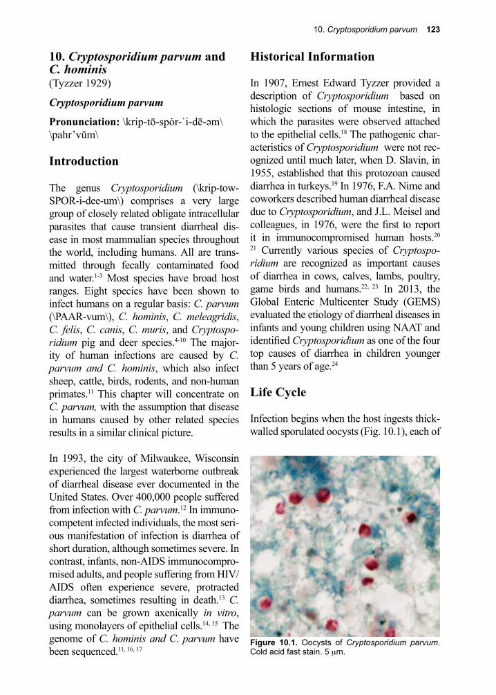
10. Cryptosporidium parvum 123
Figure 10.1. Oocysts of Cryptosporidium parvum. Cold acid fast stain. 5 µm.
10. Cryptosporidium parvum and C. hominis(Tyzzer 1929)
Cryptosporidium parvum
Pronunciation: \krip-tō-spȯr-ˈi-dē-əm\\pahr’vŭm\ Introduction
The genus Cryptosporidium (\krip-tow-SPOR-i-dee-um\) comprises a very large group of closely related obligate intracellular parasites that cause transient diarrheal dis-ease in most mammalian species throughout the world, including humans. All are trans-mitted through fecally contaminated food and water.1-3 Most species have broad host ranges. Eight species have been shown to infect humans on a regular basis: C. parvum (\PAAR-vum\), C. hominis, C. meleagridis, C. felis, C. canis, C. muris, and Cryptospo-ridium pig and deer species.4-10 The major-ity of human infections are caused by C. parvum and C. hominis, which also infect sheep, cattle, birds, rodents, and non-human primates.11 This chapter will concentrate on C. parvum, with the assumption that disease in humans caused by other related species results in a similar clinical picture.
In 1993, the city of Milwaukee, Wisconsin experienced the largest waterborne outbreak of diarrheal disease ever documented in the United States. Over 400,000 people suffered from infection with C. parvum.12 In immuno-competent infected individuals, the most seri-ous manifestation of infection is diarrhea of short duration, although sometimes severe. In contrast, infants, non-AIDS immunocompro-mised adults, and people suffering from HIV/AIDS often experience severe, protracted diarrhea, sometimes resulting in death.13 C. parvum can be grown axenically in vitro, using monolayers of epithelial cells.14, 15 The genome of C. hominis and C. parvum have been sequenced.11, 16, 17
Historical Information
In 1907, Ernest Edward Tyzzer provided a description of Cryptosporidium based on histologic sections of mouse intestine, in which the parasites were observed attached to the epithelial cells.18 The pathogenic char-acteristics of Cryptosporidium were not rec-ognized until much later, when D. Slavin, in 1955, established that this protozoan caused diarrhea in turkeys.19 In 1976, F.A. Nime and coworkers described human diarrheal disease due to Cryptosporidium, and J.L. Meisel and colleagues, in 1976, were the first to report it in immunocompromised human hosts.20 21 Currently various species of Cryptospo-ridium are recognized as important causes of diarrhea in cows, calves, lambs, poultry, game birds and humans.22, 23 In 2013, the Global Enteric Multicenter Study (GEMS) evaluated the etiology of diarrheal diseases in infants and young children using NAAT and identified Cryptosporidium as one of the four top causes of diarrhea in children younger than 5 years of age.24
Life Cycle
Infection begins when the host ingests thick-walled sporulated oocysts (Fig. 10.1), each of
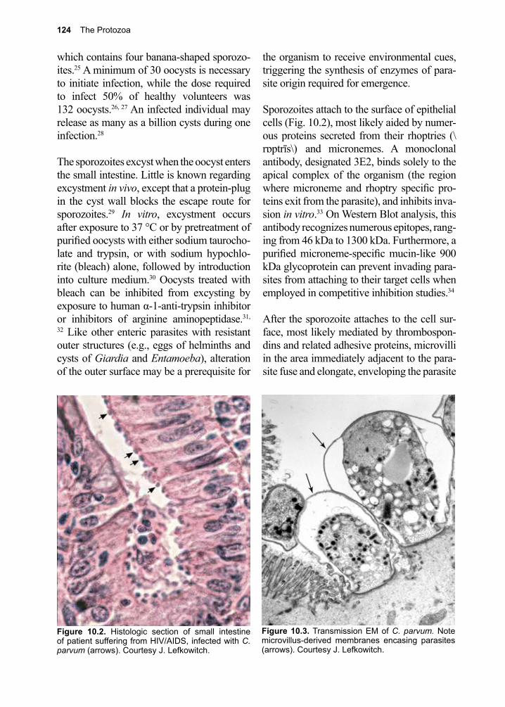
124 The Protozoa
Figure 10.3. Transmission EM of C. parvum. Note microvillus-derived membranes encasing parasites (arrows). Courtesy J. Lefkowitch.
which contains four banana-shaped sporozo-ites.25 A minimum of 30 oocysts is necessary to initiate infection, while the dose required to infect 50% of healthy volunteers was 132 oocysts.26, 27 An infected individual may release as many as a billion cysts during one infection.28
The sporozoites excyst when the oocyst enters the small intestine. Little is known regarding excystment in vivo, except that a protein-plug in the cyst wall blocks the escape route for sporozoites.29 In vitro, excystment occurs after exposure to 37 °C or by pretreatment of purified oocysts with either sodium taurocho-late and trypsin, or with sodium hypochlo-rite (bleach) alone, followed by introduction into culture medium.30 Oocysts treated with bleach can be inhibited from excysting by exposure to human α-1-anti-trypsin inhibitor or inhibitors of arginine aminopeptidase.31,
32 Like other enteric parasites with resistant outer structures (e.g., eggs of helminths and cysts of Giardia and Entamoeba), alteration of the outer surface may be a prerequisite for
the organism to receive environmental cues, triggering the synthesis of enzymes of para-site origin required for emergence.
Sporozoites attach to the surface of epithelial cells (Fig. 10.2), most likely aided by numer-ous proteins secreted from their rhoptries (\rɒptrīs\) and micronemes. A monoclonal antibody, designated 3E2, binds solely to the apical complex of the organism (the region where microneme and rhoptry specific pro-teins exit from the parasite), and inhibits inva-sion in vitro.33 On Western Blot analysis, this antibody recognizes numerous epitopes, rang-ing from 46 kDa to 1300 kDa. Furthermore, a purified microneme-specific mucin-like 900 kDa glycoprotein can prevent invading para-sites from attaching to their target cells when employed in competitive inhibition studies.34
After the sporozoite attaches to the cell sur-face, most likely mediated by thrombospon-dins and related adhesive proteins, microvilli in the area immediately adjacent to the para-site fuse and elongate, enveloping the parasite
Figure 10.2. Histologic section of small intestine of patient suffering from HIV/AIDS, infected with C. parvum (arrows). Courtesy J. Lefkowitch.
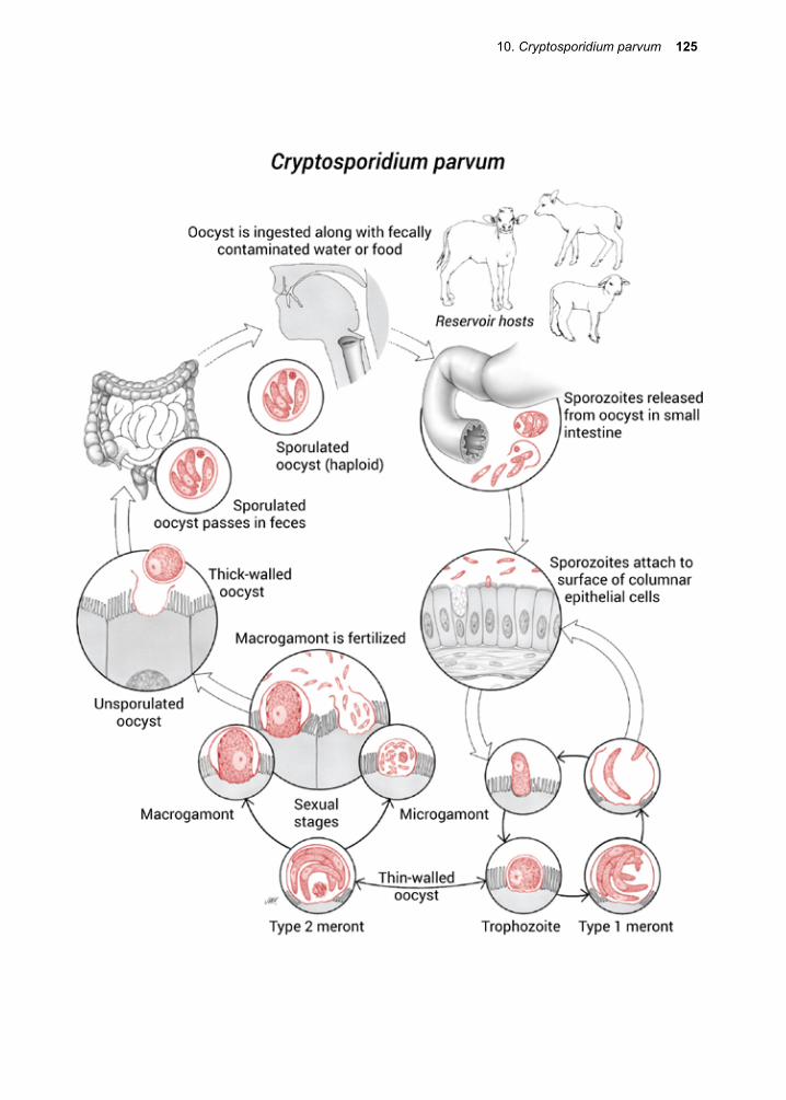
10. Cryptosporidium parvum 125
126 The Protozoa
to create a unique intracellular environment (Fig. 10.3).35 Apical end-associated secreted proteins may also trigger this event. A spe-cialized membrane structure develops at the interface between the parasite and the host cell. Nutrients are thought to pass through this region, since parasite-specific ABC trans-porters have been identified there by means of immunofluorescent monoclonal antibod-ies.36 The parasite then induces alterations in the gene expression of the invaded host cell, eliciting the upregulation of osteoprotegerin, a TNF family member known to inhibit apop-tosis.37 Such a strategy would favor the long-term survival of the parasite until it was able to complete its development to the next stage in its life cycle.38 The sporozoite differentiates into the type I meront (Fig. 10.4) and division ensues, producing four haploid merozoites.
The merozoites are released and attach to new epithelial cells, now differentiating into Type II meronts. Macrogamonts and microgam-onts (pre-sex cells analogous to the game-tocytes of Plasmodia) are produced inside these newly invaded cells. Following their release, microgamonts fuse with macrogam-onts, forming thick-walled zygotes termed oocysts. This stage sporulates within the large intestine, and four haploid infectious sporo-zoites are produced. Oocysts can also be thin-walled. In this case, they sporulate and excyst
within the same host, producing an autoin-fection that may endure for months to years. Even in these cases, thick-walled oocysts are produced, as well.
Thick-walled oocysts pass out in feces and can infect another host. This type of oocyst is environmentally resistant, and can remain viable for months to years in soil, given opti-mum moisture conditions.39
Cellular and Molecular Pathogenesis
One of the most perplexing and frustrat-ing aspects of the biology of C. parvum has been its lack of response to a wide variety of drugs.40-42 The altered microvillus-derived membrane complex that surrounds the para-sites while they are attached to epithelial cells has proven highly impermeable to many che-motherapeutic agents. That is why specula-tion favors the entry of nutrients through the attachment zone between the parasite and the surface of the host cell. The fact that ABC transporters have been identified in this region is further indirect evidence in support of this hypothesis. Cellular or molecular events that result in the alteration of microvilli at the site of attachment have attracted the attention of some research groups.43 Apparently, Cdc42 (a GTPase) and actin are recruited to the site of attachment early on in the process.41 Actin then aggregates, forming a kind of platform on top of which the organism then elaborates its complex of membranes. Much more needs to be learned about the mechanism(s) of nutri-ent acquisition by C. parvum before rational drug design, aimed at interference with this process, can evolve.
Although not fully understood, the secretory diarrhea and pathogenesis of this organism are being deciphered now that the genome is fully sequenced.16, 17 More than 25 pos-Figure 10.4. Transmission EM of C. parvum
meronts. Courtesy M. Belosevic.
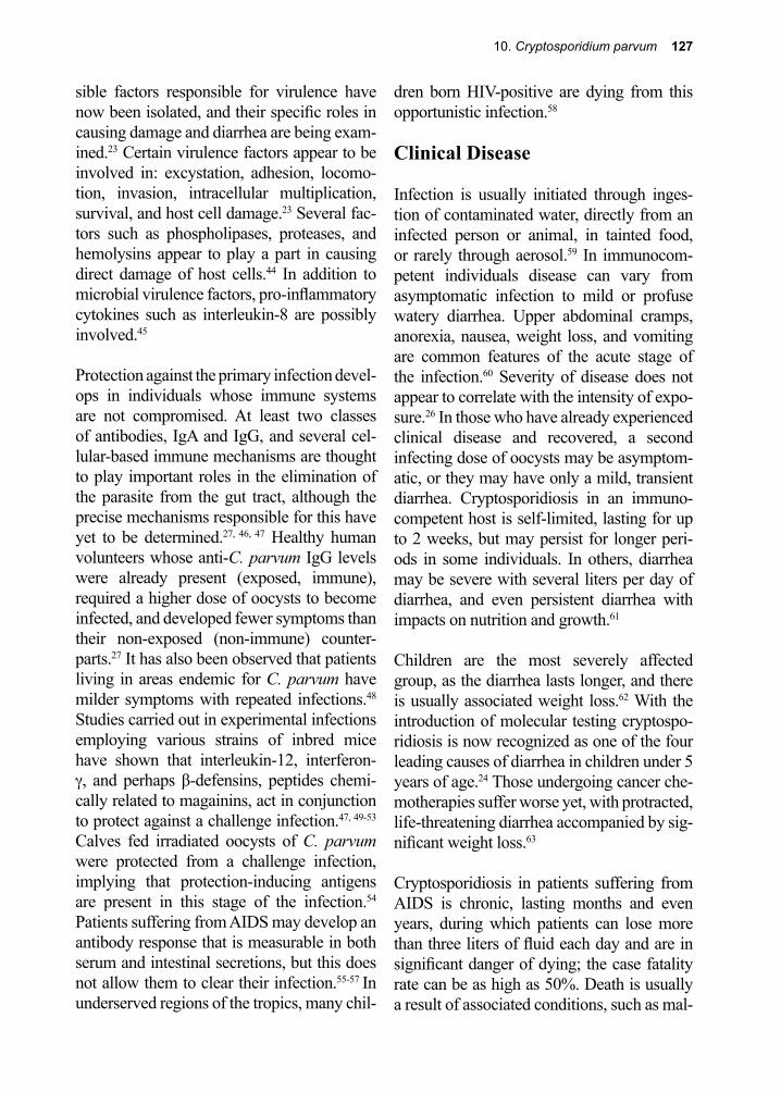
10. Cryptosporidium parvum 127
sible factors responsible for virulence have now been isolated, and their specific roles in causing damage and diarrhea are being exam-ined.23 Certain virulence factors appear to be involved in: excystation, adhesion, locomo-tion, invasion, intracellular multiplication, survival, and host cell damage.23 Several fac-tors such as phospholipases, proteases, and hemolysins appear to play a part in causing direct damage of host cells.44 In addition to microbial virulence factors, pro-inflammatory cytokines such as interleukin-8 are possibly involved.45
Protection against the primary infection devel-ops in individuals whose immune systems are not compromised. At least two classes of antibodies, IgA and IgG, and several cel-lular-based immune mechanisms are thought to play important roles in the elimination of the parasite from the gut tract, although the precise mechanisms responsible for this have yet to be determined.27, 46, 47 Healthy human volunteers whose anti-C. parvum IgG levels were already present (exposed, immune), required a higher dose of oocysts to become infected, and developed fewer symptoms than their non-exposed (non-immune) counter-parts.27 It has also been observed that patients living in areas endemic for C. parvum have milder symptoms with repeated infections.48 Studies carried out in experimental infections employing various strains of inbred mice have shown that interleukin-12, interferon-g, and perhaps β-defensins, peptides chemi-cally related to magainins, act in conjunction to protect against a challenge infection.47, 49-53 Calves fed irradiated oocysts of C. parvum were protected from a challenge infection, implying that protection-inducing antigens are present in this stage of the infection.54 Patients suffering from AIDS may develop an antibody response that is measurable in both serum and intestinal secretions, but this does not allow them to clear their infection.55-57 In underserved regions of the tropics, many chil-
dren born HIV-positive are dying from this opportunistic infection.58
Clinical Disease
Infection is usually initiated through inges-tion of contaminated water, directly from an infected person or animal, in tainted food, or rarely through aerosol.59 In immunocom-petent individuals disease can vary from asymptomatic infection to mild or profuse watery diarrhea. Upper abdominal cramps, anorexia, nausea, weight loss, and vomiting are common features of the acute stage of the infection.60 Severity of disease does not appear to correlate with the intensity of expo-sure.26 In those who have already experienced clinical disease and recovered, a second infecting dose of oocysts may be asymptom-atic, or they may have only a mild, transient diarrhea. Cryptosporidiosis in an immuno-competent host is self-limited, lasting for up to 2 weeks, but may persist for longer peri-ods in some individuals. In others, diarrhea may be severe with several liters per day of diarrhea, and even persistent diarrhea with impacts on nutrition and growth.61
Children are the most severely affected group, as the diarrhea lasts longer, and there is usually associated weight loss.62 With the introduction of molecular testing cryptospo-ridiosis is now recognized as one of the four leading causes of diarrhea in children under 5 years of age.24 Those undergoing cancer che-motherapies suffer worse yet, with protracted, life-threatening diarrhea accompanied by sig-nificant weight loss.63
Cryptosporidiosis in patients suffering from AIDS is chronic, lasting months and even years, during which patients can lose more than three liters of fluid each day and are in significant danger of dying; the case fatality rate can be as high as 50%. Death is usually a result of associated conditions, such as mal-
128 The Protozoa
nutrition or superinfection with other patho-gens. Extra-intestinal infection in the bile duct can cause acalculous biliary disease.
Diagnosis (see Clinical Appendix)
Diagnosis can be made by identification of acid fast-stained oocysts seen on microscopic examination of stool (Fig. 10.1), stool anti-gen testing, directed PCR testing or multi-plex NAAT.64, 65 Stool microscopy is the least sensitive test with sensitivities as low as 30% with single stool examinations.66 Oocysts can be isolated from stool by flotation in sugar solution, then stained by acid-fast methods.67
PCR testing is highly sensitive and can iden-tify specific genotypes, which is important in outbreaks and epidemiological investiga-tions.68, 69 Antigen tests using monoclonal antibodies have high sensitivity, are easy to perform and can be used in both feces and tissue specimens.70 The introduction of multi-plex NAAT testing has helped to increase the number of cases of infectious diarrhea now recognized to be due to Cryptosporidium spp.24 This approach to the diagnosis of infec-tious diarrhea is now a highly sensitive and commercially available diagnostic option for the diagnosis of cryptosporidiosis.71
Treatment (see Clinical Appendix)
Treatment of cryptosporidiosis is based on features of the infected host. In immuno-logically healthy children nitazoxanide is the drug of choice based on studies demonstrat-ing early clinical improvement, earlier reso-lution of diarrhea and improved termination of oocyst shedding.40, 72-74 HIV-1 infected patients are best approached with initiation
of HAART to reconstitute their immune sys-tems. All patients should receive supportive care with oral rehydration when possible and intravenous therapy if required. Several clini-cal trials with rifamycins, azalides and paro-momycin have demonstrated no clear benefit illustrating the difficulty of treating acute or established disease.75
Prevention and Control
Good hygiene is always an important approach to decreasing one’s risk of expo-sure. Without knowledge as to the source of a given outbreak, control and prevention of infection due to C. parvum is not possible. In the case of waterborne epidemics, man-agement of watersheds is the long-term solu-tion in situations where the water supply is not filtered.12, 76 Filtering drinking water is usually effective, but deterioration of filtra-tion equipment and/or lack of proper main-tenance can erode any progress made in controlling waterborne infections. Boiling is another option for purification of contami-nated drinking water. Chlorination of water supplies is ineffective against the oocyst, but ozonation kills this stage.77, 78 In agricultural settings, creation of vegetative barriers to curtail the spread of oocysts is effective.79 Surveillance is key to keeping public water supplies free of pathogens with environmen-tally resistant stages (e.g., Giardia lamblia, Entamoeba histolytica, C. parvum). In this regard, PCR-based testing now allows for the possibility of continuous monitoring of water supplies for C. parvum.80 Urban and suburban pet stores and petting zoos for chil-dren are other sources of infection that until recently have received little attention.81
References
1. Laberge, I.; Griffiths, M. W., Prevalence, detection and control of Cryptosporidium parvum in food. International journal of food microbiology 1996, 32 (1-2), 1-26.
2. Keusch, G. T., et al., Cryptosporidia--who is at risk? Schweizerische medizinische Wochenschrift 1995, 125 (18), 899-908.
10. Cryptosporidium parvum 129
3. Guerrant, R. L., Cryptosporidiosis: an emerging, highly infectious threat. Emerging infectious diseases 1997, 3 (1), 51-7.
4. Katsumata, T., et al., Short report: possible Cryptosporidium muris infection in humans. The American journal of tropical medicine and hygiene 2000, 62 (1), 70-2.
5. Ong, C. S. L., et al., Novel Cryptosporidium genotypes in sporadic cryptosporidiosis cases: first report of human infections with a cervine genotype. Emerging infectious diseases 2002, 8 (3), 263-8.
6. Pedraza-Diaz, S., et al., The identification and characterisation of an unusual genotype of Cryptosporidium from human faeces as Cryptosporidium meleagridis. FEMS Microbiol Letters 2000, 189, 189-194.
7. Pedraza-Diaz, S.; Amar, C., Unusual Cryptosporidium species recovered from human faeces: first description of Cryptosporidium felis and Cryptosporidium ‘dog type’ from patients in England. J Med Microbiol 2001, 50, 293-296.
8. Pieniazek, N. J., et al., New cryptosporidium genotypes in HIV-infected persons. Emerging infectious diseases 1999, 5 (3), 444-9.
9. Xiao, L., et al., Identification of the Cryptosporidium pig genotype in a human patient. The Journal of infectious diseases 2002, 185 (12), 1846-8.
10. Xiao, L., et al., Identification of 5 types of Cryptosporidium parasites in children in Lima, Peru. The Journal of infectious diseases 2001, 183 (3), 492-7.
11. Hadfield, S. J., et al., Generation of whole genome sequences of new Cryptosporidium hominis and Cryptosporidium parvum isolates directly from stool samples. BMC Genomics 2015, 16, 650.
12. Kramer, M. H., et al., Surveillance for waterborne-disease outbreaks--United States, 1993-1994. MMWR. CDC surveillance summaries : Morbidity and mortality weekly report. CDC surveillance summaries / Centers for Disease Control 1996, 45 (1), 1-33.
13. Farthing, M. J., et al., Recently recognised microbial enteropathies and HIV infection. The Journal of antimicrobial chemotherapy 1996, 37 Suppl B, 61-70.
14. Current, W. L.; Reese, N. C., A comparison of endogenous development of three isolates of Cryptosporidium in suckling mice. The Journal of protozoology 1986, 33 (1), 98-108.
15. Meloni, B. P.; Thompson, R. C., Simplified methods for obtaining purified oocysts from mice and for growing Cryptosporidium parvum in vitro. The Journal of parasitology 1996, 82 (5), 757-62.
16. Xu, P., et al., The genome of Cryptosporidium hominis. Nature 2004, 431 (7012), 1107-12.17. Abrahamsen, M. S., et al., Complete genome sequence of the apicomplexan, Cryptosporidium
parvum. Science (New York, N.Y.) 2004, 304 (5669), 441-5.18. Tyzzer, E. E., A Study of Heredity in Relation to the Development of tumors in Mice. The
Journal of medical research 1907, 17 (2), 199-211.19. Slavin, D.; J., Cryptosporidium meleagridis (sp. nov.). Pathol 346 1955, 65, 262-266.20. Nime, F. A., et al., Acute enterocolitis in a human being infected with the protozoan
Cryptosporidium. Gastroenterology 1976, 70 (4), 592-8.21. Meisel, J. L., et al., Overwhelming watery diarrhea associated with a Cryptosporidium in an
immunosuppressed patient. Gastroenterology 1976, 70 (6), 1156-60.22. de Graaf, D. C., et al., A review of the importance of cryptosporidiosis in farm animals. Int J
Parasitol 1999, 29 (8), 1269-87.23. Bouzid, M., et al., Cryptosporidium pathogenicity and virulence. Clin Microbiol Rev 2013, 26
(1), 115-34.24. Kotloff, K. L., et al., Burden and aetiology of diarrhoeal disease in infants and young children in
developing countries (the Global Enteric Multicenter Study, GEMS): a prospective, case-control study. Lancet 2013, 382 (9888), 209-22.
25. Fayer, R., Cryptosporidium: a water-borne zoonotic parasite. Veterinary parasitology 2004, 126 (1-2), 37-56.
26. DuPont, H. L., et al., The infectivity of Cryptosporidium parvum in healthy volunteers. The New England journal of medicine 1995, 332 (13), 855-9.
130 The Protozoa
27. Chappell, C. L., et al., Infectivity of Cryptosporidium parvum in healthy adults with pre-existing anti-C. parvum serum immunoglobulin G. The American journal of tropical medicine and hygiene 1999, 60 (1), 157-64.
28. Chappell, C. L., et al., Cryptosporidium parvum: intensity of infection and oocyst excretion patterns in healthy volunteers. J Infect Dis 1996, 173 (1), 232-6.
29. Neuman, D., et al., Structural and functional characteristics of rhenium clusters derived from redox chemistry of the triangular [ReIII3(mu-Cl)3] core unit. Inorganic chemistry 2000, 39 (24), 5530-7.
30. Forney, J. R., et al., Antagonistic effect of human alpha-1-antitrypsin on excystation of Cryptosporidium parvum oocysts. The Journal of parasitology 1997, 83 (4), 771-4.
31. Okhuysen, P. C., et al., Cryptosporidium parvum metalloaminopeptidase inhibitors prevent in vitro excystation. Antimicrobial agents and chemotherapy 1996, 40 (12), 2781-4.
32. Langer, R. C.; Riggs, M. W., Cryptosporidium parvum apical complex glycoprotein CSL contains a sporozoite ligand for intestinal epithelial cells. Infection and immunity 1999, 67 (10), 5282-91.
33. Riggs, M. W., et al., Protective monoclonal antibody defines a circumsporozoite-like glycoprotein exoantigen of Cryptosporidium parvum sporozoites and merozoites. Journal of immunology (Baltimore, Md. : 1950) 1997, 158 (4), 1787-95.
34. Barnes, D. A., et al., A novel multi-domain mucin-like glycoprotein of Cryptosporidium parvum mediates invasion. Mol Biochem Parasitol 9693 1998, 110, 1-2.
35. Wanyiri, J.; Ward, H., Molecular basis of Cryptosporidium-host cell interactions: recent advances and future prospects. Future Microbiol 2006, 1 (2), 201-8.
36. Zapata, F., et al., The Cryptosporidium parvum ABC protein family. Molecular and biochemical parasitology 2002, 120 (1), 157-61.
37. Di Genova, B. M.; Tonelli, R. R., Infection Strategies of Intestinal Parasite Pathogens and Host Cell Responses. Front Microbiol 2016, 7, 256.
38. Castellanos-Gonzalez, A., et al., Cryptosporidium infection of human intestinal epithelial cells increases expression of osteoprotegerin: a novel mechanism for evasion of host defenses. J Infect Dis 2008, 197 (6), 916-23.
39. Brasseur, P., et al., Viability of Cryptosporidium parvum oocysts in natural waters. Folia parasitologica 1998, 45 (2), 113-6.
40. Smith, H. V.; Corcoran, G. D., New drugs and treatment for cryptosporidiosis. Current opinion in infectious diseases 2004, 17 (6), 557-64.
41. Chen, X.-M., et al., Cdc42 and the actin-related protein/neural Wiskott-Aldrich syndrome protein network mediate cellular invasion by Cryptosporidium parvum. Infection and immunity 2004, 72 (5), 3011-21.
42. Sparks, H., et al., Treatment of Cryptosporidium: What We Know, Gaps, and the Way Forward. Curr Trop Med Rep 2015, 2 (3), 181-187.
43. Huang, B. Q., et al., Cryptosporidium parvum attachment to and internalization by human biliary epithelia in vitro: a morphologic study. The Journal of parasitology 2004, 90 (2), 212-21.
44. Okhuysen, P. C.; Chappell, C. L., Cryptosporidium virulence determinants--are we there yet? Int J Parasitol 2002, 32 (5), 517-25.
45. Kirkpatrick, B. D., et al., Childhood cryptosporidiosis is associated with a persistent systemic inflammatory response. Clin Infect Dis 2006, 43 (5), 604-8.
46. Jenkins, M. C., et al., Hyperimmune bovine colostrum specific for recombinant Cryptosporidium parvum antigen confers partial protection against cryptosporidiosis in immunosuppressed adult mice. Vaccine 1999, 17 (19), 2453-60.
47. McDonald, V., Host cell-mediated responses to infection with Cryptosporidium. Parasite immunology 2000, 22 (12), 597-604.
48. Okhuysen, P. C., et al., Susceptibility and serologic response of healthy adults to reinfection with Cryptosporidium parvum. Infect Immun 1998, 66 (2), 441-3.
49. Urban, J. F., et al., IL-12 protects immunocompetent and immunodeficient neonatal mice against infection with Cryptosporidium parvum. J Immunol 1998, 156 (1 ), 263-268.
10. Cryptosporidium parvum 131
50. Tarver, A. P., et al., Enteric beta-defensin: molecular cloning and characterization of a gene with inducible intestinal epithelial cell expression associated with Cryptosporidium parvum infection. Infection and immunity 1998, 66 (3), 1045-56.
51. Giacometti, A., et al., In-vitro activity of polycationic peptides against Cryptosporidium parvum, Pneumocystis carinii and yeast clinical isolates. The Journal of antimicrobial chemotherapy 1999, 44 (3), 403-6.
52. Ludtke, S. J., et al., Membrane pores induced by magainin. Biochemistry 1996, 35 (43), 13723-8.53. Huang, H. W., Peptide-lipid interactions and mechanisms of antimicrobial peptides. Novartis
Foundation symposium 1999, 225, 188-200; discussion 200.54. Jenkins, M., et al., Protection of calves against cryptosporiosis by oral inoculation with gamma-
irradiated Cryptosporidium parvum oocysts. The Journal of parasitology 2004, 90 (5), 1178-80.55. Benhamou, Y., et al., Inefficacy of intestinal secretory immune response to Cryptosporidium in
acquired immunodeficiency syndrome. Gastroenterology 1995, 108 (3), 627-35.56. Cozon, G., et al., Secretory IgA antibodies to Cryptosporidium parvum in AIDS patients with
chronic cryptosporidiosis. J Infect Dis 1994, 169 (3), 696-9.57. Ravera, M., et al., Prevalence of Cryptosporidium parvum in AIDS and immunocompetent
patients in Uganda. Int J STD AIDS 1994, 5 (4), 302-3.58. Guarino, A., et al., Management of gastrointestinal disorders in children with HIV infection.
Paediatric drugs 2004, 6 (6), 347-62.59. Putignani, L.; Menichella, D., Global distribution, public health and clinical impact of the
protozoan pathogen Cryptosporidium. Interdiscip Perspect Infect Dis 2010, 2010.60. Farthing, M. J., Clinical aspects of human cryptosporidiosis. Contributions to microbiology
2000, 6, 50-74.61. Checkley, W., et al., Effects of Cryptosporidium parvum infection in Peruvian children: growth
faltering and subsequent catch-up growth. Am J Epidemiol 1998, 148 (5), 497-506.62. Cicirello, H. G., et al., Cryptosporidiosis in children during a massive waterborne outbreak
in Milwaukee, Wisconsin: clinical, laboratory and epidemiologic findings. Epidemiology and infection 1997, 119 (1), 53-60.
63. Burgner, D., et al., Epidemiology of Cryptosporidium parvum in symptomatic paediatric oncology patients. Journal of paediatrics and child health 1999, 35 (3), 300-2.
64. Blackman, E., et al., Cryptosporidiosis in HIV-infected patients: diagnostic sensitivity of stool examination, based on number of specimens submitted. The American journal of gastroenterology 1997, 92 (3), 451-3.
65. Coupe, S., et al., Detection of Cryptosporidium and identification to the species level by nested PCR and restriction fragment length polymorphism. Journal of clinical microbiology 2005, 43 (3), 1017-23.
66. Blanshard, C., et al., Cryptosporidiosis in HIV-seropositive patients. Q J Med 1992, 85 (307-308), 813-23.
67. Ignatius, R., et al., Efficacy of different methods for detection of low Cryptosporidium parvum oocyst numbers or antigen concentrations in stool specimens. Eur J.
68. Morgan, U. M., et al., Comparison of PCR and microscopy for detection of Cryptosporidium parvum in human fecal specimens: clinical trial. J Clin Microbiol 1998, 36 (4), 995-8.
69. Morgan, U. M.; Thompson, R. C., PCR detection of cryptosporidium: the way forward? Parasitol Today 1998, 14 (6), 241-5.
70. Garcia, L. S.; Shimizu, R. Y., Evaluation of nine immunoassay kits (enzyme immunoassay and direct fluorescence) for detection of Giardia lamblia and Cryptosporidium parvum in human fecal specimens. J Clin Microbiol 1997, 35 (6), 1526-9.
71. Buss, S. N., et al., Multicenter evaluation of the BioFire FilmArray gastrointestinal panel for etiologic diagnosis of infectious gastroenteritis. J Clin Microbiol 2015, 53 (3), 915-25.
72. Fox, L. M.; Saravolatz, L. D., Nitazoxanide: a new thiazolide antiparasitic agent. Clin Inf Dis 2005, 40 (8), 1173-80.
73. Rossignol, J. F., et al., Treatment of diarrhea caused by Cryptosporidium parvum: a prospective randomized, double-blind, placebo-controlled study of Nitazoxanide. J Infect Dis 2001, 184 (1), 103-6.
132 The Protozoa
74. Rossignol, J. F., et al., Effect of nitazoxanide in diarrhea and enteritis caused by Cryptosporidium species. Clin Gastroenterol Hepatol 2006, 4 (3), 320-4.
75. Checkley, W., et al., A review of the global burden, novel diagnostics, therapeutics, and vaccine targets for cryptosporidium. Lancet Infect Dis 2015, 15 (1), 85-94.
76. Steiner, T. S., et al., Protozoal agents: what are the dangers for the public water supply? Annual review of medicine 1997, 48, 329-40.
77. Clancy, J. L., et al., UV light inactivation of Cryptosporidium oocysts. Water Works Assoc 1998, 90, 92-102.
78. Gyurek, L. L., et al., Ozone inactivation kinetics of Cryptosporidium parvum in phosphate buffer. Engine 2000, 125, 913-924.
79. Tate, K. W., et al., Efficacy of vegetated buffer strips for retaining Cryptosporidium parvum. Journal of environmental quality 2004, 33 (6), 2243-51.
80. Hallier-Soulier, S.; Guillot, E., An immunomagnetic separation polymerase chain reaction assay for rapid and ultra-sensitive detection of Cryptosporidium parvum in drinking water. FEMS microbiology letters 1999, 176 (2), 285-9.
81. Itoh, N., et al., Molecular detection and characterization of Cryptosporidium species in household dogs, pet shop puppies, and dogs kept in a school of veterinary nursing in Japan. Vet Parasitol 2014, 200 (3-4), 284-8.
11. Toxoplasma gondii 133
11. Toxoplasma gondii (Nicolle and Manceaux 1908)
Pronunciation: \täk-sə-ˈplaz-mə\\gon’dē-ī\
Introduction
Toxoplasma gondii (\TOK-so-plaz-ma\\gon-DEE-eye\) is an obligate intracellular parasite that has a worldwide distribution. Its biology is similar to other members of the phylum Apicomplexa, which includes the Plas-modium spp., Cystoisospora spp., Babesia spp., Cyclospora spp., and Cryptosporidium spp. T. gondii infects most species of warm-blooded animals including domesticated and wild birds.1 Although reliable information is not available in many parts of the world, esti-mates based on seroprevalence studies from Brazil and parts of Indonesia, suggest that the majority of individuals living in these coun-tries are infected.2 T. gondii is one of the most successful parasites on earth, even when one takes into account all the viruses and bacteria that infect this large group of vertebrates. It has even emerged as a serious pathogen of some marine mammals such as sea otters.3 It can remain alive as a dormant infection for the life of the host. When immunity breaks down, it can reactivate, often with clinical consequences. In this regard, Toxoplasma behaves similarly to other infectious agents whose reproduction is held in check by host-acquired protective immune responses (e.g., herpes simplex virus, Mycobacterium tuber-culosis).
T. gondii is easily cultured and can be experi-mentally transfected, facilitating studies on its genetics, cellular, and molecular biology.4-6
Its genome is now sequenced.7, 8 Toxoplasma is usually acquired through the ingestion of infected raw or undercooked meats, but sev-
eral outbreaks were traced back to drinking water supplies contaminated with oocysts.9,
10 The domestic cat and other feline species serve as the definitive host, harboring the sexual stages of the parasite.
In immunocompetent humans, infection rarely leads to serious illness. In contrast, when T. gondii infects immunocompromised individuals, or when a previously acquired infection is reactivated, the clinical disease that follows can often be life-threatening.11 Congenital infection also occurs and can occasionally lead to devastating pathological consequences for the developing fetus.
Historical Information
In 1908, Charles Nicolle and Louis Manceaux described the organism which they isolated from the gondi (Ctenodactylus gondii), a gerbil-like desert inhabiting mammal.12 In the same year, Alfonso Splendore, working in Brazil, described the identical parasite, which he identified in the tissues of rabbits.13 They published their results at the same time, but in different publications, so neither was aware of the other’s findings. In 1923, Josef Janku, described the congenital manifesta-tions of the infection, which he accurately characterized as causing hydrocephalus and chorioretinitis.14 Janku was unable to isolate the organism from the brains of its victims. In 1939, Abner Wolf and colleagues confirmed Janku’s clinical description, and went on to experimentally transfer the infection from infected brain tissue to mice and rabbits.15 In 1941, H. Pinkerton and W.R. Henderson, and Alfred Sabin independently described cases of adult-acquired toxoplasmosis.16, 17 In 1970, John Frenkel and colleagues identified the sexual stages of the life cycle working in cats, as did William M. Hutchinson and co-workers in that same year.18, 19
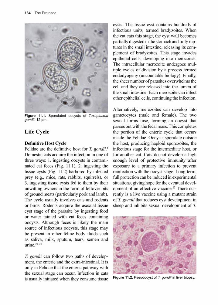
134 The Protozoa
Figure 11.1. Sporulated oocysts of Toxoplasma gondii. 12 µm.
Figure 11.2. Pseudocyst of T. gondii in liver biopsy.
Life Cycle Definitive Host CycleFelidae are the definitive host for T. gondii.4 Domestic cats acquire the infection in one of three ways: 1. ingesting oocysts in contami-nated cat feces (Fig. 11.1), 2. ingesting the tissue cysts (Fig. 11.2) harbored by infected prey (e.g., mice, rats, rabbits, squirrels), or 3. ingesting tissue cysts fed to them by their unwitting owners in the form of leftover bits of ground meats (particularly pork and lamb). The cycle usually involves cats and rodents or birds. Rodents acquire the asexual tissue cyst stage of the parasite by ingesting food or water tainted with cat feces containing oocysts. Although feces is likely the main source of infectious oocysts, this stage may be present in other feline body fluids such as saliva, milk, sputum, tears, semen and urine.20, 21
T. gondii can follow two paths of develop-ment, the enteric and the extra-intestinal. It is only in Felidae that the enteric pathway with the sexual stage can occur. Infection in cats is usually initiated when they consume tissue
cysts. The tissue cyst contains hundreds of infectious units, termed bradyzoites. When the cat eats this stage, the cyst wall becomes partially digested in the stomach and fully rup-tures in the small intestine, releasing its com-plement of bradyzoites. This stage invades epithelial cells, developing into merozoites. The intracellular merozoite undergoes mul-tiple cycles of division by a process termed endodyogeny (uncountable biology). Finally, the sheer number of parasites overwhelms the cell and they are released into the lumen of the small intestine. Each merozoite can infect other epithelial cells, continuing the infection.
Alternatively, merozoites can develop into gametocytes (male and female). The two sexual forms fuse, forming an oocyst that passes out with the fecal mass. This completes the portion of the enteric cycle that occurs inside the Felidae. Oocysts sporulate outside the host, producing haploid sporozoites, the infectious stage for the intermediate host, or for another cat. Cats do not develop a high enough level of protective immunity after exposure to a primary infection to prevent reinfection with the oocyst stage. Long-term, full protection can be induced in experimental situations, giving hope for the eventual devel-opment of an effective vaccine.22 There cur-rently is a live vaccine using a mutant strain of T. gondii that reduces cyst development in sheep and inhibits sexual development of T.
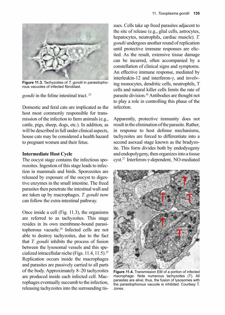
11. Toxoplasma gondii 135
Figure 11.4. Transmission EM of a portion of infected macrophage. Note numerous tachyzoites (T). All parasites are alive; thus, the fusion of lysosomes with the parasitophorous vacuole is inhibited. Courtesy T. Jones.
Figure 11.3. Tachyzoites of T. gondii in parasitopho-rous vacuoles of infected fibroblast.
gondii in the feline intestinal tract. 23
Domestic and feral cats are implicated as the host most commonly responsible for trans-mission of the infection to farm animals (e.g., cattle, pigs, sheep, dogs, etc.). In addition, as will be described in full under clinical aspects, house cats may be considered a health hazard to pregnant women and their fetus.
Intermediate Host CycleThe oocyst stage contains the infectious spo-rozoites. Ingestion of this stage leads to infec-tion in mammals and birds. Sporozoites are released by exposure of the oocyst to diges-tive enzymes in the small intestine. The freed parasites then penetrate the intestinal wall and are taken up by macrophages. T. gondii now can follow the extra-intestinal pathway.
Once inside a cell (Fig. 11.3), the organisms are referred to as tachyzoites. This stage resides in its own membrane-bound parasi-tophorous vacuole.24 Infected cells are not able to destroy tachyzoites, due to the fact that T. gondii inhibits the process of fusion between the lysosomal vessels and this spe-cialized intracellular niche (Figs. 11.4, 11.5).25
Replication occurs inside the macrophages and parasites are passively carried to all parts of the body. Approximately 8–20 tachyzoites are produced inside each infected cell. Mac-rophages eventually succumb to the infection, releasing tachyzoites into the surrounding tis-
sues. Cells take up freed parasites adjacent to the site of release (e.g., glial cells, astrocytes, hepatocytes, neutrophils, cardiac muscle). T. gondii undergoes another round of replication until protective immune responses are elic-ited. As the result, extensive tissue damage can be incurred, often accompanied by a constellation of clinical signs and symptoms. An effective immune response, mediated by interleukin-12 and interferon-γ, and involv-ing monocytes, dendritic cells, neutrophils, T cells and natural killer cells limits the rate of parasite division.26 Antibodies are thought not to play a role in controlling this phase of the infection.
Apparently, protective immunity does not result in the elimination of the parasite. Rather, in response to host defense mechanisms, tachyzoites are forced to differentiate into a second asexual stage known as the bradyzo-ite. This form divides both by endodyogeny and endopolygeny, then organizes into a tissue cyst.27 Interferon-γ-dependent, NO-mediated
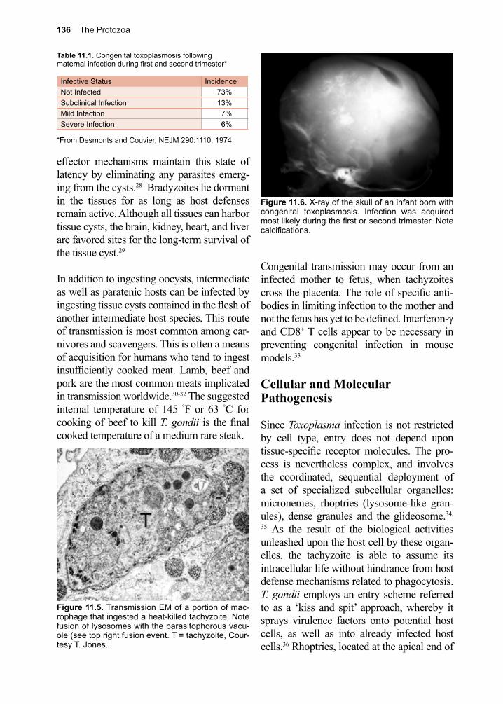
136 The Protozoa
Table 11.1. Congenital toxoplasmosis following maternal infection during first and second trimester*
Infective Status IncidenceNot Infected 73%Subclinical Infection 13%Mild Infection 7%Severe Infection 6%
*From Desmonts and Couvier, NEJM 290:1110, 1974
Figure 11.5. Transmission EM of a portion of mac-rophage that ingested a heat-killed tachyzoite. Note fusion of lysosomes with the parasitophorous vacu-ole (see top right fusion event. T = tachyzoite, Cour-tesy T. Jones.
Figure 11.6. X-ray of the skull of an infant born with congenital toxoplasmosis. Infection was acquired most likely during the first or second trimester. Note calcifications.
effector mechanisms maintain this state of latency by eliminating any parasites emerg-ing from the cysts.28 Bradyzoites lie dormant in the tissues for as long as host defenses remain active. Although all tissues can harbor tissue cysts, the brain, kidney, heart, and liver are favored sites for the long-term survival of the tissue cyst.29
In addition to ingesting oocysts, intermediate as well as paratenic hosts can be infected by ingesting tissue cysts contained in the flesh of another intermediate host species. This route of transmission is most common among car-nivores and scavengers. This is often a means of acquisition for humans who tend to ingest insufficiently cooked meat. Lamb, beef and pork are the most common meats implicated in transmission worldwide.30-32 The suggested internal temperature of 145 °F or 63 °C for cooking of beef to kill T. gondii is the final cooked temperature of a medium rare steak.
Congenital transmission may occur from an infected mother to fetus, when tachyzoites cross the placenta. The role of specific anti-bodies in limiting infection to the mother and not the fetus has yet to be defined. Interferon-γ and CD8+ T cells appear to be necessary in preventing congenital infection in mouse models.33
Cellular and Molecular Pathogenesis
Since Toxoplasma infection is not restricted by cell type, entry does not depend upon tissue-specific receptor molecules. The pro-cess is nevertheless complex, and involves the coordinated, sequential deployment of a set of specialized subcellular organelles: micronemes, rhoptries (lysosome-like gran-ules), dense granules and the glideosome.34,
35 As the result of the biological activities unleashed upon the host cell by these organ-elles, the tachyzoite is able to assume its intracellular life without hindrance from host defense mechanisms related to phagocytosis. T. gondii employs an entry scheme referred to as a ‘kiss and spit’ approach, whereby it sprays virulence factors onto potential host cells, as well as into already infected host cells.36 Rhoptries, located at the apical end of
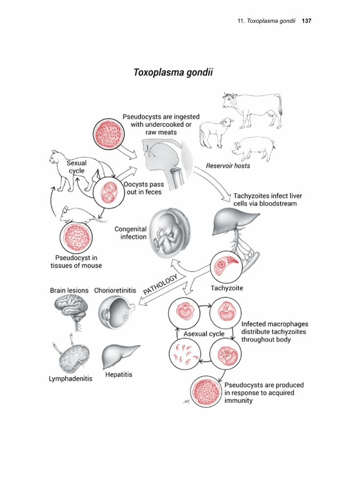
11. Toxoplasma gondii 137
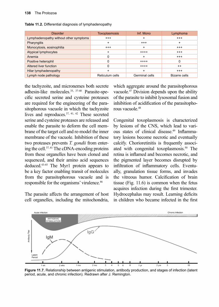
138 The Protozoa
Table 11.2. Differential diagnosis of lymphadenopathy
Disorder Toxoplasmosis Inf. Mono LymphomaLymphadenopathy without other symptoms +++ + +++Pharyngitis + +++ +Monocytosis, eosinophilia +++ + +++Atypical lymphocytes + ++++ +++Anemia 0 + +++Positive heterophil 0 ++++ 0Altered liver function 0 ++++ ++Hilar lymphadenopathy + + +++Lymph node pathology Reticulum cells Germinal cells Bizarre cells
Figure 11.7. Relationship between antigenic stimulation, antibody production, and stages of infection (latent period, acute, and chronic infection). Redrawn after J. Remington.
the tachyzoite, and micronemes both secrete adhesin-like molecules.35, 37-40 Parasite-spe-cific secreted serine and cysteine proteases are required for the engineering of the para-sitophorous vacuole in which the tachyzoite lives and reproduces.37, 41, 42 These secreted serine and cysteine proteases are released and enable the parasite to deform the cell mem-brane of the target cell and re-model the inner membrane of the vacuole. Inhibition of these two proteases prevents T. gondii from enter-ing the cell.37, 41 The cDNA-encoding proteins from these organelles have been cloned and sequenced, and their amino acid sequences deduced.43-45 The Myr1 protein appears to be a key factor enabling transit of molecules from the parasitophorous vacuole and is responsible for the organisms’ virulence.46
The parasite affects the arrangement of host cell organelles, including the mitochondria,
which aggregate around the parasitophorous vacuole.47 Division depends upon the ability of the parasite to inhibit lysosomal fusion and inhibition of acidification of the parasitopho-rous vacuole.48
Congenital toxoplasmosis is characterized by lesions of the CNS, which lead to vari-ous states of clinical disease.49 Inflamma-tory lesions become necrotic and eventually calcify. Chorioretinitis is frequently associ-ated with congenital toxoplasmosis.50 The retina is inflamed and becomes necrotic, and the pigmented layer becomes disrupted by infiltration of inflammatory cells. Eventu-ally, granulation tissue forms, and invades the vitreous humor. Calcification of brain tissue (Fig. 11.6) is common when the fetus acquires infection during the first trimester. Hydrocephalus may result. Learning deficits in children who became infected in the first
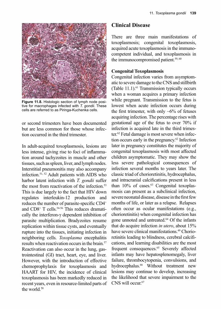
11. Toxoplasma gondii 139
Figure 11.8. Histologic section of lymph node posi-tive for macrophages infected with T. gondii. These cells are referred to as Piringa-Kuchenka cells.
or second trimesters have been documented but are less common for those whose infec-tion occurred in the third trimester.
In adult-acquired toxoplasmosis, lesions are less intense, giving rise to foci of inflamma-tion around tachyzoites in muscle and other tissues, such as spleen, liver, and lymph nodes. Interstitial pneumonitis may also accompany infection.51, 52 Adult patients with AIDS who harbor latent infection with T. gondii suffer the most from reactivation of the infection.53 This is due largely to the fact that HIV down regulates interleukin-12 production and reduces the number of parasite-specific CD4+ and CD8+ T cells.54-56 This reduces dramati-cally the interferon-γ dependent inhibition of parasite multiplication. Bradyzoites resume replication within tissue cysts, and eventually rupture into the tissues, initiating infection in neighboring cells. Toxoplasma encephalitis results when reactivation occurs in the brain.57 Reactivation can also occur in the lung, gas-trointestinal (GI) tract, heart, eye, and liver. However, with the introduction of effective chemoprophylaxis for toxoplasmosis and HAART for HIV, the incidence of clinical toxoplasmosis has been markedly reduced in recent years, even in resource-limited parts of the world.58
Clinical Disease
There are three main manifestations of toxoplasmosis; congenital toxoplasmosis, acquired acute toxoplasmosis in the immuno-competent individual, and toxoplasmosis in the immunocompromised patient.59, 60
Congenital ToxoplasmosisCongenital infection varies from asymptom-atic to severe damage to the CNS and stillbirth (Table 11.1).61 Transmission typically occurs when a woman acquires a primary infection while pregnant. Transmission to the fetus is lowest when acute infection occurs during the first trimester, with only ~6% of fetuses acquiring infection. The percentage rises with gestational age of the fetus to over 70% if infection is acquired late in the third trimes-ter.62 Fetal damage is most severe when infec-tion occurs early in the pregnancy.62 Infection later in pregnancy constitutes the majority of congenital toxoplasmosis with most affected children asymptomatic. They may show the less severe pathological consequences of infection several months to years later. The classic triad of chorioretinitis, hydrocephalus, and intracranial calcifications present in less than 10% of cases.63 Congenital toxoplas-mosis can present as a subclinical infection, severe neonatal disease, disease in the first few months of life, or later as a relapse. Relapses often occur as ocular manifestations (e.g., chorioretinitis) when congenital infection has gone unnoted and untreated.64 Of the infants that do acquire infection in utero, about 15% have severe clinical manifestations.49 Chorio-retinitis leading to blindness, cerebral calcifi-cations, and learning disabilities are the most frequent consequences.65 Severely affected infants may have hepatosplenomegaly, liver failure, thrombocytopenia, convulsions, and hydrocephalus.66 Without treatment new lesions may continue to develop, increasing the likelihood that severe impairment to the CNS will occur.67
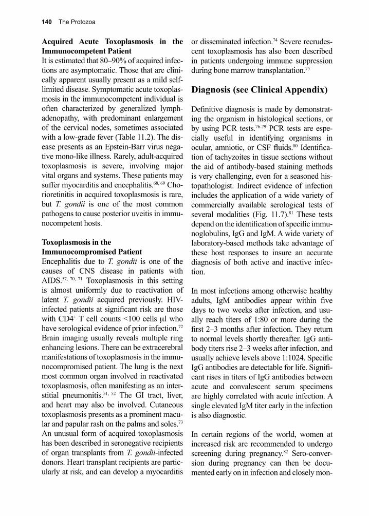
140 The Protozoa
or disseminated infection.74 Severe recrudes-cent toxoplasmosis has also been described in patients undergoing immune suppression during bone marrow transplantation.75
Diagnosis (see Clinical Appendix)
Definitive diagnosis is made by demonstrat-ing the organism in histological sections, or by using PCR tests.76-79 PCR tests are espe-cially useful in identifying organisms in ocular, amniotic, or CSF fluids.80 Identifica-tion of tachyzoites in tissue sections without the aid of antibody-based staining methods is very challenging, even for a seasoned his-topathologist. Indirect evidence of infection includes the application of a wide variety of commercially available serological tests of several modalities (Fig. 11.7).81 These tests depend on the identification of specific immu-noglobulins, IgG and IgM. A wide variety of laboratory-based methods take advantage of these host responses to insure an accurate diagnosis of both active and inactive infec-tion.
In most infections among otherwise healthy adults, IgM antibodies appear within five days to two weeks after infection, and usu-ally reach titers of 1:80 or more during the first 2–3 months after infection. They return to normal levels shortly thereafter. IgG anti-body titers rise 2–3 weeks after infection, and usually achieve levels above 1:1024. Specific IgG antibodies are detectable for life. Signifi-cant rises in titers of IgG antibodies between acute and convalescent serum specimens are highly correlated with acute infection. A single elevated IgM titer early in the infection is also diagnostic.
In certain regions of the world, women at increased risk are recommended to undergo screening during pregnancy.82 Sero-conver-sion during pregnancy can then be docu-mented early on in infection and closely mon-
Acquired Acute Toxoplasmosis in the Immunocompetent PatientIt is estimated that 80–90% of acquired infec-tions are asymptomatic. Those that are clini-cally apparent usually present as a mild self-limited disease. Symptomatic acute toxoplas-mosis in the immunocompetent individual is often characterized by generalized lymph-adenopathy, with predominant enlargement of the cervical nodes, sometimes associated with a low-grade fever (Table 11.2). The dis-ease presents as an Epstein-Barr virus nega-tive mono-like illness. Rarely, adult-acquired toxoplasmosis is severe, involving major vital organs and systems. These patients may suffer myocarditis and encephalitis.68, 69 Cho-rioretinitis in acquired toxoplasmosis is rare, but T. gondii is one of the most common pathogens to cause posterior uveitis in immu-nocompetent hosts.
Toxoplasmosis in the Immunocompromised PatientEncephalitis due to T. gondii is one of the causes of CNS disease in patients with AIDS.57, 70, 71 Toxoplasmosis in this setting is almost uniformly due to reactivation of latent T. gondii acquired previously. HIV-infected patients at significant risk are those with CD4+ T cell counts <100 cells µl who have serological evidence of prior infection.72 Brain imaging usually reveals multiple ring enhancing lesions. There can be extracerebral manifestations of toxoplasmosis in the immu-nocompromised patient. The lung is the next most common organ involved in reactivated toxoplasmosis, often manifesting as an inter-stitial pneumonitis.51, 52 The GI tract, liver, and heart may also be involved. Cutaneous toxoplasmosis presents as a prominent macu-lar and papular rash on the palms and soles.73 An unusual form of acquired toxoplasmosis has been described in seronegative recipients of organ transplants from T. gondii-infected donors. Heart transplant recipients are partic-ularly at risk, and can develop a myocarditis
11. Toxoplasma gondii 141
itored. Congenital infection can be confirmed when specific IgM antibodies are detected in the infant’s serum. About 25% of newborns with congenital toxoplasmosis have IgM anti-bodies. In contrast, infants with IgG antibod-ies pose a problem to the clinician in deciding whether or not the fetus was ever infected or whether the IgG represents passive transfer of maternal antibody. Careful clinical and sero-logical follow-up of the child is indicated to determine whether intrauterine infection has occurred.
Diagnosing toxoplasmosis in patients with AIDS often involves making a presumptive diagnosis and then administration of a thera-peutic trial.83 The triad of a compatible clini-cal syndrome, a positive toxoplasma IgG, and typical brain imaging demonstrating multiple ring enhancing lesions is associated with greater than a 90% positive predictive value for the diagnosis of CNS toxoplasmosis in an HIV infected patient with a CD4+ T cell count <100 cells/µl.72 Histologic examination of lymph node tissue (Fig. 11.8) obtained at biopsy may show abnormal histiocytes, but this evidence is not pathognomonic for toxo-plasmosis.
Treatment (see Clinical Appendix)
Mild infection in the immunocompetent patient does not require therapy, unless the patient is pregnant and being treated to pre-vent infection of the fetus. In most cases pyri-methamine and sulfadiazine are the drugs of choice.84 For a number of reasons, alternative, less studied regimens are often necessary. If pyrimethamine is not available due to supply issues, cost or intolerance, patients can be treated with trimethoprim-sulfamethoxazole or atovaquone.85 In patients unable to toler-ate sulfadiazine, alternative regimens con-taining atovaquone or azithromycin may be utilized. The combination of pyrimethamine and sulfadiazine is teratogenic in animals and
there is concern regarding its use in preg-nancy, particularly during early trimesters. As a consequence, women who become acutely infected during pregnancy are often treated with the macrolide antibiotic spiramycin. The ability of this agent to concentrate in the pla-centa is thought to help prevent transmission to the fetus.86, 87 Some physicians will treat women who acquire acute infection during later trimesters with pyrimethamine and sul-fadiazine, but in some settings women will choose to terminate their pregnancies, as it is not clear that initiation of treatment after the development of intracranial calcifications has any benefit.88
Prevention and Control
Eating only well-cooked meats and avoiding the inadvertent ingestion of cat feces contam-inated with toxoplasma oocysts can prevent most infections. These preventive measures often fail because of cultural or individual cuisine preferences. In France, where the prevalence of infection is over 85% among those over the age of 50, the most common source of infection is raw meat served as “steak” tartar, most of which is actually lamb or horsemeat. Many other cultures also have numerous recipes calling for undercooked or raw meat as a main ingredient. A number of Alaskan native peoples still eat some of their meat raw, and are therefore at the mercy of the pathogens lurking inside each carcass.89 In Ethiopia, the practice of eating raw meat with melted butter, kitfo, is a popular but risky dietary choice.90 Prevention of infection from oocysts requires care when handling cat feces, especially when cleaning litter boxes.91 Pregnant women diminish their risk of infec-tion by avoiding these activities. Rarely, toxo-plasmosis is acquired as the result of inhaling dust, or drinking water contaminated with oocysts.92 Vaccines potentially offer another set of strategies for controlling this infec-tion, but carefully defined susceptible at-risk
142 The Protozoa
References
1. Dubey, J. P., A review of toxoplasmosis in wild birds. Vet Parasitol 2002, 106 (2), 121-53.2. Pappas, G., et al., Toxoplasmosis snapshots: global status of Toxoplasma gondii seroprevalence
and implications for pregnancy and congenital toxoplasmosis. Int J Parasitol 2009, 39 (12), 1385-94.
3. Miller, M. A., et al., An unusual genotype of Toxoplasma gondii is common in California sea otters (Enhydra lutris nereis) and is a cause of mortality. International journal for parasitology 2004, 34 (3), 275-84.
4. Kim, K.; Weiss, L. M., Toxoplasma gondii: the model apicomplexan. International journal for parasitology 2004, 34 (3), 423-32.
5. Gubbels, M.-J.; Striepen, B., Studying the cell biology of apicomplexan parasites using fluorescent proteins. Microscopy and microanalysis : the official journal of Microscopy Society of America, Microbeam Analysis Society, Microscopical Society of Canada 2004, 10 (5), 568-79.
6. Boothroyd, J. C., Toxoplasma gondii: 25 years and 25 major advances for the field. Int J Parasitol 2009, 39 (8), 935-46.
7. Kissinger, J. C., et al., ToxoDB: accessing the Toxoplasma gondii genome. Nucleic acids research 2003, 31 (1), 234-6.
8. Khan, A., et al., Common inheritance of chromosome Ia associated with clonal expansion of Toxoplasma gondii. Genome Res 2006, 16 (9), 1119-25.
9. Pozio, E., Foodborne and waterborne parasites. Acta microbiologica Polonica 2003, 52 Suppl, 83-96.
10. Dubey, J. P., Toxoplasmosis - a waterborne zoonosis. Vet Parasitol 2004, 126 (1-2), 57-72.11. Halonen, S. K.; Weiss, L. M., Toxoplasmosis. Handb Clin Neurol 2013, 114, 125-45.12. Nicolle, C., et al., Sur une infection a coyes de Leishman (ou organismes voisins) du gondi.
Seance Acad Sci 1908, 147, 763-766.13. Splendore, A., Un nuovo protozoa parassita dei conigli: incontrato nelle lesioni anatomiche d’ua
maittia che ricorda in molti punti ii Kala-azar dell’uomo. Rev Soc Sci Sao Paulo 1908.14. Janku, J.; J., Pathogenesis and pathologic anatomy of the “congenital coloboma” of the
maculalutea in an eye of normal size, with microscopic detection of parasites in the retina. Phys 1923, 62, 1021-1027.
15. Wolf, A., et al., Human toxoplasmosis: occurrence in infants as an encephalomyelitis verification by transmission to animals. Science (New York, N.Y.) 1939, 89 (2306), 226-7.
16. Pinkerton, H.; Henderson, R. G., Adult toxoplasmosis: a previously unrecognized disease entity simulating the typhus-spotted fever group. JAMA 1941, 116 807-814.
17. Sabin, A. B., Toxoplasmic encephalitis in children. JAMA 1941, 116 801-807.18. Frenkel, J. K., et al., Toxoplasma gondii in cats: fecal stages identified as coccidian oocysts.
Science (New York, N.Y.) 1970, 167 (3919), 893-6.19. Hutchison, W. M., et al., Coccidian-like nature of Toxoplasma gondii. Br Med J 1970, 1 (5689),
142-4.20. Bresciani, K. D., et al., Experimental toxoplasmosis in pregnant bitches. Vet Parasitol 1999, 86
(2), 143-5.21. Tenter, A. M., et al., Toxoplasma gondii: from animals to humans. Int J Parasitol 2000, 30 (12-
13), 1217-58.22. Bhopale, G. M., Development of a vaccine for toxoplasmosis: current status, Microbes Infect.
Microbes and infection 2003, 5 (5), 457-462.23. Verma, R.; Khanna, P., Development of Toxoplasma gondii vaccine: A global challenge. Hum
Vaccin Immunother 2013, 9 (2), 291-3.
populations, such as sero-negative women of child-bearing age, would need to be clearly
articulated prior to committing the necessary resources to vaccine development.93
11. Toxoplasma gondii 143
24. Jones, T. C.; Hirsch, J. G., The interaction between Toxoplasma gondii and mammalian cells. II. The absence of lysosomal fusion with phagocytic vacuoles containing living parasites. The Journal of experimental medicine 1972, 136 (5), 1173-94.
25. Dlugonska, H.; J., Molecular modifications of host cells by Toxoplasma gondii. Pol 2004, 53, 45-54.
26. Hunter, C. A.; Sibley, L. D., Modulation of innate immunity by Toxoplasma gondii virulence effectors. Nat Rev Microbiol 2012, 10 (11), 766-78.
27. Dzierszinski, F., et al., Dynamics of Toxoplasma gondii differentiation. Eukaryotic cell 2004, 3 (4), 992-1003.
28. Scharton-Kersten, T. M., et al., Inducible nitric oxide is essential for host control of persistent but not acute infection with the intracellular pathogen Toxoplasma gondii. The Journal of experimental medicine 1997, 185 (7), 1261-73.
29. Dadimoghaddam, Y., et al., Tissue tropism and parasite burden of Toxoplasma gondii RH strain in experimentally infected mice. Asian Pac J Trop Med 2014, 7 (7), 521-4.
30. Williams, R. H., et al., High levels of congenital transmission of Toxoplasma gondii in longitudinal and cross-sectional studies on sheep farms provides evidence of vertical transmission in ovine hosts. Parasitology 2005, 130 (Pt 3), 301-7.
31. Dubey, J. P., et al., High prevalence of viable Toxoplasma gondii infection in market weight pigs from a farm in Massachusetts. J Parasitol 2002, 88 (6), 1234-8.
32. Dubey, J. P., et al., Toxoplasmosis in black-faced kangaroos (Macropus fuliginosus melanops). Vet Parasitol 1988, 30 (2), 97-105.
33. Abou-Bacar, A., et al., Role of gamma interferon and T cells in congenital Toxoplasma transmission. Parasite immunology 2004, 26 (8-9), 315-8.
34. Carruthers, V. B.; Blackman, M. J., A new release on life: emerging concepts in proteolysis and parasite invasion. Molecular microbiology 2005, 55 (6), 1617-30.
35. Keeley, A.; Soldati, D., The glideosome: a molecular machine powering motility and host-cell invasion by Apicomplexa. Trends in cell biology 2004, 14 (10), 528-32.
36. Boothroyd, J. C.; Dubremetz, J. F., Kiss and spit: the dual roles of Toxoplasma rhoptries. Nat Rev Microbiol 2008, 6 (1), 79-88.
37. Que, X., et al., The cathepsin B of Toxoplasma gondii, toxopain-1, is critical for parasite invasion and rhoptry protein processing. The Journal of biological chemistry 2002, 277 (28), 25791-7.
38. Ngo, H. M., et al., Are rhoptries in Apicomplexan parasites secretory granules or secretory lysosomal granules?Mol Microbiol. 2004, 52 1531-41.
39. Carruthers, V. B., et al., Secretion of micronemal proteins is associated with Toxoplasma invasion of host cells. Cellular microbiology 1999, 1 (3), 225-35.
40. Huynh, M.-H., et al., Rapid invasion of host cells by Toxoplasma requires secretion of the MIC2-M2AP adhesive protein complex. The EMBO journal 2003, 22 (9), 2082-90.
41. Conseil, V., et al., Serine protease inhibitors block invasion of host cells by Toxoplasma gondii. Antimicrobial agents and chemotherapy 1999, 43 (6), 1358-61.
42. Kim, K., Role of proteases in host cell invasion by Toxoplasma gondii and other Apicomplexa. Acta tropica 2004, 91 (1), 69-81.
43. Ajioka, J. W., et al., Gene discovery by EST sequencing in Toxoplasma gondii reveals sequences restricted to the Apicomplexa. Genome research 1998, 8 (1), 18-28.
44. Yahiaoui, B., et al., Isolation and characterization of a subtractive library enriched for developmentally regulated transcripts expressed during encystation of Toxoplasma gondii. Molecular and biochemical parasitology 1999, 99 (2), 223-35.
45. Bradley, P. J., et al., A GFP-based motif-trap reveals a novel mechanism of targeting for the Toxoplasma ROP4 protein. Molecular and biochemical parasitology 2004, 137 (1), 111-20.
46. Franco, M., et al., A Novel Secreted Protein, MYR1, Is Central to Toxoplasma’s Manipulation of Host Cells. MBio 2016, 7 (1), e02231-15.
47. Lindsay, D. S., et al., Association of host cell mitochondria with developing Toxoplasma gondii tissue cysts. American journal of veterinary research 1993, 54 (10), 1663-7.
144 The Protozoa
48. Shaw, M. K., et al., Acidic compartments and rhoptry formation in Toxoplasma gondii. Parasitology 1998, 117 ( Pt 5), 435-43.
49. Remington, J. S., et al., Toxoplasmosis. In: Infectious Diseases of the Fetus and Newborn Infant. 1995.
50. Mets, M. B., et al., Eye manifestations of congenital toxoplasmosis. American journal of ophthalmology 1997, 123 (1), 1-16.
51. Campagna, A. C., Pulmonary toxoplasmosis. Seminars in respiratory infections 1997, 12 (2), 98-105.
52. Mariuz, P., et al., Toxoplasma pneumonia. Seminars in respiratory infections 1997, 12 (1), 40-3.53. Kaplan, J. E.; V., H. I., Diagnosis, treatment, and prevention of selected common HIV-related
opportunistic infections in the Caribbean region. Top 2005, 12, 136-41.54. Vanham, G., et al., Decreased CD40 ligand induction in CD4 T cells and dysregulated IL-12
production during HIV infection. Clinical and experimental immunology 1999, 117 (2), 335-42.55. Sartori, A., et al., Interleukin-12: an immunoregulatory cytokine produced by B cells and
antigen-presenting cells. Methods (San Diego, Calif.) 1997, 11 (1), 116-27.56. Alonso, K., et al., Cytokine patterns in adults with AIDS. Immunological investigations 1997, 26
(3), 341-50.57. Luft, B. J.; Remington, J. S., Toxoplasmic encephalitis in AIDS. Clin Inf Dis 1992, 15 (2), 211-
22.58. Hari, K. R., et al., Reduced risk of toxoplasma encephalitis in HIV-infected patients--a
prospective study from Gauteng, South Africa. Int J STD AIDS 2007, 18 (8), 555-8.59. Hill, D.; Dubey, J. P., Toxoplasma gondii: transmission, diagnosis and prevention. Clinical
microbiology and infection : the official publication of the European Society of Clinical Microbiology and Infectious Diseases 2002, 8 (10), 634-40.
60. Montoya, J. G.; Liesenfeld, O., Toxoplasmosis. Lancet (London, England) 2004, 363 (9425), 1965-76.
61. Desmont, G.; Couvrer, J., Congenital toxoplasmosis: a prospective study of 378 pregnancies. N Engl J Med 1974, 290 S, 1110-1116.
62. Montoya, J. G.; Remington, J. S., Management of Toxoplasma gondii infection during pregnancy. Clin Infect Dis 2008, 47 (4), 554-66.
63. Tamma, P., Toxoplasmosis. Pediatr Rev 2007, 28 (12), 470-1.64. Wilson, C. B., et al., Development of adverse sequelae in children born with subclinical
congenital Toxoplasma infection. Pediatrics 1980, 66 (5), 767-74.65. Kravetz, J. D.; Federman, D. G., Toxoplasmosis in pregnancy. The American journal of medicine
2005, 118 (3), 212-6.66. Koppe, J. G., et al., Results of 20-year follow-up of congenital toxoplasmosis. Lancet (London,
England) 1986, 1 (8475), 254-6.67. Bhopale, G. M., Pathogenesis of toxoplasmosis. Comparative immunology, microbiology and
infectious diseases 2003, 26 (4), 213-22.68. Kirchhoff, L. V., et al., Parasitic diseases of the heart. Frontiers in bioscience 2004, 9, 706-23.69. Wilson, E. H.; Hunter, C. A., The role of astrocytes in the immunopathogenesis of toxoplasmic
encephalitis. International journal for parasitology 2004, 34 (5), 543-8.70. Nath, A.; Sinai, A. P., Cerebral Toxoplasmosis. Current treatment options in neurology 2003, 5
(1), 3-12.71. Leyva, W. H.; Santa Cruz, D. J., Cutaneous toxoplasmosis. Journal of the American Academy of
Dermatology 1986, 14 (4), 600-5.72. Belanger, F., et al., Incidence and risk factors of toxoplasmosis in a cohort of human
immunodeficiency virus-infected patients: 1988-1995. HEMOCO and SEROCO Study Groups. Clin Infect Dis 1999, 28 (3), 575-81.
73. Janitschke, K., et al., Diagnostic value of tests for Toxoplasma gondii-specific antibodies in patients undergoing bone marrow transplantation. Clinical laboratory 2003, 49 (5-6), 239-42.
74. Wreghitt, T. G., et al., Toxoplasmosis in heart and heart and lung transplant recipients. J Clin Pathol 1989, 42 (2), 194-9.
75. Mele, A., et al., Toxoplasmosis in bone marrow transplantation: a report of two cases and
11. Toxoplasma gondii 145
systematic review of the literature. Bone Marrow Transplant 2002, 29 (8), 691-8.76. Nimri, L., et al., Detection of Toxoplasma gondii DNA and specific antibodies in high-risk
pregnant women. The American journal of tropical medicine and hygiene 2004, 71 (6), 831-5.77. Danise, A., et al., Use of polymerase chain reaction assays of aqueous humor in the differential
diagnosis of retinitis in patients infected with human immunodeficiency virus. Clin Infect Dis 1997, 24 (6), 1100-6.
78. Bergstrom, T., et al., Congenital Toxoplasma gondii infection diagnosed by PCR amplification of peripheral mononuclear blood cells from a child and mother. Scand Dis 1998, 30, 202-4.
79. Wilson, M., et al., Evaluation of six commercial kits for detection of human immunoglobulin M antibodies to Toxoplasma gondii. The FDA Toxoplasmosis Ad Hoc Working Group. Journal of clinical microbiology 1997, 35 (12), 3112-5.
80. Costa, J. G., et al., Real-time PCR as a prognostic tool for human congenital toxoplasmosis. J Clin Microbiol 2013, 51 (8), 2766-8.
81. Petersen, E., et al., European multicenter study of the LIAISON automated diagnostic system for determination of Toxoplasma gondii-specific immunoglobulin G (IgG) and IgM and the IgG avidity index. Journal of clinical microbiology 2005, 43 (4), 1570-4.
82. Paquet, C., et al., Toxoplasmosis in pregnancy: prevention, screening, and treatment. J Obstet Gynaecol Can 2013, 35 (1), 78-81.
83. Malla, N., et al., Antigenaemia and antibody response to Toxoplasma gondii in human immunodeficiency virus-infected patients. British journal of biomedical science 2005, 62 (1), 19-23.
84. Petersen, E.; Schmidt, D. R., Sulfadiazine and pyrimethamine in the postnatal treatment of congenital toxoplasmosis: what are the options? Expert review of anti-infective therapy 2003, 1 (1), 175-82.
85. Torre, D., et al., Randomized trial of trimethoprim-sulfamethoxazole versus pyrimethamine-sulfadiazine for therapy of toxoplasmic encephalitis in patients with AIDS. Italian Collaborative Study Group. Antimicrob Agents Chemother 1998, 42 (6), 1346-9.
86. Chang, H. R.; Pechere, J. C., In vitro effects of four macrolides (roxithromycin, spiramycin, azithromycin [CP-62,993], and A-56268) on Toxoplasma gondii. Antimicrob Agents Chemother 1988, 32 (4), 524-9.
87. Couvreur, J., et al., Prophylaxis of congenital toxoplasmosis. Effects of spiramycin on placental infection. J Antimicrob Chemother 1988, 22 Suppl B, 193-200.
88. Cortina-Borja, M., et al., Prenatal treatment for serious neurological sequelae of congenital toxoplasmosis: an observational prospective cohort study. PLoS Med 2010, 7 (10).
89. Boyer, K. M., et al., Risk factors for Toxoplasma gondii infection in mothers of infants with congenital toxoplasmosis: Implications for prenatal management and screening. American journal of obstetrics and gynecology 2005, 192 (2), 564-71.
90. Muleta, D.; Ashenafi, M., Salmonella, Shigella and growth potential of other food-borne pathogens in Ethiopian street vended foods. East Afr Med J 2001, 78 (11), 576-80.
91. Teutsch, S. M., et al., Epidemic toxoplasmosis associated with infected cats. N Engl J Med 1979, 300 (13), 695-9.
92. Isaac-Renton, J., et al., Detection of Toxoplasma gondii oocysts in drinking water. Applied and environmental microbiology 1998, 64 (6), 2278-80.
93. Beghetto, E., et al., A combination of antigenic regions of Toxoplasma gondii microneme proteins induces protective immunity against oral infection with parasite cysts. The Journal of infectious diseases 2005, 191 (4), 637-45.
146 The Protozoa
William Cecil Campbell, Ph.D. (born 1930)
For their seminal work, Satoshi Omura and Campbell shared the 2015 Nobel Prize in Physiology or Medicine, for therapies that revolution-ized the treatment of certain parasitic diseases. Together with Satoshi Omura, Campbell discovered a fermentation product from a soil-dwell-ing bacteria Streptomyces avermilitis, avermectin, which proved to have remarkable anti-helminthic properties. A derivative, ivermectin, is com-mercially available and is the drug responsible for eliminating the filarial worm Onchocerca volvulus (responsible for river blindness) from most of West Africa. This work was supported in part by the Carter Center, Merck, and the cooperating countries in West Africa. Other helminthic infections (both human and animal) also are treatable with ivermectin.
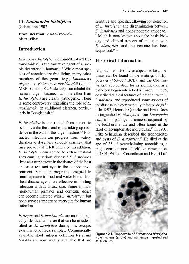
12. Entamoeba histolytica 147
Figure 12.1. Trophozoite of Entamoeba histolytica. Note nucleus (arrow) and numerous ingested red cells. 35 µm.
12. Entamoeba histolytica(Schaudinn 1903)
Pronunciation: \en-tə-ˈmē-bə\\his′təlit′ikə\
Introduction
Entamoeba histolytica (\ent-a-MEE-ba\\HIS-tow-lit-i-ka\) is the causative agent of amoe-bic dysentery in humans.1, 2 While most spe-cies of amoebae are free-living, many other members of this genus (e.g., Entamoeba dispar and Entamoeba moshkovskii (\ent-a-MEE-ba mosh-KOV-ski-ee\), can inhabit the human large intestine, but none other than E. histolytica are clearly pathogenic. There is some controversy regarding the role of E. moshkovskii in childhood diarrhea, particu-larly in Bangladesh.2, 3
E. histolytica is transmitted from person to person via the fecal-oral route, taking up resi-dence in the wall of the large intestine.4, 5 Pro-tracted infection can progress from watery diarrhea to dysentery (bloody diarrhea) that may prove fatal if left untreated. In addition, E. histolytica can spread to extra-intestinal sites causing serious disease.6 E. histolytica lives as a trophozoite in the tissues of the host and as a resistant cyst in the outside envi-ronment. Sanitation programs designed to limit exposure to food and water-borne diar-rheal disease agents are effective in limiting infection with E. histolytica. Some animals (non-human primates and domestic dogs) can become infected with E. histolytica, but none serve as important reservoirs for human infection.
E. dispar and E. moshkovskii are morphologi-cally identical amoebae that can be misiden-tified as E. histolytica during microscopic examination of fecal samples.7 Commercially available stool antigen detection tests and NAATs are now widely available that are
sensitive and specific, allowing for detection of E. histolytica and discrimination between E. histolytica and nonpathogenic amoebae.8,
9 Much is now known about the basic biol-ogy and clinical aspects of infection with E. histolytica, and the genome has been sequenced.10-12
Historical Information
Although reports of what appears to be amoe-biasis can be found in the writings of Hip-pocrates (460–377 BCE), and the Old Tes-tament, appreciation for its significance as a pathogen began when Fedor Losch, in 1875, described clinical features of infection with E. histolytica, and reproduced some aspects of the disease in experimentally infected dogs.13,
14 In 1893, Heinrich Quincke and Ernst Roos distinguished E. histolytica from Entamoeba coli, a non-pathogenic amoeba acquired by the fecal-oral route and often found in the stool of asymptomatic individuals.15 In 1903, Fritz Schaudinn described the trophozoites and cysts of E. histolytica.16 He died at the age of 35 of overwhelming amoebiasis, a tragic consequence of self-experimentation. In 1891, William Councilman and Henri Laf-
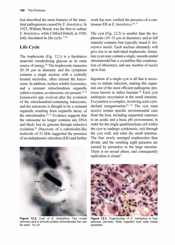
148 The Protozoa
Figure 12.3. Trophozoites of E. histolytica in liver abscess (arrows). Note ingested host cells inside parasites.
leur described the main features of the intes-tinal pathogenesis caused by E. histolytica. In 1925, William Boeck was the first to culture E. histolytica, while Clifford Dobell, in 1928, fully elucidated its life cycle.17-19
Life Cycle
The trophozoite (Fig. 12.1) is a facultative anaerobe metabolizing glucose as its main source of energy.20 The trophozoite measures 20–30 µm in diameter, and the cytoplasm contains a single nucleus with a centrally located nucleolus, often termed the karyo-some. In addition, surface soluble lysosomes, and a remnant mitochondrion organelle called a crypton, or mitosome, are present.21-23 Entamoeba spp. evolved after the evolution of the mitochondrial-containing eukaryotes, and the mitosome is thought to be a remnant organelle resulting from organelle decay of the mitochondria.24, 25 Evidence suggests that the mitosome no longer contains any DNA and likely lost its genome through reductive evolution.24 Discovery of a calreticulin-like molecule of 51 kDa suggested the presence of an endoplasmic reticulum (ER) and further
work has now verified the presence of a con-tinuous ER in E. histolytica.26, 27
The cyst (Fig. 12.2) is smaller than the tro-phozoite (10–15 µm in diameter), and at full maturity contains four typically round E. his-tolytica nuclei. Each nucleus ultimately will give rise to an individual trophozoite. Imma-ture cysts may contain a single, smooth-ended chromatoidal bar, a crystalline-like condensa-tion of ribosomes, and any number of nuclei up to four.
Ingestion of a single cyst is all that is neces-sary to initiate infection, making this organ-ism one of the most efficient pathogenic pro-tozoa known to infect humans.28 Each cyst undergoes excystation in the small intestine. Excystation is complex, involving actin cyto-skeletal reorganization.29, 30 The cyst must receive certain specific environmental cues from the host, including sequential exposure to an acidic and a basic pH environment, in order for the single quadrinucleate cell within the cyst to undergo cytokinesis, exit through the cyst wall, and enter the small intestine. The four newly emerged trophozoites then divide, and the resulting eight parasites are carried by peristalsis to the large intestine. There is no sexual phase, and consequently replication is clonal.6
Figure 12.2. Cyst of E. histolytica. Two nuclei (arrows) and a smooth-ended chromatoidal bar can be seen. 15 µm.
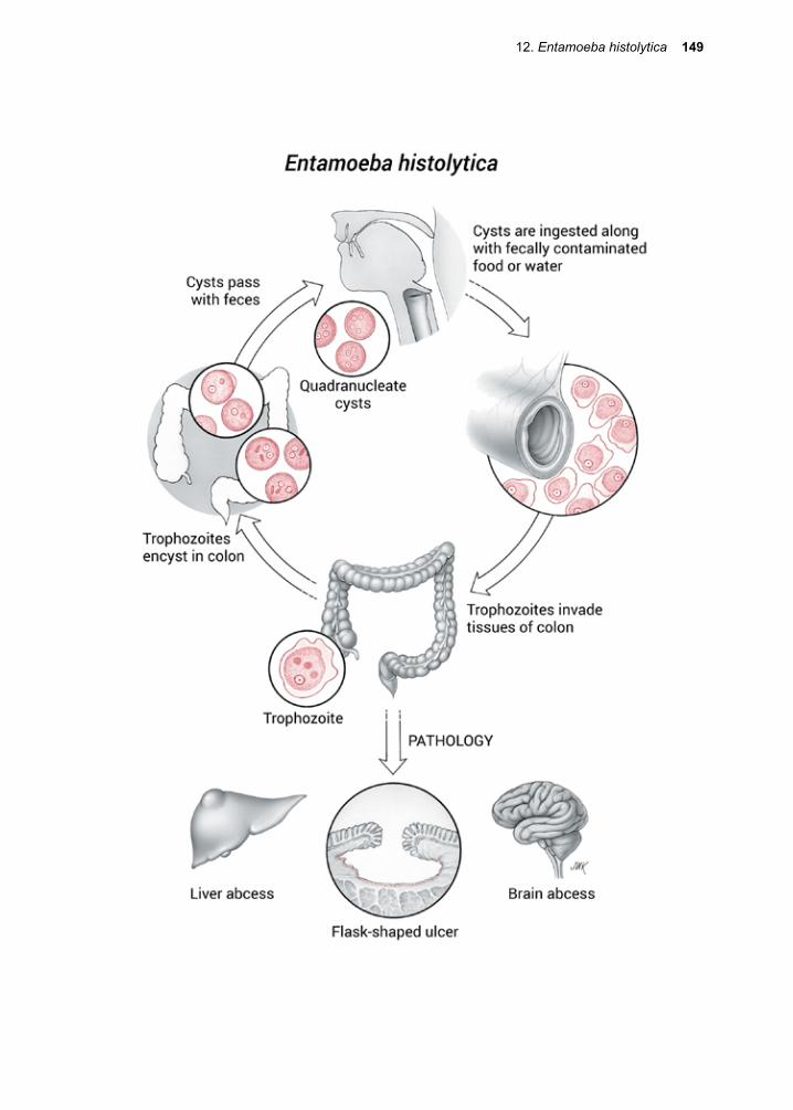
12. Entamoeba histolytica 149
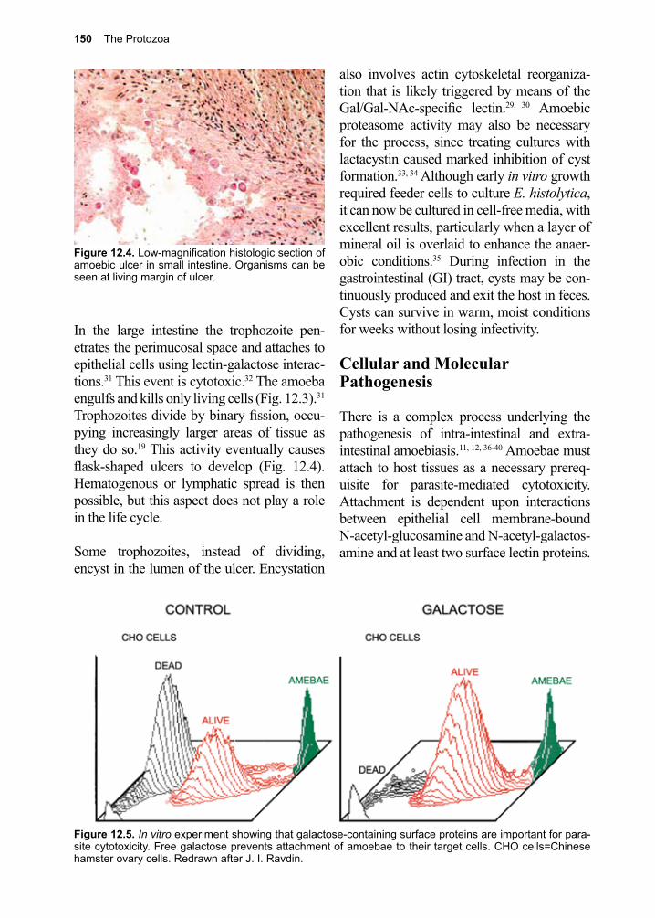
150 The Protozoa
Figure 12.5. In vitro experiment showing that galactose-containing surface proteins are important for para-site cytotoxicity. Free galactose prevents attachment of amoebae to their target cells. CHO cells=Chinese hamster ovary cells. Redrawn after J. I. Ravdin.
In the large intestine the trophozoite pen-etrates the perimucosal space and attaches to epithelial cells using lectin-galactose interac-tions.31 This event is cytotoxic.32 The amoeba engulfs and kills only living cells (Fig. 12.3).31 Trophozoites divide by binary fission, occu-pying increasingly larger areas of tissue as they do so.19 This activity eventually causes flask-shaped ulcers to develop (Fig. 12.4). Hematogenous or lymphatic spread is then possible, but this aspect does not play a role in the life cycle.
Some trophozoites, instead of dividing, encyst in the lumen of the ulcer. Encystation
also involves actin cytoskeletal reorganiza-tion that is likely triggered by means of the Gal/Gal-NAc-specific lectin.29, 30 Amoebic proteasome activity may also be necessary for the process, since treating cultures with lactacystin caused marked inhibition of cyst formation.33, 34 Although early in vitro growth required feeder cells to culture E. histolytica, it can now be cultured in cell-free media, with excellent results, particularly when a layer of mineral oil is overlaid to enhance the anaer-obic conditions.35 During infection in the gastrointestinal (GI) tract, cysts may be con-tinuously produced and exit the host in feces. Cysts can survive in warm, moist conditions for weeks without losing infectivity.
Cellular and Molecular Pathogenesis
There is a complex process underlying the pathogenesis of intra-intestinal and extra-intestinal amoebiasis.11, 12, 36-40 Amoebae must attach to host tissues as a necessary prereq-uisite for parasite-mediated cytotoxicity. Attachment is dependent upon interactions between epithelial cell membrane-bound N-acetyl-glucosamine and N-acetyl-galactos-amine and at least two surface lectin proteins.
Figure 12.4. Low-magnification histologic section of amoebic ulcer in small intestine. Organisms can be seen at living margin of ulcer.
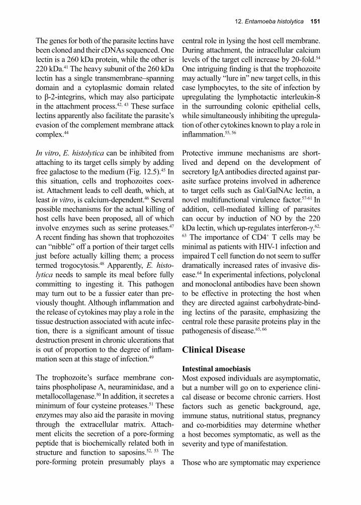
12. Entamoeba histolytica 151
The genes for both of the parasite lectins have been cloned and their cDNAs sequenced. One lectin is a 260 kDa protein, while the other is 220 kDa.41 The heavy subunit of the 260 kDa lectin has a single transmembrane–spanning domain and a cytoplasmic domain related to β-2-integrins, which may also participate in the attachment process.42, 43 These surface lectins apparently also facilitate the parasite’s evasion of the complement membrane attack complex.44
In vitro, E. histolytica can be inhibited from attaching to its target cells simply by adding free galactose to the medium (Fig. 12.5).45 In this situation, cells and trophozoites coex-ist. Attachment leads to cell death, which, at least in vitro, is calcium-dependent.46 Several possible mechanisms for the actual killing of host cells have been proposed, all of which involve enzymes such as serine proteases.47 A recent finding has shown that trophozoites can “nibble” off a portion of their target cells just before actually killing them; a process termed trogocytosis.48 Apparently, E. histo-lytica needs to sample its meal before fully committing to ingesting it. This pathogen may turn out to be a fussier eater than pre-viously thought. Although inflammation and the release of cytokines may play a role in the tissue destruction associated with acute infec-tion, there is a significant amount of tissue destruction present in chronic ulcerations that is out of proportion to the degree of inflam-mation seen at this stage of infection.49
The trophozoite’s surface membrane con-tains phospholipase A, neuraminidase, and a metallocollagenase.50 In addition, it secretes a minimum of four cysteine proteases.51 These enzymes may also aid the parasite in moving through the extracellular matrix. Attach-ment elicits the secretion of a pore-forming peptide that is biochemically related both in structure and function to saposins.52, 53 The pore-forming protein presumably plays a
central role in lysing the host cell membrane. During attachment, the intracellular calcium levels of the target cell increase by 20-fold.54 One intriguing finding is that the trophozoite may actually “lure in” new target cells, in this case lymphocytes, to the site of infection by upregulating the lymphotactic interleukin-8 in the surrounding colonic epithelial cells, while simultaneously inhibiting the upregula-tion of other cytokines known to play a role in inflammation.55, 56
Protective immune mechanisms are short-lived and depend on the development of secretory IgA antibodies directed against par-asite surface proteins involved in adherence to target cells such as Gal/GalNAc lectin, a novel multifunctional virulence factor.57-61 In addition, cell-mediated killing of parasites can occur by induction of NO by the 220 kDa lectin, which up-regulates interferon-γ.62,
63 The importance of CD4+ T cells may be minimal as patients with HIV-1 infection and impaired T cell function do not seem to suffer dramatically increased rates of invasive dis-ease.64 In experimental infections, polyclonal and monoclonal antibodies have been shown to be effective in protecting the host when they are directed against carbohydrate-bind-ing lectins of the parasite, emphasizing the central role these parasite proteins play in the pathogenesis of disease.65, 66
Clinical Disease
Intestinal amoebiasisMost exposed individuals are asymptomatic, but a number will go on to experience clini-cal disease or become chronic carriers. Host factors such as genetic background, age, immune status, nutritional status, pregnancy and co-morbidities may determine whether a host becomes symptomatic, as well as the severity and type of manifestation.
Those who are symptomatic may experience
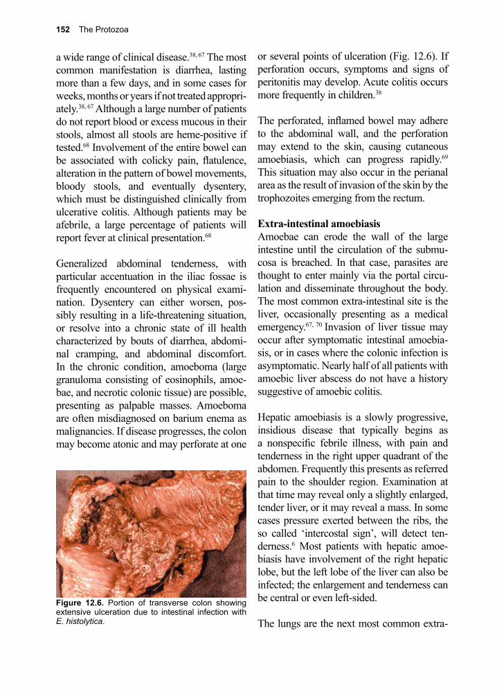
152 The Protozoa
a wide range of clinical disease.38, 67 The most common manifestation is diarrhea, lasting more than a few days, and in some cases for weeks, months or years if not treated appropri-ately.38, 67 Although a large number of patients do not report blood or excess mucous in their stools, almost all stools are heme-positive if tested.68 Involvement of the entire bowel can be associated with colicky pain, flatulence, alteration in the pattern of bowel movements, bloody stools, and eventually dysentery, which must be distinguished clinically from ulcerative colitis. Although patients may be afebrile, a large percentage of patients will report fever at clinical presentation.68
Generalized abdominal tenderness, with particular accentuation in the iliac fossae is frequently encountered on physical exami-nation. Dysentery can either worsen, pos-sibly resulting in a life-threatening situation, or resolve into a chronic state of ill health characterized by bouts of diarrhea, abdomi-nal cramping, and abdominal discomfort. In the chronic condition, amoeboma (large granuloma consisting of eosinophils, amoe-bae, and necrotic colonic tissue) are possible, presenting as palpable masses. Amoeboma are often misdiagnosed on barium enema as malignancies. If disease progresses, the colon may become atonic and may perforate at one
or several points of ulceration (Fig. 12.6). If perforation occurs, symptoms and signs of peritonitis may develop. Acute colitis occurs more frequently in children.38
The perforated, inflamed bowel may adhere to the abdominal wall, and the perforation may extend to the skin, causing cutaneous amoebiasis, which can progress rapidly.69 This situation may also occur in the perianal area as the result of invasion of the skin by the trophozoites emerging from the rectum.
Extra-intestinal amoebiasis Amoebae can erode the wall of the large intestine until the circulation of the submu-cosa is breached. In that case, parasites are thought to enter mainly via the portal circu-lation and disseminate throughout the body. The most common extra-intestinal site is the liver, occasionally presenting as a medical emergency.67, 70 Invasion of liver tissue may occur after symptomatic intestinal amoebia-sis, or in cases where the colonic infection is asymptomatic. Nearly half of all patients with amoebic liver abscess do not have a history suggestive of amoebic colitis.
Hepatic amoebiasis is a slowly progressive, insidious disease that typically begins as a nonspecific febrile illness, with pain and tenderness in the right upper quadrant of the abdomen. Frequently this presents as referred pain to the shoulder region. Examination at that time may reveal only a slightly enlarged, tender liver, or it may reveal a mass. In some cases pressure exerted between the ribs, the so called ‘intercostal sign’, will detect ten-derness.6 Most patients with hepatic amoe-biasis have involvement of the right hepatic lobe, but the left lobe of the liver can also be infected; the enlargement and tenderness can be central or even left-sided.
The lungs are the next most common extra-
Figure 12.6. Portion of transverse colon showing extensive ulceration due to intestinal infection with E. histolytica.
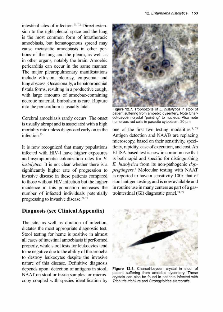
12. Entamoeba histolytica 153
intestinal sites of infection.71, 72 Direct exten-sion to the right pleural space and the lung is the most common form of intrathoracic amoebiasis, but hematogenous spread may cause metastatic amoebiasis in other por-tions of the lung and the pleura, as well as in other organs, notably the brain. Amoebic pericarditis can occur in the same manner. The major pleuropulmonary manifestations include effusion, pleurisy, empyema, and lung abscess. Occasionally, a hepatobronchial fistula forms, resulting in a productive cough, with large amounts of amoebae-containing necrotic material. Embolism is rare. Rupture into the pericardium is usually fatal.
Cerebral amoebiasis rarely occurs. The onset is usually abrupt and is associated with a high mortality rate unless diagnosed early on in the infection.73
It is now recognized that many populations infected with HIV-1 have higher exposures and asymptomatic colonization rates for E. histolytica. It is not clear whether there is a significantly higher rate of progression to invasive disease in these patients compared to those without HIV infection but the higher incidence in this population increases the number of infected individuals potentially progressing to invasive disease.74-77
Diagnosis (see Clinical Appendix)
The site, as well as duration of infection, dictates the most appropriate diagnostic test. Stool testing for heme is positive in almost all cases of intestinal amoebiasis if performed properly, while stool tests for leukocytes tend to be negative due to the ability of the amoeba to destroy leukocytes despite the invasive nature of this disease. Definitive diagnosis depends upon: detection of antigens in stool, NAAT on stool or tissue samples, or micros-copy coupled with species identification by
one of the first two testing modalities.8, 78 Antigen detection and NAATs are replacing microscopy, based on their sensitivity, speci-ficity, rapidity, ease of execution, and cost. An ELISA-based test is now in common use that is both rapid and specific for distinguishing E. histolytica from its non-pathogenic dop-pelgängers.8 Molecular testing with NAAT is reported to have a sensitivity 100x that of stool antigen testing, and is now available and in routine use in many centers as part of a gas-trointestinal (GI) diagnostic panel.74, 79
Figure 12.7. Trophozoite of E. histolytica in stool of patient suffering from amoebic dysentery. Note Char-cot-Leyden crystal “pointing” to nucleus. Also note numerous red cells in parasite cytoplasm. 30 µm.
Figure 12.8. Charcot-Leyden crystal in stool of patient suffering from amoebic dysentery. These crystals can also be found in patients infected with Trichuris trichiura and Strongyloides stercoralis.
154 The Protozoa
Microscopy is still the only diagnostic modal-ity in many facilities. Stool culture is not clinically available and does not play a role in the routine diagnosis of this disease. The trophozoites of E. histolytica are more often described as showing evidence of erythro-phagocytosis, and this has been suggested to allow for definitive diagnosis using micros-copy. There are both in vitro and in vivo reports that nonpathogenic amoebae phago-cytize red cells, and in one study over 15% of cases of E. dispar showed erythrophagocyto-sis on stool microscopy.80-83 If red blood cells are seen in the cytoplasm, particularly more than 2 erythrocytes per trophozoite, then E. histolytica is more likely (Fig. 12.7).83
The Charcot-Leyden crystals (Fig. 12.8) in stool are frequently present when patients are suffering from disease caused by E. histolyt-ica, but they are also seen with heavy infec-tion caused by Trichuris trichiura and Stron-gyloides stercoralis, and therefore are not pathognomonic for amoebiasis. PCR can also be useful for diagnosis of liver disease when used on aspirates derived from the abscess.84
Since infection with E. histolytica invariably leads to long-lasting antibody production, antibody-based tests are sometimes difficult to interpret, especially when done during chronic infection.85 Serological testing will become positive, even in intestinal amoebia-sis infection, 5–7 days after onset of symp-toms and usually remain positive for years, consequently limiting their utility in endemic areas where up to 35% of the population may have seropositivity due to prior exposure.86 The indirect hemagglutination assay (IHA) and indirect fluorescent antibody (IFA) test are used together to rule in the possibility of extra-intestinal disease, but are not definitive proof of infection. Intestinal amoebiasis must always be considered in any patient with pro-tracted diarrhea and in all patients with dysen-tery. The diagnosis must also be considered in
patients presenting with intraluminal colonic masses, because of the development of amoe-bomas that resemble carcinoma of the colon. In extra-intestinal amoebiasis, identification of the lesion by the various modalities and the presence of a travel history compatible with amoebiasis, in parallel with identification of amoebae in the colon, points to the diagnosis.
Extra-intestinal amoebiasis is often a more challenging diagnosis. Intestinal and extra-intestinal amoebiasis do not usually occur simultaneously, and stool testing is usually negative in the setting of extra-intestinal amoebiasis. Serology is usually positive in cases of extra-intestinal amoebiasis, but may be negative during the first 7 days. Imaging tests play a critical role in the diagnosis of extra-intestinal amoebiasis. Radiography of the abdomen may show enlargement of the liver and a fixed, raised diaphragm. In cases of perforation of the diaphragm, there may be evidence of consolidation of one of the lower lobes of the lung or its lower segment, and a pleural effusion. A radionuclide or a CT scan often reveals the abscess; they may also show additional abscesses; however, these are rare. On ultrasonography, an amoebic liver abscess usually appears as a round hypodense area that is contiguous to the liver capsule, usu-ally without significant wall echoes.87 Direct extension to the right pleural space and the lung is the most common form of intratho-racic amoebiasis, but hematogenous spread may cause metastatic amoebiasis in other portions of the lung and the pleura, as well as in other organs, notably the brain. Amoebic pericarditis can occur in the same manner.88
If the diagnosis is still in doubt after sero-logical testing and imaging, the cyst can be aspirated. The obtained fluid from a hepatic amoebic abscess is often a brown fluid con-taining necrotic hepatocytes that has been likened to anchovy paste. In some cases, the fluid may be clear or yellow in color. Amoebic
12. Entamoeba histolytica 155
forms are rarely present and antigen testing or NAAT is helpful in confirming the diagnosis. Abscesses may be secondarily infected with bacteria, so sending the fluid for bacterial cul-ture is recommended. Although eosinophils may play some role in local tissue control early in the course of hepatic amoebiasis, very few patients have elevated eosinophils in the peripheral circulation, and patients may even become eosinopenic in severe infections.89-91
Treatment (see Clinical Appendix)
All forms of symptomatic amoebiasis are considered invasive and consequently should be treated with an agent that is active against the tissue invasive forms that reaches ade-quate levels at the sites of infection. Metro-nidazole is the drug of choice for the intesti-nal and extra-intestinal infection.92 It can be given in equivalent doses orally or intrave-nously. This drug also has a few limitations and some adverse side effects. Use of alcohol is prohibited during treatment, as it induces a side effect similar to that caused by disulfiram (antabuse) therapy. Alternative agents include tinidazole and second-line agents that may be inferior in efficacy, such as nitazoxanide or ornidazole. Since these drugs do not target the cyst stage, a second intraluminal agent to target the intraluminal cysts is also recom-mended for all symptomatic patients. Treat-ment of asymptomatic carriers of E. histo-lytica to prevent disease or transmission is controversial.93 A cysticidal intraluminal agent alone may be adequate for asymptom-atic cyst passers and those with nondysenteric amoebic colitis. The intraluminal agent paro-momycin is widely used in the United States for this indication, while other agents, such as diloxanide furoate and iodoquinol (diiodo-hydroxyquinoline), are both effective at kill-ing cysts and should be considered.94 Addi-tional antibiotics are appropriate in patients in whom a secondary bacterial infection is suspected.
No naturally occurring metronidazole-resistant strains of E. histolytica have been reported to date, but they can be easily induced under laboratory conditions.95, 96 It is probably only a matter of time before they appear in human populations. Liver abscesses resolve slowly, despite treatment with the recommended high doses of metronidazole. Aspiration of an amoebic liver abscess may serve not only to confirm the diagnosis, but also as a therapeutic intervention. There are established indications for therapeutic drain-age of an amoebic liver abscess and for sur-gery, in cases of severe colitis. A surgeon or interventional radiologist should be involved: 1. if there is no clinical improvement within 48–72 hours despite appropriate medical therapy, 2. for abscesses greater than 10 cm in diameter, 3. when there is marked elevation of the diaphragm, 4. for abscesses in the left lobe, and 5. when there is negative serology, which might raise suspicion of a pyogenic abscess.67, 97, 98
There are few reports of patients surviving amoebic abscess of the brain, since, unfortu-nately, they are typically diagnosed too late.73 In the case of infection involving the pericar-dium, quick aspiration of an expanding peri-cardial effusion, combined with aggressive anti-amoebic therapy, has saved the lives of most of those suffering from this rare mani-festation of the infection.99
Prevention and Control
Good public health practice, starting with ensuring the safety of drinking water sup-plies, and in some cases, watershed manage-ment, are the best long-term approaches to controlling most waterborne diarrheal disease agents. Screening of food handlers with peri-odic stool examinations can identify carriers whose occupations would place the general public at risk. Recurrent outbreaks of amoe-biasis in mental institutions can be prevented
156 The Protozoa
References
1. Bercu, T. E., et al., Amebic colitis: new insights into pathogenesis and treatment. Curr Gastroenterol Rep 2007, 9 (5), 429-33.
2. Heredia, R. D., et al., Entamoeba moshkovskii perspectives of a new agent to be considered in the diagnosis of amebiasis. Acta Trop 2012, 123 (3), 139-45.
3. Ali, I. K., et al., Entamoeba moshkovskii infections in children, Bangladesh. Emerg Infect Dis 2003, 9 (5), 580-4.
4. Marciano-Cabral, F., Free-living amoebae as agents of human infection. J Infect Dis 2009, 199 (8), 1104-6.
5. Kotloff, K. L., et al., Burden and aetiology of diarrhoeal disease in infants and young children in developing countries (the Global Enteric Multicenter Study, GEMS): a prospective, case-control study. Lancet 2013, 382 (9888), 209-22.
6. Haque, R., et al., Amebiasis. N Engl J Med 2003, 348 (16), 1565-73.7. Tannich, E., Royal Society of Tropical Medicine and Hygiene Meeting at Manson House,
London, 19 February 1998. Amoebic disease. Entamoeba histolytica and E. dispar: comparison of molecules considered important for host tissue destruction. Transactions of the Royal Society of Tropical Medicine and Hygiene 1998, 92 (6), 593-6.
8. Anane, S.; Khaled, S., [Entamoeba histolytica and Entamoeba dispar: differentiation methods and implications]. Annales de biologie clinique 2005, 63 (1), 7-13.
9. Blessmann, J., et al., Real-time PCR for detection and differentiation of Entamoeba histolytica and Entamoeba dispar in fecal samples. J Clin Microbiol 2002, 40 (12), 4413-7.
10. Reed, S., et al., Amoebiasis In: Infections of the Gastrointestinal Tract 2nd ed. 2002; p 961-978.11. Martinez-Paloma, A., et al., Topley and Wilson’s Microbiology and Microbial Infections 9th ed.
1998; p 157-177.12. Loftus, B., et al., The genome of the protist parasite Entamoeba histolytica. Nature 2005, 433
(7028), 865-8.13. Losch, F. A., Massenhafte Entwicklung von Amoben in Dickdarm. Arch Pathol Anat Phys KIm
Med Virchow 1875, 65, 196-211.14. Tanyuksel, M.; Petri, W. A., Jr., Laboratory diagnosis of amebiasis. Clin Microbiol Rev 2003, 16
(4), 713-29.15. Quincke, H. I.; Roos, E., Uber Amoben-enteritis. BerI KIm Wochenschr 1893, 30 1089-1094.16. Schaudinn, F., Untersuchungen uber Fortpflanzung einiger Rhizopoden Berlin, Verlag Von Julius
Springer in Berlin: 1903.17. Councilman, W. T.; Lafleur, H. A., Amoebic dysentery. Johns Hopkins Hosp Rep 1891, 2, 395-
548.18. Boeck, W. C.; J., Cultivation of Entamoeba histolytica. Am 1925, 5, 371-407.19. Dobell, C., Researches on the intestinal protozoa of monkeys and man. Parasitology 1928, 20
357-412.20. Saavedra, E., et al., Glycolysis in Entamoeba histolytica. Biochemical characterization of
recombinant glycolytic enzymes and flux control analysis. The FEBS journal 2005, 272 (7), 1767-83.
21. Mai, Z., et al., Hsp60 is targeted to a cryptic mitochondrion-derived organelle (“crypton”) in the microaerophilic protozoan parasite Entamoeba histolytica. Molecular and cellular biology 1999, 19 (3), 2198-205.
by strictly adhering to appropriate sanitary practices, coupled with routine stool exami-nations of the patients. All infected individu-als should receive treatment.
Vaccine targets against both the intestinal and extra-intestinal infection have been identi-
fied. Successful development of vaccines based on these findings will require extensive non-clinical and clinical studies before a vac-cine is considered for the highest risk groups including children in endemic areas and other high-risk groups such as travelers.100
12. Entamoeba histolytica 157
22. Tovar, J., et al., The mitosome, a novel organelle related to mitochondria in the amitochondrial parasite Entamoeba histolytica. Molecular microbiology 1999, 32 (5), 1013-21.
23. Nakada-Tsukuii, K.; Nozaki, T., Molecular Basis of the Trafficking of Cysteine Proteases and Other Soluble Lysosomal Proteins in Entamoeba histolytica. Amebiasis Springer Japan 2015.
24. Aguilera, P., et al., Entamoeba histolytica mitosomes: organelles in search of a function. Exp Parasitol 2008, 118 (1), 10-6.
25. Bakatselou, C., et al., Analysis of genes of mitochondrial origin in the genus Entamoeba. The Journal of eukaryotic microbiology 2003, 50 (3), 210-4.
26. Gozalez, E., et al., Calreticulin-like molecule in trophozoites of Entamoeba histolytica. Am Med Hyg 2002, 67, 636-639.
27. Teixeira, J. E.; Huston, C. D., Evidence of a continuous endoplasmic reticulum in the protozoan parasite Entamoeba histolytica. Eukaryot Cell 2008, 7 (7), 1222-6.
28. Walker, E. L., et al., Experimental entamoebic dysentery. Phillipine Med 1913, 8 253-330.29. Makioka, A., et al., Expression analysis of Entamoeba invadens profilins in encystation and
excystation. Parasitol Res 2012, 110 (6), 2095-104.30. Eichinger, D., A role for a galactose lectin and its ligands during encystment of Entamoeba. J
Eukaryot Microbiol 2001, 48 (1), 17-21.31. Ravdin, J. I.; Guerrant, R. L., Role of adherence in cytopathogenic mechanisms of Entamoeba
histolytica. Study with mammalian tissue culture cells and human erythrocytes. J Clin Invest 1981, 68 (5), 1305-13.
32. Saffer, L. D.; Petri, W. A., Jr., Role of the galactose lectin of Entamoeba histolytica in adherence-dependent killing of mammalian cells. Infect Immun 1991, 59 (12), 4681-3.
33. Makioka, A., et al., Effect of proteasome inhibitors on the growth, encystation, and excystation of Entamoeba histolytica and Entamoeba invadens. Parasitology research 2002, 88 (5), 454-9.
34. Eichinger, D., Encystation of entamoeba parasites. BioEssays : news and reviews in molecular, cellular and developmental biology 1997, 19 (7), 633-9.
35. Pires-Santos, G. M., et al., Optimization of Entamoeba histolytica culturing in vitro. Exp Parasitol 2012, 132 (4), 561-5.
36. Carrero, J. C.; Laclette, J. P., Molecular biology of Entamoeba histolytica: a review. Archives of medical research 1996, 27 (3), 403-12.
37. Lohia, A., The cell cycle of Entamoeba histolytica. Molecular and cellular biochemistry 2003, 253 (1-2), 217-22.
38. Petrin, D., et al., Clinical and microbiological aspects of Trichomonas vaginalis. Clinical microbiology reviews 1998, 11 (2), 300-17.
39. Stauffer, W.; Ravdin, J. I., Entamoeba histolytica: an update. Current opinion in infectious diseases 2003, 16 (5), 479-85.
40. Begum, S., et al., Immune Evasion Mechanisms of Entamoeba histolytica: Progression to Disease. Front Microbiol 2015, 6, 1394.
41. Dodson, J. M., et al., Infection and immunity mediated by the carbohydrate recognition domain of the Entamoeba histolytica Gal/GalNAc lectin. The Journal of infectious diseases 1999, 179 (2), 460-6.
42. Rosales-Encina, J. L., et al., Isolation of a 220-kilodalton protein with lectin properties from a virulent strain of Entamoeba histolytica. The Journal of infectious diseases 1987, 156 (5), 790-7.
43. Vines, R. R., et al., Regulation of adherence and virulence by the Entamoeba histolytica lectin cytoplasmic domain, which contains a beta2 integrin motif. Molecular biology of the cell 1998, 9 (8), 2069-79.
44. Braga, L. L., et al., Inhibition of the complement membrane attack complex by the galactose-specific adhesion of Entamoeba histolytica. The Journal of clinical investigation 1992, 90 (3), 1131-7.
45. Kain, K. C.; Ravdin, J. I., Galactose-specific adhesion mechanisms of Entamoeba histolytica: model for study of enteric pathogens. Methods in enzymology 1995, 253, 424-39.
46. Arias-Negrete, S., et al., Expression of in vitro virulence by Entamoeba histolytica: effect of calmodulin inhibitors. APMIS : acta pathologica, microbiologica, et immunologica Scandinavica 1999, 107 (9), 875-81.

158 The Protozoa
47. McKerrow, J. H., et al., The proteases and pathogenicity of parasitic protozoa. Annual review of microbiology 1993, 47, 821-53.
48. Ralston, K. S., et al., Trogocytosis by Entamoeba histolytica contributes to cell killing and tissue invasion. Nature 2014, 508 (7497), 526-30.
49. Brandt, H.; Tamayo, R. P., Pathology of human amebiasis. Hum Pathol 1970, 1 (3), 351-85.50. Reed, S. L., et al., The extracellular neutral cysteine proteinase of Entamoeba histolytica
degrades anaphylatoxins C3a and C5a. Journal of immunology (Baltimore, Md. : 1950) 1995, 155 (1), 266-74.
51. Franco, E., et al., Specific and reversible inhibition of Entamoeba histolytica cysteine-proteinase activities by Zn2+: implications for adhesion and cell damage. Archives of medical research 1999, 30 (2), 82-8.
52. Leippe, M.; Muller-Eberhard, H. J., The pore-forming peptide of Entamoeba histolytica, the protozoan parasite causing human amoebiasis. Toxicology 1994, 18, 1-3.
53. Vaccaro, A. M., et al., Saposins and their interaction with lipids. Neurochemical research 1999, 24 (2), 307-14.
54. Ravdin, J. I., et al., Relationship of free intracellular calcium to the cytolytic activity of Entamoeba histolytica. Infection and immunity 1988, 56 (6), 1505-12.
55. Yu, Y.; Chadee, K., Secreted Entamoeba histolytica proteins stimulate interleukin-8 mRNA expression and protein production in human colonic epithelial cells. Archives of medical research 1997, 28 Spec No, 223-4.
56. Utrera-Barillas, D., et al., An anti-inflammatory oligopeptide produced by Entamoeba histolytica down-regulates the expression of pro-inflammatory chemokines. Parasite immunology 2003, 25 (10), 475-82.
57. Choudhuri, G., et al., Protective immunity to Entamoeba histolytica infection in subjects with antiamoebic antibodies residing in a hyperendemic zone. Scandinavian journal of infectious diseases 1991, 23 (6), 771-6.
58. Lotter, H., et al., Identification of an epitope on the Entamoeba histolytica 170-kD lectin conferring antibody-mediated protection against invasive amebiasis. The Journal of experimental medicine 1997, 185 (10), 1793-801.
59. Ravdin, J. I.; Kelsall, B. L., Role of mucosal secretory immunity in the development of an amebiasis vaccine. The American journal of tropical medicine and hygiene 1994, 50 (5 Suppl), 36-41.
60. Abou-el-Magd, I., et al., Humoral and mucosal IgA antibody response to a recombinant 52-kDa cysteine-rich portion of the Entamoeba histolytica galactose-inhibitable lectin correlates with detection of native 170-kDa lectin antigen in serum of patients with amebic colitis. The Journal of infectious diseases 1996, 174 (1), 157-62.
61. Stanley, S. L. J., Protective immunity to amoebiasis: new insights and new challenges. Dis 2001, 184, 504-6.
62. Ghadirian, E.; Denis, M., In vivo activation of macrophages by IFN-gamma to kill Entamoeba histolytica trophozoites in vitro. Parasite immunology 1992, 14 (4), 397-404.
63. Seguin, R., et al., The tumor necrosis factor alpha-stimulating region of galactose-inhibitable lectin of Entamoeba histolytica activates gamma interferon-primed macrophages for amebicidal activity mediated by nitric oxide. Infect Immun 1997, 65 (7), 2522-7.
64. Nagata, N., et al., Risk factors for intestinal invasive amebiasis in Japan, 2003-2009. Emerg Infect Dis 2012, 18 (5), 717-24.
65. Ravdin, J. I., et al., Antigenicity, immunogenicity and vaccine efficacy of the galactose-specific adherence protein of Entamoeba histolytica. Vaccine 1993, 11 (2), 241-6.
66. Marinets, A., et al., Protection against invasive amebiasis by a single monoclonal antibody directed against a lipophosphoglycan antigen localized on the surface of Entamoeba histolytica. The Journal of experimental medicine 1997, 186 (9), 1557-65.
67. Wells, C. D.; Arguedas, M., Amebic liver abscess. Southern medical journal 2004, 97 (7), 673-82.
68. Das, S. K., et al., Comparison of clinical and laboratory characteristics of intestinal amebiasis
12. Entamoeba histolytica 159
with shigellosis among patients visiting a large urban diarrheal disease hospital in Bangladesh. Am J Trop Med Hyg 2013, 89 (2), 339-44.
69. Parshad, S., et al., Primary cutaneous amoebiasis: case report with review of the literature. International journal of dermatology 2002, 41 (10), 676-80.
70. Hoffner, R. J., et al., Common presentations of amebic liver abscess. Annals of emergency medicine 1999, 34 (3), 351-5.
71. Lyche, K. D.; Jensen, W. A., Pleuropulmonary amebiasis. Seminars in respiratory infections 1997, 12 (2), 106-12.
72. Mbaye, P. S., et al., [Pleuropulmonary manifestations of amebiasis]. Revue de pneumologie clinique 1998, 54 (6), 346-52.
73. Sundaram, C., et al., Brain abscess due to Entamoeba histolytica. The Journal of the Association of Physicians of India 2004, 52, 251-2.
74. Hung, C.-C., et al., Invasive amebiasis as an emerging parasitic disease in patients with human immunodeficiency virus type 1 infection in Taiwan. Archives of internal medicine 2005, 165 (4), 409-15.
75. Hung, C. C., et al., Increased risk for Entamoeba histolytica infection and invasive amebiasis in HIV seropositive men who have sex with men in Taiwan. PLoS Negl Trop Dis 2008, 2 (2), e175.
76. Park, W. B., et al., Amebic liver abscess in HIV-infected patients, Republic of Korea. Emerg Infect Dis 2007, 13 (3), 516-7.
77. Wu, K. S., et al., Comparison of clinical characteristics of amebic liver abscess in human immunodeficiency virus (HIV)-infected and non-HIV-infected patients. J Microbiol Immunol Infect 2008, 41 (6), 456-61.
78. Mirelman, D., et al., Comparison of use of enzyme-linked immunosorbent assay-based kits and PCR amplification of rRNA genes for simultaneous detection of Entamoeba histolytica and E. dispar. Journal of clinical microbiology 1997, 35 (9), 2405-7.
79. Buss, S. N., et al., Multicenter evaluation of the BioFire FilmArray gastrointestinal panel for etiologic diagnosis of infectious gastroenteritis. J Clin Microbiol 2015, 53 (3), 915-25.
80. Talamas-Lara, D., et al., Erythrophagocytosis in Entamoeba histolytica and Entamoeba dispar: a comparative study. Biomed Res Int 2014, 2014, 626259.
81. Boettner, D. R., et al., Entamoeba histolytica and Entamoeba dispar utilize externalized phosphatidylserine for recognition and phagocytosis of erythrocytes. Infect Immun 2005, 73 (6), 3422-30.
82. Gonzalez-Ruiz, A., et al., Value of microscopy in the diagnosis of dysentery associated with invasive Entamoeba histolytica. J Clin Pathol 1994, 47 (3), 236-9.
83. Haque, R., et al., Rapid diagnosis of Entamoeba infection by using Entamoeba and Entamoeba histolytica stool antigen detection kits. J Clin Microbiol 1995, 33 (10), 2558-61.
84. Zengzhu, G., et al., Analysis by enzyme-linked immunosorbent assay and PCR of human liver abscess aspirates from patients in China for Entamoeba histolytica. Journal of clinical microbiology 1999, 37 (9), 3034-6.
85. Lotter, H., et al., Evaluation of three serological tests for the detection of antiamebic antibodies applied to sera of patients from an area endemic for amebiasis. Tropical medicine and parasitology : official organ of Deutsche Tropenmedizinische Gesellschaft and of Deutsche Gesellschaft fur Technische Zusammenarbeit (GTZ) 1995, 46 (3), 180-2.
86. Samie, A., et al., Prevalence and species distribution of E. Histolytica and E. Dispar in the Venda region, Limpopo, South Africa. Am J Trop Med Hyg 2006, 75 (3), 565-71.
87. Petri, W. A., Jr., Pathogenesis of amebiasis. Curr Opin Microbiol 2002, 5 (4), 443-7.88. Shamsuzzaman, S. M.; Hashiguchi, Y., Thoracic amebiasis. Clinics in chest medicine 2002, 23
(2), 479-92.89. Lopez-Osuna, M., et al., [The eosinophil and Entamoeba histolytica. II. Testicular amebic lesions
produced in eosinophilic rats]. Arch Invest Med (Mex) 1990, 21 Suppl 1, 263-5.90. Lopez-Osuna, M., et al., Does the eosinophil have a protective role in amebiasis? Mem Inst
Oswaldo Cruz 1997, 92 Suppl 2, 237-40.
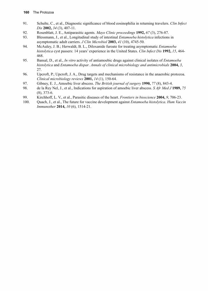
160 The Protozoa
91. Schulte, C., et al., Diagnostic significance of blood eosinophilia in returning travelers. Clin Infect Dis 2002, 34 (3), 407-11.
92. Rosenblatt, J. E., Antiparasitic agents. Mayo Clinic proceedings 1992, 67 (3), 276-87.93. Blessmann, J., et al., Longitudinal study of intestinal Entamoeba histolytica infections in
asymptomatic adult carriers. J Clin Microbiol 2003, 41 (10), 4745-50.94. McAuley, J. B.; Herwaldt, B. L., Diloxanide furoate for treating asymptomatic Entamoeba
histolytica cyst passers: 14 years’ experience in the United States. Clin Infect Dis 1992, 15, 464-468.
95. Bansal, D., et al., In vitro activity of antiamoebic drugs against clinical isolates of Entamoeba histolytica and Entamoeba dispar. Annals of clinical microbiology and antimicrobials 2004, 3, 27.
96. Upcroft, P.; Upcroft, J. A., Drug targets and mechanisms of resistance in the anaerobic protozoa. Clinical microbiology reviews 2001, 14 (1), 150-64.
97. Gibney, E. J., Amoebic liver abscess. The British journal of surgery 1990, 77 (8), 843-4.98. de la Rey Nel, J., et al., Indications for aspiration of amoebic liver abscess. S Afr Med J 1989, 75
(8), 373-6.99. Kirchhoff, L. V., et al., Parasitic diseases of the heart. Frontiers in bioscience 2004, 9, 706-23.100. Quach, J., et al., The future for vaccine development against Entamoeba histolytica. Hum Vaccin
Immunother 2014, 10 (6), 1514-21.
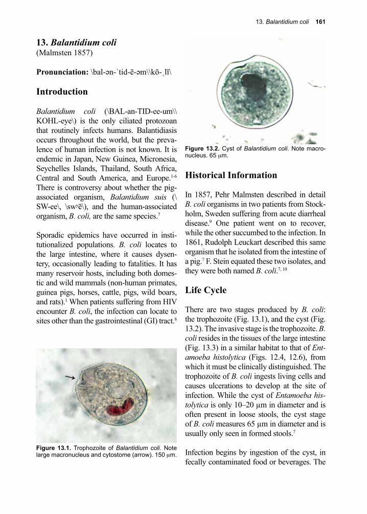
13. Balantidium coli 161
Figure 13.1. Trophozoite of Balantidium coli. Note large macronucleus and cytostome (arrow). 150 µm.
13. Balantidium coli(Malmsten 1857)
Pronunciation: \bal-ən-ˈtid-ē-əm\\kō-ˌlī\
Introduction
Balantidium coli (\BAL-an-TID-ee-um\\KOHL-eye\) is the only ciliated protozoan that routinely infects humans. Balantidiasis occurs throughout the world, but the preva-lence of human infection is not known. It is endemic in Japan, New Guinea, Micronesia, Seychelles Islands, Thailand, South Africa, Central and South America, and Europe.1-6 There is controversy about whether the pig-associated organism, Balantidium suis (\SW-ee\, \swʸē\), and the human-associated organism, B. coli, are the same species.7
Sporadic epidemics have occurred in insti-tutionalized populations. B. coli locates to the large intestine, where it causes dysen-tery, occasionally leading to fatalities. It has many reservoir hosts, including both domes-tic and wild mammals (non-human primates, guinea pigs, horses, cattle, pigs, wild boars, and rats).1 When patients suffering from HIV encounter B. coli, the infection can locate to sites other than the gastrointestinal (GI) tract.8
Historical Information
In 1857, Pehr Malmsten described in detail B. coli organisms in two patients from Stock-holm, Sweden suffering from acute diarrheal disease.9 One patient went on to recover, while the other succumbed to the infection. In 1861, Rudolph Leuckart described this same organism that he isolated from the intestine of a pig.7 F. Stein equated these two isolates, and they were both named B. coli.7, 10
Life Cycle
There are two stages produced by B. coli: the trophozoite (Fig. 13.1), and the cyst (Fig. 13.2). The invasive stage is the trophozoite. B. coli resides in the tissues of the large intestine (Fig. 13.3) in a similar habitat to that of Ent-amoeba histolytica (Figs. 12.4, 12.6), from which it must be clinically distinguished. The trophozoite of B. coli ingests living cells and causes ulcerations to develop at the site of infection. While the cyst of Entamoeba his-tolytica is only 10–20 µm in diameter and is often present in loose stools, the cyst stage of B. coli measures 65 µm in diameter and is usually only seen in formed stools.7
Infection begins by ingestion of the cyst, in fecally contaminated food or beverages. The
Figure 13.2. Cyst of Balantidium coli. Note macro-nucleus. 65 µm.
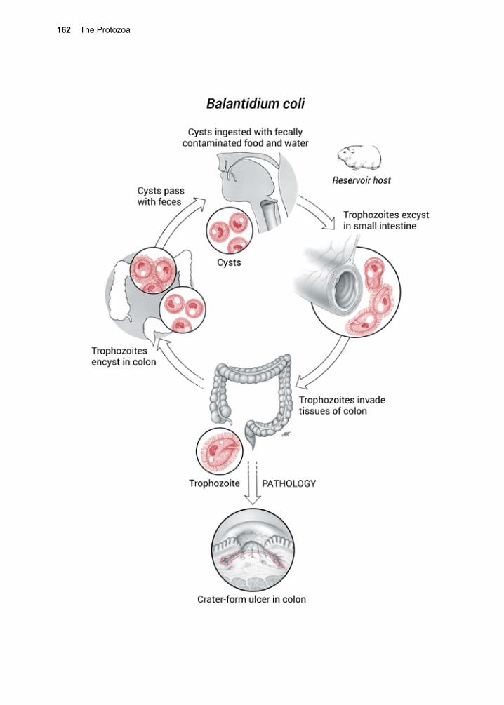
162 The Protozoa
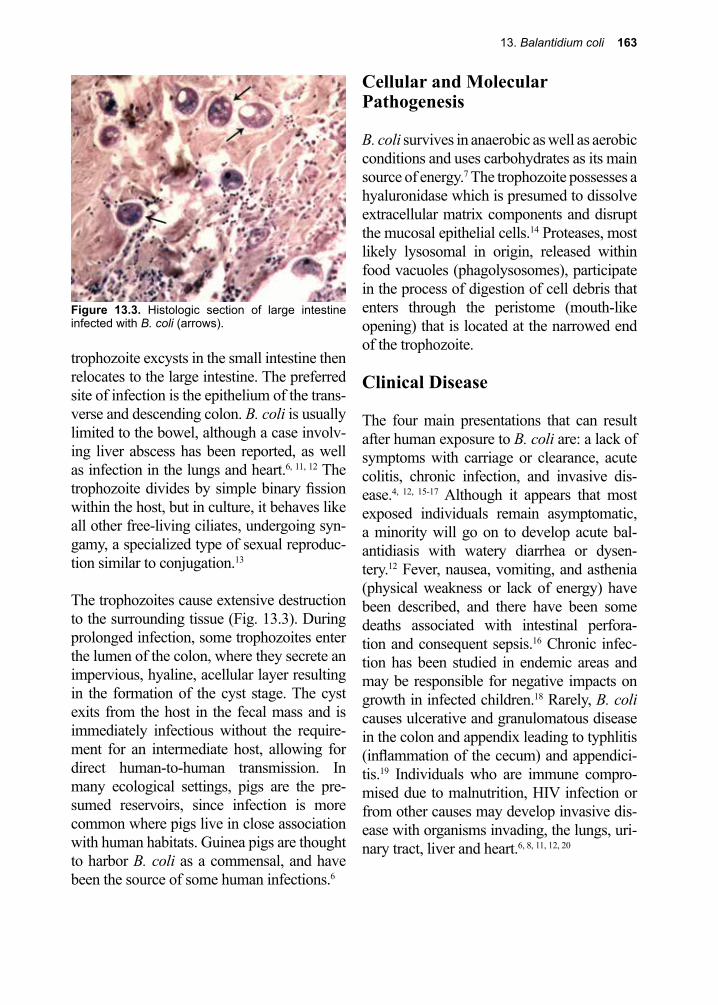
13. Balantidium coli 163
Figure 13.3. Histologic section of large intestine infected with B. coli (arrows).
trophozoite excysts in the small intestine then relocates to the large intestine. The preferred site of infection is the epithelium of the trans-verse and descending colon. B. coli is usually limited to the bowel, although a case involv-ing liver abscess has been reported, as well as infection in the lungs and heart.6, 11, 12 The trophozoite divides by simple binary fission within the host, but in culture, it behaves like all other free-living ciliates, undergoing syn-gamy, a specialized type of sexual reproduc-tion similar to conjugation.13
The trophozoites cause extensive destruction to the surrounding tissue (Fig. 13.3). During prolonged infection, some trophozoites enter the lumen of the colon, where they secrete an impervious, hyaline, acellular layer resulting in the formation of the cyst stage. The cyst exits from the host in the fecal mass and is immediately infectious without the require-ment for an intermediate host, allowing for direct human-to-human transmission. In many ecological settings, pigs are the pre-sumed reservoirs, since infection is more common where pigs live in close association with human habitats. Guinea pigs are thought to harbor B. coli as a commensal, and have been the source of some human infections.6
Cellular and Molecular Pathogenesis
B. coli survives in anaerobic as well as aerobic conditions and uses carbohydrates as its main source of energy.7 The trophozoite possesses a hyaluronidase which is presumed to dissolve extracellular matrix components and disrupt the mucosal epithelial cells.14 Proteases, most likely lysosomal in origin, released within food vacuoles (phagolysosomes), participate in the process of digestion of cell debris that enters through the peristome (mouth-like opening) that is located at the narrowed end of the trophozoite.
Clinical Disease
The four main presentations that can result after human exposure to B. coli are: a lack of symptoms with carriage or clearance, acute colitis, chronic infection, and invasive dis-ease.4, 12, 15-17 Although it appears that most exposed individuals remain asymptomatic, a minority will go on to develop acute bal-antidiasis with watery diarrhea or dysen-tery.12 Fever, nausea, vomiting, and asthenia (physical weakness or lack of energy) have been described, and there have been some deaths associated with intestinal perfora-tion and consequent sepsis.16 Chronic infec-tion has been studied in endemic areas and may be responsible for negative impacts on growth in infected children.18 Rarely, B. coli causes ulcerative and granulomatous disease in the colon and appendix leading to typhlitis (inflammation of the cecum) and appendici-tis.19 Individuals who are immune compro-mised due to malnutrition, HIV infection or from other causes may develop invasive dis-ease with organisms invading, the lungs, uri-nary tract, liver and heart.6, 8, 11, 12, 20
164 The Protozoa
Diagnosis (see Clinical Appendix)
Definitive diagnosis is by identifying the organism (trophozoite or cyst; Figs 13.1, 13.2) by microscopy in a sample of stool, or on a stained section of tissue from a biopsy of an ulcer identified by colonoscopy.21 One might find trophozoites in freshly obtained watery stool, while only the cyst stage is pres-ent in formed stools. There are no established diagnostic serological or molecular tests available.22 Culture is also not routinely used for diagnosis, so microscopy on stool speci-mens is the only diagnostic test.22
Treatment (see Clinical Appendix)
Although there are limited numbers of studies to guide therapy, tetracycline, metronidazole, iodoquinol, paromomycin, nitazoxanide and
chloroquine have been used to treat balantidi-asis.7, 21, 23 Surgery (bowel resection) is some-times necessary in severe cases of dysentery.
Prevention and Control
Good sanitation and a clean source of drink-ing water are prerequisites for controlling the spread of B. coli. The usual concentration of chlorine used to purify water is not adequate to destroy the cyst stage of B. coli.7 Domestic pigs would need to be restricted from releas-ing infective cysts into waters destined for municipal water supplies, to interrupt trans-mission. Considering that a high percent-age of swine in many parts of the world are infected with B. coli, when pigs share the same space with humans, as is the case in many parts of the less-developed world, the risk of infection is high.24, 25
References
1. Nakauchi, K., The prevalence of Balantidium coli infection in fifty-six mammalian species. The Journal of veterinary medical science / the Japanese Society of Veterinary Science 1999, 61 (1), 63-5.
2. Nuti, M., et al., An endemic focus of balantidiasis in the Seychelles Islands. In Abstracts of the 10th Internat Congr Trop Med Malaria p 1980, 13
3. Chavalittamrong, B.; Jirapinyo, P., Intestinal parasites in pediatric patients with diarrhoeal diseases in Bangkok. The Southeast Asian journal of tropical medicine and public health 1984, 15 (3), 385-8.
4. Currie, A. R., Human balantidiasis. A case report. South African journal of surgery. Suid-Afrikaanse tydskrif vir chirurgie 1990, 28 (1), 23-5.
5. Clyti, E., et al., [A case of coinfection by Balantidium coli and HIV in French Guiana]. Bulletin de la Societe de pathologie exotique (1990) 1998, 91 (4), 309-11.
6. Hindsbo, O., et al., Age-dependent occurrence of the intestinal ciliate Balantidium coli in pigs at a Danish research farm. Acta veterinaria Scandinavica 2000, 41 (1), 79-83.
7. Schuster, F. L.; Ramirez-Avila, L., Current world status of Balantidium coli. Clin Microbiol Rev 2008, 21 (4), 626-38.
8. Vasilakopoulos, A., et al., Balantidium coli pneumonia in an immunocompromised patient. Scand Dis 2003, 35, 144-146.
9. Malmsten, P. H., Infusorien als intestinal-Tiere beim Menschen. Arch Pathol Anat Physiol Klin Med Virchow 1857, 12 302-309.
10. Chistyakova, L. V., et al., Reisolation and redescription of Balantidium duodeni Stein, 1867 (Litostomatea, Trichostomatia). Parasitol Res 2014, 113 (11), 4207-15.
11. Wenger, R., [On differential electro-vectorcardiographic diagnosis of hypertrophy of the right ventricle]. Atti della Societa italiana di cardiologia 1967, 2, 91-2.
13. Balantidium coli 165
12. Arean, V. M., et al., Balantidiasis: a review and report of cases. Am 1956, 32, 1089-1117.13. Martinez-Paloma, A., et al., Topley and Wilson’s Microbiology and Microbial Infections 9th ed.
1998; p 157-177.14. Tempelis, C. H.; Lysenko, M. G., The production of hyaluronidase by B coli. Exp Parasitol
1957, 6, 31-36.15. Young, M. D., Attempts to transmit human Balantidium coli. Am J Trop Med Hyg 1950, 30 (1),
71.16. Ladas, S. D., et al., Invasive balantidiasis presented as chronic colitis and lung involvement. Dig
Dis Sci 1989, 34 (10), 1621-3.17. Dorfman, S., et al., Balantidiasis: report of a fatal case with appendicular and pulmonary
involvement. Trans R Soc Trop Med Hyg 1984, 78 (6), 833-4.18. Esteban, J. G., et al., Balantidiasis in Aymara children from the northern Bolivian Altiplano. Am
J Trop Med Hyg 1998, 59 (6), 922-7.19. Dodd, L. G., Balantidium coli infestation as a cause of acute appendicitis. The Journal of
infectious diseases 1991, 163 (6), 1392.20. Karuna, T.; Khadanga, S., A rare case of urinary balantidiasis in an elderly renal failure patient.
Trop Parasitol 2014, 4 (1), 47-9.21. Castro, J., et al., Dysentery caused by Balantidium coli-report of two cases. Endoscopy 1983, 15
(4), 272-4.22. McHardy, I. H., et al., Detection of intestinal protozoa in the clinical laboratory. J Clin Microbiol
2014, 52 (3), 712-20.23. Ochoa, T. J.; White, A. C., Jr., Nitazoxanide for treatment of intestinal parasites in children.
Pediatr Infect Dis J 2005, 24 (7), 641-2.24. Morris, R. G., et al., Prevalence of gastrointestinal parasitism in Oklahoma swine. Am J Vet Res
1984, 45 (11), 2421-3.25. Solaymani-Mohammadi, S., et al., Intestinal protozoa in wild boars (Sus scrofa) in western Iran.
J Wildl Dis 2004, 40 (4), 801-3.
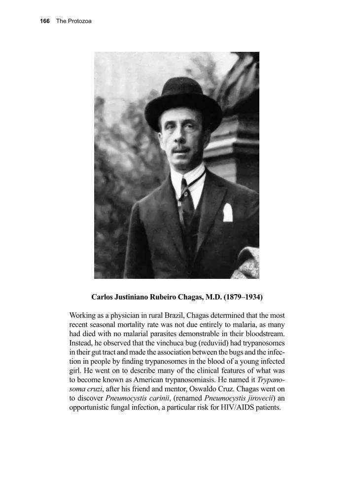
166 The Protozoa
Carlos Justiniano Rubeiro Chagas, M.D. (1879–1934)
Working as a physician in rural Brazil, Chagas determined that the most recent seasonal mortality rate was not due entirely to malaria, as many had died with no malarial parasites demonstrable in their bloodstream. Instead, he observed that the vinchuca bug (reduviid) had trypanosomes in their gut tract and made the association between the bugs and the infec-tion in people by finding trypanosomes in the blood of a young infected girl. He went on to describe many of the clinical features of what was to become known as American trypanosomiasis. He named it Trypano-soma cruzi, after his friend and mentor, Oswaldo Cruz. Chagas went on to discover Pneumocystis carinii, (renamed Pneumocystis jirovecii) an opportunistic fungal infection, a particular risk for HIV/AIDS patients.
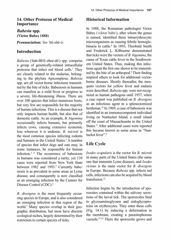
14. Other Protozoa of Medical Importance 167
14. Other Protozoa of Medical ImportanceBabesia spp.(Victor Babeș 1888)
Pronunciation: \bə-ˈbē-zhē-ə\
Introduction
Babesia (\bah-BEE-zhee-ah\) spp. comprise a group of genetically-related intracellular protozoa that infect red blood cells.1 They are closely related to the malarias, belong-ing to the phylum Apicomplexa. Babesia spp. are all vector-borne infections transmit-ted by the bite of ticks. Babesiosis in humans can manifest as a mild fever or progress to a severe, life-threatening illness. There are over 100 species that infect numerous hosts, but very few are responsible for the majority of human infections. This is a disease that not only impacts human health, but also that of domestic cattle. As an example, B. bigemina occasionally infects humans, but primarily infects cows, causing extensive economic loss wherever it is endemic. B. microti is the most common species infecting rodents and humans in the United States.2 A number of species that infect dogs and cats may, in some instances, be responsible for human infection.3, 4 The occurrence of babesiosis in humans was considered a rarity, yet 139 cases were reported from New York State between 1982 and 1993.5 Currently babe-siosis is as prevalent in some areas as Lyme disease, and consequently is now classified as an emerging infection by the Centers for Disease Control (CDC).6
B. divergens is the most frequently occur-ring species in Europe, and is also considered an emerging infection in that region of the world.7 Many species overlap in their geo-graphic distribution, but most have discrete ecological niches, largely determined by their restriction to certain species of ticks.
Historical Information
In 1888, the Romanian pathologist Victor Babeș (\viktor babe\), after whom the genus is named, identified these intraerythrocytic microorganisms as causing febrile hemoglo-binuria in cattle.8 In 1893, Theobald Smith and Frederick L. Kilbourne demonstrated that ticks were the vectors of B. bigemina, the cause of Texas cattle fever in the Southwest-ern United States. Thus, making this infec-tious agent the first one shown to be transmit-ted by the bite of an arthropod.9 Their finding inspired others to look for additional vector-borne diseases. Shortly thereafter, the mos-quito vectors for yellow fever and malaria were described. Babesia spp. were not recog-nized as human pathogens until 1957, when a case report was published on B. divergens as an infectious agent in a splenectomized herdsman.10 In 1969, a case of babesiosis was identified in an immunocompetent individual living on Nantucket Island, a small island off the coast of Massachusetts in the United States.11 After additional cases were reported this became known in some areas as “Nan-tucket fever”.6
Life Cycle
Ixodes scapularis is the vector for B. microti in many parts of the United States (the same one that transmits Lyme disease), and Ixodes ricinus is the main vector for B. divergens in Europe. Because Babesia spp. infects red cells, infections can also be acquired by blood transfusion.12
Infection begins by the introduction of spo-rozoites contained within the salivary secre-tions of the larval tick. The sporozoites bind to glycosaminoglycans and sialoglycopro-teins on erythrocytes. They enter these cells (Fig. 14.1) by inducing a deformation in the membrane, creating a parasitophorous vacuole.13-16 There the sporozoite grows and
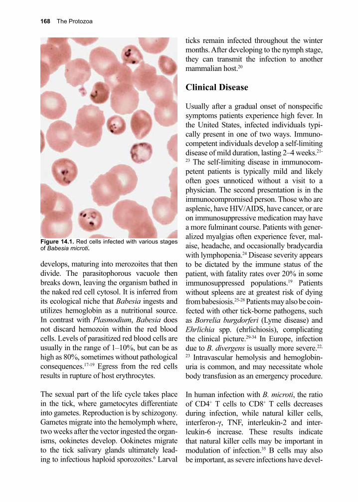
168 The Protozoa
Figure 14.1. Red cells infected with various stages of Babesia microti.
develops, maturing into merozoites that then divide. The parasitophorous vacuole then breaks down, leaving the organism bathed in the naked red cell cytosol. It is inferred from its ecological niche that Babesia ingests and utilizes hemoglobin as a nutritional source. In contrast with Plasmodium, Babesia does not discard hemozoin within the red blood cells. Levels of parasitized red blood cells are usually in the range of 1–10%, but can be as high as 80%, sometimes without pathological consequences.17-19 Egress from the red cells results in rupture of host erythrocytes.
The sexual part of the life cycle takes place in the tick, where gametocytes differentiate into gametes. Reproduction is by schizogony. Gametes migrate into the hemolymph where, two weeks after the vector ingested the organ-isms, ookinetes develop. Ookinetes migrate to the tick salivary glands ultimately lead-ing to infectious haploid sporozoites.6 Larval
ticks remain infected throughout the winter months. After developing to the nymph stage, they can transmit the infection to another mammalian host.20
Clinical Disease
Usually after a gradual onset of nonspecific symptoms patients experience high fever. In the United States, infected individuals typi-cally present in one of two ways. Immuno-competent individuals develop a self-limiting disease of mild duration, lasting 2–4 weeks.21-
23 The self-limiting disease in immunocom-petent patients is typically mild and likely often goes unnoticed without a visit to a physician. The second presentation is in the immunocompromised person. Those who are asplenic, have HIV/AIDS, have cancer, or are on immunosuppressive medication may have a more fulminant course. Patients with gener-alized myalgias often experience fever, mal-aise, headache, and occasionally bradycardia with lymphopenia.24 Disease severity appears to be dictated by the immune status of the patient, with fatality rates over 20% in some immunosuppressed populations.19 Patients without spleens are at greatest risk of dying from babesiosis.25-28 Patients may also be coin-fected with other tick-borne pathogens, such as Borrelia burgdorferi (Lyme disease) and Ehrlichia spp. (ehrlichiosis), complicating the clinical picture.29-34 In Europe, infection due to B. divergens is usually more severe.22,
23 Intravascular hemolysis and hemoglobin-uria is common, and may necessitate whole body transfusion as an emergency procedure. In human infection with B. microti, the ratio of CD4+ T cells to CD8+ T cells decreases during infection, while natural killer cells, interferon-γ, TNF, interleukin-2 and inter-leukin-6 increase. These results indicate that natural killer cells may be important in modulation of infection.35 B cells may also be important, as severe infections have devel-
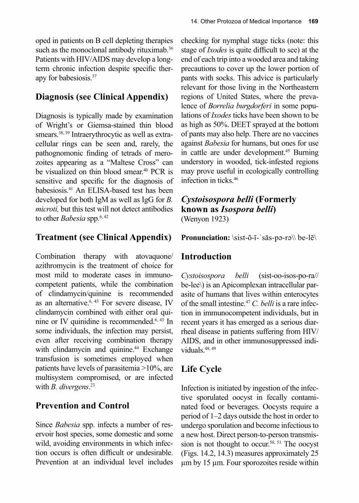
14. Other Protozoa of Medical Importance 169
oped in patients on B cell depleting therapies such as the monoclonal antibody rituximab.36 Patients with HIV/AIDS may develop a long-term chronic infection despite specific ther-apy for babesiosis.37
Diagnosis (see Clinical Appendix)
Diagnosis is typically made by examination of Wright’s or Giemsa-stained thin blood smears.38, 39 Intraerythrocytic as well as extra-cellular rings can be seen and, rarely, the pathognomonic finding of tetrads of mero-zoites appearing as a “Maltese Cross” can be visualized on thin blood smear.40 PCR is sensitive and specific for the diagnosis of babesiosis.41 An ELISA-based test has been developed for both IgM as well as IgG for B. microti, but this test will not detect antibodies to other Babesia spp.6, 42
Treatment (see Clinical Appendix)
Combination therapy with atovaquone/azithromycin is the treatment of choice for most mild to moderate cases in immuno-competent patients, while the combination of clindamycin/quinine is recommended as an alternative.6, 43 For severe disease, IV clindamycin combined with either oral qui-nine or IV quinidine is recommended.6, 43 In some individuals, the infection may persist, even after receiving combination therapy with clindamycin and quinine.44 Exchange transfusion is sometimes employed when patients have levels of parasitemia >10%, are multisystem compromised, or are infected with B. divergens.23
Prevention and Control
Since Babesia spp. infects a number of res-ervoir host species, some domestic and some wild, avoiding environments in which infec-tion occurs is often difficult or undesirable. Prevention at an individual level includes
checking for nymphal stage ticks (note: this stage of Ixodes is quite difficult to see) at the end of each trip into a wooded area and taking precautions to cover up the lower portion of pants with socks. This advice is particularly relevant for those living in the Northeastern regions of United States, where the preva-lence of Borrelia burgdorferi in some popu-lations of Ixodes ticks have been shown to be as high as 50%. DEET sprayed at the bottom of pants may also help. There are no vaccines against Babesia for humans, but ones for use in cattle are under development.45 Burning understory in wooded, tick-infested regions may prove useful in ecologically controlling infection in ticks.46
Cystoisospora belli (Formerly known as Isospora belli)(Wenyon 1923)
Pronunciation: \sist-ō-ī-ˈsäs-pə-rə\\ be-lē\
Introduction
Cystoisospora belli (sist-oo-isos-po-ra//be-lee\) is an Apicomplexan intracellular par-asite of humans that lives within enterocytes of the small intestine.47 C. belli is a rare infec-tion in immunocompetent individuals, but in recent years it has emerged as a serious diar-rheal disease in patients suffering from HIV/AIDS, and in other immunosuppressed indi-viduals.48, 49
Life Cycle
Infection is initiated by ingestion of the infec-tive sporulated oocyst in fecally contami-nated food or beverages. Oocysts require a period of 1–2 days outside the host in order to undergo sporulation and become infectious to a new host. Direct person-to-person transmis-sion is not thought to occur.50, 51 The oocyst (Figs. 14.2, 14.3) measures approximately 25 µm by 15 µm. Four sporozoites reside within
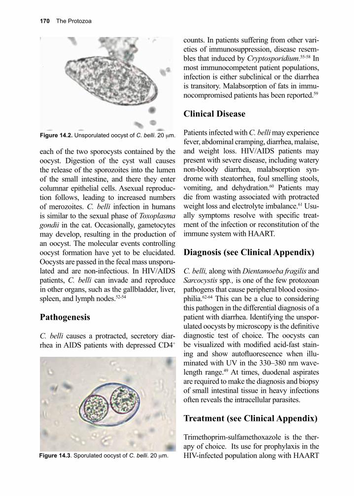
170 The Protozoa
each of the two sporocysts contained by the oocyst. Digestion of the cyst wall causes the release of the sporozoites into the lumen of the small intestine, and there they enter columnar epithelial cells. Asexual reproduc-tion follows, leading to increased numbers of merozoites. C. belli infection in humans is similar to the sexual phase of Toxoplasma gondii in the cat. Occasionally, gametocytes may develop, resulting in the production of an oocyst. The molecular events controlling oocyst formation have yet to be elucidated. Oocysts are passed in the fecal mass unsporu-lated and are non-infectious. In HIV/AIDS patients, C. belli can invade and reproduce in other organs, such as the gallbladder, liver, spleen, and lymph nodes.52-54
Pathogenesis
C. belli causes a protracted, secretory diar-rhea in AIDS patients with depressed CD4+
counts. In patients suffering from other vari-eties of immunosuppression, disease resem-bles that induced by Cryptosporidium.55-58 In most immunocompetent patient populations, infection is either subclinical or the diarrhea is transitory. Malabsorption of fats in immu-nocompromised patients has been reported.59
Clinical Disease
Patients infected with C. belli may experience fever, abdominal cramping, diarrhea, malaise, and weight loss. HIV/AIDS patients may present with severe disease, including watery non-bloody diarrhea, malabsorption syn-drome with steatorrhea, foul smelling stools, vomiting, and dehydration.60 Patients may die from wasting associated with protracted weight loss and electrolyte imbalance.61 Usu-ally symptoms resolve with specific treat-ment of the infection or reconstitution of the immune system with HAART.
Diagnosis (see Clinical Appendix)
C. belli, along with Dientamoeba fragilis and Sarcocystis spp., is one of the few protozoan pathogens that cause peripheral blood eosino-philia.62-64 This can be a clue to considering this pathogen in the differential diagnosis of a patient with diarrhea. Identifying the unspor-ulated oocysts by microscopy is the definitive diagnostic test of choice. The oocysts can be visualized with modified acid-fast stain-ing and show autofluorescence when illu-minated with UV in the 330–380 nm wave-length range.49 At times, duodenal aspirates are required to make the diagnosis and biopsy of small intestinal tissue in heavy infections often reveals the intracellular parasites.
Treatment (see Clinical Appendix)
Trimethoprim-sulfamethoxazole is the ther-apy of choice. Its use for prophylaxis in the HIV-infected population along with HAART Figure 14.3. Sporulated oocyst of C. belli. 20 µm.
Figure 14.2. Unsporulated oocyst of C. belli. 20 µm.
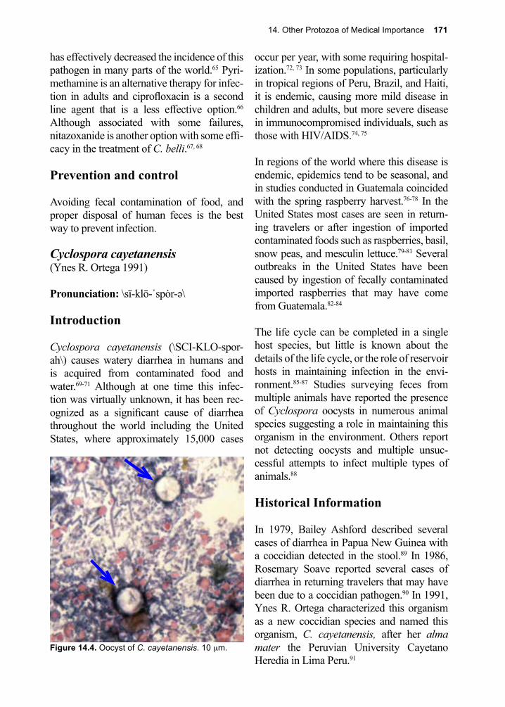
14. Other Protozoa of Medical Importance 171
has effectively decreased the incidence of this pathogen in many parts of the world.65 Pyri-methamine is an alternative therapy for infec-tion in adults and ciprofloxacin is a second line agent that is a less effective option.66 Although associated with some failures, nitazoxanide is another option with some effi-cacy in the treatment of C. belli.67, 68
Prevention and control
Avoiding fecal contamination of food, and proper disposal of human feces is the best way to prevent infection.
Cyclospora cayetanensis(Ynes R. Ortega 1991)
Pronunciation: \sī-klō-ˈspȯr-ə\
Introduction
Cyclospora cayetanensis (\SCI-KLO-spor-ah\) causes watery diarrhea in humans and is acquired from contaminated food and water.69-71 Although at one time this infec-tion was virtually unknown, it has been rec-ognized as a significant cause of diarrhea throughout the world including the United States, where approximately 15,000 cases
occur per year, with some requiring hospital-ization.72, 73 In some populations, particularly in tropical regions of Peru, Brazil, and Haiti, it is endemic, causing more mild disease in children and adults, but more severe disease in immunocompromised individuals, such as those with HIV/AIDS.74, 75
In regions of the world where this disease is endemic, epidemics tend to be seasonal, and in studies conducted in Guatemala coincided with the spring raspberry harvest.76-78 In the United States most cases are seen in return-ing travelers or after ingestion of imported contaminated foods such as raspberries, basil, snow peas, and mesculin lettuce.79-81 Several outbreaks in the United States have been caused by ingestion of fecally contaminated imported raspberries that may have come from Guatemala.82-84
The life cycle can be completed in a single host species, but little is known about the details of the life cycle, or the role of reservoir hosts in maintaining infection in the envi-ronment.85-87 Studies surveying feces from multiple animals have reported the presence of Cyclospora oocysts in numerous animal species suggesting a role in maintaining this organism in the environment. Others report not detecting oocysts and multiple unsuc-cessful attempts to infect multiple types of animals.88
Historical Information
In 1979, Bailey Ashford described several cases of diarrhea in Papua New Guinea with a coccidian detected in the stool.89 In 1986, Rosemary Soave reported several cases of diarrhea in returning travelers that may have been due to a coccidian pathogen.90 In 1991, Ynes R. Ortega characterized this organism as a new coccidian species and named this organism, C. cayetanensis, after her alma mater the Peruvian University Cayetano Heredia in Lima Peru.91
Figure 14.4. Oocyst of C. cayetanensis. 10 µm.
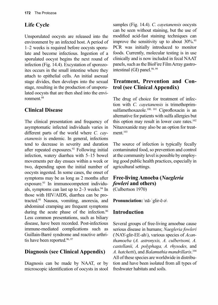
172 The Protozoa
Life Cycle
Unsporulated oocysts are released into the environment by an infected host. A period of 1–2 weeks is required before oocysts sporu-late and become infectious. Ingestion of a sporulated oocyst begins the next round of infection (Fig. 14.4). Excystation of sporozo-ites occurs in the small intestine where they attach to epithelial cells. An initial asexual stage divides, then develops into the sexual stage, resulting in the production of unsporu-lated oocysts that are then shed into the envi-ronment.88
Clinical Disease
The clinical presentation and frequency of asymptomatic infected individuals varies in different parts of the world where C. cay-etanensis is endemic. In general, infections tend to decrease in severity and duration after repeated exposures.92 Following initial infection, watery diarrhea with 5–15 bowel movements per day ensues within a week or two, depending upon the initial number of oocysts ingested. In some cases, the onset of symptoms may be as long as 2 months after exposure.93 In immunocompetent individu-als, symptoms can last up to 2–3 weeks.94 In those with HIV/AIDS, diarrhea can be pro-tracted.95 Nausea, vomiting, anorexia, and abdominal cramping are frequent symptoms during the acute phase of the infection.86 Less common presentations, such as biliary disease, have been recorded. Post-infectious immune-mediated complications such as Guillain-Barré syndrome and reactive arthri-tis have been reported.96, 97
Diagnosis (see Clinical Appendix) Diagnosis can be made by NAAT, or by microscopic identification of oocysts in stool
samples (Fig. 14.4). C. cayetanensis oocysts can be seen without staining, but the use of modified acid-fast staining techniques can improve the sensitivity up to about 30%.95 PCR was initially introduced to monitor foods. Currently, molecular testing is in use clinically and is now included in fecal NAAT panels, such as the BioFire Film Array gastro-intestinal (GI) panel.98, 99
Treatment, Prevention and Con-trol (see Clinical Appendix)
The drug of choice for treatment of infec-tion with C. cayetanensis is trimethoprim-sulfamethoxazole.100, 101 Ciprofloxacin is an alternative for patients with sulfa allergies but this option may result in lower cure rates.102 Nitazoxanide may also be an option for treat-ment.103
The source of infection is typically fecally contaminated food, so prevention and control at the community level is possible by employ-ing good public health practices, especially in agricultural settings.
Free-living Amoeba (Naegleria fowleri and others) (Culbertson 1970)
Pronunciation: \nā-ˈglir-ē-ə\
Introduction
Several groups of free-living amoebae cause serious disease in humans; Naegleria fowleri (\NAY-glir-EE-ah\), various species of Acan-thamoeba (A. astronyxis, A. culbertsoni, A. castellanii, A. polyphaga, A. rhysodes, and A. hatchetti), and Balamuthia mandrillaris.104 All of these species are worldwide in distribu-tion and have been isolated from all types of freshwater habitats and soils.
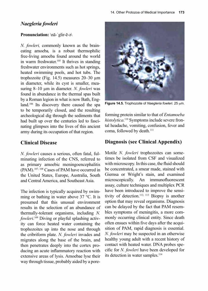
14. Other Protozoa of Medical Importance 173
Naegleria fowleri
Pronunciation: \nā-ˈglir-ē-ə\
N. fowleri, commonly known as the brain-eating amoeba, is a robust thermophilic free-living amoeba found around the world in warm freshwater.105 It thrives in standing freshwater environments such as hot springs, heated swimming pools, and hot tubs. The trophozoite (Fig. 14.5) measures 20–30 µm in diameter, while its cyst is smaller, mea-suring 8–10 µm in diameter. N. fowleri was found in abundance in the thermal spas built by a Roman legion in what is now Bath, Eng-land.106 Its discovery there caused the spa to be temporarily closed, and the resulting archeological dig through the sediments that had built up over the centuries led to fasci-nating glimpses into the lives of this ancient army during its occupation of that region.
Clinical Disease
N. fowleri causes a serious, often fatal, ful-minating infection of the CNS, referred to as primary amoebic meningoencephalitis (PAM).107, 108 Cases of PAM have occurred in the United States, Europe, Australia, South and Central America, and Southeast Asia.
The infection is typically acquired by swim-ming or bathing in water above 37 °C. It is presumed that this unusual environment results in the selection of an abundance of thermally-tolerant organisms, including N. fowleri.109 Diving or playful splashing activ-ity can force heated water containing the trophozoites up into the nose and through the cribriform plate. N. fowleri invades and migrates along the base of the brain, and then penetrates deeply into the cortex pro-ducing an acute inflammatory reaction with extensive areas of lysis. Amoebae lyse their way through tissue, probably aided by a pore-
forming protein similar to that of Entamoeba histolytica.110 Symptoms include severe fron-tal headache, vomiting, confusion, fever and coma, followed by death.111
Diagnosis (see Clinical Appendix)
Motile N. fowleri trophozoites can some-times be isolated from CSF and visualized with microscopy. In this case, the fluid should be concentrated, a smear made, stained with Giemsa or Wright’s stain, and examined microscopically. An immunofluorescent assay, culture techniques and multiplex PCR have been introduced to improve the sensi-tivity of detection.112, 113 Biopsy is another option that may reveal organisms. Diagnosis can be delayed by the fact that PAM resem-bles symptoms of meningitis, a more com-monly occurring clinical entity. Since death often ensues within five days after the acqui-sition of PAM, rapid diagnosis is essential. N. fowleri may be suspected in an otherwise healthy young adult with a recent history of contact with heated water. DNA probes spe-cific for N. fowleri have been developed for its detection in water samples.114
Figure 14.5. Trophozoite of Naegleria fowleri. 25 µm.
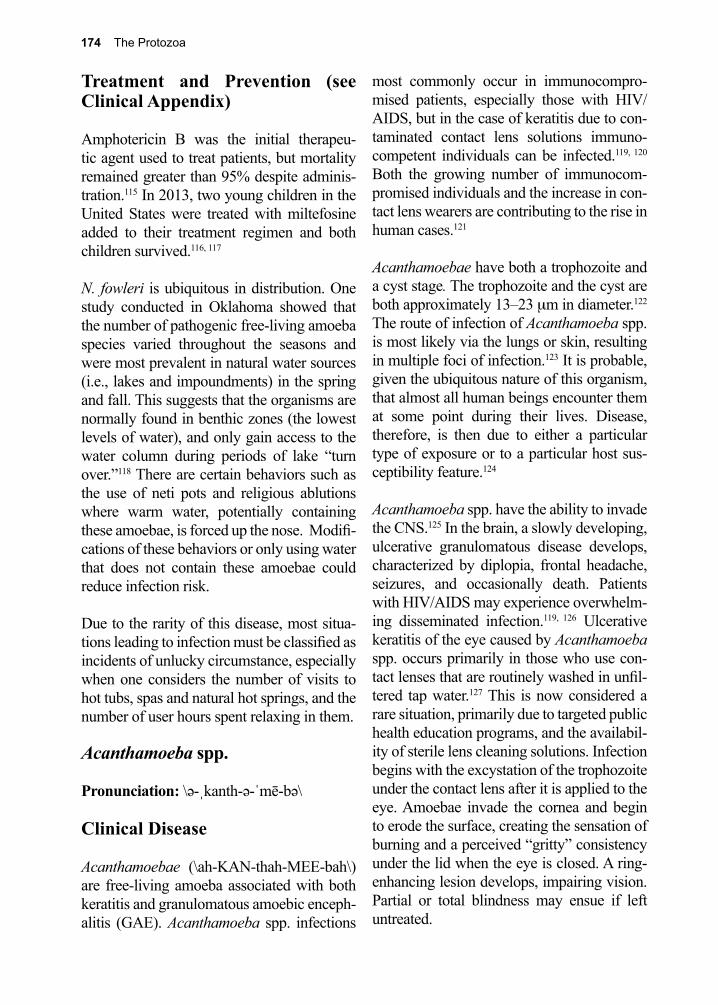
174 The Protozoa
Treatment and Prevention (see Clinical Appendix)
Amphotericin B was the initial therapeu-tic agent used to treat patients, but mortality remained greater than 95% despite adminis-tration.115 In 2013, two young children in the United States were treated with miltefosine added to their treatment regimen and both children survived.116, 117
N. fowleri is ubiquitous in distribution. One study conducted in Oklahoma showed that the number of pathogenic free-living amoeba species varied throughout the seasons and were most prevalent in natural water sources (i.e., lakes and impoundments) in the spring and fall. This suggests that the organisms are normally found in benthic zones (the lowest levels of water), and only gain access to the water column during periods of lake “turn over.”118 There are certain behaviors such as the use of neti pots and religious ablutions where warm water, potentially containing these amoebae, is forced up the nose. Modifi-cations of these behaviors or only using water that does not contain these amoebae could reduce infection risk.
Due to the rarity of this disease, most situa-tions leading to infection must be classified as incidents of unlucky circumstance, especially when one considers the number of visits to hot tubs, spas and natural hot springs, and the number of user hours spent relaxing in them.
Acanthamoeba spp.
Pronunciation: \ə-ˌkanth-ə-ˈmē-bə\
Clinical Disease
Acanthamoebae (\ah-KAN-thah-MEE-bah\) are free-living amoeba associated with both keratitis and granulomatous amoebic enceph-alitis (GAE). Acanthamoeba spp. infections
most commonly occur in immunocompro-mised patients, especially those with HIV/AIDS, but in the case of keratitis due to con-taminated contact lens solutions immuno-competent individuals can be infected.119, 120 Both the growing number of immunocom-promised individuals and the increase in con-tact lens wearers are contributing to the rise in human cases.121
Acanthamoebae have both a trophozoite and a cyst stage. The trophozoite and the cyst are both approximately 13–23 µm in diameter.122 The route of infection of Acanthamoeba spp. is most likely via the lungs or skin, resulting in multiple foci of infection.123 It is probable, given the ubiquitous nature of this organism, that almost all human beings encounter them at some point during their lives. Disease, therefore, is then due to either a particular type of exposure or to a particular host sus-ceptibility feature.124
Acanthamoeba spp. have the ability to invade the CNS.125 In the brain, a slowly developing, ulcerative granulomatous disease develops, characterized by diplopia, frontal headache, seizures, and occasionally death. Patients with HIV/AIDS may experience overwhelm-ing disseminated infection.119, 126 Ulcerative keratitis of the eye caused by Acanthamoeba spp. occurs primarily in those who use con-tact lenses that are routinely washed in unfil-tered tap water.127 This is now considered a rare situation, primarily due to targeted public health education programs, and the availabil-ity of sterile lens cleaning solutions. Infection begins with the excystation of the trophozoite under the contact lens after it is applied to the eye. Amoebae invade the cornea and begin to erode the surface, creating the sensation of burning and a perceived “gritty” consistency under the lid when the eye is closed. A ring-enhancing lesion develops, impairing vision. Partial or total blindness may ensue if left untreated.
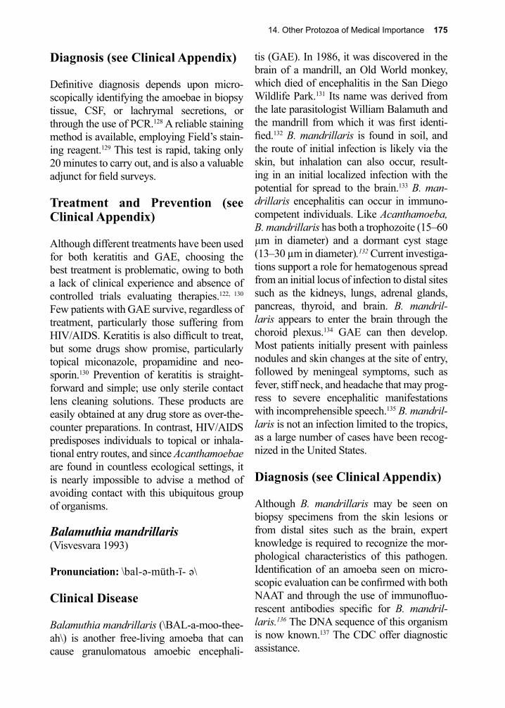
14. Other Protozoa of Medical Importance 175
Diagnosis (see Clinical Appendix)
Definitive diagnosis depends upon micro-scopically identifying the amoebae in biopsy tissue, CSF, or lachrymal secretions, or through the use of PCR.128 A reliable staining method is available, employing Field’s stain-ing reagent.129 This test is rapid, taking only 20 minutes to carry out, and is also a valuable adjunct for field surveys.
Treatment and Prevention (see Clinical Appendix) Although different treatments have been used for both keratitis and GAE, choosing the best treatment is problematic, owing to both a lack of clinical experience and absence of controlled trials evaluating therapies.122, 130 Few patients with GAE survive, regardless of treatment, particularly those suffering from HIV/AIDS. Keratitis is also difficult to treat, but some drugs show promise, particularly topical miconazole, propamidine and neo-sporin.130 Prevention of keratitis is straight-forward and simple; use only sterile contact lens cleaning solutions. These products are easily obtained at any drug store as over-the-counter preparations. In contrast, HIV/AIDS predisposes individuals to topical or inhala-tional entry routes, and since Acanthamoebae are found in countless ecological settings, it is nearly impossible to advise a method of avoiding contact with this ubiquitous group of organisms.
Balamuthia mandrillaris(Visvesvara 1993)
Pronunciation: \bal-ə-müth-ī- ə\
Clinical Disease
Balamuthia mandrillaris (\BAL-a-moo-thee-ah\) is another free-living amoeba that can cause granulomatous amoebic encephali-
tis (GAE). In 1986, it was discovered in the brain of a mandrill, an Old World monkey, which died of encephalitis in the San Diego Wildlife Park.131 Its name was derived from the late parasitologist William Balamuth and the mandrill from which it was first identi-fied.132 B. mandrillaris is found in soil, and the route of initial infection is likely via the skin, but inhalation can also occur, result-ing in an initial localized infection with the potential for spread to the brain.133 B. man-drillaris encephalitis can occur in immuno-competent individuals. Like Acanthamoeba, B. mandrillaris has both a trophozoite (15–60 µm in diameter) and a dormant cyst stage (13–30 µm in diameter).132 Current investiga-tions support a role for hematogenous spread from an initial locus of infection to distal sites such as the kidneys, lungs, adrenal glands, pancreas, thyroid, and brain. B. mandril-laris appears to enter the brain through the choroid plexus.134 GAE can then develop. Most patients initially present with painless nodules and skin changes at the site of entry, followed by meningeal symptoms, such as fever, stiff neck, and headache that may prog-ress to severe encephalitic manifestations with incomprehensible speech.135 B. mandril-laris is not an infection limited to the tropics, as a large number of cases have been recog-nized in the United States.
Diagnosis (see Clinical Appendix)
Although B. mandrillaris may be seen on biopsy specimens from the skin lesions or from distal sites such as the brain, expert knowledge is required to recognize the mor-phological characteristics of this pathogen. Identification of an amoeba seen on micro-scopic evaluation can be confirmed with both NAAT and through the use of immunofluo-rescent antibodies specific for B. mandril-laris.136 The DNA sequence of this organism is now known.137 The CDC offer diagnostic assistance.
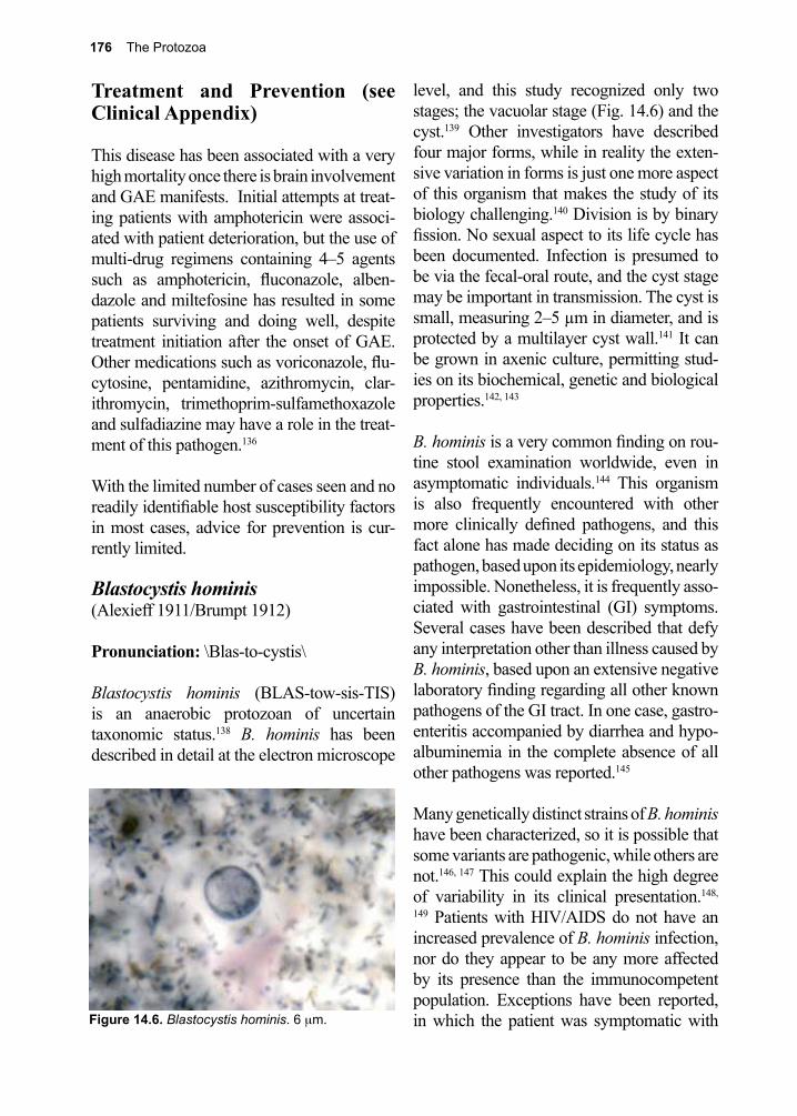
176 The Protozoa
Treatment and Prevention (see Clinical Appendix)
This disease has been associated with a very high mortality once there is brain involvement and GAE manifests. Initial attempts at treat-ing patients with amphotericin were associ-ated with patient deterioration, but the use of multi-drug regimens containing 4–5 agents such as amphotericin, fluconazole, alben-dazole and miltefosine has resulted in some patients surviving and doing well, despite treatment initiation after the onset of GAE. Other medications such as voriconazole, flu-cytosine, pentamidine, azithromycin, clar-ithromycin, trimethoprim-sulfamethoxazole and sulfadiazine may have a role in the treat-ment of this pathogen.136
With the limited number of cases seen and no readily identifiable host susceptibility factors in most cases, advice for prevention is cur-rently limited.
Blastocystis hominis(Alexieff 1911/Brumpt 1912)
Pronunciation: \Blas-to-cystis\
Blastocystis hominis (BLAS-tow-sis-TIS) is an anaerobic protozoan of uncertain taxonomic status.138 B. hominis has been described in detail at the electron microscope
level, and this study recognized only two stages; the vacuolar stage (Fig. 14.6) and the cyst.139 Other investigators have described four major forms, while in reality the exten-sive variation in forms is just one more aspect of this organism that makes the study of its biology challenging.140 Division is by binary fission. No sexual aspect to its life cycle has been documented. Infection is presumed to be via the fecal-oral route, and the cyst stage may be important in transmission. The cyst is small, measuring 2–5 µm in diameter, and is protected by a multilayer cyst wall.141 It can be grown in axenic culture, permitting stud-ies on its biochemical, genetic and biological properties.142, 143
B. hominis is a very common finding on rou-tine stool examination worldwide, even in asymptomatic individuals.144 This organism is also frequently encountered with other more clinically defined pathogens, and this fact alone has made deciding on its status as pathogen, based upon its epidemiology, nearly impossible. Nonetheless, it is frequently asso-ciated with gastrointestinal (GI) symptoms. Several cases have been described that defy any interpretation other than illness caused by B. hominis, based upon an extensive negative laboratory finding regarding all other known pathogens of the GI tract. In one case, gastro-enteritis accompanied by diarrhea and hypo-albuminemia in the complete absence of all other pathogens was reported.145
Many genetically distinct strains of B. hominis have been characterized, so it is possible that some variants are pathogenic, while others are not.146, 147 This could explain the high degree of variability in its clinical presentation.148,
149 Patients with HIV/AIDS do not have an increased prevalence of B. hominis infection, nor do they appear to be any more affected by its presence than the immunocompetent population. Exceptions have been reported, in which the patient was symptomatic with Figure 14.6. Blastocystis hominis. 6 µm.
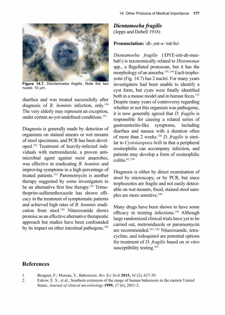
14. Other Protozoa of Medical Importance 177
diarrhea and was treated successfully after diagnosis of B. hominis infection, only.150 The very elderly may represent an exception, under certain as-yet-undefined conditions.151
Diagnosis is generally made by detection of organisms on stained smears or wet mounts of stool specimens, and PCR has been devel-oped.152 Treatment of heavily-infected indi-viduals with metronidazole, a proven anti-microbial agent against most anaerobes, was effective in eradicating B. hominis and improving symptoms in a high percentage of treated patients.153 Paromomycin is another therapy suggested by some investigators to be an alternative first line therapy.153 Trime-thoprim-sulfamethoxazole has shown effi-cacy in the treatment of symptomatic patients and achieved high rates of B. hominis eradi-cation from stool.154 Nitazoxanide shows promise as an effective alternative therapeutic approach but studies have been confounded by its impact on other intestinal pathogens.150
Dientamoeba fragilis(Jepps and Dobell 1918)
Pronunciation: \dī-ˌent-ə-ˈmē-bə\
Dientamoeba fragilis (\DYE-ent-ah-mee-bah\) is taxonomically related to Histomonas spp., a flagellated protozoan, but it has the morphology of an amoeba.155, 156 Each tropho-zoite (Fig. 14.7) has 2 nuclei. For many years investigators had been unable to identify a cyst form, but cysts were finally identified both in a mouse model and in human feces.157 Despite many years of controversy regarding whether or not this organism was pathogenic, it is now generally agreed that D. fragilis is responsible for causing a related series of gastroenteritis-like symptoms, including diarrhea and nausea with a duration often of more than 2 weeks.158 D. fragilis is simi-lar to Cystoisospora belli in that a peripheral eosinophilia can accompany infection, and patients may develop a form of eosinophilic colitis.62, 159
Diagnosis is either by direct examination of stool by microscopy, or by PCR, but since trophozoites are fragile and not easily detect-able on wet mounts, fixed, stained stool sam-ples are more sensitive.160
Many drugs have been shown to have some efficacy in treating infections.156 Although large randomized clinical trials have yet to be carried out, metronidazole or paromomycin are recommended.161, 162 Nitazoxanide, tetra-cycline, and iodoquinol are potential options for treatment of D. fragilis based on in vitro susceptibility testing.163
References
1. Beugnet, F.; Moreau, Y., Babesiosis. Rev Sci Tech 2015, 34 (2), 627-39.2. Eskow, E. S., et al., Southern extension of the range of human babesiosis in the eastern United
States. Journal of clinical microbiology 1999, 37 (6), 2051-2.
Figure 14.7. Dientamoeba fragilis. Note the two nuclei. 10 µm.
178 The Protozoa
3. Herwaldt, B. L., et al., Transfusion-transmitted babesiosis in Washington State: first reported case caused by a WA1-type parasite. J Infect Dis 1997, 175 (5), 1259-62.
4. Telford, S. R., et al., Topley and Wilson’s Microbiology and Microbial Infections. 1998; p 349-359.
5. White, D. J., et al., Human babesiosis in New York State: Review of 139 hospitalized cases and analysis of prognostic factors. Archives of internal medicine 1998, 158 (19), 2149-54.
6. Vannier, E.; Krause, P. J., Human babesiosis. N Engl J Med 2012, 366 (25), 2397-407.7. Gray, J. S., Babesia sp.: emerging intracellular parasites in Europe. Polish journal of
microbiology / Polskie Towarzystwo Mikrobiologow = The Polish Society of Microbiologists 2004, 53 Suppl, 55-60.
8. Babes, V., Sur l’hemoglobinurie bacterienne de boeuf. C R Acad Sci 1888, 107, 692-4.9. Smith, T.; Kilobourne, F. L., Investigations into the Nature, Causation, and Prevention of Texas
or Southern Cattle Fever. 1893.10. Skrabalo, Z.; Deanovic, Z., Piroplasmosis in man; report of a case. Doc Med Geogr Trop 1957, 9
(1), 11-6.11. Western, K. A., et al., Babesiosis in a Massachusetts resident. N Engl J Med 1970, 283 (16), 854-
6.12. Leiby, D. A.; Gill, J. E., Transfusion-transmitted tick-borne infections: a cornucopia of threats.
Transfusion medicine reviews 2004, 18 (4), 293-306.13. Rudzinska, M. A., et al., Morphological aspects of host-cell-parasite relationships in babesiosis.
1981; p 87-141.14. Okubo, K., et al., Calcium-ions are involved in erythrocyte invasion by equine Babesia parasites.
Parasitology 2006, 133 (Pt 3), 289-94.15. Yokoyama, N., et al., Erythrocyte invasion by Babesia parasites: current advances in the
elucidation of the molecular interactions between the protozoan ligands and host receptors in the invasion stage. Vet Parasitol 2006, 138 (1-2), 22-32.
16. Lobo, C. A., et al., Babesia and red cell invasion. Curr Opin Hematol 2012, 19 (3), 170-5.17. Christianson, D.; Pollack, R. J., Persistent parasitemia after acute babesiosis. N E J M 1998, 339
160-5.18. Shih, C. M., et al., Human babesiosis in Taiwan: asymptomatic infection with a Babesia microti-
like organism in a Taiwanese woman. Journal of clinical microbiology 1997, 35 (2), 450-4.19. Hatcher, J. C., et al., Severe babesiosis in Long Island: review of 34 cases and their
complications. Clin Infect Dis 2001, 32 (8), 1117-25.20. Piesman, J., et al., Seasonal variation of transmission risk of Lyme disease and human
babesiosis. American journal of epidemiology 1987, 126 (6), 1187-9.21. Boustani, M. R.; Gelfand, J. A., Babesiosis. Clin Inf Dis 1996, 22 (4), 611-5.22. Gorenflot, A., et al., Human babesiosis. Annals of tropical medicine and parasitology 1998, 92
(4), 489-501.23. Uguen, C., et al., [Human babesiosis in 1997]. La Revue de medecine interne / fondee ... par la
Societe nationale francaise de medecine interne 1997, 18 (12), 945-51.24. Kim, N., et al., Relative bradycardia and lymphopenia in patients with babesiosis. Clin Inf Dis
1998, 26 (5), 1218-9.25. Herwaldt, B., et al., A fatal case of babesiosis in Missouri: identification of another piroplasm
that infects humans. Annals of internal medicine 1996, 124 (7), 643-50.26. Slovut, D. P., et al., Babesiosis and hemophagocytic syndrome in an asplenic renal transplant
recipient. Transplantation 1996, 62 (4), 537-9.27. Bonoan, J. T., et al., Life-threatening babesiosis in an asplenic patient treated with exchange
transfusion, azithromycin, and atovaquone. Heart & lung : the journal of critical care 1998, 27 (6), 424-8.
28. Hohenschild, S., [Babesiosis--a dangerous infection for splenectomized children and adults]. Klinische Padiatrie 1999, 211 (3), 137-40.
29. Persing, D. H., The cold zone: a curious convergence of tick-transmitted diseases. Clin Infect Dis 25 Suppl 1997, 1, S35-42.
14. Other Protozoa of Medical Importance 179
30. Hilton, E., et al., Seroprevalence and seroconversion for tick-borne diseases in a high-risk population in the northeast United States. The American journal of medicine 1999, 106 (4), 404-9.
31. Krause, P. J., et al., Concurrent Lyme disease and babesiosis. Evidence for increased severity and duration of illness. JAMA 1996, 275 (21), 1657-60.
32. Mitchell, P. D., et al., Immunoserologic evidence of coinfection with Borrelia burgdorferi, Babesia microti, and human granulocytic Ehrlichia species in residents of Wisconsin and Minnesota. Journal of clinical microbiology 1996, 34 (3), 724-7.
33. Magnarelli, L. A., et al., Human exposure to a granulocytic Ehrlichia and other tick-borne agents in Connecticut. Journal of clinical microbiology 1998, 36 (10), 2823-7.
34. dos Santos, C. C.; Kain, K. C., Two tick-borne diseases in one: a case report of concurrent babesiosis and Lyme disease in Ontario. CMAJ 1999, 160 (13), 1851-3.
35. Shaio, M. F.; Lin, P. R., A case study of cytokine profiles in acute human babesiosis. Am J Trop Med Hyg 1998, 58 (3), 335-7.
36. Kelesidis, T., et al., Does rituximab increase the incidence of infectious complications? A narrative review. Int J Infect Dis 2011, 15 (1), e2-16.
37. Falagas, M. E.; Klempner, M. S., Babesiosis in patients with AIDS: a chronic infection presenting as fever of unknown origin. Clin Inf Dis 1996, 22 (5), 809-12.
38. Krause, P. J., Babesiosis diagnosis and treatment. Vector borne and zoonotic diseases (Larchmont, N.Y.) 2003, 3 (1), 45-51.
39. Homer, M. J., et al., Babesiosis. Clinical microbiology reviews 2000, 13 (3), 451-69.40. Conrad, P. A., et al., Description of Babesia duncani n.sp. (Apicomplexa: Babesiidae) from
humans and its differentiation from other piroplasms. Int J Parasitol 2006, 36 (7), 779-89.41. Teal, A. E., et al., A new real-time PCR assay for improved detection of the parasite Babesia
microti. J Clin Microbiol 2012, 50 (3), 903-8.42. Loa, C. C., et al., Serological diagnosis of human babesiosis by IgG enzyme-linked
immunosorbent assay. Current microbiology 2004, 49 (6), 385-9.43. Shaio, M. F.; Yang, K. D., Response of babesiosis to a combined regimen of quinine and
azithromycin. Transactions of the Royal Society of Tropical Medicine and Hygiene 1997, 91 (2), 214-5.
44. Krause, P. J., et al., Persistent parasitemia after acute babesiosis. N Engl J Med 1998, 339 (3), 160-5.
45. Beniwal, R. P., et al., An immunisation trial with in vitro produced Babesia bigemina exoantigens. Tropical animal health and production 1997, 29 (4 Suppl), 124S-126S.
46. Stafford, K. C., et al., Impact of controlled burns on the abundance of Ixodes scapularis (Acari: Ixodidae). Journal of medical entomology 1998, 35 (4), 510-3.
47. Wenyon, C. M., Coccidiosis of cats and dogs and the status of the Isospora of man. Ann Trop Med Parasitol 1923, 17, 231-39.
48. Joshi, M., et al., Parasitic diarrhoea in patients with AIDS. The National medical journal of India 2002, 15 (2), 72-4.
49. Curry, A.; Smith, H. V., Emerging pathogens: Isospora, Cyclospora and microsporidia. Parasitology 1998, 117 Suppl, S143-59.
50. Jongwutiwes, S., et al., Morphologic and molecular characterization of Isospora belli oocysts from patients in Thailand. Am J Trop Med Hyg 2007, 77 (1), 107-12.
51. Ryan, E. T., et al., Case records of the Massachusetts General Hospital. Case 38-2011. A 34-year-old man with diarrhea and weakness. N Engl J Med 2011, 365 (24), 2306-16.
52. Benator, D. A., et al., Isospora belli infection associated with acalculous cholecystitis in a patient with AIDS. Annals of internal medicine 1994, 121 (9), 663-4.
53. Michiels, J. F., et al., Intestinal and extraintestinal Isospora belli infection in an AIDS patient. A second case report. Pathology, research and practice 1994, 190 (11), 1089-93; discussion 1094.
54. Restrepo, C., et al., Disseminated extraintestinal isosporiasis in a patient with acquired immune deficiency syndrome. American journal of clinical pathology 1987, 87 (4), 536-42.
55. Heyworth, M. F., Parasitic diseases in immunocompromised hosts. Cryptosporidiosis, isosporiasis, and strongyloidiasis. Gastroenterology clinics of North America 1996, 25 (3), 691-707.
180 The Protozoa
56. Sahu, A. R., et al., Isospora induced diarrhea in a pemphigus vulgaris patient. Indian J Dermatol Venereol Leprol 2014, 80 (4), 342-3.
57. Kim, M. J., et al., Isospora belli Infection with Chronic Diarrhea in an Alcoholic Patient. Korean J Parasitol 2013, 51 (2), 207-12.
58. Stein, J., et al., An unusual complication in ulcerative colitis during treatment with azathioprine and infliximab: Isospora belli as ‘Casus belli’. BMJ Case Rep 2013, 2013.
59. Kitsukawa, K., et al., [An autopsy case of T-cell lymphoma associated with disseminated varicella and malabsorption syndrome due to Isospora belli infection (author’s transl)]. [Rinsho ketsueki] The Japanese journal of clinical hematology 1981, 22 (2), 258-65.
60. DeHovitz, J. A., et al., Clinical manifestations and therapy of Isospora belli infection in patients with the acquired immunodeficiency syndrome. N Engl J Med 1986, 315 (2), 87-90.
61. Pape, J. W.; Johnson, W. D., Isospora belli infections. Progress in clinical parasitology 1991, 2, 119-27.
62. Gray, T. J., et al., Dientamoeba fragilis: a family cluster of disease associated with marked peripheral eosinophilia. Clin Infect Dis 2013, 57 (6), 845-8.
63. Navaneethan, U., et al., Isospora belli superinfection in a patient with eosinophilic gastroenteritis--a diagnostic challenge. J Crohns Colitis 2012, 6 (2), 236-9.
64. Fayer, R., et al., Human infections with Sarcocystis species. Clin Microbiol Rev 2015, 28 (2), 295-311.
65. Mohanty, I., et al., Prevalence of isosporiasis in relation to CD4 cell counts among HIV-infected patients with diarrhea in Odisha, India. Adv Biomed Res 2013, 2, 61.
66. St. Georgiev, V., Opportunistic infections: treatment and developmental therapeutics of cryptosporidiosis and isosporiasis. Drug Develop Res 1993, 28, 445-59.
67. Bialek, R., et al., Case report: Nitazoxanide treatment failure in chronic isosporiasis. Am J Trop Med Hyg 2001, 65 (2), 94-5.
68. Doumbo, O., et al., Nitazoxanide in the treatment of cryptosporidial diarrhea and other intestinal parasitic infections associated with acquired immunodeficiency syndrome in tropical Africa. Am J Trop Med Hyg 1997, 56 (6), 637-9.
69. Ortega, Y. R., et al., Cyclospora cayetanensis. Advances in parasitology 1998, 40, 399-418.70. Rose, J. B.; Slifko, T. R., Giardia, Cryptosporidium, and Cyclospora and their impact on foods:
a review. Journal of food protection 1999, 62 (9), 1059-70.71. Mansfield, L. S.; Gajadhar, A. A., Cyclospora cayetanensis, a food- and waterborne coccidian
parasite. Vet Parasitol 2004, 126 (1-2), 73-90.72. Sterling, C. R.; Ortega, Y. R., Cyclospora: an enigma worth unraveling. Emerging infectious
diseases 1999, 5 (1), 48-53.73. Mead, P. S., et al., Food-related illness and death in the United States. Emerg Infect Dis 1999, 5
(5), 607-25.74. Eberhard, M. L., et al., Cyclospora cayetanensis infections in Haiti: a common occurrence in the
absence of watery diarrhea. Am J Trop Med Hyg 1999, 60 (4), 584-6.75. Pape, J. W., et al., Cyclospora infection in adults infected with HIV. Clinical manifestations,
treatment, and prophylaxis. Annals of internal medicine 1994, 121 (9), 654-7.76. Bern, C., et al., The contrasting epidemiology of Cyclospora and Cryptosporidium among
outpatients in Guatemala. Am J Trop Med Hyg 2000, 63 (5-6), 231-5.77. Bern, C., et al., Cyclospora in Guatemala: further considerations. J Clin Microbiol 2002, 40 (2),
731-2.78. Bern, C., et al., Epidemiologic studies of Cyclospora cayetanensis in Guatemala. Emerg Infect
Dis 1999, 5 (6), 766-74.79. Chalmers, R. M., et al., Foodborne outbreaks of cyclosporiasis have arisen in North America. Is
the United Kingdom at risk? Commun Dis Public Health 2000, 3 (1), 50-5.80. Manuel, D., et al., An outbreak of cyclosporiasis in 1996 associated with consumption of fresh
berries- Ontario. Can J Infect Dis 2000, 11 (2), 86-92.81. Centers for Disease, C.; Prevention, Outbreak of cyclosporiasis associated with snow peas--
Pennsylvania, 2004. MMWR Morb Mortal Wkly Rep 2004, 53 (37), 876-8.
14. Other Protozoa of Medical Importance 181
82. Caceres, V. M., et al., A foodborne outbreak of cyclosporiasis caused by imported raspberries. Pract 1998, 47 231-4.
83. Koumans, E. H., et al., An outbreak of cyclosporiasis in Florida in 1995: a harbinger of multistate outbreaks in 1996 and 1997. Am J Trop Med Hyg 1998, 59 (2), 235-42.
84. Herwaldt, B. L.; Beach, M. J., The return of Cyclospora in 1997: another outbreak of cyclosporiasis in North America associated with imported raspberries. Cyclospora Working Group. Annals of internal medicine 1999, 130 (3), 210-20.
85. Shields, J. M.; Olson, B. H., Cyclospora cayetanensis: a review of an emerging parasitic coccidian. International journal for parasitology 2003, 33 (4), 371-91.
86. Ortega, Y. R., et al., Pathologic and clinical findings in patients with cyclosporiasis and a description of intracellular parasite life-cycle stages. J Infect Dis 1997, 176 (6), 1584-9.
87. Smith, H. V., et al., Cyclospora in non-human primates in Gombe, Tanzania. The Veterinary record 1996, 138 (21), 528.
88. Ortega, Y. R.; Sanchez, R., Update on Cyclospora cayetanensis, a food-borne and waterborne parasite. Clin Microbiol Rev 2010, 23 (1), 218-34.
89. Ashford, R. W., Occurrence of an undescribed coccidian in man in Papua New Guinea. Ann Trop Med Parasitol 1979, 73 (5), 497-500.
90. Soave, R.; Armstrong, D., Cryptosporidium and cryptosporidiosis. Rev Infect Dis 1986, 8 (6), 1012-23.
91. Ortega, Y. R., et al., Cyclospora species--a new protozoan pathogen of humans. N Engl J Med 1993, 328 (18), 1308-12.
92. Connor, B. A., et al., Cyclosporiasis: clinical and histopathologic correlates. Clin Infect Dis 1999, 28 (6), 1216-22.
93. Shlim, D. R., et al., An alga-like organism associated with an outbreak of prolonged diarrhea among foreigners in Nepal. Am J Trop Med Hyg 1991, 45 (3), 383-9.
94. Fleming, C. A., et al., A foodborne outbreak of Cyclospora cayetanensis at a wedding: clinical features and risk factors for illness. Arch Intern Med 1998, 158 (10), 1121-5.
95. Blanshard, C., et al., Cryptosporidiosis in HIV-seropositive patients. Q J Med 1992, 85 (307-308), 813-23.
96. Richardson, R. F., et al., Guillain-Barre syndrome after Cyclospora infection. Muscle Nerve 1998, 21 (5), 669-71.
97. Connor, B. A., et al., Reiter syndrome following protracted symptoms of Cyclospora infection. Emerg Infect Dis 2001, 7 (3), 453-4.
98. Jinneman, K. C., et al., An oligonucleotide-ligation assay for the differentiation between Cyclospora and Eimeria spp polymerase chain reaction amplification products. Prot 62, 682-5.
99. Buss, S. N., et al., Multicenter evaluation of the BioFire FilmArray gastrointestinal panel for etiologic diagnosis of infectious gastroenteritis. J Clin Microbiol 2015, 53 (3), 915-25.
100. Madico, G., et al., Treatment of Cyclospora infections with co-trimoxazole. Lancet 1993, 342 (8863), 122-3.
101. Hoge, C. W., et al., Placebo-controlled trial of co-trimoxazole for Cyclospora infections among travellers and foreign residents in Nepal. Lancet 1995, 345 (8951), 691-3.
102. Verdier, R. I., et al., Trimethoprim-sulfamethoxazole compared with ciprofloxacin for treatment and prophylaxis of Isospora belli and Cyclospora cayetanensis infection in HIV-infected patients. A randomized, controlled trial. Ann Intern Med 2000, 132 (11), 885-8.
103. Zimmer, S. M., et al., Efficacy of nitazoxanide for cyclosporiasis in patients with sulfa allergy. Clin Infect Dis 2007, 44 (3), 466-7.
104. Schuster, F. L.; Visvesvara, G. S., Amebae and ciliated protozoa as causal agents of waterborne zoonotic disease. Vet Parasitol 2004, 126 (1-2), 91-120.
105. Baig, A. M.; Khan, N. A., Tackling infection owing to brain-eating amoeba. Acta Trop 2015, 142, 86-8.
106. Kilvington, S.; Beeching, J., Identification and epidemiological typing of Naegleria fowleri with DNA probes. Applied and environmental microbiology 1995, 61 (6), 2071-8.
107. Rodriguez, R., et al., Infection del sistema nervioso central por amoebas de vida libre: comunicacion de tres nuevos casos venezolanos. Rev Neurologia 1998, 26, 1005-8.
182 The Protozoa
108. Okuda, D. T., et al., Naegleria fowleri hemorrhagic meningoencephalitis: report of two fatalities in children. Journal of child neurology 2004, 19 (3), 231-3.
109. Visvesvara, G. S.; Stehr-Green, J. K., Epidemiology of free-living ameba infections. The Journal of protozoology 1990, 37 (4), 25S-33S.
110. Herbst, R., et al., Antimicrobial and pore-forming peptides of free-living and potentially highly pathogenic Naegleria fowleri are released from the same precursor molecule. The Journal of biological chemistry 2004, 279 (25), 25955-8.
111. Martinez, A. J., Free-living amoebic meningoencephalitides: comparative study. NeurologiaNeurocirugiaPsiquiatria 18 Suppl 391 1977, 401, 2-3.
112. Visvesvara, G. S., et al., Production of monoclonal antibodies to Naegleria fowleri, agent of primary amebic meningoencephalitis. J Clin Microbiol 1987, 25 (9), 1629-34.
113. Qvarnstrom, Y., et al., Multiplex real-time PCR assay for simultaneous detection of Acanthamoeba spp., Balamuthia mandrillaris, and Naegleria fowleri. J Clin Microbiol 2006, 44 (10), 3589-95.
114. Behets, J., et al., Detection of Naegleria spp. and Naegleria fowleri: a comparison of flagellation tests, ELISA and PCR. Water science and technology : a journal of the International Association on Water Pollution Research 2003, 47 (3), 117-22.
115. Schuster, F. L.; Visvesvara, G. S., Opportunistic amoebae: challenges in prophylaxis and treatment. Drug resistance updates : reviews and commentaries in antimicrobial and anticancer chemotherapy 2004, 7 (1), 41-51.
116. Linam, W. M., et al., Successful treatment of an adolescent with Naegleria fowleri primary amebic meningoencephalitis. Pediatrics 2015, 135 (3), e744-8.
117. Capewell, L. G., et al., Diagnosis, Clinical Course, and Treatment of Primary Amoebic Meningoencephalitis in the United States, 1937-2013. J Pediatric Infect Dis Soc 2015, 4 (4), e68-75.
118. John, D. T.; Howard, M. J., Seasonal distribution of pathogenic free-living amebae in Oklahoma waters. Parasitology research 1995, 81 (3), 193-201.
119. Paltiel, M., et al., Disseminated cutaneous acanthamebiasis: a case report and review of the literature. Cutis 2004, 73 (4), 241-8.
120. Marciano-Cabral, F.; Cabral, G., Acanthamoeba spp. as agents of disease in humans. Clinical microbiology reviews 2003, 16 (2), 273-307.
121. Khan, N. A., Acanthamoeba: biology and increasing importance in human health. FEMS Microbiol Rev 2006, 30 (4), 564-95.
122. Siddiqui, R.; Khan, N. A., Biology and pathogenesis of Acanthamoeba. Parasit Vectors 2012, 5, 6.
123. Martinez, A. J., Infection of the central nervous system due to Acanthamoeba. Rev of Infect Dis 13 Suppl 1991, 5, S399-402.
124. Brindley, N., et al., Acanthamoeba castellanii: high antibody prevalence in racially and ethnically diverse populations. Exp Parasitol 2009, 121 (3), 254-6.
125. Koide, J., et al., Granulomatous amoebic encephalitis caused by Acanthamoeba in a patient with systemic lupus erythematosus. Clinical rheumatology 1998, 17 (4), 329-32.
126. Sison, J. P., et al., Disseminated Acanthamoeba infection in patients with AIDS: case reports and review. Clin Inf Dis 1995, 20 (5), 1207-16.
127. Auran, J. D., et al., Acanthamoeba keratitis. A review of the literature. Cornea 1987, 6 (1), 2-26.128. Goldschmidt, P., et al., New tool for the simultaneous detection of 10 different genotypes of
Acanthamoeba available from the American Type Culture Collection. Br J Ophthalmol 2009, 93 (8), 1096-100.
129. Pirehma, M., et al., Field’s stain--a rapid staining method for Acanthamoeba spp. Parasitology research 1999, 85 (10), 791-3.
130. Seal, D., Treatment of Acanthamoeba keratitis. Expert review of anti-infective therapy 2003, 1 (2), 205-8.
131. Schuster, F. L., et al., Environmental isolation of Balamuthia mandrillaris associated with a case of amebic encephalitis. J Clin Microbiol 2003, 41 (7), 3175-80.
14. Other Protozoa of Medical Importance 183
132. Matin, A., et al., Increasing importance of Balamuthia mandrillaris. Clin Microbiol Rev 2008, 21 (3), 435-48.
133. Siddiqui, R.; Khan, N. A., Balamuthia amoebic encephalitis: an emerging disease with fatal consequences. Microb Pathog 2008, 44 (2), 89-97.
134. Jayasekera, S., et al., Post-mortem culture of Balamuthia mandrillaris from the brain and cerebrospinal fluid of a case of granulomatous amoebic meningoencephalitis, using human brain microvascular endothelial cells. J Med Microbiol 2004, 53 (Pt 10), 1007-12.
135. Martinez, A. J.; Visvesvara, G. S., Balamuthia mandrillaris infection. J Med Microbiol 2001, 50 (3), 205-7.
136. Parija, S. C., et al., Management of granulomatous amebic encephalitis: Laboratory diagnosis and treatment. Trop Parasitol 2015, 5 (1), 23-8.
137. Detering, H., et al., First Draft Genome Sequence of Balamuthia mandrillaris, the Causative Agent of Amoebic Encephalitis. Genome Announc 2015, 3 (5).
138. Nasirudeen, A. M. A.; Tan, K. S. W., Isolation and characterization of the mitochondrion-like organelle from Blastocystis hominis. Journal of microbiological methods 2004, 58 (1), 101-9.
139. Windsor, J. J., et al., Multiple reproductive processes in Blastocystis hominis. Trends in parasitology 2003, 19 (7), 289-90; author reply 291.
140. Tan, K. S., New insights on classification, identification, and clinical relevance of Blastocystis spp. Clin Microbiol Rev 2008, 21 (4), 639-65.
141. Chen, X. Q., et al., In vitro encystation and excystation of Blastocystis ratti. Parasitology 1999, 118 ( Pt 2), 151-60.
142. Zaman, V., et al., Postcystic development of Blastocystis. Parasitol Res 1999, 27 (8 ), 941-5.143. Carbajal, J. A.; Castillo, L., del Karyotypic diversity among Blastocystis hominis isolates.
Parasitol Res 1999, 85, 437-40.144. Aguiar, J. I., et al., Intestinal protozoa and helminths among Terena Indians in the State of Mato
Grosso do Sul: high prevalence of Blastocystis hominis. Rev Soc Bras Med Trop 2007, 40 (6), 631-4.
145. Nassir, E., et al., Blastocystis hominis as a cause of hypoalbuminemia and anasarca. European journal of clinical microbiology & infectious diseases : official publication of the European Society of Clinical Microbiology 2004, 23 (5), 399-402.
146. Clark, C. G., Extensive genetic diversity in Blastocystis hominis. Molecular and biochemical parasitology 1997, 87 (1), 79-83.
147. Bohm-Gloning, B., et al., Five subgroups of Blastocystis hominis from symptomatic and asymptomatic patients revealed by restriction site analysis of PCR-amplified 16S-like rDNA. Trop Med Internat Health 1997, 2, 771-8.
148. Horiki, N., et al., Epidemiologic survey of Blastocystis hominis infection in Japan. Am J Trop Med Hyg 1997, 56 (4), 370-4.
149. Jelinek, T., et al., The role of Blastocystis hominis as a possible intestinal pathogen in travellers. The Journal of infection 1997, 35 (1), 63-6.
150. Cimerman, S., et al., Blastocystosis: nitazoxanide as a new therapeutic option. Revista da Sociedade Brasileira de Medicina Tropical 2003, 36 (3), 415-7.
151. Levy, Y., et al., Severe Blastocystis hominis in an elderly man. The Journal of infection 1996, 33 (1), 57-9.
152. Jones, M. S., 2nd, et al., Detection of Blastocystis from stool samples using real-time PCR. Parasitol Res 2008, 103 (3), 551-7.
153. Sekar, U.; Shanthi, M., Blastocystis: Consensus of treatment and controversies. Trop Parasitol 2013, 3 (1), 35-9.
154. Ertug, S., et al., The effect of trimethoprim-sulfamethoxazole in Blastocystis hominis infection. Turkiye Parazitol Derg 2009, 33 (4), 270-2.
155. Silberman, J. D., et al., Dientamoeba fragilis shares a recent common evolutionary history with the trichomonads. Molecular and biochemical parasitology 1996, 76 (1-2), 311-4.
156. Johnson, E. H., et al., Emerging from obscurity: biological, clinical, and diagnostic aspects of Dientamoeba fragilis. Clin Microbiol Rev Jul 2004, 17, 553-70.
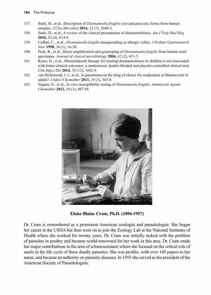
184 The Protozoa
157. Stark, D., et al., Description of Dientamoeba fragilis cyst and precystic forms from human samples. J Clin Microbiol 2014, 52 (7), 2680-3.
158. Stark, D., et al., A review of the clinical presentation of dientamoebiasis. Am J Trop Med Hyg 2010, 82 (4), 614-9.
159. Cuffari, C., et al., Dientamoeba fragilis masquerading as allergic colitis. J Pediatr Gastroenterol Nutr 1998, 26 (1), 16-20.
160. Peek, R., et al., Direct amplification and genotyping of Dientamoeba fragilis from human stool specimens. Journal of clinical microbiology 2004, 42 (2), 631-5.
161. Roser, D., et al., Metronidazole therapy for treating dientamoebiasis in children is not associated with better clinical outcomes: a randomized, double-blinded and placebo-controlled clinical trial. Clin Infect Dis 2014, 58 (12), 1692-9.
162. van Hellemond, J. J., et al., Is paromomycin the drug of choice for eradication of Blastocystis in adults? J Infect Chemother 2013, 19 (3), 545-8.
163. Nagata, N., et al., In vitro susceptibility testing of Dientamoeba fragilis. Antimicrob Agents Chemother 2012, 56 (1), 487-94.
Eloise Blaine Cram, Ph.D. (1896-1957)
Dr. Cram is remembered as a prominent American zoologist and parasitologist. She began her career at the USDA but then went on to join the Zoology Lab at the National Institutes of Health where she worked for twenty years. Dr. Cram was initially tasked with the problem of parasites in poultry and became world-renowned for her work in this area. Dr. Cram made her major contributions in the area of schistosomiasis where she focused on the critical role of snails in the life cycle of these deadly parasites. She was prolific, with over 160 papers to her name, and became an authority on parasitic diseases. In 1955 she served as the president of the American Society of Parasitologists.
15. Non-Pathogenic Protozoa 185
15. Non-Pathogenic Protozoa
Introduction
We are constantly confronted with a plethora of microbes whose sole purpose seems to be to colonize us and take advantage of our biochemical systems. The human body can be viewed as a series of ecological niches that select for numerous entities, including viruses, bacteria, fungi, protozoa, helminths, and arthropods. They enter through the gas-trointestinal (GI), urogenital, and respiratory tracts, through abrasions, and other portals of entry. Most of the world’s microbes are inca-pable of remaining on, or within, these envi-ronments and are repelled. This is mainly due to the inadequacy of their fundamental bio-logical makeup, preventing them from thriv-ing on or in us, the resiliency of our existing microbiome, and the intricate human immune system.1
The majority of those that take up residence do us little or no harm. In fact, by some esti-mates, the majority of cells on and in us are non-human! We refer to them collectively as our microbiome. Commensals do us no harm, and are just along for the ride, so to speak. Symbionts actively help maintain our homeostatic mechanisms. For example, the oral cavity harbors hundreds of different spe-cies of bacteria serving to exclude those that would lead to various states of ill health. Our intestinal tract is another good example of peaceful co-existence between our symbiotic microbes and us, harboring hundreds of spe-cies of “friendly” bacteria.2
A few that have managed to run the gaunt-let of our immune system and overcome the physiological barriers established by our complex metabolic regimes can, and often do, cause pathology leading to clinical condi-tions. This chapter is devoted to a brief men-tion of a few of those eukaryotic organisms
that we routinely harbor, and which do us no harm. The clinician may at some point receive a laboratory result with the name of one or more of these commensals on it. How these “hitchhiker” species should be approached in the context of the clinical setting is the subject of this brief chapter.
A number of commensal protozoans have been selected for life within us. Under unusual conditions, a few have been shown to be associated with disease. When a person is placed at risk from infection (e.g., sur-gery, immunosuppression, or infection with another pathogenic organism), some com-mensal organisms become opportunistic pathogens, growing and extending their terri-tory at the expense of our now compromised microbiome. At those times, the clinician has a difficult time determining who did what to whom. The diagnostic microbiology labora-tory now assumes a role of major importance, helping to catalogue microbes into the good, the bad, and the ugly. Resolving the primary cause of the disease often reverses the growth pattern of the opportunist. These organisms, including Entamoeba dispar and E. gingi-valis, are very rarely associated with actual infection.3-5
It is critical for the clinician to recognize that even though the organism reported is not a pathogen, it is potentially a marker of the patient’s exposure to a situation that may have led to the acquisition, or imbalance of another organism that may be pathogenic. The search should focus on all other agents transmitted by the same route. A representative of each organism mentioned in the following summa-ries can be found in Appendix C.
Commensal flagellated protozoa Trichomonas tenax (\trick-oh-MOAN-us\, \tri-kə-ˈmō-nəs\), T. hominis, Enteromonas hominis (\EN-ter-owe-MOW-nas\, \ent-ə-ˈrō-ˈmō-ˌnas\), Retortamonas intestinalis,
186 The Protozoa
and Chilomastix mesnili all only colonize the human host, and are considered nonpatho-genic by all standard criteria.6 T. tenax lives in the oral cavity in plaque and the rest are intestinal dwellers. Only C. mesnili has a rec-ognized cyst stage. All are considered amito-chondriate, aerotolerant anaerobic protists.7
Pathogenic outcomes have been described where heavy growth of T. tenax was found concurrently with abscesses and tumors of the oral cavity.8, 9 In addition, T. tenax has been isolated from cases of inhalation pneu-monia, and from pleural effusions from a patient in which ulceration of the esophagus resulted in communication with the pleural cavity. A PCR test for detecting T. tenax in dental plaque has been reported.10, 11 Due to the overwhelming number of people harbor-ing this flagellate who do not experience any discomfort, T. tenax remains on the list of commensals.
A case of E. hominis has been reported in which the patient experienced diarrhea and was treated successfully with metronida-zole.12 Neither R. intestinalis nor C. mesnili have ever been linked to any abnormal health condition.
Commensal amoebaeEntamoeba dispar (\ent-a-MEE-ba\, \en-tə-ˈmē-bə\) Entamoeba hartmanni, Entamoeba coli, Endolimax nana (\en-DOW-lie-MAKS\,
\ˈen-də-ˈlī-ˌmaks\), and Iodamoeba bütschlii are organisms often identified in routine stool examination. Their reporting often elic-its confusion among clinicians seeking the causes for diarrheal disease in their patients. Some (e.g., E. dispar or E. hartmanni) bear a resemblance to Entamoeba histolytica, especially to the inexperienced laboratory technician, who sometimes errs on the side of this pathogen rather than the commensal. Hence, the patient receives treatment for an entity that is not causing the problem. After “treatment”, the illness often recurs, and drug failure is blamed. These commensal amoe-bae do not respond to the standard drugs used to eradicate E. histolytica. The use of PCR is recommended for definitive diagnosis of the pathogenic amoebae.13 Another approach uses monoclonal antibodies to distinguish E. histolytica cysts from those of E. dispar and other commensal amoebae, facilitating their use in an antigen capture mode for routine diagnosis.14
Entamoeba polecki is an inhabitant of the gut tract of pigs that sometimes finds its way into humans, while Entamoeba gingivalis lives in the gingival flaps of a small subset of humans not yet defined, and is associated with, but does not cause, pyorrhea (periodontitis). E. gingivalis was diagnosed by fine needle aspi-ration of an abscess of the neck, following radiation therapy.15
References
1. Bull, M. J.; Plummer, N. T., Part 1: The Human Gut Microbiome in Health and Disease. Integr Med (Encinitas) 2014, 13 (6), 17-22.
2. Qin, J., et al., A human gut microbial gene catalogue established by metagenomic sequencing. Nature 2010, 464 (7285), 59-65.
3. Allison-Jones, E., et al., Entamoeba histolytica as a commensal intestinal parasite in homosexual men. 1986, 315, 353-6.
4. Haque, R., et al., Short report: detection of Entamoeba histolytica and E. dispar directly in stool. Am J Trop Med Hyg 1994, 50 (5), 595-6.
15. Non-Pathogenic Protozoa 187
5. Lucht, E., et al., Entamoeba gingivalis in human immunodeficiency virus type 1-infected patients with periodontal disease. Clin Inf Dis 1998, 27 (3), 471-3.
6. Aucott, J. N.; Ravdin, J. I., Amebiasis and “nonpathogenic” intestinal protozoa. Infectious disease clinics of North America 1993, 7 (3), 467-85.
7. Coombs, G. H., et al., Evolutionary Relationships Among Protozoa 1998; p 110-132.8. Duboucher, C., et al., Salivary trichomoniasis. A case report of infestation of a submaxillary
gland by Trichomonas tenax. Archives of pathology & laboratory medicine 1995, 119 (3), 277-9.9. Shiota, T., et al., Trichomonas tenax empyema in an immunocompromised patient with advanced
cancer. Parasite (Paris, France) 1998, 5 (4), 375-7.10. El Kamel, A., et al., Pulmonary eosinophilia due to Trichomonas tenax. Thorax 1996, 51 (5),
554-5.11. Kikuta, N., et al., Specific and sensitive detection of Trichomonas tenax by the polymerase chain
reaction. Lett Appl Microbiol 1999, 24 (3), 193-7.12. Spriegel, J. R., et al., Infectious diarrhea secondary to Enteromonas hominis. The American
journal of gastroenterology 1989, 84 (10), 1313-4.13. Acuna-Soto, R., et al., Application of the polymerase chain reaction to the epidemiology of
pathogenic and nonpathogenic Entamoeba histolytica. Am J Trop Med Hyg 1993, 48 (1), 58-70.14. Walderich, B., et al., Development of monoclonal antibodies specifically recognizing the cyst
stage of Entamoeba histolytica. Am J Trop Med Hyg 1998, 59 (3), 347-51.15. Perez-Jaffe, L., et al., Entamoeba gingivalis identified in a left upper neck nodule by fine-needle
aspiration: a case report. Diagnostic cytopathology 1998, 18 (6), 458-61.
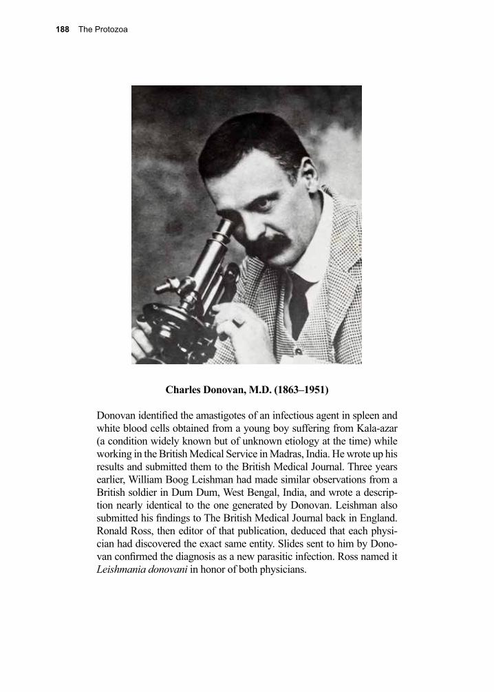
188 The Protozoa
Charles Donovan, M.D. (1863–1951)
Donovan identified the amastigotes of an infectious agent in spleen and white blood cells obtained from a young boy suffering from Kala-azar (a condition widely known but of unknown etiology at the time) while working in the British Medical Service in Madras, India. He wrote up his results and submitted them to the British Medical Journal. Three years earlier, William Boog Leishman had made similar observations from a British soldier in Dum Dum, West Bengal, India, and wrote a descrip-tion nearly identical to the one generated by Donovan. Leishman also submitted his findings to The British Medical Journal back in England. Ronald Ross, then editor of that publication, deduced that each physi-cian had discovered the exact same entity. Slides sent to him by Dono-van confirmed the diagnosis as a new parasitic infection. Ross named it Leishmania donovani in honor of both physicians.
The Nematodes 189
V. The Nematodes
Nematodes are non-segmented roundworms belonging to the phylum Nematoda and are among the most abundant life forms on earth. The great majority of nematodes are free-liv-ing, inhabiting most essential niches in soil, fresh, and saltwater, as well as other, more specialized ones. Only a small fraction of the total number of species is parasitic, and only some of these infect the human host. Most parasitic nematodes have developed a highly specific biologic dependence on a particular species of host, and are incapable of survival in any other. Only a few have succeeded in adapting to a variety of hosts.
Best known by far among the free-living nematodes is Caenorhabditis elegans, whose entire genome has been sequenced (20,512 genes). In contrast, the genome of Trichinella spiralis, a parasitic nematode, has more to-tal DNA than C. elegans, and only 60% of it is homologous with its free-living relative. There have only been 15,808 coding regions identified, implying that this parasite needs fewer, not more genes than its free-living rel-atives. Virulence factors, and other special-ized compounds needed to resist digestion or immune attack are likely to be encoded by genes that permit the invader to live comfort-ably in the face of an exquisitely developed immune system.
Infections caused by nematodes are among the most prevalent, affecting nearly all of us at one time in our lives. The most common nematodes are three types of soil-transmitted helminths (STHs), the common roundworm (Ascaris lumbricoides), the whipworm (Trichuris trichiura), and the hookworms (Necator americanus and Ancylostoma duo-denale). Children are particularly susceptible to acquiring large numbers of these parasites, and consequently suffer greater morbid-ity. In many developing countries, children
frequently harbor all three types of STHs (hence the moniker “the unholy trinity”) and suffer from childhood malnutrition, physical growth retardation, and deficits in cognitive and intellectual development.
The typical nematode, both larva and adult, is surrounded by a flexible, durable outer coating, the acellular cuticle, that is resistant to chemicals. It is a complex structure com-posed of a variety of layers, each of which has many components, including structural proteins, enzymes, and lipids. The cuticle of each species has a unique structure and composition; it not only protects the worm but may also be involved in active transport of small molecules, including water, elec-trolytes, and organic compounds. A further layer, the epicuticle, surrounds the cuticle of a few parasitic species, making them even more resistant to attack from enzymes, anti-bodies, and other host resistance factors.
All nematodes have a well-developed muscu-lar system. The longitudinal muscular system of nematodes lies just underneath the cuticle, and their origins and insertions are in cuticu-lar processes. In addition, there is some mus-cle tissue surrounding the buccal cavity and esophageal and sub-esophageal regions of the gut tract. These muscles are particularly important elements of the feeding apparatus in both parasitic and free-living nematodes. Each muscle cell consists of filaments, mi-tochondria, and cytoplasmic processes that connect it with a single nerve fiber. The ner-vous system consists of a dorsal nerve ring or a series of ganglia that give rise to the periph-eral nerves - two lateral, one dorsal, and one ventral branch. Commissures connect the branches and allow for integration of signal-ing, which results in fluid, serpiginous move-ments. Several classes of drugs interfere only with nematode nerve signaling and are thus effective treatments for nematode infections in humans.
190 The Nematodes
Nematodes have a complete, functional gut tract: the oral (i.e., buccal) cavity and esopha-gus, the midgut, and the hindgut with anus. The oral cavity and hindgut are usually lined by cuticle; the midgut consists of columnar cells, complete with microvilli. The function of the midgut is to absorb ingested nutrients, whereas the usually muscular esophagus serves to deliver food to the midgut.
In addition, a number of specialized exocrine glands open into the lumen of the digestive tract, usually in the region of the esophagus. These glands are thought to be largely con-cerned with digestion, but may be related to other functions as well. For example, in hookworms, the cephalic glands secrete an anticoagulant.
In other instances, there is a single row of cells called stichocytes that empty their products directly into the esophagus via a cuticular-lined duct. These cells occupy a large portion of the body mass of Trichinella, Trichuris, and Capillaria, for example. The function of these cells is not fully understood and may vary from species to species.
Nematodes excrete solid and fluid wastes. Excretion of solids takes place through the digestive tract. Fluids are eliminated by means of the excretory system, consisting of two or more collecting tubes connected at one end to the ventral gland (a primitive kidney-like organ) and at the other end to the excretory pore.
The adult female nematode has a large por-tion of her body devoted to reproduction. One or two ovaries lead to the vagina by way of a tubular oviduct and uterus. A seminal recep-tacle for storage of sperm is connected to the uterus. The male has a single testis connected to the vas deferens, seminal vesicle, ejacula-tory duct, and cloaca. In addition, males of many species have specialized structures to aid in transfer of sperm to the female during mating. Their identification is often based on morphology of these structures. Most nema-todes lay eggs, but some are viviparous. The biology of nematodes is discussed within the text for each infectious agent as they are con-sidered, whenever it relates to the pathogen-esis of the disease.
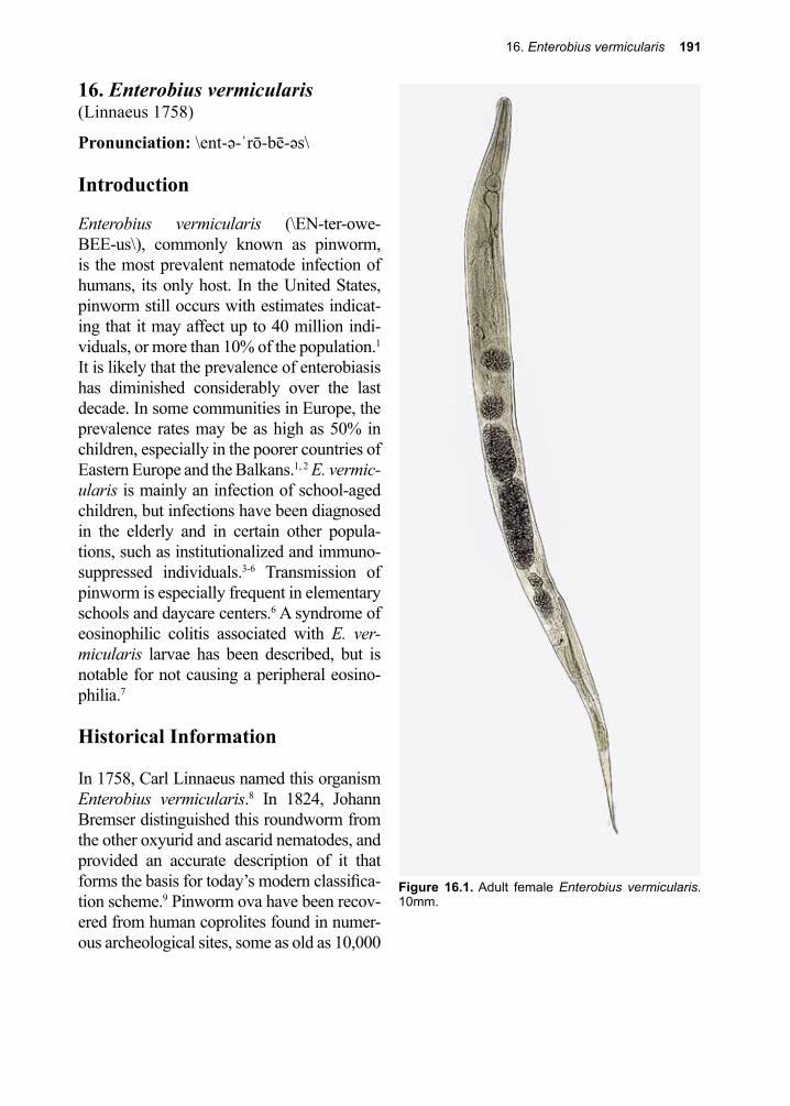
16. Enterobius vermicularis 191
Figure 16.1. Adult female Enterobius vermicularis. 10mm.
16. Enterobius vermicularis(Linnaeus 1758)
Pronunciation: \ent-ə-ˈrō-bē-əs\
Introduction
Enterobius vermicularis (\EN-ter-owe-BEE-us\), commonly known as pinworm, is the most prevalent nematode infection of humans, its only host. In the United States, pinworm still occurs with estimates indicat-ing that it may affect up to 40 million indi-viduals, or more than 10% of the population.1 It is likely that the prevalence of enterobiasis has diminished considerably over the last decade. In some communities in Europe, the prevalence rates may be as high as 50% in children, especially in the poorer countries of Eastern Europe and the Balkans.1, 2 E. vermic-ularis is mainly an infection of school-aged children, but infections have been diagnosed in the elderly and in certain other popula-tions, such as institutionalized and immuno-suppressed individuals.3-6 Transmission of pinworm is especially frequent in elementary schools and daycare centers.6 A syndrome of eosinophilic colitis associated with E. ver-micularis larvae has been described, but is notable for not causing a peripheral eosino-philia.7
Historical Information
In 1758, Carl Linnaeus named this organism Enterobius vermicularis.8 In 1824, Johann Bremser distinguished this roundworm from the other oxyurid and ascarid nematodes, and provided an accurate description of it that forms the basis for today’s modern classifica-tion scheme.9 Pinworm ova have been recov-ered from human coprolites found in numer-ous archeological sites, some as old as 10,000
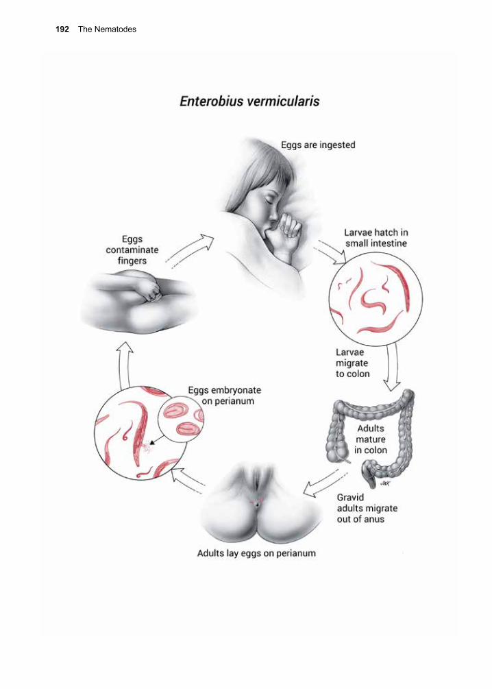
192 The Nematodes
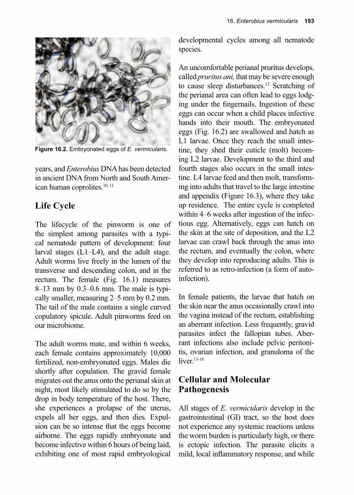
16. Enterobius vermicularis 193
years, and Enterobius DNA has been detected in ancient DNA from North and South Amer-ican human coprolites.10, 11 Life Cycle
The lifecycle of the pinworm is one of the simplest among parasites with a typi-cal nematode pattern of development: four larval stages (L1–L4), and the adult stage. Adult worms live freely in the lumen of the transverse and descending colon, and in the rectum. The female (Fig. 16.1) measures 8–13 mm by 0.3–0.6 mm. The male is typi-cally smaller, measuring 2–5 mm by 0.2 mm. The tail of the male contains a single curved copulatory spicule. Adult pinworms feed on our microbiome.
The adult worms mate, and within 6 weeks, each female contains approximately 10,000 fertilized, non-embryonated eggs. Males die shortly after copulation. The gravid female migrates out the anus onto the perianal skin at night, most likely stimulated to do so by the drop in body temperature of the host. There, she experiences a prolapse of the uterus, expels all her eggs, and then dies. Expul-sion can be so intense that the eggs become airborne. The eggs rapidly embryonate and become infective within 6 hours of being laid, exhibiting one of most rapid embryological
developmental cycles among all nematode species.
An uncomfortable perianal pruritus develops, called pruritus ani, that may be severe enough to cause sleep disturbances.12 Scratching of the perianal area can often lead to eggs lodg-ing under the fingernails. Ingestion of these eggs can occur when a child places infective hands into their mouth. The embryonated eggs (Fig. 16.2) are swallowed and hatch as L1 larvae. Once they reach the small intes-tine, they shed their cuticle (molt) becom-ing L2 larvae. Development to the third and fourth stages also occurs in the small intes-tine. L4 larvae feed and then molt, transform-ing into adults that travel to the large intestine and appendix (Figure 16.3), where they take up residence. The entire cycle is completed within 4–6 weeks after ingestion of the infec-tious egg. Alternatively, eggs can hatch on the skin at the site of deposition, and the L2 larvae can crawl back through the anus into the rectum, and eventually the colon, where they develop into reproducing adults. This is referred to as retro-infection (a form of auto-infection).
In female patients, the larvae that hatch on the skin near the anus occasionally crawl into the vagina instead of the rectum, establishing an aberrant infection. Less frequently, gravid parasites infect the fallopian tubes. Aber-rant infections also include pelvic peritoni-tis, ovarian infection, and granuloma of the liver.13-18
Cellular and Molecular Pathogenesis
All stages of E. vermicularis develop in the gastrointestinal (GI) tract, so the host does not experience any systemic reactions unless the worm burden is particularly high, or there is ectopic infection. The parasite elicits a mild, local inflammatory response, and while
Figure 16.2. Embryonated eggs of E. vermicularis.
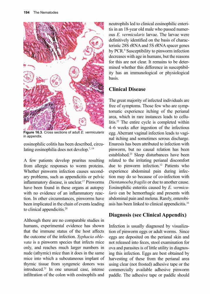
194 The Nematodes
eosinophilic colitis has been described, circu-lating eosinophilia does not develop.7, 19
A few patients develop pruritus resulting from allergic responses to worm proteins. Whether pinworm infection causes second-ary problems, such as appendicitis or pelvic inflammatory disease, is unclear.17 Pinworms have been found in these organs at autopsy with no evidence of an inflammatory reac-tion. In other circumstances, pinworms have been implicated in the chain of events leading to clinical appendicitis.20
Although there are no comparable studies in humans, experimental evidence has shown that the immune status of the host affects the outcome of the infection. Syphacia oble-vata is a pinworm species that infects mice only, and reaches much larger numbers in nude (athymic) mice than it does in the same mice into which a subcutaneous implant of thymic tissue from syngeneic donors was introduced.21 In one unusual case, intense infiltration of the colon with eosinophils and
neutrophils led to clinical eosinophilic enteri-tis in an 18-year old male who passed numer-ous E. vermicularis larvae. The larvae were definitively identified on the basis of charac-teristic 28S rRNA and 5S rRNA spacer genes by PCR.9 Susceptibility to pinworm infection decreases with age in humans, but the reasons for this are not clear. It remains to be deter-mined whether this difference in susceptibil-ity has an immunological or physiological basis.
Clinical Disease
The great majority of infected individuals are free of symptoms. Those few who are symp-tomatic experience itching of the perianal area, which in rare instances leads to cellu-litis.22 The entire cycle is completed within 4–6 weeks after ingestion of the infectious egg. Aberrant vaginal infection leads to vagi-nal itching and sometimes serous discharge. Enuresis has been attributed to infection with pinworm, but no causal relation has been established.23 Sleep disturbances have been related to the irritating perianal discomfort due to pinworm infection.12 Patients who experience abdominal pain during infec-tion may do so because of co-infection with Dientamoeba fragilis or due to another cause. Eosinophilic enteritis caused by E. vermicu-laris can be hemorrhagic and presents with abdominal pain and melena. Rarely, enterobi-asis has been linked to clinical appendicitis.16
Diagnosis (see Clinical Appendix)
Infection is usually diagnosed by visualiza-tion of pinworm eggs or adult worms. Since eggs are deposited on the perianal skin and not released into feces, stool examination for ova and parasites is of little utility in diagnos-ing this infection. Eggs are best obtained by harvesting of these from the perianal area using clear (not frosted) adhesive tape or the commercially available adhesive pinworm paddle. The adhesive tape or paddle should
Figure 16.3. Cross sections of adult E. vermicularis in appendix.
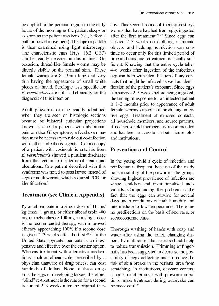
16. Enterobius vermicularis 195
be applied to the perianal region in the early hours of the morning as the patient sleeps or as soon as the patient awakens (i.e., before a bath or bowel movement). The tape or paddle is then examined using light microscopy. The characteristic eggs (Figs. 16.2, C.37) can be readily detected in this manner. On occasion, thread-like female worms may be directly visible on the perianal skin. These female worms are 8–13mm long and very thin having the appearance of small white pieces of thread. Serologic tests specific for E. vermicularis are not used clinically for the diagnosis of this infection.
Adult pinworms can be readily identified when they are seen on histologic sections because of bilateral cuticular projections known as alae. In patients with abdominal pain or other GI symptoms, a fecal examina-tion may be necessary to rule out co-infection with other infectious agents. Colonoscopy of a patient with eosinophilic enteritis from E. vermicularis showed a purulent discharge from the rectum to the terminal ileum and ulcerations. One patient described with this syndrome was noted to pass larvae instead of eggs or adult worms, which required PCR for identification.7
Treatment (see Clinical Appendix)
Pyrantel pamoate in a single dose of 11 mg/kg (max. 1 gram), or either albendazole 400 mg or mebendazole 100 mg in a single dose is the recommended therapy, with improved efficacy approaching 100% if a second dose is given 2–3 weeks after the first.24-27 In the United States pyrantel pamoate is an inex-pensive and effective over the counter option. Whereas treatment with alternative medica-tions, such as albendazole, prescribed by a physician unaware of drug prices, can cost hundreds of dollars. None of these drugs kills the eggs or developing larvae; therefore, “blind” re-treatment is the reason for a second treatment 2–3 weeks after the original ther-
apy. This second round of therapy destroys worms that have hatched from eggs ingested after the first treatment.24-27 Since eggs can survive 2–3 weeks on clothing, inanimate objects, and bedding, reinfection can con-tinue to occur only for this limited period of time and thus one retreatment is usually suf-ficient. Knowing that the entire cycle takes 4–6 weeks after ingestion of the infectious egg can help with identification of any con-tacts that might be infected as well as identi-fication of the patient’s exposure. Since eggs can survive 2–3 weeks before being ingested, the timing of exposure for an infected patient is 1–2 months prior to appearance of adult female worms capable of producing infec-tive eggs. Treatment of exposed contacts, all household members, and source patients, if not household members, is recommended and has been successful in both households and institutions.
Prevention and Control
In the young child a cycle of infection and reinfection is frequent, because of the ready transmissibility of the pinworm. The groups showing highest prevalence of infection are school children and institutionalized indi-viduals. Compounding the problem is the fact that the eggs can survive for several days under conditions of high humidity and intermediate to low temperatures. There are no predilections on the basis of sex, race, or socioeconomic class.
Thorough washing of hands with soap and water after using the toilet, changing dia-pers, by children or their carers should help to reduce transmission.3 Trimming of finger-nails has been suggested to decrease the pos-sibility of eggs collecting and to reduce the risk of skin breaks in the perianal area from scratching. In institutions, daycare centers, schools, or other areas with pinworm infec-tions, mass treatment during outbreaks can be successful.28
196 The Nematodes
References
1. Burkhart, C. N.; Burkhart, C. G., Assessment of frequency, transmission, and genitourinary complications of enterobiasis (pinworms). Int J Dermatol 2005, 44 (10), 837-40.
2. Hotez, P. J.; Gurwith, M., Europe’s neglected infections of poverty. Int J Infect Dis 2011, 15 (9), e611-9.
3. Snow, M., Pinning down pinworms. Nursing 2006, 36 (5), 17.4. Agholi, M., et al., HIV/AIDS-associated opportunistic protozoal diarrhea. AIDS Res Hum
Retroviruses 2013, 29 (1), 35-41.5. Schupf, N., et al., Prevalence of intestinal parasite infections among individuals with mental
retardation in New York State. Ment Retard 1995, 33 (2), 84-9.6. Crawford, F. G.; Vermund, S. H., Parasitic infections in day care centers. The Pediatric infectious
disease journal 1987, 6 (8), 744-9.7. Liu, L. X., et al., Eosinophilic colitis associated with larvae of the pinworm Enterobius
vermicularis. Lancet (London, England) 1995, 346 (8972), 410-2.8. Cox, F. E., History of human parasitology. Clin Microbiol Rev 2002, 15 (4), 595-612.9. Bremser, J. G., Oxyure vermiculaire. In Traite zoologique et physiologique sur les vers
intestinaux de lhomme CLF Panchouke Paris pp 1824, 149-157.10. Horne, P. D., First evidence of enterobiasis in ancient Egypt. J Parasitol 2002, 88 (5), 1019-21.11. Iniguez, A. M., et al., Enterobius vermicularis: ancient DNA from North and South American
human coprolites. Mem Inst Oswaldo Cruz 98 Suppl 2003, 1, 67-9.12. Jones, J. E., Pinworms. Am Fam Physician 1988, 38 (3), 159-64.13. Pearson, R. D.; Irons, R. P., Chronic pelvic peritonitis due to the pinworm Enterobius
vermicularis. JAMA 1981, 245 (13), 1340-1.14. Beckman, E. N.; Holland, J. B., Ovarian enterobiasis--a proposed pathogenesis. The American
journal of tropical medicine and hygiene 1981, 30 (1), 74-6.15. Daly, J. J.; Baker, G. F., Pinworm granuloma of the liver. The American journal of tropical
medicine and hygiene 1984, 33 (1), 62-4.16. Arca, M. J., et al., Clinical manifestations of appendiceal pinworms in children: an institutional
experience and a review of the literature. Pediatric surgery international 2004, 20 (5), 372-5.17. Lala, S.; Upadhyay, V., Enterobius vermicularis and its role in paediatric appendicitis: protection
or predisposition? ANZ J Surg 2016.18. Powell, G., et al., Enterobius vermicularis infection of the ovary. BMJ Case Rep 2013, 2013.19. Cacopardo, B., et al., Eosinophilic ileocolitis by Enterobius vermicularis: a description of two
rare cases. Ital J Gastroenterol Hepatol 1997, 29 (1), 51-3.20. Ahmed, M. U., et al., The Frequency of Enterobius Vermicularis Infections in Patients Diagnosed
With Acute Appendicitis in Pakistan. Glob J Health Sci 2015, 7 (5), 196-201.21. Jacobson, R. H.; Reed, N. D., The thymus dependency of resistance to pinworm infection in
mice. The Journal of parasitology 1974, 60 (6), 976-9.22. Ockert, G., [Epidemiology of Dientamoeba fragilis Jepps and Dobell 1918. 1. Spread of the
species in child collectives]. Journal of hygiene, epidemiology, microbiology, and immunology 1972, 16 (2), 213-21.
23. Hotez, P. J., The other intestinal protozoa: enteric infections caused by Blastocystis hominis, Entamoeba coli, and Dientamoeba fragilis. Sem Pedi Infect Dis 2000.
24. St. Georgiev, V., Opportunistic infections: treatment and developmental therapeutics of cryptosporidiosis and isosporiasis. Drug Develop Res 1993, 28, 445-59.
25. Wang, B. R., et al., Comparative efficacy of thienpydin, pyrantel pamoate, mebendazole and albendazole in treating ascariasis and enterobiasis. Chin Med J (Engl) 1987, 100 (11), 928-30.
26. Lormans, J. A., et al., Mebendazole (R 17635) in enterobiasis. A clinical trial in mental retardates. Chemotherapy 1975, 21 (3-4), 255-60.
27. St Georgiev, V., Chemotherapy of enterobiasis (oxyuriasis). Expert Opin Pharmacother 2001, 2 (2), 267-75.
28. Ashford, R. W., et al., Enterobius vermicularis infection in a children’s ward. J Hosp Infect 1988, 12 (3), 221-4.
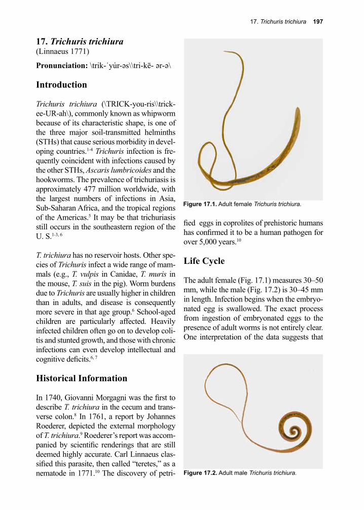
17. Trichuris trichiura 197
Figure 17.1. Adult female Trichuris trichiura.
Figure 17.2. Adult male Trichuris trichiura.
17. Trichuris trichiura(Linnaeus 1771)
Pronunciation: \trik-ˈyu̇r-əs\\tri-kē- ər-ə\
Introduction
Trichuris trichiura (\TRICK-you-ris\\trick-ee-UR-ah\), commonly known as whipworm because of its characteristic shape, is one of the three major soil-transmitted helminths (STHs) that cause serious morbidity in devel-oping countries.1-4 Trichuris infection is fre-quently coincident with infections caused by the other STHs, Ascaris lumbricoides and the hookworms. The prevalence of trichuriasis is approximately 477 million worldwide, with the largest numbers of infections in Asia, Sub-Saharan Africa, and the tropical regions of the Americas.5 It may be that trichuriasis still occurs in the southeastern region of the U. S.1-3, 6
T. trichiura has no reservoir hosts. Other spe-cies of Trichuris infect a wide range of mam-mals (e.g., T. vulpis in Canidae, T. muris in the mouse, T. suis in the pig). Worm burdens due to Trichuris are usually higher in children than in adults, and disease is consequently more severe in that age group.6 School-aged children are particularly affected. Heavily infected children often go on to develop coli-tis and stunted growth, and those with chronic infections can even develop intellectual and cognitive deficits.6, 7
Historical Information
In 1740, Giovanni Morgagni was the first to describe T. trichiura in the cecum and trans-verse colon.8 In 1761, a report by Johannes Roederer, depicted the external morphology of T. trichiura.9 Roederer’s report was accom-panied by scientific renderings that are still deemed highly accurate. Carl Linnaeus clas-sified this parasite, then called “teretes,” as a nematode in 1771.10 The discovery of petri-
fied eggs in coprolites of prehistoric humans has confirmed it to be a human pathogen for over 5,000 years.10
Life Cycle
The adult female (Fig. 17.1) measures 30–50 mm, while the male (Fig. 17.2) is 30–45 mm in length. Infection begins when the embryo-nated egg is swallowed. The exact process from ingestion of embryonated eggs to the presence of adult worms is not entirely clear. One interpretation of the data suggests that
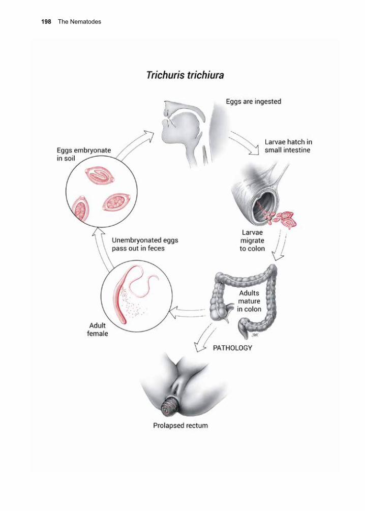
198 The Nematodes
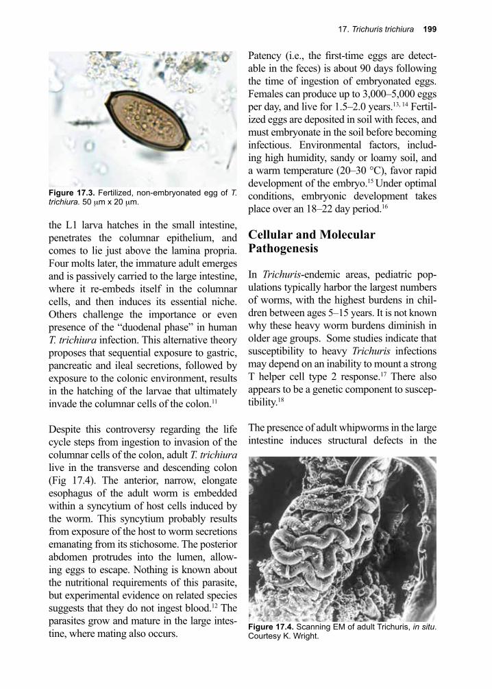
17. Trichuris trichiura 199
Figure 17.4. Scanning EM of adult Trichuris, in situ. Courtesy K. Wright.
Figure 17.3. Fertilized, non-embryonated egg of T. trichiura. 50 µm x 20 µm.
the L1 larva hatches in the small intestine, penetrates the columnar epithelium, and comes to lie just above the lamina propria. Four molts later, the immature adult emerges and is passively carried to the large intestine, where it re-embeds itself in the columnar cells, and then induces its essential niche. Others challenge the importance or even presence of the “duodenal phase” in human T. trichiura infection. This alternative theory proposes that sequential exposure to gastric, pancreatic and ileal secretions, followed by exposure to the colonic environment, results in the hatching of the larvae that ultimately invade the columnar cells of the colon.11
Despite this controversy regarding the life cycle steps from ingestion to invasion of the columnar cells of the colon, adult T. trichiura live in the transverse and descending colon (Fig 17.4). The anterior, narrow, elongate esophagus of the adult worm is embedded within a syncytium of host cells induced by the worm. This syncytium probably results from exposure of the host to worm secretions emanating from its stichosome. The posterior abdomen protrudes into the lumen, allow-ing eggs to escape. Nothing is known about the nutritional requirements of this parasite, but experimental evidence on related species suggests that they do not ingest blood.12 The parasites grow and mature in the large intes-tine, where mating also occurs.
Patency (i.e., the first-time eggs are detect-able in the feces) is about 90 days following the time of ingestion of embryonated eggs. Females can produce up to 3,000–5,000 eggs per day, and live for 1.5–2.0 years.13, 14 Fertil-ized eggs are deposited in soil with feces, and must embryonate in the soil before becoming infectious. Environmental factors, includ-ing high humidity, sandy or loamy soil, and a warm temperature (20–30 °C), favor rapid development of the embryo.15 Under optimal conditions, embryonic development takes place over an 18–22 day period.16
Cellular and Molecular Pathogenesis
In Trichuris-endemic areas, pediatric pop-ulations typically harbor the largest numbers of worms, with the highest burdens in chil-dren between ages 5–15 years. It is not known why these heavy worm burdens diminish in older age groups. Some studies indicate that susceptibility to heavy Trichuris infections may depend on an inability to mount a strong T helper cell type 2 response.17 There also appears to be a genetic component to suscep-tibility.18
The presence of adult whipworms in the large intestine induces structural defects in the
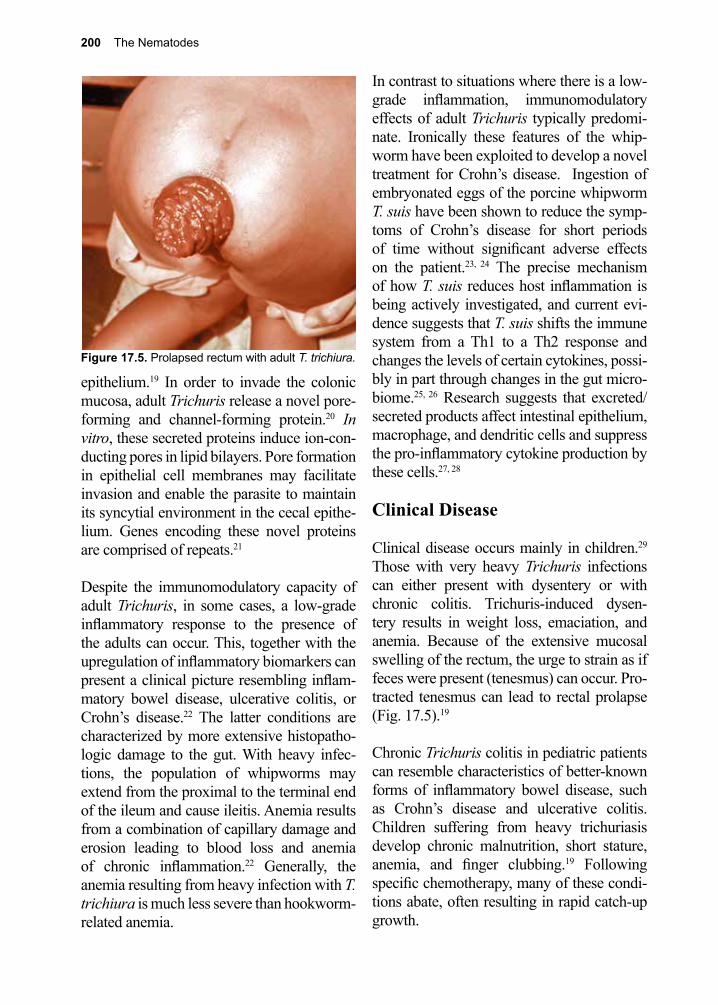
200 The Nematodes
Figure 17.5. Prolapsed rectum with adult T. trichiura.
epithelium.19 In order to invade the colonic mucosa, adult Trichuris release a novel pore-forming and channel-forming protein.20 In vitro, these secreted proteins induce ion-con-ducting pores in lipid bilayers. Pore formation in epithelial cell membranes may facilitate invasion and enable the parasite to maintain its syncytial environment in the cecal epithe-lium. Genes encoding these novel proteins are comprised of repeats.21
Despite the immunomodulatory capacity of adult Trichuris, in some cases, a low-grade inflammatory response to the presence of the adults can occur. This, together with the upregulation of inflammatory biomarkers can present a clinical picture resembling inflam-matory bowel disease, ulcerative colitis, or Crohn’s disease.22 The latter conditions are characterized by more extensive histopatho-logic damage to the gut. With heavy infec-tions, the population of whipworms may extend from the proximal to the terminal end of the ileum and cause ileitis. Anemia results from a combination of capillary damage and erosion leading to blood loss and anemia of chronic inflammation.22 Generally, the anemia resulting from heavy infection with T. trichiura is much less severe than hookworm-related anemia.
In contrast to situations where there is a low-grade inflammation, immunomodulatory effects of adult Trichuris typically predomi-nate. Ironically these features of the whip-worm have been exploited to develop a novel treatment for Crohn’s disease. Ingestion of embryonated eggs of the porcine whipworm T. suis have been shown to reduce the symp-toms of Crohn’s disease for short periods of time without significant adverse effects on the patient.23, 24 The precise mechanism of how T. suis reduces host inflammation is being actively investigated, and current evi-dence suggests that T. suis shifts the immune system from a Th1 to a Th2 response and changes the levels of certain cytokines, possi-bly in part through changes in the gut micro-biome.25, 26 Research suggests that excreted/secreted products affect intestinal epithelium, macrophage, and dendritic cells and suppress the pro-inflammatory cytokine production by these cells.27, 28
Clinical Disease
Clinical disease occurs mainly in children.29 Those with very heavy Trichuris infections can either present with dysentery or with chronic colitis. Trichuris-induced dysen-tery results in weight loss, emaciation, and anemia. Because of the extensive mucosal swelling of the rectum, the urge to strain as if feces were present (tenesmus) can occur. Pro-tracted tenesmus can lead to rectal prolapse (Fig. 17.5).19
Chronic Trichuris colitis in pediatric patients can resemble characteristics of better-known forms of inflammatory bowel disease, such as Crohn’s disease and ulcerative colitis. Children suffering from heavy trichuriasis develop chronic malnutrition, short stature, anemia, and finger clubbing.19 Following specific chemotherapy, many of these condi-tions abate, often resulting in rapid catch-up growth.
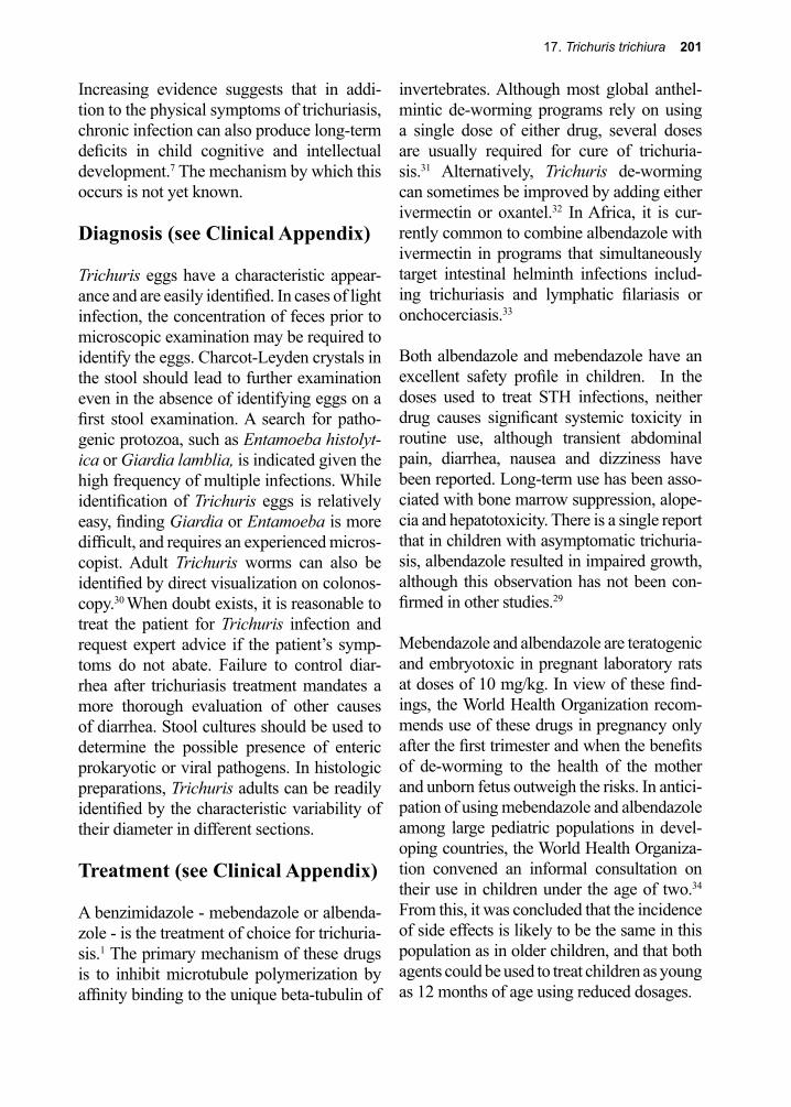
17. Trichuris trichiura 201
Increasing evidence suggests that in addi-tion to the physical symptoms of trichuriasis, chronic infection can also produce long-term deficits in child cognitive and intellectual development.7 The mechanism by which this occurs is not yet known.
Diagnosis (see Clinical Appendix)
Trichuris eggs have a characteristic appear-ance and are easily identified. In cases of light infection, the concentration of feces prior to microscopic examination may be required to identify the eggs. Charcot-Leyden crystals in the stool should lead to further examination even in the absence of identifying eggs on a first stool examination. A search for patho-genic protozoa, such as Entamoeba histolyt-ica or Giardia lamblia, is indicated given the high frequency of multiple infections. While identification of Trichuris eggs is relatively easy, finding Giardia or Entamoeba is more difficult, and requires an experienced micros-copist. Adult Trichuris worms can also be identified by direct visualization on colonos-copy.30 When doubt exists, it is reasonable to treat the patient for Trichuris infection and request expert advice if the patient’s symp-toms do not abate. Failure to control diar-rhea after trichuriasis treatment mandates a more thorough evaluation of other causes of diarrhea. Stool cultures should be used to determine the possible presence of enteric prokaryotic or viral pathogens. In histologic preparations, Trichuris adults can be readily identified by the characteristic variability of their diameter in different sections.
Treatment (see Clinical Appendix)
A benzimidazole - mebendazole or albenda-zole - is the treatment of choice for trichuria-sis.1 The primary mechanism of these drugs is to inhibit microtubule polymerization by affinity binding to the unique beta-tubulin of
invertebrates. Although most global anthel-mintic de-worming programs rely on using a single dose of either drug, several doses are usually required for cure of trichuria-sis.31 Alternatively, Trichuris de-worming can sometimes be improved by adding either ivermectin or oxantel.32 In Africa, it is cur-rently common to combine albendazole with ivermectin in programs that simultaneously target intestinal helminth infections includ-ing trichuriasis and lymphatic filariasis or onchocerciasis.33
Both albendazole and mebendazole have an excellent safety profile in children. In the doses used to treat STH infections, neither drug causes significant systemic toxicity in routine use, although transient abdominal pain, diarrhea, nausea and dizziness have been reported. Long-term use has been asso-ciated with bone marrow suppression, alope-cia and hepatotoxicity. There is a single report that in children with asymptomatic trichuria-sis, albendazole resulted in impaired growth, although this observation has not been con-firmed in other studies.29
Mebendazole and albendazole are teratogenic and embryotoxic in pregnant laboratory rats at doses of 10 mg/kg. In view of these find-ings, the World Health Organization recom-mends use of these drugs in pregnancy only after the first trimester and when the benefits of de-worming to the health of the mother and unborn fetus outweigh the risks. In antici-pation of using mebendazole and albendazole among large pediatric populations in devel-oping countries, the World Health Organiza-tion convened an informal consultation on their use in children under the age of two.34
From this, it was concluded that the incidence of side effects is likely to be the same in this population as in older children, and that both agents could be used to treat children as young as 12 months of age using reduced dosages.
202 The Nematodes
Prevention and Control
T. trichiura infection is common in tropical areas, where in certain communities over 90% of the population is infected.35 Most infections are light and asymptomatic. Warm, moist soils in tropical and subtropical regions favor the maintenance of eggs, which can remain alive for months under these optimum conditions. As with Ascaris eggs, exposure of T. trichiura eggs to direct sunlight for 12 hours or exposure to temperatures in excess of 40 °C for 1 hour kills the embryo inside the egg. Eggs are relatively resistant to chemical disinfectants and can survive for protracted periods of time in raw or treated sewage. Proper disposal of feces is the primary means of prevention. In areas of the world where untreated human feces are used to fertilize crops, thermophilic composting may be a rel-atively low tech approach to destroying these helminth eggs.36
Because school-aged children typically harbor the heaviest Trichuris (and Asca-ris) infections, and specific anthelmintic
chemotherapy with either albendazole or mebendazole can result in catch-up growth and improved cognition for heavily infected individuals, these agents have been used in school-based programs throughout the devel-oping world.6, 37, 38 In 2001, the World Health Assembly passed a resolution that recom-mended its member states administer single dose albendazole and mebendazole on a fre-quent and periodic basis (1–3 times per year) in order to control STHs’ (Ascaris, Trichuris, hookworm) morbidity. Because school-aged children contribute the most to Trichuris and Ascaris transmission in the community, there is also some optimism that widespread treatment could theoretically interrupt trans-mission. High rates of post-treatment STH re-infection require that children must be treated at least on an annual basis. While there are clear health and educational ben-efits for school-based intervention, there are concerns that single doses of albendazole or mebendazole are often not sufficient to cure Trichuris infections, so that the addition of ivermectin or oxantel may be warranted.
References
1. Keiser, J.; Utzinger, J., Efficacy of current drugs against soil-transmitted helminth infections: systematic review and meta-analysis. JAMA 2008, 299 (16), 1937-48.
2. Bethony, J., et al., Soil-transmitted helminth infections: ascariasis, trichuriasis, and hookworm. Lancet 2006, 367 (9521), 1521-32.
3. Lammie, P. J., et al., A blueprint for success: integration of neglected tropical disease control programmes. Trends Parasitol 2006, 22 (7), 313-21.
4. Webster, J. P., et al., The contribution of mass drug administration to global health: past, present and future. Philos Trans R Soc Lond B Biol Sci 2014, 369 (1645), 20130434.
5. Global Burden of Disease Study, C., Global, regional, and national incidence, prevalence, and years lived with disability for 301 acute and chronic diseases and injuries in 188 countries, 1990-2013: a systematic analysis for the Global Burden of Disease Study 2013. Lancet 2015, 386 (9995), 743-800.
6. Piraja de Silva, M. A., Contribucao para o estudo da schistosomiasena Bahia. Brazil Med 1908, 2 281-283.
7. Cooper, E. S.; Bundy, D. A., Trichuris is not trivial. Parasitology today (Personal ed.) 1988, 4 (11), 301-6.
8. Nokes, C., et al., Moderate to heavy infections of Trichuris trichiura affect cognitive function in Jamaican school children. Parasitology 1992, 104 ( Pt 3), 539-47.
9. Morgagni, G. B.; Anatomica, X. I. V., Epistolarum anatomicarum duodeviginti ad script pertinentium celeberrimi yin Antonii Marie Valsalvae pars Altera. Epistola Apud Franciscum
17. Trichuris trichiura 203
Pitheri Venice 1740.10. Cox, F. E., History of human parasitology. Clin Microbiol Rev 2002, 15 (4), 595-612.11. Bundy, D. A.; Cooper, E. S., Trichuris and trichuriasis in humans. Adv Parasitol 1989, 28, 107-
73.12. Roederer, J. G., Nachrichten von der Trichuriden, der Societat der Wissenschaften in Gottingen.
Gottingische Anzeigen von gelebrten Sachen Unter der Aufsicht der Konigliche Gesellschaft der Wissenschaften Part 1761, 25, 243-245.
13. Pike, E. H., Bionomics, blood and 51Cr: investigations of Trichuris muris and studies with two related species. Doctoral dissertation Columbia University pp 1963, 1-207.
14. Brown, H. W.; West, D., The Whipworm of Man (Seminar). Merck Sharp and PA 1954, 16, 19-22.
15. Belding, D.; New, M., Textbook of Parasitology. pp 1965, 397-398.16. Brown, H. W., Studies on the rate of development and viability of the eggs of Ascaris
lumbricoides and Trichuris trichiura under field conditions. Parasitology 1927, 14, 1-15.17. Jackson, J. A., et al., T helper cell type 2 responsiveness predicts future susceptibility to
gastrointestinal nematodes in humans. The Journal of infectious diseases 2004, 190 (10), 1804-11.
18. Williams-Blangero, S., et al., Genetic component to susceptibility to Trichuris trichiura: evidence from two Asian populations. Genetic epidemiology 2002, 22 (3), 254-64.
19. MacDonald, T. T., et al., Histopathology and immunohistochemistry of the caecum in children with the Trichuris dysentery syndrome. Journal of clinical pathology 1991, 44 (3), 194-9.
20. Drake, L., et al., The major secreted product of the whipworm, Trichuris, is a pore-forming protein. Proceedings. Biological sciences / The Royal Society 1994, 257 (1350), 255-61.
21. Bennett, A. B., et al., A beta-tubulin gene from Trichuris trichiura. Molecular and biochemical parasitology 1999, 103 (1), 111-6.
22. Bundy, D. A.; Cooper, E. S., Trichuris and trichuriasis in humans. Advances in parasitology 1989, 28, 107-73.
23. Summers, R. W., et al., Trichuris suis therapy in Crohn’s disease. Gut 2005, 54 (1), 87-90.24. Sandborn, W. J., et al., Randomised clinical trial: the safety and tolerability of Trichuris suis ova
in patients with Crohn’s disease. Aliment Pharmacol Ther 2013, 38 (3), 255-63.25. Reddy, A.; Fried, B., The use of Trichuris suis and other helminth therapies to treat Crohn’s
disease. Parasitol Res 2007, 100 (5), 921-7.26. Ramanan, D., et al., Helminth infection promotes colonization resistance via type 2 immunity.
Science 2016, 352 (6285), 608-12.27. Hiemstra, I. H., et al., Excreted/secreted Trichuris suis products reduce barrier function and
suppress inflammatory cytokine production of intestinal epithelial cells. Mol Immunol 2014, 60 (1), 1-7.
28. Ottow, M. K., et al., The helminth Trichuris suis suppresses TLR4-induced inflammatory responses in human macrophages. Genes Immun 2014, 15 (7), 477-86.
29. Gilman, R. H., et al., The adverse consequences of heavy Trichuris infection. Transactions of the Royal Society of Tropical Medicine and Hygiene 1983, 77 (4), 432-8.
30. Joo, J. H., et al., Colonoscopic diagnosis of whipworm infection. Hepato-gastroenterology 1998, 45 (24), 2105-9.
31. Sirivichayakul, C., et al., The effectiveness of 3, 5 or 7 days of albendazole for the treatment of Trichuris trichiura infection. Annals of tropical medicine and parasitology 2003, 97 (8), 847-53.
32. Speich, B., et al., Oxantel pamoate-albendazole for Trichuris trichiura infection. N Engl J Med 2014, 370 (7), 610-20.
33. Hotez, P. J., Mass drug administration and integrated control for the world’s high-prevalence neglected tropical diseases. Clin Pharmacol Ther 2009, 85 (6), 659-64.
34. Montresor, A., et al., Use of benzimidazoles in children younger than 24 months for the treatment of soil-transmitted helminthiasis. Acta tropica 2003, 86 (2-3), 223-32.
35. Needham, C., et al., Epidemiology of soil-transmitted nematode infections in Ha Nam Province, Vietnam. Trop Med Int Health 1998, 3 (11), 904-12.
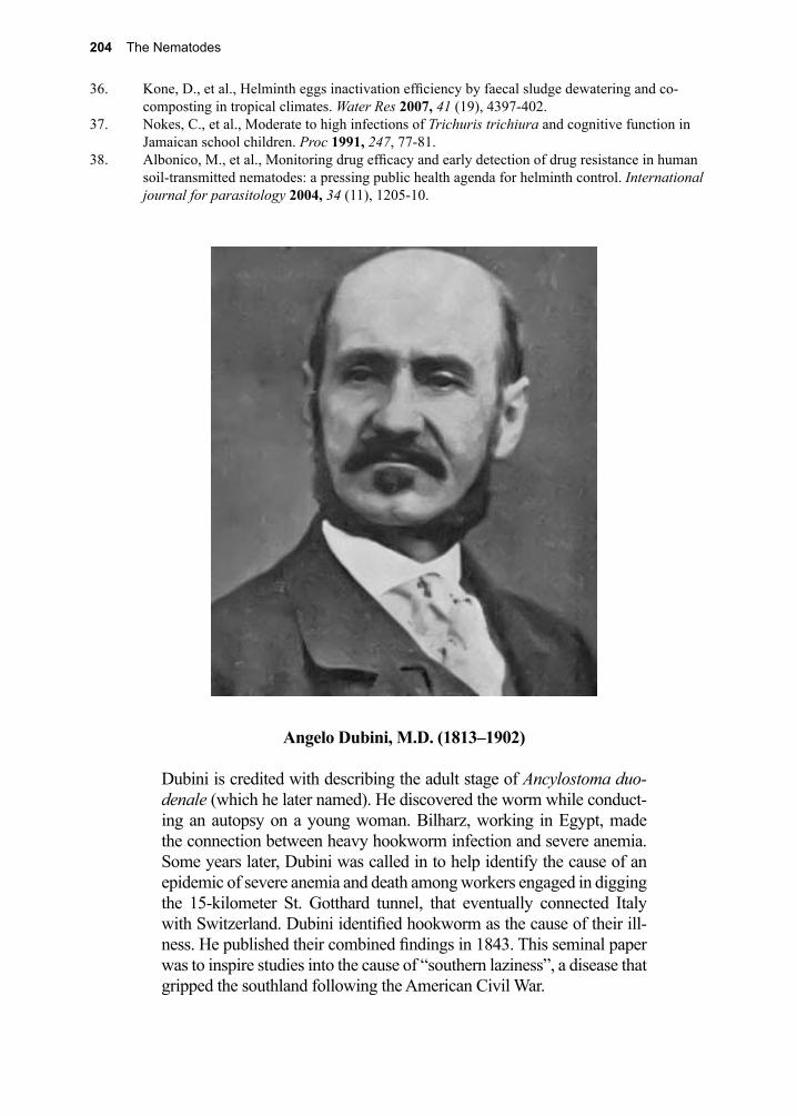
204 The Nematodes
Angelo Dubini, M.D. (1813–1902)
Dubini is credited with describing the adult stage of Ancylostoma duo-denale (which he later named). He discovered the worm while conduct-ing an autopsy on a young woman. Bilharz, working in Egypt, made the connection between heavy hookworm infection and severe anemia. Some years later, Dubini was called in to help identify the cause of an epidemic of severe anemia and death among workers engaged in digging the 15-kilometer St. Gotthard tunnel, that eventually connected Italy with Switzerland. Dubini identified hookworm as the cause of their ill-ness. He published their combined findings in 1843. This seminal paper was to inspire studies into the cause of “southern laziness”, a disease that gripped the southland following the American Civil War.
36. Kone, D., et al., Helminth eggs inactivation efficiency by faecal sludge dewatering and co-composting in tropical climates. Water Res 2007, 41 (19), 4397-402.
37. Nokes, C., et al., Moderate to high infections of Trichuris trichiura and cognitive function in Jamaican school children. Proc 1991, 247, 77-81.
38. Albonico, M., et al., Monitoring drug efficacy and early detection of drug resistance in human soil-transmitted nematodes: a pressing public health agenda for helminth control. International journal for parasitology 2004, 34 (11), 1205-10.
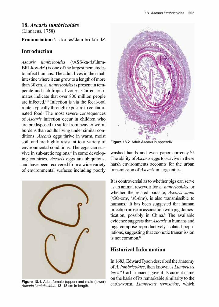
18. Ascaris lumbricoides 205
Figure 18.1. Adult female (upper) and male (lower) Ascaris lumbricoides. 13–18 cm in length.
Figure 18.2. Adult Ascaris in appendix.
18. Ascaris lumbricoides(Linnaeus, 1758)
Pronunciation: \as-kə-rəs\\ləm-bri-kȯi-dz\
Introduction
Ascaris lumbricoides (\ASS-ka-ris\\lum-BRI-koy-dz\) is one of the largest nematodes to infect humans. The adult lives in the small intestine where it can grow to a length of more than 30 cm. A. lumbricoides is present in tem-perate and sub-tropical zones. Current esti-mates indicate that over 800 million people are infected.1-3 Infection is via the fecal-oral route, typically through exposure to contami-nated food. The most severe consequences of Ascaris infection occur in children who are predisposed to suffer from heavier worm burdens than adults living under similar con-ditions. Ascaris eggs thrive in warm, moist soil, and are highly resistant to a variety of environmental conditions. The eggs can sur-vive in sub-arctic regions.4 In some develop-ing countries, Ascaris eggs are ubiquitous, and have been recovered from a wide variety of environmental surfaces including poorly
washed hands and even paper currency.5, 6 The ability of Ascaris eggs to survive in these harsh environments accounts for the urban transmission of Ascaris in large cities.
It is controversial as to whether pigs can serve as an animal reservoir for A. lumbricoides, or whether the related parasite, Ascaris suum (\SO-om\, \su̇-u̇m\), is also transmissible to humans.7 It has been suggested that human infection arose in association with pig domes-tication, possibly in China.8 The available evidence suggests that Ascaris in humans and pigs comprise reproductively isolated popu-lations, suggesting that zoonotic transmission is not common.8
Historical Information
In 1683, Edward Tyson described the anatomy of A. lumbricoides, then known as Lumbricus teres.9 Carl Linnaeus gave it its current name on the basis of its remarkable similarity to the earth-worm, Lumbricus terrestrias, which
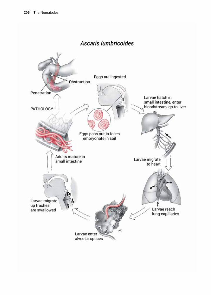
206 The Nematodes
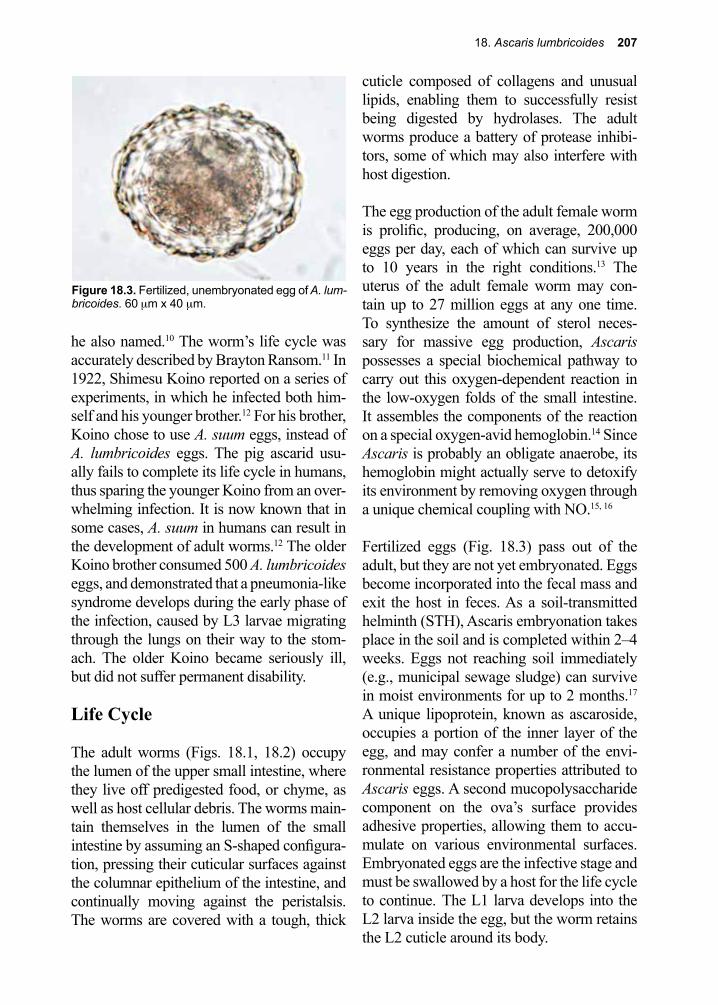
18. Ascaris lumbricoides 207
Figure 18.3. Fertilized, unembryonated egg of A. lum-bricoides. 60 µm x 40 µm.
he also named.10 The worm’s life cycle was accurately described by Brayton Ransom.11 In 1922, Shimesu Koino reported on a series of experiments, in which he infected both him-self and his younger brother.12 For his brother, Koino chose to use A. suum eggs, instead of A. lumbricoides eggs. The pig ascarid usu-ally fails to complete its life cycle in humans, thus sparing the younger Koino from an over-whelming infection. It is now known that in some cases, A. suum in humans can result in the development of adult worms.12 The older Koino brother consumed 500 A. lumbricoides eggs, and demonstrated that a pneumonia-like syndrome develops during the early phase of the infection, caused by L3 larvae migrating through the lungs on their way to the stom-ach. The older Koino became seriously ill, but did not suffer permanent disability.
Life Cycle
The adult worms (Figs. 18.1, 18.2) occupy the lumen of the upper small intestine, where they live off predigested food, or chyme, as well as host cellular debris. The worms main-tain themselves in the lumen of the small intestine by assuming an S-shaped configura-tion, pressing their cuticular surfaces against the columnar epithelium of the intestine, and continually moving against the peristalsis. The worms are covered with a tough, thick
cuticle composed of collagens and unusual lipids, enabling them to successfully resist being digested by hydrolases. The adult worms produce a battery of protease inhibi-tors, some of which may also interfere with host digestion.
The egg production of the adult female worm is prolific, producing, on average, 200,000 eggs per day, each of which can survive up to 10 years in the right conditions.13 The uterus of the adult female worm may con-tain up to 27 million eggs at any one time. To synthesize the amount of sterol neces-sary for massive egg production, Ascaris possesses a special biochemical pathway to carry out this oxygen-dependent reaction in the low-oxygen folds of the small intestine. It assembles the components of the reaction on a special oxygen-avid hemoglobin.14 Since Ascaris is probably an obligate anaerobe, its hemoglobin might actually serve to detoxify its environment by removing oxygen through a unique chemical coupling with NO.15, 16
Fertilized eggs (Fig. 18.3) pass out of the adult, but they are not yet embryonated. Eggs become incorporated into the fecal mass and exit the host in feces. As a soil-transmitted helminth (STH), Ascaris embryonation takes place in the soil and is completed within 2–4 weeks. Eggs not reaching soil immediately (e.g., municipal sewage sludge) can survive in moist environments for up to 2 months.17 A unique lipoprotein, known as ascaroside, occupies a portion of the inner layer of the egg, and may confer a number of the envi-ronmental resistance properties attributed to Ascaris eggs. A second mucopolysaccharide component on the ova’s surface provides adhesive properties, allowing them to accu-mulate on various environmental surfaces. Embryonated eggs are the infective stage and must be swallowed by a host for the life cycle to continue. The L1 larva develops into the L2 larva inside the egg, but the worm retains the L2 cuticle around its body.
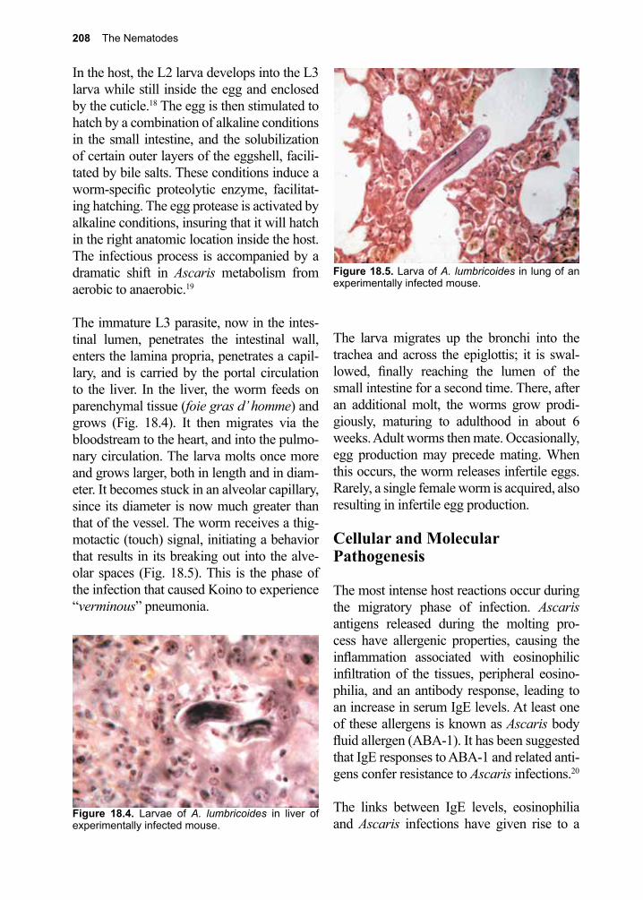
208 The Nematodes
Figure 18.5. Larva of A. lumbricoides in lung of an experimentally infected mouse.
In the host, the L2 larva develops into the L3 larva while still inside the egg and enclosed by the cuticle.18 The egg is then stimulated to hatch by a combination of alkaline conditions in the small intestine, and the solubilization of certain outer layers of the eggshell, facili-tated by bile salts. These conditions induce a worm-specific proteolytic enzyme, facilitat-ing hatching. The egg protease is activated by alkaline conditions, insuring that it will hatch in the right anatomic location inside the host. The infectious process is accompanied by a dramatic shift in Ascaris metabolism from aerobic to anaerobic.19
The immature L3 parasite, now in the intes-tinal lumen, penetrates the intestinal wall, enters the lamina propria, penetrates a capil-lary, and is carried by the portal circulation to the liver. In the liver, the worm feeds on parenchymal tissue (foie gras d’ homme) and grows (Fig. 18.4). It then migrates via the bloodstream to the heart, and into the pulmo-nary circulation. The larva molts once more and grows larger, both in length and in diam-eter. It becomes stuck in an alveolar capillary, since its diameter is now much greater than that of the vessel. The worm receives a thig-motactic (touch) signal, initiating a behavior that results in its breaking out into the alve-olar spaces (Fig. 18.5). This is the phase of the infection that caused Koino to experience “verminous” pneumonia.
The larva migrates up the bronchi into the trachea and across the epiglottis; it is swal-lowed, finally reaching the lumen of the small intestine for a second time. There, after an additional molt, the worms grow prodi-giously, maturing to adulthood in about 6 weeks. Adult worms then mate. Occasionally, egg production may precede mating. When this occurs, the worm releases infertile eggs. Rarely, a single female worm is acquired, also resulting in infertile egg production.
Cellular and Molecular Pathogenesis
The most intense host reactions occur during the migratory phase of infection. Ascaris antigens released during the molting pro-cess have allergenic properties, causing the inflammation associated with eosinophilic infiltration of the tissues, peripheral eosino-philia, and an antibody response, leading to an increase in serum IgE levels. At least one of these allergens is known as Ascaris body fluid allergen (ABA-1). It has been suggested that IgE responses to ABA-1 and related anti-gens confer resistance to Ascaris infections.20
The links between IgE levels, eosinophilia and Ascaris infections have given rise to a
Figure 18.4. Larvae of A. lumbricoides in liver of experimentally infected mouse.
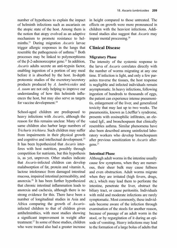
18. Ascaris lumbricoides 209
number of hypotheses to explain the impact of helminth infections such as ascariasis on the atopic state of the host. Among them is the notion that atopy evolved as an adaptive mechanism to promote resistance to hel-minths.21 During migration Ascaris larvae trigger allergic responses in the lungs that resemble the pathogenesis of asthma.22 Both processes may be linked to polymorphisms of the β-2-adrenoreceptor gene.23 In addition, Ascaris adults secrete an anti-trypsin factor, enabling ingestion of a portion of any meal before it is absorbed by the host. In-depth proteomic studies of the excretory/secretory products produced by A. lumbricoides and A. suum are not only helping to improve our understanding of how this helminth influ-ences the host, but may also serve as targets for vaccine development.24
School-aged children are predisposed to heavy infections with Ascaris, although the reason for this remains unclear. Many of the same children also harbor large numbers of Trichuris trichiura. Such children may suffer from impairments in their physical growth and cognitive and intellectual development.25 It has been hypothesized that Ascaris inter-feres with host nutrition, possibly through competition for nutrients, but this hypothesis is, as yet, unproven. Other studies indicate that Ascaris-infected children can develop malabsorption of fat, protein and vitamin A, lactose intolerance from damaged intestinal mucosa, impaired intestinal permeability, and anorexia.26 It has been further hypothesized that chronic intestinal inflammation leads to anorexia and cachexia, although there is no strong evidence for this. There have been a number of longitudinal studies in Asia and Africa comparing the growth of Ascaris-infected children to that of children given antihelminthics, with most studies showing a significant improvement in weight after treatment.27 In some of these studies, children who were treated also had a greater increase
in height compared to those untreated. The effects on growth were more pronounced in children with the heaviest infections. Addi-tional studies also suggest that Ascaris may impair mental processing.28
Clinical Disease
Migratory PhaseThe intensity of the systemic response to the larva of Ascaris correlates directly with the number of worms migrating at any one time. If infection is light, and only a few par-asites traverse the tissues, the host response is negligible and infected individuals remain asymptomatic. In heavy infections, following ingestion of hundreds to thousands of eggs, the patient can experience intense pneumoni-tis, enlargement of the liver, and generalized toxicity that may last up to two weeks. The pneumonitis, known as Löeffler’s syndrome, presents with eosinophilic infiltrates, an ele-vated IgE, and bronchospasm that clinically resembles asthma. Similar phenomena have also been described among uninfected labo-ratory workers who develop bronchospasm after previous sensitization to Ascaris aller-gens.21
Intestinal PhaseAlthough adult worms in the intestine usually cause few symptoms, when they are numer-ous, their sheer bulk may cause fullness and even obstruction. Adult worms migrate when they are irritated (high fevers, drugs, etc.), which may lead them to perforate the intestine, penetrate the liver, obstruct the biliary tract, or cause peritonitis. Individuals with mild and moderate infections are rarely symptomatic. Most commonly, these individ-uals become aware of the infection through examination of the stools for another reason, because of passage of an adult worm in the stool, or by regurgitation of it during an epi-sode of vomiting. Heavy infections may lead to the formation of a large bolus of adults that
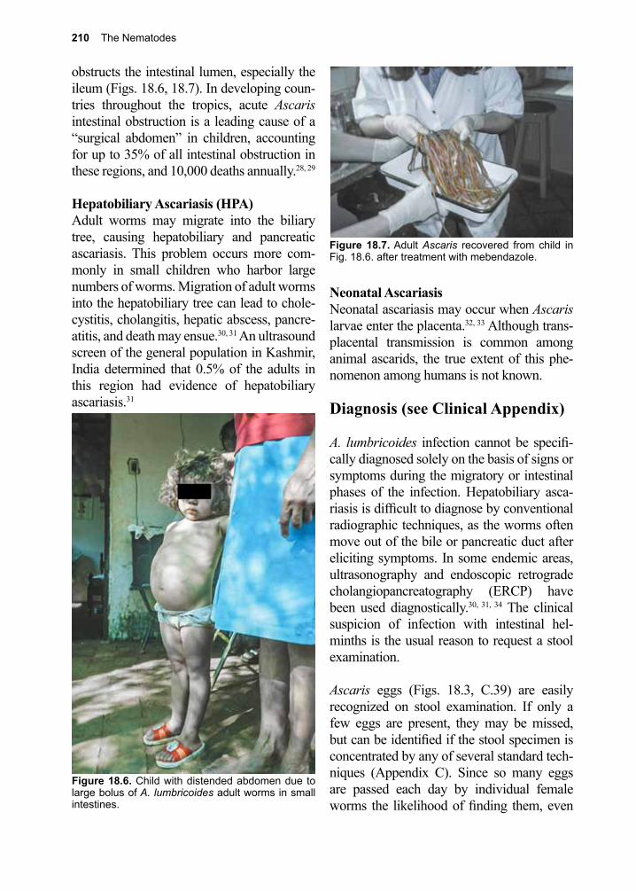
210 The Nematodes
obstructs the intestinal lumen, especially the ileum (Figs. 18.6, 18.7). In developing coun-tries throughout the tropics, acute Ascaris intestinal obstruction is a leading cause of a “surgical abdomen” in children, accounting for up to 35% of all intestinal obstruction in these regions, and 10,000 deaths annually.28, 29
Hepatobiliary Ascariasis (HPA)Adult worms may migrate into the biliary tree, causing hepatobiliary and pancreatic ascariasis. This problem occurs more com-monly in small children who harbor large numbers of worms. Migration of adult worms into the hepatobiliary tree can lead to chole-cystitis, cholangitis, hepatic abscess, pancre-atitis, and death may ensue.30, 31 An ultrasound screen of the general population in Kashmir, India determined that 0.5% of the adults in this region had evidence of hepatobiliary ascariasis.31
Neonatal AscariasisNeonatal ascariasis may occur when Ascaris larvae enter the placenta.32, 33 Although trans-placental transmission is common among animal ascarids, the true extent of this phe-nomenon among humans is not known.
Diagnosis (see Clinical Appendix)
A. lumbricoides infection cannot be specifi-cally diagnosed solely on the basis of signs or symptoms during the migratory or intestinal phases of the infection. Hepatobiliary asca-riasis is difficult to diagnose by conventional radiographic techniques, as the worms often move out of the bile or pancreatic duct after eliciting symptoms. In some endemic areas, ultrasonography and endoscopic retrograde cholangiopancreatography (ERCP) have been used diagnostically.30, 31, 34 The clinical suspicion of infection with intestinal hel-minths is the usual reason to request a stool examination.
Ascaris eggs (Figs. 18.3, C.39) are easily recognized on stool examination. If only a few eggs are present, they may be missed, but can be identified if the stool specimen is concentrated by any of several standard tech-niques (Appendix C). Since so many eggs are passed each day by individual female worms the likelihood of finding them, even
Figure 18.6. Child with distended abdomen due to large bolus of A. lumbricoides adult worms in small intestines.
Figure 18.7. Adult Ascaris recovered from child in Fig. 18.6. after treatment with mebendazole.
18. Ascaris lumbricoides 211
in patients with light infections, is high. The presence of infertile Ascaris eggs is diagnos-tically significant, as the presence of even a single female worm may have serious clini-cal consequences if she migrates. Occasion-ally, defective eggs missing the outer mamil-lations (nipplelike projections) are observed. Serological tests to detect antibodies to A. lumbricoides are available but are generally only used in epidemiological studies, rather than clinically, due to concerns with cost and specificity.35-37 While available for other hel-minths, antigen tests are not available for A. lumbricoides, but molecular tests (NAATs) have been developed with sensitivities high enough to detect a single Ascaris egg.38
Treatment (see Clinical Appendix)
Albendazole and mebendazole are the treat-ments of choice for ascariasis.39, 40 For school-based de-worming programs, usually a single dose of albendazole 400 mg or mebendazole 500 mg is effective. The older drug pyrantel pamoate is also effective. Piperazine citrate can be used in cases of intestinal obstruction because it paralyzes the worm’s myoneural junctions, allowing them to be expelled by peristalsis, although this drug is no longer widely available. The migratory parenteral phase of the infection is transitory, seldom diagnosed, and not typically treated. If infec-tion is heavy, a pneumonia-like syndrome may alert the physician, and patients may be treated symptomatically with corticoste-roids.41
Surgical intervention is sometimes necessary if a large number of worms result in an intes-tinal or biliary obstruction. These conditions often present as a medical emergency due to anaerobic necrosis of intestinal tissue. In some cases, the adult worms can be removed endoscopically.
Prevention and Control
A. lumbricoides is present in temperate and sub-tropical zones. Its highest prevalence is in tropical, rural areas where sanitation is all but absent. Because Ascaris worms are hardy, it is not uncommon to also find ascari-asis cases in urban slums. In some regions of Africa, 95% of the population is infected, and in parts of Central and South America 45% are infected. In the United States, infection was at one time prevalent in southern, rural communities, but no more recent studies have been conducted.41, 42 Sex or race is of no epi-demiological consequence in the distribution of ascariasis. Although persons of all ages are susceptible, the infection predominates among school-aged children, who typically harbor the highest intensity infections. This observation, along with the health and edu-cational benefits of de-worming, led the 2001 World Health Assembly to recommend the use of single-dose treatments of children with albendazole or mebendazole as a cornerstone of a global de-worming program. Through such programs of mass drug administration, the global prevalence of ascariasis is believed to have diminished by approximately 25% over the last two decades.3
Ascaris eggs are destroyed by exposure to direct sunlight for 12 hours and die when exposed to temperatures in excess of 40 °C. Exposure to cold does not adversely affect the eggs. They have been known to survive the ordinary freezing temperatures of winter months in the temperate zones. The eggs are also resistant to many commonly used chemi-cal disinfectants, and can thrive in treated sewage for many months to years.
212 The Nematodes
References
1. DeSilva, N. R.; Brooker, S., Soil-transmitted helminth infections: updating the global picture. Trends Parasitol 2003, 19 547-51.
2. Hotez, P. J., et al., Helminth infections: the great neglected tropical diseases. J Clin Invest 2008, 118 (4), 1311-21.
3. Global Burden of Disease Study, C., Global, regional, and national incidence, prevalence, and years lived with disability for 301 acute and chronic diseases and injuries in 188 countries, 1990-2013: a systematic analysis for the Global Burden of Disease Study 2013. Lancet 2015, 386 (9995), 743-800.
4. Embil, J. A., et al., Prevalence of Ascaris lumbricoides infection in a small Nova Scotian community. The American journal of tropical medicine and hygiene 1984, 33 (4), 595-8.
5. Jeandron, A., et al., A quantitative assessment method for Ascaris eggs on hands. PLoS One 2014, 9 (5), e96731.
6. Saturnino, A. C., et al., [Transmission of enteroparasitosis through currency notes]. Acta Cir Bras 2005, 20 Suppl 1, 262-5.
7. Mruyama, H., et al., An outbreak of ascariasis with marked eosinophilia in the southern part of Kyushu District, Japan, caused by infection with swine ascaris. Med Public Health 28 Suppl 1997, 1 194-6.
8. Peng, W., et al., Genetic variation in sympatric Ascaris populations from humans and pigs in China. Parasitology 1998, 117 ( Pt 4), 355-61.
9. Tyson, E., Lumbricus teres, or some anatomical observations on the roundworm bred on the human bodies. Philos Trans 1683, 13, 153-161.
10. Cox, F. E., History of human parasitology. Clin Microbiol Rev 2002, 15 (4), 595-612.11. Ransom, R. H., et al., Life history of Ascaris lumbricoides and related forms. Res 1917, 11, 395-
398.12. Koino, S., Experimental infections on human body with ascarides. Jpn Med World 1922, 2, 317-
320.13. Khuroo, M. S., Ascariasis. Gastroenterol Clin North Am 1996, 25 (3), 553-77.14. Sherman, D. R., et al., Components of sterol biosynthesis assembled on the oxygen-avid
hemoglobin of Ascaris. Science (New York, N.Y.) 1992, 258 (5090), 1930-2.15. Minning, D. M., et al., Ascaris haemoglobin is a nitric oxide-activated ‘deoxygenase’. Nature
1999, 401 (6752), 497-502.16. Goldberg, D. E., Oxygen-Avid Hemoglobin of Ascaris. Chemical reviews 1999, 99 (12), 3371-
3378.17. Bryan, F. D.; J., Diseases transmitted by foods contaminated by waste water. Protein 1977, 40
45-56.18. Geenen, P. L., et al., The morphogenesis of Ascaris suum to the infective third-stage larvae
within the egg. J Parasitol 1999, 85 (4), 616-22.19. Harmych, S., et al., Role of dihydrolipoyl dehydrogenase (E3) and a novel E3-binding protein in
the NADH sensitivity of the pyruvate dehydrogenase complex from anaerobic mitochondria of the parasitic nematode, Ascaris suum. Molecular and biochemical parasitology 2002, 125 (1-2), 135-46.
20. McSharry, C., et al., Natural immunity to Ascaris lumbricoides associated with immunoglobulin E antibody to ABA-1 allergen and inflammation indicators in children. Infection and immunity 1999, 67 (2), 484-9.
21. Lynch, N. R., et al., Relationship between helminthic infection and IgE response in atopic and nonatopic children in a tropical environment. The Journal of allergy and clinical immunology 1998, 101 (2 Pt 1), 217-21.
22. Buendia, E., et al., The IgE response to Ascaris molecular components is associated with clinical indicators of asthma severity. World Allergy Organ J 2015, 8 (1), 8.
23. Ramsay, C. E., et al., Association of polymorphisms in the beta2-adrenoreceptor gene with higher levels of parasitic infection. Human genetics 1999, 104 (3), 269-74.
18. Ascaris lumbricoides 213
24. Wang, T., et al., Proteomic analysis of the excretory-secretory products from larval stages of Ascaris suum reveals high abundance of glycosyl hydrolases. PLoS Negl Trop Dis 2013, 7 (10), e2467.
25. Hadidjaja, P., et al., The effect of intervention methods on nutritional status and cognitive function of primary school children infected with Ascaris lumbricoides. The American journal of tropical medicine and hygiene 1998, 59 (5), 791-5.
26. Crompton, D. W. T.; Nesheim, M. C., Nutritional impact of intestinal helminthiasis during the human life cycle. Annual review of nutrition 2002, 22, 35-59.
27. O’Lorcain, P.; Holland, C. V., The public health importance of Ascaris lumbricoides. Parasitology 2000, 121, S51-71.
28. Crompton, D. W., Ascaris and ascariasis. Advances in parasitology 2001, 48, 285-375.29. Andrade, A. M., et al., Intestinal Obstruction in a 3-Year-Old Girl by Ascaris lumbricoides
Infestation: Case Report and Review of the Literature. Medicine (Baltimore) 2015, 94 (16), e655.30. Khuroo, M. S., et al., Hepatobiliary and pancreatic ascariasis in India. Lancet (London, England)
1990, 335 (8704), 1503-6.31. Khuroo, M. S., Hepatobiliary and pancreatic ascariasis. Indian journal of gastroenterology :
official journal of the Indian Society of Gastroenterology 2001, 20 Suppl 1, C28-32.32. Costa-Macedo, L. M. D.; Rey, L., Ascaris lumbricoides in neonate: evidence of congenital
transmission of intestinal nematodes. Rev Inst Med Trop Sao Paulo 1990, 32, 351-354.33. Synovia, C. B., et al., Fetal response to maternal ascariasis as evidenced by anti-Ascaris
lumbricoides IgM antibodies in the cord blood. Acta Paediatr Scand 1991, 80, 1134-1138.34. Schulman, A., Ultrasound appearances of intra- and extrahepatic biliary ascariasis. Abdominal
imaging 1998, 23 (1), 60-6.35. Bhattacharyya, T., et al., Possible approach for serodiagnosis of ascariasis by evaluation of
immunoglobulin G4 response using Ascaris lumbricoides somatic antigen. J Clin Microbiol 2001, 39 (8), 2991-4.
36. Santra, A., et al., Serodiagnosis of ascariasis with specific IgG4 antibody and its use in an epidemiological study. Trans R Soc Trop Med Hyg 2001, 95 (3), 289-92.
37. Lamberton, P. H.; Jourdan, P. M., Human Ascariasis: Diagnostics Update. Curr Trop Med Rep 2015, 2 (4), 189-200.
38. Leles, D., et al., ITS1 intra-individual variability of Ascaris isolates from Brazil. Parasitol Int 2010, 59 (1), 93-6.
39. Aubry, M. L., et al., Aspects of the pharmacology of a new anthelmintic: pyrantel. British journal of pharmacology 1970, 38 (2), 332-44.
40. Chevarria, A. P., et al., Mebendazole, an effective broad spectrum anthelminthic. Am Med Hyg 1973, 22 592-595.
41. Blumenthal, D. S.; Schultz, M. G., Incidence of intestinal obstruction in children infected with Ascaris lumbricoides. The American journal of tropical medicine and hygiene 1975, 24 (5), 801-5.
42. Starr, M. C.; Montgomery, S. P., Soil-transmitted Helminthiasis in the United States: a systematic review--1940-2010. Am J Trop Med Hyg 2011, 85 (4), 680-4.
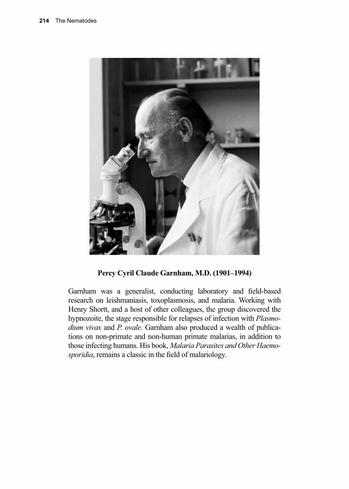
214 The Nematodes
Percy Cyril Claude Garnham, M.D. (1901–1994)
Garnham was a generalist, conducting laboratory and field-based research on leishmaniasis, toxoplasmosis, and malaria. Working with Henry Shortt, and a host of other colleagues, the group discovered the hypnozoite, the stage responsible for relapses of infection with Plasmo-dium vivax and P. ovale. Garnham also produced a wealth of publica-tions on non-primate and non-human primate malarias, in addition to those infecting humans. His book, Malaria Parasites and Other Haemo-sporidia, remains a classic in the field of malariology.
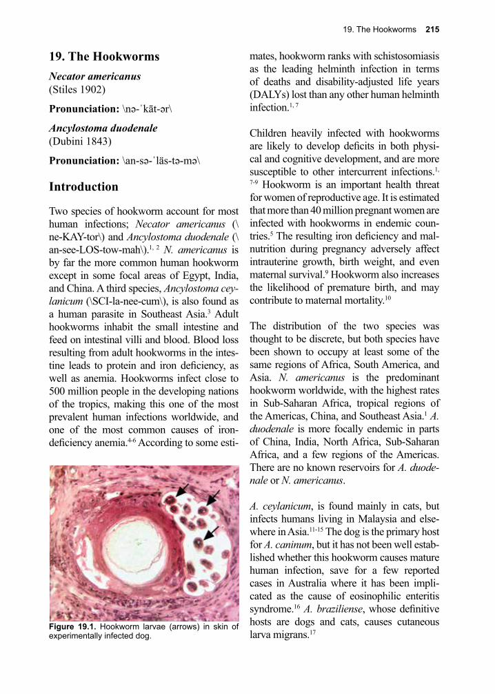
19. The Hookworms 215
19. The HookwormsNecator americanus(Stiles 1902)
Pronunciation: \nə-ˈkāt-ər\
Ancylostoma duodenale(Dubini 1843)
Pronunciation: \an-sə-ˈläs-tə-mə\
Introduction
Two species of hookworm account for most human infections; Necator americanus (\ne-KAY-tor\) and Ancylostoma duodenale (\an-see-LOS-tow-mah\).1, 2 N. americanus is by far the more common human hookworm except in some focal areas of Egypt, India, and China. A third species, Ancylostoma cey-lanicum (\SCI-la-nee-cum\), is also found as a human parasite in Southeast Asia.3 Adult hookworms inhabit the small intestine and feed on intestinal villi and blood. Blood loss resulting from adult hookworms in the intes-tine leads to protein and iron deficiency, as well as anemia. Hookworms infect close to 500 million people in the developing nations of the tropics, making this one of the most prevalent human infections worldwide, and one of the most common causes of iron-deficiency anemia.4-6 According to some esti-
mates, hookworm ranks with schistosomiasis as the leading helminth infection in terms of deaths and disability-adjusted life years (DALYs) lost than any other human helminth infection.1, 7
Children heavily infected with hookworms are likely to develop deficits in both physi-cal and cognitive development, and are more susceptible to other intercurrent infections.1,
7-9 Hookworm is an important health threat for women of reproductive age. It is estimated that more than 40 million pregnant women are infected with hookworms in endemic coun-tries.5 The resulting iron deficiency and mal-nutrition during pregnancy adversely affect intrauterine growth, birth weight, and even maternal survival.9 Hookworm also increases the likelihood of premature birth, and may contribute to maternal mortality.10
The distribution of the two species was thought to be discrete, but both species have been shown to occupy at least some of the same regions of Africa, South America, and Asia. N. americanus is the predominant hookworm worldwide, with the highest rates in Sub-Saharan Africa, tropical regions of the Americas, China, and Southeast Asia.1 A. duodenale is more focally endemic in parts of China, India, North Africa, Sub-Saharan Africa, and a few regions of the Americas. There are no known reservoirs for A. duode-nale or N. americanus.
A. ceylanicum, is found mainly in cats, but infects humans living in Malaysia and else-where in Asia.11-15 The dog is the primary host for A. caninum, but it has not been well estab-lished whether this hookworm causes mature human infection, save for a few reported cases in Australia where it has been impli-cated as the cause of eosinophilic enteritis syndrome.16 A. braziliense, whose definitive hosts are dogs and cats, causes cutaneous larva migrans.17
Figure 19.1. Hookworm larvae (arrows) in skin of experimentally infected dog.
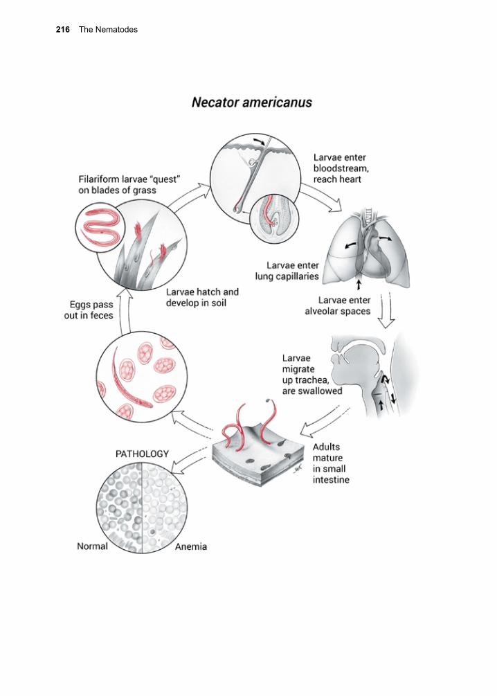
216 The Nematodes
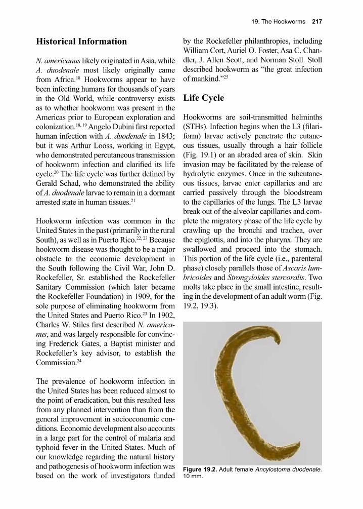
19. The Hookworms 217
Historical Information
N. americanus likely originated in Asia, while A. duodenale most likely originally came from Africa.18 Hookworms appear to have been infecting humans for thousands of years in the Old World, while controversy exists as to whether hookworm was present in the Americas prior to European exploration and colonization.18, 19 Angelo Dubini first reported human infection with A. duodenale in 1843; but it was Arthur Looss, working in Egypt, who demonstrated percutaneous transmission of hookworm infection and clarified its life cycle.20 The life cycle was further defined by Gerald Schad, who demonstrated the ability of A. duodenale larvae to remain in a dormant arrested state in human tissues.21
Hookworm infection was common in the United States in the past (primarily in the rural South), as well as in Puerto Rico.22, 23 Because hookworm disease was thought to be a major obstacle to the economic development in the South following the Civil War, John D. Rockefeller, Sr. established the Rockefeller Sanitary Commission (which later became the Rockefeller Foundation) in 1909, for the sole purpose of eliminating hookworm from the United States and Puerto Rico.23 In 1902, Charles W. Stiles first described N. america-nus, and was largely responsible for convinc-ing Frederick Gates, a Baptist minister and Rockefeller’s key advisor, to establish the Commission.24
The prevalence of hookworm infection in the United States has been reduced almost to the point of eradication, but this resulted less from any planned intervention than from the general improvement in socioeconomic con-ditions. Economic development also accounts in a large part for the control of malaria and typhoid fever in the United States. Much of our knowledge regarding the natural history and pathogenesis of hookworm infection was based on the work of investigators funded
by the Rockefeller philanthropies, including William Cort, Auriel O. Foster, Asa C. Chan-dler, J. Allen Scott, and Norman Stoll. Stoll described hookworm as “the great infection of mankind.”25
Life Cycle
Hookworms are soil-transmitted helminths (STHs). Infection begins when the L3 (filari-form) larvae actively penetrate the cutane-ous tissues, usually through a hair follicle (Fig. 19.1) or an abraded area of skin. Skin invasion may be facilitated by the release of hydrolytic enzymes. Once in the subcutane-ous tissues, larvae enter capillaries and are carried passively through the bloodstream to the capillaries of the lungs. The L3 larvae break out of the alveolar capillaries and com-plete the migratory phase of the life cycle by crawling up the bronchi and trachea, over the epiglottis, and into the pharynx. They are swallowed and proceed into the stomach. This portion of the life cycle (i.e., parenteral phase) closely parallels those of Ascaris lum-bricoides and Strongyloides stercoralis. Two molts take place in the small intestine, result-ing in the development of an adult worm (Fig. 19.2, 19.3).
Figure 19.2. Adult female Ancylostoma duodenale. 10 mm.
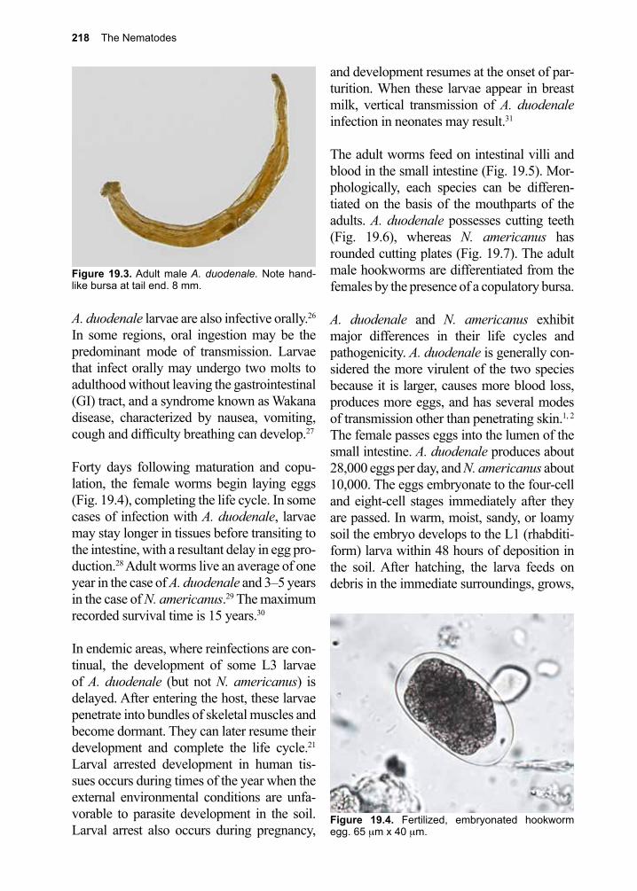
218 The Nematodes
A. duodenale larvae are also infective orally.26 In some regions, oral ingestion may be the predominant mode of transmission. Larvae that infect orally may undergo two molts to adulthood without leaving the gastrointestinal (GI) tract, and a syndrome known as Wakana disease, characterized by nausea, vomiting, cough and difficulty breathing can develop.27
Forty days following maturation and copu-lation, the female worms begin laying eggs (Fig. 19.4), completing the life cycle. In some cases of infection with A. duodenale, larvae may stay longer in tissues before transiting to the intestine, with a resultant delay in egg pro-duction.28 Adult worms live an average of one year in the case of A. duodenale and 3–5 years in the case of N. americanus.29 The maximum recorded survival time is 15 years.30
In endemic areas, where reinfections are con-tinual, the development of some L3 larvae of A. duodenale (but not N. americanus) is delayed. After entering the host, these larvae penetrate into bundles of skeletal muscles and become dormant. They can later resume their development and complete the life cycle.21 Larval arrested development in human tis-sues occurs during times of the year when the external environmental conditions are unfa-vorable to parasite development in the soil. Larval arrest also occurs during pregnancy,
and development resumes at the onset of par-turition. When these larvae appear in breast milk, vertical transmission of A. duodenale infection in neonates may result.31
The adult worms feed on intestinal villi and blood in the small intestine (Fig. 19.5). Mor-phologically, each species can be differen-tiated on the basis of the mouthparts of the adults. A. duodenale possesses cutting teeth (Fig. 19.6), whereas N. americanus has rounded cutting plates (Fig. 19.7). The adult male hookworms are differentiated from the females by the presence of a copulatory bursa.
A. duodenale and N. americanus exhibit major differences in their life cycles and pathogenicity. A. duodenale is generally con-sidered the more virulent of the two species because it is larger, causes more blood loss, produces more eggs, and has several modes of transmission other than penetrating skin.1, 2 The female passes eggs into the lumen of the small intestine. A. duodenale produces about 28,000 eggs per day, and N. americanus about 10,000. The eggs embryonate to the four-cell and eight-cell stages immediately after they are passed. In warm, moist, sandy, or loamy soil the embryo develops to the L1 (rhabditi-form) larva within 48 hours of deposition in the soil. After hatching, the larva feeds on debris in the immediate surroundings, grows,
Figure 19.3. Adult male A. duodenale. Note hand-like bursa at tail end. 8 mm.
Figure 19.4. Fertilized, embryonated hookworm egg. 65 µm x 40 µm.
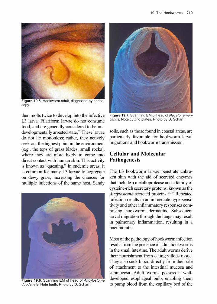
19. The Hookworms 219
then molts twice to develop into the infective L3 larva. Filariform larvae do not consume food, and are generally considered to be in a developmentally arrested state.32 These larvae do not lie motionless; rather, they actively seek out the highest point in the environment (e.g., the tops of grass blades, small rocks), where they are more likely to come into direct contact with human skin. This activity is known as “questing.” In endemic areas, it is common for many L3 larvae to aggregate on dewy grass, increasing the chances for multiple infections of the same host. Sandy
soils, such as those found in coastal areas, are particularly favorable for hookworm larval migrations and hookworm transmission.
Cellular and Molecular Pathogenesis
The L3 hookworm larvae penetrate unbro-ken skin with the aid of secreted enzymes that include a metalloprotease and a family of cysteine-rich secretory proteins, known as the Ancylostoma secreted proteins.33, 34 Repeated infection results in an immediate hypersensi-tivity and other inflammatory responses com-prising hookworm dermatitis. Subsequent larval migration through the lungs may result in pulmonary inflammation, resulting in a pneumonitis.
Most of the pathology of hookworm infection results from the presence of adult hookworms in the small intestine. The adult worms derive their nourishment from eating villous tissue. They also suck blood directly from their site of attachment to the intestinal mucosa and submucosa. Adult worms possess a well-developed esophageal bulb, enabling them to pump blood from the capillary bed of the
Figure 19.6. Scanning EM of head of Ancylostoma duodenale. Note teeth. Photo by D. Scharf.
Figure 19.5. Hookworm adult, diagnosed by endos-copy.
Figure 19.7. Scanning EM of head of Necator ameri-canus. Note cutting plates. Photo by D. Scharf.
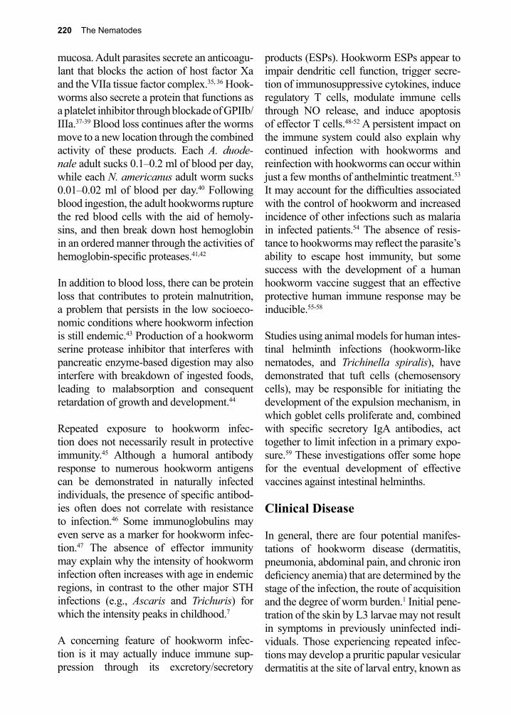
220 The Nematodes
mucosa. Adult parasites secrete an anticoagu-lant that blocks the action of host factor Xa and the VIIa tissue factor complex.35, 36 Hook-worms also secrete a protein that functions as a platelet inhibitor through blockade of GPIIb/IIIa.37-39 Blood loss continues after the worms move to a new location through the combined activity of these products. Each A. duode-nale adult sucks 0.1–0.2 ml of blood per day, while each N. americanus adult worm sucks 0.01–0.02 ml of blood per day.40 Following blood ingestion, the adult hookworms rupture the red blood cells with the aid of hemoly-sins, and then break down host hemoglobin in an ordered manner through the activities of hemoglobin-specific proteases.41,42
In addition to blood loss, there can be protein loss that contributes to protein malnutrition, a problem that persists in the low socioeco-nomic conditions where hookworm infection is still endemic.43 Production of a hookworm serine protease inhibitor that interferes with pancreatic enzyme-based digestion may also interfere with breakdown of ingested foods, leading to malabsorption and consequent retardation of growth and development.44
Repeated exposure to hookworm infec-tion does not necessarily result in protective immunity.45 Although a humoral antibody response to numerous hookworm antigens can be demonstrated in naturally infected individuals, the presence of specific antibod-ies often does not correlate with resistance to infection.46 Some immunoglobulins may even serve as a marker for hookworm infec-tion.47 The absence of effector immunity may explain why the intensity of hookworm infection often increases with age in endemic regions, in contrast to the other major STH infections (e.g., Ascaris and Trichuris) for which the intensity peaks in childhood.7
A concerning feature of hookworm infec-tion is it may actually induce immune sup-pression through its excretory/secretory
products (ESPs). Hookworm ESPs appear to impair dendritic cell function, trigger secre-tion of immunosuppressive cytokines, induce regulatory T cells, modulate immune cells through NO release, and induce apoptosis of effector T cells.48-52 A persistent impact on the immune system could also explain why continued infection with hookworms and reinfection with hookworms can occur within just a few months of anthelmintic treatment.53 It may account for the difficulties associated with the control of hookworm and increased incidence of other infections such as malaria in infected patients.54 The absence of resis-tance to hookworms may reflect the parasite’s ability to escape host immunity, but some success with the development of a human hookworm vaccine suggest that an effective protective human immune response may be inducible.55-58
Studies using animal models for human intes-tinal helminth infections (hookworm-like nematodes, and Trichinella spiralis), have demonstrated that tuft cells (chemosensory cells), may be responsible for initiating the development of the expulsion mechanism, in which goblet cells proliferate and, combined with specific secretory IgA antibodies, act together to limit infection in a primary expo-sure.59 These investigations offer some hope for the eventual development of effective vaccines against intestinal helminths.
Clinical Disease
In general, there are four potential manifes-tations of hookworm disease (dermatitis, pneumonia, abdominal pain, and chronic iron deficiency anemia) that are determined by the stage of the infection, the route of acquisition and the degree of worm burden.1 Initial pene-tration of the skin by L3 larvae may not result in symptoms in previously uninfected indi-viduals. Those experiencing repeated infec-tions may develop a pruritic papular vesicular dermatitis at the site of larval entry, known as
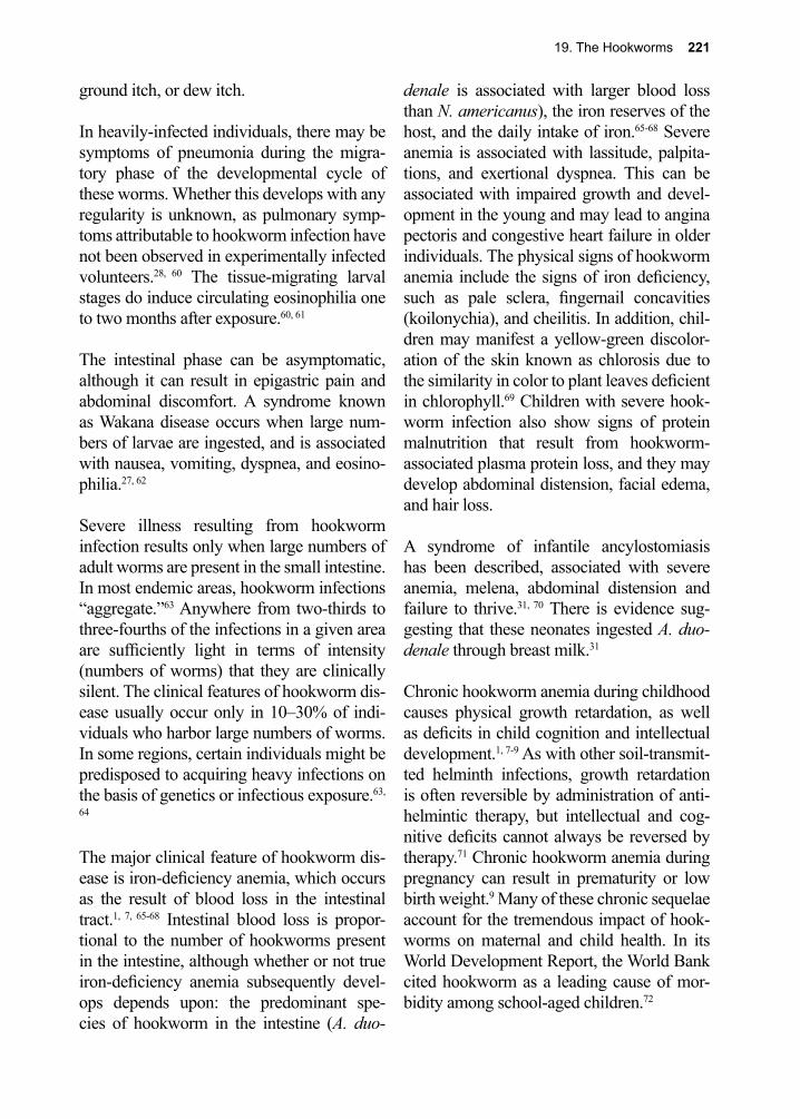
19. The Hookworms 221
ground itch, or dew itch.
In heavily-infected individuals, there may be symptoms of pneumonia during the migra-tory phase of the developmental cycle of these worms. Whether this develops with any regularity is unknown, as pulmonary symp-toms attributable to hookworm infection have not been observed in experimentally infected volunteers.28, 60 The tissue-migrating larval stages do induce circulating eosinophilia one to two months after exposure.60, 61
The intestinal phase can be asymptomatic, although it can result in epigastric pain and abdominal discomfort. A syndrome known as Wakana disease occurs when large num-bers of larvae are ingested, and is associated with nausea, vomiting, dyspnea, and eosino-philia.27, 62
Severe illness resulting from hookworm infection results only when large numbers of adult worms are present in the small intestine. In most endemic areas, hookworm infections “aggregate.”63 Anywhere from two-thirds to three-fourths of the infections in a given area are sufficiently light in terms of intensity (numbers of worms) that they are clinically silent. The clinical features of hookworm dis-ease usually occur only in 10–30% of indi-viduals who harbor large numbers of worms. In some regions, certain individuals might be predisposed to acquiring heavy infections on the basis of genetics or infectious exposure.63,
64
The major clinical feature of hookworm dis-ease is iron-deficiency anemia, which occurs as the result of blood loss in the intestinal tract.1, 7, 65-68 Intestinal blood loss is propor-tional to the number of hookworms present in the intestine, although whether or not true iron-deficiency anemia subsequently devel-ops depends upon: the predominant spe-cies of hookworm in the intestine (A. duo-
denale is associated with larger blood loss than N. americanus), the iron reserves of the host, and the daily intake of iron.65-68 Severe anemia is associated with lassitude, palpita-tions, and exertional dyspnea. This can be associated with impaired growth and devel-opment in the young and may lead to angina pectoris and congestive heart failure in older individuals. The physical signs of hookworm anemia include the signs of iron deficiency, such as pale sclera, fingernail concavities (koilonychia), and cheilitis. In addition, chil-dren may manifest a yellow-green discolor-ation of the skin known as chlorosis due to the similarity in color to plant leaves deficient in chlorophyll.69 Children with severe hook-worm infection also show signs of protein malnutrition that result from hookworm-associated plasma protein loss, and they may develop abdominal distension, facial edema, and hair loss.
A syndrome of infantile ancylostomiasis has been described, associated with severe anemia, melena, abdominal distension and failure to thrive.31, 70 There is evidence sug-gesting that these neonates ingested A. duo-denale through breast milk.31
Chronic hookworm anemia during childhood causes physical growth retardation, as well as deficits in child cognition and intellectual development.1, 7-9 As with other soil-transmit-ted helminth infections, growth retardation is often reversible by administration of anti-helmintic therapy, but intellectual and cog-nitive deficits cannot always be reversed by therapy.71 Chronic hookworm anemia during pregnancy can result in prematurity or low birth weight.9 Many of these chronic sequelae account for the tremendous impact of hook-worms on maternal and child health. In its World Development Report, the World Bank cited hookworm as a leading cause of mor-bidity among school-aged children.72
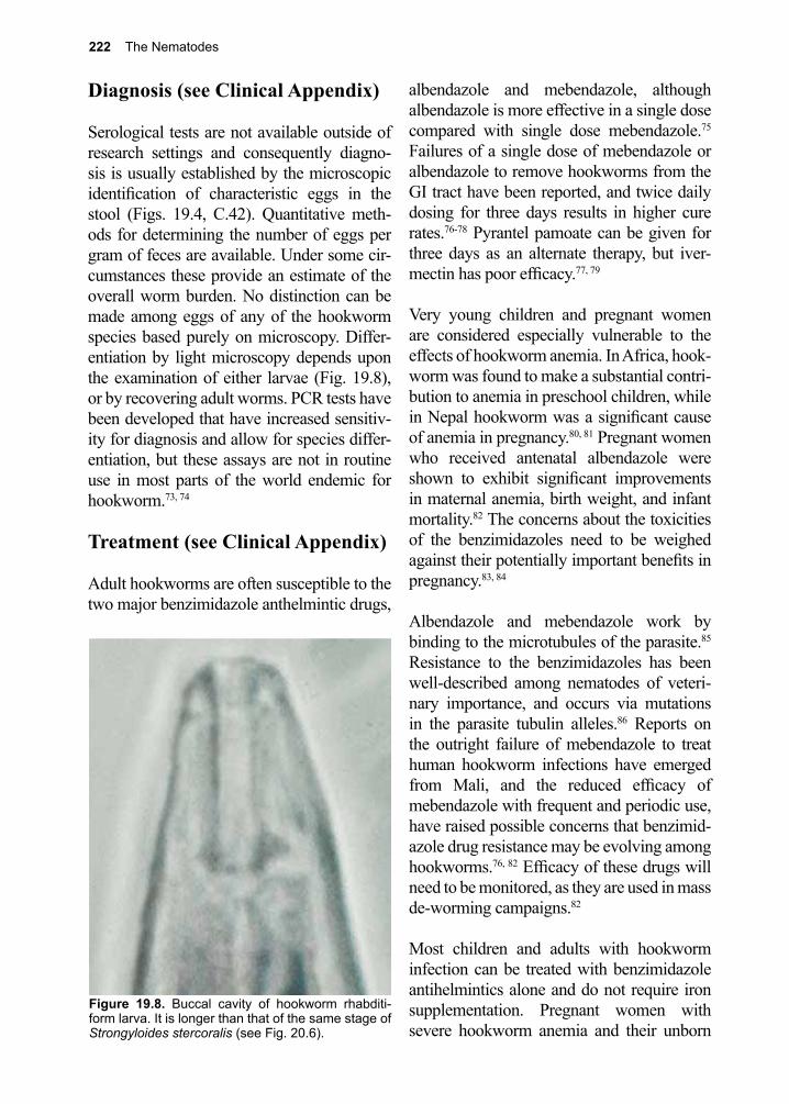
222 The Nematodes
Diagnosis (see Clinical Appendix)
Serological tests are not available outside of research settings and consequently diagno-sis is usually established by the microscopic identification of characteristic eggs in the stool (Figs. 19.4, C.42). Quantitative meth-ods for determining the number of eggs per gram of feces are available. Under some cir-cumstances these provide an estimate of the overall worm burden. No distinction can be made among eggs of any of the hookworm species based purely on microscopy. Differ-entiation by light microscopy depends upon the examination of either larvae (Fig. 19.8), or by recovering adult worms. PCR tests have been developed that have increased sensitiv-ity for diagnosis and allow for species differ-entiation, but these assays are not in routine use in most parts of the world endemic for hookworm.73, 74
Treatment (see Clinical Appendix)
Adult hookworms are often susceptible to the two major benzimidazole anthelmintic drugs,
albendazole and mebendazole, although albendazole is more effective in a single dose compared with single dose mebendazole.75 Failures of a single dose of mebendazole or albendazole to remove hookworms from the GI tract have been reported, and twice daily dosing for three days results in higher cure rates.76-78 Pyrantel pamoate can be given for three days as an alternate therapy, but iver-mectin has poor efficacy.77, 79
Very young children and pregnant women are considered especially vulnerable to the effects of hookworm anemia. In Africa, hook-worm was found to make a substantial contri-bution to anemia in preschool children, while in Nepal hookworm was a significant cause of anemia in pregnancy.80, 81 Pregnant women who received antenatal albendazole were shown to exhibit significant improvements in maternal anemia, birth weight, and infant mortality.82 The concerns about the toxicities of the benzimidazoles need to be weighed against their potentially important benefits in pregnancy.83, 84
Albendazole and mebendazole work by binding to the microtubules of the parasite.85 Resistance to the benzimidazoles has been well-described among nematodes of veteri-nary importance, and occurs via mutations in the parasite tubulin alleles.86 Reports on the outright failure of mebendazole to treat human hookworm infections have emerged from Mali, and the reduced efficacy of mebendazole with frequent and periodic use, have raised possible concerns that benzimid-azole drug resistance may be evolving among hookworms.76, 82 Efficacy of these drugs will need to be monitored, as they are used in mass de-worming campaigns.82
Most children and adults with hookworm infection can be treated with benzimidazole antihelmintics alone and do not require iron supplementation. Pregnant women with severe hookworm anemia and their unborn
Figure 19.8. Buccal cavity of hookworm rhabditi-form larva. It is longer than that of the same stage of Strongyloides stercoralis (see Fig. 20.6).
19. The Hookworms 223
children have been shown to benefit from simultaneous oral supplementation with iron such as ferrous sulfate along with antihelmin-tic drugs.81
Prevention and Control
The traditional methods of hookworm control in endemic areas have included sanitary dis-posal of feces through the implementation of latrines, health education, drinking clean safe water, hand washing, and cooking of food.87 Despite decades of widespread attempts to control hookworm through these traditional methods, the global prevalence and intensity of hookworm remains the same. Among the reasons why traditional methods have failed are the ability of A. duodenale hookworm larvae to infect humans through ingestion, the ability of N. americanus larvae to penetrate all aspects of the body including the hands and abdomen, the high rate of occupational exposure to hookworm that occurs during agricultural pursuits, and the continued reli-ance on human feces for fertilizing crops.88
At the World Health Assembly in 2001, member states were urged to control the global morbidity of hookworm and other STH infections through the frequent and periodic use of antihelmintic drugs. In 2012, The London Declaration on Neglected Tropi-cal Diseases was put forth as a plan to con-trol hookworm among other diseases that
involved several components including mass drug administration.89 There are concerns that this approach might have less of an impact on hookworm than the other STH infections such as Trichuris and Ascaris. The highest hookworm intensities in a community are often not in children, so that targeting chil-dren is not expected to reduce hookworm transmission.90 High rates of post-treatment hookworm reinfection occur in many com-munities, especially those with high levels of transmission, which sometimes requires thrice-yearly antihelmintic treatment.53 A recent analysis through the Global Burden of Disease Study found that the prevalence rate of hookworm has decreased only 5% in recent decades, despite comparatively greater reductions of 25% for ascariasis.6
As a complementing control strategy, the Human Hookworm Vaccine Initiative has developed a recombinant vaccine that tar-gets adult hookworm blood feeding by elicit-ing anti-enzyme antibodies that block para-site hemoglobin digestion.56 This vaccine is being studied for its safety and immunoge-nicity. Mathematical modeling of the vac-cine indicates that it could reduce hookworm transmission, which would not be possible using pediatric anthelmintic drug de-worm-ing approaches, while economic modeling indicates that a hookworm vaccine could be highly cost-effective.91, 92
References
1. Hotez, P. J., et al., Hookworm infection. The New England journal of medicine 2004, 351 (8), 799-807.
2. Bethony, J., et al., Soil-transmitted helminth infections: ascariasis, trichuriasis, and hookworm. Lancet 2006, 367 (9521), 1521-32.
3. Ngui, R., et al., Epidemiological and genetic data supporting the transmission of Ancylostoma ceylanicum among human and domestic animals. PLoS Negl Trop Dis 2012, 6 (2), e1522.
4. DeSilva, N. R.; Brooker, S., Soil-transmitted helminth infections: updating the global picture. Trends Parasitol 2003, 19 547-51.
5. Pullan, R. L., et al., Global numbers of infection and disease burden of soil transmitted helminth infections in 2010. Parasit Vectors 2014, 7, 37.
224 The Nematodes
6. Global Burden of Disease Study, C., Global, regional, and national incidence, prevalence, and years lived with disability for 301 acute and chronic diseases and injuries in 188 countries, 1990-2013: a systematic analysis for the Global Burden of Disease Study 2013. Lancet 2015, 386 (9995), 743-800.
7. Hotez, P. J., et al., Hookworm: “the great infection of mankind”. PLoS medicine 2005, 2 (3), e67.8. Brooker, S., et al., Human hookworm infection in the 21st century. Advances in parasitology
2004, 58, 197-288.9. Sakti, H., et al., Evidence for an association between hookworm infection and cognitive function
in Indonesian school children. Tropical medicine & international health : TM & IH 1999, 4 (5), 322-34.
10. Bundy, D. A., et al., Hookworm infection in pregnancy. Transactions of the Royal Society of Tropical Medicine and Hygiene 1995, 89 (5), 521-2.
11. Traub, R. J., Ancylostoma ceylanicum, a re-emerging but neglected parasitic zoonosis. Int J Parasitol 2013, 43 (12-13), 1009-15.
12. George, S., et al., Identification of Ancylostoma ceylanicum in children from a tribal community in Tamil Nadu, India using a semi-nested PCR-RFLP tool. Trans R Soc Trop Med Hyg 2015, 109 (4), 283-5.
13. Phosuk, I., et al., Molecular detection of Ancylostoma duodenale, Ancylostoma ceylanicum, and Necator americanus in humans in northeastern and southern Thailand. Korean J Parasitol 2013, 51 (6), 747-9.
14. Thompson, R. C., Neglected zoonotic helminths: Hymenolepis nana, Echinococcus canadensis and Ancylostoma ceylanicum. Clin Microbiol Infect 2015, 21 (5), 426-32.
15. Liu, Y., et al., The zoonotic risk of Ancylostoma ceylanicum isolated from stray dogs and cats in Guangzhou, South China. Biomed Res Int 2014, 2014, 208759.
16. Prociv, P.; Croese, J., Human eosinophilic enteritis caused by dog hookworm Ancylostoma caninum. Lancet (London, England) 1990, 335 (8701), 1299-302.
17. Blackwell, V.; Vega-Lopez, F., Cutaneous larva migrans: clinical features and management of 44 cases presenting in the returning traveller. The British journal of dermatology 2001, 145 (3), 434-7.
18. Cox, F. E., History of human parasitology. Clin Microbiol Rev 2002, 15 (4), 595-612.19. Ferreira, L. F.; Araujo, A., On hookworms in the Americas and trans-Pacific contact. Parasitol
Today 1996, 12 (11), 454; author reply 454.20. Dubini, A., Nuovo verme intestinale umano Ancylostoma duodenale, constitutente un sesto
genere dei nematoidei proprii dell’uomo. Ann Univ Med Milano 1843, 106, 5-13.21. Schad, G. A., et al., Arrested development in human hookworm infections: an adaptation to a
seasonally unfavorable external environment. Science (New York, N.Y.) 1973, 180 (4085), 502-4.22. Maldonado, A. E., Hookworm disease: Puerto Rico’s secret killer. P R Health Sci J 1993, 12 (3),
191-6.23. Bleakley, H., Disease and Development: Evidence from Hookworm Eradication in the American
South. Q J Econ 2007, 122 (1), 73-117.24. Stiles, C. W., A new species of hookworm (Uncinaria americana) parasitic in man. Am Med
1902, 3, 777-778.25. Stoll, N. R., On endemic hookworm, where do we stand today? Exp Parasitol 1962, 12, 241-51.26. Brooker, S., et al., Human hookworm infection in the 21st century. Adv Parasitol 2004, 58, 197-
288.27. Kojima, S., [Wakana disease]. Ryoikibetsu Shokogun Shirizu 1999, (24 Pt 2), 437-8.28. Nawalinski, T. A.; Schad, G. A., Arrested development in Ancylostoma duodenale: course of a
self-induced infection in man. Am J Trop Med Hyg 1974, 23 (5), 895-8.29. Hoagland, K. E.; Schad, G. A., Necator americanus and Ancylostoma duodenale: life history
parameters and epidemiological implications of two sympatric hookworms of humans. Experimental parasitology 1978, 44 (1), 36-49.
30. Plamer, E. D.; J., Course of egg output over a 15-year period in a case of experimentally induced necatoriasis americanus , in the absence of hyperinfection. The American journal of tropical medicine and hygiene 1955, 4 (4), 756-757.
31. Yu, S. H., et al., Infantile hookworm disease in China. A review. Acta tropica 1995, 59 (4), 265-
19. The Hookworms 225
70.32. Hawdon, J. M.; Hotez, P. J., Hookworm: developmental biology of the infectious process.
Current opinion in genetics & development 1996, 6 (5), 618-23.33. Zhan, B., et al., A developmentally regulated metalloprotease secreted by host-stimulated
Ancylostoma caninum third-stage infective larvae is a member of the astacin family of proteases. Molecular and biochemical parasitology 2002, 120 (2), 291-6.
34. Asojo, O. A., et al., X-ray structure of Na-ASP-2, a pathogenesis-related-1 protein from the nematode parasite, Necator americanus, and a vaccine antigen for human hookworm infection. Journal of molecular biology 2005, 346 (3), 801-14.
35. Cappello, M., et al., Ancylostoma caninum anticoagulant peptide: a hookworm-derived inhibitor of human coagulation factor Xa. Proceedings of the National Academy of Sciences of the United States of America 1995, 92 (13), 6152-6.
36. Stanssens, P., et al., Anticoagulant repertoire of the hookworm Ancylostoma caninum. Proc Natl Acad 1996, 93, 2149-2154.
37. Ma, D., et al., The structure of hookworm platelet inhibitor (HPI), a CAP superfamily member from Ancylostoma caninum. Acta Crystallogr F Struct Biol Commun 2015, 71 (Pt 6), 643-9.
38. Del Valle, A., et al., Isolation and molecular cloning of a secreted hookworm platelet inhibitor from adult Ancylostoma caninum. Mol Biochem Parasitol 2003, 129 (2), 167-77.
39. Chadderdon, R. C.; Cappello, M., The hookworm platelet inhibitor: functional blockade of integrins GPIIb/IIIa (alphaIIbbeta3) and GPIa/IIa (alpha2beta1) inhibits platelet aggregation and adhesion in vitro. J Infect Dis 1999, 179 (5), 1235-41.
40. Roche, M., et al., The nature and causes of “hookworm anemia”. Am Med Hyg 1996, 15, 1031-1102.
41. Don, T. A., et al., A pore-forming haemolysin from the hookworm, Ancylostoma caninum. International journal for parasitology 2004, 34 (9), 1029-35.
42. Williamson, A. L., et al., A multi-enzyme cascade of hemoglobin proteolysis in the intestine of blood-feeding hookworms. The Journal of biological chemistry 2004, 279 (34), 35950-7.
43. Gupta, M. C., et al., Gastrointestinal protein loss in hookworm and roundworm infections. Am J Clin Nutr 1974, 27 (12), 1386-9.
44. Chu, D., et al., Molecular characterization of Ancylostoma ceylanicum Kunitz-type serine protease inhibitor: evidence for a role in hookworm-associated growth delay. Infect Immun 2004, 72 (4), 2214-21.
45. Gaze, S., et al., Immunology of experimental and natural human hookworm infection. Parasite Immunol 2014, 36 (8), 358-66.
46. Pritchard, D. I., et al., Epidemiology and immunology of Necator americanus infection in a community in Papua New Guinea: humoral responses to excretory-secretory and cuticular collagen antigens. Parasitology 1990, 100 Pt 2, 317-26.
47. Palmer, D. R., et al., IgG4 responses to antigens of adult Necator americanus: potential for use in large-scale epidemiological studies. Bulletin of the World Health Organization 1996, 74 (4), 381-6.
48. Segura, M., et al., Impairment of dendritic cell function by excretory-secretory products: a potential mechanism for nematode-induced immunosuppression. Eur J Immunol 2007, 37 (7), 1887-904.
49. Ricci, N. D., et al., Induction of CD4(+)CD25(+)FOXP3(+) regulatory T cells during human hookworm infection modulates antigen-mediated lymphocyte proliferation. PLoS Negl Trop Dis 2011, 5 (11), e1383.
50. Dondji, B., et al., Role for nitric oxide in hookworm-associated immune suppression. Infect Immun 2008, 76 (6), 2560-7.
51. Chow, S. C., et al., The human hookworm pathogen Necator americanus induces apoptosis in T lymphocytes. Parasite Immunol 2000, 22 (1), 21-9.
52. Quinnell, R. J., et al., The immunoepidemiology of human hookworm infection. Parasite Immunol 2004, 26 (11-12), 443-54.
53. Albonico, M., et al., Rate of reinfection with intestinal nematodes after treatment of children with mebendazole or albendazole in a highly endemic area. Transactions of the Royal Society of Tropical Medicine and Hygiene 1995, 89 (5), 538-41.
226 The Nematodes
54. Nacher, M., et al., Intestinal helminth infections are associated with increased incidence of Plasmodium falciparum malaria in Thailand. J Parasitol 2002, 88 (1), 55-8.
55. Kumar, S.; Pritchard, D. I., Skin penetration by ensheathed third-stage infective larvae of Necator americanus, and the host’s immune response to larval antigens. International journal for parasitology 1992, 22 (5), 573-9.
56. Hotez, P. J., et al., The Human Hookworm Vaccine. Vaccine 2013, 31 Suppl 2, B227-32.57. Bottazzi, M. E., The human hookworm vaccine: recent updates and prospects for success. J
Helminthol 2015, 89 (5), 540-4.58. Pearson, M. S., et al., New tools for NTD vaccines: A case study of quality control assays for
product development of the human hookworm vaccine Na-APR-1M74. Hum Vaccin Immunother 2015, 11 (5), 1251-7.
59. Howitt, M. R., et al., Tuft cells, taste-chemosensory cells, orchestrate parasite type 2 immunity in the gut. Science 2016, 351 (6279), 1329-33.
60. Maxwell, C., et al., The clinical and immunologic responses of normal human volunteers to low dose hookworm (Necator americanus) infection. Am J Trop Med Hyg 1987, 37 (1), 126-34.
61. White, C. J., et al., Changes in the structural and functional properties of human eosinophils during experimental hookworm infection. J Infect Dis 1986, 154 (5), 778-83.
62. Yoshida, Y., et al., Experimental studies on the infection mode of Ancylostoma duodenale and Necator americanus to the definitive host. Japanese 1958, 7, 102-112.
63. Schad, G. A.; Anderson, R. M., Predisposition to hookworm infection in humans. Science (New York, N.Y.) 1985, 228 (4707), 1537-40.
64. Quinnell, R. J., et al., Predisposition to hookworm infection in Papua New Guinea. Trans R Soc Trop Med Hyg 2001, 95 (2), 139-42.
65. Albonico, M., et al., Epidemiological evidence for a differential effect of hookworm species, Ancylostoma duodenale or Necator americanus, on iron status of children. International journal of epidemiology 1998, 27 (3), 530-7.
66. Stoltzfus, R. J., et al., Hookworm control as a strategy to prevent iron deficiency. Nutrition reviews 1997, 55 (6), 223-32.
67. Stoltzfus, R. J., et al., Epidemiology of iron deficiency anemia in Zanzibari schoolchildren: the importance of hookworms. The American journal of clinical nutrition 1997, 65 (1), 153-9.
68. Stoltzfus, R. J., et al., Hemoquant determination of hookworm-related blood loss and its role in iron deficiency in African children. The American journal of tropical medicine and hygiene 1996, 55 (4), 399-404.
69. Crosby, W. H., Whatever became of chlorosis? JAMA 1987, 257 (20), 2799-800.70. Hotez, P. J., Hookworm disease in children. The Pediatric infectious disease journal 1989, 8 (8),
516-20.71. Stephenson, L. S., et al., Treatment with a single dose of albendazole improves growth
of Kenyan schoolchildren with hookworm, Trichuris trichiura, and Ascaris lumbricoides infections. The American journal of tropical medicine and hygiene 1989, 41 (1), 78-87.
72. Bank, T. W., World Development Report 1993: Investing in Health. Oxford University Press, Inc, New York: 1993.
73. van Mens, S. P., et al., Comparison of real-time PCR and Kato smear microscopy for the detection of hookworm infections in three consecutive faecal samples from schoolchildren in Ghana. Trans R Soc Trop Med Hyg 2013, 107 (4), 269-71.
74. Phuphisut, O., et al., Triplex polymerase chain reaction assay for detection of major soil-transmitted helminths, Ascaris lumbricoides, Trichuris trichiura, Necator americanus, in fecal samples. Southeast Asian J Trop Med Public Health 2014, 45 (2), 267-75.
75. Albonico, M., et al., Control strategies for human intestinal nematode infections. Advances in parasitology 1999, 42, 277-341.
76. DeClercq, D., et al., Failure of mebendazole in treatment of human hookworm infections in the southern region of Mali. Am Med Hyg 1997, 57 25-30.
77. Steinmann, P., et al., Efficacy of single-dose and triple-dose albendazole and mebendazole against soil-transmitted helminths and Taenia spp.: a randomized controlled trial. PLoS One 2011, 6 (9), e25003.
19. The Hookworms 227
78. Soukhathammavong, P. A., et al., Low efficacy of single-dose albendazole and mebendazole against hookworm and effect on concomitant helminth infection in Lao PDR. PLoS Negl Trop Dis 2012, 6 (1), e1417.
79. Keiser, J.; Utzinger, J., Efficacy of current drugs against soil-transmitted helminth infections: systematic review and meta-analysis. JAMA 2008, 299 (16), 1937-48.
80. Brooker, S., et al., The epidemiology of hookworm infection and its contribution to anaemia among pre-school children on the Kenyan coast. Transactions of the Royal Society of Tropical Medicine and Hygiene 1999, 93 (3), 240-6.
81. Christian, P., et al., Antenatal anthelmintic treatment, birthweight, and infant survival in rural Nepal. Lancet (London, England) 2004, 364 (9438), 981-3.
82. Albonico, M., et al., Monitoring drug efficacy and early detection of drug resistance in human soil-transmitted nematodes: a pressing public health agenda for helminth control. International journal for parasitology 2004, 34 (11), 1205-10.
83. Ndibazza, J., et al., Effects of deworming during pregnancy on maternal and perinatal outcomes in Entebbe, Uganda: a randomized controlled trial. Clin Infect Dis 2010, 50 (4), 531-40.
84. Torp-Pedersen, A., et al., Exposure to mebendazole and pyrvinium during pregnancy: a Danish nationwide cohort study. Infect Dis Obstet Gynecol 2012, 2012, 769851.
85. Fennell, B., et al., Microtubules as antiparasitic drug targets. Expert Opin Drug Discov 2008, 3 (5), 501-18.
86. Von Samson-Himmelstjerna, G., et al., Single nucleotide polymorphism (SNP) markers for benzimidazole resistance in veterinary nematodes. Parasitology 2007, 134 (Pt 8), 1077-86.
87. Zheng, Q., et al., The control of hookworm infection in China. Parasit Vectors 2009, 2 (1), 44.88. Humphries, D. L., et al., The use of human faeces for fertilizer is associated with increased
intensity of hookworm infection in Vietnamese women. Transactions of the Royal Society of Tropical Medicine and Hygiene 1997, 91 (5), 518-20.
89. Anderson, R., et al., The coverage and frequency of mass drug administration required to eliminate persistent transmission of soil-transmitted helminths. Philos Trans R Soc Lond B Biol Sci 2014, 369 (1645), 20130435.
90. Chan, M. S., et al., Transmission patterns and the epidemiology of hookworm infection. International journal of epidemiology 1997, 26 (6), 1392-400.
91. Bartsch, S. M., et al., Modeling the economic and epidemiologic impact of hookworm vaccine and mass drug administration (MDA) in Brazil, a high transmission setting. Vaccine 2016, 34 (19), 2197-206.
92. Lee, B. Y., et al., The potential economic value of a hookworm vaccine. Vaccine 2011, 29 (6), 1201-10.
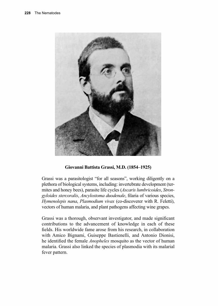
228 The Nematodes
Giovanni Battista Grassi, M.D. (1854–1925)
Grassi was a parasitologist “for all seasons”, working diligently on a plethora of biological systems, including: invertebrate development (ter-mites and honey bees), parasite life cycles (Ascaris lumbricoides, Stron-gyloides stercoralis, Ancylostoma duodenale, filaria of various species, Hymenolepis nana, Plasmodium vivax (co-discoverer with R. Feletti), vectors of human malaria, and plant pathogens affecting wine grapes.
Grassi was a thorough, observant investigator, and made significant contributions to the advancement of knowledge in each of these fields. His worldwide fame arose from his research, in collaboration with Amico Bignami, Guiseppe Bastienelli, and Antonio Dionisi, he identified the female Anopheles mosquito as the vector of human malaria. Grassi also linked the species of plasmodia with its malarial fever pattern.
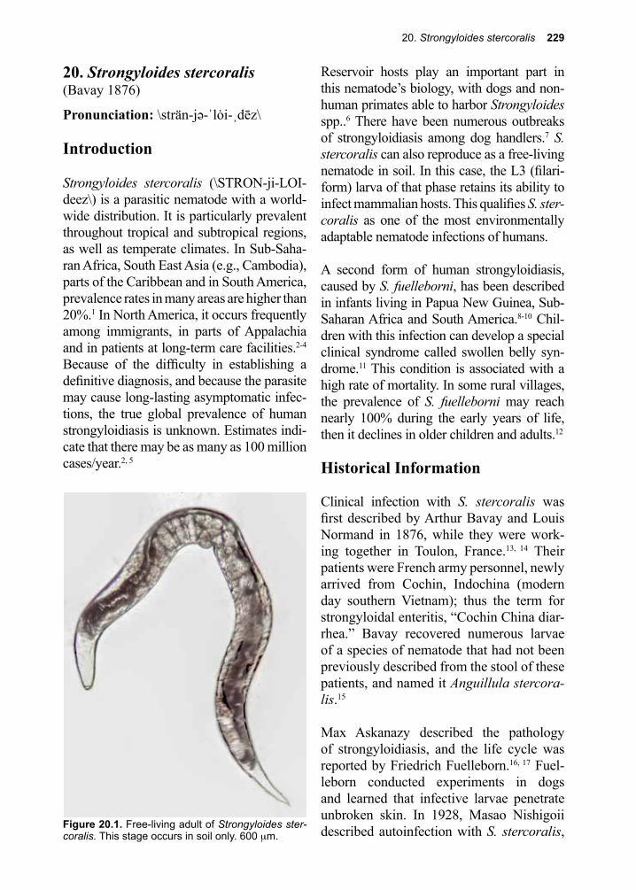
20. Strongyloides stercoralis 229
Figure 20.1. Free-living adult of Strongyloides ster-coralis. This stage occurs in soil only. 600 µm.
20. Strongyloides stercoralis(Bavay 1876)
Pronunciation: \strän-jə-ˈlȯi-ˌdēz\
Introduction
Strongyloides stercoralis (\STRON-ji-LOI-deez\) is a parasitic nematode with a world-wide distribution. It is particularly prevalent throughout tropical and subtropical regions, as well as temperate climates. In Sub-Saha-ran Africa, South East Asia (e.g., Cambodia), parts of the Caribbean and in South America, prevalence rates in many areas are higher than 20%.1 In North America, it occurs frequently among immigrants, in parts of Appalachia and in patients at long-term care facilities.2-4 Because of the difficulty in establishing a definitive diagnosis, and because the parasite may cause long-lasting asymptomatic infec-tions, the true global prevalence of human strongyloidiasis is unknown. Estimates indi-cate that there may be as many as 100 million cases/year.2, 5
Reservoir hosts play an important part in this nematode’s biology, with dogs and non-human primates able to harbor Strongyloides spp..6 There have been numerous outbreaks of strongyloidiasis among dog handlers.7 S. stercoralis can also reproduce as a free-living nematode in soil. In this case, the L3 (filari-form) larva of that phase retains its ability to infect mammalian hosts. This qualifies S. ster-coralis as one of the most environmentally adaptable nematode infections of humans.
A second form of human strongyloidiasis, caused by S. fuelleborni, has been described in infants living in Papua New Guinea, Sub-Saharan Africa and South America.8-10 Chil-dren with this infection can develop a special clinical syndrome called swollen belly syn-drome.11 This condition is associated with a high rate of mortality. In some rural villages, the prevalence of S. fuelleborni may reach nearly 100% during the early years of life, then it declines in older children and adults.12
Historical Information
Clinical infection with S. stercoralis was first described by Arthur Bavay and Louis Normand in 1876, while they were work-ing together in Toulon, France.13, 14 Their patients were French army personnel, newly arrived from Cochin, Indochina (modern day southern Vietnam); thus the term for strongyloidal enteritis, “Cochin China diar-rhea.” Bavay recovered numerous larvae of a species of nematode that had not been previously described from the stool of these patients, and named it Anguillula stercora-lis.15
Max Askanazy described the pathology of strongyloidiasis, and the life cycle was reported by Friedrich Fuelleborn.16, 17 Fuel-leborn conducted experiments in dogs and learned that infective larvae penetrate unbroken skin. In 1928, Masao Nishigoii described autoinfection with S. stercoralis,
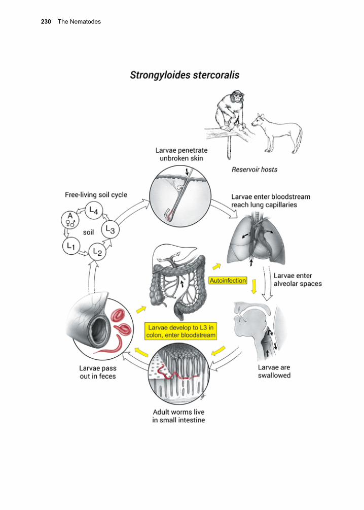
230 The Nematodes
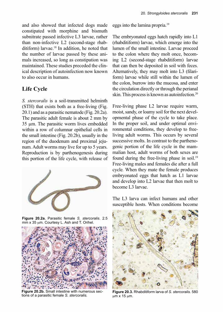
20. Strongyloides stercoralis 231
Figure 20.3. Rhabditiform larva of S. stercoralis. 580 µm x 15 µm.
and also showed that infected dogs made constipated with morphine and bismuth subnitrate passed infective L3 larvae, rather than non-infective L2 (second-stage rhab-ditiform) larvae.18 In addition, he noted that the number of larvae passed by these ani-mals increased, so long as constipation was maintained. These studies preceded the clin-ical description of autoinfection now known to also occur in humans.
Life Cycle
S. stercoralis is a soil-transmitted helminth (STH) that exists both as a free-living (Fig. 20.1) and as a parasitic nematode (Fig. 20.2a). The parasitic adult female is about 2 mm by 35 µm. The parasitic worm lives embedded within a row of columnar epithelial cells in the small intestine (Fig. 20.2b), usually in the region of the duodenum and proximal jeju-num. Adult worms may live for up to 5 years. Reproduction is by parthenogenesis during this portion of the life cycle, with release of
eggs into the lamina propria.19
The embryonated eggs hatch rapidly into L1 (rhabditiform) larvae, which emerge into the lumen of the small intestine. Larvae proceed to the colon where they molt once, becom-ing L2 (second-stage rhabditiform) larvae that can then be deposited in soil with feces. Alternatively, they may molt into L3 (filari-form) larvae while still within the lumen of the colon, burrow into the mucosa, and enter the circulation directly or through the perianal skin. This process is known as autoinfection.20
Free-living phase L2 larvae require warm, moist, sandy, or loamy soil for the next devel-opmental phase of the cycle to take place. In the proper soil, and under optimal envi-ronmental conditions, they develop to free-living adult worms. This occurs by several successive molts. In contrast to the partheno-genic portion of the life cycle in the mam-malian host, adult worms of both sexes are found during the free-living phase in soil.19 Free-living males and females die after a full cycle. When they mate the female produces embryonated eggs that hatch as L1 larvae and develop into L2 larvae that then molt to become L3 larvae.
The L3 larva can infect humans and other susceptible hosts. When conditions become
Figure 20.2b. Small intestine with numerous sec-tions of a parasitic female S. stercoralis.
Figure 20.2a. Parasitic female S. stercoralis. 2.5 mm x 35 µm. Courtesy L. Ash and T. Orihel.
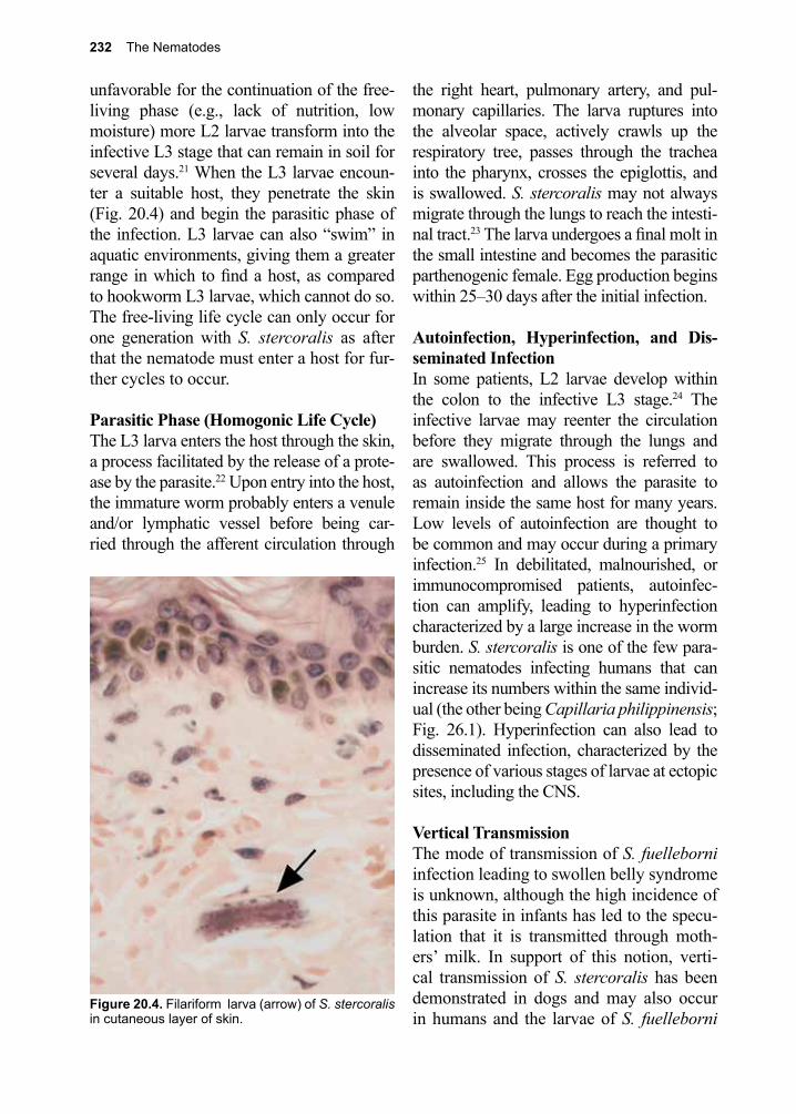
232 The Nematodes
unfavorable for the continuation of the free-living phase (e.g., lack of nutrition, low moisture) more L2 larvae transform into the infective L3 stage that can remain in soil for several days.21 When the L3 larvae encoun-ter a suitable host, they penetrate the skin (Fig. 20.4) and begin the parasitic phase of the infection. L3 larvae can also “swim” in aquatic environments, giving them a greater range in which to find a host, as compared to hookworm L3 larvae, which cannot do so. The free-living life cycle can only occur for one generation with S. stercoralis as after that the nematode must enter a host for fur-ther cycles to occur.
Parasitic Phase (Homogonic Life Cycle)The L3 larva enters the host through the skin, a process facilitated by the release of a prote-ase by the parasite.22 Upon entry into the host, the immature worm probably enters a venule and/or lymphatic vessel before being car-ried through the afferent circulation through
the right heart, pulmonary artery, and pul-monary capillaries. The larva ruptures into the alveolar space, actively crawls up the respiratory tree, passes through the trachea into the pharynx, crosses the epiglottis, and is swallowed. S. stercoralis may not always migrate through the lungs to reach the intesti-nal tract.23 The larva undergoes a final molt in the small intestine and becomes the parasitic parthenogenic female. Egg production begins within 25–30 days after the initial infection.
Autoinfection, Hyperinfection, and Dis-seminated InfectionIn some patients, L2 larvae develop within the colon to the infective L3 stage.24 The infective larvae may reenter the circulation before they migrate through the lungs and are swallowed. This process is referred to as autoinfection and allows the parasite to remain inside the same host for many years. Low levels of autoinfection are thought to be common and may occur during a primary infection.25 In debilitated, malnourished, or immunocompromised patients, autoinfec-tion can amplify, leading to hyperinfection characterized by a large increase in the worm burden. S. stercoralis is one of the few para-sitic nematodes infecting humans that can increase its numbers within the same individ-ual (the other being Capillaria philippinensis; Fig. 26.1). Hyperinfection can also lead to disseminated infection, characterized by the presence of various stages of larvae at ectopic sites, including the CNS.
Vertical Transmission The mode of transmission of S. fuelleborni infection leading to swollen belly syndrome is unknown, although the high incidence of this parasite in infants has led to the specu-lation that it is transmitted through moth-ers’ milk. In support of this notion, verti-cal transmission of S. stercoralis has been demonstrated in dogs and may also occur in humans and the larvae of S. fuelleborni
Figure 20.4. Filariform larva (arrow) of S. stercoralis in cutaneous layer of skin.
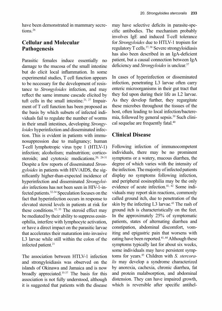
20. Strongyloides stercoralis 233
have been demonstrated in mammary secre-tions.26
Cellular and Molecular Pathogenesis
Parasitic females induce essentially no damage to the mucosa of the small intestine but do elicit local inflammation. In some experimental studies, T cell function appears to be necessary for the development of resis-tance to Strongyloides infection, and may reflect the same immune cascade elicited by tuft cells in the small intestine.21, 27 Impair-ment of T cell function has been proposed as the basis by which subsets of infected indi-viduals fail to regulate the number of worms in their small intestines, developing Strongy-loides hyperinfection and disseminated infec-tion. This is evident in patients with immu-nosuppression due to malignancy; human T-cell lymphotropic virus type 1 (HTLV-1) infection; alcoholism; malnutrition; cortico-steroids; and cytotoxic medications.20, 28-31 Despite a few reports of disseminated Stron-gyloides in patients with HIV/AIDS, the sig-nificantly higher-than-expected incidence of hyperinfection and disseminated Strongyloi-des infections has not been seen in HIV-1-in-fected patients.32-34 Speculation focuses on the fact that hyperinfection occurs in response to elevated steroid levels in patients at risk for these conditions.32, 33 The steroid effect may be mediated by their ability to suppress eosin-ophilia, interfere with lymphocyte activation, or have a direct impact on the parasitic larvae that accelerates their maturation into invasive L3 larvae while still within the colon of the infected patient.21
The association between HTLV-1 infection and strongyloidiasis was observed on the islands of Okinawa and Jamaica and is now broadly appreciated.35-37 The basis for this association is not fully understood, although it is suggested that patients with the disease
may have selective deficits in parasite-spe-cific antibodies. The mechanism probably involves IgE and induced T-cell tolerance for Strongyloides due to HTLV-1 tropism for regulatory T cells.37, 38 Severe strongyloidiasis has also been described in an IgA-deficient patient, but a causal connection between IgA deficiency and Strongyloides is unclear.37
In cases of hyperinfection or disseminated infection, penetrating L3 larvae often carry enteric microorganisms in their gut tract that they fed upon during their life as L2 larvae. As they develop further, they regurgitate these microbes throughout the tissues of the host, often leading to local infection/bactere-mia, followed by general sepsis.39 Such clini-cal sequelae are frequently fatal.40
Clinical Disease
Following infection of immunocompetent individuals, there may be no prominent symptoms or a watery, mucous diarrhea, the degree of which varies with the intensity of the infection. The majority of infected patients display no symptoms following infection, and peripheral eosinophilia may be the only evidence of acute infection.41, 42 Some indi-viduals may report skin reactions, commonly called ground itch, due to penetration of the skin by the infecting L3 larvae.43 The rash of ground itch is characteristically on the feet. In the approximately 25% of symptomatic patients, states of alternating diarrhea and constipation, abdominal discomfort, vom-iting and epigastric pain that worsens with eating have been reported.41, 44 Although these symptoms typically last for about six weeks, some individuals may have persistent symp-toms for years.45 Children with S. stercora-lis may develop a syndrome characterized by anorexia, cachexia, chronic diarrhea, fat and protein malabsorption, and abdominal distension. They can have impaired growth, which is reversible after specific antihel-
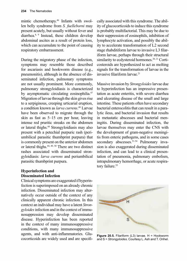
234 The Nematodes
mintic chemotherapy.46 Infants with swol-len belly syndrome from S. fuelleborni may present acutely, but usually without fever and diarrhea.8, 9 Instead, these children develop abdominal ascites as a result of protein loss, which can accumulate to the point of causing respiratory embarrassment.
During the migratory phase of the infection, symptoms may resemble those described for ascariasis and hookworm disease (e.g., pneumonitis), although in the absence of dis-seminated infection, pulmonary symptoms are not usually prominent. More commonly, pulmonary strongyloidiasis is characterized by asymptomatic circulating eosinophilia.47 Migration of larvae through the skin gives rise to a serpiginous, creeping urticarial eruption, a condition known as larva currens.48 Larvae have been observed to migrate through the skin as fast as 5–15 cm per hour, leaving intense red pruritic streaks on the abdomen or lateral thighs.44 Strongyloidiasis may also present with a petechial purpuric rash (peri-umbilical parasitic thumbprint purpura) that is commonly present on the anterior abdomen or lateral thighs.44, 49, 50 There are two distinct rashes associated with disseminated stron-gyloidiasis: larva currens and periumbilical parasitic thumbprint purpura.
Hyperinfection and Disseminated InfectionClinical symptoms are exaggerated if hyperin-fection is superimposed on an already chronic infection. Disseminated infection may alter-natively occur outside of the context of any clinically apparent chronic infection. In this context an individual may have a latent Stron-gyloides infection and in the context of immu-nosuppression may develop disseminated disease. Hyperinfection has been reported in the context of many immunosuppressive conditions, with many immunosuppressive agents, and with anti-inflammatories. Glu-cocorticoids are widely used and are specifi-
cally associated with this syndrome. The abil-ity of glucocorticoids to induce this syndrome is probably multifactorial. This may be due to their suppression of eosinophils, inhibition of lymphocyte activation, and possibly the abil-ity to accelerate transformation of L2 second stage rhabditiform larvae to invasive L3 filar-iform larvae, perhaps through their structural similarity to ecdysteroid hormones.20, 21 Corti-costeroids are hypothesized to act as molting signals for the transformation of larvae in the invasive filariform larvae.21
Massive invasion by Strongyloides larvae due to hyperinfection has an impressive presen-tation as acute enteritis, with severe diarrhea and ulcerating disease of the small and large intestine. These patients often have secondary bacterial enterocolitis that can result in a para-lytic ileus, and bacterial invasion that results in metastatic abscesses and bacterial men-ingitis. During disseminated infection, the larvae themselves may enter the CNS with the development of gram-negative meningi-tis from enteric pathogens, and in some cases secondary abscesses.51-54 Pulmonary inva-sion is also exaggerated during disseminated infection, and can lead to a clinical presen-tation of pneumonia, pulmonary embolism, intrapulmonary hemorrhage, or acute respira-tory failure.55
Figure 20.5. Filariform (L3) larvae. H = Hookworm and S = Strongyloides. Courtesy L. Ash and T. Orihel.
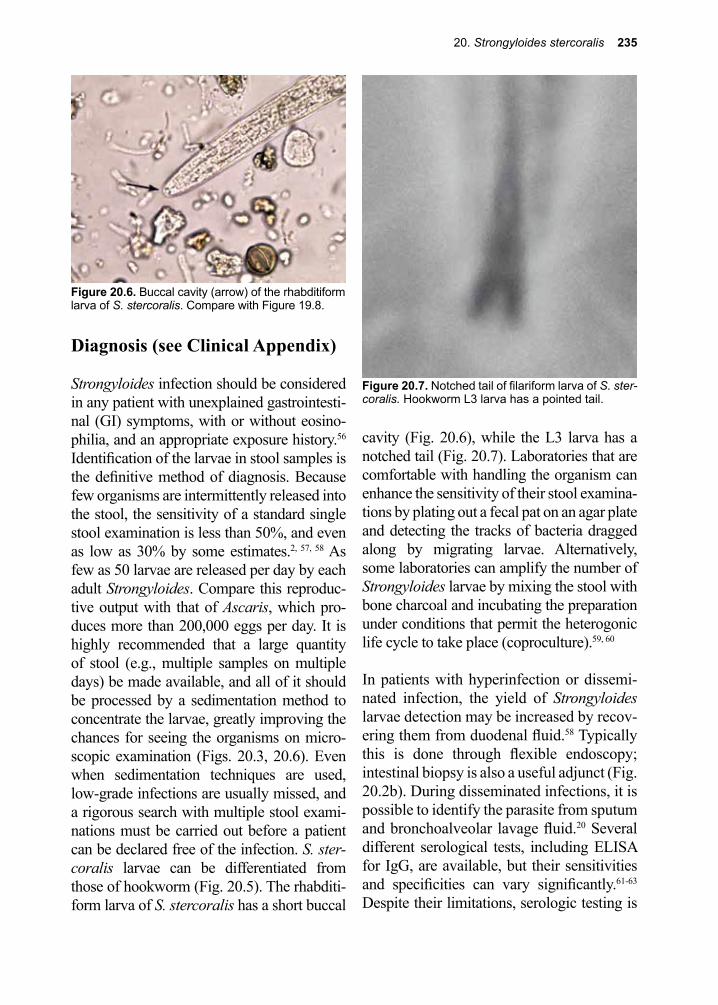
20. Strongyloides stercoralis 235
Diagnosis (see Clinical Appendix)
Strongyloides infection should be considered in any patient with unexplained gastrointesti-nal (GI) symptoms, with or without eosino-philia, and an appropriate exposure history.56 Identification of the larvae in stool samples is the definitive method of diagnosis. Because few organisms are intermittently released into the stool, the sensitivity of a standard single stool examination is less than 50%, and even as low as 30% by some estimates.2, 57, 58 As few as 50 larvae are released per day by each adult Strongyloides. Compare this reproduc-tive output with that of Ascaris, which pro-duces more than 200,000 eggs per day. It is highly recommended that a large quantity of stool (e.g., multiple samples on multiple days) be made available, and all of it should be processed by a sedimentation method to concentrate the larvae, greatly improving the chances for seeing the organisms on micro-scopic examination (Figs. 20.3, 20.6). Even when sedimentation techniques are used, low-grade infections are usually missed, and a rigorous search with multiple stool exami-nations must be carried out before a patient can be declared free of the infection. S. ster-coralis larvae can be differentiated from those of hookworm (Fig. 20.5). The rhabditi-form larva of S. stercoralis has a short buccal
cavity (Fig. 20.6), while the L3 larva has a notched tail (Fig. 20.7). Laboratories that are comfortable with handling the organism can enhance the sensitivity of their stool examina-tions by plating out a fecal pat on an agar plate and detecting the tracks of bacteria dragged along by migrating larvae. Alternatively, some laboratories can amplify the number of Strongyloides larvae by mixing the stool with bone charcoal and incubating the preparation under conditions that permit the heterogonic life cycle to take place (coproculture).59, 60
In patients with hyperinfection or dissemi-nated infection, the yield of Strongyloides larvae detection may be increased by recov-ering them from duodenal fluid.58 Typically this is done through flexible endoscopy; intestinal biopsy is also a useful adjunct (Fig. 20.2b). During disseminated infections, it is possible to identify the parasite from sputum and bronchoalveolar lavage fluid.20 Several different serological tests, including ELISA for IgG, are available, but their sensitivities and specificities can vary significantly.61-63 Despite their limitations, serologic testing is
Figure 20.7. Notched tail of filariform larva of S. ster-coralis. Hookworm L3 larva has a pointed tail.
Figure 20.6. Buccal cavity (arrow) of the rhabditiform larva of S. stercoralis. Compare with Figure 19.8.
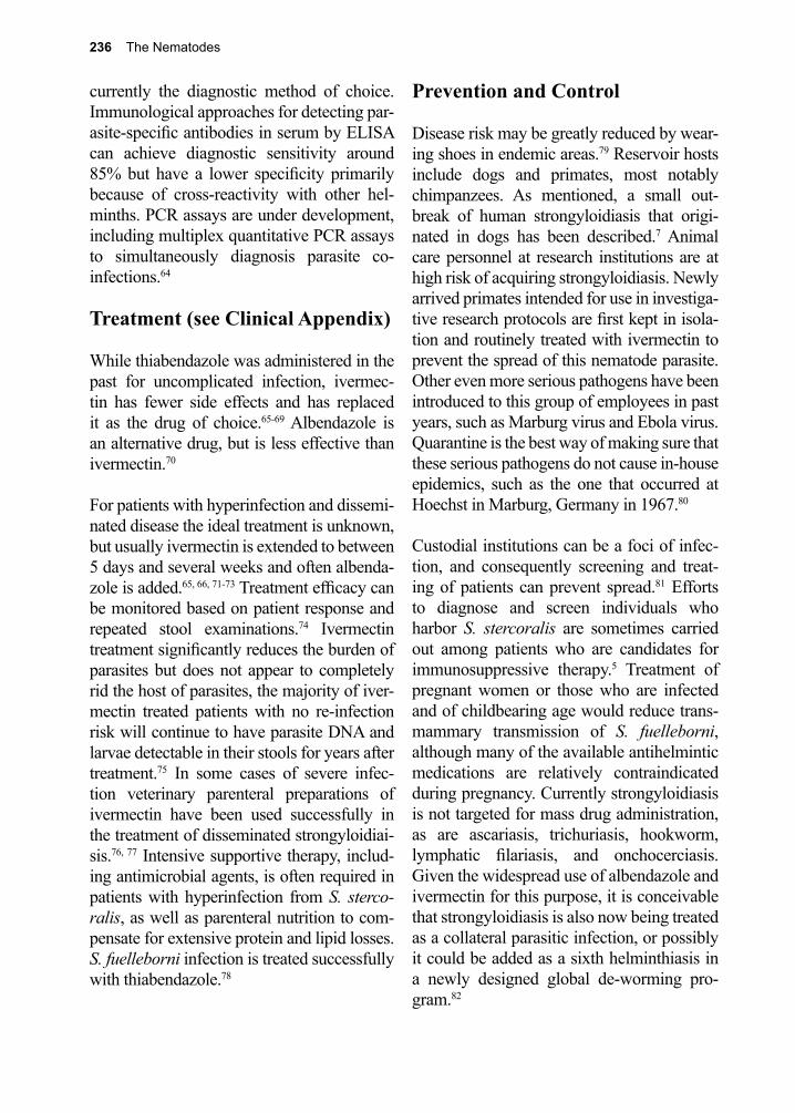
236 The Nematodes
currently the diagnostic method of choice. Immunological approaches for detecting par-asite-specific antibodies in serum by ELISA can achieve diagnostic sensitivity around 85% but have a lower specificity primarily because of cross-reactivity with other hel-minths. PCR assays are under development, including multiplex quantitative PCR assays to simultaneously diagnosis parasite co-infections.64
Treatment (see Clinical Appendix)
While thiabendazole was administered in the past for uncomplicated infection, ivermec-tin has fewer side effects and has replaced it as the drug of choice.65-69 Albendazole is an alternative drug, but is less effective than ivermectin.70
For patients with hyperinfection and dissemi-nated disease the ideal treatment is unknown, but usually ivermectin is extended to between 5 days and several weeks and often albenda-zole is added.65, 66, 71-73 Treatment efficacy can be monitored based on patient response and repeated stool examinations.74 Ivermectin treatment significantly reduces the burden of parasites but does not appear to completely rid the host of parasites, the majority of iver-mectin treated patients with no re-infection risk will continue to have parasite DNA and larvae detectable in their stools for years after treatment.75 In some cases of severe infec-tion veterinary parenteral preparations of ivermectin have been used successfully in the treatment of disseminated strongyloidiai-sis.76, 77 Intensive supportive therapy, includ-ing antimicrobial agents, is often required in patients with hyperinfection from S. sterco-ralis, as well as parenteral nutrition to com-pensate for extensive protein and lipid losses. S. fuelleborni infection is treated successfully with thiabendazole.78
Prevention and Control
Disease risk may be greatly reduced by wear-ing shoes in endemic areas.79 Reservoir hosts include dogs and primates, most notably chimpanzees. As mentioned, a small out-break of human strongyloidiasis that origi-nated in dogs has been described.7 Animal care personnel at research institutions are at high risk of acquiring strongyloidiasis. Newly arrived primates intended for use in investiga-tive research protocols are first kept in isola-tion and routinely treated with ivermectin to prevent the spread of this nematode parasite. Other even more serious pathogens have been introduced to this group of employees in past years, such as Marburg virus and Ebola virus. Quarantine is the best way of making sure that these serious pathogens do not cause in-house epidemics, such as the one that occurred at Hoechst in Marburg, Germany in 1967.80
Custodial institutions can be a foci of infec-tion, and consequently screening and treat-ing of patients can prevent spread.81 Efforts to diagnose and screen individuals who harbor S. stercoralis are sometimes carried out among patients who are candidates for immunosuppressive therapy.5 Treatment of pregnant women or those who are infected and of childbearing age would reduce trans-mammary transmission of S. fuelleborni, although many of the available antihelmintic medications are relatively contraindicated during pregnancy. Currently strongyloidiasis is not targeted for mass drug administration, as are ascariasis, trichuriasis, hookworm, lymphatic filariasis, and onchocerciasis. Given the widespread use of albendazole and ivermectin for this purpose, it is conceivable that strongyloidiasis is also now being treated as a collateral parasitic infection, or possibly it could be added as a sixth helminthiasis in a newly designed global de-worming pro-gram.82
20. Strongyloides stercoralis 237
References
1. Hatter, A. D., et al., Case report of individual with cutaneous immunodeficiency and novel 1p36 duplication. Appl Clin Genet 2016, 9, 1-4.
2. Genta, R. M., et al., Strongyloidiasis in US veterans of the Vietnam and other wars. JAMA 1987, 258 (1), 49-52.
3. Russell, E. S., et al., Prevalence of Strongyloides stercoralis antibodies among a rural Appalachian population--Kentucky, 2013. Am J Trop Med Hyg 2014, 91 (5), 1000-1.
4. Centers for Disease, C.; Prevention, Notes from the field: strongyloides infection among patients at a long-term care facility--Florida, 2010-2012. MMWR Morb Mortal Wkly Rep 2013, 62 (42), 844.
5. Genta, R. M., Global prevalence of strongyloidiasis: critical review with epidemiologic insights into the prevention of disseminated disease. Reviews of infectious diseases 1989, 11 (5), 755-67.
6. Goncalves, A. L., et al., Evaluation of strongyloidiasis in kennel dogs and keepers by parasitological and serological assays. Vet Parasitol 2007, 147 (1-2), 132-9.
7. Georgi, J. R.; Sprinkle, C. L., A case of human strongyloidosis apparently contracted from asymptomatic colony dogs. Am J Trop Med Hyg 1974, 23 (5), 899-901.
8. Ashford, R. W., et al., Strongyloides infection associated with acute infantile disease in Papua New Guinea. Transactions of the Royal Society of Tropical Medicine and Hygiene 1978, 72 (5), 554.
9. Barnish, G.; Ashford, R. W., Strongyloides cf. fuelleborni and hookworm in Papua New Guinea: patterns of infection within the community. Transactions of the Royal Society of Tropical Medicine and Hygiene 1989, 83 (5), 684-8.
10. Zerpa Larrauri, R., et al., [Life cycle of Strongyloides fuelleborni: images and videos]. Rev Peru Med Exp Salud Publica 2014, 31 (2), 390-2.
11. Vince, J. D., et al., Strongyloides species infestation in young infants of papu, New Guinea: association with generalized oedema. P N G Med J 1979, 22 (2), 120-7.
12. Ashford, R. W., et al., Strongyloides infection in a mid-mountain Papua New Guinea community: results of an epidemiological survey. P N G Med J 1979, 22 (2), 128-35.
13. Bavay, A.; R., C., Sur l’anguillule stercorale. Sci Paris 1876, 83, 694-696.14. Normand, L. A.; R., C., Sur la maladie dite diarrhie de Cochinchine. Sean Acad Sci 1876, 83
316-318.15. Darling, S. T., Strongyloides Infections in Man and Animals in the Isthmian Canal Zone. J Exp
Med 1911, 14 (1), 1-24.16. Askanazy, M., Uber Art und Zweck der Invasion der Anguillula intestinalis in die Darmwand.
Zbl Bakt 1900, 27 569-578.17. Fuelleborn, F., Untersuchungen uber den Infektionsweg bei Strongyloides und Ankylostomum
und die Biologie diesen Parasiten. Arch Schiffs Trop 1914, 18, 26-80.18. Nishigoii, M., On various factors influencing the development of Strongyloides stercoralis and
autoinfection. Taiwan Sgakkai Zassi 1928, 27, 1-56.19. Streit, A., Reproduction in Strongyloides (Nematoda): a life between sex and parthenogenesis.
Parasitology 2008, 135 (3), 285-94.20. Keiser, P. B.; Nutman, T. B., Strongyloides stercoralis in the Immunocompromised Population.
Clin Microbiol Rev 2004, 17 (1), 208-17.21. Genta, R. M., Dysregulation of strongyloidiasis: a new hypothesis. Clinical microbiology reviews
1992, 5 (4), 345-55.22. McKerrow, J. H.; Brindley, P., Strongyloides stercoralis: identification of a protease whose
inhibition prevents larval skin invasion. Exp Parasitol 1990, 70, 134-143.23. Schad, G. A., et al., Strongyloides stercoralis: is there a canonical migratory route through the
host? The Journal of parasitology 1989, 75 (5), 740-9.24. Shekamer, J. H., et al., Persistent strongyloidiasis in an immunodeficent patient. Am Med Hyg
1982, 31 746-751.25. Schad, G. A., et al., Strongyloides stercoralis: an initial autoinfective burst amplifies primary
infection. The American journal of tropical medicine and hygiene 1993, 48 (5), 716-25.
238 The Nematodes
26. Brown, R. C.; Girardeau, H. F., Transmammary passage of Strongyloides sp. larvae in the human host. The American journal of tropical medicine and hygiene 1977, 26 (2), 215-9.
27. Howitt, M. R., et al., Tuft cells, taste-chemosensory cells, orchestrate parasite type 2 immunity in the gut. Science 2016, 351 (6279), 1329-33.
28. Safdar, A., et al., Strongyloidiasis in patients at a comprehensive cancer center in the United States. Cancer 2004, 100 (7), 1531-6.
29. Carvalho, E. M.; Da Fonseca Porto, A., Epidemiological and clinical interaction between HTLV-1 and Strongyloides stercoralis. Parasite Immunol 2004, 26 (11-12), 487-97.
30. Marques, C. C., et al., Alcoholism and Strongyloides stercoralis: daily ethanol ingestion has a positive correlation with the frequency of Strongyloides larvae in the stools. PLoS Negl Trop Dis 2010, 4 (6), e717.
31. Buonfrate, D., et al., Severe strongyloidiasis: a systematic review of case reports. BMC Infect Dis 2013, 13, 78.
32. Lucas, S. B., Missing infections in AIDS. Transactions of the Royal Society of Tropical Medicine and Hygiene 1990, 84 Suppl 1, 34-8.
33. Robinson, R. D., Parasitic infections associated with HIV/AIDS in the Caribbean. Bulletin of the Pan American Health Organization 1995, 29 (2), 129-37.
34. Viney, M. E., et al., Why does HIV infection not lead to disseminated strongyloidiasis? J Infect Dis 2004, 190 (12), 2175-80.
35. Dixon, A. C., et al., Strongyloidiasis associated with human T-cell lymphotropic virus type I infection in a nonendemic area. The Western journal of medicine 1989, 151 (4), 410-3.
36. Arakaki, T., et al., Epidemiological aspects of Strongyloides stercoralis infection in Okinawa, Japan. The Journal of tropical medicine and hygiene 1992, 95 (3), 210-3.
37. Neva, F. A., et al., Antibodies to Strongyloides stercoralis in healthy Jamaican carriers of HTLV-1. 1989, 320, 252-3.
38. Montes, M., et al., Regulatory T cell expansion in HTLV-1 and strongyloidiasis co-infection is associated with reduced IL-5 responses to Strongyloides stercoralis antigen. PLoS Negl Trop Dis 2009, 3 (6), e456.
39. Gorman, S. R.; Craven, D. E., Images in clinical medicine. Strongyloides stercoralis hyperinfection. N Engl J Med 2008, 359 (11), e12.
40. Geri, G., et al., Strongyloides stercoralis hyperinfection syndrome: a case series and a review of the literature. Infection 2015, 43 (6), 691-8.
41. Valerio, L., et al., Strongyloides stercoralis, the hidden worm. Epidemiological and clinical characteristics of 70 cases diagnosed in the North Metropolitan Area of Barcelona, Spain, 2003-2012. Trans R Soc Trop Med Hyg 2013, 107 (8), 465-70.
42. Gonzalez, A., et al., Clinical and epidemiological features of 33 imported Strongyloides stercoralis infections. Trans R Soc Trop Med Hyg 2010, 104 (9), 613-6.
43. Bailey, M. S., et al., Helminth infections in British troops following an operation in Sierra Leone. Trans R Soc Trop Med Hyg 2006, 100 (9), 842-6.
44. Greaves, D., et al., Strongyloides stercoralis infection. BMJ 2013, 347, f4610.45. Vadlamudi, R. S., et al., Intestinal strongyloidiasis and hyperinfection syndrome. Clin Mol
Allergy 2006, 4, 8.46. Leung, V. K., et al., Strongyloidiasis in a patient with IgA deficiency. Tropical gastroenterology :
official journal of the Digestive Diseases Foundation 1995, 16 (4), 27-30.47. Burke, J. A., Strongyloidiasis in childhood. American journal of diseases of children (1960)
1978, 132 (11), 1130-6.48. Berk, S. L., et al., Clinical and epidemiologic features of strongyloidiasis. A prospective study in
rural Tennessee. Archives of internal medicine 1987, 147 (7), 1257-61.49. Ribeiro, L. C., et al., [Purpura in patient with disseminated strongiloidiasis]. Rev Soc Bras Med
Trop 2005, 38 (3), 255-7.50. Weiser, J. A., et al., Periumbilical parasitic thumbprint purpura: strongyloides hyperinfection
syndrome acquired from a cadaveric renal transplant. Transpl Infect Dis 2011, 13 (1), 58-62.51. Cho, J. Y., et al., [A case of steroid-induced hyperinfective strongyloidiasis with bacterial
meningitis]. Korean J Gastroenterol 2012, 60 (5), 330-4.
20. Strongyloides stercoralis 239
52. Sasaki, Y., et al., Meningitis associated with strongyloidiasis in an area endemic for strongyloidiasis and human T-lymphotropic virus-1: a single-center experience in Japan between 1990 and 2010. Infection 2013, 41 (6), 1189-93.
53. Shimasaki, T., et al., Five cases of recurrent meningitis associated with chronic strongyloidiasis. Am J Trop Med Hyg 2015, 92 (3), 601-4.
54. Zammarchi, L., et al., Persistent strongyloidiasis complicated by recurrent meningitis in an HTLV seropositive Peruvian migrant resettled in Italy. Am J Trop Med Hyg 2015, 92 (6), 1257-60.
55. Newberry, A. M., et al., Strongyloides hyperinfection presenting as acute respiratory failure and gram-negative sepsis. Chest 2005, 128 (5), 3681-4.
56. Arsic-Arsenijevic, V., et al., Fatal Strongyloides stercoralis infection in a young woman with lupus glomerulonephritis. J Nephrol 2005, 18 (6), 787-90.
57. Siddiqui, A. A.; Berk, S. L., Diagnosis of Strongyloides stercoralis infection. Clin Infect Dis 2001, 33 (7), 1040-7.
58. Jones, C. A.; Abadie, S. H., Studies in human strongyloidiasis. II. A comparison of the efficiency of diagnosis by examination of feces and duodenal fluid. Am J Clin Pathol 1954, 24 (10), 1154-8.
59. Costa-Cruz, J. M., et al., Cryo-microtome sections of coproculture larvae of Strongyloides stercoralis and Strongyloides ratti as antigen sources for the immunodiagnosis of human strongyloidiasis. Rev Inst Med Trop Sao Paulo 1997, 39 (6), 313-7.
60. Attia, M. M., et al., Some observations on coproculture of Strongyloides stercoralis. J Egypt Soc Parasitol 1982, 12 (1), 103-6.
61. van Doorn, H. R., et al., Use of enzyme-linked immunosorbent assay and dipstick assay for detection of Strongyloides stercoralis infection in humans. J Clin Microbiol 2007, 45 (2), 438-42.
62. Von Kuster, L. C.; Genta, R. M., Cutaneous manifestations of strongyloidiasis. Arch Dermatol 1988, 124, 1826-1830.
63. Buonfrate, D., et al., Accuracy of five serologic tests for the follow up of Strongyloides stercoralis infection. PLoS Negl Trop Dis 2015, 9 (2), e0003491.
64. Cimino, R. O., et al., Identification of human intestinal parasites affecting an asymptomatic peri-urban Argentinian population using multi-parallel quantitative real-time polymerase chain reaction. Parasit Vectors 2015, 8, 380.
65. Neva, F. A., Biology and immunology of human strongyloidiasis. J Infect Dis 1986, 153 (3), 397-406.
66. Naquira, C., et al., Ivermectin for human strongyloidiasis and other intestinal helminths. Am J Trop Med Hyg 1989, 40 (3), 304-9.
67. Lyagoubi, M., et al., Chronic persistent strongyloidiasis cured by ivermectin. Transactions of the Royal Society of Tropical Medicine and Hygiene 1992, 86 (5), 541.
68. Wijesundera, M. D., et al., Ivermectin therapy in chronic strongyloidiasis. Trans Trop Med Hyg 291 1994, 86
69. Gann, P. H., et al., A randomized trial of single- and two-dose ivermectin versus thiabendazole for treatment of strongyloidiasis. J Infect Dis 1994, 169 (5), 1076-9.
70. Muennig, P., et al., The cost-effectiveness of ivermectin vs. albendazole in the presumptive treatment of strongyloidiasis in immigrants to the United States. Epidemiol Infect 2004, 132 (6), 1055-63.
71. Segarra-Newnham, M., Manifestations, diagnosis, and treatment of Strongyloides stercoralis infection. Ann Pharmacother 2007, 41 (12), 1992-2001.
72. Mejia, R.; Nutman, T. B., Screening, prevention, and treatment for hyperinfection syndrome and disseminated infections caused by Strongyloides stercoralis. Curr Opin Infect Dis 2012, 25 (4), 458-63.
73. Boggild, A. K., et al., CATMAT statement on disseminated strongyloidiasis: Prevention, assessment and management guidelines. Can Comm Dis Rep 2016, 42, 12-19.
74. Schar, F., et al., Strongyloides stercoralis larvae excretion patterns before and after treatment. Parasitology 2014, 141 (7), 892-7.
240 The Nematodes
75. Repetto, S. A., et al., Strongyloidiasis Outside Endemic Areas: Long-term Parasitological and Clinical Follow-up After Ivermectin Treatment. Clin Infect Dis 2018, 66 (10), 1558-1565.
76. Barrett, J.; Newsholme, W., Subcutaneous ivermectin use in the treatment of severe Strongyloides stercoralis infection: two case reports and a discussion of the literature-authors’ response. J Antimicrob Chemother 2016.
77. Buonfrate, D., et al., Comment on: Subcutaneous ivermectin use in the treatment of severe Strongyloides stercoralis infection: two case reports and a discussion of the literature. J Antimicrob Chemother 2016.
78. Barnish, G.; Barker, J., An intervention study using thiabendazole suspension against Strongyloides fuelleborni-like infections in Papua New Guinea. Trans R Soc Trop Med Hyg 1987, 81 (1), 60-3.
79. Yori, P. P., et al., Seroepidemiology of strongyloidiasis in the Peruvian Amazon. Am J Trop Med Hyg 2006, 74 (1), 97-102.
80. Slenczka, W.; Klenk, H. D., Forty years of marburg virus. J Infect Dis 2007, 196 Suppl 2, S131-5.
81. Nair, D., Screening for Strongyloides infection among the institutionalized mentally disabled. J Am Board Fam Pract 2001, 14 (1), 51-3.
82. Krolewiecki, A. J., et al., A public health response against Strongyloides stercoralis: time to look at soil-transmitted helminthiasis in full. PLoS Negl Trop Dis 2013, 7 (5), e2165.
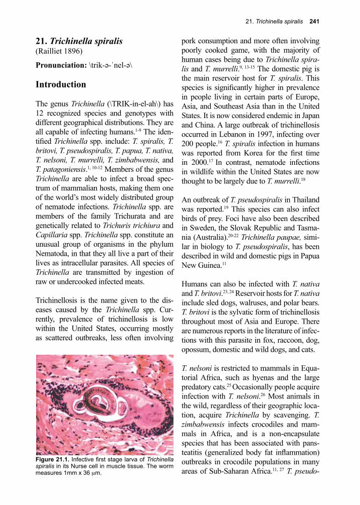
21. Trichinella spiralis 241
Figure 21.1. Infective first stage larva of Trichinella spiralis in its Nurse cell in muscle tissue. The worm measures 1mm x 36 µm.
21. Trichinella spiralis(Railliet 1896)
Pronunciation: \trik-ə-ˈnel-ə\
Introduction
The genus Trichinella (\TRIK-in-el-ah\) has 12 recognized species and genotypes with different geographical distributions. They are all capable of infecting humans.1-9 The iden-tified Trichinella spp. include: T. spiralis, T. britovi, T. pseudospiralis, T. papua, T. nativa, T. nelsoni, T. murrelli, T. zimbabwensis, and T. patagoniensis.1, 10-12 Members of the genus Trichinella are able to infect a broad spec-trum of mammalian hosts, making them one of the world’s most widely distributed group of nematode infections. Trichinella spp. are members of the family Trichurata and are genetically related to Trichuris trichiura and Capillaria spp. Trichinella spp. constitute an unusual group of organisms in the phylum Nematoda, in that they all live a part of their lives as intracellular parasites. All species of Trichinella are transmitted by ingestion of raw or undercooked infected meats.
Trichinellosis is the name given to the dis-eases caused by the Trichinella spp. Cur-rently, prevalence of trichinellosis is low within the United States, occurring mostly as scattered outbreaks, less often involving
pork consumption and more often involving poorly cooked game, with the majority of human cases being due to Trichinella spira-lis and T. murrelli.9, 13-15 The domestic pig is the main reservoir host for T. spiralis. This species is significantly higher in prevalence in people living in certain parts of Europe, Asia, and Southeast Asia than in the United States. It is now considered endemic in Japan and China. A large outbreak of trichinellosis occurred in Lebanon in 1997, infecting over 200 people.16 T. spiralis infection in humans was reported from Korea for the first time in 2000.17 In contrast, nematode infections in wildlife within the United States are now thought to be largely due to T. murrelli.18
An outbreak of T. pseudospiralis in Thailand was reported.19 This species can also infect birds of prey. Foci have also been described in Sweden, the Slovak Republic and Tasma-nia (Australia).20-22 Trichinella paupae, simi-lar in biology to T. pseudospiralis, has been described in wild and domestic pigs in Papua New Guinea.11
Humans can also be infected with T. nativa and T. britovi.23, 24 Reservoir hosts for T. nativa include sled dogs, walruses, and polar bears.
T. britovi is the sylvatic form of trichinellosis throughout most of Asia and Europe. There are numerous reports in the literature of infec-tions with this parasite in fox, raccoon, dog, opossum, domestic and wild dogs, and cats.
T. nelsoni is restricted to mammals in Equa-torial Africa, such as hyenas and the large predatory cats.25 Occasionally people acquire infection with T. nelsoni.26 Most animals in the wild, regardless of their geographic loca-tion, acquire Trichinella by scavenging. T. zimbabwensis infects crocodiles and mam-mals in Africa, and is a non-encapsulate species that has been associated with pans-teatitis (generalized body fat inflammation) outbreaks in crocodile populations in many areas of Sub-Saharan Africa.11, 27 T. pseudo-
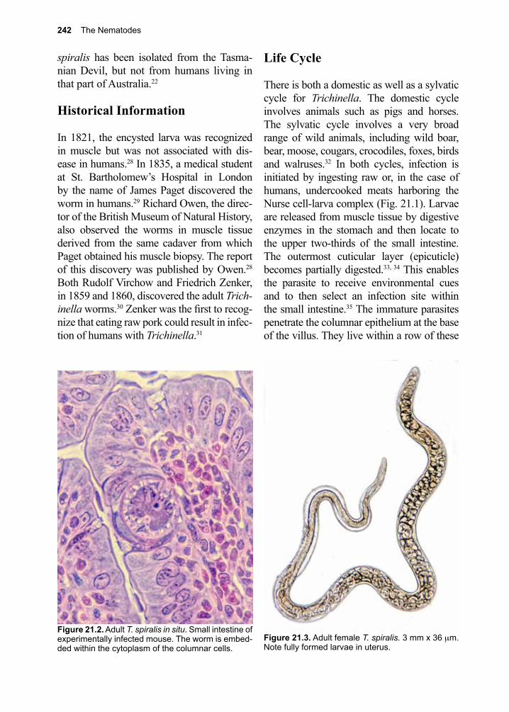
242 The Nematodes
spiralis has been isolated from the Tasma-nian Devil, but not from humans living in that part of Australia.22
Historical Information
In 1821, the encysted larva was recognized in muscle but was not associated with dis-ease in humans.28 In 1835, a medical student at St. Bartholomew’s Hospital in London by the name of James Paget discovered the worm in humans.29 Richard Owen, the direc-tor of the British Museum of Natural History, also observed the worms in muscle tissue derived from the same cadaver from which Paget obtained his muscle biopsy. The report of this discovery was published by Owen.28 Both Rudolf Virchow and Friedrich Zenker, in 1859 and 1860, discovered the adult Trich-inella worms.30 Zenker was the first to recog-nize that eating raw pork could result in infec-tion of humans with Trichinella.31
Life Cycle
There is both a domestic as well as a sylvatic cycle for Trichinella. The domestic cycle involves animals such as pigs and horses. The sylvatic cycle involves a very broad range of wild animals, including wild boar, bear, moose, cougars, crocodiles, foxes, birds and walruses.32 In both cycles, infection is initiated by ingesting raw or, in the case of humans, undercooked meats harboring the Nurse cell-larva complex (Fig. 21.1). Larvae are released from muscle tissue by digestive enzymes in the stomach and then locate to the upper two-thirds of the small intestine. The outermost cuticular layer (epicuticle) becomes partially digested.33, 34 This enables the parasite to receive environmental cues and to then select an infection site within the small intestine.35 The immature parasites penetrate the columnar epithelium at the base of the villus. They live within a row of these
Figure 21.3. Adult female T. spiralis. 3 mm x 36 µm. Note fully formed larvae in uterus.
Figure 21.2. Adult T. spiralis in situ. Small intestine of experimentally infected mouse. The worm is embed-ded within the cytoplasm of the columnar cells.
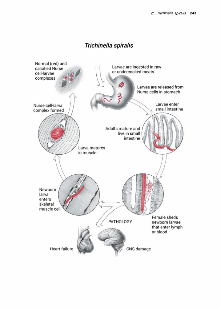
21. Trichinella spiralis 243
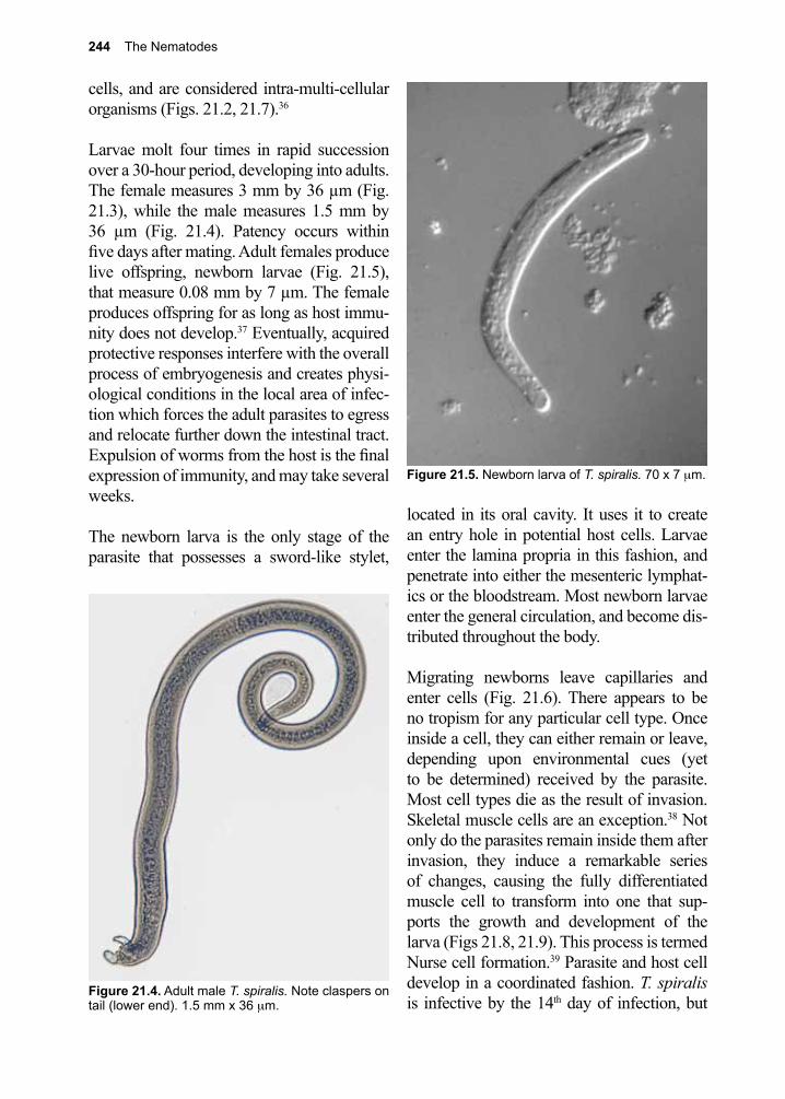
244 The Nematodes
Figure 21.5. Newborn larva of T. spiralis. 70 x 7 µm.
cells, and are considered intra-multi-cellular organisms (Figs. 21.2, 21.7).36
Larvae molt four times in rapid succession over a 30-hour period, developing into adults. The female measures 3 mm by 36 µm (Fig. 21.3), while the male measures 1.5 mm by 36 µm (Fig. 21.4). Patency occurs within five days after mating. Adult females produce live offspring, newborn larvae (Fig. 21.5), that measure 0.08 mm by 7 µm. The female produces offspring for as long as host immu-nity does not develop.37 Eventually, acquired protective responses interfere with the overall process of embryogenesis and creates physi-ological conditions in the local area of infec-tion which forces the adult parasites to egress and relocate further down the intestinal tract. Expulsion of worms from the host is the final expression of immunity, and may take several weeks.
The newborn larva is the only stage of the parasite that possesses a sword-like stylet,
located in its oral cavity. It uses it to create an entry hole in potential host cells. Larvae enter the lamina propria in this fashion, and penetrate into either the mesenteric lymphat-ics or the bloodstream. Most newborn larvae enter the general circulation, and become dis-tributed throughout the body.
Migrating newborns leave capillaries and enter cells (Fig. 21.6). There appears to be no tropism for any particular cell type. Once inside a cell, they can either remain or leave, depending upon environmental cues (yet to be determined) received by the parasite. Most cell types die as the result of invasion. Skeletal muscle cells are an exception.38 Not only do the parasites remain inside them after invasion, they induce a remarkable series of changes, causing the fully differentiated muscle cell to transform into one that sup-ports the growth and development of the larva (Figs 21.8, 21.9). This process is termed Nurse cell formation.39 Parasite and host cell develop in a coordinated fashion. T. spiralis is infective by the 14th day of infection, but
Figure 21.4. Adult male T. spiralis. Note claspers on tail (lower end). 1.5 mm x 36 µm.
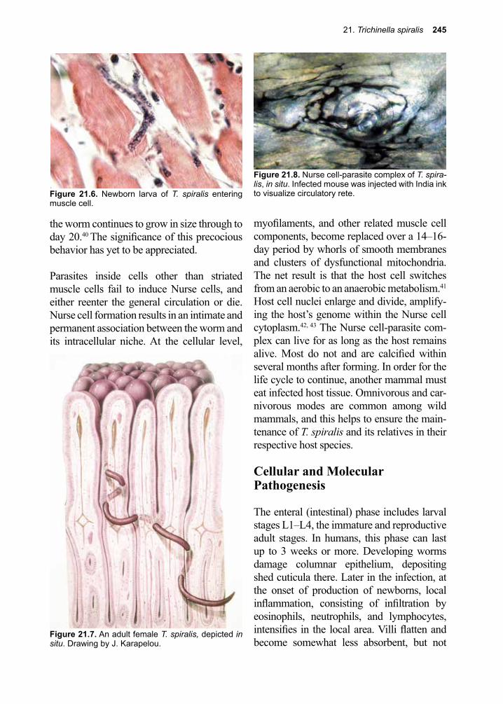
21. Trichinella spiralis 245
Figure 21.7. An adult female T. spiralis, depicted in situ. Drawing by J. Karapelou.
Figure 21.8. Nurse cell-parasite complex of T. spira-lis, in situ. Infected mouse was injected with India ink to visualize circulatory rete.
the worm continues to grow in size through to day 20.40 The significance of this precocious behavior has yet to be appreciated.
Parasites inside cells other than striated muscle cells fail to induce Nurse cells, and either reenter the general circulation or die. Nurse cell formation results in an intimate and permanent association between the worm and its intracellular niche. At the cellular level,
myofilaments, and other related muscle cell components, become replaced over a 14–16-day period by whorls of smooth membranes and clusters of dysfunctional mitochondria. The net result is that the host cell switches from an aerobic to an anaerobic metabolism.41 Host cell nuclei enlarge and divide, amplify-ing the host’s genome within the Nurse cell cytoplasm.42, 43 The Nurse cell-parasite com-plex can live for as long as the host remains alive. Most do not and are calcified within several months after forming. In order for the life cycle to continue, another mammal must eat infected host tissue. Omnivorous and car-nivorous modes are common among wild mammals, and this helps to ensure the main-tenance of T. spiralis and its relatives in their respective host species.
Cellular and Molecular Pathogenesis
The enteral (intestinal) phase includes larval stages L1–L4, the immature and reproductive adult stages. In humans, this phase can last up to 3 weeks or more. Developing worms damage columnar epithelium, depositing shed cuticula there. Later in the infection, at the onset of production of newborns, local inflammation, consisting of infiltration by eosinophils, neutrophils, and lymphocytes, intensifies in the local area. Villi flatten and become somewhat less absorbent, but not
Figure 21.6. Newborn larva of T. spiralis entering muscle cell.
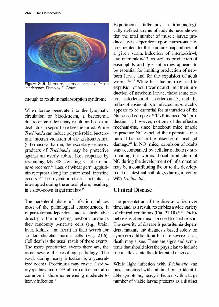
246 The Nematodes
enough to result in malabsorption syndrome.
When larvae penetrate into the lymphatic circulation or bloodstream, a bacteremia due to enteric flora may result, and cases of death due to sepsis have been reported. While Trichinella can induce polymicrobial bactere-mia through violation of the gastrointestinal (GI) mucosal barrier, the excretory-secretory products of Trichinella may be protective against an overly robust host response by restraining MyD88 signaling via the man-nose receptor.44 Loss of wheat germ aggluti-nin receptors along the entire small intestine occurs.45 The myenteric electric potential is interrupted during the enteral phase, resulting in a slow-down in gut motility.45
The parenteral phase of infection induces most of the pathological consequences. It is parasitemia-dependent and is attributable directly to the migrating newborn larvae as they randomly penetrate cells (e.g., brain, liver, kidney, and heart) in their search for striated skeletal muscle cells (Fig. 21.6). Cell death is the usual result of these events. The more penetration events there are, the more severe the resulting pathology. The result during heavy infection is a general-ized edema. Proteinuria may ensue. Cardio-myopathies and CNS abnormalities are also common in those experiencing moderate to heavy infection.1
Experimental infections in immunologi-cally defined strains of rodents have shown that the total number of muscle larvae pro-duced was dependent upon numerous fac-tors related to the immune capabilities of a given strain. Induction of interleukin-4, and interleukin-13, as well as production of eosinophils and IgE antibodies appears to be essential for limiting production of new-born larvae and for the expulsion of adult worms.46, 47 While host factors may lead to expulsion of adult worms and limit their pro-duction of newborn larvae, these same fac-tors, interleukin-4, interleukin-13, and the influx of eosinophils to infected muscle cells, appears to be essential for maturation of the Nurse-cell complex.48 TNF-induced NO pro-duction is, however, not one of the effector mechanisms, since knockout mice unable to produce NO expelled their parasites in a normal fashion in the absence of local gut damage.49 In NO+ mice, expulsion of adults was accompanied by cellular pathology sur-rounding the worms. Local production of NO during the development of inflammation may be a contributing factor to the develop-ment of intestinal pathology during infection with Trichinella.
Clinical Disease
The presentation of the disease varies over time, and, as a result, resembles a wide variety of clinical conditions (Fig. 21.10).1, 50 Trichi-nellosis is often misdiagnosed for that reason. The severity of disease is parasitemia-depen-dent, making the diagnosis based solely on symptoms difficult, at best. In severe cases, death may ensue. There are signs and symp-toms that should alert the physician to include trichinellosis into the differential diagnosis.
While light infection with Trichinella can pass unnoticed with minimal or no identifi-able symptoms, heavy infection with a large number of viable larvae presents as a distinct
Figure 21.9. Nurse cell-parasite complex. Phase interference. Photo by E. Gravé.
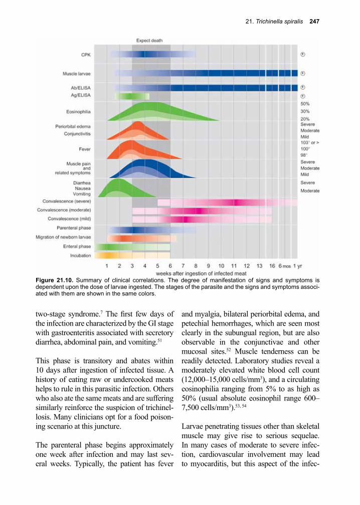
21. Trichinella spiralis 247
two-stage syndrome.7 The first few days of the infection are characterized by the GI stage with gastroenteritis associated with secretory diarrhea, abdominal pain, and vomiting.51
This phase is transitory and abates within 10 days after ingestion of infected tissue. A history of eating raw or undercooked meats helps to rule in this parasitic infection. Others who also ate the same meats and are suffering similarly reinforce the suspicion of trichinel-losis. Many clinicians opt for a food poison-ing scenario at this juncture.
The parenteral phase begins approximately one week after infection and may last sev-eral weeks. Typically, the patient has fever
and myalgia, bilateral periorbital edema, and petechial hemorrhages, which are seen most clearly in the subungual region, but are also observable in the conjunctivae and other mucosal sites.52 Muscle tenderness can be readily detected. Laboratory studies reveal a moderately elevated white blood cell count (12,000–15,000 cells/mm3), and a circulating eosinophilia ranging from 5% to as high as 50% (usual absolute eosinophil range 600–7,500 cells/mm3).53, 54
Larvae penetrating tissues other than skeletal muscle may give rise to serious sequelae. In many cases of moderate to severe infec-tion, cardiovascular involvement may lead to myocarditis, but this aspect of the infec-
Figure 21.10. Summary of clinical correlations. The degree of manifestation of signs and symptoms is dependent upon the dose of larvae ingested. The stages of the parasite and the signs and symptoms associ-ated with them are shown in the same colors.
248 The Nematodes
tion has been overrated as a clinical feature typical of most infections with this parasite, since most instances encountered by the clini-cian are of the mild variety.55 Electrocardio-graphic changes can occur during this phase, even in the absence of symptoms. Parasite invasion of the diaphragm and the accessory muscles of respiration may result in dys-pnea.56 Neurotrichinellosis occurs in associa-tion with invasion of the CNS.57 In up to 25% of patients with heavy infection, meningitis and encephalitis can develop with edema, infarcts, and hemorrhage in fatal cases.58, 59 The convalescent phase follows the acute phase, during which time many, but not all, Nurse-cell-parasite complexes are destroyed.
Two clinical presentations have been described for T. nativa infections resulting from the ingestion of infected polar bear or walrus meat: a classic myopathic form, and a second form that presents as a persistent diar-rheal illness. The second form is thought to represent a secondary infection in previously sensitized individuals.60 It is apparent that not only the amount of ingested larvae but also the species of Trichinella and the status of the infected host impact the clinical presenta-tion.61
Diagnosis (see Clinical Appendix)
Definitive diagnosis depends upon finding the Nurse cell-parasite complex in muscle biopsy by microscopic examination (Fig 21.1), or detection of Trichinella-specific DNA by PCR.62 PCR is sensitive and spe-cific for detecting small numbers of larvae in muscle tissue, but is rarely used in the clinical setting.52 Real-time PCR has been developed and is used in surveillance studies of wildlife Trichinella infection.63
Muscle biopsy can be negative, even in the heaviest of infections, due to sampling errors and as a consequence it is recommended that
symptomatic muscle, preferentially a more proximal muscle such as the deltoid, near the tendinous insertion, be sampled, to improve sensitivity.1 In addition, the larvae may be at an early stage of their development, making them inconspicuous, even to experienced pathologists. Serological tests are available with confirmatory Western blot testing.64 Serological tests begin to show positive results within two weeks. ELISA can detect antibodies in some patients as early as 12 days after infection. Muscle enzymes, such as creatine phosphokinase (CPK) and lactic dehydrogenase, are released into the circula-tion causing an increase in their serum levels.
A rising, plateauing and falling level of cir-culating eosinophils throughout the infection period is not direct proof of infection, but armed with this information and an exposure history, the clinician could treat the patient presumptively. It is helpful to remember that wild mammals can also be sources of infec-tion. Outbreaks of trichinellosis have been traced to hunters and the recipients of their kills.65-67
Treatment (see Clinical Appendix)
While many cases of Trichinella infection may be mild or even asymptomatic and easily managed with analgesics and antipyretics, more significant infections are usually man-aged with a combination of antihelmintic therapy and corticosteroids.52 Since larvae may still be burrowing through the GI tract when patients present with symptoms, treat-ment with albendazole or mebendazole may be beneficial. Treatment with mebendazole early in the course of infection has been associated with decreased muscle pain and muscle inflammation.68 Once muscle inva-sion has occurred, treatment with these agents will not succeed in destroying encysted larvae.69 Anti-inflammatory corticosteroids, particularly prednisolone, are recommended
21. Trichinella spiralis 249
if the diagnosis is secure.1, 49 The myopathic phase is treated in conjunction with antipyret-ics and analgesics (aspirin, acetaminophen), and should be continued until the fever and allergic signs recede. Because of their immu-nosuppressive potential, steroids should be administered with caution.
Prevention and Control
Within the last 10 years, outbreaks of trichi-nellosis in the United States have been rare and sporadic in nature.13-15, 70 Most have been associated with the ingestion of raw or under-cooked meats from game animals, and not from commercial sources.13-15 This represents a shift in the epidemiology of outbreaks com-pared to 20–30 years ago, when commercial sources of infected pork were much more common than today. Pigs raised on indi-vidual farms, as compared with commercial farm operations, are more likely to be fed uncooked garbage, and acquire the infection. This is because feeding unprocessed garbage containing meat scraps is against federally mandated regulations. In the past 10 years, small farms have, in the main, been bought up and replaced with larger so-called “fac-tory” farms, in which upwards of 10,000 pigs can be managed with a minimum of labor. Enforcement of laws governing the running of large production facilities is intensive and has been key in reducing the spread of dis-eases infecting livestock and humans alike.70
As already mentioned, top carnivores such as bear, fox, cougar, and the like often become infected. Hunters, and those sharing their kill
are best warned to cook all meat thoroughly. Herbivores can harbor the infection as well, since most plant eaters occasionally ingest meat when the opportunity arises. Epidem-ics due to eating raw horsemeat have been reported from France, Italy, and Poland.71
Meat inspection is nonexistent in the United States with respect to Trichinella. In Europe, the countries participating in the common market employ several strategies for exam-ining meat for muscle larvae. Most serve to identify pools of meat samples from given regions. If they are consistently negative, then a Trichinella-free designation is applied to that supply of meat. Rare outbreaks occur, despite this rigorous system of inspection.
Trichinellosis due to T. spiralis derived from pork can be prevented by either cooking meat thoroughly at 58.5 °C for 10 minutes or by freezing it at -20 °C for three days. Freez-ing is not always effective with meat from other animals or for other species of Trichi-nella. For example, Trichinella in bears and raccoons may survive at temperatures below freezing.72 T. nativa was found to survive for up to 34 months in frozen muscles of griz-zly bear and up to 5 years frozen at -18 °C in the muscles of carnivores.5 It appears that freeze tolerance of encapsulated Trichinella muscle larva is largely determined by Trichi-nella species, but may also be dependent on aspects of host tissue.72-74 Thorough cooking achieving an internal temperature of 58.5 °C for 10 minutes is the only way to render all meats safe from all Trichinella species.
References
1. Bruschi, F.; Murrell, K. D., New aspects of human trichinellosis: the impact of new Trichinella species. Postgraduate medical journal 2002, 78 (915), 15-22.
2. Rombout, Y. B., et al., Detection and identification of eight Trichinella genotypes by reverse line blot hybridization. Journal of clinical microbiology 2001, 39 (2), 642-6.
3. Zarlenga, D. S., et al., A multiplex PCR for unequivocal differentiation of all encapsulated and non-encapsulated genotypes of Trichinella. Int J Parasitol 1999, 29 (11), 1859-67.
250 The Nematodes
4. Appleyard, G. D., et al., Differentiation of Trichinella genotypes by polymerase chain reaction using sequence-specific primers. J Parasitol 1999, 85 (3), 556-9.
5. Pozio, E.; Zarlenga, D. S., Recent advances on the taxonomy, systematics and epidemiology of Trichinella. Int J Parasitol 2005, 35 (11-12), 1191-204.
6. Murrell, K. D., et al., The systematics of the genus Trichinella with a key to species. Vet Parasitol 2000, 93 (3-4), 293-307.
7. Murrell, K. D.; Bruschi, F., Clinical trichinellosis. Prog Clin Parasitol 1994, 4, 117-50.8. Murrell, K. D.; Pozio, E., Trichinellosis: the zoonosis that won’t go quietly. Int J Parasitol 2000,
30 (12-13), 1339-49.9. Korhonen, P. K., et al., Phylogenomic and biogeographic reconstruction of the Trichinella
complex. Nat Commun 2016, 7, 10513.10. Pozio, E., Foodborne and waterborne parasites. Acta Microbiol Pol 2003, 52 Suppl, 83-96.11. Pozio, E., et al., Trichinella zimbabwensis n.sp. (Nematoda), a new non-encapsulated species
from crocodiles (Crocodylus niloticus) in Zimbabwe also infecting mammals. International journal for parasitology 2002, 32 (14), 1787-99.
12. Krivokapich, S. J., et al., Trichinella patagoniensis n. sp. (Nematoda), a new encapsulated species infecting carnivorous mammals in South America. Int J Parasitol 2012, 42 (10), 903-10.
13. Wilson, N. O., et al., Trichinellosis surveillance--United States, 2008-2012. Morbidity and mortality weekly report. Surveillance summaries 2015, 64 Suppl 1, 1-8.
14. Greene, Y. G., et al., Trichinellosis caused by consumption of wild boar meat - Illinois, 2013. MMWR Morb Mortal Wkly Rep 2014, 63 (20), 451.
15. Moorehead, A., et al., Trichinellosis in the United States, : declining but not gone. Am Med Hyg 6669 1999, 60, 1991-1996.
16. Haim, M., et al., An outbreak of Trichinella spiralis infection in southern Lebanon. Epidemiology and infection 1997, 119 (3), 357-62.
17. Sohn, W. M., et al., The first human case of Trichinella spiralis infection in Korea. The Korean journal of parasitology 2000, 38 (2), 111-5.
18. Pozio, E.; La Rosa, G., Trichinella murrelli n. sp: etiological agent of sylvatic trichinellosis in temperate areas of North America. J Parasitol 2000, 86 (1), 134-9.
19. Jongwutiwes, S., et al., First outbreak of human trichinellosis caused by Trichinella pseudospiralis. Clin Inf Dis 1998, 26 (1), 111-5.
20. Pozio, E., et al., Trichinella pseudospiralis foci in Sweden. Vet Parasitol 2004, 125 (3-4), 335-42.21. Hurnikova, Z.; Snabel, V., First record of Trichinella pseudospiralis in the Slovak Republic
found in domestic focus. Vet Parasitol 2005, 128, 91-8.22. Obendorf, D. L., et al., Trichinella pseudospiralis infection in Tasmanian wildlife. Australian
veterinary journal 1990, 67 (3), 108-10.23. Pozio, E.; Kapel, C. M., Trichinella nativa in sylvatic wild boars. Journal of helminthology 1999,
73 (1), 87-9.24. Pozio, E., et al., Distribution of sylvatic species of Trichinella in Estonia according to climate
zones. J Parasitol 1998, 84 (1), 193-5.25. La Rosa, G.; Pozio, E., Molecular investigation of African isolates of Trichinella reveals genetic
polymorphism in Trichinella nelsoni. International journal for parasitology 2000, 30 (5), 663-7.26. Mukaratirwa, S., et al., Trichinella infections in animals and humans in sub-Saharan Africa: a
review. Acta Trop 2013, 125 (1), 82-9.27. La Grange, L. J., et al., The occurrence of Trichinella zimbabwensis in naturally infected wild
crocodiles (Crocodylus niloticus) from the Kruger National Park, South Africa. J Helminthol 2013, 87 (1), 91-6.
28. Cox, F. E., History of human parasitology. Clin Microbiol Rev 2002, 15 (4), 595-612.29. Owens, R., Description of a microsopic entozoon infesting the muscles of the human body.
London Med. Gaz. 1835, 16, 125-127.30. Virchow, R., Recherches sur le developpement de la Trichina spiralis (ce ver devient adulte dans
l’intestin du chien). C. R. Seanc. Acad. Sci. 1859, 49, 660-662.31. Zenker, F. A., Ueber die Trichinen-krankheit des Menschen. Arch. Pathol. Anat. Physiol. Klin.
Med. 1860, 18, 561-572.
21. Trichinella spiralis 251
32. Pozio, E., Factors affecting the flow among domestic, synanthropic and sylvatic cycles of Trichinella. Vet Parasitol 2000, 93 (3-4), 241-62.
33. Stewart, G. L., et al., Trichinella spiralis: behavior, structure, and biochemistry of larvae following exposure to components of the host enteric environment. Experimental parasitology 1987, 63 (2), 195-204.
34. Modha, J., et al., The surface coat of infective larvae of Trichinella spiralis. Parasitology 1999, 118 ( Pt 5), 509-22.
35. Despommier, D., Behavioral cues in migration and location of parasitic nematodes, with special emphasis on Trichinella spiralis. 1982; p 110-126.
36. Wright, K. A., Trichinella spiralis: an intracellular parasite in the intestinal phase. J Parasitol 1979, 65 (3), 441-5.
37. Howitt, M. R., et al., Tuft cells, taste-chemosensory cells, orchestrate parasite type 2 immunity in the gut. Science 2016, 351 (6279), 1329-33.
38. Despommier, D. D., Trichinella spiralis and the concept of niche. J Parasitol 1993, 79 (4), 472-82.
39. Despommier, D. D., How does Trichinella spiralis make itself at home? Parasitology today (Personal ed.) 1998, 14 (8), 318-23.
40. Despommier, D., et al., Trichinella spiralis: growth of the intracellular (muscle) larva. Experimental parasitology 1975, 37 (1), 108-16.
41. Despommier, D. D.; Campbell, W. C., Trichinella and trichinellosis. 1983; p 75-152.42. Despommier, D., et al., Changes in nurse cell nuclei during synchronous infection with
Trichinella spiralis. J Parasitol 1991, 77 (2), 290-5.43. Jasmer, D. P., Trichinella spiralis infected skeletal muscle cells arrest in G2/M and cease muscle
gene expression. The Journal of cell biology 1993, 121 (4), 785-93.44. Du, L., et al., Trichinella spiralis excretory-secretory products protect against polymicrobial
sepsis by suppressing MyD88 via mannose receptor. Biomed Res Int 2014, 2014, 898646.45. Castro, G. A., et al., Pathophysiology of the gastrointestinal phase. 1983; p 209-241.46. Finkelman, F. D., et al., Interleukin-4- and interleukin-13-mediated host protection against
intestinal nematode parasites. Immunological reviews 2004, 201, 139-55.47. Bell, R. G., The generation and expression of immunity to Trichinella spiralis in laboratory
rodents. Advances in parasitology 1998, 41, 149-217.48. Huang, L., et al., Eosinophils and IL-4 Support Nematode Growth Coincident with an Innate
Response to Tissue Injury. PLoS Pathog 2015, 11 (12), e1005347.49. Lawrence, C. E., et al., Nitric oxide mediates intestinal pathology but not immune expulsion
during Trichinella spiralis infection in mice. Journal of immunology (Baltimore, Md. : 1950) 2000, 164 (8), 4229-34.
50. Pozio, E., et al., Clinical aspects, diagnosis and treatment of trichinellosis. Expert review of anti-infective therapy 2003, 1 (3), 471-82.
51. Kalia, N., et al., Intestinal secretory and absorptive function in Trichinella spiralis mouse model of postinfective gut dysfunction: role of bile acids. Gut 2008, 57 (1), 41-9.
52. Gottstein, B., et al., Epidemiology, diagnosis, treatment, and control of trichinellosis. Clin Microbiol Rev 2009, 22 (1), 127-45, Table of Contents.
53. Bruschi, F., et al., Eosinophils and Trichinella infection: toxic for the parasite and the host? Trends Parasitol 2008, 24 (10), 462-7.
54. Vu Thi, N., et al., The hidden burden of trichinellosis in Vietnam: a postoutbreak epidemiological study. Biomed Res Int 2013, 2013, 149890.
55. Lazarevic, A. M., et al., Low incidence of cardiac abnormalities in treated trichinosis: a prospective study of 62 patients from a single-source outbreak. Am 1999, 107, 18-23.
56. Compton, S. J., et al., Trichinosis with ventilatory failure and persistent myocarditis. Clin Infect Dis 1993, 16 (4), 500-4.
57. Bruschi, F., et al., Neurotrichinellosis. Handb Clin Neurol 2013, 114, 243-9.58. Gelal, F., et al., Diffusion-weighted and conventional MR imaging in neurotrichinosis. Acta
Radiol 2005, 46 (2), 196-9.59. Mawhorter, S. D.; Kazura, J. W., Trichinosis of the central nervous system. Semin Neurol 1993,
252 The Nematodes
13 (2), 148-52.60. MacClean, J. D., et al., Epidemiologic and serologic definition of primary and secondary
trichinosis in the arctic. Dis 1992, 165, 908-912.61. Kociecka, W., Trichinellosis: human disease, diagnosis and treatment. Vet Parasitol 2000, 93 (3-
4), 365-83.62. Wu, Z., et al., Polymerase chain reaction-restriction fragment length polymorphism (PCR-RFLP)
for the identification of Trichinella isolates. Parasitology 1999, 118 ( Pt 2), 211-8.63. Cuttell, L., et al., Real-time PCR as a surveillance tool for the detection of Trichinella infection
in muscle samples from wildlife. Vet Parasitol 2012, 188 (3-4), 285-93.64. Moskwa, B., et al., The comparison of different ELISA procedures in detecting anti-Trichinella
IgG in human infections. Vet Parasitol 2009, 159 (3-4), 312-5.65. Garcia, E.; Mora, L., First record of human trichinosis in Chile associated with consumption of
wild boar (Sus scrofa). Mem Inst Oswaldo Cruz 2005, 100.66. CDC, Trichinellosis associated with bear meat-New York and Tennessee. MMWR 2004, 53, 606-
10.67. Rah, H., et al., Serosurvey of selected zoonotic agents in polar bears (Ursus maritimus). The
Veterinary record 2005, 156 (1), 7-13.68. Watt, G., et al., Blinded, placebo-controlled trial of antiparasitic drugs for trichinosis myositis. J
Infect Dis 2000, 182 (1), 371-4.69. Pozio, E., et al., Failure of mebendazole in the treatment of humans with Trichinella spiralis
infection at the stage of encapsulating larvae. Clin Infect Dis 2001, 32 (4), 638-42.70. Roy, S. L., et al., Trichinellosis surveillance--United States, 1997-2001. Morbidity and mortality
weekly report. Surveillance summaries (Washington, D.C. : 2002) 2003, 52 (6), 1-8.71. Murrell, K. D., et al., Epidemiology of Trichinella infection in the horse: the risk from animal
product feeding practices. Vet Parasitol 2004, 123 (3-4), 223-33.72. Kapel, C. M., et al., Freeze tolerance, morphology, and RAPD-PCR identification of Trichinella
nativa in naturally infected arctic foxes. J Parasitol 1999, 85 (1), 144-7.73. Lacour, S. A., et al., Freeze-tolerance of Trichinella muscle larvae in experimentally infected
wild boars. Vet Parasitol 2013, 194 (2-4), 175-8.74. Theodoropoulos, G., et al., Infectivity, predilection sites, and freeze tolerance of Trichinella spp.
in experimentally infected sheep. Parasitol Res 2000, 86 (5), 401-5.
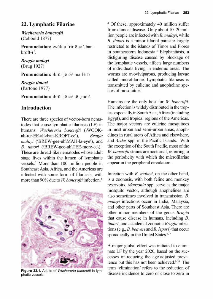
22. Lymphatic Filariae 253
Figure 22.1. Adults of Wuchereria bancrofti in lym-phatic vessels.
22. Lymphatic FilariaeWuchereria bancrofti (Cobbold 1877)
Pronunciation: \wu̇k-ə-ˈrir-ē-ə\ \ ban-krȯft-ī \
Brugia malayi (Brug 1927)
Pronunciation: \brü- jē-ə\\ ma-lē-ī\
Brugia timori (Partono 1977)
Pronunciation: \brü- jē-ə\\ tē-ˌmȯr\
Introduction
There are three species of vector-born nema-todes that cause lymphatic filariasis (LF) in humans: Wuchereria bancrofti (\WOOK-ah-rer-EE-ah\\ban-KROFT-ee\), Brugia malayi (\BREW-gee-uh\MAH-la-eye\), and B. timori (\BREW-gee-uh\TEE-more-ee\).1 These are thread-like nematodes whose adult stage lives within the lumen of lymphatic vessels.2 More than 100 million people in Southeast Asia, Africa, and the Americas are infected with some form of filariasis, with more than 90% due to W. bancrofti infection.3,
4 Of these, approximately 40 million suffer from clinical disease. Only about 10–20 mil-lion people are infected with B. malayi, while B. timori is a minor filarial parasite largely restricted to the islands of Timor and Flores in southeastern Indonesia.5 Elephantiasis, a disfiguring disease caused by blockage of the lymphatic vessels, affects large numbers of individuals living in endemic areas. The worms are ovoviviparous, producing larvae called microfilariae. Lymphatic filariasis is transmitted by culicine and anopheline spe-cies of mosquitoes.
Humans are the only host for W. bancrofti. The infection is widely distributed in the trop-ics, especially in South Asia, Africa (including Egypt), and tropical regions of the Americas. The major vectors are culicine mosquitoes in most urban and semi-urban areas, anoph-elines in rural areas of Africa and elsewhere, and Aedes spp. in the Pacific Islands. With the exception of the South Pacific, most of the W. bancrofti strains are nocturnal, referring to the periodicity with which the microfilariae appear in the peripheral circulation.
Infection with B. malayi, on the other hand, is a zoonosis, with both feline and monkey reservoirs. Mansonia spp. serve as the major mosquito vector, although anophelines are also sometimes involved in transmission. B. malayi infections occur in India, Malaysia, and other parts of Southeast Asia. There are other minor members of the genus Brugia that cause disease in humans, including B. timori, and accidental zoonotic Brugia infec-tions (e.g., B. beaveri and B. lepori) that occur sporadically in the United States.6, 7
A major global effort was initiated to elimi-nate LF by the year 2020, based on the suc-cesses of reducing the age-adjusted preva-lence but this has not been achieved.8-10 The term ‘elimination’ refers to the reduction of disease incidence to zero or close to zero in
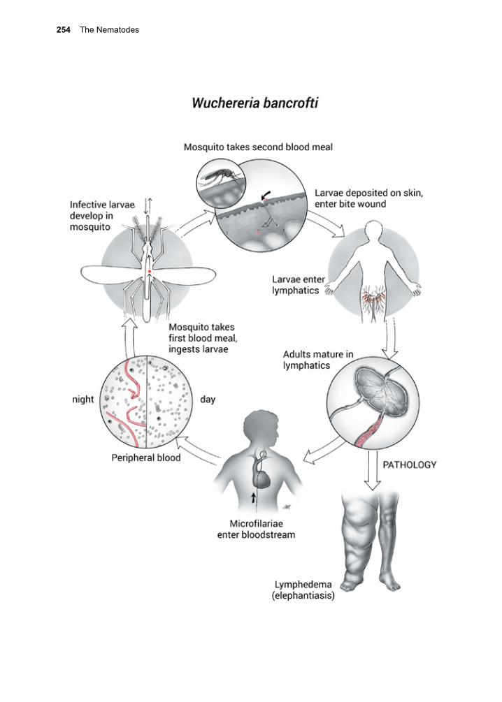
254 The Nematodes
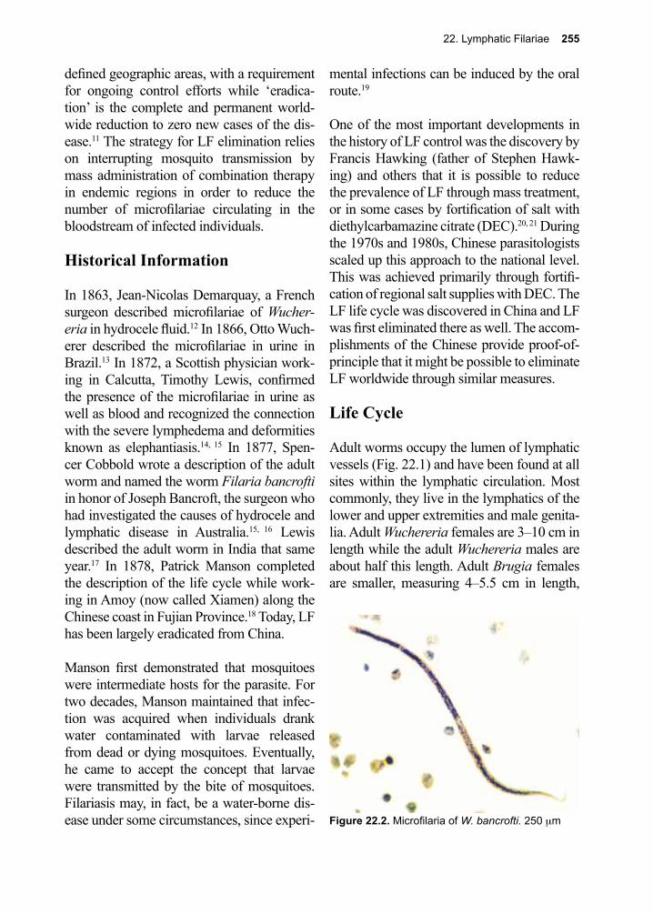
22. Lymphatic Filariae 255
defined geographic areas, with a requirement for ongoing control efforts while ‘eradica-tion’ is the complete and permanent world-wide reduction to zero new cases of the dis-ease.11 The strategy for LF elimination relies on interrupting mosquito transmission by mass administration of combination therapy in endemic regions in order to reduce the number of microfilariae circulating in the bloodstream of infected individuals.
Historical Information
In 1863, Jean-Nicolas Demarquay, a French surgeon described microfilariae of Wucher-eria in hydrocele fluid.12 In 1866, Otto Wuch-erer described the microfilariae in urine in Brazil.13 In 1872, a Scottish physician work-ing in Calcutta, Timothy Lewis, confirmed the presence of the microfilariae in urine as well as blood and recognized the connection with the severe lymphedema and deformities known as elephantiasis.14, 15 In 1877, Spen-cer Cobbold wrote a description of the adult worm and named the worm Filaria bancrofti in honor of Joseph Bancroft, the surgeon who had investigated the causes of hydrocele and lymphatic disease in Australia.15, 16 Lewis described the adult worm in India that same year.17 In 1878, Patrick Manson completed the description of the life cycle while work-ing in Amoy (now called Xiamen) along the Chinese coast in Fujian Province.18 Today, LF has been largely eradicated from China.
Manson first demonstrated that mosquitoes were intermediate hosts for the parasite. For two decades, Manson maintained that infec-tion was acquired when individuals drank water contaminated with larvae released from dead or dying mosquitoes. Eventually, he came to accept the concept that larvae were transmitted by the bite of mosquitoes. Filariasis may, in fact, be a water-borne dis-ease under some circumstances, since experi-
mental infections can be induced by the oral route.19
One of the most important developments in the history of LF control was the discovery by Francis Hawking (father of Stephen Hawk-ing) and others that it is possible to reduce the prevalence of LF through mass treatment, or in some cases by fortification of salt with diethylcarbamazine citrate (DEC).20, 21 During the 1970s and 1980s, Chinese parasitologists scaled up this approach to the national level. This was achieved primarily through fortifi-cation of regional salt supplies with DEC. The LF life cycle was discovered in China and LF was first eliminated there as well. The accom-plishments of the Chinese provide proof-of-principle that it might be possible to eliminate LF worldwide through similar measures.
Life Cycle
Adult worms occupy the lumen of lymphatic vessels (Fig. 22.1) and have been found at all sites within the lymphatic circulation. Most commonly, they live in the lymphatics of the lower and upper extremities and male genita-lia. Adult Wuchereria females are 3–10 cm in length while the adult Wuchereria males are about half this length. Adult Brugia females are smaller, measuring 4–5.5 cm in length,
Figure 22.2. Microfilaria of W. bancrofti. 250 µm
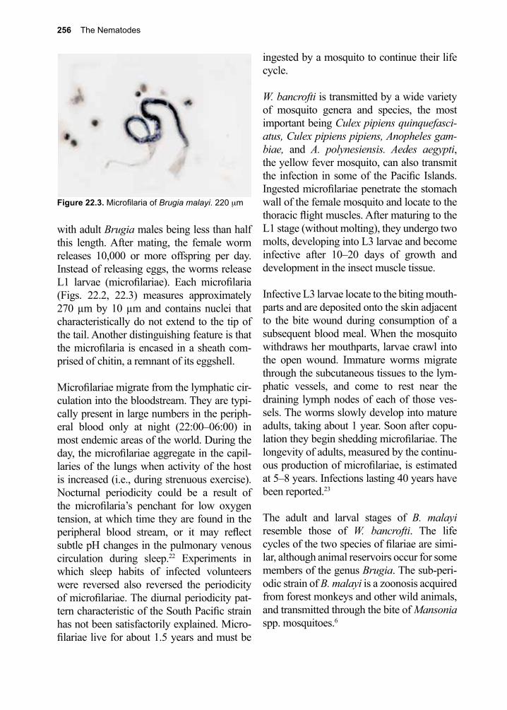
256 The Nematodes
with adult Brugia males being less than half this length. After mating, the female worm releases 10,000 or more offspring per day. Instead of releasing eggs, the worms release L1 larvae (microfilariae). Each microfilaria (Figs. 22.2, 22.3) measures approximately 270 µm by 10 µm and contains nuclei that characteristically do not extend to the tip of the tail. Another distinguishing feature is that the microfilaria is encased in a sheath com-prised of chitin, a remnant of its eggshell.
Microfilariae migrate from the lymphatic cir-culation into the bloodstream. They are typi-cally present in large numbers in the periph-eral blood only at night (22:00–06:00) in most endemic areas of the world. During the day, the microfilariae aggregate in the capil-laries of the lungs when activity of the host is increased (i.e., during strenuous exercise). Nocturnal periodicity could be a result of the microfilaria’s penchant for low oxygen tension, at which time they are found in the peripheral blood stream, or it may reflect subtle pH changes in the pulmonary venous circulation during sleep.22 Experiments in which sleep habits of infected volunteers were reversed also reversed the periodicity of microfilariae. The diurnal periodicity pat-tern characteristic of the South Pacific strain has not been satisfactorily explained. Micro-filariae live for about 1.5 years and must be
ingested by a mosquito to continue their life cycle.
W. bancrofti is transmitted by a wide variety of mosquito genera and species, the most important being Culex pipiens quinquefasci-atus, Culex pipiens pipiens, Anopheles gam-biae, and A. polynesiensis. Aedes aegypti, the yellow fever mosquito, can also transmit the infection in some of the Pacific Islands. Ingested microfilariae penetrate the stomach wall of the female mosquito and locate to the thoracic flight muscles. After maturing to the L1 stage (without molting), they undergo two molts, developing into L3 larvae and become infective after 10–20 days of growth and development in the insect muscle tissue.
Infective L3 larvae locate to the biting mouth-parts and are deposited onto the skin adjacent to the bite wound during consumption of a subsequent blood meal. When the mosquito withdraws her mouthparts, larvae crawl into the open wound. Immature worms migrate through the subcutaneous tissues to the lym-phatic vessels, and come to rest near the draining lymph nodes of each of those ves-sels. The worms slowly develop into mature adults, taking about 1 year. Soon after copu-lation they begin shedding microfilariae. The longevity of adults, measured by the continu-ous production of microfilariae, is estimated at 5–8 years. Infections lasting 40 years have been reported.23
The adult and larval stages of B. malayi resemble those of W. bancrofti. The life cycles of the two species of filariae are simi-lar, although animal reservoirs occur for some members of the genus Brugia. The sub-peri-odic strain of B. malayi is a zoonosis acquired from forest monkeys and other wild animals, and transmitted through the bite of Mansonia spp. mosquitoes.6
Figure 22.3. Microfilaria of Brugia malayi. 220 µm
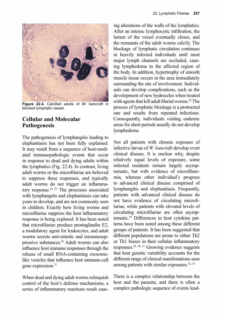
22. Lymphatic Filariae 257
Cellular and Molecular Pathogenesis
The pathogenesis of lymphangitis leading to elephantiasis has not been fully explained. It may result from a sequence of host-medi-ated immunopathologic events that occur in response to dead and dying adults within the lymphatics (Fig. 22.4). In contrast, living adult worms or the microfilariae are believed to suppress these responses, and typically adult worms do not trigger an inflamma-tory response.24, 25 The processes associated with lymphangitis and elephantiasis can take years to develop, and are not commonly seen in children. Exactly how living worms and microfilariae suppress the host inflammatory response is being explored. It has been noted that microfilariae produce prostaglandin E2, a modulatory agent for leukocytes, and adult worms secrete anti-mitotic and immunosup-pressive substances.26 Adult worms can also influence host immune responses through the release of small RNA-containing exosome-like vesicles that influence host immune-cell gene expression.27
When dead and dying adult worms relinquish control of the host’s defense mechanisms, a series of inflammatory reactions result caus-
ing alterations of the walls of the lymphatics. After an intense lymphocytic infiltration, the lumen of the vessel eventually closes, and the remnants of the adult worms calcify. The blockage of lymphatic circulation continues in heavily infected individuals until most major lymph channels are occluded, caus-ing lymphedema in the affected region of the body. In addition, hypertrophy of smooth muscle tissue occurs in the area immediately surrounding the site of involvement. Individ-uals can develop complications, such as the development of new hydroceles when treated with agents that kill adult filarial worms.28 The process of lymphatic blockage is a protracted one and results from repeated infections. Consequently, individuals visiting endemic areas for short periods usually do not develop lymphedema.
Not all patients with chronic exposure of infective larvae of W. bancrofti develop overt clinical disease. It is unclear why, despite relatively equal levels of exposure, some infected residents remain largely asymp-tomatic, but with evidence of microfilare-mia, whereas other individual’s progress to advanced clinical disease comprised of lymphangitis and elephantiasis. Frequently, patients with advanced clinical disease do not have evidence of circulating microfi-lariae, while patients with elevated levels of circulating microfilariae are often asymp-tomatic.29 Differences in host cytokine pat-terns have been noted among these different groups of patients. It has been suggested that different populations are prone to either Th2 or Th1 biases in their cellular inflammatory responses.24, 30, 31 Growing evidence suggests that host genetic variability accounts for the different range of clinical manifestations seen among patients with similar exposures.32, 33
There is a complex relationship between the host and the parasite, and there is often a complex pathologic sequence of events lead-
Figure 22.4. Calcified adults of W. bancrofti in blocked lymphatic vessel.
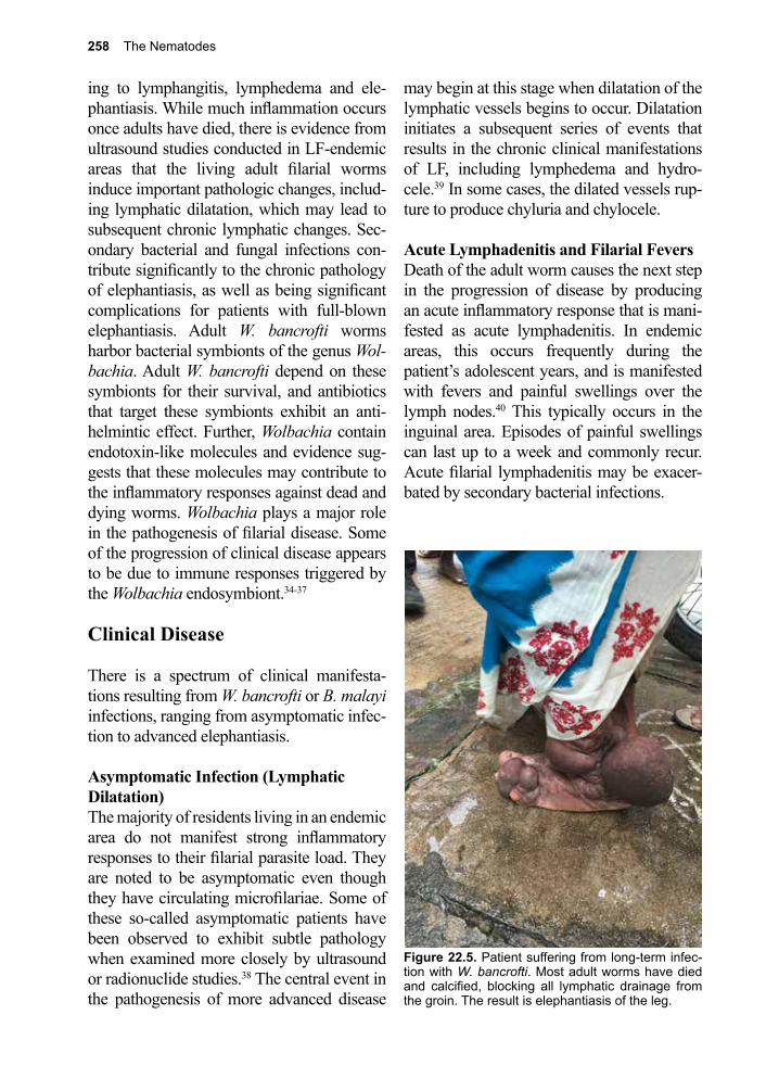
258 The Nematodes
ing to lymphangitis, lymphedema and ele-phantiasis. While much inflammation occurs once adults have died, there is evidence from ultrasound studies conducted in LF-endemic areas that the living adult filarial worms induce important pathologic changes, includ-ing lymphatic dilatation, which may lead to subsequent chronic lymphatic changes. Sec-ondary bacterial and fungal infections con-tribute significantly to the chronic pathology of elephantiasis, as well as being significant complications for patients with full-blown elephantiasis. Adult W. bancrofti worms harbor bacterial symbionts of the genus Wol-bachia. Adult W. bancrofti depend on these symbionts for their survival, and antibiotics that target these symbionts exhibit an anti-helmintic effect. Further, Wolbachia contain endotoxin-like molecules and evidence sug-gests that these molecules may contribute to the inflammatory responses against dead and dying worms. Wolbachia plays a major role in the pathogenesis of filarial disease. Some of the progression of clinical disease appears to be due to immune responses triggered by the Wolbachia endosymbiont.34-37
Clinical Disease
There is a spectrum of clinical manifesta-tions resulting from W. bancrofti or B. malayi infections, ranging from asymptomatic infec-tion to advanced elephantiasis.
Asymptomatic Infection (Lymphatic Dilatation) The majority of residents living in an endemic area do not manifest strong inflammatory responses to their filarial parasite load. They are noted to be asymptomatic even though they have circulating microfilariae. Some of these so-called asymptomatic patients have been observed to exhibit subtle pathology when examined more closely by ultrasound or radionuclide studies.38 The central event in the pathogenesis of more advanced disease
may begin at this stage when dilatation of the lymphatic vessels begins to occur. Dilatation initiates a subsequent series of events that results in the chronic clinical manifestations of LF, including lymphedema and hydro-cele.39 In some cases, the dilated vessels rup-ture to produce chyluria and chylocele.
Acute Lymphadenitis and Filarial FeversDeath of the adult worm causes the next step in the progression of disease by producing an acute inflammatory response that is mani-fested as acute lymphadenitis. In endemic areas, this occurs frequently during the patient’s adolescent years, and is manifested with fevers and painful swellings over the lymph nodes.40 This typically occurs in the inguinal area. Episodes of painful swellings can last up to a week and commonly recur. Acute filarial lymphadenitis may be exacer-bated by secondary bacterial infections.
Figure 22.5. Patient suffering from long-term infec-tion with W. bancrofti. Most adult worms have died and calcified, blocking all lymphatic drainage from the groin. The result is elephantiasis of the leg.
22. Lymphatic Filariae 259
Some individuals who have travelled to and spent several months in endemic areas can also develop acute lymphadenitis, but the pathogenesis of this process occurs by a poorly understood process. This phenom-enon was described in the 1940s among American troops returning from war in the Pacific theatre.41
ElephantiasisA subset of patients with acute lymphangi-tis and filarial fevers will go on to develop lymphedema of the arms, legs, breasts and genitalia leading to elephantiasis (Fig. 22.5). During these inflammatory processes, the skin becomes doughy and exhibits some degree of pitting, though it is rather firm. As the inflammatory reaction continues, the area becomes firmer still, and pitting disappears. There is substantial spread of the inflamma-tion into the subcutaneous tissue and conse-quent loss of elasticity of the overlying skin. Characteristically, and in contrast to celluli-tis caused by some bacteria, filarial celluli-tis shows no demarcation line between the affected and the healthy skin. In bancroft-ian filariasis, the legs are more likely to be involved than are the upper extremities, and the lower portions of the legs are more involved than the upper ones. The scrotum is frequently affected in the form of hydro-celes and may become gigantic, weighing up to 10 kg; much larger scrotums have been described in rare cases.
Tropical Pulmonary EosinophiliaTropical pulmonary eosinophilia (TPE) develops in some individuals with filarial infections. This syndrome, which occurs frequently in southern India, particularly in young adult men, is characterized by high levels of serum IgE, nocturnal asthma with interstitial infiltrates on chest radiographs, fatigue, weight loss, and circulating eosino-philia.42 Left untreated, TPE can progress to chronic restrictive lung disease. The patho-
genesis of this syndrome is related to local immune responses to microfilariae in the pul-monary vasculature, and results in eosinophil accumulation in the lung with the release of cytotoxic eosinophil products (e.g., major basic protein and eosinophil cationic pro-tein).42
Diagnosis (see Clinical Appendix)
LF should be suspected in an individual who resides in an endemic region, is beyond the first decade of life, and has lymphedema in the extremities or genitalia. Definitive diagnosis has traditionally depended upon microscopically observing the characteristic microfilariae in the blood (Figs. 22.2, 22.3, C.46–C.53). Occasionally, infection is so heavy that microfilariae can be observed on a thin blood smear stained with Giemsa. In lighter infections, methods include filtering blood onto a 0.45 µm pore-sized nucleopore filter, then staining it with Giemsa solution. In the case of very light infection, 1 ml of blood is preserved in 9 ml of 1% formalin and then concentrated by centrifugation (Knott’s test; Appendix B). The pellet contains red blood cell ghosts and microfilariae. Stained smears of the pellet are then examined microscopi-cally. Because of the nocturnal periodicity of some strains, it is best to draw blood during the customary hours of sleep (usually 22:00–02:00 hours).
Antigen tests that detect circulating W. ban-crofti antigens have been developed which are more sensitive than microscopy, can be used for daytime testing, and can give a quan-titative result correlating with adult worm burden.43-45 These are available in the form of ELISA test or an immunochromatographic card.46 The card-based assay, which recog-nizes an adult worm’s 200 kDa antigen, has a sensitivity of 96–100% and a specificity approaching 100%.47 The ELISA also has a sensitivity approaching 100% in microfilare-
260 The Nematodes
mic patients. For both assays, the circulating filarial antigen remains diurnally constant, so that blood for diagnosis can be collected during the day. PCR-based tests have been developed, but while used to monitor filarial infection in mosquitoes, are currently not rou-tinely used in clinical practice.48
Serological tests that detect levels of human IgG directed against the worms that cause LF have been developed and are in use clinically. These serve as rapid tests that can aid in the diagnosis of this disease. Note, they detect exposure rather than confirm current infec-tion.49-51
Increasingly, ultrasound has provided an important noninvasive modality for monitor-ing the efficacy of antifilarial drugs.34 Ultra-sound examination of the lymphatic vessels of the spermatic cord of infected men results in a distinctive sign, known as the “filarial dance sign” reflective of nests of live worms in the lymphatics.52 Adult worm death follow-ing treatment with DEC can be subsequently followed.
Treatment (see Clinical Appendix)
It is recommended that all patients be treated, because even patients with so-called asymp-tomatic infection may have abnormal lym-phatics, and there is increasing evidence that early treatment may prevent subsequent lymphatic damage and may reverse early lymphatic dysfunction.53 It is critical that, prior to treatment, co-infection with Loa loa with a high Loa loa microfilarial load is ruled out, due to the risk of severe adverse events if treatment is given to such patients.54 For treatment of mono-infected patients, DEC has both macrofilaricidal (adult worm) and microfilaricidal properties, and is the treat-ment of choice for such patients. In many regions it is given in a dose of 6 mg/kg/day for 12 consecutive days for a total of 72 mg/
kg body weight.34, 55 For W. bancrofti infec-tions, this results in at least a 90% decrease in microfilaremia within one month. DEC decreases the incidence of filarial lymphan-gitis, and in some cases reverses existing lymphatic damage. The addition of doxycy-cline for 4–6 weeks may be beneficial based on evidence demonstrating macrofilaricidal activity and reduced pathology.56-58
In men, the efficacy of treatment can be monitored by serial ultrasound examinations (see above), and by serial blood sampling.34
Since DEC is only partially effective against the adult worm, repeat treatments are often required. This is often done every 6–12 months.34 Some data suggests that single dose treatment with 6 mg/kg of DEC has compa-rable macrofilaricidal and long-term micro-filaricidal activity. Some clinicians have suggested that single-dose treatment can be repeated every 6–12 months.34 DEC is asso-ciated with fever (probably resulting from disintegration of a few of the adult worms), occasional nausea and vomiting, and fleeting skin rashes. Ivermectin, a drug effective for therapy of onchocerciasis, also kills microfi-lariae of W. bancrofti, but it appears to have no macrofilaricidal properties. Ivermectin and albendazole, a drug that also does not kill adult filarial worms, have been used in mass drug campaigns, reducing microfilarial blood levels and decreasing transmission.59
Aside from the use of antihelmintic drugs, there are several treatment modalities that help to improve the chronic sequelae of LF, including lymphedema and elephantiasis. Both conditions, when they occur in the leg, are reversible with a hygienic regimen that includes prevention of secondary bacterial infections by prompt antibiotic treatment of acute bacterial attacks, aggressive treat-ment of skin lesions including those caused by Candida and other fungi, and physiother-apy.34 Treatment of secondary bacterial infec-
22. Lymphatic Filariae 261
tions has been identified as a critical treat-ment modality for lymphedema and elephan-tiasis. Hydrocele drainage without corrective surgery, while it does provide relief, is often associated with reaccumulation of fluid.60 For certain affected areas, (e.g., the scro-tum) corrective surgical interventions may be required.61 Surgical techniques have been developed that can, in most cases, alleviate hydroceles and greatly improve the morbid-ity associated with filarial infections.62
Prevention and Control
Patent microfilaremia is first detected in chil-dren 5–10 years old who live in endemic regions.6 Transplacental immunity and breast-feeding may limit the intensity of infection in younger individuals. The prevalence of microscopically confirmed infection gradu-ally increases up to the age of 30–40 years.
The frequency of exposure to L3 by vectors is the most important determinant in the com-munity prevalence of filariasis.63 Prevention depends upon control of mosquito vectors, which has had limited success because mos-quitoes develop resistance to insecticides. Insecticide-treated bed nets have been effec-tive in reducing transmission in areas where anopheline mosquitos transmit the disease.64 Urbanization of vast areas of tropical Asia has resulted in a concomitant rise in the preva-lence of both W. bancrofti and B. malayi vari-eties of filariasis carried by mosquitoes that breed in peridomestic habitats.
In 1997, the World Health Assembly passed a resolution calling on its member states to
undertake a global elimination program for LF. The major strategy for LF elimination is based on two principles: 1. to interrupt transmission of infection and 2. to allevi-ate and prevent the suffering and disability caused by LF. To interrupt transmission, it is essential to reduce the levels of microfilariae in the blood for a sustainable period. This is achieved by administering a yearly, single-dose, 2-drug regimen.65 For most countries, the recommended drugs are DEC (6 mg/kg) and albendazole (400 mg).65, 66 The goal of this approach is to provide annual treatment with this drug combination.
In many parts of Sub-Saharan Africa (and Yemen as well) where there is epidemiologi-cal overlap with onchocerciasis, the toxicities caused by DEC in people with these condi-tions necessitate substituting ivermectin (200 mcg/kg). Such populations would receive ivermectin or albendazole.64, 66 A period of 5 years of annual treatments is currently rec-ommended. To date, the number of serious adverse events from LF control mass chemo-therapy has been remarkably low. In some areas, a treatment regimen comprised of daily DEC-fortified salt is used. A Global Program to Eliminate LF in collaboration with the World Health Organization is leading these efforts.
To alleviate suffering and decrease the dis-ability caused by LF, the major strategy has been to decrease secondary bacterial and fungal infections of the affected limbs and genitals. This includes meticulous local hygiene, judicious use of antibiotics, physio-therapy and health education.
References
1. Knopp, S., et al., Nematode infections: filariases. Infect Dis Clin North Am 2012, 26 (2), 359-81.2. Nelson, G. S., Current concepts in parasitology. Filariasis. The New England journal of medicine
1979, 300 (20), 1136-9.3. Global programme to eliminate lymphatic filariasis: progress report for 2012. Wkly Epidemiol
262 The Nematodes
Rec 2013, 88 (37), 389-99.4. Global programme to eliminate lymphatic filariasis. Wkly Epidemiol Rec 2010, 85 (38), 365-72.5. Taylor, M. J., et al., Lymphatic filariasis and onchocerciasis. Lancet 2010, 376 (9747), 1175-85.6. Nanduri, J.; Kazura, J. W., Clinical and laboratory aspects of filariasis. Clinical microbiology
reviews 1989, 2 (1), 39-50.7. Baird, J. K., et al., North American brugian filariasis: report of nine infections of humans. The
American journal of tropical medicine and hygiene 1986, 35 (6), 1205-9.8. Molyneux, D. H., et al., Mass drug treatment for lymphatic filariasis and onchocerciasis. Trends
in parasitology 2003, 19 (11), 516-22.9. Molyneux, D. H.; Zagaria, N., Lymphatic filariasis elimination: progress in global programme
development. Ann Trop Med Parasitol 96 Suppl 2 2003, S15-40.10. Global Burden of Disease Study, C., Global, regional, and national incidence, prevalence, and
years lived with disability for 301 acute and chronic diseases and injuries in 188 countries, 1990-2013: a systematic analysis for the Global Burden of Disease Study 2013. Lancet 2015, 386 (9995), 743-800.
11. Hotez, P. J., et al., Combating tropical infectious diseases: report of the Disease Control Priorities in Developing Countries Project. Clin Inf Dis 2004, 38 (6), 871-8.
12. Demarquay, M., Note sur une tumeur des bourses contenant un liquide laiteux (galactocele de Vidal) et refermant des petits etres vermiformes que l’on peut considerer comme des helminths hematoides a I’etat d’embryon. Gaz Med Pans. 1863, 18, 665-667.
13. Wucherer, O. E., Sobre a molestia vulgarmente denominada oppilacao ou cancaco. Gaz. Med. Bahia 1866, 1, 27-29, 39-41, 52-54, 63-64.
14. Lewis, T. R., On a haematozoon inhabiting human blood, its relation to chyluria and other diseases. 8th Annual Report of the Sanitary Commission Government of India. Sanitary Commission Government of India, Calcutta. 1872, 241-266.
15. Cox, F. E., History of human parasitology. Clin Microbiol Rev 2002, 15 (4), 595-612.16. Cobbold, T. S., Discovery of the adult representative of microscopic filariae. Lancet 1877, 2 70-
71.17. Lewis, T., Filaria sanguinis hominis (mature form), found in a bloodclot in naevoid elephantiasis
of the scrotum. Lancet 1877, 2, 453-455.18. Manson, P., Further Observations on Filaria sanguinis hominis. Medical Reports, China Imperial
Maritime Customs Shanghai no 14 pp 1878, 1-26.19. Gwadz, R. W.; Chernin, E., Oral transmission of Brugia pahangi to Jirds (Meriones
unguiculatus). Nature 1972, 239 (5374), 524-5.20. Hawking, F.; Marques, R. J., Control of Bancroftian filariasis by cooking salt medicated with
diethylcarbamazine. Bull World Health Organ 1967, 37 (3), 405-14.21. Hawking, F., A review of progress in the chemotherapy and control of filariasis since 1955. Bull
World Health Organ 1962, 27, 555-68.22. Hawking, F., et al., The periodicity of microfilariae. XI. The effect of body temperature and other
stimuli upon the cycles of Wuchereria bancrofti, Brugia malayi, B. ceylonensis and Dirofilaria repens. Transactions of the Royal Society of Tropical Medicine and Hygiene 1966, 60 (4), 497-513.
23. Carme, B.; Laigret, J., Longevity of Wuchereria bancrofti var. pacifica and mosquito infection acquired from a patient with low level parasitemia. The American journal of tropical medicine and hygiene 1979, 28 (1), 53-5.
24. Ottesen, E. A., The Wellcome Trust Lecture. Infection and disease in lymphatic filariasis: an immunological perspective. Parasitology 1992, 104 Suppl, S71-9.
25. King, C. L., et al., Immunologic tolerance in lymphatic filariasis. Diminished parasite-specific T and B lymphocyte precursor frequency in the microfilaremic state. J Clin Invest 1992, 89 (5), 1403-10.
26. Liu, L. X., et al., Release of prostaglandin E2 by microfilariae of Wuchereria bancrofti and Brugia malayi. The American journal of tropical medicine and hygiene 1992, 46 (5), 520-3.
27. Zamanian, M., et al., Release of small RNA-containing exosome-like vesicles from the human filarial parasite Brugia malayi. PLoS Negl Trop Dis 2015, 9 (9), e0004069.
28. Hussein, O., et al., Duplex Doppler sonographic assessment of the effects of diethylcarbamazine
22. Lymphatic Filariae 263
and albendazole therapy on adult filarial worms and adjacent host tissues in Bancroftian filariasis. Am J Trop Med Hyg 2004, 71 (4), 471-7.
29. Dissanayake, S., In Wuchereria bancrofti filariasis, asymptomatic microfilaraemia does not progress to amicrofilaraemic lymphatic disease. Int J Epidemiol 2001, 30 (2), 394-9.
30. Almeida, A. B., et al., The presence or absence of active infection, not clinical status, is most closely associated with cytokine responses in lymphatic filariasis. Dis 1453 1996, 173.
31. Piessens, W. F., et al., Immune responses in human infections with Brugia malayi: specific cellular unresponsiveness to filarial antigens. The Journal of clinical investigation 1980, 65 (1), 172-9.
32. Lammie, P. J., et al., The pathogenesis of filarial lymphedema: is it the worm or is it the host? Ann N Y Acad Sci 2002, 979, 131-42; discussion 188-96.
33. Choi, E. H., et al., Genetic variation in immune function and susceptibility to human filariasis. Expert Rev Mol Diagn 2003, 3 (3), 367-74.
34. Taylor, M. J., A new insight into the pathogenesis of filarial disease. Current molecular medicine 2002, 2 (3), 299-302.
35. Taylor, M. J., Wolbachia in the inflammatory pathogenesis of human filariasis. Ann N Y Acad Sci 2003, 990, 444-9.
36. Tamarozzi, F., et al., Human filarial Wolbachia lipopeptide directly activates human neutrophils in vitro. Parasite Immunol 2014, 36 (10), 494-502.
37. Turner, J. D., et al., Wolbachia lipoprotein stimulates innate and adaptive immunity through Toll-like receptors 2 and 6 to induce disease manifestations of filariasis. J Biol Chem 2009, 284 (33), 22364-78.
38. Freedman, D. O., et al., Lymphoscintigraphic analysis of lymphatic abnormalities in symptomatic and asymptomatic human filariasis. The Journal of infectious diseases 1994, 170 (4), 927-33.
39. Addiss, D. G., et al., Treatment of lymphatic filariasis. Vol. 2000, p 151-199.40. Pani, S. P., et al., Episodic adenolymphangitis and lymphoedema in patients with bancroftian
filariasis. Transactions of the Royal Society of Tropical Medicine and Hygiene 1995, 89 (1), 72-4.41. Huntington, R. W., et al., Filariasis Among American Troops in a South Pacific Island Group.
Yale J Biol Med 1944, 16 (5), 529-538 1.42. Ottesen, E. A.; Nutman, T. B., Tropical pulmonary eosinophilia. Annual review of medicine 1992,
43, 417-24.43. El-Moamly, A. A., et al., Using the AD12-ICT rapid-format test to detect Wuchereria bancrofti
circulating antigens in comparison to Og4C3-ELISA and nucleopore membrane filtration and microscopy techniques. Parasitol Res 2012, 111 (3), 1379-83.
44. Rocha, A., et al., Comparison of tests for the detection of circulating filarial antigen (Og4C3-ELISA and AD12-ICT) and ultrasound in diagnosis of lymphatic filariasis in individuals with microfilariae. Mem Inst Oswaldo Cruz 2009, 104 (4), 621-5.
45. Wattal, S., et al., Evaluation of Og4C3 antigen ELISA as a tool for detection of bancroftian filariasis under lymphatic filariasis elimination programme. J Commun Dis 2007, 39 (2), 75-84.
46. Cunningham, J., et al., A global comparative evaluation of commercial immunochromatographic rapid diagnostic tests for visceral leishmaniasis. Clin Infect Dis 2012, 55 (10), 1312-9.
47. Well, G. J., et al., A monoclonal antibody-based enzyme immunoassay for detecting parasite antigenemia in Bancroftian filariasis. Dis 1987, 156, 350-355.
48. Wijegunawardana, A. D., et al., Evaluation of PCR-ELISA as a tool for monitoring transmission of Wuchereria bancrofti in District of Gampaha, Sri Lanka. Asian Pac J Trop Biomed 2013, 3 (5), 381-7.
49. Steel, C., et al., Rapid Wuchereria bancrofti-specific antigen Wb123-based IgG4 immunoassays as tools for surveillance following mass drug administration programs on lymphatic filariasis. Clin Vaccine Immunol 2013, 20 (8), 1155-61.
50. Steel, C., et al., Antibody to the filarial antigen Wb123 reflects reduced transmission and decreased exposure in children born following single mass drug administration (MDA). PLoS Negl Trop Dis 2012, 6 (12), e1940.
51. Kubofcik, J., et al., Identification of Wb123 as an early and specific marker of Wuchereria
264 The Nematodes
bancrofti infection. PLoS Negl Trop Dis 2012, 6 (12), e1930.52. Mand, S., et al., Animated documentation of the filaria dance sign (FDS) in bancroftian filariasis.
Filaria J 2003, 2 (1), 3.53. Moore, T. A., et al., Diethylcarbamazine-induced reversal of early lymphatic dysfunction in a
patient with bancroftian filariasis: assessment with use of lymphoscintigraphy. Clin Infect Dis 1996, 23 (5), 1007-11.
54. Bhalla, D., et al., Neurological manifestations of filarial infections. Handb Clin Neurol 2013, 114, 235-42.
55. Kazura, J. W., et al., Filariasis. Tropical Infectious Diseases, Principles, Pathogens, & Practice 1999, 2, 852-860.
56. Mand, S., et al., Doxycycline improves filarial lymphedema independent of active filarial infection: a randomized controlled trial. Clin Infect Dis 2012, 55 (5), 621-30.
57. Mand, S., et al., Macrofilaricidal activity and amelioration of lymphatic pathology in bancroftian filariasis after 3 weeks of doxycycline followed by single-dose diethylcarbamazine. Am J Trop Med Hyg 2009, 81 (4), 702-11.
58. Taylor, M. J.; Hoerauf, A., A new approach to the treatment of filariasis. Current opinion in infectious diseases 2001, 14 (6), 727-31.
59. Dembele, B., et al., Use of high-dose, twice-yearly albendazole and ivermectin to suppress Wuchereria bancrofti microfilarial levels. Clin Infect Dis 2010, 51 (11), 1229-35.
60. Freeman, C. D., et al., Metronidazole. A therapeutic review and update. Drugs 1997, 54 (5), 679-708.
61. Capuano, G. P.; Capuano, C., Surgical management of morbidity due to lymphatic filariasis: the usefulness of a standardized international clinical classification of hydroceles. Trop Biomed 2012, 29 (1), 24-38.
62. Addiss, D. G.; Brady, M. A., Morbidity management in the Global Programme to Eliminate Lymphatic Filariasis: a review of the scientific literature. Filaria J 2007, 6, 2.
63. Piessens, W. F.; Partono, F., Host-vector-parasite relationships in human filariasis. Semin Infect Dis 1980, 3 131-152.
64. Global Programme to Eliminate Lymphatic Filariasis. Wkly Epidemiol Rec 2006, 81 (22), 221-32.
65. Kazura, J., et al., Comparison of single-dose diethylcarbamazine and ivermectin for treatment of bancroftian filariasis in Papua New Guinea. The American journal of tropical medicine and hygiene 1993, 49 (6), 804-11.
66. Sabesan, S., Albendazole for mass drug administration to eliminate lymphatic filariasis. Lancet Infect Dis 2006, 6 (11), 684-5.
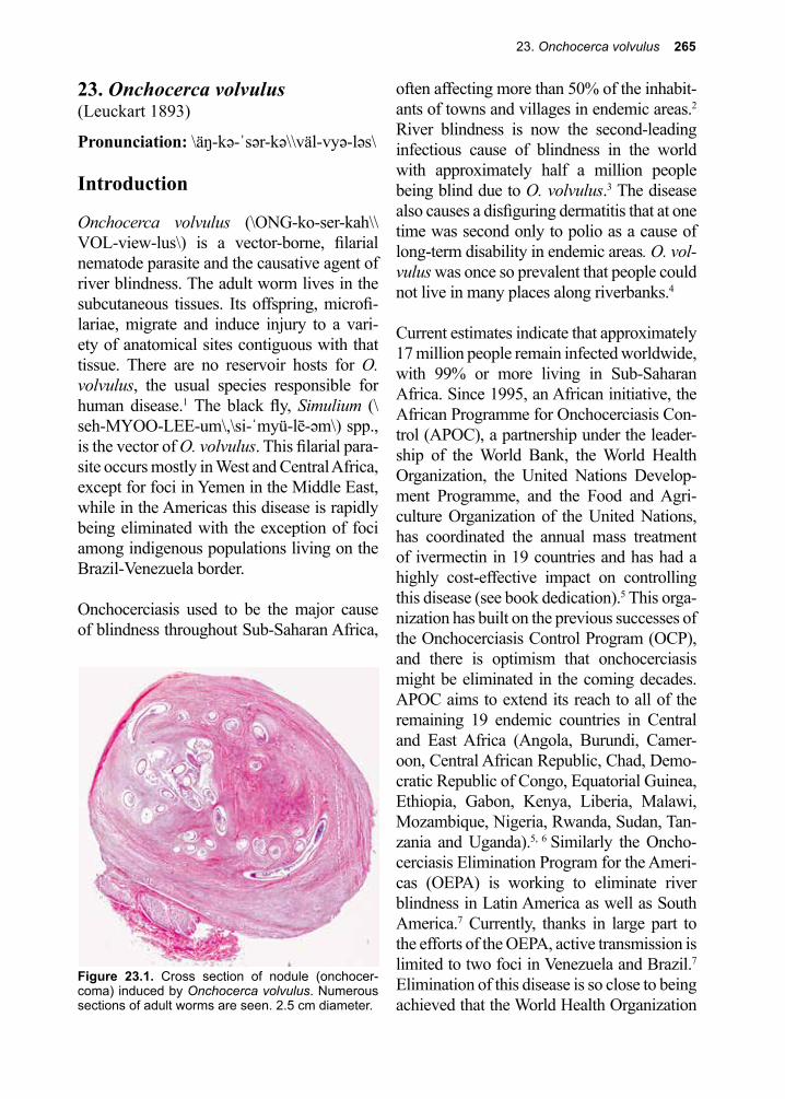
23. Onchocerca volvulus 265
Figure 23.1. Cross section of nodule (onchocer-coma) induced by Onchocerca volvulus. Numerous sections of adult worms are seen. 2.5 cm diameter.
23. Onchocerca volvulus(Leuckart 1893)
Pronunciation: \äŋ-kə-ˈsər-kə\\väl-vyə-ləs\
Introduction
Onchocerca volvulus (\ONG-ko-ser-kah\\VOL-view-lus\) is a vector-borne, filarial nematode parasite and the causative agent of river blindness. The adult worm lives in the subcutaneous tissues. Its offspring, microfi-lariae, migrate and induce injury to a vari-ety of anatomical sites contiguous with that tissue. There are no reservoir hosts for O. volvulus, the usual species responsible for human disease.1 The black fly, Simulium (\seh-MYOO-LEE-um\,\si-ˈmyü-lē-əm\) spp., is the vector of O. volvulus. This filarial para-site occurs mostly in West and Central Africa, except for foci in Yemen in the Middle East, while in the Americas this disease is rapidly being eliminated with the exception of foci among indigenous populations living on the Brazil-Venezuela border.
Onchocerciasis used to be the major cause of blindness throughout Sub-Saharan Africa,
often affecting more than 50% of the inhabit-ants of towns and villages in endemic areas.2 River blindness is now the second-leading infectious cause of blindness in the world with approximately half a million people being blind due to O. volvulus.3 The disease also causes a disfiguring dermatitis that at one time was second only to polio as a cause of long-term disability in endemic areas. O. vol-vulus was once so prevalent that people could not live in many places along riverbanks.4
Current estimates indicate that approximately 17 million people remain infected worldwide, with 99% or more living in Sub-Saharan Africa. Since 1995, an African initiative, the African Programme for Onchocerciasis Con-trol (APOC), a partnership under the leader-ship of the World Bank, the World Health Organization, the United Nations Develop-ment Programme, and the Food and Agri-culture Organization of the United Nations, has coordinated the annual mass treatment of ivermectin in 19 countries and has had a highly cost-effective impact on controlling this disease (see book dedication).5 This orga-nization has built on the previous successes of the Onchocerciasis Control Program (OCP), and there is optimism that onchocerciasis might be eliminated in the coming decades. APOC aims to extend its reach to all of the remaining 19 endemic countries in Central and East Africa (Angola, Burundi, Camer-oon, Central African Republic, Chad, Demo-cratic Republic of Congo, Equatorial Guinea, Ethiopia, Gabon, Kenya, Liberia, Malawi, Mozambique, Nigeria, Rwanda, Sudan, Tan-zania and Uganda).5, 6 Similarly the Oncho-cerciasis Elimination Program for the Ameri-cas (OEPA) is working to eliminate river blindness in Latin America as well as South America.7 Currently, thanks in large part to the efforts of the OEPA, active transmission is limited to two foci in Venezuela and Brazil.7 Elimination of this disease is so close to being achieved that the World Health Organization
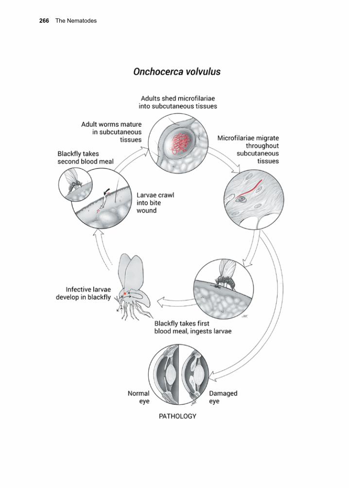
266 The Nematodes
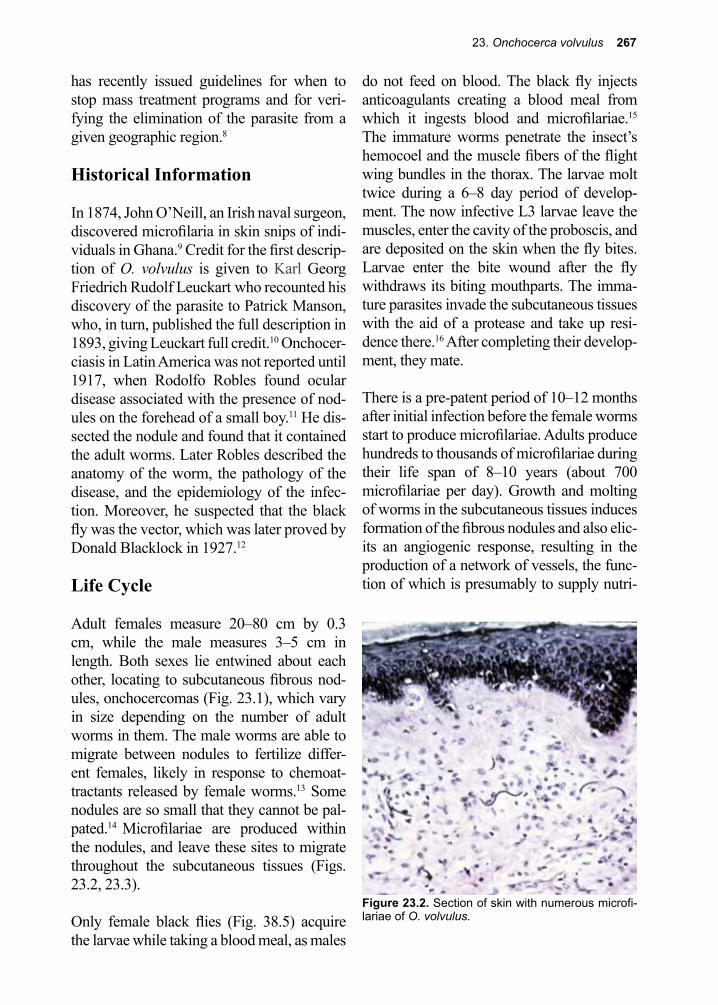
23. Onchocerca volvulus 267
has recently issued guidelines for when to stop mass treatment programs and for veri-fying the elimination of the parasite from a given geographic region.8
Historical Information
In 1874, John O’Neill, an Irish naval surgeon, discovered microfilaria in skin snips of indi-viduals in Ghana.9 Credit for the first descrip-tion of O. volvulus is given to Karl Georg Friedrich Rudolf Leuckart who recounted his discovery of the parasite to Patrick Manson, who, in turn, published the full description in 1893, giving Leuckart full credit.10 Onchocer-ciasis in Latin America was not reported until 1917, when Rodolfo Robles found ocular disease associated with the presence of nod-ules on the forehead of a small boy.11 He dis-sected the nodule and found that it contained the adult worms. Later Robles described the anatomy of the worm, the pathology of the disease, and the epidemiology of the infec-tion. Moreover, he suspected that the black fly was the vector, which was later proved by Donald Blacklock in 1927.12
Life Cycle
Adult females measure 20–80 cm by 0.3 cm, while the male measures 3–5 cm in length. Both sexes lie entwined about each other, locating to subcutaneous fibrous nod-ules, onchocercomas (Fig. 23.1), which vary in size depending on the number of adult worms in them. The male worms are able to migrate between nodules to fertilize differ-ent females, likely in response to chemoat-tractants released by female worms.13 Some nodules are so small that they cannot be pal-pated.14 Microfilariae are produced within the nodules, and leave these sites to migrate throughout the subcutaneous tissues (Figs. 23.2, 23.3).
Only female black flies (Fig. 38.5) acquire the larvae while taking a blood meal, as males
do not feed on blood. The black fly injects anticoagulants creating a blood meal from which it ingests blood and microfilariae.15 The immature worms penetrate the insect’s hemocoel and the muscle fibers of the flight wing bundles in the thorax. The larvae molt twice during a 6–8 day period of develop-ment. The now infective L3 larvae leave the muscles, enter the cavity of the proboscis, and are deposited on the skin when the fly bites. Larvae enter the bite wound after the fly withdraws its biting mouthparts. The imma-ture parasites invade the subcutaneous tissues with the aid of a protease and take up resi-dence there.16 After completing their develop-ment, they mate.
There is a pre-patent period of 10–12 months after initial infection before the female worms start to produce microfilariae. Adults produce hundreds to thousands of microfilariae during their life span of 8–10 years (about 700 microfilariae per day). Growth and molting of worms in the subcutaneous tissues induces formation of the fibrous nodules and also elic-its an angiogenic response, resulting in the production of a network of vessels, the func-tion of which is presumably to supply nutri-
Figure 23.2. Section of skin with numerous microfi-lariae of O. volvulus.
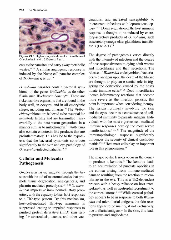
268 The Nematodes
ents to the parasites and carry away metabolic wastes.17, 18 A similar angiogenic response is induced by the Nurse-cell-parasite complex of Trichinella spiralis.19
O. volvulus parasites contain bacterial sym-bionts of the genus Wolbachia, as do other filaria such Wuchereria bancrofti. These are rickettsia-like organisms that are found in the body wall, in oocytes, and in all embryonic stages, including microfilariae.20 The Wolba-chia symbionts are believed to be essential for nematode fertility and are transmitted trans-ovarially to the next worm generation, in a manner similar to mitochondria.21 Wolbachia also contain endotoxin-like products that are proinflammatory. This has led to the hypoth-esis that the bacterial symbionts contribute significantly to the skin and eye pathology of O. volvulus-infected patients.20, 21
Cellular and Molecular Pathogenesis
Onchocerca larvae migrate through the tis-sues with the aid of macromolecules that pro-mote tissue degradation, angiogenesis, and plasmin-mediated proteolysis.16-18, 22 O. volvu-lus has impressive immunomodulatory prop-erties, with the capacity to bias host responses to a Th2-type pattern. By this mechanism, host-cell-mediated Th1-type immunity is suppressed leading to impaired responses to purified protein derivative (PPD) skin test-ing for tuberculosis, tetanus, and other vac-
cinations, and increased susceptibility to intercurrent infections with lepromatous lep-rosy.23-25 Down regulation of the host immune response is thought to be induced by excre-tory-secretory products of O. volvulus, such as secretory omega-class glutathione transfer-ase 3 (OvGST).26
The degree of pathogenesis varies directly with the intensity of infection and the degree of host responsiveness to dying adult worms and microfilariae and their secretions. The release of Wolbachia endosymbiont bacteria-derived antigens upon the death of the filariae are thought to play an essential role in trig-gering the destruction caused by the host’s innate immune cells.27, 28 Dead microfilariae induce inflammatory reactions that become more severe as the infection persists; this point is important when considering therapy. The lesions, primarily involving the skin and the eyes, occur as a consequence of cell-mediated immunity to parasite antigens. Indi-viduals with the most vigorous cell-mediated immune responses develop the most severe manifestations.2, 22, 29 The magnitude of the immunopathologic response significantly influences the severity of clinical onchoder-matitis.29, 30 Host mast cells play an important role in this phenomenon.30
The major ocular lesions occur in the cornea to produce a keratitis.31 The keratitis leads to an accumulation of punctate opacities in the cornea arising from immune-mediated damage resulting from the reaction to micro-filariae in the eye. This is a Th2-dependent process with a heavy reliance on host inter-leukin-4, as well as neutrophil recruitment to the corneal stroma.31, 32 While corneal pathol-ogy appears to be in response to both Wolba-chia and microfilarial antigens, the skin reac-tions appear to be mainly, if not exclusively, due to filarial antigens.33 In the skin, this leads to pruritus and angioedema.
Figure 23.3. Higher magnification of a microfilaria of O. volvulus in skin. 310 µm x 7 µm.
23. Onchocerca volvulus 269
The pathogenesis of onchocerciasis is depen-dent on the number of worms infecting an individual as well as certain host factors. Polymorphisms in the FcyRIIa gene that impact binding affinities of immunoglobulin subclasses have been correlated with clini-cal outcomes.29 Genome-wide association studies have detected genetic variants associ-ated with disease.34 Hyperreactive responses that trigger the skin manifestation known as sowda have been linked to variants of the interleukin-13 gene that lead to elevated levels of IgE and overly exuberant immune responses.35
Subcutaneous nodules, the other hallmark of clinical onchocerciasis, vary in size from barely discernable to approximately 5 cm in diameter. Nodules develop over an 18-month period depending on the number of adult worms in each. The number of nodules also varies, from an occasional one to several hundred, occupying large areas of subcuta-neous tissue. In the latter instance, black flies biting such individuals may actually expire due to the overwhelming nature of the infec-tion in their flight wing muscles. Those areas in which peripheral lymphatics converge (e.g., occiput, suboccipital areas, intercostal spaces, axilla, and iliac crests) have the high-est predilection for nodules. The body regions most affected differ according to geographic locales. In Africa, the nodules predominate in the lower part of the body, whereas in Cen-tral America they tend to be found more often in the upper portions of the body. This dif-ference is related to the biting habits of the vector insects, and the styles of clothing worn by the inhabitants of each endemic area.
Clinical Disease
Clinical onchocerciasis includes dermatitis, eye lesions, onchocercomas, and systemic disease.
OnchodermatitisThe major skin manifestations of oncho-cerciasis are papular (acute and chronic), lichenified (sowda), atrophy (hanging groin), and depigmentation (leopard skin).36-38 Mild infection (less than five nodules per infected individual) is usually asymptomatic. In con-trast, moderate to severe infection (ten or more nodules, with many in the head and neck region) produce correspondingly more serious and more numerous symptoms. In acute papular onchodermatitis there are pru-ritic papules, vesicles and pustules mainly on the buttocks and shoulders.39 In chronic papu-lar onchocerciasis the papules are slightly larger and symmetrically distributed in these same areas but may also involve the waist and display hyperpigmentation. Lichenified skin manifestations tend to be geographi-cally limited to areas such as the Sudan and Yemen and are named for the dark color of the plaques from the Arabic word (sowda) for dark.40, 41 There have been rare descriptions of this occurring outside these geographical confines including cases reported in the New World.42, 43
Skin atrophy may develop from loss of skin elasticity and the skin may take on the appear-ance of tissue paper in areas around the waist, buttocks, and upper legs.36, 37 Hanging groin can be associated with intense pruritus and the development of hernias.44, 45 Leopard skin is a manifestation with dermal hypopigmen-tation that has gained some attention as a low-tech means of rapidly estimating prevalence of onchocerciasis, but tends to only develop in older individuals.4, 46, 47 Occasionally, the pruritus of onchodermatitis is intense and dis-abling. It is alleged that occasional suicides result from the extreme discomfort associated with it.2 A reported manifestation in infected Central American children involved facial lesions that were reddish in color, described as erysipelas de la costa.48
270 The Nematodes
Lymphadenopathy
Lymph node involvement in Africa is usu-ally found in the inguinal and femoral nodes, whereas in the American tropics it is in the head and neck.2
Ocular LesionsOcular manifestations of onchocerciasis include keratitis (punctate and sclerosing), iritis, uveitis, optic atrophy, optic neuritis, cataracts and chorioretinitis with the major cause of blindness being corneal blindness.49 All parts of the eye are affected in chronic, long-term infections. Initially, there may be conjunctivitis, with irritation, lacrimation, and photophobia, a reaction analogous to the dermatitis in response to dead microfilariae. In general, the ocular manifestations appear to be the result of damage due to immune cells that are triggered by antigens from microfi-laria and the Wolbachia endosymbiont. The cornea initially may display the punctate lesions of keratitis. The anterior chamber is also invaded, and microfilariae can be seen with a slit lamp, revealing motile or dead microfilariae in the conjunctiva.
A long-standing infection produces sclerosis and vascularization. Sclerosing keratitis of the cornea is the leading cause of blindness due to onchocerciasis, and develops over a 20–30-year period.49 Onchocercal blindness peaks in those between 30–40 years of age; when individuals are most responsible for taking care of their families. There is little evidence suggesting that sclerosing keratitis can be reversed sufficiently to restore vision once it has developed.50
Finally, there may be iritis, iridocyclitis, and secondary glaucoma. Invasion of the poste-rior segment of the eye causes optic neuri-tis and papillitis; the choroid and the retina can also be involved, although it remains unclear if this is due to a direct effect of
the parasite or an immune response due to molecular mimicry.
Nodding SyndromeA neurological disorder known as ‘nod-ding syndrome’ has been recognized among school-aged children and adolescents living in some onchocerciasis-endemic areas, espe-cially in Uganda and in neighboring coun-tries.51 Although nodding syndrome has not been directly linked to onchocerciasis an epidemiological association has been noted. 52 Children with nodding syndrome exhibit periods of seizures and unresponsiveness comprised of multiple head nods and lack of responsiveness, as well as long-term mental disabilities. The etiology of nodding syn-drome is under investigation.
Diagnosis (see Clinical Appendix)
Because of its highly focal distribution, a travel history or history of living in an endemic area is critical in order to entertain a clinical suspi-cion of onchocerciasis. Examining a skin snip can make a definitive diagnosis. This involves the bloodless removal of a 2–5 mm2 piece of skin with a corneoscleral punch, a small bev-eled needle, or a disposable razor blade from an anatomical area thought to have the high-est levels of microfilaria. In Africa, the speci-men should be obtained from the lower part of the body, and in Central America from the upper part. The skin should be alcohol-cleansed, elevated with a needle, and cut with a scalpel blade. Next, a preferably bloodless piece should be placed in warm physiologi-cal saline and examined microscopically for motile microfilariae within 10 minutes. A rep-resentative sample of skin can be weighed and the number of microfilariae per milligram of tissue calculated as an index of the intensity of infection. In addition, the piece of skin can be pressed against a dry microscope slide, and the impression stained with Giemsa solution and examined microscopically for microfilar-
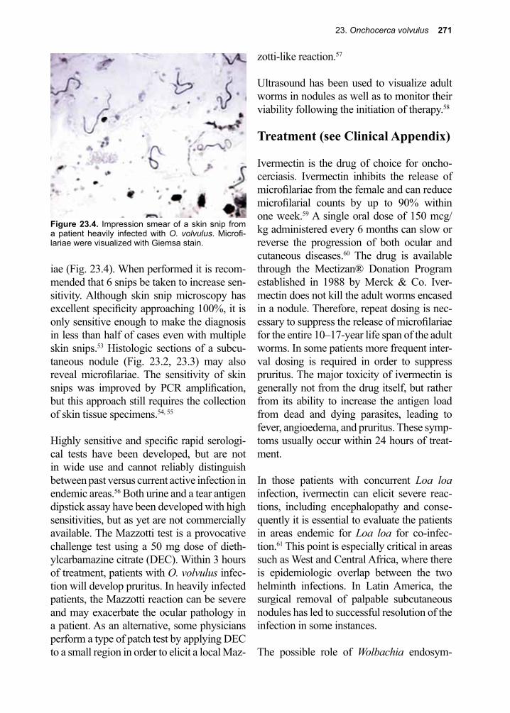
23. Onchocerca volvulus 271
iae (Fig. 23.4). When performed it is recom-mended that 6 snips be taken to increase sen-sitivity. Although skin snip microscopy has excellent specificity approaching 100%, it is only sensitive enough to make the diagnosis in less than half of cases even with multiple skin snips.53 Histologic sections of a subcu-taneous nodule (Fig. 23.2, 23.3) may also reveal microfilariae. The sensitivity of skin snips was improved by PCR amplification, but this approach still requires the collection of skin tissue specimens.54, 55
Highly sensitive and specific rapid serologi-cal tests have been developed, but are not in wide use and cannot reliably distinguish between past versus current active infection in endemic areas.56 Both urine and a tear antigen dipstick assay have been developed with high sensitivities, but as yet are not commercially available. The Mazzotti test is a provocative challenge test using a 50 mg dose of dieth-ylcarbamazine citrate (DEC). Within 3 hours of treatment, patients with O. volvulus infec-tion will develop pruritus. In heavily infected patients, the Mazzotti reaction can be severe and may exacerbate the ocular pathology in a patient. As an alternative, some physicians perform a type of patch test by applying DEC to a small region in order to elicit a local Maz-
zotti-like reaction.57
Ultrasound has been used to visualize adult worms in nodules as well as to monitor their viability following the initiation of therapy.58
Treatment (see Clinical Appendix)
Ivermectin is the drug of choice for oncho-cerciasis. Ivermectin inhibits the release of microfilariae from the female and can reduce microfilarial counts by up to 90% within one week.59 A single oral dose of 150 mcg/kg administered every 6 months can slow or reverse the progression of both ocular and cutaneous diseases.60 The drug is available through the Mectizan® Donation Program established in 1988 by Merck & Co. Iver-mectin does not kill the adult worms encased in a nodule. Therefore, repeat dosing is nec-essary to suppress the release of microfilariae for the entire 10–17-year life span of the adult worms. In some patients more frequent inter-val dosing is required in order to suppress pruritus. The major toxicity of ivermectin is generally not from the drug itself, but rather from its ability to increase the antigen load from dead and dying parasites, leading to fever, angioedema, and pruritus. These symp-toms usually occur within 24 hours of treat-ment.
In those patients with concurrent Loa loa infection, ivermectin can elicit severe reac-tions, including encephalopathy and conse-quently it is essential to evaluate the patients in areas endemic for Loa loa for co-infec-tion.61 This point is especially critical in areas such as West and Central Africa, where there is epidemiologic overlap between the two helminth infections. In Latin America, the surgical removal of palpable subcutaneous nodules has led to successful resolution of the infection in some instances.
The possible role of Wolbachia endosym-
Figure 23.4. Impression smear of a skin snip from a patient heavily infected with O. volvulus. Microfi-lariae were visualized with Giemsa stain.
272 The Nematodes
bionts in the inflammatory processes that caused eye and skin changes in O. volvulus infection, as well as their role in embryogen-esis and parasite fertility, has led to the sug-gestion that antibiotics could have a therapeu-tic activity for patients with onchocerciasis.62 Prolonged administration of doxycycline (200 mg/day for 4–6 weeks) was shown to interrupt O. volvulus embryogenesis.20
Prevention and Control
O. volvulus distribution follows that of the dipteran vectors. Black flies breed in fast-run-ning water of mountainous streams in regions of Africa and South and Central America, and they have a fairly long flight range. Thus, onchocerciasis can be found several miles from the nearest endemic breeding site. Because much of the coffee of the world is grown on mountainous hillsides, the preva-lence of onchocerciasis among workers on coffee plantations is high. The OCP was launched in 1974, with a primary emphasis on reducing black fly larval vector populations with DDT and other insecticides.63 With the increasing availability of ivermectin, the OCP has increasingly focused on control using this drug as an agent of mass chemotherapy.64 Community-wide chemotherapy can inter-rupt transmission of onchocerciasis.65, 66
In Africa, efforts to control onchocercia-sis are currently being conducted by the APOC.6 Critical to the success of APOC is the Merck Mectizan Donation Program, one of the first and largest public private-partner-ships devoted to a neglected disease. This program was launched in 1987 when Roy Vagelos, then CEO of Merck, made an his-toric announcement that his company would donate Mectizan® to anyone who needed it, for as long as it was needed.6, 64 The program works closely with the Carter Center and the Task Force for Child Survival and Develop-ment, both Atlanta-based non-governmental
organizations, for this purpose. By 2004, Merck had already donated more than 300 million treatments worth over $450 million.6
APOC works with the organizations pre-viously involved with the OCP, as well as Merck, the governments of 19 developing countries, 27 donor countries, at least 30 non-governmental organizations, and more than 80,000 rural Africa communities. This is done by coordinating with the ministries and non-governmental organizations to deliver Mectizan along with existing national health systems of the participating African coun-tries. To accomplish its mission, APOC has implemented a novel system of community-directed treatment programs. It is likely that the sight of more than 500,000 people has been saved so far. In addition, the commu-nity-based health systems created by APOC are expected to provide a framework for addi-tional pro-poor health interventions includ-ing those that target other neglected diseases such as soil-transmitted helminth infections (STHs), schistosomiasis, and trachoma.
The APOC and the OEPA have been success-ful in coordinating efforts to greatly reduce the transmission of this parasite throughout the world and currently there are only lim-ited areas of active transmission remaining.5-7 Working with the Carter Center the number of people at risk for onchocerciasis continues to decline. APOC reached the end of its lifespan and there are now a new generation of control programs integrated with intestinal helminth infections, schistosomiasis, lymphatic filaria-sis and trachoma.
As a complementary approach to onchocer-ciasis control, there have been some efforts to develop recombinant vaccines.64, 67 This pro-gram, the Onchocerciasis Vaccine for Africa, includes the development of a vaccine con-taining two antigens, Ov-103 and Ov-RAL-2.68-71 Work to create an Onchocerca vaccine
23. Onchocerca volvulus 273
References
1. Otranto, D., et al., Human ocular filariasis: further evidence on the zoonotic role of Onchocerca lupi. Parasit Vectors 2012, 5, 84.
2. Greene, B. M., Modern medicine versus an ancient scourge: progress toward control of onchocerciasis. The Journal of infectious diseases 1992, 166 (1), 15-21.
3. Kapoor, U., et al., Onchocercoma in a United Nations Peacekeeper. Med J Armed Forces India 2015, 71 (Suppl 1), S104-6.
4. Abanobi, O. C., et al., Validity of leopard skin manifestation in community diagnosis of human onchocerciasis infection. Appl Parasitol 1994, 35 (1), 8-11.
5. Coffeng, L. E., et al., African Programme For Onchocerciasis Control 1995-2015: model-estimated health impact and cost. PLoS Negl Trop Dis 2013, 7 (1), e2032.
6. Levine, R., et al., The What Works Working Group. Millions in Global Onchocerciasis in SubSaharan Africa Center for Global Development pp 2004, 6 57-64.
7. Centers for Disease, C.; Prevention, Progress toward elimination of onchocerciasis in the Americas - 1993-2012. MMWR Morb Mortal Wkly Rep 2013, 62 (20), 405-8.
8. WHO, Guidelines for Stopping Mass Drug Administration and Verifying Elimination of Human Onchocerciasis: Criteria and Procedures. Geneva, 2016.
9. O’Neill, J., On the presence of a filaria in “crawcraw.” Lancet 1875, 1, 265-266.10. Manson, P.; Davidson, A. H., Filaria volvuloxus. 1893; Vol. 1016.11. Robles, R., Enfermidad nueva en Guatemala. Juventud Med 1917, 17 97-115.12. Blacklock, D. B., The insect transmission of Onchocerca volvulus (Leuckart, 1893): The cause
of worm nodules in man in Africa. British medical journal 1927, 1 (3446), 129-33.13. Schulz-Key, H.; Karam, M., Periodic reproduction of Onchocerca volvulus. Parasitol Today
1986, 2 (10), 284-6.14. Duke, B. O., The population dynamics of Onchocerca volvulus in the human host. Tropical
medicine and parasitology : official organ of Deutsche Tropenmedizinische Gesellschaft and of Deutsche Gesellschaft fur Technische Zusammenarbeit (GTZ) 1993, 44 (2), 61-8.
15. Burnham, G., Onchocerciasis. Lancet 1998, 351 (9112), 1341-6.16. Lackey, A., et al., Extracellular proteases of Onchocerca volvulus. Exp Parasitol 1993, 68, 176-
185.17. Tawe, W., et al., Angiogenic activity of Onchocerca volvulus recombinant proteins similar to
vespid venom antigen 5. Molecular and biochemical parasitology 2000, 109 (2), 91-9.18. Higazi, T. B., et al., Angiogenic activity of an Onchocerca volvulus Ancylostoma-secreted
protein homologue. Molecular and biochemical parasitology 2003, 129 (1), 61-8.19. Capo, V., et al., Trichinella spiralis: vascular endothelial growth factor is up-regulated within the
Nurse cell during early phase of its formation. 1998, 84, 209-214.20. Hoerauf, A., et al., Onchocerciasis. BMJ (Clinical research ed.) 2003, 326 (7382), 207-10.21. Keiser, P. B., et al., Bacterial endosymbionts of Onchocerca volvulus in the pathogenesis of
posttreatment reactions. The Journal of infectious diseases 2002, 185 (6), 805-11.22. Jolodar, A., et al., Molecular cloning of an alpha-enolase from the human filarial parasite
Onchocerca volvulus that binds human plasminogen. Biochimica et biophysica acta 2003, 1627 (2-3), 111-20.
23. Rougemont, A., et al., Tuberculin skin tests and B.C.G. vaccination in hyperendemic area of onchocerciasis. Lancet (London, England) 1977, 1 (8006), 309.
24. Cooper, P. J., et al., Human onchocerciasis and tetanus vaccination: impact on the postvaccination antitetanus antibody response. Infection and immunity 1999, 67 (11), 5951-7.
25. Prost, A., et al., Lepromatous leprosy and onchocerciasis. British medical journal 1979, 1
continues with the hope that even if this vac-cine is not completely protective it will sub-stantially impact endemic areas by reducing
host microfilarial loads in children and ado-lescents.70, 72
274 The Nematodes
(6163), 589-90.26. Liebau, E., et al., The secretory omega-class glutathione transferase OvGST3 from the human
pathogenic parasite Onchocerca volvulus. FEBS J 2008, 275 (13), 3438-53.27. Brattig, N. W., Pathogenesis and host responses in human onchocerciasis: impact of Onchocerca
filariae and Wolbachia endobacteria. Microbes Infect 2004, 6 (1), 113-28.28. Hise, A. G., et al., Innate immune responses to endosymbiotic Wolbachia bacteria in Brugia
malayi and Onchocerca volvulus are dependent on TLR2, TLR6, MyD88, and Mal, but not TLR4, TRIF, or TRAM. J Immunol 2007, 178 (2), 1068-76.
29. Ali, M. M., et al., Immune responses directed against microfilariae correlate with severity of clinical onchodermatitis and treatment history. The Journal of infectious diseases 2003, 187 (4), 714-7.
30. Cooper, P. J., et al., Association of transient dermal mastocytosis and elevated plasma tryptase levels with development of adverse reactions after treatment of onchocerciasis with ivermectin. The Journal of infectious diseases 2002, 186 (9), 1307-13.
31. Pearlman, E., et al., Interleukin 4 and T helper type 2 cells are required for development of experimental onchocercal keratitis (river blindness). The Journal of experimental medicine 1995, 182 (4), 931-40.
32. Gentil, K.; Pearlman, E., Gamma interferon and interleukin-1 receptor 1 regulate neutrophil recruitment to the corneal stroma in a murine model of Onchocerca volvulus keratitis. Infect Immun 2009, 77 (4), 1606-12.
33. Timmann, C., et al., Cutaneous pathology in onchocerciasis associated with pronounced systemic T-helper 2-type responses to Onchocerca volvulus. Br J Dermatol 2003, 149 (4), 782-7.
34. Timmann, C., et al., Human genetic resistance to Onchocerca volvulus: evidence for linkage to chromosome 2p from an autosome-wide scan. J Infect Dis 2008, 198 (3), 427-33.
35. Hoerauf, A., et al., The variant Arg110Gln of human IL-13 is associated with an immunologically hyper-reactive form of onchocerciasis (sowda). Microbes Infect 2002, 4 (1), 37-42.
36. Bari, A. U., Clinical spectrum of onchodermatitis. J Coll Physicians Surg Pak 2007, 17 (8), 453-6.
37. Murdoch, M. E., Onchodermatitis. Curr Opin Infect Dis 2010, 23 (2), 124-31.38. Murdoch, M. E., et al., A clinical classification and grading system of the cutaneous changes in
onchocerciasis. Br J Dermatol 1993, 129 (3), 260-9.39. Lazarov, A., et al., Pruritus and chronic papular dermatitis in an Ethiopian man. Onchocerciasis
(chronic papular onchodermatitis). Arch Dermatol 1997, 133 (3), 382-3, 385-6.40. Siddiqui, M. A.; al-Khawajah, M. M., The black disease of Arabia, Sowda-onchocerciasis. New
findings. Int J Dermatol 1991, 30 (2), 130-3.41. Richard-Lenoble, D., et al., [Human onchocerciasis and “sowda” in the Republic of Yemen]. Bull
Acad Natl Med 2001, 185 (8), 1447-59; discussion 1459-61.42. Somorin, A. O., Sowda onchocerciasis in Nigeria. Cent Afr J Med 1982, 28 (10), 253-6.43. Schwartz, D. A., et al., Sowda-type onchocerciasis in Guatemala. Acta Trop 1983, 40 (4), 383-9.44. Nelson, G. S., Hanging groin and hernia complications of onchocerciasis. Trans R Soc Trop Med
Hyg 1958, 52 (3), 272-5.45. Niamba, P., et al., Hanging groin and persistent pruritus in a patient from Burkina Faso. Int J
Dermatol 2007, 46 (5), 485-6.46. Fuglsang, H., “Leopard skin” and onchocerciasis. Trans R Soc Trop Med Hyg 1983, 77 (6), 881.47. Edungbola, L. D., et al., ‘Leopard skin’ as a rapid diagnostic index for estimating the endemicity
of African onchocerciasis. Int J Epidemiol 1987, 16 (4), 590-4.48. Goldman, L.; Figueroa Ortiz, L., Types of dermatitis in American onchocerciasis. Arch Derm
Syphilol 1946, 53, 79-93.49. Babalola, O. E., Ocular onchocerciasis: current management and future prospects. Clin
Ophthalmol 2011, 5, 1479-91.50. Thylefors, B., Ocular onchocerciasis. Bull World Health Organ 1978, 56 (1), 63-73.51. Colebunders, R., et al., Nodding syndrome and epilepsy in onchocerciasis endemic regions:
comparing preliminary observations from South Sudan and the Democratic Republic of the
23. Onchocerca volvulus 275
Congo with data from Uganda. BMC Res Notes 2016, 9 (1), 182.52. Idro, R., et al., Is nodding syndrome an Onchocerca volvulus-induced neuroinflammatory
disorder? Uganda’s story of research in understanding the disease. Int J Infect Dis 2016, 45, 112-7.
53. Boatin, B. A., et al., Detection of Onchocerca volvulus infection in low prevalence areas: a comparison of three diagnostic methods. Parasitology 2002, 125 (Pt 6), 545-52.
54. Boatin, B. A., et al., Detection of Onchocerca volvulus infection in low prevalence areas: a comparison of three diagnostic methods. Parasitology 2002, 125 (Pt 6), 545-52.
55. Bradley, J. E.; Unnasch, T. R., Molecular approaches to the diagnosis of onchocerciasis. Advances in parasitology 1996, 37, 57-106.
56. Udall, D. N., Recent updates on onchocerciasis: diagnosis and treatment. Clin Infect Dis 2007, 44 (1), 53-60.
57. Kilian, H. D., The use of a topical Mazzotti test in the diagnosis of onchocerciasis. Tropical medicine and parasitology : official organ of Deutsche Tropenmedizinische Gesellschaft and of Deutsche Gesellschaft fur Technische Zusammenarbeit (GTZ) 1988, 39 (3), 235-8.
58. Mand, S., et al., Frequent detection of worm movements in onchocercal nodules by ultrasonography. Filaria J 2005, 4 (1), 1.
59. Greene, B. M., et al., Comparison of ivermectin and diethylcarbamazine in the treatment of onchocerciasis. The New England journal of medicine 1985, 313 (3), 133-8.
60. Burnham, G., Ivermectin treatment of onchocercal skin lesions: observations from a placebo-controlled, double-blind trial in Malawi. The American journal of tropical medicine and hygiene 1995, 52 (3), 270-6.
61. Chippaux, J. P., et al., Ivermectin treatment of loiasis. Transactions of the Royal Society of Tropical Medicine and Hygiene 1992, 86 (3), 289.
62. Hoerauf, A., et al., Doxycycline in the treatment of human onchocerciasis: Kinetics of Wolbachia endobacteria reduction and of inhibition of embryogenesis in female Onchocerca worms. Microbes and infection / Institut Pasteur 2003, 5 (4), 261-73.
63. Omura, S.; Crump, A., The life and times of ivermectin - a success story. Nature reviews. Microbiology 2004, 2 (12), 984-9.
64. Peters, D. H.; Phillips, T., Mectizan Donation Program: evaluation of a public-private partnership. Tropical medicine & international health : TM & IH 2004, 9 (4), A4-15.
65. Taylor, H. R., et al., Impact of mass treatment of onchocerciasis with ivermectin on the transmission of infection. Science (New York, N.Y.) 1990, 250 (4977), 116-8.
66. Cupp, E. W., et al., The effects of repetitive community-wide ivermectin treatment on transmission of Onchocerca volvulus in Guatemala. The American journal of tropical medicine and hygiene 1992, 47 (2), 170-80.
67. Lustigman, S., et al., Towards a recombinant antigen vaccine against Onchocerca volvulus. Trends in parasitology 2002, 18 (3), 135-41.
68. Nutman, T. B., Future directions for vaccine-related onchocerciasis research. Trends in parasitology 2002, 18 (6), 237-9.
69. MacDonald, A. J., et al., Ov-ASP-1, the Onchocerca volvulus homologue of the activation associated secreted protein family is immunostimulatory and can induce protective anti-larval immunity. Parasite immunology 2004, 26 (1), 53-62.
70. Hotez, P. J., et al., The Onchocerciasis Vaccine for Africa--TOVA--Initiative. PLoS Negl Trop Dis 2015, 9 (1), e0003422.
71. Arumugam, S., et al., Vaccination of Gerbils with Bm-103 and Bm-RAL-2 Concurrently or as a Fusion Protein Confers Consistent and Improved Protection against Brugia malayi Infection. PLoS Negl Trop Dis 2016, 10 (4), e0004586.
72. Makepeace, B. L., et al., The case for vaccine development in the strategy to eradicate river blindness (onchocerciasis) from Africa. Expert Rev Vaccines 2015, 14 (9), 1163-5.

276 The Nematodes
Donald Hopkins, M.D., M.P.H. (born 1941)
Hopkins, a Bahamian-American physician, was the director of the health programs at the Carter Center and played a key role in the Guinea Worm eradication efforts and in programs focusing on river blindness, among other neglected tropical parasitic diseases. In addition to devoting his life to reducing the amount of suffering due to parasitic diseases, he is also an acclaimed author; nominated for the Pulitzer prize in 1983. Hopkins is the recipient of several awards and is a leader in the field of applied epidemiology and for his work, he received the Pumphandle Award.
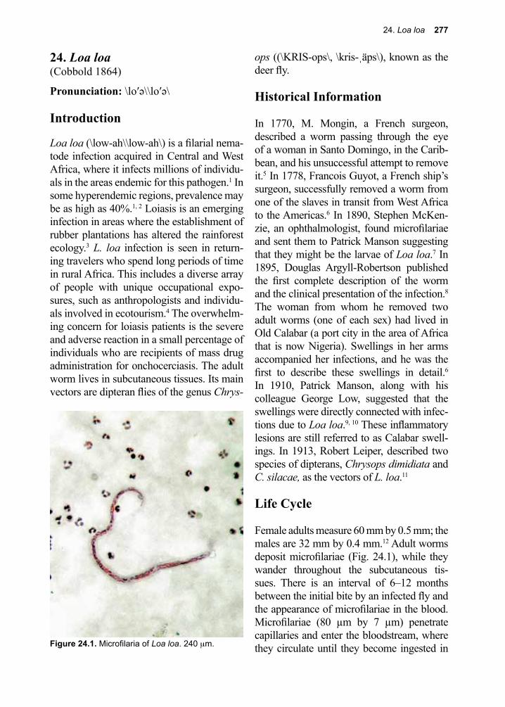
24. Loa loa 277
Figure 24.1. Microfilaria of Loa loa. 240 µm.
24. Loa loa(Cobbold 1864)
Pronunciation: \lo′ə\\lo′ə\
Introduction
Loa loa (\low-ah\\low-ah\) is a filarial nema-tode infection acquired in Central and West Africa, where it infects millions of individu-als in the areas endemic for this pathogen.1 In some hyperendemic regions, prevalence may be as high as 40%.1, 2 Loiasis is an emerging infection in areas where the establishment of rubber plantations has altered the rainforest ecology.3 L. loa infection is seen in return-ing travelers who spend long periods of time in rural Africa. This includes a diverse array of people with unique occupational expo-sures, such as anthropologists and individu-als involved in ecotourism.4 The overwhelm-ing concern for loiasis patients is the severe and adverse reaction in a small percentage of individuals who are recipients of mass drug administration for onchocerciasis. The adult worm lives in subcutaneous tissues. Its main vectors are dipteran flies of the genus Chrys-
ops ((\KRIS-ops\, \kris-ˌäps\), known as the deer fly.
Historical Information
In 1770, M. Mongin, a French surgeon, described a worm passing through the eye of a woman in Santo Domingo, in the Carib-bean, and his unsuccessful attempt to remove it.5 In 1778, Francois Guyot, a French ship’s surgeon, successfully removed a worm from one of the slaves in transit from West Africa to the Americas.6 In 1890, Stephen McKen-zie, an ophthalmologist, found microfilariae and sent them to Patrick Manson suggesting that they might be the larvae of Loa loa.7 In 1895, Douglas Argyll-Robertson published the first complete description of the worm and the clinical presentation of the infection.8 The woman from whom he removed two adult worms (one of each sex) had lived in Old Calabar (a port city in the area of Africa that is now Nigeria). Swellings in her arms accompanied her infections, and he was the first to describe these swellings in detail.6 In 1910, Patrick Manson, along with his colleague George Low, suggested that the swellings were directly connected with infec-tions due to Loa loa.9, 10 These inflammatory lesions are still referred to as Calabar swell-ings. In 1913, Robert Leiper, described two species of dipterans, Chrysops dimidiata and C. silacae, as the vectors of L. loa.11
Life Cycle
Female adults measure 60 mm by 0.5 mm; the males are 32 mm by 0.4 mm.12 Adult worms deposit microfilariae (Fig. 24.1), while they wander throughout the subcutaneous tis-sues. There is an interval of 6–12 months between the initial bite by an infected fly and the appearance of microfilariae in the blood. Microfilariae (80 µm by 7 µm) penetrate capillaries and enter the bloodstream, where they circulate until they become ingested in
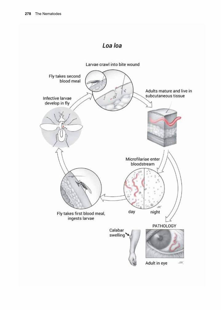
278 The Nematodes
24. Loa loa 279
a blood meal by a Chrysops spp. These flies are commonly called deer flies, horse flies, yellow flies, stouts, mango flies, or mangrove flies.12-14
L. loa microfilariae exhibit diurnal periodic-ity that coincides with the feeding habits of Chrysops.15 Larvae penetrate the stomach of the fly, and locate to the fat body. Eight to ten days later, the infective L3 larvae migrate to the cavity of the biting mouthparts and are released into the bite wound when the fly takes another blood meal. The larvae, now in the subcutaneous tissues of the host, develop slowly into adults within 1–4 years. Mature worms mate, and the females begin deposit-ing microfilariae. The adult worms can live in the tissues for up to 17 years.16
Cellular and Molecular Pathogenesis
In most cases, neither the adult worms in the subcutaneous tissues or the microfilariae in the bloodstream cause any direct patho-logic changes. In individuals native to, and living in, endemic areas there is evidence for immune tolerance with high levels of circu-lating microfilariae, low levels of filarial-spe-cific IgG, and few if any clinical symptoms.17
In temporary residents there can be peripheral eosinophilia, high levels of filarial-specific IgG, eosinophil activation and degranula-tion, and localized subcutaneous edema.17, 18 The localized swelling referred to as Calabar swellings are usually restricted to visitors to endemic areas and are not typically seen in residents who live for many years in these same regions.19, 20 The visitors to endemic areas may have exuberant immune responses with parasite-specific IgG and IgE production as well as proliferation of antigen-specific T cells.21, 22 These patients develop swellings as a local inflammatory reaction in response to filaria antigens released during the migration of adult worms.
A third group of infected individuals who neither have circulating microfilariae nor Calabar swellings, while still living in hyper-endemic areas, are suspected to have devel-oped protective sterilizing immunity early on in their lives.23, 24 Unlike other filarial nema-todes, there is currently no evidence that L. loa contains Wolbachia symbionts, based on extensive investigations employing light microscopy, electron microscopy, and PCR.25,
26
Clinical Disease
The clinical manifestations of loiasis include Calabar swellings, eye symptoms, encepha-litis, cardiomyopathy, renal disease, arthritis, and lymphadenitis. Calabar swellings are 10–20 cm non-erythematous, angioedema-tous swellings that last for a few days. They occur most typically on the extremities and the face, particularly in the periorbital region. The angioedema is often preceded by pain and itching. Recurrences are common. The occasional passage of an adult worm across the subconjunctival space of the eye is per-haps the most disturbing aspect of L. loa to most infected individuals.
Patients with more serious complications, including cardiomyopathy, nephropathy, and pleural effusions, have been reported.20 The most serious and life-threatening reactions result after patients are exposed to antihel-mintic medications. Some complications occur only after administration of diethyl-carbamazine citrate (DEC). These sequelae may result from immune complex deposi-tion. Encephalitis has also been described in patients with very high levels of microfilariae, including microfilariae in the cerebrospinal fluid (CSF).23 Most of these individuals have received DEC or ivermectin while suffering from elevated levels of microfilariae in the blood, and the syndrome probably occurs as part of host immune responses.27 While less common in patients with low levels of micro-
280 The Nematodes
filariae in the blood, neurological symptoms have been seen with treatment initiation.28 A rare cardiac condition known as endomyo-cardial fibrosis may result from eosinophilic infiltration of the myocardium in response to L. loa.29
Significant differences in the clinical presenta-tion of loiasis occur among long-term visitors to endemic areas, in contrast to people native to these areas.21 People living in endemic regions are often asymptomatic, despite high levels of circulating microfilariae, whereas visitors and expatriates suffer from a vari-ety of allergic responses, such as frequent episodes of angioedema, Calabar swellings, hypereosinophilia, and hyper-gammaglobu-linemia. This disorder is sometimes referred to as the “hyper-responsive syndrome” of loiasis, reflecting elevated humoral and cell-mediated immune responses to the parasite.20
Diagnosis (see Clinical Appendix)
Patients returning from West or Central Africa with localized angioedema or a worm beneath the conjunctiva should be suspected of having loiasis. The Calabar swellings should be clinically differentiated from other causes of angioedema, including complement C1 complex inhibitor deficiency.30 Definitive diagnosis is by identifying the microfilariae microscopically in a thin blood smear stained with either Giemsa or Wright’s solution. Among the distinguishing features of L. loa microfilariae (Figs. C.50, C.51) are their diur-nal periodicity, the presence of a sheath, and three or more terminal nuclei.24
With regards to the diurnal periodicity it is important to draw blood for this test during the middle of the day and employ a concentration technique such as the Knott’s test (using for-malin), or nucleopore filtration.31 Since visi-tors to endemic areas often have low levels of circulating microfilariae, serology can be
useful in this population.30, 32, 33 Antibodies against L. loa of the IgG4 immunoglobulin subclass may be a reliable marker of infec-tion.30 This includes IgG4 antibodies against the L. loa recombinant antigen L1-SXP-1.34 There is also a microsatellite that has been developed to detect microfilariae in blood by PCR.33, 35 Real-time PCR was developed and then adapted for loop-mediated isother-mal amplification (LAMP) as a point of care test.36, 37
Since levels of circulating microfilariae are critical in decisions regarding treatment, an automated handheld cell counter has been repurposed as a point-of-care test to assess microfilarial levels.38 Alternatively, new blood and urine L. loa-specific biomarker tests have been developed.39, 40
Treatment (see Clinical Appendix)
The first step in treatment of Loa loa infec-tion is based on levels of circulating micro-filariae. If levels are greater than 2,500 mf/ml an attempt should be made to lower these levels. This may be accomplished through apheresis or several weeks of treatment with albendazole.24, 41, 42 Once the levels of micro-filariae are below 2,500 mf/ml DEC may be administered for 21 days.
DEC destroys all stages of the infecting para-site. The full dose of the drug is not typically started on the first day of treatment. Instead, it is given in a graded manner, beginning with a test dose on day 1 and then increasing the dose to full dose by day 4. This is done to reduce the likelihood of treatment-associated complica-tions, including encephalopathy, that occur as a consequence of mass destruction of L. loa microfilariae.28 These iatrogenic complica-tions of loiasis are rare when the microfilarial concentration in blood is less than 2,500 mf/ml at initiation of therapy.24 Antihistamines or corticosteroids may be required to decrease
24. Loa loa 281
allergic reactions during treatment. In up to 50% of patients, DEC treatment may need to be repeated several times in order to effect a cure.43
Adult worms in the eye can be removed surgically. Since they tend not to cause any pathology it is not a required part of curative therapy. Alternatives to DEC include alben-dazole, effective in reducing the number of circulating microfilariae by acting directly on adult worms. Ivermectin is not a preferred agent for the treatment of loiasis and can be associated with significant morbidity if given to patients with high levels of circulat-ing microfilariae. Weekly chemoprophylaxis with DEC given in a dose of 300 mg is effec-tive in preventing loiasis among long-term visitors but is not currently recommended for short-term visitors to endemic areas.43
Prevention and Control
In hyperendemic regions of Central Africa, 95% of the population has antibodies to L. loa antigen by the age of two years.41 In the Chailu Mountains in the Democratic Repub-lic of Congo, 19% of the native populations are microfilaremic, and more than 50% of the adults have reported sub-conjunctival migra-tions of an adult worm. Mass or targeted chemotherapy with DEC may reduce trans-
mission in these areas.44, 45 Spraying mango groves with insecticides, particularly DDT, remains an effective method for controlling populations of the vector, since resistance to this insecticide in Chrysops has not yet devel-oped.
Widespread use of mass drug administra-tion in Sub-Saharan Africa for purposes of controlling lymphatic filariasis (LF) and onchocerciasis has raised concerns about L. loa co-infections. The potential complica-tions of unmonitored DEC treatment of loia-sis and the risk of encephalopathy are major reasons why this agent is not used routinely. Even the alternative combination of ivermec-tin and albendazole poses some risk. L. Loa encephalopathy is associated with ivermectin treatment of individuals with L. loa microfila-remia > 30,000 mf/ml blood, with most of the cases of ivermectin-induced encephalopathy occurring in Cameroon.45 In order to reduce the risks associated with ivermectin in this region, albendazole has been evaluated as a possible first-line measure to gradually lower microfilariae burdens.46, 47 The recent devel-opment of blood and urine tests to detect L. loa antigen may represent a future break-through in detecting loiasis patients targeted for mass drug administration to combat LF or onchocerciasis.39
References
1. Zoure, H. G., et al., The geographic distribution of Loa loa in Africa: results of large-scale implementation of the Rapid Assessment Procedure for Loiasis (RAPLOA). PLoS Negl Trop Dis 2011, 5 (6), e1210.
2. Noireau, F., et al., Loa loa and Mansonella perstans filariasis in the Chaillu mountains, Congo: parasitological prevalence. Transactions of the Royal Society of Tropical Medicine and Hygiene 1989, 83 (4), 529-34.
3. Rodhain, F., [Hypotheses on the dynamic ecology of Loa infections]. Bulletin de la Societe de pathologie exotique et de ses filiales 1980, 73 (2), 182-91.
4. Thompson, C., et al., Chronic symptomatic and microfilaremic loiasis in a returned traveller. CMAJ 2015, 187 (6), 437.
5. Mongin, Sur un ver trouve´ sous la conjunctive a` Maribarou, isle Saint-Dominique. J. Med. Chir. Pharm 1770, 32, 338-339.
282 The Nematodes
6. Cox, F. E., History of human parasitology. Clin Microbiol Rev 2002, 15 (4), 595-612.7. Manson, P., The Filaria sanguinis hominis major and minor, two new species of haematozoa.
Lancet 1891, i, 4-8.8. Argyll-Robertson, D.; Soc, U. K., Case of Filaria loa in which the parasite was removed from
under the conjunctiva. Trans Ophthalmol 1895, 15, 137-167.9. Low, G. C., Discussion of Manson, P. On the nature and origin of Calabar swellings. Trans. R.
Soc. Trop. Med. Hyg 1910, 3, 251-253.10. Manson, P., On the nature and origin of Calabar swellings. Trans. R. Soc. Trop. Med. Hyg 1910,
3, 244-251.11. Leiper, R. T., Report of the Helminthologist for the Half Year Ending 30 April, . Report of the
Advisory Commission on Tropical Diseases Research Fund. 1913, 1913-1914.12. Padgett, J. J.; Jacobsen, K. H., Loiasis: African eye worm. Trans R Soc Trop Med Hyg 2008, 102
(10), 983-9.13. Horvath, G., et al., Ventral polarization vision in tabanids: horseflies and deerflies (Diptera:
Tabanidae) are attracted to horizontally polarized light. Naturwissenschaften 2008, 95 (11), 1093-100.
14. Baldacchino, F., et al., Tabanids: neglected subjects of research, but important vectors of disease agents! Infect Genet Evol 2014, 28, 596-615.
15. Duke, B. O. L.; J., Studies of the biting habits of Chrysops. Trop Med Parasitol 1955, 49.16. Eveland, L. K., et al., Loa loa infection without microfilaraemia. Transactions of the Royal
Society of Tropical Medicine and Hygiene 1975, 69 (3), 354-5.17. Herrick, J. A., et al., Eosinophil-associated processes underlie differences in clinical presentation
of loiasis between temporary residents and those indigenous to Loa-endemic areas. Clin Infect Dis 2015, 60 (1), 55-63.
18. Olness, K., et al., Loiasis in an expatriate American child: diagnostic and treatment difficulties. Pediatrics 1987, 80 (6), 943-6.
19. Nutman, T. B., et al., Loa loa infection in temporary residents of endemic regions: recognition of a hyperresponsive syndrome with characteristic clinical manifestations. The Journal of infectious diseases 1986, 154 (1), 10-8.
20. Nutman, T. B., et al., Immunologic correlates of the hyperresponsive syndrome of loiasis. The Journal of infectious diseases 1988, 157 (3), 544-50.
21. Kilon, A. D., et al., Loiasis in endemic and non-endemic populations: immunologically mediated differences in clinical presentation. Dis 1991, 163, 1318-1325.
22. Limaye, A. P., et al., Regulation of parasite-induced eosinophilia: selectively increased interleukin 5 production in helminth-infected patients. The Journal of experimental medicine 1990, 172 (1), 399-402.
23. Kilon, A. D.; Einstein, E. M., Pulmonary involvement in loiasis. Am Rev Respir Dis 1992, 145, 961-963.
24. Kilon, A. D., et al., Loiasis and Mansonella infections In: Tropical Infectious Diseases, Principles, Pathogens, and Practice. Vol 2 eds Churchill Livingstone Pubs New York pp 1999, 861-872.
25. Grobusch, M. P., et al., No evidence of Wolbachia endosymbiosis with Loa loa and Mansonella perstans. Parasitology research 2003, 90 (5), 405-8.
26. Brouqui, P., et al., Doxycycline and eradication of microfilaremia in patients with loiasis. Emerg Infect Dis 2001, 7 (3 Suppl), 604-5.
27. Gardon, J., et al., Serious reactions after mass treatment of onchocerciasis with ivermectin in an area endemic for Loa loa infection. Lancet 1997, 350 (9070), 18-22.
28. Carme, B., et al., Five cases of encephalitis during treatment of loiasis with diethylcarbamazine. The American journal of tropical medicine and hygiene 1991, 44 (6), 684-90.
29. Andy, J. J., et al., Loasis as a possible trigger of African endomyocardial fibrosis: a case report from Nigeria. Acta tropica 1981, 38 (2), 179-86.
30. Akue, J. P., et al., High levels of parasite-specific IgG4 in the absence of microfilaremia in Loa loa infection. Tropical medicine and parasitology : official organ of Deutsche Tropenmedizinische Gesellschaft and of Deutsche Gesellschaft fur Technische Zusammenarbeit
24. Loa loa 283
(GTZ) 1994, 45 (3), 246-8.31. Mak, J. W., Recent advances in the laboratory diagnosis of filariasis. Malays J Pathol 1989, 11,
1-5.32. Toure, F. S., et al., IgG4 serology of loiasis in three villages in an endemic area of south-eastern
Gabon. Trop Med Int Health 1998, 3 (4), 313-7.33. Toure, F. S., et al., [Comparative analysis of 2 diagnostic methods of human loiasis: IgG4
serology and nested PCR]. Bull Soc Pathol Exot 1999, 92 (3), 167-70.34. Klion, A. D., et al., Serum immunoglobulin G4 antibodies to the recombinant antigen, Ll-SXP-1,
are highly specific for Loa loa infection. The Journal of infectious diseases 2003, 187 (1), 128-33.
35. Nutman, T. B., et al., A universally applicable diagnostic approach to filarial and other infections. Parasitology today (Personal ed.) 1994, 10 (6), 239-43.
36. Drame, P. M., et al., Loop-mediated isothermal amplification for rapid and semiquantitative detection of Loa loa infection. J Clin Microbiol 2014, 52 (6), 2071-7.
37. Fernandez-Soto, P., et al., Development of a highly sensitive loop-mediated isothermal amplification (LAMP) method for the detection of Loa loa. PLoS One 2014, 9 (4), e94664.
38. Bennuru, S., et al., Repurposed automated handheld counter as a point-of-care tool to identify individuals ‘at risk’ of serious post-ivermectin encephalopathy. PLoS Negl Trop Dis 2014, 8 (9), e3180.
39. Geary, T. G., A Step Toward Eradication of Human Filariases in Areas Where Loa Is Endemic. MBio 2016, 7 (2).
40. Drame, P. M., et al., Identification and Validation of Loa loa Microfilaria-Specific Biomarkers: a Rational Design Approach Using Proteomics and Novel Immunoassays. MBio 2016, 7 (1), e02132-15.
41. Ottesen, E. A., Filarial infections. Infectious disease clinics of North America 1993, 7 (3), 619-33.
42. Kilon, A. D., et al., Albendazole in human loiasis: Results of a double-blind, placebo-controlled trial. Dis 202 1993, 168.
43. Kilon, A. D., et al., Effectiveness of diethylcarbamazine in treating loiasis acquired by expatriate visitors to endemic regions: long-term follow-up. Dis 602 1994, 169.
44. Nutman, T. B., et al., Diethylcarbamazine prophylaxis for human loiasis. Results of a double-blind study. The New England journal of medicine 1988, 319 (12), 752-6.
45. Molyneux, D. H., et al., Mass drug treatment for lymphatic filariasis and onchocerciasis. Trends in parasitology 2003, 19 (11), 516-22.
46. Tabi, T.-E., et al., Human loiasis in a Cameroonian village: a double-blind, placebo-controlled, crossover clinical trial of a three-day albendazole regimen. The American journal of tropical medicine and hygiene 2004, 71 (2), 211-5.
47. Tsague-Dongmo, L., et al., Effects of a 3-day regimen of albendazole (800 mg daily) on Loa loa microfilaraemia. Annals of tropical medicine and parasitology 2002, 96 (7), 707-15.
284 The Nematodes
Susan Lim, Ph.D. (1952–2014)
Susan Lim was a full professor at the University of Malaya and a rec-ognized expert on the Monogenea, a class of ectoparasitic flatworms of fish. As one of the most productive investigators in this field, several parasites were named after her in her honor. Dr. Lim discovered a novel attachment mechanism for monogeneans that involved net-like struc-tures formed by secretions from the attachment organ of opisthaptor.
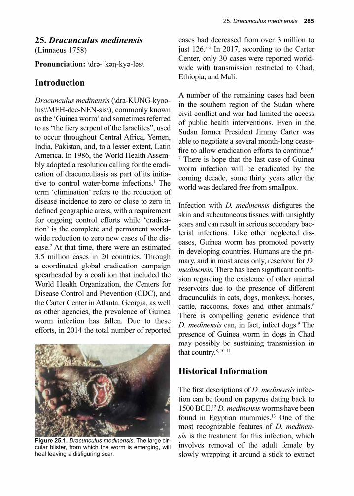
25. Dracunculus medinensis 285
Figure 25.1. Dracunculus medinensis. The large cir-cular blister, from which the worm is emerging, will heal leaving a disfiguring scar.
25. Dracunculus medinensis(Linnaeus 1758)
Pronunciation: \drə-ˈkəŋ-kyə-ləs\
Introduction
Dracunculus medinensis (\dra-KUNG-kyoo-lus\\MEH-dee-NEN-sis\), commonly known as the ‘Guinea worm’ and sometimes referred to as “the fiery serpent of the Israelites”, used to occur throughout Central Africa, Yemen, India, Pakistan, and, to a lesser extent, Latin America. In 1986, the World Health Assem-bly adopted a resolution calling for the eradi-cation of dracunculiasis as part of its initia-tive to control water-borne infections.1 The term ‘elimination’ refers to the reduction of disease incidence to zero or close to zero in defined geographic areas, with a requirement for ongoing control efforts while ‘eradica-tion’ is the complete and permanent world-wide reduction to zero new cases of the dis-ease.2 At that time, there were an estimated 3.5 million cases in 20 countries. Through a coordinated global eradication campaign spearheaded by a coalition that included the World Health Organization, the Centers for Disease Control and Prevention (CDC), and the Carter Center in Atlanta, Georgia, as well as other agencies, the prevalence of Guinea worm infection has fallen. Due to these efforts, in 2014 the total number of reported
cases had decreased from over 3 million to just 126.3-5 In 2017, according to the Carter Center, only 30 cases were reported world-wide with transmission restricted to Chad, Ethiopia, and Mali.
A number of the remaining cases had been in the southern region of the Sudan where civil conflict and war had limited the access of public health interventions. Even in the Sudan former President Jimmy Carter was able to negotiate a several month-long cease-fire to allow eradication efforts to continue.6,
7 There is hope that the last case of Guinea worm infection will be eradicated by the coming decade, some thirty years after the world was declared free from smallpox.
Infection with D. medinensis disfigures the skin and subcutaneous tissues with unsightly scars and can result in serious secondary bac-terial infections. Like other neglected dis-eases, Guinea worm has promoted poverty in developing countries. Humans are the pri-mary, and in most areas only, reservoir for D. medinensis. There has been significant confu-sion regarding the existence of other animal reservoirs due to the presence of different dracunculids in cats, dogs, monkeys, horses, cattle, raccoons, foxes and other animals.8 There is compelling genetic evidence that D. medinensis can, in fact, infect dogs.9 The presence of Guinea worm in dogs in Chad may possibly be sustaining transmission in that country.8, 10, 11
Historical Information
The first descriptions of D. medinensis infec-tion can be found on papyrus dating back to 1500 BCE.12 D. medinensis worms have been found in Egyptian mummies.13 One of the most recognizable features of D. medinen-sis is the treatment for this infection, which involves removal of the adult female by slowly wrapping it around a stick to extract
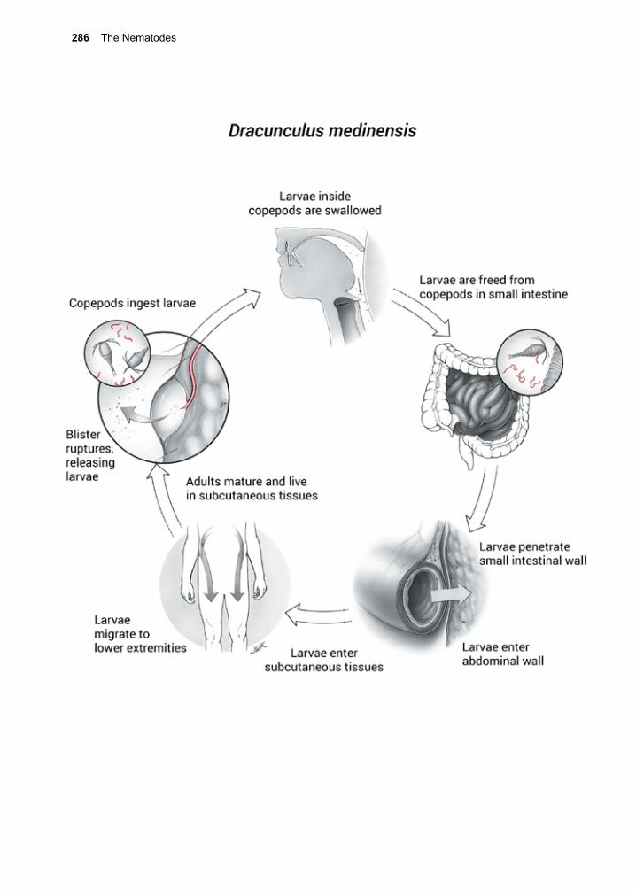
286 The Nematodes
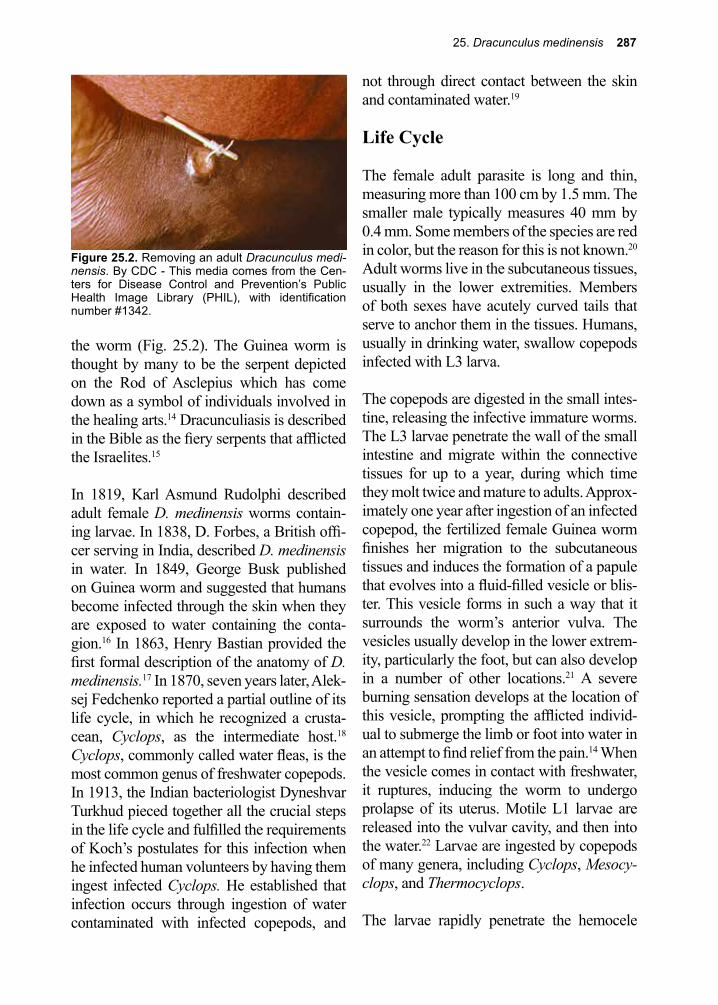
25. Dracunculus medinensis 287
the worm (Fig. 25.2). The Guinea worm is thought by many to be the serpent depicted on the Rod of Asclepius which has come down as a symbol of individuals involved in the healing arts.14 Dracunculiasis is described in the Bible as the fiery serpents that afflicted the Israelites.15
In 1819, Karl Asmund Rudolphi described adult female D. medinensis worms contain-ing larvae. In 1838, D. Forbes, a British offi-cer serving in India, described D. medinensis in water. In 1849, George Busk published on Guinea worm and suggested that humans become infected through the skin when they are exposed to water containing the conta-gion.16 In 1863, Henry Bastian provided the first formal description of the anatomy of D. medinensis.17 In 1870, seven years later, Alek-sej Fedchenko reported a partial outline of its life cycle, in which he recognized a crusta-cean, Cyclops, as the intermediate host.18 Cyclops, commonly called water fleas, is the most common genus of freshwater copepods. In 1913, the Indian bacteriologist Dyneshvar Turkhud pieced together all the crucial steps in the life cycle and fulfilled the requirements of Koch’s postulates for this infection when he infected human volunteers by having them ingest infected Cyclops. He established that infection occurs through ingestion of water contaminated with infected copepods, and
not through direct contact between the skin and contaminated water.19
Life Cycle
The female adult parasite is long and thin, measuring more than 100 cm by 1.5 mm. The smaller male typically measures 40 mm by 0.4 mm. Some members of the species are red in color, but the reason for this is not known.20 Adult worms live in the subcutaneous tissues, usually in the lower extremities. Members of both sexes have acutely curved tails that serve to anchor them in the tissues. Humans, usually in drinking water, swallow copepods infected with L3 larva.
The copepods are digested in the small intes-tine, releasing the infective immature worms. The L3 larvae penetrate the wall of the small intestine and migrate within the connective tissues for up to a year, during which time they molt twice and mature to adults. Approx-imately one year after ingestion of an infected copepod, the fertilized female Guinea worm finishes her migration to the subcutaneous tissues and induces the formation of a papule that evolves into a fluid-filled vesicle or blis-ter. This vesicle forms in such a way that it surrounds the worm’s anterior vulva. The vesicles usually develop in the lower extrem-ity, particularly the foot, but can also develop in a number of other locations.21 A severe burning sensation develops at the location of this vesicle, prompting the afflicted individ-ual to submerge the limb or foot into water in an attempt to find relief from the pain.14 When the vesicle comes in contact with freshwater, it ruptures, inducing the worm to undergo prolapse of its uterus. Motile L1 larvae are released into the vulvar cavity, and then into the water.22 Larvae are ingested by copepods of many genera, including Cyclops, Mesocy-clops, and Thermocyclops.
The larvae rapidly penetrate the hemocele
Figure 25.2. Removing an adult Dracunculus medi-nensis. By CDC - This media comes from the Cen-ters for Disease Control and Prevention’s Public Health Image Library (PHIL), with identification number #1342.
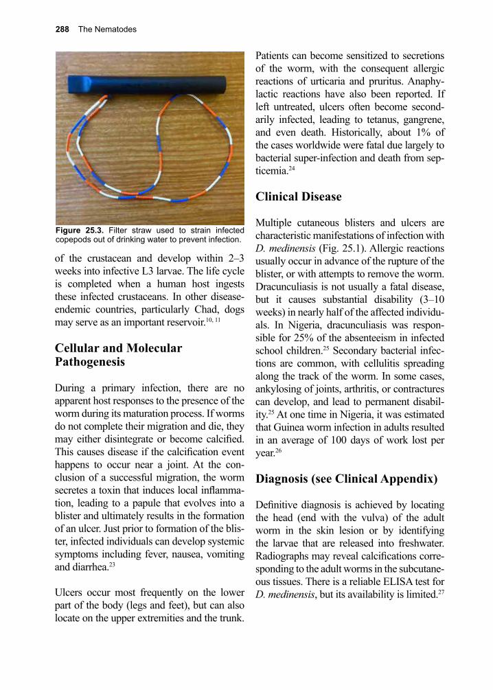
288 The Nematodes
of the crustacean and develop within 2–3 weeks into infective L3 larvae. The life cycle is completed when a human host ingests these infected crustaceans. In other disease-endemic countries, particularly Chad, dogs may serve as an important reservoir.10, 11
Cellular and Molecular Pathogenesis
During a primary infection, there are no apparent host responses to the presence of the worm during its maturation process. If worms do not complete their migration and die, they may either disintegrate or become calcified. This causes disease if the calcification event happens to occur near a joint. At the con-clusion of a successful migration, the worm secretes a toxin that induces local inflamma-tion, leading to a papule that evolves into a blister and ultimately results in the formation of an ulcer. Just prior to formation of the blis-ter, infected individuals can develop systemic symptoms including fever, nausea, vomiting and diarrhea.23
Ulcers occur most frequently on the lower part of the body (legs and feet), but can also locate on the upper extremities and the trunk.
Patients can become sensitized to secretions of the worm, with the consequent allergic reactions of urticaria and pruritus. Anaphy-lactic reactions have also been reported. If left untreated, ulcers often become second-arily infected, leading to tetanus, gangrene, and even death. Historically, about 1% of the cases worldwide were fatal due largely to bacterial super-infection and death from sep-ticemia.24
Clinical Disease
Multiple cutaneous blisters and ulcers are characteristic manifestations of infection with D. medinensis (Fig. 25.1). Allergic reactions usually occur in advance of the rupture of the blister, or with attempts to remove the worm. Dracunculiasis is not usually a fatal disease, but it causes substantial disability (3–10 weeks) in nearly half of the affected individu-als. In Nigeria, dracunculiasis was respon-sible for 25% of the absenteeism in infected school children.25 Secondary bacterial infec-tions are common, with cellulitis spreading along the track of the worm. In some cases, ankylosing of joints, arthritis, or contractures can develop, and lead to permanent disabil-ity.25 At one time in Nigeria, it was estimated that Guinea worm infection in adults resulted in an average of 100 days of work lost per year.26
Diagnosis (see Clinical Appendix)
Definitive diagnosis is achieved by locating the head (end with the vulva) of the adult worm in the skin lesion or by identifying the larvae that are released into freshwater. Radiographs may reveal calcifications corre-sponding to the adult worms in the subcutane-ous tissues. There is a reliable ELISA test for D. medinensis, but its availability is limited.27
Figure 25.3. Filter straw used to strain infected copepods out of drinking water to prevent infection.
25. Dracunculus medinensis 289
References
1. Despommier, D. D., People, parasites, and plowshares : learning from our body’s most terrifying invaders. Columbia University Press: 2014; p xxi, 213 pages.
2. Hotez, P. J., et al., Combating tropical infectious diseases: report of the Disease Control Priorities in Developing Countries Project. Clin Inf Dis 2004, 38 (6), 871-8.
3. Mmwr, Centers for Disease Control and Prevention (CDC). Progress toward eradication of drancunculiasis, . Mortal Wkly Rep : 871-2. 2004, 53 (37), 2002-2003.
4. Hopkins, D. R., et al., Progress Toward Global Eradication of Dracunculiasis, January 2014-June 2015. MMWR Morb Mortal Wkly Rep 2015, 64 (41), 1161-5.
5. Al-Awadi, A. R., et al., Guinea worm (Dracunculiasis) eradication: update on progress and endgame challenges. Trans R Soc Trop Med Hyg 2014, 108 (5), 249-51.
6. Implementation of health initiatives during a cease fire-Sudan, . MMWR. 1995, 44, 433-436.7. Imported dracunculiasis-United States, and . MMWR 209. 1998, 47.8. Bimi, L., et al., Differentiating Dracunculus medinensis from D. insignis, by the sequence
analysis of the 18S rRNA gene. Ann Trop Med Parasitol 2005, 99 (5), 511-7.9. Thiele, E. A., et al., Population genetic analysis of Chadian Guinea worms reveals that human
and non-human hosts share common parasite populations. PLoS Negl Trop Dis 2018, 12 (10), e0006747.
10. Callaway, E., Dogs thwart effort to eradicate Guinea worm. Nature 2016, 529 (7584), 10-1.
Treatment (see Clinical Appendix)
The time-honored therapy involves wind-ing the worm on a thin stick until it is totally extracted (Fig. 25.2). This must be done slowly to ensure that the worm does not break. Surgical removal of the worms has been effective, but may exaggerate allergic reactions. Mebendazole treatment or use of other antihelmintic therapy of dracunculiasis is not recommended.28 Wound care and pain management are important components of the care of patients with dracunculiasis.
Prevention and Control
Successful eradication of dracunculiasis has been possible because: 1. there is no human carrier state (beyond a 1-year incubation period), 2. there are few significant animal reservoirs, 3. transmission is seasonal, 4. cases are easily detected by observing indi-viduals with protruding worms, 5. the meth-ods for controlling transmission are relatively simple.29, 30 Recently, however it has been noted that the other animals including domes-tic dogs and cats in Chad and olive baboons
in Ethiopia, may serve as animal reservoirs for Guinea worm, a complication that could thwart global eradication efforts.9-11
The major approaches to Guinea worm pre-vention and eradication include: 1. filtering drinking water through finely-woven cloth in order to mechanically remove copepods, or drinking water through filter straws (Fig. 25.3), 2. treating contaminated water with a larvicide known as temephos (ABATE®), 3. health education to prevent infected indi-viduals from entering drinking water sources when Guinea worms are emerging, and 4. providing clean water from wells.3 The successes of this approach have resulted in international acclaim, and in large measure reflect extraordinary disease control efforts by the World Health Organization and CDC, together with advocacy efforts personally championed by former President Jimmy Carter, Dr. Donald Hopkins, Dr. Ernesto Ruiz-Tiben and the staff of the Carter Center. Guinea worm eradication may be the most impressive accomplishment ever achieved by a former U.S. President.
290 The Nematodes
11. Eberhard, M. L., et al., The peculiar epidemiology of dracunculiasis in Chad. Am J Trop Med Hyg 2014, 90 (1), 61-70.
12. Cox, F. E., History of human parasitology. Clin Microbiol Rev 2002, 15 (4), 595-612.13. Nunn, J. F.; Tapp, E., Tropical diseases in ancient Egypt. Trans R Soc Trop Med Hyg 2000, 94 (2),
147-53.14. Awofeso, N., Towards global Guinea worm eradication in 2015: the experience of South Sudan.
Int J Infect Dis 2013, 17 (8), e577-82.15. Kuchenmeister, F., Animal and vegetable parasites. The Sydenham Society, London, United
Kingdom. 1857.16. Busk, G., Observations on the Structure and Nature of the Filaria medinensis, or Guinea Worm.
Trans. Microsc. Soc. 1864, 2, 65-80.17. Bastian, H. C., On the structure and the nature of Dracunculus, or Guinea worm. Trans Linn Soc
1863, 24, 101-134.18. Fedehenko, A. P., Concerning the structure and reproduction of the guinea worm (Filaria
medinensis, L). Izv Imper Obscuh Liubit Estes. Anthrop Ethnog 1870, 8, 71-81.19. Turkhud, D. A.; Major, W., Report of the Bombay Bacteriological Laboratory for the Year. Report
presented by Glen Liston Government Central Press Bombay pp 1914, 14-16.20. Eberhard, M. L., et al., Red Dracunculus medinensis. Am J Trop Med Hyg 1989, 41 (4), 479-81.21. Karam, M.; Tayeh, A., Dracunculiasis eradication. Bull Soc Pathol Exot 2006, 99 (5), 377-85.22. Ruiz-Tiben, E., et al., Progress toward the eradication of dracunculiasis (Guinea worm disease).
Emerg Infect Dis 1995, 1 (2), 58-60.23. Greenaway, C., Dracunculiasis (guinea worm disease). CMAJ 2004, 170 (4), 495-500.24. Cairncross, S., et al., Dracunculiasis (Guinea worm disease) and the eradication initiative. Clin
Microbiol Rev 2002, 15 (2), 223-46.25. Llegbodu, V. A., et al., Impact of guinea worm disease on children in Nigeria. Am Med Hyg 1986,
35, 962-964.26. Kale, O. O., The clinico-epidemiological profile of guinea worm in the Ibadan district of Nigeria.
Am J Trop Med Hyg 1977, 26 (2), 208-14.27. Klicks, M. M., et al., Development of a rapid ELISA for early sero-diagnosis of dracunculosis.
Dis 1984, 16, 287-294.28. Chippaux, J. P., Mebendazole treatment of dracunculiasis. Transactions of the Royal Society of
Tropical Medicine and Hygiene 1991, 85 (2), 280.29. Hopkins, D. R.; Ruiz-Tiben, E., Strategies for dracunculiasis eradication. Bulletin of the World
Health Organization 1991, 69 (5), 533-40.30. Hopkins, D. R.; Foege, W. H., Guinea worm disease (letter to the editor). Science 1981, 212-495.
26. Other Nematodes of Medical Importance 291
26. Other Nematodes of Medical Importance
Several nematode infections of low to mod-erate prevalence can present with serious clinical consequences when they occur and deserve mention. Table 26.1 lists their geo-graphic distribution, major pathologic effects, modes of infection, methods of diagnosis, and therapies. A brief description of those preceded by an asterisk in the table is given in the text below.
Dirofilaria immitis (Leidy 1856)
and other Dirofilaria spp.
Pronunciation: \dī-rō-fə-ˈlar-ē-ə\
Dirofilaria immitis (\DYE-row-fi-LAR-ee-a\), the dog heartworm, is an accidental para-site of humans, usually infecting the lungs where it produces solitary nodules. The pul-monary lesions probably result from a dead worm being washed into the pulmonary artery from the right ventricle, followed by embolization to the lung.1 These nodules are frequently diagnosed on chest radiographs as a “coin lesion” that mimics lung carcinoma. The diagnosis of pulmonary dirofilariasis is usually made after finding a calcified worm in a granuloma in the resected lesion. This para-site is transmitted by the bite of an infected mosquito and is not transmitted directly from person to person or dog to person.2
There is concern that climate change is driv-ing an increase in the incidence of Dirofilaria spp. in animals as well as humans.2 A number of cases have been identified in the United States, particularly in Texas, Florida, and Louisiana, but this pathogen has now spread as far north as Alaska.1, 3 The seroprevalence in humans or other animals can be estimated by immunodiagnostic tests using either
excretory-secretory products or somatic anti-gen from the adult worm.4 Dirofilaria tenuis, a parasite of raccoons and related Dirofilaria spp., cause zoonotic subcutaneous infections in humans that result in discrete nodules.5 The diagnosis is typically established when the parasite is noted in a histopathologic section of tissue. Some patients have eosinophilia.
Capillaria hepatica (Bancroft 1893) Pronunciation: \kap-ə-ˈlar-ē-ə\\ hi-ˈpa-ti-kə\
The genus Capillaria (\cap-il-lar-ia\) has four members capable of infecting humans: C. philippinensis, C. hepatica, C. plica, and C. aerophila.6 Only C. philippinensis is a significant regional public health problem. C. hepatica is a parasite of rodents, particu-larly the rat, that can on rare occasion infect humans.7 Human cases have been reported in Asia, South America, North America, East-ern and Western Europe.7, 8 Adult worms live and lay eggs in the liver parenchyma within a syncytium. These eggs are not released into the environment until the infected animal dies or is eaten by a predator. Once released into the environment, eggs embryonate and become infective for another host. Infective eggs are ingested, and hatch in the small intestine. Eventually, larvae mature to adults and reach the liver. After mating, they begin to lay eggs, completing the life cycle. The liver serves as the source of nourishment for adults. If enough parasites are present, the host may suffer liver failure and die. Rarely this pathogen can be transmitted to humans if they accidentally ingest embryonated eggs.
Previously, diagnosis required liver biopsy and the visualization of adults or eggs in the samples, but now serological testing is avail-able.9 Often the disease is unsuspected and discovered only at autopsy or incidentally
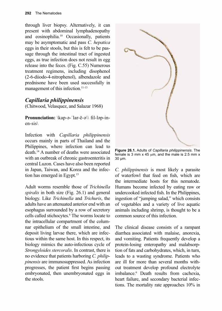
292 The Nematodes
through liver biopsy. Alternatively, it can present with abdominal lymphadenopathy and eosinophilia.10 Occasionally, patients may be asymptomatic and pass C. hepatica eggs in their stools, but this is felt to be pas-sage through the intestinal tract of ingested eggs, as true infection does not result in egg release into the feces. (Fig. C.55) Numerous treatment regimens, including disophenol (2-6-diiodo-4-nitrophenol), albendazole and prednisone have been used successfully in management of this infection.11-13
Capillaria philippinensis (Chitwood, Velasquez, and Salazar 1968)
Pronunciation: \kap-ə-ˈlar-ē-ə\\ fil-ləp-in-en-sis\
Infection with Capillaria philippinensis occurs mainly in parts of Thailand and the Philippines, where infection can lead to death.14 A number of deaths were associated with an outbreak of chronic gastroenteritis in central Luzon. Cases have also been reported in Japan, Taiwan, and Korea and the infec-tion has emerged in Egypt.15
Adult worms resemble those of Trichinella spiralis in both size (Fig. 26.1) and general biology. Like Trichinella and Trichuris, the adults have an attenuated anterior end with an esophagus surrounded by a row of secretory cells called stichocytes.6 The worms locate to the intracellular compartment of the colum-nar epithelium of the small intestine, and deposit living larvae there, which are infec-tious within the same host. In this respect, its biology mimics the auto-infectious cycle of Strongyloides stercoralis. In contrast, there is no evidence that patients harboring C. philip-pinensis are immunosuppressed. As infection progresses, the patient first begins passing embryonated, then unembryonated eggs in the stools.
C. philippinensis is most likely a parasite of waterfowl that feed on fish, which are the intermediate hosts for this nematode. Humans become infected by eating raw or undercooked infected fish. In the Philippines, ingestion of “jumping salad,” which consists of vegetables and a variety of live aquatic animals including shrimp, is thought to be a common source of this infection.
The clinical disease consists of a rampant diarrhea associated with malaise, anorexia, and vomiting. Patients frequently develop a protein-losing enteropathy and malabsorp-tion of fats and carbohydrates, which, in turn, leads to a wasting syndrome. Patients who are ill for more than several months with-out treatment develop profound electrolyte imbalance.6 Death results from cachexia, heart failure, and secondary bacterial infec-tions. The mortality rate approaches 10% in
Figure 26.1. Adults of Capillaria philippinensis. The female is 3 mm x 45 µm, and the male is 2.5 mm x 30 µm.
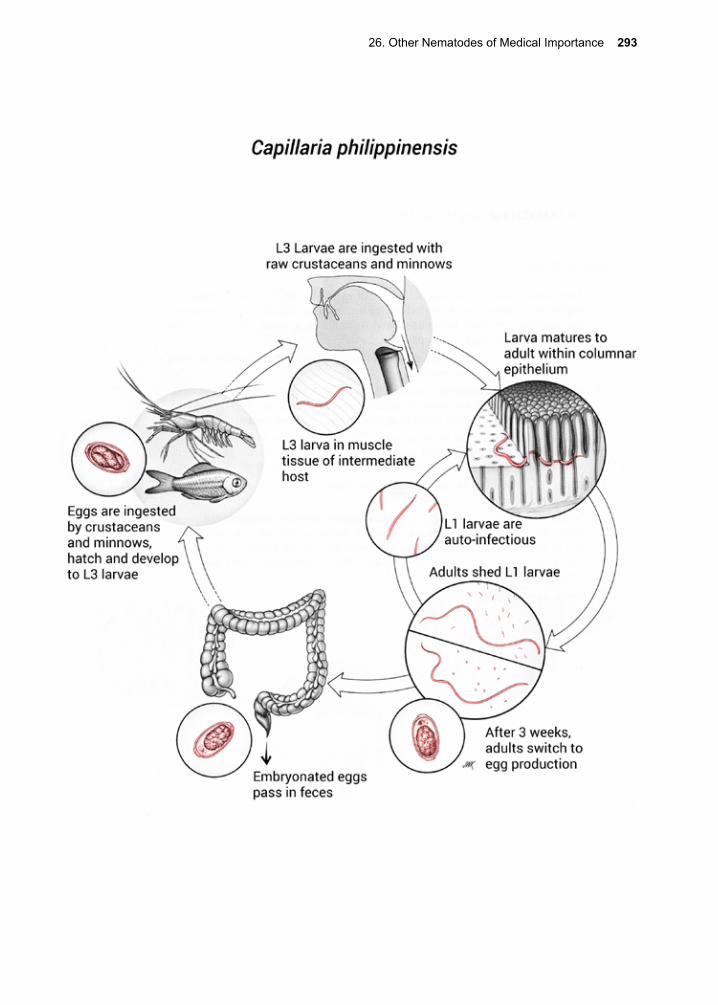
26. Other Nematodes of Medical Importance 293
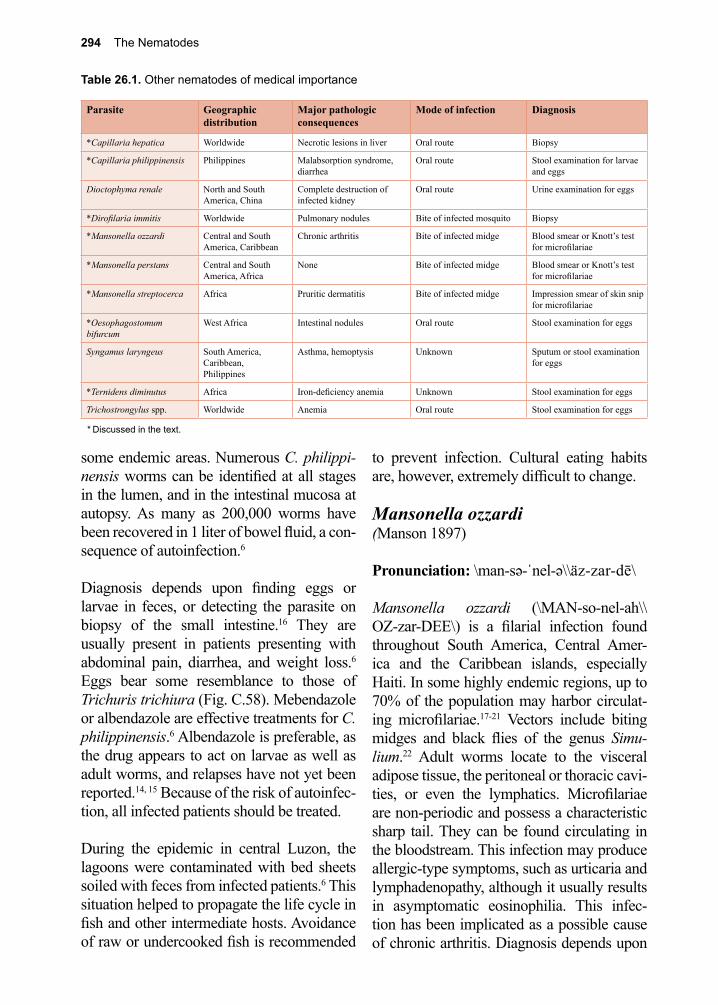
294 The Nematodes
Table 26.1. Other nematodes of medical importance
Parasite Geographic distribution
Major pathologic consequences
Mode of infection Diagnosis
*Capillaria hepatica Worldwide Necrotic lesions in liver Oral route Biopsy
*Capillaria philippinensis Philippines Malabsorption syndrome, diarrhea
Oral route Stool examination for larvae and eggs
Dioctophyma renale North and South America, China
Complete destruction of infected kidney
Oral route Urine examination for eggs
*Dirofilaria immitis Worldwide Pulmonary nodules Bite of infected mosquito Biopsy
*Mansonella ozzardi Central and South America, Caribbean
Chronic arthritis Bite of infected midge Blood smear or Knott’s test for microfilariae
*Mansonella perstans Central and South America, Africa
None Bite of infected midge Blood smear or Knott’s test for microfilariae
*Mansonella streptocerca Africa Pruritic dermatitis Bite of infected midge Impression smear of skin snip for microfilariae
*Oesophagostomum bifurcum
West Africa Intestinal nodules Oral route Stool examination for eggs
Syngamus laryngeus South America, Caribbean, Philippines
Asthma, hemoptysis Unknown Sputum or stool examination for eggs
*Ternidens diminutus Africa Iron-deficiency anemia Unknown Stool examination for eggs
Trichostrongylus spp. Worldwide Anemia Oral route Stool examination for eggs
* Discussed in the text.
some endemic areas. Numerous C. philippi-nensis worms can be identified at all stages in the lumen, and in the intestinal mucosa at autopsy. As many as 200,000 worms have been recovered in 1 liter of bowel fluid, a con-sequence of autoinfection.6
Diagnosis depends upon finding eggs or larvae in feces, or detecting the parasite on biopsy of the small intestine.16 They are usually present in patients presenting with abdominal pain, diarrhea, and weight loss.6 Eggs bear some resemblance to those of Trichuris trichiura (Fig. C.58). Mebendazole or albendazole are effective treatments for C. philippinensis.6 Albendazole is preferable, as the drug appears to act on larvae as well as adult worms, and relapses have not yet been reported.14, 15 Because of the risk of autoinfec-tion, all infected patients should be treated.
During the epidemic in central Luzon, the lagoons were contaminated with bed sheets soiled with feces from infected patients.6 This situation helped to propagate the life cycle in fish and other intermediate hosts. Avoidance of raw or undercooked fish is recommended
to prevent infection. Cultural eating habits are, however, extremely difficult to change.
Mansonella ozzardi (Manson 1897)
Pronunciation: \man-sə-ˈnel-ə\\äz-zar-dē\
Mansonella ozzardi (\MAN-so-nel-ah\\OZ-zar-DEE\) is a filarial infection found throughout South America, Central Amer-ica and the Caribbean islands, especially Haiti. In some highly endemic regions, up to 70% of the population may harbor circulat-ing microfilariae.17-21 Vectors include biting midges and black flies of the genus Simu-lium.22 Adult worms locate to the visceral adipose tissue, the peritoneal or thoracic cavi-ties, or even the lymphatics. Microfilariae are non-periodic and possess a characteristic sharp tail. They can be found circulating in the bloodstream. This infection may produce allergic-type symptoms, such as urticaria and lymphadenopathy, although it usually results in asymptomatic eosinophilia. This infec-tion has been implicated as a possible cause of chronic arthritis. Diagnosis depends upon
26. Other Nematodes of Medical Importance 295
finding the microfilariae on a stained blood smear, or in a skin sample. Serological testing is available but lacks specificity. A PCR test has been developed but is not widely avail-able. Ivermectin is the treatment of choice for M. ozzardi infection and has been shown to significantly reduce the number of circulat-ing microfilariae and reduce symptoms.23-26 Neither diethylcarbamazine citrate (DEC) nor benzimidazoles are effective against M. ozzardi.24, 27
Mansonella perstans (Manson 1891)
Pronunciation: \man-sə-ˈnel-ə\\pər-stans\
Mansonella perstans (MAN-so-nel-ah) is a filarial parasite found in Africa, and in north-eastern South America and parts of the Carib-bean. It is transmitted from person to person by biting midges. In Africa, gorillas and chimpanzees may be significant reservoirs. High prevalence rates have been reported from rural Senegal.28 The adults live free in serous cavities such as the pleural, pericar-dial, or peritoneal cavities, where they pro-duce microfilariae that circulate in blood. M. perstans infection is usually asymptomatic, but it is known to cause painless nodules in the conjunctiva with swelling of the eyelids in Africa, where it is known as the Ugandan eye worm or Kampala eye worm.29 The organism may also result in symptoms that are similar to Loa loa infection, such as angioedema and Calabar swellings. Microfilariae of M. per-stans may be observed in peripheral blood, and can be demonstrated by examination of a stained blood smear or by the Knott test. Serological testing is available but lacks specificity. A PCR test has been developed but is not widely available.
Most standard antihelmintic therapies have been ineffective for M. perstans.30-32 The discovery that this filarial parasite harbors Wolbachia endosymbionts suggested that
doxycycline might be an effective treatment option.33-35 Subsequent use of doxycycline has proven to be a highly effective therapy in species from Mozambique and the Dem-ocratic Republic of Congo that harbor the endosymbiont Wolbachia.36 Combination therapy has been successful in some cases.37 Co-infections with M. perstans do not appear to significantly alter post-treatment reaction profiles to single-dose ivermectin/albenda-zole for patients with lymphatic filariasis (LF).38
Mansonella streptocerca (Macfie and Corson 1922)
Pronunciation: \man-sə-ˈnel-ə\\strep-tə-ˈsər-kə\ Mansonella streptocerca (\MAN-so-nel-ah\\STREP-tow-ser-ka\) is a filarial parasite with a distribution restricted to tropical rainforests in Central Africa. Adult worms locate to the subcutaneous tissues, as do microfilariae.39, 40 Biting midges are its vectors. The major clin-ical manifestation of this infection is pruritic dermatitis with hypopigmented macules that may resemble onchocerciasis. There is often an associated axillary or inguinal lymphade-nopathy. Diagnosis is made by microscopi-cally identifying microfilariae in specimens of skin or impression smears made from them. Serological testing is available but lacks specificity. A PCR test has been devel-oped that can aid in detection of the micro-filariae in skin biopsy specimens.41 Micro-filariae must be differentiated from those of Onchocerca volvulus, usually by attempting to identify the characteristic hook-shaped tail, which is sometimes referred to as a “shep-herd’s crook.”42 DEC is the drug of choice, but it may exacerbate pruritus; in which case, anti-inflammatory agents and antihistamines may be necessary. Although ivermectin may also be effective, especially against microfi-lariae, but not against adult worms, there are some concerns about side effects.43, 44
296 The Nematodes
Oesophagostomum bifurcum (Creplin 1849)
Pronunciation: \i-ˌsäf-ə-ˈgäs-tə-məm\
Oesophagostomum bifurcum (I-sof-ah-GOS-tow-mum) is a nematode mostly infecting non-human primates in Africa and Asia. In northern Togo and northeastern Ghana, O. bifurcum infects up to 30% of these human populations, with an estimated 250,000 cases.45-48 Sporadic cases have also been described in other parts of Africa, Asia and South America.47 In these regions, adults 30–40 years of age have the highest preva-lence. These nematodes are often called nod-ular worms because they cause nodule forma-tion on the wall of the intestine.
Adult worms produce about 5,000 eggs per day, which pass with feces and mature to infective L3 larvae in soil.49 Eggs morpho-logically resemble those of hookworms. Humans are infected when they ingest infective larvae, which then penetrate the small intestinal wall and develop to adults. Some patients develop multi-nodular dis-ease, while, in others, a single nodular mass develops.46 The nodules in male patients are larger than the ones in females.46 The nodu-lar disease of Oesophagostomum infection often presents as an abdominal mass, which
can be painful, and mimics a surgical abdo-men. Often the mass is asymptomatic. As a result, the infection is frequently diagnosed at biopsy, although ultrasound is also of great value.50 Fecal examination is the diagnostic method of choice in individual patients (eggs cannot be visually distinguished from hook-worm eggs), while PCR has been developed and is used in prevalence studies.51 Pyrantel pamoate is the recommended drug for treat-ing infections due to O. bifurcum, and alben-dazole is also effective.52 Surgical resection of the nodules is sometimes necessary.
Ternidens diminutus
Pronunciation: \tər-nə-ˌdenz\ Ternidens diminutus (ter-nah-DENZ) is a nematode capable of infecting humans that resembles O. bifurcum. Ternidens eggs resemble hookworm eggs, so that T. diminu-tus is sometimes referred to as “the false hookworm.”53 It is primarily a parasite of non-human primates, but has been demonstrated to cause human infections in Zambia, Zim-babwe, Tanzania and Asia.54, 55 T. diminutus can result in colonic ulcerations and nodular lesions, but there are usually few symptoms. Both pyrantel pamoate and thiabendazole have been used to treat patients, and other benzimidazoles may also be effective.53, 56, 57
References
1. Asimacopoulos, P. J., et al., Pulmonary dirofilariasis. The largest single-hospital experience. Chest 1992, 102 (3), 851-5.
2. Simon, F., et al., What is new about animal and human dirofilariosis? Trends Parasitol 2009, 25 (9), 404-9.
3. Diaz, J. H.; Risher, W. H., Risk factors for human heartworm infections (dirofilariasis) in the South. J La State Med Soc 2015, 167 (2), 79-86.
4. Akao, N., et al., Immunoblot analysis of Dirofilaria immitis recognized by infected humans. Annals of tropical medicine and parasitology 1991, 85 (4), 455-60.
5. Orihel, T. C., et al., Subcutaneous dirofilariasis: single inoculum, multiple worms. The American journal of tropical medicine and hygiene 1997, 56 (4), 452-5.
6. Cross, J. H., Intestinal capillariasis. Clinical microbiology reviews 1992, 5 (2), 120-9.
26. Other Nematodes of Medical Importance 297
7. Li, C. D., et al., Capillaria hepatica in China. World J Gastroenterol 2010, 16 (6), 698-702.8. Wang, Z., et al., The emerging but neglected hepatic capillariasis in China. Asian Pac J Trop
Biomed 2013, 3 (2), 146-7.9. Juncker-Voss, M., et al., Serological detection of Capillaria hepatica by indirect
immunofluorescence assay. J Clin Microbiol 2000, 38 (1), 431-3.10. Sharma, R., et al., Capillaria hepatica infection: a rare differential for peripheral eosinophilia and
an imaging dilemma for abdominal lymphadenopathy. Ann Parasitol 2015, 61 (1), 61-4.11. McQuown, A. L.; J., Capillaria hepatica: reported genuine and spurious cases. Am Med Hyg
1950, 30, 761-767.12. Nabi, F., et al., Capillaria hepatica infestation. Indian Pediatr 2007, 44 (10), 781-2.13. Pereira, V. G.; Mattosinho Franca, L. C., Successful treatment of Capillaria hepatica infection in
an acutely ill adult. Am J Trop Med Hyg 1983, 32 (6), 1272-4.14. el-Karaksy, H., et al., Capillaria philippinensis: a cause of fatal diarrhea in one of two infected
Egyptian sisters. Journal of tropical pediatrics 2004, 50 (1), 57-60.15. Bair, M.-J., et al., Clinical features of human intestinal capillariasis in Taiwan. World journal of
gastroenterology : WJG 2004, 10 (16), 2391-3.16. Limsrivilai, J., et al., Intestinal capillariasis in the 21st century: clinical presentations and role of
endoscopy and imaging. BMC Gastroenterol 2014, 14, 207.17. Marinkelelle, C. J.; German, E., Mansonelliasis in the Comiasria del Vaupes of Colombia. Trop
Geogr Med 101 1970, 22 18. Raccurt, C. P., et al., Epidemiologic survey of Mansonella ozzardi in Corail, Haiti. Am J Trop
Med Hyg 2014, 90 (6), 1167-9.19. Veggiani Aybar, C. A., et al., Mansonella ozzardi in Neotropical region of Argentina: Prevalence
through time (1986-2010). Acta Trop 2016, 153, 1-6.20. Medeiros, J. F., et al., Mansonella ozzardi in Brazil: prevalence of infection in riverine
communities in the Purus region, in the state of Amazonas. Mem Inst Oswaldo Cruz 2009, 104 (1), 74-80.
21. Raccurt, C. P., et al., Mansonelliasis, a neglected parasitic disease in Haiti. Mem Inst Oswaldo Cruz 2014, 109 (6), 709-11.
22. Nathan, M. B., et al., Filariasis in Amerindians of western Guyana with observations on transmission of Mansonella ozzardi by a Simulium species of the amazonicum group. Tropenmedizin und Parasitologie 1982, 33 (4), 219-22.
23. Krolewiecki, A. J., et al., Ivermectin-related adverse clinical events in patients treated for Mansonella ozzardi infections. Rev Argent Microbiol 2011, 43 (1), 48-50.
24. Basano Sde, A., et al., Sustained clearance of Mansonella ozzardi infection after treatment with ivermectin in the Brazilian Amazon. Am J Trop Med Hyg 2014, 90 (6), 1170-5.
25. Nutman, T. B., et al., Ivermectin in the successful treatment of a patient with Mansonella ozzardi infection. J Infect Dis 1987, 156 (4), 662-5.
26. Nutman, T. B., et al., Ivermectin in the successful treatment of a patient with Mansonella ozzardi infection. Dis 662 1997, 156.
27. Bartholomew, C. F., et al., The failure of diethylcarbamazine in the treatment of Mansonella ozzardi infections. Trans R Soc Trop Med Hyg 1978, 72 (4), 423-4.
28. Bassene, H., et al., High Prevalence of Mansonella perstans Filariasis in Rural Senegal. Am J Trop Med Hyg 2015, 93 (3), 601-6.
29. Baird, J. K., et al., Nodules in the conjunctiva, bung-eye, and bulge-eye in Africa caused by Mansonella perstans. The American journal of tropical medicine and hygiene 1988, 38 (3), 553-7.
30. Wahlgren, M.; Frolov, I., Treatment of Dipetalonema perstans infections with mebendazole. Transactions of the Royal Society of Tropical Medicine and Hygiene 1983, 77 (3), 422-3.
31. Gardon, J., et al., Efficacy of repeated doses of ivermectin against Mansonella perstans. Transactions of the Royal Society of Tropical Medicine and Hygiene 2002, 96 (3), 325-6.
32. Van den Enden, E., et al., Mansonella perstans filariasis: failure of albendazole treatment. Annales de la Societe belge de medecine tropicale 1992, 72 (3), 215-8.
33. Gehringer, C., et al., Molecular evidence of Wolbachia endosymbiosis in Mansonella perstans in Gabon, Central Africa. J Infect Dis 2014, 210 (10), 1633-8.
298 The Nematodes
34. Keiser, P. B., et al., Molecular identification of Wolbachia from the filarial nematode Mansonella perstans. Mol Biochem Parasitol 2008, 160 (2), 123-8.
35. Grobusch, M. P., et al., No evidence of Wolbachia endosymbiosis with Loa loa and Mansonella perstans. Parasitol Res 2003, 90 (5), 405-8.
36. Coulibaly, Y. I., et al., A randomized trial of doxycycline for Mansonella perstans infection. N Engl J Med 2009, 361 (15), 1448-58.
37. Asgeirsson, H., et al., Successful treatment of 2 imported cases of Mansonella perstans infection. PLoS Negl Trop Dis 2017, 11 (5), e0005452.
38. Keiser, P. B., et al., Clinical characteristics of post-treatment reactions to ivermectin/albendazole for Wuchereria bancrofti in a region co-endemic for Mansonella perstans. The American journal of tropical medicine and hygiene 2003, 69 (3), 331-5.
39. Okelo, G. B., et al., Mansonella streptocerca in the Central African Republic. Trans R Soc Trop Med Hyg 1988, 82 (3), 464.
40. Fischer, P., et al., Occurrence and diagnosis of Mansonella streptocerca in Uganda. Acta Trop 1997, 63 (1), 43-55.
41. Fischer, P., et al., Detection of the filarial parasite Mansonella streptocerca in skin biopsies by a nested polymerase chain reaction-based assay. Am J Trop Med Hyg 1998, 58 (6), 816-20.
42. Orihel, T. C.; J., The tail of the Mansonella streptocerca microfilaria. Am Med Hyg 1984, 33 (6), 1278.
43. Fischer, P., et al., Long-term suppression of Mansonella streptocerca microfilariae after treatment with ivermectin. The Journal of infectious diseases 1999, 180 (4), 1403-5.
44. Fischer, P., et al., Treatment of human Mansonella streptocerca infection with ivermectin. Trop Med Int Health 1997, 2 (2), 191-9.
45. Polderman, A. M., et al., Oesophagostomiasis, a common infection of man in northern Togo and Ghana. The American journal of tropical medicine and hygiene 1991, 44 (3), 336-44.
46. Storey, P. A., et al., Natural progression of Oesophagostomum bifurcum pathology and infection in a rural community of northern Ghana. Transactions of the Royal Society of Tropical Medicine and Hygiene 2001, 95 (3), 295-9.
47. Bogers, J. J., et al., Human oesophagostomiasis: a histomorphometric study of 13 new cases in northern Ghana. Virchows Archiv : an international journal of pathology 2001, 439 (1), 21-6.
48. Ziem, J. B., et al., Distribution and clustering of Oesophagostomum bifurcum and hookworm infections in northern Ghana. Parasitology 2006, 132 (Pt 4), 525-34.
49. Kepel, H. P., et al., Egg production of Oesophagostomum bifurcum, a locally common parasite of humans in Togo. Am Med Hyg 1992, 46, 469-472.
50. Storey, P. A., et al., Intraobserver and interobserver variation of ultrasound diagnosis of Oesophagostomum bifurcum colon lesions. The American journal of tropical medicine and hygiene 2002, 67 (6), 680-3.
51. Verweij, J. J., et al., Determining the prevalence of Oesophagostomum bifurcum and Necator americanus infections using specific PCR amplification of DNA from faecal samples. Trop Med Int Health 2001, 6 (9), 726-31.
52. Ziem, J. B., et al., The short-term impact of albendazole treatment on Oesophagostomum bifurcum and hookworm infections in northern Ghana. Annals of tropical medicine and parasitology 2004, 98 (4), 385-90.
53. Bradley, M., Rate of expulsion of Necator americanus and the false hookworm Ternidens deminutus Railliet and Henry 1909 (Nematoda) from humans following albendazole treatment. Transactions of the Royal Society of Tropical Medicine and Hygiene 1990, 84 (5), 720.
54. Kilala, C. P., Ternidens deminutus infecting man in Southern Tanzania. East Afr Med J 1971, 48 (11), 636-45.
55. Hemsrichart, V., Ternidens deminutus infection: first pathological report of a human case in Asia. J Med Assoc Thai 2005, 88 (8), 1140-3.
56. Goldsmid, J. M.; Saunders, C. R., Preliminary trial using pyrantal pamoate for the treatment of human infections with Teernidens deminutus. Transactions of the Royal Society of Tropical Medicine and Hygiene 1972, 66 (2), 375-6.
57. Goldsmid, J. M., Thiabendazole in the treatment of human infections with Ternidens deminutus (Nematoda). S Afr Med J 1972, 46 (30), 1046-7.
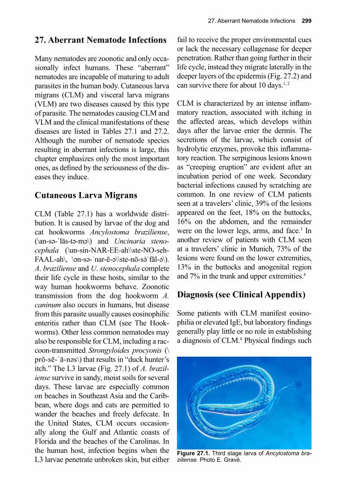
27. Aberrant Nematode Infections 299
27. Aberrant Nematode Infections
Many nematodes are zoonotic and only occa-sionally infect humans. These “aberrant” nematodes are incapable of maturing to adult parasites in the human body. Cutaneous larva migrans (CLM) and visceral larva migrans (VLM) are two diseases caused by this type of parasite. The nematodes causing CLM and VLM and the clinical manifestations of these diseases are listed in Tables 27.1 and 27.2. Although the number of nematode species resulting in aberrant infections is large, this chapter emphasizes only the most important ones, as defined by the seriousness of the dis-eases they induce.
Cutaneous Larva Migrans
CLM (Table 27.1) has a worldwide distri-bution. It is caused by larvae of the dog and cat hookworms Ancylostoma braziliense, (\an-sə-ˈläs-tə-mə\) and Uncinaria steno-cephala (\un-sin-NAR-EE-ah\\ste-NO-seh-FAAL-ah\, \ən-sə-ˈnar-ē-ə\\ste-nō-sə̇ˈfāl-ə\). A. braziliense and U. stenocephala complete their life cycle in these hosts, similar to the way human hookworms behave. Zoonotic transmission from the dog hookworm A. caninum also occurs in humans, but disease from this parasite usually causes eosinophilic enteritis rather than CLM (see The Hook-worms). Other less common nematodes may also be responsible for CLM, including a rac-coon-transmitted Strongyloides procyonis (\prō-sē-ˈä-nəs\) that results in “duck hunter’s itch.” The L3 larvae (Fig. 27.1) of A. brazil-iense survive in sandy, moist soils for several days. These larvae are especially common on beaches in Southeast Asia and the Carib-bean, where dogs and cats are permitted to wander the beaches and freely defecate. In the United States, CLM occurs occasion-ally along the Gulf and Atlantic coasts of Florida and the beaches of the Carolinas. In the human host, infection begins when the L3 larvae penetrate unbroken skin, but either
fail to receive the proper environmental cues or lack the necessary collagenase for deeper penetration. Rather than going further in their life cycle, instead they migrate laterally in the deeper layers of the epidermis (Fig. 27.2) and can survive there for about 10 days.1, 2
CLM is characterized by an intense inflam-matory reaction, associated with itching in the affected areas, which develops within days after the larvae enter the dermis. The secretions of the larvae, which consist of hydrolytic enzymes, provoke this inflamma-tory reaction. The serpiginous lesions known as “creeping eruption” are evident after an incubation period of one week. Secondary bacterial infections caused by scratching are common. In one review of CLM patients seen at a travelers’ clinic, 39% of the lesions appeared on the feet, 18% on the buttocks, 16% on the abdomen, and the remainder were on the lower legs, arms, and face.3 In another review of patients with CLM seen at a travelers’ clinic in Munich, 73% of the lesions were found on the lower extremities, 13% in the buttocks and anogenital region and 7% in the trunk and upper extremities.4
Diagnosis (see Clinical Appendix)
Some patients with CLM manifest eosino-philia or elevated IgE, but laboratory findings generally play little or no role in establishing a diagnosis of CLM.4 Physical findings such
Figure 27.1. Third stage larva of Ancylostoma bra-ziliense. Photo E. Gravé.
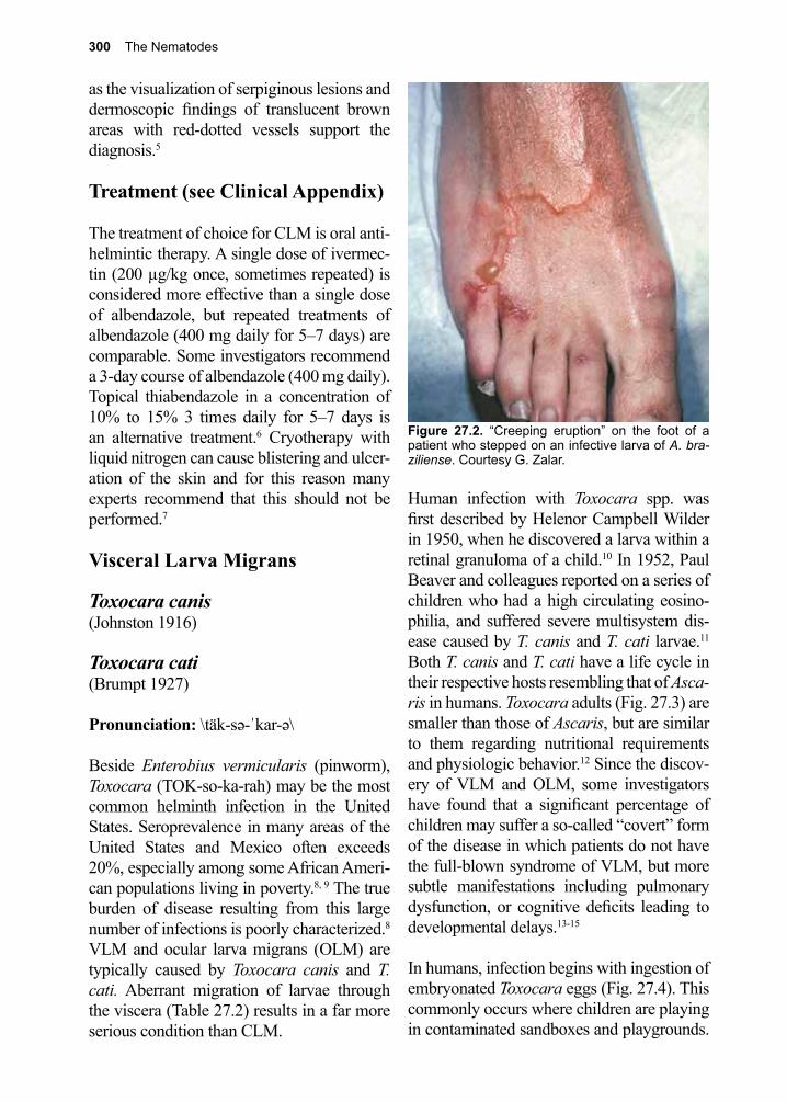
300 The Nematodes
Figure 27.2. “Creeping eruption” on the foot of a patient who stepped on an infective larva of A. bra-ziliense. Courtesy G. Zalar.
as the visualization of serpiginous lesions and dermoscopic findings of translucent brown areas with red-dotted vessels support the diagnosis.5
Treatment (see Clinical Appendix)
The treatment of choice for CLM is oral anti-helmintic therapy. A single dose of ivermec-tin (200 µg/kg once, sometimes repeated) is considered more effective than a single dose of albendazole, but repeated treatments of albendazole (400 mg daily for 5–7 days) are comparable. Some investigators recommend a 3-day course of albendazole (400 mg daily). Topical thiabendazole in a concentration of 10% to 15% 3 times daily for 5–7 days is an alternative treatment.6 Cryotherapy with liquid nitrogen can cause blistering and ulcer-ation of the skin and for this reason many experts recommend that this should not be performed.7 Visceral Larva Migrans
Toxocara canis (Johnston 1916)
Toxocara cati (Brumpt 1927)
Pronunciation: \täk-sə-ˈkar-ə\
Beside Enterobius vermicularis (pinworm), Toxocara (TOK-so-ka-rah) may be the most common helminth infection in the United States. Seroprevalence in many areas of the United States and Mexico often exceeds 20%, especially among some African Ameri-can populations living in poverty.8, 9 The true burden of disease resulting from this large number of infections is poorly characterized.8 VLM and ocular larva migrans (OLM) are typically caused by Toxocara canis and T. cati. Aberrant migration of larvae through the viscera (Table 27.2) results in a far more serious condition than CLM.
Human infection with Toxocara spp. was first described by Helenor Campbell Wilder in 1950, when he discovered a larva within a retinal granuloma of a child.10 In 1952, Paul Beaver and colleagues reported on a series of children who had a high circulating eosino-philia, and suffered severe multisystem dis-ease caused by T. canis and T. cati larvae.11 Both T. canis and T. cati have a life cycle in their respective hosts resembling that of Asca-ris in humans. Toxocara adults (Fig. 27.3) are smaller than those of Ascaris, but are similar to them regarding nutritional requirements and physiologic behavior.12 Since the discov-ery of VLM and OLM, some investigators have found that a significant percentage of children may suffer a so-called “covert” form of the disease in which patients do not have the full-blown syndrome of VLM, but more subtle manifestations including pulmonary dysfunction, or cognitive deficits leading to developmental delays.13-15
In humans, infection begins with ingestion of embryonated Toxocara eggs (Fig. 27.4). This commonly occurs where children are playing in contaminated sandboxes and playgrounds.
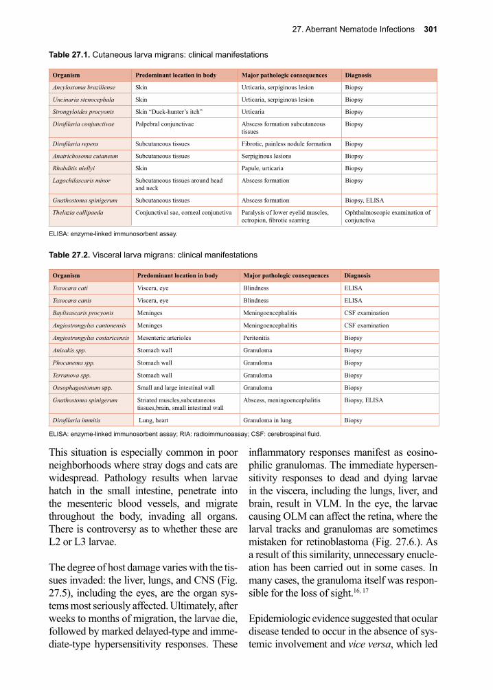
27. Aberrant Nematode Infections 301
Table 27.1. Cutaneous larva migrans: clinical manifestations
Organism Predominant location in body Major pathologic consequences Diagnosis
Ancylostoma braziliense Skin Urticaria, serpiginous lesion Biopsy
Uncinaria stenocephala Skin Urticaria, serpiginous lesion Biopsy
Strongyloides procyonis Skin “Duck-hunter’s itch” Urticaria Biopsy
Dirofilaria conjunctivae Palpebral conjunctivae Abscess formation subcutaneous tissues
Biopsy
Dirofilaria repens Subcutaneous tissues Fibrotic, painless nodule formation Biopsy
Anatrichosoma cutaneum Subcutaneous tissues Serpiginous lesions Biopsy
Rhabditis niellyi Skin Papule, urticaria Biopsy
Lagochilascaris minor Subcutaneous tissues around head and neck
Abscess formation Biopsy
Gnathostoma spinigerum Subcutaneous tissues Abscess formation Biopsy, ELISA
Thelazia callipaeda Conjunctival sac, corneal conjunctiva Paralysis of lower eyelid muscles, ectropion, fibrotic scarring
Ophthalmoscopic examination of conjunctiva
ELISA: enzyme-linked immunosorbent assay.
Table 27.2. Visceral larva migrans: clinical manifestations
Organism Predominant location in body Major pathologic consequences Diagnosis
Toxocara cati Viscera, eye Blindness ELISA
Toxocara canis Viscera, eye Blindness ELISA
Baylisascaris procyonis Meninges Meningoencephalitis CSF examination
Angiostrongylus cantonensis Meninges Meningoencephalitis CSF examination
Angiostrongylus costaricensis Mesenteric arterioles Peritonitis Biopsy
Anisakis spp. Stomach wall Granuloma Biopsy
Phocanema spp. Stomach wall Granuloma Biopsy
Terranova spp. Stomach wall Granuloma Biopsy
Oesophagostonum spp. Small and large intestinal wall Granuloma Biopsy
Gnathostoma spinigerum Striated muscles,subcutaneous tissues,brain, small intestinal wall
Abscess, meningoencephalitis Biopsy, ELISA
Dirofilaria immitis Lung, heart Granuloma in lung Biopsy
ELISA: enzyme-linked immunosorbent assay; RIA: radioimmunoassay; CSF: cerebrospinal fluid.
This situation is especially common in poor neighborhoods where stray dogs and cats are widespread. Pathology results when larvae hatch in the small intestine, penetrate into the mesenteric blood vessels, and migrate throughout the body, invading all organs. There is controversy as to whether these are L2 or L3 larvae.
The degree of host damage varies with the tis-sues invaded: the liver, lungs, and CNS (Fig. 27.5), including the eyes, are the organ sys-tems most seriously affected. Ultimately, after weeks to months of migration, the larvae die, followed by marked delayed-type and imme-diate-type hypersensitivity responses. These
inflammatory responses manifest as eosino-philic granulomas. The immediate hypersen-sitivity responses to dead and dying larvae in the viscera, including the lungs, liver, and brain, result in VLM. In the eye, the larvae causing OLM can affect the retina, where the larval tracks and granulomas are sometimes mistaken for retinoblastoma (Fig. 27.6.). As a result of this similarity, unnecessary enucle-ation has been carried out in some cases. In many cases, the granuloma itself was respon-sible for the loss of sight.16, 17
Epidemiologic evidence suggested that ocular disease tended to occur in the absence of sys-temic involvement and vice versa, which led
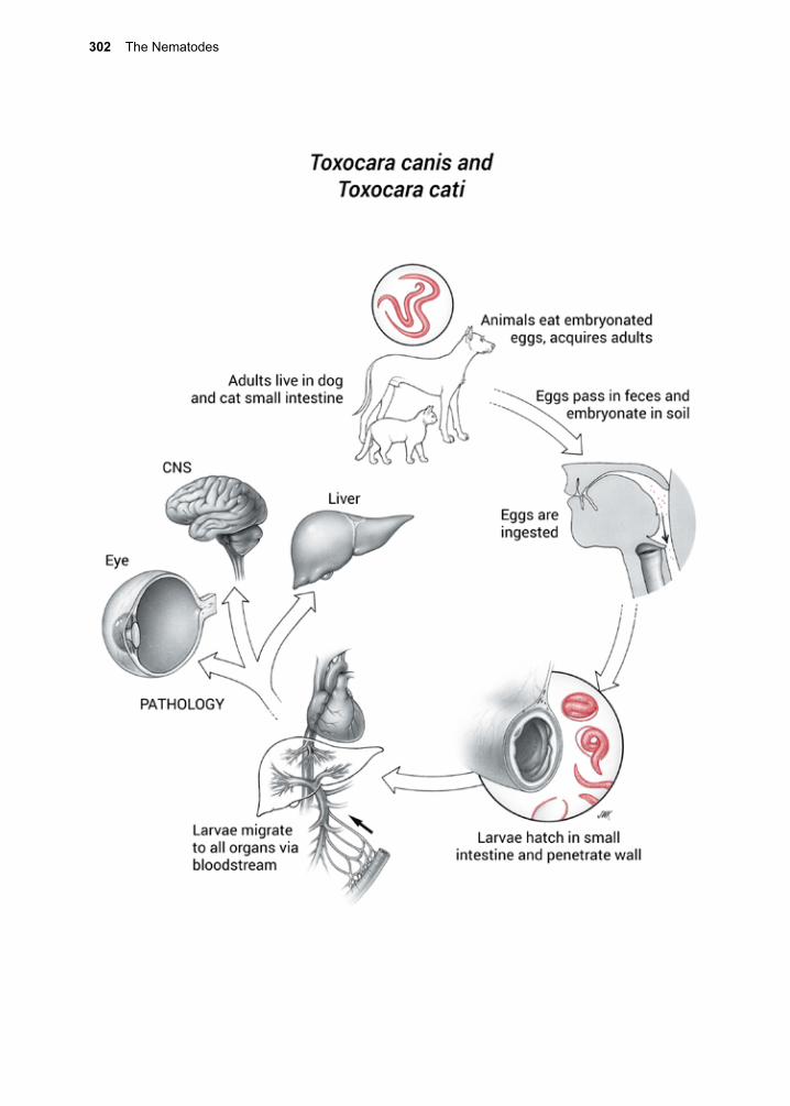
302 The Nematodes
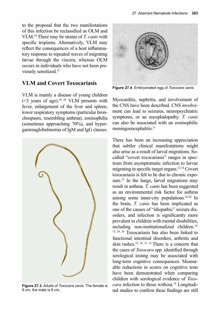
27. Aberrant Nematode Infections 303
to the proposal that the two manifestations of this infection be reclassified as OLM and VLM.18 There may be strains of T. canis with specific tropisms. Alternatively, VLM may reflect the consequences of a host inflamma-tory response to repeated waves of migrating larvae through the viscera, whereas OLM occurs in individuals who have not been pre-viously sensitized.12
VLM and Covert Toxocariasis
VLM is mainly a disease of young children (<5 years of age).19, 20 VLM presents with fever, enlargement of the liver and spleen, lower respiratory symptoms (particular bron-chospasm, resembling asthma), eosinophilia (sometimes approaching 70%), and hyper-gammaglobulinemia of IgM and IgG classes.
Myocarditis, nephritis, and involvement of the CNS have been described. CNS involve-ment can lead to seizures, neuropsychiatric symptoms, or an encephalopathy. T. canis can also be associated with an eosinophilic meningoencephalitis.21
There has been an increasing appreciation that subtler clinical manifestations might also arise as a result of larval migrations. So-called “covert toxocariasis” ranges in spec-trum from asymptomatic infection to larvae migrating in specific target organs.22-24 Covert toxocariasis is felt to be due to chronic expo-sure.25 In the lungs, larval migrations may result in asthma. T. canis has been suggested as an environmental risk factor for asthma among some inner-city populations.26-28 In the brain, T. canis has been implicated as one of the causes of “idiopathic” seizure dis-orders, and infection is significantly more prevalent in children with mental disabilities, including non-institutionalized children.14,
15, 29, 30 Toxocariasis has also been linked to functional intestinal disorders, arthritis and skin rashes.23, 26, 31, 32 There is a concern that the cases of Toxocara spp. identified through serological testing may be associated with long-term cognitive consequences. Measur-able reductions in scores on cognitive tests have been demonstrated when comparing children with serological evidence of Toxo-cara infection to those without.14 Longitudi-nal studies to confirm these findings are still
Figure 27.3. Adults of Toxocara canis. The female is 9 cm, the male is 6 cm.
Figure 27.4. Embryonated egg of Toxocara canis.
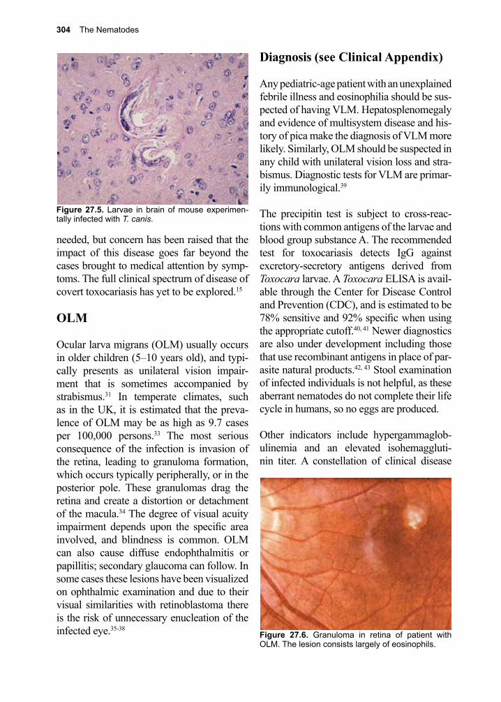
304 The Nematodes
needed, but concern has been raised that the impact of this disease goes far beyond the cases brought to medical attention by symp-toms. The full clinical spectrum of disease of covert toxocariasis has yet to be explored.15
OLM
Ocular larva migrans (OLM) usually occurs in older children (5–10 years old), and typi-cally presents as unilateral vision impair-ment that is sometimes accompanied by strabismus.31 In temperate climates, such as in the UK, it is estimated that the preva-lence of OLM may be as high as 9.7 cases per 100,000 persons.33 The most serious consequence of the infection is invasion of the retina, leading to granuloma formation, which occurs typically peripherally, or in the posterior pole. These granulomas drag the retina and create a distortion or detachment of the macula.34 The degree of visual acuity impairment depends upon the specific area involved, and blindness is common. OLM can also cause diffuse endophthalmitis or papillitis; secondary glaucoma can follow. In some cases these lesions have been visualized on ophthalmic examination and due to their visual similarities with retinoblastoma there is the risk of unnecessary enucleation of the infected eye.35-38
Diagnosis (see Clinical Appendix)
Any pediatric-age patient with an unexplained febrile illness and eosinophilia should be sus-pected of having VLM. Hepatosplenomegaly and evidence of multisystem disease and his-tory of pica make the diagnosis of VLM more likely. Similarly, OLM should be suspected in any child with unilateral vision loss and stra-bismus. Diagnostic tests for VLM are primar-ily immunological.39
The precipitin test is subject to cross-reac-tions with common antigens of the larvae and blood group substance A. The recommended test for toxocariasis detects IgG against excretory-secretory antigens derived from Toxocara larvae. A Toxocara ELISA is avail-able through the Center for Disease Control and Prevention (CDC), and is estimated to be 78% sensitive and 92% specific when using the appropriate cutoff.40, 41 Newer diagnostics are also under development including those that use recombinant antigens in place of par-asite natural products.42, 43 Stool examination of infected individuals is not helpful, as these aberrant nematodes do not complete their life cycle in humans, so no eggs are produced.
Other indicators include hypergammaglob-ulinemia and an elevated isohemaggluti-nin titer. A constellation of clinical disease
Figure 27.5. Larvae in brain of mouse experimen-tally infected with T. canis.
Figure 27.6. Granuloma in retina of patient with OLM. The lesion consists largely of eosinophils.
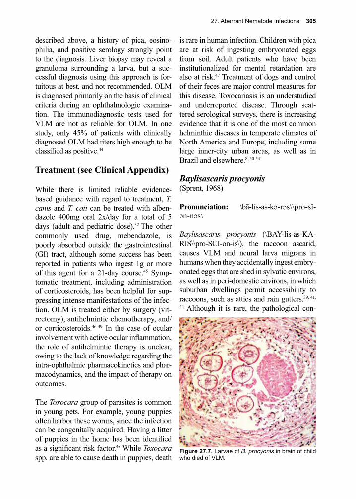
27. Aberrant Nematode Infections 305
described above, a history of pica, eosino-philia, and positive serology strongly point to the diagnosis. Liver biopsy may reveal a granuloma surrounding a larva, but a suc-cessful diagnosis using this approach is for-tuitous at best, and not recommended. OLM is diagnosed primarily on the basis of clinical criteria during an ophthalmologic examina-tion. The immunodiagnostic tests used for VLM are not as reliable for OLM. In one study, only 45% of patients with clinically diagnosed OLM had titers high enough to be classified as positive.44
Treatment (see Clinical Appendix) While there is limited reliable evidence-based guidance with regard to treatment, T. canis and T. cati can be treated with alben-dazole 400mg oral 2x/day for a total of 5 days (adult and pediatric dose).32 The other commonly used drug, mebendazole, is poorly absorbed outside the gastrointestinal (GI) tract, although some success has been reported in patients who ingest 1g or more of this agent for a 21-day course.45 Symp-tomatic treatment, including administration of corticosteroids, has been helpful for sup-pressing intense manifestations of the infec-tion. OLM is treated either by surgery (vit-rectomy), antihelmintic chemotherapy, and/or corticosteroids.46-49 In the case of ocular involvement with active ocular inflammation, the role of antihelmintic therapy is unclear, owing to the lack of knowledge regarding the intra-ophthalmic pharmacokinetics and phar-macodynamics, and the impact of therapy on outcomes.
The Toxocara group of parasites is common in young pets. For example, young puppies often harbor these worms, since the infection can be congenitally acquired. Having a litter of puppies in the home has been identified as a significant risk factor.46 While Toxocara spp. are able to cause death in puppies, death
is rare in human infection. Children with pica are at risk of ingesting embryonated eggs from soil. Adult patients who have been institutionalized for mental retardation are also at risk.47 Treatment of dogs and control of their feces are major control measures for this disease. Toxocariasis is an understudied and underreported disease. Through scat-tered serological surveys, there is increasing evidence that it is one of the most common helminthic diseases in temperate climates of North America and Europe, including some large inner-city urban areas, as well as in Brazil and elsewhere.8, 50-54
Baylisascaris procyonis(Sprent, 1968)
Pronunciation: \bā-lis-as-kə-rəs\\pro-sī-ən-nəs\
Baylisascaris procyonis (\BAY-lis-as-KA-RIS\\pro-SCI-on-is\), the raccoon ascarid, causes VLM and neural larva migrans in humans when they accidentally ingest embry-onated eggs that are shed in sylvatic environs, as well as in peri-domestic environs, in which suburban dwellings permit accessibility to raccoons, such as attics and rain gutters.39, 41,
44 Although it is rare, the pathological con-
Figure 27.7. Larvae of B. procyonis in brain of child who died of VLM.
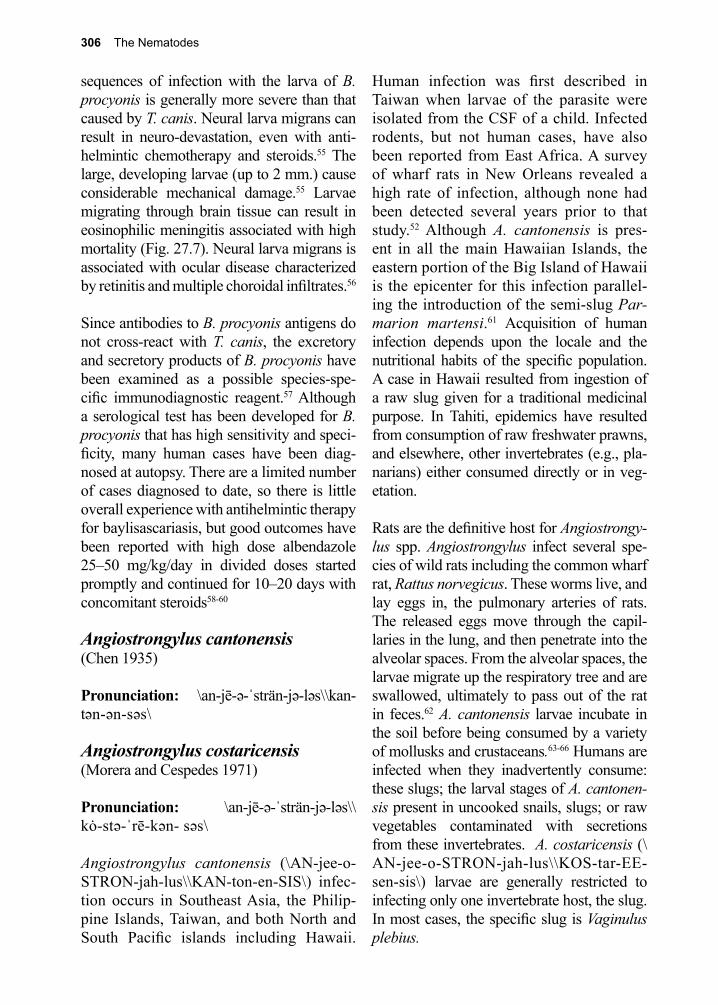
306 The Nematodes
sequences of infection with the larva of B. procyonis is generally more severe than that caused by T. canis. Neural larva migrans can result in neuro-devastation, even with anti-helmintic chemotherapy and steroids.55 The large, developing larvae (up to 2 mm.) cause considerable mechanical damage.55 Larvae migrating through brain tissue can result in eosinophilic meningitis associated with high mortality (Fig. 27.7). Neural larva migrans is associated with ocular disease characterized by retinitis and multiple choroidal infiltrates.56
Since antibodies to B. procyonis antigens do not cross-react with T. canis, the excretory and secretory products of B. procyonis have been examined as a possible species-spe-cific immunodiagnostic reagent.57 Although a serological test has been developed for B. procyonis that has high sensitivity and speci-ficity, many human cases have been diag-nosed at autopsy. There are a limited number of cases diagnosed to date, so there is little overall experience with antihelmintic therapy for baylisascariasis, but good outcomes have been reported with high dose albendazole 25–50 mg/kg/day in divided doses started promptly and continued for 10–20 days with concomitant steroids58-60
Angiostrongylus cantonensis(Chen 1935)
Pronunciation: \an-jē-ə-ˈsträn-jə-ləs\\kan-tən-ən-səs\
Angiostrongylus costaricensis(Morera and Cespedes 1971)
Pronunciation: \an-jē-ə-ˈsträn-jə-ləs\\kȯ-stə-ˈrē-kən- səs\
Angiostrongylus cantonensis (\AN-jee-o-STRON-jah-lus\\KAN-ton-en-SIS\) infec-tion occurs in Southeast Asia, the Philip-pine Islands, Taiwan, and both North and South Pacific islands including Hawaii.
Human infection was first described in Taiwan when larvae of the parasite were isolated from the CSF of a child. Infected rodents, but not human cases, have also been reported from East Africa. A survey of wharf rats in New Orleans revealed a high rate of infection, although none had been detected several years prior to that study.52 Although A. cantonensis is pres-ent in all the main Hawaiian Islands, the eastern portion of the Big Island of Hawaii is the epicenter for this infection parallel-ing the introduction of the semi-slug Par-marion martensi.61 Acquisition of human infection depends upon the locale and the nutritional habits of the specific population. A case in Hawaii resulted from ingestion of a raw slug given for a traditional medicinal purpose. In Tahiti, epidemics have resulted from consumption of raw freshwater prawns, and elsewhere, other invertebrates (e.g., pla-narians) either consumed directly or in veg-etation.
Rats are the definitive host for Angiostrongy-lus spp. Angiostrongylus infect several spe-cies of wild rats including the common wharf rat, Rattus norvegicus. These worms live, and lay eggs in, the pulmonary arteries of rats. The released eggs move through the capil-laries in the lung, and then penetrate into the alveolar spaces. From the alveolar spaces, the larvae migrate up the respiratory tree and are swallowed, ultimately to pass out of the rat in feces.62 A. cantonensis larvae incubate in the soil before being consumed by a variety of mollusks and crustaceans.63-66 Humans are infected when they inadvertently consume: these slugs; the larval stages of A. cantonen-sis present in uncooked snails, slugs; or raw vegetables contaminated with secretions from these invertebrates. A. costaricensis (\AN-jee-o-STRON-jah-lus\\KOS-tar-EE-sen-sis\) larvae are generally restricted to infecting only one invertebrate host, the slug. In most cases, the specific slug is Vaginulus plebius.
27. Aberrant Nematode Infections 307
Most cases of human infection with Angio-strongylus spp. result in failure of the worm to complete its life cycle. Once in a human host, the larvae often migrate to the brain or rarely to the lungs, as has been observed in infants and small children. These nematodes die sur-rounded by an eosinophil-rich inflammatory infiltrate.67 The pattern of clinical presentation varies with each species of Angiostrongylus. A. cantonensis L3 larvae usually migrate to the meningeal capillaries where their death triggers eosinophilic inflammation resulting in eosinophilic meningoencephalitis. Part of the pathology can involve vascular throm-bosis and aneurysms. Painful paresthesias, headache and fever are described as features seen in the clinical presentation of this infec-tion.68 Vomiting is reported in the majority of cases, as are migrating painful paresthesias.69 Painful paresthesias often persist for weeks to months along with headaches. Although eosinophilic meningitis is characteristic on CSF examination, focal lesions are usually not present on head CT or MRI. This can be useful in distinguishing the disease from other etiologies, such as Gnathostoma spini-gerum and neurocysticercosis.70
A. cantonensis is the most common cause of eosinophilic meningitis outside of the developed world, and should be considered in the differential of travelers returning from endemic areas.67 The natural course of the disease without treatment is for resolution of symptoms in 1–2 weeks, but disease duration and severity may be worse in children and some individuals may have longer durations of illness despite treatment. In contrast to the clinical picture seen with A. cantonensis, A. costaricensis causes abdominal angiostron-gyliasis. Patients may present with abdomi-nal pain, fever, and peripheral eosinophilia, but since A. costaricensis does not migrate to the brain this organism does not cause eosino-philic meningitis. 71-73
Diagnosis of Angiostrongylus spp. is based on clinical presentation and PCR, as serological testing is not widely available. Confirmation of suspected cases has been achieved using post treatment serological testing in concert with research laboratories.69 PCR testing is available and can be performed on CSF but lacks high sensitivity.74
With the self-limiting nature of this disease, treatment often focuses on symptoms rather than on antihelmintic therapy. Although animal models have supported the use of anti-helmintic therapy trials, efficacy in human infection has not been demonstrated, and there are reports that antihelmintic therapy may lead to more serious disease.75-77 When antihelmintics are given some experts recom-mend concurrent use of corticosteroids. Cor-ticosteroids have been shown to reduce symp-toms (primarily headaches) and thus are often used. In addition, serial lumbar punctures and analgesics are often part of the approach to managing these patients.
Widespread education about the proper cook-ing of food and vegetable washing, as well as the control of mollusks and planarians in vegetable gardens, can reduce the incidence of infections.51
Gnathostoma spinigerum(Owen 1838)
Pronunciation: \nə-ˈthäs-tə-mə\\spi-ni-ge-rəm\
Gnathostoma spinigerum (\nah-THOS-tah-mah\\SPI-ni-G’E-rum\) is a nematode para-site of various mammals, including cats, dogs, and the mongoose. The intermedi-ate hosts include: 1. copepods in the genus Cyclops, and 2. snakes, frogs, fish, and birds. Gnathostomiasis in humans is prevalent throughout Mexico, Thailand, and Asia. In Mexico, infections are commonly acquired in and around Acapulco.55, 60, 78
308 The Nematodes
Female adults are 25–54 mm in length, while males measure 11–25 mm. Adults live coiled in the wall of the stomach of their definitive hosts. Eggs pass in feces and hatch in water, releasing larvae that are ingested by macro-invertebrate crustaceans. Host vertebrates eat infected crustaceans and the infective stage for humans then develops within them. When humans eat these infected vertebrates, the larvae invade the small intestinal tissue. The worms leave the intestine and migrate into the deep tissues.79-81
Although the disease is likely asymptomatic in most cases: skin manifestations can include CLM, panniculitis, and subcutaneous swell-ings. Invasion of the CNS can occur with peripheral eosinophilia, eosinophilic menin-gitis with radicular pain, and paresthesias due to larval migration. G. spinigerum is also able to invade the eye and result in an intraocular form of this disease.82
Previously the diagnosis of gnathostomia-sis was limited to an appropriate travel his-tory, clinical presentation and eosinophilia, but diagnostic tests have become available. ELISA and Western Blot aid in diagnosis, but these are only available at laboratories out-side of the United States, and definitive diag-nosis with isolation of the parasite is often not possible.83
Although this disease can be self-limited, therapy often involves a 21-day oral course of albendazole at a dose of 400mg once daily.84 Ivermectin has been shown to be effective in several trials, and may serve as an alternate agent. It remains unclear as to the role of anti-helmintic therapy in ocular and neurological disease where it may worsen symptoms. In some cases, surgical removal is employed.55 Corticosteroids have been used in the treat-ment of patients with CNS manifestations. Relapses have been reported with either ther-apy.69
Anisakiasis and Related Illnesses
Anisakiasis in humans is caused by a number of species of nematodes belonging to the genera Anisakis (\an-ee-SAY-kiss\,\a-nə-ˈsā-kəs\), Phocanema, Terranova (\TEAR-ah-now-vah\,\ter-ə-ˈnō-və\), and Contracaecum. They infect sea mammals, particularly dol-phins, whales, sea lions, and seals.85 In these hosts, adults live in the lumen of the stomach. Anisakid L2 larvae infect a number of crusta-cean species. L3 larvae infect a wide variety of bottom-feeding fish.86
The adult worms of marine mammal ani-sakids embed in the gastric mucosa and pass unembryonated eggs out into the envi-ronment in the feces. The eggs embryonate and larvae mature inside the eggs until free-swimming larvae are released. Crustaceans ingest these larvae. There they mature into the stage infective for fish, as well as squid; paratenic (transport) hosts that serve to trans-port the L3 larvae to their mammalian hosts. After being ingested, rather than staying in the fish or squid intestine, larvae migrate to the peritoneal cavity, where they grow up to 3cm in length. Upon the death of the cold-blooded vertebrate host, larvae migrate to their muscles where they may be ingested by marine warm-blooded predator/scavengers, and develop into adult worms. After mating and then embedding into the mucosa, adult female worms begin laying eggs. At this point, the life cycle is complete.87
Raw or undercooked saltwater fish, often in the form of sushi or sashimi, has become a popular style of cuisine throughout the world.56, 57 When an infected piece of raw fish is eaten, the parasites in the muscle tissue are released by the enzymes in the stomach, or more rarely, into the small intestine. Tissue invasion is facilitated by release of parasite hydrolytic enzymes.88, 89 Infected individu-als often present with abdominal pain that
27. Aberrant Nematode Infections 309
can be severe, and can be confused with the symptoms of an acute gastric ulcer.90 From ingestion to onset of symptoms is usually a matter of minutes to hours. Nausea, vomiting, abdominal distension, mild fever and diar-rhea with blood and mucous in the stool can be part of the presentation.91 If larvae are able to penetrate the mucosa, angioedema, urticar-ial and allergic symptoms can dominate the clinical presentation.92
All species of anisakid worms die within a few days in humans. Dead parasites provoke an eosinophilic granulomatous infiltration. Initially, infection may be asymptomatic, but soon thereafter vague upper abdominal pain may develop.
Definitive diagnosis and treatment is made by removal of the parasite. Sometimes the worms are expelled through coughing or vomiting, but at other times endoscopic removal or sur-
gery for extra-intestinal manifestations is nec-essary. Serologic tests using an antigen cap-ture ELISA are available in some countries and have sensitivities reported near 100%.93 PCR tests have been developed, but are not commercially available.94, 95
Thorough cooking or freezing of seafood prior to ingestion can prevent infection by anisakid nematodes. Most sushi restaurants in the United States and elsewhere now inspect pieces of raw fish carefully prior to serving them, and the FDA under food code 101 requires freezing prior to serving raw fish. Flash freezing in liquid nitrogen has become a popular way of processing fish destined for sushi restaurants. After a peak in the 1980s, the incidence of anisakiasis in Europe and North America due to the consumption of raw fish has been reduced to a few sporadic cases annually.96
References
1. Sulica, V. I., et al., Histopathologic findings of cutaneous larva migrans. Pathol 346 1988, 15.2. Bowman, D. D., et al., Hookworms of dogs and cats as agents of cutaneous larva migrans.
Trends Parasitol 2010, 26 (4), 162-7.3. Blackwell, V.; Vega-Lopez, F., Cutaneous larva migrans: clinical features and management of 44
cases presenting in the returning traveller. The British journal of dermatology 2001, 145 (3), 434-7.
4. Caumes, E., It’s time to distinguish the sign ‘creeping eruption’ from the syndrome ‘cutaneous larva migrans’. Dermatology 2006, 213 (3), 179-81.
5. Aljasser, M. I., et al., Dermoscopy and near-infrared fluorescence imaging of cutaneous larva migrans. Photodermatol Photoimmunol Photomed 2013, 29 (6), 337-8.
6. Albanese, G.; Venturi, C., Albendazole: a new drug for human parasitoses. Dermatologic clinics 2003, 21 (2), 283-90.
7. Albanese, G., et al., Treatment of larva migrans cutanea (creeping eruption): a comparison between albendazole and traditional therapy. Int J Dermatol 2001, 40 (1), 67-71.
8. Lee, R. M., et al., Toxocariasis in North America: a systematic review. PLoS Negl Trop Dis 2014, 8 (8), e3116.
9. Won, K. Y., et al., National seroprevalence and risk factors for Zoonotic Toxocara spp. infection. Am J Trop Med Hyg 2008, 79 (4), 552-7.
10. Wilder, H. C., Nematode endophthalmitis. Trans Am Acad Ophthalmol Otolaryngol 1950, 55 99-104.
11. Beaver, P. C.; Snyde, C. H., Chronic eosinophilia due to visceral larva migrans. Pediatrics 1952, 9 7-19.
12. Despommier, D., Toxocariasis: clinical aspects, epidemiology, medical ecology, and molecular aspects. Clinical microbiology reviews 2003, 16 (2), 265-72.
13. Walsh, M. G.; Haseeb, M. A., Toxocariasis and lung function: relevance of a neglected infection
310 The Nematodes
in an urban landscape. Acta Parasitol 2014, 59 (1), 126-31.14. Walsh, M. G.; Haseeb, M. A., Reduced cognitive function in children with toxocariasis in a
nationally representative sample of the United States. Int J Parasitol 2012, 42 (13-14), 1159-63.15. Hotez, P. J., Neglected infections of poverty in the United States and their effects on the brain.
JAMA Psychiatry 2014, 71 (10), 1099-100.16. Despommier, D., Toxocariasis: clinical aspects, epidemiology, medical ecology, and molecular
aspects. Clin Microbiol Rev 2003, 16 (2), 265-72.17. Ahn, S. J., et al., Ocular toxocariasis: clinical features, diagnosis, treatment, and prevention. Asia
Pac Allergy 2014, 4 (3), 134-41.18. Glickman, L. I.; Schantz, P. M., Epidemiology and pathogenesis of zoonotic toxocariasis.
Epidemiol Rev 1982, 10, 143-148.19. Kazacos, K. R., Visceral and ocular larva migrans. Seminars in veterinary medicine and surgery
(small animal) 1991, 6 (3), 227-35.20. Worley, G., et al., Toxocara canis infection: clinical and epidemiological associations with
seropositivity in kindergarten children. J Infect Dis 1984, 149 (4), 591-7.21. Xinou, E., et al., CT and MR imaging findings in cerebral toxocaral disease. AJNR. American
journal of neuroradiology 2003, 24 (4), 714-8.22. Taylor, M. R., et al., The expanded spectrum of toxocaral disease. Lancet (London, England)
1988, 1 (8587), 692-5.23. Sharghi, N., et al., Toxocariasis: An occult cause of childhood asthma, seizures, and
neuropsychological deficit? . Sem Pediatr Infect Dis 2000, 11 257-60.24. Nathwani, D., et al., Covert toxocariasis--a cause of recurrent abdominal pain in childhood. The
British journal of clinical practice 1992, 46 (4), 271.25. Minvielle, M. C., et al., [Asthma and covert toxocariasis]. Medicina (B Aires) 1999, 59 (3), 243-
8.26. Buijs, J., et al., Toxocara seroprevalence in 5-year-old elementary schoolchildren: relation with
allergic asthma. American journal of epidemiology 1994, 140 (9), 839-47.27. Tariq, S. M., et al., Epidemiology of allergic disorders in early childhood. Pediatric pulmonology.
Supplement 1997, 16, 69.28. Sharghi, N., et al., Environmental exposure to Toxocara as a possible risk factor for asthma: a
clinic-based case-control study. Clin Inf Dis 2001, 32 (7), E111-6.29. Critchley, E. M., et al., Toxoplasma, Toxocara, and epilepsy. Epilepsia 1982, 23 (3), 315-21.30. Kaplan, M., et al., The frequency of Toxocara infection in mental retarded children. Memorias do
Instituto Oswaldo Cruz 2004, 99 (2), 121-5.31. Konate, A., et al., [Toxocariasis and functional intestinal disorders. Presentation of 4 cases].
Gastroenterologie clinique et biologique 1996, 20 (10), 909-11.32. Dinning, W. J., et al., Toxocariasis: a practical approach to management of ocular disease. Eye
(London, England) 1988, 2 ( Pt 5), 580-2.33. Good, B., et al., Ocular toxocariasis in schoolchildren. Clin Inf Dis 2004, 39 (2), 173-8.34. Small, K. W., et al., Surgical management of retinal traction caused by toxocariasis. American
journal of ophthalmology 1989, 108 (1), 10-4.35. Ota, K. V., et al., Radiologic surveillance for retinoblastoma metastases unexpectedly showed
disseminated toxocariasis in liver, lung, and spinal cord. Canadian journal of ophthalmology. Journal canadien d’ophtalmologie 2010, 45 (2), 185-6.
36. Ota, K. V., et al., Toxocariasis mimicking liver, lung, and spinal cord metastases from retinoblastoma. Pediatr Infect Dis J 2009, 28 (3), 252-4.
37. Lopez-Velez, R., et al., [Ocular toxocariasis or retinoblastoma?]. Enferm Infecc Microbiol Clin 1995, 13 (4), 242-5.
38. Van Nerom, P. R., et al., [Differential diagnosis between retinoblastoma and ocular toxocariasis]. Journal francais d’ophtalmologie 1987, 10 (4), 279-82.
39. Schantz, P. M., Toxocara larva migrans now. Am J Trop Med Hyg 1989, 41 (3 Suppl), 21-34.40. Rubinsky-Elefant, G., et al., Human toxocariasis: diagnosis, worldwide seroprevalences and
clinical expression of the systemic and ocular forms. Ann Trop Med Parasitol 2010, 104 (1), 3-23.
41. Fong, M. Y.; Lau, Y. L., Recombinant expression of the larval excretory-secretory antigen TES-
27. Aberrant Nematode Infections 311
120 of Toxocara canis in the methylotrophic yeast Pichia pastoris. Parasitol Res 2003, 92, 173-6.
42. Anderson, J. P., et al., Development of a Luminex Bead Based Assay for Diagnosis of Toxocariasis Using Recombinant Antigens Tc-CTL-1 and Tc-TES-26. PLoS Negl Trop Dis 2015, 9 (10), e0004168.
43. Zhan, B., et al., Identification of immunodominant antigens for the laboratory diagnosis of toxocariasis. Trop Med Int Health 2015, 20 (12), 1787-96.
44. Schantz, P. M., et al., Clinical, serologic, and epidemiologic characteristics of ocular toxocariasis. Am J Trop Med Hyg 1979, 28 (1), 24-8.
45. Hotez, P. J., et al., Toxocara canis. 1995.46. Marmor, M., et al., Toxocara canis infection of children: epidemiologic and neuropsychologic
findings. American journal of public health 1987, 77 (5), 554-9.47. Hummer, D., et al., Seroepidemiologic study of toxocariasis and strongyloidiasis in
institutionalized mentally retarded adults. Am Med Hyg 1992, 46, 278-281.48. Sturchler, D.; Schubarth, P., Thiabendazole v albendazole in treatment of toxocariasis a clinical
trial. Ann Trop Med Parasitol 1989, 83 473-478.49. Sturchler, D., et al., Thiabendazole vs. albendazole in treatment of toxocariasis: a clinical trial.
Ann Trop Med Parasitol 1989, 83 (5), 473-8.50. Gauthier, J. L., et al., Stealth parasites: the under appreciated burden of parasitic zoonoses in
North America. 2001; p 1-21.51. Hotez, P. J., Reducing the global burden of human parasitic diseases. Comp Parasitol 2002, 69,
140-45.52. Anaruma Filho, F.; Chieffi, P. P., Human toxocariasis: incidence among residents in the outskirts
of Campinas, State of Sao Paulo, Brazil. Rev Inst Med Trop Sao Paulo 2003, 45 (5), 293-294.53. Fialho, P. M.; Correa, C. R., A Systematic Review of Toxocariasis: A Neglected but High-
Prevalence Disease in Brazil. Am J Trop Med Hyg 2016.54. Borecka, A.; Klapec, T., Epidemiology of human toxocariasis in Poland - A review of cases
1978-2009. Ann Agric Environ Med 2015, 22 (1), 28-31.55. Kazacos, K. R., et al., Baylisascaris larva migrans. VeMed Assoc 1990, 195, 894-903.56. Mets, M. B., et al., Eye findings of diffuse unilateral subacute neuroretinitis and multiple
choroidal infiltrates associated with neural larva migrans due to Baylisascaris procyonis. American journal of ophthalmology 2003, 135 (6), 888-90.
57. Boyce, W. M., et al., Physicochemical characterization and monoclonal and polyclonal antibody recognition of Baylisascaris procyonis larval excretory-secretory antigens. J Parasitol 1989, 75 (4), 540-8.
58. Peters, J. M., et al., Good outcome with early empiric treatment of neural larva migrans due to Baylisascaris procyonis. Pediatrics 2012, 129 (3), e806-11.
59. Pai, P. J., et al., Full recovery from Baylisascaris procyonis eosinophilic meningitis. Emerg Infect Dis 2007, 13 (6), 928-30.
60. Gavin, P. J., et al., Neural larva migrans caused by the raccoon roundworm Baylisascaris procyonis. The Pediatric infectious disease journal 2002, 21 (10), 971-5.
61. Jarvi, S. I., et al., High prevalence of Angiostrongylus cantonensis (rat lungworm) on eastern Hawai’i Island: A closer look at life cycle traits and patterns of infection in wild rats (Rattus spp.). PLoS One 2017, 12 (12), e0189458.
62. Mackerras, M. J.; Sandars, D. F., Lifehistory of the rat lung-worm and its migration through the brain of its host. Nature 1954, 173 (4411), 956-7.
63. Qvarnstrom, Y., et al., PCR-based detection of Angiostrongylus cantonensis in tissue and mucus secretions from molluscan hosts. Appl Environ Microbiol 2007, 73 (5), 1415-9.
64. Richards, C. S.; Merritt, J. W., Studies on Angiostrongylus cantonensis in molluscan intermediate hosts. J Parasitol 1967, 53 (2), 382-8.
65. Wallace, G. D.; Rosen, L., Studies on eosinophilic meningitis. V. Molluscan hosts of Angiostrongylus cantonensis on Pacific Islands. Am J Trop Med Hyg 1969, 18 (2), 206-16.
66. Drozdz, J., et al., [Study of the morphology and larval development of Angiostrongylus (Parastrongylus) dujardini Drozdz and Doby 1970, Nematoda: Metastrongyloiidea. Infestation of the molluscan intermediary hosts]. Annales de parasitologie humaine et comparee 1971, 46 (3),
312 The Nematodes
265-76.67. Slom, T. J., et al., An outbreak of eosinophilic meningitis caused by Angiostrongylus cantonensis
in travelers returning from the Caribbean. N Engl J Med 2002, 346 (9), 668-75.68. Tseng, Y. T., et al., Clinical manifestations of eosinophilic meningitis caused by Angiostrongylus
cantonensis: 18 years’ experience in a medical center in southern Taiwan. J Microbiol Immunol Infect 2011, 44 (5), 382-9.
69. Ramirez-Avila, L., et al., Eosinophilic meningitis due to Angiostrongylus and Gnathostoma species. Clin Infect Dis 2009, 48 (3), 322-7.
70. Lo Re, V., 3rd; Gluckman, S. J., Eosinophilic meningitis. Am J Med 2003, 114 (3), 217-23.71. Morera, P., Life history and redescription of Angiostrongylus costaricensis Morera and Céspedes,
1971. The American journal of tropical medicine and hygiene 1973, 22 (5), 613-21.72. Morera, P.; Cespedes, R., Angiostrongylus costaricensis n. sp. (Nematoda: Metastrongyloidea), a
new lungworm occuring in man in Costa Rica. 1971. Rev Biol Trop 2002, 50 (2), 783-96.73. Mota, E. M.; Lenzi, H. L., Angiostrongylus costaricensis: complete redescription of the migratory
pathways based on experimental Sigmodon hispidus infection. Mem Inst Oswaldo Cruz 2005, 100 (4), 407-20.
74. Eamsobhana, P., et al., Molecular diagnosis of eosinophilic meningitis due to Angiostrongylus cantonensis (Nematoda: Metastrongyloidea) by polymerase chain reaction-DNA sequencing of cerebrospinal fluids of patients. Mem Inst Oswaldo Cruz 2013, 108 (1), 116-8.
75. Hidelaratchi, M. D., et al., A case of eosinophilic meningitis following monitor lizard meat consumption, exacerbated by anthelminthics. The Ceylon medical journal 2005, 50 (2), 84-6.
76. Sawanyawisuth, K., et al., A 1-week course of corticosteroids in the treatment of eosinophilic meningitis. Am J Med 2004, 117 (10), 802-3.
77. Chotmongkol, V., et al., Treatment of eosinophilic meningitis with a combination of albendazole and corticosteroid. Southeast Asian J Trop Med Public Health 2004, 35 (1), 172-4.
78. Moore, D. A., et al., Gnathostomiasis: an emerging imported disease. Emerg Infect Dis 2003, 9 (6), 647-50.
79. Maleewong, W., et al., Gnathostoma spinigerum: growth and development of third-stage larvae in vitro. J Parasitol 1995, 81 (5), 800-3.
80. Janwan, P., et al., Growth and development of Gnathostoma spinigerum (Nematoda: Gnathostomatidae) larvae in Mesocyclops aspericornis (Cyclopoida: Cyclopidae). Parasit Vectors 2011, 4, 93.
81. Maleewong, W., et al., Growth and development of Gnathostoma spinigerum early third-stage larvae in vitro. J Helminthol 1997, 71 (1), 69-71.
82. Funata, M., et al., Intraocular gnathostomiasis. Retina 1993, 13 (3), 240-4.83. Intapan, P. M., et al., Immunoblot diagnostic test for neurognathostomiasis. Am J Trop Med Hyg
2010, 83 (4), 927-9.84. Lv, S., et al., Human angiostrongyliasis outbreak in Dali, China. PLoS Negl Trop Dis 2009, 3 (9),
e520.85. Sorvillo, F., et al., Baylisascaris procyonis: an emerging helminthic zoonosis. Emerging
infectious diseases 2002, 8 (4), 355-9.86. Hochberg, N. S.; Hamer, D. H., Anisakidosis: Perils of the deep. Clin Infect Dis 2010, 51 (7),
806-12.87. Abollo, E., et al., Anisakis infestation in marine fish and cephalopods from Galician waters: an
updated perspective. Parasitol Res 2001, 87 (6), 492-9.88. Lo Re, V.; Gluckman, S. J., Eosinophilic meningitis. The American journal of medicine 2003, 114
(3), 217-23.89. Takei, H.; Powell, S. Z., Intestinal anisakidosis (anisakiosis). Annals of diagnostic pathology
2007, 11 (5), 350-2.90. Morera, P., Life history and redescription of Angiostrongylus costaricensis Morera and Céspedes,
1971. Am J Trop Med Hyg 1973, 22 (5), 613-21.91. Nawa, Y., et al., Sushi delights and parasites: the risk of fishborne and foodborne parasitic
zoonoses in Asia. Clin Infect Dis 2005, 41 (9), 1297-303.92. Lopez-Serrano, M. C., et al., Gastroallergic anisakiasis: findings in 22 patients. Journal of

27. Aberrant Nematode Infections 313
gastroenterology and hepatology 2000, 15 (5), 503-6.93. Lorenzo, S., et al., Usefulness of currently available methods for the diagnosis of Anisakis
simplex allergy. Allergy 2000, 55 (7), 627-33.94. Chen, Q., et al., Specific PCR assays for the identification of common anisakid nematodes with
zoonotic potential. Parasitol Res 2008, 104 (1), 79-84.95. Kim, D. Y., et al., Parastrongylus (Angiostrongylus) cantonensis now endemic in Louisiana
wildlife. J Parasitol 2002, 88 (5), 1024-6.96. Audicana, M. T.; Kennedy, M. W., Anisakis simplex: from obscure infectious worm to inducer of
immune hypersensitivity. Clin Microbiol Rev 2008, 21 (2), 360-79.
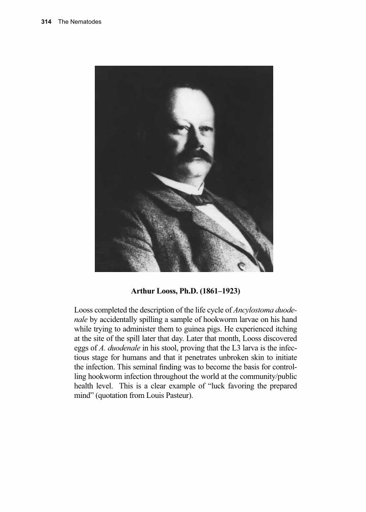
314 The Nematodes
Arthur Looss, Ph.D. (1861–1923)
Looss completed the description of the life cycle of Ancylostoma duode-nale by accidentally spilling a sample of hookworm larvae on his hand while trying to administer them to guinea pigs. He experienced itching at the site of the spill later that day. Later that month, Looss discovered eggs of A. duodenale in his stool, proving that the L3 larva is the infec-tious stage for humans and that it penetrates unbroken skin to initiate the infection. This seminal finding was to become the basis for control-ling hookworm infection throughout the world at the community/public health level. This is a clear example of “luck favoring the prepared mind” (quotation from Louis Pasteur).
The Cestodes 315
VI. The Cestodes
The phylum Platyhelminthes includes the class Cestoidea (tapeworms), all of which are parasitic in the gut tracts of various vertebrate hosts. Tapeworms are flat, segmented worms, composed of a head (scolex), and a series of segments, known as proglottids. Together, all proglottids are referred to as the strobila. The scolex is the point of attachment between the host and the parasite. It may be equipped with suckers, hooks, or grooves, which aid in the attachment process. The scolex contains nerves terminating in ganglia, while the seg-ments contain only nerves. The neck region of the scolex is metabolically active and is the site in most tapeworms from which new pro-glottids form.
Tapeworms do not have a functional gut tract. Rather, the segments are enclosed in a spe-cialized tegument, whose structure and func-tion are directly related to nutrient acquisition. Evenly spaced microvilli cover the entire surface of the tegument, underneath which lie mitochondria, vesicles (perhaps involved in tegument replacement), and related struc-tures. The tapeworm obtains some of its nutrients by actively transporting them across the tegument. Each proglottid absorbs a wide variety of low-molecular-weight substrates, but its precise metabolic requirements have yet to be fully defined.
High levels of ATPase in the tegument are related to active transport but may also help the worm resist digestion by the mammalian host. Inhibitors of tapeworm ATPase, such as niclosamide, cause disintegration of adult tapeworms by digestion in the presence of pancreatic secretions.
Each proglottid has two layers of muscle, longitudinal and transverse, enabling the seg-ment to move. Two lateral branches of nerves
innervate the worm, with perpendicular com-missures branching out into the parenchyma of each segment. Segments are anatomically independent, but they are all connected by a common nervous system emanating from central ganglia located in the scolex. Osmo-regulation and excretion of wastes occurs via a lateral pair of excretory tubules.
Mature proglottids possess both male and female sex organs, but self-mating within a segment is unusual. Typically, sperm are transferred between mature proglottids that lie next to each other. Gravid proglot-tids develop after mating, and contain hun-dreds to thousands of embryonated eggs. The gravid proglottids then detach from the parent organism and exit via the host’s anus. In some species, proglottids exit intact, while in others, segments disintegrate before leav-ing the host. Eggs are usually passed embryo-nated, and contain a hexacanth larva referred to as an oncosphere. Eggs may remain viable in the external environment for weeks to months after being deposited in soil. Hatch-ing occurs typically within the small intes-tine of the intermediate host. The oncosphere then penetrates the gut tract and lodges within the tissues, developing into the metacestode. When this stage is ingested by the definitive host it transforms to the adult in the lumen of the small intestine.
Adult tapeworms do not cause significant pathology in the human intestine. Unlike adult nematodes or trematodes, the adult cestodes do not adversely impact childhood development. However, when humans serve as intermediate hosts, the cestodes become significant causes of global morbidity. For instance, neurocysticercosis caused by the larval stages of the pork tapeworm, Taenia solium, is a leading cause of neurologic dis-ease; in many countries it is the leading cause of epilepsy.
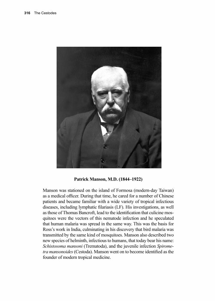
316 The Cestodes
Patrick Manson, M.D. (1844–1922)
Manson was stationed on the island of Formosa (modern-day Taiwan) as a medical officer. During that time, he cared for a number of Chinese patients and became familiar with a wide variety of tropical infectious diseases, including lymphatic filariasis (LF). His investigations, as well as those of Thomas Bancroft, lead to the identification that culicine mos-quitoes were the vectors of this nematode infection and he speculated that human malaria was spread in the same way. This was the basis for Ross’s work in India, culminating in his discovery that bird malaria was transmitted by the same kind of mosquitoes. Manson also described two new species of helminth, infectious to humans, that today bear his name: Schistosoma mansoni (Trematoda), and the juvenile infection Spirome-tra mansonoides (Cestoda). Manson went on to become identified as the founder of modern tropical medicine.
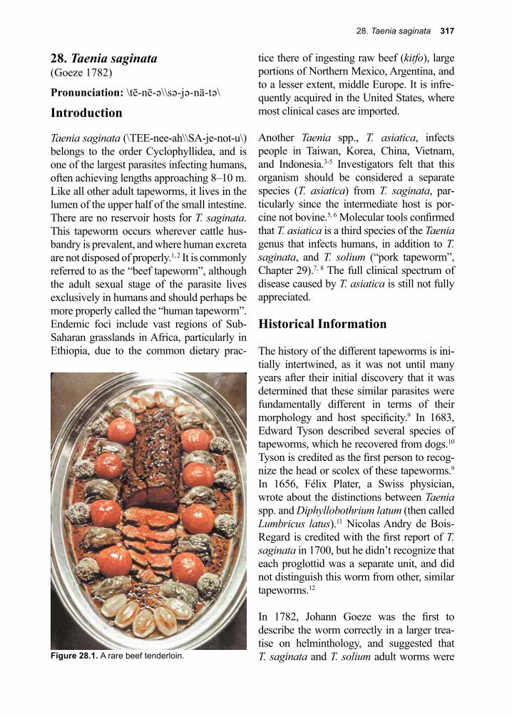
28. Taenia saginata 317
28. Taenia saginata (Goeze 1782)
Pronunciation: \tē-nē-ə\\sə-jə-nä-tə\
Introduction
Taenia saginata (\TEE-nee-ah\\SA-je-not-u\) belongs to the order Cyclophyllidea, and is one of the largest parasites infecting humans, often achieving lengths approaching 8–10 m. Like all other adult tapeworms, it lives in the lumen of the upper half of the small intestine. There are no reservoir hosts for T. saginata. This tapeworm occurs wherever cattle hus-bandry is prevalent, and where human excreta are not disposed of properly.1, 2 It is commonly referred to as the “beef tapeworm”, although the adult sexual stage of the parasite lives exclusively in humans and should perhaps be more properly called the “human tapeworm”. Endemic foci include vast regions of Sub-Saharan grasslands in Africa, particularly in Ethiopia, due to the common dietary prac-
tice there of ingesting raw beef (kitfo), large portions of Northern Mexico, Argentina, and to a lesser extent, middle Europe. It is infre-quently acquired in the United States, where most clinical cases are imported.
Another Taenia spp., T. asiatica, infects people in Taiwan, Korea, China, Vietnam, and Indonesia.3-5 Investigators felt that this organism should be considered a separate species (T. asiatica) from T. saginata, par-ticularly since the intermediate host is por-cine not bovine.5, 6 Molecular tools confirmed that T. asiatica is a third species of the Taenia genus that infects humans, in addition to T. saginata, and T. solium (“pork tapeworm”, Chapter 29).7, 8 The full clinical spectrum of disease caused by T. asiatica is still not fully appreciated.
Historical Information
The history of the different tapeworms is ini-tially intertwined, as it was not until many years after their initial discovery that it was determined that these similar parasites were fundamentally different in terms of their morphology and host specificity.9 In 1683, Edward Tyson described several species of tapeworms, which he recovered from dogs.10 Tyson is credited as the first person to recog-nize the head or scolex of these tapeworms.9 In 1656, Félix Plater, a Swiss physician, wrote about the distinctions between Taenia spp. and Diphyllobothrium latum (then called Lumbricus latus).11 Nicolas Andry de Bois-Regard is credited with the first report of T. saginata in 1700, but he didn’t recognize that each proglottid was a separate unit, and did not distinguish this worm from other, similar tapeworms.12
In 1782, Johann Goeze was the first to describe the worm correctly in a larger trea-tise on helminthology, and suggested that T. saginata and T. solium adult worms were Figure 28.1. A rare beef tenderloin.
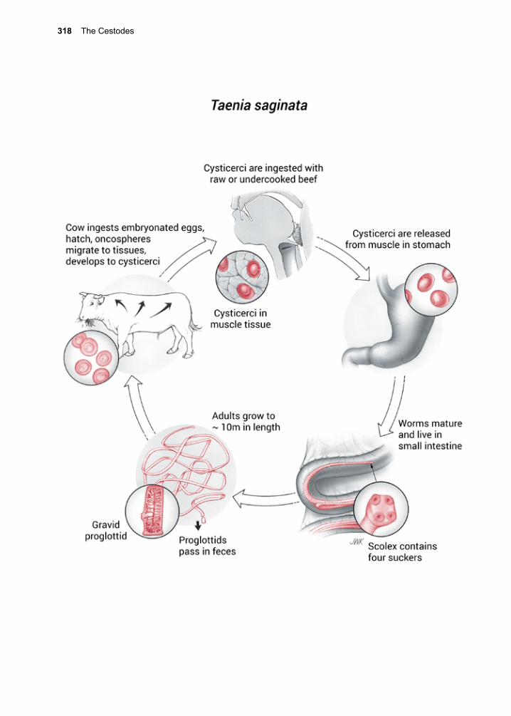
318 The Cestodes
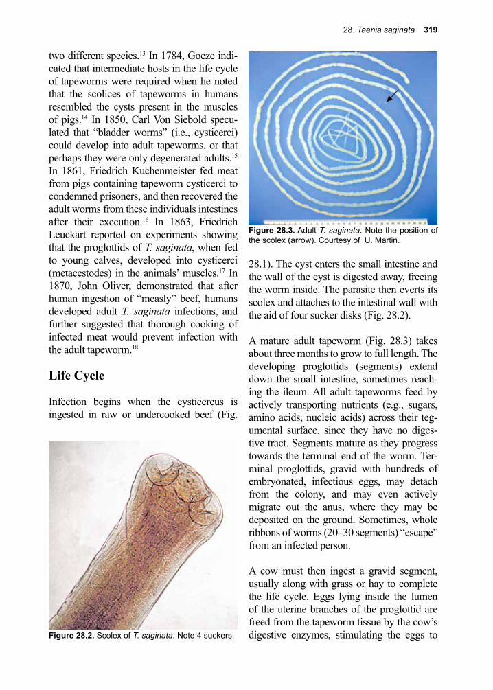
28. Taenia saginata 319
Figure 28.3. Adult T. saginata. Note the position of the scolex (arrow). Courtesy of U. Martin.
two different species.13 In 1784, Goeze indi-cated that intermediate hosts in the life cycle of tapeworms were required when he noted that the scolices of tapeworms in humans resembled the cysts present in the muscles of pigs.14 In 1850, Carl Von Siebold specu-lated that “bladder worms” (i.e., cysticerci) could develop into adult tapeworms, or that perhaps they were only degenerated adults.15 In 1861, Friedrich Kuchenmeister fed meat from pigs containing tapeworm cysticerci to condemned prisoners, and then recovered the adult worms from these individuals intestines after their execution.16 In 1863, Friedrich Leuckart reported on experiments showing that the proglottids of T. saginata, when fed to young calves, developed into cysticerci (metacestodes) in the animals’ muscles.17 In 1870, John Oliver, demonstrated that after human ingestion of “measly” beef, humans developed adult T. saginata infections, and further suggested that thorough cooking of infected meat would prevent infection with the adult tapeworm.18
Life Cycle
Infection begins when the cysticercus is ingested in raw or undercooked beef (Fig.
28.1). The cyst enters the small intestine and the wall of the cyst is digested away, freeing the worm inside. The parasite then everts its scolex and attaches to the intestinal wall with the aid of four sucker disks (Fig. 28.2).
A mature adult tapeworm (Fig. 28.3) takes about three months to grow to full length. The developing proglottids (segments) extend down the small intestine, sometimes reach-ing the ileum. All adult tapeworms feed by actively transporting nutrients (e.g., sugars, amino acids, nucleic acids) across their teg-umental surface, since they have no diges-tive tract. Segments mature as they progress towards the terminal end of the worm. Ter-minal proglottids, gravid with hundreds of embryonated, infectious eggs, may detach from the colony, and may even actively migrate out the anus, where they may be deposited on the ground. Sometimes, whole ribbons of worms (20–30 segments) “escape” from an infected person.
A cow must then ingest a gravid segment, usually along with grass or hay to complete the life cycle. Eggs lying inside the lumen of the uterine branches of the proglottid are freed from the tapeworm tissue by the cow’s digestive enzymes, stimulating the eggs to Figure 28.2. Scolex of T. saginata. Note 4 suckers.
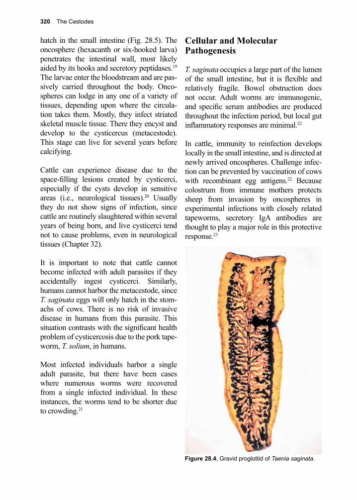
320 The Cestodes
hatch in the small intestine (Fig. 28.5). The oncosphere (hexacanth or six-hooked larva) penetrates the intestinal wall, most likely aided by its hooks and secretory peptidases.19 The larvae enter the bloodstream and are pas-sively carried throughout the body. Onco-spheres can lodge in any one of a variety of tissues, depending upon where the circula-tion takes them. Mostly, they infect striated skeletal muscle tissue. There they encyst and develop to the cysticercus (metacestode). This stage can live for several years before calcifying.
Cattle can experience disease due to the space-filling lesions created by cysticerci, especially if the cysts develop in sensitive areas (i.e., neurological tissues).20 Usually they do not show signs of infection, since cattle are routinely slaughtered within several years of being born, and live cysticerci tend not to cause problems, even in neurological tissues (Chapter 32).
It is important to note that cattle cannot become infected with adult parasites if they accidentally ingest cysticerci. Similarly, humans cannot harbor the metacestode, since T. saginata eggs will only hatch in the stom-achs of cows. There is no risk of invasive disease in humans from this parasite. This situation contrasts with the significant health problem of cysticercosis due to the pork tape-worm, T. solium, in humans.
Most infected individuals harbor a single adult parasite, but there have been cases where numerous worms were recovered from a single infected individual. In these instances, the worms tend to be shorter due to crowding.21
Cellular and Molecular Pathogenesis
T. saginata occupies a large part of the lumen of the small intestine, but it is flexible and relatively fragile. Bowel obstruction does not occur. Adult worms are immunogenic, and specific serum antibodies are produced throughout the infection period, but local gut inflammatory responses are minimal.22
In cattle, immunity to reinfection develops locally in the small intestine, and is directed at newly arrived oncospheres. Challenge infec-tion can be prevented by vaccination of cows with recombinant egg antigens.22 Because colostrum from immune mothers protects sheep from invasion by oncospheres in experimental infections with closely related tapeworms, secretory IgA antibodies are thought to play a major role in this protective response.23
Figure 28.4. Gravid proglottid of Taenia saginata.
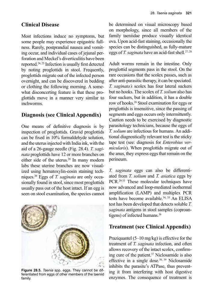
28. Taenia saginata 321
Figure 28.5. Taenia spp. eggs. They cannot be dif-ferentiated from eggs of other members of the taeniid family.
Clinical Disease
Most infections induce no symptoms, but some people may experience epigastric full-ness. Rarely, postprandial nausea and vomit-ing occur, and individual cases of jejunal per-foration and Meckel’s diverticulitis have been reported.24, 25 Infection is usually first detected by noting proglottids in stool. Frequently, proglottids migrate out of the infected person overnight, and can be discovered in bedding or clothing the following morning. A some-what disconcerting feature is that these pro-glottids move in a manner very similar to inchworms.
Diagnosis (see Clinical Appendix)
One means of definitive diagnosis is by inspection of proglottids. Gravid proglottids can be fixed in 10% formaldehyde solution, and the uterus injected with India ink, with the aid of a 26-gauge needle (Fig. 28.4). T. sagi-nata proglottids have 12 or more branches on either side of the uterus.26 In many modern labs these uterine branches are now visual-ized using hematoxylin-eosin staining tech-niques.26 Eggs of T. saginata are only occa-sionally found in stool, since most proglottids usually pass out of the host intact. If an egg is seen on stool examination, the species cannot
be determined on visual microscopy based on morphology, since all members of the family taeniidae produce visually identical ova. Upon acid-fast staining, occasionally the species can be distinguished, as fully-mature eggs of T. saginata have an acid-fast shell.27, 28
Adult worms remain in the intestine. Only proglottid segments pass in the stool. On the rare occasions that the scolex passes, such as after anti-parasitic therapy, it can be speciated. T. saginata’s scolex has four lateral suckers but no hooks. The scolex of T. solium also has four suckers, but in addition, it has a double row of hooks.26 Stool examination for eggs or proglottids is insensitive, since the passing of segments and eggs occurs only intermittently. Caution needs to be exercised by diagnostic parasitology technicians, because the eggs of T. solium are infectious for humans. An addi-tional diagnostically relevant test is the sticky tape test (see: diagnosis for Enterobius ver-micularis). When proglottids migrate out of the anus, they express eggs that remain on the perineum.
T. saginata eggs can also be differenti-ated from T. solium and T. asiatica eggs by PCR.29-33 These molecular techniques have now advanced and loop-mediated isothermal amplification (LAMP) and multiplex PCR tests have become available.34, 35 An ELISA test has been developed that detects soluble T. saginata antigens in stool samples (coproan-tigens) of infected humans.36
Treatment (see Clinical Appendix)
Praziquantel (5–10 mg/kg) is effective for the treatment of T. saginata infection, and often allows recovery of the intact scolex, confirm-ing cure of the patient.37 Niclosamide is also effective in a single dose.38, 39 Niclosamide inhibits the parasite’s ATPase, thus prevent-ing it from interfering with host digestive enzymes. The consequence of treatment is
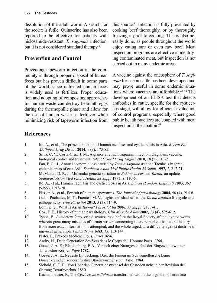
322 The Cestodes
this source.41 Infection is fully prevented by cooking beef thoroughly, or by thoroughly freezing it prior to cooking. This is also not easily done, as people throughout the world enjoy eating rare or even raw beef. Meat inspection programs are effective in identify-ing contaminated meat, but inspection is not carried out in many endemic areas.
A vaccine against the oncosphere of T. sagi-nata for use in cattle has been developed and may prove useful in some endemic situa-tions where vaccines are affordable.22, 42 The development of an ELISA test that detects antibodies in cattle, specific for the cysticer-cus stage, will allow for efficient evaluation of control programs, especially where good public health practices are coupled with meat inspection at the abattoir.43
References
1. Ito, A., et al., The present situation of human taeniases and cysticercosis in Asia. Recent Pat Antiinfect Drug Discov 2014, 9 (3), 173-85.
2. Silva, C. V.; Costa-Cruz, J. M., A glance at Taenia saginata infection, diagnosis, vaccine, biological control and treatment. Infect Disord Drug Targets 2010, 10 (5), 313-21.
3. Fan, P. C.; J., Annual economic loss caused by Taenia saginata asiatica Taeniasis in three endemic areas of east Asia. Southeast Asian Med Public Health 28 Suppl 1997, 1, 217-21.
4. McManus, D. P.; J., Molecular genetic variation in Echinococcus and Taenia: an update. Southeast Asian Med Public Health 28 Suppl 1997, 1, 110-6.
5. Ito, A., et al., Human Taeniasis and cysticercosis in Asia. Lancet (London, England) 2003, 362 (9399), 1918-20.
6. Flisser, A., et al., Portrait of human tapeworms. The Journal of parasitology 2004, 90 (4), 914-6.7. Galan-Puchades, M. T.; Fuentes, M. V., Lights and shadows of the Taenia asiatica life cycle and
pathogenicity. Trop Parasitol 2013, 3 (2), 114-9.8. Eom, K. S., What is Asian Taenia? Parasitol Int 2006, 55 Suppl, S137-41.9. Cox, F. E., History of human parasitology. Clin Microbiol Rev 2002, 15 (4), 595-612.10. Tyson, E., Lumbricus latus, or a discourse read before the Royal Society, of the joynted worm,
wherein great many mistakes of former writers concerning it, are remarked; its natural history from more exact information is attempted; and the whole urged, as a difficulty against doctrine of univocal generation. Philos Trans 1683, 13, 113-144.
11. Plater, F., Praxeos Medicae Opus. Basel 1656.12. Andry, N., De la Generation des Vers dans le Corps de l’Homme Paris. 1700.13. Goeze, J. A. E.; Blankenburg, P. A., Versuch ciner Naturgeschichte der Eingeweiderwumer
Thierischer Korper. Pape 1782.14. Goeze, J. A. E., Neueste Entdeckung. Dass die Finnen im Schweinefleische keine
Drusenkrankheit sondern wahre Blasenwurmer sind. Halle. 1784.15. Siebold, C. T. E., Von Uber den Generationswechsel der Cestoden nebst einer Revision der
Gattung Tetrarhynchus. 1850.16. Kuchenmeister, F., The Cysticercus cellulosus transformed within the organism of man into
dissolution of the adult worm. A search for the scolex is futile. Quinacrine has also been reported to be effective for patients with niclosamide-resistant T. saginata infection, but it is not considered standard therapy.40
Prevention and Control
Preventing tapeworm infection in the com-munity is through proper disposal of human feces but has proven difficult in some parts of the world, since untreated human feces is widely used as fertilizer. Proper educa-tion and adopting of composting approaches for human waste can destroy helminth eggs during the thermophilic phase and allow for the use of human waste as fertilizer while minimizing risk of tapeworm infection from
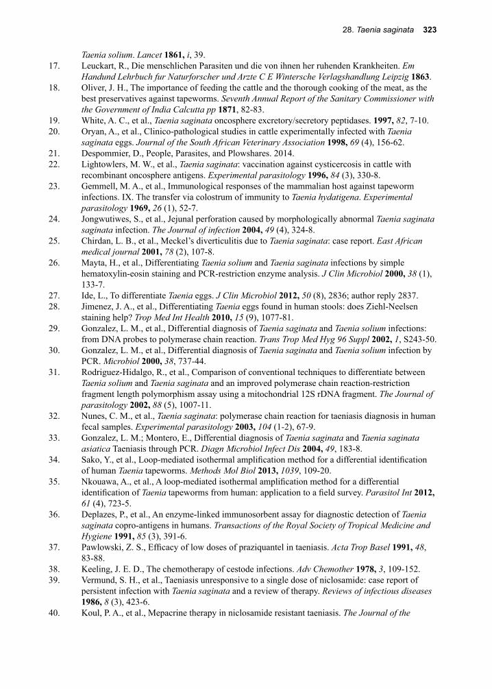
28. Taenia saginata 323
Taenia solium. Lancet 1861, i, 39.17. Leuckart, R., Die menschlichen Parasiten und die von ihnen her ruhenden Krankheiten. Em
Handund Lehrbuch fur Naturforscher und Arzte C E Wintersche Verlagshandlung Leipzig 1863.18. Oliver, J. H., The importance of feeding the cattle and the thorough cooking of the meat, as the
best preservatives against tapeworms. Seventh Annual Report of the Sanitary Commissioner with the Government of India Calcutta pp 1871, 82-83.
19. White, A. C., et al., Taenia saginata oncosphere excretory/secretory peptidases. 1997, 82, 7-10.20. Oryan, A., et al., Clinico-pathological studies in cattle experimentally infected with Taenia
saginata eggs. Journal of the South African Veterinary Association 1998, 69 (4), 156-62.21. Despommier, D., People, Parasites, and Plowshares. 2014.22. Lightowlers, M. W., et al., Taenia saginata: vaccination against cysticercosis in cattle with
recombinant oncosphere antigens. Experimental parasitology 1996, 84 (3), 330-8.23. Gemmell, M. A., et al., Immunological responses of the mammalian host against tapeworm
infections. IX. The transfer via colostrum of immunity to Taenia hydatigena. Experimental parasitology 1969, 26 (1), 52-7.
24. Jongwutiwes, S., et al., Jejunal perforation caused by morphologically abnormal Taenia saginata saginata infection. The Journal of infection 2004, 49 (4), 324-8.
25. Chirdan, L. B., et al., Meckel’s diverticulitis due to Taenia saginata: case report. East African medical journal 2001, 78 (2), 107-8.
26. Mayta, H., et al., Differentiating Taenia solium and Taenia saginata infections by simple hematoxylin-eosin staining and PCR-restriction enzyme analysis. J Clin Microbiol 2000, 38 (1), 133-7.
27. Ide, L., To differentiate Taenia eggs. J Clin Microbiol 2012, 50 (8), 2836; author reply 2837.28. Jimenez, J. A., et al., Differentiating Taenia eggs found in human stools: does Ziehl-Neelsen
staining help? Trop Med Int Health 2010, 15 (9), 1077-81.29. Gonzalez, L. M., et al., Differential diagnosis of Taenia saginata and Taenia solium infections:
from DNA probes to polymerase chain reaction. Trans Trop Med Hyg 96 Suppl 2002, 1, S243-50.30. Gonzalez, L. M., et al., Differential diagnosis of Taenia saginata and Taenia solium infection by
PCR. Microbiol 2000, 38, 737-44.31. Rodriguez-Hidalgo, R., et al., Comparison of conventional techniques to differentiate between
Taenia solium and Taenia saginata and an improved polymerase chain reaction-restriction fragment length polymorphism assay using a mitochondrial 12S rDNA fragment. The Journal of parasitology 2002, 88 (5), 1007-11.
32. Nunes, C. M., et al., Taenia saginata: polymerase chain reaction for taeniasis diagnosis in human fecal samples. Experimental parasitology 2003, 104 (1-2), 67-9.
33. Gonzalez, L. M.; Montero, E., Differential diagnosis of Taenia saginata and Taenia saginata asiatica Taeniasis through PCR. Diagn Microbiol Infect Dis 2004, 49, 183-8.
34. Sako, Y., et al., Loop-mediated isothermal amplification method for a differential identification of human Taenia tapeworms. Methods Mol Biol 2013, 1039, 109-20.
35. Nkouawa, A., et al., A loop-mediated isothermal amplification method for a differential identification of Taenia tapeworms from human: application to a field survey. Parasitol Int 2012, 61 (4), 723-5.
36. Deplazes, P., et al., An enzyme-linked immunosorbent assay for diagnostic detection of Taenia saginata copro-antigens in humans. Transactions of the Royal Society of Tropical Medicine and Hygiene 1991, 85 (3), 391-6.
37. Pawlowski, Z. S., Efficacy of low doses of praziquantel in taeniasis. Acta Trop Basel 1991, 48, 83-88.
38. Keeling, J. E. D., The chemotherapy of cestode infections. Adv Chemother 1978, 3, 109-152.39. Vermund, S. H., et al., Taeniasis unresponsive to a single dose of niclosamide: case report of
persistent infection with Taenia saginata and a review of therapy. Reviews of infectious diseases 1986, 8 (3), 423-6.
40. Koul, P. A., et al., Mepacrine therapy in niclosamide resistant taeniasis. The Journal of the

324 The Cestodes
Association of Physicians of India 2000, 48 (4), 402-3.41. Kone, D., et al., Helminth eggs inactivation efficiency by faecal sludge dewatering and co-
composting in tropical climates. Water Res 2007, 41 (19), 4397-402.42. Lightowlers, M. W., et al., Vaccination against cestode parasites: anti-helminth vaccines that
work and why. Veterinary parasitology 2003, 115 (2), 83-123.43. Onyango-Abuje, J. A., et al., Diagnosis of Taenia saginata cysticercosis in Kenyan cattle by
antibody and antigen ELISA. Veterinary parasitology 1996, 61 (3-4), 221-30.
Francisca Mutapi, Ph.D. (current)
Mutapi was born and grew up in Zimbabwe and then went on to study at the University of Oxford as a Beit Trust Scholar. After completing postdoctoral training at the Institute of Tropical Medicine in Antwerp, she went on to become a leader in the emerging field of global health with a focus on tropical diseases. Dr. Mutapi is credited with prioritizing schistosomiasis as a public health concern for the World Health Organi-zation and has championed a multidisciplinary approach to global health that incorporates economic, cultural, technological and evidence-based challenges to global health problems. Dr. Mutapi is also credited with advancing our understanding of the immune responses to helminths.
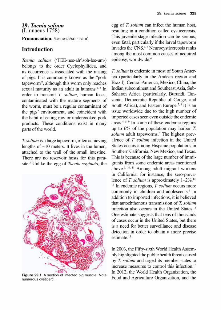
29. Taenia solium 325
Figure 29.1. A section of infected pig muscle. Note numerous cysticerci.
29. Taenia solium (Linnaeus 1758)Pronunciation: \tē-nē-ə\\sōl-ī-əm\
Introduction
Taenia solium (\TEE-nee-ah\\soh-lee-um\) belongs to the order Cyclophyllidea, and its occurrence is associated with the raising of pigs. It is commonly known as the “pork tapeworm”, although this worm only reaches sexual maturity as an adult in humans.1, 2 In order to transmit T. solium, human feces, contaminated with the mature segments of the worm, must be a regular contaminant of the pigs’ environment, and coincident with the habit of eating raw or undercooked pork products. These conditions exist in many parts of the world.
T. solium is a large tapeworm, often achieving lengths of ~10 meters. It lives in the lumen, attached to the wall of the small intestine. There are no reservoir hosts for this para-site.3 Unlike the egg of Taenia saginata, the
egg of T. solium can infect the human host, resulting in a condition called cysticercosis. This juvenile-stage infection can be serious, even fatal, particularly if the larval tapeworm invades the CNS.4, 5 Neurocysticercosis ranks among the most common causes of acquired epilepsy, worldwide.6
T. solium is endemic in most of South Amer-ica (particularly in the Andean region and Brazil), Central America, Mexico, China, the Indian subcontinent and Southeast Asia, Sub-Saharan Africa (particularly, Burundi, Tan-zania, Democratic Republic of Congo, and South Africa), and Eastern Europe.7, 8 It is an issue worldwide due to the high number of imported cases seen even outside the endemic areas.4, 5, 9 In some of these endemic regions up to 6% of the population may harbor T. solium adult tapeworms.6 The highest prev-alence of T. solium infection in the United States occurs among Hispanic populations in Southern California, New Mexico, and Texas. This is because of the large number of immi-grants from some endemic areas mentioned above.6, 10, 11 Among adult migrant workers in California, for instance, the sero-preva-lence of T. solium is approximately 1–2%.12,
13 In endemic regions, T. solium occurs more commonly in children and adolescents.6 In addition to imported infections, it is believed that autochthonous transmission of T. solium infection also occurs in the United States.14 One estimate suggests that tens of thousands of cases occur in the United States, but there is a need for better surveillance and disease detection in order to obtain a more precise estimate.15
In 2003, the Fifty-sixth World Health Assem-bly highlighted the public health threat caused by T. solium and urged its member states to increase measures to control this infection.10 In 2012, the World Health Organization, the Food and Agriculture Organization, and the
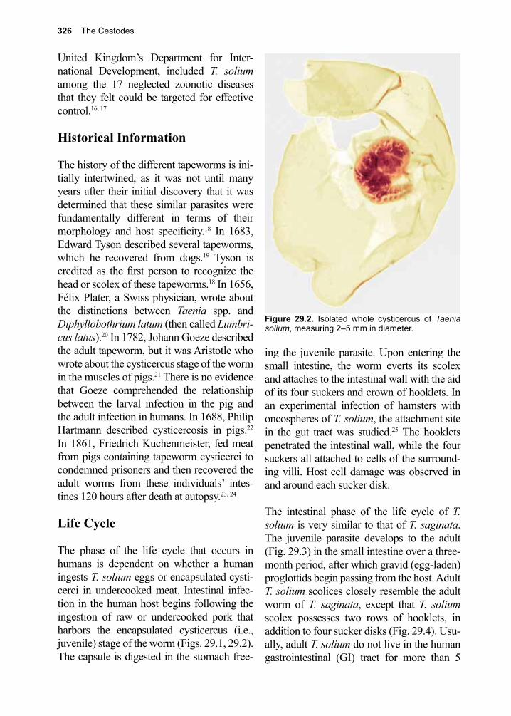
326 The Cestodes
United Kingdom’s Department for Inter-national Development, included T. solium among the 17 neglected zoonotic diseases that they felt could be targeted for effective control.16, 17
Historical Information
The history of the different tapeworms is ini-tially intertwined, as it was not until many years after their initial discovery that it was determined that these similar parasites were fundamentally different in terms of their morphology and host specificity.18 In 1683, Edward Tyson described several tapeworms, which he recovered from dogs.19 Tyson is credited as the first person to recognize the head or scolex of these tapeworms.18 In 1656, Félix Plater, a Swiss physician, wrote about the distinctions between Taenia spp. and Diphyllobothrium latum (then called Lumbri-cus latus).20 In 1782, Johann Goeze described the adult tapeworm, but it was Aristotle who wrote about the cysticercus stage of the worm in the muscles of pigs.21 There is no evidence that Goeze comprehended the relationship between the larval infection in the pig and the adult infection in humans. In 1688, Philip Hartmann described cysticercosis in pigs.22 In 1861, Friedrich Kuchenmeister, fed meat from pigs containing tapeworm cysticerci to condemned prisoners and then recovered the adult worms from these individuals’ intes-tines 120 hours after death at autopsy.23, 24
Life Cycle
The phase of the life cycle that occurs in humans is dependent on whether a human ingests T. solium eggs or encapsulated cysti-cerci in undercooked meat. Intestinal infec-tion in the human host begins following the ingestion of raw or undercooked pork that harbors the encapsulated cysticercus (i.e., juvenile) stage of the worm (Figs. 29.1, 29.2). The capsule is digested in the stomach free-
ing the juvenile parasite. Upon entering the small intestine, the worm everts its scolex and attaches to the intestinal wall with the aid of its four suckers and crown of hooklets. In an experimental infection of hamsters with oncospheres of T. solium, the attachment site in the gut tract was studied.25 The hooklets penetrated the intestinal wall, while the four suckers all attached to cells of the surround-ing villi. Host cell damage was observed in and around each sucker disk.
The intestinal phase of the life cycle of T. solium is very similar to that of T. saginata. The juvenile parasite develops to the adult (Fig. 29.3) in the small intestine over a three-month period, after which gravid (egg-laden) proglottids begin passing from the host. Adult T. solium scolices closely resemble the adult worm of T. saginata, except that T. solium scolex possesses two rows of hooklets, in addition to four sucker disks (Fig. 29.4). Usu-ally, adult T. solium do not live in the human gastrointestinal (GI) tract for more than 5
Figure 29.2. Isolated whole cysticercus of Taenia solium, measuring 2–5 mm in diameter.
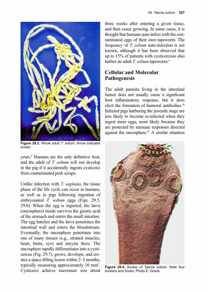
29. Taenia solium 327
years.6 Humans are the only definitive host, and the adult of T. solium will not develop in the pig if it accidentally ingests cysticerci from contaminated pork scraps.
Unlike infection with T. saginata, the tissue phase of the life cycle can occur in humans, as well as in pigs following ingestion of embryonated T. solium eggs (Figs. 29.5, 29.6). When the egg is ingested, the larva (oncosphere) inside survives the gastric acid of the stomach and enters the small intestine. The egg hatches and the larva penetrates the intestinal wall and enters the bloodstream. Eventually, the oncosphere penetrates into one of many tissues (e.g., striated muscles, heart, brain, eye) and encysts there. The oncosphere rapidly differentiates into a cysti-cercus (Fig. 29.7), grows, develops, and cre-ates a space-filling lesion within 2–3 months, typically measuring approximately 10 mm3. Cysticerci achieve maximum size about
three weeks after entering a given tissue, and then cease growing. In some cases, it is thought that humans auto-infect with the con-taminated eggs of their own tapeworm. The frequency of T. solium auto-infection is not known, although it has been observed that up to 15% of patients with cysticercosis also harbor an adult T. solium tapeworm.6
Cellular and Molecular Pathogenesis
The adult parasite living in the intestinal lumen does not usually cause a significant host inflammatory response, but it does elicit the formation of humoral antibodies.26 Infected pigs harboring the juvenile stage are less likely to become re-infected when they ingest more eggs, most likely because they are protected by immune responses directed against the oncosphere.27 A similar situation
Figure 29.4. Scolex of Taenia solium. Note four suckers and hooks. Photo E. Gravé.
Figure 29.3. Whole adult T. solium. Arrow indicates scolex.
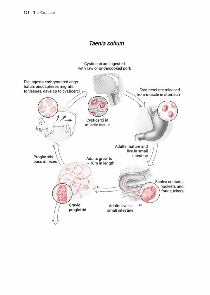
328 The Cestodes
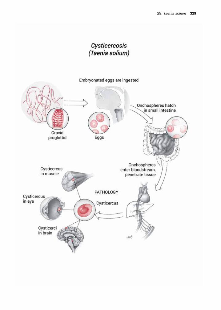
29. Taenia solium 329
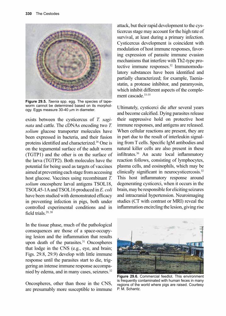
330 The Cestodes
attack, but their rapid development to the cys-ticercus stage may account for the high rate of survival, at least during a primary infection. Cysticercus development is coincident with modulation of host immune responses, favor-ing expression of parasite immune evasion mechanisms that interfere with Th2-type pro-tective immune responses.32 Immunomodu-latory substances have been identified and partially characterized; for example, Taenia-statin, a protease inhibitor, and paramyosin, which inhibit different aspects of the comple-ment cascade.33-35
Ultimately, cysticerci die after several years and become calcified. Dying parasites release their suppressive hold on protective host immune responses, and antigens are released. When cellular reactions are present, they are in part due to the result of interleukin signal-ing from T cells. Specific IgM antibodies and natural killer cells are also present in these infiltrates.36 An acute local inflammatory reaction follows, consisting of lymphocytes, plasma cells, and eosinophils, which may be clinically significant in neurocysticercosis.32 This host inflammatory response around degenerating cysticerci, when it occurs in the brain, may be responsible for eliciting seizures and intracranial hypertension. Neuroimaging studies (CT with contrast or MRI) reveal the inflammation encircling the lesion, giving rise
exists between the cysticercus of T. sagi-nata and cattle. The cDNAs encoding two T. solium glucose transporter molecules have been expressed in bacteria, and their fusion proteins identified and characterized.28 One is on the tegumental surface of the adult worm (TGTP1) and the other is on the surface of the larva (TGTP2). Both molecules have the potential for being used as targets of vaccines aimed at preventing each stage from accessing host glucose. Vaccines using recombinant T. solium oncosphere larval antigens TSOL18, TSOL45-1A and TSOL16 produced in E. coli have been studied with demonstrated efficacy in preventing infection in pigs, both under controlled experimental conditions and in field trials.29, 30
In the tissue phase, much of the pathological consequences are those of a space-occupy-ing lesion and the inflammation that results upon death of the parasites.31 Oncospheres that lodge in the CNS (e.g., eye, and brain; Figs. 29.8, 29.9) develop with little immune response until the parasites start to die, trig-gering an intense immune response accompa-nied by edema, and in many cases, seizures.32
Oncospheres, other than those in the CNS, are presumably more susceptible to immune
Figure 29.5. Taenia spp. egg. The species of tape-worm cannot be determined based on its morphol-ogy. Eggs measure 30-40 µm in diameter.
Figure 29.6. Commercial feedlot. This environment is frequently contaminated with human feces in many regions of the world where pigs are raised. Courtesy P. M. Schantz.
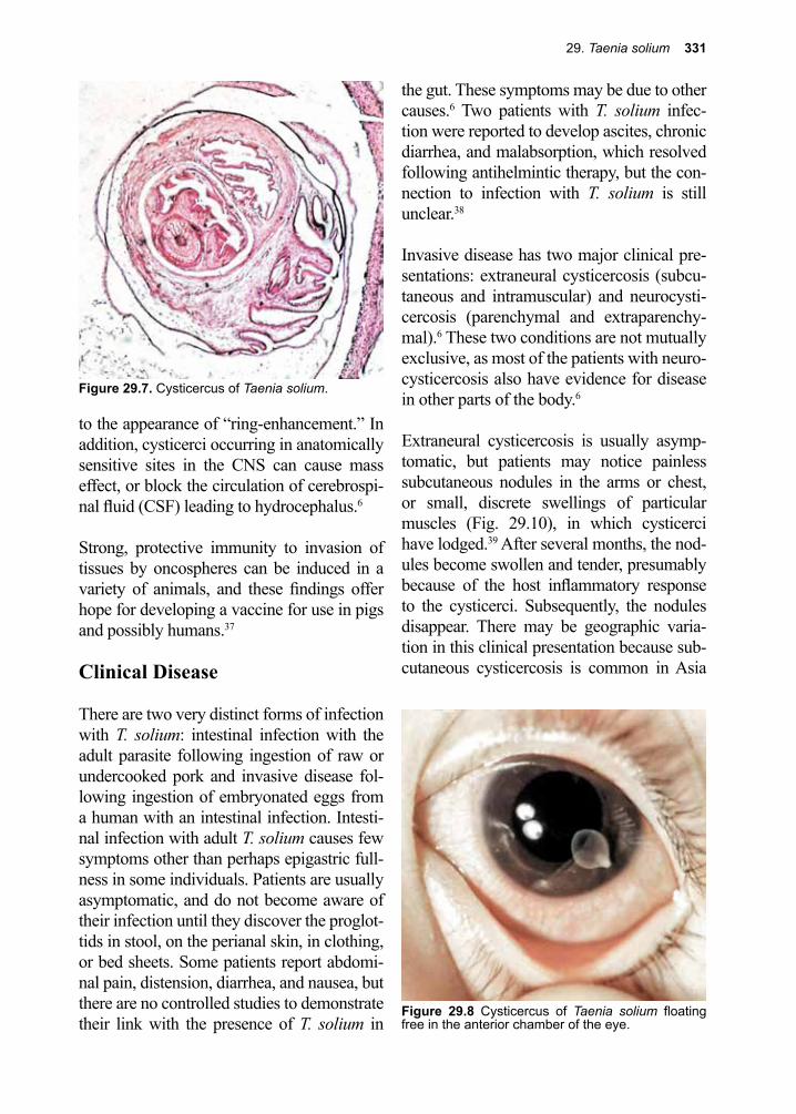
29. Taenia solium 331
to the appearance of “ring-enhancement.” In addition, cysticerci occurring in anatomically sensitive sites in the CNS can cause mass effect, or block the circulation of cerebrospi-nal fluid (CSF) leading to hydrocephalus.6
Strong, protective immunity to invasion of tissues by oncospheres can be induced in a variety of animals, and these findings offer hope for developing a vaccine for use in pigs and possibly humans.37
Clinical Disease
There are two very distinct forms of infection with T. solium: intestinal infection with the adult parasite following ingestion of raw or undercooked pork and invasive disease fol-lowing ingestion of embryonated eggs from a human with an intestinal infection. Intesti-nal infection with adult T. solium causes few symptoms other than perhaps epigastric full-ness in some individuals. Patients are usually asymptomatic, and do not become aware of their infection until they discover the proglot-tids in stool, on the perianal skin, in clothing, or bed sheets. Some patients report abdomi-nal pain, distension, diarrhea, and nausea, but there are no controlled studies to demonstrate their link with the presence of T. solium in
the gut. These symptoms may be due to other causes.6 Two patients with T. solium infec-tion were reported to develop ascites, chronic diarrhea, and malabsorption, which resolved following antihelmintic therapy, but the con-nection to infection with T. solium is still unclear.38
Invasive disease has two major clinical pre-sentations: extraneural cysticercosis (subcu-taneous and intramuscular) and neurocysti-cercosis (parenchymal and extraparenchy-mal).6 These two conditions are not mutually exclusive, as most of the patients with neuro-cysticercosis also have evidence for disease in other parts of the body.6
Extraneural cysticercosis is usually asymp-tomatic, but patients may notice painless subcutaneous nodules in the arms or chest, or small, discrete swellings of particular muscles (Fig. 29.10), in which cysticerci have lodged.39 After several months, the nod-ules become swollen and tender, presumably because of the host inflammatory response to the cysticerci. Subsequently, the nodules disappear. There may be geographic varia-tion in this clinical presentation because sub-cutaneous cysticercosis is common in Asia
Figure 29.8 Cysticercus of Taenia solium floating free in the anterior chamber of the eye.
Figure 29.7. Cysticercus of Taenia solium.
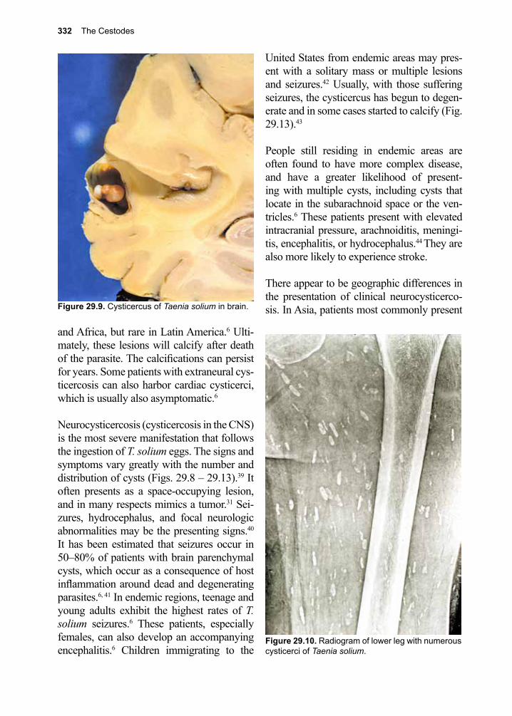
332 The Cestodes
and Africa, but rare in Latin America.6 Ulti-mately, these lesions will calcify after death of the parasite. The calcifications can persist for years. Some patients with extraneural cys-ticercosis can also harbor cardiac cysticerci, which is usually also asymptomatic.6
Neurocysticercosis (cysticercosis in the CNS) is the most severe manifestation that follows the ingestion of T. solium eggs. The signs and symptoms vary greatly with the number and distribution of cysts (Figs. 29.8 – 29.13).39 It often presents as a space-occupying lesion, and in many respects mimics a tumor.31 Sei-zures, hydrocephalus, and focal neurologic abnormalities may be the presenting signs.40 It has been estimated that seizures occur in 50–80% of patients with brain parenchymal cysts, which occur as a consequence of host inflammation around dead and degenerating parasites.6, 41 In endemic regions, teenage and young adults exhibit the highest rates of T. solium seizures.6 These patients, especially females, can also develop an accompanying encephalitis.6 Children immigrating to the
United States from endemic areas may pres-ent with a solitary mass or multiple lesions and seizures.42 Usually, with those suffering seizures, the cysticercus has begun to degen-erate and in some cases started to calcify (Fig. 29.13).43
People still residing in endemic areas are often found to have more complex disease, and have a greater likelihood of present-ing with multiple cysts, including cysts that locate in the subarachnoid space or the ven-tricles.6 These patients present with elevated intracranial pressure, arachnoiditis, meningi-tis, encephalitis, or hydrocephalus.44 They are also more likely to experience stroke.
There appear to be geographic differences in the presentation of clinical neurocysticerco-sis. In Asia, patients most commonly present Figure 29.9. Cysticercus of Taenia solium in brain.
Figure 29.10. Radiogram of lower leg with numerous cysticerci of Taenia solium.
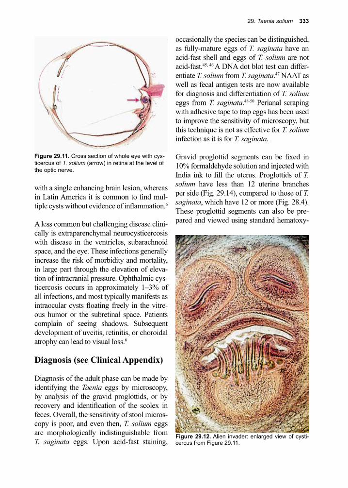
29. Taenia solium 333
with a single enhancing brain lesion, whereas in Latin America it is common to find mul-tiple cysts without evidence of inflammation.6
A less common but challenging disease clini-cally is extraparenchymal neurocysticercosis with disease in the ventricles, subarachnoid space, and the eye. These infections generally increase the risk of morbidity and mortality, in large part through the elevation of eleva-tion of intracranial pressure. Ophthalmic cys-ticercosis occurs in approximately 1–3% of all infections, and most typically manifests as intraocular cysts floating freely in the vitre-ous humor or the subretinal space. Patients complain of seeing shadows. Subsequent development of uveitis, retinitis, or choroidal atrophy can lead to visual loss.6
Diagnosis (see Clinical Appendix)
Diagnosis of the adult phase can be made by identifying the Taenia eggs by microscopy, by analysis of the gravid proglottids, or by recovery and identification of the scolex in feces. Overall, the sensitivity of stool micros-copy is poor, and even then, T. solium eggs are morphologically indistinguishable from T. saginata eggs. Upon acid-fast staining,
occasionally the species can be distinguished, as fully-mature eggs of T. saginata have an acid-fast shell and eggs of T. solium are not acid-fast.45, 46 A DNA dot blot test can differ-entiate T. solium from T. saginata.47 NAAT as well as fecal antigen tests are now available for diagnosis and differentiation of T. solium eggs from T. saginata.48-50 Perianal scraping with adhesive tape to trap eggs has been used to improve the sensitivity of microscopy, but this technique is not as effective for T. solium infection as it is for T. saginata.
Gravid proglottid segments can be fixed in 10% formaldehyde solution and injected with India ink to fill the uterus. Proglottids of T. solium have less than 12 uterine branches per side (Fig. 29.14), compared to those of T. saginata, which have 12 or more (Fig. 28.4). These proglottid segments can also be pre-pared and viewed using standard hematoxy-
Figure 29.12. Alien invader: enlarged view of cysti-cercus from Figure 29.11.
Figure 29.11. Cross section of whole eye with cys-ticercus of T. solium (arrow) in retina at the level of the optic nerve.
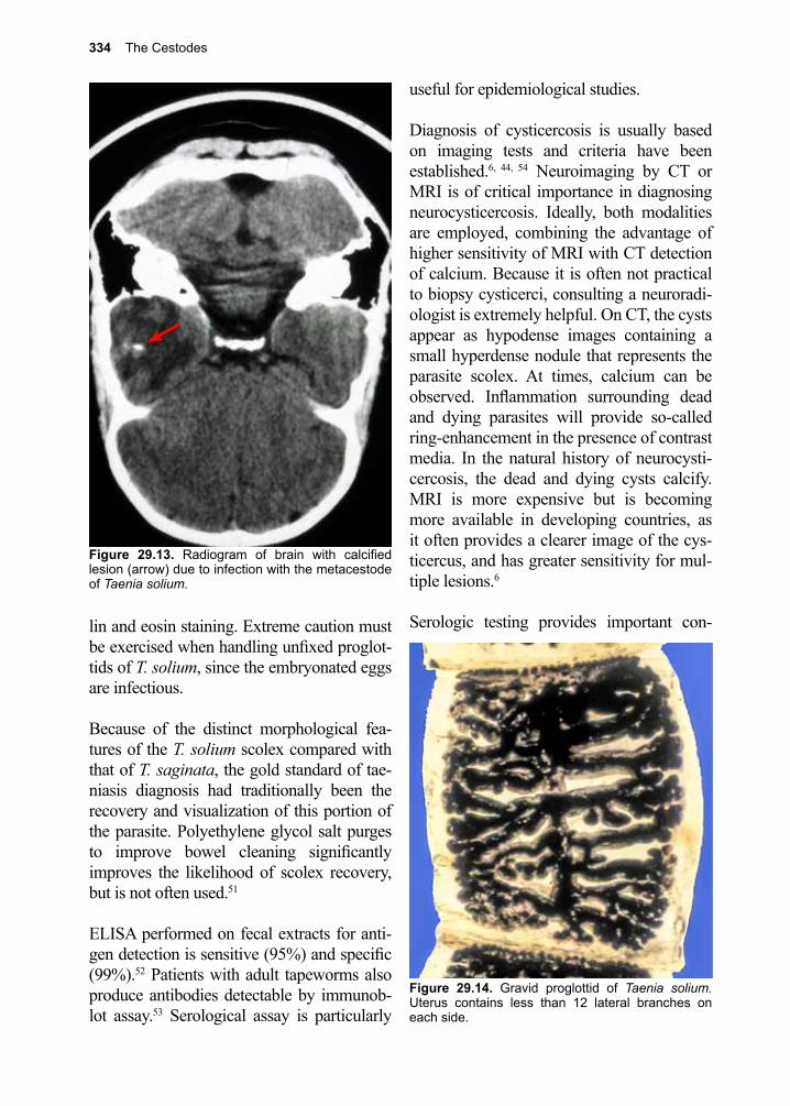
334 The Cestodes
lin and eosin staining. Extreme caution must be exercised when handling unfixed proglot-tids of T. solium, since the embryonated eggs are infectious.
Because of the distinct morphological fea-tures of the T. solium scolex compared with that of T. saginata, the gold standard of tae-niasis diagnosis had traditionally been the recovery and visualization of this portion of the parasite. Polyethylene glycol salt purges to improve bowel cleaning significantly improves the likelihood of scolex recovery, but is not often used.51
ELISA performed on fecal extracts for anti-gen detection is sensitive (95%) and specific (99%).52 Patients with adult tapeworms also produce antibodies detectable by immunob-lot assay.53 Serological assay is particularly
useful for epidemiological studies.
Diagnosis of cysticercosis is usually based on imaging tests and criteria have been established.6, 44, 54 Neuroimaging by CT or MRI is of critical importance in diagnosing neurocysticercosis. Ideally, both modalities are employed, combining the advantage of higher sensitivity of MRI with CT detection of calcium. Because it is often not practical to biopsy cysticerci, consulting a neuroradi-ologist is extremely helpful. On CT, the cysts appear as hypodense images containing a small hyperdense nodule that represents the parasite scolex. At times, calcium can be observed. Inflammation surrounding dead and dying parasites will provide so-called ring-enhancement in the presence of contrast media. In the natural history of neurocysti-cercosis, the dead and dying cysts calcify. MRI is more expensive but is becoming more available in developing countries, as it often provides a clearer image of the cys-ticercus, and has greater sensitivity for mul-tiple lesions.6
Serologic testing provides important con-
Figure 29.13. Radiogram of brain with calcified lesion (arrow) due to infection with the metacestode of Taenia solium.
Figure 29.14. Gravid proglottid of Taenia solium. Uterus contains less than 12 lateral branches on each side.
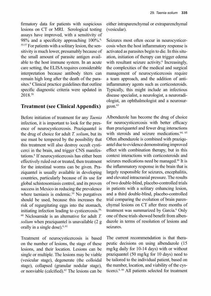
29. Taenia solium 335
firmatory data for patients with suspicious lesions on CT or MRI. Serological testing assays have improved, with a sensitivity of 98% and a specificity approaching 100%.6,
55-57 For patients with a solitary lesion, the sen-sitivity is much lower, presumably because of the small amount of parasite antigen avail-able to the host immune system. In an acute care setting, the ELISA requires considerable interpretation because antibody titers can remain high long after the death of the para-sites.6 Clinical practice guidelines that outline specific diagnostic criteria were updated in 2018.58
Treatment (see Clinical Appendix)
Before initiation of treatment for any Taenia infection, it is important to look for the pres-ence of neurocysticercosis. Praziquantel is the drug of choice for adult T. solium, but its use must be tempered by the possibility that this treatment will also destroy occult cysti-cerci in the brain, and trigger CNS manifes-tations.6 If neurocysticercosis has either been effectively ruled out or treated, then treatment for the intestinal worms can be given. Pra-ziquantel is usually available in developing countries, particularly because of its use for global schistosomiasis control, and its proven success in Mexico in reducing the prevalence where taeniasis is endemic.10 No purgatives should be used, because this increases the risk of regurgitating eggs into the stomach, initiating infection leading to cysticercosis.59,
60 Niclosamide is an alternative for adult T. solium when praziquantel is unavailable (2 g orally in a single dose).6, 61
Treatment of neurocysticercosis is based on the number of lesions, the stage of these lesions, and their location. Lesions can be single or multiple. The lesions may be viable (vesicular stage), degenerate (the colloidal stage), collapsed (granular nodular stage), or nonviable (calcified).62 The lesions can be
either intraparenchymal or extraparenchymal (vesicular).
Seizures most often occur in neurocysticer-cosis when the host inflammatory response is activated as parasites begin to die. In this situ-ation, initiation of therapy can trigger edema with resultant seizure activity.6 Increasingly, the complexities of the medical and surgical management of neurocysticercosis require a team approach, and the addition of anti-inflammatory agents such as corticosteroids. Typically, this might include an infectious disease specialist, a neurologist, a neuroradi-ologist, an ophthalmologist and a neurosur-geon.63
Albendazole has become the drug of choice for neurocysticercosis with better efficacy than praziquantel and fewer drug interactions with steroids and seizure medications.64, 65 Often albendazole is combined with praziqu-antel due to evidence demonstrating improved effect with combination therapy, but in this context interactions with corticosteroids and seizures medications need be managed.66 It is the inflammatory response in the brain that is largely responsible for seizures, encephalitis, and elevated intracranial pressure. The results of two double-blind, placebo-controlled trials in patients with a solitary enhancing lesion, and a third double-blind, placebo-controlled trial comparing the evolution of brain paren-chymal lesions on CT after three months of treatment was summarized by Garcia.6 Only one of these trials showed benefit from alben-dazole in terms of resolution of lesions and seizures.
The current recommendation is that thera-peutic decisions on using albendazole (15 mg/kg daily for 10-14 days) with or without praziquantel (50 mg/kg for 10 days) need to be tailored to the individual patient, based on the number, location, and viability of the cys-ticerci.6, 66 All patients selected for treatment
336 The Cestodes
with antihelmintic drugs should undergo a prior ophthalmologic exam in order to rule out intraocular cysts and be counseled about the risk of treatment-induced seizures.
Guidelines on the use of antihelmintic che-motherapy have been published.67 Since patients with single ring-enhancing lesions on CT scans often improve spontaneously with-out any therapy, they can be followed and not given albendazole or steroids.68, 69 In the case of multiple viable cysticerci, most clini-cians suggest treatment with albendazole and prednisone, with monitoring for elevations in intracranial pressure and seizures.4, 6
Because both albendazole and praziquantel kill cysticerci and exacerbate brain inflam-mation and edema, patients being treated for neurocysticercosis also require steroids, usually dexamethasone, to reduce swelling. Dexamethasone affects the blood levels of both praziquantel and albendazole, so drug doses of both may need to be adjusted.70 Additional medical management with anti-convulsants is also a critical component of the long-term management of T. solium-induced seizures.
Neurosurgical management with placement of ventricular shunts is often required for complicated neurocysticercosis involving cerebrospinal obstruction and hydrocephalus. Surgical management may also be required for spinal lesions and eye lesions.
Clinical practice guidelines that provide treatment recommendations were updated in 2018.58
Prevention and Control
Taenia solium is a significant public health problem, even outside the endemic areas,
due to the association of cysticercosis with the adult infections.71 For example, an out-break of cysticercosis was reported among an orthodox Jewish community in New York City resulting from the ingestion of T. solium eggs passed from domestic employees who were recent emigrants from Latin America.72 It has been suggested that recent emigrants from countries in which T. solium infection is endemic should be screened for tapeworm infection before they are employed as house-keepers or food handlers.72
In many impoverished regions in developing countries, pigs are more affordable than cows, since pigs are omnivores. That is why owners allow them to roam free to eat garbage and, perhaps, human feces.6 The infection in pigs is preventable by protecting their feed from contamination with human feces. Individual infection is prevented by thoroughly cooking pork, or by freezing it at -10 °C for a mini-mum of 5 days. Cysticerci can survive in meat refrigerated at 4 °C for up to 30 days.73
In 2003, the World Health Assembly identi-fied several measures to control T. solium infection including: 1. identification and treat-ment of individuals who carry the adult tape-worm, 2. universal or selected treatment with praziquantel to reduce the prevalence in areas where T. solium infection is endemic, 3. vet-erinary sanitary measures, such as enforced meat inspection and control, 4. improvement of pig husbandry, and treatment of infected animals with single-dose therapy, and 5. case management, reporting and surveillance of people with cysticercosis. Community-based interventions comprised of sanitation and pig management prove highly effective in dis-ease control.74 Ring-screening strategies have been proposed to control T. solium infec-tion.75 Antihelmintic chemotherapy programs need to be linked with other health programs using a wider integrated approach.10 In 2012,
29. Taenia solium 337
T. solium was added by the World Health Organization to their list of neglected zoo-notic diseases that they felt could be targeted for effective control.16, 17
Inoculation of pigs with recombinant anti-gens cloned from parasite oncosphere mRNA appears to be an effective vaccine, and could be used to reduce the incidence of adult tape-worm in a few countries, such as Mexico, where public health programs can be inte-grated with prevention on the farm.37, 76-78 In one experimental study, treating all pigs eliminated 100% of viable cysticerci.79 Sero-logical testing in the abattoir is now also pos-sible.80 Any of these three approaches, or in
combination, could significantly reduce trans-mission of T. solium to humans, if applied rig-orously to commercial pig farms in endemic areas.
Some black bears in California have acquired cysticercosis, most likely as the result of feeding at garbage dumps near campgrounds.81 Hunting bears is a popular sport in the United States, and distributing meat from kills to neighbors is common practice among hunters. Hunter organiza-tions should issue warnings, advising hunt-ers that any meat obtained from carnivores or omnivores should be cooked well before eating.
References
1. Bush, A. O., et al., Parasitology meets ecology on its own terms: Margolis et al. revisited. J Parasitol 1997, 83 (4), 575-83.
2. Pojmanska, T., [A review of ecologic terms used in current parasitology]. Wiad Parazytol 1993, 39 (3), 285-97.
3. Pawlowski, Z., et al., Control of Taenia solium taeniasis/cysticercosis: from research towards implementation. Int J Parasitol 2005, 35 (11-12), 1221-32.
4. Coyle, C. M., Neurocysticercosis: an update. Curr Infect Dis Rep 2014, 16 (11), 437.5. Coyle, C. M., et al., Neurocysticercosis: neglected but not forgotten. PLoS Negl Trop Dis 2012, 6
(5), e1500.6. Garcia, H. H., et al., The Cysticercosis Working Group in Peru. Lancet 2003, 361, 547-56.7. Nsengiyumva, G., et al., Cysticercosis as a major risk factor for epilepsy in Burundi, east Africa.
Epilepsia 2003, 44 (7), 950-5.8. Mwidunda, S. A., et al., A school based cluster randomised health education intervention trial for
improving knowledge and attitudes related to Taenia solium cysticercosis and taeniasis in Mbulu district, northern Tanzania. PLoS One 2015, 10 (2), e0118541.
9. Zammarchi, L., et al., Epidemiology and management of cysticercosis and Taenia solium taeniasis in Europe, systematic review 1990-2011. PLoS One 2013, 8 (7), e69537.
10. WHO In Resolutions and Decisions Annexes, Control of neurocysticercosis, , Fifty Sixth World Health Assembly, Geneva, Geneva, 2003.
11. Despommier, D. D., Tapeworm infection--the long and the short of it. N Engl J Med 1992, 327 (10), 727-8.
12. Richards, F. O., et al., Cysticercosis in Los Angeles County. JAMA 1985, 254 (24), 3444-8.13. DeGiorgio, C., et al., Sero-prevalence of Taenia solium cysticercosis and Taenia solium taeniasis
in California, USA. Acta neurologica Scandinavica 2005, 111 (2), 84-8.14. Cantey, P. T., et al., Neglected parasitic infections in the United States: cysticercosis. Am J Trop
Med Hyg 2014, 90 (5), 805-9.15. Hotez, P. J., Neglected infections of poverty in the United States of America. PLoS Negl Trop Dis
2008, 2 (6), e256.16. Fleury, A., et al., Taenia solium: Development of an Experimental Model of Porcine
Neurocysticercosis. PLoS Negl Trop Dis 2015, 9 (8), e0003980.
338 The Cestodes
17. WHO, Accelerating work to overcome the global impact of neglected tropical diseases A roadmap for implementation. Geneva, 2012. Available from: whqlibdocwhoint/hq/2012/WHO_HTM_NTD_20121_engpdf 2012.
18. Cox, F. E., History of human parasitology. Clin Microbiol Rev 2002, 15 (4), 595-612.19. Tyson, E., Lumbricus latus, or a discourse read before the Royal Society, of the joynted worm,
wherein great many mistakes of former writers concerning it, are remarked; its natural history from more exact information is attempted; and the whole urged, as a difficulty against doctrine of univocal generation. Philos Trans 1683, 13, 113-144.
20. Plater, F., Praxeos Medicae Opus. Basel 1656.21. Goeze, J. A. E.; Blankenburg, P. A., Versuch ciner Naturgeschichte der Eingeweiderwumer
Thierischer Korper. Pape 1782.22. Hartmann, P. J., Miscellanea curiosa sive ephemeridium medicophysicarum Germanicarum
Academiae Imperialis Leopoldineae naturae curiosorum decuriae II. Observatio 34 Literis Joonnis Ernesti Adelbuneri Nurnberg pp 1688, 58-59.
23. Kuchenmeister, F., The Cysticercus cellulosus transformed within the organism of man into Taenia solium. Lancet 1861, i, 39.
24. Kuchenmeister, F., Experimenteller nachweis, dass Cysticercus cellulosae innerhalb des menschlichen darmkanales sichlil in Taenia solium umwandelt. Wiener Medizinische Wochenschrift 1855, 5, 1-4.
25. Merchant, M. T., et al., Taenia solium: description of the intestinal implantation sites in experimental hamster infections. J Parasitol 1998, 84 (4), 681-5.
26. Camacho, S. D., et al., Serology as an indicator of Taenia solium tapeworm infections in a rural community in Mexico. Transactions of the Royal Society of Tropical Medicine and Hygiene 1990, 84 (4), 563-566.
27. de Aluja, A. S., et al., Experimental Taenia solium cysticercosis in pigs: characteristics of the infection and antibody response. Vet Parasitol 1996, 61 (1-2), 49-59.
28. Rodriguez-Contreras, D.; Skelly, P. J., Molecular and functional characterization and tissue localization of 2 glucose transporter homologues (TGTP1 and TGTP2) from the tapeworm Taenia solium. Parasitology 1998, 117, 579-88.
29. Sciutto, E., et al., Development of the S3Pvac vaccine against porcine Taenia solium cysticercosis: a historical review. J Parasitol 2013, 99 (4), 686-92.
30. Gauci, C., et al., Vaccine development against the Taenia solium parasite: the role of recombinant protein expression in Escherichia coli. Bioengineered 2013, 4 (5), 343-7.
31. Silver, S. A., et al., Cerebral cysticercosis mimicking malignant glioma: a case report. Acta cytologica 1996, 40 (2), 351-7.
32. White, A. C., Jr., et al., Taenia solium cysticercosis: host-parasite interactions and the immune response. Chem Immunol 1997, 66, 209-30.
33. Leid, R. W., et al., Parasite defense mechanisms for evasion of host attack; a review. Vet Parasitol 1987, 25 (2), 147-62.
34. Laclette, J. P., et al., Paramyosin is the Schistosoma mansoni (Trematoda) homologue of antigen B from Taenia solium (Cestoda). Molecular and biochemical parasitology 1991, 44 (2), 287-95.
35. Laclette, J. P., et al., Paramyosin inhibits complement C1. J Immunol 1992, 148 (1), 124-8.36. Restrepo, B. I., et al., Analysis of immune lesions in neurocysticercosis patients: central nervous
system response to helminth appears Th1-like instead of Th2. Journal of neuroimmunology 1998, 89 (1-2), 64-72.
37. Lightowlers, M. W., Eradication of Taenia solium cysticercosis: a role for vaccination of pigs. International journal for parasitology 1999, 29 (6), 811-7.
38. Song, E. K., et al., Unusual manifestations of Taenia solium infestation. Journal of gastroenterology 2004, 39 (3), 288-91.
39. Del Giudice, P., et al., Subcutaneous cysticercosis. Annales de dermatologie et de venereologie 1996, 123 (8), 474-7.
40. Salgado, P., et al., Cysticercosis. Clinical classification based on imaging studies. Archives of internal medicine 1997, 157 (17), 1991-7.
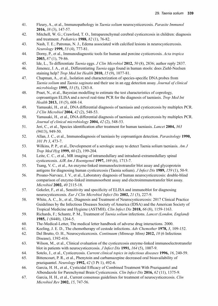
29. Taenia solium 339
41. Fleury, A., et al., Immunopathology in Taenia solium neurocysticercosis. Parasite Immunol 2016, 38 (3), 147-57.
42. Mitchell, W. G.; Crawford, T. O., Intraparenchymal cerebral cysticercosis in children: diagnosis and treatment. Pediatrics 1988, 82 (1), 76-82.
43. Nash, T. E.; Patronas, N. J., Edema associated with calcified lesions in neurocysticercosis. Neurology 1999, 53 (4), 777-81.
44. Dorny, P., et al., Immunodiagnostic tools for human and porcine cysticercosis. Acta tropica 2003, 87 (1), 79-86.
45. Ide, L., To differentiate Taenia eggs. J Clin Microbiol 2012, 50 (8), 2836; author reply 2837.46. Jimenez, J. A., et al., Differentiating Taenia eggs found in human stools: does Ziehl-Neelsen
staining help? Trop Med Int Health 2010, 15 (9), 1077-81.47. Chapman, A., et al., Isolation and characterization of species-specific DNA probes from
Taenia solium and Taenia saginata and their use in an egg detection assay. Journal of clinical microbiology 1995, 33 (5), 1283-8.
48. Praet, N., et al., Bayesian modelling to estimate the test characteristics of coprology, coproantigen ELISA and a novel real-time PCR for the diagnosis of taeniasis. Trop Med Int Health 2013, 18 (5), 608-14.
49. Yamasaki, H., et al., DNA differential diagnosis of taeniasis and cysticercosis by multiplex PCR. J Clin Microbiol 2004, 42 (2), 548-53.
50. Yamasaki, H., et al., DNA differential diagnosis of taeniasis and cysticercosis by multiplex PCR. Journal of clinical microbiology 2004, 42 (2), 548-53.
51. Jeri, C., et al., Species identification after treatment for human taeniasis. Lancet 2004, 363 (9413), 949-50.
52. Allan, J. C., et al., Immunodiagnosis of taeniasis by coproantigen detection. Parasitology 1990, 101 Pt 3, 473-7.
53. Wilkins, P. P., et al., Development of a serologic assay to detect Taenia solium taeniasis. Am J Trop Med Hyg 1999, 60 (2), 199-204.
54. Leite, C. C., et al., MR imaging of intramedullary and intradural-extramedullary spinal cysticercosis. AJR Am J Roentgenol 1997, 169 (6), 1713-7.
55. Tsang, V. C., et al., An enzyme-linked immunoelectrotransfer blot assay and glycoprotein antigens for diagnosing human cysticercosis (Taenia solium). J Infect Dis 1989, 159 (1), 50-9.
56. Proano-Narvaez, J. V., et al., Laboratory diagnosis of human neurocysticercosis: double-blind comparison of enzyme-linked immunosorbent assay and electroimmunotransfer blot assay. Microbiol 2002, 40 2115-18.
57. Gekeler, F., et al., Sensitivity and specificity of ELISA and immunoblot for diagnosing neurocysticercosis. Eur J Clin Microbiol Infect Dis 2002, 21 (3), 227-9.
58. White, A. C., Jr., et al., Diagnosis and Treatment of Neurocysticercosis: 2017 Clinical Practice Guidelines by the Infectious Diseases Society of America (IDSA) and the American Society of Tropical Medicine and Hygiene (ASTMH). Clin Infect Dis 2018, 66 (8), 1159-1163.
59. Richards, F.; Schantz, P. M., Treatment of Taenia solium infections. Lancet (London, England) 1985, 1 (8440), 1264-5.
60. The-Medical-Letter, The medical letter handbook of adverse drug interactions. 2000.61. Keeling, J. E. D., The chemotherapy of cestode infections. Adv Chemother 1978, 3, 109-152.62. Del Brutto, O. H., Neurocysticercosis. Continuum (Minneap Minn) 2012, 18 (6 Infectious
Disease), 1392-416.63. Wilson, M., et al., Clinical evaluation of the cysticercosis enzyme-linked immunoelectrotransfer
blot in patients with neurocysticercosis. J Infect Dis 1991, 164 (5), 1007-9.64. Sotelo, J., et al., Cysticercosis. Current clinical topics in infectious diseases 1996, 16, 240-59.65. Bittencourt, P. R., et al., Phenytoin and carbamazepine decreased oral bioavailability of
praziquantel. Neurology 1992, 42 (3 Pt 1), 492-6.66. Garcia, H. H., et al., Cysticidal Efficacy of Combined Treatment With Praziquantel and
Albendazole for Parenchymal Brain Cysticercosis. Clin Infect Dis 2016, 62 (11), 1375-9.67. Garcia, H. H., et al., Current consensus guidelines for treatment of neurocysticercosis. Clin
Microbiol Rev 2002, 15, 747-56.
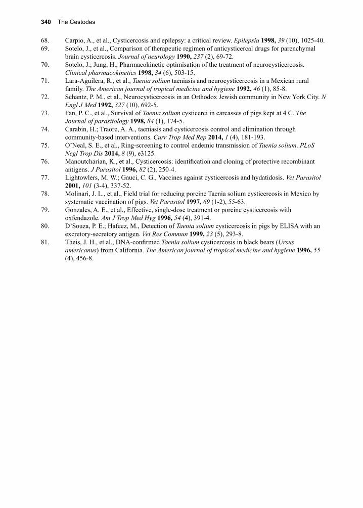
340 The Cestodes
68. Carpio, A., et al., Cysticercosis and epilepsy: a critical review. Epilepsia 1998, 39 (10), 1025-40.69. Sotelo, J., et al., Comparison of therapeutic regimen of anticysticercal drugs for parenchymal
brain cysticercosis. Journal of neurology 1990, 237 (2), 69-72.70. Sotelo, J.; Jung, H., Pharmacokinetic optimisation of the treatment of neurocysticercosis.
Clinical pharmacokinetics 1998, 34 (6), 503-15.71. Lara-Aguilera, R., et al., Taenia solium taeniasis and neurocysticercosis in a Mexican rural
family. The American journal of tropical medicine and hygiene 1992, 46 (1), 85-8.72. Schantz, P. M., et al., Neurocysticercosis in an Orthodox Jewish community in New York City. N
Engl J Med 1992, 327 (10), 692-5.73. Fan, P. C., et al., Survival of Taenia solium cysticerci in carcasses of pigs kept at 4 C. The
Journal of parasitology 1998, 84 (1), 174-5.74. Carabin, H.; Traore, A. A., taeniasis and cysticercosis control and elimination through
community-based interventions. Curr Trop Med Rep 2014, 1 (4), 181-193.75. O’Neal, S. E., et al., Ring-screening to control endemic transmission of Taenia solium. PLoS
Negl Trop Dis 2014, 8 (9), e3125.76. Manoutcharian, K., et al., Cysticercosis: identification and cloning of protective recombinant
antigens. J Parasitol 1996, 82 (2), 250-4.77. Lightowlers, M. W.; Gauci, C. G., Vaccines against cysticercosis and hydatidosis. Vet Parasitol
2001, 101 (3-4), 337-52.78. Molinari, J. L., et al., Field trial for reducing porcine Taenia solium cysticercosis in Mexico by
systematic vaccination of pigs. Vet Parasitol 1997, 69 (1-2), 55-63.79. Gonzales, A. E., et al., Effective, single-dose treatment or porcine cysticercosis with
oxfendazole. Am J Trop Med Hyg 1996, 54 (4), 391-4.80. D’Souza, P. E.; Hafeez, M., Detection of Taenia solium cysticercosis in pigs by ELISA with an
excretory-secretory antigen. Vet Res Commun 1999, 23 (5), 293-8.81. Theis, J. H., et al., DNA-confirmed Taenia solium cysticercosis in black bears (Ursus
americanus) from California. The American journal of tropical medicine and hygiene 1996, 55 (4), 456-8.
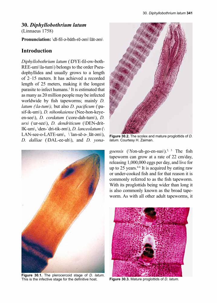
30. Diphyllobothrium latum 341
Figure 30.2. The scolex and mature proglottids of D. latum. Courtesy H. Zaiman.
Figure 30.3. Mature proglottids of D. latum.Figure 30.1. The plerocercoid stage of D. latum. This is the infective stage for the definitive host.
30. Diphyllobothrium latum(Linnaeus 1758)
Pronunciation: \dī-fil-ə-bäth-rē-əm\\lät-əm\
Introduction
Diphyllobothrium latum (\DYE-fil-ow-both-REE-um\\la-tum\) belongs to the order Pseu-dophyllidea and usually grows to a length of 2–15 meters. It has achieved a recorded length of 25 meters, making it the longest parasite to infect humans.1 It is estimated that as many as 20 million people may be infected worldwide by fish tapeworms; mainly D. latum (\la-tum\), but also D. pacificum (\pa-sif-ik-um\), D. nihonkaiense (Nee-hon-keye-en-see\), D. cordatum (\core-dah-tum\), D. ursi (\ur-see\), D. dendriticum (\DEN-drit-IK-um\, \den-ˈdri-tik-əm\), D. lanceolatum (\LAN-see-o-LATE-um\, \ˈlan-sē-ə-ˌlāt-əm\), D. dalliae (\DAL-ee-uh\), and D. yona-
goensis (\Yon-uh-go-en-sus\).2, 3 The fish tapeworm can grow at a rate of 22 cm/day, releasing 1,000,000 eggs per day, and live for up to 25 years.4-6 It is acquired by eating raw or under-cooked fish and for that reason it is commonly referred to as the fish tapeworm. With its proglottids being wider than long it is also commonly known as the broad tape-worm. As with all other adult tapeworms, it
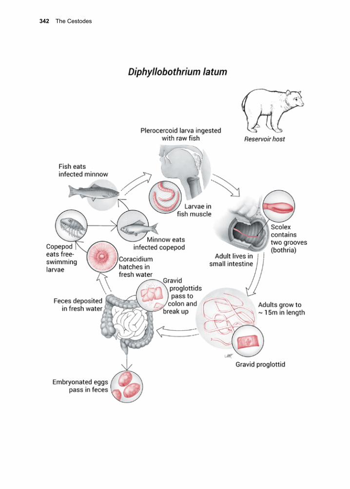
342 The Cestodes
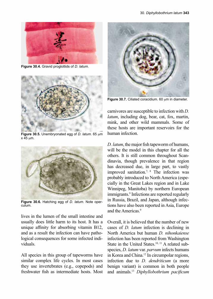
30. Diphyllobothrium latum 343
Figure 30.4. Gravid proglottids of D. latum.
Figure 30.5. Unembryonated egg of D. latum. 65 µm x 45 µm.
Figure 30.6. Hatching egg of D. latum. Note oper-culum.
Figure 30.7. Ciliated coracidium. 60 µm in diameter.
lives in the lumen of the small intestine and usually does little harm to its host. It has a unique affinity for absorbing vitamin B12, and as a result the infection can have patho-logical consequences for some infected indi-viduals.
All species in this group of tapeworms have similar complex life cycles. In most cases they use invertebrates (e.g., copepods) and freshwater fish as intermediate hosts. Most
carnivores are susceptible to infection with D. latum, including dog, bear, cat, fox, martin, mink, and other wild mammals. Some of these hosts are important reservoirs for the human infection.
D. latum, the major fish tapeworm of humans, will be the model in this chapter for all the others. It is still common throughout Scan-dinavia, though prevalence in that region has decreased due, in large part, to vastly improved sanitation.7, 8 The infection was probably introduced to North America (espe-cially in the Great Lakes region and in Lake Winnipeg, Manitoba) by northern European immigrants.9 Infections are reported regularly in Russia, Brazil, and Japan, although infec-tions have also been reported in Asia, Europe and the Americas.9
Overall, it is believed that the number of new cases of D. latum infection is declining in North America but human D. nihonkaiense infection has been reported from Washington State in the United States.10, 11 A related sub-species, D. latum var. parvum infects humans in Korea and China.12 In circumpolar regions, infection due to D. dendriticum (a more benign variant) is common in both people and animals.13 Diphyllobothrium pacificum
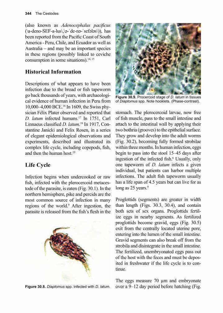
344 The Cestodes
Figure 30.8. Diaptomus spp. infected with D. latum.
Figure 30.9. Procercoid stage of D. latum in tissues of Diaptomus spp. Note hooklets. (Phase-contrast).
(also known as Adenocephalus pacificus (\a-deno-SEF-a-lus\,\ə-ˈde-nə-ˈsefələs\)), has been reported from the Pacific Coast of South America - Peru, Chile, and Ecuador as well as Australia – and may be an important species in these regions (possibly linked to ceviche consumption in some situations).14, 15
Historical Information
Descriptions of what appears to have been infection due to the broad or fish tapeworm go back thousands of years, with archaeologi-cal evidence of human infection in Peru from 10,000–4,000 BCE.16 In 1609, the Swiss phy-sician Félix Plater observed and reported that D. latum infected humans.17 In 1751, Carl Linnaeus classified D. latum.18 In 1917, Con-stantine Janicki and Felix Rosen, in a series of elegant epidemiological observations and experiments, described and illustrated its complex life cycle, including copepods, fish, and then the human host.19
Life Cycle
Infection begins when undercooked or raw fish, infected with the plerocercoid metaces-tode of the parasite, is eaten (Fig. 30.1). In the northern hemisphere, pike and percids are the most common source of infection in many regions of the world.9 After ingestion, the parasite is released from the fish’s flesh in the
stomach. The plerocercoid larvae, now free of fish muscle, pass to the small intestine and attach to the intestinal wall by applying their two bothria (grooves) to the epithelial surface. They grow and develop into the adult worms (Fig. 30.2), becoming fully formed strobilae within three months. In human infection, eggs begin to pass into the stool 15–45 days after ingestion of the infected fish.6 Usually, only one tapeworm of D. latum infects a given individual, but patients can harbor multiple infections. The adult fish tapeworm usually has a life span of 4.5 years but can live for as long as 25 years.9
Proglottids (segments) are greater in width than length (Figs. 30.3, 30.4), and contain both sets of sex organs. Proglottids fertil-ize eggs in nearby segments. As fertilized proglottids become gravid, eggs (Fig. 30.5) exit from the centrally located uterine pore, entering into the lumen of the small intestine. Gravid segments can also break off from the strobila and disintegrate in the small intestine. The fertilized, unembryonated eggs pass out of the host with the feces and must be depos-ited in freshwater if the life cycle is to con-tinue.
The eggs measure 70 µm and embryonate over a 9–12 day period before hatching (Fig.
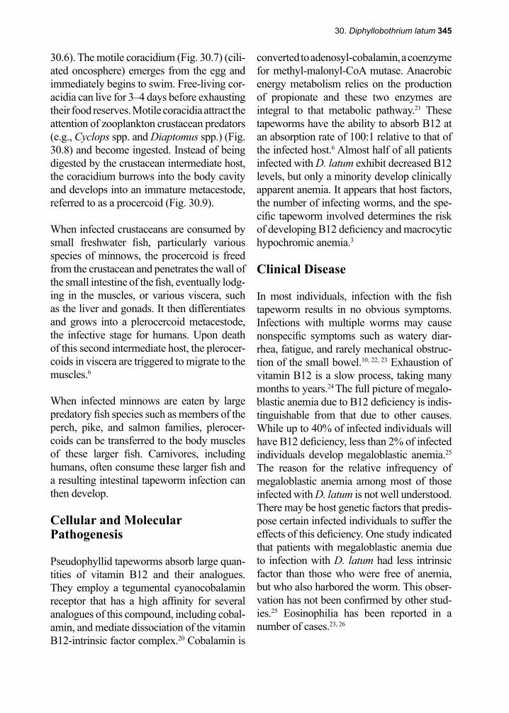
30. Diphyllobothrium latum 345
30.6). The motile coracidium (Fig. 30.7) (cili-ated oncosphere) emerges from the egg and immediately begins to swim. Free-living cor-acidia can live for 3–4 days before exhausting their food reserves. Motile coracidia attract the attention of zooplankton crustacean predators (e.g., Cyclops spp. and Diaptomus spp.) (Fig. 30.8) and become ingested. Instead of being digested by the crustacean intermediate host, the coracidium burrows into the body cavity and develops into an immature metacestode, referred to as a procercoid (Fig. 30.9).
When infected crustaceans are consumed by small freshwater fish, particularly various species of minnows, the procercoid is freed from the crustacean and penetrates the wall of the small intestine of the fish, eventually lodg-ing in the muscles, or various viscera, such as the liver and gonads. It then differentiates and grows into a plerocercoid metacestode, the infective stage for humans. Upon death of this second intermediate host, the plerocer-coids in viscera are triggered to migrate to the muscles.6
When infected minnows are eaten by large predatory fish species such as members of the perch, pike, and salmon families, plerocer-coids can be transferred to the body muscles of these larger fish. Carnivores, including humans, often consume these larger fish and a resulting intestinal tapeworm infection can then develop.
Cellular and Molecular Pathogenesis
Pseudophyllid tapeworms absorb large quan-tities of vitamin B12 and their analogues. They employ a tegumental cyanocobalamin receptor that has a high affinity for several analogues of this compound, including cobal-amin, and mediate dissociation of the vitamin B12-intrinsic factor complex.20 Cobalamin is
converted to adenosyl-cobalamin, a coenzyme for methyl-malonyl-CoA mutase. Anaerobic energy metabolism relies on the production of propionate and these two enzymes are integral to that metabolic pathway.21 These tapeworms have the ability to absorb B12 at an absorption rate of 100:1 relative to that of the infected host.6 Almost half of all patients infected with D. latum exhibit decreased B12 levels, but only a minority develop clinically apparent anemia. It appears that host factors, the number of infecting worms, and the spe-cific tapeworm involved determines the risk of developing B12 deficiency and macrocytic hypochromic anemia.3
Clinical Disease
In most individuals, infection with the fish tapeworm results in no obvious symptoms. Infections with multiple worms may cause nonspecific symptoms such as watery diar-rhea, fatigue, and rarely mechanical obstruc-tion of the small bowel.10, 22, 23 Exhaustion of vitamin B12 is a slow process, taking many months to years.24 The full picture of megalo-blastic anemia due to B12 deficiency is indis-tinguishable from that due to other causes. While up to 40% of infected individuals will have B12 deficiency, less than 2% of infected individuals develop megaloblastic anemia.25 The reason for the relative infrequency of megaloblastic anemia among most of those infected with D. latum is not well understood. There may be host genetic factors that predis-pose certain infected individuals to suffer the effects of this deficiency. One study indicated that patients with megaloblastic anemia due to infection with D. latum had less intrinsic factor than those who were free of anemia, but who also harbored the worm. This obser-vation has not been confirmed by other stud-ies.25 Eosinophilia has been reported in a number of cases.23, 26
346 The Cestodes
difficult, if not impossible. On an individual basis, the best way to avoid infection with D. latum is to avoid eating freshwater fish unless it is well-cooked or has been previ-ously frozen. Sushi is predominantly made from saltwater fish species that do not acquire D. latum or related tapeworm species. A few cases have been reported from eating sushi prepared from Pacific coast salmon, which spend a portion of their life in freshwater.28
The culinary habit among many Jewish mothers or grandmothers of teaching their daughters to prepare gefilte fish by tasting the raw mixture led many a female to acquire this infection in the United States, and was popularized in a medical anthropological description by Robert S. Desowitz.29 Today, proper disposal of human feces in the Great Lakes region of the United States has greatly reduced prevalence of D. latum. In Scandi-navian countries, gravlax and a wide variety of other marinated raw fish dishes, remain sources of infection for this tapeworm.
References
1. von Bonsdorff, B., Diphyllobothriasis in man. Academic Press, New York, NY. 1977.2. Chai, J. Y., et al., Fish-borne parasitic zoonoses: status and issues. Int J Parasitol 2005, 35 (11-
12), 1233-54.3. Jimenez, J. A., et al., Diphyllobothrium pacificum infection is seldom associated with
megaloblastic anemia. Am J Trop Med Hyg 2012, 87 (5), 897-901.4. Kuhlow, F., [Studies on the development of Diphyllobothrium latum]. Z Tropenmed Parasitol
1955, 6 (2), 213-25.5. Dogiel, V. A., Allgemeine Parasitologie. Parasitol. Schriftenr. 1963, 16, 1-523.6. Scholz, T., et al., Update on the human broad tapeworm (genus Diphyllobothrium), including
clinical relevance. Clin Microbiol Rev 2009, 22 (1), 146-60, Table of Contents.7. Kyronseppa, H.; J., The occurrence of human intestinal parasites in Finland. Scand Dis 1993, 25
(5 ), 671-3.8. Dick, T. A., et al., Diphyllobothriasis: update on human cases, foci, patterns and sources of
human infections and future considerations. Southeast Asian J Trop Med Public Health 2001, 32 Suppl 2, 59-76.
9. Dick, T. A., et al., Diphyllobothriasis: update on human cases, foci, patterns and sources of human infections and future considerations. The Southeast Asian journal of tropical medicine and public health 2001, 32 Suppl 2, 59-76.
10. Shimizu, T., et al., Diphyllobothrium nihonkaiense infection linked to chilled salmon consumption. BMJ Case Rep 2012, 2012.
11. Torres, P., et al., [New records of human diphyllobothriasis in Chile (1981-1992), with a case of multiple Diphyllobothrium latum infection]. Boletin chileno de parasitologia 1993, 48 (3-4), 39-43.
Diagnosis (see Clinical Appendix)
Segments of worm in stool (Figs. 30.3, 30.4) sometimes alert patients to the fact that they harbor a tapeworm, but most proglottids break up in the intestinal tract before exit-ing the host. Diagnosis is typically made by microscopic identification of non-embryo-nated eggs (Figs. 30.5, C.58.) in stool. Molec-ular tests are available for diagnosis that also allow for species determination, but most diagnosis is done using visual microscopy.6
Treatment (see Clinical Appendix)
The drug of choice is praziquantel (5–10 mg/kg once).27 Niclosamide is an alternative ther-apy that appears to be effective.6
Prevention and Control
In sylvatic settings, numerous reservoir hosts potentially sustain the life cycle. Interference with this phase of its ecology would be very
30. Diphyllobothrium latum 347
12. Lee, S. H., et al., Two rare cases of Diphyllobothrium latum parvum type infection in Korea. The Korean journal of parasitology 1994, 32 (2), 117-20.
13. Curtis, M. A.; Bylund, G., Diphyllobothriasis: fish tapeworm disease in the circumpolar north. Arctic medical research 1991, 50 (1), 18-24.
14. Kuchta, R., et al., Pacific Broad Tapeworm Adenocephalus pacificus as a Causative Agent of Globally Reemerging Diphyllobothriosis. Emerg Infect Dis 2015, 21 (10), 1697-703.
15. Moore, C. V., et al., Rare Human Infection with pacific broad tapeworm Adenocephalus pacificus, Australia. Emerg Infect Dis 2016, 22 (8), 1510-2.
16. Reinhard, K. J.; Barnum, S. V., Parasitology as an interpretative tool in archaeology. Am. Antiq 1991, 57, 231–245.
17. Plater, F., Praxeos seu de cognoscendis, praedicendis praecudensis curandiso affectibus homini incommodantibus tractatus tertius et ultimus. De vitiis libris duobus agens quorum primum corpis secundus Exretorum vitia continet Typis Conradi Waldkirchii 1609.
18. Linnaeus, C., Taenia osculis lateralibus solitaris. Amoenitates Academicae Holmiae Laurentium Salvium 2, 80-81.
19. Janicki, C., Le cycle evolutif du Dibothriocephalus latus L. Soc Neuchateloise Sci Natur Bull 1917, 42, 19-53.
20. Friedman, P. A., et al., Characterization of cobalamin receptor sites in brush-border plasma membranes of the tapeworm Spirometra mansonoides. The Journal of biological chemistry 1983, 258 (7), 4261-5.
21. Smyth, J. D.; McManus, D. P., The Physiology and Biochemistry of Tapeworms. 1989.22. Lee, S. H., et al., Diphyllobothrium latum infection in a child with recurrent abdominal pain.
Korean J Pediatr 2015, 58 (11), 451-3.23. Nawa, Y., et al., Sushi delights and parasites: the risk of fishborne and foodborne parasitic
zoonoses in Asia. Clin Infect Dis 2005, 41 (9), 1297-303.24. Goodman, K. I.; Salt, W. B., Vitamin B12 deficiency. Important new concepts in recognition.
Postgraduate medicine 1990, 88 (3), 147-50, 153.25. Saarni, M., et al., Symptoms in carriers of Diphyllobothrium latum and in uninfected controls.
Acta Med Scand 1963, 173, 147-154.26. Arizono, N., et al., Diphyllobothriasis associated with eating raw pacific salmon. Emerg Infect
Dis 2009, 15 (6), 866-70.27. Groll, E., Praziquantel for cestode infections in man. Acta Trop 1980, 37 (3), 293-6.28. Hutchinson, J. W., et al., Diphyllobothriasis after eating raw salmon. Hawaii medical journal
1997, 56 (7), 176-7.29. Desowitz, R. S.; A., New Guinea Tapeworms and Jewish Grandmothers: of Parasites and People.
W W Norton Co Pubs 1981.
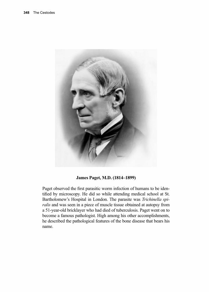
348 The Cestodes
James Paget, M.D. (1814–1899)
Paget observed the first parasitic worm infection of humans to be iden-tified by microscopy. He did so while attending medical school at St. Bartholomew’s Hospital in London. The parasite was Trichinella spi-ralis and was seen in a piece of muscle tissue obtained at autopsy from a 51-year-old bricklayer who had died of tuberculosis. Paget went on to become a famous pathologist. High among his other accomplishments, he described the pathological features of the bone disease that bears his name.
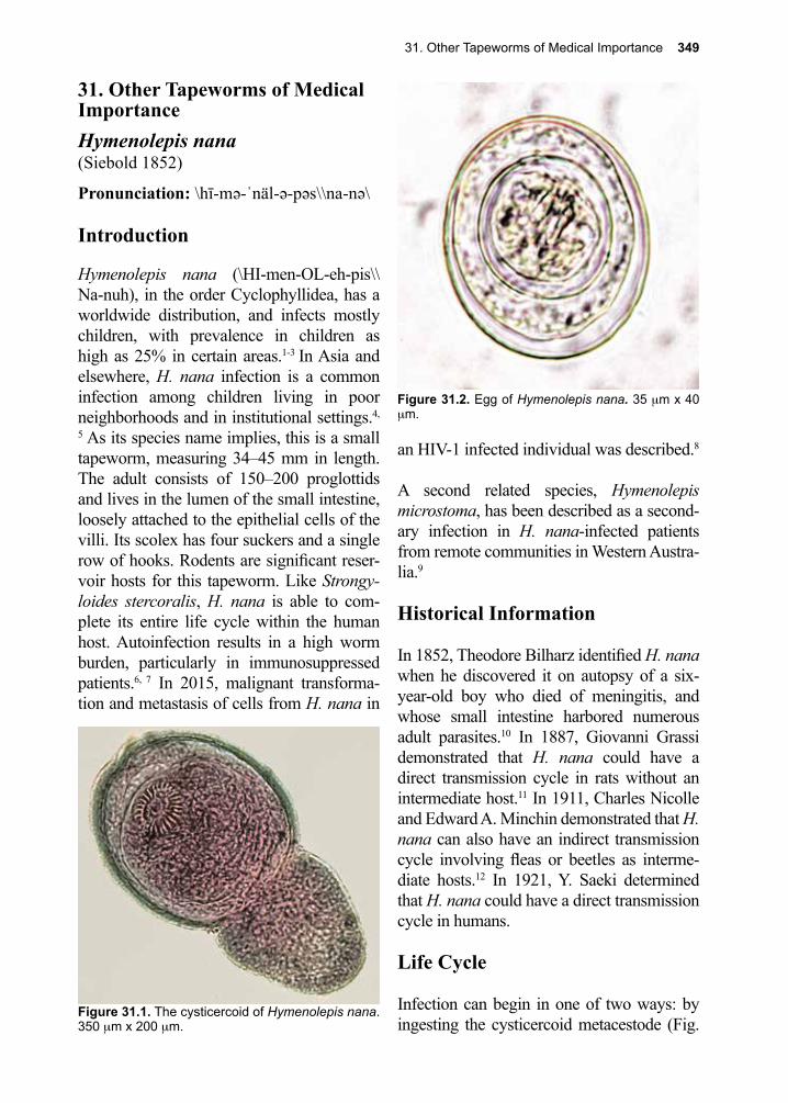
31. Other Tapeworms of Medical Importance 349
Figure 31.1. The cysticercoid of Hymenolepis nana. 350 µm x 200 µm.
Figure 31.2. Egg of Hymenolepis nana. 35 µm x 40 µm.
31. Other Tapeworms of Medical ImportanceHymenolepis nana (Siebold 1852)
Pronunciation: \hī-mə-ˈnäl-ə-pəs\\na-nə\
Introduction
Hymenolepis nana (\HI-men-OL-eh-pis\\Na-nuh), in the order Cyclophyllidea, has a worldwide distribution, and infects mostly children, with prevalence in children as high as 25% in certain areas.1-3 In Asia and elsewhere, H. nana infection is a common infection among children living in poor neighborhoods and in institutional settings.4,
5 As its species name implies, this is a small tapeworm, measuring 34–45 mm in length. The adult consists of 150–200 proglottids and lives in the lumen of the small intestine, loosely attached to the epithelial cells of the villi. Its scolex has four suckers and a single row of hooks. Rodents are significant reser-voir hosts for this tapeworm. Like Strongy-loides stercoralis, H. nana is able to com-plete its entire life cycle within the human host. Autoinfection results in a high worm burden, particularly in immunosuppressed patients.6, 7 In 2015, malignant transforma-tion and metastasis of cells from H. nana in
an HIV-1 infected individual was described.8
A second related species, Hymenolepis microstoma, has been described as a second-ary infection in H. nana-infected patients from remote communities in Western Austra-lia.9
Historical Information
In 1852, Theodore Bilharz identified H. nana when he discovered it on autopsy of a six-year-old boy who died of meningitis, and whose small intestine harbored numerous adult parasites.10 In 1887, Giovanni Grassi demonstrated that H. nana could have a direct transmission cycle in rats without an intermediate host.11 In 1911, Charles Nicolle and Edward A. Minchin demonstrated that H. nana can also have an indirect transmission cycle involving fleas or beetles as interme-diate hosts.12 In 1921, Y. Saeki determined that H. nana could have a direct transmission cycle in humans.
Life Cycle
Infection can begin in one of two ways: by ingesting the cysticercoid metacestode (Fig.
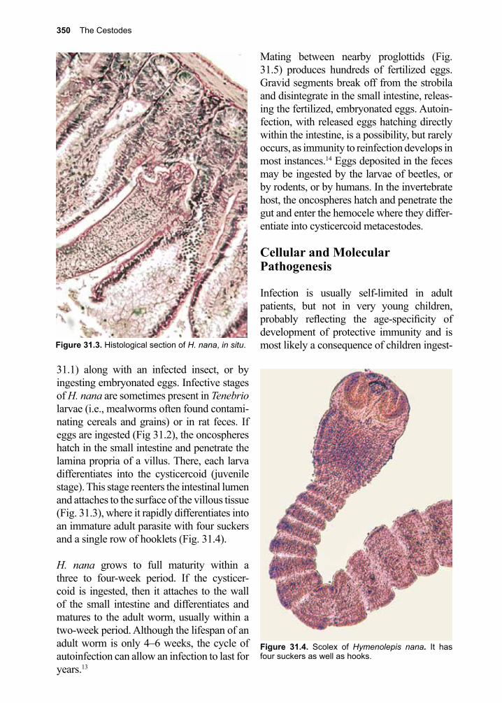
350 The Cestodes
Figure 31.4. Scolex of Hymenolepis nana. It has four suckers as well as hooks.
Figure 31.3. Histological section of H. nana, in situ.
31.1) along with an infected insect, or by ingesting embryonated eggs. Infective stages of H. nana are sometimes present in Tenebrio larvae (i.e., mealworms often found contami-nating cereals and grains) or in rat feces. If eggs are ingested (Fig 31.2), the oncospheres hatch in the small intestine and penetrate the lamina propria of a villus. There, each larva differentiates into the cysticercoid (juvenile stage). This stage reenters the intestinal lumen and attaches to the surface of the villous tissue (Fig. 31.3), where it rapidly differentiates into an immature adult parasite with four suckers and a single row of hooklets (Fig. 31.4).
H. nana grows to full maturity within a three to four-week period. If the cysticer-coid is ingested, then it attaches to the wall of the small intestine and differentiates and matures to the adult worm, usually within a two-week period. Although the lifespan of an adult worm is only 4–6 weeks, the cycle of autoinfection can allow an infection to last for years.13
Mating between nearby proglottids (Fig. 31.5) produces hundreds of fertilized eggs. Gravid segments break off from the strobila and disintegrate in the small intestine, releas-ing the fertilized, embryonated eggs. Autoin-fection, with released eggs hatching directly within the intestine, is a possibility, but rarely occurs, as immunity to reinfection develops in most instances.14 Eggs deposited in the feces may be ingested by the larvae of beetles, or by rodents, or by humans. In the invertebrate host, the oncospheres hatch and penetrate the gut and enter the hemocele where they differ-entiate into cysticercoid metacestodes.
Cellular and Molecular Pathogenesis
Infection is usually self-limited in adult patients, but not in very young children, probably reflecting the age-specificity of development of protective immunity and is most likely a consequence of children ingest-
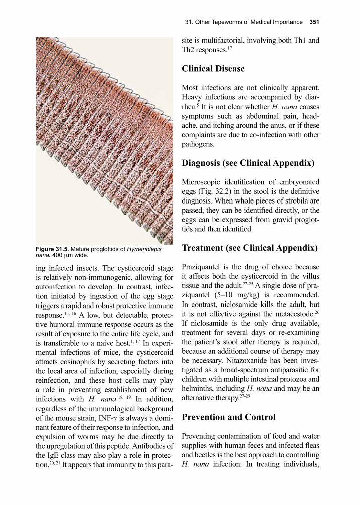
31. Other Tapeworms of Medical Importance 351
Figure 31.5. Mature proglottids of Hymenolepis nana. 400 µm wide.
ing infected insects. The cysticercoid stage is relatively non-immunogenic, allowing for autoinfection to develop. In contrast, infec-tion initiated by ingestion of the egg stage triggers a rapid and robust protective immune response.15, 16 A low, but detectable, protec-tive humoral immune response occurs as the result of exposure to the entire life cycle, and is transferable to a naive host.1, 17 In experi-mental infections of mice, the cysticercoid attracts eosinophils by secreting factors into the local area of infection, especially during reinfection, and these host cells may play a role in preventing establishment of new infections with H. nana.18, 19 In addition, regardless of the immunological background of the mouse strain, INF-γ is always a domi-nant feature of their response to infection, and expulsion of worms may be due directly to the upregulation of this peptide. Antibodies of the IgE class may also play a role in protec-tion.20, 21 It appears that immunity to this para-
site is multifactorial, involving both Th1 and Th2 responses.17
Clinical Disease
Most infections are not clinically apparent. Heavy infections are accompanied by diar-rhea.5 It is not clear whether H. nana causes symptoms such as abdominal pain, head-ache, and itching around the anus, or if these complaints are due to co-infection with other pathogens.
Diagnosis (see Clinical Appendix)
Microscopic identification of embryonated eggs (Fig. 32.2) in the stool is the definitive diagnosis. When whole pieces of strobila are passed, they can be identified directly, or the eggs can be expressed from gravid proglot-tids and then identified.
Treatment (see Clinical Appendix)
Praziquantel is the drug of choice because it affects both the cysticercoid in the villus tissue and the adult.22-25 A single dose of pra-ziquantel (5–10 mg/kg) is recommended. In contrast, niclosamide kills the adult, but it is not effective against the metacestode.26 If niclosamide is the only drug available, treatment for several days or re-examining the patient’s stool after therapy is required, because an additional course of therapy may be necessary. Nitazoxanide has been inves-tigated as a broad-spectrum antiparasitic for children with multiple intestinal protozoa and helminths, including H. nana and may be an alternative therapy.27-29
Prevention and Control
Preventing contamination of food and water supplies with human feces and infected fleas and beetles is the best approach to controlling H. nana infection. In treating individuals,
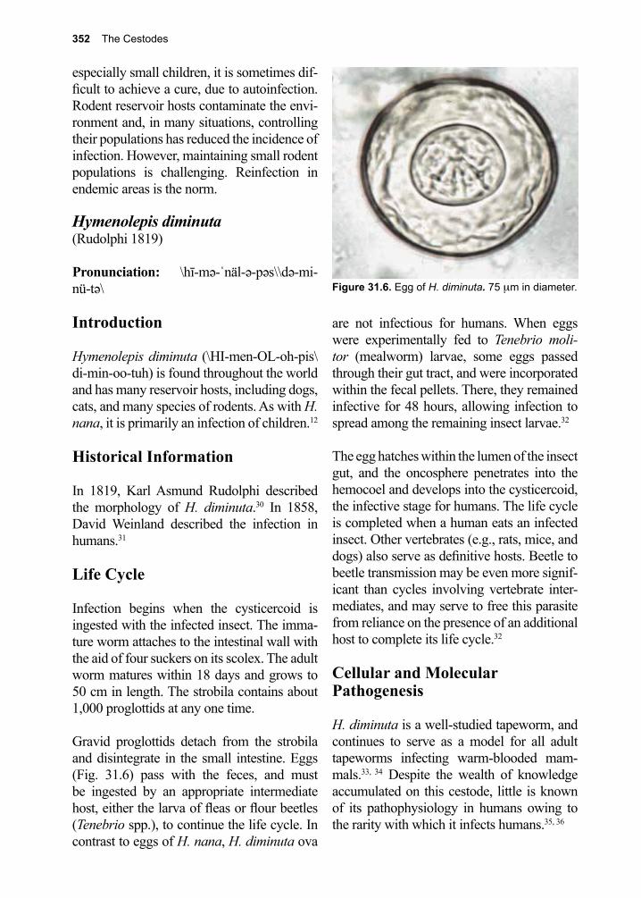
352 The Cestodes
especially small children, it is sometimes dif-ficult to achieve a cure, due to autoinfection. Rodent reservoir hosts contaminate the envi-ronment and, in many situations, controlling their populations has reduced the incidence of infection. However, maintaining small rodent populations is challenging. Reinfection in endemic areas is the norm.
Hymenolepis diminuta (Rudolphi 1819)
Pronunciation: \hī-mə-ˈnäl-ə-pəs\\də-mi-nü-tə\
Introduction
Hymenolepis diminuta (\HI-men-OL-oh-pis\di-min-oo-tuh) is found throughout the world and has many reservoir hosts, including dogs, cats, and many species of rodents. As with H. nana, it is primarily an infection of children.12
Historical Information
In 1819, Karl Asmund Rudolphi described the morphology of H. diminuta.30 In 1858, David Weinland described the infection in humans.31
Life Cycle
Infection begins when the cysticercoid is ingested with the infected insect. The imma-ture worm attaches to the intestinal wall with the aid of four suckers on its scolex. The adult worm matures within 18 days and grows to 50 cm in length. The strobila contains about 1,000 proglottids at any one time.
Gravid proglottids detach from the strobila and disintegrate in the small intestine. Eggs (Fig. 31.6) pass with the feces, and must be ingested by an appropriate intermediate host, either the larva of fleas or flour beetles (Tenebrio spp.), to continue the life cycle. In contrast to eggs of H. nana, H. diminuta ova
are not infectious for humans. When eggs were experimentally fed to Tenebrio moli-tor (mealworm) larvae, some eggs passed through their gut tract, and were incorporated within the fecal pellets. There, they remained infective for 48 hours, allowing infection to spread among the remaining insect larvae.32
The egg hatches within the lumen of the insect gut, and the oncosphere penetrates into the hemocoel and develops into the cysticercoid, the infective stage for humans. The life cycle is completed when a human eats an infected insect. Other vertebrates (e.g., rats, mice, and dogs) also serve as definitive hosts. Beetle to beetle transmission may be even more signif-icant than cycles involving vertebrate inter-mediates, and may serve to free this parasite from reliance on the presence of an additional host to complete its life cycle.32
Cellular and Molecular Pathogenesis
H. diminuta is a well-studied tapeworm, and continues to serve as a model for all adult tapeworms infecting warm-blooded mam-mals.33, 34 Despite the wealth of knowledge accumulated on this cestode, little is known of its pathophysiology in humans owing to the rarity with which it infects humans.35, 36
Figure 31.6. Egg of H. diminuta. 75 µm in diameter.
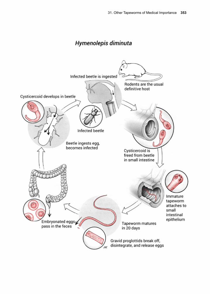
31. Other Tapeworms of Medical Importance 353
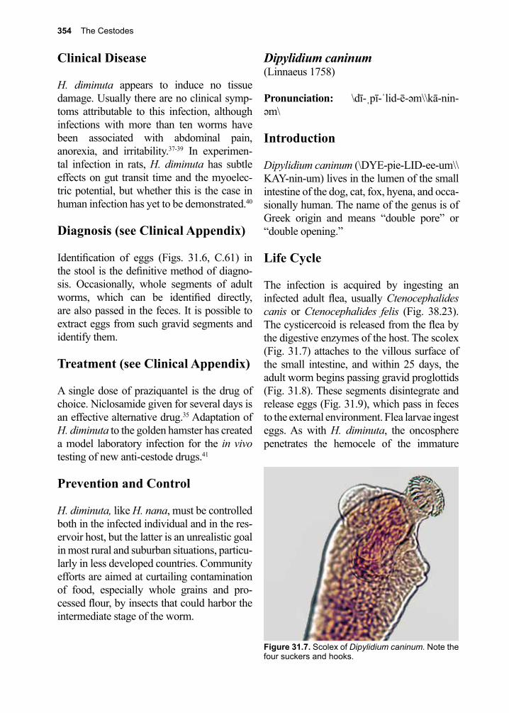
354 The Cestodes
Clinical Disease
H. diminuta appears to induce no tissue damage. Usually there are no clinical symp-toms attributable to this infection, although infections with more than ten worms have been associated with abdominal pain, anorexia, and irritability.37-39 In experimen-tal infection in rats, H. diminuta has subtle effects on gut transit time and the myoelec-tric potential, but whether this is the case in human infection has yet to be demonstrated.40
Diagnosis (see Clinical Appendix)
Identification of eggs (Figs. 31.6, C.61) in the stool is the definitive method of diagno-sis. Occasionally, whole segments of adult worms, which can be identified directly, are also passed in the feces. It is possible to extract eggs from such gravid segments and identify them.
Treatment (see Clinical Appendix)
A single dose of praziquantel is the drug of choice. Niclosamide given for several days is an effective alternative drug.35 Adaptation of H. diminuta to the golden hamster has created a model laboratory infection for the in vivo testing of new anti-cestode drugs.41
Prevention and Control
H. diminuta, like H. nana, must be controlled both in the infected individual and in the res-ervoir host, but the latter is an unrealistic goal in most rural and suburban situations, particu-larly in less developed countries. Community efforts are aimed at curtailing contamination of food, especially whole grains and pro-cessed flour, by insects that could harbor the intermediate stage of the worm.
Dipylidium caninum (Linnaeus 1758)
Pronunciation: \dī-ˌpī-ˈlid-ē-əm\\kā-nin- əm\
Introduction
Dipylidium caninum (\DYE-pie-LID-ee-um\\KAY-nin-um) lives in the lumen of the small intestine of the dog, cat, fox, hyena, and occa-sionally human. The name of the genus is of Greek origin and means “double pore” or “double opening.”
Life Cycle
The infection is acquired by ingesting an infected adult flea, usually Ctenocephalides canis or Ctenocephalides felis (Fig. 38.23). The cysticercoid is released from the flea by the digestive enzymes of the host. The scolex (Fig. 31.7) attaches to the villous surface of the small intestine, and within 25 days, the adult worm begins passing gravid proglottids (Fig. 31.8). These segments disintegrate and release eggs (Fig. 31.9), which pass in feces to the external environment. Flea larvae ingest eggs. As with H. diminuta, the oncosphere penetrates the hemocele of the immature
Figure 31.7. Scolex of Dipylidium caninum. Note the four suckers and hooks.
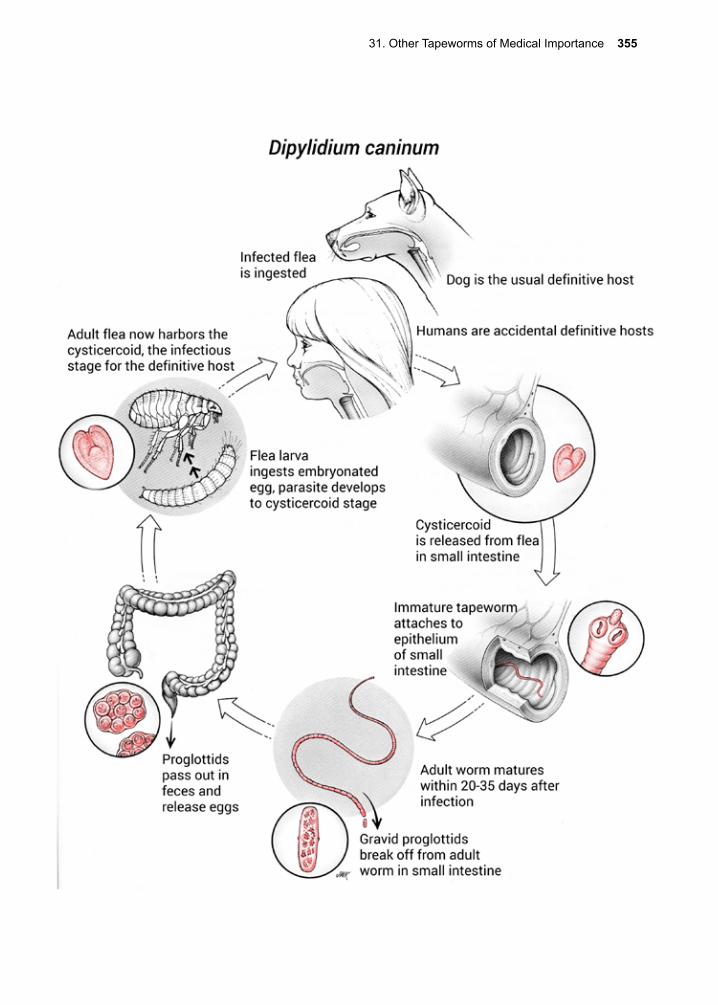
31. Other Tapeworms of Medical Importance 355
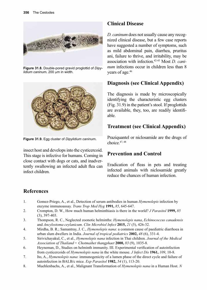
356 The Cestodes
References
1. Gomez-Priego, A., et al., Detection of serum antibodies in human Hymenolepis infection by enzyme immunoassay. Trans Trop Med Hyg 1991, 85, 645-647.
2. Crompton, D. W., How much human helminthiasis is there in the world? J Parasitol 1999, 85 (3), 397-403.
3. Thompson, R. C., Neglected zoonotic helminths: Hymenolepis nana, Echinococcus canadensis and Ancylostoma ceylanicum. Clin Microbiol Infect 2015, 21 (5), 426-32.
4. Mirdha, B. R.; Samantray, J. C., Hymenolepis nana: a common cause of paediatric diarrhoea in urban slum dwellers in India. Journal of tropical pediatrics 2002, 48 (6), 331-4.
5. Sirivichayakul, C., et al., Hymenolepis nana infection in Thai children. Journal of the Medical Association of Thailand = Chotmaihet thangphaet 2000, 83 (9), 1035-8.
6. Heyneman, D., Studies on helminth immunity. III. Experimental verification of autoinfection from cysticercoids of Hymenolepis nana in the white mouse. J Infect Dis 1961, 109, 10-8.
7. Ito, A., Hymenolepis nana: immunogenicity of a lumen phase of the direct cycle and failure of autoinfection in BALB/c mice. Exp Parasitol 1982, 54 (1), 113-20.
8. Muehlenbachs, A., et al., Malignant Transformation of Hymenolepis nana in a Human Host. N
insect host and develops into the cysticercoid. This stage is infective for humans. Coming in close contact with dogs or cats, and inadver-tently swallowing an infected adult flea can infect children.
Clinical Disease
D. caninum does not usually cause any recog-nized clinical disease, but a few case reports have suggested a number of symptoms, such as mild abdominal pain, diarrhea, pruritus ani, failure to thrive, and irritability, may be association with infection.42-45 Most D. cani-num infections occur in children less than 8 years of age.46
Diagnosis (see Clinical Appendix)
The diagnosis is made by microscopically identifying the characteristic egg clusters (Fig. 31.9) in the patient’s stool. If proglottids are available, they, too, are readily identifi-able.
Treatment (see Clinical Appendix)
Praziquantel or niclosamide are the drugs of choice.47, 48
Prevention and Control
Eradication of fleas in pets and treating infected animals with niclosamide greatly reduce the chances of human infection.
Figure 31.8. Double-pored gravid proglottid of Dipy-lidium caninum. 200 µm in width.
Figure 31.9. Egg cluster of Dipylidium caninum.
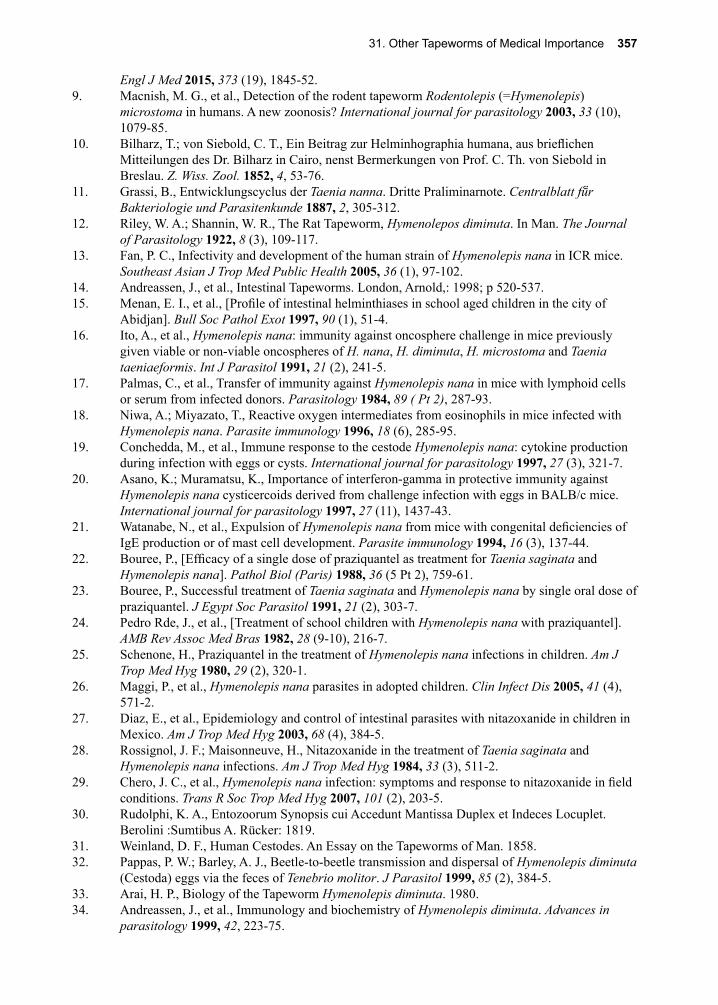
31. Other Tapeworms of Medical Importance 357
Engl J Med 2015, 373 (19), 1845-52.9. Macnish, M. G., et al., Detection of the rodent tapeworm Rodentolepis (=Hymenolepis)
microstoma in humans. A new zoonosis? International journal for parasitology 2003, 33 (10), 1079-85.
10. Bilharz, T.; von Siebold, C. T., Ein Beitrag zur Helminhographia humana, aus brieflichen Mitteilungen des Dr. Bilharz in Cairo, nenst Bermerkungen von Prof. C. Th. von Siebold in Breslau. Z. Wiss. Zool. 1852, 4, 53-76.
11. Grassi, B., Entwicklungscyclus der Taenia nanna. Dritte Praliminarnote. Centralblatt fṻr Bakteriologie und Parasitenkunde 1887, 2, 305-312.
12. Riley, W. A.; Shannin, W. R., The Rat Tapeworm, Hymenolepos diminuta. In Man. The Journal of Parasitology 1922, 8 (3), 109-117.
13. Fan, P. C., Infectivity and development of the human strain of Hymenolepis nana in ICR mice. Southeast Asian J Trop Med Public Health 2005, 36 (1), 97-102.
14. Andreassen, J., et al., Intestinal Tapeworms. London, Arnold,: 1998; p 520-537.15. Menan, E. I., et al., [Profile of intestinal helminthiases in school aged children in the city of
Abidjan]. Bull Soc Pathol Exot 1997, 90 (1), 51-4.16. Ito, A., et al., Hymenolepis nana: immunity against oncosphere challenge in mice previously
given viable or non-viable oncospheres of H. nana, H. diminuta, H. microstoma and Taenia taeniaeformis. Int J Parasitol 1991, 21 (2), 241-5.
17. Palmas, C., et al., Transfer of immunity against Hymenolepis nana in mice with lymphoid cells or serum from infected donors. Parasitology 1984, 89 ( Pt 2), 287-93.
18. Niwa, A.; Miyazato, T., Reactive oxygen intermediates from eosinophils in mice infected with Hymenolepis nana. Parasite immunology 1996, 18 (6), 285-95.
19. Conchedda, M., et al., Immune response to the cestode Hymenolepis nana: cytokine production during infection with eggs or cysts. International journal for parasitology 1997, 27 (3), 321-7.
20. Asano, K.; Muramatsu, K., Importance of interferon-gamma in protective immunity against Hymenolepis nana cysticercoids derived from challenge infection with eggs in BALB/c mice. International journal for parasitology 1997, 27 (11), 1437-43.
21. Watanabe, N., et al., Expulsion of Hymenolepis nana from mice with congenital deficiencies of IgE production or of mast cell development. Parasite immunology 1994, 16 (3), 137-44.
22. Bouree, P., [Efficacy of a single dose of praziquantel as treatment for Taenia saginata and Hymenolepis nana]. Pathol Biol (Paris) 1988, 36 (5 Pt 2), 759-61.
23. Bouree, P., Successful treatment of Taenia saginata and Hymenolepis nana by single oral dose of praziquantel. J Egypt Soc Parasitol 1991, 21 (2), 303-7.
24. Pedro Rde, J., et al., [Treatment of school children with Hymenolepis nana with praziquantel]. AMB Rev Assoc Med Bras 1982, 28 (9-10), 216-7.
25. Schenone, H., Praziquantel in the treatment of Hymenolepis nana infections in children. Am J Trop Med Hyg 1980, 29 (2), 320-1.
26. Maggi, P., et al., Hymenolepis nana parasites in adopted children. Clin Infect Dis 2005, 41 (4), 571-2.
27. Diaz, E., et al., Epidemiology and control of intestinal parasites with nitazoxanide in children in Mexico. Am J Trop Med Hyg 2003, 68 (4), 384-5.
28. Rossignol, J. F.; Maisonneuve, H., Nitazoxanide in the treatment of Taenia saginata and Hymenolepis nana infections. Am J Trop Med Hyg 1984, 33 (3), 511-2.
29. Chero, J. C., et al., Hymenolepis nana infection: symptoms and response to nitazoxanide in field conditions. Trans R Soc Trop Med Hyg 2007, 101 (2), 203-5.
30. Rudolphi, K. A., Entozoorum Synopsis cui Accedunt Mantissa Duplex et Indeces Locuplet. Berolini :Sumtibus A. Rücker: 1819.
31. Weinland, D. F., Human Cestodes. An Essay on the Tapeworms of Man. 1858.32. Pappas, P. W.; Barley, A. J., Beetle-to-beetle transmission and dispersal of Hymenolepis diminuta
(Cestoda) eggs via the feces of Tenebrio molitor. J Parasitol 1999, 85 (2), 384-5.33. Arai, H. P., Biology of the Tapeworm Hymenolepis diminuta. 1980.34. Andreassen, J., et al., Immunology and biochemistry of Hymenolepis diminuta. Advances in
parasitology 1999, 42, 223-75.
358 The Cestodes
35. Kalaivani, R., et al., Hymenolepis diminuta infection in a school-going child: A rare case report. Australas Med J 2014, 7 (9), 379-81.
36. Kilincel, O., et al., [A rare case of Hymenolepis diminuta infection in a small child]. Mikrobiyol Bul 2015, 49 (1), 135-8.
37. Wiwanitkit, V., Overview of Hymenolepis diminuta infection among Thai patients. MedGenMed : Medscape general medicine 2004, 6 (2), 7.
38. Hamrick, H. J., et al., Rat tapeworm (Hymenolepis diminuta) infection in a child. The Pediatric infectious disease journal 1990, 9 (3), 216-9.
39. Tena, D., et al., Human infection with Hymenolepis diminuta: case report from Spain. Journal of clinical microbiology 1998, 36 (8), 2375-6.
40. Dwinell, M. B., et al., Tapeworm infection decreases intestinal transit and enteric aerobic bacterial populations. The American journal of physiology 1997, 273 (2 Pt 1), G480-5.
41. Ostlind, D. A., et al., The Hymenolepis diminuta-golden hamster (Mesocricetus auratus) model for the evaluation of gastrointestinal anticestode activity. The Journal of parasitology 2004, 90 (4), 898-9.
42. Hamrick, H. J., et al., Two cases of dipylidiasis (dog tapeworm infection) in children: update on an old problem. Pediatrics 1983, 72 (1), 114-7.
43. Manyam, K. S., et al., Observations in a case of Dipylidium caninum infection. Vet Med Small Anim Clin 1980, 75 (1), 66.
44. Narasimham, M. V., et al., Dipylidium caninum infection in a child: a rare case report. Indian J Med Microbiol 2013, 31 (1), 82-4.
45. Szwaja, B., et al., A case of Dipylidium caninum infection in a child from the southeastern Poland. Wiad Parazytol 2011, 57 (3), 175-8.
46. Molina, C. P., et al., Infection by Dipylidium caninum in an infant. Archives of pathology & laboratory medicine 2003, 127 (3), e157-9.
47. Jones, W. E., Niclosamide as a treatment for Hymenolepis diminuta and Dipylidium caninum infection in man. Am J Trop Med Hyg 1979, 28 (2), 300-2.
48. Schenone, H., et al., [Infection by Dipylidium caninum in a young girl treated with praziquantel]. Bol Chil Parasitol 1987, 42 (3-4), 74-5.
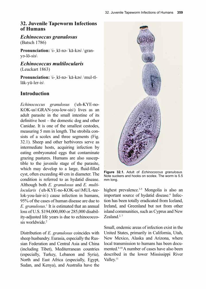
32. Juvenile Tapeworm Infections of Humans 359
32. Juvenile Tapeworm Infections of HumansEchinococcus granulosus (Batsch 1786)
Pronunciation: \i-ˌkī-nə-ˈkä-kəs\ \gran-yə-lō-sis\
Echinococcus multilocularis(Leuckart 1863)
Pronunciation: \i-ˌkī-nə-ˈkä-kəs\ \mul-tī-läk-yü-ler-is\
Introduction
Echinococcus granulosus (\eh-KYE-no-KOK-us\\GRAN-you-low-sis\) lives as an adult parasite in the small intestine of its definitive host – the domestic dog and other Canidae. It is one of the smallest cestodes, measuring 5 mm in length. The strobila con-sists of a scolex and three segments (Fig. 32.1). Sheep and other herbivores serve as intermediate hosts, acquiring infection by eating embryonated eggs that contaminate grazing pastures. Humans are also suscep-tible to the juvenile stage of the parasite, which may develop to a large, fluid-filled cyst, often exceeding 40 cm in diameter. The condition is referred to as hydatid disease. Although both E. granulosus and E. multi-locularis (\eh-KYE-no-KOK-us\\MUL-tee-lok-you-lair-is\) cause infection in humans, 95% of the cases of human disease are due to E. granulosus.1 It is estimated that an annual loss of U.S. $194,000,000 or 285,000 disabil-ity-adjusted life years is due to echinococco-sis worldwide.2
Distribution of E. granulosus coincides with sheep husbandry. Eurasia, especially the Rus-sian Federation and Central Asia and China (including Tibet), Mediterranean countries (especially, Turkey, Lebanon and Syria), North and East Africa (especially, Egypt, Sudan, and Kenya), and Australia have the
highest prevalence.3-5 Mongolia is also an important source of hydatid disease.6 Infec-tion has been totally eradicated from Iceland, Ireland, and Greenland but not from other island communities, such as Cyprus and New Zealand.2, 7
Small, endemic areas of infection exist in the United States, primarily in California, Utah, New Mexico, Alaska and Arizona, where local transmission to humans has been docu-mented.8-10 A number of cases have also been described in the lower Mississippi River Valley.11
Figure 32.1. Adult of Echinococcus granulosus. Note suckers and hooks on scolex. The worm is 5.5 mm long.
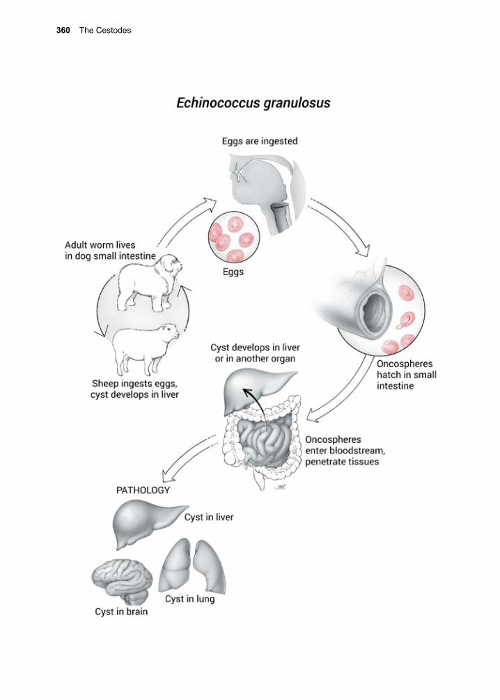
360 The Cestodes
32. Juvenile Tapeworm Infections of Humans 361
Indigenous peoples of the Canadian Arctic, especially the Inuit, are infected with a north-ern variant, E. granulosus var. canadensis, acquired in a sylvatic cycle involving moose and caribou (cervids), wolves, and sled dogs.3, 12, 13 Two cases reported from Alaska were unusually severe.13 In northern areas in certain Scandinavian countries herding of reindeer as well as artificial introduction of reindeer is associated with this infection.14
Historical Information
Descriptions of the massive cysts seen in slaughtered animals can be found in multi-ple ancient texts from Babylon, Greece, and Rome.15 In 1766, Pierre Pallas described the hydatid cyst of E. granulosus, in which he sketched cysts that he removed from the vis-cera of mice, and compared them with those recovered by others from human infections.16 In 1782, Johann Goeze, in his classic mono-graph, depicted the juveniles (i.e., protosco-lices).17 In 1853, Carl von Siebold described the adult worms in the dog and demonstrated that echinococcus cysts from sheep gave rise to adult tapeworms when fed to dogs.18
In 1863, Bernhard Naunyn fed the contents of hydatid cysts from a human infection to dogs, and recovered adult tapeworms in them.19 These experiments provided the essential link between the animal and human infection cycles.
Life Cycle
E. granulosus adults live in the canine small intestine. Multiple infections are the rule, with hundreds to thousands of adult worms occu-pying the greater portion of the upper half of the small intestine. The gravid segment (one per adult tapeworm) breaks off and disinte-grates in the large bowel, releasing hundreds of infective eggs (Figs. 28.5, 29.5), which then pass out with the feces. Sheep, as well
as other domestic animals (e.g., cattle, pigs, and horses), and humans acquire the juvenile stage by ingesting embryonated eggs. Each intermediate host species (sheep, cattle, pig, and horse) seems to have evolved a separate, genetically definable strain of parasite.20
The oncosphere hatches in the small intestine, enters the bloodstream, and in the vast major-ity of cases, reaches the liver via the portal circulation (Fig. 32.2).21-23 Other organs can also be invaded, including lung, brain, heart, bones, eyes and kidney.24-30 Once in the tissue, the larva synthesizes a hyaline mem-brane, and becomes surrounded by it. This membrane differentiates into an outer, acel-lular laminate structure, and an inner, cellular germinal layer (Fig. 32.3, 32.11). The inner surface of the germinal layer gives rise to protoscolices (Fig. 32.5) (i.e., the infectious stage for the definitive host), and more outer membrane material.
The hydatid cyst requires several months to years in order to develop, mature and fill with fluid (Fig. 32.6). The fluid is under pressure, and the wall, while substantial, can rupture if severely traumatized. The entire cyst can contain millions of protoscolices. The diam-eter of the mature outer cyst varies from 2–20 cm, and sometimes is even larger. The fluid portion of the cyst contains both host and parasite proteins. A canine host must ingest the hydatid cyst and its contents (i.e., the pro-toscolices) to complete the life cycle. This commonly occurs when infected sheep are slaughtered (Fig. 32.11), and organs contain-ing hydatid cysts are discarded or fed to dogs.
The protoscolex, released from the hydatid cyst, attaches itself to the wall of the small intestine aided by its four suckers and a row of hooklets (Fig. 32.6). New gravid proglottids are produced within about 2 months. Dogs do not seem to become ill from the effects of
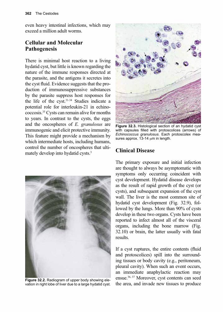
362 The Cestodes
even heavy intestinal infections, which may exceed a million adult worms.
Cellular and Molecular Pathogenesis
There is minimal host reaction to a living hydatid cyst, but little is known regarding the nature of the immune responses directed at the parasite, and the antigens it secretes into the cyst fluid. Evidence suggests that the pro-duction of immunosuppressive substances by the parasite suppress host responses for the life of the cyst.31-34 Studies indicate a potential role for interleukin-21 in echino-coccosis.35 Cysts can remain alive for months to years. In contrast to the cysts, the eggs and the oncospheres of E. granulosus are immunogenic and elicit protective immunity. This feature might provide a mechanism by which intermediate hosts, including humans, control the number of oncospheres that ulti-mately develop into hydatid cysts.3
Figure 32.2. Radiogram of upper body showing ele-vation in right lobe of liver due to a large hydatid cyst.
Figure 32.3. Histological section of an hydatid cyst with capsules filled with protoscolices (arrows) of Echinococcus granulosus. Each protoscolex mea-sures approx. 13-14 µm in length.
Clinical Disease
The primary exposure and initial infection are thought to always be asymptomatic with symptoms only occurring coincident with cyst development. Hydatid disease develops as the result of rapid growth of the cyst (or cysts), and subsequent expansion of the cyst wall. The liver is the most common site of hydatid cyst development (Fig. 32.9), fol-lowed by the lungs. More than 90% of cysts develop in these two organs. Cysts have been reported to infect almost all of the visceral organs, including the bone marrow (Fig. 32.10) or brain, the latter usually with fatal results.
If a cyst ruptures, the entire contents (fluid and protoscolices) spill into the surround-ing tissues or body cavity (e.g., peritoneum, pleural cavity). When such an event occurs, an immediate anaphylactic reaction may ensue.36, 37 Moreover, cyst contents can seed the area, and invade new tissues to produce

32. Juvenile Tapeworm Infections of Humans 363
Figure 32.4. Brood capsule with protoscolices of Echinococcus granulosus.
Figure 32.5. Protoscolex of E. granulosus.
second-generation hydatid cysts (Fig. 32.10). Even a few cells from the germinal mem-brane can re-establish an entire hydatid cyst, as each cell of the germinal membrane has a stem cell-like ability to reproduce a full hyda-tid cyst. Avoiding host contact with hydatid cyst contents and sterilizing the germinal layer is essential as the mortality rate of 2–4% usually seen in cases of cystic echinococcus may be increased if patients are improperly treated.
Approximately 60% of hydatid cysts are asymptomatic. Symptoms, when present, are those of a large space-occupying lesion. If the liver is involved, it becomes enlarged (Fig. 32.3). The cyst presents as a palpable, soft, non-tender, intrahepatic mass. The unin-volved bulk of the liver remains normal. Involvement of the lung is usually identified by chance on a radiograph, or by the pres-ence of bloody or ‘salty’ sputum, in which protoscolices or hooklets (Fig. 32.7) can be found. Patients may describe the sputum they produce as salty if it is due to a leaking hydatid cyst in the lungs.38 Expansion and rupture of liver hydatid cysts into the biliary tree can result in secondary cholangitis, bili-ary obstruction, and intraperitoneal rupture.39 Lung cysts can rupture into the bronchial tree
and cause the development of a bronchopul-monary fistula. Rupture of a cyst, wherever it is lodged, may occur even after relatively minor blunt trauma, and may lead to an aller-gic reaction. It may be mild and limited to urticaria, or may take the form of anaphy-lactic shock, requiring immediate interven-tion. Most patients with the northern variant of echinococcosis have asymptomatic lung cysts that are usually detected on chest radio-graphs obtained for other reasons (e.g., tuber-culosis screening).
Diagnosis (see Clinical Appendix)
An accurate case history is essential to the diagnosis of hydatid disease. Ownership of dogs, life on a sheep farm – even during childhood, and especially in endemic areas – and/or a history of travel to endemic areas are important factors for this disease.
The diagnostic algorithms for hydatid dis-ease have been modified based on increasing experience with radiologic imaging modali-ties, including ultrasound, CT, and MRI. Currently ultrasound is the recommended
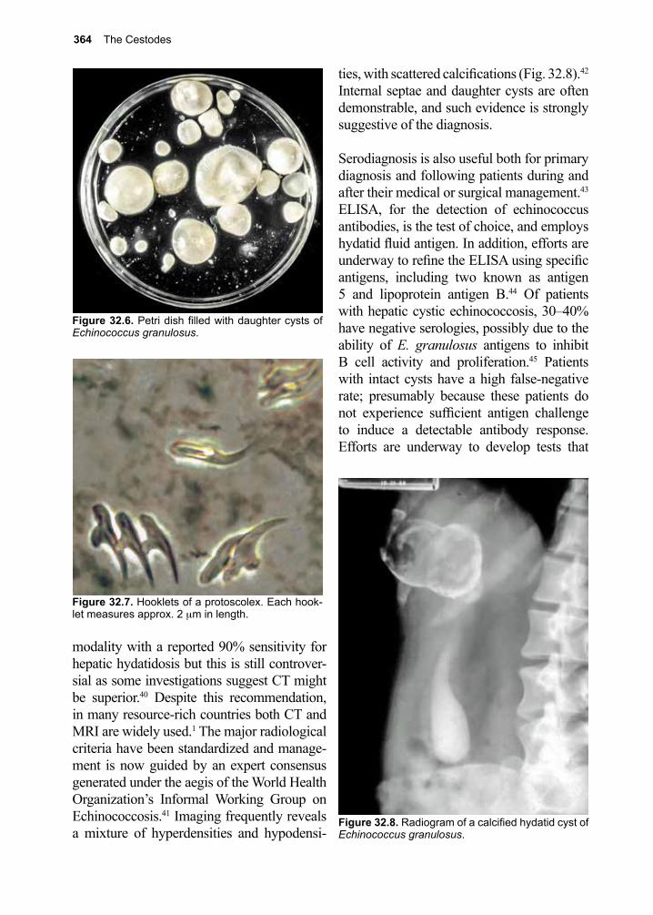
364 The Cestodes
Figure 32.6. Petri dish filled with daughter cysts of Echinococcus granulosus.
Figure 32.7. Hooklets of a protoscolex. Each hook-let measures approx. 2 µm in length.
Figure 32.8. Radiogram of a calcified hydatid cyst of Echinococcus granulosus.
modality with a reported 90% sensitivity for hepatic hydatidosis but this is still controver-sial as some investigations suggest CT might be superior.40 Despite this recommendation, in many resource-rich countries both CT and MRI are widely used.1 The major radiological criteria have been standardized and manage-ment is now guided by an expert consensus generated under the aegis of the World Health Organization’s Informal Working Group on Echinococcosis.41 Imaging frequently reveals a mixture of hyperdensities and hypodensi-
ties, with scattered calcifications (Fig. 32.8).42 Internal septae and daughter cysts are often demonstrable, and such evidence is strongly suggestive of the diagnosis.
Serodiagnosis is also useful both for primary diagnosis and following patients during and after their medical or surgical management.43 ELISA, for the detection of echinococcus antibodies, is the test of choice, and employs hydatid fluid antigen. In addition, efforts are underway to refine the ELISA using specific antigens, including two known as antigen 5 and lipoprotein antigen B.44 Of patients with hepatic cystic echinococcosis, 30–40% have negative serologies, possibly due to the ability of E. granulosus antigens to inhibit B cell activity and proliferation.45 Patients with intact cysts have a high false-negative rate; presumably because these patients do not experience sufficient antigen challenge to induce a detectable antibody response. Efforts are underway to develop tests that
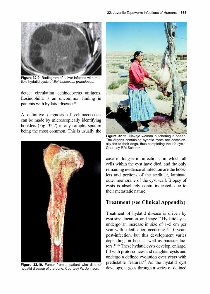
32. Juvenile Tapeworm Infections of Humans 365
Figure 32.9. Radiogram of a liver infected with mul-tiple hydatid cysts of Echinococcus granulosus.
Figure 32.10. Femur from a patient who died of hydatid disease of the bone. Courtesy W. Johnson.
Figure 32.11. Navajo woman butchering a sheep. The organs containing hydatid cysts are occasion-ally fed to their dogs, thus completing the life cycle. Courtesy P.M.Schantz.
detect circulating echinococcus antigens. Eosinophilia is an uncommon finding in patients with hydatid disease.46
A definitive diagnosis of echinococcosis can be made by microscopically identifying hooklets (Fig. 32.7) in any sample, sputum being the most common. This is usually the
case in long-term infections, in which all cells within the cyst have died, and the only remaining evidence of infection are the hook-lets and portions of the acellular, laminate outer membrane of the cyst wall. Biopsy of cysts is absolutely contra-indicated, due to their metastatic nature.
Treatment (see Clinical Appendix)
Treatment of hydatid disease is driven by cyst size, location, and stage.47 Hydatid cysts undergo an increase in size of 1–5 cm per year with calcification occurring 5–10 years post-infection, but this development varies depending on host as well as parasite fac-tors.48, 49 These hydatid cysts develop, enlarge, fill with protoscolices and daughter cysts and undergo a defined evolution over years with predictable features.47 As the hydatid cyst develops, it goes through a series of defined
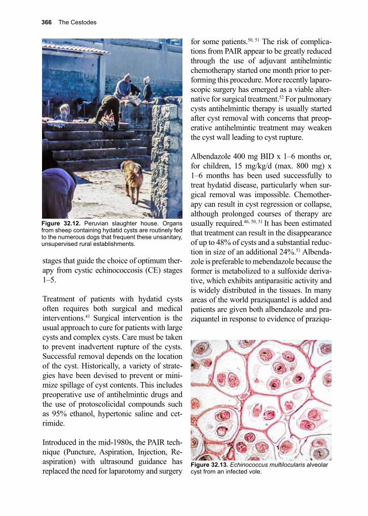
366 The Cestodes
for some patients.50, 51 The risk of complica-tions from PAIR appear to be greatly reduced through the use of adjuvant antihelmintic chemotherapy started one month prior to per-forming this procedure. More recently laparo-scopic surgery has emerged as a viable alter-native for surgical treatment.52 For pulmonary cysts antihelmintic therapy is usually started after cyst removal with concerns that preop-erative antihelmintic treatment may weaken the cyst wall leading to cyst rupture.
Albendazole 400 mg BID x 1–6 months or, for children, 15 mg/kg/d (max. 800 mg) x 1–6 months has been used successfully to treat hydatid disease, particularly when sur-gical removal was impossible. Chemother-apy can result in cyst regression or collapse, although prolonged courses of therapy are usually required.46, 50, 51 It has been estimated that treatment can result in the disappearance of up to 48% of cysts and a substantial reduc-tion in size of an additional 24%.51 Albenda-zole is preferable to mebendazole because the former is metabolized to a sulfoxide deriva-tive, which exhibits antiparasitic activity and is widely distributed in the tissues. In many areas of the world praziquantel is added and patients are given both albendazole and pra-ziquantel in response to evidence of praziqu-
Figure 32.12. Peruvian slaughter house. Organs from sheep containing hydatid cysts are routinely fed to the numerous dogs that frequent these unsanitary, unsupervised rural establishments.
Figure 32.13. Echinococcus multilocularis alveolar cyst from an infected vole.
stages that guide the choice of optimum ther-apy from cystic echinococcosis (CE) stages 1–5.
Treatment of patients with hydatid cysts often requires both surgical and medical interventions.41 Surgical intervention is the usual approach to cure for patients with large cysts and complex cysts. Care must be taken to prevent inadvertent rupture of the cysts. Successful removal depends on the location of the cyst. Historically, a variety of strate-gies have been devised to prevent or mini-mize spillage of cyst contents. This includes preoperative use of antihelmintic drugs and the use of protoscolicidal compounds such as 95% ethanol, hypertonic saline and cet-rimide.
Introduced in the mid-1980s, the PAIR tech-nique (Puncture, Aspiration, Injection, Re-aspiration) with ultrasound guidance has replaced the need for laparotomy and surgery
32. Juvenile Tapeworm Infections of Humans 367
antel’s protoscolicidal activity.53-56 Therapeu-tic responses can be monitored radiographi-cally and serologically. Reversible liver toxicity has been reported with prolonged therapy with albendazole.57, 58 As outlined in the section on trichuriasis, the benzimidazole antihelminthics, including albendazole, are teratogenic and embryotoxic in laboratory animals. Both drugs are considered category C agents in the United States. Before use in pregnancy the risks versus benefits must be weighed.
Prevention and Control
Infection of domestic dogs with E. granulo-sus can be prevented. Control is best achieved by avoiding feeding dogs (Figs. 32.11, 32.12) any infected organs of slaughtered sheep, or other animals, and by periodically treat-ing dogs prophylactically with niclosamide, arecoline hydrobromide, or praziquantel.51 An arecoline control program in dogs has resulted in the near-elimination of E. granu-losus infection in New Zealand and Tasma-nia. In Iceland, mass slaughter of infected sheep and dogs led to the total eradication of the disease. Attempts to duplicate that effort in Cyprus were not successful.59
Strict regulations regarding the importation of animal products that might carry E. granu-losus eggs (e.g., animal hides of various car-nivores, fishing flies, etc.) is a requirement for this control strategy to be effective. A recombinant peptide vaccine, EG95, induces high levels of protection in sheep, and may prove useful in certain parts of the world.60 Canine vaccination, which is currently being tested, may be critical in many areas of the world.61, 62
Echinococcus multilocularis
Pronunciation: \i-ˌkī-nə-ˈkä-kəs\\mul-tī-läk-yü-ler-is\
Echinococcus multilocularis (\eh-KYE-no-KOK-us\\MUL-tee-lok-you-lair-is\) infects wild Caenidae, such as fox, and can also infect domestic dogs. The intermediate hosts are usually rodents (e.g., voles, field mice, ground squirrels).63 The biology of the infec-tion in humans resembles the situation found in the intermediate hosts. E. multilocularis results in discrete cysts (alveolar echino-coccosis) (Fig. 32.13) that metastasize from a single location, except for the fact that E. multilocularis does not produce protoscoli-ces in human infection, only membranes that grow and bisect whatever organ they happen to be in, confounding the diagnosis of this unusual parasitic disease. In rodents, proto-scolices are found in each daughter cyst.
This infection is prevalent among fur trappers and others whose occupations bring them in close contact with wild foxes and popula-tions with close contact with dogs that regu-larly feed on infected rodents.64 This includes urban areas where reservoir hosts could expand from foxes to domesticated dogs and cats. Alveolar echinococcosis has emerged to the point where it is endemic in the north-ern hemisphere including western Europe, where it is considered a re-emerging disease due, in part, to the banning of fox hunting in a number of Common Market countries.65 It is also prevalent in the Russian Federation and Central Asia, China (especially in west-ern and central regions), and northern Japan (Hokkaido). In North America, human infec-tions have been reported from Alaska and in the upper Midwest and Northern Plains along the Canadian border.
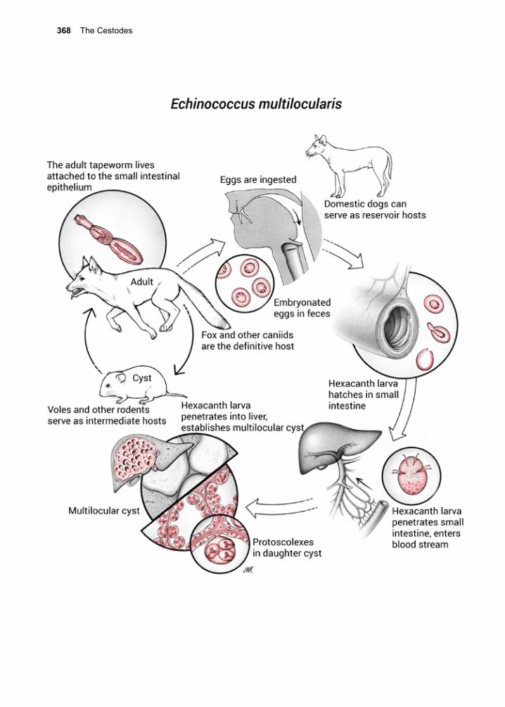
368 The Cestodes
32. Juvenile Tapeworm Infections of Humans 369
individuals, and may relate to the lack of development of protective immunity.69 Other investigators have noted an important role of interleukin-5 and a robust Th2 response to infection with E. multilocularis.70
Diagnosis (see Clinical Appendix)
Diagnosis is based on radiographic imaging findings which show tumor-like lesions with areas of calcification.71 Serologic testing by ELISA is both highly sensitive and specific and allows distinction between E. multilocu-laris and E. granulosus.72, 73 Directed biopsy of the lesion can often establish the diagno-sis.63
Treatment (see Clinical Appendix)
Surgical resection is the preferred approach to treatment with the entire larval mass being removed as part of a hepatic wedge or pul-monary lobe resection.64 Long-term adjuvant chemotherapy with albendazole or mebenda-zole improves the 10-year survival rate.74, 75 Nitazoxanide has some demonstrated in vitro activity against E. multilocularis and may prove to be a useful alternative therapy.76
Prevention relates to the handling of animal furs, subsequent oral contamination with eggs, and transmission from infected dogs or cats to humans. Trappers and those involved in animal husbandry in the fur industry should be educated to exercise caution when handling carcasses and processing furs. Use of oral baits laced with therapeutic doses of praziquantel for control of E. multilocularis infections in foxes has been effective in cer-tain areas.77 Monthly treatment of dogs at high risk with praziquantel may be an effec-tive approach.
Life Cycle
Infection begins when the intermediate host ingests the egg. The oncosphere hatches in the small intestine and invades the liver by the hematogenous route. It transforms, and then grows into an alveolar type of cyst, charac-terized by numerous daughter cysts, as com-pared to hydatid cysts, that grow as a single membrane-bounded unit. Foxes and dogs ingest infected liver containing numerous protoscolices, which leads to infection with the adult worm, thereby completing the life cycle.
Clinical Disease
In humans, the incubation period of larval infection with E. multilocularis is long, with incubation periods of 5–15 years. The initial exposure often occurs during childhood, but the disease has been recognized mainly in older adults.66 Alveolar disease can be highly aggressive, specifically affecting the liver as the primary target organ. The membrane pro-liferates indefinitely and causes progressive destruction of the liver parenchyma, which can lead to hepatic failure. In some cases this may lead to direct extension to the lungs and rarely metastasis to the CNS.67 Although ini-tially asymptomatic, as the disease progresses infected individuals may develop hepatomeg-aly, jaundice, multiple palpable abdominal masses, epigastric pain, and weight loss. If untreated, the mortality of clinically apparent disease can be as high as 50–75%.64
HLA type may play a role in a given individ-ual’s risk of developing further disease after chemotherapy. Re-growth of the parasite was significantly more prevalent in patients with haplotype HLA-DQB1*02 than those with haplotype HLA-DRB1*1157.68 Interleu-kin-10 production was also higher in infected
370 The Cestodes
humans (some are life-threatening), collec-tively referred to as sparganosis. Cases of sparganosis have been reported from South-east Asia, Japan, China, especially on the island of Hainan, Africa, Italy, and the United States.80, 81 The definitive hosts for the many species of Spirometra are cats, birds, canines, and a number of other carnivores.80, 82
The life cycles of Spirometra spp. are com-plex and similar to that of Diphyllobothrium latum (see Chapter 30). The definitive hosts harbor the adult tapeworm in their small intestinal tract. When they defecate into fresh-water, the unembryonated eggs hatch and the released free-swimming coracidia are eaten by copepods. The parasites then develop into the procercoid stage that is infectious for second intermediate hosts: amphibians, fish, reptiles, and birds. These animals ingest the infected copepods, releasing the procercoids that penetrate the intestinal tract and develop to the plerocercoid stage in muscle tissue. This stage is infective for definitive hosts; warm-blooded predator species that prey on these infected cold-blooded animals.83
Human infection results from ingestion of raw or undercooked flesh of any of the numerous intermediate hosts, and from application of such flesh as poultices. This practice is very common in some areas of the world, par-ticularly on the island of Hainan, where over 30% of the frogs harbor the juvenile stage of Spirometra.84, 85 The skin of numerous cold-blooded vertebrates contains a variety of closely related peptides, referred to as megai-nins.86 These amphipathic peptides are related to gramicidin, and have potent anti-microbial activities.87 Employing frog and fish skins for medical use has a chemotherapeutic basis. As with any other therapy, the use of cold-blooded vertebrate skin as a poultice can have unwanted “side effects.” In this case, the user may develop a parasitic infection.
Mesocestoides spp. (Valliant 1863)
Pronunciation: \me-zə-ses-ˈtȯi-dēz\
Introduction
Cestodes, in the genus Mesocestoides (\MEH-zo-SES-toy-DEEZ\), infect numer-ous species of mammals and birds. Human infections have been reported.78 Most of the cases reported from the U.S. were caused by Mesocestoides variabilis.79 The life cycle of Mesocestoides is complex, involving a coprophagous arthropod as the first interme-diate host, and birds, snakes, lizards, amphib-ians, rodents, or other mammalian carnivores as the second intermediate host. The infective stage, known as a tetrathyridium, develops in the second intermediate host. Tetrathyridia are usually about 1 cm long and contain an invaginated scolex with four suckers.
Humans acquire the parasite by eating the tetrathyridia, which can develop to an adult worm in the gut or migrate to the peritoneal cavity. Cases of Mesocestoides infection have been described in the U.S., but the mode of acquisition is not known.78 A case of Meso-cestoides infection from a 19 month-old boy in Alexandria, Louisiana, has led to the sug-gestion that the infection is food-borne, pos-sibly in association with the culinary customs of the Acadian and Creole communities in this region. Treatment of the adult tapeworm with praziquantel or niclosamide is effective.79
Spirometra spp. (Mueller 1935)
Pronunciation: \spī-ˈräm-ə-trə\
Spirometra mansonoides (\SPEE-rom-u-tru\) and other related Spirometra spp. are pseu-dophyllidian tapeworms that cause a closely related series of metacestode infections in
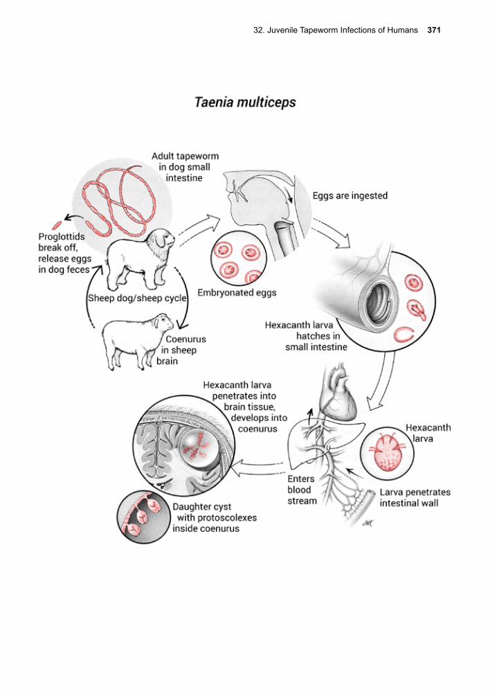
32. Juvenile Tapeworm Infections of Humans 371
372 The Cestodes
The plerocercoid stage can migrate out of the poultice and into the subcutaneous tissues, stimulated by the rise in temperature from the human host. If the poultice is placed over an open wound, or the eye, the immature para-site may enter the site. In the eye, the inner surface of the lid is the initial site of infec-tion.88, 89 The surrounding tissues proliferate, becoming edematous and painful (unilat-eral peri-orbital edema), because the larva secretes a version of growth hormone similar to that of its mammalian host.90 The pleuro-cercoid may migrate beyond the eyelid into the brain, where it continues to grow. Neu-rological symptoms follow.91 Occasionally, patients die of neurological complications of infection with Spirometra spp..
Diagnosis can be made based on identifi-cation of the worm after removal or biopsy. There is an ELISA test for sparganosis with high levels of sensitivity and specificity that can be useful in making the diagnosis.92, 93 In the case of early eye involvement, removal of the larva from the space between the lower lid and the eyeball results in cure. Larvae often migrate behind the eye, and even into the brain via the optic nerve, making easy removal of the parasite difficult if not impos-sible.94 In these instances, surgery is neces-sary. Subcutaneous lesions are often removed by surgery, as treatment with antihelmintic therapy such as with praziquantel is associ-ated with limited success.94 Prevention is dif-ficult, because of the effectiveness and popu-larity of poultice use for a variety of medi-cal problems, including photophobia due to chickenpox.95
Taenia spp. (other than T. saginata and T. solium)
Pronunciation: \tē-nē-ə\
Taenia (TEE-nee-ah) spp. (e.g., T. multiceps, T. brauni, T. serialis) are a group of cyclo-phyllidian tapeworms that, as adults, infect dogs and other Canidae. Intermediate hosts include domestic cattle, horses, goats, and some wild herbivorous animals of Africa. Humans become infected with the metaces-tode (juvenile stage), known as coenurus, which resembles cysts found in the inter-mediate hosts. Human infections have been largely confined to the African continent, but a few cases have been described from France, England, and North and South America.
The larva may invade the CNS (i.e., brain, eyes, spinal cord). Other space-occupying lesions resemble this infection, such as those caused by cysticerci and hydatid cysts.
Diagnosis is based on clinical, epidemiologic, and laboratory findings. No serologic tests are currently available.
Treatment involves surgery if the lesion is accessible. Although albendazole, praziqu-antel and other antihelmintic therapies have been used, it is unclear how efficacious these are.96 Recommendations for prevention are limited, because reservoir hosts include such a large number of species of wild animals.
References
1. Griffin, D. O., et al., Management of serology negative human hepatic hydatidosis (caused by Echinococcus granulosus) in a young woman from Bangladesh in a resource-rich setting: A case report. IDCases 2014, 1 (2), 17-21.
2. Budke, C. M., et al., Global socioeconomic impact of cystic echinococcosis. Emerg Infect Dis 2006, 12 (2), 296-303.
32. Juvenile Tapeworm Infections of Humans 373
3. Garcia, H. H., et al., The Cysticercosis Working Group in Peru. Lancet 2003, 361, 547-56.4. Moro, P. L.; Schantz, P. M., Echinococcosis: historical landmarks and progress in research and
control. Ann Trop Med Parasitol 2006, 100 (8), 703-14.5. Abdybekova, A., et al., Epidemiology of echinococcosis in Kazakhstan: an update. J Helminthol
2015, 89 (6), 647-50.6. Ito, A.; Budke, C. M., The present situation of echinococcoses in Mongolia. J Helminthol 2015,
89 (6), 680-8.7. Economides, P., et al., Control of Echinococcus granulosus in Cyprus and comparison with other
island models. Veterinary parasitology 1998, 79 (2), 151-63.8. Stojkovic, M., et al., Diagnosing and staging of cystic echinococcosis: how do CT and MRI
perform in comparison to ultrasound? PLoS Negl Trop Dis 2012, 6 (10), e1880.9. Moro, P.; Schantz, P. M., Cystic echinococcosis in the Americas. Parasitology international
2006, 55 Suppl, S181-6.10. Schantz, P. M., Echinococcosis in American Indians living in Arizona and New Mexico: a review
of recent studies. American journal of epidemiology 1977, 106 (5), 370-9.11. Daly, J. J., et al., Unilocular hydatid cyst disease in the mid-South. JAMA 1984, 251 (7), 932-3.12. Finlay, J. C.; Speert, D. P., Sylvatic hydatid disease in children: case reports and review of
endemic Echinococcus granulosus infection in Canada and Alaska. The Pediatric infectious disease journal 1992, 11 (4), 322-6.
13. McManus, D. P., et al., Short report: molecular genetic characterization of an unusually severe case of hydatid disease in Alaska caused by the cervid strain of Echinococcus granulosus. Am J Trop Med Hyg 2002, 67 (3), 296-8.
14. Sweatman, G. K., The Significance of the Artificial Introduction of Reindeer (Rangifer Tarandus) and Moose (Alces Alces) in the Spread of Hydatid Disease (Echinococcus Granulosus). Ann Trop Med Parasitol 1964, 58, 307-14.
15. Cox, F. E., History of human parasitology. Clin Microbiol Rev 2002, 15 (4), 595-612.16. Pallas, P. S., Miscellanea Zoologica: Quibus Novae Imprimis Atque Obscure Animalium Species
Describuntur Et Observationibus Iconibusque Illustrantur. 1766.17. Goeze, J. A. E.; Blankenburg, P. A., Versuch ciner Naturgeschichte der Eingeweiderwumer
Thierischer Korper. Pape 1782.18. Seibold, C. T., Von Uber die Verwandlung der Echinococcus-brut in Taenia. 1853.19. Naunyn, B., Uber die zu Echinococcus hominis gehorige Taenia. Arch Anat Physiol Wissenschr
Med 1863, 412-416.20. Haag, K. L., et al., Breeding systems in Echinococcus granulosus (Cestoda; Taeniidae): selfing
or outcrossing? Parasitology 1999, 118 ( Pt 1), 63-71.21. Heath, D. D., et al., Echinococcus granulosus: the mechanism of oncosphere lysis by sheep
complement and antibody. Int J Parasitol 1994, 24 (7), 929-35.22. Holcman, B., et al., Ultrastructure of oncosphere and early stages of metacestode development of
Echinococcus granulosus. Int J Parasitol 1994, 24 (5), 623-35.23. Harris, A., et al., Echinococcus granulosus: ultrastructure of epithelial changes during the first 8
days of metacestode development in vitro. Int J Parasitol 1989, 19 (6), 621-9.24. Diaz-Menendez, M., et al., Management and outcome of cardiac and endovascular cystic
echinococcosis. PLoS Negl Trop Dis 2012, 6 (1), e1437.25. Nourbakhsh, A., et al., Hydatid disease of the central nervous system: a review of literature with
an emphasis on Latin American countries. Neurol Res 2010, 32 (3), 245-51.26. Rexiati, M., et al., Diagnosis and surgical treatment of renal hydatid disease: a retrospective
analysis of 30 cases. PLoS One 2014, 9 (5), e96602.27. Sen, S., et al., Primary intraocular hydatid cyst with glaucoma. J Pediatr Ophthalmol Strabismus
2003, 40 (5), 312-3.28. Sinav, S., et al., A primary intraocular hydatid cyst. Acta Ophthalmol (Copenh) 1991, 69 (6),
802-4.29. Babitha, F., et al., Hydatid cyst of bone. Indian J Med Microbiol 2015, 33 (3), 442-4.30. Pazarci, O., et al., Treatment of Bifocal Cyst Hydatid Involvement in Right Femur with
Teicoplanin Added Bone Cement and Albendazole. Case Rep Orthop 2015, 2015, 824824.
374 The Cestodes
31. Dixon, J. B., Echinococcosis. Comparative immunology, microbiology and infectious diseases 1997, 20 (1), 87-94.
32. Gottstein, B.; Hemphill, A., Immunopathology of echinococcosis. Chemical immunology 1997, 66, 177-208.
33. Wang, Y., et al., Impairment of dendritic cell function and induction of CD4(+)CD25(+)Foxp3(+) T cells by excretory-secretory products: a potential mechanism of immune evasion adopted by Echinococcus granulosus. BMC Immunol 2015, 16, 44.
34. Zheng, Y., Strategies of Echinococcus species responses to immune attacks: implications for therapeutic tool development. Int Immunopharmacol 2013, 17 (3), 495-501.
35. Zhang, F., et al., CCR7(lo)PD-1(hi) CXCR5(+) CD4(+) T cells are positively correlated with levels of IL-21 in active and transitional cystic echinococcosis patients. BMC Infect Dis 2015, 15, 457.
36. Nunnari, G., et al., Hepatic echinococcosis: clinical and therapeutic aspects. World J Gastroenterol 2012, 18 (13), 1448-58.
37. Minciullo, P. L., et al., Anaphylaxis caused by helminths: review of the literature. Eur Rev Med Pharmacol Sci 2012, 16 (11), 1513-8.
38. Nakamura, K., et al., A case of pulmonary and hepatic cystic Echinococcosis of CE1 stage in a healthy Japanese female that was suspected to have been acquired during her stay in the United Kingdom. Am J Trop Med Hyg 2011, 85 (3), 456-9.
39. Irkoruucu, O., et al., The detection of cysto-biliary communications during surgery for liver hydatid cysts: let’s speak the unspoken. Clinics (Sao Paulo) 2012, 67 (2), 179.
40. Dhar, P., et al., Current trends in the diagnosis and management of cystic hydatid disease of the liver. J Commun Dis 1996, 28 (4), 221-30.
41. Brunetti, E., et al., Expert consensus for the diagnosis and treatment of cystic and alveolar echinococcosis in humans. Acta Trop 2010, 114 (1), 1-16.
42. Agildere, A. M., et al., MRI of hydatid disease of the liver: a variety of sequences. Assist Tomogr 1998, 22, 718-24.
43. Gottstein, B., Molecular and immunological diagnosis of echinococcosis. Clinical microbiology reviews 1992, 5 (3), 248-61.
44. Chemale, G., et al., Echinococcus granulosus antigen B hydrophobic ligand binding properties. Biochimica et biophysica acta 2005, 1747 (2), 189-94.
45. Zhang, W.; McManus, D. P., Recent advances in the immunology and diagnosis of echinococcosis. FEMS immunology and medical microbiology 2006, 47 (1), 24-41.
46. Schaefer, J. W.; Khan, M. Y., Echinococcosis (Hydatid disease): lessons from experience with 59 patients. Reviews of infectious diseases 1991, 13 (2), 243-7.
47. Gharbi, H. A., et al., Ultrasound examination of the hydatic liver. Radiology 1981, 139 (2), 459-63.
48. Moro, P. L., et al., Human hydatidosis in the central Andes of Peru: evolution of the disease over 3 years. Clin Infect Dis 1999, 29 (4), 807-12.
49. Frider, B., et al., Long-term outcome of asymptomatic liver hydatidosis. J Hepatol 1999, 30 (2), 228-31.
50. Filice, C.; Brunetti, E., Use of PAIR in human cystic echinococcosis. Acta tropica 1997, 64 (1-2), 95-107.
51. Pelaez, V.; Kugler, C., PAIR as percutaneous treatment of hydatid liver cysts. Acta Tropica 2000, 75, 197-202.
52. Chen, X., et al., The Comparison of 2 New Promising Weapons for the Treatment of Hydatid Cyst Disease: PAIR and Laparoscopic Therapy. Surg Laparosc Endosc Percutan Tech 2015, 25 (4), 358-62.
53. Alvela-Suarez, L., et al., Safety of the combined use of praziquantel and albendazole in the treatment of human hydatid disease. Am J Trop Med Hyg 2014, 90 (5), 819-22.
54. Bygott, J. M.; Chiodini, P. L., Praziquantel: neglected drug? Ineffective treatment? Or therapeutic choice in cystic hydatid disease? Acta Trop 2009, 111 (2), 95-101.
55. De, S., et al., In vitro assessment of praziquantel and a novel nanomaterial against protoscoleces
32. Juvenile Tapeworm Infections of Humans 375
of Echinococcus granulosus. J Helminthol 2012, 86 (1), 26-9.56. Moreno, M. J., et al., Praziquantel and albendazole in the combined treatment of experimental
hydatid disease. Parasitol Res 2001, 87 (3), 235-8.57. Horton, R. J., Albendazole in treatment of human cystic echinococcosis: 12 years of experience.
Acta tropica 1997, 64 (1-2), 79-93.58. Rowley, A. H., et al., Albendazole treatment of recurrent echinococcosis. The Pediatric infectious
disease journal 1988, 7 (9), 666-7.59. Beard, T. C., The elimination of echinococcosis from Iceland. Bulletin of the World Health
Organization 1973, 48 (6), 653-60.60. Woollard, D. J., et al., Epitope specificities and antibody responses to the EG95 hydatid vaccine.
Parasite immunology 1998, 20 (11), 535-40.61. Petavy, A. F., et al., An oral recombinant vaccine in dogs against Echinococcus granulosus, the
causative agent of human hydatid disease: a pilot study. PLoS Negl Trop Dis 2008, 2 (1), e125.62. Zhang, W.; McManus, D. P., Vaccination of dogs against Echinococcus granulosus: a means to
control hydatid disease? Trends Parasitol 2008, 24 (9), 419-24.63. Moro, P.; Schantz, P. M., Echinococcosis: a review. Int J Infect Dis 2009, 13 (2), 125-33.64. Wilson, J. F.; Rausch, R. L., Alveolar hydatid disease. A review of clinical features of 33
indigenous cases of Echinococcus multilocularis infection in Alaskan Eskimos. Am J Trop Med Hyg 1980, 29 (6), 1340-55.
65. Schweiger, A., et al., Human alveolar echinococcosis after fox population increase, Switzerland. Emerg Infect Dis 2007, 13 (6), 878-82.
66. Lukaschenko, N. P.; J., Problems of epidemiology and prophylaxix of alveolar (multilocular echinococcosis): a general review-with particular reference to the USSR. Int 1971, 1, 125-134.
67. Kammerer, W. S.; Schantz, P. M., Echinococcal disease. Infect Dis Clin North Am 1993, 7 (3), 605-18.
68. Eiermann, T. H., et al., HLA and alveolar echinococcosis. Tissue antigens 1998, 52 (2), 124-9.69. Godot, V., et al., Increased basal production of interleukin-10 by peripheral blood mononuclear
cells in human alveolar echinococcosis. European cytokine network 1997, 8 (4), 401-8.70. Sturm, D., et al., Interleukin-5 is the predominant cytokine produced by peripheral blood
mononuclear cells in alveolar echinococcosis. Infect Immun 1995, 63 (5), 1688-97.71. Didier, D., et al., Hepatic alveolar echinococcosis: correlative US and CT study. Radiology 1985,
154 (1), 179-86.72. Gottstein, B., et al., Improved primary immunodiagnosis of alveolar echinococcosis in humans
by an enzyme-linked immunosorbent assay using the Em2plus antigen. Journal of clinical microbiology 1993, 31 (2), 373-6.
73. Carmena, D., et al., The immunodiagnosis of Echinococcus multilocularis infection. Clin Microbiol Infect 2007, 13 (5), 460-75.
74. Schantz, P. M., et al., Effects of albendazole on Echinococcus multilocularis infection in the Mongolian jird. J Infect Dis 1990, 162 (6), 1403-7.
75. Eckert, J.; Deplazes, P., Biological, epidemiological, and clinical aspects of echinococcosis, a zoonosis of increasing concern. Clin Microbiol Rev 2004, 17 (1), 107-35.
76. Reuter, S., et al., In vitro activities of itraconazole, methiazole, and nitazoxanide versus Echinococcus multilocularis larvae. Antimicrob Agents Chemother 2006, 50 (9), 2966-70.
77. Takahashi, K., et al., Efficacy of anthelmintic baiting of foxes against Echinococcus multilocularis in northern Japan. Vet Parasitol 2013, 198 (1-2), 122-6.
78. Schultz, L. V., et al., Mesocestoides (Cestoda) infection in a California child. The Pediatric infectious disease journal 1992, 11 (4), 332-4.
79. Fuentes, M. V., et al., Short report: a new case report of human Mesocestoides infection in the United States. Am J Trop Med Hyg 2003, 68 (5), 566-7.
80. Walker, M. D.; Zunt, J. R., Neuroparasitic infections: cestodes, trematodes, and protozoans. Semin Neurol 2005, 25 (3), 262-77.
81. Pampiglione, S., et al., Human sparganosis in Italy. Case report and review of the European cases. APMIS 2003, 111 (2), 349-54.
82. Hughes, A. J.; Biggs, B. A., Parasitic worms of the central nervous system: an Australian
376 The Cestodes
perspective. Intern Med J 2002, 32 (11), 541-53.83. Lescano, A. G.; Zunt, J., Other cestodes: sparganosis, coenurosis and Taenia crassiceps
cysticercosis. Handb Clin Neurol 2013, 114, 335-45.84. Kean, B. H., et al., Color Atlas / Text of Ophthalmic Parasitology. 1991; p 195-200.85. Despommier, D., People, Parasites, and Plowshares. 2014.86. Oren, Z.; Shai, Y., Mode of action of linear amphipathic alpha-helical antimicrobial peptides.
Biopolymers 1998, 47 (6), 451-63.87. Bechinger, B., Structure and functions of channel-forming peptides: magainins, cecropins,
melittin and alamethicin. The Journal of membrane biology 1997, 156 (3), 197-211.88. Otranto, D.; Eberhard, M. L., Zoonotic helminths affecting the human eye. Parasit Vectors 2011,
4, 41.89. Wiwanitkit, V., Ocular sparganosis. Orbit 2014, 33 (6), 474.90. Phares, K., An unusual host-parasite relationship: the growth hormone-like factor from
plerocercoids of Spirometrid tapeworms. International journal for parasitology 1996, 26 (6), 575-88.
91. Song, T., et al., CT and MR characteristics of cerebral sparganosis. AJNR Am J Neuroradiol 2007, 28 (9), 1700-5.
92. Cui, J., et al., Serodiagnosis of experimental sparganum infections of mice and human sparganosis by ELISA using ES antigens of Spirometra mansoni spargana. Parasitol Res 2011, 108 (6), 1551-6.
93. Liu, L. N., et al., Serodiagnosis of sparganosis by ELISA using recombinant cysteine protease of Spirometra erinaceieuropaei spargana. Parasitol Res 2015, 114 (2), 753-7.
94. Tan, T. Y., et al., Cerebral sparganosis: case report. Changgeng yi xue za zhi / Changgeng ji nian yi yuan = Chang Gung medical journal / Chang Gung Memorial Hospital 1999, 22 (2), 287-92.
95. Chang, J. H., et al., Subcutaneous sparganosis--a case report and a review of human sparganosis in Taiwan. The Kaohsiung journal of medical sciences 1999, 15 (9), 567-71.
96. El-On, J., et al., Taenia multiceps: a rare human cestode infection in Israel. Vet Ital 2008, 44 (4), 621-31.
The Trematodes 377
VII. The Trematodes
The class Trematoda, in the phylum Platyhel-minthes, consists of two orders, Monogenea and Digenea. All are obligate parasites. Trem-atodes of medical importance only occur in the order Digenea, and include the blood flukes, the intestinal flukes, and the tissue flukes. They are mainly found throughout the tropics and subtropics, while a few species are encountered in temperate zones, as well.
Trematodes undergo complex developmen-tal cycles in their intermediate and definitive hosts. But, as complex as trematode biology is, offering seemingly numerous opportuni-ties for interrupting their life cycles, eradica-tion of any medically important species has occurred to only a limited extent in specific geographic areas such as Schistosoma man-soni in China and S. japonicum in Japan.
Trematodes maintain their site location within the host using their two suckers, one anterior and one ventral. The anterior sucker also serves as the opening to the oral cavity, into which host tissues are ingested. The outer sur-face of the adult is covered with a tegument similar in design and function to the tegument of cestodes. It serves as an absorbing surface for both large and small molecular-weight molecules. The tegument is covered by mem-brane-bound microvilli, underneath which are mitochondria, pinocytotic vesicles, and other structures facilitating nutrient acquisi-tion.
In addition, trematodes have a functional blind gut into which they ingest tissues of the host. Ingested material is pumped down into the bifurcated intestinal tract, where digestion occurs, aided by enzymes (e.g., proteases, lipases, aminopeptidases, esterases). Since the gut has no exit, wastes are regurgitated into the host. These worms obtain a wide variety of nutrients in several different ways, making
it difficult to develop drugs or immune-based therapies to interrupt metabolic processes.
Several layers of muscle lie just below the tegument, allowing trematodes to move about freely within the host. This activity can result in severe pathological consequences for the host, particularly in the case of Fas-ciola hepatica, Paragonimus westermani and the schistosomes.
A pair of dorsal ganglia gives rise to lateral peripheral nerves running the length of the body, and they innervate the muscle layers. Commissures from the lateral nerves also innervate various organs, including the gut, and reproductive organs. The flukes have no body cavity; rather, organs are embedded in the parenchyma.
In addition to solid wastes, trematodes excrete small molecular-weight compounds using a network of tubules that connect to collect-ing organelles. These, in turn, connect to the excretory pore at the tegumental surface of the parasite.
The trematodes in the order Digenea employ one of three reproductive strategies: 1. self-fertilization, in which the same worm pos-sesses both sets of reproductive organs (e.g., Fasciola hepatica), 2. cross-fertilization between two worms possessing both sets of reproductive organs (e.g., Paragonimus wes-termani), or 3. fertilization between worms of the opposite sex, as is the case among the schistosomes.
Egg production is complex, involving a series of specialized organs. The ovum, sup-plied with yolk from the vitelline glands, is fertilized within the oviduct and becomes surrounded by a shell from secretions of the Mehlis’ gland. It exits from the parasite through the genital pore, usually situated in between the anterior and ventral suckers.
378 The Trematodes
Once the eggs reach freshwater, or, for some species, a suitable terrestrial niche, they are either stimulated to hatch in the external envi-ronment or, they hatch after being ingested by the next host. There, they undergo asexual reproduction, eventually increasing in num-bers many-fold. Intermediate hosts include snail species that live in freshwater or ter-restrial habitats. In addition, other inverte-brates (e.g., insects, crabs) and a variety of cold-blooded vertebrates (e.g., fish) function as intermediate hosts for medically important species of trematodes. Plants (e.g., watercress, water chestnuts) are sites on which some spe-cies of metacercariae encyst.
Many species of adult trematodes are acquired by ingesting the intermediate stage (i.e., the metacercaria), but a few (notably the schisto-somes) can actively penetrate unbroken skin. Site selection by trematodes within the human host is poorly understood. It is determined by a complex interplay between chemical and physical niches, which represent environ-mental cues from the host, and the receiving and translation of those cues by the nervous system of the parasite. Some drugs interfere with trematode nervous system functions (e.g., praziquantel), resulting in profound changes in worm behavior. Under those con-ditions, elimination of the parasite is possible.
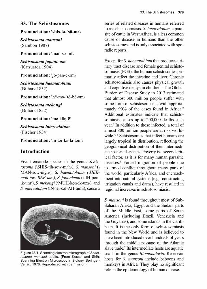
33. The Schistosomes 379
Figure 33.1. Scanning electron micrograph of Schis-tosoma mansoni adults. (From Kessel and Shih: Scanning Electron Microscopy in Biology. Springer-Verlag, 1976. Reproduced with permission).
33. The SchistosomesPronunciation: \shis-tə-ˈsō-mə\
Schistosoma mansoni (Sambon 1907)
Pronunciation: \man-sə-ˌnī\
Schistosoma japonicum (Katsurada 1904)
Pronunciation: \jə-pän-c-əm\
Schistosoma haematobium (Bilharz 1852)
Pronunciation: \hē-mə-ˈtō-bē-əm\
Schistosoma mekongi (Bilharz 1852)
Pronunciation: \mə-käŋ-ē\
Schistosoma intercalatum (Fischer 1934)
Pronunciation: \in-tər-kə-la-təm\
Introduction
Five trematode species in the genus Schis-tosoma (\SHIS-tih-sow-mah\), S. mansoni (\MAN-sow-nigh\), S. haematobium (\HEE-mah-tow-BEE-um\), S. japonicum (\JIH-pon-ik-um\), S. mekongi (\MUH-kon-ik-um\), and S. intercalatum (IN-ter-cal-AH-tum\), cause a
series of related diseases in humans referred to as schistosomiasis. S. intercalatum, a para-site of cattle in West Africa, is a less common cause of disease in humans than the other schistosomes and is only associated with spo-radic reports.
Except for S. haematobium that produces uri-nary tract disease and female genital schisto-somiasis (FGS), the human schistosomes pri-marily affect the intestine and liver. Chronic schistosomiasis also causes physical growth and cognitive delays in children.1 The Global Burden of Disease Study in 2013 estimated that almost 300 million people suffer with some form of schistosomiasis, with approxi-mately 90% of the cases found in Africa.2 Additional estimates indicate that schisto-somiasis causes up to 200,000 deaths each year.3 In addition to those infected, a total of almost 800 million people are at risk world-wide.4, 5 Schistosomes that infect humans are largely tropical in distribution, reflecting the geographical distribution of their intermedi-ate host snail species. Poverty is a second crit-ical factor, as it is for many human parasitic diseases.6 Forced migration of people due to armed conflict throughout many parts of the world, particularly Africa, and encroach-ment into natural systems (e.g., constructing irrigation canals and dams), have resulted in regional increases in schistosomiasis.
S. mansoni is found throughout most of Sub-Saharan Africa, Egypt and the Sudan, parts of the Middle East, some parts of South America (including Brazil, Venezuela and the Guyanas), and some islands in the Carib-bean. It is the only form of schistosomiasis found in the New World and is believed to have been introduced over hundreds of years through the middle passage of the Atlantic slave trade.7 Its intermediate hosts are aquatic snails in the genus Biomphalaria. Reservoir hosts for S. mansoni include baboons and monkeys in Africa. They play no significant role in the epidemiology of human disease.
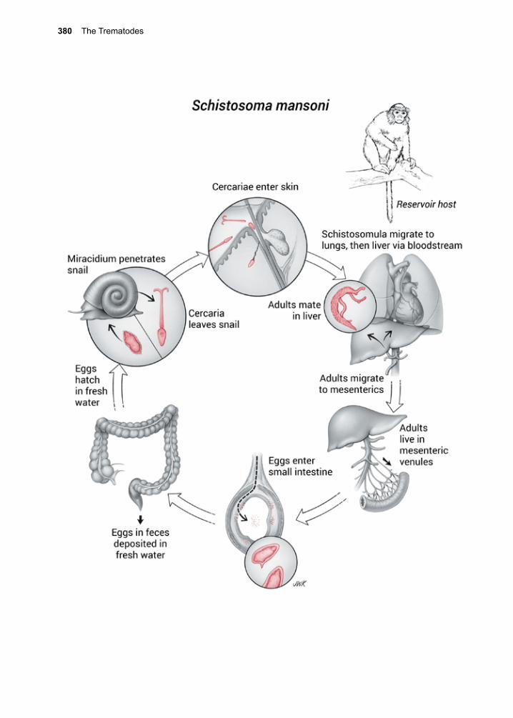
380 The Trematodes
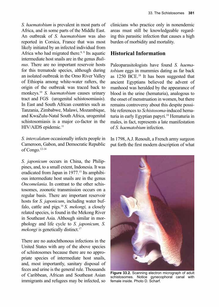
33. The Schistosomes 381
S. haematobium is prevalent in most parts of Africa, and in some parts of the Middle East. An outbreak of S. haematobium was also reported in Corsica, France that was most likely initiated by an infected individual from Africa who had migrated there.8, 9 Its aquatic intermediate host snails are in the genus Buli-nus. There are no important reservoir hosts for this trematode species, although during an isolated outbreak in the Omo River Valley of Ethiopia among white-water rafters, the origin of the outbreak was traced back to monkeys.10 S. haematobium causes urinary tract and FGS (urogenital schistosomiasis). In East and South African countries such as Tanzania, Zimbabwe, Malawi, Mozambique, and KwaZulu-Natal South Africa, urogenital schistosomiasis is a major co-factor in the HIV/AIDS epidemic.11
S. intercalatum occasionally infects people in Cameroon, Gabon, and Democratic Republic of Congo.12-14
S. japonicum occurs in China, the Philip-pines, and, to a small extent, Indonesia. It was eradicated from Japan in 1977.15 Its amphibi-ous intermediate host snails are in the genus Oncomelania. In contrast to the other schis-tosomes, zoonotic transmission occurs on a regular basis. There are important reservoir hosts for S. japonicum, including water buf-falo, cattle and pigs.16 S. mekongi, a closely related species, is found in the Mekong River in Southeast Asia. Although similar in mor-phology and life cycle to S. japonicum, S. mekongi is genetically distinct.17
There are no autochthonous infections in the United States with any of the above species of schistosomes because there are no appro-priate species of intermediate host snails, and, most importantly, sanitary disposal of feces and urine is the general rule. Thousands of Caribbean, African and Southeast Asian immigrants and refugees may be infected, so
clinicians who practice only in nonendemic areas must still be knowledgeable regard-ing this parasitic infection that causes a high burden of morbidity and mortality.
Historical Information
Paleoparasitologists have found S. haema-tobium eggs in mummies dating as far back as 1250 BCE.18 It has been suggested that ancient Egyptians believed the advent of manhood was heralded by the appearance of blood in the urine (hematuria), analogous to the onset of menstruation in women, but there remains controversy about this despite possi-ble references to Schistosoma-induced hema-turia in early Egyptian papyri.19 Hematuria in males, in fact, represents a late manifestation of S. haematobium infection.
In 1798, A.J. Renoult, a French army surgeon put forth the first modern description of what
Figure 33.2. Scanning electron micrograph of adult schistosomes. Notice gynecophoral canal with female inside. Photo D. Scharf.
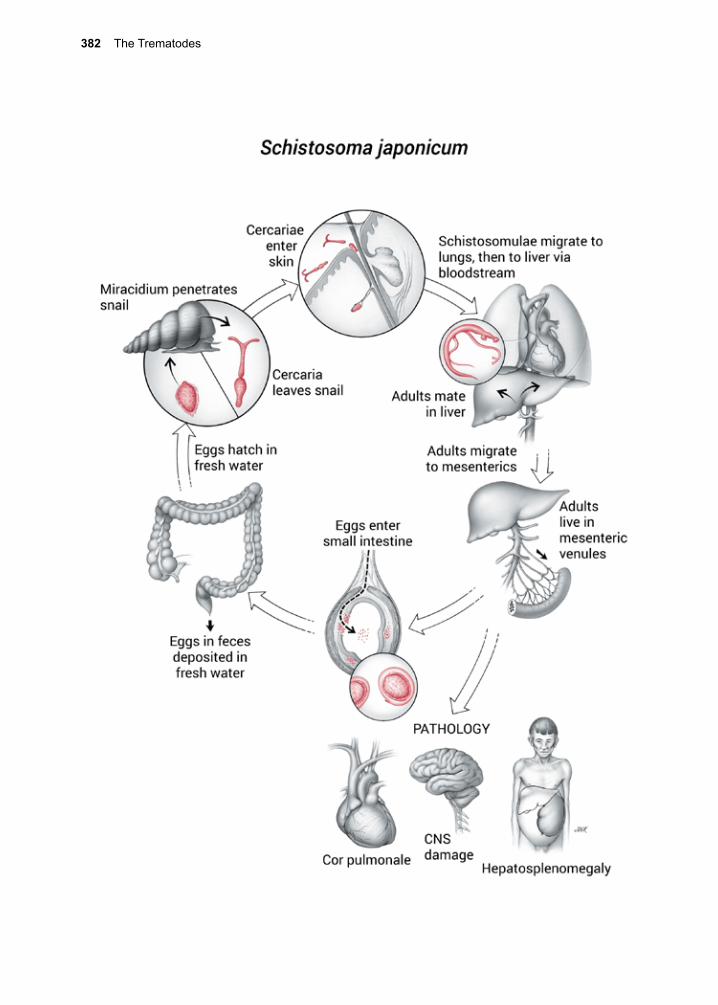
382 The Trematodes
33. The Schistosomes 383
is believed to have been hematuria due to S. haematobium. Renoult described the epi-demic of hematuria seen in Napoleon’s sol-diers who invaded Egypt.20 Cases were subse-quently described among troops involved in the Second Anglo-Boer War (1899–1902).21 In 1851 and 1852, Theodore Bilharz and Ernst von Siebold reported human cases of S. haematobium, described the adult worm and made the connection with the appearance of hematuria.22-24 They identified this as a para-site that occupied the venous plexus of the bladder and whose eggs possessed a terminal spine. In 1854, Wilhelm Griesinger described in detail the clinical disease and its pathol-ogy.25 Griesinger noted the relation of the infection to the involvement of the bladder and ureters. In 1918, Robert Leiper described the life cycle of S. haematobium, its inter-mediate host, and its morphology.26 He also carried out experimental infections with S. haematobium in various indigenous animals of northern Egypt, and proved that rats and mice were susceptible.
Although the symptoms associated with S. haematobium are perhaps easier to recognize in early writings, other forms of schistosomi-asis came to be recognized and understood. In 1902, Patrick Manson described a case of schistosomiasis in an Englishman who had traveled extensively throughout the Carib-bean, and in whose stool, but not his urine, he found many eggs with lateral spines.27 In 1907, Louis Sambon recognized two blood flukes, on the basis of morphology and origin of the eggs in stool and urine.28 In tribute to Manson, Sambon named this new organism after him, Schistosoma mansoni.24 In 1908, Piraja de Silva also discovered S. mansoni in South America.29 By 1918, Leiper had conducted extensive investigations on schis-tosomiasis, and reported the life cycle of S. mansoni, in which he described its snail inter-
mediate host, and morphology of the adult worms.26
Giving credit for the discovery of S. japoni-cum is less straightforward. In 1888, Tokuho Majima described a case of cirrhosis and linked the presence of S. japonicum with this disease.30 In 1904, Fujiro Katsurada described S. japonicum adult worms from infected cats.31 At the same time, John Catto, working in Singapore, described an identical adult worm in a patient who died of cholera.32 Catto named it S. cattoi, but his publication was delayed, and the name S. japonicum was accepted instead. In 1904, Kenji Kawanishi made the correlation between the clinical con-dition, Katayama fever (acute schistosomia-sis), and the presence of S. japonicum adults, after finding eggs of this parasite in the stools of patients suffering from the acute phase of the infection.33 Kan Fujinami and Hatchitaro Nakamura in 1909, and Yoenji Miyagawa in 1912, independently reported on the details of the life cycle.34, 35 In 1914, Keinosuke Miyairi and Masatsugu Suzuki, identified Oncomela-nia spp. snails as the intermediate hosts.36
S. japonicum infection has had a major impact on the history of modern China. It is believed that Mao’s troops were unable to launch an amphibious assault on Taiwan in the late 1940s because they developed Kata-yama fever while encamped along the Yang-tze River.37 Later on during the Great Leap Forward, Mao mobilized tens of thousands of workers to either bury Oncomelania snails along the clay banks of rice paddy irrigation canals, or remove them by hand, one by one.38
In the 1900s, a number of additional species of schistosomes, including S. mekongi, and S. intercalatum, were discovered and their life cycles, specific snail intermediates, and dis-ease manifestations were described.39
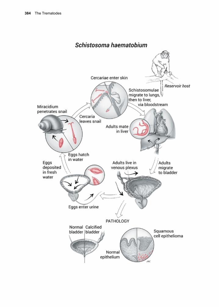
384 The Trematodes
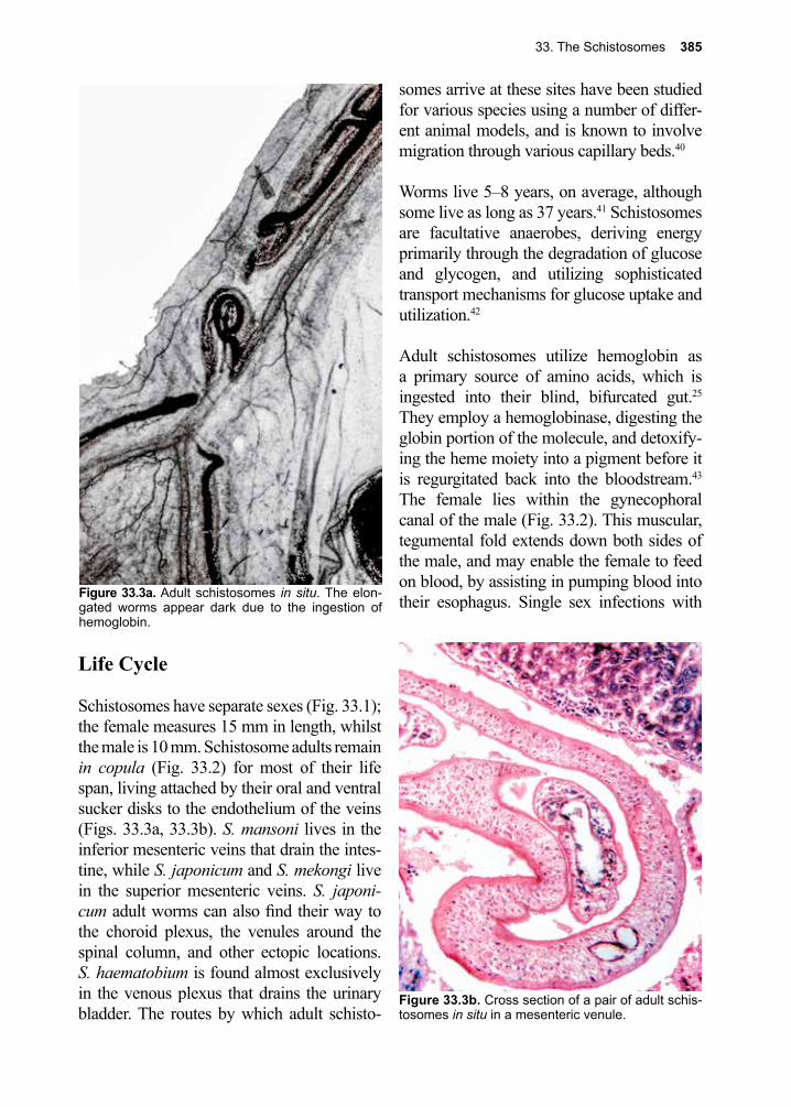
33. The Schistosomes 385
Life Cycle
Schistosomes have separate sexes (Fig. 33.1); the female measures 15 mm in length, whilst the male is 10 mm. Schistosome adults remain in copula (Fig. 33.2) for most of their life span, living attached by their oral and ventral sucker disks to the endothelium of the veins (Figs. 33.3a, 33.3b). S. mansoni lives in the inferior mesenteric veins that drain the intes-tine, while S. japonicum and S. mekongi live in the superior mesenteric veins. S. japoni-cum adult worms can also find their way to the choroid plexus, the venules around the spinal column, and other ectopic locations. S. haematobium is found almost exclusively in the venous plexus that drains the urinary bladder. The routes by which adult schisto-
somes arrive at these sites have been studied for various species using a number of differ-ent animal models, and is known to involve migration through various capillary beds.40
Worms live 5–8 years, on average, although some live as long as 37 years.41 Schistosomes are facultative anaerobes, deriving energy primarily through the degradation of glucose and glycogen, and utilizing sophisticated transport mechanisms for glucose uptake and utilization.42
Adult schistosomes utilize hemoglobin as a primary source of amino acids, which is ingested into their blind, bifurcated gut.25 They employ a hemoglobinase, digesting the globin portion of the molecule, and detoxify-ing the heme moiety into a pigment before it is regurgitated back into the bloodstream.43 The female lies within the gynecophoral canal of the male (Fig. 33.2). This muscular, tegumental fold extends down both sides of the male, and may enable the female to feed on blood, by assisting in pumping blood into their esophagus. Single sex infections with Figure 33.3a. Adult schistosomes in situ. The elon-
gated worms appear dark due to the ingestion of hemoglobin.
Figure 33.3b. Cross section of a pair of adult schis-tosomes in situ in a mesenteric venule.
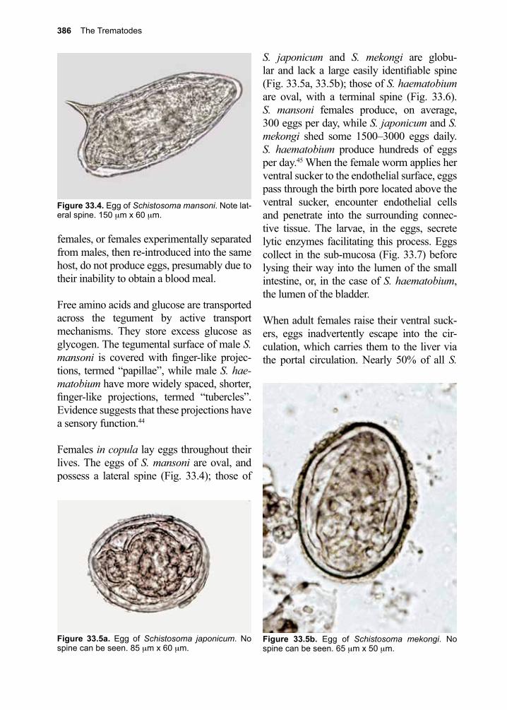
386 The Trematodes
females, or females experimentally separated from males, then re-introduced into the same host, do not produce eggs, presumably due to their inability to obtain a blood meal.
Free amino acids and glucose are transported across the tegument by active transport mechanisms. They store excess glucose as glycogen. The tegumental surface of male S. mansoni is covered with finger-like projec-tions, termed “papillae”, while male S. hae-matobium have more widely spaced, shorter, finger-like projections, termed “tubercles”. Evidence suggests that these projections have a sensory function.44
Females in copula lay eggs throughout their lives. The eggs of S. mansoni are oval, and possess a lateral spine (Fig. 33.4); those of
S. japonicum and S. mekongi are globu-lar and lack a large easily identifiable spine (Fig. 33.5a, 33.5b); those of S. haematobium are oval, with a terminal spine (Fig. 33.6). S. mansoni females produce, on average, 300 eggs per day, while S. japonicum and S. mekongi shed some 1500–3000 eggs daily. S. haematobium produce hundreds of eggs per day.45 When the female worm applies her ventral sucker to the endothelial surface, eggs pass through the birth pore located above the ventral sucker, encounter endothelial cells and penetrate into the surrounding connec-tive tissue. The larvae, in the eggs, secrete lytic enzymes facilitating this process. Eggs collect in the sub-mucosa (Fig. 33.7) before lysing their way into the lumen of the small intestine, or, in the case of S. haematobium, the lumen of the bladder.
When adult females raise their ventral suck-ers, eggs inadvertently escape into the cir-culation, which carries them to the liver via the portal circulation. Nearly 50% of all S.
Figure 33.4. Egg of Schistosoma mansoni. Note lat-eral spine. 150 µm x 60 µm.
Figure 33.5a. Egg of Schistosoma japonicum. No spine can be seen. 85 µm x 60 µm.
Figure 33.5b. Egg of Schistosoma mekongi. No spine can be seen. 65 µm x 50 µm.
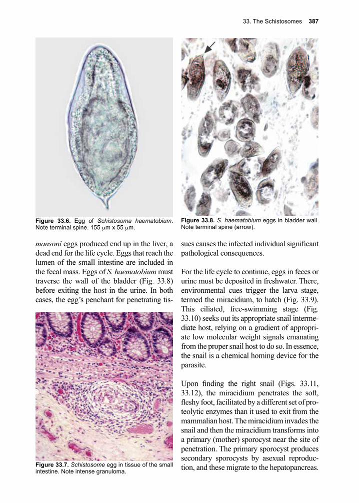
33. The Schistosomes 387
mansoni eggs produced end up in the liver, a dead end for the life cycle. Eggs that reach the lumen of the small intestine are included in the fecal mass. Eggs of S. haematobium must traverse the wall of the bladder (Fig. 33.8) before exiting the host in the urine. In both cases, the egg’s penchant for penetrating tis-
sues causes the infected individual significant pathological consequences.
For the life cycle to continue, eggs in feces or urine must be deposited in freshwater. There, environmental cues trigger the larva stage, termed the miracidium, to hatch (Fig. 33.9). This ciliated, free-swimming stage (Fig. 33.10) seeks out its appropriate snail interme-diate host, relying on a gradient of appropri-ate low molecular weight signals emanating from the proper snail host to do so. In essence, the snail is a chemical homing device for the parasite.
Upon finding the right snail (Figs. 33.11, 33.12), the miracidium penetrates the soft, fleshy foot, facilitated by a different set of pro-teolytic enzymes than it used to exit from the mammalian host. The miracidium invades the snail and then the miracidium transforms into a primary (mother) sporocyst near the site of penetration. The primary sporocyst produces secondary sporocysts by asexual reproduc-tion, and these migrate to the hepatopancreas.
Figure 33.8. S. haematobium eggs in bladder wall. Note terminal spine (arrow).
Figure 33.6. Egg of Schistosoma haematobium. Note terminal spine. 155 µm x 55 µm.
Figure 33.7. Schistosome egg in tissue of the small intestine. Note intense granuloma.
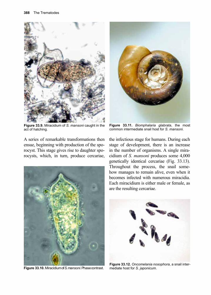
388 The Trematodes
Figure 33.11. Biomphalaria glabrata, the most common intermediate snail host for S. mansoni.
Figure 33.12. Oncomelania nosophora, a snail inter-mediate host for S. japonicum.
A series of remarkable transformations then ensue, beginning with production of the spo-rocyst. This stage gives rise to daughter spo-rocysts, which, in turn, produce cercariae,
the infectious stage for humans. During each stage of development, there is an increase in the number of organisms. A single mira-cidium of S. mansoni produces some 4,000 genetically identical cercariae (Fig. 33.13). Throughout the process, the snail some-how manages to remain alive, even when it becomes infected with numerous miracidia. Each miracidium is either male or female, as are the resulting cercariae.
Figure 33.9. Miracidium of S. mansoni caught in the act of hatching.
Figure 33.10. Miracidium of S. mansoni. Phase contrast.
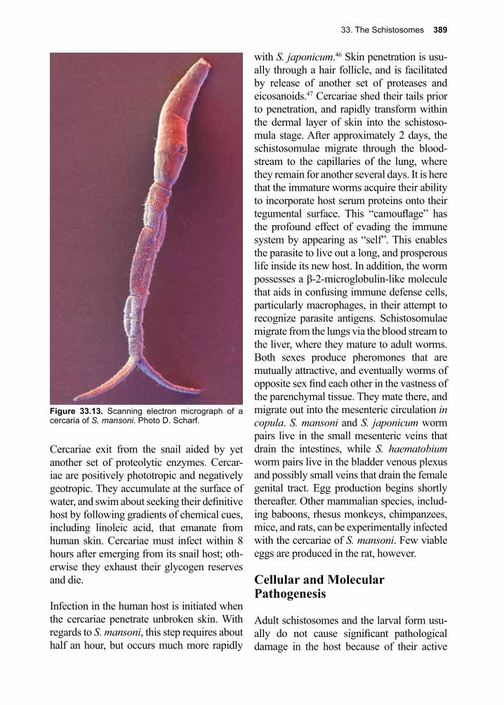
33. The Schistosomes 389
Cercariae exit from the snail aided by yet another set of proteolytic enzymes. Cercar-iae are positively phototropic and negatively geotropic. They accumulate at the surface of water, and swim about seeking their definitive host by following gradients of chemical cues, including linoleic acid, that emanate from human skin. Cercariae must infect within 8 hours after emerging from its snail host; oth-erwise they exhaust their glycogen reserves and die.
Infection in the human host is initiated when the cercariae penetrate unbroken skin. With regards to S. mansoni, this step requires about half an hour, but occurs much more rapidly
with S. japonicum.46 Skin penetration is usu-ally through a hair follicle, and is facilitated by release of another set of proteases and eicosanoids.47 Cercariae shed their tails prior to penetration, and rapidly transform within the dermal layer of skin into the schistoso-mula stage. After approximately 2 days, the schistosomulae migrate through the blood-stream to the capillaries of the lung, where they remain for another several days. It is here that the immature worms acquire their ability to incorporate host serum proteins onto their tegumental surface. This “camouflage” has the profound effect of evading the immune system by appearing as “self”. This enables the parasite to live out a long, and prosperous life inside its new host. In addition, the worm possesses a β-2-microglobulin-like molecule that aids in confusing immune defense cells, particularly macrophages, in their attempt to recognize parasite antigens. Schistosomulae migrate from the lungs via the blood stream to the liver, where they mature to adult worms. Both sexes produce pheromones that are mutually attractive, and eventually worms of opposite sex find each other in the vastness of the parenchymal tissue. They mate there, and migrate out into the mesenteric circulation in copula. S. mansoni and S. japonicum worm pairs live in the small mesenteric veins that drain the intestines, while S. haematobium worm pairs live in the bladder venous plexus and possibly small veins that drain the female genital tract. Egg production begins shortly thereafter. Other mammalian species, includ-ing baboons, rhesus monkeys, chimpanzees, mice, and rats, can be experimentally infected with the cercariae of S. mansoni. Few viable eggs are produced in the rat, however.
Cellular and Molecular Pathogenesis
Adult schistosomes and the larval form usu-ally do not cause significant pathological damage in the host because of their active
Figure 33.13. Scanning electron micrograph of a cercaria of S. mansoni. Photo D. Scharf.
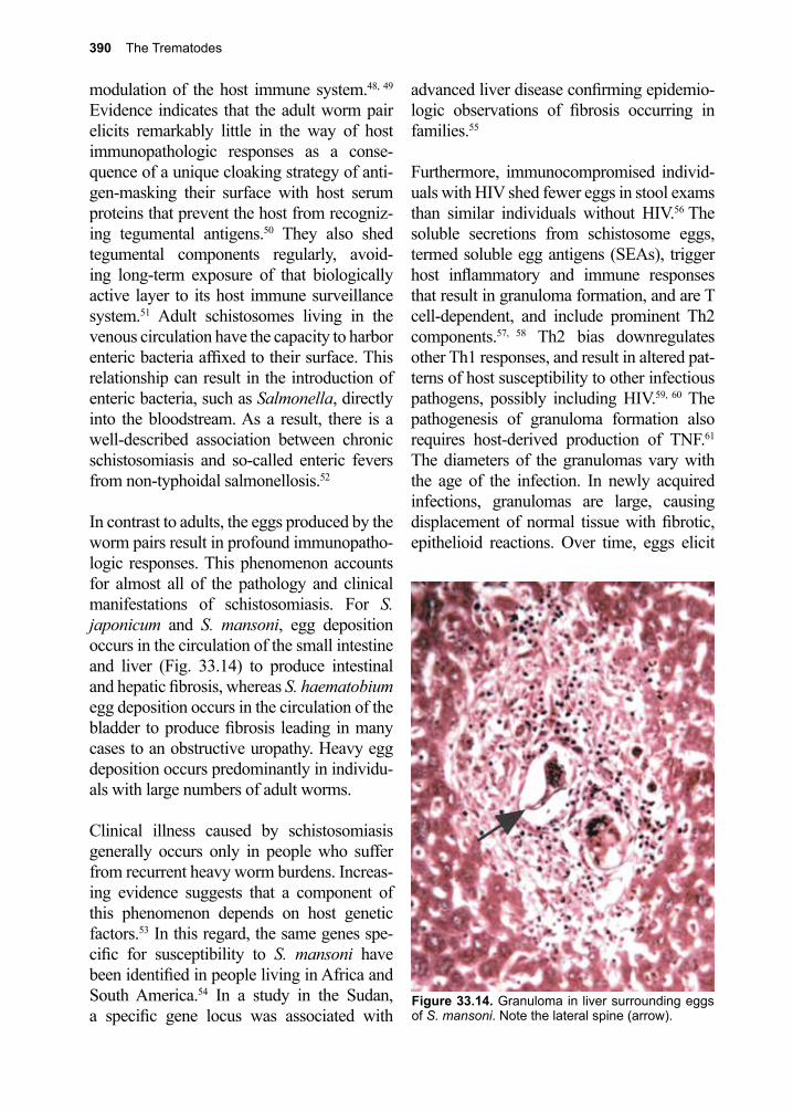
390 The Trematodes
modulation of the host immune system.48, 49 Evidence indicates that the adult worm pair elicits remarkably little in the way of host immunopathologic responses as a conse-quence of a unique cloaking strategy of anti-gen-masking their surface with host serum proteins that prevent the host from recogniz-ing tegumental antigens.50 They also shed tegumental components regularly, avoid-ing long-term exposure of that biologically active layer to its host immune surveillance system.51 Adult schistosomes living in the venous circulation have the capacity to harbor enteric bacteria affixed to their surface. This relationship can result in the introduction of enteric bacteria, such as Salmonella, directly into the bloodstream. As a result, there is a well-described association between chronic schistosomiasis and so-called enteric fevers from non-typhoidal salmonellosis.52
In contrast to adults, the eggs produced by the worm pairs result in profound immunopatho-logic responses. This phenomenon accounts for almost all of the pathology and clinical manifestations of schistosomiasis. For S. japonicum and S. mansoni, egg deposition occurs in the circulation of the small intestine and liver (Fig. 33.14) to produce intestinal and hepatic fibrosis, whereas S. haematobium egg deposition occurs in the circulation of the bladder to produce fibrosis leading in many cases to an obstructive uropathy. Heavy egg deposition occurs predominantly in individu-als with large numbers of adult worms.
Clinical illness caused by schistosomiasis generally occurs only in people who suffer from recurrent heavy worm burdens. Increas-ing evidence suggests that a component of this phenomenon depends on host genetic factors.53 In this regard, the same genes spe-cific for susceptibility to S. mansoni have been identified in people living in Africa and South America.54 In a study in the Sudan, a specific gene locus was associated with
advanced liver disease confirming epidemio-logic observations of fibrosis occurring in families.55
Furthermore, immunocompromised individ-uals with HIV shed fewer eggs in stool exams than similar individuals without HIV.56 The soluble secretions from schistosome eggs, termed soluble egg antigens (SEAs), trigger host inflammatory and immune responses that result in granuloma formation, and are T cell-dependent, and include prominent Th2 components.57, 58 Th2 bias downregulates other Th1 responses, and result in altered pat-terns of host susceptibility to other infectious pathogens, possibly including HIV.59, 60 The pathogenesis of granuloma formation also requires host-derived production of TNF.61 The diameters of the granulomas vary with the age of the infection. In newly acquired infections, granulomas are large, causing displacement of normal tissue with fibrotic, epithelioid reactions. Over time, eggs elicit
Figure 33.14. Granuloma in liver surrounding eggs of S. mansoni. Note the lateral spine (arrow).

33. The Schistosomes 391
less and less volume of granulomatous tissue. This reaction appears to be under the regula-tion of interleukin-12.62
Granulomas form around eggs that collect in the intestinal wall and results in fibrosis. Ero-sion of the submucosa and villous tissue also occurs, presumably by the action of secreted proteolytic enzymes from the miracidia within the eggs. In heavy infection, gastroin-testinal (GI) hemorrhage results from damage to the submucosa.
Eggs swept back into the liver block pre-sinusoidal capillaries, and induce granulomas there, as well. The presence of granulomas causes tissue fibrosis, and eventually leads to obstruction of the hepatic vasculature. Fibro-sis of most of the portal areas incorporating the blood vessels leads to pipe stem fibrosis (Symmers’ fibrosis) (Fig. 33.15), and, ulti-mately, to portal hypertension. Clinically, this manifests as hepatosplenomegaly, the extent
of which is dependent partially on host major histocompatibility class II alleles.63 Develop-ment of collateral circulation follows, includ-ing esophageal varices. Parenchymal liver cells remain unaffected by granulomas and liver function remains normal.
Portal hypertension forces eggs to bypass the liver, and many are carried to the spleen, which becomes enlarged, further contribut-ing to increased pressure in portal circula-tion. Infection with S. japonicum results in a greater number of granulomas, and conse-quently greater morbidity because this spe-cies produces, on average, five to ten times more eggs than S. mansoni. Collateral circu-
Figure 33.16. X-ray showing calcified dome of the bladder due to chronic infection with S. haematobium.
Figure 33.17. Histological section of bladder with pseu-dopolyp due to chronic infection with S. haematobium.
Figure 33.15. Pipe stem fibrosis in liver due to heavy infection with S. mansoni. Note normal liver tissue next to fibrotic vessels.
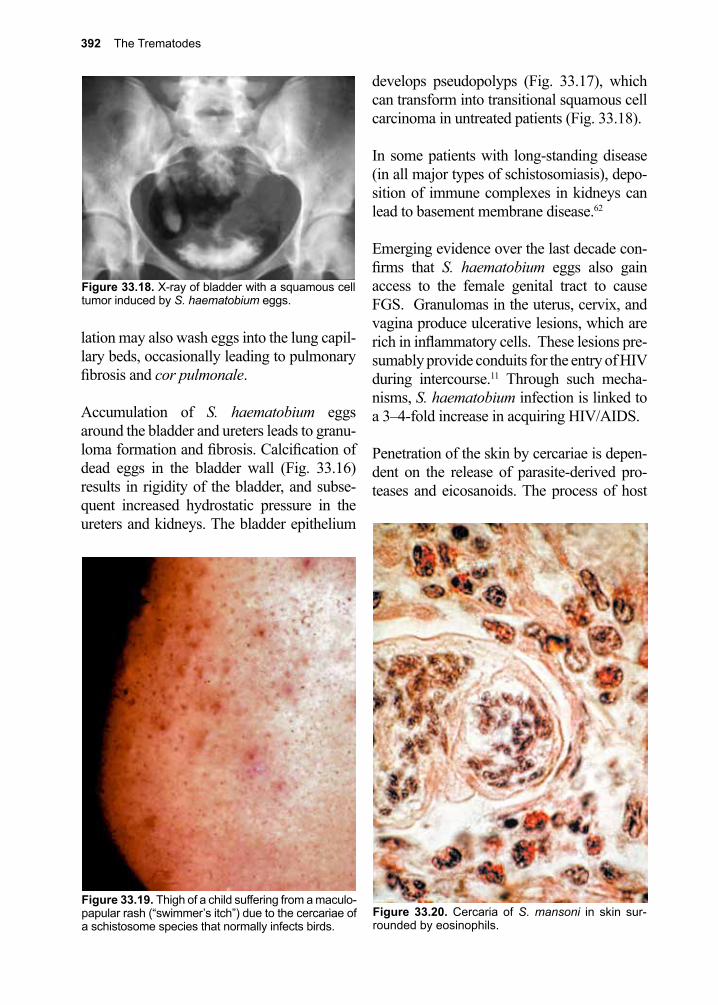
392 The Trematodes
lation may also wash eggs into the lung capil-lary beds, occasionally leading to pulmonary fibrosis and cor pulmonale.
Accumulation of S. haematobium eggs around the bladder and ureters leads to granu-loma formation and fibrosis. Calcification of dead eggs in the bladder wall (Fig. 33.16) results in rigidity of the bladder, and subse-quent increased hydrostatic pressure in the ureters and kidneys. The bladder epithelium
develops pseudopolyps (Fig. 33.17), which can transform into transitional squamous cell carcinoma in untreated patients (Fig. 33.18).
In some patients with long-standing disease (in all major types of schistosomiasis), depo-sition of immune complexes in kidneys can lead to basement membrane disease.62
Emerging evidence over the last decade con-firms that S. haematobium eggs also gain access to the female genital tract to cause FGS. Granulomas in the uterus, cervix, and vagina produce ulcerative lesions, which are rich in inflammatory cells. These lesions pre-sumably provide conduits for the entry of HIV during intercourse.11 Through such mecha-nisms, S. haematobium infection is linked to a 3–4-fold increase in acquiring HIV/AIDS.
Penetration of the skin by cercariae is depen-dent on the release of parasite-derived pro-teases and eicosanoids. The process of host
Figure 33.20. Cercaria of S. mansoni in skin sur-rounded by eosinophils.
Figure 33.18. X-ray of bladder with a squamous cell tumor induced by S. haematobium eggs.
Figure 33.19. Thigh of a child suffering from a maculo-papular rash (“swimmer’s itch”) due to the cercariae of a schistosome species that normally infects birds.
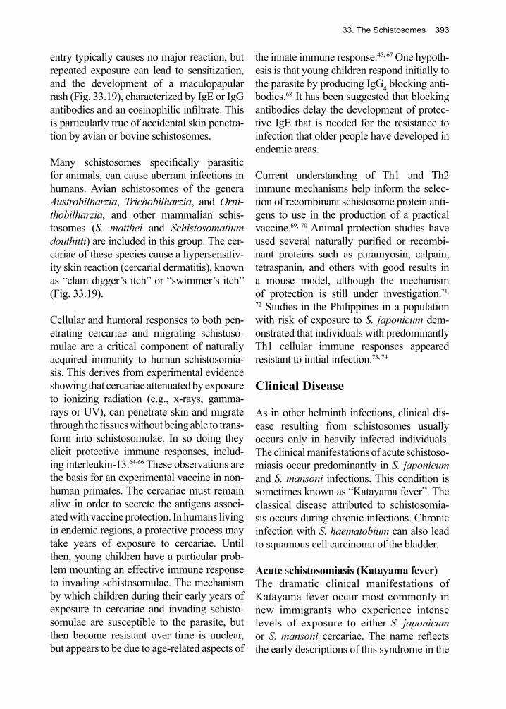
33. The Schistosomes 393
entry typically causes no major reaction, but repeated exposure can lead to sensitization, and the development of a maculopapular rash (Fig. 33.19), characterized by IgE or IgG antibodies and an eosinophilic infiltrate. This is particularly true of accidental skin penetra-tion by avian or bovine schistosomes.
Many schistosomes specifically parasitic for animals, can cause aberrant infections in humans. Avian schistosomes of the genera Austrobilharzia, Trichobilharzia, and Orni-thobilharzia, and other mammalian schis-tosomes (S. matthei and Schistosomatium douthitti) are included in this group. The cer-cariae of these species cause a hypersensitiv-ity skin reaction (cercarial dermatitis), known as “clam digger’s itch” or “swimmer’s itch” (Fig. 33.19).
Cellular and humoral responses to both pen-etrating cercariae and migrating schistoso-mulae are a critical component of naturally acquired immunity to human schistosomia-sis. This derives from experimental evidence showing that cercariae attenuated by exposure to ionizing radiation (e.g., x-rays, gamma-rays or UV), can penetrate skin and migrate through the tissues without being able to trans-form into schistosomulae. In so doing they elicit protective immune responses, includ-ing interleukin-13.64-66 These observations are the basis for an experimental vaccine in non-human primates. The cercariae must remain alive in order to secrete the antigens associ-ated with vaccine protection. In humans living in endemic regions, a protective process may take years of exposure to cercariae. Until then, young children have a particular prob-lem mounting an effective immune response to invading schistosomulae. The mechanism by which children during their early years of exposure to cercariae and invading schisto-somulae are susceptible to the parasite, but then become resistant over time is unclear, but appears to be due to age-related aspects of
the innate immune response.45, 67 One hypoth-esis is that young children respond initially to the parasite by producing IgG4 blocking anti-bodies.68 It has been suggested that blocking antibodies delay the development of protec-tive IgE that is needed for the resistance to infection that older people have developed in endemic areas.
Current understanding of Th1 and Th2 immune mechanisms help inform the selec-tion of recombinant schistosome protein anti-gens to use in the production of a practical vaccine.69, 70 Animal protection studies have used several naturally purified or recombi-nant proteins such as paramyosin, calpain, tetraspanin, and others with good results in a mouse model, although the mechanism of protection is still under investigation.71,
72 Studies in the Philippines in a population with risk of exposure to S. japonicum dem-onstrated that individuals with predominantly Th1 cellular immune responses appeared resistant to initial infection.73, 74
Clinical Disease
As in other helminth infections, clinical dis-ease resulting from schistosomes usually occurs only in heavily infected individuals. The clinical manifestations of acute schistoso-miasis occur predominantly in S. japonicum and S. mansoni infections. This condition is sometimes known as “Katayama fever”. The classical disease attributed to schistosomia-sis occurs during chronic infections. Chronic infection with S. haematobium can also lead to squamous cell carcinoma of the bladder.
Acute schistosomiasis (Katayama fever)The dramatic clinical manifestations of Katayama fever occur most commonly in new immigrants who experience intense levels of exposure to either S. japonicum or S. mansoni cercariae. The name reflects the early descriptions of this syndrome in the
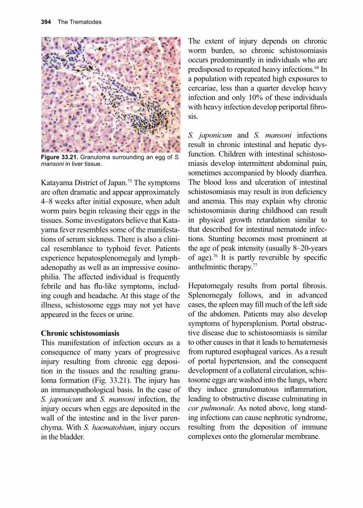
394 The Trematodes
Katayama District of Japan.75 The symptoms are often dramatic and appear approximately 4–8 weeks after initial exposure, when adult worm pairs begin releasing their eggs in the tissues. Some investigators believe that Kata-yama fever resembles some of the manifesta-tions of serum sickness. There is also a clini-cal resemblance to typhoid fever. Patients experience hepatosplenomegaly and lymph-adenopathy as well as an impressive eosino-philia. The affected individual is frequently febrile and has flu-like symptoms, includ-ing cough and headache. At this stage of the illness, schistosome eggs may not yet have appeared in the feces or urine.
Chronic schistosomiasis This manifestation of infection occurs as a consequence of many years of progressive injury resulting from chronic egg deposi-tion in the tissues and the resulting granu-loma formation (Fig. 33.21). The injury has an immunopathological basis. In the case of S. japonicum and S. mansoni infection, the injury occurs when eggs are deposited in the wall of the intestine and in the liver paren-chyma. With S. haematobium, injury occurs in the bladder.
The extent of injury depends on chronic worm burden, so chronic schistosomiasis occurs predominantly in individuals who are predisposed to repeated heavy infections.68 In a population with repeated high exposures to cercariae, less than a quarter develop heavy infection and only 10% of these individuals with heavy infection develop periportal fibro-sis.
S. japonicum and S. mansoni infections result in chronic intestinal and hepatic dys-function. Children with intestinal schistoso-miasis develop intermittent abdominal pain, sometimes accompanied by bloody diarrhea. The blood loss and ulceration of intestinal schistosomiasis may result in iron deficiency and anemia. This may explain why chronic schistosomiasis during childhood can result in physical growth retardation similar to that described for intestinal nematode infec-tions. Stunting becomes most prominent at the age of peak intensity (usually 8–20-years of age).76 It is partly reversible by specific anthelmintic therapy.77
Hepatomegaly results from portal fibrosis. Splenomegaly follows, and in advanced cases, the spleen may fill much of the left side of the abdomen. Patients may also develop symptoms of hypersplenism. Portal obstruc-tive disease due to schistosomiasis is similar to other causes in that it leads to hematemesis from ruptured esophageal varices. As a result of portal hypertension, and the consequent development of a collateral circulation, schis-tosome eggs are washed into the lungs, where they induce granulomatous inflammation, leading to obstructive disease culminating in cor pulmonale. As noted above, long stand-ing infections can cause nephrotic syndrome, resulting from the deposition of immune complexes onto the glomerular membrane.
Figure 33.21. Granuloma surrounding an egg of S. mansoni in liver tissue.
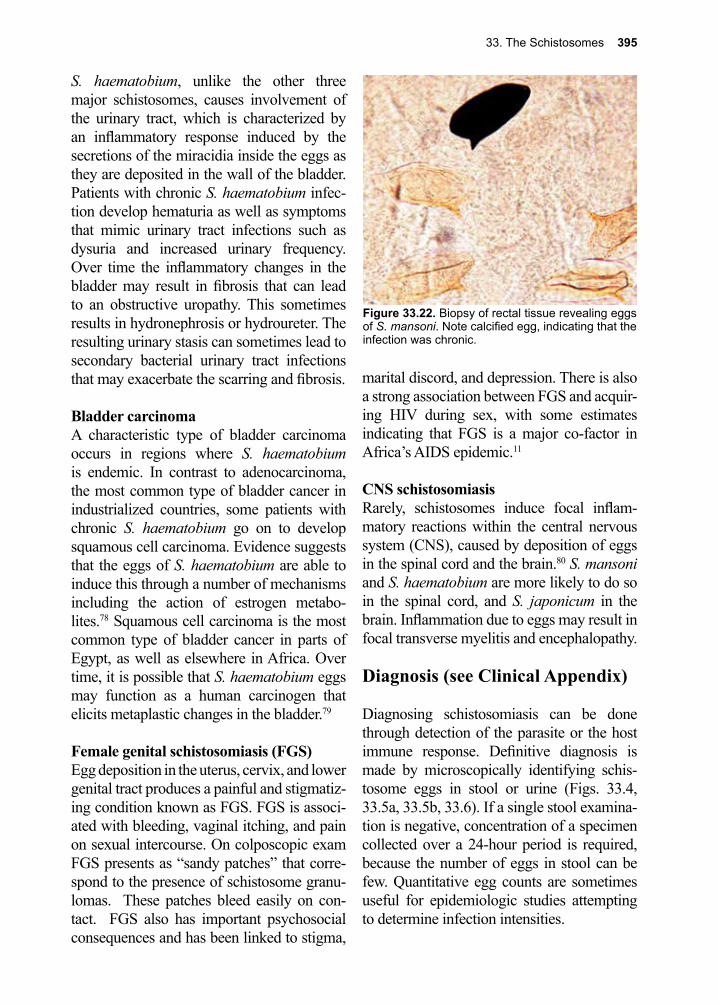
33. The Schistosomes 395
S. haematobium, unlike the other three major schistosomes, causes involvement of the urinary tract, which is characterized by an inflammatory response induced by the secretions of the miracidia inside the eggs as they are deposited in the wall of the bladder. Patients with chronic S. haematobium infec-tion develop hematuria as well as symptoms that mimic urinary tract infections such as dysuria and increased urinary frequency. Over time the inflammatory changes in the bladder may result in fibrosis that can lead to an obstructive uropathy. This sometimes results in hydronephrosis or hydroureter. The resulting urinary stasis can sometimes lead to secondary bacterial urinary tract infections that may exacerbate the scarring and fibrosis.
Bladder carcinomaA characteristic type of bladder carcinoma occurs in regions where S. haematobium is endemic. In contrast to adenocarcinoma, the most common type of bladder cancer in industrialized countries, some patients with chronic S. haematobium go on to develop squamous cell carcinoma. Evidence suggests that the eggs of S. haematobium are able to induce this through a number of mechanisms including the action of estrogen metabo-lites.78 Squamous cell carcinoma is the most common type of bladder cancer in parts of Egypt, as well as elsewhere in Africa. Over time, it is possible that S. haematobium eggs may function as a human carcinogen that elicits metaplastic changes in the bladder.79
Female genital schistosomiasis (FGS)Egg deposition in the uterus, cervix, and lower genital tract produces a painful and stigmatiz-ing condition known as FGS. FGS is associ-ated with bleeding, vaginal itching, and pain on sexual intercourse. On colposcopic exam FGS presents as “sandy patches” that corre-spond to the presence of schistosome granu-lomas. These patches bleed easily on con-tact. FGS also has important psychosocial consequences and has been linked to stigma,
marital discord, and depression. There is also a strong association between FGS and acquir-ing HIV during sex, with some estimates indicating that FGS is a major co-factor in Africa’s AIDS epidemic.11
CNS schistosomiasisRarely, schistosomes induce focal inflam-matory reactions within the central nervous system (CNS), caused by deposition of eggs in the spinal cord and the brain.80 S. mansoni and S. haematobium are more likely to do so in the spinal cord, and S. japonicum in the brain. Inflammation due to eggs may result in focal transverse myelitis and encephalopathy.
Diagnosis (see Clinical Appendix)
Diagnosing schistosomiasis can be done through detection of the parasite or the host immune response. Definitive diagnosis is made by microscopically identifying schis-tosome eggs in stool or urine (Figs. 33.4, 33.5a, 33.5b, 33.6). If a single stool examina-tion is negative, concentration of a specimen collected over a 24-hour period is required, because the number of eggs in stool can be few. Quantitative egg counts are sometimes useful for epidemiologic studies attempting to determine infection intensities.
Figure 33.22. Biopsy of rectal tissue revealing eggs of S. mansoni. Note calcified egg, indicating that the infection was chronic.
396 The Trematodes
For light infections, or in patients from whom egg excretion is intermittent, and from whom eggs cannot be found in stool, a rectal biopsy can be carried out (Fig. 33.22). The tissue is squashed between two microscope slides and examined under the low-power lens of a microscope. If eggs are detected, they can then be observed under higher power and examined for the presence of “flame” cells (excretory cells). If they are flickering (as in a flame), then the miracidium in the egg is alive, and the patient has an active infection. Treatment must then be entertained. If no live eggs are seen, or if they are calcified, then it is likely that the infection is no longer active, and treatment is not necessary. It is helpful to refer to the specimen as a “rectal snip”, rather than a biopsy, to preclude its fixation and subsequent sectioning, which would make the identification of live miracidia in eggs impossible.
While most schistosome eggs appear in feces, urine should be examined for the presence of eggs of S. haematobium if this species is sus-pected. The urine sample should generally be collected close to noon, when egg excretion is usually maximal. Urine may have to be con-centrated by sedimentation to reveal the few eggs present. S. haematobium eggs may also be seen in stool and rectal snip specimens, but their numbers are typically small in these samples.
Diagnosis of FGS requires training to identify the characteristic sandy patches associated with this condition. Confirmatory micros-copy is a useful aid but not always available in resource-poor settings.
A number of additional tests have become available for the diagnosis of schistosomia-sis. Two schistosome glycoprotein antigens known as CCA and CAA that circulate in the bloodstream of acutely-infected patients have been identified and can now be tested for
using both qualitative and quantitative assays that are currently available.81-83 These assays can be easily applied to urine samples.84, 85 A number of NAATs have been developed that allow for detection as well as schistosome speciation.86-88
Antibodies develop 6–12 weeks after expo-sure and tend to become positive before eggs are evident in urine or stool.89 There are a number of serological tests available with western blot confirmatory testing, as well.90 Since serologies remain positive for long periods after treatment of infection, they cannot be used for follow-up and are difficult to use to monitor repeat infections in endemic areas.91
Portable ultrasound imaging has been shown to be clinically useful in the diagnosis of schistosomiasis. Ultrasound can define the extent of Symmers’ fibrosis in patients with S. mansoni or S. japonicum infections, while the chronic obstructive changes associated with S. haematobium infection can also be detected.92
Treatment (see Clinical Appendix)
Optimal treatment depends on whether one is treating acute schistosomiasis syndrome (Katayama fever) or the chronic phase of the disease. Since the acute schistosomiasis syndrome is a hypersensitivity reaction to the parasitic antigens, anthelmintic therapy results in exacerbation of symptoms in about half of those treated.93 Short course treatment with corticosteroids and delay of anthelmin-tic therapy improves symptoms, leads to decreased morbidity and avoids the clinical deterioration seen with acute use of anthel-mintic treatment.93, 94
Acute treatment with anthelmintic therapy is not required to prevent the chronic mani-festations of schistosomiasis.95 How long to
33. The Schistosomes 397
wait after the acute syndrome subsides before initiating anthelmintic therapy is unknown, but it seems prudent to wait for a period (6 weeks according to some) after resolution of the acute symptoms to start treatment when the worms have fully matured, as praziquan-tel, the drug of choice for schistosomiasis, is not active against the larval stage.95 Treatment can then be repeated 4–6 weeks after this first treatment course.
For treatment of chronic schistosomiasis, pra-ziquantel is the drug of choice for most species of schistosomes. This drug is well-tolerated, is associated with few side effects (nausea, epi-gastric pain, dizziness, and general malaise), has a very high therapeutic index, and a single dose is usually sufficient to greatly reduce the worm burden in individuals in endemic areas with high worm burdens, and to cure those with low worm burdens.96-98 Praziquan-tel interferes with calcium ion influx across the tegument, resulting in spastic paralysis of the worm. At higher doses, the tegument develops blebs and is unmasked, making it susceptible to immune attack.99 In younger patients, praziquantel may also reverse some of the pathology associated with Symmers’ fibrosis.100 There is evidence that part of its effectiveness is due to synergism with the host’s humoral immune response.101 Because it is effective in a single dose, it has been used in control programs (see below).
For patients no longer in endemic areas, test of cure is recommended with repeat testing for eggs in urine or stool no sooner than 3–6 months after treatment.102 When treatment is initiated patients should be monitored for evi-dence of CNS disease, as treatment can cause an acute inflammatory response in patients who have eggs in the CNS. If this develops, prompt and prolonged therapy with corti-costeroids is critical to prevent worsening of neurological symptoms and irreversible damage.103, 104
Praziquantel is now inexpensive and the World Health Assembly has endorsed com-munity treatment of school-age children in endemic areas. The Schistosomiasis Con-trol Initiative based at Imperial College, London, is leading global efforts to provide mass drug administration of praziquantel. Currently, praziquantel is being provided in “rapid impact” interventions, which include mass-drug treatments for intestinal helminth infections, lymphatic filariasis, and onchocer-ciasis.105 Political obstacles have so far failed to link this approach with antiretroviral drug therapy for HIV/AIDS. Millions of girls and women living in poverty in Africa have been denied access to an inexpensive approach to HIV/AIDS prevention.106 Resistance has occurred and a number of treatment failures have been reported.107-109
Alternatives to praziquantel are limited in use due to a higher frequency of adverse reac-tions and differences in spectrum of activity. Oxamniquine is an alternate drug with good anti-parasitic activity. In some regions, oxam-niquine is as effective as praziquantel for the treatment of infections with S. mansoni, and metrifonate is effective for the treatment of S. haematobium infections.110 The anti-malarial drug artemether has been studied in China as a chemoprophylactic agent in patients who anticipate high levels of exposure to S. japonicum and S. mansoni cercariae during seasonal floods. Chemoprophylactic activity of artemether was present but lower against S. haematobium.111 The efficacy of praziquantel is enhanced when combined with artemether, and the combination might prevent the emer-gence of resistance to praziquantel when used in widespread and repeated community treat-ment.112
Treatment should be carried out only in patients with active schistosome infections. Portocaval or splenorenal shunts should be avoided in untreated schistosomiasis, because
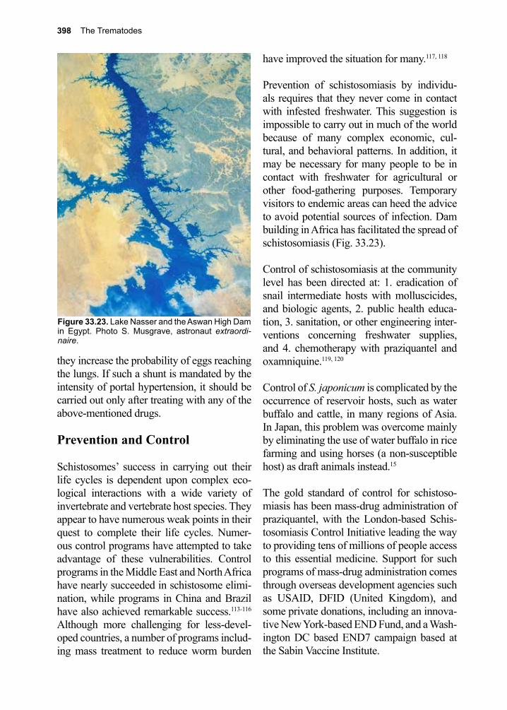
398 The Trematodes
they increase the probability of eggs reaching the lungs. If such a shunt is mandated by the intensity of portal hypertension, it should be carried out only after treating with any of the above-mentioned drugs.
Prevention and Control
Schistosomes’ success in carrying out their life cycles is dependent upon complex eco-logical interactions with a wide variety of invertebrate and vertebrate host species. They appear to have numerous weak points in their quest to complete their life cycles. Numer-ous control programs have attempted to take advantage of these vulnerabilities. Control programs in the Middle East and North Africa have nearly succeeded in schistosome elimi-nation, while programs in China and Brazil have also achieved remarkable success.113-116 Although more challenging for less-devel-oped countries, a number of programs includ-ing mass treatment to reduce worm burden
have improved the situation for many.117, 118
Prevention of schistosomiasis by individu-als requires that they never come in contact with infested freshwater. This suggestion is impossible to carry out in much of the world because of many complex economic, cul-tural, and behavioral patterns. In addition, it may be necessary for many people to be in contact with freshwater for agricultural or other food-gathering purposes. Temporary visitors to endemic areas can heed the advice to avoid potential sources of infection. Dam building in Africa has facilitated the spread of schistosomiasis (Fig. 33.23).
Control of schistosomiasis at the community level has been directed at: 1. eradication of snail intermediate hosts with molluscicides, and biologic agents, 2. public health educa-tion, 3. sanitation, or other engineering inter-ventions concerning freshwater supplies, and 4. chemotherapy with praziquantel and oxamniquine.119, 120
Control of S. japonicum is complicated by the occurrence of reservoir hosts, such as water buffalo and cattle, in many regions of Asia. In Japan, this problem was overcome mainly by eliminating the use of water buffalo in rice farming and using horses (a non-susceptible host) as draft animals instead.15
The gold standard of control for schistoso-miasis has been mass-drug administration of praziquantel, with the London-based Schis-tosomiasis Control Initiative leading the way to providing tens of millions of people access to this essential medicine. Support for such programs of mass-drug administration comes through overseas development agencies such as USAID, DFID (United Kingdom), and some private donations, including an innova-tive New York-based END Fund, and a Wash-ington DC based END7 campaign based at the Sabin Vaccine Institute.
Figure 33.23. Lake Nasser and the Aswan High Dam in Egypt. Photo S. Musgrave, astronaut extraordi-naire.
33. The Schistosomes 399
Studies in endemic areas have shown that while praziquantel is effective at treating large populations, there is a high rate of post-treatment reinfection. This necessitates fre-quent administration of the drug, although this tactic is frequently not possible in poor, developing rural areas without the support of the international community.121 Control of the
infection with anthelmintic drugs alone is dif-ficult. There is also concern about the emer-gence of praziquantel drug resistance.122
Investigations into vaccinations and new therapeutics are currently underway and offer hope for improved methods to help address this neglected tropical disease.45, 62, 69, 71, 72, 123-129
References
1. Gurarie, D., et al., Modeling the effect of chronic schistosomiasis on childhood development and the potential for catch-up growth with different drug treatment strategies promoted for control of endemic schistosomiasis. Am J Trop Med Hyg 2011, 84 (5), 773-81.
2. Global Burden of Disease Study, C., Global, regional, and national incidence, prevalence, and years lived with disability for 301 acute and chronic diseases and injuries in 188 countries, 1990-2013: a systematic analysis for the Global Burden of Disease Study 2013. Lancet 2015, 386 (9995), 743-800.
3. Rollinson, D., et al., Time to set the agenda for schistosomiasis elimination. Acta Trop 2013, 128 (2), 423-40.
4. Utzinger, J., et al., Schistosomiasis and neglected tropical diseases: towards integrated and sustainable control and a word of caution. Parasitology 2009, 136 (13), 1859-74.
5. Steinmann, P., et al., Schistosomiasis and water resources development: systematic review, meta-analysis, and estimates of people at risk. Lancet Infect Dis 2006, 6 (7), 411-25.
6. King, C. H., Parasites and poverty: the case of schistosomiasis. Acta Trop 2010, 113 (2), 95-104.7. Lammie, P. J., et al., Eliminating lymphatic filariasis, onchocerciasis, and schistosomiasis from
the americas: breaking a historical legacy of slavery. PLoS Negl Trop Dis 2007, 1 (2), e71.8. Douard, A., et al., Urinary schistosomiasis in France. Int J Infect Dis 2011, 15 (7), e506-7.9. Berry, A., et al., Schistosomiasis haematobium, Corsica, France. Emerg Infect Dis 2014, 20 (9),
1595-7.10. Fuller, G. K., et al., Schistosomiasis in Omo National Park of southwest Ethiopia. Am J Trop
Med Hyg 1979, 28 (3), 526-30.11. Kjetland, E. F., et al., Genital schistosomiasis and its unacknowledged role on HIV transmission
in the STD intervention studies. Int J STD AIDS 2014, 25 (10), 705-15.12. Chu, T., et al., Prevalence of Schistosoma intercalatum and S. haematobium Infection among
Primary Schoolchildren in Capital Areas of Democratic Republic Of Sao Tome and Principe, West Africa. Iran J Parasitol 2012, 7 (1), 67-72.
13. Dufillot, D., et al., [Prevalence of Schistosoma intercalatum intestinal bilharziasis in children hospitalized the urban area of Gabon]. Arch Pediatr 1995, 2 (10), 1023-4.
14. World Health Organization. Weekly Epidemiol Rec 171 1989, 64.15. Minai, M., et al., Historical view of Schistosomiasis japonica in Japan: implementation and
evaluation of disease-control strategies in Yamanashi Prefecture. Parasitology international 2003, 52 (4), 321-6.
16. Hotez, P. J., et al., Emerging and reemerging helminthiases and the public health of China. Emerging infectious diseases 1997, 3 (3), 303-10.
17. Kongklieng, A., et al., Molecular differentiation of Schistosoma japonicum and Schistosoma mekongi by real-time PCR with high resolution melting analysis. Korean J Parasitol 2013, 51 (6), 651-6.
18. Ruffer, M. A., Note on the Presence of “Bilharzia Haematobia” in Egyptian Mummies of the Twentieth Dynasty [1250-1000 B.C.]. Br Med J 1910, 1 (2557), 16.
19. Nunn, J. F.; Tapp, E., Tropical diseases in ancient Egypt. Trans R Soc Trop Med Hyg 2000, 94 (2), 147-53.
400 The Trematodes
20. Renoult, A. J., Notice sur l’he´maturie qu’e´prouvent les Europe´ens dans la haute Egypte et la Nubie. J. Gen. Med. Chir. Pharm 1808, 17, 366-370.
21. Cox, F. E., History of human parasitology. Clin Microbiol Rev 2002, 15 (4), 595-612.22. Bilharz, T.; von Siebold, C. T., Ein Beitrag zur Helminhographia humana, aus brieflichen
Mitteilungen des Dr. Bilharz in Cairo, nenst Bermerkungen von Prof. C. Th. von Siebold in Breslau. Z. Wiss. Zool. 1852, 4, 53-76.
23. Bilharz, T., Fernere mittheilungen u¨ber Distomun haematobium. Z. Wiss. Zool. 1853, 4, 454-456.
24. Kean, B. H., et al., Tropical medicine and parasitology: classic investigations. Cornell University Press, Ithaca, NY 1978.
25. Griesinger, W., Klinische und anatomische Beobachtungen uber die Krankheiten von Aegypten. Arch Physiol Heilk 1854, 13, 528-575.
26. Leiper, R. T., Researches on Egyptian Bilharziosis. 1918.27. Manson, P., Report of a Case of Bilharzia from the West Indies. British medical journal 1902, 2
(2190), 1894-5.28. Sambon, L. W.; J., New or little known African entozoa. Med Hyg 117 1907, 10.29. Piraja de Silva, M. A., Contribucao para o estudo da schistosomiasena Bahia. Brazil Med 1908, 2
281-283.30. Majima, T., A strange case of liver cirrhosis caused by parasitic ova. Tokyo Igakkai Zasshi 1888,
2, 898-901.31. Katsurada, F., The etiology of a parasitic disease. Iji Shimbun 1904, 669, 1325-1332.32. Catto, J., Schistosoma cattoi, a new blood fluke of man. British medical journal 1905, 1 (2297),
11-26.2.33. Kawanishi, K., A report on a study of the “Katayama disease” in Higo-No-Kuni. Tokyo Ig Za
1904, 18, 31-48.34. Fujinami, K.; Nakamura, H., Katayama disease in Hiroshima prefecture: route of infection,
development of the worm in the host and animals in Katayama disease in Hiroshima prefecture (Japanese blood sucking worm disease-schistosomiasis japonica). . Kyoto Ig Za 1909, 6, 224-252.
35. Miyagawa, Y., Uber den Wanderungsweg des Schistosomum japonicum von der Haut bis zum Pfortadersystem und uber die Korperkonstitution der jungsten Wurmer zur Zeit der Hautinvasion. Zentralbl Bakteriol Parasit Lnfekt 1912, 66, 406-417.
36. Miyairi, K.; Suzuki, M., Der Zwischenwirt der Schistosoma japonicum Katsurada. Mitt Med Fakultat Kaiserlichen Univ Kyushu 1914, 1 187-197.
37. Kierman, F. A., The blood fluke that saved Formosa. Harper Magazine 1959, 45-47.38. Hotez, P., Forgotten People, Forgotten Diseases: The Neglected Tropical Diseases and their
Impact on Global Health and Development. 2013.39. Foster, W. D., A history of parasitology. Livingstone, Edinburgh, United Kingdom. 1965.40. Wilson, R. A., The saga of schistosome migration and attrition. Parasitology 2009, 136 (12),
1581-92.41. Vermund, S. H., et al., Survival of Schistosoma mansoni in the human host: estimates from a
community-based prospective study in Puerto Rico. The American journal of tropical medicine and hygiene 1983, 32 (5), 1040-8.
42. You, H., et al., Revisiting glucose uptake and metabolism in schistosomes: new molecular insights for improved schistosomiasis therapies. Front Genet 2014, 5, 176.
43. Chappell, C. L., et al., The hypersensitivity response to the adult worm proteinase, SMw32, in Schistosoma mansoni infected mice. Am J Trop Med Hyg 1988, 39 (5), 463-8.
44. Kruger, F. J.; Hamilton-Attwell, V. L., The morphology of a sensory receptor in the nippled tubercles of Schistosoma mattheei. Onderstepoort J Vet Res 1985, 52 (2), 111-2.
45. McManus, D. P.; Loukas, A., Current status of vaccines for schistosomiasis. Clin Microbiol Rev 2008, 21 (1), 225-42.
46. Ruppel, A., et al., Invasion by schistosome cercariae: neglected aspects in Schistosoma japonicum. Trends in parasitology 2004, 20 (9), 397-400.
47. Cohen, F. E., et al., Arresting tissue invasion of a parasite by protease inhibitors chosen with the
33. The Schistosomes 401
aid of computer modeling. Biochemistry 1991, 30 (47), 11221-9.48. Jenkins, S. J., et al., Modulation of the host’s immune response by schistosome larvae. Parasite
Immunol 2005, 27 (10-11), 385-93.49. Cai, P., et al., The Tao survivorship of schistosomes: implications for schistosomiasis control. Int
J Parasitol 2016.50. Sepulveda, J., et al., Schistosoma mansoni host-exposed surface antigens characterized by sera
and recombinant antibodies from schistosomiasis-resistant rats. Int J Parasitol 2010, 40 (12), 1407-17.
51. Collins, J. J., et al., Stem cell progeny contribute to the schistosome host-parasite interface. Elife 2016, 5.
52. Gendrel, D., et al., Nontyphoidal salmonellal septicemia in Gabonese children infected with Schistosoma intercalatum. Clin Inf Dis 1994, 18 (1), 103-5.
53. Webster, J. P., et al., Do hosts and parasites coevolve? Empirical support from the Schistosoma system. Am Nat 2004, 164 Suppl 5, S33-51.
54. Chiarella, J. M., et al., Absence of linkage between MHC and a gene involved in susceptibility to human schistosomiasis. Brazilian journal of medical and biological research = Revista brasileira de pesquisas medicas e biologicas / Sociedade Brasileira de Biofisica ... [et al.] 1998, 31 (5), 665-70.
55. Dessein, A. J., et al., Severe hepatic fibrosis in Schistosoma mansoni infection is controlled by a major locus that is closely linked to the interferon-gamma receptor gene. American journal of human genetics 1999, 65 (3), 709-21.
56. Karanja, D. M., et al., Studies on schistosomiasis in western Kenya: II. Efficacy of praziquantel for treatment of schistosomiasis in persons coinfected with human immunodeficiency virus-1. Am J Trop Med Hyg 1998, 59 (2), 307-11.
57. Warren, K. S.; Ser, A., The pathology of schistosome infections. Helminth Abstr 1973, 42 591-633.
58. King, C. L., et al., Mice with a targeted deletion of the IgE gene have increased worm burdens and reduced granulomatous inflammation following primary infection with Schistosoma mansoni. Journal of immunology (Baltimore, Md. : 1950) 1997, 158 (1), 294-300.
59. Pearce, E. J., et al., Downregulation of Th1 cytokine production accompanies induction of Th2 responses by a parasitic helminth, Schistosoma mansoni. The Journal of experimental medicine 1991, 173 (1), 159-66.
60. Curry, A. J., et al., Evidence that cytokine-mediated immune interactions induced by Schistosoma mansoni alter disease outcome in mice concurrently infected with Trichuris muris. The Journal of experimental medicine 1995, 181 (2), 769-74.
61. Haseeb, M. A., et al., Elevated serum levels of TNF-alpha, sTNF-RI and sTNF-RII in murine schistosomiasis correlate with schistosome oviposition and circumoval granuloma formation. Cytokine 2001, 15 (5), 266-9.
62. Wynn, T. A., Development of an antipathology vaccine for schistosomiasis. Annals of the New York Academy of Sciences 1996, 797, 191-5.
63. Secor, W. E., et al., Association of hepatosplenic schistosomiasis with HLA-DQB1*0201. J Infect Dis 1996, 174 (5), 1131-5.
64. Bickle, Q. D., et al., Resistance against Schistosoma mansoni induced by highly irradiated infections: studies on species specificity of immunization and attempts to transfer resistance. Parasitology 1985, 90 ( Pt 2), 301-12.
65. Mangold, B. L.; Dean, D. A., The role of IgG antibodies from irradiated cercaria-immunized rabbits in the passive transfer of immunity to Schistosoma mansoni-infected mice. Am J Trop Med Hyg 1992, 47 (6), 821-9.
66. Dessein, A., et al., Interleukin-13 in the skin and interferon-gamma in the liver are key players in immune protection in human schistosomiasis. Immunological reviews 2004, 201, 180-90.
67. Gryseels, B., et al., Human schistosomiasis. Lancet 2006, 368 (9541), 1106-18.68. Acosta, L. P., et al., Antigen-specific antibody isotype patterns to schistosoma japonicum
recombinant and native antigens in a defined population in Leyte, the Philippines. Am J Trop Med Hyg 2004, 70 (5), 549-55.
402 The Trematodes
69. Siddiqui, A. A., et al., Schistosomiasis vaccines. Hum Vaccin 2011, 7 (11), 1192-7.70. El Ridi, R., et al., Cysteine peptidases as schistosomiasis vaccines with inbuilt adjuvanticity.
PLoS One 2014, 9 (1), e85401.71. Kojima, S., et al., A vaccine trial for controlling reservoir livestock against schistosomiasis
japonica. Proceedings of the 9th International Congress of Parasitology Bologna 1998, 489-494.72. Hotez, P. J., et al., Developing vaccines to combat hookworm infection and intestinal
schistosomiasis. Nat Rev Microbiol 2010, 8 (11), 814-26.73. Acosta, L. P., et al., Immune correlate study on human Schistosoma japonicum in a well-defined
population in Leyte, Philippines: I. Assessment of ‘resistance’ versus ‘susceptibility’ to S. japonicum infection. Acta tropica 2002, 84 (2), 127-36.
74. Acosta, L. P., et al., Immune correlate study on human Schistosoma japonicum in a well-defined population in Leyte, Philippines: II. Cellular immune responses to S. japonicum recombinant and native antigens. Acta tropica 2002, 84 (2), 137-49.
75. Kajihara, N.; Hirayama, K., The War against a Regional Disease in Japan A History of the Eradication of Schistosomiasis japonica. Trop Med Health 2011, 39 (1 Suppl 1), 3-44.
76. McGarvey, S. T., et al., Child growth and schistosomiasis japonica in northeastern Leyte, the Philippines: cross-sectional results. Am J Trop Med Hyg 1992, 46 (5), 571-81.
77. Stephenson, L. S., et al., Single dose metrifonate or praziquantel treatment in Kenyan children. II. Effects on growth in relation to Schistosoma haematobium and hookworm egg counts. Am J Trop Med Hyg 1989, 41 (4), 445-53.
78. Santos, J., et al., Urinary estrogen metabolites and self-reported infertility in women infected with Schistosoma haematobium. PLoS One 2014, 9 (5), e96774.
79. Hodder, S. L., et al., Predisposition to urinary tract epithelial metaplasia in Schistosoma haematobium infection. Am J Trop Med Hyg 2000, 63 (3-4), 133-8.
80. Scrimgeour, E. M.; Gajdusek, D. C., Involvement of the central nervous system in Schistosoma mansoni and S. haematobium infection. A review. Brain : a journal of neurology 1985, 108 ( Pt 4), 1023-38.
81. van Dam, G. J., et al., Diagnosis of schistosomiasis by reagent strip test for detection of circulating cathodic antigen. Journal of clinical microbiology 2004, 42 (12), 5458-61.
82. van Dam, G. J., et al., A robust dry reagent lateral flow assay for diagnosis of active schistosomiasis by detection of Schistosoma circulating anodic antigen. Exp Parasitol 2013, 135 (2), 274-82.
83. Gundersen, S. G., et al., Early detection of circulating anodic antigen (CAA) in a case of acute schistosomiasis mansoni with Katayama fever. Scand J Infect Dis 1992, 24 (4), 549-52.
84. Coulibaly, J. T., et al., Accuracy of urine circulating cathodic antigen test for the diagnosis of Schistosoma mansoni in preschool-aged children before and after treatment. PLoS Negl Trop Dis 2013, 7 (3), e2109.
85. Coulibaly, J. T., et al., Accuracy of urine circulating cathodic antigen (CCA) test for Schistosoma mansoni diagnosis in different settings of Cote d’Ivoire. PLoS Negl Trop Dis 2011, 5 (11), e1384.
86. Schunk, M., et al., Use of occult blood detection cards for real-time PCR-based diagnosis of Schistosoma Mansoni Infection. PLoS One 2015, 10 (9), e0137730.
87. Sady, H., et al., Detection of Schistosoma mansoni and Schistosoma haematobium by Real-Time PCR with High Resolution Melting Analysis. Int J Mol Sci 2015, 16 (7), 16085-103.
88. Meurs, L., et al., Is PCR the Next Reference Standard for the Diagnosis of Schistosoma in Stool? A Comparison with Microscopy in Senegal and Kenya. PLoS Negl Trop Dis 2015, 9 (7), e0003959.
89. Jones, M. E., et al., Long seronegative window in Schistosoma infection. Lancet 1992, 340 (8834-8835), 1549-50.
90. Kinkel, H. F., et al., Evaluation of eight serological tests for diagnosis of imported schistosomiasis. Clin Vaccine Immunol 2012, 19 (6), 948-53.
91. Rabello, A. L., et al., Humoral immune responses in patients with acute Schistosoma mansoni infection who were followed up for two years after treatment. Clin Infect Dis 1997, 24 (3), 304-8.
92. Hatz, C., et al., Ultrasound in schistosomiasis--a critical look at methodological issues and
33. The Schistosomes 403
potential applications. Acta tropica 1992, 51 (1), 89-97.93. Jaureguiberry, S., et al., Acute schistosomiasis, a diagnostic and therapeutic challenge. Clin
Microbiol Infect 2010, 16 (3), 225-31.94. Harries, A. D.; Cook, G. C., Acute schistosomiasis (Katayama fever): clinical deterioration after
chemotherapy. J Infect 1987, 14 (2), 159-61.95. Grandiere-Perez, L., et al., Efficacy of praziquantel during the incubation and invasive phase of
Schistosoma haematobium schistosomiasis in 18 travelers. Am J Trop Med Hyg 2006, 74 (5), 814-8.
96. Utzinger, J., et al., Efficacy of praziquantel against Schistosoma mansoni with particular consideration for intensity of infection. Trop Med Int Health 2000, 5 (11), 771-8.
97. Kramer, C. V., et al., Drugs for treating urinary schistosomiasis. Cochrane Database Syst Rev 2014, 8, CD000053.
98. Danso-Appiah, A., et al., Drugs for treating urinary schistosomiasis. Cochrane Database Syst Rev 2008, (3), CD000053.
99. Greenberg, R. M., Are Ca2+ channels targets of praziquantel action? International journal for parasitology 2005, 35 (1), 1-9.
100. Homeida, M. A., et al., Association of the therapeutic activity of praziquantel with the reversal of Symmers’ fibrosis induced by Schistosoma mansoni. Am J Trop Med Hyg 1991, 45 (3), 360-5.
101. Brindley, P. J.; Sher, A., The chemotherapeutic effect of praziquantel against Schistosoma mansoni is dependent on host antibody response. Journal of immunology (Baltimore, Md. : 1950) 1987, 139 (1), 215-20.
102. Lucey, D. R.; Maguire, J. H., Schistosomiasis. Infect Dis Clin North Am 1993, 7 (3), 635-53.103. Silva, L. C., et al., Treatment of schistosomal myeloradiculopathy with praziquantel and
corticosteroids and evaluation by magnetic resonance imaging: a longitudinal study. Clin Infect Dis 2004, 39 (11), 1618-24.
104. Ferrari, T. C., et al., Clinical characterization of neuroschistosomiasis due to Schistosoma mansoni and its treatment. Acta Trop 2008, 108 (2-3), 89-97.
105. Molyneux, D. H., et al., “Rapid-impact interventions”: how a policy of integrated control for Africa’s neglected tropical diseases could benefit the poor. PLoS Med 2005, 2 (11), e336.
106. Hotez, P. J., et al., Incorporating a rapid-impact package for neglected tropical diseases with programs for HIV/AIDS, tuberculosis, and malaria. PLoS Med 2006, 3 (5), e102.
107. Silva, I. M., et al., Therapeutic failure of praziquantel in the treatment of Schistosoma haematobium infection in Brazilians returning from Africa. Mem Inst Oswaldo Cruz 2005, 100 (4), 445-9.
108. Wang, W., et al., Is there reduced susceptibility to praziquantel in Schistosoma japonicum? Evidence from China. Parasitology 2010, 137 (13), 1905-12.
109. Melman, S. D., et al., Reduced susceptibility to praziquantel among naturally occurring Kenyan isolates of Schistosoma mansoni. PLoS Negl Trop Dis 2009, 3 (8), e504.
110. King, C. H., et al., Chemotherapy-based control of schistosomiasis haematobia. II. Metrifonate vs. praziquantel in control of infection-associated morbidity. Am J Trop Med Hyg 1990, 42 (6), 587-95.
111. Utzinger, J., et al., Effect of combined treatment with praziquantel and artemether on Schistosoma japonicum and Schistosoma mansoni in experimentally infected animals. Acta tropica 2001, 80 (1), 9-18.
112. Ngoran, E. K.; Utzinger, J., Randomized, double blind placebo controlled trial of oral artemether for the prevention of patent S hematobium infections AmJ Trop Med Hyg 2003, 24-32.
113. Engels, D., et al., The global epidemiological situation of schistosomiasis and new approaches to control and research. Acta tropica 2002, 82 (2), 139-46.
114. Jordan, P., Schistosomiasis--research to control. Am J Trop Med Hyg 1977, 26 (5 Pt 1), 877-86.115. Liu, R., et al., What is the role of health education in the integrated strategy to control
transmission of Schistosoma japonicum in China? Parasitol Res 2012, 110 (5), 2081-2.116. Wang, L. D., et al., A strategy to control transmission of Schistosoma japonicum in China. N
Engl J Med 2009, 360 (2), 121-8.
404 The Trematodes
117. Cleland, C. R., et al., Mass drug administration with praziquantel reduces the prevalence of Schistosoma mansoni and improves liver morbidity in untreated preschool children. Trans R Soc Trop Med Hyg 2014, 108 (9), 575-81.
118. Chaula, S. A.; Tarimo, D. S., Impact of praziquantel mass drug administration campaign on prevalence and intensity of Schistosoma haemamtobium among school children in Bahi district, Tanzania. Tanzan J Health Res 2014, 16 (1), 1-8.
119. Unrau, G. O., Individual household water supplies as a control measure against Schistosoma mansoni. A study in rural St Lucia. Bulletin of the World Health Organization 1975, 52 (1), 1-8.
120. Jobin, W. R., et al., Biological control of Biomphalaria glabrata in major reservoirs of Puerto Rico. Am J Trop Med Hyg 1977, 26 (5 Pt 1), 1018-24.
121. Olveda, R. M., et al., Schistosomiasis japonica in the Philippines: the long-term impact of population-based chemotherapy on infection, transmission, and morbidity. J Infect Dis 1996, 174 (1), 163-72.
122. Herwaldt, B. L., et al., Persistence of Schistosoma haematobium infection despite multiple courses of therapy with praziquantel. Clin Inf Dis 1995, 20 (2), 309-15.
123. Pearce, E. J., Progress towards a vaccine for schistosomiasis. Acta tropica 2003, 86 (2-3), 309-13.
124. Bergquist, R., et al., Blueprint for schistosomiasis vaccine development. Acta tropica 2002, 82 (2), 183-92.
125. Capron, A., et al., Vaccine strategies against schistosomiasis: from concepts to clinical trials. International archives of allergy and immunology 2001, 124 (1-3), 9-15.
126. Onkanga, I. O., et al., Impact of two rounds of praziquantel mass drug administration on Schistosoma mansoni infection prevalence and intensity: a comparison between community wide treatment and school based treatment in western Kenya. Int J Parasitol 2016.
127. Almeida, G. T., et al., Synergy of omeprazole and praziquantel in vitro treatment against Schistosoma mansoni adult worms. PLoS Negl Trop Dis 2015, 9 (9), e0004086.
128. Buro, C., et al., Imatinib treatment causes substantial transcriptional changes in adult Schistosoma mansoni in vitro exhibiting pleiotropic effects. PLoS Negl Trop Dis 2014, 8 (6), e2923.
129. Basra, A., et al., Efficacy of mefloquine intermittent preventive treatment in pregnancy against Schistosoma haematobium infection in Gabon: a nested randomized controlled assessor-blinded clinical trial. Clin Infect Dis 2013, 56 (6), e68-75.
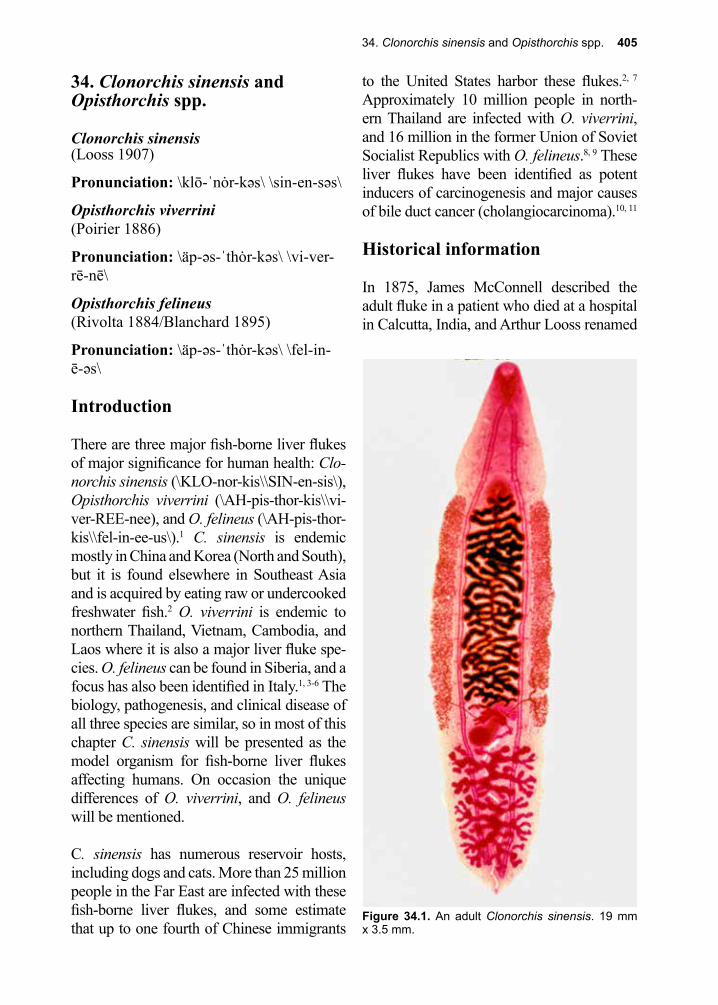
34. Clonorchis sinensis and Opisthorchis spp. 405
Figure 34.1. An adult Clonorchis sinensis. 19 mm x 3.5 mm.
34. Clonorchis sinensis and Opisthorchis spp.
Clonorchis sinensis(Looss 1907)
Pronunciation: \klō-ˈnȯr-kəs\ \sin-en-səs\
Opisthorchis viverrini(Poirier 1886)
Pronunciation: \äp-əs-ˈthȯr-kəs\ \vi-ver-rē-nē\
Opisthorchis felineus(Rivolta 1884/Blanchard 1895)
Pronunciation: \äp-əs-ˈthȯr-kəs\ \fel-in-ē-əs\
Introduction
There are three major fish-borne liver flukes of major significance for human health: Clo-norchis sinensis (\KLO-nor-kis\\SIN-en-sis\), Opisthorchis viverrini (\AH-pis-thor-kis\\vi-ver-REE-nee), and O. felineus (\AH-pis-thor-kis\\fel-in-ee-us\).1 C. sinensis is endemic mostly in China and Korea (North and South), but it is found elsewhere in Southeast Asia and is acquired by eating raw or undercooked freshwater fish.2 O. viverrini is endemic to northern Thailand, Vietnam, Cambodia, and Laos where it is also a major liver fluke spe-cies. O. felineus can be found in Siberia, and a focus has also been identified in Italy.1, 3-6 The biology, pathogenesis, and clinical disease of all three species are similar, so in most of this chapter C. sinensis will be presented as the model organism for fish-borne liver flukes affecting humans. On occasion the unique differences of O. viverrini, and O. felineus will be mentioned.
C. sinensis has numerous reservoir hosts, including dogs and cats. More than 25 million people in the Far East are infected with these fish-borne liver flukes, and some estimate that up to one fourth of Chinese immigrants
to the United States harbor these flukes.2, 7 Approximately 10 million people in north-ern Thailand are infected with O. viverrini, and 16 million in the former Union of Soviet Socialist Republics with O. felineus.8, 9 These liver flukes have been identified as potent inducers of carcinogenesis and major causes of bile duct cancer (cholangiocarcinoma).10, 11
Historical information
In 1875, James McConnell described the adult fluke in a patient who died at a hospital in Calcutta, India, and Arthur Looss renamed
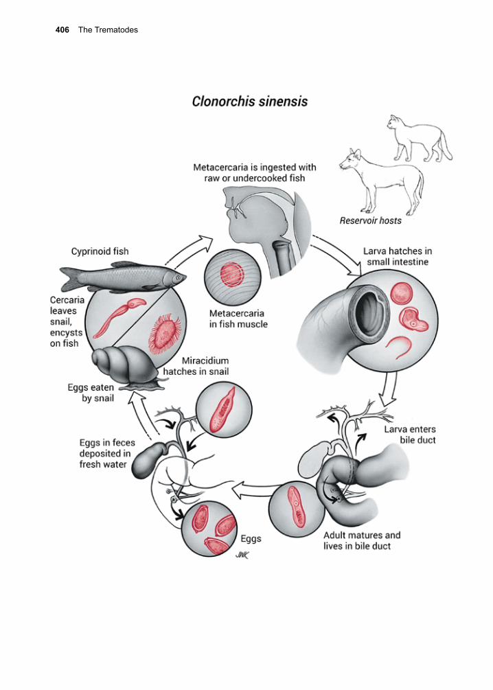
406 The Trematodes
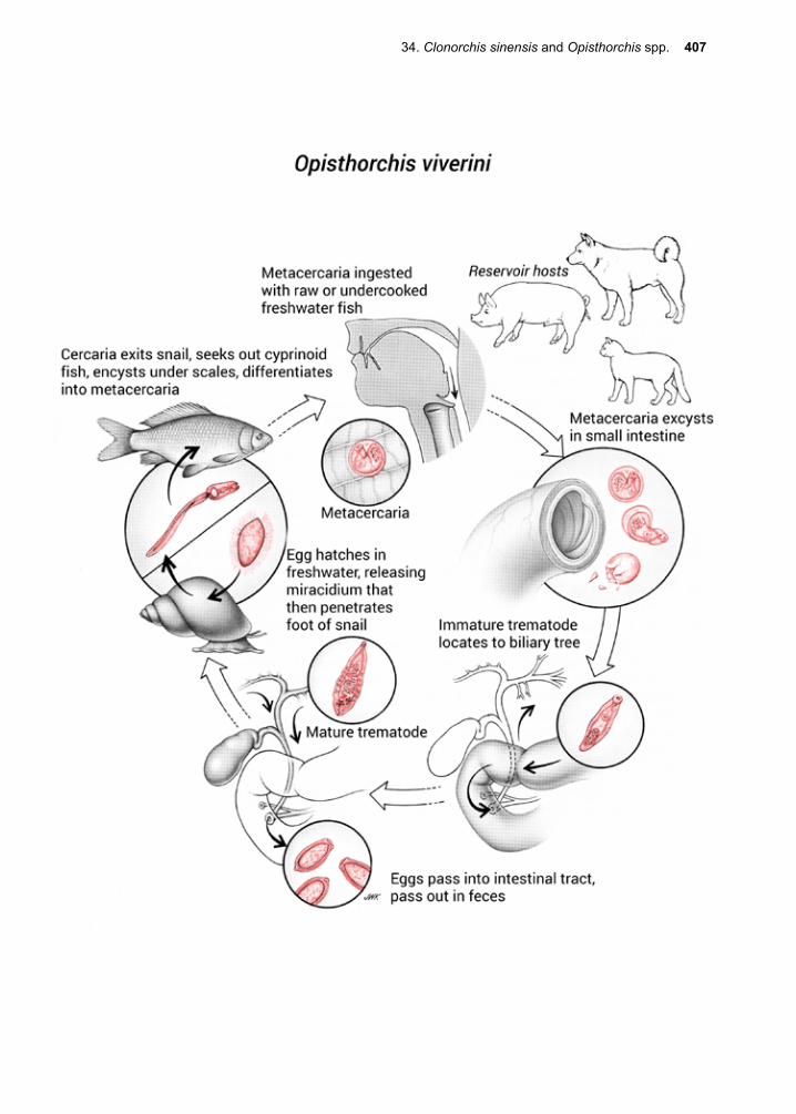
34. Clonorchis sinensis and Opisthorchis spp. 407
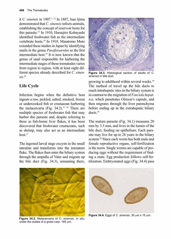
408 The Trematodes
Figure 34.4. Eggs of C. sinensis. 30 µm x 15 µm.
it C. sinensis in 1907.7, 12 In 1887, Isao Ijima demonstrated that C. sinensis infects animals, establishing the concept of reservoir hosts for this parasite.13 In 1910, Haraujiro Kobayashi identified freshwater fish as the intermediate vertebrate hosts.14 In 1918, Masatomo Muto extended these studies in Japan by identifying snails in the genus Parafossarulus as the first intermediate host.15 It is now known that the genus of snail responsible for harboring the intermediate stages of these trematodes varies from region to region, with at least eight dif-ferent species already described for C. sinen-sis.16
Life Cycle
Infection begins when the definitive host ingests a raw, pickled, salted, smoked, frozen or undercooked fish or crustacean harboring the metacercaria (Fig. 34.2).17, 18 There are multiple species of freshwater fish that may harbor this parasite and, despite referring to these as fish-borne liver flukes, it has been discovered that freshwater crustaceans, such as shrimp, may also act as an intermediate host.17
The ingested larval stage excysts in the small intestine and transforms into the immature fluke. The flukes then enter the biliary system through the ampulla of Vater and migrate up the bile duct (Fig. 34.3), remaining there,
growing to adulthood within several weeks.19 The method of travel up the bile ducts to reach intrahepatic sites in the biliary system is in contrast to the migration of Fasciola hepat-ica, which penetrates Glisson’s capsule, and then migrates through the liver parenchyma before ending up in the extrahepatic biliary ducts.19
The mature parasite (Fig. 34.1) measures 20 mm by 3.5 mm, and lives in the lumen of the bile duct, feeding on epithelium. Each para-site may live for up to 26 years in the biliary system.20 Since each worm has both male and female reproductive organs, self-fertilization is the norm. Single worms are capable of pro-ducing eggs without the requirement of find-ing a mate. Egg production follows self-fer-tilization. Embryonated eggs (Fig. 34.4) pass
Figure 34.2. Metacercaria of C. sinensis, in situ, under the scales of a grass carp. 165 µm.
Figure 34.3. Histological section of adults of C. sinensis in bile duct.
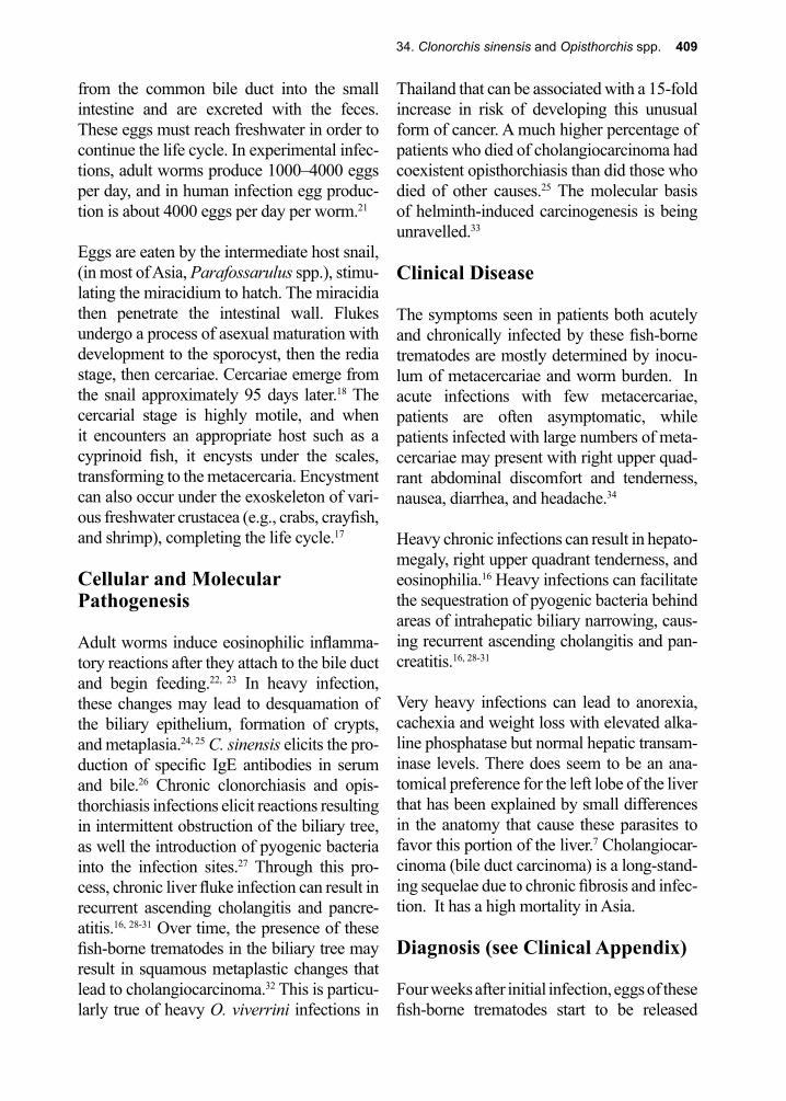
34. Clonorchis sinensis and Opisthorchis spp. 409
from the common bile duct into the small intestine and are excreted with the feces. These eggs must reach freshwater in order to continue the life cycle. In experimental infec-tions, adult worms produce 1000–4000 eggs per day, and in human infection egg produc-tion is about 4000 eggs per day per worm.21
Eggs are eaten by the intermediate host snail, (in most of Asia, Parafossarulus spp.), stimu-lating the miracidium to hatch. The miracidia then penetrate the intestinal wall. Flukes undergo a process of asexual maturation with development to the sporocyst, then the redia stage, then cercariae. Cercariae emerge from the snail approximately 95 days later.18 The cercarial stage is highly motile, and when it encounters an appropriate host such as a cyprinoid fish, it encysts under the scales, transforming to the metacercaria. Encystment can also occur under the exoskeleton of vari-ous freshwater crustacea (e.g., crabs, crayfish, and shrimp), completing the life cycle.17
Cellular and Molecular Pathogenesis
Adult worms induce eosinophilic inflamma-tory reactions after they attach to the bile duct and begin feeding.22, 23 In heavy infection, these changes may lead to desquamation of the biliary epithelium, formation of crypts, and metaplasia.24, 25 C. sinensis elicits the pro-duction of specific IgE antibodies in serum and bile.26 Chronic clonorchiasis and opis-thorchiasis infections elicit reactions resulting in intermittent obstruction of the biliary tree, as well the introduction of pyogenic bacteria into the infection sites.27 Through this pro-cess, chronic liver fluke infection can result in recurrent ascending cholangitis and pancre-atitis.16, 28-31 Over time, the presence of these fish-borne trematodes in the biliary tree may result in squamous metaplastic changes that lead to cholangiocarcinoma.32 This is particu-larly true of heavy O. viverrini infections in
Thailand that can be associated with a 15-fold increase in risk of developing this unusual form of cancer. A much higher percentage of patients who died of cholangiocarcinoma had coexistent opisthorchiasis than did those who died of other causes.25 The molecular basis of helminth-induced carcinogenesis is being unravelled.33
Clinical Disease
The symptoms seen in patients both acutely and chronically infected by these fish-borne trematodes are mostly determined by inocu-lum of metacercariae and worm burden. In acute infections with few metacercariae, patients are often asymptomatic, while patients infected with large numbers of meta-cercariae may present with right upper quad-rant abdominal discomfort and tenderness, nausea, diarrhea, and headache.34
Heavy chronic infections can result in hepato-megaly, right upper quadrant tenderness, and eosinophilia.16 Heavy infections can facilitate the sequestration of pyogenic bacteria behind areas of intrahepatic biliary narrowing, caus-ing recurrent ascending cholangitis and pan-creatitis.16, 28-31
Very heavy infections can lead to anorexia, cachexia and weight loss with elevated alka-line phosphatase but normal hepatic transam-inase levels. There does seem to be an ana-tomical preference for the left lobe of the liver that has been explained by small differences in the anatomy that cause these parasites to favor this portion of the liver.7 Cholangiocar-cinoma (bile duct carcinoma) is a long-stand-ing sequelae due to chronic fibrosis and infec-tion. It has a high mortality in Asia.
Diagnosis (see Clinical Appendix)
Four weeks after initial infection, eggs of these fish-borne trematodes start to be released
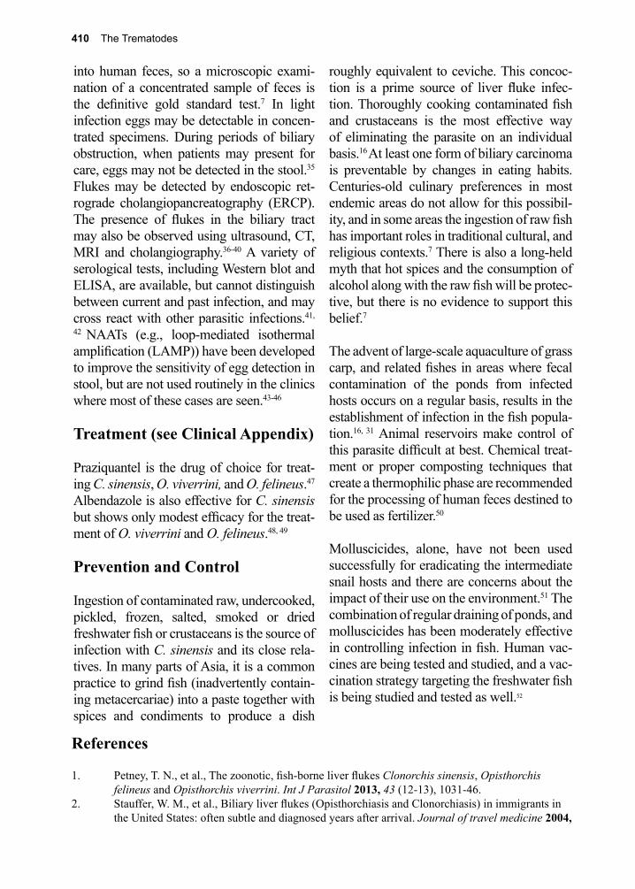
410 The Trematodes
into human feces, so a microscopic exami-nation of a concentrated sample of feces is the definitive gold standard test.7 In light infection eggs may be detectable in concen-trated specimens. During periods of biliary obstruction, when patients may present for care, eggs may not be detected in the stool.35 Flukes may be detected by endoscopic ret-rograde cholangiopancreatography (ERCP). The presence of flukes in the biliary tract may also be observed using ultrasound, CT, MRI and cholangiography.36-40 A variety of serological tests, including Western blot and ELISA, are available, but cannot distinguish between current and past infection, and may cross react with other parasitic infections.41,
42 NAATs (e.g., loop-mediated isothermal amplification (LAMP)) have been developed to improve the sensitivity of egg detection in stool, but are not used routinely in the clinics where most of these cases are seen.43-46
Treatment (see Clinical Appendix)
Praziquantel is the drug of choice for treat-ing C. sinensis, O. viverrini, and O. felineus.47 Albendazole is also effective for C. sinensis but shows only modest efficacy for the treat-ment of O. viverrini and O. felineus.48, 49
Prevention and Control
Ingestion of contaminated raw, undercooked, pickled, frozen, salted, smoked or dried freshwater fish or crustaceans is the source of infection with C. sinensis and its close rela-tives. In many parts of Asia, it is a common practice to grind fish (inadvertently contain-ing metacercariae) into a paste together with spices and condiments to produce a dish
roughly equivalent to ceviche. This concoc-tion is a prime source of liver fluke infec-tion. Thoroughly cooking contaminated fish and crustaceans is the most effective way of eliminating the parasite on an individual basis.16 At least one form of biliary carcinoma is preventable by changes in eating habits. Centuries-old culinary preferences in most endemic areas do not allow for this possibil-ity, and in some areas the ingestion of raw fish has important roles in traditional cultural, and religious contexts.7 There is also a long-held myth that hot spices and the consumption of alcohol along with the raw fish will be protec-tive, but there is no evidence to support this belief.7
The advent of large-scale aquaculture of grass carp, and related fishes in areas where fecal contamination of the ponds from infected hosts occurs on a regular basis, results in the establishment of infection in the fish popula-tion.16, 31 Animal reservoirs make control of this parasite difficult at best. Chemical treat-ment or proper composting techniques that create a thermophilic phase are recommended for the processing of human feces destined to be used as fertilizer.50
Molluscicides, alone, have not been used successfully for eradicating the intermediate snail hosts and there are concerns about the impact of their use on the environment.51 The combination of regular draining of ponds, and molluscicides has been moderately effective in controlling infection in fish. Human vac-cines are being tested and studied, and a vac-cination strategy targeting the freshwater fish is being studied and tested as well.52
References
1. Petney, T. N., et al., The zoonotic, fish-borne liver flukes Clonorchis sinensis, Opisthorchis felineus and Opisthorchis viverrini. Int J Parasitol 2013, 43 (12-13), 1031-46.
2. Stauffer, W. M., et al., Biliary liver flukes (Opisthorchiasis and Clonorchiasis) in immigrants in the United States: often subtle and diagnosed years after arrival. Journal of travel medicine 2004,
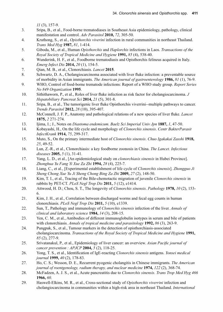
34. Clonorchis sinensis and Opisthorchis spp. 411
11 (3), 157-9.3. Sripa, B., et al., Food-borne trematodiases in Southeast Asia epidemiology, pathology, clinical
manifestation and control. Adv Parasitol 2010, 72, 305-50.4. Kruthong, S., et al., Opisthorchis viverini infection in rural communities in northeast Thailand.
Trans Med Hyg 1987, 81, 1-414.5. Giboda, M., et al., Human Opisthorchis and Haplorchis infections in Laos. Transactions of the
Royal Society of Tropical Medicine and Hygiene 1991, 85 (4), 538-40.6. Wunderink, H. F., et al., Foodborne trematodiasis and Opisthorchis felineus acquired in Italy.
Emerg Infect Dis 2014, 20 (1), 154-5.7. Qian, M. B., et al., Clonorchiasis. Lancet 2015.8. Schwartz, D. A., Cholangiocarcinoma associated with liver fluke infection: a preventable source
of morbidity in Asian immigrants. The American journal of gastroenterology 1986, 81 (1), 76-9.9. WHO, Control of food-borne trematode infections: Report of a WHO study group. Report Series
No 849 Organization 1995.10. Sithithaworn, P., et al., Roles of liver fluke infection as risk factor for cholangiocarcinoma. J
Hepatobiliary Pancreat Sci 2014, 21 (5), 301-8.11. Sripa, B., et al., The tumorigenic liver fluke Opisthorchis viverrini--multiple pathways to cancer.
Trends Parasitol 2012, 28 (10), 395-407.12. McConnell, J. F. P., Anatomy and pathological relations of a new species of liver fluke. Lancet
1875, 2 271-274.13. Ijima, I.; J., Notes on Diastoma endemicum. Baelz Sci Imperial Univ Jpn 1887, 1, 47-50.14. Kobayashi, H., On the life cycle and morphology of Clonorchis sinensis. Centr BakterParasit
InfectKrank 1914, 75, 299-317.15. Muto, S., On the primary intermediate host of Clonorchis sinensis. Chuo Igakakai Zasshi 1918,
25, 49-52.16. Lun, Z.-R., et al., Clonorchiasis: a key foodborne zoonosis in China. The Lancet. Infectious
diseases 2005, 5 (1), 31-41.17. Yang, L. D., et al., [An epidemiological study on clonorchiasis sinensis in Hubei Province].
Zhonghua Yu Fang Yi Xue Za Zhi 1994, 28 (4), 225-7.18. Liang, C., et al., [Experimental establishment of life cycle of Clonorchis sinensis]. Zhongguo Ji
Sheng Chong Xue Yu Ji Sheng Chong Bing Za Zhi 2009, 27 (2), 148-50.19. Kim, T. I., et al., Tracing of the Bile-chemotactic migration of juvenile Clonorchis sinensis in
rabbits by PET-CT. PLoS Negl Trop Dis 2011, 5 (12), e1414.20. Attwood, H. D.; Chou, S. T., The longevity of Clonorchis sinensis. Pathology 1978, 10 (2), 153-
6.21. Kim, J. H., et al., Correlation between discharged worms and fecal egg counts in human
clonorchiasis. PLoS Negl Trop Dis 2011, 5 (10), e1339.22. Sun, T., Pathology and immunology of Clonorchis sinensis infection of the liver. Annals of
clinical and laboratory science 1984, 14 (3), 208-15.23. Yen, C. M., et al., Antibodies of different immunoglobulin isotypes in serum and bile of patients
with clonorchiasis. Annals of tropical medicine and parasitology 1992, 86 (3), 263-9.24. Pungpak, S., et al., Tumour markers in the detection of opisthorchiasis-associated
cholangiocarcinoma. Transactions of the Royal Society of Tropical Medicine and Hygiene 1991, 85 (2), 277-9.
25. Srivatanakul, P., et al., Epidemiology of liver cancer: an overview. Asian Pacific journal of cancer prevention : APJCP 2004, 5 (2), 118-25.
26. Yong, T. S., et al., Identification of IgE-reacting Clonorchis sinensis antigens. Yonsei medical journal 1999, 40 (2), 178-83.
27. Ho, C. S.; Wesson, D. E., Recurrent pyogenic cholangitis in Chinese immigrants. The American journal of roentgenology, radium therapy, and nuclear medicine 1974, 122 (2), 368-74.
28. McFadzen, A. J. S., et al., Acute pancreatitis due to Clonorchis sinensis. Trans Trop Med Hyg 466 1966, 60.
29. Haswell-Elkins, M. R., et al., Cross-sectional study of Opisthorchis viverrini infection and cholangiocarcinoma in communities within a high-risk area in northeast Thailand. International
412 The Trematodes
journal of cancer 1994, 59 (4), 505-9.30. Thamavit, W., et al., Effects of dimethylnitrosamine on induction of cholangiocarcinoma in
Opisthorchis viverrini-infected Syrian golden hamsters. Cancer research 1978, 38 (12), 4634-9.31. Wang, K. X., et al., Clinical and epidemiological features of patients with clonorchiasis. World J
Gastroenterol 2004, 10 (3), 446-8.32. Okuda, K., et al., Clinical aspects of intrahepatic bile duct carcinoma including hilar carcinoma:
a study of 57 autopsy-proven cases. Cancer 1977, 39 (1), 232-46.33. Brindley, P. J., et al., Why does infection with some helminths cause cancer? Trends Cancer
2015, 1 (3), 174-182.34. Liang, S. D., et al., [Analysis on 2175 admitted cases of clonorchiasis sinensis in Guigang City].
Zhongguo Ji Sheng Chong Xue Yu Ji Sheng Chong Bing Za Zhi 2008, 26 (5), 374-5.35. Joo, K. R.; Bang, S. J., A bile based study of Clonorchis sinensis infections in patients with
biliary tract diseases in Ulsan, Korea. Yonsei Med J 2005, 46 (6), 794-8.36. Choi, D.; Hong, S. T., Imaging diagnosis of clonorchiasis. Korean J Parasitol 2007, 45 (2), 77-
85.37. Jeong, Y. Y., et al., MR imaging findings of clonorchiasis. Korean J Radiol 2004, 5 (1), 25-30.38. Lim, J. H., Radiologic findings of clonorchiasis. AJR Am J Roentgenol 1990, 155 (5), 1001-8.39. Choi, B. I., et al., CT findings of clonorchiasis. AJR Am J Roentgenol 1989, 152 (2), 281-4.40. Lim, J. H., et al., Clonorchiasis: sonographic findings in 59 proved cases. AJR Am J Roentgenol
1989, 152 (4), 761-4.41. Kim, S. I., A Clonorchis sinensis-specific antigen that detects active human clonorchiasis. The
Korean journal of parasitology 1998, 36 (1), 37-45.42. Kim, C. S., et al., Immunodiagnosis of clonorchiasis using a recombinant antigen. Korean J
Parasitol. 1998, 36, 183-90.43. Kim, E. M., et al., Detection of Clonorchis sinensis in stool samples using real-time PCR. Ann
Trop Med Parasitol 2009, 103 (6), 513-8.44. Parvathi, A., et al., Development and evaluation of a polymerase chain reaction (PCR) assay for
the detection of Opisthorchis viverrini in fish. Acta Trop 2008, 107 (1), 13-6.45. Umesha, K. R., et al., Opisthorchis viverrini: detection by polymerase chain reaction (PCR) in
human stool samples. Exp Parasitol 2008, 120 (4), 353-6.46. Arimatsu, Y., et al., Rapid detection of Opisthorchis viverrini copro-DNA using loop-mediated
isothermal amplification (LAMP). Parasitol Int 2012, 61 (1), 178-82.47. Hsu, C. C.; Kron, M. A., Clonorchiasis and praziquantel. Archives of internal medicine 1985,
145 (6), 1002-3.48. Liu, Y. H., et al., Experimental and clinical trial of albendazole in the treatment of Clonorchiasis
sinensis. Chin Med J (Engl) 1991, 104 (1), 27-31.49. Pungpark, S., et al., Albendazole in the treatment of opisthorchiasis and concomitant intestinal
helminthic infections. Southeast Asian J Trop Med Public Health 1984, 15 (1), 44-50.50. Kone, D., et al., Helminth eggs inactivation efficiency by faecal sludge dewatering and co-
composting in tropical climates. Water Res 2007, 41 (19), 4397-402.51. Sithithaworn, P., et al., The current status of opisthorchiasis and clonorchiasis in the Mekong
Basin. Parasitol Int 2012, 61 (1), 10-6.52. Wang, X., et al., Surface display of Clonorchis sinensis enolase on Bacillus subtilis spores
potentializes an oral vaccine candidate. Vaccine 2014, 32 (12), 1338-45.
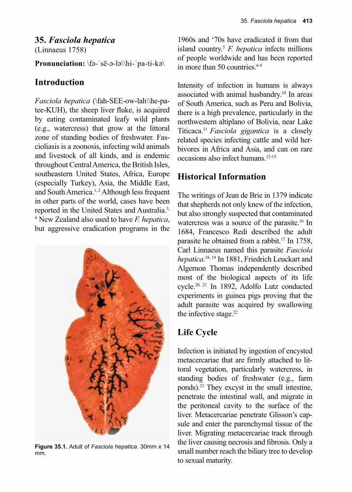
35. Fasciola hepatica 413
Figure 35.1. Adult of Fasciola hepatica. 30mm x 14 mm.
35. Fasciola hepatica(Linnaeus 1758)
Pronunciation: \fə-ˈsē-ə-lə\\hi-ˈpa-ti-kə\
Introduction
Fasciola hepatica (\fah-SEE-ow-lah\\he-pa-tee-KUH), the sheep liver fluke, is acquired by eating contaminated leafy wild plants (e.g., watercress) that grow at the littoral zone of standing bodies of freshwater. Fas-cioliasis is a zoonosis, infecting wild animals and livestock of all kinds, and is endemic throughout Central America, the British Isles, southeastern United States, Africa, Europe (especially Turkey), Asia, the Middle East, and South America.1, 2 Although less frequent in other parts of the world, cases have been reported in the United States and Australia.3,
4 New Zealand also used to have F. hepatica, but aggressive eradication programs in the
1960s and ‘70s have eradicated it from that island country.5 F. hepatica infects millions of people worldwide and has been reported in more than 50 countries.6-9
Intensity of infection in humans is always associated with animal husbandry.10 In areas of South America, such as Peru and Bolivia, there is a high prevalence, particularly in the northwestern altiplano of Bolivia, near Lake Titicaca.11 Fasciola gigantica is a closely related species infecting cattle and wild her-bivores in Africa and Asia, and can on rare occasions also infect humans.12-15
Historical Information
The writings of Jean de Brie in 1379 indicate that shepherds not only knew of the infection, but also strongly suspected that contaminated watercress was a source of the parasite.16 In 1684, Francesco Redi described the adult parasite he obtained from a rabbit.17 In 1758, Carl Linnaeus named this parasite Fasciola hepatica.18, 19 In 1881, Friedrich Leuckart and Algernon Thomas independently described most of the biological aspects of its life cycle.20, 21 In 1892, Adolfo Lutz conducted experiments in guinea pigs proving that the adult parasite was acquired by swallowing the infective stage.22
Life Cycle
Infection is initiated by ingestion of encysted metacercariae that are firmly attached to lit-toral vegetation, particularly watercress, in standing bodies of freshwater (e.g., farm ponds).23 They excyst in the small intestine, penetrate the intestinal wall, and migrate in the peritoneal cavity to the surface of the liver. Metacercariae penetrate Glisson’s cap-sule and enter the parenchymal tissue of the liver. Migrating metacercariae track through the liver causing necrosis and fibrosis. Only a small number reach the biliary tree to develop to sexual maturity.
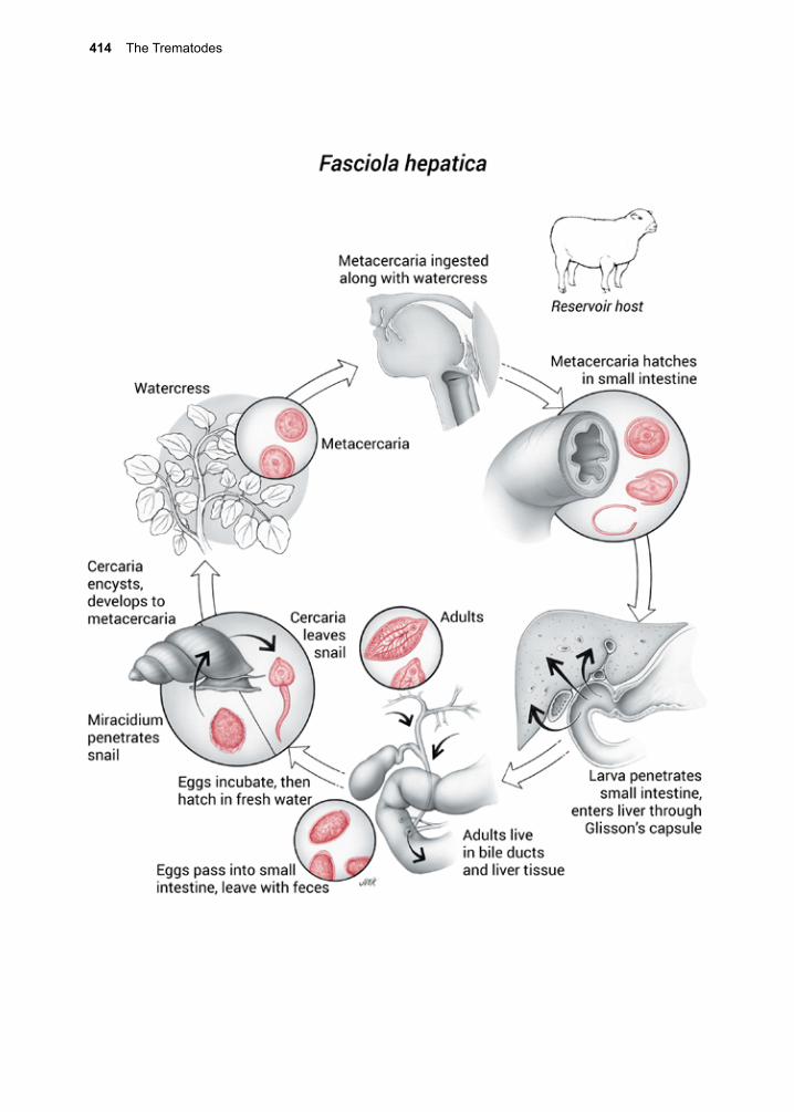
414 The Trematodes
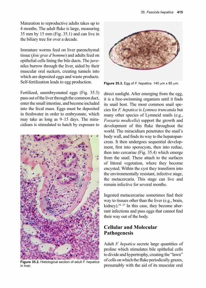
35. Fasciola hepatica 415
Figure 35.3. Egg of F. hepatica. 140 µm x 85 µm.
Maturation to reproductive adults takes up to 4 months. The adult fluke is large, measuring 35 mm by 15 mm (Fig. 35.1) and can live in the biliary tree for over a decade.
Immature worms feed on liver parenchymal tissue (foie gras d’homme) and adults feed on epithelial cells lining the bile ducts. The juve-niles burrow through the liver, aided by their muscular oral suckers, creating tunnels into which are deposited eggs and waste products. Self-fertilization leads to egg production.
Fertilized, unembryonated eggs (Fig. 35.3) pass out of the liver through the common duct, enter the small intestine, and become included into the fecal mass. Eggs must be deposited in freshwater in order to embryonate, which may take as long as 9–15 days. The mira-cidium is stimulated to hatch by exposure to
direct sunlight. After emerging from the egg, it is a free-swimming organism until it finds its snail host. The most common snail spe-cies for F. hepatica is Lymnea truncatula but many other species of Lymneid snails (e.g., Fossaria modicella) support the growth and development of this fluke throughout the world. The miracidium penetrates the snail’s body wall, and finds its way to the hepatopan-creas. It then undergoes sequential develop-ment, first into sporocysts, then into rediae, then into cercariae (Fig. 35.4) which emerge from the snail. These attach to the surfaces of littoral vegetation, where they become encysted. Within the cyst they transform into the environmentally resistant, infective stage, the metacercaria. This stage can live and remain infective for several months.
Ingested metacercariae sometimes find their way to tissues other than the liver (e.g., brain, kidney).24, 25 In this case, they become aber-rant infections and pass eggs that cannot find their way out of the body.
Cellular and Molecular Pathogenesis
Adult F. hepatica secrete large quantities of proline which stimulates bile epithelial cells to divide and hypertrophy, creating the “lawn” of cells on which the fluke periodically grazes, presumably with the aid of its muscular oral
Figure 35.2. Histological section of adult F. hepatica in liver.
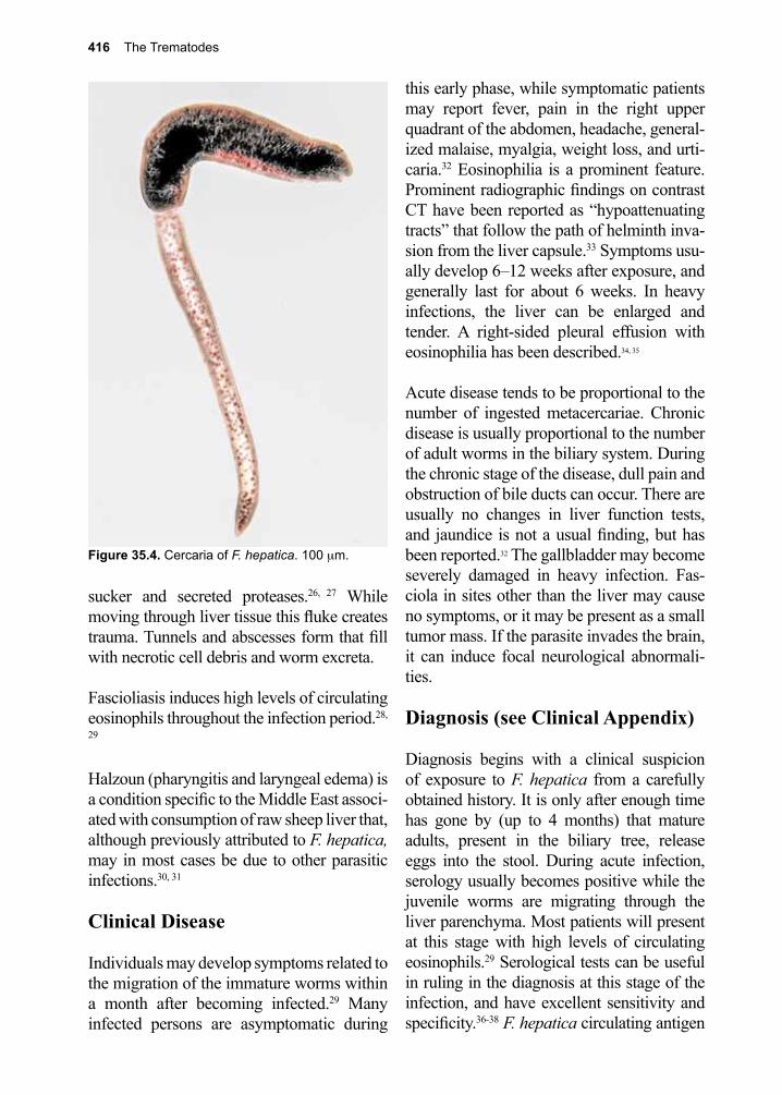
416 The Trematodes
sucker and secreted proteases.26, 27 While moving through liver tissue this fluke creates trauma. Tunnels and abscesses form that fill with necrotic cell debris and worm excreta.
Fascioliasis induces high levels of circulating eosinophils throughout the infection period.28,
29
Halzoun (pharyngitis and laryngeal edema) is a condition specific to the Middle East associ-ated with consumption of raw sheep liver that, although previously attributed to F. hepatica, may in most cases be due to other parasitic infections.30, 31
Clinical Disease
Individuals may develop symptoms related to the migration of the immature worms within a month after becoming infected.29 Many infected persons are asymptomatic during
this early phase, while symptomatic patients may report fever, pain in the right upper quadrant of the abdomen, headache, general-ized malaise, myalgia, weight loss, and urti-caria.32 Eosinophilia is a prominent feature. Prominent radiographic findings on contrast CT have been reported as “hypoattenuating tracts” that follow the path of helminth inva-sion from the liver capsule.33 Symptoms usu-ally develop 6–12 weeks after exposure, and generally last for about 6 weeks. In heavy infections, the liver can be enlarged and tender. A right-sided pleural effusion with eosinophilia has been described.34, 35
Acute disease tends to be proportional to the number of ingested metacercariae. Chronic disease is usually proportional to the number of adult worms in the biliary system. During the chronic stage of the disease, dull pain and obstruction of bile ducts can occur. There are usually no changes in liver function tests, and jaundice is not a usual finding, but has been reported.32 The gallbladder may become severely damaged in heavy infection. Fas-ciola in sites other than the liver may cause no symptoms, or it may be present as a small tumor mass. If the parasite invades the brain, it can induce focal neurological abnormali-ties.
Diagnosis (see Clinical Appendix)
Diagnosis begins with a clinical suspicion of exposure to F. hepatica from a carefully obtained history. It is only after enough time has gone by (up to 4 months) that mature adults, present in the biliary tree, release eggs into the stool. During acute infection, serology usually becomes positive while the juvenile worms are migrating through the liver parenchyma. Most patients will present at this stage with high levels of circulating eosinophils.29 Serological tests can be useful in ruling in the diagnosis at this stage of the infection, and have excellent sensitivity and specificity.36-38 F. hepatica circulating antigen
Figure 35.4. Cercaria of F. hepatica. 100 µm.
35. Fasciola hepatica 417
tests, also with excellent sensitivity and speci-ficity, are available and correlate with burden of infection.39, 40
When mature flukes are present in the biliary system, microscopic identification of eggs in the stool is a definitive method of diag-nosis. Eggs may be detected in the feces, in bile aspirates, or in duodenal aspirates. These unembryonated eggs (Fig. 35.3) are yellow-brown and measure 130–150 µm by 60–90 µm wide.
Imaging techniques such as ultrasonography, CT, cholangiography, endoscopic cholangio-pancreatography (ERCP) and MRI may be helpful in making the diagnosis of fasciolia-sis.41-44 During acute disease, linear tracts can be seen in the liver, while filling defects and adult flukes can be visualized in the biliary system during chronic disease.
Treatment (see Clinical Appendix)
Triclabendazole is the drug of choice for treat-ment of infection with Fasciola hepatica.29,
45-48 Although triclabendazole is not FDA approved or generally available in the United States, it can be obtained from the Centers for Disease Control (CDC) under an investiga-tional protocol. Praziquantel, which is excel-lent for treating infections with other flukes, is not effective against Fasciola, nor are mebendazole, albendazole, or artesunate.49-51
Nitazoxanide appears to be an inferior but alternate therapy with some demonstrated efficacy.52, 53 Successfully treated patients will develop negative serologies 6–12 months after clearing their parasites.
Prevention and Control
Fasciola control may be achieved through periodic draining of ponds which reduces lit-toral plant growth to a minimum. Protecting freshwater supplies and regularly surveying herds and herders for the presence of the para-sites can achieve further control of the spread of Fascioliasis in domestic animals. In this regard, an ELISA test detected experimen-tally and naturally-infected calves with a high degree of sensitivity and specificity.37 When infections are detected, appropriate treatment in both groups is warranted. Snail elimination with molluscicides has not been successful. Education of farm personnel regarding the mode of acquisition of the infection is essen-tial to eliminating transmission due to human fecal contamination of freshwater aquatic habitats.
Vaccines have been developed for animal use that have demonstrated some degree of efficacy in terms of reduction in egg produc-tion and worm burdens.54 Despite significant advances in vaccine development, no human or animal vaccine is in current use.55
References
1. Mas-Coma, M. S., et al., Epidemiology of human fascioliasis: a review and proposed new classification. Bull World Health Organ 1999, 77 (4), 340-6.
2. Bosnak, V. K., et al., Evaluation of patients diagnosed with fascioliasis: A six-year experience at a university hospital in Turkey. J Infect Dev Ctries 2016, 10 (4), 389-94.
3. Weisenberg, S. A.; Perlada, D. E., Domestically acquired fascioliasis in northern California. Am J Trop Med Hyg 2013, 89 (3), 588-91.
4. Sivagnanam, S., et al., Hepatic lesions and eosinophilia in an urban dweller. Liver Int 2014, 34 (4), 643.
5. Charleston, W. A.; McKenna, P. B., Nematodes and liver fluke in New Zealand. N Z Vet J 2002, 50 (3 Suppl), 41-7.
418 The Trematodes
6. Lukambagire, A. H., et al., Diagnosis of human fascioliasis in Arusha region, northern Tanzania by microscopy and clinical manifestations in patients. BMC Infect Dis 2015, 15 (1), 578.
7. Nyindo, M.; Lukambagire, A. H., Fascioliasis: An Ongoing Zoonotic Trematode Infection. Biomed Res Int 2015, 2015, 786195.
8. Keiser, J.; Utzinger, J., Food-borne trematodiases. Clin Microbiol Rev 2009, 22 (3), 466-83.9. Mas-Coma, S., Epidemiology of fascioliasis in human endemic areas. J Helminthol 2005, 79 (3),
207-16.10. Esteban, J.-G., et al., Hyperendemic fascioliasis associated with schistosomiasis in villages in the
Nile Delta of Egypt. The American journal of tropical medicine and hygiene 2003, 69 (4), 429-37.
11. Hillyer, G. V., et al., Use of the Falcon assay screening test--enzyme-linked immunosorbent assay (FAST-ELISA) and the enzyme-linked immunoelectrotransfer blot (EITB) to determine the prevalence of human fascioliasis in the Bolivian Altiplano. The American journal of tropical medicine and hygiene 1992, 46 (5), 603-9.
12. Menon, P., et al., Biliary Fasciola gigantica infestation in a nonendemic area - An intraoperative surprise. J Pediatr Surg 2015, 50 (11), 1983-6.
13. Fang, W., et al., [Case report: Fasciola gigantica infection treated by triclabendazole]. Zhongguo Ji Sheng Chong Xue Yu Ji Sheng Chong Bing Za Zhi 2013, 31 (2), Inside front page.
14. Bestas, R., et al., Cholestasis caused by Fasciola gigantica. Turkiye Parazitol Derg 2014, 38 (3), 201-4.
15. Chen, J. X., et al., An outbreak of human Fascioliasis gigantica in Southwest China. PLoS One 2013, 8 (8), e71520.
16. de Brie, J., Le Bon Berger ou le Vray Regime et Gouvenement de Bergers et Bergeres: Compose par le Rustique Jehan de Brie le Bon Berger (1379). Isidor Liseux, Paris. 1879.
17. Redi, F., Osservazioni di Francesco Redi. Intorno Agli Animali Viventi che si Trovano Negli Animali Viventi Piero Matini Florence 1684.
18. Lima Wdos, S., et al., Occurrence of Fasciola hepatica (Linnaeus, 1758) infection in Brazilian cattle of Minas Gerais, Brazil. Rev Bras Parasitol Vet 2009, 18 (2), 27-30.
19. Cox, F. E., History of human parasitology. Clin Microbiol Rev 2002, 15 (4), 595-612.20. Leuckart, F. R., Zur Entwicklungsgeschichte des Lerberegels. Zool Anz 1881, 4, 641-646.21. Thomas, A. P. W.; R., J., Report of experiments on the development of the liver fluke, Fasciola
hepatica. Soc Engi 1881, 17, 1-28.22. Lutz, A., Zur Lebensgeschichte des Distoma hepaticum. Zentralbi Bakteriol Parasit 1892, 11,
783-796.23. Mas-Coma, S., et al., Fascioliasis and other plant-borne trematode zoonoses. Int J Parasitol
2005, 35 (11-12), 1255-78.24. Catchpole, B. N.; Snow, D., Human ectopic fascioliasis. Lancet 1952, 2 711-712.25. Arjona, R., et al., Fascioliasis in developed countries: a review of classic and aberrant forms of
the disease. Medicine 1995, 74 (1), 13-23.26. Modavi, S.; Isseroff, H., Fasciola hepatica: collagen deposition and other histopathology in the
rat host’s bile duct caused by the parasite and by proline infusion. Experimental parasitology 1984, 58 (3), 239-44.
27. Wijffels, G. L., et al., The secreted cathepsin L-like proteinases of the trematode, Fasciola hepatica, contain 3-hydroxyproline residues. The Biochemical journal 1994, 299 ( Pt 3), 781-90.
28. Demirci, M., et al., Diagnostic importance of serological methods and eosinophilia in tissue parasites. Journal of health, population, and nutrition 2002, 20 (4), 352-5.
29. Saba, R., et al., Human fascioliasis. Clinical microbiology and infection : the official publication of the European Society of Clinical Microbiology and Infectious Diseases 2004, 10 (5), 385-7.
30. Khalil, G., et al., Halzoun, an allergic pharyngitis syndrome in Lebanon: the trematode Dicrocoelium dendriticum as an additional cause. Acta Trop 2013, 125 (1), 115-8.
31. Saleha, A. A., Liver fluke disease (fascioliasis): epidemiology, economic impact and public health significance. The Southeast Asian journal of tropical medicine and public health 1991, 22 Suppl, 361-4.
35. Fasciola hepatica 419
32. Arjona, R., et al., Fascioliasis in developed countries: a review of classic and aberrant forms of the disease. Medicine (Baltimore) 1995, 74 (1), 13-23.
33. Patel, N. U., et al., CT findings of human Fasciola hepatica infection: case reports and review of the literature. Clin Imaging 2016, 40 (2), 251-5.
34. Corredoira, J. C., et al., [Eosinophilic pleural effusion caused by Fasciola hepatica]. Enferm Infecc Microbiol Clin 1990, 8 (4), 258-9.
35. Moretti, G., et al., [Pleural effusion revealing and a recapitulating Fasciola hepatica distomatosis]. Bord Med 1971, 4 (4), 1181-2 passim.
36. O’Neill, J., On the presence of a filaria in “crawcraw.” Lancet 1875, 1, 265-266.37. Rokni, M. B., et al., Diagnosis of human fasciolosis in the Gilan province of Northern Iran:
application of cathepsin L-ELISA. Diagnostic microbiology and infectious disease 2002, 44 (2), 175-9.
38. Gonzales Santana, B., et al., The diagnosis of human fascioliasis by enzyme-linked immunosorbent assay (ELISA) using recombinant cathepsin L protease. PLoS Negl Trop Dis 2013, 7 (9), e2414.
39. Hassan, M. M., et al., Evaluation of circulating Fasciola antigens in specific diagnosis of fascioliasis. J Egypt Soc Parasitol 2001, 31 (1), 271-9.
40. Almazan, C., et al., Effect of parasite burden on the detection of Fasciola hepatica antigens in sera and feces of experimentally infected sheep. Vet Parasitol 2001, 97 (2), 101-12.
41. Zali, M. R., et al., Liver, spleen, pancreas and kidney involvement by human fascioliasis: imaging findings. BMC gastroenterology 2004, 4, 15.
42. Cevikol, C., et al., Human fascioliasis: MR imaging findings of hepatic lesions. Eur Radiol 2003, 13 (1), 141-8.
43. Van Beers, B., et al., Hepatobiliary fascioliasis: noninvasive imaging findings. Radiology 1990, 174 (3 Pt 1), 809-10.
44. Dias, L. M., et al., Biliary fascioliasis: diagnosis, treatment and follow-up by ERCP. Gastrointest Endosc 1996, 43 (6), 616-20.
45. el-Karaksy, H., et al., Human fascioliasis in Egyptian children: successful treatment with triclabendazole. Journal of tropical pediatrics 1999, 45 (3), 135-8.
46. Marcos, L. A., et al., Update on hepatobiliary flukes: fascioliasis, opisthorchiasis and clonorchiasis. Curr Opin Infect Dis 2008, 21 (5), 523-30.
47. Marcos, L. A., et al., Natural history, clinicoradiologic correlates, and response to triclabendazole in acute massive fascioliasis. Am J Trop Med Hyg 2008, 78 (2), 222-7.
48. Villegas, F., et al., Administration of triclabendazole is safe and effective in controlling fascioliasis in an endemic community of the Bolivian Altiplano. PLoS Negl Trop Dis 2012, 6 (8), e1720.
49. Fand, Z., et al., Praziquantel and Fasciola hepatica infection. Trans Trop Med Hyg 813 1989, 83.50. Hien, T. T., et al., A randomized controlled pilot study of artesunate versus triclabendazole for
human fascioliasis in central Vietnam. Am J Trop Med Hyg 2008, 78 (3), 388-92.51. Cabada, M. M.; White, A. C., Jr., New developments in epidemiology, diagnosis, and treatment
of fascioliasis. Curr Opin Infect Dis 2012, 25 (5), 518-22.52. Rossignol, J. F., et al., Successful treatment of human fascioliasis with nitazoxanide. Trans R Soc
Trop Med Hyg 1998, 92 (1), 103-4.53. Favennec, L., et al., Double-blind, randomized, placebo-controlled study of nitazoxanide in the
treatment of fascioliasis in adults and children from northern Peru. Aliment Pharmacol Ther 2003, 17 (2), 265-70.
54. Espino, A. M.; Hillyer, G. V., A novel Fasciola hepatica saposinlike recombinant protein with immunoprophylactic potential. The Journal of parasitology 2004, 90 (4), 876-9.
55. Molina-Hernandez, V., et al., Fasciola hepatica vaccine: we may not be there yet but we’re on the right road. Vet Parasitol 2015, 208 (1-2), 101-11.
420 The Trematodes
Francesco Redi, M.D., Ph.D. (1626–1697)
Redi was a global thinker, an original “myth buster”, and one of the original Renaissance Men of science. He conducted investigations into the origins of life and established the experimental control as a means of comparing an unaltered situation to one that was manipulated. He described over 180 different parasites, which included a number of arthropod ectoparasites. He correctly determined that many parasites grow up from eggs, making significant contributions to the germ theory of disease. In addition to all of his scientific contributions, Redi was an accomplished poet. His life-sized statue stands proudly looking down on all who visit the world-renowned Uffizi Museum in Florence, Italy.
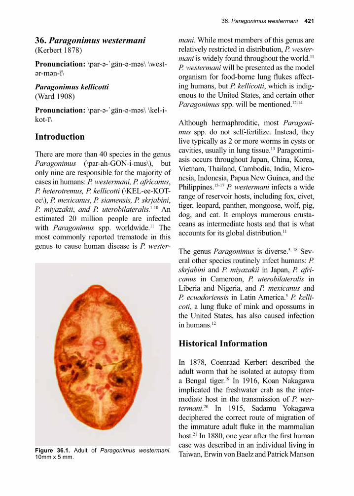
36. Paragonimus westermani 421
Figure 36.1. Adult of Paragonimus westermani. 10mm x 5 mm.
36. Paragonimus westermani(Kerbert 1878)
Pronunciation: \par-ə-ˈgän-ə-məs\ \west-ər-mən-ī\
Paragonimus kellicotti(Ward 1908)
Pronunciation: \par-ə-ˈgän-ə-məs\ \kel-i-kot-ī\
Introduction
There are more than 40 species in the genus Paragonimus (\par-ah-GON-i-mus\), but only nine are responsible for the majority of cases in humans: P. westermani, P. africanus, P. heterotremus, P. kellicotti (\KEL-ee-KOT-ee\), P. mexicanus, P. siamensis, P. skrjabini, P. miyazakii, and P. uterobilateralis.1-10 An estimated 20 million people are infected with Paragonimus spp. worldwide.11 The most commonly reported trematode in this genus to cause human disease is P. wester-
mani. While most members of this genus are relatively restricted in distribution, P. wester-mani is widely found throughout the world.11 P. westermani will be presented as the model organism for food-borne lung flukes affect-ing humans, but P. kellicotti, which is indig-enous to the United States, and certain other Paragonimus spp. will be mentioned.12-14
Although hermaphroditic, most Paragoni-mus spp. do not self-fertilize. Instead, they live typically as 2 or more worms in cysts or cavities, usually in lung tissue.13 Paragonimi-asis occurs throughout Japan, China, Korea, Vietnam, Thailand, Cambodia, India, Micro-nesia, Indonesia, Papua New Guinea, and the Philippines.15-17 P. westermani infects a wide range of reservoir hosts, including fox, civet, tiger, leopard, panther, mongoose, wolf, pig, dog, and cat. It employs numerous crusta-ceans as intermediate hosts and that is what accounts for its global distribution.11
The genus Paragonimus is diverse.5, 18 Sev-eral other species routinely infect humans: P. skrjabini and P. miyazakii in Japan, P. afri-canus in Cameroon, P. uterobilateralis in Liberia and Nigeria, and P. mexicanus and P. ecuadoriensis in Latin America.5 P. kelli-coti, a lung fluke of mink and opossums in the United States, has also caused infection in humans.12
Historical Information
In 1878, Coenraad Kerbert described the adult worm that he isolated at autopsy from a Bengal tiger.19 In 1916, Koan Nakagawa implicated the freshwater crab as the inter-mediate host in the transmission of P. wes-termani.20 In 1915, Sadamu Yokagawa deciphered the correct route of migration of the immature adult fluke in the mammalian host.21 In 1880, one year after the first human case was described in an individual living in Taiwan, Erwin von Baelz and Patrick Manson
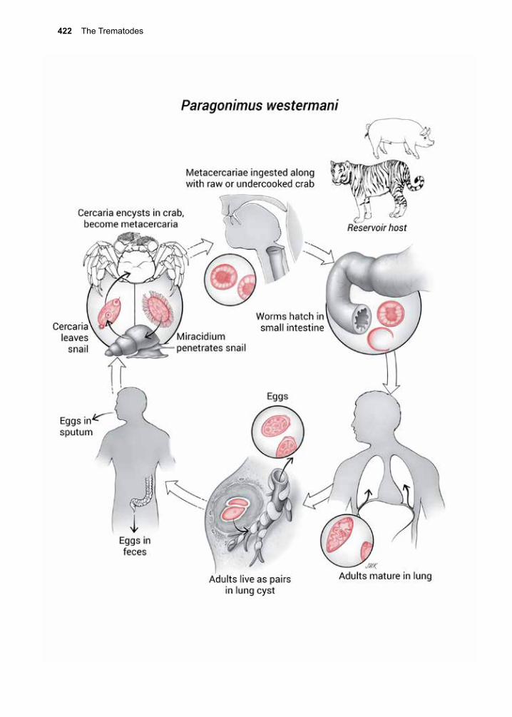
422 The Trematodes
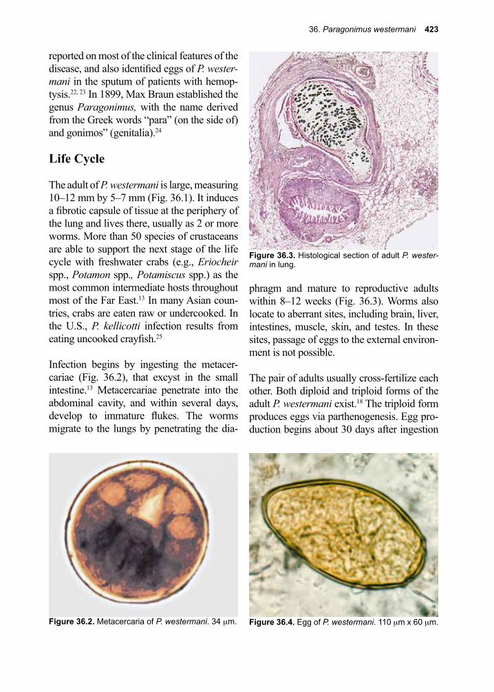
36. Paragonimus westermani 423
Figure 36.3. Histological section of adult P. wester-mani in lung.
Figure 36.4. Egg of P. westermani. 110 µm x 60 µm.
reported on most of the clinical features of the disease, and also identified eggs of P. wester-mani in the sputum of patients with hemop-tysis.22, 23 In 1899, Max Braun established the genus Paragonimus, with the name derived from the Greek words “para” (on the side of) and gonimos” (genitalia).24
Life Cycle
The adult of P. westermani is large, measuring 10–12 mm by 5–7 mm (Fig. 36.1). It induces a fibrotic capsule of tissue at the periphery of the lung and lives there, usually as 2 or more worms. More than 50 species of crustaceans are able to support the next stage of the life cycle with freshwater crabs (e.g., Eriocheir spp., Potamon spp., Potamiscus spp.) as the most common intermediate hosts throughout most of the Far East.13 In many Asian coun-tries, crabs are eaten raw or undercooked. In the U.S., P. kellicotti infection results from eating uncooked crayfish.25
Infection begins by ingesting the metacer-cariae (Fig. 36.2), that excyst in the small intestine.13 Metacercariae penetrate into the abdominal cavity, and within several days, develop to immature flukes. The worms migrate to the lungs by penetrating the dia-
phragm and mature to reproductive adults within 8–12 weeks (Fig. 36.3). Worms also locate to aberrant sites, including brain, liver, intestines, muscle, skin, and testes. In these sites, passage of eggs to the external environ-ment is not possible.
The pair of adults usually cross-fertilize each other. Both diploid and triploid forms of the adult P. westermani exist.18 The triploid form produces eggs via parthenogenesis. Egg pro-duction begins about 30 days after ingestion
Figure 36.2. Metacercaria of P. westermani. 34 µm.
424 The Trematodes
of the metacercariae. Eggs (Fig. 36.4) pass fertilized, but unembryonated, into the sur-rounding tissue. Eventually, they reach the bronchioles and are included into the sputum that also contains blood and debris from the necrotic lesions created by the adults. Because some of the sputum is swallowed, eggs can be recovered from feces as well as sputum. The eggs must reach freshwater to embryonate. The miracidium develops over a 3-week period, after which it hatches and seeks out its intermediate snail host (e.g., Melania spp., Semisulcospira spp., and Thiara spp.). In contrast to other trematodes such as Schistosoma spp., Paragonimus spp. are able to mature in a large variety of snail species.13 Paragonimus spp. develop through the sporocyst and redia stages into cercariae, which then exit from the snail and encyst on and within crustacean intermediate hosts. In the case of the crab, the metacercariae infect all organs.
Cellular and Molecular Pathogenesis
Immature worms of P. westermani do not cause clinical disease, either on their way from the small intestine to the abdominal cavity, or during the last leg of their journey to the lung tissue, except in the case of heavy infection.26-29 In contrast, mature adult worms in the lung form cysts that eventually com-municate with the bronchioles.24
Triploid forms of the parasite are considered more pathogenic than diploid forms. The dip-loid forms are smaller and will form cysts only if sexual partners are found.24
The inflammatory responses to Paragoni-mus cysts are characterized by a variety of cells, but eosinophils usually predominate. Charcot-Leyden crystals can frequently be found in the sputum of infected individuals. Specific IgG and IgE antibodies are produced
throughout the infection, but appear to have no protective function.30 Several stages of P. westermani secrete cysteine proteases that cleave IgG molecules, and the worm may employ this strategy to avoid immune damage by the host.31 Infections last somewhat longer than a year, after which adult worms die and become calcified.
Larval stages of several zoonotic species of Paragonimus such as P. skrjabini and P. miyazakii can cause extensive damage to the tissues as they migrate throughout the vis-cera.24
Clinical Disease
The clinical manifestations of paragoni-miasis include acute early infection and late or chronic disease. Early infection occurs between the time of ingestion of infective metacercariae and lasts up until flukes mature into egg-producing adults. During this stage of infection some patients may remain asymp-tomatic, while others present with diarrhea, fever, chest pain, fatigue, urticaria, epigastric pain, and eosinophilia.26-29 Patients may go on to develop cough with blood-tinged sputum, dyspnea, increasing leukocytosis and eosino-philia, and transient pulmonary infiltrates. In some situations patients may present with cutaneous manifestations, noting painless subcutaneous swellings that are migratory.32 These subcutaneous nodules contain juvenile flukes.24
Mature lung flukes trigger late stage infec-tion. Cough and recurrent hemoptysis are the most common clinical features.33, 34 Patients may also present with chest pain, dyspnea, fever, or chills.13 Depending on the severity of the infection and the frequency of bacterial superinfections, there may be pneumothorax and pleural effusion, with consequent pleural adhesions.
36. Paragonimus westermani 425
An often-fatal clinical outcome results from extrapulmonary paragonimiasis. Immature flukes may migrate to a number of tissues, including the brain. Cerebral paragonimiasis is thought to be rare, estimated to occur in less than 1% of symptomatic individuals infected with P. westermani, but is associated with a significantly higher mortality rate than pul-monary disease.35 Cerebral paragonimiasis has been observed with other Paragonimus spp. such as P. kellicotti.36, 37
Diagnosis (see Clinical Appendix)
Diagnostic modality is based on the stage of the disease. Late-stage disease is diagnosed by microscopic identification of eggs in the sputum, bronchoalveolar lavage fluid, and more rarely in stool.38, 39 A number of cases of acid-fast bacilli-negative pulmonary infec-tions are due to paragonimiasis. One distin-guishing feature is the presence of Charcot-Leyden crystals on histopathology.40 Eggs can be visualized microscopically using wet preps. For many years, acid-fast staining of sputum specimens destroyed Paragonimus eggs. This issue has been overcome using modern Ziehl-Neelsen staining and use of a 10x rather than the standard 100x objective employed in the diagnosis of mycobacterial infections.38, 39
If sputum or other body fluids, such as CSF or pleural fluid, is negative for eggs on repeated sampling, indirect evidence of infection can be obtained by use of serological tests, such as ELISA and Western blot.41-43 These tests are particularly helpful in early stage disease as well as extrapulmonary disease. Immu-noblot testing is available from the Centers for Disease Control (CDC) and ELISA tests are available from commercial laboratories.13,
27 Serology is also used in the diagnosis of P. kellicotti infection in the U.S..25
Antigen detection tests have been developed, but are not routinely used in clinical prac-tice.44 NAATs are being developed that allow for species identification.45 There is also a simple and rapid intradermal test, performed by injecting diluted Paragonimus antigen into the skin.24 Over 2 million people have been skin tested in China, with an overall positivity rate of 20%.24 Both the serologic and intradermal assays indicate either current or past exposure to the infection.
Imaging tests such as CT and fluorodeox-yglucose-positron emission tomography (FDG-PET) can help with the diagnosis but are not definitive.46-48 In many cases, migra-tory tracks caused by helminth invasion can be visualized through, and between, the pleura and lung parenchyma.49 Clinical diag-nosis depends on suspicion of paragonimiasis in any patient from an endemic area who has the characteristic pulmonary disease. Pulmo-nary paragonimiasis must be distinguished from chronic bronchiectasis, lung abscess due to other causes, and tuberculosis.16, 50
Cerebral paragonimiasis must be distin-guished from brain tumors, and lesions caused by other helminths (e.g., juvenile tapeworms and Fasciola hepatica). The subcutaneous nodules of P. skrjabini must be differentiated from other forms of cutaneous larva migrans and gnathostomiasis.24
Treatment (see Clinical Appendix)
The drug of choice against Paragonimus spp. is praziquantel, while triclabendazole is an alternative drug with similar efficacy.24, 50-54 Another regimen that has been used is mul-tiple rounds of praziquantel alternating with albendazole.55 The complications of pleural effusions and subsequent fibrosis may some-times require surgical management, including decortication.13
426 The Trematodes
infected individuals, sanitation changes, and behavioral changes in handling and cooking intermediate crustacean hosts all have a role to play in the control of paragonimiasis.
In certain endemic regions of the world, mass chemotherapy has been attempted to reduce prevalence rates.57-60 Boiling the invertebrate host for several minutes until the meat has congealed and turned opaque can kill the metacercariae. Marinating and salting of crabs or other crustaceans does not reliably kill the infective stages.24
References
1. Iwagami, M., et al., Ancient divergence of Paragonimus westermani in Sri Lanka. Parasitol Res 2008, 102 (5), 845-52.
2. Iwagami, M., et al., The first intermediate host of Paragonimus westermani in Sri Lanka. Acta Trop 2009, 109 (1), 27-9.
3. Iwagami, M., et al., Identities of two Paragonimus species from Sri Lanka inferred from molecular sequences. J Helminthol 2003, 77 (3), 239-45.
4. Iwagami, M., et al., Molecular phylogeographic studies on Paragonimus westermani in Asia. J Helminthol 2000, 74 (4), 315-22.
5. Iwagami, M., et al., A molecular phylogeographic study based on DNA sequences from individual metacercariae of Paragonimus mexicanus from Guatemala and Ecuador. Journal of helminthology 2003, 77 (1), 33-8.
6. Iwagami, M., et al., A molecular phylogeographic study based on DNA sequences from individual metacercariae of Paragonimus mexicanus from Guatemala and Ecuador. J Helminthol 2003, 77 (1), 33-8.
7. Agatsuma, T., et al., The origin of the triploid in Paragonimus westermani on the basis of variable regions in the mitochondrial DNA. J Helminthol 2003, 77 (4), 279-85.
8. Blair, D., et al., Paragonimus skrjabini Chen, 1959 (Digenea: Paragonimidae) and related species in eastern Asia: a combined molecular and morphological approach to identification and taxonomy. Syst Parasitol 2005, 60 (1), 1-21.
9. Doanh, P. N., et al., Paragonimus and paragonimiasis in Vietnam: an update. Korean J Parasitol 2013, 51 (6), 621-7.
10. Chai, J. Y., Paragonimiasis. Handb Clin Neurol 2013, 114, 283-96.11. Furst, T., et al., Global burden of human food-borne trematodiasis: a systematic review and meta-
analysis. Lancet Infect Dis 2012, 12 (3), 210-21.12. Mariano, E. G., et al., A human infection with Paragonimus kellicotti (lung fluke) in the United
States. American journal of clinical pathology 1986, 86 (5), 685-7.13. Procop, G. W., North American paragonimiasis (Caused by Paragonimus kellicotti) in the context
of global paragonimiasis. Clin Microbiol Rev 2009, 22 (3), 415-46.14. Diaz, J. H., Paragonimiasis acquired in the United States: native and nonnative species. Clin
Microbiol Rev 2013, 26 (3), 493-504.15. Kim, E.-A., et al., Imaging findings of hepatic paragonimiasis : a case report. Journal of Korean
medical science 2004, 19 (5), 759-62.16. Singh, T. N., et al., Pulmonary paragonimiasis. The Indian journal of chest diseases & allied
sciences 2004, 46 (3), 225-7.17. Owen, I. L., Parasitic zoonoses in Papua New Guinea. Journal of helminthology 2005, 79 (1),
1-14.
Prevention and Control
Because of its numerous reservoir hosts and its worldwide distribution, many con-sider control of this parasite in animals to be impractical in most parts of the world.13 Due to cultural eating habits favoring the acqui-sition of this parasite, control of paragoni-mus infection is difficult, and would require a comprehensive approach.56 For example, “drunken hairy crab” is traditionally eaten live, and in the modern city of Shanghai, its considered haute cuisine. Treatment of
36. Paragonimus westermani 427
18. Blair, D., Genomes of Paragonimus westermani and related species: current state of knowledge. International journal for parasitology 2000, 30 (4), 421-6.
19. Kerbert, C., Zur Trematodenkenntnis. Zool Anz 1878, 1, 271-273.20. Nakagawa, K.; J., The mode of infection in pulmonary distomiasis: certain fresh water crabs as
intermediate hosts of Paragonimus westermani. Dis 1916, 18, 131-142.21. Yokogawa, S., On the route of migration of Paragonimus westermani in the definitive host.
aiwan Igakkai Zasshi 1915, 152, 685-700.22. Von Baelz, E. O. E., Uber Parasitare Haemoptoe (gregarinosis pulmonum). Zentralbl Med
Wissenschr. 1880, 18 721-722.23. Manson, P., Distoma ringeri: Medical Report for the Half Year Ended 30 September. 1880; Vol.
2, p 10-12.24. Blair, D., et al., Paragonimiasis and the genus Paragonimus. Advances in parasitology 1999, 42,
113-222.25. Fischer, P. U.; Weil, G. J., North American paragonimiasis: epidemiology and diagnostic
strategies. Expert Rev Anti Infect Ther 2015, 13 (6), 779-86.26. Zhong, H. L., et al., Recent progress in studies of paragonimus and paragonimiasis control in
China. Chin Med J (Engl) 1981, 94 (8), 483-94.27. Kagawa, F. T., Pulmonary paragonimiasis. Semin Respir Infect 1997, 12 (2), 149-58.28. DeFrain, M.; Hooker, R., North American paragonimiasis: case report of a severe clinical
infection. Chest 2002, 121 (4), 1368-72.29. Uchiyama, F., et al., Re-emergence of paragonimiasis in Kyushu, Japan. Southeast Asian J Trop
Med Public Health 1999, 30 (4), 686-91.30. Kong, Y., et al., Immunoglobulin G (IgG) subclass and IgE responses in human paragonimiases
caused by three different species. Clinical and diagnostic laboratory immunology 1998, 5 (4), 474-8.
31. Chung, Y. B., et al., Activities of different cysteine proteases of Paragonimus westermani in cleaving human IgG. The Korean journal of parasitology 1997, 35 (2), 139-42.
32. Hatano, Y., et al., Expression of Th1 and Th2 cytokine mRNAs in freshly isolated peripheral blood mononuclear cells of a patient with cutaneous paragonimiasis. J Dermatol Sci 1999, 19 (2), 144-7.
33. Singh, T. S., et al., Pulmonary paragonimiasis: clinical features, diagnosis and treatment of 39 cases in Manipur. Trans R Soc Trop Med Hyg 1986, 80 (6), 967-71.
34. Im, J. G., et al., Pleuropulmonary paragonimiasis: radiologic findings in 71 patients. AJR Am J Roentgenol 1992, 159 (1), 39-43.
35. Singh, T. S., et al., Cerebral paragonimiasis mimicking tuberculoma: First case report in India. Trop Parasitol 2011, 1 (1), 39-41.
36. Centers for Disease, C.; Prevention, Human paragonimiasis after eating raw or undercooked crayfish --- Missouri, July 2006-September 2010. MMWR Morb Mortal Wkly Rep 2010, 59 (48), 1573-6.
37. Lane, M. A., et al., Human paragonimiasis in North America following ingestion of raw crayfish. Clin Infect Dis 2009, 49 (6), e55-61.
38. Barennes, H., et al., Paragonimiasis as an important alternative misdiagnosed disease for suspected acid-fast bacilli sputum smear-negative tuberculosis. Am J Trop Med Hyg 2014, 90 (2), 384-5.
39. Slesak, G., et al., Ziehl-Neelsen staining technique can diagnose paragonimiasis. PLoS Negl Trop Dis 2011, 5 (5), e1048.
40. Luo, J., et al., Pulmonary Paragonimiasis Mimicking Tuberculous Pleuritis: A Case Report. Medicine (Baltimore) 2016, 95 (15), e3436.
41. Ikeda, T., Cystatin capture enzyme-linked immunosorbent assay for immunodiagnosis of human paragonimiasis and fascioliasis. The American journal of tropical medicine and hygiene 1998, 59 (2), 286-90.
42. Slemenda, S. B., et al., Diagnosis of paragonimiasis by immunoblot. The American journal of tropical medicine and hygiene 1988, 39 (5), 469-71.
428 The Trematodes
43. Dekumyoy, P., et al., Human lung fluke Paragonimus heterotremus: differential diagnosis between Paragonimus heterotremus and Paragonimus westermani infections by EITB. Tropical medicine & international health : TM & IH 1998, 3 (1), 52-6.
44. Zhang, Z., et al., Antigen detection assay to monitor the efficacy of praziquantel for treatment of Paragonimus westermani infections. Trans R Soc Trop Med Hyg 1996, 90 (1), 43.
45. Intapan, P. M., et al., Genomic characterization of lung flukes, Paragonimus heterotremus, P. siamensis, P. harinasutai, P. westermani and P. bangkokensis by RAPD markers. Vet Parasitol 2004, 124 (1-2), 55-64.
46. Lee, I. J., et al., Organizing pneumonia by paragonimiasis and coexistent aspergilloma manifested as a pulmonary irregular nodule. Case Rep Radiol 2011, 2011, 692405.
47. Kim, K. U., et al., A pulmonary paragonimiasis case mimicking metastatic pulmonary tumor. Korean J Parasitol 2011, 49 (1), 69-72.
48. Song, J. U., et al., Pulmonary paragonimiasis mimicking lung cancer in a tertiary referral centre in Korea. Int J Tuberc Lung Dis 2011, 15 (5), 674-9.
49. Akaba, T., et al., Pulmonary Paragonimiasis: The Detection of a Worm Migration Track as a Diagnostic Clue for Uncertain Eosinophilic Pleural Effusion. Intern Med 2016, 55 (5), 503-6.
50. Johnson, R. J.; Johnson, J. R., Paragonimiasis in Indochinese refugees. Roentgenographic findings with clinical correlations. The American review of respiratory disease 1983, 128 (3), 534-8.
51. Johnson, R. J., et al., Paragonimiasis: diagnosis and the use of praziquantel in treatment. Reviews of infectious diseases 1985, 7 (2), 200-6.
52. Udonsi, J. K., Clinical field trials of praziquantel in pulmonary paragonimiasis due to Paragonimus uterobilateralis in endemic populations of the Igwun Basin, Nigeria. Trop Med Parasitol 1989, 40 (1), 65-8.
53. Ripert, C., et al., Therapeutic effect of triclabendazole in patients with paragonimiasis in Cameroon: a pilot study. Transactions of the Royal Society of Tropical Medicine and Hygiene 1992, 86 (4), 417.
54. Keiser, J.; Utzinger, J., Chemotherapy for major food-borne trematodes: a review. Expert opinion on pharmacotherapy 2004, 5 (8), 1711-26.
55. Hu, Y., et al., Pleuropulmonary paragonimiasis with migrated lesions cured by multiple therapies. Indian J Pathol Microbiol 2016, 59 (1), 56-8.
56. Sharma, O. P., The man who loved drunken crabs. A case of pulmonary paragonimiasis. Chest 1989, 95 (3), 670-2.
57. Kim, J. S.; Bang, F. B., [A Follow-Up Study To Evaluate The Efficacy Of Mass Chemotherapy For Control Of Paragonimiasis]. Kisaengchunghak Chapchi 1974, 12 (1), 8-13.
58. Kim, J. S., Mass Chemotherapy In The Control Of Paragonimiasis. Kisaengchunghak Chapchi 1969, 7 (1), 6-14.
59. Xu, Z. B., Studies on clinical manifestations, diagnosis and control of paragonimiasis in China. Southeast Asian J Trop Med Public Health 1991, 22 Suppl, 345-8.
60. Choi, D. W., Paragonimus and paragonimiasis in Korea. Kisaengchunghak Chapchi 1990, 28 Suppl, 79-102.

37. Other Trematodes of Medical Importance 429
Figure 37.1. Adult of Fasciolopsis buski. 25 mm x 10mm.
37. Other Trematodes of Medical ImportanceBesides the trematode infections already identified as major causes of human disease throughout the world, other trematode spe-cies continue to have a negative impact on the human condition, but not quite to the extent of schistosomiasis, for example. A few of these “rare” infections are actually not so rare in some geographic regions, and deserve more than a mention, since cases of exotic infections are becoming more common in western clinics due to increased immigration from those regions. Many of them are zoo-notic and classify as emerging infections in some locales.1 They include at least 59 dif-ferent species of intestinal flukes found in Southeast Asia.2
Fasciolopsis buski(Lankaster 1857)
Pronunciation: \ˈfä-shōˈläp-səs\\bu-skē\
Three trematodes in the Fasciolidae family are reported to infect humans: Fasciola hepatica, F. gigantica, and Fasciolopsis buski (\FAH-see-ow-lop-sis\\Boo-skee).3 F. hepatica is considered a trematode of major human importance (Chapter 35). F. gigantica is very similar in biology and geography to F. hepatica and will not be discussed further in this section. Fasciolopsis buski, the giant intestinal fluke, is a large trematode (Fig. 37.1), similar in morphology to F. hepatica. F. buski is the most common intestinal trema-tode infection in humans, and lives attached to the columnar epithelium of the small intestine. Infection occurs in China, Taiwan, Vietnam, Thailand, Bangladesh, and India (including the Bihar State).4-7 Reservoir hosts include dogs and rabbits.
Life Cycle
The infectious stage for mammals is the meta-cercaria, which is found on the husks of the
seeds of littoral freshwater plants (e.g., lotus, water chestnut, water caltrop and other com-mercial crops in which human feces is used as fertilizer). Once eaten, the metacercaria excysts in the small intestine and attaches to the luminal surface.
The adult matures within 2–4 months, and measures 20–30 mm by 10 mm. In con-trast to the longer life spans of other trema-todes, F. buski only lives for about one year. After self-fertilization, egg laying begins. The large, ovoid, unembryonated eggs (Fig. 37.2) are passed out with the fecal mass. If they reach warm (i.e., 25–30 °C) freshwater, they immediately undergo embryogenesis, and hatch within 5–8 weeks. The miracidium that emerges penetrates a snail (e.g., Segmen-tina spp. and Heppentis spp.), and develops sequentially first into sporocysts, then rediae,
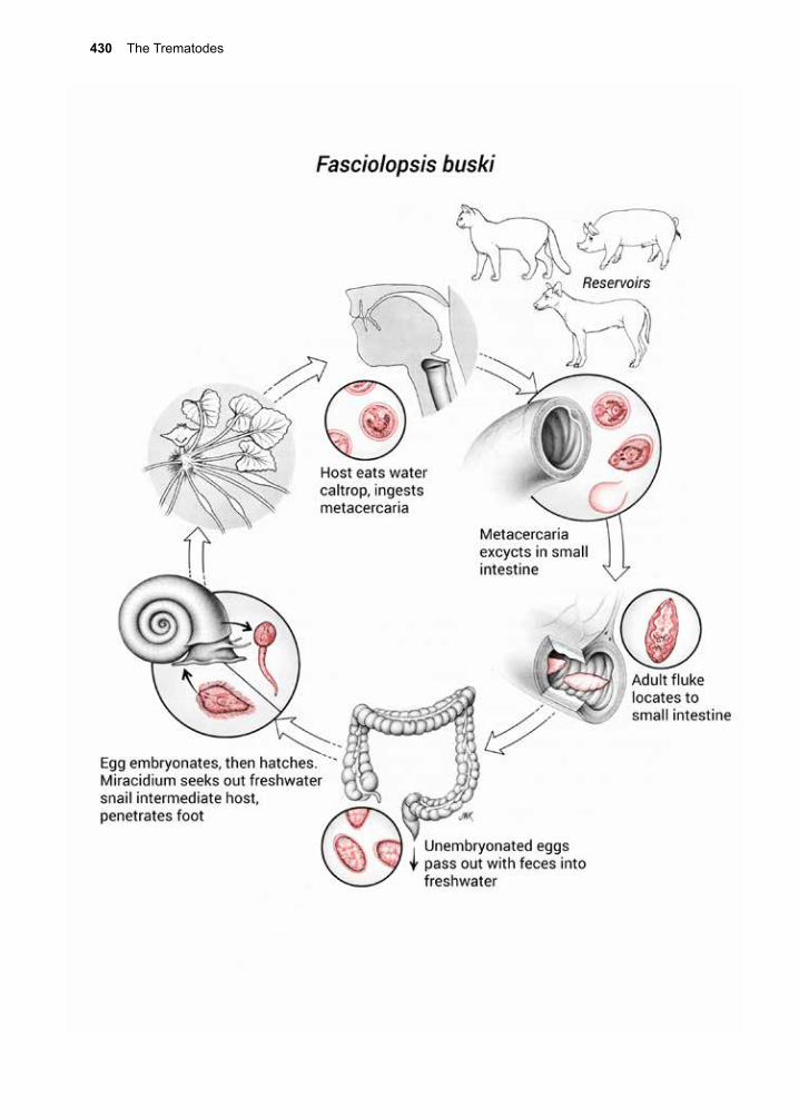
430 The Trematodes
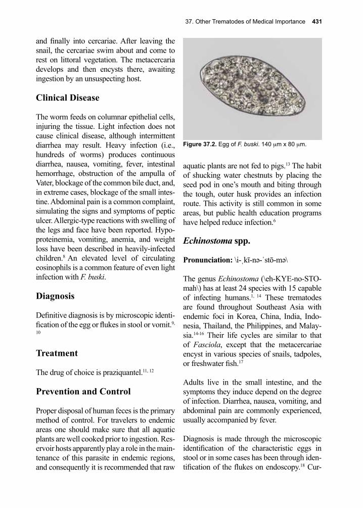
37. Other Trematodes of Medical Importance 431
and finally into cercariae. After leaving the snail, the cercariae swim about and come to rest on littoral vegetation. The metacercaria develops and then encysts there, awaiting ingestion by an unsuspecting host.
Clinical Disease
The worm feeds on columnar epithelial cells, injuring the tissue. Light infection does not cause clinical disease, although intermittent diarrhea may result. Heavy infection (i.e., hundreds of worms) produces continuous diarrhea, nausea, vomiting, fever, intestinal hemorrhage, obstruction of the ampulla of Vater, blockage of the common bile duct, and, in extreme cases, blockage of the small intes-tine. Abdominal pain is a common complaint, simulating the signs and symptoms of peptic ulcer. Allergic-type reactions with swelling of the legs and face have been reported. Hypo-proteinemia, vomiting, anemia, and weight loss have been described in heavily-infected children.8 An elevated level of circulating eosinophils is a common feature of even light infection with F. buski.
Diagnosis
Definitive diagnosis is by microscopic identi-fication of the egg or flukes in stool or vomit.9,
10
Treatment
The drug of choice is praziquantel.11, 12
Prevention and Control
Proper disposal of human feces is the primary method of control. For travelers to endemic areas one should make sure that all aquatic plants are well cooked prior to ingestion. Res-ervoir hosts apparently play a role in the main-tenance of this parasite in endemic regions, and consequently it is recommended that raw
aquatic plants are not fed to pigs.13 The habit of shucking water chestnuts by placing the seed pod in one’s mouth and biting through the tough, outer husk provides an infection route. This activity is still common in some areas, but public health education programs have helped reduce infection.6
Echinostoma spp.
Pronunciation: \i-ˌkī-nə-ˈstō-mə\
The genus Echinostoma (\eh-KYE-no-STO-mah\) has at least 24 species with 15 capable of infecting humans.1, 14 These trematodes are found throughout Southeast Asia with endemic foci in Korea, China, India, Indo-nesia, Thailand, the Philippines, and Malay-sia.14-16 Their life cycles are similar to that of Fasciola, except that the metacercariae encyst in various species of snails, tadpoles, or freshwater fish.17
Adults live in the small intestine, and the symptoms they induce depend on the degree of infection. Diarrhea, nausea, vomiting, and abdominal pain are commonly experienced, usually accompanied by fever.
Diagnosis is made through the microscopic identification of the characteristic eggs in stool or in some cases has been through iden-tification of the flukes on endoscopy.18 Cur-
Figure 37.2. Egg of F. buski. 140 µm x 80 µm.
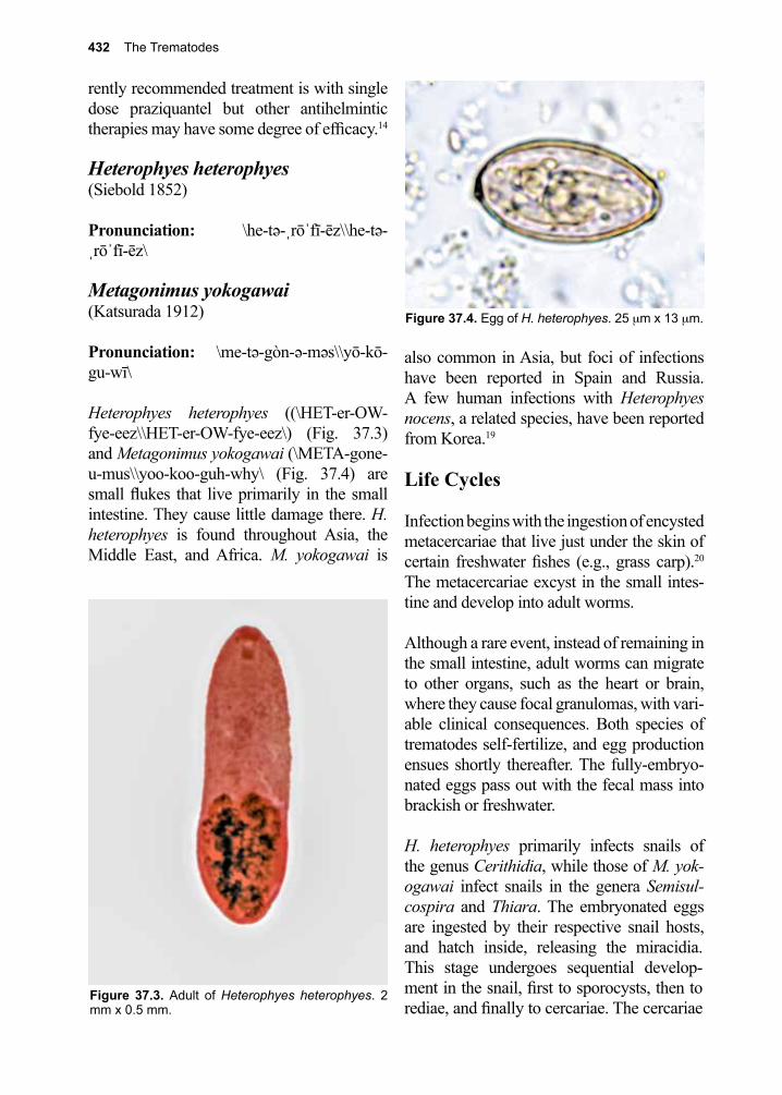
432 The Trematodes
rently recommended treatment is with single dose praziquantel but other antihelmintic therapies may have some degree of efficacy.14
Heterophyes heterophyes(Siebold 1852)
Pronunciation: \he-tə-ˌrōˈfī-ēz\\he-tə-ˌrōˈfī-ēz\
Metagonimus yokogawai (Katsurada 1912)
Pronunciation: \me-tə-gòn-ə-məs\\yō-kō-gu-wī\
Heterophyes heterophyes ((\HET-er-OW-fye-eez\\HET-er-OW-fye-eez\) (Fig. 37.3) and Metagonimus yokogawai (\META-gone-u-mus\\yoo-koo-guh-why\ (Fig. 37.4) are small flukes that live primarily in the small intestine. They cause little damage there. H. heterophyes is found throughout Asia, the Middle East, and Africa. M. yokogawai is
also common in Asia, but foci of infections have been reported in Spain and Russia. A few human infections with Heterophyes nocens, a related species, have been reported from Korea.19
Life Cycles
Infection begins with the ingestion of encysted metacercariae that live just under the skin of certain freshwater fishes (e.g., grass carp).20 The metacercariae excyst in the small intes-tine and develop into adult worms.
Although a rare event, instead of remaining in the small intestine, adult worms can migrate to other organs, such as the heart or brain, where they cause focal granulomas, with vari-able clinical consequences. Both species of trematodes self-fertilize, and egg production ensues shortly thereafter. The fully-embryo-nated eggs pass out with the fecal mass into brackish or freshwater.
H. heterophyes primarily infects snails of the genus Cerithidia, while those of M. yok-ogawai infect snails in the genera Semisul-cospira and Thiara. The embryonated eggs are ingested by their respective snail hosts, and hatch inside, releasing the miracidia. This stage undergoes sequential develop-ment in the snail, first to sporocysts, then to rediae, and finally to cercariae. The cercariae
Figure 37.3. Adult of Heterophyes heterophyes. 2 mm x 0.5 mm.
Figure 37.4. Egg of H. heterophyes. 25 µm x 13 µm.
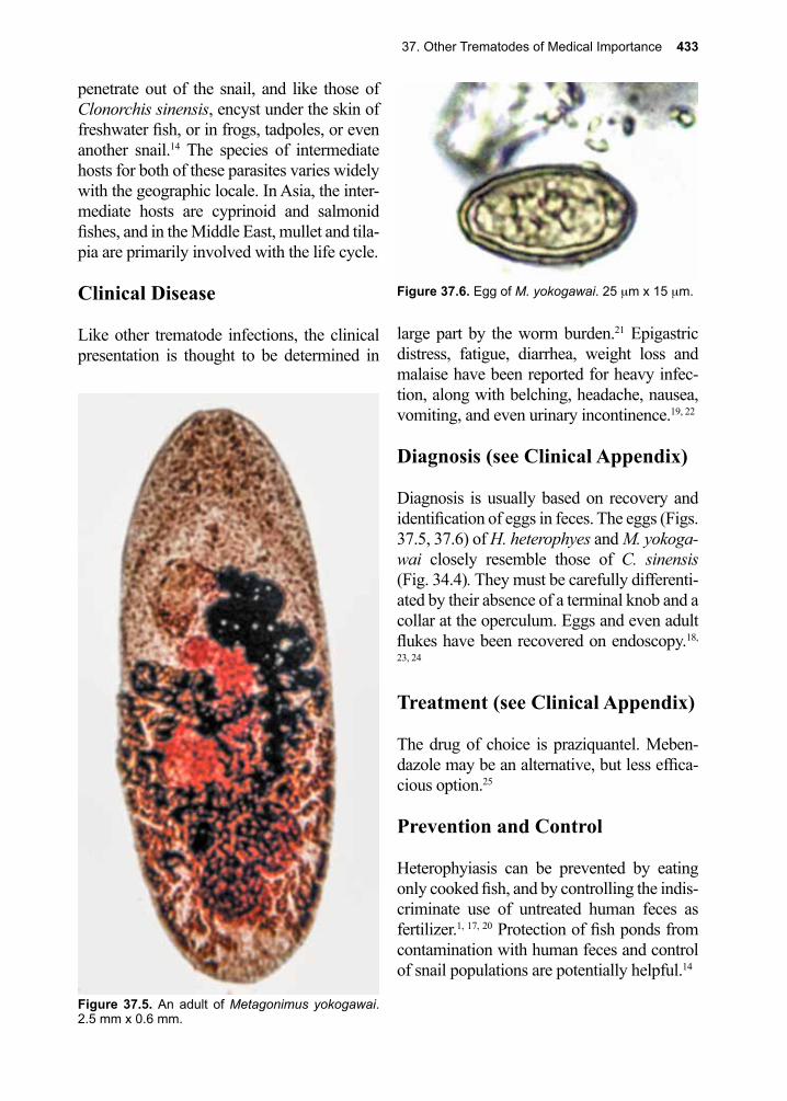
37. Other Trematodes of Medical Importance 433
penetrate out of the snail, and like those of Clonorchis sinensis, encyst under the skin of freshwater fish, or in frogs, tadpoles, or even another snail.14 The species of intermediate hosts for both of these parasites varies widely with the geographic locale. In Asia, the inter-mediate hosts are cyprinoid and salmonid fishes, and in the Middle East, mullet and tila-pia are primarily involved with the life cycle.
Clinical Disease
Like other trematode infections, the clinical presentation is thought to be determined in
large part by the worm burden.21 Epigastric distress, fatigue, diarrhea, weight loss and malaise have been reported for heavy infec-tion, along with belching, headache, nausea, vomiting, and even urinary incontinence.19, 22
Diagnosis (see Clinical Appendix)
Diagnosis is usually based on recovery and identification of eggs in feces. The eggs (Figs. 37.5, 37.6) of H. heterophyes and M. yokoga-wai closely resemble those of C. sinensis (Fig. 34.4). They must be carefully differenti-ated by their absence of a terminal knob and a collar at the operculum. Eggs and even adult flukes have been recovered on endoscopy.18,
23, 24
Treatment (see Clinical Appendix)
The drug of choice is praziquantel. Meben-dazole may be an alternative, but less effica-cious option.25
Prevention and Control
Heterophyiasis can be prevented by eating only cooked fish, and by controlling the indis-criminate use of untreated human feces as fertilizer.1, 17, 20 Protection of fish ponds from contamination with human feces and control of snail populations are potentially helpful.14
Figure 37.5. An adult of Metagonimus yokogawai. 2.5 mm x 0.6 mm.
Figure 37.6. Egg of M. yokogawai. 25 µm x 15 µm.
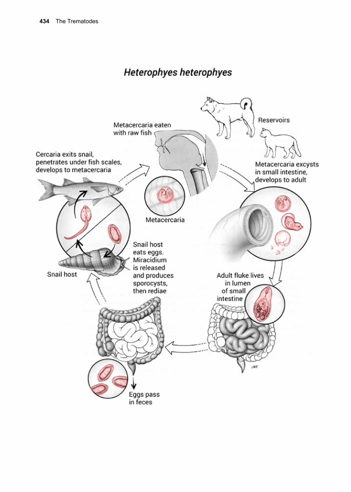
434 The Trematodes
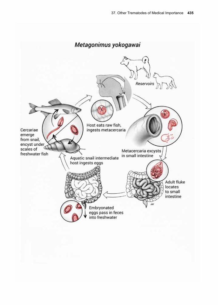
37. Other Trematodes of Medical Importance 435
436 The Trematodes
References
1. Fried, B., et al., Food-borne intestinal trematodiases in humans. Parasitology research 2004, 93 (2), 159-70.
2. Chai, J. Y., et al., Foodborne intestinal flukes in Southeast Asia. Korean J Parasitol 2009, 47 Suppl, S69-102.
3. Keiser, J.; Utzinger, J., Food-borne trematodiases. Clin Microbiol Rev 2009, 22 (3), 466-83.4. Wiwanitkit, V.; Suyaphan, A., High prevalence of HBsAg seropositivity in Hilltribers in the Mae
Jam district in northern Thailand. MedGenMed : Medscape general medicine 2002, 4 (3), 26.5. Bhatti, H. S., et al., Fasciolopslasis--a re-emerging infection in Azamgarh (Uttar Pradesh). Indian
journal of pathology & microbiology 2000, 43 (1), 73-6.6. Graczyk, T. K., et al., Fasciolopsiasis: is it a controllable food-borne disease? Parasitology
research 2001, 87 (1), 80-3.7. Achra, A., et al., Fasciolopsiasis: Endemic focus of a neglected parasitic disease in Bihar. Indian
J Med Microbiol 2015, 33 (3), 364-8.8. Gupta, A., et al., Fasciolopsis buski (giant intestinal fluke)--a case report. Indian journal of
pathology & microbiology 1999, 42 (3), 359-60.9. Naher, B. S., et al., Fasciolopsiasis in a five year old girl. Mymensingh Med J 2013, 22 (2), 397-
9.10. Le, T. H., et al., Case report: unusual presentation of Fasciolopsis buski in a Vietnamese child.
Trans R Soc Trop Med Hyg 2004, 98 (3), 193-4.11. Taraschewski, H., et al., Effects of praziquantel on human intestinal flukes (Fasciolopsis buski
and Heterophyes heterophyes). Zentralblatt fur Bakteriologie, Mikrobiologie, und Hygiene. Series A, Medical microbiology, infectious diseases, virology, parasitology 1986, 262 (4), 542-50.
12. Chai, J. Y., Praziquantel treatment in trematode and cestode infections: an update. Infect Chemother 2013, 45 (1), 32-43.
13. Inpankaew, T., et al., A survey for potentially zoonotic gastrointestinal parasites of dogs and pigs in Cambodia. Acta Parasitol 2015, 60 (4), 601-4.
14. Toledo, R.; Esteban, J. G., An update on human echinostomiasis. Trans R Soc Trop Med Hyg 2016, 110 (1), 37-45.
15. Park, S. K., et al., Status of intestinal parasite infections among children in Bat Dambang, Cambodia. The Korean journal of parasitology 2004, 42 (4), 201-3.
16. Huffman, J. E.; Fried, B., Echinostoma and echinostomiasis. Advances in parasitology 1990, 29, 215-69.
17. Anantaphruti, M. T., Parasitic contaminants in food. The Southeast Asian journal of tropical medicine and public health 2001, 32 Suppl 2, 218-28.
Nanophyetus salmincola(Chapin 1927)
Pronunciation: \nānəˌfīˈētəs\\sal-min-kō-lə\
Although Nanophyetus salmincola (\NAH-no-FIE-ee-tus\\SAL-min-koo-la\) may be the most common trematode in the United States, it tends to mostly infect animals, and only rarely infects humans.26 N. salmincola infects dogs, foxes, and coyotes in eastern Siberia and in the Pacific Northwest of the United States, where it produces “salmon poison-
ing” or “Elokomin fluke fever” as a result of a rickettsia, Neorickettsia helmintheca, which is co-transmitted with the parasite.27 Human infection has also been described, result-ing in diarrhea, nausea, vomiting, cachexia, anorexia, and elevated levels of circulating eosinophils.28 N. salmincola infection is diag-nosed by the presence of characteristic eggs in the stools, along with a history of inges-tion of raw or poorly cooked salmon.28 Pra-ziquantel is the recommended treatment but niclosamide may also be efficacious.28
37. Other Trematodes of Medical Importance 437
18. Jung, W. T., et al., A case of Echinostoma cinetorchis (Trematoda: Echinostomatidae) infection diagnosed by colonoscopy. Korean J Parasitol 2014, 52 (3), 287-90.
19. Chai, J.-Y., et al., Prevalence of Heterophyes nocens and Pygydiopsis summa infections among residents of the western and southern coastal islands of the Republic of Korea. The American journal of tropical medicine and hygiene 2004, 71 (5), 617-22.
20. Chai, J.-Y.; Lee, S.-H., Food-borne intestinal trematode infections in the Republic of Korea. Parasitology international 2002, 51 (2), 129-54.
21. Graczyk, T. K.; Fried, B., Echinostomiasis: a common but forgotten food-borne disease. Am J Trop Med Hyg 1998, 58 (4), 501-4.
22. Chang, Y. D., et al., A human infection of Echinostoma hortense in duodenal bulb diagnosed by endoscopy. Korean J Parasitol 2005, 43 (2), 57-60.
23. Toledo, R., et al., Intestinal trematode infections. Adv Exp Med Biol 2014, 766, 201-40.24. Esteban, J. G., et al., Diagnosis of human trematode infections. Adv Exp Med Biol 2014, 766,
293-327.25. Chen, Y., et al., Studies on efficacy of praziquantel and mebendazole-medicated salt in treatment
of Echinochasmus fujianensis infection. Southeast Asian J Trop Med Public Health 1997, 28 (2), 344-6.
26. Eastburn, R. L., et al., Human intestinal infection with Nanophyetus salmincola from salmonid fishes. Am J Trop Med Hyg 1987, 36 (3), 586-91.
27. Headley, S. A., et al., Neorickettsia helminthoeca and salmon poisoning disease: a review. Vet J 2011, 187 (2), 165-73.
28. Eastburn, R. L., et al., Human intestinal infection with Nanophyetus salmincola from salmonid fishes. The American journal of tropical medicine and hygiene 1987, 36 (3), 586-91.
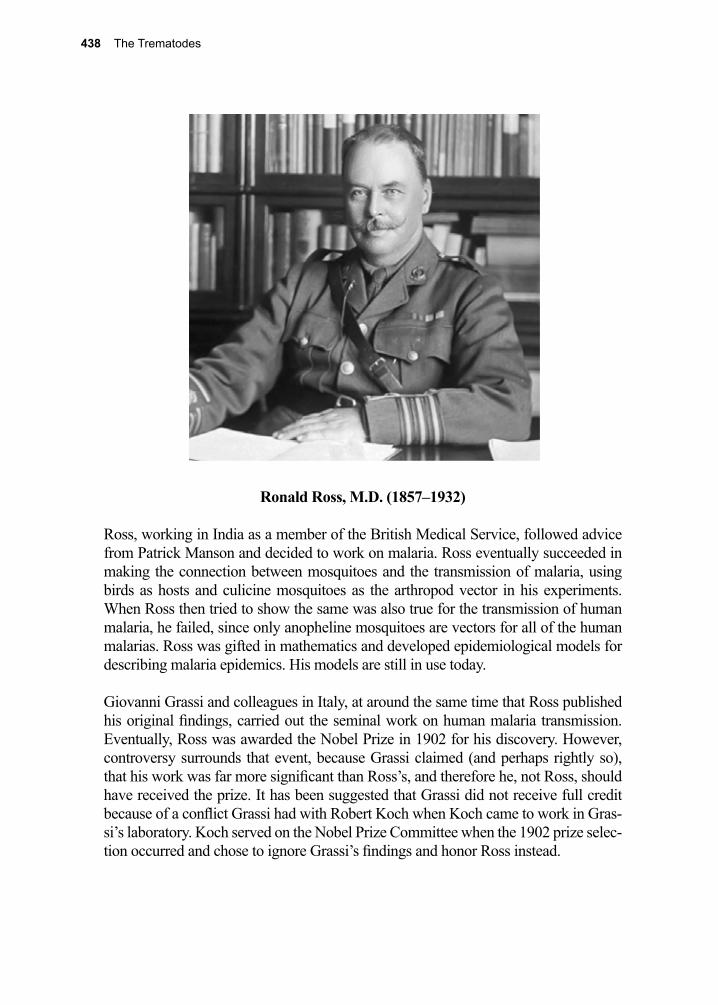
438 The Trematodes
Ronald Ross, M.D. (1857–1932)
Ross, working in India as a member of the British Medical Service, followed advice from Patrick Manson and decided to work on malaria. Ross eventually succeeded in making the connection between mosquitoes and the transmission of malaria, using birds as hosts and culicine mosquitoes as the arthropod vector in his experiments. When Ross then tried to show the same was also true for the transmission of human malaria, he failed, since only anopheline mosquitoes are vectors for all of the human malarias. Ross was gifted in mathematics and developed epidemiological models for describing malaria epidemics. His models are still in use today.
Giovanni Grassi and colleagues in Italy, at around the same time that Ross published his original findings, carried out the seminal work on human malaria transmission. Eventually, Ross was awarded the Nobel Prize in 1902 for his discovery. However, controversy surrounds that event, because Grassi claimed (and perhaps rightly so), that his work was far more significant than Ross’s, and therefore he, not Ross, should have received the prize. It has been suggested that Grassi did not receive full credit because of a conflict Grassi had with Robert Koch when Koch came to work in Gras-si’s laboratory. Koch served on the Nobel Prize Committee when the 1902 prize selec-tion occurred and chose to ignore Grassi’s findings and honor Ross instead.
The Arthropods 439
York City in 1999 of mosquito-transmitted human infections of the West Nile virus, and its subsequent spread throughout the United States, should reinforce our awareness of vul-nerability to invasion by both pathogens and vectors. Although fear has been expressed that blood-feeding arthropods, especially mosquitoes, could transmit HIV, a large body of epidemiological and experimental evi-dence fails to support this hypothesis.
Despite the problems arthropods cause for humans and livestock, they are also beneficial as pollinators, producers of honey, natural reg-ulators of harmful insects, and essential mem-bers of food chains. The phylum Arthropoda contains an enormous diversity of members, with the number of species exceeding that of all other phyla combined. The arthropods share a number of characteristics that dis-tinguish them from all other animal groups, although some of these features are absent in a particular species or group at some period of development. Nevertheless, there are traits specific to each class in the phylum to help identify them.
Among the morphologic characteristics are bilateral symmetry, a hard exoskeleton, a segmented body, and paired, jointed append-ages. The term arthropod, derived from Greek, means “jointed foot.” Growth by metamorphosis is another characteristic of the arthropods. In some groups, growth is gradual; each change from one stage to the next is known as a molt, and gives rise to a stage somewhat larger but morphologically similar to its predecessor (incomplete meta-morphosis). Among the spiders, eight or nine immature stages may precede the final molt to the sexually mature adult. Another devel-opmental strategy involves egg, larva, pupa and adult. In this case, each stage is morpho-logically distinct (complete metamorphosis). Examples include the flies and the fleas.
VIII. The Arthropods
Arthropods directly influence humans’ well-being, not only because they are hosts of par-asitic organisms and vectors of a wide vari-ety of pathogens, but also by causing tissue damage and disease. They also affect human health by reducing the availability of food. Insects are essential pollinators of crops, but they also destroy an estimated 20% of all food crops. This destruction continues despite the increasing use of pest manage-ment tactics such as insecticides, genetically modified organisms and physical control methods in fields and storage areas. Live-stock are also affected by arthropod-borne infections. Vast areas of Africa are short of protein foods because cattle suffer a number of vector-borne diseases, including trypano-somiasis transmitted by tsetse flies and a variety of tick-transmitted diseases.
Although the pathogenic effects of arthro-pods are most pronounced in the tropics, they are by no means negligible in the United States and other temperate areas. Lyme dis-ease and anaplasmosis, which are transmitted by ticks, have spread rapidly throughout the United States. Aedes albopictus, the Asian tiger mosquito, has been introduced in ship-ments of used automobile tires into the south-ern United States and has spread as far north as central Ohio, Indiana, and Illinois; more-over, the introduced strain of the mosquito is apparently able to survive the winter in the egg stage in temperate climates. The same species has been introduced into Europe and South America. Ae. albopictus can be an effi-cient vector of dengue, chikungunya and Zika virus. In 1999, reports surfaced about the introduction and rapid spread across the East-ern United States of another pest mosquito, Ochlerotatus japonicus. Introductions of new species should not be unexpected and point to the ease with which such introductions can occur. The unexpected appearance in New
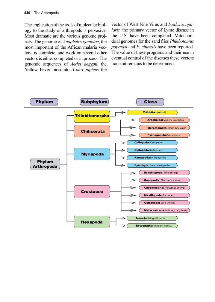
440 The Arthropods
The application of the tools of molecular biol-ogy to the study of arthropods is pervasive. Most dramatic are the various genome proj-ects. The genome of Anopheles gambiae, the most important of the African malaria vec-tors, is complete, and work on several other vectors is either completed or in process. The genomic sequences of Aedes aegypti, the Yellow Fever mosquito, Culex pipiens the
vector of West Nile Virus and Ixodes scapu-laris, the primary vector of Lyme disease in the U.S. have been completed. Mitochon-drial genomes for the sand flies Phlebotomus papatasi and P. chinesis have been reported. The value of these programs and their use in eventual control of the diseases these vectors transmit remains to be determined.
Trilobita: (extinct)
Arachnida: Spiders, Scorpions
Merostomata: Horseshoe crabs
Pycnogonida: Sea spiders
Chilopoda: Centipedes
Diplopoda: Millipedes
Paurapoda: Millipede-like
Symphyla: Pseudocentipedes
Brachiopoda: Brine shrimp
Remipedia: Blind crustaceans
Chephlocaria: Horseshoe shrimp
Maxillopoda: Barnacles
Ostracoda: Seed shrimps
Malacostraca: Lobster, crab, shrimp
Insecta: Winged Insects
Ectognatha: Wingless insects
Chilicerata
Trilobitomorpha
Myriapoda
Crustacea
Hexapoda
PhylumArthropoda
Subphylum ClassPhylum
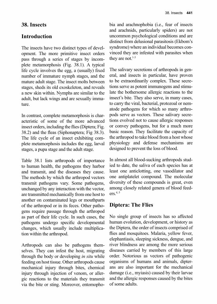
38. Insects 441
38. Insects
Introduction
The insects have two distinct types of devel-opment. The more primitive insect orders pass through a series of stages by incom-plete metamorphosis (Fig. 38.1). A typical life cycle involves the egg, a (usually) fixed number of immature nymph stages, and the mature adult stage. The insect molts between stages, sheds its old exoskeleton, and reveals a new skin within. Nymphs are similar to the adult, but lack wings and are sexually imma-ture.
In contrast, complete metamorphosis is char-acteristic of some of the more advanced insect orders, including the flies (Diptera; Fig. 38.2) and the fleas (Siphonaptera; Fig 38.3). The life cycle of an insect exhibiting com-plete metamorphosis includes the egg, larval stages, a pupa stage and the adult stage.
Table 38.1 lists arthropods of importance to human health, the pathogens they harbor and transmit, and the diseases they cause. The methods by which the arthropod vectors transmit pathogens vary. Some pathogens, unchanged by any interaction with the vector, are transmitted mechanically from one host to another on contaminated legs or mouthparts of the arthropod or in its feces. Other patho-gens require passage through the arthropod as part of their life cycle. In such cases, the pathogens undergo specific developmental changes, which usually include multiplica-tion within the arthropod.
Arthropods can also be pathogens them-selves. They can infest the host, migrating through the body or developing in situ while feeding on host tissue. Other arthropods cause mechanical injury through bites, chemical injury through injection of venom, or aller-gic reactions to the materials they transmit via the bite or sting. Moreover, entomopho-
bia and arachnophobia (i.e., fear of insects and arachnids, particularly spiders) are not uncommon psychological conditions and are distinct from delusional parasitosis (Ekbom’s syndrome) where an individual becomes con-vinced they are infested with parasites when they are not.1-3
The salivary secretions of arthropods in gen-eral, and insects in particular, have proven to be extraordinarily complex. These secre-tions serve as potent immunogens and stimu-late the bothersome allergic reactions to the insect’s bite. They also serve, in many cases, to carry the viral, bacterial, protozoal or nem-atode pathogens for which so many arthro-pods serve as vectors. These salivary secre-tions evolved not to cause allergic responses or convey pathogens, but for a much more basic reason. They facilitate the capacity of the arthropod to take blood from a host whose physiology and defense mechanisms are designed to prevent the loss of blood.
In almost all blood-sucking arthropods stud-ied to date, the saliva of each species has at least one anticlotting, one vasodilator and one antiplatelet compound. The molecular diversity of these compounds is great, even among closely related genera of blood feed-ers.4, 5
Diptera: The Flies
No single group of insects has so affected human evolution, development, or history as the Diptera, the order of insects comprised of flies and mosquitoes. Malaria, yellow fever, elephantiasis, sleeping sickness, dengue, and river blindness are among the more serious diseases carried by members of this large order. Notorious as vectors of pathogenic organisms of humans and animals, dipter-ans are also important for the mechanical damage (i.e., myiasis) caused by their larvae and the allergic responses caused by the bites of some adults.
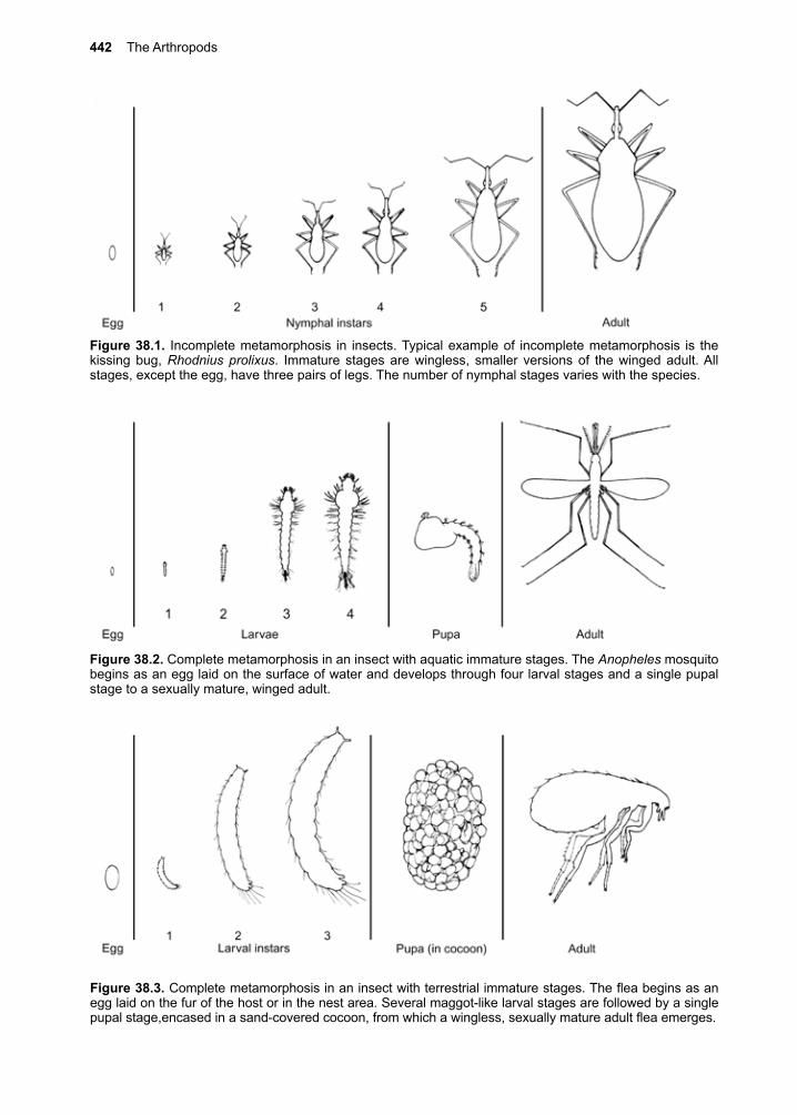
442 The Arthropods
Figure 38.3. Complete metamorphosis in an insect with terrestrial immature stages. The flea begins as an egg laid on the fur of the host or in the nest area. Several maggot-like larval stages are followed by a single pupal stage,encased in a sand-covered cocoon, from which a wingless, sexually mature adult flea emerges.
Figure 38.2. Complete metamorphosis in an insect with aquatic immature stages. The Anopheles mosquito begins as an egg laid on the surface of water and develops through four larval stages and a single pupal stage to a sexually mature, winged adult.
Figure 38.1. Incomplete metamorphosis in insects. Typical example of incomplete metamorphosis is the kissing bug, Rhodnius prolixus. Immature stages are wingless, smaller versions of the winged adult. All stages, except the egg, have three pairs of legs. The number of nymphal stages varies with the species.
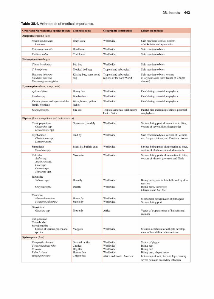
38. Insects 443
Table 38.1. Arthropods of medical importance.
Order and representative species Insecta Common name Geographic distribution Effects on humans
Anoplura (sucking lice)
Pediculus humanushumanus
Body louse Worldwide Skin reactions to bites, vectorsof rickettsiae and spirochetes
P. humanus capitis Head louse Worldwide Skin reaction to bites
Phthirus pubis Crab louse Worldwide Skin reaction to bites
Heteroptera (true bugs)
Cimex lectularius Bed bug Worldwide Skin reaction to bites
C. hemipterus Tropical bed bug Tropical and subtropical Skin reaction to bites
Triatoma infestansRhodnius prolixusPanstrongylus megistus
Kissing bug, cone-nosed bug
Tropical and subtropical regions of the New World
Skin reaction to bites, vectorsof Trypanosoma cruzi (cause of Chagas disease)
Hymenoptera (bees, wasps, ants)
Apis mellifera Honey bee Worldwide Painful sting, potential anaphylaxis
Bombus spp. Bumble bee Worldwide Painful sting, potential anaphylaxis
Various genera and species of the family Vespidae
Wasp, hornet, yellow jacket
Worldwide Painful sting, potential anaphylaxis
Solenopsis spp. Fire ant Tropical America, southeastern United States
Painful bite and multiple stings, potential anaphylaxis
Diptera (flies, mosquitoes, and their relatives)
CeratopogonidaeCulicoides spp.Leptoconops spp.
No-see-um, sand fly Worldwide Serious biting pest, skin reaction to bites, vectors of several filariid nematodes
PsychodidaePhlebotomus spp.Lutzomyia spp.
sand fly Worldwide Skin reaction to bites, vectors of Leishma-nia, Pappataci fever, and Carrion’s disease
Simuliidae Simulium spp.
Black fly, buffalo gnat Worldwide Serious biting pests, skin reaction to bites, vectors of Onchocerca and Mansonella
CulicidaeAedes spp.Anopheles spp.Culex spp.Culiseta spp.Mansonia spp.
Mosquito Worldwide Serious biting pests, skin reaction to bites, vectors of viruses, protozoa, and filaria
TabanidaeTabanus spp.
Chrysops spp.
Horsefly
Deerfly
Worldwide
Worldwide
Biting pests, painful bite followed by skin reactionBiting pests, vectors oftularemia and Loa loa.
MuscidaeMusca domesticaStomoxys calcitrans
House flyStable fly
WorldwideWorldwide
Mechanical disseminator of pathogensSerious biting pest
GlossinidaeGlossina spp. Tsetse fly Africa Vector of trypanosomes of humans and
animals
CalliphoridaeCuterebridaeSarcophagidae
Larvae of various genera and species
Maggots Worldwide Myiasis, accidental or obligate develop-ment of larval flies in human tissue
Siphonaptera (fleas)
Xenopsylla cheopisCtenocephalides felisC. canisPulex irritansTunga penetrans
Oriental rat fleaCat fleaDog fleaHuman fleaChigoe flea
WorldwideWorldwideWorldwideWorldwideAfrica and South America
Vector of plagueBiting pestBiting pestBiting pest, plague vectorInfestation of toes, feet and legs, causing severe pain and secondary infection
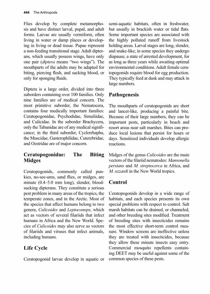
444 The Arthropods
Flies develop by complete metamorpho-sis and have distinct larval, pupal, and adult forms. Larvae are usually vermiform, often living in water or damp places or develop-ing in living or dead tissue. Pupae represent a non-feeding transitional stage. Adult dipter-ans, which usually possess wings, have only one pair (diptera means “two wings”). The mouthparts of the adults may be adapted for biting, piercing flesh, and sucking blood, or only for sponging fluids.
Diptera is a large order, divided into three suborders containing over 100 families. Only nine families are of medical concern. The most primitive suborder, the Nematocera, contains four medically important families: Ceratopogonidae, Psychodidae, Simuliidae, and Culicidae. In the suborder Brachycera, only the Tabanidae are of any medical signifi-cance; in the third suborder, Cyclorrhapha, the Muscidae, Gasterophilidae, Cuterebridae, and Oestridae are of major concern.
Ceratopogonidae: The Biting Midges
Ceratopogonids, commonly called pun-kies, no-see-ums, sand flies, or midges, are minute (0.4–5.0 mm long), slender, blood-sucking dipterans. They constitute a serious pest problem in many areas of the tropics, the temperate zones, and in the Arctic. Most of the species that affect humans belong to two genera, Culicoides and Leptoconops, which act as vectors of several filariids that infect humans in Africa and the New World. Spe-cies of Culicoides may also serve as vectors of filariids and viruses that infect animals, including humans.
Life Cycle
Ceratopogonid larvae develop in aquatic or
semi-aquatic habitats, often in freshwater, but usually in brackish water or tidal flats. Some important species are associated with the highly polluted runoff from livestock holding areas. Larval stages are long, slender, and snake-like; in some species they undergo diapause, a state of arrested development, for as long as three years while awaiting optimal environmental conditions. Adult female cera-topogonids require blood for egg production. They typically feed at dusk and may attack in large numbers.
Pathogenesis
The mouthparts of ceratopogonids are short and lancet-like, producing a painful bite.
Because of their large numbers, they can be important pests, particularly in beach and resort areas near salt marshes. Bites can pro-duce local lesions that persist for hours or days. Sensitized individuals develop allergic reactions.
Midges of the genus Culicoides are the main vectors of the filariid nematodes: Mansonella perstans and M. streptocerca in Africa, and M. ozzardi in the New World tropics.
Control
Ceratopogonids develop in a wide range of habitats, and each species presents its own special problems with respect to control. Salt marsh habitats can be drained, or channeled, and other breeding sites modified. Treatment of breeding sites with insecticides remains the most effective short-term control mea-sure. Window screens are ineffective unless they are treated with insecticides, because they allow these minute insects easy entry. Commercial mosquito repellents contain-ing DEET may be useful against some of the common species of these pests.
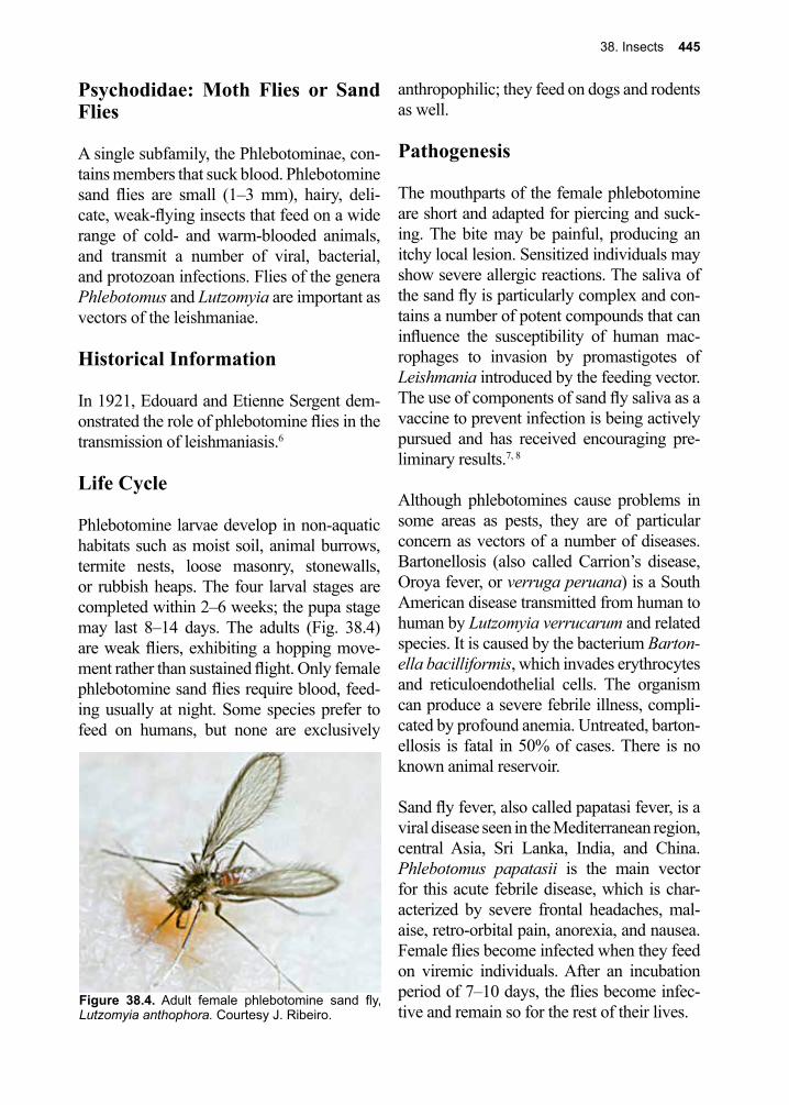
38. Insects 445
Psychodidae: Moth Flies or Sand Flies
A single subfamily, the Phlebotominae, con-tains members that suck blood. Phlebotomine sand flies are small (1–3 mm), hairy, deli-cate, weak-flying insects that feed on a wide range of cold- and warm-blooded animals, and transmit a number of viral, bacterial, and protozoan infections. Flies of the genera Phlebotomus and Lutzomyia are important as vectors of the leishmaniae.
Historical Information
In 1921, Edouard and Etienne Sergent dem-onstrated the role of phlebotomine flies in the transmission of leishmaniasis.6
Life Cycle
Phlebotomine larvae develop in non-aquatic habitats such as moist soil, animal burrows, termite nests, loose masonry, stonewalls, or rubbish heaps. The four larval stages are completed within 2–6 weeks; the pupa stage may last 8–14 days. The adults (Fig. 38.4) are weak fliers, exhibiting a hopping move-ment rather than sustained flight. Only female phlebotomine sand flies require blood, feed-ing usually at night. Some species prefer to feed on humans, but none are exclusively
anthropophilic; they feed on dogs and rodents as well.
Pathogenesis
The mouthparts of the female phlebotomine are short and adapted for piercing and suck-ing. The bite may be painful, producing an itchy local lesion. Sensitized individuals may show severe allergic reactions. The saliva of the sand fly is particularly complex and con-tains a number of potent compounds that can influence the susceptibility of human mac-rophages to invasion by promastigotes of Leishmania introduced by the feeding vector. The use of components of sand fly saliva as a vaccine to prevent infection is being actively pursued and has received encouraging pre-liminary results.7, 8
Although phlebotomines cause problems in some areas as pests, they are of particular concern as vectors of a number of diseases. Bartonellosis (also called Carrion’s disease, Oroya fever, or verruga peruana) is a South American disease transmitted from human to human by Lutzomyia verrucarum and related species. It is caused by the bacterium Barton-ella bacilliformis, which invades erythrocytes and reticuloendothelial cells. The organism can produce a severe febrile illness, compli-cated by profound anemia. Untreated, barton-ellosis is fatal in 50% of cases. There is no known animal reservoir.
Sand fly fever, also called papatasi fever, is a viral disease seen in the Mediterranean region, central Asia, Sri Lanka, India, and China. Phlebotomus papatasii is the main vector for this acute febrile disease, which is char-acterized by severe frontal headaches, mal-aise, retro-orbital pain, anorexia, and nausea. Female flies become infected when they feed on viremic individuals. After an incubation period of 7–10 days, the flies become infec-tive and remain so for the rest of their lives.
Figure 38.4. Adult female phlebotomine sand fly, Lutzomyia anthophora. Courtesy J. Ribeiro.
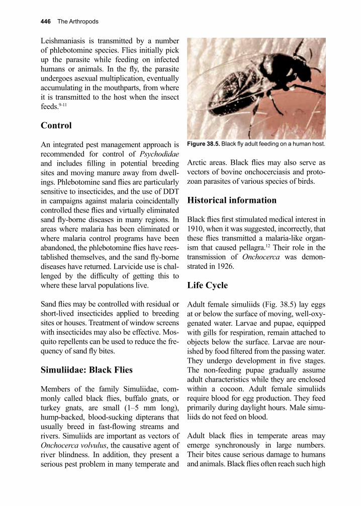
446 The Arthropods
Leishmaniasis is transmitted by a number of phlebotomine species. Flies initially pick up the parasite while feeding on infected humans or animals. In the fly, the parasite undergoes asexual multiplication, eventually accumulating in the mouthparts, from where it is transmitted to the host when the insect feeds.9-11
Control
An integrated pest management approach is recommended for control of Psychodidae and includes filling in potential breeding sites and moving manure away from dwell-ings. Phlebotomine sand flies are particularly sensitive to insecticides, and the use of DDT in campaigns against malaria coincidentally controlled these flies and virtually eliminated sand fly-borne diseases in many regions. In areas where malaria has been eliminated or where malaria control programs have been abandoned, the phlebotomine flies have rees-tablished themselves, and the sand fly-borne diseases have returned. Larvicide use is chal-lenged by the difficulty of getting this to where these larval populations live.
Sand flies may be controlled with residual or short-lived insecticides applied to breeding sites or houses. Treatment of window screens with insecticides may also be effective. Mos-quito repellents can be used to reduce the fre-quency of sand fly bites.
Simuliidae: Black Flies
Members of the family Simuliidae, com-monly called black flies, buffalo gnats, or turkey gnats, are small (1–5 mm long), hump-backed, blood-sucking dipterans that usually breed in fast-flowing streams and rivers. Simuliids are important as vectors of Onchocerca volvulus, the causative agent of river blindness. In addition, they present a serious pest problem in many temperate and
Arctic areas. Black flies may also serve as vectors of bovine onchocerciasis and proto-zoan parasites of various species of birds.
Historical information
Black flies first stimulated medical interest in 1910, when it was suggested, incorrectly, that these flies transmitted a malaria-like organ-ism that caused pellagra.12 Their role in the transmission of Onchocerca was demon-strated in 1926.
Life Cycle
Adult female simuliids (Fig. 38.5) lay eggs at or below the surface of moving, well-oxy-genated water. Larvae and pupae, equipped with gills for respiration, remain attached to objects below the surface. Larvae are nour-ished by food filtered from the passing water. They undergo development in five stages. The non-feeding pupae gradually assume adult characteristics while they are enclosed within a cocoon. Adult female simuliids require blood for egg production. They feed primarily during daylight hours. Male simu-liids do not feed on blood.
Adult black flies in temperate areas may emerge synchronously in large numbers. Their bites cause serious damage to humans and animals. Black flies often reach such high
Figure 38.5. Black fly adult feeding on a human host.
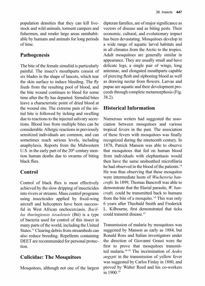
38. Insects 447
population densities that they can kill live-stock and wild animals, torment campers and fishermen, and render large areas uninhabit-able by humans and animals for long periods of time.
Pathogenesis
The bite of the female simuliid is particularly painful. The insect’s mouthparts consist of six blades in the shape of lancets, which tear the skin surface to induce bleeding. The fly feeds from the resulting pool of blood, and the bite wound continues to bleed for some time after the fly has departed. Simuliid bites leave a characteristic point of dried blood at the wound site. The extreme pain of the ini-tial bite is followed by itching and swelling due to reactions to the injected salivary secre-tions. Blood loss from multiple bites can be considerable. Allergic reactions in previously sensitized individuals are common, and can sometimes reach serious levels, including anaphylaxis. Reports from the Midwestern U.S. in the early part of the 20th century men-tion human deaths due to swarms of biting black flies.
Control
Control of black flies is most effectively achieved by the slow dripping of insecticides into rivers or streams. Mass control programs using insecticides applied by fixed-wing aircraft and helicopters have been success-ful in West African onchocerciasis. Bacil-lus thuringiensis israelensis (Bti) is a type of bacteria used for control of this insect in many parts of the world, including the United States.13 Clearing debris from streambeds can also reduce breeding. Repellents containing DEET are recommended for personal protec-tion.
Culicidae: The Mosquitoes
Mosquitoes, although not one of the largest
dipteran families, are of major significance as vectors of disease and as biting pests. Their economic, cultural, and evolutionary impact has been devastating. Mosquitoes develop in a wide range of aquatic larval habitats and in all climates from the Arctic to the tropics. Adult mosquitoes are generally similar in appearance. They are usually small and have delicate legs, a single pair of wings, long antennae, and elongated mouthparts capable of piercing flesh and siphoning blood as well as drawing nectar from flowers. Larvae and pupae are aquatic and their development pro-ceeds through complete metamorphosis (Fig. 38.2).
Historical Information
Numerous writers had suggested the asso-ciation between mosquitoes and various tropical fevers in the past. The association of these fevers with mosquitoes was finally recognized during the nineteenth century. In 1878, Patrick Manson was able to observe that mosquitoes that fed on human blood from individuals with elephantiasis would then have the same unsheathed microfilaria he had observed in the blood of the patients.14 He was thus observing that these mosquitos were intermediate hosts of Wuchereria ban-crofti. In 1899, Thomas Bancroft was able to demonstrate that the filarial parasite, W. ban-crofti, could be transmitted back to humans from the bite of a mosquito. 14 This was only 6 years after Theobald Smith and Frederick L. Kilbourne, first demonstrated that ticks could transmit disease.15
Transmission of malaria by mosquitoes was suggested by Manson as early as 1884, but Ronald Ross and Italian investigators under the direction of Giovanni Grassi were the first to prove that mosquitoes transmit-ted malaria.16-18 The incrimination of Aedes aegypti in the transmission of yellow fever was suggested by Carlos Finlay in 1880, and proved by Walter Reed and his co-workers in 1900.19
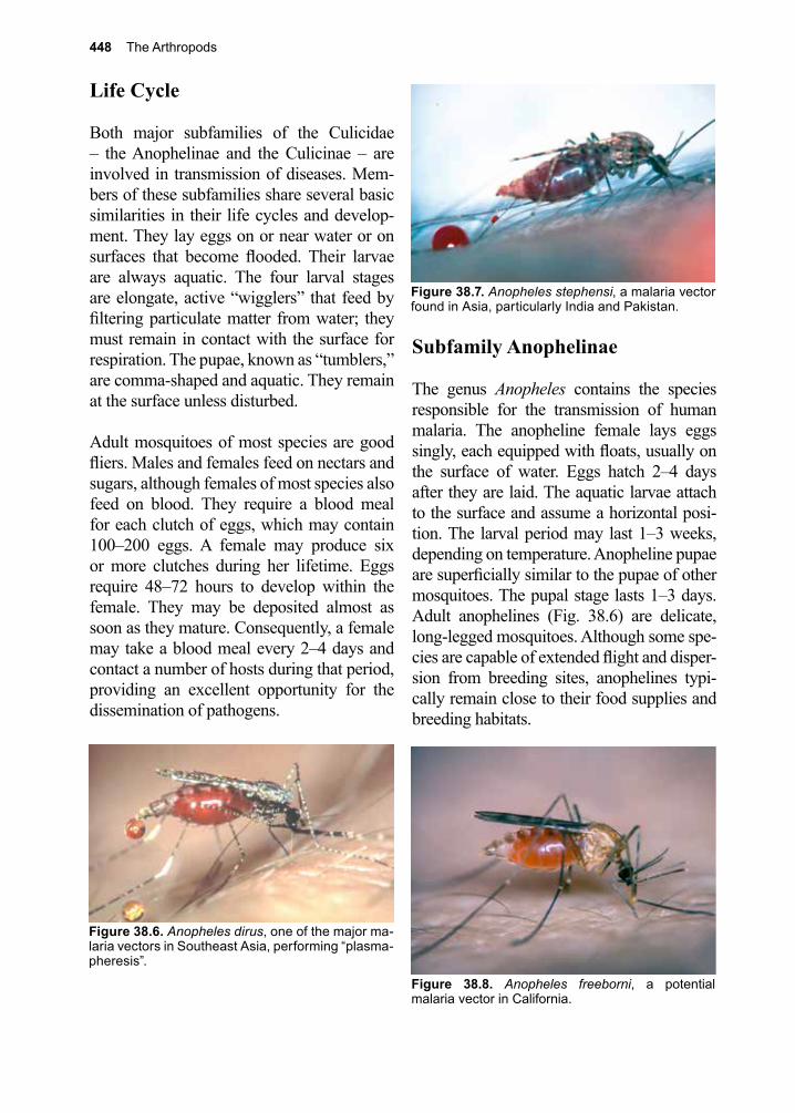
448 The Arthropods
Life Cycle
Both major subfamilies of the Culicidae – the Anophelinae and the Culicinae – are involved in transmission of diseases. Mem-bers of these subfamilies share several basic similarities in their life cycles and develop-ment. They lay eggs on or near water or on surfaces that become flooded. Their larvae are always aquatic. The four larval stages are elongate, active “wigglers” that feed by filtering particulate matter from water; they must remain in contact with the surface for respiration. The pupae, known as “tumblers,” are comma-shaped and aquatic. They remain at the surface unless disturbed.
Adult mosquitoes of most species are good fliers. Males and females feed on nectars and sugars, although females of most species also feed on blood. They require a blood meal for each clutch of eggs, which may contain 100–200 eggs. A female may produce six or more clutches during her lifetime. Eggs require 48–72 hours to develop within the female. They may be deposited almost as soon as they mature. Consequently, a female may take a blood meal every 2–4 days and contact a number of hosts during that period, providing an excellent opportunity for the dissemination of pathogens.
Subfamily Anophelinae
The genus Anopheles contains the species responsible for the transmission of human malaria. The anopheline female lays eggs singly, each equipped with floats, usually on the surface of water. Eggs hatch 2–4 days after they are laid. The aquatic larvae attach to the surface and assume a horizontal posi-tion. The larval period may last 1–3 weeks, depending on temperature. Anopheline pupae are superficially similar to the pupae of other mosquitoes. The pupal stage lasts 1–3 days. Adult anophelines (Fig. 38.6) are delicate, long-legged mosquitoes. Although some spe-cies are capable of extended flight and disper-sion from breeding sites, anophelines typi-cally remain close to their food supplies and breeding habitats.
Figure 38.8. Anopheles freeborni, a potential malaria vector in California.
Figure 38.7. Anopheles stephensi, a malaria vector found in Asia, particularly India and Pakistan.
Figure 38.6. Anopheles dirus, one of the major ma-laria vectors in Southeast Asia, performing “plasma-pheresis”.
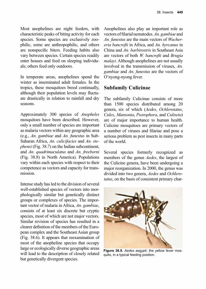
38. Insects 449
Most anophelines are night feeders, with characteristic peaks of biting activity for each species. Some species are exclusively zoo-philic, some are anthropophilic, and others are nonspecific biters. Feeding habits also vary between species. Certain species readily enter houses and feed on sleeping individu-als; others feed only outdoors.
In temperate areas, anophelines spend the winter as inseminated adult females. In the tropics, these mosquitoes breed continually, although their population levels may fluctu-ate drastically in relation to rainfall and dry seasons.
Approximately 300 species of Anopheles mosquitoes have been described. However, only a small number of species are important as malaria vectors within any geographic area (e.g., An. gambiae and An. funestus in Sub-Saharan Africa, An. culicifacies and An. ste-phensi (Fig. 38.7) on the Indian subcontinent, and An. quadrimaculatus and An. freeborni (Fig. 38.8) in North America). Populations vary within each species with respect to their competence as vectors and capacity for trans-mission.
Intense study has led to the division of several well-established species of vectors into mor-phologically similar but genetically distinct groups or complexes of species. The impor-tant vector of malaria in Africa, An. gambiae, consists of at least six discrete but cryptic species, most of which are not major vectors. Similar revision of species has resulted in a clearer definition of the members of the Euro-pean complex and the Southeast Asian group (Fig. 38.6). It appears that reexamination of most of the anopheline species that occupy large or ecologically diverse geographic areas will lead to the description of closely related but genetically divergent species.
Anophelines also play an important role as vectors of filarial nematodes. An. gambiae and An. funestus are the main vectors of Wucher-eria bancrofti in Africa, and An. hyrcanus in China and An. barbirostris in Southeast Asia are vectors of both W. bancrofti and Brugia malayi. Although anophelines are not usually involved in the transmission of viruses, An. gambiae and An. funestus are the vectors of O’nyong-nyong fever.
Subfamily Culicinae
The subfamily Culicinae consists of more than 1500 species distributed among 20 genera, six of which (Aedes, Ochlerotatus, Culex, Mansonia, Psorophora, and Culiseta) are of major importance to human health. Culicine mosquitoes are primary vectors of a number of viruses and filariae and pose a serious problem as pest insects in many parts of the world.
Several species formerly recognized as members of the genus Aedes, the largest of the Culicine genera, have been undergoing a major reorganization. In 2000, the genus was divided into two genera, Aedes and Ochlero-tatus, on the basis of consistent primary char-
Figure 38.9. Aedes aegypti, the yellow fever mos-quito, in a typical feeding position.
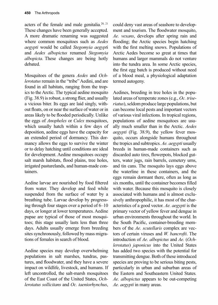
450 The Arthropods
acters of the female and male genitalia.20, 21 These changes have been generally accepted. A more dramatic renaming was suggested where common mosquitoes such as Aedes aegypti would be called Stegomyia aegypti and Aedes albopictus renamed Stegomyia albopicta. These changes are being hotly debated.
Mosquitoes of the genera Aedes and Och-lerotatus remain in the “tribe” Aedini, and are found in all habitats, ranging from the trop-ics to the Arctic. The typical aedine mosquito (Fig. 38.9) is robust, a strong flier, and usually a vicious biter. Its eggs are laid singly, with-out floats, on or near the surface of water or in areas likely to be flooded periodically. Unlike the eggs of Anopheles or Culex mosquitoes, which usually hatch within a few days of deposition, aedine eggs have the capacity for an extended period of dormancy. This dor-mancy allows the eggs to survive the winter or to delay hatching until conditions are ideal for development. Aedine mosquitoes occupy salt marsh habitats, flood plains, tree holes, irrigated pasturelands, and human-made con-tainers.
Aedine larvae are nourished by food filtered from water. They develop and feed while suspended from the surface of water by a breathing tube. Larvae develop by progress-ing through four stages over a period of 6–10 days, or longer at lower temperatures. Aedine pupae are typical of those of most mosqui-toes; this stage usually lasts less than three days. Adults usually emerge from breeding sites synchronously, followed by mass migra-tions of females in search of blood.
Aedine species may develop overwhelming populations in salt marshes, tundras, pas-tures, and floodwater, and they have a severe impact on wildlife, livestock, and humans. If left uncontrolled, the salt-marsh mosquitoes of the East Coast of the United States, Och-lerotatus sollicitans and Oc. taeniorhynchus,
could deny vast areas of seashore to develop-ment and tourism. The floodwater mosquito, Ae. vexans, develops after spring rain and flooding; the Arctic species begin hatching with the first melting snows. Populations of Arctic Aedes become so great at times that humans and larger mammals do not venture into the tundra area. In some Arctic species, the first egg batch is produced without need of a blood meal, a physiological adaptation termed autogeny.
Aedines, breeding in tree holes in the popu-lated areas of temperate zones (e.g., Oc. trise-riatus), seldom produce large populations, but can become local pests and important vectors of various viral infections. In tropical regions, populations of aedine mosquitoes are usu-ally much smaller than in the Arctic. Aedes aegypti (Fig. 38.9), the yellow fever mos-quito, occurs alongside humans throughout the tropics and subtropics. Ae. aegypti usually breeds in human-made containers such as discarded auto tires, flowerpots, blocked gut-ters, water jugs, rain barrels, cemetery urns, and tin cans. The mosquito lays eggs above the waterline in these containers, and the eggs remain dormant there, often as long as six months, until the container becomes filled with water. Because this mosquito is closely associated with humans and is almost exclu-sively anthropophilic, it has most of the char-acteristics of a good vector. Ae. aegypti is the primary vector of yellow fever and dengue in urban environments throughout the world. In the South Pacific, container-breeding mem-bers of the Ae. scutellaris complex are vec-tors of certain viruses and W. bancrofti. The introduction of Ae. albopictus and Ae. (Och-lerotatus) japonicus into the United States has added two species with the potential for transmitting dengue. Both of these introduced species are proving to be serious biting pests, particularly in urban and suburban areas of the Eastern and Southeastern United States. Ae. albopictus appears to be out-competing Ae. aegypti in many areas.
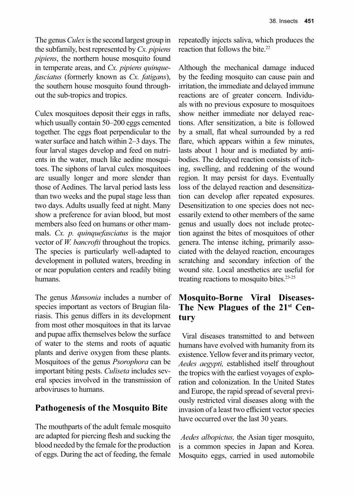
38. Insects 451
The genus Culex is the second largest group in the subfamily, best represented by Cx. pipiens pipiens, the northern house mosquito found in temperate areas, and Cx. pipiens quinque-fasciatus (formerly known as Cx. fatigans), the southern house mosquito found through-out the sub-tropics and tropics.
Culex mosquitoes deposit their eggs in rafts, which usually contain 50–200 eggs cemented together. The eggs float perpendicular to the water surface and hatch within 2–3 days. The four larval stages develop and feed on nutri-ents in the water, much like aedine mosqui-toes. The siphons of larval culex mosquitoes are usually longer and more slender than those of Aedines. The larval period lasts less than two weeks and the pupal stage less than two days. Adults usually feed at night. Many show a preference for avian blood, but most members also feed on humans or other mam-mals. Cx. p. quinquefasciatus is the major vector of W. bancrofti throughout the tropics. The species is particularly well-adapted to development in polluted waters, breeding in or near population centers and readily biting humans.
The genus Mansonia includes a number of species important as vectors of Brugian fila-riasis. This genus differs in its development from most other mosquitoes in that its larvae and pupae affix themselves below the surface of water to the stems and roots of aquatic plants and derive oxygen from these plants. Mosquitoes of the genus Psorophora can be important biting pests. Culiseta includes sev-eral species involved in the transmission of arboviruses to humans.
Pathogenesis of the Mosquito Bite
The mouthparts of the adult female mosquito are adapted for piercing flesh and sucking the blood needed by the female for the production of eggs. During the act of feeding, the female
repeatedly injects saliva, which produces the reaction that follows the bite.22
Although the mechanical damage induced by the feeding mosquito can cause pain and irritation, the immediate and delayed immune reactions are of greater concern. Individu-als with no previous exposure to mosquitoes show neither immediate nor delayed reac-tions. After sensitization, a bite is followed by a small, flat wheal surrounded by a red flare, which appears within a few minutes, lasts about 1 hour and is mediated by anti-bodies. The delayed reaction consists of itch-ing, swelling, and reddening of the wound region. It may persist for days. Eventually loss of the delayed reaction and desensitiza-tion can develop after repeated exposures. Desensitization to one species does not nec-essarily extend to other members of the same genus and usually does not include protec-tion against the bites of mosquitoes of other genera. The intense itching, primarily asso-ciated with the delayed reaction, encourages scratching and secondary infection of the wound site. Local anesthetics are useful for treating reactions to mosquito bites.23-25
Mosquito-Borne Viral Diseases- The New Plagues of the 21st Cen-tury
Viral diseases transmitted to and between humans have evolved with humanity from its existence. Yellow fever and its primary vector, Aedes aegypti, established itself throughout the tropics with the earliest voyages of explo-ration and colonization. In the United States and Europe, the rapid spread of several previ-ously restricted viral diseases along with the invasion of a least two efficient vector species have occurred over the last 30 years. Aedes albopictus, the Asian tiger mosquito, is a common species in Japan and Korea. Mosquito eggs, carried in used automobile
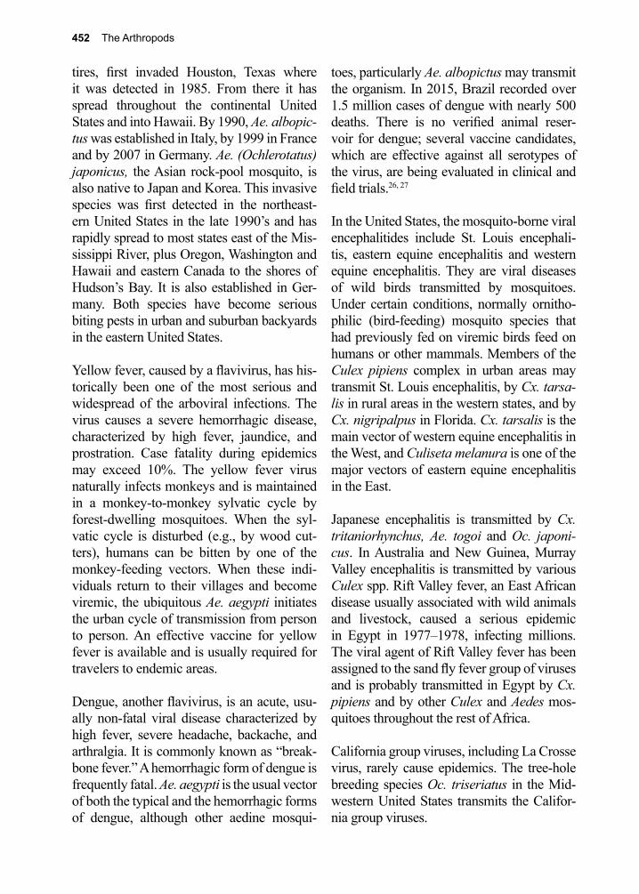
452 The Arthropods
tires, first invaded Houston, Texas where it was detected in 1985. From there it has spread throughout the continental United States and into Hawaii. By 1990, Ae. albopic-tus was established in Italy, by 1999 in France and by 2007 in Germany. Ae. (Ochlerotatus) japonicus, the Asian rock-pool mosquito, is also native to Japan and Korea. This invasive species was first detected in the northeast-ern United States in the late 1990’s and has rapidly spread to most states east of the Mis-sissippi River, plus Oregon, Washington and Hawaii and eastern Canada to the shores of Hudson’s Bay. It is also established in Ger-many. Both species have become serious biting pests in urban and suburban backyards in the eastern United States.
Yellow fever, caused by a flavivirus, has his-torically been one of the most serious and widespread of the arboviral infections. The virus causes a severe hemorrhagic disease, characterized by high fever, jaundice, and prostration. Case fatality during epidemics may exceed 10%. The yellow fever virus naturally infects monkeys and is maintained in a monkey-to-monkey sylvatic cycle by forest-dwelling mosquitoes. When the syl-vatic cycle is disturbed (e.g., by wood cut-ters), humans can be bitten by one of the monkey-feeding vectors. When these indi-viduals return to their villages and become viremic, the ubiquitous Ae. aegypti initiates the urban cycle of transmission from person to person. An effective vaccine for yellow fever is available and is usually required for travelers to endemic areas.
Dengue, another flavivirus, is an acute, usu-ally non-fatal viral disease characterized by high fever, severe headache, backache, and arthralgia. It is commonly known as “break-bone fever.” A hemorrhagic form of dengue is frequently fatal. Ae. aegypti is the usual vector of both the typical and the hemorrhagic forms of dengue, although other aedine mosqui-
toes, particularly Ae. albopictus may transmit the organism. In 2015, Brazil recorded over 1.5 million cases of dengue with nearly 500 deaths. There is no verified animal reser-voir for dengue; several vaccine candidates, which are effective against all serotypes of the virus, are being evaluated in clinical and field trials.26, 27
In the United States, the mosquito-borne viral encephalitides include St. Louis encephali-tis, eastern equine encephalitis and western equine encephalitis. They are viral diseases of wild birds transmitted by mosquitoes. Under certain conditions, normally ornitho-philic (bird-feeding) mosquito species that had previously fed on viremic birds feed on humans or other mammals. Members of the Culex pipiens complex in urban areas may transmit St. Louis encephalitis, by Cx. tarsa-lis in rural areas in the western states, and by Cx. nigripalpus in Florida. Cx. tarsalis is the main vector of western equine encephalitis in the West, and Culiseta melanura is one of the major vectors of eastern equine encephalitis in the East.
Japanese encephalitis is transmitted by Cx. tritaniorhynchus, Ae. togoi and Oc. japoni-cus. In Australia and New Guinea, Murray Valley encephalitis is transmitted by various Culex spp. Rift Valley fever, an East African disease usually associated with wild animals and livestock, caused a serious epidemic in Egypt in 1977–1978, infecting millions. The viral agent of Rift Valley fever has been assigned to the sand fly fever group of viruses and is probably transmitted in Egypt by Cx. pipiens and by other Culex and Aedes mos-quitoes throughout the rest of Africa.
California group viruses, including La Crosse virus, rarely cause epidemics. The tree-hole breeding species Oc. triseriatus in the Mid-western United States transmits the Califor-nia group viruses.
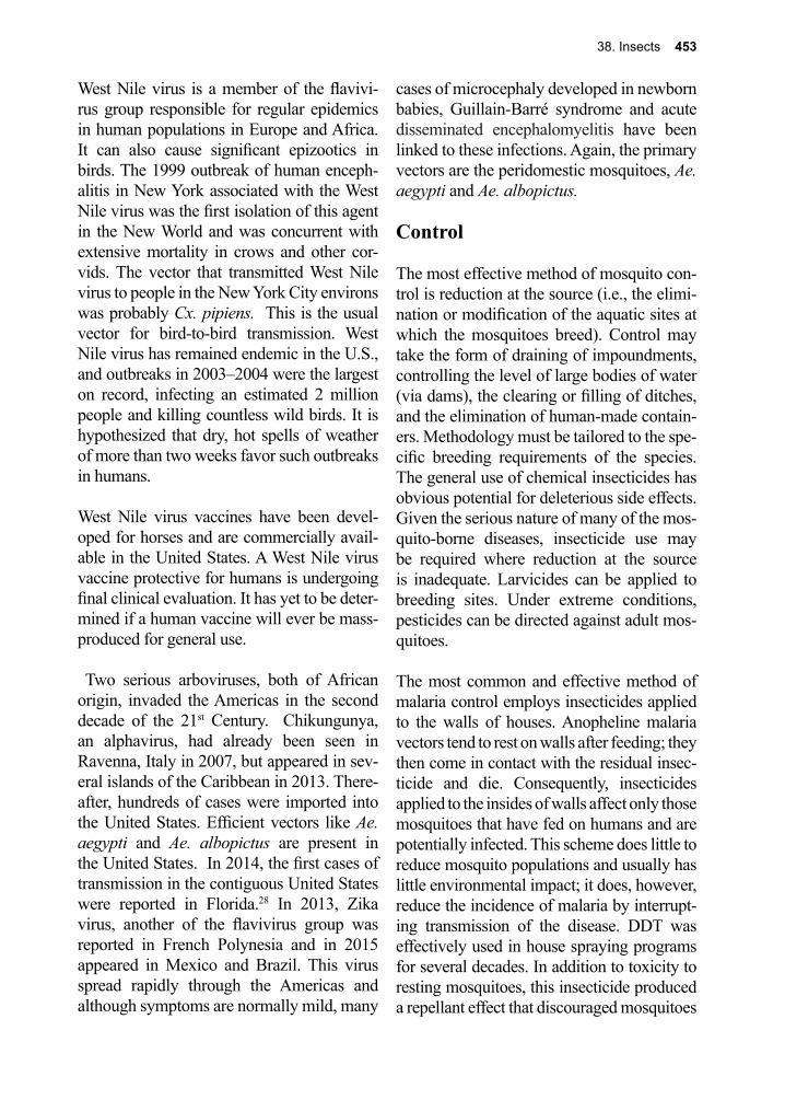
38. Insects 453
West Nile virus is a member of the flavivi-rus group responsible for regular epidemics in human populations in Europe and Africa. It can also cause significant epizootics in birds. The 1999 outbreak of human enceph-alitis in New York associated with the West Nile virus was the first isolation of this agent in the New World and was concurrent with extensive mortality in crows and other cor-vids. The vector that transmitted West Nile virus to people in the New York City environs was probably Cx. pipiens. This is the usual vector for bird-to-bird transmission. West Nile virus has remained endemic in the U.S., and outbreaks in 2003–2004 were the largest on record, infecting an estimated 2 million people and killing countless wild birds. It is hypothesized that dry, hot spells of weather of more than two weeks favor such outbreaks in humans.
West Nile virus vaccines have been devel-oped for horses and are commercially avail-able in the United States. A West Nile virus vaccine protective for humans is undergoing final clinical evaluation. It has yet to be deter-mined if a human vaccine will ever be mass-produced for general use.
Two serious arboviruses, both of African origin, invaded the Americas in the second decade of the 21st Century. Chikungunya, an alphavirus, had already been seen in Ravenna, Italy in 2007, but appeared in sev-eral islands of the Caribbean in 2013. There-after, hundreds of cases were imported into the United States. Efficient vectors like Ae. aegypti and Ae. albopictus are present in the United States. In 2014, the first cases of transmission in the contiguous United States were reported in Florida.28 In 2013, Zika virus, another of the flavivirus group was reported in French Polynesia and in 2015 appeared in Mexico and Brazil. This virus spread rapidly through the Americas and although symptoms are normally mild, many
cases of microcephaly developed in newborn babies, Guillain-Barré syndrome and acute disseminated encephalomyelitis have been linked to these infections. Again, the primary vectors are the peridomestic mosquitoes, Ae. aegypti and Ae. albopictus.
Control
The most effective method of mosquito con-trol is reduction at the source (i.e., the elimi-nation or modification of the aquatic sites at which the mosquitoes breed). Control may take the form of draining of impoundments, controlling the level of large bodies of water (via dams), the clearing or filling of ditches, and the elimination of human-made contain-ers. Methodology must be tailored to the spe-cific breeding requirements of the species. The general use of chemical insecticides has obvious potential for deleterious side effects. Given the serious nature of many of the mos-quito-borne diseases, insecticide use may be required where reduction at the source is inadequate. Larvicides can be applied to breeding sites. Under extreme conditions, pesticides can be directed against adult mos-quitoes.
The most common and effective method of malaria control employs insecticides applied to the walls of houses. Anopheline malaria vectors tend to rest on walls after feeding; they then come in contact with the residual insec-ticide and die. Consequently, insecticides applied to the insides of walls affect only those mosquitoes that have fed on humans and are potentially infected. This scheme does little to reduce mosquito populations and usually has little environmental impact; it does, however, reduce the incidence of malaria by interrupt-ing transmission of the disease. DDT was effectively used in house spraying programs for several decades. In addition to toxicity to resting mosquitoes, this insecticide produced a repellant effect that discouraged mosquitoes
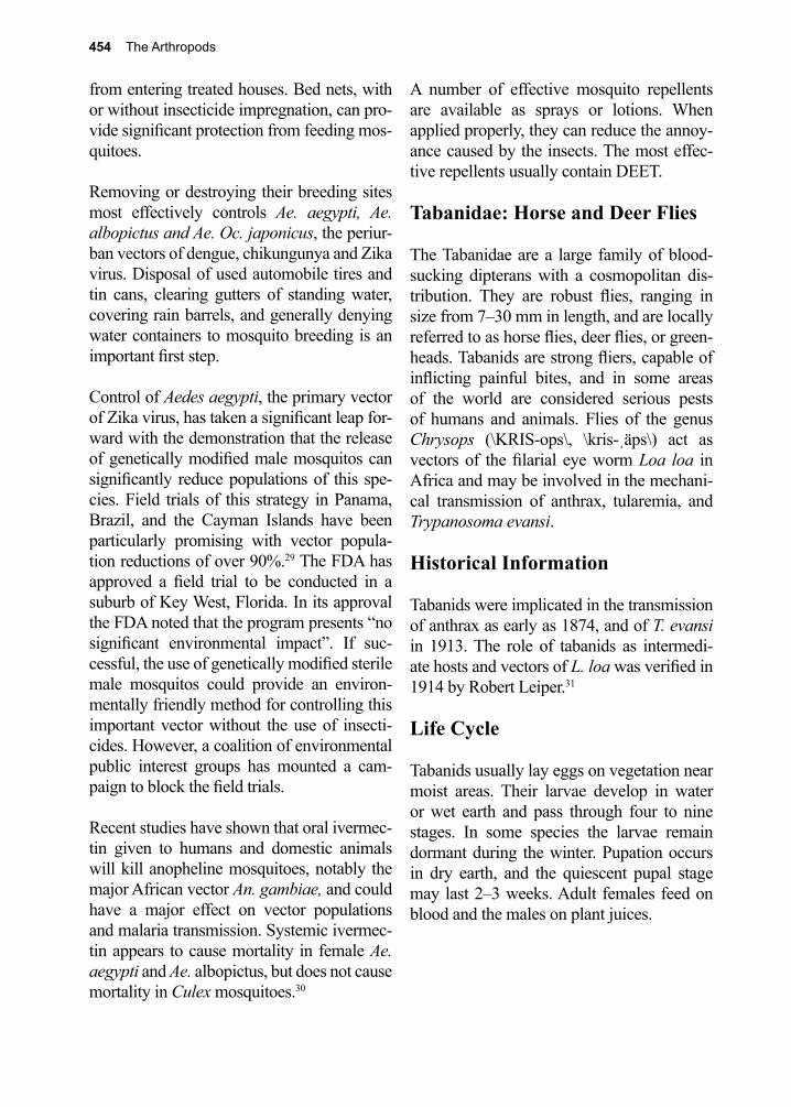
454 The Arthropods
from entering treated houses. Bed nets, with or without insecticide impregnation, can pro-vide significant protection from feeding mos-quitoes. Removing or destroying their breeding sites most effectively controls Ae. aegypti, Ae. albopictus and Ae. Oc. japonicus, the periur-ban vectors of dengue, chikungunya and Zika virus. Disposal of used automobile tires and tin cans, clearing gutters of standing water, covering rain barrels, and generally denying water containers to mosquito breeding is an important first step.
Control of Aedes aegypti, the primary vector of Zika virus, has taken a significant leap for-ward with the demonstration that the release of genetically modified male mosquitos can significantly reduce populations of this spe-cies. Field trials of this strategy in Panama, Brazil, and the Cayman Islands have been particularly promising with vector popula-tion reductions of over 90%.29 The FDA has approved a field trial to be conducted in a suburb of Key West, Florida. In its approval the FDA noted that the program presents “no significant environmental impact”. If suc-cessful, the use of genetically modified sterile male mosquitos could provide an environ-mentally friendly method for controlling this important vector without the use of insecti-cides. However, a coalition of environmental public interest groups has mounted a cam-paign to block the field trials.
Recent studies have shown that oral ivermec-tin given to humans and domestic animals will kill anopheline mosquitoes, notably the major African vector An. gambiae, and could have a major effect on vector populations and malaria transmission. Systemic ivermec-tin appears to cause mortality in female Ae. aegypti and Ae. albopictus, but does not cause mortality in Culex mosquitoes.30
A number of effective mosquito repellents are available as sprays or lotions. When applied properly, they can reduce the annoy-ance caused by the insects. The most effec-tive repellents usually contain DEET.
Tabanidae: Horse and Deer Flies
The Tabanidae are a large family of blood-sucking dipterans with a cosmopolitan dis-tribution. They are robust flies, ranging in size from 7–30 mm in length, and are locally referred to as horse flies, deer flies, or green-heads. Tabanids are strong fliers, capable of inflicting painful bites, and in some areas of the world are considered serious pests of humans and animals. Flies of the genus Chrysops (\KRIS-ops\, \kris-ˌäps\) act as vectors of the filarial eye worm Loa loa in Africa and may be involved in the mechani-cal transmission of anthrax, tularemia, and Trypanosoma evansi.
Historical Information
Tabanids were implicated in the transmission of anthrax as early as 1874, and of T. evansi in 1913. The role of tabanids as intermedi-ate hosts and vectors of L. loa was verified in 1914 by Robert Leiper.31
Life Cycle
Tabanids usually lay eggs on vegetation near moist areas. Their larvae develop in water or wet earth and pass through four to nine stages. In some species the larvae remain dormant during the winter. Pupation occurs in dry earth, and the quiescent pupal stage may last 2–3 weeks. Adult females feed on blood and the males on plant juices.

38. Insects 455
Pathogenesis
Tabanid mouthparts are short and blade-like. During the act of biting, the insect inflicts a deep, painful wound, causing blood to flow. The fly then ingests blood from the freshly formed pool. Individuals can become sensi-tized to tabanid bites and suffer severe aller-gic reactions after attack.
Tabanids act as efficient mechanical vectors of several pathogens. They are easily disturbed during feeding. They fly to another host and begin the process anew. Consequently, the fly’s mouthparts can readily transfer organ-isms to the next host after contamination on the first. Bacteria causing anthrax and tulare-mia, the protozoan T. evansi, and the retro-virus agents of bovine leukemia and equine infectious anemia may be transmitted by the tabanid flies, which act as mechanical vec-tors.
L. loa is transmitted by African tabanids of the genus Chrysops, which include C. silacea and C. dimidiata. Microfilariae of the worm, ingested by female flies with the blood meal, develop in the flight muscles. When they reach maturity, they migrate to the mouth-parts and are deposited on the skin of a new host when the fly feeds again. Infectious larvae burrow into the skin of the host after the fly has abandoned the bite wound.
Control
Tabanids are difficult to control because of their diverse breeding sites. Larvae are sensi-tive to DDT and other insecticides, but these compounds are seldom used. Sensitive indi-viduals should consider using repellents to avoid bites. Mosquito repellents containing DEET are usually effective. Although DEET is the standard for mosquito repellants there are some people that are not able to tolerate DEET and alternatives such as picaridin are available.
Muscidae: The House Fly and its Relatives
The muscoid flies include insects that are important as blood-sucking pests, vectors of diseases, and mechanical vectors of a variety of pathogenic organisms.32, 33 Some better-known members of this family are the house-fly Musca domestica and the stable fly Sto-moxys calcitrans.
Figure 38.10. Musca domestica, the housefly. A. Larva. B. Pupa. C. Adult. The larvae of flies are referred to as maggots.
Figure 38.11. Glossina spp. tsetse fly feeding on blood. Courtesy of J. Gingrich.
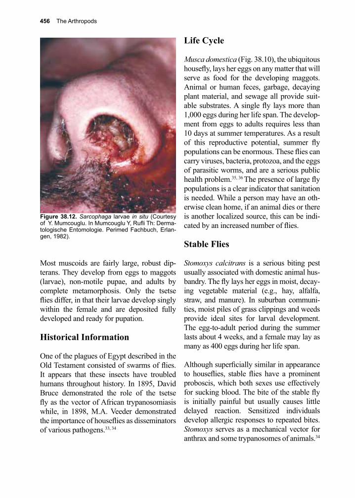
456 The Arthropods
Most muscoids are fairly large, robust dip-terans. They develop from eggs to maggots (larvae), non-motile pupae, and adults by complete metamorphosis. Only the tsetse flies differ, in that their larvae develop singly within the female and are deposited fully developed and ready for pupation.
Historical Information
One of the plagues of Egypt described in the Old Testament consisted of swarms of flies. It appears that these insects have troubled humans throughout history. In 1895, David Bruce demonstrated the role of the tsetse fly as the vector of African trypanosomiasis while, in 1898, M.A. Veeder demonstrated the importance of houseflies as disseminators of various pathogens.33, 34
Life Cycle
Musca domestica (Fig. 38.10), the ubiquitous housefly, lays her eggs on any matter that will serve as food for the developing maggots. Animal or human feces, garbage, decaying plant material, and sewage all provide suit-able substrates. A single fly lays more than 1,000 eggs during her life span. The develop-ment from eggs to adults requires less than 10 days at summer temperatures. As a result of this reproductive potential, summer fly populations can be enormous. These flies can carry viruses, bacteria, protozoa, and the eggs of parasitic worms, and are a serious public health problem.35, 36 The presence of large fly populations is a clear indicator that sanitation is needed. While a person may have an oth-erwise clean home, if an animal dies or there is another localized source, this can be indi-cated by an increased number of flies.
Stable Flies
Stomoxys calcitrans is a serious biting pest usually associated with domestic animal hus-bandry. The fly lays her eggs in moist, decay-ing vegetable material (e.g., hay, alfalfa, straw, and manure). In suburban communi-ties, moist piles of grass clippings and weeds provide ideal sites for larval development. The egg-to-adult period during the summer lasts about 4 weeks, and a female may lay as many as 400 eggs during her life span.
Although superficially similar in appearance to houseflies, stable flies have a prominent proboscis, which both sexes use effectively for sucking blood. The bite of the stable fly is initially painful but usually causes little delayed reaction. Sensitized individuals develop allergic responses to repeated bites. Stomoxys serves as a mechanical vector for anthrax and some trypanosomes of animals.34
Figure 38.12. Sarcophaga larvae in situ (Courtesy of Y. Mumcouglu. In Mumcouglu Y, Rufli Th: Derma-tologische Entomologie. Perimed Fachbuch, Erlan-gen, 1982).
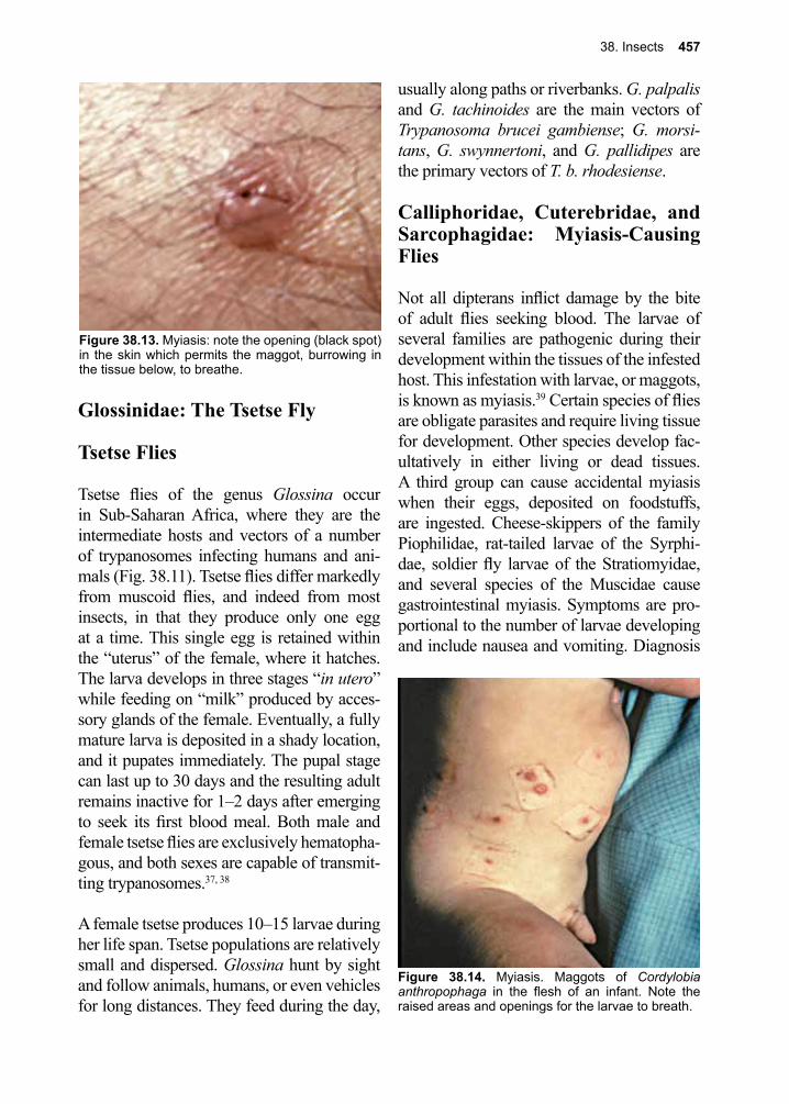
38. Insects 457
Glossinidae: The Tsetse Fly
Tsetse Flies
Tsetse flies of the genus Glossina occur in Sub-Saharan Africa, where they are the intermediate hosts and vectors of a number of trypanosomes infecting humans and ani-mals (Fig. 38.11). Tsetse flies differ markedly from muscoid flies, and indeed from most insects, in that they produce only one egg at a time. This single egg is retained within the “uterus” of the female, where it hatches. The larva develops in three stages “in utero” while feeding on “milk” produced by acces-sory glands of the female. Eventually, a fully mature larva is deposited in a shady location, and it pupates immediately. The pupal stage can last up to 30 days and the resulting adult remains inactive for 1–2 days after emerging to seek its first blood meal. Both male and female tsetse flies are exclusively hematopha-gous, and both sexes are capable of transmit-ting trypanosomes.37, 38
A female tsetse produces 10–15 larvae during her life span. Tsetse populations are relatively small and dispersed. Glossina hunt by sight and follow animals, humans, or even vehicles for long distances. They feed during the day,
usually along paths or riverbanks. G. palpalis and G. tachinoides are the main vectors of Trypanosoma brucei gambiense; G. morsi-tans, G. swynnertoni, and G. pallidipes are the primary vectors of T. b. rhodesiense.
Calliphoridae, Cuterebridae, and Sarcophagidae: Myiasis-Causing Flies
Not all dipterans inflict damage by the bite of adult flies seeking blood. The larvae of several families are pathogenic during their development within the tissues of the infested host. This infestation with larvae, or maggots, is known as myiasis.39 Certain species of flies are obligate parasites and require living tissue for development. Other species develop fac-ultatively in either living or dead tissues. A third group can cause accidental myiasis when their eggs, deposited on foodstuffs, are ingested. Cheese-skippers of the family Piophilidae, rat-tailed larvae of the Syrphi-dae, soldier fly larvae of the Stratiomyidae, and several species of the Muscidae cause gastrointestinal myiasis. Symptoms are pro-portional to the number of larvae developing and include nausea and vomiting. Diagnosis
Figure 38.13. Myiasis: note the opening (black spot) in the skin which permits the maggot, burrowing in the tissue below, to breathe.
Figure 38.14. Myiasis. Maggots of Cordylobia anthropophaga in the flesh of an infant. Note the raised areas and openings for the larvae to breath.
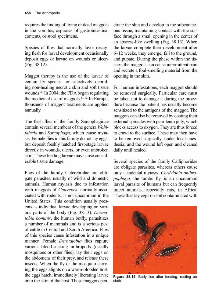
458 The Arthropods
requires the finding of living or dead maggots in the vomitus, aspirates of gastrointestinal contents, or stool specimens.
Species of flies that normally favor decay-ing flesh for larval development occasionally deposit eggs or larvae on wounds or ulcers (Fig. 38.12).
Maggot therapy is the use of the larvae of certain fly species for selectively debrid-ing non-healing necrotic skin and soft tissue wounds.40 In 2004, the FDA began regulating the medicinal use of maggots.41, 42 In Europe, thousands of maggot treatments are applied annually.
The flesh flies of the family Sarcophagidae contain several members of the genera Wohl-fahrtia and Sarcophaga, which cause myia-sis. Female flies in this family do not lay eggs, but deposit freshly hatched first-stage larvae directly in wounds, ulcers, or even unbroken skin. These feeding larvae may cause consid-erable tissue damage.
Flies of the family Cuterebridae are obli-gate parasites, usually of wild and domestic animals. Human myiasis due to infestation with maggots of Cuterebra, normally asso-ciated with rodents, is not uncommon in the United States. This condition usually pres-ents as individual larvae developing on vari-ous parts of the body (Fig. 38.13). Derma-tobia hominis, the human botfly, parasitizes a number of mammals and is a serious pest of cattle in Central and South America. Flies of this species cause infestation in a unique manner. Female Dermatobia flies capture various blood-sucking arthropods (usually mosquitoes or other flies), lay their eggs on the abdomens of their prey, and release these insects. When the fly or the mosquito carry-ing the eggs alights on a warm-blooded host, the eggs hatch, immediately liberating larvae onto the skin of the host. These maggots pen-
etrate the skin and develop in the subcutane-ous tissue, maintaining contact with the sur-face through a small opening in the center of an abscess-like swelling (Fig. 38.13). When the larvae complete their development after 6–12 weeks, they emerge, fall to the ground, and pupate. During the phase within the tis-sues, the maggots can cause intermittent pain and secrete a foul-smelling material from the opening in the skin.
For human infestations, each maggot should be removed surgically. Particular care must be taken not to damage it during the proce-dure because the patient has usually become sensitized to the antigens of the maggot. The maggots can also be removed by coating their external spiracles with petroleum jelly, which blocks access to oxygen. They are thus forced to crawl to the surface. These may then have to be removed surgically, under local anes-thesia; and the wound left open and cleaned daily until healed.
Several species of the family Calliphoridae are obligate parasites, whereas others cause only accidental myiasis. Cordylobia anthro-pophaga, the tumbu fly, is an uncommon larval parasite of humans but can frequently infect animals, especially rats, in Africa. These flies lay eggs on soil contaminated with
Figure 38.15. Body lice after feeding, resting on cloth.
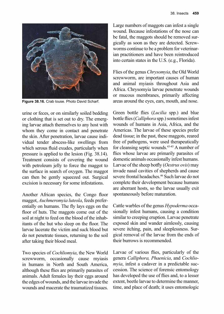
38. Insects 459
urine or feces, or on similarly soiled bedding or clothing that is set out to dry. The emerg-ing larvae attach themselves to any host with whom they come in contact and penetrate the skin. After penetration, larvae cause indi-vidual tender abscess-like swellings from which serous fluid exudes, particularly when pressure is applied to the lesion (Fig. 38.14). Treatment consists of covering the wound with petroleum jelly to force the maggot to the surface in search of oxygen. The maggot can then be gently squeezed out. Surgical excision is necessary for some infestations.
Another African species, the Congo floor maggot, Auchmeromyia luteola, feeds prefer-entially on humans. The fly lays eggs on the floor of huts. The maggots come out of the soil at night to feed on the blood of the inhab-itants of the hut who sleep on the floor. The larvae lacerate the victim and suck blood but do not penetrate tissues, returning to the soil after taking their blood meal.
Two species of Cochliomyia, the New World screwworm, occasionally cause myiasis in humans in North and South America, although these flies are primarily parasites of animals. Adult females lay their eggs around the edges of wounds, and the larvae invade the wounds and macerate the traumatized tissues.
Large numbers of maggots can infest a single wound. Because infestations of the nose can be fatal, the maggots should be removed sur-gically as soon as they are detected. Screw-worms continue to be a problem for veterinar-ian practitioners and have been reintroduced into certain states in the U.S. (e.g., Florida).
Flies of the genus Chrysomyia, the Old World screwworm, are important causes of human and animal myiasis throughout Asia and Africa. Chrysomyia larvae penetrate wounds or mucous membranes, primarily affecting areas around the eyes, ears, mouth, and nose.
Green bottle flies (Lucilia spp.) and blue bottle flies (Calliphora spp.) sometimes infest wounds of humans in Asia, Africa, and the Americas. The larvae of these species prefer dead tissue; in the past, these maggots, reared free of pathogens, were used therapeutically for cleansing septic wounds.43-45 A number of flies whose larvae are primarily parasites of domestic animals occasionally infest humans. Larvae of the sheep botfly (Oestrus ovis) may invade nasal cavities of shepherds and cause severe frontal headaches.46 Such larvae do not complete their development because humans are aberrant hosts, so the larvae usually exit spontaneously before maturation.
Cattle warbles of the genus Hypoderma occa-sionally infest humans, causing a condition similar to creeping eruption. Larvae penetrate exposed skin and wander aimlessly, causing severe itching, pain, and sleeplessness. Sur-gical removal of the larvae from the ends of their burrows is recommended.
Larvae of various flies, particularly of the genera Calliphora, Phaenicia, and Cochlio-myia, infest a cadaver in a predictable suc-cession. The science of forensic entomology has developed the use of flies and, to a lesser extent, beetle larvae to determine the manner, time, and place of death; it uses entomologic
Figure 38.16. Crab louse. Photo David Scharf.
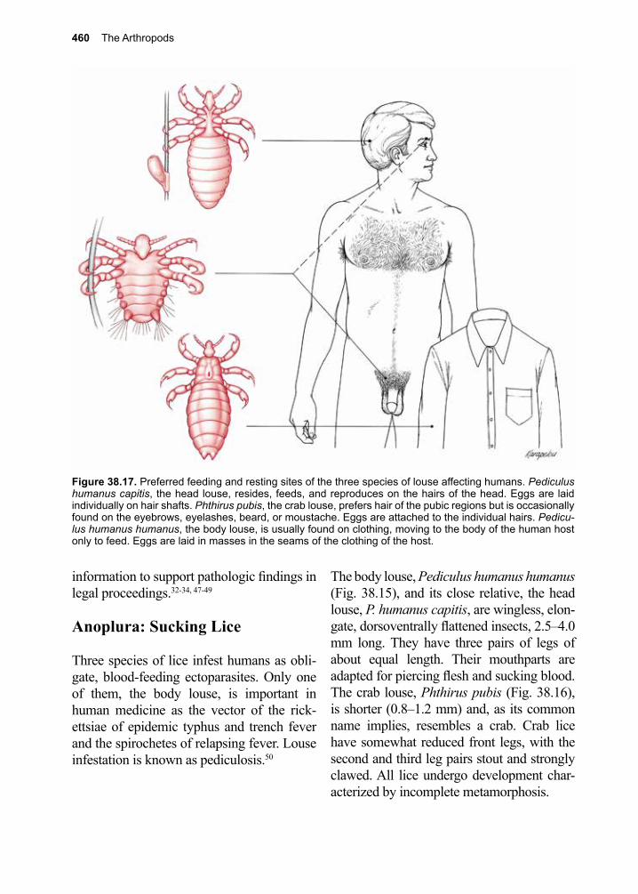
460 The Arthropods
information to support pathologic findings in legal proceedings.32-34, 47-49
Anoplura: Sucking Lice
Three species of lice infest humans as obli-gate, blood-feeding ectoparasites. Only one of them, the body louse, is important in human medicine as the vector of the rick-ettsiae of epidemic typhus and trench fever and the spirochetes of relapsing fever. Louse infestation is known as pediculosis.50
The body louse, Pediculus humanus humanus (Fig. 38.15), and its close relative, the head louse, P. humanus capitis, are wingless, elon-gate, dorsoventrally flattened insects, 2.5–4.0 mm long. They have three pairs of legs of about equal length. Their mouthparts are adapted for piercing flesh and sucking blood. The crab louse, Phthirus pubis (Fig. 38.16), is shorter (0.8–1.2 mm) and, as its common name implies, resembles a crab. Crab lice have somewhat reduced front legs, with the second and third leg pairs stout and strongly clawed. All lice undergo development char-acterized by incomplete metamorphosis.
Figure 38.17. Preferred feeding and resting sites of the three species of louse affecting humans. Pediculus humanus capitis, the head louse, resides, feeds, and reproduces on the hairs of the head. Eggs are laid individually on hair shafts. Phthirus pubis, the crab louse, prefers hair of the pubic regions but is occasionally found on the eyebrows, eyelashes, beard, or moustache. Eggs are attached to the individual hairs. Pedicu-lus humanus humanus, the body louse, is usually found on clothing, moving to the body of the human host only to feed. Eggs are laid in masses in the seams of the clothing of the host.
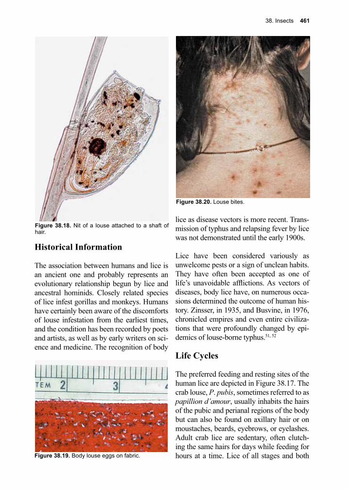
38. Insects 461
Historical Information
The association between humans and lice is an ancient one and probably represents an evolutionary relationship begun by lice and ancestral hominids. Closely related species of lice infest gorillas and monkeys. Humans have certainly been aware of the discomforts of louse infestation from the earliest times, and the condition has been recorded by poets and artists, as well as by early writers on sci-ence and medicine. The recognition of body
lice as disease vectors is more recent. Trans-mission of typhus and relapsing fever by lice was not demonstrated until the early 1900s.
Lice have been considered variously as unwelcome pests or a sign of unclean habits. They have often been accepted as one of life’s unavoidable afflictions. As vectors of diseases, body lice have, on numerous occa-sions determined the outcome of human his-tory. Zinsser, in 1935, and Busvine, in 1976, chronicled empires and even entire civiliza-tions that were profoundly changed by epi-demics of louse-borne typhus.51, 52
Life Cycles
The preferred feeding and resting sites of the human lice are depicted in Figure 38.17. The crab louse, P. pubis, sometimes referred to as papillion d’amour, usually inhabits the hairs of the pubic and perianal regions of the body but can also be found on axillary hair or on moustaches, beards, eyebrows, or eyelashes. Adult crab lice are sedentary, often clutch-ing the same hairs for days while feeding for hours at a time. Lice of all stages and both
Figure 38.18. Nit of a louse attached to a shaft of hair.
Figure 38.20. Louse bites.
Figure 38.19. Body louse eggs on fabric.
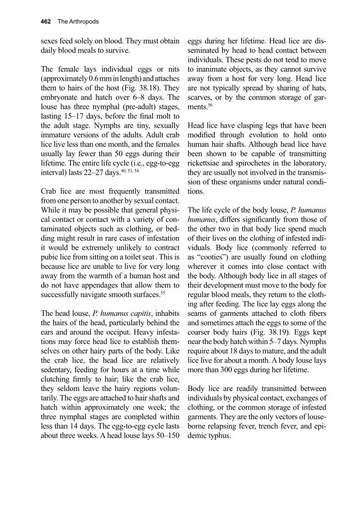
462 The Arthropods
sexes feed solely on blood. They must obtain daily blood meals to survive.
The female lays individual eggs or nits (approximately 0.6 mm in length) and attaches them to hairs of the host (Fig. 38.18). They embryonate and hatch over 6–8 days. The louse has three nymphal (pre-adult) stages, lasting 15–17 days, before the final molt to the adult stage. Nymphs are tiny, sexually immature versions of the adults. Adult crab lice live less than one month, and the females usually lay fewer than 50 eggs during their lifetime. The entire life cycle (i.e., egg-to-egg interval) lasts 22–27 days.40, 53, 54
Crab lice are most frequently transmitted from one person to another by sexual contact. While it may be possible that general physi-cal contact or contact with a variety of con-taminated objects such as clothing, or bed-ding might result in rare cases of infestation it would be extremely unlikely to contract pubic lice from sitting on a toilet seat . This is because lice are unable to live for very long away from the warmth of a human host and do not have appendages that allow them to successfully navigate smooth surfaces.55
The head louse, P. humanus capitis, inhabits the hairs of the head, particularly behind the ears and around the occiput. Heavy infesta-tions may force head lice to establish them-selves on other hairy parts of the body. Like the crab lice, the head lice are relatively sedentary, feeding for hours at a time while clutching firmly to hair; like the crab lice, they seldom leave the hairy regions volun-tarily. The eggs are attached to hair shafts and hatch within approximately one week; the three nymphal stages are completed within less than 14 days. The egg-to-egg cycle lasts about three weeks. A head louse lays 50–150
eggs during her lifetime. Head lice are dis-seminated by head to head contact between individuals. These pests do not tend to move to inanimate objects, as they cannot survive away from a host for very long. Head lice are not typically spread by sharing of hats, scarves, or by the common storage of gar-ments.56
Head lice have clasping legs that have been modified through evolution to hold onto human hair shafts. Although head lice have been shown to be capable of transmitting rickettsiae and spirochetes in the laboratory, they are usually not involved in the transmis-sion of these organisms under natural condi-tions.
The life cycle of the body louse, P. humanus humanus, differs significantly from those of the other two in that body lice spend much of their lives on the clothing of infested indi-viduals. Body lice (commonly referred to as “cooties”) are usually found on clothing wherever it comes into close contact with the body. Although body lice in all stages of their development must move to the body for regular blood meals, they return to the cloth-ing after feeding. The lice lay eggs along the seams of garments attached to cloth fibers and sometimes attach the eggs to some of the coarser body hairs (Fig. 38.19). Eggs kept near the body hatch within 5–7 days. Nymphs require about 18 days to mature, and the adult lice live for about a month. A body louse lays more than 300 eggs during her lifetime.
Body lice are readily transmitted between individuals by physical contact, exchanges of clothing, or the common storage of infested garments. They are the only vectors of louse-borne relapsing fever, trench fever, and epi-demic typhus.
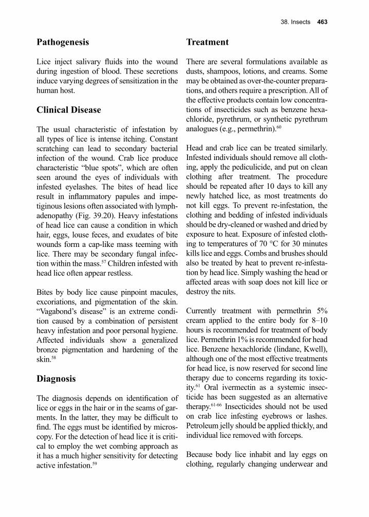
38. Insects 463
Pathogenesis
Lice inject salivary fluids into the wound during ingestion of blood. These secretions induce varying degrees of sensitization in the human host.
Clinical Disease
The usual characteristic of infestation by all types of lice is intense itching. Constant scratching can lead to secondary bacterial infection of the wound. Crab lice produce characteristic “blue spots”, which are often seen around the eyes of individuals with infested eyelashes. The bites of head lice result in inflammatory papules and impe-tiginous lesions often associated with lymph-adenopathy (Fig. 39.20). Heavy infestations of head lice can cause a condition in which hair, eggs, louse feces, and exudates of bite wounds form a cap-like mass teeming with lice. There may be secondary fungal infec-tion within the mass.57 Children infested with head lice often appear restless.
Bites by body lice cause pinpoint macules, excoriations, and pigmentation of the skin. “Vagabond’s disease” is an extreme condi-tion caused by a combination of persistent heavy infestation and poor personal hygiene. Affected individuals show a generalized bronze pigmentation and hardening of the skin.58
Diagnosis
The diagnosis depends on identification of lice or eggs in the hair or in the seams of gar-ments. In the latter, they may be difficult to find. The eggs must be identified by micros-copy. For the detection of head lice it is criti-cal to employ the wet combing approach as it has a much higher sensitivity for detecting active infestation.59
Treatment
There are several formulations available as dusts, shampoos, lotions, and creams. Some may be obtained as over-the-counter prepara-tions, and others require a prescription. All of the effective products contain low concentra-tions of insecticides such as benzene hexa-chloride, pyrethrum, or synthetic pyrethrum analogues (e.g., permethrin).60
Head and crab lice can be treated similarly. Infested individuals should remove all cloth-ing, apply the pediculicide, and put on clean clothing after treatment. The procedure should be repeated after 10 days to kill any newly hatched lice, as most treatments do not kill eggs. To prevent re-infestation, the clothing and bedding of infested individuals should be dry-cleaned or washed and dried by exposure to heat. Exposure of infested cloth-ing to temperatures of 70 °C for 30 minutes kills lice and eggs. Combs and brushes should also be treated by heat to prevent re-infesta-tion by head lice. Simply washing the head or affected areas with soap does not kill lice or destroy the nits.
Currently treatment with permethrin 5% cream applied to the entire body for 8–10 hours is recommended for treatment of body lice. Permethrin 1% is recommended for head lice. Benzene hexachloride (lindane, Kwell), although one of the most effective treatments for head lice, is now reserved for second line therapy due to concerns regarding its toxic-ity.61 Oral ivermectin as a systemic insec-ticide has been suggested as an alternative therapy.61-66 Insecticides should not be used on crab lice infesting eyebrows or lashes. Petroleum jelly should be applied thickly, and individual lice removed with forceps.
Because body lice inhabit and lay eggs on clothing, regularly changing underwear and
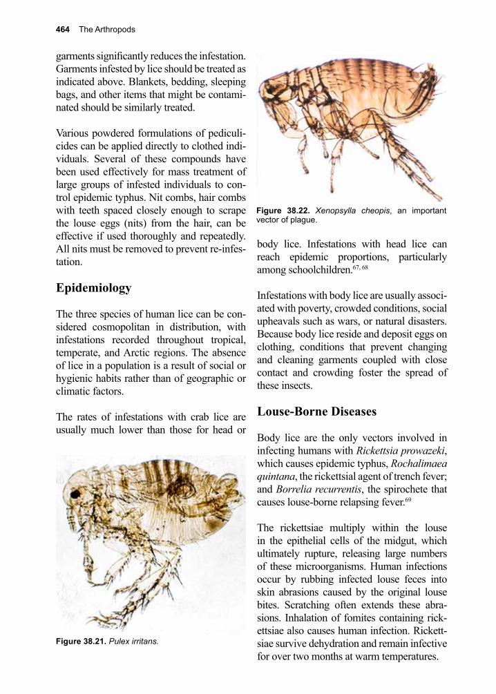
464 The Arthropods
garments significantly reduces the infestation. Garments infested by lice should be treated as indicated above. Blankets, bedding, sleeping bags, and other items that might be contami-nated should be similarly treated.
Various powdered formulations of pediculi-cides can be applied directly to clothed indi-viduals. Several of these compounds have been used effectively for mass treatment of large groups of infested individuals to con-trol epidemic typhus. Nit combs, hair combs with teeth spaced closely enough to scrape the louse eggs (nits) from the hair, can be effective if used thoroughly and repeatedly. All nits must be removed to prevent re-infes-tation.
Epidemiology
The three species of human lice can be con-sidered cosmopolitan in distribution, with infestations recorded throughout tropical, temperate, and Arctic regions. The absence of lice in a population is a result of social or hygienic habits rather than of geographic or climatic factors.
The rates of infestations with crab lice are usually much lower than those for head or
body lice. Infestations with head lice can reach epidemic proportions, particularly among schoolchildren.67, 68
Infestations with body lice are usually associ-ated with poverty, crowded conditions, social upheavals such as wars, or natural disasters. Because body lice reside and deposit eggs on clothing, conditions that prevent changing and cleaning garments coupled with close contact and crowding foster the spread of these insects.
Louse-Borne Diseases
Body lice are the only vectors involved in infecting humans with Rickettsia prowazeki, which causes epidemic typhus, Rochalimaea quintana, the rickettsial agent of trench fever; and Borrelia recurrentis, the spirochete that causes louse-borne relapsing fever.69
The rickettsiae multiply within the louse in the epithelial cells of the midgut, which ultimately rupture, releasing large numbers of these microorganisms. Human infections occur by rubbing infected louse feces into skin abrasions caused by the original louse bites. Scratching often extends these abra-sions. Inhalation of fomites containing rick-ettsiae also causes human infection. Rickett-siae survive dehydration and remain infective for over two months at warm temperatures.
Figure 38.22. Xenopsylla cheopis, an important vector of plague.
Figure 38.21. Pulex irritans.
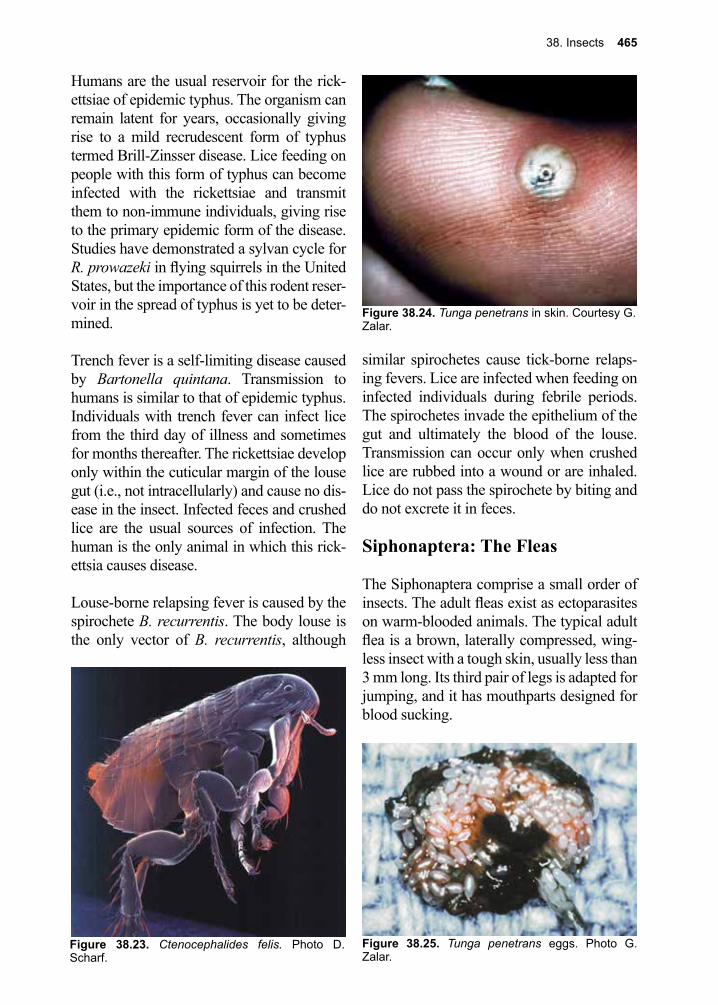
38. Insects 465
Humans are the usual reservoir for the rick-ettsiae of epidemic typhus. The organism can remain latent for years, occasionally giving rise to a mild recrudescent form of typhus termed Brill-Zinsser disease. Lice feeding on people with this form of typhus can become infected with the rickettsiae and transmit them to non-immune individuals, giving rise to the primary epidemic form of the disease. Studies have demonstrated a sylvan cycle for R. prowazeki in flying squirrels in the United States, but the importance of this rodent reser-voir in the spread of typhus is yet to be deter-mined.
Trench fever is a self-limiting disease caused by Bartonella quintana. Transmission to humans is similar to that of epidemic typhus. Individuals with trench fever can infect lice from the third day of illness and sometimes for months thereafter. The rickettsiae develop only within the cuticular margin of the louse gut (i.e., not intracellularly) and cause no dis-ease in the insect. Infected feces and crushed lice are the usual sources of infection. The human is the only animal in which this rick-ettsia causes disease.
Louse-borne relapsing fever is caused by the spirochete B. recurrentis. The body louse is the only vector of B. recurrentis, although
similar spirochetes cause tick-borne relaps-ing fevers. Lice are infected when feeding on infected individuals during febrile periods. The spirochetes invade the epithelium of the gut and ultimately the blood of the louse. Transmission can occur only when crushed lice are rubbed into a wound or are inhaled. Lice do not pass the spirochete by biting and do not excrete it in feces.
Siphonaptera: The Fleas
The Siphonaptera comprise a small order of insects. The adult fleas exist as ectoparasites on warm-blooded animals. The typical adult flea is a brown, laterally compressed, wing-less insect with a tough skin, usually less than 3 mm long. Its third pair of legs is adapted for jumping, and it has mouthparts designed for blood sucking.
Figure 38.23. Ctenocephalides felis. Photo D. Scharf.
Figure 38.25. Tunga penetrans eggs. Photo G. Zalar.
Figure 38.24. Tunga penetrans in skin. Courtesy G. Zalar.
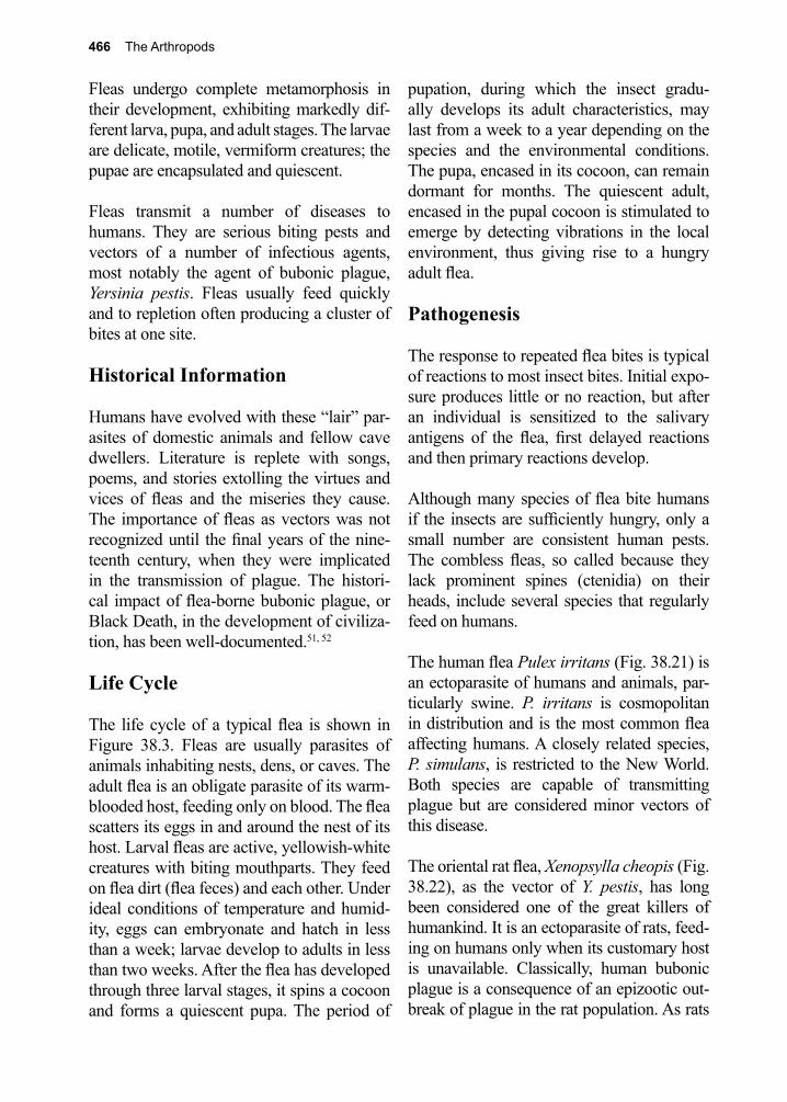
466 The Arthropods
Fleas undergo complete metamorphosis in their development, exhibiting markedly dif-ferent larva, pupa, and adult stages. The larvae are delicate, motile, vermiform creatures; the pupae are encapsulated and quiescent.
Fleas transmit a number of diseases to humans. They are serious biting pests and vectors of a number of infectious agents, most notably the agent of bubonic plague, Yersinia pestis. Fleas usually feed quickly and to repletion often producing a cluster of bites at one site.
Historical Information
Humans have evolved with these “lair” par-asites of domestic animals and fellow cave dwellers. Literature is replete with songs, poems, and stories extolling the virtues and vices of fleas and the miseries they cause. The importance of fleas as vectors was not recognized until the final years of the nine-teenth century, when they were implicated in the transmission of plague. The histori-cal impact of flea-borne bubonic plague, or Black Death, in the development of civiliza-tion, has been well-documented.51, 52
Life Cycle
The life cycle of a typical flea is shown in Figure 38.3. Fleas are usually parasites of animals inhabiting nests, dens, or caves. The adult flea is an obligate parasite of its warm-blooded host, feeding only on blood. The flea scatters its eggs in and around the nest of its host. Larval fleas are active, yellowish-white creatures with biting mouthparts. They feed on flea dirt (flea feces) and each other. Under ideal conditions of temperature and humid-ity, eggs can embryonate and hatch in less than a week; larvae develop to adults in less than two weeks. After the flea has developed through three larval stages, it spins a cocoon and forms a quiescent pupa. The period of
pupation, during which the insect gradu-ally develops its adult characteristics, may last from a week to a year depending on the species and the environmental conditions. The pupa, encased in its cocoon, can remain dormant for months. The quiescent adult, encased in the pupal cocoon is stimulated to emerge by detecting vibrations in the local environment, thus giving rise to a hungry adult flea.
Pathogenesis
The response to repeated flea bites is typical of reactions to most insect bites. Initial expo-sure produces little or no reaction, but after an individual is sensitized to the salivary antigens of the flea, first delayed reactions and then primary reactions develop.
Although many species of flea bite humans if the insects are sufficiently hungry, only a small number are consistent human pests. The combless fleas, so called because they lack prominent spines (ctenidia) on their heads, include several species that regularly feed on humans.
The human flea Pulex irritans (Fig. 38.21) is an ectoparasite of humans and animals, par-ticularly swine. P. irritans is cosmopolitan in distribution and is the most common flea affecting humans. A closely related species, P. simulans, is restricted to the New World. Both species are capable of transmitting plague but are considered minor vectors of this disease.
The oriental rat flea, Xenopsylla cheopis (Fig. 38.22), as the vector of Y. pestis, has long been considered one of the great killers of humankind. It is an ectoparasite of rats, feed-ing on humans only when its customary host is unavailable. Classically, human bubonic plague is a consequence of an epizootic out-break of plague in the rat population. As rats
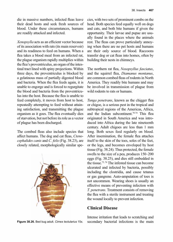
38. Insects 467
die in massive numbers, infected fleas leave their dead hosts and seek fresh sources of blood. Under these circumstances, humans are readily attacked and infected.
Xenopsylla acts as an efficient vector because of its association with rats (its main reservoir) and its readiness to feed on humans. When a flea takes a blood meal from an infected rat, the plague organism rapidly multiplies within the flea’s proventriculus, an organ of the intes-tinal tract lined with spiny projections. Within three days, the proventriculus is blocked by a gelatinous mass of partially digested blood and bacteria. When the flea feeds again, it is unable to engorge and is forced to regurgitate the blood and bacteria from the proventricu-lus into the host. Because the flea is unable to feed completely, it moves from host to host, repeatedly attempting to feed without attain-ing satisfaction, and transmitting the plague organism as it goes. The flea eventually dies of starvation, but not before its role as a vector of plague has been discharged.
The combed fleas also include species that affect humans. The dog and cat fleas, Cteno-cephalides canis and C. felis (Fig. 38.23), are closely related, morphologically similar spe-
cies, with two sets of prominent combs on the head. Both species feed equally well on dogs and cats, and both bite humans if given the opportunity. Their larvae and pupae are usu-ally found in the places where the animals rest. The fleas can prove particularly annoy-ing when there are no pet hosts and humans are their only source of blood. Raccoons transfer dog or cat fleas into homes, often by building their nests in chimneys.
The northern rat flea, Nosopsyllus fasciatus, and the squirrel flea, Diamanus montanus, are common combed fleas of rodents in North America. They readily bite humans and may be involved in transmission of plague from wild rodents to rats or humans.
Tunga penetrans, known as the chigger flea or chigoe, is a serious pest in the tropical and subtropical regions of the Americas, Africa, and the Indian subcontinent.70-74 This flea originated in South America and was intro-duced into Africa during the late nineteenth century. Adult chigoes are less than 1 mm long. Both sexes feed regularly on blood. After insemination, the female flea attaches itself to the skin of the toes, soles of the feet, or the legs, and becomes enveloped by host tissue (Fig. 38.24). Thus protected, the female swells to the size of a pea, produces 150–200 eggs (Fig. 38.25), and dies still embedded in the tissue.75, 76 The infested tissue can become ulcerated and infected by bacteria, possibly including the clostridia, and cause tetanus or gas gangrene. Auto-amputation of toes is not uncommon. Wearing shoes is usually an effective means of preventing infection with T. penetrans. Treatment consists of removing the flea with a sterile instrument and treating the wound locally to prevent infection.
Clinical Disease
Intense irritation that leads to scratching and secondary bacterial infections is the main Figure 38.26. Bed bug adult. Cimex lectularius 15x.
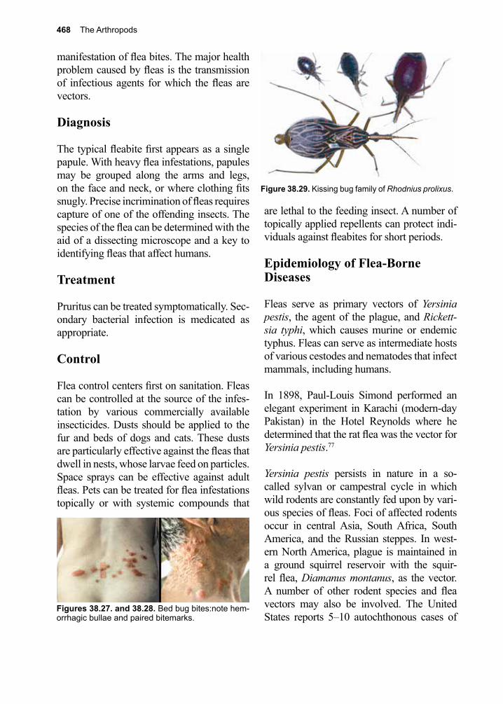
468 The Arthropods
manifestation of flea bites. The major health problem caused by fleas is the transmission of infectious agents for which the fleas are vectors.
Diagnosis
The typical fleabite first appears as a single papule. With heavy flea infestations, papules may be grouped along the arms and legs, on the face and neck, or where clothing fits snugly. Precise incrimination of fleas requires capture of one of the offending insects. The species of the flea can be determined with the aid of a dissecting microscope and a key to identifying fleas that affect humans.
Treatment
Pruritus can be treated symptomatically. Sec-ondary bacterial infection is medicated as appropriate.
Control
Flea control centers first on sanitation. Fleas can be controlled at the source of the infes-tation by various commercially available insecticides. Dusts should be applied to the fur and beds of dogs and cats. These dusts are particularly effective against the fleas that dwell in nests, whose larvae feed on particles. Space sprays can be effective against adult fleas. Pets can be treated for flea infestations topically or with systemic compounds that
are lethal to the feeding insect. A number of topically applied repellents can protect indi-viduals against fleabites for short periods.
Epidemiology of Flea-Borne Diseases
Fleas serve as primary vectors of Yersinia pestis, the agent of the plague, and Rickett-sia typhi, which causes murine or endemic typhus. Fleas can serve as intermediate hosts of various cestodes and nematodes that infect mammals, including humans.
In 1898, Paul-Louis Simond performed an elegant experiment in Karachi (modern-day Pakistan) in the Hotel Reynolds where he determined that the rat flea was the vector for Yersinia pestis.77
Yersinia pestis persists in nature in a so-called sylvan or campestral cycle in which wild rodents are constantly fed upon by vari-ous species of fleas. Foci of affected rodents occur in central Asia, South Africa, South America, and the Russian steppes. In west-ern North America, plague is maintained in a ground squirrel reservoir with the squir-rel flea, Diamanus montanus, as the vector. A number of other rodent species and flea vectors may also be involved. The United States reports 5–10 autochthonous cases of
Figure 38.29. Kissing bug family of Rhodnius prolixus.
Figures 38.27. and 38.28. Bed bug bites:note hem-orrhagic bullae and paired bitemarks.
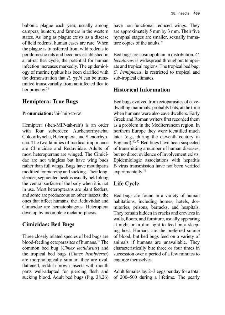
38. Insects 469
bubonic plague each year, usually among campers, hunters, and farmers in the western states. As long as plague exists as a disease of field rodents, human cases are rare. When the plague is transferred from wild rodents to peridomestic rats and becomes established in a rat-rat flea cycle, the potential for human infection increases markedly. The epidemiol-ogy of murine typhus has been clarified with the demonstration that R. typhi can be trans-mitted transovarially from an infected flea to her progeny.78
Hemiptera: True Bugs
Pronunciation: \hi-ˈmip-tə-rə\
Hemiptera (\heh-MIP-tah-rah\) is an order with four suborders: Auchenorrhyncha, Coleorrhyncha, Heteroptera, and Stenorrhyn-cha. The two families of medical importance are Climicidae and Reduviidae. Adults of most heteropterans are winged. The Cimici-dae are not wingless but have wing buds rather than full wings. Bugs have mouthparts modified for piercing and sucking. Their long, slender, segmented beak is usually held along the ventral surface of the body when it is not in use. Most heteropterans are plant feeders, and some are predaceous on other insects; the ones that affect humans, the Reduviidae and Cimicidae are hematophagous. Heteroptera develop by incomplete metamorphosis.
Cimicidae: Bed Bugs
Three closely related species of bed bugs are blood-feeding ectoparasites of humans.75 The common bed bug (Cimex lectularius) and the tropical bed bugs (Cimex hemipterus) are morphologically similar; they are oval, flattened, reddish-brown insects with mouth parts well-adapted for piercing flesh and sucking blood. Adult bed bugs (Fig. 38.26)
have non-functional reduced wings. They are approximately 5 mm by 3 mm. Their five nymphal stages are smaller, sexually imma-ture copies of the adults.76
Bed bugs are cosmopolitan in distribution. C. lectularius is widespread throughout temper-ate and tropical regions. The tropical bed bug, C. hemipterus, is restricted to tropical and sub-tropical climates.
Historical Information
Bed bugs evolved from ectoparasites of cave-dwelling mammals, probably bats, at the time when humans were also cave dwellers. Early Greek and Roman writers first recorded them as a problem in the Mediterranean region. In northern Europe they were identified much later (e.g., during the eleventh century in England).40, 52 Bed bugs have been suspected of transmitting a number of human diseases, but no direct evidence of involvement exists. Epidemiologic associations with hepatitis B virus transmission have not been verified experimentally.79
Life Cycle
Bed bugs are found in a variety of human habitations, including homes, hotels, dor-mitories, prisons, barracks, and hospitals. They remain hidden in cracks and crevices in walls, floors, and furniture, usually appearing at night or in dim light to feed on a sleep-ing host. Humans are the preferred source of blood, but bed bugs feed on a variety of animals if humans are unavailable. They characteristically bite three or four times in succession over a period of a few minutes to engorge themselves.
Adult females lay 2–3 eggs per day for a total of 200–500 during a lifetime. The pearly
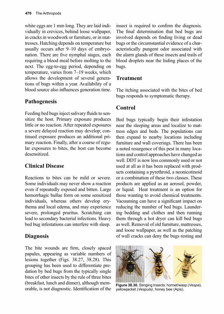
470 The Arthropods
white eggs are 1 mm long. They are laid indi-vidually in crevices, behind loose wallpaper, in cracks in woodwork or furniture, or in mat-tresses. Hatching depends on temperature but usually occurs after 9–10 days of embryo-nation. There are five nymphal stages, each requiring a blood meal before molting to the next. The egg-to-egg period, depending on temperature, varies from 7–19 weeks, which allows the development of several genera-tions of bugs within a year. Availability of a blood source also influences generation time.
Pathogenesis
Feeding bed bugs inject salivary fluids to sen-sitize the host. Primary exposure produces little or no reaction. After repeated exposures a severe delayed reaction may develop; con-tinued exposure produces an additional pri-mary reaction. Finally, after a course of regu-lar exposures to bites, the host can become desensitized.
Clinical Disease
Reactions to bites can be mild or severe. Some individuals may never show a reaction even if repeatedly exposed and bitten. Large hemorrhagic bullae form on some sensitized individuals, whereas others develop ery-thema and local edema, and may experience severe, prolonged pruritus. Scratching can lead to secondary bacterial infections. Heavy bed bug infestations can interfere with sleep.
Diagnosis
The bite wounds are firm, closely spaced papules, appearing as variable numbers of lesions together (Figs. 38.27, 38.28). This grouping has been used to differentiate pre-dation by bed bugs from the typically single bites of other insects by the rule of three bites (breakfast, lunch and dinner), although mem-orable, is not diagnostic. Identification of the
insect is required to confirm the diagnosis. The final determination that bed bugs are involved depends on finding living or dead bugs or the circumstantial evidence of a char-acteristically pungent odor associated with the alarm glands of these insects and trails of blood droplets near the hiding places of the bugs.
Treatment
The itching associated with the bites of bed bugs responds to symptomatic therapy.
Control
Bed bugs typically begin their infestation near the sleeping areas and localize to mat-tress edges and beds. The populations can then expand to nearby locations including furniture and wall coverings. There has been a noted resurgence of this pest in many loca-tions and control approaches have changed as well. DDT is now less commonly used or not used at all as it has been replaced with prod-ucts containing a pyrethroid, a neonicotinoid or a combination of these two classes. These products are applied as an aerosol, powder, or liquid. Heat treatment is an option for those wanting to avoid chemical treatments. Vacuuming can have a significant impact on reducing the number of bed bugs. Launder-ing bedding and clothes and then running them through a hot dryer can kill bed bugs as well. Removal of old furniture, mattresses, and loose wallpaper, as well as the patching of wall cracks can deny the bugs resting and
Figure 38.30. Stinging Insects: hornet/wasp (Vespa), yellowjacket (Vespula), honey bee (Apis).
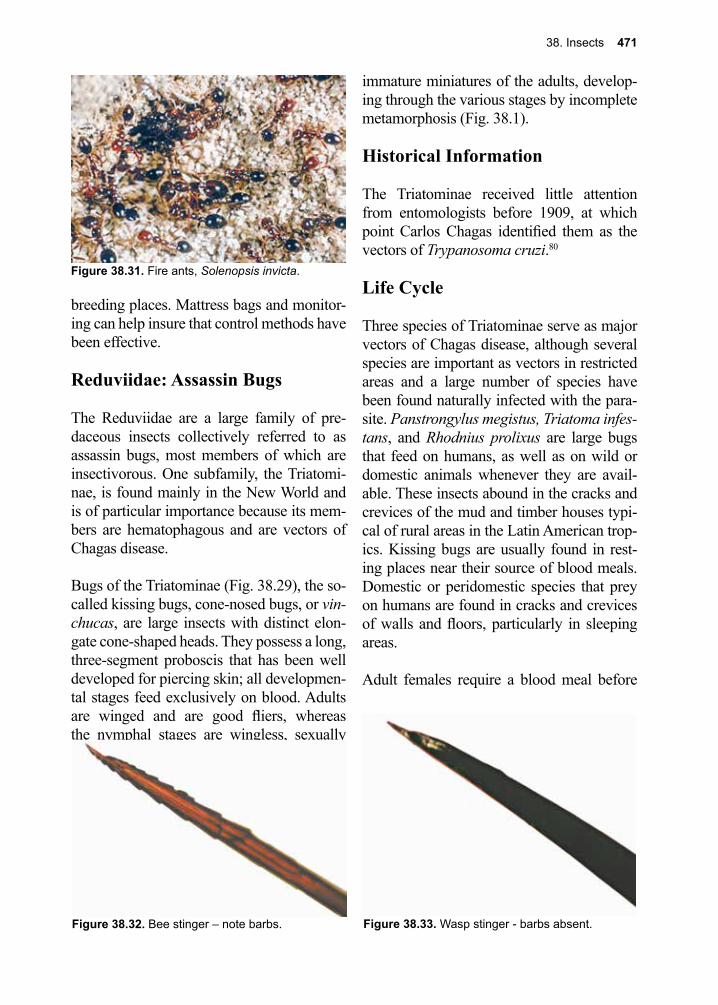
38. Insects 471
breeding places. Mattress bags and monitor-ing can help insure that control methods have been effective.
Reduviidae: Assassin Bugs
The Reduviidae are a large family of pre-daceous insects collectively referred to as assassin bugs, most members of which are insectivorous. One subfamily, the Triatomi-nae, is found mainly in the New World and is of particular importance because its mem-bers are hematophagous and are vectors of Chagas disease.
Bugs of the Triatominae (Fig. 38.29), the so-called kissing bugs, cone-nosed bugs, or vin-chucas, are large insects with distinct elon-gate cone-shaped heads. They possess a long, three-segment proboscis that has been well developed for piercing skin; all developmen-tal stages feed exclusively on blood. Adults are winged and are good fliers, whereas the nymphal stages are wingless, sexually
immature miniatures of the adults, develop-ing through the various stages by incomplete metamorphosis (Fig. 38.1).
Historical Information
The Triatominae received little attention from entomologists before 1909, at which point Carlos Chagas identified them as the vectors of Trypanosoma cruzi.80
Life Cycle
Three species of Triatominae serve as major vectors of Chagas disease, although several species are important as vectors in restricted areas and a large number of species have been found naturally infected with the para-site. Panstrongylus megistus, Triatoma infes-tans, and Rhodnius prolixus are large bugs that feed on humans, as well as on wild or domestic animals whenever they are avail-able. These insects abound in the cracks and crevices of the mud and timber houses typi-cal of rural areas in the Latin American trop-ics. Kissing bugs are usually found in rest-ing places near their source of blood meals. Domestic or peridomestic species that prey on humans are found in cracks and crevices of walls and floors, particularly in sleeping areas.
Adult females require a blood meal before
Figure 38.31. Fire ants, Solenopsis invicta.
Figure 38.33. Wasp stinger - barbs absent.Figure 38.32. Bee stinger – note barbs.
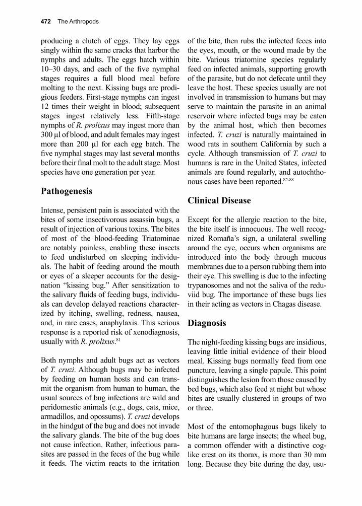
472 The Arthropods
producing a clutch of eggs. They lay eggs singly within the same cracks that harbor the nymphs and adults. The eggs hatch within 10–30 days, and each of the five nymphal stages requires a full blood meal before molting to the next. Kissing bugs are prodi-gious feeders. First-stage nymphs can ingest 12 times their weight in blood; subsequent stages ingest relatively less. Fifth-stage nymphs of R. prolixus may ingest more than 300 µl of blood, and adult females may ingest more than 200 µl for each egg batch. The five nymphal stages may last several months before their final molt to the adult stage. Most species have one generation per year.
Pathogenesis
Intense, persistent pain is associated with the bites of some insectivorous assassin bugs, a result of injection of various toxins. The bites of most of the blood-feeding Triatominae are notably painless, enabling these insects to feed undisturbed on sleeping individu-als. The habit of feeding around the mouth or eyes of a sleeper accounts for the desig-nation “kissing bug.” After sensitization to the salivary fluids of feeding bugs, individu-als can develop delayed reactions character-ized by itching, swelling, redness, nausea, and, in rare cases, anaphylaxis. This serious response is a reported risk of xenodiagnosis, usually with R. prolixus.81
Both nymphs and adult bugs act as vectors of T. cruzi. Although bugs may be infected by feeding on human hosts and can trans-mit the organism from human to human, the usual sources of bug infections are wild and peridomestic animals (e.g., dogs, cats, mice, armadillos, and opossums). T. cruzi develops in the hindgut of the bug and does not invade the salivary glands. The bite of the bug does not cause infection. Rather, infectious para-sites are passed in the feces of the bug while it feeds. The victim reacts to the irritation
of the bite, then rubs the infected feces into the eyes, mouth, or the wound made by the bite. Various triatomine species regularly feed on infected animals, supporting growth of the parasite, but do not defecate until they leave the host. These species usually are not involved in transmission to humans but may serve to maintain the parasite in an animal reservoir where infected bugs may be eaten by the animal host, which then becomes infected. T. cruzi is naturally maintained in wood rats in southern California by such a cycle. Although transmission of T. cruzi to humans is rare in the United States, infected animals are found regularly, and autochtho-nous cases have been reported.82-88
Clinical Disease
Except for the allergic reaction to the bite, the bite itself is innocuous. The well recog-nized Romaña’s sign, a unilateral swelling around the eye, occurs when organisms are introduced into the body through mucous membranes due to a person rubbing them into their eye. This swelling is due to the infecting trypanosomes and not the saliva of the redu-viid bug. The importance of these bugs lies in their acting as vectors in Chagas disease.
Diagnosis
The night-feeding kissing bugs are insidious, leaving little initial evidence of their blood meal. Kissing bugs normally feed from one puncture, leaving a single papule. This point distinguishes the lesion from those caused by bed bugs, which also feed at night but whose bites are usually clustered in groups of two or three.
Most of the entomophagous bugs likely to bite humans are large insects; the wheel bug, a common offender with a distinctive cog-like crest on its thorax, is more than 30 mm long. Because they bite during the day, usu-
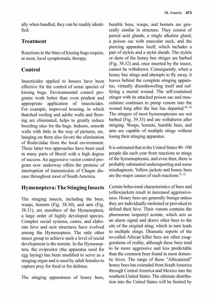
38. Insects 473
ally when handled, they can be readily identi-fied.
Treatment
Reactions to the bites of kissing bugs require, at most, local symptomatic therapy.
Control
Insecticides applied to houses have been effective for the control of some species of kissing bugs. Environmental control pro-grams work better than even prudent and appropriate application of insecticides. For example, improved housing, in which thatched roofing and adobe walls and floor-ing are eliminated, helps to greatly reduce breeding sites for the bugs. Indoors, smooth walls with little in the way of pictures, etc. hanging on them also favors the elimination of Reduviidae from the local environment. These latter two approaches have been used in many parts of Brazil with a high degree of success. An aggressive vector control pro-gram now underway offers the promise of interruption of transmission of Chagas dis-ease throughout most of South America.
Hymenoptera: The Stinging Insects
The stinging insects, including the bees, wasps, hornets (Fig. 38.30), and ants (Fig. 38.31), are members of the Hymenoptera, a large order of highly developed species. Complex social systems, castes, and elabo-rate hive and nest structures have evolved among the Hymenoptera. The only other insect group to achieve such a level of social development is the termite. In the Hymenop-tera, the ovipositor (the apparatus used for egg laying) has been modified to serve as a stinging organ and is used by adult females to capture prey for food or for defense.
The stinging apparatuses of honey bees,
bumble bees, wasps, and hornets are gen-erally similar in structure. They consist of paired acid glands, a single alkaline gland, a poison sac with muscular neck, and the piercing apparatus itself, which includes a pair of stylets and a stylet sheath. The stylets or darts of the honey bee stinger are barbed (Fig. 38.32) and, once inserted by the insect, cannot be withdrawn. Consequently, when a honey bee stings and attempts to fly away, it leaves behind the complete stinging appara-tus, virtually disemboweling itself and suf-fering a mortal wound. The self-contained stinger with its attached poison sac and mus-culature continues to pump venom into the wound long after the bee has departed.89, 90 The stingers of most hymenopterans are not barbed (Fig. 39.33) and are withdrawn after stinging. Wasps, hornets, bumble bees, and ants are capable of multiple stings without losing their stinging apparatus.
It is estimated that in the United States 90–100 people die each year from reactions to stings of the hymenopterans, and even then, there is probably substantial underreporting and some misdiagnosis. Yellow jackets and honey bees are the major causes of such reactions.91, 92
Certain behavioral characteristics of bees and yellowjackets result in increased aggressive-ness. Honey bees are generally benign unless they are individually molested or provoked to defend their hive. Their venom contains the pheromone isopentyl acetate, which acts as an alarm signal and draws other bees to the site of the original sting, which in turn leads to multiple stings. Dramatic reports of the so-called African killer bees are often exag-gerations of reality, although these bees tend to be more aggressive and less predictable than the common bees found in most domes-tic hives. The range of these “Africanized” honey bees has extended from South America through Central America and Mexico into the southern United States. The ultimate distribu-tion into the United States will be limited by
474 The Arthropods
the bees’ ability to survive killing tempera-tures.92-94
Yellowjackets are particularly aggressive when their nest areas are approached, at which point they sting without provoca-tion or warning. Their aggressive behavior increases during the late summer and early fall. Gardeners and picnickers are particu-larly at risk.
Historical Information
The honey bee, Apis mellifera, was one of the first insects recorded by humans in writ-ings and art. Bees have long been recognized as sources of honey, and their role as plant pollinators is crucial to agriculture.
Life Cycle
Hymenopterans develop by complete meta-morphosis with distinct larva, pupa, and adult stages. Larvae are vermiform and resemble maggots. They are dependent upon adults for food. Pupae, encased in a cocoon, represent an inactive, non-feeding transitional phase. The adults usually have wings and are good fliers. Certain groups with highly developed social systems have evolved non-reproduc-ing worker and soldier castes. Other groups, such as the ants, are wingless as adults except during reproductive periods. Four families of the Hymenoptera contain medically impor-tant species, the stings of which can cause severe reactions in humans: Apidae, Vespi-dae, Formicidae, and Mutillidae.
The bees, or Apidae, include some species that live in complex social organizations such as hives or in less-structured subterranean nests, although most species in this family live as solitary insects. Only the honey bees and bumble bees among the Apidae are of concern to humans because of their ability to sting. The honey bee, Apis mellifera, origi-
nally an Old World species, is now found worldwide in domestic and wild hives. Bees of this species are raised commercially for their honey and for their role as pollinators of a wide range of plants, including most fruits and legume crops. They construct elaborate hives wherein a single non-foraging queen lays eggs in wax cells. Larvae develop within these cells while being fed by non-reproduc-tive female workers. Adult workers tend the hives and forage for nectar and pollen. The bees tend to sting when their hive or individ-ual insects are disturbed. Bumble bees of the genus Bombus are large, hairy, ungainly, less organized social bees that build simple under-ground nests. They sting under the same cir-cumstances as do honey bees.
The Vespidae include the wasps, hornets, and yellowjackets, all of which are capable of inflicting painful stings. Many species in this family build elaborate nests of masticated wood fibers or mud, whereas others construct simple nests underground. The yellowjack-ets are social hymenopterans with distinctive yellow and black bands on the abdomen and are often mistaken for bees. They are aggres-sive insects and a major cause of stings in humans.
Ants belong to the family Formicidae, some members of which can cause damage by biting or stinging. They have a variety of complex social systems, with elaborate behavior pat-terns, intricate nests, and castes of workers, soldiers, and reproducing insects. Two groups of ants are of concern in the United States, the harvester ant and the fire ant.
The harvester ants of the genus Pogonomyr-mex readily attack humans and other animals and are capable of inflicting painful stings. They build underground nests, topped by mounds, in warm, dry, sandy areas. When a nest is disturbed, ants come out and swarm over the invader; their stings are repeated and
38. Insects 475
vigorous.
Fire ants of the genus Solenopsis are so named because of their sharp, fiery sting. Several species native to the United States are of medical importance, but the imported fire ant, Solenopsis invicta (Fig. 38.31), is a particu-larly dangerous species.95, 96 It was introduced into the United States around 1930 and since then has spread throughout the southeastern states, where it presents a serious hazard to humans and livestock. These ants build large, hard-crusted mounds, which are well camou-flaged and often not seen until they are dis-turbed. When fire ants attack, they first bite their victim with strong mandibles, and then sting their victim repeatedly. The result is a circle of painful stings around a central bite.
Certain species of tropical ants are notori-ous for their ability to ravage plants and ani-mals alike as they travel from place to place in colonies or armies numbering millions of individuals.
The so-called velvet ants are not true ants, but wingless wasps of the family Mutilli-dae. These large, hairy, often brightly col-ored insects are capable of inflicting a pain-ful sting if they are disturbed. A large black mutillid with scarlet hairs is common in the central United States, where it can cause con-siderable distress by stinging barefoot bath-ers. Several other groups of the Hymenoptera have the capacity to sting.
Pathogenesis
During the act of stinging, the aroused insect first inserts the sheath, inflicting a wound, then follows immediately with the inward thrust of the stylets and injection of the venom. The combination of the acid and alkaline venom fluids, designed to kill insect prey, causes extreme pain and inflammation. Venom from 500 stings received within a few minutes can
cause death. The inhabitants of a single dis-turbed beehive can easily achieve that.
Sensitization to the venom can result in severe allergic reactions. A number of anti-genically active compounds have been iden-tified in venom, phospholipase A being the most important. Others include hyaluroni-dase, melittin, and apamin.97, 98
Clinical Disease
The primary manifestations of the sting are due to mechanical damage and the direct action of the venom. The pain, edema, pru-ritus, and warmth produced at the site of the sting are transitory. Severe toxic reactions can be caused by as few as 10 stings within a period of a few minutes. Muscle cramps, drowsiness, fever, and headache are charac-teristic.
Allergic reactions are by far the most serious consequence of the stings of hymenopterans. They may develop in previously sensitized individuals and include three symptom pat-terns: 1. urticaria associated with pruritus, 2. edematous skin and mucous membranes, and 3. simultaneous bronchospasm and anaphy-laxis, followed sometimes by death. In sen-sitized individuals, even a single sting may bring the most severe reaction. A delayed reaction characterized by urticaria, fever, and arthralgia may occur hours or weeks after a sting.
Although the stinging hymenopterans share a number of common antigens, each possesses one or more unique antigens. Sensitization to stings of one species does not always pro-duce sensitivity to those of other species.
Diagnosis
Individuals with suspected sting sensitivi-ties can undergo skin testing with specific
476 The Arthropods
venoms to determine the level of risk. Iden-tification of the species posing the greatest threat to an individual may be critical.97
Treatment
Initial treatment for a honey bee sting must include removal of its stinger and the attached venom sac. Removal can be accomplished with a knife blade, a needle, or a fingernail. It is important not to squeeze the site, because such pressure releases more venom from the sac. A non-allergic primary reaction may be treated with ice to lessen edema and pain, and with various local anti-pruritic compounds.
Individuals with known sting sensitivity must be prepared to act quickly to prevent serious reactions. Upon being stung, the individual should remove the stinger immediately. Emergency kits are available by prescription, and sensitive individuals should be familiar with their use. Such kits contain epineph-rine in a syringe, antihistamine tablets, and a tourniquet. Emergency treatment consists of intramuscular injections of epinephrine and an antihistamine. Obviously, use of these
measures presupposes planning; the sensi-tized person and his or her next of kin must be prepared to carry out the intramuscular injec-tion of epinephrine. In addition, the potential victim should carry an oral antihistamine medication such as diphenhydramine.
Desensitization using whole-body extracts of the insects has been attempted but is inef-fective. Purified venoms have been used suc-cessfully for desensitization. Successful treat-ment for severe reactions to fire ant stings has been reported.
Control
Wasp, hornet, and ant nests can be destroyed with a number of commercially available insecticidal compounds, such as carba-mates (also called urethranes), malathion (an organophosphate), and resmithrin (a pyrethroid). Aerial nests can be destroyed at night, when the insects are quiescent. General avoidance of areas where stinging hymenopterans occur should be a rule for sensitive individuals. There are no effective repellents against these insects.
References
1. Savely, V. R., et al., The mystery of Morgellons disease: infection or delusion? Am J Clin Dermatol 2006, 7 (1), 1-5.
2. Koo, J.; Lee, C. S., Delusions of parasitosis. A dermatologist’s guide to diagnosis and treatment. Am J Clin Dermatol 2001, 2 (5), 285-90.
3. Ozkan, A. T.; Mumcuoglu, K. Y., [Entomophobia and delusional parasitosis]. Turkiye Parazitol Derg 2008, 32 (4), 366-70.
4. Peng, Z., et al., Mosquito allergy and mosquito salivary allergens. Protein Pept Lett 2007, 14 (10), 975-81.
5. Ribeiro, J. M. C.; Francischetti, I. M. B., Role of arthropod saliva in blood feeding: Sialome and post-sialome perspectives. Annu Rev Entomol 2002, 48, 73-88.
6. Theodorides, J., Note historique sur la decouverte de la transmission de la leishmaniose cutanee par les phlebotomes. Bull Soc Pathol Exot 1997, 90, 177-180.
7. Oliveira, F., et al., A sand fly salivary protein vaccine shows efficacy against vector-transmitted cutaneous leishmaniasis in nonhuman primates. Sci Transl Med 2015, 7 (290), 290ra90.
8. Elnaiem, D. E., et al., Genetic variation in the sand fly salivary protein, SP-15, a potential vaccine candidate against Leishmania major. Insect Mol Biol 2005, 14 (2), 145-50.
9. Wenyon, C. M.; J., Some recent advances in our knowledge of leishmaniasis. School Trop Med 1912, 1 93-98.
10. Lewis, D. J., The biology of Phlebotomidae in relation to leishmaniasis. Annual review of
38. Insects 477
entomology 1974, 19, 363-84.11. Valenzuela, J. G., et al., Identification of the most abundant secreted proteins from the salivary
glands of the sand fly Lutzomyia longipalpis, vector of Leishmania chagasi. The Journal of experimental biology 2004, 207 (Pt 21), 3717-29.
12. Sambon, L. W.; J., Progress report of investigations of pellagra. Med 1910, 13 271-287.13. Monnerat, R., et al., Synergistic activity of Bacillus thuringiensis toxins against Simulium spp.
larvae. J Invertebr Pathol 2014, 121, 70-3.14. To, K. K.; Yuen, K. Y., In memory of Patrick Manson, founding father of tropical medicine and
the discovery of vector-borne infections. Emerg Microbes Infect 2012, 1 (10), e31.15. Sonenshine, D. E., Biology of Ticks Volume 2. 2013.16. de Kruif, P., The Microbe Hunters. 1926.17. Harrison, G., Mosquitos, Malaria, and Man. 1978.18. Shah, S., The Fever: How malaria has ruled mankind for 500,000 years. 2010.19. Reed, W., et al., Etiology of yellow fever, a preliminary note. Philos 1900, 6, 790-796.20. Reinert, J. F., New classification for the composite genus Aedes (Diptera: Culicidae: Aedini),
elevation of subgenus Ochlerotatus to generic rank, reclassification of the other subgenera, and notes on certain subgenera and species. Journal of the American Mosquito Control Association 2000, 16 (3), 175-88.
21. Reinert, J. F., et al., Phylogeny and classification of Aedini (Diptera:Culicidae), based on morphological characters of all life stages. Zool Soc 2004, 142 289-368.
22. Singh, S.; Mann, B. K., Insect bite reactions. Indian J Dermatol Venereol Leprol 2013, 79 (2), 151-64.
23. Morsy, T. A., Insect bites and what is eating you? J Egypt Soc Parasitol 2012, 42 (2), 291-308.24. Williams, L. A.; Allen, L. V., Jr., Treatment and prevention of insect bites: mosquitoes. Int J
Pharm Compd 2012, 16 (3), 210-8.25. Steen, C. J., et al., Arthropods in dermatology. J Am Acad Dermatol 2004, 50 (6), 819-42, quiz
842-4.26. Villar, L., et al., Efficacy of a tetravalent dengue vaccine in children in Latin America. N Engl J
Med 2015, 372 (2), 113-23.27. McArthur, M. A., et al., Dengue vaccines: recent developments, ongoing challenges and current
candidates. Expert Rev Vaccines 2013, 12 (8), 933-53.28. Kendrick, K., et al., Notes from the field: Transmission of chikungunya virus in the continental
United States--Florida, 2014. MMWR Morb Mortal Wkly Rep 2014, 63 (48), 1137.29. Winskill, P., et al., Dispersal of Engineered Male Aedes aegypti Mosquitoes. PLoS Negl Trop Dis
2015, 9 (11), e0004156.30. Allan, S. A., Susceptibility of adult mosquitoes to insecticides in aqueous sucrose baits. J Vector
Ecol 2011, 36 (1), 59-67.31. Leiper, R. T., Report of the Helminthologist for the Half Year Ending 30 April, . Report of the
Advisory Commission on Tropical Diseases Research Fund. 1913, 1913-1914.32. Benecke, M., A brief history of forensic entomology. Forensic Sci Int 2001, 120 (1-2), 2-14.33. Michaud, J. P., et al., Rewriting Ecological Succession History: Did Carrion Ecologists Get
There First? Q Rev Biol 2015, 90 (1), 45-66.34. Amendt, J., et al., Forensic entomology: applications and limitations. Forensic Sci Med Pathol
2011, 7 (4), 379-92.35. Scudder, H. l.; Linda, D. R., Nonbiting flies and disease Annu Rev Entomol 1956, 1, 323-346.36. Greenberg, B., Flies and Disease. 1973; Vol. II.37. Franco, J. R., et al., Epidemiology of human African trypanosomiasis. Clin Epidemiol 2014, 6,
257-75.38. Aksoy, S., et al., Interactions between tsetse and trypanosomes with implications for the control
of trypanosomiasis. Adv Parasitol 2003, 53, 1-83.39. Francesconi, F.; Lupi, O., Myiasis. Clin Microbiol Rev 2012, 25 (1), 79-105.40. Morley, W. N., Body infestations. Scottish medical journal 1977, 22 (3), 211-6.41. Steenvoorde, P., et al., Maggot debridement therapy of infected ulcers: patient and wound factors
influencing outcome - a study on 101 patients with 117 wounds. Ann R Coll Surg Engl 2007, 89
478 The Arthropods
(6), 596-602.42. Sun, X., et al., A systematic review of maggot debridement therapy for chronically infected
wounds and ulcers. Int J Infect Dis 2014, 25, 32-7.43. Mumcuoglu, K. Y., et al., Pain related to maggot debridement therapy. J Wound Care 2012, 21
(8), 400, 402, 404-5.44. Gilead, L., et al., The use of maggot debridement therapy in the treatment of chronic wounds in
hospitalised and ambulatory patients. J Wound Care 2012, 21 (2), 78, 80, 82-85.45. Whitaker, I. S., et al., Larval therapy from antiquity to the present day: mechanisms of action,
clinical applications and future potential. Postgrad Med J 2007, 83 (980), 409-13.46. Gregory, A. R., et al., Ophthalmomyiasis caused by the sheep bot fly Oestrus ovis in northern
Iraq. Optom Vis Sci 2004, 81 (8), 586-90.47. Lord, W. D., et al., Collection and preservation of forensically important entomological
materials. Science 1983, 28, 936-944.48. Greenberg, B., Forensic entomology: case studies. Bull Entomol Soc Am 1985, 31, 25-28.49. Greenberg, B.; Kunich, J. C., Entomology and the law. 2002.50. Chosidow, O., Scabies and pediculosis. Lancet (London, England) 2000, 355 (9206), 819-26.51. Zinsser, H., Rats, Lice and History. 1935.52. Busvine, J. R., Insects, Hygiene, and History. Athlone Press London 1976.53. Do-Pham, G., et al., Lice. Semin Cutan Med Surg 2014, 33 (3), 116-8.54. Ko, C. J.; Elston, D. M., Pediculosis. J Am Acad Dermatol 2004, 50 (1), 1-12; quiz 13-4.55. Leone, P. A., Scabies and pediculosis pubis: an update of treatment regimens and general review.
Clin Infect Dis 2007, 44 Suppl 3, S153-9.56. Roberts, R. J., Clinical practice. Head lice. N Engl J Med 2002, 346 (21), 1645-50.57. Osorio, C., et al., Plica polonica secondary to seborrheic dermatitis. J Eur Acad Dermatol
Venereol 2015.58. Nara, A., et al., An unusual autopsy case of lethal hypothermia exacerbated by body lice-induced
severe anemia. Int J Legal Med 2015.59. Jahnke, C., et al., Accuracy of diagnosis of pediculosis capitis: visual inspection vs wet combing.
Arch Dermatol 2009, 145 (3), 309-13.60. Ackerman, A. B., Crabs--the resurgence of Phthirus pubis. N Engl J Med 1968, 278 (17), 950-1.61. Feldmeier, H., Treatment of pediculosis capitis: a critical appraisal of the current literature. Am J
Clin Dermatol 2014, 15 (5), 401-12.62. Burkhart, C. G.; Burkhart, C. N., Oral ivermectin for Phthirus pubis. J Am Acad Dermatol 2004,
51 (6), 1037; author reply 1037-8.63. Bohl, B., et al., Clinical Practice Update: Pediculosis Capitis. Pediatr Nurs 2015, 41 (5), 227-34.64. Walsh, J.; Nicholson, A., Head lice in children--a modern pandemic. Ir Med J 2005, 98 (5), 156-
7.65. Elston, D. M., Treating pediculosis--those nit-picking details. Pediatr Dermatol 2007, 24 (4),
415-6.66. Deeks, L. S., et al., Topical ivermectin 0.5% lotion for treatment of head lice. Ann Pharmacother
2013, 47 (9), 1161-7.67. Orion, E., et al., Ectoparasitic sexually transmitted diseases: scabies and pediculosis. Clinics in
dermatology 2004, 22 (6), 513-9.68. Elgart, M. L., Current treatment for scabies and pediculosis. Skin Therapy Lett 2000, 5, 1-3.69. Raoult, D.; Roux, V., The body louse as a vector of reemerging human diseases. Clin Inf Dis
1999, 29 (4), 888-911.70. Palicelli, A., et al., Tungiasis in Italy: An imported case of Tunga penetrans and review of the
literature. Pathol Res Pract 2016.71. Feldmeier, H., et al., Tungiasis (sand flea disease): a parasitic disease with particular challenges
for public health. Eur J Clin Microbiol Infect Dis 2013, 32 (1), 19-26.72. Feldmeier, H.; Keysers, A., Tungiasis - A Janus-faced parasitic skin disease. Travel Med Infect
Dis 2013, 11 (6), 357-65.
38. Insects 479
73. Grunwald, M. H., et al., Tungiasis. The Australasian journal of dermatology 2000, 41 (1), 46-7.74. Heukelbach, J., Revision on tungiasis: treatment options and prevention. Expert Rev Anti Infect
Ther 2006, 4 (1), 151-7.75. Huntley, A. C., Cimex lectularius. What is this insect and how does it affect man? Dermatology
online journal 1999, 5 (1), 6.76. Kolb, A., et al., Bedbugs. Dermatol Ther 2009, 22 (4), 347-52.77. Simond, M., et al., Paul-Louis Simond and his discovery of plague transmission by rat fleas: a
centenary. J R Soc Med 1998, 91 (2), 101-4.78. Farhang-Azad, A., et al., Transovarial transmission of murine Typhus rickettsiae in Xenopsylla
cheopis fleas. Science (New York, N.Y.) 1985, 227 (4686), 543-5.79. Jupp, P. G., et al., Attempts to transmit hepatitis B virus to chimpanzees by arthropods. South
African medical journal (Suid-Afrikaanse tydskrif vir geneeskunde) 1991, 79 (6), 320-2.80. Chagas, C., Nova trypanozomiaze humans: estudos sobre a morfolojiia e o ciclo evalutivo do
Schizotripanum cruzi. n genl n sp ajente etiolojico de nova entidade morbida do homen. Mem Inst Oswaldo Cruz 1909, 1, 159-218.
81. Costa, C. H., et al., Skin reactions to bug bites as a result of xenodiagnosis. Transactions of the Royal Society of Tropical Medicine and Hygiene 1981, 75 (3), 405-8.
82. Morel, C. M., Chagas’ disease, from discovery to control and beyond: history, myths and lessons to take home. Mem Inst Oswaldo Cruz 94 Suppl 1999, 1 3-16.
83. Ebrahim, G. J., Eradication of American trypanosomiasis (Chagas’ disease): an achievable goal? Journal of tropical pediatrics 2004, 50 (6), 320-1.
84. Klotz, S. A., et al., Kissing bugs in the United States: risk for vector-borne disease in humans. Environ Health Insights 2014, 8 (Suppl 2), 49-59.
85. Bern, C., et al., Trypanosoma cruzi and Chagas’ Disease in the United States. Clin Microbiol Rev 2011, 24 (4), 655-81.
86. Garcia, M. N., et al., Historical Perspectives on the Epidemiology of Human Chagas Disease in Texas and Recommendations for Enhanced Understanding of Clinical Chagas Disease in the Southern United States. PLoS Negl Trop Dis 2015, 9 (11), e0003981.
87. Stevens, L., et al., Kissing bugs. The vectors of Chagas. Adv Parasitol 2011, 75, 169-92.88. Klotz, J. H., et al., “Kissing bugs”: potential disease vectors and cause of anaphylaxis. Clin Infect
Dis 2010, 50 (12), 1629-34.89. Tankersley, M. S.; Ledford, D. K., Stinging insect allergy: state of the art 2015. J Allergy Clin
Immunol Pract 2015, 3 (3), 315-22; quiz 323.90. Touchard, A., et al., The Biochemical Toxin Arsenal from Ant Venoms. Toxins (Basel) 2016, 8
(1).91. Fitzgerald, K. T.; Flood, A. A., Hymenoptera stings. Clin Tech Small Anim Pract 2006, 21 (4),
194-204.92. Potiwat, R.; Sitcharungsi, R., Ant allergens and hypersensitivity reactions in response to ant
stings. Asian Pac J Allergy Immunol 2015, 33 (4), 267-75.93. Shaker, M. S., et al., An update on venom allergy. Curr Opin Pediatr 2013, 25 (5), 629-34.94. Winston, M. L., The biology and management of Africanized honey bees Entomol 547 1992, 37
S, 173-193.95. Caplan, E. L., et al., Fire ants represent an important risk for anaphylaxis among residents of an
endemic region. The Journal of allergy and clinical immunology 2003, 111 (6), 1274-7.96. Stafford, C. T., Hypersensitivity to fire ant venom. Annals of allergy, asthma & immunology :
official publication of the American College of Allergy, Asthma, & Immunology 1996, 77 (2), 87-95; quiz 96.
97. Wong, H. C., Importance of proper identification of stinging insects. Annals of internal medicine 2000, 132 (5), 418.
98. Hoffman, D. R., Insect venom allergy, immunology and immunotherapy. 1984; p 187-223.

480 The Arthropods
Miriam Rothschild (1908–2005)
During her life, Miriam Rothschild was the world’s leading authority on fleas. In addition to being the first person to elucidate the jumping mechanism of fleas, she published on the role of Xenopsylla cheopsis as a vector for plague. Rothschild went on to publish dozens of papers on the digenetic trematodes and even after her laboratory and data were destroyed by a Nazi attack she went on to write the book Fleas, Flukes, and Cuckoos, which was written in such a manner as to make parasitol-ogy accessible to the general reader. By the time of her death in 2005, she had authored over 300 papers and several texts.
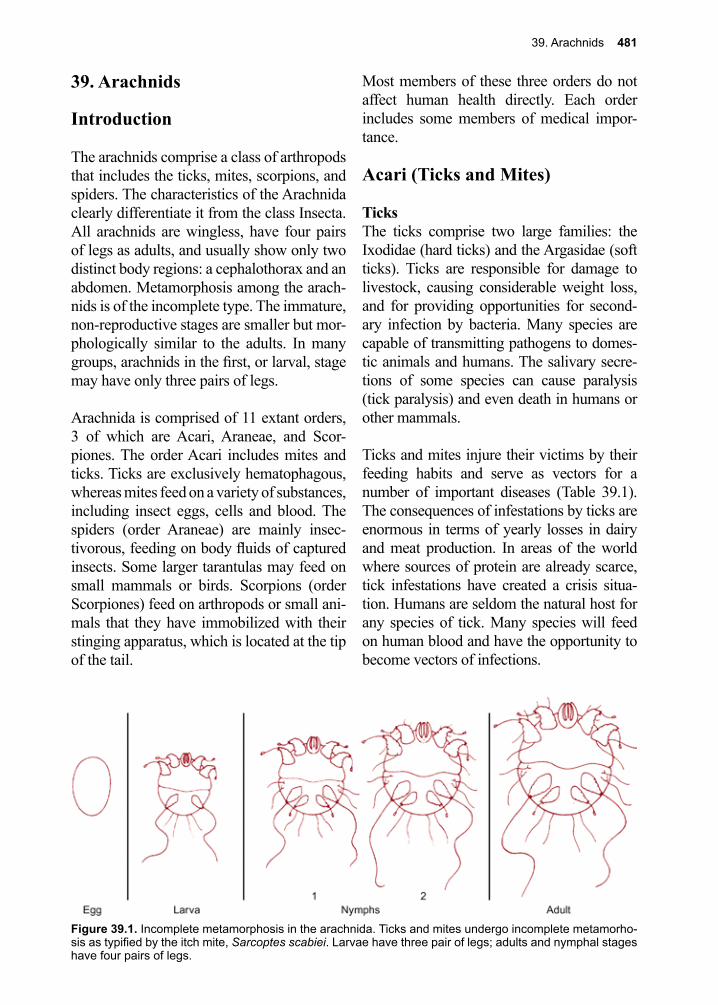
39. Arachnids 481
39. Arachnids
Introduction
The arachnids comprise a class of arthropods that includes the ticks, mites, scorpions, and spiders. The characteristics of the Arachnida clearly differentiate it from the class Insecta. All arachnids are wingless, have four pairs of legs as adults, and usually show only two distinct body regions: a cephalothorax and an abdomen. Metamorphosis among the arach-nids is of the incomplete type. The immature, non-reproductive stages are smaller but mor-phologically similar to the adults. In many groups, arachnids in the first, or larval, stage may have only three pairs of legs.
Arachnida is comprised of 11 extant orders, 3 of which are Acari, Araneae, and Scor-piones. The order Acari includes mites and ticks. Ticks are exclusively hematophagous, whereas mites feed on a variety of substances, including insect eggs, cells and blood. The spiders (order Araneae) are mainly insec-tivorous, feeding on body fluids of captured insects. Some larger tarantulas may feed on small mammals or birds. Scorpions (order Scorpiones) feed on arthropods or small ani-mals that they have immobilized with their stinging apparatus, which is located at the tip of the tail.
Most members of these three orders do not affect human health directly. Each order includes some members of medical impor-tance.
Acari (Ticks and Mites)
TicksThe ticks comprise two large families: the Ixodidae (hard ticks) and the Argasidae (soft ticks). Ticks are responsible for damage to livestock, causing considerable weight loss, and for providing opportunities for second-ary infection by bacteria. Many species are capable of transmitting pathogens to domes-tic animals and humans. The salivary secre-tions of some species can cause paralysis (tick paralysis) and even death in humans or other mammals.
Ticks and mites injure their victims by their feeding habits and serve as vectors for a number of important diseases (Table 39.1). The consequences of infestations by ticks are enormous in terms of yearly losses in dairy and meat production. In areas of the world where sources of protein are already scarce, tick infestations have created a crisis situa-tion. Humans are seldom the natural host for any species of tick. Many species will feed on human blood and have the opportunity to become vectors of infections.
Figure 39.1. Incomplete metamorphosis in the arachnida. Ticks and mites undergo incomplete metamorho-sis as typified by the itch mite, Sarcoptes scabiei. Larvae have three pair of legs; adults and nymphal stages have four pairs of legs.
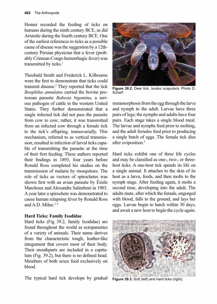
482 The Arthropods
Homer recorded the feeding of ticks on humans during the ninth century BCE, as did Aristotle during the fourth century BCE. One of the earliest references to ticks as a possible cause of disease was the suggestion by a 12th-century Persian physician that a fever (prob-ably Crimean-Congo hemorrhagic fever) was transmitted by ticks.1
Theobald Smith and Frederick L. Kilbourne were the first to demonstrate that ticks could transmit disease.2 They reported that the tick Boophilus annulatus carried the bovine pro-tozoan parasite Babesia bigemina, a seri-ous pathogen of cattle in the western United States. They further demonstrated that a single infected tick did not pass the parasite from cow to cow; rather, it was transmitted from an infected cow through a female tick to the tick’s offspring, transovarially. This mechanism, referred to as vertical transmis-sion, resulted in infection of larval ticks capa-ble of transmitting the parasite at the time of their first feeding. These authors reported their findings in 1893, four years before Ronald Ross completed his studies on the transmission of malaria by mosquitoes. The role of ticks as vectors of spirochetes was shown first with an avian parasite by Émile Marchoux and Alexandre Salimbeni in 1903. A year later a spirochete was demonstrated to cause human relapsing fever by Ronald Ross and A.D. Milne.3, 4
Hard Ticks: Family IxodidaeHard ticks (Fig 39.2, family Ixodidae) are found throughout the world as ectoparasites of a variety of animals. Their name derives from the characteristic tough, leather-like integument that covers most of their body. Their mouthparts are included in a capitu-lum (Fig. 39.2), but there is no defined head. Members of both sexes feed exclusively on blood.
The typical hard tick develops by gradual
metamorphosis from the egg through the larva and nymph to the adult. Larvae have three pairs of legs; the nymphs and adults have four pairs. Each stage takes a single blood meal. The larvae and nymphs feed prior to molting, and the adult females feed prior to producing a single batch of eggs. The female tick dies after oviposition.5
Hard ticks exhibit one of three life cycles and may be classified as one-, two-, or three-host ticks. A one-host tick spends its life on a single animal. It attaches to the skin of its host as a larva, feeds, and then molts to the nymph stage. After feeding again, it molts a second time, developing into the adult. The adults mate, after which the female, engorged with blood, falls to the ground, and lays her eggs. Larvae begin to hatch within 30 days, and await a new host to begin the cycle again.
Figure 39.2. Deer tick, Ixodes scapularis. Photo D. Scharf.
Figure 39.3. Soft (left) and hard ticks (right).
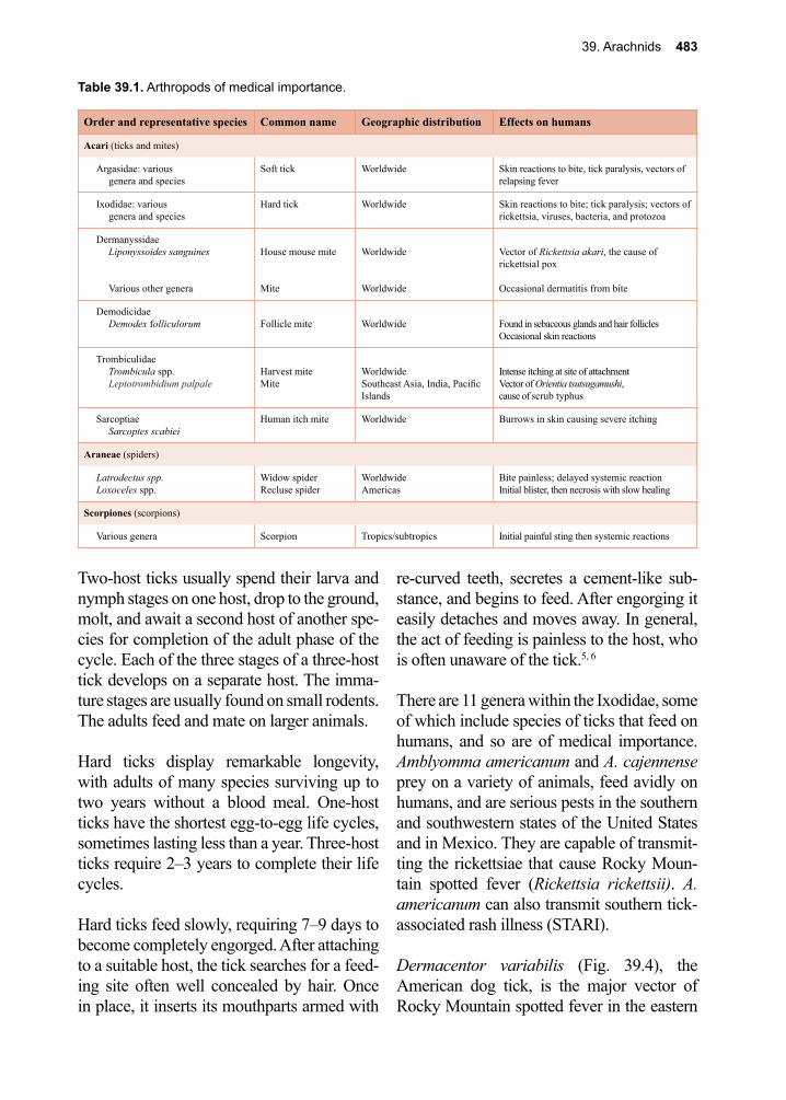
39. Arachnids 483
Two-host ticks usually spend their larva and nymph stages on one host, drop to the ground, molt, and await a second host of another spe-cies for completion of the adult phase of the cycle. Each of the three stages of a three-host tick develops on a separate host. The imma-ture stages are usually found on small rodents. The adults feed and mate on larger animals.
Hard ticks display remarkable longevity, with adults of many species surviving up to two years without a blood meal. One-host ticks have the shortest egg-to-egg life cycles, sometimes lasting less than a year. Three-host ticks require 2–3 years to complete their life cycles.
Hard ticks feed slowly, requiring 7–9 days to become completely engorged. After attaching to a suitable host, the tick searches for a feed-ing site often well concealed by hair. Once in place, it inserts its mouthparts armed with
re-curved teeth, secretes a cement-like sub-stance, and begins to feed. After engorging it easily detaches and moves away. In general, the act of feeding is painless to the host, who is often unaware of the tick.5, 6
There are 11 genera within the Ixodidae, some of which include species of ticks that feed on humans, and so are of medical importance. Amblyomma americanum and A. cajennense prey on a variety of animals, feed avidly on humans, and are serious pests in the southern and southwestern states of the United States and in Mexico. They are capable of transmit-ting the rickettsiae that cause Rocky Moun-tain spotted fever (Rickettsia rickettsii). A. americanum can also transmit southern tick-associated rash illness (STARI).
Dermacentor variabilis (Fig. 39.4), the American dog tick, is the major vector of Rocky Mountain spotted fever in the eastern
Table 39.1. Arthropods of medical importance.
Order and representative species Common name Geographic distribution Effects on humans
Acari (ticks and mites)
Argasidae: variousgenera and species
Soft tick Worldwide Skin reactions to bite, tick paralysis, vectors of relapsing fever
Ixodidae: variousgenera and species
Hard tick Worldwide Skin reactions to bite; tick paralysis; vectors of rickettsia, viruses, bacteria, and protozoa
DermanyssidaeLiponyssoides sanguines
Various other genera
House mouse mite
Mite
Worldwide
Worldwide
Vector of Rickettsia akari, the cause of rickettsial pox
Occasional dermatitis from bite
DemodicidaeDemodex folliculorum Follicle mite Worldwide Found in sebaceous glands and hair follicles
Occasional skin reactions
TrombiculidaeTrombicula spp.Leptotrombidium palpale
Harvest miteMite
WorldwideSoutheast Asia, India, Pacific Islands
Intense itching at site of attachmentVector of Orientia tsutsugamushi,cause of scrub typhus
SarcoptiaeSarcoptes scabiei
Human itch mite Worldwide Burrows in skin causing severe itching
Araneae (spiders)
Latrodectus spp.Loxoceles spp.
Widow spiderRecluse spider
WorldwideAmericas
Bite painless; delayed systemic reactionInitial blister, then necrosis with slow healing
Scorpiones (scorpions)
Various genera Scorpion Tropics/subtropics Initial painful sting then systemic reactions
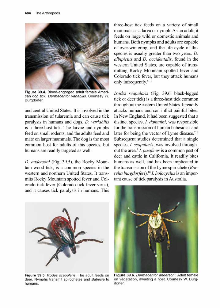
484 The Arthropods
Figure 39.5. Ixodes scapularis. The adult feeds on deer. Nymphs transmit spirochetes and Babesia to humans.
Figure 39.6. Dermacentor andersoni. Adult female on vegetation, awaiting a host. Courtesy W. Burg-dorfer.
and central United States. It is involved in the transmission of tularemia and can cause tick paralysis in humans and dogs. D. variabilis is a three-host tick. The larvae and nymphs feed on small rodents, and the adults feed and mate on larger mammals. The dog is the most common host for adults of this species, but humans are readily targeted as well.
D. andersoni (Fig. 39.5), the Rocky Moun-tain wood tick, is a common species in the western and northern United States. It trans-mits Rocky Mountain spotted fever and Col-orado tick fever (Colorado tick fever virus), and it causes tick paralysis in humans. This
three-host tick feeds on a variety of small mammals as a larva or nymph. As an adult, it feeds on large wild or domestic animals and humans. Both nymphs and adults are capable of over-wintering, and the life cycle of this species is usually greater than two years. D. albipictus and D. occidentalis, found in the western United States, are capable of trans-mitting Rocky Mountain spotted fever and Colorado tick fever, but they attack humans only infrequently.5-11
Ixodes scapularis (Fig. 39.6, black-legged tick or deer tick) is a three-host tick common throughout the eastern United States. It readily attacks humans and can inflict painful bites. In New England, it had been suggested that a distinct species, I. dammini, was responsible for the transmission of human babesiosis and later for being the vector of Lyme disease.7, 8 Subsequent studies determined that a single species, I. scapularis, was involved through-out the area.9 I. pacificus is a common pest of deer and cattle in California. It readily bites humans as well, and has been implicated in the transmission of the Lyme spirochete (Bor-relia burgdorferi).10 I. holocyclus is an impor-tant cause of tick paralysis in Australia.
Figure 39.4. Blood-engorged adult female Ameri-can dog tick, Dermacentor variabilis. Courtesy W. Burgdorfer.
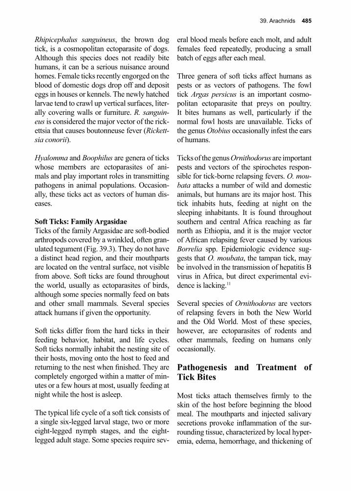
39. Arachnids 485
Rhipicephalus sanguineus, the brown dog tick, is a cosmopolitan ectoparasite of dogs. Although this species does not readily bite humans, it can be a serious nuisance around homes. Female ticks recently engorged on the blood of domestic dogs drop off and deposit eggs in houses or kennels. The newly hatched larvae tend to crawl up vertical surfaces, liter-ally covering walls or furniture. R. sanguin-eus is considered the major vector of the rick-ettsia that causes boutonneuse fever (Rickett-sia conorii).
Hyalomma and Boophilus are genera of ticks whose members are ectoparasites of ani-mals and play important roles in transmitting pathogens in animal populations. Occasion-ally, these ticks act as vectors of human dis-eases.
Soft Ticks: Family ArgasidaeTicks of the family Argasidae are soft-bodied arthropods covered by a wrinkled, often gran-ulated tegument (Fig. 39.3). They do not have a distinct head region, and their mouthparts are located on the ventral surface, not visible from above. Soft ticks are found throughout the world, usually as ectoparasites of birds, although some species normally feed on bats and other small mammals. Several species attack humans if given the opportunity.
Soft ticks differ from the hard ticks in their feeding behavior, habitat, and life cycles. Soft ticks normally inhabit the nesting site of their hosts, moving onto the host to feed and returning to the nest when finished. They are completely engorged within a matter of min-utes or a few hours at most, usually feeding at night while the host is asleep.
The typical life cycle of a soft tick consists of a single six-legged larval stage, two or more eight-legged nymph stages, and the eight-legged adult stage. Some species require sev-
eral blood meals before each molt, and adult females feed repeatedly, producing a small batch of eggs after each meal.
Three genera of soft ticks affect humans as pests or as vectors of pathogens. The fowl tick Argas persicus is an important cosmo-politan ectoparasite that preys on poultry. It bites humans as well, particularly if the normal fowl hosts are unavailable. Ticks of the genus Otobius occasionally infest the ears of humans.
Ticks of the genus Ornithodorus are important pests and vectors of the spirochetes respon-sible for tick-borne relapsing fevers. O. mou-bata attacks a number of wild and domestic animals, but humans are its major host. This tick inhabits huts, feeding at night on the sleeping inhabitants. It is found throughout southern and central Africa reaching as far north as Ethiopia, and it is the major vector of African relapsing fever caused by various Borrelia spp. Epidemiologic evidence sug-gests that O. moubata, the tampan tick, may be involved in the transmission of hepatitis B virus in Africa, but direct experimental evi-dence is lacking.11
Several species of Ornithodorus are vectors of relapsing fevers in both the New World and the Old World. Most of these species, however, are ectoparasites of rodents and other mammals, feeding on humans only occasionally.
Pathogenesis and Treatment of Tick Bites
Most ticks attach themselves firmly to the skin of the host before beginning the blood meal. The mouthparts and injected salivary secretions provoke inflammation of the sur-rounding tissue, characterized by local hyper-emia, edema, hemorrhage, and thickening of
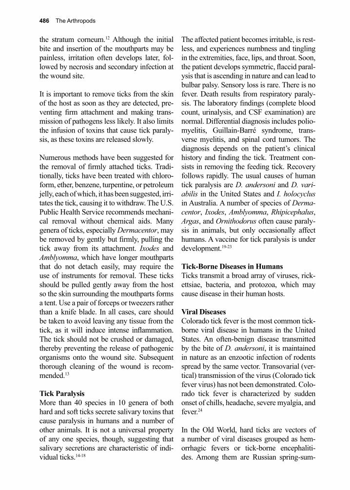
486 The Arthropods
the stratum corneum.12 Although the initial bite and insertion of the mouthparts may be painless, irritation often develops later, fol-lowed by necrosis and secondary infection at the wound site.
It is important to remove ticks from the skin of the host as soon as they are detected, pre-venting firm attachment and making trans-mission of pathogens less likely. It also limits the infusion of toxins that cause tick paraly-sis, as these toxins are released slowly.
Numerous methods have been suggested for the removal of firmly attached ticks. Tradi-tionally, ticks have been treated with chloro-form, ether, benzene, turpentine, or petroleum jelly, each of which, it has been suggested, irri-tates the tick, causing it to withdraw. The U.S. Public Health Service recommends mechani-cal removal without chemical aids. Many genera of ticks, especially Dermacentor, may be removed by gently but firmly, pulling the tick away from its attachment. Ixodes and Amblyomma, which have longer mouthparts that do not detach easily, may require the use of instruments for removal. These ticks should be pulled gently away from the host so the skin surrounding the mouthparts forms a tent. Use a pair of forceps or tweezers rather than a knife blade. In all cases, care should be taken to avoid leaving any tissue from the tick, as it will induce intense inflammation. The tick should not be crushed or damaged, thereby preventing the release of pathogenic organisms onto the wound site. Subsequent thorough cleaning of the wound is recom-mended.13
Tick ParalysisMore than 40 species in 10 genera of both hard and soft ticks secrete salivary toxins that cause paralysis in humans and a number of other animals. It is not a universal property of any one species, though, suggesting that salivary secretions are characteristic of indi-vidual ticks.14-18
The affected patient becomes irritable, is rest-less, and experiences numbness and tingling in the extremities, face, lips, and throat. Soon, the patient develops symmetric, flaccid paral-ysis that is ascending in nature and can lead to bulbar palsy. Sensory loss is rare. There is no fever. Death results from respiratory paraly-sis. The laboratory findings (complete blood count, urinalysis, and CSF examination) are normal. Differential diagnosis includes polio-myelitis, Guillain-Barré syndrome, trans-verse myelitis, and spinal cord tumors. The diagnosis depends on the patient’s clinical history and finding the tick. Treatment con-sists in removing the feeding tick. Recovery follows rapidly. The usual causes of human tick paralysis are D. andersoni and D. vari-abilis in the United States and I. holocyclus in Australia. A number of species of Derma-centor, Ixodes, Amblyomma, Rhipicephalus, Argas, and Ornithodorus often cause paraly-sis in animals, but only occasionally affect humans. A vaccine for tick paralysis is under development.19-23
Tick-Borne Diseases in HumansTicks transmit a broad array of viruses, rick-ettsiae, bacteria, and protozoa, which may cause disease in their human hosts.
Viral DiseasesColorado tick fever is the most common tick-borne viral disease in humans in the United States. An often-benign disease transmitted by the bite of D. andersoni, it is maintained in nature as an enzootic infection of rodents spread by the same vector. Transovarial (ver-tical) transmission of the virus (Colorado tick fever virus) has not been demonstrated. Colo-rado tick fever is characterized by sudden onset of chills, headache, severe myalgia, and fever.24
In the Old World, hard ticks are vectors of a number of viral diseases grouped as hem-orrhagic fevers or tick-borne encephaliti-des. Among them are Russian spring-sum-
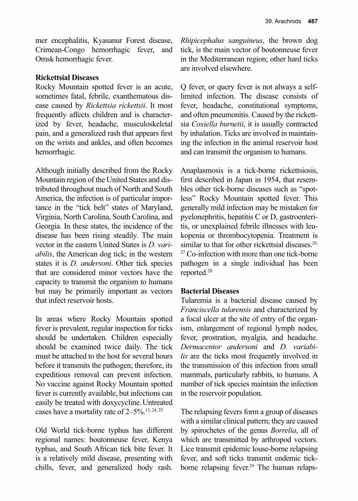
39. Arachnids 487
mer encephalitis, Kyasanur Forest disease, Crimean-Congo hemorrhagic fever, and Omsk hemorrhagic fever.
Rickettsial DiseasesRocky Mountain spotted fever is an acute, sometimes fatal, febrile, exanthematous dis-ease caused by Rickettsia rickettsii. It most frequently affects children and is character-ized by fever, headache, musculoskeletal pain, and a generalized rash that appears first on the wrists and ankles, and often becomes hemorrhagic.
Although initially described from the Rocky Mountain region of the United States and dis-tributed throughout much of North and South America, the infection is of particular impor-tance in the “tick belt” states of Maryland, Virginia, North Carolina, South Carolina, and Georgia. In these states, the incidence of the disease has been rising steadily. The main vector in the eastern United States is D. vari-abilis, the American dog tick; in the western states it is D. andersoni. Other tick species that are considered minor vectors have the capacity to transmit the organism to humans but may be primarily important as vectors that infect reservoir hosts.
In areas where Rocky Mountain spotted fever is prevalent, regular inspection for ticks should be undertaken. Children especially should be examined twice daily. The tick must be attached to the host for several hours before it transmits the pathogen; therefore, its expeditious removal can prevent infection. No vaccine against Rocky Mountain spotted fever is currently available, but infections can easily be treated with doxycycline. Untreated cases have a mortality rate of 2–5%.13, 24, 25
Old World tick-borne typhus has different regional names: boutonneuse fever, Kenya typhus, and South African tick bite fever. It is a relatively mild disease, presenting with chills, fever, and generalized body rash.
Rhipicephalus sanguineus, the brown dog tick, is the main vector of boutonneuse fever in the Mediterranean region; other hard ticks are involved elsewhere.
Q fever, or query fever is not always a self-limited infection. The disease consists of fever, headache, constitutional symptoms, and often pneumonitis. Caused by the rickett-sia Coxiella burnetii, it is usually contracted by inhalation. Ticks are involved in maintain-ing the infection in the animal reservoir host and can transmit the organism to humans.
Anaplasmosis is a tick-borne rickettsiosis, first described in Japan in 1954, that resem-bles other tick-borne diseases such as “spot-less” Rocky Mountain spotted fever. This generally mild infection may be mistaken for pyelonephritis, hepatitis C or D, gastroenteri-tis, or unexplained febrile illnesses with leu-kopenia or thrombocytopenia. Treatment is similar to that for other rickettsial diseases.26,
27 Co-infection with more than one tick-borne pathogen in a single individual has been reported.28
Bacterial DiseasesTularemia is a bacterial disease caused by Franciscella tularensis and characterized by a focal ulcer at the site of entry of the organ-ism, enlargement of regional lymph nodes, fever, prostration, myalgia, and headache. Dermacentor andersoni and D. variabi-lis are the ticks most frequently involved in the transmission of this infection from small mammals, particularly rabbits, to humans. A number of tick species maintain the infection in the reservoir population.
The relapsing fevers form a group of diseases with a similar clinical pattern; they are caused by spirochetes of the genus Borrelia, all of which are transmitted by arthropod vectors. Lice transmit epidemic louse-borne relapsing fever, and soft ticks transmit endemic tick-borne relapsing fever.29 The human relaps-
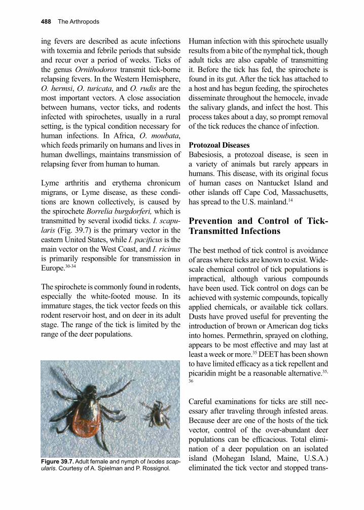
488 The Arthropods
ing fevers are described as acute infections with toxemia and febrile periods that subside and recur over a period of weeks. Ticks of the genus Ornithodoros transmit tick-borne relapsing fevers. In the Western Hemisphere, O. hermsi, O. turicata, and O. rudis are the most important vectors. A close association between humans, vector ticks, and rodents infected with spirochetes, usually in a rural setting, is the typical condition necessary for human infections. In Africa, O. moubata, which feeds primarily on humans and lives in human dwellings, maintains transmission of relapsing fever from human to human.
Lyme arthritis and erythema chronicum migrans, or Lyme disease, as these condi-tions are known collectively, is caused by the spirochete Borrelia burgdorferi, which is transmitted by several ixodid ticks. I. scapu-laris (Fig. 39.7) is the primary vector in the eastern United States, while I. pacificus is the main vector on the West Coast, and I. ricinus is primarily responsible for transmission in Europe.30-34
The spirochete is commonly found in rodents, especially the white-footed mouse. In its immature stages, the tick vector feeds on this rodent reservoir host, and on deer in its adult stage. The range of the tick is limited by the range of the deer populations.
Human infection with this spirochete usually results from a bite of the nymphal tick, though adult ticks are also capable of transmitting it. Before the tick has fed, the spirochete is found in its gut. After the tick has attached to a host and has begun feeding, the spirochetes disseminate throughout the hemocele, invade the salivary glands, and infect the host. This process takes about a day, so prompt removal of the tick reduces the chance of infection. Protozoal DiseasesBabesiosis, a protozoal disease, is seen in a variety of animals but rarely appears in humans. This disease, with its original focus of human cases on Nantucket Island and other islands off Cape Cod, Massachusetts, has spread to the U.S. mainland.14
Prevention and Control of Tick-Transmitted Infections
The best method of tick control is avoidance of areas where ticks are known to exist. Wide-scale chemical control of tick populations is impractical, although various compounds have been used. Tick control on dogs can be achieved with systemic compounds, topically applied chemicals, or available tick collars. Dusts have proved useful for preventing the introduction of brown or American dog ticks into homes. Permethrin, sprayed on clothing, appears to be most effective and may last at least a week or more.35 DEET has been shown to have limited efficacy as a tick repellent and picaridin might be a reasonable alternative.35,
36
Careful examinations for ticks are still nec-essary after traveling through infested areas. Because deer are one of the hosts of the tick vector, control of the over-abundant deer populations can be efficacious. Total elimi-nation of a deer population on an isolated island (Mohegan Island, Maine, U.S.A.) eliminated the tick vector and stopped trans-
Figure 39.7. Adult female and nymph of Ixodes scap-ularis. Courtesy of A. Spielman and P. Rossignol.
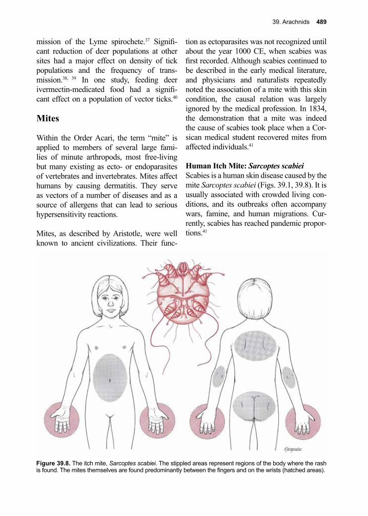
39. Arachnids 489
mission of the Lyme spirochete.37 Signifi-cant reduction of deer populations at other sites had a major effect on density of tick populations and the frequency of trans-mission.38, 39 In one study, feeding deer ivermectin-medicated food had a signifi-cant effect on a population of vector ticks.40
Mites
Within the Order Acari, the term “mite” is applied to members of several large fami-lies of minute arthropods, most free-living but many existing as ecto- or endoparasites of vertebrates and invertebrates. Mites affect humans by causing dermatitis. They serve as vectors of a number of diseases and as a source of allergens that can lead to serious hypersensitivity reactions.
Mites, as described by Aristotle, were well known to ancient civilizations. Their func-
tion as ectoparasites was not recognized until about the year 1000 CE, when scabies was first recorded. Although scabies continued to be described in the early medical literature, and physicians and naturalists repeatedly noted the association of a mite with this skin condition, the causal relation was largely ignored by the medical profession. In 1834, the demonstration that a mite was indeed the cause of scabies took place when a Cor-sican medical student recovered mites from affected individuals.41
Human Itch Mite: Sarcoptes scabieiScabies is a human skin disease caused by the mite Sarcoptes scabiei (Figs. 39.1, 39.8). It is usually associated with crowded living con-ditions, and its outbreaks often accompany wars, famine, and human migrations. Cur-rently, scabies has reached pandemic propor-tions.41
Figure 39.8. The itch mite, Sarcoptes scabiei. The stippled areas represent regions of the body where the rash is found. The mites themselves are found predominantly between the fingers and on the wrists (hatched areas).
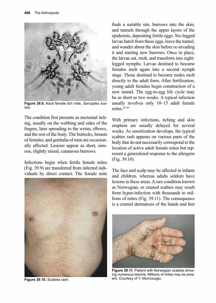
490 The Arthropods
The condition first presents as nocturnal itch-ing, usually on the webbing and sides of the fingers, later spreading to the wrists, elbows, and the rest of the body. The buttocks, breasts of females, and genitalia of men are occasion-ally affected. Lesions appear as short, sinu-ous, slightly raised, cutaneous burrows.
Infections begin when fertile female mites (Fig. 39.9) are transferred from infected indi-viduals by direct contact. The female mite
finds a suitable site, burrows into the skin, and tunnels through the upper layers of the epidermis, depositing fertile eggs. Six-legged larvae hatch from these eggs, leave the tunnel, and wander about the skin before re-invading it and starting new burrows. Once in place, the larvae eat, molt, and transform into eight-legged nymphs. Larvae destined to become females molt again into a second nymph stage. Those destined to become males molt directly to the adult form. After fertilization, young adult females begin construction of a new tunnel. The egg-to-egg life cycle may be as short as two weeks. A typical infection usually involves only 10–15 adult female mites.42-45
With primary infections, itching and skin eruption are usually delayed for several weeks. As sensitization develops, the typical scabies rash appears on various parts of the body that do not necessarily correspond to the location of active adult female mites but rep-resent a generalized response to the allergens (Fig. 39.10).
The face and scalp may be affected in infants and children, whereas adults seldom have lesions in these areas. A rare condition known as Norwegian, or crusted scabies may result from hyper-infection with thousands to mil-lions of mites (Fig. 39.11). The consequence is a crusted dermatosis of the hands and feet
Figure 39.10. Scabies rash.
Figure 39.11. Patient with Norwegian scabies show-ing numerous lesions. Millions of mites may be pres-ent. Courtesy of Y. Mumcouglu.
Figure 39.9. Adult female itch mite, Sarcoptes sca-biei.
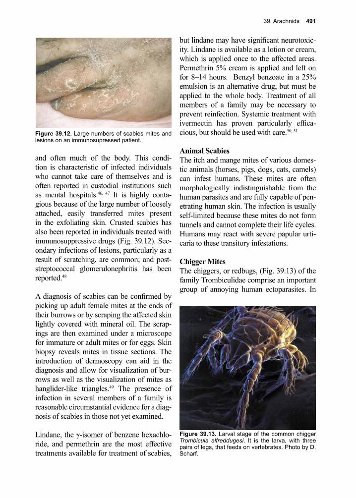
39. Arachnids 491
and often much of the body. This condi-tion is characteristic of infected individuals who cannot take care of themselves and is often reported in custodial institutions such as mental hospitals.46, 47 It is highly conta-gious because of the large number of loosely attached, easily transferred mites present in the exfoliating skin. Crusted scabies has also been reported in individuals treated with immunosuppressive drugs (Fig. 39.12). Sec-ondary infections of lesions, particularly as a result of scratching, are common; and post-streptococcal glomerulonephritis has been reported.48
A diagnosis of scabies can be confirmed by picking up adult female mites at the ends of their burrows or by scraping the affected skin lightly covered with mineral oil. The scrap-ings are then examined under a microscope for immature or adult mites or for eggs. Skin biopsy reveals mites in tissue sections. The introduction of dermoscopy can aid in the diagnosis and allow for visualization of bur-rows as well as the visualization of mites as hanglider-like triangles.49 The presence of infection in several members of a family is reasonable circumstantial evidence for a diag-nosis of scabies in those not yet examined.
Lindane, the γ-isomer of benzene hexachlo-ride, and permethrin are the most effective treatments available for treatment of scabies,
but lindane may have significant neurotoxic-ity. Lindane is available as a lotion or cream, which is applied once to the affected areas. Permethrin 5% cream is applied and left on for 8–14 hours. Benzyl benzoate in a 25% emulsion is an alternative drug, but must be applied to the whole body. Treatment of all members of a family may be necessary to prevent reinfection. Systemic treatment with ivermectin has proven particularly effica-cious, but should be used with care.50, 51
Animal ScabiesThe itch and mange mites of various domes-tic animals (horses, pigs, dogs, cats, camels) can infest humans. These mites are often morphologically indistinguishable from the human parasites and are fully capable of pen-etrating human skin. The infection is usually self-limited because these mites do not form tunnels and cannot complete their life cycles. Humans may react with severe papular urti-caria to these transitory infestations.
Chigger MitesThe chiggers, or redbugs, (Fig. 39.13) of the family Trombiculidae comprise an important group of annoying human ectoparasites. In
Figure 39.12. Large numbers of scabies mites and lesions on an immunosupressed patient.
Figure 39.13. Larval stage of the common chigger Trombicula alfreddugesi. It is the larva, with three pairs of legs, that feeds on vertebrates. Photo by D. Scharf.
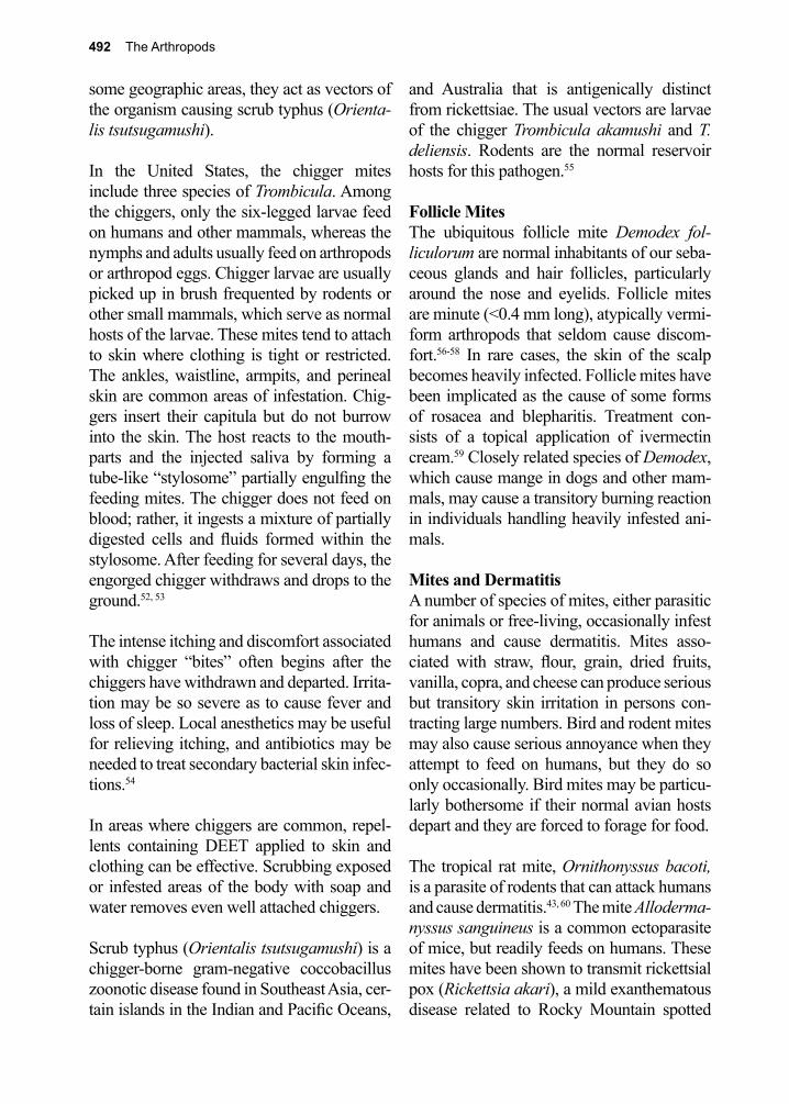
492 The Arthropods
some geographic areas, they act as vectors of the organism causing scrub typhus (Orienta-lis tsutsugamushi).
In the United States, the chigger mites include three species of Trombicula. Among the chiggers, only the six-legged larvae feed on humans and other mammals, whereas the nymphs and adults usually feed on arthropods or arthropod eggs. Chigger larvae are usually picked up in brush frequented by rodents or other small mammals, which serve as normal hosts of the larvae. These mites tend to attach to skin where clothing is tight or restricted. The ankles, waistline, armpits, and perineal skin are common areas of infestation. Chig-gers insert their capitula but do not burrow into the skin. The host reacts to the mouth-parts and the injected saliva by forming a tube-like “stylosome” partially engulfing the feeding mites. The chigger does not feed on blood; rather, it ingests a mixture of partially digested cells and fluids formed within the stylosome. After feeding for several days, the engorged chigger withdraws and drops to the ground.52, 53
The intense itching and discomfort associated with chigger “bites” often begins after the chiggers have withdrawn and departed. Irrita-tion may be so severe as to cause fever and loss of sleep. Local anesthetics may be useful for relieving itching, and antibiotics may be needed to treat secondary bacterial skin infec-tions.54
In areas where chiggers are common, repel-lents containing DEET applied to skin and clothing can be effective. Scrubbing exposed or infested areas of the body with soap and water removes even well attached chiggers.
Scrub typhus (Orientalis tsutsugamushi) is a chigger-borne gram-negative coccobacillus zoonotic disease found in Southeast Asia, cer-tain islands in the Indian and Pacific Oceans,
and Australia that is antigenically distinct from rickettsiae. The usual vectors are larvae of the chigger Trombicula akamushi and T. deliensis. Rodents are the normal reservoir hosts for this pathogen.55
Follicle MitesThe ubiquitous follicle mite Demodex fol-liculorum are normal inhabitants of our seba-ceous glands and hair follicles, particularly around the nose and eyelids. Follicle mites are minute (<0.4 mm long), atypically vermi-form arthropods that seldom cause discom-fort.56-58 In rare cases, the skin of the scalp becomes heavily infected. Follicle mites have been implicated as the cause of some forms of rosacea and blepharitis. Treatment con-sists of a topical application of ivermectin cream.59 Closely related species of Demodex, which cause mange in dogs and other mam-mals, may cause a transitory burning reaction in individuals handling heavily infested ani-mals.
Mites and DermatitisA number of species of mites, either parasitic for animals or free-living, occasionally infest humans and cause dermatitis. Mites asso-ciated with straw, flour, grain, dried fruits, vanilla, copra, and cheese can produce serious but transitory skin irritation in persons con-tracting large numbers. Bird and rodent mites may also cause serious annoyance when they attempt to feed on humans, but they do so only occasionally. Bird mites may be particu-larly bothersome if their normal avian hosts depart and they are forced to forage for food.
The tropical rat mite, Ornithonyssus bacoti, is a parasite of rodents that can attack humans and cause dermatitis.43, 60 The mite Alloderma-nyssus sanguineus is a common ectoparasite of mice, but readily feeds on humans. These mites have been shown to transmit rickettsial pox (Rickettsia akari), a mild exanthematous disease related to Rocky Mountain spotted
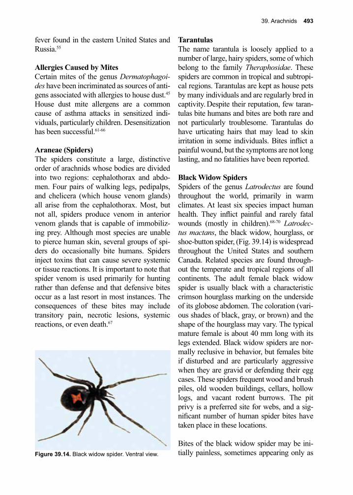
39. Arachnids 493
fever found in the eastern United States and Russia.55
Allergies Caused by MitesCertain mites of the genus Dermatophagoi-des have been incriminated as sources of anti-gens associated with allergies to house dust.45 House dust mite allergens are a common cause of asthma attacks in sensitized indi-viduals, particularly children. Desensitization has been successful.61-66
Araneae (Spiders)The spiders constitute a large, distinctive order of arachnids whose bodies are divided into two regions: cephalothorax and abdo-men. Four pairs of walking legs, pedipalps, and chelicera (which house venom glands) all arise from the cephalothorax. Most, but not all, spiders produce venom in anterior venom glands that is capable of immobiliz-ing prey. Although most species are unable to pierce human skin, several groups of spi-ders do occasionally bite humans. Spiders inject toxins that can cause severe systemic or tissue reactions. It is important to note that spider venom is used primarily for hunting rather than defense and that defensive bites occur as a last resort in most instances. The consequences of these bites may include transitory pain, necrotic lesions, systemic reactions, or even death.67
TarantulasThe name tarantula is loosely applied to a number of large, hairy spiders, some of which belong to the family Theraphosidae. These spiders are common in tropical and subtropi-cal regions. Tarantulas are kept as house pets by many individuals and are regularly bred in captivity. Despite their reputation, few taran-tulas bite humans and bites are both rare and not particularly troublesome. Tarantulas do have urticating hairs that may lead to skin irritation in some individuals. Bites inflict a painful wound, but the symptoms are not long lasting, and no fatalities have been reported.
Black Widow SpidersSpiders of the genus Latrodectus are found throughout the world, primarily in warm climates. At least six species impact human health. They inflict painful and rarely fatal wounds (mostly in children).68-70 Latrodec-tus mactans, the black widow, hourglass, or shoe-button spider, (Fig. 39.14) is widespread throughout the United States and southern Canada. Related species are found through-out the temperate and tropical regions of all continents. The adult female black widow spider is usually black with a characteristic crimson hourglass marking on the underside of its globose abdomen. The coloration (vari-ous shades of black, gray, or brown) and the shape of the hourglass may vary. The typical mature female is about 40 mm long with its legs extended. Black widow spiders are nor-mally reclusive in behavior, but females bite if disturbed and are particularly aggressive when they are gravid or defending their egg cases. These spiders frequent wood and brush piles, old wooden buildings, cellars, hollow logs, and vacant rodent burrows. The pit privy is a preferred site for webs, and a sig-nificant number of human spider bites have taken place in these locations.
Bites of the black widow spider may be ini-tially painless, sometimes appearing only as Figure 39.14. Black widow spider. Ventral view.
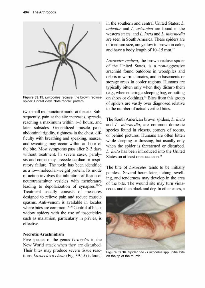
494 The Arthropods
in the southern and central United States; L. unicolor and L. arizonica are found in the western states; and L. laeta and L. intermedia are seen in South America. These spiders are of medium size, are yellow to brown in color, and have a body length of 10–15 mm.77
Loxoceles reclusa, the brown recluse spider of the United States, is a non-aggressive arachnid found outdoors in woodpiles and debris in warm climates, and in basements or storage areas in cooler regions. Humans are typically bitten only when they disturb them (e.g., when entering a sleeping bag, or putting on shoes or clothing).54 Bites from this group of spiders are vastly over diagnosed relative to the number of actual verified bites.
The South American brown spiders, L. laeta and L. intermedia, are common domestic species found in closets, corners of rooms, or behind pictures. Humans are often bitten while sleeping or dressing, but usually only when the spider is threatened or disturbed. L. laeta has been introduced into the United States on at least one occasion.78
The bite of Loxoceles tends to be initially painless. Several hours later, itching, swell-ing, and tenderness may develop in the area of the bite. The wound site may turn viola-ceous and then black and dry. In other cases, a
two small red puncture marks at the site. Sub-sequently, pain at the site increases, spreads, reaching a maximum within 1–3 hours, and later subsides. Generalized muscle pain, abdominal rigidity, tightness in the chest, dif-ficulty with breathing and speaking, nausea, and sweating may occur within an hour of the bite. Most symptoms pass after 2–3 days without treatment. In severe cases, paraly-sis and coma may precede cardiac or respi-ratory failure. The toxin has been identified as a low-molecular-weight protein. Its mode of action involves the inhibition of fusion of neurotransmitter vesicles with membranes leading to depolarization of synapses.71-74 Treatment usually consists of measures designed to relieve pain and reduce muscle spasms. Anti-venom is available in locales where bites are common.75, 76 Control of black widow spiders with the use of insecticides such as malathion, particularly in privies, is effective.
Necrotic ArachnidismFive species of the genus Loxoceles in the New World attack when they are disturbed. Their bites may produce severe tissue reac-tions. Loxoceles reclusa (Fig. 39.15) is found
Figure 39.15. Loxoceles reclusa, the brown recluse spider. Dorsal view. Note “fiddle” pattern.
Figure 39.16. Spider bite - Loxoceles spp. initial bite on the tip of the thumb.
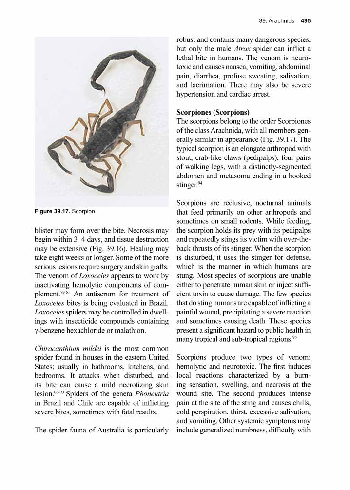
39. Arachnids 495
robust and contains many dangerous species, but only the male Atrax spider can inflict a lethal bite in humans. The venom is neuro-toxic and causes nausea, vomiting, abdominal pain, diarrhea, profuse sweating, salivation, and lacrimation. There may also be severe hypertension and cardiac arrest.
Scorpiones (Scorpions)The scorpions belong to the order Scorpiones of the class Arachnida, with all members gen-erally similar in appearance (Fig. 39.17). The typical scorpion is an elongate arthropod with stout, crab-like claws (pedipalps), four pairs of walking legs, with a distinctly-segmented abdomen and metasoma ending in a hooked stinger.94
Scorpions are reclusive, nocturnal animals that feed primarily on other arthropods and sometimes on small rodents. While feeding, the scorpion holds its prey with its pedipalps and repeatedly stings its victim with over-the-back thrusts of its stinger. When the scorpion is disturbed, it uses the stinger for defense, which is the manner in which humans are stung. Most species of scorpions are unable either to penetrate human skin or inject suffi-cient toxin to cause damage. The few species that do sting humans are capable of inflicting a painful wound, precipitating a severe reaction and sometimes causing death. These species present a significant hazard to public health in many tropical and sub-tropical regions.95
Scorpions produce two types of venom: hemolytic and neurotoxic. The first induces local reactions characterized by a burn-ing sensation, swelling, and necrosis at the wound site. The second produces intense pain at the site of the sting and causes chills, cold perspiration, thirst, excessive salivation, and vomiting. Other systemic symptoms may include generalized numbness, difficulty with
blister may form over the bite. Necrosis may begin within 3–4 days, and tissue destruction may be extensive (Fig. 39.16). Healing may take eight weeks or longer. Some of the more serious lesions require surgery and skin grafts. The venom of Loxoceles appears to work by inactivating hemolytic components of com-plement.79-85 An antiserum for treatment of Loxoceles bites is being evaluated in Brazil. Loxoceles spiders may be controlled in dwell-ings with insecticide compounds containing γ-benzene hexachloride or malathion.
Chiracanthium mildei is the most common spider found in houses in the eastern United States; usually in bathrooms, kitchens, and bedrooms. It attacks when disturbed, and its bite can cause a mild necrotizing skin lesion.86-93 Spiders of the genera Phoneutria in Brazil and Chile are capable of inflicting severe bites, sometimes with fatal results.
The spider fauna of Australia is particularly
Figure 39.17. Scorpion.
496 The Arthropods
speech and swallowing, paralysis, convul-sions, tachycardia, and myocarditis. Death may result from respiratory paralysis, often within two hours of the sting.
Children under five years of age are partic-ularly susceptible to the adverse effects of scorpion stings, and case fatality rates of 5% in Mexico, 25% in Trinidad, and 60% in the Sudan have been reported. Multiple stings, stings around the head, and stings of debili-tated individuals are also particularly serious. Scorpion stings are not uncommon in the western United States.96
Since the venom is injected into the subcu-taneous tissues, initial treatment of scorpion stings should be designed to delay absorp-tion of the toxin into the lymphatic vessels. The affected limb should be immobilized and the sting area cleaned with soap and water. Ice should be applied to the wound site and the patient kept calm. Specific scorpion anti-sera are available in areas where stings are common.97-100
Programs to reduce scorpion populations with wide-scale or focal application of per-sistent chemical pesticides have met with limited success. Elimination of rubbish piles around dwellings can reduce favored hiding and breeding places of scorpions.
References
1. Bowman, A. S.; Nuttall, P. A., Ticks: Biology, Disease and Control. Cambridge University Press. 2009.
2. Sonenshine, D. E., Biology of Ticks Volume 2. 2013.3. Smith, T.; Kilobourne, F. L., Investigations into the Nature, Causation, and Prevention of Texas
or Southern Cattle Fever. 1893.4. Marchoux, E.; Salimbeni, A., La spirillose des poules. Ann Inst Pasteur Paris 1903, 17, 569-580.5. Ross, P. H.; Milne, A. D., “Tick Fever.”. British medical journal 1904, 2 (2291), 1453-4.6. Bratton, R. L.; Corey, R., Tick-borne disease. Am Fam Physician 2005, 71 (12), 2323-30.7. de la Fuente, J., et al., Overview: Ticks as vectors of pathogens that cause disease in humans and
animals. Front Biosci 2008, 13, 6938-46.8. Meagher, K. E.; Decker, C. F., Other tick-borne illnesses: tularemia, Colorado tick fever, tick
paralysis. Dis Mon 2012, 58 (6), 370-6.9. Pujalte, G. G.; Chua, J. V., Tick-borne infections in the United States. Prim Care 2013, 40 (3),
619-35.10. Buckingham, S. C., Tick-borne diseases of the USA: Ten things clinicians should know. J Infect
2015, 71 Suppl 1, S88-96.11. Anderson, J. F., The natural history of ticks. Med Clin North Am 2002, 86 (2), 205-18.12. Spielman, A., et al., Human babesiosis on Nantucket Island, USA: description of the vector,
Ixodes (Ixodes) dammini, n. sp. (Acarina: Ixodidae). Journal of medical entomology 1979, 15 (3), 218-34.
13. Colyar, M., Tick removal techniques. Adv Nurse Pract 2006, 14 (5), 26-7.14. Spielman, A., et al., Ecology of Ixodes dammini-borne human babesiosis and Lyme disease.
Annual review of entomology 1985, 30, 439-60.15. Oliver, J. H., Jr., et al., Conspecificity of the ticks Ixodes scapularis and I. dammini (Acari:
Ixodidae). J Med Entomol 1993, 30 (1), 54-63.16. Burgdorfer, W., et al., The western black-legged tick, Ixodes pacificus: a vector of Borrelia
burgdorferi. The American journal of tropical medicine and hygiene 1985, 34 (5), 925-30.17. Jupp, P. G., et al., Attempts to transmit hepatitis B virus to chimpanzees by arthropods. South
African medical journal = Suid-Afrikaanse tydskrif vir geneeskunde 1991, 79 (6), 320-2.
39. Arachnids 497
18. Sen, S. K., The mechanism of feeding in ticks. Parasitology 1935, 27, 355-368.19. Greenstein, P., Tick paralysis. Med Clin North Am 2002, 86 (2), 441-6.20. Pecina, C. A., Tick paralysis. Semin Neurol 2012, 32 (5), 531-2.21. Vedanarayanan, V. V., et al., Tick paralysis in children: electrophysiology and possibility of
misdiagnosis. Neurology 2002, 59 (7), 1088-90.22. Diaz, J. H., A Comparative Meta-Analysis of Tick Paralysis in the United States and Australia.
Clin Toxicol (Phila) 2015, 53 (9), 874-83.23. Felz, M. W., et al., A six-year-old girl with tick paralysis. N Engl J Med 2000, 342 (2), 90-4.24. Burgdorfer, W., Tick-borne diseases in the United States: Rocky Mountain spotted fever and
Colorado tick fever. A review. Acta tropica 1977, 34 (2), 103-26.25. Burgdorfer, W.; J., A review of Rocky Mountain spotted fever (tick-borne typhus), its agent, and
its tick vectors in the United States. Entomol 1979, 12, 269-278.26. McDade, J. F., et al., Erhlichiosis, Q fever, typhus, rickettsialpox, and other rickettsioses. 1998; p
1599-1611.27. Ijdo, J. W., et al., The emergence of another tick-borne infection in the 12-town area around
Lyme, Connecticut: Human Granulocytic Ehrlichiosis. Dis 181 2000, 4.28. Diuk-Wasser, M. A., et al., Coinfection by Ixodes Tick-Borne Pathogens: Ecological,
Epidemiological, and Clinical Consequences. Trends Parasitol 2016, 32 (1), 30-42.29. Burgdorfer, W., et al., The epidemiology of relapsing fevers. 1976; p 191-200.30. Burgdorfer, W., et al., Lyme disease-a tick-borne spirochetosis? Science (New York, N.Y.) 1982,
216 (4552), 1317-9.31. Burgdorfer, W., et al., Erythema chronicum migrans--a tickborne spirochetosis. Acta tropica
1983, 40 (1), 79-83.32. Basler, E. M., et al., Natural distribution of Ixodes dammini spirochete. Science 1983, 220, 321-
322.33. Lane, R. S., et al., Lyme borreliosis: relation of its causative agent to its vectors and hosts in
North America and Europe. Annual review of entomology 1991, 36, 587-609.34. Steere, A. C., et al., The spirochetal etiology of Lyme disease. The New England journal of
medicine 1983, 308 (13), 733-40.35. Pages, F., et al., Tick repellents for human use: prevention of tick bites and tick-borne diseases.
Vector Borne Zoonotic Dis 2014, 14 (2), 85-93.36. Insect repellents. Med Lett Drugs Ther 2016, 58 (1498), 83-5.37. Kugeler, K. J., et al., Will Culling White-Tailed Deer Prevent Lyme Disease? Zoonoses Public
Health 2016, 63 (5), 337-45.38. Kilpatrick, H. J., et al., The relationship between deer density, tick abundance, and human cases
of Lyme disease in a residential community. J Med Entomol 2014, 51 (4), 777-84.39. Rand, P. W., et al., Abundance of Ixodes scapularis (Acari: Ixodidae) after the complete removal
of deer from an isolated offshore island, endemic for Lyme Disease. J Med Entomol 2004, 41 (4), 779-84.
40. Pound, J. M., et al., Systemic treatment of white-tailed deer with ivermectin-medicated bait to control free-living populations of lone star ticks (Acari:Ixodidae). J Med Entomol 1996, 33 (3), 385-94.
41. Busvine, J. R., Insects, Hygiene, and History. Athlone Press London 1976.42. Mellanby, K., Transmission of Scabies. Br Med J 1941, 2 (4211), 405-6.43. Rosamilia, L. L., Scabies. Semin Cutan Med Surg 2014, 33 (3), 106-9.44. Walton, S. F., et al., Scabies: new future for a neglected disease. Advances in parasitology 2004,
57, 309-76.45. Romani, L., et al., Prevalence of scabies and impetigo worldwide: a systematic review. Lancet
Infect Dis 2015, 15 (8), 960-7.46. Hatter, A. D., et al., Case report of individual with cutaneous immunodeficiency and novel 1p36
duplication. Appl Clin Genet 2016, 9, 1-4.47. Karthikeyan, K., Crusted scabies. Indian J Dermatol Venereol Leprol 2009, 75 (4), 340-7.48. Guldbakke, K. K.; Khachemoune, A., Crusted scabies: a clinical review. J Drugs Dermatol 2006,
5 (3), 221-7.
498 The Arthropods
49. Park, J. H., et al., The diagnostic accuracy of dermoscopy for scabies. Ann Dermatol 2012, 24 (2), 194-9.
50. Goldust, M., et al., Treatment of scabies: Comparison of permethrin 5% versus ivermectin. J Dermatol 2012, 39 (6), 545-7.
51. Romani, L., et al., Mass Drug Administration for Scabies Control in a Population with Endemic Disease. N Engl J Med 2015, 373 (24), 2305-13.
52. Sasa, M., Biology of chiggers. Annu Rev Entomol 1961, 6 221-244.53. Audy, J. R., Red Mites and Typhus. Athlone Press London 1968.54. Schulert, G. S.; Gigante, J., Summer penile syndrome: an acute hypersensitivity reaction. J
Emerg Med 2014, 46 (1), e21-2.55. Fuller, H. S.; J., Studies of rickettsial pox: Life cycle of the mite vector Allodermanyssus
sanguineus. I Am 1954, 59, 236-239.56. Elston, C. A.; Elston, D. M., Demodex mites. Clin Dermatol 2014, 32 (6), 739-43.57. Chen, W.; Plewig, G., Human demodicosis: revisit and a proposed classification. Br J Dermatol
2014, 170 (6), 1219-25.58. Cheng, A. M., et al., Recent advances on ocular Demodex infestation. Curr Opin Ophthalmol
2015, 26 (4), 295-300.59. Ali, S. T., et al., The treatment of rosacea with topical ivermectin. Drugs Today (Barc) 2015, 51
(4), 243-50.60. Cafiero, M. A., et al., Dermatitis by Tropical Rat Mite, Ornithonyssus bacoti (Mesostigmata,
Macronyssidae) in Italian city-dwellers: a diagnostic challenge. J Eur Acad Dermatol Venereol 2015.
61. Arlien, L. G.; Morgan, M. S., Biology, ecology, and prevelance of dust mites. Immunol Allergy Clin North Am 2003, 23, 443-468.
62. Milian, E., et al., Allergy to house dust mites and asthma. 2004, 23 47-57.63. Bessot, J. C.; Pauli, G., Mite allergens: an overview. Eur Ann Allergy Clin Immunol 2011, 43 (5),
141-56.64. Huss, K., et al., House dust mite and cockroach exposure are strong risk factors for positive
allergy skin test responses in the Childhood Asthma Management Program. J Allergy Clin Immunol 2001, 107 (1), 48-54.
65. Raulf, M., et al., Mites and other indoor allergens - from exposure to sensitization and treatment. Allergo J Int 2015, 24 (3), 68-80.
66. Ken, K. M., et al., Dermatoses associated with mites other than Sarcoptes. Semin Cutan Med Surg 2014, 33 (3), 110-5.
67. Juckett, G., Arthropod bites. Am Fam Physician 2013, 88 (12), 841-7.68. Vetter, R. S.; Isbister, G. K., Medical aspects of spider bites. Annu Rev Entomol 2008, 53, 409-
29.69. Isbister, G. K.; Fan, H. W., Spider bite. Lancet 2011, 378 (9808), 2039-47.70. Kang, J. K., et al., Spiders in dermatology. Semin Cutan Med Surg 2014, 33 (3), 123-7.71. Nicholson, G. M.; Graudins, A., Spiders of medical importance in the Asia-Pacific: atracotoxin,
latrotoxin and related spider neurotoxins. Clin Exp Pharmacol Physiol 2002, 29 (9), 785-94.72. Maretic, Z., et al., Venomous Animals and Their Venoms. 1971; p 299-309.73. Russell, F. E., Venom Poisoning. Rational drug therapy 1971, 5 (8), 1-7.74. Sudhof, T. C., alpha-Latrotoxin and its receptors: neurexins and CIRL/latrophilins. . Annu Rev
Neurosci 2001, 24, 933-62.75. Monte, A. A., Black widow spider (Latrodectus mactans) antivenom in clinical practice. Curr
Pharm Biotechnol 2012, 13 (10), 1935-9.76. Monte, A. A., et al., A US perspective of symptomatic Latrodectus spp. envenomation and
treatment: a National Poison Data System review. Ann Pharmacother 2011, 45 (12), 1491-8.77. Furbee, R. B., et al., Brown recluse spider envenomation. Clin Lab Med 2006, 26 (1), 211-26, ix-
x.
39. Arachnids 499
78. Levi, H. W., et al., Biology and control of the South American brown spider Loxosceles laeta (Nicolet) in a North American focus. Am Med Hyg 1964, 13, 132-136.
79. Rhoads, J., Epidemiology of the brown recluse spider bite. J Am Acad Nurse Pract 2007, 19 (2), 79-85.
80. Wendell, R. P., Brown recluse spiders: a review to help guide physicians in nonendemic areas. South Med J 2003, 96 (5), 486-90.
81. Vetter, R. S.; Barger, D. K., An infestation of 2,055 brown recluse spiders (Araneae: Sicariidae) and no envenomations in a Kansas home: implications for bite diagnoses in nonendemic areas. J Med Entomol 2002, 39 (6), 948-51.
82. Vetter, R., Identifying and misidentifying the brown recluse spider. Dermatology online journal 1999, 5 (2), 7.
83. Swanson, D. L.; Vetter, R. S., Bites of brown recluse spiders and suspected necrotic arachnidism. The New England journal of medicine 2005, 352 (7), 700-7.
84. Williams, S. T., et al., Severe intravascular hemolysis associated with brown recluse spider envenomation. A report of two cases and review of the literature. American journal of clinical pathology 1995, 104 (4), 463-7.
85. Zeglin, D., Brown recluse spider bites. Am J Nurs 2005, 105 (2), 64-8.86. Spielman, A.; Levi, H. W., Probable envenomation by Chiracanthium mildei; a spider found in
houses. The American journal of tropical medicine and hygiene 1970, 19 (4), 729-32.87. Nimorakiotakis, B.; Winkel, K. D., The funnel web and common spider bites. Aust Fam
Physician 2004, 33 (4), 244-51.88. Isbister, G. K., et al., Antivenom treatment in arachnidism. J Toxicol Clin Toxicol 2003, 41 (3),
291-300.89. Isbister, G. K., et al., Funnel-web spider bite: a systematic review of recorded clinical cases. Med
J Aust 2005, 182 (8), 407-11.90. Isbister, G. K.; Gray, M. R., Bites by Australian mygalomorph spiders (Araneae,
Mygalomorphae), including funnel-web spiders (Atracinae) and mouse spiders (Actinopodidae: Missulena spp). Toxicon 2004, 43 (2), 133-40.
91. Graudins, A., et al., Red-back spider (Latrodectus hasselti) antivenom prevents the toxicity of widow spider venoms. Ann Emerg Med 2001, 37 (2), 154-60.
92. White, J., Envenoming and antivenom use in Australia. Toxicon : official journal of the International Society on Toxinology 1998, 36 (11), 1483-92.
93. Currie, B. J., Clinical toxicology: a tropical Australian perspective. Therapeutic drug monitoring 2000, 22 (1), 73-8.
94. Keegan, H. L., Scorpions of medical importance. 1980.95. Del Brutto, O. H., Neurological effects of venomous bites and stings: snakes, spiders, and
scorpions. Handb Clin Neurol 2013, 114, 349-68.96. LoVecchio, F.; McBride, C., Scorpion envenomations in young children in central Arizona. J
Toxicol Clin Toxicol 2003, 41 (7), 937-40.97. Tuuri, R. E.; Reynolds, S., Scorpion envenomation and antivenom therapy. Pediatr Emerg Care
2011, 27 (7), 667-72; quiz 673-5.98. Bawaskar, H. S.; Bawaskar, P. H., Scorpion sting: update. J Assoc Physicians India 2012, 60, 46-
55.99. Bosnak, M., et al., Severe scorpion envenomation in children: Management in pediatric intensive
care unit. Hum Exp Toxicol 2009, 28 (11), 721-8.100. Chippaux, J. P., Emerging options for the management of scorpion stings. Drug Des Devel Ther
2012, 6, 165-73.
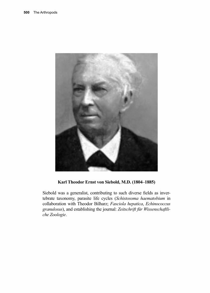
500 The Arthropods
Karl Theodor Ernst von Siebold, M.D. (1804–1885)
Siebold was a generalist, contributing to such diverse fields as inver-tebrate taxonomy, parasite life cycles (Schistosoma haematobium in collaboration with Theodor Bilharz; Fasciola hepatica, Echinococcus granulosus), and establishing the journal: Zeitschrift für Wissenschaftli-che Zoologie.
40. Other Arthropods of Medical Importance 501
40. Other Arthropods of Medical Importance
Butterflies and Moths: Order LepidopteraSeveral species of larvae or caterpillars of the order Lepidoptera are covered with hollow, sharply pointed hairs containing a toxin that may cause severe dermatitis. Contact occurs when an individual handles the caterpil-lars or inhales the “hairs,” which are blown about after the larva molts. In the northeast-ern United States, the gypsy moth, Lymantria dispar, has been responsible for defoliation of enormous areas of forest. The “population explosion” of the caterpillars of this species has led to periodic outbreaks of pruritic der-matitis, primarily among school children.1 Similar outbreaks of dermatitis have been reported from the Southeastern and South Central U. S., attributed to the caterpillars of the puss moth Megalopyge opercalis.
In a Mexican outbreak, severe dermatitis was initially attributed to scabies, but eventually associated with contact with adult moths of the species Hylesia alinda. Populations of this normally rare species rapidly expanded after its natural predators were killed by a hurricane sweeping the island.2 Lepidopteran pests include the Io moth, saddleback cater-pillar, the puss caterpillar and many members of the family Limacodidae.
Individuals working with lepidopterans may become sensitized to the scales of adult moths or butterflies. Repeated exposure may pro-duce severe bronchospasm and even asthma.
There is an increasing awareness of the ubiq-uity and potentially serious consequences of exposure to a wide range of these larvae.3-5
Beetles: Order Coleoptera
Within the order Coleoptera, which comprises a large group of insects, only a few families contain members of medical importance. Certain scavenger beetles of the families Der-mestidae, Silphidae, and Staphylinidae feed on feces and carrion and mechanically trans-mit pathogenic organisms. Adult beetles of the family Meloidae, the blister beetles, pro-duce a vesicant (cantharidin) that may cause blistering or a severe burning sensation on contact with the skin or mucous membranes. Inadvertent ingestion of beetle larvae, a con-dition termed canthariasis, may produce tran-sient gastrointestinal (GI) discomfort.
Many beetles, particularly those that feed on feces, act as intermediate hosts for helmin-thic parasites of humans and other animals. Members of the family Scarabaeidae are intermediate hosts of the spiny-headed worm Macracanthorhynchus hirudinaceus, a para-site of pigs, which rarely infects humans. The tapeworms Hymenolepis nana and Hymenol-epis diminuta develop in grain beetles of the family Tenebrionidae.
Cockroaches: Order Blattodea
Blattodea is a large, diverse order of primi-tive, successful insects (over 4,000 species, worldwide) that includes the grasshoppers and crickets, as well as cockroaches. The cockroaches are included in a single family, the Blattidae, with several members closely associated with human habitations. All mem-bers of this group have chewing mouthparts.
The female cockroach encloses her eggs in a bean-shaped case called an ootheca. Some species retain the ootheca internally until the eggs hatch, others carry it externally for sev-eral weeks, and still others drop the ootheca soon after it is formed. After hatching, the young, wingless, feeding nymphs begin to
502 The Arthropods
undergo staged development. Some spe-cies progress through as many as 13 nymph stages, each being wingless and somewhat larger than its predecessor, until the final molt produces the winged adult. With its series of wingless nymph stages, the cockroach is a classic example of an insect developing by incomplete metamorphosis.
Most cockroach species do not invade homes, confining themselves to outdoor habitats, although in the United States eleven species of cockroaches do invade human habitats. The most common is the German cockroach or croton bug, Blatella germanica, a small (<16 mm), light brown species. The Ameri-can cockroach or palmetto bug Periplaneta americana, is, in fact, an African species now found worldwide. It is a large (30–40 mm) reddish-brown insect with long wings. It is found in and around homes, farms, restau-rants, stores, and warehouses. Other species that may infest homes are the Oriental cock-roach, B. orientalis, the Australian cockroach, P. australasiae, and the brown-banded cock-roach Supella longipalpa.
Most of these species are cosmopolitan, having been distributed by ship traffic starting with the earliest voyages. In general, domes-tic species are omnivorous. They feed on a wide variety of nutrients including: paper; bookbindings; and human and animal feces. They serve as mechanical vectors of patho-gens, carrying infectious agents from feces to food.6
The presence of cockroaches is usually asso-ciated with a breakdown of general sanita-tion. Exposed foods or poor packaging and storage, open garbage, darkness and moisture are all conducive to the development of large cockroach populations. Initial infestations may be introduced with foodstuffs or migra-tion from adjoining dwellings. In apartment buildings, the insecticide treatment of one apartment may cause the migration of cock-roaches to adjoining, untreated apartments.
Cockroach baits paired with sanitation, best control cockroach infestations. Although cockroaches are resistant to a number of insecticides in some areas, compounds for control are commercially available. Coupled with improved housekeeping, treatment with these agents can be sufficient, although heavy infestations require repeated treatments by professional exterminators.
Cockroaches, because of their close associa-tion with sewage and garbage, may serve as paratenic hosts for various pathogens.6 Long-term exposure to cockroaches or to their shed exoskeletons can induce asthma-like symp-toms.7, 8
Centipedes: Class Chilopoda
The centipedes of the class Chilopoda are worm-like, segmented creatures with a dis-tinct head and paired appendages on each of 15–100 or more segments. They have a pair of poisonous claws, or maxillipeds, on the first segment after the head, which are used for capturing prey. Most centipedes are pre-daceous insectivores, and humans are some-times bitten accidentally. Centipede bites may be locally painful, causing transient swelling at the site of the bite. No long-term complica-tions are usually associated with these bites.9
Crustacea
The crustacea include many species that serve as intermediate hosts of parasites of humans and other animals. These organisms are dis-cussed in the other, relevant chapters.
Tongue Worms: Class Pentas-tomida
The pentastomids, or tongue worms, of the class Pentastomida are a small group of para-sites of uncertain origin and affinity. Because their larvae superficially resemble the larvae of mites, they have been included among the
40. Other Arthropods of Medical Importance 503
hatch in the gut, yielding a migratory larva that pierces the stomach wall and encysts in host tissue. When the intermediate host is eaten by the definitive host, the larvae mature.10
In humans, encysted larvae have been found in the lungs, liver, intestine, spleen, and other internal organs.11, 12 There may be rare cases of symptomatic human disease from infec-tion with tongue worms, but most cases are usually identified at autopsy without prior attributable symptoms.11-15
Arthropoda, but they probably evolved early from annelid or arthropod ancestral stocks. They were first noted in the nasal cavities of dogs and horses during the eighteenth century and were later described in human autopsy material as insect larvae.
The adult tongue worms are blood sucking, endoparasitic, legless vermiform inhabitants of the respiratory system of reptiles, birds, and mammals. Eggs fertilized within the host emerge through the respiratory tract. After being eaten by an intermediate host, they
References
1. Allen, V. T., et al., Gypsy moth caterpillar dermatitis--revisited. J Am Acad Dermatol 1991, 24 (6 Pt 1), 979-81.
2. Fernandez, G., et al., Epidemic dermatitis due to contact with a moth in Cozumel, Mexico. The American journal of tropical medicine and hygiene 1992, 46 (5), 560-3.
3. Diaz, J. H., The evolving global epidemiology, syndromic classification, management, and prevention of caterpillar envenoming. The American journal of tropical medicine and hygiene 2005, 72 (3), 347-57.
4. Hossler, E. W., Caterpillars and moths. Dermatol Ther 2009, 22 (4), 353-66.5. Hossler, E. W., Caterpillars and moths: Part II. Dermatologic manifestations of encounters with
Lepidoptera. J Am Acad Dermatol 2010, 62 (1), 13-28; quiz 29-30.6. Graczyk, T. K., et al., Mechanical transmission of human protozoan parasites by insects. Clin
Microbiol Rev 2005, 18 (1), 128-32.7. Gao, P., Sensitization to cockroach allergen: immune regulation and genetic determinants. Clin
Dev Immunol 2012, 2012, 563760.8. Pomes, A.; Arruda, L. K., Investigating cockroach allergens: aiming to improve diagnosis and
treatment of cockroach allergic patients. Methods 2014, 66 (1), 75-85.9. Sanaei-Zadeh, H., Centipede bite. Eur Rev Med Pharmacol Sci 2014, 18 (7), 1106-7.10. Drabick, J. J., Pentastomiasis. Rev Infect Dis 1987, 9, 1087-1094.11. Tappe, D.; Buttner, D. W., Diagnosis of human visceral pentastomiasis. PLoS Negl Trop Dis
2009, 3 (2), e320.12. Tappe, D., et al., Imported Armillifer pentastomiasis: report of a symptomatic infection in The
Netherlands and mini-review. Travel Med Infect Dis 2014, 12 (2), 129-33.13. Latif, B., et al., Human pentastomiasis caused by Armillifer moniliformis in Malaysian Borneo.
Am J Trop Med Hyg 2011, 85 (5), 878-81.14. Sulyok, M., et al., Ocular pentastomiasis in the Democratic Republic of the Congo. PLoS Negl
Trop Dis 2014, 8 (7), e3041.15. Tappe, D., et al., Molecular Diagnosis of Abdominal Armillifer grandis Pentastomiasis in the
Democratic Republic of Congo. J Clin Microbiol 2015, 53 (7), 2362-4.
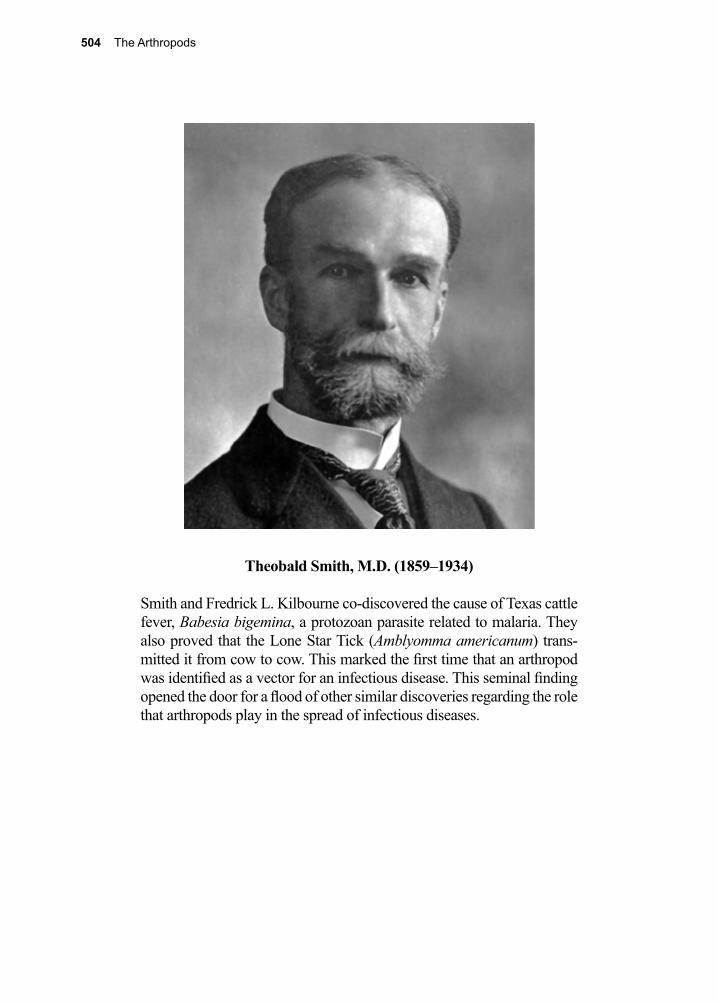
504 The Arthropods
Theobald Smith, M.D. (1859–1934)
Smith and Fredrick L. Kilbourne co-discovered the cause of Texas cattle fever, Babesia bigemina, a protozoan parasite related to malaria. They also proved that the Lone Star Tick (Amblyomma americanum) trans-mitted it from cow to cow. This marked the first time that an arthropod was identified as a vector for an infectious disease. This seminal finding opened the door for a flood of other similar discoveries regarding the role that arthropods play in the spread of infectious diseases.
505Appendix A: Procedures for Collecting Clinical Specimens for Diagnosing Protozoan and Helminthic Parasites
Appendix A: Procedures for Collect-ing Clinical Specimens for Diagnosing Protozoan and Helminthic Parasites
There is no substitute for a well-trained labo-ratory diagnostic technician, even with the advent of sensitive and specific serologi-cal and molecular diagnostic methods, such as ELISA, PCR, and NAAT. But even the best-trained personnel cannot make up for an improper sample delivered to the laboratory in the expectation of securing the diagnosis. Stool, blood, urine and tissue samples must be treated as the most important link between the patient and the correct diagnosis of their parasitic illness. The following advice out-lines standard procedure for insuring that the diagnostic laboratory receives the right amount and type of patient specimen.
Stool Specimens
Proper collection and delivery of stool speci-mens is a critical aspect of any diagnostic procedure relying on stool examination. The clinician can control the quality of this aspect, and in doing so, will insure both the reliabil-ity and accuracy of any test they recommend, regardless of whether that test is carried out in-house or at a regional diagnostic facility.
1. Fresh, unpreserved feces should be obtained and transported to the labora-tory immediately. Fresh specimens are preferred for examinations for tropho-zoites, and are required when tests for Strongyloides stercoralis larvae are to be performed.
2. Unpreserved feces should be examined within one hour after passage, espe-cially if the stool is loose or watery, and might contain trophozoites of patho-genic amoebae. Examination of formed stool may be delayed for a short time, but must be completed on the day on
which the specimen is received in the laboratory. If prompt examination or proper fixation cannot be carried out, formed specimens may be refrigerated for 1-2 days.
3. If specimens are delayed in reaching the laboratory, or if they cannot be exam-ined promptly (such as those received at night, on weekends, or when no para-sitologist is available), portions should be preserved in fixatives such as 8% aqueous formalin or formol-saline, or with polyvinyl alcohol (PVA). Forma-lin preserves cysts, eggs and larvae for wet-mount examination or for concen-tration tests. PVA-fixative preserves trophozoites, cysts, and eggs for perma-nent staining. A ratio of one part feces to three parts of fixative is recommended. The specimen may be placed in fixatives in the laboratory, or the patient may be provided with fixatives and instructions for collection and preservation of their own specimens.
Stool Examination
Stool specimens may be successfully exam-ined by any one of the three methods listed below. The advantages and limitations of each technique must be recognized.
1. Saline mounts are of value primarily for demonstrating the characteristic motility of amoebae and flagellates. In addition, seeing red cells inside a tro-phozoite of an amoeba is indicative of infection with Entamoeba histolytica. These organisms may be found in fresh stools, or occasionally in bloody mucus adhering to the surface of formed stools. Material should be obtained from sev-eral parts of the specimen. An iodine stain (a drop of 1% iodine in 2% potas-sium iodide) mixed with a stool suspen-
506 Appendix A: Procedures for Collecting Clinical Specimens for Diagnosing Protozoan and Helminthic Parasites
sion in saline solution facilitates identi-fication of protozoan cysts, but it kills and distorts trophozoites.
2. Concentration techniques, useful for detecting small numbers of cysts and helminth eggs, may be used on unpre-served stool specimens, those preserved in aqueous formalin or formol-saline, or on PVA-fixed material.
3. Stained, thin smears of feces should be made, if possible, on all specimens obtained fresh or fixed in PVA. If prop-erly prepared, they comprise the single most productive stool examination for protozoa. Smears may be stained with Wheatly-Gomori’s Trichrome solution or with iron-hematoxylin (see Appendix B for colors of each). Any outstanding examples of positive specimens should be retained in a permanent file and used for future reference.
Number of Specimens Examined and Appropriate Intervals
1. To detect amoebae, a minimum of three specimens should be examined; if these samples (obtained preferably at intervals of 2-3 days) are negative and amoebic infection remains a diagnos-tic consideration, additional specimens should be examined.
2. With suspected giardiasis, NAAT and antigen capture ELISA are the currently recommended tests. When employ-ing light microscopy, three specimens should initially be examined. If they are negative, additional specimens should be obtained at weekly intervals for three weeks.
3. A single concentrate from one day’s worth of stool is frequently sufficient to
detect intestinal helminthic infections of clinical importance. With very light infections due to Schistosoma spp. few or no eggs may be found in the feces or urine. Strongyloides stercoralis may also require concentrating the specimen for diagnosis, but this method is not always reliable; various fecal culture methods may also be used.
4. Examination after treatment, under most circumstances, should be delayed until one month after completion of therapy (three months after treatment for schistosomiasis or tapeworms).
Examination of Blood
1. Smears for malaria should consist of both thick and thin films. It is important that all involved laboratory personnel be aware of the technique for making thick films as they are useless if improp-erly made. Smears should be stained with Giemsa solution and a minimum of 100 contiguous microscopic fields examined before a specimen is reported as “negative.” If the first specimen is negative, additional thick and thin films should be taken every six hours for the first 24 hours after admission.
2. When examining for filarial infection, the possibility of diurnal or noctur-nal periodicity of microfilariae in the peripheral blood must be taken into account, and specimens should be taken every six hours for the first 24 hours after admission, as with malaria. Thick smears or blood concentration methods are most likely to demonstrate infection. Smears should be done in conjunction with the Knott’s test (see Appendix B).
507Appendix A: Procedures for Collecting Clinical Specimens for Diagnosing Protozoan and Helminthic Parasites
Serologic Methods
A variety of immunodiagnostic methods may serve as useful adjuncts to the clinical diag-nosis of parasitic infections. In some cases, serologic methods may be the only labora-tory recourse for making a diagnosis. Certain serologic tests provide a high degree of diag-nostic accuracy; however, mixed infections, cross reacting antigens by related and unre-lated parasites, and other diseases or physi-ologic conditions may interfere with the diag-nostic accuracy of a given test.
Western blot analysis and ELISA will undoubtedly continue to offer the clinician sensitive, reliable methods for diagnosing parasitic infections. Positive tests revealing the presence of specific antibodies are indi-rect evidence of infection, no matter how good the method. Tests employing antibody capture techniques, in which monoclonal antibodies are used to select for a single class of immunoglobulin increases the likelihood of a true positive result.
Most serum specimens may be shipped frozen or preserved with thimerosal to a final concentration of 1:10,000 to a state public health laboratory for forwarding to the Cen-ters for Disease Control (CDC) and Preven-tion in Atlanta, Georgia. The vial, containing at least 2 ml of serum, should indicate the pre-servative used.
Nucleic Acid Amplification Tests (NAAT) and Polymerase chain reaction (PCR)
The advent of amplifying parasite DNA in stool, blood, and tissue samples has widened the range of parasite detection methods. Reli-able NAAT tests have been developed for malaria, most species that cause leishmani-asis, toxoplasmosis, giardiasis, amoebiasis (Entamoeba histolytica and E. dispar), try-panosomiasis, and many others. These tests have already become the primary diagnostic tests in many clinical centers.
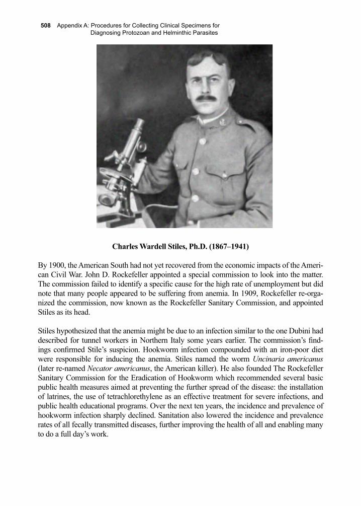
508 Appendix A: Procedures for Collecting Clinical Specimens for Diagnosing Protozoan and Helminthic Parasites
Charles Wardell Stiles, Ph.D. (1867–1941)
By 1900, the American South had not yet recovered from the economic impacts of the Ameri-can Civil War. John D. Rockefeller appointed a special commission to look into the matter. The commission failed to identify a specific cause for the high rate of unemployment but did note that many people appeared to be suffering from anemia. In 1909, Rockefeller re-orga-nized the commission, now known as the Rockefeller Sanitary Commission, and appointed Stiles as its head.
Stiles hypothesized that the anemia might be due to an infection similar to the one Dubini had described for tunnel workers in Northern Italy some years earlier. The commission’s find-ings confirmed Stile’s suspicion. Hookworm infection compounded with an iron-poor diet were responsible for inducing the anemia. Stiles named the worm Uncinaria americanus (later re-named Necator americanus, the American killer). He also founded The Rockefeller Sanitary Commission for the Eradication of Hookworm which recommended several basic public health measures aimed at preventing the further spread of the disease: the installation of latrines, the use of tetrachlorethylene as an effective treatment for severe infections, and public health educational programs. Over the next ten years, the incidence and prevalence of hookworm infection sharply declined. Sanitation also lowered the incidence and prevalence rates of all fecally transmitted diseases, further improving the health of all and enabling many to do a full day’s work.
Appendix B: Laboratory Diagnostic Methods 509
Appendix B: Laboratory Diagnostic Methods
In recent years, a battery of cutting-edge diag-nostic modalities have emerged, making the identification of many infectious diseases straightforward, without requiring any more skill than being able to read the instructions and to execute them. Microscopy training is not needed in these instances. Each of the preceding chapters attests to this fact, with a robust sampling of modern serological and molecular diagnostic strategies.
The vast majority of parasitology laborato-ries in hospitals and outpatient health clin-ics throughout the world continue to rely on more traditional approaches for the diagnosis of eukaryotic parasites. In these instances, microscopy remains the gold standard for pathogen identification. This chapter serves as a standard reference for these time-hon-ored laboratory procedures.
Part I deals with unpreserved specimens and Part II with preserved specimens. There is no single method that efficiently renders all stages of all parasites available for micro-scopic identification; several tests must often be performed to obtain optimal results.
Unpreserved Stool Specimens
Ideally, stool specimens should be less than one hour old when first examined, although this may not always be possible. Stools that are up to 24 hours old may still be useful for recovering protozoan cysts, larvae and eggs of helminths, but trophozoites rarely survive that long. A confounding factor when exam-ining specimens left at room temperature for more than 24 hours is that some parasites can grow and develop. Refrigeration helps prevent this problem. Stools should not be frozen, as that would alter the morphology of the organisms examined.
Because of day-to-day variability in the quan-tity of various stages of parasite shed by an infected individual, parasites may not be pres-ent in a single specimen, particularly when the infection is light. A total of three specimens collected on consecutive days are suggested when attempting to detect most enteric infec-tions by visual microscopy. Some parasites (e.g., the schistosomes and Giardia lamblia) often require more specimens for detection or the use of more sensitive diagnostic modali-ties.
Barium or mineral oil interferes with identi-fication of parasites. Patients should not be subjected to radiographic studies involving barium or given laxatives containing min-eral oil until the stool specimens have been obtained.
Direct Examination
Gross examination:1. Observe and record the appearance of the
entire specimen, noting the color, consis-tency, and odor.
2. Examine the specimen for the presence of living parasites.
3. Perform a microscopic examination.4. Examine a direct smear of the material.
The direct examination is effective for diag-nosing living parasites (e.g., Entamoeba his-tolytica, Giardia lamblia, Strongyloides ster-coralis), and should be performed on loose, diarrheic, or purged stool. When motile amoe-bae are found on a direct smear, a stained preparation should also be examined for the definitive diagnosis. If the amoeba contains erythrocytes within the cytoplasm, it is indic-ative of infection caused by E. histolytica.
If the specimen appears negative, as may occur with light infections, the sample should be concentrated.
1. Dip a wooden applicator stick into the specimen to coat the tip of it with stool.
510 Appendix B: Laboratory Diagnostic Methods
Solution TimeEthanol 95% or 100% Two rinsesEthanol 100% 1 minuteXylol 1 minute
Mount the coverslip in an appropriate mount-ing medium and examine under a microscope.
Concentration Methods
Sedimentation by Centrifugation: Formaldehyde-Ethyl Acetate MethodSedimentation by concentration and expo-sure to formaldehyde-ethyl acetate concen-trates cysts and eggs of parasites, but debris and ether-soluble materials localize in the formaldehyde-ether interface or the ether layer in the top of the tube. This process destroys trophozoites, as they disintegrate in ethyl acetate.
1. Mix stool 1:10 with H2O.2. Strain through a single layer of gauze
into a 15-ml centrifuge tube.3. Centrifuge the strained stool (1 minute
at 2000 rpm) and discard the superna-tant.
4. Wash the sediment once with H2O.5. Repeat steps 3 and 4.6. Discard the supernatant and save the
sediment.7. Add 10 ml of 7.5% formaldehyde to
the sediment.8. Let stand 10–30 minutes.9. Add approximately 3 ml of ethyl ace-
tate, plug the tubes with stoppers, and agitate the mixture vigorously.
10. Remove the stoppers and centrifuge the tubes at 1500 rpm for 1 minute.
11. Gently loosen the debris from the tube wall with an applicator stick, being careful not to disturb the pellet.
12. Discard the supernatant.13. Examine the sediment microscopi-
cally.14. Add a drop of 70% ethanol-iodine
solution (Lugol’s solution) and exam-
2. Smear the stool onto a clear glass micro-scope slide on which a drop of normal saline solution has been placed and over-lay with a coverslip. Smears must be thin enough to facilitate microscopic exami-nation.
Staining the Direct smear The Wheatly-Gomori trichrome reagent stains the protozoan nuclei red to dark blue, the cytoplasm a lighter blue, and the back-ground material green. Trophozoites and cysts tend to shrink away from the back-ground material and are therefore relatively easy to locate.The Wheatly-Gomori trichrome reagent con-sists of:
• 6.0 g chromotrope 2R• 0.5 g aniline blue CI42755• 0.25 g dodecatungstophosphoric acid
AR in 3 ml glacial acetic acid
Wheatly-Gomori trichrome stain is applied to a thin smear of stool on a coverslip, and the coverslip is immersed sequentially in the solutions enumerated below for the pre-scribed lengths of time.
Solution TimeSchaudinn’s fixative 5 minutes at
50+˚C, or 1 hour at room temperature
Ethanol-iodine 70% 1 minuteEthanol 70% 1 minuteEthanol 70% 1 minuteTrichrome stain 2–8 minutesEthanol 90% 10–20 seconds
(acidified)
To remove excess stain, briefly dip the cov-erslip in destaining solution once or twice. Rinse in 90% ethanol to stop the process. Thin smears destain quickly; thicker ones may require three or four dips to obtain opti-mal differentiation. The process is as follows.
Appendix B: Laboratory Diagnostic Methods 511
ine again if internal structures of cysts are not recognized on first examina-tion.
Sedimentation by Gravity:The water sedimentation test is used primarily for the concentration and recovery of Schis-tosoma mansoni and Schistosoma japoni-cum eggs, and it is effective for determining their viability. An entire day’s worth of stool should be examined in a single test because schistosome eggs are shed sporadically.
1. Emulsify the entire stool sample in H2O.
2. Strain the specimen through a single layer of gauze into conical sedimenta-tion flasks.
3. Allow the sediment to settle (approxi-mately 20 minutes) and discard the supernatant.
4. Resuspend the sediment in H2O.5. Repeat steps 3 and 4 until the superna-
tant is clear.6. Discard final supernatant and save the
sediment.7. Examine the entire sediment micro-
scopically.The entire water sedimentation procedure should be done within two hours of starting, since prolonged exposure of schistosome eggs to water stimulates them to hatch. If hatching occurs, the empty shells remain in the sediment and the ciliated miracidia can be seen moving about rapidly.
Baerman Sedimentation Method This method is extremely useful for con-centrating and recovering larvae of Strongy-loides stercoralis. The test requires a funnel with a piece of rubber tubing attached to it. An adjustable clamp is applied across the tubing, and the entire apparatus is suspended from a ring stand in a 37˚C incubator. Larvae concentrate in the sediment that accumulates in the base of the rubber tube connected to the funnel, and the fluid containing them is
expressed into a test tube for microscopic identification
1. Emulsify the entire stool sample in H2O.
2. Strain the specimen through a single layer of gauze into conical sedimenta-tion flasks.
3. Allow the sediment to settle (approxi-mately 20 minutes) and discard the supernatant.
4. Resuspend the sediment in H2O.5. Repeat steps 3 and 4 until the superna-
tant is clear.6. Discard final supernatant and save the
sediment.7. Examine the entire sediment micro-
scopically.
Floatation by Centrifugation:Floatation methods concentrate various stages of parasites by taking advantage of their specific gravity. The unwanted debris sediments to the bottom of the tube during centrifugation, but the diagnostic forms float to the surface. Cysts and most eggs can be recovered in large quantities by this method, but trophozoites, operculated eggs, and schis-tosome eggs are either destroyed, or sediment to the bottom of the tube.
Zinc Sulfate Floatation Method1. Mix 1 part stool in 15 ml H2O in a
15-ml centrifuge tube.2. Centrifuge for 1 minute at 2500 rpm;
decant the supernatant.3. Add zinc sulfate solution (specific
gravity1.18) until the tube is half full and resuspend the sediment with a wooden applicator stick.
4. Fill the tube to the top with more zinc sulfate solution.
5. Centrifuge the suspension for 1 minute at 2500 rpm. Do not apply the brake to the centrifuge or jar the tube, as either maneuver causes any eggs or cysts accumulated at the liquid-surface
512 Appendix B: Laboratory Diagnostic Methods
interface to sink.6. Using a bacteriologic loop, remove
two aliquots from the surface and place them on a clean glass slide.
7. Examine microscopically. A small drop of Lugol’s iodine solution can be added to provide contrast.
Sugar (Sheather’s) Floatation MethodThe recovery of Cryptosporidium parvum oocysts is facilitated by this method.
1. Filter stool through three pieces of cheesecloth.
2. Place 2 ml of stool filtrate in a conical tube.
3. Fill the tube to the top with sucrose solution.
4. Place a coverslip on top of the tube.5. Centrifuge at 1000 rpm for 5 minutes.
If the stool sample is watery, no cen-trifugation is necessary. Let the cover-slip rest on the top of sucrose solution for 20 minutes.
6. Examine the coverslip microscopically at 400 X. The focal plane is important, because the oocysts are located on the inner surface of the coverslip, rather than on the slide itself. The oocysts appear slightly pink in color without the addition of any stain. They are ovoid to spherical in shape, range in size from 5 to 6 µm in diameter and are usually not sporulated.
Blood
Fresh, heparinized, or citrated blood samples are best for examination. Delays reduce the chances of finding the parasites.
Place a drop of blood on a slide, overlay with a coverslip, and examine microscopically for living microfilariae or trypanosomes. Both groups of parasites are motile and can be
seen swimming among the formed blood ele-ments. Motility is significantly decreased if the blood sample is refrigerated. If an organ-ism is seen, the smears should be prepared and stained, preferably with Giemsa solution.
Urine
Gross ExaminationObserve and record the degree of turbidity and the color of the specimen.
Microscopic Examination1. Take a drop of urine with a Pasteur
pipette, preferably from the bottom of the container, and transfer it to a glass slide.
2. Examine microscopically.If Trichomonas vaginalis is suspected, the specimen must be fresh (<1 hour old), as the trophozoites quickly lose their charac-teristic morphology and motility.
Sedimentation by Centrifugation1. Divide the entire urine specimen into
15 ml conical glass centrifuge tubes.2. Sediment at 1000 rpm for 5 minutes.3. Discard the supernatant.4. Re-suspend the pellets with a Pasteur
pipette and examine microscopically.
Sputum
Gross ExaminationObserve and record the appearance of the specimen.
Microscopic Examination1. Transfer a small amount of sputum
with a wooden applicator stick to a clean glass slide.
2. Add a drop of normal saline solution.3. Examine microscopically.
Appendix B: Laboratory Diagnostic Methods 513
Sedimentation by Centrifugation1. Mix sputum with equal parts of 3%
NaOH.2. Let stand 5 minutes.3. Sediment at 1000 rpm for 5 minutes
and examine microscopically.
Tissues
Place a small piece of tissue between two clean glass slides using forceps, press to flat-ten, then examine under a microscope.
To examine skin scrapings:1. Place the scrapings on a clean glass
slide.2. Add a drop of normal saline solution
and overlay with a coverslip.3. Let stand 30 minutes.4. Press the coverslip gently to break
up the skin pieces and then examine microscopically.
Whole tapeworms must be carefully exam-ined for the presence of a scolex. It is located at the narrowest end of the strobila. If only proglottids are available for observation, they must first be preserved in 10% formal-dehyde. Then, the central uterus is injected with India ink using a 25-gauge needle. It is then placed between two glass slides, com-pressed, and examined with the aid of a dis-section microscope. With taenia segments, the lateral branches on one side of the main uterine stem are counted (see Taenia saginata and T. solium). Arthropods are best identified preserved. The specimen should be placed in 70% ethanol and transferred to a Petri dish for examination when no longer motile.
Aspirated Fluids
Sedimentation by Centrifugation1. Centrifuge clear fluid aspirates at 1000
rpm for 5 minutes in a conical centri-fuge tube.
2. Decant supernatant.3. Examine the pellet microscopically.4. Stain by the Wheatly-Gomori tri-
chrome procedure.
Miscellaneous Tests
Tape test for Enterobius vermicularis (Pinworm)Clear tape preparations of various types, available commercially, are routinely used in the diagnosis of pinworm infection. The tape is placed with the sticky side down on the perineum, and eggs or adult worms adhere to it. The tape is then examined microscopi-cally. Adult pinworms are also occasionally found on the surface of formed stool samples. Occasionally, eggs of Taenia spp. are seen on sticky tape tests.
Preserved Specimens
Whenever a delay of 24 hours or longer is anticipated, the specimen should be pre-served. The preservative to be employed depends on the type of test selected.
Stool: Direct SmearMerthiolate-iodine-formaldehyde Method (MIF). a solution of merthiolate, iodine, and formaldehyde (MIF) preserves and stains tro-phozoites and cysts. The organisms develop an orange color, but this stain does not last. Therefore, a permanent stain should also be done on the same stool sample. MIF is not acceptable for other staining procedures, such as the Wheatly-Gomori trichrome stain.
1. Emulsify 1 g of stool sample in 10 ml of MIF solution.
2. Place a drop of stool-MIF-emulsion on a clean glass slide and examine micro-scopically.
514 Appendix B: Laboratory Diagnostic Methods
Stools preserved in MIF can be concen-trated by sedimentation using the formal-dehyde-ethyl acetate method.
Wheatly-Gomori Trichrome Stain for PVA-Preserved StoolStool specimens preserved in polyvinyl alco-hol (PVA) can be stained by the Wheatly-Gomori trichrome method, which is the same as that for unpreserved stools, except that Schaudinn’s fixative is not necessary and the staining time differs.
Solution TimeEthanol-iodine 70% 10–20 minutesEthanol 70% 3–5 minutesEthanol 70% 3–5 minutesTrichrome stain 8–10 minutesEthanol 90% 1–10 seconds (acidified)
Dip the coverslip in the destaining solution once or twice. Rinse in 95% alcohol to stop the process. Thin smears destain quickly; thicker smears require three to five dips.
Solution TimeEthanol 95% RinseEthanol 95% 5 minutesXylol 10 minutes
Mount the stained coverslip and examine microscopically.
Blood
Microscopic ExaminationA thick smear consists of several drops of blood on a slide, dried in air, and hemolyzed by immersion in a hypotonic solution. This process concentrates the parasites. A thin smear is prepared by making a film of blood analogous to that used for a differential count of the white cells. Giemsa staining is recom-mended for both preparations.
1. Immerse the slide in 100% ethanol or methanol for 2–3 minutes.
2. Make a solution consisting of 1 drop of concentrated Giemsa stain per 1 ml of distilled water (pH 7.4) and fill a Copeland jar with 50 ml of the mix-ture.
3. Stain for 10–30 minutes.4. Wash in distilled H2O.5. Air dry the slide.6. Examine microscopically under oil
immersion. View 100 contiguous fields of a thin smear.
Concentration by Sedimentation: Knott TestThe Knott technique concentrates and pre-serves filarial microfilariae, which can be stained by Giemsa solution and identified morphologically.
1. Mix 1 ml of heparinized blood with 9 ml of 2% formaldehyde.
2. Centrifuge at 2,000 rpm for 10 min-utes.
3. Decant the supernatant.4. Examine the sediment microscopi-
cally.
If microfilariae are present, they are stained as follows:
1. Spread the sediment on a clean glass slide.
2. Dry overnight.3. Stain with Giemsa solution (1 ml of
concentrated Giemsa stain in 50 ml of distilled H2O at pH 7.4).
4. Destain 10–15 minutes in H2O.5. Air dry.6. Examine microscopically.
Solutions
Schaudinn’s Fixative• HgCl2, saturated aqueous solution: 666
ml (add 80 g HgCl2 to 1 liter de-ionized H2O; stir 3–4 hours and then filter)
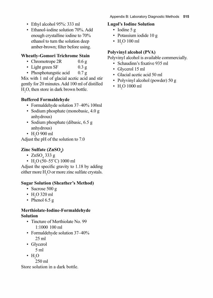
Appendix B: Laboratory Diagnostic Methods 515
• Ethyl alcohol 95%: 333 ml• Ethanol-iodine solution 70%. Add
enough crystalline iodine to 70% ethanol to turn the solution deep amber-brown; filter before using.
Wheatly-Gomori Trichrome Stain• Chromotrope 2R 0.6 g• Light green SF 0.3 g• Phosphotungstic acid 0.7 g
Mix with 1 ml of glacial acetic acid and stir gently for 20 minutes. Add 100 ml of distilled H2O, then store in dark brown bottle.
Buffered Formaldehyde• Formaldehyde solution 37–40% 100ml• Sodium phosphate (monobasic, 4.0 g
anhydrous)• Sodium phosphate (dibasic, 6.5 g
anhydrous)• H2O 900 ml
Adjust the pH of the solution to 7.0
Zinc Sulfate (ZnSO4)• ZnSO4 333 g• H2O (50–55˚C) 1000 ml
Adjust the specific gravity to 1.18 by adding either more H2O or more zinc sulfate crystals.
Sugar Solution (Sheather’s Method)• Sucrose 500 g• H2O 320 ml• Phenol 6.5 g
Merthiolate-Iodine-Formaldehyde Solution
• Tincture of Merthiolate No. 99 1:1000 100 ml
• Formaldehyde solution 37–40% 25 ml
• Glycerol 5 ml
• H2O 250 ml
Store solution in a dark bottle.
Lugol’s Iodine Solution• Iodine 5 g• Potassium iodide 10 g• H2O 100 ml
Polyvinyl alcohol (PVA)Polyvinyl alcohol is available commercially.
• Schaudinn’s fixative 935 ml• Glycerol 15 ml• Glacial acetic acid 50 ml • Polyvinyl alcohol (powder) 50 g• H2O 1000 ml
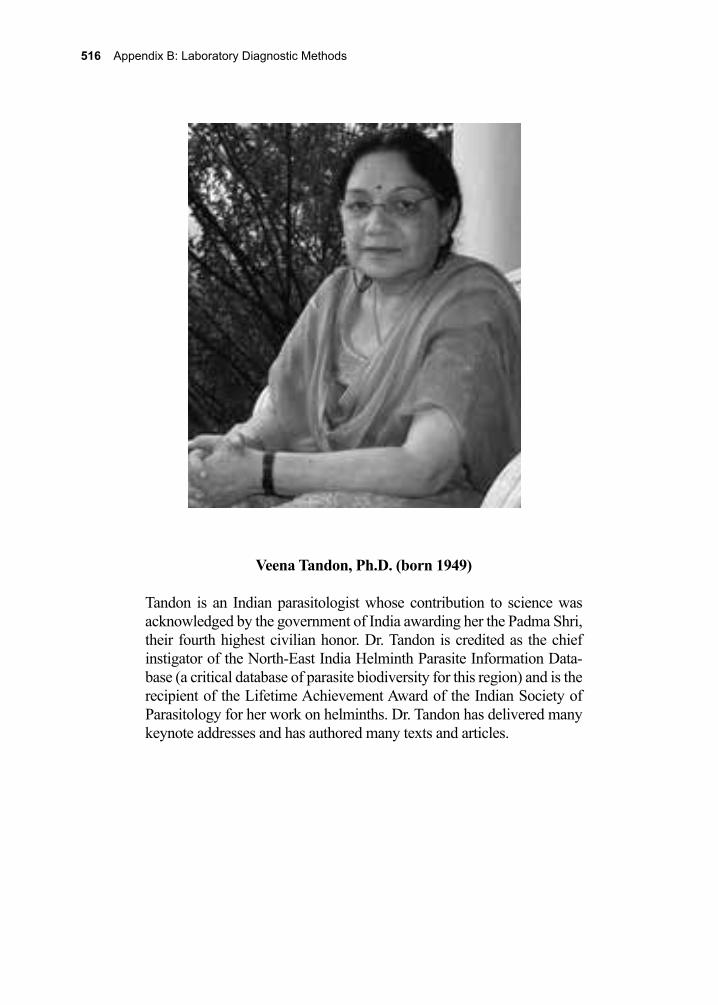
516 Appendix B: Laboratory Diagnostic Methods
Veena Tandon, Ph.D. (born 1949)
Tandon is an Indian parasitologist whose contribution to science was acknowledged by the government of India awarding her the Padma Shri, their fourth highest civilian honor. Dr. Tandon is credited as the chief instigator of the North-East India Helminth Parasite Information Data-base (a critical database of parasite biodiversity for this region) and is the recipient of the Lifetime Achievement Award of the Indian Society of Parasitology for her work on helminths. Dr. Tandon has delivered many keynote addresses and has authored many texts and articles.
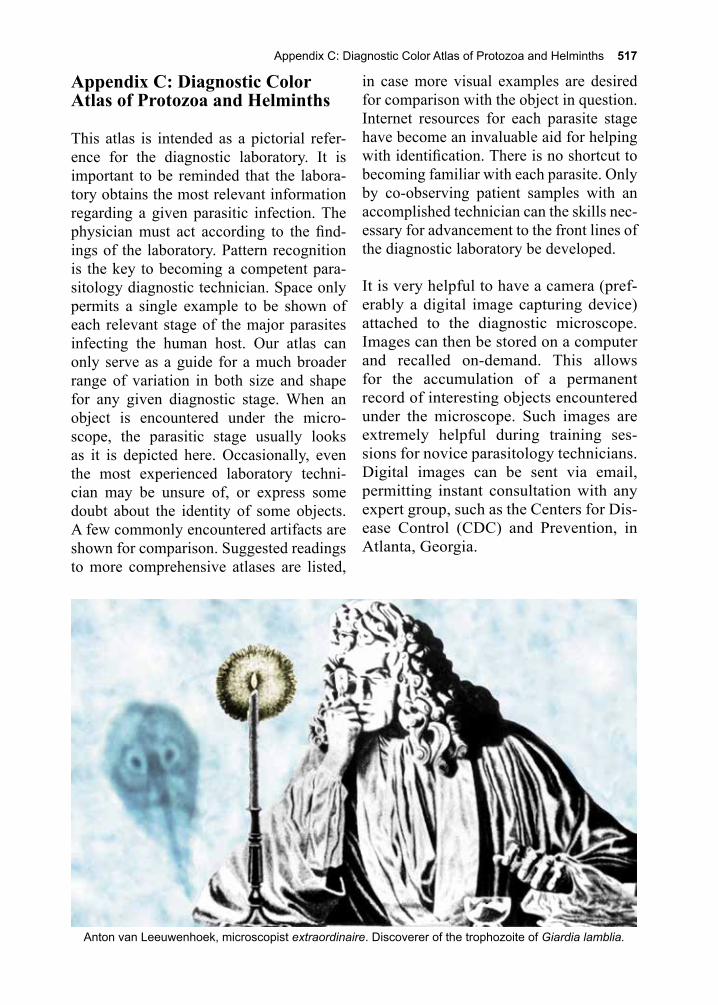
Appendix C: Diagnostic Color Atlas of Protozoa and Helminths 517
Appendix C: Diagnostic Color Atlas of Protozoa and Helminths
This atlas is intended as a pictorial refer-ence for the diagnostic laboratory. It is important to be reminded that the labora-tory obtains the most relevant information regarding a given parasitic infection. The physician must act according to the find-ings of the laboratory. Pattern recognition is the key to becoming a competent para-sitology diagnostic technician. Space only permits a single example to be shown of each relevant stage of the major parasites infecting the human host. Our atlas can only serve as a guide for a much broader range of variation in both size and shape for any given diagnostic stage. When an object is encountered under the micro-scope, the parasitic stage usually looks as it is depicted here. Occasionally, even the most experienced laboratory techni-cian may be unsure of, or express some doubt about the identity of some objects. A few commonly encountered artifacts are shown for comparison. Suggested readings to more comprehensive atlases are listed,
in case more visual examples are desired for comparison with the object in question. Internet resources for each parasite stage have become an invaluable aid for helping with identification. There is no shortcut to becoming familiar with each parasite. Only by co-observing patient samples with an accomplished technician can the skills nec-essary for advancement to the front lines of the diagnostic laboratory be developed.
It is very helpful to have a camera (pref-erably a digital image capturing device) attached to the diagnostic microscope. Images can then be stored on a computer and recalled on-demand. This allows for the accumulation of a permanent record of interesting objects encountered under the microscope. Such images are extremely helpful during training ses-sions for novice parasitology technicians. Digital images can be sent via email, permitting instant consultation with any expert group, such as the Centers for Dis-ease Control (CDC) and Prevention, in Atlanta, Georgia.
Anton van Leeuwenhoek, microscopist extraordinaire. Discoverer of the trophozoite of Giardia lamblia.

518 Appendix C: Diagnostic Color Atlas of Protozoa and Helminths
The majority of the protozoa depicted here have been stained with either iron-hematox-ylin (blue-gray stain) or Wheatly and Gomo-ri’s trichrome stain (green and red stain). Hel-minth eggs are as they appear in unstained, concentrated stool samples. Their yellow-brown tints attest to the fact that they have
encountered bile pigments. Microfilariae have all been stained with Giemsa (blue and red stain), as have the malaria parasites. The tissue section of the Nurse cell-parasite com-plex of Trichinella spiralis is stained with hematoxylin and eosin.
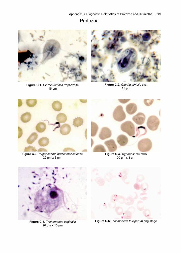
Appendix C: Diagnostic Color Atlas of Protozoa and Helminths 519
Protozoa
Figure C.6. Plasmodium falciparum ring stage
Figure C.2. Giardia lamblia cyst 15 µm
Figure C.3. Trypanosoma brucei rhodesiense 25 µm x 3 µm
Figure C.5. Trichomonas vaginalis20 µm x 10 µm
Figure C.1. Giardia lamblia trophozoite 15 µm
Figure C.4. Trypanosoma cruzi20 µm x 3 µm
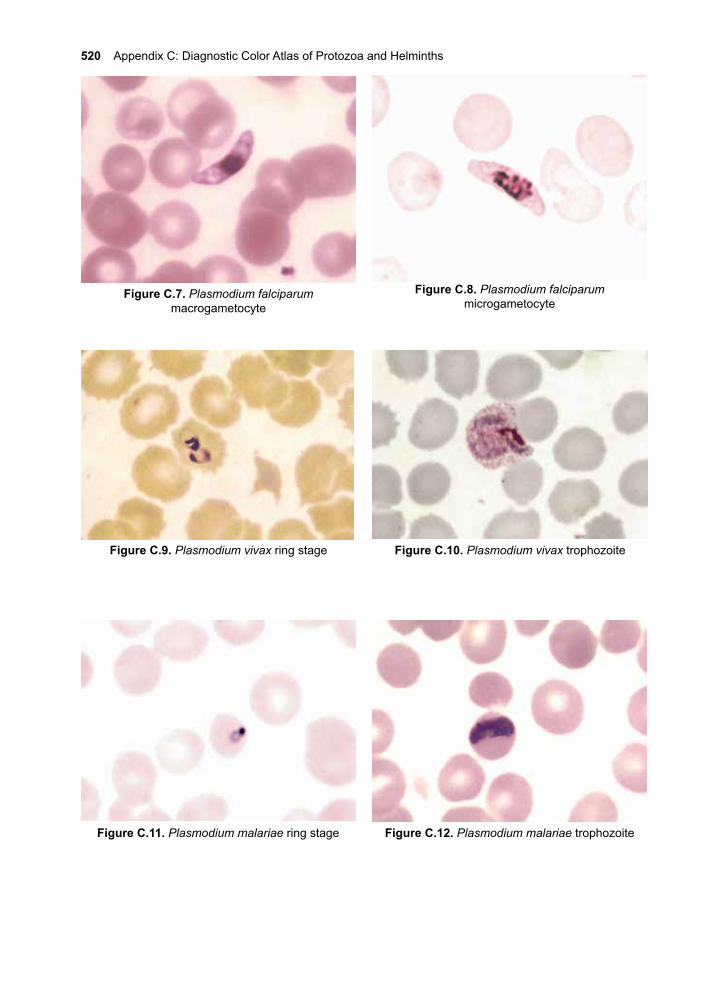
520 Appendix C: Diagnostic Color Atlas of Protozoa and Helminths
Figure C.8. Plasmodium falciparum microgametocyte
Figure C.11. Plasmodium malariae ring stage
Figure C.9. Plasmodium vivax ring stage Figure C.10. Plasmodium vivax trophozoite
Figure C.12. Plasmodium malariae trophozoite
Figure C.7. Plasmodium falciparum macrogametocyte
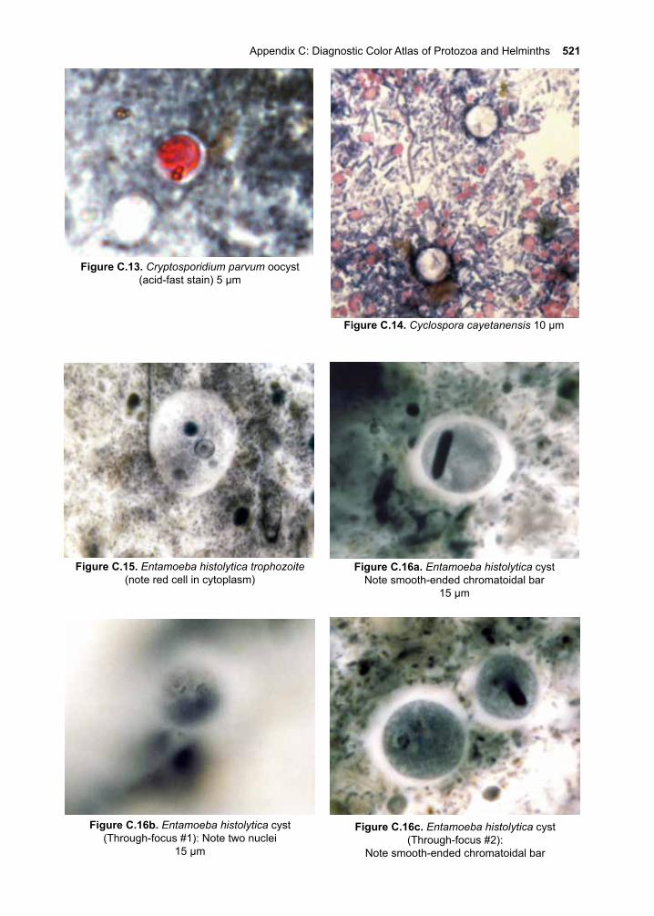
Appendix C: Diagnostic Color Atlas of Protozoa and Helminths 521
Figure C.14. Cyclospora cayetanensis 10 µm
Figure C.16c. Entamoeba histolytica cyst(Through-focus #2):
Note smooth-ended chromatoidal bar
Figure C.15. Entamoeba histolytica trophozoite (note red cell in cytoplasm)
Figure C.16a. Entamoeba histolytica cystNote smooth-ended chromatoidal bar
15 µm
Figure C.16b. Entamoeba histolytica cyst(Through-focus #1): Note two nuclei
15 µm
Figure C.13. Cryptosporidium parvum oocyst(acid-fast stain) 5 µm
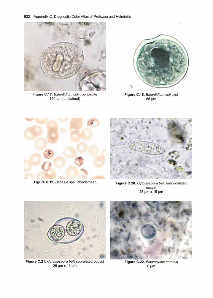
522 Appendix C: Diagnostic Color Atlas of Protozoa and Helminths
Figure C.18. Balantidium coli cyst65 µm
Figure C.19. Babesia spp. Bloodsmear
Figure C.17. Balantidium coli trophozoite150 µm (unstained)
Figure C.20. Cytoisospora belli unsporulated oocyst
25 µm x 15 µm
Figure C.21. Cytoisospora belli sporulated oocyst25 µm x 15 µm
Figure C.22. Blastocystis hominis6 µm
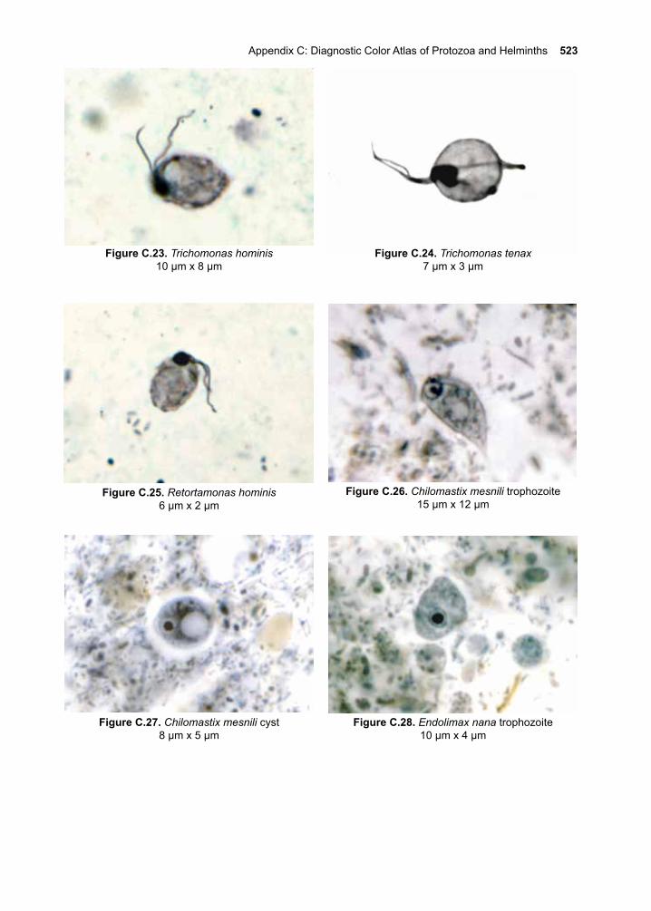
Appendix C: Diagnostic Color Atlas of Protozoa and Helminths 523
Figure C.24. Trichomonas tenax7 µm x 3 µm
Figure C.23. Trichomonas hominis10 µm x 8 µm
Figure C.25. Retortamonas hominis6 µm x 2 µm
Figure C.26. Chilomastix mesnili trophozoite15 µm x 12 µm
Figure C.27. Chilomastix mesnili cyst8 µm x 5 µm
Figure C.28. Endolimax nana trophozoite10 µm x 4 µm
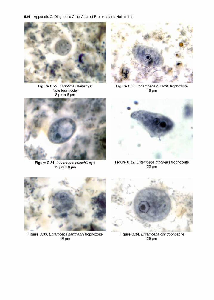
524 Appendix C: Diagnostic Color Atlas of Protozoa and Helminths
Figure C.29. Endolimax nana cystNote four nuclei
8 µm x 6 µm
Figure C.30. Iodamoeba bütschlii trophozoite18 µm
Figure C.31. Iodamoeba bütschlii cyst12 µm x 8 µm
Figure C.34. Entamoeba coli trophozoite35 µm
Figure C.33. Entamoeba hartmanni trophozoite10 µm
Figure C.32. Entamoeba gingivalis trophozoite30 µm
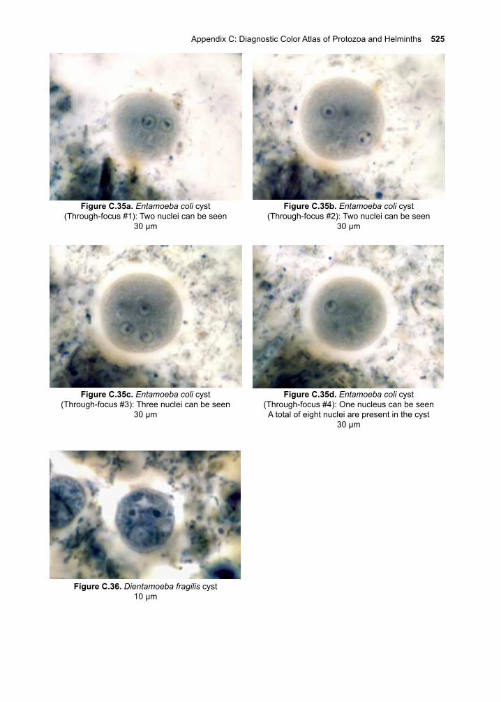
Appendix C: Diagnostic Color Atlas of Protozoa and Helminths 525
Figure C.35c. Entamoeba coli cyst(Through-focus #3): Three nuclei can be seen
30 µm
Figure C.35d. Entamoeba coli cyst(Through-focus #4): One nucleus can be seenA total of eight nuclei are present in the cyst
30 µm
Figure C.35a. Entamoeba coli cyst(Through-focus #1): Two nuclei can be seen
30 µm
Figure C.35b. Entamoeba coli cyst(Through-focus #2): Two nuclei can be seen
30 µm
Figure C.36. Dientamoeba fragilis cyst10 µm
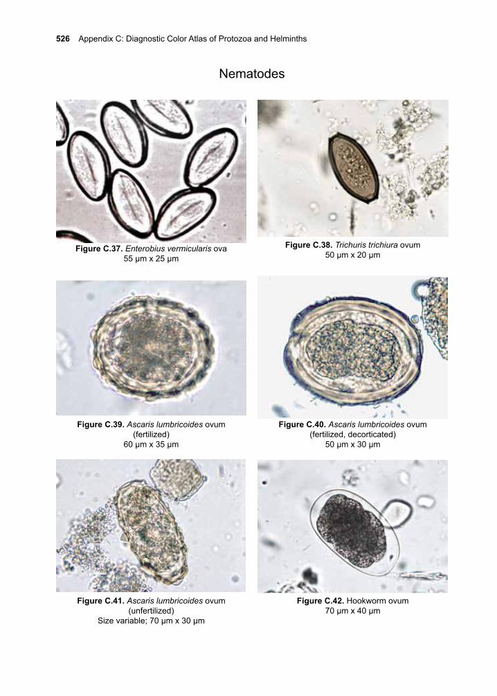
526 Appendix C: Diagnostic Color Atlas of Protozoa and Helminths
Figure C.37. Enterobius vermicularis ova55 µm x 25 µm
Figure C.40. Ascaris lumbricoides ovum(fertilized, decorticated)
50 µm x 30 µm
Figure C.42. Hookworm ovum70 µm x 40 µm
Nematodes
Figure C.38. Trichuris trichiura ovum50 µm x 20 µm
Figure C.39. Ascaris lumbricoides ovum(fertilized)
60 µm x 35 µm
Figure C.41. Ascaris lumbricoides ovum(unfertilized)
Size variable; 70 µm x 30 µm
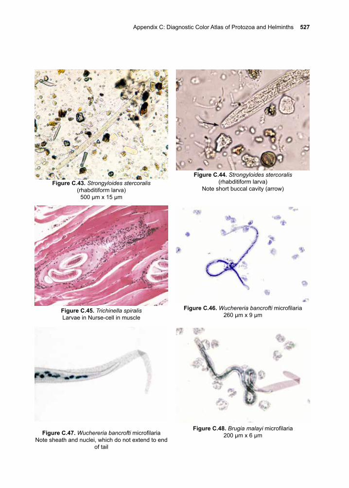
Appendix C: Diagnostic Color Atlas of Protozoa and Helminths 527
Figure C.45. Trichinella spiralisLarvae in Nurse-cell in muscle
Figure C.43. Strongyloides stercoralis(rhabditiform larva)
500 µm x 15 µm
Figure C.44. Strongyloides stercoralis(rhabditiform larva)
Note short buccal cavity (arrow)
Figure C.46. Wuchereria bancrofti microfilaria260 µm x 9 µm
Figure C.47. Wuchereria bancrofti microfilariaNote sheath and nuclei, which do not extend to end
of tail
Figure C.48. Brugia malayi microfilaria200 µm x 6 µm
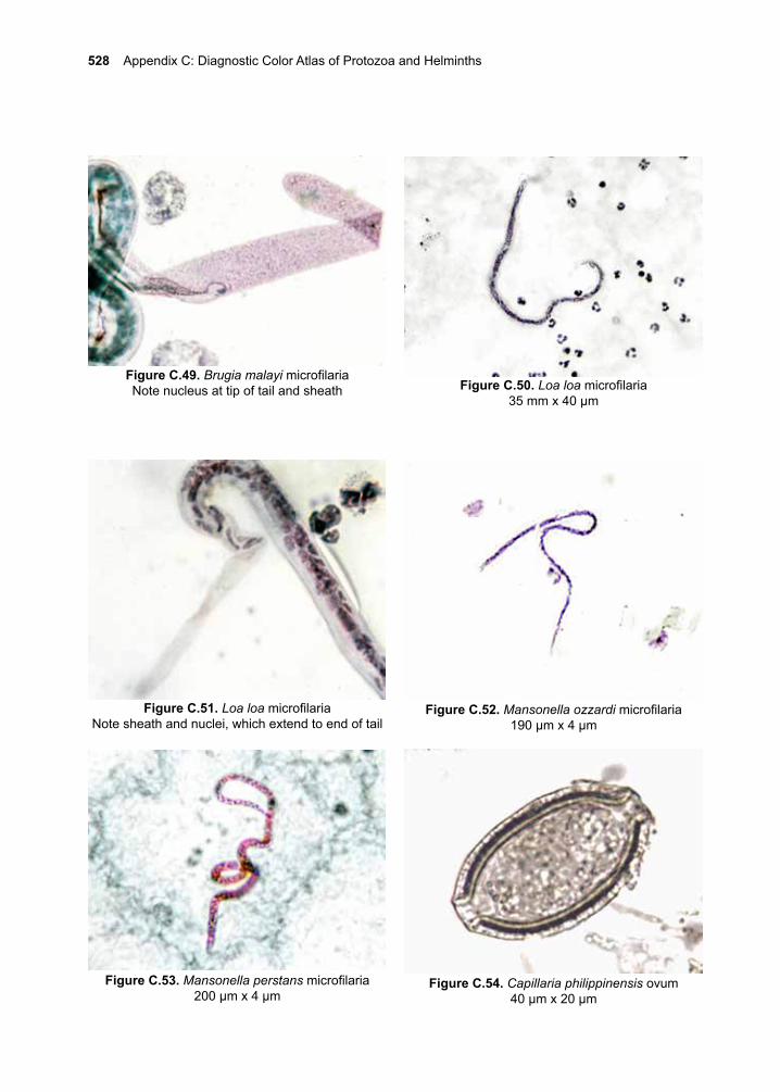
528 Appendix C: Diagnostic Color Atlas of Protozoa and Helminths
Figure C.54. Capillaria philippinensis ovum40 µm x 20 µm
Figure C.49. Brugia malayi microfilariaNote nucleus at tip of tail and sheath Figure C.50. Loa loa microfilaria
35 mm x 40 µm
Figure C.51. Loa loa microfilariaNote sheath and nuclei, which extend to end of tail
Figure C.52. Mansonella ozzardi microfilaria190 µm x 4 µm
Figure C.53. Mansonella perstans microfilaria200 µm x 4 µm
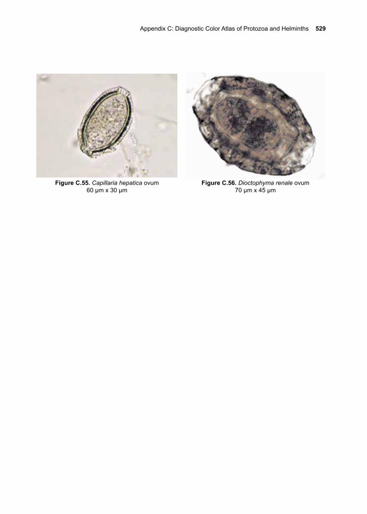
Appendix C: Diagnostic Color Atlas of Protozoa and Helminths 529
Figure C.56. Dioctophyma renale ovum70 µm x 45 µm
Figure C.55. Capillaria hepatica ovum60 µm x 30 µm
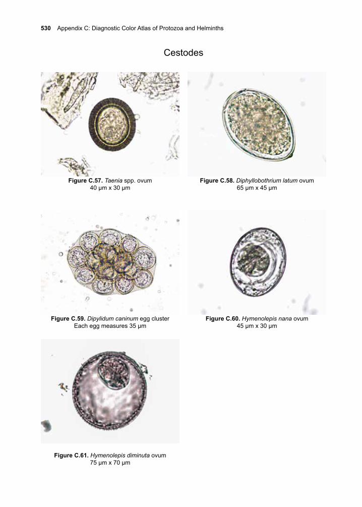
530 Appendix C: Diagnostic Color Atlas of Protozoa and Helminths
Figure C.57. Taenia spp. ovum40 µm x 30 µm
Figure C.58. Diphyllobothrium latum ovum65 µm x 45 µm
Cestodes
Figure C.59. Dipylidum caninum egg clusterEach egg measures 35 µm
Figure C.60. Hymenolepis nana ovum45 µm x 30 µm
Figure C.61. Hymenolepis diminuta ovum75 µm x 70 µm
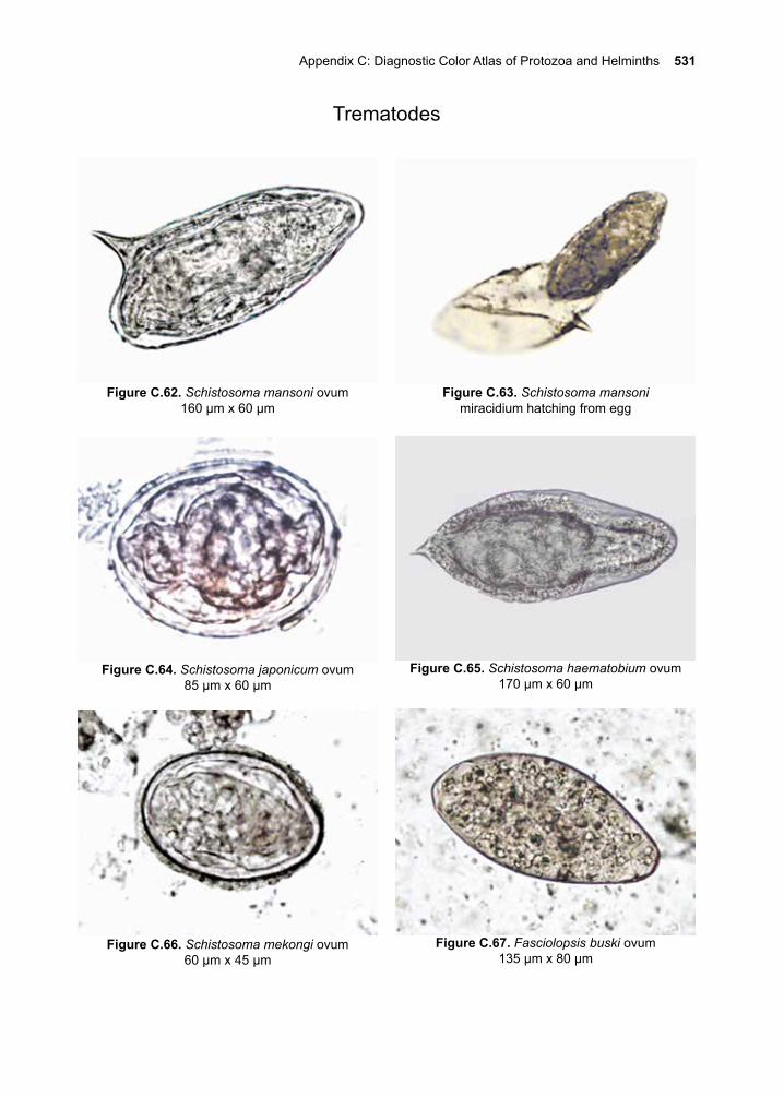
Appendix C: Diagnostic Color Atlas of Protozoa and Helminths 531
Figure C.67. Fasciolopsis buski ovum135 µm x 80 µm
Trematodes
Figure C.63. Schistosoma mansonimiracidium hatching from egg
Figure C.66. Schistosoma mekongi ovum60 µm x 45 µm
Figure C.62. Schistosoma mansoni ovum160 µm x 60 µm
Figure C.64. Schistosoma japonicum ovum85 µm x 60 µm
Figure C.65. Schistosoma haematobium ovum170 µm x 60 µm
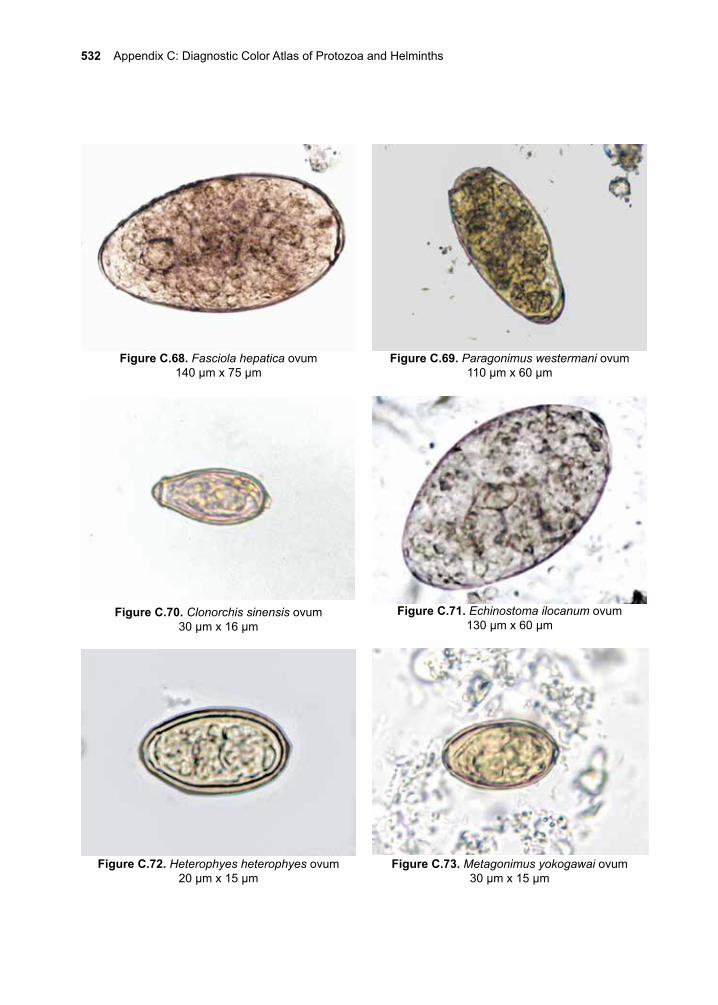
532 Appendix C: Diagnostic Color Atlas of Protozoa and Helminths
Figure C.68. Fasciola hepatica ovum140 µm x 75 µm
Figure C.69. Paragonimus westermani ovum110 µm x 60 µm
Figure C.71. Echinostoma ilocanum ovum130 µm x 60 µm
Figure C.72. Heterophyes heterophyes ovum20 µm x 15 µm
Figure C.70. Clonorchis sinensis ovum30 µm x 16 µm
Figure C.73. Metagonimus yokogawai ovum30 µm x 15 µm
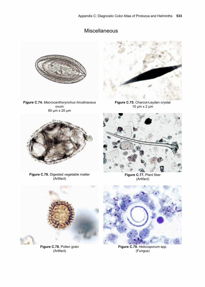
Appendix C: Diagnostic Color Atlas of Protozoa and Helminths 533
Miscellaneous
Figure C.75. Charcot-Leyden crystal10 µm x 2 µm
Figure C.74. Macrocanthorynchus hirudinaceus ovum
60 µm x 20 µm
Figure C.76. Digested vegetable matter(Artifact)
Figure C.77. Plant fiber(Artifact)
Figure C.78. Pollen grain(Artifact)
Figure C.79. Helicosporum spp.(Fungus)
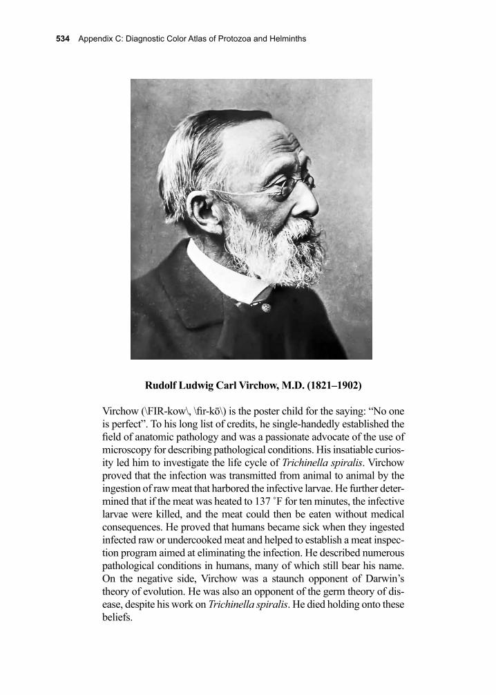
534 Appendix C: Diagnostic Color Atlas of Protozoa and Helminths
Rudolf Ludwig Carl Virchow, M.D. (1821–1902)
Virchow (\FIR-kow\, \fir-kō\) is the poster child for the saying: “No one is perfect”. To his long list of credits, he single-handedly established the field of anatomic pathology and was a passionate advocate of the use of microscopy for describing pathological conditions. His insatiable curios-ity led him to investigate the life cycle of Trichinella spiralis. Virchow proved that the infection was transmitted from animal to animal by the ingestion of raw meat that harbored the infective larvae. He further deter-mined that if the meat was heated to 137 ̊ F for ten minutes, the infective larvae were killed, and the meat could then be eaten without medical consequences. He proved that humans became sick when they ingested infected raw or undercooked meat and helped to establish a meat inspec-tion program aimed at eliminating the infection. He described numerous pathological conditions in humans, many of which still bear his name. On the negative side, Virchow was a staunch opponent of Darwin’s theory of evolution. He was also an opponent of the germ theory of dis-ease, despite his work on Trichinella spiralis. He died holding onto these beliefs.
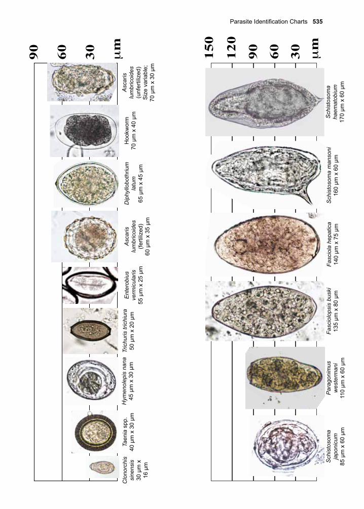
Parasite Identification Charts 535
306090120
150
306090
Fasc
iolo
psis
bus
ki13
5 µm
x 8
0 µm
Fasc
iola
hep
atic
a14
0 µm
x 7
5 µm
Sch
isto
som
a m
anso
ni16
0 µm
x 6
0 µm
Sch
isto
som
a ha
emat
obiu
m17
0 µm
x 6
0 µm
Sch
isto
som
a ja
poni
cum
85 µ
m x
60
µm
Par
agon
imus
w
este
rman
i11
0 µm
x 6
0 µm
Dip
hyllo
both
rium
la
tum
65 µ
m x
45
µm
Hoo
kwor
m
70 µ
m x
40
µmA
scar
is
lum
bric
oide
s(u
nfer
tiliz
ed)
Siz
e va
riabl
e;
70 µ
m x
30
µm
Asc
aris
lu
mbr
icoi
des
(ferti
lized
)60
µm
x 3
5 µm
Ent
erob
ius
verm
icul
aris
55 µ
m x
25
µm
Tric
huris
tric
hiur
a50
µm
x 2
0 µm
Taen
ia s
pp.
40 µ
m x
30
µmH
ymen
olep
is n
ana
45 µ
m x
30
µmC
lono
rchi
s si
nens
is30
µm
x
16 µ
m
µm
µm
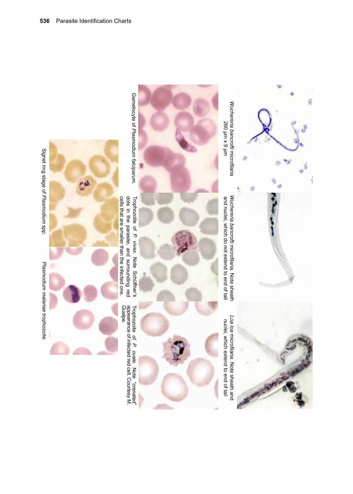
536 Parasite Identification Charts
Wuchereria bancrofti m
icrofilaria260 µm
x 9 µmW
uchereria bancrofti microfilaria. N
ote sheath and nuclei, w
hich do not extend to end of tailLoa loa m
icrofilaria. Note sheath and
nuclei, which extend to end of tail
Gam
etocyte of Plasm
odium falciparum
.
Signet ring stage of P
lasmodium
spp.
Trophozoite of P. vivax. Note S
chüffner’s dots in the parasite, and surrounding red cells that are sm
aller than the infected one.
Trophozoite of P. ovale. Note “crenated”
appearance of infected red cell. Courtesy M
. G
uelpe.
Plasm
odium m
alariae trophozoite.

Parasite Identification Charts 537
Marietta Voge Ph.D. (1918-1984)
Marietta Voge was a leader in the field of parasitology education. Mari-etta was the co-author of one of the leading textbooks in the field and served as the president of the American Society of Parasitologists in 1976. Dr. Voge not only inspired many future parasitologists but she also devoted her time to working in many resource-limited parts of the world, refugee camps and mission clinics. She is well remembered for her dedication to teaching, and her life was an embodiment of her words from her presidential address to the American Society of Parasitologists where she wrote: “It is the teacher that sparks and stimulates the minds of students to produce new ideas. It is the teacher who gives the impetus to students to become the most important propellant to our society and civilization - the innovator – because, without innovation and originality, society becomes stagnant and decadent. A superior teacher is the inter-preter and keeper of our past, the critic of our present, the dreamer of things to come.”
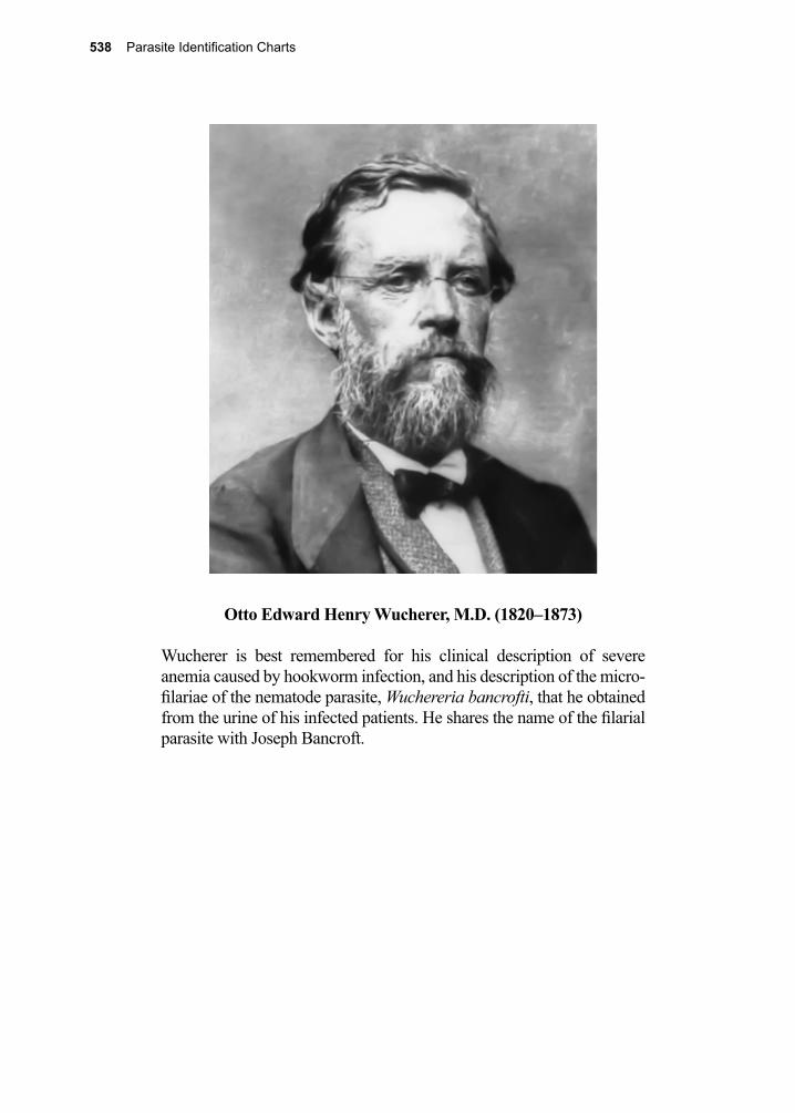
538 Parasite Identification Charts
Otto Edward Henry Wucherer, M.D. (1820–1873)
Wucherer is best remembered for his clinical description of severe anemia caused by hookworm infection, and his description of the micro-filariae of the nematode parasite, Wuchereria bancrofti, that he obtained from the urine of his infected patients. He shares the name of the filarial parasite with Joseph Bancroft.
Clinical Appendix 539
Clinical Appendix
1. Giardia lambliaExposure Required:Oral ingestion of cysts in fecally contaminated water or food
Clinical Disease:• Asymptomatic• Acute diarrhea, chronic diarrhea, foul smelling diarrhea, floating stools, flatulence,
nausea, rarely fever, epigastric pain, weight loss, fatigue
Diagnosis:• Stool ova and parasites (microscopic examination of stool) • Antigen capture-ELISA from stool sample• NAAT
Treatment:• Preferred drug options:
o Metronidazole 250 mg PO TID x 7-dayso Tinidazole 2 g PO x1
• Alternate preferred drug option: o Nitazoxanide 500 mg PO BID x 3 days
• Alternative antimicrobials: o Paromomycin 500 mg PO TID x 5–10 dayso Furazolidone 100 mg PO QID x 7–10 dayso Quinacrine 100 mg PO TID x 5–7 dayso Albendazole 400 mg PO Q-day x 5–7 days
Resistant strains of Giardia are increasingly prevalent, but many will retreat with a different class of antimicrobial therapy or a longer course of the original agent. In some refractory cases combination antimicrobial therapy may be necessary.
3. Cutaneous LeishmaniasisExposure Required:
• Bite from sand fly
Clinical Disease:• Small papule that then forms a painless chronic ulcer with a raised indurated border
Diagnosis:Organisms are found only in the living tissue at the raised margin, regardless of the age of the lesion.
• Histology• Culture
• NAAT• Leishmaniasis skin test
540 Clinical Appendix
Treatment:For non-mucocutaneous species (see mucocutaneous section for those species with mucocutaneous potential).
• Physical Therapieso Thermotherapy – 50 °C once weekly for 4 weekso CO2 laser – single sessiono Photodynamic therapy – once weekly for 4 weeks after application of
‘photosensitizer’o Cryotherapy – freeze for 10–30 s, thaw and perform 2–3 times, repeated every
1–4 weeks for 2–4 sessions or more depending on response
• Local Drug Therapyo Paromomycin ointment 15% -apply BID for 20–28 dayso Clotrimazole 1% / miconazole 2% ointment applied BID x 30 dayso Intralesional antimonials (meglumine antimoniate injected until complete
blanching of border, every 3–7 days for 1–5 sessions-very painful!)
• Oral Drug Therapyo Azoles
▪ Fluconazole 400 mg PO Q-day x 6 weeks ▪ Ketoconazole 600 mg PO Q-day x 6 weeks ▪ Itraconazole 400 mg PO Q-day x 3–6 weeks
o Miltefosine (2.5 mg/kg/day) or for an adult 150 mg PO per day given as 50 mg 2–3x/day x 28 days
• Parenteral Therapyo Pentavalent antimonialso Amphotericino Liposomal amphotericino Pentamidine
• Combination Therapyo Combining local and parenteralo Combining different parenteral therapies
4. Mucocutaneous Leishmaniasis Leishmania (V.) braziliensis ~ 30% Leishmania (V.) panamensis ~ 10% Leishmania (V.) guyanensis ~ 10% Leishmania (L.) amazonensis (rare)
Exposure Required:• Bite from sand fly

Clinical Appendix 541
Clinical Disease:• Small papule that then forms a painless chronic ulcer with a raised indurated border, fol-
lowed by ulcers at mucous membranes – i.e. soft palate, nasal septum, larynx, anus, lips
Diagnosis:Organisms are found only in the living tissue at the raised margin, regardless of the age of the lesion.
• NAAT – main modality and identifies species • Histology – very low sensitivity• Culture – very low sensitivity
Treatment:For species with muco-cutaneous potential, even first infection.
• Oral Drug Therapyo Miltefosine (2.5 mg/kg/day) or for an adult 150 mg PO per day given as 50 mg
2–3x/day x 28 days
• Parenteral Therapyo Pentavalent antimonials
▪ Sodium stibogluconate 20 mg/kg/day IV/IM x 28–30 days ▪ Meglumine antimoniate 20 mg/kg/day IV/IM x 28–30 days
o Amphotericin 0.5–1 mg/kg IV Q-day up to cumulative dose of 20–45 mg/kgo Liposomal amphotericin 3 mg/kg IV Q-day up to cumulative dose of 20–60 mg/
kgo (Possible inferior option) pentamidine 2–4 mg/kg IV/IM every other day or 3x/
week for 15 doses
• Combination Therapyo Combining local and parenteralo Combining different parenteral therapies
5. Visceral Leishmaniasis Leishmania (L.) donovani Leishmania (L.) infantum
Exposure Required:• Bite from sand fly
Clinical Disease:• Fever, lymphadenopathy, hepatomegaly, splenomegaly, weight loss, darkening of the
skin
Diagnosis:• NAAT – from marrow or splenic aspirate• Culture• Microscopic evaluation• rK39 antigen ELISA
542 Clinical Appendix
Treatment:• Parenteral Therapy
o Liposomal amphotericin 3–5 mg/kg IV Q-day for 3–5 days up to cumulative dose of 15 mg/kg or single 10 mg/kg IV dose, for HIV co-infected 3–5 mg/kg IV Q-day for 10 days up to cumulative dose of 40 mg/kg
o Pentavalent antimonials (preferred in Africa but not in India) ▪ Sodium stibogluconate 20 mg/kg/day IV/IM x 28–30 days ▪ Meglumine antimoniate 20 mg/kg/day IV/IM x 28–30 days
o Amphotericin 0.5–1 mg/kg IV Q-day up to cumulative dose of 20–45 mg/kgo Liposomal amphotericin 3 mg/kg IV Q-day up to cumulative dose of 20–60 mg/
kg
• Oral Drug Therapyo (Non-preferred-alternative) miltefosine (2.5 mg/kg/day) or for an adult 150 mg
PO per day given as 50 mg 2–3x/day x 28 days
• Combination Therapyo Combining amphotericin and miltefosine
6. African Trypanosomiasis Trypanosoma brucei rhodesiense Trypanosoma brucei gambiense
Exposure Required:• Bite from tsetse fly
Clinical Disease:• Hemolymphatic stage – initially a large painless chancre at bite site, rash, generalized
pruritus, weight loss, facial swelling, posterior cervical adenopathy• CNS Stage – headache, stiff neck, periods of insomnia alternating with
hypersomnolence, depression, seizures, tremors, palsies, coma
Diagnosis:• Microscopic
o Blood smears o Lymph node aspirateso CSF - examination of CSF is mandatory in the diagnostic evaluation of
trypanosomiasis and a white blood cell count >5 cells/ml is considered indicative of CNS involvement
o Aspirates taken at the edge of chancreso NAAT –limited availability
• Cultures• Card agglutination test for trypanosomiasis
Clinical Appendix 543
Treatment:Treatment is determined by species and stage.
• Early Stages: Trypanosoma b. gambiense
o (Preferred) pentamidine 4 mg/kg/day IV/IM (given over 2 hrs) for 7 days o (Alternative) suramin 100–200 mg test dose, then 20 mg/kg (max 1g) IV on
days 1, 3, 7, 14, and 21Trypanosoma b. rhodesienseo Suramin 100–200 mg test dose, then 20 mg/kg (max 1g) IV on days 1, 3, 7, 14,
and 21• Late Stages:
Trypanosoma b. gambiense o (Preferred) eflornithine 200 mg/kg IV q12 hrs (given over 1 hr) for 7 days PLUS
nifurtimox 5 mg/kg PO q8 hrs for 10 dayso Eflornithine 100 mg/kg IV q6 hrs for 14 days (monotherapy)o (Alternative) melarsoprol 2.2 mg/kg/day IV x 10 days (with oral prednisone)
Trypanosoma b. rhodesiense o (Preferred) melarsoprol 2.2 mg/kg/day IV q10 days (with oral prednisone)o (Alternative) melarsoprol; three series of 3.6 mg/kg/day IV x 3 days spaced
apart by 7-day intervals (with oral prednisone)
7. American Trypanosomiasis Trypanosoma cruzi
Exposure Required:• Trypomastigotes present in reduviid bug feces enter through bug bite or mucous
membranes, ingestion orally (contaminated sweetened drinks), transfusion, congenital
Clinical Disease:• Acute – malaise, fever, myocarditis, pericardial effusion, meningoencephalitis• Chronic – cardiac damage (arrhythmias, congestive heart failure), gastrointestinal (GI)
damage (dysphagia, regurgitation, megacolon, constipation)
Diagnosis:• Microscopic - blood smears • NAAT - limited availability• Serology• Histology
Treatment:• Benznidazole (for 60 days) through the Centers for Disease Control (CDC) and
Preventiono Adults (>12 years old) 5–7.5 mg/kg daily divided into BID doses (12.5 and 100
mg tablets)o Children (< 12 years old) 5 mg/kg PO BID
544 Clinical Appendix
• Nifurtimox (for 90 days) (divided into 3–4 doses per day)o Adults 8–10 mg/kg/dayo Young children (<10 years old) 15–20 mg/kg/dayo Older children (>10 years old <16 years old )12.5–15 mg/kg/day
8. Trichomonas vaginalisExposure Required:
• Acquisition of trophozoites during sexual contact or at birth
Clinical Disease:• Asymptomatic• Discomfort, dyspareunia, vaginal discharge (thick, yellow, blood tinged, pH increased
from 4.5 to >5.0), strawberry cervix, erythema, dysuria
Diagnosis:• Microscopic observation of motile forms from wet prep • Culture • Rapid antigen testing • Nucleic acid probe test• NAAT
Treatment:• Metronidazole 500 mg PO BID x 7 days• Tinidazole 2 g PO x1• Combination: paromomycin compounded cream 250 mg each day with high dose
tinidazole 1 g orally 3x/day for 2 weeks
9. The Malarias Plasmodium falciparum Plasmodium vivax Plasmodium ovale Plasmodium malariae Plasmodium knowlesi
Exposure Required:• Bite from female Anopheles mosquito
Clinical Disease:• Periodic fever and chills, cough, abdominal pain, vomiting, diarrhea, dyspnea, anemia,
leukopenia, eosinopenia, thrombocytopenia,Diagnosis:
• Thick and thin blood smears• Antibody-based rapid diagnostic tests• NAAT• Mutation-specific PCR
Clinical Appendix 545
Staging:• Uncomplicated malaria (not having a feature of severe)• Severe malaria (having one or more of the following features)
o Decreased level of consciousnesso Unable to sit or stand without assistanceo Convulsions (more than 2 in <24 hrs)o Acidosis or bicarbonate <15 mmol/Lo Hypoglycemia (specific cutoffs)o Anemia (specific cutoffs)o Renal impairment (Cr >3 mg/dL or BUN >20 mmol/Lo Jaundice (bilirubin >3 mg/dL)o Pulmonary edema (observable with chest X-ray, hypoxemia, tachypnea)o Significant bleedingo Shocko P. falciparum parasitemia >10%
Treatment:• Chloroquine-Sensitive Uncomplicated Malaria (not having a feature of severe)
o Chloroquine ▪ Adults 1 g PO x1 then 500 mg at 6, 24, and 48 hrs ▪ Children 10 mg/kg followed by 5 mg/kg of the base at 6, 24, and 48 hrs
o Hydroxychloroquine ▪ Adults 800 mg x1, then 400 mg at 6, 24, and 48 hrs ▪ Children 10 mg/kg x1, then 5 mg/kg at 6, 24, and 48 hrs
• Chloroquine-resistant Uncomplicated Malaria (not having a feature of severe)o Artemisinin combination therapy (see table below)o Atovaquone-proguanil (adults) 4-tablets PO Q-Day x 3 dayso Quinine 650 mg PO TID for 3–7 days plus second agent for 7 days PLUS
(doxycycline, or tetracycline, or clindamycin)o Mefloquine 3-tablets PO x1, then 2 tablets 6 hrs later
• Treatment of Complicated Malariao Artemisinin derivatives
▪ Artesunate IV (preferred) (If>20 kg) 2.4 mg/kg IV x1 then at 12 hrs, 24 hrs, then daily, (If <20 kg) 3 mg/kg IV same schedule
▪ Artemether IM 3.2 mg/kg IM x1 then 1.6 mg/kg Q-dayo Quinidine based
▪ Quinidine gluconate 10 mg/kg IV x1 then continuous infusion 0.0125 mg/kg/min
▪ Plus, doxycycline or clindamycin for 7 days
• Treatment to Prevent Malaria Relapse (Hyponozoites) o Primaquine – 30 mg (of base) 2-tablets PO Q-day for 2 weeks
Consider broad spectrum antibiotics (~10% coinfected)/Exchange transfusion not recommended
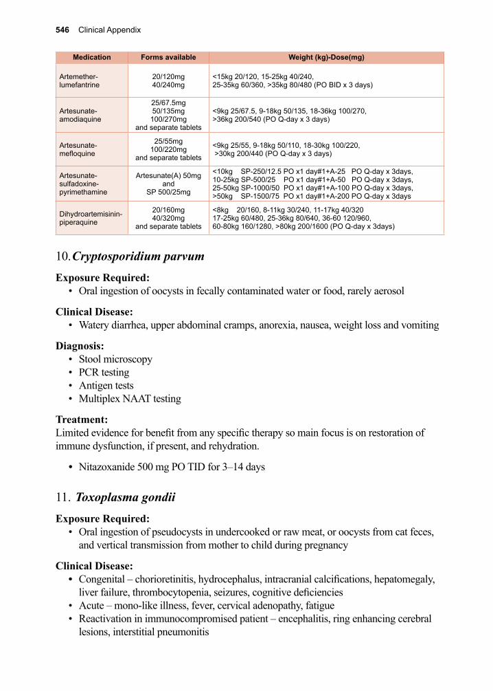
546 Clinical Appendix
10. Cryptosporidium parvumExposure Required:
• Oral ingestion of oocysts in fecally contaminated water or food, rarely aerosol
Clinical Disease:• Watery diarrhea, upper abdominal cramps, anorexia, nausea, weight loss and vomiting
Diagnosis:• Stool microscopy• PCR testing• Antigen tests • Multiplex NAAT testing
Treatment:Limited evidence for benefit from any specific therapy so main focus is on restoration of immune dysfunction, if present, and rehydration.
• Nitazoxanide 500 mg PO TID for 3–14 days
11. Toxoplasma gondiiExposure Required:
• Oral ingestion of pseudocysts in undercooked or raw meat, or oocysts from cat feces, and vertical transmission from mother to child during pregnancy
Clinical Disease:• Congenital – chorioretinitis, hydrocephalus, intracranial calcifications, hepatomegaly,
liver failure, thrombocytopenia, seizures, cognitive deficiencies• Acute – mono-like illness, fever, cervical adenopathy, fatigue• Reactivation in immunocompromised patient – encephalitis, ring enhancing cerebral
lesions, interstitial pneumonitis
Medication Forms available Weight (kg)-Dose(mg)
Artemether-lumefantrine
20/120mg40/240mg
<15kg 20/120, 15-25kg 40/240, 25-35kg 60/360, >35kg 80/480 (PO BID x 3 days)
Artesunate-amodiaquine
25/67.5mg50/135mg100/270mg
and separate tablets
<9kg 25/67.5, 9-18kg 50/135, 18-36kg 100/270, >36kg 200/540 (PO Q-day x 3 days)
Artesunate-mefloquine
25/55mg100/220mg
and separate tablets
<9kg 25/55, 9-18kg 50/110, 18-30kg 100/220, >30kg 200/440 (PO Q-day x 3 days)
Artesunate-sulfadoxine-pyrimethamine
Artesunate(A) 50mgand
SP 500/25mg
<10kg SP-250/12.5 PO x1 day#1+A-25 PO Q-day x 3days, 10-25kg SP-500/25 PO x1 day#1+A-50 PO Q-day x 3days, 25-50kg SP-1000/50 PO x1 day#1+A-100 PO Q-day x 3days,>50kg SP-1500/75 PO x1 day#1+A-200 PO Q-day x 3days
Dihydroartemisinin-piperaquine
20/160mg40/320mg
and separate tablets
<8kg 20/160, 8-11kg 30/240, 11-17kg 40/32017-25kg 60/480, 25-36kg 80/640, 36-60 120/960, 60-80kg 160/1280, >80kg 200/1600 (PO Q-day x 3days)
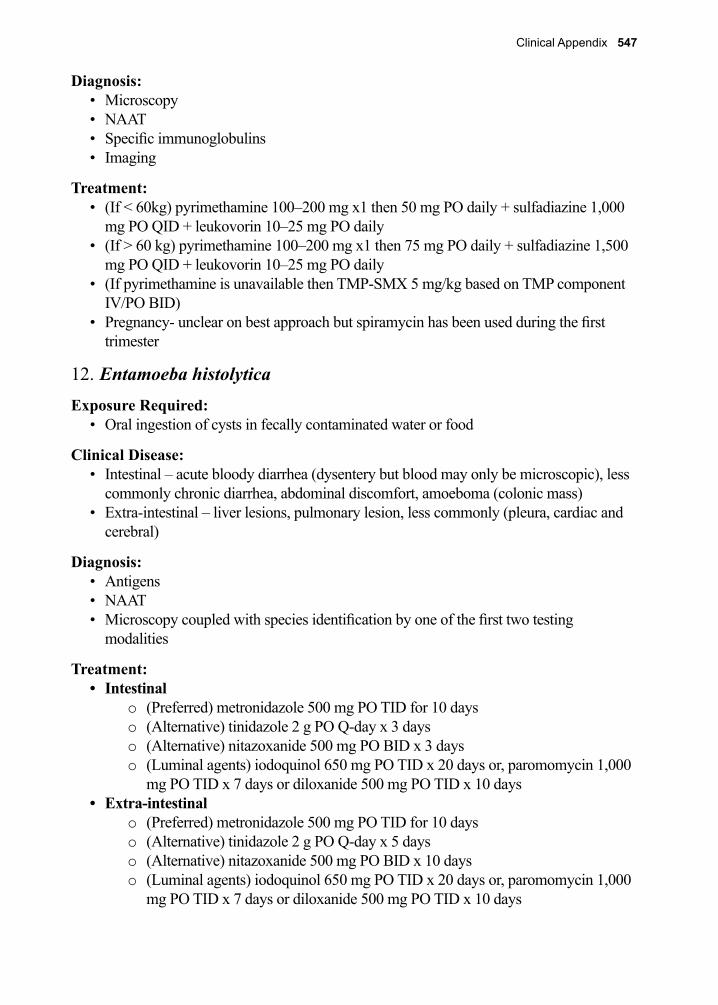
Clinical Appendix 547
Diagnosis:• Microscopy• NAAT• Specific immunoglobulins• Imaging
Treatment:• (If < 60kg) pyrimethamine 100–200 mg x1 then 50 mg PO daily + sulfadiazine 1,000
mg PO QID + leukovorin 10–25 mg PO daily • (If > 60 kg) pyrimethamine 100–200 mg x1 then 75 mg PO daily + sulfadiazine 1,500
mg PO QID + leukovorin 10–25 mg PO daily • (If pyrimethamine is unavailable then TMP-SMX 5 mg/kg based on TMP component
IV/PO BID)• Pregnancy- unclear on best approach but spiramycin has been used during the first
trimester
12. Entamoeba histolyticaExposure Required:
• Oral ingestion of cysts in fecally contaminated water or food
Clinical Disease:• Intestinal – acute bloody diarrhea (dysentery but blood may only be microscopic), less
commonly chronic diarrhea, abdominal discomfort, amoeboma (colonic mass)• Extra-intestinal – liver lesions, pulmonary lesion, less commonly (pleura, cardiac and
cerebral)
Diagnosis:• Antigens• NAAT • Microscopy coupled with species identification by one of the first two testing
modalities
Treatment:• Intestinal
o (Preferred) metronidazole 500 mg PO TID for 10 dayso (Alternative) tinidazole 2 g PO Q-day x 3 dayso (Alternative) nitazoxanide 500 mg PO BID x 3 dayso (Luminal agents) iodoquinol 650 mg PO TID x 20 days or, paromomycin 1,000
mg PO TID x 7 days or diloxanide 500 mg PO TID x 10 days• Extra-intestinal
o (Preferred) metronidazole 500 mg PO TID for 10 dayso (Alternative) tinidazole 2 g PO Q-day x 5 dayso (Alternative) nitazoxanide 500 mg PO BID x 10 dayso (Luminal agents) iodoquinol 650 mg PO TID x 20 days or, paromomycin 1,000
mg PO TID x 7 days or diloxanide 500 mg PO TID x 10 days
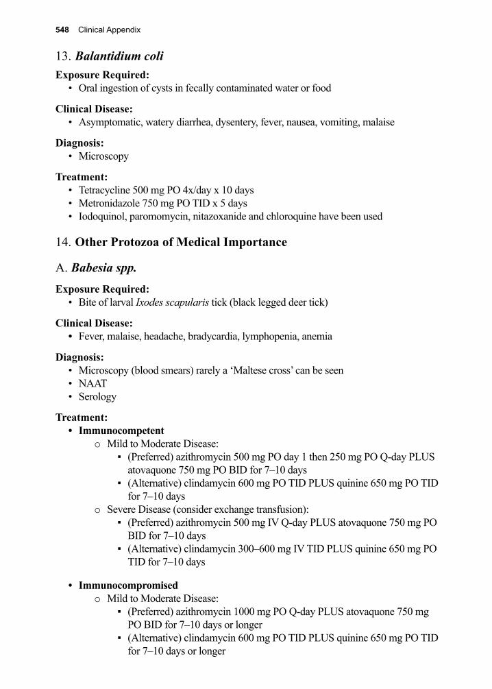
548 Clinical Appendix
13. Balantidium coliExposure Required:
• Oral ingestion of cysts in fecally contaminated water or food
Clinical Disease:• Asymptomatic, watery diarrhea, dysentery, fever, nausea, vomiting, malaise
Diagnosis:• Microscopy
Treatment:• Tetracycline 500 mg PO 4x/day x 10 days• Metronidazole 750 mg PO TID x 5 days • Iodoquinol, paromomycin, nitazoxanide and chloroquine have been used
14. Other Protozoa of Medical Importance
A. Babesia spp.Exposure Required:
• Bite of larval Ixodes scapularis tick (black legged deer tick)
Clinical Disease:• Fever, malaise, headache, bradycardia, lymphopenia, anemia
Diagnosis:• Microscopy (blood smears) rarely a ‘Maltese cross’ can be seen• NAAT • Serology
Treatment:• Immunocompetent
o Mild to Moderate Disease: ▪ (Preferred) azithromycin 500 mg PO day 1 then 250 mg PO Q-day PLUS
atovaquone 750 mg PO BID for 7–10 days ▪ (Alternative) clindamycin 600 mg PO TID PLUS quinine 650 mg PO TID
for 7–10 dayso Severe Disease (consider exchange transfusion):
▪ (Preferred) azithromycin 500 mg IV Q-day PLUS atovaquone 750 mg PO BID for 7–10 days
▪ (Alternative) clindamycin 300–600 mg IV TID PLUS quinine 650 mg PO TID for 7–10 days
• Immunocompromised o Mild to Moderate Disease:
▪ (Preferred) azithromycin 1000 mg PO Q-day PLUS atovaquone 750 mg PO BID for 7–10 days or longer
▪ (Alternative) clindamycin 600 mg PO TID PLUS quinine 650 mg PO TID for 7–10 days or longer
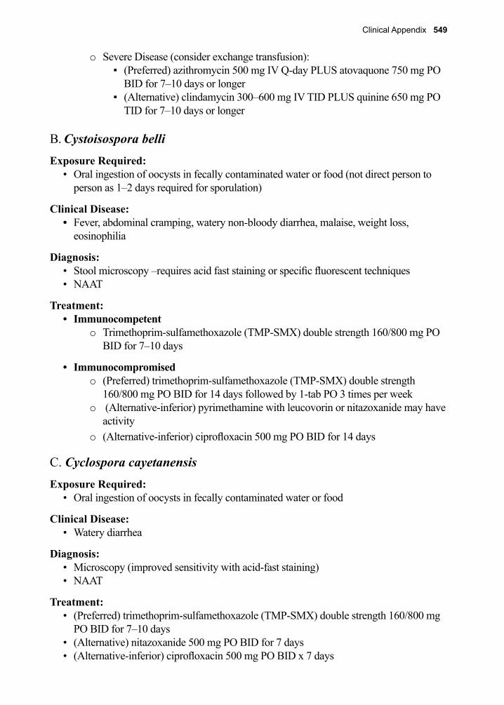
Clinical Appendix 549
o Severe Disease (consider exchange transfusion): ▪ (Preferred) azithromycin 500 mg IV Q-day PLUS atovaquone 750 mg PO
BID for 7–10 days or longer ▪ (Alternative) clindamycin 300–600 mg IV TID PLUS quinine 650 mg PO
TID for 7–10 days or longer
B. Cystoisospora belliExposure Required:
• Oral ingestion of oocysts in fecally contaminated water or food (not direct person to person as 1–2 days required for sporulation)
Clinical Disease:• Fever, abdominal cramping, watery non-bloody diarrhea, malaise, weight loss,
eosinophilia
Diagnosis:• Stool microscopy –requires acid fast staining or specific fluorescent techniques• NAAT
Treatment:• Immunocompetent
o Trimethoprim-sulfamethoxazole (TMP-SMX) double strength 160/800 mg PO BID for 7–10 days
• Immunocompromisedo (Preferred) trimethoprim-sulfamethoxazole (TMP-SMX) double strength
160/800 mg PO BID for 14 days followed by 1-tab PO 3 times per weeko (Alternative-inferior) pyrimethamine with leucovorin or nitazoxanide may have
activityo (Alternative-inferior) ciprofloxacin 500 mg PO BID for 14 days
C. Cyclospora cayetanensisExposure Required:
• Oral ingestion of oocysts in fecally contaminated water or food
Clinical Disease:• Watery diarrhea
Diagnosis:• Microscopy (improved sensitivity with acid-fast staining)• NAAT
Treatment: • (Preferred) trimethoprim-sulfamethoxazole (TMP-SMX) double strength 160/800 mg
PO BID for 7–10 days• (Alternative) nitazoxanide 500 mg PO BID for 7 days• (Alternative-inferior) ciprofloxacin 500 mg PO BID x 7 days
550 Clinical Appendix
D. Naeglaria fowleriExposure Required:
• Contact of the inside of the nasal cavity (cribriform plate) with water containing trophozoites
Clinical Disease:• Frontal headache, vomiting, confusion, fever, coma
Diagnosis:• Microscopy/histology• NAAT
Treatment: (multidrug regimen)• Conventional-amphotericin (not liposomal) 1.5 mg/kg/day IV, and• Rifampin 10 mg/kg/day PO in three doses, and • Fluconazole 10 mg/kg/day IV or PO, and• Miltefosine 50 mg PO BID or TID, and • Azithromycin 500 mg Q-day IV or PO
E. Acanthamoeba spp.Exposure Required:
• Most likely acquired through lungs or skin on exposure to keratitis contaminated tap water
Clinical Disease:• Encephalitic form- Granulomatous Amebic Encephalitis (GAE) - frontal headache,
diplopia, seizures• Ulcerative keratitis – gritty feeling in eye, impaired vision, blindness
Diagnosis:• Microscopy identification of trophozoites on wet mount, by confocal, or in fixed
specimens• Culture• NAAT
Treatment:• Keratitis – often combination therapy• Disseminated disease (skin, CNS, disseminated) – use combination therapy
F. Balamuthia mandrillaris Exposure Required:
• Acquired through lungs or skin
Clinical Disease:• Fever, stiff neck, headache, encephalitis
Clinical Appendix 551
Diagnosis:• Microscopy/histology (immunofluorescent antibodies available)• NAAT
Treatment:• Multi-drug regimens containing 4–5 agents such as amphotericin, fluconazole,
albendazole and miltefosine• (Additional alternative agents) voriconazole, flucytosine, pentamidine, azithromycin,
clarithromycin, trimethoprim-sulfamethoxazole and sulfadiazine
G. Blastocystis hominisExposure Required:
• Oral ingestion of cysts in fecally contaminated water or food
Clinical Disease:• Asymptomatic, diarrhea, abdominal discomfort
Diagnosis:• Stool microscopy • NAAT
Treatment:• Metronidazole 500 mg PO TID for 5–10 days• Tinidazole 2 g PO x1• Paromomycin 500 mg PO TID for 7–10 days• Trimethoprim-sulfamethoxazole (TMP-SMX) double strength 160/800 mg PO BID for
7 days • Nitazoxanide 500 mg PO BID for 3 days
H. Dientamoeba fragilisExposure Required:
• Oral ingestion of fecally contaminated water or food
Clinical Disease:• Diarrhea, nausea, abdominal discomfort
Diagnosis:• Stool microscopy –issues with sensitivity• NAAT
Treatment:• Metronidazole 500 mg PO TID for 10 days• Paromomycin 10 mg/kg PO TID for 7 days• Iodoquinol 650 mg PO TID for 20 days• Tetracycline 400 mg PO QID for 10 days• Doxycycline 100 mg PO BID for 10 days
552 Clinical Appendix
15. Non-pathogenic Protozoa• Commensal flagellates – no treatment• Commensal amoebae – no treatment
16. Enterobius vermicularisExposure Required:
• Oral ingestion of embryonated eggs, autoinfection
Clinical Disease:• Asymptomatic, perianal pruritus, vaginal irritation
Diagnosis:• Microscopy - pinworm paddle or clear adhesive tape to collect eggs, not standard O+P,
and direct visualization of adult females• NAAT
Treatment: Treatment of exposed contacts, all household members, and/or source patients, if not household members, is recommended and has been successful in both households and institutions.
• Pyrantel pamoate 11 mg/kg PO x1 then repeated 2–3 weeks later• Albendazole 400 mg PO x1 then repeated 2–3 weeks later• Mebendazole 100 mg PO x1 then repeated 2–3 weeks later• (Inferior option) ivermectin 200 mcg/kg PO x 1 then repeated 2–3 weeks later
17. Trichuris trichiuraExposure Required:
• Oral ingestion of embryonated eggs
Clinical Disease:• Asymptomatic, dysentery, tenesmus, weight loss, anemia, rectal prolapse
Diagnosis:• Stool microscopy - standard O+P and direct visualization of adults on endoscopy• NAAT
Treatment: • (Preferred) mebendazole 100 mg PO BID x 3 days• (Inferior option) albendazole 400 mg PO Q-day x 3 days• (Inferior option) ivermectin 200 mcg/kg PO x 1
Clinical Appendix 553
18. Ascaris lumbricoidesExposure Required:
• Oral ingestion of embryonated eggs
Clinical Disease:• Migratory phase – Löeffler’s syndrome -pneumonitis, hepatomegaly, bronchospasm,
eosinophilia• Intestinal phase – asymptomatic, high burden can lead to obstruction, aberrant
migration can lead to peritonitis or obstruction such as in hepatobiliary ascariasis
Diagnosis:• Stool microscopy - standard O+P and direct visualization of adults on endoscopy• NAAT
Treatment: • Albendazole 400 mg PO x 1• Mebendazole 500 mg PO x 1 or 100 mg PO BID for 3 days• Pyrantel pamoate 11 mg/kg PO x1 (can be used during pregnancy)• (Inferior option) ivermectin 200 mcg/kg PO x 1
19. The Hookworms Necator americanus Ancylostoma duodenale Ancylostoma ceylanicum
Exposure Required:• Infective L3 filariform larvae penetrate skin (usually through a hair follicle)
Ancyclostoma duodenale larvae are also infective orally
Clinical Disease:• Dermatitis-(during entry), pneumonia-(during migratory phase), abdominal pain-(can
occur with heavy oral ingestion with eosinophilia ‘Wakana disease’), chronic iron deficiency anemia, skin pigmentation change to yellow-green ‘chlorosis’-(chronic disease)
Diagnosis:• Stool microscopy - standard O+P and direct visualization of adults on endoscopy• NAAT
Treatment: Ivermectin has poor efficacy and is not recommended.
• (Preferred) albendazole 400 mg PO x 1• Mebendazole 500 mg PO x 1 or 100 mg PO BID x 1 day• Pyrantel pamoate 11 mg/kg PO Q-day x 3 days
554 Clinical Appendix
20. Strongyloides stercoralisExposure Required:
• Infective L3 filariform larvae penetrate skin (usually through a hair follicle)
Clinical Disease:• Asymptomatic, watery diarrhea, eosinophilia, dermatitis-(‘ground itch’), larva currens
rash, periumbilical thumbprint purpura rash, with hyperinfection-bacterial sepsis and bacterial meningitis, with S. fuelleborni swollen belly syndrome
Diagnosis:• Microscopy - stool O+P but larvae are seen not eggs, or histological examination of
tissues• Fecal culture - coproculture• Serology• NAAT
Treatment: Control (not elimination)
• Uncomplicated Infection:o Ivermectin 200 mcg/kg/day given once and then repeated 2 weeks later (so for
60 kg adult 4 of the 3 mg tablets PO each time)o (Inferior alternative) albendazole 400 mg PO Q-day for 7-days
• Disseminated Disease:o Ivermectin 200 mcg/kg/day PO Q-day with duration determined by clinical
response, some will add albendazole 400 mg PO Q-day if poor clinical response (Some patients have been successfully treated off label with subcutaneous dosing of veterinarian ivermectin preparations).
21. Trichinella spiralisExposure Required:
• Oral ingestion of raw or undercooked meats
Clinical Disease:• Gastrointestinal phase – secretory diarrhea, abdominal pain, nausea, vomiting• Parenteral phase – fever, myalgia, bilateral periorbital edema, petechial
hemorrhages, leukocytosis, eosinophilia, can have CNS or cardiac involvement with meningoencephalitis or arrythmias
Diagnosis:• Microscopy - histological examination of tissues after muscle biopsy• Serology - ELISA with Western blot confirmation• NAAT • Supportive laboratory tests – muscle enzymes, such as creatine kinase, lactic
dehydrogenase, and peripheral eosinophilia
Clinical Appendix 555
Treatment: • Albendazole 400 mg PO BID x 14 days• Mebendazole 400 mg PO TID x 14 days• Prednisone 30–60 mg Q-day for 14 days (add to regimen only if diagnosis secure)• Antipyretics and analgesics
22. Lymphatic Filariae Wuchereria bancrofti Brugia malayi
Exposure Required:• Bite from mosquito (wide variety of genera and species)
Clinical Disease:• Asymptomatic – lymphatic dilation detectable only by ultrasound or other testing• Acute lymphadenitis – fever, painful swelling of lymph nodes, secondary bacterial
infections• Elephantiasis – lymphedema of arms, legs, breasts, genitalia, secondary bacterial
infections• Tropical pulmonary eosinophilia – nocturnal asthma with dyspnea, fatigue, weight loss,
and eosinophilia
Diagnosis:• Microscopy - of blood smears during the night and can be concentrated• Serology - ELISA with Western blot confirmation• Antigen testing – circulating filarial antigen assay• NAAT - research tool, no commercially available tests• Ultrasound – lymphatic vessels and can detect filarial dance sign in the spermatic cord
Treatment: It is critical that, prior to treatment, co-infection with Loa loa with a high Loa loa microfilarial load is ruled out, due to the risk of severe adverse events if treatment is given to such patients.
• Antiparasitics:o Diethylcarbamazine (DEC) 6 mg/kg/day x 12 days for a total of 72 mg/kg body
weight o Doxycycline 100 mg PO BID for 6 weekso Ivermectin as part of mass drug programso Albendazole as part of mass drug programs
• Surgery: hydrocele drainage and lymphatic surgery• Complementary care:
o Lymphedema careo Treatment of wounds and secondary bacterial infections
556 Clinical Appendix
23. Onchocerca volvulusExposure Required:
• Bite of the black fly (Simulium spp.)
Clinical Disease:• Dermatitis – papular changes (can be extremely pruritic), lichenification (sowda),
atrophy (hanging groin), depigmentation (leopard skin), reddish facial lesions (erysipelas de la costa)
• Lymphadenopathy – Africa (inguinal), Americas (head and neck) • Ocular – keratitis, iritis, optic atrophy, optic neuritis, cataracts, chorioretinitis, blindness• Nodding syndrome – not clearly caused by this parasite but presents with seizures,
head nodding, periods of unresponsiveness, long-term disability
Diagnosis:• Microscopy - bloodless skin snips, blood smears to rule out other infections• Serology - ELISA with Western blot confirmation• Mazzotti test - now modified test• NAAT - research tool, no commercially available tests• Ultrasound - evaluation of nodules
Treatment: The major toxicity of ivermectin is generally not from the drug itself but rather from its ability to increase the antigen load from dead and dying parasites, leading to fever, angioedema and pruritus. These symptoms usually occur within 24 hrs of treatment. In those patients with concurrent Loa loa infection, ivermectin can elicit severe reactions, including encephalopathy and consequently it is essential to evaluate the patients in areas endemic for Loa loa for co-infection.
• Endemic Area: o Ivermectin 150 mcg/kg by mouth every 6 months for yearso (Alternative) doxycycline 100 mg PO 1x/day x 6 weeks followed by ivermectin
• Outside Endemic Area:o Ivermectin 150 mcg/kg by mouth every 6 months for several years until
asymptomatico (Alternative) doxycycline 100 mg PO 1x/day x 6 weeks followed by ivermectin
24. Loa loaExposure Required:
• Bite from the deer fly (Chrysops spp.)
Clinical Disease:• Asymptomatic, Calabar swellings, angioedema, localized swelling, worm migration
across eye, cardiomyopathy, renal disease, encephalitis, lymphadenitis, eosinophilia, -serious adverse reactions when treated for other parasitic infections
Clinical Appendix 557
Diagnosis:• Microscopy - of blood smears during the middle of the day and can be concentrated• Serology - ELISA with Western blot confirmation• NAAT - research tool, no commercially available tests
Treatment: It is critical that, prior to treatment, co-infection with L. loa with a high L. loa microfilarial load is ruled out, due to the risk of severe adverse events if treatment is given to such patients.
• >2,500 microfilariae/ml: If levels are greater than 2,500 mf/ml, then apheresis or treatment with albendazole 200 mg PO BID until loads <2,500 mf/ml.
• <2,500 microfilariae/ml: o Diethylcarbamazine (DEC)
▪ CDC regimen: DEC 8 to 10 mg/kg/day in 3 divided doses for 21 days; patients with symptomatic loiasis and microfilarial loads ≥ 8,000 mf/mL should receive apheresis or treatment with albendazole prior to treatment with diethylcarbamazine (CDC 2015). For patients with microfilaria in the blood, some clinicians recommend the following dose-escalating regimen: 50 mg as a single dose on day 1; 50 mg 3 times daily on day 2; 100 mg 3 times daily on day 3; 9 mg/kg/day in 3 divided doses on day 4 to end of treatment course. Repeat courses of treatment may be needed to achieve cure.
▪ World Health Organization recommendations: DEC 1 mg/kg as a single dose on day 1, with doubling of the dose on the next 2 days, then 6–9 mg/kg/day in divided doses 3 times daily for 18 days.
• Surgery: Adult worms in the eye can be removed surgically. • Albendazole: Alternatives to DEC include albendazole 200 mg PO BID x 21 days that
can effectively reduce the number of circulating microfilariae by acting directing on adult worms.
• Ivermectin is not a preferred agent for the treatment of loiasis and can be associated with significant morbidity if given to patients with high levels of circulating microfilariae.
• Chemoprophylaxis with weekly DEC given in a dose of 300 mg is effective in preventing loiasis among long-term visitors but is not currently recommended for short-term visitors to endemic areas.
25. Dracunculus medinensisExposure Required:
• Oral ingestion of infected copepods
Clinical Disease:• Cutaneous blisters and ulcers, allergic reactions and superinfection with failed
attempts to remove worms, arthritis, contractures and scarring causing disability and absenteeism from work and school
558 Clinical Appendix
Diagnosis:• Direct visualization: locating head of the adult worm in the skin lesion• Microscopy - identifying the larvae that are released into freshwater • ELISA - availability limited• Radiographs - calcifications corresponding to adult worms
Treatment: • Slow mechanical extraction of about 1cm/day, pain control and treatment of any
secondary bacterial infections
26. Other Nematodes of Medical Importance
A. Capillaria hepaticaExposure Required:
• Oral ingestion of embryonated eggs
Clinical Disease:• Asymptomatic, liver failure, abdominal lymphadenopathy, eosinophilia
Diagnosis:• Histology - after liver biopsy or at autopsy• Serological testing - ELISA and indirect fluorescent antibody test (IFA), high
sensitivity and specificity but not widely available
Treatment: • Combination therapy has been used successfully with combinations of albendazole,
thiabendazole, disophenol (2-6-diiodo-4-nitrophenol) and prednisone
B. Capillaria philippinensisExposure Required:
• Oral ingestion of raw or undercooked freshwater fish or crustaceans
Clinical Disease:• Diarrhea
Diagnosis:• Stool microscopy - standard O+P with direct visualization of eggs or larvae in stool or
detecting the adults on small bowel biopsy of the small intestinal wall
Treatment: • Albendazole 400 mg PO Q-day x 30 days• Mebendazole 200 mg PO BID x 20–30 days
Clinical Appendix 559
C. Dirofilaria immitisExposure Required:
• Bite of infected mosquito
Clinical Disease:• Coin lesion in lung
Diagnosis:• Histology• Radiographs - coin lesion in lungs
Treatment: No known effective therapies in humans
D. Mansonella ozzardiExposure Required:
• Bite of black flies and biting midges
Clinical Disease:• Asymptomatic, urticaria, lymphadenopathy, chronic arthritis, eosinophilia
Diagnosis:• Microscopy - visualization of microfilariae in a blood smear, sensitivity increased with
concentration techniques • ELISA - based on crude antigen preparations but with limited specificity• PCR - developed and available through the National Institutes of Health
Treatment: • Ivermectin 200 mcg/kg/day PO x1
E. Mansonella perstansExposure Required:
• Bite from biting midge
Clinical Disease:• Asymptomatic, painless conjunctival nodules, eyelid swelling, angioedema, (called the
Ugandan or Kampala eye worm)
Diagnosis:• Microscopy - visualization of microfilariae in a blood smear, sensitivity increased with
concentration techniques • ELISA - based on crude antigen preparations but with limited specificity• PCR - developed and available through the National Institutes of Health
560 Clinical Appendix
Treatment: Considered one of the most challenging filarial infections to treat.
• Combination therapy has been successful• Doxycycline has been successfully used on strains from Mozambique and the
Democratic Republic of Congo that harbor the endosymbiont Wolbachia
F. Mansonella streptocercaExposure Required:
• Biting midges
Clinical Disease:• Pruritic dermatitis, hypopigmented macules
Diagnosis:• Microscopy - visualization of microfilariae in a blood smear, sensitivity increased with
concentration techniques • ELISA - based on crude antigen preparations but with limited specificity• PCR - developed and available through the National Institutes of Health
Treatment: • Diethylcarbamazine (DEC) 2 mg/kg/day TID for 12 days• Ivermectin 150 mcg/kg/day PO x1 can reduce microfilarial levels
G. Oesophagostomum bifurcumExposure Required:
• Oral ingestion of infective larvae
Clinical Disease:• Asymptomatic, abdominal nodules or masses, abdominal pain
Diagnosis:• Stool microscopy - standard O+P with direct visualization of eggs (cannot be visually
distinguished from hookworm eggs)• Coproculture - allowing eggs to develop to third stage larvae (difficult and time
consuming)• Histology of biopsied nodules showing larval or adult forms• Imaging - Ultrasound • NAAT - PCR/Multiplex
Treatment: • Pyrantel pamoate 11mg/kg PO x1• Albendazole 400 mg PO x1
Clinical Appendix 561
H. Ternidens diminutus Exposure Required:
• Oral ingestion of infective larvae
Clinical Disease:• Colonic ulcerations, nodular lesions, abdominal mass
Diagnosis:• Stool microscopy - standard O+P with direct visualization of eggs (can not be visually
distinguished from hookworm eggs)
Treatment: • Pyrantel pamoate 11 mg/kg PO x1• Albendazole 400 mg PO x1
27. Aberrant Nematode Infections
A. Cutaneous Larva MigransExposure Required:
• Infective larvae penetrate unbroken skin
Clinical Disease:• Serpiginous lesions, pruritus, secondary bacterial infections
Diagnosis:• Physical findings: visualization of serpiginous lesions• Dermoscopy - translucent brown areas and red-dotted vessels
Treatment: • (Preferred) Ivermectin 200 mcg/kg PO Q-day x 1–2 days• (Alternative) Albendazole 400 mg PO Q-day x 3–7 days• Topical thiabendazole 15% applied daily for 5 days• Topical albendazole ointment 10% applied TID x 10 days
B. Visceral Larva MigransExposure Required:
• Oral ingestion of embryonated eggs
Clinical Disease:• Tissue damage, cognitive defects, hypersensitivity responses, eosinophilia
Diagnosis:• Serology - ELISA• NAAT - research tool, no commercially available tests• Ophthalmological exam for ocular larva migrans (OLM)• Imaging - CT, MRI, Ultrasound
562 Clinical Appendix
Treatment: • Steroids for severe or CNS reactions• Albendazole 400 mg PO BID x 5 days
C. Ocular Larva MigransExposure Required:
• Oral ingestion of embryonated eggs
Clinical Disease:• Ocular granulomas, visual disturbance, blindness
Diagnosis:• Serology - ELISA
Treatment: • Steroids for some ophthalmic or CNS reactions• Albendazole 400 mg PO BID x 5 days• Surgery (vitrectomy)
D. Baylisascaris procyonis Exposure Required:
• Oral ingestion of embryonated eggs
Clinical Disease:• Nausea, fatigue, hepatomegaly, loss of coordination and muscle control, eosinophilic
meningitis, ocular disease, encephalitis, coma
Diagnosis:• Serology - recombinant protein-based ELISA
Treatment: • Albendazole 25–50 mg/kg/day in divided doses x 20 days, with concomitant steroids
E. Angiostrongylus cantonensis/costaricensis Exposure Required:
• Oral ingestion of infective larvae
Clinical Disease:• A. cantonensis - eosinophilic meningoencephalitis, fever, headache, painful
paresthesias• A. costaricensis – abdominal pain, fever, nausea, vomiting
Clinical Appendix 563
Diagnosis:• CSF examination for eosinophilia• PCR of CSF fluid• Imaging - MRI may reveal areas of enhancement in characteristic patterns• Serology - ELISA (not widely available and older assays cross react with
gnathostomiasis)
Treatment: • Analgesics, repeated lumbar punctures, steroids and albendazole are used but optimal
treatment is not defined
F. Gnathostoma spinigerum Exposure Required:
• Ingestion of infective larvae in fish, snakes, and birds
Clinical Disease:• Asymptomatic, cutaneous larva migrans, subcutaneous swellings, eosinophilic
meningitis, painful paresthesias
Diagnosis:• CSF examination for eosinophilia• Imaging - MRI may reveal areas of enhancement in characteristic patterns and CT may
demonstrate areas of hemorrhage• Serology - ELISA (not widely available and older assays cross react with
Angiostrongylus cantonensis)
Treatment: • Albendazole 400 mg PO Q-day x 21 days• Ivermectin 200 mcg/kg PO Q-day x 2 days• Repeated lumbar punctures to reduce opening pressure, steroids
G. Anisakiasis Exposure Required:
• Oral ingestion of infective larvae in raw or undercooked saltwater fish or squid
Clinical Disease:• Abdominal pain, nausea, vomiting, diarrhea, fever
Diagnosis:• Direct visualization of parasites by endoscopy or in vomit• Serology - ELISA• PCR - not commercially available outside research settings
Treatment: • Physical removal of the parasite prior to penetration• Surgery after penetration• Albendazole 400 mg PO BID for 3–21 days but optimal therapy is not defined
564 Clinical Appendix
28. Taenia saginataExposure Required:
• Ingestion of raw or undercooked beef containing cysticerci
Clinical Disease:• Asymptomatic, proglottids noted in stool or clothing
Diagnosis:• Microscopy:
o Gravid proglottids can be fixed in 10% formaldehyde solution, and the uterus injected with India ink, with the aid of a 26-gauge needle or stained with hematoxylin-eosin staining techniques. T. saginata proglottids have 12 or more branches on either side of the uterus.
o Eggs of T. saginata are occasionally found in stool, since most proglottids usually pass out of the host intact. If an egg is seen on stool examination, the species cannot be determined on visual microscopy based on morphology, since all members of the family Taeniidae produce visually identical ova. Upon acid-fast staining, occasionally the species can be distinguished, as fully mature eggs of T. saginata have an acid-fast shell.
o Paddle Test/Sticky Tape Test is an additional diagnostically relevant test (see: diagnosis for Enterobius vermicularis). When proglottids migrate out of the anus, they express eggs that remain on the perineum.
• NAAT – PCR/ loop-mediated isothermal amplification (LAMP)/Multiplex• Antigen detection (coproantigens) – used on stool samples
Treatment: • Praziquantel 5–10 mg/kg PO x1 (600 mg tablets)• Niclosamide 2 g PO x1 (not commercially available in the United States)
29. Taenia solium
IntestinalExposure Required:
• Ingestion of raw or undercooked pork containing cysticerci
Clinical Disease:• Asymptomatic, proglottids noted in stool or clothing
Diagnosis:• Microscopy:
o Gravid proglottids can be fixed in 10% formaldehyde solution, and the uterus injected with India ink, with the aid of a 26-gauge needle or stained with hematoxylin-eosin staining techniques. T. solium proglottids have less than 12 branches on either side of the uterus.
Clinical Appendix 565
o Eggs of T. solium are occasionally found in stool, since most proglottids usually pass out of the host intact. If an egg is seen on stool examination, the species cannot be determined on visual microscopy based on morphology, since all members of the family Taeniidae produce visually identical ova. Upon acid-fast staining, occasionally the species can be distinguished, as fully mature eggs of T. saginata have an acid-fast shell and T. solium eggs have a shell that is not acid-fast.
o Paddle Test/Sticky Tape Test is an additional diagnostically relevant test (see: diagnosis for Enterobius vermicularis). When proglottids migrate out of the anus, they express eggs that remain on the perineum.
• NAAT - PCR/ loop-mediated isothermal amplification (LAMP)/Multiplex• Antigen detection (coproantigens) - used on stool samples
Treatment: • Praziquantel 5–10 mg/kg PO x1 (600 mg tablets)• Niclosamide 2 g PO x1 (not commercially available in the United States)
Extra-intestinal (Cysticercosis and Neurocysticercosis)Exposure Required:
• Oral ingestion of embryonated eggs
Clinical Disease:• Extraneural – subcutaneous (discrete swellings that go on to become tender),
intramuscular (asymptomatic, cysts in muscles, calcifications)• Neurocysticercosis – intraparenchymal/extraparenchymal (space occupying symptoms,
headaches, seizures, hydrocephalus, focal neurological abnormalities)
Diagnosis:• Imaging - recommend that patients undergo both MRI and CT for CNS disease, plain
radiographs can also show peripheral calcified disease• Serology - Enzyme-linked immune-transfer blot, rather than crude antigen assays• NAAT - PCR/ loop mediated isothermal amplification (LAMP) /Multiplex
Treatment: • Monotherapy for 1–2 viable cysts - albendazole 7.5 mg/kg PO BID for 10 days (200
mg tablets)• Combination therapy for > 2 viable cysts - albendazole 7.5 mg/kg PO BID for 10 days
(200 mg tablets) and praziquantel 5 mg/kg PO TID for 10 days (600 mg tablets)• Corticosteroids (recommended for all CNS disease when using antiparasitics) -
prednisone 1 mg/kg PO Q-day x 5–10 days then tapered or dexamethasone 10 mg IV x1 then 4 mg IV q6 hrs x 5–10 days then tapered. For acute encephalitis steroids alone without antiparasitics are recommended.
• Antiepileptic therapy - recommended for all patients having seizures• Mechanical therapy - for extraparenchymal neurocysticercosis surgical therapy and
ventriculoperitoneal shunting may be required.
566 Clinical Appendix
30. Diphyllobothrium latumExposure Required:
• Oral ingestion of infective larvae from eating raw or undercooked freshwater or andromous fish
Clinical Disease:• Asymptomatic, watery diarrhea, fatigue, B12 deficiency
Diagnosis:• Microscopy:
o Gravid proglottids can be fixed in 10% formaldehyde solution, and stained with hematoxylin-eosin staining techniques. These proglottids are wider than they are long (most proglottids do not pass out of the host intact).
o Eggs can be found in stool (most proglottids do not pass out of the host intact)• NAAT
Treatment: • Praziquantel 5–10 mg/kg PO x1 (600 mg tablets)• Niclosamide 2 g PO x1 (not commercially available in the United States)
31. Other Tapeworms of Medical Importance
A. Hymenolepis nanaExposure Required:
• Oral ingestion of infective larvae along with infected insect or oral ingestion of embryonated eggs
Clinical Disease:• Asymptomatic, rarely diarrhea
Diagnosis:• Standard detection of eggs on stool O+P
Treatment: • Praziquantel 5–10 mg/kg PO x1 (600 mg tablets)• (Inferior alternative) nitazoxanide 500 mg PO BID x 3 days
B. Hymenolepis diminutaExposure Required:
• Oral ingestion of infective larvae along with infected insect
Clinical Disease:• Asymptomatic
Diagnosis:• Standard detection of eggs on stool O+P
Clinical Appendix 567
Treatment: • Praziquantel 5–10 mg/kg PO x1 (600 mg tablets)• (Inferior alternative) nitazoxanide 500 mg PO BID x 3 days
C. Dipylidium caninum Exposure Required:
• Oral ingestion of infected adult fleas
Clinical Disease:• Asymptomatic
Diagnosis:• Standard detection of eggs on stool O+P
Treatment: • Praziquantel 5–10 mg/kg PO x1 (600 mg tablets)• (Inferior alternative) nitazoxanide 500 mg PO BID x 3 days
32. Juvenile Tapeworm Infections of Humans
A. Echinococcus granulosusExposure Required:
• Oral ingestion of embryonated eggs
Clinical Disease:• Liver cysts, lung cysts, cysts in any organ, anaphylactic reactions with cyst rupture
Diagnosis:• Imaging: cysts can be visualized with CT, MRI and ultrasound• Microscopy: examination of cyst contents and cysts themselves• Serological testing: sensitivities vary by cyst stage• NAAT
568 Clinical Appendix
Treatment: • Based on Stage CE1–CE5:
o CE1 ▪ < 5cm - Albendazole 400 mg PO BID ▪ > 5cm - Albendazole 400 mg PO BID and puncture, aspiration injection,
re-aspiration (PAIR)o CE3a
▪ < 5cm - Albendazole 400 mg PO BID ▪ > 5cm - Albendazole 400 mg PO BID and PAIR
o CE2 - Albendazole 40 0 mg PO BID and large bore percutaneous treatment, (PAIR is contraindicated)
o CE3b - Albendazole 400 mg PO BID and large bore percutaneous treatment, (PAIR is contraindicated)
o CE4 & CE5 - observation with imaging every 6 months, (PAIR is contraindicated)
o Surgery - indicated for cysts >10 cm, ruptured cysts, extra-hepatic disease, very complex cysts (many daughter cells) and cysts that have formed fistula.
B. Echinococcus multilocularis Exposure Required:
• Oral ingestion of embryonated eggs
Clinical Disease:• Proliferative membranes primarily in the liver leading to hepatic failure, abdominal
pain, weight loss, fatigue
Diagnosis:• Imaging - lesions can be visualized with CT, MRI and ultrasound• Microscopy - histology of directed biopsy specimens• Serological testing - sensitive and specific enough to distinguish from E. granulosus• NAAT
Treatment: • Surgery is the primary approach when possible• Albendazole 400 mg PO BID suggested minimum of 2 years but indefinitely if not
amenable to surgery
C. Mesocestoides spp.Exposure Required:
• Oral ingestion of infective larvae in under cooked bird, snake, lizard, amphibian or mammalian carnivore
Clinical Disease:• Mild abdominal discomfort, nausea, diarrhea, vomiting
Diagnosis:• Detection of eggs on stool O+P
Clinical Appendix 569
Treatment: • Praziquantel 5–10 mg/kg PO x1 (600 mg tablets)• (Inferior alternative) nitazoxanide 500 mg PO BID x 3 days
D. Spirometra spp.Exposure Required:
• Oral ingestion of infective larvae in undercooked meat or exposure to larvae from poultice that then invade through wound or mucous membrane
Clinical Disease:• Asymptomatic, orbital edema, neurological complications
Diagnosis:• Identification of the parasite after removal or biopsy
Treatment: • Primarily Surgical Management
E. Taenia spp. (other than T. saginata and T. solium)Exposure Required:
• Oral ingestion of embryonated eggs
Clinical Disease:• Mass effect in organ invaded, may invade CNS (brain, eyes, spinal cord)
Diagnosis:• Identification of the parasite after removal or biopsy
Treatment: • Primarily surgical management
33. The Schistosomes Schistosoma mansoni Schistosoma japonicum Schistosoma haematobium Schistosoma mekongi Schistosoma intercalatum
Exposure Required:• Infective cercariae enter skin (usually through a hair follicle)
Clinical Disease:• Acute – ‘Katayama fever’ hepatomegaly, splenomegaly, lymphadenopathy, fever,
myalgias, cough, headache, eosinophilia• Chronic – abdominal pain, diarrhea, hepatomegaly, splenomegaly, hematuria, vaginal
symptoms in female genital schistosomiasis (FGS), CNS - (focal transverse myelitis, encephalitis)
570 Clinical Appendix
Diagnosis:• Microscopy: detection of schistosome eggs in stool or urine. Detection of eggs in an
unfixed rectal snip/biopsy• Antigen detection: two schistosome glycoprotein antigens known as CCA and CAA
circulate in the bloodstream of acutely infected patients and can be detected with certain assays
• Serology: antibodies develop 6–12 weeks after exposure and tend to become positive before eggs are evident in urine or stool. (ELISA, indirect hemagglutination assay (IHA), radioimmunoassay, complement fixation, Western blot)
• Imaging: portable ultrasound imaging has been shown to be clinically useful in the diagnosis of schistosomiasis
Treatment: Detection of viable eggs 6 weeks after treatment warrants retreatment
• Acute Infectiono Prednisone 40 mg PO Q-day x 5 dayso Praziquantel 20 mg/kg PO TID x 1 day (6 weeks after exposure and when acute
symptoms have resolved) then repeated 6 weeks later (600 mg tablets)• Chronic Infection
o Praziquantel 20 mg/kg PO TID x 1 day (8 weeks after exposure and when acute symptoms have resolved) (600 mg tablets)
• CNS Schistosomiasiso Prednisone 1mg/kg PO Q-day started immediately with duration based on
response and clinical course
34. Clonorchis sinensis and Opisthorchis spp. Exposure Required:
• Oral ingestion of raw or undercooked freshwater fish containing metacercariae
Clinical Disease:• Asymptomatic, right upper quadrant abdominal pain, nausea, diarrhea, headache,
hepatomegaly, eosinophilia
Diagnosis:• Microscopy - after 4 weeks eggs will be released into feces. The sensitivity can be
improved with NAAT (PCR and loop-mediated isothermal amplification ([LAMP])• Endoscopy - endoscopic retrograde cholangiopancreatography (ERCP) may allow
visualization of flukes• Serology - ELISA with confirmatory Western blot is available• Imaging - the presence of flukes in the biliary tract may also be observed using
ultrasound, CT, MRI and cholangiography
Clinical Appendix 571
Treatment: • Antiparasitics
o Praziquantel 25 mg/kg PO TID x 2 days or praziquantel 40 mg/kg PO x1 for light infections (600 mg tablets)
o Albendazole 10 mg/kg PO Q-day x 7 dayso Mebendazole 30 mg/kg PO Q-day x 30 days
• Mechanical Interventionso Surgery, biliary drainage and broad-spectrum antibiotics may be required in
certain cases
35. Fasciola hepaticaExposure Required:
• Oral ingestion of metacercariae on watercress or other littoral plants
Clinical Disease:• Early phase – fever, right upper quadrant abdominal pain, malaise, headache,
eosinophilia• Chronic – dull right upper quadrant pain, biliary obstruction
Diagnosis:• Serology - serological tests become positive early in disease during migration through
the liver parenchyma• Antigen tests - available with high sensitivity and specificity• Microscopy - after 4 months eggs will start to be released into feces• Endoscopy - endoscopic retrograde cholangiopancreatography (ERCP) may allow
visualization of flukes• Imaging - the presence of linear migratory tracts and adult flukes may also be observed
using ultrasound, CT, MRI and cholangiography
Treatment: In the United States Triclabendazole can be obtained through the CDC.
• Triclabendazole 10 mg/kg PO x1 and then in severe infections may be repeated 12–24 hrs after the first dose. Successfully treated patients will develop negative serologies 6–12 months after clearing their parasites.
• (Inferior alternative) Nitazoxanide 500 mg PO BID for 7 days
36. Paragonimus westermani / P. kellicotti Exposure Required:
• Oral ingestion of metacercariae on raw or undercooked crab or crustaceans
Clinical Disease:• Acute - asymptomatic, diarrhea, fever, chest pain, fatigue, urticaria, epigastric pain,
eosinophilia • Late – fever, chills, cough, dyspnea, blood tinged sputum, hemoptysis, pulmonary
infiltrates, pulmonary lesions, CNS-more common with P. kellicotti
572 Clinical Appendix
Diagnosis:• Serology - serological tests are important in early disease before egg production occurs
which can take 8–12 weeks• Microscopy - late-stage disease is diagnosed by microscopic identification of eggs in
the sputum, bronchoalveolar lavage fluid, and, more rarely, in stool• Imaging - ultrasound, X-ray examinations, CT, MRI and fluorodeoxyglucose-positron
emission tomography (FDG-PET)
Treatment: • Praziquantel 25 mg/kg PO TID for 3 days (600 mg tablets)• Triclabendazole 10 mg/kg PO x1
37. Other Trematodes of Medical Importance
A. Fasciolopsis buskiExposure Required:
• Oral ingestion of metacercariae on husks of seeds of littoral plants such as water chestnuts
Clinical Disease:• Asymptomatic, diarrhea, vomiting, nausea, fever, intestinal hemorrhage, abdominal
pain, eosinophilia
Diagnosis:• Microscopy - identification of eggs or flukes in stool or vomit
Treatment: • Praziquantel 25 mg/kg PO TID x 1 day (600 mg tablets)
B. Echinostoma spp.Exposure Required:
• Oral ingestion of metacercariae from ingesting various species of snails, tadpoles, or freshwater fish
Clinical Disease:• Diarrhea, nausea, vomiting, abdominal pain, fever
Diagnosis:• Microscopy - identification of eggs in stool or in some cases flukes recovered from
endoscopy
Treatment: • Praziquantel 25 mg/kg PO x 1 day (600 mg tablets)
Clinical Appendix 573
C. Heterophyes heterophyes/Metagonimus yokogawaiExposure Required:
• Oral ingestion of metacercariae from ingesting certain freshwater fish
Clinical Disease:• Epigastric pain, fatigue, diarrhea, weight loss, malaise, belching, nausea, headache,
vomiting
Diagnosis:• Microscopy - identification of eggs in stool • NAAT – only available in research settings
Treatment: • Praziquantel 25 mg/kg PO TID x 1 day (600 mg tablets)
D. Nanophyetus salmincolaExposure Required:
• Ingestion of raw or undercooked salmon containing metacercariae
Clinical Disease:• Diarrhea, nausea, vomiting, anorexia, eosinophilia
Diagnosis:• Microscopy: identification of eggs in stool
Treatment: • Praziquantel 25 mg/kg PO TID x 1 day (600 mg tablets)• Niclosamide 2 g PO x1 (not commercially available in the United States)
38. The Insects
A. Myiasis-Causing Flies: Calliphoridae, Cuterebridae, and SarcophagidaeExposure Required:
• Larvae penetrate intact skin or wounds
Clinical Disease:• Abscess like swellings with openings, maggots visible in wounds
Diagnosis:• Visualization of living or dead maggots, but suggested by abscess like lesions with
small central opening
Treatment: • Surgical removal is the primary treatment • Alternatively, if the opening is blocked with a substance such as petroleum jelly, that
blocks access to oxygen, they can be forced to crawl to the surface and be removed
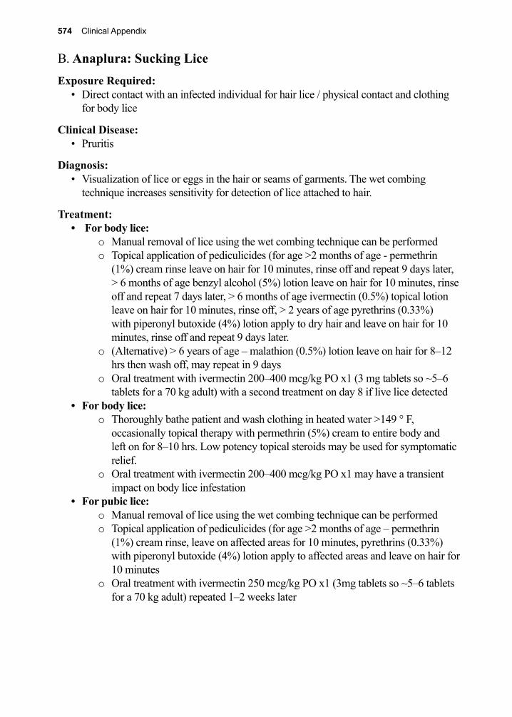
574 Clinical Appendix
B. Anaplura: Sucking LiceExposure Required:
• Direct contact with an infected individual for hair lice / physical contact and clothing for body lice
Clinical Disease:• Pruritis
Diagnosis:• Visualization of lice or eggs in the hair or seams of garments. The wet combing
technique increases sensitivity for detection of lice attached to hair.
Treatment: • For body lice:
o Manual removal of lice using the wet combing technique can be performed o Topical application of pediculicides (for age >2 months of age - permethrin
(1%) cream rinse leave on hair for 10 minutes, rinse off and repeat 9 days later, > 6 months of age benzyl alcohol (5%) lotion leave on hair for 10 minutes, rinse off and repeat 7 days later, > 6 months of age ivermectin (0.5%) topical lotion leave on hair for 10 minutes, rinse off, > 2 years of age pyrethrins (0.33%) with piperonyl butoxide (4%) lotion apply to dry hair and leave on hair for 10 minutes, rinse off and repeat 9 days later.
o (Alternative) > 6 years of age – malathion (0.5%) lotion leave on hair for 8–12 hrs then wash off, may repeat in 9 days
o Oral treatment with ivermectin 200–400 mcg/kg PO x1 (3 mg tablets so ~5–6 tablets for a 70 kg adult) with a second treatment on day 8 if live lice detected
• For body lice: o Thoroughly bathe patient and wash clothing in heated water >149 ° F,
occasionally topical therapy with permethrin (5%) cream to entire body and left on for 8–10 hrs. Low potency topical steroids may be used for symptomatic relief.
o Oral treatment with ivermectin 200–400 mcg/kg PO x1 may have a transient impact on body lice infestation
• For pubic lice: o Manual removal of lice using the wet combing technique can be performed o Topical application of pediculicides (for age >2 months of age – permethrin
(1%) cream rinse, leave on affected areas for 10 minutes, pyrethrins (0.33%) with piperonyl butoxide (4%) lotion apply to affected areas and leave on hair for 10 minutes
o Oral treatment with ivermectin 250 mcg/kg PO x1 (3mg tablets so ~5–6 tablets for a 70 kg adult) repeated 1–2 weeks later
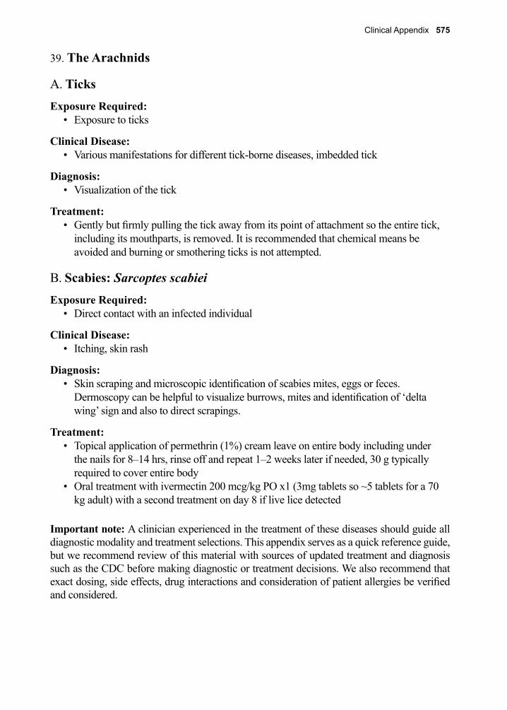
Clinical Appendix 575
39. The Arachnids
A. TicksExposure Required:
• Exposure to ticks
Clinical Disease:• Various manifestations for different tick-borne diseases, imbedded tick
Diagnosis:• Visualization of the tick
Treatment: • Gently but firmly pulling the tick away from its point of attachment so the entire tick,
including its mouthparts, is removed. It is recommended that chemical means be avoided and burning or smothering ticks is not attempted.
B. Scabies: Sarcoptes scabieiExposure Required:
• Direct contact with an infected individual
Clinical Disease:• Itching, skin rash
Diagnosis:• Skin scraping and microscopic identification of scabies mites, eggs or feces.
Dermoscopy can be helpful to visualize burrows, mites and identification of ‘delta wing’ sign and also to direct scrapings.
Treatment: • Topical application of permethrin (1%) cream leave on entire body including under
the nails for 8–14 hrs, rinse off and repeat 1–2 weeks later if needed, 30 g typically required to cover entire body
• Oral treatment with ivermectin 200 mcg/kg PO x1 (3mg tablets so ~5 tablets for a 70 kg adult) with a second treatment on day 8 if live lice detected
Important note: A clinician experienced in the treatment of these diseases should guide all diagnostic modality and treatment selections. This appendix serves as a quick reference guide, but we recommend review of this material with sources of updated treatment and diagnosis such as the CDC before making diagnostic or treatment decisions. We also recommend that exact dosing, side effects, drug interactions and consideration of patient allergies be verified and considered.
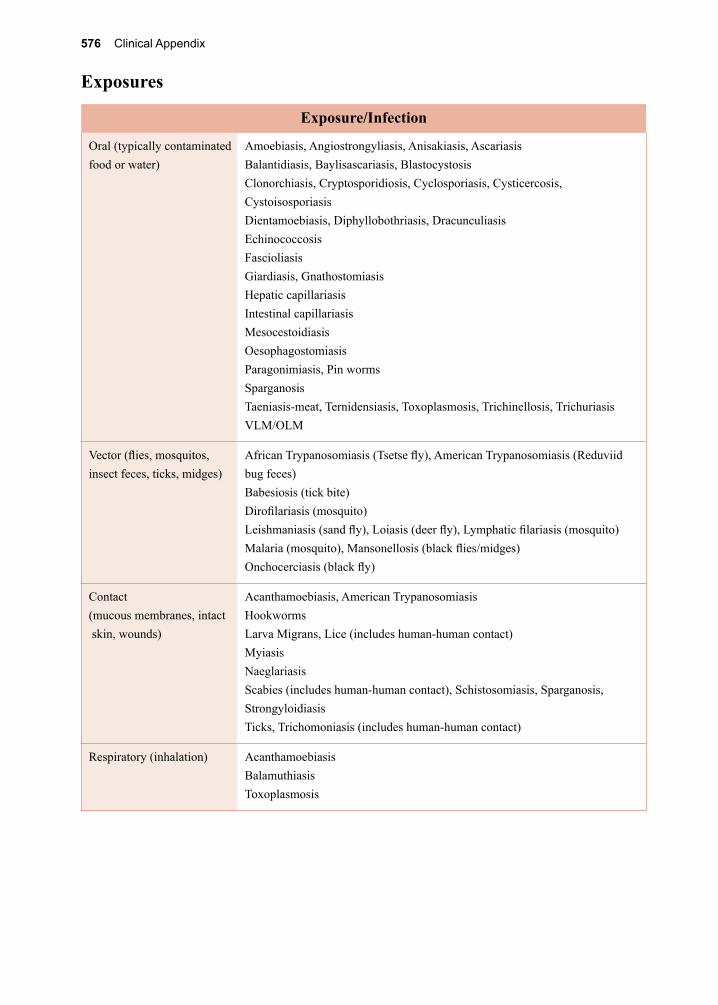
576 Clinical Appendix
Exposures
Exposure/Infection
Oral (typically contaminated food or water)
Amoebiasis, Angiostrongyliasis, Anisakiasis, AscariasisBalantidiasis, Baylisascariasis, BlastocystosisClonorchiasis, Cryptosporidiosis, Cyclosporiasis, Cysticercosis, CystoisosporiasisDientamoebiasis, Diphyllobothriasis, DracunculiasisEchinococcosisFascioliasisGiardiasis, GnathostomiasisHepatic capillariasisIntestinal capillariasisMesocestoidiasisOesophagostomiasisParagonimiasis, Pin wormsSparganosisTaeniasis-meat, Ternidensiasis, Toxoplasmosis, Trichinellosis, TrichuriasisVLM/OLM
Vector (flies, mosquitos, insect feces, ticks, midges)
African Trypanosomiasis (Tsetse fly), American Trypanosomiasis (Reduviid bug feces)Babesiosis (tick bite)Dirofilariasis (mosquito)Leishmaniasis (sand fly), Loiasis (deer fly), Lymphatic filariasis (mosquito)Malaria (mosquito), Mansonellosis (black flies/midges)Onchocerciasis (black fly)
Contact(mucous membranes, intact skin, wounds)
Acanthamoebiasis, American TrypanosomiasisHookwormsLarva Migrans, Lice (includes human-human contact)MyiasisNaeglariasisScabies (includes human-human contact), Schistosomiasis, Sparganosis, StrongyloidiasisTicks, Trichomoniasis (includes human-human contact)
Respiratory (inhalation) AcanthamoebiasisBalamuthiasisToxoplasmosis
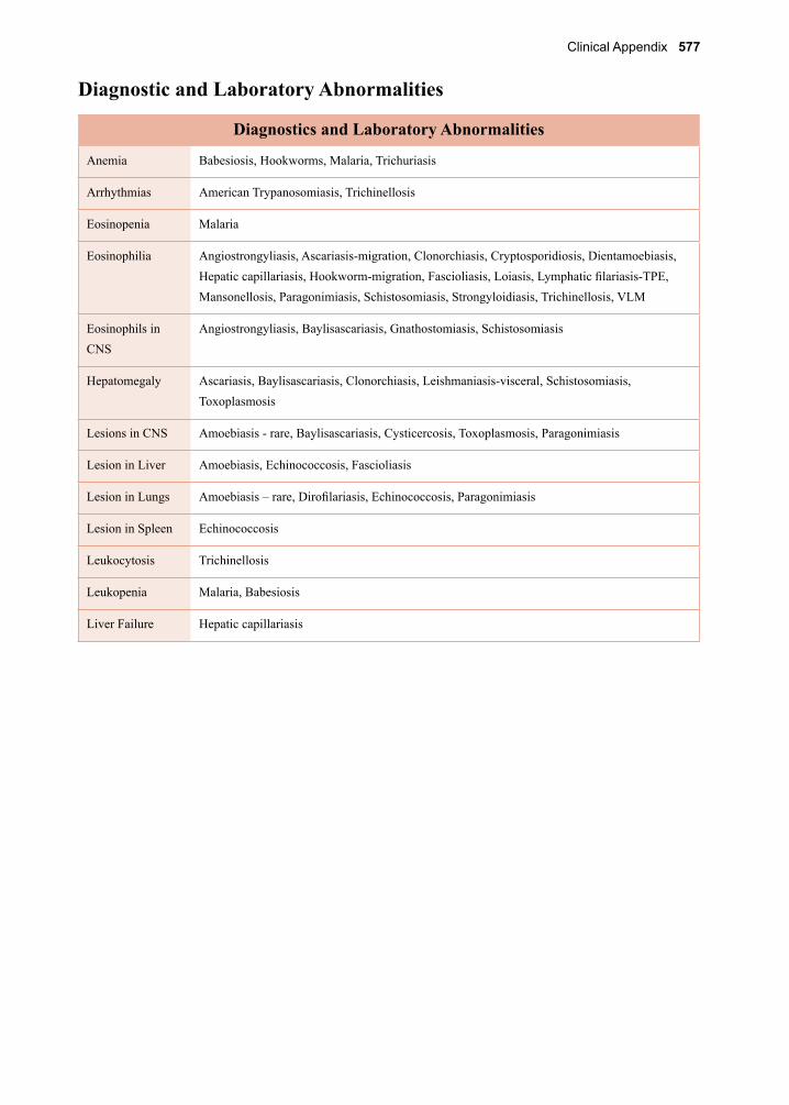
Clinical Appendix 577
Diagnostic and Laboratory Abnormalities
Diagnostics and Laboratory Abnormalities
Anemia Babesiosis, Hookworms, Malaria, Trichuriasis
Arrhythmias American Trypanosomiasis, Trichinellosis
Eosinopenia Malaria
Eosinophilia Angiostrongyliasis, Ascariasis-migration, Clonorchiasis, Cryptosporidiosis, Dientamoebiasis, Hepatic capillariasis, Hookworm-migration, Fascioliasis, Loiasis, Lymphatic filariasis-TPE, Mansonellosis, Paragonimiasis, Schistosomiasis, Strongyloidiasis, Trichinellosis, VLM
Eosinophils in CNS
Angiostrongyliasis, Baylisascariasis, Gnathostomiasis, Schistosomiasis
Hepatomegaly Ascariasis, Baylisascariasis, Clonorchiasis, Leishmaniasis-visceral, Schistosomiasis, Toxoplasmosis
Lesions in CNS Amoebiasis - rare, Baylisascariasis, Cysticercosis, Toxoplasmosis, Paragonimiasis
Lesion in Liver Amoebiasis, Echinococcosis, Fascioliasis
Lesion in Lungs Amoebiasis – rare, Dirofilariasis, Echinococcosis, Paragonimiasis
Lesion in Spleen Echinococcosis
Leukocytosis Trichinellosis
Leukopenia Malaria, Babesiosis
Liver Failure Hepatic capillariasis
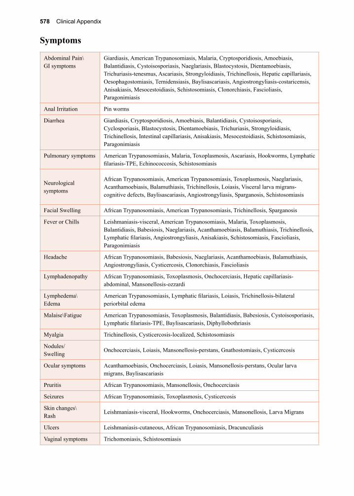
578 Clinical Appendix
Symptoms
Abdominal Pain\GI symptoms
Giardiasis, American Trypanosomiasis, Malaria, Cryptosporidiosis, Amoebiasis, Balantidiasis, Cystoisosporiasis, Naeglariasis, Blastocystosis, Dientamoebiasis, Trichuriasis-tenesmus, Ascariasis, Strongyloidiasis, Trichinellosis, Hepatic capillariasis, Oesophagostomiasis, Ternidensiasis, Baylisascariasis, Angiostrongyliasis-costaricensis, Anisakiasis, Mesocestoidiasis, Schistosomiasis, Clonorchiasis, Fascioliasis, Paragonimiasis
Anal Irritation Pin worms
Diarrhea Giardiasis, Cryptosporidiosis, Amoebiasis, Balantidiasis, Cystoisosporiasis, Cyclosporiasis, Blastocystosis, Dientamoebiasis, Trichuriasis, Strongyloidiasis, Trichinellosis, Intestinal capillariasis, Anisakiasis, Mesocestoidiasis, Schistosomiasis, Paragonimiasis
Pulmonary symptoms American Trypanosomiasis, Malaria, Toxoplasmosis, Ascariasis, Hookworms, Lymphatic filariasis-TPE, Echinococcosis, Schistosomiasis
Neurological symptoms
African Trypanosomiasis, American Trypanosomiasis, Toxoplasmosis, Naeglariasis, Acanthamoebiasis, Balamuthiasis, Trichinellosis, Loiasis, Visceral larva migrans-cognitive defects, Baylisascariasis, Angiostrongyliasis, Sparganosis, Schistosomiasis
Facial Swelling African Trypanosomiasis, American Trypanosomiasis, Trichinellosis, Sparganosis
Fever or Chills Leishmaniasis-visceral, American Trypanosomiasis, Malaria, Toxoplasmosis, Balantidiasis, Babesiosis, Naeglariasis, Acanthamoebiasis, Balamuthiasis, Trichinellosis, Lymphatic filariasis, Angiostrongyliasis, Anisakiasis, Schistosomiasis, Fascioliasis, Paragonimiasis
Headache African Trypanosomiasis, Babesiosis, Naeglariasis, Acanthamoebiasis, Balamuthiasis, Angiostrongyliasis, Cysticercosis, Clonorchiasis, Fascioliasis
Lymphadenopathy African Trypanosomiasis, Toxoplasmosis, Onchocerciasis, Hepatic capillariasis-abdominal, Mansonellosis-ozzardi
Lymphedema\Edema
American Trypanosomiasis, Lymphatic filariasis, Loiasis, Trichinellosis-bilateral periorbital edema
Malaise\Fatigue American Trypanosomiasis, Toxoplasmosis, Balantidiasis, Babesiosis, Cystoisosporiasis, Lymphatic filariasis-TPE, Baylisascariasis, Diphyllobothriasis
Myalgia Trichinellosis, Cysticercosis-localized, Schistosomiasis
Nodules/Swelling
Onchocerciasis, Loiasis, Mansonellosis-perstans, Gnathostomiasis, Cysticercosis
Ocular symptoms Acanthamoebiasis, Onchocerciasis, Loiasis, Mansonellosis-perstans, Ocular larva migrans, Baylisascariasis
Pruritis African Trypanosomiasis, Mansonellosis, Onchocerciasis
Seizures African Trypanosomiasis, Toxoplasmosis, Cysticercosis
Skin changes\Rash
Leishmaniasis-visceral, Hookworms, Onchocerciasis, Mansonellosis, Larva Migrans
Ulcers Leishmaniasis-cutaneous, African Trypanosomiasis, Dracunculiasis
Vaginal symptoms Trichomoniasis, Schistosomiasis
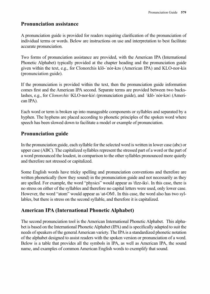
Pronunciation Guide 579
Pronunciation assistance
A pronunciation guide is provided for readers requiring clarification of the pronunciation of individual terms or words. Below are instructions on use and interpretation to best facilitate accurate pronunciation.
Two forms of pronunciation assistance are provided, with the American IPA (International Phonetic Alphabet) typically provided at the chapter heading and the pronunciation guide given within the text, e.g., for Clonorchis klō-ˈnȯr-kəs (American IPA) and KLO-nor-kis (pronunciation guide).
If the pronunciation is provided within the text, then the pronunciation guide information comes first and the American IPA second. Separate terms are provided between two backs-lashes, e.g., for Clonorchis \KLO-nor-kis\ (pronunciation guide), and \klō-ˈnȯr-kəs\ (Ameri-can IPA).
Each word or term is broken up into manageable components or syllables and separated by a hyphen. The hyphens are placed according to phonetic principles of the spoken word where speech has been slowed down to facilitate a model or example of pronunciation.
Pronunciation guide
In the pronunciation guide, each syllable for the selected word is written in lower case (abc) or upper case (ABC). The capitalized syllables represent the stressed part of a word or the part of a word pronounced the loudest, in comparison to the other syllables pronounced more quietly and therefore not stressed or capitalized.
Some English words have tricky spelling and pronunciation conventions and therefore are written phonetically (how they sound) in the pronunciation guide and not necessarily as they are spelled. For example, the word “physics” would appear as \fizz-iks\. In this case, there is no stress on either of the syllables and therefore no capital letters were used, only lower case. However, the word “atom” would appear as \at-OM\. In this case, the word also has two syl-lables, but there is stress on the second syllable, and therefore it is capitalized.
American IPA (International Phonetic Alphabet)
The second pronunciation tool is the American International Phonetic Alphabet. This alpha-bet is based on the International Phonetic Alphabet (IPA) and is specifically adapted to suit the needs of speakers of the general American variety. The IPA is a standardized phonetic notation of the alphabet designed to assist readers with the spoken version or pronunciation of a word. Below is a table that provides all the symbols in IPA, as well as American IPA, the sound name, and examples of common American English words to exemplify that sound.
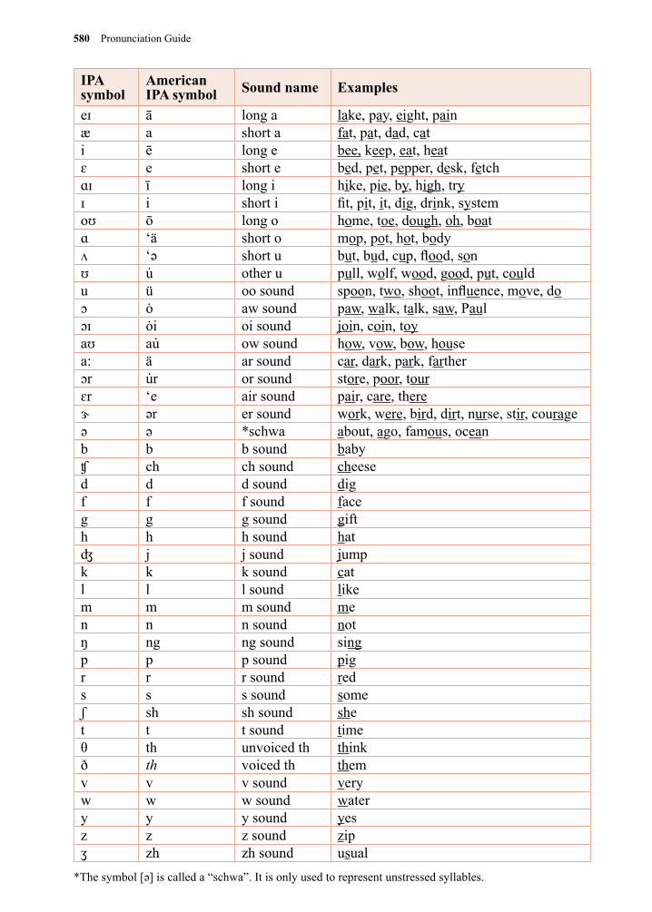
580 Pronunciation Guide
IPA symbol
American IPA symbol Sound name Examples
eɪ ā long a lake, pay, eight, painæ a short a fat, pat, dad, cati ē long e bee, keep, eat, heatɛ e short e bed, pet, pepper, desk, fetchɑɪ ī long i hike, pie, by, high, tryɪ i short i fit, pit, it, dig, drink, systemoʊ ō long o home, toe, dough, oh, boatɑ ‘ä short o mop, pot, hot, bodyʌ ‘ǝ short u but, bud, cup, flood, sonʊ u̇ other u pull, wolf, wood, good, put, couldu ü oo sound spoon, two, shoot, influence, move, doɔ ȯ aw sound paw, walk, talk, saw, Paul ɔɪ ȯi oi sound join, coin, toyaʊ au̇ ow sound how, vow, bow, housea: ä ar sound car, dark, park, fartherɔr u̇r or sound store, poor, tourɛr ‘e air sound pair, care, thereɝ ǝr er sound work, were, bird, dirt, nurse, stir, courageǝ ǝ *schwa about, ago, famous, oceanb b b sound babyʧ ch ch sound cheesed d d sound digf f f sound faceg g g sound gifth h h sound hatʤ j j sound jumpk k k sound catl l l sound likem m m sound men n n sound notŋ ng ng sound singp p p sound pigr r r sound reds s s sound someʃ sh sh sound shet t t sound timeθ th unvoiced th thinkð th voiced th themv v v sound veryw w w sound watery y y sound yesz z z sound zipʒ zh zh sound usual
*The symbol [ǝ] is called a “schwa”. It is only used to represent unstressed syllables.
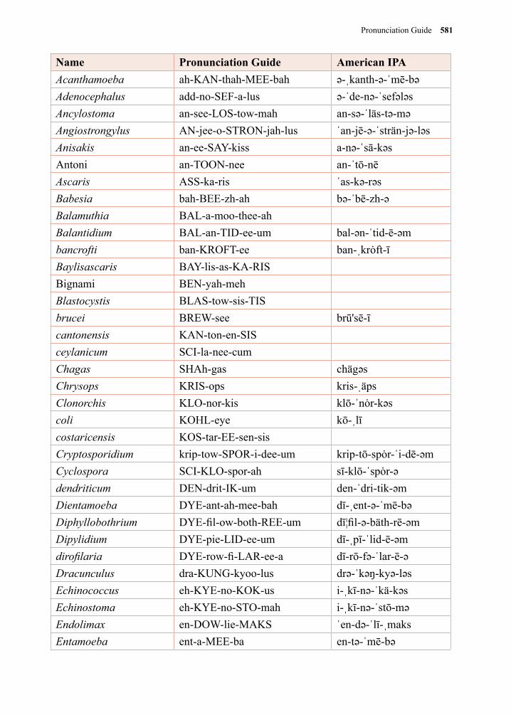
Pronunciation Guide 581
Name Pronunciation Guide American IPAAcanthamoeba ah-KAN-thah-MEE-bah ə-ˌkanth-ə-ˈmē-bəAdenocephalus add-no-SEF-a-lus ə-ˈde-nə-ˈsefələsAncylostoma an-see-LOS-tow-mah an-sə-ˈläs-tə-məAngiostrongylus AN-jee-o-STRON-jah-lus ˈan-jē-ə-ˈsträn-jə-ləsAnisakis an-ee-SAY-kiss a-nə-ˈsā-kəsAntoni an-TOON-nee an-ˈtō-nēAscaris ASS-ka-ris ˈas-kə-rəsBabesia bah-BEE-zh-ah bə-ˈbē-zh-əBalamuthia BAL-a-moo-thee-ahBalantidium BAL-an-TID-ee-um bal-ən-ˈtid-ē-əmbancrofti ban-KROFT-ee ban-ˌkrȯft-īBaylisascaris BAY-lis-as-KA-RISBignami BEN-yah-mehBlastocystis BLAS-tow-sis-TISbrucei BREW-see brū'sē-īcantonensis KAN-ton-en-SISceylanicum SCI-la-nee-cumChagas SHAh-gas chägəsChrysops KRIS-ops kris-ˌäpsClonorchis KLO-nor-kis klō-ˈnȯr-kəscoli KOHL-eye kō-ˌlīcostaricensis KOS-tar-EE-sen-sisCryptosporidium krip-tow-SPOR-i-dee-um krip-tō-spȯr-ˈi-dē-əmCyclospora SCI-KLO-spor-ah sī-klō-ˈspȯr-ədendriticum DEN-drit-IK-um den-ˈdri-tik-əmDientamoeba DYE-ant-ah-mee-bah dī-ˌent-ə-ˈmē-bəDiphyllobothrium DYE-fil-ow-both-REE-um dī¦fil-ə-bäth-rē-əmDipylidium DYE-pie-LID-ee-um dī-ˌpī-ˈlid-ē-əmdirofilaria DYE-row-fi-LAR-ee-a dī-rō-fə-ˈlar-ē-əDracunculus dra-KUNG-kyoo-lus drə-ˈkəŋ-kyə-ləsEchinococcus eh-KYE-no-KOK-us i-ˌkī-nə-ˈkä-kəsEchinostoma eh-KYE-no-STO-mah i-ˌkī-nə-ˈstō-məEndolimax en-DOW-lie-MAKS ˈen-də-ˈlī-ˌmaksEntamoeba ent-a-MEE-ba en-tə-ˈmē-bə
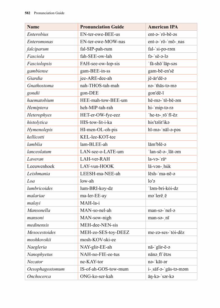
582 Pronunciation Guide
Name Pronunciation Guide American IPAEnterobius EN-ter-owe-BEE-us ent-ə-ˈrō-bē-əsEnteromonas EN-ter-owe-MOW-nas ent-ə-ˈrō-ˈmō-ˌnasfalciparum fal-SIP-pah-rum fal-ˈsi-pə-rəmFasciola fah-SEE-ow-lah fə-ˈsē-ə-ləFasciolopsis FAH-see-ow-lop-sis ˈfä-shōˈläp-səsgambiense gam-BEE-in-ss gam-bē-en'sēGiardia jee-ARE-dee-ah jē-är′dē-əGnathostoma nah-THOS-tah-mah nə-ˈthäs-tə-məgondii gon-DEE gon'dē-īhaematobium HEE-mah-tow-BEE-um hē-mə-ˈtō-bē-əmHemiptera heh-MIP-tah-rah hi-ˈmip-tə-rəHeterophyes HET-er-OW-fye-eez ˈhe-tə-ˌrōˈfī-ēzhistolytica HIS-tow-lit-i-ka his′təlit′ikəHymenolepis HI-men-OL-oh-pis hī-mə-ˈnäl-ə-pəskellicotti KEL-lee-KOT-teelamblia lam-BLEE-ah lăm′blē-əlanceolatum LAN-see-o-LATE-um ˈlan-sē-ə-ˌlāt-əmLaveran LAH-ver-RAH la-və-ˈräⁿLeeuwenhoek LAY-vun-HOOK lā-vən-ˌhu̇kLeishmania LEESH-ma-NEE-ah lēsh-ˈma-nē-əLoa low-ah lo′əlumbricoides lum-BRI-koy-dz ˈləm-bri-kȯi-dzmalariae ma-ler-EE-ay məˈlerēˌēmalayi MAH-la-iMansonella MAN-so-nel-ah man-sə-ˈnel-əmansoni MAN-sow-nigh man-sə-ˌnīmedinensis MEH-dee-NEN-sisMesocestoides MEH-zo-SES-toy-DEEZ me-zə-ses-ˈtȯi-dēzmoshkovskii mosh-KOV-ski-eeNaegleria NAY-glir-EE-ah nā-ˈglir-ē-əNanophyetus NAH-no-FIE-ee-tus nānəˌfīˈētəsNecator ne-KAY-tor nə-ˈkāt-ərOesophagostomum IS-of-ah-GOS-tow-mum i-ˌsäf-ə-ˈgäs-tə-məmOnchocerca ONG-ko-ser-kah äŋ-kə-ˈsər-kə
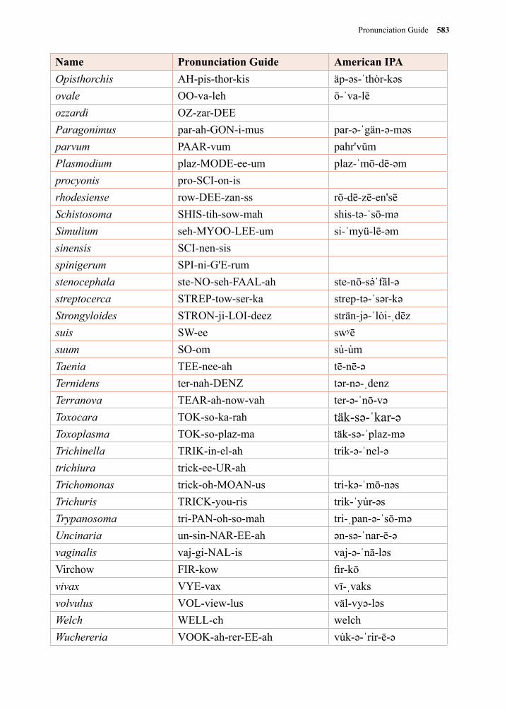
Pronunciation Guide 583
Name Pronunciation Guide American IPAOpisthorchis AH-pis-thor-kis äp-əs-ˈthȯr-kəsovale OO-va-leh ō-ˈva-lēozzardi OZ-zar-DEEParagonimus par-ah-GON-i-mus par-ə-ˈgän-ə-məsparvum PAAR-vum pahr'vŭmPlasmodium plaz-MODE-ee-um plaz-ˈmō-dē-əmprocyonis pro-SCI-on-isrhodesiense row-DEE-zan-ss rō-dē-zē-en'sēSchistosoma SHIS-tih-sow-mah shis-tə-ˈsō-məSimulium seh-MYOO-LEE-um si-ˈmyü-lē-əmsinensis SCI-nen-sisspinigerum SPI-ni-G'E-rumstenocephala ste-NO-seh-FAAL-ah ste-nō-sə̇ˈfāl-əstreptocerca STREP-tow-ser-ka strep-tə-ˈsər-kəStrongyloides STRON-ji-LOI-deez strän-jə-ˈlȯi-ˌdēzsuis SW-ee swʸēsuum SO-om su̇-u̇mTaenia TEE-nee-ah tē-nē-əTernidens ter-nah-DENZ tər-nə-ˌdenzTerranova TEAR-ah-now-vah ter-ə-ˈnō-vəToxocara TOK-so-ka-rah täk-sə-ˈkar-əToxoplasma TOK-so-plaz-ma täk-sə-ˈplaz-məTrichinella TRIK-in-el-ah trik-ə-ˈnel-ətrichiura trick-ee-UR-ahTrichomonas trick-oh-MOAN-us tri-kə-ˈmō-nəs Trichuris TRICK-you-ris trik-ˈyu̇r-əsTrypanosoma tri-PAN-oh-so-mah tri-ˌpan-ə-ˈsō-məUncinaria un-sin-NAR-EE-ah ən-sə-ˈnar-ē-əvaginalis vaj-gi-NAL-is vaj-ə-ˈnā-ləsVirchow FIR-kow fir-kōvivax VYE-vax vī-ˌvaksvolvulus VOL-view-lus väl-vyə-ləsWelch WELL-ch welchWuchereria VOOK-ah-rer-EE-ah vu̇k-ə-ˈrir-ē-ə
584 Pronunciation Guide
Bibliography
Pronunciations are based on those in everyday use as well as the standard references listed below. In a number of cases there are several acceptable pronunciations, in such instances, one of the many variations was chosen.
PMI. (n.d.) Farlex Partner Medical Dictionary. (2012). Retrieved October 11 2018 from http://medical-dictionary.thefreedictionary.com/PMI
Whonamedit.com. (2012). Whonamedit - dictionary of medical eponyms. [online] Available at: http://www.whonamedit.com/doctor.cfm/2761.html
Morris, W. (1969). American heritage dictionary of the English language. American heritage.
Virginia P. Studdert, Clive C. Gay and Douglas Blood Saunders Comprehensive Veterinary Dictionary 3rd Edition. Philadelphia: Saunders.
Miller-Keane, O. T. M., & O’Toole, M. T. (2003). Miller-Keane encyclopedia and dictionary of medicine, nursing, and allied health. 7th ed. Philadelphia: Saunders.
Dictionary, A. H. (2007). The American Heritage Medical Dictionary. Boston : Houghton Mifflin CoSaunders, J. H. (2007). Dorland's Medical Dictionary for Health Consumers. Philadelphia: Saunders.
Mosby (2009) Mosby's Medical Dictionary: 8th edition. Elsevier Health Sciences Stedman’s Medical Dictionary for the Health Professions and Nursing Seventh edition 2012 Wolters Kluwer Health | Lippincott Williams & Wilkins
Care, P. C. (2017). Segen's Medical Dictionary.(2011). Retrieved October 11 2018 from http://medical-dictionary.thefreedictionary.com/PMI
Moore, C. V., Thompson, R. C. A., Jabbar, A., Williams, J., Rasiah, K., Pallant, L., Koehler, A., Graham, C., and Weldhagen, G. F. (2016). Rare Human Infection with Pacific Broad Tapeworm Adenocephalus pacificus, Australia. Emerging Infectious Diseases, 22(8), 1510–1512. http://doi.org/10.3201/eid2208.160156
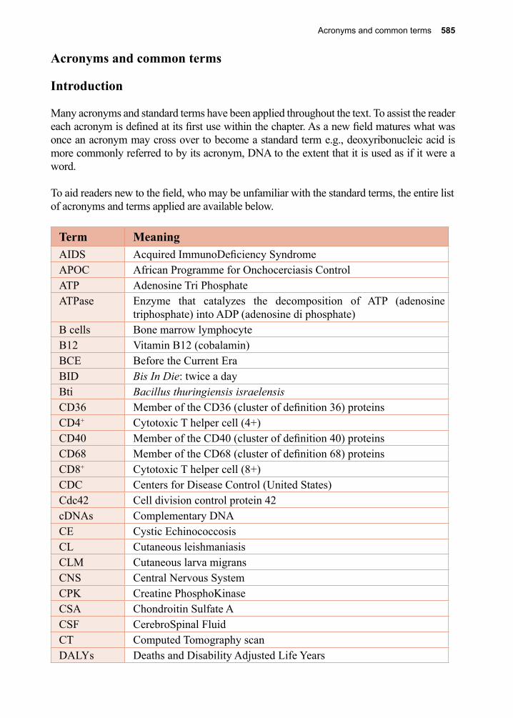
Acronyms and common terms 585
Acronyms and common terms
Introduction
Many acronyms and standard terms have been applied throughout the text. To assist the reader each acronym is defined at its first use within the chapter. As a new field matures what was once an acronym may cross over to become a standard term e.g., deoxyribonucleic acid is more commonly referred to by its acronym, DNA to the extent that it is used as if it were a word.
To aid readers new to the field, who may be unfamiliar with the standard terms, the entire list of acronyms and terms applied are available below.
Term MeaningAIDS Acquired ImmunoDeficiency SyndromeAPOC African Programme for Onchocerciasis Control ATP Adenosine Tri PhosphateATPase Enzyme that catalyzes the decomposition of ATP (adenosine
triphosphate) into ADP (adenosine di phosphate) B cells Bone marrow lymphocyteB12 Vitamin B12 (cobalamin)BCE Before the Current EraBID Bis In Die: twice a dayBti Bacillus thuringiensis israelensisCD36 Member of the CD36 (cluster of definition 36) proteinsCD4+ Cytotoxic T helper cell (4+)CD40 Member of the CD40 (cluster of definition 40) proteinsCD68 Member of the CD68 (cluster of definition 68) proteinsCD8+ Cytotoxic T helper cell (8+)CDC Centers for Disease Control (United States)Cdc42 Cell division control protein 42cDNAs Complementary DNACE Cystic EchinococcosisCL Cutaneous leishmaniasis CLM Cutaneous larva migransCNS Central Nervous SystemCPK Creatine PhosphoKinaseCSA Chondroitin Sulfate ACSF CerebroSpinal FluidCT Computed Tomography scanDALYs Deaths and Disability Adjusted Life Years
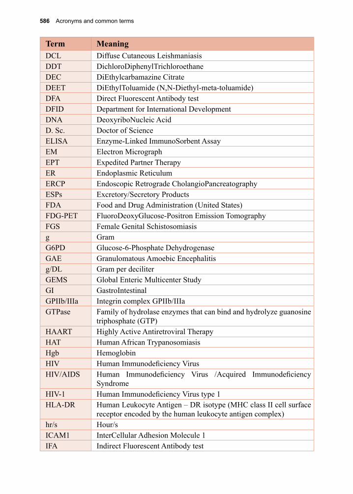
586 Acronyms and common terms
Term MeaningDCL Diffuse Cutaneous LeishmaniasisDDT DichloroDiphenylTrichloroethaneDEC DiEthylcarbamazine CitrateDEET DiEthylToluamide (N,N-Diethyl-meta-toluamide)DFA Direct Fluorescent Antibody testDFID Department for International Development DNA DeoxyriboNucleic AcidD. Sc. Doctor of ScienceELISA Enzyme-Linked ImmunoSorbent AssayEM Electron MicrographEPT Expedited Partner TherapyER Endoplasmic ReticulumERCP Endoscopic Retrograde CholangioPancreatographyESPs Excretory/Secretory ProductsFDA Food and Drug Administration (United States)FDG-PET FluoroDeoxyGlucose-Positron Emission Tomography FGS Female Genital Schistosomiasisg Gram G6PD Glucose-6-Phosphate DehydrogenaseGAE Granulomatous Amoebic Encephalitis g/DL Gram per deciliterGEMS Global Enteric Multicenter Study GI GastroIntestinalGPIIb/IIIa Integrin complex GPIIb/IIIaGTPase Family of hydrolase enzymes that can bind and hydrolyze guanosine
triphosphate (GTP)HAART Highly Active Antiretroviral TherapyHAT Human African TrypanosomiasisHgb HemoglobinHIV Human Immunodeficiency VirusHIV/AIDS Human Immunodeficiency Virus /Acquired Immunodeficiency
SyndromeHIV-1 Human Immunodeficiency Virus type 1HLA-DR Human Leukocyte Antigen – DR isotype (MHC class II cell surface
receptor encoded by the human leukocyte antigen complex)hr/s Hour/s ICAM1 InterCellular Adhesion Molecule 1IFA Indirect Fluorescent Antibody test
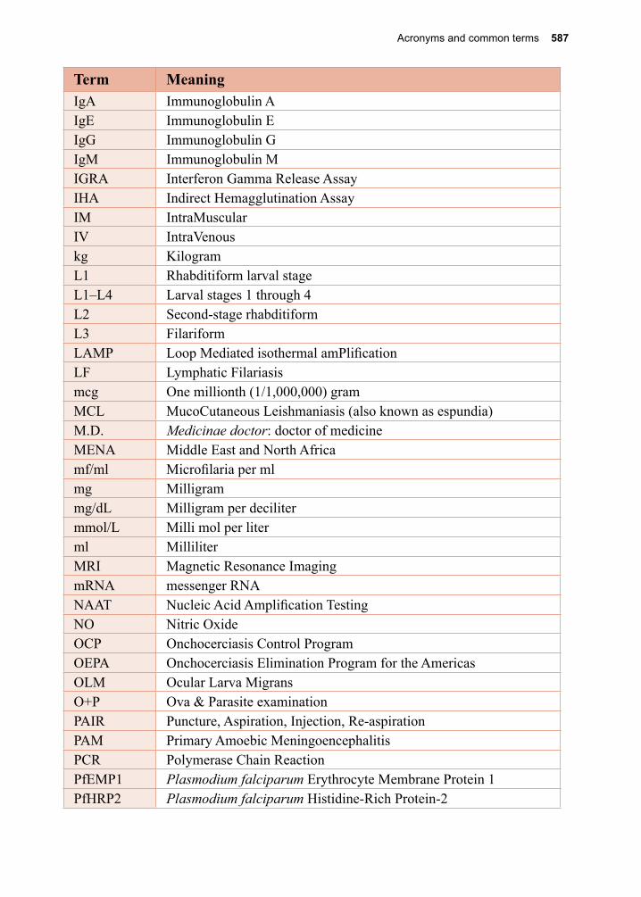
Acronyms and common terms 587
Term MeaningIgA Immunoglobulin AIgE Immunoglobulin EIgG Immunoglobulin GIgM Immunoglobulin MIGRA Interferon Gamma Release AssayIHA Indirect Hemagglutination AssayIM IntraMuscularIV IntraVenouskg KilogramL1 Rhabditiform larval stageL1–L4 Larval stages 1 through 4L2 Second-stage rhabditiformL3 FilariformLAMP Loop Mediated isothermal amPlificationLF Lymphatic Filariasismcg One millionth (1/1,000,000) gramMCL MucoCutaneous Leishmaniasis (also known as espundia)M.D. Medicinae doctor: doctor of medicineMENA Middle East and North Africamf/ml Microfilaria per mlmg Milligrammg/dL Milligram per decilitermmol/L Milli mol per liter ml MilliliterMRI Magnetic Resonance ImagingmRNA messenger RNANAAT Nucleic Acid Amplification TestingNO Nitric OxideOCP Onchocerciasis Control ProgramOEPA Onchocerciasis Elimination Program for the AmericasOLM Ocular Larva MigransO+P Ova & Parasite examinationPAIR Puncture, Aspiration, Injection, Re-aspirationPAM Primary Amoebic MeningoencephalitisPCR Polymerase Chain ReactionPfEMP1 Plasmodium falciparum Erythrocyte Membrane Protein 1 PfHRP2 Plasmodium falciparum Histidine-Rich Protein-2
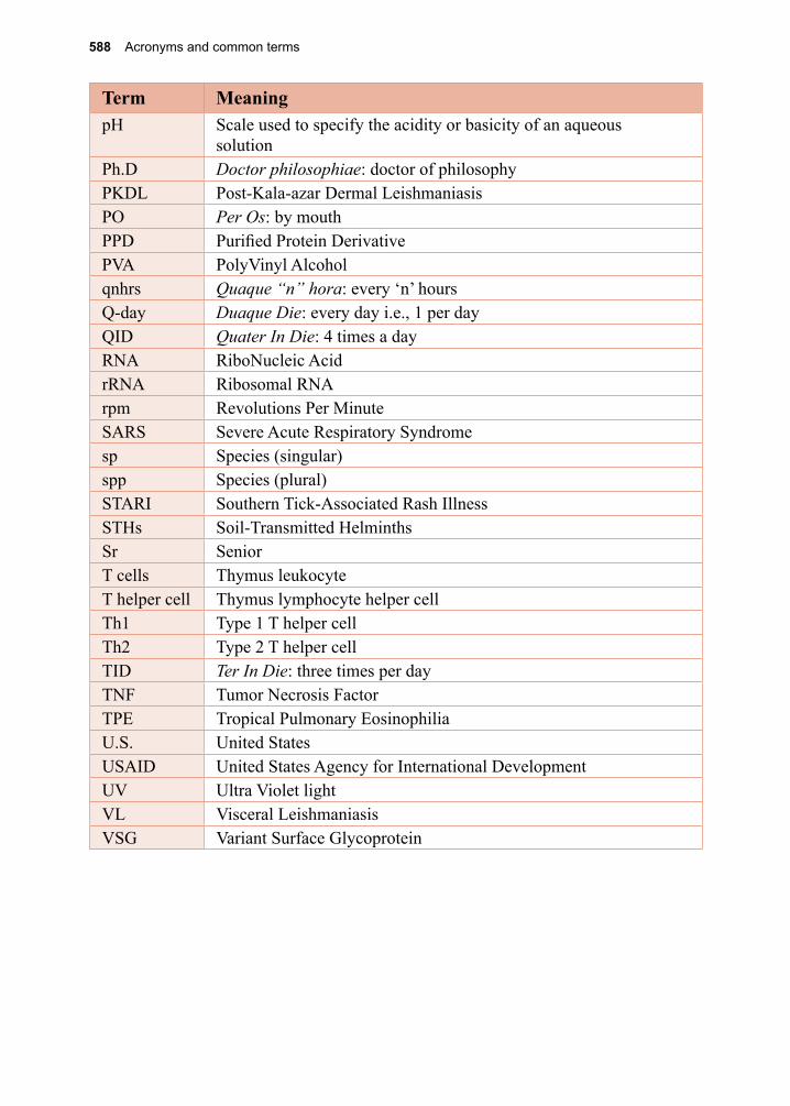
588 Acronyms and common terms
Term MeaningpH Scale used to specify the acidity or basicity of an aqueous
solutionPh.D Doctor philosophiae: doctor of philosophyPKDL Post-Kala-azar Dermal LeishmaniasisPO Per Os: by mouth PPD Purified Protein DerivativePVA PolyVinyl Alcoholqnhrs Quaque “n” hora: every ‘n’ hoursQ-day Duaque Die: every day i.e., 1 per dayQID Quater In Die: 4 times a day RNA RiboNucleic AcidrRNA Ribosomal RNArpm Revolutions Per MinuteSARS Severe Acute Respiratory Syndromesp Species (singular)spp Species (plural)STARI Southern Tick-Associated Rash IllnessSTHs Soil-Transmitted HelminthsSr SeniorT cells Thymus leukocyteT helper cell Thymus lymphocyte helper cellTh1 Type 1 T helper cellTh2 Type 2 T helper cellTID Ter In Die: three times per dayTNF Tumor Necrosis FactorTPE Tropical Pulmonary EosinophiliaU.S. United StatesUSAID United States Agency for International Development UV Ultra Violet light VL Visceral LeishmaniasisVSG Variant Surface Glycoprotein
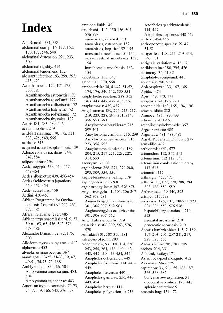
Index 589
IndexA.J. Renoult: 381, 383abdominal cramp: 16, 127, 152,
170, 172, 546, 549abdominal distension: 221, 233,
309abdominal rigidity: 494abdominal tenderness: 152aberrant infection: 193, 299, 393,
415, 423Acanthamoeba: 172, 174-175,
550, 581 Acanthamoeba astronyxis: 172 Acanthamoeba castellanii: 172 Acanthamoeba culbertsoni: 172 Acanthamoeba hatchetti: 172 Acanthamoeba polyphaga: 172 Acanthamoeba rhysodes: 172
Acari: 481, 483, 489, 496acetaminophen: 249acid-fast staining: 170, 172, 321,
333, 425, 549, 565acidosis: 545acquired acute toxoplasmosis: 139Adenocephalus pacificus: 344,
347, 584adipose tissue: 294Aedes aegypti: 256, 440, 447,
449-454Aedes albopictus: 439, 450-454Aedes Ochlerotatus japonicus:
450, 452, 454Aedes scutellaris: 450Aedini: 450-452African Programme for Oncho-
cerciasis Control (APOC): 265, 272, 585
African relapsing fever: 485African trypanosomiasis: vi, 9, 57,
59-61, 63, 65, 456, 542, 576, 578, 586
Alexandre Brumpt: 72, 92, 176, 300
Allodermanyssus sanguineus: 492alphavirus: 453alveolar echinococcosis: 367amastigote: 23-25, 31-33, 39, 47,
49-51, 74-75, 77, 188Amblyomma: 483, 486, 504 Amblyomma americanum: 483,
504 Amblyomma cajennense: 483American trypanosomiasis: 71-73,
75, 77, 79, 166, 543, 576-578
amniotic fluid: 140amoebiasis: 147, 150-156, 507,
576-578 amoebiasis, cerebral: 153 amoebiasis, cutaneous: 152 amoebiasis, hepatic: 152, 155 intestinal amoebiasis: 151-154 extra-intestinal amoebiasis: 152,
154 intrathoracic amoebiasis: 153-
154amoeboma: 152, 547amphibian: 370, 568amphotericin: 34, 41-42, 51-52,
174, 176, 540-542, 550-551anaphylactic reaction: 288, 362-
363, 443, 447, 472, 475, 567anaplasmosis: 439, 487Ancylostoma: 189, 204, 215, 217,
219, 223, 228, 299, 301, 314, 356, 553, 581
Ancylostoma braziliense: 215, 299-301
Ancylostoma caninum: 215, 299 Ancylostoma ceylanicum: 215,
223, 356, 553 Ancylostoma duodenale: 189,
204, 215, 217-221, 223, 228, 314, 553
aneurysm: 75, 307angioedema: 268, 271, 279-280,
295, 309, 556, 559angioedematous swelling: 279angiogenesis: 267-268angiostrongyliasis: 307, 576-578Angiostrongylus: 1, 301, 306-307,
562-563, 581 Angiostrongylus cantonensis: 1,
301, 306-307, 562-563 Angiostrongylus costaricensis:
301, 306-307, 562Anguillula stercoralis: 229anisakiasis: 308-309, 563, 576,
578Anisakis: 301, 308-309, 581ankylosis of joint: 288Anopheles: 4, 93, 100, 114, 228,
253, 256, 261, 438, 440, 442-443, 448-450, 453-454, 544
Anopheles culicifacies: 449 Anopheles freeborni: 114, 448-
449 Anopheles funestus: 449 Anopheles gambiae: 256, 440,
449, 454 Anopheles hermsi: 114 Anopheles polynesiensis: 256
Anopheles quadrimaculatus: 114, 449
Anopheles stephensi: 448-449anthrax: 454-456anthroponotic species: 29, 47,
51-52antigen test: 128, 211, 259, 333,
546, 571antigenic variation: 4, 15, 62antihistamine: 280, 295, 476antimony: 34, 41-42antiplatelet compound: 441apheresis: 280, 557Apicomplexa: 133, 167, 169Apidae: 474Apis: 443, 470, 474apoptosis: 74, 126, 220appendicitis: 163, 165, 194, 196arachnoiditis: 332Araneae: 481, 483, 493arbovirus: 451-453arecoline hydrobromide: 367Argas persicus: 485Argasidae: 481, 483, 485Argyll-Robertson, Douglas: 277armadillo: 472arrhythmia: 543, 577artemether: 112, 397, 545artemisinin: 112-113, 545artemisinin combination therapy:
113, 545artemotil: 112arthralgia: 452, 475arthritis: 17, 172, 279, 288, 294,
303, 488, 557, 559Arthropoda: 439-440, 503artifact: 517, 533ascariasis: 196, 202, 209-211, 223,
234, 236, 553, 576-578 hepatobiliary ascariasis: 210,
553 neonatal ascariasis: 210 pancreatic ascariasis: 210Ascaris lumbricoides: 1, 5, 7, 189,
197, 203, 205, 207-211, 217, 228, 526, 553
Ascaris suum: 205, 207, 209ascites: 234, 331Ashford, Bailey: 171Asian rock-pool mosquito: 452Askanazy, Max: 229aspiration: 33, 51, 155, 186-187,
366, 568, 587 bone marrow aspiration: 51 duodenal aspiration: 170, 417 splenic aspiration: 51assassin bug: 471-472
590 Index
asthenia: 163asthma: 209, 259, 294, 303, 493,
501-502, 555astrocyte: 63, 135asymptomatic carriers: 15, 88, 127,
151, 155, 258, 260, 303Atkins, John: 60atovaquone: 114, 141, 169, 545,
548-549Atrax: 495Auchmeromyia luteola: 459Austrobilharzia: 393auto-infectious cycle: 126, 193,
229, 231-232, 292, 294, 349-352, 356, 552
autoimmunity: v, 7autoinfection: 126, 193, 229, 231-
232, 294, 349-352, 356, 552avian influenza: 1azithromycin: 141, 169, 176, 548-
551azoles: 34, 42, 176, 540, 550-551Babeș, Victor: 167Babesia: 133, 167-169, 482, 484,
504, 522, 548, 581 Babesia bigemina: 167, 482, 504 Babesia divergens: 167-169 Babesia microti: 167-169babesiosis: 167-169, 177, 484,
488, 496, 576-578Bacillus thuringiensis israelensis:
447, 585bacteremia: 233, 246bacterial enterocolitis: 234bacterial meningitis: 234, 554Baelz, Erwin von: 421Baerman sedimentation method:
511Balamuth, William: 175Balamuthia mandrillaris: 172, 175,
550Balantidiasis: 161, 163-165, 576,
578Balantidium coli: 161, 163-165,
522, 548Balantidium suis: 161Bancroft, Thomas: 316, 447barium, enema: 152Bartonella bacilliformis: 445Bartonella quintana: 465bartonellosis: 445Bastian, Henry: 287Bavay, Arthur: 229baylisascariasis: 306, 576-578Baylisascaris procyonis: 301, 305-
306, 562beaver fever: 11, 17
Beaver, Paul: 300bed bug: 443, 467-470, 472beef: 5, 136, 317, 319, 322, 564beef tapeworm: 317beetle: 349-352, 459, 501benzene hexachloride: 463, 491,
495benzimidazole: 201, 203, 222,
295-296, 367benznidazole: 78, 543benzyl alcohol: 574benzyl benzoate: 491big spleen disease: 109bile duct carcinoma: 409Bilharz, Theodore: 349, 383BioFire: 16, 172Biomphalaria: 379, 388bird mite: 492birth weight: 108, 215, 221-222biting midge: 294-295, 444, 559-
560Black Death: 466black fly: 5, 265, 267, 269, 272,
294, 443, 446-447, 556, 559, 576
blackwater fever: 109bladder fibrosis: 390Blastocystis hominis: 92, 176-177,
196, 522, 551Blatella germanica: 502Blatta orientalis: 502Blattidae: 501Blattodea: 501blepharitis: 492blindness: vi, 139, 146, 174, 265,
270, 276, 301, 304, 333, 441, 446, 550, 556, 562
blister: 285, 287-288, 483, 495, 501, 557
bloating: 16blood bank screening: 79blood loss: 200, 215, 218, 220-
221, 394, 447blue bottle fly: 459blue spots: 463body louse: 443, 458, 460-465,
574Boeck, William: 148Boisregard, Nicolas Andry de: 317Boophilus annulatus: 482Borovsky, Peter: 31Borrelia: 1, 168-169, 464, 484-
485, 487-488, 496 Borrelia burgdorferi: 1, 168-169,
484, 488, 496 Borrelia recurrentis: 464-465botfly: 458-459
boutonneuse fever: 485, 487bovine leukemia: 455bovine onchocerciasis: 446Brachycera: 444bradycardia: 168, 548bradyzoite: 134-136, 139Braun, Max: 423breakbone fever: 452breast milk: 15, 218, 221Bremser, Johann: 191Brie, Jean de: 413Brill-Zinsser disease: 465broad tapeworm: 341, 346-347,
584bronchoalveolar lavage: 235, 425,
572bronchopulmonary fistula: 363bronchospasm: 209, 303, 475, 501,
553Bruce, David: 60, 70, 456Brugia: 253, 255-256, 449, 527-
528, 555 Brugia beaveri: 253 Brugia lepori: 253 Brugia malayi: 253, 256, 258,
261, 449, 527-528, 555 Brugia timori: 253Brugian filariasis: 451bubonic plague: 466, 469buccal cavity: 189, 222, 235, 527buffalo: 381, 398, 443, 446buffer zones: 17, 43buffered formaldehyde: 515bug, kissing: 71-72, 74, 76, 78-79,
442-443, 468, 471-473bug, palmetto: 502bug, redbug: 491bug, reduviid: 72, 472, 543, 576bug, true bug: 71, 443, 469bug, wheel: 472bulbar palsy: 486Bulinus: 381Burkitt’s lymphoma: 109Busk, George: 287butterfly: 501cachexia: 209, 233, 292, 409, 436Caenorhabditis elegans: 189Calabar swellings: 277, 279-280,
295, 556Calliphora: 459Calliphoridae: 443, 457-458, 573cancer: 16, 89, 127, 168, 187, 395,
405, 409Candida: 260Canidae: 197, 359, 372canthariasis: 501Capillaria: 190, 232, 241, 291-292,

Index 591
294, 528-529, 558 Capillaria aerophila: 291 Capillaria hepatica: 291-292,
294, 529, 558 Capillaria philippinensis: 232,
291-292, 294, 528, 558 Capillaria plica: 291carbon dioxide laser: 540card agglutination test: 64, 542cardiomyopathy: 71, 76-79, 246,
279, 556Carini, Antonio: 39Carrion’s disease: 443, 445Carter, Jimmy: iii, 285, 289cataracts: 270, 556caterpillar: 501, 503Catto, John: 383cellulitis: 194, 259, 288centipede: 440, 502-503Ceratopogonidae: 443-444cercaria: 5, 388-389, 392-394, 397,
409, 415-416, 424, 431-432, 569cercarial dermatitis: 393cerebral calcifications: 139cerebrospinal obstruction: 336Cerithidia: 432cervical neoplasia: 85Chagas disease: vi, 71-72, 75-79,
443, 471-473Chagas, Carlos: 49, 72, 471chagasin: 74chagoma: 76chancre: 60, 63-64, 542Chandler, Asa C.: 217Charcot-Leyden crystal: 153, 533cheese-skipper: 457cheilitis: 221chemoprophylaxis: 110, 114, 139,
281, 557Chiclero’s ulcer: 32-33chigger: 467, 491-492chigoe: 443, 467chikungunya: 1, 439, 453-454Chilomastix mesnili: 186, 523Chilopoda: 440, 502Chiracanthium mildei: 495chlorination: 128chloroquine: 94, 112-114, 164,
545, 548chlorosis: 221, 553cholangiocarcinoma: 405, 409cholangiography: 410, 417, 570-
571cholangitis: 210, 363, 409cholecystitis: 210chorioetinitis: 133, 138-140, 270,
546, 556
choroidal infiltrates: 306Chrysomyia: 459Chrysops: 277, 279, 281, 443, 454-
455, 556, 581 Chrysops dimidiata: 277, 455 Chrysops silacea: 455chylocele: 258chyluria: 258cilia: 2, 9, 161, 343, 345, 387, 511Cimex lectularius: 443, 467, 469cinchona tree: 94, 112cinchonine: 94ciprofloxacin: 171-172, 549cirrhosis: 383clam digger’s itch: 393clarithromycin: 176, 551clindamycin: 112, 169, 545, 548-
549clonorchiasis: 409-410, 576-578Clonorchis sinensis: 405, 407-410,
433, 532, 570clotrimazole: 540coccidian: 171Cochin China diarrhea: 229Cochliomyia: 459cockroach: 501-503coenurus: 372cognitive defects: 189, 197, 201,
209, 221, 300, 546, 561Coleoptera: 501colitis: 152, 155-156, 163, 165,
177, 184, 191, 194, 196-197, 200
Colorado tick fever: 484, 486, 496colostrum: 320, 323coma: 63-64, 173, 417, 494, 542,
550, 562commensalism: 163, 185-186, 552concentration by sedimentation:
514cone-nosed bug: 443, 471congenital toxoplasmosis: 136,
138-139, 141congestive heart failure: 221, 543Congo floor maggot: 459conjunctivitis: 76, 270connective tissue: 287, 386constipation: 77, 231, 233, 543contact lens: 174-175Contracaecum: 308contracture: 288, 557convulsion: 139, 496, 545cooties: 462copepod: 5, 287-289, 307, 343-
344, 370, 557coproculture: 235, 554, 560coprolites: 191, 193, 196-197
copulatory spicule: 193cor pulmonale: 392, 394coracidia: 343, 345, 370Cordylobia anthropophaga: 457-
458cornea: 174, 268, 270corneal blindness: 270corneal stroma: 268coronary artery disease: v, 7Cort, William: 217Councilman, William: 147covert toxocariasis: 303-304Coxiella burnetii: 487crab: 5, 421, 424, 426, 440, 443,
459-464, 495, 571craterform ulcer: 31, 33crayfish: 409, 423creeping eruption: 299-300, 309,
459crepuscular pattern: 34, 42cribriform plate: 173, 550cricket: 501Crimean-Congo hemorrhagic
fever: 482, 487cross-fertilization: 377croton bug: 502crusted scabies: 490-491Cruz, Oswaldo: 52, 72, 166, 196cruzipain: 74cryotherapy: 34, 300, 540crypt: 409Cryptosporidium: vi, 1, 4, 123,
125, 127-128, 133, 170, 512, 521, 546, 581
Cryptosporidium canis: 123, 443 Cryptosporidium felis: 123, 467 Cryptosporidium hominis: 123 Cryptosporidium meleagridis:
123 Cryptosporidium muris: 123 Cryptosporidium parvum: vi, 1,
4, 123-128, 512, 521, 546Ctenocephalides: 354, 443, 465,
467 Ctenocephalides canis: 123, 354,
443, 467 Ctenocephalides felis: 123, 354,
443, 465, 467 Ctenodactylus gondii: 133Culex: 256, 440, 443, 449-454 Culex fatigans: 451 Culex nigripalpus: 452 Culex pipiens pipiens: 256, 451 Culex pipiens quinquefasciatus:
256, 451 Culisetax tarsalis: 452 Culex tritaniorhynchus: 452
592 Index
Culicidae: 443-444, 447-448Culicinae: 253, 316, 438, 448-449Culicoides: 443-444Culiseta: 443, 449, 451-452Cunha Chagas: 49Cunningham, David: 31cutaneous larva migrans (CLM):
215, 299-301, 308-309, 425, 561, 563, 585
cutaneous leishmaniasis (CL): 21, 23, 25, 29, 31-34, 39, 41, 51, 585
cutaneous lesions: 21, 23, 29, 31-33, 39, 41, 50
Cuterebra: 458Cuterebridae: 443-444, 457-458,
573cycloguanil: 112cyclophilin: 24Cyclophyllidea: 317, 325, 349Cyclops: 287, 307, 345Cyclorrhapha: 444Cyclospora: vi, 1, 133, 171, 521,
549, 581 Cyclospora cayetanensis: vi, 1,
171-172, 521, 549cyclosporine A: 24cyst passers: 15, 155cyst wall: 124, 134, 148, 170, 176,
362, 365-366cysticercosis, cardiac: 332 cysticercosis, extraneural: 331-
332 cysticercosis, neurocysticercosis:
307, 315, 325, 330-337, 565 cysticercosis, ophthalmic: 333cystic fibrosis: 11, 16-17Cytoisospora: 522cytokine: 9, 23, 51, 108, 127, 151,
200, 203, 220, 257cytokinesis: 9, 148cytoskeleton: 13cytotoxic: 25, 87, 150, 233, 259,
585DALYs (deaths and disability
adjusted life years): 215, 585Darwin, Charles: 72DDT: 94, 272, 281, 446, 453, 455,
470, 586de-worming program: 201, 211,
236DEC (diethylcarbamazine citrate):
255, 260-261, 271, 279-281, 295, 555, 557, 560, 586
decortication: 425deer fly: 5, 277, 279, 454, 556, 576deer tick: 482, 484, 548
DEET (diethyltoluamide): 114, 169, 444, 447, 454-455, 488, 492, 586
deforestation: 42dehydration: 170, 464delusional parasitosis: 441, 476Demodex: 483, 492dendritic cell: 21, 23, 31-32, 39,
49, 74, 135, 200, 220dengue: 1, 439, 441, 450, 452, 454depigmentation: 76, 269, 556depression: 64, 108, 395, 542Dermacentor: 483-484, 486-487 Dermacentor albipictus: 484 Dermacentor andersoni: 484,
486-487 Dermacentor occidentalis: 484 Dermacentor variabilis: 483-484,
486-487 Dermatobia hominis: 458Dermatophagoides: 493Dermestidae: 501desensitization: 451, 476, 493dew itch: 221dexamethasone: 336, 565Diamanus montanus: 467-468Diaptomus: 344-345Dientamoeba fragilis: 2, 170, 177,
184, 194, 196, 525, 551diffuse cutaneous leishmaniasis
(DCL): 21, 32, 586diffusion: 9Digenea: 377, 426diloxanide furoate: 155diphenhydramine: 476Diphyllobothrium: 317, 326, 341,
343, 345-347, 370, 530, 566, 581
Diphyllobothrium cordatum: 341 Diphyllobothrium dalliae: 341 Diphyllobothrium dendriticum:
341, 343 Diphyllobothrium lanceolatum:
341 Diphyllobothrium latum: 317,
322, 326, 341, 343-347, 370, 530, 566
Diphyllobothrium nihonkaiense: 341, 343, 346
Diphyllobothrium pacificum: 341, 343, 346
Diphyllobothrium ursi: 341 Diphyllobothrium yonagoensis:
341diplopia: 174, 550dipstick assay: 271Diptera: 43, 272, 277, 441, 443-
444, 446-447, 454, 456-457Dipylidium caninum: 354, 356,
567direct fluorescent antibody test
(DFA): 16, 586Dirofilaria immitis: 291, 294, 296,
301, 559disophenol: 292, 558disseminated infection: 140, 174,
232-235diurnal periodicity: 256, 279-280Dobell, Clifford: 148dog heartworm: 291dog tick: 483-485, 487Donald Blacklock: 267Donné, Alfred François: 85Donovan, Charles: 49, 188dormancy: 2, 94, 101, 110, 133,
136, 175, 217-218, 450, 454, 466
doxycycline: 112-114, 260, 272, 295, 487, 545, 551, 555-556, 560
dracunculiasis: vi, 285, 287-290, 576, 578
Dracunculus medinensis: 285, 287-290, 557
drug resistance: 56, 89, 112-113, 204, 222, 399
drunken hairy crab: 426Dubini, Angelo: 204, 217duck hunter’s itch: 299Duffy blood type: 111duodenal fluid: 235dysentery: 5, 147, 152-154, 156,
161, 163-165, 200, 203, 547-548, 552
dyspareunia: 88, 544dysphagia: 77, 543dyspnea: 221, 248, 424, 544, 555,
571dysuria: 88, 395, 544East African sleeping sickness:
57, 65East African trypanosomiasis: 57,
61, 65eastern equine: 452Ebola virus: 42, 236echinococcosis: 5, 359, 362-367,
372, 576-578, 585Echinococcus granulosus: 359,
361-365, 367, 369, 372, 500, 567-568
Echinococcus multilocularis: 1, 359, 366-367, 568
Echinostoma: 431, 436, 532, 572, 581
Index 593
ecotones: 42ectoparasite: 420, 460, 465-466,
469, 482, 485, 489, 491-492edema edema, bilateral periorbital: 247,
554, 578 edema, cerebral: 63 edema, facial: 63, 76, 221, 542,
578 edema, hemi-facial: 76 edema, laryngeal: 416 edema, periorbital: 247, 554,
578 edema, periorbital subcutaneous:
247, 554, 578 edema, pulmonary: 545effusion: 76, 153-155, 416, 424,
543eflornithine: 65, 543Egyptian mummy: 285, 399Ehrlichia: 168ehrlichiosis: 168eicosanoids: v, 7, 389, 392Ekbom’s syndrome: 441electrocardiography (ECG): 77electrolyte imbalance: 170, 292elephantiasis: 253, 255, 257-261,
441, 447, 555Elokomin fluke fever: 436empyema: 153, 187encephalomyelitis: 453encephalopathy: 63, 65, 271, 280-
281, 303, 395, 556Endolimax nana: 186, 523-524endomyocardial fibrosis: 280endophthalmitis: 304, 309endoplasmic reticulum (ER): 148,
432, 535, 580, 582, 586endosymbiont: 7, 17, 258, 268,
270, 295, 560Entamoeba coli: 147, 186, 196,
524-525Entamoeba dispar: 92, 147, 154,
156, 185-186, 507Entamoeba gingivalis: 185-187,
524Entamoeba histolytica: vi, 5, 11,
92, 128, 147-156, 161, 173, 186-187, 201, 505, 507, 509, 521, 547
Entamoeba moshkovskii: 147, 156Entamoeba polecki: 186enteric fever: 390enteritis: 156, 194-195, 215, 229,
234, 299enterobiasis: 191, 194, 196Enterobius vermicularis: 191, 193-
196, 300, 321, 513, 526, 552, 564-565
Enteromonas hominis: 185-187enuresis: 194eosinopenia: 110, 544, 577eosinopenic: 155eosinophil count: 110eosinophilic colitis: 177, 191, 194,
196eosinophilic enteritis: 194-195,
215, 299eosinophilic granuloma: 301epigastric fullness: 321, 331epiglottis: 208, 217, 232epilepsy: 315, 325, 337epimastigote: 61, 74epinephrine: 476Epstein-Barr virus: 109, 140EPT (expedited partner therapy):
89, 586equine infectious anemia: 455Eriocheir: 423erysipelas de la costa: 269, 556erythema: 51, 76, 88, 279, 470,
488, 544erythematous macular rash: 51erythrocytic cycle: 101-102Escherichia coli: 1, 330espundia: 21, 39, 41, 43, 540, 587exchange transfusion: 169, 545,
548-549excretory/secretory products
(ESPs): 220, 586exocytosis: 9extracellular parasites: 23-24, 31,
39, 49, 60-61, 151, 163, 169extrapulmonary paragonimiasis:
425extrasystoles: 77failure to thrive: 7, 16, 189, 197,
209, 220-221, 356, 379, 394false hookworm: 296Fantham, Harold: 60Fasciola: 377, 408, 413, 415-417,
425, 429, 431, 500, 532, 571, 582
Fasciola gigantica: 413, 429 Fasciola hepatica: 377, 408, 413,
415-417, 425, 429, 500, 532, 571
fascioliasis: 413, 416-417, 576-578Fasciolopsis buski: 429, 431, 436,
531, 572FDA (Food and Drug Administra-
tion): 34, 42, 112, 309, 417, 454, 458, 586
fecal retention: 77
Fedchenko, Aleksej: 287Felidae: 4, 134Félix Dujardin: 85female genital schistosomiasis
(FGS): 379, 381, 392, 395-396, 569, 586
fibrotic capsule: 423fibrous granuloma: 41fibrous nodules: 267Field’s staining reagent: 175fiery serpent: 285Filaria bancrofti: 255filarial eye worm: 454filarial nematode: 265, 277filariform larva: 219, 232, 234-
235, 553-554FilmArray: 16finger clubbing: 200fingernail concavities: 221Finlay, Carlos: 447fish tapeworm: 341, 343-345, 347flagellated protozoan: 11, 22, 57,
62, 85, 156, 177, 185-186, 505, 552
flash freezing: 309flattening of the villi: 14-15, 245flatulence: 16, 152, 539flatworm: 5, 284flavivirus: 452-453flea: 287, 349, 351-352, 354, 356,
439, 441-443, 465-469, 480, 567floatation by centrifugation: 511flucytosine: 176, 551fluid-filled cyst: 359fluorodeoxyglucose-positron emis-
sion tomography (FDG-PET): 425, 572, 586
follicle mite: 483, 492Forbes, D.: 287Forde, Robert: 60Formicidae: 474Fossaria modicella: 415Foster, Auriel O.: 217fowl tick: 485Franciscella tularensis: 487free-living amoeba: 172-175free-living nematode: 229Frenkel, John: 133frog: 307, 370, 433Fuelleborn, Friedrich: 229Fujinami, Kan: 383fur trapper: 367furazolidone: 16, 539G6PD (glucose-6-phosphate dehy-
drogenase): 111, 113-114, 586galectin: 22, 74gametocyte: 94, 96, 107, 126, 134,
594 Index
168, 170gametocytogenesis: 96ganglion: 189, 315, 377gangrene: 288, 467Garnham, Cyril: 94Gasterophilidae: 444gastric ulcer: 309gastroenteritis: 176-177, 247, 292,
487Gates, Frederick: 217geographical isolation: 25Giardia: vi, 2, 4, 11, 13-17, 124,
128, 201, 509, 517, 519, 539, 582
Giardia duodenalis: 11, 17 Giardia intestinalis: 11, 17 Giardia lamblia: vi, 2, 4, 11,
13-15, 17, 128, 201, 509, 517, 519, 539
giardiasis: 11, 15-17, 506-507, 576, 578
Giardiavirus: 13Giemsa stain: 64, 77, 111, 169,
271, 514glaucoma: 270, 304Glisson’s capsule: 408, 413Global Enteric Multicenter Study
(GEMS): 123, 156, 586Glossina: 57, 443, 455, 457 Glossinia morsitans: 457 Glossinia pallidipes: 457 Glossinia palpalis: 457 Glossinia swynnertoni: 457 Glossinia tachinoides: 457glycosome: 9, 25, 61Gnathostoma spinigerum: 301,
307-308, 563gnathostomiasis: 307-308, 425,
563, 576-578goblet cell: 220Goeze, Johann: 317, 326, 361Golgi body: 9, 94Golgi, Camillo: 94gondi: 133gram-negative meningitis: 234granuloma: 41, 152, 193, 196, 291,
300-301, 304-305, 387, 390-392, 394-395, 432, 562
granulomatous amoebic encephali-tis (GAE): 174-176, 550, 586
granulomatous disease: 163, 174grasshopper: 501Grassi, Giovanni: 94, 349, 438,
447gravlax: 346green bottle fly: 459greenhead fly: 454
Griesinger, Wilhelm: 383ground itch: 221, 233, 554Guillain-Barré syndrome: 172,
453, 486Guinea worm: 5, 276, 285, 287-
290Guyot, Francois: 277hair loss: 221hairworms: 5halzoun: 416hanging groin: 269, 556Hartmann, Philip: 326Hawking, Francis: 255headache (see Chapters: 3, 4, 6, 9,
14, 27, 31, 33-35, 37, 38, 39, and Clinical Appendix)
heart disease: 71, 76-77heat shock genes: 24hematemesis: 394hematoxylin stain: 321, 323, 506,
518, 564, 566hematuria: 381, 383, 395, 569Hemiptera: 3, 71, 469, 582hemoflagellate: 62hemoglobinuria: 109, 167-168hemolymph: 168hemolymphatic trypanosomiasis:
64-65, 542hemolysis: 108-109, 113-114, 168hemoptysis: 294, 423-424, 571hemozoin: 4-5, 102, 168Henderson, W.R.: 133Henri Lafleur: 147, 156hepatic abscess: 210hepatic failure: 369, 568hepatic fibrosis: 390hepatitis: 469, 485, 487, 496 hepatitis B: 469, 485, 496 hepatitis C: 487 hepatitis D: 487hepatobronchial fistula: 153hepatomegaly: 109, 369, 394, 409,
541, 546, 553, 562, 569-570, 577
hepatosplenomegaly: 47, 50, 64, 139, 304, 391, 394
Heppentis: 429hermaphrodite: 6, 421herpes simplex virus: 133Heterophyes heterophyes: 6, 432-
433, 436, 532, 573Heterophyes nocens: 432heterophyiasis: 433hexacanth larva: 315highly active antiretroviral therapy
(HAART): 128, 139, 170, 586Hippocrates: 93, 147
histiocytes: 51, 141Histomonas: 177Hooke, Robert: 11hookworm dermatitis: 219Hopkins, Donald: 276, 289hornet: 443, 470, 473-474, 476horse fly: 279, 454house dust mite: 493housefly: 455-456HTLV-1: 233Human African Trypanosomiasis:
57, 580, 586Human Leukocyte Antigen - DR
isotype (HLA-DR): 74, 586Hutchinson, William M.: 133Hyalomma: 485hydatid cyst: 361-366, 369, 372hydatid disease: 359, 362-363,
365-366hydrocele: 255, 257-259, 261, 555hydrocephalus: 133, 138-139, 331-
332, 336, 546, 565hydrogenosome: 9, 87, 89hydronephrosis: 395hydroureter: 395Hylesia alinda: 501Hymenolepis: 46, 228, 349-352,
356, 501, 530, 566, 582Hymenolepis diminuta: 352, 354,
501, 530, 566Hymenolepis microstoma: 349Hymenolepis nana: 46, 228, 349-
352, 354, 356, 501, 530, 566Hymenoptera hymenopterans:
443, 473-476hyper-responsive syndrome: 280hypereosinophilia: 280hypergammaglobulinemia: 303-
304hyperinfection: 232-236, 554hyperpigmentation: 269hypersensitivity: 39, 41, 65, 219,
301, 393, 396, 489, 561hypersomnolence: 542hypersplenism: 394hypnozoite: 94, 96-97, 100-101,
107, 110, 113, 214hypoalbuminemia: 176hypoattenuating tracts: 416Hypoderma: 459hypogammaglobulinemia: 16hypoglycemia: 545hypoproteinemia: 431ICAM1 (intercellular adhesion
molecule 1): 108, 586Ijima, Isao: 408ileitis: 200
Index 595
immune evasion: 7, 9, 23, 62, 330immune suppression: 140, 220immunoblot testing: 425immunocompromised: 11, 77, 123,
133, 139-140, 164, 168, 170-171, 174, 187, 232, 390, 546, 548-549
immunofluorescent assay: 173immunogen: 441immunoparasitology: 9incontinence: 433indirect fluorescent antibody test
(IFA): 154, 558, 586indirect hemagglutination assay
(IHA): 154, 570, 587infantile ancylostomiasis syn-
drome: 221infarct: 203, 219, 257-258, 301,
320, 424infertility: 85inflammatory bowel disease: 200insect repellents: 34, 42, 52, 114insomnia: 64, 459, 542intercostal sign: 152interferon-γ: 62, 135-136, 139,
151, 168intestinal fibrosis: 391intestinal infection: 128, 152, 155-
156, 326, 331intestinal perforation: 163intracellular parasites: 21, 50, 71,
109, 123, 133, 169-170, 241intracranial calcifications: 139,
141, 546intracranial hypertension: 330intracranial pressure: 332-333,
335-336intraocular cysts: 333, 336intrauterine growth: 215intravascular coagulopathy: 108intravascular hemolysis: 108, 168intravenous drug users: 110Inuit: 361Iodamoeba bütschlii: 186, 524iodine stain: 505iodoquinol: 155, 164, 177, 547-
548, 551iridocyclitis: 270iritis: 270, 556iron deficiency: 215, 220-221, 294,
394, 553iron-hematoxylin: 506, 518Isospora belli: 169-171, 177, 549itch mite: 481, 483, 489-490ivermectin: iii, 146, 201-202, 222,
236, 260-261, 265, 271-272, 279, 281, 295, 300, 308, 454,
463, 489, 491-492, 552-557, 559-561, 563, 574-575
Ixodes: 167, 169, 440, 482, 484, 486, 488, 496, 548
Ixodes dammini: 484, 496 Ixodes holocyclus: 484, 486 Ixodes pacificus: 484, 488, 496 Ixodes ricinus: 167, 488 Ixodes scapularis: 167, 440, 482,
484, 488, 496, 548Ixodidae: 481-483, 496Janicki, Constantine: 344Janku, Josef: 133Japanese encephalitis: 452jaundice: 369, 416, 452, 545Jean-Nicolas Demarquay: 255jejunal perforation: 321, 323joint pain: 64jumping salad: 292Kala-azar: 50-52, 188, 588Kampala eye worm: 295, 559Karl Georg Friedrich Rudolf
Leuckart: 161, 265, 267, 319, 323, 359, 413
karyosome: 148Katayama fever: 383, 393-394,
396, 569Katsurada, Fujiro: 383Kawanishi, Kenji: 383keratitis: 174-175, 268, 270, 550,
556Kerbert, Coenraad: 421Kessel, John: 85Kilbourne, Frederick L.: 167, 447,
482kinetoplast: 21, 33, 57, 61kinetoplastidae: 9, 21, 57Kinghorn, Allan: 60Kligler, Israel: 39Knott test: 259, 280, 294-295, 506,
514Kobayashi, Haraujiro: 408Koch’s postulates: 287koilonychia: 221Krotoski, Wojciech: 94Kuchenmeister, Friedrich: 319,
326Kyasanur Forest disease: 487La Crosse virus: 452lacrimation: 270, 495lactic dehydrogenase: 248, 555lactoferrin: 15lactose intolerance: 16, 72, 209Lambl, Vilem: 11Langerhans cell: 25larvicide: 94, 289, 446, 453Lassa fever virus: 42
Latrodectus: 483, 493Latrodectus mactans: 493Laveran, Charles: 94learning deficit: 139Leeuwenhoek, Antonie van: 11Legionella pneumophila: 1Leiper, Robert: 277, 383, 454Leishman, William: 49Leishmania (Leishmania) aethi-
opica: 29Leishmania (Leishmania) amazo-
nensis: 21, 29, 32Leishmania (Leishmania) don-
ovani: 25, 47, 188Leishmania (Leishmania)
garnhami: 21, 29Leishmania (Leishmania) infantum
chagasi: 21, 25, 29, 47, 50-52, 79
Leishmania (Leishmania) major: 21-22, 29, 32, 476
Leishmania (Leishmania) mexi-cana: 21, 29, 32, 47
Leishmania (Leishmania) pifanoi: 21, 29
Leishmania (Leishmania) tropica: 21, 23, 29, 47
Leishmania (Leishmania) venezua-lensis: 21
Leishmania (Viannia) braziliensis: 21, 29, 32, 39, 42-43
Leishmania (Viannia) colombien-sis: 21, 29
Leishmania (Viannia) guyanensis: 21, 29
Leishmania (Viannia) lainsoni: 21, 29
Leishmania (Viannia) lindenbergi: 21, 29
Leishmania (Viannia) naiffi: 21, 29Leishmania (Viannia) panamensis:
21, 29Leishmania (Viannia) peruviana:
21, 29Leishmania (Viannia) shawi: 21,
29leishmaniasis recidivans (LR): 21leishmaniasis skin test: 33, 539Leishmaniavirus: 39, 43leishmanization: 31leopard skin: 269, 556Lepidoptera: 501, 503leprosy: 10, 32, 51, 268Leptoconops: 443-444leptomeningitis: 63Leuckart, Friedrich: 319, 413Leuckart, Rudolph: 161
596 Index
leukocyte count: 110leukocytosis: 424, 554, 577leukopenia: 109, 487, 544, 577leukovorin: 547, 549Lewis, Timothy: 255lichenification: 269, 556Limacodidae: 501lindane: 463, 491Linnaeus, Carl: 191, 197, 205,
344, 413littoral vegetation: 413, 415, 417,
429, 431liver abscess: 148, 152, 154-155,
163liver failure: 139, 291, 546, 558,
577liver fluke: 405, 408-410, 413, 417liver tenderness: 152lizard: 370, 568Loa loa: 260, 271, 277, 279-281,
295, 443, 454-455, 528, 536, 555-557
Löeffler’s syndrome: 209, 553loiasis: 5, 277, 279-281, 557, 576-
578loop mediated isothermal amplifi-
cation (LAMP): 112, 270, 280, 321, 410, 564-565, 570, 587
Looss, Arthur: 217, 314, 405Losch, Fedor: 147louse: 443, 458-465, 487, 574-576Low, George: 277Loxoceles: 483, 494-495 Loxoceles arizonica: 494 Loxoceles intermedia: 494 Loxoceles laeta: 494 Loxoceles reclusa: 494 Loxoceles unicolor: 494Lucilia: 459Lugol’s iodine solution: 512, 515lumbar puncture: 307, 563Lumbricus terrestrias: 205Luminex xTAG Gastrointestinal
Pathogen Panel: 16Lutz, Adolfo: 413Lutzomyia: 21, 29, 39, 43, 443,
445Lymantria dispar: 501Lyme disease: 167-168, 439-440,
484, 488-489, 496Lymnea truncatula: 415Lymneid snail: 415lymphadenitis: 258-259, 279,
555-556lymphadenopathy: 50, 138, 140,
270, 292, 294-295, 394, 463, 541, 556, 558-559, 569, 578
lymphangitis: 257-260lymphatics: 6, 23, 49, 61, 244, 255,
257, 260, 269, 294lymphedema: 5, 255, 257-261,
555, 578lymphopenia: 168, 548Macracanthorhynchus hirudina-
ceus: 501macrocytic hypochromic anemia:
345macrogametocyte: 107, 520macrogamont: 126macronucleus: 161macule: 295, 463, 560maculopapular rash: 392-393maggot therapy: 458-459magnetic resonance imaging
(MRI): 307, 330, 334-335, 363-364, 410, 417, 561, 563, 565, 567-568, 570-572, 587
Majima, Tokuho: 383malabsorption: 14, 16, 109, 170,
209, 220, 233, 246, 292, 294, 331
malabsorption of carbohydrates: 292
malabsorption of fats: 170, 292 malabsorption of protein: 233 malabsorption of vitamin A: 209 malabsorption syndrome: 14, 16,
170, 246, 294malaise: 64, 76, 108, 168, 170,
292, 397, 416, 433, 445, 543, 548-549, 571, 573, 578
malaria malaria, aestivo-autumnal: 96,
108-111 malaria, benign tertian: 96 malaria, cerebral: 108, 111 malaria, congenital: 107, 109-
110 malaria, falciparum: 96, 108-111 malaria, induced: 107, 110, 113 malaria, malignant tertain: 96,
108-111 malaria, quartan: 97, 102 malaria, simian: 100Malarone: 113-114Malmsten, Pehr: 161malnutrition: 7, 16, 163, 189, 200,
215, 220-221, 233Maltese cross: 169, 548Manceaux, Louis: 133mango fly: 279mangrove fly: 279Manson, Patrick: 10, 255, 267,
277, 316, 383, 421, 438, 447
Mansonella: 281, 294-295, 443-444, 528, 559-560, 582
Mansonella ozzardi: 294-295, 444, 528, 559
Mansonella perstans: 281, 294-295, 444, 528, 559
Mansonella streptocerca: 294-295, 444, 560
Marburg virus: 42, 236Marchoux, Émile: 482Markus, Miles: 94maternal mortality: 215maxadilan: 22maxi circle DNA: 61Mazzotti test: 271, 556McConnell, James: 405McKenzie, Stephen: 277mealworm: 350, 352Meckel’s diverticulitis: 321, 323Mectizan: 271-272median body: 13mefloquine: 113-114, 545mega-gallbladder: 77megabladder: 77megacolon: 75, 77, 543megaesophagus: 75, 77megaloblastic anemia: 345-346Megalopyge opercalis: 501megaureters: 77meglumine antimoniate: 34, 540-
542Meisel, J.L.: 123Melania: 424melarsoprol: 65, 543Meloidae: 501meningitis: 173, 175, 234, 248,
306-308, 332, 349, 554, 562-563meningoencephalitis: 76, 173, 301,
303, 307, 543, 554, 562, 587mepacrine: 94, 323Merck Mectizan Donation Pro-
gram: 272meront: 126merozoite: 3, 100-102, 107, 110-
111, 126, 134, 168-170merthiolate-iodine-formaldehyde
solution: 515Mesocestoides: 370, 568, 582Mesocyclops: 287metacyclic promastigote: 22Metagonimus yokogawai: 432-
433, 532, 573metastasis: 349, 369metrifonate: 397metronidazole: 16, 89, 92, 155,
164, 177, 184, 186, 539, 544, 547-548, 551
Index 597
miconazole: 175, 540microbiome: 185-186, 193, 200microcephaly: 453microfilaremia: 257, 260-261, 281microgametes: 94, 107microgametocyte: 101, 107, 520microgamont: 126Migone, Lewis E.: 49Milne, A.D.: 482miltefosine: 34, 41-42, 51, 174,
176, 540-542, 550-551Minchin, Edward A.: 349mini circle DNA: 61, 503miracidia: 387-388, 391, 395-396,
409, 415, 424, 429, 432, 511, 531
mite: 481, 483, 489-493, 502, 575Miyagawa, Yoenji: 383Miyairi, Keinosuke: 383molecular testing: 64, 112, 127,
153, 172molluscicides: 398, 410, 417mollusk: 306-307Mongin, M.: 277monkey pox: 1Morgagni, Giovanni: 197moth: 445, 501, 503Mott cell: 64mucocutaneous belt: 39mucocutaneous junction: 23, 39mucocutaneous leishmaniasis
(MCL): 21, 23, 39, 41-42, 587mucocutaneous lesions: 23, 39mucosal belt: 34multiple infections: 6, 96, 108,
185, 201, 219, 319, 344, 361, 408, 429
Musca domestica: 443, 455-456Muscidae: 443-444, 455-457muscle cramp: 475Mutillidae: 474-475Muto, Masatomo: 408myalgia: 34, 41, 108, 110, 168,
247, 416, 486-487, 554, 569, 578
Mycobacterium tuberculosis: 133Mycoplasma hominis: 88myenteric plexus: 74-75myiasis: 441, 443, 457-459, 573,
576myocarditis: 76, 140, 247, 303,
496, 543Naegleria fowleri: 172-174nagana: 60, 70Nakagawa, Koan: 421Nakamura, Hatchitaro: 383Nanophyetus salmincola: 436, 573
Nantucket fever: 167Naunyn, Bernhard: 361Necator americanus: 189, 215,
217-221, 223, 508, 553necrotizing inflammation: 41Nematocera: 444Nematoda (see Chapters: III, V,
16-27, VI, 33, 35, 38, Appendix C, and Clinical Appendix)
Nematophora: 5neonicotinoid: 470Neorickettsia helmintheca: 436neosporin: 175nephritis: 303nephropathy: 279nephrotic syndrome: 109, 394neti pot: 174neural larva migrans: 305-306neurological damage: 51, 315, 416,
565neurological tissue: 320neuropathology: 63neuropathy: 76neuropsychiatric symptoms: 303neurotrichinellosis: 248niche selection: 1, 3, 6niclosamide: 315, 321-323, 335,
346, 351, 354, 356, 367, 370, 436, 564-566, 573
Nicolle, Charles: 49, 133, 349nifurtimox: 65, 78, 543-544Nime, F.A.: 123Nishigoii, Masao: 229nit: 461-464no-see-ums: 444nocturnal periodicity: 34, 42, 256,
259, 506nodding syndrome: 270, 556nodule: 31-33, 175, 187, 265, 267,
269, 271, 291, 294-296, 301, 331, 334, 424-425, 556, 559-560, 578
Noguchi, Hadeyo: 39Normand, Louis: 229Norwegian scabies: 490Nosopsyllus fasciatus: 467nucleic acid probe test: 88-89, 544numbness: 486, 495Nurse cell: 241-242, 244-246, 248,
518O’Neill, John: 267O’nyong-nyong fever: 449obstructive uropathy: 390, 395Ochlerotatus: 439, 449-450, 452 Ochlerotatus japonicus: 439,
452, 454 Ochlerotatus sollicitans: 450
Ochlerotatus taeniorhynchus: 450
Ochlerotatus triseriatus: 450, 452
ocular disease: 267, 301, 306, 562ocular larva migrans (OLM):
300-301, 303-305, 561-562, 576, 578, 587
ocular lesions: 268-270, 336Oesophagostomum bifurcum: 294,
296, 560Oestridae: 444Oestrus ovis: 459Old World Leishmaniasis: 21Oliver, John: 319Omsk hemorrhagic fever: 487Onchocerca: 92, 146, 265, 267-
269, 271-272, 295, 443, 446, 556, 582
Onchocerca volvulus: 92, 146, 265, 267-269, 271-272, 295, 446, 556
onchocercal blindness: 270onchocerciasis: 5, 201, 236, 260-
261, 265, 267, 269-272, 277, 281, 295, 397, 399, 446-447, 576, 578, 585, 587
Onchocerciasis Control Program (OCP): 265, 272, 587
Onchocerciasis Elimination Pro-gram for the Americas (OEPA): 265, 272, 587
onchocercoma: 265, 267, 269onchodermatitis: 268-269Oncomelania: 381, 383, 388Operation Desert Storm: 29opisthorchiasis: 409-410Opisthorchis: 405, 407, 409-410,
570, 583 Opisthorchis felineus: 405, 410 Opisthorchis viverrini: 405, 409-
410optic atrophy: 270, 556optic neuritis: 270, 556oriental sore: 31Orientalis tsutsugamushi: 492ornidazole: 155Ornithobilharzia: 393Ornithodorus hermsi: 488Ornithodorus moubata: 485, 488Ornithodorus rudis: 488Ornithodorus turicata: 488Ornithonyssus bacoti: 492Oroya fever: 445Ortega, Ynes R.: 171Otobius: 485ovoviviparous: 253
598 Index
Owen, Richard: 242oxamniquine: 397-398oxantel: 201-203Paget, James: 242, 348PAIR (Puncture, Aspiration, Injec-
tion, Re-aspiration): 315, 366, 377, 385, 390, 423, 444, 447, 465, 473, 481, 486, 502, 568, 580, 587
pale sclera: 221Pallas, Pierre: 361palsy: 64, 486, 542pamaquine: 94pancreatitis: 34, 41, 210, 409panniculitis: 308pansteatitis: 241Panstrongylus megistus: 443, 471papatasi fever: 445papillitis: 270, 304papule: 31, 269, 287-288, 301,
463, 468, 470, 472, 539, 541Parafossarulus: 408-409paragonimiasis: 421, 424-426,
576-578Paragonimus: 6, 377, 421, 423-
426, 532, 571, 583 Paragonimus africanus: 421 Paragonimus heterotremusx: 421 Paragonimus kellicotti: 421,
423, 425-426, 571 Paragonimus mexicanus: 421,
426 Paragonimus miyazakii: 421,
424 Paragonimus siamensis: 421 Paragonimus skrjabini: 421,
424-426 Paragonimus uterobilateralis:
421 Paragonimus westermani: 6,
377, 421, 423-426, 532, 571paralysis: 301, 397, 481, 483-484,
486, 494, 496paralytic ileus: 234parasitophorous vacuole: 2, 4, 74,
135-136, 138, 167-168paresthesias: 307-308, 562-563Paris green: 94Parmarion martensi: 306paromomycin: 16, 34, 128, 155,
164, 177, 184, 539-540, 544, 547-548, 551
pediculicide: 463-464, 574pediculosis: 460Pediculus: 443, 460 Pediculus humanus capitis: 443,
460, 462
Pediculus humanus humanus: 443, 460, 462
pellagra: 446Pelletier, Pierre-Joseph: 94pelvic inflammatory disease: 85,
194pentamidine: 34, 41-42, 52, 65,
176, 540-541, 543, 551Pentastomida: 502pentavalent antimonials: 34, 540-
542peptic ulcer: 431percutaneous transmission: 217,
232, 458-459perforated colon: 152pericardial effusion: 76, 155, 543pericarditis (amoebic): 153-154Periplaneta americana: 502periportal fibrosis: 394peristome: 163peritonitis: 152, 193, 196, 209,
301, 553perivascular cuffing: 63permethrin: 463, 488, 491, 574-
575petechial: 247, 554Phaenicia: 459phagolysosomal vacuole: 23phagolysosome: 9, 23, 163Phlebotomus: 21, 25, 49, 440, 443,
445-446Phlebotomus argentipes: 29 Phlebotomus ariasi: 29 Phlebotomus chinesis: 440 Phlebotomus longipes: 29 Phlebotomus papatasi: 440 Phlebotomus sergenti: 29, 31Phocanema: 301, 308Phoneutria: 495photodynamic therapy: 540photophobia: 270, 372photosensitivity: 114Phthirus pubis: 443, 460-461physical obstruction: 7pica: 304-305picaridin: 114, 455, 488Pinkerton, H.: 133pinworm: 17, 191, 193-196, 300,
513, 552Piophilidae: 457pipe stem fibrosis: 391piperazine citrate: 211piperonyl butoxide: 574Piringa-Kuchenka cell: 139plague: 443, 464, 466-469, 480planarian: 306-307Plasmodium brasilianum: 100
Plasmodium cynomolgi: 100Plasmodium eylesi: 100Plasmodium falciparum: 2-3,
93-94, 96, 100-102, 107-115, 519-520, 544-545, 587
Plasmodium inui: 100Plasmodium knowlesi: 93, 97, 100,
112, 544Plasmodium malariae: 93, 97, 100-
102, 107, 109-110, 520, 544Plasmodium ovale: 93, 96-97,
100-102, 107, 110-111, 113, 214, 536, 544
Plasmodium ovale curtisi: 93, 97Plasmodium ovale wallikeri: 93,
97Plasmodium schwetzi: 100Plasmodium simium: 100Plasmodium vivax: 93-94, 96,
100-102, 107, 109-111, 113-115, 214, 228, 520, 536, 544
platelet inhibitor: 220Plater, Félix: 317, 326, 344Platyhelminth: 5, 315, 377pleural adhesion: 424pleural effusion: 154, 186, 279,
416, 424-425pleurisy: 153pleurocercoid: 372pneumonia: 64, 88, 164, 186, 207-
208, 211, 220-221, 234, 553pneumonitis: 139-140, 209, 219,
234, 487, 546, 553pneumothorax: 424polio: 265, 486Polyvinyl alcohol (PVA): 505-506,
514-515, 588pork tapeworm: 315, 317, 320, 325portal circulation: 6, 152, 208, 361,
386, 391portal fibrosis: 394portal hypertension: 109, 391, 394,
398Portocaval: 397post-Kala-azar dermal leishmani-
asis (PKDL): 51, 588postprandial nausea: 321Potamiscus: 423Potamon: 423poultice: 370, 372, 569praziquantel: 321, 323, 335-336,
346-347, 351, 354, 356, 366-367, 369-370, 372, 378, 397-399, 410, 417, 425, 431-433, 436, 564-567, 569-573
prednisolone: 248prednisone: 292, 336, 543, 555,
Index 599
558, 565, 570premature birth: 215premunition: 111primaquine: 94, 113-114, 545primary amoebic meningoencepha-
litis (PAM): 173, 587primigravid: 108procercoid: 344-345, 370proguanil: 94, 114, 545promastigote: 22-25, 31, 33, 39,
41, 47, 49, 445propamidine: 175prostaglandin: 23, 63, 257protein deficiency: 292, 439proteinuria: 246pruritus: 63, 193-194, 220, 234,
268-269, 271, 288, 294-295, 356, 468, 470, 475-476, 501, 542, 552, 556, 560-561
pseudocyst: 85, 134Pseudophyllidea: 341, 345pseudopolyps: 392Psorophora: 449, 451Psychodidae: 43, 443-446Pulex irritans: 443, 464, 466Pulex simulans: 466pulmonary circulation: 208pulmonary dirofilariasis: 291, 296pulmonary dysfunction: 300pulmonary embolism: 234pulmonary fibrosis: 392pulmonary infection: 425punkies: 444purified protein derivative skin test
(PPD): 33, 268, 588purulent discharge: 85, 195pustules: 269pyelonephritis: 487pyrantel pamoate: 195-196, 211,
222, 296, 552-554, 560-561pyrethrin: 574pyrethroid: 34, 79, 470, 476pyrethrum: 463pyrimethamine: 94, 112-113, 141,
171, 547, 549Q fever: 487query fever: 487quinacrine: 16, 322, 539Quincke, Heinrich: 147quinidine: 112-113, 169, 545quinine: 94, 109, 112, 169, 545,
548-549quotidian fever: 100radionuclide scan: 154, 258Ransom, Brayton: 207rapid antigen testing: 88, 544rash: 34, 41, 51, 63, 140, 220, 233-
234, 269, 392-393, 483, 487, 489-491, 542, 554, 556, 575, 578, 588
raspberry: 171rat mite: 492reactivation: 77, 139-140, 546recrudescence: 96-97, 101, 110,
140, 465rectal prolapse: 200, 552rectal snip: 396, 570Redi, Francesco: 413, 420Reduviidae: 469, 471, 473regurgitation: 77, 209, 543relapsing fever: 460-462, 464-465,
482-483, 485, 487-488respiratory embarrassment: 234respiratory infection: 50, 85, 185,
232, 234, 303, 306, 486, 494, 496, 503, 576, 588
respiratory paralysis: 486, 496reticulocyte count: 110retinitis: 306, 333retinoblastoma: 301, 304Retortamonas intestinalis: 185-186rhabditiform larva: 218, 222, 231,
234-235, 527, 587Rhipicephalus: 485-487Rhipicephalus sanguineus: 485,
487Rhodnius prolixus: 442-443, 468,
471-472Rickettsia: 268, 436, 464-465, 468,
483, 485, 487, 492rickettsia: 268, 436, 443, 460, 462,
464-465, 468, 483, 485-487, 492 Rickettsia akari: 483, 492 Rickettsia conorii: 485 Rickettsia prowazeki: 464-465 Rickettsia rickettsii: 483, 487rifampin: 550Rift Valley fever: 452right bundle branch block: 77right ventricular enlargement: 77ring enhancing lesion: 140-141ring-enhancement: 331, 334rituximab: 169river blindness: vi, 146, 265, 276,
441, 446RNA virus: 25, 39, 41, 43Robles, Rodolfo: 267Rochalimaea quintana: 464Rockefeller, John D.: 217, 508Rocky Mountain spotted fever:
483-484, 487Rod of Asclepius: 287Roederer, Johannes: 197Roos, Ernst: 147
rosacea: 492Rosen, Felix: 344Ross, Ronald: 49, 94, 188, 438,
447, 482Rudolphi, Karl Asmund: 287, 352Ruiz-Tiben, Ernesto: 289Russell, Alexander: 31Russian spring-summer encepha-
litis: 486Sabin, Alfred: 133saddleback caterpillar: 501Saeki, Y.: 349Salimbeni, Alexandre: 482salmon poisoning: 436Salmonella: 390salmonellosis: 390sand fly: 22-25, 29, 31, 34, 39, 42,
47, 49-52, 440, 443-446, 452, 476, 539-541, 576
Sarcocystis: 170Sarcophaga: 456, 458Sarcophagidae: 443, 457-458, 573Sarcoptes scabiei: 481, 483, 489-
490, 575SARS: 1, 588scabies: 489-491, 501, 575-576Scarabaeidae: 501Schad, Gerald: 217Schaudinn’s fixative: 510, 514-515Schaudinn, Fritz: 147Schistosoma haematobium: 46,
379, 381, 383, 385-387, 389-397, 399, 500, 531, 569
Schistosoma intercalatum: 379, 381, 383, 399, 569
Schistosoma japonicum: 377, 379, 381, 383, 385-386, 388-391, 393-399, 511, 531, 569
Schistosoma mansoni: 316, 377, 379, 383, 385-386, 388-397, 511, 531, 569
Schistosoma matthei: 393Schistosoma mekongi: 379, 381,
383, 385-386, 399, 531, 569Schistosomatium douthitti: 393schistosomiasis: 184, 215, 272,
324, 335, 379, 381, 383, 390, 392-399, 429, 506, 569-570, 576-578, 586
schistosomula: 389, 393schizogony: 4, 97, 100, 168schizont: 96-97, 102Schüffner’s dots: 96scolex: 315, 317, 319, 321-322,
326-327, 333-334, 341, 349-350, 352, 354, 359, 370, 513
Scorpiones: 440, 481, 483, 495-
600 Index
496Scott, J. Allen: 217screwworm: 459scrub typhus: 483, 492secondary bacterial infection: 155,
258, 260, 285, 288, 292, 299, 463, 467-468, 470, 492, 555, 558, 561
secondary fungal infection: 463sedimentation by centrifugation:
510, 512-513sedimentation by gravity: 511sedimentation technique: 235Segmentina: 429seizure: 7, 64, 113, 174, 270, 303,
330, 332, 335-336, 542, 546, 550, 556, 565, 578
Semisulcospira: 424, 432sepsis: 64, 113, 163, 233, 246, 288,
554sequestration: 96, 108, 409Sergent, Etienne: 31, 445serpiginous lesion: 299-301, 561sexual transmission: 2, 85, 89Sheather’s method: 512, 515sheep botfly: 459Shimesu Koino: 207Shortt, Henry: 94, 214shoulder pain: 152shuttle vectors: 11, 13, 17, 61sickle-cell trait: 111Siebold, Carl von: 319, 361Siebold, Ernst von: 383, 500Silphidae: 501Silva, Piraja de: 202, 383Simon, C.E.: 11Simuliidae: 443-444, 446Simulium: 265, 294, 443, 446-447,
556, 583Siphonaptera: 441, 443, 465sitamaquine: 52skin atrophy: 269skin scraping examination: 513skin snip: 270-271, 294Slavin, D.: 123sleep disturbance: 193-194sleeping sickness: 2, 57, 60, 63-65,
70, 441slug: 306Smith, Theobald: 10, 167, 447,
482, 504Soave, Rosemary: 171sodium stibogluconate: 34, 41, 51,
541-542Solenopsis invicta: 471, 475South African tick bite fever: 487sowda: 269, 556
Spencer Cobbold: 255spider: 439-441, 481, 483, 493-495spiny-headed worms: 5spiramycin: 141, 547spirochete: 443, 460, 462, 464-
465, 482, 484-485, 487-489Spirometra mansonoides: 316,
347, 370Splendore, Alfonso: 133splenomegaly: 50, 109, 394, 541,
569splenorenal shunt: 397sporocyst: 170, 387-388, 409, 415,
424, 429, 432sporozoite: 100-101, 107-108, 110,
112, 115, 124, 126, 134-135, 167-170, 172
sporulated oocyst: 169-170, 172, 522
sporulation: 169, 549squamous cell carcinoma: 392-
393, 395stable fly: 443, 455-456Staphylinidae: 501steatorrhea: 14, 16, 170Stegomyia: 450 Stegomyia aegypti: 450 Stegomyia albopicta: 450Stephens, John: 60stichocytes: 190, 292stichosome: 199sticky tape test: 321, 333, 513,
564-565stiff neck: 64, 175, 542, 550Stiles, Charles W.: 217stinger: 471, 473, 476, 495Stoll, Norman: 217stout fly: 279strabismus: 304Stratiomyidae: 457strawberry cervix: 88, 544string test: 16strobila: 315, 344, 350-352, 359,
513stroke: v, 7, 332strongyloidal enteritis: 229Strongyloides fuelleborni kellyi:
229, 232, 234, 236, 554Strongyloides procyonis: 299, 301Strongyloides stercoralis: 153-154,
217, 222, 228-229, 231-233, 235-236, 292, 349, 505-506, 509, 511, 527, 554
strongyloidiasis: 229, 233-234, 236, 576-578
subcutaneous nodule: 269, 271, 331, 424-425
sulfa allergies: 172sulfadiazine: 141, 176, 547, 551sulfadoxine: 112-113sulfamethoxazole: 141, 170, 176-
177, 549, 551Supella longipalpa: 502suramin: 65, 543sushi: 308-309, 346-347Suzuki, Masatsugu: 383Swaminath, C.S.: 49swimmer’s itch: 392-393swollen belly syndrome: 229, 232,
234, 554Symmers’ fibrosis: 391, 396-397syngamy: 163Syphacia oblevata: 194syphilis: 64Syrphidae: 457Tabanidae: 443-444, 454-455tachycardia: 496tachyzoite: 135-136, 138-140tadpole: 431, 433, 572Taenia asiatica: 317, 321-322Taenia brauni: 372Taenia saginata: 317, 319-327,
330, 333-334, 372, 513, 564-565, 569
Taenia serialis: 372Taenia solium: 315, 317, 320-321,
323, 325-327, 329-337, 372, 513, 564-565, 569
taeniasis: 322-323, 334-335, 337, 576
Taeniastatin: 330Taeniidae: 321, 564-565tampan tick: 485tarantula: 481, 493Tasmanian devil: 242temephos: 289Tenebrionidae: 350, 352, 501tenesmus: 200, 552, 578termite: 445, 473Ternidens diminutus: 294, 296,
561Terranova: 301, 308, 583tertian fever: 97, 102tetanus: 268, 288, 467tetracycline: 112, 164, 177, 545,
548, 551tetrathyridium: 370Texas cattle fever: 167, 504thalassemia: 111Theraphosidae: 493Thermocyclops: 287thermophilic: 173, 202, 322, 410thermotherapy: 34, 540thiabendazole: 236, 296, 300, 558,
Index 601
561Thiara: 424, 432thick smear: 77, 506, 514thimerosal: 507Thomas, Algernon: 413thrombocytopenia: 50, 109, 139,
487, 544, 546thrombosis: 307tick: 10, 167-169, 439, 447, 465,
481-489, 496, 504, 548, 575-576, 588
tick paralysis: 481, 483-484, 486, 496
tinidazole: 16, 89, 92, 155, 539, 544, 547, 551
tongue worm: 502-503Totiviridae: 88Toxocara canis: 300-301, 303-306Toxocara cati: 300-301, 305toxocariasis: 303-305, 309Toxoplasma gondii: v, 3-5, 133-
141, 170, 546toxoplasmosis: 133, 136, 138-141,
214, 507, 576-578trachoma: 272transfusion: 71, 74, 76, 79, 110,
113, 167-169, 543, 545, 548-549transplant: 71, 76, 78, 140transverse myelitis: 395, 486, 569trench fever: 460, 462, 464-465Triatoma infestans: 443, 471Triatominae: 71-72, 74, 471-472Trichinella: 1, 189-190, 220, 241-
243, 245-249, 268, 292, 348, 518, 527, 534, 554, 583
Trichinella britovi: 241 Trichinella murrelli: 241 Trichinella nelsoni: 241 Trichinella papua: 241 Trichinella patagoniensis: 241 Trichinella pseudospiralis: 241 Trichinella spiralis: 1, 189, 220,
241-245, 247, 249, 268, 292, 348, 518, 527, 534, 554
Trichinella zimbabwensis: 241trichinellosis: 241, 246-249, 576-
578Trichobilharzia: 393Trichomonas: 2, 9, 11, 85, 87, 89,
92, 185, 187, 512, 519, 523, 544, 583
Trichomonas hominis: 523 Trichomonas tenax: 185-187,
523 Trichomonas vaginalis: 2, 9, 11,
85, 87-89, 92, 512, 519, 544trichomoniasis: 85, 89, 92, 187,
576, 578Trichurata: 241trichuriasis: 197, 200-203, 223,
236, 367, 576-578Trichuris: 5, 153-154, 189-190,
197, 199-204, 209, 220, 223, 241, 292, 294, 526, 552, 583
Trichuris muris: 197, 203 Trichuris suis: 197, 200, 203 Trichuris trichiura: 5, 7, 153-
154, 189, 197, 199-204, 209, 241, 294, 526, 552
Trichuris vulpis: 197triclabendazole: 417, 425, 571-572trimethoprim-sulfamethoxazole:
141, 170, 176-177, 549, 551trogocytosis: 151Trombicula: 483, 491-492 Trombicula akamushi: 492 Trombicula deliensis: 492tropical pulmonary eosinophilia:
259, 555, 577-578, 588tropical splenomegaly syndrome:
109trypanomastigote: 57, 60Trypanosoma brucei rhodesiense:
57, 60-65, 70, 457, 519, 542Trypanosoma cruzi: 3-4, 57, 71-72,
74-79, 92, 166, 443, 471-472, 519, 543
Trypanosoma evansi: 454-455trypanosome: 4, 57, 60-65, 70-71,
166, 443, 456-457, 472, 512trypomastigote: 60-62, 72, 74-75,
77, 543tsetse fly: 2, 57, 60-63, 65, 70, 92,
439, 443, 455-457, 542, 576tuberculosis: 33, 50, 133, 268, 348,
363, 425tubular necrosis: 109tuft cell: 220, 233tumbu fly: 458tumor: 332, 369, 392, 416, 588tumor necrosis factor (TNF): 23,
50, 74, 126, 168, 246, 390, 588Tunga penetrans: 443, 465, 467turkey gnats: 446Turkhud, Dyneshvar: 287typhoid fever: 50, 217, 394typhus: 460-462, 464-465, 468-
469, 483, 487, 492Tyson, Edward: 205, 317, 326Tyzzer, Ernest Edward: 123Ugandan eye worm: 295ulcerative colitis: 152, 200ulcerative keratitis: 174, 550Uncinaria stenocephala: 299, 301
unsporulated oocysts: 170, 172urinary stasis: 395urinary tract: 88, 163, 379, 381,
395urticaria: 88, 288, 294, 301, 363,
416, 424, 475, 491, 559, 571uveitis: 140, 270, 333Vagelos, Roy: 272vaginal erythema: 88vaginal itching: 85, 88, 194, 395Veeder, M.A.: 456venom: 441, 473, 475-476, 493-
496verruga peruana: 445vertical transmission: 71, 77, 218,
232, 482, 546Vespidae: 443, 474Vespula: 470Vianna, Gaspar: 39, 72Virchow, Rudolf: 242visceral leishmaniasis (VL): 21,
23, 25, 33-34, 42, 47, 49-52, 588vision loss: 304vitamin A: 209vitamin B12: 343, 345, 347, 566,
585vivax malaria: 96-97, 109VLM (visceral larva migrans):
299-301, 303-305, 576-577voriconazole: 176, 551Wakana disease: 218, 221, 553Walter Reed: 447wasp: 443, 470-471, 473-476water caltrop: 429water chestnut: 429water filtration: 17, 128, 289water treatment: 14, 470watercress: 378, 413, 571Weinland, David: 352West African trypanosomiasis: 57,
60-61, 65, 70, 457, 542West Nile virus: 439-440, 453Western blot: 124, 248, 308, 396,
410, 425, 507, 554-557, 570western equine encephalitis: 452wet mount: 88, 177, 550Wheatly-Gomori trichrome stain:
506, 510, 513-515, 518whipworm: 189, 197, 200, 203Wilder, Helenor Campbell: 300Winterbottom’s sign: 60, 63Winterbottom, Thomas Master-
man: 60Wohlfahrtia: 458Wolbachia: 7, 258, 268, 270-271,
279, 295, 560Wolf, Abner: 133
602 Index
World Health Assembly: 202, 211, 223, 261, 285, 325, 336-337, 397
World Health Organization: 63, 65, 71, 93, 201, 261, 265, 285, 289-290, 324-325, 337, 364, 399, 557
Wright’s stain: 108, 173Wucherer, Otto: 255Wuchereria: 10, 253, 255, 268,
447, 449, 527, 538, 555, 583Wuchereria bancrofti: 10, 253,
255-261, 268, 447, 449-451, 527, 538, 555
xenodiagnosis: 78, 472Xenopsylla cheopis: 443, 464,
466-467, 480yellow fever: 42-43, 167, 256,
440-441, 447, 449-452yellow fly: 279yellowjacket: 470Yersinia pestis: 466, 468Yokagawa, Sadamu: 421Yorke, Warrington: 60Zenker, Friedrich: 242Ziehl-Neelsen staining: 323, 425Zika: 1, 439, 453-454zinc sulfate: 511, 515zinc sulfate floatation method: 511