depletion of cd4 1 t cells causes reactivation of murine persistent tuberculosis despite continued...
TRANSCRIPT

J. Exp. Med.
The Rockefeller University Press • 0022-1007/2000/08/347/12 $5.00Volume 192, Number 3, August 7, 2000 347–358http://www.jem.org/cgi/content/full/192/3/347
347
Depletion of CD4
1
T Cells Causes Reactivation of Murine Persistent Tuberculosis Despite Continued Expression
of Interferon
g
and Nitric Oxide Synthase 2
By Charles A. Scanga,
*
V.P. Mohan,
§
i
Keming Yu,
§
i
Heather Joseph,
*
Kathryn Tanaka,
¶
John Chan,
§
i
and JoAnne L. Flynn
*
‡
From the
*
Department of Molecular Genetics and Biochemistry and
‡
Department of Medicine, University of Pittsburgh School of Medicine, Pittsburgh, Pennsylvania 15261; and the
§
Department of Medicine,
i
Department of Microbiology and Immunology, and
¶
Department of Pathology, Montefiore Hospital and Albert Einstein College of Medicine, Bronx, New York 10461
Abstract
Tuberculosis is a major cause of death in much of the world. Current estimates are that one-third of the world’s population is infected with
Mycobacterium tuberculosis
. Most infected personscontrol the infection but in many cases may not eliminate the organism. Reactivation of thisclinically latent infection is responsible for a large proportion of active tuberculosis cases. A ma-jor risk factor for reactivation of latent tuberculosis is HIV infection, suggesting a role for theCD4
1
T cell subset in maintaining the latent persistent infection. In this study, we tested therequirement for CD4
1
T cells in preventing reactivation in a murine model of latent tubercu-losis. Antibody-mediated depletion of CD4
1
T cells resulted in rapid reactivation of a persis-tent infection, with dramatically increased bacterial numbers in the organs, increased pathologyin the lungs, and decreased survival. Although CD4
1
T cells are believed to be a major sourceof interferon (IFN)-
g
, expression of the gene for IFN-
g
in the lungs of CD4
1
T cell–depletedmice was similar to that in control mice. In addition, inducible nitric oxide synthase produc-tion and activity was unimpaired after CD4
1
T cell depletion, indicating that macrophage acti-vation was present even during CD4
1
T cell deficiency. These data indicate that CD4
1
T cellsare necessary to prevent reactivation but may have roles in addition to IFN-
g
production andmacrophage activation in controlling a persistent tuberculous infection.
Key words:
Mycobacterium tuberculosis
• bacterial infection • macrophage • lung • nitric oxide synthase
Introduction
Mycobacterium tuberculosis
, the etiologic agent of tuberculosis,was responsible for an estimated 8 million cases of tuberculo-sis and 1.5 million deaths worldwide in 1998 (1). In humans,the majority of
M
.
tuberculosis
infections are initially con-trolled, and a clinically latent infection is established, charac-terized by the persistence of low numbers of possibly dormant
bacilli. A small but significant percentage (
z
10%) of latent in-fections reactivate years to decades later to give rise to reacti-vation tuberculosis (2). Aging (3) and iatrogenic immunosup-
pression (4) have been associated with reactivation of humanlatent infections. In recent years, HIV infection has emergedas the biggest risk factor for reactivation of latent tuberculosis
(5). The severe CD4
1
T cell deficiency in AIDS implicatesCD4
1
T cells in protection against reactivation tuberculosis.Cell-mediated immunity, contributed by both CD4
1
and CD8
1
T lymphocytes, plays an essential role in con-taining acute
M
.
tuberculosis
infection in murine models (forreview see reference 6). Studies in mouse models using an-tibody depletion (7–9), adoptive transfer (10), and trans-genic mouse strains deficient in either MHC class II (11,12) or CD4 (12) have established the absolute requirementfor CD4
1
T cells in controlling an acute
M
.
tuberculosis
challenge. The key role of the CD4
1
T cell in tuberculosisis thought to be its ability to produce the cytokine IFN-
g
,which is essential in the control of experimental tuberculo-sis in mice (13, 14) and is the first identified human immu-
C.A. Scanga and V.P. Mohan contributed equally to this study.Address correspondence to JoAnne Flynn, Dept. of Molecular Genet-
ics and Biochemistry, University of Pittsburgh School of Medicine,E1240 Biomedical Science Tower, Pittsburgh, PA 15261. Phone: 412-624-7743; Fax: 412-624-1401; E-mail: [email protected], or John Chan,Dept. of Medicine and Microbiology and Immunology, Albert EinsteinCollege of Medicine, 1300 Morris Park Ave., Bronx, NY 10461. Phone:718-430-2678; Fax: 718-652-0536; E-mail: [email protected]
on June 12, 2016jem
.rupress.orgD
ownloaded from
Published July 31, 2000

348
Depletion of CD4
1
T Cells Reactivates Persistent Tuberculosis
nologic factor essential to resistance against mycobacterialinfection (15). IFN-
g
is a critical factor for inducing mac-rophage synthesis of the enzyme inducible nitric oxide syn-thase (NOS2)
1
(14, 16). Upon activation, macrophagesgenerate nitric oxide and other reactive nitrogen interme-diates (RNIs), the best characterized antituberculous effec-tor molecules in the mouse (for review see reference 17).Evidence is mounting that RNIs also play a role in antimy-cobacterial defense in humans (18). It is also likely thatRNI-independent mechanisms induced by IFN-
g
partici-pate in protection against tuberculosis (17, 19).
Despite the large body of knowledge on the immune re-sponse required to control an experimental acute
M
.
tubercu-losis
infection, little is known about the immunologic mech-anisms responsible for maintaining a latent infection. Studiesusing persistent tuberculosis in mice to model latent tubercu-losis in humans have demonstrated that RNIs are required toprevent reactivation of persistent infection (19, 20). IFN-
g
and TNF-
a
also participate in maintenance of persistent
M
.
tuberculosis
infection in mice (21, 22, and Mohan, V.P., C.A.Scanga, K. Yu, H.M. Scott, K.E. Tanaka, E. Tsang, J.L.Flynn, and J. Chan, manuscript submitted for publication).Although CD4
1
T cells clearly are important early in thecourse of
M
.
tuberculosis
infection, the role of these cells be-yond the acute phase of infection when a vigorous immuneresponse has been established is unknown. CD4
1
T cell–deficient mice succumbed to acute tuberculosis, although thelevel of IFN-
g
was merely delayed compared with controlmice; a compensatory increase in CD8
1
T cells producingIFN-
g
occurred by 4 wk after infection (12). This suggestedthat subsequent to the induction of an immune response tothe infection, other cells might be capable of producingIFN-
g
, reducing the requirement for CD4
1
T cells.In this study, we tested whether CD4
1
T cells are re-quired to prevent reactivation tuberculosis, using a previ-ously described murine model of persistent tuberculosis(19). Depletion of CD4
1
T cells resulted in marked reacti-vation of the infection. However, the expression of IFN-
g
and NOS2 in the lungs of CD4
1
T cell–depleted mice wassimilar to that in control mice, suggesting that the mecha-nism by which CD4
1
T cells maintain a quiescent infectionis not simply production of IFN-
g
.
Materials and Methods
Mice.
8–10-wk-old female C57BL/6 mice (The Jackson Lab-oratory and Charles River Laboratories) were housed in microiso-lator cages under specific pathogen–free biosafety level 3 conditionsand monitored for various viruses, bacteria, and parasites. All ani-mal protocols used in this study were approved by the InstitutionalAnimal Care and Use Committees at Albert Einstein College ofMedicine and the University of Pittsburgh School of Medicine.
Mycobacteria and Infection.
The virulent Erdman strain (TheTrudeau Institute, Saranac Lake, NY) of
M
.
tuberculosis
was passedthrough mice, grown once in culture, and frozen in aliquots. For in-
fections, an aliquot was thawed, diluted in PBS with 0.05% Tween-80, and briefly sonicated in a cup horn sonicator, and 100
m
l (con-taining 5
3
10
3
viable bacilli) was injected into mice via a lateral tailvein. The low-dose latency model, described previously (19), wasused in this study. In brief, mice were infected with
M
.
tuberculosis
.Within 1 mo, the numbers of bacilli in the lungs and spleen reach10
5
–10
6
and the infection is stably maintained for
.
10 mo.
In Vivo Depletion of CD4
1
T Cells.
6–8 mo after infection,during the period of stable infection, CD4
1
T cells were depletedin vivo using 0.5 mg of rat anti-CD4 mAb GK1.5 delivered intra-peritoneally weekly (
n
5
20 mice per experiment). The GK1.5hybridoma (ATCC) was used to produce ascites (Harlan Bioprod-ucts for Science) and has been used by others for in vivo CD4
1
Tcell depletion (23). The ascites were subjected to sodium ammo-nium sulfate precipitation to obtain CD4-specific IgG. Similarlyinfected control mice (
n
5
19 mice per experiment) received nor-mal rat IgG (Jackson ImmunoResearch Laboratories). Mice thatbecame moribund during the infection were humanely killed.
CFU Determination.
Organs were homogenized in PBS/0.05% Tween-80 and dilutions were plated on 7H10 agar. Plateswere incubated at 37
8
C in 5% CO
2
, and colonies were enumer-ated after 21 d.
Histology and Immunohistochemistry.
Formalin-fixed, paraffin-embedded tissue sections were stained with hematoxylin and eosinfor histological analysis and for acid fast bacilli using Kinyoun’smethod (Difco Labs.) according to the manufacturer’s directions.Immunohistochemical staining for NOS2 was performed as de-scribed previously (19). In brief, formalin-fixed, paraffin-embeddedtissue sections were deparaffinized, subjected to microwave antigenretrieval, and stained using rabbit anti-NOS2 antibody (Transduc-tion Labs.), followed by biotinylated anti–rabbit IgG (VectorLabs.). Visualization of antibody for NOS2 was accomplished us-ing an ABC kit (Vector Labs.) and diaminobenzidine (DAB) or3-amino-9-ethyl-carbazole substrate (Sigma-Aldrich) followed by ahematoxylin counterstain. Nitrotyrosine was detected in similarlytreated tissue sections using a rabbit polyclonal antinitrotyrosine an-tibody (Upstate Biotechnology) with DAB substrate and a methylgreen counterstain. Antibody specificity was confirmed by preincu-bating the primary antibody with 10 mM 3-nitro-
l
-tyrosine(Sigma-Aldrich) before incubation with the tissue sections.
FACS
®
Analysis.
To determine the efficacy of GK1.5 anti-CD4 mAb for CD4
1
T cell depletion in vivo, single-cell suspen-sions were prepared by passing spleens harvested from long term–infected mice treated with GK1.5 or normal rat IgG throughmesh bags (Bally Ribbon Mills). RBCs were lysed with Tris/NH
4
Cl, and the cells were stained with mAbs directed againstCD3, CD4, and CD8 (clones 145-2C11, H129.19, and 53-6.7,respectively; BD PharMingen) at 0.2
m
g/10
6
cells. The cells werefixed with 2% paraformaldehyde and subjected to three-colorFACS
®
analysis using CELLQuest™ software on a FACSCali-bur™ (Becton Dickinson). Cells were gated on lymphocytes byforward and side scatter parameters.
Intracellular Cytokine Staining.
All antibodies used in FACS
®
analysis were obtained from BD PharMingen. Single-cell suspen-sions of lungs were prepared as above. Cells were stimulatedovernight with anti-CD3 and anti-CD28 mAb (clones 145-2C11and 37.51, respectively) at 0.1 and 1.0
m
g/ml, respectively. Mo-nensin (3
m
M) was added for the final 6 h. Cells were then stainedfor the surface markers CD4 and CD8 as above, permeabilizedwith saponin, and stained for intracellular IFN-
g
(anti–IFN-
g
–PEmAb; clone XMG1.2). Cells were fixed with 1% paraformalde-hyde and analyzed by three-color FACS
®
. Cells were gated onlymphocytes by forward and side scatter parameters.
1
Abbreviations used in this paper:
NOS, nitric oxide synthase; RNIs, reac-tive nitrogen intermediates; RPA, ribonuclease protection assay.
on June 12, 2016jem
.rupress.orgD
ownloaded from
Published July 31, 2000

349
Scanga et al.
Ribonuclease Protection Assay.
Total RNA was isolated fromflash-frozen lungs using Trizol (GIBCO BRL) according to man-ufacturer’s instructions, with an additional phenol-chloroformextraction before RNA precipitation. Gene expression was as-sessed using the RiboQuant™ ribonuclease protection assay(RPA) system (BD PharMingen) using customized template setsthat included probes for NOS2, IFN-
g
, IL-12p40, TNF-
a
, IL-1
a
, and IL-1b and the GAPDH and L32 housekeeping genes inone set and IL-4, IL-12p40, IL-10, IL-1a, IL-1b, IFN-g,GAPDH, and L32 in another. Expression of cell surface markergenes was analyzed with the CD-1 RPA probe (BD PharMin-gen). Band intensities on the autoradiographs were quantitated bydensitometry (Personal Densitometer SI; Molecular Dynamics),and the ratio of band intensity between the gene of interest and ahousekeeping gene, either GAPDH or L32, was calculated.
Statistical Analysis. Where appropriate, values were tested for sta-tistical significance by unpaired Student’s t test using InStat (v.2.03;GraphPad Software). CFU values were subjected to log transforma-tion before analysis. P values ,0.05 were considered to be significant.
ResultsCourse of Chronic Persistent Infection after CD41 T Cell De-
pletion. A murine model of latent human tuberculosis (19)
was used to assess the contribution of CD41 T cells to pre-venting reactivation. C57BL/6 mice were infected intrave-nously with 5 3 103 CFU M. tuberculosis Erdman strain, andthe infection was allowed to progress for 6–8 mo. As detailedpreviously (19), the bacillary burden in the lungs, spleen, andliver increased for 3–4 wk after infection. As a cellular im-mune response to the infection is established, the numbers ofbacilli stabilize in the organs and remain essentially unchangedfor at least 10 mo. During this period, the mice remain clini-cally healthy. This prolonged period of stable bacillary num-bers in an apparently healthy animal may be best character-ized as a persistent or chronic infection but can serve as auseful experimental model of human latent tuberculosis.
6 mo after infection, during the period of stable bacterialload, CD41 T cells were depleted in vivo by weekly admin-istration of rat anti–murine CD4 mAb GK1.5. Splenocyteswere analyzed by FACS® to evaluate the efficacy of the de-pletion regimen 11 d after initiation of the antibody treat-ment. GK1.5-treated mice exhibited a 93% reduction in thenumber of splenic CD41 T cells as compared with similarlyinfected control mice receiving normal rat IgG at this timepoint (1.7% CD41 T cells [mean, two mice] versus 22.7%;
Figure 1. Bacterial burdens in organs after depletion ofCD41 T cells in mice latently infected with M. tuberculosis.C57BL/6 mice were infected with 5 3 103 CFU of M. tu-berculosis Erdman strain and 6 (A) or 8 mo (B) later wereinjected twice weekly with GK1.5 monoclonal anti-CD4to deplete CD41 T cells (open symbols) or with controlIgG (closed symbols). Mice were killed at intervals afterthe antibody regimen was begun, and the lungs (top pan-els), spleens (center panels), and livers (bottom panels)were homogenized, plated onto 7H10, and enumerated af-ter 21 d. Each point comprises data from three to fivemice; bars represent SE. *P , 0.05; **P , 0.005.
on June 12, 2016jem
.rupress.orgD
ownloaded from
Published July 31, 2000
350 Depletion of CD41 T Cells Reactivates Persistent Tuberculosis
n 5 1). The number of CD81 T cells was unchanged inGK1.5-treated mice compared with control mice (13.4 ver-sus 14.0%). The bacillary burden increased steadily in lung,liver, and spleen beginning as early as 2 wk after depletion ofCD41 T cells, while the numbers of bacilli in the organs ofanimals receiving normal rat IgG remained unchanged (Fig.1 A). By 9 wk of antibody treatment, the GK1.5-treatedmice had z60-fold more mycobacteria in the lungs andspleen and 400-fold more in the liver compared with IgG-treated control mice (Fig. 1 A). Although specific mice werenot set aside in this experiment to follow mortality, 2 of 20CD41 T cell–depleted mice succumbed to tuberculosis (at50 and 56 d of GK1.5 treatment) throughout the course ofthis experiment, while all control mice appeared healthy. Ina second trial in which mice were infected for 8 mo beforeanti-CD4 treatment, the kinetics of tissue bacterial burdenwere virtually identical in GK1.5-treated mice (Fig. 1 B).CD41 T cell–depleted mice exhibited a mean survival timeof 76 6 25 d with 100% mortality by 109 d of antibodytreatment (range 46–109 d; n 5 9 mice followed for mortal-ity of 25 total). 4 of 25 control IgG–treated mice died dur-ing the antibody treatment period at 27, 35, 54, and 97 d oftreatment. Although the bacillary burdens were not deter-mined in the animals that died, analysis of other control miceindicated that bacterial numbers and pathology remained sta-ble throughout the treatment period (Fig. 1 B). It is likelythat the four deaths in the control IgG mice were not due toprogression of tuberculosis but instead may have been causedby inadvertent penetration of visceral organs during antibodyadministration. In sum, depletion of CD41 T cells was detri-mental to control of chronic persistent tuberculosis.
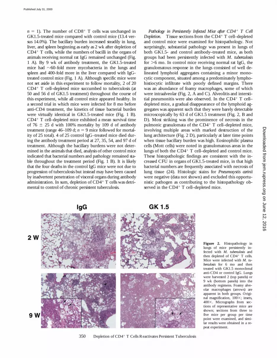
Pathology in Persistently Infected Mice after CD41 T CellDepletion. Tissue sections from the CD41 T cell–depletedand control mice were examined for histopathology. Notsurprisingly, substantial pathology was present in lungs ofboth GK1.5- and control antibody–treated mice, as bothgroups had been persistently infected with M. tuberculosisfor .6 mo. In control mice receiving normal rat IgG, thegranulomatous response in the lungs consisted of well de-lineated lymphoid aggregates containing a minor mono-cytic component, situated among a predominately lympho-histiocytic infiltrate with poorly defined margins. Therewas an abundance of foamy macrophages, some of whichwere intraalveolar (Fig. 2, A and C). Alveolitis and intersti-tial pneumonitis were also observed. In the CD41 T cell–depleted mice, a gradual disappearance of the lymphoid ag-gregates was apparent such that they were barely detectablemicroscopically by 63 d of GK1.5 treatment (Fig. 2, B andD). Most striking was the prominence of necrosis in thepulmonic granulomata of the CD41 T cell–depleted mice,involving multiple areas with marked destruction of thelung architecture (Fig. 2 D), particularly at later time pointswhen tissue bacillary burden was high. Eosinophilic plasmacells (Mott cells) were noted in granulomatous areas in thelungs of both the CD41 T cell–depleted and control mice.These histopathologic findings are consistent with the in-creased CFU in organs of GK1.5-treated mice, in that highbacterial numbers are frequently associated with necrosis oflung tissue (24). Histologic stains for Pneumocystis cariniiwere negative (data not shown) and excluded this opportu-nistic pathogen as contributing to the histopathology ob-served in the CD41 T cell–depleted mice.
Figure 2. Histopathology inlungs of mice persistently in-fected with M. tuberculosis andthen depleted of CD41 T cells.Mice were infected with M. tu-berculosis for 6 mo and thentreated with GK1.5 monoclonalanti-CD4 or control IgG. Lungswere harvested 2 (top panels) or9 wk (bottom panels) into theantibody regimens. Foamy alve-olar macrophages (arrows) areapparent in both groups. Origi-nal magnification, 1003; insets,4003. Micrographs from sec-tions of representative mice areshown; sections from three tofive mice per group per timepoint were examined, and simi-lar results were obtained in a re-peat experiment.
on June 12, 2016jem
.rupress.orgD
ownloaded from
Published July 31, 2000

351 Scanga et al.
NOS2 Expression and Activity in CD41 T Cell–depletedMice. The expression of NOS2 protein in lungs, spleen,and liver of CD41 T cell–depleted and control mice wasassessed immunohistochemically. High levels of NOS2protein localized to foamy macrophages within granuloma-tous areas were observed at 6 mo after infection, before an-tibody treatment, as previously described (19; data notshown). There was no discernible difference in tissueNOS2 expression between GK1.5- and normal IgG-treated mice after 2 wk (Fig. 3, A and E), 6 wk (Fig. 3, Band F), or 9 wk (data not shown) of antibody treatment.The loss of CD41 T cells did not appear to affect the num-ber of NOS2-positive cells (Table I), the distribution ofthose cells, nor the intensity of NOS2 staining in the lung.This observation was supported by RPA analyses of totallung RNA from GK1.5 and control mice (Fig. 4). Expres-
sion of the NOS2 gene was similar between CD41 T cell–depleted and control mice at all time points. More directevidence of continued production of RNIs in the CD41 Tcell–depleted mice was provided by immunostaining tissuesections from these mice using antinitrotyrosine antibodyto detect nitrated proteins. Nitrotyrosine is generated vianitration of tyrosine by peroxynitrite, a product of NO andsuperoxide anion, and thus reflects NOS2 activity. Nitroty-rosine staining was similar in the lung tissue and granulo-mas of GK1.5- and normal IgG-treated mice (Fig. 3, C andH). When the antinitrotyrosine antibody was preincubatedwith nitrotyrosine before being used in this assay, all stain-ing was abolished (Fig. 3, D and G), confirming the stain-ing specificity. These results indicate that CD41 T cell de-pletion–induced reactivation was not due to a deficiency inRNI-dependent antimicrobial mechanisms.
Table I. Number of Cells Expressing NOS2 on Lung Sections of Persistently Infected Mice after Initiation of CD41 T Cell Depletion Regimen
2 wk 6 wk 9 wk
Day 0 GK1.5 IgG GK1.5 IgG GK1.5 IgG
70 6 18 78 6 17 101 6 8 102 6 11 114 6 33 153 6 25 181 6 18
P 5 0.30 P 5 0.75 P 5 0.40
Beginning 6 mo after infection with M. tuberculosis, mice were treated with GK1.5 anti-CD4 antibody or control IgG. At the indicated time intervalsafter initiation of antibody treatment, lungs were recovered and immunohistochemically stained for NOS2 expression. The number of cells stainedpositively for NOS2 per 10 1003 fields 6 SE is shown. Three to five mice per time point per group were examined.
Figure 3. NOS2 protein expression and protein nitration in lungs of mice persistently infected with M. tuberculosis for 6 mo and then depleted ofCD41 T cells. 6 mo after infection, mice were treated with GK1.5 monoclonal anti-CD4 to deplete CD41 T cells (E–H) or control IgG (A–D). Lungsections were stained for NOS2 protein (A, B, E, and F) 2 wk (A and E) and 6 wk (B and F) into the antibody treatment regimen. Similarly processedlung sections obtained 6 wk into the antibody regimen were also stained for nitrotyrosine (C and G). Specificity of the nitrotyrosine antibody was con-firmed by preincubating the nitrotyrosine antibody with nitrotyrosine before staining the sections (D and H). B–D are serial sections, as are F–H. Repre-sentative sections from four mice per group per time point are shown. Original magnification, 4003.
on June 12, 2016jem
.rupress.orgD
ownloaded from
Published July 31, 2000

352 Depletion of CD41 T Cells Reactivates Persistent Tuberculosis
IFN-g Production after CD41 T Cell Depletion. IFN-g isa critical factor required for activating macrophages to pro-duce NOS2 (14, 16). Therefore, the finding of unimpairedNOS2 production in CD41 T cell–depleted mice sug-gested that an alternate source of IFN-g, independent ofCD41 T cells, existed in GK1.5-treated mice. To addressthe possible source of IFN-g in the lungs of the mice, cellpopulations and cytokine production in the lungs were as-sessed by FACS® analysis. Staining for CD41 T cells con-firmed that GK1.5 treatment was efficacious in the lungs,with .94% depletion of CD41 T cells in lungs of GK1.5-treated mice compared with normal rat IgG–treated miceafter 4 and 9 wk of antibody treatment (Fig. 5). However,the mean number of cells recovered from lung tissue didnot differ significantly between GK1.5- and normal ratIgG–treated mice (Table II). There was an increase in thepercentage of CD81 T cells in CD41 T cell–depleted micecompared with control IgG–treated mice at both 4 wk(mean GK1.5, 20.5%; mean control IgG, 10.8%; P 5 0.03)and 9 wk (mean GK1.5, 34.1%; mean control IgG, 26.9%;
P 5 0.076). Because the numbers of cells recovered fromthe lungs of mice from both groups were similar (Table II),this increased percentage corresponds to an actual increasein total number of CD81 T cells.
We used intracellular cytokine staining of lung cells toassess production of IFN-g in the lungs of the CD41 Tcell–depleted, persistently infected mice. Despite effectivedepletion of the pulmonic CD41 T cell compartment,there was not a significant decrease in the percentage of to-tal lymphocytes producing IFN-g after 4 or 9 wk of deple-tion compared with IgG-treated control mice. Data fromrepresentative mice are shown in Fig. 5. Lymphocytes fromuninfected mice produce very little IFN-g after ex vivostimulation (25). As noted above, there was an increase innumbers of CD81 T cells in GK1.5-treated mice, and theseCD81 T cells were capable of producing IFN-g (Fig. 5).RPA analysis of total lung RNA confirmed that the levelsof IFN-g expression in the lungs of GK1.5- and controlIgG–treated mice were indeed comparable (Fig. 4). The ef-ficacy of GK1.5 treatment, the increase in CD81 T cells
Figure 4. Gene expression in lungsafter depletion of CD41 T cells inmice persistently infected with M. tu-berculosis. Persistently infected micewere injected with GK1.5 to depleteCD41 T cells (open symbols) or withcontrol IgG (closed symbols). Micewere killed at intervals after the anti-body regimen was begun, total RNAwas isolated from lungs, and mRNAexpression was measured using RPA
with a template specific for NOS2 (A), IFN-g (B), and TNF-a (C) as well as IL-12p40, IL-1a,IL-1b (not shown), and the housekeeping genes GAPDH and L32. Autoradiographs werequantitated by densitometry, and the ratio between the gene of interest and a housekeepinggene was calculated. Each point represents the mean of results obtained from three to five mice,and bars are SE. *P , 0.05. Similar results were obtained in a repeat experiment. Representativeautoradiograph is shown (D).
on June 12, 2016jem
.rupress.orgD
ownloaded from
Published July 31, 2000

353 Scanga et al.
upon CD41 T cell depletion, and the comparable IFN-gexpression in the lungs of GK1.5 and control mice wasconfirmed in a second CD41 T cell depletion experiment(data not shown). Taken together, these data indicate thatin the lungs of persistently infected mice depleted of CD41
T cells, there was an increase in CD81 T cells capable ofIFN-g production.
Gene Expression in the Lungs after CD41 T Cell Depletionin Chronic Persistent Tuberculosis. RPA analysis of totallung RNA revealed that in the CD41 T cell–depletedmice, TNF-a expression remained relatively steady, exceptat the last time point when a significant increase was ob-served (Fig. 4). This may reflect the more severe pathologyin the lungs of these mice relative to the mice receivingnormal rat IgG (Fig. 2). Substantial expression of IL-1a andIL–1b was detected over the course of the antibody treat-
ment regimen, but there was little difference between themice receiving GK1.5 and those receiving control antibody(Fig. 4). A low level of IL-12p40 expression was noted inboth groups and did not vary throughout the antibodytreatment period (data not shown). Expression of IL-4 (Fig.4) and IL-10 (data not shown) was low or undetectable atall time points in both groups of mice.
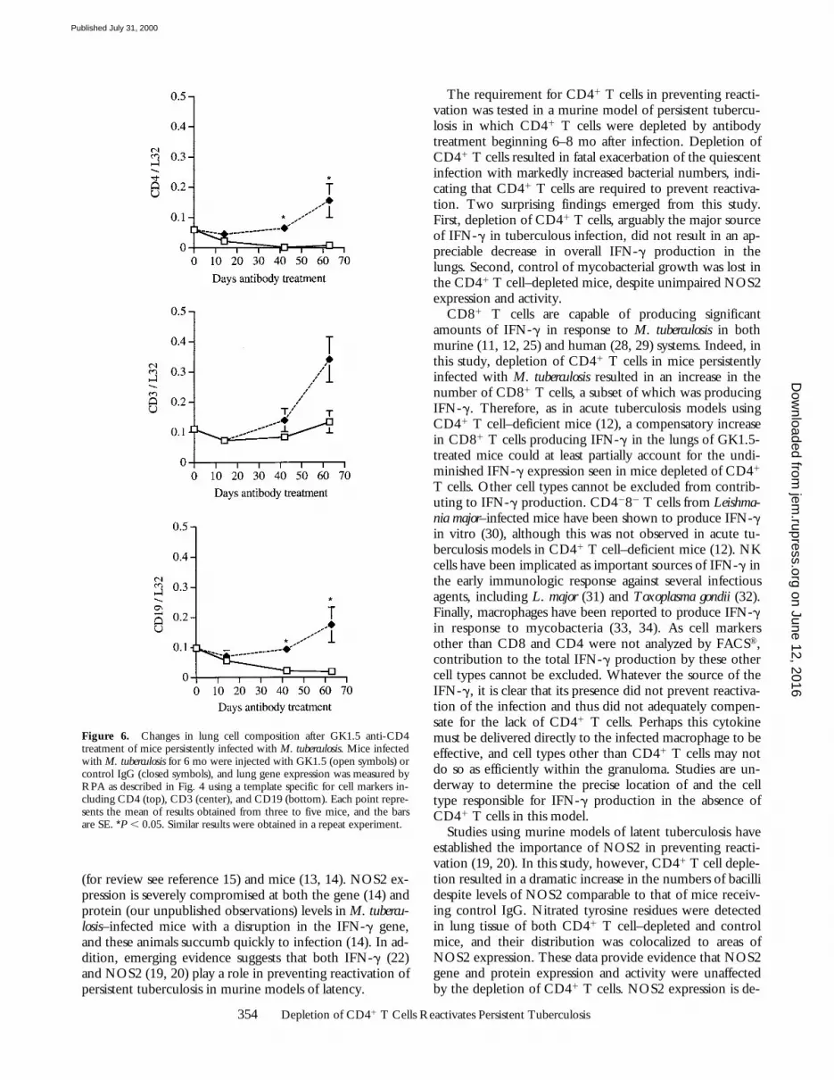
2 wk after initiation of anti-CD4 antibody treatment,there was a decrease in CD4 gene expression in the lungsrelative to that in mice receiving control antibody, as as-sessed by RPA (Fig. 6). This difference was significant (P 50.002) by 6 wk of antibody treatment and was maintainedthroughout the remainder of the experiment (Fig. 6).These data confirmed the results obtained by FACS® analy-sis that GK1.5 was efficacious in depleting CD41 T cells inthe lungs over a long treatment period. The expression ofCD3 was not significantly different among the GK1.5- andrat IgG–treated mice at the various time points studied(Fig. 6). There was a significant decrease in the expressionof CD19 (a pan-B cell marker) in CD41 T cell–depletedmice by 6 wk of GK1.5 treatment compared with IgG-treated mice (P 5 0.001; Fig. 6), suggesting that fewer Bcells were present in those mice.
DiscussionThe association between the deficiency of CD41 T cells
observed with AIDS and the incidence of reactivation oflatent tuberculosis suggests that CD41 T cells play an im-portant role in preventing reactivation tuberculosis. In hu-man studies, M. tuberculosis–specific CD41 T cells secreteIFN-g (26), and a recent publication suggests that in vitroIFN-g can induce human macrophages to kill M. tuberculo-sis coincident with the expression of NOS2 (27). CD41 Tcells are thought to be the primary source of IFN-g and arerequired to control acute M. tuberculosis infection in mice(7, 8, 10, 12). The importance of IFN-g in controlling my-cobacterial infections has been demonstrated in humans
Figure 5. Intracellular IFN-gstaining of lung cells 4 wk after be-ginning CD41 T cell depletion ofpersistently infected mice. Lung cellswere disaggregated, stimulated for16 h with anti-CD3 and anti-CD28in the presence of monensin for thefinal 6 h, fixed in paraformaldehyde,stained for CD4 and CD8, perme-abilized, and stained for IFN-g.Cells were gated on lymphocytes byforward and side scatter and analyzedby three-color FACS®. Lungs fromtwo mice were analyzed from eachgroup, and dot plots from a repre-sentative mouse are shown. Thenumbers are the percentages of gatedcells in that quadrant.
Table II. CD41 T Cell Depletion Does Not Affect Total Cell Numbers in Lungs of Persistently Infected Mice
Treatment4 wk Abtreatment
9 wk Ab treatment
3106 Control rat IgG 7.8 5.2
5.0 6.8
GK1.5 3.3 5.66.5 6.9
Total cell numbers in lungs of mice infected 6 mo previously with M.tuberculosis were determined by trypan blue exclusion after preparationof a single-cell suspension from the lungs. Mice were either treated withcontrol rat IgG or anti-CD4 antibody (GK1.5). Values for two mice pergroup per time point are shown. Statistical analysis indicated that valueswere not significantly different (P . 0.5) between the two groups ateach time point. The mean number of cells obtained from uninfected,untreated mice was 1.2 3 106 cells.
on June 12, 2016jem
.rupress.orgD
ownloaded from
Published July 31, 2000
354 Depletion of CD41 T Cells Reactivates Persistent Tuberculosis
(for review see reference 15) and mice (13, 14). NOS2 ex-pression is severely compromised at both the gene (14) andprotein (our unpublished observations) levels in M. tubercu-losis–infected mice with a disruption in the IFN-g gene,and these animals succumb quickly to infection (14). In ad-dition, emerging evidence suggests that both IFN-g (22)and NOS2 (19, 20) play a role in preventing reactivation ofpersistent tuberculosis in murine models of latency.
The requirement for CD41 T cells in preventing reacti-vation was tested in a murine model of persistent tubercu-losis in which CD41 T cells were depleted by antibodytreatment beginning 6–8 mo after infection. Depletion ofCD41 T cells resulted in fatal exacerbation of the quiescentinfection with markedly increased bacterial numbers, indi-cating that CD41 T cells are required to prevent reactiva-tion. Two surprising findings emerged from this study.First, depletion of CD41 T cells, arguably the major sourceof IFN-g in tuberculous infection, did not result in an ap-preciable decrease in overall IFN-g production in thelungs. Second, control of mycobacterial growth was lost inthe CD41 T cell–depleted mice, despite unimpaired NOS2expression and activity.
CD81 T cells are capable of producing significantamounts of IFN-g in response to M. tuberculosis in bothmurine (11, 12, 25) and human (28, 29) systems. Indeed, inthis study, depletion of CD41 T cells in mice persistentlyinfected with M. tuberculosis resulted in an increase in thenumber of CD81 T cells, a subset of which was producingIFN-g. Therefore, as in acute tuberculosis models usingCD41 T cell–deficient mice (12), a compensatory increasein CD81 T cells producing IFN-g in the lungs of GK1.5-treated mice could at least partially account for the undi-minished IFN-g expression seen in mice depleted of CD41
T cells. Other cell types cannot be excluded from contrib-uting to IFN-g production. CD4282 T cells from Leishma-nia major–infected mice have been shown to produce IFN-gin vitro (30), although this was not observed in acute tu-berculosis models in CD41 T cell–deficient mice (12). NKcells have been implicated as important sources of IFN-g inthe early immunologic response against several infectiousagents, including L. major (31) and Toxoplasma gondii (32).Finally, macrophages have been reported to produce IFN-gin response to mycobacteria (33, 34). As cell markersother than CD8 and CD4 were not analyzed by FACS®,contribution to the total IFN-g production by these othercell types cannot be excluded. Whatever the source of theIFN-g, it is clear that its presence did not prevent reactiva-tion of the infection and thus did not adequately compen-sate for the lack of CD41 T cells. Perhaps this cytokinemust be delivered directly to the infected macrophage to beeffective, and cell types other than CD41 T cells may notdo so as efficiently within the granuloma. Studies are un-derway to determine the precise location of and the celltype responsible for IFN-g production in the absence ofCD41 T cells in this model.
Studies using murine models of latent tuberculosis haveestablished the importance of NOS2 in preventing reacti-vation (19, 20). In this study, however, CD41 T cell deple-tion resulted in a dramatic increase in the numbers of bacillidespite levels of NOS2 comparable to that of mice receiv-ing control IgG. Nitrated tyrosine residues were detectedin lung tissue of both CD41 T cell–depleted and controlmice, and their distribution was colocalized to areas ofNOS2 expression. These data provide evidence that NOS2gene and protein expression and activity were unaffectedby the depletion of CD41 T cells. NOS2 expression is de-
Figure 6. Changes in lung cell composition after GK1.5 anti-CD4treatment of mice persistently infected with M. tuberculosis. Mice infectedwith M. tuberculosis for 6 mo were injected with GK1.5 (open symbols) orcontrol IgG (closed symbols), and lung gene expression was measured byRPA as described in Fig. 4 using a template specific for cell markers in-cluding CD4 (top), CD3 (center), and CD19 (bottom). Each point repre-sents the mean of results obtained from three to five mice, and the barsare SE. *P , 0.05. Similar results were obtained in a repeat experiment.
on June 12, 2016jem
.rupress.orgD
ownloaded from
Published July 31, 2000

355 Scanga et al.
pendent on IFN-g (14, 16), and thus the unimpairedNOS2 expression provides further, indirect evidence ofundiminished IFN-g expression in the lungs of CD41 Tcell–depleted mice. These surprising findings suggest thatCD41 T cells prevent reactivation by a mechanism that isnot solely mediated by IFN-g or NOS2-generated RNIs.Although unlikely, it is possible that there was an unde-tected decrease in IFN-g and/or NOS2 production immedi-ately following CD41 T cell depletion, as the earliest post-depletion time point examined was 2 wk. In acute M.tuberculosis infection of CD42/2 or MHC class II2/2 mice,production of wild-type levels of IFN-g and NOS2 wasdelayed for only 2 wk (12) and was apparently sufficient torender these mice very susceptible to tuberculosis. How-ever, in a chronic infection, macrophages are alreadyprimed to produce NOS2, and a short-lived IFN-g defi-ciency would be less likely to have an effect than in the im-munologically naive mice used for the acute tuberculosisstudies. This study raises the question of how the reactivat-ing bacilli not only survive but actively replicate in thepresence of NOS2 and the resultant RNIs. AlthoughNOS2-generated RNIs are a necessary antimycobacterialeffector mechanism, it is not sufficient to eliminate M. tu-berculosis infection in vivo (19, 20, 35).
In light of reactivation in the presence of control levelsof IFN-g and NOS2 in the CD41 T cell–depleted mice,additional CD41 T cell functions capable of maintaininglatency must be considered. First, the requirement ofCD41 T cells for a vigorous granulomatous reaction inmycobaterial infection has been reported (12, 36, 37). Intuberculosis patients coinfected with HIV, the structuralintegrity of the tuberculous granuloma appears to correlatewith the number of total peripheral CD41 T cells (38, 39).Although the pathology and general loss of granulomastructure observed in the lungs of GK1.5-treated mice dur-ing reactivation may be directly related to bacterial num-bers, we cannot exclude a role for CD41 T cells in mainte-nance of an organized granuloma during latent infection.Second, a key element in controlling M. tuberculosis infec-tion is macrophage activation. CD41 T cells may haveIFN-g– and NOS2-independent pathways to activate my-cobactericidal or mycobacteriostatic effector functions inmacrophages. Macrophages can be activated through directcontact via interaction between CD40 on macrophagesand CD40L on activated lymphocytes (40), and this canresult in NOS2 induction (41). Although this mechanismis apparently necessary for successful immunity to L. major(42–44), CD40L2/2 mice were not more susceptible to M.tuberculosis infection (45). As negative data in geneticallydeficient mice can be difficult to interpret, the role ofCD40 in tuberculosis remains unclear. Third, it is possiblethat depletion of CD41 T cells results in increased produc-tion of cytokines capable of deactivating macrophages.Two such cytokines are IL-10 (46) and TGF-b (47). In-deed, in HIV-infected individuals, production of IL-10and TGF-b by PBMCs is augmented (for review see refer-ence 48). IL-10 mRNA was expressed at very low levelsthat were similar in lungs of CD41 T cell–depleted and
control mice, so this cytokine is not likely to contribute tothe reactivation observed in this model. TGF-b producedby M. tuberculosis–infected macrophages has been reportedto downregulate macrophage function (49). TGF-b wasnot examined in this study, and so the possibility remainsthat this cytokine may increase in the absence of CD41 Tcells, leading to macrophage deactivation and increasedbacterial growth. Fourth, CD41 T cells are thought to beimportant in maintaining an adequate CD81 T cell re-sponse (50–52). We and others have demonstrated previ-ously that CD81 T cells participate in control of tubercu-losis in mice (9, 11, 25, 53, 54), and the presence ofmycobacterial-specific CD81 T cells in tuberculosis pa-tients has been recently reported (28, 29, 55). Mycobacte-ria-specific CD81 T cells can produce IFN-g or act as cy-totoxic cells for infected macrophages (25, 28, 29, 56, 57).Recent data indicate that CD81 CTLs that produce granu-lysin can kill intracellular M. tuberculosis (58). AlthoughCD81 T cell numbers were increased after CD41 T celldepletion, and these cells were clearly primed to produceIFN-g, the actual function of these cells in the lungs in theabsence of CD41 T cells is unclear. Perhaps CD41 T cellsare necessary for complete function or maintenance ofCD81 cytolytic activity, without which control of chronicpersistent tuberculosis is compromised.
Results obtained from RPA analysis of cell surfacemarker expression suggested that there was a substantial re-duction in B cells in the lungs of CD41 T cell–depletedmice. T cells play an important role in modulating B celldifferentiation, function, and life span. CD41 T cells mayalso regulate production of specific B cell chemoattractants.Plasmacytoid cells were observed in lung sections, butCD19 is downregulated on fully differentiated B cells inhumans (59, 60), and so this does not contradict the RPAdata. The role of antibodies in immunity to M. tuberculosis iscontroversial (61), and B cells may contribute to the im-mune response to intracellular infections in an antibody-independent manner, as suggested recently in a Francisellatularensis murine model (62). Studies using B cell–deficientmice are contradictory, suggesting that B cells contributemodestly (63) or not at all (64) to control of M. tuberculosisinfection. A careful analysis of the contribution of B cells inlatent tuberculosis may further define protective mecha-nisms against reactivation of latent infections.
In this murine model of latent tuberculosis, inhibition ofNOS2 (19), neutralization of TNF-a (Mohan, V.P., C.A.Scanga, K. Yu, H.M. Scott, K.E. Tanaka, E. Tsang, J.L.Flynn, and J. Chan, manuscript submitted for publication),or depletion of CD41 T cells (this study) resulted in reacti-vation of infection and fatal tuberculosis. However, therewere significant differences in the course of reactivationand pathology as well as expression of cytokines, dependingon the immunologic manipulation implemented. Inhibi-tion of NOS2 resulted in slowly progressive reactivationprimarily in the lungs (19), whereas CD41 T cell depletionresulted in a rapid and dramatic increase in bacterial num-bers in liver and spleen, as well as lung, shortly after initia-tion of antibody treatment. TNF-a neutralization caused
on June 12, 2016jem
.rupress.orgD
ownloaded from
Published July 31, 2000

356 Depletion of CD41 T Cells Reactivates Persistent Tuberculosis
an initial increase in bacterial numbers that stabilized after 3wk, despite decreased NOS2 expression (Mohan, V.P.,C.A. Scanga, K. Yu, H.M. Scott, K.E. Tanaka, E. Tsang,J.L. Flynn, and J. Chan, manuscript submitted for publica-tion). Severe pathology was observed in TNF-a–neutral-ized mice that was very different than that observed inCD41 T cell–depleted mice, with marked disorganizationof granulomata and increased leukocytic infiltration (Mo-han, V.P., C.A. Scanga, K. Yu, H.M. Scott, K.E. Tanaka,E. Tsang, J.L. Flynn, and J. Chan, manuscript submitted forpublication). Although interpretations based on interexper-imental comparisons must be made with prudence, the re-sults of our previous studies and results presented in thisstudy suggest that individual immunologic componentsplay a variety of roles in preventing reactivation, highlight-ing the complex nature of the protective response inchronic tuberculosis.
In summary, this study demonstrates the requirement forCD41 T cells in maintaining a persistent M. tuberculosis in-fection. However, depletion of CD41 T cells did not resultin decreased tissue expression of IFN-g or NOS2, the twomost characterized CD41 T cell–related antimycobacterialfactors. Thus, the role of the CD41 T cell in persistent tu-berculosis is more complex than merely as a source of IFN-gfor macrophage activation. Further investigation of themechanism by which the CD41 T cell functions during la-tency may guide the development of therapies and vaccinesdesigned to prevent reactivation of tuberculosis.
We gratefully acknowledge Dr. Joseph Ahearn for use of his FACS-Calibur™ and Dr. Simon Watkins, Director of the University ofPittsburgh Center for Biologic Imaging. We thank Dr. DeniseCroix and Natalya Serbina for assistance in analyzing FACS® dataand the members of the Flynn and Chan laboratories for helpfuldiscussions.
This work was supported by National Institutes of Health grantAI36990 (to J. Chan and J.L. Flynn).
Submitted: 28 January 2000Revised: 11 May 2000Accepted: 17 May 2000
References1. World Health Organization. 1999. The World Health Report
1999. Making a Difference. 116.2. Stead, W.W. 1967. Pathogenesis of a first episode of chronic
pulmonary tuberculosis in man: recrudescence of residuals ofthe primary infection or exogenous reinfection? Am. Rev.Resp. Dis. 95:729–745.
3. Powell, K.E., and L.S. Farer. 1980. The rising age of tuber-culosis patients. J. Infect. Dis. 142:946–948.
4. Cisneros, J.R., and K.M. Murray. 1996. Corticosteroids intuberculosis. Ann. Pharmacotherapy. 30:1298–1303.
5. Raviglione, M.C., D.E. Snider, and A. Kochi. 1995. Globalepidemiology of tuberculosis: morbidity and mortality of aglobal epidemic. JAMA. 273:220–226.
6. Chan, J., and S.H.E. Kaufmann. 1994. Immune Mechanismsof Protection. In Tuberculosis: Pathogenesis, Protection andControl. B.R. Bloom, editor. American Society for Microbi-
ology, Washington, D.C. 389–415.7. Leveton, C., S. Barnass, B. Champion, S. Lucas, B. De
Souza, M. Nicol, D. Banerjee, and G. Rook. 1989. T-cellmediated protection of mice against virulent Mycobacterium tu-berculosis. Infect. Immun. 57:390–395.
8. Flory, C., R. Hubbard, and F. Collins. 1992. Effects of invivo T lymphocyte subset depletion on mycobacterial infec-tions in mice. J. Leukoc. Biol. 51:225–229.
9. Muller, I., S. Cobbold, H. Waldmann, and S.H.E. Kauf-mann. 1987. Impaired resistance to Mycobacterium tuberculosisinfection after selective in vivo depletion of L3T41 andLyt21 T cells. Infect. Immunol. 55:2037–2041.
10. Orme, I. 1988. Characteristics and specificity of acquired im-munologic memory to Mycobacterium tuberculosis infection. J.Immunol. 140:3589–3593.
11. Tascon, R.E., E. Stavropoulos, K.V. Lukacs, and M.J. Col-ston. 1998. Protection against Mycobacterium tuberculosis infec-tion by CD8 T cells requires production of gamma inter-feron. Infect. Immun. 66:830–834.
12. Caruso, A.M., N. Serbina, E. Klein, K. Triebold, B.R.Bloom, and J.L. Flynn. 1999. Mice deficient in CD4 T cellshave only transiently diminished levels of IFN-g, yet suc-cumb to tuberculosis. J. Immunol. 162:5407–5416.
13. Cooper, A.M., D.K. Dalton, T.A. Stewart, J.P. Griffen, D.G.Russell, and I.M. Orme. 1993. Disseminated tuberculosis inIFN-g gene-disrupted mice. J. Exp. Med. 178:2243–2248.
14. Flynn, J.L., J. Chan, K.J. Triebold, D.K. Dalton, T.A. Stew-art, and B.R. Bloom. 1993. An essential role for interferon-gin resistance to Mycobacterium tuberculosis infection. J. Exp.Med. 178:2249–2254.
15. Ottenhof, T.H., D. Kumararatne, and J.L. Casanova. 1998.Novel human immunodeficiencies reveal the essential role oftype-1 cytokines in immunity to intracellular bacteria. Immu-nol. Today. 19:491–494.
16. Dalton, D.K., S. Pitts-Meek, S. Keshav, I.S. Figari, A. Brad-ley, and T.A. Stewart. 1993. Multiple defects of immune cellfunction in mice with disrupted interferon-gamma genes. Sci-ence. 259:1739–1742.
17. Chan, J., and J. Flynn. 1999. Nitric oxide in Mycobacterium tu-berculosis infection. In Nitric Oxide and Infection. F. Fang,editor. Plenum Publishers, New York. 281–310.
18. Nicholson, S., M. Bonecini-Almeida, J.R.L. Silva, C.Nathan, Q.-W. Xie, R. Mumford, J.R. Weidner, J. Calay-cay, J. Geng, N. Boechat, et al. 1996. Inducible nitric oxidesynthase in pulmonary alveolar macrophages from patientswith tuberculosis. J. Exp. Med. 184:2293–2302.
19. Flynn, J.L., C.A. Scanga, K.E. Tanaka, and J. Chan. 1998.Effects of aminoguanidine on latent murine tuberculosis. J.Immunol. 160:1796–1803.
20. MacMicking, J.D., R.J. North, R. LaCourse, J.S. Mudgett,S.K. Shah, and C.F. Nathan. 1997. Identification of nitricoxide synthase as a protective locus against tuberculosis. Proc.Natl. Acad. Sci. USA. 94:5243–5248.
21. Adams, L.B., C.M. Mason, J.K. Kolls, D. Scollard, J.L. Kra-henbuhl, and S. Nelson. 1995. Exacerbation of acute andchronic murine tuberculosis by administration of a tumor ne-crosis factor receptor-expressing adenovirus. J. Infect. Dis.171:400–405.
22. Scanga, C.A., V.P. Mohan, H. Joseph, K. Yu, J. Chan, and J.Flynn. 1999. Reactivation of latent tuberculosis: variations onthe Cornell murine model. Infect. Immun. 67:4531–4538.
23. Topham, D.J., R.A. Tripp, and P.C. Doherty. 1997. CD81T cells clear influenza virus by perforin or Fas-dependent
on June 12, 2016jem
.rupress.orgD
ownloaded from
Published July 31, 2000

357 Scanga et al.
processes. J. Immunol. 159:5197–5200.24. Rhoades, E.R., A.A. Frank, and I.M. Orme. 1997. Progres-
sion of chronic pulmonary tuberculosis in mice aerogenicallyinfected with virulent Mycobacterium tuberculosis. Tubercle LungDis. 78:57–66.
25. Serbina, N.V., and J.L. Flynn. 1999. Early emergence ofCD81 T cells primed for production of type 1 cytokines inthe lungs of Mycobacterium tuberculosis-infected mice. Infect.Immun. 67:3980–3988.
26. Boom, W.H., R.S. Wallis, and K.A. Chervenak. 1991. Hu-man Mycobacterium tuberculosis-reactive CD41 T-cell clones:heterogeneity in antigen recognition, cytokine production,and cytotoxicity for mononuclear phagocytes. Infect. Immun.59:2737–2743.
27. Almeida, G.B., C. Chitale, I. Boutsikakis, J.-Y. Geng, H.Doo, S. He, and J.L. Ho. 1998. Induction of in vitro humanmacrophage anti-Mycobacterium tuberculosis activity: require-ment for interferon-g and primed lymphocytes. J. Immunol.160:4490–4499.
28. Lalvani, A., R. Brookes, R. Wilkinson, A. Malin, A. Pathan,P. Andersen, H. Dockrell, G. Pasvol, and A. Hill. 1998. Hu-man cytolytic and interferon gamma-secreting CD81 Tlymphocytes specific for Mycobacterium tuberculosis. Proc. Natl.Acad. Sci. USA. 95:270–275.
29. Lewinsohn, D., M. Alderson, A. Briden, S. Riddell, S. Reed,and K. Grabstein. 1998. Characterization of human CD81 Tcells reactive with Mycobacterium tuberculosis–infected antigen-presenting cells. J. Exp. Med. 187:1633–1640.
30. Locksley, R.M., S.L. Reiner, F. Hatam, D.R. Littman, andN. Killeen. 1993. Helper T cells without CD4: control ofleishmaniasis in CD4-deficient mice. Science. 261:1448–1451.
31. Scharton, T.M., and P. Scott. 1993. Natural killer cells are asource of interferon g that drives differentiation of CD41 Tcell subsets and induces early resistance to Leishmania major inmice. J. Exp. Med. 178:567–577.
32. Gazzinelli, R.T., S. Hieny, T.A. Wynn, S. Wolf, and A.Sher. 1993. Interleukin 12 is required for the T-lymphocyte-independent induction of interferon-g by an intracellularparasite and induces resistance in T-cell-deficient hosts. Proc.Natl. Acad. Sci. USA. 90:6115–6119.
33. Fenton, M.J., M.W. Vermeulen, S. Kim, M. Burdick, R.M.Strieter, and H. Kornfeld. 1997. Induction of gamma inter-feron production in human macrophages by Mycobacterium tu-berculosis. Infect. Immun. 65:5149–5156.
34. Wang, J., J. Wakeham, R. Harkness, and Z. Xing. 1999.Macrophages are a significant source of type 1 cytokines dur-ing mycobacterial infection. J. Clin. Invest. 103:1023–1029.
35. Chan, J., K. Tanaka, D. Carroll, J.L. Flynn, and B.R. Bloom.1995. Effect of nitric oxide synthase inhibitors on murine in-fection with Mycobacterium tuberculosis. Infect. Immun. 63:736–740.
36. Ladel, C.H., S. Daugelat, and S.H.E. Kaufmann. 1995. Im-mune response to Mycobacterium bovis bacille Calmette Guerininfection in major histocompatibility complex class I- and II-deficient knock-out mice: contribution of CD4 and CD8 Tcells to acquired resistance. Eur. J. Immunol. 25:377–384.
37. Hansch, H.C., D.A. Smith, M.E. Mielke, H. Hahn, G.J.Bancroft, and S. Ehlers. 1996. Mechanisms of granuloma for-mation in murine Mycobacterium avium infection: the contri-bution of CD41 T cells. Int. Immunol. 8:1299–1310.
38. Muller, H., and S. Kruger. 1994. Immunohistochemical anal-ysis of cell composition and in situ cytokine expression inHIV- and non-HIV-associated tuberculous lymphadenitis.
Immunobiology. 191:354–368.39. DiPerri, G., A. Cazzadori, S. Vento, S. Bonora, M. Malena,
L. Bontempini, M. Lanzafame, B. Allegranzi, and E. Concia.1996. Comparative histopathological study of pulmonary tu-berculosis in human immunodeficiency virus-infected andnon-infected patients. Tubercle Lung Dis. 77:244–249.
40. Shu, U., M. Kiniwa, C.Y. Wu, C. Maliszewski, N. Vezzio, J.Kakimi, M. Gately, and G. Delespesse. 1995. Activated Tcells induce Interleukin-12 production by monocytes viaCD40-CD40 ligand interaction. Eur. J. Immunol. 25:1125–1128.
41. Tian, L., R.J. Noelle, and D.A. Lawrence. 1995. Activated Tcells enhance nitric oxide production by murine splenic mac-rophages through gp39 and LFI-1. Eur. J. Immunol. 25:306–309.
42. Campbell, K.A., P.J. Ovendale, M.K. Kennedy, S.G.Fanslow, S.G. Reed, and C.R. Maliszewski. 1996. CD40ligand is required for protective cell-mediated immunity toLeishmania major. Immunity. 4:283–289.
43. Kamanaka, M., P. Yu, T. Yasui, K. Yoshida, T. Kawabe, T.Horii, T. Kishimoto, and H. Kikutani. 1996. Protective roleof CD40 in Leishmania major infection at two distinct phasesof cell-mediated immunity. Immunity. 4:275–281.
44. Soong, L., J.C. Xu, I.S. Grewal, P. Kima, J. Sun, B.J. Long-ley, N.H. Ruddle, D. McMahon-Pratt, and R.A. Flavell.1996. Disruption of CD40-CD40 ligand interactions resultsin an enhanced susceptibility to Leishmania amazonensis infec-tion. Immunity. 4:263–273.
45. Campos-Neto, A., P. Ovendale, T. Bement, T.A. Koppi,W.C. Fanslow, M.A. Rossi, and M.R. Alderson. 1998.CD40 ligand is not essential for the development of cell-medi-ated immunity and resistance to Mycobacterium tuberculosis. J.Immunol. 160:2037–2041.
46. Bogdan, C., Y. Vodovotz, and C. Nathan. 1991. Macro-phage deactivation by interleukin 10. J. Exp. Med. 174:1549–1555.
47. Tsunawaki, S., M. Sporn, A. Ding, and C. Nathan. 1988.Deactivation of macrophages by transforming growth factor-beta. Nature. 334:260–262.
48. Fauci, A.S. 1996. Host factors and pathogenesis of HIV-induced disease. Nature. 384:529–534.
49. Toossi, Z., P. Gogate, H. Shiratsuchi, T. Young, and J.J. Ell-ner. 1995. Enhanced production of TGF-beta by bloodmonocytes from patients with active tuberculosis and pres-ence of TGF-beta in tuberculosis granulomatous lung lesions.J. Immunol. 154:465–473.
50. Keene, J.A., and J. Forman. 1982. Helper activity is requiredfor the in vivo generation of cytotoxic T lymphocytes. J.Exp. Med. 155:768–782.
51. von Herrath, M.G., M. Yokoyama, J. Dockter, M.B. Old-stone, and J.L. Whitton. 1996. CD4-deficient mice have re-duced levels of memory cytotoxic T lymphocytes after im-munization and show diminished resistance to subsequentvirus challenge. J. Virol. 70:1072–1079.
52. Bennett, S.R., F.R. Carbone, F. Karamalis, J.F. Miller, andW.R. Heath. 1997. Induction of a CD8 cytotoxic T lym-phocyte response by cross-priming requires cognate CD4 Tcell help. J. Exp. Med. 186:65–70.
53. Flynn, J.L., M.M. Goldstein, K.J. Triebold, B. Koller, andB.R. Bloom. 1992. Major histocompatibility complex classI-restricted T cells are required for resistance to Mycobacteriumtuberculosis infection. Proc. Natl. Acad. Sci. USA. 89:12013–12017.
on June 12, 2016jem
.rupress.orgD
ownloaded from
Published July 31, 2000

358 Depletion of CD41 T Cells Reactivates Persistent Tuberculosis
54. Orme, I., and F. Collins. 1984. Adoptive protection of theMycobacteria tuberculosis-infected lung. Cell. Immunol. 84:113–120.
55. Stenger, S., R. Mazzaccaro, K. Uyemura, S. Cho, P. Barnes,J. Rosat, A. Sette, M. Brenner, S. Porcelli, B. Bloom et al.1997. Differential effects of cytolytic T cell subsets on intra-cellular infection. Science. 276:1684–1687.
56. De Libero, G., I. Flesch, and S.H.E. Kaufmann. 1988. Myco-bacteria-reactive Lyt-21 T cell lines. Eur. J. Immunol. 18:59–66.
57. Serbina, N.V., C.-C. Liu, C.A. Scanga, and J.L. Flynn. 2000.CD81 cytotoxic T lymphocytes from lungs of M. tuberculosisinfected mice express perforin in vivo and lyse infected mac-rophages. J. Immunol. 165:353–363.
58. Stenger, S., D.A. Hanson, R. Teitelbaum, P. Dewan, K.R.Niazi, C.J. Froelich, T. Ganz, S. Thoma-Uszynski, A. Me-lian, C. Bogdan, et al. 1998. An antimicrobial activity of cy-totoxic T cells mediated by granulysin. Science. 282:121–125.
59. Loken, M.R., V.O. Shah, K.L. Dattilio, and C.I. Civin.1987. Flow cytometric analysis of human bone marrow. II.
Normal B cell development. Blood. 70:1316–1324.60. Scheuermann, R.H., and E. Racila. 1995. CD19 antigen in
leukemia and lymphoma diagnosis and immunotherapy.Leuk. Lymphoma. 18:385–397.
61. Glatman-Freedman, A., and A. Casadevall. 1998. Serumtherapy for tuberculosis revisited: reappraisal of the role ofantibody-mediated immunity against Mycobacterium tuberculo-sis. Clin. Microbiol. Rev. 11:514–532.
62. Elkins, K.L., C.M. Bosio, and T.R. Rhinehart-Jones. 1999.Importance of B cells, but not specific antibodies, in primaryand secondary protective immunity to the intracellular bacte-rium Francisella tularensis live vaccine strain. Infect. Immun. 67:6002–6007.
63. Vordermeier, H.M., N. Venkataprasad, D.P. Harris, and J.Ivanyi. 1996. Increase of tuberculous infection in the organsof B cell-deficient mice. Clin. Exp. Immunol. 106:312–316.
64. Johnson, C.M., A.M. Cooper, A.A. Frank, C.B. Bonorino,L.J. Wysoki, and I.M. Orme. 1997. Mycobacterium tuberculosisaerogenic challenge infections in B cell-deficient mice. Tu-bercle Lung Dis. 78:257–261.
on June 12, 2016jem
.rupress.orgD
ownloaded from
Published July 31, 2000