dendrite plasticity in the lateral geniculate nucleus in primate glaucoma
TRANSCRIPT
Vision Research 51 (2011) 243–250
Contents lists available at ScienceDirect
Vision Research
journal homepage: www.elsevier .com/locate /v isres
Dendrite plasticity in the lateral geniculate nucleus in primate glaucoma
Tina Ly a, Neeru Gupta a,b,c, Robert N. Weinreb d, Paul L. Kaufman e, Yeni H. Yücel a,c,⇑a Ophthalmology & Vision Sciences, Laboratory Medicine & Pathobiology, St. Michael’s Hospital, University of Toronto, Toronto, Ontario, Canadab Glaucoma & Nerve Protection Unit, St. Michael’s Hospital, Toronto, Ontario, Canadac Keenan Research Centre at the Li Ka Shing Knowledge Institute of St. Michael’s Hospital, Toronto, Ontario, Canadad Hamilton Glaucoma Center and Department of Ophthalmology, University of California, San Diego, CA, United Statese Department of Ophthalmology and Visual Sciences, University of Wisconsin, Madison, WI, United States
a r t i c l e i n f o
Article history:Received 14 June 2010Available online 6 August 2010
Keywords:NeuroprotectionTranssynaptic degenerationNeurodegenerationGlutamateNMDAMemantine
0042-6989/$ - see front matter � 2010 Elsevier Ltd. Adoi:10.1016/j.visres.2010.08.003
⇑ Corresponding author at: St. Michael’s Hospital, 35-038, Toronto, Ontario, Canada M5B 1W8. Fax: +1 4
E-mail address: [email protected] (Y.H. Yüce
a b s t r a c t
Neural degeneration in glaucoma involves retinal ganglion cells and neurons of their major target, thelateral geniculate nucleus (LGN). Dendrites of relay LGN neurons projecting to the visual cortex werestudied by immunocytochemical and quantitative Sholl analysis in combination with confocal micros-copy and 3D-morphometry. In non-human adult primate glaucoma, relay LGN neurons showed reduceddendrite complexity and length, and these changes were modified by NMDA receptor blockade. Dendriteplasticity of LGN relay neurons in adult primate glaucoma has implications for potential disease modifi-cation by treatment interventions.
� 2010 Elsevier Ltd. All rights reserved.
1. Introduction
Glaucoma is a major cause of irreversible blindness world-wide, characterized by retinal ganglion cell (RGC) loss (Weinreb& Khaw, 2004). Degeneration of retinal ganglion cells spreadstrans synaptically to their main target, the lateral geniculate nu-cleus (LGN) of the brain in human glaucoma (Gupta, Ang, Noelde Tilly, Bidaisee, & Yucel, 2006; Gupta et al., 2009). Previousstudies in experimental primate glaucoma, show degenerativechanges in the LGN including neuron loss (Weber, Chen, Hub-bard, & Kaufman, 2000; Yucel, Zhang, Gupta, Kaufman, & Wein-reb, 2000), neuron atrophy (Weber et al., 2000; Yucel, Zhang,Weinreb, Kaufman, & Gupta, 2001), reactive gliosis (Sasaokaet al., 2008), and microglial activation (Imamura et al., 2009).Neuron loss and atrophy increase with increasing RGC loss asdo metabolic changes observed in the visual cortex in primateglaucoma. (Yucel, Zhang, Weinreb, Kaufman, & Gupta, 2003;Yucel et al., 2000, 2001).
Dendrites are fine processes emerging from the cell body ofneurons that form elaborate dendritic arbors supporting post-syn-aptic contact elements (Johnston & Narayanan, 2008). Distur-bances to dendrite branching can disrupt neural networkorganization and lead to neural dysfunction, as in human neurolog-
ll rights reserved.
0 Bond Street, Shuter Wing,16 864 5648.l).
ical disorders including Alzheimer’s disease (Moolman, Vitolo,Vonsattel, & Shelanski, 2004). Their fine structure is maintainedby microtubules, and among the associated proteins, microtu-bule-associated protein-2 (MAP2) is enriched in dendrites (Cassim-eris & Spittle, 2001; Ichihara, Kitazawa, Iguchi, Hotani, & Itoh,2001) playing an important role in dendrite morphology andbranching (Dehmelt & Halpain, 2005; Harada, Teng, Takei, Oguchi,& Hirokawa, 2002; Matus, 1994). MAP2 has been used as a markerto assess dendrites in neurodegenerative diseases (Goedert,Crowther, & Garner, 1991; Kaufmann, Naidu, & Budden, 1995;Kwei, Jiang, & Haddad, 1993; Matesic & Lin, 1994).
In glaucoma, dendrites in the primate retina undergo atrophy(Morgan, Uchida, & Caprioli, 2000; Weber, Kaufman, & Hubbard,1998). A study in which LGN interneurons confined to the LGNand relay neurons projecting to the visual cortex were not dis-criminated, showed dendrite changes in primate glaucoma (Gup-ta et al., 2007). However, detailed dendrite evaluation of the LGNrelay neurons under conditions of glaucoma at various stages ofdisease and following pharmacological treatment has not beenpreviously investigated. Memantine is an NMDA glutamateopen-channel blocker approved as a neuroprotective agent forthe treatment of cognitive impairment in Alzheimer’s disease(Reisberg et al., 2003). The purpose of this work is to identifyand quantify dendrite characteristics of LGN relay neurons in aprimate model of glaucoma to determine whether they are al-tered, and if so, whether these changes are modifiable bymemantine (Bormann, 1989).
244 T. Ly et al. / Vision Research 51 (2011) 243–250
2. Materials and methods
2.1. Subjects
All experiments were performed following the guidelines of theAssociation for Research in Vision and Ophthalmology Resolutionon the Use of Animals in Research.
Six LGNs from six normal adult cynomolgus monkeys (Macaca fas-cicularis) from the University of Wisconsin, Madison were used as con-trols. They were perfused intracardially with 4% paraformaldehyde in0.1 M phosphate buffer (pH 7.4). Intraocular pressure (IOP) measure-ments were performed in vivo before sacrifice with a pneumatonom-eter (Digilab, Norwell, MA) under light sedation (intramuscularinjection of 5 mg/kg of ketamine hydrochloride) and topical anesthe-sia (5% proparacaine hydrochloride) (Yucel et al., 2000). The mean IOPranged from 12.3 to 21.0 mm Hg and the maximum IOP ranged from13.0 to 26.0 mm Hg. There was significant difference in mean IOP(P < 0.001) or maximum IOP (P < 0.001) between the vehicle-treatedglaucoma group and the normal group.
Experimental glaucoma was induced in adult monkeys (Macacafascicularis) by laser scarification of the trabecular meshwork (Hareet al., 2004). The duration of IOP elevation was 14 months. Threeanimals out of 18 were excluded from the present study due toeither suboptimal fixation or unidentifiable LGN layers. Twogroups of monkeys were evaluated and compared: (1) seven vehi-cle-treated (VT) monkeys with glaucoma and (2) eight memantine-treated (MT) monkeys with glaucoma who received a daily oraldose of 4 mg/kg of memantine. In both the VT and MT glaucomagroups, the treatment was continued during 14 months until sacri-fice (Hare et al., 2004). There was no significant difference in meanIOP (P = 0.43) or maximum IOP (P = 0.73) between the VT and MTglaucoma groups (Yucel, Gupta, Zhang, Mizisin, Kalichman, &Weinreb, 2006). Previously published optic nerve counts of theseanimals showed no significant difference in mean percent opticnerve fiber loss between the VT and the MT glaucoma groups(Yucel, Gupta et al., 2006).
2.2. Tissue processing
Perfusion fixation under deep general anesthesia and tissueprocessing and 40 lm-thick serial sections were performed (Yucelet al., 2000). Every seventh section was mounted onto a glass slideand stained with cresyl violet. Care was taken to use the same tis-sue processing procedures for all monkey brains. Sections contain-ing left LGN with six layers were randomly selected for each of thenormal control animals and each of the VT and MT glaucomaanimals.
2.3. Double immunofluorescence labeling of MAP2 and parvalbumin
LGN sections were double-labeled with a monoclonal antibodyagainst MAP2 (Clone HM-2, Sigma, St. Louis, MO), and a polyclonalantibody against parvalbumin (PV28, Swant, Bellinzona, Switzer-land), a calcium-binding protein, a specific marker for relay LGNneurons (Johnson & Casagrande, 1995). The tyramide signal ampli-fication (TSA) kit (Invitrogen Inc., ON, Canada) was used to enhanceimmunostaining. Sections were washed with phosphate bufferedsaline (1 � PBS, 3 � 10 min). All subsequent washes were per-formed with 1 � PBS, pH 7.35 (3 � 5 min). Sections were incubatedwith 0.2% Triton-X (Sigma, St. Louis, MO) in PBS (2 � 5 min). AfterPBS wash, endogenous peroxidase activity was quenched by 3%H2O2 (2 � 15 min). Sections were washed with PBS and blockedin 1% blocking reagent (TSA kit). Incubation in parvalbumin[1:500] was extended to 2 h at room temperature, overnight at4 �C and then left at room temperature for another 2 h. Sections
were washed with PBS and incubated in anti Rabbit-HRP [1:100](TSA kit). After PBS wash, sections were incubated in Tyramide-Alexa Fluor-488 diluted in Amplification Buffer [1:100](2 � 10 min). Sections were washed with PBS, quenched with 3%H2O2 (2 � 15 min), and blocked in 1% blocking reagent. Incubationin MAP2 [1:100] was extended to 2 h at room temperature, over-night at 4 �C and then left at room temperature for another 2 h.Sections were washed with PBS and incubated in anti Mouse-HRP [1:100] (TSA kit). After being washed again in PBS, sectionswere incubated in Tyramide-Alexa Fluor-555 [1:100](2 � 10 min) diluted in Amplification buffer (TSA kit). Sectionswere washed again with PBS, mounted with antifade PVA-DABCOonto Vectabond (Vector Laboratories, Burlingame, CA) coatedslides and cover-slipped. Immunostaining experiments for all threegroups were performed simultaneously with same reagents. Nega-tive controls were obtained by omitting the primary antibodies.
2.4. Morphological assessment
Immunofluorescence-labeled sections were viewed using anOlympus bright field BX51 upright microscope with a color digitalcamera (Microfire, Optronics Inc. Goleta, CA) and a computer mon-itor. The LGN layers were identified as layers 1–6 from ventral todorsal. The ventral layers 1 and 2 are magnocellular layers, whilethe remaining dorsal layers 3–6 are parvocellular layers. Layers1, 4 and 6 of the left LGN are connected to the glaucomatous righteye while layers 2, 3 and 5 are connected to the non-glaucomatousleft eye. We analyzed MAP2-immunoreactive dendrites of parval-bumin-positive relay neurons projecting to the primary visual cor-tex (Johnson & Casagrande, 1995), located in left LGNmagnocellular layer 1 and parvocellular layer 6 connected to theglaucomatous right eye.
2.5. Quantitative analysis
2.5.1. Confocal image acquisitionSections from MT and VT glaucoma groups, and normal controls
were examined using LaserSharp 2000 software (Biorad Cell Sci-ence Division, Hernelhempstead, UK) with a digital camera on anupright confocal laser microscope (Biorad Radiance, Bronx, NY onNikon E800 Upright Microscope, Melville, NY) in a masked fashion.Five sampling sites of 500 lm � 500 lm within each magnocellu-lar layer 1 and parvocellular layer 6 were selected. Within eachsampling site, one stack of images was taken under red (CY3 forMAP2) and green (FITC for parvalbumin) channels separately witha 60� oil-immersion objective at zoom of 1.5� and 512 � 512 pix-els by LaserSharp2000 program. Serial stack images of 18 lmdepth and 0.5 lm step size were collected using the same camerasettings for power, gain, iris aperture size, and offset to maintainconsistent settings for comparison. All images were obtained usinga Kalman filter with three passes.
2.5.2. Automated tracing and 3D reconstructionStacks of images were imported (Neurolucida; MicroBright-
Field, Inc., Colchester, VT) and neurons were traced with an auto-mated tracing software (AutoNeuron, MicroBrightField Inc.,Colchester, VT). Tracing of MAP2-immunoreactive dendrites (CY3channel) and parvalbumin-immunostained cell bodies (FITC chan-nel) of stacks of confocal images was performed separately. Twomodes of the automated tracing software were used to trace neu-rons: interactive mode for MAP2-immunostained dendrites, andautomatic mode for parvalbumin-immunostained cell bodies. Con-figuration parameters used for tracing for each neuron were: (1)maximum process diameter, 1.5 lm, (2) ignore somas smallerthan, 2.0 lm, (3) soma detector sensitivity, 55 and 65 for layers 1and 6, respectively, and (4) interactive mode.

T. Ly et al. / Vision Research 51 (2011) 243–250 245
2.5.3. Quantitative analysis of dendritesImmunofluorescence labeling, image acquisition and quantita-
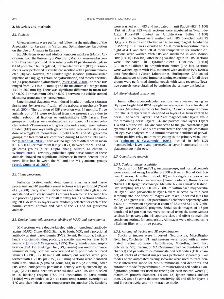
tive analysis of the dendrites were performed in a masked fashion.Tracings were loaded into Neurolucida software (NeuroExplorer,MicroBrightField Inc. VT, USA). A total of 45 parvalbumin-positiveneurons with MAP2-immunostained dendrites were selected ran-domly from each magnocellular layer 1 and parvocellular layer 6per animal for dendrite complexity analysis (Sholl analysis, Neu-roExplorer) and dendrite length analysis (Branch Structure Analy-sis, NeuroExplorer). In Sholl analysis, once the cell body to studywas selected, the intersections of dendrites and series of sphereswith radii 1–14 lm, spaced at 1-lm increments, were generatedby the software around the cell body. The number of dendrite-sphere intersections to estimate dendrite complexity was calcu-lated by the software. The percent decrease in dendrite complexitywas calculated from the difference between the average mean den-drite complexity of the normal group and mean dendrite complex-ity of MT and VT animals with glaucoma divided by the averagemean of dendrite complexity in the normal group. Percent de-crease in dendrite length was assessed in a similar manner.
2.6. Statistical analysis
The two-tailed student t-test was used to compare dendriteparameters in relay neurons in LGN layers 1 and 6 of the VT glau-coma group to the MT glaucoma group, and to the normal controlgroup. Mean IOP, maximum IOP, and percent optic nerve fiber losswere also compared using a t-test.
Using the generalized linear models procedure of SAS statisticalsoftware (SAS Institute Inc., Cary, NC), percent decrease in dendritecomplexity and dendrite length were compared between thememantine- and the vehicle-treated glaucoma groups, with meanIOP as a covariate.
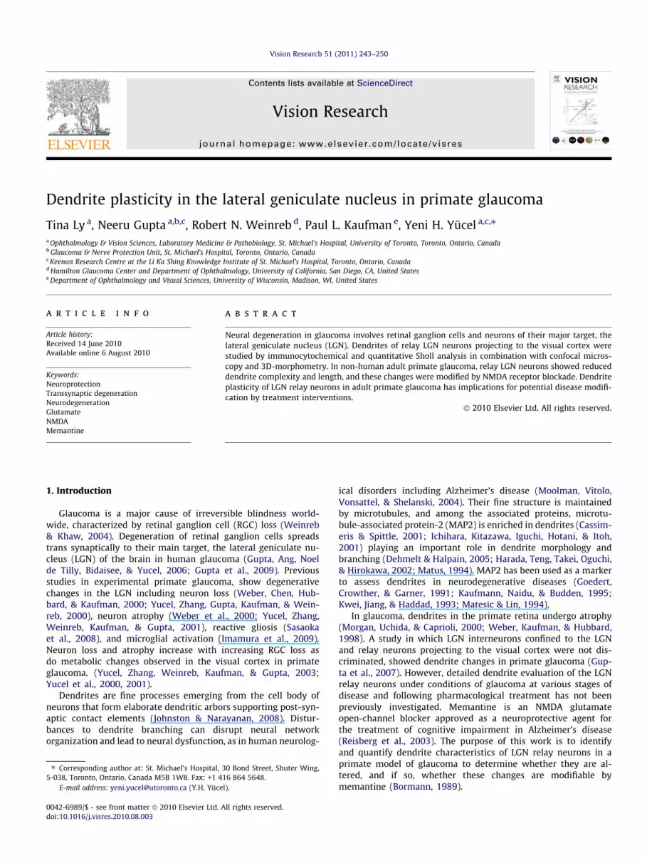
Fig. 1. Projected images of stacks of confocal images of LGN relay neurons immunostainein magnocellular layer 1 from normal monkey ID #S1, from vehicle-treated (VT) glaurespectively (Table 1). MAP2 immunoreactivity in normal layer 1 (A) was more intense cMAP2 appeared more intense in MT glaucoma (C) compared to VT glaucoma monkeys (Band MT glaucoma (F) monkeys. Scale bar indicates 40 lm.
3. Results
3.1. Dendritic plasticity in LGN relay neurons in experimentalglaucoma (Fig. 1)
MAP2-immunoreactivity in magnocellular layer 1 relay neuronsin normal monkey (Fig. 1A) was more intense compared to vehicle-treated (VT) glaucoma monkeys (Fig. 1B). Similar observationswere made for parvocellular layer 6 (Fig. 1D and E). MAP2-immu-noreactivity in magnocellular layer 1 relay neurons in memantine-treated (MT) glaucoma monkeys (Fig. 1C) was more intensecompared to VT glaucoma monkeys (Fig. 1B). Similar observationswere made for parvocellular layer 6 (Fig. 1E and F).
Dendrite complexity (Fig. 2) and length were measured in normalgroup, VT and MT glaucoma groups (Table 1). In magnocellular layer 1,both mean dendrite complexity (Fig. 3A) and dendrite length (Fig. 3B)weresignificantly decreased intheVT glaucoma groupcompared withthose in the normal group (11.2 ± 1.0 (Mean ± SD) vs. 23.7 ± 1.5,P < 0.0001; 18.4 ± 1.4 lm vs. 38.5 ± 3.1 lm, P < 0.0001; respectively).
In parvocellular layer 6, both mean dendrite complexity (Fig. 4A)and length (Fig. 4B) were significantly decreased in the VT glaucomagroup compared with those in the normal group (11.1 ± 1.3 vs.21.2 ± 0.9, P < 0.0001; 16.7 ± 1.9 lm vs. 33.7 ± 2.4 lm, P < 0.0001,respectively).
3.2. Dendritic alterations in LGN relay neurons in memantine-treatedglaucoma monkeys
3.2.1. Magnocellular layer 1Percent decrease in dendrite complexity (Table 2) in MT glau-
coma was significantly reduced compared to VT glaucoma group(38.1% ± 10.7 vs. 52.9% ± 4.2, P = 0.0047; respectively). Percent
d with parvalbumin (green) and dendrites with MAP2 (red). A, B and C show neuronscoma monkey #106 and from memantine-treated (MT) glaucoma monkey #97,
ompared to those in the VT glaucoma (B) and MT glaucoma (C) monkeys. In layer 1,). Similar results were seen in parvocellular layer 6 in normal (D), VT glaucoma (E)
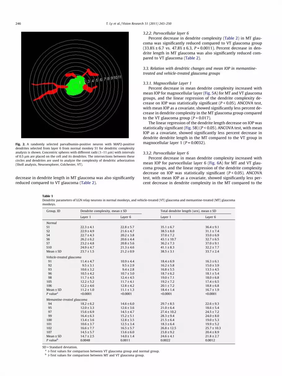
Fig. 2. A randomly selected parvalbumin-positive neuron with MAP2-positivedendrites selected from layer 6 from normal monkey S1 for dendritic complexityanalysis is shown. Concentric spheres with different radii (1–11 lm) with intervalsof 0.5 lm are placed on the cell and its dendrites. The intersections between thesecircles and dendrites are used to analyze the complexity of dendritic arborization(Sholl analysis, Neuroexplorer, Colchester, VT).
246 T. Ly et al. / Vision Research 51 (2011) 243–250
decrease in dendrite length in MT glaucoma was also significantlyreduced compared to VT glaucoma (Table 2).
Table 1Dendritic parameters of LGN relay neurons in normal monkeys, and vehmonkeys.
Group, ID Dendrite complexity, mean ± SD
Layer 1 Layer 6
NormalS1 22.3 ± 4.1 22.8 ± 5.7S2 22.9 ± 4.9 21.6 ± 4.7S4 22.7 ± 4.3 20.2 ± 3.8S6 26.2 ± 6.2 20.6 ± 4.4S7 23.2 ± 4.8 20.8 ± 5.6S10 24.9 ± 4.7 21.3 ± 4.6Mean ± SD 23.7 ± 1.5 21.2 ± 0.9
Vehicle-treated glaucoma91 11.4 ± 4.7 10.9 ± 4.492 9.5 ± 3.1 9.5 ± 2.993 10.6 ± 3.2 9.4 ± 2.896 10.5 ± 4.2 10.7 ± 3.098 11.7 ± 4.5 12.4 ± 4.5
105 12.2 ± 5.2 11.7 ± 4.1106 12.2 ± 4.6 12.8 ± 4.2Mean ± SD 11.2 ± 1.0 11.1 ± 1.3P valuea <0.0001 <0.0001
Memantine-treated glaucoma94 18.2 ± 6.2 14.6 ± 6.095 12.0 ± 3.3 12.6 ± 3.697 15.6 ± 6.9 14.5 ± 4.799 16.4 ± 6.3 15.2 ± 5.1
100 13.4 ± 3.6 12.8 ± 3.5101 10.6 ± 3.7 12.5 ± 3.4102 16.6 ± 7.7 16.5 ± 5.7107 14.5 ± 5.7 13.6 ± 6.0Mean ± SD 14.7 ± 2.5 14.0 ± 1.4P valueb 0.0048 0.0011
SD = Standard deviation.a t-Test values for comparison between VT glaucoma group and nob t-Test values for comparison between MT and VT glaucoma grou
3.2.2. Parvocellular layer 6Percent decrease in dendrite complexity (Table 2) in MT glau-
coma was significantly reduced compared to VT glaucoma group(33.8% ± 6.7 vs. 47.8% ± 6.3, P = 0.0011). Percent decrease in den-drite length in MT glaucoma was also significantly reduced com-pared to VT glaucoma (Table 2).
3.3. Relation with dendritic changes and mean IOP in memantine-treated and vehicle-treated glaucoma groups
3.3.1. Magnocellular Layer 1Percent decrease in mean dendrite complexity increased with
mean IOP for magnocellular layer (Fig. 5A) for MT and VT glaucomagroups, and the linear regression of the dendrite complexity de-crease on IOP was statistically significant (P < 0.05). ANCOVA test,with mean IOP as a covariate, showed significantly less percent de-crease in dendrite complexity in the MT glaucoma group comparedto the VT glaucoma group (P = 0.017).
The linear regression of the dendrite length decrease on IOP wasstatistically significant (Fig. 5B) (P < 0.05). ANCOVA test, with meanIOP as a covariate, showed significantly less percent decrease indendrite dendrite length in the MT compared to the VT group inmagnocellular layer 1 (P = 0.0032).
3.3.2. Parvocellular layer 6Percent decrease in mean dendrite complexity increased with
mean IOP for parvocellular layer 6 (Fig. 6A) for MT and VT glau-coma groups, and the linear regression of the dendrite complexitydecrease on IOP was statistically significant (P < 0.05). ANCOVAtest, with mean IOP as a covariate, showed significantly less per-cent decrease in dendrite complexity in the MT compared to the
icle-treated (VT) glaucoma and memantine-treated (MT) glaucoma
Total dendrite length (urn), mean ± SD
Layer 1 Layer 6
35.1 ± 6.7 36.4 ± 9.138.5 ± 8.0 31.1 ± 7.437.0 ± 7.2 33.0 ± 6.943.1 ± 10.7 32.7 ± 6.536.2 ± 7.3 37.0 ± 9.141.1 ± 8.3 32.2 ± 7.738.5 ± 3.1 33.7 ± 2.4
18.4 ± 6.9 16.3 ± 6.116.2 ± 5.8 15.0 ± 3.916.8 ± 5.5 13.5 ± 4.518.7 ± 6.2 18.1 ± 5.419.0 ± 7.1 18.0 ± 6.819.2 ± 7.2 17.4 ± 6.520.1 ± 7.2 18.8 ± 6.818.4 ± 1.4 16.7 ± 1.9<0.0001 <0.0001
29.7 ± 8.5 22.6 ± 9.321.0 ± 6.4 18.6 ± 5.427.4 ± 10.2 24.5 ± 7.228.3 ± 9.4 24.0 ± 8.021.5 ± 6.4 19.0 ± 5.318.3 ± 6.4 19.9 ± 5.226.8 ± 12.5 25.7 ± 10.323.8 ± 9.2 20.4 ± 8.924.6 ± 4.1 21.8 ± 2.70.0022 0.0012
rmal group.p.
Fig. 3. Magnocellular layer 1: Box plots for dendrite complexity (A) and dendrite length (B) in the normal group, and vehicle-treated (VT) and memantine-treated (MT)glaucoma groups. Compared to normal group, dendrite complexity and dendrite length were reduced in VT glaucoma group. Dendrite complexity and dendrite length in MTgroup were significantly increased compared to VT group (P = 0.0048 and P = 0.0022, respectively). The box extends from the 25th percentile to the 75th percentile, withhorizontal solid line and square at the median and mean, respectively. The bars indicate the highest and lowest values determined by the 25th and 75th percentiles.
Fig. 4. Parvocellular layer 6: Box plots for dendrite complexity (A) and dendrite length (B) in the normal group, and VT and MT glaucoma groups. Compared to normal group,dendrite complexity and dendrite length were reduced in VT glaucoma group. Dendrite complexity and dendrite length in MT glaucoma group were significantly increasedcompared to VT glaucoma group (P = 0.0011; P = 0.0012, respectively). The box extends from the 25th percentile to the 75th percentile, with horizontal solid line and squareat the median and mean, respectively. The bars indicate the highest and lowest values determined by the 25th and 75th percentiles.
Table 2Dendrite complexity and total dendrite length of vehicle-treated and memantine-treated glaucoma monkeys.
Group, ID Decrease in dendrite complexity (%) Mean Decrease in total dendrite length (%) Mean
Layer 1 Layer 6 Layer 1 Layer 6
Vehicle-treated91 51.9 48.6 52.2 51.692 59.9 55.2 57.9 55.593 55.3 55.7 56.4 59.996 55.7 49.5 51.4 46.398 50.6 41.5 50.6 46.6105 48.5 44.8 50.1 48.4106 48.5 39.6 47.8 44.2Mean ± SD 52.9 ± 4.2 47.8 ± 6.3 52.4 ± 3.6 50.4 ± 5.7
Memantine-treated94 23.2 31.1 22.9 32.995 49.4 40.6 45.5 44.897 34.2 31.6 28.8 27.399 30.8 28.3 26.5 28.8100 43.5 39.6 44.2 43.6101 55.3 41.0 52.5 40.9102 30.0 22.2 30.4 23.7107 38.8 35.8 38.2 39.5Mean ± SD 38.1 ± 10.7 33.8 ± 6.7 36.1 ± 10.5 35.2 ± 8.1P value 0.0047 0.0011 0.0019 0.0011
SD = Standard deviation.
T. Ly et al. / Vision Research 51 (2011) 243–250 247
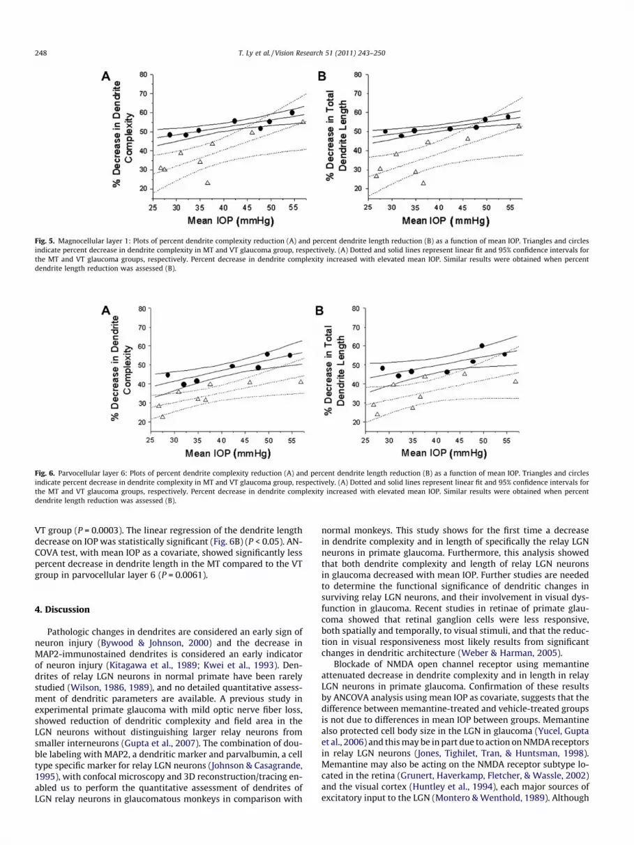
Fig. 5. Magnocellular layer 1: Plots of percent dendrite complexity reduction (A) and percent dendrite length reduction (B) as a function of mean IOP. Triangles and circlesindicate percent decrease in dendrite complexity in MT and VT glaucoma group, respectively. (A) Dotted and solid lines represent linear fit and 95% confidence intervals forthe MT and VT glaucoma groups, respectively. Percent decrease in dendrite complexity increased with elevated mean IOP. Similar results were obtained when percentdendrite length reduction was assessed (B).
Fig. 6. Parvocellular layer 6: Plots of percent dendrite complexity reduction (A) and percent dendrite length reduction (B) as a function of mean IOP. Triangles and circlesindicate percent decrease in dendrite complexity in MT and VT glaucoma group, respectively. (A) Dotted and solid lines represent linear fit and 95% confidence intervals forthe MT and VT glaucoma groups, respectively. Percent decrease in dendrite complexity increased with elevated mean IOP. Similar results were obtained when percentdendrite length reduction was assessed (B).
248 T. Ly et al. / Vision Research 51 (2011) 243–250
VT group (P = 0.0003). The linear regression of the dendrite lengthdecrease on IOP was statistically significant (Fig. 6B) (P < 0.05). AN-COVA test, with mean IOP as a covariate, showed significantly lesspercent decrease in dendrite length in the MT compared to the VTgroup in parvocellular layer 6 (P = 0.0061).
4. Discussion
Pathologic changes in dendrites are considered an early sign ofneuron injury (Bywood & Johnson, 2000) and the decrease inMAP2-immunostained dendrites is considered an early indicatorof neuron injury (Kitagawa et al., 1989; Kwei et al., 1993). Den-drites of relay LGN neurons in normal primate have been rarelystudied (Wilson, 1986, 1989), and no detailed quantitative assess-ment of dendritic parameters are available. A previous study inexperimental primate glaucoma with mild optic nerve fiber loss,showed reduction of dendritic complexity and field area in theLGN neurons without distinguishing larger relay neurons fromsmaller interneurons (Gupta et al., 2007). The combination of dou-ble labeling with MAP2, a dendritic marker and parvalbumin, a celltype specific marker for relay LGN neurons (Johnson & Casagrande,1995), with confocal microscopy and 3D reconstruction/tracing en-abled us to perform the quantitative assessment of dendrites ofLGN relay neurons in glaucomatous monkeys in comparison with
normal monkeys. This study shows for the first time a decreasein dendrite complexity and in length of specifically the relay LGNneurons in primate glaucoma. Furthermore, this analysis showedthat both dendrite complexity and length of relay LGN neuronsin glaucoma decreased with mean IOP. Further studies are neededto determine the functional significance of dendritic changes insurviving relay LGN neurons, and their involvement in visual dys-function in glaucoma. Recent studies in retinae of primate glau-coma showed that retinal ganglion cells were less responsive,both spatially and temporally, to visual stimuli, and that the reduc-tion in visual responsiveness most likely results from significantchanges in dendritic architecture (Weber & Harman, 2005).
Blockade of NMDA open channel receptor using memantineattenuated decrease in dendrite complexity and in length in relayLGN neurons in primate glaucoma. Confirmation of these resultsby ANCOVA analysis using mean IOP as covariate, suggests that thedifference between memantine-treated and vehicle-treated groupsis not due to differences in mean IOP between groups. Memantinealso protected cell body size in the LGN in glaucoma (Yucel, Guptaet al., 2006) and this may be in part due to action on NMDA receptorsin relay LGN neurons (Jones, Tighilet, Tran, & Huntsman, 1998).Memantine may also be acting on the NMDA receptor subtype lo-cated in the retina (Grunert, Haverkamp, Fletcher, & Wassle, 2002)and the visual cortex (Huntley et al., 1994), each major sources ofexcitatory input to the LGN (Montero & Wenthold, 1989). Although

T. Ly et al. / Vision Research 51 (2011) 243–250 249
the expression of NMDAR1 receptor subunit, part of all NMDARs, isincreased in LGN layer in primate glaucoma (Yucel, Darabie, Wang,Kaufman, & Gupta, 2006), the expression of other NMDAR subunits(Liu et al., 2007) and the cellular location of NMDARs (synaptic orextrasynaptic) (Hardingham, Fukunaga, & Bading, 2002; Sattler,Xiong, Lu, MacDonald, & Tymianski, 2000) that are implicated in con-trolling their effect on neuronal viability, are not yet known in nor-mal and glaucomatous LGN. This knowledge may be important tounderstand the effect of memantine of relay neurons in glaucoma-tous LGN since memantine has been shown to act preferentially onthe extrasynaptic NMDA receptors (Leveille et al., 2008). It is notclear whether the extent of dendritic changes following memantinetreatment contributes the decrease in amplitude reduction of visu-ally-evoked cortical potential in memantine-treated glaucoma mon-keys (Hare et al., 2004). Memantine failed to demonstrate aneuroprotective treatment effect on visual fields of glaucoma pa-tients in a large clinical trial. Nonetheless, the finding of measurabledendrite plasticity in central visual neurons following administra-tion of memantine suggests that targeting LGN neurons in glaucomais a strategy worth exploring to prevent visual dysfunction inglaucoma.
Disclosure
Drs. Y.H. Yücel, N. Gupta, R.N. Weinreb and P.L. Kaufman areconsultants to Allergan Inc. Dr. N. Gupta and R.N. Weinreb wereinvestigators for the memantine clinical trial that ended in 2006(Allergan Inc.).
Acknowledgments
This work was partially supported by Canadian Institutes ofHealth Research/Small Medium Enterprise Program (NG, YY), TheNicky and Thor Eaton, Fred Jarvis, and Dorothy Pitts Funds (NG)and NIH/NEI R01 EY002698, P30 EY016665, Research to PreventBlindness, Inc, New York, NY, unrestricted departmental and Phy-sician-Scientist awards; Ocular Physiology Research and EducationFoundation; Walter Helmerich Chair from the Retina ResearchFoundation (PLK). We thank Audrey Darabie and Qiang Zhang forexcellent technical work. We also acknowledge the contributionof Barbara Thomson, MSc, Department of Statistics, University ofToronto.
References
Bormann, J. (1989). Memantine is a potent blocker of N-methyl-D-aspartate(NMDA) receptor channels. European Journal of Pharmacology, 166(3), 591–592.
Bywood, P. T., & Johnson, S. M. (2000). Dendrite loss is a characteristic earlyindicator of toxin-induced neurodegeneration in rat midbrain slices.Experimental Neurology, 161(1), 306–316.
Cassimeris, L., & Spittle, C. (2001). Regulation of microtubule-associated proteins.International Review of Cytology, 210, 163–226.
Dehmelt, L., & Halpain, S. (2005). The MAP2/Tau family of microtubule-associatedproteins. Genome Biology, 6(1), 204.
Goedert, M., Crowther, R. A., & Garner, C. C. (1991). Molecular characterization ofmicrotubule-associated proteins tau and MAP2. Trends in Neurosciences, 14(5),193–199.
Grunert, U., Haverkamp, S., Fletcher, E. L., & Wassle, H. (2002). Synaptic distributionof ionotropic glutamate receptors in the inner plexiform layer of the primateretina. Journal of Comparative Neurology, 447(2), 138–151.
Gupta, N., Ang, L. C., Noel de Tilly, L., Bidaisee, L., & Yucel, Y. H. (2006). Humanglaucoma and neural degeneration in intracranial optic nerve, lateral geniculatenucleus, and visual cortex. British Journal of Ophthalmology, 90(6), 674–678.
Gupta, N., Ly, T., Zhang, Q., Kaufman, P. L., Weinreb, R. N., & Yucel, Y. H. (2007).Chronic ocular hypertension induces dendrite pathology in the lateralgeniculate nucleus of the brain. Experimental Eye Research, 84(1), 176–184.
Gupta, N., Greenberg, G., Noel de Tilly, L., Gray, B., Polemidiotis, M., & Yucel, Y. H.(2009). Atrophy of the lateral geniculate nucleus in human glaucoma bymagnetic resonance imaging. British Journal of Ophthalmology, 93(1), 56–60.
Harada, A., Teng, J., Takei, Y., Oguchi, K., & Hirokawa, N. (2002). MAP2 is required fordendrite elongation, PKA anchoring in dendrites, and proper PKA signaltransduction. Journal of Cell Biology, 158(3), 541–549.
Hardingham, G. E., Fukunaga, Y., & Bading, H. (2002). Extrasynaptic NMDARs opposesynaptic NMDARs by triggering CREB shut-off and cell death pathways. NatureNeuroscience, 5(5), 405–414.
Hare, W. A., WoldeMussie, E., Lai, R. K., Ton, H., Ruiz, G., Chun, T., et al. (2004).Efficacy and safety of memantine treatment for reduction of changes associatedwith experimental glaucoma in monkey, I: Functional measures. InvestigativeOphthalmology and Visual Science, 45(8), 2625–2639.
Huntley, G. W., Vickers, J. C., Janssen, W., Brose, N., Heinemann, S. F., & Morrison, J.H. (1994). Distribution and synaptic localization of immunocytochemicallyidentified NMDA receptor subunit proteins in sensory-motor and visual corticesof monkey and human. Journal of Neuroscience, 14(6), 3603–3619.
Ichihara, K., Kitazawa, H., Iguchi, Y., Hotani, H., & Itoh, T. J. (2001). Visualization ofthe stop of microtubule depolymerization that occurs at the high-density regionof microtubule-associated protein 2 (MAP2). Journal of Molecular Biology, 312(1),107–118.
Imamura, K., Onoe, H., Shimazawa, M., Nozaki, S., Wada, Y., Kato, K., et al. (2009).Molecular imaging reveals unique degenerative changes in experimentalglaucoma. NeuroReport, 20(2), 139–144.
Johnson, J. K., & Casagrande, V. A. (1995). Distribution of calcium-binding proteinswithin the parallel visual pathways of a primate (Galago crassicaudatus). Journalof Comparative Neurology, 356(2), 238–260.
Johnston, D., & Narayanan, R. (2008). Active dendrites: Colorful wings of themysterious butterflies. Trends in Neurosciences, 31(6), 309–316.
Jones, E. G., Tighilet, B., Tran, B. V., & Huntsman, M. M. (1998). Nucleus- and cell-specific expression of NMDA and non-NMDA receptor subunits in monkeythalamus. Journal of Comparative Neurology, 397(3), 371–393.
Kaufmann, W. E., Naidu, S., & Budden, S. (1995). Abnormal expression ofmicrotubule-associated protein 2 (MAP-2) in neocortex in Rett syndrome.Neuropediatrics, 26(2), 109–113.
Kitagawa, K., Matsumoto, M., Niinobe, M., Mikoshiba, K., Hata, R., Ueda, H., et al.(1989). Microtubule-associated protein 2 as a sensitive marker for cerebralischemic damage–immunohistochemical investigation of dendritic damage.Neuroscience, 31(2), 401–411.
Kwei, S., Jiang, C., & Haddad, G. G. (1993). Acute anoxia-induced alterations in MAP2immunoreactivity and neuronal morphology in rat hippocampus. BrainResearch, 620(2), 203–210.
Leveille, F., El Gaamouch, F., Gouix, E., Lecocq, M., Lobner, D., Nicole, O., et al. (2008).Neuronal viability is controlled by a functional relation between synaptic andextrasynaptic NMDA receptors. FASEB Journal, 22(12), 4258–4271.
Liu, Y., Wong, T. P., Aarts, M., Rooyakkers, A., Liu, L., Lai, T. W., et al. (2007). NMDAreceptor subunits have differential roles in mediating excitotoxic neuronaldeath both in vitro and in vivo. Journal of Neuroscience, 27(11), 2846–2857.
Matesic, D. F., & Lin, R. C. (1994). Microtubule-associated protein 2 as an earlyindicator of ischemia-induced neurodegeneration in the gerbil forebrain.Journal of Neurochemistry, 63(3), 1012–1020.
Matus, A. (1994). Stiff microtubules and neuronal morphology. Trends inNeurosciences, 17(1), 19–22.
Morgan, J. E., Uchida, H., & Caprioli, J. (2000). Retinal ganglion cell death inexperimental glaucoma. British Journal of Ophthalmology, 84(3), 303–310.
Montero, V. M., & Wenthold, R. J. (1989). Quantitative immunogold analysis revealshigh glutamate levels in retinal and cortical synaptic terminals in the lateralgeniculate nucleus of the macaque. Neuroscience, 31(3), 639–647.
Moolman, D. L., Vitolo, O. V., Vonsattel, J. P., & Shelanski, M. L. (2004). Dendrite anddendritic spine alterations in Alzheimer models. Journal of Neurocytology, 33(3),377–387.
Reisberg, B., Doody, R., Stoffler, A., Schmitt, F., Ferris, S., & Mobius, H. J. (2003).Memantine in moderate-to-severe Alzheimer’s disease. New England Journal ofMedicine, 348(14), 1333–1341.
Sasaoka, M., Nakamura, K., Shimazawa, M., Ito, Y., Araie, M., & Hara, H. (2008).Changes in visual fields and lateral geniculate nucleus in monkey laser-induced high intraocular pressure model. Experimental Eye Research, 86(5),770–782.
Sattler, R., Xiong, Z., Lu, W. Y., MacDonald, J. F., & Tymianski, M. (2000). Distinctroles of synaptic and extrasynaptic NMDA receptors in excitotoxicity. Journal ofNeuroscience, 20(1), 22–33.
Weber, A. J., Kaufman, P. L., & Hubbard, W. C. (1998). Morphology of single ganglioncells in the glaucomatous primate retina. Investigative Ophthalmology and VisualScience, 39(12), 2304–2320.
Weber, A. J., Chen, H., Hubbard, W. C., & Kaufman, P. L. (2000). Experimentalglaucoma and cell size, density, and number in the primate lateral geniculatenucleus. Investigative Ophthalmology and Visual Science, 41(6), 1370–1379.
Weber, A. J., & Harman, C. D. (2005). Structure-function relations of parasol cells inthe normal and glaucomatous primate retina. Investigative Ophthalmology andVisual Science, 46(9), 3197–3207.
Weinreb, R. N., & Khaw, P. T. (2004). Primary open-angle glaucoma. Lancet,363(9422), 1711–1720.
Wilson, J. R. (1986). Synaptic connections of relay and local circuit neurons inthe monkey’s dorsal lateral geniculate nucleus. Neuroscience Letters, 66(1),79–84.
Wilson, J. R. (1989). Synaptic organization of individual neurons in the macaquelateral geniculate nucleus. Journal of Neuroscience, 9(8), 2931–2953.
Yucel, Y. H., Zhang, Q., Gupta, N., Kaufman, P. L., & Weinreb, R. N. (2000). Loss ofneurons in magnocellular and parvocellular layers of the lateral geniculatenucleus in glaucoma. Archives of Ophthalmology, 118(3), 378–384.
Yucel, Y. H., Zhang, Q., Weinreb, R. N., Kaufman, P. L., & Gupta, N. (2001). Atrophy ofrelay neurons in magno- and parvocellular layers in the lateral geniculate

250 T. Ly et al. / Vision Research 51 (2011) 243–250
nucleus in experimental glaucoma. Investigative Ophthalmology and VisualScience, 42(13), 3216–3222.
Yucel, Y. H., Zhang, Q., Weinreb, R. N., Kaufman, P. L., & Gupta, N. (2003). Effects ofretinal ganglion cell loss on magno-, parvo-, koniocellular pathways in thelateral geniculate nucleus and visual cortex in glaucoma. Progress in Retinal andEye Research, 22(4), 465–481.
Yucel, Y. H., Gupta, N., Zhang, Q., Mizisin, A. P., Kalichman, M. W., & Weinreb, R. N.(2006). Memantine protects neurons from shrinkage in the lateral geniculatenucleus in experimental glaucoma. Archives of Ophthalmology, 124(2), 217–225.
Yucel, Y., Darabie, A., Wang, S., Kaufman, P. L., & Gupta, N. (2006). Glutamate NMDAreceptor 1 subunit expression is increased in the lateral geniculate nucleus ofexperimental glaucoma. Investigative Ophthalmology and Visual Science, 47, 1555.