cytoplasmic flows localize injected oskar rna in drosophila oocytes
TRANSCRIPT

326 Research Paper
Cytoplasmic flows localize injected oskar RNA in DrosophilaoocytesJolanta B. Glotzer, Rainer Saffrich, Michael Glotzer and Anne Ephrussi
Background: The oskar (osk) gene encodes a determinant of posterior identity inDrosophila, and the localization of osk RNA to the pole plasm at the posterior poleof the oocyte is essential for development of the embryo. The mechanisms bywhich osk RNA is localized are unknown.
Results: To study the mechanisms underlying localization of osk RNA, we haveinjected fluorescently labelled RNA into oocytes at stages 9, 10 and 11. Injectedosk RNA localizes to the pole plasm, reproducing localization of the endogenousRNA. In oocytes at stages 10 and 11, the long-range movement of injected oskRNA is promoted by a vigorous, microtubule-dependent cytoplasmic flow, orooplasmic streaming. Treatment with colchicine, a microtubule-destabilizingdrug, inhibits ooplasmic streaming and prevents localization of the RNA from aninjection site distal to the posterior pole. If the RNA is injected close to theposterior pole, however, it localizes even in the presence of colchicine. Similarly,in small oocytes, such as stage 9 oocytes, localization of injected osk RNA isinsensitive to colchicine.
Conclusions: These results reveal that microtubule-dependent cytoplasmicflows could contribute to the long-range transport of osk RNA, whereasmicrotubule-independent processes could mediate short-range transport. Theseresults also highlight the role of the osk RNA anchor in the localization process.
BackgroundMessenger RNAs are localized to precise cytoplasmiclocations in a wide variety of cell types, allowing spatialcontrol of the distribution of the encoded protein products(reviewed in [1–4]). The localization of oskar (osk) RNA tothe posterior pole of the Drosophila oocyte is essential forproper development of the embryo. Failure to localize oskRNA during oogenesis results in embryos lacking anabdomen and a germ line [5,6]. Conversely, ectopiclocalization of osk RNA causes embryos to form an ectopicabdomen and germ cells, and is lethal [5,7]. Localizationof osk RNA to the posterior pole of the oocyte establishesa localized source of OSK protein [8–10], inducingassembly of the germ plasm, or pole plasm.
The Drosophila oocyte develops in an egg chamberconsisting of a syncytium of 15 nurse cells and an oocyte,enveloped in a monolayer of follicle cells. The nurse cellsare connected to each other and to the oocyte at its anteriorby ring canals, allowing the intercellular flow of moleculesand organelles (reviewed in [11]). Development of theoocyte takes 80 hours and has been divided into 14 mor-phologically distinct stages (for a review of oogenesis, see[12]). The synthesis of osk RNA occurs in the nurse cells,and the RNA accumulates in the oocyte during stages 1–7.At stages 8/9, osk RNA is transiently enriched at theanterior of the oocyte and begins to accumulate at the
posterior pole, where it persists throughout oogenesis andearly embryogenesis [5,6]. The localization of osk RNAtherefore occurs in at least two phases: first, the RNA istransported from the nurse cells into the oocyte, then it istransported within the oocyte to the posterior pole.
Localization of osk RNA appears to involve thecytoskeleton. Early in oogenesis, when osk RNA accumu-lates uniformly throughout the oocyte, microtubulesextend from the oocyte through the ring canals into thenurse cells [13]. During mid-oogenesis, before osk RNAbegins to accumulate at the posterior pole of the oocyte,the majority of microtubules within the oocyte form apolarized anteroposterior gradient. Depolymerization ofthe microtubules abolishes osk RNA accumulation inoocytes at stages 1–7 and prevents its posterior localizationin oocytes at stages 8/9 [14–16], suggesting that micro-tubules participate in both phases of osk RNA localization.Actin filaments may also be involved in the second phaseof osk RNA localization, as mutations in an actin-bindingprotein, tropomyosin II (TmII), inhibit osk RNA accumu-lation at the posterior pole of the oocyte, but not its initiallocalization to the oocyte [17,18].
The mechanisms underlying osk RNA localization areunknown. The RNA might be localized by motor-dependent directional transport along the cytoskeletal
Address: Programmes in Developmental Biology,Biochemical Instrumentation and Cell Biology,European Molecular Biology Laboratory,Meyerhofstrasse 1, Heidelberg D-69117,Germany.
Correspondence: Anne EphrussiE-mail: [email protected]
Received: 6 February 1997Revised: 26 March 1997Accepted: 3 April 1997
Published: 17 April 1997Electronic identifier: 0960-9822-007-00326
Current Biology 1997, 7:326–337
© Current Biology Ltd ISSN 0960-9822
elements. Alternatively, the RNA might be transportedby the bulk cytoplasmic flows generated by motor-depen-dent transport of organelles along the cytoskeletal ele-ments. The RNA could then be concentrated by bindingto a localized anchor. To study how osk RNA is localizedto the posterior pole of the oocyte, we have developed anassay in which fluorescently labelled RNA is injected intolive oocytes and the movement of the RNA is observedduring the process of localization. We observed thatinjected osk RNA first disperses throughout the ooplasm,then accumulates at the posterior pole. The dispersion ofthe injected RNA is promoted by microtubule-dependentcytoplasmic flows, indicating that such flows might con-tribute to the localization of endogenous osk RNA duringDrosophila oogenesis.
ResultsFluorescently labelled osk RNA localizes to the posteriorpole of stage 9, 10 and 11 oocytesTo study how osk RNA is localized within the oocyte, weinjected fluorescently labelled RNA into dissected, live,wild-type oocytes at different stages. Oocytes at stage 9, inwhich endogenous osk RNA is localized to both the ante-rior and the posterior poles, were injected in the center,and oocytes at stages 10 and 11, in which endogenous oskRNA is localized to the posterior pole, were injected at theanterior. The injected oocytes were cultured in vitro andobserved by fluorescence microscopy.
Oocytes were injected with either full-length osk RNA orwith the 3′ untranslated region (UTR) alone (Fig. 1b,d);the osk 3′ UTR is sufficient to direct posterior localization of a reporter RNA [19]. Both injected osk RNAs accumulated specifically at the posterior pole of oocytes atstages 10 and 11 (Fig. 1c,e,g). In oocytes at stage 9,
injected osk RNA accumulated at both the anterior and theposterior poles (Fig. 1f). Posterior accumulation of injectedosk RNA was first detected approximately 1.5 hours afterinjection. Over the next 1–2 hours, we observed an increas-ing number of oocytes with localized RNA; after this time,the morphology of oocytes, particularly young oocytes,began to deteriorate. We therefore limited the analysis ofthe fate of injected RNA to 2–3 hours after injection. At allstages, the distribution of the injected RNA closely resem-bled that of endogenous osk RNA (compare to Fig. 1a). Inall oocytes, nonlocalized RNA was dispersed uniformlythroughout the ooplasm, with residual amounts of theRNA remaining near the site of injection. A minor amountof RNA was occasionally detected in the nurse cells,
Research Paper Localization of oskar RNA Glotzer et al. 327
Figure 1
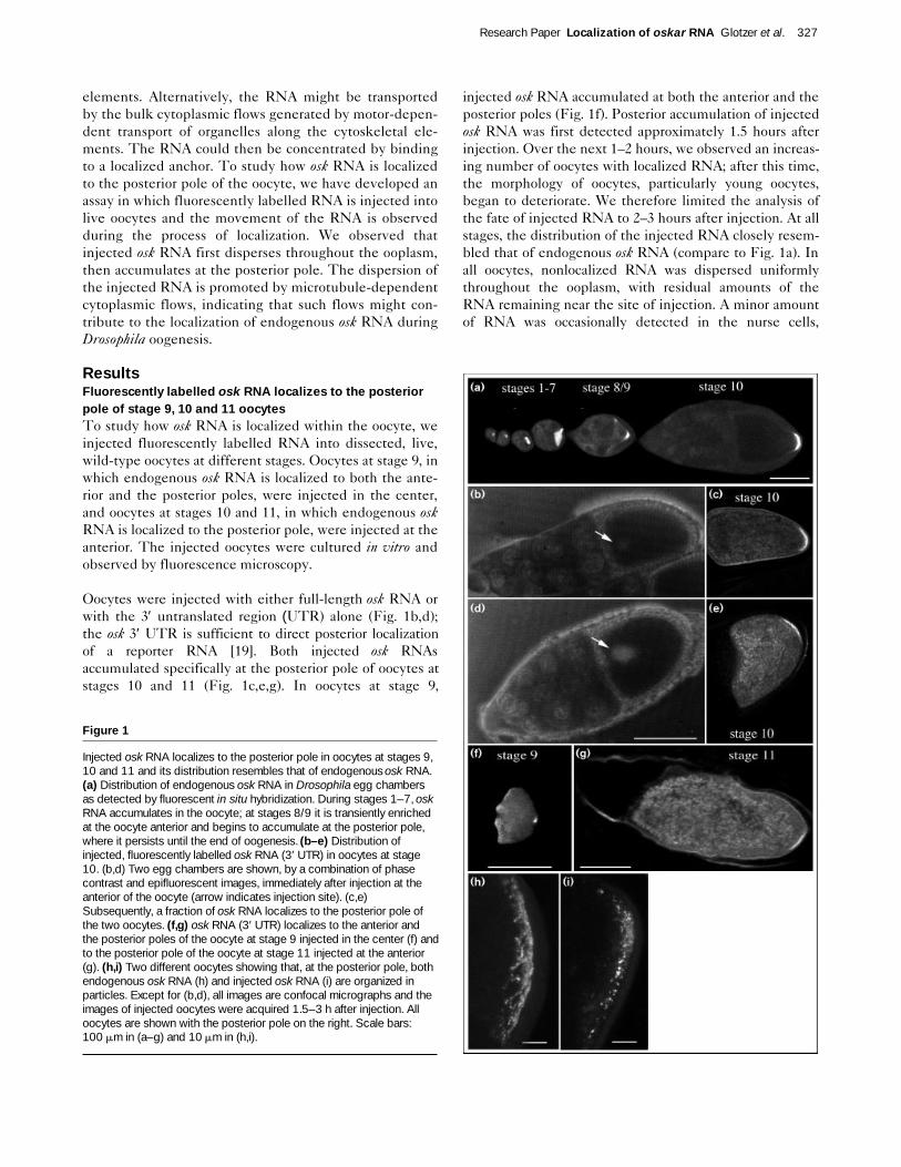
Injected osk RNA localizes to the posterior pole in oocytes at stages 9,10 and 11 and its distribution resembles that of endogenous osk RNA.(a) Distribution of endogenous osk RNA in Drosophila egg chambersas detected by fluorescent in situ hybridization. During stages 1–7, oskRNA accumulates in the oocyte; at stages 8/9 it is transiently enrichedat the oocyte anterior and begins to accumulate at the posterior pole,where it persists until the end of oogenesis. (b–e) Distribution ofinjected, fluorescently labelled osk RNA (3′ UTR) in oocytes at stage10. (b,d) Two egg chambers are shown, by a combination of phasecontrast and epifluorescent images, immediately after injection at theanterior of the oocyte (arrow indicates injection site). (c,e)Subsequently, a fraction of osk RNA localizes to the posterior pole ofthe two oocytes. (f,g) osk RNA (3′ UTR) localizes to the anterior andthe posterior poles of the oocyte at stage 9 injected in the center (f) andto the posterior pole of the oocyte at stage 11 injected at the anterior(g). (h,i) Two different oocytes showing that, at the posterior pole, bothendogenous osk RNA (h) and injected osk RNA (i) are organized inparticles. Except for (b,d), all images are confocal micrographs and theimages of injected oocytes were acquired 1.5–3 h after injection. Alloocytes are shown with the posterior pole on the right. Scale bars:100 mm in (a–g) and 10 mm in (h,i).
indicating that translocation of the RNA from the oocyteinto the nurse cells was minimal.
Further analysis of the distribution of injected osk RNA athigh magnification revealed that the RNA that had accu-mulated at the posterior pole was organized in particles ofless than 1 mm diameter (Fig. 1i). This particulate distribu-tion was similar to that of endogenous osk RNA (Fig. 1h).Such particles were seldom observed elsewhere in theooplasm, with the exception of the injection site, suggest-ing that particle assembly occurs where RNA concentrationis high. To determine whether individual particles con-sisted of several RNA molecules, we coinjected rhodamine-and fluorescein-labelled osk RNAs. Most fluorescent parti-cles contained both fluorochromes, and intensity measure-ments indicated that an average particle contained morethan 100 osk RNA molecules (data not shown).
The similarity in distribution of injected osk RNA to thatof endogenous osk RNA, at the anterior and the posteriorpoles of stage 9 oocytes, at the posterior pole of stage 10and 11 oocytes, and in particles, suggests that our injectionassay reproduces the localization of endogenous osk RNAin the Drosophila oocyte.
Injected osk RNA accumulates in the pole plasmEarly observations defined pole plasm as the yolk-freeregion at the posterior pole of the Drosophila earlyembryo [20]. To determine whether injected osk RNAlocalized to the pole plasm, we compared the distribution
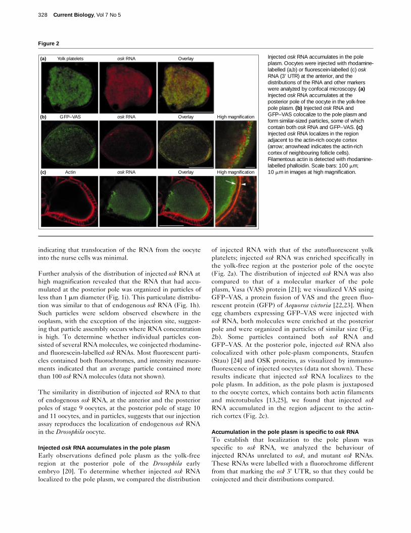
of injected RNA with that of the autofluorescent yolkplatelets; injected osk RNA was enriched specifically inthe yolk-free region at the posterior pole of the oocyte(Fig. 2a). The distribution of injected osk RNA was alsocompared to that of a molecular marker of the poleplasm, Vasa (VAS) protein [21]; we visualized VAS usingGFP–VAS, a protein fusion of VAS and the green fluo-rescent protein (GFP) of Aequorea victoria [22,23]. Whenegg chambers expressing GFP–VAS were injected withosk RNA, both molecules were enriched at the posteriorpole and were organized in particles of similar size (Fig.2b). Some particles contained both osk RNA andGFP–VAS. At the posterior pole, injected osk RNA alsocolocalized with other pole-plasm components, Staufen(Stau) [24] and OSK proteins, as visualized by immuno-fluorescence of injected oocytes (data not shown). Theseresults indicate that injected osk RNA localizes to thepole plasm. In addition, as the pole plasm is juxtaposedto the oocyte cortex, which contains both actin filamentsand microtubules [13,25], we found that injected oskRNA accumulated in the region adjacent to the actin-rich cortex (Fig. 2c).
Accumulation in the pole plasm is specific to osk RNATo establish that localization to the pole plasm wasspecific to osk RNA, we analyzed the behaviour ofinjected RNAs unrelated to osk, and mutant osk RNAs.These RNAs were labelled with a fluorochrome differentfrom that marking the osk 3′ UTR, so that they could becoinjected and their distributions compared.
328 Current Biology, Vol 7 No 5
Figure 2
Injected osk RNA accumulates in the poleplasm. Oocytes were injected with rhodamine-labelled (a,b) or fluorescein-labelled (c) oskRNA (3′ UTR) at the anterior, and thedistributions of the RNA and other markerswere analyzed by confocal microscopy. (a)Injected osk RNA accumulates at theposterior pole of the oocyte in the yolk-freepole plasm. (b) Injected osk RNA andGFP–VAS colocalize to the pole plasm andform similar-sized particles, some of whichcontain both osk RNA and GFP–VAS. (c)Injected osk RNA localizes in the regionadjacent to the actin-rich oocyte cortex(arrow; arrowhead indicates the actin-richcortex of neighbouring follicle cells).Filamentous actin is detected with rhodamine-labelled phalloidin. Scale bars: 100 mm;10 mm in images at high magnification.
Yolk platelets(a) osk RNA Overlay
GFP–VAS(b) osk RNA Overlay High magnification
Actin(c) osk RNA Overlay High magnification
Research Paper Localization of oskar RNA Glotzer et al. 329
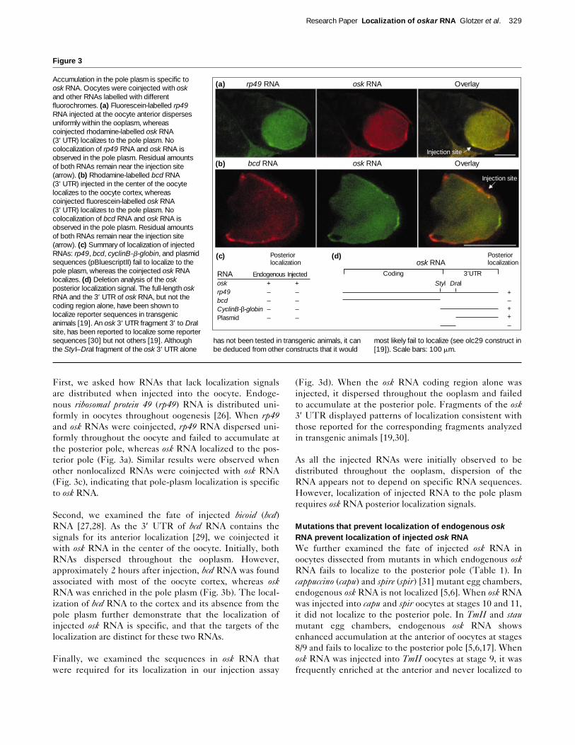
First, we asked how RNAs that lack localization signalsare distributed when injected into the oocyte. Endoge-nous ribosomal protein 49 (rp49) RNA is distributed uni-formly in oocytes throughout oogenesis [26]. When rp49and osk RNAs were coinjected, rp49 RNA dispersed uni-formly throughout the oocyte and failed to accumulate atthe posterior pole, whereas osk RNA localized to the pos-terior pole (Fig. 3a). Similar results were observed whenother nonlocalized RNAs were coinjected with osk RNA(Fig. 3c), indicating that pole-plasm localization is specificto osk RNA.
Second, we examined the fate of injected bicoid (bcd)RNA [27,28]. As the 3′ UTR of bcd RNA contains thesignals for its anterior localization [29], we coinjected itwith osk RNA in the center of the oocyte. Initially, bothRNAs dispersed throughout the ooplasm. However,approximately 2 hours after injection, bcd RNA was foundassociated with most of the oocyte cortex, whereas oskRNA was enriched in the pole plasm (Fig. 3b). The local-ization of bcd RNA to the cortex and its absence from thepole plasm further demonstrate that the localization ofinjected osk RNA is specific, and that the targets of thelocalization are distinct for these two RNAs.
Finally, we examined the sequences in osk RNA thatwere required for its localization in our injection assay
(Fig. 3d). When the osk RNA coding region alone wasinjected, it dispersed throughout the ooplasm and failedto accumulate at the posterior pole. Fragments of the osk3′ UTR displayed patterns of localization consistent withthose reported for the corresponding fragments analyzedin transgenic animals [19,30].
As all the injected RNAs were initially observed to bedistributed throughout the ooplasm, dispersion of theRNA appears not to depend on specific RNA sequences.However, localization of injected RNA to the pole plasmrequires osk RNA posterior localization signals.
Mutations that prevent localization of endogenous oskRNA prevent localization of injected osk RNAWe further examined the fate of injected osk RNA inoocytes dissected from mutants in which endogenous oskRNA fails to localize to the posterior pole (Table 1). Incappuccino (capu) and spire (spir) [31] mutant egg chambers,endogenous osk RNA is not localized [5,6]. When osk RNAwas injected into capu and spir oocytes at stages 10 and 11,it did not localize to the posterior pole. In TmII and staumutant egg chambers, endogenous osk RNA showsenhanced accumulation at the anterior of oocytes at stages8/9 and fails to localize to the posterior pole [5,6,17]. Whenosk RNA was injected into TmII oocytes at stage 9, it wasfrequently enriched at the anterior and never localized to
Figure 3
Accumulation in the pole plasm is specific toosk RNA. Oocytes were coinjected with oskand other RNAs labelled with differentfluorochromes. (a) Fluorescein-labelled rp49RNA injected at the oocyte anterior dispersesuniformly within the ooplasm, whereascoinjected rhodamine-labelled osk RNA (3′ UTR) localizes to the pole plasm. Nocolocalization of rp49 RNA and osk RNA isobserved in the pole plasm. Residual amountsof both RNAs remain near the injection site(arrow). (b) Rhodamine-labelled bcd RNA (3′ UTR) injected in the center of the oocytelocalizes to the oocyte cortex, whereascoinjected fluorescein-labelled osk RNA (3′ UTR) localizes to the pole plasm. Nocolocalization of bcd RNA and osk RNA isobserved in the pole plasm. Residual amountsof both RNAs remain near the injection site(arrow). (c) Summary of localization of injectedRNAs: rp49, bcd, cyclinB-b-globin, and plasmidsequences (pBluescriptII) fail to localize to thepole plasm, whereas the coinjected osk RNAlocalizes. (d) Deletion analysis of the oskposterior localization signal. The full-length oskRNA and the 3′ UTR of osk RNA, but not thecoding region alone, have been shown tolocalize reporter sequences in transgenicanimals [19]. An osk 3′ UTR fragment 3′ to DraIsite, has been reported to localize some reportersequences [30] but not others [19]. Althoughthe Sty I–DraI fragment of the osk 3′ UTR alone
has not been tested in transgenic animals, it canbe deduced from other constructs that it would
most likely fail to localize (see olc29 construct in[19]). Scale bars: 100 mm.
(c)
(b)
(a) rp49 RNA osk RNA Overlay
bcd RNA osk RNA Overlay
(d)Posterior localization
Posterior localization
RNA Endogenous Injectedosk + +rp49 – –bcd – –CyclinB-β-globin – –Plasmid – –
Coding 3'UTR
Styl Dral
osk RNA
+–++–
Injection site
Injection site
the posterior pole. In neither TmII nor stau oocytes atstages 10 and 11 did the injected osk RNA localize to theposterior pole. These results indicate that spir, capu, TmIIand stau functions are required for the correct posteriorlocalization of both endogenous and injected osk RNAs.
In osk mutant egg chambers, including osk54, in whichOSK protein is not produced [9,10], endogenous osk RNAis localized to the posterior pole of oocytes at stages 8/9[5,6]. During the later stages of oogenesis, endogenousosk RNA becomes delocalized and dispersed in theooplasm, indicating that OSK protein plays a role in themaintenance of osk RNA localized during stages 8/9. Weinjected osk RNA into osk54 mutant oocytes at stages 9, 10and 11, and found that the RNA did not localize, indicat-ing that OSK protein is required for the localization ofinjected osk RNA.
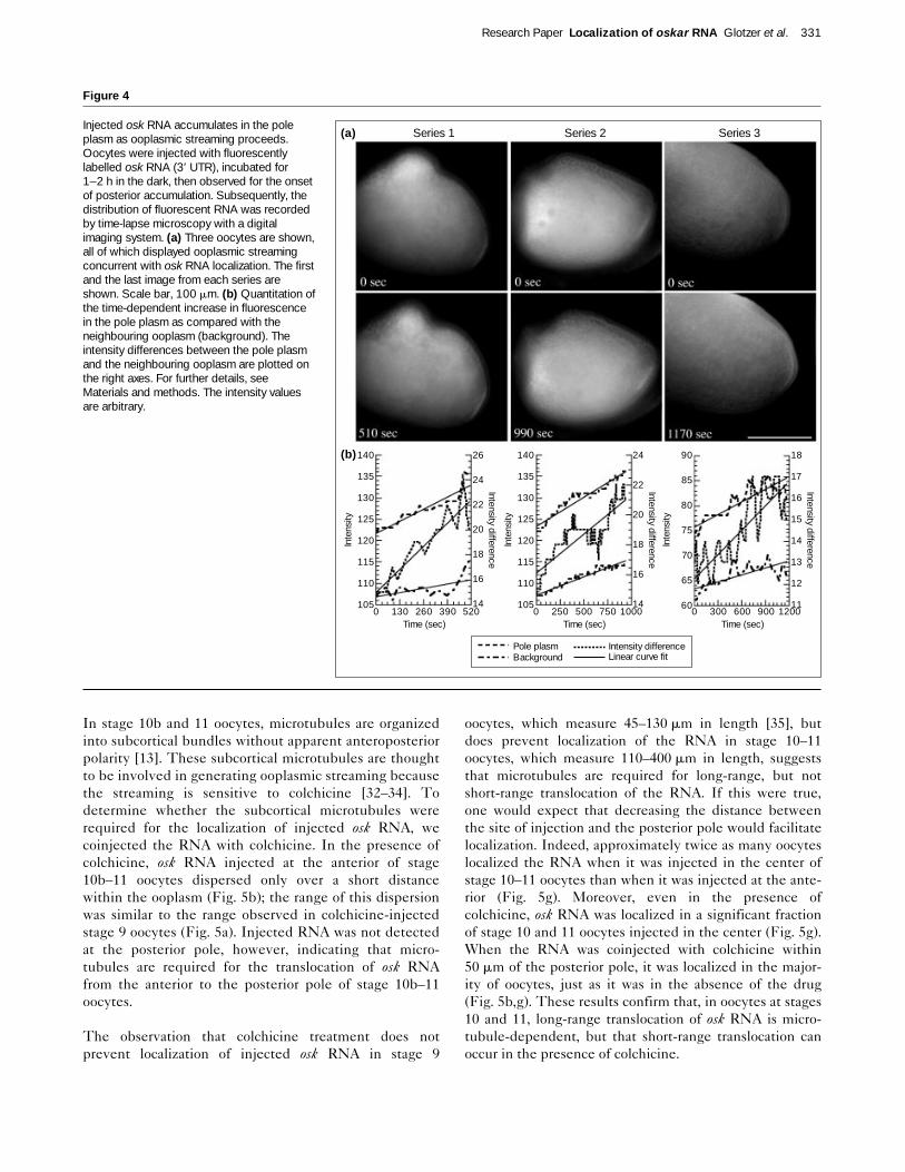
Injected osk RNA accumulates at the posterior pole asooplasmic streaming proceedsTo observe directly how fluorescently labelled osk RNAlocalizes to the posterior pole, movement of the RNA wasrecorded by digital microscopy. We focused on document-ing the localization process in oocytes at stage 10b, asyounger oocytes are very fragile and difficult to handle.The ooplasm of stage 10b–12 oocytes moves vigorously;this cytoplasmic flow or ooplasmic streaming is easily visu-alized as the autofluorescent yolk platelets move in con-centric circular waves at rates averaging 25 mm min–1
[32–34]. When stage 10b oocytes were injected with oskRNA, we observed that, shortly after injection, a wave offluorescent RNA formed and spread circularly within theooplasm (data not shown). The RNA gradually filled theentire ooplasm and began to accumulate at the posteriorpole (Fig. 4a and see Supplementary material for a videoclip). When paired images of the movements of theinjected RNA and of the autofluorescent yolk plateletswere compared, it could be seen that the wave of RNAand the yolk platelets followed the same trajectory andmoved at similar rates — indicative of ooplasmic stream-ing. The similarity of the path and the rate of movementof the fluorescent RNA and the yolk platelets suggeststhat the same mechanism mediates both movements.
We quantitated the posterior accumulation of the injectedosk RNA in oocytes that exhibited ooplasmic streaming.Oocytes were selected that showed trace amounts offluorescent osk RNA localized to the posterior, and time-lapse microscopy was used to record the subsequent local-ization process (Fig. 4a). Quantitation of the accumulationof fluorescent RNA at the posterior pole showed that theamount of RNA increased over time both in the poleplasm and in the neighbouring ooplasm (Fig. 4b). Fromthese data, we plotted the difference between the averageintensity of fluorescence in the pole plasm and that of theneighbouring ooplasm. This revealed that, during the first
500 seconds of the analysis, the RNA accumulatedapproximately 50%, 30% and 15% faster in the pole plasmthan in the neighbouring ooplasm of the three oocytesexamined, respectively. Taken together, these resultsshow that injected osk RNA accumulates at the posteriorpole while ooplasmic streaming proceeds.
Depolymerization of microtubules inhibits long-rangemovement of injected osk RNA but does not preventposterior localizationTo determine whether the polarized microtubulenetwork, characteristic of stage 7–10a oocytes, is requiredfor the localization of injected osk RNA, we injected theRNA in the center of stage 9 oocytes in the presence ofcolchicine, a microtubule-depolymerizing drug. Approxi-mately 2–3 hours later, the majority of injected osk RNAremained at the injection site, forming a local gradient(Fig. 5a). However, a significant proportion of the RNAhad localized to both the anterior and the posterior poles(Fig. 5a; compare with Fig. 1f), and the percentage ofoocytes in which injected RNA was localized was similarin the presence and in the absence of the drug (Fig. 5g).This suggests that, in stage 9 oocytes, the translocation ofinjected osk RNA from the center of the oocyte to theanterior and posterior poles, and the accumulation of theRNA at these two locations, can occur in the absence ofmicrotubules. In the presence of the microtubules,however, the dispersion of osk RNA within the ooplasm ofstage 9 oocytes is more efficient.
330 Current Biology, Vol 7 No 5
Table 1
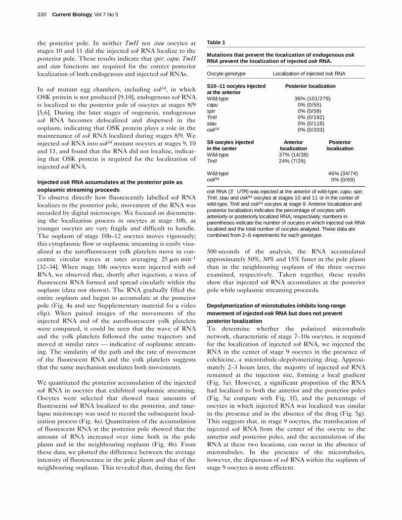
Mutations that prevent the localization of endogenous oskRNA prevent the localization of injected osk RNA.
Oocyte genotype Localization of injected osk RNA
S10–11 oocytes injected Posterior localizationat the anteriorWild-type 36% (101/279)capu 0% (0/55)spir 0% (0/58)TmII 0% (0/192)stau 0% (0/118)osk54 0% (0/203)
S9 oocytes injected Anterior Posteriorin the center localization localizationWild-type 37% (14/38)TmII 24% (7/29)
Wild-type 46% (34/74)osk54 0% (0/69)
osk RNA (3′� UTR) was injected at the anterior of wild-type, capu, spir,TmII, stau and osk54 oocytes at stages 10 and 11 or in the center ofwild-type, TmII and osk54 oocytes at stage 9. Anterior localization andposterior localization indicates the percentage of oocytes withanteriorly or posteriorly localized RNA, respectively; numbers inparentheses indicate the number of oocytes in which injected osk RNAlocalized and the total number of oocytes analyzed. These data arecombined from 2–6 experiments for each genotype.
In stage 10b and 11 oocytes, microtubules are organizedinto subcortical bundles without apparent anteroposteriorpolarity [13]. These subcortical microtubules are thoughtto be involved in generating ooplasmic streaming becausethe streaming is sensitive to colchicine [32–34]. Todetermine whether the subcortical microtubules wererequired for the localization of injected osk RNA, wecoinjected the RNA with colchicine. In the presence ofcolchicine, osk RNA injected at the anterior of stage10b–11 oocytes dispersed only over a short distancewithin the ooplasm (Fig. 5b); the range of this dispersionwas similar to the range observed in colchicine-injectedstage 9 oocytes (Fig. 5a). Injected RNA was not detectedat the posterior pole, however, indicating that micro-tubules are required for the translocation of osk RNAfrom the anterior to the posterior pole of stage 10b–11oocytes.
The observation that colchicine treatment does notprevent localization of injected osk RNA in stage 9
oocytes, which measure 45–130 mm in length [35], butdoes prevent localization of the RNA in stage 10–11oocytes, which measure 110–400 mm in length, suggeststhat microtubules are required for long-range, but notshort-range translocation of the RNA. If this were true,one would expect that decreasing the distance betweenthe site of injection and the posterior pole would facilitatelocalization. Indeed, approximately twice as many oocyteslocalized the RNA when it was injected in the center ofstage 10–11 oocytes than when it was injected at the ante-rior (Fig. 5g). Moreover, even in the presence ofcolchicine, osk RNA was localized in a significant fractionof stage 10 and 11 oocytes injected in the center (Fig. 5g).When the RNA was coinjected with colchicine within50 mm of the posterior pole, it was localized in the major-ity of oocytes, just as it was in the absence of the drug(Fig. 5b,g). These results confirm that, in oocytes at stages10 and 11, long-range translocation of osk RNA is micro-tubule-dependent, but that short-range translocation canoccur in the presence of colchicine.
Research Paper Localization of oskar RNA Glotzer et al. 331
Figure 4
Injected osk RNA accumulates in the poleplasm as ooplasmic streaming proceeds.Oocytes were injected with fluorescentlylabelled osk RNA (3′ UTR), incubated for1–2 h in the dark, then observed for the onsetof posterior accumulation. Subsequently, thedistribution of fluorescent RNA was recordedby time-lapse microscopy with a digitalimaging system. (a) Three oocytes are shown,all of which displayed ooplasmic streamingconcurrent with osk RNA localization. The firstand the last image from each series areshown. Scale bar, 100 mm. (b) Quantitation ofthe time-dependent increase in fluorescencein the pole plasm as compared with theneighbouring ooplasm (background). Theintensity differences between the pole plasmand the neighbouring ooplasm are plotted onthe right axes. For further details, seeMaterials and methods. The intensity valuesare arbitrary.
(a)
(b)
Series 1 Series 2 Series 3
140�
135�
130�
125�
120�
115�
110�
105
26�
24�
22�
20�
18�
16�
140 130 260 390 520
Inte
nsity
Intensity difference
Time (sec)
140�
135�
130�
125�
120�
115�
110�
105
24�
22�
20�
18�
16�
140 250 500 750 1000
Inte
nsity
Intensity difference
Time (sec)
90�
85�
80�
75�
70�
65�
60
18�
17�
16�
15�
14�
13�
12�
110 300 600 900 1200
Inte
nsity
Intensity difference
Time (sec)
Pole plasmBackground
Intensity differenceLinear curve fit

We used two criteria to determine whether microtubuleswere depolymerized in oocytes in which injected osk RNAlocalized in the presence of colchicine. First, eightcolchicine-treated oocytes, in which posteriorly injectedosk RNA had localized, were examined for the occurrenceof ooplasmic streaming; in all cases, the colchicine treat-ment had effectively arrested the streaming, suggestingthat microtubule-dependent ooplasmic streaming is notrequired to promote short-range osk RNA movement.Second, to rule out the possibility that injected osk RNAhad localized before the coinjected colchicine causedmicrotubule depolymerization, we injected the RNA intooocytes dissected from flies that had been fed colchicinefor 6 hours; this drug treatment causes depolymerizationof microtubules in oocytes but does not affect localizationof endogenous osk RNA [15,16]. We confirmed byimmunostaining that, in such oocytes, microtubules weredepolymerized at the time of injection (Fig. 5f). When oskRNA was injected in the center or close to the posteriorpole of oocytes dissected from colchicine-fed flies, theRNA localized to the posterior pole (Fig. 5d,g). Thus, inoocytes at stages 10 and 11, as in oocytes at stage 9,accumulation of the injected osk RNA at the posterior poledoes not require microtubules.
We also tested whether actin microfilaments are requiredfor the localization of osk RNA by injecting the RNA inthe presence of drugs that prevent polymerization of thesecytoskeletal components. We found that, whether in thepresence or absence of cytochalasin D, injected osk RNAlocalized to the posterior pole in a similar percentage ofoocytes (Fig. 5g), suggesting that cytochalasin-sensitiveactin filaments are not required for localization.
Hence, accumulation of injected osk RNA at the posteriorpole of stage 9, 10 and 11 oocytes is not sensitive to drugsthat depolymerize microtubules or actin filaments.However, translocation of the RNA throughout theooplasm is less efficient in the presence of colchicine,indicating that intact microtubules are required for thelong-range transport of the RNA.
DiscussionTransport of osk RNA by cytoplasmic flowsSeveral mechanisms could underlie the movement ofRNAs within cells, including simple diffusion, directedtransport, and transport by cytoplasmic flows [36].Although the role of directed transport by molecularmotors has been discussed most frequently [1–3], the only
332 Current Biology, Vol 7 No 5
Figure 5
Effects of depolymerization of microtubules andactin filaments on the distribution of injectedosk RNA. Oocytes were coinjected withfluorescently labelled osk RNA (3′ UTR) andcolchicine or cytochalasin D, to depolymerizemicrotubules or actin filaments, respectively.(a–c) In the presence of colchicine, injectedRNA disperses a short distance around theinjection site. (a,c) When the distance betweenthe injection site and the posterior pole is lessthan 50 mm, the RNA accumulates at theposterior pole. (b) When the distance betweenthe injection site and the posterior pole is morethan 50–100 mm, the RNA does notaccumulate at the posterior pole. (d–f)Oocytes were dissected from flies fedcolchicine for 6 h. (d) Posteriorly injected oskRNA disperses a short distance around theinjection site and localizes to the posterior pole.Colchicine was coinjected with the RNA toprevent repolymerization of the microtubulenetwork. Prior to the injections, microtubulesare depolymerized in egg chambers dissectedfrom flies fed colchicine (f) but not in controlegg chambers (e). The images of injectedoocytes were acquired 1.5–3 h after injection.(g) Summary of the effects of colchicine,cytochalasin D, and the site of injection onlocalization of injected osk RNA. ‘Colchicine*’indicates oocytes dissected from flies fedcolchicine for 6 h and subsequently injectedwith osk RNA. ‘Posterior localization’ is definedas in Table 1. These data are combined from2–6 experiments per type of injection. S, stage;scale bar, 100 mm.
(a)
(e)
(g)
(f)
S9-center (b) S10-anterior (c) S11-posterior (d) S10-posterior
Stage Site of injection Drug Posterior localization (%)10/11 Anterior – 34% (127/379)
Colchicine 0% (0/28)Cytochalasin D 39% (17/44)
10/11 Center – 60% (78/131)Colchicine 18% (21/115)Colchicine* 26% (5/19)
10/11 Posterior – 72% (68/95)Colchicine 75% (24/32)Colchicine* 61% (11/18)
9 Center – 51% (25/49)Colchicine 48% (10/21)
direct evidence for the involvement of this mechanismcomes from the finding that myelin basic protein RNAinjected into oligodendrocytes moves in the cell processesin anterograde fashion at 12 mm min–1 [37]. This move-ment is kinesin-dependent, suggesting that the RNAmoves along microtubules [38]. As similar direct observa-tions of RNA localization have not yet been reported inother cell types, it is premature to conclude that directedtransport is a general mechanism of RNA localization.
Directed transport by microtubule-dependent motors hasalso been proposed to account for the localization ofseveral RNAs to discrete sites within the Drosophilaoocyte (reviewed in [2,3,11,39,40]). This working modelis based on the observation that bcd and osk RNAs arelocalized to the anterior and the posterior poles of theoocyte, respectively, during stages 7–9 — when theoocyte contains an anteroposteriorly polarized gradient ofmicrotubules [13]. The findings that a kinesin-b-galactosidase (kin-b-gal) fusion protein localizes duringstages 8 and 9 to the posterior pole of the oocyte [15], thatinhibitors of microtubule polymerization affect the local-ization of both kin-b-gal and bcd and osk RNAs[14–16,41], and that injected bcd RNA associates withastral microtubules in blastoderm embryos [42], havefurther supported this model. However, direct evidencefor motor-based transport of RNA in Drosophila oocyteshas been lacking.
Here, we have begun to investigate how osk RNA localizeswithin the Drosophila oocyte by observing the fate ofinjected RNA. We find that injected osk RNA localizes inoocytes at stages 9, 10 and 11. The simple observation thatosk RNA can localize in oocytes at each of these stages isrevealing. In particular, these results provide the first evi-dence that osk RNA can localize during stages 10b and 11.Previous analysis of the steady-state distribution ofendogenous osk RNA has indicated that a significantportion of the RNA is localized by stage 10a, but technicallimitations precluded analysis of later stages [5,6]. Usingtime-lapse microscopy, we have shown that injected oskRNA localizes in oocytes at stages 10b and 11. In theseoocytes, injected osk RNA disperses in concentric circularwaves that parallel those of ooplasmic streaming of theyolk, and this dispersion enables the RNA to reach theposterior pole where it accumulates. Treatment withcolchicine to depolymerize microtubules reduces theextent of dispersion of injected RNA. As ooplasmicstreaming ceases in the presence of colchicine, it is likelythat the inhibition of RNA dispersion by microtubuledrugs is a consequence of the drug-induced inhibition ofooplasmic streaming. Hence, these experiments revealthat transport by cytoplasmic flows could contribute to thelocalization of endogenous osk RNA during oogenesis.This mechanism could be used to promote the localizationof any endogenous osk RNA that enters the oocyte when
the nurse cells contract and ‘dump’ their cytoplasm intothe oocyte during stages 10b–12 [25,43].
The localization of injected osk RNA during stages 10band 11 is revealing for a second reason. The onset ofooplasmic streaming correlates with a rearrangement ofthe microtubule cytoskeleton within the oocyte. Theanteroposterior gradient of microtubules characteristic ofstages 7–10a disassembles during stage 10a, and a corticalarray of microtubules that lacks obvious polarity assemblesand persists during stages 10b-12. Thus, the localization ofinjected osk RNA in stage 10b and 11 oocytes indicatesthat injected osk RNA can localize in the absence of theanteroposteriorly polarized microtubules.
Although a portion of endogenous osk RNA may localizeafter the anteroposteriorly polarized microtubules havedisassembled, a significant fraction of the RNA localizesduring stages 7–10a. Earlier experiments suggested that thepolarized microtubule network present during these stagesparticipates in osk RNA localization [15,16]. In those experi-ments, flies were fed colchicine to depolymerize micro-tubules, and the distribution of microtubules, kin-b-gal andosk RNA was examined after drug treatments of differentlengths. Colchicine treatment for 3.5 hours did not affectendogenous osk RNA localization, although it was sufficientto depolymerize microtubules and to abolish posterior accu-mulation of kin-b-gal. Prolonged exposure to colchicine,lasting 10–16 hours, was found to abolish osk RNA localiza-tion in stage 8 and 9 oocytes, but not in oocytes of stage 10or later. One interpretation of these results is that the drugdid not interfere with the maintenance of previously local-ized RNA, but did interfere with microtubule-dependentdirected transport of the RNA. However, we have shownhere that colchicine treatment does not prevent localizationof injected osk RNA in stage 9 oocytes. It is therefore possi-ble that endogenous osk RNA localizes in stage 9 oocytes bya mechanism distinct from microtubule-dependent direc-tional transport. Prolonged treatment with colchicine mayprevent posterior accumulation of osk RNA by interferingwith an early step in the localization process: establishmentof an anchor for osk RNA.
Our observation that the dispersion of injected osk RNA isseverely affected in stage 9 oocytes whose microtubuleshave been disrupted indicates that a microtubule-depen-dent process is essential for efficient dispersion of theRNA. Our data do not allow us to determine the mecha-nism of dispersion. One possibility is that efficient disper-sion relies on directional transport of the RNA. Analternative possibility is that osk RNA dispersion is medi-ated by microtubule-dependent cytoplasmic flows, as wehave shown to be the case during later stages of oogenesis.Time-resolved recordings of particle movements inooplasm indicate that microtubule-dependent cytoplasmicflows are present from stage 6 through 12 [32,34,44].
Research Paper Localization of oskar RNA Glotzer et al. 333
Further studies will be directed towards improving theconditions for direct observation of osk RNA localization atstage 9 to evaluate whether cytoplasmic flows or othermechanisms, including directional transport, mediate oskRNA localization.
Accumulation of osk RNA by binding to a localized anchorGenetic evidence indicates that there are two distinctaspects to osk RNA accumulation at the posterior pole:initial localization of the RNA, and maintenance of thelocalization. In osk mutants in which OSK protein is notproduced [9,10], osk RNA is localized transiently to theposterior pole, indicating that the initial localization of theRNA is independent of OSK [5,6]. The transient localiza-tion disappears at stage 10, concurrent with the onset ofooplasmic streaming, indicating that the maintenance ofosk RNA at the posterior pole requires OSK. As the local-ization of injected osk RNA requires OSK, our assayreveals the later stages of localization of osk RNA and itsmaintenance. Moreover, our results suggest that OSKplays a role not only in the maintenance of the localizedRNA but also in the continuous localization of osk RNAafter stages 8/9. OSK-dependent localization of osk RNAcorrelates in time with the formation of polar granules,RNA–protein complexes that are associated withinduction of the germ line [45,46]. Polar granules initiallyappear at the posterior pole of the oocyte during stage 9,
and they persist past the end of oogenesis [20]. At the pos-terior pole of stage 10 and 11 oocytes, we observe thatinjected osk RNA colocalizes with GFP–VAS in particulatestructures. As VAS and OSK have been immunolocalizedto polar granules [21,22], it seems likely that osk RNA isalso a component of polar granules. Assembly of osk RNAinto polar granules may ensure the maintenance of theRNA at the posterior pole.
RNA localization cannot be achieved solely by cytoplasmicflows. In order for localization to occur, the RNA must beselectively anchored. Our observation that injected oskRNA localizes concurrently with ooplasmic streamingdemonstrates that the posteriorly localized anchor bindsand retains osk RNA in the presence of the entropic forcesthat participate in RNA transport. Thus, our findings revealthat the anchor plays a crucial role not only in the mainte-nance of localized osk RNA, but also in the localization perse. If osk RNA were localized by directional transport, itsaccumulation at the posterior pole could be explained as theresult of continuous transport, analogous to the accumula-tion of kin-b-gal observed at the posterior pole of stage 8/9oocytes. However, as cytoplasmic flows occur in the oocytecytoplasm during most of oogenesis, it seems likely thatlocalization of osk RNA requires an anchor regardless of thetransport mechanism. Although the function of the oskRNA anchor is microtubule-independent, microtubules
334 Current Biology, Vol 7 No 5
Figure 6
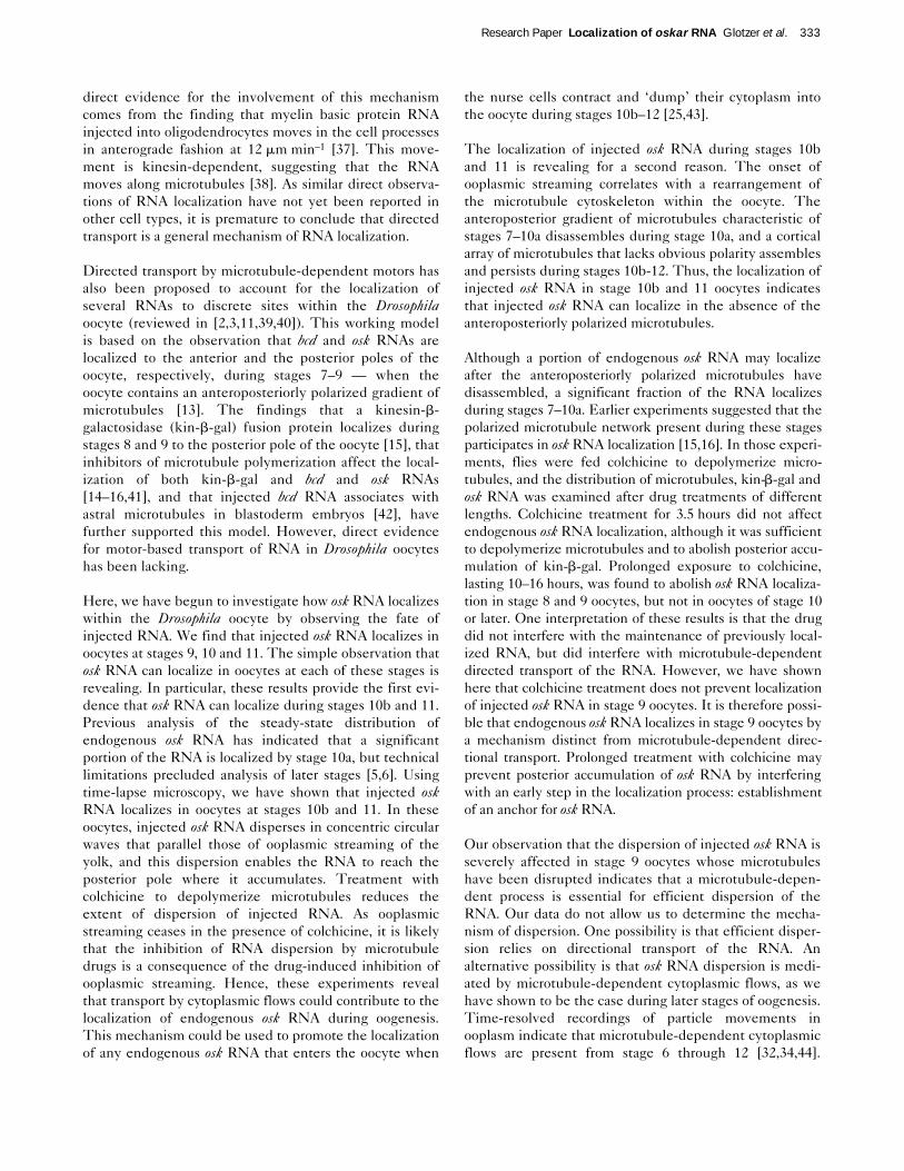
A model of osk RNA localization duringoogenesis in Drosophila. osk RNA issynthesized in the nurse cells and is thenlocalized to the oocyte. (a) In early oogenesis,the RNA is transported into the oocyte, whereit accumulates; this transport process remainsuncharacterized and may involve directionaltransport along microtubules that extend fromthe oocyte into the nurse cells. During thesestages, an osk RNA anchor is established atthe posterior pole by a mechanism that mayinvolve microtubules and actin filaments. (b)During mid-oogenesis, osk RNA becomesredistributed within the oocyte: the RNAaccumulates transiently at the anterior andbegins to concentrate at the posterior pole,where it binds to the localized anchor. Bothmicrotubule-dependent cytoplasmic flows andmicrotubule-dependent directional transportcould participate in the redistribution of theRNA. The transient anterior accumulation mayreflect binding sites that compete with theposterior anchor for the binding of osk RNA.At the posterior pole, localized osk RNAinduces the formation of polar granules. OSKprotein, translated at the posterior pole,stabilizes the anchoring of osk RNA to thepolar granules and participates in thecontinuous localization of the RNA. (c) Duringlate oogenesis, the remaining osk RNA istransferred into the oocyte from the nurse
cells with the bulk flow of the nurse-cellcytoplasm (‘nurse cell dumping’). The RNA isdispersed in the ooplasm by ooplasmic
streaming. Upon reaching the posterior pole,osk RNA continues to accumulate by bindingto the localized posterior anchor.
osk RNA
Direction of movement
Posterior anchor
OSK protein
Microtubule
Actin
Cytoplasmic flow
Early oogenesis (stages 1–7)
osk RNA is localized to the oocyte
Mid-oogenesis (stages 8–10a)
osk RNA is localized to the anterior pole (transiently)
and to the posterior pole of the oocyte
Late oogenesis (stages 10b–12)
osk RNA is localized to the posterior pole
of the oocyte
(a) (b) (c)
© 1997 Current Biology
may play a role in its establishment. If establishment ofthe anchor occurs before stage 8, this could explain whyextended treatment with colchicine is required to abolishthe localization of endogenous osk RNA in stage 8/9oocytes.
An actin-binding protein, TmII, whose RNA is posteriorlylocalized in the oocyte [18], has been shown recently to berequired for posterior localization of osk RNA. These datasuggest that actin filaments may also participate in oskRNA anchoring. The observation that injected osk RNAlocalizes in the vicinity of the actin-rich oocyte cortex sup-ports this possibility. However, depolymerization of actinfilaments with cytochalasins does not inhibit localizationof injected and endogenous [16] osk RNAs. Therefore, ifan actin-based structure participates in osk RNA anchor-ing, it must be relatively stable. As tropomyosins areknown to stabilize actin filaments [47], it is conceivablethat TmII promotes formation of a cytochalasin-resistantactin network to which osk RNA is anchored.
In stages 8/9, osk RNA is transiently enriched at theanterior end of the oocyte. Our injection assay detects thisanterior accumulation of osk RNA at the appropriatedevelopmental stage, stage 9. Other RNAs, includingBicaudal D, K10, orb and gurken (grk), also accumulate tran-siently at the anterior of the oocyte during stages 8–9[48–52], suggesting that all these RNAs bind to anteriorlylocalized binding sites. Several mutations, including stauand TmII, which disrupt posterior localization of osk RNA,enhance anterior accumulation of the RNA. If the poste-rior osk RNA anchor were to bind osk RNA more avidlythan the anteriorly localized binding sites, the RNA wouldbecome progressively enriched at the posterior pole overtime. If mutations prevented the assembly of the posterioranchor, this competition would not occur and osk RNAwould accumulate at the anterior pole.
ConclusionsWe have shown that bulk cytoplasmic flows couldcontribute to the localization of osk RNA (Fig. 6). This doesnot exclude the possibility that directed transport also playsa role at specific developmental stages. Transport by cyto-plasmic flows and concentration by a specific, localizedanchor is a simple mechanism that could generate discretedistribution patterns of RNA within a cell. During latestages of oogenesis, after the onset of ooplasmic streaming,nanos, germ cell-less and cyclin B RNAs localize to the poste-rior pole of the Drosophila oocyte [53–55]. This cytoplasmicflow is likely to translocate RNAs towards the pole plasm,where they are anchored. It is also possible that cytoplasmicflows contribute to the anterior localization of bcd RNA andthe anterodorsal localization of grk RNA [52] in Drosophilaoocytes. Continued analysis of the mechanisms of RNAlocalization in Drosophila and other cell types will furtherour understanding of intracellular organization.
Materials and methodsFly strains and reagentsThe following allelic combinations were used for RNA injections: OregonR (wild-type), spirRP/spirRP, capuRK/capuRK [31], stauD3/stauD3 [56],Tm2gs1/Tm2gs1 [17], osk54/osk54 [5] and GFP–VAS [22]. RNAs weretranscribed from the following plasmids: pBlueosk [5] for full-length oskand the coding region alone; osk DraI–NotI fragment in pBSIIKS(nucleotide positions 3183–3309 of osk cDNA in [5]; NotI is the 3′cloning site); the Drosophila rp49 cDNA [17]; the sea urchin cyclin Bcoding region cDNA flanked by 5′ and 3′ UTRs of the Xenopus b-globin[57]; and full-length bcd cDNA [27]. osk 3′ UTR (nucleotides3156–3309), osk StyI–DraI fragment (nucleotides 3156–3183) and bcd3′ UTR in pBSIISK+ (nucleotide positions 3978–4837 of bcd cDNA in[27]) were provided by L.C. Olsen (L.C. Olsen and A. Ephrussi, unpub-lished).
In vitro transcription and fluorescent labeling of the RNARNA for microinjection and for in situ hybridization was transcribed invitro. Typically, 5–10 mg of DNA was linearized, phenol:chloroformextracted and ethanol precipitated using NH4OAc. Transcription compo-nents were assembled at room temperature and included: DNA, tran-scription buffer (10×, Boehringer Mannheim), 14 mM (final) MgCl2,2.5 mM ATP, 2.5 mM CTP, 0.25 mM GTP, 2.5 mM of a mixture of UTPand aminoallyl-UTP (Sigma; aminoallyl-UTP:UTP ratio was typically 1:10or 1:5), 1 mM 7mG(5′)ppp(5′)G cap analog (BioLabs), RNAsin (50 U;Boehringer Mannheim) and RNA polymerase T3 or T7 (40 U; BoehringerMannheim) in a final volume of 100ml. The reaction was incubated atroom temperature for 10 min, then rGTP was added to a final concentra-tion of 2.5 mM. After a further 1 h incubation at 37°C, 40 U of RNA poly-merase were added and the reaction continued for 1 h at 37°C.RNAse-free DNAse was added (40 U; Boehringer Mannheim) and thereaction was incubated for 15 min at 37°C. The RNA was extractedtwice with phenol:chloroform and once with chloroform. Unincorporatednucleotides were removed by gel filtration on a Sephadex G50 spincolumn (Boehringer Mannheim). RNA was NH4OAc:ethanol precipitated,resuspended in DEPC-treated H20 and its quality evaluated by agarosegel electrophoresis. Up to 100 mg of RNA was obtained per reaction.The RNA was labelled with either 5-(and-6)-carboxy-X-rhodamine, suc-cinimidyl ester or 6-(fluorescein-5-(and-6)-carboxamido)hexanoic acid,succinimidyl ester (SFX; Molecular Probes). The RNA-labeling protocolwas adapted from the protein-labeling protocol provided by MolecularProbes. Fluorochromes were dissolved in dimethyl-formamide (Sigma) at100 mM. The labeling reaction contained 25mg of RNA, 5 mM fluo-rochrome, and 0.15 mM bicarbonate buffer (pH 9) in a final volume of100 ml. The reaction was incubated at room temperature for 1–3 h. Unin-corporated fluorochromes were removed by gel filtration on a SephadexG50 spin column. The RNA was NH4OAc:ethanol precipitated andresuspended in DEPC-treated H20 at 1 mg ml–1. Spectrophotometricanalysis indicates that the RNA was labelled with 10–30 fluorochromesper RNA molecule.
Fluorescent in situ hybridizationWhole-mount in situ hybridization was performed as previouslydescribed [5] with some modifications. Fixed egg chambers werewashed four times with PBS and 0.1% Tween 20 (PBT) for 5 min,digested with proteinase K (Boehringer Mannheim) at 50mg ml–1 for45 min, washed twice with PBT for 5 min, post-fixed in 4% paraformalde-hyde:PBS for 20 min and washed twice with PBT for 5 min; all at roomtemperature. Egg chambers were transferred into MetOH:DMSO (9:1)for 1 h at –20°C, washed three times with PBT for 5 min at room temper-ature, rinsed in PBT:hybridization buffer (1:1) and prehybridized inhybridization buffer (HB) for 1 h at 60°C. HB contains 50% formamide,5 × SSC, 100 mg ml–1 tRNA, 50 mg ml–1 heparin, 0.1% Tween 20,pH 4.5 (with citric acid). The buffer was exchanged with 100 ml of pre-warmed HB and 1–5 ml of SFX-labelled full-length osk anti-sense RNAprobe and the hybridization was incubated overnight at 60°C. Eggchambers were washed three times with prewarmed HB for 20 min at60°C, once with prewarmed PBT:HB (1:1) for 20 min at 60oC, andfour times with PBT for 20 min at room temperature. Egg chambers
Research Paper Localization of oskar RNA Glotzer et al. 335
were mounted in 87% glycerol, 10 mM Tris pH 8.5, 4% n-propyl gallate(Sigma).
Injection assayInjection chambers were prepared as follows: a hole of 1.8 cm in diam-eter was cut in a plexiglass slide (7.5 cm × 2 cm × 2 mm) and a cover-slip was attached with wax to seal the chamber at the bottom. Thechambers were filled with a few drops of Voltalef 10S oil (Atochem). 2-day-old female flies were anaesthetized with CO2, placed under oil andthe ovaries were dissected out and split into ovarioles or individual eggchambers [34]. The injections were performed on the stage of anupright microscope using a Zeiss AIS microinjection system. osk RNAwas injected at concentrations ranging from 0.05–1.0 mg ml–1. Similarpercentages of oocytes were found to localize the RNA injected at0.25–1.0 mg ml–1 (see also Fig. 5). Approximately half as many oocyteslocalized the RNA at the lowest concentration (0.05 mg ml–1). In themajority of experiments, therefore, RNAs were injected at 0.5 mg ml–1 inDEPC-treated H20. The volume of the average S10 oocyte was esti-mated to be approximately 4–5 nl and the injection volume was esti-mated to be 10–100 pl. In experiments in which drugs were coinjected,colchicine (Sigma) was dissolved in EtOH at 20 mg ml–1 and wasdiluted to a final concentration (in the needle) 2 mg ml–1 and400 mg ml–1. We estimate that this results in approximately 100 mM and20 mM final concentration in the oocyte. Similar results were obtained atboth concentrations tested. Cytochalasin D (Sigma) was dissolved inDMSO at 10 mg ml–1 and was injected at the final concentration in theneedle at 200 mg ml–1, 250 mg ml–1 and 500 mg ml–1. We estimate thatthis gives approximately 10 mM, 12.5 mM and 25 mM concentration inthe oocyte. Similar results were obtained at all three concentrationstested. Analogous concentrations of colchicine and cytochalasin D inculture medium have been shown to depolymerize microtubules andsome actin filaments, respectively, in Drosophila oocytes ([14,16,41];our unpublished observations). In experiments in which osk RNA wascoinjected with colchicine into oocytes dissected from flies fed withcolchicine, the flies were first starved for 6 h, then fed for 6 h with freshlyprepared yeast paste containing 100 mg ml–1 colchicine [14–16,41,58],or with yeast paste without the drug. Subsequently, 7–8 flies from eachgroup were dissected and one ovary from each fly was used for injec-tions whereas the second ovary was used for microtubule staining.Microtubules were stained as follows: dissected ovaries were fixed for15 min in MetOH/50 mM EGTA pH 7 (prechilled to –20°C), washedtwice for 15 min in PBT, extracted for 2 h in PBS/1% Triton-100. Anti-a-tubulin antibody (clone DM1 a; Sigma) was used at 1:250 in PBTovernight, followed by four 15 min washes in PBT. DTAF-conjugatedgoat a-mouse secondary antibody (Jackson ImmunoResearch Labora-tories) was used at 1:100 in PBT for 2 h, followed by four 15 minwashes in PBT. Ovaries were post-fixed in 4% paraformaldehyde(Sigma) in PBS, quenched in 50 mM NH4Cl in PBS for 15 min,washed with PBS. All steps were performed at room temperature.Ovaries were mounted on glass slides in 87% glycerol, 10 mM TrispH 8.5, 4% n-propyl gallate. To compare the distribution of injectedosk RNA and filamentous actin, injected egg chambers were fixed andstained with rhodamine-labelled phalloidin (Molecular Probes) accord-ing to the protocol adapted from [59], with the following modifications.The fixative was applied under the oil in the vicinity of the samples. Fixa-tion was at room temperature for 10 min, and then the oil was removedwith heptane. This procedure immobilizes the egg chambers on thesurface of the coverslip. The egg chambers were washed with PBT,incubated with labelled phalloidin (5 ml in 1 ml of PBT) for 10–30 min indark, followed by extensive washes with PBT. The cover slips weremounted on glass slides in 87% glycerol, 10 mM Tris pH 8.5, 4% n-propyl gallate.
Microscopy and image processingLaser scanning microscopy was performed with the EMBL compactconfocal microscope mounted on a Zeiss Axiovert equipped with 25×(0.8 NA) and 100× (1.3 NA) oil immersion lenses. The images wereprocessed using Adobe Photoshop software. Time-lapse analysis ofaccumulation of injected RNA was performed with a Photometrics
cooled CCD camera mounted on a Zeiss Axiovert controlled by soft-ware developed at the EMBL [60]. Movement of autofluorescent yolkplatelets was detected in the fluorescein channel [34]. To quantitatethe accumulation of fluorescence in the pole plasm and in the neigh-bouring ooplasm, the images were acquired at equal time intervalsunder conditions where the full dynamic range of fluorescence wasregistered by the camera. 20, 40 and 100 images of the threeoocytes were analyzed in each time series, respectively. Areas ofcomparable size were chosen in the pole plasm and in the neighbour-ing ooplasm and the fluorescence was integrated in these regionsand plotted as a function of time. To calculate the difference betweenaccumulations of fluorescence in the pole plasm and in the neigh-bouring ooplasm, the average values were subtracted, and plotted asa function of time. Image analysis was performed on a Macintoshcomputer using the public domain NIH Image program (developed atthe U.S. National Institutes of Health and available on the Internet athttp://rsb.info.nih.gov/nih-image/).
Supplementary materialA brief video clip, showing the accumulation of injected osk RNA at theposterior pole as ooplasmic streaming proceeds, is published with thispaper on the internet.
AcknowledgementsWe thank Lisbeth Olsen for providing osk and bcd deletion plasmids, ElisaIzaurralde and Jim Deshler for suggestions on fluorescent RNA labeling,William Theurkauf for technical suggestions on the oocyte injections and fordiscussions, and Daniel St Johnston for suggestions on the RNA hybridiza-tion in situ. We also thank Wolfgang Breitwieser for providing theGFP–VAS strain and for comments on the manuscript, David Drechsel foradvice on image analysis, Steve Cohen, Suzanne Eaton, Pierre Gonczy, JoeHoward, Eric Karsenti, Finn-Hugo Markussen, Iain Mattaj, and Kai Simonsfor comments on the manuscript. J.B.G. is grateful to all the members ofEphrussi group for discussions and support, and to Eric Karsenti for hisspecial interest in the project. J.B.G. was supported by the EMBL and anEC grant to Eric Karsenti. This research was supported by the EMBL.
References1. Wilhelm J, Vale R: RNA on the move: the mRNA localisation
pathway. J Cell Biol 1993, 123:269–274.2. Ding D, Lipshitz HD: Localized RNAs and their functions. BioEssays
1993, 15:651–658.3. St Johnston D: The intracellular localisation of messenger RNAs.
Cell 1995, 81:161–170.4. Curtis D, Lehmann R, Zamore PD: Translational regulation in
development. Cell 1995, 81:171–178.5. Ephrussi A, Dickinson LK, Lehmann R: oskar organizes the germ
plasm and directs localization of the posterior determinant nanos.Cell 1991, 66:37–50.
6. Kim-Ha J, Smith JL, Macdonald PM: oskar mRNA is localized to theposterior pole of the Drosophila oocyte. Cell 1991, 66:23–35.
7. Ephrussi A, Lehmann R: Induction of germ cell formation by oskar.Nature 1992, 358:387–392.
8. Kim-Ha J, Kerr K, Macdonald PM: Translational regulation of oskarmRNA by Bruno, an ovarian RNA-binding protein, is essential. Cell1995, 81:403–412.
9. Markussen F-H, Michon A-M, Breitwieser W, Ephrussi A: Translationalcontrol of oskar generates Short OSK, the isoform that inducespole plasm assembly. Development 1995, 121:3723–3732.
10. Rongo C, Gavis ER, Lehmann R: Localization of oskar RNAregulates oskar translation and requires Oskar protein.Development 1995, 121:2737–2746.
11. Cooley L, Theurkauf WE: Cytoskeletal functions during Drosophilaoogenesis. Science 1994, 266:590–595.
12. Spradling AC: Developmental genetics of oogenesis. In Thedevelopment of Drosophila melanogaster. Edited by Bate M andMartinez-Arias A. Cold Spring Harbor: Cold Spring Harbor LaboratoryPress; 1993:1–70.
13. Theurkauf WE, Smiley S, Wong ML, Alberts BM: Reorganization ofthe cytoskeleton during Drosophila oogenesis: implications foraxis specification and intercellular transport. Development 1992,115:923–936.
336 Current Biology, Vol 7 No 5

14. Theurkauf WE, Alberts BM, Jan YN, Jongens TA: A central role formicrotubules in the differentiation of Drosophila oocytes.Development 1993, 118:1169–1180.
15. Clark I, Giniger E, Ruohola-Baker H, Jan LY, Jan YN: Transientposterior localization of a kinesin fusion protein reflects antero-posterior polarity of the Drosophila oocyte. Curr Biol 1994,4:289–300.
16. Pokrywka NJ, Stephenson EC: Microtubules are a generalcomponent of mRNA localization systems in Drosophila oocytes.Dev Biol 1995, 167:363–370.
17. Erdélyi M, Michon A-M, Guichet A, Glotzer JB, Ephrussi A: Arequirement for Drosophila cytoplasmic tropomyosin in oskarmRNA localization. Nature 1995, 377:524–527.
18. Tetzlaff MT, Jäckle H, Pankratz MJ: Lack of Drosophila cytoskeletaltropomyosin affects head morphogenesis and the accumulationof oskar mRNA required for germ cell formation. EMBO J 1996,15:1247–1254.
19. Kim-Ha J, Webster PJ, Smith JL, Macdonald PM: Multiple RNAregulatory elements mediate distinct steps in localization of oskarmRNA. Development 1993, 119:169–178.
20. Mahowald AP: Fine structure of pole cells and polar granules inDrosophila melanogaster. J Exp Zool 1962, 151:201–215.
21. Hay B, Jan LY, Jan YN: A protein component of Drosophila polargranules is encoded by vasa and has extensive sequencesimilarity to ATP-dependent helicases. Cell 1988, 55:577–587.
22. Breitwieser W, Markussen F-H, Horstmann H, Ephrussi A: Oskarprotein interaction with Vasa represents an essential step in polargranule assembly. Genes Dev 1996, 10:2179–2188.
23. Chalfie M, Tu Y, Euskirchen G, Ward WW, Prasher DC: Greenfluorescent protein as a marker for gene expression. Science1994, 263:802–805.
24. St Johnston D, Beuchle D, Nüsslein-Vorhard C: staufen, a generequired to localize maternal RNAs in Drosophila eggs. Cell 1991,66:51–63.
25. Warn RM, Gutzeit HO, Smith L, Warn A: F-actin rings areassociated with the ring canals of the Drosophila egg chamber.Exp Cell Res 1985, 157:355–363.
26. O’Connell PO, Rosbash M: Sequence, structure, and codonpreference of the Drosophila ribosomal protein 49 gene. NucleicAcids Res 1984, 12:5495–5511.
27. Berleth T, Burri M, Thoma G, Bopp D, Richstein S, Frigerio G, Noll M,Nüsslein-Volhard C: The role of localization of bicoid RNA inorganizing the anterior pattern of the Drosophila embryo. EMBO J1988, 7:1749–1756.
28. St Johnston D, Driever W, Berleth T, Richstein S, Nüsslein-Volhard C:Multiple steps in the localization of bicoid RNA to the anterior poleof the Drosophila oocyte. Development 1989, Suppl. 107:13–19.
29. Macdonald P, Struhl G: cis-acting sequences responsible foranterior localisation of bicoid mRNA in Drosophila embryos.Nature 1988, 336:595–598.
30. Serano TL, Cohen RS: Gratuitous mRNA localization in theDrosophila oocyte. Development 1995, 121:3013–3021.
31. Manseau LJ, Schüpbach T: cappuccino and spire: two uniquematernal-effect loci required for both the anteroposterior anddorsoventral patterns of the Drosophila embryo. Genes Dev 1989,3:1437–1452.
32. Gutzeit HO, Koppa R: Time-lapse film analysis of cytoplasmicstreaming during late oogenesis of Drosophila. J Embryol ExpMorphol 1982, 67:101–111.
33. Gutzeit HO: The role of microtubules in the differentiation ofovarian follicles during vitellogenesis in Drosophila. Roux’s ArchDev Biol 1986, 195:173–181.
34. Theurkauf WE: Premature microtubule-dependent cytoplasmicstreaming in cappuccino and spire mutant oocytes. Science 1994,265:2093–2096.
35. Ashburner M: Drosophila, a Laboratory Manual. Cold Spring Harbor:Cold Spring Harbor Laboratory Press; 1989.
36. Glotzer JB, Ephrussi A: mRNA localization and the cytoskeleton.Semin Cell Dev Biol 1996, 7:357–365.
37. Ainger K, Avossa D, Morgan F, Hill SJ, Barry C, Barbarese E, CarsonJH: Transport and localization of exogenous myelin basic proteinmRNA microinjected into oligodendrocytes. J Cell Biol 1993,123:431–441.
38. Carson JH, Worboys K, Ainger K, Barbarese E: Translocation ofmyelin basic protein mRNA in oligodendrocytes requiresmicrotubules and kinesin. Cell Motil Cytoskeleton 1997, in press.
39. Theurkauf WE: Microtubules and cytoplasm organization duringDrosophila oogenesis. Dev Biol 1994, 165:352–360.
40. Mahajan-Miklos S, Cooley L: Intercellular cytoplasm transportduring Drosophila oogenesis. Dev Biol 1994, 165:336–351.
41. Pokrywka NJ, Stephenson EC: Microtubules mediate thelocalization of bicoid RNA during Drosophila oogenesis.Development 1991, 113:55–66.
42. Ferrandon D, Elphick L, Nüsslein-Volhard C, St Johnston D: Staufenprotein associates with the 3¢UTR of bicoid mRNA to formparticles that move in a microtubule-dependent manner. Cell1994, 79:1221–1232.
43. Gutzeit HO: The role of microfilaments in cytoplasmic streamingin Drosophila follicles. J Cell Sci 1986, 80:159–169.
44. Bohrmann J, Biber K: Cytoskeleton-dependent transport ofcytoplasmic particles in previtellogenic to mid-vitellogenic ovarianfollicles of Drosophila: time-lapse analysis using video-enhancedcontrast microscopy. J Cell Sci 1994, 107:849–858.
45. Mahowald AP: Polar granules of Drosophila. IV. Cytochemicalstudies showing loss of RNA from polar granules during earlystages of embryogenesis. J Exp Zool 1971, 176:345–352.
46. Illmensee K, Mahowald AP: Transplantation of posterior polarplasm in Drosophila. Induction of germ cells at the anterior poleof the egg. Proc Natl Acad Sci USA 1974, 71:1016–1020.
47. Pittenger MF, Kazzaz JA, Helfman DM: Functional properties of non-muscle tropomyosin isoforms. Curr Opin Cell Biol 1994, 6:96–104.
48. Suter B, Romberg LM, Steward R: Bicaudal-D, a Drosophila geneinvolved in developing asymmetry: localized transcriptaccumulation in ovaries and sequence similarity to myosin heavychain tail domains. Genes Dev 1989, 3:1957–1968.
49. Haenlin M, Roos C, Cassab A, Mohier E: Oocyte-specifictranscription of fs(1) K10: a Drosophila gene affecting dorsal-ventral developmental polarity. EMBO J 1987, 6:801–807.
50. Cheung H, Serano T, Cohen R: Evidence for the highly selectiveRNA transport system and its role in establishing the dorsoventralaxis of the Drosophila egg. Development 1992, 114:653–661.
51. Lantz V, Ambrosio L, Schedl P: The Drosophila orb gene ispredicted to encode sex-specific germline RNA-binding proteinsand has localized transcripts in ovaries and early embryos.Development 1992, 115:75–88.
52. Neuman-Silberberg FS, Schüpbach T: The Drosophila dorsoventralpatterning gene gurken produces a dorsally localized RNA andencodes a TGFa-like protein. Cell 1993, 75:165–174.
53. Wang C, Lehmann R: Nanos is the localized posterior determinantin Drosophila. Cell 1991, 66:637–648.
54. Jongens TA, Hay B, Jan LY, Jan YN: The germ cell-less geneproduct: a posteriorly localized component necessary for germcell development in Drosophila. Cell 1992, 70:569–584.
55. Dalby B, Glover DM: 3¢ non-translated sequences in Drosophilacyclin B transcripts direct posterior accumulation late inoogenesis and peri-nuclear association in syncytial embryos.Development 1992, 115:989–997.
56. Lehmann R, Nüsslein-Volhard C: The maternal gene nanos has acentral role in posterior pattern formation of the Drosophilaembryo. Development 1991, 112:679–691.
57. Glotzer M, Murray AW, Kirschner MW: Cyclin is degraded by theubiquitin pathway. Nature 1991, 349:132–138.
58. Koch EA, Spitzer RH: Multiple effects of colchicine on oogenesis inDrosophila: induced sterility and switch of potential oocyte tonurse-cell developmental pathway. Cell Tissue Res 1983,228:21–32.
59. Xue F, Cooley L: kelch encodes a component of intercellularbridges in Drosophila egg chambers. Cell 1993, 72:681–693.
60. Herr S, Bastian T, Pepperkok R, Boulin C, Ansorge W: A fullyautomated image acquisition and analysis system for low lightlevel fluorescence microscopy. Methods Mol Cell Biol 1993,4:164–170.
Research Paper Localization of oskar RNA Glotzer et al. 337
Because Current Biology operates a ‘Continuous PublicationSystem’ for Research Papers, this paper has been publishedvia the internet before being printed. The paper can beaccessed from http://biomednet.com/cbiology/cub — forfurther information, see the explanation on the contents page.