cytokine inducing activities of rhizobial and mesorhizobial lipopolysaccharides of different lethal...
TRANSCRIPT

Immunobiology (2000) 202, pp. 408-420© 2000 Urban & Fischer Verlaghttp://www.urbanfischer.de/journals/immunobiol
1Department of General Microbiology and 2Department of Immunology and Virology, M.CurieSklodowska University, Lublin, Poland
Cytokine Inducing Activities of Rhizobial and MesorhizobialLipopolysaccharides of Different Lethal Toxicity
T. URBANIK-SYPNIEWSKA1
, A. CHOMA1, J. KUTKOWSKA1
, T. KAMINSKA2,
M. KANDEFER-SZERSZEN2
, R. RUSSA1
, and J. DOLECKA1
Received November 16, 1999 . Accepted in revised form July 31,2000
Abstract
The lethality and eytokines-inducing activity of lipopolysaccharides (LPS) obtained from nodulatingbacteria, Rhizobium leguminosarum and Mesorhizobium loti, were compared to those of Salmonellatyphimurium LPS. The activity of R. leguminosarum LPS was almost comparable to Salmonellaendotoxin in terms of lethality, Limulus lysate gelating activity and in vivo tumor necrosis factor-a(TNF-a), interleukin-l~(IL-l~), interleukin-6 (IL-6) and interferon-y (IFN-y) induction capacity. Incontrast to high lethal toxicity of Rhizobium LPS, the lethality of LPS isolated from Mesorhizobium lotiwas more than 103-fold lower. Weak lethality of LPS from Mesorhizobium correlated with low capacityof this LPS to induce TNF-a, IL-1~, IL-6 and IFN-y both in vivo and in vitro in murine splenoeytes.
The examined overall chemical composition of LPS indicates a considerable distinction in their lipidA regions. Lipid Xs obtained from R. leguminosarum and M loti differed from their enterobacterialcounterpart with respect to lipid A sugar backbone, its phosphate content as well as the type and distribution of hydrophobic acyl residues. The relation of lipid A chemotype and bioactivity of LPS fromthe two Rhizobiaceae genera is discussed.
Introduction
Bacterial lipopolysaccharide (LPS) or endotoxin is known to elicit a variety of pathological effects as a result of an adverse host inflammatory response (1-4). LPS consists of apolysaccharide part (O-antigen polysaccharide and a core oligosaccharide) and a glycolipid called lipid A (1,2).
Abbreviations: DAG = 2,3-diamino-2,3-dideoxy-D-glucose; ELISA = enzyme-linked immunosorbentassay; GalA =galacturonic acid; GleAN =2-amino-2-deoxygluconic acid; GC-MS =combined gas liquid chromatography-mass spectrometry; GlcN =glucosamine; IFN-y =interferon-y; IL-I ~ =interleukin l~; IL-6 = interleukin-6; LPS = lipopolysaccharide(s); 31p NMR = phosphorus nuclear magneticresonance
0171-2985/00/202/04-408 $ 15.00/0
Cytokine induction by Rhizobium and Mesorhizobium LPS . 409
It has been recognised for many years that the endotoxic activities of LPS are largelydue to the lipid A subunit ofLPS, although its activity can be modulated by other regionsof the molecule (2, 5, G).
Recently, there has been great progress in studies on the correlation of the primarystructure and bioactivity ofLPS (2,3,5,7,8). In humans, acute inflammatory responses to LPS or lipid A result in release of cytokines and other cellular mediators as leukotrienes and tromboxane A2 from monocytes and macrophages. At extreme levels, thosecytokines and cellular mediators are known to trigger many pathophysiological eventsincluding fever, shock, disseminated intravascular coagulation (DIe), hypotension, andorgan failure. When low levels of mediators are produced, beneficial effects e.g. induction of resistance to infection or adjuvant activity are elicited (1--4, 9). The key role asinflammatory mediators ofTNF-a, IL-l, IL-G, interleukin-8 (IL-8) and IFN-y havebeen recognized (1, 2,4, 10, 11). TNF-a, the signal molecule produced by macrophages is thought to mediate most of the endotoxin effects such as lethality or tissue injuryas well as cytotoxic/cytostatic activity on tumor growth (5,8, 11, 12). IL-l~ is a monocyte/macrophage-derived mediator involved in fever, inflammation, immunity and theacute-phase response (3, 4, 8). IL-6 is the most potent stimulator known to induceacute-phase protein synthesis in hepatocytes. IFN-y is synthesized upon lymphocyte Tstimulation by LPS, other antigens and mitogens as well as other cytokines, mainlyIL-l. IFN-y antiviral, antibacterial and antitumor activity is due to immunomodulatory and immunostimulatory action, and increasing expression of IL-l, IL-G and TNF-a(2, 4, 12, 13).
Full endotoxic activity of LPS is exhibited by a lipid A molecule which consists of twoD-glucosamine residues or another hexosamine with D-gluco configuration, two phosphoryl groups and six fatty acids including 3-acyloxyacyl groups with a defined chainlength in a distinct location (1,5,7,9). Therefore a unique molecular structure and conformation i.e. endotoxic conformation of lipid A seems to allow optimal expression ofendotoxicity in vivo.
Of special interest are the investigations of naturally occurring lipid A variants withdeviating structures which exhibit serological cross-reactivity with toxic lipid A and arenon-toxic or weakly toxic (14-19).
Due to the unique structural features of lipid A from Rhizobium leguminosarum(19-21) and Mesorhizobium loti (22,23 and this paper) the lethality and immunostimulatory activities of their LPS have been investigated.
Materials and Methods
Bacteria and bacterial LPS
Mesorhizobium loti HAMBI 1148 and Rhizobium leguminosarum bv. trifolii TAl strains were cultivated as described by Russa et al., (22). LPS was extracted from whole cells by the phenol-water procedure(24). The reference standard LPS of Salmonella typhimurium was purchased from Sigma St. Louis MO,USA (Cat. No. 40H4000). LPS preparations used for all biological tests were electrodialysed and theacidic LPSs have been converted into water-soluble form by triethylamine (Sigma) neutralization asGALANOS et ai., described (25).

410 . T. URBANIK-SYPNIEWSKA et al.
Chemical composition of lipid A
Lipid A was prepared after hydrolysis of LPS in 1% acetic acid at 100°C for 1-2 h as described byKRAuss et al., (15).
The compositional studies of lipid A were performed as described by RUSSA et aI., (22) and CHOMA(23).
Phosphorus and phosphate substitution pattern
Total phosphate was determined by the method of LOWRY and TINSLEY (26). Phosphate substitutionof lipid A was investigated by 31p NMR methods (27). 31p NMR spectra were registered on VarianUnity plus 500 spectrometer at 121.51 MHz. Chemical shifts were measured relatively to an external31P-source (85% phosphoric acid, 0.00 ppm) at 50°C.
Limulus amebocyte lysate (LAL) assay
The endotoxin standard (E. coli 055:B5 lipopolysaccharide), LPS of Salmonella typhimurium and LPSpreparations tested were diluted in endotoxin-free water according to manufacturer protocol, mixedwith equal volume of lysate [E-Toxate®, Sigma] and incubated at 37°C for Ih. The minimal LPS concentration of gelation was determined by inverting the tubes once.
lethal toxicity test
Lethality test was performed according to the method described by GALANOS et al., (10). LPS dilutionsin 0.2 ml sterile nonpyrogenic saline were injected intraperitoneally (i.p.) together with 20 mg of Dgalactosamine (Sigma) into 5-6 weeks old, six per LPS preparation, NIH male mice (Tuttlingen,Germany). The number bf dead mice was determined after 72 h. Toxicity was expressed as 500/0 lethaldose (LDso' J.Lg ofLPS per mouse) according to the method of REED and MUENCH (28).
Induction of TNF-u, Il-l~, IL-6 and IFN-y in vivo
The dose-dependent effect ofLPS on TNF-a induction was assayed at the following doses: 0.1; 1; 10;50 and 100 J.Lg per mouse. Male NIH mice 5-6 week old, were housed 4 per cape and fed regular micechow. LPS were diluted in 0.2 ml pyrogen-free saline and injected i.p. In further experiments the doseof 80 J.Lg of LPS per mouse was applied due to a slight although significant toxic effect observed at thedose of 100 J.Lg of S. typhimurium LPS. The mice that received only the diluent served as negative control. Blood was obtained after 1.5 and 3 h by retro-orbitalplexus puncture. Serum was kept at -20°C.The concentrations ofTNF-a, IL-IB and IFN-ywere measured using Genzyme Intertest™ ELISA kits(Cambridge, MA, USA) and murine IL-6 was estimated by Cytimmune Sciences Inc. Cytelisa™ test(College, Maryland, USA) according to the manufacturer's protocol. All eytokine concentrations wereexpressed in pg/ml of serum. The detection limits ofIL-6, TNF-a, IL-IB and IFN-y were 16.8 pg/ml,15 pg/ml, 10 pg/ml and 5 pg/m!, respectively.
Induction of cytokines in murine spleen cell cultures
Spleens were harvested from 5-6 week old NIH mice and then filtered through gauze. Next, erythrocytes were lysed by adding 0.840/0 NH4Cl. The remaining cells were washed two times and suspendedat a density of 5 x 10⁶ leukocytes/ml in Minimum Essential Eagle's Medium (MEM) supplementedwith 2% of foetal calf serum (Gibco) and distributed into 24-well plastic plates (Nunc NS Roskilde,Denmark). Two 1 ml samples were cultured at 37°C for 1.5, 3,6 and 24 h with or without 10 J.Lg/mlofLPS examined. The dose ofLPS (10 J.Lg/ml) was chosen from preliminary experiments as optimal forinduction of all eytokines examined. Then the supernatants were collected by centrifugation and keptat -20°C until eytokine determination (1-2 weeks). Cytokine concentrations in supernatants were estimated simultaneously with serum samples as described above.

Cytokine induction by Rhizobium and Mesorhizobium LPS . 411
Results
Lipopolysaccharides isolated from nodulating bacteria were examined for their biologicalactivity by in vitro and in vivo assays.
Lethal toxicity and Limulus amebocyte lysate gelation capacity of lipopolysaccharides
We analyzed the lethal toxicity of LPS in galactosamine-treated mice. The results of LPSLDso determinations are summarized in Table 1. The reference LPS from S. typhimurium exhibited the highest toxic activity. The R. leguminosarum LPS was two hundredtimes less toxic than LPS from Salmonella. In comparison, even high doses of LPS preparations from Mesorhizobium (100 flg/mouse) failed to kill the animals. This LPS preparation was estimated approximately 1000 times less toxic than LPS from Salmonella.
Interestingly, LPS from R. leguminosarum induced gelation of Limulus amebocytelysate at the same concentration (0.1 ng/m!) as the LPS from S. typhimurium. In the caseof LPS from the strain M. loti 1148 the dose required for gelation was 100 times higherthan that of Salmonella. These data correlated with the results of lethal toxicity determination (Table 1).
LPS induced cytokine production in vivo
The dependence of LPS dose on the key inflammatory mediator TNF-a production wasmeasured. As shown in Figure 1 the production ofTNF-a due to all LPS tested increasedin a dose-dependent manner. For each of the LPS tested both the minimal and the mosteffective doses were the same, i.e. 0.1 f..Lg and 100 flg of LPS, respectively. The activity ofrhizobial and mesorhizobial LPS at doses of 0.1 f..Lg and 1 f..Lg was la-fold lower than thatof Salmonella, whereas at higher doses of 10, 50 and 100 f..Lg the difference was only 2fold for LPS from Rhizobium and 6- to la-fold for LPS from Mesorhizobium.
Serum concentrations ofTNF-a, IL-lP, IL-6 and IFN-y in mice induced by the samedose of highly toxic LPS from S. typhimurium and R. leguminosarum and the LPS of low
Table 1. Lethal toxicity in galactosamine-sensitized mice and gelation of Limulus amebocyte lysate ofLPS preparations from Rhizobium and Mesorhizobium compared to LPS of Salmonella.
LPS
Salmonella typhimurium (Sigma)Rhizobium leguminosarum bv. trifolii TAlMesorhizobium loti HAMBI 1148
LDsoa
(f..Lg/mouse)
0.1
17.8>100
Limulus activityb(ng/ml)
0.1
0.110
aD-Galactosamine-hydrochloride (20 mg) mixed with LPS tested at the doses of 1.0, 10 and 100J..Lg/mouse in 0.2 ml of saline (six per group) were injected i.p. into 5-6 week old NIH mice. The micewith galactosamine only were used as a control.b Activation of the proclotting enzyme of Limulus amebocyte lysate (LAL) was assayed by a gelationreaction using a reagent kit (Sigma, Mo., USA). The minimal LPS concentration of gelation was compared to that of S. typhimurium endotoxin standard Cat. No 40H4000 (Sigma).

412 . T. URBANIK-SYPNIEWSKA et al.
___ R.trifolii TA 1
-.-M.loti1148
--.-Salmonella
10000
8000
~R 6000iLz.....
4000
10 20 30 40 50 60 70 80 90 100
LPS [f.tg/mouse]
12000
2000
Figure 1. Dose-dependent effect of LPS from Salmonella, Rhizobium and Mesorhizobium on TNF-aproduction measured after stimulation of NIH mice (six per group) with 0.1, 1, 10,50 and 100 J.1gof LPS per mouse. Blood was obtained after 1.5 hand TNF-a was determined with ELISA. Miceinjected with saline were used as control. Values are means ± S.D. of three experiments.
Table 2. In vivo production ofTNF-a, IL-l~, IL-6 and IFN-y (pg/ml) after induction of NIH micewith 80 J.Lg of LPS per mouse (six mice per group) from Salmonella, Rhizobium and Mesorhizobium.5-6 week old NIH mice were injected i.p., blood was obtained after 1.5 and 3 hand eytokines con-centrations in serum were determined by specific ELISA. Mice injected with saline were used as nega-tive control.
LPS TNF-a IL-IB IL-6 IFN-y
1.5 h 3h 1.5h 3h 1.5h 3h 1.5h 3h
S. typhimurium (Sigma) 7306 2773 17.5 309.4 17280 16557 <5 55±1072 ±455 ±2.8 ±42 ±460 ±356 ±8.4
R. leguminosarum 5516 2615 <10 136 14240 17639 5 33bv. trifolii TAl ±954 ±455 ±25.7 ±446 ±457 ±0.8 ±4.1
M loti HAMBI 1148 1332 <15 <10 17.5 12508 2359 <5 11±106 ±2.1 ±967 ±392 ±1.8
Control without LPS <17 <17 3.6 3.6 58.9 58.9 <5 <5
toxicity from Mesorhizobium were compared. LPS from S. typhimurium induced the highest serum concentration of all eytokines examined (Table 2). The peak values ofTNF-awere detected 1.5 h after LPS injection, while increased levels of IL-1 ~ and IFN-y weredetected 3 h after LPS induction. The concentrations of IL-6 were comparable after 1.5and 3 h. Similar levels and a time dependence of all eytokines production induced by
Cytokine induction by Rhizobium and Mesorhizobium LPS . 413
Salmonella endotoxin were observed after injection of LPS from R. leguminosarum. Incontrast to that, the LPS from Mesorhizobium-induced lower levels ofTNF-a, IL-1 ~ andIL-6. IFN-y production was not detectable with all LPS examined until 3 h of stimulation. A significant decrease of TNF-a production as shortly as 3 h after theMesorhizobium LPS induction is noticeable.
LPS-induced cytokine production in murine spleen leukocytes
The freshly isolated spleen leukocytes were induced with LPS at fixed doses of 10 f..Lg/mlto compare their ability to induce cytokine production. The supernatants were collected1.5, 3, 6 and 24 h after induction and the cytokine concentrations were estimated in anELISA test. As shown in Figure 2 the highest concentrations of cytokines were inducedby LPS from S. typhimurium, whereas those induced by LPS from R. leguminosarum wereslightly lower. The lowest cytokine inducing capacity of all preparations tested exhibitedLPS from Mesorhizobium loti. The production ofTNF-a and IL-1~ stimulated by LPSfrom M loti 1148 reached peak TNF-a and IL-1 ~ activity earlier than other LPS tested.IL-6 and IFN-y were induced at similar time courses as those of LPS from Salmonella.Surprisingly, only at 6 h after stimulation the weak toxic LPS of Mesorhizobium inducedeven higher levels of TNF-a and IL-1 ~ than did the toxic LPS from Rhizobium. Afterthat time a decrease in the concentrations of all cytokines was recovered.
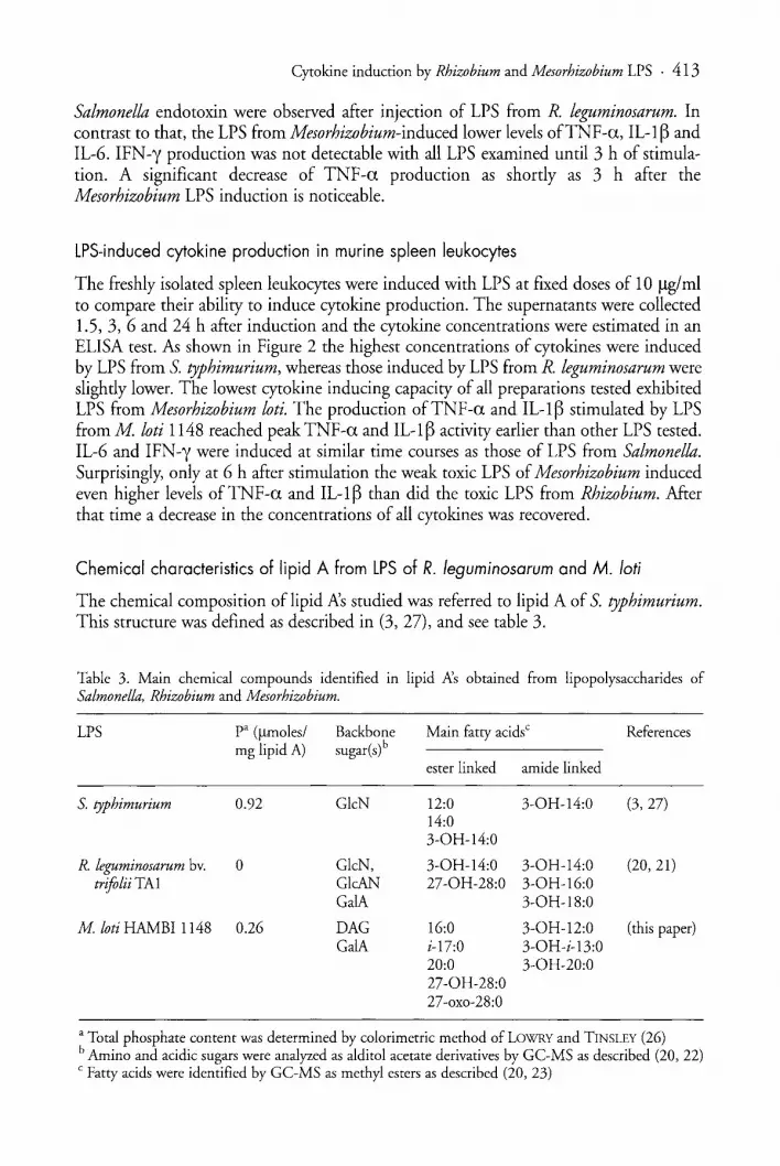
Chemical characteristics of lipid A from LPS of R. leguminosarum and M. loti
The chemical composition of lipid Ns studied was referred to lipid A of S. typhimurium.This structure was defined as described in (3, 27), and see table 3.
Table 3. Main chemical compounds identified in lipid .P-:s obtained from lipopolysaccharides ofSalmonella, Rhizobium and Mesorhizobium.
LPS pa (f..Lmoles/ Backbone Main fatty acidsc Referencesmg lipid A) sugar(s)b
ester linked amide linked
S. typhimurium 0.92 GleN 12:0 3-0H-14:0 (3,27)14:03-0H-14:0
R. leguminosarum bv. 0 GleN, 3-0H-14:0 3-0H-14:0 (20,21)trifolii TAl GleAN 27-0H-28:0 3-0H-16:0
GalA 3-0H-18:0
M. loti BAMBI 1148 0.26 DAG 16:0 3-0H-12:0 (this paper)GalA i-17:0 3-0H-i-13:0
20:0 3-0H-20:027-0H-28:027-oxo-28:0
a Total phosphate content was determined by colorimetric method of LOWRY and TINSLEY (26)b Amino and acidic sugars were analyzed as alditol acetate derivatives by GC-MS as described (20, 22)C Fatty acids were identified by GC-MS as methyl esters as described (20, 23)

414 . T. URBANIK-SYPNIEWSKA et al.
A B2500 40
35
200030
=' 150025
E Ec, E20BLL ....Z ~I- 1000 15
10
500
5
00
1 3 5 7 9 11 13 15 17 19 21 23 1 3 5 7 9 11 13 15 17 19 21 23
incubation time [hours] Incubation time [hours]
C D3500 1400
3000 1200
2500 1000
E2000 ='800.§C, C)
B 8CD
~600~ 1500
1000 400
500 200
0 0
1 3 5 7 9 11 13 15 17 19 21 23 1 3 5 7 9 11 13 15 17 19 21 23
Incubation time [hours] Incubation time [hours]
-+-S.typhimurium ___ R.leguminosarum -'-M.loti 1148
Figure 2. Time courses ofLPS induced TNF-a (A), ILl-~ (B), IL-6 (C) and IFN-y (D) productionin murine splenocytes. Mouse spleen leukocytes 5 x 10⁶ per ml were stimulated with fixed doses(10 J..Lg/ml) LPS. At indicated time points supernatants were sampled and stored at -20°Cuntil determination of cytokines by ELISA. The sera without LPS injection were used as control samples. Valuesare means ± S.D. of three determinations.

Cytokine induction by Rhizobium and Mesorhizobium LPS . 415
The fine structure of lipid A ofR. leguminosarum bv. trifolii TAl is under investigationbut our preliminary results of chemical composition are in agreement with the dataobtained by BHAT et al., (20) for R. leguminosarum bv. phaseoli CE3. We can assume thatthe structure of lipid A isolated from TAl LPS is very similar, if not identical with therevised structure presented by BASU et aI., (21). According to these data, lipid A of R.leguminosarum lacks 1- and 4'-phosphate groups and D-glucosamine is substituted by agalacturonic acid at position 4'. Instead of the proximal glucosamine-1-phosphate unit anaminogluconic acid (GleAN) is placed. In this sugar backbone the residues of glucosamine and 2-aminogluconate are substituted by ester-linked 3-hydroxymirystic acid. Thisacid is also amide linked to glucosamine. As amide linked to GlcAN either 3-hydroxypalmitic or 3-hydroxystearic acyl residues can occur. The only acyloxyacyl residue identifiedin this lipid A is ester linked 3-hydroxymyristoyl residue with 3-hydroxyl function acylated by 27-hydroxyoctadecanoate in which a part of hydroxyl groups is substituted by succinate (20, 21).
Analysis of chemical composition of lipid A isolated from Mesorhizobium loti indicated high heterogeneity of this preparation. The main sugar identified in lipid A obtainedfrom M loti 1148 was diaminoglucose. Galacturonic acid was detected as minor constituent.
The determination of total phosphate content in free lipid A preparations showed thepresence of phosphate in the preparation of Mesorhizobium loti and the lack of this compound in lipid from R. leguminosarum (Table 3).
31p nuclear magnetic resonance studies were performed to determine the locations ofphosphate groups in the free lipid A of Mesorhizobium (Fig. 3). A single peak with achemical shift of 2.00 ppm was observed at neutral pH (Fig. 3C). Titration of lipid Afrom pH 7.2 to 10.0 demonstrated that this signal was shifted to 4.50 ppm (Fig. 3B). Byanalogy to enterobacterial lipid A we concluded that this pH-dependent singlet could beattributed to a nonglycosidic phosphomonoester (27). Moreover, the same 31p NMRspectra with such a change in chemical shift as described above were also reported forlipid A of P diminuta and P vesicularis containin& DAG as backbone sugar (29, 30).Another signal, very weak, pH-independent and centered at 1.00 ppm was probably dueto a phosphate diester.
On the basis of preliminary results we can suppose that in lipid A of Mesorhizobiumthe sugar backbone contains diaminoglucose disaccharide, carrying phosphate group atthe nonreducing residue (i.e. phosphomonoester). However, it is not excluded that in apart of this lipid A species, additionally, galacturonic acid residue is present probably atthe reducing end as shown in Figure 3A. Moreover, the preparations of this lipid A couldcomprise a mixture of various backbone types.
The results of analysis of fatty acid content indicated a wide range of at least severalvarious acyl residues contributing to the heterogenous character of lipid A fromMesorhizobium. The results presented in Table 3 indicate that both short-and long-chainfatty acids have been identified as ester and amide linked acyl residues. It is noteworthythat in addition to hydroxy forms, some fatty acids particularly 27-0H-28:0 occurred asoxo derivatives (23).

416 . T. URBANIK-SYPNIEWSKA et al.
A
R. 3-0H-12:0; 3-0H-i-13:0; 3-0H-20:0Rz 16:0; i17:0; 20:0; 27-0H-28:0? GaiA
B
c
,.....,.........."T"T"'~..,...·rr-..........,...-...,..r·...·y"..,-ry"Y"T···~..,.,..,...'"Y·'·rrr.,.....,""TTrTTTTT"Y"l.,.,-r"'W'"J-rt·'.......,,-rrTTrrrr;"'Y...' , , .,.,.,~r-rT'~
6 4 Z -0 -2 -4 -5 -a -11 -12 -14 pp.
Figure 3. The proposed structure of lipid A from M. loti 1148 with 31p nuclear magnetic resonancespectra enclosed indicating the presence of ester bound phosphate group marked with asterisks (A).The signal at 2.00 ppm (C) recorded at pH 7.2 shifted to 4.50 ppm at pH 9.5 (B) could be assignedto monophosphate group.

Cytokine induction by Rhizobium and Mesorhizobium LPS . 417
Discussion
In our experiments we compared the typical endotoxic activities of lipopolysaccharidesfrom nodulating bacteria to those of enteric bacteria.
The results of lethality studies in galactosamine-treated mice showed that 0.1 flg ofLPS from Salmonella typhimurium is sufficient to kill half the animals. The same lethaleffect was caused by 17.8 flg of the Rhizobium leguminosarum LPS, while more than100 ~g was necessary in the case of LPS preparations from Mesorhizobium loti. The lastpreparation in contrast to that from R. leguminosarum could be defined as weakly toxicalso by the criteria of other authors (2, 16).
The lower lethal activity of this preparation was accompanied by its 10 times weakercapacity in Limulus amebocyte gelation as compared to LPS from Salmonella andRhizobium. The same value of the gelation activity of LPS from Rhizobium andSalmonella is of great interest. TAKADA et al. (9) have demonstrated that the presence of3-hydroxyacyl groups on the bisphosphorylated B-1 ,6-D-glucosamine disaccharide backbone is the prerequisite for effective activation of the clotting enzyme cascade ofLimulus.Lipid X, the natural precursor of lipid A composed of D-glucosamine I-phosphate withtwo acyl residues was recognized as the minimal structural requirement for positive LALactivity (31). Since LAL activity was exhibited by LPS preparations from Salmonella,Rhizobium and Mesorhizobium differing both in the hydrophilic sugar backbone andhydrophobic constituents of their lipid Xs, the structural requirements for this activitymust be broad.
In the previous studies on synthetic lipid A structures it was demonstrated that thebiological potency of a compound was dependent on the phosphorylation pattern (7, 8,13, 23). The removal of one phosphate group (particularly at C-l position) from E. colilipid A reduced its bioactivity by a factor 103 to 102 (1, 7). As it was demonstratedthe lethal activity of LPS of R. leguminosarum TAl with phosphate-free lipid A wasonly two hundred times lower than that of S. typhimurium LPS. This confirms the results of other authors demonstrating that there are phosphate-free lipid N.s, which areboth toxic like phototrophic bacterium Rhodospirillum fulvum and non-toxic ones likeRhodopseudomonas viridis, R. sulphoviridis and R. palustris (18, 19).
Lipopolysaccharide ofM loti which contains phosphate at least in major part of lipidA species is low toxic.
It was found that the number and distribution of fatty acids in lipid A is crucial forthe LPS-induced production ofcytokines, however, it was not clear how the length of thefatty acid chain affects the activity of the whole molecule.
Assuming structural similarities between lipid A of R. etli CE3 and lipid A of R. leguminosarum TAl, long chain 27-hydroxyoctacosanoic acid is present as ester linkedacyloxyacyl residue but in contrast to enterobacterial lipid A the degree of its acylation islower (12, 21).
So far the precise location of 27-0H-28:0 is not been established in mesorhizobiallipid A. Mesorhizobia contain oxo fatty acids in their lipid Xs (23). Whether the presence ofsuch fatty acids likewise the occurrence of 3-oxo fatty acids in lipid Xs from nontoxic Rhodobacter species (14, 15) is the reason for the observed low toxicity ofMesorhizobium loti LPS cannot be decided at present.
Although the exact role of acyl groups of lipid A for the induction of cytokines is presently not fully understood, it may relate to a peculiar three-dimensional physical struc-

418 . T. URBANIK-SYPNIEWSKA et al.
ture required not only for binding but also the passage of lipid A into the cytoplasmneeded for cell activation (3, 33).
Different dose-responses in TNF-a inducing ability among LPS were found. TNF-asecretion was strongly induced in vivo by LPS from R. leguminosarum in contrast to thepreparation from M. loti. A possible explanation of different TNF-a levels induced invivo due to M. loti and R. leguminosarum LPS stimulation could be the different affinityof their lipid Ns to bactericidal/permeability-increasing protein (BPI). The LPS/BPIcomplex is known to inhibit synthesis ofTNF-a and other cytokines after initial stimulation of granulocytes and macrophages by LPS (34).
The studies concerning the effects of LPS originating from non-enterobacterial species; R. salinarum, R.fulvum, (18,19) Bradyrhizobium. sp. (Lupinus), P diminuta and R.capsulatus (16) demonstrated that induction ofTNF-a, IL-6 and IL-l in mouse macrophages was correlated with in vivo lethality.
Our results showed the differences in the dynamics ofcytokines production in murinespleen leukocytes due to induction of these cells by LPS of high lethality i.e. R. leguminosarum and by LPS of low lethality from Mesorhizobium.
However, additional investigations are necessary to explain whether there are anychemical features correlating with differentiated abilities of Rhizobium andMesorhizobium LPS to activate various cytokine productions. Of particular interest isLPS from M loti 1148 of substantially low TNF-a and IL-l ~ induction activity in vivo.It could be interesting to investigate if this preparation is able to inhibit LPS -inducedmonokine production by human monocytes or exhibit any antiendotoxin activity.
Acknowledgements
This work was supported by the Polish Committee for Scientific Research: grant No.6 P04C 020 12.The authors thank Dr. PAWEL SOWINSKI for excellent NMR analyses.
References
1. RIETSCHEL, E. TH., T. KIRKAE, F. U. SCHADE, U. MAMAT, G. SCHMIDT, H. LOPPNOW, A.].ULMER, U. ZAHRINGER, U. SEYDEL, F. D. PADOVA, M. SCHREIER, and H. BRADE. 1994.Bacterial endotoxin: molecular relationship of structure to activity and function. FASEB]. 8: 217.
2. HENDERSON, B., S. POOLE, and M. WILSON. 1996. Bacterial modulins: a novel class ofvirulencefactors which cause host tissue pathology by inducing eytokine synthesis. Microbiol. Rev. 60: 314.
3. MORAN, A. I? 1995. Structure bioactivity relationships of bacterial endotoxins. ]. Toxicol.-ToxinReviews 14: 47.
4. DECKER, K. 1990. Biologically active products of stimulated liver macrophages (Kupffer cells).Eur.]. Biochem. 192: 245.
5. HOLST, 0., A. J. ULMER, H. BRADE, H. D. FLAD, and E. TH. RIETSCHEL. 1996. Biochemistryand cellular biology of bacterial endotoxins. FEMS Immunol. Med. Microbiol. 16: 83.
6. SUDA, Y., K. AOYAMA, K. ARIMoTO, T. TAMURA, and S. KUSUMOTO. 1999. S-Form lipopolysaccharide (LPS), but not lipid A or R-chemotype-LPS, induces interleukin-6 production in vitaminD 3-differentiated THP-l cells. Biochem. Biophys. Res. ~ommun. 257: 327.
7. KANEGASAKI, S., Y. KOJIMA, M. MATSUURA, Y. HOMMA, A. YAMAMOTO, Y. KUMAZAWA, K.TANAMOTO, T. YASUDA, T. TSUMITA, M. IMOTO, H. YOSHIMURA, M. YAMAMOTO, T.SHIMAMOTO, S. KUSUMOTO, and T. SHIBA. 1984. Biological activities of analogues of lipid Abased chemically on the revised structural model. Comparison of mediator-inducing, immunomodulating and endotoxic activities. Eur. ]. Biochem. 143: 237.

Cytokine induction by Rhizobium and Mesorhizobium LPS . 419
8. FLAD, H. D., H. LOPPNOW, M. H. WANG, H. BRADE, S. KUSUMOTO, E. TH. RIETSCHEL, andA. J. ULMER. 1989. Interleukin-l and tumor necrosis factor: studies on the induction by lipopolysaccharide partial structures. Lymphokine Res. 8: 235.
9. TAKADA, H., S. KOTANI, S. TANAKA, T. OGAWA, I. TAKAHASHI, M. TSUJIMOTO, T. KOMURO, T.SHIBA, S. KUSUMOTO, N. KUSUNOSE, A. HASEGAWA, and M. KIso. 1988. Structural requirements of lipid A species in activation of clotting enzymes from the horseshoe crab, and the humancomplement cascade. Eur. J. Biochem. 175: 573.
10. GALANOS, C., M. A. FREUDENBERG, and W REUTTER. 1979. Galactosamine-induced sensitization to the lethal effects of endotoxin. Proc. Natl. Acad. Sci. USA. 76: 5939.
11. ZINETTI, M., S. AGYCKUM, T. EVANS, J. POLAK, and J. COHEN. 1998. The role of lipopolysaccharide, pro-inflammatory cytokines and bacterial superantigens in the transcriptional regulationoflymphotoxin a and ~ in mouse splenocytes. Cytokine 10: 940.
12. Low, K. B., M. ITTENSOHN, T. LE, J. PLATT, S. SODI, M. AMoss, O. ASH, E. CARMICHAEL, A.CHAKRABORTY, J. FISCHER, S. L. LIN, X. Luo, S. I. MILLER, L. ZHENG, I. KING, J. M. PAWELEK,and D. BERMUDES. 1999. Lipid A mutant Salmonella with suppressed virulence and TNF-ainduction retain tumor-targeting in vivo. Nature Biotechnology 17: 37.
13. TAKAYAMA, K.) N. QURESHI, B. BEUTLER, and T. N. KIRKLAND. 1989. Diphosphoryllipid Afrom Rhodopseudomonas sphaeroides ATCC 17023 blocks induction of cachectin in macrophagesby lipopolysccharide. Infect. Immun. 57: 1336.
14. CHRIST, W J., ~ D. MCGUINNESS, O. ASANO, Y. WANG, M. A. MULLARKEY, M. PEREZ, L. D.HAWKINS, T. A. BLYTHE, G. R. DUBUC, and A. L. ROBIDOUX. 1994. Total synthesis of the proposed structure of Rhodobacter sphaeroides lipid A resulting in the synthesis of new potent lipopolysaccharide antagonists. J. Am. Chern. Soc. 116: 3637.
15. KRAuss, J. W, U. SEYDEL, J. WECKESSER., and H. MAYER. 1989. Structural analysis of the nontoxic lipid A of Rhodobacter capsulatus 37b4. Eur. J. Biochem. 180: 519.
16. LOPPNOW, H., ~ LIBBY, M. FREUDENBERG, J. H. KRAuss, J. WECKESSER, and H. MAYER 1990.Cytokine induction by lipopolysaccharide (LPS) corresponds to lethal toxicity and is inhibited bynontoxic Rhodobacter capsulatus LPS. Infect. Immun. 58: 3743.
17. BHAT, U. R., R. W CARLSON, M. BUSCH, and H. MAYER. 1991. Distribution and phylogeneticsignificance of 27-hydroxy-octacosanoic acid in lipopolysaccharides from bacteria belonging to thealpha-2-subgroup of Proteobacteria. Int. J. Syst. Bacteriol. 41: 213.
18. RAu, H., U. SEYDEL, M. FREUDENBERG, J. WECKESSER, and H. MAYER. 1995. Lipopolysaccharide of Rhodospirillum salinarium 40: structural studies on the core and lipid A region. Arch.Microbiol. 164: 280.
19. RAu, H., U. SEYDEL, M. FREUDENBERG, J. WECKESSER, and H. MAYER. 1995. Lipopolysaccharide of the phototrophic bacterium Rhodospirillum fulvum. System. Appl. Microbiol. 18:154.
20. BHAT, U. R., L. S. FORSBERG, and R. W CARLSON. 1994. Structure of lipid A component ofRhizobium leguminosarum bv. phaseoli lipopolysaccharide. Unique nonphosphorylated lipid A containing 2-amino-2-deoxygluconate, galacturonate, and glucosamine. J. BioI. Chern. 269: 14402.
21. BASU, S. B., K. A. WHITE, N. L. S. QUE, and C. R. H. RAETZ. 1999. A deacylase in Rhizobiumleguminosarum membranes that cleaves 3-0-linked ~-hydroxymyristoylmoiety of lipid A precursors. J. BioI. Chern. 274: 11150.
22. RUSSA, R., T. URBANIK-SYPNIEWSKA, K. LINDSTROM, and H. MAYER. 1995. Chemical characterization of two lipopolysaccharide species isolated from Rhizobium loti NZP 2213. Arch. Microbiol.163: 345.
23. CHOMA, A. 1999. Fatty acid composition of Mesorhizobium huakuii lipopolysaccharides.Identification of 27-oxooctacosanoic acid. FEMS Microbiol. Lett. 177: 257.
24. WESTPHAL, 0., and K. JANN. 1965. Bacteriallipopolysaccharides. Extration with phenol-waterand further applications of the procedure. Meth. Carbohydr. Chern. 3: 83.
25. GALANOS, C., and 0. LOOFRTTZ. 1975. Ele~trodialysis of lipopoly::mcchariocs ano lheir conversion to uniform salt forms. Eur. J. Biochem. 54: 603.
26. LOWRY, R. R., and 1. J. TINSLEY. 1974. A simple, sensitive method for lipid phosphorus. Lipids 9:491.

420 . T. URBANIK-SYPNIEWSKA et ai.
27. BATLEY, M., N. H. PACKER, andJ. W REDMOND. 1985. Analytical studies of lipopolysaccharideand its derivatives from Salmonella minnesota R595. I. Phosphorus magnetic resonance spectra.Biochem. Biophys. Acta 821: 179.
28. REED, L., and H. MUENCH. 1936. A simple method of estimating fifty points endpoints. Am. J.Hyg. 27: 493.
29. KASAl, N., S. ARATA, J. MASHIMO, Y AKIYAMA, C. TANAKA, K. EGAWA, and S. TANAKA. 1987.Pseudomonas diminuta with a new endotoxic lipid A structure. Biochem. Biophys. Res. Commun.142: 972.
30. ARATA, S., K. NAKAYA, H. FURUHASHI, Y. NAKAMURA, T. HIRAYAMA, J. MASHIMO, and N.KAsAl. 1988. Tumor necrosis factor-inducing activities of lipid A preparations from Pseudomonasdiminuta and Pseudomonas vesicularis. Jpn. J. Cancer Res. (Gann). 79: 626.
31. TAKAYAMA, K., N. QURESHI., C. R. H. RAETZ, E. RIBI, J. PETERSON, J. L. CANTRELL, F. C.PAERSON, J. WIGGINS, and A. G. JOHNSON. 1984. Influence of fine structure of lipid A onLimulus amebocyte lysate clotting and toxic activities. Infect. Immun. 45: 350.
32. LIN, Z. Z., J. COTTER, and C. R. H. RAETZ. 1999. Lipid A modifications characteristic ofSalmonella typhimurium are induced by NH4V03 in Escherichia coli K12. Detection of 4-amino4-deoxy-L-arabinose, phosphoethanolamine and palmitate. J. BioI. Chern. 274: 18503.
33. lIDA, 1:, Y. HAISHIMA, A. TANAKA, K. NISHIYAMA, S. SAITO, and K. TANAMOTO. 1996. Chemicalstructure of lipid A isolated from Comamonas testosteroni lipopolysaccharide. Eur. J. Biochem. 237:468.
34. BRANDENBURG, K., H. MAYER, M. H. J. KOCH, E. TH. RIETsCHEL, and U. SEYDEL, 1993.Influence of the supramolecular structure of free lipid A on its biological activity. Eur. J. Biochem.218: 555.
35. CAPODICI, C., S. CHEN, Z. SIDORCZYK, ~ ELSBACH, and J. WEISS. 1994. Effect of lipopolysaccharide (LPS) chain length on interactions of bactericidal/permeability-increasing protein and itsbioactive 23-kilodalton NH2-terminal fragment with isolated LPS and intact Proteus mirabilis andEscherichia coli. Infect. Immun. 62: 259.
Dr. TERESA URBANIK-SYPNIEWSKA, Department of General Microbiology, M. Curie-SklodowskaUniversity, 20-033 Lublin, 19 Akademicka St., Poland, e-mail: [email protected]