crystal structure and functional insight of hp0420-homolog from helicobacter felis
TRANSCRIPT
Crystal structure and functional insight of HP0420-homolog fromHelicobacter felis
Shunfu Piaoa, Xiao Ling Jina, Bo-Young Yuna, Nahee Kimb, Hyun-Soo Chob, MinoruFukudac, Heeseob Leed, and Nam-Chul Haa,*a College of Pharmacy and Research Institute for Drug Development, Pusan National University,Jangjeon-dong, Geumjeong-gu, Busan 609-735, Republic of Koreab Department of Biology, College of Science, Yonsei University, Seoul 120-749, Republic of Koreac Tumor Microenvironment Program, Cancer Research Center, Burnham Institute for MedicalResearch, La Jolla, CA 92037, USAd Department of Food Science and Nutrition, Pusan National University, Jangjeon-dong,Geumjeong-gu, Busan 609-735, Republic of Korea
AbstractHelicobacter pylori infect more than half of the world’s population and are considered a cause ofpeptic ulcer disease and gastric cancer. Recently, hypothetical gene HP0421 was identified in H.pylori as a cholesterol α-glucosyltransferase, which is required to synthesize cholesteryl glucosides,essential cell wall components of the bacteria. In the same gene-cluster, HP0420 was co-identified,whose function remains unknown. Here we report the crystal structure of HP0420-homolog of H.felis (HF0420) to gain insight into the function of HP0420. The crystal structure, combined with size-exclusion chromatography, reveals that HF0420 adopts a homodimeric hot-dog fold. The crystalstructure suggests that HF0420 has enzymatic activity that involves a conserved histidine residue atthe end of the central α-helix. Subsequent biochemical studies provide clues to the function ofHP0420 and HF0420.
KeywordsCrystal structure; Helicobacter; Hot-dog fold; Structural genomics; HP0420
1. IntroductionHelicobacter pylori is a Gram-negative bacteria that lives in human stomach and may causegastric chronic inflammation and even stomach cancer [1]. Helicobacter species havecholesteryl glucosides (CGs) as unique and essential cell membrane components [2,3]. CGsare synthesized from cholesterol, and thus cholesterol must be taken up from the host cellbecause the bacteria lack the genes responsible for its biosynthesis [4]. Recently, it was reportedthat the hypothetical gene HP0421 from H. pylori exhibits cholesterol α-glucosyltransferaseactivity that converts cholesterol into CGs and can be inhibited by the natural antibiotic mucinO-glycan in deeper portions of the gastric mucosa [5]. Bacterial growth is severely inhibited
*Corresponding author. [email protected] (N.-C. Ha).Conflict of interestThe authors declare that they have no conflicts of interest.
NIH Public AccessAuthor ManuscriptBiochem Biophys Res Commun. Author manuscript; available in PMC 2010 June 25.
Published in final edited form as:Biochem Biophys Res Commun. 2010 April 16; 394(4): 940–946. doi:10.1016/j.bbrc.2010.03.087.
NIH
-PA Author Manuscript
NIH
-PA Author Manuscript
NIH
-PA Author Manuscript
in the absence of the HP0421 gene [6,7], suggesting a critical role for HP0421 in the survivalof H. pylori.
During expression cloning of cholesterol α-glucosyltransferase from H. pylori and H. felis, Leeet al. identified HP0420 and its homolog in a single plasmid harboring genomic sequences fortwo open reading frames (HP0420 and HP0421) [6]. H. felis, which lives in cat stomach [8]and shows genomic similarity to H. pylori, has been used to study colonization, pathogenesis,and eradication of H. pylori [9,10]. The gene structures of HP0420 and HP0421 were conservedin both Helicobacter species. Unlike HP0421, HP0420 was not essential for the survival of thebacteria [6]. Although the gene structure suggested that the function of HP0420 might beassociated with HP0421, such association remains to be elucidated [6]. The HP0420-homologfrom H. felis (herein referred to as HF0420) shares high sequence identity (40%) with HP0420,indicating that HF0420 is a functional and structural homolog of HP0420 [6].
To gain insight into the function of the hypothetical protein HP0420 and its homologs fromHelicobacter species, we determined the crystal structure of HF0420 from H. felis andperformed subsequent biochemical studies.
2. Materials and methods2.1. DNA construction and protein expression
DNA fragments encoding HF0420 (residues 1–141) and HP0420 (residues 1–142) wereamplified from H. felis and H. pylori genomic DNA, respectively, using the polymerase chainreaction. The DNA fragments were inserted into the NcoI and XhoI sites of pProEXHTa(Invitrogen, USA) containing the hexa-histidine tag and the TEV protease cleavage sites at theN-terminus. The recombinant HF0420 and HP0420 proteins were expressed in Escherichiacoli strain BL21 (DE3) RIL in Luria–Bertani (LB) medium at 37 °C until the OD600 nm reached0.5. The protein was expressed by adding 0.5 mM isopropyl-β-D-1-thiogalactopyranoside(IPTG). The cells were harvested by centrifugation at 5000 rpm for 15 min at 4 °C.
2.2. Protein purification of HF0420 and HP0420To produce HF0420 and HP0420 proteins, harvested cells were suspended in lysis buffercontaining 20 mM Tris (pH 8.0), 150 mM NaCl and 2 mM β-mercaptoethanol, and weredisrupted by sonication. The lysate was centrifuged at 13,000 rpm for 30 min at 4 °C. Theresulting supernatant was loaded onto Ni–NTA agarose resin that was pre-equilibrated withlysis buffer. The resin was washed with lysis buffer supplemented with 20 mM imidazole andthen eluted with lysis buffer supplemented with 200 mM imidazole. The fractions containingthe HF0420 protein were pooled and treated with recombinant TEV protease overnight at roomtemperature to remove the hexa-histidine tag after addition of 10 mM β-mercaptoethanol. Thereaction mixture was subsequently loaded onto a Q-anion exchange column (Hitrap-Q; GEHealthcare, USA) for further purification, and eluted with a gradient from 0 to 1 M NaCl. Thecollected fractions containing the target protein were pooled, concentrated, and separated ona HiLoad Superdex 200 gel-filtration column (GE Healthcare, USA) pre-equilibrated with lysisbuffer. During the purification, the presence of the protein was confirmed by SDS–PAGE. Thepurified HF0420 and HP0420 proteins were concentrated to 60 mg/ml and 40 mg/ml,respectively, using Centriprep (Millipore, USA) and stored frozen at −80 °C until use.
2.3. Crystallization, data collection and structure determinationCrystals of the wild-type HF0420 protein were obtained by the vapor-diffusion technique at14 °C. The initial crystallization screening was performed by the sitting-drop method withCrystal Screen HT, a high-throughput sparse-matrix screening kit (Hampton Research, USA).Crystals grown in the solution containing 0.1 M Tris–HCl (pH 8.5) and 2 M ammonium sulfate
Piao et al. Page 2
Biochem Biophys Res Commun. Author manuscript; available in PMC 2010 June 25.
NIH
-PA Author Manuscript
NIH
-PA Author Manuscript
NIH
-PA Author Manuscript
were directly chosen from the initial crystallization screening plate for data collection. Forcryoprotection, the HF0420 crystals were soaked with the sticky oil Paratone-N. The data setswere collected on BL6C at Pohang Accelerator Laboratory with a CCD detector Quantum 210(ADSC) at −173 °C. The diffraction data sets were processed and scaled with the HKL2000package [11]. The crystal belongs to the space group P212121 with cell dimensions of a = 70.5,b = 70.3, and c = 58.8 Å. Initial phases were determined by the molecular replacement packageMOLREP [12] using the coordinates of Cj0977 (Protein Data Bank code 3BNV) as a searchmodel. Model building was performed using the program COOT [13], and model refinementwas conducted using the program CNS 1.2 [14]. PHENIX. REFINE [15] was applied at thefinal round of the model refinement.
Crystals of mutant HF0420 (C46A) were obtained by the same method as wild-type HF0420,and the crystallization conditions were optimized to produce high quality crystals in dropletscontaining 1 μl of protein solution and 1 μl of a precipitant solution consisting of 0.01 M cobalt(II) chloride hexahydrate, 0.1 M MES, pH 6.5, and 1.8 M ammonium sulfate. Data sets werecollected as described above, and the crystal belonged to the space group P212121 with celldimensions of a = 53.3, b = 67.1, and c = 70.2 Å. Initial phases were determined by themolecular replacement package MOLREP [12] using coordinates of the wild-type HF0420structure as a search model.
Crystallographic data statistics are summarized in Table 1. All figures were prepared withPYMOL [16].
2.4. Size-exclusion chromatographySize-exclusion chromatography was performed at room temperature at a flow rate of 0.5 ml/min on Superdex S-200 HR 10/30 (GE Healthcare) equilibrated with 20 mMTris buffer (pH8.0) containing 150 mM NaCl and 2 mM β-mercaptoethanol. Five hundred microliters of eachprotein (100 μg) were injected onto the column.
2.5. Thermal stabilityThermal stability studies of HF0420 (wild-type and C46A) and HP0420 were performed bycircular dichroism in a JASCO-J750 spectropolarimeter. Samples were prepared in 20 mMTris, 0.15 M NaCl, pH 8.0, and thermal unfolding experiments were performed by monitoringthe circular dichroism signal at 220 nm between 25 °C and 90 °C using a heating rate of 2 °C/min at a concentration of 0.25 mg/ml.
2.6. Isothermal titration calorimeter (ITC)Isothermal titration calorimeter (ITC) measurement was done using a VP-ITC from MicroCal,Inc. (Northampton, MA). HF0420 protein and CoA were dissolved in 100 mM phosphate buffer(pH 8.0) at a final concentration of 20 μM and 150 μM, respectively. The titration of HF0420with CoA involved 25 injections of CoA solution, of 10 μl each. The duration of each injectionwas 20 s, and the time delay between successive injections was 220 s. The contents of thesample cell were stirred at 300 rpm throughout the experiment to ensure thorough mixing. Thetemperature of the titration cell was set at 25 °C.
3. Results3.1. Structural determination of HF0420
To gain insight into the functions of HP0420 and HF0420, we attempted to determine the three-dimensional structures of HP0420 and HF0420. Full-length proteins were successfullyoverexpressed in E. coli and were purified to homogeneity. The purified proteins were highlysoluble and were concentrated for crystallization. Fortunately, we obtained crystals of HF0420
Piao et al. Page 3
Biochem Biophys Res Commun. Author manuscript; available in PMC 2010 June 25.
NIH
-PA Author Manuscript
NIH
-PA Author Manuscript
NIH
-PA Author Manuscript
under several conditions, and these crystals were suitable for data collection withoutoptimization. However, we failed to obtain crystals of HP0420 despite extensive screening ofthe crystallization conditions.
The crystal structure of HF0420 was solved by molecular replacement using Cj0977 fromCampylobacter jejuni, which shows the highest sequence homology, as a search model. Theasymmetric unit of the crystal contains two molecules, consists of 248 residues, and comprises89% of the total number of amino acid residues in the crystallized protein (Fig. 2A). TheMatthews coefficient (VM) of the crystal [17] was 2.1 Å3/Da and the solvent content wascalculated to be 42%.
3.2. Overall structure and dimeric nature of HF0420The crystal structure of HF0420 revealed that HF0420 adopts a “hot-dog” fold that consists ofa central long α-helix and a surrounding four-stranded β-sheet (Fig. 2B). The hot-dog fold wasso named because the α-helix resembles a sausage of hot-dog, and the β-sheet wraps aroundthis helix like a bun [18]. In addition to the hot-dog fold elements, loops and short α-helicesare found in the N- and C-terminal regions of the HF0420 structure (Fig. 2B).
The structural homologs of HF0420 were searched using the DALI server [19]. A nonflagellavirulence protein from C. jejuni (Cj0977) that has a putative acyl-CoA-binding site [20] wasindicated as the top match (Z-score 21.9) (Fig. 2C). A hypothetical protein PH1136 fromPyrococcus horikoshii [21] and malonyl-CoA-binding transcription factor FapR from Bacillussubtilis [22] were also indicated as matches (Z-scores 21.5 and 18.7). Phenylacetyl-CoAthioesterase from Thermus thermophilus (PaaI) and other acyl-CoA thioesterases werematched, as listed in Supplementary Table S1.
All hot-dog fold proteins form dimers, andsome hot-dog fold proteins form tetramers orhexamers by dimerization or trimerization of the dimeric unit [20]. Given the extensive dimericinterface between two molecules in the asymmetric unit (approximately 30% of HF0420surface area), the crystal structure suggests that HF0420 forms a dimer. Subsequent size-exclusion chromatography showed that both HF0420 and HP0420 proteins are present indimeric form in solution (Fig. 2D). These results indicate that HF0420 and HP0420 functionas dimeric units.
3.3. Putative substrate binding regionThe hot-dog fold proteins were first reported in the crystal structure of FabA [18], and this foldwas found in diverse thioesterases and coenzyme A (CoA)-derived binding proteins. To obtaininsight into the function of the hypothetical proteins HP0420 and its homologs, we superposedthe structure of HF0420 with PaaI in complex with phenylacetyl-CoA (PDB code:1WN3)(Z-score 18.7). Structures of HF0420 with PaaI were well fitted, and the superposition identifieda putative substrate binding site of HF0420 from the location of the superposed phenylacetyl-CoA (Fig. 3A and B). The putative active site is located at the interface between two protomersof HF0420. The nucleotide moiety of CoA is positioned in a shallow groove at the surface ofone subunit. The thioester bond of acyl-CoA is positioned at the N-terminal end of the centralα-helix, where the His52 and Gly54 residues are present (Fig. 3C). The hydrocarbon moietyof CoA is in a deep crevice formed at the dimeric interface (Fig. 3B). Based on this structuralsuperposition, we hypothesize that the real substrate of HF0420 may be acyl-CoA or itsderivatives.
3.4. Putative catalytic residueThe position of the His52 residue in HF0420 is fixed by the interaction with the conservedGly54 residue, and the residues are buried in the main body of the protein (Fig. 3B). However,
Piao et al. Page 4
Biochem Biophys Res Commun. Author manuscript; available in PMC 2010 June 25.
NIH
-PA Author Manuscript
NIH
-PA Author Manuscript
NIH
-PA Author Manuscript
His52 is accessible from the solvent through a narrow channel that is formed by a long loopfrom the neighboring subunit (Fig. 3B). Interestingly, the histidine and glycine residues areconserved in HP0420 and its homologs of Helicobacter species, whereas they are replaced inPaaI and acyl-CoA-binding hot-dog proteins such as Cj0977 (Fig. 1).
The central α-helix of hot-dog fold proteins is highly polarizing because the long α-helix issurrounded by the largely hydrophobic β-sheet (Fig. 2B). Since the conserved His52 residueis located at the positive (N-terminal) end of the polarizing α-helix, where the negative chargeis stabilized and positive charge is destabilized, the electron density of the unshared electronpair of the His52 imidazole ring may become richer. Thus, the nucleophilic reactivity of His52is likely to be enhanced due to the electron-rich unshared electron pair. Moreover, the imidazolering of His52 makes a hydrogen bond to the NH group of the conserved Gly54 residue, whichmay also augment the negative charge of His52 (Fig. 3C). Given that no residues interact withthe side chain of His52 except for Gly54, His52 might directly attack the substrate in theenzymatic reaction. Taken together, the structural features of HF0420 indicate that the HP0420family proteins may be hydrolases in which the conserved histidine is a catalytic residue thatdirectly attacks the substrate.
3.5. N-terminal cysteines of HF0420 form inter-subunit disulfide bondsThe HF0420 crystal structure shows that two cysteine residues (Cys15 and Cys46) form inter-subunit disulfide bonds, which is the first observation in the hot-dog fold proteins (Fig. 2A).This disulfide bridge is located around His52, connecting the loops that form the narrowchannel to the histidine residue (Fig. 3C). Since there is no signal sequence for secretion inHF0420, HF0420 is thought to be present in the cytosol, where most cysteines are present inreduced form, when expressed in bacteria. However, the possibility that HF0420 is secreted tothe extracellular medium, where cysteines are oxidized to form a disulfide bond as observedin the crystal structure, by some unknown mechanism cannot be excluded. To examine whetherthis inter-subunit disulfide bond is involved in the oxidation/reduction-induced conformationalchange, we generated and crystallized a C46A mutant HF0420 protein in which the disulfidebond is not formed. Comparison of the crystal structures and the molecular sizes in solutionrevealed that the disulfide bridge did not affect the conformation of the loop. As shown in Fig.4A and B, the structure of the C46A mutant was very similar to that of wild-type HF0420,except for the mutated residue. However, the mutant protein showed reduced thermostabilitycompared to wild-type HF0420, as measured by circular dichroism spectroscopy (Fig. 4C).This result indicates that this disulfide bond contributes to the structural integrity of HF0420,although the disulfide bond is not associated with the oxidation/reduction-inducedconformational change of HF0420.
In HP0420 from H. pylori, the inter-subunit disulfide bridge is not expected to be presentbecause of the lack of the corresponding Cys46 residue. Nonetheless, HP0420 was intrinsicallythermostable, as the recombinant HP0420 protein showed high thermostability (Tm = 95 °C)(Fig. 4C).
4. DiscussionIn this study, we demonstrated that HP0420 and HF0420 are dimeric proteins and adopt thehot-dog fold. The crystal structure of HF0420 suggested that HP0420 family proteins may behydrolases. However, the substrate of HP0420 family proteins remains unknown. Sequencehomology using CLUSTALX [23] and BLAST [24] suggests that HP0420, HF0420, andHAC_0608 from H. acinonychis are most similar, and phylogenetic analysis [25] suggests thatthe three proteins comprise a subgroup distinct from other hot-dog fold proteins(Supplementary Fig. S1). Moreover, they are all followed by cholesterol α-glucotransferase inthe gene structure. Thus, we examined whether HF0420 has cholesterol esterase activity, since
Piao et al. Page 5
Biochem Biophys Res Commun. Author manuscript; available in PMC 2010 June 25.
NIH
-PA Author Manuscript
NIH
-PA Author Manuscript
NIH
-PA Author Manuscript
HP0420 might be functionally involved with cholesteryl α-glucotransferase (HP0421). Thisidea is supported by the facts that cholesterol esters are the major storage form in serum ratherthan cholesterol, cholesterol esters are similar to acyl-CoA in size, and cholesteryl α-glucotransferase requires cholesterol, not cholesterol ester, as a substrate. Thus, cholesterolesterase activity would be needed to efficiently uptake the substrate of cholesteryl α-glucotransferase. However, cholesterol esterase activity was not detected from HF0420 protein(data not shown).
Hot-dog fold proteins have been characterized as acyl-CoA thioesterases or acyl-CoA-bindingproteins, and acyl-CoA or its derivatives are suggested as the substrate of HF0420 by thestructural superposition in this study. Thus we next examined whether HF0420 exhibits acyl-CoA thioesterase activity. The recombinant HF0420 did not show any acyl-CoA thioesteraseactivity when palmitoyl-CoA was used as the substrate in an enzymatic assay describedpreviously [26] (Supplementary Fig. S2). Moreover, no CoA-binding ability of HF0420 wasdetected by titration calorimetry experiments (Supplementary Fig. S3). These results seem toexclude the possibility that HP0420 family proteins are associated with CoA, and suggest thatan acyl-CoA analog, not acyl-CoA, is the substrate.
Since the closest structural homolog, Cj0977, is known as a virulence factor that is transferredinto the cytosol of host cells, we next examined the virulence of HF0420. HF0420 did not showany cytotoxic effect on human cells when human embryonic kidney cells were directly treatedwith HF0420 protein or when 293 cells were transfected with the HF0420 gene for expressionin the cytosol of human cells (data not shown). Taken together, HP0420 family proteins maynot be virulence factors on their own and do not change the essential processes of eukaryoticcells.
In this study, the crystal structure of HF0420 was determined to gain insight into the functionof HP0420 family proteins, and subsequent diverse experiments were carried out to reveal thefunction of the proteins. Although no conclusive results were produced, the experimentssuggested that HP0420 family proteins may not be acyl-CoA thioesterases, CoA-bindingproteins, cholesterol esterases, or virulence factors that affect mammalian cell survival. Thisstudy provides clues to the function of HP0420 and HF0420, which may be different fromother hot-dog fold proteins.
Supplementary MaterialRefer to Web version on PubMed Central for supplementary material.
AcknowledgmentsThis study made use of beamline 6C at Pohang Accelerator Laboratory (Pohang, Korea). This study was supportedby a grant from the 21C Frontier Microbial Genomics and Applications Center Program, Ministry of Education,Science & Technology, Republic of Korea. The coordinates of the HF0420 structures have been deposited into theProtein Data Bank (PDB codes: 3LW3 and 3LWG).
References1. Brown LM. Helicobacter pylori: epidemiology and routes of transmission. Epidemiol Rev
2000;22:283–297. [PubMed: 11218379]2. Hirai Y, Haque M, Yoshida T, Yokota K, Yasuda T, Oguma K. Unique cholesteryl glucosides in
Helicobacter pylori: composition and structural analysis. J Bacteriol 1995;177:5327–5333. [PubMed:7665522]
3. Haque M, Hirai Y, Yokota K, Mori N, Jahan I, Ito H, Hotta H, Yano I, Kanemasa Y, Oguma K. Lipidprofile of Helicobacter spp.: presence of cholesteryl glucoside as a characteristic feature. J Bacteriol1996;178:2065–2070. [PubMed: 8606185]
Piao et al. Page 6
Biochem Biophys Res Commun. Author manuscript; available in PMC 2010 June 25.
NIH
-PA Author Manuscript
NIH
-PA Author Manuscript
NIH
-PA Author Manuscript
4. Tomb JF, White O, Kerlavage AR, Clayton RA, Sutton GG, Fleischmann RD, Ketchum KA, KlenkHP, Gill S, Dougherty BA, Nelson K, Quackenbush J, Zhou L, Kirkness EF, Peterson S, Loftus B,Richardson D, Dodson R, Khalak HG, Glodek A, McKenney K, Fitzegerald LM, Lee N, Adams MD,Hickey EK, Berg DE, Gocayne JD, Utterback TR, Peterson JD, Kelley JM, Cotton MD, WeidmanJM, Fujii C, Bowman C, Watthey L, Wallin E, Hayes WS, Borodovsky M, Karp PD, Smith HO, FraserCM, Venter JC. The complete genome sequence of the gastric pathogen Helicobacter pylori. Nature1997;388:539–547. [PubMed: 9252185]
5. Kawakubo M, Ito Y, Okimura Y, Kobayashi M, Sakura K, Kasama S, Fukuda MN, Fukuda M,Katsuyama T, Nakayama J. Natural antibiotic function of a human gastric mucin against Helicobacterpylori infection. Science 2004;305:1003–1006. [PubMed: 15310903]
6. Lee H, Kobayashi M, Wang P, Nakayama J, Seeberger PH, Fukuda M. Expression cloning ofcholesterol alpha-glucosyltransferase, a unique enzyme that can be inhibited by natural antibioticgastric mucin O-glycans, from Helicobacter pylori. Biochem Biophys Res Commun 2006;349:1235–1241. [PubMed: 16978585]
7. Lebrun AH, Wunder C, Hildebrand J, Churin Y, Zahringer U, Lindner B, Meyer TF, Heinz E, WarneckeD. Cloning of a cholesterol-alpha-glucosyltransferase from Helicobacter pylori. J Biol Chem2006;281:27765–27772. [PubMed: 16844692]
8. Paster BJ, Lee A, Fox JG, Dewhirst FE, Tordoff LA, Fraser GJ, O’Rourke JL, Taylor NS, Ferrero R.Phylogeny of Helicobacter felis sp. nov., Helicobacter mustelae, and related bacteria. Int J SystBacteriol 1991;41:31–38. [PubMed: 1704791]
9. Lavelle JP, Landas S, Mitros FA, Conklin JL. Acute gastritis associated with spiral organisms fromcats. Dig Dis Sci 1994;39:744–750. [PubMed: 8149839]
10. Wegmann W, Aschwanden M, Schaub N, Aenishanslin W, Gyr K. Gastritis associated withGastrospirillum hominis—a zoonosis? Schweiz Med Wochenschr 1991;121:245–254. [PubMed:2011719]
11. Otwinosky Z, Minor W. Processing of X-ray diffraction data collected in oscillation mode. MethodsEnzymol 1997;276:307–326.
12. CCP4i. The CCP4 suite: programs for protein crystallography. Acta Crystallogr D: Biol Crystallogr1994;50:760–763. [PubMed: 15299374]
13. Emsley P, Cowtan K. Coot: model-building tools for molecular graphics. Acta Crystallogr D: BiolCrystallogr 2004;60:2126–2132. [PubMed: 15572765]
14. Brunger AT, Adams PD, Clore GM, DeLano WL, Gros P, Grosse-Kunstleve RW, Jiang JS, KuszewskiJ, Nilges M, Pannu NS, Read RJ, Rice LM, Simonson T, Warren GL. Crystallography & NMRsystem: a new software suite for macromolecular structure determination. Acta Crystallogr D: BiolCrystallogr 1998;54(Pt. 5):905–921. [PubMed: 9757107]
15. Adams PD, Grosse-Kunstleve RW, Hung LW, Ioerger TR, McCoy AJ, Moriarty NW, Read RJ,Sacchettini JC, Sauter NK, Terwilliger TC. PHENIX: building new software for automatedcrystallographic structure determination. Acta Crystallogr D: Biol Crystallogr 2002;58:1948–1954.[PubMed: 12393927]
16. DeLano, W. The PyMOL User’s Manual. DeLano Scientific; Palo Alto: 2002.17. Kantardjieff KA, Rupp B. Matthews coefficient probabilities: improved estimates for unit cell
contents of proteins, DNA, and protein–nucleic acid complex crystals. Protein Sci 2003;12:1865–1871. [PubMed: 12930986]
18. Leesong M, Henderson BS, Gillig JR, Schwab JM, Smith JL. Structure of a dehydratase–isomerasefrom the bacterial pathway for biosynthesis of unsaturated fatty acids: two catalytic activities in oneactive site. Structure 1996;4:253–264. [PubMed: 8805534]
19. Holm L, Sander C. Touring protein fold space with Dali/FSSP. Nucleic Acids Res 1998;26:316–319.[PubMed: 9399863]
20. Yokoyama T, Paek S, Ewing CP, Guerry P, Yeo HJ. Structure of a sigma28-regulated nonflagellarvirulence protein from Campylobacter jejuni. J Mol Biol 2008;384:364–376. [PubMed: 18835274]
21. Tajika Y, Sakai N, Tanaka Y, Yao M, Watanabe N, Tanaka I. Crystal structure of conserved proteinPH1136 from Pyrococcus horikoshii. Proteins 2004;55:210–213. [PubMed: 14997554]
Piao et al. Page 7
Biochem Biophys Res Commun. Author manuscript; available in PMC 2010 June 25.
NIH
-PA Author Manuscript
NIH
-PA Author Manuscript
NIH
-PA Author Manuscript

22. Schujman GE, Guerin M, Buschiazzo A, Schaeffer F, Llarrull LI, Reh G, Vila AJ, Alzari PM, deMendoza D. Structural basis of lipid biosynthesis regulation in Gram-positive bacteria. EMBO J2006;25:4074–4083. [PubMed: 16932747]
23. Jeanmougin F, Thompson JD, Gouy M, Higgins DG, Gibson TJ. Multiple sequence alignment withClustal X. Trends Biochem Sci 1998;23:403–405. [PubMed: 9810230]
24. Altschul SF, Gish W, Miller W, Myers EW, Lipman DJ. Basic local alignment search tool. J MolBiol 1990;215:403–410. [PubMed: 2231712]
25. Waterhouse AM, Procter JB, Martin DM, Clamp M, Barton GJ. Jalview version 2—a multiplesequence alignment editor and analysis workbench. Bioinformatics 2009;25:1189–1191. [PubMed:19151095]
26. Yokoyama T, Choi KJ, Bosch AM, Yeo HJ. Structure and function of a Campylobacter jejunithioesterase Cj0915, a hexameric hot dog fold enzyme. Biochim Biophys Acta 2009;1794:1073–1081. [PubMed: 19303060]
Appendix A. Supplementary dataSupplementary data associated with this article can be found, in the online version, at doi:10.1016/j.bbrc.2010.03.087.
Piao et al. Page 8
Biochem Biophys Res Commun. Author manuscript; available in PMC 2010 June 25.
NIH
-PA Author Manuscript
NIH
-PA Author Manuscript
NIH
-PA Author Manuscript
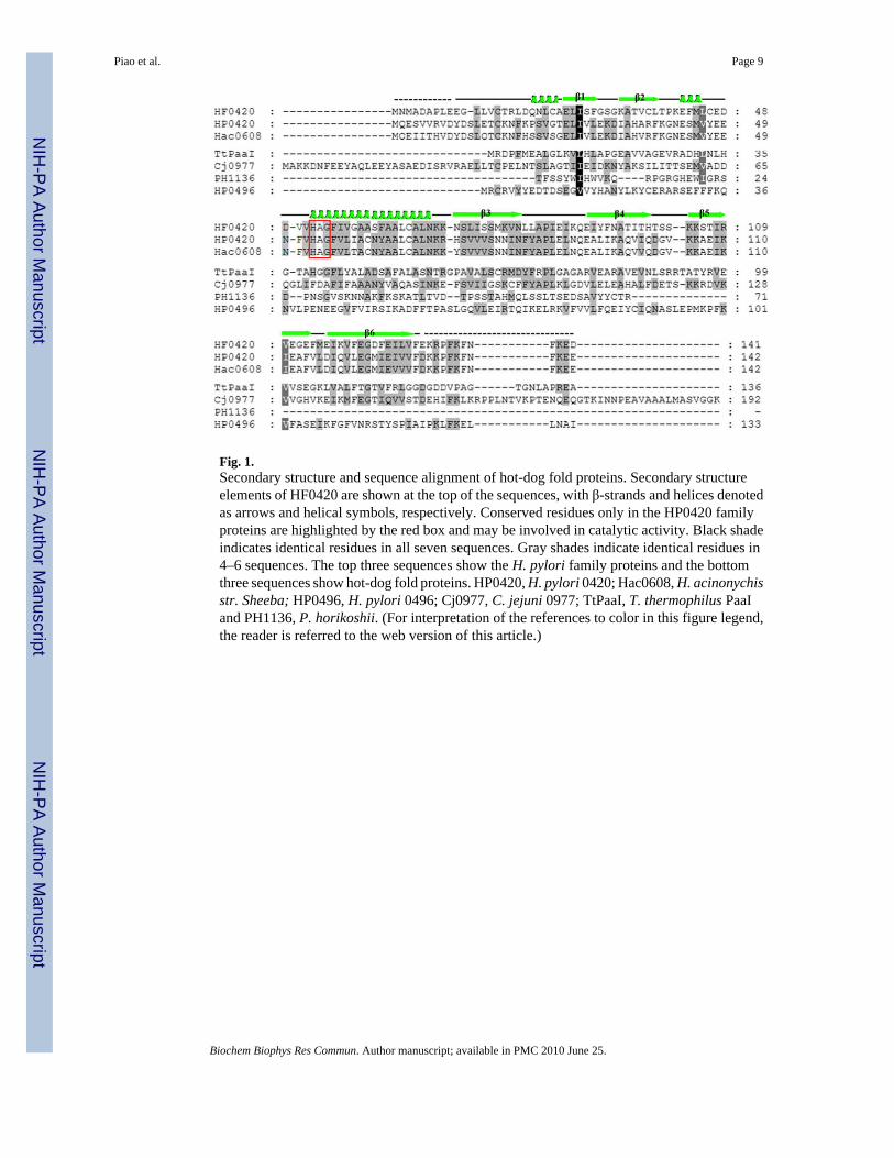
Fig. 1.Secondary structure and sequence alignment of hot-dog fold proteins. Secondary structureelements of HF0420 are shown at the top of the sequences, with β-strands and helices denotedas arrows and helical symbols, respectively. Conserved residues only in the HP0420 familyproteins are highlighted by the red box and may be involved in catalytic activity. Black shadeindicates identical residues in all seven sequences. Gray shades indicate identical residues in4–6 sequences. The top three sequences show the H. pylori family proteins and the bottomthree sequences show hot-dog fold proteins. HP0420, H. pylori 0420; Hac0608, H. acinonychisstr. Sheeba; HP0496, H. pylori 0496; Cj0977, C. jejuni 0977; TtPaaI, T. thermophilus PaaIand PH1136, P. horikoshii. (For interpretation of the references to color in this figure legend,the reader is referred to the web version of this article.)
Piao et al. Page 9
Biochem Biophys Res Commun. Author manuscript; available in PMC 2010 June 25.
NIH
-PA Author Manuscript
NIH
-PA Author Manuscript
NIH
-PA Author Manuscript
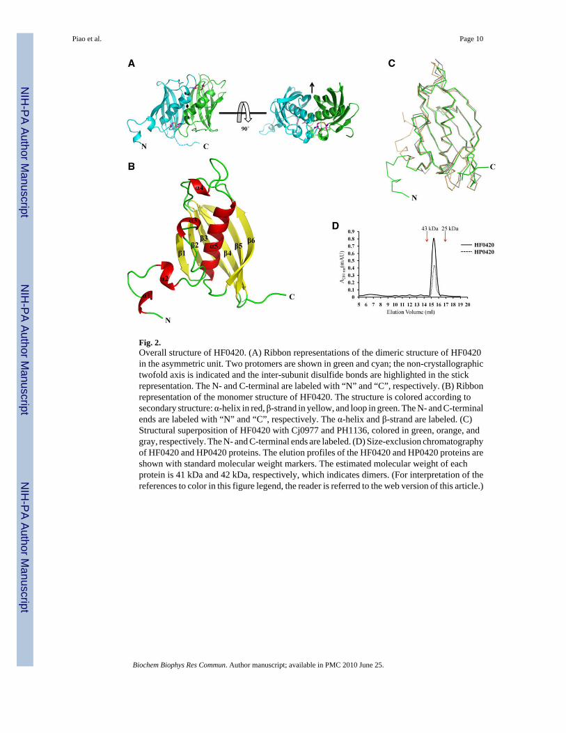
Fig. 2.Overall structure of HF0420. (A) Ribbon representations of the dimeric structure of HF0420in the asymmetric unit. Two protomers are shown in green and cyan; the non-crystallographictwofold axis is indicated and the inter-subunit disulfide bonds are highlighted in the stickrepresentation. The N- and C-terminal are labeled with “N” and “C”, respectively. (B) Ribbonrepresentation of the monomer structure of HF0420. The structure is colored according tosecondary structure: α-helix in red, β-strand in yellow, and loop in green. The N- and C-terminalends are labeled with “N” and “C”, respectively. The α-helix and β-strand are labeled. (C)Structural superposition of HF0420 with Cj0977 and PH1136, colored in green, orange, andgray, respectively. The N- and C-terminal ends are labeled. (D) Size-exclusion chromatographyof HF0420 and HP0420 proteins. The elution profiles of the HF0420 and HP0420 proteins areshown with standard molecular weight markers. The estimated molecular weight of eachprotein is 41 kDa and 42 kDa, respectively, which indicates dimers. (For interpretation of thereferences to color in this figure legend, the reader is referred to the web version of this article.)
Piao et al. Page 10
Biochem Biophys Res Commun. Author manuscript; available in PMC 2010 June 25.
NIH
-PA Author Manuscript
NIH
-PA Author Manuscript
NIH
-PA Author Manuscript
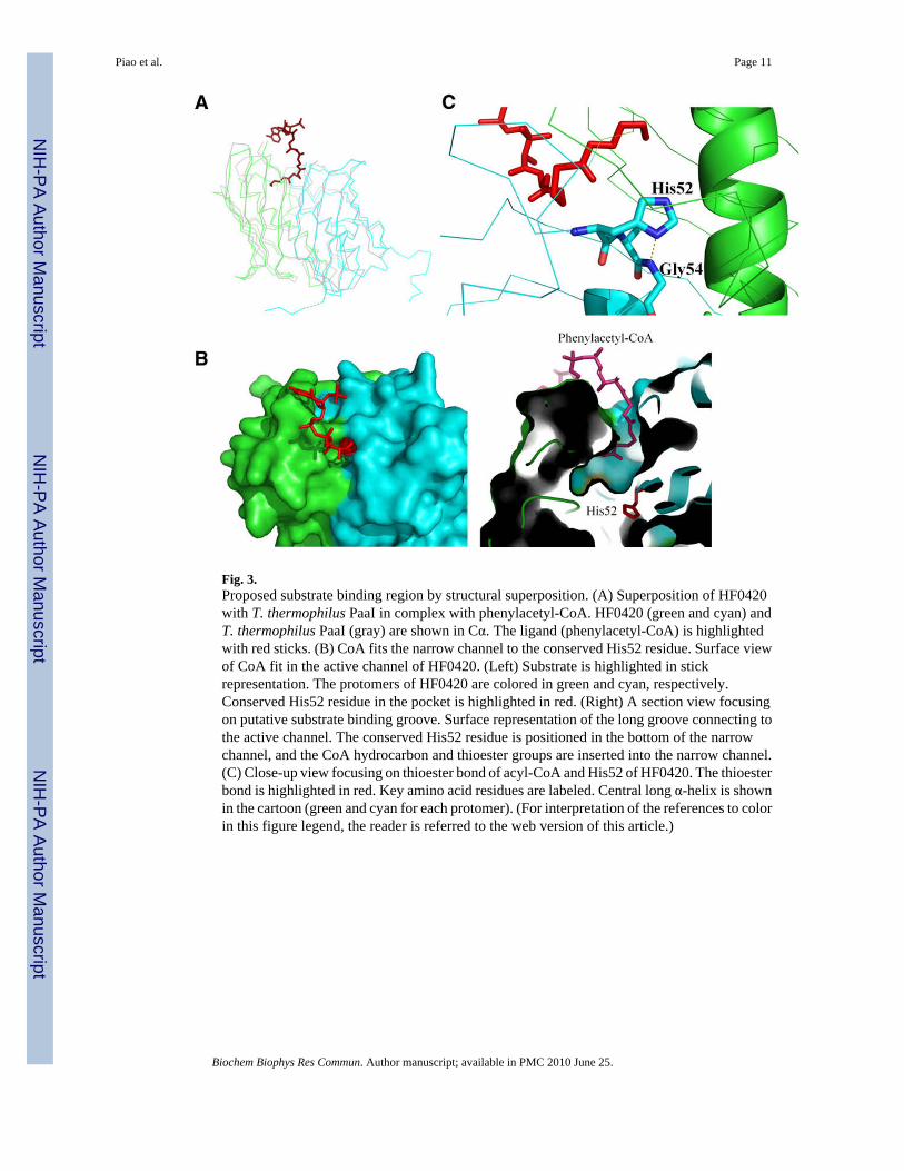
Fig. 3.Proposed substrate binding region by structural superposition. (A) Superposition of HF0420with T. thermophilus PaaI in complex with phenylacetyl-CoA. HF0420 (green and cyan) andT. thermophilus PaaI (gray) are shown in Cα. The ligand (phenylacetyl-CoA) is highlightedwith red sticks. (B) CoA fits the narrow channel to the conserved His52 residue. Surface viewof CoA fit in the active channel of HF0420. (Left) Substrate is highlighted in stickrepresentation. The protomers of HF0420 are colored in green and cyan, respectively.Conserved His52 residue in the pocket is highlighted in red. (Right) A section view focusingon putative substrate binding groove. Surface representation of the long groove connecting tothe active channel. The conserved His52 residue is positioned in the bottom of the narrowchannel, and the CoA hydrocarbon and thioester groups are inserted into the narrow channel.(C) Close-up view focusing on thioester bond of acyl-CoA and His52 of HF0420. The thioesterbond is highlighted in red. Key amino acid residues are labeled. Central long α-helix is shownin the cartoon (green and cyan for each protomer). (For interpretation of the references to colorin this figure legend, the reader is referred to the web version of this article.)
Piao et al. Page 11
Biochem Biophys Res Commun. Author manuscript; available in PMC 2010 June 25.
NIH
-PA Author Manuscript
NIH
-PA Author Manuscript
NIH
-PA Author Manuscript
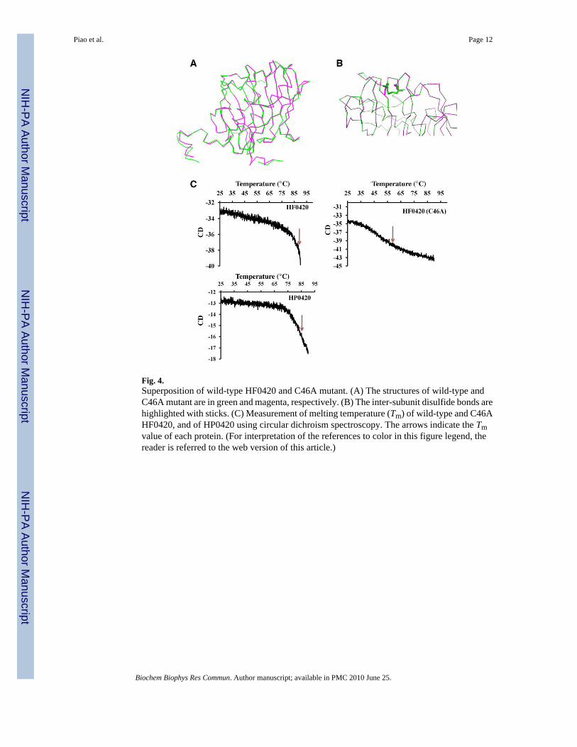
Fig. 4.Superposition of wild-type HF0420 and C46A mutant. (A) The structures of wild-type andC46A mutant are in green and magenta, respectively. (B) The inter-subunit disulfide bonds arehighlighted with sticks. (C) Measurement of melting temperature (Tm) of wild-type and C46AHF0420, and of HP0420 using circular dichroism spectroscopy. The arrows indicate the Tmvalue of each protein. (For interpretation of the references to color in this figure legend, thereader is referred to the web version of this article.)
Piao et al. Page 12
Biochem Biophys Res Commun. Author manuscript; available in PMC 2010 June 25.
NIH
-PA Author Manuscript
NIH
-PA Author Manuscript
NIH
-PA Author Manuscript
NIH
-PA Author Manuscript
NIH
-PA Author Manuscript
NIH
-PA Author Manuscript
Piao et al. Page 13
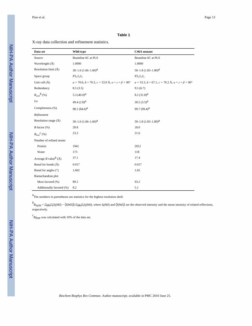
Table 1
X-ray data collection and refinement statistics.
Data set Wild-type C46A mutant
Source Beamline 6C at PLS Beamline 6C at PLS
Wavelength (Å) 1.0000 1.0000
Resolution limit (Å) 30–1.6 (1.66–1.60)a 50–1.8 (1.83–1.80)a
Space group P212121 P212121
Unit cell (Å) a = 70.6, b = 70.3, c = 53.9 Å, α = γ = β = 90° a = 53.3, b = 67.1, c = 70.2 Å, α = γ = β = 90°
Redundancy 8.5 (3.5) 9.5 (6.7)
Rsymb (%) 5.3 (40.0)a 8.2 (31.8)a
I/σ 49.4 (2.8)a 30.5 (3.5)a
Completeness (%) 98.1 (84.6)a 99.7 (99.4)a
Refinement
Resolution range (Å) 30–1.6 (1.66–1.60)a 50–1.8 (1.83–1.80)a
R-factor (%) 20.8 18.0
Rfreec (%) 23.3 21.6
Number of refined atoms
Protein 1941 2012
Water 173 118
Average B valueb (Å) 37.1 17.4
Rmsd for bonds (Å) 0.017 0.017
Rmsd for angles (°) 1.602 1.65
Ramachandran plot
Most favored (%) 89.2 93.2
Additionally favored (%) 8.2 5.1
aThe numbers in parentheses are statistics for the highest resolution shell.
bRsym = ΣhklΣi|Ii(hkl) − ⟨I(hkl)⟩|/ΣhklΣiIi(hkl), where Ii(hkl) and ⟨I(hkl)⟩ are the observed intensity and the mean intensity of related reflections,
respectively.
cRfree was calculated with 10% of the data set.
Biochem Biophys Res Commun. Author manuscript; available in PMC 2010 June 25.