cross talk between devs sensor kinase homologue, rv2027c, and devr response regulator of...
TRANSCRIPT
FEBS 28322 FEBS Letters 565 (2004) 75–80
Cross talk between DevS sens
or kinase homologue, Rv2027c,and DevR response regulator of Mycobacterium tuberculosisDeepak Kumar Saini, Vandana Malhotra, Jaya Sivaswami Tyagi*
Department of Biotechnology, All India Institute of Medical Sciences, Ansari Nagar, New Delhi 110029, India
Received 24 December 2003; revised 13 February 2004; accepted 17 February 2004
First published online 8 April 2004
Edited by Gianni Cesareni
Abstract Rv2027c is a putative orphan histidine sensor kinasethat bears strong homology to DevS of the hypoxia-responsiveDevR–DevS two-component system in M. tuberculosis. Thecytosolic C-terminal domain of Rv2027c protein (Rv2027c194)was overexpressed in E. coli and biochemically characterized.Rv2027c194 underwent autophosphorylation at a conserved His392
residue and engaged in phosphotransfer with DevR responseregulator. The rates of autophosphorylation and the stabilities ofthe phosphorylated species were broadly similar in Rv2027c andDevS. However, unlike DevS, Rv2027c utilized Ca2þ as analternative divalent ion during autophosphorylation. In contrastto DevS which completed phosphotransfer to DevR in 5–10 min,phosphotransfer from Rv2027c�P was only partial at 30 min.Unlike devS transcription that was hypoxia-responsive, Rv2027ctranscript levels were not upregulated from basal levels duringhypoxia. The differential regulation of devS and Rv2027c genes,the ability of Rv2027c to utilize Ca2þ as a divalent cation inautophosphorylation at physiological concentrations and toengage in phosphotransfer with DevR suggests that the DevRregulon could be modulated by more than one environmental cuerelayed through DevS and Rv2027c.� 2004 Federation of European Biochemical Societies. Publishedby Elsevier B.V. All rights reserved.
Keywords: Two-component system; DevS and Rv2027c sensorkinase; DevR response regulator; Cross talk; Calcium;M. tuberculosis
1. Introduction
Two-component systems of bacteria play a major role in
their adaptation to the environment. In its most basic form,
the two-component system consists of a histidine sensor kinase
and a response regulator [1]. These systems use transient
phosphorylation of the sensory and regulatory proteins at
conserved residues for transducing extracellular or intracellu-
lar signals, thereby leading to an appropriate cellular response
[2]. Such phosphorylation-based signalling has been estab-
lished for five of eleven systems in M. tuberculosis, RegX3–
* Corresponding author. Fax: +91-11-2658-8663.E-mail address: [email protected] (J.S. Tyagi).
Abbreviations: cGMP, cyclic GMP; GAF, cyclic GMP (cGMP) –regulated cyclic nucleotide phosphodiesterases, adenyl cyclases andbacterial transcriptional regulator FhlA; GFP, green fluorescentprotein; USP, Universal stress protein
0014-5793/$22.00 � 2004 Federation of European Biochemical Societies. Pu
doi:10.1016/j.febslet.2004.02.092
SenX3 [3], TrcR–TrcS [4], MprA–MprB [5], PrrA–PrrB [6] and
DevR–DevS (Rv3133c–Rv3132c/DosR–DosS) [7]. The genes
encoding two-component systems are generally located adja-
cent to each other and are also coexpressed. The presence of
seven ‘orphan’ genes coding for histidine kinases or response
regulators but without the genes coding for the other compo-
nent of proteins of two-component systems adjacent to them
was an intriguing finding derived from determination of the
genome sequence of M. tuberculosis H37Rv [8]. For instance,
the gene encoding a potential sensor kinase, Rv2027c, is not
adjacent to a gene encoding a response regulator. Since
Rv2027c bears strong homology with DevS [8], there exists a
possibility that diverse signals sensed by DevS and Rv2027c
could generate a similar genetic response by signalling to a
common response regulator protein, DevR. In this paper, we
report for the first time the likely function of the orphan his-
tidine kinase, Rv2027c. We have established Rv2027c to be a
bonafide sensor kinase that undergoes autophosphorylation at
a conserved histidine residue and communicates with DevR
through a phosphotransfer reaction. Its unique metal ion
requirement and physiological relevance are discussed.
2. Materials and methods
2.1. Bacterial strains, media, growth conditions and plasmidsEscherichia coli DH5a (Invitrogen Inc., USA) and XL1Blue (Strat-
agene Inc., USA) were used for routine cloning and E. coli BL23 (DE3)(gift from Dr. S. Vijaya, Indian Institute of Science, Bangalore) forprotein production. Antibiotics, when required, were used at the fol-lowing concentrations: ampicillin at 100 lg/ml, hygromycin at 200 lg/ml in E. coli and 25 lg/ml in M. smegmatis. E. coli was grown in LBmedia, while M. tuberculosis H37Rv (kindly provided by Dr. R.F.Silver, Case Western Reserve University, Cleveland, USA) and M.smegmatis LR222 (gift from Dr. J. Crawford, CDC, Atlanta, USA)were cultured in Dubos–Tween Albumin broth (DTA). All routinerecombinant DNA work was performed as described [9]. The cytosolicdomain-coding portion of Rv2027c194 gene (amino acids 380–573) ofM. tuberculosis H37Rv was amplified using gene-specific primers,2027bamf (5
0GCGAGAAGTGGAGGATCCTGACC 3
0) and
2027bamr (50GGATTGCGCGGATCCGTCGAC GCC 3
0, engi-
neered BamHI restriction site is underlined), and inserted in pPROEx-HTc expression vector as described [7] to generate plasmid pDSH194.The reporter vector pFPVH was generated by inserting a hygromycinresistance cassette from pGEMT-Hyg (obtained as a gift from Dr. A.Ranganathan, ICGEB, New Delhi) in the green fluorescent protein(GFP) reporter vector, pFPV27 at the NsiI site [10].
2.2. Overexpression of recombinant proteinRv2027c194 protein was overexpressed, purified and refolded using
an established matrix-assisted protein renaturation protocol asdescribed [11].
blished by Elsevier B.V. All rights reserved.
76 D.K. Saini et al. / FEBS Letters 565 (2004) 75–80
2.3. Site-directed mutagenesisH392Q point mutation was introduced into pDSH194 using the
QuikChangeTM site-directed mutagenesis kit (Stratagene Inc., USA)and the mutagenic oligonucleotides H392Qf (5
0GCACGTGATCTG-
CAAGACCACGTCATCCAG 30) and H392Qr (5
0CTGGATGAC-
GTGGTCTTGCAGATCACGTGC 30). The mutagenesis was verified
by automated DNA sequencing using 377 ABI Prism DNA sequencer.
2.4. In vitro phosphorylation assaysRv2027c194 (15 lM) was autophosphorylated by incubating it with 5
lCi of c[32P] ATP (5000 Ci/mmol, BRIT, India) in 10 ll reaction buffer(50 mM Tris �Cl, pH 8.0, 50 mM KCl, 25 mM MgCl2, and 50 lMATP) at 25�C for 0–24 h. Standard reactions were performed as abovefor 60 min. For stability analysis, radiolabelled Rv2027c194�P wasprepared in a 60 min reaction and incubated for up to 20 h after re-moval of unincorporated ATP and MgCl2 by filtration of the reactionmixture through a 10 kDa Nanosep device. To study phosphotransfer,Rv2027c194 (15 lM) was autophosphorylated for 60 min, then DevR(20 lM) was added to the reaction mixture and incubated at25 �C for the indicated time periods. In all phosphorylation assays,samples were analyzed by SDS–PAGE and the gels were autoradio-graphed.
2.5. RT-PCRMycobacterium tuberculosis H37Rv was grown in DTA broth (100
ml in a 500 ml Erlenmeyer flask) as aerobic culture to late logarithmicphase with shaking at 37 �C (OD600 � 0.7–0.8), after which 13 mlculture aliquots were aseptically transferred to 15 ml polystyreneconical tubes (Corning Inc., USA), sealed and kept without stirring for48 h (hypoxic cultures). Methylene blue fading occurred in controltubes at 48 h indicating the setting in of hypoxia. RNA was isolatedfrom these aerobic and hypoxic cultures and used for RT-PCR analysisas described [7]. The expression of Rv2027c, hspX and devS was ana-lyzed using gene-specific primers after normalizing for 23S rRNAtranscripts. The following primers were used: Rv2027c, 2027f (5
0
CACCCTGACAGGGCGAACGTT 30) and Rv2027r (5
0GGTCGTA-
ACCGCGCACCCCG 30); hspX, hspXf (5
0CGCCACCCGCGGTC-
CCTCTT 30) and hspXr (5
0GCTCGGTGCGCTCGGCCTTG 3
0) and
devS, devS1 (50ACTACGCC TGCACGAGCTGCT 3
0) and N3 (5
0
GTCCAGCCGTAACGGTTTGGG 30). At least two lots of RNA
were analyzed twice each by RT-PCR.
2.6. GFP assaysThe Rv2027c and hspX promoter regions were amplified using
appropriate primers as below: Rv2027c, 2027Pf (50TCAATCGGTA-
CACATGGTGGTGCTCA 30) and 2027Pr (5
0TGCGCTCAAC-
CAACTGGTCTGGCTA 30); hspX, hspXPf (5
0TCTGAACGGCGG-
TTGGCAGACA 30) and hspXPr (5
0CGGGAAGGGTGGTGGC-
CATTTG 30). The amplified promoter fragments were inserted in the
promoterless GFP reporter vector, pFPVH, at the EcoRV site. M.smegmatis cultures carrying the recombinant GFP constructs weregrown in DTA containing hygromycin till early log phase, subcultured(using a starting OD600 of 0.05) and grown under aerobic conditionsand hypoxic conditions as described above. At 48 h, �105 cells fromboth aerated (OD590 1.2–1.5) and hypoxic cultures (OD590 0.12–0.16)were analyzed for fluorescence in a multiwell spectrofluorimeter(Molecular Devices Inc., USA) using an excitation wavelength of 483nm and an emission wavelength of 515 nm.
3. Results
3.1. Computational analysis of Rv2027c
Mycobacterium tuberculosis H37Rv genome analysis
revealed the existence of a putative sensor histidine kinase of 573
amino acids, Rv2027c, without its accompanying response reg-
ulator that displayed 62.5% identity with DevS sensor
kinase [12] over its entire length. Two GAF domains (present in
cyclic GMP (cGMP) – regulated cyclic nucleotide phosphodi-
esterases, adenyl cyclases and bacterial transcriptional regulator
FhlA and formerly thought to bind only cyclic GMP and now
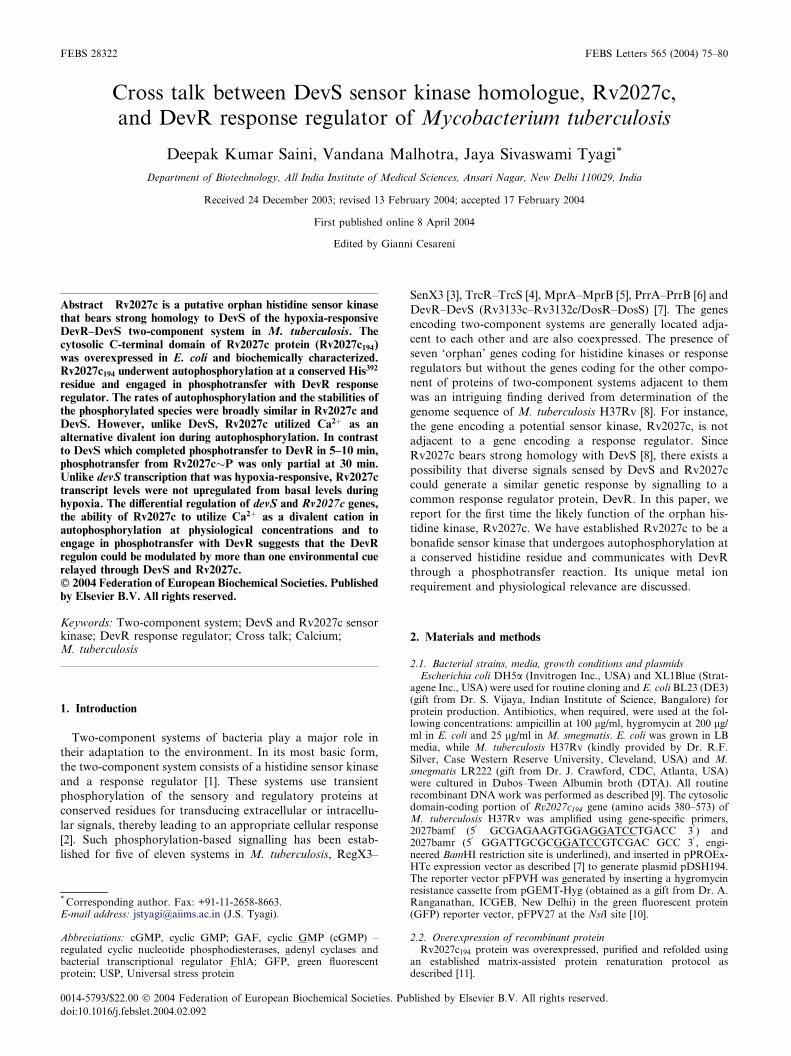
found to bind cyclicAMPaswell [13])mapped in theN-terminal
domain of Rv2027c protein at a similar location as in DevS
(Fig. 1A). These domains are ubiquitous modules present in a
large number of signalling and sensory proteins and are reported
to bind smallmolecule ligands such as cGMPandmodulate their
activity [13]. The presence of similarly located and organized
GAF domains inRv2027c andDevS proteins suggests that both
the proteins are involved in processing similar/related signals
and/or may be involved in the execution of similar outputs. The
presence of only one transmembrane region (TM1) in the
Rv2027c protein was deduced by TMpred analysis (online
transmembrane domain-predicting software available at http://
us.expasy.org). The C-terminal region sequences that comprise
the catalytically active kinase core were also highly conserved in
Rv2027c andDevS, particularly the amino acid sequences of the
H box (containing the phosphorylatable histidine residue) and
the N,D/G1 andG2 boxes involved in ATP binding [1]. BLAST
analysis of the available mycobacterial genome databases indi-
cated Rv2027c to be absent from all mycobacteria except
M. tuberculosis andM. bovis (Table 1). The presence ofRv2027c
orthologue in M. bovis BCG (Pasteur strain ATCC 35734) was
established by PCR using M. tuberculosis gene-specific primers
(not shown). The possession of an additional DevS-like sensor
kinase inM. tuberculosis complex but not in other mycobacteria
sequenced so far is striking and may be indicative of a unique
role for it in these pathogens.
3.2. Genetic organization of Rv2027c
The genomic context of Rv2027c gene was very similar to
that of DevS except that a devR homologue was absent. This
suggested that this locus could have arisen through a partial
gene duplication event from the Rv3134c–devR–devS locus
accompanied by the loss of devR, hence its ‘orphan’ status
(Fig. 1B). Rv2027c is flanked on either side by Rv2026c and
Rv2028c, both of which are homologous to Rv3134c and dis-
play 30% and 40% identity, respectively, with the latter. All
three genes code for proteins containing the Universal stress
protein (USP) domain first identified in the Usp protein that is
induced under a variety of stress conditions and is proposed to
play a protective role in E. coli [14]. Proteins of this family are
induced during stress responses in several other bacteria as well
including mycobacteria [15]. There are numerous examples of
gene redundancy such as this in M. tuberculosis [8] and could
reflect a strategy used by the tubercle bacillus to keep ‘back-up’
copies of genes functioning in critical bacterial pathways. A
second similarity between the two loci is their hypoxia-
responsive character; the devR–devS genes map in a 12-gene
hypoxia-responsive cluster [16], Bagchi, G. unpublished
observations, while Rv2027c maps near a 5-gene hypoxia-
responsive cluster that includes hspX (acr) but was itself not
induced in hypoxia in M. tuberculosis [16].
A devS (dosS) knockout strain of M. bovis BCG was re-
ported to be only moderately affected in hypoxic survival
compared to a devR (dosR) mutant that showed a severe
defect in hypoxic viability [17]. Similarly, a devR but not a
devS mutant of M. tuberculosis was abrogated in the hypoxic
expression of Acr [16]. Based on the significant similarity
between DevS and Rv2027c and the phenotype of devS
mutants, it was speculated that Rv2027c could participate in
phosphosignalling [12,16]. Therefore, the possibility of
Rv2027c to function as a sensor kinase and communicate
with DevR was investigated in this study.
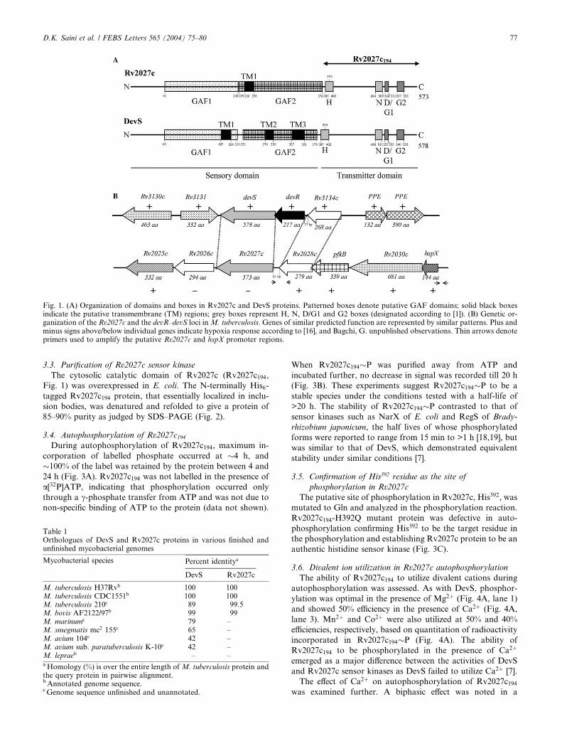
Fig. 1. (A) Organization of domains and boxes in Rv2027c and DevS proteins. Patterned boxes denote putative GAF domains; solid black boxesindicate the putative transmembrane (TM) regions; grey boxes represent H, N, D/G1 and G2 boxes (designated according to [1]). (B) Genetic or-ganization of the Rv2027c and the devR–devS loci inM. tuberculosis. Genes of similar predicted function are represented by similar patterns. Plus andminus signs above/below individual genes indicate hypoxia response according to [16], and Bagchi, G. unpublished observations. Thin arrows denoteprimers used to amplify the putative Rv2027c and hspX promoter regions.
D.K. Saini et al. / FEBS Letters 565 (2004) 75–80 77
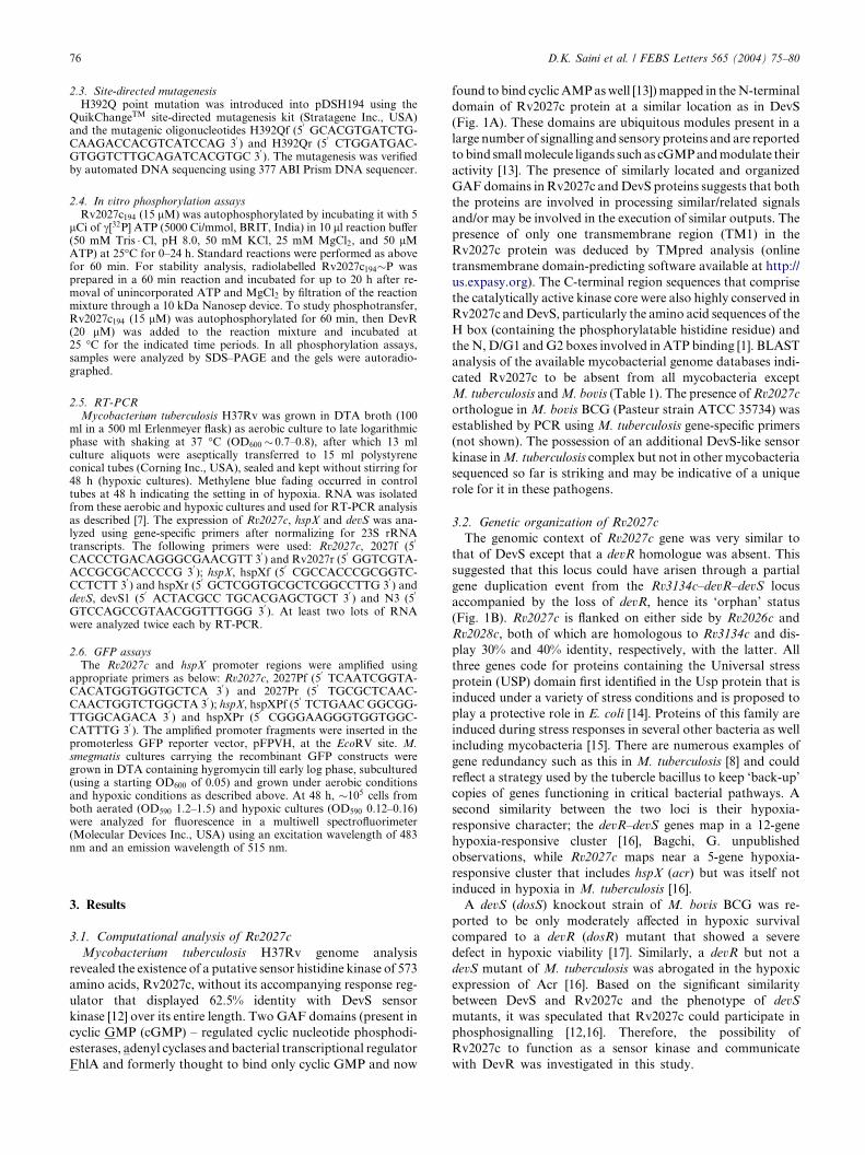
3.3. Purification of Rv2027c sensor kinase
The cytosolic catalytic domain of Rv2027c (Rv2027c194,
Fig. 1) was overexpressed in E. coli. The N-terminally His6-
tagged Rv2027c194 protein, that essentially localized in inclu-
sion bodies, was denatured and refolded to give a protein of
85–90% purity as judged by SDS–PAGE (Fig. 2).
3.4. Autophosphorylation of Rv2027c194During autophosphorylation of Rv2027c194, maximum in-
corporation of labelled phosphate occurred at �4 h, and
�100% of the label was retained by the protein between 4 and
24 h (Fig. 3A). Rv2027c194 was not labelled in the presence of
a[32P]ATP, indicating that phosphorylation occurred only
through a c-phosphate transfer from ATP and was not due to
non-specific binding of ATP to the protein (data not shown).
Table 1Orthologues of DevS and Rv2027c proteins in various finished andunfinished mycobacterial genomes
Mycobacterial species Percent identitya
DevS Rv2027c
M. tuberculosis H37Rvb 100 100M. tuberculosis CDC1551b 100 100M. tuberculosis 210c 89 99.5M. bovis AF2122/97b 99 99M. marinumc 79 –M. smegmatis mc2 155c 65 –M. avium 104c 42 –M. avium sub. paratuberculosis K-10c 42 –M. lepraeb – –aHomology (%) is over the entire length of M. tuberculosis protein andthe query protein in pairwise alignment.bAnnotated genome sequence.cGenome sequence unfinished and unannotated.
When Rv2027c194�P was purified away from ATP and
incubated further, no decrease in signal was recorded till 20 h
(Fig. 3B). These experiments suggest Rv2027c194�P to be a
stable species under the conditions tested with a half-life of
>20 h. The stability of Rv2027c194�P contrasted to that of
sensor kinases such as NarX of E. coli and RegS of Brady-
rhizobium japonicum, the half lives of whose phosphorylated
forms were reported to range from 15 min to >1 h [18,19], but
was similar to that of DevS, which demonstrated equivalent
stability under similar conditions [7].
3.5. Confirmation of His392 residue as the site of
phosphorylation in Rv2027c
The putative site of phosphorylation in Rv2027c, His392, was
mutated to Gln and analyzed in the phosphorylation reaction.
Rv2027c194-H392Q mutant protein was defective in auto-
phosphorylation confirming His392 to be the target residue in
the phosphorylation and establishing Rv2027c protein to be an
authentic histidine sensor kinase (Fig. 3C).
3.6. Divalent ion utilization in Rv2027c autophosphorylation
The ability of Rv2027c194 to utilize divalent cations during
autophosphorylation was assessed. As with DevS, phosphor-
ylation was optimal in the presence of Mg2þ (Fig. 4A, lane 1)
and showed 50% efficiency in the presence of Ca2þ (Fig. 4A,
lane 3). Mn2þ and Co2þ were also utilized at 50% and 40%
efficiencies, respectively, based on quantitation of radioactivity
incorporated in Rv2027c194�P (Fig. 4A). The ability of
Rv2027c194 to be phosphorylated in the presence of Ca2þ
emerged as a major difference between the activities of DevS
and Rv2027c sensor kinases as DevS failed to utilize Ca2þ [7].
The effect of Ca2þ on autophosphorylation of Rv2027c194was examined further. A biphasic effect was noted in a
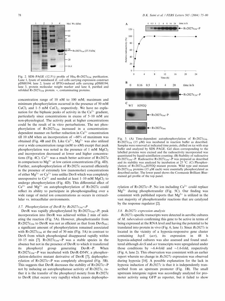
Fig. 3. (A) Time-dependent autophosphorylation of Rv2027c194.Rv2027c194 (15 lM) was incubated in reaction buffer as described.Samples were removed at indicated time points, chilled on ice with stopbuffer and analyzed by SDS–PAGE. Gel slices corresponding to thelabelled proteins were excised and the radioactivity incorporated wasquantitated by liquid-scintillation counting. (B) Stability of radioactiveRv2027c194�P. Radioactive Rv2027c194�P was prepared as describedand its stability was analyzed by incubation at 25 �C. (C) Phosphor-ylation of Rv2027c194H392Q mutant protein. Wild type and mutantRv2027c194 proteins (15 lM each) were essentially phosphorylated asdescribed earlier. The lower panel shows the Coomassie Brilliant Blue-stained gel profile of the top panel.
Fig. 2. SDS–PAGE (12.5%) profile of His6-Rv2027c194 purification.Lane 1, lysate of uninduced E. coli cells carrying expression constructpDSH194; lane 2, lysate of IPTG-induced cells carrying pDSH194;lane 3, protein molecular weight marker and lane 4, purified andrefolded Rv2027c194 protein. �, contaminating proteins.
78 D.K. Saini et al. / FEBS Letters 565 (2004) 75–80
concentration range of 10 nM to 100 mM; maximum and
minimum phosphorylation occurred in the presence of 50 mM
CaCl2 and 1–5 mM CaCl2, respectively. We have no expla-
nation for the biphasic peaks of activity in the Ca2þ gradient,
particularly since concentrations in excess of 5–10 mM are
non-physiological. The activity peak at higher concentrations
could be the result of in vitro perturbations. The net phos-
phorylation of Rv2027c194 increased in a concentration-
dependent manner on further reduction in Ca2þ concentration
till 10 nM when an incorporation of �60% of maximum was
obtained (Fig. 4B and D). Like Ca2þ, Mg2þ was also utilized
over a wide concentration range (mM to nM) except that peak
phosphorylation was noted in the presence of 1 mM MgCl2and incorporation decreased at lower and higher concentra-
tions (Fig. 4C). Ca2þ was a much better activator of Rv2027c
in comparison to Mg2þ at low cation concentrations (Fig. 4D).
Further, autophosphorylation of Rv2027c occurred efficiently
in the presence of extremely low (nanomolar) concentrations
of either Mg2þ or Ca2þ ions unlike DevS which was completely
unresponsive to Ca2þ and needed at least 1–10 mM MgCl2 to
undergo phosphorylation (Fig. 4D). This differential effect of
Ca2þ and Mg2þ on autophosphorylation of Rv2027c could
reflect its ability to participate in phosphosignalling over a
wide range of metal ion concentrations as occurs in extracel-
lular vs. intracellular environments.
3.7. Phosphorylation of DevR by Rv2027c194�P
DevR was rapidly phosphorylated by Rv2027c194�P; peak
incorporation into DevR was achieved within 2 min of initi-
ating the reaction (Fig. 5A). However, phosphotransfer from
Rv2027c194 to DevR was not as efficient as that from DevS201;
a significant amount of phosphorylation remained associated
with Rv2027c194 at the end of 30 min (Fig. 5A) in contrast to
DevS from which phosphosignal disappeared rapidly within
10–15 min [7]. Rv2027c194�P was a stable species in the
absence but not in the presence of DevR to which it transferred
its phosphoryl group generating DevR�P. When
Rv2027c194�P was incubated with DevR-D54V, a phosphor-
ylation-defective mutant derivative of DevR [7], dephospho-
rylation of Rv2027c�P was completely abrogated (Fig. 5B).
This suggests that DevR decreases the stability of Rv2027c�P
not by inducing an autophosphatase activity of Rv2027c, ra-
ther it is the transfer of the phosphoryl moiety from Rv2027c
to DevR (that occurs very rapidly) which causes dephospho-
rylation of Rv2027c�P. No ion including Ca2þ could replace
Mg2þ during phosphotransfer (Fig. 5C). Our finding was
consistent with published reports that Mg2þ is utilized in the
vast majority of phosphotransfer reactions that are catalyzed
by the response regulator [2].
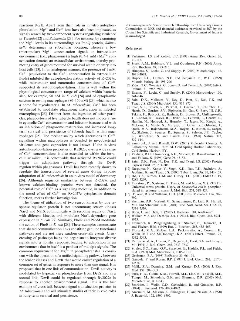
3.8. Rv2027c expression analysis
Rv2027c-specific transcripts were detected in aerobic cultures
of M. tuberculosis confirming this gene to be active in terms of
being expressed at the RNA level and having the potential to be
translated into protein in vivo (Fig. 6, lane 1). Since Rv2027c is
located in the vicinity of a hypoxia-responsive gene cluster
containing hspX (acr), its expression in 48 h
hypoxia-adapted cultures was also assessed and found unal-
tered although devS and acr transcripts were upregulated under
those conditions by �6.4-fold and �1.6-fold, respectively
(Fig. 6, lane 2). This observation was consistent with an earlier
report wherein no change in Rv2027c expression was observed
during hypoxia [16]. A possible explanation for the lack in
hypoxic induction of Rv2027c is that it is independently tran-
scribed from an upstream promoter (Fig. 1B). The small
upstream intergenic region was accordingly analyzed for pro-
moter activity using GFP as reporter, but it failed to show
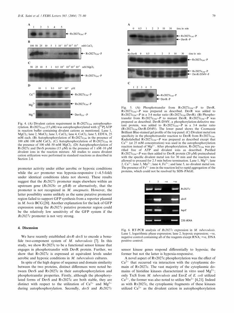
Fig. 5. (A) Phosphotransfer from Rv2027c194�P to DevR.Rv2027c194�P was prepared as described. DevR was added toRv2027c194�P in a 3:4 molar ratio (Rv2027c194:DevR). (B) Phospho-transfer from Rv2027c194�P to mutant DevR. Rv2027c194�P wasprepared as described. DevR-D54V, a phosphorylation-defective mu-tant protein, was added to Rv2027c194�P in a 3:4 molar ratio(Rv2027c194:DevR-D54V). The lower panel shows the CoomassieBrilliant Blue-stained gel profile of the top panel. (C) Divalent metal ionspecificity in the phosphotransfer reaction to DevR from Rv2027c194.Radiolabelled Rv2027c194�P was prepared as described except thatCa2þ (at 25 mM concentration) was used in the autophosphorylationreaction instead of Mg2þ. After phosphorylation, Rv2027c194 was pu-rified free of ATP and divalent ions as described. PurifiedRv2027c194�P was then added to DevR protein (20 lM) preincubatedwith the specific divalent metal ion for 30 min and the reaction wasallowed to proceed for 2.5 min before termination. Lane 1, Mg2þ; lane2, Ca2þ; lane 3, Mn2þ; lane 4, Fe3þ; and lane 5, no divalent metal ion.The presence of Fe3þ ions in the reaction led to rapid aggregation of theproteins, which could not be resolved by SDS–PAGE.
Fig. 4. (A) Divalent cation requirement in Rv2027c194 autophospho-rylation. Rv2027c194 (15 lM) was autophosphorylated with c[32P] ATPin reaction buffer containing divalent cations as mentioned. Lane 1,MgCl2; lane 2, MnCl2; lane 3, CaCl2; lane 4, CoCl2; lane 5, EDTA, 25mM each. (B) Autophosphorylation of Rv2027c194 in the presence of100 nM–100 mM CaCl2. (C) Autophosphorylation of Rv2027c194 inthe presence of 100 nM–50 mM MgCl2. (D) Autophosphorylation ofRv2027c and DevS proteins (15 lM) in the presence of 1 nM–10 lMdivalent ions in the reaction mixture. All studies to assess divalentcation utilization were performed in standard reactions as described inSection 2.4.
D.K. Saini et al. / FEBS Letters 565 (2004) 75–80 79
promoter activity under either aerobic or hypoxic conditions
while the acr promoter was hypoxia-responsive (�4.5-fold)
under identical conditions (data not shown). These results
suggest that the Rv2027c promoter maps elsewhere within an
upstream gene (Rv2028c or pfkB) or alternatively, that the
promoter is not recognized in M. smegmatis. However, the
latter possibility seems unlikely as the same putative promoter
region failed to support GFP synthesis from a reporter plasmid
inM. bovis BCG [20]. Another explanation for the lack of GFP
expression using the Rv2027c putative promoter region could
be the relatively low sensitivity of the GFP system if the
Rv2027c promoter is not very strong.
Fig. 6. RT-PCR analysis of Rv2027c expression in M. tuberculosis.Lane 1, logarithmic phase expression; lane 2, hypoxic expression; )ve,negative control containing all of the reagents except RNA; +ve, DNApositive control.
4. Discussion
We have recently established devR–devS to encode a bona-
fide two-component system of M. tuberculosis [7]. In this
study, we show Rv2027c to be a functional sensor kinase that
engages in phosphotransfer with DevR protein. Further, we
show that Rv2027c is expressed at equivalent levels under
aerobic and hypoxic conditions in M. tuberculosis cultures.
In spite of the high degree of sequence and domain similarity
between the two proteins, distinct differences were noted be-
tween DevS and Rv2027c in their autophosphorylation and
phosphotransfer properties. Firstly, although the phosphory-
lated forms of DevS and Rv2027c are both stable, they are
distinct with respect to the utilization of Ca2þ and Mg2þ
during autophosphorylation. Secondly, devS and Rv2027c
sensor kinase genes respond differentially to hypoxia; the
former but not the latter is hypoxia-responsive.
A novel aspect of Rv2027c phosphorylation was the effect of
Ca2þ that occurred via interaction with the cytoplasmic do-
main of Rv2027c. The vast majority of the cytoplasmic do-
mains of histidine kinases characterized in vitro used Mg2þ;only TrcS from M. tuberculosis and EnvZ of E. coli utilized
Ca2þ, the former was also noted to utilize Mn2þ [4,21]. Indeed
as with Rv2027c, the cytoplasmic fragments of these kinases
utilized Ca2þ as the divalent cation in autophosphorylation
80 D.K. Saini et al. / FEBS Letters 565 (2004) 75–80
reactions [4,21]. Apart from their role in in vitro autophos-
phorylation, Mg2þ and Ca2þ ions have also been implicated as
signals sensed by two-component systems regulating virulence
in Yersinia [22] and Salmonella [23]. For instance, by examining
the Mg2þ levels in its surroundings via PhoQ protein, Salmo-
nella determines its subcellular location; whereas a low
(micromolar) Mg2þ concentration signals an intracellular
environment (i.e., phagosome) a high (0.7–1 mM) Mg2þ con-
centration denotes an extracellular environment, thereby pro-
moting entry of genes required for survival within or entry into
host cells [23]. In an analogous manner, the presence of 1 mM
Ca2þ (equivalent to the Ca2þ concentration in extracellular
fluids) inhibited the autophosphorylation activity of Rv2027c,
while micromolar and nanomolar concentrations of Ca2þ
supported its autophosphorylation. This is well within the
physiological concentration range of calcium within bacteria
also, for example, 90 nM in E. coli [24] and of cytoplasmic
calcium in resting macrophages (80–150 nM) [23], which is also
a home for mycobacteria. In M. tuberculosis, Ca2þ has been
established to modulate phagosome maturation in infected
macrophages [25]. Distinct from the ingestion of other parti-
cles, phagocytosis of live tubercle bacilli does not induce a rise
in cytosolic Ca2þ concentration and infection is associated with
an inhibition of phagosome maturation and increased long-
term survival and persistence of tubercle bacilli within mac-
rophages [25]. The mechanism by which alterations in Ca2þ
signalling within macrophages is coupled to mycobacterial
virulence and gene expression is not known. If the in vitro
autophosphorylation properties of Rv2027c over a wide range
of Ca2þ concentrations is also extended to within the intra-
cellular milieu, it is conceivable that activated Rv2027c could
trigger an adaptation pathway through the DevR
regulon within phagosomes. DevR has been recently shown to
regulate the transcription of several genes during hypoxic
adaptation ofM. tuberculosis in an in vitro model of dormancy
[26]. Although sequence similarities between Rv2027c and
known calcium-binding proteins were not detected, the
potential role of Ca2þ as a signalling molecule, in addition to
the noted effect of Ca2þ on Rv2027c cytoplasmic domain
function, merits further investigation.
The theme of utilization of two sensor kinases by one re-
sponse regulator protein is not uncommon; sensor kinases
NarQ and NarX communicate with response regulator NarL
with different kinetics and modulate NarL-dependent gene
expression in E. coli [27]. Similarly, PhoR and PhoM modulate
the action of PhoB in E. coli [28]. These examples demonstrate
that shared communication links constitute genuine functional
pathways and are not mere random cross-talk events. Criss-
crossing of pathways helps the organism to integrate diverse
signals into a holistic response, leading to adaptation in an
environment that in itself is a product of multiple signals. The
common requirement for Mg2þ in phosphotransfer is consis-
tent with the operation of a unified signalling pathway between
the sensor kinases and DevR that would ensure regulation of a
common set of genes in response to more than one signal. It is
proposed that in one link of communication, DevR activity is
modulated by hypoxia via phosphorelay from DevS and in a
second link, DevR could be regulated through Rv2027c in
response to another environmental signal. This is the first
example of cross-talk between signal transduction proteins in
M. tuberculosis and will stimulate studies of their in vivo roles
in long-term survival and persistence.
Acknowledgements: Senior research fellowship from University GrantsCommission to DKS and financial assistance provided to JST by theCouncil for Scientific and Industrial Research, Government of India isacknowledged.
References
[1] Parkinson, J.S. and Kofoid, E.C. (1992) Annu. Rev. Genet. 26,71–112.
[2] Stock, A.M., Robinson, V.L. and Goudreau, P.N. (2000) Annu.Rev. Biochem. 69, 183–215.
[3] Himpens, S., Locht, C. and Supply, P. (2000) Microbiology 146,3091–3098.
[4] Haydel, S.E., Dunlap, N.E. and Benjamin Jr., W.H. (1999)Microb. Pathog. 26, 195–206.
[5] Zahrt, T.C., Wozniak, C., Jones, D. and Trevett, A. (2003) Infect.Immun. 71, 6962–6970.
[6] Ewann, F., Locht, C. and Supply, P. (2004) Microbiology 150,241–246.
[7] Saini, D.K., Malhotra, V., Dey, D., Pant, N., Das, T.K. andTyagi, J.S. (2004) Microbiol. 150, 865–875.
[8] Cole, S.T., Brosch, R., Parkhill, J., Garnier, T., Churcher, C.,Harris, D., Gordon, S.V., Eiglmeier, K., Gas, S., Barry III, C.E.,Tekaia, F., Badcock, K., Basham, D., Brown, D., Chillingworth,T., Connor, R., Davies, R., Devlin, K., Feltwell, T., Gentles, S.,Hamlin, N., Holroyd, S., Hornsby, T., Jagels, K., Krogh, A.,McLean, J., Moule, S., Murphy, L., Oliver, K., Osborne, J.,Quail, M.A., Rajandream, M.A., Rogers, J., Rutter, S., Seeger,K., Skelton, J., Squares, R., Squares, S., Sulston, J.E., Taylor,K., Whitehead, S. and Barrell, B.G. (1998) Nature 393,537–544.
[9] Sambrook, J. and Russell, D.W. (2001) Molecular Cloning: ALaboratory Manual, third ed. Cold Spring Harbor Laboratory,Cold Spring Harbor, NY.
[10] Valdivia, R.H., Hromockyj, A.E., Monack, D., Ramakrishnan, L.and Falkow, S. (1996) Gene 29, 45–52.
[11] Saini, D.K., Pant, N., Das, T.K. and Tyagi, J.S. (2002) ProteinExpress Purif. 25, 203–208.
[12] Dasgupta, N., Kapur, V., Singh, K.K., Das, T.K., Sachdeva, S.,Jyothisri, K. and Tyagi, J.S. (2000) Tuber. Lung Dis. 80, 141–159.
[13] Ho, Y.S., Burden, L.M. and Hurley, J.H. (2000) EMBO J. 19,5288–5299.
[14] Freestone, P., Nystr€om, T., Trinei, M. and Norris, V. (1997) TheUniversal stress protein, UspA, of Escherichia coli is phosphor-ylated in response to stasis. J. Mol. Biol. 274, 318–324.
[15] O’Toole, R. and Williams, H.D. (2003) Res. Microbiol. 154, 387–392.
[16] Sherman, D.R., Voskuil, M., Schnappinger, D., Liao, R., Harrell,M.I. and Schoolnik, G.K. (2001) Proc. Natl. Acad. Sci. USA 98,7534–7539.
[17] Boon, C. and Dick, T. (2002) J. Bacteriol. 184, 6760–6767.[18] Walker, M.S. and DeMoss, J.A. (1993) J. Biol. Chem. 268, 8931–
8933.[19] Emmerich, R., Panglungtshang, K., Strehler, P., Hennecke, H.
and Fischer, H.M. (1999) Eur. J. Biochem. 263, 455–463.[20] Florczyk, M.A., McCue, L.A., Purkayastha, A., Currenti, E.,
Wolin, M.J. and McDonough, K.A. (2003) Infect. Immun. 71,5332–5343.
[21] Rampersaud, A., Utsumi, R., Delgado, J., Forst, S.A. and Inouye,M. (1991) J. Biol. Chem. 266, 7633–7637.
[22] Straley, S.C., Plano, G.V., Skrzypek, E., Haddix, P.L. and Fields,K.A. (1993) Mol. Microbiol. 8, 1005–1010.
[23] Groisman, E.A. (1998) BioEssays 20, 96–101.[24] Gangola, P. and Rosen, B.P. (1987) J. Biol. Chem. 262, 12570–
12574.[25] Malik, Z.A., Denning, G.M. and Kusner, D.J. (2000) J. Exp.
Med. 191, 287–303.[26] Park, H.D., Guinn, K.M., Harrell, M.I., Liao, R., Voskuil, M.I.,
Tompa, M., Schoolnik, G.K. and Sherman, D.R. (2003) Mol.Microbiol. 48, 833–843.
[27] Schr€oder, I., Wolin, C.D., Cavicchioli, R. and Gunsalus, R.P.(1994) J. Bacteriol. 176, 4985–4992.
[28] Amemura, M., Makino, K., Shinagawa, H. and Nakata, A. (1990)J. Bacteriol. 172, 6300–6307.