concurrent infections of flavobacterium columnare and edwardsiella ictaluri in striped catfish,...
TRANSCRIPT
Aquaculture 448 (2015) 142–150
Contents lists available at ScienceDirect
Aquaculture
j ourna l homepage: www.e lsev ie r .com/ locate /aqua-on l ine
Concurrent infections of Flavobacterium columnare and Edwardsiellaictaluri in striped catfish, Pangasianodon hypophthalmus in Thailand☆
Ha Thanh Dong a, Vuong Viet Nguyen a, Kornsunee Phiwsaiya b,c, Warachin Gangnonngiw b,c,Boonsirm Withyachumnarnkul b,d,e, Channarong Rodkhum a,⁎, Saengchan Senapin b,c,⁎⁎a Department of Veterinary Microbiology, Faculty of Veterinary Science, Chulalongkorn University, Bangkok 10330, Thailandb Center of Excellence for Shrimp Molecular Biology and Biotechnology (Centex Shrimp), Mahidol University, Rama VI Rd., Bangkok 10400, Thailandc National Center for Genetic Engineering and Biotechnology (BIOTEC), National Science and Technology Development Agency, Pathum Thani 12120, Thailandd Shrimp Genetic Improvement Center, Surat Thani 84100, Thailande Department of Anatomy, Faculty of Science, Mahidol University, Bangkok 10400, Thailand
☆ The GenBank accession numbers for 22 sequencKR080244 to KR080265.⁎ Corresponding author.⁎⁎ Correspondence to: S. Senapin, Center of Excellence foBiotechnology (Centex Shrimp), Mahidol University,Thailand.
E-mail addresses: [email protected] (C. Rodk(S. Senapin).
http://dx.doi.org/10.1016/j.aquaculture.2015.05.0460044-8486/© 2015 Elsevier B.V. All rights reserved.
a b s t r a c t
a r t i c l e i n f oArticle history:Received 14 April 2015Received in revised form 28 May 2015Accepted 31 May 2015Available online 2 June 2015
Keywords:Edwardsiella ictaluriFlavobacterium columnareStriped catfishPangasianodon hypophthalmus
Flavobacterium columnare and Edwardsiella ictaluri are two major bacterial pathogens threatening catfishaquaculture globally. Earlier studies have focused on the characterization of single bacterial infection. In reality,multiple bacterial pathogens are present in aquaculture systems and are probably responsible for diseaseoutbreaks and considerably outweigh single infection. The objectives of this study, therefore, were to investigatewhether single or concurrent bacterial pathogens were involved in naturally diseased striped catfish(Pangasianodon hypophthalmus) and subsequently investigate the pathogenicity of single- and dual-infectionthrough experimental challenges. The investigation revealed coinfections of F. columnare and E. ictaluri foundin naturally diseased Thai striped catfish exhibiting columnaris and edwardsiellosis diseases. Bacterial identifica-tion was confirmed by phenotypic tests, species-specific PCR and 16S rDNA sequence analysis. Molecular dataanalysis also identified that the infected fish species was P. hypophthalmus. Experimental challenges of stripedcatfish juveniles with single and dual bacterial species using both immersion (i.m) and injection (i.p)approaches were performed. Injection of two different doses of combined bacteria caused markedly highmortality of 86.7–100%, indicating high virulence of the bacterial isolates. Immersion (i.m.) coinfection ofE. ictaluri (2.6 × 10⁶ CFU mL−1) and F. columnare (1.0 × 10⁴ CFUmL−1) caused significantly high cumulativemortality (96.7 ± 5.8%) compared to i.m. of single infection of E. ictaluri (80.0 ± 20%) or F. columnare (3.3 ±5.7%) with the same dose of bacteria. Both coinfection challenge routes i.p. and i.m. successfully mimickedtypical signs and histopathological manifestations of natural coinfection. This study had fulfilled Koch'spostulates through single- or dual-challenged tests to mimic the natural disease case in striped catfish.
Statement of relevance:
The authors strongly believe that our manuscript would provide significant knowledge to fish aquacultureespecially to that of the striped catfish P. hypophthalmus.
© 2015 Elsevier B.V. All rights reserved.
1. Introduction
Striped catfish (Pangasianodon hypophthalmus) is native to theMekong, Chaophraya, and Maeklong basins of Southeast Asia,
es reported in this paper are
r ShrimpMolecular Biology andRama VI Rd., Bangkok 10400,
hum), [email protected]
including Cambodia, Laos, Viet Nam and Thailand (Poulsen et al.,2004; Robert and Vidthayanon, 1991). It has been introduced to otherAsian countries such as Bangladesh, China, Indonesia, Malaysia,Myanmar, and India for aquaculture purposes (FAO, 2010–2011). Therecent boom in striped catfish culture and significant export levels inVietnamhas brought global attention to the Asian catfish culture indus-try (Nguyen and Dang, 2009). The high stock density of intensively cul-tured farms, however, faces devastation through infectious pathogenssuch as channel catfish virus (Siti-Zahrah et al., in press), parasiticmonogenea Thaparocleidus caecus and Thaparocleidus siamensis(Šimková et al., 2013; Tripathi et al., 2014) or important bacteriaEdwardsiella ictaluri, Edwardsiella tarda, Flavobacterium columnare, and
143H.T. Dong et al. / Aquaculture 448 (2015) 142–150
Aeromonas hydrophila (Crumlish et al., 2010; Panangala et al., 2007;Shetty et al., 2014). Among bacterial pathogens, E. ictaluri andF. columnare are recognized as the most highly pathogenic bacteriathat cause enteric septicemia of catfish (ESC) and columnaris diseasein freshwater fish respectively (Declercq et al., 2013; Hawke et al.,1981). E. ictaluriwas reported in channel catfish (Ictalurus punctatus)in the United States (Hawke et al. 1981), walking catfish (Clariasbatrachus) and hybrid catfish (Clarias macrocephalus × Clariasgariepinus) in Thailand (Boonyaratpalin and Kasornchan, 1986;Kasornchandra et al., 1987; Suanyuk et al., 2014), striped catfish inVietnam and Indonesia (Crumlish et al., 2002; Ferguson et al.,2001; Yuasa et al., 2003), wild ayu (Plecoglossus altivelis) in Japan(Nagai et al., 2008), yellow catfish (Pelteobagrus fulvidraco) inChina (Ye et al., 2009), as well as cultured Nile tilapia (Oreochromisniloticus) (Soto et al., 2012). F. columnare is one of the oldest knownbacterial pathogens, having affected the global population of aquacul-ture freshwater fish species since the beginning of the last century(Bernardet, 1989; Bernardet and Bowman, 2006; Declercq et al.,2013). A wide range of fish hosts have been reported, 37 fish specieswere addressed by Anderson and Conroy (1969), and many economicaquaculture fish recently such as Nile tilapia (Figueiredo et al., 2005),red tilapia (Oreochromis sp.) (Dong et al., in press—a,b), Indian carp(Catla catla) (Vermaand and Rathore, 2013), striped catfish (Tienet al., 2012). It is noticeable that F. columnarewas considered as the sec-ondmost important bacterial pathogen threatening the U.S. catfish cul-ture industry after E. ictaluri (Shoemaker et al., 2007). Earlier studieshave focused on characterization of single pathogen infection. Mostlikely, the reality of the disease manifestation in cultured fish farms fre-quently occurred as the result of dual or multiple infections. Coinfectionbetween bacteria and parasites has been primarily described in channelcatfish and tilapia (Xu et al., 2007, 2012a,b). Understanding the coinfec-tion of important bacterial pathogens, however, remains undetermined,especially in striped catfish. This study aims 1) to characterize a natu-ral coinfection of E. ictaluri and F. columnare in cultured striped cat-fish and 2) to experimentally investigate pathogenicity of singleand dual infection.
2. Materials and methods
2.1. Infected fish samples and experimental fish
Infected juvenile striped catfish P. hypophthalmus (n = 20) wereobtained from two different hatcheries in Ratchaburi, a central provinceof Thailand through a commercial fish store in Bangkok in June, 2014.Affected fish exhibited typical clinical signs of both columnaris andedwardsiellosis diseases (see details in Section 3.1). For experimentalchallenge tests, healthy striped catfish juveniles (mean weight, 26.7 ±8.7 g; mean length, 5.4 ± 0.7 in.) were kindly provided by CharoenPokphand Company (Ayutthaya province, Thailand) and were allowedto acclimate in the aquarium for 11 days prior to infection. Prior to chal-lenge tests, a subset of five fish were randomly subjected for bacterialisolation and found to be free of F. columnare and E. ictaluri. Remainingfish were observed by the naked eye and none of them had any clinicalsigns or any abnormalities.
2.2. Bacterial isolation and phenotypic assays
Infected fish (n = 20) were euthanized in ice-cold water beforeaseptically necropsied for bacterial isolation. Samples were collectedby inserting a sterile metal bacteriology loop into the gills, skin lesion,kidney, and liver of each fish. Collected specimens were then streakeddirectly onto two different agar plates. Tryptic soy agar (TSA, Difco)plates supplemented with 5% bovine blood (Chulalongkorn University)were generally used for isolation of Edwardsiella sp. while Anacker andOrdal's medium (AO) supplemented with 1 μg mL−1 tobramycin(Sigma) was employed for culturing of Flavobacterium sp. Plates were
incubated at 30 °C for 48 h (Dong et al., in press—a; Figueiredo et al.,2005; Hawke et al., 1981) and individual bacterial colonies were sub-cultured on respective agar plates to obtain pure isolates. Some conven-tional phenotypic assays including Gram staining, oxidase, catalase, ox-idation/fermentation (O/F), and flexirubin pigment were performed aspreviously described by Bernardet (1989) and Crumlish et al. (2002).It was known later from the phenotypic assays and molecular data(see below) that the identified bacterial strains were E. ictaluri andF. columnare, respectively. Subsequently, six isolates of E. ictaluri, desig-nated T1-1 to T1-3 and T2-1 and T2-3, and six individual colonies ofF. columnare, named CF1 to CF6, were used in this study.
2.3. PCR amplification of bacterial DNA sequences
Universal primers targeting prokaryotic 16S rDNA (Weisburg et al.,1991) and species specific primers for E. ictaluri (Sakai et al., 2009)and F. columnare (Welker et al., 2005) used in this study are listed inTable 1. A PCR reaction volume of 20 μL contained a small amount ofbacterial colony, 0.25 μM of each primer pair, 0.2 mM of dNTPs,0.25 μM of MgCl2, 1 unit of Taq polymerase (Invitrogen), and 1× reac-tion buffer. The PCR conditions were 94 °C for 5 min followed by30 cycles of 94 °C for 40 s, annealing at 50 °C for 40 s and extension at72 °C for 1 min/kb. PCR products were analyzed using 1.0% agarose gelelectrophoresis.
2.4. PCR amplification of fish DNA sequences
Crude DNA extracts from fish samples were prepared according to aprevious report (Kowasupat et al., 2014). Briefly, approximately 5mg offish muscle tissue was incubated with 180 μL of 50 mM NaOH at 95 °Cfor 10 min. The reaction was then neutralized by the addition of 20 μLof 1 M Tris–HCl (pH 8.0). DNA-containing supernatant was used forsubsequent PCR reactions. Primers used for PCR amplification of fishDNA sequences listed in Table 1 included universal primers targetingeukaryotic 18S rDNA (Medlin et al., 1988), specific primers for fish COI(cytochrome c oxidase I), ITS (internal transcribed spacer), and RAG1(recombinase activating gene 1). PCR reactions and thermocyclingconditions were carried out using previously described protocols(Kowasupat et al., 2014).
2.5. DNA cloning and sequence analysis
AmplifiedDNA ampliconswere gel purified using a FavogenGel/PCRPurification Kit and cloned into pPrime cloning vector (5PRIME). Re-combinant clones were verified by colony PCR (data not shown) priorto plasmid DNA purification using a FavoPrep Plasmid Extraction MiniKit. DNA sequencing was performed by 1st BASE Pte Ltd. (Malaysia). ADNA sequence homology search was carried out using BLAST on theGenBank database. Multiple sequence alignments were performed byClustal W (Thompson et al., 1994) and a Neighbor-Joining (NJ) treeand pairwise distance analysis were conducted using MEGA version 5(Tamura et al., 2011) with 1000 replicates bootstrap values.
2.6. Challenge test by immersion
To minimize the effects from opportunistic pathogens, naïve stripedcatfishwere treatedwith 1%NaCl for 20min, kept in 0.1% NaCl for 1 daybefore being raised in pre-aerated freshwater. The treatment was re-peated once after three days. Two bacterial isolates E. ictaluri T1-1 andF. columnare CF1 were used for challenged experiments. F. columnareCF1 and E. ictaluri T1-1 were cultured in AO broth and TSB, respectivelyat 30 °C with shaking (250 rpm) until reaching to an optical density of~1.0 at 600 nm to get an expected density of ~108 CFU mL−1. Conven-tional plate count method was then performed to determine theCFU mL−1. For immersion challenge test, designed doses (see below)were prepared by diluting the cells in 50 L water. Fish were divided

Table 1Primers used in this study.
Organism/virus Gene Primer names/sequences (5′ to 3′) Ref.
Fish 18S rDNA Universal-Euka-F/AACCTGGTTGATCCTGCCAG Medlin et al. (1988)Universal-Euka-R/TTGATCCTTCTGCAGGTTCACCTAC
COI VF2_t1/TCAACCAACCACAAAGACATTGGCAC Ward et al. (2005)FishF2_t1/TCGACCTAATCATAAAGATATCGGCACFishR2_t1/ACTTCAGGGTGACCGAAGAATCAGAAFR1d_t1/ACCTCAGGGTGTCCGAARAAYCARAA Ivanova et al. (2007)
ITS ITS-F2/ACTTGACTATCTAGAGGAAG Kowasupat et al. (2014)ITS-R4/TCCACCGCTAAGAGTTGTC
RAG1 RAG1-2510F/TGGCCATCCGGGTMAACAC Li and Orti (2007)RAG1-4090R/CTGAGTCCTTGTGAGCTTCCATRAAYTT Lopez et al. (2004)RAG1-2533F/CTGAGCTGCAGTCAGTACCATAAGATGTRAG1-4078R/TGAGCCTCCATGAACTTCTGAAGRTAYTT
Bacteria 16S rDNA Uni-Bact-F/AGAGTTTGATCMTGGCTCAG Weisburg et al. (1991)Uni-Bact-R/ACGGHTACCTTGTTACGACTT
Flavobacterium columnare ITS FCISRFL/TGCGGCTGGATCACCTCCTTTCTAGAGACA Welker et al. (2005)FCISRRL1/TAATYRCTAAAGATGTTCTTTCTACTTGTTTG
Edwardsiella ictaluri Fimbrial gene Ed-ictaluri-F/CAGATGAGCGGATTTCACAG Sakai et al. (2009)Ed-ictaluri-R/CGCGCAATTAACATAGAGCC
Infectious myonecrosis virus (IMNV) Capsid F13N/TGTTTATGCTTGGGATGGAA Senapin et al. (2007)R13N/TCGAAAGTTGTTGGCTGATG
144 H.T. Dong et al. / Aquaculture 448 (2015) 142–150
into 5 groups: (1) was infected by E. ictaluri 2.6 × 10⁶ CFUmL−1; (2) in-fected by F. columnare 1.0 × 10⁴ CFUmL−1; (3) infected by F. columnare2.5 × 107 CFU mL−1; (4) infected by E. ictaluri 2.6 × 10⁶ CFUmL−1 plusF. columnare 1.0 × 10⁴ CFUmL−1; and (5) non-infected control. In eachgroup, 30 fish were immersed in 50 L water with bacteria for 2 h. Fishnot exposed to the bacteriumwere kept inwater for the same time. Fol-lowing bacterial exposure, fish were delivered into 3 replicate tankswith 10 individuals each. Fish mortality was recorded every 12 h for20 days. Details of challenge tests are summarized in SupplementalFig. 1. Fresh dead fish and moribund fish were necropsied and a bacte-riological examination was made on kidney and liver tissues on TSAwith 5% bovine blood and kidney and gills on AOA. Caudal muscle,gills, liver, spleen, and kidney were fixed in 10% buffered formalin forhistopathology assessment. During trials, water temperature wasmain-tained in the range 28–29 °C and pH was 7 ± 0.5. Fifty percent of thewater was replaced every 5 days.
2.7. Bacterial coinfection by injection method
Fishwere divided into 3 groups, 15 fish each. Group (1)was injectedintraperitoneally with E. ictaluri 8 × 10⁷ CFU plus F. columnare6 × 107 CFU fish−1; (2) injected with E. ictaluri 8 × 10⁵ CFU plusF. columnare 6 × 10⁵ CFU fish−1; and (3) non-infected control. After in-jection, fish were distributed to 3 replicate tanks (Supplemental Fig. 1).Experimental conditions were performed similarly with an immersiontest. The cumulative mortality of experimental fish was recorded in20 days. Dead and moribund fish were sampled for bacterial isolationand histological analysis in the same manner as the immersion chal-lenge test mentioned above.
2.8. Histopathological assessment and in situ hybridization
Fish organs from representatives of naturally infected fish (n = 2)and experimentally infected fish (2 fish per treatment group) were pre-served in 10% neutral buffered formalin and paraffin sectioning wasproceeded according to standard protocols. The sections were stainedwith hematoxylin and eosin (H&E) for histopathological examination.Sections from experimentally infected fish were further used for insitu hybridization assay. Digoxygenin (DIG)-labeled probes for in situhybridization were prepared using a commercial PCR DIG-labelingmix (RocheMolecular Biochemicals). Recombinant plasmids containingDNA fragments derived from E. ictaluri (470 bp) and F. columnare(~500 bp) species specific primers were used as templates in the label-ing reactions. A control probe was produced from a 282-bp fragment
from the infectious myonecrosis virus (IMNV) genome using primerscorresponding to nucleotides 5789 to 6070 as previously described(Senapin et al., 2007). The tissue sections were separately incubatedwith each DIG-labeled probe for 1 h at room temperature, washed,and incubated with anti-DIG antibody conjugated to alkaline phospha-tase for 1 h at 37 °C. Positive in situ hybridization signals were then de-tected using a NBT/BCIP substrate (Roche Molecular Biochemicals).Sections were counterstained with 0.5% Bismarck brown, mountedwith permount (EMS, England), and then examined by lightmicroscopy(Olympus BX51 digital microscope).
2.9. Statistical analysis
Differences in means were tested for statistical significance usingone-way ANOVA.
3. Results
3.1. Clinical signs of naturally and experimentally infected fish
Naturally diseased striped catfish (n= 20) exhibited mixed-clinicalsigns of both columnaris and edwardsiellosis diseases. The presence of“saddle back” lesion,white-yellowish areas on the body surface, necrot-ic gills and erodedfinswere typical signs of columnaris disease (Supple-mental Fig. 2A). Internally, multiple 1–3 mm diameter white necroticand pyogranulomatous foci resembling edwardsiellosis were notablyfound in the kidney, liver, and spleen of affected fish (SupplementalFig. 2B). Atypical signs of some clinically sickfishwere also observed, in-cluding emaciation, swollen abdomen or petechial hemorrhages on thebody surface (not shown). Experimentally sick fish in the immersioncombined-challenged group (details below) exhibited the consistencyof clinical signs with naturally coinfected fish (Supplemental Fig. 2C–D), whereas sick fish in single-treated group with F. columnare orE. ictaluri exhibited signs of only columnaris or edwardsiellosis disease,respectively.
3.2. Bacterial isolation, identification, and phylogenetic analysis
Two kinds of bacterial colonies were predominantly recovered fromthe same fish specimens of all examined diseased fish (n = 20) usingTSA supplemented by 5% bovine blood and AOA supplemented withTobramycin 1 μgmL−1 tobramycin (Sigma). Thewhitish, pinpoint colo-nies (1–2mm in diameter) on TSA blood recovered from the kidney andliver were Gram negative, slender, variable length. The isolates were
145H.T. Dong et al. / Aquaculture 448 (2015) 142–150
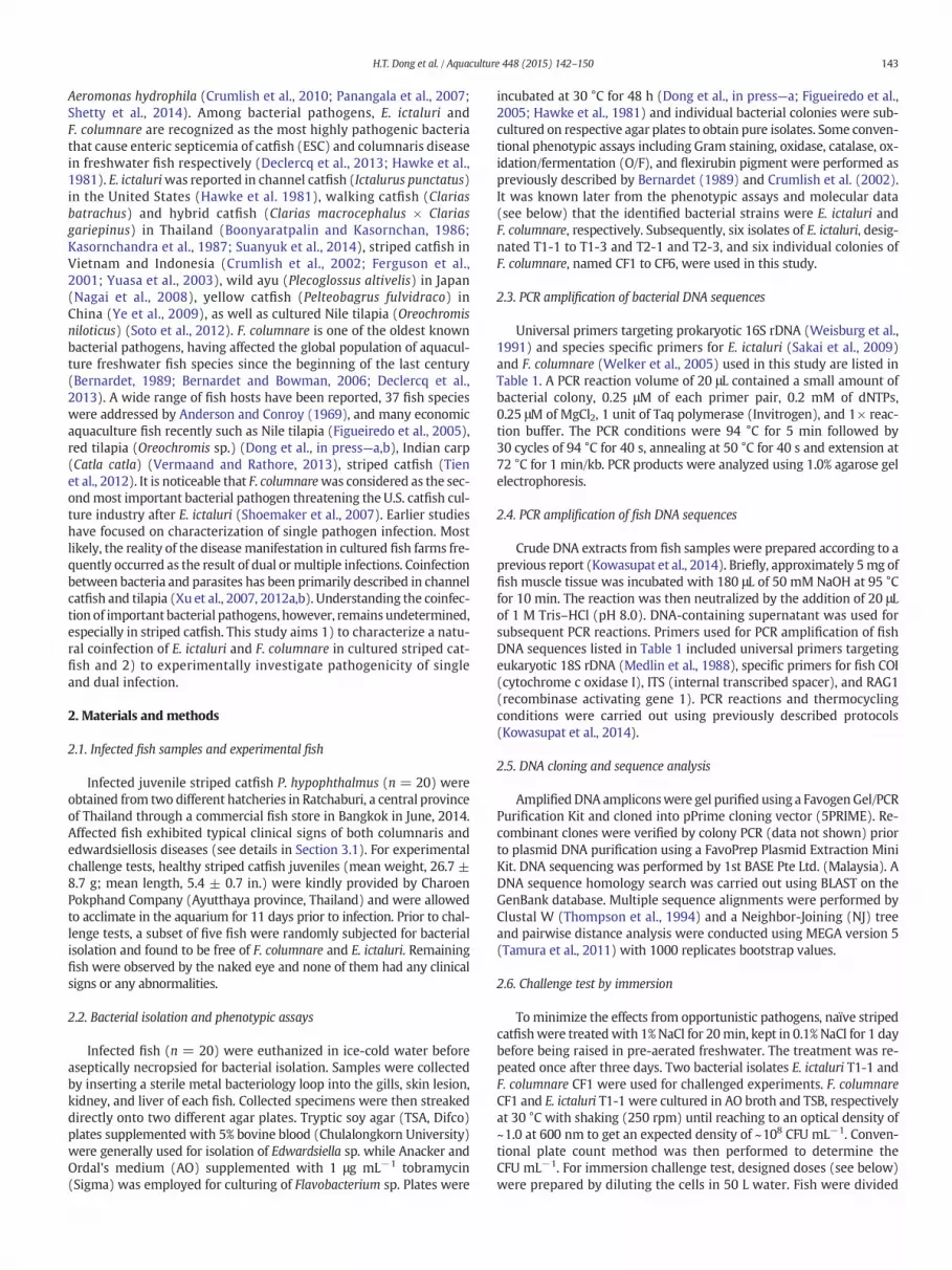
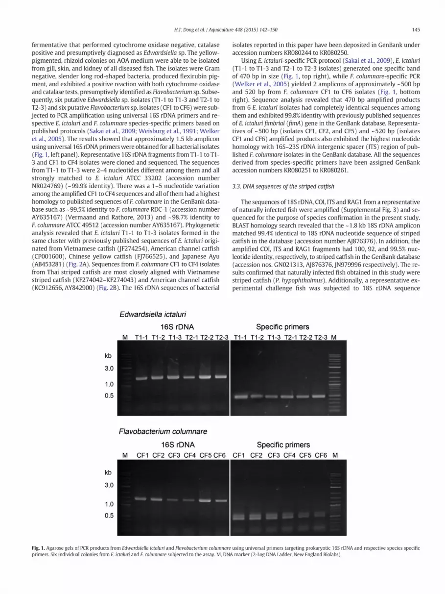
fermentative that performed cytochrome oxidase negative, catalasepositive and presumptively diagnosed as Edwardsiella sp. The yellow-pigmented, rhizoid colonies on AOA medium were able to be isolatedfrom gill, skin, and kidney of all diseased fish. The isolates were Gramnegative, slender long rod-shaped bacteria, produced flexirubin pig-ment, and exhibited a positive reaction with both cytochrome oxidaseand catalase tests, presumptively identified as Flavobacterium sp. Subse-quently, six putative Edwardsiella sp. isolates (T1-1 to T1-3 and T2-1 toT2-3) and six putative Flavobacterium sp. isolates (CF1 to CF6)were sub-jected to PCR amplification using universal 16S rDNA primers and re-spective E. ictaluri and F. columnare species-specific primers based onpublished protocols (Sakai et al., 2009; Weisburg et al., 1991; Welkeret al., 2005). The results showed that approximately 1.5 kb ampliconusinguniversal 16S rDNAprimerswere obtained for all bacterial isolates(Fig. 1, left panel). Representative 16S rDNA fragments from T1-1 to T1-3 and CF1 to CF4 isolates were cloned and sequenced. The sequencesfrom T1-1 to T1-3 were 2–4 nucleotides different among them and allstrongly matched to E. ictaluri ATCC 33202 (accession numberNR024769) (~99.9% identity). There was a 1–5 nucleotide variationamong the amplified CF1 to CF4 sequences and all of themhad a highesthomology to published sequences of F. columnare in the GenBank data-base such as ~99.5% identity to F. columnare RDC-1 (accession numberAY635167) (Vermaand and Rathore, 2013) and ~98.7% identity toF. columnare ATCC 49512 (accession number AY635167). Phylogeneticanalysis revealed that E. ictaluri T1-1 to T1-3 isolates formed in thesame cluster with previously published sequences of E. ictaluri origi-nated from Vietnamese catfish (JF274254), American channel catfish(CP001600), Chinese yellow catfish (FJ766525), and Japanese Ayu(AB453281) (Fig. 2A). Sequences from F. columnare CF1 to CF4 isolatesfrom Thai striped catfish are most closely aligned with Vietnamesestriped catfish (KF274042–KF274043) and American channel catfish(KC912656, AY842900) (Fig. 2B). The 16S rDNA sequences of bacterial
Fig. 1. Agarose gels of PCR products from Edwardsiella ictaluri and Flavobacterium columnareprimers. Six individual colonies from E. ictaluri and F. columnare subjected to the assay. M, DNA
isolates reported in this paper have been deposited in GenBank underaccession numbers KR080244 to KR080250.
Using E. ictaluri-specific PCR protocol (Sakai et al., 2009), E. ictaluri(T1-1 to T1-3 and T2-1 to T2-3 isolates) generated one specific bandof 470 bp in size (Fig. 1, top right), while F. columnare-specific PCR(Welker et al., 2005) yielded 2 amplicons of approximately ~500 bpand 520 bp from F. columnare CF1 to CF6 isolates (Fig. 1, bottomright). Sequence analysis revealed that 470 bp amplified productsfrom 6 E. ictaluri isolates had completely identical sequences amongthem and exhibited 99.8% identity with previously published sequencesof E. ictaluri fimbrial (fimA) gene in the GenBank database. Representa-tives of ~500 bp (isolates CF1, CF2, and CF5) and ~520 bp (isolatesCF1 and CF6) amplified products also exhibited the highest nucleotidehomology with 16S–23S rDNA intergenic spacer (ITS) region of pub-lished F. columnare isolates in the GenBank database. All the sequencesderived from species-specific primers have been assigned GenBankaccession numbers KR080251 to KR080261.
3.3. DNA sequences of the striped catfish
The sequences of 18S rDNA, COI, ITS and RAG1 from a representativeof naturally infected fish were amplified (Supplemental Fig. 3) and se-quenced for the purpose of species confirmation in the present study.BLAST homology search revealed that the ~1.8 kb 18S rDNA ampliconmatched 99.4% identical to 18S rDNA nucleotide sequence of stripedcatfish in the database (accession number AJ876376). In addition, theamplified COI, ITS and RAG1 fragments had 100, 92, and 99.5% nuc-leotide identity, respectively, to striped catfish in the GenBank database(accession nos. GN021313, AJ876376, JN979996 respectively). The re-sults confirmed that naturally infected fish obtained in this study werestriped catfish (P. hypophthalmus). Additionally, a representative ex-perimental challenge fish was subjected to 18S rDNA sequence
using universal primers targeting prokaryotic 16S rDNA and respective species specificmarker (2-Log DNA Ladder, New England Biolabs).
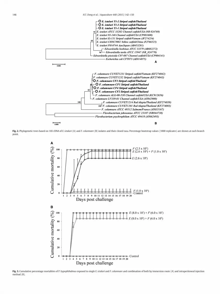
Fig. 2. Phylogenetic trees based on 16S rDNA of E. ictaluri (A) and F. columnare (B) isolates and their closed taxa. Percentage bootstrap values (1000 replicates) are shown at each branchpoint.
Fig. 3. Cumulative percentagemortalities of P. hypophthalmus exposed to single E. ictaluri and F. columnare and combination of both by immersion route (A) and intraperitoneal injectionmethod (B).
146 H.T. Dong et al. / Aquaculture 448 (2015) 142–150

147H.T. Dong et al. / Aquaculture 448 (2015) 142–150
amplification and analyzed in the same manner. The result revealed99.4% sequence identity between natural and experimental fish assayedin this study (not shown). The sequenceswere assigned accession num-bers KR080262 to KR080265 in the GenBank database.
3.4. Challenge tests
In the three single immersion-challenged groups, the groupwhich received a high dose (2.5 × 10⁷ CFU mL−1) of F. columnarewas able to induce 100.0 ± 0.0% mortality within one day, whereas,a lower dose (1.0 × 10⁴ CFU mL−1) caused only 3.3 ± 5.7% mortalityin 20 days (Fig. 3A). In the group which received only E. ictaluri2.6 ×106 CFUmL−1,fish started dying onday4, and reached the highestcumulative mortality (80.0 ± 20.0%) on day 9 post challenge (Fig. 3A).Cumulative mortality was more pronounced, 96.7 ± 5.8%, in the co-infection treatment with the same bacterial dose (E. ictaluri 2.6 ×10⁶ CFU mL−1 and F. columnare 1.0 × 10⁴ CFU mL−1) (Fig. 3A). In thecombined intraperitoneal (i.p.) injection-treated groups, the percent-age cumulative mortality of the group 1 (received E. ictaluri 8 ×10⁷ CFU & F. columnare 6 × 10⁷ CFU fish−1) and the group 2 (receivedE. ictaluri 8 × 10⁵ CFU & F. columnare 6 × 10⁵ CFU fish−1) were100.0 ± 0.0% and 86.7 ± 11.5% respectively (Fig. 3B). All clinically sickfish administrated with E. ictaluri either single or dually withF. columnare exhibited edwardsiellosis disease (multifocal pinpoint
Fig. 4.Anterior kidney (A), posterior kidney (B), spleen (C) and liver (D) of striped catfish infectwith F. columnare. NP = multifocal areas of necrosis and pyogranulomas, H = hemorrhages.
white spots in the internal organs) (Supplemental Fig. 2D). Clinicalsigns of both edwardsiellosis and columnaris (“saddle back” lesion, erod-ed fins) diseases were observed from groups that received both E. ictaluriand F. columnare by either immersion (i.m.) or injection (i.p.) route (Sup-plemental Fig. 2C–D). Only clinical signs of columnaris disease were ob-served in the group which received single F. columnare. No mortality orabnormalities were observed in the control group during 20 experimen-tal days. F. columnare and E. ictaluriwere successfully re-isolated from allrepresentative diseased fish in both i.p and i.m. combined-treated groups(five fish per group). Single types of either E. ictaluri or F. columnarewererecovered from diseased fish in respective single-challenged groups. Allthese bacterial isolates were re-confirmed by species-specific PCR asmentioned above (data not shown).
3.5. Histological examination
The severemulti-focal areas of necrosis and pyogranulomas, notablyin the kidney, liver, and spleen (Fig. 4A–D)were found in both naturallyand experimentally coinfected fish as well as apparently sick fish in thesingle E. ictaluri treated group. There were no significant differences inhistopathological manifestation in the internal organs (kidney, liver,and spleen) of the single E. ictaluri treated group and combined treat-ment group. The kidney and spleen of affected fish exhibited more se-vere damage than the liver (Fig. 4). Varying degrees of degeneration
edwith E. ictaluri and F. columnare. Posterior kidney (E) and gill lamellar (F) of fish infected

148 H.T. Dong et al. / Aquaculture 448 (2015) 142–150
and necrosis were visualized by H&E. The normal architecture of tissuewas almost completely obliterated in the center of necrotic areas(Fig. 4A–D). These histopathological manifestations were typicallyreferred to as edwardsiellosis caused by E. ictaluri. The typical histopath-ological changes of columnaris disease were found in the gills of bothnaturally and experimentally combined-challenged groups as wellas single high dose of F. columnare, but not in single low dose ofF. columnare. The lesion was characterized by partially or completelynecrotic gill lamellar with presence of numerous inflammatory cellswith morphology resembling macrophages, eosinophilic cells, and redblood cells (Fig. 4F). It is noticeable that only hemorrhage without mul-tifocal areas of necrosis in posterior kidney was observed in the fishinfected with single F. columnare (Fig. 4E) but both hemorrhage andmultifocal areas of necrosis were presented in coinfected fish (Fig. 4B–D).
The result of in situ hybridization visualized the specific locations ofE. ictaluri and F. columnare in the infected tissue. Using E. ictaluri-specificprobe, strong positive bindings were found in the tubule cells of poste-rior kidney (Fig. 5A, arrow) andweak positive signals in the gill lamellar(Fig. 5B) and anterior kidney (picture not shown) of the fish in both sin-gle E. ictaluri and combined challenge groups.When using F. columnare-specific probe, the strongest positive signals were detected in the gills(Fig. 5D), and weaker signals in the spleen (Fig. 5C), liver, and kidney(picture not shown) of both single F. columnare and combined challengegroups. No positive signals were detected using IMNV-specific probe orcontrol group without probe.
4. Discussion
Since striped catfish (P. hypophthalmus) have been intensively cul-tured inAsian countries, numerous disease outbreaks resulting in signif-icant financial losses have been reported. Subsequently, variousbacterial pathogens causing disease outbreaks have been identifiedand well-described (Crumlish et al., 2002, 2010; Ferguson et al., 2001;Tien et al., 2012). Most laboratory studies, however, focused on patho-genesis of single bacterial infections whereas concurrent infections
Fig. 5. Posterior kidney (A) and gill (B) were positive with E. ictaluri (arrow), spl
remain poorly understood. Here, we first report a naturally concurrentinfection of E. ictaluri and F. columnare in striped catfish and fulfilledKoch's postulates through single- or dual-challenged tests to mimicthe natural disease case. Both i.m. and i.p. combined-treated groups re-sulted in high cumulative mortality and the infected fish clearly exhib-ited typical signs of edwardsiellosis and columnaris disease. It shouldbe noted that the fish which received combined-bacterial pathogensby i.m. route exhibited more clearly diseased progress than i.p. route.Additionally, rapid mortality of the group which received a high doseof single F. columnaremay be correlated to biofilm formation of the bac-teria on the gills surface that could inhibit oxygen uptake and result inhigh mortality. The consistent mortality rate and biofilm formationwas previously determined in experimental challenge of commoncarp (Cyprinus carpio) and rainbow trout (Oncorhynchus mykiss) withhighly virulent strains of F. columnare (Declercq et al., 2015). In contrast,low dose of F. columnare (1.0 × 10⁴ CFU mL−1) was not able to causecolumnaris disease but in situ hybridization exhibited positive resultsinmultiple organs (gills, liver, spleen) of the experimentalfish. This sug-gests that under lethal dose, F. columnare was able to persist in the fishandmay need other stressors to induce disease such as the combinationof low dose of F. columnare with E. ictaluri evidenced in the presentstudy that successfully induced clinical signs of both edwardsiellosisand columnaris disease. Previously, Crumlish et al. (2010) also reportedthat experimental coinfection of E. ictaluri and A. hydrophila mimickednatural outbreaks of the disease in intensively cultured striped catfishfarms in Vietnam. Taken together with the data produced in the presentstudy, there is strong evidence to support that the reality of diseasemanifestation in aquaculture systems could be contributed by concur-rent infections of bacterial pathogens.
With an aspect of histopathological manifestation caused by singleinfection (either E. ictaluri or F. columnare), our results in this studyare consistent with previous reports for Vietnamese striped catfish(Ferguson et al., 2001; Tien et al., 2012). Moreover, this study first de-scribes histopathological changes of both naturally and experimentallycoinfected fish and revealed the presence of bacteria in specific tissuethrough in situ DNA hybridization during infections.
een (C) and gill (D) were positive with F. columnare by in situ hybridization.

149H.T. Dong et al. / Aquaculture 448 (2015) 142–150
Sharing the samediseased patternwith channel catfish (I. punctatus)in the United States, E. ictaluri and F. columnare have been reported asthe most highly pathogenic bacteria threatening the cultured stripedcatfish industry in Vietnam (Crumlish et al., 2002; Ferguson et al.,2001; Tien et al., 2012). Recent reports, however, have indicated thatthe genetic characterization and pathogenicity of E. ictaluriwere differ-ent betweenVietnamese andUS isolates (Bartie et al., 2012; Rogge et al.,2013), where E. ictaluri catfish isolates from Vietnam were non-pathogenic to the US channel catfish (Rogge et al., 2013). The differ-ences in clinical signs and histopathological manifestations caused byE. ictaluri in two different hosts may explain the inconsistency in origi-nally described names as enteric septicemia of catfish (ESC) in US chan-nel catfish and “bacillary necrosis in Pangasius” in Vietnamese stripedcatfish (Crumlish et al., 2002; Ferguson et al., 2001; Hawke et al.,1981). With respect to genetic diversity, the phylogenetic analysisbased on 16S rDNA sequences exhibited the identity among E. ictaluriisolates from Thailand, Vietnam, the United States, China, and Japan(Fig. 2A). In contrast, the 16S rDNA of F. columnare from striped catfishin Thailand exhibited similar genetic characteristics with theVietnamese and US catfish isolates but were different to Thai tilapia(Oreochromis spp.)-originated isolates (Fig. 2B) (Dong et al., inpress—a). In Thailand, striped catfish has been bred for fry/fingerlingexporting purposes. Due to the boom in striped catfish production inVietnam, Thai aquaculture producers have recently paid more atten-tion to striped catfish (Mr. Warren Tuner, personal communication).Although E. ictaluri and F. columnare have never been reported in cul-tured striped catfish in Thailand, sharing similar geographical loca-tions and culture technologies with Vietnam, the future catfishfarming industry in Thailand is unavoidably predicted to face similardevastation as a result of the problematic pathogens mentionedabove. Early management strategies to control the spreading ofE. ictaluri and F. columnare, in a sustainable intensively cultured sys-tem in Thailand, therefore, are highly recommended. As a model les-son, disease manipulation strategy against infectious pathogensusing monovalent or bivalent vaccine has been developed as a prior-ity and widely applied to fish farms in the United States (Klesius andShoemaker, 1999; Shoemaker et al., 2007, 2011). Our further studywill focus on the development of a combined vaccine against coin-fection of E. ictaluri and F. columnare in striped catfish.
Additionally, since the diversity of pangasiid fish has been reportedas over ten species that are naturally distributed in the MekongRiver basin including, Pangasianodon gigas, P. hypophthalmus, Pangasiusbocourti, Pangasius conchophilus, Pangasius krempfi, Pangasius mekon-gensis, Pangasius larnaudii, Pangasius pleurotaenia, Pangasius elongates,Pangasius macronema and Pangasius sanitwangsei (Poulsen et al., 2004),many of them are similar in morphology that may lead to confusionover species identification. Moreover, this study investigated coinfectionof two important bacterial pathogens and provided supplemental data ofmolecular markers (18S rDNA, ITS, COI, and RAG1) for fish identification.
In conclusion, to our best knowledge, this study is the first tocharacterize naturally and experimentally concurrent infection oftwo important bacterial pathogens F. columnare and E. ictaluri instriped catfish.
Acknowledgments
This work was supported by the Office of the Higher EducationCommission and Mahidol University under the National ResearchUniversities Initiative. H.T. Dong has been supported by the 100thyear anniversary of Chulalongkorn University fund for doctoral scholar-ship and the 90th anniversary of Chulalongkorn University fund(Ratchadaphiseksomphot Endowment Fund). The authors would liketo thank the Veterinary Medical Aquatic Research Center (VMARC),Chulalongkorn University for providing equipment for the experimen-tal challenge.
Appendix A. Supplementary data
Supplementary data to this article can be found online at http://dx.doi.org/10.1016/j.aquaculture.2015.05.046.
References
Anderson, J.I., Conroy, D.A., 1969. The pathogenic myxobacteria with special reference tofish diseases. J. Appl. Bacteriol. 32, 30–39.
Bartie, K.L., Austin, F.W., Diab, A., Dickson, C., Dung, T.T., Giacomini, M., Crumlish, M.,2012. Intraspecific diversity of Edwardsiella ictaluri isolates from diseased fresh-water catfish, Pangasianodon hypophthalmus (Sauvage), cultured in the MekongDelta, Vietnam. J. Fish Dis. 35, 671–682.
Bernardet, J.F., 1989. “Flexibacter columnaris”: first description in France and comparisonwith bacterial strains from other origins. Dis. Aquat. Organ. 6, 37–44.
Bernardet, J.F., Bowman, J.P., 2006. The genus Flavobacterium. In: Dworkin, M., et al. (Eds.),The Prokaryotes. Springer, New York, pp. 481–531.
Boonyaratpalin, S., Kasornchan, J., 1986. Edwardsiella ictaluri-like organism (EILO), a newfish pathogenic bacteria of walking catfish (Clarias batrachus Linn.) in Thailand.Songklanakarin J. Sci. Technol. 8, 445–449.
Crumlish, M., Dung, T.T., Turnbull, J.F., Ngoc, N.T.N., Ferguson, H.W., 2002. Identification ofEdwardsiella ictaluri from diseased freshwater catfish, Pangasius hypophthalmus(Sauvage), cultured in the Mekong Delta, Vietnam. J. Fish Dis. 25, 733–736.
Crumlish, M., Thanh, P.C., Koesling, J., Tung, V.T., Gravningen, K., 2010. Experimentalchallenge studies in Vietnamese catfish, Pangasianodon hypophthalmus (Sauvage),exposed to Edwardsiella ictaluri and Aeromonas hydrophila. J. Fish Dis. 33, 717–722.
Declercq, A.M., Haesebrouck, F., Van den Broeck, W., Bossier, P., Decostere, A., 2013.Columnaris disease in fish: a review with emphasis on bacterium–host interactions.Vet. Res. 44 (1), 27.
Declercq, A.M., Chiers, K., Haesebrouck, F., Van den Broeck, W., Dewulf, J., Cornelissen, M.,Decostere, A., 2015. Gill infection model for columnaris disease in common carp andrainbow trout. J. Aquat. Anim. Health 27, 1–11.
Dong, T.H., LaFrentz, B., Pirarat, P., Rodkhum, C., 2014. Phenotypic characterization andgenetic diversity of Flavobacterium columnare isolated from red tilapia, Oreochromissp. in Thailand. J. Fish Dis. http://dx.doi.org/10.1111/jfd.12304 (in press).
Dong, H.T., Senapin, S., LaFrentz, B., Rodkhum, C., 2015. Virulence assay of rhizoid andnon-rhizoid morphotypes of Flavobacterium columnare in red tilapia, Oreochromissp., fry. J. Fish Dis. http://dx.doi.org/10.1111/jfd.12385 (in press).
FAO, 2010–2011. Cultured Aquatic Species Information Programme. Pangasiushypophthalmus. Text by D. Griffiths, P. Van Khanh & T.Q. Trong. In FAO Fisheriesand Aquaculture Department [online]. Rome. Updated 14 January 2010. Accessed20 March 2011. Available at: http://www.fao.org/fishery/culturedspecies/Pangasius_hypophthalmus/en).
Ferguson, H.W., Turnbull, J.F., Shinn, A.P., Thompson, K., Dung, T.T., Crumlish, M.,2001. Bacillary necrosis in farmed Pangasius hypophthalmus (Sauvage) from theMekong Delta, Vietnam. J. Fish Dis. 24, 509–513.
Figueiredo, H.C.P., Klesius, P.H., Arias, C.R., Evans, J., Shoemaker, C.A., Pereira, D.J., Peixoto,M.T.D., 2005. Isolation and characterization of strains of Flavobacterium columnarefrom Brazil. J. Fish Dis. 28, 199–204.
Hawke, J.P., McWhorter, A.C., Steigerwalt, A.G., D.J., B., 1981. Edwardsiella ictaluri sp.nov., the causative agent of enteric septicemia of catfish. Int. J. Syst. Bacteriol. 31,396–400.
Ivanova, N.V., Zemlak, T.S., Hanner, R.H., Hebert, P.D.N., 2007. Universal primer cocktailsfor fish DNA barcoding. Mol. Ecol. Notes. 7, 544–548.
Kasornchandra, J., Rogers, W.A., Plumb, J.A., 1987. Edwardsiella ictaluri from walkingcatfish, Clarias batrachus L., in Thailand. J. Fish Dis. 10, 137–138.
Klesius, P.H., Shoemaker, C.A., 1999. Development and use of modified live Edwardsiellaictaluri vaccine against enteric septicemia of catfish. Adv. Vet. Sci. Comp. Med. 41,523–537.
Kowasupat, C., Panijpan, B., Laosinchai, P., Ruenwongsa, P., Phongdara, A., Wanna, W.,Senapin, S., Phiwsaiya, K., 2014. Biodiversity of the Betta smaragdina (Teleostei:Perciformes) in the northeast region of Thailand as determined by mitochondrialCOI and nuclear ITS1 gene sequences. Meta Gene 2, 83–95.
Li, C., Ortí, G., 2007. Molecular phylogeny of Clupeiformes (Actinopterygii) inferred fromnuclear and mitochondrial DNA sequences. Mol. Phylogenet. Evol. 44, 386–398.
López, J.A., Chen, W.J., Ortí, G., 2004. Esociform phylogeny. Copeia. 3, 449–464.Medlin, L., Elwood, H.J., Stickel, S., Sogin, M.L., 1988. The characterization of enzymatically
amplified eukaryotic 16S-like rRNA-coding regions. Gene 71, 491–499.Nagai, T., Iwamoto, E., Sakai, T., Arima, T., Tensha, K., Iida, Y., Iida, T., Nakai, T., 2008.
Characterization of Edwardsiella ictaluri isolated from wild ayu Plecoglossus altivelisin Japan. Fish Pathol. 43, 158–163.
Nguyen, T.P., Dang, T.H.O., 2009. Striped catfish (Pangasianodon hypophthalmus)aquaculture in Viet Nam: an unprecedented development within a decade. In:De Silva, S.S., Davy, F.B. (Eds.), Success Stories in Asian Aquaculture. Dordrecht,Springer, pp. 133–149.
Panangala, V.S., Shoemaker, C.A., Van Santen, V.L., Dybvig, K., Klesius, P.H., 2007.Multiplex-PCR for simultaneous detection of 3 bacterial fish pathogens,Flavobacterium columnare, Edwardsiella ictaluri, and Aeromonas hydrophila. Dis.Aquat. Organ. 74, 199–208.
Poulsen, A.F., Hortle, K.G., Valbo-Jorgensen, J., Chan, S., Chhuon, C.K., Viravong, S.,Bouakhamvongsa, K., Suntornratana, U., Yoorong, N., Nguyen, T.T., Tran, B.Q., 2004.Distribution and ecology of some important riverine fish species of the Mekongriver basin. MRC Technical Paper No. 10.

150 H.T. Dong et al. / Aquaculture 448 (2015) 142–150
Robert, T.R., Vidthayanon, C., 1991. Systematic revision of the Asian catfish familyPangasiidae, with biological observations and descriptions of three new species.Proceeding of the Academy of Nature Sciences, Philadelphia 143, 97–144.
Rogge, M.L., Dubytska, L., Jung, T.S., Wiles, J., Elkamel, A.A., Rennhoff, A., Oanh, D.T., Thune,R.L., 2013. Comparison of Vietnamese and US isolates of Edwardsiella ictaluri. Dis.Aquat. Organ. 106, 17–29.
Sakai, T., Yuasa, K., Sano, M., Iida, T., 2009. Identification of Edwardsiella ictaluri andE. tarda by species-specific polymerase chain reaction targeted to the upstream re-gion of the fimbrial gene. J. Aquat. Anim. Health 21, 124–132.
Senapin, S., Phewsaiya, K., Briggs, M., Flegel, T.W., 2007. Outbreaks of infectiousmyonecrosis virus (IMNV) in Indonesia confirmed by genome sequencing and useof an alternative RT-PCR detection method. Aquaculture 266, 32–38.
Shetty, M., Maiti, B., Venugopal, M.N., Karunasagar, I., Karunasagar, I., 2014. First isolationand characterization of Edwardsiella tarda from diseased striped catfish,Pangasianodon hypophthalmus (Sauvage). J. Fish Dis. 37, 265–271.
Shoemaker, C.A., Klesius, P.H., Evans, J.J., 2007. Immunization of eyed channel catfish,Ictalurus punctatus, eggs with monovalent Flavobacterium columnare vaccine and bi-valent F. columnare and Edwardsiella ictaluri vaccine. Vaccine 25, 1126–1131.
Shoemaker, C.A., Klesius, P.H., Drennan, J.D., Evans, J.J., 2011. Efficacy of a modified liveFlavobacterium columnare vaccine in fish. Fish Shellfish Immunol. 30, 304–308.
Šimková, A., Serbielle, C., Pariselle, A., Vanhove, M.P., Morand, S., 2013. Speciation inThaparocleidus (Monogenea: Dactylogyridae) parasitizing Asian pangasiid catfishes.BioMed. Res. Int. 2013, 353956.
Siti-Zahrah, A., Zamri-Saad, M., Firdaus-Nawi, M., Hazreen-Nita, M.K., Nur-Nazifah, M.,2013. Detection of channel catfish virus in cage-cultured Pangasius hypophthalmus(Sauvage, 1878) in Malaysia. J. Fish Dis. http://dx.doi.org/10.1111/jfd.12185 (inpress).
Soto, E., Griffin, M., Arauz, M., Riofrio, A., Martinez, A., Cabrejos, M.E., 2012. Edwardsiellaictaluri as the causative agent of mortality in cultured Nile tilapia. J. Aquat. Anim.Health 24, 81–90.
Suanyuk, N., Rogge, M., Thune, R., Watthanaphiromsakul, M., Champhat, N., Wiangkum,W., 2014. Mortality and pathology of hybrid catfish, Clarias macrocephalus(Gunther) × Clarias gariepinus (Burchell), associated with Edwardsiella ictaluri infec-tion in southern Thailand. J. Fish Dis. 37, 385–395.
Tamura, K., Peterson, D., Peterson, N., Stecher, G., Nei, M., Kumar, S., 2011. MEGA5: molec-ular evolutionary genetics analysis using maximum likelihood, evolutionary distance,and maximum parsimony methods. Mol. Biol. Evol. 28, 2731–2739.
Thompson, J.D., Higgins, D.G., Gibson, T.J., 1994. CLUSTAL W: improving the sensitivity ofprogressive multiple sequence alignment through sequence weighting, position-specific gap penalties and weight matrix choice. Nucleic Acids Res. 22, 4673–4680.
Tien, N.T., Dung, T.T., Tuan, N.A., Crumlish, M., 2012. First identification of Flavobacteriumcolumnare infection in farmed freshwater striped catfish Pangasianodonhypophthalmus. Dis. Aquat. Organ. 100, 83–88.
Tripathi, A., Rajvanshi, S., Agrawal, N., 2014. Monogenoidea on exotic Indian freshwaterfishes. 2. Range expansion of Thaparocleidus caecus and T. siamensis (Dactylogyridae)by introduction of striped catfish Pangasianodon hypophthalmus (Pangasiidae).Helminthologia 51, 23–30.
Verma, D.K., Rathore, G., 2013. Molecular characterization of Flavobacterium columnareisolated from a natural outbreak of columnaris disease in farmed fish, Catla catlafrom India. J. Gen. Appl. Microbiol. 59, 417–424.
Ward, R.D., Zemlak, T.S., Innes, B.H., Last, P.R., Hebert, P.D.N., 2005. DNA barcodingAustralia's fish species. Philos. Trans. R. Soc. Lond. B Biol. Sci. 360, 1847–1857.
Weisburg, W.G., Barns, S.M., Pelletier, D.A., Lane, D.J., 1991. 16S ribosomal DNA amplifica-tion for phylogenetic study. J. Bacteriol. 173, 697–703.
Welker, T.L., Shoemaker, C.A., Arias, C.R., Klesius, P.H., 2005. Transmission and detection ofFlavobacterium columnare in channel catfish Ictalurus punctatus. Dis. Aquat. Organ. 63,129–138.
Xu, D.H., Shoemaker, C.A., Klesius, P.H., 2007. Evaluation of the link betweengyrodactylosis and streptococcosis of Nile tilapia, Oreochromis niloticus (L.). J. FishDis. 30, 233–238.
Xu, D.H., Shoemaker, C.A., Klesius, P.H., 2012a. Ichthyophthirius multifiliis as a potentialvector of Edwardsiella ictaluri in channel catfish. FEMS Microbiol. Lett. 329, 160–167.
Xu, D.H., Pridgeon, J.W., Klesius, P.H., Shoemaker, C.A., 2012b. Parasitism by protozoanIchthyophthirius multifiliis enhanced invasion of Aeromonas hydrophila in tissues ofchannel catfish. Vet. Parasitol. 184, 101–107.
Ye, S., Li, H., Qiao, G., Li, Z., 2009. First case of Edwardsiella ictaluri infection in Chinafarmed yellow catfish Pelteobagrus fulvidraco. Aquaculture 292, 6–10.
Yuasa, K., Kholidin, E.B., Panigoro, N., Hatai, K., 2003. First isolation of Edwardsiella ictalurifrom cultured striped catfish Pangasius hypophthalmus in Indonesia. Fish Pathol. 38,181–183.