comparative analysis of detoxification enzymes in acyrthosiphon pisum and myzus persicae
TRANSCRIPT

1
Comparative analysis of detoxification enzymes in Acrythosiphon pisum and Myzus persicae John S. Ramsey1+, Dean S. Rider2+, Thomas K. Walsh3, Martin De Vos1, Karl H. J. Gordon4, Lalit Ponnala5, Simone L. Macmil6, Bruce A. Roe6, and Georg Jander
1*
1Boyce Thompson Institute for Plant Research, Ithaca, NY, USA 2Department of Veterinary Pathobiology, Texas A&M University, College Station, TX, USA 3CSIRO Entomology, Centre for Environment and Life Sciences (CELS), Floreat Park, WA 6014, Australia 4CSIRO Entomology, Black Mountain Laboratories, Clunies Ross Street, Black Mountain, Acton ACT 2601, Australia 5Biotechnology Center, Cornell University, Ithaca, NY, USA 6
Department of Chemistry and Biochemistry, University of Oklahoma, OK, USA
+
John S. Ramsey and Dean S. Rider contributed equally to this research.
*For correspondence Georg Jander Boyce Thompson Institute for Plant Research 1 Tower Road Ithaca, NY 14850 USA phone: 607-354-1365 fax: 607-254-1502 email: [email protected] Running title: Aphid detoxification enzymes Key words: Acyrthosiphon pisum, Myzus persicae, esterase, glutathione S-transferase, cytochrome P450, detoxification

2
Abstract Herbivorous insects use detoxification enzymes, including cytochrome P450
monooxygenases, glutathione S-transferases, and carboxy/cholinesterases, to
metabolize otherwise deleterious plant secondary metabolites. Whereas Acrythosiphon
pisum (pea aphid) feeds almost exclusively from the Fabaceae, Myzus persicae (green
peach aphid) feeds from hundreds of species in more than forty plant families.
Therefore, M. persicae as a species would be exposed to a greater diversity of plant
secondary metabolites than A. pisum, and has been predicted to require a larger
complement of detoxification enzymes. A comparison of M. persicae cDNA and A.
pisum genomic sequences is partially consistent with this hypothesis. There is evidence
of at least 40% more cytochrome P450 genes in M. persicae than in A. pisum. In
contrast, no major differences were found between the two species in the numbers of
glutathione S-transferases, and carboxy/cholinesterases. However, given the
incomplete M. persicae cDNA data set, the number of identified detoxification genes in
this species is likely an underestimate.
3
Introduction In their long co-evolution with insect herbivores, plants have developed a variety of
defenses to keep from being eaten (Rosenthal and Berenbaum, 1991). These include
physical barriers such as spines, tough bark, and sticky sap, as well as numerous
distasteful or toxic compounds that are often unique to particular plant genera or
families. Defense against herbivory is likely the primary function for many of the several
hundred thousand different plant secondary metabolites found in nature (Becerra, 2007;
Bino et al., 2004). Nevertheless, most, likely all, land plants are fed upon by at least one
of the several hundred thousand herbivorous insect species (Schoonhoven et al., 1998).
This suggests that insects, as part of an evolutionary arms race with their host plants,
have developed efficient mechanisms to avoid or detoxify plant secondary metabolites.
Insect herbivores are often broadly classified as either specialists or generalists.
Whereas specialists consume only a small number of closely related plant species,
generalists tend to forage more widely on a variety of host plants. It is estimated that
90% of herbivorous insects are specialists that forage on three or fewer plant families
(Bernays and Graham, 1988), and must therefore negotiate a relatively limited array of
plant defenses. In comparison to these more abundant specialists, generalist herbivores
are almost certainly exposed to a greater diversity of plant defensive chemicals.
Insect responses to plant secondary metabolites can include avoidance of the
most well-defended tissue types, target site insensitivity, rapid passage of toxins
through the gut, efflux pumps and direct metabolic detoxification. Cytochrome 450
monooxygenases (P450s) constitute the largest and most functionally diverse class of
insect detoxification enzymes (Li et al., 2007). Members of the CYP3 clade have been

4
implicated in the oxidative detoxification of furanocoumarins, alkaloids, numerous other
plant secondary metabolites and synthetic insecticides (Feyereisen, 1999; Mao et al.,
2006; Scott, 1999; Snyder and Glendinning, 1996). The CYP4 clade has been
implicated in pheromone metabolism (Maїbèche-Coisne et al., 2004). Members of the
CYP2 clade and mitochondrial targeted P450s contribute to hormone, sterol, and fatty
acid metabolism (Feyereisen, 1999; Feyereisen, 2006).
Similar to P450s, carboxyl/cholinesterases (CCEs) can function broadly in
xenobiotic detoxification. CCEs from aphids and other insects have been shown to
hydrolyze both plant secondary metabolites, organophosphates, and other man-made
insecticides (Field, 2000; Li et al., 2007). Other members of the CCE superfamily have
important neurological and developmental functions, or are involved in pheromone
processing (Oakeshott et al., 1999).
Glutathione S-transferase (GST) enzymes, which occur in all eukaryotic
organisms, function by conjugating xenobiotics and endogenously activated compounds
to the thiol group of reduced glutathione, thereby targeting them for more rapid
excretion or degradation (Li et al., 2007). In insects, GSTs have been associated with
resistance to insecticides, including DDT, spinosad, diazinon and nitenpyram, which
target the nervous system, as well as lufenuron and dicyclanil, which cause larval
lethality during life stage transitions (Enayati et al., 2005; Low et al., 2007). GST
enzyme activity in the generalist Myzus persicae (green peach aphid) increases upon
ingestion of isothiocyanates, a class of toxic secondary metabolites found in the
Brassicaceae (Francis et al., 2005) suggesting that GSTs are involved in detoxification.
Comparison of the legume specialist Acyrthosiphon pisum (pea aphid) and generalist
5
Aulacorthum solani (foxglove or potato aphid) showed broad differences in GST
enzyme activities that may reflect their respective host plant preferences (Francis et al.,
2001).
Whereas specialist insect herbivores tend to have highly efficient detoxification
mechanisms that target the predictable set of secondary metabolites in their favored
host plants, generalists that feed on a wide variety of plants would need a less specific
array of constitutive or inducible detoxification enzymes. Therefore, it is often assumed
that generalist herbivores must possess a greater diversity of detoxification enzymes
than specialists. Research on Lepidoptera in the genus Papilio (swallowtail butterflies)
showed that the generalists possess a broader array of P450s for detoxification of
furanocoumarins (Li et al., 2002; Mao et al., 2007). However, since the only sequenced
lepidopteran genome is that of B. mori (Mita et al., 2004), an extreme host plant
specialist, it is difficult to assess the actual genetic diversity of P450s and other
detoxification enzymes relative to the number of host plants that can be consumed by
particular larvae of a particular lepidopteran species.
The A. pisum genome (International Aphid Genomics Consortium, 2009) together
with a large M. persicae expressed sequence tag (EST) collection (Ramsey et al.,
2007), allows direct comparison of the xenobiotic detoxification enzymes in two related
insect species with different feeding habits. Both are classified in the tribe Macrosiphini
within the aphid sub-family Aphidinae (von Dohlen et al., 2006) and are about 95%
identical at the DNA sequence level. However, A. pisum is specialized for feeding on
legumes (Fabaceae) and M. persicae is a broad generalist that feeds from hundreds of
species in more than forty plant families (Blackman and Eastop, 2000).
6
Legumes, which can be consumed by both A. pisum and M. persicae, have
aphid-deterrent secondary metabolites. For instance, a hemiterpene glucoside was
identified as an aphid-deterrent in Vicia hirsuta (Ohta et al., 2005) and low saponin and
phenolic content in alfalfa was associated with improved A. pisum performance
(Golawska and Lukasik, 2008). Although phloem feeders likely encounter a smaller
repertoire of secondary metabolites than chewing insects feeding on the same plants,
M. persicae almost certainly ingests secondary metabolites that a legume specialist like
A. pisum would not normally encounter. This would include glucosinolates in
Brassicaceae (Kim et al., 2008) and alkaloids in the Solanaceae. Tobacco-adapted
strains of M. persicae are reported to have a nine-fold greater resistance to nicotine in
artificial diet (Nauen et al., 1996) and nicotine vapors in air (Devine et al., 1996),
suggesting routine exposure to this alkaloid when M. persicae are feeding from tobacco
plants.
Given its more diverse feeding habits, M. persicae as a species would need to
avoid or inactivate a greater variety of plant defenses than A. pisum. Although there are
defensive proteins (Walz et al., 2004) and other barriers to phloem feeding that would
not be specifically targeted by detoxifying enzymes (Walling, 2008), secondary
metabolites definitely have important defensive functions in many plant species. Here
we compare the predicted P450s, GSTs, and esterases produced by M. persicae and A.
pisum to test the hypothesis that a broad generalist insect herbivore would have a
greater abundance and functional diversity of xenobiotic detoxification enzymes than a
specialist.
7
Results and Discussion M. persicae cDNA sequencing, assembly, and annotation To allow large-scale comparisons with the recently sequenced A. pisum genome
(International Aphid Genomics Consortium, 2009), an existing expressed sequence tag
(EST) collection with >10,000 M. persicae unigenes produced by Sanger sequencing
(Ramsey et al., 2007) was augmented by DNA sequencing using Roche GS-FLX (454)
technology. Sequencing of cDNA libraries prepared from tobacco-adapted and non-
adapted isolates of M. persicae produced 142,600 ESTs, with a median read length
(N50) of 235 bp and a mean read length of 200 bp. The SeqClean program
(http://www.tigr.org/tdb/tgi/software/) was used to remove polyA tails and primer
sequences, and any sequence less than 30 bp long after cleaning was discarded. This
resulted in a set of 118,756 sequences, with an N50 of 173 bp and a mean length of
166 bp. Removal of 39,795 reads that aligned to an existing M. persicae Sanger-
sequencing unigene set (Ramsey et al., 2007) with a blastn e-value less than 1e-10
resulted in 78,961 sequences were clustered using the MCL algorithm (Enright et al.,
2002; Van Dongen, 2008). Of these, 34,781 sequences could not be clustered with any
other sequences, whereas the remaining 44,180 sequences yielded 9201 clusters. The
sequences in each cluster were assembled into contigs using the CAP3 program
(Huang and Madan, 1999). The default CAP3 parameters, b=20 and d=200, for the
sequence quality score and number of allowable differences, respectively were used to
determine whether two given sequences would be assembled into one contig. This
yielded a total of 7710 contigs and 5341 singlets. These two non-overlapping datasets,
the 34,781 unclustered sequences (Supplemental Table 1) and the 13,051 contigs and

8
singletons (Supplemental Table 2), represent a significant expansion of existing cDNA
sequence available for M. persicae. A FASTA file containing the sequences of these
47,832 unigenes can be downloaded from AphidBase
(http://www.aphidbase.com/aphidbase/downloads).
Both clustered and unclustered sequence datasets were compared to the D.
melanogaster RefSeq protein set, the merged set of A. pisum Glean and RefSeq
proteins, and the A. pisum genomic scaffolds. Approximately 10% of the 454 sequences
(4890) had a hit to a D. melanogaster RefSeq protein (blastx, e-value ≤ 1e-3). These
sequences were annotated by parsing GenBank records to retrieve gene descriptions
(Supplemental Table 2). More than 25% of the 454 sequences (12,371) had a match to
A. pisum Glean and/or RefSeq predicted proteins. GenBank records containing
automated annotation of A. pisum proteins were parsed to annotate M. persicae
orthologs according to these gene descriptions (Supplemental Tables 1 and 2).
Over 25% of the 454 unigenes (13,531) had a hit in a BLAST comparison against
the A. pisum genomic scaffolds (tblastx, e-value ≤ 1E-4), but not to any Glean or
RefSeq protein data sets (International Aphid Genomics Consortium, 2009). This
discrepancy likely results from M. persicae sequences aligning to untranslated regions
of the A. pisum genome. A higher stringency e-value cutoff was used for this tblastx
search than for the blastx against RefSeq proteins, but it is possible that differences
between these two algorithms are responsible for the increased number of 454
unigenes having positive hits against the genome. It is also possible that the A. pisum
Glean and RefSeq protein sets are incomplete, and that some of the M. persicae

9
sequences with a match in the A. pisum genome but not the Glean/RefSeq set are
aligning to coding regions of genes that were missed by gene prediction programs.
The M. persicae unigene set, consisting of >10,000 Sanger sequencing contigs
(Ramsey et al., 2007) combined with 47,832 additional sequences representing
expressed genes identified by 454 sequencing, was compared to the A. pisum genome
sequence (International Aphid Genomics Consortium, 2009) to assess the relative
abundance of xenobiotic detoxification enzymes in a specialist and a broad generalist
aphid species.
Cytochrome P450s
The A. pisum genome encodes 83 predicted P450s (Supplemental Table 3). At least 58
loci exhibit both good sequence homology to other insect P450s and contain a complete
P450 domain. Additionally, 25 P450-related loci in the A. pisum genome may have
incomplete P450 domains. The presence of partial domains could result from gene
assembly and annotation problems (e.g. aberrant exon prediction), or these loci could
represent actual pseudogenes. The majority of pea aphid P450 sequences are related
to members of the CYP3 clade, followed by those related to the CYP4 clade (Table 1).
Of the 83 identified A. pisum P450s, 51 show evidence of expression through ESTs in
public data sets. There is evidence for P450 evolution through tandem duplications, with
several scaffolds in the A. pisum genome assembly possessing two or more P450 loci
that are closely related to one another.
Analyses of other sequenced arthropod genomes have identified 143 P450s in
Tribolium castaneum (red flour beetle), 106 in Anopheles gambiae (malaria mosquito),

10
86 in Bombyx mori (silk moth), 85 in Drosophila melanogaster (fruit fly), 46 in Apis
mellifera (honeybee) and 75 in Daphnia pulex (Baldwin et al., 2009; Claudianos et al.,
2006; Li et al., 2005; Richards et al., 2008;
http://drnelson.utmem.edu/CytochromeP450.html). Therefore, A. pisum appears to have
a fairly typical number of P450s. However, if one assumes that some of the 83 putative
P450 loci are pseudogenes with incomplete P450 domains, then the A. pisum P450
complement would be toward the lower end of sequenced insect genomes. This
relatively low number of P450s may be particularly interesting in light of mounting
evidence that the pea aphid has a tendency to accumulate duplicated genes over time
(International Aphid Genomics Consortium, 2009). It has been suggested that the social
organization of the bee hive, which may shield the A. mellifera queen and larvae from
environmental exposure to toxins, has permitted a relative loss of P450 loci in this
species (Claudianos et al., 2006). However, this is certainly not the case in A. pisum.
A subset of A. pisum P450s that are predicted to be involved in 20-hydroxy-
ecdysone biosynthesis (orthologs of the fruit fly loci disembodied and shade) appear to
have undergone duplications in the A. pisum genome (Supplemental Table 3). This is in
contrast to evidence from holometabolous insects, where it has been suggested that the
presence of only single genes for these enzymes represents a structural and
evolutionary constraint (Rewitz et al., 2007). Duplications of shade were not observed
among the M. persicae ESTs, suggesting that this observation may be specific to the A.
pisum genome.
Analysis of M. persicae ESTs identified more than 150 P450-related sequences.
Unique genes, as opposed to allelic variants of the same P450 gene, were identified

11
based on the contig assembly parameters described above. Many of the DNA
sequences could not be confidently assigned as P450s based on translations, perhaps
representing the non-coding regions, and most of the EST contigs represented
incomplete coding regions. However, based on significant homology to the A. pisum
P450s (e-value ≤ 1E-4), at least 115 of the M. persicae EST contigs represent
expressed P450 loci. Neighbor joining trees based on Clustal alignments (Larkin et al.,
2007) of either DNA sequences or protein translations indicated that 30% to 50% of A.
pisum P450 loci have an ortholog in M. persicae (Figure 1A).
Although the P450 loci from the pea aphid genomic sequence could be readily
assigned to clades through the construction of phylograms, the addition of fragments of
P450 loci from the M. persicae EST clusters increased the level of 'noise' in the
phylograms. This is evident in Figure 1A, where clade assignments for the pea aphid
P450s (labeled nodes in the figure) are somewhat dispersed. However, potential M.
persicae orthologs were identified using phylograms of DNA sequences from the M.
persicae EST clusters and the predicted pea aphid DNA sequences. Putative orthologs
are indicated in the supplementary table 3. Sequences were considered putatively
orthologous if they were paired with a pea aphid gene and had bootstrap support
greater than 50%. Although there is considerable uncertainty in the specific assignment
of the M. persicae sequences at the clade level (Table 1), it is clear that some the pea
aphid sequences group separately from some M. persicae sequences, indicating
divergence in the P450 complement between the two species. It is impossible to
determine whether the remaining A. pisum loci have M. persicae orthologs, because the
available EST data likely represent only a fraction of all M. persicae P450s. On the other

12
hand, more than 100 of the M. persicae P450 ESTs have no clear match in the A. pisum
genome, suggesting that there has been an expansion of this gene family in M. persicae
or, conversely, a contraction in A. pisum. Most species in the Aphidinae (von Dohlen et
al., 2006) are more specialized than M. persicae in their feeding habits, but the number
of P450 enzymes encoded in their genomes is as yet unknown.
Carboxyl/cholinesterases
There are 30 members of CCE superfamily in the A. pisum genome (Supplemental
Table 4), compared to the 24, 35, and 51 that have been identified in A. mellifera, D.
melanogaster and A. gambiae, respectively (Table 2; Claudianos et al., 2006). All of the
A. pisum genes appear functional, though several lack EST support and a few are
truncated, likely due to errors with the genome assembly. In comparison, there are 19 to
23 identifiable CCEs in the M. persicae EST unigene set, 13 with putative A. pisum
homologs and 6 to 10 esterase-like genes that do not have any obvious homologs in the
A. pisum genome (Figure 1B). This suggests that there is diversification of enzyme
functions in the esterase gene family, at least for those that are not involved in basal
metabolism that would likely be common to all aphids.
Known CCEs can be divided into 13 clades (Ranson et al., 2002), seven of which
are represented in A. pisum and M. persicae (Table 2). Clades without identifiable A.
pisum or M. persicae homologs are the Diptera-specific clades B and C, integument
esterases (D), dipteran juvenile hormone esterases (F), lepidopteran juvenile esterases
(G) and the glutactin like esterases (H). Thirteen of the A. pisum esterase genes are
found in small clusters of 2 to 6 genes per scaffold. This is particularly apparent in clade
13
E, where there appears to be an expansion in A. pisum. However, this level of
duplication is less than that found in the Diptera, where 8 to 10 CCEs may cluster
together (Campbell et al., 2003). In A. pisum, the largest CCE cluster consists of 6
genes that share no more than 60% amino acid identity to each other (Figure 1B),
suggesting fairly ancient duplication events.
Esterases in clades A-C are involved in the detoxification of xenobiotics. A.
pisum or M. persicae only possess esterases in clade A, which also contains an
esterase linked to organophosphate resistance in Anisopteromalus calandrae, a
parasitic wasp (Zhu et al., 1999). It has been suggested that the eight clade A esterases
in A. mellifera represent an order-specific radiation within the Hymenoptera (Claudianos
et al., 2006). However, given a similar radiation in A. pisum and M. persicae, it would
seem that an order-specific radiation in clade A is not unique to A. mellifera. Comparing
A. pisum and M. persicae shows that they both have five members of clade A. However,
the absence of direct homologs suggests an ancient radiation event or independent
expansions of this gene family (Figure 1B).
A. pisum shows a reduction in diversity in CCEs involved in hormone and
pheromone processing (clades D-H) with only clade E having any members at all (Table
2). However, within clade E, A. pisum esterase genes have undergone a considerable
expansion to 18 genes, compared to three in D. melanogaster and A. mellifera and five
in A. gambiae (Claudianos et al., 2006). When the 18 A. pisum clade E CCEs are
aligned with CCEs from other species they form a monophyletic clade. However when
aligned with M. persicae they split into two sub-clades, perhaps a reflection of the
diversity within clade E in aphids (Figure 1B). The 11 - 12 M. persicae unigenes in clade

14
E suggest that this expansion is not unique to A. pisum. Clade E enzymes are thought
to be largely involved with pheromone and hormone processing in insects, and it is
interesting that this gene family has been expanded in A. pisum and M. persicae (Table
2). Although, aphid alarm and sex pheromones have been identified (Dawson et al.,
2005; Hatano et al., 2008; Verheggen et al., 2008), aphid pheromone communication is
almost certainly less complex than that of honeybees (Slessor et al., 2005). Therefore,
the relative expansion of the aphid CCE clade E family likely serves a different function.
In organophosphate-resistant M. persicae, the E4 esterase gene is often found in
clusters of up to 80 virtually identical copies, and is associated with another esterase,
FE4 (Field and Devonshire, 1998). Although the closest A. pisum homolog of M.
persicae E4 esterase (ACYPI623066) is also clustered with other esterase genes, there
is only one copy in the sequenced genome (Figure 1B). ACYPI623066 and
ACYPI559388, the A. pisum genes most similar to M. persicae FE4 esterase (Figure
1B), also generated the most hits in comparisons to the M. persicae transcript data
(Supplemental Table 4).
Clades F and G contain validated juvenile hormone esterases (JHE) for Diptera
and Lepidoptera respectively. Given their phylogenetic specificity, it is perhaps not
surprising that A. pisum and M. persicae do not have members of these clades.
However ACYPI381461 and ACYPI929836 in clade E are candidate aphid JHEs. The
predicted coding sequence of ACYPI381461 contains two GQSAG nucleophilic elbow
motifs, whereas ACYPI929836 has one. The GQSAG motif is present in the active site
in all functionally validated JHEs, though there is usually only one in each protein. All A.
pisum motifs align with the JHEs from D. melanogaster and lepidopteran species

15
(Figure 2). Other analyses show that ACYPI381461 is somewhat longer than the other
aphid esterases in clade E and does not have an obvious M. persicae homolog.
With the exception of acetylcholine esterase (ACHE, clade J), members of the
neurological/developmental group (Table 2, clades I-M) tend to be non-catalytic and are
involved in cell-cell interactions. Seven members of this group were identified in A.
pisum (5 - 8 in M. persicae; Table 2), less than the 10 to 12 identified in A. mellifera, D.
melanogaster and A. gambiae (Claudianos et al., 2006). Like A. mellifera and A.
gambiae, A. pisum has two AChEs, one of which one (ACYPI102248) has a likely
neurological function. In contrast, M. persicae EST data show three potential AChEs.
Other CCEs found in aphids include the neuroligins (clade L), conserved structural
proteins involved in synapse formation, gliotactin (clade K), which is thought to be a
structural protein, and clade I, which has unknown function (Table 2).
Glutathione S-transferases
Analysis of the A. pisum genome identified 20 putative members of the GST superfamily
(Figure 1C; Supplemental Table 5). This is fewer than the number of loci identified in D.
melanogaster (38) and A. gambiae (31) but more than A. mellifera (10), which has an
reduced number of genes encoding detoxification enzymes (Claudianos et al., 2006;
Ranson et al., 2001). cDNA sequences provide gene expression evidence for 15 of the
20 likely A. pisum GSTs. Insects generally harbor six different classes of GSTs
(Chelvanayagam et al., 2001). However, although the A. pisum genome encodes two
microsomal GSTs and GSTs in the delta (10), theta (2), and sigma (6) classes, it
apparently lacks the epsilon, omega and zeta classes (Table 3). The delta and epsilon
16
GST classes are found uniquely in insects (Ranson et al., 2001) and have been
implicated in insecticide resistance. However, A. pisum has fewer members of these
two GSTs classes than D. melanogaster and A. gambiae.
EST data from the generalist aphid M. persicae show at least 14 and a maximum
21 of GST-like genes (Figure 1C; Table 3). M. persicae homologs were found for all
except two of the predicted A. pisum genes. Eight M. persicae genes are obviously
homologous to genes identified in A. pisum, whereas others seemed to have diversified
(Figure 1C). As in the case of A. pisum, the epsilon, omega and zeta GST classes are
lacking in M. persicae (Table 3), suggesting that these three GST classes may be
absent from aphids in general. Analysis of cDNA libraries from specific tissue types
(Ramsey et al., 2007) suggests that one GST (contig 1196) is specifically expressed in
gut tissue, whereas another (contig 3648) is over-represented in the salivary glands of
M. persicae.
A. pisum EST data show that several alternative splicing variants that are
apparently derived from a single GST gene (Figure 1C; Supplemental Table 5).
Although the M. persicae EST data also suggest GST alternative splicing, these do not
occur for the same gene as in A. pisum. Such alternative splicing could be an
evolutionary strategy to broaden the spectrum of effectiveness in detoxification
enzymes, and might thus allow aphids to expand their host range or to effectively
metabolize xenobiotics such as plant secondary metabolites and insecticides (Ranson
et al., 2001). Further analysis of the A. pisum genome showed the presence of enzymes
that are potentially involved in the degradation of conjugated glutathione, including eight
gamma-glutamyl transpeptidases and 34 aminopeptidases (Supplemental Table 5).

17
These A. pisum genes, along with their M. persicae EST homologs (Supplemental
Table 5), indicate that aphids have the complete pathway for degradation of xenobiotics
via conjugation to glutathione.
Conclusions
The A. pisum genome and large-scale sequencing of M. persicae ESTs have partially
made it possible to determine whether a broad generalist insect herbivore has a greater
diversity of detoxification enzymes than a specialist. There is apparently no great
expansion of the GSTs and CCEs in M. persicae relative to A. pisum, and the number of
interspecies differences in the presence and absence of specific genes are likely to be
similar those found for other aphid gene families. In the case of the CCEs, a relatively
large fraction of these proteins are involved in basal metabolic functions that are likely to
be the same or similar in A. pisum and M. persicae. Although it is difficult to estimate
exact gene numbers from the EST data, the P450 gene family is at least 40% larger in
M. persicae than in A. pisum. If one assumes similar gene representation in EST
libraries produced from the two species (115 vs. 51 unique genes), then M. persicae
may encode twice as many P450s as A. pisum. This expansion may reflect the different
host ranges of the two aphid species, only Fabaceae for A. pisum and 40 different plant
families for M. persicae, and is consistent with the hypothesis that a phloem-feeding
generalist insect herbivore would require a greater number of detoxification enzymes
than a specialist.
Experimental Procedures

18
RNA isolation and cDNA synthesis:
Total RNA was isolated using the RNeasy kit (Qiagen, USA) from 100-200 aphids, a
mixture of adults and all larval stages. RNA samples were prepared from two different
lineages of M. persicae: tobacco-adapted M. persicae feeding on tobacco, and non-
adapted M. persicae feeding on cabbage. mRNA was purified using Oligotex (Qiagen,
USA) and precipitated to increase the concentration to ~100 ng/uL. cDNA synthesis and
normalization were performed using a modification of the Creator SMART cDNA
synthesis protocol (Clontech, USA) in conjunction with the Trimmer Direct cDNA
normalization kit (Evrogen, Moscow, Russia). A modified reverse transcription primer,
5'-
AAGCAGTGGTATCAACGCAGAGTGGCCGAGGTTTTGTTTTTTTTTCTTTTTTTTTTV
N-3',
was used. To avoid potential problems associated with 454 sequencing of
homopolymeric stretches, adaptor-ligated poly-T primers for reverse transcription were
modified to disrupt the poly-T tract every several bases. The presence of the SMART IV
oligo (Clontech, USA) in the reverse transcription reaction established the 3' end of the
reverse transcription product as the reverse complement of the 5' adaptor. Therefore,
the subsequent second strand synthesis and amplification (16 cycles) was
accomplished with one primer (5' PCR primer, 5'-AAGCAGTGGTATCAACGCAGAGT-
3'), which anneals to the identical 3' ends of complementary strands.
cDNA normalization

19
Normalization of cDNA by duplex-specific nuclease treatment was performed to
increase the relative abundance of low-expression transcripts (Zhulidov et al., 2004).
Normalized cDNA was subjected to 2 rounds of amplification: 20 cycles in the first round
and 12 cycles in the second round. cDNA was purified and sequenced with a Roche
GS-FLX system.
Sequence assembly and analysis
Primer and adaptor sequences were trimmed from sequences, and any trimmed
sequences less than 30 bp were discarded. In an effort to identify sequences not
represented in previous Sanger EST sequencing of M. persicae, 454 sequences were
aligned to an existing unigene set (Ramsey et al., 2007). 454 sequences with over 95%
overlap with an existing unigene were discarded. The remaining sequences were
clustered with MCL software (Enright et al., 2002), and consensus contigs were formed
from each cluster using CAP3 software using default parameters (Huang and Madan,
1999). Contigs and singletons were BLASTed against the M. persicae Sanger unigene
set, and sequences with more than 75% overlap with the Sanger unigene were
discarded, as were sequences less than 100 bp in length.
Annotation of 454 unigenes
Annotation of M. persicae sequences relied on BLAST (Altschul et al., 1997; Tatusova
and Madden, 1999) to identify the most similar sequences among Drosophila and A.
pisum RefSeq proteins, as well as A. pisum genomic scaffolds. BioPerl modules and

20
custom Perl scripts were used to parse GenBank files and extract gene descriptions for
top BLAST hits.
Phylogenetic trees
Protein sequences for A. pisum were downloaded from AphidBase and/or GenBank. M.
persicae ESTs were obtained from GenBank and converted into protein sequences.
Trees are built using the amino acid sequence and neighbor-joining using FigTree
(version1.2.1; Andrew Rambaut; http://tree.bio.ed.ac.uk/software/figtree/).
References
Altschul, SF, Madden, TL, Schaffer, AA, Zhang, J, Zhang, Z, Miller, W and Lipman, DJ
(1997) Gapped BLAST and PSI-BLAST: a new generation of protein database
search programs. Nucleic Acids Res 25: 3389-402.
Baldwin, WS, Marko, PB and Nelson, DR (2009) The cytochrome P450 (CYP) gene
superfamily in Daphnia pulex. BMC Genomics 10: 169.
Becerra, JX (2007) The impact of herbivore-plant coevolution on plant community
structure. PNAS 104: 7483-7488.
Bernays, EA and Graham, M (1988) On the evolution of host specificity in
phytophagous arthropods. Ecology 69: 886-892.
Bino, RJ, Hall, RD, Fiehn, O, Kopka, J, Saito, K, Draper, J, Nikolau, BJ, Mendes, P,
Roessner-Tunali, U, Beale, MH, Trethewey, RN, Lange, BM, Wurtele, ES and

21
Sumner, LW (2004) Potential of metabolomics as a functional genomics tool.
Trends Plant Sci 9: 418-425.
Blackman, RL and Eastop, VF (2000) Aphids on the World's Crops. Wiley, Chichester.
466 pp.
Campbell, PM, de, QRGC, Court, LN, Dorrian, SJ, Russell, RJ and Oakeshott, JG
(2003) Developmental expression and gene/enzyme identifications in the alpha
esterase gene cluster of Drosophila melanogaster. Insect Mol Biol 12: 459-71.
Chelvanayagam, G, Parker, MW and Board, PG (2001) Fly fishing for GSTs: a unified
nomenclature for mammalian and insect glutathione transferases. Chem Biol
Interact 133: 256-260.
Claudianos, C, Ranson, H, Johnson, RM, Biswas, S, Schuler, MA, Berenbaum, MR,
Feyereisen, R and Oakeshott, JG (2006) A deficit of detoxification enzymes:
pesticide sensitivity and environmental response in the honeybee. Insect Mol Biol
15: 615-36.
Dawson, GW, Griffiths, DC, Merritt, LA, Mudd, A, Pickett, JA, Wadhams, LJ and
Woodcock, CM (2005) Aphid semiochemicals - A review and recent advances on
the sex pheromones. J Chem Ecol 16: 1573-1561.
Devine, GJ, Harling, ZK, Scarr, AW and Devonshire, AL (1996) Lethal and sublethal
effects of imidacloprid on nicotine-tolerant Myzus nicotinianae and Myzus
persicae. Pest Sci 48: 57-62.
Enayati, AA, Ranson, H and Hemingway, J (2005) Insect glutathione transferases and
insecticide resistance. Insect Mol Biol 14: 3-8.

22
Enright, AJ, Van Dongen, S and Ouzounis, CA (2002) An efficient algorithm for large-
scale detection of protein families. Nucleic Acids Res 30: 1575-84.
Feyereisen, R (1999) Insect P450 enzymes. Annual Review of Entomology 44: 507-33.
Feyereisen, R (2006) Evolution of insect P450. Biochem Soc Trans 34: 1252-5.
Field, LM (2000) Methylation and expression of amplified esterase genes in the aphid
Myzus persicae (Sulzer). Biochem J 349: 863-8.
Field, LM and Devonshire, AL (1998) Evidence that the E4 and FE4 esterase genes
responsible for insecticide resistance in the aphid Myzus persicae (Sulzer) are
part of a gene family. Biochem J 330 ( Pt 1): 169-73.
Francis, F, Haubruge, E, Gasper, C and Dierickx, PJ (2001) Glutathione S-transferases
of Aulacorthum solani and Acyrthosiphon pisum: partial purification and
characterization Comp Biochem Physiol B 129: 165-171.
Francis, F, Vanhaelen, N and Haubruge, E (2005) Glutathione S-transferases in the
adaptation to plant secondary metabolites in the Myzus persicae aphid. Arch
Insect Biochem Physiol 58: 166-74.
Golawska, S and Lukasik, I (2008) Acceptance of low-saponin lines of alfalfa with varied
phenolic concentrations by pea aphid (Homoptera: Aphididae). Biologia 64: 377-
382.
Hatano, E, Kunert, G, Bartram, S, Boland, W, Gershenzon, J and Weisser, WW (2008)
Do aphid colonies amplify their emission of alarm pheromone? J. Chem. Ecol.
34: 1149-52.
Huang, X and Madan, A (1999) CAP3: A DNA sequence assembly program. Genome
Res 9: 868-77.

23
International Aphid Genomics Consortium (2009) Genome sequence of the pea aphid
Acyrthosiphon pisum. PLoS Biology in review.
Kim, JH, Lee, BW, Schroeder, FC and Jander, G (2008) Identification of indole
glucosinolate breakdown products with antifeedant effects on Myzus persicae
(green peach aphid). Plant Journal 54: 1015-1026.
Larkin, MA, Blackshields, G, Brown, NP, Chenna, R, McGettigan, PA, McWilliam, H,
Valentin, F, Wallace, IM, Wilm, A, Lopez, R, Thompson, JD, Gibson, TJ and
Higgins, DG (2007) Clustal W and Clustal X version 2.0. Bioinformatics 23: 2947-
8.
Li, B, Xia, Q, Lu, C, Zhou, Z and Xiang, Z (2005) Analysis of cytochrome P450 genes in
silkworm genome (Bombyx mori). Sci China C Life Sci 48: 414-8.
Li, W, Petersen, RA, Schuler, MA and Berenbaum, MR (2002) CYP6B cytochrome p450
monooxygenases from Papilio canadensis and Papilio glaucus: potential
contributions of sequence divergence to host plant associations. Insect Mol Biol
11: 543-51.
Li, X, Schuler, MA and Berenbaum, MR (2007) Molecular mechanisms of metabolic
resistance to synthetic and natural xenobiotics. Annual Review of Entomology
52: 231-53.
Low, WY, Ng, HL, Morton, CJ, Parker, MW, Batterham, P and Robin, C (2007)
Molecular evolution of glutathione S-transferases in the genus Drosophila.
Genetics 177: 1363-75.

24
Mao, W, Berhow, MA, Zangerl, AR, McGovern, J and Berenbaum, MR (2006)
Cytochrome P450-mediated metabolism of xanthotoxin by Papilio multicaudatus.
J. Chem. Ecol. 32: 523-36.
Mao, W, Schuler, MA and Berenbaum, MR (2007) Cytochrome P450s in Papilio
multicaudatus and the transition from oligophagy to polyphagy in the
Papilionidae. Insect Mol Biol 16: 481-90.
Maїbèche-Coisne, M, Nikonov, AA, Ishida, Y, Jacquin-Joly, E and Leal, WS (2004)
Pheromone anosmia in a scarab beetle induced by in vivo inhibition of a
pheromone-degrading enzyme. Proc Natl Acad Sci U S A 101: 11459-64.
Mita, K, Kasahara, M, Sasaki, S, Nagayasu, Y, Yamada, T, Kanamori, H, Namiki, N,
Kitagawa, M, Yamashita, H, Yasukochi, Y, Kadono-Okuda, K, Yamamoto, K,
Ajimura, M, Ravikumar, G, Shimomura, M, Nagamura, Y, Shin, IT, Abe, H,
Shimada, T, Morishita, S and Sasaki, T (2004) The genome sequence of
silkworm, Bombyx mori. DNA Res 11: 27-35.
Nauen, R, Strobel, J, Tietjen, K, Yuichi, O, Erdelen, C and Elbert, A (1996) Aphicidal
activity of imidacloprid against a tobacco feedins strain of Myzus persicae
(Homoptera: Aphididae) from Japan closely related to Myzus nicotinianae and
highly resistant to carbamates and organophosphates. Bull Ent Res 86: 165-171.
Oakeshott, JG, Claudianos, C, Russell, RJ and Robin, GC (1999)
Carboxyl/cholinesterases: a case study of the evolution of a successful multigene
family. Bioessays 21: 1031-42.

25
Ohta, N, Mori, N, Kuwahara, Y and Nishida, R (2005) A hemiterpene glucoside as a
probing deterrent of the bean aphid, Megoura crassicauda, from a non-host
vetch, Vicia hirsute. Phytochemistry 67: 584-588.
Ramsey, JS, Wilson, AC, de Vos, M, Sun, Q, Tamborindeguy, C, Winfield, A, Malloch,
G, Smith, DM, Fenton, B, Gray, SM and Jander, G (2007) Genomic resources for
Myzus persicae: EST sequencing, SNP identification, and microarray design.
BMC Genomics 8: 423.
Ranson, H, Claudianos, C, Ortelli, F, Abgrall, C, Hemingway, J, Sharakhova, MV,
Unger, MF, Collins, FH and Feyereisen, R (2002) Evolution of supergene families
associated with insecticide resistance. Science 298: 179-81.
Ranson, H, Rossiter, L, Ortelli, F, Jensen, B, Wang, X, Roth, CW, Collins, FH and
Hemingway, J (2001) Identification of a novel class of insect glutathione S-
transferases involved in resistance to DDT in the malaria vector Anopheles
gambiae. Biochem J 359: 295-304.
Rewitz, KF, O'Connor, MB and Gilbert, LI (2007) Molecular evolution of the insect
Halloween family of cytochrome P450s: phylogeny, gene organization and
functional conservation. Insect Biochem Mol Biol 37: 741-53.
Richards, S, et al. (2008) The genome of the model beetle and pest Tribolium
castaneum. Nature 452: 949-55.
Rosenthal, GA and Berenbaum, MR (1991) Herbivores: Their Interactions With
Secondary Plant Metabolites, Second Edition, Vol. I: The Chemical Participants.
Academic Press, San Diego.

26
Schoonhoven, LM, Jermey, T and van Loon, JJA (1998) Insect-Plant Biology: From
Physiology to Evolution. Chapman & Hall, London.
Scott, JG (1999) Cytochromes P450 and insecticide resistance. Insect Biochem Mol
Biol 29: 757-77.
Slessor, KN, Winston, ML and Le Conte, Y (2005) Pheromone Communication in the
Honeybee (Apis mellifera L.) J Chem Ecol 31: 2731-2745.
Snyder, MJ and Glendinning, JI (1996) Causal connection between detoxification
enzyme activity and consumption of a toxic plant compound. J Comp Physiol [A]
179: 255-61.
Tatusova, TA and Madden, TL (1999) BLAST 2 Sequences, a new tool for comparing
protein and nucleotide sequences. FEMS Microbiol Lett 174: 247-50.
Van Dongen, S (2008) Graph clustering via a discrete uncoupling process. Siam
Journal on Matrix Analysis and Applications 30: 121-141.
Verheggen, FJ, Mescher, MC, Haubruge, E, Moraes, CM and Schwartzberg, EG (2008)
Emission of alarm pheromone in aphids: a non-contagious phenomenon. J.
Chem. Ecol. 34: 1146-8.
von Dohlen, CD, Rowe, CA and Heie, OE (2006) A test of morphological hypotheses for
tribal and subtribal relationships of Aphidinae (Insecta: Hemiptera: Aphididae)
using DNA sequences. Molecular Mol Phylogenet Evol 38: 316-329.
Walling, LL (2008) Avoiding effective defenses: strategies employed by phloem-feeding
insects. Plant Physiol 146: 859-66.
27
Walz, C, Giavalisco, P, Schad, M, Juenger, M, Klose, J and Kehr, J (2004) Proteomics
of curcurbit phloem exudate reveals a network of defence proteins.
Phytochemistry 65: 1795-1804.
Zhu, YC, Dowdy, AK and Baker, JE (1999) Differential mRNA expression levels and
gene sequences of a putative carboxylesterase-like enzyme from two strains of
the parasitoid Anisopteromalus calandrae (Hymenoptera: Pteromalidae). Insect
Biochem Mol Biol 29: 417-25.
Acknowledgements This work was funded by USDA grant 2005-35604-15446 to GJ and by the CSIRO
Office of the Chief Executive postdoctoral fellowship scheme. The authors would also
like to thank Qi Sun for help with sequence assemblies, and Alexandre dos Santos
Cristino and John Oakeshott for their comments and advice in the preparation of this
manuscript.

28
Table 1. Cytochrome P450 genes
Clade Drosophila
melanogaster Apis
mellifera Acyrthosiphon
pisum Myzus
persicae1
CYP2
6 8 10 3 CYP3 36 28 33 63 CYP4 32 4 32 48
Mitochondrial 11 6 8 1 total 85 46 83 115
. 1
Numbers based on M. persicae EST data
29
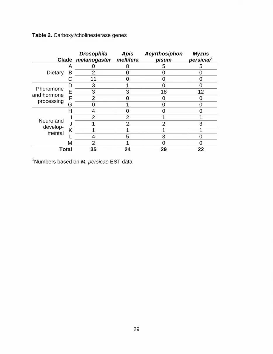
Table 2. Carboxyl/cholinesterase genes
Clade Drosophila
melanogaster Apis
mellifera Acyrthosiphon
pisum Myzus
persicae1
Dietary
A 0 8 5 5 B 2 0 0 0 C 11 0 0 0
Pheromone and hormone
processing
D 3 1 0 0 E 3 3 18 12 F 2 0 0 0 G 0 1 0 0
Neuro and develop-
mental
H 4 0 0 0 I 2 2 1 1 J 1 2 2 3 K 1 1 1 1 L 4 5 3 0 M 2 1 0 0
Total 35 24 29 22
1
Numbers based on M. persicae EST data

30
Table 3: Glutathione S-transferase genes
Class Drosophila
melanogaster Apis
mellifera Acyrthosiphon
pisum Myzus
persicaeDelta
1 11 1 10 8
Epsilon 14 0 0 0 Omega 5 1 0 0 Sigma 1 4 6 8 Theta 4 1 2 2
Zeta 2 1 0 0 Microsomal 1 2 2 2
Total 38 10 20 21
1
Numbers based on M. persicae EST data

31
Figure Legends Figure 1. Comparison of detoxification enzyme from A. pisum and M. persicae: (A)
cytochrome P450s, with clade names on the A pisum branches as listed in Table 1; (B)
esterases, with letters on the nodes representing the clades listed in Table 2 (C) and
GSTs, with class names on the branches as in Table 3 . Predicted protein coding genes
from A. pisum were compared to EST sequences from M. persicae using the Clustal
alignment algorithm and a neighbor-joining tree. A. pisum sequences with putative
orthologs in M. persicae are highlighted with green branches and those with no relatives
present in the M. persicae EST data are indicated with red branches. In some cases,
clear orthologs were not established due to many A. pisum sequences being grouped
with a single M. persicae sequence (blue branches) or a single A. pisum sequence
being grouped with many M. persicae sequences (orange branches). M. persicae
sequences for which there is no clear A. pisum homolog are shown with black
branches. A. pisum genes are represented by ACYPI numbers, and M. persicae genes
are represented by contig numbers from the current analysis
(http://www.aphidbase.com/aphidbase/downloads), with the exception of previously
studied esterases that are included as GenBank identifiers. The scale bars are
proportional to the number of amino acid changes between different proteins.
32
Figure 2. Alignment of known juvenile hormone esterase (JHE) sequences with the
potential pea aphid JHEs. Almost all known JHE enzymes possess a GQSAG motif (in
bold) in the nucleophillic elbow of the active site.

A
B C
No M. persicae HomologM. persicae orthologMultiple A. pisum + 1 M. persicaeMultiple M. persicae+ 1 A. pisumNo A. pisum Homolog
A E
E
I
L
K
J
Mt
3
33
3
33
3 3
3
33
3 3
3 3
3
4
Mt
3
4
34 2
44
4
Mt
22
3
3
3
2
4
4 4
44
4
4
Mt
Mt
3
4
3Mt
32
3
micro-somal
delta delta
sigma
sigma sigm
a
sigma
theta

Figure 1. Comparison of detoxification enzyme from A. pisum and M. persicae: (A)
cytochrome P450s, with clade names on the A pisum branches as listed in Table 1; (B)
esterases, with letters on the nodes representing the clades listed in Table 2 (C) and
GSTs, with class names on the branches as in Table 3 . Predicted protein coding genes
from A. pisum were compared to EST sequences from M. persicae using the Clustal
alignment algorithm and a neighbor-joining tree. A. pisum sequences with putative
orthologs in M. persicae are highlighted with green branches and those with no relatives
present in the M. persicae EST data are indicated with red branches. In some cases,
clear orthologs were not established due to many A. pisum sequences being grouped
with a single M. persicae sequence (blue branches) or a single A. pisum sequence
being grouped with many M. persicae sequences (orange branches). M. persicae
sequences for which there is no clear A. pisum homolog are shown with black
branches. A. pisum genes are represented by ACYPI numbers, and M. persicae genes
are represented by contig numbers from the current analysis
(http://www.aphidbase.com/aphidbase/downloads), with the exception of previously
studied esterases that are included as GenBank identifiers. The scale bars are
proportional to the number of amino acid changes between different proteins.
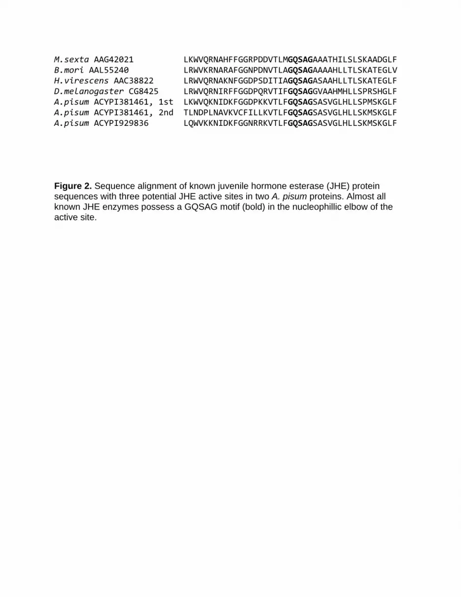
M.sexta AAG42021 LKWVQRNAHFFGGRPDDVTLMGQSAGAAATHILSLSKAADGLF B.mori AAL55240 LRWVKRNARAFGGNPDNVTLAGQSAGAAAAHLLTLSKATEGLV H.virescens AAC38822 LRWVQRNAKNFGGDPSDITIAGQSAGASAAHLLTLSKATEGLF D.melanogaster CG8425 LRWVQRNIRFFGGDPQRVTIFGQSAGGVAAHMHLLSPRSHGLF A.pisum ACYPI381461, 1st LKWVQKNIDKFGGDPKKVTLFGQSAGSASVGLHLLSPMSKGLF A.pisum ACYPI381461, 2nd TLNDPLNAVKVCFILLKVTLFGQSAGSASVGLHLLSKMSKGLF A.pisum ACYPI929836 LQWVKKNIDKFGGNRRKVTLFGQSAGSASVGLHLLSKMSKGLF Figure 2. Sequence alignment of known juvenile hormone esterase (JHE) protein sequences with three potential JHE active sites in two A. pisum proteins. Almost all known JHE enzymes possess a GQSAG motif (bold) in the nucleophillic elbow of the active site.