colletotrichum anthracnose of amaryllidaceae
TRANSCRIPT

Online advance Fungal Diversity
123
Colletotrichum anthracnose of Amaryllidaceae Yang, Y.L.1,2,3, Liu, Z.Y.2*, Cai, L.4, Hyde, K.D.5, Yu, Z.N.1 and McKenzie, E.H.C.6 1College of Life Science & Technology, Huazhong Agricultural University, Wuhan, 430070, P.R. China 2Guizhou Academy of Agricultural Sciences, Guiyang, Guizhou 550006 P.R. China 3Department of Biology and Geography, Liupanshui Normal College. Shuicheng, Guizhou 553006, P.R. China 4Novozymes China, No. 14, Xinxi Road, Shangdi, HaiDian, Beijing, 100085, PR China. 5School of Science, Mae Fah Luang University, Chiang Rai 57100, Thailand 6Landcare Research, Private Bag 92170, Auckland, New Zealand Yang, Y.L., Liu, Z.Y., Cai, L., Hyde, K.D., Yu, Z.N. and Mckenzie, E.H.C. (2009). Colletotrichum anthracnose of Amaryllidaceae. Fungal Diversity 39: 123-146. Twenty strains representing eight species of Colletotrichum were isolated from lesions of various species of amaryllids (monocotyledons, Amaryllidaceae) in Yunnan, Guangxi and Guizhou Provinces in China and Chiang Rai Province, Thailand. They are characterized through morphological studies and from phylogenetic analyses based on actin, β-tubulin (tub 2), calmodulin (CAL), chitin synthase A (CHS I), glyceraldehyde-3-phosphate dehydrogenase (GPDH) and the rDNA internal transcribed spacer (ITS) gene sequence data. Colletotrichum cliviae, C. hippeastri and C. hymenocallidis are new species described and illustrated in this paper based on morphological characters and multi-gene sequence data. Pathogenicity testing indicated that the three new taxa from Amaryllidaceae are not host-specific. Key words: anthracnose, multigene phylogeny, new species, Amaryllidaceae, pathogenicity
Article Information Received 30 November 2009 Accepted 3 December 2009 Published online 9 December 2009 *Corresponding author: Zuo-Yi Liu; e-mail: [email protected] Introduction
Colletotrichum is among the most im-portant genera of plant pathogenic fungi worldwide and species cause disease symptoms commonly known as anthracnose on a wide range of important crops, fruits and ornamental plants (Bailey et al., 1992). Some of the better known species include Colletotrichum linde-muthianum (Sacc. & Magnus) Briosi & Cavara, C. gloeosporioides (Penz.) Penz. & Sacc., C. acutatum J.H. Simmonds ex J.H. Simmonds, C. falcatum Went, C. nupharicola D.A. Johnson, Carris & J.D. Rogers and C. nymphaeae (Pass.) Aa. Some plant pathogenic species have high significance to quarantine because they could be introduced into countries where the disease does not occur (Farr et al., 2006).
The taxonomy and phylogeny of the genus Colletotrichum is confused (Hyde et al., 2009; Cai et al., 2009). Historically, if anthrac-nose was found on a host substratum for which no records of that fungus/host relationship were
known, then the species was interpreted as new, and formally described (Cannon et al., 2000). This practice was later rejected (von Arx 1957; Sutton 1980; Baxter et al., 1983), but new species are still occasionally described in Colletotrichum (Sivanesan et al., 1993; Naka-nura et al., 2006; Liu et al., 2007). There are now more than 688 epithets listed in Index fungorum (www. index fungorum.org/names /names.asp). Based on a morphological study, von Arx (1957) included more than 600 synonyms under C. gloeosporioides and 84 synonyms under C. dematium. Sutton (1992) listed 39 accepted species in Colletotrichum each with a short description.
In recent years, molecular tools have been employed to infer the evolutionary relationships of Colletotrichum species. Based on nu-rDNA ITS sequence data and morpho-logical characteristics, some species have been segregated from the Colletotrichum gloeo-sporioides complex, such as C. boninense Moriwaki, Toy. Sato & Tsukib. which has been

124
shown to cause disease of eight host plants in Japan (Moriwaki et al., 2003). Although ITS sequence data may help in Colletotrichum species identification, it cannot alone be used to adequately address species delimitation for closely related species (Crouch et al., 2009a). Researchers have recently tried to examine multiple genes sequence data to distinguish species in Colletotrichum (Du et al., 2005; Johnston et al., 1997; Crouch et al., 2006, 2009b,c; Farr et al., 2006; Shenoy et al., 2007a,b; Than et al., 2008a,b; Moriwaki and Tsukiboshi, 2009). Using multilocus phyloge-netics, Crouch et al. (2006; 2009b) separated C. cereale and C. eleusines from C. graminicola and introduced six new Colletotrichum species from warm-season grass hosts. Multigene sequence analyses are helpful in clarifying the confusion in the Colletotrichum systematics.
Amaryllids (members of the Amaryllida-ceae) are bulbous herbs with distichous or rarely rosulate leaves and a lateral, solid, naked scape which is terminated by an umbel-like cluster of showy flowers (Meerow et al., 1998). Several amaryllids have economic value as medicine, while species and/or hybrids of Nerine, Amaryllis and Crinum are cultivated in many countries for their elegant flowers (Dahlgren et al., 1985).
Colletotrichum species causing anthrac-nose on Amaryllidaceae have been reported from Japan (Horie et al., 1990; Moriwaki et al., 2003) and China (Liu et al., 2000). The disease symptoms include depressed reddish brown lesions on leaves or scapes, limiting the com-mercial production of ornamental and medici-nal plants (Liu et al., 2000). Colletotrichum boninense Moriwaki, Toy. Sato & Tsukib., C. crassipes (Speg.) Arx, C. dematium (Pers.) Grove and C. capsici (Syd.) E.J. Butler & Bisby have been reported to infect Amaryllis sp., Crinum asiaticum var. sinicum and Clivia miniata (Sutton, 1980; Moriwaki et al., 2003; http://194.203.77.76/herbIMI/Name.asp). In this study we use combined morphological and molecular characteristics to identify and characterize Colletotrichum species associated with anthracnose of Amaryllidaceae in China and Thailand.
Materials and methods Isolation of Colletotrichum
Colletotrichum samples were collected from anthracnose lesions on Clivia, Crinum, Hippeastrum, Hymenocallis (Amaryllidaceae) in Guangxi, Guizhou and Yunnan provinces, China and Chiang Rai Province, Thailand from June 2008 to September 2009 (Table 3). Isola-tion was carried out through two methods depending on the status of fungal sporulation. Isolates were obtained from lesions without visible sporulation using the procedure des-cribed by Photita et al. (2005). Single-spore isolations from infected leaves or scapes with sporulation were also carried out using the procedure described by Choi et al. (1999). Spore masses were picked up with a sterilized wire loop and streaked on to the surface of water agar (WA) plates which were then incubated overnight. A single germinated spore was picked up with a sterilized needle and transferred onto potato dextrose agar (PDA). Pure cultures were stored at 4°C on PDA slants. Isolates were deposited in Guizhou Academy of Agricultural Sciences, China and Mae Fah Luang University (MFLU) Culture Collection and National Centre for Genetic Engineering and Biotechnology (BIOTEC), Thailand. Morphological and cultural characterization
Starter cultures were prepared by plating each isolate onto PDA at 25°C. Five 4 mm plugs were aseptically cut from actively sporu-lating areas near the growing edge of a 5-day-old culture of each isolate using a sterile cork borer. Each plug was placed onto PDA plates (Petri dishes diameter: 90 × 15 mm) and grown in alternating 12 hours near UV/12 hours dark at 25ºC (Sutton, 1980). Colony diameter was measured at day six (for the fastest growing cultures, at day four). Growth rate was calcu-lated as the 6-day or 4-day average of mean daily growth (mm per day). After 7-10 days, size and shape of 50 conidia harvested from culture were assessed. The colour of the coni-dial masses and zonation were recorded at day seven. Mycelial appressoria were produced using a slide culture technique (Sutton, 1980),

Fungal Diversity
125
where 10 mm squares of potato carrot agar (PCA) were placed in an empty Petri dish, the edge of the PCA was inoculated with small sections of mycelia, and a cover slip was placed over the inoculated agar. After 5-14 days, appressoria formed across the underside of the cover slip and their shape and size were then recorded. Conidial appressoria of some strains with cylindrical spores were also recorded. Conidial appressoria were induced by inoculating with conidia in two drops of distilled water on a microscope slide, then placing it inside Petri dishes containing moistened cotton with distilled sterile water, and incubated at 25ºC in darkness. After incubating 24 hours, conidial appressoria were formed from germ tube and then measured. DNA extraction and sequencing
DNA was extracted from all isolates growing on PDA at room temperature for 8-10 days using a modified protocol of Chen et al. (2007). The gene regions were amplified using the primers listed in Table 1. The PCR amplifi-cations were performed in a 25 µl mixture containing 9.5µl ddH2O, 12.5 µl 2×PCR Master Mix (TIANGEN Co. China), 1 µl of DNA templates, 1 µl of each primers (10 µM). The reactions were performed with a thermal cycler (MyclerTM, Bio-Rad, Hercules, CA, USA) using the following thermal program: 94°C for 5 min, followed by 35 cycles of denaturation (94°C for 30 s), annealing (30 s at 59°C for Actin, CAL and β-tubulin, 56°C for GPDH and CHS I, and 53°C for ITS), elongation (72°C for 90 s), and a final 7 min extension at 72°C. PCR products were examined by electrophoresis stained with ethidium bromide, and purified according to the manufacturer’s instructions of a TIANGEL Mini Purification kit (TIANGEN Co. China). Purified samples were sequenced using the above-mentioned PCR primers in an Applied Biosystems 3730xl DNA Analyzers at Sino-max Co., China. Two of gene amplicon, CAL and β-tubulin, were cloned into DH5α (TIANGEN Co., China) with pMD18-T vector (Takara), and then sequenced at the same company. Molecular phylogenetic analysis
Phylogenetic analysis was performed
using six gene regions. The accession numbers of all sequences are listed in Table 3. Multiple sequence alignments were generated using ClustalX 2.0.10 (Larkin et al., 2007). Gaps were treated as missing data.
Each of the single and combined sequence alignments were analyzed using maximum parsimony (MP) in PAUP* 4b10. Ambiguously aligned regions were excluded from all analyses. Trees were inferred using the heuristic search option with tree bisection-reconnection (TBR) branch swapping and 1000 random sequence additions. Maxtrees were unlimited, branches of zero length were collapsed and all multiple parsimonious trees were saved. Descriptive tree statistics such as tree length [TL], consistency index [CI], retention index [RI], rescaled consistency index [RC], and homoplasy index [HI] were calcu-lated for trees generated under different optimality criteria. Clade stability of the trees resulting from the parsimony analyses were assessed by bootstrap analysis with 1000 replicates. Trees were figured in Treeview. When analyzing ITS and β-tubulin sequence, some reference sequences were obtained from GeneBank (Table 2).
The model of evolution was estimated by using Mrmodeltest 2.2 (Nylander, 2004). Posterior probabilities (PP) (Rannala and Yang, 1996; Zhaxybayeva and Gogarten, 2002) were determined by Markov Chain Monte Carlo sampling (BMCMC) in MrBayes 3.0b4 (Huel-senbeck and Ronquist, 2001). Six simultaneous Markov chains were run for 3,000,000 genera-tions and trees were sampled every 100th generations (resulting 30,000 total trees). The first 2,000 trees, which represented the burn-in phase of the analyses, were discarded and the remaining 28,000 trees were used for calculating posterior probabilities (PP) in the majority rule consensus tree.
Pathogenicity testing and host range
A representative isolate of each species from Amaryllidaceae was selected for patho- genicity testing (Table 6). For testing the host range, 12 plant species were selected belonging to 6 families (Table 6), most being hosts of Colletotrichum species that are morphologi-cally similar to our isolates from Amaryllida-ceae. Single spore cultures of each isolate were

126
Table 1. Primers used for PCR amplification and DNA sequencing. Gene primer Primer sequences Reference
ITS1 TCCGTAGGTGAACCTGCGG ITS ITS4 TCCTCCGCTTATTGATATGC
White et al., 1990
ACT-512F ATGTGCAAGGCCGGTTTCGC Actin ACT-783R TACGAGTCCTTCTGGCCCAT
Carbone and Kohn., 1999
CL1 GARTWCAAGGAGGCCTTCTC CAL CL2 TTTTTGCATCATGAGTTGGAC
Johnston, pers. comm.
CHS I-79F TGGGGCAAGGATGCTTGGAAGAAG CHS I CHS I-354R TGGAAGAACCATCTGTGAGAGTTG
Carbone et al., 1999
GDF1 GCCGTCAACGACCCCTTCATTGA GPDH GDR1 GGGTGGAGTCGTACTTGAGCATGT
Guerber et al., 2003 Templeton et al., 1992
T1 AACATGCGTGAGATTGTAAGT O’Donnell and Cigelnik, 1997 β-tubulin βt2b ACCCTCAGTGTAGTGACCCTTGGC Glass and Donaldson, 1995
grown on PDA for 7 days at 25°C. Spores were then harvested by adding 10 ml of sterilized
distilled water onto the culture, which was then gently swirled to dislodge the conidia with
the concentration adjusted to 106conidia/ml using a haemocytometer, and used as the standard inoculum for pathogenicity and host range testing. The conidial suspension was filtered through two layers of muslin cloth (Than et al., 2008a). Healthy leaves or fruits were picked and washed with tap water; and then disinfected in 1% sodium hypochlorite for 5-7 minutes (disinfection time was adjusted according to whether the surface was smooth or coarse). Disinfected leaves and fruits were washed three times with distilled, sterilized water, and then dried with sterilized filter paper. Leaves or fruits were kept individually in a 12 cm diam Petri dish or tissue culture bottle (for tomato and grape fruit) with a swab of cotton wall (about 1 cm in diam.) containing distilled water to maintain humidity.
The inoculation method has been described by Than et al. (2008a). The wound /drop inoculation method involved pin pricking the leaf or fruit wall to a 1 mm depth and then placing 6 µl of conidia suspension (106coni-dia/ml) onto the wound /non wound. Control fruits and leaves were inoculated with 6 µl of sterilized, distilled water onto the wound. Each plant was inoculated with four replicates per selected strain. The inoculated fruits and leaves were then incubated at 25°C for 24 hours and then at room temperature (22-27°C). After 6-7 days and 14-15 days of incubation leaves and fruits were checked for lesions with a stereo-microscope on a superclean bench. Spore masses or acervuli on leaves or fruits were checked with a compound microscope and spores were transferred to PDA medium, and
then incubated at 25°C for checking the colony and spore characters. All infected leaves or fruits were sterilized and then disposed. Results Collection of Colletotrichum species
Colletotrichum isolates used in this study are listed in Table 3. Twenty strains of Colletotrichum were isolated from anthracnose associated with Clivia, Crinum, Hippeastrum and Hymenocallis (Amaryllidaceae). Morpho-logical examination classified twenty isolates into 6 morphogroups; while analysis of ITS sequence reveal 8 phylogenetic lineages (see below). For comparison, reference strains of several important Colletotrichum species were included in this study including Colletotrichum simmondsii R.G. Shivas & Y.P. Tan (holotype, BRIP 28519), C. truncatum (Schwein.) Andrus & W.D. Moore (CBS 120709), C. coccodes (Wallr.) S. Hughes (CPOS1), C. gloeospor-ioides (epitype, CBS 953.97) and C. trichellum (Fr.) Duke (HKUCC 10378). Phylogenetic analysis
The ITS dataset comprised 570 charac-ters after alignment, of which 138 characters were parsimony informative (24.2%). The Kishino-Hasegawa (KH) test showed that 556 trees generated from parsimonious analysis were not significantly different. One of the most parsimonious trees (TL =549, CI = 0.692, RI = 0.867, RC = 0.600, HI = 0.308) (Table 4.) is shown in Fig. 1. The dataset of combined six genes comprised 2973 characters after align-

Fungal Diversity
127
Table 2. ITS and β-tubulin sequences obtained from GenBank for analysis.
Anamorph/ teleomorph Strain GenBank
no. ITS GeneBank no. β-tub2 Host Country of origin Reference
Colletotrichum agaves
AR3920 DQ286221 Agave striata Mexico Farr et al., 2006
CBS318 DQ286219 Agave americana Netherlands Farr et al., 2006 C. asianum 2BPD-I4 FJ972612 Coffea arabica Thailand Prihastuti et al., 2009 BML-I14 FJ972615 Coffea arabica Thailand Prihastuti et al., 2009 C. boninense 2MAFF 305972 AB051400 Crinum asiaticum var.
sinicum Japan Moriwaki et al., 2003
3MAFF 306094 AB051403 Crinum asiaticum var. sinicum
Japan Moriwaki et al., 2003
C. caudatum STE-U 5300 AY376575 Cymbopogon martinii India Lubbe et al., 2004 C. circinans BBA67864 AJ301955 Allium schoenoprasum Germany Nirenberg et al., 2002 C. coccodes BBA70879 AJ301957 Solanum tuberosum Germany Nirenberg et al., 2002 ATCC 58682 FJ545227 Capsicum annuum New York, USA Unpublished C. dematium 1CBS125.25 * * * Damm et al., 2009 CBS125340 * * * Damm et al., 2009 C. crassipes STU-4445 AY376530 AY376578 Dryandra sp. Madeira Lubbe et al., 2004 STE-U 5302 AY376529 AY376577 Dryas octopetala Switzerland Lubbe et al., 2004 C. destructivum CD-hz 01 EU070911 Phaseolus limensis China Unpublished ATCC11995 AF320563 Nicotiana tabacum Canada Shen et al., 2001 C. dracaenophilum MEP1537 DQ286207 Dracaena sp. China Farr et al. 2006 CBS 121453 EU003533 Dracaena sanderiana Bulgaria Unpublished C. fructicola 2BPD-I16 FJ972603 Coffea arabica Thailand Prihastuti et al., 2009 BPD-I12 FJ972611 Coffea arabica Thailand Prihastuti et al., 2009 C. gloeosporioides 1IMI 356878 EU371022 Citrus sinensis Italy Cannon et al., 2008 BBA71473 AJ301988 Citrus aurantiacus Germany Nirenberg et al., 2002 STE-U 5297 AY376582 Citrus sp. Belize Lubbe et al., 2004 STE-U 4295 AY376580 Citrus sp. Italy Lubbe et al., 2004

128
Table 2 (continued). ITS and β-tubulin sequences obtained from GenBank for analysis. Anamorph/ teleomorph Strain GenBank
no. ITS GeneBank no. β-tub2 Host Country of origin Reference
Colletotrichum. graminicola
MAFF 306612 AB233343 unkown Japan Unpublished
DR 1 AF059676 Poa annua USA Unpublished STE-U 5298 AY376587 Zea mays Zimbabwe Lubbe et al., 2004 C. lupini BBA 70385 AJ301935 Lupinus angustifolius Germany Nirenberg et al., 2002 BBA 63879 AJ301930 Lupinus mutabilis Germany Nirenberg et al., 2002 C. orbiculare MAFF306684 AB275876 Cucumis melo Japan Unpublished C. siamense 2BML-I2 FJ972613 Coffea arabica Thailand Prihastuti et al., 2009 BML-I15 FJ972614 Coffea arabica Thailand Prihastuti et al., 2009 C. spaethianum 2CBS167.49 * * * Damm et al., 2009 CBS_100063 * * * Damm et al., 2009 C. spinaciae BBA71333 AJ301973 Spinacia oleracea Germany Nirenberg et al. 2002 C. sublineolum BBA71362 AJ301978 Sorghum bicolor Germany Nirenberg et al., 2002 C. trichellum BBA71091 AJ301989 Hedera helix Germany Nirenberg et al., 2002 C. truncatum 1CBS151.35 * * * Damm et al., 2009 CBS120709 * * * Damm et al., 2009 C. yunnanense 2AS3.9617 EF369490 Buxus sp. China Liu et al., 2007 3AS3.9616 EF369491 Buxus sp. China Liu et al., 2007 Glomerella acutata STE-U 4466 AY376567 Hakea sericea South Africa Lubbe et al., 2004 G. lagenaria BBA71046 AJ301965 Cucumis sativus Germany Nirenberg et al., 2002 Fusarium oxysporum ATCC MYA-
3970 FJ614650 unknown USA Unpublished
1, epitype; 2, holotype; 3, paratype; *, See the same issue.
Fungal Diversity
129
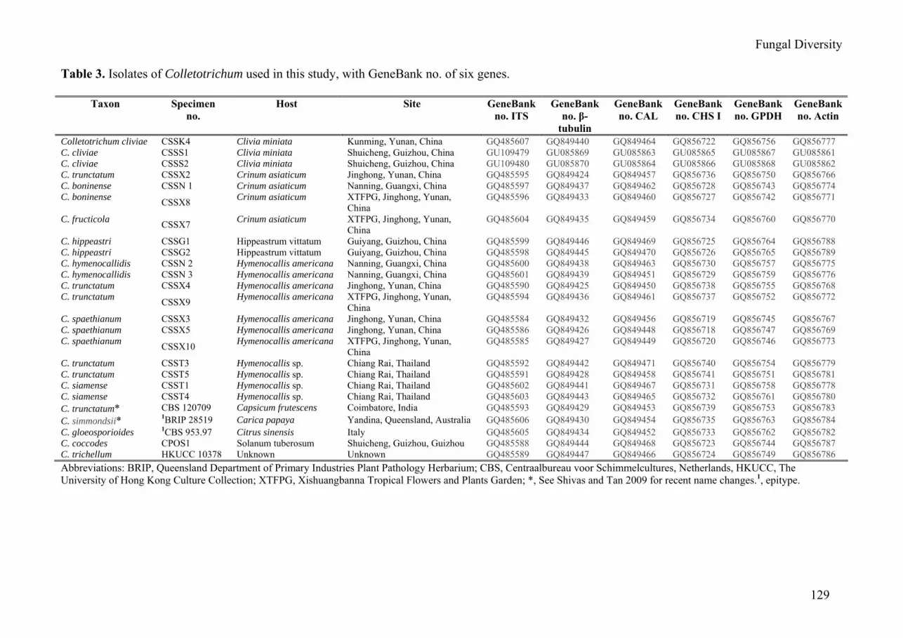
Table 3. Isolates of Colletotrichum used in this study, with GeneBank no. of six genes.
Taxon Specimen no.
Host Site GeneBank no. ITS
GeneBank no. β-
tubulin
GeneBank no. CAL
GeneBank no. CHS I
GeneBank no. GPDH
GeneBank no. Actin
Colletotrichum cliviae CSSK4 Clivia miniata Kunming, Yunan, China GQ485607 GQ849440 GQ849464 GQ856722 GQ856756 GQ856777 C. cliviae CSSS1 Clivia miniata Shuicheng, Guizhou, China GU109479 GU085869 GU085863 GU085865 GU085867 GU085861 C. cliviae CSSS2 Clivia miniata Shuicheng, Guizhou, China GU109480 GU085870 GU085864 GU085866 GU085868 GU085862 C. trunctatum CSSX2 Crinum asiaticum Jinghong, Yunan, China GQ485595 GQ849424 GQ849457 GQ856736 GQ856750 GQ856766 C. boninense CSSN 1 Crinum asiaticum Nanning, Guangxi, China GQ485597 GQ849437 GQ849462 GQ856728 GQ856743 GQ856774 C. boninense CSSX8 Crinum asiaticum XTFPG, Jinghong, Yunan,
China GQ485596 GQ849433 GQ849460 GQ856727 GQ856742 GQ856771
C. fructicola CSSX7 Crinum asiaticum XTFPG, Jinghong, Yunan, China
GQ485604 GQ849435 GQ849459 GQ856734 GQ856760 GQ856770
C. hippeastri CSSG1 Hippeastrum vittatum Guiyang, Guizhou, China GQ485599 GQ849446 GQ849469 GQ856725 GQ856764 GQ856788 C. hippeastri CSSG2 Hippeastrum vittatum Guiyang, Guizhou, China GQ485598 GQ849445 GQ849470 GQ856726 GQ856765 GQ856789 C. hymenocallidis CSSN 2 Hymenocallis americana Nanning, Guangxi, China GQ485600 GQ849438 GQ849463 GQ856730 GQ856757 GQ856775 C. hymenocallidis CSSN 3 Hymenocallis americana Nanning, Guangxi, China GQ485601 GQ849439 GQ849451 GQ856729 GQ856759 GQ856776 C. trunctatum CSSX4 Hymenocallis americana Jinghong, Yunan, China GQ485590 GQ849425 GQ849450 GQ856738 GQ856755 GQ856768 C. trunctatum CSSX9 Hymenocallis americana XTFPG, Jinghong, Yunan,
China GQ485594 GQ849436 GQ849461 GQ856737 GQ856752 GQ856772
C. spaethianum CSSX3 Hymenocallis americana Jinghong, Yunan, China GQ485584 GQ849432 GQ849456 GQ856719 GQ856745 GQ856767 C. spaethianum CSSX5 Hymenocallis americana Jinghong, Yunan, China GQ485586 GQ849426 GQ849448 GQ856718 GQ856747 GQ856769 C. spaethianum CSSX10 Hymenocallis americana XTFPG, Jinghong, Yunan,
China GQ485585 GQ849427 GQ849449 GQ856720 GQ856746 GQ856773
C. trunctatum CSST3 Hymenocallis sp. Chiang Rai, Thailand GQ485592 GQ849442 GQ849471 GQ856740 GQ856754 GQ856779 C. trunctatum CSST5 Hymenocallis sp. Chiang Rai, Thailand GQ485591 GQ849428 GQ849458 GQ856741 GQ856751 GQ856781 C. siamense CSST1 Hymenocallis sp. Chiang Rai, Thailand GQ485602 GQ849441 GQ849467 GQ856731 GQ856758 GQ856778 C. siamense CSST4 Hymenocallis sp. Chiang Rai, Thailand GQ485603 GQ849443 GQ849465 GQ856732 GQ856761 GQ856780 C. trunctatum* CBS 120709 Capsicum frutescens Coimbatore, India GQ485593 GQ849429 GQ849453 GQ856739 GQ856753 GQ856783 C. simmondsii* 1BRIP 28519 Carica papaya Yandina, Queensland, Australia GQ485606 GQ849430 GQ849454 GQ856735 GQ856763 GQ856784 C. gloeosporioides 1CBS 953.97 Citrus sinensis Italy GQ485605 GQ849434 GQ849452 GQ856733 GQ856762 GQ856782 C. coccodes CPOS1 Solanum tuberosum Shuicheng, Guizhou, Guizhou GQ485588 GQ849444 GQ849468 GQ856723 GQ856744 GQ856787 C. trichellum HKUCC 10378 Unknown Unknown GQ485589 GQ849447 GQ849466 GQ856724 GQ856749 GQ856786 Abbreviations: BRIP, Queensland Department of Primary Industries Plant Pathology Herbarium; CBS, Centraalbureau voor Schimmelcultures, Netherlands, HKUCC, The University of Hong Kong Culture Collection; XTFPG, Xishuangbanna Tropical Flowers and Plants Garden; *, See Shivas and Tan 2009 for recent name changes.1, epitype.
130
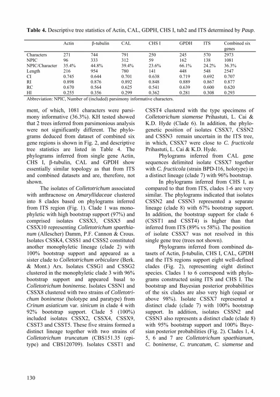
Table 4. Descriptive tree statistics of Actin, CAL, GDPH, CHS I, tub2 and ITS determined by Paup. Actin β-tubulin
CAL CHS I GPDH ITS Combined six
genes Characters 271 744 791 250 245 570 2973 NPIC 96 333 312 59 162 138 1081 NPIC/Character 35.4% 44.8% 39.4% 23.6% 66.1% 24.2% 36.3% Length 216 954 780 141 448 548 2547 CI 0.745 0.644 0.701 0.638 0.719 0.692 0.707 RI 0.898 0.876 0.892 0.848 0.889 0.867 0.877 RC 0.670 0.564 0.625 0.541 0.639 0.600 0.620 HI 0.255 0.356 0.299 0.362 0.281 0.308 0.293 Abbreviation: NPIC, Number of (included) parsimony informative characters. ment, of which, 1081 characters were parsi-mony informative (36.3%). KH tested showed that 2 trees inferred from parsimonious analysis were not significantly different. The phylo-grams deduced from dataset of combined six gene regions is shown in Fig. 2, and descriptive tree statistics are listed in Table 4. The phylograms inferred from single gene Actin, CHS I, β-tubulin, CAL and GPDH show essentially similar topology as that from ITS and combined datasets and are, therefore, not shown.
The isolates of Colletotrichum associated with anthracnose on Amaryllidaceae clustered into 8 clades based on phylograms inferred from ITS region (Fig. 1). Clade 1 was mono-phyletic with high bootstrap support (97%) and comprised isolates CSSX3, CSSX5 and CSSX10 representing Colletotrichum spaethia-num (Allescher) Damm, P.F. Cannon & Crous. Isolates CSSK4, CSSS1 and CSSS2 constituted another monophyletic lineage (clade 2) with 100% bootstrap support and appeared as a sister clade to Colletotrichum orbiculare (Berk. & Mont.) Arx. Isolates CSSG1 and CSSG2 clustered in the monophyletic clade 3 with 96% bootstrap support and appeared basal to Colletotrichum boninense. Isolates CSSN1 and CSSX8 clustered with two strains of Colletotri-chum boninense (holotype and paratype) from Crinum asiaticum var. sinicum in clade 4 with 92% bootstrap support. Clade 5 (100%) included isolates CSSX2, CSSX4, CSSX9, CSST3 and CSST5. These five strains formed a distinct lineage together with two strains of Colletotrichum truncatum (CBS151.35 (epi-type) and CBS120709). Isolates CSST1 and
CSST4 clustered with the type specimens of Colletotrichum siamense Prihastuti, L. Cai & K.D. Hyde (Clade 6). In addition, the phylo-genetic position of isolates CSSX7, CSSN2 and CSSN3 remain uncertain in the ITS tree, in which, CSSX7 were close to C. fructicola Prihastuti, L. Cai & K.D. Hyde.
Phylograms inferred from CAL gene sequences delimited isolate CSSX7 together with C. fructicola (strain BPD-I16, holotype) in a distinct lineage (clade 7) with 96% bootstrap.
In phylograms inferred from CHS I, as compared to that from ITS, clades 1-6 are very similar. The phylograms indicated that isolates CSSN2 and CSSN3 represented a separate lineage (clade 8) with 67% bootstrap support. In addition, the bootstrap support for clade 6 (CSST1 and CSST4) is higher than that inferred from ITS (89% vs 58%). The position of isolate CSSX7 was not resolved in this single gene tree (trees not shown).
Phylograms inferred from combined da-tasets of Actin, β-tubulin, CHS I, CAL, GPDH and the ITS regions support eight well-defined clades (Fig. 2), representing eight distinct species. Clades 1 to 6 correspond with phylo-grams constructed using ITS and CHS I. The bootstrap and Bayesian posterior probabilities of the six clades are also very high (equal or above 98%). Isolate CSSX7 represented a distinct clade (clade 7) with 100% bootstrap support. In addition, isolates CSSN2 and CSSN3 also represents a distinct clade (clade 8) with 95% bootstrap support and 100% Baye-sian posterior probabilities (Fig. 2). Clades 1, 4, 5, 6 and 7 are Colletotrichum spaethianum, C. boninense, C. truncatum, C. siamense and

Fungal Diversity
131
Fig. 1. Maximum parsimony phylograms inferred from ITS sequence data showing phylogenetic relationships among isolates of Colletotrichum from Amaryllidaceae in southwest China and Chiang Rai, Thailand (bold) and selected sequences of Colletotrichum species (of which, some are epitypes). Data analyzed with random addition sequence, unweighted parsimony and treating gaps as missing data. Values above the branches are parsimony bootstrap (equal or above 50%). The tree is rooted with Fusarium oxysporum (ATCC MYA- 3790).
10
CSSX4
CSSX9
CSSX2
CSST5
CSST3
CSSK4
CSSS2
CSSS1
CSSX8
CSSN1
CSSG2
CSSG1
CSSN2
CSSN3
CSST1
CSST4
CSSX7
CSSX3
CSSX5
CSSX10
C. gloeosporioides CBS 953.97 epitype
CPOS1 C. coccodes isolated from potato
C. trichellium HKUCC 10378
C. simmondsii BRIP 28519 holotype outgroup
100
100
100
100
100
100
100
100
100
98
95
64
100
100
67
100
100
clade 2 C. cliviae
clade 6 C. siamense
clade5 C. truncatum
clade 4 C. boninense
clade 3 C. hippeastri
clade 1 C. spaethianum
clade 7 C. fructicola
clade 8 C. hymenocallidis

132
Fig. 2. Maximum parsimony phylograms inferred from combined partial Actin, β-tubulin, CHS I, CAL, GPDH and ITS sequence data, showing phylogenetic relationships among isolates of Colletotrichum from Amaryllidaceae in southwest China, and Chiang Rai Province, Thailand and selected sequences of Colletotrichum species (some are epitypes). Data analyzed with random addition sequence, unweighted parsimony and treating gaps as missing data. Values above the branches are parsimony bootstrap (equal or above 50%). Thickened branches represent significant Bayesian posterior probabilities (equal or above 95%). The tree is rooted with C. simmondsii (BRIP 28519 epitype). The clade number corresponds with phylograms deduced from ITS. C. fructicola, respectively. Clades 2, 3 and 8 represent three undescribed Colletotrichum species. Full descriptions and illustrations of these three species are provided in the following taxonomy section.
Taxonomy Eight species of Colletotrichum causing
anthracnose of plants in Amaryllidaceae are listed in (Tables 3, 5). Three of these are clearly different from all currently known
10
CSST1
CSST4
CSSX7
CSSN2CSSN3
CSSX4C truncatum CBS120709C truncatum CBS151.35 epitypeCSSX2CSSX9CSST3CSST5
CSSN1CSSX8
CSSG2CSSG1
CSSK4CSSS2CSSS1
CSSX3C spaethianum CBS 100063C spaethianum CBS167.49 ex-epitypCSSX5CSSX10
C dematium CBS125.25 epitypeC dematium CBS125340
FJ 972613 C. siamense BPD-I2 holotype FJ 972614 C. siamense BML-I15
C.gloeosporioides CBS 953.97 AJ301988 C. gloeosporioides BBA 71473 EU371022 C.gloeosporioides IMI 356878 epitype
FJ 972611 C. fructicola BPD-I12 FJ 972603 C. fructicola BPD-I16 holotype FJ 972615 C. asianum BML-I14 FJ 972612 C. asianum BPD-I4 holotype
AJ301929 C. musae BBA70365 AJ301904 C. musae BBA62471
AY376529 C. crassipes STE-U 5302 AY376530 C. crassipes STU-4445
AB051400 C. boninense holotype
AB051403 C. boninense paratype
AB275876 C. orbiculare MAFF306684 AJ301965 Glomerella lagenaria BBA71046
EF369490 C. yunnanense AS3.9617 paratype EF369491 C. yunnanense AS3.9616 holotype DQ286207 C. dracaenophilum MEP1537 EU003533 C. dracaenophilum CBS 121453
DQ286219 C. agaves strain CBS318 DQ286221 C. agaves AR3920
AB233343 C. graminicola MAFF 306612 AF059676 C. graminicola DR1
EU070911 C. destructivum CD-hz 01 AF320563 C. destructivum ATCC11995
CPOS1 C. coccodes isolated from potato AJ301957 C. coccodes BBA 708797
AJ301955 C. circinans BBA67864 AJ301973 C. spinaciae BBA71333
C. trichellium HKUCC 10378 AJ301989 C. trichellum BBA71091
AJ301978 C. sublineolum BBA71362 AJ301935 C. lupini BBA 70385 AJ301930 C. lupini BBA 63879
C. simmondsii BRIP 28519 holotype FJ614650 Fusarium oxysporum ATCC MYA-3970 outgroup
72
97
100
92
65
91
54
55
81
58
96
98
100
100
100
61
97
99
99
100 95
100
73
96
99
74
100
100
99
97
76

Fungal Diversity
133
species of Colletotrichum (Hyde et al., 2009; Sutton, 1992) and are therefore described and illustrated as new species.
Colletotrichum boninense Moriwaki, Toy. Sato & Tsukib.
This species occurs on leaves of Crinum asiaticum as reddish brown, ellipsoid to irregular spots and forms pink conidial masses with rare setae (Fig. 6A). In this study, although the conidia of C. boninense (CSSN1 and CSSX8) are wider than those of the holotype and paratype, their shape and cultural characteristics are similar and they cluster in one clade with high bootstrap support (there is one base difference in the ITS sequence among them and the holotype). In addition, the holotype and paratype of C. boninense was collected from Crinum asiaticum var. sinicum in Japan.
Material examined: CHINA, Yunnan Province, Jinghong, on leaf of Crinum asiaticum, 2 August 2008, Y.L. Yang. (GZAAS 080009, ex-living culture CSSX8); China, Guangxi Province, Nanning, on leaf of Crinum asiaticum, 19 June 2008, Y.L. Yang. (GZAAS 080006, ex-living culture CSSN1). Colletotrichum cliviae Y.L. Yang, Zuo Y. Liu, K.D. Hyde & L. Cai, sp. nov. (Fig. 3) MycoBank: 515279
Etymology: cliviae, in reference to the host Clivia miniata.
In agaro decoct tuberosum post 5 dies colonae est 7.4-7.7 cm diam, albae vel griseae. Ad margine griseo-albus, densus, reversum atrobrunnea vel pallens nigri cum pallide bubalinus massa conidiales. Sclerotia presentia, setosa. Setae presentia, basi medie brunneae, 1-3-septatae, 75-105 × 4-6 µm, acuta ad apicem. Conidiophora hyalinae, 22.5-49 × 4-5.5 µm, 1-4-septatae. Conidia in pallide bubalinus massa, 19.5-24.5 × 4.5-7µm, aseptata, laevia, hyalina,cylindrica, rectae vel laeviter curvus, ad apicem obtusa, Appressoria medie brunneae, irregularis, crenate vel lobata, 10.5-14.5 × 6-11 µm.
Habitate: in foliis Clivia miniata. Holotypus: Cultura (CSSK4), isolata e foliis
morbo affectis Clivia miniata, Yunnan, China, 2008. Paratypus: Cultura (CSSS2), isolata e foliis morbo affectis Clivia miniata, Guizhou, China, 2009.
Description: On host, acervuli circular to elliptical, arranged irregularly, subepidermal, disrupting outer epidermal cell wall of host, setae present, with pale yellow conidial masses (Fig. 3A). Setae 95-210 × 6-7 µm (x = 160 ± 43.2 × 6.5 ± 0.5, n = 5), moderately brown, smooth-walled, 1-3-septate, tapered and paler
toward the apex. Conidiophores hyaline, 1-3-celled, branched or unbranched at the base, 18-36.5 × 4-6.5 µm ( x = 29.4 ± 7.7 × 5.2 ± 0.9, n = 10). Conidia 15-24 × 4.5-6.5 µm (x = 20.5 ± 2.2 × 5.8 ± 0.5, n = 20), one-celled, smooth-walled, hyaline, straight, obtuse at the ends.
Colonies on PDA, attaining 7.4-7.7 cm (x = 7.5 ± 1.1, n = 10) diam. in 5 days at 25°C, growth rate 15.2-16 mm per day (x = 15.6 ± 0.2, n = 10); at first white, becoming grey with age, greyish-white at margin, dense, reverse dark brown to greenish-black (Figs 3B, C), with pale buff conidial masses. Sclerotia pre-sent, setose. Setae present, brown, 75-105 × 4-6 µm ( x = 85 ± 10.5 × 5 ± 0.7, n = 5), 1-3-septate, with tapering acute apices. Conidio-phores hyaline, 22.5-49 × 4-5.5 µm (x = 33.3 ± 8.7 × 4.7 ± 0.6, n = 10), 1-4-celled, branched or unbranched at the base (Figs 3D, G). Conidia in pale buff masses, 19.5-24.5 × 4.5-7 µm (x = 21.8 ± 1.4 × 5.7 ± 0.5, n = 100), one-celled, smooth-walled, hyaline, cylindrical, straight or slightly curved, obtuse at the ends (Figs 3E, H-I). Appressoria brown, irregular, crenate or lobed, 10.5-14.5 × 6-11 µm (x = 11.7 ± 1.2 × 8.6 ± 1.2, n = 40) (Figs 3F, J-K).
Teleomorph: not produced in culture. Holotype: CHINA, Yunnan Province,
Kunming, on leaf of Clivia miniata, 10 August 2008, Y.L. Yang (GZAAS 080005; ex-holo-type living culture CSSK4, CBS 125375).
Known host and distribution: Clivia miniata, Kunming and Shuicheng, China.
Additional specimens examined: CHINA, Guizhou Province, Shuicheng, on leaf of Clivia miniata, 20 September 2009, Y.L. Yang (GZAAS 080018, ex-paratype living culture CSSS2). Colletotrichum fructicola Prihastuti, L. Cai & K.D. Hyde
On leaves of Crinum asiaticum with anthracnose caused by this species occurring as yellowish brown ellipsoid spots, acervuli without setae, with yellowish-white conidial masses (Fig.6D).
Material examined: CHINA, Yunnan Province, Jinghong, on leaf of Crinum asiaticum, 1 August 2008, Y.L. Yang (GZAAS 080019, ex-living culture, CSSX7). Colletotrichum hippeastri Y.L. Yang, Zuo Y. Liu, K.D. Hyde & L. Cai, sp. nov.
(Fig. 4) MycoBank: 515280.

134
Table 5. Colletotrichum species on Amaryllidaceae.
Species and Isolate numbers
Conidial shape and size (µm)
Mycelial appressoria shape and size (µm)
Conidial appressoria shape and size (µm)
Colony characteristics
Mycelial growth rate(mm per day)
Host(s)
C. boninense (CSSN1, CSSX8)
Cylindrical, with a hilum-like base, 13-19.5 × 4.5-8, x =16.3 ± 1.2 × 6.6 ± 0.7, n = 100
Irregular, sepia to dark brown, (8.5-)10-12(-15) × 6.5-11, x = 11.4 ± 1.5 × 8.5 ± 1.1 n = 20
Irregular, sepia to dark brown, 6.5-9.5 (-11.5) × 5.0-7.5, x = 8.8 ± 0.8 × 6.4 ± 0.7, n = 40
White aerial mycelia; reverse cream to reddish orange
9.8-11, x = 10.6 ± 0.4, n = 10
Crinum asiaticum
C. cliviae (CSSK4, CSSS1, CSSS2)
Cylindrical, straight or slightly curved, obtuse at the ends, 19.5-24.5 × 4.5-7, x = 21.8 ± 1.4 × 5.7 ± 0.5, n = 100
Not produced Dark brown, irregular, crenate or lobed, 10.5-14.5 × 6-11 µm, x = 11.7 ± 1.2 × 8.6 ± 1.2, n = 40
White to grey, white at margin, reverse dark brown to greenish black
15.2-16 , x = 15.6 ± 0.2, n = 10
Clivia miniata
C. fructicola (CSSX7)
Cylindrical, with obtuse to slightly rounded ends, 8.5-16×3.5-5, x = 12.7 ± 1.7 × 4.1 ± 0.4, n = 50
Clavate to ovate, medium brown, 7.5-13.5×5-8, x = 10 ± 1.5 × 6.3 ± 0.9, n = 20
Clavate to ovate, medium brown, 6-9 × 5.5-7, x = 7.2 ± 0.7 × 5.9 ± 0.4, n = 20)
White, grey to dark grey with age, reverse black, circular around at the centre
11.4-13.4 x = 12.3 ± 0.7, n = 5
Crinum asiaticum
C. hippeastri (CSSG1, CSSG2)
Cylindrical, straight, usually slightly curved, obtuse at the ends, usually constricted near each end or centre, 19.5-40.5 (42.5) × 7-10.5 (-12), x = 29.2 ± 5.5 × 8.8 ± 1.0,n = 100
Not produced Medium to dark brown, irregular, crenate or lobed, occasionally becoming complex, 10.5-17.5 × 8-12.5, x = 13 ± 1.7 × 10 ± 1.1,n = 40
Pale white to black, floccose, sparse, reverse pale white-black
13.6-14.2 x = 13.9 ± 0.2, n = 10
Hippeastrum vittatum

Fungal Diversity
135
Table 5 (continued). Colletotrichum species on Amaryllidaceae.
Species and Isolate numbers
Conidial shape and size (µm)
Mycelial appressoria shape and size (µm)
Conidial appressoria shape and size (µm)
Colony characteristics
Mycelial growth rate(mm per day)
Host(s)
C. hymenocallidis (CSSN2, CSSN3)
Fusiform, straight, obtuse at the ends, 14-18.5(-20) × 5-6.5, x = 15.9 ± 1.1 × 5.1 ± 0.4, n = 100
Ovate to clavate, margin entire, medium brown, 6-13 × 5-7, x = 9.4 ± 3 ×5.9 ± 0.7, n = 5
Ovate, sometimes clavate, medium brown, 7-11×5-7.5, x = 8.5 ± 0.9 × 6.6 ± 0.6, n = 40
White to becoming pale grey with circles, reverse greenish black
8.8-11, x = 9.9 ± 0.8, n = 10
Hymenocallis americana
C. siamense (CSST1, CSST4)
Fusiform to cylindrical with obtuse to slightly rounded ends, 13-17.5 × 4-5.5, x = 15.3 ± 0.9 × 4.7 ± 0.3, n = 100
Ovate to clavate, medium brown, 6.5-10.5(-12) × 4.5-8, x = 8.8 ± 1.3 ×5.9 ± 1, n = 15
Ovoid, medium brown, 6-8 × 4.5-7, x = 7 ± 0.7 × 5.9 ± 0.6, n = 20, n = 20
White, becoming pale brown with age, reverse pale yellowish
10.5-12.4 x = 11.5 ± 0.5, n = 10
Hymenocallis sp.
C. spaethianum (CSSX3, CSSX5, CSSX10)
Falcate, fusiform, gradually tapered towards each end, 13.5-19 × 2.5-4, x = 15.8 ± 1.2 × 3.2 ± 0.3, n = 50
Clavate, ovate to irregular, margin entire or irregular lobed, medium, 8.5-15.5 × 5-9.5, x = 11.5 ± 1.8 × 7 ± 1.3, n = 60
No data Pale grey to mouse grey, reverse pale yellow
12.4-13.2, x = 13± 0.3, n = 15
Hymenocallis americana
C. truncatum (CSSX2,CSSX4, CSSX9, CSST3 CSST5)
Falcate, fusiform, gradually tapered towards each end, (15-) 21.5-29 (-32) ×3-4, x = 25.2 ± 2.1 × 3.6 ± 0.2, n = 150
Clavate to ovate, margin entire, becoming complex and forming irregular chains, medium brown to dark brown, 9-15.5(-18) ×5.5(-12), x = 13.2 ± 2 ×8.3 ± 1.3 , n = 60
No data Medium grey to dark grey dense colony, reverse dark brown.
9.8 - 12, x = 10.5 ± 0.4, n = 15
Crinum asiaticum Hymenocallis sp. Hymenocallis americana

136
Fig. 3. Colletotrichum cliviae (from holotype). A, Acervuli on leaf of Clivia miniata. B, C. Colony on PDA after 7 days, upper B and reverse C. D, G, Seta and conidiophores. E, H, I, Conidia. F, J, K, Appressoria. Bars: A = 100 µm; E, I = 5 µm; D, F, H, J and K = 10 µm.
Etymology: hippeastri, in reference to the host Hippeastrum vittatum.
In agaro decoct tuberosum post 6 dies colonae est 7.5-7.8 cm diam., griseo-albus vel nigri de margine, sparsae, reversum griseoalbus vel nigri cum pallide bubalinus conidiales massa. Sclerotia presentia, setosae. Setae absentia. Conidiophora hyalinae, 20.5-39.5 (-67) × 4.5-6.5 µm, 2-4- cellulae. Conidia in albo massa, 19.5-42.5 × 7-12 µm ( x = 29.2 ± 5.5 ×8.8 ± 1.0, n =
100), aseptata, laevia, hyalinae, cylindrica, rectae, ad apicem obtusa. Ad prope utrinque vel centralis anqustusAppressoria medie brunneae vel fusco-brunneo, irregularis, crenate vel lobata, 10.5-17.5 × 8-12.5 µm (x = 13 ± 1.7 × 10 ± 1.1, n = 40).
Habitat: in foliis Hippeastrum vittati. Holotypus: Cultura (CSSG1), isolata e foliis
morbo affectis Hippeastrum vittatum, Guizhou, China, 2009. Paratypus: Cultura (CSSG2), isolata e foliis morbo

Fungal Diversity
137
Fig. 4. Colletotrichum hippeastri (from holotype). A. Colony on PDA after 7 days. B, Setae. C. Conidiophores. D-G and J, Conidia. H, I, and K-N, Appressoria. Bars: B, D = 5 µm; C, E-N = 10 µm. affectis Hippeastrum vittatum, Guizhou, China, 2009.
Description: Scape lesions circular, ellip-tical to striped, reddish brown (Fig. 6C). Acervuli, circular to elliptical, subepidermal, disrupting outer epidermal cell wall of host, setae sparse or absent, with buff conidia masses. Setae 70-100 × 5-7 µm (x = 88.5 ± 10.0 × 5.9 ± 0.8, n = 5), brown, smooth-walled,1-4-septate, tapered to the paler acute apex and
swollen at the base (Fig. 4B). Conidiophores hyaline, 1-2 celled, not branched or branching at the base, 19-24 × 5-6µm (x = 21.5 ± 1.9 × 5.4 ± 0.4, n = 5) (Fig. 4C). Conidia 22-26 × 6.5-8 µm ( x = 22.3 ±1.1 × 7 ± 0.5, n = 20), one-celled, smooth-walled, hyaline, straight, obtuse at the ends.
Colonies on PDA attaining 7.5-7.8 cm (x = 7.7 ± 1.1, n = 10) diam. in 6 days at 25°C,

138
growth rate 13.6-14.2 mm per day (x = 13.9 ± 0.2, n = 10); at first pale white, becoming black from the margin with age, floccose, sparse, reverse white at first, becoming black with age, with pale buff conidial masses (Fig. 4A). Sclerotia present, setose. Setae absent. Coni-diophores hyaline, 20.5-39.5 (-67) × 4.5-6.5µm ( x = 33.5 ± 13.0 × 5.6 ± 0.6, n = 10), 2-4-celled, branched or unbranched at the base. Conidia in white masses, 19.5-42.5 × 7-12 µm ( x = 29.2 ± 5.5 × 8.8 ± 1.0, n = 100), one-celled, smooth-walled, hyaline, cylindrical, straight or slightly curved, obtuse at the ends, usually constricted near both ends or the centre (Figs 4D-G, J). Appressoria brown to dark brown, irregular, crenate or lobed, occasionally becoming complex (Figs 4H-I, K-N), 10.5-17.5 × 8-12.5 µm (x = 13 ± 1.7 × 10 ± 1.1, n = 40).
Teleomorph: not produced in culture. Holotype: CHINA, Guizhou Province,
Guiyang, on leaf of Hippeastrum vittatum, 23 May 2009, Y.L. Yang (GZAAS 090001; ex-holotype living culture CSSG1, CBS 125376).
Known host and distribution: Hippea-strum vittatum, Guiyang, China.
Additional specimens examined: China, Yunnan Province, Kunming, on scape of Hippeastrum vittatum, 23 May 2009, Y.L. Yang (GZAAS 090002, ex-paratype living culture CSSG2, CBS 125377). Colletotrichum hymenocallidis Y.L. Yang, Zuo Y. Liu, K.D. Hyde & L. Cai, sp. nov.
(Fig. 5) MycoBank: 515281
Etymology: hymenocallidis, in reference to the host Hymenocallis americana.
In agaro decoct tuberosum post 6 dies colonae est 6-6.4 cm diam, albo vel pallid griseae cum orbis, densus, reversum pallid griseae vel pallens nigri, margine albo. Sclerotia absentia. setae absentia. Conidia in persicinum massa, 14--20 × 5-6.5 µm (x = 15.9 ± 1.1 × 5.1 ± 0.40, n = 100), aseptata, laevia, hyalina, fusiformia, recta cum unus vel duo guttulatae, interdum ad centralis anqustus, ad apicem obtusa. Appressoria atrobrunnea, ovata, interdum irregularis, margine pluries totus, interdum crenate vel lobulata, 7-11 × 5-7.5 µm ( x = 8.5 ± 0.9 × 6.6 ± 0.6, n = 40).
Habitat: in foliis Hymenocallis americanis Holotypus: Cultura (CSSN2), isolata e foliis morbo
affectis Hymenocallis americana, Nanning, China, 2009. Paratypus: Cultura (CSSN3), isolata e foliis morbo affectis Hymenocallis americana, Nanning, China, 2009.
Description: Leaf lesions elliptical or cir-cular, reddish-brown (Fig. 6E). On host, acer-
vuli subepidermal, disrupting outer epidermal cell wall of host, setae absent, with salmon-pink conidia masses (Fig. 5A). Conidiophores hya-line, 1-2 celled, not branching or branching at the base, 9-20 × 3-4.5 µm (x = 14.7 ± 3.1 × 3.8 ± 0.5, n = 20) (Fig.5D). Conidia 9.5-15 × 3.5-5.5 µm ( x = 13.1 ± 1.4 × 4.8 ± 0.4, n = 50), one-celled, smooth-walled, hyaline, straight, sometimes slightly curved, fusiform, gradually tapered at the ends (Figs 5E, F).
Colonies on PDA attaining 6-6.4 cm in diam. (x = 6.2 ± 4.4, n = 10) in 6 days at 25°C, growth rate 8.8-11 mm per day (x = 9.9 ± 0.8, n = 10); at first white, with aging, becoming pale grey from centre with concentric zones, dense, reverse pale white at first, becoming greenish black from centre with age, margin white (Figs 5B, C). Sclerotia absent. Setae absent. Conidia formed in orange-pink conidial masses, 14-20 × 5-6.5 µm (x = 15.9 ± 1.1 × 5.1 ± 0.40, n = 100), one-celled, smooth-walled, hyaline, fusiform, straight, guttulate, occa-sionally constricted at centre, obtuse at the ends (Figs 5G-I). Appressoria dark brown, ovate, irregular, margin entire, or crenate to lobed, occasionally forming secondary appressorium from first appressorium, but not complex (Figs 5J-L), 7-11 × 5-7.5 µm (x = 8.5 ± 0.9 × 6.6 ± 0.6, n = 40).
Teleomorph: not produced in culture. Holotype: CHINA, Guangxi Province,
Nanning, on leaf spot of Hymenocallis americana, 19 June 2008, Y. L. Yang (GZAAS 080001; ex-holotype living culture CSSN2, CBS 125378).
Known host and distribution: Hymeno-callis americana, Guangxi, China.
Additional specimens examined: CHINA, Guangxi Province, Nanning, on leaf spot of Hymeno-callis americana, 19 June 2008, Y.L. Yang (GZAAS 080002, ex-paratype living culture CSSN3, CBS 125379). Colletotrichum siamense Prihastuti, L. Cai & K.D. Hyde
On leaves of Hymenocallis sp. with an-thracnose caused by Colletotrichum siamense occurring as brown ellipsoid spots with orange conidial masses, without setae.
Material examined: THAILAND, Chiang Rai Province, Fah Luang University, on leaf of Hymenocallis sp., 25 November 2008, Y.L. Yang and K.D. Hyde, ex-living culture CSST1, and CSST4).

Fungal Diversity
139
Fig. 5. Colletotrichum hymenocallidis (from holotype). A. Acervuli on leaf of Hymenocallis americana. B, C. Colony on PDA after 7 days, upper B and reverse C. D. Conidiophores. E and F. Conidia from conidial masses on leaf of H. americana. G-I. Conidia from conidial masses from culture. J-L. Appressoria (arrows indicate secondary appressoria). Bars:A = 200 µm; D-L = 10 µm. Colletotrichum spaethianum (Allescher) Damm, P.F. Cannon & Crous
Symptoms of anthracnose of Hymeno-callis americana caused by this species are similar to that caused by C. truncatum. The acervuli of C. truncatum were larger than
C. spaethianum and the setae are longer. Material examined: CHINA, Yunnan province,
Jinghong, on leaf of Hymenocallis americana, 1 August 2008, Y.L. Yang. (GZAAS 080003, ex-living culture CSSX3; GZAAS 080004, ex-living culture CSSX5; GZAAS 080012, ex-living culture, CSSX10).

140
Fig. 6. Anthracnose symptoms on leaves of amaryllids (Amaryllidaceae). A. Colletotrichum boninense on leaf of Crinum asiaticum. B. Colletotrichum truncatum on leaf of Hymenocallis americana. C. Colletotrichum hippeastri on scape of Hippeastrum vittatum. D. Colletotrichum fructicola on leaf of Crinum asiaticum. E. Colletotrichum hymenocallidis on leaf of Hymenocallis americana. Colletotrichum truncatum (Schwein.) Andrus & W.D. Moore
This taxon causes brown stripes or spots on leaves with pale yellow conidial masses, setae are long and dense (Fig. 6B).
Material examined: CHINA, Guangxi Province, Nanning, on leaf of Crinum asiaticum (GZAAS 080007, ex-living culture CSSX2) and Hymenocallis americana (GZAAS 080010, ex-living culture CSSX9; GZAAS 080014, ex-living culture CSSX4), 19 June 2008, Y.L. Yang; THAILAND, Chiang Rai Province, Fah Luang University, on leaf of Hymenocallis sp., 25 November 2008, Y.L. Yang and K.D. Hyde. ex-living culture CSST3 and CSST5. Pathogenicity testing and host range
The three new species are not host-specific (Table 6). Isolate CSSN2 (Colletotri-
chum hymenocallidis) from Hymenocallis americana can infect fruits of Capsicum annuum and Lycopersicon esculentum by wound/drop inoculation forming salmon-pink conidial masses, without setae. Both non-wound/drop and wound/drop inoculation re-sulted in leaf infection of Hymenocallis americana, with similar symptoms to the field infection. Isolate CSSG1 (Colletotrichum hippeastri) from Hippeastrum vittatum caused disease on the leaves of Crinum asiaticum by wound/drop inoculation. Reddish brown spots formed with acervuli and few setae on Crinum asiaticum leaves. Isolate CSSK4 (Colletotri-chum cliviae) can infect Bletilla striata by wound/drop inoculation resulting in numerous pale yellow conidial masses on the leaves. In addition, CSSK4 can infect Crinum asiaticum by non-wound/drop and wound/drop inoculation forming a similar symptom as that in field.
Discussion
The main objective of this study was to identify and characterize the Colletotrichum species associated with anthracnose of Amaryl-lidaceae in southwest China and Chiang Rai, Thailand based on morphological characters and DNA sequence data. A taxonomic strategy for studying Colletotrichum species has been reviewed by Cai et al. (2009), in which a polyphasic approach is suggested since single phenotypic character has been proven to be inadequate to differentiate Colletotrichum species. It was emphasized that multiple-loci phylogeny should be employed as the basis for establishing species relationships. In the current study, twenty strains of Colletotrichum were separated into six morph-types using morpho-logical characters, while ITS sequence data (Fig. 1) delineated eight phylogenetic lineages but with low support for 2 clades. Combined multi-gene datasets (Fig. 2) delineated eight, well-supported, distinct phylogenetic lineages (bootstrap > 95%) representing distinct taxa and these met the standard required for the rank of phylogenetic species (Taylor et al., 2000). Four of the species had type or epitype data in GeneBank, which was used for comparison. Three species are described as new species, and each taxon is uniquely characterized by the

Fungal Diversity
141
Table 6. Results of inoculation tests with Colletotrichum isolates from Amaryllidaceae.
C. hymenocallidis C. hippeastri C. cliviae Colletotrichum strain Plant (CSSN2) (CSSG1) (CSSK4)
W - - + Clivia miniata UW - - +
W -/- + - Crinum asiaticum
UW - - -
W - + - Hippeastrum vittatum
UW - + -
W + - - Hymenocallis americana
UW + - -
W - - - Agapanthus africanus
UW - - -
W - - + Bletilla striata
UW - - -
W + - - Capsicum annuum
UW - - -
W - - - Cymbidium hookerianum
UW - - -
W + - - Lycopersicon esculentum
UW - - -
W - - - Phaseolus vulgaris
UW - - -
W - - - Polygonatum odoratum
UW - - -
W - - - Vitis vinifera
UW - - -
Note: W, wound inoculation; NW, non-wound inoculation; +, symptom observed; -, no symptom observed. molecular identities at the ITS, GPDH, β-tubulin, CAL, Actin and CHS I loci. Species of Colletotrichum on Amaryllidaceae
Colletotrichum boninense (MAFF 3060 94, CBS 119185, CBS 241.78), C. capsici (IMI 233426; synonymy, C. truncatum (Hyde et al., 2009)), C. crassipes (IMI 165604, IMI 304052,IMI 259839) and C. dematium (IMI 208403, IMI 124329) have been previously recorded from Amaryllidaceae anthracnose (Sutton, 1980; Moriwaki et al., 2003; http:// www.cbs.knaw.nl/fungi/BioloMICS.aspx, http: //194.203.77.76/herbIMI/Name.asp). Colleto-trichum boninense was reported as the causal
agent of anthracnose on Dracaena sanderiana and Euonymus japonica (Lee et al., 2005; Farr et al., 2006), and an endophyte on a number of hosts worldwide (Lu et al., 2004). Recent indications are that C. boninense may be a species complex (P.W. Crous and R.G. Shivas, pers. comm.). Colletotrichum capsici and C. dematium also cause disease of a number of host plants from tropical to temperate regions (Sutton, 1992; Hyde et al., 2009; Damm et al., 2009). In this study, C. dematium and C. crassipes were not encountered. Six other taxa, Colletotrichum cliviae, C. fructicola, C. hippeastri, C. hymenocallidis, C. siamense and C. spaethianum, were recorded for the first

142
Table 7. Synopsis of morphological characters of Colletotrichum hippeastri and similar taxa. Taxa Colony
characteristics and growth rate
Conidial shape and size Conidial appressorium, shape and size
Referrence
1Colletotrichum dracaenophilum
Pale pink, margin pale pink, sparse aerial mycelium; rosy buff to saffron in reverse; 6–6.7 cm in 6 days
Broadly clavate to cylindrical, frequently slightly curved, 22–34 × 6.5–9.5 µm, x = 29 × 8.5µm
No data Farr et al., 2006
1Colletotrichum hippeastri
Pale white to black, floccose, sparse, reverse pale white black; 6.8-7.1 cm in 6 days
Cylindrical, straight, occasionally slightly curved, obtuse at the ends, usually constricted near each end or in the centre, becoming 2-4-celled, 19.5-42.5 × 7-12, x = 29.2 × 8.8 µm
Medium to dark brown, irregular, crenate or lobed, occasionally becoming complex, 10.5-17.5 × 8-12.5, x = 13 × 10 µm
This paper
2Colletotrichum nupharicola
Flattened, wet and slimy toward centre, at first yellowish to orange with whitish margins, darkening from centre outwards; covering plate (but never reaching perimeter) in ca 3 weeks
Cylindrical to clavate, becoming 2-celled, 14-53 × 5-10 µm
Blackish, ovoid to clavate, 6-10 × 7-9 µm
Johnson et al., 1997
3Colletotrichum sansevieriae
Greyish white and partly cream to pink, felted with aerial mycelium, reverse grey to dark olivaceous grey and partly cream to pink; Growth rate: no data
Straight, cylindrical, obtuse at apex, and slightly acute at base, base with truncate attachment point, 12.5-32.5 × 3.8-8.8µm, x = 18.4 × 6.4 µm
No data Nakamura et al., 2006
Incubation:1On PDA in alternating 12 hours near UV/12 hours dark at 25ºC; 2On PDA at 20° under 12 hours near UV/12 hours; 3On PDA in the dark at 25°C. time as the cause of anthracnose on Amarylli-daceae. Colletotrichum fructicola, C. siamense and C. spaethianum have been also recorded on coffee berries, jujube, papaya, lilium and hosta (Damm et al., 2009; Prihastuti et al., 2009; Phoulivong et al., pers. comm.). Justification of new species Colletotrichum cliviae
Colletotrichum cliviae is similar to C. boninense, C. crassipes and C. orbiculare in conidial width. However, conidial shape is different; conidia of C. crassipes are truncate at the base while those of C. boninense have a hilum-like low protuberance at the base (Sutton, 1980; Moriwaki et al., 2003). In addition, Colletotrichum cliviae grows faster in culture
than C. orbiculare and C. boninense (15.2-16 mm/day in C. cliviae vs 10.4-12.2 mm/day in C. boninense and 5.1-5.3 mm/day in C. orbiculare) (Sutton, 1980; Moriwaki et al., 2003; Asakura et al., 2009). In the phylogram derived from single and combined multi-gene dataset, three C. cliviae isolates clustered together and phylogenetically separated from above mor-phologically similar species (Figs 1, 2).
Colletotrichum hippeastri
Conidia of Colletotrichum hippeastri overlap in size with those of C. dracaeno-philum D.F. Farr & M.E. Palm, C. sansevieriae M. Nakamura & M. Ohzono and C. nupha-ricola D.A. Johnson, Carris & J.D. Rogers (Johnson et al., 1997; Farr et al., 2006; Nakamura et al., 2006). Colletotrichum hippea-

Fungal Diversity
143
stri differs in several other characters (Table 7). In C. hippeastri the conidia are distinct in being usually narrower near each end or at the centre, while the conidia of C. dracaenophilum and C. nupharicola do not narrow (Johnson et al. 1997; Farr et al., 2006). Germinating conidia of C. hippeastri form 2-4 cells (Fig. 4), a character distinct from other species of Colletotrichum. The conidia of C. nupharicola become 2-celled and secondary conidia form from germ tubes (Johnson et al., 1997). The appressoria of C. hippeastri are larger than those of C. nupharicola (10.5-17.5 × 8-12.5 µm vs. 6-10 × 7-9 µm), and they are irregular, crenate or lobed (Figs 4H, I, K-N), whereas those of C. nupharicola are ovoid to clavate. The conidia of C. hippeastri are longer than those of C. sansevieriae (mean 29.2 × 8.8 µm vs 18.4 × 6.4 µm). In addition, C. sansevieriae is highly host-specific to Sansevieria (Nakamura et al., 2006). Colonies of C. hippeastri grow faster than those of C. dracaenophilum, C. sansevieriae or C. nupharicola, and other colony characters are also different (Johnson et al., 1997; Farr et al., 2006; Nakamura et al., 2006) (Table 7). Because the ITS sequence of C. sansevieriae is very short (159 bases pairs, in GeneBank), we did not include these sequences in our ITS phylogenetic analysis. The ITS2 of C. hippeastri and of C. sansevieriae (AB212991, strain:Sa-1-2; MAFF239721, ex-holotype) have nineteen base differences. Finally, C. hippeastri clustered separately from C. dracaenophilum in phylograms inferred from ITS sequences (Fig. 1). Colletotrichum hymenocallidis
The conidial size and shape of Colleto-trichum hymenocallidis is similar to that of C. siamense, however, their colony characters is different; reverse of C. siamense is yellowish white while that of C. hymenocallidis is greenish black. The conidial and mycelial appressorium in C. hymenocallidis are larger than those of C. siamense (Prihastuti et al., 2009) (Table 5). Based on the phylograms deduced from CHS I and combined datasets of six genes, they occur in two different clades (Fig. 2). Although the conidial size and shape in C. gloeosporioides is similar to C. hymeno-callidis, the latter grows slower than C.
gloeosporioides under the same conditions (10 mm/d in C. hymenocallidis versus 26.5 mm/d in C. gloeosporioides) (Cannon et al., 2008). In the single and combined gene trees, C. hymeno-callidis formed a distinct lineage separated from C. gloeosporioides (Figs. 1, 2). Comparison of conidial appressoria with mycelial appressoria
Appressorium shape and size are im-portant characters for species identification in Colletotrichum (von Arx, 1957; Johnston and Jones, 1997; Crouch et al., 2009b;), and both conidial and mycelial appressoria have been induced and characterized in various studies (Sutton, 1980, 1992; Johnston and Jones, 1997; Johnson et al., 1997; Chaky et al., 2001; Crouch et al., 2009 b). In the present study, we tried inducing both conidial and mycelial appressoria but mycelial appressoria often failed to develop. Conidial appressoria were usually smaller and had less variation in shape than mycelial appressoria (Table 5). In addition, induction of mycelial appressorium, especially for cylindrical-spored species, often took more than 10 days (Cannon et al., 2008; Crouch et al., 2009b). It is therefore easier to characterize conidial appressoria and this has been suggested by Cai et al. (2009).
Pathogenicity analysis
With the exception of the three new species, five other species identified in this study have been previously reported from several other hosts (Sutton, 1980; Shenoy et al., 2007b; Moriwaki et al., 2003; Damm et al., 2009; Prihastuti et al., 2009; Phoulivong et al., pers. comm.). We therefore selected the three new species for pathogenicity testing in vitro against 12 plants belonging to 6 families. The results showed that C. cliviae, C. hippeastri and C. hymenocallidis are not host-specific as they are able to infect two or three different hosts (Table 6). The virulence potential of Colletotri-chum species has been shown to be dependent on interplay of various factors such as plant varieties, humidity, temperature and the concentration of inoculum (Simmonds, 1965; Freeman et al., 1998). The pathogenicity and virulence of these new species therefore needs testing in vivo.

144
Future study The Amaryllidaceae comprises 51 genera,
with over 800 species and a worldwide distribution (Meerow et al., 1998, 2000). A high diversity of Colletotrichum species, including many undocumented new species, could be expected if more hosts are studied. Acknowledgements
We thank Dr. Cheng from the Research Institute
of Resources Insects (RIRI) of the Chinese Academy of Forestry, for supplying several reference strains. The project is supported by Scientific and Technological Foundation of Guizhou Province (QianKeHeJZiG [2008] 700120) and Scientific Foundation of Guizhou Academy of Agricultural Science (QianNoKeYuanZhuanXian [2008] 023), China and Mae Fah Luang University, research funding. References Asakura, M., Ninomiya, S., Sugimoto, M., Oku, M.,
Yamashita, S., Okuno, T., Sakai, Y. and Takanoa, Y. (2009). Atg26-Mediated pexophagy is re-quired for host invasion by the plant pathogenic fungus Colletotrichum orbiculare (supplemental data). Plant Cell 21: 1291-1034.
Arx, J.A. von (1957). Die Arten der Gattung Colletotrichum Cda. Phytopathologische Zeitschrift 29: 414-468.
Bailey, J.A., O’Connell, R.J., Pring, R.J. and Nash, C. (1992). Infection strategies of Colletotrichum species. In: Colletotrichum: biology, pathology and control (eds. J.A. Bailey and M.J. Jeger). CAB International: Wallingford: 88-120.
Baxter, A.P., van der Westhuizen, G.C.A. and Eicker, A. (1983). Morphology and taxonomy of South African isolates of Colletotrichum. South African Journal of Botany 2: 259-289.
Cai, L., Hyde, K.D., Taylor, P.W.J., Weir, B., Waller, J., Abang, M.M., Zhang, J.Z., Yang, Y.L., Phoulivong, S., Liu, Z.Y., Prihastuti, H., Shivas, R.G., McKenzie, E.H.C. and Johnston, P.R. (2009). A polyphasic approach for studying Colletotrichum. Fungal Diversity 39: 183-204.
Cannon, P.F., Bridge, P.D. and Monte, E. (2000). Linking the past, present and future of Colletotrichum systematics. In: Colletotrichum. Host Specificity, Pathology and Host-Pathogen Interaction (eds. D. Prusky, S. Freeman and M.B. Dickman). APS Press, St Paul, Minnesota: 1-20.
Cannon, P.F., Buddie, A.G. and Bridge, P.D. (2008). The epitypification of Colletotrichum gloeosporioides. Mycotaxon 104: 189-204.
Carbone, I. and Kohn, L.M. (1999). A method for designing primer sets for speciation studies in filamentous ascomycetes. Mycologia 91: 553-556.
Chaky, J., Anderson, K., Moss, M. and Vaillancourt, L. (2001). Surface hydrophobicity and surface
rigidity induce spore germination in Colleto-trichum graminicola. Phytopathology 91: 558-564.
Chen, J., Xu, L.L., Liu, B. and Liu, X.Z. (2007). Taxonomy of Dactylella complex and Vermispora. I. Generic concepts based on morphology and ITS sequences data. Fungal Diversity 26: 73-83.
Choi, Y.W., Hyde, k.D. and Ho, W.H. (1999). Single spore isolation of fungi. Fungal Diversity 3: 29-38.
Crouch, J.A., Clarke, B.B. and Hillman, B.I. (2006). Unraveling evolutionary relationships among the divergent lineages of Colletotrichum causing anthracnose disease in turfgrass and corn. Phytopathology 96: 46-60.
Crouch, J.A., Clarke, B.B. and Hillman, B.I. (2009a). What is the values of ITS sequence data in Colletotrichum systematic and diagnosis? A case study using the falcate-spored gramincolous Colletotrichum group. Mycologia 101: 648-656.
Crouch, J.A., White, J.F.Jr., Clarke, B.B. and Hillman, B.I. (2009b). Systematic analysis of the falcate-spored graminicolous Colletotrichum and a description of six new species from warm season grasses. Mycologia 101: 717-732.
Crouch, J.A., Tredway, L.P., Clarke, B.B. and Hillman, B.I. (2009c). Phylogenetic and population genetic divergence correspond with habitat for the pathogen Colletotrichum cereale and allied taxa across diverse grass communities. Molecular Ecology 18: 123-135.
Dahlgren, R.M.T., Clifford, H.T. and Yeo, P.F. (1985). The families of the monocotyledons: structure, evolution, and taxonomy. Springer-Verlag, Berlin, Germany.
Damm, U., Woudenberg, J.H.C., Cannon, P.F. and Crous, P.W. (2009). Colletotrichum species with curved conidia from herbaceous hosts. Fungal Diversity 39: 45-87.
Du, M.Z., Schardl, C.L., Nuckles, E.M. and Vaillancourt, L. (2005). Using mating-type gene sequences for improved phylogenetic resolution of Colletotrichum species complexes. Mycologia 97: 641-58.
Farr, D.F., Aime, M.C., Rossman, A.Y. and Palm, M.E. (2006). Species of Colletotrichum on Agavaceae. Mycological Research 110: 1395-1480.
Freeman, S., Katan, T. and Shabi, E. (1998). Charact-erization of Colletotrichum Species responsible for Anthracnose Disease of Various Fruits. Plant Disease 82: 596-605.
Glass, N.L. and Donaldson, G.C. (1995). Development of primer sets designed for use with the PCR to amplify conserved genes from filamentous ascomycetes. Applied and Environmental Microbiology 61: 1323-1330.
Guerber, J.C., Liu, B., Johnston, P. and Correll, J.C. (2003). Characterization of diversity in Colletotrichum acutatum sensu lato by sequence analysis of two introns, mtDNA and intron RFLPs, and mating compatibility. Mycologia 95: 872-895.
Horie, H., Iijima, T. and Sato, T. (1990). Anthracnose fungi from the Izu and the Bonin islands, and their host plants (in Japanese). Rep Tottori Mycol Inst 28:267-274

Fungal Diversity
145
Huelsenbeck, J.P. and Ronquist, F. (2001). MRBAYES: Bayesian inference of phylogeny. Bioinformatics 17: 754-755.
Hyde, K.D., Cai, L., Cannon, P.F., Crouch, J.A., Crous, P.W., Damm, U., Goodwin, P.H., Chen, H., Johnston, P.R., Jones, E.B.G.,, Liu, Z.Y., McKenzie, E.H.C., Moriwaki, J., Noireung, P., Pennycook, S.R., Pfenning, L.H., Prihastuti, H., Sato, T., Shivas, R.G., Tan, Y.P., Taylor, P.W.J., Weir, B.S., Yang, Y.L. and Zhang, J.Z. (2009). Colletotrichum – names in current use. Fungal Diversity 39: 147-183.
Johnson, D.A., Carris, L.M. and Rogers, J.D. (1997). Morphological and molecular characterization of Colletotrichum nymphaeae and C. nupharicola sp. nov. on water-lilies (Nymphaea and Nuphar). Mycological Research 101: 641-849.
Johnston, P.R. and Jones, D. (1997). Relationship among Colletotrichum isolates from fruit-rots assessed using rDNA sequences. Mycologia 89: 420-430.
Larkin, M.A., Blackshields, G., Brown, N.P., Chenna, R., McGettigan, P.A., McWilliam, H., Valentin, F., Wallace, I.M., Wilm, A., Lopez, R., Thompson, J.D., Gibson, T.J. and Diggins, D.G. (2007). Clustal W and Clustal X version 2.0. Bioinformatics 23: 2947-2948.
Lee, H.B., Park, J.Y. and Jung, H.S. (2005). First report of leaf anthracnose caused by Colletotrichum boninense on spindle trees. Plant Pathology 54: 254.
Liu, X.Y., Duan, J.X. and Xie, X.M. (2007). Colleto-trichum yunnanense sp. nov., a new endophytic species from Buxus sp. Mycotaxon 100: 137-144.
Liu, S.Q., Zhao, D.Q., Liang, G.P., Zhou, H. L. and Zhao, L.F. (2000). List of Ornamental Disease in Xishuangbanna. Fujian Science and Technology of Tropical Crops 25: 42-48.
Lu, G.Z., Cannon, P.F., Reid, A. and Simmons, C.M. (2004). Diversity and molecular relationships of endophytic Colletotrichum isolates from the Iwokrama Forest Reserve, Guyana. Mycological Research 108: 53-63.
Lubble, C.M., Denman, S., Cannon, P.F., Groenewald, J.Z., Lamprecht, S.C. and Crous, P.W. (2004). Characterization of Colletotrichum species asso-ciated with disease of Proteaceae. Mycologia 96: 1268-1279.
Meerow, A. and Snijman, D.A. (1998). Amaryllidaceae. in the families and genera of vascular plants. In: Flowering Plants. Monocotyledons. Lilianae (except Orchidaceae). (eds. Kubitzki, K., Sprin-ger-Verlag, Berlin, Germany) 3: 83-110.
Meerow, A.W., Guy, C.L., Li, Q.B. and Yang, S.L. (2000). Phylogeny of the American Amaryllida-ceae based on nrDNA ITS Sequences. Systematic Botany 25: 708-726.
Moriwaki, J., Sato, T. and Tsukiboshi, T. (2003). Morphological and molecular characterization of Colletotrichum boninense sp. nov. from Japan. Mycoscience 44: 47-53.
Moriwaki, J. and Tsukiboshi, T. (2009). Colletotrichum echinochloae, a new species on Japanese on
Japanese barnyard millet (Echinochloa utilis). Mycoscience 50: 273-280.
Nakamura, M., Ohzono, M., Iwai, H. and Arai, K. (2006). Anthracnose of Sansevieria trifasciata caused by Colletotrichum sansevieriae sp. nov. Journal of General Plant Pathology 72: 253-256.
Nirenberg, H.I., Feiler, U. and Hagendorn, G. (2002). Description of Colletotrichum lupini comb. nov. in modern terms. Mycologia 94: 307-320.
Nylander, J.A.A. (2004). MrModeltest 2.0. Program distributed by the author. Dept. Systematic Zoology, EBC, Uppsala University, Sweden.
O’Donnell, K. and Cigelnik, E. (1997). Two divergent intragenomic rDNA ITS2 types within a monophyletic lineage of the fungus Fusarium are nonorthologous. Molecular Phylogenetics and Evolution 7: 103-116.
Photita, W., Taylor, P.W.J., Ford, R., Lumyong, P., McKenzie, E.H.C. and Hyde, K.D. (2005). Morphological and molecular characterization of Colletotrichum species from herbaceous plants in Thailand. Fungal Diversity 18: 117-33.
Prihastuti, H., Cai, L. and Hyde, K.D. (2009). Characterization of Colletotrichum species associated with coffee berries in Chiang Mai, Thailand. Fungal Diversity 39: 89-109.
Simmonds, J.H. (1965). A study of the species of Colletotrichum causing ripe fruit rots in Queensland. Queensland Journal Agriculture and Animal Science 22: 437-459.
Sivanesan, A. and Hsieh, W.H. (1993). a new ascomycete, Glomerella septospora sp. nov. and its coelomycete anamorph, Colletotrichum taiwanense sp. nov. from Taiwan. Mycological Research 97: 1523-1529.
Shen, S., Goodwin, P.H. and Hsiang, T. (2001). Hemibiotrophic infection and identity of the fungus, Colletotrichum destructivum, causing anthracnose of tobacco. Mycological Research 105: 1340-1347.
Shenoy, B.D., Jeewon, R. and Hyde, K.D. (2007a). Impact of DNA sequence-data on the taxonomy of anamorphic fungi. Fungal Diversity 26: 1-54.
Shenoy, B.D., Jeewon, R., Lam, W.H., Bhat, D.J., Than, P.P., Taylor, P.W.J. and Hyde, K.D. (2007b). Morpho-molecular characterisation and epytypi-cation of Colletotrichum capsici (Glomerellaceae, Sordariomycetes), the causative agent of anthrac-nose in chilli. Fungal Diversity 27: 197-211.
Shivas, R.G. and Tan, Y.P. (2009). A taxonomic re-assessment of Colletotrichum acutatum, introducing C. fioriniae comb. et stat. nov. and C. simmondsii sp. nov. Fungal Diversity 39: 111-122.
Sutton, B.C. (1980). The coelomycetes. Fungi imperfecti with pycnidia, acervuli and stromata. Commonwealth Mycological Institute, Kew, UK.
Sutton, B. (1992).The genus Glomerella and its anamorph Colletotrichum. In Colletotrichum: biology, pathology and control (eds. J.A. Bailey and M.J. Jeger). CAB International: Wallingford: 1-26.

146
Taylor, J.W., Jacobson, D.J., Kroken, S., Kasuga, T., Geiser, D.M., Hibbett, D.S. and Fisher, M.C. (2000). Phylogenetic species recognition and species concepts in fungi. Fungal Genetics and Biology 31: 21-32.
Templeton, M.D., Rikkerink, E.H., Solon, S.L. and Crowhurst, R.N. (1992). Cloning and molecular characterization of the glyceraldehyde-3-phosphate dehydrogenase-encoding gene and cDNA from the plant pathogenic fungus Glomerella cingulata. Gene 122: 225-230.
Than, P.P., Jeewon R., Hyde, K.D.; Pongsupasamit, S., Mongkolporn, O. and Taylor, P.W.J. (2008a). Characterization and pathogenicity of Colleto-trichum species associated with anthracnose on
chilli (Capsicum spp.) in Thailand. Plant Pathology 57: 562-572.
Than, P.P., Shivas, R.G., Jeewon, R., Pongsupasamit, S., Marney, T.S., Taylor, P.W.J. and Hyde, K.D. (2008b). Epitypification and phylogeny of Colletotrichum acutatum J.H. Simmonds. Fungal Diversity 28: 97-108.
White, T.J., Bruns, T., Lee, S. and Taylor, J. (1990). Amplification and direct sequencing of fungal ribosomal RNA genes for phylogenetics. In: PCR protocols: a guide to methods and applications (eds. M.A. Innis, D.H. Gelfand, J. Sninsky and T.J. White). Academic Press, San Diego: 315-322.