cloning and characterization of the hpall methylase gene
TRANSCRIPT
NRl 1990 Oxford University Press 1377
Cloning and characterization of the Hpall methylase gene
Charles O.Card, Geoffrey G.Wilson, Karin Weule', Joseph Hasapes', Antal Kiss' and RichardJ.Robertsl'*New England Biolabs, Inc., 32 Tozer Road, Beverly, MA 01915 and 1 Cold Spring HarborLaboratory, PO Box 100, Cold Spring Harbor, NY 11724, USA
Received January 23, 1990; Accepted February 13, 1990
ABSTRACT
The Hpall restriction-modification system fromHaemophilus parainfluenzae recognizes the DNAsequence CCGG. The gene for the Hpall methylase hasbeen cloned into E. coil and its nucleotide sequencehas been determined. The DNA of the clones is fullyprotected against cleavage by the Hpall restrictionenzyme in vitro, indicating that the methylase gene isactive in E. coli. The clones were isolated in an McrA-strain of E. coil; attempts to isolate them in an McrA+strain were unsuccessful. The clones do not expressdetectable Hpall restriction endonuclease activity,suggesting that either the endonuclease gene is notexpressed well in E. coli, or that it is not present in itsentirety in any of the clones that we have isolated. Thederived amino acid sequence of the Hpall methylaseshows overall similarity to other cytosine methylases.It bears a particularly close resemblance to thesequences of the Hhal, BsuFl and MspI methylases.When compared with three other methylases thatrecognize CCGG, the variable region of the Hpallmethylase, which is believed to be reponsible forsequence specific recognition, shows some similarityto the corresponding regions of the BsuFl and MspImethylases, but is rather dissimilar to that of the SPRmethylase.
INTRODUCTION
Haemophilus parainfluenzae possesses two Type II restriction-modification systems, designated HpaI and HpaII (1). Theenzymes of the HpaII system recognize the symmetric double-stranded DNA sequence 5 '-CCGG-3' (2). The HpaIIendonuclease cleaves the sequence between the two cytosines (2)and the HpaII methylase modifies the inner cytosine to form5-methylcytosine (3). We report here the cloning, into E. coli,of the gene for the HpaII methylase and the determination ofits nucleotide sequence. The predicted sequence of the proteinis compared with other methylases that form 5-methylcytosinewithin DNA (m5C-methylases). The genes for a number ofm5C-methylases have been cloned and sequenced (cited in 4).The derived amino acid sequences of the enzymes show
EMBL accession no. X51322
extensive, and often very close, similarities (5-7). It is of especialinterest to compare the sequence of the HpaII methylase withthe sequences of three other m5C-methylases, MspI (8), BsuFI(9) and SPR (10,11), that also recognize the sequence5'-CCGG-3'.
MATERIALS AND METHODSBacterial strains, plasmids and phagesWild type Haemophilus parainfluenzae, and Escherichia coliK-12 strains, K802 (HsdRK- HsdMK+, McrA- McrB-) andRR1 (HsdRB-, HsdMB-, McrA+ McrB-) were from the NewEngland Biolabs strain collection. They were originally providedby Drs. Jane Setlow, Helen Revel, and Raymond Rodriguez,respectively. E. coli K802 and RR1 are available from theAmerican Type Culture Collection, catalog numbers 33526 and31343, respectively.
Enzymes and chemicalsRestriction enzymes, the Klenow fragment of E. coli DNApolymerase I, exonuclease HI, SI nuclease, T4 DNA ligase andcleaved, dephosphorylated pBR322 were obtained from NewEngland Biolabs, and were used according to the manufacturer'sinstructions. 35S-a-dATP (> 1000 Ci/mmole) and 32P-a-dATP(> 2000 Ci/mmole) were purchased from New England Nuclear.All other chemicals were of reagent grade quality.
DNA preparationTotal cellular DNA from 10 gm of frozen H. parainfluenzae cellswas purified using lysozyme/osmotic-shock/detergent lysis,phenol/chloroform extraction, RNase digestion and isopropanolprecipitation, by the method previously described (12).
Plasmid DNAs were extracted from cells by lysozyme/TritonX-100 lysis, and were purified by CsCl/Ethidium bromideultracentrifugation (13). Plasmid mini-preps were prepared bythe alkaline-SDS procedure (14).
TransformationE. coli strains K802 and RRI were made competent by theCaCl2/low-temperature procedure (15,16). Transformationmixtures (0.25 ,ug DNA in 12.5 1l; 100 yd 67 mM CaCl2, 5 mM
* To whom correspondence should be addressed+ Present address: Institute of Biochemistry, Biological Research Center of the Hungarian Academy of Sciences, POB 521, 6701 Szeged, Hungary
Nucleic Acids Research, Vol. 18, No. 6
1378 Nucleic Acids Research
Na3citrate, 50 mM NaCl; 200 p.l competent cells, 0°C) werewarmed to 42°C for 3 min, then diluted into Luria broth (LB)and incubated at 37°C for three hr. The cultures were spreadonto Luria agar (LA) plates containing 50 ,1g Ampicillin (Ap)/mlor 25 jig Tetracycline (Tc)/ml, and then incubated overnight at37°C to select for transformants.
Preparation of genomic DNA librariesPurified H. parainfluenzae DNA was diluted to approximately100 ,g/ml in 10 mM Tris.HCl pH 7.5, 10 mM MgCl2, 10 mMmercaptoethanol, 100 mM NaCl. In separate reactions, 100 p.laliquots of the DNA were digested at 37°C for 1 hr with varyingamounts (1 unit enzyme/,4g DNA to 0.001 units/,ug) of therestriction endonucleases BamHI, Bcll, BgIH, EcoRI, Hindm andPstI. Reactions were terminated by heating to 75°C for 15 min,then the digests were examined by 1 % agarose gelelectrophoresis. Tubes in which substantial, but incomplete,digestion had occurred were selected for ligation: 8 /g (80 1,u)of each partially-digested DNA preparation was mixed with 4 itgof cleaved, dephosphorylated, pBR322, and then ligated with T4DNA ligase at 17°C for 4 hr in 150 j1l reaction volumes.The ligations were extracted with chloroform, then transformed
into competent E. coli K802 or RR1 (8 ug total DNA in 100 ul,added to 1 ml 67 mM CaCl2, 5 mM Na3Citrate, 50 mM NaCl,and 2 ml competent cells). Transformed mixtures were culturedin LB, then spread onto LB plates containing Ap or Tc. Followingovernight growth, the plates were each flooded with 2.5 ml of10 mM Tris.HCl pH 7.5, 10 mM MgC12, and the transformantsfrom each ligation were scraped together to form cell libraries.1 ml of each cell library was inoculated into 500 ml LB containingAp or Tc. The cultures were grown to saturation, the cells werecollected by centrifugation, and the plasmid populations that theycarried were purified by CsCl/ethidium bromideultracentrifugation, to create plasmid libraries.
Selection of methylase clonesEach plasmid library was diluted to approximately 30 ,ug/ml inHpaII digestion buffer (10 mM Tris.HCl pH 7.5, 10 mMMgCl2, 10 mM mercaptoethanol, 10 mM KCl). A 100 /tlaliquot of each was digested to completion at 37°C for 1 hr with30 units of HpaH. The digests were transformed into E. coli K802or RR1, cultured in LB for several hr, then plated onto LAcontaining Ap or Tc. Surviving colonies were collected and theplasmids that they carried were individually purified by mini-prep procedure and analyzed.
Endonuclease assays of crude cell extracts50 ml overnight cultures were centrifuged at 4,000 rpm for 10min. The cell pellets were resuspended in 2.5 ml of 10 mMTris.HCl pH 7.5, 1 mM Na2EDTA, 10 mM mercaptoethanol,1 mg lysozyme/ml, and left on ice for 2 hr. The suspensionswere frozen at -20°C overnight, then thawed on ice, and lysedby mixing with an equal vol of 10 mM Tris.HCl pH 7.5, 1 mMNa2EDTA, 10 mM mercaptoethanol and 0.01 % Triton X- 100.1.5 ml of the lysed suspensions were micro-centrifuged for 10min. at 4°C. The supernatants were withdrawn and then assayedby serial dilution into 100 ILI aliquots of HpaII digestion buffercontaining 50 /Ag phage X DNA/ml. Each assay series containedfrom 1 to 0.001 ml of supernatant/4g X DNA. The tubes wereincubated at 37°C for 1 hr, then electrophoresed through 1 %agarose gels containing 0.5 Aig EtdBr/ml. The gels wereilluminated with UV light and examined for the occurrence ofthe characteristic pattern of HpaII digestion of X DNA.
DNA sequence determinationThe 2-Kb HindIll fragment carrying the HpaII methylase genewas recloned from pCChpaIIM2-1 into M13mpl9 in bothorientations. RF DNA, isolated from the two clones, was digestedwith XbaI or Sacl and unidirectional deletions were prepared bythe combined action of exonuclease Im, SI-nuclease, the Klenowfragment of DNA polymerase I and DNA ligase (17). Single-stranded phage DNA was isolated from the shortened clones andsequenced using the chain termination procedure (18,19). Usuallya synthetic primer (TCCCAGTCACGACGT) was used that iscomplementary to the M 13 sequence immediately adjacent to thesite of insertion. Thin sequencing gels (20) were used throughoutand contained either 5, 6, or 8 % polyacrylamide. Both strandswere sequenced.
Computer AnalysisComputer analysis was performed on a DEC VAX11/750 anda SUN Microsystems 3/60. Primary data was stored and overlapswere established using the programs ASSEMBLER (21), M13and SEQ (22). Analysis was carried out using additional programsdescribed elsewhere (23 -28). Homology searches.used the PIRdatabase version 19, the GenBank database version 59 and theEMBL data library version 18.
RESULTSIsolation of HpaII methylase clonesPlasmid clones carrying the gene for the HpaII methylase wereisolated by selecting for recombinants that had protectivelymodified themselves against digestion by HpaII. Details of thisprocedure have been discussed previously (29). In brief, H.parainfluenzae DNA was purified, digested with a number ofrestriction enzymes, and the resulting fragments were ligated toappropriately cleaved preparations of the plasmid vector,pBR322. The ligation mixtures were transformed into E. coliK802; the transformants from each ligation were pooled, andthe plasmid populations that they contained were purified. Themixed plasmid populations were then digested with HpaII todestroy unmethylated molecules, and the digests weretransformed back into E. coli K802 to recover survivors. Thesurviving transformants were individually picked and the plasmidsthat they contained were separately purified by a mini-preparationprocedure, and examined by restriction enzyme digestion andagarose gel electrophoresis.
HpaII-methylated plasmids were recognized by their resistanceto digestion by HpaII, and their sensitivity to digestion by MspI.MspI also recognizes the sequence CCGG but cleaves whetheror not it has been methylated by the HpaII methylase (30). Severalrecombinant plasmids meeting these criteria were identifiedamong the survivors from the libraries prepared with H.parainfluenzae DNA that had been digested individually witheither EcoRI, HindIII or PstI. They were not found among thesurvivors from the libraries prepared with BamHI, BclI, or BgEl.The HpaII methylase clones from the HindIII-library carried
a single, 2-Kb HindIII fragment in common; they displayedcomplete resistance to HpaII-digestion, indicating that they werefully methylated. The clones from the EcoRI-library carried acommon 6-Kb EcoRI fragment; those from the PstI-library,carried a common 10-Kb PstI fragment. The EcoRI-, and PstI-clones displayed incomplete resistance to HpaII-digestion,indicating that they were only partially methylated. Restrictionmapping established that the fragments were nested; the HindIII
Nucleic Acids Research 1379
XhoI HpoI HpoI BPgV PstIPS/I Avol EcoRI EcoRI AvoI AvoI B/III EcoRI M(Ao?HI BnHI
II I - [B ,i_~~~~~~~ r,, ~~~~~~~~~I
H,ndM H,ndM H8ndM Clol SphI SphII,I
pCChpoffM 4-12 LLt
.-A Tc-pCChpoffM 1-6 -3Ap TC
_____Ap_I Tc -*
pCC hpoUM 2-8 2kb
hpofzM/
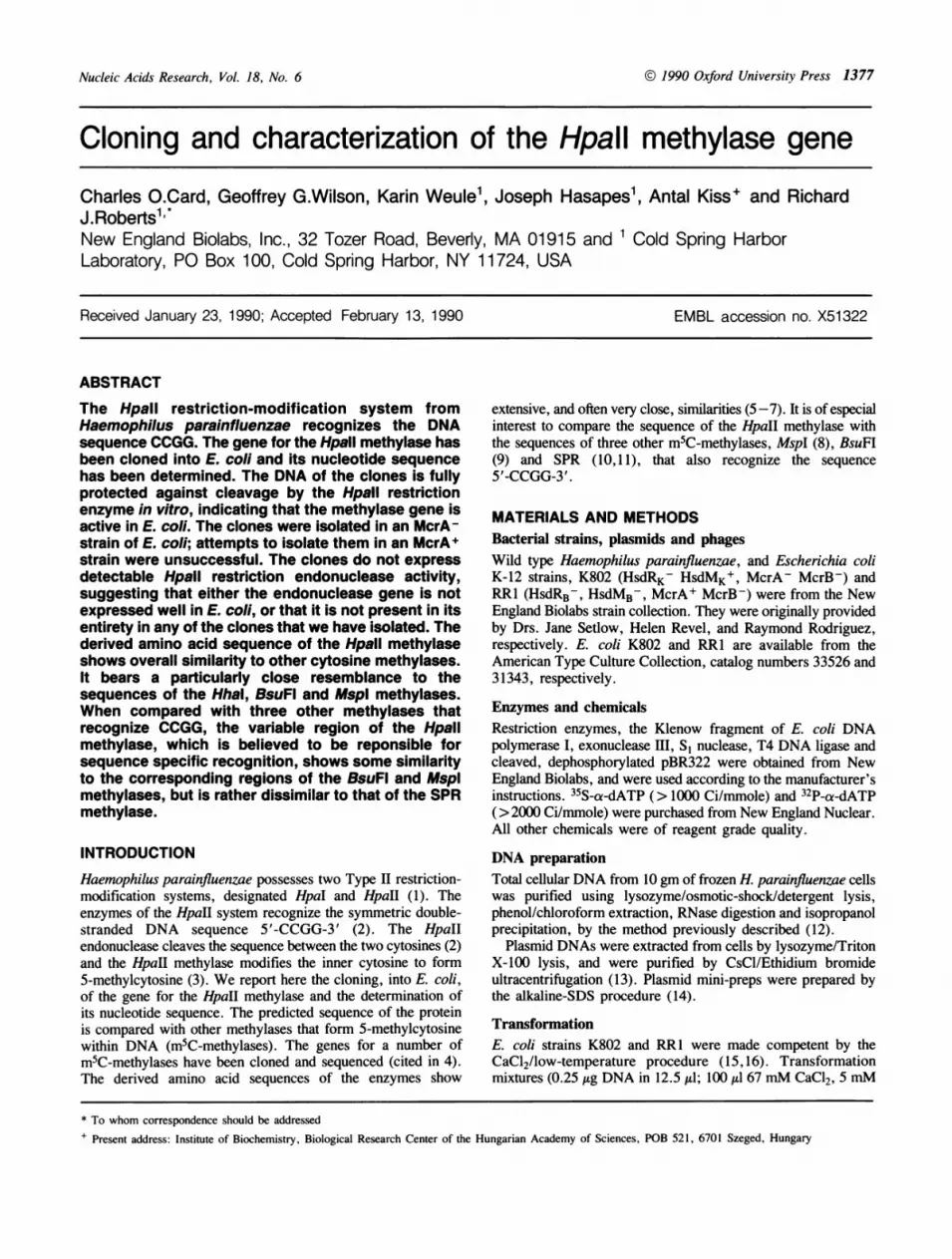
\Figure 1. Schematic diagram of plasmid pCChpalIM2- 1, which contains a 2-Kbinsert of H. parainfluenzae DNA in the vector pBR322 and which expresses themethylase gene. Also shown are schematics for two larger plasmids containingthe flanking sequences of the H. parainfluenzae genome.
AAGCTTCGTGGCTAAAGCACCTAAAGCGGTGATTACGAAAGAACGTGAAAAACAAGCAGAATATCAATCCTGGAT 75TAGAAAAAATCCAAGAGCAGTATAAAGCGATTGAAGCGTTGTAGTTAGTCTTATTTGGTTGCTTTAAATTTACTG 150TTTAAACAGTATATCAGCTATGGTATAAATTAACTCCTGAGCAACGTAAAAATGTATGAAAGCTAGTAGTAGAAT 225AAAGGACAAAGCCTGAGTTACTACTAGCTAAATATTTATGGGCTTTAGGATTAAGATATCGTAAGAATGATAGAA 300GCATTTTTGGTACTCCCGATTTAAGTTTTAAAAGGTACAAAATTGCAATTTTTATTGATGGTGAGTTTTGGCATG 375GAAAGGATTGGGATATTCGAAAGTATGATATCAAAAGTAACAAAGATTTTTGGATCTCAAAAATTGAGCATAATA 450TGAATAGGGATAAGAAAGTAAACGATTATCTTATTTCTAATGGTTGGGTAATATTTCGATTTTGGGGAAAGGATG 525
M K D V L D D N LTATTAAAAAATCCTGAGAAGTTTAGTTTAGAGATACAGAAAGCAATTTATSGAAAGATGTGTTAGATGATAACTTG 600L E E P A A Q Y S L F E P E S N P N L R E K F T F
TTAGAAGAACCCGCTGCACAATATAGTTTATTTGAACCAGAGTCCAACCCTAATTTACGAGAAAAATTTACTTTT 675I D L F A G I G G F R I A M Q N L G G K C I F S S
ATTGATTTATTTGCAGGTATTGGTGGATTCCGCATTGCCATGCAAAATTTAGGAGGTAAATGCATTTTCTCTAGT 750E W D E Q A Q K T Y E A N F G D L P Y G D I T L EGAATGGGATGAGCAAGCTCAGAAAACTTATGAGGCTAATTTTGGTGATTTGCCTTATGGAGATATTACCTTAGAG 825E T K A F I P E K F D I L C A G F P C Q A F S I AGAAACAAAGGCTTTTATTCCTGAAAAATTTGATTCTTTATGTGCTGGTTTTCCTTGTCAGGCATTTTCTATTGCA 900G K R G G F E D T R G T L F F D V A E I I R R H QGGAAAACGTGGAGGATTTGAAGATACTAGAGGGACTTTGTTTTTTGATGTTGCAGAAATTATACGTCGTCATCAG 975P K A F F L E N V K G L K N H D K G R T L K T I LCCTAAAGCATTTTTTTTAGAGAATGTAAAAGGATTAAAAAACCATGATAAAGGTAGGACATTAAAAACTATATTG 1050N V L R E D L G Y F V P E P A I V N A K N F G V PAATGTACTAAGAGAAGATTTAGGTTATTTTGTTCCTGAACCAGCAATTGTTAATGCTAAGAATTTTGGTGTGCCA 1125Q N R E R I Y I V G F H K S T G V N S F S Y P E PCAAAATAGAGAAAGAATTTATATTGTAGGCTTTCATAAAAGCACTGGTGTTAATAGTTTTAGTTATCCAGAACCT 1200L D K I V T F A D I R E E K T V P T K Y Y L S T Q
TTAGATAAAATTGTAACTTTCGCTGATATTCGGGAAGAAAAAACAGTTCCAACTAAATATTACCTATCAACTCAG 1275Y I D T L R K H K E R H E S K G N G F G Y E I I P
TATATTGATACTTTAAGAAAACATAAAGAACGTCATGAGAGTAAAGGTAATGGTTTTGGTTATGAAATTATTCCA 1350D D G I A N A I V V G G M G R E R N L V I D H R IGATGATGGAATAGCCAATGCGATTGTAGTTGGAGGTATGGGACGTGAACGTAATCTTGTAATTGATCATAGAATT 1425T D F T P T T N I K G E V N R E G I R K M T P R E
ACGGATTTTACTCCTACTACGAATATTAAAGGGGAGGTAAATCGTGAAGGGATTCGTAAAATGACCCCTCGAGAA 1500W A R L Q G F P D S Y V I P V S D A S A Y K Q F G
TGGGCAAGATTGCAGGGGTTTCCAGATAGTTATGTTATTCCGGTTTCTGATGCATCAGCGTATAAACAATTTGGT 1575N S V A V P A I Q A T G K K I L E K L G N L Y D *
AATTCAGTAGCAGTGCCGGCTATTCAAGCTACAGGTAAGAAAATTTTAGAAAAATTAGGAAATTTATATGACTGA 1650ATTTTTTTCTGGTAATAGAGGAGAGTGGAGTGAGCCTTACGCCCTCTTTAAGTTATTGGCTGATGGTCAGCTTTA 1725TTTAGGAGATAGTCAACTAAATAAACTGGAATTGTAATGCCGATTCTATCAATTCTTCGGCAGGAAAAAATTATG 1800AGAGTTCATATATACTTCATAACAATTCTCAAAATATTATAGTTACATATAATAATGAAAAATTTACAGTTCCAA 1875TTTCCGGATTTCAAGAAAAAGCTGTTTTGCTGTTATCAGAAATAAAAAATGCATCAGGCAATAGGGCTTTTTCTA 1950TCCCGAGTATTGATGATTTTCTAAAGCTT 1979
fragment is located in the middle of the EcoRI fragment whichis, itself, located in the middle of the PstI fragment (Fig. 1).
Where is the HpaII endonuclease gene?The methylase selection procedure that we have used to isolatethe HpaII methylase gene frequently yields large fragments ofDNA that encode both methylase and endonuclease genes (4).In all cases where both genes have been cloned they have beenfound to lie adjacent to one another (4). Among the HpaIImethylase clones that we isolated, the 10-Kb PstI-fragmentextends approximately 4-Kb beyond each end of the HindIII-fragment that contains the methylase gene. Since 4-Kb is ampleto code for both a large restriction endonuclease (average length:280 aa) and a large intergenic region we examined these clonesfor production of the endonuclease. Cell extracts were preparedfrom the HindIl-, EcoRI- and PstI-clones, and each extract wasassayed for Hpall endonuclease activity. No activity was detected.This suggests that either the gene for the endonuclease is notpresent or its expression in E. coli is too low to be detected.Another possibility is that the endonuclease gene is present in
the PstI-clones, but is defective. Some restriction-modificationsystems cannot be transferred to E. coli in a single step, probablybecause the DNA of the recipient cell becomes irreparablycleaved before the methylase is able to protect it. Such systems,which include DdeI (31) and BamHI (32) can only be cloned intwo steps, even though both genes of the system lie in closeproximity and could be retrieved on a single restriction fragment.In those cases, the methylase gene must be cloned first so thatthe resulting clones provide a permissive host for theendonuclease gene.To test if the HpaI system behaved similarly, the PstI fragment
was recloned de novo, into cells that already contained the HpaIImethylase gene. The 2-Kb HindUI fragment, encoding the HpaIImethylase gene, was recloned into pACYC184, and the new
plasmid was transformed into E. coli K802. DNA from thesecells was prepared and shown to be resistant to HpaII digestion.H. parainfluenzae DNA was digested with PstI, and a DNAfraction that included fragments in the 10-Kb size range was gel-purified. The fraction was ligated to pBR322 and transformed
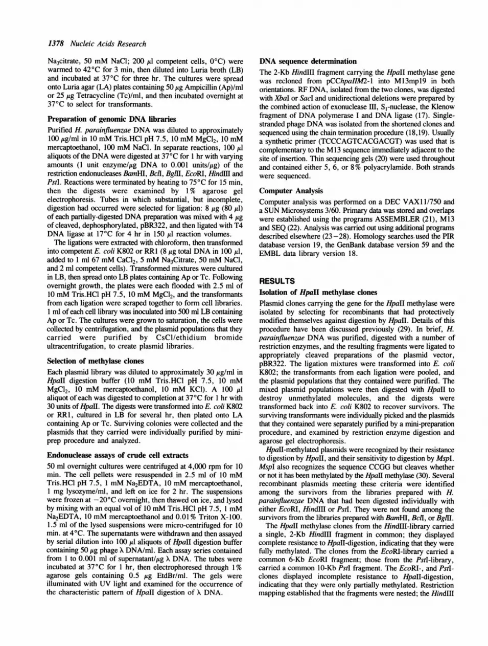
Figure 2. The sequence of the 2-Kb Hindlll fragment of pGWl containing theHpaII methylase gene. The translation for that gene is shown above the DNAsequence beginning at the first AUG codon within the reading frame.
into the HpaII-premethylated cells. Clones carrying the 10-KbPstI fragment were detected by colony hybridization to a nick-translated probe made from the unique right end of the PstIfragment. Several new PstI-clones were isolated and were shownto contain plasmids with the expected structures; however, whenextracts of these clones were analyzed, no HpaH endonucleaseactivity was detected.
Effects of Mcr function on HpaII methylase plasmidsThe HpaH methylase clones were isolated from libraries preparedin E. coli K802. When similar libraries were prepared in E. coliRRI, as they were during our first attempts to clone the gene,no HpaII methylase clones were recovered. The difference, itwas later discovered, was due to the E. coli McrA function, whichspecifically restricts HpaH methylated DNA (33,34). The McrAgene functions normally in RR1, but it is defective in K802 (35).pBR322 DNA that is methylated in vitro with HpaII transformsK802 at normal efficiency, but it transforms RR1 at only 2%of normal efficiency. The HpaII methylase clones were also foundto transform K802 normally, but to transform RRI atapproximately 5% of the normal efficiency (data not shown).Transformants of RRI that do arise and contain the HpaIImethylase show an unhealthy colony morphology: they form flat,translucent colonies, as opposed to the dome-shaped, opaquecolonies that RR 1 normally forms. Examination of the plasmidscarried by fourteen independent HpaII-transformants of RRIrevealed that tme plasmids were all normal, as judged byrestriction enzyme analysis and in the degree of HpaII-modification that they displayed.
Sequence of the HpaII methylase geneThe smallest HpaII methylase plasmid clone displays completeresistance to HpaII endonuclease digestion, suggesting that itpossesses a fully functional HpaH gene. The nucleotide sequenceof the 2-Kb HindlIl fragment inserted in this plasmid was
1380 Nucleic Acids Research
determined on both strands and is shown in Figure 2. Thesequence contains only one internal open reading frame longenough to encode the HpaII methylase. The first methioninecodon in this reading frame lies at position 574 and the framecontinues until a TGA terminator at position 1648. Thisterminator is followed by three more in-frame translational stops,TAA, TAG and TGA, five, six and eleven triplets furtherdownstream. This open reading frame predicts a proteincontaining 358 amino acids with a calculated molecular weightof 40,406 daltons. Previous studies of the Hpall methylase havedetected proteins of approximately 38,500 and 41,500 daltonswhich showed methylase activity (36). The exact relationshipbetween these protein species was not established, although itwas suggested that the higher molecular weight form mightrepresent a precursor species (36). The higher molecular weightwould correlate well with it being the product of the 358 aminoacid open reading frame.
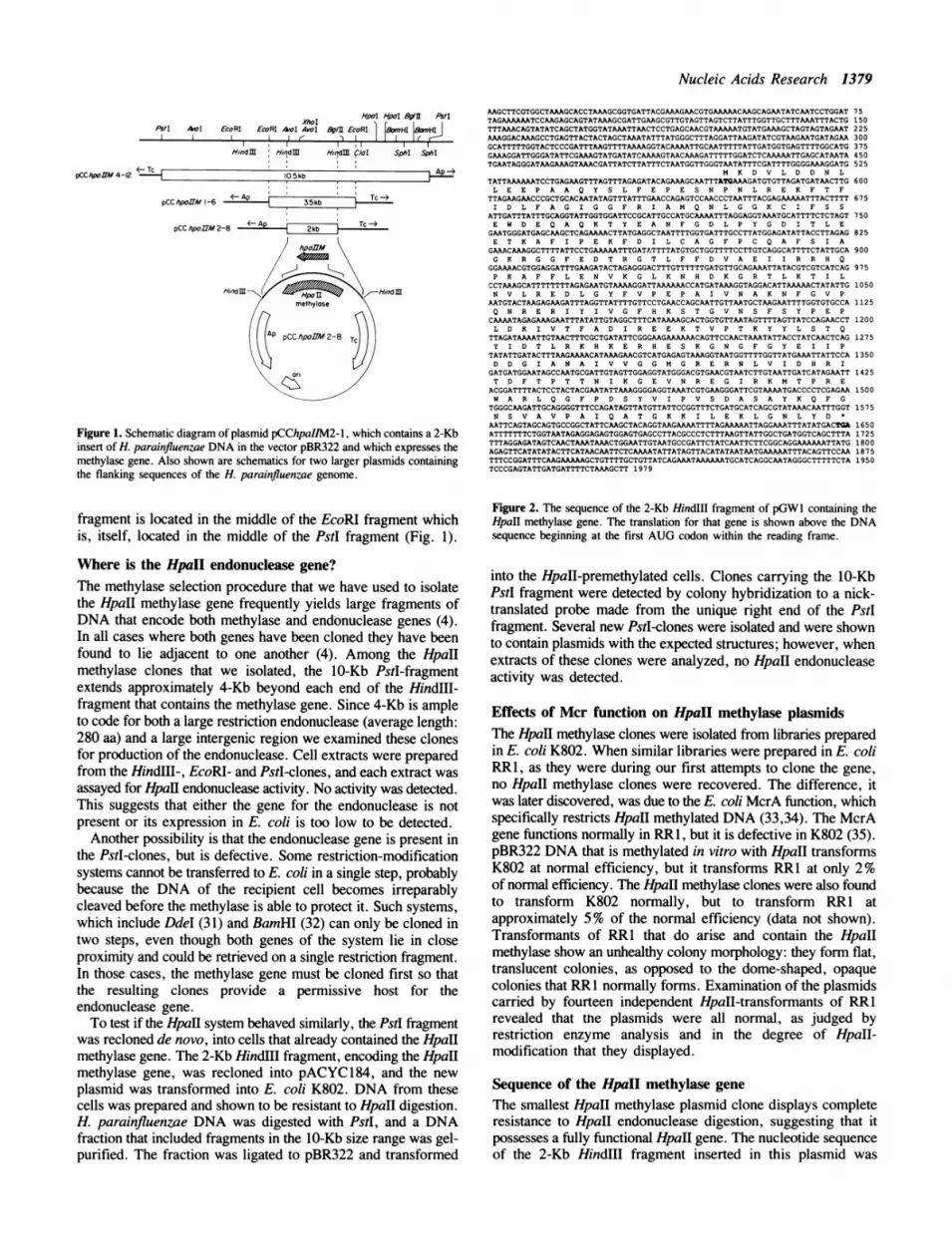
Sequence comparison with other modification enzymesSequences have been reported for thirty four methylase genesincluding fifteen that form N6-methyladenine, one that formsMg-methylcytosine and eighteen that form 5-methylcytosine(referenced in 5-7, 37 and this paper). The FASTA program(26) was used to compare the Hpall methylase with each of thesemethylase sequences as well as the complete contents of the PIR,GenBank and EMBL databases. The major similarities detectedwere with other m5C-methylases (Table 1). It can be seen thatthe HpaH methylase shows the greatest similarity to the Hhaland BsuFI methylases and an alignment between these threesequences is shown in Figure 3. Overall the sequence of the Hpalmethylase displays the same pattern of ten conserved sequencesimilarities, that have been found in all known m5C-methylases(6,7) (Figure 3).The interval between conserved regions VIII and IX in m5C-
methylases forms the so-called variable region. The length andsequence of this region varies considerably from enzyme to
TABLE 1. Similarity between the HpaII and other m5C-methylases.
Methylase Recognition Sequence Score Reference
*HhaI GCGC 611 12
BsuFI CCGG 573 9*
EcoRII CCWGG 508 37*
dcm CCWGG 506 38 and A. Bhagwatunpublished.
*MspI CCGG 412 8
* * *SPR GGCC, CCGG, CCWGG 389 10,11Qll GGCC, GAGCTC 380 39
* *<03GGCC, GCNGC 334 40HaeIII GGCC 299 41
NgoPII GGCC 293 42
AquI CYCGRG 281 43*
BspRI GGCC 245 44BepI CGCO 237 45
*SinI GGWCC 226 46
*BsuRI GGCC 222 47
TDdeI CTNAG 203 31*
SssI CO 89 48
enzyme. In the Bacillus phage methylases, which recognizemultiple target sequences, the variable region has been shownto be responsible for sequence specificity (49-51). Based uponthe overall common architecture of the prokaryotic m5C-methylases, it is likely that this variable region is also responsiblefor sequence specificity in the case of the monospecificmethylases. Four m5C-methylases that all recognize thesequence GGCC have been found to possess very similar variableregions (putative recognition domains); these are HaeIII (41),NgoPII (42), BspRI (44) and BsuRI (47). It was, therefore, ofgreat interest to compare in detail the variable regions of theHpaII, MspI, BsuFI and SPR methylases, all of which recognizethe sequence CCGG.To define the boundaries between the conserved regions and
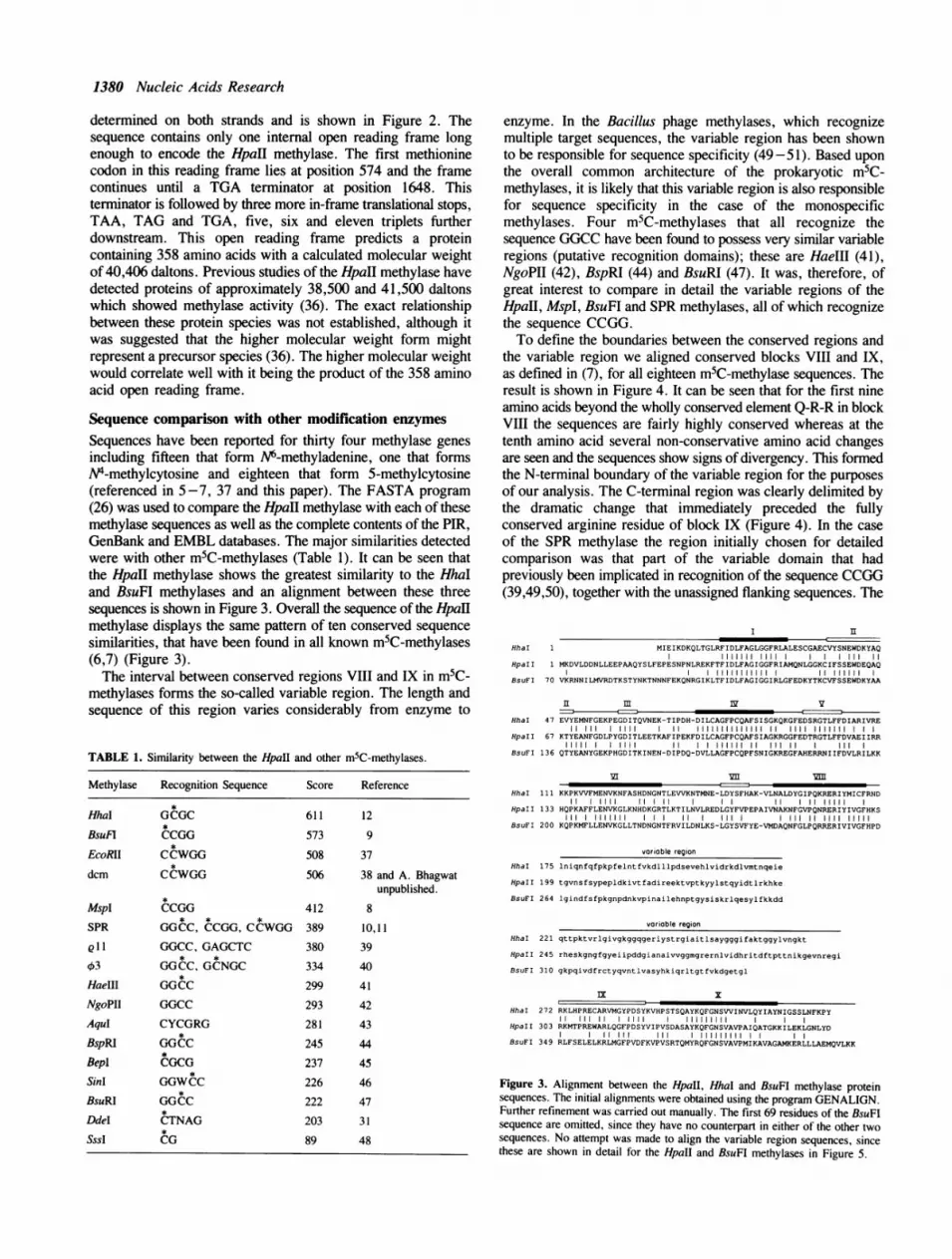
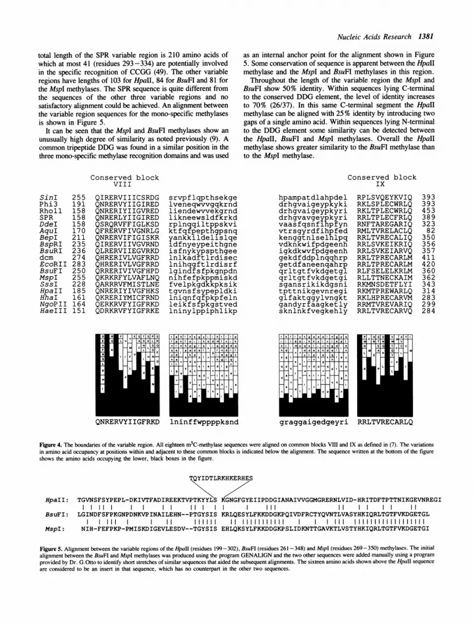
the variable region we aligned conserved blocks VIII and IX,as defined in (7), for all eighteen m5C-methylase sequences. Theresult is shown in Figure 4. It can be seen that for the first nineamino acids beyond the wholly conserved element Q-R-R in blockVIII the sequences are fairly highly conserved whereas at thetenth amino acid several non-conservative amino acid changesare seen and the sequences show signs of divergency. This formedthe N-terminal boundary of the variable region for the purposesof our analysis. The C-terminal region was clearly delimited bythe dramatic change that immediately preceded the fullyconserved arginine residue of block IX (Figure 4). In the caseof the SPR methylase the region initially chosen for detailedcomparison was that part of the variable domain that hadpreviously been implicated in recognition of the sequence CCGG(39,49,50), together with the unassigned flanking sequences. The
I II
HhaI 1 MIEIKDKQLTGLRFIDLFAGLGGFRLALESCGAECVYSNEWDKYAQI11111 lIll I III 11
HpaI I 1 MKDVLDDNLLEEPAAQYSLFEPESNPNLREKFTFIDLFAGIGGFRIAMQNLGGKCIFSSEWDEQAQI IIIIIIIIIII I 11111111
BsuFI 70 VKRNNILMVRDTKSTYNKTNNNFEKQNRGIKLTFIDLFAGIGGIRLGFEDKYTKCVFSSEWDKYAA
n m I vHhaI 47 EVYEMNFGEKPEGDITQVNEK-TIPDH-DILCAGFPCQAFSISGKQKGFEDSRGTLFFDIARIVRE
11 11 11111 11 IIIIIIll 11 1111 1111111HpaI I 67 KTYEANFGDLPYGDITLEETKAFIPEKFDILCAGFPCQAFSIAGKRGGFEDTRGTLFFDVAEI IRR
11111 1 1111 I I HI M 11 111 11 IIIBsuFI 136 QTYEANYGEKPHGDITKINEN-DIPDQ-DVLLAGFPCQPFSNIGKREGFAHERRNIIFDVLRILKK
HhaI 111 KKPKVVFMENVKNFASHDNGNTLEVVKNTMNE-LDYSFHAK-VLNALDYGIPQKRERIYMICFRND11 1 1111 11 1 11 II 11 11 11111
HpaI I 133 HQPKAFFLENVKGLKNHDKGRTLKTILNVLREDLGYFVPEPAIVNAKNFGVPQNRERIYIVGFHKSIII 1111111 11 I II Ill 11 1111 11111
BsuFI 200 KQPKMFLLENVKGLLTNDNGNTFRVILDNLKS-LGYSVFYE-VMDAQNFGLPQRRERIVIVGFHPD
variable region
HhaI 175 lniqnfqfpkpfelntfvkdlllpdsevehlvidrkdlvmtnqeie
HpaII 199 tgvnsfsypepldkivtfadireektvptkyylstqyidtlrkhke
BsuFI 264 lgindfsfpkgnpdnkvpinailehnptgysiskrlqesylfkkdd
varioble region
HhaI 221 qttpktvrlgivgkggqgeriystrgiaitlsaygggifaktggylvngkt
HpaII 245 rheskgngfgyeiipddgianaivvggmgrernlvidhritdftpttnikgevnregi
BsuFI 310 gkpqivdfrctyqvntlvasyhkiqrltgtfvkdgetgl
lx I
HhaFI 272 RKLHPRECARVMGYPDSYKVHPSTSQAYKQFGNSVVINVLQYIAYNIGSSLNFKPY11 III 1 1111 111111111
HpaI I 303 RKMTPREWARLQGFPDSYVIPVSDASAYKQFGNSVAVPAIQATGKKILEKLGNLYDi III III 111111111
BsuFI 349 RLFSELELKRLMGFPVDFKVPVSRTQMYRQFGNSVAVPMIKAVAGAMKERLLLAEMQVLKK
Figure 3. Alignment between the HpaII, HhaI and BsuFI methylase proteinsequences. The initial alignments were obtained using the program GENALIGN.Further refinement was carried out manually. The first 69 residues of the BsuFIsequence are omitted, since they have no counterpart in either of the other twosequences. No attempt was made to align the variable region sequences, sincethese are shown in detail for the HpaII and BsuFI methylases in Figure 5.
Nucleic Acids Research 1381
total length of the SPR variable region is 210 amino acids ofwhich at most 41 (residues 293 -334) are potentially involvedin the specific recognition of CCGG (49). The other variableregions have lengths of 103 for HpaII, 84 for BsuFI and 81 forthe MspI methylases. The SPR sequence is quite different fromthe sequences of the other three variable regions and nosatisfactory alignment could be achieved. An alignment betweenthe variable region sequences for the mono-specific methylasesis shown in Figure 5.
It can be seen that the MspI and BsuFI methylases show anunusually high degree of similarity as noted previously (9). Acommon tripeptide DDG was found in a similar position in thethree mono-specific methylase recognition domains and was used
as an internal anchor point for the alignment shown in Figure5. Some conservation of sequence is apparent between the HpaIImethylase and the MspI and BsuFI methylases in this region.Throughout the length of the variable region the MspI and
BsuFI show 50% identity. Within sequences lying C-terminalto the conserved DDG element, the level of identity increasesto 70% (26/37). In this same C-terminal segment the HpaHmethylase can be aligned with 25% identity by introducing twogaps of a single amino acid. Within sequences lying N-terminalto the DDG element some similarity can be detected betweenthe HpaII, BsuFI and MspI methylases. Overall the HpaIImethylase shows greater similarity to the BsuFI methylase thanto the MspI methylase.
Conserved blockVIII
Conserved blockIx
QIRERVIIICSRDGQNRERVYIIGIREDQNRERIYIIGVREDQNRERLYIIGIREDQSRQRVFFIGLKSDQFRERVFIVGNRLGQNRERVIFIGISKRQIRERVIIVGVRNDQLRERVIIEGVRKDQHRERIVLVGFRRDQHRERIVLVGFRRDQRRERIVIVGFHPDQKRKRFYLVAFLNQQARRRVFMISTLNEQNRERIYIVGFHKSQKRERIYMICFRNDQERKRVFYIGFRKDQDRKRVFYIGFRKE
srvpflqpthsekgelveneqwvvgqkrndliendewvvekgrndlikneewsldfkrkdrplnqqiltppskviktfqfpepthcpsnqyankkildelislqeidfnyeypeithgneisfnykypspthgeelnlkadftlrdiseclnihqgftlrdisrflqindfsfpkgnpdnnlhfefpkppmiskdfvelpkgdkkpksiktgvnsfsypepldkilniqnfqfpkpfelnleikfs pkgstvedlninylppiphlikp
hpampatdlahpdeldrhgvaigeypkykidrhgvaigeypkyridrhgvavgeypkyrivaasfqsnfihpfynvtrsgyrdfihpfedkenggtnlselhlpqvdknkwifpdgeenhigkdkwvfpdgeenhgekdfddp nqqhrpgetdfaneenqahrpqrltgtfvkdgetgiqrltgtfvkdgetgisgansrikikdgsnitpttnikgevnregigifaktggylvngktgandyrfaagketlysknlnkfvegkehly
RPLSVQEYKVIQRKLSPLECWRLQRKLTPLECWRLQRRLTPLECFRLQRNFTAREGARIQRMLTVRELACLQRRLTVRECALIQRRLSVKEIKRIQRRLSVKEIARVQRRLTPRECARLMRRLTPRECARLMRLFSELELKRLMRLLTTNECKAIMRKMNSDETFLYIRKMTPREWARLQRKLHPRECARVMRRMTVREVARIQRRLTVRECARVQ
tI k-If, I ] ,I gI 1*1VI.[§Iil7laplil#klklyl Ilolsi
QNRERVYIIGFRKD graggalgeageyrl KKLTLVK UAU.Q
Figure 4. The boundaries of the variable region. All eighteen m5C-methylase sequences were aligned on common blocks VIII and IX as defined in (7). The variationsin amino acid occupancy at positions within and adjacent to these common blocks is indicated below the alignment. The sequence written at the bottom of the figureshows the amino acids occupying the lower, black boxes in the figure.
TQYIDTLRKHKERHES
HpaII: TGVNSFSYPEPL-DKIVTFADIREEKTVPTKYYLS KGNGFGYEIIPDDGIANAIVVGGMGRERNLVID-HRITDFTPTTNIKGEVNREGI11 I I I I 11I III II I I
BsuFI: LGINDFSFPKGNPDNKVPINAILEHN--PTGYSIS KRLQESYLFKKDDGKPQIVDFRCTYQVNTLVASYHKIQRLTGTFVKDGETGLlI II 11 111111 11IIIIIIIIIII I II111 11111111111111
MspI: NIH-FEFPKP-PMISKDIGEVLESDV--TGYSIS EHLQKSYLFKKDDGKPSLIDKNTTGAVKTLVSTYHKIQRLTGTFVKDGETGI
Figure 5. Alignment between the variable regions of the HpaII (residues 199-302), BsuFI (residues 261-348) and MspI (residues 269-350) methylases. The initialalignment between the BsuFI and MspI methylases was produced using the program GENALIGN and the two other sequences were added manually using a programprovided by Dr. G.Otto to identify short stretches of similar sequences that aided the subsequent alignments. The sixteen amino acids shown above the HpaII sequenceare considered to be an insert in that sequence, which has no counterpart in the other two sequences.
SinIPhi3RhollSPRDdeIAquIBepIBspRIBsuRIdcmEcoRIIBsuFIMspISssIHpaIIHhaINgoPIIHaeIII
255191158158158170211235236274283250255228185161164151
39339345338932382
350356357411420360362343314283299284

1382 Nucleic Acids Research
DISCUSSION
Like many other attempts to clone the genes for restriction-modification systems we have been able only to clone themethylase gene for the HpaII system. We have isolated severalclones containing sequences flanking the methylase gene, but no
HpaII endonuclease activity has been detected in these clones.Among nine other Haemophilus systems where cloning has beensuccessful the genes for both the methylase and the restrictionendonuclease have been located adjacent to one another in eightinstances (4 and GGW unpublished). Only in the cases of HaeIIIand Hpal have the endonucleases not been detected within clonescontaining sequences flanking the methylase gene (4). In neitherof these two cases nor in the present case is it known whetherthis is because the two genes are physically separated on thegenome or because the endonuclease genes are not expressed inE. coli. Unfortunately comparison of flanking sequences withthe known sequences of restriction endonuclease genes is nothelpful in trying to identify an unexpressed gene. So far eachrestriction endonuclease gene appears to have a unique sequence
and no diagnostic similarities have been detected. Unlike thesituation with the BsuFI methylase gene, which appears to beallelic with other Bacillus subtilis methylase genes (9,54), theHaemophilus methylase genes share no obvious sequence
similarities across species.The sequences immediately upstream of the HpaII methylase
gene are quite AT rich, but carry no clear similarity to typicalE. coli promoter sequences. Since a clone carrying the 2-KbHindIll fragment shows a higher level of expression of themethylase than clones carrying longer inserts it seems likely thatthe endogenous promoter is not very active in E. coli. Ratherit is likely that the increased levels of methylase expression thatwe have observed in the smaller clones is being driven by a pB322promoter. This notion is supported by the observation thatrecloning of the HpaII methylase gene into pACYC leads to fullin vivo modification when the gene is oriented so that itsexpression is away from that of the tetracycline resistance gene,but only partial modification in the opposite orientation.A striking feature of the coding region is the extreme bias in
codon usage. Among 359 codons, 300 have A or T in the thirdposition and only 18 have a C in the third position. Three HpaIIrecognition sites are found within the sequence reported; two liewithin the coding region close to its C-terminus, while the thirdlies 230 nucleotides beyond the end of the gene. Given theirlocation it is unlikely that these have any regulatory significanceas has been proposed in other systems (52,53).One of the most interesting aspects of the HpaII methylase
sequence is its comparison with the sequences of the three otherknown methylases that recognize the sequence CCGG. These are
the MspI, BsuFI and SPR methylases. It should be noted thatall three of these enzymes methylate the outer cytosine of therecognition sequence whereas the HpaII methylase modifies theinternal cytosine. In terms of overall similarity the sequences
share the typical building blocks found among all known m5C-methylases (6,7). However it is the comparison of the so-calledvariable regions, that is believed to be responsible for sequence
recognition, that is of most interest. Among these four enzymesthe MspI and BsuFI methylases show the highest degree ofsimilarity to each other in this region. The HpaII methylase ismost similar to the BsuFI methylase, and shows the leastsimilarity to the SPR methylase. Given the overall similarity ofall m5C methylases and the clear relatedness of this family it islikely that they have all evolved from a common precursor. The
extensive changes found in the SPR methylase may reflect thefact that its recognition domain is part of a complicated regionthat includes recognition domains for two other sequences.When searching for common sub-sequences within the variable
region we were struck by the apparent conservation of thetripeptide DDG, which we used as an anchor point when aligningthe three mono-specific methylases. This is the only tripeptidefound in these variable regions and is positioned similarly, withrespect to conserved block IX, in all three sequences. DGG alsooccurs within the SPR methylase, but within a part of the variableregion that has been implicated in the recognition of the sequenceGGCC. It does not occur in the variable region of any othersequenced m5C-methylase. It is interesting to note that two ofthe three residues are aspartic acid, which would be capable ofhydrogen bonding with the bases of the DNA recognitionsequence.
ACKNOWLEDGEMENTS
The authors would like to express their thanks to Drs. A. Bhagwatfor many helpful discussions during the course of this work andfor allowing us to use the dcm methylase sequence prior topublication. Thanks are due to Dr. T. Trautner for sharing thesequence of the BsuFI methylase gene prior to publication andto Dr. G. Otto for help with some of the computational aspects.We thank J. Duffy and P. Renna for the artwork andphotography. Part of this work was supported by a grant fromthe National Science Foundation to RJR (DMB-8614032).
REFERENCES1. Sharp, P.A., Sugden, B. and Sambrook, J. (1973) Biochemistry 12:
3055 -3066.2. Garfin, D.E. and Goodman, H.M. (1974) Biochem. Biophys. Res. Comm.
259: 108-116.3. Mann, M.B. and Smith, H.O. (1977) Nucl. Acids Res. 4: 4211-4221.4. Wilson, G.G. (1988) Gene 74: 281-289.5. Chandrasegaran, S. and Smith, H.O. (1988) in Structure and Expression
Volume 1: From Proteins to Ribosomes. eds. R.H. Sarma and M.H. Sarma.(Adenine Press) pp 149-156.
6. Lauster, R., Trautner, T.A. and Noyer-Weidner, M. (1989) J. Mol. Biol.206: 305-312.
7. Posfai, J., Bhagwat, A.S., Posfai, G. and Roberts, R.J. (1989) Nucl. AcidsRes. 17: 2421-2435.
8. Lin, P.M., Lee, C.H. and Roberts, R.J.(1989) Nucl. Acids Res. 17:3001 -3011.
9. Walter, J., Noyer-Weidner, M. and Trautner, T.A. submitted for publication.10. Posfai, G., Baldauf, F., Erdei, S., Posfai, J., Venetianer, P. and Kiss, A.
(1984) Nucl. Acids Res. 12: 9039-9049.11. Buhk, H-J., Behrens, B., Tailor, R., Wilke, K., Prada, J.J., Gunthert, U.,
Noyer-Weidner, M., Jentsch, S. and Trautner, T.A. (1984) Gene 29: 51-61.12. Caserta, M., Zacharias, W., Nwankwo, D., Wilson, G.G. and Wells, R.D.
(1987) J. Biol. Chem. 262: 4770-4777.13. Clewell, D.B. and Helinski, D.R. (1969) Proc. Natl. Acad. Sci. USA 62:
1159-1166.14. Birnboim, H.C. and Doly, J. (1979) Nucl. Acids Res. 7: 1513-1523.15. Mandel, M. and Higa, A. (1970) J. Mol. Biol. 53: 159-162.16. Lederberg.E.M. and Cohen, S.N. (1974) J. Bacteriol. 119: 1072-1074.17. Henikoff, S. (1984) Gene 28: 351-358.18. Sanger, F., Nicklen, S. and Coulson, A.R. (1977) Proc. Natl. Acad. Sci.
USA 74: 5463-5467.19. Messing, J. (1983) Meth. Enzymol. 101: 20-78.20. Sanger, F. and Coulson, A.R. (1978) FEBS Letters 87: 107-110.21. Gingeras, T.R., Milazzo, J.P., Sciaky, D. and Roberts, R.J. (1979) Nucl.
Acids Res. 7: 529-545.22. Blumenthal, R.M., Rice, P.J. and Roberts, R.J. (1982) Nucl. Acids Res.
10: 91-101.23. Staden, R. (1977) Nucl. Acids Res. 4: 4037-4051.24. Staden. R. (1978) Nucl. Acids Res. 5: 1013-1015.
Nucleic Acids Research 1383
25. Keller, C., Corcoran, M. and Roberts, R.J. (1984) Nucl. Acids Res. 12:379-386.
26. Lipman, D.J. and Pearson, W.R. (1988) Proc. Nati. Acad. Sci. USA 85:2444-2448.
27. Feng, D-F. and Doolittle, R.F. (1987) J. Mol. Evol. 25: 351-360.28. Devereux, J., Haeberli, P. and Smithies, 0. (1984) Nucl. Acids Res. 12:
387-395.29. Lunnen, K.D., Barsomian, J.M., Camp, R.R., Card, C.O., Chen, S-Z.,
Croft, R., Looney, M.C., Meda, M.M., Moran, L.S., Nwankwo, D.O.,Slatko, B.E., Van Cott, E.M. and Wilson, G.G.(1988) Gene 74: 25-32.
30. Brooks, J.E. and Roberts, R.J. (1982) Nucl. Acids Res. 10: 913-93431. Howard, K.A., Card, C., Benner, J.S., Callahan, H.L., Maunus, R., Silber,
K., Wilson, G. and Brooks, J.E. (1986) Nucl. Acids Res. 14: 7939-795 1.32. Brooks, J.E., Benner, J.S., Heiter, D., Silber, K.R., Sznyter, L., Jager-
Quinton, T., Moran, L., Slatko, B.E., Wilson, G.G. and Nwankwo, D.O.(1989) Nucl. Acids Res. 17: 979-997.
33. Noyer-Weidner, M., Diaz, R. and Reiners, L. (1986) Mol. Gen. Genet.205: 469-475.
34. Raleigh, E.A and Wilson, G. (1986) Proc. Natl. Acad. Sci. USA 83:9070-9074.
35. Raleigh, E.A., Murray, N.E., Revel, H., Blumenthal, R.M., Westaway,D., Reith, A.D., Rigby, P.W.J., Elhai, J. and Hanahan, D.(1988) Nucl.Acids Res. 16: 1563-1575.
36. Yoo, O.J. and Agarwal, K.L. (1980) J. Biol. Chem. 255: 6445-6449.37. Som, S., Bhagwat, A.S. and Friedman, S. (1987) Nucl. Acids Res. 15:
313-332.38. Hanck, T., Gerwin, N., Fritz, H.J. (1989) Nucl. Acids Res. 17: 5844.39. Behrens, B., Noyer-Weidner, M., Pawlek, B., Lauster, R., Balganesh, T.S.
and Trautner, T.A. (1987) EMBO J. 6: 1137-1142.40. Tran-Betcke, A., Behrens, B., Noyer-Weidner, M. and Trautner, T.A. (1986)
Gene 42: 89-96.41. Slatko, B.E., Croft, R., Moran, L. and Wilson, G.G. (1988) Gene 74:
45-50.42. Sullivan, K.M. and Saunders, J.R. (1988) Nucl. Acids Res. 16: 4369-4387.43. Karreman, C. and De Waard, A. (1990) J. Bacteriol. 172: 266-272.44. Posfai, G., Kiss, A., Erdei, S., Posfai, J. and Venetianer, P. (1983) J. Mol.
Biol. 170: 597-610.45. Kupper, D., Zhou, J.G., Venetianer, P. and Kiss, A. (1989) Nucl. Acids
Res. 17: 1077- 1088.46. Karreman, C. and de Waard, A. (1988) J. Bacteriol. 170: 2527-2532.47. Kiss, A., Posfai, G., Keller, C.C., Venetianer, P. and Roberts, R.J. (1985)
Nucl. Acids Res. 13: 6403-6421.48. Renbaum, P., Abrahamove, D., Fainsod, A., Wilson, G.G., Rottem, S.
and Razin, A. submitted for publication.49. Wilke, K., Rauhut, E., Noyer-Weidner, M., Lauster, R., Pawlek, B.,
Behrens, B., and Trautner, T.A. (1988) EMBO J. 7: 2601-2609.50. Balganesh, T.S., Reiners, L., Lauster, R., Noyer-Weidner, M., Wilke, K.
and Trautner, T.A. (1987) EMBO J. 6: 3542-3549.51. Trautner, T.A., Balganesh, T.S. and Pawlek, B. (1988) Nucl. Acids Res.
16: 6649-6658.52. Slatko, B.E., Benner, J.S., Jager-Quinton, T., Moran, L.S., Simcox, T.G.,
Van Cott, E.M. and Wilson, G.G. (1987) Nucl. Acids Res. 15: 9781-9796.53. Gingeras, T.R., Theriault, G. and Brooks, J.E. (1984) in Proceedings of
the 5th International Symposium on Metabolism and Enzymology of NucleicAcids (eds. J. Zelinka and J. Balan) Slovak Academy of Sciences, Bratislavapp 267-275.
54. Ikawa, S., Shibata, T., Matsumoto, K., lijima, T., Saito, H. and Ando,T. (1981) Mol. Gen. Genet. 183: 1-6.