clinically adaptable polymer enables simultaneous spatial
TRANSCRIPT
ARTICLE OPEN
Clinically adaptable polymer enables simultaneous spatialanalysis of colonic tissues and biofilmsMary C. Macedonia1,2, Julia L. Drewes 3, Nicholas O. Markham 1,2, Alan J. Simmons2,4, Joseph T. Roland2,5, Paige N. Vega 2,4,Cherie’ R. Scurrah2,4, Robert J. Coffey1,2,6, Martha J. Shrubsole 6,7, Cynthia L. Sears 3 and Ken S. Lau 2,4,6✉
Microbial influences on host cells depend upon the identities of the microbes, their spatial localization, and the responses theyinvoke on specific host cell populations. Multimodal analyses of both microbes and host cells in a spatially resolved fashion wouldenable studies into these complex interactions in native tissue environments, potentially in clinical specimens. While techniques topreserve each of the microbial and host cell compartments have been used to examine tissues and microbes separately, weendeavored to develop approaches to simultaneously analyze both compartments. Herein, we established an original method formucus preservation using Poloxamer 407 (also known as Pluronic F-127), a thermoreversible polymer with mucus-adhesivecharacteristics. We demonstrate that this approach can preserve spatially-defined compartments of the mucus bi-layer in the colonand the bacterial communities within, compared with their marked absence when tissues were processed with traditional formalin-fixed paraffin-embedded (FFPE) pipelines. Additionally, antigens for antibody staining of host cells were preserved and signalintensity for 16S rRNA fluorescence in situ hybridization (FISH) was enhanced in poloxamer-fixed samples. This in turn enabled us tointegrate multimodal analysis using a modified multiplex immunofluorescence (MxIF) protocol. Importantly, we have formulatedPoloxamer 407 to polymerize and cross-link at room temperature for use in clinical workflows. These results suggest that thefixative formulation of Poloxamer 407 can be integrated into biospecimen collection pipelines for simultaneous analysis ofmicrobes and host cells.
npj Biofilms and Microbiomes (2020) 6:33 ; https://doi.org/10.1038/s41522-020-00143-x
INTRODUCTIONInvestigating host–microbe interactions may reveal the mechan-isms underlying how changes in the gastrointestinal microbiomeare related to colorectal cancer (CRC). Recently, increasedabundance of Fusobacterium nucleatum, Bacteroides fragilis, andEscherichia coli have been associated with human CRC1–3. Thesespecies are hypothesized to promote tumorigenesis and/orprogression of CRC through upregulation of oncogenes, modifica-tion of intestinal mucus, or damage to host DNA2,4,5. In addition tothe mere presence or absence of putative carcinogenic bacteria inthe gut, the spatial localization and physical interaction with hostepithelial cells may be critically important.Bacterial biofilms are ubiquitous in nature and can also be
observed routinely in the gastrointestinal tract of healthyindividuals specifically in the outer layer of the MUC2-dominated mucus bi-layer that lines the luminal colonic sur-face6–11. In contrast, the inner, striated mucus layer of the colon islargely sterile in healthy hosts6. Perturbations of this tightlyregulated spatial organization have been linked to both inflam-matory bowel diseases and CRC12–15. Mucosa-associated bacterialbiofilms are defined as microbial aggregates embedded within apolymeric matrix of both host and bacterial origin that directlycontacts epithelial cells14,16. Mucosa-associated biofilms havebeen identified in ~50% of sporadic CRC patients, 100% offamilial adenomatous polyposis (FAP) patients, and 13% of healthyindividuals. They are particularly enriched in CRCs from theascending colon3. Additionally, bacterial isolates from CRC or FAP
biofilms have been shown to promote colonic tumor progressionin genetically susceptible mice13,15.While many studies have characterized the microbiome
through sequencing and/or mass spectrometry, host–microbespatial relationships can only be studied with sufficient resolutionthrough microscopy-based techniques, such as fluorescencein situ hybridization (FISH) or electron microscopy11,12. Moreover,while sophisticated multimodal analyses are being conducted onthe microbiome17, concurrent analyses of host cells have lagged.Studies on the host response to microbiome alterations to datehave typically relied on macroscopic outputs, such as tumorformation, or molecular assays performed on separate, spatiallydisconnected specimens2,15. We propose that concurrent, multi-modal analysis of both host and microbial cells within the sametissue specimen would significantly advance knowledge intohost–microbe interactions in CRC, as well as other forms ofpathogenesis.Recently developed imaging techniques enable multiplexed
in situ analysis of 50 or more analytes on the same tissuesection18–27. However, one challenge has been identifying a tissueprocessing method that enables simultaneous spatial localizationand quantifiable detection of both host components, such asproteins, and bacterial components via immunofluorescence (IF)and FISH, respectively. The colonic mucus layer acts as both aresident niche and a barrier for the luminal microbiome, playingwell-documented roles in colonic pathologies such as ulcerativecolitis10,14,28,29. The hydrophilic colonic mucus is easily disrupted
1Division of Gastroenterology, Hepatology and Nutrition, Department of Medicine, Vanderbilt University Medical Center, Nashville, TN, USA. 2Epithelial Biology Center, VanderbiltUniversity Medical Center, Nashville, TN, USA. 3Division of Infectious Diseases, Department of Medicine, Johns Hopkins University School of Medicine, Baltimore, MD, USA.4Department of Cell and Developmental Biology, Vanderbilt University School of Medicine, Nashville, TN, USA. 5Department of Surgery, Vanderbilt University Medical Center,Nashville, TN, USA. 6Vanderbilt Ingram Cancer Center, Nashville, TN, USA. 7Division of Epidemiology, Vanderbilt Epidemiology Center, Department of Medicine, VanderbiltUniversity Medical Center, Nashville, TN, USA. ✉email: [email protected]
www.nature.com/npjbiofilms
Published in partnership with Nanyang Technological University
1234567890():,;
by hydration and dehydration of formalin-fixed tissue withstandard histological preparations7,30. Carnoy’s solution is arapidly dehydrating fixative that preserves mucus9,14, but itreduces the sensitivity of IF and FISH7,31. Our goal for this workis to devise a tissue processing strategy that preserves mucusarchitecture and is amenable to coincident antibody and nucleicacid detection. Herein, we describe how Poloxamer 407 fixationpermits the simultaneous characterization of host cell proteinexpression and identification of microbial communities within thecolonic mucus. Furthermore, we deployed this approach into apilot clinical pipeline to enable multiplex in situ profiling of humantissues and associated microbes.
RESULTSCarnoy’s and formalin fixation are incompatible with simultaneousmucus preservation and host cell assaysThe alcohol-based Carnoy’s solution (Carnoy’s fixative/Methacarn)is a rapid dehydration fixative that preserves the hydrophilicmucus layer for colonic microbial FISH analysis. Cross-linkingfixative, such as neutral buffered formalin (NBF), is used forstandard formalin-fixed paraffin-embedded (FFPE) tissue proces-sing and has optimal antigen preservation for IF or immunohis-tochemistry32,33. For this study, we aimed to identify the besttissue fixation method for simultaneous microbial FISH and hostcell IF. We initially used mouse colon tissue as a model because ofits abundance and availability. Mice do not naturally form mucosa-associated biofilms, but their mucus layers are arranged in bi-layerstructures with different microbial compositions that can easily beevaluated.Initially, we compared Carnoy’s fixation and NBF fixation,
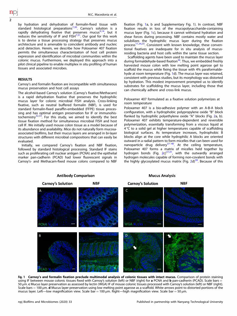
followed by standard histological processing. Standard IF stainssuch as proliferating cell nuclear antigen (PCNA) and the epithelialmarker pan-cadherin (PCAD) had lower fluorescent signals inCarnoy’s- and Methacarn-fixed mouse colons compared to NBF
fixation (Fig. 1a, b and Supplementary Fig. 1). In contrast, NBFfixation results in loss of the mucopolysaccharide-containingmucus layer (Fig. 1c), because it cannot withstand hydration andshear forces during processing. NBF contains mostly water andsolubilizes the hydrophilic mucus layer during the fixationprocess11,34,35. Consistent with known knowledge, these conven-tional fixatives are inadequate for in situ analysis of mucus-residing bacteria and host cells within the same tissue section.Scaffolding agents have been used to maintain the mucus layer
during formaldehyde-based fixation36. Thus, we embedded freshlyharvested mouse colon with low melting point agarose gel toscaffold the mucus while fixing the tissue with 4% paraformalde-hyde at room temperature (Fig. 1d). The mucus layer was retained,consistent with previous studies, but its morphology was distortedby hydration. This modest improvement prompted us to identifysubstrates for scaffolding the mucus layer, including those thatcan chemically adhere and cross-link mucus.
Poloxamer 407 formulated as a fixative solution polymerizes atroom temperaturePoloxamer 407 is a bio-adhesive polymer with an A-B-A blockconfiguration, with a hydrophobic polypropylene oxide “B” blockflanked by hydrophilic polyethylene oxide “A” blocks (Fig. 2a, b).Poloxamer 407 exhibits temperature-dependent and reversiblepolymerization, essentially transforming from a viscous liquid at4 °C to a solid gel at higher temperatures capable of scaffoldingbiological surfaces. As temperature increases, hydrophobic Bblocks align at the core while hydrophilic A blocks are orientedoutward in a radial pattern to form micelles that can been used fornanoparticle drug delivery37–40. At the ceiling temperature,Poloxamer 407 forms a matrix of micelles held together byhydrogen bonds (Fig. 2c)37,41, with the outwardly arrangedhydrogen molecules capable of forming non-covalent bonds withthe highly glycosylated mucus matrix (Fig. 2d)39. Because of this
Fig. 1 Carnoy’s and formalin fixation preclude multimodal analysis of colonic tissues with intact mucus. Comparison of protein stainingusing IF between mouse colonic tissues fixed with Carnoy’s solution (left) or NBF (right) for a PCNA and b pan-cadherin (PCAD). Scale bars=50 μm. cMucus layer preservation as assessed by lectin (WGA) IF of mouse colonic tissues processed with Carnoy’s solution (left) or NBF (right).Scale bars= 100 μm. d Mucus layer preservation using low melting point agarose as a scaffold. White arrows point to distorted portions of themucus layer. Left—low magnification view. Scale bar= 100 μm. Right—high magnification view. Scale bar= 50 μm.
M.C. Macedonia et al.
2
npj Biofilms and Microbiomes (2020) 33 Published in partnership with Nanyang Technological University
1234567890():,;
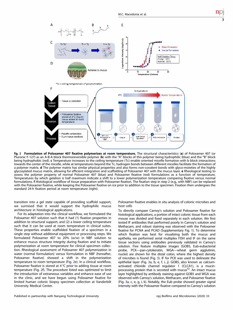
transition into a gel state capable of providing scaffold support,we surmised that it would support the hydrophilic mucusarchitecture in histological applications.For its adaptation into the clinical workflow, we formulated the
Poloxamer 407 solution such that it had (1) fixation properties inaddition to structural support, and (2) a lower ceiling temperaturesuch that it can be used at room temperature in clinical suites.These properties enable scaffolded fixation of a specimen in asingle step without additional equipment or processing steps. Weformulated Poloxamer 407 to 20% (w/w) in NBF solution toenhance mucus structure integrity during fixation and to initiatepolymerization at room temperature for clinical specimen collec-tion. Rheological assessment of Poloxamer 407 polymerization inwater (normal formulation) versus formulation in NBF (hereafter,Poloxamer fixative) showed a shift in the polymerizationtemperature to room temperature (Fig. 2e). In a clinical workflow,Poloxamer fixative is stored at 4 °C prior to adding tissue at roomtemperature (Fig. 2f). The procedure listed was optimized to limitthe introduction of extraneous variables and enhance ease of usein the clinic, and we have begun using Poloxamer fixative forlimited human colonic biopsy specimen collection at VanderbiltUniversity Medical Center.
Poloxamer fixative enables in situ analysis of colonic microbes andhost cellsTo directly compare Carnoy’s solution and Poloxamer fixative forhistological applications, a portion of intact colonic tissue from eachmouse was divided and fixed separately in each solution. We firsttested IF antibodies that performed poorly in Carnoy’s solution andMethacarn, and robust staining was observed with the Poloxamerfixative for PCNA and PCAD (Supplementary Fig. 1). To determinewhich fixation was best for visualizing both the mucus andepithelia, we performed serial multiplex FISH and IF on the sametissue sections using antibodies previously validated in Carnoy’ssolution. Five feature multiplex images (GOB5, Eub-eubacterialprobe, PCK—pan-cytokeratin, WGA—wheat germ agglutinin,nuclei) are shown for the distal colon, where the highest densityof microbes is found (Fig. 3). IF for PCK was used to delineate theepithelial layer (Fig. 3a, b, e, f, i, j). GOB5, also known as calcium-activated chloride channel regulator 1 (CLCA1), is a mucin-processing protein that is secreted with mucus42. An intact mucuslayer highlighted by antibody staining against GOB5 and WGA wasobserved with Carnoy’s solution, Methacarn, and Poloxamer fixative(Fig. 3a, c, e, g, i, k). Notably, the Eub probe showed greater signalintensity with the Poloxamer fixative compared to Carnoy’s solution
Fig. 2 Formulation of Poloxamer 407 fixative polymerizes at room temperature. The structural characteristics (a) of Poloxamer 407 (orPluronic F-127) as an A-B-A block thermoreversible polymer (b) with the “A” blocks of this polymer being hydrophilic (blue) and the “B” blockbeing hydrophobic (red). c Temperature increases to the ceiling temperature (Tc) enable oriented micelle formation with b block interactionstowards the center of the micelle, while at temperatures beyond the Tc, hydrogen bonds between different micelles facilitate the formation ofa polymer matrix. d The polymer matrix has similar physical properties and also forms non-covalent bonds with glyco-moieties of the highlyglycosylated mucus matrix, allowing for efficient integration and scaffolding of Poloxamer 407 with the mucus layer. e Rheological testing toassess the polymer property of normal Poloxamer 407 (blue) and Poloxamer fixative (red) formulations as a function of temperature.Temperatures by which gelation is half maximum indicate a shift to a lower polymerization temperature comparing fixative versus normalformulations. f Histological workflow of tissue preparation with Poloxamer fixation. The fixation step in step 2 (e.g., with NBF) can be replacedwith the Poloxamer fixative, while keeping the Poloxamer fixative on ice prior to addition to the tissue specimen. Fixation then undergoes thestandard 24-h fixation period at room temperature (right).
M.C. Macedonia et al.
3
Published in partnership with Nanyang Technological University npj Biofilms and Microbiomes (2020) 33
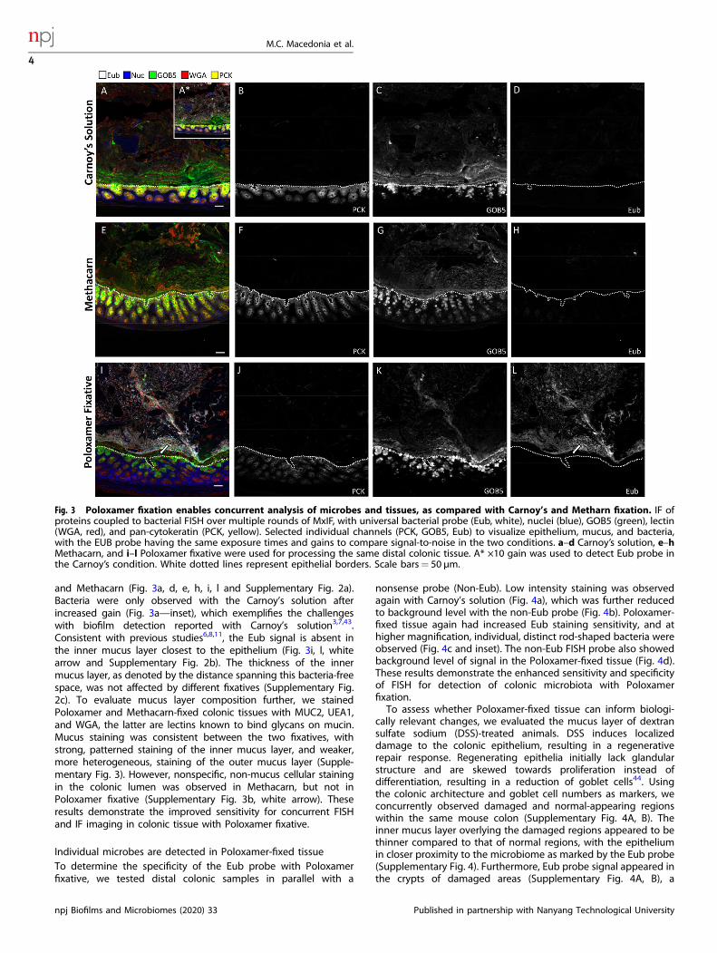
and Methacarn (Fig. 3a, d, e, h, i, l and Supplementary Fig. 2a).Bacteria were only observed with the Carnoy’s solution afterincreased gain (Fig. 3a—inset), which exemplifies the challengeswith biofilm detection reported with Carnoy’s solution3,7,43.Consistent with previous studies6,8,11, the Eub signal is absent inthe inner mucus layer closest to the epithelium (Fig. 3i, l, whitearrow and Supplementary Fig. 2b). The thickness of the innermucus layer, as denoted by the distance spanning this bacteria-freespace, was not affected by different fixatives (Supplementary Fig.2c). To evaluate mucus layer composition further, we stainedPoloxamer and Methacarn-fixed colonic tissues with MUC2, UEA1,and WGA, the latter are lectins known to bind glycans on mucin.Mucus staining was consistent between the two fixatives, withstrong, patterned staining of the inner mucus layer, and weaker,more heterogeneous, staining of the outer mucus layer (Supple-mentary Fig. 3). However, nonspecific, non-mucus cellular stainingin the colonic lumen was observed in Methacarn, but not inPoloxamer fixative (Supplementary Fig. 3b, white arrow). Theseresults demonstrate the improved sensitivity for concurrent FISHand IF imaging in colonic tissue with Poloxamer fixative.
Individual microbes are detected in Poloxamer-fixed tissueTo determine the specificity of the Eub probe with Poloxamerfixative, we tested distal colonic samples in parallel with a
nonsense probe (Non-Eub). Low intensity staining was observedagain with Carnoy’s solution (Fig. 4a), which was further reducedto background level with the non-Eub probe (Fig. 4b). Poloxamer-fixed tissue again had increased Eub staining sensitivity, and athigher magnification, individual, distinct rod-shaped bacteria wereobserved (Fig. 4c and inset). The non-Eub FISH probe also showedbackground level of signal in the Poloxamer-fixed tissue (Fig. 4d).These results demonstrate the enhanced sensitivity and specificityof FISH for detection of colonic microbiota with Poloxamerfixation.To assess whether Poloxamer-fixed tissue can inform biologi-
cally relevant changes, we evaluated the mucus layer of dextransulfate sodium (DSS)-treated animals. DSS induces localizeddamage to the colonic epithelium, resulting in a regenerativerepair response. Regenerating epithelia initially lack glandularstructure and are skewed towards proliferation instead ofdifferentiation, resulting in a reduction of goblet cells44. Usingthe colonic architecture and goblet cell numbers as markers, weconcurrently observed damaged and normal-appearing regionswithin the same mouse colon (Supplementary Fig. 4A, B). Theinner mucus layer overlying the damaged regions appeared to bethinner compared to that of normal regions, with the epitheliumin closer proximity to the microbiome as marked by the Eub probe(Supplementary Fig. 4). Furthermore, Eub probe signal appeared inthe crypts of damaged areas (Supplementary Fig. 4A, B), a
Fig. 3 Poloxamer fixation enables concurrent analysis of microbes and tissues, as compared with Carnoy’s and Metharn fixation. IF ofproteins coupled to bacterial FISH over multiple rounds of MxIF, with universal bacterial probe (Eub, white), nuclei (blue), GOB5 (green), lectin(WGA, red), and pan-cytokeratin (PCK, yellow). Selected individual channels (PCK, GOB5, Eub) to visualize epithelium, mucus, and bacteria,with the EUB probe having the same exposure times and gains to compare signal-to-noise in the two conditions. a–d Carnoy’s solution, e–hMethacarn, and i–l Poloxamer fixative were used for processing the same distal colonic tissue. A* ×10 gain was used to detect Eub probe inthe Carnoy’s condition. White dotted lines represent epithelial borders. Scale bars= 50 µm.
M.C. Macedonia et al.
4
npj Biofilms and Microbiomes (2020) 33 Published in partnership with Nanyang Technological University
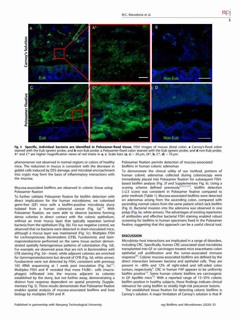
phenomenon not observed in normal regions or colons of healthymice. The reduction in mucus is consistent with the decrease ingoblet cells induced by DSS damage, and microbial encroachmentinto crypts may form the basis of inflammatory interactions withthe mucosa.
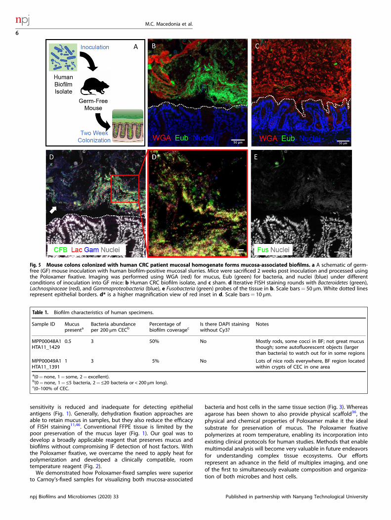
Mucosa-associated biofilms are observed in colonic tissue usingPoloxamer fixationTo further validate Poloxamer fixative for biofilm detection withdirect implications for the human microbiome, we colonizedgerm-free (GF) mice with a biofilm-positive microbiota slurryisolated from a human colorectal cancer (Fig. 5a)15. WithPoloxamer fixation, we were able to observe bacteria formingdense colonies in direct contact with the colonic epithelium,without an inner mucus layer that typically separates luminalbacteria from the epithelium (Fig. 5b). For our negative control, weobserved that no bacteria were detected in sham-inoculated mice,although a mucus layer was maintained (Fig. 5c). Multiplex FISHfor Lachnospiraceae, Bacteroidetes (CFB), Fusobacteria, and Gam-maproteobacteria performed on the same tissue section demon-strated spatially heterogeneous patterns of colonization (Fig. 5d).For example, we observed areas that are rich in Bacteroidetes withCFB staining (Fig. 5d—inset), while adjacent colonies are enrichedfor Gammaproteobacteria but devoid of CFB (Fig. 5d, white arrow).Fusobacteria were not detected by FISH, consistent with previous16S rRNA sequencing at 1 week post inoculation (Fig. 5e)15.Multiplex FISH and IF revealed that more F4/80+ cells (macro-phages) infiltrated into the mucosa adjacent to coloniesestablished by the slurry, but not further away, demonstrating adistinct host response dependent on spatial proximity (Supple-mentary Fig. 5). These results demonstrate that Poloxamer fixativeenables spatial analysis of mucosa-associated biofilms and hostbiology by multiplex FISH and IF.
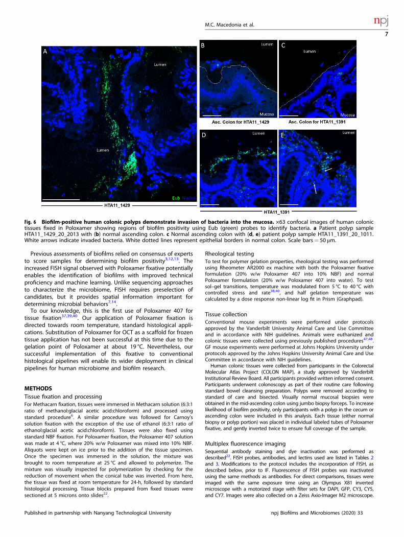
Poloxamer fixation permits detection of mucosa-associatedbiofilms in human colonic adenomasTo demonstrate the clinical utility of our method, portions ofhuman colonic adenomas collected during colonoscopy wereimmediately placed into Poloxamer fixation for subsequent FISH-based biofilm analysis (Fig. 2f and Supplementary Fig. 6). Using ascoring scheme defined previously3,12,13,15, biofilm detection(>2.5 score) was consistent in Poloxamer fixative compared toprior methods (Table 1). Mucosa-associated biofilms were detectedon adenomas arising from the ascending colon, compared withascending normal colons from the same patient which lack biofilm(Fig. 6). Bacterial invasion into the adenoma was observed in onepolyp (Fig. 6e, white arrows). The advantages of existing repertoiresof antibodies and effective bacterial FISH staining enabled robustscreening for biofilms in human specimens fixed in the Poloxamerfixative, suggesting that this approach can be a useful clinical tool.
DISCUSSIONMicrobiota–host interactions are implicated in a range of disorders,including CRC. Specifically, human CRC-associated stool microbiotatransplanted into GF or carcinogen-treated mice exacerbates colonepithelial cell proliferation and the tumor-associated immuneresponse45. Colonic mucosa-associated biofilms are defined by thedirect interaction between bacteria and epithelial cells. They arepresent in ~89% and 12% of right-sided and left-sided colontumors, respectively3. CRC in human FAP appears to be uniformlybiofilm positive13. Some human colonic biofilms are carcinogenicin GF ApcMin mice15. With a reported range of 13–35% colonicbiofilm positive in healthy subjects, these findings indicate clinicalrelevance for using biofilm to stratify high-risk precancer lesions.The established tissue fixation for detecting colonic biofilms is
Carnoy’s solution. A major limitation of Carnoy’s solution is that IF
Fig. 4 Specific, individual bacteria are identified in Poloxamer-fixed tissue. FISH images of mouse distal colon. a Carnoy’s-fixed colonstained with the Eub (green) probe, and b non-Eub probe. c Poloxamer-fixed colon stained with the Eub (green) probe, and d non-Eub probe.A* and C* are higher magnification views of red insets in a, c. Scale bars (a, c)= 20 μm, (A*, b, C*, d)= 10 μm.
M.C. Macedonia et al.
5
Published in partnership with Nanyang Technological University npj Biofilms and Microbiomes (2020) 33
sensitivity is reduced and inadequate for detecting epithelialantigens (Fig. 1). Generally, dehydration fixation approaches areable to retain mucus in samples, but they also reduce the efficacyof FISH staining11,46. Conventional FFPE tissue is limited by thepoor preservation of the mucus layer (Fig. 1). Our goal was todevelop a broadly applicable reagent that preserves mucus andbiofilms without compromising IF detection of host factors. Withthe Poloxamer fixative, we overcame the need to apply heat forpolymerization and developed a clinically compatible, roomtemperature reagent (Fig. 2).We demonstrated how Poloxamer-fixed samples were superior
to Carnoy’s-fixed samples for visualizing both mucosa-associated
bacteria and host cells in the same tissue section (Fig. 3). Whereasagarose has been shown to also provide physical scaffold36, thephysical and chemical properties of Poloxamer make it the idealsubstrate for preservation of mucus. The Poloxamer fixativepolymerizes at room temperature, enabling its incorporation intoexisting clinical protocols for human studies. Methods that enablemultimodal analysis will become very valuable in future endeavorsfor understanding complex tissue ecosystems. Our effortsrepresent an advance in the field of multiplex imaging, and oneof the first to simultaneously evaluate composition and organiza-tion of both microbes and host cells.
Fig. 5 Mouse colons colonized with human CRC patient mucosal homogenate forms mucosa-associated biofilms. a A schematic of germ-free (GF) mouse inoculation with human biofilm-positive mucosal slurries. Mice were sacrificed 2 weeks post inoculation and processed usingthe Poloxamer fixative. Imaging was performed using WGA (red) for mucus, Eub (green) for bacteria, and nuclei (blue) under differentconditions of inoculation into GF mice: b Human CRC biofilm isolate, and c sham. d Iterative FISH staining rounds with Bacteroidetes (green),Lachnospiraceae (red), and Gammaproteobacteria (blue), e Fusobacteria (green) probes of the tissue in b. Scale bars= 50 μm. White dotted linesrepresent epithelial borders. d* is a higher magnification view of red inset in d. Scale bars= 10 μm.
Table 1. Biofilm characteristics of human specimens.
Sample ID Mucuspresenta
Bacteria abundanceper 200 μm CECb
Percentage ofbiofilm coveragec
Is there DAPI stainingwithout Cy3?
Notes
MPP00048A1HTA11_1429
0.5 3 50% No Mostly rods, some cocci in BF; not great mucusthough; some autofluorescent objects (largerthan bacteria) to watch out for in some regions
MPP00049A1HTA11_1391
1 3 5% No Lots of nice rods everywhere, BF region locatedwithin crypts of CEC in one area
a(0= none, 1= some, 2= excellent).b(0= none, 1= ≤5 bacteria, 2= ≤20 bacteria or < 200 μm long).c(0–100% of CEC.
M.C. Macedonia et al.
6
npj Biofilms and Microbiomes (2020) 33 Published in partnership with Nanyang Technological University
Previous assessments of biofilms relied on consensus of expertsto score samples for determining biofilm positivity3,12,13. Theincreased FISH signal observed with Poloxamer fixative potentiallyenables the identification of biofilms with improved technicalproficiency and machine learning. Unlike sequencing approachesto characterize the microbiome, FISH requires preselection ofcandidates, but it provides spatial information important fordetermining microbial behaviors7,14.To our knowledge, this is the first use of Poloxamer 407 for
tissue fixation37,39,40. Our application of Poloxamer fixation isdirected towards room temperature, standard histological appli-cations. Substitution of Poloxamer for OCT as a scaffold for frozentissue application has not been successful at this time due to thegelation point of Poloxamer at about 19 °C. Nevertheless, oursuccessful implementation of this fixative to conventionalhistological pipelines will enable its wider deployment in clinicalpipelines for human microbiome and biofilm research.
METHODSTissue fixation and processingFor Methacarn fixation, tissues were immersed in Methacarn solution (6:3:1ratio of methanol:glacial acetic acid:chloroform) and processed usingstandard procedure6. A similar procedure was followed for Carnoy’ssolution fixation with the exception of the use of ethanol (6:3:1 ratio ofethanol:glacial acetic acid:chloroform). Tissues were also fixed usingstandard NBF fixation. For Poloxamer fixation, the Poloxamer 407 solutionwas made at 4 °C, where 20% w/w Poloxamer was mixed into 10% NBF.Aliquots were kept on ice prior to the addition of the tissue specimen.Once the specimen was immersed in the solution, the mixture wasbrought to room temperature at 25 °C and allowed to polymerize. Themixture was visually inspected for polymerization by checking for thereduction of movement when the conical tube was inverted. From here,the tissue was fixed at room temperature for 24-h, followed by standardhistological processing. Tissue blocks prepared from fixed tissues weresectioned at 5 microns onto slides22.
Rheological testingTo test for polymer gelation properties, rheological testing was performedusing Rheometer AR2000 ex machine with both the Poloxamer fixativeformulation (20% w/w Poloxamer 407 into 10% NBF) and normalPoloxamer formulation (20% w/w Poloxamer 407 into water). To testsol–gel transitions, temperature was modulated from 5 °C to 40 °C withcontrolled stress and rate38,40, and half gelation temperature wascalculated by a dose response non-linear log fit in Prism (Graphpad).
Tissue collectionConventional mouse experiments were performed under protocolsapproved by the Vanderbilt University Animal Care and Use Committeeand in accordance with NIH guidelines. Animals were euthanized andcolonic tissues were collected using previously published procedures47,48.GF mouse experiments were performed at Johns Hopkins University underprotocols approved by the Johns Hopkins University Animal Care and UseCommittee in accordance with NIH guidelines.Human colonic tissues were collected from participants in the Colorectal
Molecular Atlas Project (COLON MAP), a study approved by VanderbiltInstitutional Review Board. All participants provided written informed consent.Participants underwent colonoscopy as part of their routine care followingstandard bowel cleansing preparation. Polyps were removed according tostandard of care and bisected. Visually normal mucosal biopsies wereobtained in the mid-ascending colon using jumbo biopsy forceps. To increaselikelihood of biofilm positivity, only participants with a polyp in the cecum orascending colon were included in this analysis. Each tissue (either normalbiopsy or polyp portion) was placed in individual labeled tubes of Poloxamerfixative, and gently inverted twice to ensure full coverage of the sample.
Multiplex fluorescence imagingSequential antibody staining and dye inactivation was performed asdescribed22. FISH probes, antibodies, and lectins used are listed in Tables 2and 3. Modifications to the protocol includes the incorporation of FISH, asdescribed below, prior to IF. Fluorescence of FISH probes was inactivatedusing the same methods as antibodies. For direct comparisons, tissues wereimaged with the same exposure time using an Olympus X81 invertedmicroscope with a motorized stage with filter sets for DAPI, GFP, CY3, CY5,and CY7. Images were also collected on a Zeiss Axio-Imager M2 microscope.
Fig. 6 Biofilm-positive human colonic polyps demonstrate invasion of bacteria into the mucosa. ×63 confocal images of human colonictissues fixed in Poloxamer showing regions of biofilm positivity using Eub (green) probes to identify bacteria. a Patient polyp sampleHTA11_1429_20_2013 with (b) normal ascending colon. c Normal ascending colon with (d, e) patient polyp sample HTA11_1391_20_1011.White arrows indicate invaded bacteria. White dotted lines represent epithelial borders in normal colon. Scale bars= 50 μm.
M.C. Macedonia et al.
7
Published in partnership with Nanyang Technological University npj Biofilms and Microbiomes (2020) 33
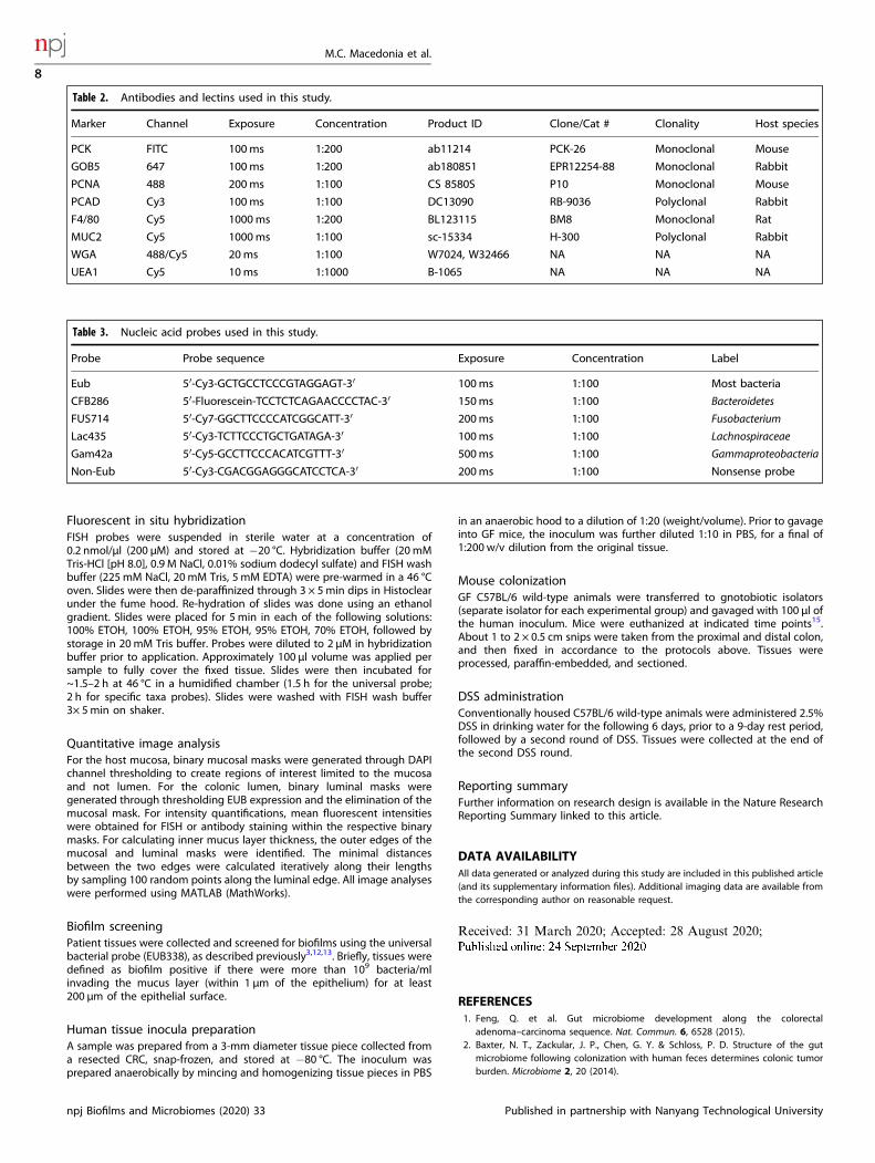
Fluorescent in situ hybridizationFISH probes were suspended in sterile water at a concentration of0.2 nmol/μl (200 μM) and stored at −20 °C. Hybridization buffer (20mMTris-HCl [pH 8.0], 0.9 M NaCl, 0.01% sodium dodecyl sulfate) and FISH washbuffer (225mM NaCl, 20 mM Tris, 5 mM EDTA) were pre-warmed in a 46 °Coven. Slides were then de-paraffinized through 3 × 5min dips in Histoclearunder the fume hood. Re-hydration of slides was done using an ethanolgradient. Slides were placed for 5 min in each of the following solutions:100% ETOH, 100% ETOH, 95% ETOH, 95% ETOH, 70% ETOH, followed bystorage in 20mM Tris buffer. Probes were diluted to 2 μM in hybridizationbuffer prior to application. Approximately 100 μl volume was applied persample to fully cover the fixed tissue. Slides were then incubated for~1.5–2 h at 46 °C in a humidified chamber (1.5 h for the universal probe;2 h for specific taxa probes). Slides were washed with FISH wash buffer3× 5min on shaker.
Quantitative image analysisFor the host mucosa, binary mucosal masks were generated through DAPIchannel thresholding to create regions of interest limited to the mucosaand not lumen. For the colonic lumen, binary luminal masks weregenerated through thresholding EUB expression and the elimination of themucosal mask. For intensity quantifications, mean fluorescent intensitieswere obtained for FISH or antibody staining within the respective binarymasks. For calculating inner mucus layer thickness, the outer edges of themucosal and luminal masks were identified. The minimal distancesbetween the two edges were calculated iteratively along their lengthsby sampling 100 random points along the luminal edge. All image analyseswere performed using MATLAB (MathWorks).
Biofilm screeningPatient tissues were collected and screened for biofilms using the universalbacterial probe (EUB338), as described previously3,12,13. Briefly, tissues weredefined as biofilm positive if there were more than 109 bacteria/mlinvading the mucus layer (within 1 μm of the epithelium) for at least200 μm of the epithelial surface.
Human tissue inocula preparationA sample was prepared from a 3-mm diameter tissue piece collected froma resected CRC, snap-frozen, and stored at −80 °C. The inoculum wasprepared anaerobically by mincing and homogenizing tissue pieces in PBS
in an anaerobic hood to a dilution of 1:20 (weight/volume). Prior to gavageinto GF mice, the inoculum was further diluted 1:10 in PBS, for a final of1:200 w/v dilution from the original tissue.
Mouse colonizationGF C57BL/6 wild-type animals were transferred to gnotobiotic isolators(separate isolator for each experimental group) and gavaged with 100 μl ofthe human inoculum. Mice were euthanized at indicated time points15.About 1 to 2 × 0.5 cm snips were taken from the proximal and distal colon,and then fixed in accordance to the protocols above. Tissues wereprocessed, paraffin-embedded, and sectioned.
DSS administrationConventionally housed C57BL/6 wild-type animals were administered 2.5%DSS in drinking water for the following 6 days, prior to a 9-day rest period,followed by a second round of DSS. Tissues were collected at the end ofthe second DSS round.
Reporting summaryFurther information on research design is available in the Nature ResearchReporting Summary linked to this article.
DATA AVAILABILITYAll data generated or analyzed during this study are included in this published article(and its supplementary information files). Additional imaging data are available fromthe corresponding author on reasonable request.
Received: 31 March 2020; Accepted: 28 August 2020;
REFERENCES1. Feng, Q. et al. Gut microbiome development along the colorectal
adenoma–carcinoma sequence. Nat. Commun. 6, 6528 (2015).2. Baxter, N. T., Zackular, J. P., Chen, G. Y. & Schloss, P. D. Structure of the gut
microbiome following colonization with human feces determines colonic tumorburden. Microbiome 2, 20 (2014).
Table 2. Antibodies and lectins used in this study.
Marker Channel Exposure Concentration Product ID Clone/Cat # Clonality Host species
PCK FITC 100ms 1:200 ab11214 PCK-26 Monoclonal Mouse
GOB5 647 100ms 1:200 ab180851 EPR12254-88 Monoclonal Rabbit
PCNA 488 200ms 1:100 CS 8580S P10 Monoclonal Mouse
PCAD Cy3 100ms 1:100 DC13090 RB-9036 Polyclonal Rabbit
F4/80 Cy5 1000ms 1:200 BL123115 BM8 Monoclonal Rat
MUC2 Cy5 1000ms 1:100 sc-15334 H-300 Polyclonal Rabbit
WGA 488/Cy5 20ms 1:100 W7024, W32466 NA NA NA
UEA1 Cy5 10ms 1:1000 B-1065 NA NA NA
Table 3. Nucleic acid probes used in this study.
Probe Probe sequence Exposure Concentration Label
Eub 5′-Cy3-GCTGCCTCCCGTAGGAGT-3′ 100ms 1:100 Most bacteria
CFB286 5′-Fluorescein-TCCTCTCAGAACCCCTAC-3′ 150ms 1:100 Bacteroidetes
FUS714 5′-Cy7-GGCTTCCCCATCGGCATT-3′ 200ms 1:100 Fusobacterium
Lac435 5′-Cy3-TCTTCCCTGCTGATAGA-3′ 100ms 1:100 Lachnospiraceae
Gam42a 5′-Cy5-GCCTTCCCACATCGTTT-3′ 500ms 1:100 Gammaproteobacteria
Non-Eub 5′-Cy3-CGACGGAGGGCATCCTCA-3′ 200ms 1:100 Nonsense probe
M.C. Macedonia et al.
8
npj Biofilms and Microbiomes (2020) 33 Published in partnership with Nanyang Technological University
3. Dejea, C. M. A. W. E. C. et al. Microbiota organization is a distinct feature ofproximal colorectal cancers. Proc. Natl Acad. Sci. 111, 18321–18326 (2014).
4. Drewes, J. L. et al. High-resolution bacterial 16S rRNA gene profile meta-analysisand biofilm status reveal common colorectal cancer consortia. NPJ BiofilmsMicrobiomes 3, 34 (2017).
5. Chen, J., Domingue, J. C. & Sears, C. L. Microbiota dysbiosis in select humancancers: evidence of association and causality. Semin. Immunol. 34, 25–34 (2017).
6. Johansson, M. E. et al. The inner of the two Muc2 mucin-dependent mucus layersin colon is devoid of bacteria. Proc. Natl Acad. Sci. 105, 15064–15069 (2008).
7. Earle, K. A. et al. Quantitative imaging of gut microbiota spatial organization. CellHost Microbe 18, 478–488 (2015).
8. Donaldson, G. P., Lee, S. M. & Mazmanian, S. K. Gut biogeography of the bacterialmicrobiota. Nat. Rev. Microbiol. 14, 20–32 (2015).
9. Johansson, M. E., & Hansson, G. C. Preservation of mucus in histological sections,immunostaining of mucins in fixed tissue, and localization of bacteria with FISH.Methods Mol. Biol. 842, 229–235 (2012).
10. Lavelle, A. et al. Spatial variation of the colonic microbiota in patients withulcerative colitis and control volunteers. Gut 64, 1553–1561 (2015).
11. Tropini, C., Earle, K. A., Huang, K. C. & Sonnenburg, J. L. The gut microbiome:connecting spatial organization to function. Cell Host Microbe 21, 433–442 (2017).
12. Drewes, J. L. et al. High-resolution bacterial 16S rRNA gene profile meta-analysisand biofilm status reveal common colorectal cancer consortia. npj BiofilmsMicrobiomes 3, 34 (2017).
13. Dejea, C. M. et al. Patients with familial adenomatous polyposis harbor colonicbiofilms containing tumorigenic bacteria. Science 359, 592–597 (2018).
14. Swidsinski, A., Weber, J., Loening-Baucke, V., Hale, L. P. & Lochs, H. Spatialorganization and composition of the mucosal flora in patients with inflammatorybowel disease. J. Clin. Microbiol. 43, 3380–3389 (2005).
15. Tomkovich, S., et al. Human colon mucosal biofilms from healthy or colon cancerhosts are carcinogenic. J Clin. investig. 129, 1699–1712 (2019).
16. Costerton, J. W., Stewart, P. S. & Greenberg, E. P. Bacterial biofilms: a commoncause of persistent infections. Science 284, 1318–1322 (1999).
17. Mallick, H. et al. Predictive metabolomic profiling of microbial communities usingamplicon or metagenomic sequences. Nat. Commun. 10, 1–11 (2019).
18. Chen, K. H., Boettiger, A. N., Moffitt, J. R., Wang, S. & Zhuang, X. Spatiallyresolved, highly multiplexed RNA profiling in single cells. Science 348, aaa6090(2015).
19. Gerdes, M. J. et al. Highly multiplexed single-cell analysis of formalin-fixed,paraffin-embedded cancer tissue. Proc. Natl Acad. Sci. 110, 11982–11987 (2013).
20. Eng, C. H. L. et al. Transcriptome-scale super-resolved imaging in tissues by RNAseqFISH. Nature 568, 235–239 (2019).
21. Goltsev, Y. et al. Deep profiling of mouse splenic architecture with CODEX mul-tiplexed imaging. Cell 174, 968–981 (2018).
22. McKinley, E. T., et al. Optimized multiplex immunofluorescence single-cell ana-lysis reveals tuft cell heterogeneity. JCI Insight 2, e93487 (2017).
23. Lin, J. R., Fallahi-Sichani, M. & Sorger, P. K. Highly multiplexed imaging of singlecells using a high-throughput cyclic immunofluorescence method. Nat. Commun.6, 8390 (2015).
24. Tjalsma, H., Boleij, A., Marchesi, J. R. & Dutilh, B. E. A bacterial driver–passengermodel for colorectal cancer: beyond the usual suspects. Nat. Rev. Microbiol. 10,575–582 (2012).
25. Gut, G., Herrmann, M. D. & Pelkmans, L. Multiplexed protein maps link subcellularorganization to cellular states. Science 361, eaar7042 (2018).
26. Remark, R. et al. In-depth tissue profiling using multiplexed immunohistochem-ical consecutive staining on single slide. Sci. Immunol. 1, aaf6925 (2016).
27. Jungmann, R. et al. Multiplexed 3D cellular super-resolution imaging with DNA-PAINT and exchange-PAINT. Nat. Methods 11, 313–318 (2014).
28. Biedermann, L. & Rogler, G. The intestinal microbiota: its role in health anddisease. Eur. J. pediatrics 174, 151–167 (2015).
29. Abraham, C. & Medzhitov, R. Interactions between the host innate immunesystem and microbes in inflammatory bowel disease. Gastroenterology 140,1729–1737 (2011).
30. Bromberg, L. E. & Barr, D. P. Self-association of mucin. Biomacromolecules 1,325–334 (2000).
31. Atuma, C., Strugala, V., Allen, A., & Holm, L. The adherent gastrointestinal mucusgel layer: thickness and physical state in vivo. J. Am. Physiol. Soc. 280, G922–G929(2001).
32. Rizzardi, A. E. et al. Quantitative comparison of immunohistochemical stainingmeasured by digital image analysis versus pathologist visual scoring. DiagnosticPathol. 7, 42 (2012).
33. Robertson, D., Savage, K., Reis-Filho, J. S. & Isacke, C. M. Multiple immuno-fluorescence labelling of formalin-fixed paraffin-embedded (FFPE) tissue. BMC cellBiol. 9, 13 (2008).
34. Dongari-Bagtzoglou, A. Pathogenesis of mucosal biofilm infections: challengesand progress. Expert Rev. Anti Infect. Ther. 6, 201–208 (2008).
35. Demouveaux, B., Gouyer, V., Gottrand, F., Narita, T. & Desseyn, J. L. Gel-formingmucin interactome drives mucus viscoelasticity. Adv. Colloid Interface Sci. 252,69–82 (2017).
36. Hasegawa, Y., Welch, J. L. M., Rossetti, B. J. & Borisy, G. G. Preservation of three-dimensional spatial structure in the gut microbiome. PLOS ONE 12, e0188257(2017).
37. Dumortier, G., Grossiord, J. L., Agnely, F. & Chaumeil, J. C. A review of poloxamer407 pharmaceutical and pharmacological characteristics. Pharm. Res. 23,2709–2272 (2006).
38. Charrueau, C., Tuleu, C., Astre, V., Grossiord, J. L. & Chaumeil, J. C. Poloxamer 407as a thermogelling and adhesive polymer for rectal administration of short-chainfatty acids. Drug Dev. Ind. Pharm. 27, 351–357 (2001).
39. Giuliano, E., Paolino, D., Fresta, M. & Cosco, D. Mucosal applications of poloxamer407-based hydrogels: an overview. Pharmaceutics 10, 159 (2018).
40. Baloglu, E., Karavana, S. Y., Senyigit, Z. A., & Guneri, T. Rheological and mechanicalproperties of poloxamer mixtures as a mucoadhesive gel base. Pharm. Dev.Technol. 16, 627–636 (2011).
41. Fakhari, A., Corcoran, M. & Schwarz, A. Thermogelling properties of purifiedpoloxamer 407. Heliyon 3, e00390 (2017).
42. Nyström, E. E., Arike, L., Ehrencrona, E., Hansson, G. C. & Johansson, M. E. Calcium-activated chloride channel regulator 1 (CLCA1) forms non-covalent oligomers incolonic mucus and has mucin 2–processing properties. J. Biol. Chem. 294,17075–17089 (2019).
43. Arena, E. T., et al. Bioimage analysis of Shigella infection reveals targeting ofcolonic crypts. Proc. Natl Acad. Sci. 112, E3282–E3290 (2015).
44. Nowarski, R. et al. Epithelial IL-18 equilibrium controls barrier function in colitis.Cell 163, 1444–1456 (2015).
45. Wong, S. H. et al. Gavage of fecal samples from patients with colorectal cancerpromotes intestinal carcinogenesis in germ-free and conventional mice. Gastro-enterology 153, 1621–1633 (2017).
46. Urieli-Shoval, S. et al. Preservation of RNA for in situ hybridization: Carnoy’sversus formaldehyde fixation. J. Histochemistry Cytochemistry 40, 1879–1885(1992).
47. Liu, Q. et al. Quantitative assessment of cell population diversity in single-celllandscapes. PLoS Biol. 16, e2006687 (2018).
48. Simmons, A. J., et al. Cytometry-based single-cell analysis of intact epithelialsignaling reveals MAPK activation divergent from TNF-α-induced apoptosisin vivo. Mol. Syst. Biol. 11, 835 (2015).
ACKNOWLEDGEMENTSThe authors would like to thank Dr. Cynthia Reinhart-King for advice on polymers,Katarzyna Zienkiewicz and Dr. Scott Guelcher for rheometer use, and Dr. EliotMcKinley for his advice on MxIF use. K.S.L. and A.J.S. are funded by R01DK103831. R.J.C. is funded by P50CA236733 and R35CA1975703. M.C.M., J.L.D., J.T.R., R.J.C., M.J.S.,C.L.S., and K.S.L. are funded by U2CCA233291. N.O.M. is funded by T32DK007673. P.N.V. is funded by T32HD007502. C.R.S. is funded T32AI007281. Participant recruitmentand biospecimen collection were conducted in part by the Survey and BiospecimenShared Resource which is supported in part by P30CA68485. Tissue fixation andprocessing was provided by the NCI Cooperative Human Tissue Network (CHTN)Western Division UM1CA183727.
AUTHOR CONTRIBUTIONSM.C.M. conceived the study, performed experiments, analyzed the data, compiled thefigures, and wrote the manuscript. J.L.D. performed microbiome transplantexperiments, scored human specimens, and contributed to the writing of themanuscript. N.O.M. assisted with mouse experiments, intellectually contributed to thestudy, and wrote the manuscript. A.J.S. contributed to the study design, and assistedwith experiments. J.T.R. assisted with imaging. P.N.V. and C.R.S. assisted with mouseexperiments. R.J.C. contributed to funding and writing of the manuscript. M.J.S.facilitated human specimen collection, and contributed to funding and writing of themanuscript. C.L.S. supervised the research, contributed to the study design andwriting of the manuscript. K.S.L. conceived the study, analyzed the data, wrote themanuscript, obtained funding, and supervised the research.
COMPETING INTERESTSC.L.S. receives research funding from Bristol Myers Squibb and Janssen and apersonal fee from Merck & Co. in 2019 for an advisory role. The authors declare thatthere are no other competing interests.
M.C. Macedonia et al.
9
Published in partnership with Nanyang Technological University npj Biofilms and Microbiomes (2020) 33
ADDITIONAL INFORMATIONSupplementary information is available for this paper at https://doi.org/10.1038/s41522-020-00143-x.
Correspondence and requests for materials should be addressed to K.S.L.
Reprints and permission information is available at http://www.nature.com/reprints
Publisher’s note Springer Nature remains neutral with regard to jurisdictional claimsin published maps and institutional affiliations.
Open Access This article is licensed under a Creative CommonsAttribution 4.0 International License, which permits use, sharing,
adaptation, distribution and reproduction in any medium or format, as long as you giveappropriate credit to the original author(s) and the source, provide a link to the CreativeCommons license, and indicate if changes were made. The images or other third partymaterial in this article are included in the article’s Creative Commons license, unlessindicated otherwise in a credit line to the material. If material is not included in the article’sCreative Commons license and your intended use is not permitted by statutory regulationorexceeds thepermitteduse, youwill need toobtainpermissiondirectly fromthecopyrightholder. To view a copy of this license, visit http://creativecommons.org/licenses/by/4.0/.
© The Author(s) 2020
M.C. Macedonia et al.
10
npj Biofilms and Microbiomes (2020) 33 Published in partnership with Nanyang Technological University