chromosomal rearrangements as a source of local adaptation
TRANSCRIPT
Chromosomal rearrangements as a source of localadaptation in island Drosophila
Brandon A. Turner∗1, Theresa R. Miorin∗2, Nicholas B. Stewart3,
Robert W. Reid1, Cathy C. Moore1 and Rebekah L. Rogers1
1. Department of Bioinformatics and Genomics, University of North Carolina, Charlotte NC2. Dept of Genetics, University of Georgia, Athens, GA3. Dept of Biology, Ft. Hays State University, Hays, KS*These authors contributed equally to this work.
Keywords: Structural variation, genetic novelty, local adaptation, habitat shifts
1
arX
iv:2
109.
0980
1v1
[q-
bio.
PE]
20
Sep
2021
Introduction
The genetic response to shifting selective pressures remains one of the most fundamentalquestions in evolutionary theory. How does genetic variation appear and contribute to phe-notypic changes during sudden or extreme shifts in habitat [1]? Among the potential geneticresponders to selection, SNPs have received ready attention in theory and in empirical stud-ies of differentiation. However, structural variants are often masked or ignored as thesemutations are more difficult to identify and interpret in genetic sequence data [2–5]. Thissource of genetic variation deserves greater observation in the context of evolutionary change.Mutations that copy or shuffle pieces of DNA within the genome can join new linkage groups,alter gene expression patterns, or create new gene sequences where none existed before [6–9].As such we hypothesize they are a source of innovation that likely to produce cellular changeswhen they are necessary during habitat shifts. We wish to discern whether these “hopefulmonsters” among genes might lead to adaptive success in new environments.
Among the many fundamental questions facing local adaptation, the role of new mutationand standing variation is debated [10–12]. To what extent is adaptation limited by the spanof standing variation in populations? How long might it take to acquire new mutationswhen selective pressures shift? Over time, due to factors such as allopatry, genetic drift, andreproductive isolation, populations can begin to diverge [13]. Selection on adaptive mutationsallow these populations and species to become better suited to their environments. How theseadaptive mutations come about is still a question that remains.
The tempo and magnitude of evolutionary change depend on the available substrate ofvariation, as well as evolutionary forces. Standing variation may fix rapidly in response toenvironmental shifts [10], but the spectrum of available variation will be limited by popu-lation genetic forces [11]. Mutations with fitness impacts greater than the nearly neutralthreshold are expected to be weeded out of populations quickly [14, 15]. Even among neu-tral variation, segregating variation will depend on mutation rates and population sizes [16].Meanwhile long wait times for new mutations may place bounds on how quickly genomescan adapt through new mutations [17]. If mechanisms exist generate the supply of newmutations, it may be possible to generate variation quickly during habitat shifts. Discerninghow new mutations appear in populations and spread during habitat changes is imperativeto evaluate the genetic response to rapid or drastic shifts in selective pressures.
Recent technological innovation has allowed us to identify such variation in high through-put large scale genome sequencing panels [18–21], opening doors to study evolutionary impactin new organisms or systems beyond standard models [22]. We are able to discover how thesemutations contribute to population variation and how their allele frequencies shift in differingenvironmental contexts. One evolutionary system that has experienced such habitat shiftsis the D. yakuba-D. santomea species complex. D. yakuba contains a large number of chro-mosomal rearrangements [23–25] that have shown evidence of being linked to the creationof de novo exons, providing a source of new gene formation [26].
D. yakuba and D. santomea are two sister species of Drosophila that have both adaptedto the island habitats of Sao Tome in the past 500,000 years [27]. These two species inhabitseparate elevations on the island, with D. santomea living in higher elevations and absent inthe lower regions of the island where D. yakuba is abundant [28]. They do have a contact zoneat moderate altitude, however, where hybrids can occur [29]. The two species likely diverged
2
in allopatry, and during this time partial sexual and post-zygotic isolation evolved [29]. Thisis reflective of a second invasion of D. yakuba [28, 30, 31], which has not yet completelydiverged from the mainland population of D. yakuba, but has not had the time to divergecompared to established populations like D. santomea [27]. This species complex providesa unique opportunity to study and understand genetic variation between two closely relatedspecies on a single island.
This complex is ideal for population genetics of local adaptation as it has invaded farenough in the past that phenotypic and genetic differentiation can occur [29,32]. However, itis not yet so long in the past that population genetic signals are obscured by new mutationsand left to be seen only in species divergence [33]. It further benefits from a known ancestralmainland population that is well characterized for structural variation [20, 34]. To build onthese strengths of the D. yakuba-D. santomea system, we have generated population geneticpanels for D. yakuba and D. santomea on Sao Tome with paired end Illlumina and PacBioHiFi sequencing. We pair these data with gene expression analysis to identify cases wherestructural variants alter expression or create new genes.
This comprehensive portrait of structural variation in local adaptation on Sao Tome canserve as the basis for future insights on evolutionary responses in shifting selective pressures.By learning how chromosomal rearrangements result in phenotypic changes we can betterunderstand how rapid evolution can reshape populations in nature.
Results
Rearrangements role in population differentiation
We wished to determine whether chromosomal rearrangements serve as new sources of ge-netic variation during habitat shifts. To assay this source of genetic novelty during localadaptation, we identified rearrangements in populations of D. santomea and D. yakuba onthe island of Sao Tome. Mainland D. yakuba are the known ancestral population to both D.santomea and the island D. yakuba on Sao Tome [35]. These two waves of island invasion of-fer independent cases of local adaptation from the same mainland population. By surveyingthe differences in island environments we can determine how chromosomal rearrangementsserve as agents of local adaptation.
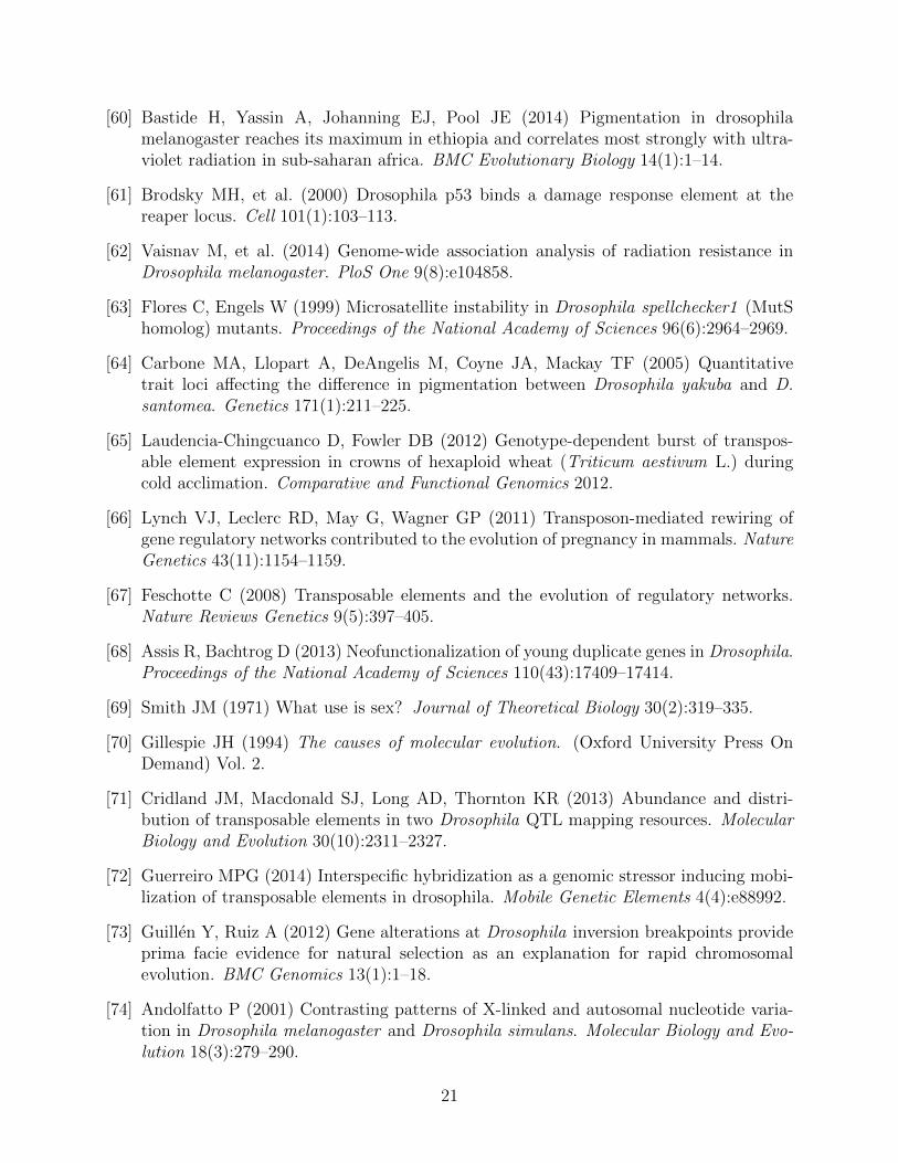
The metapopulation consists of three sub-populations, the island D. santomea and D.yakuba, as well as the ancestral source of variation, mainland D. yakuba. We used Illu-mina paired-end reads to identify chromosomal rearrangements and validated genotypes withPacBio HiFi sequences. We identify find 3,412 rearrangements associated with 19 strains ofmainland D. yakuba, 7,093 with 35 strains of island D. yakuba, and 7,796 with 42 strains ofD. santomea out 16,480 total rearrangements for the meta-population(Figure S1, S2). Thenumber of rearrangements found per strain varies from 82 to 875 total rearrangements in astrain (Table 1). Using PacBio long read data we show that the confirmation rate of rear-rangements is 100% when using a combination of four alignment methods, minimap2 [36],Sniffles [37], and PacBio’s pbsv [38]. We find 6,588 genes within 5kb of the rearrangement’sbreakpoints (Table 2).
For each rearrangement we calculate the allele frequency for the metapopulation, as well
3
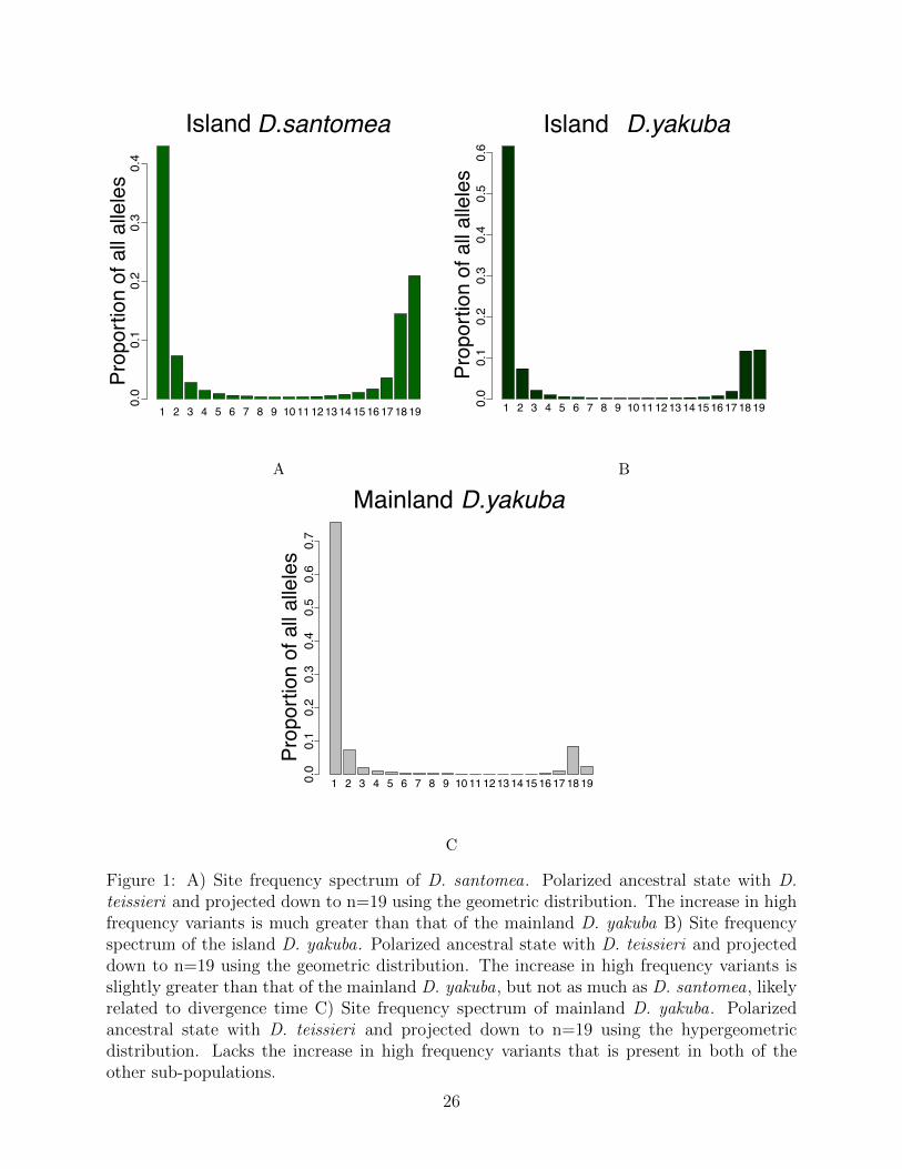
as the frequency in each subpopulation. These frequencies are corrected residual heterozy-gosity created by inbreeding resistance at inversions similar to prior work [39]. Calculatingthe differences in the populations at rearrangements we can infer what changes have ac-cumulated in the D. santomea since their separation from the mainland D. yakuba. Weidentify variants that are outliers compared with genome wide expectations, that have puta-tively spread through island populations due to selection. The three site frequency spectra(SFS) when projected down to the same sample size of n=19 (Figures 1a, 1b, 1c) show,most of the rearrangements at low frequency, as expected. We observe increases at highfrequency, with 20.7% of the projected mutations with a frequency of 18/19 or higher. Thisincrease at high frequency in the SFS could signal that rearrangements are disproportion-ately responsible for rapid adaptation when a population is suddenly introduced to a newenvironment. The distribution of the SFS for D. santomea rearrangements is statisticallydifferent (Kolmogorov-Smirnov, D = 0.65, P = 0.00027) than that of SNPs in D. santomea,implying that allele frequency of rearrangements behave differently than that of the clock-likeSNPs in the population. This is also true for the island D. yakuba (Kolmogorov-Smirnov,D = 0.65, P = 0.0.00027) and the mainland D. yakuba (Kolmogorov-Smirnov, D = 0.7,P = 5.6 × 10⁻⁵).
The sex chromosomes show more dynamic occurrence of rearrangements on the sex chro-mosomes. The autosomes averaged 1,684 mutations each (s.d = 251.68), whereas the Xchromosome has 2,814.
Transposable elements and structural variation
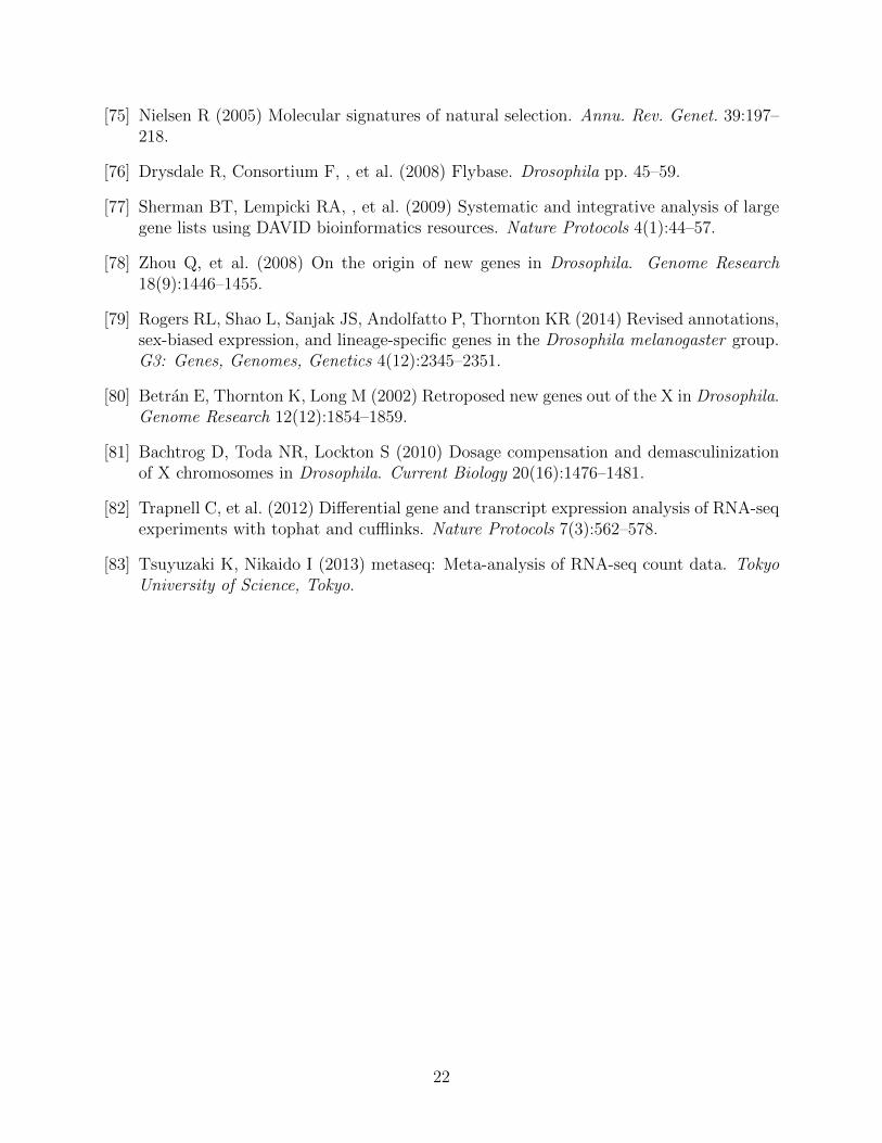
Transposable elements (TEs) facilitate formation of chromosomal rearrangements. TEs inser-tions can result in duplications of nearby sequence and also cause ectopic recombination [40].Using BLAST, we compare the rearrangement breakpoints clustered across strains againstthe RepBase database [41]. We find that 13,763 of 16,480 (83.5%) rearrangements are as-sociated with TEs. Only 5.5% of the reference genome is composed of TEs [42]. Hence, weobserve that rearrangements are heavily linked with transposable elements.
We see 9,152 out of 16,480 (55.5%) associated with TE insertions and 4,611 of the 16,480(28.0%) rearrangements show evidence that transposable elements are facilitating local adap-tation via ectopic recombination. The remaining 2,717 of 16,480 (16.5%) rearrangementsshow no association with transposable elements.
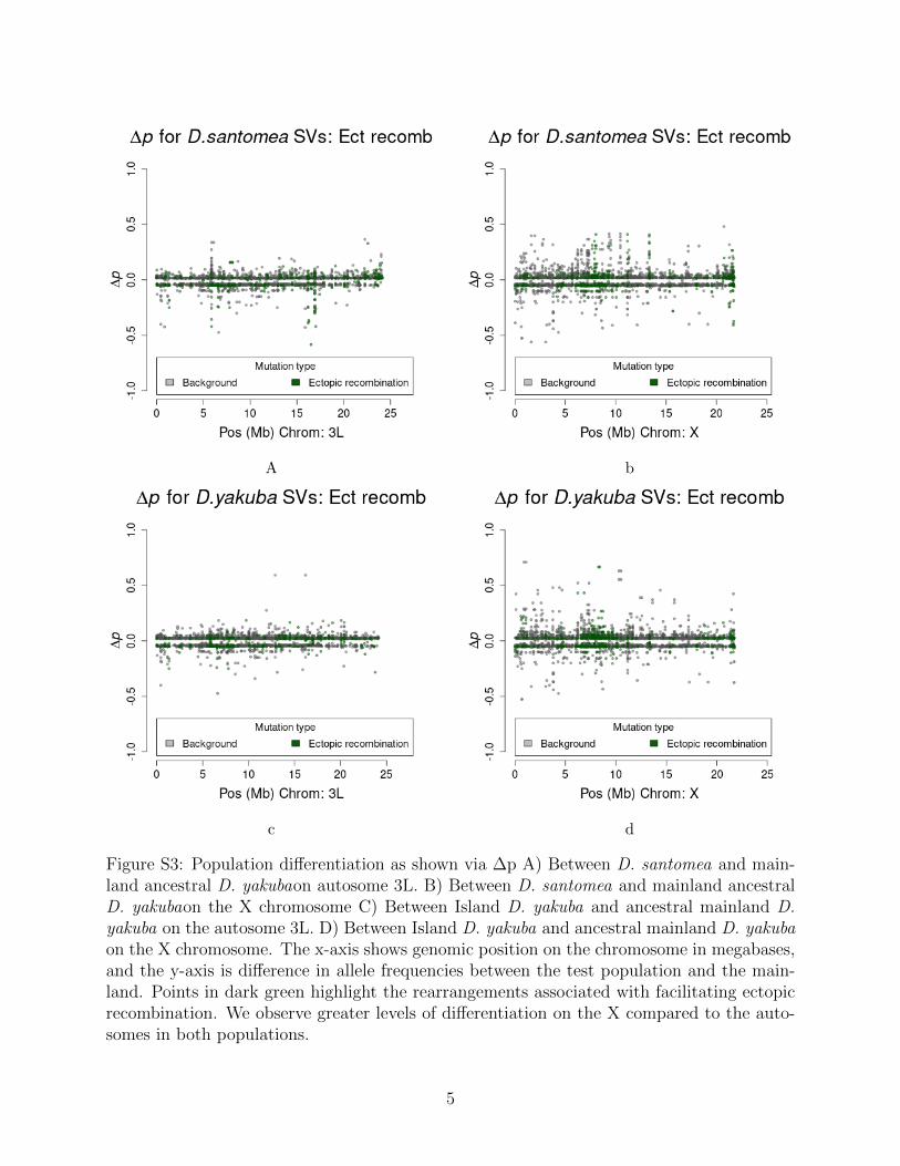
Rearrangements are enriched near the centromeres where recombination suppression al-lows detrimental variation to accumulate (Figure S3). Novel TE insertions show the major-ity of high frequency variants in island environments (Figure S4 and Figure S5; Table 3).Genome wide there are more new TE insertions than cases of ectopic recombination (Fig-ure S6, S7, S8, S9), with the proportion of ectopic recombination sites being 3116/16,480(18.9%). Near the centromeres (within 3Mb) we see the proportion of rearrangements facil-itating ectopic recombination rise to 1,990/3,562 (55.9%). This implies that rearrangementsnear the centromere are disproportionately responsible for facilitating ectopic recombination.
Each TE family has a different history, and previous work has shown that when cer-tain TE families are enriched they can affect transcriptional regulation of stress responsegenes [43]. Here we show that the top 10 TE families are associated with 14345/18374(78.1%) of rearrangements in the metapopulation (Figure S10). The proportion of TE fam-
4
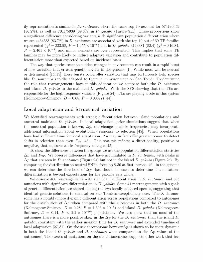
ily representation is similar in D. santomea where the same top 10 account for 5741/6659(86.2%), as well as 5301/5939 (89.3%) in D. yakuba (Figure S11). These proportions showa significant difference considering variants with significant population differentiation wherewe see 446/533 (83.7%) in D. santomea are associated with the top 10 out of 60 TE familiesrepresented (χ2 = 333.58, P = 1.455× 10⁻⁶) and in D. yakuba 314/381 (82.4) (χ2 = 316.84,P = 2.461 × 10⁻⁵) and minor elements are over represented. This implies that some TEfamilies may be more likely to induce adaptive variation and contribute to population dif-ferentiation more than expected based on incidence rates.
The way that species react to sudden changes in environment can result in a rapid burstof new variation that creates genetic novelty in the genome [1]. While most will be neutralor detrimental [14, 15], these bursts could offer variation that may fortuitously help specieslike D. santomea rapidly adapted to their new environment on Sao Tome. To determinethe role that rearrangements have in this adaptation we compare both the D. santomeaand island D. yakuba to the mainland D. yakuba. With the SFS showing that the TEs areresponsible for the high frequency variants (Figure S4), TEs are playing a role in this system(Kolmogorov-Smirnov, D = 0.65, P = 0.00027) [44].
Local adaptation and Structural variation
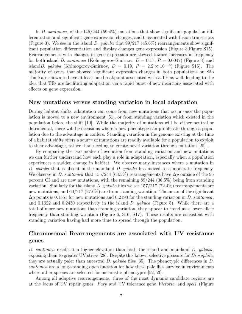
We identified rearrangements with strong differentiation between island populations andancestral mainland D. yakuba. In local adaptation, prior simulations suggest that whenthe ancestral population is known, ∆p, the change in allele frequencies, may incorporateadditional information about evolutionary response to selection [45]. When populationshave had sufficient time for local adaptation, ∆p may in fact offer greater power to detectshifts in selection than even FST [45]. This statistic reflects a directionality, positive ornegative, that captures allele frequency changes [45].
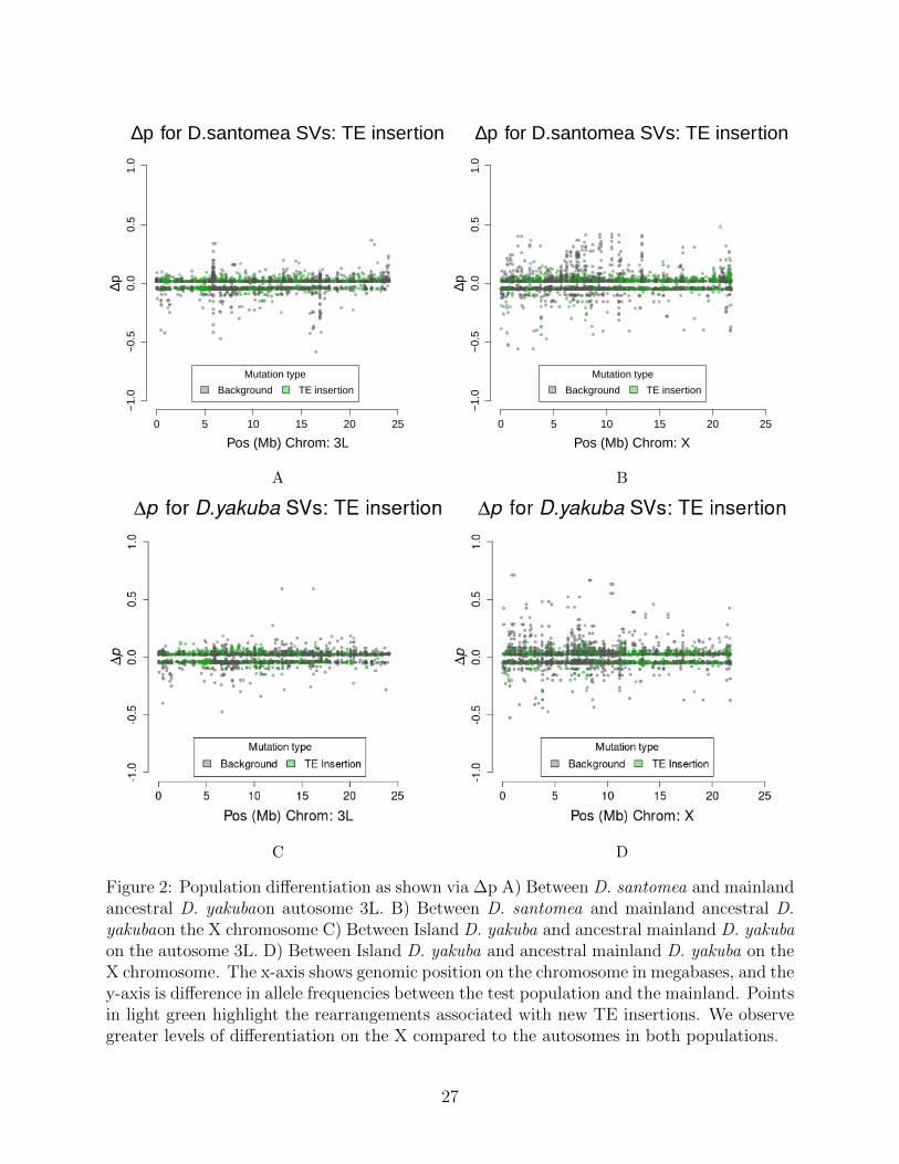
To show the differences between the groups we use the population differentiation statisticsΔp and FST . We observe differences that have accumulated in D. santomea, with peaks inΔp that are seen in D. santomea (Figure 2a) but not in the island D. yakuba (Figure 2c). Bycomparing the distribution to neutral SNPs, from bp 8-30 at first introns [46], in the genomewe can determine the threshold of Δp that should be used to determine if a mutationsdifferentiation is beyond expectations for the genome as a whole.
We observe 468 rearrangements with significant differentiation in D. santomea, and 383mutations with significant differentiation in D. yakuba. Some 41 rearrangements with signalsof genetic differentiation are shared among the two locally adapted species, suggesting thatidentical genetic solutions to survival on Sao Tome is exceptionally rare. The X chromo-some has a notably more dynamic differentiation across populations compared to autosomesfor the distribution of Δp when compared with the autosomes in both the D. santomea(Kolmogorov-Smirnov, D = 0.28, P = 1.603 × 10⁻⁸) and island D. yakuba (Kolmogorov-Smirnov, D = 0.14, P < 2.2 × 10⁻¹⁶) populations. We also show that on most of theautosomes there is a more positive skew in the Δp for the D. santomea than the island D.yakuba, consistent with the earlier invasion time for D. santomea and extended timeline oflocal adaptation [27,31]. On the sex chromosome howeverΔp is shown to be more dynamicin both the island D. yakuba and D. santomea when compared to the Δp values of theautosomes. The excess of mutations on the sex chromosomes supports other work that has
5
shown higher rates of rearrangements out of the X [47] and the outsized role of the X in spe-ciation [33, 48]. FST can identify population differentiation even when allele frequencies arelow, and offers a secondary assessment of whether genome structure changes are associatedwith population differentiation. We observe 180 rearrangements with high FST . Together,these metrics each show that structural variants show strong differentiation between islandpopulations and the mainland (Figure S12, S13).
Transposable element families each have different histories and recent activity levels, andby looking at which families are associated with rearrangements more often we can ascertainwhether specific families are disproportionately associated with rearrangements that haveΔp outside of the 95% C.I. We show that while 446/533 (83.7%) number of significantlydifferentiated rearrangements are associated with just the top 10 out of 60 TE familiesrepresented (Figure S11), these same TE families are represented at roughly same rate inrearrangements that did not have significant Δp (Figure S10, implying that there is likelyno specific TE family is directly responsible for adaptive change.
Gene expression changes and gene fusions role in significant rear-rangements
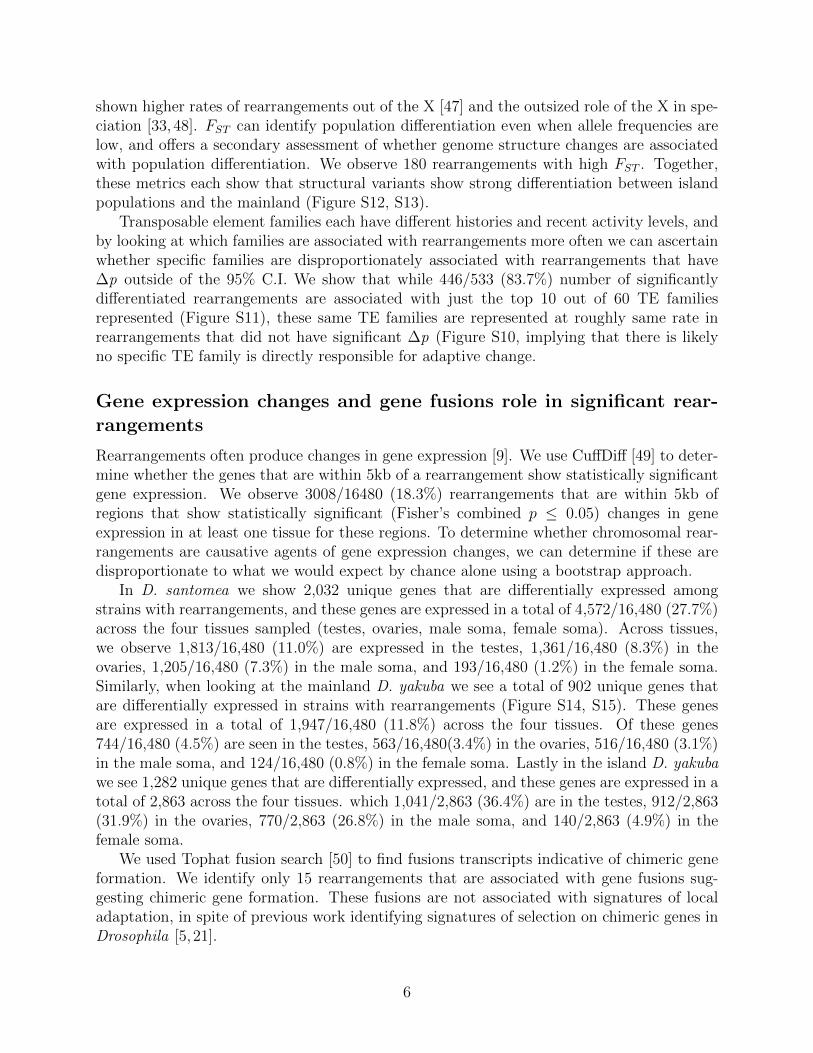
Rearrangements often produce changes in gene expression [9]. We use CuffDiff [49] to deter-mine whether the genes that are within 5kb of a rearrangement show statistically significantgene expression. We observe 3008/16480 (18.3%) rearrangements that are within 5kb ofregions that show statistically significant (Fisher’s combined p ≤ 0.05) changes in geneexpression in at least one tissue for these regions. To determine whether chromosomal rear-rangements are causative agents of gene expression changes, we can determine if these aredisproportionate to what we would expect by chance alone using a bootstrap approach.
In D. santomea we show 2,032 unique genes that are differentially expressed amongstrains with rearrangements, and these genes are expressed in a total of 4,572/16,480 (27.7%)across the four tissues sampled (testes, ovaries, male soma, female soma). Across tissues,we observe 1,813/16,480 (11.0%) are expressed in the testes, 1,361/16,480 (8.3%) in theovaries, 1,205/16,480 (7.3%) in the male soma, and 193/16,480 (1.2%) in the female soma.Similarly, when looking at the mainland D. yakuba we see a total of 902 unique genes thatare differentially expressed in strains with rearrangements (Figure S14, S15). These genesare expressed in a total of 1,947/16,480 (11.8%) across the four tissues. Of these genes744/16,480 (4.5%) are seen in the testes, 563/16,480(3.4%) in the ovaries, 516/16,480 (3.1%)in the male soma, and 124/16,480 (0.8%) in the female soma. Lastly in the island D. yakubawe see 1,282 unique genes that are differentially expressed, and these genes are expressed in atotal of 2,863 across the four tissues. which 1,041/2,863 (36.4%) are in the testes, 912/2,863(31.9%) in the ovaries, 770/2,863 (26.8%) in the male soma, and 140/2,863 (4.9%) in thefemale soma.
We used Tophat fusion search [50] to find fusions transcripts indicative of chimeric geneformation. We identify only 15 rearrangements that are associated with gene fusions sug-gesting chimeric gene formation. These fusions are not associated with signatures of localadaptation, in spite of previous work identifying signatures of selection on chimeric genes inDrosophila [5, 21].
6
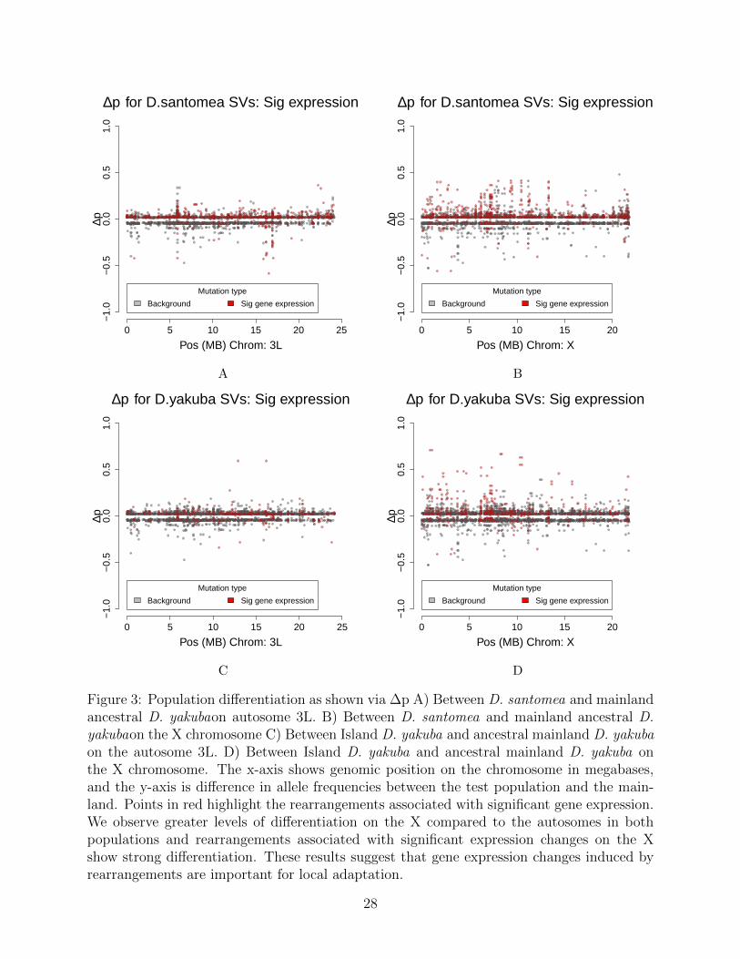
In D. santomea, of the 145/244 (59.4%) mutations that show significant population dif-ferentiation and significant gene expression changes, and 6 associated with fusion transcripts(Figure 3). We see in the island D. yakuba that 99/217 (45.6%) rearrangements show signif-icant population differentiation and display changes gene expression (Figure 3,Figure S15).Rearrangements with changes in gene expression are skewed toward increases in frequencyfor both island D. santomea (Kolmogorov-Smirnov, D = 0.17, P = 0.0047) (Figure 3) andislandD. yakuba (Kolmogorov-Smirnov, D = 0.19, P = 2.2 × 10⁻¹⁶) (Figure S15). Themajority of genes that showed significant expression changes in both populations on SaoTome are shown to have at least one breakpoint associated with a TE as well, lending to theidea that TEs are facilitating adaptation via a rapid burst of new insertions associated witheffects on gene expression.
New mutations versus standing variation in local adaptation
During habitat shifts, adaptation can come from new mutations that occur once the popu-lation is moved to a new environment [51], or from standing variation which existed in thepopulation before the shift [10]. While the majority of mutations will be either neutral ordetrimental, there will be occasions where a new phenotype can proliferate through a popu-lation due to the advantage in confers. Standing variation in the genome existing at the timeof a habitat shifts offers a source of mutations are readily available for a population to exploitto their advantage, rather than needing to create novel variation through mutation [20] .
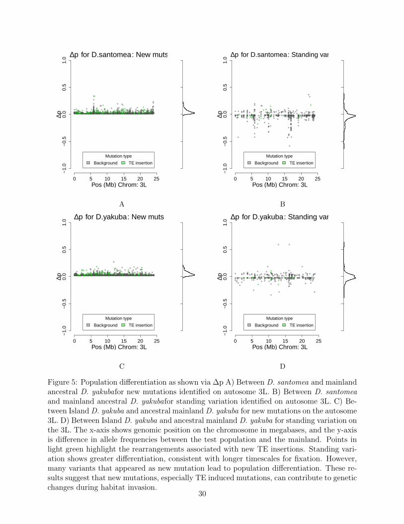
By comparing the two modes of evolution from standing variation and new mutationswe can further understand how each play a role in adaptation, especially when a populationexperiences a sudden change in habitat. We observe many instances where a mutation inD. yakuba that is absent in the mainland D. yakuba has moved to a moderate frequency.We observe in D. santomea that 155/244 (63.5%) rearrangements have Δp outside of the 95percent CI and are new mutations, with the remaining 89/244 (36.5%) being from standingvariation. Similarly for the island D. yakuba flies we see 157/217 (72.4%) rearrangements arenew mutations, and 60/217 (27.6%) are from standing variation. The mean of the significantΔp points is 0.1551 for new mutations and 0.2193 for the standing variation in D. santomea,and 0.1622 and 0.2430 respectively in the island D. yakuba (Figure 5). While there are atotal of more new mutations than standing variation, they appear to trend at a lower allelefrequency than standing variation (Figure 6, S16, S17). These results are consistent withstanding variation having had more time to spread through the population.
Chromosomal Rearrangements are associated with UV resistancegenes
D. santomea reside at a higher elevation than both the island and mainland D. yakuba,exposing them to greater UV stress [28]. Despite this known selective pressure for Drosophila,they are actually paler than ancestral D. yakuba flies [35]. The phenotypic differences in D.santomea are a long-standing open question for how these pale flies survive in environmentswhere other species are selected for melanistic phenotypes [52, 53].
Among all adaptive rearrangements, three of the most dynamic candidate regions areat the locus of UV repair genes: Parp and UV tolerance gene Victoria, and spel1 (Figure
7
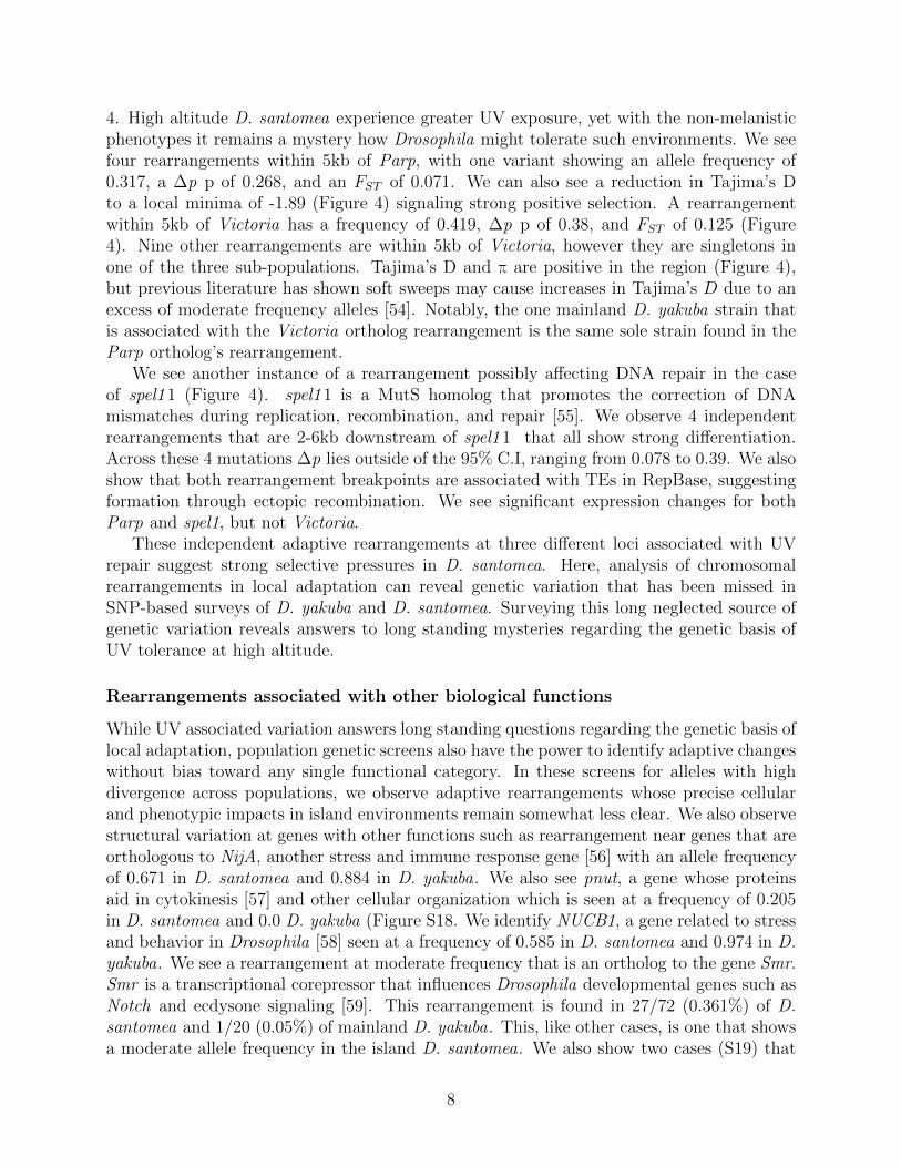
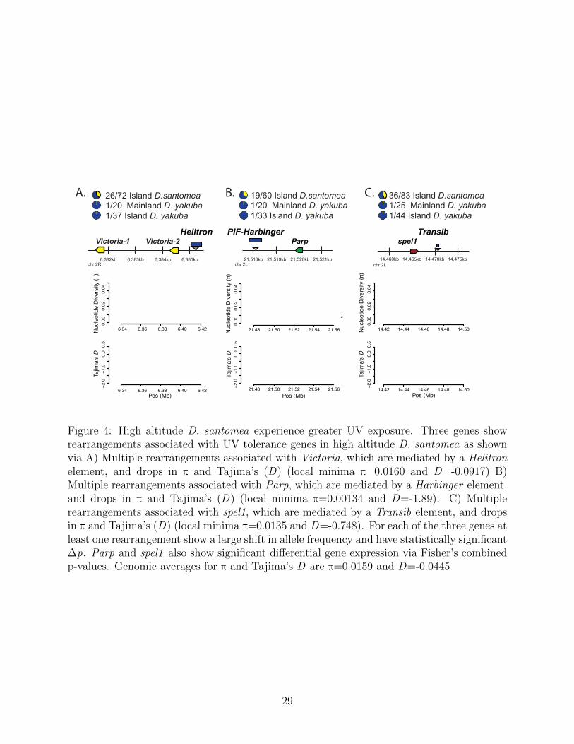
4. High altitude D. santomea experience greater UV exposure, yet with the non-melanisticphenotypes it remains a mystery how Drosophila might tolerate such environments. We seefour rearrangements within 5kb of Parp, with one variant showing an allele frequency of0.317, a Δp p of 0.268, and an FST of 0.071. We can also see a reduction in Tajima’s Dto a local minima of -1.89 (Figure 4) signaling strong positive selection. A rearrangementwithin 5kb of Victoria has a frequency of 0.419, Δp p of 0.38, and FST of 0.125 (Figure4). Nine other rearrangements are within 5kb of Victoria, however they are singletons inone of the three sub-populations. Tajima’s D and π are positive in the region (Figure 4),but previous literature has shown soft sweeps may cause increases in Tajima’s D due to anexcess of moderate frequency alleles [54]. Notably, the one mainland D. yakuba strain thatis associated with the Victoria ortholog rearrangement is the same sole strain found in theParp ortholog’s rearrangement.
We see another instance of a rearrangement possibly affecting DNA repair in the caseof spel11 (Figure 4). spel11 is a MutS homolog that promotes the correction of DNAmismatches during replication, recombination, and repair [55]. We observe 4 independentrearrangements that are 2-6kb downstream of spel11 that all show strong differentiation.Across these 4 mutations Δp lies outside of the 95% C.I, ranging from 0.078 to 0.39. We alsoshow that both rearrangement breakpoints are associated with TEs in RepBase, suggestingformation through ectopic recombination. We see significant expression changes for bothParp and spel1, but not Victoria.
These independent adaptive rearrangements at three different loci associated with UVrepair suggest strong selective pressures in D. santomea. Here, analysis of chromosomalrearrangements in local adaptation can reveal genetic variation that has been missed inSNP-based surveys of D. yakuba and D. santomea. Surveying this long neglected source ofgenetic variation reveals answers to long standing mysteries regarding the genetic basis ofUV tolerance at high altitude.
Rearrangements associated with other biological functions
While UV associated variation answers long standing questions regarding the genetic basis oflocal adaptation, population genetic screens also have the power to identify adaptive changeswithout bias toward any single functional category. In these screens for alleles with highdivergence across populations, we observe adaptive rearrangements whose precise cellularand phenotypic impacts in island environments remain somewhat less clear. We also observestructural variation at genes with other functions such as rearrangement near genes that areorthologous to NijA, another stress and immune response gene [56] with an allele frequencyof 0.671 in D. santomea and 0.884 in D. yakuba. We also see pnut, a gene whose proteinsaid in cytokinesis [57] and other cellular organization which is seen at a frequency of 0.205in D. santomea and 0.0 D. yakuba (Figure S18. We identify NUCB1, a gene related to stressand behavior in Drosophila [58] seen at a frequency of 0.585 in D. santomea and 0.974 in D.yakuba. We see a rearrangement at moderate frequency that is an ortholog to the gene Smr.Smr is a transcriptional corepressor that influences Drosophila developmental genes such asNotch and ecdysone signaling [59]. This rearrangement is found in 27/72 (0.361%) of D.santomea and 1/20 (0.05%) of mainland D. yakuba. This, like other cases, is one that showsa moderate allele frequency in the island D. santomea. We also show two cases (S19) that
8
have no genes within 10kb, where we see significant population differentiation (Δp=0.4651and Δp=0.3485), as well as noticeable changes in the signatures of selection through dropsin π and Tajima’s D (Table S1).
Population genetic scans suggest that these rearrangement facilitates local adaptationthrough mechanisms that remain yet to be discovered. The rearrangement being at mod-erate frequency and related to the developmental processes in Drosophila could signal thatrearrangements are responsible for adaptation through phenotypes that we have not yetunderstood.
Discussion
Structural variation responds to habitat changes
Three independent adaptive rearrangements at three different loci associated with UV repairsuggest strong selective pressures in D. santomea. Here, analysis of chromosomal rearrange-ments in local adaptation can reveal genetic variation that has been missed in SNP-basedsurveys of D. yakuba and D. santomea. While high altitude flies often evolve melanisticphenotypes for protection, D. santomea is pale [35, 60]. Structural variation offers multiplegenetic solutions for DNA integrity in the face of increased radiation [61]. Surveying thisneglected source of genetic variation reveals answers to long standing mysteries regardingthe genetic basis of UV tolerance at high altitude [60,62]. Flies lacking the spel11 gene suffera highly in long runs of dinucleotide repeats after 10-12 fly generations [63]. MutS mutantsare associated with greater sensitivity to DNA damage, including UV stress.
We also observe structural variation at genes with other functions such as rearrangementnear genes that are orthologous to stress and immune response genes, cytokinesis, and stressand behavior among others. We also observe regions that have no genes within 10kb, yetshow changes in the signatures of selection (Figure S18). We observe more extreme signals oflocal adaptation and differentiation from the mainland on the X chromosome compared withthe autosomes. These impacts are consistent with larger amounts of repetitive sequences onthe X chromosome. The D. yakuba-D. santomea species complex is known to interbreed,with partially sterile hybrids. The high levels of differentiation on the X chromosome areconsistent with the large-X effect in speciation, where interactions among species that arepartially reproductively isolated may be driving some part of sex-linked differences.
Many of the rearrangements we identify as targets of selection are found to alter geneexpression, consistent with prior work [9], suggesting that these mutations create importantchanges in gene regulation that are causative agents of local adaptation. Without informationabout rearrangements, SNP-based studies [64] offer an incomplete account of the geneticchanges between populations and the diverse solutions they offer to selective pressures. Astechnological advances continue, it is imperative that we continue to survey the impacts ofthese mutations and their phenotypic effects in evolutionary and biomedical genetics.
9
Genetic convergence is rare in adaptation to Sao Tome
The D. yakuba-D. santomea species complex is an excellent model to study the genetic basisof habitat shifts as it offers two different species adapted to the same island: D. santomeaat high altitude and D. yakuba in the lowlands [28]. The two species may also interbreed innature, offering the opportunity for adaptation through allele-sharing [29,47,48,52]. However,In these two species we see few cases of identical mutations spreading in island environments(7.3%). In these environments we have shown that chromosomal rearrangements can serveas “hopeful monsters” of the genetic world. However, the specific monsters that are favoreddiffer widely across populations.
Genome structure changes remain understudied in evolutionary genetics compared tosingle base pair changes. However, here, we have shown that these mutations can createdynamic changes in the genetic substrate available during adaptation during selective shifts.Studying genome structure changes in local adaptation and habitat invasion is essential tounderstand how genetic diversity facilitates phenotypic changes in nature. The ability toperform parallel analysis of these mutations in two independent cases of habitat invasionwith a known ancestral population lends greater strength to evolutionary inference as rear-rangements appear to be important players in evolutionary processes in nature.
Structural variation and transposable elements
Transposable elements are selfish DNA that proliferates within the genome even at the ex-pense of the host organism. TEs often appear in bursts and can rapidly remodel multiplegenes on short timescales. These mutations depart from the clock-like progression of mu-tations at SNPs, with boom-and-bust dynamics as cells engage in arms races to developrepressors that keep TE proliferation in check. In times of stress, TEs can activate andoccasionally create variation that remodels gene expression under specific environmentalconditions or developmental processes [65–67]. In this work we observe a significant propor-tion of new mutations that are created by transposable elements. New TE insertions appearin abundance, however ectopic recombination among distantly related TE copies is commonas well. The majority of these mutations do not spread in island environments, and appearto be neutral or detrimental. However, a distinct subset appears to spread in populations asthey adapt to new and changing environments.
Prior work in other systems such as plants have observed functional biases from differentclasses of TEs. Among all chromosomal rearrangements, no single TE class is enriched amongvariation that is targeted by natural selection, but rather reflects the relative abundance ofTE classes that have modified DNA within the genome. This result implies that the selectiveimpacts of TE-induced mutations are random, not biased to a specific TE type. Bothisland populations of D. santomea and D. yakuba show different sets of mutations serving astargets of selection, showing independent that TE induced rearrangement facilitates adaptivechanges. DNA transposons and retrotransposons across multiple TE families are represented,implying multiple TE activation events rather than a single activation of only one TE type.Our results show that TEs are causative agents of genetic change that result in differentiationbetween island and mainland populations. This substrate of genetic novelty appears to bea key source of variation as populations adapt to new environments, especially on short
10
timescales.
Local Adaptation with Standing Variation or New Mutations
Adaptation during shifts in selective pressures can come from standing variation or fromnew mutations [10, 20, 68]. Standing variation in populations is immediately available foradaptation, and may proceed to fixation so long as allele frequencies are high enough toestablish deterministic selective sweeps [11]. New mutations are expected to appear moreslowly, as mutations accumulate in clock-like fashion as generations progress. The relativecontribution of new mutation and standing variation can tell us how quickly populations areable to evolve genetic solutions in populations as they adapt to new habitats. The spectrumof neutral diversity segregating in natural populations depends on the effective populationsize, and the mutation rate, where π = θ = 4Ne ∗ µ. Larger populations will weed outmutations with large detrimental impacts where 4Ne ∗ s > 1, limiting the span of variation.
In our analysis of chromosomal rearrangements, we observe differentiation with large Δpand FST within a time-span of 500,000 years [28]. Here, we observe the majority of rear-rangements (76.2%) are derived from new mutations not present on the mainland. However,standing variation is associated with more dynamic differentiation, with larger values of Δpthan new mutations. Our results imply that the tempo of local adaptation is then expectedto proceed in two waves. First, an initial spread of standing variation after habitat invasionleads to adaptation from the limited span of mutations imported from the founder popula-tion. In such a case, we expect genetic differentiation at multiple loci shortly after invasion.Still, the substrate available among standing variation is insufficient to facilitate adaptationto the optimum. Afterwards, new mutations appear stochastic ally in the population, andspread if selectively favored.
At SNPs, long wait times would affect the span of adaptation, as multiple generations areexpected to be required before establishment of deterministic sweeps [4,11,69,70]. However,TE-induced mutations can be generated in “bursts” that remodel genomes with many mu-tations at once [71]. Our results suggest that new mutations from transposable have rapidlychanged genome content and structure, as they move pieces of DNA around the genome [72].These genetic responders serve as a source of increased variation that fortuitously createsnovel substrates, including genetic changes at UV tolerance loci. The subset of selectivelyfavored mutations is a minority of the genetic diversity identified (244/16480 rearrangements1.480%). The large number of mutations that must be sifted through suggests that adapta-tion is a game of genetic chance, where the dice must be rolled multiple times for organismsto achieve selective advantages.
Methods
Whole genome sequencing and SNP calling
We generated whole genome sequencing for single Drosophila from 42 strains of D. santomeafrom Sao Tome, 35 strains of D. yakuba from the island of Sao Tome, and 19 strains of D.yakuba from the central African mainland (Cameroon and Kenya). DNA was extracted fromflies flash frozen in liquid nitrogen following QIAamp Mini Kit (Qiagen) protocol without
11
using RNase A. The resulting DNA samples were quantified (Qubit dsDNA HS assay kit,ThermoFisher Scientific), assessed for quality (Nanodrop ND-2000; A260/A280>1.8), andstored at -20°C.
Illumina TruSeq Nano DNA libraries were prepared manually following the manufac-turer’s protocol (TruSeq Nano DNA, RevD; Illumina). Briefly, samples were normalized to100ng DNA and sheared by sonication with Covaris M220 (microTUBE 50; Sage Science).The samples were end repaired, purified with Ampure XP beads (Agencourt; Beckman Coul-ter), adaptors adenylated, and Unique Dual Indices (Table) ligated. Adaptor enrichment wasperformed using eight cycles of PCR. Following Ampure XP bead cleanup, fragment sizesfor all libraries were measured using Agilent Bioanalyzer 2100 (HS DNA Assay; AppliedBiosystems). The libraries were diluted 1:10 000 and 1:20 000 and quantified in triplicateusing the KAPA Library Quantification Kit (Kapa Biosystems). Equimolar samples werepooled and the libraries were size selected targeting 400-700bp range to remove adaptormonomers and dimers using Pippen Prep DNA Size Selection system (1.5% Agarose GelCassette #CDF1510; Sage Sciences). Library pools (24 samples per lane) were run on anllumina HiSeq 4000 platform using the 150bp paired end (PE) Cluster Kit.
We aligned short read sequences to the D. yakuba reference genome r1.0.5 and Wolbachiaendoparasite sequence NC 002978.6 using bwa aln, and resolved paired end mappings usingbwa sampe. We sorted alignments by position using samtools sort. Sequence depth on sortedbam files was calculated using samtools depth -aa.
Using GATK [18] we called the SNPs for each strain. To calculate the frequency weneed to determine the haplotype, which we do using an HMM. After initially running theHMM with incorrect parameters we observe no changes of state, so we used the Baum-Welshalgorithm to estimate the parameters of the HMM. With the correct parameters we can rerunthe HMM and determine haplotypes for SNPs and calculate their frequencies.
HiFi PacBio sequences were used to confirm the Illumina structural variant calls. We callstructural variants using minimap2 [36], Sniffles [37], and PacBip’s pbsv [38]. All structuralvariant calls were done with alignments created by minimp2, except in the vase of pbsvwhich requires alignments from pbmm2 [38], a Minimap2 frontend for PacBio native dataformats.
Identification of chromosomal rearrangements
We identified abnormally mapping read pairs on different chromosomes and long-spanningread pairs greater than 100kb apart as signals of putative chromosomal rearrangements.Mutations with greater than 3 read pairs supporting were included among the chromosomalrearrangements. The mutation data were then clustered across strains assuming that variantswere within 325 base pairs of each other on from either rearrangement breakpoint. Takingthe min and max for each of the rearrangement’s breakpoints in a cluster, we associate allstrains in said cluster with these bounds.
D. yakuba is resistant to inbreeding due to segregating inversions [26,73,74]. To identifyinbred and heterozygous regions and correct allele frequencies, we used a Hidden MarkovModel (HMM) to parse SNP heterozygosity for each strain. In D. yakuba, some strains hadreference strain contamination, resulting in no heterozygous or homozygous non-referenceSNPs. In D. santomea no indication of references contamination was observed. We used a
12
three state HMM for D. yakuba and an two-state HMM for D. santomea to identify referencecontamination, inbred haplotypes, and non-inbred haplotypes. Transitions probabilities wereset to 10−10. Emission probabilities were set as heterozygosity for inbred regions wouldbe 0, and heterozygosity in non-inbred regions was θ = 0.01, with a lower threshold forprobabilities on off-diagonals of ε = 0.00005 to prevent chilling effects of zero probability.
Haplotype calls from the HMM were used to generate correct site frequency spectra forSNPs and structural variants given the variable number of chromosomes sampled acrossdifferent parts of the genome.
Within inbred regions, we assume mutations are homozygous, and assign a genotype of1 structural variant on 1 sampled chromosome. Within heterozygous haplotypes, read pairinformation alone cannot detect ploidy. For outbred regions with two chromosomes withdifferent ancestry, we used coverage changes to distinguish hemizygous and homozygousmutations. We compared the average coverage of observed mutations to the average coverageacross the entire chromosome in a strain looking for an observed coverage between 1.45 <x < 1.55 or 1.95 < x < 2.05. Regions whose haplotypes were around 1.5x the averagecoverage of the chromosome are expected to be heterozygous, and regions who have around2x the average are expected to be homozygous. False negative genotypes are possible whenread pair support is insufficient to identify rearrangements de novo. For each mutation, ifanother strain showed 1.5x or 2x coverage changes and lesser support of 1 or 2 read pairs,allele frequencies were adjusted to avoid low frequency reads, similarly to prior work on geneduplications and rearrangements [20, 26].
Polarize ancestral state
Genotyping identifies mutations in populations that differ from the reference sequence, buton its own cannot identify which is ancestral and which is novel. To determine the ancestralstate for rearrangements, we compared each variant to D. teissieri, we determine whethermutations are novel or if the ancestral state has been rearranged in the reference strain. Topolarize the mutations we use BLASTn at an E-value of 10−20 to compare the D. yakubareference sequence for +/- 1kb of each rearrangement breakpoint to D. teissieri. We considera mutation the ancestral state if both sequences map to the same location in D. teissieri,they have an alignment length greater than 150, share greater than 95 percent identity, andthe two sequences overlap less than 10 percent in D. teissieri. Mutations where sampleswere identified as containing the ancestral state found in D. teissieri, allele frequencies fromgenotyping, p, were reversed to (1 − p) to reflect the reference as the new mutation.
By using the haplotype data we determine the number of sampled strains and comparethem to the number of observed strains in the clustered rearrangements .
Association with transposable elements
To match variants with transposable elements (TEs) we used BLAST to compare the variantsto Repbase. Variants that mapped to TEs at an e-value of 10−20 were marked as TEs in ouranalysis. By using BLAST to compare both the origin and destination loci of the variant,we were able to see which variants mapped on either one or both sides of the rearrangement.
13
(This was verified by checking the regions to make sure that they had unusual coveragecompared to the average for that chromosome and strain.)
Resolving complex variation
Previous work has shown that Pacbio can confirm 100% of Illumina paired-end reads whenusing alignment methods in tandem, as the rate varies on the confirmation method (BLAST,MM2, PBSV, etc). (Figure S20). The majority of unconfirmed regions have erratic coverage,implying complex variation that the aligners and assemblers cannot solve. To investigatefurther, we conducted targeted de novo assembly of these regions and plotted them withMummer.
Structural variant allele frequencies
We used polarized allele frequencies adjusted for inbreeding to calculate the allele frequencyfor each of the variants that we observed. We calculated the allele frequency within sub-populations for D. santomea, island D. yakuba and mainland D. yakuba. With the allelefrequency data for each variant and the separation by sub-population. To generate an SFSwhile accounting for uneven sample sizes across the genome, we projected allele frequenciesin each subpopulation to a sample size of n=19 using a hypergeometric model [26,75].
Differentiation between populations is a signature of local adaptation to changing envi-ronments. This differentiation can be measured through different means. This study benefitsfrom a known ancestral mainland population that invaded new island environments that canbe used as a population genetic ‘control’ to identify allele frequency changes. Simulationsfrom other groups have shown that the difference in allele frequencies, Δp
p1 − p2 (1)
and FSTp(1 − p) −
∑cipi(1 − pi)
p(1 − p)(2)
can identify population differentiation during local adaptation. Δp, and FST contain over-lapping information. Δp contains directionality that FST lacks, and may be more usefulon moderate timescales when populations have had sufficient time for variants to spread inpopulations. It may identify population differentiation better than FST while FST may bemore sensitive to changes while allele frequencies are low. We calculate Δp and FST identifystructural variants that have spread in island environments.
To determine whether mutations showed unusually high population differentiation wecalculated a Bonferroni corrected 95% confidence interval, calculated by chromosome due tothe distribution of Δp varying by chromosome. Mutations that were greater than the upperbound of the C.I were considered significantly differentiated.
SNP Population diversity
To measure the selective pressure and genomic structural differences caused by the rear-rangements we calculate Θπ, Θw, and Tajima’s D. These statistics are calculated for a 10kb
14
sliding window with a 1kb slide. Using these estimates we matched our variants into thesewindows and associated the rearrangement with the window who’s midpoint was closest tothe origin locus.
Gene ontology
Using the location of the rearrangements we looked 5kb upstream and downstream of themutations to see if their were genes that could be affected. Variants that matched D. yakubagene coordinates were mapped to their orthologs in D. melanogaster using Flybase [76].Genes that had no orthologs were excluded. Using these orthologs we analyzed the functionalgene annotations using DAVID [77] using low stringency.
Tissue dissection and RNA sequencing
To determine how rearrangements might change gene expression profiles or create new genes,we collected RNAseq data for a subset of 14 strains. Because new gene formation commonlyoccurs at genes with testes-specific expression [78–81] we collected gene expression data forgonads and soma of adult male and female flies. For each strain, we collected virgin malesand females within 2 hours of eclosion and placed into separate vials to age for 5-7 days.Once adults, we placed individual males and females on a glass slide with Ringer’s solutionfor gonad dissection. We removed testes and the accessory gland from males and the ovariesfrom females. Gonads and the carcass (rest of the body minus the gonads) were placed intoseparate Eppendorf tubes and flash frozen immediately in liquid nitrogen. Five biologicalreplicates were used for each tissue.
RNA was extracted from fly tissue frozen in liquid nitrogen following Zymo DirectZolRNA Microprep (Zymo Research) without DNAase treatment. The resulting RNA sampleswere quantified (Qubit RNA HS assay kit, ThermoFisher Scientific), assessed for quality(Nanodrop ND-2000; A260/A280>2.0), and stored at -80°C.
Poly (A) enriched strand-specific Illumina TruSeq libraries were manually prepared fol-lowing the manufacturer’s protocol (TruSeq Stranded mRNA LS, RevD; Illumina). Briefly,samples were normalized to 100ng RNA and poly (A) containing mRNA molecules purifiedusing poly (T) oligo attached magnetic beads. The poly (A) molecules were chemicallyfragmented for 8 minutes and primed for cDNA synthesis. Reverse transcription was per-formed using SuperScript IV enzyme (Invitrogen), samples purified with Ampure XP beads(Agencourt; Beckman Coulter), adaptors adenylated, and Unique Dual Indices (Table) lig-ated. Adaptor enrichment was performed using 15 cycles of PCR. Following Ampure XPbead cleanup, fragment sizes for all libraries were measured using Agilent Bioanalyzer 2100(HS DNA Assay; Applied Biosystems). The libraries were diluted 1:10 000 and 1:20 000and quantified in triplicate using the KAPA Library Quantification Kit (Kapa Biosystems).Equimolar samples were pooled (20 samples per lane) and run on an Illumina HiSeq 4000platform using the 150bp paired end (PE) Cluster Kit.
15
Identifying chromosomal rearrangements and differential expres-sion analysis
To determine if strains contain structural rearrangements, we performed a Tophat fusionsearch version 2.1.0 to find split reads on paired-end RNA sequencing data [50]. This pro-gram has previously been validated to identify chimeric constructs using split-read mappingand abnormal read pairs in RNASeq data at loci with genomic DNA signals indicating re-arrangements [4, 21, 26]. We identified genes within 5kb of rearrangement breakpoints toidentify genes most likely to experience changes in gene expression. We then identifiedstructural variants that were differentially expressed using the program CuffDiff [82]. Todetermine whether gene expression changes and fusion transcripts are associated with localadaptation, these were also examined for signatures of selection.
We separated structural variant data into male carcass, female carcass, male gonads, andfemale gonads to determine if these differed across the tissue sample. To identify statisticallysignificant expression changes across the structural variants, we calculated Fisher’s AdjustedP-values [83] using the p-values for expression in each strain that contained that structuralvariant. To determine whether structural variants as a class are more likely to induce changesin gene expression than we would expect for the genome at large, we performed 10,000bootstrap replicates with random sampling of 9549 to 10632 genes depending on the tissue.
Acknowledgements
This work was funded by NIH NIGMS MIRA R35-GM133376 to Rebekah L. Rogers and bystartup funding from the University of North Carolina, Charlotte. TM is funded by UGATraining Grant T32GM007103 from NIH NIGMS and NSF DEB-1737824 to Kelly Dyer.
Data Availability
All sequence data are available under SRA PRJNA764098, PRJNA764689, PRJNA764691,PRJNA764693,PRJNA764695, PRJNA764098, PRJNA269314. Supplementary Data areavailable at https://www.dropbox.com/sh/4fkf4fojcbbnzil/AABpp5TGeefHjaQk1YqI9bPKa?dl=0.
16
References
[1] Schoville SD, et al. (2012) Adaptive genetic variation on the landscape: methods andcases. Annual Review of Ecology, Evolution, and Systematics 43:23–43.
[2] Ohno S (2013) Evolution by gene duplication. (Springer Science & Business Media).
[3] Conant GC, Wolfe KH (2008) Turning a hobby into a job: how duplicated genes findnew functions. Nature Reviews Genetics 9(12):938–950.
[4] Rogers RL, Shao L, Thornton KR (2017) Tandem duplications lead to novel expressionpatterns through exon shuffling in Drosophila yakuba. PLoS Genetics 13(5):e1006795.
[5] Rogers RL, Hartl DL (2012) Chimeric genes as a source of rapid evolution in Drosophilamelanogaster. Molecular biology and evolution 29(2):517–529.
[6] Kondrashov FA, Kondrashov AS (2006) Role of selection in fixation of gene duplications.Journal of Theoretical Biology 239(2):141–151.
[7] Huminiecki L, Wolfe KH (2004) Divergence of spatial gene expression profiles followingspecies-specific gene duplications in human and mouse. Genome Research 14(10a):1870–1879.
[8] Harewood L, Fraser P (2014) The impact of chromosomal rearrangements on regulationof gene expression. Human Molecular Genetics 23(R1):R76–R82.
[9] De S, Teichmann SA, Babu MM (2009) The impact of genomic neighborhood on theevolution of human and chimpanzee transcriptome. Genome Research 19(5):785–794.
[10] Barrett RD, Schluter D (2008) Adaptation from standing genetic variation. Trends inEcology & Evolution 23(1):38–44.
[11] Hermisson J, Pennings PS (2005) Soft sweeps: molecular population genetics of adap-tation from standing genetic variation. Genetics 169(4):2335–2352.
[12] Orr HA (2005) The genetic theory of adaptation: a brief history. Nature ReviewsGenetics 6(2):119–127.
[13] Coyne JA, Orr HA (1989) Patterns of speciation in Drosophila. Evolution 43(2):362–381.
[14] Ohta T (1992) The nearly neutral theory of molecular evolution. Annual Review ofEcology and Systematics 23(1):263–286.
[15] Lynch M, et al. (1999) Perspective: spontaneous deleterious mutation. Evolution53(3):645–663.
[16] Kimura M, , et al. (1968) Evolutionary rate at the molecular level. Nature217(5129):624–626.
17
[17] Smith JM, Haigh J (1974) The hitch-hiking effect of a favourable gene. Genetics Re-search 23(1):23–35.
[18] McKenna A, et al. (2010) The genome analysis toolkit: a mapreduce framework foranalyzing next-generation DNA sequencing data. Genome Research 20(9):1297–1303.
[19] Li H (2011) A statistical framework for SNP calling, mutation discovery, associationmapping and population genetical parameter estimation from sequencing data. Bioin-formatics 27(21):2987–2993.
[20] Rogers RL, et al. (2014) Landscape of standing variation for tandem duplicationsin Drosophila yakuba and Drosophila simulans. Molecular Biology and Evolution31(7):1750–1766.
[21] Rogers RL (2015) Chromosomal rearrangements as barriers to genetic homogenizationbetween archaic and modern humans. Molecular Biology and Evolution 32(12):3064–3078.
[22] Ellegren H (2014) Genome sequencing and population genomics in non-model organisms.Trends in Ecology & Evolution 29(1):51–63.
[23] Clark AG, et al. (2007) Evolution of genes and genomes on the drosophila phylogeny.Nature 450(7167):203–218.
[24] Bhutkar A, et al. (2008) Chromosomal rearrangement inferred from comparisons of 12drosophila genomes. Genetics 179(3):1657–1680.
[25] Ranz JM, Castillo-Davis CI, Meiklejohn CD, Hartl DL (2003) Sex-dependent gene ex-pression and evolution of the Drosophila transcriptome. Science 300(5626):1742–1745.
[26] Stewart NB, Rogers RL (2019) Chromosomal rearrangements as a source of new geneformation in Drosophila yakuba. PLoS Genetics 15(9):e1008314.
[27] Lachaise D, et al. (2000) Evolutionary novelties in islands: Drosophila santomea, a newmelanogaster sister species from sao tome. Proceedings of the Royal Society of London.Series B: Biological Sciences 267(1452):1487–1495.
[28] Coyne JA, Kim SY, Chang AS, Lachaise D, Elwyn S (2002) Sexual isolation between twosibling species with overlapping ranges: Drosophila santomea and Drosophila yakuba.Evolution 56(12):2424–2434.
[29] Llopart A, Lachaise D, Coyne JA (2005) An anomalous hybrid zone in Drosophila.Evolution 59(12):2602–2607.
[30] Obbard DJ, et al. (2012) Estimating divergence dates and substitution rates in theDrosophila phylogeny. Molecular Biology and Evolution 29(11):3459–3473.
18
[31] Cariou ML, Silvain JF, Daubin V, Da Lage JL, Lachaise D (2001) Divergence betweenDrosophila santomea and allopatric or sympatric populations of D. yakuba using paralo-gous amylase genes and migration scenarios along the cameroon volcanic line. MolecularEcology 10(3):649–660.
[32] Comeault AA, Venkat A, Matute DR (2016) Correlated evolution of male and femalereproductive traits drive a cascading effect of reinforcement in drosophila yakuba. Pro-ceedings of the Royal Society B: Biological Sciences 283(1835):20160730.
[33] Bachtrog D, Thornton K, Clark A, Andolfatto P (2006) Extensive introgression of mi-tochondrial DNA relative to nuclear genes in the Drosophila yakuba species group.Evolution 60(2):292–302.
[34] Andolfatto P, Wong KM, Bachtrog D (2011) Effective population size and the efficacyof selection on the x chromosomes of two closely related drosophila species. GenomeBiology and Evolution 3:114–128.
[35] Llopart A, Elwyn S, Lachaise D, Coyne JA (2002) Genetics of a difference in pigmenta-tion between drosophila yakuba and drosophila santomea. Evolution 56(11):2262–2277.
[36] Li H (2018) Minimap2: pairwise alignment for nucleotide sequences. Bioinformatics34(18):3094–3100.
[37] Sedlazeck FJ, et al. (2018) Accurate detection of complex structural variations usingsingle-molecule sequencing. Nature Methods 15(6):461–468.
[38] Wenger AM (year?) Comprehensive structural and copy-number variant detection withlong reads.
[39] Rogers RL, et al. (2015) Tandem duplications and the limits of natural selection indrosophila yakuba and drosophila simulans. PLoS One 10(7):e0132184.
[40] Montgomery E, Huang S, Langley C, Judd B (1991) Chromosome rearrangement byectopic recombination in Drosophila melanogaster : genome structure and evolution.Genetics 129(4):1085–1098.
[41] Jurka J, et al. (2005) Repbase update, a database of eukaryotic repetitive elements.Cytogenetic and Genome Research 110(1-4):462–467.
[42] Merel V, Boulesteix M, Fablet M, Vieira C (2020) Transposable elements in Drosophila.Mobile DNA 11(1):1–20.
[43] Villanueva-Canas JL, Horvath V, Aguilera L, Gonzalez J (2019) Diverse families oftransposable elements affect the transcriptional regulation of stress-response genes inDrosophila melanogaster. Nucleic Acids Research 47(13):6842–6857.
[44] Fawcett JA, Innan H (2019) The role of gene conversion between transposable elementsin rewiring regulatory networks. Genome Biology and Evolution 11(7):1723–1729.
19
[45] Innan H, Kim Y (2008) Detecting local adaptation using the joint sampling of polymor-phism data in the parental and derived populations. Genetics 179(3):1713–1720.
[46] Halligan DL, Keightley PD (2006) Ubiquitous selective constraints in the drosophilagenome revealed by a genome-wide interspecies comparison. Genome Research16(7):875–884.
[47] Llopart A (2012) The rapid evolution of X-linked male-biased gene expression and thelarge-x effect in Drosophila yakuba, D. santomea, and their hybrids. Molecular Biologyand Evolution 29(12):3873–3886.
[48] Matute DR, Gavin-Smyth J (2014) Fine mapping of dominant X-linked incompatibilityalleles in Drosophila hybrids. PLoS Genetics 10(4):e1004270.
[49] Trapnell C, et al. (2010) Transcript assembly and quantification by RNA-Seq re-veals unannotated transcripts and isoform switching during cell differentiation. NatureBiotechnology 28(5):511–515.
[50] Kim D, Salzberg SL (2011) Tophat-fusion: an algorithm for discovery of novel fusiontranscripts. Genome Biology 12(8):1–15.
[51] Carvunis AR, et al. (2012) Proto-genes and de novo gene birth. Nature 487(7407):370–374.
[52] Matute DR, Butler IA, Coyne JA (2009) Little effect of the tan locus on pigmentationin female hybrids between Drosophila santomea and D melanogaster. Cell 139(6):1180–1188.
[53] Rebeiz M, et al. (2009) Evolution of the tan locus contributed to pigment loss inDrosophila santomea: a response to Matute et al. Cell 139(6):1189–1196.
[54] Pennings PS, Hermisson J (2006) Soft sweeps iii: the signature of positive selection fromrecurrent mutation. PLoS Genetics 2(12):e186.
[55] Harr B, Todorova J, Schlotterer C (2002) Mismatch repair-driven mutational bias in d.melanogaster. Molecular Cell 10(1):199–205.
[56] Broderick S, Wang X, Simms N, Page-McCaw A (2012) Drosophila Ninjurin A inducesnonapoptotic cell death. PLoS One.
[57] Neufeld TP, Rubin GM (1994) The Drosophila peanut gene is required for cytokine-sis and encodes a protein similar to yeast putative bud neck filament proteins. Cell77(3):371–379.
[58] Rech GE, et al. (2019) Stress response, behavior, and development are shaped by trans-posable element-induced mutations in drosophila. PLoS Genetics 15(2):e1007900.
[59] Heck BW, et al. (2011) The transcriptional corepressor smrter influences both notchand ecdysone signaling during drosophila development. Biology Open 1(3):182–196.
20
[60] Bastide H, Yassin A, Johanning EJ, Pool JE (2014) Pigmentation in drosophilamelanogaster reaches its maximum in ethiopia and correlates most strongly with ultra-violet radiation in sub-saharan africa. BMC Evolutionary Biology 14(1):1–14.
[61] Brodsky MH, et al. (2000) Drosophila p53 binds a damage response element at thereaper locus. Cell 101(1):103–113.
[62] Vaisnav M, et al. (2014) Genome-wide association analysis of radiation resistance inDrosophila melanogaster. PloS One 9(8):e104858.
[63] Flores C, Engels W (1999) Microsatellite instability in Drosophila spellchecker1 (MutShomolog) mutants. Proceedings of the National Academy of Sciences 96(6):2964–2969.
[64] Carbone MA, Llopart A, DeAngelis M, Coyne JA, Mackay TF (2005) Quantitativetrait loci affecting the difference in pigmentation between Drosophila yakuba and D.santomea. Genetics 171(1):211–225.
[65] Laudencia-Chingcuanco D, Fowler DB (2012) Genotype-dependent burst of transpos-able element expression in crowns of hexaploid wheat (Triticum aestivum L.) duringcold acclimation. Comparative and Functional Genomics 2012.
[66] Lynch VJ, Leclerc RD, May G, Wagner GP (2011) Transposon-mediated rewiring ofgene regulatory networks contributed to the evolution of pregnancy in mammals. NatureGenetics 43(11):1154–1159.
[67] Feschotte C (2008) Transposable elements and the evolution of regulatory networks.Nature Reviews Genetics 9(5):397–405.
[68] Assis R, Bachtrog D (2013) Neofunctionalization of young duplicate genes in Drosophila.Proceedings of the National Academy of Sciences 110(43):17409–17414.
[69] Smith JM (1971) What use is sex? Journal of Theoretical Biology 30(2):319–335.
[70] Gillespie JH (1994) The causes of molecular evolution. (Oxford University Press OnDemand) Vol. 2.
[71] Cridland JM, Macdonald SJ, Long AD, Thornton KR (2013) Abundance and distri-bution of transposable elements in two Drosophila QTL mapping resources. MolecularBiology and Evolution 30(10):2311–2327.
[72] Guerreiro MPG (2014) Interspecific hybridization as a genomic stressor inducing mobi-lization of transposable elements in drosophila. Mobile Genetic Elements 4(4):e88992.
[73] Guillen Y, Ruiz A (2012) Gene alterations at Drosophila inversion breakpoints provideprima facie evidence for natural selection as an explanation for rapid chromosomalevolution. BMC Genomics 13(1):1–18.
[74] Andolfatto P (2001) Contrasting patterns of X-linked and autosomal nucleotide varia-tion in Drosophila melanogaster and Drosophila simulans. Molecular Biology and Evo-lution 18(3):279–290.
21
[75] Nielsen R (2005) Molecular signatures of natural selection. Annu. Rev. Genet. 39:197–218.
[76] Drysdale R, Consortium F, , et al. (2008) Flybase. Drosophila pp. 45–59.
[77] Sherman BT, Lempicki RA, , et al. (2009) Systematic and integrative analysis of largegene lists using DAVID bioinformatics resources. Nature Protocols 4(1):44–57.
[78] Zhou Q, et al. (2008) On the origin of new genes in Drosophila. Genome Research18(9):1446–1455.
[79] Rogers RL, Shao L, Sanjak JS, Andolfatto P, Thornton KR (2014) Revised annotations,sex-biased expression, and lineage-specific genes in the Drosophila melanogaster group.G3: Genes, Genomes, Genetics 4(12):2345–2351.
[80] Betran E, Thornton K, Long M (2002) Retroposed new genes out of the X in Drosophila.Genome Research 12(12):1854–1859.
[81] Bachtrog D, Toda NR, Lockton S (2010) Dosage compensation and demasculinizationof X chromosomes in Drosophila. Current Biology 20(16):1476–1481.
[82] Trapnell C, et al. (2012) Differential gene and transcript expression analysis of RNA-seqexperiments with tophat and cufflinks. Nature Protocols 7(3):562–578.
[83] Tsuyuzaki K, Nikaido I (2013) metaseq: Meta-analysis of RNA-seq count data. TokyoUniversity of Science, Tokyo.
22
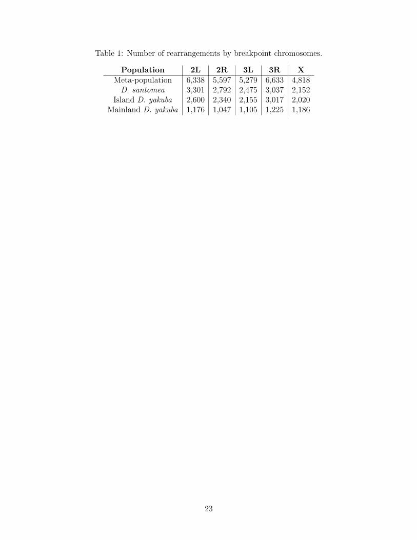
Table 1: Number of rearrangements by breakpoint chromosomes.
Population 2L 2R 3L 3R XMeta-population 6,338 5,597 5,279 6,633 4,818D. santomea 3,301 2,792 2,475 3,037 2,152
Island D. yakuba 2,600 2,340 2,155 3,017 2,020Mainland D. yakuba 1,176 1,047 1,105 1,225 1,186
23

Table 2: Number of genes withing 5kb of a rearrangement breakpoint
Population 2L 2R 3L 3R XMeta-population 742 641 674 844 552D. santomea 557 279 441 509 324
Island D. yakuba 505 322 456 642 328Mainland D. yakuba 343 298 273 337 261
24
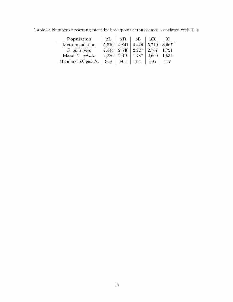
Table 3: Number of rearrangement by breakpoint chromosomes associated with TEs
Population 2L 2R 3L 3R XMeta-population 5,510 4,841 4,426 5,710 3,667D. santomea 2,944 2,540 2,227 2,707 1,721
Island D. yakuba 2,280 2,019 1,787 2,600 1,534Mainland D. yakuba 959 805 817 995 757
25
1 2 3 4 5 6 7 8 9 10 11 12 13 14 15 16 17 18 19
Island D.santomeaPr
opor
tion
of a
ll al
lele
s0.
00.
10.
20.
30.
4
A
1 2 3 4 5 6 7 8 9 10 11 12 13 14 15 16 17 18 19
Island D.yakuba
Prop
ortio
n of
all
alle
les
0.0
0.1
0.2
0.3
0.4
0.5
0.6
B
1 2 3 4 5 6 7 8 9 10 11 12 13 14 15 16 17 18 19
Mainland D.yakuba
Prop
ortio
n of
all
alle
les
0.0
0.1
0.2
0.3
0.4
0.5
0.6
0.7
C
Figure 1: A) Site frequency spectrum of D. santomea. Polarized ancestral state with D.teissieri and projected down to n=19 using the geometric distribution. The increase in highfrequency variants is much greater than that of the mainland D. yakuba B) Site frequencyspectrum of the island D. yakuba. Polarized ancestral state with D. teissieri and projecteddown to n=19 using the geometric distribution. The increase in high frequency variants isslightly greater than that of the mainland D. yakuba, but not as much as D. santomea, likelyrelated to divergence time C) Site frequency spectrum of mainland D. yakuba. Polarizedancestral state with D. teissieri and projected down to n=19 using the hypergeometricdistribution. Lacks the increase in high frequency variants that is present in both of theother sub-populations.
26
0 5 10 15 20 25
−1.
0−
0.5
0.0
0.5
1.0
Pos (Mb) Chrom: 3L
∆p for D.santomea SVs: TE insertion∆p
Mutation type
Background TE insertion
A
0 5 10 15 20 25
−1.
0−
0.5
0.0
0.5
1.0
Pos (Mb) Chrom: X
∆p for D.santomea SVs: TE insertion
∆p
Mutation type
Background TE insertion
B
C D
Figure 2: Population differentiation as shown via ∆p A) Between D. santomea and mainlandancestral D. yakubaon autosome 3L. B) Between D. santomea and mainland ancestral D.yakubaon the X chromosome C) Between Island D. yakuba and ancestral mainland D. yakubaon the autosome 3L. D) Between Island D. yakuba and ancestral mainland D. yakuba on theX chromosome. The x-axis shows genomic position on the chromosome in megabases, and they-axis is difference in allele frequencies between the test population and the mainland. Pointsin light green highlight the rearrangements associated with new TE insertions. We observegreater levels of differentiation on the X compared to the autosomes in both populations.
27
0 5 10 15 20 25
−1.
0−
0.5
0.0
0.5
1.0
Pos (MB) Chrom: 3L
∆p for D.santomea SVs: Sig expression∆p
Mutation type
Background Sig gene expression
A
0 5 10 15 20
−1.
0−
0.5
0.0
0.5
1.0
Pos (MB) Chrom: X
∆p for D.santomea SVs: Sig expression
∆p
Mutation type
Background Sig gene expression
B
0 5 10 15 20 25
−1.
0−
0.5
0.0
0.5
1.0
Pos (MB) Chrom: 3L
∆p for D.yakuba SVs: Sig expression
∆p
Mutation type
Background Sig gene expression
C
0 5 10 15 20
−1.
0−
0.5
0.0
0.5
1.0
Pos (MB) Chrom: X
∆p for D.yakuba SVs: Sig expression∆p
Mutation type
Background Sig gene expression
D
Figure 3: Population differentiation as shown via ∆p A) Between D. santomea and mainlandancestral D. yakubaon autosome 3L. B) Between D. santomea and mainland ancestral D.yakubaon the X chromosome C) Between Island D. yakuba and ancestral mainland D. yakubaon the autosome 3L. D) Between Island D. yakuba and ancestral mainland D. yakuba onthe X chromosome. The x-axis shows genomic position on the chromosome in megabases,and the y-axis is difference in allele frequencies between the test population and the main-land. Points in red highlight the rearrangements associated with significant gene expression.We observe greater levels of differentiation on the X compared to the autosomes in bothpopulations and rearrangements associated with significant expression changes on the Xshow strong differentiation. These results suggest that gene expression changes induced byrearrangements are important for local adaptation.
28
Transib
chr 2L14,460kb 14,465kb 14,470kb 14,475kb
spel1
A. B. C.
14.42 14.44 14.46 14.48 14.50
14.42 14.44 14.46 14.48 14.50
Nuc
leot
ide
Div
ersi
ty (π
)Ta
jima'
s D
Pos (Mb)
Helitron
chr 2R6,382kb 6,383kb 6,384kb 6,385kb
Victoria-2Victoria-1
6.34 6.36 6.38 6.40 6.42
0.00
0.02
0.04
Nuc
leot
ide
Div
ersi
ty (π
)
6.34 6.36 6.38 6.40 6.42
−2.0
−1.0
0.0
0.5
Pos (Mb)
Tajim
a's
D
26/72 Island D.santomea 1/20 Mainland D. yakuba 1/37 Island D. yakuba
21.48 21.50 21.52 21.54 21.56
21.48 21.50 21.52 21.54 21.56Pos (Mb) Pos (MB)
Tajim
a's
DPIF-Harbinger
chr 2L21,518kb 21,519kb 21,520kb 21,521kb
Parp
19/60 Island D.santomea 1/20 Mainland D. yakuba 1/33 Island D. yakuba
Nuc
leot
ide
Div
ersi
ty (π
)
36/83 Island D.santomea 1/25 Mainland D. yakuba 1/44 Island D. yakuba
−2.0
−1.0
0.0
0.5
−2.0
−1.0
0.0
0.5
0.00
0.02
0.04
0.00
0.02
0.04
Figure 4: High altitude D. santomea experience greater UV exposure. Three genes showrearrangements associated with UV tolerance genes in high altitude D. santomea as shownvia A) Multiple rearrangements associated with Victoria, which are mediated by a Helitronelement, and drops in π and Tajima’s (D) (local minima π=0.0160 and D=-0.0917) B)Multiple rearrangements associated with Parp, which are mediated by a Harbinger element,and drops in π and Tajima’s (D) (local minima π=0.00134 and D=-1.89). C) Multiplerearrangements associated with spel1, which are mediated by a Transib element, and dropsin π and Tajima’s (D) (local minima π=0.0135 and D=-0.748). For each of the three genes atleast one rearrangement show a large shift in allele frequency and have statistically significantΔp. Parp and spel1 also show significant differential gene expression via Fisher’s combinedp-values. Genomic averages for π and Tajima’s D are π=0.0159 and D=-0.0445
29
0 5 10 15 20 25
−1.
0−
0.5
0.0
0.5
1.0
Pos (Mb) Chrom: 3L
∆p for D.santomea: New muts∆p
Mutation type
Background TE insertion
A
0 5 10 15 20 25
−1.
0−
0.5
0.0
0.5
1.0
Pos (Mb) Chrom: 3L
∆p for D.santomea: Standing var
∆p
Mutation type
Background TE insertion
B
0 5 10 15 20 25
−1.
0−
0.5
0.0
0.5
1.0
Pos (Mb) Chrom: 3L
∆p for D.yakuba: New muts
∆p
Mutation type
Background TE insertion
C
0 5 10 15 20 25
−1.
0−
0.5
0.0
0.5
1.0
Pos (Mb) Chrom: 3L
∆p for D.yakuba: Standing var∆p
Mutation type
Background TE insertion
D
Figure 5: Population differentiation as shown via ∆p A) Between D. santomea and mainlandancestral D. yakubafor new mutations identified on autosome 3L. B) Between D. santomeaand mainland ancestral D. yakubafor standing variation identified on autosome 3L. C) Be-tween Island D. yakuba and ancestral mainland D. yakuba for new mutations on the autosome3L. D) Between Island D. yakuba and ancestral mainland D. yakuba for standing variation onthe 3L. The x-axis shows genomic position on the chromosome in megabases, and the y-axisis difference in allele frequencies between the test population and the mainland. Points inlight green highlight the rearrangements associated with new TE insertions. Standing vari-ation shows greater differentiation, consistent with longer timescales for fixation. However,many variants that appeared as new mutation lead to population differentiation. These re-sults suggest that new mutations, especially TE induced mutations, can contribute to geneticchanges during habitat invasion.
30
2524
1052
825
618535
359204179
99 67 47 39 39 28 26 22 19 17 16 15 13 12 10 60
1000
2000
3000
4000
Inte
rsec
tion
Siz
e
NOT Sig deltaP
New mutation
No sig expression
TE insertion
Ectopic recombination
Sig expression
Standing variation
Not TE associated
Sig deltaP
0200040006000Set Size
A
3587
921837
472312274
12682 73 60 56 51 44 30 28 17 15 13 12 12 12 9 6 30
1000
2000
3000
4000
Inte
rsec
tion
Siz
e
NOT Sig deltaP
No sig expression
New mutation
TE insertion
Ectopic recombination
Standing variation
Not TE associated
Sig expression
Sig deltaP
0200040006000Set Size
B
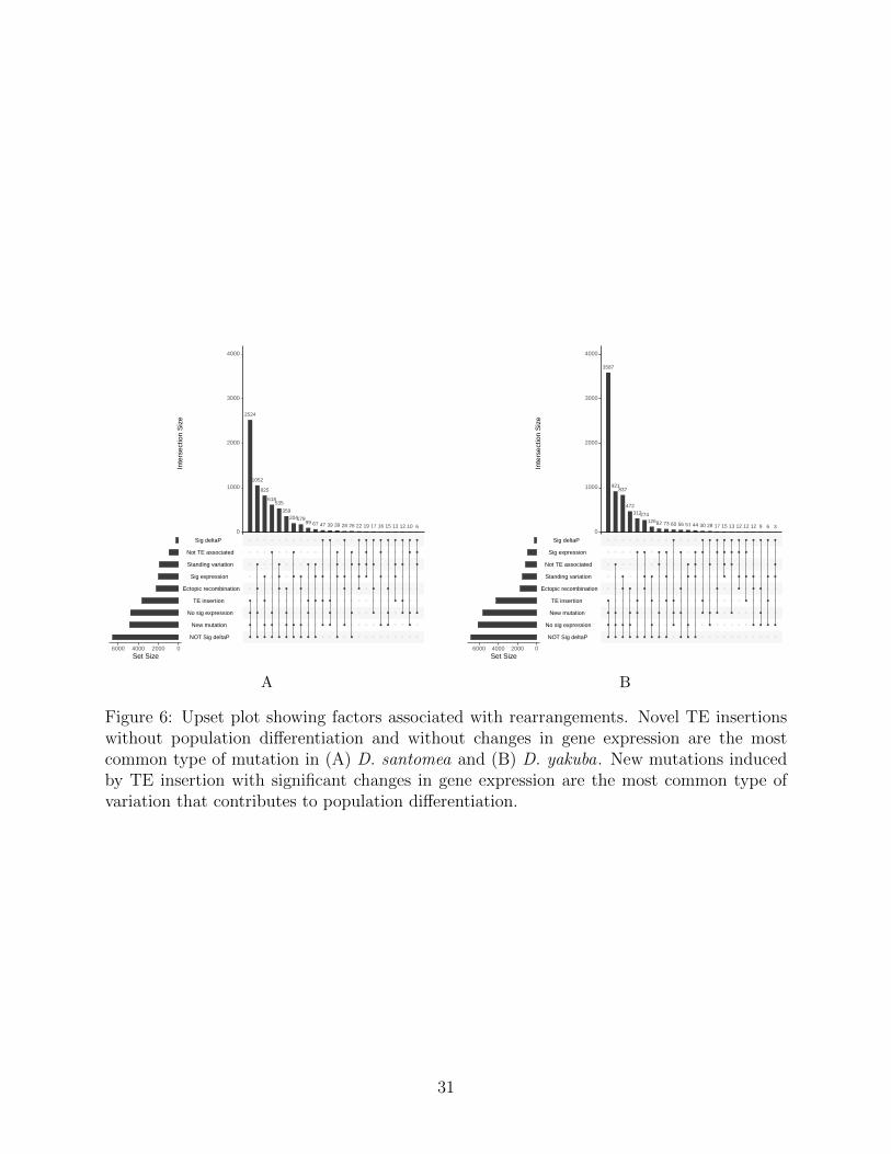
Figure 6: Upset plot showing factors associated with rearrangements. Novel TE insertionswithout population differentiation and without changes in gene expression are the mostcommon type of mutation in (A) D. santomea and (B) D. yakuba. New mutations inducedby TE insertion with significant changes in gene expression are the most common type ofvariation that contributes to population differentiation.
31
Supplementary Information
Complex variation causes genotyping challenges
Long read sequencing is proposed as a promising solution to structural variant calling. How-ever, computational and technical limitations remain, especially in the context of populationgenetics where false positives and false negatives may alter allele frequencies. We exploredthe utility of HiFi PacBio reads to determine structural variants using 3 pre-existing toolsand a BLAST comparison.
By looking at three programs designed to call structural variants, Minimap2, Pbsv, andSniffles, we ascertained that these programs were not confirming our Illumina reads at asufficient rate individually. Individual confirmation rates ranged from 3.2%-67.5% acrossindividual bioinformatics pipelines. Drosophila is known to have many small structuralvariants in comparison with mammals. Deletions accumulate rapidly in Drosophila, creatinggaps in alignment that make it more difficult to clarify mutation state. We find that complexvariation is difficult for existing aligners to solve. To confirm our suspicions we used coveragedata and investigated these complex regions where we frequently, and were able to findsigns in the coverage that there was likely some kind of variation occurring, whether it beduplications, rearrangements, or other forms of variation.
Taking all of these factors into consideration, we aligned sequence data for these regionsa BLAST against and the long-read data. Once we did this we were able to confirm rear-rangements at a rate of 79.7%-87.2% from strain to strain S20. In aggregate across all 4bioinformatic pipelines, we confirm 100% of rearrangement genotypes. BLAST is not de-signed to align long read sequence data, however with BLAST we are able to confirm a totalof 100% that these regions do share similarity based on nucleotide identity and that thecurrent tools commonly used to call these mutations are not accurately handling all cases ofstructural variation in Drosophila.
The prospects of structural variant calling in Drosophila with new HiFi sequence technol-ogy are promising. However, greater computational and bioinformatic analysis, likely withspecies-specific parameter tuning is likely necessary to solve these technical issues in dataanalysis for complex regions of the genome.
1
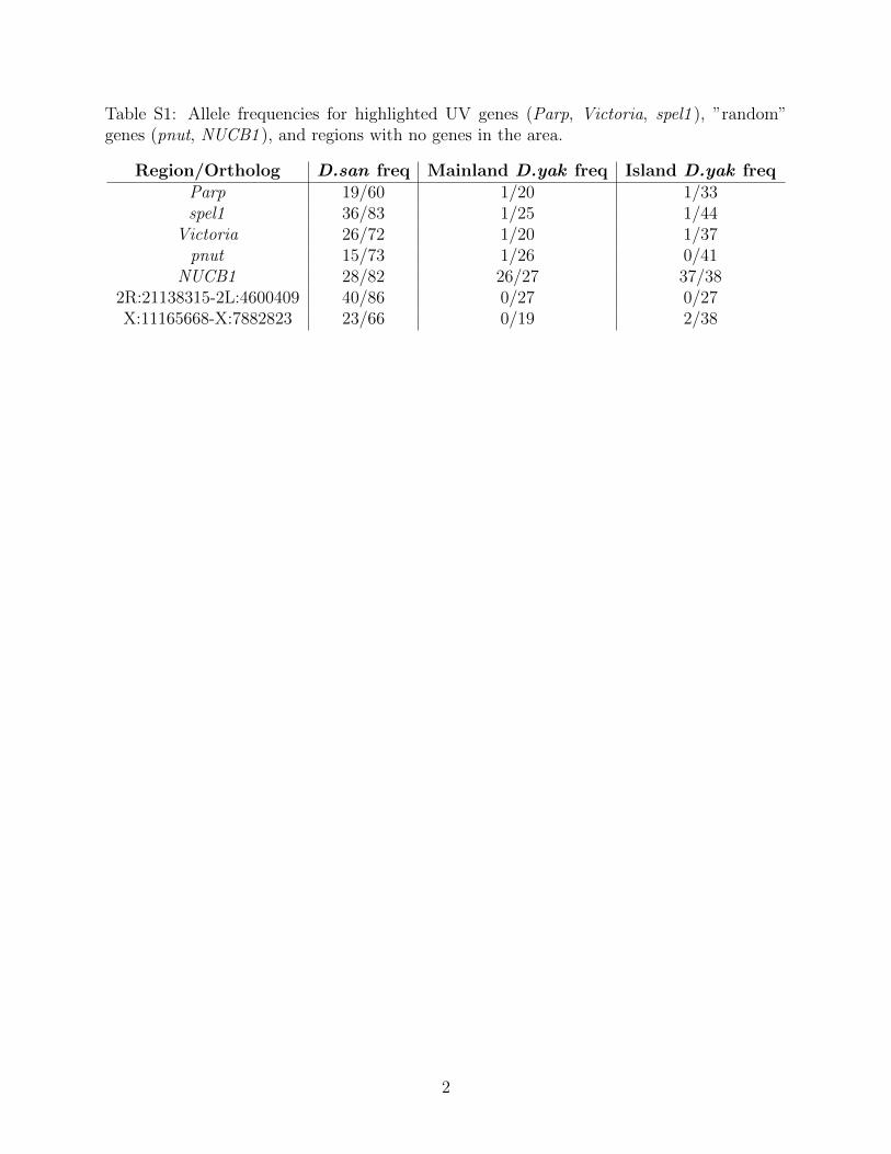
Table S1: Allele frequencies for highlighted UV genes (Parp, Victoria, spel1 ), ”random”genes (pnut, NUCB1 ), and regions with no genes in the area.
Region/Ortholog D.san freq Mainland D.yak freq Island D.yak freqParp 19/60 1/20 1/33spel1 36/83 1/25 1/44
Victoria 26/72 1/20 1/37pnut 15/73 1/26 0/41
NUCB1 28/82 26/27 37/382R:21138315-2L:4600409 40/86 0/27 0/27X:11165668-X:7882823 23/66 0/19 2/38
2
050
100
150
200
250
Loci (Mb): 2L
1 2 3 4 5 6 7 8 9 10 11 12 13 14 15 16 17 18 19 20 21 22 23
D.santomea breakpoints
050
100
150
200
250
Loci (Mb): 2R
1 2 3 4 5 6 7 8 9 10 11 12 13 14 15 16 17 18 19 20 21
D.santomea breakpoints
050
100
150
200
250
Loci (Mb): 3L
1 2 3 4 5 6 7 8 9 10 11 12 13 14 15 16 17 18 19 20 21 22 23 24 25
D.santomea breakpoints
050
100
150
200
250
Loci (Mb): 3R
1 2 3 4 5 6 7 8 9 10 11 12 13 14 15 16 17 18 19 20 21 22 23 24 25 26 27 28 29
D.santomea breakpoints
050
100
150
200
250
Loci (Mb): X
1 2 3 4 5 6 7 8 9 10 11 12 13 14 15 16 17 18 19 20 21 22
D.santomea breakpoints
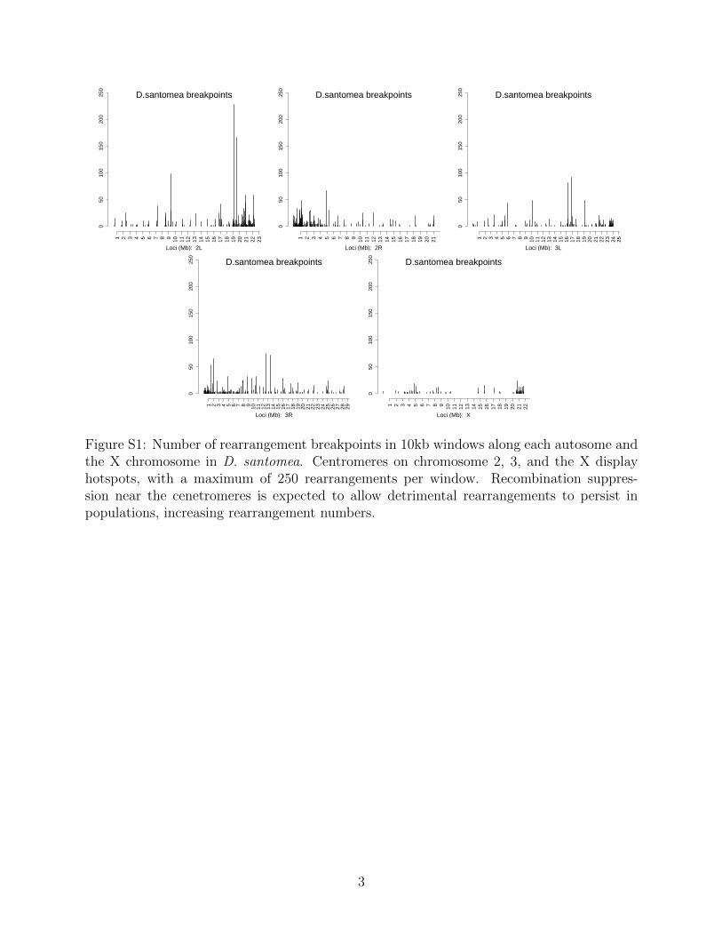
Figure S1: Number of rearrangement breakpoints in 10kb windows along each autosome andthe X chromosome in D. santomea. Centromeres on chromosome 2, 3, and the X displayhotspots, with a maximum of 250 rearrangements per window. Recombination suppres-sion near the cenetromeres is expected to allow detrimental rearrangements to persist inpopulations, increasing rearrangement numbers.
3
050
100
150
200
250
Loci (Mb): 2L
1 2 3 4 5 6 7 8 9 10 11 12 13 14 15 16 17 18 19 20 21 22 23
D.yakuba breakpoints
050
100
150
200
250
Loci (Mb): 2R
1 2 3 4 5 6 7 8 9 10 11 12 13 14 15 16 17 18 19 20 21
D.yakuba breakpoints
050
100
150
200
250
Loci (Mb): 3L
1 2 3 4 5 6 7 8 9 10 11 12 13 14 15 16 17 18 19 20 21 22 23 24
D.yakuba breakpoints
050
100
150
200
250
Loci (Mb): 3R
1 2 3 4 5 6 7 8 9 10 11 12 13 14 15 16 17 18 19 20 21 22 23 24 25 26 27 28 29
D.yakuba breakpoints
050
100
150
200
250
Loci (Mb): X
1 2 3 4 5 6 7 8 9 10 11 12 13 14 15 16 17 18 19 20 21 22
D.yakuba breakpoints
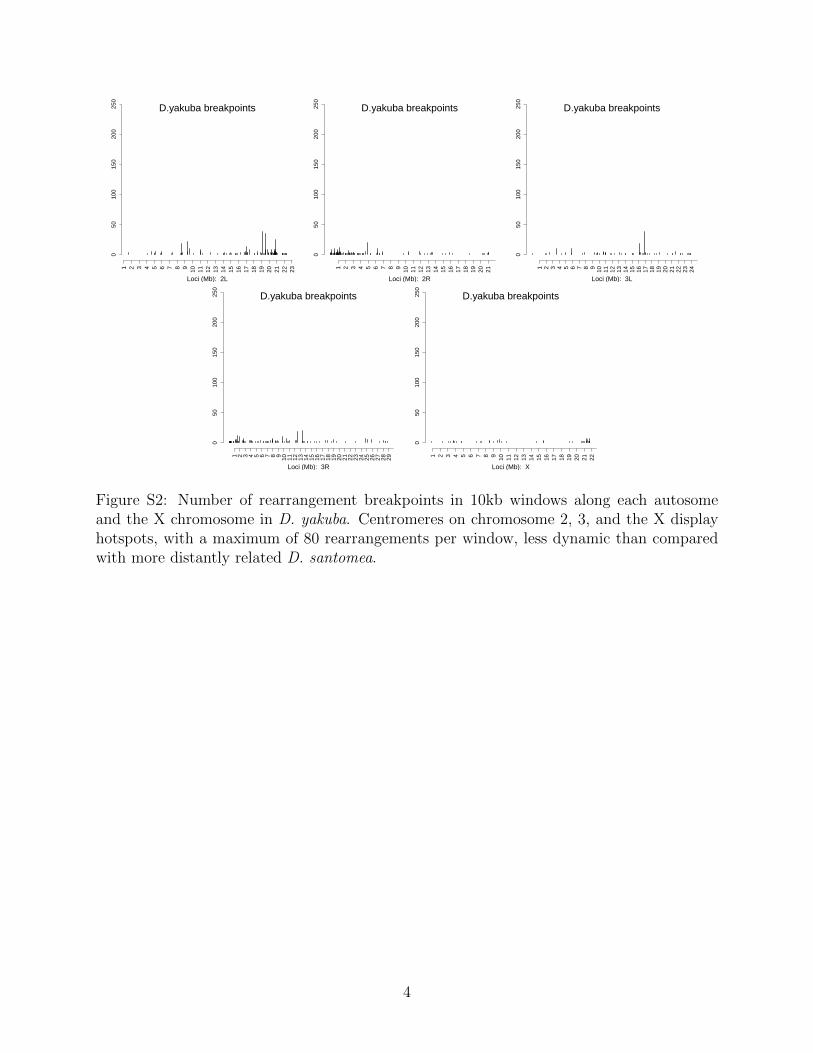
Figure S2: Number of rearrangement breakpoints in 10kb windows along each autosomeand the X chromosome in D. yakuba. Centromeres on chromosome 2, 3, and the X displayhotspots, with a maximum of 80 rearrangements per window, less dynamic than comparedwith more distantly related D. santomea.
4
A b
c d
Figure S3: Population differentiation as shown via ∆p A) Between D. santomea and main-land ancestral D. yakubaon autosome 3L. B) Between D. santomea and mainland ancestralD. yakubaon the X chromosome C) Between Island D. yakuba and ancestral mainland D.yakuba on the autosome 3L. D) Between Island D. yakuba and ancestral mainland D. yakubaon the X chromosome. The x-axis shows genomic position on the chromosome in megabases,and the y-axis is difference in allele frequencies between the test population and the main-land. Points in dark green highlight the rearrangements associated with facilitating ectopicrecombination. We observe greater levels of differentiation on the X compared to the auto-somes in both populations.
5
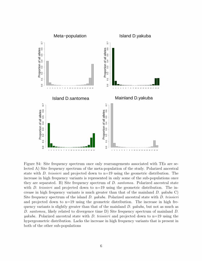
1 2 3 4 5 6 7 8 9 10 11 12 13 14 15 16 17 18 19
Meta−population0.
00.
10.
20.
30.
40.
50.
60.
7P
ropo
rtio
n of
all
alle
les
1 2 3 4 5 6 7 8 9 10 11 12 13 14 15 16 17 18 19
Island D.yakuba
0.0
0.1
0.2
0.3
0.4
0.5
0.6
0.7
Pro
port
ion
of a
ll al
lele
s
1 2 3 4 5 6 7 8 9 10 11 12 13 14 15 16 17 18 19
Island D.santomea
0.0
0.1
0.2
0.3
0.4
0.5
0.6
0.7
Pro
port
ion
of a
ll al
lele
s
1 2 3 4 5 6 7 8 9 10 11 12 13 14 15 16 17 18 19
Mainland D.yakuba
0.0
0.1
0.2
0.3
0.4
0.5
0.6
0.7
Pro
port
ion
of a
ll al
lele
s
Figure S4: Site frequency spectrum once only rearrangements associated with TEs are se-lected A) Site frequency spectrum of the meta-population of the study. Polarized ancestralstate with D. teissieri and projected down to n=19 using the geometric distribution. Theincrease in high frequency variants is represented in only some of the sub-populations oncethey are separated. B) Site frequency spectrum of D. santomea. Polarized ancestral statewith D. teissieri and projected down to n=19 using the geometric distribution. The in-crease in high frequency variants is much greater than that of the mainland D. yakuba C)Site frequency spectrum of the island D. yakuba. Polarized ancestral state with D. teissieriand projected down to n=19 using the geometric distribution. The increase in high fre-quency variants is slightly greater than that of the mainland D. yakuba, but not as much asD. santomea, likely related to divergence time D) Site frequency spectrum of mainland D.yakuba. Polarized ancestral state with D. teissieri and projected down to n=19 using thehypergeometric distribution. Lacks the increase in high frequency variants that is present inboth of the other sub-populations
6
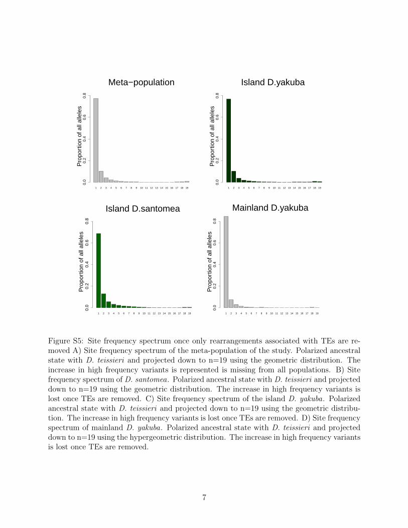
1 2 3 4 5 6 7 8 9 10 11 12 13 14 15 16 17 18 19
Meta−population0.
00.
20.
40.
60.
8P
ropo
rtio
n of
all
alle
les
1 2 3 4 5 6 7 8 9 10 11 12 13 14 15 16 17 18 19
Island D.yakuba
0.0
0.2
0.4
0.6
0.8
Pro
port
ion
of a
ll al
lele
s
1 2 3 4 5 6 7 8 9 10 11 12 13 14 15 16 17 18 19
Island D.santomea
0.0
0.2
0.4
0.6
0.8
Pro
port
ion
of a
ll al
lele
s
1 2 3 4 5 6 7 8 9 10 11 12 13 14 15 16 17 18 19
Mainland D.yakuba 0.
00.
20.
40.
60.
8P
ropo
rtio
n of
all
alle
les
Figure S5: Site frequency spectrum once only rearrangements associated with TEs are re-moved A) Site frequency spectrum of the meta-population of the study. Polarized ancestralstate with D. teissieri and projected down to n=19 using the geometric distribution. Theincrease in high frequency variants is represented is missing from all populations. B) Sitefrequency spectrum of D. santomea. Polarized ancestral state with D. teissieri and projecteddown to n=19 using the geometric distribution. The increase in high frequency variants islost once TEs are removed. C) Site frequency spectrum of the island D. yakuba. Polarizedancestral state with D. teissieri and projected down to n=19 using the geometric distribu-tion. The increase in high frequency variants is lost once TEs are removed. D) Site frequencyspectrum of mainland D. yakuba. Polarized ancestral state with D. teissieri and projecteddown to n=19 using the hypergeometric distribution. The increase in high frequency variantsis lost once TEs are removed.
7
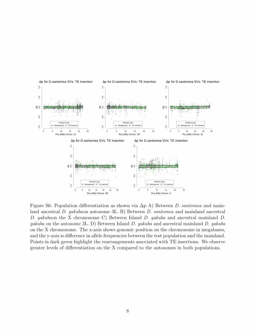
0 5 10 15 20 25
−1.
0−
0.5
0.0
0.5
1.0
Pos (Mb) Chrom: 2L
∆p for D.santomea SVs: TE insertion
∆p
Mutation type
Background TE insertion
0 5 10 15 20 25
−1.
0−
0.5
0.0
0.5
1.0
Pos (Mb) Chrom: 2R
∆p for D.santomea SVs: TE insertion
∆p
Mutation type
Background TE insertion
0 5 10 15 20 25
−1.
0−
0.5
0.0
0.5
1.0
Pos (Mb) Chrom: 3L
∆p for D.santomea SVs: TE insertion
∆p
Mutation type
Background TE insertion
0 5 10 15 20 25 30
−1.
0−
0.5
0.0
0.5
1.0
Pos (Mb) Chrom: 3R
∆p for D.santomea SVs: TE insertion
∆p
Mutation type
Background TE insertion
0 5 10 15 20 25
−1.
0−
0.5
0.0
0.5
1.0
Pos (Mb) Chrom: X
∆p for D.santomea SVs: TE insertion
∆p
Mutation type
Background TE insertion
Figure S6: Population differentiation as shown via ∆p A) Between D. santomea and main-land ancestral D. yakubaon autosome 3L. B) Between D. santomea and mainland ancestralD. yakubaon the X chromosome C) Between Island D. yakuba and ancestral mainland D.yakuba on the autosome 3L. D) Between Island D. yakuba and ancestral mainland D. yakubaon the X chromosome. The x-axis shows genomic position on the chromosome in megabases,and the y-axis is difference in allele frequencies between the test population and the mainland.Points in dark green highlight the rearrangements associated with TE insertions. We observegreater levels of differentiation on the X compared to the autosomes in both populations.
8
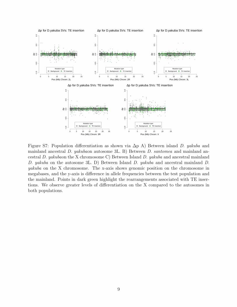
0 5 10 15 20 25
−1.
0−
0.5
0.0
0.5
1.0
Pos (Mb) Chrom: 2L
∆p for D.yakuba SVs: TE insertion∆p
Mutation type
Background TE Insertion
0 5 10 15 20 25
−1.
0−
0.5
0.0
0.5
1.0
Pos (Mb) Chrom: 2R
∆p for D.yakuba SVs: TE insertion
∆p
Mutation type
Background TE Insertion
0 5 10 15 20 25
−1.
0−
0.5
0.0
0.5
1.0
Pos (Mb) Chrom: 3L
∆p for D.yakuba SVs: TE insertion
∆p
Mutation type
Background TE Insertion
0 5 10 15 20 25 30
−1.
0−
0.5
0.0
0.5
1.0
Pos (Mb) Chrom: 3R
∆p for D.yakuba SVs: TE insertion
∆p
Mutation type
Background TE Insertion
0 5 10 15 20 25
−1.
0−
0.5
0.0
0.5
1.0
Pos (Mb) Chrom: X
∆p for D.yakuba SVs: TE insertion
∆pMutation type
Background TE Insertion
Figure S7: Population differentiation as shown via ∆p A) Between island D. yakuba andmainland ancestral D. yakubaon autosome 3L. B) Between D. santomea and mainland an-cestral D. yakubaon the X chromosome C) Between Island D. yakuba and ancestral mainlandD. yakuba on the autosome 3L. D) Between Island D. yakuba and ancestral mainland D.yakuba on the X chromosome. The x-axis shows genomic position on the chromosome inmegabases, and the y-axis is difference in allele frequencies between the test population andthe mainland. Points in dark green highlight the rearrangements associated with TE inser-tions. We observe greater levels of differentiation on the X compared to the autosomes inboth populations.
9
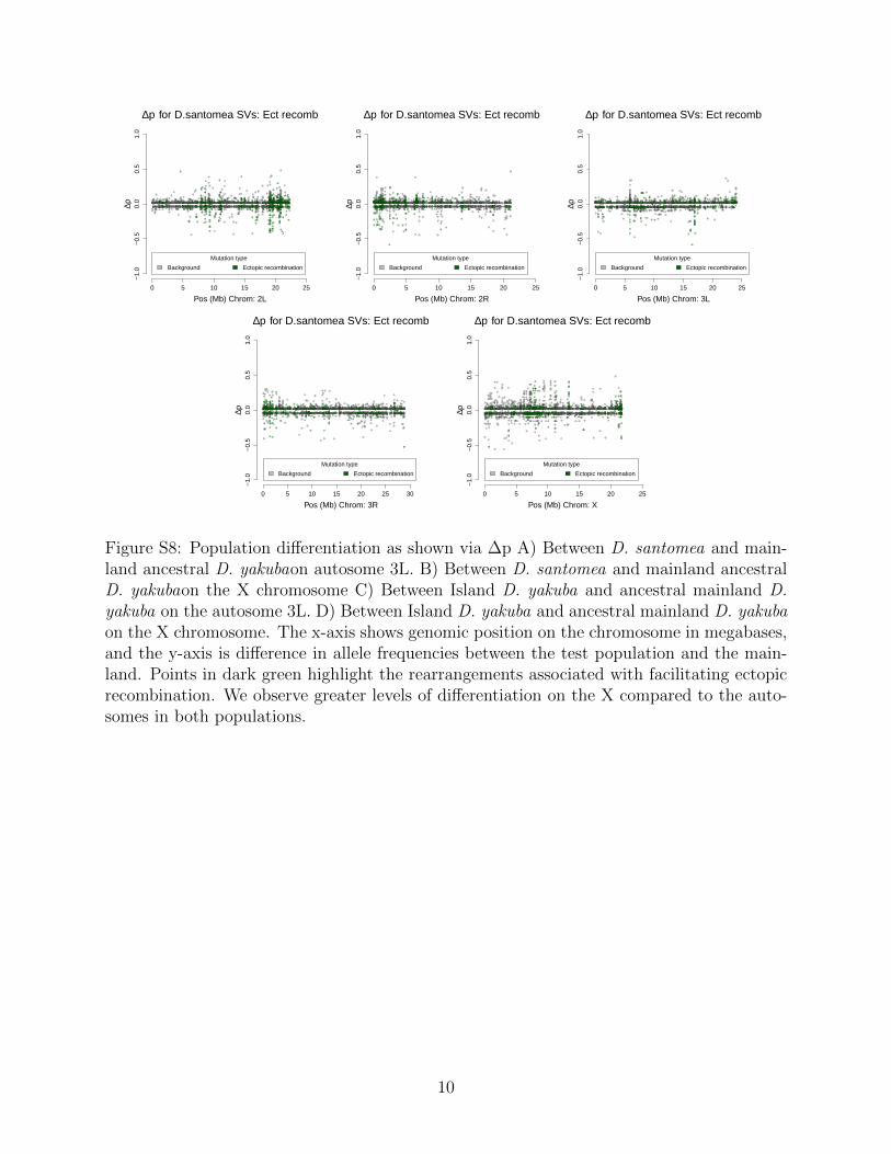
0 5 10 15 20 25
−1.
0−
0.5
0.0
0.5
1.0
Pos (Mb) Chrom: 2L
∆p for D.santomea SVs: Ect recomb∆p
Mutation type
Background Ectopic recombination
0 5 10 15 20 25
−1.
0−
0.5
0.0
0.5
1.0
Pos (Mb) Chrom: 2R
∆p for D.santomea SVs: Ect recomb
∆p
Mutation type
Background Ectopic recombination
0 5 10 15 20 25
−1.
0−
0.5
0.0
0.5
1.0
Pos (Mb) Chrom: 3L
∆p for D.santomea SVs: Ect recomb
∆p
Mutation type
Background Ectopic recombination
0 5 10 15 20 25 30
−1.
0−
0.5
0.0
0.5
1.0
Pos (Mb) Chrom: 3R
∆p for D.santomea SVs: Ect recomb
∆p
Mutation type
Background Ectopic recombination
0 5 10 15 20 25
−1.
0−
0.5
0.0
0.5
1.0
Pos (Mb) Chrom: X
∆p for D.santomea SVs: Ect recomb
∆pMutation type
Background Ectopic recombination
Figure S8: Population differentiation as shown via ∆p A) Between D. santomea and main-land ancestral D. yakubaon autosome 3L. B) Between D. santomea and mainland ancestralD. yakubaon the X chromosome C) Between Island D. yakuba and ancestral mainland D.yakuba on the autosome 3L. D) Between Island D. yakuba and ancestral mainland D. yakubaon the X chromosome. The x-axis shows genomic position on the chromosome in megabases,and the y-axis is difference in allele frequencies between the test population and the main-land. Points in dark green highlight the rearrangements associated with facilitating ectopicrecombination. We observe greater levels of differentiation on the X compared to the auto-somes in both populations.
10
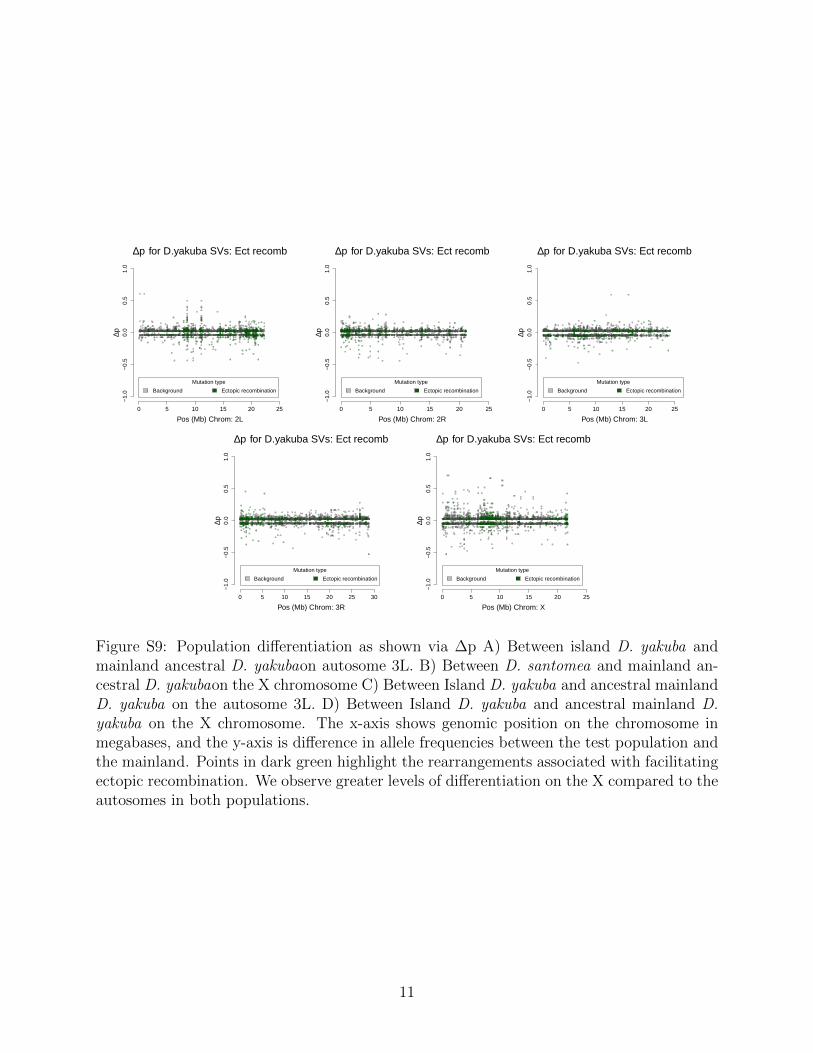
0 5 10 15 20 25
−1.
0−
0.5
0.0
0.5
1.0
Pos (Mb) Chrom: 2L
∆p for D.yakuba SVs: Ect recomb
∆p
Mutation type
Background Ectopic recombination
0 5 10 15 20 25
−1.
0−
0.5
0.0
0.5
1.0
Pos (Mb) Chrom: 2R
∆p for D.yakuba SVs: Ect recomb
∆p
Mutation type
Background Ectopic recombination
0 5 10 15 20 25
−1.
0−
0.5
0.0
0.5
1.0
Pos (Mb) Chrom: 3L
∆p for D.yakuba SVs: Ect recomb
∆p
Mutation type
Background Ectopic recombination
0 5 10 15 20 25 30
−1.
0−
0.5
0.0
0.5
1.0
Pos (Mb) Chrom: 3R
∆p for D.yakuba SVs: Ect recomb
∆p
Mutation type
Background Ectopic recombination
0 5 10 15 20 25
−1.
0−
0.5
0.0
0.5
1.0
Pos (Mb) Chrom: X
∆p for D.yakuba SVs: Ect recomb
∆p
Mutation type
Background Ectopic recombination
Figure S9: Population differentiation as shown via ∆p A) Between island D. yakuba andmainland ancestral D. yakubaon autosome 3L. B) Between D. santomea and mainland an-cestral D. yakubaon the X chromosome C) Between Island D. yakuba and ancestral mainlandD. yakuba on the autosome 3L. D) Between Island D. yakuba and ancestral mainland D.yakuba on the X chromosome. The x-axis shows genomic position on the chromosome inmegabases, and the y-axis is difference in allele frequencies between the test population andthe mainland. Points in dark green highlight the rearrangements associated with facilitatingectopic recombination. We observe greater levels of differentiation on the X compared to theautosomes in both populations.
11
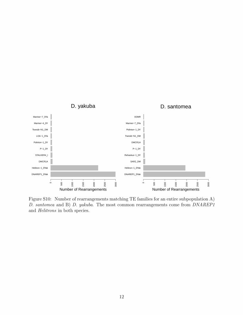
DNAREP1_DYak
Helitron−1_DYak
DMCR1A
STALKER4_I
P−1_DY
Polinton−1_DY
LOA−1_DYa
Transib−N1_DM
Mariner−4_DY
Mariner−7_DYa
0
500
1000
1500
2000
2500
3000
Number of Rearrangements
D. yakuba
DNAREP1_DYak
Helitron−1_DYak
SAR2_DM
Rehavkus−1_DY
P−1_DY
DMCR1A
Transib−N1_DM
Polinton−1_DY
Mariner−7_DYa
XDMR
0
500
1000
1500
2000
2500
3000
Number of Rearrangements
D. santomea
Figure S10: Number of rearrangements matching TE families for an entire subpopulation A)D. santomea and B) D. yakuba. The most common rearrangements come from DNAREP1and Helitrons in both species.
12
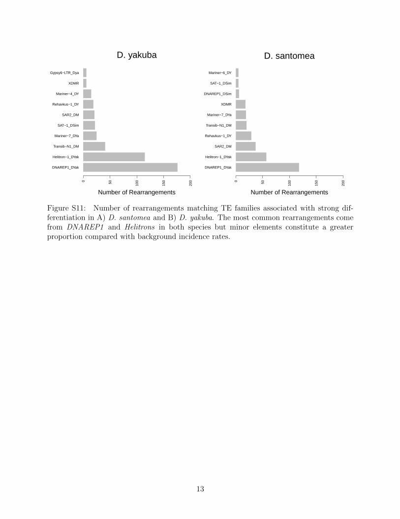
DNAREP1_DYak
Helitron−1_DYak
Transib−N1_DM
Mariner−7_DYa
SAT−1_DSim
SAR2_DM
Rehavkus−1_DY
Mariner−4_DY
XDMR
Gypsy6−LTR_Dya
0 50 100
150
200
Number of Rearrangements
D. yakuba
DNAREP1_DYak
Helitron−1_DYak
SAR2_DM
Rehavkus−1_DY
Transib−N1_DM
Mariner−7_DYa
XDMR
DNAREP1_DSim
SAT−1_DSim
Mariner−6_DY
0 50 100
150
200
Number of Rearrangements
D. santomea
Figure S11: Number of rearrangements matching TE families associated with strong dif-ferentiation in A) D. santomea and B) D. yakuba. The most common rearrangements comefrom DNAREP1 and Helitrons in both species but minor elements constitute a greaterproportion compared with background incidence rates.
13
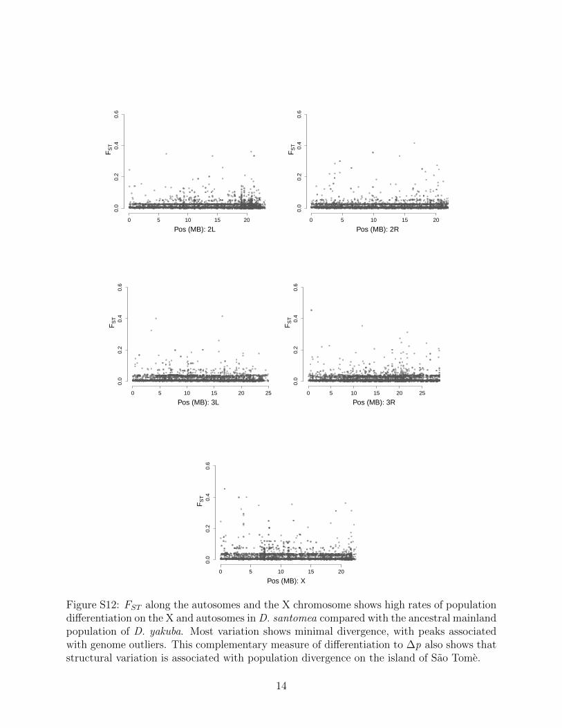
0 5 10 15 20
0.0
0.2
0.4
0.6
Pos (MB): 2L
FS
T
0 5 10 15 20
0.0
0.2
0.4
0.6
Pos (MB): 2R
FS
T
0 5 10 15 20 25
0.0
0.2
0.4
0.6
Pos (MB): 3L
FS
T
0 5 10 15 20 25
0.0
0.2
0.4
0.6
Pos (MB): 3R
FS
T
0 5 10 15 20
0.0
0.2
0.4
0.6
Pos (MB): X
FS
T
Figure S12: FST along the autosomes and the X chromosome shows high rates of populationdifferentiation on the X and autosomes in D. santomea compared with the ancestral mainlandpopulation of D. yakuba. Most variation shows minimal divergence, with peaks associatedwith genome outliers. This complementary measure of differentiation to Δp also shows thatstructural variation is associated with population divergence on the island of Sao Tome.
14
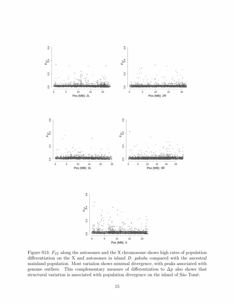
0 5 10 15 20
0.0
0.2
0.4
0.6
Pos (MB): 2L
FS
T
0 5 10 15 20
0.0
0.2
0.4
0.6
Pos (MB): 2R
FS
T
0 5 10 15 20 25
0.0
0.2
0.4
0.6
Pos (MB): 3L
FS
T
0 5 10 15 20 25
0.0
0.2
0.4
0.6
Pos (MB): 3R
FS
T
0 5 10 15 20
0.0
0.2
0.4
0.6
Pos (MB): X
FS
T
Figure S13: FST along the autosomes and the X chromosome shows high rates of populationdifferentiation on the X and autosomes in island D. yakuba compared with the ancestralmainland population. Most variaiion shows minimal divergence, with peaks associated withgenome outliers. This complementary measure of differentiation to Δp also shows thatstructural variation is associated with population divergence on the island of Sao Tome.
15
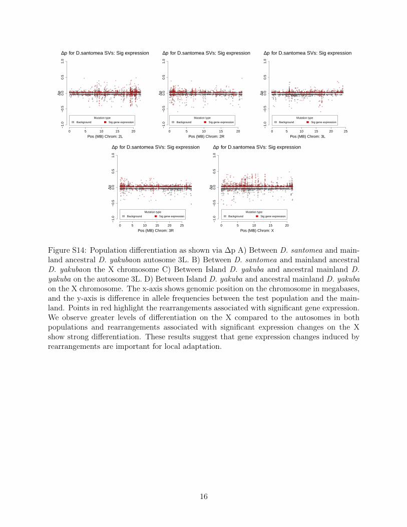
0 5 10 15 20
−1.
0−
0.5
0.0
0.5
1.0
Pos (MB) Chrom: 2L
∆p for D.santomea SVs: Sig expression∆p
Mutation type
Background Sig gene expression
0 5 10 15 20
−1.
0−
0.5
0.0
0.5
1.0
Pos (MB) Chrom: 2R
∆p for D.santomea SVs: Sig expression
∆p
Mutation type
Background Sig gene expression
0 5 10 15 20 25
−1.
0−
0.5
0.0
0.5
1.0
Pos (MB) Chrom: 3L
∆p for D.santomea SVs: Sig expression
∆p
Mutation type
Background Sig gene expression
0 5 10 15 20 25
−1.
0−
0.5
0.0
0.5
1.0
Pos (MB) Chrom: 3R
∆p for D.santomea SVs: Sig expression
∆p
Mutation type
Background Sig gene expression
0 5 10 15 20
−1.
0−
0.5
0.0
0.5
1.0
Pos (MB) Chrom: X
∆p for D.santomea SVs: Sig expression
∆pMutation type
Background Sig gene expression
Figure S14: Population differentiation as shown via ∆p A) Between D. santomea and main-land ancestral D. yakubaon autosome 3L. B) Between D. santomea and mainland ancestralD. yakubaon the X chromosome C) Between Island D. yakuba and ancestral mainland D.yakuba on the autosome 3L. D) Between Island D. yakuba and ancestral mainland D. yakubaon the X chromosome. The x-axis shows genomic position on the chromosome in megabases,and the y-axis is difference in allele frequencies between the test population and the main-land. Points in red highlight the rearrangements associated with significant gene expression.We observe greater levels of differentiation on the X compared to the autosomes in bothpopulations and rearrangements associated with significant expression changes on the Xshow strong differentiation. These results suggest that gene expression changes induced byrearrangements are important for local adaptation.
16
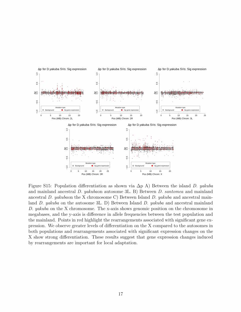
0 5 10 15 20
−1.
0−
0.5
0.0
0.5
1.0
Pos (MB) Chrom: 2L
∆p for D.yakuba SVs: Sig expression
∆p
Mutation type
Background Sig gene expression
0 5 10 15 20
−1.
0−
0.5
0.0
0.5
1.0
Pos (MB) Chrom: 2R
∆p for D.yakuba SVs: Sig expression
∆p
Mutation type
Background Sig gene expression
0 5 10 15 20 25
−1.
0−
0.5
0.0
0.5
1.0
Pos (MB) Chrom: 3L
∆p for D.yakuba SVs: Sig expression
∆p
Mutation type
Background Sig gene expression
0 5 10 15 20 25
−1.
0−
0.5
0.0
0.5
1.0
Pos (MB) Chrom: 3R
∆p for D.yakuba SVs: Sig expression
∆p
Mutation type
Background Sig gene expression
0 5 10 15 20
−1.
0−
0.5
0.0
0.5
1.0
Pos (MB) Chrom: X
∆p for D.yakuba SVs: Sig expression∆p
Mutation type
Background Sig gene expression
Figure S15: Population differentiation as shown via ∆p A) Between the island D. yakubaand mainland ancestral D. yakubaon autosome 3L. B) Between D. santomea and mainlandancestral D. yakubaon the X chromosome C) Between Island D. yakuba and ancestral main-land D. yakuba on the autosome 3L. D) Between Island D. yakuba and ancestral mainlandD. yakuba on the X chromosome. The x-axis shows genomic position on the chromosome inmegabases, and the y-axis is difference in allele frequencies between the test population andthe mainland. Points in red highlight the rearrangements associated with significant gene ex-pression. We observe greater levels of differentiation on the X compared to the autosomes inboth populations and rearrangements associated with significant expression changes on theX show strong differentiation. These results suggest that gene expression changes inducedby rearrangements are important for local adaptation.
17
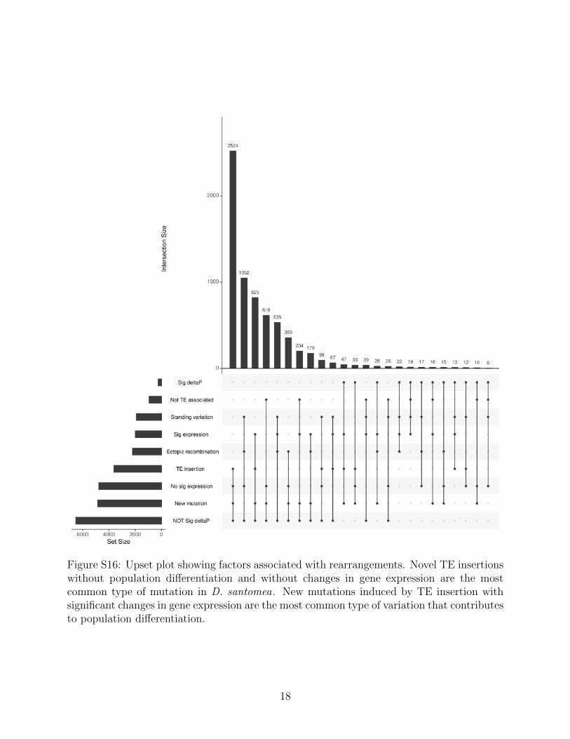
Figure S16: Upset plot showing factors associated with rearrangements. Novel TE insertionswithout population differentiation and without changes in gene expression are the mostcommon type of mutation in D. santomea. New mutations induced by TE insertion withsignificant changes in gene expression are the most common type of variation that contributesto population differentiation.
18
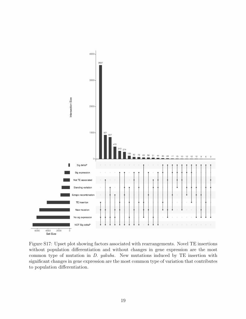
Figure S17: Upset plot showing factors associated with rearrangements. Novel TE insertionswithout population differentiation and without changes in gene expression are the mostcommon type of mutation in D. yakuba. New mutations induced by TE insertion withsignificant changes in gene expression are the most common type of variation that contributesto population differentiation.
19
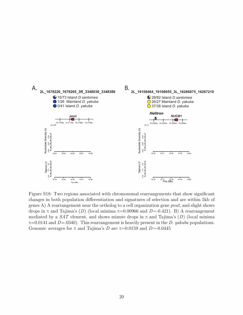
A. B.
16,776kb 16,777kb 16,778kb 16,779kb
15/73 Island D.santomea 1/26 Mainland D. yakuba 0/41 Island D. yakuba
Nuc
leot
ide
Div
ersi
ty (π
)Ta
jima'
s D
chr 2R
2L_1678226_1678265_3R_3348038_3348386
Helitron
16,262kb 16,264kb 16,266kb 16,268kb
28/82 Island D.santomea 26/27 Mainland D. yakuba 37/38 Island D. yakuba
Nuc
leot
ide
Div
ersi
ty (π
)
Pos (Mb)
Tajim
a's
D
chr X
2L_19108464_19108655_3L_16266875_16267219
pnut NUCB1
16.24 16.25 16.26 16.27 16.28
0.00
0.02
0.04
16.24 16.25 16.26 16.27 16.28
−2.0
−1.0
0.0
Pos (Mb)
16.76 16.77 16.78 16.79 16.80
0.00
0.02
0.04
16.76 16.77 16.78 16.79 16.80
−2.0
−1.0
0.0
Figure S18: Two regions associated with chromosomal rearrangements that show significantchanges in both population differentiation and signatures of selection and are within 5kb ofgenes A) A rearrangement near the ortholog to a cell organization gene pnut, and slight showsdrops in π and Tajima’s (D) (local minima π=0.00966 and D=-0.421). B) A rearrangementmediated by a SAT element, and shows minute drops in π and Tajima’s (D) (local minimaπ=0.0141 and D=.0340). This rearrangement is heavily present in the D. yakuba populations.Genomic averages for π and Tajima’s D are π=0.0159 and D=-0.0445
20
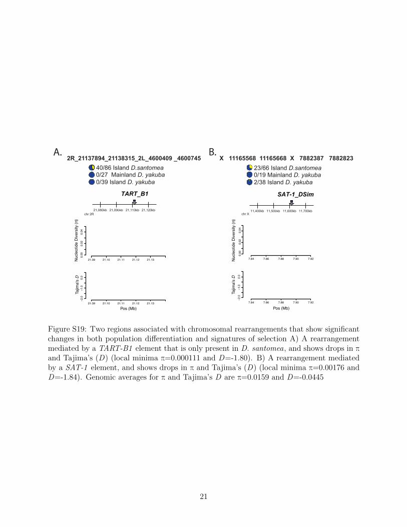
A. B.
TART_B1
21,080kb 21,090kb 21,110kb 21,120kb
40/86 Island D.santomea 0/27 Mainland D. yakuba 0/39 Island D. yakuba
Pos (Mb)
21.09 21.10 21.11 21.12 21.13
0.00
0.02
0.04
21.09 21.10 21.11 21.12 21.13
−2.0
−1.0
0.0
Nuc
leot
ide
Div
ersi
ty (π
)Ta
jima'
s D
chr 2R
2R_21137894_21138315_2L_4600409 _4600745
SAT-1_DSim
11,400kb 11,500kb 11,600kb 11,700kb
23/66 Island D.santomea 0/19 Mainland D. yakuba 2/38 Island D. yakuba
7.84 7.86 7.88 7.90 7.92
0.00
0.02
0.04
7.84 7.86 7.88 7.90 7.92
−2.0
−1.0
0.0
Nuc
leot
ide
Div
ersi
ty (π
)
Pos (Mb)
Tajim
a's
D
chr X
X 11165568 11165668 X 7882387 7882823
Figure S19: Two regions associated with chromosomal rearrangements that show significantchanges in both population differentiation and signatures of selection A) A rearrangementmediated by a TART-B1 element that is only present in D. santomea, and shows drops in πand Tajima’s (D) (local minima π=0.000111 and D=-1.80). B) A rearrangement mediatedby a SAT-1 element, and shows drops in π and Tajima’s (D) (local minima π=0.00176 andD=-1.84). Genomic averages for π and Tajima’s D are π=0.0159 and D=-0.0445
21
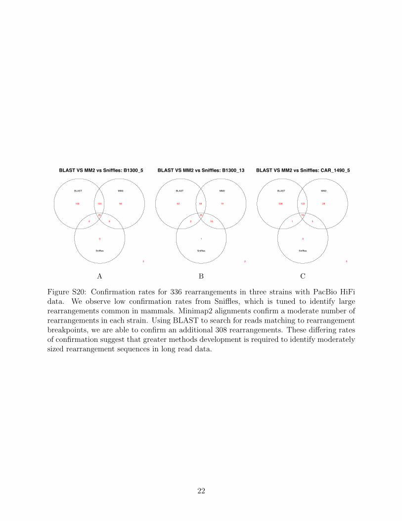
BLAST VS MM2 vs Sniffles: B1300_5
BLAST MM2
Sniffles
0
0
56
9
100
4
131
20
A
BLAST VS MM2 vs Sniffles: B1300_13
BLAST MM2
Sniffles
0
1
19
16
52
2
59
20
B
BLAST VS MM2 vs Sniffles: CAR_1490_5
BLAST MM2
Sniffles
0
3
28
5
108
1
122
16
C
Figure S20: Confirmation rates for 336 rearrangements in three strains with PacBio HiFidata. We observe low confirmation rates from Sniffles, which is tuned to identify largerearrangements common in mammals. Minimap2 alignments confirm a moderate number ofrearrangements in each strain. Using BLAST to search for reads matching to rearrangementbreakpoints, we are able to confirm an additional 308 rearrangements. These differing ratesof confirmation suggest that greater methods development is required to identify moderatelysized rearrangement sequences in long read data.
22