choroidal pigmented lesions imaged by ultra-wide-field scanning laser ophthalmoscopy with two laser...
TRANSCRIPT
© 2010 Kernt et al, publisher and licensee Dove Medical Press Ltd. This is an Open Access article which permits unrestricted noncommercial use, provided the original work is properly cited.
Clinical Ophthalmology 2010:4 829–836
Clinical Ophthalmology Dovepress
submit your manuscript | www.dovepress.com
Dovepress 829
O r i g i n A L r e s e A r C h
open access to scientific and medical research
Open Access Full Text Article
11864
Choroidal pigmented lesions imaged by ultra-wide-field scanning laser ophthalmoscopy with two laser wavelengths (Optomap)
Marcus Kernt Ulrich C schaller Carmen stumpf Michael W Ulbig Anselm Kampik Aljoscha s neubauerDepartment of Ophthalmology, Ludwig-Maximilians-University, Munich, germany
Correspondence: Marcus Kernt Department of Ophthalmology, Ludwig-Maximilians-University Munich, Mathildenstr. 8, 80336 Muenchen, germany Tel +49-89-5160-3811 Fax +49-89-5160-5160 email [email protected]
Purpose: Clinical differentiation of choroidal pigmented lesions is sometimes difficult.
Choroidal melanoma is the most prevalent primary neoplasia among malignant ocular tumors,
and metastasis often occurs before the primary tumor is diagnosed. Therefore, early detection is
essential. We investigated the imaging properties of clinically diagnosed melanocytic choroidal
tumors using a nonmydriatic ultra-wide-field scanning laser ophthalmoscope (SLO) with two
laser wavelengths to distinguish benign from malignant lesions. Repeated standardized ultrasound
(US) evaluation provided reference standard.
Methods: In a consecutive series of 49 patients with clinically diagnosed melanocytic choroidal
tumors in one eye, 29 had established melanoma (defined by proven growth on repeated US
follow-up) and 20 had nevi (defined by no malignancy according to clinical, US, and growth
characteristics for at least 2 years). All patients underwent clinical examination, undilated
Optomap (Optos PLC, Dunfermline, Fife, Scotland, UK) imaging, standardized US examination,
and standard retinal photography. Measurements of the tumor base using the Optomap software
were compared with US B-scan measurements. Imaging characteristics from the SLO images
were correlated with the structural findings in the two patient groups.
Results: Measurements of tumor base correlated well between SLO and US with r = 0.61
(T-direction) and r = 0.51 (L-direction). On SLO imaging, typical malignant lesions appeared
dark on the red laser channel and bright on the green laser channel. Based on those simple
binary characteristics, a sensitivity of 76% at a specificity of 70% was obtained for a correct
classification of lesions. When analogous to clinical examination lesion size, margin touching
the optic disc, and existence of subretinal fluid were additionally considered, 90% sensitivity
at 82% specificity was obtained.
Conclusions: In this first, limited series, nonmydriatic SLO imaging with two laser wave-
lengths permitted to differentiate malignant ocular tumors from nonmalignant lesions with high
diagnostic accuracy. Additional parameters may further enhance diagnostic properties, but larger
patient series are required to validate our findings and prove the diagnostic properties.
Keywords: choroidal melanoma, nevus, imaging, ultra-wide-field scanning laser ophthal-
moscopy
IntroductionPigmented choroidal lesions are a common finding on the ocular fundus. Most of
these lesions can be differentiated by their clinical appearance together with their
angiographic and ultrasonographic (US) characteristics. However, distinguishing
between benign and malignant lesions is sometimes difficult. Most choroidal lesions are
benign, and the overall incidence of malignancies in the form of choroidal melanoma
is low ( approximately six new cases per million per year in the United States).
Clinical Ophthalmology 2010:4submit your manuscript | www.dovepress.com
Dovepress
Dovepress
830
Kernt et al
Nonetheless, choroidal melanoma is the most frequent
primary malignant intraocular tumor and the second most
frequent malignant melanoma in the body.1 Benign choroidal
nevi are common and can be found in approximately 6% of
the US population.2–5 Choroidal melanoma can arise de novo
as well as from pre-existing choroidal nevi.6–8 Therefore,
choroidal nevi should be monitored regularly. In addition,
early detection of malignant lesions is extremely important
because progressive tumors have a very poor prognosis.9–12
On the other hand, especially in smaller lesions, dif-
ferentiating choroidal nevi from small choroidal melanoma
is difficult and concern about malignant transformation
is always present.12–14 Consequently, several studies have
attempted to distinguish small choroidal melanomas from
choroidal nevi and to identify risk factors for growth of small
choroidal tumors.1,12,15,16
Biomicroscopy of the fundus and photographic color
fundus imaging are the most commonly used techniques for
diagnosing and documenting choroidal lesions. However, both
techniques are sometimes insufficient to differentiate lesions
accurately. Even with combined clinical, angiographic, and US
findings, diagnosis sometimes remains uncertain.1,11,12,14–16
Recently, a novel ultra-wide-f ield scanning laser
ophthalmoscope (SLO) with two laser wavelengths, the
Optomap Panoramic 200MA (Optos PLC, Dunfermline,
Fife, Scotland, UK), was developed. The system allows
nonmydriatic imaging and by differentiating the two laser
scans, provides additional image information. Optomap
Panoramic 200MA images encompass not only the posterior
pole but also extend over the equator.17
This study evaluates measurements of the tumor base
using the Optomap Vantage V2 software vs the reference
standard, standardized US evaluation, which is a well estab-
lished, highly sensitive, and specific method to measure and
differentiate intraocular tumors.18,19 In addition, the diagnostic
properties of Panoramic 200MA images to separate benign
from malignant lesions of the ocular fundus were investi-
gated. For this, a number of imaging characteristics from the
two-wavelength SLO images were investigated for their abil-
ity to differentiate a nevus and melanoma patient group.
MethodsPatientsIn a consecutive series of 49 patients (20 men and 29 women)
comprising 49 eyes (26 OD and 23 OS) with clinically
diagnosed melanocytic choroidal tumors, 29 had established
melanoma (defined by proven growth on repeated US
follow-up). Of these patients, 20 had received radiation
therapy (9 in total showed radiation signs funduscopically)
and 9 were untreated at the time of examination. The
remaining 20 patients had nevi as defined by no malignancy
based on clinical and US examinations and no growth for at
least 2 years. The median age of patient was 64 years (range,
39–85 years). After informed consent was obtained, all
patients underwent clinical examination, undilated Optomap
imaging, standardized US examination, and standard retinal
photography. The study fully conformed to the principles
expressed in the Declaration of Helsinki, and Institutional
Review Board approval was obtained.
standardized Us examinationThe same equipment was used for all patients for A-scan and
B-scan examination (Ultrasound Cinescan-S, Memory Card
Version S 2.07; QuantelMedical, Clermont-Ferrand, France).
We followed the specific criteria described by Ossoinig
et al20 to evaluate an intraocular tumor with standardized
US. In brief, the eye was open during examination and each
examination consisted of a preliminary topographic B-scan
evaluation. B-scan was also used to evaluate the shape of
the lesion and to measure both the maximum transverse,
circumferential basal tumor diameter (T-direction) and
longitudinal, radial basal tumor diameter (L-direction) of
the tumor base with calipers.20 A relatively low gain setting
was used to obtain the best measurement, and we identified
the inner sclera as the first distinct line of the tumor base
that was continuous with the surrounding fundus. Gain
settings for B-scan evaluation were not specified. Tumor
height measurements were then obtained only with the
standardized A-scan, not the B-scan.20 The standardized
A-scan instrument was first set at tissue sensitivity, accord-
ing to the principles described by Ossoinig et al20 with the
probe placed at the opposite side of the tumor. Careful atten-
tion was paid to the perpendicular orientation of the sound
beam with respect to the point of maximal tumor elevation
and the inner sclera. Once the tumor surface spike and the
scleral spike were displayed with their maximum height,
we lowered the gain while continuously monitoring the
screen until the peaks were distinct and clear. We obtained
measurements by placing calipers on the peak of the tumor
surface spike and the inner scleral spike and then took at
least three high-quality images. The examiner selected the
photograph representing the most accurate measurement and
recorded the tumor height. Other documented parameters
were shape, internal reflectivity (low, medium, and high),
internal structure (homogeneous-regular, heterogeneous-
regular, and irregular), vascularity (positive and negative),
retinal detachment (positive and negative), and location
(posterior, equator, and anterior).
Clinical Ophthalmology 2010:4 submit your manuscript | www.dovepress.com
Dovepress
Dovepress
831
Pigmented fundus lesions imaged by Optomap
Optomap imagingOptomap imaging was performed without pupil dilation,
prior to and independent of clinical and US examinations.
Optomap imaging consisted of taking several images and
saving the best image per eye for grading. The instrument
takes one image in approximately 0.25 seconds thus avoiding
motion artifacts. Total scanning time was about 3–5 minutes,
which included patient positioning, and was performed by an
experienced technician or one of the authors. Basic opera-
tion of the Optomap Panoramic 200MA is an SLO with two
scanning laser wavelengths: one green (532 nm) and one red
(633 nm). The two images can be viewed separately or super-
imposed to yield semirealistic color imaging. The instrument
requires a small optical path of only 2 mm and its mirror
design allows obtaining wide-field images of approximately
180–200° without pupil dilatation. The optical resolution with
the specific instrument used in our study was 3,900–3,072
pixels, resulting in approximately 17–20 pixels per degree.
Due to the SLO, principal images of high contrast and sharp-
ness were obtained, which showed less susceptibility to media
opacities than conventional photography.21
image analysis and statisticsThe retinal images were loaded from the server to a viewing
station (equipped with a conventional 17-inch noncalibrated
cathode ray color monitor) via network and assessed with the
Optomap Vantage V2 software. This software allows basic
image manipulations such as changing contrast and bright-
ness and zooming, as well as measuring specific lesions using
calipers. It also offers views of both composite color and single-
wavelength images. We compared the images obtained at the
different wavelengths to better differentiate the level at which
the lesions were located; unlike the longer red laser wavelength,
the green laser cannot penetrate significantly below the retinal
pigment epithelium. Two experienced retina specialists inde-
pendently graded the images. They had not previously partici-
pated in patient examinations and were masked to all additional
information such as visual acuity or clinical symptoms. They
could, however, decline assigning a grade due to insufficient
image quality, which was defined as not covering at least the
central 60° and both the macula and the optic disc.
Measurements included are the following:
– Location of the lesion: margin touching the optic disc
according to the Shields et al12 definition (yes/no);
– Existence of subretinal fluid at or surrounding the
pigmented lesion (yes/no);
– Lesion appearance in red channel (dark/bright/mixed);
– Lesion appearance in green channel (dark/bright/
mixed);
– Lesion size (transversal and longitudinal diameter in pixels).
The maximum diameter was measured on Optomap.
Measurements were standardized according to individual
optic disc size defined as the mean horizontal and vertical
disc diameters. For this known average, planimetry mean
diameters of 1.92 mm vertically and 1.76 mm horizontally
were used according to Jonas et al.22
Irregular lesion brightness or drusen was described and
in case of major irregularity, the lesion appearance was
graded as “mixed”. The first step in analysis of the obtained
data was to prepare a scatter plot of measurements of the
tumor basal diameters determined from Optomap images
and those estimated from B-scan US images and perform
correlation analysis on the plotted points of the tumor base
on Optomap to US B-scan measurements. In a second step,
we compared the Optomap imaging characteristics from the
two-wavelength SLO images with the clinical definition and
findings in the nevi and melanoma groups. To minimize bias
from including pretreated melanoma patients in the study, we
performed reanalysis excluding those 9 patients with signs of
radiation. We collected and analyzed all data using SPSS 17.0
for Windows (SPSS Inc, Chicago, IL, USA), and a P value
,0.05 was considered statistically significant.
ResultsCorrelation of Optomap imaging with Us measurementsMeasurements of tumor base correlated statistically significant
between SLO and US with r = 0.61 (T-direction, Spearman) and
r = 0.51 (L-direction; Figure 1A and B). Optomap measurements
were statistically significantly higher (P , 0.001) than US for
measurements in the T-direction, yielding a mean difference of
2.8 mm. However, US and SLO measurements did not differ
significantly in the L-direction (Wilcoxon).
imaging characteristics nevi vs melanoma with OptomapThe Optomap imaging details obtained in the two groups
(nevi vs melanoma) are shown in Table 1 for the image
characteristics investigated. With SLO imaging, malignant
lesions typically appeared dark on the red laser channel and
bright on the green laser channel. Figures 2–4 give repre-
sentative examples for Optomap imaging of malignant vs
benign pigmented fundus lesion.
group differentiation by Optomap image featuresBased only on the simple, mostly binary characteristics of a
lesion’s appearance with Optomap imaging, a binary logistic

Clinical Ophthalmology 2010:4
Figure 1 Base diameters of lesions measured in Us and Optomap. A) T-direction and B) L-direction.
Op
tom
ap T
in [
mm
]
US T in [mm]
A25.00
20.00
15.00
10.00
5.00
0.00
0.0 5.0 10.0 15.0 20.0 25.0
Op
tom
ap L
in [
mm
]
US L in [mm]
B25.00
20.00
15.00
10.00
5.00
0.00
0.0 5.0 10.0 15.0 20.0 25.0
submit your manuscript | www.dovepress.com
Dovepress
Dovepress
832
Kernt et al
Table 1 imaging characteristics nevi vs melanoma
Characteristics on optomap
Nevi group, n = 20
Melanoma group, n = 29
Statistically significant difference nevi–melanoma, ANOVA, post hoc testing
Melanoma subgroup without any signs of radiation therapy, n = 20 out of 29
Clinical melanoma characteristics as defined by Shields and Shields11
Location touching optic disc
3 (15%) 8 (28%) ns 5 (25%) M = margin touch optic disc
Subretinal fluid 7 (35%) 14 (48%) ns 13 (65%) F = subretinal fluidred channel imaging 12 dark
7 bright 1 mixeda
23 dark 4 bright 2 mixeda
ns 16 dark 2 bright 2 mixeda
O = orange pigment
green channel imaging
2 dark 11 bright 7 mixeda
6 dark 22 bright 1 mixeda
P = 0.011 3 dark 16 bright 1 mixeda
size T, in mm (mean ± sD)
8.18 ± 3.77 15.59 ± 4.69 P , 0.001 13.79 ± 4.26 T = thickness .2 mm; .1mm (for metastasis)
size L, in mm (mean ± sD)
7.88 ± 3.75 13.67 ± 4.00 P , 0.001 12.62 ± 4.49
Clinical parameter (not included in this imaging study) s = symptomsOnly available at follow-up examination g = growth
(for metastasis)
Note: aMixed, irregular pattern not allowing a binary classification.Abbreviations: ns, not significant; SD, standard deviation; ANOVA, analysis of variance.
regression analysis was calculated to obtain a simple overall
predictor for malignancy. This relied only on the brightness
imaging features available from the Optomap images and
yielded a sensitivity of 76% at a specificity of 70%. The green
channel (odd ratio [OR], 0.23; P = 0.02) was more predictive
than the red channel (OR, 0.65; not significant). Overall fit
was low with a Nagelkerke R2 of 0.20.
If in addition the T-diameter and L-diameter with Optomap
imaging, contact with the optic disc and existence of subretinal
fluid were considered, a 90% sensitivity at a 82% specificity
was obtained. The overall Nagelkerke R2 reached 0.63 with the
strongest predictors being subretinal fluid (OR, 2.1) and lesion
size (T-direction OR, 1.5; L-direction OR, 1.1). The green
and red channel images on Optomap retained a similarly high
importance (green channel OR, 0.5; red channel OR, 0.45).
DiscussionChoroidal melanomas account for the largest proportion
of malignant melanomas after those involving skin and
are the most frequent primary intraocular tumor.1 Due to

Clinical Ophthalmology 2010:4
Figure 2 A) Optomap composite image showing the typical appearance of an untreated choroidal melanoma (red and green channel superimposed). B) Detail of the fundus image from Figure 2A, as viewed with the specific Optomap viewing software (composite image on the left, red and green separation on the right). The lesion appears dark in the red channel and bright in green channel. C) red separation image (untreated melanoma). D) green separation image (untreated melanoma).
submit your manuscript | www.dovepress.com
Dovepress
Dovepress
833
Pigmented fundus lesions imaged by Optomap
their malignancy and the very poor prognosis associated
with progressing tumors, early detection is extremely
important.8–12 Diagnostic criteria for differentiating malignant
from nonmalignant lesions are controversial and often seem
arbitrary.4,23,24 However, size (mainly a lesion’s thickness but
also the basal diameter) is one of the most important clinical
features. If a lesion enlarges convincingly during follow-up,
most ophthalmologists classify the tumor as at least probable
choroidal melanoma.25 Nevertheless, clinical classification
of pigmented choroidal lesions is often a great challenge for
ophthalmologists.13 One reason for that may be a substantial
overlap between choroidal nevi and melanoma, particularly
for tumors between 1.5 and 3 mm of thickness having a
basal diameter between 5 and 9 mm.25 Standardized A-scan
US has become a valuable aid to establish the diagnosis of
choroidal melanoma in such cases.20 However, this technique
is not generally available; further, it is time consuming and
requires an experienced operator. Therefore, especially for
screening purposes, alternative, easy-to-handle diagnostic
tools would be preferred.
The SLO Optomap P200MA is a nonmydriatic ultra-
wide-field fundus imaging system that uses two laser
wavelengths and is able to image up to 200° of the retina
with 1 scan. It, therefore, allows suspicious lesions to be
easily detected and has substantial screening abilities for
several retinal diseases. In addition, separating the two
laser scans provides further information because red laser
better penetrates the deeper layers of the retina and the
choroid, and green, red-free laser provides better images
of the superficial layers of the retina and retinal vessels.26
Therefore, we investigated the diagnostic properties of
nonmydriatic SLO imaging with two laser wavelengths
A
C D
B

Clinical Ophthalmology 2010:4
with the Optomap Panoramic 200MA imaging device as a
potential screening tool to differentiate clinically diagnosed
melanocytic choroidal tumors.
This first, limited series yielded data that allowed differ-
entiating malignant ocular tumors from nonmalignant lesions
with high diagnostic accuracy. It must be kept in mind, though,
that for the diagnostic features developed in our current
patient group sensitivity and specificity are “theoretical”:
those need to be proven and recalculated in an independent
patient group. By separating red and green laser scans alone,
we could evaluate very simple image properties, which pro-
vided a diagnostic sensitivity of 76% and a specificity of 70%
without assessment of any other clinical parameters. Aiming
to enhance diagnostic accuracy, Shields and Shields11 analyzed
1,287 patients presenting different choroidal lesions including
both choroidal nevi and choroidal melanoma. As a result of
their investigation, they identified five predictive clinical
features of small choroidal melanoma growth to help clinicians
better differentiate suspect lesions: tumor thickness .2 mm,
presence of subretinal fluid, clinical symptoms, orange pigment
overlying the surface of the tumor, and tumor margins touching
or within 3 mm of the optic disc.12 These combined clinical
parameters in combination allowed distinguishing malignant
from clinically as nonmalignant diagnosed melanocytic
choroidal tumors with high accuracy.11
Therefore, another aim of our study was to prove
whether combining some of these criteria from Shields
and Shields11 with the specific imaging characteristics
from Optomap Panoramic 200MA images could improve
diagnostic precision. We found that measurement of the
submit your manuscript | www.dovepress.com
Dovepress
Dovepress
834
Kernt et al
Figure 3 A) Optomap composite image showing the typical appearance of a choroidal nevus (red and green channel superimposed). Due to the nonconfocal sLO imaging, some bright spots anterior to the lesion are imaged and some lashes in the upper part of the image also appear in focus. B) Detail of the according fundus image from Figure 3A, as viewed with the specific Optomap viewing software (composite image on the left, red and green separation on the right). The lesion appears dark in the red channel and dark in green channel. Due to the nonconfocal sLO imaging, some bright spots anterior to the lesion. C) red separation image (nevus). D) green separation image (nevus).
A
C D
B
Clinical Ophthalmology 2010:4 submit your manuscript | www.dovepress.com
Dovepress
Dovepress
835
Pigmented fundus lesions imaged by Optomap
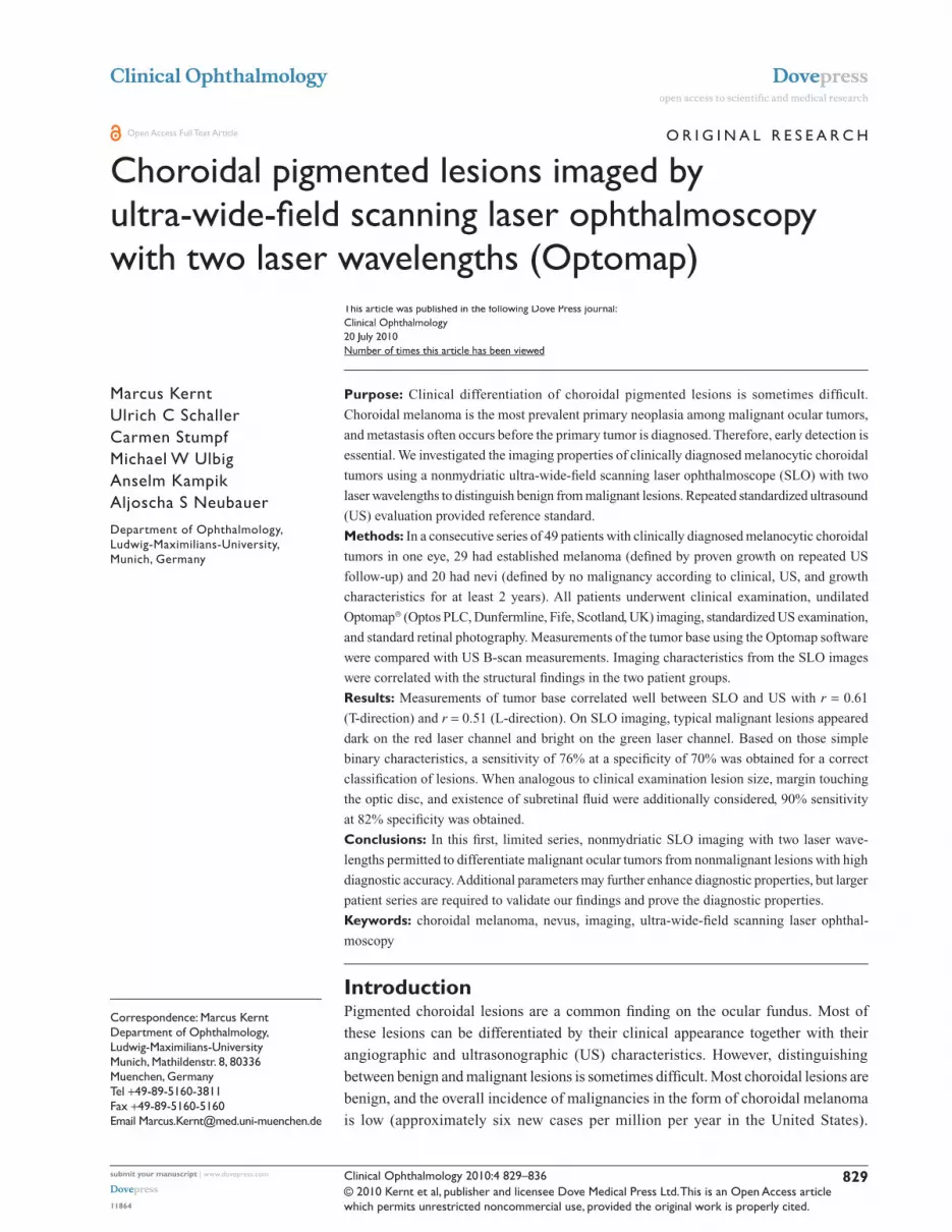
tumor base, combined with an evaluation of subretinal
fluid, assessment of the lesion locations on the ocular
fundus, and the specific imaging criteria from Optomap
led to a significant improvement of diagnostic accuracy
allowing differentiation of malignant from nonmalignant
ocular tumors in 90% of cases. This appears to exceed
the diagnostic accuracy of photographic fundus imaging,
although validation in an independent series is required.
Indeed, standardized A-scan US conducted by an experi-
enced operator might allow higher accuracy; however, with
less experience of the investigator, diagnostic accuracy
drops dramatically. Further, the technique itself might be
less tolerated by patients than a simple imaging procedure
and it is not widely available. In contrast, the Optomap
imaging system is noninvasive and easy to handle. It also
permits telescreening because data files from Optomap can
be easily transferred online and can be examined anywhere
in the world.
In summary, we showed that nonmydriatic SLO imaging
with two laser wavelengths allowed differentiating malignant
ocular tumors from nonmalignant lesions with relatively high
accuracy. Lesion size measurements correlated reasonably well
with US measurements. Additional parameters to describe and
differentiate lesions may further increase diagnostic properties,
and larger patient series are required to validate our findings.
AcknowledgmentsThe authors thank Stefanie Guthmann and Rita Hauser for
excellent technical assistance. Presented in part at the 9th
Euretina meeting 2009 in Nice, France and at the Interna-
tional society for imaging in the eye (ISIE) meeting 2010 in
Fort Lauderdale, USA.
Figure 4 A) Optomap composite image showing the typical appearance of a choroidal melanoma after radiation therapy (red and green channel superimposed). B) Detail of the according fundus image from Figure 4A, as viewed with the specific Optomap viewing software (composite image on the left, red and green separation on the right). The lesion appears bright in the red channel and bright in green channel. C) red separation image (treated melanoma). D) green separation image (treated melanoma).
A
C D
B
Clinical Ophthalmology
Publish your work in this journal
Submit your manuscript here: http://www.dovepress.com/clinical-ophthalmology-journal
Clinical Ophthalmology is an international, peer-reviewed journal covering all subspecialties within ophthalmology. Key topics include: Optometry; Visual science; Pharmacology and drug therapy in eye diseases; Basic Sciences; Primary and Secondary eye care; Patient Safety and Quality of Care Improvements. This journal is indexed on
PubMed Central and CAS, and is the official journal of The Society of Clinical Ophthalmology (SCO). The manuscript management system is completely online and includes a very quick and fair peer-review system, which is all easy to use. Visit http://www.dovepress.com/ testimonials.php to read real quotes from published authors.
Clinical Ophthalmology 2010:4submit your manuscript | www.dovepress.com
Dovepress
Dovepress
Dovepress
836
Kernt et al
DisclosureThe authors do not have any commercial or financial interest
in any of the materials and methods used in this study.
References 1. Margo CE. The Collaborative Ocular Melanoma Study: an overview.
Cancer Control. 2004;11(5):304–309. 2. Albert DM, Robinson NL, Fulton AB, et al. Epidemikological
investigation of increased incidence of choroidal melanoma in a single population of chemical workers. Int Ophthalmol Clin. 1980;20(2):71–92.
3. Balch CM, Murad TM, Soong SJ, Ingalls AL, Richards PC, Maddox WA. Tumor thickness as a guide to surgical management of clinical stage I melanoma patients. Cancer. 1979;43(3):883–888.
4. Ganley JP, Comstock GW. Benign nevi and malignant melanomas of the choroid. Am J Ophthalmol. 1973;76(1):19–25.
5. Rodriguez-Sains RS. Ocular findings in patients with dysplastic nevus syndrome. Ophthalmology. 1986;93(5):661–665.
6. Arnesen K, Nornes M. Malignant melanoma of the choroid as related to coexistent benign nevus. Acta Ophthalmol (Copenh). 1975;53(2): 139–152.
7. Sahel JA, Pesavento R, Frederick AR Jr, Albert DM. Melanoma arising de novo over a 16-month period. Arch Ophthalmol. 1988;106(3): 381–385.
8. Yanoff M, Zimmerman LE. Histogenesis of malignant melanomas of the uvea. II. Relationship of uveal nevi to malignant melanomas. Cancer. 1967;20(4):493–507.
9. McLean IW, Saraiva VS, Burnier MN Jr. Pathological and prognos-tic features of uveal melanomas. Can J Ophthalmol. 2004;39(4): 343–350.
10. Eskelin S, Pyrhonen S, Summanen P, Hahka-Kemppinen M, Kivela T. Tumor doubling times in metastatic malignant melanoma of the uvea: tumor progression before and after treatment. Ophthalmology. 2000;107(8):1443–1449.
11. Shields CL, Shields JA. Clinical features of small choroidal melanoma. Curr Opin Ophthalmol. 2002;13(3):135–141.
12. Shields CL, Shields JA, Kiratli H, De Potter P, Cater JR. Risk factors for growth and metastasis of small choroidal melanocytic lesions. Trans Am Ophthalmol Soc. 1995;93:259–275; discussion 275–279.
13. Gass JD. Problems in the differential diagnosis of choroidal nevi and malignant melanomas. The XXXIII Edward Jackson Memorial Lecture. Am J Ophthalmol. 1977;83(3):299–323.
14. Shields CL, Demirci H, Materin MA, Marr BP, Mashayekhi A, Shields JA. Clinical factors in the identification of small choroidal melanoma. Can J Ophthalmol. 2004;39(4):351–357.
15. Shields CL, Cater J, Shields JA, Singh AD, Santos MC, Carvalho C. Combination of clinical factors predictive of growth of small choroidal melanocytic tumors. Arch Ophthalmol. 2000;118(3):360–364.
16. Butler P, Char DH, Zarbin M, Kroll S. Natural history of indeterminate pigmented choroidal tumors. Ophthalmology. 1994;101(4):710–716; discussion 717.
17. Kernt M, Ulbig MW. Images in cardiovascular medicine. Wide-field scanning laser ophthalmoscope imaging and angiography of central retinal vein occlusion. Circulation. 2010;121(12):1459–1460.
18. Hodes BL, Choromokos E. Standardized a-scan echographic diagnosis of choroidal malignant melanomas. Arch Ophthalmol. 1977;95(4): 593–597.
19. Ossoinig KC. Standardized echography: basic principles, clinical applications, and results. Int Ophthalmol Clin. 1979;19(4):127–210.
20. Ossoinig KC, Bigar F, Kaefring SL. Malignant melanoma of the choroid and ciliary body. A differential diagnosis in clinical echography. Bibl Ophthalmol. 1975(83):141–154.
21. Kirkpatrick JN, Manivannan A, Gupta AK, Hipwell J, Forrester JV, Sharp PF. Fundus imaging in patients with cataract: role for a variable wavelength scanning laser ophthalmoscope. Br J Ophthalmol. 1995; 79(10):892–899.
22. Jonas JB, Gusek GC, Guggenmoos-Holzmann I, Naumann GO. Cor-relations of the neuroretinal rim area with ocular and general parameters in normal eyes. Ophthalmic Res. 1988;20(5):298–303.
23. Tamler E, Maumenee AE. A clinical study of choroidal nevi. AMA Arch Ophthalmol. 1959;62(2):196–202.
24. Naumann G, Yanoff M, Zimmerman LE. Histogenesis of malignant melanomas of the uvea. I. Histopathologic characteristics of nevi of the choroid and ciliary body. Arch Ophthalmol. 1966;76(6):784–796.
25. Augsburger JJ, Correa ZM, Trichopoulos N, Shaikh A. Size overlap between benign melanocytic choroidal nevi and choroidal malignant melanomas. Invest Ophthalmol Vis Sci. 2008;49(7):2823–2828.
26. Saari JM, Kivela T, Summanen P, Nummelin K, Saari KM. Digital imaging in differential diagnosis of small choroidal melanoma. Graefes Arch Clin Exp Ophthalmol. 2006;244(12):1581–1590.