chloride channels: an emerging molecular picture
TRANSCRIPT

Thomas J. Jentsch and Willy Gunther
Summary
Chloride channels are probably found in every cell, from bacteria to mammals. Their physiological tasks range from cell volume regulation to stabilization of the membrane potential, signal transduction, transepithelial transport and acidification of intracellular organelles. These different functions require the presence of many distinct chloride channels, which are differentially expressed and regulated by various stimuli. These include various intracellular messengers (like calcium and cyclic AMP), pH, extracellular ligands and transmembrane voltage. Three major structural classes of chloride channels are known to date, but there may be others not yet identified. After an overview of the general functions of chloride channels, this review will focus on these cloned chloride channels: the CLC chloride channel family, which includes voltage-gated chloride channels, and the cystic fibrosis transmembrane regulator (CFTR), which performs other functions in addition to being a chloride channel. Finally, a short section deals with GABA and glycine receptors. Diseases resulting from chloride channel defects will be specially emphasized, together with the somewhat limited Accepted information about how these proteins work at the molecular level. 30 October 1996
Functions of chloride channels Chloride is the most abundant anion in plant and animal tissues. Therefore, anion channels are often called CI- channels, even though they may be permeable to other anions as well. Various transporters and channels transport chloride across membranes, both at the cell surface and in intracellular organelles. In contrast to ions like sodium and especially calcium, the electrochemical gradient of chloride across the animal plasma membrane is generally not far from equilibrium. Thus, at the resting potential of cells, opening of CI- channels, which provide pathways for the passive diffusion of chloride, will not lead to large excursions of the plasma membrane voltage or dramatic changes in intracellular chloride concentrations. Thus, although effects of chloride on enzymatic activities have been described(’), and although the intracellular chloride concentration cou- ples different CI- transporters in opposing membranes of epithelia, the chloride ion has no established role as an intracellular second messenger.
In this sense, chloride resembles potassium, which is also predominantly passively distributed across the plasma membrane. Thus, similar to K+ channels, opening of CI- channels generally stabilizes the membrane potential. This is best known for the ligand-gated postsynaptic GABA- and
glycine-receptor CI- channel^(^,^). Opening of these chan- nels is mostly inhibitory, since the resulting stabilization of the voltage counteracts the depolarization caused by excita- tory neurotransmitters. As shown r e ~ e n t l y ( ~ < ~ ) , genetic defects in glycine receptor subunits lead to a hyperexcitabil- ity syndrome, startle disease (or hyperekplexia). The volt- age-gated CI- channel of skeletal muscle, CIC-l@), provides another glaring example. Like many potassium channels, it is partially open under resting conditions, and significantly contributes to the repolarization of action potentials in muscle. Its loss of function leads to an inherited muscular hyperexcitability syndrome, myot0nia(~88).
While intracellular chloride concentration is close to equi- librium, it can deviate from it by a few tens of mV. While there is no firm evidence for an active chloride-transporting ATPase in mammalian plasma membranes, several trans- porters (Fig. l a ) use the energy stored in transmembrane gradients of other ions to transport chloride against its elec- trochemical gradient (‘secondary active transport’). Most of these transporters will accumulate intracellular chloride above its equilibrium level (at a membrane voltage of -60 mV and an external chloride concentration of 150 mM, inter- nal chloride would be 15 mM at equilibrium). Coupling to the sodium gradient (generated by the Na,K-ATPase) leads to
a
* HCOY
HCO;
CI- HF+Na+
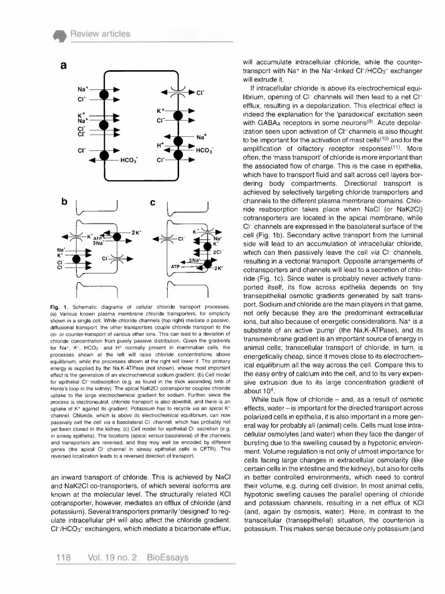
Fig. 1. Schematic diagrams of cellular chloride transport processes. (a) Various known plasma membrane chloride transporters, for simplicity shown in a single cell. While chloride channels (top right) mediate a passive, diffusional transport, the other transporters couple chloride transport to the co- or counter-transport of various other ions. This can lead to a deviation of chloride concentration from purely passive distribution. Given the gradients for Na+, K+, HCO3 and H+ normally present in mammalian cells, the processes shown at the left will raise chloride concentrations above equilibrium, while the processes shown at the right will lower it. The primary energy is supplied by the Na,K-ATPase (not shown), whose most important effect is the generation of an electrochemical sodium gradient. (b) Cell mode for epithelial CI- reabsorption (e.g as found in the thick ascending limb of Henle's loop in the kidney). The apical NaK2CI cotransporter couples chloride uptake to the large electrochemical gradient for sodium. Further, since the process is electroneutral, chloride transport is also downhill, and there IS an uptake of K+ against its gradient. Potassium has to recycle via an apical K+ channel. Chloride, which is above its electrochemical equilibrium, can now passively exit the cell via a basolateral CI- channel, which has probably not yet been cloned in the kidney. (c) Cell model for epithelial CI- secretion (e.g. in airway epithelia). The locations (apical versus basolateral) of the channels and transporters are reversed, and they may well be encoded by different genes (the apical CI- channel in airway epithelial cells is CFTR). This reversed localization leads to a reversed direction of transport.
an inward transport of chloride. This is achieved by NaCl and NaK2CI co-transporters, of which several isoforms are known at the molecular level. The structurally related KCI cotransporter, however, mediates an efflux of chloride (and potassium). Several transporters primarily 'designed' to reg- ulate intracellular pH will also affect the chloride gradient. CI-/HC03- exchangers, which mediate a bicarbonate efflux,
will accumulate intracellular chloride, while the counter- transport with Na+ in the Na+-linked CI-/HC03- exchanger will extrude it.
If intracellular chloride is above its electrochemical equi- librium, opening of CI- channels will then lead to a net CI- efflux, resulting in a depolarization. This electrical effect is indeed the explanation for the 'paradoxical' excitation seen with GABAA receptors in some neurondg). Acute depolar- ization seen upon activation of CI-channels is also thought to be important for the activation of mast cells(10) and for the amplification of olfactory receptor responses(' ). More often, the 'mass transport' of chloride is more important than the associated flow of charge. This is the case in epithelia, which have to transport fluid and salt across cell layers bor- dering body compartments. Directional transport is achieved by selectively targeting chloride transporters and channels to the different plasma membrane domains. Chlo- ride reabsorption takes place when NaCl (or NaK2CI) cotransporters are located in the apical membrane, while CI- channels are expressed in the basolateral surface of the cell (Fig. 1 b). Secondary active transport from the luminal side will lead to an accumulation of intracellular chloride, which can then passively leave the cell via CI- channels, resulting in a vectorial transport. Opposite arrangements of cotransporters and channels will lead to a secretion of chlo- ride (Fig. l c ) . Since water is probably never actively trans- ported itself, its flow across epithelia depends on tiny transepithelial osmotic gradients generated by salt trans- port. Sodium and chloride are the main players in that game, not only because they are the predominant extracellular ions, but also because of energetic considerations. Na+ is a substrate of an active 'pump' (the Na,K-ATPase), and its transmembrane gradient is an important source of energy in animal cells; transcellular transport of chloride, in turn, is energetically cheap, since it moves close to its electrochem- ical equilibrium all the way across the cell. Compare this to the easy entry of calcium into the cell, and to its very expen- sive extrusion due to its large concentration gradient of about 1 04.
While bulk flow of chloride - and, as a result of osmotic effects, water - is important for the directed transport across polarized cells in epithelia, it is also important in a more gen- eral way for probably all (animal) cells. Cells must lose intra- cellular osmolytes (and water) when they face the danger of bursting due to the swelling caused by a hypotonic environ- ment. Volume regulation is not only of utmost importance for cells facing large changes in extracellular osmolarity (like certain cells in the intestine and the kidney), but also for cells in better controlled environments, which need to control their volume, e.g. during cell division. In most animal cells, hypotonic swelling causes the parallel opening of chloride and potassium channels, resulting in a net efflux of KCI (and, again by osmosis, water). Here, in contrast to the transcellular (transepithelial) situation, the counterion is potassium. This makes sense because only potassium (and
not sodium) can passively leave the cell, and also because potassium is the main intracellular cation. In many cells, however, it is not only potassium and chloride that are lost in regulatory volume decrease; a loss of certain intracellular amino acids (like taurine) may be of at least equal impor- tance, and these may also leave the cell by (less selective) anion channels (for a review, see ref. 12).
In plant leaves, guard cells control the opening of the stomata1 pore, which limits gas exchange with the atmos- phere. The pore is opened by the swelling of these cells, which take up potassium salts as osmolytes. Closure of the pores is due to an efflux of potassium and chloride or malate, the latter anions passing through Cl-channels(l3). In this special case, the regulation does not serve to keep the cell volume constant, but intentionally to change it.
Finally, CI- channels are also present in intracellular organelles such as the endoplasmatic reticulum, the Golgi, endosomes, lysosomes and synaptic vesicles(14). The inter- nal pH of these organelles is more acidic than the cytosol. This is important in, for example, the control of intravesicular enzymatic activity and the dissociation of ligands from their receptors after endocytosis. This is accomplished by an electrogenic H+-ATPase, which uses ATP hydrolysis to pump protons into the lumen. The concomitant build-up of a transmembranal voltage would limit the pH gradient as it would raise the energy required to pump H+. Here CI-chan- nels come into play: their presumptive role is to provide an electrical shunt, allowing for a larger pH gradient to be reached. Since disturbance of vesicular pH (e.g. by ammo- nium or monensin) is known to affect intracellular vesicle trafficking, defects in intracellular CI- channels could have similar consequences.
Classification of chloride channels As shown above, chloride channels perform various func- tions, even within a single cell. This implies that different CI- channels have to be regulated by specific stimuli, and that they have to be expressed differentially in different organs and cells, and in different domains within a given cell. Although their ultimate function is similar (providing a path- way for the passive diffusion of anions), this diversity in func- tion could most easily be achieved by the evolution of many distinct CI- channels (encoded by different genes). This is now being confirmed by molecular cloning, but a bewilder- ing variety of different CI- channels had already been demonstrated by electrophysiological studies. These differ in their biophysical properties (e.g. conductance, ion selec- tivity, voltage-dependence), their mode of regulation (e.g. by ligands, calcium, G-proteins etc.) and the tissues in which they have been found. Unfortunately, the picture emerging from physiological studies is sometimes confusing, since measurements are often performed under differing con- ditions, or using different sets of parameters. Moreover, single channel measurements (using the patch-clamp tech-
nique) will bias the results towards channels with a large (and at least detectable) unitary conductance.
CI- channels can be roughly classified according to their mechanism of activation. Thus it is possible to distinguish CI- channels activated by extracellular ligands, intracellular calcium, cyclic AMP, G-proteins, transmembrane voltage, mechanical stretch and cell swelling. This classification has its problems, though, as mechanisms of activation may overlap (e.g. swelling-activated channels may also be volt- age-dependent). Further, it may not reflect at all the struc- tural similarities of the corresponding proteins.
Without doubt, the final classification will instead be based on structural classes. Although molecular cloning has yielded considerable information about CI-channels over the past 9 years, our molecular picture is still far from being com- plete. Entire 'functional' classes have probably not yet been cloned, e.g. CI-channels activated by intracellular calcium.
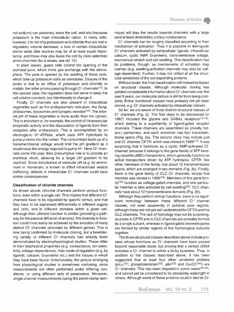
So far, we are aware of three distinct structural classes of CI- channels (Fig. 2). The first class to be discovered (in 1987) included the glycine and GABAA receptor^('^.'^), which belong to a superfamily of ligand-gated receptor channels. These channels are assembled as (mostly het- ero-) pentamers, and each monomer has four transmem- brane spans (Fig. 2a). The second class may include just one Cl-channel, CFTR, which was cloned in 1 989(17). It was surprising that it functions as a cyclic AMP-activated CI- channel, because it belongs to the gene family of ATP-bind- ing cassette (ABC) transporters, which generally function as active transporters driven by ATP hydrolysis. CFTR, like other members of the family, has about 12 transmembrane spans, which are arranged in two domains (Fig. 2c). Finally, there is the gene family of CLC CI- channels, whose first member was cloned in 1 990(i8). Members of this gene fam- ily(19) function as voltage-gated channels, and one particu- lar member is also activated by cell swelling(20). CLC chan- nels have about 12 transmembrane domains (Fig. 2b).
Although they perform similar functions, there is no signif- icant homology between these different CI- channel classes, not even apparently in putative pore regions, although these are not yet well understood for CFTR and the CLC channels. The lack of homology may not be surprising, as pores in CFTR and in CLC channels are probably formed by a single subunit, whereas in ligand-gated channels pores are formed by similar regions of five homologous subunits together.
The three structural classes described above include pro- teins whose functions as CI- channels have been proved beyond reasonable doubt, but proving that a certain cDNA encodes a CI- channel is rather a tricky business. Thus, in addition to the classes described above, it has been suggested that at least four other unrelated proteins ( p l c ~ ~ ( ~ l ) , phospholemman(22), p64(23) and Ca-CC(")) are CI- channels. This has been disputed in some ~ a s e s ( ~ ~ , ~ ~ ) , and cannot yet be considered to be absolutely watertight in others. Although some of these proteins could in fact be CI-

n n n n n n
Fig. 2. Transmembrane topology models of cloned chloride channels. (a) Ligand-gated CI- channels (GABA and glycine receptors) These postsynaptic receptor channels function as (mostly hetero-) pentamers of homologous subunits, each of which has four transmembrane domains. There is a bewildering variety of different, homologous subunits. Thus, for GABAA receptors, six different a-subunits, four P-subunits, four y-subunits, one &subunit and two p-subunits are known, which are in part tissue-specific and can associate only in certain combinations. (b) CLC-channels. Two regions of intermediate hydrophobicity (D4 and D13), which were previously hypothesized to span the membrane, are no longer supposed to do so. The region D9 through D12 is a broad hydrophobic segment, which has to span the lipid bilayer three or five times. More work is necessary to determine the exact topology of CLC channels. CLC channels function as homomultimers (probably dimers), with at least two pores in the case of the Torpedo channel CIC-0. (c) The cystic fibrosis transmembrane regulator (CFTR). As typical for other members of the ATP-binding cassette gene family, CFTR has two blocks of several transmembrane domains and two nucleotide binding folds. A special feature of CFTR is the non-conserved 'R-domain', which confers regulation by cyclic AMP-dependent phosphorylation. CFTR probably functions as a monomer.
channels or channel regulators, they will, for the sake of brevity, not be dealt with here.
Firstly, we will discuss the CLC CI- channels in some detail, since these comprise the only known large gene fam- ily of CI- channels that is evolutionarily conserved. The dis- cussion will proceed with CFTR, followed by a brief account of ligand-gated CI-channels.
The CLC family of chloride channels Since the initial cloning('*) of the Torpedo electric organ CI- channel CIC-0, it has become clear that it was the first mem- ber to be discovered of a large gene family. There are now nine different CLC genes known in mammal^('^!^^) and the number is likely to increase. Moreover, CLC genes are highly conserved during evolution (Fig. 3). There is a CLC gene in E. coliand, since the completion of the S. cerevisiae genome project, it is now clear that yeast has just a single CLC gene(28). In a single plant species (Arabidopsis thaliana), however, there are at least four different CLC genes(29). This shows that CLC genes evolved very early
during evolution, suggesting that their function is fundamen- tally important, even in prokaryotes and monocellular eukaryotes. Whether they function as anion channels in these organisms, however, is not yet known.
Several animal CLC proteins, in contrast, have been proved unambiguously to have a function as a CI-channel, and studies over the past few years have already yielded considerable insights into their structure, function and phys- iological roles.
CIC-0, the voltage-gated chloride channel from Torpedo electric organ The ray Torpedo has strong electric organs to stun its prey. The voltage pulses are generated by the concerted action of nicotinic acetylcholine receptors and CI- channels located on opposite sides of the electrocytes, modified polarized muscle cells arranged in columnar stacks. Binding of acetyl- choline to its receptors depolarizes the innervated cell mem- brane to about +10 mV. The opposite membrane voltage is kept at its resting value of about -90 mV by CI- channels, resulting in a transcellular voltage of approx. 100 mV upon

excitation. Since the electrocytes are arranged like batteries in series, this adds up to some 100 V over the electric organ.
The Torpedo CI- channel has been extensively studied after reconstitution into lipid bilayers. Exploiting its abun- dance, the cDNA for this channel, subsequently named CIC- 0, was isolated by expression cloning(ls). It encodes a pro- tein of about 90 kDa and is predicted to have roughly 12 transmembrane spans. It is the ‘founding member’ of the CLC gene family, and is probably the most thoroughly stud- ied voltage-gated CI-channel. As with other CLC channels, it is highly selective for chloride (ClzBrzl conductivity sequence).
CIC-0 has a peculiar ‘double-barrelled’ structure: it has two identical pores which close and open independently, but which can also be closed together by another common gate. Definitive evidence for this model comes from two recent s t ~ d i e s ( ~ ~ 5 ~ ~ ) using the co-expression of wild-type (WT) CIC- 0 with different CIC-0 mutants, which change its single channel conductance (normally approx. 9 pS) and ion selec- tivity. This resulted in double-barrelled channels, with differ- ent ‘sizes’ (conductances) of the two individual ‘barrels’ (pores) that were shown to be largely independent. More- over, these studies strongly suggest that the channel is a homodimer, which is compatible with one pore being formed by a single subunit, and is consistent with a biochemical analysis of the protein(32).
If a single CIC-0 protein forms a pore, this implies that several different regions of the protein should be in contact with the permeating anion. Indeed, mutations in two differ- ent regions (after the putative transmembrane domains D2 and D12) change ion selectivity, single channel conduc- tance and r e c t i f i c a t i ~ n ( ~ ~ , ~ ~ ) . This is in contrast to GABA and glycine receptors (and to potassium channels), in which similar segments of the several homologous subunits form- ing the channel line the common, single pore.
In the well-studied cation channel superfamily, voltage- dependent gating is ‘intrinsic’. Charged residues in a trans- membrane domain (the S4 segment) sense the voltage drop over the membrane and lead to a conformational change (gating) of the channel. In CIC-0, however, fast gat- ing was proposed to be extrinsic(33): gating depends strongly on the extracellular anion concentration, and the channel may be gated by the permeant anion. In this model, the presence of the anion at the cytoplasmic end of the pore facilitates channel opening. Since the anion travels along the electric field when it permeates the pore to reach its binding site, gating becomes (extrinsically) voltage- and c~ncentration-dependent(~~). A modified version of this model, in which chloride binding itself is voltage-indepen- dent, was proposed recently(34).
CIC- 1, fhe major skeletal muscle CP channel Homology screening with CIC-0 identified CIC-1, which is specifically expressed in skeletal muscle(6). Its electrophysi- ological and pharmacological properties agree with the
EXPRESSION PROPOSED PATTERN FUNCTION
stabilization of membrane potential
skeletal loss of function: I-’ C1C-1 muscle mvotonia cell volume
limiting intracellular CI concentration
c1c-2 ubiquitous regulation
transepithelial transport ?? C1C-Ka kidney
broad e g. c1c-3 brain,’kidney
transepithelial transport ??
?
4 main,y kidney transepithelial c1c-5 (brain, liver) t r ~ p ~ ~ ~ ~ ~ c t ~ o n . ,
kidney stones
cic-6 ubiquitous ?
clc-7 ubiquitous ?
Fig. 3. The CLC family of chloride channels in mammals. The dendrogram indicates the degree of similarity between the different gene products. It shows three different branches with less than 30% identity between them: the first one includes CIC-I, CIC-2 and the CIC-K channels. CIC-0, the Torpedo channel, also belongs to that branch. The second branch includes CIC-3 through CIC-5, and it is this part of the gene family which is most closely related to the yeast scCIC. The third branch includes CIC-6 and CIC-7; the four channels from Arabidopsis fhaliana are most closely related to this branch. Tissue distribution and suggested functions are indicated.
mascroscopic chloride conductance of that tissue. Its low single channel conductance of approx. 1 pS unfortunately precludes single channel analysis; thus, it is unknown whether it forms a ‘double-barrelled’ channel, similar to CIC- 0, but resembling CIC-0, it has a CI>Br>I selectivity sequence and opens with depolarization.
In skeletal muscle, chloride accounts for approx. 75% of the ionic conductance of the resting sarcoplasmic mem- brane. Whereas neuronal action potentials are repolarized by potassium channels (and the inactivation of sodium channels), chloride conductance plays a major role in skele- tal muscle. When muscle CI-channels are blocked by drugs in vifro, the membrane voltage is destabilized and a single electrical stimulus leads to repetitive action potentials. In vivo this would result in a defective muscle relaxation after voluntary contraction. Such a muscle ‘stiffness’ (myotonia) is a symptom in several inherited diseases. Indeed, a decrease in muscle chloride conductance was observed both in myotonic patients and in animal models.
This made CIC-1 into an attractive candidate gene, and it was quickly shown that CIC-1 is indeed mutated in the myotonic mouse strain ADFd7). In humans, purely myotonic diseases are either autosomal recessive (Becker myotonia or Recessive Generalized Myotonia) or autosomal dominant (Thomsen’s disease or dominant Myotonia Congenita). Both

forms were shown to be due to CIC-I mutation^(^,^^-^^). This raised the interesting question of why some mutations result in a recessive and others in a dominant phenotype.
Since a total loss of function of one CIC-1 allele leads to recessive disease, dominant myotonia cannot be due to haploinsufficiency. Also in vitro studies demonstrated that a 50% block of CI- conductance is not sufficient to cause myotonia. Dominant myotonia therefore requires that the mutant CIC-1 gene product compromises the function of its WT counterpart, reducing CI- conductance below approx. 30% of control values(36). Such a dominant negative effect suggests a (homo)multimeric channel, as is also indicated by the double-barrelled structure of CIC-O(30,31). Incorpora- tion of mutant subunits into the channel complex should reduce its channel activity under physiological conditions.
A detailed analysis of the dominant negative effect in the Xenopus oocyte expression system suggested that CIC-1 has more than two subunits(36). Even though this is at odds with the likely homodimeric structure of CIC-O(30-32), CIC-1 may form higher aggregates. Most dominant mutations found in human myotonia, even though affecting very differ- ent residues, act by a similar mechanism: they shift the volt- age-dependent opening of CIC-1 to very positive potentials, where they can no longer repolarize action potentials(37).
CIC-1 is already significantly open under resting con- ditions. As its gating is slow compared to an action potential, this short depolarization will only slightly increase its open probability. What, then, might be the physiological ‘purpose’ of its voltage-dependence? The incremental activation dur- ing a prolonged series of action potentials will lead to a decrease in the electrical excitability, which could be impor- tant in limiting tetanic excitation(3’).
CIC-2, a ubiquitous C t channel activated by cell swelling and hyperpolariza tion Northern analysis showed that CIC-2 is nearly ubiquitously expressed(38). When examined in more detail by in situ hybridization, however, it became clear that it is differentially expressed in brain neurons(g). lmmunocytochemistry shows CIC-2 expression in apical membranes of lung epithelia(39), which also express CFTR.
When expressed in Xenopus oocytes, CIC-2 is closed under resting conditions. It can be slowly activated by hyper- polarization in excess of approx. -80 mV and has the CIA selectivity typical of CLC channel~(3~<~0). This easily distin- guishes the current from a superficially similar endogenous current (with an I>CI selectivity) found in some batches of Xenopus oo~ytes(~ l ) . CIC-2 may serve to prevent intracellular CI- accumulation in specific neurondg), thereby modulating the effects of postsynaptic GABAA receptors. CIC-2 expression is absent or low in neurons exhibiting a ‘paradoxi- cal’ excitation by GABA, attributable to a CI- efflux via GABAA receptors in cells with high intracellular chloride levels. Indeed, transfection of CIC-2 into dorsal root ganglia trans- forms the GABAresponse from excitatory to inhibitory(42).
Thus, CIC-2 may be important for setting the intracellular chloride concentration and regulating neuronal excitability.
Still more fascinating is its activation by cell swelling. When Xenopus oocytes expressing CIC-2 are exposed to hypotonic medium (osmolality reduced by 50%), chloride currents increase drastically after 10-1 5 minutes(2o). The channel is also now open within a more physiological volt- age range (-60-0 mV). This effect is reversible upon return- ing to isotonicity. This suggests a role in cell volume regula- tion, but CIC-2 currents differ from the swelling-induced current observed in many cells. The latter current is induced faster, and has a different kinetic and ion selectivity. This indicates that at least two different CI- channels contribute to cell volume regulation in mammalian cells(12).
CIC-2 can also be activated by acidic extracellular pH@O). This may be important (e.g. in the gastric mucosa), or could counteract cell swelling in pathological situations (e.g. hypoxia), leading to local acidosis.
The activation of CIC-2 by these stimuli is dependent on a small segment of the amino terminus, deletions of which lead to constitutively open channels that are no longer sen- sitive to swelling(20). When this domain was transplanted to the carboxy terminus it retained its function. This suggested a position-independent mechanism according to the ball- and-chain model, in which an amino-terminal ‘ball’ domain binds to a putative receptor on the channel and prevents its opening. Cell swelling and hyperpolarization are thought to reduce the affinity of the receptor, leading to a dissociation of the ‘ball’ and to channel opening. A potentially interesting CIC-2 variant in which this ‘ball’ domain is replaced by a new sequence(43), however, is probably an artifact, as this sequence may be a concatamer of linkers (T.J.J., unpub- lished observation).
CIC-5, a Ct channel whose defect results in kidney stones CIC-5(44s45) belongs to a branch of the CLC family that also includes CIC-3(46) and CIC-4(47). While CIC-4 cannot be expressed functionally and expression of CIC-3 is contro- versial(l 9 3 4 6 , 4 7 ) , CIC-5 expression yields chloride currents with the typical CbI conductivity ~ e q u e n c e ( ~ ~ 1 ~ ~ ) . Its voltage- dependence in Xenopus oocytes is enigmatic: currents are only detectable at voltages more positive than +20mV, which are unlikely to be reached in vivo.
While CIC-3 and CIC-4 are rather broadly expressed, CIC-5 is predominantly, though not exclusively, expressed in k i d n e ~ ( ~ ~ , ~ ~ ) , in large portions of the n e p h r ~ n ( ~ ~ ) . Surpris- ingly, loss of function mutations in human CIC-5 lead to pro- teinuria, hypercalciuria and kidney stones(48). How muta- tions in a CI-channel can lead to these symptoms is unclear at present.
Additional mammalian CLC genes The physiological functions of several other mammalian CLC genes are still totally unknown. This includes CIC-3 and CIC-4, which were mentioned above, but also the

kidney-specific CIC-K genes (CIC-Ka and CIC-Kb in human^)(^^%^^) and the ubiquitously expressed CIC-6 and CIC-7 genes(27). The problem is compounded by the fact that they cannot be functionally expressed as CI- channels, or that their expression is c o n t r o v e r ~ i a l ( ~ ~ ~ ~ ~ ) .
Several explanations may be invoked to explain a lack of expression as a plasma membrane CI- channel. First, some CLC genes could encode intracellular Cl-channels. However, immunofluorescence is compatible with a plasma membrane localization of the CIC-K proteins(51). Second, there may be a need for additional subunits which have not yet been identi- fied. Further, we may not understand the appropriate stimulus for channel activation, and - a bit more farfetched - these gene products may not even be CI- channels after all. Much more work is necessary to find out what these putative chan- nels are good for. Localizing these proteins within the cells by immunocytochemistry will be an important first step.
scCLC (GEFl), the single yeast CLC In a search for iron-suppressible petite (respiration-deficient) yeast mutants, Greene ef identified a yeast CLC gene they named GEF1. The completion of the yeast genome pro- ject has shown that there is just this single CLC in yeast. It is currently unclear how the lack of GEF1 (renamed scClC to conform to the CLC nomenclature) leads to the inability of yeast cells to grow on glyceroVethanol carbon sources in the absence of high iron concentrations. A hint comes from GEM, whose knock-out leads to the same phenotype. GEF2 encodes a subunit of the vacuolar H+-ATPase. SCCLC may provide the electrical shunt for the efficient acidification of the vacuole, an organelle that is important for iron-storage in yeast. Although attractive this hypothesis is unproved, how- ever, and first data(28,29) rather argue against it. While all mam- malian CLC genes tested so far cannot functionally substitute for scClC in yeast cells (B. Schwappach and T.J. Jentsch, unpublished), a particular plant CLC (AtCIC-d) restored iron- limited growth when transformed into the knock-out mutant(29).
CFTR - a bifunctional transmembrane protein Cystic fibrosis (CF) is the most frequent monogenetic fatal genetic disease in Caucasians. Its symptoms are mainly due to defects in transepithelial transport, leading to an accumula- tion of mucus in pancreatic ductules and in the airways. Com- bined with secondary bacterial infections, this causes the life- threatening loss of lung function. Physiological studies indicated that the underlying molecular defect may be in a cyclic AMP-regulated CI-channel of airway epithelial cells.
The gene defective in CF was finally identified by a painstaking positional cloning approach(17). Surprisingly, its structure did not ‘look’ like that of a typical ion channel. Rather, CFTR (for Cystic Fibrosis Transmembrane Conduc- tance Regulator, as it was named) belongs to a superfamily of transport ATPases, the ABC (ATP-binding cassette) trans- porters. It has 12 putative transmembrane spans arranged in
two separate blocks, and two nucleotide binding folds (NBF 1 and 2). Not found in other ABC transporters is the R-domain, which is located between NBFl and the second transmem- brane block of CFTR (Fig. 2c). It has several consensus sites for phosphorylation by protein kinases A and C and is thought to regulate the function of CFTR (hence its name: R for regulatory). Since the initial cloning in 1989, dramatic progress has been made in understanding its function, cov- ered in excellent reviews (see, for example, ref. 52).
CFTR - a cyclic AMP-regulated chloride channel Although the structure of CFTR fuelled speculation that it may not be an ion channel itself, but just a regulator of another, physically distinct channel, it is now unambiguously established that CFTR is a cyclic AMP-activated CI- chan- nel. Several mutations were shown to alter pore properties of CFTR(53a54), and the purified CFTR protein retained its typical electrophysiological characteristics when reconsti- tuted into lipid b i l a y e r ~ ( ~ ~ ) .
CFTR is a small (5-8 pS) anion channel which is highly selective for chloride (with a BraCI>I selectivity sequence). Opening of the CFTR CI- channel requires phosphorylation (at the R-domain) by protein kinase A, making it susceptible to regulation by cyclic AMP. In addition, it requires Mg2+ and hydrolyzable ATP. In contrast to conventional transport ATPases, where ATP hydrolysis is required for every single transport cycle, the passive pore of CFTR is thought to be regulated by ATP hydrolysis. Elegant experiment^(^^,^^) suggest that ATP hydrolysis at NBFl is necessary for the channel to open. Binding of ATP at the second NBF is thought to stabilize the open state, and its hydrolysis at NBF2 leads to channel closure. Thus, opening and closing of CFTR also depends on the metabolic state of the cell, possibly allowing for an additional mechanism of control.
While the predominant mutation found in CF patients, AF508, leads to a temperature-dependent processing and trafficking defect(58), many other mutations have been found in different regions of the protein. Several mutations in trans- membrane domains reduce the single-channel conduc- tance of CFTR(54), and others in cytoplasmic loops reduce the open probability(59). This does not lead to a total loss of CI- channel function, and accordingly these mutations may be associated with a relatively mild phenotype in patients. These mutations, as well as those introduced intentionally in structure-function studies, indicate that several CFTR trans- membrane spans (at least TM1 and TM6) participate in pore formation. Since sedimentation data suggest that CFTR functions as a monomer(60), this is not surprising and is analogous to the situation discussed above for CLC pores. Consistent with CFTR being a monomer, no dominant nega- tive CFTR mutation has been described, and cystic fibrosis is always recessive. This is in contrast to myotonia, where the multimeric structure of CIC-1 (despite having only one subunit per pore(30~3~)) allows for dominant myotonia (Thomsen’s disease)(36).

Surprisingly, the first half of the CFTR molecule seems to be sufficient to form a chloride channel(60), although not retaining all properties of the intact protein. These truncated channels probably associate to dimers. Interestingly, a splice variant detected in kidney, leading to a truncation of the protein at the end of the first NBD, similarly led to CFTR-like currents(61).
CFTR - a regulator of other channels The additional role of CFTR as an ion-channel regulator has been revived recently by several reports. Patients with cystic fibrosis show an increased sodium reabsorption across lung epithelia, suggesting that CFTR somehow regulates epithe- lial sodium channels. Co-expression of CFTR with all three subunits of the epithelial sodium ~ h a n n e I ( ~ ~ 3 ~ ~ ) indeed demonstrates that CFTR inhibits this sodium channel in a cyclic AMP-dependent manner.
Certain outwardly rectifying CI- channels (distinct from CFTR, and probably also from CLC channels) can be acti- vated by protein kinase A (PKA). This regulation is absent in epithelia lacking functional CFTR, and can be restored by transfecting wild-type CFTR(64). It has been suggested that CFTR can also act as a channel for ATP, and that ATP leaving the cell via CFTR acts on purinergic receptors nearby. These, in turn, are thought to activate outwardly rectifying CI- channels via a G-protein-dependent pathway(65). However, several groups (e.g. see ref. 66) were unable to find that CFTR can conduct ATP. Other data suggest that CFTR enhances the sensitivity of a renal potassium channel (ROMK2) to gl iben~lamide(~~). More work is needed to inves- tigate these effects and to clarify the underlying mechanism.
Do other ABC transporters regulate chloride channels? The notion that ABC transporters can regulate ion channels has been significantly bolstered by the finding that the sul- fonylurea receptor (SUR), another member of the ABC transporter gene family, reconstitutes the ATP-sensitive potassium channel when coexpressed with the inward recti- fier gene Kir6.2. Importantly, mutations in SUR cause hyper- insulinemic hypoglycemia of infancy, as would be expected from a defect in the ATP-sensitive potassium channel which plays a key role in regulating insulin secretion.
Thus, it seems worthwhile investigating whether other ABC gene family members regulate ion channels, including CI- channels. In fact, it had already been suggested several years ago that the multidrug resistance protein mdrl , an active trans- porter for certain classes of hydrophobic substances, is either itself a swelling-activated CI-channel, or regulates such chan- nels(68). After much controversy (see e.g. ref. 69), it now seems clear that mdrl is not a CI-channel. Instead, it was pro- posed to confer PKC-sensitivity on endogenous swelling-acti- vated CI- channels(’O), which have not yet been cloned. Still another ABC-transporter (dubbed EBCR) was recently suggested to regulate CI- channels(71). When expressed in some batches of Xenopus oocytes, elevation of intracellular cyclic AMP elicited long-lasting chloride currents. This
response was highly variable, however, and EBCR is proba- bly the rabbit homologue of a bile-acid transporter whose knock-out leads to congenital jaundice(72). More work is needed to confirm that EBCR regulates ion channels.
GABAA and glycine receptors - two gene families of ligand- gated chloride channels The postsynaptic chloride channels known as GABAA and glycine receptors are oligomers composed of several identi- cal or homologous subunits (for recent reviews, see refs 2, 3 and 73). These belong to either one of these distinct, but related gene families, and in turn form a gene superfamily with the nicotinic acetylcholine receptor subunits. Just as the acetylcholine receptors (which form cation channels) they are believed to function as (mostly heteromeric) pentameric com- plexes. The single subunits, just like those of the acetylcholine receptor, probably have four transmembrane-spanning domains and typically have molecular masses in the 50-60 kDa range (Fig. 2a). The large amino terminus, which is pre- ceded by a cleavable signal peptide, faces the extracellular space. By analogy to acetylcholine receptors, the pore is probably lined by the second transmembrane domain M2. This segment is reasonably conserved between GABA and glycine receptors, and even shows significant homology to the corresponding region of the nicotinic acetylcholine recep- tor. By contrast, M2 segments of GABA or glycine receptors do not show any significant homology to putative pore regions of CLC channels or CFTR. This is not very surprising, as five homologous (or identical) M2 regions contribute to the pore of these ligand-gated channels, while one pore is formed by a single (non-repetitive) subunit in CLC and CFTR channels.
There are four different a-subunits and one P-subunit known for the glycine receptor, and the situation is even more com- plex for GABAA receptor channels: six a-isoforms, three each of p and y subunits, and just one 6 subunit. This diversity is increased further by alternative splicing of some subunits. In addition, there are two GABA p subunits, which are probably the molecular basis for the GABAc response in the retina(74), with its different pharmacological properties. In addition to these channel-forming subunits, there will be associated sub- units that will anchor these channels in the right place. This has been shown convincingly for glycine receptors, where gephyrin, a 93 kDa tubulin-binding peripheral membrane pro- tein, induces clustering of the receptors(75).
Both GABA- and glycine-gated anion channels have mul- tiple conductance levels in the 10-90 pS range (which also depend on the particular subunit composition), and have an I>Br>CI halide selectivity sequence(76). In addition, both channel types are significantly permeable to bicarbonate, and the resulting intracellular acidification may be important for modulating synaptic activity. As discussed above, both receptors function generally in neuronal inhibition. Excep- tions to this rule occur during development, and in distinct adult neurons in which a high intracellular chloride concen-
tration leads to a ‘paradoxical’ excitatory GABA response(g). While glycine is an established inhibitory neurotransmitter in the spinal chord and brainstem, in situ hybridization reveals that glycine receptor subunits are broadly expressed throughout the mammalian central nervous system(’), as are various subunits of the GABAA receptor. It remains a challenging task to elucidate the physiological functions of specific heteromeric receptors in specific neurons.
The large number of GABAA receptor subunits and their important physiological functions (as evident also by the effects of drugs such as benzodiazepines) has fuelled spec- ulation that mutations in some of their genes may lead to inherited neurological and psychiatric disorders(73), such as some forms of manic depressive illness. This could not be confirmed, however, and while the 03 subunit is deleted in some patients with Angelman or Prader-Willi syndrome, it is unlikely that GABA receptors are involved in their pathogen- e ~ i s ( ~ ~ ) . While as yet no other human diseases are known to be caused by GABAA receptor mutations, the alcohol-non- tolerant (ANT) rat line has been shown to carry a mutation in the 0% GABA receptor subunit(77).
In contrast, mutations in glycine receptor subunits cause inheritable diseases, both in humans and in animal models. Missense mutations in the human a1 glycine receptor cause dominant hyperekplexia (startle disease)(4), which is charac- terized by marked muscular hypertonia in infancy, and a grossly exaggerated response to unexpected stimuli (startle response) throughout life. A mutation in the a1 glycine recep- tor subunit is also present in the spasmodicmouse mutant(78). Its phenotype is similar to human hyperekplexia, but is trans- mitted recessively. Recently, human recessive hyperekplexia, due to a deletion in the a1 subunit, has been described(5). Surprisingly, and in contrast to a mouse model, this total loss of a1 function is not lethal in humans. Another recessive mouse mutant (spastic) has a similar phenotype, due to a mutation in the p subunit(79), but in humans such mutations have not yet been described. It can be expected that muta- tions in other subunits, possibly including even the anchoring molecule gephyrin, may be discovered in the future.
Summary and outlook Nature has produced a wide variety of different chloride chan- nels to perform specific, but sometimes overlapping, roles. They range from housekeeping functions such as cell volume homeostasis to regulated transepithelial transport and signal transduction in specialized neurons. So far, three gene fami- lies of chloride channels have been established, but only two of these (CLC channels and ligand-gated channels) have several members (at least nine for CLC channels, and a mini- mum of 20 if GABA and glycine receptors are combined). The physiological literature reveals that this collection is far from being complete, however, and there is a good chance that there are still other chloride channel gene families around, which may include cloned putative chloride channels whose
functions have not yet been proved unambiguously when strict criteria are applied(21-24), or totally new structures.
Acknowledgements Work in the authors’ laboratory is supported by the Deutsche Forschungsgemeinschaft, the Fonds der Chemischen Industrie, and the US Muscular Dystrophy Association.
References 1 Treharne, K.J., Karshall, L.J. and Mehta, A. (1994). A novel chloride- dependent GTP-utilizing protein kinase in plasma membranes from human respiratory epithelium. Am. J Physiol 267. L592-L601 2 Betz, H. (1991). Glycine receptors. heterogeneous and widespread in the mammalian brain. Trends Neurosci. 14, 458-461 3 Rabow L.E., Russek S.J. and Farb D.H. (1 995). From ion currents to genomic analysis: recent advances in GABAA receptor research. Synapse21, 189-274 4 Shiang, R. eta/ . (1993). Mutations in the a , subunit of the inhibitory glycine receptor cause the dominant neurological disorder. hyperekplexia. Nature Genet. 5,351-357 5 Brune, W. et a/. (1996). A GLRA1 null mutation in recessive hyperekplexia challenges the functional role of glycine receptors Am J. Hum. Genet 58, 989- 997. 6 Steinmeyer, K., Ortland, C. and Jentsch, T.J. (1991). Primary structure and functional expression of a developmentally regulated skeletal muscle chloride channel. Nature 354, 301 -304. 7 Steinmeyer, K. et a/. (1991). Inactivation of muscle chloride channel by transposon insertion in myotonic mice. Nature 354,304-308. 8 Koch, M.C. eta/. (1992). The skeletal muscle chloride channel in dominant and recessive human myotonia. Science 257, 797-800. 9 Smith, R.L., Clayton, C.L., Wilcox, C.L., Escudero, K.W. and Staley, K.J. (1 995) Differential expression of an inwardly rectifying chloride conductance in rat brain neurons: a potential mechanism for cell-specific modulation of postsynaptic inhibition. J. Neurosci. 15,4057-4067. 10 Romanin C., Reinsprecht M., Pecht I. and Schindler, H. (1991). Immunologically activated chloride channels involved in degranulation of mast cells. EMBO. J. 12,3603-3608 11 Lowe G. and Gold G.H. (1993). Nonlinear amplification by calcium-dependent chloride channels in olfactory receptor cells. Nature366, 283-286 12 Strange, K., Emma, F. and Jackson, P.S. (1996). Cellular and molecular physiology of volume-sensitive anion channels. Am J Physiol 270, C711-730 13 Schroeder J.I. (1995). Anion channels as central mechanisms for signal transduction in guard cells and putative functions in roots for plant-soil interactions. Plant Molec. Bid. 28, 353-361 14 Al-Awqati, Q. (1995). Chloride channels of intracellular organelles Cur . Opin CellBiol. 7, 504-508. 15 Grenningloh, G. eta/. (1987). The strychnine-binding subunit of the glycine receptor shows homology with nicotinic acetylcholine receptors. Nature 328. 21 5- 220. 16 Schofield, P.R. et a/. (1987). Sequence and functional expression of the GABAA receptor shows a ligand-gated receptor super-family. Nature 328. 221 - 227. 17 Riordan, J.R. eta/. (1 989). Identification of the cystic fibrosis gene. cloning and characterization of complementary DNA. Science 245, 1066-1 073 18 Jentsch, T.J., Steinmeyer, K. and Schwarz, G. (1990). Primary structure of Torpedo marmorata chloride channel isolated by expression cloning in Xenopus oocytes. Nature348,510-514. 19 Jentsch, T.J., Gunther, W., Pusch, M. and Schwappach, B. (1995). Properties of voltage-gated chloride channels of the CLC gene family. J Physiol
20 Griinder, S., Thiemann, A,, Pusch, M. and Jentsch, T.J. (1992). Regions involved in the opening of CLC-2 chloride channel by voltage and cell volume Nature 360,759-763. 21 Paulmichl, M. eta/ . (1992). New mammalian chloride channel identified by expression cloning. Nature 356, 238-241 22 Moorman, J.R., Palmer, C.J., John, J.E., Durieux, M.E. and Jones, I.R. (1992). Phospholemman expression induces a hyperpolarization-activated chloride current in Xenopus oocytes. J. BiO/. Chem 267, 14551.14554. 23 Landry, D. et a/. (1993). Molecular cloning and characterization of p64. a chloride channel protein from kidney microsomes. J. BiO/. Chem. 268, 14948- 14955. 24 Cunningham S.A. et a/. (1995) Cloning of an epithelial chloride channel from bovine trachea. J. Biol. Chem. 270,31016-31026. 25 Krapivinsky, G.P. et a/. (1994). Molecular characterization of a swelling- induced chloride conductance regulatory protein. plcin. Cell76, 439-448.
482, 19s-25s.

26 Tzounopoulos, T., Maylie, J. and Adelman, J.P. (1995). Induction of endogenous channels by high levels of heterologous membrane proteins in Xenopus oocytes Eiophys J. 69,904-908. 27 Brandt, S. and Jentsch, T.J. (1995). CIC-6 and CIC-7 are two novel broadly expressed members of the CLC chloride channel family FEBS Lett. 377, 15-20. 28 Greene, J.R., Brown, N.H., Didomenico, B.J., Kaplan, J. and Eide, D.J. (1993). The GEF1 gene of Saccharomyces cerevesiae encodes an integral membrane protein; mutations in which have effects on respiration and iron-limited growth. Mol. Gen. Genet. 241,542-553. 29 Hechenberger, M. etal. (1996). A family of putative chloride channels from Arabidopsis and functional complementation of a yeast strain with a CLC gene disruption. J. Eiol. Chem. (in press) 30 Middleton R.E., Pheasant D.J. and Miller C. (1996). Homodimeric architecture of a CIC-type CI channel. Nature383,337-340 31 Ludewig U., Pusch M. and Jentsch T.J. (1996). Two physically distinct pores in the dimeric CIC-0 chloride channel. Nature 383. 340-343 32 Middleton, R.E., Pheasant, D.J. and Miller, C. (1994). Purification. reconstitution and subunit composition of a voltage-gated chloride channel from Torpedo electfoplax. Biochemistry33, 13189-1 3198. 33 Pusch, M., Ludewig, U., Rehfeldt, A. and Jentsch, T.J. (1995). Gating of the voltage-dependent chloride channel CIC-0 by the permeant anion. Nature 373, 527-531 34 Chen, T.Y. and Miller, C. (1996). Nonequilibrium gating and voltage- dependence of the CIC-0 chloride channel. J. Gen. Physiol. 108,237-250. 35 George, Jr. A.L., Crackower, M.A., Abdalla, J.A., Hudson, A.J. and Ebers, G.C. (1 993). Molecular basis of Thomsen's disease (autosomal dominant myotonia congenita). Nature Genet. 3,305-31 0 36 Steinmeyer, K., Lorenz, C., Pusch, M., Koch, M.C. and Jentsch, T.J. (1994). Multimeric structure of CIC-1 chloride channels as revealed by mutations in dominant myotonia congenita (Thomsen) €ME0 J. 13, 737-743. 37 Pusch, M., Steinmeyer, K., Koch, M.C. and Jentsch, T.J. (1 995). Mutations in dominant myotonia congenita drastically alter the voltage-dependence of the CIC- 1 chloride channel Neuron 15, 1455-1463. 38 Thiemann, A,, Grunder, S., Pusch, M. and Jentsch, T.J. (1992). A chloride channel widely expressed in epithelial and non-epithelial cells. Nature 356, 57-60. 39 Murray, C.B. et a/. (1995) CIC-2: a developmentally dependent chloride channel expressed in the fetal lung and downregulated after birth. Am. J. Respir. CellMol. Eiol. 12. 597-604. 40 Lorenz, C., Pusch, M. and Jentsch, T.J. (1996) Heteromultimeric CLC channels with novel properties. Proc. NatlAcad So. USA 93 13362-13366. 41 Kowdley, G.C., Ackerman, S.J., John, J.E., Jones, L.R. and Moorman, J.R. (1 994). Hyperpolarization-activated chloride currents in Xenopus oocytes. J. Gen. Physiol. 103, 217.230. 42 Staley, K., Smith, R., Schaack, J., Wilcox, C. and Jentsch, T.J. (1996). Alteration of GABAA receptor function following gene transfer of the CIC-2 chloride channel Neuron 17, 543-551. 43 Furukawa, T., Horikawa, S., Terai, T., Ogura, T., Katayama, Y. and Hiraoka, M. (1995). Molecular cloning and characterization of a novel truncated form (CIC- 28) of CIC-2u (CIC-2G) in rabbit heart. FEBS Lett. 375,56-62. 44 Fisher, S.E. eta/. (1994). Isolation and partial characterization of a chloride channel gene which is expressed in kidney and is a candidate for Dent's disease (an X-linked hereditary nephrolithiasis). Hum. Mol Genet. 3, 2053-2059. 45 Steinmeyer, K., Schwappach, B., Bens, M., Vandewalle, A. and Jentsch, T.J. (1995). Cloning and functional expression of rat CLC-5, a chloride channel related to kidney disease. J. Eiol. Chem. 270,31172-31 177. 46 Kawasaki, M. et a/. (1994). Cloning and expression of a protein kinase C- regulated chloride channel abundantly expressed in rat brain neuronal cells. Neuron 12,597-604. 47 van Slegtenhorst, M. et a/. (1994). A gene from the Xp22.3. region shares homology with voltage-gated chloride channels. Hum. Mol. Genet. 3, 547-552. 48 Lloyd, S.E. eta/. (1996). A common molecular basis for three inherited kidney stone diseases Nature 379,445-449. 49 Uchida, S. et a/. (1993). Molecular cloning of a chloride channel that is regulated by dehydration and expressed predominantly in kidney medulla. J. Eiol. Chem. 268,3821 -3824. 50 Kieferle, S., Fong, P., Bens, M., Vandewalle, A. and Jentsch, T.J. (1994). Two highly homologous members of the CLC chloride channel family in both rat and human kidney. Proc. Natl. Acad. So. USA 91, 6943-6947. 51 Uchida, S., Sasaki, S., Nitta, K., Uchida, K., Horita, S., Nihei, H. and Marumo, F. (1 995). Localization and functional characterization of rat kidney- specific chloride channel, CLC-Kl. J. Ckn. Invest. 95, 104-1 13. 52 Gadsby, D.C., Nagel, G. and Hwang, T.-C. (1995) The CFTR chloride channel of mammalian heart. Annu. Rev Physiol. 57, 387-416. 53 Anderson, M.P. eta/. (1991). Demonstration that CFTR is a chloride channel by alteration of its anion selectivity. Science 253, 202-205. 54 Sheppard, D.N. etal. (1992). Mutations in CFTR associated with mild disease form CI- channels with altered pore properties. Nature362. 160.164
55 Bear, C.E. eta/. (1992). Purification and functional reconstitution of the cystic fibrosis transmembrane conductance regulator. Ce1/68,809-818 56 Baukrowitz, T., Hwang, T.-C., Nairn, A.C. and Gadsby, D.C. (1994). Coupling of CFTR Cl-channel gating to an ATP hydrolysis cycle. Neuron 12.473-482. 57 Gunderson, K.L. and Kopito, R.R. (1995). Conformational states of CFTR associated with channel gating: the role of ATP binding and hydrolysis. Cell 82, 231-239. 58 Cheng, S.H. eta/. (1990). Defective intracellular transport and processing of CFTR is the molecular basis of most cystic fibrosis. Cell63. 827-834 59 Seibert, F.S. et a/. (1996). Disease-associated mutations in the fourth cytoplasmic loop of cystic fibrosis transmembrane conductance regulator compromise biosynthetic processing and chloride channel acitivity. J. Eiol. Chem. 271,15139-15145. 60 Sheppard, D.N., Ostegaard, L.S., Rich, D.P. and Welsh, M.J. (1994). The amino-terminal portion of CFTR forms a regulated CI- channel. Cell76. 1091 -1098. 61 Morales, M.M. et a/. (1996). Both the wild type and a functional isoform of CFTR are expressed in the kidney. Am J. Physiol. 270, F1038-F1048. 62 Stutts, M.J. et a/. (1995). CFTR as a cyclic AMP-dependent regulator of sodium channels. Science269, 847-850 63 Mall, M., Hipper, A,, Greger, R. and Kunzelmann, K. (1996). Wild type but not AF508 CFTR inhibits Na+ conductance when coexpressed in Xenopus oocytes. FEBS Lett. 381, 47-52. 64 Egan, M. eta/. (1 992). Defective regulation of outwardly rectifying CI- channels by protein kinase A corrected by insertion of CFTR. Nature 358,581 -584. 65 Schwiebert, E.M. eta/. (1995). CFTR regulates outwardly rectifying chloride channels through an autocrine mechanism involving ATP. Cell81, 1063-1073 66 Reddy, M.M. et a/. (1996) Failure of the cystic fibrosis transmembrane conductance regulator to conduct ATP Science 271, 1876-1 879. 67 McNicholas, C.M. eta/. (1996). Sensitivity of a renal K+ channel (ROMK2) to the inhibitory sulfonylurea compound glibenclamide is enhanced by coexpression with the ATP-binding cassette transporter cystic fibrosis transmembrane conductance regulator. Proc. Natl Acad. Sci. USA 93, 8083-8088 68 Valverde, M.A., Diaz, M., Sepulveda, F.V., Gill, D.R., Hyde, S.C. and Higgins, C.F. (1 992). Volume-regulated chloride channels associated with the human multidrug-resistance P-glycoprotein. Nature 355, 830-833. 69 Rasola, A., Galietta, L.J.V., Gruenert, D.C. and Romeo, G. (1994). Volume- sensitive chloride currents in four epithelial cell lines are not directly correlated to the expression of the MDR-1 gene J Biol Chem. 269,1432-1436. 70 Hardy, S.P. et a/. (1995). Protein kinase C-mediated phosphorylation of the human multidrug resistance P-glycoprotein regulates cell volume activated chloride channels. €ME0 J. 14, 68-75. 71 van Kuijck, M, eta/. (1996). Molecular cloning and expression of a cyclic AMP- activated chloride conductance regulator. A novel ATP-binding cassette transporter. Proc. Natl Acad. Sci. USA 93,5401 -5406. 72 Paulusma, C.C. eta/. (1996). Congenital jaundice in rats with a mutation in a multidrug resistance-associated protein gene. Science 271, 11 26-1 128. 73 Darlison, M.G. and Harvey, R.J. (1996) GABA-, glycine-, and glutamate- gated channels and their possible involvement in neurological and psychiatric illness. In MolecularEiologyofMembrane Transport Disorders(ed S.G Schultz et a/.) , pp. 169-180. Plenum Press, New York. 74Wang,T.L., Hackam,A.S.,Guggino, W.B. andcutting, G.R. (1995). Asingle amino-acid in y-aminobutyric acid p l receptors affects competitive and noncompetitive components of picrotoxin inhibition. Proc. NaNAcad. So. USA 92, 11751-1 1755. 75 Kirsch, J., Wolters, I., Triller, A. and Betz, H. (1993). Gephyrin antisense oligonucleotides prevent glycine receptor clustering in spinal neurons. Nature366. 745-748. 76 Bormann, J., Hamill, O.P. and Sakmann, B. (1987). Mechanism of anion permeation through channels gated by glycine and y-aminobutyric acid in mouse cultured spinal neurones. J. Physiol. (London)385.243-286. 77 Korpi, E.R., Kleingor, C., Kettenmann, H. and Seeburg, P.H. (1993). Benzodiazepine-induced motor impairment linked to a point mutation in a cerebellar GABAA receptor. Nature 361,356-359. 78 Ryan, S.G. et a/. (1994). A missense mutation in the gene encoding the (11 subunit of the inhibitory glycine receptor in the spasmodic mouse. Nature Genet 7, 131-135. 79 Mulhardt, C. et a/. (1994). The spastic mouse: Aberrant splicing of glycine receptor 8 subunit mRNA caused by intronic insertion of L1 element. Neuron 13, 1003-1 015. 80 Jordt, S.E. and Jentsch, T.J. (1997). Molecular dissection of gating in the CIC- 2 chloride channel. €ME0 J. (in press).
Thomas J. Jentsch and Willy Gunther are at the Zentrum fur molekulare Neurobiologie (ZMNH), Universitat Hamburg, Martinistr. 52, 20246 Hamburg, Germany.