chiral 3-(4,5-dihydrooxazol-2-yl)phenyl alkylcarbamates as novel faah inhibitors: insight into faah...
TRANSCRIPT
This article appeared in a journal published by Elsevier. The attachedcopy is furnished to the author for internal non-commercial researchand education use, including for instruction at the authors institution
and sharing with colleagues.
Other uses, including reproduction and distribution, or selling orlicensing copies, or posting to personal, institutional or third party
websites are prohibited.
In most cases authors are permitted to post their version of thearticle (e.g. in Word or Tex form) to their personal website orinstitutional repository. Authors requiring further information
regarding Elsevier’s archiving and manuscript policies areencouraged to visit:
http://www.elsevier.com/copyright

Author's personal copy
Original article
Chiral 3-(4,5-dihydrooxazol-2-yl)phenyl alkylcarbamates as novel FAAHinhibitors: Insight into FAAH enantioselectivity by molecular dockingand interaction fields
Mikko J. Myllymaki a, Heikki Kasnanen b,c, Antti O. Kataja a, Maija Lahtela-Kakkonen b,Susanna M. Saario b, Antti Poso b, Ari M.P. Koskinen a,*
a Department of Chemistry, Helsinki University of Technology, P.O. Box 6100, FI-02015 Espoo, TKK, Finlandb Department of Pharmaceutical Chemistry, University of Kuopio, P.O. Box 1627, FI-70211 Kuopio, Finlandc Division of Pharmaceutical Chemistry, University of Helsinki, P.O. Box 56, FI-000014 Helsinki, Finland
a r t i c l e i n f o
Article history:Received 24 March 2009Received in revised form4 May 2009Accepted 14 May 2009Available online 22 May 2009
Keywords:FAAH inhibitorFatty acid amide hydrolaseCarbamateEnantiomeric pair
a b s t r a c t
Fatty acid amide hydrolase (FAAH) and monoglyceride lipase (MGL) are the main enzymes responsiblefor the hydrolysis of endogenous cannabinoids N-arachidonoylethanolamide (AEA) and 2-arach-idonoylglycerol (2-AG), respectively. Phenyl alkylcarbamates are FAAH inhibitors with anxiolytic andanalgesic activities in vivo. Herein we present for the first time the synthesis and biological evaluation ofa series of chiral 3-(2-oxazoline)-phenyl N-alkylcarbamates as FAAH inhibitors. Furthermore, thestructural background of chirality on the FAAH inhibition is explored by analyzing the protein–ligandinteractions. Remarkably, 10-fold difference in potency was observed for (R)- and (S)-derivatives of 3-(5-methyl-4,5-dihydrooxazol-2-yl)phenyl cyclohexylcarbamate (6a vs. 6b). Molecular modelling indicatedan important interaction between the oxazoline nitrogen and FAAH active site.
� 2009 Elsevier Masson SAS. All rights reserved.
1. Introduction
The endocannabinoid system contains two major endogenousagonists, N-arachidonoylethanolamide (AEA) [1] and 2-arach-idonoylglycerol (2-AG) [2,3]. These compounds, also calledendocannabinoids, activate cannabinoid receptors CB1 and CB2.Activation of these receptors has been reported to induce severalbiological effects [4,5] such as relief of pain [6] and anxiety [7],increase of appetite [8] and reduction of intraocular pressure [9].Additionally, the activation of CB2 receptors is involved in thedampening of inflammation, lowering of blood pressure, andsuppression of peripheral pain [10]. The cannabinoid signalingsystem is activated by increasing the levels of endocannabinoidsupon demand, beginning from biosynthesis of endocannabinoidsin postsynaptic neurons, and terminating in degradation of them[11,12]. The key enzymes responsible for the hydrolysis ofendocannabinoids are fatty acid amide hydrolase (FAAH) andmonoglyceride lipase (MGL, EC 3.1.1.23) [13,14]. FAAH is mainly
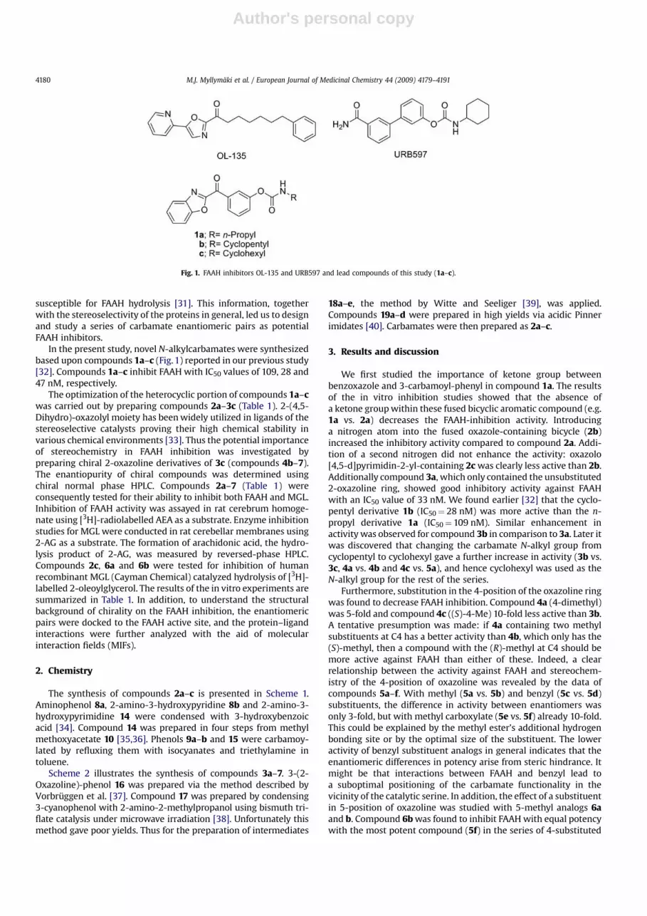
responsible for hydrolysis of AEA to arachidonic acid and etha-nolamine, and MGL for hydrolysis of 2-AG to arachidonic acidand glycerol [15–17]. Numerous potent inhibitors against FAAHhave been reported, including compounds that have shownpromising in vivo activity, selectivity and therapeutic effects [18–21]. Boger et al. introduced a large number of potential inhibitorsbased on a-ketoheterocycles (e.g. OL-135, Fig. 1) that reversiblyform hemiketals with the active site serine [22]. N-Alkylcarba-mates constitute a second widely explored class of inhibitors,including the well studied compound URB597 (Fig. 1) by Piomelliet al. [7,23,24]. Inhibition by N-alkylcarbamates is based onirreversible acylation of the active site serine [25]. Recentlyreported other structure families with inhibitory activity againstFAAH include (thio)hydantoins [26], piperidine- and piperazineureas [27,28], sulfonyl derivatives [29] and boronic acid deriva-tives [30].
To our knowledge, thus far no systematic study on the effect ofchirality on the activity of FAAH inhibitors has been presented. Thestereoselectivity of FAAH has been explored briefly: while studyingAEA derivatives as FAAH substrates, Makriyannis et al. found thatwithin the enantiomeric pairs of certain methanandamides theones having higher affinity towards CB1 receptor were also less
* Corresponding author. Tel.: þ358 9 451 2526; fax: þ358 9 451 2538.E-mail address: [email protected] (A.M.P. Koskinen).
Contents lists available at ScienceDirect
European Journal of Medicinal Chemistry
journal homepage: ht tp: / /www.elsevier .com/locate /e jmech
0223-5234/$ – see front matter � 2009 Elsevier Masson SAS. All rights reserved.doi:10.1016/j.ejmech.2009.05.012
European Journal of Medicinal Chemistry 44 (2009) 4179–4191
Author's personal copy
susceptible for FAAH hydrolysis [31]. This information, togetherwith the stereoselectivity of the proteins in general, led us to designand study a series of carbamate enantiomeric pairs as potentialFAAH inhibitors.
In the present study, novel N-alkylcarbamates were synthesizedbased upon compounds 1a–c (Fig. 1) reported in our previous study[32]. Compounds 1a–c inhibit FAAH with IC50 values of 109, 28 and47 nM, respectively.
The optimization of the heterocyclic portion of compounds 1a–cwas carried out by preparing compounds 2a–3c (Table 1). 2-(4,5-Dihydro)-oxazolyl moiety has been widely utilized in ligands of thestereoselective catalysts proving their high chemical stability invarious chemical environments [33]. Thus the potential importanceof stereochemistry in FAAH inhibition was investigated bypreparing chiral 2-oxazoline derivatives of 3c (compounds 4b–7).The enantiopurity of chiral compounds was determined usingchiral normal phase HPLC. Compounds 2a–7 (Table 1) wereconsequently tested for their ability to inhibit both FAAH and MGL.Inhibition of FAAH activity was assayed in rat cerebrum homoge-nate using [3H]-radiolabelled AEA as a substrate. Enzyme inhibitionstudies for MGL were conducted in rat cerebellar membranes using2-AG as a substrate. The formation of arachidonic acid, the hydro-lysis product of 2-AG, was measured by reversed-phase HPLC.Compounds 2c, 6a and 6b were tested for inhibition of humanrecombinant MGL (Cayman Chemical) catalyzed hydrolysis of [3H]-labelled 2-oleoylglycerol. The results of the in vitro experiments aresummarized in Table 1. In addition, to understand the structuralbackground of chirality on the FAAH inhibition, the enantiomericpairs were docked to the FAAH active site, and the protein–ligandinteractions were further analyzed with the aid of molecularinteraction fields (MIFs).
2. Chemistry
The synthesis of compounds 2a–c is presented in Scheme 1.Aminophenol 8a, 2-amino-3-hydroxypyridine 8b and 2-amino-3-hydroxypyrimidine 14 were condensed with 3-hydroxybenzoicacid [34]. Compound 14 was prepared in four steps from methylmethoxyacetate 10 [35,36]. Phenols 9a–b and 15 were carbamoy-lated by refluxing them with isocyanates and triethylamine intoluene.
Scheme 2 illustrates the synthesis of compounds 3a–7. 3-(2-Oxazoline)-phenol 16 was prepared via the method described byVorbruggen et al. [37]. Compound 17 was prepared by condensing3-cyanophenol with 2-amino-2-methylpropanol using bismuth tri-flate catalysis under microwave irradiation [38]. Unfortunately thismethod gave poor yields. Thus for the preparation of intermediates
18a–e, the method by Witte and Seeliger [39], was applied.Compounds 19a–d were prepared in high yields via acidic Pinnerimidates [40]. Carbamates were then prepared as 2a–c.
3. Results and discussion
We first studied the importance of ketone group betweenbenzoxazole and 3-carbamoyl-phenyl in compound 1a. The resultsof the in vitro inhibition studies showed that the absence ofa ketone group within these fused bicyclic aromatic compound (e.g.1a vs. 2a) decreases the FAAH-inhibition activity. Introducinga nitrogen atom into the fused oxazole-containing bicycle (2b)increased the inhibitory activity compared to compound 2a. Addi-tion of a second nitrogen did not enhance the activity: oxazolo[4,5-d]pyrimidin-2-yl-containing 2c was clearly less active than 2b.Additionally compound 3a, which only contained the unsubstituted2-oxazoline ring, showed good inhibitory activity against FAAHwith an IC50 value of 33 nM. We found earlier [32] that the cyclo-pentyl derivative 1b (IC50¼ 28 nM) was more active than the n-propyl derivative 1a (IC50¼109 nM). Similar enhancement inactivity was observed for compound 3b in comparison to 3a. Later itwas discovered that changing the carbamate N-alkyl group fromcyclopentyl to cyclohexyl gave a further increase in activity (3b vs.3c, 4a vs. 4b and 4c vs. 5a), and hence cyclohexyl was used as theN-alkyl group for the rest of the series.
Furthermore, substitution in the 4-position of the oxazoline ringwas found to decrease FAAH inhibition. Compound 4a (4-dimethyl)was 5-fold and compound 4c ((S)-4-Me) 10-fold less active than 3b.A tentative presumption was made: if 4a containing two methylsubstituents at C4 has a better activity than 4b, which only has the(S)-methyl, then a compound with the (R)-methyl at C4 should bemore active against FAAH than either of these. Indeed, a clearrelationship between the activity against FAAH and stereochem-istry of the 4-position of oxazoline was revealed by the data ofcompounds 5a–f. With methyl (5a vs. 5b) and benzyl (5c vs. 5d)substituents, the difference in activity between enantiomers wasonly 3-fold, but with methyl carboxylate (5e vs. 5f) already 10-fold.This could be explained by the methyl ester’s additional hydrogenbonding site or by the optimal size of the substituent. The loweractivity of benzyl substituent analogs in general indicates that theenantiomeric differences in potency arise from steric hindrance. Itmight be that interactions between FAAH and benzyl lead toa suboptimal positioning of the carbamate functionality in thevicinity of the catalytic serine. In addition, the effect of a substituentin 5-position of oxazoline was studied with 5-methyl analogs 6aand b. Compound 6b was found to inhibit FAAH with equal potencywith the most potent compound (5f) in the series of 4-substituted
Fig. 1. FAAH inhibitors OL-135 and URB597 and lead compounds of this study (1a–c).
M.J. Myllymaki et al. / European Journal of Medicinal Chemistry 44 (2009) 4179–41914180

Author's personal copy
Table 1Structures and in vitro activity of the synthesized compounds.
R1 OHNR2
O
Compound R1 R2 FAAH IC50,a mM MGL, % of inhibitionb
2a
O
N n-Propyl 3.0 (2.6–3.6) 26
2b N
O
N n-Propyl 0.68 (0.59–0.78) 29
2c
N
N
O
N n-Propyl 4.5 (4.0–5.2) 18c
3a
O
N n-Propyl 0.033 (0.028–0.038) 32
3b Cyclopentyl 0.013 (0.011–0.014) 10d
3c Cyclohexyl 0.0012 (0.00098–0.0014) 22
4a
O
NCyclopentyl 0.065 (0.053–0.081) 28
4b Cyclohexyl 0.046 (0.037–0.057) 20
4c
O
N Cyclopentyl 0.11 (0.097–0.13) 28
5a Cyclohexyl 0.051 (0.045–0.058) 28
5b
O
N Cyclohexyl 0.016 (0.014–0.018) 16
5c
O
N Cyclohexyl 2.1 (1.7–2.7) 17
5d
O
N Cyclohexyl 0.59 (0.51–0.69) 14
5e
O
NO
O Cyclohexyl 0.090 (0.077–0.11) 35
5f
O
NO
O Cyclohexyl 0.0094 (0.0078–0.011) 25
6a
O
N Cyclohexyl 0.073 (0.058–0.093) 13c
(continued on next page)
M.J. Myllymaki et al. / European Journal of Medicinal Chemistry 44 (2009) 4179–4191 4181

Author's personal copy
oxazolines. Furthermore, when a bulky S-indolyl group (7) wasintroduced at 4-position of oxazoline a significant decrease in theFAAH-inhibition activity was observed. None of the testedcompounds showed significant activity against MGL at 100 mMcompound concentration, and therefore IC50 values were notdetermined. As we found in our earlier work, in phenyl carbamatesinhibiting MGL, para-substitution is more favorable than meta-substitution [32].
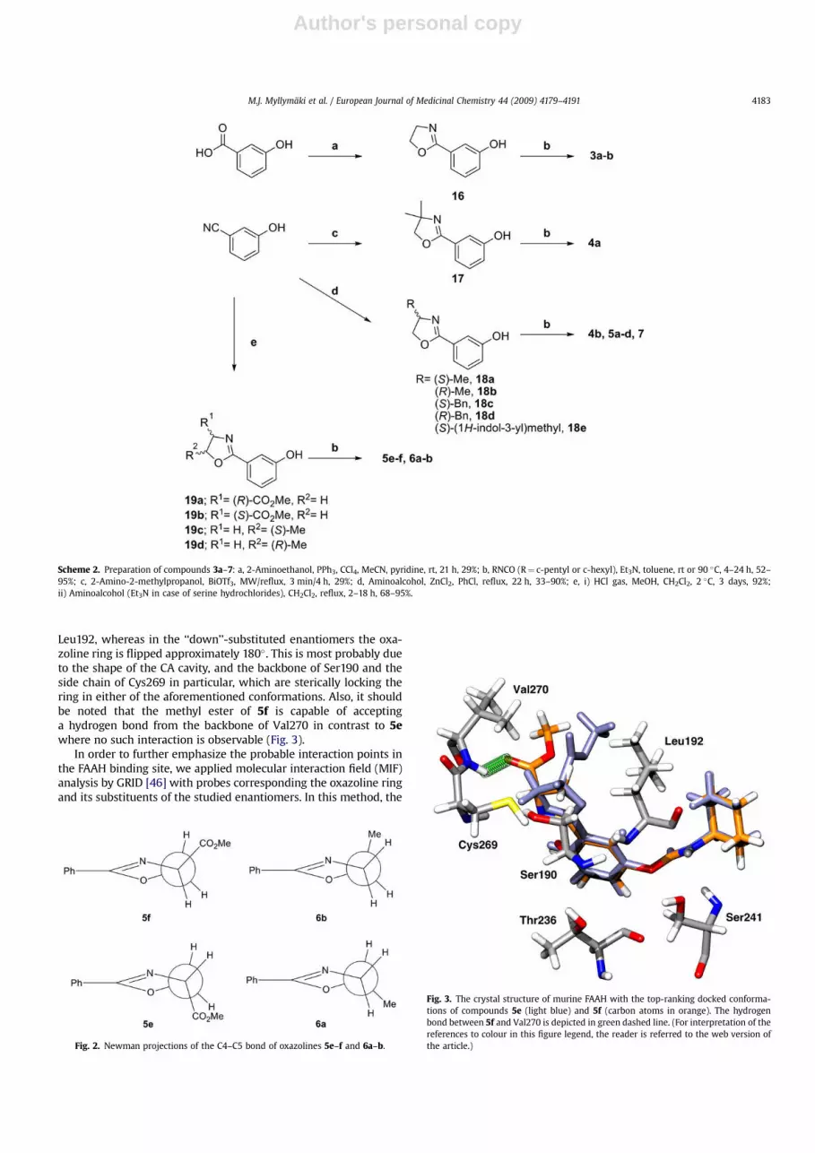
The stereochemistry of the compounds is illustrated in Fig. 2. Inthese Newman projections, the more active compounds 5f and 6bare the ones with their chiral carbon substituent ‘‘up’’ from theplane of the ring. This trend was also present in the enantiomericpairs 5a vs. 5b and 5c vs. 5d. These examples suggest thatthe stereochemistry in the oxazoline is more important than theregiochemistry of substitution (C4 or C5). This implies that theoxazoline ring conformation is locked within the enzyme’s activesite. This would explain the activity difference between the enan-tiomeric pairs since the substituent of the chiral carbon is pointingto a specific direction and thus filling the possible hydrophobicpocket or conversely causing steric hindrance.
Molecular modelling was performed to investigate the differ-ences in the inhibitory activities of the enantiomeric pairs. Inparticular, in this approach, we focused on exploring the differencesarising from the protein–ligand interactions of the recognitionprocess, as it can be assumed that the reactivity of the compounds isin similar level within the enantiomeric pairs. It should be noted
that a crystal structure of partially humanized rat FAAH with a drug-like inhibitor PF-750 [41] (PDB code 2VYA) has been publishedrecently. This structure shows structural rearrangements in thesubstrate access channel region with Phe432 flipping into the acylchain binding (ACB) channel. However, owing to our FAAH in vitroassay in rat brain homogenate, we docked the compounds to thecrystal structure of murine FAAH (PDB code 1MT5) [42] with GOLD[43], and the top-ranking pose of the most abundant cluster wasvisualized for each enantiomer (see Experimental section fordetails). There is a speculation of a general FAAH binding mode of N-alkylcarbamates before the acylation (carbamoylation) reactionoccurs [24,44,45]. In our docking study, the binding of all theenantiomers was indeed in agreement with this mode, and theligands were positioned in a conformation where the N-cyclohexylmoiety is pointing towards the branching point of the ACB andsubstrate access channels, and forming van der Waals interactionswith Phe194, Phe244, and Ile491. The oxygen of the carbamatecarbonyl is accepting hydrogen bond(s) from the backbone N–Hgroups of the FAAH oxyanion hole residues (Ile238–Ser241) [42],thus giving rise to a conformation where the electropositive a-carbon of the ligand is residing next to the nucleophilic hydroxyl ofcatalytic Ser241. Moreover, the O-aryl part is pointing towards thecytoplasmic access (CA) cavity leading to the intracellular surface ofFAAH. Noticeably, a common feature between all the enantiomericpairs was that in enantiomers with ‘‘up’’-substitutions, the oxazo-line oxygen atom is pointing roughly towards the side chain of
Table 1 (continued)
Compound R1 R2 FAAH IC50,a mM MGL, % of inhibitionb
6b
O
N Cyclohexyl 0.0068 (0.0056–0.0083) 14c
7
O
N
HN
Cyclohexyl 1.9 (1.6–2.4) 25
a Values represent the mean of three independent experiments (n¼ 3) performed in duplicate (95% confidence intervals are given in parentheses).b Inhibition of enzymatic activity (% of control) at 100 mM (n¼ 2).c Inhibition of human recombinant MGL.d Inhibition of enzymatic activity (% of control) at 1 mM (n¼ 2).
Scheme 1. Preparation of compounds 2a–c: a, 3-Hydroxybenzoic acid, boric acid, Na2SO4, m-xylene, autoclave, 200 �C, 16 h, 21–83%; b, 3-Hydroxybenzoic acid, MW, 250 �C, 6 min,71–77%; c, n-PrNCO, Et3N, toluene, rt or 90 �C, 20 h, 50–95%; d, i) Methyl formate, NaH, THF, 20 �C, 20 h; ii) formamidine acetate, EtOH, rt, 14 h, reflux, 24 h, 31%; e, POCl3, reflux,2.5 h, 86%; f, NH3, EtOH, autoclave, 130 �C, 18 h, 75%; g, n-BuSH, NaH, DMF, 110 �C, 20 h, 74%.
M.J. Myllymaki et al. / European Journal of Medicinal Chemistry 44 (2009) 4179–41914182
Author's personal copy
Leu192, whereas in the ‘‘down’’-substituted enantiomers the oxa-zoline ring is flipped approximately 180�. This is most probably dueto the shape of the CA cavity, and the backbone of Ser190 and theside chain of Cys269 in particular, which are sterically locking thering in either of the aforementioned conformations. Also, it shouldbe noted that the methyl ester of 5f is capable of acceptinga hydrogen bond from the backbone of Val270 in contrast to 5ewhere no such interaction is observable (Fig. 3).
In order to further emphasize the probable interaction points inthe FAAH binding site, we applied molecular interaction field (MIF)analysis by GRID [46] with probes corresponding the oxazoline ringand its substituents of the studied enantiomers. In this method, the
Fig. 2. Newman projections of the C4–C5 bond of oxazolines 5e–f and 6a–b.
Fig. 3. The crystal structure of murine FAAH with the top-ranking docked conforma-tions of compounds 5e (light blue) and 5f (carbon atoms in orange). The hydrogenbond between 5f and Val270 is depicted in green dashed line. (For interpretation of thereferences to colour in this figure legend, the reader is referred to the web version ofthe article.)
Scheme 2. Preparation of compounds 3a–7: a, 2-Aminoethanol, PPh3, CCl4, MeCN, pyridine, rt, 21 h, 29%; b, RNCO (R¼ c-pentyl or c-hexyl), Et3N, toluene, rt or 90 �C, 4–24 h, 52–95%; c, 2-Amino-2-methylpropanol, BiOTf3, MW/reflux, 3 min/4 h, 29%; d, Aminoalcohol, ZnCl2, PhCl, reflux, 22 h, 33–90%; e, i) HCl gas, MeOH, CH2Cl2, 2 �C, 3 days, 92%;ii) Aminoalcohol (Et3N in case of serine hydrochlorides), CH2Cl2, reflux, 2–18 h, 68–95%.
M.J. Myllymaki et al. / European Journal of Medicinal Chemistry 44 (2009) 4179–4191 4183

Author's personal copy
protein active site is enclosed in a grid cage, and the interactionenergies between amino acid residues and probes are calculated. Theresulting MIFs can be then used to determine the most favorableinteraction regions in the active site. By visualizing the MIFs in thelight of the docked compounds, no explanation for the inhibitionactivity differences arising from the oxazoline ring substituentconformations could be seen, apart from the carbonyl oxygen probeindicating a favorable interaction with Val270 backbone (corre-sponding to the aforementioned hydrogen bond of the methyl esterof 5f). However, when studying the MIFs in the binding region of theoxazoline ring, the sp2 nitrogen probe showed strong interactionwith the backbone of Cys269 and Val270 (Fig. 4a). Furthermore, withthe same interaction energy level, no interaction is observed withthese residues and aromatic/aliphatic ether oxygen probe (Fig. 4b).This is in line with the docking results, and suggests that the ‘‘up’’-substituted carbamates are forming an electrostatic interactionbetween the oxazoline ring nitrogen and the backbone N–H groups
of Cys269 and Val270, whereas in ‘‘down’’-substituted compoundsthe interaction between the ring oxygen and these residues is clearlyless favorable. This type of interaction between oxazoline nitrogenand protein backbone is not uncommon, and in Relibase [47] one canfind cases of similar drug–receptor interactions (e.g. [48]).
To gain confidence in this observation, we wanted to assess theelectronic properties of the enantiomers in more detail by calcu-lating the electrostatic potentials V(r)s with an ab initio method. Inall of the optimized structures the minima of the electrostaticpotential was located in the vicinity of the oxazoline nitrogen(Fig. 5). This indirectly indicates the oxazoline nitrogen havinghigher interaction potential compared to the oxygen [49,50]. Thus,it seems the IC50 differences of the enantiomeric pairs might beindeed due to the sterical features of the FAAH active site, and theconsequential electrostatic dipole–dipole interaction of the ‘‘up’’-substituted oxazoline carbamates with the Cys269 and Val270backbone N–H groups.
4. Conclusion
In conclusion, a series of chiral 3-oxazolinylphenyl N-alkylcar-bamates were prepared and tested for their in vitro inhibitoryactivity against FAAH and MGL. In this series, enantiomers havingtheir chiral center substituent ‘‘up’’ from plane of the oxazoline ring(5b, 5d, 5f, 6b) were found to be more potent FAAH inhibitors thancorresponding ‘‘down’’ enantiomers. The most potent chiralcompound, (R)-3-(5-methyl-4,5-dihydrooxazol-2-yl)phenyl cyclo-hexylcarbamate (6b), inhibited FAAH with approximately 10-foldhigher potency (IC50 value: 6.8 nM) than the corresponding (S)-enantiomer (6a, IC50 value: 73 nM). In addition, non-substitutedoxazoline derivative (3b, IC50 value: nM) was found to be morepotent than any of the corresponding substituted compounds.None of the compounds presented significantly inhibited MGLactivity. Since the carbamate based FAAH inhibitors have beenreported to have several off-targets [19,25,51] the selectivity ofchiral 3-oxazolinylphenyl N-alkylcarbamates would be importantissue to study in more detail in the future. In molecular modellingstudies, the combined docking and molecular interaction fieldanalysis emphasized the importance of heterocycle interactionwith Cys269 and Val270 of FAAH, and also highlighted the stericfeatures of the FAAH active site. These findings could provide
Fig. 4. The crystal structure of murine FAAH with the top-ranking docked conforma-tions of compounds a) 6b and b) 6a. The GRID MIFs of sp2 nitrogen and aromatic/aliphatic oxygen probes at contour level of �6.0 kcal/mol are colored in blue and red,respectively. The carbon atoms of the ligands are colored in orange, while those ofprotein in white. The wireframe presentation is illustrating the shape of the FAAHactive site. In a) the distance between the oxazoline nitrogen and backbone hydrogensof Cys269 and Val270 is 2.96 Å and 3.60 Å, respectively. In b) the distance between theoxazoline oxygen and the backbone hydrogens of Cys269 and Val270 is 3.36 Å and3.9 Å, respectively. (For interpretation of the references to colour in this figure legend,the reader is referred to the web version of the article.)
Fig. 5. The optimized structure and the electrostatic potential V(r) (isopotentialsurface) of 6b. The gray and red areas correspond to þ0.25 and �0.07 eV, respectively.The minimum and maximum V(r) values for 6b are �0.1132 and þ137.3 eV, respec-tively. (For interpretation of the references to colour in this figure legend, the reader isreferred to the web version of the article.)
M.J. Myllymaki et al. / European Journal of Medicinal Chemistry 44 (2009) 4179–41914184
Author's personal copy
valuable information for further development of more selectiveFAAH inhibitors.
5. Experimental protocols
5.1. Chemistry
Commercially available starting materials were used withoutfurther purification. All dry reactions were performed under argonin flame-dried glassware and solvents were distilled. In microwavereactions CEM Discover -microwave reactor was used. Analyticalthin-layer chromatography was carried out on Merck silica gel F254(60 Å, 40–63 mm, 230–400 mesh) precoated aluminium sheets anddetected under UV light. Purification of reaction products wascarried out by flash chromatography (FC) on J. T. Bakers silica gel forchromatography (pore size 60 Å, particle size 50 nM). The 1H NMRand 13C NMR spectra were recorded on a Bruker Avance 400spectrometer operating at 400 MHz for 1H and 100 MHz for 13C.Chemical shifts are reported in ppm on the d scale from an internalstandard (TMS 0.00 ppm) or residual solvent (CDCl3 7.26 and77.0 ppm; DMSO-d6 2.50 and 39.52 ppm). Melting points weredetermined in open capillaries using Stuart SMP3 and are uncor-rected. Optical rotation data were recorded on Perkin Elmer 343polarimeter using Na lamb (589 nm) and 100 mm cuvette at roomtemperature. HRMS were recorded on Waters Micromass LCTPremier (ESI) spectrometer. Chiral HPLC analysis was carried outusing Waters pump and UV detector (254 nm) and Daicel ChiralcelOD analytical chiral column. Eluent used was 20% 2-propanol inhexane with flow rate 0.5 mL/min. Retention time (Rt) and ee-% arereported. Elemental analyses (CHN) were recorded using a PerkinElmer 2400 CHN-elemental analyzer. Analyses indicated by thesymbols of the elements were within �0.4% of the theoreticalvalues.
5.1.1. 3-Benzo[d]oxazol-2-yl-phenol (9a) [52,53]3-Hydroxybenzoic acid (840 mg, 6.2 mmol, 100 mol-%), 2-ami-
nophenol (8a, 680 mg, 6.1 mmol, 100 mol-%), boric acid (380 mg,6.1 mmol, 100 mol-%) and Na2SO4 (8 g, 56 mmol, 900 mol-%) werestirred in m-xylene (60 mL) in an autoclave at 200 �C for 16 h. Thecooled mixture was poured into sat. aq. NaHCO3 (100 mL) andextracted with EtOAc (3�100 mL). Combined organic phases werewashed with water (100 mL) and brine (100 mL), dried withNa2SO4, filtered and evaporated. The resulting red solid (1.36 g) waspurified by FC (20% EtOAc in hex) and recrystallized (EtOAc/hex)giving 9a (1.07 g, 83%) as a white solid: mp. 238–239 �C, Rf (50%EtOAc in hex) 0.5; 1H NMR (DMSO-d6) 9.97 (s, 1H), 7.81–7.77 (m,1H), 7.64 (dt, 1H, J¼ 7.8, 1.1 Hz), 7.60–7.59 (m, 1H), 7.45–7.38 (m,3H), 7.02 (ddd, 1H, J¼ 8.2, 2.5, 0.8 Hz); 13C NMR (DMSO-d6) 162.3,157.9, 150.2, 141.5, 130.6, 127.5, 125.5, 124.9, 119.8, 119.2, 118.1, 113.7,110.9.
5.1.2. Procedure for preparation of carbamates: 3-(benzo[d]oxazol-2-yl)phenyl propylcarbamate (2a)
Compound 9a (79 mg, 0.37 mmol, 100 mol-%), propylisocyanate(158 mg, 1.85 mmol, 500 mol-%) and Et3N (37 mg, 0.37 mmol,100 mol-%) were stirred in toluene (4 mL) at rt for 16 h. The mixturewas diluted with EtOAc (5 mL) and filtered through a pad of silica.The crude material was recrystallized (EtOAc/hex) giving 2a(84 mg, 77%) as white crystals: mp. 144–145 �C, Rf (EtOAc) 0.5; 1HNMR (CDCl3) 8.09 (d, 1H, J¼ 7.8 Hz), 8.03 (s, 1H), 7.78–7.74 (m, 1H),7.59–7.55 (m, 1H), 7.50 (t, 1H, J¼ 8.0 Hz), 5.17 (br s, 1H), 3.26 (q, 2H,J¼ 6.7 Hz), 1.62 (sext, 2H, J¼ 7.3 Hz), 0.98 (t, 3H, J¼ 7.4 Hz); 13CNMR (CDCl3) 162.2, 154.2, 151.5, 150.7, 142.0, 129.8, 128.3, 125.3,124.8, 124.6, 124.3, 120.9, 120.1, 110.6, 43.0, 23.0, 11.2; Anal.C17H16N2O3 (C, H, N).
5.1.3. 3-(Oxazolo[4,5-b]pyridin-2-yl)phenol (9b) [34,54]3-Hydroxybenzoic acid (250 mg, 1.8 mmol, 150 mol-%) and 2-
amino-3-hydroxypyridine (8b, 130 mg, 1.2 mmol, 100 mol-%)were microwave-irradiated (300 W, 4 min ramp to 250 �C, hold2 min). The tan solid was dissolved to MeOH/EtOAc (1:1, 10 mL),poured to sat. aq. NaHCO3 (30 mL) and extracted with EtOAc(3� 50 mL). Combined organic phases were washed with water(50 mL) and brine (50 mL), dried (Na2SO4), filtered and evapo-rated. The crude material was purified by FC (50% EtOAc in hex)and recrystallized (EtOAc/hex) giving 9b (195 mg, 77%) as a whitesolid: mp. 203–204 �C, Rf (EtOAc) 0.5; 1H NMR (DMSO-d6) 10.02(s, 1H), 8.55 (dd, 1H, J¼ 4.9, 1.4 Hz), 8.24 (dd, 1H, J¼ 8.2, 1.4 Hz),7.69 (app dt, 1H, J¼ 7.7, 1.2 Hz), 7.64 (t, 1H, J¼ 2.0 Hz), 7.48–7.43(m, 2H), 7.08 (ddd, 1H, J¼ 8.2, 2.5, 0.9 Hz); 13C NMR (DMSO-d6)165.0, 158.1, 155.6, 146.7, 142.9, 130.8, 127.1, 121.0, 120.2, 119.3,118.7, 114.1.
5.1.4. 3-(Oxazolo[4,5-b]pyridin-2-yl)phenyl propylcarbamate (2b)White solid (93 mg, 78%): mp. 146–147 �C, Rf (EtOAc) 0.5; 1H
NMR (CDCl3) 8.59 (dd, 1H, J¼ 4.9, 1.3 Hz), 8.17 (d, 1H, J¼ 7.8 Hz),8.10–8.08 (m, 1H), 7.86 (dd, 1H, J¼ 8.1, 1.3 Hz), 7.54 (t, 1H,J¼ 8.0 Hz), 7.39–7.36 (m, 1H), 7.31 (dd, 1H, J¼ 8.1, 4.9 Hz), 5.19 (br s,1H), 3.28 (q, 2H, J¼ 6.7 Hz), 1.64 (sext, 2H, J¼ 7.2 Hz), 1.00 (t, 3H,J¼ 7.4 Hz); 13C NMR (CDCl3) 164.9, 156.2, 154.0, 151.5, 146.8, 143.1,130.0, 127.7, 125.8, 124.9, 121.2, 120.2, 118.2, 43.0, 23.0, 11.2; Anal.C16H15N3O3 (C, H, N).
5.1.5. 5-Methoxypyrimidin-4(3H)-one (11) [55]Methyl 2-methoxyacetate (10, 10.4 g, 100 mmol, 100 mol-%) and
methyl formate (7.2 g, 120 mmol, 120 mol-%) were added toa mixture of sodium hydride (5.6 g of 60% dispersion in oil,140 mmol, 140 mol-%) in dry THF (150 mL) and stirred for 20 hkeeping the temperature of the mixture at 20 �C by water bath.After formation of white solid, dry Et2O (100 mL) was added andthe mixture was filtered. The solid (18.6 g) was dried underreduced pressure and added to a mixture of formamidine acetate(10.4 g, 100 mmol, 100 mol-%) in EtOH (200 mL) at rt. The mixturewas stirred for 14 h at rt and refluxed for 24 h. Water (70 mL) wasadded and the mixture was acidified with AcOH (25 mL) from pH10 to 5. Ethanol was evaporated and the residue was extractedwith CHCl3 (10�100 mL) and EtOAc:Et2O (2:1, 3�100 mL).Combined organic phases were dried over MgSO4, filtered andevaporated. Crystallization of the crude product (MeOH:hex) gave11 (3.9 g, 31%) as off-white solid: mp. 216–217 �C; Rf (10% MeOH inCH2Cl2) 0.2; 1H NMR (DMSO-d6) 12.51 (br s, 1H), 7.81 (s, 1H), 7.52(s, 1H), 3.73 (s, 3H).
5.1.6. 4-Chloro-5-methoxypyrimidine (12) [53]Compound 11 (2.6 g, 20.6 mmol, 100 mol-%) was treated with
phosphorus oxychloride (17 mL, 180 mmol, 900 mol-%) at 0 �C. Themixture was refluxed for 2.5 h, cooled to 30–40 �C and unreactedPOCl3 was removed under reduced pressure. Ice-cold water(50 mL) was added and pH was adjusted to 7 with K2CO3 (10 g).The mixture was extracted with EtOAc:Et2O (3:1, 3�150 mL),organic phases were combined, dried (MgSO4) and filtered througha pad of silica (eluted with EtOAc). Evaporation of the solventsresulted in compound 12 (2.6 g, 86%) as a yellow solid; mp. 63–64 �C; Rf (EtOAc) 0.5; 1H NMR (CDCl3) 8.60 (s, 1H), 8.30 (s, 1H), 4.00(s, 3H).
5.1.7. 5-Methoxypyrimidin-4-amine (13) [56]The mixture of 12 (3.4 g, 24 mmol, 100 mol-%) in EtOH (30 mL)
was bubbled with dry NH3 gas for 30 min at 0 �C. The mixture waspoured to autoclave and stirred at 130 �C for 18 h. Cooling andevaporation resulted in tan residue which was dissolved to CHCl3
M.J. Myllymaki et al. / European Journal of Medicinal Chemistry 44 (2009) 4179–4191 4185

Author's personal copy
(100 mL) and brine (100 mL) and extracted with CHCl3(3�100 mL). Combined organic phases were dried over MgSO4,filtered and evaporated giving 13 (2.2 g, 75%) as a yellow solid; mp.117–119 �C; Rf (10% MeOH in CH2Cl2) 0.4; 1H NMR (DMSO-d6) 7.99(s, 1H,), 7.79 (s, 1H), 6.68 (s, 2H), 3.80 (s, 3H).
5.1.8. 4-Aminopyrimidin-5-ol (14) [57]The mixture of 13 (280 mg, 2.2 mmol, 100 mol-%) in dry DMF
(15 mL) was treated with NaH (215 mg, washed with hexanes from60% mineral oil dispersion, 9.0 mmol, 400 mol-%) at rt. The mixturewas cooled by ice bath and n-BuSH (300 mg, 3.3 mmol, 150 mol-%)was added. The mixture was stirred at 110 �C for 20 h and evapo-rated to dryness. AcOH (0.25 mL) and water (2.5 mL) were addedand evaporation was repeated. The residue was purified by FC (10%MeOH in CH2Cl2) and recrystallized (MeOH) giving 14 (181 mg,74%) as a red solid; mp. 257–260 �C; Rf (20% MeOH in CH2Cl2)0.2; 1H NMR (DMSO-d6) 9.81 (br s, 1H), 7.90 (s, 1H), 7.65 (s, 1H),6.44 (2H).
5.1.9. 3-(Oxazolo[4,5-d]pyrimidin-2-yl)phenol (15)Compound 14 (222 mg, 2.0 mmol, 100 mol-%), 3-hydrox-
ybenzoic acid (276 mg, 2.0 mmol, 100 mol-%), boric acid (124 mg,2.0 mmol, 100 mol-%) and Na2SO4 (2.5 g, 18 mmol, 900 mol-%) werestirred in m-xylene (20 mL) in an autoclave at 200 �C for 24 h. Thecooled mixture was poured into sat. aq. NaHCO3 (50 mL) andextracted with EtOAc (3�70 mL). Combined organic phases weredried over Na2SO4, filtered and evaporated. The resulting red solidwas purified by FC (50% EtOAc in hex) and recrystallized (MeOH/CH2Cl2) giving 15 (92 mg, 21%) as an off-white solid: mp. 257–261 �C; Rf (EtOAc) 0.5; 1H NMR (DMSO-d6) 10.14 (s, 1H), 9.33 (s, 1H),9.14 (s, 1H), 7.76 (d, 1H, J¼ 7.7 Hz), 7.68 (app t, 1H, J¼ 1.9 Hz), 7.49 (t,1H, J¼ 7.9 Hz), 7.15 (dd, 1H, J¼ 8.1, 2.1 Hz); 13C NMR (DMSO-d6)167.6, 161.6, 158.0, 154.9, 142.2, 139.7, 130.8, 126.2, 121.1, 119.3, 114.7;HRMS (ESI): calcd. for [MþNa] C11H7N3O2Na: 236.0436, found236.0447.
5.1.10. 3-(Oxazolo[4,5-d]pyrimidin-2-yl)phenyl propylcarbamate (2c)A white solid (30 mg, 50%): mp. 161–162 �C; Rf (EtOAc) 0.5; 1H
NMR (CDCl3) 9.20 (s, 1H), 9.01 (s, 1H), 8.2 (dt, 1H, J¼ 7.87, 1.28 Hz),8.14 (t, 1H, J¼ 1.9 Hz), 7.58 (t, 1H, J¼ 8.0 Hz), 7.45 (ddd, 1H, J¼ 8.2,2.3, 0.9 Hz), 5.24 (app t, 1H, J¼ 5.5 Hz), 3.28 (q, 2H, J¼ 6.7 Hz), 1.64(sext, 2H, J¼ 7.2 Hz), 1.00 (t, 3H, J¼ 7.4 Hz); 13C NMR (CDCl3) 167.6,162.3, 155.5, 154.0, 151.7, 142.3, 138.8, 130.2, 127.1, 126.5, 125.6,122.0, 43.0, 23.0, 11.2; HRMS (ESI): calcd. for [MþNa]C15H14N4O3Na: 321.0964, found 321.0951.
5.1.11. 3-(4,5-Dihydrooxazol-2-yl)-phenol (16) [58]3-Hydroxybenzoic acid (1.38 g, 10 mmol, 100 mol-%), 2-amino-
ethanol (610 mg, 10 mmol, 100 mol-%) and Et3N (3.0 g, 30 mmol,300 mol-%) were stirred in pyridine (20 mL) and MeCN (30 mL) at22 �C for 40 min. CCl4 (6.15 g, 40 mmol, 400 mol-%) was addedfollowed by dropwise admission of PPh3 in pyridine–MeCN (1:1,80 mL) during 2 h keeping the temperature of the mixture between22 and 24 �C. The mixture was stirred for 18 h, concentrated byrotavapor to ca. 40 mL, diluted with aq. ammonia (25%, 100 mL) andextracted with EtOAc (3�100 mL). Combined organic phases werewashed with sat. aq. CuSO4 (100 mL), water (100 mL) and brine(100 mL), dried (Na2SO4), filtered and evaporated. The crudeproduct was purified by FC (0–4% MeOH in CH2Cl2) giving 16(479 mg, 29%) as a white solid: mp. 188–189 �C; Rf (EtOAc) 0.5; 1HNMR (DMSO-d6) 9.70 (s, 1H), 7.30–7.23 (m, 3H), 6.91 (d, 1H,J¼ 8.1 Hz), 4.36 (t, 2H, J¼ 9.5 Hz), 3.92 (t, 2H, J¼ 9.5 Hz); 13C NMR(DMSO-d6) 163.0, 157.3, 129.7, 128.7, 118.5, 118.4, 114.3, 67.2, 54.4.This compound was later prepared in 96% yield by similar methodas 19a.
5.1.12. 3-(4,5-Dihydrooxazol-2-yl)phenyl propylcarbamate (3a)White crystals (65 mg, 65%): mp. 94–95 �C; Rf (EtOAc) 0.5; 1H
NMR (CDCl3) 7.78 (d, 1H, J¼ 7.8 Hz), 7.70 (s, 1H), 7.39 (t, 1H,J¼ 8.0 Hz), 7.29–7.24 (m, 1H), 5.05 (br s, 1H), 4.43 (app t, 2H,J¼ 9.5 Hz), 4.06 (app t, 2H, J¼ 9.5 Hz), 3.24 (q, 2H, J¼ 6.7 Hz), 1.60(sext, 2H, J¼ 7.3 Hz), 0.97 (t, 3H, J¼ 7.4 Hz); 13C NMR (CDCl3) 163.9,150.9, 129.1, 128.9, 124.9, 124.6, 121.4, 67.6, 54.8, 42.9, 23.0, 11.1;Anal. C13H16N2O3 (C, H, N).
5.1.13. 3-(4,5-Dihydrooxazol-2-yl)phenyl cyclopentylcarbamate (3b)White crystals (48 mg, 70%): mp. 166–168 �C; Rf (10% Et2O in
CH2Cl2) 0.4; 1H NMR (DMSO-d6) 7.86 (d, 1H, J¼ 7.1 Hz), 7.69 (d, 1H,J¼ 7.7 Hz), 7.53 (s, 1H), 7.47 (t, 1H, J¼ 7.9 Hz), 7.28 (dd, 1H, J¼ 1.5,8.1 Hz), 4.41 (t, 2H, J¼ 9.5 Hz), 3.96 (t, 2H, J¼ 9.5 Hz), 3.88–3.79 (m,1H), 1.89–1.79 (m, 2H), 1.72–1.61 (m, 2H), 1.57–1.44 (m, 4H); 13CNMR (DMSO-d6) 162.3, 153.4, 151.0, 129.7, 128.6, 124.7, 124.1, 120.9,67.5, 54.4, 52.4, 32.2, 23.3; Anal. C15H18N2O3 (C, H, N).
5.1.14. 3-(4,5-Dihydrooxazol-2-yl)phenyl cyclohexylcarbamate (3c)White cotton-like crystals (100 mg, 71%): mp. 151–152 �C; Rf (5%
MeOH in CH2Cl2) 0.54; 1H NMR (CDCl3) 7.78 (d, 1H, J¼ 8.0 Hz), 7.71–7.69 (s, 1H), 7.39 (t, 1H, J¼ 7.9 Hz), 7.28–7.23 (m, 1H), 4.94 (d, 1H,J¼ 7.0 Hz), 4.42 (t, 2H, J¼ 9.5 Hz), 4.05 (t, 2H, J¼ 9.5 Hz), 3.63–3.50(m, 1H), 2.07–1.96 (m, 2H), 1.79–1.70 (m, 2H), 1.67–1.58 (m, 1H),1.44–1.31 (m, 2H), 1.28–1.14 (m, 3H); 13C NMR (CDCl3) 163.9, 153.3,151.0, 129.2, 129.0, 124.9, 124.6, 121.5, 67.7, 54.9, 50.2, 33.2, 25.4,24.7; Anal. C16H20N2O3 (C, H, N).
5.1.15. 3-(4,4-Dimethyl-4,5-dihydrooxazol-2-yl)phenol (17) [37]This compound was synthesized in two different methods:
Method 1: 3-cyanophenol (237 mg, 2.0 mmol, 100 mol-%), 2-amino-2-methylpropanol (450 mL, 4.0 mmol, 200 mol-%) andbismuth trifluoromethylsulfonate (64.5 mg, 0.1 mmol, 5 mol-%)were microwave-irradiated in a closed vessel (2� 60 s at 50 W,1�60 s at 60 W). The resulting mixture was diluted with EtOAc andpurified by FC (40% EtOAc in hex).
Method 2: 3-cyanophenol (237 mg, 2.0 mmol, 100 mol-%), 2-amino-2-methylpropanol (450 ml, 4.0 mmol, 200 mol-%) andbismuth trifluoromethylsulfonate (64.5 mg, 0.1 mmol, 5 mol-%)were refluxed for 3 h. Crops from both reactions were combinedand recrystallized from toluene giving 17 (190 mg, 25% overallyield) as a white solid: mp. 159–161 �C; Rf (65% EtOAc in hex) 0.28;1H NMR (DMSO-d6) 9.66 (s, 1H), 7.28–7.21 (m, 3H), 6.92–6.88 (m,1H), 4.07 (m, 2H), 1.26 (m, 6H). This compound was later preparedin 90% yield by similar method as 18a.
5.1.16. 3-(4,4-Dimethyl-4,5-dihydrooxazol-2-yl)phenyl cyclopentylcarbamate (4a)
White needles (73 mg, 48%): mp. 139–141 �C; Rf (10% Et2O inCH2Cl2) 0.27; 1H NMR (DMSO-d6) 7.76 (d, 1H, J¼ 7.7 Hz), 7.70 (s,1H), 7.38 (dd, 1H, J¼ 7.9 Hz), 7.23 (d, 2H, J¼ 8.1 Hz), 4.98 (d, 1H,J¼ 6.4 Hz), 4.10 (s, 2H), 4.07–4.00 (m, 1H), 2.06–1.98 (m, 2H), 1.76–1.69 (m, 2H), 1.66–1.60 (m, 2H), 1.53–1.44 (m, 2H), 1.37 (s, 6H); 13CNMR (DMSO-d6) 161.3, 153.7, 150.9, 129.4, 129.1, 125.0, 124.5, 121.6,79.1, 67.6, 53.0, 33.1, 28.4, 23.5; Anal. C17H22N2O3 (C, H, N).
5.1.17. 3-(4,4-Dimethyl-4,5-dihydrooxazol-2-yl)phenylcyclohexy-lcarbamate (4b)
White crystals (176 mg, 56%): mp. 154–155 �C; Rf (5% MeOH inCH2Cl2) 0.67; 1H NMR (CDCl3) 7.76 (d, 1H, J¼ 7.8 Hz), 7.72–7.69 (m,1H), 7.37 (t, 1H, J¼ 7.9 Hz), 7.23 (dd, 1H, J¼ 8.1, 1.4 Hz), 4.96 (d, 1H,J¼ 7.5 Hz), 4.09 (s, 2H), 3.62–3.49 (m, 1H), 2.06–1.95 (m, 2H), 1.79–1.69 (m, 2H),1.66–1.58 (m, 2H), 1.44–1.31 (m, 7H),1.28–1.13 (m, 3H);13C NMR (CDCl3) 161.3, 153.4, 151.0, 129.3, 129.1, 124.9, 124.5, 121.6,79.2, 67.6, 50.1, 33.2, 28.4, 25.4, 24.7; Anal. C18H24N2O3 (C, H, N).
M.J. Myllymaki et al. / European Journal of Medicinal Chemistry 44 (2009) 4179–41914186

Author's personal copy
5.1.18. Procedure for preparation of 18a–e5.1.18.1. (S)-3-(4-Methyl-4,5-dihydrooxazol-2-yl)phenol (18a). Zincchloride (48 mg, 0.35 mmol, 10 mol-%) was melted in a 50 mL flaskunder high vacuum. 3-Cyanophenol (413 mg, 3.46 mmol, 100 mol-%) and chlorobenzene (10 mL) were added and heated up to refluxunder argon atmosphere. (S)-2-Aminopropanol (510 mL, 6.5 mmol,190 mol-%) was added and the mixture was refluxed for 22 h. Themixture was cooled to rt and filtered through a pad of silica withEtOAc and purified by FC (33% EtOAc in hex) and recrystallized fromEtOAc:hex giving 18a (317 mg, 52%) as white needles: mp. 125–126 �C; Rf (50% EtOAc in hex) 0.31; [a]D 48 (c¼ 0.5, CDCl3); 1H NMR(CDCl3) 9.45 (br s, 1H), 7.45 (dd, 1H, J¼ 2.3, 1.6 Hz), 7.30 (td, 1H,J¼ 7.7,1.3 Hz), 7.18 (t,1H, J¼ 7.9 Hz), 6.95 (ddd,1H, J¼ 8.1, 2.5,1.0 Hz),4.53 (dd,1H, J¼ 9.4, 8.0 Hz), 4.45–4.36 (m,1H), 3.96 (t,1H, J¼ 7.9 Hz),1.33 (d, 3H, J¼ 6.6 Hz); 13C NMR (CDCl3) 164.6, 156.9, 129.6, 128.0,119.8, 119.5, 115.1, 74.2, 61.2, 21.1; Anal. C10H11NO2 (C, H, N).
5.1.19. (S)-3-(4-Methyl-4,5-dihydrooxazol-2-yl)phenylcyclopentyl-carbamate (4c)
White crystals (83 mg, 51%): mp. 139–142 �C; Rf (15% Et2O inCH2Cl2) 0.33; [a]D �54 (c¼ 0.5, CHCl3); 1H NMR (CDCl3) 7.77 (d, 1H,J¼ 7.7 Hz), 7.70 (s, 1H), 7.38 (t, 1H, J¼ 8.0 Hz), 7.24 (d, 1H, J¼ 8.1 Hz),5.04 (d, 1H, J¼ 6.6 Hz), 4.51 (dd, 1H, J¼ 9.3, 8.1 Hz), 4.41–4.32 (m,1H), 4.09–4.00 (m, 1H), 3.94 (t, 1H, J¼ 7.9 Hz), 2.06–1.97 (m, 2H),1.75–1.56 (m, 4H), 1.53–1.44 (m, 2H), 1.35 (d, 3H, J¼ 6.6 Hz); 13CNMR (CDCl3) 162.7, 160.1, 153.7, 151.0, 129.1, 125.0, 124.6, 121.6, 74.1,62.0, 53.0 (rotam. 52.1), 33.1 (rotam. 33.6), 23.5, 21.4; Anal.C16H20N2O3 (C, H, N).
5.1.20. (S)-3-(4-Methyl-4,5-dihydrooxazol-2-yl)phenylcyclohexyl-carbamate (5a)
White crystals (94 mg, 65%): mp. 133–135 �C; Rf (50% EtOAc inhex) 0.40; [a]D �49 (c¼ 0.5, CHCl3); 1H NMR (CDCl3) 7.77 (d, 1H,J¼ 7.7 Hz), 7.70 (s, 1H), 7.38 (t, 1H, J¼ 7.9 Hz), 7.26–7.23 (m, 1H),4.92 (d, 1H, J¼ 7.3 Hz), 4.51 (dd, 1H, J¼ 9.2, 8.2 Hz), 4.42–4.32 (m,1H), 3.94 (t, 1H, J¼ 7.9 Hz), 3.61–3.51 (m, 1H), 2.05–1.97 (m, 2H),1.78–1.70 (m, 2H), 1.66–1.59 (m, 1H), 1.43–1.32 (m, 2H), 1.35 (d, 3H,J¼ 6.6 Hz), 1.28–1.15 (m, 3H); 13C NMR (CDCl3) 162.8, 153.4, 151.0,129.1 (2C), 125.0, 124.6, 121.6, 74.1, 62.0, 50.1, 33.2, 25.4, 24.7, 21.4;Rt 60 min, 93 ee-%; Anal. C17H22N2O3 (C, H, N).
5.1.21. (R)-3-(4-Methyl-4,5-dihydrooxazol-2-yl)phenol (18b)White needles (283 mg, 57%): mp. 126–127 �C; Rf (50% EtOAc in
hex) 0.19; [a]D �46 (c¼ 0.5, CHCl3); 1H NMR (CDCl3) 9.46 (br s, 1H),7.44 (dd, 1H, J¼ 2.4, 1.6 Hz), 7.29 (td, 1H, J¼ 7.7, 1.3 Hz), 7.18 (t, 1H,J¼ 7.9 Hz), 6.95 (ddd, 1H, J¼ 8.1, 2.6, 1.0 Hz), 4.53 (dd, 1H, J¼ 9.5,8.1 Hz), 4.45–4.36 (m, 1H), 3.96 (t, 1H, J¼ 7.9 Hz), 1.33 (d, 3H,J¼ 6.6 Hz); 13C NMR (CDCl3) 164.5, 156.9, 129.6, 127.9, 119.8, 119.5,115.0, 74.2, 61.1, 21.1; Anal. C10H11NO2 (C, H, N).
5.1.22. (R)-3-(4-Methyl-4,5-dihydrooxazol-2-yl)phenylcyclohexyl-carbamate (5b)
White crystals (46 mg, 27%): mp. 133–134 �C; Rf (50% EtOAc inhex) 0.24; [a]D 45 (c¼ 0.5, CDCl3); 1H NMR (CDCl3) 7.77 (d, 1H,J¼ 7.7 Hz), 7.70 (m, 1H), 7.38 (t, 1H, J¼ 7.9 Hz), 7.24 (dd, 1H, J¼ 8.1,1.4 Hz), 4.94 (d, 1H, J¼ 7.4 Hz), 4.51 (dd, 1H, J¼ 9.3, 8.1 Hz), 4.42–4.32 (m, 1H), 3.94 (t, 1H, J¼ 7.9 Hz), 3.60–3.50 (m, 1H), 2.07–1.96(m, 2H), 1.78–1.70 (m, 2H), 1.66–1.57 (m, 1H), 1.43–1.32 (m, 2H), 1.35(d, 3H, J¼ 6.6 Hz), 1.27–1.15 (m, 3H); 13C NMR (CDCl3) 162.8, 153.4,151.0, 129.1 (2C), 124.9, 124.6, 121.6, 74.1, 62.0, 50.1, 33.2, 25.4, 24.7,21.4; Rt 22 min, 99 ee-%; Anal. C17H22N2O3 (C, H, N).
5.1.23. (S)-3-(4-Benzyl-4,5-dihydrooxazol-2-yl)phenol (18c)White waxy solid (446 mg, 52%): mp. 109–111 �C; Rf (10% Et2O in
CH2Cl2) 0.14; [a]D 39 (c¼ 0.5, CHCl3); 1H NMR (CDCl3) 7.92 (s, 1H),
7.50 (dd, 1H, J¼ 2.4, 1.5 Hz), 7.39 (td, 1H, J¼ 7.7, 1.2 Hz), 7.30–7.18(m, 6H), 6.98 (ddd, 1H, J¼ 8.1, 2.6, 1.0 Hz), 4.65–4.57 (m, 1H), 4.35 (t,1H, J¼ 9.0 Hz), 4.17 (dd, 1H, J¼ 8.6, 7.2 Hz), 3.23 (dd, 1H, J¼ 13.7,4.9 Hz), 2.75 (dd, 1H, J¼ 13.7, 9.0 Hz); 13C NMR (CDCl3) 164.8, 156.4,137.6, 129.7, 129.3, 128.6, 128.3, 126.6, 120.2, 119.4, 115.2, 72.0, 67.2,41.5; Anal. C16H15NO2 (C, H, N).
5.1.24. (S)-3-(4-Benzyl-4,5-dihydrooxazol-2-yl)phenylcyclohexyl-carbamate (5c)
White crystals (120 mg, 56%): mp. 152–155 �C; Rf 0.46 (10% Et2Oin CH2Cl2); [a]D 5.8 (c¼ 0.5, CHCl3); 1H NMR (CDCl3) 7.78 (d, 1H,J¼ 7.8 Hz), 7.70 (t,1H, J¼ 1.8 Hz), 7.39 (t,1H, J¼ 8.0 Hz), 7.33–7.21 (m,6H), 4.93 (d, 1H, J¼ 7.6 Hz), 4.62–4.54 (m, 1H), 4.34 (t,1H, J¼ 8.9 Hz),4.13 (dd, 1H, J¼ 8.3, 7.5 Hz), 3.61–3.51 (m, 1H), 3.23 (dd, 1H, J¼ 13.7,5.1 Hz), 2.72 (dd,1H, J¼ 13.7, 8.9 Hz), 2.05–1.97 (m, 2H),1.78–1.70 (m,2H), 1.66–1.61 (m, 1H),1.43–1.32 (m, 2H), 1.28–1.14 (m, 3H); 13C NMR(CDCl3) 163.3, 153.3, 151.0, 137.9, 129.2, 129.2, 129.0, 128.6, 126.5,125.0,124.7,121.6, 71.9, 67.9, 50.2, 41.8, 33.2, 25.4, 24.7; Rt 57 min, 98ee-%; Anal. C23H26N2O3 (C, H, N).
5.1.25. (R)-3-(4-Benzyl-4,5-dihydrooxazol-2-yl)phenol (18d)White waxy solid (267 mg, 41%): mp. 108–111 �C; Rf 0.21 (15%
Et2O in CH2Cl2); [a]D �40 (c¼ 0.5, CHCl3); 1H NMR (CDCl3) 8.50 (brs, 1H), 7.50 (dd, 1H, J¼ 2.3, 1.5 Hz), 7.36 (td, 1H, J¼ 7.7, 1.1 Hz), 7.29–7.18 (m, 6H), 6.97 (ddd, 1H J¼ 8.2, 2.5, 0.9 Hz), 4.66–4.57 (m, 1H),4.34 (t, 1H, J¼ 9.0 Hz), 4.17 (dd, 1H, J¼ 8.5, 7.3 Hz), 3.24 (dd, 1H,J¼ 13.7, 4.8 Hz), 2.75 (dd, 1H, J¼ 13.7, 9.2 Hz); 13C NMR (CDCl3)165.0, 156.6, 137.5, 129.7, 129.3, 128.6, 128.2, 126.6, 120.1, 119.5,115.2, 72.0, 67.0, 41.4; Anal. C16H15NO2 (C, H, N).
5.1.26. (R)-3-(4-Benzyl-4,5-dihydrooxazol-2-yl)phenylcyclohexyl-carbamate (5d)
White crystals (152 mg, 57%): mp. 153–155 �C; Rf 0.38 (13% Et2Oin CH2Cl2); [a]D �7.4 (c¼ 0.5, CHCl3); 1H NMR (CDCl3) 7.78 (d, 1H,J¼ 7.7 Hz), 7.7 (m, 1H), 7.39 (t, 1H, J¼ 8.0 Hz), 7.33–7.20 (m, 6H),4.93 (d, 1H, J¼ 8.0 Hz), 4.62–4.53 (m, 1H), 4.33 (t, 1H, J¼ 8.9 Hz),4.13 (dd, 1H, J¼ 8.3, 7.5 Hz), 3.62–3.50 (m, 1H), 3.23 (dd, 1H, J¼ 13.7,5.1 Hz), 2.71 (dd, 1H, J¼ 13.7, 8.9 Hz), 2.05–1.97 (m, 2H), 1.77–1.70(m, 2H), 1.66–1.58 (m, 1H), 1.43–1.32 (m, 2H), 1.28–1.14 (m, 3H); 13CNMR (CDCl3) 163.3, 153.3, 151.0, 137.9, 129.2, 129.2, 129.0, 128.5,126.5, 125.0, 124.7, 121.6, 71.9, 67.9, 50.1, 41.8, 33.2, 25.4, 24.7; Rt22 min, 99 ee-%; Anal. C23H26N2O3 (C, H, N).
5.1.27. (S)-3-(4-((1H-Indol-3-yl)methyl)-4,5-dihydrooxazol-2-yl)-phenol (18e)
Gray powder (140 mg, 35%): mp. 183–186 �C; Rf (50% EtOAc inhex) 0.20; [a]D 59 (c¼ 0.3, MeOH); 1H NMR (DMSO-d6) 10.85 (br s,1H), 9.68 (s, 1H), 7.59 (d, 1H, J¼ 7.9 Hz), 7.33 (d, 1H, J¼ 8.0 Hz), 7.31–7.19 (m, 4H), 7.06 (td, 1H, J¼ 7.5, 1.1 Hz), 6.98 (td, 1H, J¼ 7.4, 1.0 Hz),6.93–6.89 (m, 1H), 4.64–4.55 (m, 1H), 4.38 (dd, 1H, J¼ 9.4, 8.4 Hz),4.07 (t, 1H, J¼ 7.9 Hz), 3.13 (dd, 1H, J¼ 14.8, 4.9 Hz), 2.82 (dd, 1H,J¼ 14.6, 8.1 Hz); 13C NMR (DMSO-d6) 162.1, 157.3, 136.1, 129.6, 128.8,127.5, 123.4, 120.9, 118.5, 118.5, 118.4, 118.3, 114.4, 111.3, 110.4, 71.7,66.6, 31.0; Anal. C18H16N2O2 (C, H, N).
5.1.28. (S)-3-(4-((1H-Indol-3-yl)methyl)-4,5-dihydrooxazol-2-yl)-phenyl cyclohexylcarbamate (7)
White crystals (35 mg, 35%): mp. 149–151 �C; Rf 0.23 (10% Et2Oin CH2Cl2); [a]D 22 (c¼ 0.5, CDCl3); 1H NMR (CDCl3) 8.11 (br s, 1H),7.79 (d, 1H, J¼ 7.8 Hz), 7.72 (s, 1H), 7.66 (d, 1H, J¼ 7.8 Hz), 7.41–7.33(m, 2H), 7.20 (td, 1H, J¼ 7.5, 1.1 Hz), 7.13 (td, 1H, J¼ 7.4, 1.0 Hz), 7.28–7.24 (m, 1H), 7.04 (d, 1H, J¼ 2.2 Hz), 4.94 (d, 1H, J¼ 7.8 Hz), 4.75–4.67 (m, 1H), 4.33 (t, 1H, J¼ 8.9 Hz), 4.15 (t, 1H, J¼ 7.9 Hz), 3.62–3.51(m, 1H), 3.36 (dd, 1H, J¼ 14.5, 4.6 Hz), 2.88 (dd, 1H, J¼ 14.6, 8.9 Hz),2.05–1.97 (m, 2H), 1.78–1.70 (m, 2H), 1.67–1.58 (m, 1H), 1.43–1.32
M.J. Myllymaki et al. / European Journal of Medicinal Chemistry 44 (2009) 4179–4191 4187

Author's personal copy
(m, 2H), 1.28–1.14 (m, 3 H); 13C NMR (CDCl3) 163.2, 153.4, 151.0,136.2, 129.2 (2C), 127.7, 125.0, 124.6, 122.4, 122.1, 121.6, 119.5, 118.8,112.0, 111.1, 72.3, 67.0, 50.2, 33.2, 31.3, 25.4, 24.7; Anal. C25H27N3O3
(C, H, N).
5.1.29. Procedure for preparation of 19a–dEt3N was used only in the case of 19a–b.
5.1.29.1. (R)-Methyl 2-(3-hydroxyphenyl)-4,5-dihydrooxazole-4-carboxylate (19a). To a mixture of 3-cyanophenol (1840 mg,15.5 mmol, 100 mol-%) in dry CH2Cl2 (36 mL) was added dry MeOH(3.2 mL, 79 mmol, 510 mol-%) and the mixture was bubbled withHCl gas in an ice bath. The mixture was stirred at 2 �C for 3 days andsolvents were evaporated. Filtering and washing with dry Et2O gavemethyl 3-hydroxybenzimidate hydrochloride (2.67 g, 92%) asa white powder: 1H NMR (DMSO-d6) 11.68 (br s, 1H), 10.34 (s, 1H),7.57–7.53 (m, 1H), 7.46–7.41 (m, 2H), 7.24–7.21 (m, 1H), 4.27 (s, 3H).(R)-Serine methyl ester hydrochloride (125 mg, 0.80 mmol,100 mol-%) was suspended in dry CH2Cl2 and Et3N was added(180 mL, 1.28 mmol, 160 mol-%) followed by above describedimidate salt (153 mg, 0.82 mmol, 100 mol-%) and the mixture wasrefluxed overnight. Solvent was evaporated and remaining solidpartitioned between H2O (10 mL) and EtOAc (15 mL). Organic phasewas washed with H2O (10 mL). Aqueous phases were combined andbackwashed with EtOAc (2�15 mL). Organic phases werecombined, washed with brine (20 mL), dried over Na2SO4, filteredand evaporated. Purification by FC (twice, 70% EtOAc:hex, then 5%MeOH:CH2Cl2) gave 19a (122 mg, 68%) as an oil: Rf (70% EtOAc:hex)0.33; [a]D �57 (c¼ 1, CDCl3); 1H NMR (CDCl3) 8.57 (s, 1H), 7.42 (dd,1H, J¼ 2.4, 1.6 Hz), 7.38 (app. ddd, 1H), 7.18 (t, 1H, J¼ 7.9 Hz), 6.96(ddd, 1H, J¼ 8.2, 2.5, 0.9 Hz), 4.96 (dd, 1H, J¼ 10.7, 7.8 Hz), 4.69 (dd,1H, J¼ 8.7, 7.9 Hz), 4.58 (dd, 1H, J¼ 10.7, 8.8 Hz), 3.69 (s, 3H); 13CNMR (CDCl3) 171.3, 167.1, 156.6, 129.6, 127.2, 120.3, 119.8, 115.4, 69.6,67.7, 52.7; HRMS (ESI) calcd for [MþH] C11H11NO4: 222.0766,found 222.0766.
5.1.30. (R)-Methyl 2-(3-(cyclohexylcarbamoyloxy)phenyl)-4,5-dihydrooxazole-4-carboxylate (5e)
White crystals (136 mg, 73%): mp. 114–116 �C; Rf (17% Et2O inCH2Cl2) 0.33; [a]D �68 (c¼ 0.5, CHCl3); 1H NMR (CDCl3) 7.81 (d, 1H,J¼ 7.8 Hz), 7.75 (s, 1H), 7.39 (t, 1H, J¼ 8.0 Hz), 7.29–7.26 (m, 1H),4.95 (dd, 2H, J¼ 10.5, 8.0 Hz), 4.69 (t, 1H, J¼ 8.3 Hz), 4.59 (dd, 1H,J¼ 10.5, 8.8 Hz), 3.82 (s, 3H), 3.60–3.51 (m, 1H), 2.04–1.97 (m, 2H),1.78–1.70 (m, 2H), 1.67–1.59 (m, 1H), 1.43–1.32 (m, 2H), 1.27–1.16(m, 3H); 13C NMR (CDCl3) 171.4, 165.6, 153.3, 151.0, 129.2, 128.1,125.3, 125.2, 121.9, 69.6, 68.6, 52.7, 50.1, 33.2, 25.4, 24.7; Rt 99 min,96 ee-%; Anal. C18H22N2O5 (C, H, N).
5.1.31. (S)-Methyl 2-(3-hydroxyphenyl)-4,5-dihydrooxazole-4-carboxylate (19b)
Colourless oil (122 mg, 68%): Rf (60% EtOAc in hex) 0.28; [a]D
65 (c¼ 1.8, CDCl3); 1H NMR (CDCl3) 8.16 (s, 1H), 7.43–7.42 (m, 1H),7.40 (d, 1H, J¼ 7.8 Hz), 7.20 (t, 1H, J¼ 7.9 Hz), 6.97 (ddd, 1H, J¼ 8.2,2.5, 0.9 Hz), 4.96 (dd, 1H, J¼ 10.7, 7.9 Hz), 4.69 (dd, 1H, J¼ 8.7,7.9 Hz), 4.59 (dd, 1H, J¼ 10.7, 8.8 Hz), 3.71 (s, 3H); 13C NMR (CDCl3)171.4, 170.0, 156.5, 129.6, 127.3, 120.4, 119.8, 115.4, 69.6, 67.8, 52.7;HRMS (ESI): calcd for (MþHþ) C11H11NO4: 222.0766, found222.0759.
5.1.32. (S)-Methyl 2-(3-(cyclohexylcarbamoyloxy)phenyl)-4,5-dihydrooxazole-4-carboxylate (5f)
White crystals (141 mg, 75%): mp. 114–116 �C; Rf (17%Et2O:CH2Cl2) 0.33; [a]D 73 (c¼ 0.5, CHCl3); 1H NMR (CDCl3) 7.81 (d,1H, J¼ 7.8 Hz), 7.75 (s, 1H), 7.39 (t, 1H, J¼ 7.9 Hz), 7.29–7.26 (m, 1H),4.94 (dd, 2H, J¼ 10.5, 8.0 Hz), 4.69 (t, 1H, J¼ 7.8 Hz), 4.59 (dd, 1H,
J¼ 10.6, 8.8 Hz), 3.82 (s, 3H), 3.59–3.52 (m, 1H), 2.04–1.97 (m, 2H),1.78–1.70 (m, 2H), 1.67–1.59 (m, 1H), 1.43–1.32 (m, 2H), 1.27–1.16(m, 3H); 13C NMR (CDCl3) 171.4, 165.6, 153.3, 151.0, 129.2, 128.1,125.3, 125.2, 121.9, 69.6, 68.6, 52.7, 50.1, 33.2, 25.4, 24.7; Rt 34 min,97 ee-%; Anal. C17H22N2O3 (C, H, N).
5.1.33. (R)-3-(5-Methyl-4,5-dihydrooxazol-2-yl)phenol (19c)White crystals (137 mg, 26%): Rf (EtOAc) 0.33; [a]D
20�4.5 (c¼ 1.0,CDCl3); 1H NMR (CDCl3) 9.64 (br s, 1H), 7.48–7.46 (app. dd, 1H),7.34–7.30 (m, 1H), 7.19 (t, 1H, J¼ 7.9 Hz), 6.95 (ddd, 1H, J¼ 8.1, 2.5,0.9 Hz), 4.91–4.81 (m, 1H), 4.13 (dd, 1H, J¼ 14.2, 9.5 Hz), 4.58 (dd,1H, J¼ 14.2, 7.6 Hz), 1.42 (d, 3H, J¼ 6.2 Hz); 13C NMR (CDCl3) 165.1,157.0, 129.6, 128.2, 119.7, 119.5, 115.1, 76.6, 60.3, 20.9; Rt 15 min, 99ee-%; Anal. C10H11NO2 (C, H, N).
5.1.34. (R)-3-(5-Methyl-4,5-dihydrooxazol-2-yl)phenylcyclohexylcarbamate (6a)
White crystals (75 mg, 54%): mp. 141–142 �C; Rf (20% acetone inCH2Cl2) 0.7; [a]D
20 �8.9 (c¼ 0.9, CDCl3); 1H NMR (CDCl3) 7.77 (d, 1H,J¼ 7.7 Hz), 7.71–7.68 (m, 1H), 7.38 (t, 1H, J¼ 7.9 Hz), 7.26–7.22 (app.dd, 1H), 4.98 (br d, 1H, J¼ 7.6 Hz), 4.89–4.79 (m, 1H), 4.14 (dd, 1H,J¼ 14.6, 9.4 Hz), 3.60 (dd, 1H, J¼ 14.5, 7.4 Hz), 3.60–3.50 (m, 1H),2.08–1.96 (m, 2H), 1.81–1.69 (m, 2H), 1.67–1.58 (m, 1H), 1.45–1.31(m, 2H), 1.41 (t, 3H, J¼ 6.2 Hz) 1.29–1.13 (m, 3H); 13C NMR (CDCl3)163.2, 153.4, 151.0, 129.3, 129.1, 124.8, 124.5, 121.4, 76.4, 61.6, 50.1,33.2, 25.4, 24.7, 21.1; Rt 64 min, 99 ee-%; Anal. C17H22N2O3 (C, H, N).
5.1.35. (S)-3-(5-Methyl-4,5-dihydrooxazol-2-yl)phenol (19d)White crystals (126 mg, 71%): mp. 104–106 �C; Rf (EtOAc) 0.33;
[a]D 3.7 (c¼ 1.0, CDCl3); 1H NMR (CDCl3) 9.63 (br s, 1H), 7.48–7.46(app. dd, 1H), 7.34–7.30 (m, 1H), 7.19 (t, 1H, J¼ 7.9 Hz), 6.95 (ddd,1H, J¼ 8.1, 2.5, 0.9 Hz), 4.91–4.81 (m, 1H), 4.13 (dd, 1H, J¼ 14.2,9.5 Hz), 4.58 (dd, 1H, J¼ 14.2, 7.6 Hz), 1.42 (d, 3H, J¼ 6.2 Hz); 13CNMR (CDCl3) 165.1, 157.0, 129.6, 128.2, 119.7, 119.5, 115.1, 76.6, 60.3,20.9; Rt 10.8 min, 99 ee-%; Anal. C10H11NO2 (C, H, N).
5.1.36. (S)-3-(5-Methyl-4,5-dihydrooxazol-2-yl)phenylcyclohexylcarbamate (6b)
White crystals (98 mg, 58%): mp. 140–142 �C; Rf (20% acetone inCH2Cl2); 0.7; [a]D
20 9.3 (c¼ 1.0, CDCl3); 1H NMR (CDCl3) 7.77 (d, 1H,J¼ 7.7 Hz), 7.70–7.68 (m, 1H), 7.38 (t, 1H, J¼ 7.9 Hz), 7.26–7.22 (app.dd, 1H), 4.98 (br d, 1H, J¼ 7.6 Hz), 4.89–4.79 (m, 1H), 4.14 (dd, 1H,J¼ 14.6, 9.4 Hz), 3.60 (dd, 1H, J¼ 14.5, 7.4 Hz), 3.60–3.50 (m, 1H),2.08–1.96 (m, 2H),1.81–1.69 (m, 2H),1.67–1.58 (m,1H),1.45–1.31 (m,2H), 1.41 (t, 3H, J¼ 6.2 Hz) 1.29–1.13 (m, 3H); 13C NMR (CDCl3) 163.2,153.4,151.0,129.3,129.1,124.8,124.5,121.4, 76.4, 61.6, 50.1, 33.2, 25.4,24.7, 21.1; Rt 23.5 min, 97 ee-%; Anal. C17H22N2O3 (C, H, N).
5.2. Biological testing protocols
5.2.1. Animals and preparation of rat brain homogenatefor FAAH assay
Eight-week-old male Wistar rats were used in these studies. Allanimal experiments were approved by the local ethics committee.The animals lived in a 12-h light/12-h dark cycle (lights on at0700 h) with water and food available ad libitum.
The rats were decapitated, whole brains minus cerebellum weredissected and homogenized in one volume (v/w) of ice-cold 0.1 Mpotassium phosphate buffer (pH 7.4) with a Potter–Elvehjemhomogenizer (Heidolph). The homogenate was centrifuged at10,000g for 20 min at 4 �C and the resulting supernatant was usedas a source of FAAH activity. The protein concentration of thesupernatant (7.2 mg/mL) was determined by the method of Brad-ford with BSA as the standard [59]. Aliquots of the supernatantwere stored at �80 �C until use.
M.J. Myllymaki et al. / European Journal of Medicinal Chemistry 44 (2009) 4179–41914188

Author's personal copy
5.2.2. Animals and preparation of rat cerebellar membranes forMGL assay
Four-week-old male Wistar rats were used in these studies. Allanimal experiments were approved by the local ethics committee.The animals lived in a 12-h light/12-h dark cycle (lights on at0700 h), with water and food available ad libitum. The rats weredecapitated, 8 h after lights on (1500 h), whole brains wereremoved, dipped in isopentane on dry ice and stored at �80 �C.Membranes were prepared as previously described [60–62].
Briefly, cerebella (minus brain stem) from eight animals wereweighed and homogenized in nine volumes of ice-cold 0.32 Msucrose with a glass Teflon homogenizer. The crude homogenatewas centrifuged at low speed (1000g for 10 min at 4 �C) and thepellet was discharged. The supernatant was centrifuged at highspeed (100,000g for 10 min at 4 �C). The pellet was resuspended inice-cold deionized water and washed twice, repeating the high-speed centrifugation. Finally, membranes were resuspended in50 mM Tris–HCl, pH 7.4 with 1 mM EDTA and aliquoted for storageat �80 �C. The protein concentration of the final preparation,measured by the Bradford method [59], was 11 mg/mL.
5.2.3. In vitro assay for FAAH activityThe assay for FAAH activity has been described previously [63].
The endpoint enzymatic assay was developed to quantify FAAHactivity with tritium-labelled arachidonoylethanolamide (etha-nolamine 1-3H). The assay buffer used was 0.1 M potassium phos-phate (pH 7.4) and test compounds were dissolved in DMSO (thefinal DMSO concentration was max 5% v/v). The incubations wereperformed in the presence of 0.5% (w/v) BSA (essentially fatty acidfree). Test compounds were preincubated with rat brain homoge-nate protein (18 mg) for 10 min at 37 �C (60 mL). At the 10 min timepoint, arachidonoylethanolamide was added so that its finalconcentration was 2 mM (containing 50�10�3 mCi of 60 Ci/mmol[3H]AEA) and the final incubation volume was 100 mL. The incu-bations proceeded for 10 min at 37 �C. EtOAc (400 mL) was added atthe 20 min time point to stop the enzymatic reaction. Additionally,100 mL of unlabelled ethanolamine (1 mM) was added as a ‘carrier’for radioactive ethanolamine. Samples were centrifuged at 16,000gfor 4 min at rt, and aliquots (100 mL) from aqueous phase containing[ethanolamine 1-3H] were measured for radioactivity by liquidscintillation counting (Wallac 1450 MicroBeta; Wallac Oy, Finland).
5.2.4. In vitro assay for MGL activity in rat cerebellar membranesThe assay for MGL activity has been described previously [64].
Briefly, experiments were carried out with preincubations (80 mL,30 min at 25 �C) containing 10 mg membrane protein, 44 mM Tris–HCl (pH 7.4), 0.9 mM EDTA, 0.5% (wt/vol) BSA and 1.25% (vol/vol)DMSO as a solvent for inhibitors. The preincubated membraneswere kept at 0 �C just prior to the experiments. The incubations(90 min at 25 �C) were initiated by adding 40 mL of preincubatedmembrane cocktail, in a final volume of 400 mL. The final volumecontained 5 mg membrane protein, 54 mM Tris–HCl (pH 7.4),1.1 mM EDTA, 100 mM NaCl, 5 mM MgCl2, 0.5% (wt/vol) BSA and50 mM of 2-AG. At time points of 0 and 90 min, 100-mL samples wereremoved from the incubation, acetonitrile (200 mL) was added tostop the enzymatic reaction and the pH of the samples wassimultaneously decreased to 3.0 with phosphoric acid (added toacetonitrile) to stabilize 2-AG against acyl migration to 1(3)-AG.Samples were centrifuged at 23,700g for 4 min at rt prior to HPLCanalysis of the supernatant.
5.2.5. HPLC methodThe analytical HPLC was performed as previously described [17].
Briefly, the analytical HPLC system consisted of a Merck Hitachi(Hitachi Ltd., Tokyo, Japan) L-7100 pump, D-7000 interface module,
L-7455 diode-array UV detector (190–800 nm, set at 211 nm) and L-7250 programmable autosampler. The separations were accom-plished on a Zorbax SB-C18 endcapped reversed-phase precolumn(4.6�12.5 mm, 5 mm) and column (4.6�150 mm, 5 mm) (Agilent,USA). The injection volume was 50 mL. A mobile phase mixture of28% phosphate buffer (30 mM, pH 3.0) in acetonitrile was used ata flow rate of 2.0 mL/min. Retention times were 5.8 min for 2-AG,6.3 min for 1(3)-AG and 10.2 min for arachidonic acid. The relativeconcentrations of 2-AG, 1(3)-AG and arachidonic acid were deter-mined by the corresponding peak areas. This was justified by theequivalence of response factors for the studied compounds, andwas supported by the observation that the sum of the peak areaswas constant throughout the experiments.
5.2.6. Human recombinant MGL assayThe endpoint enzymatic assay was developed to quantify human
recombinant MGL (Cayman Chemical, cat# 10008354) activity withtritium-labelled 2-oleoylglycerol (2-OG) [glycerol-1,2,3-3H] (Amer-ican Radiolabeled Chemicals Inc., St Louis, MO, USA). The assaybuffer was 50 mM Tris–HCl, pH 7.4; 1 mM EDTA and testcompounds were dissolved in DMSO (the final DMSO concentrationwas not more than 5% v/v). The incubations were performed in thepresence of 0.5% (w/v) BSA (essentially fatty acid free). hrMGL waspreincubated with test compounds for 10 min at 37 �C (60 mL). Atthe 10 min time point, 2-OG was added to achieve the finalconcentration of 50 mM (containing 112�10�3 mCi of 40 Ci/mmol[3H]2-OG) with the final incubation volume of 100 mL. The incuba-tions proceeded for 10 min at 37 �C. EtOAc (400 mL) was added at the20 min time point to stop the enzymatic reaction. Additionally,100 mL of buffer (50 mM Tris–HCl, pH 7.4; 1 mM EDTA) was added.The samples were centrifuged at 16,000g for 4 min at rt, andaliquots (100 mL) were taken from the aqueous phase, which con-tained glycerol-1,2,3-3H, and measured for radioactivity by liquidscintillation counting (Wallac 1450 MicroBeta; Wallac Oy, Finland).
5.2.7. Data analysesThe results from the enzyme inhibition experiments are pre-
sented as mean� 95% confidence intervals of at least three inde-pendent experiments performed in duplicate. Data analyses for thedose–response curves were calculated as non-linear regressionsusing GraphPad Prism 4.0 for Windows.
5.3. Molecular modelling
5.3.1. Structure constructionThe 3-D coordinates in the X-ray crystal structure of murine FAAH
in complex with covalently bound inhibitor methyl arachidonylfluorophosphonate (MAFP) (Protein Data Bank code 1MT5, resolu-tion 2.8 Å) [42] was used as an enzyme model for the dockingcalculations. Missing side chain atoms were added into the FAAHmonomer (chain A extracted from the X-ray data) of the crystalstructure by using Lovell rotamer library [65] and optimized inpolarizable AMBER07_FF02 force field [66] (energy gradient of0.01 kcal/mol) while keeping the rest of the protein atoms fixed, asimplemented in Sybyl 8.1 [67]. Hydrogen atoms were added withMolProbity (v.3.15) [68] server by allowing the program to optimizehydrogen bonding by flipping Asn, Gln or His, where applicable. Theorientation of the hydrogen atoms of the catalytic region amino acids(catalytic triad) [42,69] was inspected to ensure that a hydrogenbond was formed between hydroxyl groups of Ser217 and Ser241and one between side chain of Ser217 and side chain of Lys142.
The 3-D coordinates of the ligands were generated in Sybyl 8.1following an energy minimization to energy gradient of 0.005 kcal/mol in Merck molecular force field (MMFF94s) [70] with Powellconjugate gradient method.
M.J. Myllymaki et al. / European Journal of Medicinal Chemistry 44 (2009) 4179–4191 4189
Author's personal copy
5.3.2. Molecular dockingVersion 4.0 of GOLD [43] was used as a docking tool in this study.
The binding site of FAAH was defined to range 15 Å radius from theoxygen atom of catalytic Ser241 side chain hydroxyl group (with‘detect cavity’ feature of GOLD toggled on). The default GoldScoreempirical fitness function was used as a scoring function for thegenerated poses. In order to maximize the conformational spacesampled during the docking calculations, a preset ‘200% efficiency’setting was utilized for 1000 separate genetic algorithm (GA)docking runs for each ligand. To ensure that different possiblebinding modes are inspected upon visualization of the results, andnot only the top-ranking poses, RMSD-based complete linkageclustering was applied. Clustering distance of 1.0 Å (as imple-mented in GOLD) was used to derive the clusters of poses. The top-ranking (according to GOLD) poses of three most abundant clusters,in terms of cluster members, were visualized for each ligand. Thehighest scoring pose of the most abundant cluster was consideredfor the further analyses.
5.3.3. GRID interaction fieldsIn order to analyze and identify possible hot spots for favorable
protein–ligand interactions in the FAAH active site and conse-quently shed light on the measured enantiopreference, GRID [46]molecular interaction field (MIF) analysis was carried out. For GRIDcalculations, the active site of the modified FAAH structure (seeSection 5.3.1) was enclosed in a 26� 26� 25 Å box with gridspacing of 0.2 Å. Only lone pairs and tautomeric hydrogens wereallowed to alter in response to the probes (directive MOVE¼ 0). Toreflect the oxazoline ring and its different substituents found in theenantiomeric pairs, the following GRID probes were used in thisstudy: methyl (C3), sp2 aromatic carbon (C1]), sp2 nitrogen witha lone pair (N:]), sp2 carbonyl oxygen (O), and aromatic/aliphaticether oxygen (OC1). The resulting MIFs were visually analyzed.
5.3.4. Electrostatic potentialTo briefly explore the nucleophilic characteristics of the chiral
carbamates, we calculated the electrostatic potential V(r) of theenantiomers. Starting from the docked conformations, the mole-cules were optimized and V(r) was calculated in Hartree–Fock/6-311G** level of theory with Gaussian 03 [71]. The V(r) isosurfaceswere visualized with VMD 1.8.6 [72].
Acknowledgments
We thank Ms Anna Minkkila for help in biological testing. TheNational Technology Agency of Finland, The National GraduateSchool in Informational and Structural Biology (ISB), and FinnishCultural Foundation are greatly acknowledged for financial support.
Appendix
References
[1] W.A. Devane, L. Hanus, A. Breuer, R.G. Pertwee, L.A. Stevenson, G. Griffin,D. Gibson, A. Mandelbaum, A. Etinger, R. Mechoulam, Science 258 (1992)1946–1949.
[2] R. Mechoulam, S. Ben-Shabat, L. Hanus, M. Ligumsky, N.E. Kaminski, A.R. Schatz,A. Gopher, S. Almog, B.R. Martin, D.R. Compton, R.G. Pertwee, G. Griffin,M. Bayewitch, J. Barg, Z. Vogel, Biochem. Pharmacol. 50 (1995) 83–90.
[3] T. Sugiura, S. Kondo, A. Sukagawa, S. Nakane, A. Shinoda, K. Itoh, A. Yamashita,K. Waku, Biochem. Biophys. Res. Commun. 215 (1995) 89–97.
[4] V. Di Marzo, M. Bifulco, L. De Petrocellis, Nat. Rev. Drug Discov. 3 (2004)771–784.
[5] D.M. Lambert, C.J. Fowler, J. Med. Chem. 48 (2005) 5059–5087.[6] J.M. Walker, S.M. Huang, N.M. Strangman, K. Tsou, M.C. Sanudo-Pena, Proc.
Natl. Acad. Sci. U.S.A. 96 (1999) 12198–12203.[7] S. Kathuria, S. Gaetani, D. Fegley, F. Valino, A. Duranti, A. Tontini, M. Mor,
G. Tarzia, G. La Rana, A. Calignano, A. Giustino, M. Tattoli, M. Palmery,V. Cuomo, D. Piomelli, Nat. Med. 9 (2003) 76–81.
[8] V. Di Marzo, S.K. Goparaju, L. Wang, J. Liu, S. Batkai, Z. Jarai, F. Fezza, G.I. Miura,R.D. Palmiter, T. Sugiura, G. Kunos, Nature 410 (2001) 822–825.
[9] T. Jarvinen, D.W. Pate, K. Laine, Pharmacol. Ther. 95 (2002) 203–220.[10] L. Hanus, A. Breuer, S. Tchilibon, S. Shiloah, D. Goldenberg, M. Horowitz,
R.G. Pertwee, R.A. Ross, R. Mechoulam, E. Fride, Proc. Natl. Acad. Sci. U.S.A. 96(1999) 14228–14233.
[11] V. Di Marzo, T. Bisogno, L. De Petrocellis, D. Melck, P. Orlando, J.A. Wagner,G. Kunos, Eur. J. Biochem. 264 (1999) 258–267.
[12] V. Di Marzo, L. De Petrocellis, N. Sepe, A. Buono, Biochem. J. 316 (1996)977–984.
[13] B.F. Cravatt, D.K. Giang, S.P. Mayfield, D.L. Boger, R.A. Lerner, N.B. Gilula, Nature384 (1996) 83–87.
[14] T.P. Dinh, D. Carpenter, F.M. Leslie, T.F. Freund, I. Katona, S.L. Sensi, S. Kathuria,D. Piomelli, Proc. Natl. Acad. Sci. U.S.A. 99 (2002) 10819–10824.
[15] S.K. Goparaju, N. Ueda, K. Taniguchi, S. Yamamoto, Biochem. Pharmacol. 57(1999) 417–423.
[16] A.H. Lichtman, E.G. Hawkins, G. Griffin, B.F. Cravatt, J. Pharmacol. Exp. Ther.302 (2002) 73–79.
[17] S.M. Saario, J.R. Savinainen, J.T. Laitinen, T. Jarvinen, R. Niemi, Biochem.Pharmacol. 67 (2004) 1381–1387.
[18] M. Seierstad, J.G. Breitenbucher, J. Med. Chem. 51 (2008) 7327–7343.[19] A.H. Lichtman, D. Leung, C.C. Shelton, A. Saghatelian, C. Hardouin, D.L. Boger,
B.F. Cravatt, J. Pharmacol. Exp. Ther. 311 (2004) 441–448.[20] L. Chang, L. Luo, J.A. Palmer, S. Sutton, S.J. Wilson, A.J. Barbier, J.G. Breitenbucher,
S.R. Chaplan, M. Webb, Br. J. Pharmacol. 148 (2006) 102–113.[21] M. Scherma, J. Medalie, W. Fratta, S.K. Vadivel, A. Makriyannis, D. Piomelli,
E. Mikics, J. Haller, S. Yasar, G. Tanda, S.R. Goldberg, Neuropharmacology 54(2008) 129–140.
[22] C. Hardouin, M.J. Kelso, F.A. Romero, T.J. Rayl, D. Leung, I. Hwang, B.F. Cravatt,D.L. Boger, J. Med. Chem. 50 (2007) 3359–3368.
[23] M. Mor, S. Rivara, A. Lodola, P.V. Plazzi, G. Tarzia, A. Duranti, A. Tontini,G. Piersanti, S. Kathuria, D. Piomelli, J. Med. Chem. 47 (2004) 4998–5008.
[24] G. Tarzia, A. Duranti, A. Tontini, G. Piersanti, M. Mor, S. Rivara, P.V. Plazzi,C. Park, S. Kathuria, D. Piomelli, J. Med. Chem. 46 (2003) 2352–2360.
[25] J.P. Alexander, B.F. Cravatt, Chem. Biol. 12 (2005) 1179–1193.[26] G.G. Muccioli, N. Fazio, G.K.E. Scriba, W. Poppitz, F. Cannata, J.H. Poupaert,
J. Wouters, D.M. Lambert, J. Med. Chem. 49 (2006) 417–425.[27] K. Ahn, D.S. Johnson, L.R. Fitzgerald, M. Liimatta, A. Arendse, T. Stevenson,
E.T. Lund, R.A. Nugent, T.K. Nomanbhoy, J.P. Alexander, B.F. Cravatt, Biochem-istry 46 (2007) 13019–13030.
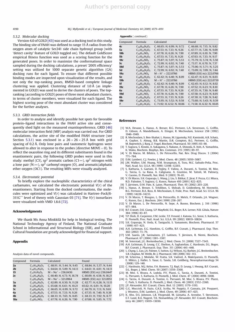
Analysis data of novel compounds.
Compound Formula Calculated Found
2a C17H16N2O3 C, 68.91; H, 5.44; N, 9.45 C, 68.84; H, 5.37; N, 9.442b C16H15N3O3 C, 64.64; H, 5.09; N, 14.13 C, 64.61; H, 4.81; N, 14.1315 C11H7N3O2 MþNaþ¼ 236.0436 HRMS (ESI) m/z 236.04472c C15H14N4O3 C, 60.40; H, 4.73; N, 18.78 C, 60.09; H, 4.58; N, 18.21
C15H14N4O3 MþNaþ¼ 321.0964 HRMS (ESI) m/z 321.09513a C13H16N2O3 C, 62.89; H, 6.50; N, 11.28 C, 62.77; H, 6.26; N, 11.153b C15H18N2O3 C, 65.68; H, 6.61; N, 10.21 65.62; H, 6.91; N, 10.203c C16H20N2O3 C, 66.65; H, 6.99; N, 9.72 C, 66.79; H, 7.13; N, 9.614a C17H22N2O3 C, 67.53; H, 7.33; N, 9.26 C, 67.35; H, 7.40; N, 9.384b C18H24N2O3 C, 68.33; H, 7.65; N, 8.85 C, 68.33; H, 7.92; N, 8.7718a C10H11NO2 C, 67.78; H, 6.26; N, 7.90 C, 67.88; H, 5.85; N, 7.72
Appendix ( continued )
Compound Formula Calculated Found
4c C16H20N2O3 C, 66.65; H, 6.99; N, 9.72 C, 66.68; H, 7.15; N, 9.825a C17H22N2O3 C, 67.53; H, 7.33; N, 9.26 C, 67.77; H, 7.26; N, 9.0918b C10H11NO2 C, 67.78; H, 6.26; N, 7.90 C, 67.89; H, 6.10; N, 7.905b C17H22N2O3 C, 67.53; H, 7.33, N, 9.26 C, 67.63; H, 7.33; N, 9.1718c C16H15NO2 C, 75.87; H, 5.97; N, 5.53 C, 75.79; H, 5.74; N, 5.505c C23H26N2O3 C, 72.99; H, 6.92; N, 7.40 C, 73.17; H, 6.70; N, 7.3718d C16H15NO2 C, 75.87; H, 5.97; N, 5.53 C, 75.96; H, 5.71; N, 5.255d C23H26N2O3 C, 72.99; H, 6.92; N, 7.40 C, 73.09; H, 7.09; N, 7.4419a C11H11NO4 MþHþ¼ 222.0766 HRMS (ESI) m/z 222.07665e C18H22N2O5 C, 62.42; H, 6.40; N, 8.09 C, 62.47; H, 6.15; N, 8.0319b C11H11NO4 MþHþ¼ 222.0766 HRMS (ESI) m/z 222.07595f C18H22N2O5 C, 62.42; H, 6.40; N, 8.09 C, 62.45; H, 6.12; N, 8.0219c C10H11NO2 C, 67.78; H, 6.26; N, 7.90 C, 67.52; H, 6.23; N, 8.106a C17H22N2O3 C, 67.53; H, 7.33; N, 9.26 C, 67.35; H, 7.30; N, 9.4019d C10H11NO2 C, 67.78; H, 6.26; N, 7.90 C, 67.58; H, 6.23; N, 8.056b C17H22N2O3 C, 67.53; H, 7.33; N, 9.26 C, 67.30; H, 7.28; N, 9.4218e C18H16N2O2 C, 73.95; H, 5.52; N, 9.58 C, 73.60; H, 5.43; N, 9.397 C25H27N3O3 C, 71.92; H, 6.52; N, 10.06 C, 71.58; H, 6.32; N, 10.05
M.J. Myllymaki et al. / European Journal of Medicinal Chemistry 44 (2009) 4179–41914190

Author's personal copy
[28] J.M. Keith, R. Apodaca, W. Xiao, M. Seierstad, K. Pattabiraman, J. Wu, M. Webb,M.J. Karbarz, S. Brown, S. Wilson, B. Scott, C.S. Tham, L. Luo, J. Palmer,M. Wennerholm, S. Chaplan, J.G. Breitenbucher, Bioorg. Med. Chem. Lett. 18(2008) 4838–4843.
[29] X. Wang, K. Sarris, K. Kage, D. Zhang, S.P. Brown, T. Kolasa, C. Surowy, O.F. ElKouhen, S.W. Muchmore, J.D. Brioni, A.O. Stewart, J. Med. Chem. 52 (2009)170–180.
[30] A. Minkkila, S.M. Saario, H. Kasnanen, J. Leppanen, A. Poso, T. Nevalainen, J.Med. Chem. 51 (2008) 7057–7060.
[31] W. Lang, C. Qin, S. Lin, A.D. Khanolkar, A. Goutopoulos, P. Fan, K. Abouzid,Z. Meng, D. Biegel, A. Makriyannis, J. Med. Chem. 42 (1999) 896–902.
[32] M.J. Myllymaki, S.M. Saario, A.O. Kataja, J.A. Castillo-Melendez, T. Nevalainen,R.O. Juvonen, T. Jarvinen, A.M.P. Koskinen, J. Med. Chem. 50 (2007)4236–4242.
[33] (a) H.A. McManus, P.J. Guiry, Chem. Rev. 104 (2004) 4151–4202;(b) M.J. Oila, J.E. Tois, A.M.P. Koskinen, Tetrahedron 61 (2005) 10748–10756.
[34] M.J. Myllymaki, A.M.P. Koskinen, Tetrahedron Lett. 48 (2007) 2295–2298.[35] J.H. Chesterfield, J.F.H. McOmie, M.S. Tute, J. Chem. Soc. (1960) 4590–4596.[36] B. Singh, G.Y. Lesher, Heterocycles 31 (1990) 2163–2172.[37] H. Vorbruggen, K. Krolikiewicz, Tetrahedron 49 (1993) 9353–9372.[38] I. Mohammadpoor-Baltork, A.R. Khosropour, S.F. Hojati, Synlett (2005)
2747–2750.[39] (a) H. Witte, W. Seeliger, Liebigs Ann. Chem. (1974) 996–1009;
(b) C. Bolm, K. Weickhardt, M. Zehnder, T. Ranzz, Chem. Ber.124 (1991) 1173–1180.[40] (a) T. Yaegashi, S. Nunomura, T. Okutome, T. Nakayama, M. Kurumi, Chem.
Pharm. Bull. 32 (1984) 4466–4477;(b) P.J. Reider, R.S. Eichen Conn, P. Davis, V.J. Grenda, A.J. Zambito,E.J.J. Grabowski, J. Org. Chem. 52 (1987) 3326–3334;(c) Y. Huang, D.R. Dalton, P.J. Carrol, J. Org. Chem. 62 (1997) 372–376.
[41] M. Mileni, D.S. Johnson, Z. Wang, D.S. Everdeen, M. Liimatta, B. Pabst,K. Bhattacharya, R.A. Nugent, S. Kamtekar, B.F. Cravatt, K. Ahn, R.C. Stevens,Proc. Natl. Acad. Sci. U.S.A. 105 (2008) 12820–12824.
[42] M.H. Bracey, M.A. Hanson, K.R. Masuda, R.C. Stevens, B.F. Cravatt, Science 298(2002) 1793–1796.
[43] (a) G. Jones, P. Willett, R.C. Glen, A.R. Leach, R. Taylor, J. Mol. Biol. 267 (1997)727–748;(b) M.J. Hartshorn, M.L. Verdonk, G. Chessari, S.C. Brewerton, W.T. Mooij,P.N. Mortenson, C.W. Murray, J. Med. Chem. 50 (2007) 726–741.
[44] A. Lodola, M. Mor, S. Rivara, C. Christov, G. Tarzia, D. Piomelli, A.J. Mulholland,Chem. Commun. (Camb) 13 (2008) 214–216.
[45] M. Mor, A. Lodola, S. Rivara, F. Vacondio, A. Duranti, A. Tontini, S. Sanchini,G. Piersanti, J.R. Clapper, A.R. King, G. Tarzia, D. Piomelli, J. Med. Chem. 51(2008) 3487–3498.
[46] GRID, v. 22a, Molecular Discovery Ltd., Pinner, Middlesex.[47] M. Hendlich, Acta Crystallogr. D. Biol. Crystallogr. 54 (1998) 1178–1182.[48] J. Badger, I. Minor, M.A. Oliveira, T.J. Smith, M.G. Rossmann, Proteins 6 (1989)
1–19.[49] J.S. Murray, S. Ranganathan, P. Politzer, J. Org. Chem. 56 (1991) 3734–3737.[50] P.W. Kenny, J. Chem. Soc., Perkin Trans. 2 (1994) 199–202.
[51] D. Zhang, A. Saraf, T. Kolasa, P. Bhatia, G.Z. Zheng, M. Patel, G.S. Lannoye,P. Richardson, A. Stewart, J.C. Rogers, J.D. Brioni, C.S. Surowy, Neuropharma-cology 52 (2007) 1095–1105.
[52] V.V. Somayajulu, N.V. Subba Rao, Proc. Indian Acad. Sci. Sect. A 59 (1964)396–402.
[53] S.M. Johnson, S. Connelly, I.A. Wilson, J.W. Kelly, J. Med. Chem. 51 (2008)260–270.
[54] T.-Y. Shen, R.L. Clark, A.A. Pessolano, B.E. Witzel, T.J. Lanza, US 4038396, 1977,CAN 90:137799.
[55] T.J. Kress, J. Org. Chem. 50 (1985) 3073–3076.[56] F. Dennin, D. Blondeau, H. Sliwa, J. Heterocycl. Chem. 27 (1990) 1963–1967.[57] J.F. McOmie, A.B. Turner, J. Chem. Soc. (1963) 5590–5593.[58] J. Luston, J. Kronek, F. Bohme, J. Polym. Sci. Part A: Polym. Chem. 44 (2005)
343–355.[59] M. Bradford, Anal. Biochem. 72 (1976) 248–254.[60] A. Lorenzen, M. Fuss, H. Vogt, U. Schwabe, Mol. Pharmacol. 44 (1993)
115–123.[61] K.M. Kurkinen, J. Koistinaho, J.T. Laitinen, Brain Res. 769 (1997) 21–28.[62] J.R. Savinainen, T. Jarvinen, K. Laine, J.T. Laitinen, Br. J. Pharmacol. 134 (2001)
664–672.[63] S.M. Saario, A. Poso, R.O. Juvonen, T. Jarvinen, O.M.H. Salo-Ahen, J. Med. Chem.
49 (2006) 4650–4656.[64] S.M. Saario, O.M. Salo, T. Nevalainen, A. Poso, J.T. Laitinen, T. Jarvinen, R. Niemi,
Chem. Biol. 12 (2005) 649–656.[65] S.C. Lovell, J.M. Word, J.S. Richardson, D.C. Richardson, Proteins 40 (2000)
389–408.[66] J.M. Wang, P. Cieplak, P.A. Kollman, J. Comput. Chem. 21 (2000) 1049–1074.[67] Sybyl v. 8.1; Tripos Associates, Inc.: St. Louis, MO.[68] (a) http://molprobity.biochem.duke.edu/ (retrieved 01.03.09);
(b) S.C. Lovell, I.W. Davis, W.B. Arendall, P.I. de Bakker, J.M. Word, M.G. Prisant,J.S. Richardson, D.C. Richardson, Proteins 50 (2003) 437–450.
[69] M.K. McKinney, B.F. Cravatt, J. Biol. Chem. 278 (2003) 37393–37399.[70] T.A. Halgren, J. Comput. Chem. 17 (1996) 490–519.[71] M.J. Frisch, G.W. Trucks, H.B. Schlegel, G.E. Scuseria, M.A. Robb, J.R. Cheeseman,
J.A. Montgomery, T. Vreven, K.N. Kudin, J.C. Burant, J.M. Millam, S.S. Iyengar,J. Tomasi, V. Barone, B. Mennucci, M. Cossi, G. Scalmani, N. Rega, G.A. Petersson,H. Nakatsuji, M. Hada, M. Ehara, K. Toyota, R. Fukuda, J. Hasegawa, M. Ishida,T. Nakajima, Y. Honda, O. Kitao, H. Nakai, M. Klene, X. Li, J.E. Knox, H.P. Hratchian,J.B. Cross, V. Bakken, C. Adamo, J. Jaramillo, R. Gomperts, R.E. Stratmann,O. Yazyev, A.J. Austin, R. Cammi, C. Pomelli, J.W. Ochterski, P.Y. Ayala,K. Morokuma, G.A. Voth, P. Salvador, J.J. Dannenberg, V.G. Zakrzewski,S. Dapprich, A.D. Daniels, M.C. Strain, O. Farkas, D.K. Malick, A.D. Rabuck,K. Raghavachari, J.B. Foresman, J.V. Ortiz, Q. Cui, A.G. Baboul, S. Clifford,J. Cioslowski, B.B. Stefanov, G. Liu, A. Liashenko, P. Piskorz, I. Komaromi,R.L. Martin, D.J. Fox, T. Keith, M.A. Al-Laham, C.Y. Peng, A. Nanayakkara,M. Challacombe, P.M.W. Gill, B. Johnson, W. Chen, M.W. Wong, C. Gonzalez,J.A. Pople, Gaussian 03, Revision B.04, Gaussian, Inc., Wallingford, CT, 2004.
[72] W. Humphrey, A. Dalke, K. Schulten, J. Mol. Graphics 14 (1996) 33–38pp. 27–28.
M.J. Myllymaki et al. / European Journal of Medicinal Chemistry 44 (2009) 4179–4191 4191