childhood leprosy: a retrospective descriptive study from government medical college, kozhikode,...
TRANSCRIPT
Childhood leprosy: A retrospective descriptive
study from Government Medical College,
Kozhikode, Kerala, India
SARITA SASIDHARANPILLAI, MANIKOTH
PAYYANADAN BINITHA, NAJEEBA RIYAZ,
BETSY AMBOOKEN, OLASSERI KALATHINGAL
REENA MARIYATH, BIJU GEORGE, ANISHA
KANHIRANGATTIL JANARDHANAN &
PENTAM VELI BEEGUM SHERJEENA
Government Medical College, Kozhikode, Kerala, India
Accepted for publication 14 June 2014
Summary
Objective: To assess the profile and describe the clinical presentations and
complications of childhood leprosy in a tertiary care hospital in North Kerala, South
India during 2003–2012 and to analyse any change in the age-sex profile and the
clinical pattern of leprosy in children below the age of 15 years over the 10-year
study period.
Design: A retrospective descriptive study of children less than 15 years of age
diagnosed with leprosy and registered for treatment in a tertiary care institution from
2003 to 2012. Demographic, clinical, investigative and treatment data were collected
using a pre-set proforma.
Results: 138 (12·1%) of the total 1143 leprosy cases registered for treatment during
the 10-year period were below 15 years of age. The 10-year study period witnessed a
statistically insignificant decrease in the new childhood leprosy cases registered for
treatment in our tertiary care institution. The majority of cases belonged to the 6–12
year age group (61·6%) with a male predominance. Borderline tuberculoid (BT) was
the commonest clinical type (65·9%) followed by indeterminate leprosy (18·8%); 101
patients required paucibacillary (PB) and 37 needed multibacillary (MB) treatment.
The number of patients requiring MB treatment showed a statistically significant
increase and there was a significant decline in number of cases requiring PB
treatment. During the entire study period no Type 2 lepra reaction was documented
in patients below Hema 15 years and only two patients manifested Type 1 reaction.
Ten (7·2%) out of the 138 patients were cases of relapse. There was a clear female
predilection among relapse cases with the majority belonging to the adolescent age.
Conclusions: Childhood leprosy still contributes to a significant proportion of
the total case load denoting the continuing active horizontal transmission of leprosy.
Correspondence to: Sarita Sasidharanpillai, Assistant Professor, Department of Dermatology, GovernmentMedical College, Kozhikode, Kerala, India (e-mail: [email protected])
Lepr Rev (2014) 85, 100–110
100 0305-7518/14/064053+11 $1.00 q Lepra
The rise in number of patients with more extensive disease in the background of
declining disease prevalence is suggestive of the delay in diagnosis and treatment.
A high relapse rate noted in the present study may be due to incorrect classification
and treatment of MB as PB leprosy which in turn might have resulted in treatment
failure due to inadequate treatment.
Introduction
Elimination of leprosy as a public health problem is one of the major success stories of
modern medicine. This was achieved at the global level in 2000 and India attained this status
in January 2006.1,2 Leprosy elimination as a public health problem is defined as the reduction
of disease prevalence to less than one per 10,000 population. In other infections elimination
is defined as a reduction to zero of infection caused by a specified agent in a defined
geographical area through deliberate efforts.2 If we use this criteria, leprosy is definitely not
eliminated from the world.
With the declaration of ‘leprosy elimination’, leprosy services were integrated into the
general health system. This was done with a view to ensure treatment to the affected at their
nearest centre and to reduce the stigma associated with the disease. But recently Odisha, one
of the first states in India to have disintegrated leprosy services reported a rise in prevalence
of the disease.3
Data based on the prevalence of registered cases for treatment may not reveal much about
the current status of leprosy, as the shortened duration of fixed duration treatment (from
2 years to 1 year in multibacillary cases) in itself reduces the number of patients on treatment.
Moreover, it was often suggested that the rapid decline reported in number of leprosy
cases from many parts of the world, was unlikely in a disease with such a long incubation
period. It is possible that in order to achieve the elimination status, countries might have
manipulated the statistics.4
It is often suggested that childhood leprosy can serve as a better tool to assess the disease
transmission in the society.5 Many studies in the post-elimination era documented a
significant number of childhood cases including smear positive cases, pointing to the active
disease transmission still taking place.5,6
Hence we decided to conduct a retrospective descriptive study on leprosy among patients
below 15 years who attended the outpatient department (OPD) the of Government Medical
College, Kozhikode with cardinal features of leprosy from 2003 January to 2012 December.
Materials and methods
STUDY DESIGN: RETROSPECTIVE DESCRIPTIVE
This study was a retrospective analysis of all leprosy cases less than 15 years of age,
who registered for treatment at the Dermatology department of the Government Medical
College, Kozhikode from January 2003 to December 2012. This institution is a tertiary-care
teaching hospital situated in the north of Kerala, a state in South India. Ethical clearance was
obtained from the institutional ethics committee of Government Medical College, Kozhikode
on 23.5.2013.
Childhood Leprosy 101
A diagnosis of leprosy was made, when a patient presented with any of the cardinal
features of leprosy (asymptomatic hypopigmented or erythematous skin lesion with definite
loss or impairment of sensation or thickened peripheral nerve with sensory impairment in the
area supplied by the nerve or skin smear positive for acid fast bacilli).
A pre-set proforma was used to collect data regarding age, sex, possible source of contact,
clinical findings and investigations from previous case records. A ‘household’ or ‘intra-
familial’ contact was defined as any person with current or past history of leprosy in the
immediate family (parents, siblings, grandparents) living in the same house and partaking in
meals from a common kitchen.5 Known cases from the immediate neighbourhood of the
patient’s house were considered as ‘extra-familial’ contacts.5 Clinical features including size,
site, morphology and number of skin lesions were noted. Nerve function impairment (NFI),
when present was charted out. Sensory impairment was detected by the inability or reduced
ability to appreciate temperature, pain and fine touch. Test tubes containing water at 408C and
at 258C were used to test temperature sensation, pain sensation was tested using pin prick and
a wisp of cotton was used to check fine touch. Motor impairment was diagnosed when less
than grade 5 power was recorded on voluntary muscle testing. Criteria by the World Health
Organisation was used for disability grading.7
From each leprosy patient one smear from an ear lobe smear and at least two more slit
skin smears (from representative skin lesion and normal skin) were taken routinely in our
institution and were stained with Ziehl-Neelsen technique to determine the morphological
and bacteriological indices. Biopsies from the leprosy lesions were stained using
haematoxylin and eosin to study the morphology and Wade Fite (modified Ziehl-Neelsen
staining technique) to identify the acid fast bacilli (AFB).
Patients were categorised as per the Ridley-Jopling classification.8
Patients who had only nerve lesions without any cutaneous manifestations and who
satisfied the cardinal criteria for leprosy were placed under the category of neuritic leprosy
and patients who manifested vague hypopigmented patches with doubtful sensory
impairment and perivascular and peri-appendageal lymphocytic infiltration on histology
were diagnosed as indeterminate leprosy.
Patients who had features of Type 1(T1R) or Type 2 (T2R) lepra reaction were classified
accordingly. (A clinical diagnosis of T1R was made when a patient in the borderline spectrum
of leprosy had acute onset of eryt and oedema of skin lesions with or without neuritis and
oedema of the hands, feet and face.9 T2R was diagnosed when a BL or LL patient had crops
of tender subcutaneous skin lesions with or without accompanying neuritis, iritis, arthritis,
orchitis, dactylitis, lymphadenopathy, oedema and fever.9
The disease spectrum as well as the treatment received were documented. Patients
presenting with six or more skin lesions or two or more enlarged nerve trunks or skin smear
positivity for acid fast bacilli were treated with multibacillary regimen and patients
presenting with less than six skin lesions, less than two enlarged nerve trunks and a negative
skin smear for acid fast bacilli were treated with paucibacillary regimen.10
The number of patients who had developed Grade 2 disability at the time of initial
presentation were documented. Grade 2 disability was defined as the presence of visible
deformity or damage (ulceration, shortening, disorganization, stiffness and loss of part of or
all of the hand or foot) affecting hands and feet due to leprosy or visual acuity less than 6/60
or inability to count fingers at a distance of six meters caused by leprosy.10
Patients who attended the OPD with suspected leprosy relapses were noted and data
regarding the previous disease spectrum and previous treatment received were documented.
S. Sasidharanpillai et al.102
Relapse in leprosy was diagnosed when a patient who successfully completed an adequate
course of multidrug therapy, subsequently developed new signs and symptoms of the disease,
either during the surveillance period (2 years for PB and 5 years for MB leprosy) or
thereafter.5
The data was studied with respect to the age and sex distribution, clinical features,
complications and the treatment received by leprosy patients below the age of 15 years. The
proportion of new childhood leprosy cases with respect to the total number of new leprosy
patients who attended our institution during the 10-year period was also studied. The
epidemiology of the new childhood leprosy patients who attended our institution over the
decade (2003–2012) was analysed using chi-square test for linear trend using the StatCal
component of the Epi Info version 7·1·2 and an attempt was made to detect any change in the
pattern over the years.
Results
138 (12·1%) of the total 1143 leprosy patients registered for treatment in our institution from
2003 to 2012 were below the age of 15 (Table 1).
The number of new childhood leprosy cases showed a statistically insignificant decline
over the 10-year study period (P value 0·16).
The majority of cases belonged to the 6–12 years age group (61·6%, Table 2) which
remained unchanged throughout the 10-year interval (P value 0·301).
The age of the affected children ranged from 2 to 15 years.
There was a clear male predilection; (Table 2) no significant change was observed in the
sex distribution of the affected over the years (P value 0·163).
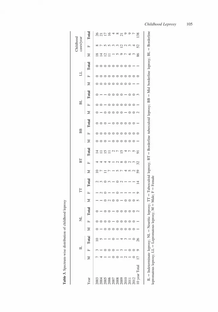
The commonest disease spectrum throughout the duration of the study was BT. Only
3·6% of our patients belonged to mid borderline (BB), borderline lepromatous (BL) and
lepromatous leprosy (LL) spectra (Table 3).
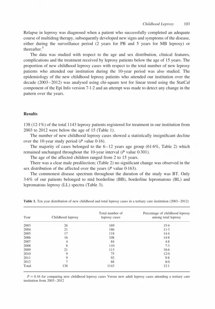
Table 1. Ten year distribution of new childhood and total leprosy cases in a tertiary care institution (2003–2012)
Year Childhood leprosyTotal number ofleprosy cases
Percentage of childhood leprosyamong total leprosy
2003 26 169 15·42004 21 186 11·32005 17 118 14·42006 16 108 14·82007 4 84 4·82008 8 110 7·32009 21 113 18·62010 9 75 12·02011 9 92 9·82012 7 88 8·0Total 138 1143 12·1
P ¼ 0.16 for comparing new childhood leprosy cases Versus new adult leprosy cases attending a tertiary careinstitution from 2003–2012
Childhood Leprosy 103

Seventeen patients (12·3%) gave a history of contact with a leprosy case; 15 of them were
household contacts and two were family members living in a nearby house. The 2-year old
who presented with BT had a household contact in his paternal grandfather who was
receiving treatment for BL.
Of the 138, 73·2% required PB treatment and the rest received MB treatment [Figure 1].
The number of patients requiring MB treatment showed a statistically significant increase
and there was a significant decline in number of cases requiring PB treatment (P value 0·009,
Figure 2). 3·6% of the total were smear positive for AFB.
Enlarged nerves were detected in 45·7% (Figure 2) with 23·9% manifesting multiple
nerve thickening. A statistically significant increase was noted in number of patients
presenting with nerve involvement (P value 0·001, Figure 3) and an increase (though
statistically insignificant, P value 0·16) was noted for those presenting with multiple nerve
involvement.
Grade 2 disability was observed in eight patients. All were in the 13 to 15 age group.
Seven were males. Four children suffered from clawing of fingers and foot drop developed
in two. They were treated with steroids and physiotherapy and two patients had residual
weakness. Two children at the time of diagnosis had trophic ulcers affecting the heel and the
ball of the great toe respectively.
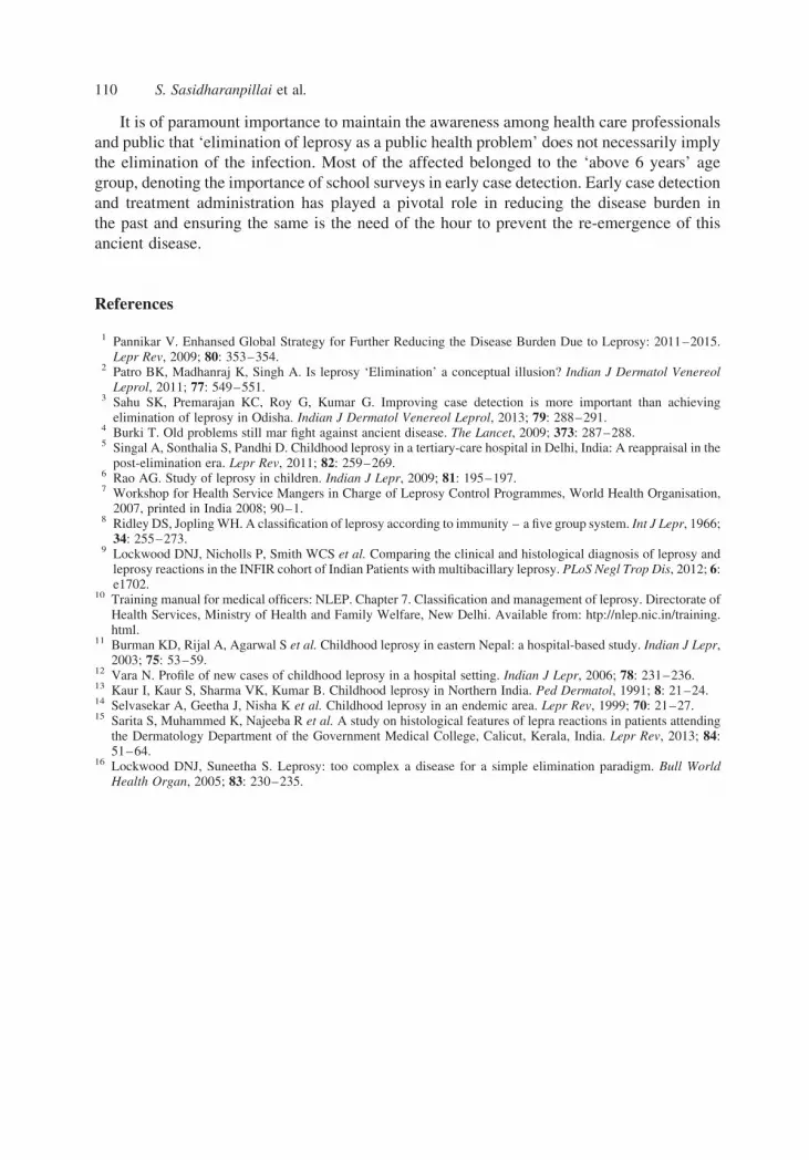
One of the male patients with Grade 2 disability developed nerve palsy as part
of the neuritis of Type 1 reaction. Only one more patient (a 9-year old girl) developed
Type 1 reaction during the study period that manifested as neuritis and ulcerated skin lesions
(Figure 4).
During the entire study period no Type 2 reaction was documented in patients below the
age of 15.
Ten patients (7·2%) were diagnosed as relapse cases. Seven of them had received
multidrug therapy from other institutions and were referred to us when symptoms reappeared
Table 2. Age and sex-wise distribution of childhood leprosy in a tertiary care institution during the ten year studyperiod from 2003–2012
Age in years
, 6 6 – 12 13–15 Yearly No. of cases
Year M F Total M F Total M F Total Total M Total F Grand Total
2003 0 0 0 12 8 20 6 6 18 8 262004 2 0 2 5 3 8 7 4 11 14 7 212005 1 0 1 5 3 8 6 2 8 12 5 172006 0 0 0 5 2 7 6 3 9 11 5 162007 0 0 0 1 2 3 1 1 1 3 42008 0 0 0 4 2 6 1 1 2 5 3 82009 1 0 1 5 11 16 3 1 4 9 12 212010 0 0 0 5 2 7 2 2 7 2 92011 1 0 1 4 2 6 1 1 2 6 3 92012 0 0 0 1 3 4 2 1 3 3 4 710 year total 5 0 5 47 38 85 34 14 48 86 52 138
M ¼ Male; F ¼ FemaleP ¼ 0.163 for comparing new male childhood leprosy cases versus new female childhood leprosy cases
attending a tertiary care institution from 2003–2012. P ¼ 0.301 for comparing new childhood leprosy cases in thebelow 6, 6–12 and 13–15 age groups attending a tertiary care institution from 2003–2012
S. Sasidharanpillai et al.104
Table
3.Spectrum-w
isedistributionofchildhoodleprosy
ILNL
TT
BT
BB
BL
LL
Childhood
cases/year
Year
MF
Total
MF
Total
MF
Total
MF
Total
MF
Total
MF
Total
MF
Total
MF
Total
2003
73
10
00
01
12
10
414
00
00
00
00
018
826
2004
41
51
01
12
37
411
00
01
01
00
014
721
2005
10
10
00
00
011
415
00
00
11
00
012
517
2006
10
10
00
21
37
411
10
10
00
00
011
516
2007
00
00
11
01
11
12
00
00
00
00
01
34
2008
01
10
00
00
05
27
00
00
00
00
05
38
2009
13
40
00
11
27
815
00
00
00
00
09
12
21
2010
21
30
00
01
14
04
00
01
01
00
07
29
2011
10
10
00
01
15
27
00
00
00
00
06
39
2012
00
00
00
01
12
35
00
00
00
10
13
47
10yearTotal
17
926
11
25
914
59
32
91
10
12
13
10
186
52
138
IL¼
Indeterminateleprosy;NL¼
Neuriticleprosy;TT¼
Tuberculoid
leprosy;BT¼
Borderlinetuberculoid
leprosy;BB¼
Mid
borderlineleprosy;BL¼
Borderline
lepromatousleprosy;LL¼
Lepromatousleprosy;M
¼Male;
F¼
Fem
ale
Childhood Leprosy 105

after the completion of treatment. All except one had documents of receiving regular
treatment. One child did not have any details of previous treatment, but according to her
parents received regular treatment for the recommended duration. Eight out of the 10 were
girls, indicating a clear female predilection (P value 0·004). One girl and one boy were 9 years
and 10 years old respectively at the time of the relapse and the rest were 12 to 15 years of age.
Eight out of the 10 were treated with PB MDT in the past and the interval between the
Figure 2. Thickened nerve in childhood leprosy.
30
25
20
15
PBMB
10No.
of p
atie
nts
5
02003 2004 2005 2006 2007 2008
Year
2009 2010 2011 2012
Figure 1. New childhood leprosy cases registered for PB and MB treatment in a tertiary care institution from2003 to 2012.
S. Sasidharanpillai et al.106

completion of treatment and the relapse varied from 3 months to 2 years. All relapsed cases
manifested clinical features warranting re-treatment with MB MDT.
Discussion
Seven years after the declaration of the elimination of leprosy, childhood leprosy still
contributes to a significant proportion of the total case load and this denotes that active
horizontal transmission is still taking place. The childhood cases contributing to 12·1% of the
total, noted in our study was much higher than the 4·5% reported by Burman et al.,9 but was
comparable to the findings in certain other studies.4,9
The commonly affected age group remained 6–12 years which was consistent with the
incubation period of the disease. Another study in the post-elimination era documented the
majority of patients in the above 11 age group.5
The fact that we could identify the contact case in only 17 of our patients points to the
hidden cases in the community. Similar findings were reported in some studies5,12,13 but
higher figures were noted in other studies.14 This suggests that even in low endemic areas,
mass leprosy detection camps have a role to play.
Our observation of predominance of BT was in concordance with earlier studies.5,11–14
Compared to other studies our study documented less patients with extensive disease with
respect to nerve involvement, treatment required (MB MDT), smear positivity and Grade 2
disability at initial presentation.5,11,12 But the number of patients requiring MB treatment and
those with multiple nerve involvement showed a rise during the later years of the study.
The increase observed in cases requiring multibacillary treatment including those with
multiple nerve involvement could be due to a waning of disease-specific immunity in the
community that has taken place with the decline in disease prevalence (leading to extensive
disease manifestation in the affected). The single LL case observed in the study group
30
25
20
15
10
5
No.
of p
atie
nts
02003 2004 2005 2006 2007 2008 2009 2010 2011 2012
>3 nerve trunk involvement
3 nerve trunk involvement
2 nerve trunk involvement
<2 nerve trunk involvement
No nerve trunk involvement
Year
Figure 3. Nerve trunk involvement in new childhood leprosy cases attending a tertiary care institution from 2003to 2012.
Childhood Leprosy 107

presented during the last year of the study. Another possible explanation is the delay in
diagnosis that has been already reported from different parts of the world due to the
diminishing expertise in detecting early leprosy lesions.4 With the declaration of ‘leprosy
elimination’, there is a waning of awareness of leprosy among the health care workers and the
general population.4 Experts had predicted that as the prevalence of leprosy comes down, the
major challenge will be in making the correct diagnosis early, in those affected.4
5·8% of the study group developed Grade 2 disability at initial presentation. An increase
documented in Grade 2 disability among the newly registered adult leprosy patients who
attended our institution during the same interval was not observed in childhood leprosy.
The development of severe T1R in a 9-year old girl observed in our study highlights the
need to look for this complication in childhood leprosy as a delay in diagnosis and treatment
of the same can lead to irreversible nerve damage.
The majority of relapse cases in our study belonged to the 12 to 15 years age group as
expected in a disease with a prolonged incubation period. There was a striking female
predominance noted in relapse cases which was contrary to the sex profile observed in the
total number of leprosy cases. The higher susceptibility of adolescent females to leprosy
relapse may be attributed to the hormonal changes and the subsequent alterations taking place
in the immune system with menarche.
Figure 4. Ulcerated skin lesion in type 1 lepra reaction.
S. Sasidharanpillai et al.108

Our study documented a higher relapse rate compared to some other studies;5 80% of the
relapses occurred following PB treatment and 70% had received previous treatment from
other institutions. All 10 patients, on evaluation were found to have disease requiring
retreatment with MB regimen. Probably a wrong classification of MB as PB disease and
subsequent inadequate treatment might have contributed to the high relapse rate. One patient
did not have any documents regarding previous treatment received, hence we could not rule
out the possibility of irregular treatment.
All the relapsed cases in our study developed reappearance of symptoms within 2 years of
completion of treatment. The major diagnostic challenge was in differentiating relapse from
late T1R. None of them showed any clinical evidence of lepra reactions.9 One patient came to
us with suspected relapse within 3 months of completion of treatment. This patient after
completion of MDT complained of the appearance of a new skin lesion with sensory
impairment and without any clinical evidence of lepra reaction. The appearance of a non-
tender, non-erythematous new single skin lesion satisfying the cardinal criteria for leprosy in
a treated patient was more in favour of a relapse than a lepra reaction as the latter is usually
associated with multiple erythematous oedematous tender skin lesions. In spite of this, in
view of the short interval between the completion of the treatment and the appearance of
the new skin lesion, the patient was given a therapeutic trial with systemic steroids. Since
the patient did not show any improvement after 4 weeks, and as the histopathology analysis of
the new skin lesion revealed borderline tuberculoid leprosy with no evidence of dermal or
intra-granuloma oedema, a final diagnosis of relapse was made rather than late T1R.15 The
patient gradually responded to standard MB-MDT as she had enlargement of two nerve
trunks (without clinical evidence of neuritis) in addition to the skin lesion.
The major limitations of our study was the small sample size (138 cases over 10 years)
and the dependence on data collected from previous case records in a tertiary care institution.
This data does not reflect the status of the disease in the general population, as the more
severely affected patients usually seek advice in a tertiary referral unit.
Conclusions
Leprosy transmission still takes place as reflected by the significant portion of childhood
cases. The rise in number of patients with more extensive disease in the background of
declining disease prevalence highlights the need to reassess the efficacy of the existing
system in early case detection. Diagnosing leprosy requires training. For the successful
implementation of leprosy services provided through the general health system, training of
health workers, regular refresher classes and continuous monitoring are essential. Health care
workers should be proficient in identifying anaesthetic skin lesions and enlarged nerves, as
about 30% of the MB cases will be missed if a patch with sensory impairment is taken as the
single criterion to diagnosis leprosy.15 Inexperienced health workers may miss the early
lesions of leprosy, leading to delay in diagnosis or may incorrectly classify and treat MB as
PB leprosy, thus causing treatment failure. This incorrect classification could be the reason
for the high relapse rate noted in the present study especially following PB regimen. But the
female preponderance noted among the relapse cases cannot be ignored. We recommend that
after completion of treatment, affected female children should be kept under follow-up
throughout the adolescent years, as female sex and pubertal age group are found to be the
major risk factors for relapse in our study.
Childhood Leprosy 109
It is of paramount importance to maintain the awareness among health care professionals
and public that ‘elimination of leprosy as a public health problem’ does not necessarily imply
the elimination of the infection. Most of the affected belonged to the ‘above 6 years’ age
group, denoting the importance of school surveys in early case detection. Early case detection
and treatment administration has played a pivotal role in reducing the disease burden in
the past and ensuring the same is the need of the hour to prevent the re-emergence of this
ancient disease.
References
1 Pannikar V. Enhansed Global Strategy for Further Reducing the Disease Burden Due to Leprosy: 2011–2015.Lepr Rev, 2009; 80: 353–354.
2 Patro BK, Madhanraj K, Singh A. Is leprosy ‘Elimination’ a conceptual illusion? Indian J Dermatol VenereolLeprol, 2011; 77: 549–551.
3 Sahu SK, Premarajan KC, Roy G, Kumar G. Improving case detection is more important than achievingelimination of leprosy in Odisha. Indian J Dermatol Venereol Leprol, 2013; 79: 288–291.
4 Burki T. Old problems still mar fight against ancient disease. The Lancet, 2009; 373: 287–288.5 Singal A, Sonthalia S, Pandhi D. Childhood leprosy in a tertiary-care hospital in Delhi, India: A reappraisal in thepost-elimination era. Lepr Rev, 2011; 82: 259–269.
6 Rao AG. Study of leprosy in children. Indian J Lepr, 2009; 81: 195–197.7 Workshop for Health Service Mangers in Charge of Leprosy Control Programmes, World Health Organisation,2007, printed in India 2008; 90–1.
8 Ridley DS, JoplingWH. A classification of leprosy according to immunity – a five group system. Int J Lepr, 1966;34: 255–273.
9 Lockwood DNJ, Nicholls P, Smith WCS et al. Comparing the clinical and histological diagnosis of leprosy andleprosy reactions in the INFIR cohort of Indian Patients with multibacillary leprosy. PLoS Negl Trop Dis, 2012; 6:e1702.
10 Training manual for medical officers: NLEP. Chapter 7. Classification and management of leprosy. Directorate ofHealth Services, Ministry of Health and Family Welfare, New Delhi. Available from: htp://nlep.nic.in/training.html.
11 Burman KD, Rijal A, Agarwal S et al. Childhood leprosy in eastern Nepal: a hospital-based study. Indian J Lepr,2003; 75: 53–59.
12 Vara N. Profile of new cases of childhood leprosy in a hospital setting. Indian J Lepr, 2006; 78: 231–236.13 Kaur I, Kaur S, Sharma VK, Kumar B. Childhood leprosy in Northern India. Ped Dermatol, 1991; 8: 21–24.14 Selvasekar A, Geetha J, Nisha K et al. Childhood leprosy in an endemic area. Lepr Rev, 1999; 70: 21–27.15 Sarita S, Muhammed K, Najeeba R et al. A study on histological features of lepra reactions in patients attending
the Dermatology Department of the Government Medical College, Calicut, Kerala, India. Lepr Rev, 2013; 84:51–64.
16 Lockwood DNJ, Suneetha S. Leprosy: too complex a disease for a simple elimination paradigm. Bull WorldHealth Organ, 2005; 83: 230–235.
S. Sasidharanpillai et al.110