chemistry, structure and function of ... - ibn
TRANSCRIPT

NATIONAL ACADEMY OF SCIENCES OF BELARUS THE DEPARTMENT OF CHEMISTRY AND EARTH SCIENCES
INSTUTE OF BIOORGANIC CHEMISTRY
BELARUSIAN REPUBLICAN FOUNDATION FOR FUNDAMENTAL RESEARCH
VI INTERNATIONAL CONFERENCE
CHEMISTRY, STRUCTURE AND FUNCTION OF BIOMOLECULES
Book of Abstracts
Minsk, May 22-25, 2018
VI International Conference “Chemistry, Structure and Function of Biomolecules”
ii
The present book contains abstracts of the VIth International Conference “Chemistry, Structure and Function of Biomolecules”, which was held in Minsk on May 22-25, 2018. The Conference materials cover a wide spectrum of scientific problems from the isolation from nature and structure elucidation, synthesis and biosynthesis, to bioactivity investigation, and practical applications of biologically active compounds. The abstract book contains abstracts as provided by their authors except for minor editing for consistency in style. Organizing/ScientificCommittee
Chairman: Usanov Sergey, Presidium of the National Academy of Sciences of Belarus, Minsk; Vice-chairman: Babitskaya Svetlana, Institute of Bioorganic Chemistry NASB, Minsk; Scientific secretary: Khripach Natalia, Institute of Bioorganic Chemistry NASB, Minsk. Members: Agabekov Vladimir, Institute of Chemistry of New Materials, Minsk; Bildyukevich Alexandr, Institute of Physical Organic Chemistry, Minsk; Baranovsky Alexandr, Institute of Bioorganic Chemistry NASB, Minsk; Drasar Pavel, University of Chemistry and Technology, Prague; Gilep Andrey, Institute of Bioorganic Chemistry NASB, Minsk; Golubovich Vladimir, Institute of Bioorganic Chemistry NASB, Minsk; Ivashkevich Oleg, Belarusian State University, Minsk; Kalinichenko Elena, Institute of Bioorganic Chemistry NASB, Minsk; Kisel Mikhail, Institute of Bioorganic Chemistry NASB, Minsk; Khripach Vladimir, Institute of Bioorganic Chemistry NASB, Minsk; Lakhvich Fiodor, Institute of Bioorganic Chemistry NASB, Minsk; Mikhailopuluo Igor; Institute of Bioorganic Chemistry NASB, Minsk; Nasek Vladimir, Institute of Bioorganic Chemistry NASB, Minsk; Petrov Piotr, Institute of Bioorganic Chemistry NASB, Minsk; Sivetz Grigorii, Institute of Bioorganic Chemistry NASB, Minsk; Sviridov Oleg, Institute of Bioorganic Chemistry NASB, Minsk; Sviridov Dmitrii, Belarusian State University, Minsk; Zhabinskii Vladimir, Institute of Bioorganic Chemistry NASB, Minsk; Yantsevich Alexey Institute of Bioorganic Chemistry NASB, Minsk.
Editorialboard:
Zhabinskii Vladimir, Institute of Bioorganic Chemistry NASB, Minsk; Khripach Vladimir, Institute of Bioorganic Chemistry NASB, Minsk; Khripach Natalia, Institute of Bioorganic Chemistry NASB, Minsk.
© Institute of Bioorganic Chemistry NASB, 2018
Contents
Plenary Lectures
Borshchevskiy V., Mishin A., Marin E., Luginina A., Gusach A., Kovalev K., Volkov O., Cherezov V., Gordeliy V. Structural studies of 7‐TM membrane proteins ........................................................................................................................... 1
Chukicheva I.Yu., Buravlev E.V., Dvornikova I.A., Fedorova I.V., Shchukina O.V., Belykh D.V., Khudyaeva I.S., Kutchin A.V. Terpenylphenols and porphyrins as perspective platform for the formation of new biomolecules ................. 3
Demidchik V., Straltsova D., Charnysh M., Chikun P., Przhevalskaya D., Zhabinskii V.N., Khripach V.A., Sokolik A. Brassinosteroids as regulators plant Ca2+ signaling, ion channel activities growth and signalling in roots of higher plants ............................................................................................................................... 5
Deyev S.M. Supramolecular structures for biomedicine ................................................ 7
Drašar P.B. Matrix assisted supramolecular chirality amplification with natural products .......................................................................................................................... 9
Efimova M.V. The regulation of light‐dependent gene expression by brassinosteroids ............................................................................................................ 12
Grudinin S. Using machine learning to predict protein structure and interactions .................................................................................................................... 15
Hurski A.L. Application of C‐H activation and hydroxycyclopropanation strategies in the synthesis of steroids ........................................................................... 16
Ivanov A.S. Surface plasmon resonance (SPR) biosensors Biacore in biomolecules research .................................................................................................. 17
Levina I.S., Kuznetsov Y.V. Selective modulators of estrogen and progesterone receptors: steroidal agonists and antagonists ........................................ 19
Hryniewicka A., Łotowski Z., Seroka B., Witkowski S., Morzycki J.W. Synthesis of a cisplatin derivative from lithocholic acid................................................ 21
Boldescu V., Curlat S., Pogrebnoi S., Smetanscaia A., Uncu L., Valica V., Macaev F. Molecular architecture of ionic liquids with anticancer activity, antioxidant, and photosenisibilizing properties ............................................................ 22
Chen Y., Paetz C., Schneider B. Precursor‐directed synthesis of new phenylbenzoisoquinolindione alkaloids and the discovery of a phenylphenalenone‐based plant defense mechanism ................................................. 24
Oral Communications
Bocharov E. Signal transduction via transmembrane domains of bitopic receptors in norma and pathology ................................................................................ 27
VI International Conference “Chemistry, Structure and Function of Biomolecules”
iv
Bocharova O. APP familiar mutations as a tool for investigation of the molecular basis of Alzheimer disease ........................................................................... 28
Bukhdruker S., Varaksa T., Marin E., Kovalev K., Shevtsov M., Luginina A., Gusach A., Mishin A., Gilep A., Strushkevich N., Borshchevskiy V. X‐ray diffraction analysis of cytochromes P450 ..................................................................... 30
Dormeshkin D. , Katsin M., Migas A., Meleshko A., Gilep A. Engineering chimeric antigen receptor (CAR) T‐cells for enhanced cancer immunotherapy ........... 32
Dudek B., Warskulat A.‐C., Tatsis E., Schnurrer F., Paetz Ch., Schneider B. From red to yellow: the biosynthesis of unique indole alkaloids in yellow Papaver nudicaule flowers and their biomimetic synthesis.......................................... 33
Dzichenka Ya.V., Shkel T.V. Faletrov Ya.V. Novel NBD‐labeled ligands of CYP51............................................................................................................................. 34
Efimov A.V. Selection of right‐ or left‐handed structural motifs depends on their arrangement in protein structure ......................................................................... 36
Gilep A.A., Sushko S.A., Shkel T.V., Svirid A.V., Smolskaya S.V., Vasilevskaya A.V., Usanov S.A., Ershov P.V., Ivanov A.S., Strushkevich N.V. Functional analysis of cytochrome P450s involved in biosynthesis of autocrine and paracrine factors ........................................................................................................... 37
Gilevich S., Brechka Y. Active site docking and in vitro inhibitory activity of some N‐heterocyclic and carbocyclic compounds towards purified human glutathione transferase P1 ............................................................................................ 38
Gusach A., Luginina A., Mishin A., Borshchevskiy V., Marin E., Shevtsov M., Stepko A., Safronova N., Lyapina E., Popov P., Gordeliy V., Cherezov V. Structural studies of G‐protein coupled receptors in Moscow Institute of Physics and Technology ................................................................................................. 41
Iskryk M.V., Hurski A.L., Zhabinskii V.N., Khripach V.A. Synthesis of anabolic metabolites using decarboxylative alkynylation strategy ............................................. 42
Kadukova M., Grudinin S. Challenges in structure‐based prediction of binding affinities ......................................................................................................................... 43
Kiseleva E.P., Mikhailopulo K.I., Novik G.I. Do all human autoantibodies share unidentified site that is absent in other human immunoglobulins? ................... 44
Koroleva E.V., Ignatovich Zh.V. Amides of 2‐arylaminopyrymiridine series ‐ potential multitarget inhibitors of tumor process enzymes ......................................... 47
Litvinko N.M. Enzymes of the phospholipolysis: research and new approaches to practical use .............................................................................................................. 49
Luginina A., Gusach A., Mishin A., Marin E., Lyapina E., Popov P., Borshchevskiy V., Katritch V., Cherezov V. Role of sodium allosteric binding site in GPCR function ..................................................................................................... 50
Contents
v
Mishin A., Luginina A., Gusach A., Marin E., Safronova N., Lyapina E., Khorn1 P., Shevtsov M., Gordeliy V., Borshchevskiy V., Cherezov V. Biophysical assays for functional activity studies of GPCRs .......................................... 51
Okhrimenko I., Popov P., Malyar N., Petrovskaya L., Lyubaikina N., Soloviov D., Bueldt G., Gordeliy V. Properties of new unexplored microbial rhodopsins .......... 52
Panibrat O.V., Zhabinskii V.N., Khripach V.A. The effects of cisplatin‐brassinosteroid combination on the growth of A549 cancer cell line .......................... 54
Fatychava A.A., Garetskii R.G., Khripach V.A., Litvinovskaya R.P., Lukyanava K.L., Sauchuk A.L., Schabunya P.S. Plant steroid hormones in mineral deposits ......... 56
Siergiejczyk L. Application of 31P NMR spectroscopy to study the composition of liver phospholipids .................................................................................................... 59
Sivets G., Novichkova E., Melnik A., Belko A., Kalinichenko E. Synthesis of clofarabine, related nucleoside analogues and evaluation of in vitro anticancer activity ......................................................................................................... 60
Stepuro I., Labor S., Stsiapura V., Smirnov V., Yantsevich A. Photolysis of thiamine by UV light ...................................................................................................... 63
Stolboushkina E., Bukhtoyarova M., Dzhus U., Makeeva D., Anisimova A., Garber M., Dmitriev S. Study of eukaryotic translation factors contacting with initiator tRNA ................................................................................................................. 65
Grabovec I., Tempel W., MacKenzie F., Dichenko Ya., Marin E., Bukhdrucker S., Borshchevskiy V., Usanov S.A., Park H.W., Strushkevich N. Structural insights into cholesterol metabolism by cytochrome P450s ......................................... 66
Torlopov M.A., Udoratina E.V., Martakov I.S., Mikhaylov V.I., Sitnikov P.A., Drozd N.N. Preparation, characterization and hemocompatibility of polysaccharide nanocrystals.......................................................................................... 67
Yablokov E., Florinskaya A., Medvedev A., Sergeev G., Strushkevich N., Luschik A., Shkel T., Haidukevich I., Gilep A., Usanov S., Ivanov A. Thermodynamics of interactions between mammalian cytochromes P450 and b5 ................................................................................................................................... 70
Posters
Nikolaev G.I., Kashyn I.A., Kornoushenko Yu.V., Tuzikov A.V., Andrianov A.M. Computational development of novel HIV‐1 entry inhibitors targeting CD‐binding site of the viral envelope gp120 protein .................................................... 73
Bei M.P., Yuvchenko A.P. The synthesis of terpenoid bis(1,2,3‐triazoles) as a potential ligands for asymmetric catalysis .................................................................... 74
Brazhnikov E.V., Kargatov A.M., Efimov A.V. Module structure of SH3‐like protein domains ............................................................................................................ 75
VI International Conference “Chemistry, Structure and Function of Biomolecules”
vi
Brechka Y., Gilevich S. Efficient bacterial expression of tagless recombinant human glutathione transferase P1 and characterization of the purified, highly active enzyme ................................................................................................................ 77
Demidchik V. Plant ion channels activated by reactive oxygen species: molecular nature of ROS sensing and physiological functions ..................................... 80
Demidchik V., Makavitskaya M., Svistunenko D., Navaselsky I., Hryvusevich P., Mackievic V., Samokhina V., Straltsova D., Sokolik A. New roles of exogenous L‐ascorbic acid in plants: elevation of cytosolic free calcium, efflux through anion channels under stress conditions and regulation of root elongation growth ......................................................................................................... 81
Charnysh М., Batuleu A.V., Zhabinskii V.N., Khripach V.A., Demidchik V. Brassinosteroid‐induced stimulation of protocorm growth and modification of tissue morphology in orchids ........................................................................................ 82
Dobysh A.A., Shapira M.A., Yantsevich A.V. Recombinant cholesterol oxidase from Pseudomonas aeruginosa ..................................................................................... 83
Dontsu Y.S., Huryna M.A., Pashkovsky F.S., Lakhvich F.A. Synthesis of bis‐isoxazole derivatives on the basis of 5,5‐dimethyl‐2‐(4‐nitro‐3‐aryl‐butanoyl)cyclohexane‐1,3‐dione ................................................................................... 85
Golubovich V.P., Ermola E.M., Makarevich D.A., Kurlenko S.P., Kirkovskiy V.V. Hemosorbent «antilipoproteid» ‐ means to combat lipid exchange .................... 86
Fando M.S., Lekontseva N.V., Selikhanov G. K., Nikulin A.D. Crystallization of a Lsm protein from Halobacterium salinarum .............................................................. 88
Fedorkevich A. N., Sharko O. L., Shmanai V. V. Computer modeling and synthesis of potentially biologically active pyrrole‐containing inhibitors of Bcr‐Abl tyrosine kinase with T315I mutation ...................................................................... 89
Florinskaya A., Yablokov E., Ershov P., Shkel T., Haidukevich I., Gilep A., Usanov S., Ivanov A. The effect of CYP17a1's substrates on the binding affinity CYP17A1/CYB5A interaction ............................................................................. 91
Martsinovich V.P., Gribovskaya O.V., Golubovich V.P., Rasyuk E.D., Vensko D.G. Synthesis of chromogenic substrates of Factor VIII .............................................. 93
Gruzdev G.A., Voronina Y.A., Manchenko D.M., Glazova N.Y., Levitskaya N.G. Effects of antidepressant fluvoxamine in the embryonic period of development on the cognitive function of adult offspring ........................................... 95
Haidukevich I.V., Kisel M.S., Rudauskaya O.M., Bokut O.S., Gilep A.A., Dokukina T.V., Mahrov M.V. Genetic polymorphisms of UGT1A6 in Belarussian patients with epilepsy ................................................................................ 96
Haidukevich V.A., Kiyavitskaya D.V., Popova L.A., Zubreichuk Z.P., Knizhnikau V.A. Synthesis of acyl derivatives of prolylleucinamide and leucylisoleucinamide ..................................................................................................... 98
Contents
vii
Hurski A.L., Barysevich M.V., Iskryk M.V., Zhabinskii V.N., Khripach V.A. C‐H acetoxylation and arylation of N‐(2‐(alkylsulfinyl)phenyl)‐acetamides ...................... 100
Hurski A.L., Zhabinskii V.N., Khripach V.A. Synthesis of the proposed structures of the long‐term metabolite of oral‐turinabol ........................................... 101
Jovanović‐Šanta S., Dzichenka Ya.V., Shkel T.V., Yantsevich A.V., Savić M., Ajduković J., Usanov S.A. Screening of novel derivatives of androst‐5‐ene towards cytochromes P450 ......................................................................................... 103
Grischenko H., Kandelinskaya O., Shabashova T., Shukanova N., Maksimova S., Taganovich A., Devina H., Vashkevich E., Afonin V., Anisovich M. Leсtin of Chelidonium majus seeds: biopesticidal, immunomodulating and antitumor effects .......................................................................................................................... 104
Kargatov A.M., Efimov A.V. Selection of right‐ or left‐handed βαβ‐units depends on their location in protein structure ........................................................... 106
Laman N., Kem K., Chaschina N. Root growth response of spring barley seedlings to the combined glyphosate‐brassinosteroid treatment of seeds .............. 107
Kashyn I.A., Tuzikov A.V., Andrianov A.M. Virtual screening of potential HIV‐1 inhibitors mimicking the high‐affinity ligands of the viral envelope proteins .......... 110
Barysevich M.V., Kazlova V.V., Kukel A.G., Liubina A.I., Hurski A.L.,
Zhabinskii V.N., Khripach V.A. Stereoselective synthesis of ‐alkyl ketones from esters and alkenes via cyclopropanol intermediates ......................................... 111
Kazlova V.V., Yakimchyk V.S., Hurski A.L., Zhabinskii V.N., Khripach V.A. Enantioselective synthesis of (S)‐2,3‐dimethylbutan‐1‐ol .......................................... 112
Kiełczewska U., Morzycki J.W., Wojtkielewicz A. Synthesis of Solanum alkaloid analogs from steroidal sapogenins ................................................................ 113
Kolesnik I.A., Kletskov A.V., Petkevich S.K., Potkin V.I., Kvachonak A.V., Pashkevich S.G., Kulchitsky V.A. Bioactive conjugates of substituted isoxazoles and isothiazoles with some biomolecules.................................................. 114
Farina A.V., Kondrateva V.V., Avdoshko O.V., Belko A.V., Kalinichenko E.N. Synthesis and antitumor activity of novel structural analogues of folic acid .............. 117
Pashkovsky F.S., Korneev D.I., Lakhvich F.A. Cyclopentenone synthons for 3‐oxa‐3,7‐interphenylene analogues of prostaglandins on the basis of cyclopentane‐1,3‐dione ............................................................................................... 119
Kretynin S.V., Kolesnikov Y.S., Derevyanchuk M.V., Litvinovskaya R.P., Zhabinskii V.N., Khripach V.A., Kravets V.S. Interconnections between brassinosteroid and calcium in regulation of plant cell metabolism .......................... 121
Kukel A.G., Liubina A.I., Hurski A.L., Zhabinskii V.N., Khripach V.A. Regioselective late stage C‐H amination of brassinosteroids ..................................... 123
Kulak T., Yankovskaya D., Konoplich A., Buravskaya T., Kalinichenko E. Synthesis of new 6‐N‐modified purine nucleosides .................................................... 124
VI International Conference “Chemistry, Structure and Function of Biomolecules”
viii
Kuprienko O., Vashkevich I., Semenov D., Terentieva T., Sviridov O. Chemical aspects of hapten‐protein conjugates synthesis for mycotoxins enzyme immunoassays ............................................................................................................. 127
Ladyko A., Baranovsky A. Structure modification of 14‐isoxazolylmethyl steroids ........................................................................................................................ 130
Lekontseva N., Fando M., Mikhailina A., Nikulin A. Investigation of RNA‐binding properties of the Escherichia coli RNA chaperone ProQ ................................ 131
Derevyanchuk M.V., Kretynin S.V., Karpets L.‐A., Litvinovskaya R.P., Sauchuk A.L., Khripach V.A., Kravets V.S. Intracellular transport and acummulation of new fluorescent 24‐epicastasterone conjuncted with NBD fragment ........................ 132
Lukyanova M.I., Ryabtseva T.V., Martsinovich V.P., Golubovich V.P. Synthesis of peptidic cytokine activity regulators ....................................................... 133
Pogrebnoi S., Boldescu V., Uncu A., Valica V., Uncu L., Macaev F. New inhibitors of enoyl‐acyl carrier protein reductase: structure, activity against Mycobacterium tuberculosis, modeling of enzyme binding ....................................... 135
Sucman N., Boldescu V., Uncu L., Valica V., Macaev F. Non‐nucleoside reverstranscriptase inhibitors with targeted activation in macrophages ................... 137
Marin E., Luginina A., Gusach A., Mishin A., Kovalev K., Borshchevskiy V., Cherezov V. Serial femtosecond crystallography membrane protein structure determination ............................................................................................................. 140
Maslov I., Bogorodskiy A., Podolyak E., Burkatovskiy D., Ilyinsky N., Büldt G., Mishin A., Gensch T., Borshchevskiy V. Light nanoscope ‐ advanced microscopy platform ................................................................................................... 141
Melik‐Kasumov T., Pavlut T., Shavalda E., Mikhal’chuk A.L., Kisel M.A. Antiseizure effects of different N‐palmitoylamides in the model of acute seizure in rats .............................................................................................................. 142
Rudak E.V., Kisel M.A., Mikhal’chuk A.L. Synthesis and properties of phosphatidylhydroxyacetone ...................................................................................... 143
Kisel M.A., Mikhal’chuk A.L. Ethanolamides of high fatty acids (N‐acyl ethanolamines – NAEs). Status and prospects ............................................................ 145
Molchanova A.Yu., Zhavoronok I.P., Melik‐Kasumov T.B., Antipova O.A., Pavlut T.O., Pekhtsereva E.I., Mikhalchuk A.L., Kisel M.A. Evaluation of acute and subacute toxicity induced by liposomal formulations of N‐palmitoyl glycine and N‐palmitoyl‐5‐aminolevulinic acid ........................................................... 147
Narmantovich V.V., Kvach M.V., Lysenko I.L., Ulashchik E.A., Sharko O.L., Shmanai V.V. New phosphoramidite reagent for modification of oligonucleotides for copper‐free bioconjugation ....................................................... 149
Contents
ix
Andrianov A.M., Nikolaev G.I., Kashyn I.A., Kornoushenko Yu.V., Usanov S.A. De novo design of non‐steroidal aromatase inhibitors:a computational study ............................................................................................................................ 152
Piven Y.A., Smaliak V.A., Khlebnicova T.S., Lakhvich F.A. Synthesis of novel trifluoromethyl‐containing N,O‐heterocycles ............................................................. 153
Potapova A., Kudryashova O., Lukonina Yu., Volotovich A. The analysis of variability of bioproductional parameters of in vitro germinal cultures of Fraxinus excelsior L. in the presence of 24‐epibrassinolide ........................................ 154
Rubinov D.B., Pashkovsky F.S., Lakhvich F.A. Annulation of 2‐acyl derivatives of cyclic β‐dicarbonyl compounds with azomethines. Synthesis of novel nitrogen‐containing flavonoid analogues.................................................................... 156
Ryabzeva T., Makarevich D., Ermola E. Studying of proteinogenic amino acids as ligands for the binding and elimination of proinflammatory cytokines from human plasma ............................................................................................................. 157
Savachka A.P., Manzhalesava N.E., Litvinovskaya R.P., Palyanskaya S.N., Karytska L.A., Khripach V.A. Brassinosteroid salicylates as biotic stress protectors in barley ..................................................................................................... 158
Semenov D., Irina Vashkevich I., Sviridov O. Interactions of recombinant human lactoferrin (rhLF) and natural lactoferrins with anti‐rhLF antibodies in a prototype enzyme immunoassay system ................................................................. 161
Seroka B., Łotowski Z., Morzycki J.W. Synthesis of 1,2‐ and 1,3‐diamines from cholesterol as potential cisplatin analogs ................................................................... 164
Shapira M.A., Dormeshkin D.O. Bioinformatic analysis of the structural peculiar properties of Lama glama heavy‐chain antibodies ....................................... 165
Yantsevich A., Shchur V., Usanov S. Sequence‐specific optimization of reverse‐phase solid phase extraction for long oligonucleotides ................................. 167
Sivets G. Stereoselective synthesis of pentofuranosyl oxazolines from acylated 1,2‐O‐isopropylidene‐D‐pentofuranoses ...................................................... 170
Sokolov Y.A., Filiptsova H.G., Lushchik A.Y., Yurin V.M. Synthesis and analysis of the influence of some peptide elicitors on resistance of legumes to oxidative stress ............................................................................................................ 172
Varaksa T.S., Grabovec I.P., Shkel T.V., Gilep A.A., Strushkevich N.V., Dolgopalets V.I., Charnou Yu.G. Study of biological activity of 17β‐ethers of androstan series and of heteroaromatic acids ............................................................ 174
Varaksa T.S., Smolskaya S.V., Strushkevich N.V., Gilep A.A. Expression, purification and ligand binding properties of monooxygenases from M.tuberculosis Rv2266, Rv3545c and Rv3518c ........................................................... 176
VI International Conference “Chemistry, Structure and Function of Biomolecules”
x
Farina A.V., Shevchenko V.A., Melnik A.K., Vlasova E.I., Avdoshko O.V., Belko A.V., Kalinichenko E.N. Novel folic acid derivatives: synthesis and in vitro antitumor activity ............................................................................................... 177
Volotovich A., Lukonina Yu., Potapova A., Kudryashova O. The analysis of variability of bioproductional parameters of ex vitro adaptants of Vaccinium corymbosum L. in the presence of phytohormonal steroids ...................................... 180
Selezneva A., Stahanova A., Voskresenskaya O., Golubovich V., Kamensky A. The effect of chronic neonatal injection of AVP (6‐9) and its analogue Ac‐D‐MPRG on the social behavior of rats ........................................................................... 181
Yakimchik V.S., Hurski A.L., Sauchuk A.L., Mozgovoj О.S., Kostyleva S.A., Savchenko R.G., Zhabinskii V.N., Khripach V.A. Synthesis of brassinosteroid/ecdysteroid hybrids .......................................................................... 183
Late Abstracts
Yermakovich Y., Skorostetskaya L.A., Litvinko N.M. Study of primary phosphatidylcholine UV‐peroxidation using hemoglobin ........................................... 185
Goncharuk V.M., Lakhvich F. A., Zotova G.S., Bulavin L.A. Effect оf growth regulator fitovital on yield and grain quality of spring wheat ..................................... 186
xi
Preface
Biomolecules play an important role in human life as well as in life of Nature as a whole. In fact, they are life themselves and its major carriers. They serve as multi-dimensional structural skeleton of life in all its appearences and interactions, govern the birth and development of all organisms, their phenotype, adaptation to the environment, reproduction and behavior, or in a word, all the aspects of phenomenon that we call Life.
At the same time, biomolecules having regulatory functions are playing stable and growing role in practical activities of humans, especially in human and vet medicine, in agriculture, in food industry, biotechnology, and in others, increasing number of fields. It is noteworthy that namely biomolecules gave an origin of stereochemistry, regioselectivity, chirality, and many other concepts and disciplins within science, development and industry in a scope, which is indispensable.
The VI International Conference “Chemistry, Structure and Function of Biomolecules” that is being held in Minsk, on May 22-25, 2018 is organised by the Institute of Bioorganic Chemistry, Academy of Sciences of Belarus together with the Republican Foundation for Fundamental Research. The Institute originated from the school of Academician Afanasii A. Akhrem and has a long tradition of research in natural products and biomolecules, which historically mounts to famous Russian scientists academician Ivan N. Nazarov and Alexey E. Favorskii. The Institute is successful not only in research and development but also in medical and agricultural utilisation of biomolecules they are studying, and it has its own Pilot Production Plant.
Conferences “Chemistry, Structure and Function of Biomolecules” traditionally organized by the Institute once in each 2-3 years, although have no long history, are a representative multidisciplinary forums in biomolecular sciences that bring many new scientific unveilings and new personal acquaintances for researchers from different countries, and also give a good opportunity for young researchers to do their first important steps in profession. Present Conference gathers together more than a hundered participants from Belarus, Czech Republic, Germany, Moldova, Poland, Russia, Serbia and Ukraine. Conference materials cover wide spectrum of scientific problems from the isolation from nature and structure elucidation, synthesis and biosynthesis, to bio-activity investigation, and practical applications of biologically active compounds.
This Book of Abstracts aims to underline current development in the fields that are connected to the biomolecular studies presented at the Conference.
VI International Conference “Chemistry, Structure and Function of Biomolecules”
xii
Chemical Abstracts Service (CAS), a division of the American Chemical Society, is the world's authority for chemical information and indexes over 20,000 new disclosed substances every day. CAS maintains the world's most valuable collection of chemistry for scientific researchers, patent professionals and business leaders around the world. SciFinder, a research discovery tool which provides unlimited access to the world's most comprehensive and authoritative CAS databases. SciFinder is an end‐user search tool to explore millions of references, substances and reactions from 10,000+ scientific journals, patents from 63 authorities from all over the world.
Plenary Lectures
1
PLENARY LECTURES
STRUCTURAL STUDIES OF 7-TM MEMBRANE PROTEINS
Valentin Borshchevskiy1*, Alexey Mishin1, Egor Marin1, Alexandra Luginina1, Anastasia Gusach1, Kirill Kovalev1,2, Oleksandr Volkov2, Vadim Cherezov1,3, and Valentin Gordeliy1,2,4
1Research Center for Molecular Mechanisms of Aging and Age-Related Diseases, Moscow Institute of Physics and Technology, Dolgoprudny, Russia. 2Institute of Complex Systems (ICS), ICS-6, Structural Biochemistry, Research Centre Jülich, Jülich, Germany. 3Department of Chemistry, Bridge Institute, University of Southern California, Los Angeles, USA 4University of Grenoble Alpes, CEA, CNRS, IBS, Grenoble, France e-mail: [email protected]
7 Transmembrane (TM) α-helices proteins form a large and important superfamily represented in all kingdoms of life. Examples of proteins containing the 7 α-helix motif include variety of receptors, channels and transmembrane transporters.
Included in 7 α-helices family are G-protein coupled receptors (GPCRs) which detect most of hormones and signaling molecules in human body1. GPCRs constitute the largest human membrane protein family with over 800 members. They reside in cell membranes mediating cell signaling and regulate virtually every physiological process, making them successful targets of over 30-40% of current drugs.
Another example of 7α-helices proteins are microbial rhodopsins (MR) – a large family of photoactive membrane proteins, found in bacteria, archaea, eukaryota and viruses2. Among MRs are light-driven proton and ion pumps, light-gated channels, and photoreceptors. The recent interest to MRs relies on optogenetics – an approach which allows for spatially and temporally control of defined events in biological systems with the most important example in neuroscience3.
Structural studies of both GPCRs and MRs made a tremendous impact to the field and were enabled by multiple breakthroughs in technology of high-throughput nanovolume crystallization in a native-like lipidic cubic phase matrix and micro-crystallography. Despite the enormous progress achieved in structural biology of 7 α-helices proteins, obtaining structures of new members is still a challenge. In addition, the mechanisms of protein functioning remains hindered for current approaches.
Here, we discuss the current progress in the field of structural studies4–13 of GPCRs and MRs. The particular attention is given to recently emerged techniques of serial
VI International Conference “Chemistry, Structure and Function of Biomolecules”
2
crystallography using synchrotrons and X-ray free-electron lasers (XFELs) as well as approaches to monitor protein dynamics.
REFERENCES
(1) Ishchenko, A.; Gati, C.; Cherezov, V. Curr. Opin. Struct. Biol. 2018, 51, 44–52. (2) Gushchin, I.; Gordeliy, V. Microbial Rhodopsins. In Membrane Protein Complexes:
Structure and Function; Fersht, A. R., Ed.; Wiley-VCH Verlag GmbH & Co. KGaA: Weinheim, Germany, 2018; pp 19–56.
(3) Shevchenko, V.; Gushchin, I.; Polovinkin, V.; Kovalev, K.; Balandin, T.; Borshchevskiy, V.; Gordeliy, V. Sodium and Engineered Potassium Light-Driven Pumps. In Optogenetics; Cambridge University Press, 2017; p 79.
(4) Mishin, A. V.; Luginina, A. P.; Potapenko, A. P.; Borshchevskiy, V. I.; Katritch, V.; Edelweiss, E.; Okhrimenko, I. S.; Gordeliy, V. I.; Cherezov, V. G. Dokl. Biochem. Biophys. 2016, 467 (1), 157–161.
(5) Melnikov, I.; Polovinkin, V.; Kovalev, K.; Gushchin, I.; Shevtsov, M.; Shevchenko, V.; Mishin, A.; Alekseev, A.; Rodriguez-Valera, F.; Borshchevskiy, V.; et al. Sci. Adv. 2017, 3 (5), e1602952.
(6) Shevchenko, V.; Mager, T.; Kovalev, K.; Polovinkin, V.; Alekseev, A.; Juettner, J.; Chizhov, I.; Bamann, C.; Vavourakis, C.; Ghai, R.; et al. Sci. Adv. 2017, 3 (9), e1603187.
(7) Volkov, O.; Kovalev, K.; Polovinkin, V.; Borshchevskiy, V.; Bamann, C.; Astashkin, R.; Marin, E.; Popov, A.; Balandin, T.; Willbold, D.; et al. Science (80-. ). 2017, 358 (6366), eaan8862.
(8) Ishchenko, A.; Peng, L.; Zinovev, E.; Vlasov, A.; Lee, S. C.; Kuklin, A.; Mishin, A.; Borshchevskiy, V.; Zhang, Q.; Cherezov, V. Cryst. Growth Des. 2017, 17 (6), 3502–3511.
(9) Nikolaev, M.; Round, E.; Gushchin, I.; Polovinkin, V.; Balandin, T.; Kuzmichev, P.; Shevchenko, V.; Borshchevskiy, V.; Kuklin, A.; Round, A.; et al. Cryst. Growth Des. 2017, 17 (3), 945–948.
(10) Ishchenko, A.; Round, E.; Borshchevskiy, V.; Grudinin, S.; Gushchin, I.; Klare, J. P.; Remeeva, A.; Polovinkin, V.; Utrobin, P.; Balandin, T.; et al. Sci. Rep. 2017, 7 (2016), 41811.
(11) Gushchin, I.; Shevchenko, V.; Polovinkin, V.; Borshchevskiy, V.; Buslaev, P.; Bamberg, E.; Gordeliy, V. FEBS J. 2016, 283 (7), 1232–1238.
(12) Bogorodskiy, A.; Frolov, F.; Mishin, A.; Round, E.; Polovinkin, V.; Cherezov, V.; Gordeliy, V.; Büldt, G.; Gensch, T.; Borshchevskiy, V. Cryst. Growth Des. 2015, 15 (12), 5656–5660.
(13) Gushchin, I.; Shevchenko, V.; Polovinkin, V.; Kovalev, K.; Alekseev, A.; Round, E.; Borshchevskiy, V.; Balandin, T.; Popov, A.; Gensch, T.; et al. Nat. Struct. Mol. Biol. 2015, 22 (5), 390–395.
VB is supported by the personal grant from the Ministry of Education and Science of the Russian Federation (Project no. 6.9909.2017/ВУ).
Plenary Lectures
3
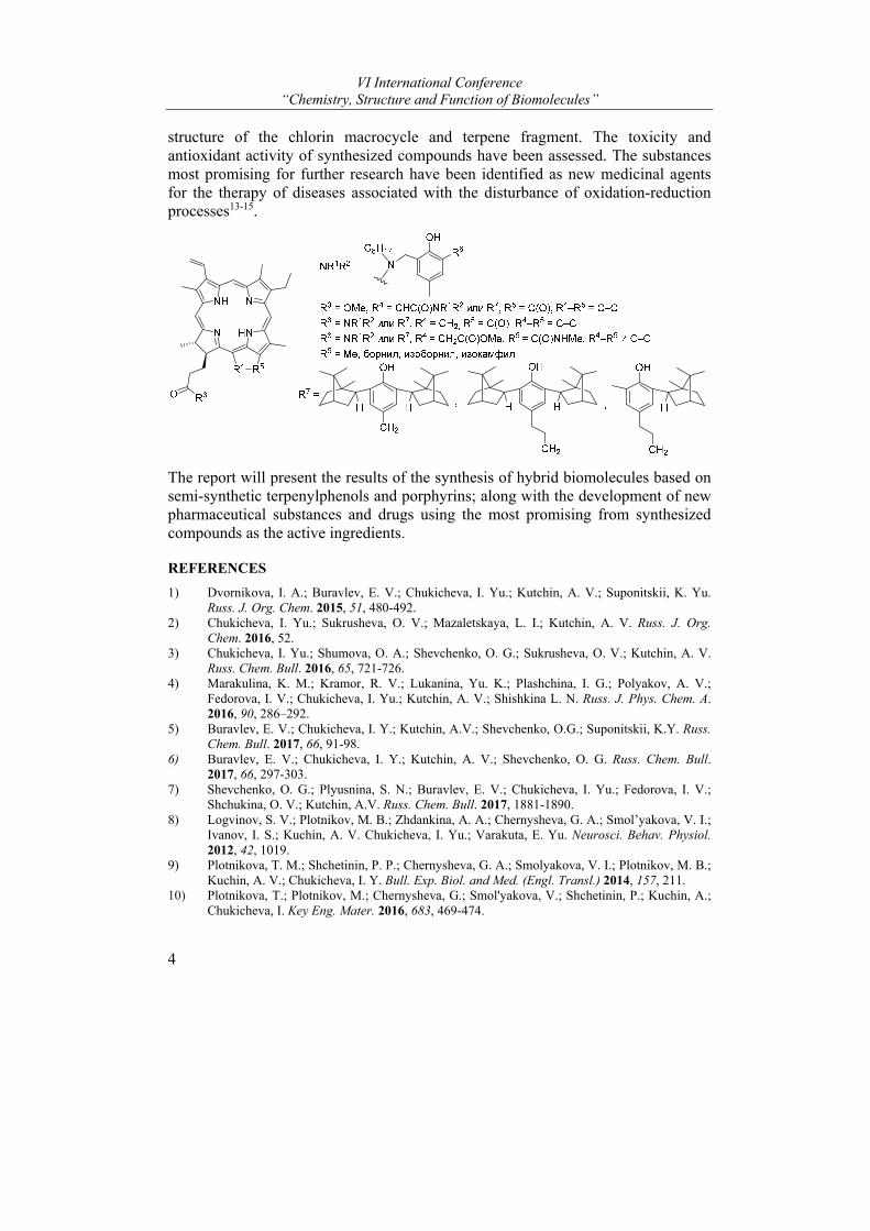
TERPENYLPHENOLS AND PORPHYRINS AS PERSPECTIVE PLATFORM FOR THE FORMATION OF NEW BIOMOLECULES
Irina Yu. Chukicheva*, Evgeny V. Buravlev, Irina A. Dvornikova, Irina V. Fedorova, Olga V. Shchukina, Dmitry V. Belykh, Irina. S. Khudyaeva, and Aleksandr V. Kutchin Institute of Chemistry, Komi Scientific Centre, Ural Branch of the Russian Academy of Sciences, Syktyvkar, Russian Federation e-mail: [email protected]
Natural compounds have high bioavailability due to their specific interactions with target macromolecules in living organisms. Therefore, the structural motif and the basic skeletons of a lot of natural compounds can be used as reference points for the synthesis of new molecules with high biological significance.
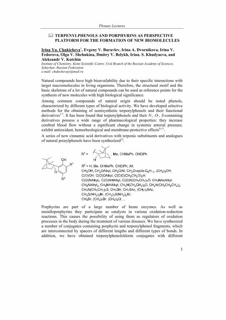
Among common compounds of natural origin should be noted phenols, characterized by different types of biological activity. We have developed selective methods for the obtaining of semisynthetic terpenylphenols and their functional derivatives1-7. It has been found that terpenylphenols and their N-, O-, S-containing derivatives possess a wide range of pharmacological properties: they increase cerebral blood flow without a significant change in systemic arterial pressure, exhibit antioxidant, hemorheological and membrane-protective effects8-11.
A series of new cinnamic acid derivatives with terpenic substituents and analogues of natural prenylphenols have been synthesized12.
Porphyrins are part of a large number of heme enzymes. As well as metalloporphyrins they participate as catalysts in various oxidation-reduction reactions. This causes the possibility of using them as regulators of oxidation processes in the body during the treatment of various diseases. We have synthesized a number of conjugates containing porphyrin and terpenylphenol fragments, which are interconnected by spacers of different lengths and different types of bonds. In addition, we have obtained terpenylphenolchlorin conjugates with different
VI International Conference “Chemistry, Structure and Function of Biomolecules”
4
structure of the chlorin macrocycle and terpene fragment. The toxicity and antioxidant activity of synthesized compounds have been assessed. The substances most promising for further research have been identified as new medicinal agents for the therapy of diseases associated with the disturbance of oxidation-reduction processes13-15.
The report will present the results of the synthesis of hybrid biomolecules based on semi-synthetic terpenylphenols and porphyrins; along with the development of new pharmaceutical substances and drugs using the most promising from synthesized compounds as the active ingredients.
REFERENCES
1) Dvornikova, I. A.; Buravlev, E. V.; Chukicheva, I. Yu.; Kutchin, A. V.; Suponitskii, K. Yu. Russ. J. Org. Chem. 2015, 51, 480-492.
2) Chukicheva, I. Yu.; Sukrusheva, O. V.; Mazaletskaya, L. I.; Kutchin, A. V. Russ. J. Org. Chem. 2016, 52.
3) Chukicheva, I. Yu.; Shumova, O. A.; Shevchenko, O. G.; Sukrusheva, O. V.; Kutchin, A. V. Russ. Chem. Bull. 2016, 65, 721-726.
4) Marakulina, K. M.; Kramor, R. V.; Lukanina, Yu. K.; Plashchina, I. G.; Polyakov, A. V.; Fedorova, I. V.; Chukicheva, I. Yu.; Kutchin, A. V.; Shishkina L. N. Russ. J. Phys. Chem. A. 2016, 90, 286–292.
5) Buravlev, E. V.; Chukicheva, I. Y.; Kutchin, A.V.; Shevchenko, O.G.; Suponitskii, K.Y. Russ. Chem. Bull. 2017, 66, 91-98.
6) Buravlev, E. V.; Chukicheva, I. Y.; Kutchin, A. V.; Shevchenko, O. G. Russ. Chem. Bull. 2017, 66, 297-303.
7) Shevchenko, O. G.; Plyusnina, S. N.; Buravlev, E. V.; Chukicheva, I. Yu.; Fedorova, I. V.; Shchukina, O. V.; Kutchin, A.V. Russ. Chem. Bull. 2017, 1881-1890.
8) Logvinov, S. V.; Plotnikov, M. B.; Zhdankina, A. A.; Chernysheva, G. A.; Smol’yakova, V. I.; Ivanov, I. S.; Kuchin, A. V. Chukicheva, I. Yu.; Varakuta, E. Yu. Neurosci. Behav. Physiol. 2012, 42, 1019.
9) Plotnikova, T. M.; Shchetinin, P. P.; Chernysheva, G. A.; Smolyakova, V. I.; Plotnikov, M. B.; Kuchin, A. V.; Chukicheva, I. Y. Bull. Exp. Biol. and Med. (Engl. Transl.) 2014, 157, 211.
10) Plotnikova, T.; Plotnikov, M.; Chernysheva, G.; Smol'yakova, V.; Shchetinin, P.; Kuchin, A.; Chukicheva, I. Key Eng. Mater. 2016, 683, 469-474.
Plenary Lectures
5
11) Plotnikov, M. B.; Aliev, O. I.; Sidekhmenova, A. V.; Popova, E. V.; Ostrikova, O. I.; Kuchin, A. V.; Chukicheva, I. Yu.; Torlopov M. A. Pharm. Chem. J. 2018, 51, 863-866.
12) Chukicheva, I. Yu.; Fedorova, I. V.; Koroleva, A. A.; Kutchin, A. V. Chem. Natur. Comp. 2018, 54, 1-6.
13) Khudyaeva, I. S.; Belykh, D. V.; Shevchenko, O. G.; Maximova, M. A.; Zainullina, L. F.; Vakhitova, Yu. V.; Shchukina, O. V.; Buravlev, E.V.; Chukicheva, I.Yu.; Kutchin, A.V. Russ. Chem. Bull. 2017, 66, 2157-2164.
14) Belykh, D.V.; Rocheva, T.K.; Buravlev, E.V.; Chukicheva, I.Yu.; Kutchin, A.V. Russ. Chem. Bull. 2017, 66, 2131-2135.
15) Belykh, D. V.; Khudyaeva, I. S.; Buravlev, E. V.; Chukicheva, I. Yu.; Kutchin, A. V.; Shevchenko, O. G. Russ. J. Org. Chem. 2017, 53, 610-614.
This work was financially supported by the Russian Science Foundation (Project No. 16-13-10367) and the Russian Foundation for Basic Research (Project No. 18-03-00950).
BRASSINOSTEROIDS AS REGULATORS PLANT Ca2+ SIGNALING, ION CHANNEL ACTIVITIES GROWTH AND SIGNALLING IN ROOTS OF HIGHER PLANTS
Demidchik V.1*, Straltsova D.1, Charnysh M.1, Chikun P.1, Przhevalskaya D.1, Zhabinskii V.N.2, Khripach V.A.2, and Sokolik A.1
1Department of Plant Cell Biology and Bioengineering, Biological Faculty, Belarusian State University, 220030, 4 Independence Ave., Minsk, Belarus; 2Institute of Bioorganic Chemistry NASB, Minsk, Belarus e-mail: [email protected]
Brassinosteroids (BRs) are endogenous plant hormones essential for the proper regulation of multiple physiological processes required for normal plant growth and development. Exogenous BRs can improve the quantity and quality of crops and ameliorates effects of stresses. Using native and synthetic analogues of BRs as a tool to improve plant yield seems to have a great potential for agriculture and biotechnology (Khripach, 2000). BRs have been intensively investigated for their biosynthesis, distribution and physiological functions using classical physiological tests, analyses of mutants and transgenic plants (Arabidopsis thaliana plants constitutively expressing aequorin). Recent data indicate that BRs are also sensed by the plasma membrane system catalyzing increase in the cytosolic free Ca2+ (in leaves of Arabidopsis thaliana). Zhao et al. (2013) have shown that the BR-induced elevation in the cytosolic free Ca2+ is abolished in knockout line lacking functional brassinosteroid receptor and after treatment with Gd3+ (blocker of Ca2+-permeable nonselective cation channels) (Zhao, 2013). Zhang et al. (2005) using suspension culture cells of Arabidopsis have found that anion channel currents were inhibited by both 28-homobrassionolide and 28-castasterone and outwardly-directed K+ conductance was stimulated by 28-homobrassionolide but inhibited by 28-castasterone (Zhang, 2005). This study was to examine possible effects of brassinosteroids on the plasma membrane cation conductances in plant cells and
VI International Conference “Chemistry, Structure and Function of Biomolecules”
6
related Ca2+ driven signalling events. Standard patch-clamp and aequorin chemiluminometry techniques were used (Demidchik, 2011). Here, we report the first electrophysiological characterisation of brassinosteroid-activated Ca2+-permeable channels in higher plants. Wheat root protoplasts (tested by patch-clamping) and whole arabidopsis plants expressing Ca2+-reporting protein, aequorin (analysed by chemiluminometry), were used in this study. In the whole-cell patches (wheat root protoplasts), 1 µM 24-epibrassonolide, 28-homobrassionolide or 24-epicastasterone were applied exogenously. Only 24-epicastosterone modified transmembrane cation currents while 24-epibrassonolide and 28-homobrassionolide did not cause any reaction. Addition of 24-epicastosterone at cytosolic side through the patch-clamp pipette increased Ca2+ influx conductance, which demonstrated characteristics of depolarisation-activated Ca2+ channels. The pharmacological analyses have shown that brassinosteroid-activated Ca2+-influx conductance was sensitive to inhibitors of Ca2+-permeable cation channels. Blockers of K+ channels did not inhibit this conductance.The plasma membrane conductance, which was activated by an endogenous 24-epicastosterone, showed bell-like shape with maximal activation at depolarisation voltages (bath: 20 mM Ca2+). Labelling castosterone (and its derivates) with BODIPY (using castosterone-BODIPY conjugates which were synthesised chemically) showed that castosterone (and its derivates) can be transferred to the cytosol both in intact roots and protoplasts. This confirms that the effect of 24-epicastosterone at the cytosolic face can potentially be observed in real plants. We also tested the effect of different brassinosteroids on cytosolic free Ca2+, using Arabidopsis thaliana plants constitutively expressing aequorin. Six brassionosteroids including brassinolide, castosterone, 24-epibrassonolide, 28-homobrassionolide, 24-epicastosterone and 28-homocastosterone were tested. All six brassionosteroids induced elevation of the cytosolic free Ca2+ in arabidopsis root cells. In the present study we demonstrated that 24-epicastosterone being more potent than 24-epibrassonolide and 28-homobrassionolide. 10 μM of еxogеnоus BRs was the minimal cоncentration at which stаtistically significant chаnges of the cytosolic Ca2+ were observed. The obtained results suggest that the plasma membrane of root cells contains the brassinosteroid-activated cation-permeable channels, which can be involved in cell ion homeostasis and signalling.
REFERENCES
(1) Demidchik V et al. (2011) Plant Physiol 156: 1375-1385. (2) Khripach V et al. (2000) Ann Bot 86: 441-447. (3) Zhao Y. et al. (2013) Plant Physiol 163: 555-565. (4) Zhang Z. et al. (2005) Plant Cell Physiol 46: 1494-1504.

Plenary Lectures
7
SUPRAMOLECULAR STRUCTURES FOR BIOMEDICINE
Sergey M. Deyev
Shemyakin & Ovchinnikov Institute of Bioorganic Chemistry, Moscow, Russia e-mail: [email protected]
Nowadays nanobiotechnologies open up new possibilities for diagnostics and treatment of oncological, cardiovascular, autoimmune, and other diseases. Standard procedures for design of targeted imaging and therapeutic compounds are based on an attachment of recognizing molecules to visualizing agents or drugs. In frame of this approach the fully genetically encoded anti-receptor antibody-photosesitizers and immunoRNase were constructed. A fluorescent proteins, Killer Red, miniSOG and a ribonuclease barnase were used as toxic principles. They were fused to the single-chain scFv-fragment of anti-HER2/neu antibody 4D5 that recognizes the extracellular domain of cancer marker HER2. The both bifunctional fusion proteins demonstrated specific cytotoxic effect on HER2-positive human carcinoma cells. А novel strategy, “Protein-assisted NanoAssembler”, for design of heterostructures based on the ribonuclease barnase and its inhibitor, barstar, was suggested. The barnase and barstar are small, stable, very soluble, resistant to proteases proteins. The complex between them is extremely tight with a Kd10-14 M. The N- and C-terminal parts of both proteins are localized outside of the barnase∙barstar interface and are therefore accessible for fusion with targeting, visualizing or toxic compounds. The suggested strategy is applicable to virtually any proteins that can be functionally attached to the barstar and barnase molecules. It seems particularly well suited to the production of heterooligomeric constructs because the extremely specific barnase∙barstar interaction eliminates reliably the mispairing problems. The important advantage of barnase∙barstar over the majority of other dimerization modules is that their interaction ratio is precisely 1:1, and neither of the partners is aggregation prone.
A particular attention as new and unique therapeutic agents attract nanoparticles (NPs) that make it possible to solve old but still actual problems by principally new means and ways. A number of nanoparticle-based medications are already approved for therapeutic purposes. Important advantage of NPs is their developed surface, which can be decorated with biocompatible functional moieties, and thus form a versatile docking station. NP can serve as a nano-vehicle to host biologically significant modules, such as therapeutic, targeting and stealth modules for targeted delivery, diagnosis that guides and monitor effects of the NP-assisted therapy of pathology lesions. These properties provide foundations for significant emerging areas in applied biomedical science including (personalised) nanomedicine and theranostics.
In order to apply nanoparticles for imaging and/or therapy one needs to consider three key aspects: design of bright and photostable luminescent nanomaterials
VI International Conference “Chemistry, Structure and Function of Biomolecules”
8
conspicuous on the background of the excitation light back-scattering and cell autofluorescence; amiable surface modification to enable facile interfacing with biomolecules, and modular engineering of the biomolecular complexes with targeting vectors (antibody, mini-antibodies or peptides) firmly attached to the NP for target delivery to specific cellular or tissue sites.
To develop a modular engineering conceptwe study self-assembly of polystyrene micro- and nanoparticles with two functionalities − magnetic and fluorescent – using proteinaceous ‘molecular glues’, most notably, the barnase−barstar system (BBS). The obtained assemblies were tested for their resistance to high concentrations of chaotropic agents (urea and GdmHCl) as well as high temperature and low pH conditions causing denaturation of most proteins. In the majority of cases, the structures exhibit unusual stability and maintain apparently unaltered morphologies upon exposure to these conditions for extended periods of time. Comparison of the BBS-system with other proteinaceous self-assembly systems (streptavidin_biotin, antibody_antigen, and protein A_immunoglobulin), showed that whereas their resistance to destruction is relatively comparable, the capacity to assemble under harsh conditions differs substantially.
The ability of the BBS-glued assemblies to retain their integrity in extreme conditions makes them attractive for a number of applications, taking into account the feasibility of utilization of modules of various nature as participants of self-assembly. The designed nanoparticle assembly approach may prove particularly advantageous for such applications where the remarkable durability of the assemblies becomes a feature of high value. Examples embrace a broad spectrum including sensing of ecological pollutants in complex media, photonics and theragnostic approaches in medicine, also making use of multifunctionality offered by the assemblies.
Furthermore, the unexpectedly high ‘tensile strength’ of the proteinaceous molecular glues described in this work sets one thinking of potential applicability of these self-assembled structures instead of, or alongside with, covalently linked entities. If for creation of a fortiori very durable and ‘reliable’ structures at the nano- and microscale one would definitely choose chemical reactions as a means to build such structures, now, armed with the knowledge of exceptional stability of protein-assisted assemblies, one has access to a great variety of specific (naturally occurring or engineered) ‘molecular glues’ that can be used for the same purposes and with similar efficiency. Moreover, utilization of specific non-covalent interactions adds to the flexibility of the designed assembly systems and imparts higher controllability over the whole process of assembly than in the case of chaotic chemical reactions.
This universal platform provides a straight-forward technology to design a multifunctional nanoheterostructures “when the whole is greater than the sum of the parts”.
Plenary Lectures
9
In the paper we review our recent results on theranostics applications of multifunctional agents with important types of the nanoparticles, including quantum dots (QDs), luminescent nanodiamonds (LNDs), colloidal gold, magnetic NPs, and luminescent upconversion NPs.
REFERENCES
1. Deyev S.M.; Waibel R.; Lebedenko E.N.; Schubiger A.P.; Plückthun A. Nat. Biotechnol. 2003, 21, 1486-1492.
2. Nikitin M.P.; Shipunova V.O.; Deyev S.M.; Nikitin P. Nat. Nanotechnol. 2014. 9, 716-722. 3. Shipunova V.O.; Nikitin M.P.; Nikitin P.I.; Deyev S.M. Nanoscale. 2016, 8, 12764-12772. 4. Sokolova E.; Proshkina G.; Kutova O.; Shilova O.; Ryabova A.; Schulga A.; Stremovskiy O.;
Zdobnova T.; Balalaeva I.; Deyev S. J. Control Release. 2016, 233, 48-56. 5. Khaydukov E.V.; Mironova K.E.; Semchishen V.A.; Generalova A.N.; Nechaev A.V.;
Khochenkov D.A.; Stepanova E.V.; Lebedev O.I.; Zvyagin A.V.; Deyev S.M.; Panchenko V.Y. Sci. Rep. 2016, 6, 35103.
6. Liang L; Lu Y; Zhang R; Care A; Ortega TA; Deyev SM; Qian Y; Zvyagin AV.. Acta Biomater. 2017, 51, 461-470.
7. Sokolova E.; Guryev E.; Yudintsev A.; Vodeneev V.; Deyev S.; Balalaeva I. Oncotarget. 2017, 8, 22048-22058.
8. Semenova G. ; Stepanova D.S.; Dubyk C. ; Handorf E.; Deyev S.M.; Lazar A. J.; Chernoff J. Oncogene . 2017, 36. 5421-5431.
9. Deyev S.; Proshkina G.; Ryabova A.; Tavanti F.; Menziani M.C.; Eidelshtein G.; Avishai G.; Kotlyar A. Bioconjug Chem. 2017, 28, 2569-2574. .
10. Souslova E.A.; Mironova K.E.; Deyev S.M. J. Biophotonics. 2017, 10, 338-352. 11. Shilova O.N.; Shilov E.S.; Deyev S.M. Cytometry A. 2017, 91, 917-925. 12. Kostyukevich Y.; Shulga A.A.; Kononikhin A.; Popov I.; Nikolaev E.; Deyev S. Sci Rep. 2017,
7, 6176.
This research was supported by the RSF grant No. 14-24-00106.
MATRIX ASSISTED SUPRAMOLECULAR CHIRALITY AMPLIFICATION WITH NATURAL PRODUCTS
Pavel B. Drašar
Department of Chemistry of Natural Products, University of Chemistry and Technology, Technicka 5, 166 28 Praha 6, Czech Republic e-mail: [email protected]
The communication aims to help the better and contemporary understanding of the new challenges. The first is the utilisation of mostly renewable natural products, secondary metabolites and raw materials and their semisynthetic derivatives in the mirror of plans for sustainable and circular economy and their repurposing (repositioning, or re-profiling) regardless of their utilisation as e.g. as APIs, biorational agrochemicals, advanced materials, components of diagnostics, chiral substrates. The second challenge, which is entirely connected with the first one, is the emerging next big frontier, the systems chemistry1 investigating into the origin

VI International Conference “Chemistry, Structure and Function of Biomolecules”
10
of life. Trained, and also in the past practicing, chemist Pope Francis declared2 the evolution as real, after the world originated, however, the systems chemistry and systems biology have difficult position in explaining the origin of chirality.
Chirality is one of the properties that is in natural products connected to many aspects of life. The question, “Where it comes from?” is not simple to answer3. Primeval “sin” of the energy difference caused by the violation of parity (PVED) in the universe may not be sufficient for the creation of (almost) homochiral world of sugars, DNA/RNA, amino acids, steroids, terpenes etc., as this energy difference is rather small. Hence, we can suspect the influence of the light of either circular polarisation, or different wavelengths. Further aspect may be the “synthesis” on solid phase surface. Iterestingly, it may be also uniform movement during crystallisation (Ostwald and Viedma ripening), “chiral energy” influenced crystallisation (quartz), electric fields influence, or just the serendipity if not the intelligent design. It seems comparable to the Archimedean (solid) point as without the first chiral phenomenon or object its further propagation is difficult to imagine.
Non-covalent interactions of chiral objects (not necessarily only molecules) may give the origin of truly supramolecular clusters that are so much organised they posses a newly acquired property – suprachirality. Supramolecular assembly follows hard “lock and key” principle, soft “induced fit” one and generally a truly natural feeling of “horror vacui”, where naturally not only the main molecules do act as they may have good help e.g. from the matrix. Matrix generally helps here with protonation, deprotonation, stabilisation of all phenomena involved as hydrogen bondings, Heitler-London and van der Waals “forces” i.a. despite the field of study as e.g. biology, genetics, crystallography, chiral organic synthesis.
In our work, we met with the superassembly phenomenon by a chance. We found that in some cases of porphyrins with chiral substitution it was impossible to measure the optical rotation just after dissolution of the sample and introduction into the measuring cell. The rotation was highly unstable, however, after some time the rotation was measurable and the value of [α]D was in the range of thousands and sometimes even more4.
It is generally accepted that the porphyrins stack in two modes (cf.5), either as coins in columns (H-stacking) or as shingles on the roof (J-stacking). Both contribute to the suprachirality in their own way (Fig. 1).
Fig. 1 H- and J-stacking of porphyrins
Hence, we studied these types of compounds more and more, despite the fact the synthesis of some was giving terribly low yields. There were performed studies by
Plenary Lectures
11
the means of spectroscopies as UV, NMR, CD, studies of monolayer assembly by the Langmuir-Blodget apparatus, studies of monolayers on glass plates, studies by surface plasmon resonance. During the study we found, that the suprachiral assembly is difficult to predict, however, when it occurs it is controlled by the environment, based on the ‘sergeants-and-soldiers’ effect6. The second major player that influenced the supraassembly was polarity of the matrix (media)4.
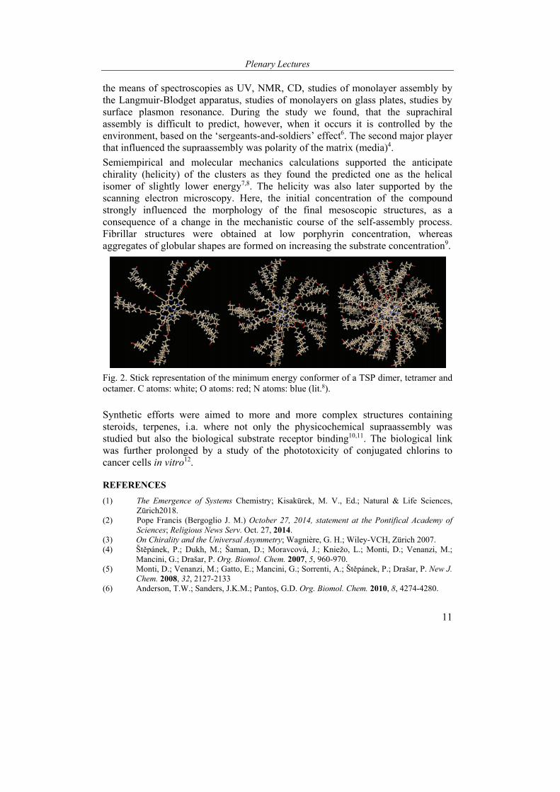
Semiempirical and molecular mechanics calculations supported the anticipate chirality (helicity) of the clusters as they found the predicted one as the helical isomer of slightly lower energy7,8. The helicity was also later supported by the scanning electron microscopy. Here, the initial concentration of the compound strongly influenced the morphology of the final mesoscopic structures, as a consequence of a change in the mechanistic course of the self-assembly process. Fibrillar structures were obtained at low porphyrin concentration, whereas aggregates of globular shapes are formed on increasing the substrate concentration9.
Fig. 2. Stick representation of the minimum energy conformer of a TSP dimer, tetramer and octamer. C atoms: white; O atoms: red; N atoms: blue (lit.8).
Synthetic efforts were aimed to more and more complex structures containing steroids, terpenes, i.a. where not only the physicochemical supraassembly was studied but also the biological substrate receptor binding10,11. The biological link was further prolonged by a study of the phototoxicity of conjugated chlorins to cancer cells in vitro12.
REFERENCES
(1) The Emergence of Systems Chemistry; Kisakürek, M. V., Ed.; Natural & Life Sciences, Zürich2018.
(2) Pope Francis (Bergoglio J. M.) October 27, 2014, statement at the Pontifical Academy of Sciences; Religious News Serv. Oct. 27, 2014.
(3) On Chirality and the Universal Asymmetry; Wagnière, G. H.; Wiley-VCH, Zürich 2007. (4) Štěpánek, P.; Dukh, M.; Šaman, D.; Moravcová, J.; Kniežo, L.; Monti, D.; Venanzi, M.;
Mancini, G.; Drašar, P. Org. Biomol. Chem. 2007, 5, 960-970. (5) Monti, D.; Venanzi, M.; Gatto, E.; Mancini, G.; Sorrenti, A.; Štěpánek, P.; Drašar, P. New J.
Chem. 2008, 32, 2127-2133 (6) Anderson, T.W.; Sanders, J.K.M.; Pantoş, G.D. Org. Biomol. Chem. 2010, 8, 4274-4280.

VI International Conference “Chemistry, Structure and Function of Biomolecules”
12
(7) Zelenka, K.; Trnka, T.; Tišlerová, I.; Monti, D.; Cinti, S.; Naitana, M.L.; Schiaffino, L.; Venanzi, M.; Laguzzi, G.; Luvidi, L.; Mancini, G.; Nováková, Z.; Šimák, O.; Wimmer, Z.; Drašar, P. Chem. Eur. J. 2011, 17, 13743-13753.
(8) Lettieri, R.; Cardova, L.; Gatto, E.; Mazzuca, C.; Monti, D.; Palleschi, A.; Placidi, E.; Drasar, P.; Venanzi, M. 2017, 41, 639-649.
(9) Lorecchio, C.; Venanzi, M.; Mazzuca, C.; Lettieri, R.; Palleschi, A.; Nguyen Thi, T.H.; Cardová, L.; Drašar.; P.; Monti, D. Org. Biomol. Chem. 2014, 12, 3956-3963.
(10) Zhylitskaya, H.A.; Zhabinskii, V.N.; Litvinovskaya, R.P.; Lettieri, R.; Monti, D.; Venanzi, M.; Khripach, V.A.; Drašar, P. Steroids 2012, 77, 1169-1175.
(11) Tomanová, P.; Rimpelová, S.; Jurášek, M.; Buděšínský, M.; Vejvodová, L.; Ruml, T.; Kmoníčková, E.; Drašar, P. B. Steroids 2015, 97, 8-12.
(12) Darmostuk, M.; Jurasek, M.; Lengyel, K.; Zelenka, J.; Rumlova, M.; Drasar, P.; Ruml, T. J. Photochem. Photobiol. B 2017, 168, 175-184.
THE REGULATION OF LIGHT-DEPENDENT GENE EXPRESSION BY BRASSINOSTEROIDS
Marina V. Efimova
National Research Tomsk State University, Tomsk, Russia e-mail: [email protected]
It is generally accepted that phytohormones play an important role in the realization of light regulatory and photosynthetic functions. Some phytohormones are known to imitate the regulatory function of light because they similarly control the rate and nature of morphophysiological processes in plants1. This phytohormone property was shown in the course of investigation of phenotypic features of plants differing in endogenous level of hormones or sensitivity to them2.
Brassinosteroids (BRs) occupy a specific place among phytohormones, along with cytokinins, they can induce in the dark phenotypic changes and trigger the features characteristic of light regulated development. The role of BRs in light-dependent plant development is just starting to be explored, unlike cytokinins (CKs). Exogenous CKs suppress etiolation, which activates the photomorphogenic development of plants under dark conditions. This is accompanied by the activation of promoters of light-regulated genes involved in photosynthesis, transport of sucrose, nitrogen assimilation, more active development of etioplasts, increase in the cotyledons size, shortening of hypocotyl and leaf appearance. Photomorphogenic mutants are characterized by a higher content of zeatin and dihydrozeatin both in the light and in the dark (e.g., amp1-1) and increased sensitivity to these hormones (e.g., det1)3. Possibly, brassinosteroids can regulate of light development of plant indirectly through cytokinins.
The central element of hormonal control of plant growth and development lies in the interaction of phytohormonal pathways. Indeed, BRs are involved in a complex signalling network via a modulation of the levels and sensitivity of other phytohormones or via the intersection of the primary signalling pathways. Several
Plenary Lectures
13
recent studies have identified the specific mechanisms of the coordinated action of BRs and several other phytohormones, including gibberellic acid, abscisic acid, jasmonic acid, auxin and ethylene. The mechanisms of interplay between BRs and CKs are still obscure.
Moreover, there is an assumption that BRs ability to activate photosynthetic processes. Further progress of studies in this direction can be connected with the study of the expression of light-controlled photosynthetic genes in plants differing in their endogenous BR levels.
The goal of the study was to elucidate the influence of BRs on the expression of the genes participating in cytokinin signaling and light controlled photosynthetic genes.
In the present investitation the plants of Arabidopsis thaliana (L.) Heynh transformed with the PARR5::GUS construct and Solanum tuberosum plants we used to estimate the influences of several BRs (brassinolide, epibrassinolide and homobrassinolide) and 6-benzylaminopurine (BA) on the expression of the RR-A genes which belongs to the type A negative regulators of plant responses to cytokinin.
To study the BRs controlled regulation of the transcription of plastid genes, we used Arabidopsis thaliana plants differing in their endogenous BRs level; the parental line belonged to a Columbia ecotype (Col) with the normal endogenous BRs level, whereas its mutant form det2 was characterized by defective BRs synthesis. We also used barley (Hordeum vulgare L.) plants. To achieve a high levels of BRs, detached leaves of A. thaliana and H. vulgare plants were treated with exogenous epibrassinolide (1 µM, EBL). The isolation of plastids and the run on transcription in their lysates were carried out according to the earlier described technique4.
The first question we attempted to answer was whether BRs are capable of affecting the expression of the ARR5 gene promoter in the dark or light conditions. To this end, 4-day-old etiolated seedlings transformed with the PARR5:GUS construct were exposed to either 1 µM BRs or BA treatment for 24 h in the dark or light. In darkness, the GUS activity in the Arabidopsis BR-treated seedlings increased up to a value of approximately 140 % compared to the controls but was substantially lower than after the CK application. The BR-treated seedlings grown in the light, unlike those exposed to BA, did not exhibit any reliable increase in GUS activity. These results imply that the promoter of the ARR5 gene is sensitive to exogenous BRs and that this sensitivity is attenuated by light5.
To estimate the regulation of the light-dependent genes by BRs we compared the transcription rates of 12 chloroplast genes belonging to functionally different groups of plastome genes in darkness and light conditions. First of all, they are the genes encoding the products that play an important role in the process of photosynthesis: the photosystem I genes psaA and psaB, the photosystem II genes psbA, psbD, and the psbK, gene of the large subunit of Rubisco rbcL, the ATP-synthase complex genes atpB, and the subunit F of NADPH-plastoquinone
VI International Conference “Chemistry, Structure and Function of Biomolecules”
14
reductase ndhF. Among the housekeeping genes, we investigated transcription of the gene encoding β-subunit of RNA-polymerase of bacterial type (rpoB), the genes of 16S and 23S ribosomal RNA (rrn16 and rrn23), and the genes of tRNA-Glu and tRNA-Tyr (trnE-Y).
Comparative analysis of the intensity of transcription of chloroplast genes in Arabidopsis showed that the decrease in the endogenous BRs level observed in the mutant det2 line promoted the activation of the transcription of the chloroplast genes studied. The highest (8 to 12-fold) activation level was observed for the ndhF, psbK, and atpB genes. A significant (3 to 4-fold) activation level was observed for the psaA, psaB, psbA, rbcL, rrn23, trnEY, and rpoB genes. The transcription of psbD and rrn16 genes, having the maximum transcription intensity in control plants, was activated less. The analysis of the results has shown that a high concentration of exogenous epibrassinolide activates the transcription of some chloroplast genes in leaves of dicotyledonous (Arabidopsis thaliana) and monocotyledonous (Hordeum vulgare) plants. For example, in the case of chloroplasts isolated from rosellate Arabidopsis leaves treated with exogenous epibrassinolide, we observed a minor activation of the transcription of the most (ten) of the studied genes; in the case of five genes (psaB, psbK, ndhF, rrn23, and atpB), the difference was significant. Two other genes, psbD and rpoB, exhibited a tendency towards a decrease in the intensity of their transcription. The first leaves of etiolated barley seedlings also demonstrated a high sensitivity to exogenous epibrassinolide. Treatment with exogenous EBL in the dark up-regulated the transcription of the tested barley seedling genes. The greatest induction was observed with two genes, – psaA and ndhF (3.8 and 4.5 times, respectively), while the transcriptional activity of other genes increased by 2.5 – 3 times on average.
The data on the activation of the transcription of genes, required to realize the photosynthetic function of light, at the low level of endogenous BS or under the influence of exogenous epibrassinolide, evidence that BS are involved into the realization of the light program of plant development, starting from the changes in the level of expression of plastid genes. The up-regulation of plastid gene transcription in the dark by brassinosteroids could be mediated by the expression of the gene for the cytokinin primary response.
REFERENCES
(1) Chemical Probes in Biology Science at the Interface of Chemistry, Biology, and Medicine; Schneider, M.P., Ed., Springer-Science+Business Media, B.V., 2003.
(2) Chory, J.; Nagpal, P.; Peto, C.A. Plant Cell. 1991, 3, 445–459. (3) Chin-Atkins, A.N.; Craig, S.; Hocart, C.H.; Dennis, E.S.; Chaudhury, A.M. Planta. 1996,
198, 549-556. (4) Zubo, Y.O.; Kusnetsov, V.V. Rus. J. Plant Physiol. 2008, 55, 107-114. (5) Kudryakova, N.V.; Efimova, M.V.; Danilova, M.N.; Zubkova, N.K.; Khripach, V.A.;
Kusnetsov, V.V.; Kulaeva, O.N. Plant Growth Regulation. 2013, 70, 61-69.
Plenary Lectures
15
(6) Efimova, M.V.; Vankova, R.; Kusnetsov, V.V.; Litvinovskaya, R.P.; Zlobin, I.E.; Dobrev, P.; Vedenicheva, N.P.; Sauchuk, A.L.; Karnachuk, R.A.; Kudryakova, N.V.; Kuznetsov, V.V. Steroids. 2017, 120, 32-40.
This study was performed with the financial support by the Russian Science Foundation (project no. 16-16-04057).
USING MACHINE LEARNING TO PREDICT PROTEIN STRUCTURE AND INTERACTIONS
Sergei Grudinin
INRIA Rhone-Alpes Research Center, Grenoble, France e-mail: [email protected]
Machine learning and artificial intelligence in general have been extensively used in bioinformatics. I will start my presentation discussing how machine-learning can be used for structural predictions and, more specifically, how to use it for parametrization of small molecules1,9 and for the training a free-shape distance-dependent protein-ligand potential7,8. Unlike knowledge-based methods based on Boltzmann statistics, in our approach, called Convex-PL, we do not impose any functional form of the potential. Instead, we use an optimization approach, accepting that the target binding energy value is decomposed into a polynomial basis with unknown expansion coefficients. These are then deduced from the structural data collected from protein-ligand complexes using a convex formulation of the optimization problem, similar to our protein-protein interaction potentials2,3. The training set consists of the complexes taken from the PDBBind database. We generate false poses with constant RMSD rigid-body deformations of the ligands inside the binding pockets. This allows the obtained potential to be generally unbiased towards other molecular docking methods, which are often used for decoys generation. Convex-PL performed successfully in the CSAR 2013-2014 and D3R 2015-2016 competitions4,5. For a more general validation, we assessed it using data from D3R Grand Challenge 2 submissions and the CASF 2013 study6, which includes the docking, scoring, ranking, and screening tests. Our docking and ranking test results outperform the other 20 methods previously assessed in CASF 2013. Also, Convex-PL performs better than average in the scoring test. I will conclude my presentation discussing the current challenges in virtual screening and protein-ligand docking. Specifically, I will discuss the importance of modelling protein flexibility and multiple conformational states upon binding to small molecules10.
REFERENCES
(1) Kadukova, M.; Grudinin, S. J. Chem. Inf. Model. 2016, 56, 1410-1419. (2) Popov, P.; Grudinin, S. J. Chem. Inf. Model. 2015, 55, 2242-2255.
VI International Conference “Chemistry, Structure and Function of Biomolecules”
16
(3) Neveu, E.; Ritchie, D.W.; Popov, P.; Grudinin, S. Bioinformatics. 2016, 32, i693-i701. (4) Grudinin, S.; Popov, P.; Neveu, E.; Cheremovskiy, G. J. Chem. Inf. Model. 2015, 56, 1053-
1062. (5) Grudinin, S.; Kadukova, M.; Eisenbarth, A.; Marillet, S.; Cazals, F. J. Comput. Aided Mol.
Des. 2016, 30, 791-804. (6) Li, Y.; Han, L.; Liu, Zh.; Wang, R. J. Chem. Inf. Model., 2014. (7) Kadukova, M.; Grudinin, S. Journal of Computer-Aided Molecular Design. 2017, doi:
10.1007/s10822-017-0068-8. (8) https://team.inria.fr/nano-d/convex-pl/ (9) https://team.inria.fr/nano-d/software/knodle/ (10) Kadukova, M.; Grudinin, S. Journal of Computer-Aided Molecular Design, 2017, doi:
10.1007/s10822-017-0062-1.
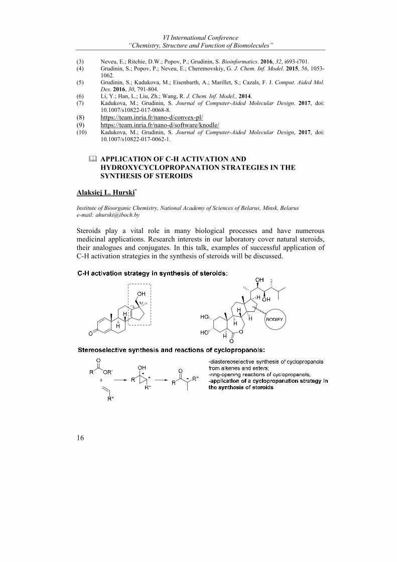
APPLICATION OF C-H ACTIVATION AND HYDROXYCYCLOPROPANATION STRATEGIES IN THE SYNTHESIS OF STEROIDS
Alaksiej L. Hurski*
Institute of Bioorganic Chemistry, National Academy of Sciences of Belarus, Minsk, Belarus e-mail: [email protected]
Steroids play a vital role in many biological processes and have numerous medicinal applications. Research interests in our laboratory cover natural steroids, their analogues and conjugates. In this talk, examples of successful application of C-H activation strategies in the synthesis of steroids will be discussed.

Plenary Lectures
17
Using these methods, we have successfully prepared 17β-hydroxymethyl-17α-methyl-13-androstenes that are on demand in anti-doping analysis, conjugates of the plant growth hormone 24-epibrassinolide with dyes and other steroids. A diastereoselective synthesis of α-methylketones from esters and alkenes via cyclopropanol intermediates will be also presented. This approach was found to be useful for the attachment of side chains to steroidal cores.
Stereochemical mechanisms, reaction conditions and spectral properties of newly synthesized compounds will be discussed.
SURFACE PLASMON RESONANCE (SPR) BIOSENSORS BIACORE IN BIOMOLECULES RESEARCH
Alexis S. Ivanov
Institute of Biomedical Chemistry, Moscow, Russia e-mail: [email protected]
The aim of this report is to give a brief overview of the application of surface plasmon resonance (SPR) biosensors Biacore in biomolecule research. SPR technology enables to record practically any intermolecular interactions in real time without any labels or associated processes.
The principle of SPR biosensor operating is rather simple. The first molecular partner (ligand) is immobilized on the optical chip surface and biosensor records mass transfer of the second molecular partner (analyte) between free volume and the layer at the chip surface where the second partner is fixed. Any types of molecules (from low-molecular weight substances to biopolymers) and even large supra-molecular complexes can be used as ligands and analytes.
The curve of biosensor signal record depending on time is called sensorgram. A series of sensorgrams obtained at different analyte concentrations can be computationally analyzed and the constant of complex dissociation (Kd), as well as and the rate constants of complex formation (kon) and dissociation (koff), can be calculated. Furthermore, from series of sensorgrams at different temperatures the following thermodynamic parameters of interaction also can be calculated: Gibbs free energy change (ΔG), the change of enthalpy (ΔH) and entropy (ΔS).
In the world market of scientific equipment, manufacturers offer different SPR biosensors with original constructive decisions and various functional characteristics. However, the best known biosensors are Biacore from GE Healthcare (USA). Biacore biosensors have the best characteristics in some essential parameters including: very high sensitivity, minimal noise level (less 0.0005 RU), high stability of basic signal, no restrictions on minimum molecular weight of analyte, cost-effective biomaterial consumption (100 ng of proteins is

VI International Conference “Chemistry, Structure and Function of Biomolecules”
18
enough), minimal volume of flow measurement channels (about 20 nL), flexibility of measurement protocols based on commutated micro-fluidics system.
SPR biosensors Biacore are successfully used in our diverse investigations of biomolecules:
Analysis of protein-protein interactions1-4;
Real-time analysis of enzyme-inhibitors interactions5-8;
Analysis of thermodynamics of molecular complexes formation9-11;
Quantitative analysis of disease biomarker in blood serum12-13;
Protein-protein interactions as new targets for drug design6,10,14-16;
Direct molecular fishing of probable partners of protein-protein interactions17-22.
REFERENCES
(1) O.V. Gnedenko, A.S. Ivanov, E.O. Yablokov, S.A. Usanov, D. . Mukha, G.V. Sergeev, A.V. Kuzikov, N.E. Moskaleva, T.V. Bulko, V.V. Shumyantseva, A.I. Archakov. Biochemistry (Moscow) Supplement Series B: Biomedical Chemistry, 2014, 8(3), 231–236.
(2) O.V. Gnedenko, E.O. Yablokov, S.A. Usanov, D.V. Mukha, G.V. Sergeev, T.V. Bulko, A.V. Kuzikov, N.E. Moskaleva, V.V. Shumyantseva, A.S. Ivanov, A.I. Archakov. Chemical Physical Letters, 2014, 593, 40-44.
(3) O.A. Buneeva, O.V. Gnedenko, A.T. Kopylov, M.V. Medvedeva, V.G. Zgoda, A.S. Ivanov, A.E. Medvedev. Biochemistry (Moscow), 2017, 82(9), 1042-1047.
(4) P. Ershov, Y. Mezentsev, A. Gilep, S. Usanov, O. Buneeva, A. Medvedev, A. Ivanov. Protein Science, 2017, 26, 2458—2462.
(5) I.N. Sokotun, O.V. Gnedenko, A.V. Leychenko, M.M. Monastyrnaya, E.P. Kozlovskaya, A.A. Molnar, A.S. Ivanov. Biochemistry (Moscow) Supplemental Series B: Biomedical Chemistry, 2007, 1(2), 139-142.
(6) P.V. Ershov, O.V. Gnedenko, A.A. Molnar, A.V. Lisitsa, A.S. Ivanov, A.I. Archakov. Biochemistry (Moscow) Supplement Series B: Biomedical Chemistry, 2012, 6(1), 94-97.
(7) L.A. Kaluzhsky, O.V. Gnedenko, A.A. Gilep, N.V. Strushkevich, T.V. Shkel, M.A. Chernovetsky, A.S. Ivanov, A.V. Lisitsa, A.S. Usanov, V.A. Stonik, A.I. Archakov. Biochemistry (Moscow) Supplement Series B: Biomedical Chemistry, 2014, 8(4), 349–360.
(8) L.A. Kaluzhskiy, T.V. Shkel, N.V. Ivanchina, A.A. Kicha, A.A. Gilep, N.V. Strushkevich, M.A. Chernovetsky, A.E. Medvedev, S.A. Usanov, A.S.. Natural Product Communications, 2017, 12(12), 1843-1846.
(9) S.Yu.Rakhmetova, S.P.Radko, O.V.Gnedenko, N.V.Bodoev, A.S.Ivanov and A.I.Archakov. Biochemistry (Moscow) Supplemental Series B: Biomedical Chemistry, 2010, 5(2), 139-143.
(10) P.V. Ershov, O.V. Gnedenko, A.A. Molnar, A.V. Lisitsa, A.S. Ivanov, A.I. Archakov. Biochemistry (Moscow) Supplement Series B: Biomedical Chemistry, 2012, 6(1), 94-97.
(11) E. Yablokov, A. Florinskaya, A. Medvedev, G. Sergeev, N. Strushkevich, A. Luschik, T. Shkel, A. Dmitrochenko, A. Yantsevich, A. Gilep, S. Usanov, A. Archives of Biochemistry and Biophysics, 2017, 619, 10-15.
(12) E. Suprun, T. Bulko, A. Lisitsa, O. Gnedenko, A. Ivanov, V. Shumyantseva, A. Archakov. Biosensors and Bioelectronics, 2010, 25, 1694–1698.
(13) Oksana V. Gnedenko, Yury V. Mezentsev, Andrey A. Molnar, Andrey V. Lisitsa, Alexis S. Ivanov and Alexander I. Archakov. Analytica Chimica Acta, 2013, 759, 105–109.
Plenary Lectures
19
(14) Ivanov A.S., Gnedenko O.V., Molnar A.A., Mezentsev Y.V., Lisitsa A.V., Archakov A.I.. J .Bioinform. Comput. Biol. 2007, 5(2b), 579-592.
(15) Yu.V. Mezentsev, A.A. Molnar, O.V. Gnedenko, Yu.V. Krasotkina, N.N. Sokolov, A.S. Ivanov. Biochemistry (Moscow) Supplemental Series B: Biomedical Chemistry, 2007, 1(1), 58-67.
(16) Ershov P., Gnedenko O., Molnar A., Lisitsa A., Ivanov A., Archakov A. Biochemistry (Moscow) Supplemental Series B: Biomedical Chemistry, 2009, 3(3), 272-288.
(17) A. S. Ivanov, P. V. Ershov, Yu. V. Mezentsev, E. V. Poverennaya, A. V. Lisitsa, and A. I. Biochemistry (Moscow) Supplement Series B: Biomedical Chemistry, 2012, 6(2), 99–106.
(18) P. Ershov, Y. Mezentsev, O. Gnedenko, D. Mukha, A. Yantsevich, V. Britikov, L. Kaluzhskiy, E. Yablokov, A. Molnar, A. Ivanov, A. Lisitsa, A. Gilep, S. Usanov, A. Archakov. Proteomics, 2012, 12, 3295–3298.
(19) Ivanov A.S., Medvedev A., Ershov P., Molnar A., Mezentsev Y., Yablokov E., Kaluzhsky L., Gnedenko O., Buneeva O., Haidukevich I., Sergeev G., Lushchyk A., Yantsevich A., Medvedeva M., Kozin S., Popov I., Novikova S., Zgoda V., Gilep A., Usanov S., Lisitsa A., Archakov A. Proteomics. 2014, 14, 2261–2274.
(20) A.S.Ivanov, A.E.. Bochemistry (Moscow) Supplement Series B: Biomedical Chemistry, 2016, 10(1), 55–62.
(21) A.S. Ivanov, P.V. Ershov, A.A. Molnar, Yu.V. Mezentsev, L.A. Kaluzhsky, E.O. Yablokov, A.V. Florinskaya, O.V. Gnedenko, A.E. Medvedev, S.A. Kozin, V.A. Mitkevich, A.A. Makarov, A.A. Gilep, A.Ya. Luschik, I.V. Gaidukevich, S.A. Russian Journal of Bioorganic Chemistry, 2016, 42(1), 14–21.
(22) A.V. Svirid, P.V. Ershov, E.O. Yablokov, L.A. Kaluzhskiy, Yu.V. Mezentsev, A.V. Florinskaya, T.A. Sushko, N.V. Strushkevich, A.A. Gilep, S.A. Usanov, A.E. Medvedev, A.S. Acta Naturae, 2017, 9, №4(35), 92-100.
The work was performed in the framework of the Program for Basic Research of State Academies of Sciences for 2013-2020.
SELECTIVE MODULATORS OF ESTROGEN AND PROGESTERONE RECEPTORS: STEROIDAL AGONISTS AND ANTAGONISTS
Inna S. Levina and Yury V. Kuznetsov
N. D. Zelinsky Institute of Organic Chemistry, Russian Academy of Sciences, Leninsky Prosp. 47, Moscow 119991, Russia e-mails: [email protected], [email protected]
A modification of known biologically active compounds is one of the main tools of medicinal chemistry. Variations in substituents, in the spatial structure and electron density distribution make it possible to alter the initial properties of the molecule being modified, thereby to increase its resistance to metabolism, to enhance the binding to the biological target, and to differentiate or integrate the effects on different biological targets. Additionally, such modifications being even small can both enhance the inherent biological effects of the original molecule and suppress them. Within this framework, the empirical approach based on SAR and the targeted approach based on knowledge of the structure and functions of the
VI International Conference “Chemistry, Structure and Function of Biomolecules”
20
biological target are realized. Both the approaches enable to determine the direction of the subsequent modification.
Considering the functional diversity of morphologically similar biological targets, the search for new compounds with a unique, and, ideally, preset, pattern of biological activity does not lose relevance. At the same time, the similarity of the targets implies the possibility to transfer the ideas been tested on the one target to another.
Early we have examined series of progesterone derivatives1-3 varying some structural elements, which showed wide spectrum of the progesterone receptor modulation and selectivity. The structural and mechanistic similarity of the progesterone (PR) and estrogen receptors (ER), shapes of their binding sites gave a reason to believe that similar compounds containing some structural fragments which are characteristic of estradiol would prove to be active towards estrogen receptor.
Based on this, the series of compounds combining some structural fragments typical for modified pregnane derivatives and estradiol were synthesized and biologically evaluated.4 It was demonstrated that compounds with natural C/D-rings junction in the steroid core are highly cytotoxic against MCF-7 cancer cells and suppress estradiol-induced transcription in reporter assay. At the same time, the 13-epi-compounds with distorted steroid core showed high cytotoxicity on cancer cell lines including doxorubicin and cisplatin resistant cells while ambiguous effects on ER modulation.
This report provides the examples of synthesis of the modified natural steroid hormones - progesterone and estradiol, biological effects of such a modification, and molecular modeling of the resulted modulators of progesterone and estrogen receptors.
REFERENCES
(1) Kamernitzky A. V., Levina I. S. Russ. J. Bioorg. Chem. 2005, 31, 105-118 and 199-209. (2) Lisanova O. V., Shchelkunova T. A., Smirnov A. N., Morozov I. A., Rubtsov P. M., Levina
I. S., Kulikova L. E. Biochemistry (Moscow) 2013, 78, 236-243. (3) Scherbakov A.M., Levina I.S., Kulikova L.E., Kuznetsov Y.V., Zavarzin I.V., Fedyushkina
I.V., Skvortsov V.S., Veselovsky A.V. Biochemistry (Moscow) Suppl. Ser. B: Biomed. Chem. 2016, 10, 341-345.
(4) Kuznetsov Yu. V., Levina I. S., Scherbakov A. M., Andreeva O. E., Fedyushkina I. V., Dmitrenok A. S., Shashkov A. S., Zavarzin I. V. Eur. J. Med. Chem. 2018, 143, 670-682.
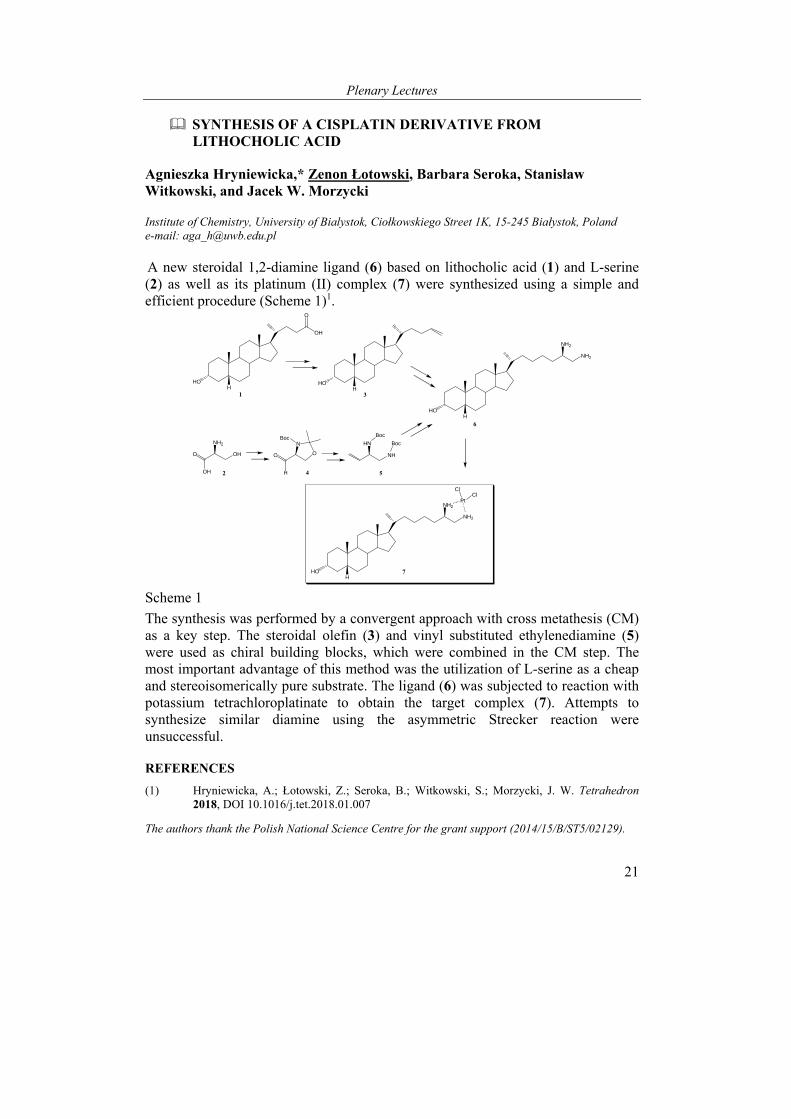
Plenary Lectures
21
SYNTHESIS OF A CISPLATIN DERIVATIVE FROM LITHOCHOLIC ACID
Agnieszka Hryniewicka,* Zenon Łotowski, Barbara Seroka, Stanisław Witkowski, and Jacek W. Morzycki
Institute of Chemistry, University of Bialystok, Ciołkowskiego Street 1K, 15-245 Białystok, Poland e-mail: [email protected]
A new steroidal 1,2-diamine ligand (6) based on lithocholic acid (1) and L-serine (2) as well as its platinum (II) complex (7) were synthesized using a simple and efficient procedure (Scheme 1)1.
HOH
NH2
NH2Pt
ClCl
O
H
O
NBoc
NH
HN
Boc
Boc
OHO
OH
NH2
HOH
O
OH
HOH
HOH
NH2
NH2
1
2
3
4 5
6
7
Scheme 1
The synthesis was performed by a convergent approach with cross metathesis (CM) as a key step. The steroidal olefin (3) and vinyl substituted ethylenediamine (5) were used as chiral building blocks, which were combined in the CM step. The most important advantage of this method was the utilization of L-serine as a cheap and stereoisomerically pure substrate. The ligand (6) was subjected to reaction with potassium tetrachloroplatinate to obtain the target complex (7). Attempts to synthesize similar diamine using the asymmetric Strecker reaction were unsuccessful.
REFERENCES
(1) Hryniewicka, A.; Łotowski, Z.; Seroka, B.; Witkowski, S.; Morzycki, J. W. Tetrahedron 2018, DOI 10.1016/j.tet.2018.01.007
The authors thank the Polish National Science Centre for the grant support (2014/15/B/ST5/02129).
VI International Conference “Chemistry, Structure and Function of Biomolecules”
22
MOLECULAR ARCHITECTURE OF IONIC LIQUIDS WITH ANTICANCER ACTIVITY, ANTIOXIDANT, AND PHOTOSENISIBILIZING PROPERTIES
Veaceslav Boldescu1, Serghei Curlat1, Serghei Pogrebnoi1, Anastasia Smetanscaia2, Livia Uncu2, Vladimir Valica2, and Fliur Macaev1,2*
1 Institute of Chemistry, Laboratory of organic synthesis and biopharmaceuticals, Chișinău, Moldova, 2 State University of Medicine and Pharmacy“Nicolae Testemițanu”, Scientific Center for Drug Research , Chișinău, Moldova e-mail: [email protected]
The anticancer potential of organic salts and ionic liquids (OSILs) has been widely discussed recently. Their attractiveness for pharmaceutical production is attributed to such advantageous properties as simplicity in preparation and purification, low costs, tunable permeability through biological barriers, etc.
Here we report a group of OSILs with cytotoxic activity against cancerous cells comparable with that of carboplatin and much lower against noncancerous cells. Biological tests of the obtained compounds have demonstrated that they possess inhibitory activity against two ectonucleotidase isozymes, which could be one of their main antiproliferation mechanisms of action. At the same time, physicochemical tests have shown that at room temperature and deemed light these compounds possess antioxidant effect acting as free radical scavengers, while their irradiation with 365 nm light makes them photosensitizers inducing singlet oxygen production in water suspensions.
Most of the previous studies by other groups on OSILs with more or less selective cytotoxicity against different lines of cancerous cells have mainly demonstrated the antiproliferative activity but did not go further to study their potential cytotoxic mechanisms. Other works made attempts for structure-cytotoxic activity relationship studies that demonstrated higher toxicity for OSILs containing cations with longer chains and lower toxicity for compounds with functionalized side chains in cations as compared to non-functionalized ones. Wang et al.1 studied cytotoxicity of a group of ionic liquids and their precursors in HeLa cells and determined increase in reactive oxygen species production (ROS) and a consequent reduction of mitochondrial membrane potential.
While it is well known that intracellular generation of singlet oxygen equally induces cell death in both cancerous and noncancerous cells, extracellular singlet oxygen has a more selective action on tumor cells via membrane-associated catalase inhibition and reactivation of intracellular ROS/RNS-dependent apoptosis-inducing signaling, while having no effect on non-malignant cells. Previously, we have demonstrated that some of the benzylamine derivatized OSILs synthesized by us possess photosensibilizing properties with formation of ROS.2
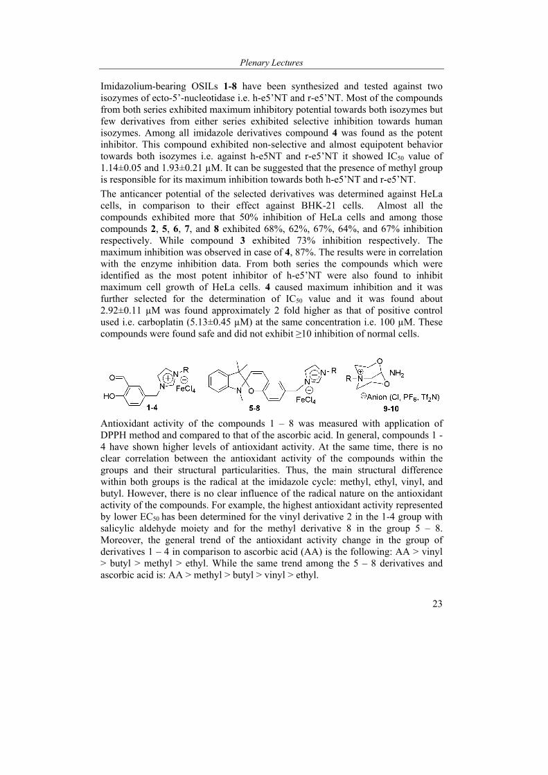
Plenary Lectures
23
Imidazolium-bearing OSILs 1-8 have been synthesized and tested against two isozymes of ecto-5’-nucleotidase i.e. h-e5’NT and r-e5’NT. Most of the compounds from both series exhibited maximum inhibitory potential towards both isozymes but few derivatives from either series exhibited selective inhibition towards human isozymes. Among all imidazole derivatives compound 4 was found as the potent inhibitor. This compound exhibited non-selective and almost equipotent behavior towards both isozymes i.e. against h-e5NT and r-e5’NT it showed IC50 value of 1.14±0.05 and 1.93±0.21 µM. It can be suggested that the presence of methyl group is responsible for its maximum inhibition towards both h-e5’NT and r-e5’NT.
The anticancer potential of the selected derivatives was determined against HeLa cells, in comparison to their effect against BHK-21 cells. Almost all the compounds exhibited more that 50% inhibition of HeLa cells and among those compounds 2, 5, 6, 7, and 8 exhibited 68%, 62%, 67%, 64%, and 67% inhibition respectively. While compound 3 exhibited 73% inhibition respectively. The maximum inhibition was observed in case of 4, 87%. The results were in correlation with the enzyme inhibition data. From both series the compounds which were identified as the most potent inhibitor of h-e5’NT were also found to inhibit maximum cell growth of HeLa cells. 4 caused maximum inhibition and it was further selected for the determination of IC50 value and it was found about 2.92±0.11 µM was found approximately 2 fold higher as that of positive control used i.e. carboplatin (5.13±0.45 µM) at the same concentration i.e. 100 µM. These compounds were found safe and did not exhibit ≥10 inhibition of normal cells.
Antioxidant activity of the compounds 1 – 8 was measured with application of DPPH method and compared to that of the ascorbic acid. In general, compounds 1 - 4 have shown higher levels of antioxidant activity. At the same time, there is no clear correlation between the antioxidant activity of the compounds within the groups and their structural particularities. Thus, the main structural difference within both groups is the radical at the imidazole cycle: methyl, ethyl, vinyl, and butyl. However, there is no clear influence of the radical nature on the antioxidant activity of the compounds. For example, the highest antioxidant activity represented by lower EC50 has been determined for the vinyl derivative 2 in the 1-4 group with salicylic aldehyde moiety and for the methyl derivative 8 in the group 5 – 8. Moreover, the general trend of the antioxidant activity change in the group of derivatives 1 – 4 in comparison to ascorbic acid (AA) is the following: AA > vinyl > butyl > methyl > ethyl. While the same trend among the 5 – 8 derivatives and ascorbic acid is: AA > methyl > butyl > vinyl > ethyl.
VI International Conference “Chemistry, Structure and Function of Biomolecules”
24
Another series of ILs was obtained via conversion of carbonitriles into primary amine cyclic ether quaternized salts.3 The obtained compounds were checked for their antiproliferative activity in HeLa cells line. All these compounds demonstrated an efficient cytotoxic behavior against HeLa cells as compared to a standard anticancer drug Vincristine. The IC50 values for these compounds vary in the limits of 0.97 – 2.37 µM with percent inhibition of Vero cells growth at 10 µM varying from 10 -28%.
Compound [Me]Cl (9) displayed the highest inhibition activity towards HeLa with SI10µM ≥ 5.0, while the [2HE]Tf2N (10) showed the lowest selectivity index within the substituted 1-amino-2,8-dioxa-5-azoniabiciclo[3.3.1]nonanium salts (SI10µM ≥ 2.5).
REFERENCES
(1) Wang, X.; Ohlin, C. A.; Lu, Q.; Fei, Z.; Hu, J.; Dyson, P. J. Green Chem., 2007, 9, 1191-1197.
(2) Neamțu, M.; Macaev, F.; Boldescu, V.; Hodoroaba, V-D.; Nădejde, C.; Schneider, R. J.; Paul, A; Ababei, G.; Panne, U. Appl. Catal. B. 2016, 183, 335-342.
(3) Prodius, D.; Shah, H.S.; Iqbal, J.; Macaeva, A.; Dimoglo, A.; Kostakis, G. E.; Zill, N.; Macaev, F.; Powell A.K. Chem. Comm. 2014, 50, 4888-4890.
Acknowledgements: the authors are grateful for the funding support offered by the Science and Technology Center in Ukraine and the Agency for Research and Development of the Republic of Moldova under international project 17.80013.8007.10/6245STCU
PRECURSOR-DIRECTED SYNTHESIS OF NEW PHENYLBENZOISOQUINOLINDIONE ALKALOIDS AND THE DISCOVERY OF A PHENYLPHENALENONE-BASED PLANT DEFENSE MECHANISM
Yu Chen1,2, Christian Paetz1, and Bernd Schneider1
1 Jiangsu Key Laboratory for the Research and Utilization of Plant Resources, Institute of Botany, Jiangsu Province and Chinese Academy of Sciences, Qianhu Houcun 1, 210014, Nanjing, China 2 Max Planck Institute for Chemical Ecology, Jena, Germany e-mail: [email protected]
Phenylphenalenones (1) and their oxa- and aza derivatives [phenylbenzoisochromenones (PBICs, 2-5), phenylbenzoisoquinolindiones (PBIQs, 8)] belong to a class of structurally diverse natural products occurring in the Haemodoraceae and some other monocotyledonous plants. Involvement of such compounds in the plant’s defense against pathogens and herbivores has been reported. Biosynthetic studies revealed the origin of phenylphenalenones from two phenylpropanoid units and C-2 of acetate. Furthermore, PBICs (2-5) are the result of an oxidative conversion of phenylphenalenones (1). However, the formation of PBIQ alkaloids (8) was unknown until now.
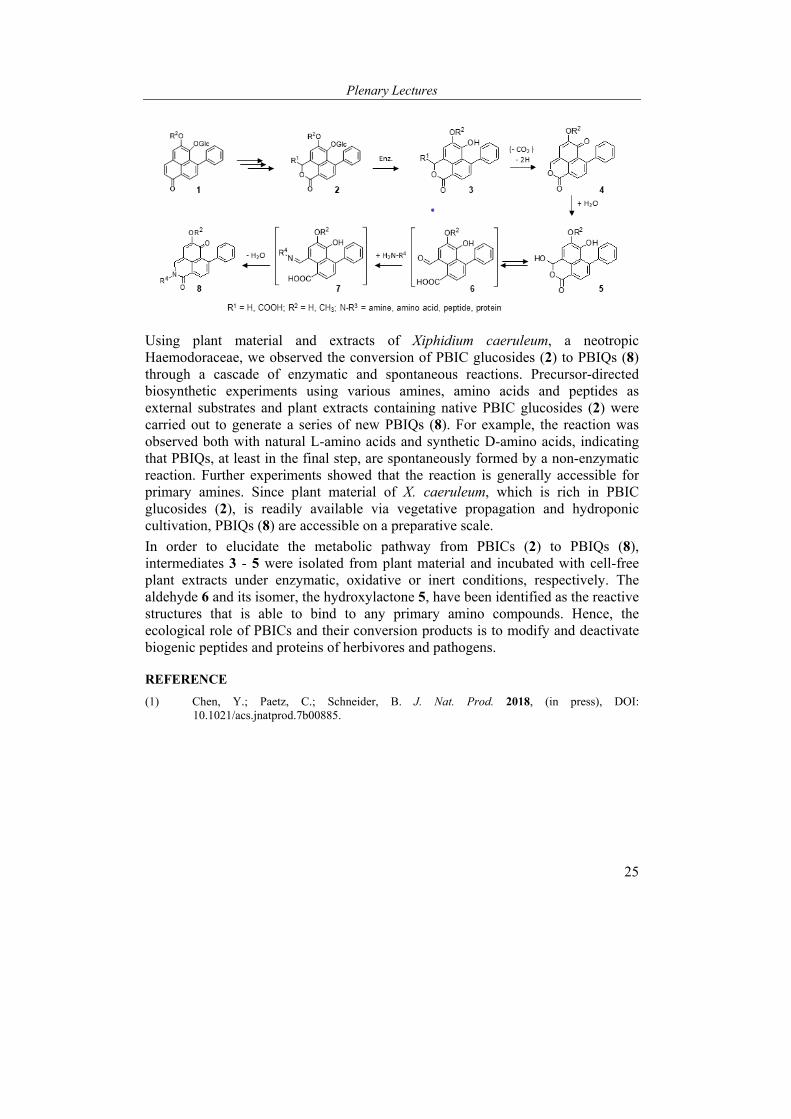
Plenary Lectures
25
Using plant material and extracts of Xiphidium caeruleum, a neotropic Haemodoraceae, we observed the conversion of PBIC glucosides (2) to PBIQs (8) through a cascade of enzymatic and spontaneous reactions. Precursor-directed biosynthetic experiments using various amines, amino acids and peptides as external substrates and plant extracts containing native PBIC glucosides (2) were carried out to generate a series of new PBIQs (8). For example, the reaction was observed both with natural L-amino acids and synthetic D-amino acids, indicating that PBIQs, at least in the final step, are spontaneously formed by a non-enzymatic reaction. Further experiments showed that the reaction is generally accessible for primary amines. Since plant material of X. caeruleum, which is rich in PBIC glucosides (2), is readily available via vegetative propagation and hydroponic cultivation, PBIQs (8) are accessible on a preparative scale.
In order to elucidate the metabolic pathway from PBICs (2) to PBIQs (8), intermediates 3 - 5 were isolated from plant material and incubated with cell-free plant extracts under enzymatic, oxidative or inert conditions, respectively. The aldehyde 6 and its isomer, the hydroxylactone 5, have been identified as the reactive structures that is able to bind to any primary amino compounds. Hence, the ecological role of PBICs and their conversion products is to modify and deactivate biogenic peptides and proteins of herbivores and pathogens.
REFERENCE
(1) Chen, Y.; Paetz, C.; Schneider, B. J. Nat. Prod. 2018, (in press), DOI: 10.1021/acs.jnatprod.7b00885.

ORAL COMMUNICATIONS
SIGNAL TRANSDUCTION VIA TRANSMEMBRANE DOMAINS OF BITOPIC RECEPTORS IN NORMA AND PATHOLOGY
Eduard Bocharov1,2*
1 Moscow Institute of Physics and Technology (State University), Moscow, Russian Federation, 2 Shemyakin-Ovchinnikov Institute of Bioorganic Chemistry RAS, Moscow, Russian Federation [email protected]
Signal transduction by bitopic receptors, such as receptor tyrosine kinases (RTK) and type I cytokine receptors, has been in the spotlight of scientific interest owing to the central role of these single-spanning membrane receptors in the regulation of development, cell motility, proliferation, differentiation, and apoptosis. Nowadays, the elucidation of high-resolution structure of full-size bitopic receptors having flexible multiple-domain composition is still a challenge. During signal transduction across plasma membrane, bitopic receptors are activated by proper ligand-induced homo- and hetero-dimerization or by reorientation of monomers in preformed receptor dimers upon ligand binding. Specific helix-helix interactions of transmembrane domains (TMD) are believed to be important for the bitopic receptor lateral dimerization and signal transduction. Either destroying or enhancing such helix-helix interactions can result in many human diseases: developmental, oncogenic, neurodegenerative, etc. Observed TMD helix-helix packing diversity among bitopic receptors appears in favor of the recently proposed the lipid-mediated rotation-coupled activation mechanism,1 which implies that the sequence of structural rearrangements of the receptor domains is associated with perturbations of the lipid bilayer in the course of ligand-induced receptor activation, considering the receptor together with its lipid environment as a self-consistent signal transduction system.
The human epidermal growth factor receptors (HER or ErbB) and the fibroblast growth factor receptors (FGFR) families serve as excellent model RTK to illustrate how ligand-induced conformational rearrangements and specific dimerization of extracellular domains lead to the allosteric activation of the cytoplasmic kinase domains, resulting in signal propagation across the membrane.2 Besides, HER and FGFR relatives are known oncogenic drivers in many cancers, and inhibitors of these receptors have been among the most successful examples of targeted cancer therapies to date. Pathogenic transmembrane mutations found for the HER and FGFR relatives are located as a rule in narrow regions within the specific TMD helix-helix interfaces assuming that the intermolecular interactions inside membrane are important for the RTK cell signaling dysfunction in human organism.3,4 Such regions can be characterized as a “hot spot” for gain-of-function mutations associated with different human pathologies. This finding justifies a prediction that similar gain-of-function mutations, e.g. enhancing the TMD
VI International Conference “Chemistry, Structure and Function of Biomolecules”
28
dimerization in certain conformation and thus activating the receptor independently of ligand binding, can be found for other bitopic receptor representatives and suggests searching for them is a promising idea for future clinical studies. It can also have potential therapeutic implications, broadening the spectrum of targets for pharmaceuticals by inclusion of plasma membranes and their constituents.
REFERENCES
(1) Bocharov, E. V.; Mineev, K. S.; Pavlov, K. V.; Akimov, S. A.; Kuznetsov, A. S.; Efremov, R. G.; Arseniev, A. S. Biochim. Biophys. Acta - Biomembranes 2017, 1859, 561-576.
(2) Bocharov, E. V.; Sharonov, G. V.; Bocharova, O.V.; Pavlov, K.V. Biochim. Biophys. Acta - Biomembranes 2017, 1859, 1417-1429.
(3) Bocharov, E. V.; Lesovoy, D. M.; Goncharuk, S. A.; Goncharuk, M. V.; Hristova, K.; Arseniev, A. S. Structure 2013, 21, 2087–2093.
(4) Ou, S. I.; Schrock, A. B.; Bocharov, E. V.; Klempner, S. J.; Kawamura Haddad, C.; Steinecker, G.; Johnson, M.; Gitlitz, B. J.; Chung, J.; Campregher, P. V.; Ross, J. S.; Stephens, P. J.; Miller, V. A.; Suh, J. H.; Ali, S. M.; Velcheti, V. J. Thorac. Oncol. 2017, 12, 446-457.
The work is supported by the Russian Foundation for Basic Research (project #18-04-01289-а), by the RAS Program “Molecular and Cellular Biology” and by the Russian Academic Excellence Project “5-100”.
APP FAMILIAR MUTATIONS AS A TOOL FOR INVESTIGATION OF THE MOLECULAR BASIS OF ALZHEIMER DISEASE
Olga Bocharova1,2
1 Moscow Institute of Physics and Technology (State University), Moscow, Russian Federation, 2 Shemyakin-Ovchinnikov Institute of Bioorganic Chemistry RAS, Moscow, Russian Federation [email protected]
Alzheimer disease is the most common cause of neurocognitive disorder and may contribute to 60–70% of cases of dementia (according to the WHO report). Despite some progress in study of the molecular mechanisms of Alzheimer's disease development the initial steps of the pathogenesis are still puzzling. Amyloid Aβ-peptides forming plaques in brain during Alzheimer disease are the products of sequential intramembrane cleavage of a single-span membrane amyloid precursor protein (APP). Most of mutations associated with familial forms of Alzheimer disease were found in the APP transmembrane (TM) domain and juxtamembrane (JM) regions. The pathogenic mutations presumably affect structural-dynamic properties of the APP TM domain, e.g. changing its conformational stability, lateral dimerization and intermolecular interactions, which can result in enhanced and alternative cleavage by γ-secretase in membrane.
We designed highly productive systems of bacterial and cell-free expression and easy purification procedure for APP JM-TM fragments of different length, as well
Oral Communications
29
as the fragments with familial Alzheimer disease mutations.1,2 The systems produce milligram quantities of the APP TM fragments with 13C/15N-isotope labeling more than 95% for the detailed NMR characterization of spatial structure, dynamic, and dimerization/oligomerization. The fragments were solubilized in detergent micelles and lipid bicelles, 40-60 kDa supramolecular membrane-mimicking complexes, which allows acquiring proper high-resolution NMR spectra despite low sample stability and aggregation. Molecular Dynamics relaxation of obtained NMR structures of the APP JM-TM fragments in hydrated explicit lipid bilayers provided a detailed atomistic picture of the intra- and intermolecular interactions.
“Australian” (APP L723P) mutation is identified to be associated with autosomal-dominant, early onset Alzheimer’s disease. We detected enhanced flexibility and partial unfolding of the C-terminal region of the TM helix of L723P mutant compared to wild-type peptide, which can facilitate the APP proteolysis in the ε-site and switch between alternative (“pathogenic” and “non-pathogenic”) cleavage cascades.3 We found that unlike wild-type fragment the L723P “Australian” mutant gradually converts from α-helical to β-conformation and this process accompanied by high molecular weight aggregates formation. These findings suggest a straightforward mechanism of the pathogenesis associated with this mutation, and are of generic import for understanding of the molecular-level events associated with APP sequential proteolysis resulting in accumulation of the pathogenic forms of amyloid-β. Thereby the mutant APP JM-TM fragments are shown to be promising objects for elaboration the molecular aspects of γ-secretase proteolysis. Understanding of the principle of different length amyloidogenic peptides generation is necessary for adequate tactics for the Alzheimer disease treatment.
REFERENCES
(1) Bocharova, O. V.; Urban, A. S.; Nadezhdin, K. D.; Bocharov, E. V.; Arseniev, A. S. Protein Expr. Purif. 2016, 123, 105-111.
(2) Nadezhdin, K. D.; Bocharova, O. V.; Bocharov, E. V.; Arseniev, A. S. FEBS Letters 2012, 586, 1687-1692.
(3) Bocharov, E. V.; Nadezhdin, K. D.; Urban, A. S.; Volynsky, P. E.; Pavlov, K. V.; Efremov, R. G.; Arseniev, A. S.; Bocharova, O. V. Biochim. Biophys. Acta - Biomembranes 2018, submitted.
The work is supported by the Russian Foundation for Basic Research (project # 17-04-02045-а), by the RAS Program “Molecular and Cellular Biology” and by the Russian Academic Excellence Project “5-100”.
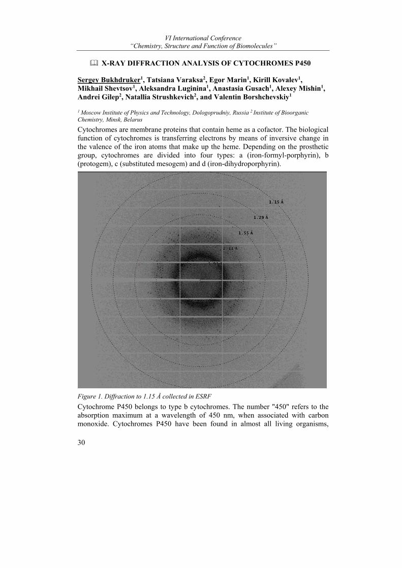
VI International Conference “Chemistry, Structure and Function of Biomolecules”
30
X-RAY DIFFRACTION ANALYSIS OF CYTOCHROMES P450
Sergey Bukhdruker1, Tatsiana Varaksa2, Egor Marin1, Kirill Kovalev1, Mikhail Shevtsov1, Aleksandra Luginina1, Anastasia Gusach1, Alexey Mishin1, Andrei Gilep2, Natallia Strushkevich2, and Valentin Borshchevskiy1
1 Moscow Institute of Physics and Technology, Dologoprudniy, Russia 2 Institute of Bioorganic Chemistry, Minsk, Belarus
Cytochromes are membrane proteins that contain heme as a cofactor. The biological function of cytochromes is transferring electrons by means of inversive change in the valence of the iron atoms that make up the heme. Depending on the prosthetic group, cytochromes are divided into four types: a (iron-formyl-porphyrin), b (protogem), c (substituted mesogem) and d (iron-dihydroporphyrin).
Figure 1. Diffraction to 1.15 Å collected in ESRF
Cytochrome P450 belongs to type b cytochromes. The number "450" refers to the absorption maximum at a wavelength of 450 nm, when associated with carbon monoxide. Cytochromes P450 have been found in almost all living organisms,
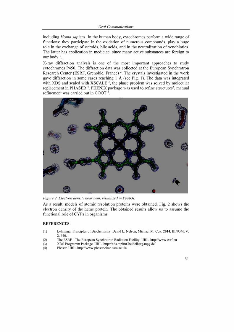
Oral Communications
31
including Homo sapiens. In the human body, cytochromes perform a wide range of functions: they participate in the oxidation of numerous compounds, play a huge role in the exchange of steroids, bile acids, and in the neutralization of xenobiotics. The latter has application in medicice, since many active substances are foreign to our body 1.
X-ray diffraction analysis is one of the most important approaches to study cytochromes P450. The diffraction data was collected at the European Synchrotron Research Center (ESRF, Grenoble, France) 2. The crystals investigated in the work gave diffraction in some cases reaching 1 Å (see Fig. 1). The data was integrated with XDS and scaled with XSCALE 3, the phase problem was solved by molecular replacement in PHASER 4. PHENIX package was used to refine structures5, manual refinement was carried out in COOT 6.
Figure 2. Electron density near hem, visualized in PyMOL
As a result, models of atomic resolution proteins were obtained. Fig. 2 shows the electron density of the heme protein. The obtained results allow us to assume the functional role of CYPs in organisms
REFERENCES
(1) Lehninger Principles of Biochemistry. David L. Nelson, Michael M. Cox. 2014, BINOM, V. 2, 640.
(2) The ESRF - The European Synchrotron Radiation Facility. URL: http://www.esrf.eu (3) XDS Programm Package. URL: http://xds.mpimf-heidelberg.mpg.de/ (4) Phaser. URL: http://www.phaser.cimr.cam.ac.uk/
VI International Conference “Chemistry, Structure and Function of Biomolecules”
32
(5) Python-based Hierarchical ENvironment for Integrated Xtallography (PHENIX). URL: https://www.phenix-online.org/
(6) Crystallographic Object-Oriented Toolkit (COOT). URL: https://www2.mrc-lmb.cam.ac.uk/personal/pemsley/coot/.
The work was supported by the Ministry of Education and Science of the Russian Federation (RFMEFI58716X0026).
ENGINEERING CHIMERIC ANTIGEN RECEPTOR (CAR) T-CELLS FOR ENHANCED CANCER IMMUNOTHERAPY
Dmitri Dormeshkin1*, Mikalai Katsin2, Aleksandr Migas3, Alexander Meleshko3, and Andrei Gilep1
1 Institute of Bioorganic Chemistry of NAS of Belarus, Minsk, Belarus, 2 Vitebsk state medical university, Vitebsk, Belarus, 3 Belarusian Research Center for Pediatric Oncology, Hematology and Immunology, Minsk, Belarus e-mail: [email protected]
In a past decade CAR T-cell based immunotherapy has achieved a significant progress in the treatment of malignant hematological diseases and became one of the major frontiers at the overcrowded immune-oncology field1. One of the most exciting clinical results showed that complete remission (CR) rates as high as 90% in children and adults with relapsed and refractory acute lymphoid leukemia treated with CAR-modified T-cells targeting the CD192. CAR T-cells are genetically modified T-cells engineered to express a tumor-specific chimeric antigen receptor (CAR), which can activate them upon binding the antigen in MHC-independent manner. CAR is composed of an extracellular targeting domain (scFv), a hinge region, a transmembrane domain and one or few costimulatory signaling domains, that enhance the T-cell activation and cytokines production3. The lack of structural information about CAR organization and behavior upon the antigen binding slows down its engineering and development as the effect of CAR domain composition on the signal transduction effect is not obvious4.
CD19 receptor is absent from bone marrow progenitor cells and expressed on most B-lineage malignancies5. Almost all published clinical trials targeting CD19 have utilized antigen-binding scFvs derived from murine monoclonal antibodies. T-cell immune responses directed against anti-CD19 CAR have been reported on repeated occasions. It negatively affects CAR T-cell persistence and thus decreases overall efficiency of therapy.
We have generated humanized scFv antibody fragment consisted of engineered FMC63 mAb VH and VL domains with proteolytic stable flexible «whitlow-218» linker between them. Heterologous expression in Escherichia coli system was carried out. Humanized scFv was obtained in the purified and homogenous state utilizing previously developed CYB5-fusion monitoring system6. In vitro cellular
Oral Communications
33
ELISA experiments revealed that humanized FMC63 scFv binds to CD19+ Raji and Namalwa cell lines, but not to the CD19– HEK293T and A549 cells.
In order to predict an impact of extracellular CAR composition on its behavior, we have created MD model of the anti-CD19 CAR in the lipid bilayer membrane.
A series of 2nd generation humanized and structurally optimized anti-CD19 CAR constructions with different «short» and «long» hinge regions were designed and constructed. Anti-CD19 CAR-expressing CD8+ cells subsets show a different grade of cytotoxic activity depended on hinge region length.
REFERENCES
(1) Fesnak, A. D.; June, C. H.; Levine, B. L. Nature reviews. Cancer 2016, 16, 566. (2) Grupp, S. A.; Kalos, M.; Barrett, D.; Aplenc, R.; Porter, D. L.; Rheingold, S. R.; Teachey,
D. T.; Chew, A.; Hauck, B.; Wright, J. F.; Milone, M. C.; Levine, B. L.; June, C. H. The New England journal of medicine 2013, 368, 1509.
(3) Maus, M. V.; Grupp, S. A.; Porter, D. L.; June, C. H. Blood 2014, 123, 2625. (4) Alabanza, L.; Pegues, M.; Geldres, C.; Shi, V.; Wiltzius, J. J. W.; Sievers, S. A.; Yang, S.;
Kochenderfer, J. N. Molecular therapy : the journal of the American Society of Gene Therapy 2017, 25, 2452.
(5) Naddafi, F.; Davami, F. International journal of molecular and cellular medicine 2015, 4, 143.
(6) Dormeshkin, D.; Gilep, A.; Sergeev, G.; Usanov, S. Protein expression and purification 2016, 128, 60.
This work was supported by State Committee on Science and Technology of the Republic of Belarus (№201739)
FROM RED TO YELLOW: THE BIOSYNTHESIS OF UNIQUE INDOLE ALKALOIDS IN YELLOW PAPAVER NUDICAULE FLOWERS AND THEIR BIOMIMETIC SYNTHESIS
Bettina Dudek, Anne-Christin Warskulat, Evangelos Tatsis, Florian Schnurrer, Christian Paetz, and Bernd Schneider*
Max Planck Institute for Chemical Ecology, Jena, Germany e-mail: [email protected]
The petals of Papaver nudicaule (Iceland poppy) owe their bright yellow colour a unique class of alkaloids, known as nudicaulins.1 For 80 years, details about the chemical structure and the biosynthesis of nudicaulins remained obscure until their indole-polyphenol hybrid structure was elucidated in 2013.2 Since then, the interest turned to study their remarkable biosynthesis.
The first indications on the nudicaulin biosynthetic pathway were derived from changes of the colour and the pigment profile of P. nudicaule petals. During bud development, the petals show a colour shift from white over red and orange to yellow. The red colour is derived from pelargonidin glycosides, anthocyanins,
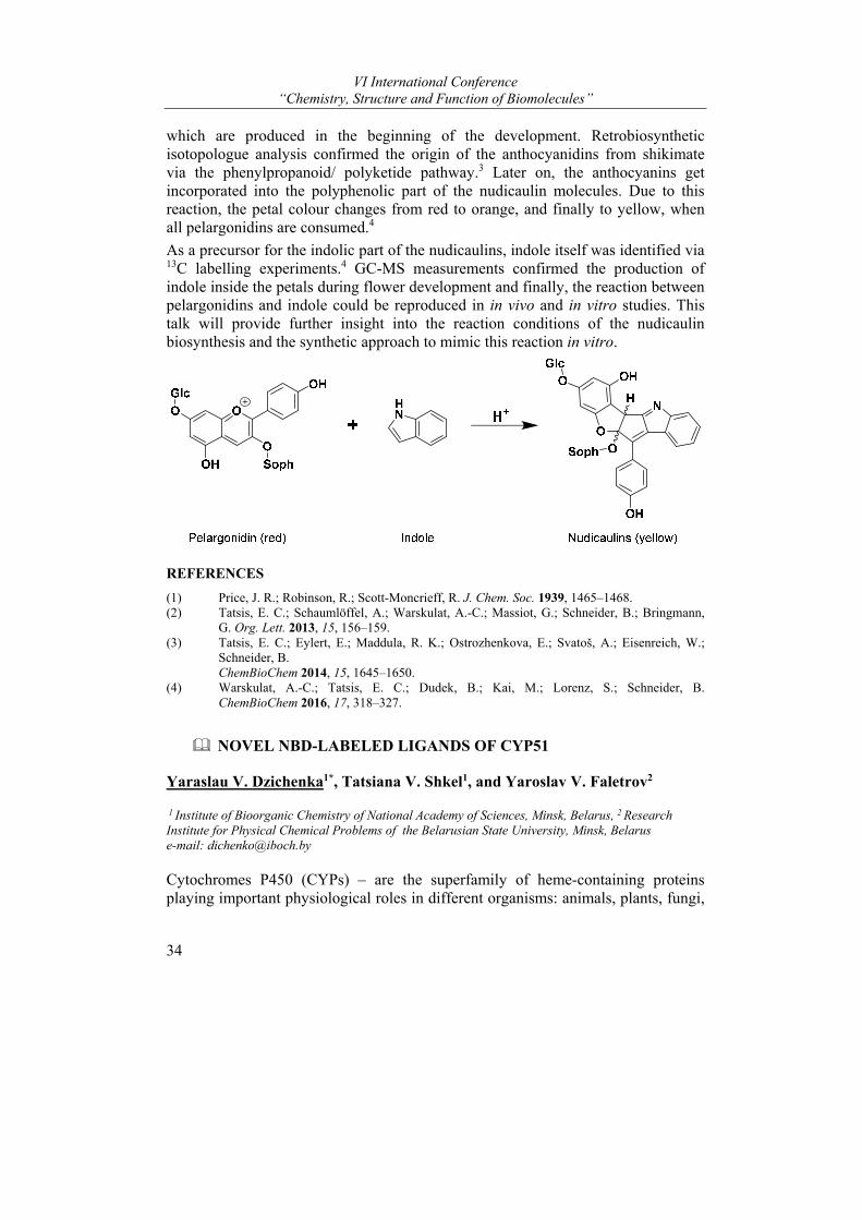
VI International Conference “Chemistry, Structure and Function of Biomolecules”
34
which are produced in the beginning of the development. Retrobiosynthetic isotopologue analysis confirmed the origin of the anthocyanidins from shikimate via the phenylpropanoid/ polyketide pathway.3 Later on, the anthocyanins get incorporated into the polyphenolic part of the nudicaulin molecules. Due to this reaction, the petal colour changes from red to orange, and finally to yellow, when all pelargonidins are consumed.4
As a precursor for the indolic part of the nudicaulins, indole itself was identified via 13C labelling experiments.4 GC-MS measurements confirmed the production of indole inside the petals during flower development and finally, the reaction between pelargonidins and indole could be reproduced in in vivo and in vitro studies. This talk will provide further insight into the reaction conditions of the nudicaulin biosynthesis and the synthetic approach to mimic this reaction in vitro.
REFERENCES
(1) Price, J. R.; Robinson, R.; Scott-Moncrieff, R. J. Chem. Soc. 1939, 1465–1468. (2) Tatsis, E. C.; Schaumlöffel, A.; Warskulat, A.-C.; Massiot, G.; Schneider, B.; Bringmann,
G. Org. Lett. 2013, 15, 156–159. (3) Tatsis, E. C.; Eylert, E.; Maddula, R. K.; Ostrozhenkova, E.; Svatoš, A.; Eisenreich, W.;
Schneider, B. ChemBioChem 2014, 15, 1645–1650. (4) Warskulat, A.-C.; Tatsis, E. C.; Dudek, B.; Kai, M.; Lorenz, S.; Schneider, B.
ChemBioChem 2016, 17, 318–327.
NOVEL NBD-LABELED LIGANDS OF CYP51
Yaraslau V. Dzichenka1*, Tatsiana V. Shkel1, and Yaroslav V. Faletrov2
1 Institute of Bioorganic Chemistry of National Academy of Sciences, Minsk, Belarus, 2 Research Institute for Physical Chemical Problems of the Belarusian State University, Minsk, Belarus e-mail: [email protected]
Cytochromes P450 (CYPs) – are the superfamily of heme-containing proteins playing important physiological roles in different organisms: animals, plants, fungi,
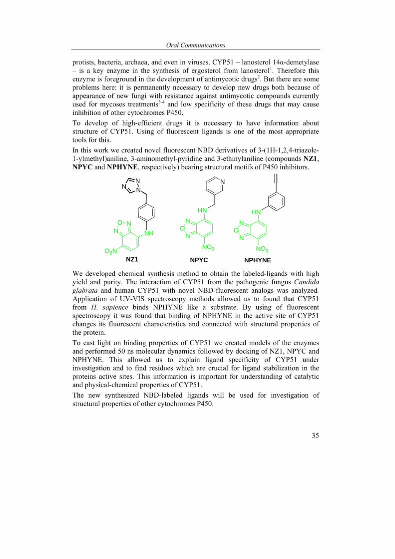
Oral Communications
35
protists, bacteria, archaea, and even in viruses. CYP51 – lanosterol 14α-demetylase – is a key enzyme in the synthesis of ergosterol from lanosterol1. Therefore this enzyme is foreground in the development of antimycotic drugs2. But there are some problems here: it is permanently necessary to develop new drugs both because of appearance of new fungi with resistance against antimycotic compounds currently used for mycoses treatments3-4 and low specificity of these drugs that may cause inhibition of other cytochromes P450.
To develop of high-efficient drugs it is necessary to have information about structure of CYP51. Using of fluorescent ligands is one of the most appropriate tools for this.
In this work we created novel fluorescent NBD derivatives of 3-(1H-1,2,4-triazole-1-ylmethyl)aniline, 3-aminomethyl-pyridine and 3-ethinylaniline (compounds NZ1, NPYC and NPHYNE, respectively) bearing structural motifs of P450 inhibitors.
N
NN
NZ1
NHNO N
O2N
HN
NO
N
NO2
N
NPYC
HN
NO
N
NO2
NPHYNE
We developed chemical synthesis method to obtain the labeled-ligands with high yield and purity. The interaction of CYP51 from the pathogenic fungus Candida glabrata and human CYP51 with novel NBD-fluorescent analogs was analyzed. Application of UV-VIS spectroscopy methods allowed us to found that CYP51 from H. sapience binds NPHYNE like a substrate. By using of fluorescent spectroscopy it was found that binding of NPHYNE in the active site of CYP51 changes its fluorescent characteristics and connected with structural properties of the protein.
To cast light on binding properties of CYP51 we created models of the enzymes and performed 50 ns molecular dynamics followed by docking of NZ1, NPYC and NPHYNE. This allowed us to explain ligand specificity of CYP51 under investigation and to find residues which are crucial for ligand stabilization in the proteins active sites. This information is important for understanding of catalytic and physical-chemical properties of CYP51.
The new synthesized NBD-labeled ligands will be used for investigation of structural properties of other cytochromes P450.
VI International Conference “Chemistry, Structure and Function of Biomolecules”
36
REFERENCES
(1) Lepesheva, G. I.; Waterman M. R. Biochimica et biophysica acta. 2007. 1770, № 3, 467-477.
(2) Junqueira, J.C. [et al.] BMC microbiology. 2011. 11, 247. (3) Sanglard, D.; Odds F.C. The Lancet infectious diseases. 2002. 2, № 2, 73-85. (4) Morio, F. [et al.] Diagnostic microbiology and infectious disease. 2010. 66, № 4. 373-384.
SELECTION OF RIGHT- OR LEFT-HANDED STRUCTURAL MOTIFS DEPENDS ON THEIR ARRANGEMENT IN PROTEIN STRUCTURE
Alexander V. Efimov
Institute of Protein Research, Russian Academy of Sciences, Pushchino, Moscow Region, Russia e-mail address: [email protected]
In proteins, the polypeptide chain forms a number of right- and left-handed helices and superhelices, right- and left-turned hairpins and some other structures that are non-superimposable, although they are not mirror images of each other as the L-amino acids are not converted to the D-amino acids. This property of protein structures will be referred to here as pseudo-chirality or handedness. There is a number of structural motifs that exhibit unique handedness in proteins, for example, αα-corners, abcd- and abCd-units, 3β-corners, βSβ-superhelices etc.1 This property of the polypeptide chain is of particular value in protein folding and modeling since it drastically reduces the number of possible folds.
On the other hand, β-hairpins can be right- or left-handed, triple-strand β-sheets can exist as S-like or Z-like β-sheets and so on. Analysis shows that selection of one form of such structural motifs depends on mutual arrangement of the motifs and the other parts of the higher order structures that include them. For example, the abCd-unit can be represented as a combination of the right-handed βαβ-superhelix and a β-hairpin1. In the abCd-unit of type ββαβ, in which hairpin ab is located at the N-end and the βαβ-superhelix follows it, the right-turned hairpin ab is selected. In the abCd-unit of type βαββ, where hairpin ab follows the βαβ-superhelix, the left-turned hairpin ab is selected. A similar selection takes place in combinations of the П-modules and triple-strand β-sheets. In the combinations, in which the П-module follows the β-sheet in the chain, the S-like β-sheet is selected and the SП-motif is formed. If the β-sheet follows the П-module in the chain, the Z-like β-sheet is selected and the ПZ-motif is formed2.
It is well known that overwhelming majority of the βαβ-units form the right-handed superhelices in α/β-proteins3 and most П-modules are right-turned2. However, in combinations of П-modules and βαβ-units, there is anomalously high frequencies of occurrence of the left-handed βαβ-units (~11%) and the left-turned П-modules (34%)4. It is shown that in βαβП-combinations, where the П-module follows the
Oral Communications
37
βαβ-units, both the elements are right-handed. In the Пβαβ-combinations, in which the βαβ-unit follows the П-module, the βαβ-unit is right-handed and the П-module is left-turned. In the combinations of the left-handed βαβ-unit and the right-turned П-module that occur relatively rare, the βαβ-unit follows the П-module in the chain.
Thus, these and other examples to be present in this report demonstrate the relationship between mutual arrangement of structural elements and their handedness in protein structures. This relationship seems to play very important role in protein folding and will be useful in protein modeling and design.
References (1) Efimov, A.V. Biochemistry(Moscow) 2018, 83(Suppl. 1), S103-S110. (2) Efimov, A.V. Proteins 2017, 85, 1925-1930. (3) Rao, S.T., Rossmann, M.G. J. Mol. Biol. 1973, 76, 241-256. (4) Kargatov, A.M., Efimov, A.V. Molecular Biol. (Moscow) 2018, 52, 36-41.
This work was supported by the Russian Foundation for Basic Research (project No 17-04-242).
FUNCTIONAL ANALYSIS OF CYTOCHROME P450S INVOLVED IN BIOSYNTHESIS OF AUTOCRINE AND PARACRINE FACTORS.
A.A. Gilep1 , S.A. Sushko2, T.V. Shkel1, A.V. Svirid1, S. V. Smolskaya1, A.V. Vasilevskaya1, S.A. Usanov1, P.V. Ershov3, A.S. Ivanov3, N.V. Strushkevich1
1Institute of Bioorganic Chemistry, National Academy of Sciences of Belarus, Minsk, Belarus; 2
Department of Bioengineering, School of Engineering, The University of Tokyo, Tokyo, Japan; 3Institute of Biomedical Chemistry, Moscow, Russia. e-mail: [email protected]
Cytochromes Р450 (Р450) are membrane bound hemeproteins involved in biosynthesis of bioactive molecules and metabolism of drugs. Moreover these enzymes play an essential role in metabolic functions of different pathogens. Therefore P450s are important targets for generation of new drugs for treatment endocrine disorders and drug-resistant forms of infection diseases. Our research is focused on understanding of molecular mechanism of substrate recognition, binding and catalysis of sterols and eicosanoids modifying P450s. This research provides valuable tool for P450-targeted drug discovery.
We obtained the data that indicate the existence of an alternative pathway of steroid hormone biosynthesis using 7-dehydrosteroids (precursors of secosteroids) [1-2]. Recently, we have shown the efficient conversion of 7-dehydro- cholesterol to 7-dehydropregnenolone by CYP11A1 and demonstrated the ability of CYP17 from different species to catalyze reactions with the Δ 5,7-type of steroid substrates, forming precursors for equine type steroid hormones. The products of CYP11A1 and CYP17A1 catalyzed reaction could be converted to C21- or C19-derivatives of secosteroids. We also discovered alternative pathway of vitamin D3 metabolism by
VI International Conference “Chemistry, Structure and Function of Biomolecules”
38
CYP11A1. All of these alternative endogenous compound could be involved in different biological process both in normal and pathological conditions (Smith–Lemli–Opitz syndrome). We also found that P450s of pathogenic mycobacteria involved in metabolism of immunоactive lipids and thus can modulate local immunoresponse [3].
By direct molecular fishing, we identified new protein partners for human thromboxane synthase (CYP5A1). These results suggest that interaction with identified protein-partners is important in the regulation of the biosynthesis of eicosanoids [4].
REFERENCES
1) Gilep, A.A., Sushko, T.A., Usanov, S.A. Biochim Biophys Acta. 2011, 1814, 200-209. 2) Guryev, O., Carvalho, R.A., Usanov, S., Gilep, A., Estabrook, R.W. PNAS 2003,100, 14754-
14759. 3) Vasilevskaya A.V., Yantsevich A.V., Sergeev G.V., Lemish A.P., Usanov S.A., Gilep A.A. J
Steroid Biochem Mol Biol. 2017,169, 202-209. 4) Svirid, A.V., Ershov, P.V., Yablokov, E.O., Kaluzhskiy, L.A., Mezentsev Y.V., Florinskaya
A.V., Sushko, T.A., Strushkevich, N.V., Gilep, A.A., Usanov S.A., Medvedev, A.E., Ivanov, A.S. Acta Naturae 2017, 9, 92-100
ACTIVE SITE DOCKING AND IN VITRO INHIBITORY ACTIVITY OF SOME N-HETEROCYCLIC AND CARBOCYCLIC COMPOUNDS TOWARDS PURIFIED HUMAN GLUTATHIONE TRANSFERASE P1
Syargey Gilevich and Yuliya Brechka
Institute of Bioorganic Chemistry of the National Academy of Sciences of Belarus, Minsk, Belarus e-mail: [email protected]
Human glutathione transferase P1 (GSTP1; EC 2.5.1.18) is a prominent enzyme often overexpressed in tumor cells which thus acquire multiple drug resistance. Monomeric GSTP1 also prevents the cells from apoptosis by binding to c-Jun N-terminal kinase (JNK) and down-regulating the JNK signaling pathway1. Known 3D structures of GSTP1 in complex with glutathione (GSH) and a few inhibitors imply the presence of at least two ligand-binding sites per enzyme monomer: the G-site specifically binds GSH, while the nearby H-site accommodates amphiphilic cosubstrates, inhibitors, and non-inhibiting ligands2. The H-site cavity is defined by residues F8, V10, V35, I104, Y108, and G2053; however, residues Y7, R13, and N204 may also be involved in binding of certain ligands4. A number of natural and synthetic organic compounds reversibly bind to and inhibit the enzyme with more or less efficiency. Still, only few of them are isoform P1-specific and have reached clinical trials to date1, the molecular design requirements for a potent and selective inhibitor being not clearly understood.
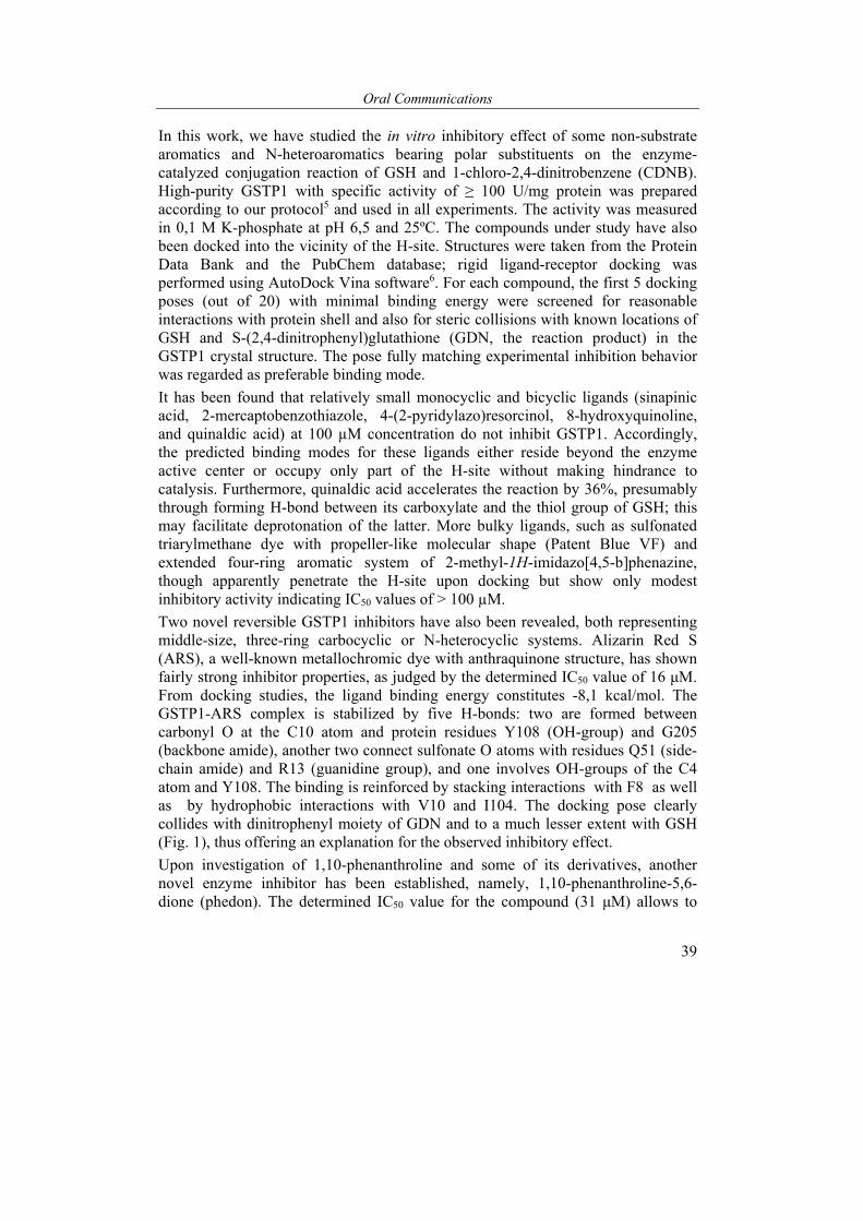
Oral Communications
39
In this work, we have studied the in vitro inhibitory effect of some non-substrate aromatics and N-heteroaromatics bearing polar substituents on the enzyme-catalyzed conjugation reaction of GSH and 1-chloro-2,4-dinitrobenzene (CDNB). High-purity GSTP1 with specific activity of ≥ 100 U/mg protein was prepared according to our protocol5 and used in all experiments. The activity was measured in 0,1 M K-phosphate at pH 6,5 and 25ºC. The compounds under study have also been docked into the vicinity of the H-site. Structures were taken from the Protein Data Bank and the PubChem database; rigid ligand-receptor docking was performed using AutoDock Vina software6. For each compound, the first 5 docking poses (out of 20) with minimal binding energy were screened for reasonable interactions with protein shell and also for steric collisions with known locations of GSH and S-(2,4-dinitrophenyl)glutathione (GDN, the reaction product) in the GSTP1 crystal structure. The pose fully matching experimental inhibition behavior was regarded as preferable binding mode.
It has been found that relatively small monocyclic and bicyclic ligands (sinapinic acid, 2-mercaptobenzothiazole, 4-(2-pyridylazo)resorcinol, 8-hydroxyquinoline, and quinaldic acid) at 100 µM concentration do not inhibit GSTP1. Accordingly, the predicted binding modes for these ligands either reside beyond the enzyme active center or occupy only part of the H-site without making hindrance to catalysis. Furthermore, quinaldic acid accelerates the reaction by 36%, presumably through forming H-bond between its carboxylate and the thiol group of GSH; this may facilitate deprotonation of the latter. More bulky ligands, such as sulfonated triarylmethane dye with propeller-like molecular shape (Patent Blue VF) and extended four-ring aromatic system of 2-methyl-1H-imidazo[4,5-b]phenazine, though apparently penetrate the H-site upon docking but show only modest inhibitory activity indicating IC50 values of > 100 µM.
Two novel reversible GSTP1 inhibitors have also been revealed, both representing middle-size, three-ring carbocyclic or N-heterocyclic systems. Alizarin Red S (ARS), a well-known metallochromic dye with anthraquinone structure, has shown fairly strong inhibitor properties, as judged by the determined IC50 value of 16 μM. From docking studies, the ligand binding energy constitutes -8,1 kcal/mol. The GSTP1-ARS complex is stabilized by five H-bonds: two are formed between carbonyl O at the C10 atom and protein residues Y108 (OH-group) and G205 (backbone amide), another two connect sulfonate O atoms with residues Q51 (side-chain amide) and R13 (guanidine group), and one involves OH-groups of the C4 atom and Y108. The binding is reinforced by stacking interactions with F8 as well as by hydrophobic interactions with V10 and I104. The docking pose clearly collides with dinitrophenyl moiety of GDN and to a much lesser extent with GSH (Fig. 1), thus offering an explanation for the observed inhibitory effect.
Upon investigation of 1,10-phenanthroline and some of its derivatives, another novel enzyme inhibitor has been established, namely, 1,10-phenanthroline-5,6-dione (phedon). The determined IC50 value for the compound (31 μM) allows to
VI International Conference “Chemistry, Structure and Function of Biomolecules”
40
consider phedon as medium-strength inhibitor comparable to natural GSTP1 inhibitors, kaempferol and quercetin.
OH
OH
O
O
S
O
O–
O
Na+
Alizarin Red S
O O
N N 1,10-phenanthroline-5,6-dione Fig. 1. Predicted binding modes of novel inhibitors in the H-site of GSTP1. Carbon atoms of ARS, phedon and GDN are colored in brown, magenta and pale lavender, respectively.
In steady-state kinetic experiments, the type of inhibition with phedon has been established as noncompetitive towards GSH and competitive towards CDNB. The calculated energy of GSTP1-phedon complex formation constitutes -6,4 kcal/mol. The predicted ligand binding mode is concordant to the kinetic data and has much in common with the location of ARS (Fig. 1). Similarly to ARS, phedon forms H-bonds with residues Y108 and G205 through its carbonyl O at the C5 atom. Stacking interactions with F8 and hydrophobic interactions with W38 and V35 also significantly contribute to the complex stabilization. To our knowledge, this is the first example of W38 participation in ligand binding by the H-site. According to mass spectrometric and spectrophotometric data, phedon doesn’t form stable covalent adducts with GSTP1 or GSH but slowly oxidizes the tripeptide thiol group, thus yielding GSSG and 1,10-phenanthroline-5,6-diol which is, in turn, reoxidized aerobically to produce phedon and H2O2. Under the same experimental conditions, however, neither 20 μM diol compound nor 100 μM H2O2 inhibit GSTP1, and the proportion of oxidized GSH in all cases is below 2,6%. These results can exclude indirect mechanisms of the enzyme inhibition. The possibility of using ARS, phedon and other three-ring scaffolds as precursors in directed synthesis of novel lead structures with potent GSTP1 inhibitory activity is discussed.
REFERENCES
(1) Allocati, N.; Masulli, M.; Di Ilio, C.; Federici, L. Oncogenesis 2018, 7, article number: 8. (2) Oakley, A. J.; Lo Bello, M.; Nuccetelli, M.; Mazzetti, A. P.; Parker, M. W. J. Mol. Biol.
1999, 291, 913-926. (3) Prade, L.; Huber, R.; Manoharan, T. H.; Fahl, W.E.; Reuter, W. Structure 1997, 5, 1287-
1295.
Oral Communications
41
(4) Ji, X.; Tordova, M.; O’Donnell, R.; Parsons, J. F.; Hayden, J. B.; Gilliland, G. L.; Zimniak, P. Biochemistry 1997, 36, 9690-9702.
(5) Gilevich, S. N.; Brechka, Yu. V.; Ripinskaya, K. Yu. Vesti Nats. Akad. Navuk Belarusi. Ser. Khim. Navuk [Proc. Nat. Acad. Sci. Belarus, chem. ser.] 2017, (2), 66-79.
(6) Trott, O.; Olson A. J. J. Comput. Chem. 2010, 31, 455-461.
STRUCTURAL STUDIES OF G-PROTEIN COUPLED RECEPTORS IN MOSCOW INSTITUTE OF PHYSICS AND TECHNOLOGY
Anastasia Gusach1, Alexandra Luginina1, Alexey Mishin1, Valentin Borshchevskiy1, Egor Marin1, Mikhail Shevtsov1, Anastasia Stepko1, Nadezda Safronova1, Elizaveta Lyapina1, Petr Popov1, Valentin Gordeliy1, and Vadim Cherezov1, 2
1Research Center for Molecular Mechanisms of Aging and Age-Related Diseases, Moscow Institute of Physics and Technology, Dolgoprudny, Russia; 2Department of Chemistry, Bridge Institute, University of Southern California, Los Angeles, USA e-mail: [email protected]
In the last decades the expanding growth of the field of G-protein coupled receptors (GPCR) research is broadly observed. This class of transmembrane receptors is responsible for the intracellular signal transduction followed by cellular response. Malfunctions in GPCRs result in a variety of pathologies connected with signal systems break-downs: from allergic reactions to color blindness, cardiovascular diseases and cancerogenesis. Hence the structural studies of these proteins are of importance not only as a fundamental problem of biophysics and molecular biology but also as a prospective for medicine and pharmacology. Despite of the numerous efforts of scientists working on GPCR crystallization, the process itself remains challenging. Only about 40 unique GPCRs out of almost 800 have high resolution structures published up to date.
Method of X-ray diffraction on protein crystals gives the best resolution of protein structure but it demands a high purity of protein together with high stability and monodispersity. Thus prior to crystallization the receptor should be carefully characterized from the point of view of its monomer/aggregates ratio, melting temperature and other physical properties. Oftentimes an insertion of fusion partners, thermostabilizing point mutations or various truncations are required for the demanded protein quality.
The full pipeline of GPCR crystallization is now function in our laboratory, the number of techniques and methods applied to GPCR studies is constantly growing in MIPT and the new projects are planned for the next years.
Acknowledgments: This work was supported by the Russian President Grant for Governmental Support of Young Russian Scientists (project no. МК-5184.2018.4).
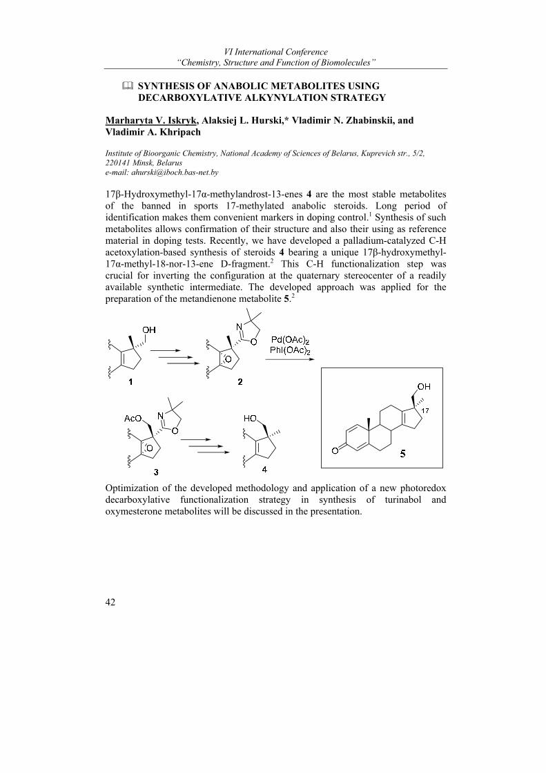
VI International Conference “Chemistry, Structure and Function of Biomolecules”
42
SYNTHESIS OF ANABOLIC METABOLITES USING DECARBOXYLATIVE ALKYNYLATION STRATEGY
Marharyta V. Iskryk, Alaksiej L. Hurski,* Vladimir N. Zhabinskii, and Vladimir A. Khripach
Institute of Bioorganic Chemistry, National Academy of Sciences of Belarus, Kuprevich str., 5/2, 220141 Minsk, Belarus e-mail: [email protected]
17β-Hydroxymethyl-17α-methylandrost-13-enes 4 are the most stable metabolites of the banned in sports 17-methylated anabolic steroids. Long period of identification makes them convenient markers in doping control.1 Synthesis of such metabolites allows confirmation of their structure and also their using as reference material in doping tests. Recently, we have developed a palladium-catalyzed C-H acetoxylation-based synthesis of steroids 4 bearing a unique 17β-hydroxymethyl-17α-methyl-18-nor-13-ene D-fragment.2 This C-H functionalization step was crucial for inverting the configuration at the quaternary stereocenter of a readily available synthetic intermediate. The developed approach was applied for the preparation of the metandienone metabolite 5.2
Optimization of the developed methodology and application of a new photoredox decarboxylative functionalization strategy in synthesis of turinabol and oxymesterone metabolites will be discussed in the presentation.
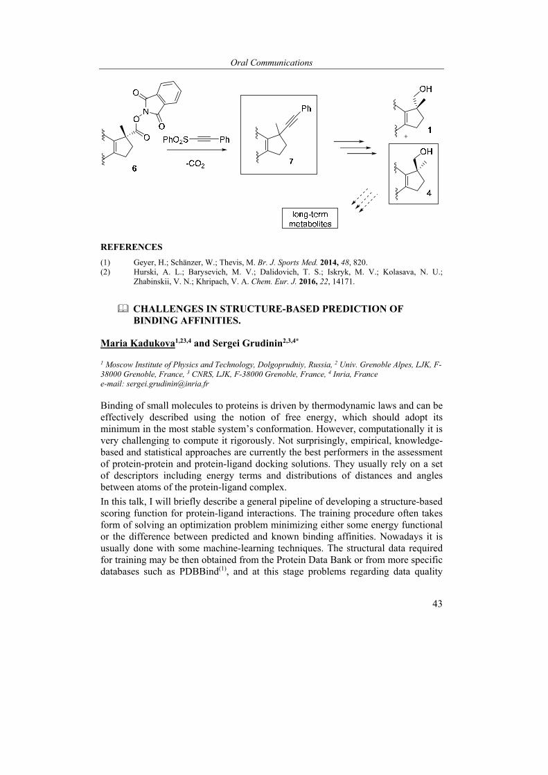
Oral Communications
43
REFERENCES
(1) Geyer, H.; Schänzer, W.; Thevis, M. Br. J. Sports Med. 2014, 48, 820. (2) Hurski, A. L.; Barysevich, M. V.; Dalidovich, T. S.; Iskryk, M. V.; Kolasava, N. U.;
Zhabinskii, V. N.; Khripach, V. A. Chem. Eur. J. 2016, 22, 14171.
CHALLENGES IN STRUCTURE-BASED PREDICTION OF BINDING AFFINITIES.
Maria Kadukova1,23,4 and Sergei Grudinin2,3,4*
1 Moscow Institute of Physics and Technology, Dolgoprudniy, Russia, 2 Univ. Grenoble Alpes, LJK, F-38000 Grenoble, France, 3 CNRS, LJK, F-38000 Grenoble, France, 4 Inria, France e-mail: [email protected]
Binding of small molecules to proteins is driven by thermodynamic laws and can be effectively described using the notion of free energy, which should adopt its minimum in the most stable system’s conformation. However, computationally it is very challenging to compute it rigorously. Not surprisingly, empirical, knowledge-based and statistical approaches are currently the best performers in the assessment of protein-protein and protein-ligand docking solutions. They usually rely on a set of descriptors including energy terms and distributions of distances and angles between atoms of the protein-ligand complex.
In this talk, I will briefly describe a general pipeline of developing a structure-based scoring function for protein-ligand interactions. The training procedure often takes form of solving an optimization problem minimizing either some energy functional or the difference between predicted and known binding affinities. Nowadays it is usually done with some machine-learning techniques. The structural data required for training may be then obtained from the Protein Data Bank or from more specific databases such as PDBBind(1), and at this stage problems regarding data quality
VI International Conference “Chemistry, Structure and Function of Biomolecules”
44
arise. After having been trained, the scoring function may be assessed on several benchmarks, of which the most popular are the two CASF benchmarks(2) measuring a number of important abilities of the affinity predicting algorithms. Another option is participation in the annual community-wide blind challenges aiming at evaluation of the docking programs and protocols on sets of problems, which are very close to the real ones, i.e. finding a best pose or a best binder for a receptor from a set of small molecules.(3)(4)(5) After each competition a new small dataset of answers containing the correct poses and affinities is released and can be also used for testing and training. Currently we are adopting our scoring function(6) to the computation of binding affinities, and I will discuss some problems that we are facing. Most notably these are the contributions of the flexibility of the molecules and their interactions with solvent.
REFERENCES
(1) Liu, Z.H. et al. Acc. Chem. Res., 2017, 50, 302-309 (2) Li, Y., et al. Nature protocols, 2018, 13(4), 666. (3) Smith, R. D.; Damm-Ganamet, K. L.; Dunbar Jr, J. B., et al. J. Chem. Inf. Model., 2015, 56(6),
1022-1031. (4) Gathiaka, S.; Liu, S., et al. J. Comput. Aided. Mol. Des., 2016, 30(9), 651-668. (5) Gaieb, Z.; Liu, S.; Gathiaka, S.; et al. J. Comput. Aided. Mol. Des., 2017, 32(1), 151-162 (6) Kadukova, M.; Grudinin, S. J. Comput. Aided. Mol. Des., 2017, 31(10), 943-958.
DO ALL HUMAN AUTOANTIBODIES SHARE UNIDENTIFIED SITE THAT IS ABSENT IN OTHER HUMAN IMMUNOGLOBULINS?
Elena Kiseleva1*, Konstantin Mikhailopulo1, and Galina Novik2
1 The Institute of Bioorganic chemistry, National Academy of Sciences of Belarus, Minsk, Republic of Belarus; 2 The Institute of Microbiology, National Academy of Sciences of Belarus, Minsk, Republic of Belarus e-mail: [email protected]
We isolated and identified a biopolymer of bacterial origin that interact selectively with three different human autoantibodies (AAbs), viz., antibodies to thyroid peroxidase (anti-TPO), thyroglobulin (anti-Tg) and transglutaminase 2 (anti-TG2). The first two AAbs are recognized serological markers of Hashimoto’s thyroiditis, the most common form of autoimmune thyroid disease (ATD), and the last is marker of celiac disease (CD).
The source of the substance was cell-free fraction (CFF) of Bifidobacterium bifidum BIM В-733D (formerly known as Bifidobacterium bifidum 7911) obtained as a result of cell destruction by ultrasound and ultracentrifugation. CFF was divided into three parts for independent application to three affinity sorbents with immobilized anti-TPO, anti-Tg and anti-gliadins (containing bispecific anti-
Oral Communications
45
gliadin/anti-TG2, see below in more details). Three eluted substances designated Bbanti-TPO, Bbanti-Tg and Bbanti-gliadins, respectively, were purified by gel filtration on TSK-40 and identified by two-dimensional NMR spectroscopy including a 1H, 13C-heteronuclear single-quantum coherence experiment as identical linear α-(1→6)-D-glucans with molecular mass about 5 000 Da, hereinafter referred to as GBb.
The uniqueness of the substance is as follows. At first, GBb has short and unbranched polymeric chain, homogeneous mass by gel-filtration. This distinguishes GBb from α-(1→6)-D-glucans of other bacteria/yeast, which are non-linear (for exception of unique dextran from Leuconostoc mesenteroides CMG713 that does not have any branching2) and have heterogeneous length of the polymer chain with the average molecular weight range from 9×10⁶ to 5×10⁸ Da. At second, GBb has unique immunochemical property, viz., ability to interact selectively with three human AAbs, anti-TPO, anti-Tg1 and anti-TG2, which was proven in ELISA tests.
We hypothesized that GBb differentiates between human AAbs per se and other human Ig (e.g. antibodies against infection agents) due to specific interaction with a yet unidentified site shared by molecules of all AAbs and is absent in Igs against foreign antigens.
What is common in anti-gliadins and AAbs? We answer this question on the basis of data on the mechanisms of immune response characteristic of CD. An intestinal enzyme TG2 (EC 2.3.2.13) catalyzes the deamidation of glutamine in gliadins that enhances their immunoreactivity3 which leads to production of anti-gliadins (stage 1). Indeed, TG2 forms the isopeptide bonds between glutamic acid of gliadin and its own lysine4. As a result, a production of antibodies to epitopes of covalent complex “TG2 - gliadin peptide” (stage 2) and also actually anti-TG2 (stage 3) is initiated. We showed that anti-gliadins used in our study cross-react with TG2 in ELISA and assumed that certain anti-gliadins contain paratopes complementary to TG2/gliadin epitopes related to stage 2. These anti-gliadins are bispecific (anti-gliadins and anti-TG2 simultaneously) and already contain a yet unidentified site shared by molecules of all AAbs.
The first argument in favor of the hypothesis was obtained in ELISA test. In the test, 40 preselected human serum samples with different combinations of anti-TPO, anti-Tg, anti-gliadins and anti-TG2 were added into wells with immobilized GBb, TPO, Tg, TG2 and gliadins. As a result, five sets of A450 values were obtained; each A450 value was proportional to the amount of IgG bound with immobilized item. Indeed, we generate seven sets of A450 values for artificial items represented in Table by calculation the sum of A450 values (for each serum sample independently) in wells with appropriate immobilized items. The correlation coefficient calculated for pair GBb – artificial item TPO + Tg + TG2 + gliadins (r = 0.77) was higher than the other correlation coefficients.
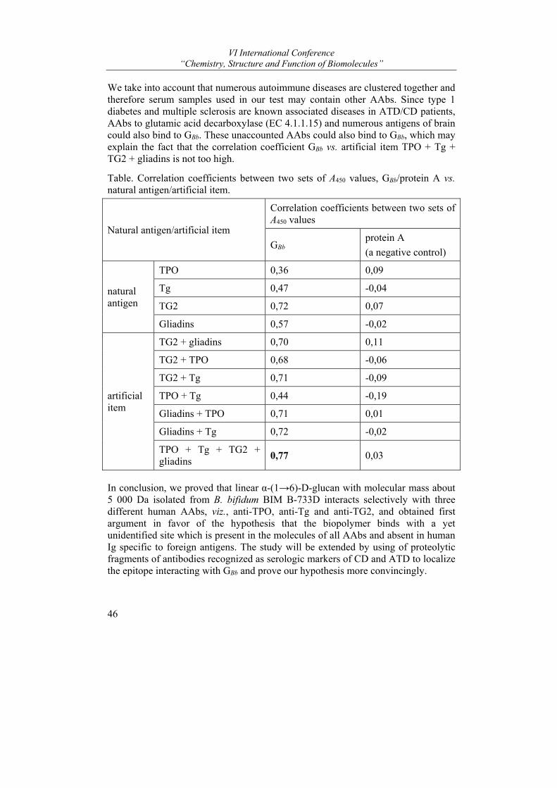
VI International Conference “Chemistry, Structure and Function of Biomolecules”
46
We take into account that numerous autoimmune diseases are clustered together and therefore serum samples used in our test may contain other AAbs. Since type 1 diabetes and multiple sclerosis are known associated diseases in ATD/CD patients, AAbs to glutamic acid decarboxylase (EC 4.1.1.15) and numerous antigens of brain could also bind to GBb. These unaccounted AAbs could also bind to GBb, which may explain the fact that the correlation coefficient GBb vs. artificial item TPO + Tg + TG2 + gliadins is not too high.
Table. Correlation coefficients between two sets of A450 values, GBb/protein A vs. natural antigen/artificial item.
Natural antigen/artificial item
Correlation coefficients between two sets of A450 values
GBb protein A
(a negative control)
natural antigen
TPO 0,36 0,09
Tg 0,47 -0,04
TG2 0,72 0,07
Gliadins 0,57 -0,02
artificial item
TG2 + gliadins 0,70 0,11
TG2 + TPO 0,68 -0,06
TG2 + Tg 0,71 -0,09
TPO + Tg 0,44 -0,19
Gliadins + TPO 0,71 0,01
Gliadins + Tg 0,72 -0,02
TPO + Tg + TG2 + gliadins
0,77 0,03
In conclusion, we proved that linear α-(1→6)-D-glucan with molecular mass about 5 000 Da isolated from B. bifidum BIM В-733D interacts selectively with three different human AAbs, viz., anti-TPO, anti-Tg and anti-TG2, and obtained first argument in favor of the hypothesis that the biopolymer binds with a yet unidentified site which is present in the molecules of all AAbs and absent in human Ig specific to foreign antigens. The study will be extended by using of proteolytic fragments of antibodies recognized as serologic markers of CD and ATD to localize the epitope interacting with GBb and prove our hypothesis more convincingly.
Oral Communications
47
REFERENCES
(1) Kiseleva E.P., Mikhailopulo K.I., Novik G.I., Szwajcer Dey E., Zdorovenko E.L., Shashkov A.S., Knirel Y.A. Benef. Microbes. 2013, 4(4), 375–391.
(2) Sarwat F., Qader S.A.U., Aman A., Ahmed N. Int J Biol Sci. 2008, 4(6), 379-386. (3) Kumar V., Rajadhyaksha M., Wortman J. Clin Diagn Lab Immunol. 2001, 8(4), 678-685. (4) Fleckenstein B., Qiao S.W., Larsen M.R., Jung G., Roepstorff P., Sollid L.M. J Biol Chem.
2004, 279(17), 17607-17616.
AMIDES OF 2-ARYLAMINOPYRYMIRIDINE SERIES - POTENTIAL MULTITARGET INHIBITORS OF ENZYMES INVOLVED IN TUMOR DEVELOPMENT
Elena V. Koroleva* and Zhanna V. Ignatovich
Institute of Chemistry of New Materials, The National Academy of Sciences of Belarus, 36 Skorina st., Minsk 220140, Republic of Belarus e-mail: [email protected]
The discovery of the nature of inhibition of cancer by low molecular weight organic compounds (small molecule) has changed the principles of the development of drug compounds for antitumor therapy. Recent successes in this area are associated with the creation of low molecular weight kinase inhibitors – organic compounds of directed pathogenetic action1.
The modern methodology for the development of anticancer drugs includes a virtual search for a target molecule using pharmacophore structure modeling, molecular docking with a target protein molecule, computer screening of biological activity, selection and optimization of the leader compound, etc. The data of these studies form a basis for subsequent obtaining the target compounds by organic synthesis.
Clinical practice of cancer diseases therapy has demonstrated that drugs with effect on more than one link of the chain of the oncogenesis process can provide more efficacy than mono-targeted drugs. Therefore, in recent years interest has grown to the development of a MTDD (multitarget drug discovery) antitumor drugs with a multi-kinase activity profile2,3. To create potential substances of antitumor drugs, a concept is proposed, which consists in combining structural fragments of compounds with different types of pharmacological activity in one chimeric molecule.
Synthesis of new small molecule inhibitors of tumor enzymes with a multi-kinase profile of therapeutic effect and development of new drugs for the treatment of cancer diseases based on aminopyrimidine derivatives, which include the most demanded and effective antitumor pharmaceuticals, are the current trend in modern organic chemistry and medical chemistry.
2-Arylaminopyrimidine derivatives are among the most commonly recurring templates among kinase inhibitor.
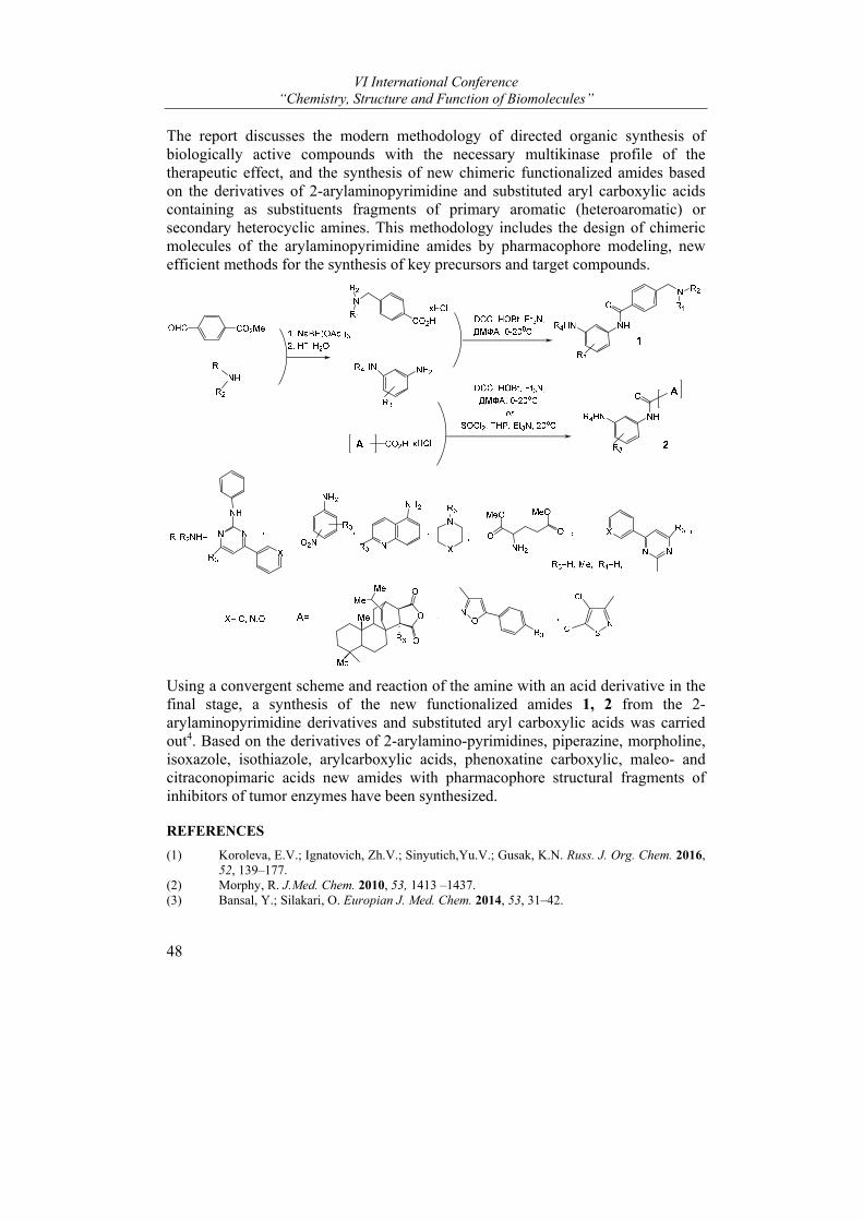
VI International Conference “Chemistry, Structure and Function of Biomolecules”
48
The report discusses the modern methodology of directed organic synthesis of biologically active compounds with the necessary multikinase profile of the therapeutic effect, and the synthesis of new chimeric functionalized amides based on the derivatives of 2-arylaminopyrimidine and substituted aryl carboxylic acids containing as substituents fragments of primary aromatic (heteroaromatic) or secondary heterocyclic amines. This methodology includes the design of chimeric molecules of the arylaminopyrimidine amides by pharmacophore modeling, new efficient methods for the synthesis of key precursors and target compounds.
Using a convergent scheme and reaction of the amine with an acid derivative in the final stage, a synthesis of the new functionalized amides 1, 2 from the 2-arylaminopyrimidine derivatives and substituted aryl carboxylic acids was carried out4. Based on the derivatives of 2-arylamino-pyrimidines, piperazine, morpholine, isoxazole, isothiazole, arylcarboxylic acids, phenoxatine carboxylic, maleo- and citraconopimaric acids new amides with pharmacophore structural fragments of inhibitors of tumor enzymes have been synthesized.
REFERENCES
(1) Koroleva, E.V.; Ignatovich, Zh.V.; Sinyutich,Yu.V.; Gusak, K.N. Russ. J. Org. Chem. 2016, 52, 139–177.
(2) Morphy, R. J.Med. Chem. 2010, 53, 1413 –1437. (3) Bansal, Y.; Silakari, O. Europian J. Med. Chem. 2014, 53, 31–42.

Oral Communications
49
(4) Koroleva, E.V.; Ignatovich, Zh.V.; Gusak, K.N.; Ermolinskaya. A. L.; Sinyutich,Yu.V. Russ. J. Org. Chem. 2015, 51, 101–109.
ENZYMES OF THE PHOSPHOLIPOLYSIS: RESEARCH AND NEW APPROACHES TO PRACTICAL USE
Natalia M. Litvinko
Institute of Bioorganic Chemistry of National Academy of Science of Belarus, Minsk, Belarus, e-mail: [email protected]
These report deals with the study of the mechanisms of biocatalysis in the field of enzymatic hydrolysis of phospholipids – phospholipolysis under action of PLA2 and PLC. It is known, that phospholipases are markers of socially dangerous diseases: oncological, cardiovascular, infectious, inflammatory etc. So, their research and ways of practical use are of special interest.
The aspect of the primary regulation of the catalytic ability of phospholipases, which occurs at the supramolecular level and depends on phospholipid organization at the lipid-water interface is discussed. It was shown that the surface specificity of phospholipases can be drastically changed under the action of xenobiotics. Various mechanisms of inhibition catalysis (direct, indirect and feedback) due to inhibitory analysis are demonstrated.
New directions of practical applications of phospholipolysis - enzyme therapy and enzyme diagnostics - are presented.
In the first direction, the tasks to be accomplished are:
1. Determination of anti enzyme resistance of liposomes, as containers for medicines. 2. Determination of the physiological activity of chemical compounds. 3. Regulation of safe doses of antibiotics. Within the framework of the second direction, the tasks to be solved are: 1. Diagnosis of pancreatitis. 2. Biosafety control of pesticides. 3. Evaluation of the antioxidant potential of the organism. 4. Virulence of microbial phospholipases. To provide research in these areas, a number of biocatalytic systems are proposed.
Firstly, on the basis of ”Phospholipase A2-ligand“ system, in which ligands are low-molecular bioregulators: derivatives of prostaglandins, cyclohexanediones, nucleosides, organic acids, etc.
Secondly, cascade bioreactors containing two proteins, which come into effect step by step, on the basis of the systems ”Phospholipase A2-cytochrome P450“ and ”Phospholipase A2– hemoglobin“1. The latter system, for the first time, was used for creation of the diagnostic kit ”PLA2-PHOA“ for identifying patients with
VI International Conference “Chemistry, Structure and Function of Biomolecules”
50
necrotic pancreatitis. Diagnostic accuracy of the kit is near 100%, that provide possibility of adequate treatment and reduce mortality in patients with inflammatory diseases of the gastrointestinal tract. The results of preclinical and clinical testing of the PLA2-PHOA kit for the detection of pancreatitis based on the determination of the activity of phospholipase A2 are presented and discussed 2,3
REFERENCES
(1) Litvinko, N.M., Skorostetskaya, L.A., Gudko, T.G., Timokhova, M.M., Kamyshnikov, V.S., Vizhiniz, E.I., Vorobei, V.A. Doclady of the National Academy of sciences of Belarus. 2016. 60, 82–87.
(2) Litvinko, N.M.; Skorostetskaya, L.A.; Gerlovsky, D.O. Chem. Phys.Lipids.2018, 211, 44-51. (3) Litvinko, N.M., Skorostetskaya, L.A., Gerlovsky, D.O. Patent BY No. 019669
”Composition and method for determining the total antioxidant activity of blood serum“; Applicant - Institute of Bioorganic Chemistry of the National Academy of Sciences of Belarus, 2011 (in Russian).
ROLE OF SODIUM ALLOSTERIC BINDING SITE IN GPCR FUNCTION
Aleksandra Luginina1*, Anastasia Gusach1, Alexey Mishin1, Egor Marin1, Elizaveta Lyapina1, Petr Popov1, Valentin Borshchevskiy1, Vsevolod Katritch1, 2, and Vadim Cherezov1, 2
1Research Center for Molecular Mechanisms of Aging and Age-Related Diseases, Moscow Institute of Physics and Technology, Dolgoprudny, Russia; 2Department of Chemistry, Bridge Institute, University of Southern California, Los Angeles, USA e-mail: [email protected]
G-protein coupled receptors (GPCR) form the largest transmembrane protein superfamily in the human genome. They play key roles in cell signaling processes and serve as targets for drug development and for modern fundamental and medical research. However, many aspects of GPCR function, in particular effects of endogenous allosteric modulators, remain poorly understood.
In 1970s, it was observed that sodium ions negatively modulate agonist binding to the opioid receptors.1 Later, mutagenesis studies of several receptors revealed the importance of the D2.50 residue2 for the sodium effect. Recently, an allosteric sodium binding pocket next to the D2.50 residue was finally discovered in the 1.8 Å structure of the adenosine receptor.3 The pocket is formed by 15 highly conserved in Class A GPCR residues including D2.50, N1.50, S3.39, N7.45, S7.46, N7.49, Y7.53 and harbors a sodium ion and several water molecules. This pocket was then described in the inactive state structures of several other receptors, while it was shown to collapse upon receptor activation.
A prominent role of the sodium binding site in GPCR function has been suggested by numerous studies, in which mutations in the sodium pocket led to constitutive activation, decoupling or biasing signal transduction toward G protein or β-arrestin
Oral Communications
51
pathways.4,5 It has been hypothesized that sodium ions enter the pocket from the extracellular side stabilizing the receptor in the inactive state and modulating agonist binding, and then exit inside the cell during receptor activation, adding sodium transport to the receptor function.6
Many questions concerning the role of Na+ ion in class A GPCRs remain unsolved. The growing number of new structures provide answers to some of them, while raising new ones.
REFERENCES
(1) Pert, C. B., Pasternak, G. & Snyder, S. H. Science (80 ). 182, 1359–1361 (1973). (2) Ballesteros, J. & Weinstein, H. Methods Neurosci. 25, 366–428 (1995). (3) Liu, W. et al. Science (80). 337, 232–236 (2013). (4) Bonde, M. M. et al. PLoS One 5, (2010). (5) Bot, G., Blake, a D., Li, S. & Reisine, T. J. Pharmacol. Exp. Ther. 284, 283–90 (1998). (6) Katritch, V. et al. Trends Biochem Sci. 39, 233–244 (2015).
The work was supported by the Russian Science Foundation (project no. 16-14-10273)
BIOPHYSICAL ASSAYS FOR FUNCTIONAL ACTIVITY STUDIES OF GPCRS
Alexey Mishin1, Aleksandra Luginina1, Anastasiia Gusach1, Egor Marin2, Nadezda Safronova1, Elizaveta Lyapina1, Polina Khorn1, Mikhail Shevtsov1, Valentin Gordeliy2,3,4, Valentin Borshchevskiy2,4, and Vadim Cherezov1,5
1 Laboratory for Structural Biology of GPCRs, Moscow Institute of Physics and Technology, Dolgoprudniy, 141700, Russian Federation; 2 Laboratory for Advanced Studies of Membrane Proteins, Moscow Institute of Physics and Technology, Dolgoprudniy, 141700, Russian Federation; 3 Institut de Biologie Structurale Jean-Pierre Ebel, Université Grenoble Alpes–Commissariat à l'Energie Atomique et aux Energies Alternatives–CNRS, F-38000 Grenoble, France,4 Institute of Complex Systems (ICS), ICS-6: Molecular Biophysics, Research Centre Juelich, 52425 Juelich, Germany,5 Bridge Institute, Departments of Chemistry and Physics & Astronomy, University of Southern California, Los Angeles, USA
The most important criteria for the selection of G-protein coupled receptors (GPCRs) ligands are the kinetic parameters of their binding, namely, the equilibrium binding constant, and, if possible, the association (on-rate) and dissociation (off-rate) constants . In addition to the tasks of developing the drug candidates, data on the kinetics of receptor binding to ligands are needed to confirm the functional identity of receptors obtained by the recombinant method to their natural prototypes and to study the effect of various protein mutations on its pharmacological properties. To measure these parameters, a number of biophysical research methods are used. In this study we are focusing on microscale thermophoresis (NanoTemper Monolith NT 115). This method, from the point of view of the instrumentation, is simple in execution, does not require the use of
VI International Conference “Chemistry, Structure and Function of Biomolecules”
52
radioactive or fluorescently labeled ligands and also has prospects for scaling (which is important for pharmaceutical screening). However, significant difficulties related to the work are connected with some features of GPCR receptors. First, the typical mass ratio of the receptor to ligand is two orders of magnitude, which requires the selection of an experimental protocol that provides the maximum sensitivity of the detection of the corresponding signal. Secondly, GPCR receptors, like other integral membrane proteins, are complex objects for research because of their low stability, the difficulty in achieving sufficient yields of properly folded protein during expression and purification. Wild-type receptors are generally unstable and prone to aggregation, and therefore, a stabilization strategy is needed which uses stabilizing partner proteins, suitable membrane-modeling media and properly selected protocols for the production of the protein samples. As part of the work on this project, it is planned to conduct an experimental adaptation of the discussed method for studying the binding of GPCR receptors to their ligands, using a number of experimental solutions, including the use of various modern membrane-modeling media (nanodisks, amphipoles, stabilizing detergents), fluorescent partner proteins, the selection of conditions that provide simultaneous additional stabilization of the receptor together with the provision of high signal sensititvity. Currently, there is no approach to solve this task which is easily reproduced and transferred between different representatives of GPCR receptors. In this study we report our efforts in this research directions and also give some perspective view on other functional assays of GPCRs .
This work was supported by the Russian President Grant for Governmental Support of Young Russian Scientists (project no. МК-5184.2018.4).
PROPERTIES OF NEW UNEXPLORED MICROBIAL RHODOPSINS
Ivan Okhrimenko1, Peter Popov1, Nina Malyar1, Lada Petrovskaya2, Natalia Lyubaikina1, Dmitry Soloviov, Georg Bueldt1, and Valentin Gordeliy1,3,4
1 Moscow Institute of Physics and Technology, MIPT, Dolgoprudny, Russia; 2 Shemyakin–Ovchinnikov Institute of Bioorganic Chemistry, RAS, Moscow, Russia; 3 Univ. Grenoble Alpes, CEA, CNRS, IBS, F-38000, Grenoble, France; 4 Institute of Complex Systems: Structural Biochemistry (ICS-6), Research Centre Juelich, Juelich, Germany e-mail: [email protected]
Rhodopsins are probably the most universal biological light-energy transducers and abundant phototrophic mechanisms evolved on Earth. They are found in all the domains of life and in viruses and have a remarkable diversity and potential for biotechnological applications. Channel rhodopsins, H+ and Cl- pumps have become indispensable means of optogenetics and revolutionized neuroscience promising new approaches to the treatment of severe diseases. However, among all identified
Oral Communications
53
rhodopsin genes only a few proteins are characterized. This dramatically limits our knowledge of their functions, mechanisms and biotechnological applications. Moreover, high-resolution structures and molecular mechanisms of recently studied new rhodopsins are either not known or limited to non-active states. The amazing scientific and technological potential of rhodopsins is still to be exploited. Bioinformatic search conducted in open databases of proteins and genomes of microorganisms displayed more than 7000 microbial rhodopsins which are not studied yet1. Gene encoding one of them belongs to the Sphingomonas paucimobilis, Gram-negative bacterium featuring an unusual composition of lipid membrane and implicated in various types of clinical infection2, 3. Selection of conditions for optimized gene expression gave yield of synthesized heterologously protein enough for functional and structural study. We established that S. paucimobilis rhodopsin pumps protons and has an absorption maximum at 540 nm. Flash-photolysis data shows that it has photocycle slower than sensory rhodopsin (SR) in pH higher than 7, and photocycle is like BR’s in pH lower than 6. Cl- and ionic strength of the buffer alters the photocycle. Protein is relatively stable, its melting point is about 87°C. It forms trimeric associates in crystals. Another rhodopsin we took from Hymenobacter sp - gram-negative, UV radiation resistant and low temperature resistant Antarctic lichen bacterium4. Rhodopsin appeared highly light-sensitive – it could be easily denatured by light exposure. Its photocycle is more than 100 seconds. Adsorption spectrum has maximum at 520 nm, and is pH dependent. Deinococcus-Thermus is radio-resistant bacteria also resistant to desiccating conditions5. Its rhodopsin has maximum of adsorption at 560 nm like BR, but it has proline residue instead of corresponding Asp212 of BR. Rhodopsins may provide a unique opportunity to understand better molecular aspects of function and they may find new important applications.
REFERENCES
(1) Ushakov, A., et al. FEBS Journal. 2016, 283, 111. (2) David L. Balkwill, J. K. Fredrickson And M. F. Romine. Prokaryotes, 2006. Sphingomonas
and Related Genera. Chapter 6.10, 7:605–629. (3) Satoko, K., Ryozo, M., Kachiko, S., Toyotsugu, N., Eriko, O., Katsumi, K., Kazuyoshi, K. J.
Bacteriology, 1994, 176, 284-290. (4) Tae-Jin, O., So-Ra, H., Do-Hwan, A., Hyun, P., Augustine, Y.K. J. Biotechnology, 2016,
227, 19-20. (5) Griffiths E., Gupta R.S. Int Microbiol., 2007, 10,:201-8.
The work was supported by RSF 16-15-00242.
VI International Conference “Chemistry, Structure and Function of Biomolecules”
54
THE EFFECTS OF CISPLATIN-BRASSINOSTEROID COMBINATION ON THE GROWTH OF A549 CANCER CELL LINE
Olesya V. Panibrat, Vladimir N. Zhabinskii, and Vladimir A. Khripach
Institute of Bioorganic Chemistry, National Academy of Sciences of Belarus, Minsk, Belarus e-mail:[email protected]
Despite significant advances in oncology, the number of annual deaths reaches 8 million and tends to grow.1 Nowadays there is a large amount of compounds that are used in cancer therapy. They include anthracyclines (daunorubicin, doxorubicin), taxanes (paclitaxel, docetaxel), topoisomerase inhibitors (irinotecan, topotecan), topoisomerase II inhibitors (etoposide, teniposide), kinase inhibitors (imatinib), nucleotide analogs and precursor analogs (azacitidine, cytarabine, fluorouracil, hydroxyurea, methotrexate), antimicrobial peptides (bleomycin, actinomycin), platinum-based agents (carboplatin, cisplatin, oxaliplatin), Vinca alkaloid (vinblastine, vincristine), etc.2 Unfortunately, the absolute majority of known antitumor drugs have pronounced side effects, which limits their use. They are: immunosuppression and myelosuppression, cardiotoxicity, hepatotoxicity, nephrotoxicity, tumor lysis syndrome, peripheral neuropathy and many others.3 This causes the need of searching for new compounds with minimal destructive side effects or compositions that can reduce them. In this connection, attention is drawn to steroids and their derivatives, including natural and synthetic brassinosteroids (BS).
BS belong to a group of steroidal hormones similar to those of animals and humans. In plants, they regulate the expression of genes, affect the course of metabolic processes, growth and differentiation of cells.4 In recent years, they are considered as potential anticarcinogenic agents. The background for this is the antiproliferative activity of BS, shown on a number of cancer cell lines, their ability to inhibit angiogenesis, and low cytotoxicity to normal cells. Moreover, it was shown that BS have a lot of positive effects in mammals such as neuroprotective, antiviral, anabolic and adaptogenic, immunostimulating,5 anti-inflammatory, woundhealing, etc.6 All that refers that BS cannot only inhibit cancer cell growth but also minimize side effects and abate toxicity of chemotherapeutics.
In this work, we evaluate in what way some natural BS (24-epibrassinolide and 28-homocastasterone) and their synthetic analogs ((22S,23S)-24-epibrassinolide and (22S,23S)-28-homocastasterone) (Fig.1) influence on the cytotoxicity of classical antitumor drug cisplatin.
For this purpose, we employed lung carcinoma cells A549 as a cell model to investigate BS effects with the help of MTT assay that is widely used to measure the in vitro cytotoxic effects of drugs.7 It was found that both natural and synthetic BS (except of 28-homocastasterone) at a small concentration 10-6 M reduced IC50 of
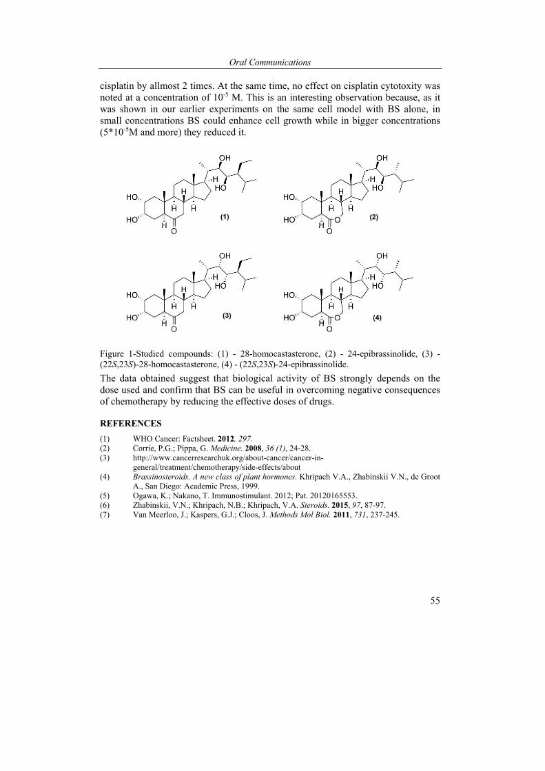
Oral Communications
55
cisplatin by allmost 2 times. At the same time, no effect on cisplatin cytotoxity was noted at a concentration of 10-5 M. This is an interesting observation because, as it was shown in our earlier experiments on the same cell model with BS alone, in small concentrations BS could enhance cell growth while in bigger concentrations (5*10-5M and more) they reduced it.
Figure 1-Studied compounds: (1) - 28-homocastasterone, (2) - 24-epibrassinolide, (3) - (22S,23S)-28-homocastasterone, (4) - (22S,23S)-24-epibrassinolide.
The data obtained suggest that biological activity of BS strongly depends on the dose used and confirm that BS can be useful in overcoming negative consequences of chemotherapy by reducing the effective doses of drugs.
REFERENCES
(1) WHO Cancer: Factsheet. 2012, 297. (2) Corrie, P.G.; Pippa, G. Medicine. 2008, 36 (1), 24-28. (3) http://www.cancerresearchuk.org/about-cancer/cancer-in-
general/treatment/chemotherapy/side-effects/about (4) Brassinosteroids. A new class of plant hormones. Khripach V.A., Zhabinskii V.N., de Groot
A., San Diego: Academic Press, 1999. (5) Ogawa, K.; Nakano, T. Immunostimulant. 2012; Pat. 20120165553. (6) Zhabinskii, V.N.; Khripach, N.B.; Khripach, V.A. Steroids. 2015, 97, 87-97. (7) Van Meerloo, J.; Kaspers, G.J.; Cloos, J. Methods Mol Biol. 2011, 731, 237-245.
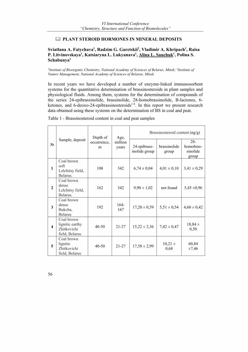
VI International Conference “Chemistry, Structure and Function of Biomolecules”
56
PLANT STEROID HORMONES IN MINERAL DEPOSITS
Sviatlana A. Fatychava1, Radzim G. Garetskii2, Vladimir A. Khripach1, Raisa P. Litvinovskaya1, Katsiaryna L. Lukyanava1, Alina L. Sauchuk1, Polina S. Schabunya1
1Institute of Bioorganic Chemistry, National Academy of Sciences of Belarus, Minsk; 2Institute of Nature Management, National Academy of Sciences of Belarus, Minsk
In recent years we have developed a number of enzyme-linked immunosorbent systems for the quantitative determination of brassinosteroids in plant samples and physiological fluids. Among them, systems for the determination of compounds of the series 24-epibrassinolide, brassinolide, 28-homobrassinolide, B-lactones, 6-ketones, and 6-deoxo-24-epibrassinosteroids1-6. In this report we present research data obtained using these systems on the determination of BS in coal and peat.
Table 1 - Brassinosteroid content in coal and peat samples
№
Sample, deposit
Depth of
occurrence, m
Age,
million years
Brassinosteroid content (ng/g)
24-epibrass-inolide group
brassinolide group
28-homobras-
sinolide group
1
Coal brown soft Lelchitsy field, Belarus.
108 342 6,74 ± 0,04 4,01 ± 0,10 3,41 ± 0,29
2
Coal brown dense Lelchitsy field, Belarus.
162 342 9,90 ± 1,02 not found 5,45 ±0,96
3
Coal brown dense Bukcha, Belarus
192 164-167
17,20 ± 0,59 5,51 ± 0,54 4,60 ± 0,42
4
Coal brown lignitic earthy Zhitkovichi field, Belarus
40-50 21-27 15,22 ± 2,36 7,42 ± 0,47 18,84 ±
0,50
5
Coal brown lignitic Zhitkovichi field, Belarus
40-50 21-27 17,58 ± 2,99 10,21 ±
0,68 60,84 ±7,46
Oral Communications
57
6
Coal brown lignitic dense Zhitkovichi field, Belarus
40-50 21-27 11,57 ± 1,78 29,96 ±
4,06 56,06
±11,10
7
Peat The Turshevka-Chertovo deposit of the Krupsky district , Belarus
0,2-0,4 0,004-0,006
37,2±4,97 6,57±0,438 10,5±0,599
The quantitative determination of brassinosteroids in 6 coal samples from various deposits in Belarus and in 1 sample of peat was carried out by the enzyme immunoassay. It was noted that all studied samples contain brassinosterides of the main groups - 24-epibrassinolide, brassinolide (except for one sample 2 of coal) and 28-homo-brassinolide (see Table 1).
For a detailed study, samples 5 and 6 of brown coal with lignin of the Zhitkovichi deposit were chosen. They differed the greatest content of all group of brassinosteroids. For the purity of the experiment, HPLC fractionation of the samples was carried out and the content of BS in each fraction was studied by two methods − enzyme immunoassay and HPLC-MS. The obtained results are presented in Tables 2.
Table 2 − BS content (ng/g) in samples of brown coal 5 and 6
№ Sample Detected phytohormone
Method of analysis HPLC-MS ELISA
5 6 5 6
1 castasterone 0,968 0,41 4,19±0,493 1,91±0,21
2 24-epicastasterone 2,51 3,24 8,89±0,566 11,2±1,04
3 24-epibrassinolide 1,80 5,84 3,79±0,990 9,42±0,30
4 28-homocastasterone 0,176 0,22 2,72±0,035 1,62±0,36
5 28-homobrassinolide not
detected 0,35 0,275±0,03
Based on the obtained data, it can be concluded that in sample 5, the main fractions, according to ELISA and HPLC-MS, are 24-epibrassinolide (3.8 and 1.8 ng/g, respectively) and 24-epicastasterone (8.9 and 2,5 ng/g), an appreciable amount of castasterone (4.2 and 1.0 ng / g) and an insignificant (an order of magnitude lower) 28-homocastastone. The main detectable representatives of brassinosteroids in sample 6 are also 24-epibrassinolide (9.4 ng/g according to HPLC and 5.8 ng/g

VI International Conference “Chemistry, Structure and Function of Biomolecules”
58
according to ELISA) and 24-epicastasterone (11.2 and 3.2 ng/g). It was detected the palpable amount of castasterone (1.9 and 0.4 ng/g) and representatives of the 28-homoborassinolide group − 28-homocastastone (1.6 and 0.2 ng/g) and 28-homobrassinolide (0.35 and 0.3 n/g).
A surprising fact is the absence in both coal samples of brassinolide (as noted above, its biosynthetic precursor castesterone is detected in appreciable amounts). It can be assumed that brassinosteroids that are not found in the test samples can be present in very small amounts that are not detected by these methods. In addition, in peat samples, the content of brassinosteroid B-lactones and 6-ketones was analyzed using specific immunochemical test systems [5, 6].Their content is comparable but the lactone series compounds prevail (38.3 vs 12.2 ng/g).
Thus, based on the study of coal and peat samples, it can be concluded that these minerals, which are of vegetable origin, contain brassinosteroids in appreciable amounts. The brassinosteroid composition varies depending on the deposit, depth of bedding, and other factors. However, the quantitative content of brassinosteroids is comparable to that in plant objects7-9 and medicinal herbs6. Obviously, this can explain the high stability of the studied objects to transformations under the influence of environmental factors.
REFERENCES
(1) Khripach V.A., Sviridov O.V., Priadko A.G., Litvinovskaya R.P., Drach S.V., Matveentsev V.D., Novik T.V. Rus. J. Bioorganic Chem. 2007, 33, 347-53.
(2) Khripach V.A., Litvinovskaya R.P., Drach S.V., Aver'kova M.A., Zhabinski, V.N., Sviridov O.V., Priadko A.G., Novik T.V., Matveentsev V.D. Doklady NAN Belarusi. 2009, 53, 74–77.
(3) Khripach V.A., Litvinovskaya R.P., Raiman, М.E., Drach S.V., Zhabinskii V.N., Sviridov O.V., Priadko A.G., Novik T.V. Vestsi NAN Belarusi, Ser. Khim. Navuk. 2008, № 3, 47-58.
(4) Khripach V., Zhabinskii V., Antonchick A., Litvinovskaya R., Drach S., Sviridov O., Pryadko A., Novick T., Matveentsev V., Schneider B. Natural Product Commun. 2008, 3, 735-748.
(5) Pradko A.G., Litvinovskaya R.P., Sauchuk A.L., Drach S.V., Baranovsky A.V., Zhabinskii, Mirantsova T.V., Khripach V.A. Steroids 2015, 97, 78-86.
(6) Litvinovskaya R.P., Sauchuk A.L., Kazharnovich K.G., Pradko A.G., Mirantsova T.V., Zhabinskii V.N., Khripach V.A. Rus. J. Bioorganic Chem. 2017, 43, 286-301.
(7) Khripach V.A., Litvinovskaya R.P., Kurtsikava А.L., Drach S.V., Pradko A.G., Mirantsova T.V., Baranovsky А.V. Doklady NAN Belarusi. 2013, 57, 63-69.
(8) Bajguz A., Tretyn A. Phytochemistry, 2003, 62, 1027-1046. (9) Ding J., Mao L.-J., Wang Sh.-T., Yuan B.-F., Feng Y.-Q. Phytochemical Analysis. 2013, 24,
386-394.
Oral Communications
59
APPLICATION OF 31P NMR SPECTROSCOPY TO STUDY THE COMPOSITION OF LIVER PHOSPHOLIPIDS
Leszek Siergiejczyk
Institute of Chemistry, University of Białystok, Białystok, Poland
Due to the significant role played by phospholipids in living organisms, many pathological factors cause disorders in their normal metabolism. Several reasons: excessive alcohol intake, obesity, viral or other sources of inflammation of the liver cause distortions in the liver functions, which lead to its steatosis, fibrosis and cirrhosis, and sometimes with end-stage – cancer1. The observed by 31P NMR spectroscopy in vitro changes in the composition of phospholipids are useful to identify the early and reversible stages of liver pathology, thus making a significant support of MRS vivo studies as well as results of biochemical analysis in obtaining the earliest diagnosis2.
The literature clearly shows that along with the intensification of pathological processes, the concentration of the phospholipids degradation products in the form of lysoderivatives is increased3. However, it has yet not been confirmed widely prevailing view that the observed increase in concentrations of phosphomonoesters is a reflection of increased activity of liver regeneration, since such changes in the composition of phosphoesters could be assigned to disturbances in the functioning of the endoplasmic reticulum rather than processes underway in the lipid
membranes4. Rat liver methanol-chloroform extracts were examined by 31P NMR in vitro spectroscopy and in general, there was a noticeable decrease in the level of phosphatidylcholine (PTC) and phosphatidylethanolamine (PTE), while increasing the content of the corresponding lysoderivatives. However, in hepatitis C, the concentration of PTE was changed only a little. Interesting observation was that cardiolipin concentration in hepatitis C decreased slightly but in cirrhosis decrease was significant (> 80%) even in the early stages of the disease. In both lesions, there was a steady decrease in sphingomyelin and phosphatidyl acids while in cirrhosis this decline was deeper.
Based on the observed changes in the composition of phospholipids scheme, one is able to distinguish pathological stages of hepatitis C cirrhosis. In addition, the increase in the concentration of the phosphatidyl lysoderivaties clearly correlates with the progression of liver cirrhosis. There were also observed changes in the composition of other phosphorous liver metabolites: significant decrease in the concentration of cardiolipin in the early stages of the disease, as well as systematic reduction in the concentration of phosphatidic acids. The results, in the long term, can be used to more accurately assess of the state of the examined liver by non-invasive methods.
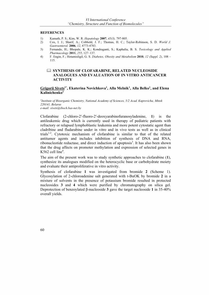
VI International Conference “Chemistry, Structure and Function of Biomolecules”
60
REFERENCES
1) Kamath, P. S.; Kim, W. R. Hepatology 2007, 45(3), 797-805. 2) Cox, I. J.; Sharif, A.; Cobbold, J. F.; Thomas, H. C.; Taylor-Robinson, S. D. World J.
Gastroenterol. 2006, 12, 4773-4783. 3) Fernando, H.; Bhopale, K. K.; Kondraganti, S.; Kaphalia, B. S. Toxicology and Applied
Pharmacology 2011, 255, 127–137. 4) F. Engin, F.; Hotamisligil, G. S. Diabetes, Obesity and Metabolism 2010, 12 (Suppl. 2), 108 –
115.
SYNTHESIS OF CLOFARABINE, RELATED NUCLEOSIDE ANALOGUES AND EVALUATION OF IN VITRO ANTICANCER ACTIVITY
Grigorii Sivets1*, Ekaterina Novichkova1, Alla Melnik1, Alla Belko1, and Elena Kalinichenko1
1Institute of Bioorganic Chemistry, National Academy of Sciences, 5/2 Acad. Kuprevicha, Minsk 220141, Belarus e-mail: [email protected]
Clofarabine (2-chloro-2′-fluoro-2′-deoxyarabinofuranosyladenine, 1) is the antileukemic drug which is currently used in therapy of pediatric patients with refractory or relapsed lymphoblastic leukemia and more potent cytostatic agent than cladribine and fludarabine under in vitro and in vivo tests as well as in clinical trials1-2. Cytotoxic mechanism of clofarabine is similar to that of the related antitumor agents and includes inhibition of synthesis of DNA and RNA, ribonucleotide reductase, and direct induction of apoptosis3. It has also been shown that the drug affects on promoter methylation and expression of selected genes in K562 cell line4.
The aim of the present work was to study synthetic approaches to clofarabine (1), synthesize its analogues modified on the heterocyclic base or carbohydrate moiety and evaluate their antiproliferative in vitro activity.
Synthesis of clofarabine 1 was investigated from bromide 2 (Scheme 1). Glycosylation of 2-chloroadenine salt generated with t-BuOK by bromide 2 in a mixture of solvents in the presence of potassium bromide resulted in protected nucleosides 3 and 4 which were purified by chromatography on silica gel. Deprotection of benzoylated β-nucleoside 3 gave the target nucleoside 1 in 35-40% overall yields.
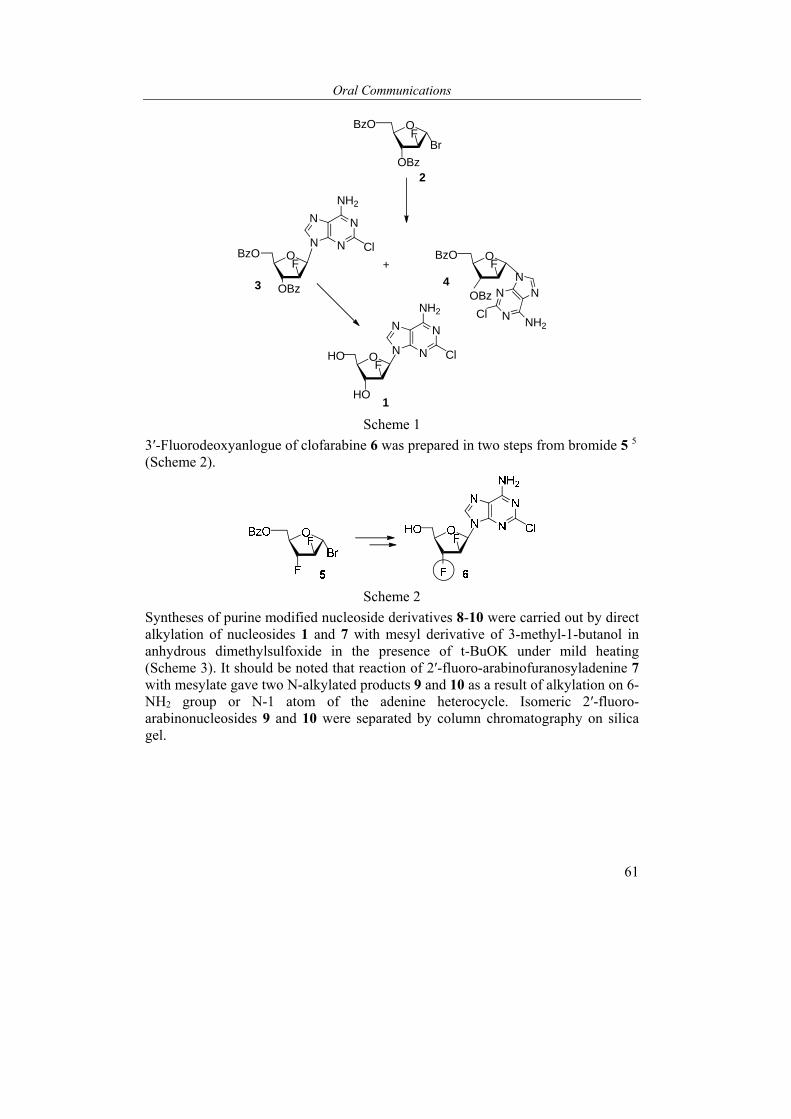
Oral Communications
61
O
OBzOF
Br
BzOF
OHOF
OBz
OBz
OBzOF
HO
+
N
NN
N
NH2
N
NN
N
NH2Cl
Cl
OBz3 4
1
N
NN
N
NH2
Cl
2
Scheme 1
3′-Fluorodeoxyanlogue of clofarabine 6 was prepared in two steps from bromide 5 5 (Scheme 2).
Scheme 2
Syntheses of purine modified nucleoside derivatives 8-10 were carried out by direct alkylation of nucleosides 1 and 7 with mesyl derivative of 3-methyl-1-butanol in anhydrous dimethylsulfoxide in the presence of t-BuOK under mild heating (Scheme 3). It should be noted that reaction of 2′-fluoro-arabinofuranosyladenine 7 with mesylate gave two N-alkylated products 9 and 10 as a result of alkylation on 6-NH2 group or N-1 atom of the adenine heterocycle. Isomeric 2′-fluoro-arabinonucleosides 9 and 10 were separated by column chromatography on silica gel.
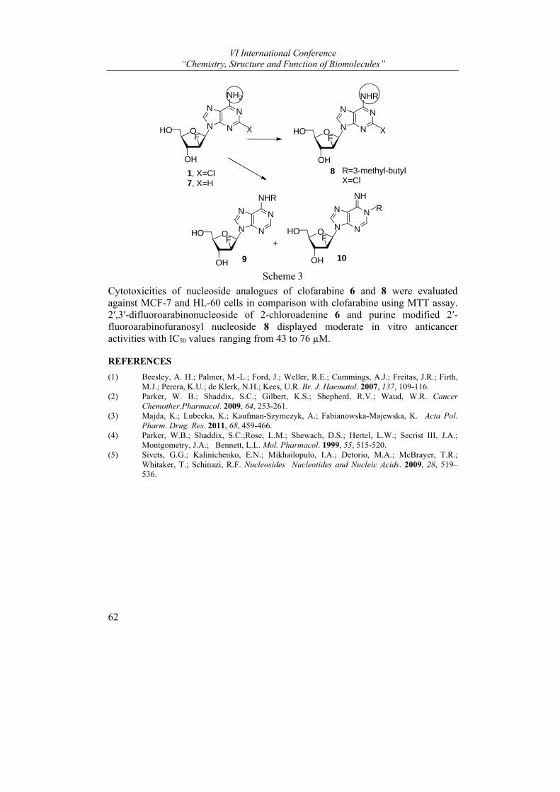
VI International Conference “Chemistry, Structure and Function of Biomolecules”
62
OHOF
OH
1, X=Cl7, X=H
N
NN
N
NH2
X OHOF
OH8
N
NN
N
NHR
X
OHOF
OH 9
N
NN
N
NHR
+OHOF
OH 10
N
NN
N
NH
R=3-methyl-butylX=Cl
R
Scheme 3
Cytotoxicities of nucleoside analogues of clofarabine 6 and 8 were evaluated against MCF-7 and HL-60 cells in comparison with clofarabine using MTT assay. 2′,3′-difluoroarabinonucleoside of 2-chloroadenine 6 and purine modified 2′-fluoroarabinofuranosyl nucleoside 8 displayed moderate in vitro anticancer activities with IC50 values ranging from 43 to 76 µM.
REFERENCES
(1) Beesley, A. H.; Palmer, M.-L.; Ford, J.; Weller, R.E.; Cummings, A.J.; Freitas, J.R.; Firth, M.J.; Perera, K.U.; de Klerk, N.H.; Kees, U.R. Br. J. Haematol. 2007, 137, 109-116.
(2) Parker, W. B.; Shaddix, S.C.; Gilbert, K.S.; Shepherd, R.V.; Waud, W.R. Cancer Chemother.Pharmacol. 2009, 64, 253-261.
(3) Majda, K.; Lubecka, K.; Kaufman-Szymczyk, A.; Fabianowska-Majewska, K. Acta Pol. Pharm. Drug. Res. 2011, 68, 459-466.
(4) Parker, W.B.; Shaddix, S.C.;Rose, L.M.; Shewach, D.S.; Hertel, L.W.; Secrist III, J.A.; Montgometry, J.A.; Bennett, L.L. Mol. Pharmacol. 1999, 55, 515-520.
(5) Sivets, G.G.; Kalinichenko, E.N.; Mikhailopulo, I.A.; Detorio, M.A.; McBrayer, T.R.; Whitaker, T.; Schinazi, R.F. Nucleosides Nucleotides and Nucleic Acids. 2009, 28, 519–536.
Oral Communications
63
PHOTOLYSIS OF THIAMINE BY UV LIGHT
Ivan Stepuro1, Svetlana Labor1, Vitali Stsiapura2, Vitali Smirnov3, and Aleksei Yantsevich4
1 Institute of Biochemistry of Biologically Active Compounds, NAS of Belarus, Grodno, Belarus, 2Yanka Kupala State University of Grodno, Grodno, Belarus, 3 Grodno State Medical University, Grodno, Belarus, 4Institute of Bioorganic Chemistry, NAS of Belarus, Minsk, Belarus e-mail: [email protected]
Introduction. In living organisms, thiamine (or vitamin B1) is essential nutritional factor which is the structural component of thiamine diphosphate (TDP) molecule. TDP is the cofactor for pyruvate dehydrogenase and α-ketoglutarate dehydrogenase. TDP is also the cofactor of transketolase, key enzyme of the pentose phosphate pathway.
Thiamine in aqueous solutions degrades under exposure to UV light (e.g. of mercury lamp with λ≥253 nm) but is stable under visible light irradiation [1,2]. It was proposed that singlet oxygen contributes to photodestruction of thiamine and its derivatives under UV light1-3.
In the current work it was shown that thiamine and its phosphate esters are effective traps for singlet oxygen.
Materials and methods. Mass spectra of thiamine and products of its photolysis were recorded with a chromato-mass-spectrometer Agilent 1290 high performance liquid chromatograph with Q-TOF 6550 tandem quadrupole time-of-flight mass spectrometer detector switched to ESI+ ionization mode. The mixture of thiamine photolysis products was separated by HPLC using an Agilent 1100 chromatograph and a Zorbax-Extend-C-18 column (2.1 x 50 mm, 1.8 µM).
Results and discussion. The exposure to visible light did not result in formation of thiamine oxidation products and changes in absorption spectra. However, when thiamine solution contained riboflavin, or pyridoxal-P, or its analogues, such as pyridoxine, pyridoxal, pyridoxamine, and phosphopyridoxine, a photosensitized oxidation of thiamine by visible light was observed. Under conditions stated, the thiazole component of thiamine was oxidized at a higher rate. The amount of decomposed thiamine correlated well with the value of the quantum yield for singlet oxygen generation by riboflavin and pyridoxine derivatives. Tyrosine, tryptophan, and human serum albumin also photosensitized thiamine oxidation. The addition of sodium azide to the aqueous solution inhibited the oxidation of thiamine and thiamine diphosphate significantly. These results provide an evidence for involvement of singlet oxygen in oxidation of thiamine or thiamine diphosphate.
On exposure of thiamine aqueous solutions to UV, photolysis products were detected. The photolysis products representing fragments of thiamine molecule were observed as individual chromatographic peak with Rt = 0.91 min. The most
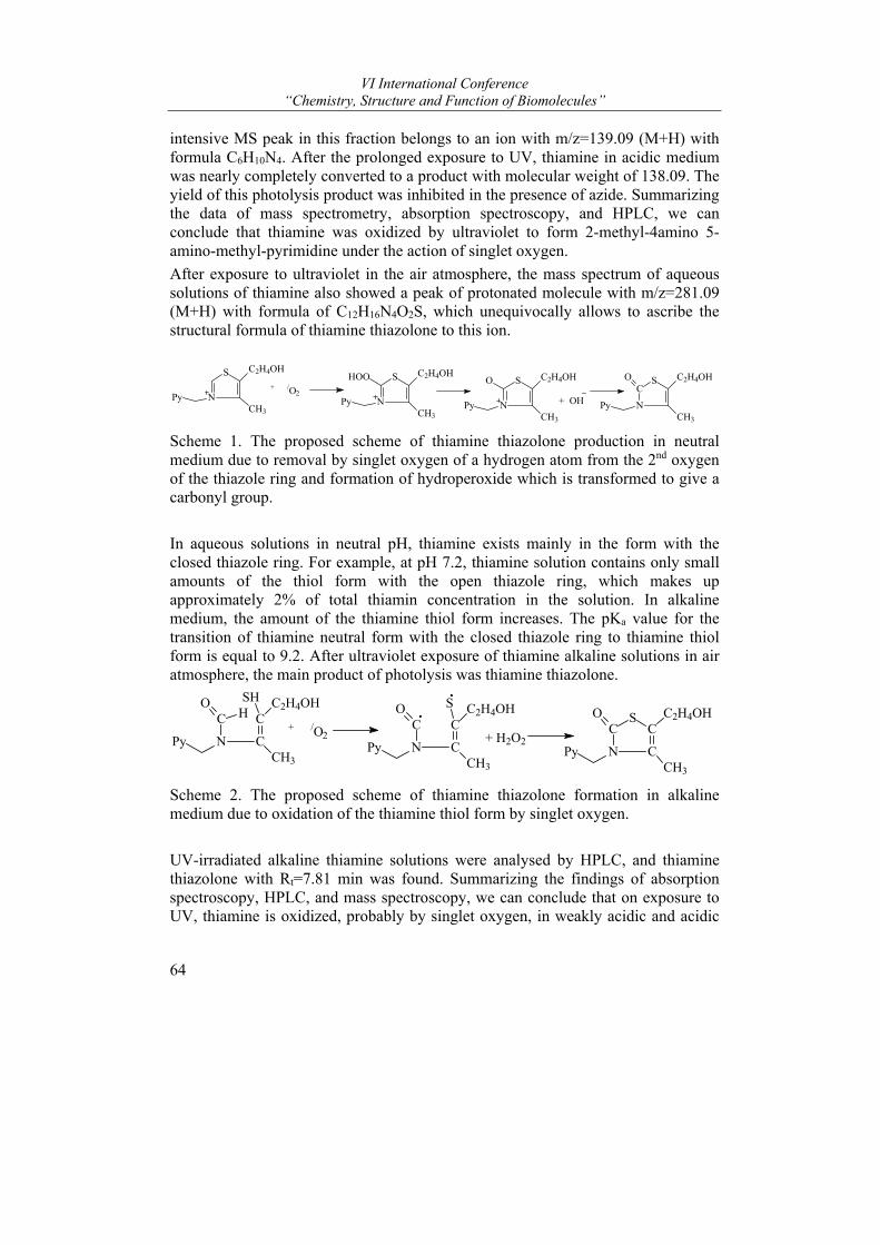
VI International Conference “Chemistry, Structure and Function of Biomolecules”
64
intensive MS peak in this fraction belongs to an ion with m/z=139.09 (M+H) with formula C6H10N4. After the prolonged exposure to UV, thiamine in acidic medium was nearly completely converted to a product with molecular weight of 138.09. The yield of this photolysis product was inhibited in the presence of azide. Summarizing the data of mass spectrometry, absorption spectroscopy, and HPLC, we can conclude that thiamine was oxidized by ultraviolet to form 2-methyl-4amino 5-amino-methyl-pyrimidine under the action of singlet oxygen.
After exposure to ultraviolet in the air atmosphere, the mass spectrum of aqueous solutions of thiamine also showed a peak of protonated molecule with m/z=281.09 (M+H) with formula of C12H16N4O2S, which unequivocally allows to ascribe the structural formula of thiamine thiazolone to this ion.
N
S
Py
C2H4OH
CH3
+ /O2N
S
Py
C2H4OH
CH3
HOO
N
S
Py
C2H4OH
CH3
O
+ OHC
N
S
Py
C2H4OH
CH3
O
Scheme 1. The proposed scheme of thiamine thiazolone production in neutral medium due to removal by singlet oxygen of a hydrogen atom from the 2nd oxygen of the thiazole ring and formation of hydroperoxide which is transformed to give a carbonyl group.
In aqueous solutions in neutral pH, thiamine exists mainly in the form with the closed thiazole ring. For example, at pH 7.2, thiamine solution contains only small amounts of the thiol form with the open thiazole ring, which makes up approximately 2% of total thiamin concentration in the solution. In alkaline medium, the amount of the thiamine thiol form increases. The pKa value for the transition of thiamine neutral form with the closed thiazole ring to thiamine thiol form is equal to 9.2. After ultraviolet exposure of thiamine alkaline solutions in air atmosphere, the main product of photolysis was thiamine thiazolone.
CC2H4OHSH
N CCH3
C HO
Py+ /O2
CC2H4OHS
N CCH3
CO
Py+ H2O2
CC2H4OH
N CCH3
CO
Py
S
Scheme 2. The proposed scheme of thiamine thiazolone formation in alkaline medium due to oxidation of the thiamine thiol form by singlet oxygen.
UV-irradiated alkaline thiamine solutions were analysed by HPLC, and thiamine thiazolone with Rt=7.81 min was found. Summarizing the findings of absorption spectroscopy, HPLC, and mass spectroscopy, we can conclude that on exposure to UV, thiamine is oxidized, probably by singlet oxygen, in weakly acidic and acidic
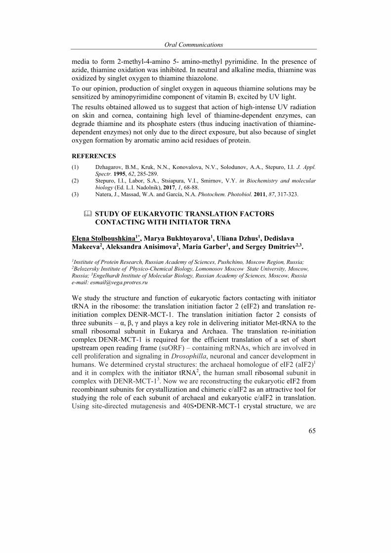
Oral Communications
65
media to form 2-methyl-4-amino 5- amino-methyl pyrimidine. In the presence of azide, thiamine oxidation was inhibited. In neutral and alkaline media, thiamine was oxidized by singlet oxygen to thiamine thiazolone.
To our opinion, production of singlet oxygen in aqueous thiamine solutions may be sensitized by aminopyrimidine component of vitamin B1 excited by UV light.
The results obtained allowed us to suggest that action of high-intense UV radiation on skin and cornea, containing high level of thiamine-dependent enzymes, can degrade thiamine and its phosphate esters (thus inducing inactivation of thiamine-dependent enzymes) not only due to the direct exposure, but also because of singlet oxygen formation by aromatic amino acid residues of protein.
REFERENCES
(1) Dzhagarov, B.M., Kruk, N.N., Konovalova, N.V., Solodunov, A.A., Stepuro, I.I. J. Appl. Spectr. 1995, 62, 285-289.
(2) Stepuro, I.I., Labor, S.A., Stsiapura, V.I., Smirnov, V.Y. in Biochemistry and molecular biology (Ed. L.I. Nadolnik), 2017, 1, 68-88.
(3) Natera, J., Massad, W.A. and García, N.A. Photochem. Photobiol. 2011, 87, 317-323.
STUDY OF EUKARYOTIC TRANSLATION FACTORS CONTACTING WITH INITIATOR TRNA
Elena Stolboushkina1*, Marya Bukhtoyarova1, Uliana Dzhus1, Dedislava Makeeva2, Aleksandra Anisimova2, Maria Garber1, and Sergey Dmitriev2,3.
1Institute of Protein Research, Russian Academy of Sciences, Pushchino, Moscow Region, Russia; 2Belozersky Institute of Physico-Chemical Biology, Lomonosov Moscow State University, Moscow, Russia; 3Engelhardt Institute of Molecular Biology, Russian Academy of Sciences, Moscow, Russia
e-mail: [email protected]
We study the structure and function of eukaryotic factors contacting with initiator tRNA in the ribosome: the translation initiation factor 2 (eIF2) and translation re-initiation complex DENR-MCT-1. The translation initiation factor 2 consists of three subunits – α, β, γ and plays a key role in delivering initiator Met-tRNA to the small ribosomal subunit in Eukarya and Archaea. The translation re-initiation complex DENR-MCT-1 is required for the efficient translation of a set of short upstream open reading frame (suORF) – containing mRNAs, which are involved in cell proliferation and signaling in Drosophilla, neuronal and cancer development in humans. We determined crystal structures: the archaeal homologue of eIF2 (aIF2)1 and it in complex with the initiator tRNA2, the human small ribosomal subunit in complex with DENR-MCT-13. Now we are reconstructing the eukaryotic eIF2 from recombinant subunits for crystallization and chimeric e/aIF2 as an attractive tool for studying the role of each subunit of archaeal and eukaryotic e/aIF2 in translation. Using site-directed mutagenesis and 40S•DENR-MCT-1 crystal structure, we are
VI International Conference “Chemistry, Structure and Function of Biomolecules”
66
investigating the role of the conserved basic β1 loop of DENR (contacts with the codon-anticodon duplex of mRNA-initiator tRNA) in discriminating initiator tRNA in the P-site of the 40S subunit by DENR-MCT-1. This research should contribute significantly to understanding molecular mechanism of translation initiation and reinitiation and was supported by the Program for Basic Researches on Molecular and Cellular Biology and Post-Genomic technologies of the Presidium of RAS to E.A.S.
REFERENCES
1) Stolboushkina E., Nikonov S., Nikulin A., Bläsi U., Manstein D.J., Fedorov R., Garber M. and Nikonov O. J. Mol. Biol., 2008, 382, 680-691.
2) Stolboushkina E., Nikonov S., Zelinskaya N., Arkhipova V., Nikulin A., Garber M. and Nikonov O. J. Mol. Biol., 2013, 425, 989-998.
3) Ivan B. Lomakin, Elena A. Stolboushkina, Anand T. Vaidya, Chenguang Zhao, Maria B. Garber, Sergey E. Dmitriev and Thomas A. Steitz. Cell Reports, 2017, 20, 1-8.
STRUCTURAL INSIGHTS INTO CHOLESTEROL METABOLISM BY CYTOCHROME P450S.
Irina Grabovec1, Wolfram Tempel2, Farrell MacKenzie2, Yaroslav Dichenko1, Egor Marin3, Sergey Bukhdruker3, Valentin Borshchevskiy3, Sergey A. Usanov1, Hee-Won Park4, and Natallia Strushkevich1*
1 Institute of Bioorganic Chemistry NAS of Belarus, Minsk, Belarus, 2 Structural Genomics Consortium, University of Toronto, Toronto, ON, Canada, 3 Laboratory for Advanced Studies of Membrane Proteins, MIPT, Dolgoprudny, Russia, 4 Department of Biochemistry and Molecular Biology, Tulane University School of Medicine, New Orleans, LA, USA. e-mail: [email protected]
Cholesterol is an essential lipid molecule required for the normal development and functioning of the human and animal body. Cholesterol is an important component of the cell membrane as well as a precursor of steroid hormones (such as androgens, estrogens, gluco- and mineralocorticoids), vitamin D and bile acids. Disruptions of its synthesis and metabolism lead to serious diseases and can often be fatal. Cholesterol biosynthesis and steroidogenesis are targets for many drugs. In this regard molecular mechanisms of function of proteins with cholesterol and/or its derivatives as substrates are of particular interest. Cytochrome P450s are enzymes containing heme prosthetic group and catalyzing regio- and stereo-selective oxidations of cholesterol that can be used as different signals for specific regulation of a variety of cellular metabolic and differentiation processes. Structural studies of human cytochromes P450 are challenging because of their membrane nature and a complex catalysis. Here we present crystal structures of two human cytochrome P450s: CYP7A1 and CYP11A1 in complex with their substrates and show distinct features of each enzyme for specific hydroxylation of cholesterol molecule. Recent
Oral Communications
67
structural data on cholesterol hydroxylase from Mycobacterium tuberculosis will also be discussed in the light of protein-ligand interactions between host and pathogen.
The SGC is a registered charity (number 1097737) that receives funds from AbbVie, Bayer, Boehringer Ingelheim, the Canada Foundation for Innovation, the Canadian Institutes for Health Research, Genome Canada through the Ontario Genomics Institute [OGI-055], GlaxoSmithKline, Janssen, Lilly Canada, the Novartis Research Foundation, the Ontario Ministry of Economic Development and Innovation, Pfizer, Takeda, and the Wellcome Trust [092809/Z/10/Z].
PREPARATION, CHARACTERIZATION AND HEMOCOMPATIBILITY OF POLYSACCHARIDE NANOCRYSTALS
Mikhail A. Torlopov1, Elena V. Udoratina1*, Ilia S. Martakov1, Vasily I. Mikhaylov1, Petr A. Sitnikov1, and Natalia N. Drozd2
1Institute of Chemistry of the Komi Scientific Center of the Ural Branch of the Russian Academy of Sciences, Syktyvkar, Russian Federation, 2National Research Center for Hematology, Ministry of Health of the Russian Federation, Moscow Russian Federation. e-mail: [email protected]
Hydrosols of polysaccharide nanocrystals (PsNC) such as a chitin and cellulose are promising systems for obtaining materials filled with nanoparticles, modifiers of rheological properties, drug delivery systems, and templates in organic and inorganic synthesis. These dispersions can be used for preparation of biocompatible films, threads, and gels. The PsNC possess a wide variety of surface functional groups, high chemical and biological activity. Сhitin nanocrystals (ChNC) has positive and cellulose nanocrystals (CNC) – negative surface charge.
Rod-like particles of ChNC were prepared from the crab shell by controlled destruction of polymer chains via acid-catalyzed hydrolysis; this process proceeds at a high rate in amorphous domains of fibrils1. Plate-like and rod-like cellulose nanocrystals were prepared by solvolysis of cellulose in acetic acid/octanol medium in the presence of phosphotungstic acid2. PsNC was studied in the form of hydrosols with different concentrations of the dispersed phase. The morphology and structure of the particles was studied by transmission electron microscopy (TEM), atomic force microscopy (AFM) and X-ray diffraction. Dispersed phase of the obtained ChNC and cellulose I (CNC I) hydrosols consists of rod-like particles with the average length of 200 ± 70 nm and the average diameter of 7 ± 3 nm (Fig. 1a, b), and the morphology of cellulose II nanocrystal (CNC II) is described as a platelet with a diameter of 40-60 nm and a thickness of 10 ± 2 nm (Fig. 1c).
PsNCs are characterized by a highly ordered structure, as evidenced by the value of crystallinity index Ic in range from 0.8 to 0.9. The zeta-potential of ChNC particles

VI International Conference “Chemistry, Structure and Function of Biomolecules”
68
are +50 mV, for CNC particles are -40 mV. Due to strong electrostatic repulsion between the particles, the dispersion is stable against aggregation.
Fig. 1. TEM and AFM images of polysaccharide nanocrystals: a - ChNC, b - CNC I, and c - CNC II.
The rheology of PsNC hydrosols in the absence and presence of electrolyte (KCl) has been studied. It has been established that the rheological properties of PsNC hydrosols depend strongly on the concentrations of the dispersed phase and electrolyte1. It was found that the dispersions of rod-like ChNC, CNC I and disc-like CNC II particles form oriented domains at certain concentrations of an electrolyte, and in this domain, parallel orientation prevails. Hydrosols of PsNC have non-Newtonian properties, demonstrate optical anisotropy under the action of shear stress field (as a result of orientation of anisotropic particles in mechanical field). Association proceeds at the expense of coagulation in secondary minimum through water interlayer; increase in electrolyte concentration results in sol-gel transition and formation of mobile three-dimensional structure, which is stable against sedimentation with time.
The influence of PsNC particles on the components of human blood in vitro and cavy in vivo was studied3. In vitro anticoagulant activity of PsNC hydrosols was assessed using tests (Renam) based on recording of the clotting time of platelet-poor human plasma in the presence of the test compounds in different concentrations: activated partial thromboplastin time (aPTT), prothrombin time (PT), and blood recalcification time test (BRT). The coagulation time of human plasma in the aPTT test was significantly higher (41.32 ± 1.67 sec) in comparison with the control (28.88 ± 1.42 sec) only at a concentration of ChNC of 2.32 mg / ml. The clotting time of the plasma in the PT test did not increase reliably with an increase in the concentration of ChNC from 0.74 to 1.67 mg / ml. A slight decrease in the coagulation time of citrate stabilized human blood was observed in the BRT test with the addition of 1.06 and 1.67 mg / ml ChNC, in comparison with the control (Fig. 2). ChNC in the form of hydrosol at concentrations of 0.63 mg / ml do not independently affect the aggregation of human platelets and during the clotting of platelet-poor plasma in coagulation tests. At concentrations of 0.63 and 1.00 mg /
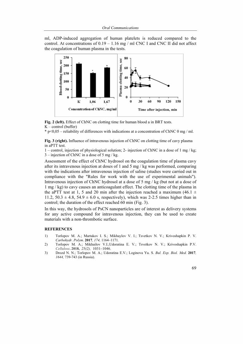
Oral Communications
69
ml, ADP-induced aggregation of human platelets is reduced compared to the control. At concentrations of 0.19 – 1.16 mg / ml CNC I and CNC II did not affect the coagulation of human plasma in the tests.
Fig. 2 (left). Effect of ChNC on clotting time for human blood a in BRT tests. K – control (buffer) * p<0,05 – reliability of differences with indications at a concentration of ChNC 0 mg / ml. Fig. 3 (right). Influence of intravenous injection of ChNC on clotting time of cavy plasma in aPTT test. 1 – control, injection of physiological solution; 2- injection of ChNC in a dose of 1 mg / kg; 3 – injection of ChNC in a dose of 5 mg / kg.
Assessment of the effect of ChNC hydrosol on the coagulation time of plasma cavy after its intravenous injection at doses of 1 and 5 mg / kg was performed, comparing with the indications after intravenous injection of saline (studies were carried out in compliance with the "Rules for work with the use of experimental animals"). Intravenous injection of ChNC hydrosol at a dose of 5 mg / kg (but not at a dose of 1 mg / kg) to cavy causes an anticoagulant effect. The clotting time of the plasma in the aPTT test at 1, 5 and 20 min after the injection reached a maximum (46.1 ± 11.2, 50.3 ± 4.8, 54.9 ± 6.0 s, respectively), which was 2-2.5 times higher than in control; the duration of the effect reached 60 min (Fig. 3).
In this way, the hydrosols of PsCN nanoparticles are of interest as delivery systems for any active compound for intravenous injection, they can be used to create materials with a non-thrombotic surface.
REFERENCES
1) Torlopov M. A.; Martakov I. S.; Mikhaylov V. I.; Tsvetkov N. V.; Krivoshapkin P. V. Carbohydr. Polym. 2017, 174, 1164–1171.
2) Torlopov M. A.; Mikhailov V.I.;Udoratina E. V.; Tsvetkov N. V.; Krivoshapkin P.V. Cellulose. 2018, 25(2), 1031–1046.
3) Drozd N. N.; Torlopov M. A.; Udoratina E.V.; Loginova Yu. S. Bul. Exp. Biol. Med. 2017, 1644, 739-743 (in Russia).

VI International Conference “Chemistry, Structure and Function of Biomolecules”
70
THERMODYNAMICS OF PROTEIN-PROTEIN INTERACTIONS BETWEEN MAMMALIAN CYTOCHROMES P450 AND B5
Evgeniy Yablokov1,*, Anna Florinskaya1, Alexei Medvedev1, Gennady Sergeev2, Natallia Strushkevich2, Alexander Luschik2, Tatsiana Shkel2, Irina Haidukevich2, Andrei Gilep2, Sergey Usanov2, and Alexis Ivanov1
1 Institute of Biomedical Chemistry, Pogodinskaya str. 10, bldg 8, Moscow, 119121, Russia; 2 Institute of Bioorganic Chemistry, Kuprevicha str. 5/2, Minsk, 220141, Belarus e-mail: [email protected]
Cytochromes P450 (CYPs) form a superfamily of monooxygenases, widely represented in living organisms. The cytochrome P450-dependent monooxygenase systems are present in practically all living organisms. Protein components of these systems are structurally conserved as shown by experiments on chimeric complexes formation between proteins isolated from different species of living organisms. This is due to the fact that in spite of some species differences in the structure of protein partners, regions which are responsible for the intermolecular interaction have a similar structure.
A small hemoprotein, cytochrome b5 (CYB5), is often involved in the CYP monooxygenase systems as an effector 1-6. In humans two major isoforms of membrane bound CYB5 have been found; they are localized in the endoplasmic reticulum (type CYB5A) and in the outer mitochondrial membrane (type CYB5B) 7.
At present the molecular mechanisms of protein-protein interactions in P450 monooxygenase systems are not well understood, mainly due to difficulties in experimental investigation of such membrane proteins. Therefore, researchers often use computer-aided molecular modeling allowing to build simplified models of complex formation between protein partners of interest and to estimate the energy of its formation (mainly enthalpy component in relative units) 8, 9. The reliability of such computer models depends on available experimental results used as input data for modeling and choice of computational methods and algorithms. In addition, a serious disadvantage of this approach is the almost complete lack of evaluation of the entropy component 10. Such models of molecular complexes represent only hypotheses that need experimental verification including obtaining data on the affinity and thermodynamics of intermolecular interactions.
Recent progress in understanding of principles of protein-protein interactions is associated with employment of new molecular techniques. In the context of microsomal monooxygenase system, complex formation by its constituents has been successfully studied by means of optical biosensors using surface plasmon resonance (SPR)11. This resulted in real time recording of intermolecular interactions and characterization of their equilibrium, kinetic and thermodynamic parameters.
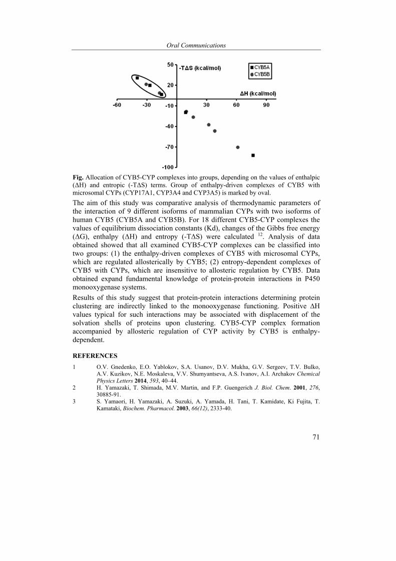
Oral Communications
71
Fig. Allocation of CYB5-CYP complexes into groups, depending on the values of enthalpic (ΔH) and entropic (-TΔS) terms. Group of enthalpy-driven complexes of CYB5 with microsomal CYPs (CYP17A1, CYP3A4 and CYP3A5) is marked by oval.
The aim of this study was comparative analysis of thermodynamic parameters of the interaction of 9 different isoforms of mammalian CYPs with two isoforms of human CYB5 (CYB5A and CYB5B). For 18 different CYB5-CYP complexes the values of equilibrium dissociation constants (Kd), changes of the Gibbs free energy (ΔG), enthalpy (ΔH) and entropy (-TΔS) were calculated 12. Analysis of data obtained showed that all examined CYB5-CYP complexes can be classified into two groups: (1) the enthalpy-driven complexes of CYB5 with microsomal CYPs, which are regulated allosterically by CYB5; (2) entropy-dependent complexes of CYB5 with CYPs, which are insensitive to allosteric regulation by CYB5. Data obtained expand fundamental knowledge of protein-protein interactions in P450 monooxygenase systems.
Results of this study suggest that protein-protein interactions determining protein clustering are indirectly linked to the monooxygenase functioning. Positive ∆H values typical for such interactions may be associated with displacement of the solvation shells of proteins upon clustering. CYB5-CYP complex formation accompanied by allosteric regulation of CYP activity by CYB5 is enthalpy-dependent.
REFERENCES
1 O.V. Gnedenko, E.O. Yablokov, S.A. Usanov, D.V. Mukha, G.V. Sergeev, T.V. Bulko, A.V. Kuzikov, N.E. Moskaleva, V.V. Shumyantseva, A.S. Ivanov, A.I. Archakov Chemical Physics Letters 2014, 593, 40–44.
2 H. Yamazaki, T. Shimada, M.V. Martin, and F.P. Guengerich J. Biol. Chem. 2001, 276, 30885-91.
3 S. Yamaori, H. Yamazaki, A. Suzuki, A. Yamada, H. Tani, T. Kamidate, Ki Fujita, T. Kamataki, Biochem. Pharmacol. 2003, 66(12), 2333-40.

VI International Conference “Chemistry, Structure and Function of Biomolecules”
72
4 T. Aoyama, K. Nagata, Y. Yamazoe, R. Kato, E .Matsunaga, H.V. Gelboin, F.J. Gonzalez Proc. Natl. Acad. Sci. U S A 1990, 87(14), 5425-9.
5 M.P. Duarte, B.B. Palma, A.A. Gilep, A . Laires, J.S. Oliveira, S.A. Usanov, J. Rueff, M. Kranendonk, Mutagenesis, 2005, 20(2), 93-100.
6 A.A. Gilep, R.W. Estabrook and S.A. Usanov Biochemistry (Mosc.) 2003, 68, 86-98. 7 T. Ogishima, J.Y. Kinoshita, F. Mitani, M. Suematsu, A. Ito J. Biol. Chem. 2003, 278(23),
21204-11. 8 V.S. Skvortsov, N.V. Belkina, A.S. Ivanov Molecular Simulation 2000, 24, 369-378. 9 N.V. Belkina, M. Lisurek, A.S. Ivano, Bernhardt R. J. Inorg. Biochem. 2001, 87(4), 197-
207. 10 X. Cheng, I. Ivanov Methods Mol. Biol. 2012, 929, 243-85. 11 Y. Higashimoto et al J. Biol. Chem. 2005, 280(1), 729–737 12 Yablokov E., Florinskaya A., Medvedev A., Sergeev G., Strushkevich N., Luschik A., Shkel
T., Haidukevich I., Gilep A., Usanov S., Ivanov A. Arch. Biochem. Biophys. 2017, 619, 10-15.
The reported study was funded by RFBR according to the research project № «18-04-00071 A» and Program for Basic Research of State Academies of Sciences for 2013-2020 (0518- 2018- 0003). The study was carried out using “Human Proteome” Core Facility (IBMC, Moscow, Russia) which is supported by Ministry of Education and Science of the Russian Federation (unique project ID RFMEFI62117X0017).
POSTERS
COMPUTATIONAL DEVELOPMENT OF NOVEL HIV-1 ENTRY INHIBITORS TARGETING CD-BINDING SITE OF THE VIRAL ENVELOPE GP120 PROTEIN
Grigory I. Nikolaev1, Ivan A. Kashyn1, Yuri V. Kornoushenko2, Alexander V. Tuzikov1, and Alexander M. Andrianov2*
1United Institute of Informatics Problems of the National Academy of Sciences of Belarus, Minsk, Republic of Belarus, 2 Institute of Bioorganic of the National Academy of Sciences of Belarus, Minsk, Republic of Belarus e-mail: [email protected]
In this study, in silico design of novel HIV-1 entry inhibitor scaffolds that target CD4-binding site of the viral gp120 protein was carried out and evaluation of their antiviral potency was performed by high-throughput docking and molecular dynamics simulations. In the first step, a Drug-Like subset of the ZINC database (http://zinc.docking.org/) was screened by the DataWarrior program (http://www.openmolecules.org/help/basics.html) to generate two virtual molecular libraries. Library 1 comprised small molecules (molecular mass < 250 Da) with an azide group or an analkyne group and aromatic fragments critically important for the HIV-1 attachment to cellular receptor CD4. In library 2, all low-molecular compounds (molecular mass < 250 Da) with an azide group or an alkyne group were collected. The modular units from libraries 1 and 2 were then used as reagents to mimic the click chemistry reaction of azide-alkyne cycloaddition by the AutoClickChem software tool (http://nbcr-222.ucsd.edu/autoclickchem/library_1.php). This resulted in a set of 1 655 301 hybrid molecules. 294 378 compounds that fully satisfied Lipinski's “rule of five” were further screened by high-throughput docking and molecular dynamics simulations to evaluate the affinity of their binding to the target protein.
As a result, five compounds were found to exhibit low values of the binding free energy. These drug-like molecules were therefore selected for the final analysis as potential CD4-mimetic candidates. Analysis of the docked ligand/gp120 structures indicates that these molecules form hydrogen bond with Asp-368gp120 mimicking the critical H-bond interaction of this highly conserved gp120 residue with Arg-59CD4. Along with this hydrogen bond, the designed compounds are involved in van der Waals interactions with the gp120 residues Asp-368, Glu-370, Ile-371, Asn-425, Met-426, Trp-427, and Gly-473 that make the direct interatomic contacts with Phe-43CD4. With these data, the mechanism of interactions between the designed compounds and gp120 is close to that appearing in the crystal gp120/CD4 structure. This mechanism is generally provided by hydrogen bonds with Asp-368gp120 and multiple van der Waals contacts with the gp120 residues that bind to Phe-43CD4,

VI International Conference “Chemistry, Structure and Function of Biomolecules”
74
resulting in destruction of the critical interactions of gp120 with Phe-43CD4 and Arg-59CD4. In addition, the complexes of the CD4-mimetic candidates with gp120 show relative conformational stability within the molecular dynamics simulations and expose the high percentage occupancies of intermolecular hydrogen bonds, in line with the data on the binding free energy calculations.
Thus, the data of molecular modeling suggest that the designed compounds may be able to block the two well-conserved hotspots of the CD4-binding site by mimicking the critical interactions of cellular receptor CD4 with the HIV-1 gp120 protein. These hotspots include the Phe-43 cavity of gp120 and residue Asp-368gp120 that is associated with increasing the binding affinity without triggering the unwanted allosteric signal. In light of these data, the predicted CD4-mimetic candidates may be used as a strong foundation for the design of new functional antagonists of viral entry with broad HIV-1 neutralization.
This study was supported by grant from the Belarusian Republican Foundation for Fundamental Research (project X17MC-004).
THE SYNTHESIS OF TERPENOID BIS(1,2,3-TRIAZOLES) AS A POTENTIAL LIGANDS FOR ASYMMETRIC CATALYSIS
Maksim P. Bei* and Anatolij P. Yuvchenko
The Institute of Chemistry of New Materials of NAS of Belarus, Minsk, Belarus E-mail: [email protected]
Fumaropimaric acid (FPA) is a rosin-based substance obtained by reaction of rosin with fumaric acid or by isomerization of maleopimaric acid.1 Fumaropimaric acid and its derivatives show immunomodulatory, anticancer activity and selectively bind with replication protein A in human cells.2 Previously we described the method for preparation of fumaropimaric acid monoamides by alkali isomerization of maleopimaric acid amides.3 At the present work we describe the synthesis of new FPA derivatives: bis(1,2,3-triazoles).
Treatment of amido diacid 1 with diethyl sulfate in dimethylformamide in the presence of potassium carbonate afforded diethyl ester 2 in 99% yield, which was converted into diol 3 by reduction with LiAlH4 in 58% yield. The treatment of diol 3 with methanesulfonyl chloride in pyridine gave dimesylate in 100% yield that was converted into diazide 4 by reaction with sodium azide (yield 88%). Reaction of 1,3-cycloaddition of diazide 4 with terminal alkynes (propargyl alcohol, N,N-dimethylpropargylamine, phenylacetylene) in the presence of acetic acid, tributylamine and CuI as catalyst afforded bis(1,2,3-triazoles) 5a–c in 43–51% yields.
Structures of synthesized bis(1,2,3-triazoles) 5a–c were confirmed by mass-spectrometry, IR, NMR spectroscopy, elemental analysis. NMR spectra of
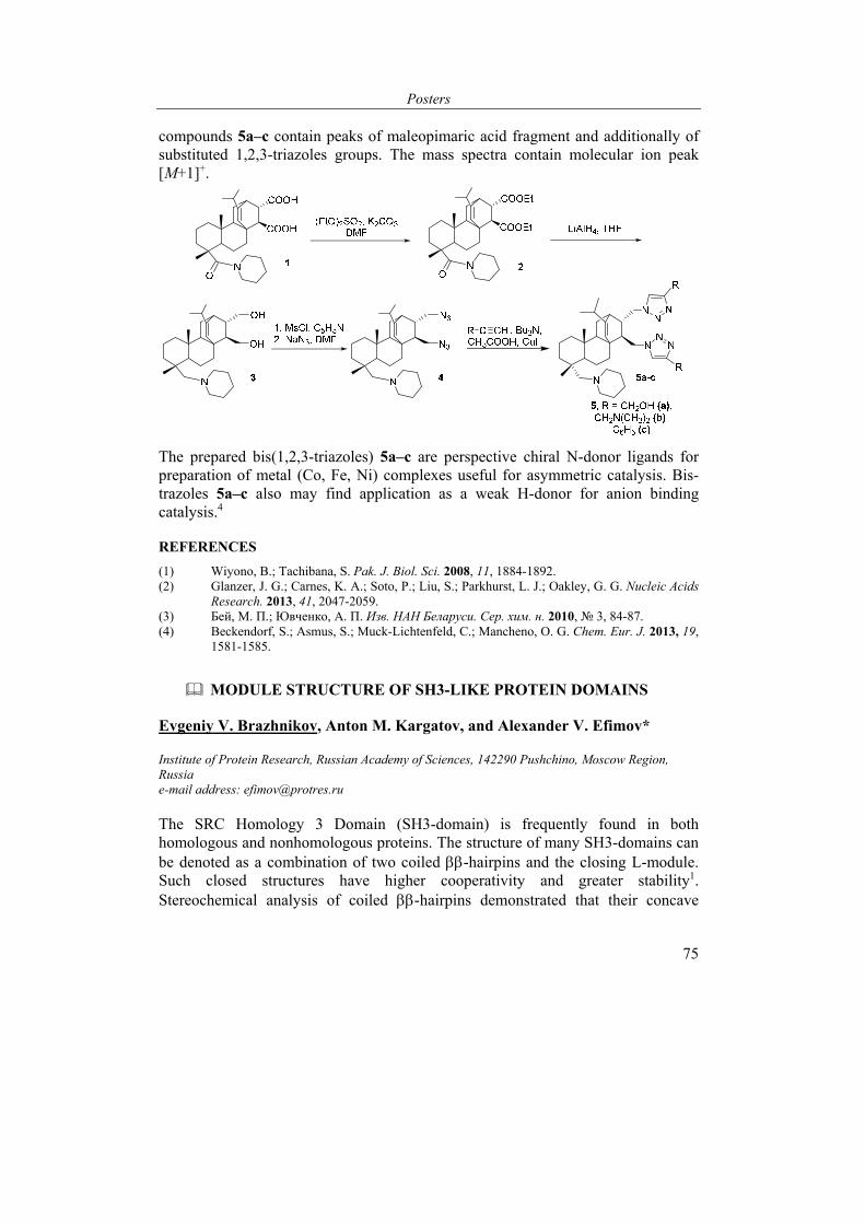
Posters
75
compounds 5a–c contain peaks of maleopimaric acid fragment and additionally of substituted 1,2,3-triazoles groups. The mass spectra contain molecular ion peak [M+1]+.
The prepared bis(1,2,3-triazoles) 5a–c are perspective chiral N-donor ligands for preparation of metal (Co, Fe, Ni) complexes useful for asymmetric catalysis. Bis-trazoles 5a–c also may find application as a weak H-donor for anion binding catalysis.4
REFERENCES
(1) Wiyono, B.; Tachibana, S. Pak. J. Biol. Sci. 2008, 11, 1884-1892. (2) Glanzer, J. G.; Carnes, K. A.; Soto, P.; Liu, S.; Parkhurst, L. J.; Oakley, G. G. Nucleic Acids
Research. 2013, 41, 2047-2059. (3) Бей, М. П.; Ювченко, А. П. Изв. НАН Беларуси. Сер. хим. н. 2010, № 3, 84-87. (4) Beckendorf, S.; Asmus, S.; Muck-Lichtenfeld, C.; Mancheno, O. G. Chem. Eur. J. 2013, 19,
1581-1585.
MODULE STRUCTURE OF SH3-LIKE PROTEIN DOMAINS
Evgeniy V. Brazhnikov, Anton M. Kargatov, and Alexander V. Efimov*
Institute of Protein Research, Russian Academy of Sciences, 142290 Pushchino, Moscow Region, Russia e-mail address: [email protected]
The SRC Homology 3 Domain (SH3-domain) is frequently found in both homologous and nonhomologous proteins. The structure of many SH3-domains can be denoted as a combination of two coiled -hairpins and the closing L-module. Such closed structures have higher cooperativity and greater stability1. Stereochemical analysis of coiled -hairpins demonstrated that their concave
VI International Conference “Chemistry, Structure and Function of Biomolecules”
76
surface is formed primarily by hydrophobic amino acid residues, while the convex surface is formed correspondingly by hydrophilic residues, and in their amino acid sequences the inner hydrophobic residues interchange with outer hydrophilic ones2. Furthermore, the formation of a strongly twisted and coiled -hairpin requires not only interchanging of hydrophobic and hydrophilic amino acid residues, but also the presence of one or two glycine residues at the cross-link as well as an excess of glycine and alanine residues at the inner positions of sites with the strongest coiling of strands.
In this study, our attention was focused mainly on the examination of the closing region in the C-terminal parts of SH3-like domains which we called the L-shaped module. Three-dimensional structures of these modules of nonhomologous proteins were superimposed one on of the other and their structural similarity was studied. We analyzed conformations of loop regions (cross-links) in L-modules of such domains. Altogether 67 domains (59 PDB files), where the L-module closes the domain structure, were selected in nonhomologous proteins. Frequencies of occurrence of cross-links of different lengths were determined and corresponding histograms were plotted. It was found that L-modules with cross-links of 3 and 5 amino acid residues are the most frequent ones – 60% and 13%, respectively. In 97% cases, these cross-links of 3 amino acid residues long have the “mn” conformation, and cross-links of 5 residues long have the “mn” conformation in 67% cases. When superimposed, the structures of L-modules with the same conformation of the arch that were taken from different nonhomologous proteins are compatible. Structural alignment of amino acid sequences of L-modules allowed determining key positions which should be occupied by hydrophobic, hydrophilic and proline residues.
REFERENCES
1) Efimov, A. V. Biochem. Biophys. Res. Commun. 2010, 399, 412-415. 2) Boshkova, E. A.; Brazhnikov, E. V.: Efimov, A. V. Molekul. Biol. (in Russian) 2016, 50, 880-
886.
The research was supported by the Russian Foundation for Basic Research, grant #17-04-00242-a.
Posters
77
EFFICIENT BACTERIAL EXPRESSION OF TAGLESS RECOMBINANT HUMAN GLUTATHIONE TRANSFERASE P1 AND CHARACTERIZATION OF THE PURIFIED, HIGHLY ACTIVE ENZYME
Yuliya Brechka and Syargey Gilevich
Institute of Bioorganic Chemistry of the National Academy of Sciences of Belarus, Minsk, Belarus e-mail: [email protected]
Human glutathione transferase P1 (GSTP1) is an important enzyme in clinical diagnostics since its overexpression in solid tumors and lymphomas largely contributes to multiple drug resistance of cancer cells. Till recently, two general approaches were used to express recombinant GSTP1 in E. coli: either with oligopeptide tag or without it. Chang et al. expressed the enzyme with N-terminal 6xHis tag at a level of 30 mg/l culture but 21% of the activity was found in bacterial pellet1. Wu et al. inserted an enterokinase cleavage site (6 amino acids) between the above tag and GSTP1 sequence2. Despite the high expression yield (50-60 mg/l culture), the pellet-associated activity amounted to 27%. After passage through an IMAC column, cleaving the tag and further purification on glutathione (GSH)-sepharose, the purified GSTP1 had a reduced specific activity (72 U/mg protein) and still contained an extra Gly residue at the N-terminus. Within the second approach, Kolm et al. cloned GSTP1 gene into a bacterial vector and reported poor expression possibly due to secondary structures at the 5ʹ end of the corresponding RNA transcript3. Thus, a truncated cDNA clone was employed lacking codons for the N-terminal 34 amino acids. The missing DNA fragment was codon-optimized, synthesized chemically and ligated, together with the cDNA, into an appropriate expression plasmid. Though time-consuming and laborious, the method allowed to largely express the enzyme in JM103 cells (> 200 mg/l culture) and purify it to the specific activity of 128 U/mg (measured at 30ºC rather than at 25ºC). In another work4, Battistoni et al. also utilized a complex, multi-step procedure to construct the vector for native GSTP1 expression. Cytoplasmic production of the enzyme constituted 35-50 mg/l culture; a smaller part (24%) was forwarded into periplasm by co-expression with molecular chaperones. The purified GSTP1 showed moderate specific activity (81 U/mg), and half of the enzyme contained an extra N-terminal Met residue.
Here we report a novel, straightforward and efficient protocol for cloning, bacterial expression, and purification of tagless recombinant GSTP1. Initially, total RNA having A260/A280 ratio of 1,94-1,98 was extracted from human leukocytes with Purezol reagent. By using a reverse gene-specific primer, the RNA was reverse transcribed to obtain full-length cDNA. Amplification of the cDNA involved 35 PCR cycles in the presence of the forward and reverse gene-specific primers which had been thoroughly designed with respect to E. coli codon usage and non-
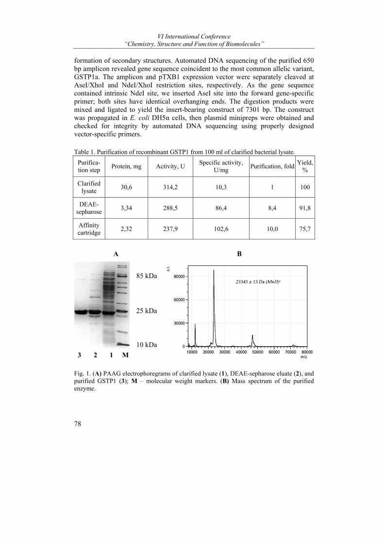
VI International Conference “Chemistry, Structure and Function of Biomolecules”
78
formation of secondary structures. Automated DNA sequencing of the purified 650 bp amplicon revealed gene sequence coincident to the most common allelic variant, GSTP1a. The amplicon and pTXB1 expression vector were separately cleaved at AseI/XhoI and NdeI/XhoI restriction sites, respectively. As the gene sequence contained intrinsic NdeI site, we inserted AseI site into the forward gene-specific primer; both sites have identical overhanging ends. The digestion products were mixed and ligated to yield the insert-bearing construct of 7301 bp. The construct was propagated in E. coli DH5α cells, then plasmid minipreps were obtained and checked for integrity by automated DNA sequencing using properly designed vector-specific primers.
Table 1. Purification of recombinant GSTP1 from 100 ml of clarified bacterial lysate.
Purifica-tion step
Protein, mg Activity, U Specific activity,
U/mg Purification, fold
Yield, %
Clarified lysate
30,6 314,2 10,3 1 100
DEAE-sepharose
3,34 288,5 86,4 8,4 91,8
Affinity cartridge
2,32 237,9 102,6 10,0 75,7
A B
3 2 1 М
85 kDa
25 kDa
10 kDa
Fig. 1. (A) PAAG electrophoregrams of clarified lysate (1), DEAE-sepharose eluate (2), and purified GSTP1 (3); M – molecular weight markers. (B) Mass spectrum of the purified enzyme.
Posters
79
The enzyme expression was carried out in chemically transformed E. coli BL21(DE3) RIPL cells grown in LB broth. Under optimized expression conditions (induction with 0,5 mM IPTG; postinduction time of 24 h at 37ºC), the yield of tagless recombinant GSTP1 reached 30-32 mg/l culture, which is in between the values reported for 6xHis-tagged enzyme1,2. Importantly, GSTP1 was mostly produced as soluble cytosolic protein since only 6-10% of the total activity resided in bacterial pellet.
We also efficiently purified recombinant GSTP1 from bacterial lysate by using our two-step procedure previously developed for the native erythrocyte enzyme5. At the final step, a cartridge with GSH-immobilized affinity membranes was employed as a convenient and cost-effective substitute for commonly used GSH-sepharose. The procedure recovers 75,7% of the total GSTP1 activity in lysate and affords highly purified enzyme with specific activity of 102,6 U/mg (Table 1). The latter value nearly equals to the best activity levels reported earlier for both recombinant and native GSTP1. The enzyme is electrophoretically homogeneous (Fig. 1, A) and its monomeric mass measured by MALDI-TOF mass spectrometry (23343 Da; Fig. 1, B) is closely similar to calculated value (23356 Da). The presence of Met at the enzyme N-terminus was proven by trypsinolysis followed by mass spectrometric identification of the N-terminal tryptic dodecapeptide, Met-Pro-Pro-Tyr-Thr-Val-Val-Tyr-Phe-Pro-Val-Arg.
Finally, steady-state kinetic parameters of the recombinant GSTP1-catalyzed conjugation reaction between GSH and 1-chloro-2,4-dinitrobenzene were determined at pH 6,5 and 25°С. For both substrates, the obtained Km values (0,17 mМ and 0,65 mМ, respectively) almost coincide with characteristics of the native enzyme5. The results of the present work have been used for screening and synthesis of new GSTP1 inhibitors with potential antitumor activity.
REFERENCES
(1) Chang, M.; Bolton, J. L.; Blond, S. Y. Protein Expr. Purif. 1999, 17, 443-448. (2) Wu, Y.; Shen, J.; Yin, Z. Protein J. 2007, 26, 359-370. (3) Kolm, R. H; Stenberg, G.; Widersten, M.; Mannervik, B. Protein Expr. Purif. 1995, 6, 265-
271. (4) Battistoni, A.; Mazzetti, A. P.; Petruzzelli, R.; Muramatsu, M.; Federici, G.; Ricci, G.; Lo
Bello, M. Protein Expr. Purif 1995, 6, 579-587. (5) Gilevich, S. N.; Brechka, Yu. V.; Ripinskaya, K. Yu. Vesti Nats. Akad. Navuk Belarusi. Ser.
Khim. Navuk [Proc. Nat. Acad. Sci. Belarus, chem. ser.] 2017, (2), 66-79.
VI International Conference “Chemistry, Structure and Function of Biomolecules”
80
PLANT ION CHANNELS ACTIVATED BY REACTIVE OXYGEN SPECIES: MOLECULAR NATURE OF ROS SENSING AND PHYSIOLOGICAL FUNCTIONS
Demidchik V.1,2*
1Department of Plant Cell Biology and Bioengineering, Biological Faculty, Belarusian State University, 220030, 4 Independence Ave., Minsk, Belarus; 2Komarov Botanical Institute RAS, 2 Professora Popova Street, 197376, Saint-Petersburg, Russian Federation e-mail: [email protected]
It is widely accepted that reactive oxygen species (ROS) are critically important for plants’ life. ROS are produced by intracellular and extracellular mechanisms and accumulate in the cell wall (apoplast), where the antioxidant capacity is much lower than in cytosol. The moderate generation of ROS is involved in normal plant physiology and adaptation needs but their overproduction, for example during the environmental stress, results in irreversible oxidative damage and dysfunction of cell components (Demidchik 2015, Environ Exp Bot). The question of sensing ROS is still debated in plant physiology. Here, it is demonstrated that the plasma membrane ion channels transporting cations, such as Ca2+ and K+, function as prime targets of ROS in plants. These systems can catalyse early and rapid sensing of ROS in plants involved in a multitude of physiological reactions, such as adaptation to stresses, control of photosynthesis, cell elongation and gravitropic responses.
In the plasma membranes of lower and higher plants, ROS instantaneously activate two major classes of ion channels: Ca2+-permeable nonselective cation channels (NSCCs) and K+ outwardly-rectifying channels (KORs encoded by GORK). Activation of cation channels by ROS leads to dramatic influx of Ca2+ for signaling, developmental and nutritional needs and K+ loss (electrolyte leakage) inducing autophagic and necrotic cell death. Ca2+ entry also rearranges actin cytoskeleton and modifies vesicular transport. ROS-activated ion channels reveal complex nature of activation, depending on the developmental stage and oxidative capacity of tested ROS. The transition metal binding centres have recently been identified in some members of cyclic nucleotide-gated channels, a subclass of NSCCs (Demidchik et al. 2014, JXB). These centers potentially produce hydroxyl radicals from H2O2 (Haber-Weiss reaction) directly in the channel’s macromolecule. Mutations in ROS-sensitive moieties in K+ efflux GORK channel leads to the decrease of ROS-sensing capacity, suggesting that distinct molecular groups are responsible for ROS sensing by ion channels. These moieties probably confer physiological properties related to ROS, such as programmed cell death and autophagy.
This study was supported by Russian Science Foundation grant#15-14-30008 to VD.
Posters
81
NEW ROLES OF EXOGENOUS L-ASCORBIC ACID IN PLANTS: ELEVATION OF CYTOSOLIC FREE CALCIUM, EFFLUX THROUGH ANION CHANNELS UNDER STRESS CONDITIONS AND REGULATION OF ROOT ELONGATION GROWTH
Demidchik V.,1,3* Makavitskaya M.,1 Svistunenko D.,2 Navaselsky I.,1 Hryvusevich P.,1 Mackievic V.,1 Samokhina V.,1 Straltsova D.,1 and Sokolik A.1
1Department of Plant Cell Biology and Bioengineering, Biological Faculty, Belarusian State University, 4 Independence Square, Minsk, 220030, Belarus; 2School of Biological Sciences, University of Essex, Wivenhoe Park, Colchester, Essex, CO4 3SQ, United Kingdom; 3Russian Academy of Sciences, Komarov Botanical Institute, 2 Professora Popova Street, 197376 St Petersburg, Russia e-mail: [email protected]
L-ascorbate is not often considered as a signaling molecule in plants. Here we show that, in Arabidopsis roots, exogenous L-ascorbic acid (>30 µM) triggered a transient increase of the cytosolic free calcium activity ([Ca2+]cyt.) that is central to plant signaling. Exogenous copper and iron stimulated the ascorbate-induced [Ca2+]cyt. elevation while cation channel blockers, free radical scavengers, low extracellular [Ca2+], transition metal chelators and removal of the cell wall inhibited this reaction. These data show that the apoplastic redox-active transition metals are involved in the ascorbate-induced [Ca2+]cyt. elevation. Exogenous ascorbate also induced moderate increase in programmed cell death symptoms in intact roots, but it did not activate Ca2+ influx currents in patch-clamped root protoplasts. Intriguingly, replacement of gluconate with ascorbate in the patch-clamp pipette revealed a large ascorbate efflux current, which showed sensitivity to anion channel blocker, anthracene-9-carboxylic acid (A9C), indicative of the ascorbate release via anion channels. EPR spectroscopy measurements demonstrated that salinity (NaCl) triggered accumulation of root apoplastic ascorbyl radicals in A9C-dependent manner, confirming that L-ascorbate leaks through anion channels under depolarisation. This mechanism can underlie ascorbate release, signaling phenomena, apoplastic redox reactions, iron acquisition and control of membrane ionic and electrical equilibrium (together K+ efflux via GORK channels).
Financial support of the Russian Science Foundation (grant#15-14-30008 to VD) is gratefully acknowledged.
VI International Conference “Chemistry, Structure and Function of Biomolecules”
82
BRASSINOSTEROID-INDUCED STIMULATION OF PROTOCORM GROWTH AND MODIFICATION OF TISSUE MORPHOLOGY IN ORCHIDS
Charnysh М.1, Batuleu A.V.1, Zhabinskii V.N.2, Khripach V.A.2, and Demidchik V.1
1Belarusian State University, Minsk, Belarus; 2Institute of Bioorganic Chemistry NAS of Belarus, Minsk, Belarus e-mail: [email protected]
The content of certain phytohormones and their concentrations in a medium is the determining factor for controlling growth and differentiation of plant cultured in vitro. The most commonly used hormones are auxins and cytokinins. Recent studies showed that brassinosteroids (BRs) have a strong modifying effect on growth, development, sex determination and reproduction in higher plants. However, these hormones are not studied for their action on growth of plant in vitro cultures. Moreover their effects are not investigated in such an important plant as orchids.
The aim of this work was to determine the effect of six different BRs, belonging to two main BR classes, on growth rate and development of Phalaenopsis hybridum Blume protocorm-like bodies. 10-10-10-6 М brassinolide (BL), castasterone (CS), epicastasterone (EC), homocastasterone (GC), epibrassinolide (EB) and homobrassinolide (GB) were tested. Culture of protocorms was generated from seeds of Phalaenopsis hybridum Blume. Protocorm-like bodies were isolated from the primary culture and transferred to media containing various levels of BRs. Weigh and length of the protocorm-like bodies were measured after 100 days of cultivation on BR-containing media. Our data demonstrated that all BRs significantly stimulated orchid growth in vitro. The greatest effect on length was caused by CS while maximal increase of weight was induced by BL and EB. Orchid microclones, grown in the presence of 10-6 М CS, had twice bigger length that control plants. Weight gain also increased 2 and 3.5 times when plants were cultivated on media containing 10-8 M and 10-6 М BL, respectively. GB and GC caused smallest effects on growth among all tested BRs. We also compared the BR effects with classical auxins, such as indol-3-acetic acid, indole-3-butyric acid and 2,4-dichlorophenoxyacetic acid. We have found that auxins were less effective than BRs.
Overall, we have demonstrated for the first time that BRs stimulate growth of Phalaenopsis hybridum Blume protocorm-like bodies and that this stimulation exceed effect of auxins.
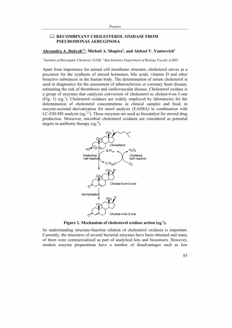
Posters
83
RECOMBINANT CHOLESTEROL OXIDASE FROM PSEUDOMONAS AERUGINOSA
Alexandra A. Dobysh1,2, Michail A. Shapira1, and Aleksei V. Yantsevich1
1 Institute of Bioorganic Chemistry NASB; 2 Biochemistry Department of Biology Faculty of BSU
Apart from importance for animal cell membrane structure, cholesterol serves as a precursor for the synthesis of steroid hormones, bile acids, vitamin D and other bioactive substances in the human body. The determination of serum cholesterol is used in diagnostics for the assessment of atherosclerosis or coronary heart disease, estimating the risk of thrombosis and cardiovascular disease. Cholesterol oxidase is a group of enzymes that catalyzes conversion of cholesterol to cholest-4-en-3-one (Fig. 1) (eg.1). Cholesterol oxidases are widely employed by laboratories for the determination of cholesterol concentrations in clinical samples and food, in enzyme-assisted derivatization for sterol analysis (EADSA) in combination with LC-ESI-MS analysis (eg.2,3). These enzymes are used as biocatalyst for steroid drug production. Moreover, microbial cholesterol oxidases are considered as potential targets in antibiotic therapy (eg.4).
Figure 1. Mechanism of cholesterol oxidase action (eg.1).
So understanding structure-function relation of cholesterol oxidases is important. Currently, the structures of several bacterial enzymes have been obtained and many of them were commercialized as part of analytical kits and biosensors. However, modern enzyme preparations have a number of disadvantages such as low
VI International Conference “Chemistry, Structure and Function of Biomolecules”
84
reproducibility, high cost and low substrate specificity. Many biosensors require using a number of additional enzymes for analysis (eg.5).
Cholesterol oxidase from Pseudomonas aeruginosa can independently effect a catalytic action and also has a wide range of optimal catalysis conditions. Also, many bacteria of the Pseudomonas sp. are opportunistic pathogens that cause diseases whose treatment is complicated by high antibiotic resistance (eg.4). Cholesterol oxidase of these bacteria is an important and promising target in the search for alternative forms of antibacterial drugs.
Thus, the biosynthesis and investigation of cholesterol oxidase from Pseudomonas aeruginosa is an important aid for development of new biosensors and for antibiotic therapy.
Aims: E. coli-based expression system for biosynthesis of enzymatically active cholesterol oxidase from Pseudomonas aeruginosa strain PAO1 investigation of substrate specificity and enzymatic activity of enzyme for its future application as a part of diagnostic kits and sterol biosensors.
Materials and methods:
Total DNA of Pseudomonas aeruginosa strain PAO1 (kindly provided by the Institute of Microbiology NASB), commercially available cholesterol oxidases, horseradish peroxidase, OPD (peroxidase chromogenic substrate), sodium cholate, IPTG (Sigma-Aldrich, USA), Ni2+-NTA Agarose (Thermo Fisher Scientific, USA). Microplate reader SpectraMax i3 (Molecular Devices, USA) was used in enzyme activity measurements.
Conclusions:
1. Recombinant cholesterol oxidase from Pseudomonas aeruginosa PAO1 was expressed E. coli and purified by immobilized metal affinity chromatography.
2. Catalytic activity of obtained enzyme was thoroughly investigated.
REFERENCES
(1) Vrielink, A.; Ghisla, S. FEBS J. 2009, 276, 6826–6843. (2) Doukyu. N. Appl Microbiol Biotechnol. 2009, 83, 825–837. (3) Griffiths, W.J.; Crick, P.J.; Wang, Y.; Ogundare, M.; Tuschl, K.; Morris, A.A.; Bigger,
B.W.; Clayton, P.T.; Wang, Y. Free Radic Biol Med 2013, 59, 69–84. (4) Reiss, R.; Faccio, G.; Thöny-Meyer, L.; Richter, M. BMC Biotechnol. 2014, 14:46. (5) Lolekha, P. H.; Srisawasdi, P.; Jearanaikoon, P.; Wetprasit, N.; Sriwanthana, B.; Kroll, M.
H. Clin Chim Acta. 2004, 339, 135–145.
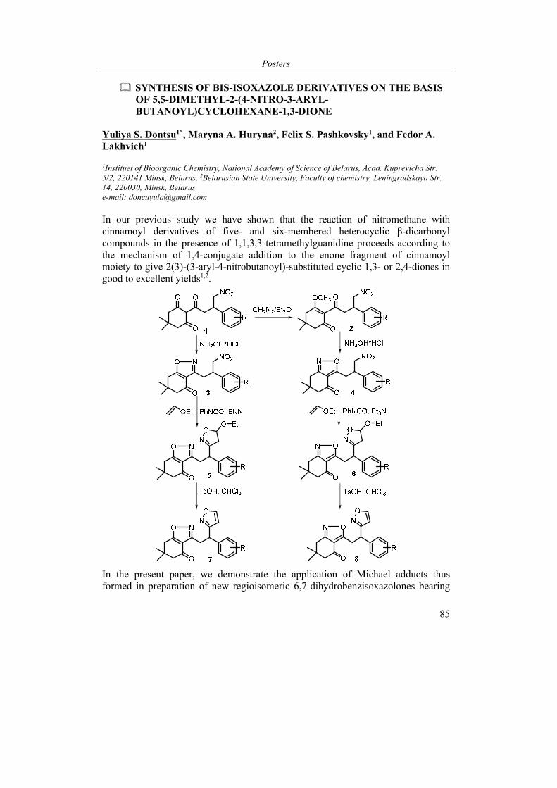
Posters
85
SYNTHESIS OF BIS-ISOXAZOLE DERIVATIVES ON THE BASIS OF 5,5-DIMETHYL-2-(4-NITRO-3-ARYL-BUTANOYL)CYCLOHEXANE-1,3-DIONE
Yuliya S. Dontsu1*, Maryna A. Huryna2, Felix S. Pashkovsky1, and Fedor A. Lakhvich1
1Instituet of Bioorganic Chemistry, National Academy of Science of Belarus, Acad. Kuprevicha Str. 5/2, 220141 Minsk, Belarus, 2Belarusian State University, Faculty of chemistry, Leningradskaya Str. 14, 220030, Minsk, Belarus e-mail: [email protected]
In our previous study we have shown that the reaction of nitromethane with cinnamoyl derivatives of five- and six-membered heterocyclic β-dicarbonyl compounds in the presence of 1,1,3,3-tetramethylguanidine proceeds according to the mechanism of 1,4-conjugate addition to the enone fragment of cinnamoyl moiety to give 2(3)-(3-aryl-4-nitrobutanoyl)-substituted cyclic 1,3- or 2,4-diones in good to excellent yields1,2.
In the present paper, we demonstrate the application of Michael adducts thus formed in preparation of new regioisomeric 6,7-dihydrobenzisoxazolones bearing

VI International Conference “Chemistry, Structure and Function of Biomolecules”
86
an additional isoxazoline (isoxazole) moiety in the side chain. By esterification of vinylogous acids (1) their methyl enol ethers (2) were obtained. Nitromethyl derivatives of β-triketones (1) and methyl ethers (2) reacted with hydroxylamine hydrochloride in ethanol to give regioisomeric dihydrobenzisoxazolones (3, 4) respectively. The isoxazoline fragment in the side chain of compounds (5, 6) was formed by 1,3-dipolar cycloaddition of nitrile oxides generated from the nitromethyl substituent of compounds (3, 4) to ethyl vinyl ether as dipolarophile. Compounds (5, 6) on treatment with p-toluenesulfonic acid in trichloromethane give rise to regioisomeric bis-isoxazole derivatives (7, 8).
REFERENCES
(1) Pashkovsky F.S., Dontsu J.S., Rubinov D.B., Lakhvich F.A. Chem. Heterocycl. Compd. 2015, 50, 1421.
(2) Pashkovskii F.S., Dontsu Yu.S., Rubinov D.B., Lakhvich F.A., Traven’ V.F., Borunov A.M. Russ. J. Org. Chem. 2014, 50, 1598.
HEMOSORBENT «ANTILIPOPROTEID» - MEANS TO COMBAT LIPID EXCHANGE
Vladimir P. Golubovich1, Evgeniy M. Ermola1*, Denis A. Makarevich1, Svetlana P. Kurlenko1, and Valery V. Kirkovskiy2
1 Institute of Bioorganic Chemistry of the National Academy of Sciences of Belarus, Minsk, Belarus, 2
9th City Clinical Hospital, Minsk, Belarus e-mail: [email protected]
The purpose of this work was to create a new original medical device to improve the effectiveness of treatment of patients with severe forms of dyslipidemia due to the introduction of the antilipoprotein biospecific hemosorbent "Antilipoproteid" into clinical practice.
It is known that lipid metabolism disorders can be associated with various pathological conditions of the body and are both a consequence and cause of various diseases. At present, the leading role of certain classes of lipoproteins (low and very low density lipoproteins - LDL and VLDL) has been proven in the pathogenesis of atherosclerosis. Correction of lipid metabolism disorders is an actual problem of practical medicine. Logically to suggest that the elimination of atherogenic lipoproteins can positively affect the course and outcome of the pathological process itself. It was shown when using plasmapheresis (PA) in the treatment of patients in order to remove the above substances, a noticeable therapeutic effect was achieved. The following years were characterized by the accumulation of a large amount of factual material on the nature of the therapeutic effect of PA for various diseases, which confirms the prospects of corrective
Posters
87
therapy methods, especially when traditional drug therapy does not give the desired results.
Experience shows that the inclusion of PA in the complex therapy of patients with severe dyslipidemia leads, as a rule, to a decrease in the severity of the latter. The extent and persistence of the achieved therapeutic effect varies within a very wide range. The reasons for this have not yet been studied. The cases are recorded with a slight decrease in the coefficient of atherogenicity after the completion of the course of treatment with the use of therapeutic PA. Nevertheless, in the post-apheresis period, when studying this indicator, a further decrease in the level of atherogenic lipids was detected. The reason for this seems to be the change in the rate of synthesis and the rate of metabolism of these compounds after treatment with biospecific hemosorbent "Antilipoproteid". The study the phenomenon may allow a new look at the role of organs and systems involved in the biodegradation of these metabolites.
Therapeutic PA involves the extraction from the body of not only pathogenetic, but also physiologically significant substances. In this regard, the need for the development of plasma-saving technologies is becoming more and more evident. These include plasma and hemosorption. The use of various hemosorbents with directed action is associated with a number of problems, the main of which is the unacceptably high cost of imported sorbents and the lack of their industrial production in the CIS. The widely used ALP-hemosorbent effectively removes LDL and VLDL from plasma, but can not be used to extract these substances directly from the blood for the reason of its unsatisfactory hemocompatibility. This circumstance sharply increases the cost and complicates the process of extracting LDL and VLDL from the body.
To reduce the cost of this process, researchers from the National Academy of Sciences of Belarus and the Belarusian State Medical University conducted research on the development and use of the biospecific hemosorbent "Antilipoproteid", which can extract LDL and VLDL from the blood. The new biospecific affinity hemosorbent was created on the basis of a polyacrylamide matrix, which proved to be well established in the development of a number of disposable hemosorbents - Proteasazor Hemo, LPS-Gemo. According to its physico-chemical properties, this compound resembles living tissues: it is elastic, easily permeable to many molecules and contains a large amount of water in its volume. In the process of blood perfusion through such a hemosorbent, it interacts with a more "habitual" biocompatible surface. In the polyacrylamide gel, there are pores that provide an efficient transport of extractable substrates to the volume of the polymer. Some polymeric materials or preparations e.g., heparin, or more precisely its high molecular weight fraction (more than 7 kDa) are used as ligands. This sorbent is effective in the treatment of dyslipidemia, atherosclerosis, IHD and has no analogues in the world.
VI International Conference “Chemistry, Structure and Function of Biomolecules”
88
The clinical trials of hemosorbent "Antilipoproteid" on the bases of Vitebsk and Gomel regional clinical hospitals, and the 9th city clinical hospital in Minsk, have shown the effectiveness of this hemosorbent. The necessary documents for production, realization and application in the clinical practice of the biospecific hemosorbent have been obtained. Realization of hemosorbent "Antilipoproteid" in the Republic of Belarus will be carried out by SP ALC "Farmavit".
CRYSTALLIZATION OF A LSM PROTEIN FROM HALOBACTERIUM SALINARUM
Maria S. Fando*, Natalia V. Lekontseva, Georgi K. Selikhanov, and Alexey D. Nikulin
Institute of Protein Research, Pushchino, Russia. e-mail: [email protected]
Proteins of Lsm (Sm-like) family are found in all the three domains of life. They provide biogenesis and functioning of various RNA molecules in the cells. Bacterial Lsm protein called Hfq exhibits chaperone activity promoting interaction between regulatory sRNA and mRNA during regulation of translation (1, 2). Eukaryotic Sm proteins are core proteins of the spliceosome while eukaryotic Lsm proteins are involved in the mRNA degradation (3). Functions of archaeal Lsm proteins (SmAP) in the cells have been studied pitiable, although there is some data on their participation in processing of some RNA (such as tRNA).
Our work concerns with structural and functional studies of SmAP protein from Halobacterium salinarum. This protein has remarkable difference of the sequence compared with homologues and, in fact, represents a minimal Lsm core. Our current task is to determine the structure of this protein and its complexes with ribonucleotides and short RNA to define the specificity and structural aspects of the SmAP-RNA interaction. We have obtained a genetic construct carrying the gene of the SmAP from H. salinarum (HsaSmAP). The protein has been isolated and purified in preparative scale, then it has been crystallized and a high resolution diffraction dataset has been collected at ERSF in Grenoble. At this moment, the HsaSmAP structure is under determination and refinement.
The work is supported by RFBR grant #18-04-00222.
REFERENCES
(1) Sun, X.; Zhulin, I.; Wartell, R.M. Nucleic Acids Research. 2002, 30, 3662-3671. (2) Valentin-Hansen, P.; Eriksen, M.; Udesen, C.; Molecular Microbiology. 2004, 51, 1525–
1533. (3) Thore, S.; Mayer, C.; Sauter, C.; Weeks, S.; Suck, D.; Journal of Biological Chemistry.
2003, 278, 1239–1247.
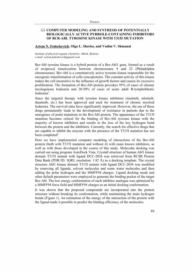
Posters
89
COMPUTER MODELING AND SYNTHESIS OF POTENTIALLY BIOLOGICALLY ACTIVE PYRROLE-CONTAINING INHIBITORS OF BCR-ABL TYROSINE KINASE WITH T315I MUTATION
Artem N. Fedorkevich, Olga L. Sharko, and Vadim V. Shmanai
Institute of physical organic chemistry, Minsk, Belarus. e-mail: [email protected]
Bcr-Abl tyrosine kinase is a hybrid protein of a Bcr-Abl1 gene, formed as a result of reciprocal translocation between chromosomes 9 and 22 (Philadelphia chromosome). Bcr-Abl is a constitutively active tyrosine kinase responsible for the oncogenic transformation of cells (oncoprotein). The constant activity of this kinase makes the cell insensitive to the influence of growth factors and causes its excessive proliferation. The formation of Bcr-Abl protein provokes 95% of cases of chronic myelogenous leukemia and 20-50% of cases of acute adult B-lymphoblastic leukemia1.
Since the targeted therapy with tyrosine kinase inhibitors (imatinib, nilotinib, dasatinib, etc.) has been approved and used for treatment of chronic myeloid leukemia. The survival rates have significantly improved. However, the use of these drugs permanently leads to the development of resistance in patients due to the emergence of point mutations in the Bcr-Abl protein. The appearance of the T315I mutation becomes critical for the binding of Bcr-Abl tyrosine kinase with the majority of known inhibitors and results in the loss of the key hydrogen bond between the protein and the inhibitors. Currently, the search for effective drugs that are capable to inhibit the enzyme with the presence of the T315I mutation has not been completed2.
Here we have implemented computer modeling of interactions of the Bcr-Abl protein (both with T315I mutation and without it) with main known inhibitors, as well as with those developed in the course of this study. Molecular docking was carried out using program AutoDock Vina. Сrystall structure of human Abl1 kinase domain T315I mutant with ligand DCC-2036 was retrieved from RCSB Protein Data Bank (PDB ID: 3QRJ, resolution: 1.82 Å) as a docking template. The crystal structure Abl1 kinase domain T315I mutant with ligand DCC-2036 was modified by removing all ligands, solvent molecules and some water molecules and then adding the polar hydrogen and the MMFF94 charges. Ligand docking mode and other default parameters were employed to generate the binding pocket of the target Bcr-Abl. The low energy conformation of each inhibitor analogue was optimized by a MMFF94 force field and MMFF94 charges as an initial docking conformation.
It was shown that the proposed compounds are incorporated into the protein structure without breaking its conformation, while maintaining the main hydrogen bonds (Figure 1). An estimation of the energy of the interaction of the protein with the ligand made it possible to predict the binding efficiency of the molecules.
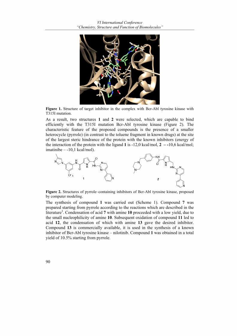
VI International Conference “Chemistry, Structure and Function of Biomolecules”
90
Figure 1. Structure of target inhibitor in the complex with Bcr-Abl tyrosine kinase with T315I mutation.
As a result, two structures 1 and 2 were selected, which are capable to bind efficiently with the T315I mutation Bcr-Abl tyrosine kinase (Figure 2). The characteristic feature of the proposed compounds is the presence of a smaller heterocycle (pyrrole) (in contrast to the toluene fragment in known drugs) at the site of the largest steric hindrance of the protein with the known inhibitors (energy of the interaction of the protein with the ligand 1 is -12,0 kсal/mol, 2 – -10,6 kсal/mol; imatinibe – -10,1 kсal/mol).
Figure 2. Structures of pyrrole–containing inhibitors of Bcr-Abl tyrosine kinase, proposed by computer modeling.
The synthesis of compound 1 was carried out (Scheme 1). Compound 7 was prepared starting from pyrrole according to the reactions which are described in the literature3. Condensation of acid 7 with amine 10 proceeded with a low yield, due to the small nucleophilicity of amine 10. Subsequent oxidation of compound 11 led to acid 12, the condensation of which with amine 13 gave the desired inhibitor. Compound 13 is commercially available, it is used in the synthesis of a known inhibitor of Bcr-Abl tyrosine kinase – nilotinib. Compound 1 was obtained in a total yield of 10.5% starting from pyrrole.
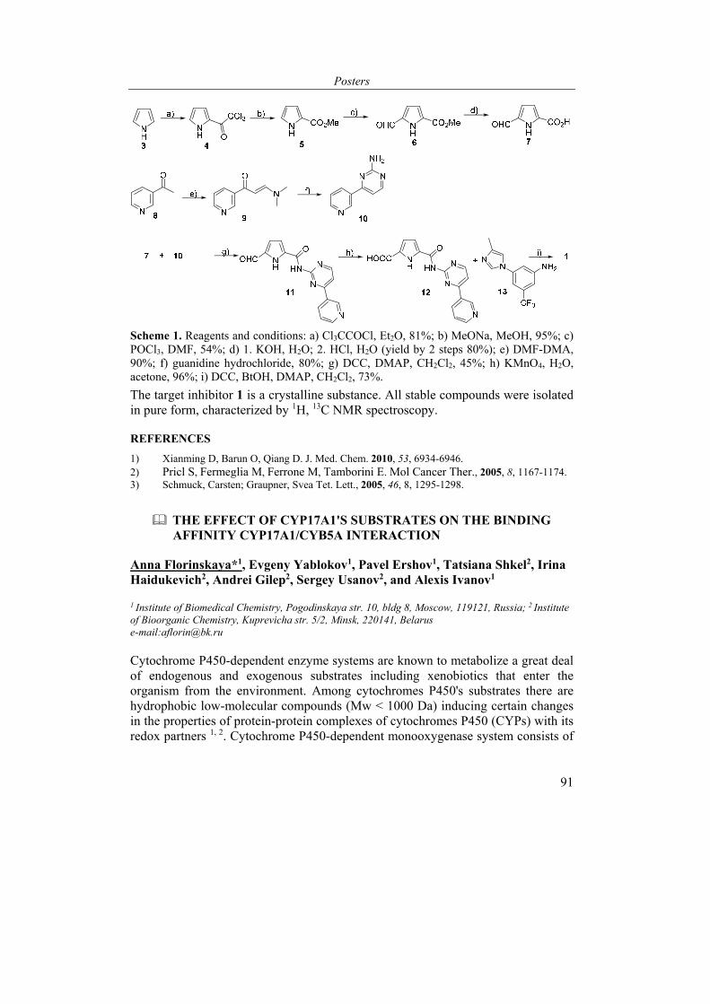
Posters
91
Scheme 1. Reagents and conditions: а) Cl3CCOCl, Et2O, 81%; b) MeONa, MeOH, 95%; c) POCl3, DMF, 54%; d) 1. KOH, H2O; 2. HCl, H2O (yield by 2 steps 80%); e) DMF-DMA, 90%; f) guanidine hydrochloride, 80%; g) DCC, DMAP, CH2Cl2, 45%; h) KMnO4, H2O, acetone, 96%; i) DCC, BtOH, DMAP, CH2Cl2, 73%.
The target inhibitor 1 is a crystalline substance. All stable compounds were isolated in pure form, characterized by 1H, 13C NMR spectroscopy.
REFERENCES
1) Xianming D, Barun O, Qiang D. J. Med. Chem. 2010, 53, 6934-6946. 2) Pricl S, Fermeglia M, Ferrone M, Tamborini E. Mol Cancer Ther., 2005, 8, 1167-1174. 3) Schmuck, Carsten; Graupner, Svea Tet. Lett., 2005, 46, 8, 1295-1298.
THE EFFECT OF CYP17A1'S SUBSTRATES ON THE BINDING AFFINITY CYP17A1/CYB5A INTERACTION
Anna Florinskaya*1, Evgeny Yablokov1, Pavel Ershov1, Tatsiana Shkel2, Irina Haidukevich2, Andrei Gilep2, Sergey Usanov2, and Alexis Ivanov1
1 Institute of Biomedical Chemistry, Pogodinskaya str. 10, bldg 8, Moscow, 119121, Russia; 2 Institute of Bioorganic Chemistry, Kuprevicha str. 5/2, Minsk, 220141, Belarus e-mail:[email protected]
Cytochrome P450-dependent enzyme systems are known to metabolize a great deal of endogenous and exogenous substrates including xenobiotics that enter the organism from the environment. Among cytochromes P450's substrates there are hydrophobic low-molecular compounds (Mw < 1000 Da) inducing certain changes in the properties of protein-protein complexes of cytochromes P450 (СYPs) with its redox partners 1, 2. Cytochrome P450-dependent monooxygenase system consists of
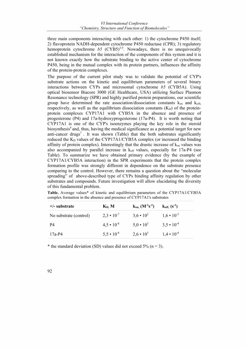
VI International Conference “Chemistry, Structure and Function of Biomolecules”
92
three main components interacting with each other: 1) the cytochrome P450 itself; 2) flavoprotein NADH-dependent cytochrome P450 reductase (CPR); 3) regulatory hemoprotein cytochrome b5 (CYB5)3-5. Nowadays, there is no unequivocally established mechanism for the interaction of the components of this system and it is not known exactly how the substrate binding to the active center of cytochrome P450, being in the mutual complex with its protein partners, influences the affinity of the protein-protein complexes.
The purpose of the current pilot study was to validate the potential of СYP's substrate actions on the kinetic and equilibrium parameters of several binary interactions between СYPs and microsomal cytochrome b5 (CYB5A). Using optical biosensor Biacore 3000 (GE Healthcare, USA) utilizing Surface Plasmon Resonance technology (SPR) and highly purified protein preparations, our scientific group have determined the rate association/dissociation constants kon and koff, respectively, as well as the equilibrium dissociation constants (KD) of the protein-protein complexes CYP17A1 with CYB5A in the absence and presence of progesterone (P4) and 17a-hydroxyprogesterone (17а-P4). It is worth noting that CYP17A1 is one of the CYP's isoenzymes playing the key role in the steroid biosynthesis6 and, thus, having the medical significance as a potential target for new anti-cancer drugs7 . It was shown (Table) that the both substrates significantly reduced the KD values of the CYP17A1/CYB5A complex (or increased the binding affinity of protein complex). Interestingly that the drastic increase of kon values was also accompanied by parallel increase in koff values, especially for 17a-P4 (see Table). To summarize we have obtained primary evidence (by the example of CYP17A1/CYB5A interaction) in the SPR experiments that the protein complex formation profile was strongly different in dependence on the substrate presence comparing to the control. However, there remains a question about the “molecular spreading” of above-described type of CYPs binding affinity regulation by other substrates and compounds. Future investigation will allow elucidating the diversity of this fundamental problem. Table. Average values* of kinetic and equilibrium parameters of the CYP17A1/CYB5A complex formation in the absence and presence of CYP17A1's substrates
+/- substrate KD, М kon, (М-1s-1) koff, (s-1)
No substrate (control) 2,3 • 10-7 3,6 • 102 1,6 • 10-5
P4 4,5 • 10-8 5,0 • 103 3,5 • 10-4
17а-P4 5,5 • 10-8 2,6 • 103 1,4 • 10-4
* the standard deviation (SD) values did not exceed 5% (n = 3).
Posters
93
REFERENCES
1) Meng Zhang, Stephanie V. Le Clair, Rui Huang, Shivani Ahuja, Sang-Choul Im, Lucy Waskell & Ayyalusamy Ramamoorthy. Sci Rep. 2015, 5, 8392.
2) D. Fernando Estrada, Andria L. Skinner, Jennifer S. Laurence, Emily E. Scott. JBC, 2014, 289, 20, 14310–14320.
3) Omura, T. J Biochem, 2010, 147 (3), 297-306.
4) John S. French, F. Peter Guengerich, Minor J. JBC, 1980, 255., 9., 4112-4119.
5) Claude Bonfils, Claude Balny, and Patrick Maurel. JBC, 1981, 256, 18, 9457-9465. 6) Roberta Ferraldeschi, Nima Sharifi, Richard J. Clin Cancer Res. 2013, 19(13): 3353–3359. 7) Meng Zhang1, Stephanie V. Le Clair, Rui Huang, Shivani Ahuja, Sang-Choul Im, Lucy
Waskell & Ayyalusamy Ramamoorthy. Sci Rep. 2015, (17)5:8392.
The reported study was funded by RFBR according to the research project № 18-04-00071 A. The study was carried out using “Human Proteome” Core Facility (IBMC, Moscow, Russia) which is supported by Ministry of Education and Science of the Russian Federation (unique project ID RFMEFI62117X0017).
SYNTHESIS OF CHROMOGENIC SUBSTRATES OF FACTOR VIII
Vera P. Martsinovich1, Olga V. Gribovskaya1, Vladimir P. Golubovich1, Elena D. Rasyuk2, and Darya G. Vensko2
1 Institute of Bioorganic Chemistry, National Academy of Sciences of Belarus, Minsk, Republic of Belarus; 2 Republican Scientific-Practical Center for Transfusiology and Medical Biotechnologies, Minsk, Republic of Belarus
Factor VIII (FVIII) is measured in plasma if it is necessary to diagnose and treat congenital and acquired hemophilia, especially hemophilia A, von Willebrand congenital defects, to assess the risk of thrombophilia and in other cases. Its specific activity is directed to the proteolysis of factor X and proceeds with the obligatory participation of factor IX. FVIII is a serine protease, the peptide substrates for it mimic the cleavage sites of natural protein substrates. Although the history of the development of methods for its determination is several decades old, intensive research is currently underway to improve existing and develop new methods [1-3].
To develop a quantitative method for the determination of FVIII in blood plasma, methods for the synthesis of its chromogenic peptide substrates Z-D-Arg-Gly-Arg-pNA, known as S-2765 and the original tetrapeptide substrate Z-Gly-D-Arg-Gly-Arg-pNA, have been developed. The introduction of a glycine residue into the first position allows the use of Z-Gly-OH, a compound much more accessible than Z-D-Arg-OH, which makes the final product cheaper, making its synthesis more accessible. In addition, it is possible to increase the substrate specificity of Z-Gly-D-Arg-Gly-Arg-pNA as compared to the tripeptide substrate. The developed substrate synthesis scheme involves the addition of 4-nitroanilide to the dipeptide fragment of Boc-Gly-Arg and the addition of Z-D-Arg-OH or Z-Gly-D-Arg-OH to H-Gly-Arg-pNA in the final stage (Scheme).
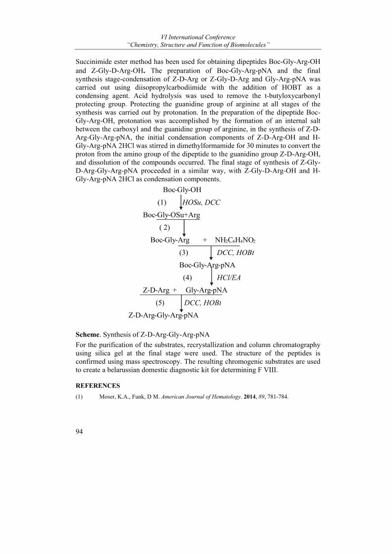
VI International Conference “Chemistry, Structure and Function of Biomolecules”
94
Succinimide ester method has been used for obtaining dipeptides Boc-Gly-Arg-OH and Z-Gly-D-Arg-OH. The preparation of Boc-Gly-Arg-pNA and the final synthesis stage-condensation of Z-D-Arg or Z-Gly-D-Arg and Gly-Arg-pNA was carried out using diisopropylcarbodiimide with the addition of HOBT as a condensing agent. Acid hydrolysis was used to remove the t-butyloxycarbonyl protecting group. Protecting the guanidine group of arginine at all stages of the synthesis was carried out by protonation. In the preparation of the dipeptide Boc-Gly-Arg-OH, protonation was accomplished by the formation of an internal salt between the carboxyl and the guanidine group of arginine, in the synthesis of Z-D-Arg-Gly-Arg-pNA, the initial condensation components of Z-D-Arg-OH and H-Gly-Arg-pNA 2HCl was stirred in dimethylformamide for 30 minutes to convert the proton from the amino group of the dipeptide to the guanidino group Z-D-Arg-OH, and dissolution of the compounds occurred. The final stage of synthesis of Z-Gly-D-Arg-Gly-Arg-pNA proceeded in a similar way, with Z-Gly-D-Arg-OH and H-Gly-Arg-pNA 2HCl as condensation components.
Scheme. Synthesis of Z-D-Arg-Gly-Arg-pNA
For the purification of the substrates, recrystallization and column chromatography using silica gel at the final stage were used. The structure of the peptides is confirmed using mass spectroscopy. The resulting chromogenic substrates are used to create a belarussian domestic diagnostic kit for determining F VIII.
REFERENCES
(1) Moser, K.A., Funk, D M. American Journal of Hematology. 2014, 89, 781-784.
Boc-Gly-OH
(1) HOSu, DCC
Boc- Gly-OSu+Arg
( 2)
Boc-Gly-Arg + NH2C6H4NO2
(3) DCC, HOBt
Boc-Gly-Arg-pNA
(4) HCl/EA
Z -D-Arg + Gly-Arg-pNA
( 5 ) DCC, HOBt
Z-D- Arg- Gly-Arg-pNA
Posters
95
(2) Kitchen, S, Preston, F. In: Kitchen S, Olson J, Preston F, Ed,. Quality in Laboratory Hemostasis and Thrombosis, 1st ed. Chicester, West Sussex, UK: Wiley-Blackwell; 2009. 81–89.
(3) Kitchen, S., Tiefenbacher, S., Gosselin R. Semin Thromb Hemostasis 2017, 43, 331-337.
EFFECTS OF ANTIDEPRESSANT FLUVOXAMINE IN THE EMBRYONIC PERIOD OF DEVELOPMENT ON THE COGNITIVE FUNCTION OF ADULT OFFSPRING
1Gruzdev G.A., 1Voronina Y.A., 1Manchenko D.M., 2Glazova N.Y., and 1Levitskaya N.G.
1Lomonosov Moscow State University. Biological Faculty, Department of Human and Animal
Physiology; 2 Institute of Molecular Genetics, RAS e-mail: [email protected],
Serotonin Reuptake inhibitor fluvoxamine is a drug approved for use during pregnancy. The effect of this substance is based on an increase in the content of serotonin in the synaptic cleft and prolongation of its actions. Fluvoxamine penetrates the placental barrier and not only to the maternal organism, but also to the fetus. In a large number of studies, it has been shown that serotonin in early embryogenesis serves as a trophic factor and contributes to the normal formation of various organ systems, including the circulatory and nervous systems. It monitors the proper neurogenesis and stimulates the myelination of nerve fibers, as well as regulates the functioning of mediator systems, such as dopaminergic, noradrenalinergic. Therefore, any intervention in the serotonergic system can lead to irreversible developmental disorders, and can also reduce the level of cognitive abilities and learning opportunities.
In this paper, we investigated the effect of prenatal administration of antidepressant Fluvoxamine on the training of offspring of white rats.
In the study, pregnant females we devided into two groups. The first group - pregnant females were injected intraperitoneally with fluvoxamine before the onset of embryonic serotoninergic system formation from day 3-10 of pregnancy. The second group - pregnant females injected fluvoxamine from 8 to 14 day of pregnancy, this interval corresponds to the embryonic period of formation of all parts of the brain and development of the basic mediator systems including serotoninergic. Fluvoxamine was administered intraperitoneally at a dose of 10 mg / kg. The control group received injections of water in an equivalent volume.
Cognitive functions were screened in the tests, conducted on the 34th and 46th day of life respectively. "Complex food labyrinth", conducted on the 34th and 46th day of life respectively.
All tests were conducted in accordance with ethical standards.
VI International Conference “Chemistry, Structure and Function of Biomolecules”
96
Fluvoxamine administration to the first group in the elier embryonic stages of development doesn’t influence on cognitive functions in both tests. Fluvoxamine administration to the second group showed decreased ability to memorization. In the test with negative pain reinforcement "conditioned passive avoidance reflex" test, the experimental group showed reduced learning ability, this manifested itself in a small latent period of entering the dark compartment, as well as in increasing the time spent in the dark compartment of the control group. An increased research motor activity was also expressed, which indicates a decreased anxiety, this indicates that the trained rats in the experimental group were not afraid of the pain stimulus, there should normally be an opposite reaction. Also in this test, the negative impact on memorization more pronounced in female rats.
In the test with positive food reinforcement "Complex Labyrinth", the experimental group also shows a lower ability to learn, in contrast to the control group, this effect manifests an increased number of performed reactions. There is also more expressed anxiety in behavior.
Thus, the prenatal administration of fluvoxamine at different gestation times affects the training of offspring only when administered from 8to14 day of rat pregnancy. The effect gives a more pronounced in female offspring. The negative effect on cognitive function of offspring may be related to intervention to the serotonergic system of the developing fetus.
GENETIC POLYMORPHISMS OF UGT1A6 IN BELARUSSIAN PATIENTS WITH EPILEPSY
Irina V. Haidukevich1*, Maria S. Kisel1, Olga M. Rudauskaya1, Olga S. Bokut1, Andrei A. Gilep1, Tatiana V. Dokukina2, Mikhail V. Mahrov2
1Institute of Bioorganic Chemistry NASB, Minsk, Belarus, 2Republican Scientific and Practical Center for Mental Health, Minsk, Belarus, *e-mail: [email protected]
Valproic acid (VPA) is a widely prescribed antiepileptic drug that is used as first-line therapy for epilepsy. However, it shows wide variability in pharmacokinetics and pharmacodynamics among patients, even with the same doses of VPA. Although adverse drug reactions of VPA (such as depression, hepatitis, hypotension, hypernatremia, acidosis, pancreatitis, acute renal failure, and thrombocytopenia) are relatively rare, hepatotoxicity is severe in particular in those younger than 2 years old and patients with polypharmacy.
Cytochrome P450 (CYP) enzymes, mitochondrion-mediated β-oxidation, and glucuronosyltransferases (UGTs) are the three main metabolic routes of VPA. Glucuronidation is the prevalent metabolic route and account for 50% of the VPA metabolism [1]. Under the conditions of deficiency or reduced enzymatic activity of
Posters
97
glucuronosyltransferases VPA becomes available for biotransformation on other metabolic pathways (β-oxidation, cytochrome P450-mediated oxidation), resulting in a plasma elevation in the concentration of metabolites that are assumed to be responsible for many side effects. One of the reasons for the improper functioning of UGTs is genetic polymorphism. These can cause changes in pharmacokinetic of VPA.
UGT1A6 is member of UGT1 subfamily which along with UGT2 subfamily is responsible for xenobiotic elimination, including VPA. Several polymorphisms have been described in the first exon of UGT1А6 gene. The most studied are non-synonymous SNPs rs2070959 (541 A>G, Thr181Ala), rs1105879 (552 A>C, Arg184Ser) and rs6759892 (19 T>G, Ser7Ala), which are inherited together and constitute a haplotype.
Homozygosity on UGT1A6*1 allele (19(T) / 541(A) / 552(A)) is associated with normal metabolism of VPA, while UGT1A6*2 homozygosity (19(G) / 541(G) / 552(C)) is associated with higher rate of VPA glucuronidation and UGT1A6*1/*2 genotype conversely shows low enzyme activity [2].
In the present study we have developed method for detection of three above mentioned UGT1A6 SNPs by Sanger sequencing. We have genotyped 49 patients with epilepsy and were able to detect all four allele variants (*1, *2, *3, *4) of UGT1A6 gene. Frequencies of allele *1 and allele *2 are almost the same (46% and 45% respectively) and significantly differ from those previously reported by Chatzistefanidis [3] in healthy Caucasians (UGT1A6*1 - 62% and UGT1A6*2 - 27% respectively). Alleles *3 and *4 are relatively rare - 7% and 2% and these values correspond to appearance of these alleles in healthy Caucasians (UGT1A6*3 - 6% and UGT1A6*4 - 5%). UGT1A6*1/*2 genotype had the highest frequency (41%) among all detected genotypes (*1/*1 - 20,5%; *1/*3 - 8%; *1/*4 - 2%; *2/*2 - 20,5%; *2/*3 - 6%; *2/*4 - 2%).
This preliminary study indicated the high frequency of UGT1A6*2 allele and UGT1A6*1/*2 genotype, associated with low UGT1A6 enzymatic activity, in Belarussian patients with epilepsy. Taking those findings into account UGT1A6*2 can be proposed as perspective pharmacogenetic marker for personalization of VPA treatment. Further clinical studies are needed.
REFERENCES
(1) Du, Z. Med. Sci. Monit. 2016, 22, 4107-4113. (2) Nagar, S.; Zalatoris, J.J.; Blanchard, R.L. Pharmacogenetics. 2004, 14(8), 487-499. (3) Chatzistefanidis, D; Georgiou, I; Kyritsis, AP; Markoula, S. Pharmacogenomics. 2012,
13(9), 1055-1071.
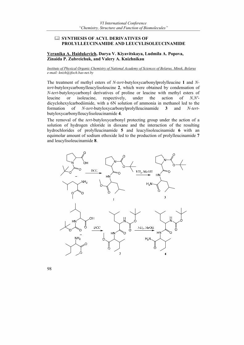
VI International Conference “Chemistry, Structure and Function of Biomolecules”
98
SYNTHESIS OF ACYL DERIVATIVES OF PROLYLLEUCINAMIDE AND LEUCYLISOLEUCINAMIDE
Veranika A. Haidukevich, Darya V. Kiyavitskaya, Ludmila A. Popova, Zinaida P. Zubreichuk, and Valery A. Knizhnikau
Institute of Physical Organic Chemistry of National Academy of Sciences of Belarus, Minsk, Belarus e-mail: [email protected]
The treatment of methyl esters of N-tert-butyloxycarbonylprolylleucine 1 and N-tert-butyloxycarbonylleucylisoleucine 2, which were obtained by condensation of N-tert-butyloxycarbonyl derivatives of proline or leucine with methyl esters of leucine or isoleucine, respectively, under the action of N,N′-dicyclohexylcarbodiimide, with a 6N solution of ammonia in methanol led to the formation of N-tert-butyloxycarbonylprolylleucinamide 3 and N-tert-butyloxycarbonylleucylisoleucinamide 4.
The removal of the tert-butyloxycarbonyl protecting group under the action of a solution of hydrogen chloride in dioxane and the interaction of the resulting hydrochlorides of prolylleucinamide 5 and leucylisoleucinamide 6 with an equimolar amount of sodium ethoxide led to the production of prolylleucinamide 7 and leucylisoleucinamide 8.
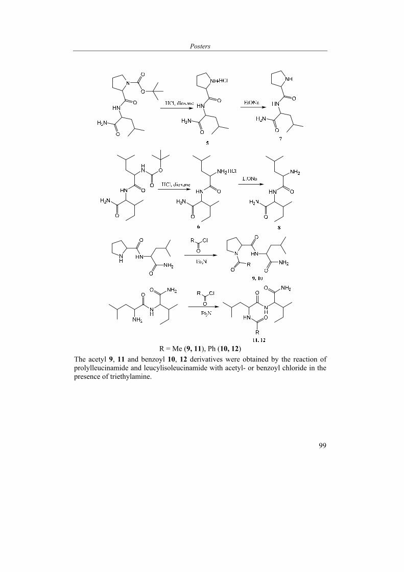
Posters
99
R = Me (9, 11), Ph (10, 12)
The acetyl 9, 11 and benzoyl 10, 12 derivatives were obtained by the reaction of prolylleucinamide and leucylisoleucinamide with acetyl- or benzoyl chloride in the presence of triethylamine.
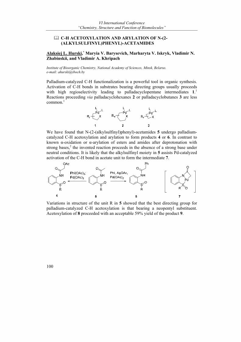
VI International Conference “Chemistry, Structure and Function of Biomolecules”
100
C-H ACETOXYLATION AND ARYLATION OF N-(2-(ALKYLSULFINYL)PHENYL)-ACETAMIDES
Alaksiej L. Hurski,* Maryia V. Barysevich, Marharyta V. Iskryk, Vladimir N. Zhabinskii, and Vladimir A. Khripach
Institute of Bioorganic Chemistry, National Academy of Sciences, Minsk, Belarus. e-mail: [email protected]
Palladium-catalyzed C-H functionalization is a powerful tool in organic synthesis. Activation of C-H bonds in substrates bearing directing groups usually proceeds with high regioselectivity leading to palladacyclopentane intermediates 1.1 Reactions proceeding via palladacyclohexanes 2 or palladacyclobutanes 3 are less common.1
We have found that N-(2-(alkylsulfinyl)phenyl)-acetamides 5 undergo palladium-catalyzed C-H acetoxylation and arylation to form products 4 or 6. In contrast to known α-oxidation or α-arylation of esters and amides after deprotonation with strong bases,2 the invented reaction proceeds in the absence of a strong base under neutral conditions. It is likely that the alkylsulfinyl moiety in 5 assists Pd-catalyzed activation of the C-H bond in acetate unit to form the intermediate 7.
Variations in structure of the unit R in 5 showed that the best directing group for palladium-catalyzed C-H acetoxylation is that bearing a neopentyl substituent. Acetoxylation of 8 proceeded with an acceptable 59% yield of the product 9.
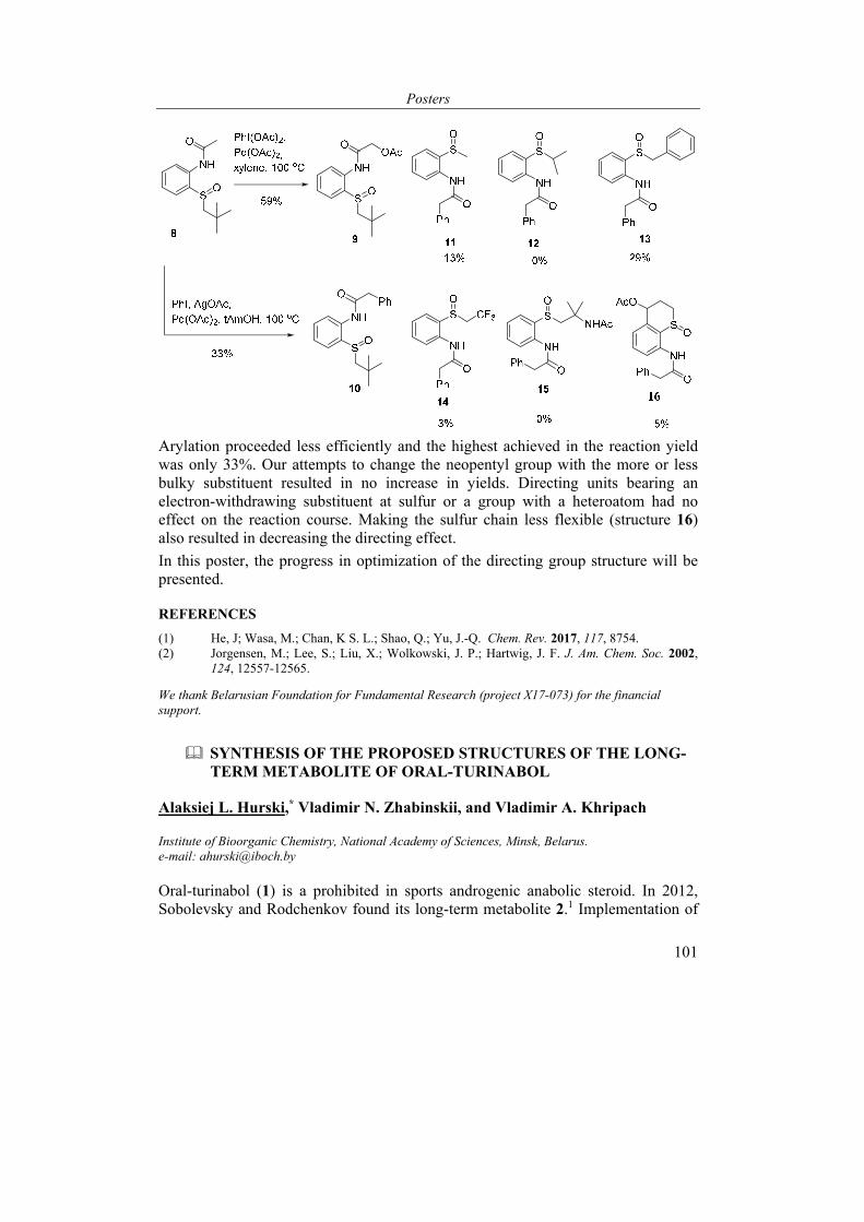
Posters
101
Arylation proceeded less efficiently and the highest achieved in the reaction yield was only 33%. Our attempts to change the neopentyl group with the more or less bulky substituent resulted in no increase in yields. Directing units bearing an electron-withdrawing substituent at sulfur or a group with a heteroatom had no effect on the reaction course. Making the sulfur chain less flexible (structure 16) also resulted in decreasing the directing effect.
In this poster, the progress in optimization of the directing group structure will be presented.
REFERENCES
(1) He, J; Wasa, M.; Chan, K S. L.; Shao, Q.; Yu, J.-Q. Chem. Rev. 2017, 117, 8754. (2) Jorgensen, M.; Lee, S.; Liu, X.; Wolkowski, J. P.; Hartwig, J. F. J. Am. Chem. Soc. 2002,
124, 12557-12565.
We thank Belarusian Foundation for Fundamental Research (project Х17-073) for the financial support.
SYNTHESIS OF THE PROPOSED STRUCTURES OF THE LONG-TERM METABOLITE OF ORAL-TURINABOL
Alaksiej L. Hurski,* Vladimir N. Zhabinskii, and Vladimir A. Khripach
Institute of Bioorganic Chemistry, National Academy of Sciences, Minsk, Belarus. e-mail: [email protected]
Oral-turinabol (1) is a prohibited in sports androgenic anabolic steroid. In 2012, Sobolevsky and Rodchenkov found its long-term metabolite 2.1 Implementation of
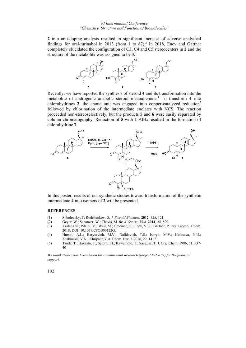
VI International Conference “Chemistry, Structure and Function of Biomolecules”
102
2 into anti-doping analysis resulted in significant increase of adverse analytical findings for oral-turinabol in 2013 (from 1 to 87).2 In 2018, Enev and Gärtner completely elucidated the configuration of C3, C4 and C5 stereocenters in 2 and the structure of the metabolite was assigned to be 3.3
Recently, we have reported the synthesis of steroid 4 and its transformation into the metabolite of androgenic anabolic steroid metandienone.4 To transform 4 into chlorohydrines 2, the enone unit was engaged into copper-catalyzed reduction5 followed by chlorination of the intermediate enolates with NCS. The reaction proceeded non-stereoselectively, but the products 5 and 6 were easily separated by column chromatography. Reduction of 5 with LiAlH4 resulted in the formation of chlorohydrine 7.
In this poster, results of our synthetic studies toward transformation of the synthetic intermediate 4 into isomers of 2 will be presented.
REFERENCES
(1) Sobolevsky, T; Rodchenkov, G. J. Steroid Biochem. 2012, 128, 121. (2) Geyer, W.; Schanzer, W.; Thevis, M. Br. J. Sports. Med. 2014, 48, 820. (3) Kratena,N.; Pilz, S. M.; Weil, M.; Gmeiner, G.; Enev, V. S.; Gärtner, P. Org. Biomol. Chem.
2018, DOI: 10.1039/C8OB00122G. (4) Hurski, A.L.; Barysevich, M.V.; Dalidovich, T.S.; Iskryk, M.V.; Kolasava, N.U.;
Zhabinskii, V.N.; Khripach,V.A. Chem. Eur. J. 2016, 22, 14171. (5) Tsuda, T.; Hayashi, T.; Satomi, H.; Kawamoto, T.; Saegusa, T. J. Org. Chem. 1986, 51, 537-
40
We thank Belarusian Foundation for Fundamental Research (project Х16-107) for the financial support.
Posters
103
SCREENING OF NOVEL DERIVATIVES OF ANDROST-5-ENE TOWARDS CYTOCHROMES P450
Suzana Jovanović-Šanta1*, Yaraslau V. Dzichenka2, Tatsiana V. Shkel2, Aleksei V. Yantsevich2, Marina Savić1, Jovana Ajduković1, and Sergey A. Usanov2
1 University of Novi Sad Faculty of Sciences, Department of Chemistry, Biochemistry and Environmental protection, Trg Dositeja Obradovića 3, 21000 Novi Sad, Serbia, 2 Institute of Bioorganic Chemistry of National Academy of Sciences, Minsk, Belarus [email protected]
Screening of ligand libraries towards to the range of protein targets is a standard procedure during development of high-efficient drugs. Today one of the more often used targets for creation and testing of new drugs are cytochromes P450 (CYPs). CYPs participate in oxidation of various endogenous and exogenous substances and play important role in the metabolism of steroids, bile acids, unsaturated fat acids, phenol metabolites and in biotransformation of xenobiotics (drugs etc.)1,2. So any information about interaction of cytochromes P450 with any class of ligands is very important for «smart» drug-design.
The aim of the current work is to establish new types of biological activity of synthetic derivatives of androst-5-ene towards cytochromes P450 7A1, 7B1 and 51A1 from Homo sapience and Candida albicans.
By using a molecular docking approach, a panel of novel modified steroids – effective anticancer agents – was tested in silico towards cytochromes P450 7A1, 7B1 and 51A1 from Homo sapience and Candida albicans. It was found that some of the Δ5-steroids bind with high efficiency (in terms of free energy) in the CYP7 and CYP51 active site. It should be stressed that binding mode of these compounds corresponds to the binding mode of «essential» substrates of CYPs under investigation.
The ligands found in the previous step were tested in vitro. Target titration with ligand showed that every compound binds as a substrate-like molecule with relatively low Kd. Analysis of CYPs catalytic properties towards these compounds showed that they are able to convert into hydroxylated and demethylated derivatives under reaction in the reconstituted system.
Structural identification of enzyme catalysis products and screening of ligands under investigation towards other CYPs is the aim of our further work.
REFERENCES
(1) Danielson, P. B. Curr. Drug. Metab. 2002, 3(6), 561-597. (2) Estabrook, R. W. Drug. Metab. Dispos. 2003, 31(12), 1461-1473.
Presented results are obtained in the frame of Belarus- Serbia bilateral project Target-specific screening of new activity modulators of human sterol-hydroxylases which is being realized between Institute of Bioorganic Chemistry of NAS of Belarus and University of Novi Sad Faculty of Sciences.
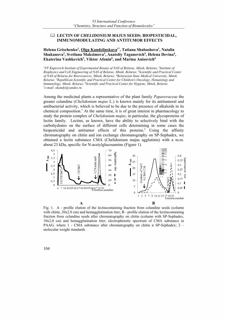
VI International Conference “Chemistry, Structure and Function of Biomolecules”
104
LEСTIN OF CHELIDONIUM MAJUS SEEDS: BIOPESTICIDAL, IMMUNOMODULATING AND ANTITUMOR EFFECTS
Helena Grischenko1, Olga Kandelinskaya1*, Tatiana Shabashova1, Natalia Shukanova2, Svetlana Maksimova3, Anatoliy Taganovich4, Helena Devina4, Ekaterina Vashkevich5, Viktor Afonin6, and Marina Anisovich6
1VF Kuprevich Institute of Experimental Botany of NAS of Belarus, Minsk, Belarus; 2Institute of Biophysics and Cell Engineering of NAS of Belarus, Minsk, Belarus; 3Scientific and Practical Center of NAS of Belarus for Bioresources, Minsk, Belarus; 4Belarusian State Medical University, Minsk, Belarus; 5Republican Scientific and Practical Center for Children's Oncology, Hematology and Immunology, Minsk, Belarus; 6Scientific and Practical Center for Hygiene, Minsk, Belarus *e-mail: [email protected]
Among the medicinal plants a representative of the plant family Papaveraceae the greater celandine (Chelidonium majus L.) is known mainly for its antitumoral and antibacterial activity, which is believed to be due to the presence of alkaloids in its chemical composition.1 At the same time, it is of great interest in pharmacology to study the protein complex of Chelidonium majus, in particular, the glycoproteins of lectin family. Lectins, as known, have the ability to selectively bind with the carbohydrates on the surface of different cells determining in some cases the biopesticidal and antitumor effects of this proteins.2 Using the affinity chromatography on chitin and ion exchange chromatography on SP-Sephadex, we obtained a lectin substance CMA (Chelidonium majus agglutinin) with a m.m. about 23 kDa, specific for N-acetylglucosamine (Figure 1).
A B Fig. 1. A - profile elution of the lectincontaining fraction from celandine seeds (column with chitin, 20x2.0 cm) and hemagglutination titer; B - profile elution of the lectincontaining fraction from celandine seeds after chromatography on chitin (column with SP-Sephadex, 10х2,0 см) and hemagglutination titer; electrophoretic spectrum of CMA substance in PAAG, where 1 - CMA substance after chromatography on chitin и SP-Sephadex; 2 – molecular weight standards
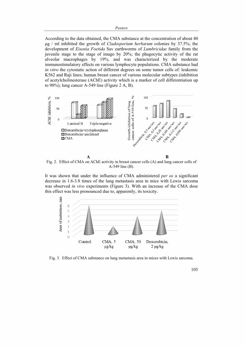
Posters
105
According to the data obtained, the CMA substance at the concentration of about 40 μg / ml inhibited the growth of Cladosporium herbarum colonies by 37.5%; the development of Eisenia Foetida Sav earthworms of Lumbricidae family from the juvenile stage to the stage of imago by 20%; the phagocytic activity of the rat alveolar macrophages by 19%, and was characterized by the moderate immunostimulatory effects on various lymphocyte populations. CMA substance had in vitro the cytostatic action of different degrees on some tumor cells of: leukemic K562 and Raji lines; human breast cancer of various molecular subtypes (inhibition of acetylcholinesterase (AChE) activity which is a marker of cell differentiation up to 90%); lung cancer A-549 line (Figure 2 A, B).
A B Fig. 2. Effect of CMA on AChE activity in breast cancer cells (A) and lung cancer cells of
A-549 line (B). It was shown that under the influence of CMA administered per os a significant decrease in 1.6-3.8 times of the lung metastasis area in mice with Lewis sarcoma was observed in vivo experiments (Figure 3). With an increase of the CMA dose this effect was less pronounced due to, apparently, its toxicity.
Fig. 3. Effect of CMA substance on lung metastasis area in mices with Lewis sarcoma.
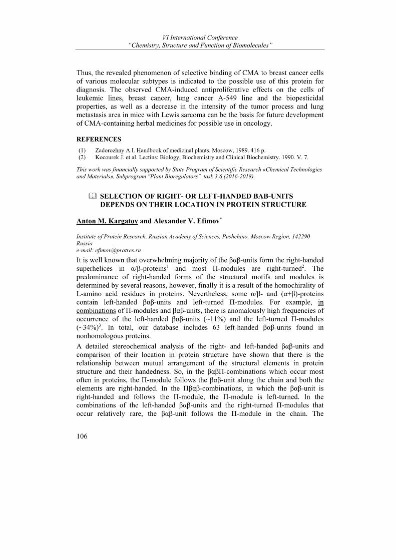
VI International Conference “Chemistry, Structure and Function of Biomolecules”
106
Thus, the revealed phenomenon of selective binding of СMA to breast cancer cells of various molecular subtypes is indicated to the possible use of this protein for diagnosis. The observed CMA-induced antiproliferative effects on the cells of leukemic lines, breast cancer, lung cancer A-549 line and the biopesticidal properties, as well as a decrease in the intensity of the tumor process and lung metastasis area in mice with Lewis sarcoma can be the basis for future development of CMA-containing herbal medicines for possible use in oncology.
REFERENCES
(1) Zadorozhny A.I. Handbook of medicinal plants. Moscow, 1989. 416 p. (2) Kocourek J. et al. Lectins: Biology, Biochemistry and Clinical Biochemistry. 1990. V. 7.
This work was financially supported by State Program of Scientific Research «Chemical Technologies and Materials», Subprogram "Plant Bioregulators", task 3.6 (2016-2018).
SELECTION OF RIGHT- OR LEFT-HANDED ΒΑΒ-UNITS DEPENDS ON THEIR LOCATION IN PROTEIN STRUCTURE
Anton M. Kargatov and Alexander V. Efimov*
Institute of Protein Research, Russian Academy of Sciences, Pushchino, Moscow Region, 142290 Russia e-mail: [email protected]
It is well known that overwhelming majority of the βαβ-units form the right-handed superhelices in α/β-proteins1 and most П-modules are right-turned2. The predominance of right-handed forms of the structural motifs and modules is determined by several reasons, however, finally it is a result of the homochirality of L-amino acid residues in proteins. Nevertheless, some α/β- and (α+β)-proteins contain left-handed βαβ-units and left-turned П-modules. For example, in combinations of П-modules and βαβ-units, there is anomalously high frequencies of occurrence of the left-handed βαβ-units (~11%) and the left-turned П-modules (~34%)3. In total, our database includes 63 left-handed βαβ-units found in nonhomologous proteins.
A detailed stereochemical analysis of the right- and left-handed βαβ-units and comparison of their location in protein structure have shown that there is the relationship between mutual arrangement of the structural elements in protein structure and their handedness. So, in the βαβП-combinations which occur most often in proteins, the П-module follows the βαβ-unit along the chain and both the elements are right-handed. In the Пβαβ-combinations, in which the βαβ-unit is right-handed and follows the П-module, the П-module is left-turned. In the combinations of the left-handed βαβ-units and the right-turned П-modules that occur relatively rare, the βαβ-unit follows the П-module in the chain. The
Posters
107
combinations of the left-turned П-modules and the left-turned βαβ-units do not occur in proteins.
We have also found that most other left-handed βαβ-units of the database (except those forming the above mentioned combinations with the right-turned П-modules) are located in the C-terminal parts of α/β- and (α+β)-domains. It looks like the left-handed βαβ-units close up protein domains making them more stable and completed. This and other structural features described here seem to play important role in protein folding and will be useful in protein modeling and design.
REFERENCES
(1) Sternberg, M. J.; Thornton, J. M. J. Mol. Biol. 1977, 110, 269-283. (2) Efimov, A. V. Proteins 1997, 28, 241-260. (3) Kargatov, A. M.; Efimov, A. V. Molecular Biol. (Moscow) 2018, 52, 43-50.
This work was supported by the Russian Foundation for Basic Research (project No 17-04-242)
ROOT GROWTH RESPONSE OF SPRING BARLEY SEEDLINGS TO THE COMBINED GLYPHOSATE-BRASSINOSTEROID TREATMENT OF SEEDS
Nikolai Laman1, Karina Kem1, and Natalia Chaschina2
1V.F. Kuprevich Institute of Experimental Botany, National Academy of Science of Belarus, Minsk, Belarus; 2Institute of Bioorganic Chemistry, National Academy of Science of Belarus, Minsk, Belarus e-mail: [email protected], [email protected]
Brassinosteroids (BS) are the class of plant steroid hormones, whose activity is manifested in stimulating the immune system and adaptation of plants. Particularly their effect is noted in stressful conditions: at low temperatures, flooding of crops, drought, salinization of soil, action of pesticides, diseases, etc.1
However, not for all cases described in the literature, the use of brassinosteroids alone or in combination with other biologically active substances is characterized by stimulation of growth processes and plant resistance.2,3 Therefore, it is necessary to determine the ranges of concentrations in which their action is the most stable or interaction of the components in their mixtures takes place.
Among currently known brassinosteroids with high activity brassinolide, epibrassinolide and homobrassinolide are the most promising for practice.
The aim of the study is to identify the dose-response relationship in the action of mixtures of these compounds with the dose of glyphosate (N-phosphonomethylglycine) inhibiting the linear growth of seedlings. The objects of the study are spring barley seedlings (vr. Radzimich). The seeds were incrusted with mixtures of the investigated agents. The inhibitory dose of glyphosate was 5,5 * 10-2M.
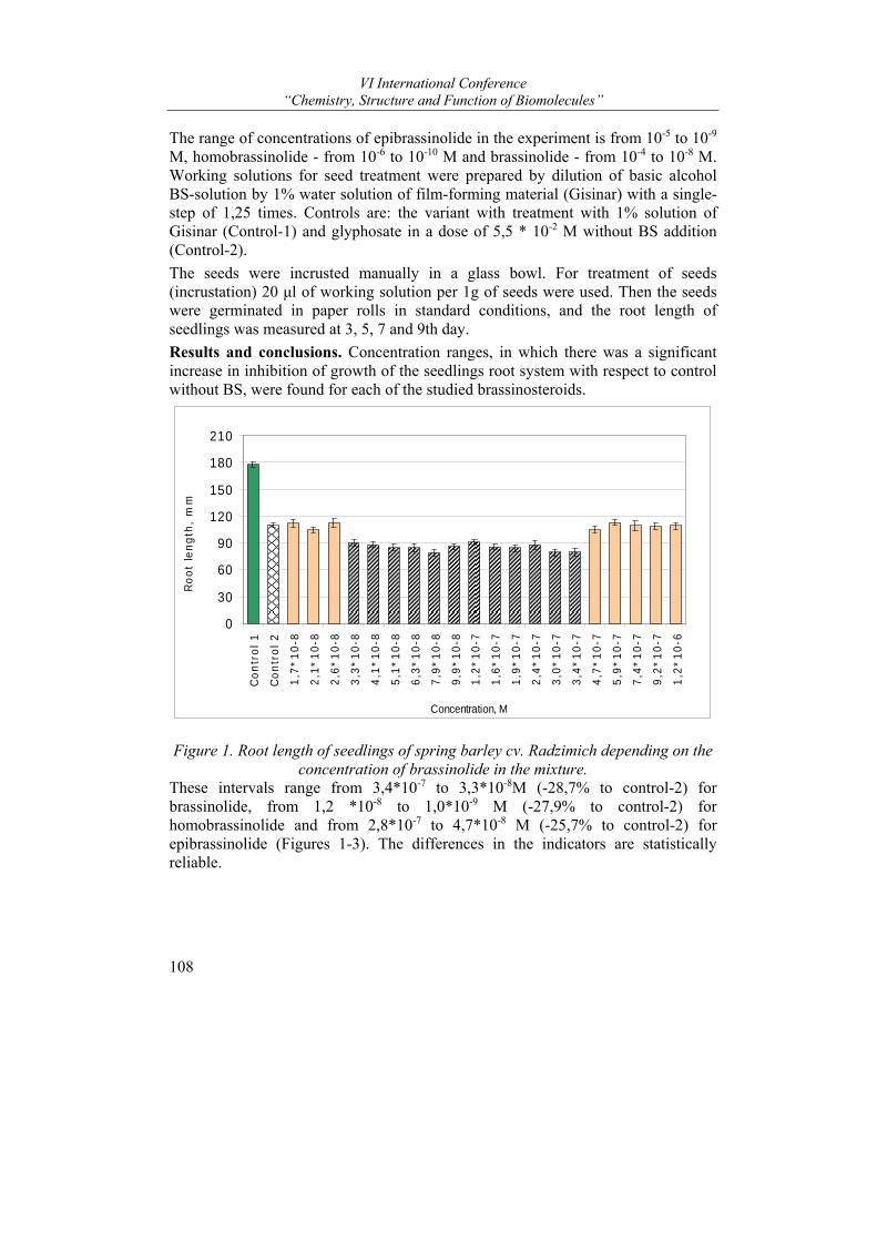
VI International Conference “Chemistry, Structure and Function of Biomolecules”
108
The range of concentrations of epibrassinolide in the experiment is from 10-5 to 10-9 M, homobrassinolide - from 10-6 to 10-10 M and brassinolide - from 10-4 to 10-8 M. Working solutions for seed treatment were prepared by dilution of basic alcohol BS-solution by 1% water solution of film-forming material (Gisinar) with a single-step of 1,25 times. Controls are: the variant with treatment with 1% solution of Gisinar (Control-1) and glyphosate in a dose of 5,5 * 10-2 M without BS addition (Control-2).
The seeds were incrusted manually in a glass bowl. For treatment of seeds (incrustation) 20 μl of working solution per 1g of seeds were used. Then the seeds were germinated in paper rolls in standard conditions, and the root length of seedlings was measured at 3, 5, 7 and 9th day.
Results and conclusions. Concentration ranges, in which there was a significant increase in inhibition of growth of the seedlings root system with respect to control without BS, were found for each of the studied brassinosteroids.
0
30
60
90
120
150
180
210
Con
trol
1
Con
trol
2
1,7*
10-8
2,1*
10-8
2,6*
10-8
3,3*
10-8
4,1*
10-8
5,1*
10-8
6,3*
10-8
7,9*
10-8
9,9*
10-8
1,2*
10-7
1,6*
10-7
1,9*
10-7
2,4*
10-7
3,0*
10-7
3,4*
10-7
4,7*
10-7
5,9*
10-7
7,4*
10-7
9,2*
10-7
1,2*
10-6
Concentration, M
Roo
t le
ngth
, m
m
Figure 1. Root length of seedlings of spring barley cv. Radzimich depending on the
concentration of brassinolide in the mixture. These intervals range from 3,4*10-7 to 3,3*10-8M (-28,7% to control-2) for brassinolide, from 1,2 *10-8 to 1,0*10-9 M (-27,9% to control-2) for homobrassinolide and from 2,8*10-7 to 4,7*10-8 M (-25,7% to control-2) for epibrassinolide (Figures 1-3). The differences in the indicators are statistically reliable.
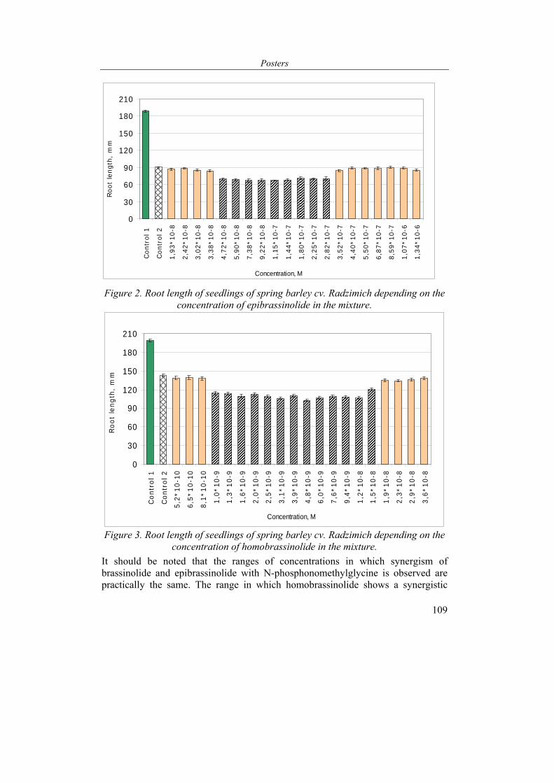
Posters
109
0
30
60
90
120
150
180
210
Con
trol
1
Con
trol
2
1,93
*10-
8
2,42
*10-
8
3,02
*10-
8
3,38
*10-
8
4,72
*10-
8
5,90
*10-
8
7,38
*10-
8
9,22
*10-
8
1,15
*10-
7
1,44
*10-
7
1,80
*10-
7
2,25
*10-
7
2,82
*10-
7
3,52
*10-
7
4,40
*10-
7
5,50
*10-
7
6,87
*10-
7
8,59
*10-
7
1,07
*10-
6
1,34
*10-
6
Concentration, M
Roo
t le
ngth
, m
m
Figure 2. Root length of seedlings of spring barley cv. Radzimich depending on the concentration of epibrassinolide in the mixture.
0
30
60
90
120
150
180
210
Con
trol
1
Con
trol
2
5,2*
10-1
0
6,5*
10-1
0
8,1*
10-1
0
1,0*
10-9
1,3*
10-9
1,6*
10-9
2,0*
10-9
2,5*
10-9
3,1*
10-9
3,9*
10-9
4,8*
10-9
6,0*
10-9
7,6*
10-9
9,4*
10-9
1,2*
10-8
1,5*
10-8
1,9*
10-8
2,3*
10-8
2,9*
10-8
3,6*
10-8
Concentration, M
Roo
t le
ngth
, m
m
Figure 3. Root length of seedlings of spring barley cv. Radzimich depending on the
concentration of homobrassinolide in the mixture.
It should be noted that the ranges of concentrations in which synergism of brassinolide and epibrassinolide with N-phosphonomethylglycine is observed are practically the same. The range in which homobrassinolide shows a synergistic
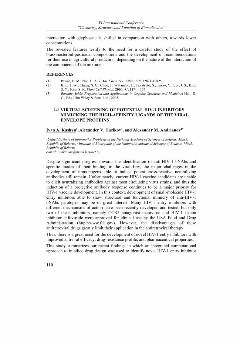
VI International Conference “Chemistry, Structure and Function of Biomolecules”
110
interaction with glyphosate is shifted in comparison with others, towards lower concentrations.
The revealed features testify to the need for a careful study of the effect of brassinosteroid-pesticidal compositions and the development of recommendations for their use in agricultural production, depending on the nature of the interaction of the components of the mixtures.
REFERENCES
(1) Pawar, D. M.; Noe, E. A. J. Am. Chem. Soc. 1996, 118, 12821-12825. (2) Kim, T. W.; Chang, S. C.; Choo, J.; Watanabe, T.; Takatsuto, S.; Takao, Y.; Lee, J. S.; Kim,
S. Y.; Kim, S. K. Plant Cell Physiol. 2000, 41, 1171-1174. (3) Boronic Acids: Preparation and Applications in Organic Synthesis and Medicine; Hall, D.
G., Ed.; John Wiley & Sons, Ltd., 2005.
VIRTUAL SCREENING OF POTENTIAL HIV-1 INHIBITORS MIMICKING THE HIGH-AFFINITY LIGANDS OF THE VIRAL ENVELOPE PROTEINS
Ivan A. Kashyn1, Alexander V. Tuzikov1, and Alexander M. Andrianov2*
1United Institute of Informatics Problems of the National Academy of Sciences of Belarus, Minsk, Republic of Belarus, 2 Institute of Bioorganic of the National Academy of Sciences of Belarus, Minsk, Republic of Belarus e-mail: [email protected]
Despite significant progress towards the identification of anti-HIV-1 bNAbs and specific modes of their binding to the viral Env, the major challenges in the development of immunogens able to induce potent cross-reactive neutralizing antibodies still remain. Unfortunately, current HIV-1 vaccine candidates are unable to elicit neutralizing antibodies against most circulating virus strains, and thus the induction of a protective antibody response continues to be a major priority for HIV-1 vaccine development. In this context, development of small-molecule HIV-1 entry inhibitors able to show structural and functional mimicry of anti-HIV-1 bNAbs paratopes may be of great interest. Many HIV-1 entry inhibitors with different mechanisms of action have been recently developed and tested, but only two of these inhibitors, namely CCR5 antagonist maraviroc and HIV-1 fusion inhibitor enfuvirtide were approved for clinical use by the USA Food and Drug Administration (http://www.fda.gov). However, the disadvantages of these antiretroviral drugs greatly limit their application in the antiretroviral therapy.
Thus, there is a great need for the development of novel HIV-1 entry inhibitors with improved antiviral efficacy, drug-resistance profile, and pharmaceutical properties.
This study summarizes our recent findings in which an integrated computational approach to in silico drug design was used to identify novel HIV-1 entry inhibitor
Posters
111
scaffolds mimicking high-affinity ligands of the viral envelope proteins, namely broadly neutralizing anti-HIV-1 antibodies (bNabs) VRC01, 3074, 10E8, and primary receptor CD4. This computer-based approach includes: (i) generation of pharmacophore models representing 3D-arrangements of chemical functionalities that make the above high-affinity ligands active towards their targets, (ii) shape and pharmacophore-based identification of mimetic candidates for VRC01, 3074, 10E8 and CD4 by a web-oriented virtual screening platform pepMMsMIMIC, (iii) high-throughput docking of the identified compounds with the molecular targets, and (iv) molecular dynamics simulations of the docked structures followed by binding free energy calculations.
As a result, peptidomimetics of bNabs VRC01, 3074, 10E8 and primary receptor CD4 that exhibit spatial and pharmacophore features responsible for biological activity were found by virtual screening and computer modeling tools. The identified compounds were shown to expose a high binding affinity to the HIV-1 envelope proteins by mimicking critical interactions between HIV-1 and cellular receptors. This provides strong adsorption on the surface of a target cell. Based on the data obtained, the identified molecules were shown to form a strong foundation for the development of novel, potent and broad anti-HIV drugs. It was suggested that a “cocktail” of these small molecules blocking different functionally conserved epitopes on HIV-1 envelope may suppress the viral replication and reduce the plasma HIV-1 load.
So the obtained findings show that the computational methodology used here is a powerful in silico tool for the discovery of novel small-molecule functional antagonists of viral entry based on the high-affinity ligands of the HIV-1 envelope proteins.
This study was supported by grants from the Belarusian Republican Foundation for Fundamental Research (projects X17MC-004, X18KИ-002).
STEREOSELECTIVE SYNTHESIS OF -ALKYL KETONES FROM ESTERS AND ALKENES VIA CYCLOPROPANOL INTERMEDIATES
Maryia V. Barysevich, Volha V. Kazlova, Aliaksandr G. Kukel, Aliaksandra I. Liubina, Alaksiej L. Hurski*, Vladimir N. Zhabinskii, and Vladimir A. Khripach
Laboratory of Steroids, Institute of Bioorganic Chemistry, National Academy of Sciences of Belarus, Belarus e-mail: [email protected]
Alkenes bearing a chiral allylic carbon were found to undergo Kulinkovich hydroxycyclopropanation with a good level of diastereoselectivity.1 For the
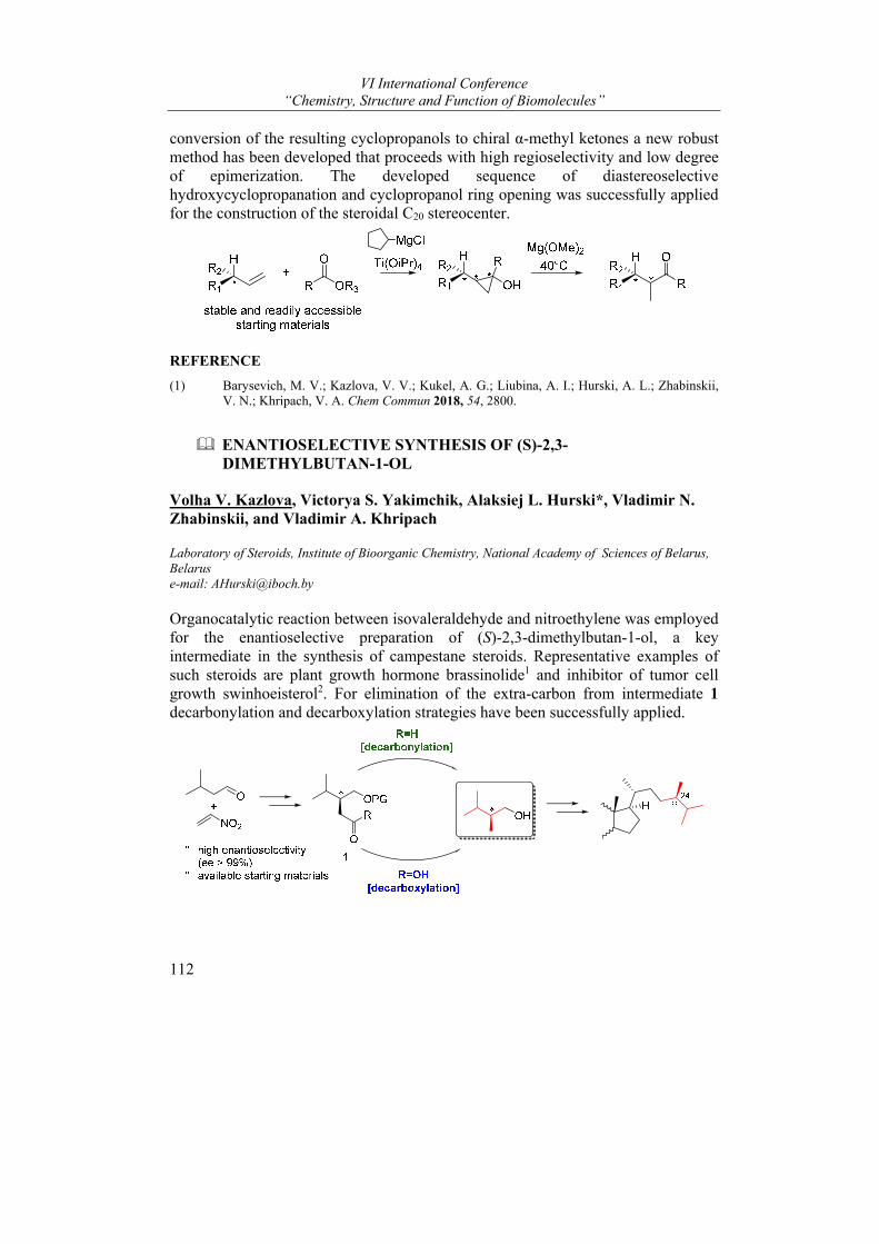
VI International Conference “Chemistry, Structure and Function of Biomolecules”
112
conversion of the resulting cyclopropanols to chiral α-methyl ketones a new robust method has been developed that proceeds with high regioselectivity and low degree of epimerization. The developed sequence of diastereoselective hydroxycyclopropanation and cyclopropanol ring opening was successfully applied for the construction of the steroidal C20 stereocenter.
REFERENCE
(1) Barysevich, M. V.; Kazlova, V. V.; Kukel, A. G.; Liubina, A. I.; Hurski, A. L.; Zhabinskii, V. N.; Khripach, V. A. Chem Commun 2018, 54, 2800.
ENANTIOSELECTIVE SYNTHESIS OF (S)-2,3-DIMETHYLBUTAN-1-OL
Volha V. Kazlova, Victorya S. Yakimchik, Alaksiej L. Hurski*, Vladimir N. Zhabinskii, and Vladimir A. Khripach
Laboratory of Steroids, Institute of Bioorganic Chemistry, National Academy of Sciences of Belarus, Belarus e-mail: [email protected]
Organocatalytic reaction between isovaleraldehyde and nitroethylene was employed for the enantioselective preparation of (S)-2,3-dimethylbutan-1-ol, a key intermediate in the synthesis of campestane steroids. Representative examples of such steroids are plant growth hormone brassinolide1 and inhibitor of tumor cell growth swinhoeisterol2. For elimination of the extra-carbon from intermediate 1 decarbonylation and decarboxylation strategies have been successfully applied.
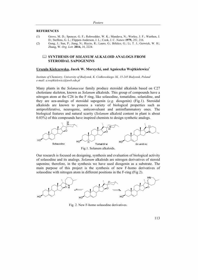
Posters
113
REFERENCES
(1) Grove, M. D.; Spencer, G. F.; Rohwedder, W. K.; Mandava, N.; Worley, J. F.; Warthen, J. D.; Steffens, G. L.; Flippen-Anderson, J. L.; Cook, J. C. Nature 1979, 281, 216.
(2) Gong, J.; Sun, P.; Jiang, N.; Riccio, R.; Lauro, G.; Bifulco, G.; Li, T. J.; Gerwick, W. H.; Zhang, W. Org. Lett. 2014, 16, 2224.
SYNTHESIS OF SOLANUM ALKALOID ANALOGS FROM STEROIDAL SAPOGENINS
Urszula Kielczewska, Jacek W. Morzycki, and Agnieszka Wojtkielewicz*
Institute of Chemistry, University of Bialystok, K. Ciołkowskiego 1K, 15-245 Bialystok, Poland e-mail: [email protected]
Many plants in the Solanaceae family produce steroidal alkaloids based on C27 cholestane skeleton, known as Solanum alkaloids. This group of compounds have a nitrogen atom at the C26 in the F ring, like solasodine, tomatidine, solanidine, and they are aza-analogs of steroidal sapogenin (e.g. diosgenin) (Fig.1). Steroidal alkaloids are known to possess a variety of biological properties such as antiproliferative, neurogenic, anticonvulsant and antiinflammatory ones. The biological features and natural scarity (Solanum alkaloid content in plant is about 0.03%) of this compounds have inspired chemists to design synthetic analogs.
Fig.1. Solanum alkaloids.
Our research is focused on designing, synthesis and evaluation of biological activity of solasodine and its analogs. Solanum alkaloids are nitrogen derivatives of steroid saponins; therefore, in the synthesis we have used diosgenin as a substrate. The main purpose of this project is the synthesis of new F-homo derivatives of solasodine with nitrogen atom in different positions in the F-ring (Fig 2).
Fig. 2. New F-homo solasodine derivatives.
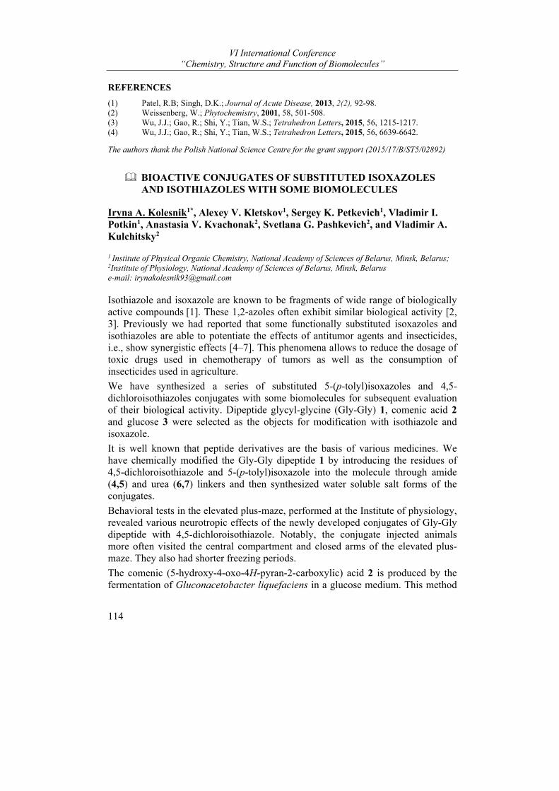
VI International Conference “Chemistry, Structure and Function of Biomolecules”
114
REFERENCES
(1) Patel, R.B; Singh, D.K.; Journal of Acute Disease, 2013, 2(2), 92-98. (2) Weissenberg, W.; Phytochemistry, 2001, 58, 501-508. (3) Wu, J.J.; Gao, R.; Shi, Y.; Tian, W.S.; Tetrahedron Letters, 2015, 56, 1215-1217. (4) Wu, J.J.; Gao, R.; Shi, Y.; Tian, W.S.; Tetrahedron Letters, 2015, 56, 6639-6642.
The authors thank the Polish National Science Centre for the grant support (2015/17/B/ST5/02892)
BIOACTIVE CONJUGATES OF SUBSTITUTED ISOXAZOLES AND ISOTHIAZOLES WITH SOME BIOMOLECULES
Iryna A. Kolesnik1*, Alexey V. Kletskov1, Sergey K. Petkevich1, Vladimir I. Potkin1, Anastasia V. Kvachonak2, Svetlana G. Pashkevich2, and Vladimir A. Kulchitsky2
1 Institute of Physical Organic Chemistry, National Academy of Sciences of Belarus, Minsk, Belarus; 2Institute of Physiology, National Academy of Sciences of Belarus, Minsk, Belarus e-mail: [email protected]
Isothiazole and isoxazole are known to be fragments of wide range of biologically active compounds [1]. These 1,2-azoles often exhibit similar biological activity [2, 3]. Previously we had reported that some functionally substituted isoxazoles and isothiazoles are able to potentiate the effects of antitumor agents and insecticides, i.e., show synergistic effects [4–7]. This phenomena allows to reduce the dosage of toxic drugs used in chemotherapy of tumors as well as the consumption of insecticides used in agriculture.
We have synthesized a series of substituted 5-(p-tolyl)isoxazoles and 4,5-dichloroisothiazoles conjugates with some biomolecules for subsequent evaluation of their biological activity. Dipeptide glycyl-glycine (Gly-Gly) 1, comenic acid 2 and glucose 3 were selected as the objects for modification with isothiazole and isoxazole.
It is well known that peptide derivatives are the basis of various medicines. We have chemically modified the Gly-Gly dipeptide 1 by introducing the residues of 4,5-dichloroisothiazole and 5-(p-tolyl)isoxazole into the molecule through amide (4,5) and urea (6,7) linkers and then synthesized water soluble salt forms of the conjugates.
Behavioral tests in the elevated plus-maze, performed at the Institute of physiology, revealed various neurotropic effects of the newly developed conjugates of Gly-Gly dipeptide with 4,5-dichloroisothiazole. Notably, the conjugate injected animals more often visited the central compartment and closed arms of the elevated plus-maze. They also had shorter freezing periods.
The comenic (5-hydroxy-4-oxo-4H-pyran-2-carboxylic) acid 2 is produced by the fermentation of Gluconacetobacter liquefaciens in a glucose medium. This method
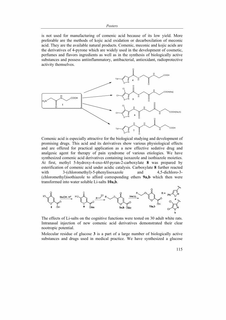
Posters
115
is not used for manufacturing of comenic acid because of its low yield. More preferable are the methods of kojic acid oxidation or decarboxilation of meconic acid. They are the available natural products. Comenic, meconic and kojic acids are the derivatives of 4-pyrone which are widely used in the development of cosmetic, perfumes and flavors ingredients as well as in the synthesis of biologically active substances and possess antiinflammatory, antibacterial, antioxidant, radioprotective activity themselves.
NH
HN COOH(Na)
O
OS N
Cl
Cl
H2N
HN COOH
O HN
HN
NH
S N
Cl
Cl
O
O
COOH(Na,K)
NH
HN COOH
O
OO N
Tol
HN
HN
NH
O
O
COOH
O N
Tol
1
6
5
4
7
Comenic acid is especially attractive for the biological studying and development of promising drugs. This acid and its derivatives show various physiological effects and are offered for practical application as a new effective sedative drug and analgesic agent for therapy of pain syndrome of various etiologies. We have synthesized comenic acid derivatives containing isoxazole and isothiazole moieties. At first, methyl 5-hydroxy-4-oxo-4H-pyran-2-carboxylate 8 was prepared by esterification of comenic acid under acidic catalysis. Carboxylate 8 further reacted with 3-(chloromethyl)-5-phenylisoxazole and 4,5-dichloro-3-(chloromethyl)isothiazole to afford corresponding ethers 9a,b which then were transformed into water soluble Li-salts 10a,b.
The effects of Li-salts on the cognitive functions were tested on 30 adult white rats. Intranasal injection of new comenic acid derivatives demonstrated their clear nootropic potential.
Molecular residue of glucose 3 is a part of a large number of biologically active substances and drugs used in medical practice. We have synthesized a glucose

VI International Conference “Chemistry, Structure and Function of Biomolecules”
116
conjugate with 4,5-dichloroisothiazole 11 by selective acylation of its molecule with 4,5-dichloroisothiazole carbonyl chloride as shown below.
Intranasal injection of the synthesized conjugate of glucose with isothiazole to rats was followed by increase of their locomotor activity.
Thus, for all new compounds, neurotropic effects are revealed with different intensity and direction, which is the basis for further research.
REFERENCES
(1) Bioactive Heterocycles III; Khan, M., Ed.; Springer: Berlin Heidelberg, 2007, Vol. IX. (2) Matzen, L.; Engesgaard, A.; Ebert B.; Didriksen, M.; Frolund, B.; Krogsgaard-Larsen, P.;
Jaroszewski, J.W. J. Med. Chem. 1997, 40, 520-527. (3) Zefirova, O.N.; Zefirov, N.S. Rus J. Org. Chem. 2000, 36, 1231-1258. (4) Kulchitsky, V.A.; Potkin, V.; Zubenko Yu.S.; Chernov, A.N.; Talabaev, M.V.; Demidchik,
Yu.E.; Petkevich, S.K.; Kazbanov, V.V.; Gurinovich, T.A.; Roeva, M.O.; Grigoriev, D.G.; Kletskov, A.V.; Kalunov, V.N. Med. Chem. 2012, 8, 22-32.
(5) Potkin, V.I.; Kletskov, A.V.; Petkevich, S.K.; Pashkevich, S.G.; Kazbanov, V.V.; Denisov, A.A.; Kulchitsky, V.A. Heterocycl. Lett. 2015, 1, 11-19.
(6) Potkin, V.; Zubenko, Yu.; Bykhovetz, A.; Zolotar, R.; Goncharuk, V. Nat. Prod. Commun. 2009, 4, 1205-1208.
(7) Kletskov, A.V.; Potkin, V.I.; Dikusar, E.A.; Zolotar, R.M. Nat. Prod. Commun. 2017, 12, 105-106.
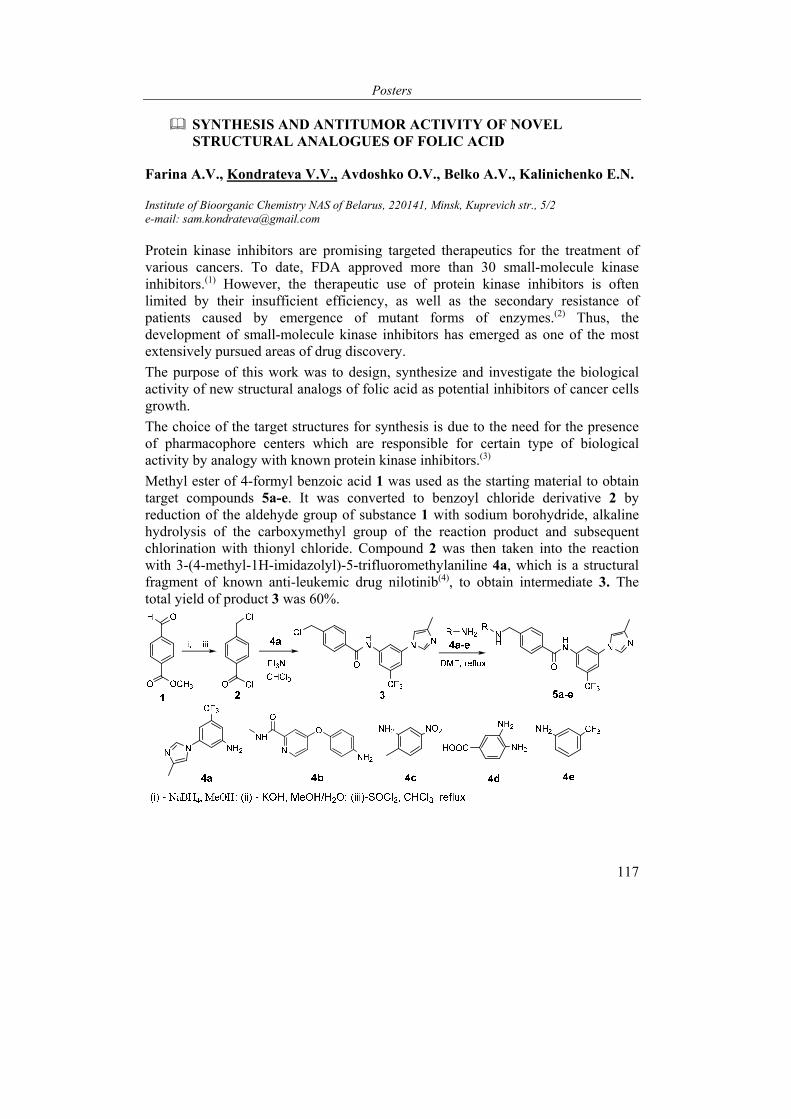
Posters
117
SYNTHESIS AND ANTITUMOR ACTIVITY OF NOVEL STRUCTURAL ANALOGUES OF FOLIC ACID
Farina A.V., Kondrateva V.V., Avdoshko O.V., Belko A.V., Kalinichenko E.N.
Institute of Bioorganic Chemistry NAS of Belarus, 220141, Minsk, Kuprevich str., 5/2 e-mail: [email protected]
Protein kinase inhibitors are promising targeted therapeutics for the treatment of various cancers. To date, FDA approved more than 30 small-molecule kinase inhibitors.(1) However, the therapeutic use of protein kinase inhibitors is often limited by their insufficient efficiency, as well as the secondary resistance of patients caused by emergence of mutant forms of enzymes.(2) Thus, the development of small-molecule kinase inhibitors has emerged as one of the most extensively pursued areas of drug discovery.
The purpose of this work was to design, synthesize and investigate the biological activity of new structural analogs of folic acid as potential inhibitors of cancer cells growth.
The choice of the target structures for synthesis is due to the need for the presence of pharmacophore centers which are responsible for certain type of biological activity by analogy with known protein kinase inhibitors.(3)
Methyl ester of 4-formyl benzoic acid 1 was used as the starting material to obtain target compounds 5a-e. It was converted to benzoyl chloride derivative 2 by reduction of the aldehyde group of substance 1 with sodium borohydride, alkaline hydrolysis of the carboxymethyl group of the reaction product and subsequent chlorination with thionyl chloride. Compound 2 was then taken into the reaction with 3-(4-methyl-1H-imidazolyl)-5-trifluoromethylaniline 4a, which is a structural fragment of known anti-leukemic drug nilotinib(4), to obtain intermediate 3. The total yield of product 3 was 60%.
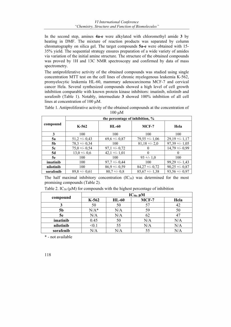
VI International Conference “Chemistry, Structure and Function of Biomolecules”
118
In the second step, amines 4a-e were alkylated with chloromethyl amide 3 by heating in DMF. The mixture of reaction products was separated by column chromatography on silica gel. The target compounds 5a-e were obtained with 15-35% yield. The sequential strategy ensures preparation of a wide variety of amides via variation of the initial amine structure. The structure of the obtained compounds was proved by 1H and 13C NMR spectroscopy and confirmed by data of mass spectrometry.
The antiproliferative activity of the obtained compounds was studied using single concentration MTT test on the cell lines of chronic myelogenous leukemia K-562, promyelocytic leukemia HL-60, mammary adenocarcinoma MCF-7 and cervical cancer Hela. Several synthesized compounds showed a high level of cell growth inhibition comparable with known protein kinase inhibitors: imatinib, nilotinib and sorafenib (Table 1). Notably, intermediate 3 showed 100% inhibition of all cell lines at concentration of 100 μM.
Table 1. Antiproliferative activity of the obtained compounds at the concentration of 100 μM
compound
the percentage of inhibition, %
K-562 HL-60 MCF-7 Hela
3 100 100 100 100 5a 51,2 +/- 0,43 69,6 +/- 0,87 79,55 +/- 1,06 29,19 +/- 1,17 5b 78,3 +/- 0,34 100 81,18 +/- 2,0 97,39 +/- 1,05 5c 75,0 +/- 0,54 97,1 +/- 0,72 0 14,79 +/- 0,99 5d 13,0 +/- 0,6 42,1 +/- 1,01 0 0 5e 100 100 93 +/- 1,0 100
imatinib 100 97,7 +/- 0,44 100 99,29 +/- 1,43 nilotinib 100 86,9 +/- 0,59 84,27 +/- 0,72 90,25 +/- 0,87 sorafenib 89,8 +/- 0,61 80,7 +/- 0,8 85,67 +/- 1,38 93,56 +/- 0,97
The half maximal inhibitory concentration (IC50) was determined for the most promising compounds (Table 2).
Table 2. IC50 (μM) for compounds with the highest percentage of inhibition
compound IC50, μМ
K-562 HL-60 MCF-7 Hela 3 50 50 57 42
5b N/A* N/A 59 50 5e N/A N/A 62 47
imatinib 0.45 50 N/A N/A nilotinib <0.1 55 N/A N/A sorafenib N/A N/A 55 N/A
* - not available
Posters
119
As a result of the study, 6 new compounds were obtained showing a significant level of antiproliferative activity against several cancer cell lines. The results of biological activity testing suggest further structure optimization of titled compounds as well as structure-activity relationship investigation to create effective anticancer drugs.
REFERENCES
(1) Zhang, J., Salminen, A., Yang, X., Luo, Y., Wu, Q., White, M., Archives of toxicology, 2017, 91(8), 2921-2938.
(2) Jabbour, E., Hochhaus, A., Cortes, J., La Rosee, P., & Kantarjian, H. M. Leukemia. 2010. 24(1). 6.
(3) Cui, J., Fu, R., Zhou, L. H., Chen, S. P., Li, G. W., Qian, S. X., & Liu, S. Bioorganic & medicinal chemistry letters. 2013. 23(8), 2442-2450.
(4) Su, B. H., Huang, Y. S., Chang, C. Y., Tu, Y. S., & Tseng, Y. J. Molecules. 2013, 18, 13487-13509.
CYCLOPENTENONE SYNTHONS FOR 3-OXA-3,7-INTERPHENYLENE ANALOGUES OF PROSTAGLANDINS ON THE BASIS OF CYCLOPENTANE-1,3-DIONE
Felix S. Pashkovsky*, Dmitry I. Korneev, and Fedor A. Lakhvich
The Institute of Bioorganic Chemistry, National Academy of Sciences of Belarus, Minsk, Belarus, e-mail: [email protected]
Among synthetic prostanoids 3,7-interphenylene- as well as 3-oxa-analogues are of considerable interest. On one hand, such prostanoids exhibit a broad spectrum of biological activities1. On the other hand, they possess increased metabolic stability due to suppression by 3,7-interphenylene fragment and/or 3-oxa- function of the β-oxidation of α-prostanoid chain2 (one of the main ways of metabolic decomposition of prostaglandins in the body). In this connection, synthesis of new metabolically stable 3,7-interphenylene- and 3-oxa- PG-analogues is of great scientific and practical importance.
Herein, we describe a synthetic scheme for obtaining cyclopentenone synthons for 3-oxa-3,7-inter-m-phenylene prostanoids. In this scheme cyclopentane-1,3-dione (1) serves as the precursor of cyclic part of the target synthons, and readily available 3-(formylphenoxy)acetic acid methyl ester (2) is employed as the 3,7-inter-m-phenylene α-prostanoid chain precursor.
Coupling of the compounds (1) and (2) can be achieved by Knoevenagel condensation to give cross-conjugated diketone (3). Such methodology was used by us earlier in preparation of 3,7-interphenylene 3,10(11)-dioxa-13-aza- and 9-oxa-7-azaprostanoids on the basis of tetronic acid3. But in contrast to stable 3-arylidenetetrahydrofuran-2,4-diones described earlier3, related to them cross-conjugated diketone (3) is a very reactive intermediate which immediately reacts
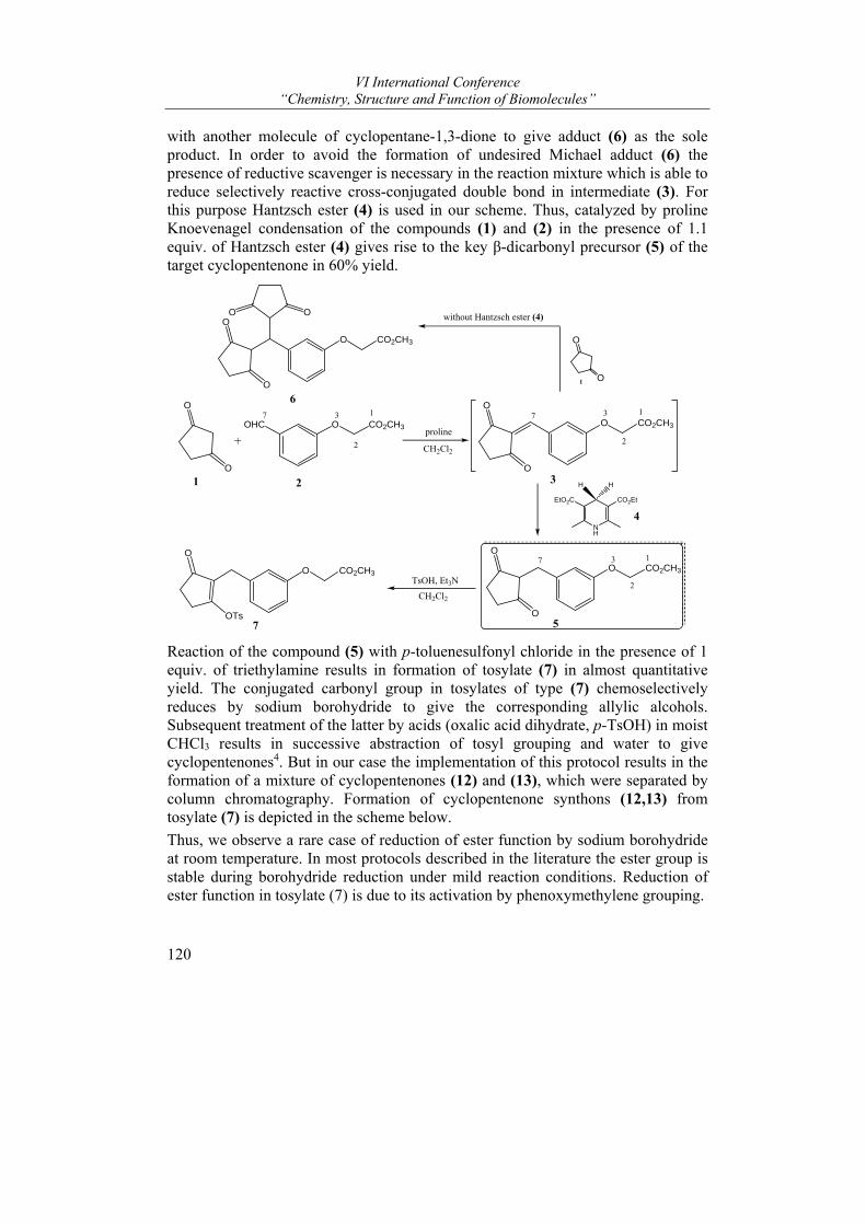
VI International Conference “Chemistry, Structure and Function of Biomolecules”
120
with another molecule of cyclopentane-1,3-dione to give adduct (6) as the sole product. In order to avoid the formation of undesired Michael adduct (6) the presence of reductive scavenger is necessary in the reaction mixture which is able to reduce selectively reactive cross-conjugated double bond in intermediate (3). For this purpose Hantzsch ester (4) is used in our scheme. Thus, catalyzed by proline Knoevenagel condensation of the compounds (1) and (2) in the presence of 1.1 equiv. of Hantzsch ester (4) gives rise to the key β-dicarbonyl precursor (5) of the target cyclopentenone in 60% yield.
O
O
O CO2CH3
O
O
+
OHC O CO2CH3
NH
EtO2C CO2Et
O
O
O CO2CH3proline
CH2Cl2
1 2 3
4
5
2
37 1O
OTs
O CO2CH3
2
37 1
TsOH, Et3N
CH2Cl2
O
O1
O
O
O CO2CH3
OO without Hantzsch ester (4)
7
1
2
37
6
H H
Reaction of the compound (5) with p-toluenesulfonyl chloride in the presence of 1 equiv. of triethylamine results in formation of tosylate (7) in almost quantitative yield. The conjugated carbonyl group in tosylates of type (7) chemoselectively reduces by sodium borohydride to give the corresponding allylic alcohols. Subsequent treatment of the latter by acids (oxalic acid dihydrate, p-TsOH) in moist CHCl3 results in successive abstraction of tosyl grouping and water to give cyclopentenones4. But in our case the implementation of this protocol results in the formation of a mixture of cyclopentenones (12) and (13), which were separated by column chromatography. Formation of cyclopentenone synthons (12,13) from tosylate (7) is depicted in the scheme below.
Thus, we observe a rare case of reduction of ester function by sodium borohydride at room temperature. In most protocols described in the literature the ester group is stable during borohydride reduction under mild reaction conditions. Reduction of ester function in tosylate (7) is due to its activation by phenoxymethylene grouping.
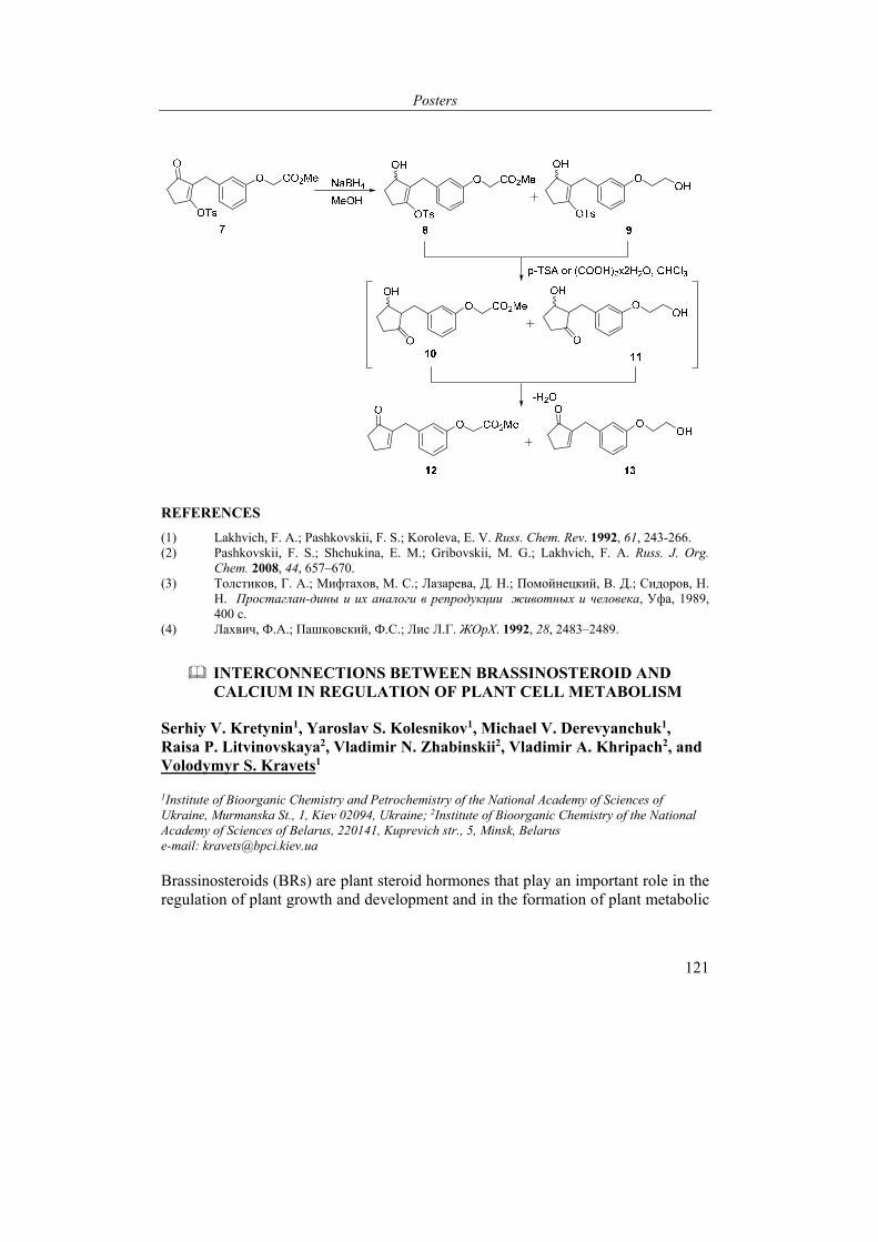
Posters
121
REFERENCES
(1) Lakhvich, F. A.; Pashkovskii, F. S.; Koroleva, E. V. Russ. Chem. Rev. 1992, 61, 243-266. (2) Pashkovskii, F. S.; Shchukina, E. M.; Gribovskii, M. G.; Lakhvich, F. A. Russ. J. Org.
Chem. 2008, 44, 657–670. (3) Толстиков, Г. А.; Мифтахов, М. С.; Лазарева, Д. Н.; Помойнецкий, В. Д.; Сидоров, Н.
Н. Простаглан-дины и их аналоги в репродукции животных и человека, Уфа, 1989, 400 с.
(4) Лахвич, Ф.А.; Пашковский, Ф.С.; Лис Л.Г. ЖОрХ. 1992, 28, 2483–2489.
INTERCONNECTIONS BETWEEN BRASSINOSTEROID AND CALCIUM IN REGULATION OF PLANT CELL METABOLISM
Serhiy V. Kretynin1, Yaroslav S. Kolesnikov1, Мichael V. Derevyanchuk1, Raisa P. Litvinovskaya2, Vladimir N. Zhabinskii2, Vladimir A. Khripach2, and Volodymyr S. Kravets1
1Institute of Bioorganic Chemistry and Petrochemistry of the National Academy of Sciences of Ukraine, Murmanska St., 1, Kiev 02094, Ukraine; 2Institute of Bioorganic Chemistry of the National Academy of Sciences of Belarus, 220141, Kuprevich str., 5, Minsk, Belarus e-mail: [email protected]
Brassinosteroids (BRs) are plant steroid hormones that play an important role in the regulation of plant growth and development and in the formation of plant metabolic
VI International Conference “Chemistry, Structure and Function of Biomolecules”
122
response to stress action. However, mechanisms which involve BRs in plant cell metabolism regulation are poorly understood.
NADPH plays a key role in the myriad of metabolic pathways in cells with different level of organization. Significant importance in NADPH biosynthesis belongs to the activity of glucose-6-phosphate dehydrogenase. However, the role of brassinosteroids (e.g. epibrassinolide, EBl) and the second messengers (e.g. free calcium) in regulation of the level of NADP+/NADPH in plants is insuffiently studied.
For this purpose, tobacco plants were grown hydroponically for 4 weeks on ½ nutrient Hogland solution containing sufficient amount of calcium or on calcium-free ½ nutrient Hogland solution. After 1 week, EBl was added to nutrient medium and activity of antioxidant enzymes was measured in tobacco leaves after 1 day. Besides wild-type tobacco plants, transgenic cax1 tobacco plants expressing H+/Ca2+ vacuolar Arabidopsis thaliana antiporter CAX1 (Cation Exchanger 1, At2g38170) and thus having the exhaustion of cytosolic calcium pool under limited calcium uptake were utilized for investigations of the role of calcium in regulation of these events. After 6 weeks, another set of plants was transferred to soil in order to obtain seeds.
Results suggest that brassinosteroids alone and, especially, in the presence of calcium stimulated the activity of ascorbate-glutathione cycle enzymes (ascorbate peroxidase and glutathione reductase) as well as increased seed weight and the amount of oil in wild type and transgenic plants. On the other hand, pharmacological reduction of glucose-6-phosphate dehydrogenase activity caused a dramatic reduction in the activity of ascorbate-glutathione cycle enzymes stimulated by epibrassinolide alone and together with calcium. Тransient calcium exclusion from the nutrient medium during BRs action in transgenic plants caused the dramatic reduction of EBl-stimulated seed weight and the amount of oil in comparison to the samples with sufficient calcium supply. According to the literature data of brassinosteroid-regulated proteomics and in silico prediction of protein-protein interactions in Arabidopsis thaliana, brassinosteroids induce protein phosphorylation changes of calcium-regulated proteins (calcium-dependent protein kinase AtCDPK2; calcium-binding EF hand-containing protein At1g20760) that could interact with NADPH-generating enzymes (6-phosphogluconate dehydrogenase (At6PGDH); NAD kinase 2 (AtNDK2); 6-phosphogluconate dehydrogenase family protein AtPGD1) regulating their activity and thus possibly modulating NADPH levels in plants.
Therefore, the results suggest that BRs ability to regulate NADPH levels by activating calcium signaling serves an important way to raise antioxidant potential and biological productivity in plants.
Authors are grateful to Prof. K.D. Hirschi (Baylor College of Medicine, U.S. Department of Agriculture) for providing seeds of cax1 tobacco plants. This work was supported by the joint grants of the NAS of Ukraine – the NAS of Belarus – 2018 (X18УКА-010).
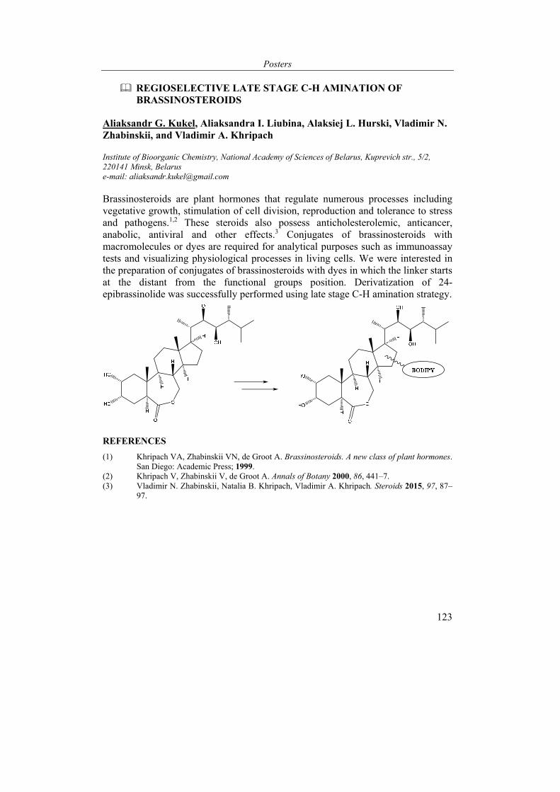
Posters
123
REGIOSELECTIVE LATE STAGE C-H AMINATION OF BRASSINOSTEROIDS
Aliaksandr G. Kukel, Aliaksandra I. Liubina, Alaksiej L. Hurski, Vladimir N. Zhabinskii, and Vladimir A. Khripach
Institute of Bioorganic Chemistry, National Academy of Sciences of Belarus, Kuprevich str., 5/2, 220141 Minsk, Belarus e-mail: [email protected]
Brassinosteroids are plant hormones that regulate numerous processes including vegetative growth, stimulation of cell division, reproduction and tolerance to stress and pathogens.1,2 These steroids also possess anticholesterolemic, anticancer, anabolic, antiviral and other effects.3 Conjugates of brassinosteroids with macromolecules or dyes are required for analytical purposes such as immunoassay tests and visualizing physiological processes in living cells. We were interested in the preparation of conjugates of brassinosteroids with dyes in which the linker starts at the distant from the functional groups position. Derivatization of 24-epibrassinolide was successfully performed using late stage C-H amination strategy.
REFERENCES
(1) Khripach VA, Zhabinskii VN, de Groot A. Brassinosteroids. A new class of plant hormones. San Diego: Academic Press; 1999.
(2) Khripach V, Zhabinskii V, de Groot A. Annals of Botany 2000, 86, 441–7. (3) Vladimir N. Zhabinskii, Natalia B. Khripach, Vladimir A. Khripach. Steroids 2015, 97, 87–
97.
VI International Conference “Chemistry, Structure and Function of Biomolecules”
124
SYNTHESIS OF NEW 6-N-MODIFIED PURINE NUCLEOSIDES
Tamara Kulak*, Darya Yankovskaya, Alena Konoplich, Tatyana Buravskaya, and Elena Kalinichenko
Institute of Bioorganic Chemistry, National Academy of Sciences of Belarus, Minsk, Republic of Belarus e-mail: [email protected]
6-N-Substituted purine nucleosides (cytokinin nucleosides) are of great interest since many derivatives belonging to this class of compounds exhibit prominent biological activities. Cytokinin nucleosides represent an important group of plant growth regulatory substances involved in the processes of plant cell division, shoot differentiation, biosynthesis of pigments. In animals, 6-N-substituted adenine nucleosides are found to be agonists/antagonists of various types of adenosine receptors. A number of such derivatives possess antiviral or antitumor properties characteristic of a wide spectrum of nucleoside drugs as a whole. Besides, several 6-N-substituted purine nucleosides exhibit promising activity in the treatment of protozoa-induced human diseases such as malaria, sleeping sickness, leishmaniasis1. Therefore, numerous groups of investigators work on design, synthesis and biological evaluation of novel nucleosides containing various substituents at heterocyclic 6-NH2 group.
In this study, we prepared a group of new purine nucleosides containing N,N-diethylethylenediamine or N-(2-aminoethyl)pyrrolidine residue at C(6) atom of heterobase. The above mentioned amines are parts of the molecules of anticancer drugs sunitinib and toceranib.
The most widely used methods for the preparation of 6-N-alkylsubstituted adenine nucleosides are the interaction of 6-chloropurineriboside with alkylamines and regioselective alkylation of 6-N-acetyl-2’,3’,5’-tri-O-acetyladenosine with alkylhalides or alcohols followed by the removal of protecting groups. Similar methods can be used for the synthesis of 6-N-substituted purine nucleosides modified at 2-position of purine heretocycle. In this work, amination of 6-chloropurineriboside derivatives with N,N-diethylethylenediamine or N-(2-aminoethyl)pyrrolidine was used for the preparation of 6-N-substituted purine nucleosides. The starting compounds were 2,3,5-tri-O-acetylribofuranosides of 6-chloropurine, 2-amino-6-chloropurine, and 2,6-dichloropurine (1-3)2,3.

Posters
125
The amination of protected 6-chloropurineriboside derivatives has been investigated on example of nucleoside 2 interaction with N,N-diethylethylenediamine. The reaction was conducted at different temperatures in i-PrOH in the presence of N,N-diisopropylethylamine (DIPEA). The course of the reaction was controlled with HPLC on C18 columns. At room temperature, we predominantly observed a slow deprotection of hydroxyl groups of sugar moiety rather than substitution of chlorine atom with N,N-diethylethylenediamine. The rate of amination essentially increased under elevated temperature (≥ 60oC); however, with the use of small molar excess of N,N-diethylethylenediamine, it was impossible to selectively substitute 6-Cl atom on amine residue keeping acetyl protecting groups intact. When 6-fold excess of N,N-diethylethylenediamine was used in the presence of 1,1 molar equivalent of DIPEA per mol of nucleoside, after 45 min from the addition of amines only 1,5% of nucleoside 2 was detected in the reaction mixture, along with 55% of partially deacylated compounds having UV spectrum characteristic of nucleoside 5 and 44% of compound 5. After heating the reaction mixture at 70оС for 10 h, nucleoside 2 was not observed; the content of nucleoside 5 reached 96% and did not further changed.
Similar conditions (6-fold excess of amine, 1,1 equiv of DIPEA in i-PrOH at 60-70оС) were used for the preparative amination of 6-Cl-nucleosides by N,N-diethylethylenediamine or N-(2-aminoethyl)pyrrolidine. In all cases, the substitution of 6-Cl atom with amine ended faster than concomitant deacylation of hydroxyl groups, especially primary 5’-OH group. At the synthesis of compounds 4, 5, 7, 8, it took about 14 h for completing the reactions, while in the case of nucleosides 6, 9 – about 22 h.
After purification by Dekker’s column chromatography on Dowex 1х4 (ОН-) under elution with increasing methanol concentration (30-80%) in water, compounds 4-9 were separated in crystalline form in 53-94% yields. In several experiments, triethylamine was used instead of DIPEA (e.g., nucleoside 4 was obtained from 6-Cl derivative 1 in the presence of NEt3 in 78% yield).
The proposed method for the synthesis of modified nucleosides 4-9 is simple and convenient from experimental viewpoint and enables the preparation of the desired compounds in good yields.

VI International Conference “Chemistry, Structure and Function of Biomolecules”
126
It should be noted that during the synthesis of nucleosides 6, 9 under above-described experimental conditions we did not observe the formation of 2-amino derivatives; this correlates with the known different reactivity of 6-Cl and 2-Cl atoms in 2,6-dichloropurine ribonucleosides. However, in principle, under more drastic experimental conditions, 2-Cl atom of nucleosides 6, 9 could be sufficiently reactive to allow further preparation of 2,6-disubstituted compounds containing 2-alkoxy, 2-alkilamino, 2-hydrazo, 2-azido groups4.
The structure of compounds 4-9 has been confirmed by the data of NMR spectroscopy, UV and mass-spectrometry. 1H NMR spectra of nucleosides 4-9 contain the signals from the protons of every structural fragment (sugar, base, and a substituent at 6-NH2 group). The only marked difference between 1H NMR spectra of N,N-diethylethylenediamine or N-(2-aminoethyl)pyrrolidine derivatives consists in the presence of the triplet at 0,96 ppm (6H) attributed to СН3 groups of two ethyl fragments in the spectra of the first series of compounds, whereas in the case of pyrrolidine derivatives one observes the multiplet at 1,67 ppm (4H) from the protons of С2Н4 fragment. UV spectra of nucleosides 4-6 are very similar to those of corresponding compounds 7-9 with the same substituents at C2 atom of purine base. It is known that the introduction of 6-N-alkyl group into adenine moiety leads to a small batochromic shift of maximum in UV spectrum. This phenomenon is also observed at comparison of compounds 4, 7 with adenosine; the maximum in the UV spectra of modified nucleosides is shifted by 7-8 nm to the long-wavelength field. The spectra of nucleosides 4, 7 registered at pH 1-10 do not essentially differ. The introduction of 2-Cl atom into purine moiety causes additional batochromic shift of maximum in the UV spectra of derivatives 6, 9 as compared with those of compounds 4, 7. The spectra of compounds 5, 8 at pH 7 and 10 are characterized by the presence of two maximums at 240-260 and 280 nm which are slightly shifted to the long-wavelength field in comparison with 2,6-diaminopurine riboside. The spectra of nucleosides 5, 8 at pH 1 essentially differ from those at neutral and basic pH, possibly, due to the protonation of chromophore. A good correlation was found between the calculated [M+H]+ values for nucleosides 4-9 and the experimental LS-MS data obtained at +ESI regimen.
REFERENCES
(1) Drenichev, M. S., Oslovsky, V. E., Mikhailov, S. N. Curr. Top. Med. Chem. 2016, 16, 2562-2576.
(2) Robins, M. J., Uznanski, B. Can. J. Chem. 1981, 59, 2601-2607. (3) Hou, X., Lee, H. W., Tosh, D. K., Zhao, L. X., Jeong, L. S. Archives Pharm. Res. 2007, 30,
1205-1209. (4) Bosch, M. P., Campos, F., Niubó, I., Rosell, G., Díaz, J. L., Brea, J., Loza, M. I., Guerrero,
A. J. Med. Chem. 2004, 47, 4041-4053.
Posters
127
CHEMICAL ASPECTS OF HAPTEN-PROTEIN CONJUGATES SYNTHESIS FOR MYCOTOXINS ENZYME IMMUNOASSAYS
Olga Kuprienko, Irina Vashkevich, Dmitry Semenov, Tatiana Terentieva, and Oleg Sviridov
Institute of Bioorganic Chemistry, National Academy of Sciences of Belarus, Minsk, Belarus e-mail: [email protected]
Mycotoxins aflatoxin B1 (AFB1), zearalenon (ZEN), fumonisins B (FUM), T-2 toxin (T-2), deoxynivalenol (DON) and ochratoxin A (OTA) produced by three genera of fungi, namely, Aspergillus, Fusarium and Penicillium, are the most common contaminants of grain. The presence of these highly toxic substances in foods and feeds is a hazard for humans and may cause severe diseases in domestic animals. At present, worldwide regulatory requirements exist, and about 80 countries, including Belarus, are known to monitor mycotoxin levels in both raw materials and finished products using a variety of sensitive, precise and accurate techniques.
Mycotoxins are low-molecular weight compounds; therefore chromatography in combination with mass-spectrometry is often used as a reference (confirmatory) method to quantify them. However, this kind of analysis requires rather tedious and time-consuming sample preparation as well as expensive equipment and highly qualified personnel. Due to very specific and fairly fast antigen-antibody reaction, immunochemical methods have become very popular as rapid screening tests that can provide reliable results of a food/feed safety control at an acceptable cost. Both direct and indirect enzyme-linked immunosorbent assays (ELISAs) have been proposed to determine mycotoxins quantitatively in different matrixes. In comparison with the indirect variant, direct ELISA has fewer stages to perform and does not require the stabilization of specific antibodies in solution. A hapten conjugated with an enzyme, usually horseradish peroxidase (HRP), is one of the principal components of a direct ELISA kit. The conjugate is usually included in the kit as a dilute ready-to-use solution or as a 10-20 fold concentrate. Stock solutions of hapten-HRP conjugates in 50 % glycerol can retain their properties when stored at -20 °C for several years, while very dilute solutions in special assay buffers that require storage at 4 °C are much less stable.
The aim of this work was to obtain conjugates of the six mycotoxins with HRP suitable for direct competitive ELISAs and to study the stability of the enzyme labeled haptens. Also, the conjugates of DON and T-2 with a carrier protein were synthesized and utilized as the immunogens to raise antibodies in rabbits.
Some of mycotoxins used in our study contained intrinsic functional groups capable of interacting with proteins. The molecule of FUM B1 has four carboxyl groups which can be linked to amino groups of proteins. Also in FUM B1 at C2 atom there is a primary amino group (Fig. 1) which we used for the synthesis of the conjugate
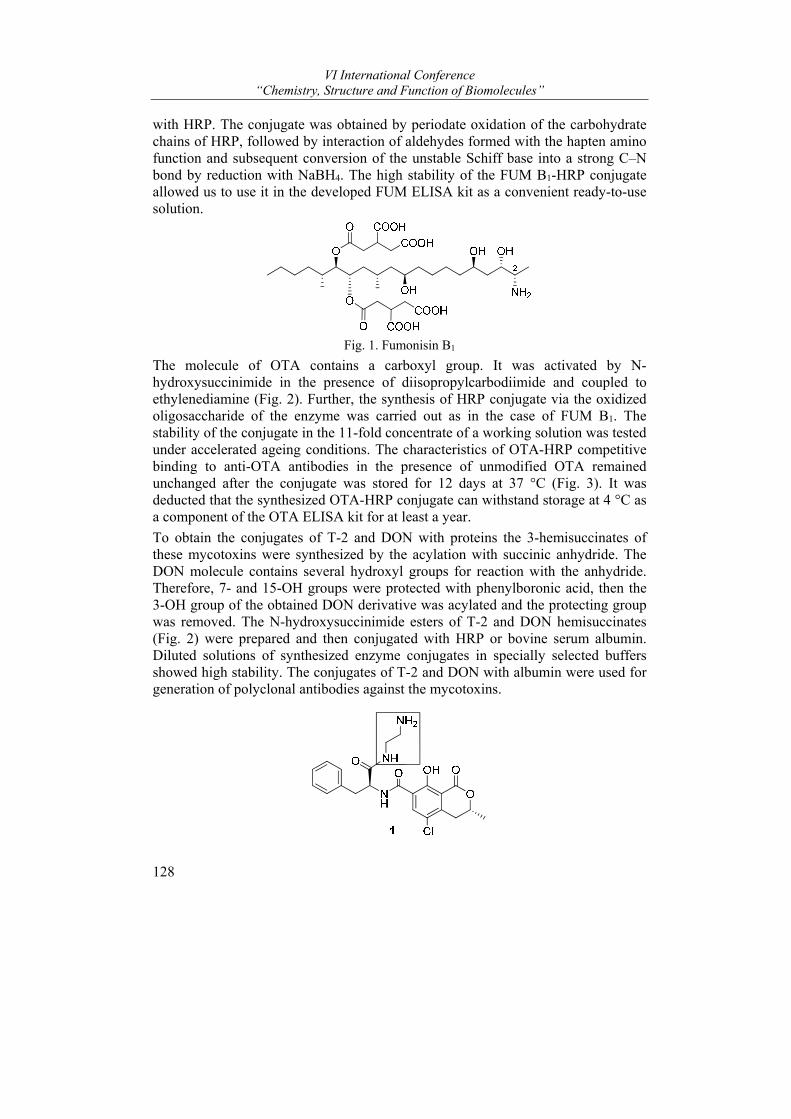
VI International Conference “Chemistry, Structure and Function of Biomolecules”
128
with HRP. The conjugate was obtained by periodate oxidation of the carbohydrate chains of HRP, followed by interaction of aldehydes formed with the hapten amino function and subsequent conversion of the unstable Schiff base into a strong C–N bond by reduction with NaBH4. The high stability of the FUM B1-HRP conjugate allowed us to use it in the developed FUM ELISA kit as a convenient ready-to-use solution.
Fig. 1. Fumonisin B1
The molecule of OTA contains a carboxyl group. It was activated by N-hydroxysuccinimide in the presence of diisopropylcarbodiimide and coupled to ethylenediamine (Fig. 2). Further, the synthesis of HRP conjugate via the oxidized oligosaccharide of the enzyme was carried out as in the case of FUM B1. The stability of the conjugate in the 11-fold concentrate of a working solution was tested under accelerated ageing conditions. The characteristics of OTA-HRP competitive binding to anti-OTA antibodies in the presence of unmodified OTA remained unchanged after the conjugate was stored for 12 days at 37 °C (Fig. 3). It was deducted that the synthesized OTA-HRP conjugate can withstand storage at 4 °C as a component of the OTA ELISA kit for at least a year.
To obtain the conjugates of T-2 and DON with proteins the 3-hemisuccinates of these mycotoxins were synthesized by the acylation with succinic anhydride. The DON molecule contains several hydroxyl groups for reaction with the anhydride. Therefore, 7- and 15-OH groups were protected with phenylboronic acid, then the 3-OH group of the obtained DON derivative was acylated and the protecting group was removed. The N-hydroxysuccinimide esters of T-2 and DON hemisuccinates (Fig. 2) were prepared and then conjugated with HRP or bovine serum albumin. Diluted solutions of synthesized enzyme conjugates in specially selected buffers showed high stability. The conjugates of T-2 and DON with albumin were used for generation of polyclonal antibodies against the mycotoxins.
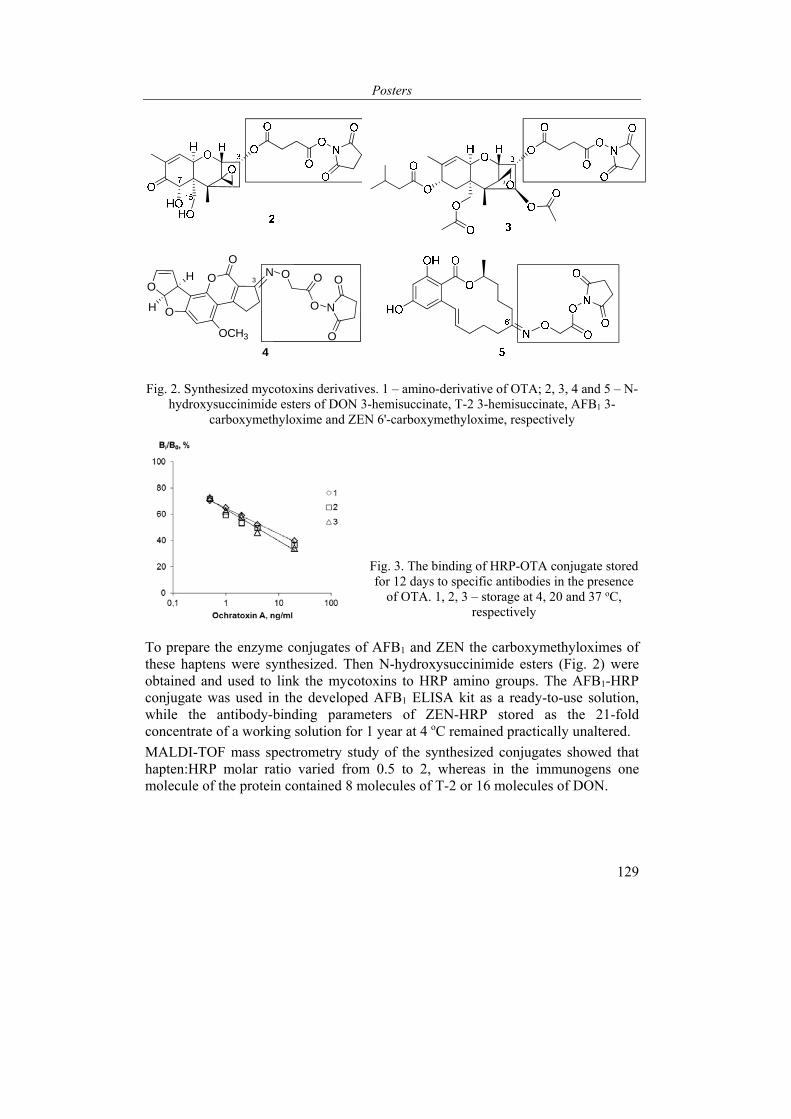
Posters
129
O
OO 3
ON
OCH3
O O
O
H
H
4
N
O
O
Fig. 2. Synthesized mycotoxins derivatives. 1 – amino-derivative of OTA; 2, 3, 4 and 5 – N-hydroxysuccinimide esters of DON 3-hemisuccinate, T-2 3-hemisuccinate, AFB1 3-
carboxymethyloxime and ZEN 6'-carboxymethyloxime, respectively
Fig. 3. The binding of HRP-OTA conjugate stored for 12 days to specific antibodies in the presence
of OTA. 1, 2, 3 – storage at 4, 20 and 37 oC, respectively
To prepare the enzyme conjugates of AFB1 and ZEN the carboxymethyloximes of these haptens were synthesized. Then N-hydroxysuccinimide esters (Fig. 2) were obtained and used to link the mycotoxins to HRP amino groups. The AFB1-HRP conjugate was used in the developed AFB1 ELISA kit as a ready-to-use solution, while the antibody-binding parameters of ZEN-HRP stored as the 21-fold concentrate of a working solution for 1 year at 4 oC remained practically unaltered.
MALDI-TOF mass spectrometry study of the synthesized conjugates showed that hapten:HRP molar ratio varied from 0.5 to 2, whereas in the immunogens one molecule of the protein contained 8 molecules of T-2 or 16 molecules of DON.

VI International Conference “Chemistry, Structure and Function of Biomolecules”
130
STRUCTURE MODIFICATION OF 14-ISOXAZOLYLMETHYL STEROIDS
Alesya Ladyko and Alexander Baranovsky
Institute of Bioorganic Chemistry, National Academy of Sciences, Minsk, Belarus e-mail: [email protected]
Previously it was shown that bridged nitro steroids 1 can be converted into isoxazoles 2 in good yield by applying a very simple procedure (propargyl alcohol, i-propanol, water, NaHCO3)1,2 The result prompted us to study such polyfunctional molecules as a template for the introduction of various pharmacophore or synthetically valuable groups in the ring D and isoxazole moiety.
The efforts were undertaken in three directions: to elucidate availability of the double bond in the Michael addition, to explore the primary hydroxy group in nucleophilic substitution and to evaluate the behavior of the isoxazole ring in the conditions of reductive cleavage.
The Michael addition to the enone system of acetoxy protected steroid 2 (EtMgBr, Cu2I2) gave 15β-ethyl derivative 3 predominantly in a moderate yield. Perhaps, a relatively low selectivity and yield are connected with cis-junction of the rings CD of the molecule.
Conjugated reduction of steroid 2 (R1=R2=H) with NaBH4 gave saturated triols in a 17α:17β=3:2 ratio. After separation and acetylation, 17α-acetate was undergone reductive cleavage (Ra-Ni, H2) to give enamino ketone 4. Here was observed hydrogenolysis of the acetoxy group similar to that for benzylic esters.
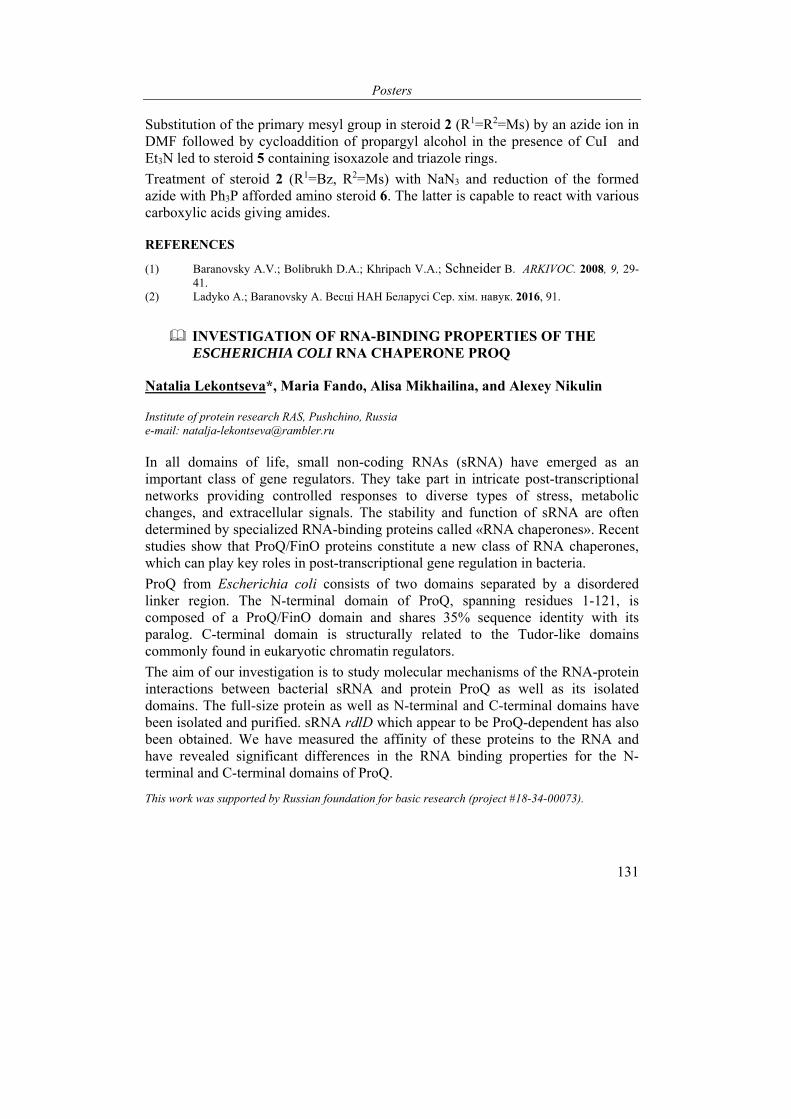
Posters
131
Substitution of the primary mesyl group in steroid 2 (R1=R2=Ms) by an azide ion in DMF followed by cycloaddition of propargyl alcohol in the presence of CuI and Et3N led to steroid 5 containing isoxazole and triazole rings.
Treatment of steroid 2 (R1=Bz, R2=Ms) with NaN3 and reduction of the formed azide with Ph3P afforded amino steroid 6. The latter is capable to react with various carboxylic acids giving amides.
REFERENCES
(1) Baranovsky A.V.; Bolibrukh D.A.; Khripach V.A.; Schneider B. ARKIVOC. 2008, 9, 29-41.
(2) Ladyko A.; Baranovsky A. Весцi НАН Беларусi Сер. хiм. навук. 2016, 91.
INVESTIGATION OF RNA-BINDING PROPERTIES OF THE ESCHERICHIA COLI RNA CHAPERONE PROQ
Natalia Lekontseva*, Maria Fando, Alisa Mikhailina, and Alexey Nikulin
Institute of protein research RAS, Pushchino, Russia e-mail: [email protected]
In all domains of life, small non-coding RNAs (sRNA) have emerged as an important class of gene regulators. They take part in intricate post-transcriptional networks providing controlled responses to diverse types of stress, metabolic changes, and extracellular signals. The stability and function of sRNA are often determined by specialized RNA-binding proteins called «RNA chaperones». Recent studies show that ProQ/FinO proteins constitute a new class of RNA chaperones, which can play key roles in post-transcriptional gene regulation in bacteria.
ProQ from Escherichia coli consists of two domains separated by a disordered linker region. The N-terminal domain of ProQ, spanning residues 1-121, is composed of a ProQ/FinO domain and shares 35% sequence identity with its paralog. C-terminal domain is structurally related to the Tudor-like domains commonly found in eukaryotic chromatin regulators.
The aim of our investigation is to study molecular mechanisms of the RNA-protein interactions between bacterial sRNA and protein ProQ as well as its isolated domains. The full-size protein as well as N-terminal and C-terminal domains have been isolated and purified. sRNA rdlD which appear to be ProQ-dependent has also been obtained. We have measured the affinity of these proteins to the RNA and have revealed significant differences in the RNA binding properties for the N- terminal and C-terminal domains of ProQ.
This work was supported by Russian foundation for basic research (project #18-34-00073).
VI International Conference “Chemistry, Structure and Function of Biomolecules”
132
INTRACELLULAR TRANSPORT AND ACUMMULATION OF NEW FLUORESCENT 24-EPICASTASTERONE CONJUNCTED WITH NBD FRAGMENT
Мichael V. Derevyanchuk1, Serhiy V. Kretynin1, Leila-Anastasia Karpets1, Raisa P. Litvinovskaya2, Alina L. Sauchuk2, Vladimir A. Khripach2, and Volodymyr S. Kravets1
1Institute of Bioorganic Chemistry and Petrochemistry of the National Academy of Sciences of Ukraine, Murmanska St., 1, Kiev 02094, Ukraine; 2Institute of Bioorganic Chemistry of the National Academy of Sciences of Belarus, 220141, Kuprevich str., 5, Minsk, Belarus e-mail: [email protected]
Brassinosteroids (BS) are well known group of polyhydroxylated plant steroids that involved in the regulation of the critical processes of plant growth. As the part of many defense mechanisms BS play a role in regulation of genes activity that participate in the formation of response to external environmental stimulus like salinity, high and low temperature, heavy metals and pathogens attack. BS signaling to cell mediated via BRI1 receptor which is crucial for growth-promoting effects of BS. It was shown earlier that BRI1 localizes to both plasma membrane and early endosomal compartments. But we have limited knowledge about localization and transport of BS in tissues and cells. To this date BS are believed to not undergo long-distance transport in tissues but at cellular level BS do appear to be transported. In this research we were aimed at investigation of intracellular transport of BS and sites where these hormones are accumulated.
Using novel brassinosteroid 24-EC-NBD – 24-epicastasterone conjuncted with fluorescent nitrobenzofurazan (NBD) fragment1 - and confocal microscopy we tested 24-EC-NBD intracellular transport and accumulation. As plant object in experiments we used 14 days old Arabidopsis thaliana (col-1) roots. Treatment of roots with 24-EC-NBD (20 µM) for 20 min led to strong accumulation of fluorescent brassinosteroid in lateral roots. 24-EC-NBD accumulated mainly in plasma membrane and membranes of intracellular compartments like endoplasmic reticulum that has been proven with fluorescent dye FM4-64 that specifically accumulate in plasma membrane. Using fluorescent dye Mitotracker Red that specifically incorporates in mitochondria we observed that 24-EC-NBD do not integrate in A.thaliana mitochondria after 20-120 min of treatment since no colocalization of Mitotracker Red and 24-EC-NBD was observed. 24-EC-NBD has been found to be specifically accumulated in membranes of guard cells. Our observations showed that plant roots uptake exogenous 24-EC-NBD mainly via root hairs. 24-EC-NBD has been found inside root hairs after 5 min of roots incubation in medium containing 24-EC-NBD (20 µM) solution. After 10 min of incubation 24-EC-NBD has been found in lateral and primary roots, still growing amount of 24-EC-NBD was detected in root hairs. It indicates that 24-EC-NBD appears
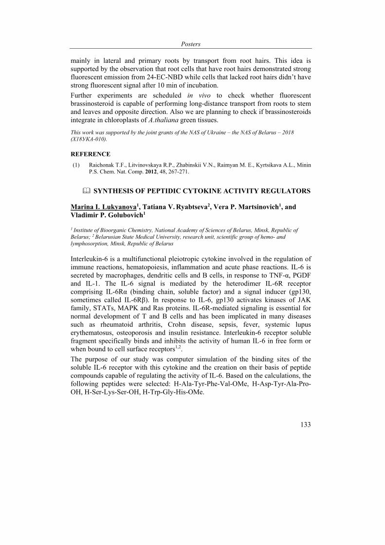
Posters
133
mainly in lateral and primary roots by transport from root hairs. This idea is supported by the observation that root cells that have root hairs demonstrated strong fluorescent emission from 24-EC-NBD while cells that lacked root hairs didn’t have strong fluorescent signal after 10 min of incubation.
Further experiments are scheduled in vivo to check whether fluorescent brassinosteroid is capable of performing long-distance transport from roots to stem and leaves and opposite direction. Also we are planning to check if brassinosteroids integrate in chloroplasts of A.thaliana green tissues.
This work was supported by the joint grants of the NAS of Ukraine – the NAS of Belarus – 2018 (X18УКА-010).
REFERENCE
(1) Raichonak T.F., Litvinovskaya R.P., Zhabinskii V.N., Raimyan M. E., Kyrtsikava A.L., Minin P.S. Chem. Nat. Comp. 2012, 48, 267-271.
SYNTHESIS OF PEPTIDIC CYTOKINE ACTIVITY REGULATORS
Marina I. Lukyanova1, Tatiana V. Ryabtseva2, Vera P. Martsinovich1, and Vladimir P. Golubovich1
1 Institute of Bioorganic Chemistry, National Academy of Sciences of Belarus, Minsk, Republic of Belarus; 2 Belarusian State Medical University, research unit, scientific group of hemo- and lymphosorption, Minsk, Republic of Belarus
Interleukin-6 is a multifunctional pleiotropic cytokine involved in the regulation of immune reactions, hematopoiesis, inflammation and acute phase reactions. IL-6 is secreted by macrophages, dendritic cells and B cells, in response to TNF-α, PGDF and IL-1. The IL-6 signal is mediated by the heterodimer IL-6R receptor comprising IL-6Rα (binding chain, soluble factor) and a signal inducer (gp130, sometimes called IL-6Rβ). In response to IL-6, gp130 activates kinases of JAK family, STATs, MAPK and Ras proteins. IL-6R-mediated signaling is essential for normal development of T and B cells and has been implicated in many diseases such as rheumatoid arthritis, Crohn disease, sepsis, fever, systemic lupus erythematosus, osteoporosis and insulin resistance. Interleukin-6 receptor soluble fragment specifically binds and inhibits the activity of human IL-6 in free form or when bound to cell surface receptors1,2.
The purpose of our study was computer simulation of the binding sites of the soluble IL-6 receptor with this cytokine and the creation on their basis of peptide compounds capable of regulating the activity of IL-6. Based on the calculations, the following peptides were selected: H-Ala-Tyr-Phe-Val-OMe, H-Asp-Tyr-Ala-Pro-OH, H-Ser-Lys-Ser-OH, H-Trp-Gly-His-OMe.
VI International Conference “Chemistry, Structure and Function of Biomolecules”
134
Boc-Ala
1.NHS2.DCC
Boc-Ala-OSu
Tyr
Boc-Ala-Tyr
Val
SOCl2,MeOH
Val-OMe*HCl
1.Boc-Phe DCHA2.HOBT, DCC
Boc-Phe-Val-OMe
HCl/CH3COOC2H5
Phe-Val-OMe*HCl
1. DIPEA2. HOBT, DCC
Boc-Ala-Tyr-Phe-Val-OMe
HCl/CH3COOC2H5
Ala-Tyr-Phe-Val-OMe*HCl
I
II
Ia
IIa
IV
III
Scheme. The synthesis of H-Ala-Tyr-Phe-Val-OMe
For the preparation of peptides, classical synthesis methods in liquid phase were used with maximum protection of the lateral functional groups. As coupling agents for the formation of peptide bonds, diisopropylcarbodiimide (DIC) or dicyclohexylcarbodiimide (DCC) with the addition of oxybenzotriazole (OBT) were used, also succinimide esters were used for the attachment of some protected amino acids. For protection of lateral functional groups of amino acid residues, hydrogen-labile groups were used: benzyl for carboxyl Asp and β-hydroxyl Ser, benzoxycarbonyl for ε-amino group of lysine. T-butyloxycarbonyl protections were used to block α-amino groups, C-terminal carboxyl groups were protected by the formation of methyl or ethyl esters. Deprotection was carried out by 3 methods: hydrogen-labile protecting groups were removed by hydrogenation with palladium black, alkaline hydrolysis was used to decompose ester groups, and tert-butyloxycarbonyl protecting groups were removed by acidic hydrolysis. The total yields of compounds were 20-32%.
Purification was carried out by column chromatography on silica gel. The structures of the peptides were confirmed by mass spectroscopy. Biological studies have shown that some of the synthesized peptides exhibit the properties of IL-6 antagonists. Our further research is aimed at creating on the basis of selected peptides drugs for the treatment of mediated IL-6 diseases.
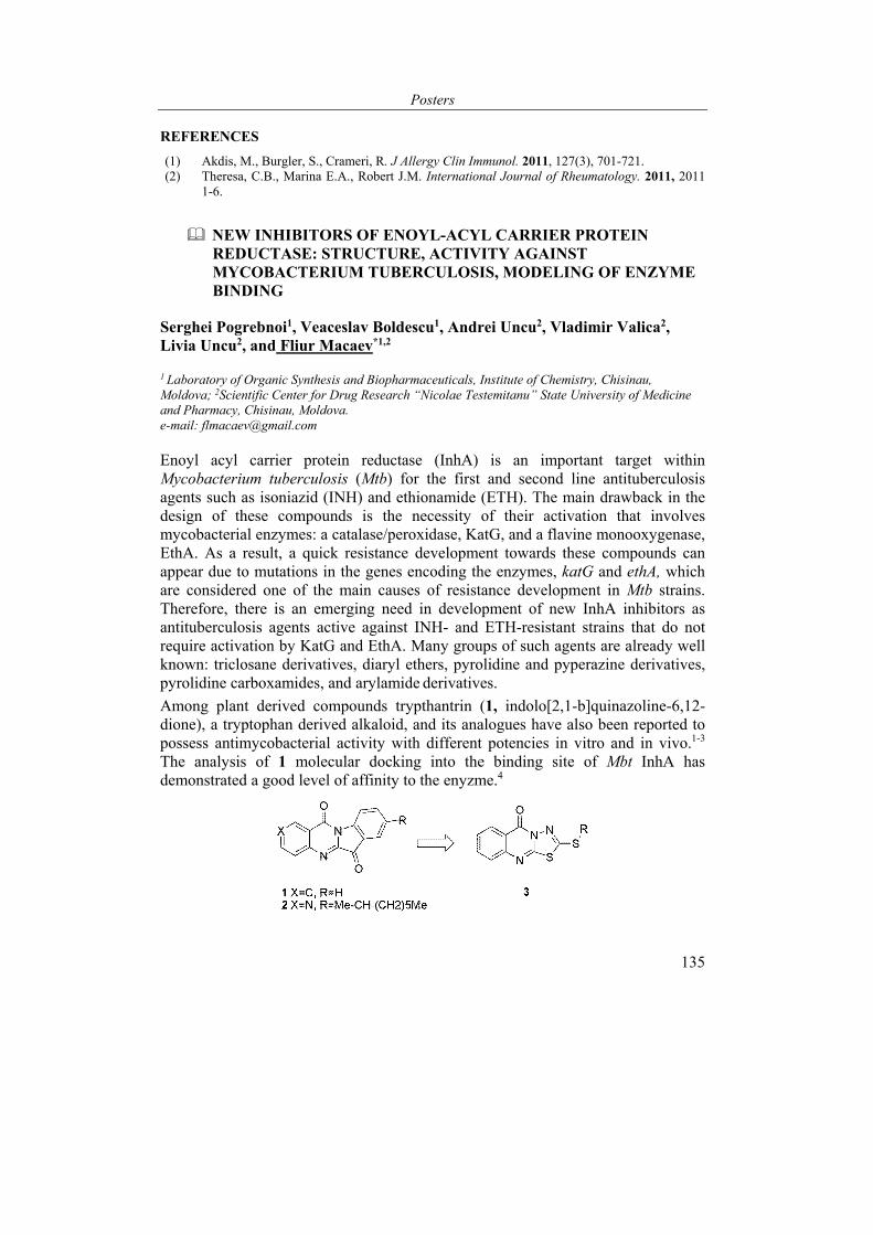
Posters
135
REFERENCES
(1) Akdis, M., Burgler, S., Crameri, R. J Allergy Clin Immunol. 2011, 127(3), 701-721. (2) Theresa, C.B., Marina E.A., Robert J.M. International Journal of Rheumatology. 2011, 2011
1-6.
NEW INHIBITORS OF ENOYL-ACYL CARRIER PROTEIN REDUCTASE: STRUCTURE, ACTIVITY AGAINST MYCOBACTERIUM TUBERCULOSIS, MODELING OF ENZYME BINDING
Serghei Pogrebnoi1, Veaceslav Boldescu1, Andrei Uncu2, Vladimir Valica2, Livia Uncu2, and Fliur Macaev*1,2
1 Laboratory of Organic Synthesis and Biopharmaceuticals, Institute of Chemistry, Chisinau, Moldova; 2Scientific Center for Drug Research “Nicolae Testemitanu” State University of Medicine and Pharmacy, Chisinau, Moldova. e-mail: [email protected]
Enoyl acyl carrier protein reductase (InhA) is an important target within Mycobacterium tuberculosis (Mtb) for the first and second line antituberculosis agents such as isoniazid (INH) and ethionamide (ETH). The main drawback in the design of these compounds is the necessity of their activation that involves mycobacterial enzymes: a catalase/peroxidase, KatG, and a flavine monooxygenase, EthA. As a result, a quick resistance development towards these compounds can appear due to mutations in the genes encoding the enzymes, katG and ethA, which are considered one of the main causes of resistance development in Mtb strains. Therefore, there is an emerging need in development of new InhA inhibitors as antituberculosis agents active against INH- and ETH-resistant strains that do not require activation by KatG and EthA. Many groups of such agents are already well known: triclosane derivatives, diaryl ethers, pyrolidine and pyperazine derivatives, pyrolidine carboxamides, and arylamide derivatives.
Among plant derived compounds trypthantrin (1, indolo[2,1-b]quinazoline-6,12-dione), a tryptophan derived alkaloid, and its analogues have also been reported to possess antimycobacterial activity with different potencies in vitro and in vivo.1-3 The analysis of 1 molecular docking into the binding site of Mbt InhA has demonstrated a good level of affinity to the enyzme.4
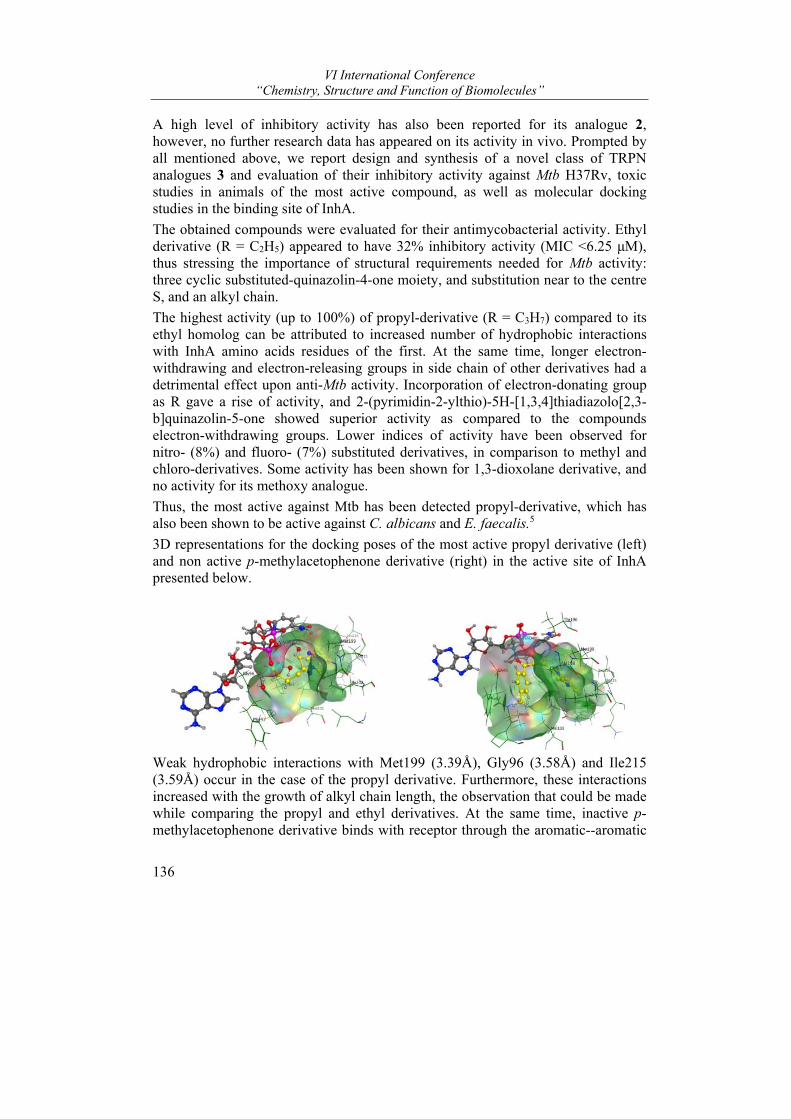
VI International Conference “Chemistry, Structure and Function of Biomolecules”
136
A high level of inhibitory activity has also been reported for its analogue 2, however, no further research data has appeared on its activity in vivo. Prompted by all mentioned above, we report design and synthesis of a novel class of TRPN analogues 3 and evaluation of their inhibitory activity against Mtb H37Rv, toxic studies in animals of the most active compound, as well as molecular docking studies in the binding site of InhA.
The obtained compounds were evaluated for their antimycobacterial activity. Ethyl derivative (R = C2H5) appeared to have 32% inhibitory activity (MIC <6.25 μM), thus stressing the importance of structural requirements needed for Mtb activity: three cyclic substituted-quinazolin-4-one moiety, and substitution near to the centre S, and an alkyl chain.
The highest activity (up to 100%) of propyl-derivative (R = C3H7) compared to its ethyl homolog can be attributed to increased number of hydrophobic interactions with InhA amino acids residues of the first. At the same time, longer electron-withdrawing and electron-releasing groups in side chain of other derivatives had a detrimental effect upon anti-Mtb activity. Incorporation of electron-donating group as R gave a rise of activity, and 2-(pyrimidin-2-ylthio)-5H-[1,3,4]thiadiazolo[2,3-b]quinazolin-5-one showed superior activity as compared to the compounds electron-withdrawing groups. Lower indices of activity have been observed for nitro- (8%) and fluoro- (7%) substituted derivatives, in comparison to methyl and chloro-derivatives. Some activity has been shown for 1,3-dioxolane derivative, and no activity for its methoxy analogue.
Thus, the most active against Mtb has been detected propyl-derivative, which has also been shown to be active against C. albicans and E. faecalis.5
3D representations for the docking poses of the most active propyl derivative (left) and non active p-methylacetophenone derivative (right) in the active site of InhA presented below.
Weak hydrophobic interactions with Met199 (3.39Å), Gly96 (3.58Å) and Ile215 (3.59Å) occur in the case of the propyl derivative. Furthermore, these interactions increased with the growth of alkyl chain length, the observation that could be made while comparing the propyl and ethyl derivatives. At the same time, inactive p-methylacetophenone derivative binds with receptor through the aromatic--aromatic

Posters
137
moiety (Gly96-3.34Å) and H--Aromatic interactions with Phe97 (3.71Å), Met98 (3.33 Å) and Tyr158 (3.56Å) and presents no hydrophobic interactions.
Electron density of the propyl derivtive is distributed both on ligand and amino acid residues causing more effective donor-acceptor interaction. For the p-methylacetophenone derivative, electron density is concentrated on residues and absent on the ligand’s atoms.
REFERENCES
(1) A. M. Baker, William R.; Lester, Indolo [2, 1-Biquinazoline-6, 12-Dione Antibacterial Compounds and Methods of Use Thereof, 1995, US Patent 5,441,955.
(2) L. A. Mitscher, W. Baker, Med. Res. Rev. 1998, 18, 363–374. (3) J.-M. Hwang, T. Oh, T. Kaneko, A. M. Upton, S. G. Franzblau, Z. Ma, S.-N. Cho, P. Kim, J.
Nat. Prod. 2013, 76, 354–367. (4) A. Tripathi, N. Wadia, D. Bindal, T. Jana, Indian J. Biochem. Biophys. 2012, 49, 435–441. (5) S. Pogrebnoi, C. Chiriţă, V. Valica, F. Macaev, M. C. Chifiriuc, C. Kamerzan, L. Uncu,
Farmacia 2017, 65, 69–74.
Acknowledgements: the authors are grateful for the funding support from the Agency for Research and Development of the Republic of Moldova under moldo-romanian bilateral project 16.80013.5007.05/Ro.
NON-NUCLEOSIDE REVERSTRANSCRIPTASE INHIBITORS WITH TARGETED ACTIVATION IN MACROPHAGES
Natalia Sucman1, Veaceslav Boldescu1, Livia Uncu2, Vladimir Valica2, and Fliur Macaev1,2*
1 Laboratory of Organic Synthesis and Biopharmaceuticals, Institute of Chemistry, Chisinau, Moldova,2 State University of Medicine and Pharmacy“Nicolae Testemițanu”, Scientific Center for Drug Research , Chișinău, Moldova e-mail: [email protected]
The foundation of the mononuclear phagocyte system are macrophages that derive from bone marrow’s monoblasts and promonoblasts. First, monoblasts and promonoblasts get transformed in circulating monocytes, which, after migration in extravascular tissue, differentiate into macrophages.
The main role of macrophages is defense of the organism against various infectious agents. Furthermore, macrophages have been found to play an important part in the pathology of different diseases: atherosclerosis (foam cells), cancer (tumor-associated macrophages), infectious diseases for which macrophages play the role of host cells (e. g. tuberculosis, HIV, leishmaniasis, dengue virus), etc. Thus, recent studies have detected high viral load in tissue macrophages at all stages of HIV-1, which persists in them even under combination antiretroviral therapy.1 Therefore, macrophages make one of important targets for anti-HIV drug design and development.
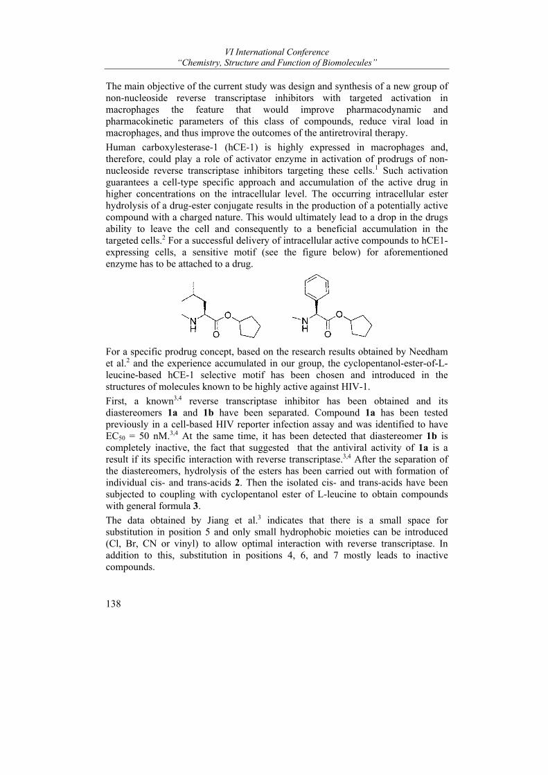
VI International Conference “Chemistry, Structure and Function of Biomolecules”
138
The main objective of the current study was design and synthesis of a new group of non-nucleoside reverse transcriptase inhibitors with targeted activation in macrophages the feature that would improve pharmacodynamic and pharmacokinetic parameters of this class of compounds, reduce viral load in macrophages, and thus improve the outcomes of the antiretroviral therapy.
Human carboxylesterase-1 (hCE-1) is highly expressed in macrophages and, therefore, could play a role of activator enzyme in activation of prodrugs of non-nucleoside reverse transcriptase inhibitors targeting these cells.1 Such activation guarantees a cell-type specific approach and accumulation of the active drug in higher concentrations on the intracellular level. The occurring intracellular ester hydrolysis of a drug-ester conjugate results in the production of a potentially active compound with a charged nature. This would ultimately lead to a drop in the drugs ability to leave the cell and consequently to a beneficial accumulation in the targeted cells.2 For a successful delivery of intracellular active compounds to hCE1-expressing cells, a sensitive motif (see the figure below) for aforementioned enzyme has to be attached to a drug.
For a specific prodrug concept, based on the research results obtained by Needham et al.2 and the experience accumulated in our group, the cyclopentanol-ester-of-L-leucine-based hCE-1 selective motif has been chosen and introduced in the structures of molecules known to be highly active against HIV-1.
First, a known3,4 reverse transcriptase inhibitor has been obtained and its diastereomers 1a and 1b have been separated. Compound 1a has been tested previously in a cell-based HIV reporter infection assay and was identified to have EC50 = 50 nM.3,4 At the same time, it has been detected that diastereomer 1b is completely inactive, the fact that suggested that the antiviral activity of 1a is a result if its specific interaction with reverse transcriptase.3,4 After the separation of the diastereomers, hydrolysis of the esters has been carried out with formation of individual cis- and trans-acids 2. Then the isolated cis- and trans-acids have been subjected to coupling with cyclopentanol ester of L-leucine to obtain compounds with general formula 3.
The data obtained by Jiang et al.3 indicates that there is a small space for substitution in position 5 and only small hydrophobic moieties can be introduced (Cl, Br, CN or vinyl) to allow optimal interaction with reverse transcriptase. In addition to this, substitution in positions 4, 6, and 7 mostly leads to inactive compounds.
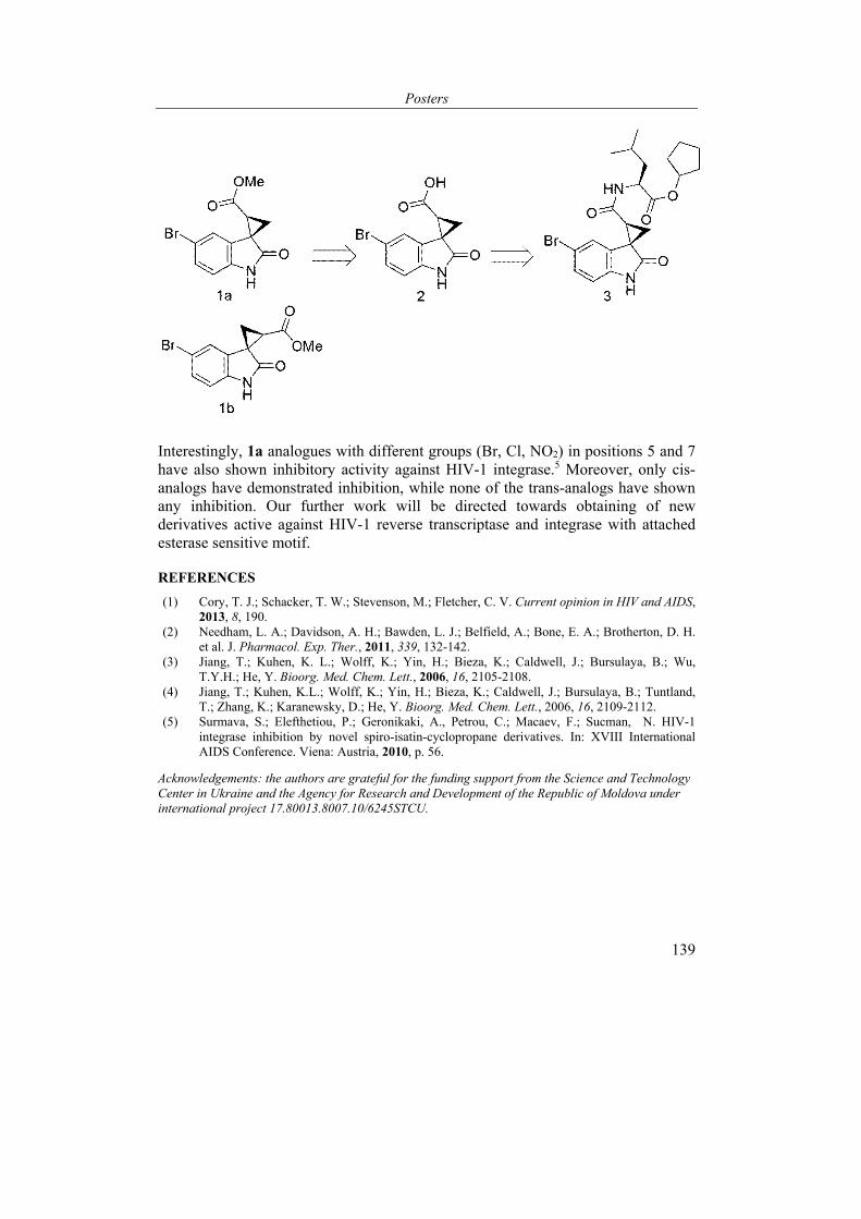
Posters
139
Interestingly, 1a analogues with different groups (Br, Cl, NO2) in positions 5 and 7 have also shown inhibitory activity against HIV-1 integrase.5 Moreover, only cis-analogs have demonstrated inhibition, while none of the trans-analogs have shown any inhibition. Our further work will be directed towards obtaining of new derivatives active against HIV-1 reverse transcriptase and integrase with attached esterase sensitive motif.
REFERENCES
(1) Cory, T. J.; Schacker, T. W.; Stevenson, M.; Fletcher, C. V. Current opinion in HIV and AIDS, 2013, 8, 190.
(2) Needham, L. A.; Davidson, A. H.; Bawden, L. J.; Belfield, A.; Bone, E. A.; Brotherton, D. H. et al. J. Pharmacol. Exp. Ther., 2011, 339, 132-142.
(3) Jiang, T.; Kuhen, K. L.; Wolff, K.; Yin, H.; Bieza, K.; Caldwell, J.; Bursulaya, B.; Wu, T.Y.H.; He, Y. Bioorg. Med. Chem. Lett., 2006, 16, 2105-2108.
(4) Jiang, T.; Kuhen, K.L.; Wolff, K.; Yin, H.; Bieza, K.; Caldwell, J.; Bursulaya, B.; Tuntland, T.; Zhang, K.; Karanewsky, D.; He, Y. Bioorg. Med. Chem. Lett., 2006, 16, 2109-2112.
(5) Surmava, S.; Elefthetiou, P.; Geronikaki, A., Petrou, C.; Macaev, F.; Sucman, N. HIV-1 integrase inhibition by novel spiro-isatin-cyclopropane derivatives. In: XVIII International AIDS Conference. Viena: Austria, 2010, p. 56.
Acknowledgements: the authors are grateful for the funding support from the Science and Technology Center in Ukraine and the Agency for Research and Development of the Republic of Moldova under international project 17.80013.8007.10/6245STCU.
VI International Conference “Chemistry, Structure and Function of Biomolecules”
140
SERIAL FEMTOSECOND CRYSTALLOGRAPHY MEMBRANE PROTEIN STRUCTURE DETERMINATION
E. Marin1, A. Luginina1, A. Gusach1, A. Mishin1, K. Kovalev1,3, V. Borshchevskiy1, and V. Cherezov1,2,*
1 Moscow Institute of Physics and Technology (State University), Moscow, Russia; 2 Bridge Institute, Department of Chemistry, University of Southern California, Los Ange-les, California, USA; 3 Forschungszentrum Julich, Julich, Germany e-mail: [email protected]
Structural studies of membrane proteins have recently became more accessible due to advancements in their expression, stabilization and crystallization techniques. In particular, X-ray crystallography in combination with lipidic cubic phase crystallization have been successfully used to study structures of many membrane proteins, including G-protein coupled receptors and light-activating rhodopsins [1, 2]. However, obtaining a high-resolution structure remains challenging due to the small crystal size and associated with it radiation damage.
In 2011, the method of serial femtosecond crystallography (SFX) has been proposed [3]. It allows one to collect room temperature structural data from micrometer-sized crystals without radiation damage using X-ray free-electron lasers. Due to crystal destruction by single shots, 10 000's of crystals are used for a single dataset and sample delivery is performed to keep up with the laser pulse repetition rate. Since 2011, SFX has been successfully applied to several challenging targets, as recently reviewed in [4,5].
In this work, we report on the structure determination of a membrane protein using an X-ray free electron laser. We improved and optimized data processing algorithm to extract multiple diffraction patterns from individual images, allowing us to increase indexing rate and improve overall resolution.
The work was supported by the Russian Science Foundation (project no. 16-14-10273).
REFERENCES
(1) Zhang, H. et al. Structural basis for selectivity and diversity in angiotensin II receptors. Nature 544, 327–332 (2017).
(2) Volkov, O. et al. Structural Insights into Ion Conduction by Channelrhodopsin 2. Science 358, eaan8862 (2017).
(3) Chapman, H. N. et al. Femtosecond X-ray protein nanocrystallography. Nature 470, 73–7 (2011).
(4) Johansson, L.C. et al. A bright future for serial femtosecond crystallography with XFELs. Trends Biochem Sci 42, 749-762 (2017).
(5) Ishchenko, A. et al. Structural Biology of G-Protein-Coupled Receptors: New Opportunities from XFELs and cryoEM. Curr Opin Struct Biol 51, 44–52 (2018).
Posters
141
LIGHT NANOSCOPE - ADVANCED MICROSCOPY PLATFORM
Maslov Ivan1, Bogorodskiy Andrey1, Podolyak Elizaveta1, Burkatovskiy Dmitriy1, Ilyinsky Nikolay1, Büldt Georg1, Mishin Alexey1, Gensch Thomas2, and Borshchevskiy Valentin1*
1 Moscow Institute of Physics and Technology, 141701, Moscow, Russia; 2Institute of Complex Systems (ICS), ICS-4: Cellular Biophysics, Forschungszentrum Jülich GmbH, 52428, Jülich, Germany e-mail: [email protected]
Fluorescence microscopy is the dominating technique in the field of bioimaging. The ease of fluorescence staining of biomolecules (incl. in vivo), sensitivity of the technique (up to single-molecule detection) and wide variety of biophysical methods based on fluorescence microscopy makes it very attractive for research in life sciences. The fluorescence microscopy platform at Moscow Institute of Physics and Technology was designed to unite wide range of fluorescence-based microscopy methods in the single optical path. \
The platform (Light nanoscope) is based on confocal microscope (LSM780, Carl Zeiss) combined with the module for super-resolution fluorescence microscopy experiments based on single-molecule localization microscopy (ELYRA, Carl Zeiss). Light Nanoscope is equipped with 6 excitation lasers, which cover entire visible spectrum, near UV and infrared. 2-photon laser with tunable wavelength allows to conduct precise (in spectrum and in localization) experiments even with thick samples based on 2-photon fluorescence excitation or second harmonics generation phenomenon.
ELYRA module can be used to perform fast (down to 10 ms) detection at extremely high (down to single fluorophore) sensitivity. This allowed us to use super-resolution fluorescence microscopy methods based on single-molecule localization microscopy (incl. PALM and STORM) and localize biomolecules with up to 20 nm resolution. TIRF method (Total Internal Reflection Fluorescence) can be used to improve contrast in super-resolution experiments. Additional super-resolution modality, which is less precise but much faster, SIM (Structured Illumination Microscopy) was also installed in the platform.
Such powerful method as FLIM (Fluorescence Lifetime IMaging) was introduced in the Light nanoscope and opened additional opportunities to measure various physical parameters in biological systems using fluorescent nanosensors and to observe complicated phenomena, including FRET (Forster Resonance Energy Transfer).
In order to perform experiments with living cells, an incubator with adjustable temperature, atmosphere composition and humidity was installed in the optical path. In the proximity of the nanoscope there is all necessary infrastructure to perform sterile sample preparation with mammalian cells (laminars, incubators, centrifuges, cytometers etc). Nanoscope is equiped with microinjection system –
VI International Conference “Chemistry, Structure and Function of Biomolecules”
142
one can use it to deliver particular fluorophores or chemicals in the selected living cells.
Thus, light nanoscope is the unique platform for fluorescence microscopy experiments, which combines numerous powerful complimentary methods, such as confocal microscopy, super-resolution microscopy (SIM, SMLM), FLIM and FRET, 2PE and SHG, FCS and others.
The platform was designed in the framework of govenmental task of (Ministry of Education and Science of the Russian Federation, project № 6.9909.2017/ВУ)
ANTISEIZURE EFFECTS OF DIFFERENT N-PALMITOYLAMIDES IN THE MODEL OF ACUTE SEIZURE IN RATS
Tigran Melik-Kasumov1*, Tatjana Pavlut1, Evgeniy Shavalda2, Alexander L. Mikhal’chuk3, and Mihail A. Kisel3
1 Institute of Physiology, Minsk, Belarus,2International Sakharov Environmental Institute of Belarusian State University, Minsk, Belarus 3 Institute of Bioorganic Chemistry, Minsk, Belarus e-mail: [email protected]
In Belarus epilepsy comes to 8-12% of all neurological diseases and takes 0.02-0.03% of all cases of primary morbidity. Around 30% of cases of epilepsy are pharmacoresistant1. In parallel with the search of exogenous antiepileptic compounds it comes more actual to understand the structure and mechanisms of inner antiepileptic system and its components – endogenous anticonvulsants which can protect healthy brain from generation of hypersynchronous discharges. Recently, in this regard great attention is being paid to fatty acids derivatives: different lipid signaling molecules were found in nervous tissue and most of them had shown neurotropic effects. Antiepileptic effect has been already shown for N-palmitoylethanolamine and oleamide – endogenous cannabimimetic fatty acids amides2,3. The purpose of this study was to assess antiepileptic potential of three N-palmitoylamides – N-palmitoylglycine (PalGly), N-palmitoyl-5-aminolevulenic acid (PalALA) and N-palmitoylserinol (PalSerinol) in the model of acute seizures.
The study was conducted on male Wistar rats with mass in range 200-220g. Palmitic acid amides were synthesized in the Laboratory of lipids chemistry of the Institute of Bioorganic Chemistry. Due to poor water solubility amides were dissolved in complex solvent consisted of DMSO, Tween 20 (Sigma) and saline (1:1:8 by volume, respectively). Acute generalized clonic or tonic seizures were induced by single i.p. injection of pentylenetetrazole (60 mg/kg, Sigma). Forty-five minutes prior to PTZ injection rats of different experimental groups had been injected with PalGly (10 mg/kg), PalALA (10 mg/kg), PalSerinol (5 m/kg), solvent (sham control) or saline (control). The latency, duration and severity of seizure

Posters
143
activity were observed behaviorally and classified by Racine’s scale points. For significance assessment Mann-Whitney U-test was used.
Complex solvent by itself did not affect any parameters analyzed: neither latency, nor average Racine’s scale point in either group significantly differed. Proceeding from this results, for further analysis intact control group were used for comparison. All of tested amides showed significant increase in average latency to seizure onset. PalGly postponed seizure onset for additional 22.5 s (37,6%). PalALA increased latency for 48.5%, and PalSerinol – for 45.9%. Besides, PalSerinol affected another temporal parameter analyzed. It was set that single injection of 5 mg/kg PalSerinol shorted the overall duration of seizures within 45 minutes of observation. This parameter was 76.5% lower than control value. It is worth noting that all of effects found concerned only temporal parameters. Neither of compounds tested led to decrease in average maximal or weighted point of Racine’s scale. It is quite possible that chronic injections of amides in question could lead to decrease of seizure severity along with decrease in its temporal parameters.
Our results show that different amides of palmitic acid induce urgent changes in brain and slow down convulsive readiness escalation. Thus, the tested endogenous fatty acids derivatives can be a part of endogenous anti-seizure system.
REFERENCES
(1) Dokukina T.B., Golubeva T.S., Matveichuk I.V., Mahrov M.V., Loseva V.M., Krupenkina E.V., Marchuk S.A., Epilepsia and paroxyzmal conditions. 2014, 6(2), 29-33.
(2) Sheerin A.H., Zhang X., Saucier D.M., Corcoran M.E. Epilepsia, 2004, 45(10), 1184–1188. (3) Solomonia R., Nozadze M., Mikautadze E., Kuchiashvili N., Kiguradze T., Abkazava D.,
Pkhakadze V., Mamulaishvili I., Mikeladze E., Avaliani N. Bulletin of Experimental Biology and Medicine. 2008, 145(2), 175-187.
This work was supported by the Belarusian Republican Foundation for Fundamental Research (grant no. M17-070 of April 18th, 2017).
SYNTHESIS AND PROPERTIES OF PHOSPHATIDYLHYDROXYACETONE
Rudak E.V., Kisel M.A., and Mikhal’chuk A.L.
Institute of Bioorganic Chemistry NASB, Minsk, Belarus e-mail: [email protected]
Phosphatidyl hydroxyacetone (PHA) is a minor phospholipid found in biological tissues and fluids [1]. At present, the biochemical significance of the formation of PHA as a result of free radical fragmentation of cardiolipin remains unknown. It is assumed that PHA, as well as phosphatidic acid (PA), can act as a signal molecule. However, the PHA has in its own structure of an oxo group uncharacteristic for phospholipids of natural origin. Such a unique structure of this phospholipid can
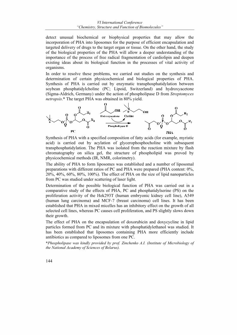
VI International Conference “Chemistry, Structure and Function of Biomolecules”
144
detect unusual biochemical or biophysical properties that may allow the incorporation of PHA into liposomes for the purpose of efficient encapsulation and targeted delivery of drugs to the target organ or tissue. On the other hand, the study of the biological properties of the PHA will allow a deeper understanding of the importance of the process of free radical fragmentation of cardiolipin and deepen existing ideas about its biological function in the processes of vital activity of organisms.
In order to resolve these problems, we carried out studies on the synthesis and determination of certain physicochemical and biological properties of PHA. Synthesis of PHA is carried out by enzymatic transphosphatidylation between soybean phosphatidylcholine (PC; Lipoid, Switzerland) and hydroxyacetone (Sigma-Aldrich, Germany) under the action of phospholipase D from Streptomyces netropsis.* The target PHA was obtained in 80% yield.
Synthesis of PHA with a specified composition of fatty acids (for example, myristic acid) is carried out by acylation of glycerophosphocholine with subsequent transphosphatidylation. The PHA was isolated from the reaction mixture by flash chromatography on silica gel, the structure of phospholipid was proved by physicochemical methods (IR, NMR, colorimetry).
The ability of PHA to form liposomes was established and a number of liposomal preparations with different ratios of PC and PHA were prepared (PHA content: 0%, 20%, 40%, 60%, 80%, 100%). The effect of PHA on the size of lipid nanoparticles from PC was studied under scattering of laser light.
Determination of the possible biological function of PHA was carried out in a comparative study of the effects of PHA, PC and phosphatidylserine (PS) on the proliferation activity of the Hek293T (human embryonic kidney cell line), A549 (human lung carcinoma) and MCF-7 (breast carcinoma) cell lines. It has been established that PHA in mixed micelles has an inhibitory effect on the growth of all selected cell lines, whereas PC causes cell proliferation, and PS slightly slows down their growth.
The effect of PHA on the encapsulation of doxorubicin and doxycycline in lipid particles formed from PC and its mixture with phosphatidylethanol was studied. It has been established that liposomes containing PHA more efficiently include antibiotics as compared to liposomes from one PC. *Phospholipase was kindly provided by prof. Zinchenko A.I. (Institute of Microbiology of the National Academy of Sciences of Belarus).
Posters
145
REFERENCE
(1) Yurkova, I. L.; [et al.] Int. J. Radiat. Biol. 2004, 80(3), 239-245.
The study was carried out with the financial support of the BRFFR, grant Х16М-087.
ETHANOLAMIDES OF HIGH FATTY ACIDS (N-ACYL ETHANOLAMINES – NAES). STATUS AND PROSPECTS
Kisel M.A. and Mikhal’chuk A.L.
Institute of Bioorganic Chemistry NASB, Minsk, Belarus e-mail: [email protected]
The totality of organic substances defined as ethanol amides of higher fatty acids, or acyl ethanol amides, or N-acyl ethanol amines (NAEs) [1, 2] in a generalized form is described by the formula:
R2
N
O
OH(R5)
R3
R4
R1
I where R1 – is an alkyl or alkenyl residue of varying degrees of unsaturation; R2 – is usually H, or may be Alk C1-C5; R3 – is H, Me, CH2OH, CH2OR5, COOH, ...; R4 is H, CH2OH, CH2OR5, ...; R5 – is an alkаnoyl or alkenoyl residue of different degrees of unsaturation.
This commonality is private, at the same time very limited and very extensive, and representative of the most numerous family of natural substances of biological origin – lipids [3, 4].
The most famous and simplest (ancestral) representatives of this series are: lauryl ethanolamide (LAE, N-lauryl ethanolamine, NAE 12:0) [1, 5]; miristoyl ethanolamide (MEA, N-miristoyl ethanolamine, NAE 14:0) [6]; palmitoyl ethanol amide (PEA, N-palmitoyl ethanolamine, NAE 16:0) [6, 7], stearoyl ethanol amide (SEA, N-stearoyl ethanolamine, NAE 18:0) [6, 7], acyl serinol amides [8, 9], acyl glycine amides [10], acyl seryn amides [11], and others. All these compounds are considered as endogenous kanabimimetics or endocannabinoid-like mediators, and exhibit a wide range of biological effects from anti-inflammatory and antinociceptive before neuropathic and neuroprotective. Acyl ethanol amides are effective against chronic and neuropathic pains caused by arthrosis, arthritis, migraine, menstruation, endometriosis, visceral pain syndromes, cervical and whiplash fibromyalgia, chronic lumbar pain, prostatic gland problems. The use of drugs based on acyl ethanol amides is shown with diffuse atherosclerosis, in chemotherapy of oncological diseases, in the prevention and treatment of type 1 and

VI International Conference “Chemistry, Structure and Function of Biomolecules”
146
2 diabetes, in the relief and relief of pain and spasms after a stroke, in complex regional pain syndromes (CRPS) and neuralgia of various etiologies.
An independent group in this series are ethanol amides of unsaturated carboxylic acids: oleoyl ethanol amide (OAE, N-oleoyl ethanolamine, NAE 18:1Δ9) [6, 7], linoleoyl ethanolamide (LAE, N-linoleoyl ethanolamine, NAE 18:2Δ9,12) [12]; arachidonoyl ethanol amide (anandamid, arachidonoyl ethanolamine NAE 20:4Δ5,8,11,14) [13] and others. The most famous and well-known representative of this group is the first isolated and characterized in 1992 endocannabinoid anandamide. These endocannabinoids are present in tissues and biological fluids in extremely low concentrations, labile and are rapidly metabolized, and therefore remain little studied.
Analysis of available literature testifies that the studies of endocannabinoids, endogenous cannabimimetics, and endocannabinoid-like mediators are currently on the rise and develop in two main directions. The first — is the detection of these substances in biological tissues with the elucidation of the mechanisms of their formation and the establishment of biological functions, and the second — is a chemical or biochemical synthesis with followed bioscreening.
Obviously, the spectrum of biological activity of the compounds under discussion is very wide, so attention to the study of their biological effects and efforts to create new pharmacological agents on their basis are constantly expanding and intensifying. At the same time, most acyl ethanol amides are present in natural objects and biological tissues in low concentrations, which exclude the possibility of their isolation for practical use.
At the same time, many representatives of this series are still not only inaccessible to research, but they are not even received and not characterized at all. A particular example of this is the first obtained and characterized by us in 2017 and currently studied 5-aminolevulinic (5-amino-4-oxopentanoic) acid palmitoyl ethanol amide.
REFERENCES
(1) Keereetaweepa, J.; Kilarua, A.; Feussnerb, I.; Venablesa, B. J.; Chapman, K. D. FEBS Letters 2010, 584, 3215–3222.
(2) Kilaru, A.; Tamura, P.; Isaac, G.; Welti, R.; Venables, B. J.; Seier, E.; Chapman, K. D.; Planta. 2012, 236(3), 809–824.
(3) Biochemistry of Lipids, Lipoproteins and Membranes (Fifth Edition); Vance, Dennis E. and Vance, Jean E., Ed.; Elsevier, 2008, 631.
(4) Handbook of functional lipids. Ed by Akoh, Casimir C., CRC Press, Taylor & Francis Groop LLC. 2005, 544.
(5) Zhang, Y.; Guo, W.M.; Chen, S.M.; Han, L.; Li, Z.M. J. Plant Physiol. 2007, 164(8), 993-1001.
(6) Ueda, N.; Yamanaka, K; Yamamoto, S. J. Biol. Chem. 2001, 276(38), 35552-35557. (7) Darmania, N. A.; Izzob, A. A.; Degenhardta, B.; Valentic, M.; Scaglioned, G.;
Capassob, R.; Sorrentinid, I.; Di Marzo, V. Neuropharmacology. 2005, 48, 1154-1163.
(8) Ray, K. Nature Reviews Gastroenterology & Hepatology. 2017, 14, 630–631.
Posters
147
(9) Bieberich, E.; Kawaguchi, T.; Yu, R.K. J. Biol. Chem. 2000, 275(1), 177–181. (10) Cohen, L. J. et al. Nature. 2017, 549, 48–53. (11) Cебякин, Ю.Л.; Федякова, Н.Д.; Рунова, Т.Л. Биоорганическая химия 1994, 20(10),
1101-1106. (12) Wang, Xiaosan; Chen, Yan; Jin, Qingzhe; Huang, Jianhua; Wang, Xingguo J. Oleo Sci.
2013, 62(6), 427-433. (13) Felder, C.C.; Briley, E.M.; Axelrod, J.; Simpson, J.T.; Mackie, K; Devane, W.A. Proc. Natl.
Acad. Sci. USA 1993, 90, 7656-7660.
EVALUATION OF ACUTE AND SUBACUTE TOXICITY INDUCED BY LIPOSOMAL FORMULATIONS OF N-PALMITOYL GLYCINE AND N-PALMITOYL-5-AMINOLEVULINIC ACID
Alla Yu. Molchanova1*, Irina P. Zhavoronok1, Tigran B. Melik-Kasumov1, Olga A. Antipova1, Tatjana O. Pavlut1, Elena I. Pekhtsereva1, Alexander L. Mikhal’chuk2, and Mihail A. Kisel2
1 Institute of Physiology, Minsk, Belarus, 2 Institute of Bioorganic Chemistry, Minsk, Belarus e-mail: [email protected]
Nowadays research of lipid signal molecules is among the most intriguing fields in pharmacology. A number of compounds were referred to as mediators of communication inside and in between of cells. N-acyl conjugates of amino- acids with long fatty chain acids had been given considerable attention by researchers due to their antinociceptive and potential anti-inflammatory effects1. Results of our previous studies also suggested that N-palmitoyl glycine (PalGly) and N-palmitoyl-5-aminolevulinic acid (Pal-5-Ala) had strong analgesic effect at peripheral neuropathy2. However, since these lipophilic compounds suffer from a poor aqueous solubility, which may limit their bioavailability in therapeutic applications, we prepared both, PalGly and Pal-5-Ala, in the form of liposomes from egg phospholipids. Unfortunately, there were no safety reports on of PalGly or Pal-5-Ala in a free as well as liposomal form as far as we know. Therefore, a separate study was proposed to evaluate the toxicity profile for liposomes containing PalGly and Pal-5-Ala.
The study of acute toxicity was conducted on males and females of Wistar rats and C57BL6 mice. Mice were given a single intraperitoneal (i.p.) injection of PalGly or Pal-5-Ala in one of following doses: 7.5, 75, 300 and 900 μMol·kg-1, whereas doses chosen for single i.p. injection to rats were different – 7.5, 75, 150 and 300 μMol·kg-1. Also, additional groups of rats were administered liposomes intravenously (i.v.) in doses of 7.5, 22.5, 45 and 75 μMol·kg-1. All animals were monitored for two weeks after administration of test substances.
In the subacute study, three different doses (7.5, 75 and 150 μMol·kg-1per day) of PalGly and Pal-5-Ala were administered to C57BL6 mice for 28 days.

VI International Conference “Chemistry, Structure and Function of Biomolecules”
148
Control groups (either in acute or subacute experiment) were formed from intact animals of both sexes, as well as from animals that received “empty” liposomes from egg phosphatidylcholine without PalGly and Pal-5-Ala (PCh). Mortality, clinical signs, body weight changes, hematological and biochemical parameters, changes in skin and fur, eyes and mucous membranes, and respiratory system, gross findings, organ weights, were monitored during the study. Presence of tremors, convulsions, lethargy was also recorded. Individual weights of animals were determined right before the administration as well as at least weekly thereafter.
As shown in Table 1, the LD50 of both PalGly and Pal-5-Ala in mice was found to be in a range of 76-500 mgkg-1. That allows classification of these compounds as low toxic. Morbidity was observed only after administration of tested compounds in a maximal dose of 900 μMol·kg-1. A number of death cases were found to be gender dependent on N-acyl conjugate: males appeared to be more sensitive to PalGly and PCh, whereas females to Pal-5-Ala.
Table 1 - Lethal doses of PalGly and Pal-5-Ala, mg kg-1
Specie Gender Tested compound
Number of animals per group
LD16 LD50 ± SLD50 LD84 LD100
Mice males PalGly (i.p.) 5 79,1 164,7±0,0 250,2 293,0 Mice females PalGly (i.p.) 5 93,7 201,8±68,4 310,0 364,1 Mice
males Pal-5-Ala (i.p.)
5 173,3 339,1±142,8 625,0 737,9
Mice females
Pal-5-Ala (i.p.)
5 110,4 237,9±80,6 365,4 429,2
Note: SLD50 is the standard error of LD50
Within 30 minutes after single injection of 300 μMol·kg-1 either of PalGly or Pal-5-Ala mice started to demonstrate signs of hypothermia (intention to group in to a pile, muscular tremor, tousled fur, pale skin) which ceased in 4-5 hours. No mortality was observed in rats regardless of administration route, sex or test substance. However, a decrease in motor activity occurred immediately after i.v. injection of either PalGly or Pal-5-Ala at a dose of 75 μMol·kg-1 (both males and females). The majority of rats also had a reduced limb tonus (up to collapse) or disruption of coordination. Injection of tested substances in a maximal dose in some rats was immediately followed by muscle cramps. Described reactions were observed for 2-3 minutes post injection, and animal general condition and behavior returned to normal after that.
In subacute study in mice no mortality was observed at doses of 7.5 or 75 μMol·kg-
1per day. At maximal dose (150 μMol·kg-1per day) 3 males and 1 female died from PalGly, 1 male from Pal-5-Ala and 1 female from PCh. Animals, which were repeatedly injected with PalGly or PCh, died during the period of treatment. A single death of mice received Pal-5-Ala occurred at 11th day after injections where
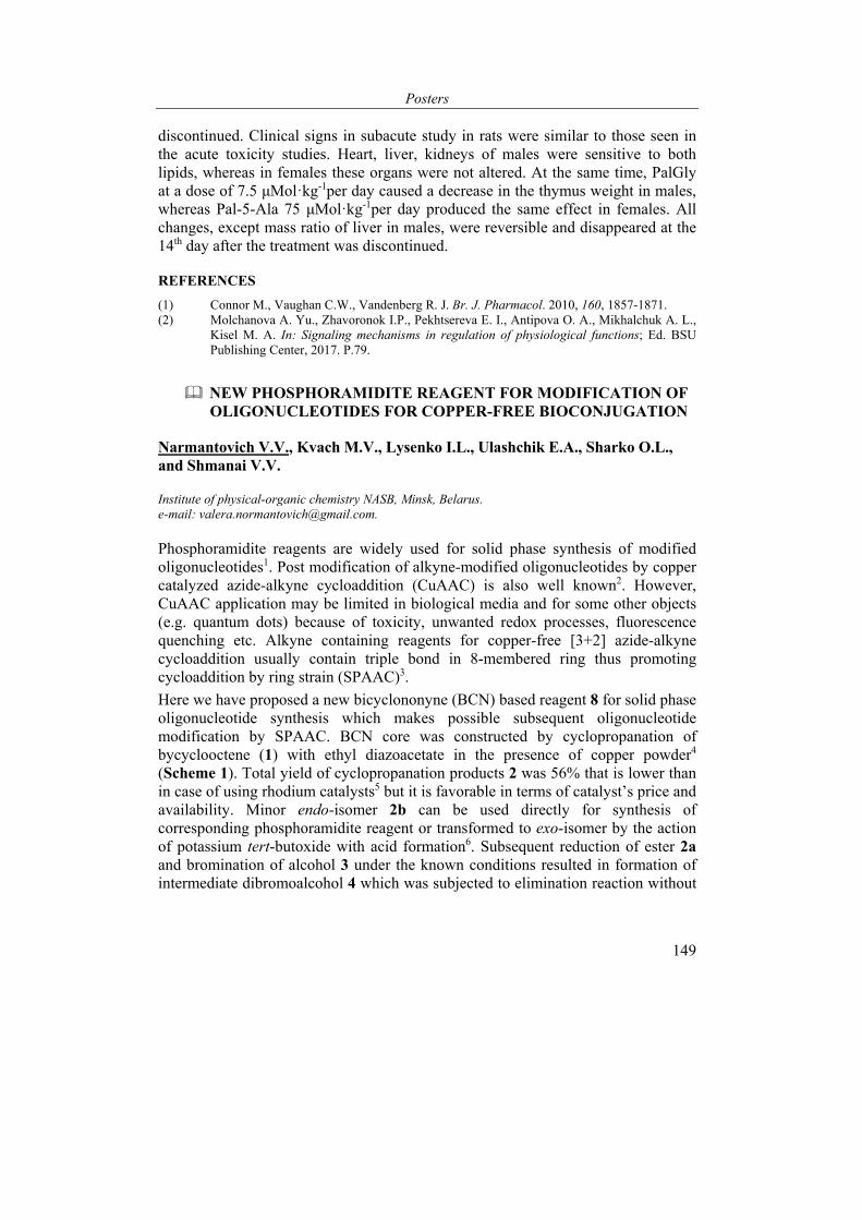
Posters
149
discontinued. Clinical signs in subacute study in rats were similar to those seen in the acute toxicity studies. Heart, liver, kidneys of males were sensitive to both lipids, whereas in females these organs were not altered. At the same time, PalGly at a dose of 7.5 μMol·kg-1per day caused a decrease in the thymus weight in males, whereas Pal-5-Ala 75 μMol·kg-1per day produced the same effect in females. All changes, except mass ratio of liver in males, were reversible and disappeared at the 14th day after the treatment was discontinued.
REFERENCES
(1) Connor M., Vaughan C.W., Vandenberg R. J. Br. J. Pharmacol. 2010, 160, 1857-1871. (2) Molchanova A. Yu., Zhavoronok I.P., Pekhtsereva E. I., Antipova O. A., Mikhalchuk A. L.,
Kisel M. A. In: Signaling mechanisms in regulation of physiological functions; Ed. BSU Publishing Center, 2017. P.79.
NEW PHOSPHORAMIDITE REAGENT FOR MODIFICATION OF OLIGONUCLEOTIDES FOR COPPER-FREE BIOCONJUGATION
Narmantovich V.V., Kvach M.V., Lysenko I.L., Ulashchik E.A., Sharko O.L., and Shmanai V.V.
Institute of physical-organic chemistry NASB, Minsk, Belarus. e-mail: [email protected].
Phosphoramidite reagents are widely used for solid phase synthesis of modified oligonucleotides1. Post modification of alkyne-modified oligonucleotides by copper catalyzed azide-alkyne cycloaddition (CuAAC) is also well known2. However, CuAAC application may be limited in biological media and for some other objects (e.g. quantum dots) because of toxicity, unwanted redox processes, fluorescence quenching etc. Alkyne containing reagents for copper-free [3+2] azide-alkyne cycloaddition usually contain triple bond in 8-membered ring thus promoting cycloaddition by ring strain (SPAAC)3.
Here we have proposed a new bicyclononyne (BCN) based reagent 8 for solid phase oligonucleotide synthesis which makes possible subsequent oligonucleotide modification by SPAAC. BCN core was constructed by cyclopropanation of bycyclooctene (1) with ethyl diazoacetate in the presence of copper powder4 (Scheme 1). Total yield of cyclopropanation products 2 was 56% that is lower than in case of using rhodium catalysts5 but it is favorable in terms of catalyst’s price and availability. Minor endo-isomer 2b can be used directly for synthesis of corresponding phosphoramidite reagent or transformed to exo-isomer by the action of potassium tert-butoxide with acid formation6. Subsequent reduction of ester 2a and bromination of alcohol 3 under the known conditions resulted in formation of intermediate dibromoalcohol 4 which was subjected to elimination reaction without
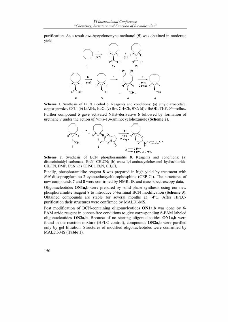
VI International Conference “Chemistry, Structure and Function of Biomolecules”
150
purification. As a result exo-bycyclononyne methanol (5) was obtained in moderate yield.
Scheme 1. Synthesis of BCN alcohol 5. Reagents and conditions: (a) ethyldiazoacetate, copper powder, 80˚C; (b) LiAlH4, Et2O; (c) Br2, CH2Cl2, 0˚C; (d) t-BuOK, THF, 00→reflux.
Further compound 5 gave activated NHS–derivative 6 followed by formation of urethane 7 under the action of trans-1,4-aminocyclohexanole (Scheme 2).
Scheme 2. Synthesis of BCN phosphoramidite 8. Reagents and conditions: (a) disuccinimidyl carbonate, Et3N, CH3CN; (b) trans-1,4-aminocyclohexanol hydrochloride, CH3CN, DMF, Et3N; (c) CEP-Cl, Et3N, CH2Cl2.
Finally, phosphoramidite reagent 8 was prepared in high yield by treatment with N,N-diisopropylamino-2-cyanoethoxychlorophosphine (CEP-Cl). The structures of new compounds 7 and 8 were confirmed by NMR, IR and mass-spectroscopy data.
Oligonucleotides ON1a,b were prepared by solid phase synthesis using our new phosphoramidite reagent 8 to introduce 5′-terminal BCN modification (Scheme 3). Obtained compounds are stable for several months at +4oC. After HPLC-purification their structures were confirmed by MALDI-MS.
Post modification of BCN-containing oligonucleotides ON1a,b was done by 6-FAM azide reagent in copper-free conditions to give corresponding 6-FAM labeled oligonucleotides ON2a,b. Because of no starting oligonucleotides ON1a,b were found in the reaction mixture (HPLC control), compounds ON2a,b were purified only by gel filtration. Structures of modified oligonucleotides were confirmed by MALDI-MS (Table 1).
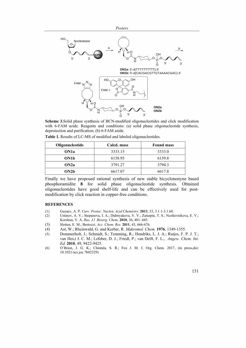
Posters
151
ON1a: 5'-d(TTTTTTTTTT)-3'ON1b: 5'-d(CACGACGTTGTAAAACGAC)-3'
a
O NH
OH H
O
ON2aON2bO N
H
OH H
OP
NN
N
bO
ONucleobase
HO
5' 3'
3'5'P
O
O
OH
O
O
O
O
NH
OHHO
FAM =
FAM
O
O
OH
3'5'
CPG
Scheme 3.Solid phase synthesis of BCN-modified oligonucleotides and click modification with 6-FAM azide. Reagents and conditions: (a) solid phase oligonucleotide synthesis, deprotection and purification; (b) 6-FAM azide.
Table 1. Results of LC-MS of modified and labeled oligonucleotides.
Oligonucleotide Calcd. mass Found mass
ON1a 3333.15 3333.0
ON1b 6158.95 6159.8
ON2a 3791.27 3794.3
ON2b 6617.07 6617.8
Finally we have proposed rational synthesis of new stable bicyclononyne based phosphoramidite 8 for solid phase oligonucleotide synthesis. Obtained oligonucleotides have good shelf-life and can be effectively used for post-modification by click reaction in copper-free conditions.
REFERENCES
(1) Guzaev, A. P. Curr. Protoc. Nucleic Acid Chemistry. 2013, 53, 3.1.1-3.1.60. (2) Ustinov, A. V.; Stepanova, I. A.; Dubnyakova, V. V.; Zatsepin, T. S.; Nozhevnikova, E. V.;
Korshun, V. A. Rus. J.l Bioorg. Chem. 2010, 36, 401–445. (3) Sletten, E. M.; Bertozzi. Acc. Chem. Res. 2011, 43, 666-676. (4) Ast, W.; Rheinwald, G. und Kerber, R. Makromol. Chem. 1976, 1349-1355. (5) Dommerholt, J.; Schmidt, S.; Temming, R.; Hendriks, L. J. A.; Rutjes, F. P. J. T.;
van Hes,t J. C. M.; Lefeber, D. J.; Friedl, P.; van Delft, F. L., Angew. Chem. Int. Ed. 2010, 49, 9422-9425.
(6) O’Brien, J. G. K.; Chintala, S. R.; Fox J. M. J. Org. Chem. 2017, (in press,doi: 10.1021/acs.joc.7b02329).

VI International Conference “Chemistry, Structure and Function of Biomolecules”
152
DE NOVO DESIGN OF NON-STEROIDAL AROMATASE INHIBITORS: A COMPUTATIONAL STUDY
Alexander M. Andrianov1*, Grigory I. Nikolaev2, Ivan A. Kashyn2, Yuri V. Kornoushenko1, and Sergei A. Usanov1
1Institute of Bioorganic of the National Academy of Sciences of Belarus, Minsk, Republic of Belarus; 2United Institute of Informatics Problems of the National Academy of Sciences of Belarus, Minsk, Republic of Belarus e-mail: [email protected]
The third-generation aromatase inhibitors (AIs), which are now used as first-line therapy in the treatment of early- and advanced-stage breast cancer in postmenopausal women, include two categories: the reversible non-steroidal inhibitors anastrozole and letrozole and the steroidal inhibitor exemestane. Non-steroidal AIs are imidazoles or triazoles that bind to the active site of CYP19A1 by coordinating the heme iron atom of the enzyme through a heterocyclic nitrogen electron pair. Steroidal inhibitors may exhibit either competitive inhibition, irreversible inhibition, or mechanism-based inhibition of aromatase. Among them, exemestane is a mechanism-based inhibitor, which is transformed by aromatase into a reactive species that irreversibly binds to the active site of the enzyme. Although AIs are currently popular and effective in the treatment of postmenopausal estrogen receptor positive breast cancer, the search for novel drugs still remains necessary to avoid the risk of possible emerging resistances to available drugs as well as to reduce toxicity and undesirable side effects associated with a prolonged use.
The publication of a high resolution X-ray structure of human aromatase has opened the way to a greater understanding of the structural basis for estrogen synthesis and substrate/inhibitor recognition and may encourage efforts to discover novel AIs through structure-based molecular design.
In this study, computer-aided design of the high-affinity AIs based on 1,2,4-triazole derivatives was performed by molecular modeling tools. Potential biological activity of the designed compounds was evaluated by molecular docking and quantum chemistry calculations. As a result, six hits that form a coordinate bond with the heme iron of the enzyme and effectively interact with its substrate-binding site were identified. Analysis of intermolecular interactions appearing in the docked structures of these ligands with aromatase was carried out and the enthalpies of their formation were calculated. According to the predicted binding modes, each of the identified compounds shows peculiar interactions with the enzyme binding pocket, the interaction being realized between the triazole ring and the heme iron, van der Waals interactions with the hydrophobic pocket lined by Arg-115, Ile-133, Phe-134, Trp-224, Тhr-310, Val-370, Met-374, Leu-477, Ser-478, and the hydrogen bond with Met-374 NH, which is also involved in a hydrogen bond with the natural substrate androstenedione. In addition, the identified compounds form van der
Posters
153
Waals contacts with the CYP19A1 heme, and -conjugated systems of individual molecules participate in specific --interactions with the pyrrole rings of a prosthetic group. Finally, the docked ligand/aromatase structures are energetically stable, in line with the data on the binding enthalpy calculations.
In summary, the conclusions that can be drawn by the new identified AIs are that, in addition to the interaction between the triazole rings and the heme iron, hydrophobic contacts play a pivotal role in ligand binding, and hydrogen bond involving Met-374 is also essential for ligand recognition. Based on the data obtained, the designed compounds are suggested to present good scaffolds for the development of novel effective drugs against breast cancer.
Surely the properties of these virtual compounds warrant further biological characterization as cellular assays to confirm in vitro their interesting in silico profile.
SYNTHESIS OF NOVEL TRIFLUOROMETHYL-CONTAINING N,O-HETEROCYCLES
Yuri A. Piven1*, Viktoryia A. Smaliak2, Tatyana S. Khlebnicova1, and Fedor A. Lakhvich1
1 Institute of Bioorganic Chemistry, National Academy of Sciences of Belarus, Acad. Kuprevicha Str. 5/2, 220141 Minsk, Belarus, 2Belarusian State University, Faculty of chemistry, Leningradskaya Str. 14, 220030, Minsk, Belarus e-mail: [email protected]
Numerous heterocyclic compounds possessing an isoxazole ring (both isolated and fused to other mono or polycyclic systems) have been used as a base structure for the design of many pharmaceutical and agrochemical agents. The benzisoxazole scaffold and its analogues are important pharmacophores that can be found in biologically active compounds across a number of different therapeutic areas as anti-HIV, antimicrobial, antipsychotic, antiinflammatory, analgesic, anticancer and so on1. A large number of fluorine containing heterocyclic compounds are well known as important marketed drugs2.
An efficient protocol for the synthesis of novel fluorine-containing N,O-heterocyclic compounds has been developed from regioisomeric 3-trifluoromethyl-6,7-dihydrobenzisoxazolones (1,2). The oxidation of benzisoxazolones (1,2) in acetic acid with selenium dioxide in the presence of sulfuric acid led to 3-(trifluoromethyl)-6,7-dihydrobenzoisoxazole-4,5-diones (3,4). By heating compounds (3,4) with equimolar amount of o-phenylenediamine in boiling ethanol we obtained trifluoromethyl-containing 4,5-dihydroisoxazolo[4,5-a]- or [4,3-a]phenazines (5,6) respectively. Compounds (3,4) reacted with 1 equiv of benzaldehyde and excess ammonium acetate in boiling acetic acid to give 8-
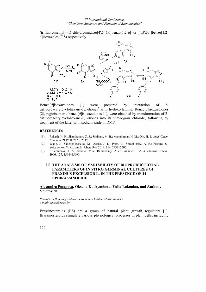
VI International Conference “Chemistry, Structure and Function of Biomolecules”
154
(trifluoromethyl)-4,5-dihydroimidazo[4',5':5,6]benzo[1,2-d]- or [4',5':3,4]benzo[1,2-c]isoxazoles (7,8) respectively.
Benzo[d]isoxazolones (1) were prepared by interaction of 2-trifluoroacetylcyclohexane-1,3-diones3 with hydroxylamine. Benzo[c]isoxazolones (2), regioisomeric benzo[d]isoxazolones (1), were obtained by transformation of 2-trifluoroacetylcyclohexane-1,3-diones into its vinylogous chloride, following by treatment of the latter with sodium azide in DMF.
REFERENCES
(1) Rakesh, K. P.; Shantharam, C. S.; Sridhara, M. B.; Manukumar, H. M.; Qin, H.-L. Med. Chem. Commun. 2017, 8, 2023−2039.
(2) Wang, J.; Sánchez-Rosello, M.; Aceña, J. L.; Pozo, C., Sorochinsky, A. E.; Fustero, S.; Soloshonok, V. A.; Liu, H. Chem Rev 2014, 114, 2432−2506.
(3) Khlebnicova, T. S.; Isakova, V.G.; Baranovsky, A.V.; Lakhvich, F.A. J. Fluorine Chem., 2006, 127, 1564−15690.
THE ANALYSIS OF VARIABILITY OF BIOPRODUCTIONAL PARAMETERS OF IN VITRO GERMINAL CULTURES OF FRAXINUS EXCELSIOR L. IN THE PRESENCE OF 24-EPIBRASSINOLIDE
Alexandra Potapova, Oksana Kudryashova, Yulia Lukonina, and Anthony Volotovich
Republican Breeding and Seed Production Center, Minsk, Belarus e-mail: [email protected]
Brassinosteroids (BS) are a group of natural plant growth regulators [1]. Brassinosteroids stimulate various physiological processes in plant cells, including

Posters
155
membrane potential changes, photosynthetic and enzymatic activity, and the phytohormone balance. The effects of BS on plant growth and development show a trend of synergism with other phytohormones.
Research on application of 24-epibrassinolide on the in vitro germinal cultures of European ash was for the first time conducted since 2017 on the basis of Republican Breeding and Seed Production Center (Minsk, Belarus). Here, the results of application of 0.01 mg per liter of 24-epibrassinolide in agarized nutrient mediums on a different micro-, macro-salt basis (WPM, Anderson’s medium, MS), in combination with 0.5 mg per liter of zeatin to the germs of European ash seeds of different geographical origin at the stage of in vitro aseptic introduction are given. The analysis of bioproductional parameters (lengths of a hypocotyl, length of cotyledonous leaves, length of roots, quantity of the real leaves and diameter of callus) was carried out in 3 weeks after of in vitro cultivation.
Mathematical analysis of the data (the means ± standard error, calculation of least significant differences at significance levels of P < 0.05 and P < 0.01) was performed according to standard methods of variation statistics [2] using statistics analysis software STATISTICA 6.0 [3]. The dispersive analysis of data and calculation of share of factors influence on variability of the studied features carried out in the program of the statistical analysis AB-Stat 1.0 developed at Institute of Genetics and Cytology of NAS of Belarus [4].
As a result of the conducted research it is established that the presence of a 24-epibrassinolide as a part of medium on the basis of Anderson’s or MS, in combination with zeatin, promotes reliable (at P < 0.01) reduction at 1.7-2.7 times of callus diameter; as well as a part of medium on the basis of Anderson’s or WPM stimulates formation of roots, despite of presence of cytokinin (zeatin). Besides, the combination of 24-epibrassinolide with zeatin, in most cases, gives reliable (at P < 0.01) reduction at 1.5-2.7 times (and up to zero) quantities of the real leaves at in vitro germinal regenerants of European ash. REFERENCES
(1) Brassinosteroids. A new class of plant hormones; Khripach, V.A.; Zhabinskii V.N.; Groot A.E., Ed.; San Diego: Academic Press, 1999.
(2) Fielplot technique; Dospehov, B. A., Ed.; M, 1985. (3) STATISTICA; Borovikov, V. P., Ed.; SPb, 2001. (4) Anoshenko, B. Yu. Genetics. 1994, 30, 8-9.
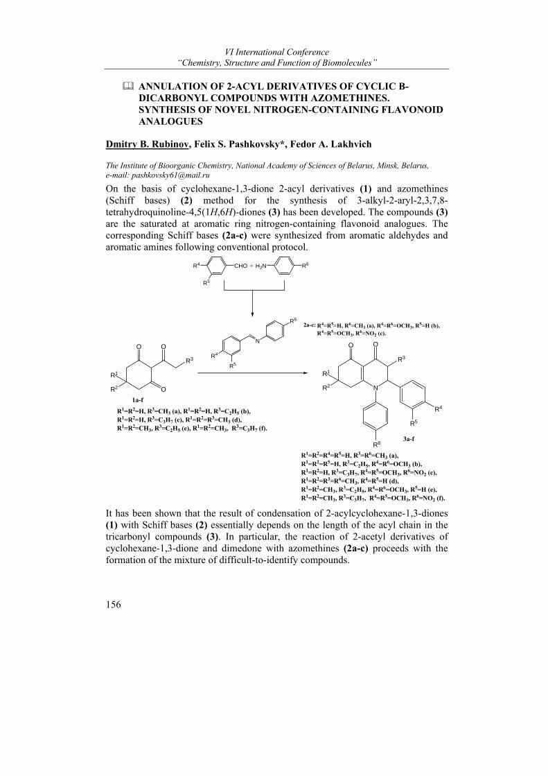
VI International Conference “Chemistry, Structure and Function of Biomolecules”
156
ANNULATION OF 2-ACYL DERIVATIVES OF CYCLIC Β-DICARBONYL COMPOUNDS WITH AZOMETHINES. SYNTHESIS OF NOVEL NITROGEN-CONTAINING FLAVONOID ANALOGUES
Dmitry B. Rubinov, Felix S. Pashkovsky*, Fedor A. Lakhvich
The Institute of Bioorganic Chemistry, National Academy of Sciences of Belarus, Minsk, Belarus, e-mail: [email protected]
On the basis of cyclohexane-1,3-dione 2-acyl derivatives (1) and azomethines (Schiff bases) (2) method for the synthesis of 3-alkyl-2-aryl-2,3,7,8-tetrahydroquinoline-4,5(1Н,6Н)-diones (3) has been developed. The compounds (3) are the saturated at aromatic ring nitrogen-containing flavonoid analogues. The corresponding Schiff bases (2a-c) were synthesized from aromatic aldehydes and aromatic amines following conventional protocol.
R4
R5
CHO + H2N R6
N
R6
R4
R5
R1
R2
O
O
R3
O
R2
R1
N
O O
R3
R4
R5
R6
2a-c:
1a-f
3a-f
R1=R2=H, R3=CH3 (a), R1=R2=H, R3=C2H5 (b), R1=R2=H, R3=C3H7 (c), R1=R2=R3=CH3 (d), R1=R2=CH3, R3=C2H5 (e), R1=R2=CH3, R3=C3H7 (f).
R4=R5=H, R6=CH3 (a), R4=R6=OCH3, R5=H (b), R4=R5=OCH3, R6=NO2 (c).
R1=R2=R4=R5=H, R3=R6=CH3 (a), R1=R2=R5=H, R3=C2H5, R4=R6=OCH3 (b), R1=R2=H, R3=C3H7, R4=R5=OCH3, R6=NO2 (c), R1=R2=R3=R6=CH3, R4=R5=H (d), R1=R2=CH3, R3=C2H5, R4=R6=OCH3, R5=H (e), R1=R2=CH3, R3=C3H7, R4=R5=OCH3, R6=NO2 (f).
It has been shown that the result of condensation of 2-acylcyclohexane-1,3-diones (1) with Schiff bases (2) essentially depends on the length of the acyl chain in the tricarbonyl compounds (3). In particular, the reaction of 2-acetyl derivatives of cyclohexane-1,3-dione and dimedone with azomethines (2a-c) proceeds with the formation of the mixture of difficult-to-identify compounds.

Posters
157
On the contrary, β-triketones (3a-f) containing 2-propionyl, 2-butanoyl and 2-pentanoyl substituents readily condense with the Schiff bases (2a-c) to give the corresponding 3-alkyl-2-aryl-2,3,7,8-tetrahydroquinoline-4,5(1Н,6Н)-diones (3а-f) in the yield of 65-80%. The compounds (3а-f) are hitherto unknown nitrogen-containing flavonoid analogues.
Financial support provided by the Belorussian Foundation for Basic Research (grant X16P-042) is greatly appreciated.
STUDYING OF PROTEINOGENIC AMINO ACIDS AS LIGANDS FOR BINDING AND ELIMINATION OF PROINFLAMMATORY CYTOKINES FROM HUMAN PLASMA
Tatiana Ryabzeva1, Denis Makarevich2, and Eugeniy Ermola2
1Belorussian state medical university, Minsk, Belarus; 2Institute of bioorganic chemistry NAN Belarus, Minsk, Belarus e-mail: [email protected]
Significant efforts of modern scientists are direct to developing pro-inflammatory cytokines inhibitors. In the scientific literature there is a lot of data proving the role of TNF-α, IL-8 and IL-6 in the pathogenesis of a large number of diseases. Especially evident is the involvement of these cytokines in the development of autoimmune diseases: rheumatoid arthritis, multiple sclerosis, systemic lupus erythematosus, psoriasis, Crohn's disease1. Numerous studies are directed to developing the monoclonal antibodies against the corresponding cytokines. However, using the monoclonal antibodies drugs is associated with the development of a number of side effects: a delayed-type hypersensitivity reaction, infectious complications (tuberculosis, viral hepatitis), lymphoproliferative diseases, leukopenia, thrombocytopenia and neutropenia2,3.
The aim of this work was the comparative analysis of the cytokine-binding activity of proteinogenic amino acids with pro-inflammatory cytokines (TNF-α, IL-6 and IL-8).
The virtual docking of the interaction of proteinogenic amino acids with TNF-α, IL-6 and IL-8 showed that binding energies lie in the range from -2,80 to -5,60 kcal/mol for IL-6, from -2,30 to -4,50 kcal / mol for the IL-8 monomer, from -2,20 to -4,80 kcal/mol for the IL-8 dimer and from -2,65 to -5,85 kcal/mol for TNF-α. Thus, from the point of view of theoretical calculations, aromatic and positively charged amino acids are the most promising for the further construction of the most effective oligopeptides for binding and eliminating cytokines from blood plasma. Further we conducted experiments to study the change of the concentration of cytokines in the blood plasma after interaction with the fixed on the polymer matrix proteinogenic amino acids. The results revealed that the cytokines concentration
VI International Conference “Chemistry, Structure and Function of Biomolecules”
158
decrease by more than 30% is observed in the interaction IL-6 with Cys, Gly and Pro; IL-8 with Ala, Gly, Pro and Tyr; TNF-α with Gly, Leu and Trp. In conclusion, the experimental data conformed that effective oligopeptide for binding IL-6, IL-8 and TNF-α should be consisted of aromatic amino acids and Cys, Gly Ala, Pro, Leu.
REFERENCES
(1) Vinay DS, Kwon BS, Clin Exp Immunol, 2011, 162(2), 145-157 (2) Aggarwal BB, Gupta SC, Blood, 2012, (119), 651-665 (3) Sheetal B. Desai, Daniel E. Furst, Best practice&research clinical rheumatology, 2006, 757-
790
BRASSINOSTEROID SALICYLATES AS BIOTIC STRESS PROTECTORS IN BARLEY
Aleh P. Savachka1, Neli E. Manzhalesava2, Raisa P. Litvinovskaya1, Svetlana N. Palyanskaya2, Larysa A. Karytska2, and Vladimir A. Khripach1
1Institute of Bioorganic Chemistry, National Academy of Sciences of Belarus, Minsk, Belarus; 2V.F. Kuprevich Institute of Experimental Botany, National Academy of Sciences of Belarus, Minsk, Belarus e-mail: [email protected]
In continuation of our work on the synthesis and study of the activity of brassinosteroid esters with various acids1-4, a number of salicylic acid esters with brassinosteroids of the 24R-methyl group was synthesized. The reaction of brassinosteroids, 24-epibrassinolide 1 (EBl), 24-epicastasterone 2 (EBk) and 6-deoxo-24-epicastasterone 3 (EBd), with 2-O-benzylsalicylic anhydride, obtained in situ from the acid, proceeded smoothly and gave 2-acyl derivatives 4-6 respectively. The benzyl protection group in derivatives 4-6 was removed by hydrogenolysis over a Pd catalyst in MeOH. This produced target products 7-9.
The effect of the resulting conjugates of phytohormonal steroids on the processes of barley germination was studied in laboratory experiments. It has been shown that all the studied phytohormonal steroids 1-3 and their derivatives 7-9 possess a growth regulation activity. Germination energy and seed germination under their influence increased by 10-15%, however, modified phytohormonal steroids exert a stimulating effect in a narrower range of concentrations than natural compounds.
Determination of the antibiotic activity of phytohormonal steroids and their derivatives was carried out under laboratory conditions on the model phytopathosystem of barley-phytopathogenic fungus Helminthosporium teres Sacc. [Drechslera teres (Sacc) Shoem.] - causative agent of the barley net blotch.
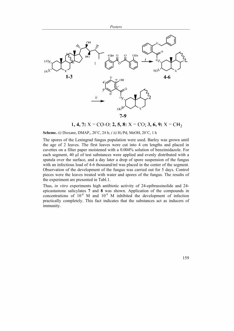
Posters
159
Scheme. (i) Dioxane, DMAP,, 20˚C, 24 h; ( ii) H2/Pd, MeOH, 20˚C, 1 h
The spores of the Leningrad fungus population were used. Barley was grown until the age of 2 leaves. The first leaves were cut into 4 cm lengths and placed in cuvettes on a filter paper moistened with a 0.004% solution of benzimidazole. For each segment, 40 μl of test substances were applied and evenly distributed with a spatula over the surface, and a day later a drop of spore suspension of the fungus with an infectious load of 4-6 thousand/ml was placed in the center of the segment. Observation of the development of the fungus was carried out for 5 days. Control pieces were the leaves treated with water and spores of the fungus. The results of the experiment are presented in Tabl.1.
Thus, in vitro experiments high antibiotic activity of 24-epibrassinolide and 24-epicastastone salicylates 7 and 8 was shown. Application of the compounds in concentrations of 10-8 M and 10-9 M inhibited the development of infection practically completely. This fact indicates that the substances act as inducers of immunity.
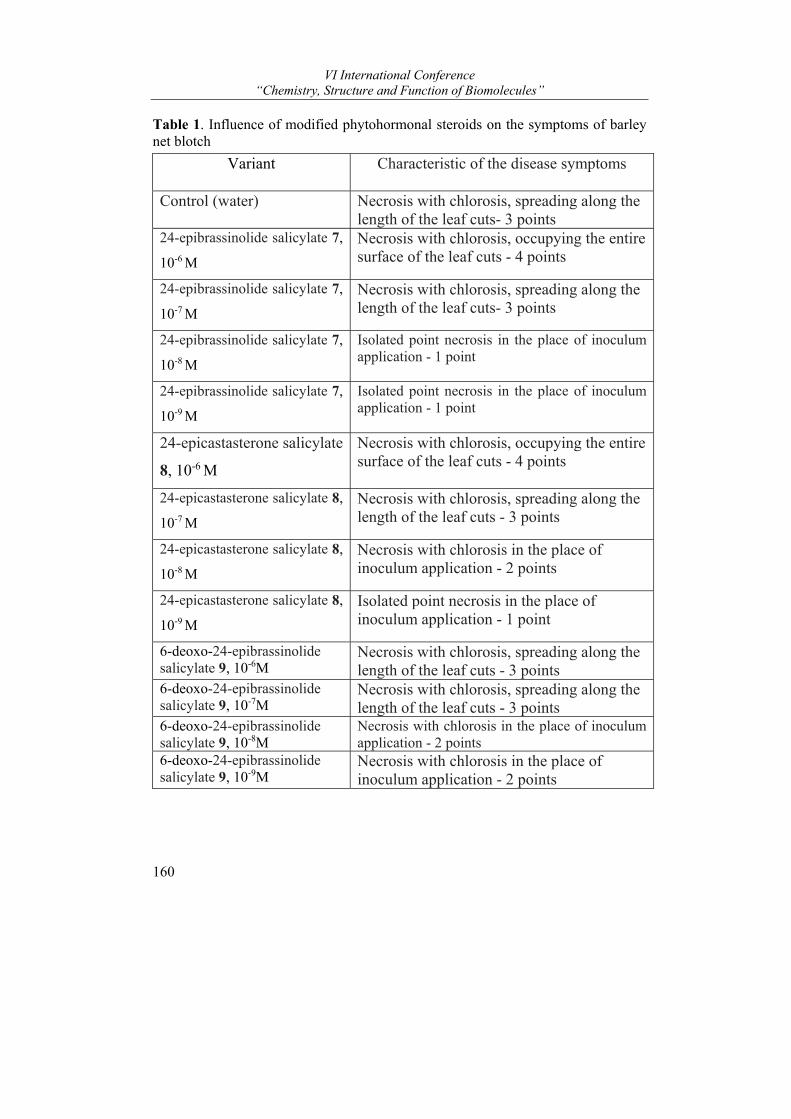
VI International Conference “Chemistry, Structure and Function of Biomolecules”
160
Table 1. Influence of modified phytohormonal steroids on the symptoms of barley net blotch
Variant Characteristic of the disease symptoms
Control (water)
Necrosis with chlorosis, spreading along the length of the leaf cuts- 3 points
24-epibrassinolide salicylate 7,
10-6 М
Necrosis with chlorosis, occupying the entire surface of the leaf cuts - 4 points
24-epibrassinolide salicylate 7,
10-7 М
Necrosis with chlorosis, spreading along the length of the leaf cuts- 3 points
24-epibrassinolide salicylate 7,
10-8 М
Isolated point necrosis in the place of inoculum application - 1 point
24-epibrassinolide salicylate 7,
10-9 М
Isolated point necrosis in the place of inoculum application - 1 point
24-epicastasterone salicylate
8, 10-6 М
Necrosis with chlorosis, occupying the entire surface of the leaf cuts - 4 points
24-epicastasterone salicylate 8,
10-7 М
Necrosis with chlorosis, spreading along the length of the leaf cuts - 3 points
24-epicastasterone salicylate 8,
10-8 М
Necrosis with chlorosis in the place of inoculum application - 2 points
24-epicastasterone salicylate 8,
10-9 М
Isolated point necrosis in the place of inoculum application - 1 point
6-deoxo-24-epibrassinolide salicylate 9, 10-6М
Necrosis with chlorosis, spreading along the length of the leaf cuts - 3 points
6-deoxo-24-epibrassinolide salicylate 9, 10-7М
Necrosis with chlorosis, spreading along the length of the leaf cuts - 3 points
6-deoxo-24-epibrassinolide salicylate 9, 10-8М
Necrosis with chlorosis in the place of inoculum application - 2 points
6-deoxo-24-epibrassinolide salicylate 9, 10-9М
Necrosis with chlorosis in the place of inoculum application - 2 points
Posters
161
REFERENCES
(1) Litvinovskaya R.P., Minin P.S., Raiman M.E., Zhilitskaya G.A., Kurtikova A.L., Kozharnovich K.G., Derevyanchuk M.V., Kravets V.S., Khripach V.A. Chem. Nat. Comp., 2013, 49, 478-485.
(2) Litvinovskaya R.P., Vayner A.A., Zhylitskaya H.A., Kolupaev Yu.E., Savochka O.P., Khipach V.A. Chem. Nat. Comp., 2016, 52, 394-398.
(3) Zhylitskaya H., Chashchina N., Litvinovskaya R., Zavadskaya M., Zhabinskii V., Khripach V. Steroids, 2017, 117, 2-10.
(4) Pat. BY № 18530.
INTERACTIONS OF RECOMBINANT HUMAN LACTOFERRIN (rhLF) AND NATURAL LACTOFERRINS WITH ANTI-rhLF ANTIBODIES IN A PROTOTYPE ENZYME IMMUNOASSAY SYSTEM
Dmitry Semenov*, Irina Vashkevich, and Oleg Sviridov
Institute of Bioorganic Chemistry, National Academy of Sciences of Belarus, Minsk, Belarus e-mail: [email protected]
Lactoferrin (LF) is a mammalian iron-binding glycosylated protein with a molecular mass of approximately 80 000 g/mol. The single polypeptide chain of this highly basic glycoprotein (pI~8.7) is folded into two symmetrical lobes linked by a small peptide fragment (hinge region). Each lobe contains a metal-binding site, and the protein has different spatial conformations depending on whether or not it is associated with Fe3+. Numerous biomedical studies have shown that LF takes part in a large number of important physiological processes and has a wide range of health enhancing and protection activities that are essential for newborns and beneficial for adults. In global biotechnology, human LF is manufactured as a recombinant protein (rhLF) obtained in several expression systems including transgenic animals. In this country, the herb of goats that produce rhLF was generated and a procedure for the purification of physiologically active rhLF from transgenic goat milk was developed as the result of works supported by Belarus-Russia Union State programs1. Recently, rhLF immunoaffinity chromatography and enzyme immunoassay using highly specific single-domain antibodies raised in a camel against native human LF and selected by phage display-based technology were described2.
In our work, rhLF was used as an immunogen for the first time, and interactions of polyclonal antibodies generated in rabbits with rhLF and native LFs were studied in a model system of an indirect competitive enzyme-linked immunosorbent assay (ELISA). High purity (95-98 %) freeze dried rhLF was acquired from Institute of Microbiology, NASB as a finished product of a laboratory scale technology elaborated on the basis of the published technique1. At first, absorbance

VI International Conference “Chemistry, Structure and Function of Biomolecules”
162
measurements were made in phosphate buffered saline (pH 7.2-7.4) to determine rhLF’s extinction coefficient and to assess the iron content of the protein. The values of 280 = 89 203 M-1. cm-1 (E280
1% = 11.2) and A280/A465 = 129 were found. The experimental extinction coefficient was in a good agreement with a theoretical one calculated as the sum of molar absorbance of known numbers of tryptophan, tyrosine and cystine residues in rhLF (280 = 85 700 M-1.cm-1). The value of A280/A465 corresponded to 11.3 % of rhLF saturation with iron ions. These data were used in further experiments to prepare rhLF solutions with accurate concentrations.
To produce anti-rhLF antibodies (Ab), rhLF was injected subcutaneously into multiple sites of Grey Giant rabbits (1.0 or 0.5 mg/ml in Freund’s complete and incomplete adjuvants) at 2- or 3-week intervals. The titer of antibodies reached its maximum after 5 months of immunization. The dilution of finished antiserum of about 1:50 000 provided a colorimetric signal of 1.2-1.5 optical units when the antibodies bound to rhLF passively adsorbed in a microplate and were detected with an anti-rabbit immunoglobulin G (IgG)-horseradish peroxidase (HRP) conjugate.
The experiments on anti-rhLF Ab-rhLF binding kinetics were carried out with the use of microplate wells containing immobilized rhLF and an anti-rhLF Ab solution without adding the competitor (Fig. 1) or with added rhLF (Fig. 2) at 25 or 37 oC under continuous shaking. After washing the wells, the formed complex was detected by the anti-rabbit IgG-HRP conjugate solution. In a wide time interval, the intensities of colorimetric signals A450 (3,3’,5,5’-tetramethylbenzidine / H2O2 + H2SO4) in terms of optical densities (Fig. 1) and the extent of competitive inhibition Bi/Bo (Fig. 2) appeared to meet the basic requirements of a rhLF enzyme immunoassay.
Also, we studied the dependence of anti-rabbit IgG-HRP conjugate binding to immobilized Ab-antigen complex on the length of incubation (Fig. 3). It was shown that the incubation for 20 min at 25 oC is sufficient to reach maximum optical density and reliably label the bound anti-rhLF Ab.
Fig. 1. The kinetics of anti-rhLF Ab binding to immobilized rhLF
at 25 (1) or 37 oC (2), respectively

Posters
163
Fig. 2. The competitive inhibition of anti-rhLF Ab binding to immobilized rhLF by 0.5 μg/ml rhLF in a liquid phase at 25 (1) or 37 oC, respectively
Finally, we compared immunoreactivities of LFs as components of breast milk of humans and normal and transgenic goats, performing the indirect ELISA under conditions selected above: first incubation – 5 min at 25 oC with shaking, second incubation – 20 min at 25 oC with shaking (Fig. 4).
Fig. 3. The kinetics of anti-rabbit IgG-HRP conjugate binding to anti-rhLF Ab-rhLF complex on the solid phase at 25 oC Fig. 4. The competitive inhibition of anti-rhLF Ab binding to immobilized rhLF in the presence of various dilutions of defatted milk from humans (1), transgenic goats (2) and normal goats (3)
In conclusion, we found that the new anti-rhLF Ab explored in this work exhibited good sensitivity to rhLF in the model indirect ELISA system and interacted very similarly with human, transgenic goat and normal goat milk. This property is useful
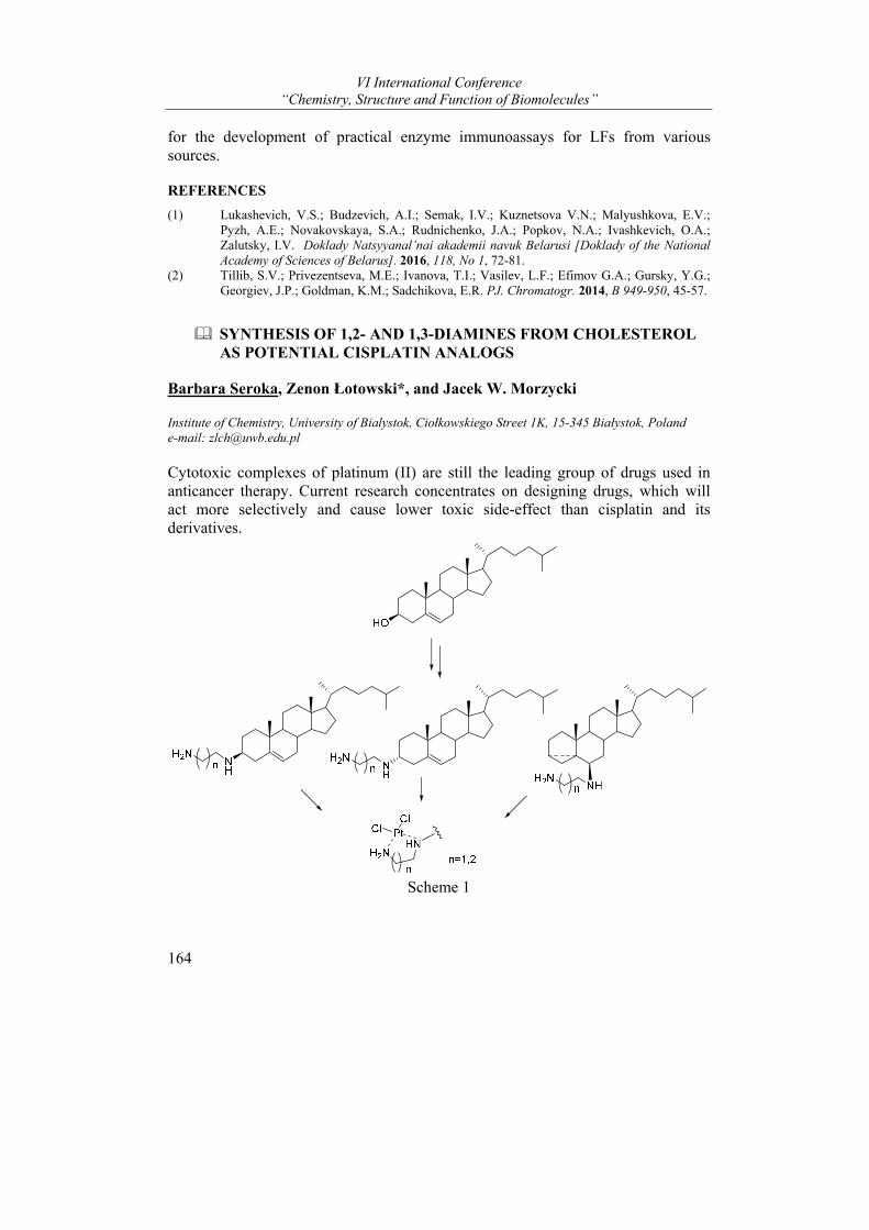
VI International Conference “Chemistry, Structure and Function of Biomolecules”
164
for the development of practical enzyme immunoassays for LFs from various sources.
REFERENCES
(1) Lukashevich, V.S.; Budzevich, A.I.; Semak, I.V.; Kuznetsova V.N.; Malyushkova, E.V.; Pyzh, A.E.; Novakovskaya, S.A.; Rudnichenko, J.A.; Popkov, N.A.; Ivashkevich, O.A.; Zalutsky, I.V. Doklady Natsyyanal’nai akademii navuk Belarusi [Doklady of the National Academy of Sciences of Belarus]. 2016, 118, No 1, 72-81.
(2) Tillib, S.V.; Privezentseva, M.E.; Ivanova, T.I.; Vasilev, L.F.; Efimov G.A.; Gursky, Y.G.; Georgiev, J.P.; Goldman, K.M.; Sadchikova, E.R. PJ. Chromatogr. 2014, B 949-950, 45-57.
SYNTHESIS OF 1,2- AND 1,3-DIAMINES FROM CHOLESTEROL AS POTENTIAL CISPLATIN ANALOGS
Barbara Seroka, Zenon Łotowski*, and Jacek W. Morzycki
Institute of Chemistry, University of Bialystok, Ciołkowskiego Street 1K, 15-345 Białystok, Poland e-mail: [email protected]
Cytotoxic complexes of platinum (II) are still the leading group of drugs used in anticancer therapy. Current research concentrates on designing drugs, which will act more selectively and cause lower toxic side-effect than cisplatin and its derivatives.
Scheme 1
Posters
165
The research project is based on an experimental approach and involves the fundamental study associated with designing, preparation, and biological activity evaluation of steroidal platinum(II) complexes.
We designed and received a series of steroid vic-diamines based on cholesterol (Scheme 1) as potential ligands that, in the form of complexes with platinum ions, will be subjected to biological tests for their anti-cancer properties. We also plan to do syntheses of other model diamine systems based on already used, as well as new steroid compounds (eg diosgenin).
REFERENCES
(1) Trynda-Lemiesz L., Sliwińska-Hill U., NOWOTWORY Journal of Oncology, 2011, 61, 5, 465-474.
(2) Kvasnica M, Rárová L, Oklešťková J, Budĕšinský M, Kohout L. Bioorg. Med. Chem. 2012, 20: 6969.
(3) H Johnstone T. C., Lippard S. J., J. Am. Chem. Soc. 2014, 136, 2126−2134. (4) Cai S., Peterson B. R., Sun Q., Org. Lett., 2009, 11 (3), 567–570.
The authors thank the Polish National Science Centre for the grant support (2014/15/B/ST5/02129).
BIOINFORMATIC ANALYSIS OF THE STRUCTURAL PECULIAR PROPERTIES OF LAMA GLAMA HEAVY-CHAIN ANTIBODIES
Michail A. Shapira1 and Dmitri O. Dormeshkin1
1 Institute of Bioorganic Chemistry of the National Academy of Sciences of Belarus
Tremendous efforts have been expended over the past two and a half decades to understand many aspects of camelid antibodies, from their biology, evolution, and immunogenetics to their potential applications in various fields of research and medicine (eg.1).
Of the three immunoglobulin G (IgG) isotypes described to occur in camelids, IgG2 and IgG3 are distinct in that they devoid of light chains. These heavy-chain antibodies (HCAbs) constitute approximately 50% of the IgG in llama serum and as much as 75% of the IgG in camel serum (eg.2).
Investigations of camelid antibodies have focused largely on manipulating or mimicking the architecture of the variable domains of the HCAbs (VHH) for application to medical therapy and biotechnology (eg.3). Unlike conventional antibodies, HCAbs use a single VHH to bind an epitope. The absence of the light-chain variable domain is compensated for by extended complementarity-determining regions (CDR) that provide an adequate antigen-binding surface and demonstrate affinities comparable to those of conventional antibodies (eg.4).
Due to the extended CDR3 loop HCAbs have the unique ability to bind molecular clefts, such as the active site of enzymes, or to interfere with protein-protein
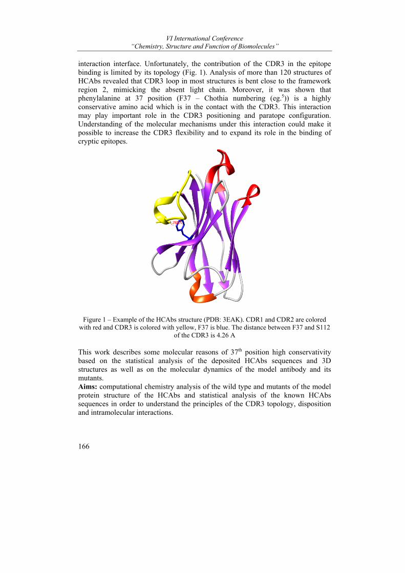
VI International Conference “Chemistry, Structure and Function of Biomolecules”
166
interaction interface. Unfortunately, the contribution of the CDR3 in the epitope binding is limited by its topology (Fig. 1). Analysis of more than 120 structures of HCAbs revealed that CDR3 loop in most structures is bent close to the framework region 2, mimicking the absent light chain. Moreover, it was shown that phenylalanine at 37 position (F37 – Chothia numbering (eg.5)) is a highly conservative amino acid which is in the contact with the CDR3. This interaction may play important role in the CDR3 positioning and paratope configuration. Understanding of the molecular mechanisms under this interaction could make it possible to increase the CDR3 flexibility and to expand its role in the binding of cryptic epitopes.
Figure 1 – Example of the HCAbs structure (PDB: 3EAK). CDR1 and CDR2 are colored
with red and CDR3 is colored with yellow, F37 is blue. The distance between F37 and S112 of the CDR3 is 4.26 A
This work describes some molecular reasons of 37th position high conservativity based on the statistical analysis of the deposited HCAbs sequences and 3D structures as well as on the molecular dynamics of the model antibody and its mutants. Aims: computational chemistry analysis of the wild type and mutants of the model protein structure of the HCAbs and statistical analysis of the known HCAbs sequences in order to understand the principles of the CDR3 topology, disposition and intramolecular interactions.
Posters
167
Materials and methods.
The structure of NbBCII10 huminized antibodies (PDB: 3EAK) was chosen as a model protein molecule as it possesses very high stability and could maintain a vast variety of paratope topologies after CDR-grafting. Molecular dynamics simulations were done using AMBER16 software. For statistical analysis custom scripts were created using python 3.5 as the programming language. For the data visualization and analysis OriginPro 2016 application was used.
Conclusions: 1. We have carried out molecular dynamics simulations and analysis of the
wild type 3EAK scaffold and its F37X (X=all the possible amino acids) mutants
2. Statistical analysis of the HCAbs sequences has revealed that phenylalanine is not only possible amino acid at the 37th position but most preferable. Such distribution is dictated by the amino acid surrounding and drastically affects the antibody stability.
3. Some of the 3EAK mutants have shown the stable scaffold structure “behavior” as well as the longer distances between the framework region and CDR3 comparing to the wild type that makes them promising candidates to the role of universal scaffolds for synthetic camelid antibody libraries development.
REFERENCES
(1) M. Arbabi-Ghahroudi. Front Immunol. 2017, 8: 1589, 1-8. (2) L. P. Daley, L. F. Gagliardo, M. S. Duffy, M. C. Smith, and J. A. Appleton. Clin Diagn Lab
Immunol. 2005, 12(3), 380–386. (3) V. Cortez-Retamozo, Lauwereys M., Hassanzadeh G. G., M. Gobert, K. Conrath, S.
Muyldermans, P. De Baetselier, and H. Revets. Int. J. Cancer 2002. 98:456-462. (4) K. E. Conrath, U. Wernery, S. Muyldermans, and V. K. Nguyen. Dev. Comp. Immunol.
2003. 27:87-103. (5) C. Chothia, A.M. Lesk. J Mol Biol. 1987;196:901–917.
SEQUENCE-SPECIFIC OPTIMIZATION OF REVERSE-PHASE SOLID PHASE EXTRACTION FOR LONG OLIGONUCLEOTIDES
Aleksei Yantsevich, Veronika Shchur, and Sergei Usanov
Institute of Bioorganic Chemistry, NAS of Belarus, Minsk, Belarus e-mail: [email protected]
Introduction.
Synthetic oligonucleotides are routinely used as primers for polymerase chain reaction, DNA sequencing, site-directed mutagenesis, as aptamer for specific binding to biomolecules and as building blocks for synthetic gene assembly
VI International Conference “Chemistry, Structure and Function of Biomolecules”
168
methods1. These applications require high-throughput DNA synthesis that is currently based on phosphoramidite chemistry synthesizers. Accurate optimization of synthetic cycle allows to get step yield more than 99%. Even in case of short oligonucleotides (<25 bases) such cycle yield usually leads to total yield about 75-85%. As concerning long oligonucleotides used for synthetic gene assembly (usually 40-100 bases) total yield can be much lower. Even minor contamination of final product with mismatched sequences may cause errors during gene assembly and amplification of unwanted DNA sequences. Chromatography and electrophoresis are two main techniques, that are routinely used for target oligonucleotide separation but these techniques are time consuming and not useful for high-throughput synthesis.
“Trityl ON” purification is an alternative method for target oligonucleotide purification 2. This technique utilizes the hydrophobicity of DMT protecting group, that protects 5’-deoxyribose hydroxyl group during coupling steps. DMT is not cleaved after last synthetic cycle and allows to differ full length oligonucleotide from truncated by-products capped with acetylation during the synthesis. This hydrophobicity difference of target sequence from impurities may be used for HPLC separation or solid-phase extraction (SPE). SPE is a fast and simple method that usually give a yield about 95% for sequence length of about 20 bases.
In the current work we performed thorough research, aim of which was to investigate the influence of the length of oligonucleotide sequence and sequence composition on effectiveness of “Trityl ON” RP-SPE based methods for oligonucleotide purification.
Materials and methods.
Oligonucleotides were synthesized on DNA synthesizer ABI 380B (Applied Biosystems, USA) on the 100 nm scale with a 1000 Å-wide pore CPG. Phosphoramidite concentration was 0.05 M. The oxidation reagent was 0.05 M I2 in pyridine-water (90:10) mixture. The detritylation reagent was 3% TCA in dichloromethane. Acetic anhydride/lutidine/THF and 1-methylimidazole in THF were used as the capping reagents, and 34% aqueous ammonia was used for cleavage and deprotection (16 h, 55°C). Oligonucleotide mass-spectra were registered with LCQ-Fleet mass spectrometer (Thermo Sci, USA). High resolution mass spectra were registered with Q-TOF 6550 mass-spectrometer (Agilent, USA). Analytical HPLC analysis was performed on Agilent 1290 HPLC system. Mass-spectra of oligonucleotides were analyzed on the presence of target sequences, truncated sequences, not fully deblocked sequences with a software developed by authors. For SPE we used 1 ml (100 mg) C18 end-capped silica-based columns. General scheme of SPE procedure presented on Fig.1.
Results. During the current work we demonstrated that routinely used “DMT ON” solid phase extraction of oligonucleotides does not work equally well on all sequences. Poly(dT) products is the easiest to extract. Extraction of poly(dA) or poly(dG) may be accompanied by depurination during detritylation step. As
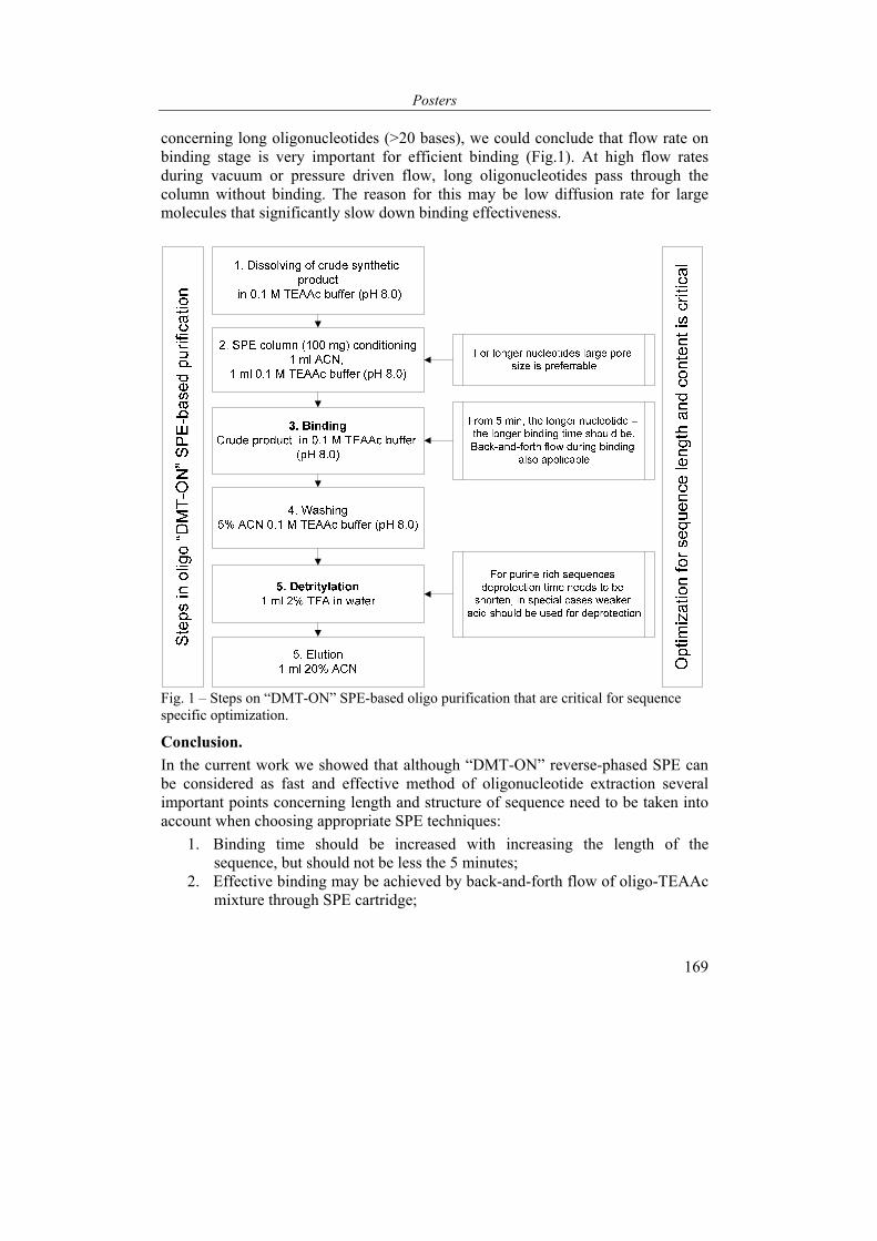
Posters
169
concerning long oligonucleotides (>20 bases), we could conclude that flow rate on binding stage is very important for efficient binding (Fig.1). At high flow rates during vacuum or pressure driven flow, long oligonucleotides pass through the column without binding. The reason for this may be low diffusion rate for large molecules that significantly slow down binding effectiveness.
Fig. 1 – Steps on “DMT-ON” SPE-based oligo purification that are critical for sequence specific optimization.
Conclusion.
In the current work we showed that although “DMT-ON” reverse-phased SPE can be considered as fast and effective method of oligonucleotide extraction several important points concerning length and structure of sequence need to be taken into account when choosing appropriate SPE techniques:
1. Binding time should be increased with increasing the length of the sequence, but should not be less the 5 minutes;
2. Effective binding may be achieved by back-and-forth flow of oligo-TEAAc mixture through SPE cartridge;
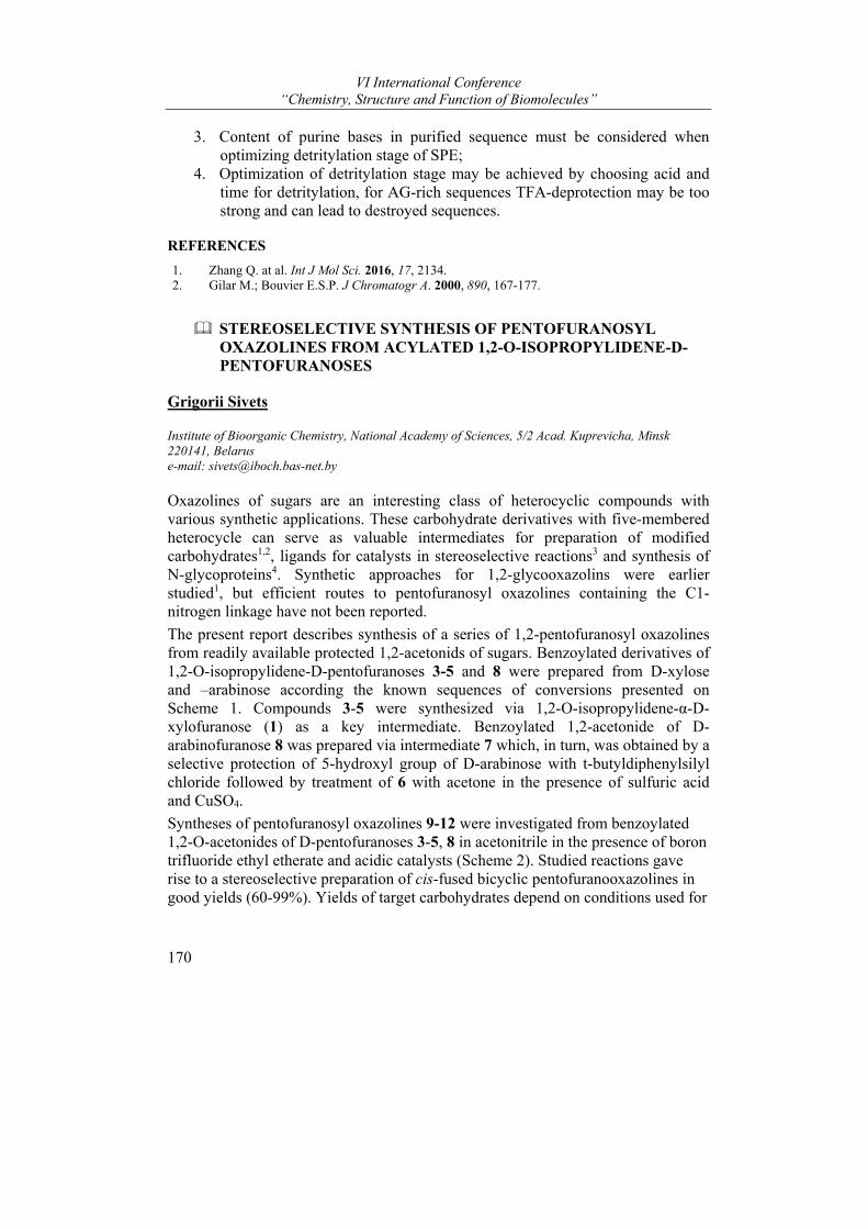
VI International Conference “Chemistry, Structure and Function of Biomolecules”
170
3. Content of purine bases in purified sequence must be considered when optimizing detritylation stage of SPE;
4. Optimization of detritylation stage may be achieved by choosing acid and time for detritylation, for AG-rich sequences TFA-deprotection may be too strong and can lead to destroyed sequences.
REFERENCES
1. Zhang Q. at al. Int J Mol Sci. 2016, 17, 2134. 2. Gilar M.; Bouvier E.S.P. J Chromatogr A. 2000, 890, 167-177.
STEREOSELECTIVE SYNTHESIS OF PENTOFURANOSYL OXAZOLINES FROM ACYLATED 1,2-O-ISOPROPYLIDENE-D-PENTOFURANOSES
Grigorii Sivets
Institute of Bioorganic Chemistry, National Academy of Sciences, 5/2 Acad. Kuprevicha, Minsk 220141, Belarus e-mail: [email protected]
Oxazolines of sugars are an interesting class of heterocyclic compounds with various synthetic applications. These carbohydrate derivatives with five-membered heterocycle can serve as valuable intermediates for preparation of modified carbohydrates1,2, ligands for catalysts in stereoselective reactions3 and synthesis of N-glycoproteins4. Synthetic approaches for 1,2-glycooxazolins were earlier studied1, but efficient routes to pentofuranosyl oxazolines containing the C1-nitrogen linkage have not been reported.
The present report describes synthesis of a series of 1,2-pentofuranosyl oxazolines from readily available protected 1,2-acetonids of sugars. Benzoylated derivatives of 1,2-O-isopropylidene-D-pentofuranoses 3-5 and 8 were prepared from D-xylose and –arabinose according the known sequences of conversions presented on Scheme 1. Compounds 3-5 were synthesized via 1,2-O-isopropylidene-α-D-xylofuranose (1) as a key intermediate. Benzoylated 1,2-acetonide of D-arabinofuranose 8 was prepared via intermediate 7 which, in turn, was obtained by a selective protection of 5-hydroxyl group of D-arabinose with t-butyldiphenylsilyl chloride followed by treatment of 6 with acetone in the presence of sulfuric acid and CuSO4.
Syntheses of pentofuranosyl oxazolines 9-12 were investigated from benzoylated 1,2-O-acetonides of D-pentofuranoses 3-5, 8 in acetonitrile in the presence of boron trifluoride ethyl etherate and acidic catalysts (Scheme 2). Studied reactions gave rise to a stereoselective preparation of cis-fused bicyclic pentofuranooxazolines in good yields (60-99%). Yields of target carbohydrates depend on conditions used for
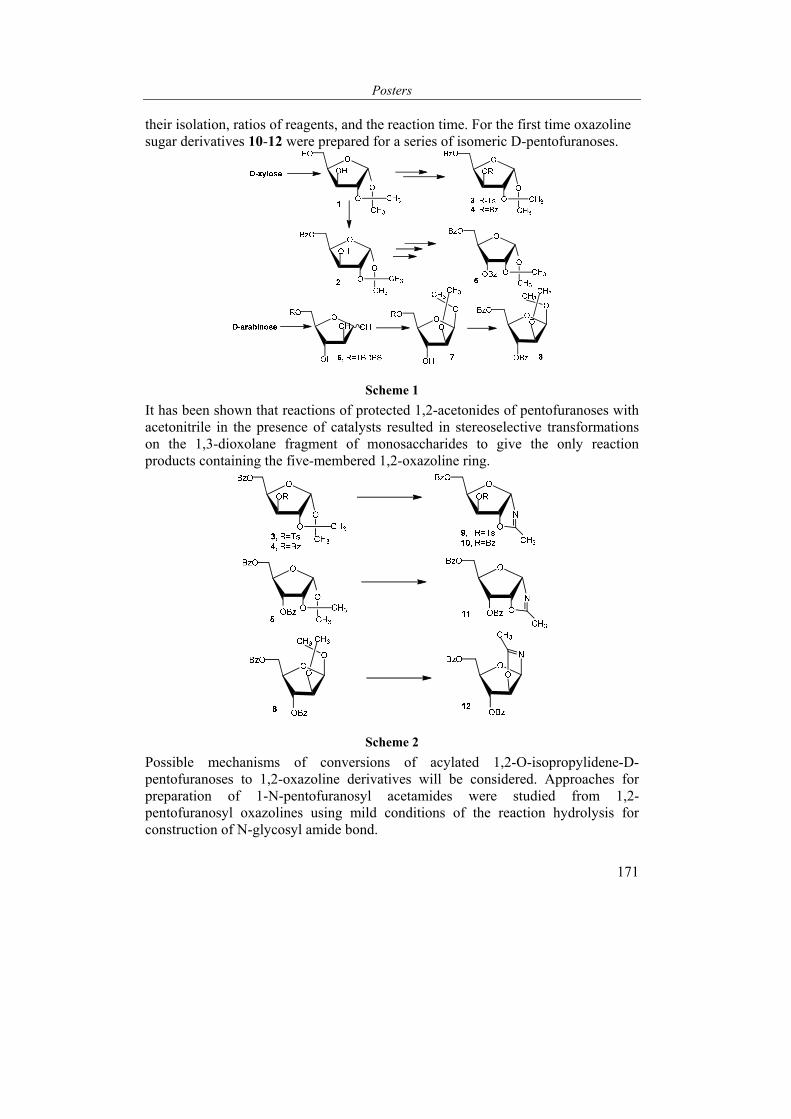
Posters
171
their isolation, ratios of reagents, and the reaction time. For the first time oxazoline sugar derivatives 10-12 were prepared for a series of isomeric D-pentofuranoses.
Scheme 1
It has been shown that reactions of protected 1,2-acetonides of pentofuranoses with acetonitrile in the presence of catalysts resulted in stereoselective transformations on the 1,3-dioxolane fragment of monosaccharides to give the only reaction products containing the five-membered 1,2-oxazoline ring.
Scheme 2
Possible mechanisms of conversions of acylated 1,2-O-isopropylidene-D-pentofuranoses to 1,2-oxazoline derivatives will be considered. Approaches for preparation of 1-N-pentofuranosyl acetamides were studied from 1,2-pentofuranosyl oxazolines using mild conditions of the reaction hydrolysis for construction of N-glycosyl amide bond.

VI International Conference “Chemistry, Structure and Function of Biomolecules”
172
The structures of prepared oxazolines were supported by 1H, 13C NMR, IR and mass-spectroscopic data.
REFERENCES
(1) Blanco, J.L.J.; Sylla, B.; Mellet, C.O.; Fernandez, J.M.G. J. Org. Chem. 2007, 72, 4547-45550.
(2) Pravdic, N.; Inch, T.D.; Fletcher, H.G. J. Org. Chem. 1967, 32, 1815-1818 (3) Jones, G.; Richards, C.J. Tetrahedron Assym. 2004, 15, 653-659. (4) Andreini, M.; Anderluth, M.; Audfray, A; Bernardi, A.; Imberty, A. Carbohydr. Res. 2010,
345, 1400-1407.
This study was supported by Byelorussian Republic Foundation for Fundamental Research (project No.X16-048).
SYNTHESIS AND ANALYSIS OF THE INFLUENCE OF SOME PEPTIDE ELICITORS ON RESISTANCE OF LEGUMES TO OXIDATIVE STRESS
Yuri A. Sokolov1*, Halina G. Filiptsova2, Aleksandr Y. Lushchyk1, and Vladimir M. Yurin2
1 Institute of bioorganic chemistry of the National Academy of Sciences of Belarus, Minsk, Belarus, 2 Belarusian State University, Minsk, Belarus *e-mail: [email protected]
In recent years there has been a transition to a much wider use of environmentally friendly plant protection products designed to protect plants against biotic and abiotic stresses. One of the modern plant protection strategies is based on the use of elicitors that are recognized by plants and trigger signaling systems leading to the expression of defense genes and the formation of systemic resistance1,2. Compounds exhibiting elicitor properties are used in very low concentrations, do not pollute the environment, are safe for humans and animals. Elicitors can belong to different classes of chemical compounds: carbohydrates, proteins and peptides, glycoproteins, lipids and glycolipids, etc. Peptide elicitors are the least studied. At the same time, there are a number of studies demonstrating their important role in the formation of plant resistance to stresses3-7.
The purpose of this work is to synthesize and study the effect of some peptide elicitors (Pep1, SubPep and Csp15) on the morphometric parameters of soybean seedlings (var. Pripyat), peas (var. Natalyevskaya) and mung beans (Vigna radiata) under conditions of oxidative stress.
Peptides Peps (plant elicitor peptides) regulate plant defense response to pathogens damaging effect. The first peptide Pep1 (ATKVKAKQRGKEKVSSGRPGQHN) was isolated from the leaves of Arabidopsis thaliana. It activates the production of
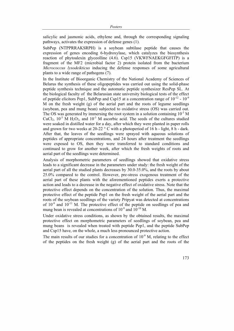
Posters
173
salicylic and jasmonic acids, ethylene and, through the corresponding signaling pathways, activates the expression of defense genes (1).
SubPep (NTPPRRAKSRPH) is a soybean subtilase peptide that causes the expression of genes encoding 6-hydroxylase, which catalyzes the biosynthesis reaction of phytoalexin glyceolline (4.6). Csp15 (VKWFNAEKGFGFITP) is a fragment of the MF2 (microbial factor 2) protein isolated from the bacterium Micrococcus lysodeikticus inducing the defense responses of some agricultural plants to a wide range of pathogens (7).
In the Institute of Bioorganic Chemistry of the National Academy of Sciences of Belarus the synthesis of these oligopeptides was carried out using the solid-phase peptide synthesis technique and the automatic peptide synthesizer ResPep SL. At the biological faculty of the Belarusian state university biological tests of the effect of peptide elicitors Pep1, SubPep and Csp15 at a concentration range of 10-12 - 10-9 M on the fresh weight (g) of the aerial part and the roots of legume seedlings (soybean, pea and mung bean) subjected to oxidative stress (OS) was carried out. The OS was generated by immersing the root system in a solution containing 10-3 M CuCl2, 10-3 M H2O2, and 10-3 M ascorbic acid. The seeds of the cultures studied were soaked in distilled water for a day, after which they were planted in paper rolls and grown for two weeks at 20-22 ° C with a photoperiod of 16 h - light, 8 h - dark. After that, the leaves of the seedlings were sprayed with aqueous solutions of peptides of appropriate concentrations, and 24 hours after treatment the seedlings were exposed to OS, then they were transferred to standard conditions and continued to grow for another week, after which the fresh weights of roots and aerial part of the seedlings were determined.
Analysis of morphometric parameters of seedlings showed that oxidative stress leads to a significant decrease in the parameters under study: the fresh weight of the aerial part of all the studied plants decreases by 30.0-35.0%, and the roots by about 25.0% compared to the control. However, pre-stress exogenous treatment of the aerial part of these plants with the aforementioned peptides exerts a protective action and leads to a decrease in the negative effect of oxidative stress. Note that the protective effect depends on the concentration of the solution. Thus, the maximal protective effect of the peptide Pep1 on the fresh weight of the aerial part and the roots of the soybean seedlings of the variety Pripyat was detected at concentrations of 10-9 and 10-11 M. The protective effect of the peptide on seedlings of pea and mung bean is revealed at concentrations of 10-9 and 10-10 M.
Under oxidative stress conditions, as shown by the obtained results, the maximal protective effect on morphometric parameters of seedlings of soybean, pea and mung beans is revealed when treated with peptide Pep1, and the peptide SubPep and Csp15 have, on the whole, a much less pronounced protective action.
The main results of our studies for a concentration of 10-9 M, relating to the effect of the peptides on the fresh weight (g) of the aerial part and the roots of the
VI International Conference “Chemistry, Structure and Function of Biomolecules”
174
soybean, pea and mung beans seedlings in conditions of oxidative stress, are presented in the table:
Plant Variant of experience
control OS Pep1+OS SubPep+OS Csp15+OS
soybean, var. Pripyat
aerial part 1,23±0,055 0,85±0,038 0,97±0,042 0,78±0,056 0,83±0,041
root 0,35±0,017 0,27±0,021 0,32±0,026 0,30±0,027 0,31±0,042
pea aerial part
0,80±0,034 0,51±0,021 0,72±0,042 0,69±0,042 0,48±0,032
root 0,56±0,022 0,42±0,018 0,47±0,023 0,41±0,021 0,41±0,016
mung bean
aerial part
0,47±0,022 0,28±0,014 0,37±0,023 0,37±0,021 0,32±0,018
root 0,26±0,014 0,17±0,016 0,22±0,019 0,20±0,015 0,18±0,017
REFERENCES
(1) Albert, M. J. of Experimental Botany. 2013, 64, 5269-5279. (2) Соколов, Ю.А. Элиситоры и их применение в растениеводстве. Минск: Бел. наука,
2016. – 201 с. (3) Boller, T.; Felix G. Annu. Rev. Plant Biol. 2009, 60, 379–406. (4) Yamaguchi, Y.; Huffaker A. Current Opinion in Plant Biology. 2011, 14, 351–357. (5) Huffaker, A.; [et al.] Plant Physiology. 2011, 155, 1325–1338. (6) Yamaguchi, Y.; [et al.] Plant Physiology. 2011, 156, 932–942. (7) Felix, G.; Boller T. J. Biol. Chem. 2003, 278, 6201 – 6208.
STUDY OF BIOLOGICAL ACTIVITY OF 17-ETHERS OF ANDROSTAN SERIES AND OF HETEROAROMATIC ACIDS
T. S. Varaksa, I. P. Grabovec, T. V. Shkel, A. A. Gilep, N. V. Strushkevich, V. I. Dolgopalets, and Yu. G. Charnou
Institute of Bioorganic Chemistry, National Academy of Sciences of Belarus, Minsk, Belarus, e-mail: e-mail: [email protected]
Abiraterone is anticancer drug on the market representing a selective inhibitor of cytochrome P450c17 – an enzyme with 17α-hydroxylase and C17, 20-lyase activity. Abiraterone is a steroid molecule with a pyridine ring moiety at C17. The selectivity of abiraterone and its derivates was not tested on all clinically important cytochrome P450s, both human and pathogenic microorganisms. Here we present new synthesized compounds of androstane series and heteroaromatic acids to evaluate their effect on human CYP11B1 and Candida glabrata CYP51.
A number of esters [2, 3] have been previously synthesized and described such as 7-methyl-19-nortestosterone 1a and its 7-isomer: 17-(6-chloronicotinoyloxy)-
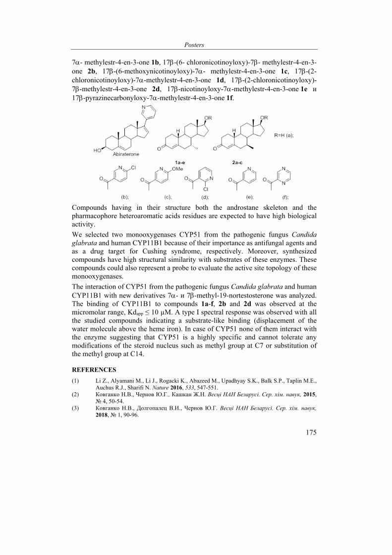
Posters
175
7- methylestr-4-en-3-one 1b, 17-(6- chloronicotinoyloxy)-7- methylestr-4-en-3-one 2b, 17-(6-methoxynicotinoyloxy)-7- methylestr-4-en-3-one 1c, 17-(2-chloronicotinoyloxy)-7-methylestr-4-en-3-one 1d, 17-(2-chloronicotinoyloxy)-7-methylestr-4-en-3-one 2d, 17-nicotinoyloxy-7-methylestr-4-en-3-one 1e и 17-pyrazinecarbonyloxy-7-methylestr-4-en-3-one 1f.
Compounds having in their structure both the androstane skeleton and the pharmacophore heteroaromatic acids residues are expected to have high biological activity.
We selected two monooxygenases CYP51 from the pathogenic fungus Candida glabrata and human CYP11B1 because of their importance as antifungal agents and as a drug target for Cushing syndrome, respectively. Moreover, synthesized compounds have high structural similarity with substrates of these enzymes. These compounds could also represent a probe to evaluate the active site topology of these monooxygenases.
The interaction of CYP51 from the pathogenic fungus Candida glabrata and human CYP11B1 with new derivatives 7- и 7-methyl-19-nortestosterone was analyzed. The binding of CYP11B1 to compounds 1a-f, 2b and 2d was observed at the micromolar range, Kdapp ≤ 10 µМ. A type I spectral response was observed with all the studied compounds indicating a substrate-like binding (displacement of the water molecule above the heme iron). In case of CYP51 none of them interact with the enzyme suggesting that CYP51 is a highly specific and cannot tolerate any modifications of the steroid nucleus such as methyl group at C7 or substitution of the methyl group at C14.
REFERENCES
(1) Li Z., Alyamani M., Li J., Rogacki K., Abazeed M., Upadhyay S.K., Balk S.P., Taplin M.E., Auchus R.J., Sharifi N. Nature 2016, 533, 547-551.
(2) Ковганко Н.В., Чернов Ю.Г., Кашкан Ж.Н. Весцi НАН Беларусi. Сер. хiм. навук, 2015, № 4, 50-54.
(3) Ковганко Н.В., Долгопалец В.И., Чернов Ю.Г. Весцi НАН Беларусi. Сер. хiм. навук, 2018, № 1, 90-96.
VI International Conference “Chemistry, Structure and Function of Biomolecules”
176
EXPRESSION, PURIFICATION AND LIGAND BINDING PROPERTIES OF MONOOXYGENASES FROM M.TUBERCULOSIS RV2266, RV3545C AND RV3518C
T. S. Varaksa, S. V. Smolskaya, N. V. Strushkevich, and A. A. Gilep
Institute of Bioorganic Chemistry, National Academy of Sciences of Belarus, Minsk, Belarus, e-mail: [email protected]
Three genes Rv2266, Rv3545c and Rv3518c in the genome of M. tuberculosis H37Rv are required for cholesterol side chain degradation in mycobacteria1. These genes encode monooxygenases involved to catalyze terminal oxidation of cholesterol and cholest-4-en-3-one side-chains2. Inhibition of these enzymes leads to accumulation of a toxic cholest-4-en-3-one, which are likely to inhibit the growth of mycobacteria and able to increase permeability cell wall for drugs. Currently identification and design of new inhibitors monooxygenases have high priority for treatment tuberculosis, caused by multi-drag resistant strains of mycobacteria. Due to the fact that genes Rv2266, Rv3545c and Rv3518c are involved in cholesterol catabolism, they are considered as targets for drugs in tuberculosis therapy. Here we report the results of ligand-binding properties of enzymes, encoded by Rv2266, Rv3545c and Rv3518c genes.
Genes Rv2266, Rv3545c and Rv3518c from M. tuberculosis H37Rv were cloned, heterologously expressed in E. coli DH5α, and purified as described previously3. Enzymes, products of genes Rv2266 and Rv3518c, were obtained in preparative amounts in a highly purified state in the ferric low-spin six-coordinated form and have the characteristic UV absorption spectrum in Soret region. Purified enzyme, product of gene Rv3545c, was obtained in a high-spin state of the heme iron atom with the absorption peak at 393 nm in Soret band. Screening ligands of active sites enzymes, products of genes Rv2266, Rv3545c and Rv3518c, was carried out by spectrophotometric titration. The affinity of ligands to the active sites of analyzed enzymes was estimated in according to the apparent dissociation constant (Kdapp).
It was found that the products of genes Rv2266 and Rv3518c bind carbethoxyhexyl imidazole and carboxyheptyl imidazole, thromboxane A synthetase inhibitors, 1-alkylimidazole derivatives, displaying a type II spin shifts, indicating replacement of the coordinated water molecule by a nitrogen atom of the ligand in ferric atom of the analyzed enzymes. Kdapp of the carbethoxyhexyl imidazole complex with the products of genes Rv2266 and Rv3518c is equal 1.01 ± 0.25 µМ and 1.09 ± 0.23 µМ, respectively. Such Kd value indicates a high degree of affinity of this compound with regard to the enzymes studied is revealed. Product of gene Rv3518c is able to bind carboxyheptyl imidazole (Kdapp= 23.35 ± 1.97 µМ), producing a type II spectral shift, whereas product of gene Rv2266 binds carboxyheptyl imidazole, displaying a reverse type I spectral shift, where there is an increase in the 420 nm absorbtion peak coupled with a decrease in the 390 nm peak. Such spectral shift is
Posters
177
thought to be suggest displacement of hydroxyl group the distal ligand of monooxygenases combined with substrate binding to a hydrophobic region of the active site take place4. Identified ligands of enzymes provide an alternative scaffold for selective inhibitors of mycobacterial monooxygenases.
REFERENCES
(1) Ouellet, H.; Johnston, J. B.; Ortiz de Montellano, P. R. Trends Microbiol. 2011, 19, 530-539.
(2) Johnston, J. B.; Ouellet, H.; Ortiz de Montellano, P. R. J Biol Chem. 2010, 285, 36352-36360.
(3) Vasilevskaya, A. V.; Yantsevich, A. V; Sergeev, G. V.; Lemish, A. P.; Usanov, S. A.; Gilep, A. A. J Steroid Biochem Mol Biol. 2017, 169, 202-209.
(4) Ouellet, H.; Kells, P. M.; Ortiz de Montellano, P. R.; Podust, L. M. Bioorg Med Chem Lett. 2011, 21, 332-337.
NOVEL FOLIC ACID DERIVATIVES: SYNTHESIS AND IN VITRO ANTITUMOR ACTIVITY
A.V. Farina, V.A. Shevchenko, A.K. Melnik, E.I. Vlasova, O.V. Avdoshko, A.V. Belko, and E.N. Kalinichenko
Institute of Bioorganic Chemistry, National Academy of Sciences of Belarus, Minsk, the Republic of Belarus. e-mail:[email protected]
Folic acid is an important metabolite highly involved in biosynthesis of nucleobases, nucleic acids and aminoacids thus playing vital role in cell division and proliferation(1). Antifolate class drugs are structural analogues of folic acid widely used for cancer treatment (pemetrexed, raltitrexed, methotrexate). Some folic acid derivatives are promising drug candidates as histone diacetylase inhibitors (mocetinostat)(2).
The aim of this study is design, synthesis and biological activity investigation of novel folic acid derviatives.
The key approach to the design of targeted structures was to replace pteridine and L-glutamic acid residues of folic acid with different substituents to ensure structure similarity and pharmacophore properties with known protein kinase inhibitors.
Target compounds were synthesized in three steps starting with the reductive amination of the initial methyl-4-formyl benzoate 1 with variety of amines followed by acidic hydrolysis of the intermediate esters. The final step was an amidation with the use of condensation agents (DCC of CDI) and HOBt as a catalyst (Method 1) or via intermediate acid chlorides (Method 2).
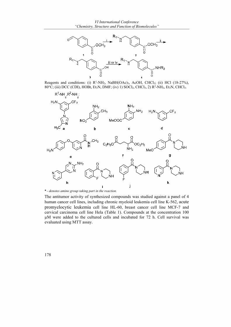
VI International Conference “Chemistry, Structure and Function of Biomolecules”
178
Reagents and conditions: (i) R1-NH2, NaBH(OAc)3, AcOH, CHCl3; (ii) HCl (18-27%), 80°C; (iii) DCC (CDI), HOBt, Et3N, DMF; (iv) 1) SOCl2, CHCl3, 2) R2-NH2, Et3N, CHCl3.
* - denotes amino group taking part in the reaction.
The antitumor activity of synthesized compounds was studied against a panel of 4 human cancer cell lines, including chronic myeloid leukemia cell line K-562, acute promyelocytic leukemia cell line HL-60, breast cancer cell line MCF-7 and cervical carcinoma cell line Hela (Table 1). Compounds at the concentration 100 µМ were added to the cultured cells and incubated for 72 h. Cell survival was evaluated using MTT assay.
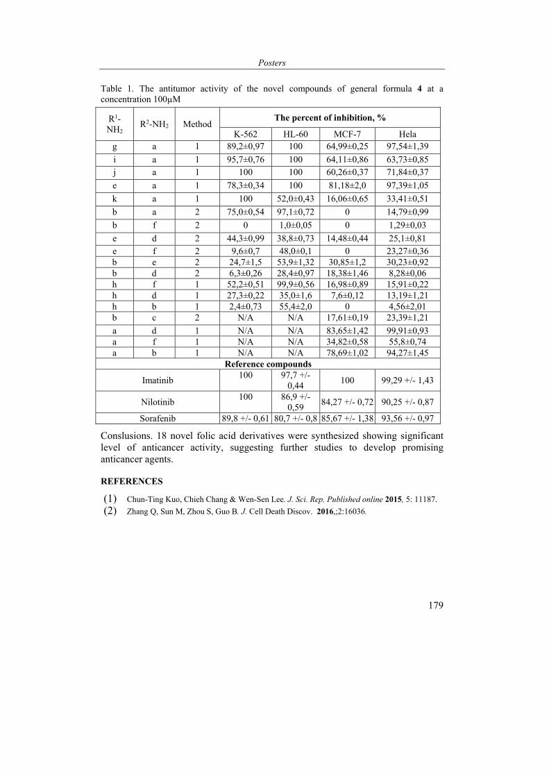
Posters
179
Table 1. The antitumor activity of the novel compounds of general formula 4 at a concentration 100µМ
R1-NH2
R2-NH2 Method The percent of inhibition, %
K-562 HL-60 MCF-7 Hela g a 1 89,2±0,97 100 64,99±0,25 97,54±1,39
i a 1 95,7±0,76 100 64,11±0,86 63,73±0,85 j a 1 100 100 60,26±0,37 71,84±0,37
e a 1 78,3±0,34 100 81,18±2,0 97,39±1,05
k a 1 100 52,0±0,43 16,06±0,65 33,41±0,51
b a 2 75,0±0,54 97,1±0,72 0 14,79±0,99
b f 2 0 1,0±0,05 0 1,29±0,03
e d 2 44,3±0,99 38,8±0,73 14,48±0,44 25,1±0,81 e f 2 9,6±0,7 48,0±0,1 0 23,27±0,36 b e 2 24,7±1,5 53,9±1,32 30,85±1,2 30,23±0,92 b d 2 6,3±0,26 28,4±0,97 18,38±1,46 8,28±0,06 h f 1 52,2±0,51 99,9±0,56 16,98±0,89 15,91±0,22 h d 1 27,3±0,22 35,0±1,6 7,6±0,12 13,19±1,21 h b 1 2,4±0,73 55,4±2,0 0 4,56±2,01 b c 2 N/A N/A 17,61±0,19 23,39±1,21
a d 1 N/A N/A 83,65±1,42 99,91±0,93 a f 1 N/A N/A 34,82±0,58 55,8±0,74 a b 1 N/A N/A 78,69±1,02 94,27±1,45
Reference compounds
Imatinib 100 97,7 +/-
0,44 100 99,29 +/- 1,43
Nilotinib 100 86,9 +/-
0,59 84,27 +/- 0,72 90,25 +/- 0,87
Sorafenib 89,8 +/- 0,61 80,7 +/- 0,8 85,67 +/- 1,38 93,56 +/- 0,97
Conslusions. 18 novel folic acid derivatives were synthesized showing significant level of anticancer activity, suggesting further studies to develop promising anticancer agents.
REFERENCES
(1) Chun-Ting Kuo, Chieh Chang & Wen-Sen Lee. J. Sci. Rep. Published online 2015, 5: 11187.
(2) Zhang Q, Sun M, Zhou S, Guo B. J. Cell Death Discov. 2016,;2:16036.
VI International Conference “Chemistry, Structure and Function of Biomolecules”
180
THE ANALYSIS OF VARIABILITY OF BIOPRODUCTIONAL PARAMETERS OF EX VITRO ADAPTANTS OF VACCINIUM CORYMBOSUM L. IN THE PRESENCE OF PHYTOHORMONAL STEROIDS
Anthony Volotovich, Yulia Lukonina, Alexandra Potapova, and Oksana Kudryashova
Republican Breeding and Seed Production Center, Minsk, Belarus e-mail: [email protected]
Brassinosteroids (steroid plant hormones, BS) are a promising group of natural plant growth regulators [1].
The comparative analysis of action efficiency of 24-epibrassinolide, as independently in use, and as a part of ‘Epin’ (Institute of Bioorganic Chemistry, NAS of Belarus, Minsk, Belarus), to combinations with the ‘Agronan’ (Argentum Group Ltd., Minsk, Belarus), on variability of bioproductional parameters of ex vitro adaptants of ‘Spartan’ cultivar of highbush blueberry at the age of 1–2 months is carried out in the present research. Adaptants one by one were landed in the P9 containers containing a peat substratum. The volume of substratum at 1 container: 0.000448 cub.m. The area of substratum at 1 container: 0.0064 sq.m. Quantity of plants: 210. Growth regulators were brought by spraying of an elevated part of plants once a week, as well as, 100 ml of solution on 21 adaptants.
Experiment options: control (processing by water); the ‘Agronan’ in a dose of 100 ml per 1.5 million of adaptants; 24-epibrassinolide in a dose of 10 mg per 1.5 million of adaptants; 24-epibrassinolide in a dose of 20 mg per 1.5 million of adaptants; the ‘Epin’ in a dose of active ingredient of 10 mg per 1.5 million of adaptants; the ‘Epin’ in a dose of active ingredient of 20 mg per 1.5 million of adaptants; the combination of ‘Agronan’ in a dose of 100 ml per 1.5 million of adaptants with 24-epibrassinolide in a dose of 10 mg per 1,5 million adaptant; the combination of ‘Agronan’ in a dose of 100 ml per 1.5 million of adaptants with 24-epibrassinolide in a dose of 20 mg per 1.5 million of adaptants; the combination of ‘Agronan’ in a dose of 100 ml per 1.5 million of adaptants with the ‘Epin’ in a dose of active ingredient of 10 mg per 1.5 million of adaptants; the combination of ‘Agronan’ in a dose of 100 ml per 1.5 million of adaptants with the ‘Epin’ in a dose of active ingredient of 20 mg per 1.5 million of adaptants. Analyzed the variability of height of the leading shoot, diameter of a root neck of the leading shoot and the number of shoots per plant.
Mathematical analysis of the data (the means ± standard error, calculation of least significant differences at significance levels of P < 0.05 and P < 0.01) was performed according to standard methods of variation statistics [2] using statistics analysis software STATISTICA 6.0 [3]. The dispersive analysis of data and calculation of share of factors influence on variability of the studied features carried
Posters
181
out in the program of the statistical analysis AB-Stat 1.0 developed at the Institute of Genetics and Cytology of NAS of Belarus [4].
As a result of research it is established reliable (at P < 0.01) increase at 1.3-1.4 time of all analyzed traits when processing an elevated part of adaptants by 24-epibrassinolide in a dose of 10 mg per 1.5 million of adaptants weekly. Use of the ‘Agronan’ in a dose of 100 ml per 1.5 million of adaptants authentically (at P < 0.05) increased by 1.2 times the diameter of a root neck of the leading shoot. The combination of ‘Agronan’ in the specified dose with 24-epibrassinolide in a dose of 10 mg per 1.5 million of adaptants brought to reliable (at P < 0.01) by 1.3 times increase in diameter of a root neck of the leading shoot.
REFERENCES
(1) Brassinosteroids. A new class of plant hormones; Khripach, V.A.; Zhabinskii V.N.; Groot A.E., Ed.; San Diego: Academic Press, 1999.
(2) Fielplot technique; Dospehov, B. A., Ed.; M, 1985. (3) STATISTICA; Borovikov, V. P., Ed.; SPb, 2001. (4) Anoshenko, B. Yu. Genetics. 1994, 30, 8-9.
THE EFFECT OF CHRONIC NEONATAL INJECTION OF AVP (6-9) AND ITS ANALOGUE AC-D-MPRG ON THE SOCIAL BEHAVIOR OF RATS
Selezneva Alexandra*, Stahanova Anna, Voskresenskaya Olga, Golubovich Vladimir, and Kamensky Andrey
Faculty of Biology, Lomonosov State University, Moscow, Russia e-mail: [email protected]
Our previous researches have shown that arginine vasopressin (AVP), its C-sided fragment (AVP(6-9)) and its analogues Ac-D-MPRG and Ac-D-SPRG which had been synthesized in the Institute of Bioorganic Chemistry National Academy of Sciences of Belarus provide the increase in exploratory behavior, decrease the anxiety and depression levels and accelerate the training processes of animals from different age groups in case of chronic neonatal injection.
This work illustrates the held research on deferred effects of chronic neonatal injection (3rd to 7th days of life) of AVP (6-9) and Ac-D-MPRG in white rats’ cubs on the social behavior of animals. The described method allows to study the drive of animals to the social novelty and their adaptation to the new environmental conditions. Each brood was divided into two parts, where the experimental group was injected with peptides in different dosage and the control was given the equivalent dose of distillated water. The experiment was conducted with the usage of a special labyrinth constructed for the researches on social behavior, where animals were put on 22nd day in mother/non-lactating female modification and on

VI International Conference “Chemistry, Structure and Function of Biomolecules”
182
32nd day in sibling/non-sibling (the rat from the same brood and from different one) modification. Mother was separated from the brood on the 30th day.
The animals that were injected with AVP(6-9) in the dosage of 1.0 g/kg were showing the increase of the total number of standings and the number of standings in the section containing a stranger female in the test of social behavior in mother/non-lactating female modification. Additionally, in this section the frequency of grooming was also increased. In the group of males there were no visible differences. The female group performed an increase in standings and grooming frequency in the section containing a stranger female. In rats that were injected with AVP(6-9) in the dosage of 10.0 g/kg there was a decline in run, the number of rapprochements and contacts with both mother and a stranger female. Total grooming has also decreased. In the group of males there was a decrease in the number of rapprochements with a stranger female and in the total grooming. In the group of females a decline in the number of rapprochements with both mother and a stranger female and the drop of the total number of contacts were observed. Besides, the total run of the subjects has decreased as well.
In the conducted test on social behavior in sib/non-sib modification rats injected with AVP(6-9) in the dosage of 1.0 g/kg were demonstrating approximately similar behavior with no severe declines. In the group of males an increase of contacts with a non-sib was observed. In the female group there was a reduction of time spent in the section containing a non-sibling and a decline in the number of standings in said section.
The animals that were injected with Ac-D-MPRG in the dosage of 1.0 g/kg were showing a significant increase in time spent in the center of the labyrinth as well as reduce of grooming both in the section with a stranger female and in total in the test on social behavior (in mother/non-lactating female modification). Apart from that, the number of standings in the center of the labyrinth has risen. In the male group there was a decline in grooming frequency in the section with a stranger female, while in the group of females there were no behavioral differences. The animals injected with tetrapeptide in the dosage of 1.0 g/kg were performing the decline in total grooming frequency, mostly through females’ activity, and the increase in net climbing close to a stranger female, mostly through males’ activity.
During the test on social behavior in sib/non-sib modification rats injected with Ac-D-MPRG in the dosage of 1.0 g/kg were showing the increased number of standings in the center and the decline in both total grooming and the grooming in the sib’s sector combined with the increased number of the net climbing close to a non-sibling. In animals with the tetrapeptide dosage of 10.0 g/kg the decrease in grooming in the center and in time spent there was being observed. Besides, there was an increase in the number of contacts with a non-sib, both total and separate number of net climbing in both sectors and in experimental group’s run. Males were showing an increased run, number of rapprochements and contacts with a non-
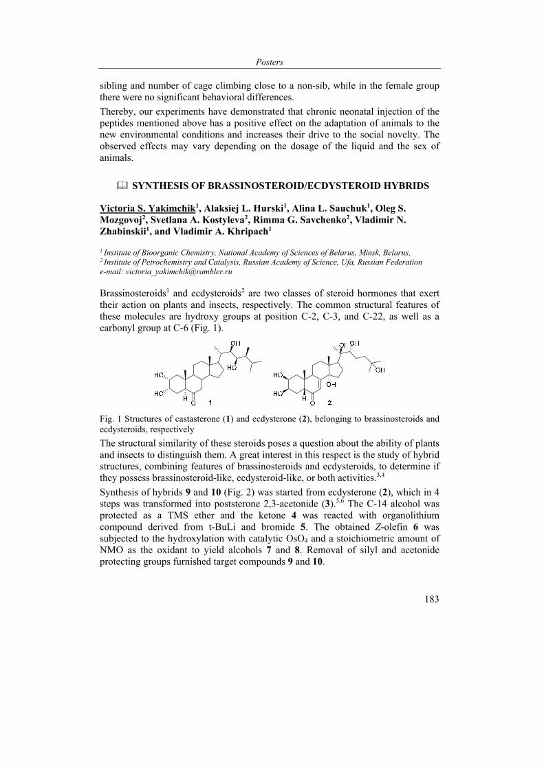
Posters
183
sibling and number of cage climbing close to a non-sib, while in the female group there were no significant behavioral differences.
Thereby, our experiments have demonstrated that chronic neonatal injection of the peptides mentioned above has a positive effect on the adaptation of animals to the new environmental conditions and increases their drive to the social novelty. The observed effects may vary depending on the dosage of the liquid and the sex of animals.
SYNTHESIS OF BRASSINOSTEROID/ECDYSTEROID HYBRIDS
Victoria S. Yakimchik1, Alaksiej L. Hurski1, Alina L. Sauchuk1, Оleg S. Mozgovoj2, Svetlana A. Kostyleva2, Rimma G. Savchenko2, Vladimir N. Zhabinskii1, and Vladimir A. Khripach1
1 Institute of Bioorganic Chemistry, National Academy of Sciences of Belarus, Minsk, Belarus, 2 Institute of Petrochemistry and Catalysis, Russian Academy of Science, Ufa, Russian Federation e-mail: [email protected]
Brassinosteroids1 and ecdysteroids2 are two classes of steroid hormones that exert their action on plants and insects, respectively. The common structural features of these molecules are hydroxy groups at position C-2, C-3, and C-22, as well as a carbonyl group at C-6 (Fig. 1).
Fig. 1 Structures of castasterone (1) and ecdysterone (2), belonging to brassinosteroids and ecdysteroids, respectively
The structural similarity of these steroids poses a question about the ability of plants and insects to distinguish them. A great interest in this respect is the study of hybrid structures, combining features of brassinosteroids and ecdysteroids, to determine if they possess brassinosteroid-like, ecdysteroid-like, or both activities.3,4
Synthesis of hybrids 9 and 10 (Fig. 2) was started from ecdysterone (2), which in 4 steps was transformed into poststerone 2,3-acetonide (3).5,6 The C-14 alcohol was protected as a TMS ether and the ketone 4 was reacted with organolithium compound derived from t-BuLi and bromide 5. The obtained Z-olefin 6 was subjected to the hydroxylation with catalytic OsO4 and a stoichiometric amount of NMO as the oxidant to yield alcohols 7 and 8. Removal of silyl and acetonide protecting groups furnished target compounds 9 and 10.
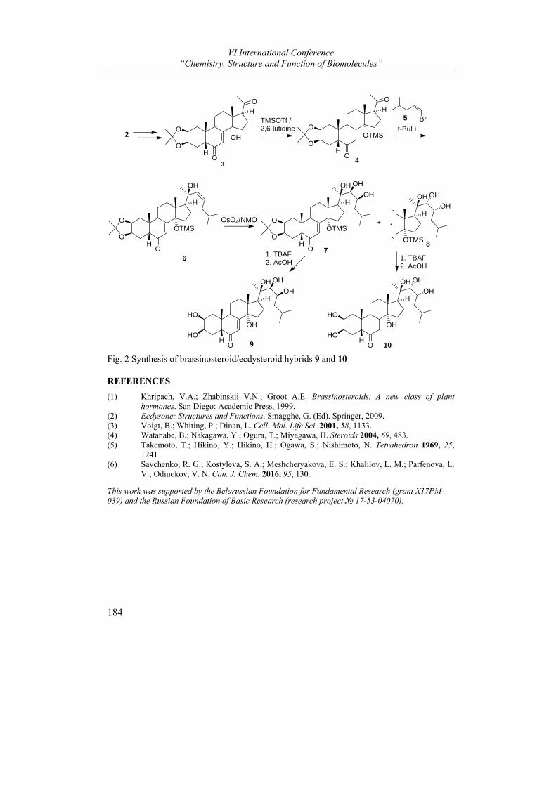
VI International Conference “Chemistry, Structure and Function of Biomolecules”
184
O
OH
OH
H
O
O
TMSOTf / 2,6-lutidine
O
OTMS
OH
H
O
O
Br
OTMS
OH
H
O
O
OH
3 4
OsO4/NMOOTMS
OH
H
O
O
OH OH
OH
OH
OH
H
OH OH
OH
6
2t-BuLi
5
OTMS
H
OH OH
OH
+
78
HO
HO
OH
OH
H
OH OH
OH
HO
HO9 10
1. TBAF2. AcOH
1. TBAF2. AcOH
Fig. 2 Synthesis of brassinosteroid/ecdysteroid hybrids 9 and 10
REFERENCES
(1) Khripach, V.A.; Zhabinskii V.N.; Groot A.E. Brassinosteroids. A new class of plant hormones. San Diego: Academic Press, 1999.
(2) Ecdysone: Structures and Functions. Smagghe, G. (Ed). Springer, 2009. (3) Voigt, B.; Whiting, P.; Dinan, L. Cell. Mol. Life Sci. 2001, 58, 1133. (4) Watanabe, B.; Nakagawa, Y.; Ogura, T.; Miyagawa, H. Steroids 2004, 69, 483. (5) Takemoto, T.; Hikino, Y.; Hikino, H.; Ogawa, S.; Nishimoto, N. Tetrahedron 1969, 25,
1241. (6) Savchenko, R. G.; Kostyleva, S. A.; Meshcheryakova, E. S.; Khalilov, L. M.; Parfenova, L.
V.; Odinokov, V. N. Can. J. Chem. 2016, 95, 130.
This work was supported by the Belarussian Foundation for Fundamental Research (grant X17PM-039) and the Russian Foundation of Basic Research (research project № 17-53-04070).
Posters
185
LATE ABSTRACTS
STUDY OF PRIMARY PHOSPHATIDYLCHOLINE UV-PEROXIDATION USING HEMOGLOBIN
Yulia Yermakovich, Lydia A. Skorostetskaya, and Natalia M. Litvinko*
Institute of Bioorganic Chemistry of National Academy of Science of Belarus, Minsk, Belarus, e-mail: [email protected]
It is known, that part of oxidized phospholipids after UV-irradiation is not fragmented and remains esterified in the parent PL molecules, which are the primary target for an attack of reactive oxygen species. Many methods for the determination of oxidized phospholipid (OxPLs) are based on the measurement of TBA-reactive products with the final oxidized lipid degradation product (OxPLs) -MDA. At the same time that test does not allow detecting over all fragmented and non-fragmented OxPLs. We have previously proposed the enzymatic method of determination the degree of oxidation of non-fragmented primary OxPLs) using hemoglobin1. We showed, that UV-irradiation of phosphatidylcholine (PC) for a long time lead to secondary oxidation process, what finally instigate transition of hemoglobin to hemichrome 2. According to our earlier data, the native PC does not affect the spectral properties of hemoglobin (Hb) Therefore, the aim of the present work is to study spectral changes in hemoglobin induced by UV-irradiated PC during lipid peroxidation (LPO) at initial stage for the purpose of non-enzymatic determination of non-fragmented primary OxPLs with using hemoglobin as indicator.
PC film of 0.9 μmol PC after UV-irradiation of various duration under mercury-quartz lamp PRK-4 (radiation range 180-400 nm) was subsequently dissolved in ethanol for determining of LPO index (ratio of the OxPLs absorption intensity A233/A215 ) or was solubilized by detergent (3 mol Triton X-100/1 mol PC in 0.05M Tris-HCl-buffer, pH 8.0, t=20⁰ C). Differential spectra of Hb (400 < A < 430) nm in presence of UV-irradiated and non-irradiated PC-Triton X-100 - micelles were recorded in the transmission mode of T75-125% at necessary time intervals (ΔD). MDA as fragmented OxPLs product was detected by thiobarbituric acid.
Changes of LPO index of UV- irradiated PC film, which are in quite correlation with the changes of Hb differential spectra amplitude in presence of UV-irradiated PC-detergent micelles (left) and the accumulation of secondary LPO products (MDA, right), are shown at figure.
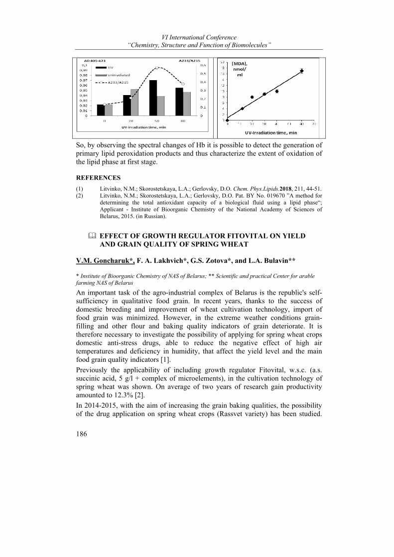
VI International Conference “Chemistry, Structure and Function of Biomolecules”
186
So, by observing the spectral changes of Hb it is possible to detect the generation of primary lipid peroxidation products and thus characterize the extent of oxidation of the lipid phase at first stage.
REFERENCES
(1) Litvinko, N.M.; Skorostetskaya, L.A.; Gerlovsky, D.O. Chem. Phys.Lipids.2018, 211, 44-51. (2) Litvinko, N.M.; Skorostetskaya, L.A.; Gerlovsky, D.O. Pat. BY No. 019670 ”A method for
determining the total antioxidant capacity of a biological fluid using a lipid phase“; Applicant - Institute of Bioorganic Chemistry of the National Academy of Sciences of Belarus, 2015. (in Russian).
EFFECT ОF GROWTH REGULATOR FITOVITAL ON YIELD AND GRAIN QUALITY OF SPRING WHEAT
V.M. Goncharuk*, F. A. Lakhvich*, G.S. Zotova*, and L.A. Bulavin**
* Institute of Bioorganic Chemistry of NAS of Belarus; ** Scientific and practical Center for arable farming NAS of Belarus
An important task of the agro-industrial complex of Belarus is the republic's self-sufficiency in qualitative food grain. In recent years, thanks to the success of domestic breeding and improvement of wheat cultivation technology, import of food grain was minimized. However, in the extreme weather conditions grain-filling and other flour and baking quality indicators of grain deteriorate. It is therefore necessary to investigate the possibility of applying for spring wheat crops domestic anti-stress drugs, able to reduce the negative effect of high air temperatures and deficiency in humidity, that affect the yield level and the main food grain quality indicators [1].
Previously the applicability of including growth regulator Fitovital, w.s.c. (a.s. succinic acid, 5 g/l + complex of microelements), in the cultivation technology of spring wheat was shown. On average of two years of research gain productivity amounted to 12.3% [2].
In 2014-2015, with the aim of increasing the grain baking qualities, the possibility of the drug application on spring wheat crops (Rassvet variety) has been studied.
Posters
187
Meteorological conditions during the period of studies differed substantially from the average indicators on both temperature and rainfalls. In this regard, the grain yield of spring wheat in the version without the use of Fitovital was 41.8 kg/ha in 2014, 19.3 kg/ha in 2015, and on average of 2 years − 30.6 kg/ha.
It was revealed that after the single-use of Fitovital (0.6 l/ha), the biggest increase in yield, on average over the period of studies, was received when it was making at the stage when flag leaf is visible (37-39 day of cultivation) and amounted to 3.3 centner/ha (10.8%). Two-and threefold use of Fitovital at this stage didn’t show significant increase in grain yield of spring wheat (5.2 -12.7%) in comparison with single-use.
It was established that the Fitovital application had a positive effect both on the number of grains per ear, 1000 grain weight, and the technological and nutritional value of the wheat. Depending on the Fitovital application variants, grain varies on such parameters as the flour yield (56,9-60,2%), protein content (15,1-16,3%), gluten content (34,5-37,7%) , flour strength (259,5-304,5 u.a.), water absorbing ability (58,0-59,0%), the volume of bread (822,5-942,5 ml), the total baking grade (4.02 -4.34 points). The highest total baking grade was noted after twofold use of Fitovital at the stages when flag leaf is visible (37-39 day of cultivation) and at the stage when first awns is visible (49 day of cultivation) and after its single-use at the flag leaf visible stage or at first node detectable stage (31 day of cultivation).
REFERENCES
(1) Cultivation of spring wheat. Sectoral rules. Organizational and technological standarts for the cultivation of cereals, legumes, cereal crops. A compilation of industry regulations. -Mn.: Rue "Publishing House. "Belorusskaya Niva". 2012.
(2) Kogotko, E.I., Vildflush, I.R. Messenger BSAA. 2011, 3, 74-77.

AUTHOR INDEX
A
Afonin V. ∙ 104 Ajduković J. ∙ 103 Andrianov A.M. ∙ 73, 110, 152 Anisimova A. ∙ 65 Anisovich M. ∙ 104 Antipova O.A. ∙ 147 Avdoshko O.V. ∙ 117, 177
B
Baranovsky A. ∙ 130 Barysevich M.V. ∙ 100, 111 Batuleu A.V. ∙ 82 Bei M.P. ∙ 74 Belko A. ∙ 60 Belko A.V. ∙ 117, 177 Belykh D.V. ∙ 3 Bocharov E. ∙ 27 Bocharova O. ∙ 28 Bogorodskiy A. ∙ 141 Bokut O.S. ∙ 96 Boldescu V. ∙ 22, 135, 137 Borshchevskiy V. ∙ 1, 30, 41, 50, 51, 66, 140,
141 Brazhnikov E.V. ∙ 75 Brechka Y. ∙ 38, 77 Bueldt G. ∙ 52 Bukhdruker S. ∙ 30, 66 Bukhtoyarova M. ∙ 65 Bulavin L.A. ∙ 186 Büldt G. ∙ 141 Buravlev E.V. ∙ 3 Buravskaya T. ∙ 124 Burkatovskiy D. ∙ 141
C
Charnou Yu.G. ∙ 174 Charnysh M. ∙ 5 Charnysh М. ∙ 82 Chaschina N. ∙ 107 Chen Y. ∙ 24
Cherezov V. ∙ 1, 41, 50, 51, 140 Chikun P. ∙ 5 Chukicheva I.Yu. ∙ 3 Curlat S. ∙ 22
D
Demidchik V. ∙ 5, 80, 81, 82 Derevyanchuk M.V. ∙ 121, 132 Devina H. ∙ 104 Deyev S.M. ∙ 7 Dichenko Y. ∙ 66 Dmitriev S. ∙ 65 Dobysh A.A. ∙ 83 Dokukina T.V. ∙ 96 Dolgopalets V.I. ∙ 174 Dontsu Y.S. ∙ 85 Dormeshkin D. ∙ 32, 165 Drašar P.B. ∙ 9 Drozd N.N. ∙ 67 Dudek B. ∙ 33 Dvornikova I.A. ∙ 3 Dzhus U. ∙ 65 Dzichenka Y.V. ∙ 34, 103
E
Efimov A.V. ∙ 36, 75, 106 Efimova M.V. ∙ 12 Ermola E.M. ∙ 86, 157 Ershov P.B. ∙ 91 Ershov P.V. ∙ 37
F
Faletrov Y.V. ∙ 34 Fando M.S. ∙ 88, 131 Farina A.V. ∙ 117, 177 Fatychava S.A. ∙ 56 Fedorkevich A.N. ∙ 89 Fedorova I.V. ∙ 3 Filiptsova H.G. ∙ 172 Florinskaya A. ∙ 70, 91
VI International Conference “Chemistry, Structure and Function of Biomolecules”
190
G
Garber M. ∙ 65 Garetskii R.G. ∙ 56 Gensch T. ∙ 141 Gilep A. ∙ 30, 32, 37, 70, 91, 96, 174, 176 Gilevich S. ∙ 38, 77 Glazova N.Y. ∙ 95 Golubovich V.P. ∙ 86, 93, 133, 181 Goncharuk V.M. ∙ 186 Gordeliy V. ∙ 1, 41, 51, 52 Grabovec I.P. ∙ 66, 174 Gribovskaya O.V. ∙ 93 Grischenko H. ∙ 104 Grudinin S. ∙ 15, 43 Gruzdev G.A. ∙ 95 Gusach A. ∙ 1, 30, 41, 50, 51, 140
H
Haidukevich I. ∙ 70, 91, 96 Haidukevich V.A. ∙ 98 Hryniewicka A. ∙ 21 Hryvusevich P. ∙ 81 Hurski A.L. ∙ 16, 42, 100, 101, 111, 112, 123,
183 Huryna M.A. ∙ 85
I
Ignatovich Z.V. ∙ 47 Ilyinsky N. ∙ 141 Iskryk M.V. ∙ 42, 100 Ivanov A.S. ∙ 17, 37, 70, 91
J
Jovanović‐Šanta S. ∙ 103
K
Kadukova M. ∙ 43 Kalinichenko E.N. ∙ 60, 117, 124, 177
Kamensky A. ∙ 181 Kandelinskaya O. ∙ 104 Kargatov A.M. ∙ 75, 106 Karpets L.‐A. ∙ 132 Karytska L.A. ∙ 158 Kashyn I.A. ∙ 73, 110, 152 Katritch V. ∙ 50 Katsin M. ∙ 32 Kazlova V.V. ∙ 111, 112 Kem K. ∙ 107 Khlebnicova T.S. ∙ 153 Khorn P. ∙ 51 Khripach V.A. ∙ 5, 42, 54, 56, 82, 100, 101,
111, 112, 121, 123, 132, 158, 183 Khudyaeva I.S. ∙ 3 Kielczewska U. ∙ 113 Kirkovskiy V.V. ∙ 86 Kisel M.A. ∙ 142, 143, 145, 147 Kisel M.S. ∙ 96 Kiseleva E. ∙ 44 Kiyavitskaya D.V. ∙ 98 Kletskov A.A. ∙ 114 Knizhnikau V.A. ∙ 98 Kolesnik I.A. ∙ 114 Kolesnikov Y.S. ∙ 121 Kondrateva V.V. ∙ 117 Konoplich A. ∙ 124 Korneev D.I. ∙ 119 Kornoushenko Y.V. ∙ 73, 152 Koroleva E.V. ∙ 47 Kostyleva S.A. ∙ 183 Kovalev K. ∙ 1, 30, 140 Kravets V.S. ∙ 121, 132 Kretynin S.V. ∙ 121, 132 Kudryashova O. ∙ 154, 180 Kukel A.G. ∙ 111, 123 Kulak T. ∙ 124 Kulchitsky V.A. ∙ 114 Kuprienko O. ∙ 127 Kurlenko S.P. ∙ 86 Kutchin A.V. ∙ 3 Kuznetsov Y.V. ∙ 19 Kvach M.V. ∙ 149 Kvachonak A.V. ∙ 114
L
Labor S. ∙ 63
Author Index
191
Ladyko A. ∙ 130 Lakhvich F.A. ∙ 85, 119, 153, 156, 186 Laman N. ∙ 107 Lekontseva N.V. ∙ 88, 131 Levina I.S. ∙ 19 Levitskaya N.G. ∙ 95 Litvinko N.M. ∙ 49, 185 Litvinovskaya R.P. ∙ 56, 121, 132, 158 Liubina A.I. ∙ 111, 123 Łotowski Z. ∙ 21, 164 Luginina A. ∙ 1, 30, 41, 50, 51, 140 Lukonina Y. ∙ 154, 180 Lukyanava K.L. ∙ 56 Lukyanova M.I. ∙ 133 Luschik A. ∙ 70 Lushchyk A.Y. ∙ 172 Lyapina E. ∙ 41, 50, 51 Lysenko I.L. ∙ 149 Lyubaikina N. ∙ 52
M
Macaev F. ∙ 22, 135, 137 MacKenzie F. ∙ 66 Mackievic V. ∙ 81 Mahrov M.V. ∙ 96 Makarevich D.A. ∙ 86, 157 Makavitskaya M. ∙ 81 Makeeva D. ∙ 65 Maksimova S. ∙ 104 Malyar N. ∙ 52 Manchenko D.M. ∙ 95 Manzhalesava N.E. ∙ 158 Marin E. ∙ 1, 30, 41, 50, 51, 66, 140 Martakov I.S. ∙ 67 Martsinovich V.P. ∙ 93, 133 Maslov I. ∙ 141 Medvedev A. ∙ 70 Meleshko A. ∙ 32 Melik‐Kasumov T.B. ∙ 142, 147 Melnik A.K. ∙ 60, 177 Migas A. ∙ 32 Mikhailina A. ∙ 131 Mikhailopulo K. ∙ 44 Mikhal’chuk A.L. ∙ 142, 143, 145, 147 Mikhaylov V.I. ∙ 67 Mishin A. ∙ 1, 30, 41, 50, 51, 140, 141 Molchanova A.Yu. ∙ 147
Morzycki J.W. ∙ 21, 113, 164 Mozgovoj O.S. ∙ 183
N
Narmantovich V.V. ∙ 149 Navaselsky I. ∙ 81 Nikolaev G.I. ∙ 73, 152 Nikulin A.D. ∙ 88, 131 Novichkova E. ∙ 60 Novik G. ∙ 44
O
Okhrimenko I. ∙ 52
P
Paetz C. ∙ 24, 33 Palyanskaya S.N. ∙ 158 Panibrat O.V. ∙ 54 Park H.‐W. ∙ 66 Pashkevich S.G. ∙ 114 Pashkovsky F.S. ∙ 85, 119, 156 Pavlut T.O. ∙ 142, 147 Pekhtsereva E.I. ∙ 147 Petkevich S.K. ∙ 114 Petrovskaya L. ∙ 52 Piven Y.A. ∙ 153 Podolyak E. ∙ 141 Pogrebnoi S. ∙ 135 Popov P. ∙ 41, 50, 52 Popova L.A. ∙ 98 Potapova A. ∙ 154, 180 Potkin V.I. ∙ 114 Przhevalskaya D. ∙ 5
R
Rasyuk E.D. ∙ 93 Rubinov D.B. ∙ 156 Rudak E.V. ∙ 143 Rudauskaya O.M. ∙ 96 Ryabtseva T.V. ∙ 133

VI International Conference “Chemistry, Structure and Function of Biomolecules”
192
Ryabzeva T. ∙ 157
S
Safronova N. ∙ 41, 51 Samokhina V. ∙ 81 Sauchuk A.L. ∙ 56, 132, 183 Savachka A.P. ∙ 158 Savchenko R.G. ∙ 183 Savić M. ∙ 103 Schabunya P.S. ∙ 56 Schneider B. ∙ 24, 33 Schnurrer F. ∙ 33 Selezneva A. ∙ 181 Selikhanov G.K. ∙ 88 Semenov D. ∙ 161 Semenov S. ∙ 127 Sergeev G. ∙ 70 Seroka B. ∙ 21, 164 Shabashova T. ∙ 104 Shapira M.A. ∙ 83, 165 Sharko O.L. ∙ 89, 149 Shavalda E. ∙ 142 Shchukina O.V. ∙ 3 Shchur V. ∙ 167 Shevchenko V.A. ∙ 177 Shevtsov M. ∙ 30, 41, 51 Shkel T. ∙ 34, 37, 70, 91, 103, 174 Shmanai V.V. ∙ 89, 149 Shukanova N. ∙ 104 Siergiejczyk L. ∙ 59 Sitnikov P.A. ∙ 67 Sivets G. ∙ 60, 170 Skorostetskaya L.A. ∙ 185 Smaliak V.A. ∙ 153 Smetanscaia A. ∙ 22 Smirnov V. ∙ 63 Smolskaya S.V. ∙ 37, 176 Sokolik A. ∙ 5, 81 Sokolov Y.A. ∙ 172 Soloviov D. ∙ 52 Stahanova A. ∙ 181 Stepko A. ∙ 41 Stepuro I. ∙ 63 Stolboushkina E. ∙ 65 Straltsova D. ∙ 5, 81 Strushkevich N. ∙ 30, 37, 66, 70, 174, 176 Stsiapura V. ∙ 63
Sucman N. ∙ 137 Sushko S. ∙ 37 Svirid A.V. ∙ 37 Sviridov O. ∙ 127, 161 Svistunenko D. ∙ 81
T
Taganovich A. ∙ 104 Tatsis E. ∙ 33 Tempel W. ∙ 66 Terentieva T. ∙ 127 Torlopov M.A. ∙ 67 Tuzikov A.V. ∙ 73, 110
U
Udoratina E.V. ∙ 67 Ulashchik E.A. ∙ 149 Uncu A. ∙ 135 Uncu L. ∙ 22, 135, 137 Usanov S.A. ∙ 37, 66, 70, 91, 103, 152, 167
V
Valica V. ∙ 22, 135, 137 Varaksa T.S. ∙ 30, 174, 176 Vashkevich E. ∙ 104 Vashkevich I. ∙ 127, 161 Vasilevskaya A.V. ∙ 37 Vensko D.G. ∙ 93 Vlasova E.I. ∙ 177 Volkov O. ∙ 1 Volotovich A. ∙ 154, 180 Voronina Y.A. ∙ 95 Voskresenskaya O. ∙ 181
W
Warskulat A.‐C. ∙ 33 Witkowski S. ∙ 21 Wojtkielewicz A. ∙ 113

Author Index
193
Y
Yablokov E. ∙ 70, 91 Yakimchik V.S. ∙ 112, 183 Yankovskaya D. ∙ 124 Yantsevich A. ∙ 63, 83, 103, 167 Yermakovich Y. ∙ 185 Yurin V.M. ∙ 172 Yuvchenko A.P. ∙ 74
Z
Zhabinskii V.N. ∙ 5, 42, 54, 82, 100, 101, 111, 112, 121, 123, 183
Zhavoronok I.P. ∙ 147 Zotova G.S. ∙ 186 Zubreichuk Z.P. ∙ 98