chemical analysis of norrisolide-induced golgi vesiculation
TRANSCRIPT

S1
Chemical Analysis of Norrisolide-Induced Golgi Vesiculation Gianni Guizzunti,b Thomas P. Brady,a Vivek Malhotra,b* and Emmanuel A. Theodorakisa aDepartment of Chemistry and Biochemistry and bDepartment of Biology,
University of California, San Diego, 9500 Gilman Drive, La Jolla, CA 92093-
0358
Supporting Information
Table of Contents
page
1. Materials and methods S2
2. Golgi membranes localization assay and competition studies S3
3. Localization and competition studies with 6 S3
4. Experimental procedures/data S4-S10
5. NMR Spectra S11-S26
S2
Materials and methods Reagents and cells: NRK cells were plated on 12 mm glass coverslips coated with Pronectin F
(Sigma) and grown in complete medium (500 µL per coverslip), consisting of alpha MEM
medium (GIBCO) supplemented with 10% fetal calf serum, 2 mM L-glutamine and 25 mM Hepes pH 7.4, at 37 °C in a 5% CO2 cell incubator. Stock solution (25 mg/mL) of norrisolide
and analogues were made in DMSO and stored at -20 °C. The working concentration of the compound 4 was 80 µM per coverslip for living cells (Golgi fragmentation assay) and 25 µM for
fixed cells (Golgi localization assay). The working concentration of norrisolide and other analogues was 25 µM per coverslip.
Golgi membranes fragmentation assay: To half of the coverslips (70% confluent) were added norrisolide or analogues (0.5 µL of the stock solutions). To the other half were added 0.5 µL of
DMSO as negative control. Both groups of cells were incubated at 37 °C for 60 min. Part of the
treated cells (Fig. 3b, 4b, 4e, 4h) and part of the control cells (Fig. 3a, 4a, 4d, 4g) were then fixed
with 4% formaldehyde for 10 min and processed for immunofluorescence microscopy. The remaining cells were washed four times with phosphate-buffered saline (PBS) (150 mM NaCl,
1.8 mM NaH2PO4, 8.4 mM Na2HPO4) and then incubated in fresh complete medium at 37 °C for 90 min, then fixed with 4% formaldehyde for 10 min and processed for immunofluorescence
microscopy (Fig. 3c, 4c, 4f, 4i).
Immunofluorescence microscopy: For fluorescent labeling, cells were incubated in blocking
buffer (PBS containing 2.5% fetal bovine serum and 0.1% Tween 20) for 30 min at room
temperature. The cells were then incubated for 1h at room temperature in primary antibody diluted in blocking buffer. Rabbit Mannosidase II antibody (1:2000) (a gift from Dr. Kelly
Moreman, Vanderbilt University, TN) was used to visualize Golgi apparatus. The cells were then washed three times with PBS and incubated with secondary antibody, diluted in blocking
buffer, for 1h at room temperature. Alexa Fluor 594 goat anti rabbit (1:500) from Molecular
Probes was used. Cells were washed three times with PBS containing Hoescht (1:100,000) (H33342, Molecular Probes) to stain DNA. Coverslips were then mounted onto glass slides and
visualized using a Nikon micophot-FXA fluorescence microscope at 60x magnification.
S3
Golgi membrane localization assay: The cells were first fixed with 2% formaldehyde for 90 sec. This mild fixation induces a minimal perturbation of cellular proteins, allowing the binding
between compound 4 and its cellular receptor. Cells were processed for immunofluorescence microscopy as described above. Before the coverslips were mounted onto glass slides,
compound 4 was added at 25 µM for 30 min (Fig. 3d, 3e, 3f).
Competition experiments: Norrisolide was added after compound 4 at 100µM for 30 min.
Coverslips were then mounted onto glass slides and visualized using a Nikon micophot-FXA fluorescence microscope at 60x magnification. The intracellular visualization of compound 4
was accomplished using excitation at 370 nm and emission at 460 nm. .
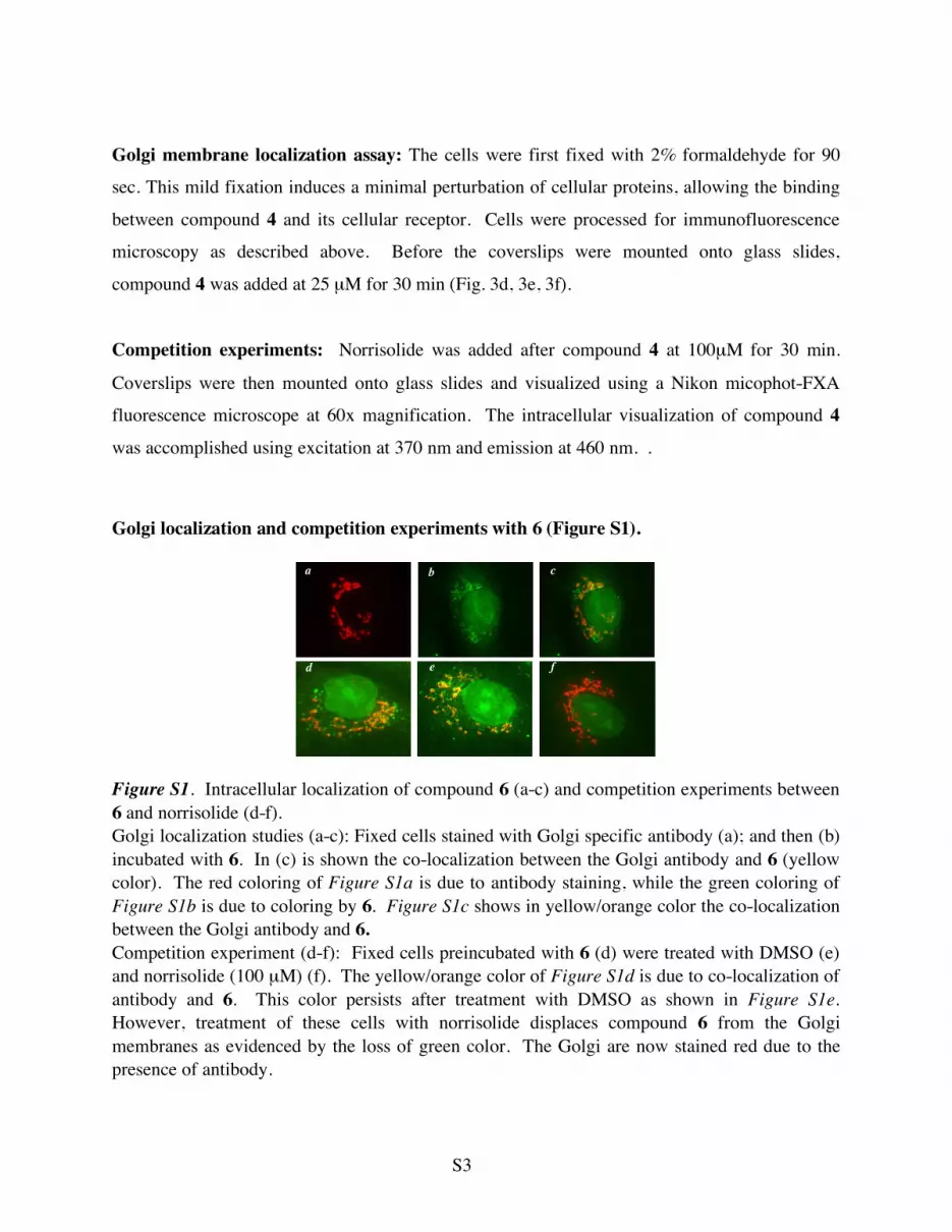
Golgi localization and competition experiments with 6 (Figure S1).
Figure S1. Intracellular localization of compound 6 (a-c) and competition experiments between 6 and norrisolide (d-f). Golgi localization studies (a-c): Fixed cells stained with Golgi specific antibody (a); and then (b) incubated with 6. In (c) is shown the co-localization between the Golgi antibody and 6 (yellow color). The red coloring of Figure S1a is due to antibody staining, while the green coloring of Figure S1b is due to coloring by 6. Figure S1c shows in yellow/orange color the co-localization between the Golgi antibody and 6. Competition experiment (d-f): Fixed cells preincubated with 6 (d) were treated with DMSO (e) and norrisolide (100 µM) (f). The yellow/orange color of Figure S1d is due to co-localization of antibody and 6. This color persists after treatment with DMSO as shown in Figure S1e. However, treatment of these cells with norrisolide displaces compound 6 from the Golgi membranes as evidenced by the loss of green color. The Golgi are now stained red due to the presence of antibody.
d e f
a b c
S4
General Chemical Techniques.: All reagents were commercially obtained (Aldrich, Acros) at highest commercial quality and used without further purification except where noted. The
coumarin probe was prepared as described by Fritz, M. G.; Seebach D. in Helv. Chim. Acta
1998, 81, 2414-2429. The norrisolide fragments were prepared as described in reference 3.
Yields refer to chromatographically and spectroscopically (1H NMR, 13C NMR) homogeneous
materials. Reactions were monitored by thin-layer chromatography (TLC) carried out on 0.25 mm E. Merck silica gel plates (60F-254) using UV light as the visualizing agent and 10%
ethanolic phosphomolybdic acid (PMA) or p-anisaldehyde solution and heat as developing agents. E. Merck silica gel (60, particle size 0.040-0.063 mm) was used for flash
chromatography. Preparative thin-layer chromatography separations were carried out on 0.25 or
0.50 mm E. Merck silica gel plates (60F-254). NMR spectra were recorded on Varian Mercury 400 and/or Unity 500 MHz instruments and calibrated using the residual undeuterated solvent as
an internal reference. The following abbreviations were used to explain the multiplicities: s = singlet, d = doublet, t = triplet, q = quartet, m = multiplet, b = broad. High resolution mass
spectra (HRMS) were recorded on a VG 7070 HS mass spectrometer under chemical ionization
(CI) conditions or on a VG ZAB-ZSE mass spectrometer under fast atom bombardment (FAB) conditions.
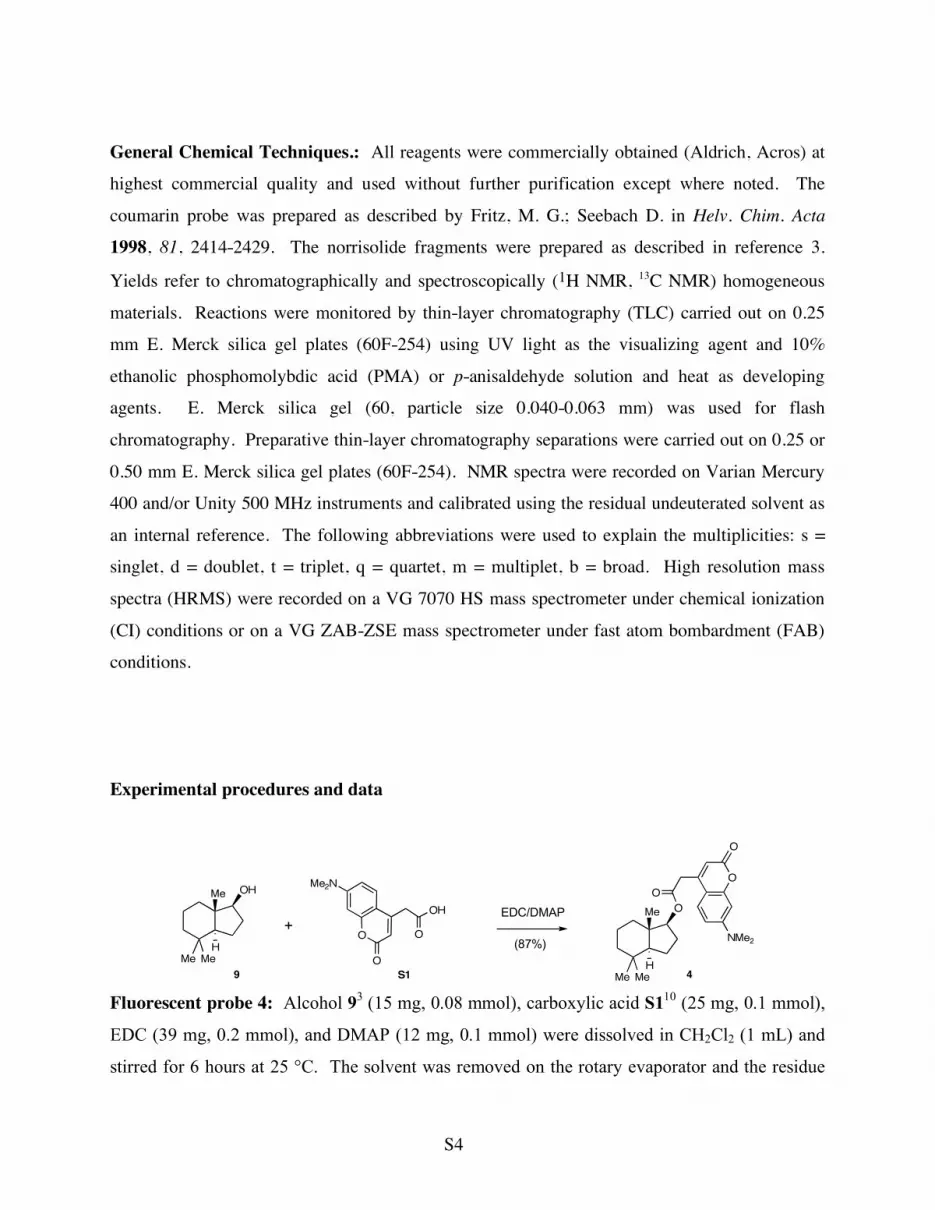
Experimental procedures and data
O
O
NMe2
O
OMe
Me Me
H4
O
O
Me2N
O
OH
S1
+EDC/DMAP
(87%)
OHMe
Me Me
H
9
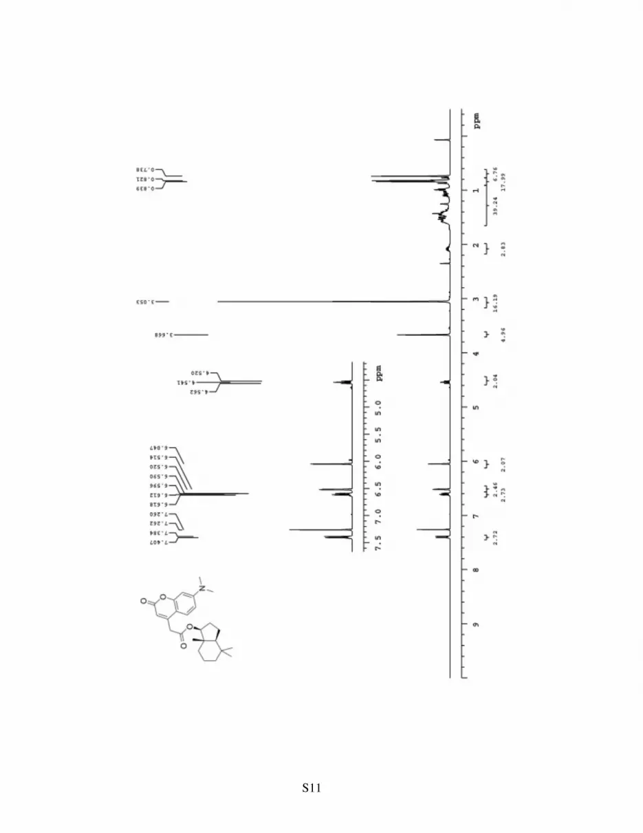
Fluorescent probe 4: Alcohol 93 (15 mg, 0.08 mmol), carboxylic acid S110 (25 mg, 0.1 mmol),
EDC (39 mg, 0.2 mmol), and DMAP (12 mg, 0.1 mmol) were dissolved in CH2Cl2 (1 mL) and
stirred for 6 hours at 25 °C. The solvent was removed on the rotary evaporator and the residue
S5
was applied to silica gel chromatography (25% ether in hexanes) to afford 29 mg of compound 4
(87%). 4: Rf= 0.4 (100% ether); 1H NMR (400 MHz, CDCl3) δ 7.39 (d, J= 9.2 Hz, 1H), 6.60 (d,
J= 8.8 Hz, 1H), 6.52 (s, 1H), 6.05 (s, 1H),4.54 (t, J= 8.4 Hz, 1 H), 3.67 (s, 2H), 3.05 (s, 6H), 2.09
(m, 1H) 1.60-1.09 (m, 6 H), 1.11-0.99 (m, 4H), 0.87 9s, 3H), 0.84 (s, 3H), 0.74 (s, 3H); 13C
NMR (100 MHz, CDCl3) δ 168.9, 161.7, 155.9, 152.9, 148.7, 125.4, 110.6, 108.9, 108.5, 98.4,
84.3, 52.6, 42.6, 41.3, 40.1, 38.6, 37.5, 33.0, 32.8, 26.2, 20.7, 20.2, 19.3, 12.6; HRMS FAB,
calcd for C25H33NO4 [M+]: 411.2410, found 411.2413.
O
O
O
OH
TBDPSOH O
O
O
O
TBDPSOH
O OMe2N
O
O
O
O
O
TBDPSO
PDC
(57%)
1. NaBH4
2. EDC, S1
(40%)
O OMe2N
O
O
O
O
O
HO
TBAF
(85%)
S2 S3
S4 5
Aldehyde S3: Lactol S23 (0.3g, 0.7 mmol), PDC (1.3 g, 3.5 mmol), and Mol
Sieves (4 Å, 4g) were dissolved in CH2Cl2 (15 mL) and stirred for 8 hours at
25 °C. The reaction was filtered through a plug of celite and the solvent
removed on the rotary evaporator. The residue was applied to silica gel
chromatography (100% ether) to afford 180 mg of compound S3 (57%). S3:
Rf= 0.6 (100% ether); [α]25D= +11 (c= 0.3, CH2Cl2);
1H NMR (400 MHz,
CDCl3) δ 9.69 (s, 1H), 7.66-7.61 (m, 4H), 7.43-7.33 (m, 6H), 6.08 (d, J= 5.2 Hz, 1H), 3.89-3.79
(dd, J= 10.8, 3.2 Hz, 1H), 3.81-3.73 (m, 2H), 3.48 (m, 1H), 2.75 (m, 1H), 2.61-2.30 (m, 4H),1.07
(s, 9H); 13C NMR (100 MHz, CDCl3) δ 199.0, 175.0, 135.5, 132.7, 129.8, 127.7, 107.2, 82.1,
63.0, 41.5, 41.3, 35.9, 28.6, 26.8, 19.2; HRMS, calcd for C25H30O5Si [M+Na+] 461.1761, found:
461.1757.
Lactone S4: To aldehyde S3 (50 mg, 0.1 mmol), in MeOH (5 ml) cooled to 0 ° was added
sodium borohydride (4 mg, 0.1 mmol) and the reaction was stirred for 10 minutes. The reaction
O
O
O
O
TBDPSOH
S3

S6
was quenched with 20 ml of saturated ammonium chloride (aq) and
extracted with CH2Cl2 (3 x 20 ml). The solvent was dried (MgSO4) and
the solvent removed on the rotary evaporator. The crude alcohol (Rf= 0.3,
75% ether) was taken to the next step directly. The alcohol (50 mg, 0.1
mmol), carboxylic acid S1 (25 mg, 0.1 mmol), DMAP (12 mg, 0.1 mmol),
and EDC (20 mg, 0.1 mmol) were stirred together in CH2Cl2 (2mL) for 6
hours at 25 °C. The reaction was concentrated and applied to silica gel
chromatography (100% ether) to afford 30 mg of compound S4 (40%, 2 steps). S4: Rf= 0.6
(100% ether); [α]25D= +7.4 (c= 0.21, CH2Cl2);
1H NMR (400 MHz, CDCl3) δ 7.69-7.63 (m, 4H),
7.41-7.31 (m, 7H), 6.64 (dd, J= 8.8, 2.4 Hz, 1H), 6.53 (d, J= 2.4 Hz, 1H), 6.05 (s, 1H), 6.01 (d,
J= 6 Hz, 1H), 4.19-4.08 (m, 2H), 3.95-3.66 (m, 3H), 3,70 (s, 2H), 3.07 (s, 6H), 2.63-2.21 (m,
4H), 2.83-2.67 (m, 2H), 1.05 (s, 9H); 13C NMR (100 MHz, CDCl3) δ 175.0, 167.2, 164.7, 135.5,
132.7, 129.8, 127.7, 125.3, 125.0, 110.7, 109.3, 106.2, 99.1, 82.2, 63.1, 60.9, 41.3, 40.5, 37.9,
37.3, 30.1, 29.5, 27.7, 25.5, 20.0; HRMS, calcd for C38H43NO8Si [M+Na+] 692.2656, found:
692.2647.
Fluorescent probe 5: To a solution of silyl ether S4 (12 mg, 0.01 mmol) in
THF (2 ml) was added TBAF (1M in THF, 0.05 mL, 0.05mmol) and the
reaction was stirred for 2 hours at 25 °C. The solvent was removed on the
rotary evaporator and the residue was subjected to silica gel
chromatography (100% hexanes) to afford 3 mg of 5 (85%). 5: Rf= 0.3
(100% ether); [α]25D= +9.2, c=0.21, CH2Cl2;
1H NMR (400 MHz, CDCl3) δ
7.38 (d, J= 1.2 Hz, 1H), 6.63 (dd, J= 8.8, 2.4 Hz, 1H), 6.53 (d, J= 2.4 Hz,
1H), 6.05 (s, 1H), 5.99 (d, J= 5.6 Hz, 1H), 4.19-4.08 (m, 2H), 3.91-3.63 (m, 3H), 3.70 (s, 2H),
3.07 (s, 6H), 2.59-2.23 (m, 4H), 2.81-2.65 (m, 2H); 13C NMR (100 MHz, CDCl3) δ 174.9,
166.8, 164.9, 125.5, 125.1, 110.7, 109.0, 106.7, 99.4, 82.4, 63.3, 61.0, 41.5, 40.1, 38.1, 37.5,
30.3, 29.7, 28.0, 25.7; HRMS, calcd for C22H25NO8 [M+H+] 432.1653, found: 432.1658. .
O OMe2N
O
O
O
O
O
TBDPSO
S4
O OMe2N
O
O
O
O
O
HO
5
S7
O
O
Me2N
O
Me
Me Me
H
O
O
O
O
6
S5
Me
Me MeH
O
O
O
HO
O
O
Me2N
O
OH
S1
+EDC/DMAP
(91%)
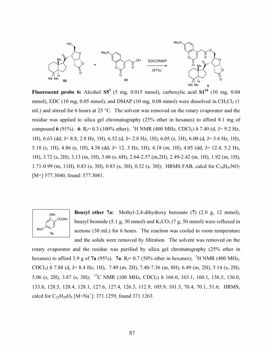
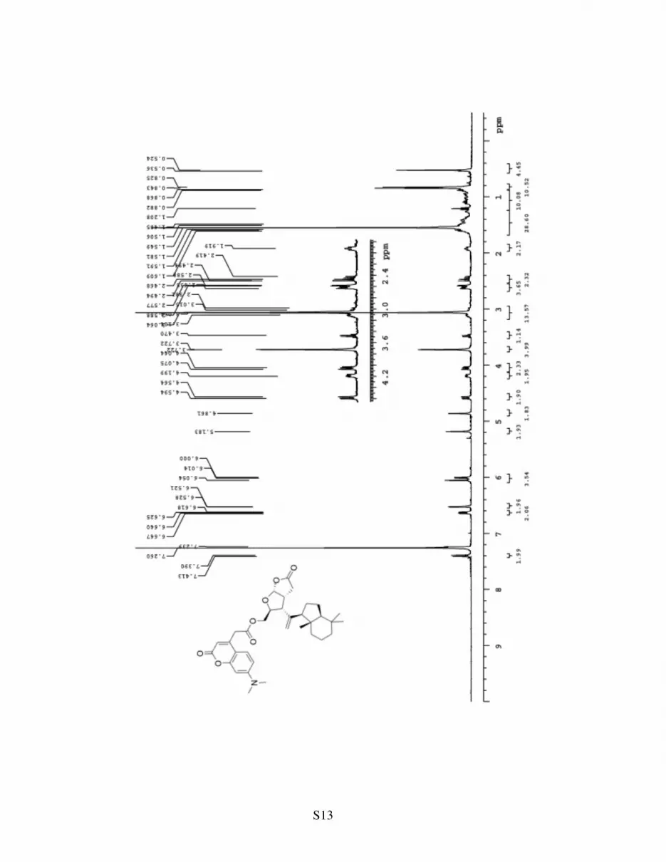
Fluorescent probe 6: Alcohol S53 (5 mg, 0.015 mmol), carboxylic acid S110 (10 mg, 0.04
mmol), EDC (10 mg, 0.05 mmol), and DMAP (10 mg, 0.08 mmol) were dissolved in CH2Cl2 (1
mL) and stirred for 6 hours at 25 °C. The solvent was removed on the rotary evaporator and the
residue was applied to silica gel chromatography (25% ether in hexanes) to afford 8.1 mg of
compound 6 (91%). 6: Rf= 0.3 (100% ether); 1H NMR (400 MHz, CDCl3) δ 7.40 (d, J= 9.2 Hz,
1H), 6.63 (dd, J= 8.8, 2.8 Hz, 1H), 6.52 (d, J= 2.8 Hz, 1H), 6.05 (s, 1H), 6.00 (d, J= 5.6 Hz, 1H),
5.18 (s, 1H), 4.86 (s, 1H), 4.58 (dd, J= 12, 3 Hz, 1H), 4.18 (m, 1H), 4.05 (dd, J= 12.4, 5.2 Hz,
1H), 3.72 (s, 2H), 3.13 (m, 1H), 3.06 (s, 6H), 2.64-2.57 (m,2H), 2.49-2.42 (m, 1H), 1.92 (m, 1H),
1.71-0.99 (m, 11H), 0.83 (s, 3H), 0.83 (s, 3H), 0.52 (s, 3H); HRMS FAB, calcd for C34H43NO7
[M+] 577.3040, found: 577.3041.
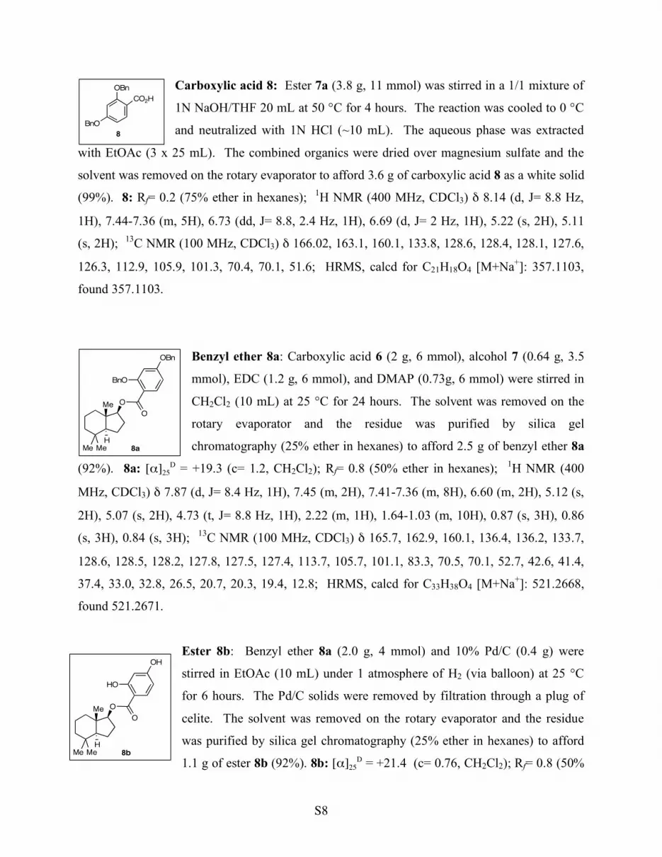
Benzyl ether 7a: Methyl-2,4-dihydroxy benzoate (7) (2.0 g, 12 mmol),
benzyl bromide (5.1 g, 30 mmol) and K2CO3 (7 g, 50 mmol) were refluxed in
acetone (30 mL) for 6 hours. The reaction was cooled to room temperature
and the solids were removed by filtration. The solvent was removed on the
rotary evaporator and the residue was purified by silica gel chromatography (25% ether in
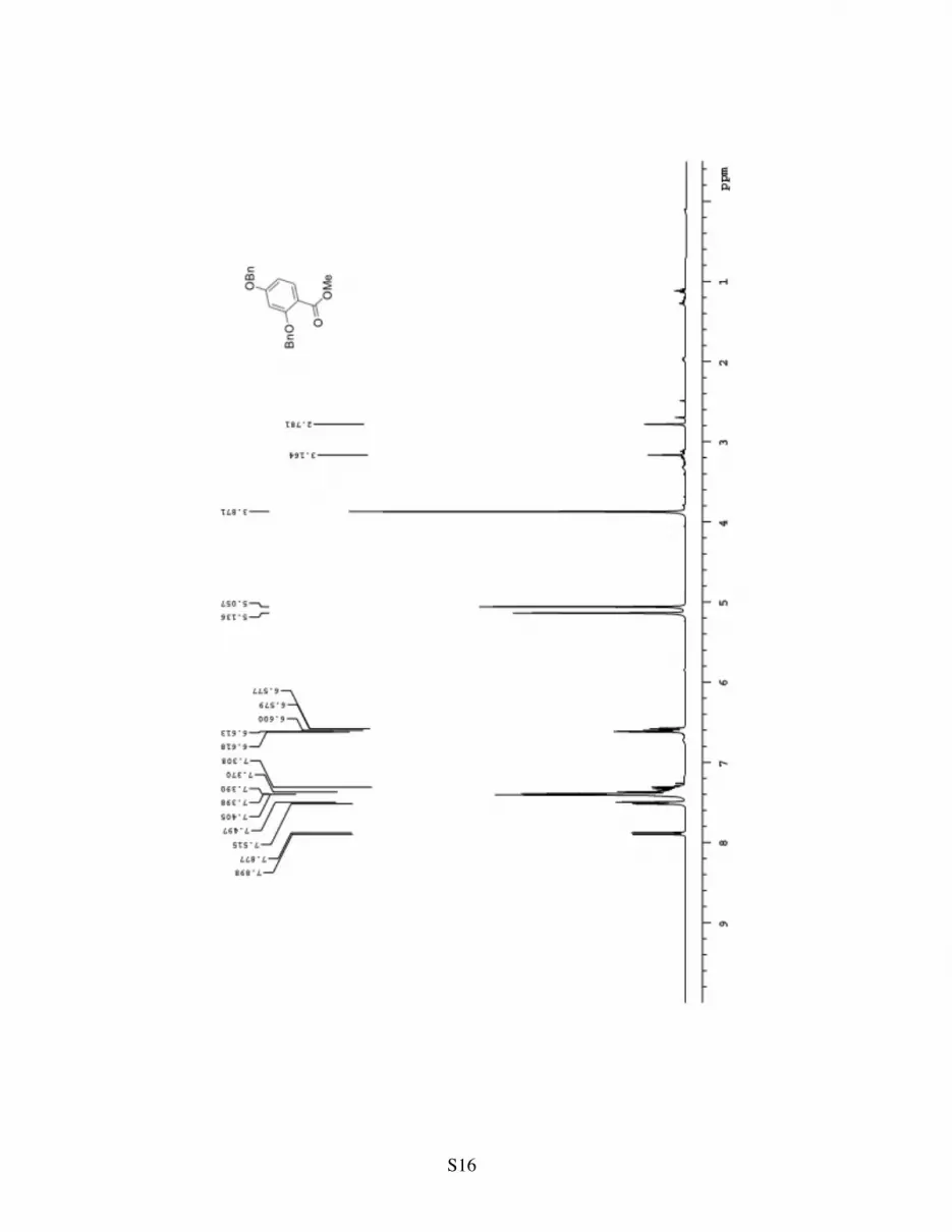
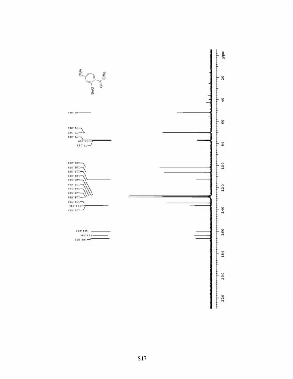
hexanes) to afford 3.9 g of 7a (95%). 7a: Rf= 0.7 (50% ether in hexanes); 1H NMR (400 MHz,
CDCl3) δ 7.88 (d, J= 8.4 Hz, 1H), 7.49 (m, 2H), 7.40-7.36 (m, 8H), 6.49 (m, 2H), 5.14 (s, 2H),
5.06 (s, 2H), 3.87 (s, 3H); 13C NMR (100 MHz, CDCl3) δ 166.0, 163.1, 160.1, 136.5, 136.0,
133.8, 128.5, 128.4, 128.1, 127.6, 127.4, 126.3, 112.9, 105.9, 101.3, 70.4, 70.1, 51.6; HRMS,
calcd for C22H20O4 [M+Na+]: 371.1259, found 371.1263.
OBn
BnO
CO2Me
7a
S8
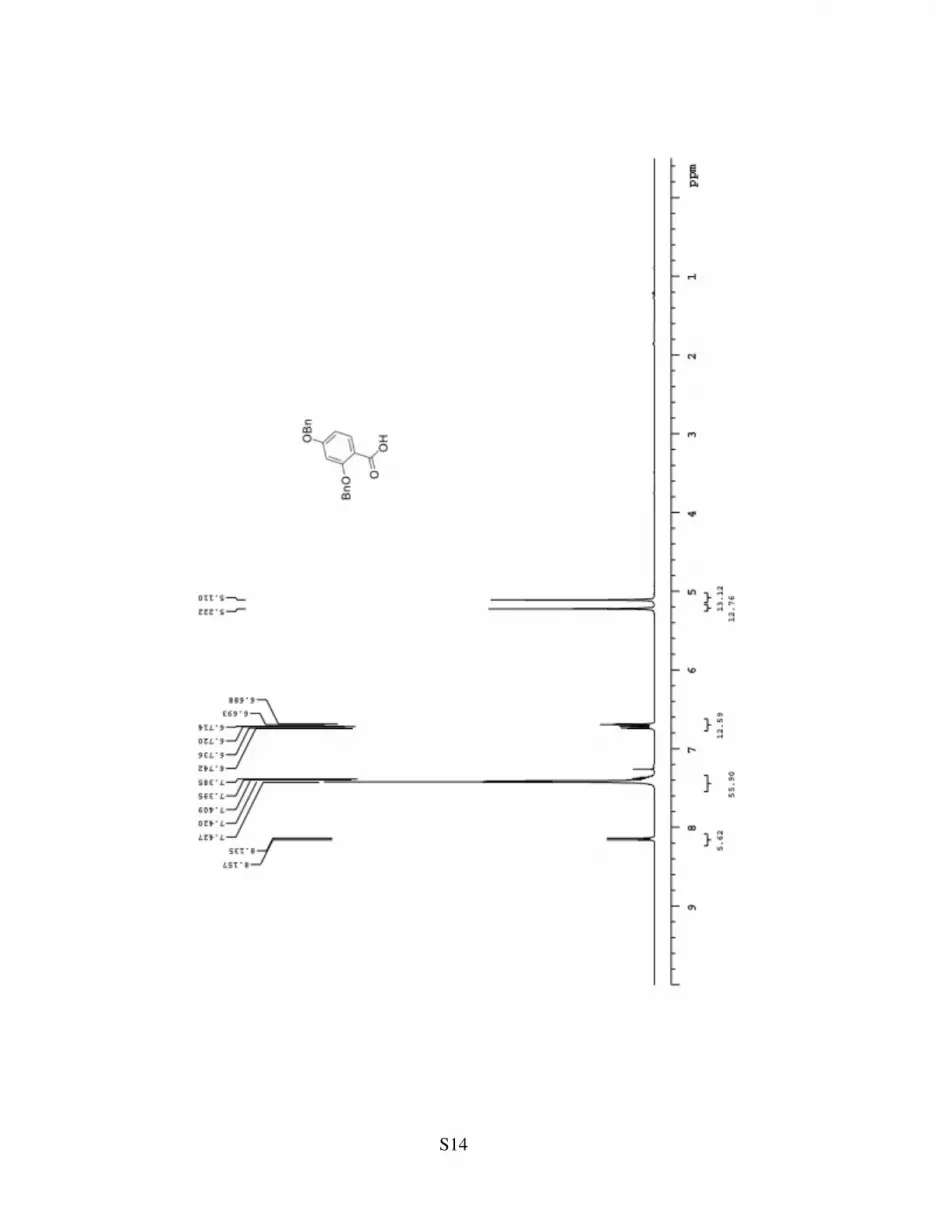
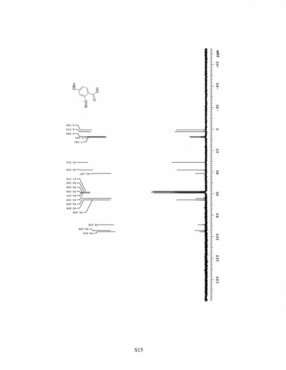
Carboxylic acid 8: Ester 7a (3.8 g, 11 mmol) was stirred in a 1/1 mixture of
1N NaOH/THF 20 mL at 50 °C for 4 hours. The reaction was cooled to 0 °C
and neutralized with 1N HCl (~10 mL). The aqueous phase was extracted
with EtOAc (3 x 25 mL). The combined organics were dried over magnesium sulfate and the
solvent was removed on the rotary evaporator to afford 3.6 g of carboxylic acid 8 as a white solid
(99%). 8: Rf= 0.2 (75% ether in hexanes); 1H NMR (400 MHz, CDCl3) δ 8.14 (d, J= 8.8 Hz,
1H), 7.44-7.36 (m, 5H), 6.73 (dd, J= 8.8, 2.4 Hz, 1H), 6.69 (d, J= 2 Hz, 1H), 5.22 (s, 2H), 5.11
(s, 2H); 13C NMR (100 MHz, CDCl3) δ 166.02, 163.1, 160.1, 133.8, 128.6, 128.4, 128.1, 127.6,
126.3, 112.9, 105.9, 101.3, 70.4, 70.1, 51.6; HRMS, calcd for C21H18O4 [M+Na+]: 357.1103,
found 357.1103.
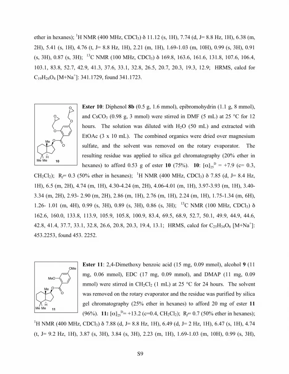
Benzyl ether 8a: Carboxylic acid 6 (2 g, 6 mmol), alcohol 7 (0.64 g, 3.5
mmol), EDC (1.2 g, 6 mmol), and DMAP (0.73g, 6 mmol) were stirred in
CH2Cl2 (10 mL) at 25 °C for 24 hours. The solvent was removed on the
rotary evaporator and the residue was purified by silica gel
chromatography (25% ether in hexanes) to afford 2.5 g of benzyl ether 8a
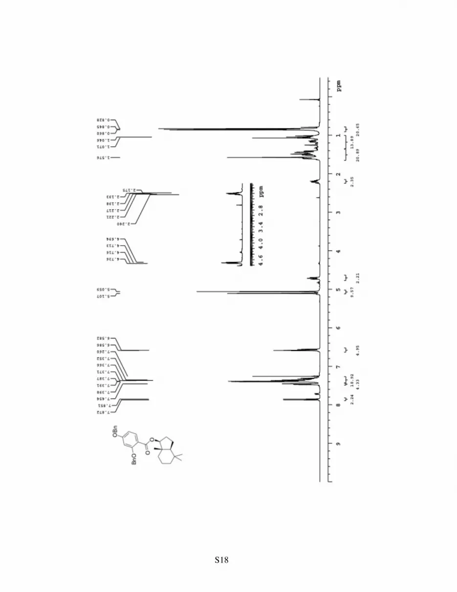
(92%). 8a: [α]25D = +19.3 (c= 1.2, CH2Cl2); Rf= 0.8 (50% ether in hexanes); 1H NMR (400
MHz, CDCl3) δ 7.87 (d, J= 8.4 Hz, 1H), 7.45 (m, 2H), 7.41-7.36 (m, 8H), 6.60 (m, 2H), 5.12 (s,
2H), 5.07 (s, 2H), 4.73 (t, J= 8.8 Hz, 1H), 2.22 (m, 1H), 1.64-1.03 (m, 10H), 0.87 (s, 3H), 0.86
(s, 3H), 0.84 (s, 3H); 13C NMR (100 MHz, CDCl3) δ 165.7, 162.9, 160.1, 136.4, 136.2, 133.7,
128.6, 128.5, 128.2, 127.8, 127.5, 127.4, 113.7, 105.7, 101.1, 83.3, 70.5, 70.1, 52.7, 42.6, 41.4,
37.4, 33.0, 32.8, 26.5, 20.7, 20.3, 19.4, 12.8; HRMS, calcd for C33H38O4 [M+Na+]: 521.2668,
found 521.2671.
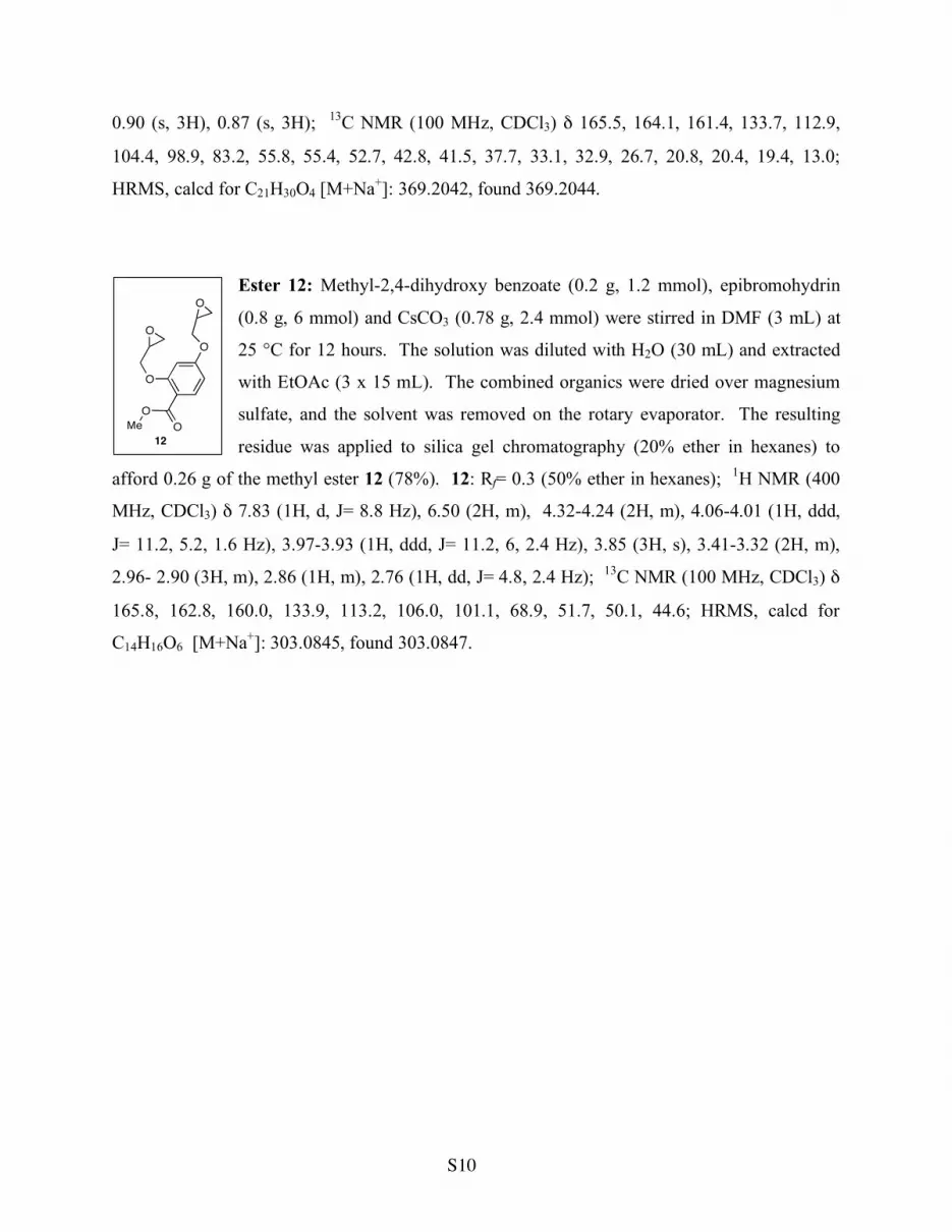
Ester 8b: Benzyl ether 8a (2.0 g, 4 mmol) and 10% Pd/C (0.4 g) were
stirred in EtOAc (10 mL) under 1 atmosphere of H2 (via balloon) at 25 °C
for 6 hours. The Pd/C solids were removed by filtration through a plug of
celite. The solvent was removed on the rotary evaporator and the residue
was purified by silica gel chromatography (25% ether in hexanes) to afford
1.1 g of ester 8b (92%). 8b: [α]25D = +21.4 (c= 0.76, CH2Cl2); Rf= 0.8 (50%
8a
Me
Me Me
H
O
BnO
O
OBn
8b
Me
Me Me
H
O
HO
O
OH
OBn
BnO
CO2H
8
S9
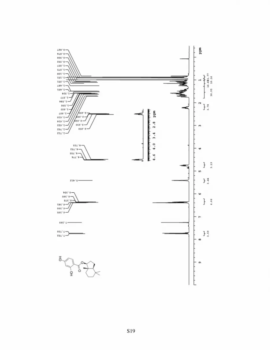
ether in hexanes); 1H NMR (400 MHz, CDCl3) δ 11.12 (s, 1H), 7.74 (d, J= 8.8 Hz, 1H), 6.38 (m,
2H), 5.41 (s, 1H), 4.76 (t, J= 8.8 Hz, 1H), 2.21 (m, 1H), 1.69-1.03 (m, 10H), 0.99 (s, 3H), 0.91
(s, 3H), 0.87 (s, 3H); 13C NMR (100 MHz, CDCl3) δ 169.8, 163.6, 161.6, 131.8, 107.6, 106.4,
103.1, 83.8, 52.7, 42.9, 41.3, 37.6, 33.1, 32.8, 26.5, 20.7, 20.3, 19.3, 12.9; HRMS, calcd for
C19H26O4 [M+Na+]: 341.1729, found 341.1723.
Ester 10: Diphenol 8b (0.5 g, 1.6 mmol), epibromohydrin (1.1 g, 8 mmol),
and CsCO3 (0.98 g, 3 mmol) were stirred in DMF (5 mL) at 25 °C for 12
hours. The solution was diluted with H2O (50 mL) and extracted with
EtOAc (3 x 10 mL). The combined organics were dried over magnesium
sulfate, and the solvent was removed on the rotary evaporator. The
resulting residue was applied to silica gel chromatography (20% ether in
hexanes) to afford 0.53 g of ester 10 (75%). 10: [α]25D = +7.9 (c= 0.3,
CH2Cl2); Rf= 0.3 (50% ether in hexanes); 1H NMR (400 MHz, CDCl3) δ 7.85 (d, J= 8.4 Hz,
1H), 6.5 (m, 2H), 4.74 (m, 1H), 4.30-4.24 (m, 2H), 4.06-4.01 (m, 1H), 3.97-3.93 (m, 1H), 3.40-
3.34 (m, 2H), 2.93- 2.90 (m, 2H), 2.86 (m, 1H), 2.76 (m, 1H), 2.24 (m, 1H), 1.75-1.34 (m, 6H),
1.26- 1.01 (m, 4H), 0.99 (s, 3H), 0.89 (s, 3H), 0.86 (s, 3H); 13C NMR (100 MHz, CDCl3) δ
162.6, 160.0, 133.8, 113.9, 105.9, 105.8, 100.9, 83.4, 69.5, 68.9, 52.7, 50.1, 49.9, 44.9, 44.6,
42.8, 41.4, 37.7, 33.1, 32.8, 26.6, 20.8, 20.3, 19.4, 13.1; HRMS, calcd for C25H34O6 [M+Na+]:
453.2253, found 453. 2252.
Ester 11: 2,4-Dimethoxy benzoic acid (15 mg, 0.09 mmol), alcohol 9 (11
mg, 0.06 mmol), EDC (17 mg, 0.09 mmol), and DMAP (11 mg, 0.09
mmol) were stirred in CH2Cl2 (1 mL) at 25 °C for 24 hours. The solvent
was removed on the rotary evaporator and the residue was purified by silica
gel chromatography (25% ether in hexanes) to afford 20 mg of ester 11
(96%). 11: [α]25D= +13.2 (c=0.4, CH2Cl2); Rf= 0.7 (50% ether in hexanes);
1H NMR (400 MHz, CDCl3) δ 7.88 (d, J= 8.8 Hz, 1H), 6.49 (d, J= 2 Hz, 1H), 6.47 (s, 1H), 4.74
(t, J= 9.2 Hz, 1H), 3.87 (s, 3H), 3.84 (s, 3H), 2.23 (m, 1H), 1.69-1.03 (m, 10H), 0.99 (s, 3H),
10
Me
Me Me
H
O
O
O
O
O
O
Me
Me Me
H
O
MeO
O
OMe
11
S10
0.90 (s, 3H), 0.87 (s, 3H); 13C NMR (100 MHz, CDCl3) δ 165.5, 164.1, 161.4, 133.7, 112.9,
104.4, 98.9, 83.2, 55.8, 55.4, 52.7, 42.8, 41.5, 37.7, 33.1, 32.9, 26.7, 20.8, 20.4, 19.4, 13.0;
HRMS, calcd for C21H30O4 [M+Na+]: 369.2042, found 369.2044.
Ester 12: Methyl-2,4-dihydroxy benzoate (0.2 g, 1.2 mmol), epibromohydrin
(0.8 g, 6 mmol) and CsCO3 (0.78 g, 2.4 mmol) were stirred in DMF (3 mL) at
25 °C for 12 hours. The solution was diluted with H2O (30 mL) and extracted
with EtOAc (3 x 15 mL). The combined organics were dried over magnesium
sulfate, and the solvent was removed on the rotary evaporator. The resulting
residue was applied to silica gel chromatography (20% ether in hexanes) to
afford 0.26 g of the methyl ester 12 (78%). 12: Rf= 0.3 (50% ether in hexanes); 1H NMR (400
MHz, CDCl3) δ 7.83 (1H, d, J= 8.8 Hz), 6.50 (2H, m), 4.32-4.24 (2H, m), 4.06-4.01 (1H, ddd,
J= 11.2, 5.2, 1.6 Hz), 3.97-3.93 (1H, ddd, J= 11.2, 6, 2.4 Hz), 3.85 (3H, s), 3.41-3.32 (2H, m),
2.96- 2.90 (3H, m), 2.86 (1H, m), 2.76 (1H, dd, J= 4.8, 2.4 Hz); 13C NMR (100 MHz, CDCl3) δ
165.8, 162.8, 160.0, 133.9, 113.2, 106.0, 101.1, 68.9, 51.7, 50.1, 44.6; HRMS, calcd for
C14H16O6 [M+Na+]: 303.0845, found 303.0847.
O
O
O
O
O
O
Me
12
S11

S12
S13
S14
S15
S16
S17
S18
S19
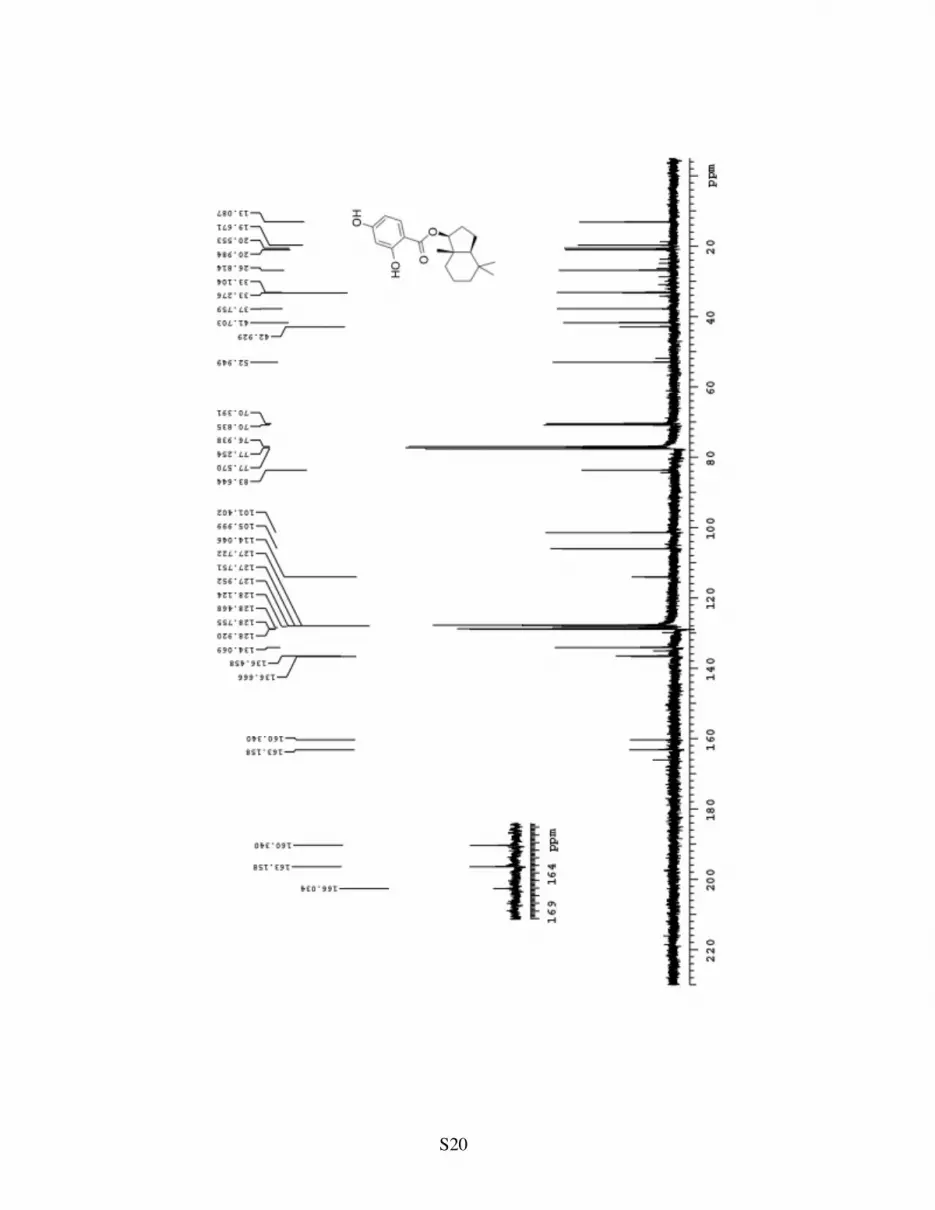
S20
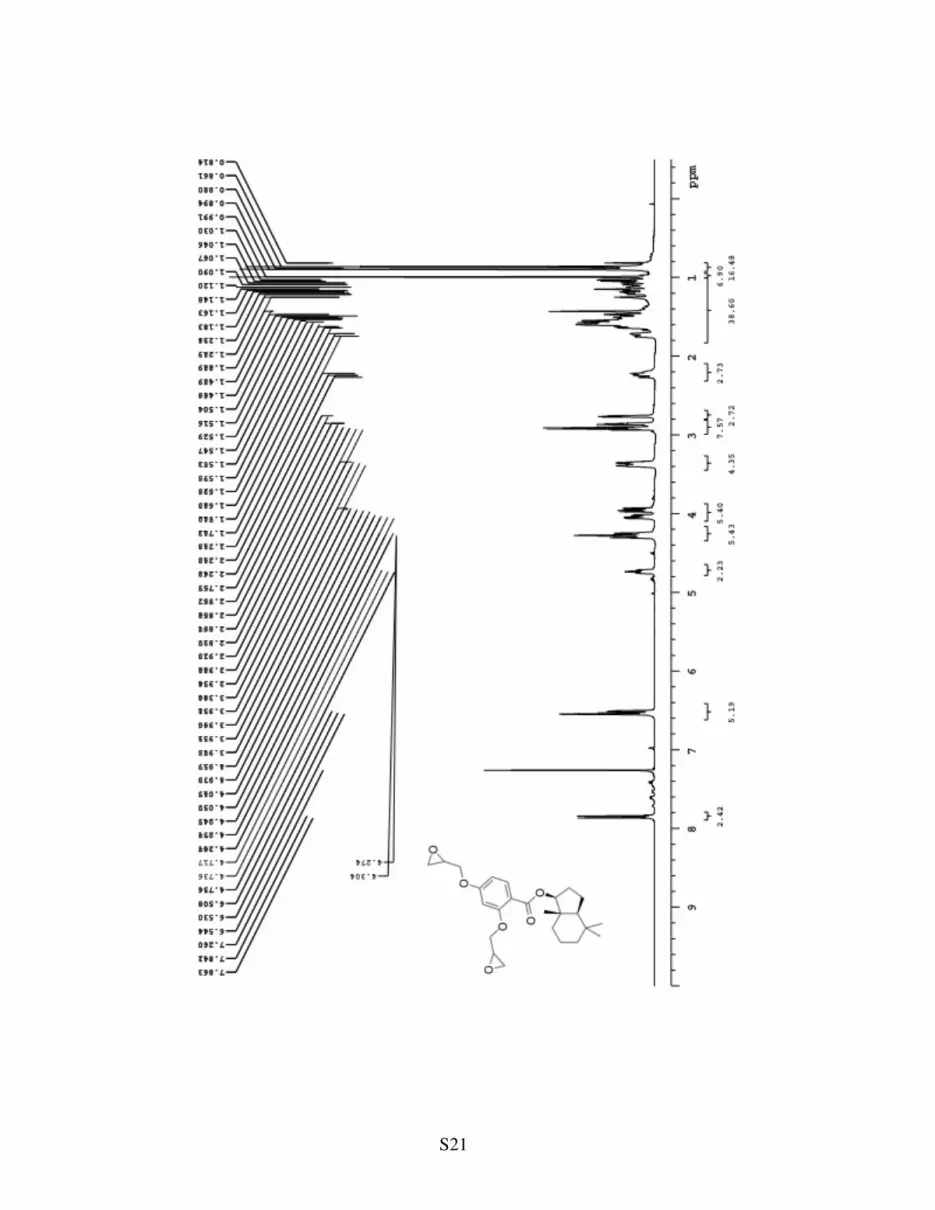
S21

S22
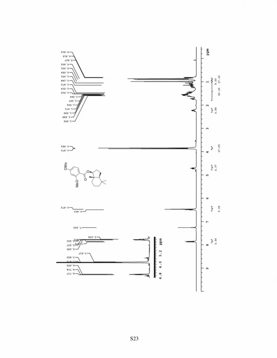
S23
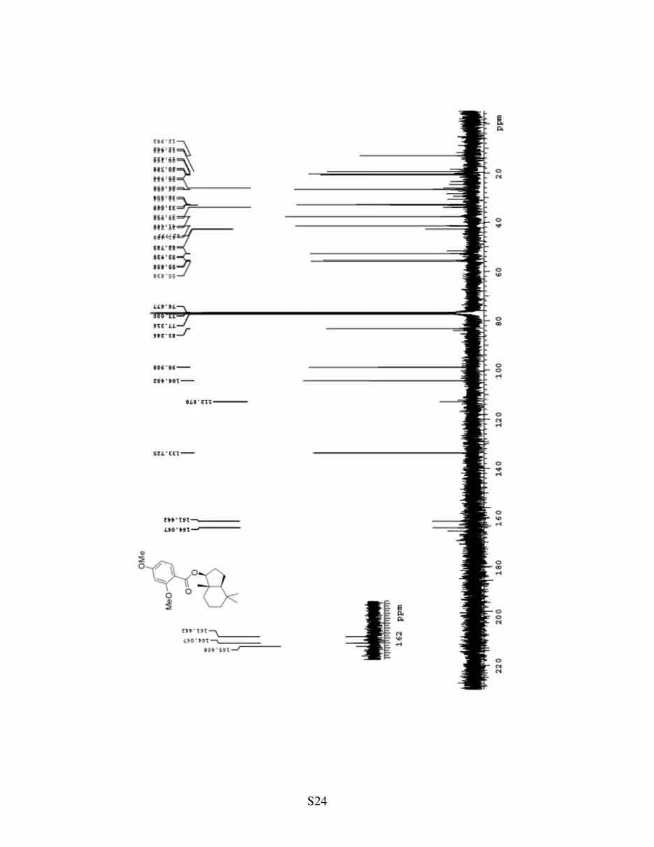
S24
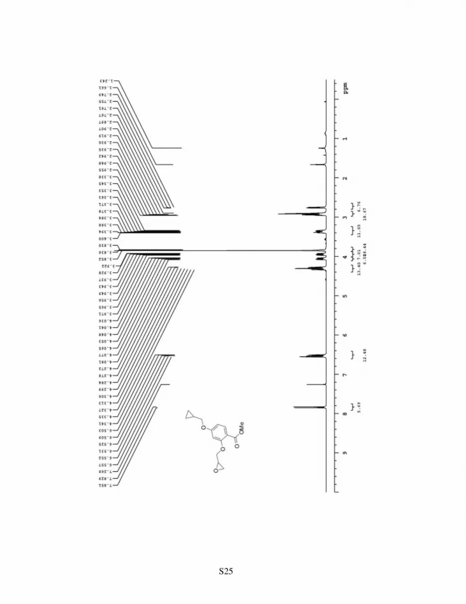
S25
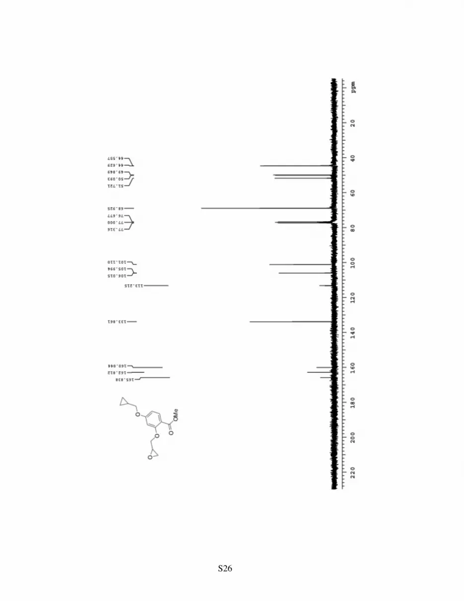
S26