characterization of thermophilic bacteria using surface-enhanced raman scattering
TRANSCRIPT
Characterization of Thermophilic Bacteria UsingSurface-Enhanced Raman Scattering
MUSTAFA CULHA,* AHMET ADIGUZEL, M. MUGE YAZICI, MEHMET KAHRAMAN,FIKRETTIN SxAHIN, and MEDINE GULLUCEYeditepe University, Faculty of Engineering and Architecture, Genetics and Bioengineering Department, Kayisdagi, 34755, Istanbul, Turkey
(M.C., M.M.Y., M.K., F.Sx.); Ataturk University, Health Services Vocational Training School, 25070, Yenisehir-Erzurum, Turkey (A.A.); and
Ataturk University, Faculty of Arts and Sciences, Biology Department, 25240-Kampus/Erzurum
Surface-enhanced Raman scattering (SERS) can provide molecular-level
information about the molecules and molecular structures in the vicinity
of nanostructured noble metal surfaces such as gold and silver. The three
thermophilic bacteria Bacillus licheniformis, Geobacillus stearothermophi-
lus, and Geobacillus pallidus, a Gram-negative bacterium E. coli, and a
Gram-positive bacterium B. megaterium are comparatively characterized
using SERS. The SERS spectra of thermophilic bacteria are similar, while
they show significant differences compared to E. coli and B. megaterium.
The findings indicate that a higher number of thiol residues and possible
S–S bridges are present in the cell wall structure of thermophilic bacteria,
providing their stability at elevated temperatures. Incubating the
thermophilic bacteria with colloidal silver suspension at longer times
improved the bacteria–silver nanoparticle interaction kinetics, while
increased temperature does not have a pronounced effect on spectral
features. A tentative assignment of the SERS bands was attempted for
thermophilic bacteria. The results indicate that SERS can be a useful tool
to study bacterial cell wall molecular differences.
Index Headings: Surface-enhanced Raman spectroscopy; SERS; Thermo-
philic bacteria; Bacteria; Cell walls; Silver nanoparticles.
INTRODUCTION
Surface-enhanced Raman scattering (SERS) has beenemerging as a powerful tool for the characterization ofbiological structures and the development of novel diagnostictools and sensing systems for a variety of biological and non-biological molecules.1–3 The power of the technique can beattributed to the ‘‘finger-printing’’ property and its compatibilitywith water. This ‘‘finger-printing’’ property can be extended forlarge biological structures such as bacteria and viruses4,5 andused for microorganism identification and classification.6–13
Although several aspects of the use of SERS for ‘‘whole-microorganism’’ identification such as the reproducibility ofSERS spectra have been addressed, it can still be used with ahigh confidence level for fast bacterial identification in clinicalsettings.
In order for a molecule to experience SERS enhancement, itmust be in the close vicinity of a nano-structured surface. Thisdistance must be in the 1 to 4 nm range for proper SERSenhancement.14 The distance requirement significantly altersthe process that brings together the nano-structured surface andbacterial cells compared to small molecules. Most reportsconcerning bacterial SERS studies involve the use of a noblemetal colloidal suspension.4,12,15,16 This is mostly due to thefact that bacterial cells and nanostructures must be in closecontact at as many points as possible. Therefore, the SERSspectra obtained with simple mixing is mostly from bacterial
cell walls. In order to investigate the origin of the bands in theSERS spectra, a detailed investigation was undertaken by Zeiriet al.6–8 They demonstrated that the bands appearing in theSERS spectra belong to the bacterial wall biochemicalstructure. Although there was a possibility of nanoparticlessmaller than 3 nm entering the bacterial cells, in our studies wedemonstrated that the contribution to the SERS spectra fromsuch nanoparticles is very small and probably nonexistent dueto the fact that the silver nanoparticles used in our studies aremuch larger than 3 nm in size.17
Thermophilic bacteria are a subclass of archaea, which isaccepted as a third life domain along with bacteria andeucarya.18 Bacteria can be classified into two major groups asGram-positive and Gram-negative based on their cell-wallstructure. Gram-negative bacteria cell walls have a thinpeptidoglycan layer between the plasma membrane and aphospholipid–lipopolysaccharide outer membrane, whileGram-positive bacteria cell walls have a murein-containingthick peptidoglycan layer that is a polymer of disaccharides(glycan) cross-linked by short peptide fragments. All bacterialpeptidoglycans contain N-acetylmuramic acid, which is a partof murein. The layers of peptidoglycan in Gram-positivebacteria contain teichoic acids that are linear polymers ofpolyglycerol or polyribitol substituted with phosphates and afew amino acids and sugars. Thermophilic bacteria, which areclassified into archaebacter, have different cell wall propertiescompared to Gram-positive and Gram-negative bacteria due totheir extreme life conditions. The proteins, polysaccharides,and peptidoglycan-like molecules are also the main compo-nents of archaea. However, the biochemical structural differ-ences make them stable at elevated temperatures. In this study,we have investigated the cell wall biochemical structuraldifferences of the thermophilic bacteria Bacillus licheniformis,Geobacillus pallidus, and Geobacillus stearothermophilusalong with two well-characterized bacteria, E. coli and B.megaterium.
MATERIALS AND METHODS
Chemicals. Silver nitrate (AgNO3, 99.5%), HAuCl4�3H20,and nutrient agar were purchased from Fluka (Seelze,Germany). Sodium citrate (99%) was purchased from Merck(Darmstadt, Germany).
Preparation of Colloidal Suspensions. Silver colloid wasprepared according to the method of Lee and Meisel.19 Briefly,90 mg of AgNO3 was dissolved in 500 mL of distilled waterand heated until boiling. Then, a 10 mL volume of 1% sodiumcitrate solution was added. The mixture was kept boiling until afinal volume of 250 mL is reached. The colloidal suspensionwas characterized by using ultraviolet (UV) spectroscopy, and
Received 21 April 2008; accepted 25 August 2008.* Author to whom correspondence should be sent. E-mail: [email protected].
1226 Volume 62, Number 11, 2008 APPLIED SPECTROSCOPY0003-7028/08/6211-1226$2.00/0
� 2008 Society for Applied Spectroscopy
a maximum wavelength at 420 nm was recorded. The colloidalsuspension was concentrated to four-fold by centrifugation at5500 rpm for 30 min. The gold colloidal suspension wassynthesized as previously defined.15
Characterization of Thermophilic Bacteria. The isola-tion, purification, and characterization of selected thermophilicbacteria used in this study was reported elsewhere.20
16S rRNA Gene Sequence Analysis. A total 1400nucleotides of the 16S rRNA from three isolates (Ah2, Ah22,Ah23) were aligned and compared with sequences of relatedbacteria. On the basis of 16S rRNA gene sequence analysis, thetwo isolates exhibited �98% resemblance to Geobacilluspallidus (Ah23) and G. stearothermophilus (Ah22), respec-tively. On the basis of 16S rRNA gene sequence analysis, thethird isolate exhibited �99% resemblance to Bacillus lichen-iformis (Ah2).
Preparation of Bacterial Samples. All bacterial sampleswere obtained from the microorganism collection of AtaturkUniversity. Before using the bacteria, all of them wereidentified by the Sherlock Microbial Identification Systemversion 4.5 (MIDI, Newark, DE) and principal componentsregression (PCR). The thermophilic bacteria were cultivated for24 h at 55 8C and E. coli and B. megaterium were grown for 24h at 37 8C on nutrient agar. After cultivation, the bacteria werewashed with deionized water to remove the nutrient agar fromthe bacteria. The washing procedure includes the following:The bacterial samples were added into 1 mL deionized waterand the mixture was centrifuged at 7500 rpm for five minutes.The supernatant was discarded and 1 mL of deionized waterwas added. This procedure was repeated three times. For thesimple mixing method, 5 lL of each washed bacterium wasadded to 100 lL of colloidal silver nanoparticles. This wasmixed with a vortex and 5 lL of the resulting homogeneousmixture was put onto a CaF2 slide. The spot was dried at roomtemperature for about 15 min before analysis.
Temperature and Incubation Time Study. Each cultivatedthermophilic bacteria sample was incubated with the 43concentrated colloidal suspension at 40, 55, and 70 8C forincreasing times of 5, 10, and 25 minutes. The incubatedsamples were spotted on a CaF2 slide and dried at roomtemperature. The temperature and incubation experiments wereperformed for B. megaterium and E. coli in the same way as forthe thermophilic bacteria. The only difference was the startingtemperature of 37 8C, which is the regular growth temperatureof B. megaterium and E. coli.
Raman Instrumentation. All measurements were per-formed using a Renishaw In via reflex Raman spectroscopysystem (Renishaw Plc., New Mills, Wotton-under-EdgeGloucestershire, UK) equipped with 830 nm diode and 514nm argon ion lasers. The diode laser at 830 nm was used forthis study. The laser power was 3 mW and exposure time was10 s. A 503 objective was used.
Scanning Electron Microscope. The mixture of silvernanoparticles and bacteria was spotted and dried on a scanningelectron microscope (SEM) sample stub. A Karl Zeiss EVO 40model SEM instrument was used. The accelerating voltage was10 kV.
RESULTS AND DISCUSSION
Either silver or gold colloidal suspensions for bacterialSERS experiments can be used. However, our previous studiesshowed that silver colloidal suspension was the more
appropriate choice and the attempts at utilizing gold colloidalsuspension were not as successful as those using silvercolloidal suspension under the experimental conditions report-ed in this study.15 The SERS spectra of G. stearothermophilusobtained with silver and gold colloidal nanoparticles in the sizeof 41 nm and 13 nm are comparatively presented in theSupplemental Material.� Silver colloidal suspensions preparedusing the method of Lee and Meisel are well characterized andcontain silver nanoparticles with an average size of 50 nm andthe spectral variations in bacterial SERS especially becomemore limited with the use of citrate-reduced silver colloidalnanoparticles.21 Mixing silver colloidal suspension as synthe-sized with bacterial cells generates a heterogeneous sample,which results in considerable variations in the SERS spectra ofthe bacteria. A bacterial sample prepared with increasedcolloidal suspension concentration results in a more homoge-nous sample.
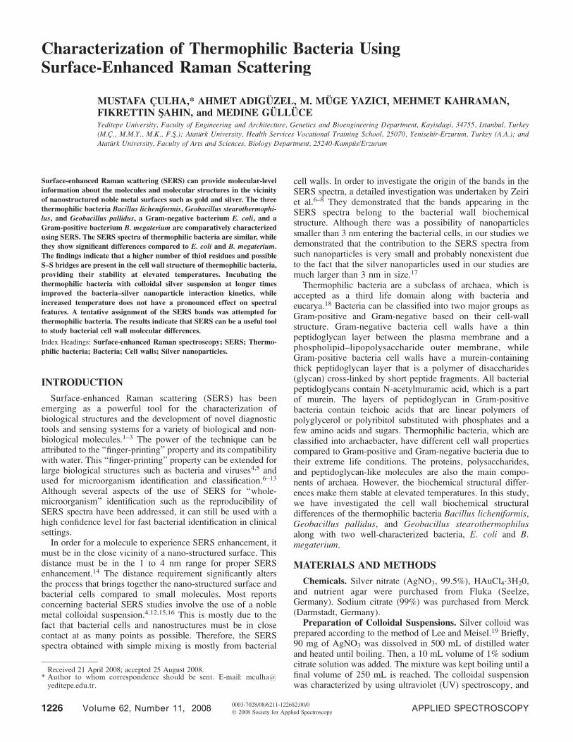
Previously, we have demonstrated the reproducibility ofSERS spectra following the sample preparation method used inthis study.16 Figure 1 shows the scanning electron microscopy(SEM) image of B. licheniformis mixed with the silvercolloidal nanoparticles and Fig. 2 shows the reproducibilityof the SERS spectra obtained from the sample demonstrated onFig. 1, ensuring the spot to spot variations on the same sample.The density of silver nanoparticles in the sample is reasonablyhigh and the spectra acquired from different spots on the samesample show very small variations, which is critical for thecomparison of SERS spectra obtained from different samples.
Another consideration that must be addressed upon mixingsilver colloidal suspension with bacterial samples is thepossibility of the nanoparticles entering into the bacterial cells.This information is critical for accurate assignment of bands inthe SERS spectra. The origin of the bands in SERS spectra wasstudied by Efrima and Bronk and Zeiri et al.6–8 A discussionregarding the possible origin of the SERS bands of bacteria canbe found in Ref. 17. In addition, when the size of the silvernanoparticles used in this study is considered, a contributionfrom the components in the bacterial cells appears limited. Itshould also be noted that the SERS spectra obtained from asample prepared at a different time with a different batch of thesame bacteria (G. stearothermophilus) are very comparable tothe spectra obtained before (see Fig. 3 and the SupplementalMaterial).
Despite the biochemical structural differences, the SERSspectra of bacteria strongly depend on the selective interactionof silver nanoparticles with the biochemical structures on thebacterial cell walls. The presence of negatively and positivelycharged functional groups and thiol groups may enhance theaffinity of the silver nanoparticles to the bacterial cell wall. Forexample, the presence of FADH possessing locations sur-rounded by the thiol-group-containing amino acid cysteine maycause the selective localization of silver nanoparticles in theseregions due to the possible Ag–S– bonds. The peak at 727cm�1 was previously assigned to an adenine-containingcompound such as FADH other than DNA.7,22 The same peakwas also assigned to N-acetyl-D-glucosamine (NAG) byanother group.4 It is possible that this peak may arise fromboth structures when its intensity is considered. The change in
� Supplemental Material is available on-line in the electronic version of thejournal (htt://www.s-a-s.org).
APPLIED SPECTROSCOPY 1227

the intensity of this peak from bacterium to bacterium may
reflect the number of such sites on the cell wall.
The most distinct difference in the bacteria and archaea cell
walls is the organization of biological macromolecules with
some functional differences. For example, in bacterial cell
membranes, un-branched fatty acid chains are connected to the
D-glycerol through ester linkages, while in archaea this linkage
is an ether bond of branched isoprene chains. Also, some of the
archaea contain pseudomurein, which corresponds to the
peptidoglycan structure in bacteria. Similar to the structure of
peptidoglycan, N-acetyl glucosamine (NAG) and N-acetyl
muramic acid (NAM) are cross-linked by amino acid bridges.
Some archaea may contain an S-layer that may include one or a
few proteins, glycoproteins, or sugar. Therefore, the distinct
FIG. 2. Spot-to-spot reproducibility of the SERS spectra obtained from thesample seen in Fig. 1.
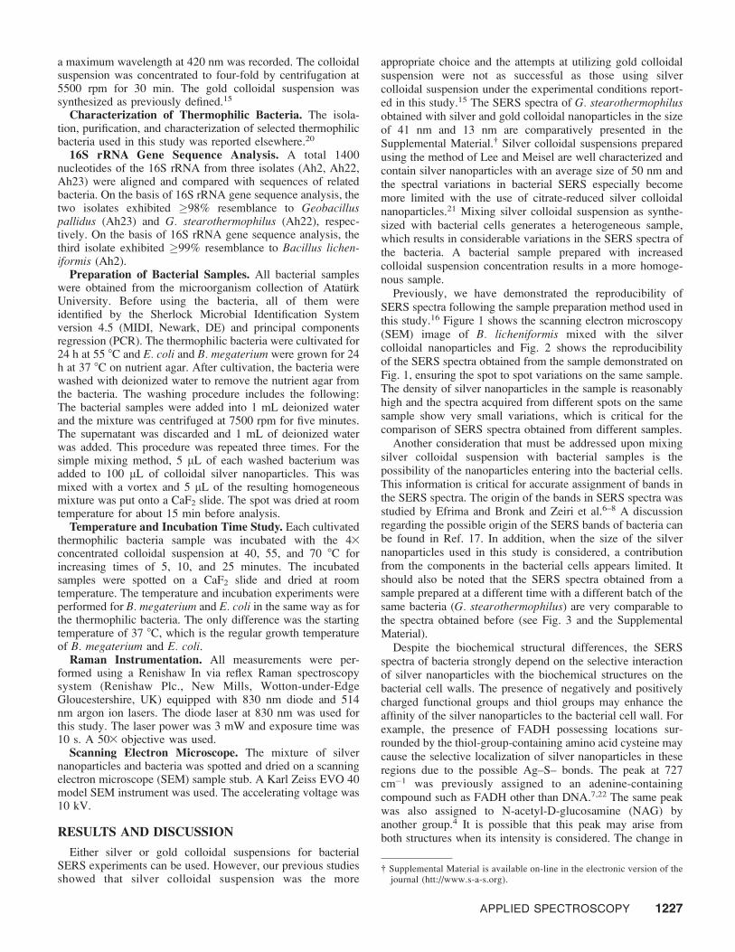
FIG. 3. (A) SERS spectra of (a) B. megaterium, (b) B. licheniformis, (c) G.stearothermophilus, (d) G. pallidus, and (e) E. coli. (B) Three thermophilicbacteria have been separated and shown separately for clarity.
FIG. 1. SEM image of B. licheniformis mixed with colloidal silver nanoparticles.
1228 Volume 62, Number 11, 2008

biochemical structural differences in thermophilic bacteria areexpected.
Due to the fact that Raman bands are the result of theinelastic scattering from vibrating bonds and the presence of anumber of similar bonds in many macromolecules, theobserved bands could be originating from multiple macromol-ecules in contact with the silver nanoparticles. Carefulinspection of Figs. 3A and 3B reveals that there are significantdifferences in the spectral features of Gram-positive, Gram-negative, and thermophilic bacteria. The most distinct featureseen in the SERS spectra of B. megaterium (spectrum a)compared to gram-negative (spectrum e) and thermophilicbacteria (spectra b, c, and d) is the peak at 658 cm�1 in thespectra of B. megaterium, which appears at lower wavenum-bers (663, 665, or 659, cm�1) with other bacteria. At otherwavenumbers such as 522, 577, 801, 961 1134, 1246, and1329 cm�1, the relative intensities of the peaks varysignificantly. The comparison of the SERS spectrum of E. colito thermophilic bacteria again reveals that the cell wallbiochemical structure is rather different.
The most distinct features of the SERS spectra ofthermophilic bacteria are seen in the 500–700 cm�1 regioncompared to E. coli and B. megaterium. The adsorption of S–Scontaining proteins on silver colloidal nanoparticles wasstudied, and the presence of S–S and C–S bonds can beconfirmed from SERS spectra.23 Especially, the 600–750 cm�1
region may provide information about the conformation of theC–C bond adjacent to the C–S bond.23 It should also be notedthat this region of the spectra of proteins is dominated by bandsoriginating from tryptophan and phenylalanine.23 The bands at689, 690, and 693 cm�1 on the SERS spectra of insulin,oxytocin, and Streptomyces substilisin inhibitor, respectively,were assigned to C–S vibrations of C–S–S–C.23 Therefore, weassign the bands at 691, 692, and 695 cm�1 to C–S vibrationson the SERS spectra of three thermophilic bacteria. This bandis observed in neither E. coli nor B. mageterium SERS spectra.There is a broad band ranging from 555 to 580 cm�1 on the
SERS spectra of thermophilic bacteria and we believe that thebands of the S–S bond vibrations overlap with the bandsoriginating from carbohydrate moieties on the cell-wallstructure. The rest of the bands up to 750 cm�1 that arepresent in the spectra of all bacteria can be originating fromcarbohydrates and amino acid residues such as tryptophan,phenylalanine, and tyrosine.
The inspection of the SERS spectra of all bacteria revealsthat all the intensities of the bands relative to other bands in thesame spectrum show considerable variations, which mayindicate the presence of the indicated bonds at higher frequencyon the cell wall structure. The SERS spectra of B. megaterium,B. licheniformis, G. stearothermophilus, G. pallidus, and E.coli are seen in Fig. 3. The tentative peak assignments for theSERS spectra are given in Table I.
As explained in the Experimental section, the colloidalsuspension of bacterial cells is mixed and spotted on a CaF2
slide immediately after mixing and the SERS spectra wereacquired from this spotted sample. Increased temperatures andtreatment of thermophilic bacteria with silver colloidalnanoparticles at longer times before spotting on the substratemay provide information about the interaction of C–S and C–S–S–C bonds with silver nanoparticles due to the strongtendency of possible formation of Ag–S bonds. Figure 4 showsthe SERS spectra of G. stearothermophilus after 5 minutes ofincubation with colloidal silver nanoparticles at 55 8C anddrying at room temperature (spectrum a) and withoutincubation (spectrum b). As seen, there is a considerablechange in the spectral features in the range of 500 to 730 cm�1,to which the C–S or S–S bond vibrations are assigned.However, the band intensities change significantly, indicatingthe possible rearrangement of bond positions against nanopar-ticles. The broad band in the range of 555 to 580 cm�1 perhapscontains more than two overlapping bands (558 and 575 cm�1).The intensity of the bands at 691, 527, and 460 cm�1 alsoincreases. An increase in the intensity of the bands appearing at1152 and 1214 cm�1 is apparent upon incubation and is
TABLE I. The tentative band assignments of the SERS spectra of the characterized bacteria.
B. megaterium B. licheniformis G. stearothermophilus G. pallidus E. coli Band assignments
– – 460 – –522 (w) 522 (w) 527 (w) 522 (w) 522 (m)
– 556 (w) 558 (w) 560 (w) – C–S–S–C bonds23
577 (m) 576 (m) 575 (m) 577 (m) 582 (m) Carbohydrates24
631 (vw) 630 (w) 632 (m) 635 (w) 610 (m) COO� bend or C–S stretch23
658 (vs) 663 (m) 665 (m) 659 (m) 663 (m) Carbohydrates24
– 692 (w) 691 (m) 695 (w) – C–S stretch23
727 (vs) 734 (vs) 734 (vs) 734 (vs) 732 (vs) Adenine from flavin7
801 (m) 805 (m) 805 (w) 805 (w) 808 (w) O–P–O25
873 (vw) – – – – C–C stretch934 (w) 924 (w) 931 (w) 921 (vw) 925 (m) C–COO� stretch26
961 (s) 966 (vw) 963 (s) 963 (s) 967 (m) C–N stretch27
1054 (m) – 1043 (w) 1032 (w) 1056 (w) C–C stretch28
1103 (m) 1081 (vw) – 1067 (w) 1107 (m) PO2� 7
1134 (s) 1150 (vw) – 1150 (w) – Aromatic amino acids in protein29
– 1213 (vw) 1213 (vw) – 1225 (w) Amide III8
1246 (s) 1283 (vw) 1276 (vw) 1272 (w) – ¼CH in-plane (lipid) or amide III29
1329 (s) 1341 (s) 1337 (vs) 1342 (vs) 1331 (s) C–H bend protein28
1377 (m) – – – C–H bend protein28
1433 (w) 1447 (s) – 1454 (s) 1450 (s) CH2 bend (protein, lipid)29
1473 (w) 1471 (s) – – CH2 bend (protein, lipid)29
1557 (m) 1548 (m) – 1545 (m) 1550 (s) N–H, C–H bend, C¼C stretch30
1589 (w) 1590 (w) 1587 (w) 1583 (w) – C¼C lipid31
1637 (vw) – – – – Amide I32
APPLIED SPECTROSCOPY 1229

assigned to aromatic amino acid side chains and amide IIIvibrations. This indicates that incubating the mixture allowsimproved interaction between the bacteria cell wall and thesilver nanoparticles.
All thermophilic bacteria with the silver colloidal suspensionat three different temperatures (40, 55, and 70 8C) wereincubated at three different time intervals (5, 10, and 25minutes) before SERS acquisition. Figures 5A and 5C show theinfluence of the incubation time on the SERS spectra ofthermophilic bacteria at constant temperature. The bands thatshow changes are marked with asterisks in the spectra. The
incubation time had a dramatic effect on the peak at 691 cm�1,which is assigned to C–S vibrations. The intensity of the peakat 691 cm�1 on the SERS spectra of G. stearothermophilusdiminishes as the incubation time increases, and the broad bandin the range of 555 to 580 cm�1 splits into two bands as theincubation time and temperature are increased. A band around558 cm�1 is observed. It is also possible that the multiplenumbers of S–S bond vibrations under different chemicalenvironments causes the broadening of the band. As theinteraction is improved with increased incubation time, andsome of the S–S bonds are broken due to the strong interactionsbetween thiol groups and silver nanoparticles, a separate andmuch narrower band at 558 cm�1 emerges while the position ofthe band at 575 cm�1 remains unchanged. The intensity of theband assigned to the C–S vibrations at 691 cm�1 diminisheswith increased incubation time, which may indicate the loss ofits C–S character adjacent to the S–S bond.
Although the temperature increase does not show as much ofa direct influence on the SERS spectral features as theincubation time shows, the incubation time effect becomesmore pronounced as the temperature increases. Figure 5Dshows the influence of temperature on SERS spectra of G.stearothermophilus at a constant time of 10 min. As seen, thereis almost no effect of temperature on SERS spectral features.This can perhaps be explained by the breakage of S–S bondswithout the need for a temperature increase. A similar trend forB. licheniformis and G. pallidus SERS spectra with incubationtime and temperature increase was observed as presented in theSupplemental Material. It is interesting to note that with the
FIG. 4. SERS spectra of G. stearothermophilus with 5-minute incubation time(a) with colloidal suspension at 55 8C and (b) without incubation.
FIG. 5. Effect of incubation time on the SERS spectra of G. stearothermophilus at (A) 40 8C, (B) 55 8C, and (C) 70 8C, respectively, and (D) effect of increasingtemperature with 10-minute incubation time.
1230 Volume 62, Number 11, 2008

incubation at 40 8C (thermophilic bacterial growth temperatureis 55 8C or above) the peak at around 690 cm�1 was notobserved. When the incubation temperature increased to 55 8Cor higher, the peak around 690 cm�1 appears and its intensitydiminishes as the incubation time increases.
In order to confirm that the observed effect pertains tothermophilic bacteria, B. megaterium as a bacillus was chosento repeat the incubation time and temperature influenceexperiments. Figure 6A shows the influence of incubationtime on SERS spectra of B. megaterium. As seen, there isalmost no effect on the spectral features. This can again beexplained with the bacterial cell wall structure, which has amore rigid biochemical structure in Gram-positive bacteria.The quality of the SERS spectra obtained from the samplesincubated at increased temperatures degrades, indicating thedecomposition of B. megaterium cells. Figure 6B shows theSERS spectra of B. megaterium with 5 minutes of incubationtime. These observations indicate that the thermophilicbacterial cell wall has a diverse biochemical structure ratherdifferent from a Gram-positive bacterium cell wall structure.
CONCLUSION
The characterization of three thermophilic bacteria comparedto E. coli and B. megaterium was performed. The SERS spectraof three thermophilic bacteria revealed that the number of thiolgroups and disulfide linkages is higher at the cell wall structureof thermophilic bacteria, which is consistent with their stabilityat elevated temperatures. The incubation of thermophilicbacteria with colloidal suspension improved the interaction of
silver nanoparticles with the bacterial cell wall biochemicalstructure, evidencing the presence of thiol moieties, while theincubation temperature has almost no effect on the SERSspectra of thermophilic bacteria. The treatment of B. mega-terium as thermophilic bacteria at increased temperature had noeffect on its SERS spectra except for the deterioration of thespectra. This study demonstrates that the SERS technique canbe used to gain biochemical information about the bacterial cellwall by simply mixing with colloidal silver nanoparticles undercontrolled experimental conditions.
SUPPLEMENTAL MATERIAL
The Supplemental Material mentioned in the text, includingcomparative SERS spectra obtained from silver and goldnanoparticles and variations in SERS spectra of B. lichen-iformis and G. pallidus with incubation time and temperatureincrease, is available on-line in the electronic version of thejournal (htt://www.s-a-s.org).
ACKNOWLEDGMENTS
The financial support of Yeditepe University and The Scientific andTechnological Research Council of Turkey (TUBITAK) is gratefullyacknowledged.
1. S. Lee, S. Kim, J. Choo, S. Y. Shin, Y. H. Lee, H. Y. Choi, S. Ha, K. Kang,and C. H. Oh, Anal. Chem. 79, 916 (2007).
2. C. Ruan, W. Wang, and B. Gu, Anal. Chem. 78, 3379 (2006).3. O. Cozar, N. Leopold, C. Jelic, V. Chis, L. David, A. Mocanu, and M.
Tomoaia-Cotisel, J. Mol. Struct. 788, 1 (2006).4. R. M. Jarvis and R. Goodacre, Anal. Chem. 76, 40 (2004).5. S. Shanmukh, L. Jones, J. Driskell, Y. Zhao, R. Dluhy, and R. A. Tripp,
Nanoletters 6, 2630 (2006).6. S. Efrima and B. V. Bronk, J. Phys. Chem. B 102, 5947 (1998).7. L. Zeiri, B. Bronk, V. Y. Shabtai, J. Eichler, and S. Efrima, Appl.
Spectrosc. 58, 33 (2004).8. L. Zeiri and S. Efrima, J. Raman Spectrosc. 36, 667 (2005).9. A. Sengupta, M. L. Laucks, and E. J. Davis, Appl. Spectrosc. 59, 1016
(2005).10. A. Sengupta, M. Mujacic, and E. Davis, J. Anal. Bioanal. Chem. 386, 1379
(2006).11. M. L. Laucks, A. Sengupta, K. Junge, E. J. Davis, and B. D. Swanson,
Appl. Spectrosc. 59, 1222 (2005).12. R. M. Jarvis, A. Brooker, and R. Goodacre, Anal. Chem. 76, 5198 (2004).13. W. R. Premasiri, D. T. Moir, M. S. Klempner, N. Krieger, G. Jones II, and
L. D. Ziegler, J. Phys. Chem. B 109, 312 (2005).14. A. M. Schwartzberg, C. D. Grant, A. Wolcott, C. E. Talley, T. R. Huser, R.
Bogomolni, and J. Z. Zhang, J. Phys. Chem. B 108, 19191 (2004).15. M. Kahraman, M. M. Yazici, F. Sahin, and M. Culha, J. Biomed. Opt. 12
Art No: 054015 (2007).16. M. Kahraman, M. M. Yazici, F. Sahin, O. F. Bayrak, and M. Culha, Appl.
Spectrosc. 61, 479 (2007).17. M. Kahraman, M. M. Yazycy, F. Sahin, and M. Culha, Langmuir 24, 894
(2008).18. H. Claus, E. Akca, T. Debaerdemaeker, C. Evrard, J. P. Declercq, and H.
Konig, Syst. Appl. Microbiol. 25, 3 (2002).19. P. C. Lee and D. Meisel, J. Phys. Chem. 88, 3391 (1982).20. A. Adiguzel, Ph.D. Thesis, Ataturk University, Erzurum, Turkey (2006).21. M. Kanraman, M. M. Yazycy, F. Sahin, O. F. Bayrak, E. Topcu, and M.
Culha, Intern. J. Environ. Anal. Chem. 87, 763 (2007).22. L. Zeiri, B. V. Bronk, Y. Shabtai, J. Czege, and S. Efrima, Colloids Surf.
A: Physicochem. Eng. Aspects 208, 357 (2002).23. E. Podstawka, Y. Ozaki, and L. M. Proniewicz, Appl. Spectrosc. 58, 1147
(2004).24. R. Keir, D. Sadler, and W. E. Smith, Appl. Spectrosc. 56, 551 (2002).25. S. Sanchez-Cortes, R. M. Berenguel, A. Madejon, and M. Perez-Mendez,
Biomacromolecules 3, 655 (2002).26. S. Stewart and P. M. Fredericks, Spectrochim. Acta, Part A 55, 1641 (1999).27. H. Zhao, B. Yuan, and X. Dou, J. Opt. A: Pure Appl. Opt. 6, 900 (2004).28. K. C. Schuster, E. Urlaub, and J. R. Gapes, J. Microbiol. Methods 42, 29
(2000).
FIG. 6. SERS spectra of B. megaterium at 37 8C (A) with increased incubationtime and (B) at constant incubation time (5 min) at increased temperatures.
APPLIED SPECTROSCOPY 1231

29. K. Venkatakrishna, J. Kurien, k. M. Pai, M. Valiathan, N. N. Kumar, C. M.Krishna, G. Ullas, and V. B. Kartha, Curr. Sci. 80, 665 (2001).
30. Y. Zheng, P. R. Carey, and B. A. Palfey, J. Raman Spectrosc. 35, 521(2004).
31. D. S. Nichols, P. D. Nichols, and T. A. McMeekin, Sci. Prog. 78, 311(1995).
32. K. C. Schuster, I. Reese, E. Urlaub, J. R. Gapes, and B. Lendl, Anal.Chem. 72, 5529 (2000).
1232 Volume 62, Number 11, 2008