characterisation of the first enzymes committed to lysine biosynthesis in arabidopsis thaliana
TRANSCRIPT

Characterisation of the First Enzymes Committed toLysine Biosynthesis in Arabidopsis thalianaMichael D. W. Griffin1, Jagan M. Billakanti2,3, Akshita Wason2, Sabrina Keller2, Haydyn D. T. Mertens4,
Sarah C. Atkinson1,5, Renwick C. J. Dobson1,2, Matthew A. Perugini1,5, Juliet A. Gerrard2,3, Frederick
Grant Pearce2*
1 Department of Biochemistry and Molecular Biology, Bio21 Molecular Science and Biotechnology Institute, University of Melbourne, Melbourne, Victoria, Australia,
2 Biomolecular Interactions Centre and School of Biological Sciences, University of Canterbury, Christchurch, New Zealand, 3 Industrial Research Limited, Lower Hutt, New
Zealand, 4 Australian Synchrotron, Melbourne, Victoria, Australia, 5 Department of Biochemistry, La Trobe Institute for Molecular Science, La Trobe University, Melbourne,
Victoria, Australia
Abstract
In plants, the lysine biosynthetic pathway is an attractive target for both the development of herbicides and increasing thenutritional value of crops given that lysine is a limiting amino acid in cereals. Dihydrodipicolinate synthase (DHDPS) anddihydrodipicolinate reductase (DHDPR) catalyse the first two committed steps of lysine biosynthesis. Here, we carry out forthe first time a comprehensive characterisation of the structure and activity of both DHDPS and DHDPR from Arabidopsisthaliana. The A. thaliana DHDPS enzyme (At-DHDPS2) has similar activity to the bacterial form of the enzyme, but is morestrongly allosterically inhibited by (S)-lysine. Structural studies of At-DHDPS2 show (S)-lysine bound at a cleft between twomonomers, highlighting the allosteric site; however, unlike previous studies, binding is not accompanied by conformationalchanges, suggesting that binding may cause changes in protein dynamics rather than large conformation changes. DHDPRfrom A. thaliana (At-DHDPR2) has similar specificity for both NADH and NADPH during catalysis, and has tighter binding ofsubstrate than has previously been reported. While all known bacterial DHDPR enzymes have a tetrameric structure,analytical ultracentrifugation, and scattering data unequivocally show that At-DHDPR2 exists as a dimer in solution. Theexact arrangement of the dimeric protein is as yet unknown, but ab initio modelling of x-ray scattering data is consistentwith an elongated structure in solution, which does not correspond to any of the possible dimeric pairings observed in theX-ray crystal structure of DHDPR from other organisms. This increased knowledge of the structure and function of plantlysine biosynthetic enzymes will aid future work aimed at improving primary production.
Citation: Griffin MDW, Billakanti JM, Wason A, Keller S, Mertens HDT, et al. (2012) Characterisation of the First Enzymes Committed to Lysine Biosynthesis inArabidopsis thaliana. PLoS ONE 7(7): e40318. doi:10.1371/journal.pone.0040318
Editor: Beata G. Vertessy, Institute of Enzymology of the Hungarian Academy of Science, Hungary
Received March 15, 2012; Accepted June 4, 2012; Published July 5, 2012
Copyright: � 2012 Griffin et al. This is an open-access article distributed under the terms of the Creative Commons Attribution License, which permitsunrestricted use, distribution, and reproduction in any medium, provided the original author and source are credited.
Funding: This work was supported in part by the Defense Threat Reduction Agency (W911NF-07-1-0073 and AB07CBT004) and in part by the Foundation ofResearch, Science and Technology. Travel to the Australian Synchrotron was funded by the New Zealand Synchrotron Group. The funders had no role in studydesign, data collection and analysis, decision to publish, or preparation of the manuscript.
Competing Interests: Billakanti & Gerrard are currently employed by Industrial Research Limited, which has no competing interests. This does not alter theauthors‘ adherence to all the PLoS ONE policies on sharing data and materials.
* E-mail: [email protected],
Introduction
Lysine biosynthesis in plants provides an attractive target for the
development of novel herbicides by inhibiting the pathway, or
increasing the nutritional value of crops by increasing production
of lysine [1–2]. Cereal crops have nutritionally limiting amounts of
lysine, and work is ongoing to modify this metabolic pathway to
increase the levels of this essential amino acid thus providing crops
with higher nutritional value. Indeed, transgenic maize plants with
increased lysine content have recently become commercially
available [3–4].
Synthesis of lysine in plants uses the diaminopimelate (DAP)
pathway, beginning with aspartate (Figure S1). While three
established variants of the DAP pathway occur in prokaryotes,
plants and photosynthetic cohorts use a novel variant of the
pathway, in which (S)-tetrahydrodipicolinate (THDP) is converted
directly into LL-DAP [5–6]. The first reaction specific to the DAP
pathway is the condensation of (S)-ASA and pyruvate into HTPA
by dihydrodipicolinate synthase (DHDPS), followed by the
formation of THDP by dihydrodipicolinate reductase (DHDPR)
(Figure S1). These two enzymes are common to all variants of the
DAP pathway. In plants, DHDPS is located as a soluble stromal
protein in the chloroplast [7], and studies of the DHDPS promoter
show that it directs high expression in the meristems and
vasculature of roots, stem and leaves [8].
DHDPS has been extensively studied in bacteria and plants,
including tobacco [7] (Ns-DHDPS), wheat [9–10], spinach [11],
maize [12], pea [13], carrot [14] and grapevine [15]. In plants,
DHDPS is inhibited by lysine, and thus is a key enzyme in
regulating lysine biosynthesis. The structure of the tobacco enzyme
shows that plant DHDPS is a homotetramer, made up of a dimer
of tight dimers [16]. The active site is located at the centre of a (b/
a)8-barrel in each monomer, with the lysine binding site located in
a cleft at the tight-dimer interface. Most prokaryotic DHDPS
enzymes have a similar homotetrameric structure, with the
exception of Staphylococcus aureus (Sa-DHDPS) and Pseudomonas
aeruginosa (Pa-DHDPS), which exist as dimers [17–18]. Curiously,
PLoS ONE | www.plosone.org 1 July 2012 | Volume 7 | Issue 7 | e40318

while DHDPS from both plants and bacteria comprise a ‘dimer of
dimers’ structure, they adopt a different configuration of dimers
(Figure 1), leading to the hypothesis that each protein evolved from
an ancestral dimeric enzyme [16,19].
In comparison to DHDPS, DHDPR has been surprisingly
unstudied in plants, with DHDPR from maize being the only
plant DHDPR enzyme that has been well characterised [20].
The genes encoding plant DHDPR were only recently
characterised, with the identification of two DHDPR orthologues
in Arabidopsis thaliana [5]. Several bacterial DHDPR enzymes
have been characterised, including those from Escherichia coli (Ec-
DHDPR, pdb: 1arz) [21–22], Mycobacterium tuberculosis (Mt-
DHDPR, pdb: 1c3v) [23], Thermotoga maritima (Tm-DHDPR,
pdb: 1vm6) [24] and S. aureus (Sa-DHDPR, pdb: 3qy9) [25–27].
DHDPR catalyses the conversion of HTPA into THDP via a
pyridine nucleotide linked reduction, and has dehydratase
activity that initially converts HTPA into DHDP [28]. The E.
coli and M. tuberculosis DHDPR enzymes are unusual in that they
have similar specificity for both 29-phosphorylated (NADPH) and
non-phosphorylated (NADH) nucleotide substrates [21–23],
while T. maritima and S. aureus DHDPR have a significantly
higher affinity for NADPH [24–27]. All known bacterial
DHDPR enzymes exist as homotetramers, with an N-terminal
domain that binds to dinucleotides, and a C-terminal domain
that binds to HTPA [23,27,29].
Genes encoding orthologues of DHDPS and DHDPR are
located on chromosomes 2 (At-DHDPS1, At-DHDPR1) and 3 (At-
DHDPS2, At-DHDPR2) of A. thaliana. The two isoforms of
DHDPS show 84% identity at the nucleotide level, have similar
functionality, and are both inhibited by (S)-lysine to a similar
extent [30]. Complementation studies confirmed that the cDNAs
encoding AT2G44040 and AT3G59890 were able to complement
a DapB2 strain of E. coli, confirming that they encode for enzymes
with DHDPR activity [5]. An additional gene with high homology
to DHDPR (AT5G52100) did not complement the DapB2 strain,
and was later shown to encode for chloroplast NAD(P)H
dehydrogenase [31].
As part of our studies investigating enzymes involved in lysine
biosynthesis [19,24,32–34], we have characterised the DHDPS
and DHDPR enzymes from A. thaliana, in an effort to better
understand the structure and function of two key enzymes
involved in lysine biosynthesis in plants. The At-DHDPS2 isoform
was chosen in order to complement previous studies which showed
that the gene is expressed in the root apex and that mutations in
the gene reduce lysine production [30,35]. A detailed knowledge
of these enzymes will assist in the design of novel herbicides aimed
at inhibiting the lysine biosynthetic pathway, and will aid the
development of crops that have higher nutritional value.
Results and Discussion
Given the interest in (S)-lysine biosynthesis in plants as a target
for improving nutritional value or as a target for pesticide
development, we undertook a study of the first two enzymes of the
pathway. No previous studies have characterised these enzymes
together, or utilised improved methods for substrate preparation.
Surprisingly, although more than 20 bacterial DHDPS structures
have been determined to date, there have only been two plant
DHDPS structures characterised [15–16], and little is known
about the structure of plant DHDPR.
Activity of A. thaliana DHDPS2In order to better characterise the activity of one of the key
regulatory steps of lysine biosynthesis, DHDPS2 from A. thaliana
was cloned, expressed in E. coli, and purified to homogeneity. The
enzyme is shown to be active, with a KM for (S)-ASA of
0.0960.01 mM, KM for pyruvate of 1.060.1 mM, and kcat of
9365 s21 (Figure S2). The reaction follows a ping-pong mecha-
nism, in which pyruvate binds before (S)-ASA. Previous studies of
plant DHDPS enzymes showed a KM(ASA) of 0.4–1.4 mM, and
KM(pyruvate) of 1.7–12 mM [9–14]. The KM values for At-
DHDPS2 are lower than those previously reported for plant
DHDPS, however all of the previous studies used (S)-ASA that was
synthesised by ozonolysis, a method that has been shown to form
inhibitory compounds that reduce the accuracy of the assay [36].
The previous studies also used a colorimetric assay that is less
accurate over all conditions than the coupled assay used in this
study. At-DHDPS2 is strongly inhibited by (S)-lysine, with a K0.5 of
2363 mM, which is similar to previously reported values [9–14],
and consistent with its key role in regulating (S)-lysine biosynthesis.
Figure 1. Comparison of bacterial and plant DHDPS. Structures shown for E. coli (1yxc) and N. sylvestris [16].doi:10.1371/journal.pone.0040318.g001
Lysine Biosynthetic Enzymes in A. Thaliana
PLoS ONE | www.plosone.org 2 July 2012 | Volume 7 | Issue 7 | e40318
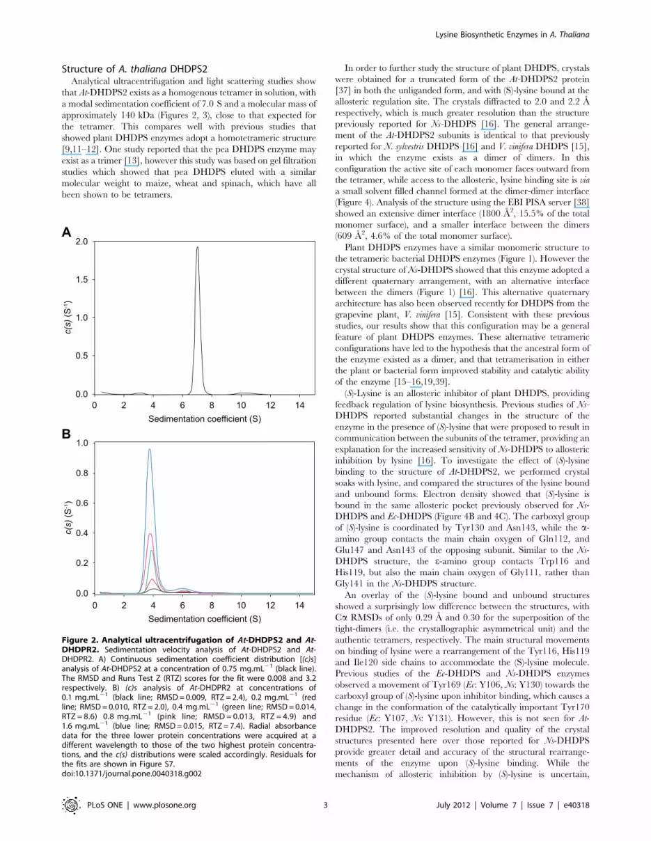
Structure of A. thaliana DHDPS2Analytical ultracentrifugation and light scattering studies show
that At-DHDPS2 exists as a homogenous tetramer in solution, with
a modal sedimentation coefficient of 7.0 S and a molecular mass of
approximately 140 kDa (Figures 2, 3), close to that expected for
the tetramer. This compares well with previous studies that
showed plant DHDPS enzymes adopt a homotetrameric structure
[9,11–12]. One study reported that the pea DHDPS enzyme may
exist as a trimer [13], however this study was based on gel filtration
studies which showed that pea DHDPS eluted with a similar
molecular weight to maize, wheat and spinach, which have all
been shown to be tetramers.
In order to further study the structure of plant DHDPS, crystals
were obtained for a truncated form of the At-DHDPS2 protein
[37] in both the unliganded form, and with (S)-lysine bound at the
allosteric regulation site. The crystals diffracted to 2.0 and 2.2 A
respectively, which is much greater resolution than the structure
previously reported for Ns-DHDPS [16]. The general arrange-
ment of the At-DHDPS2 subunits is identical to that previously
reported for N. sylvestris DHDPS [16] and V. vinifera DHDPS [15],
in which the enzyme exists as a dimer of dimers. In this
configuration the active site of each monomer faces outward from
the tetramer, while access to the allosteric, lysine binding site is via
a small solvent filled channel formed at the dimer-dimer interface
(Figure 4). Analysis of the structure using the EBI PISA server [38]
showed an extensive dimer interface (1800 A2, 15.5% of the total
monomer surface), and a smaller interface between the dimers
(609 A2, 4.6% of the total monomer surface).
Plant DHDPS enzymes have a similar monomeric structure to
the tetrameric bacterial DHDPS enzymes (Figure 1). However the
crystal structure of Ns-DHDPS showed that this enzyme adopted a
different quaternary arrangement, with an alternative interface
between the dimers (Figure 1) [16]. This alternative quaternary
architecture has also been observed recently for DHDPS from the
grapevine plant, V. vinifera [15]. Consistent with these previous
studies, our results show that this configuration may be a general
feature of plant DHDPS enzymes. These alternative tetrameric
configurations have led to the hypothesis that the ancestral form of
the enzyme existed as a dimer, and that tetramerisation in either
the plant or bacterial form improved stability and catalytic ability
of the enzyme [15–16,19,39].
(S)-Lysine is an allosteric inhibitor of plant DHDPS, providing
feedback regulation of lysine biosynthesis. Previous studies of Ns-
DHDPS reported substantial changes in the structure of the
enzyme in the presence of (S)-lysine that were proposed to result in
communication between the subunits of the tetramer, providing an
explanation for the increased sensitivity of Ns-DHDPS to allosteric
inhibition by lysine [16]. To investigate the effect of (S)-lysine
binding to the structure of At-DHDPS2, we performed crystal
soaks with lysine, and compared the structures of the lysine bound
and unbound forms. Electron density showed that (S)-lysine is
bound in the same allosteric pocket previously observed for Ns-
DHDPS and Ec-DHDPS (Figure 4B and 4C). The carboxyl group
of (S)-lysine is coordinated by Tyr130 and Asn143, while the a-
amino group contacts the main chain oxygen of Gln112, and
Glu147 and Asn143 of the opposing subunit. Similar to the Ns-
DHDPS structure, the e-amino group contacts Trp116 and
His119, but also the main chain oxygen of Gly111, rather than
Gly141 in the Ns-DHDPS structure.
An overlay of the (S)-lysine bound and unbound structures
showed a surprisingly low difference between the structures, with
Ca RMSDs of only 0.29 A and 0.30 for the superposition of the
tight-dimers (i.e. the crystallographic asymmetrical unit) and the
authentic tetramers, respectively. The main structural movements
on binding of lysine were a rearrangement of the Tyr116, His119
and Ile120 side chains to accommodate the (S)-lysine molecule.
Previous studies of the Ec-DHDPS and Ns-DHDPS enzymes
observed a movement of Tyr169 (Ec: Y106, Ns: Y130) towards the
carboxyl group of (S)-lysine upon inhibitor binding, which causes a
change in the conformation of the catalytically important Tyr170
residue (Ec: Y107, Ns: Y131). However, this is not seen for At-
DHDPS2. The improved resolution and quality of the crystal
structures presented here over those reported for Ns-DHDPS
provide greater detail and accuracy of the structural rearrange-
ments of the enzyme upon (S)-lysine binding. While the
mechanism of allosteric inhibition by (S)-lysine is uncertain,
Figure 2. Analytical ultracentrifugation of At-DHDPS2 and At-DHDPR2. Sedimentation velocity analysis of At-DHDPS2 and At-DHDPR2. A) Continuous sedimentation coefficient distribution [(c)s]analysis of At-DHDPS2 at a concentration of 0.75 mg.mL21 (black line).The RMSD and Runs Test Z (RTZ) scores for the fit were 0.008 and 3.2respectively. B) (c)s analysis of At-DHDPR2 at concentrations of0.1 mg.mL21 (black line; RMSD = 0.009, RTZ = 2.4), 0.2 mg.mL21 (redline; RMSD = 0.010, RTZ = 2.0), 0.4 mg.mL21 (green line; RMSD = 0.014,RTZ = 8.6) 0.8 mg.mL21 (pink line; RMSD = 0.013, RTZ = 4.9) and1.6 mg.mL21 (blue line; RMSD = 0.015, RTZ = 7.4). Radial absorbancedata for the three lower protein concentrations were acquired at adifferent wavelength to those of the two highest protein concentra-tions, and the c(s) distributions were scaled accordingly. Residuals forthe fits are shown in Figure S7.doi:10.1371/journal.pone.0040318.g002
Lysine Biosynthetic Enzymes in A. Thaliana
PLoS ONE | www.plosone.org 3 July 2012 | Volume 7 | Issue 7 | e40318
Arg199 (Ec: R138, Ns: R160) has been implicated in this process.
Arg199 is located in helix a5 and binds to the carboxyl group of
(S)-ASA [16,40–41]. In the case of At-DHDPS2, binding of (S)-
lysine caused only slight changes in the orientation of Arg199.
To confirm that the crystal structures accurately reflect the
structure of the protein in solution, small angle X-ray scattering
data were collected for both free enzyme, and enzyme in the
presence of (S)-lysine (Figure 5). The scattering data were
compared to the theoretical scattering calculated from the crystal
structure, and showed good agreement at low q (q ,0.12 A)
corresponding to the overall shape for both the unliganded
enzyme (x= 0.91) and enzyme bound to (S)-lysine (x= 1.20).
Deviations from the fit at higher q, in particular, the smoothing
out of the second maxima from the calculated profiles may result
from flexibility of mobile regions, or perhaps small rearrangements
of the quaternary structure in solution. The experimental
scattering profile of At-DHDPS2 was unchanged by the addition
of (S)-lysine or the substrate, pyruvate, showing that there are no
large conformational changes in solution caused by ligand binding.
Activity of A. thaliana DHDPR2Since few studies have characterised DHDPR from plants,
recombinant DHDPR2 from A. thaliana was cloned, expressed in
E. coli, and purified to homogeneity. DHDPR catalyses the second
reaction in the DAP pathway, namely the NAD(P)H-dependent
reduction of HTPA (via dehydration first to DHDP) to form (S)-
tetrahydrodipicolinate [28]. The enzyme was active in the
presence of either NADPH or NADH, but showed inhibition by
the HTPA substrate when NADH was used as a cofactor (Table 1,
Figure S3). Maize DHDPR is the only plant DHDPR to have
previously been characterised, and it was shown to have a
KM(HTPA) of 430 mM, and KM(NADPH) of 46 mM [20]. These
values are similar to the kinetic constants of 35 mM and 57 mM for
NADPH and HTPA observed in this study, and those for bacterial
DHDPR enzymes, which have a KM(HTPA) of 7.6 mM and
KM(NADH) of 2.5 mM [24]. The difference in the Michaelis
constant previously observed for HTPA may be a result of the
inherent instability of HTPA in solution, or the presence of
inhibitory compounds when (S)-ASA is produced by ozonolysis, as
was used for the previous study, which has been shown to affect
measurement of enzyme kinetics [36].
In order to determine whether DHDPS and DHDPR form a
species-specific transient complex during catalysis, coupled assays
were performed using different combinations of DHDPS and
DHDPR enzymes from T. maritima, E. coli and A. thaliana. In all
cases the measured activity of DHDPS was not dependent on the
DHDPR used in the assay (data not shown), suggesting that there
is no species specific coupling of DHDPS and DHDPR.
While At-DHDPR2 is similar to other DHDPR enzymes in
having similar specificities for both NADH and NADPH [21,23–
24,26–27], it shows a slightly higher catalytic rate in the presence
of NADPH. Given that At-DHDPR2 is localised in the chloroplast
[7], where NADPH is used as part of the light reactions, it is most
likely that NADPH is the biologically relevant cofactor for At-
DHDPR2. Substrate inhibition by HTPA has previously been
observed for both Tm-DHDPR and Sa-DHDPR, and may be the
result of a dead-end complex formed by HTPA binding to enzyme
that has the oxidised form of nucleotide still bound to the active
site [26]. During normal lysine synthesis in the plant, it is unlikely
that HTPA would accumulate to sufficiently high levels for
inhibition to occur, particularly when using NADPH as a cofactor.
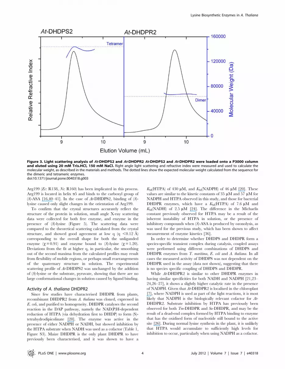
Figure 3. Light scattering analysis of At-DHDPS2 and At-DHDPR2 At-DHDPS2 and At-DHDPR2 were loaded onto a P3000 columnand eluted using 20 mM Tris.HCl, 150 mM NaCl. Right angle light scattering and refractive index were measured and used to calculate themolecular weight, as described in the materials and methods. The dotted lines show the expected molecular weight calculated from the sequence forthe dimeric and tetrameric enzymes.doi:10.1371/journal.pone.0040318.g003
Lysine Biosynthetic Enzymes in A. Thaliana
PLoS ONE | www.plosone.org 4 July 2012 | Volume 7 | Issue 7 | e40318

Structure of A. thaliana DHDPRStructural studies of plant DHDPR have been extremely
limited. All bacterial DHDPR enzymes characterised to date have
been shown to exist as homotetramers, while maize DHDPR was
reported to exist as a dimer [20]. In order to probe the quaternary
structure of plant DHDPR, At-DHDPR2 was characterised using
scattering studies and analytical ultracentrifugation.
At-DHDPS2 was eluted from a size-exclusion chromatography
column, and measurement of right angle light scattering showed
that the main peak of At-DHDPR2 was largely monodisperse, with
a calculated molecular weight of 67.5 kDa. This is very similar to
the expected molecular weight for a dimer of 64.0 kDa (Figure 3).
A smaller peak (,5% of total peak area) was also observed, which
was consistent with the size expected for a tetrameric species.
In order to better characterise At-DHDPR2 in solution,
sedimentation velocity studies were carried out at 5 different
protein concentrations (0.1–1.6 mg.mL21) (Figure 2B). These
experiments show two main species in solution, with the major
peak having a sedimentation coefficient of ,4 S, which corre-
sponds to that expected for the dimer, and a minor peak with a
Figure 4. Crystal structures of unliganded and lysine bound At-DHDPS2. A) Wall-eyed stereo image of the Ca superposition of At-DHDPS2with bound lysine (blue Ca trace) and unliganded At-DHDPS2 (gold Ca trace; rmsd = 0.3 A). The lysine molecules bound at the allosteric site of eachmonomer of the tetramer are shown in yellow (stick representation). B) The lysine binding site at the monomer-monomer interface of the tight-dimershowing residues in contact with the bound lysine molecules (yellow). Electron density around the bound lysine (grey mesh, contoured at 1.0 sigma)was calculated using refined coordinates omitting the bound lysine molecules. Residues contributed by each monomer of the tight-dimer are shownin different shades of blue, and are indicated by the use of the prime (’) symbol. C) overlay of the lysine binding residues of the tight-dimer from thelysine bound (blue) and unliganded (gold) structures. Lysine molecules are shown in yellow. Residues contributed by each monomer of the tight-dimer are shown in different shades of blue or gold, and are indicated by the use of the prime (’) symbol.doi:10.1371/journal.pone.0040318.g004
Lysine Biosynthetic Enzymes in A. Thaliana
PLoS ONE | www.plosone.org 5 July 2012 | Volume 7 | Issue 7 | e40318

sedimentation coefficient of ,6.5 S, corresponding to that
expected for a tetramer. The observation of a dimeric species
has not previously been observed in studies of bacterial DHDPR
enzymes [21–24,26].
In bacteria, DHDPR is a homotetrameric enzyme, with each
subunit consisting of two domains. The N-terminal domain
includes a Rossman fold, and is the binding site for nucleotides,
while the C-terminal domain includes the site for substrate and
inhibitor binding [21–24,29,42]. E. coli DHDPR has an extensive
interface between adjacent residues in which two 8-stranded bbarrels are paired face to face to form a 16 stranded b barrel
(Figure S4). One interface forms by pairing 4 strands of one
subunit with 4 strands of an adjacent unit to form an 8-stranded
mixed b sheet through pairing of b-10 (residues 229–238) of
subunit A with subunit D [42]. An alpha helix, A4, is also involved
in stabilising this subunit-subunit interaction through interactions
between Val135, Val146, Met147 and Leu139. The other
interface involves interactions between strand b-8 (residues 205–
213), loop L1 (residues 195–203) and loop L2 (residues 164–167)
of one subunit with the corresponding regions of the adjacent
subunit. A similarly extensive interface has also been observed for
M. tuberculosis, T. maritima, and S. aureus DHDPR enzymes [23–
24,27], as shown by analysing the interfaces of bacterial DHDPR
structures using PISA (Table 2).
If the plant DHDPR enzyme has a similar structural
arrangement to bacterial DHDPR enzymes, the dimer observed
by light scattering and analytical ultracentrifugation could
correspond to that formed through the 8-stranded mixed b sheet
(b-10 dimer), or that formed through interactions between strand
b-8 and the extended loop region (b-8 dimer). Alignment of the
protein sequences shows that A. thaliana (and other plants) have a
truncation at the region corresponding to strand b-8, which may
reduce the potential interactions at this interface (Figure S5).
In order to gain information about the shape and structure in
solution, small angle X-ray scattering data was collected for At-
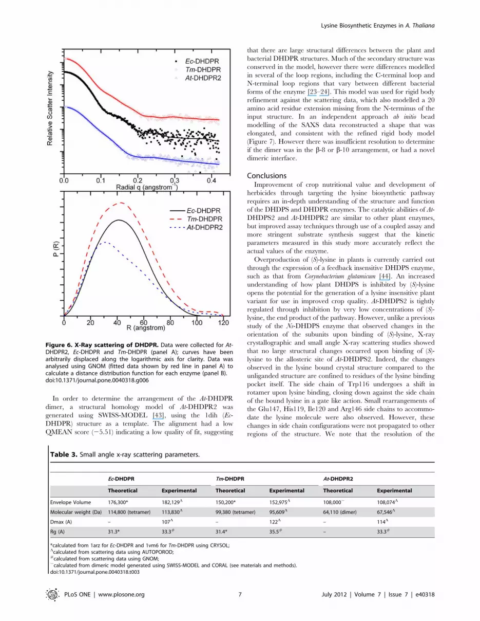
DHDPR2. At-DHDPR2 has a different scattering profile to that of
Ec-DHDPR and Tm-DHDPR (Figure 6), and the real-space
distance distributions, p(r) differ significantly. The near-symmetric
p(r) of Ec-DHDPR and Tm-DHDPR are characteristic of
spheroidal objects, whereas that of At-DHDPR2 is negatively
skewed with a tail at long distances consistent with an elongated
structure in solution. The particle volume and molecular weight
estimated from the scattering data match the values expected for a
dimeric enzyme in the case of At-DHDPR2, and a tetrameric
enzyme in the case of Ec-DHDPR and Tm-DHDPR (Table 3).
Comparison of the At-DHDPR2 scattering data with that
calculated for the variants of the Ec-DHDPR structure show that
the data most closely matches that expected for the b-10 (x= 1.64)
or b-8 dimers (x= 1.50), and not the tetramer (x= 4.86) or
monomer (x= 8.39) (Figure S6).
Figure 5. X-Ray scattering data of At-DHDPS2. Data werecollected in the absence of ligand, or in the presence of 1 mM (S)-lysine, top panel; curves have been arbitrarily displaced along thelogarithmic axis for clarity. Solid lines show the scattering profile fromthe unliganded crystal structure, calculated using CRYSOL. Distance-distribution functions, p(r) for the unbound and ligand bound At-DHDPS2 were determined using the indirect Fourier tranformationpackage GNOM (bottom panel).doi:10.1371/journal.pone.0040318.g005
Table 1. Kinetic parameters (6 SE) for At-DHDPR2 werecalculated as described in materials and method section.
NADH cofactor NADPH cofactor
Constant NAD(P)H Vmax
(mmol.s21.mg21)388695 881666
KM (HTPA) (mM) 1761 3568
KiS (mM) 163675 33006900
Constant HTPA Vmax
(mmol.s21.mg21)310620 1030648
KM (NAD(P)H) (mM) 70611 5767
doi:10.1371/journal.pone.0040318.t001
Table 2. Analysis of DHDPR interface regions using thePDBePISA web server (38).
b-10 interface b-8 interface
ModelSISA(A2) # H-Bonds
SISA(A2) # H-Bonds
E. coli –1dih [42] 1563 26 1354 30
E. coli –1arz [29] 1519 21 1209 26
T. maritima –1vm6 1059 17 778 12
S. aureus –3qy9 [27] 1170 23 1339 23
M. tuberculosis –1c3v [23] 1127 24 1506 24
M. tuberculosis –1p9l [23] 1094 25 1487 23
doi:10.1371/journal.pone.0040318.t002
Lysine Biosynthetic Enzymes in A. Thaliana
PLoS ONE | www.plosone.org 6 July 2012 | Volume 7 | Issue 7 | e40318
In order to determine the arrangement of the At-DHDPR
dimer, a structural homology model of At-DHDPR2 was
generated using SWISS-MODEL [43], using the 1dih (Ec-
DHDPR) structure as a template. The alignment had a low
QMEAN score (25.51) indicating a low quality of fit, suggesting
that there are large structural differences between the plant and
bacterial DHDPR structures. Much of the secondary structure was
conserved in the model, however there were differences modelled
in several of the loop regions, including the C-terminal loop and
N-terminal loop regions that vary between different bacterial
forms of the enzyme [23–24]. This model was used for rigid body
refinement against the scattering data, which also modelled a 20
amino acid residue extension missing from the N-terminus of the
input structure. In an independent approach ab initio bead
modelling of the SAXS data reconstructed a shape that was
elongated, and consistent with the refined rigid body model
(Figure 7). However there was insufficient resolution to determine
if the dimer was in the b-8 or b-10 arrangement, or had a novel
dimeric interface.
ConclusionsImprovement of crop nutritional value and development of
herbicides through targeting the lysine biosynthetic pathway
requires an in-depth understanding of the structure and function
of the DHDPS and DHDPR enzymes. The catalytic abilities of At-
DHDPS2 and At-DHDPR2 are similar to other plant enzymes,
but improved assay techniques through use of a coupled assay and
more stringent substrate synthesis suggest that the kinetic
parameters measured in this study more accurately reflect the
actual values of the enzyme.
Overproduction of (S)-lysine in plants is currently carried out
through the expression of a feedback insensitive DHDPS enzyme,
such as that from Corynebacterium glutamicum [44]. An increased
understanding of how plant DHDPS is inhibited by (S)-lysine
opens the potential for the generation of a lysine insensitive plant
variant for use in improved crop quality. At-DHDPS2 is tightly
regulated through inhibition by very low concentrations of (S)-
lysine, the end product of the pathway. However, unlike a previous
study of the Ns-DHDPS enzyme that observed changes in the
orientation of the subunits upon binding of (S)-lysine, X-ray
crystallographic and small angle X-ray scattering studies showed
that no large structural changes occurred upon binding of (S)-
lysine to the allosteric site of At-DHDPS2. Indeed, the changes
observed in the lysine bound crystal structure compared to the
unliganded structure are confined to residues of the lysine binding
pocket itself. The side chain of Trp116 undergoes a shift in
rotamer upon lysine binding, closing down against the side chain
of the bound lysine in a gate like action. Small rearrangements of
the Glu147, His119, Ile120 and Arg146 side chains to accommo-
date the lysine molecule were also observed. However, these
changes in side chain configurations were not propagated to other
regions of the structure. We note that the resolution of the
Figure 6. X-Ray scattering of DHDPR. Data were collected for At-DHDPR2, Ec-DHDPR and Tm-DHDPR (panel A); curves have beenarbitrarily displaced along the logarithmic axis for clarity. Data wasanalysed using GNOM (fitted data shown by red line in panel A) tocalculate a distance distribution function for each enzyme (panel B).doi:10.1371/journal.pone.0040318.g006
Table 3. Small angle x-ray scattering parameters.
Ec-DHDPR Tm-DHDPR At-DHDPR2
Theoretical Experimental Theoretical Experimental Theoretical Experimental
Envelope Volume 176,300* 182,129L 150,200* 152,975L 108,000, 108,074L
Molecular weight (Da) 114,800 (tetramer) 113,830L 99,380 (tetramer) 95,609L 64,110 (dimer) 67,546L
Dmax (A) – 107L – 122L – 114L
Rg (A) 31.3* 33.3# 31.4* 35.5# – 33.3#
*calculated from 1arz for Ec-DHDPR and 1vm6 for Tm-DHDPR using CRYSOL;Lcalculated from scattering data using AUTOPOROD;#calculated from scattering data using GNOM;,calculated from dimeric model generated using SWISS-MODEL and CORAL (see materials and methods).doi:10.1371/journal.pone.0040318.t003
Lysine Biosynthetic Enzymes in A. Thaliana
PLoS ONE | www.plosone.org 7 July 2012 | Volume 7 | Issue 7 | e40318

structures presented here (2.0–2.2 A) is considerably higher than
that of the Ns-DHDPS structures previously published (,2.8 A)
[16]. Since the lysine binding sites of At-DHDPS2 are not situated
close to crystal contacts, crystal packing arguments should not
preclude observation of structural changes around this site. As
such, we would expect to detect any significant conformational
changes in the structure due to lysine binding.
Given the similarity of the unliganded and lysine bound crystal
structures, it is possible that lysine binding significantly alters the
dynamics of At-DHDPS2, resulting in allosteric inhibition. We
have previously shown that protein dynamics have a considerable
effect on the activity of E. coli DHDPS [19,39]. Examination of the
structural dynamics of At-DHDPS2 may shed further light on this
idea in future studies.
Despite similar catalytic properties, the quaternary structure of
At-DHDPR2 is strikingly different to the bacterial enzyme.
Phylogenetic analysis of bacterial, cyanobacterial and plant
DHDPR protein sequences shows several major clusters that
generally align with lineages determined from 16 s ribosomal
RNA genes, with plant DHDPR sequences forming an isolated
cluster that do not share any clear lineages with bacterial or
cyanobacterial genes (Figure S8) [5]. For each of the distinct
bacterial and archaea clusters, a representative tetrameric
structure is known (gamma proteobacteria, Ec-DHDPR; alpha
proteobacteria, Bartonella henselae; actinobacteria, Mt-DHDPR;
firmicutes Sa-DHDPR; archaeabacteria Tm-DHDPR), raising
the likelihood that the ancestral DHDPR enzyme was a tetramer.
In this study, we have used analytical ultracentrifugation, static
light scattering and small angle X-ray scattering studies to
unequivocally show that At-DHDPR2 exists as a dimer. The
reasons for the differences in quaternary structure are unclear,
given that the enzymes have similar catalytic abilities, and the
subunit arrangement of plant DHDPR remains uncertain.
DHDPS has a similarly divergent quaternary structure, with
different tetrameric arrangements for plant and bacterial enzymes,
with S. aureus existing as a dimeric enzyme. It will be interesting
to extend the current studies of A. thaliana DHDPR to other plant
DHDPR enzymes and also to the cyanobacterial DHDPR
enzymes, as well as the chloroplast NAD(P)H dehydrogenase
enzyme, which has high homology to DHDPR.
Materials and Methods
MaterialsUnless otherwise stated, chemicals were obtained from Sigma
Chemical Co. GE Biosciences, or Invitrogen. (S)-ASA was
synthesised using the methods of Roberts [45], and was the kind
gift of Andrew Muscroft-Taylor. Unless otherwise stated, enzymes
were manipulated at 4uC or on ice.
Cloning, Expression & PurificationPlasmids encoding AT3G60880 (At-DHDPS2) and
AT3G59890 (At-DHDPR2) were obtained from the Arabidopsis
Information Resource (TAIR), Carnegie Institution of Washing-
ton, Stanford CA. Primer pairs encoding the predicted 59-39 ends
of the ORF were used to amplify the gene. Primers were designed
to exclude the chloroplast transit peptide, as identified using
chloroP [46]. The PCR product was ligated into the pET151/D-
Topo vector (Invitrogen), with reactions carried out according to
the manufacturer’s protocols. Protein expression was performed in
Figure 7. Results of ab initio modeling of At-DHDPR from SAXS data. Models were generated using GASBOR (left panels) and DAMMIN (rightpanels). The structural homology model generated by SWISS-MODEL and fitted to the scattering data using CORAL is superimposed for comparison.doi:10.1371/journal.pone.0040318.g007
Lysine Biosynthetic Enzymes in A. Thaliana
PLoS ONE | www.plosone.org 8 July 2012 | Volume 7 | Issue 7 | e40318
BL21(DE3) Star cells, using ZYM-5052 media [47]. Cultures were
grown at 37uC for five hours, followed by incubation at 26uCovernight. Cells were harvested by centrifugation, resuspended in
buffer containing 50 mM NaH2PO4, pH 8.0, 30 mM imidazole
and 300 mM NaCl and lysed by sonication. Cell debris was
pelleted by centrifugation, and the cell pellet applied to a His-Trap
Crude column (GE Biosciences). The column was washed with
three volumes of resuspension buffer, before bound protein was
eluted using 50 mM NaH2PO4, pH 8.0, 300 mM imidazole and
300 mM NaCl. Cleavage of the His-tag was carried out by
incubation of the enzyme with the TEV protease for two hours at
20uC, followed by removal of the cleaved tag using a His-trap
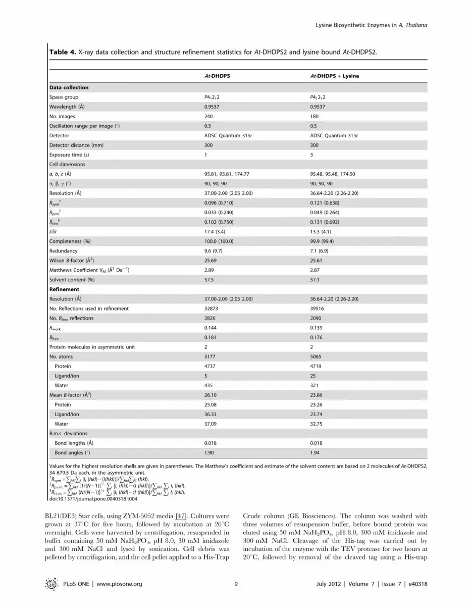
Table 4. X-ray data collection and structure refinement statistics for At-DHDPS2 and lysine bound At-DHDPS2.
At-DHDPS At-DHDPS + Lysine
Data collection
Space group P41212 P41212
Wavelength (A) 0.9537 0.9537
No. images 240 180
Oscillation range per image (u) 0.5 0.5
Detector ADSC Quantum 315r ADSC Quantum 315r
Detector distance (mm) 300 300
Exposure time (s) 1 3
Cell dimensions
a, b, c (A) 95.81, 95.81, 174.77 95.48, 95.48, 174.50
a, b, c (u) 90, 90, 90 90, 90, 90
Resolution (A) 37.00-2.00 (2.05 2.00) 36.64-2.20 (2.26-2.20)
Rsym{ 0.096 (0.710) 0.121 (0.638)
Rpim{ 0.033 (0.240) 0.049 (0.264)
Rrim1 0.102 (0.750) 0.131 (0.692)
I/dI 17.4 (3.4) 13.3 (4.1)
Completeness (%) 100.0 (100.0) 99.9 (99.4)
Redundancy 9.6 (9.7) 7.1 (6.9)
Wilson B-factor (A2) 25.69 25.61
Matthews Coefficient VM (A3 Da21) 2.89 2.87
Solvent content (%) 57.5 57.1
Refinement
Resolution (A) 37.00-2.00 (2.05 2.00) 36.64-2.20 (2.26-2.20)
No. Reflections used in refinement 52873 39516
No. Rfree reflections 2826 2090
Rwork 0.144 0.139
Rfree 0.181 0.176
Protein molecules in asymmetric unit 2 2
No. atoms 5177 5065
Protein 4737 4719
Ligand/ion 5 25
Water 435 321
Mean B-factor (A2) 26.10 23.86
Protein 25.08 23.26
Ligand/ion 36.33 23.74
Water 37.09 32.75
R.m.s. deviations
Bond lengths (A) 0.018 0.018
Bond angles (u) 1.90 1.94
Values for the highest resolution shells are given in parentheses. The Matthew’s coefficient and estimate of the solvent content are based on 2 molecules of At-DHDPS2,34 679.5 Da each, in the asymmetric unit.{Rsym =ghklgi |Ii (hkl)2ÆI(hkl)æ|/ghklgiIi (hkl).{Rp.i.m. =ghkl [1/(N21)]K gi |Ii (hkl)2ÆI (hkl)æ|/ghkl gi Ii (hkl).1Rr.i.m. =ghkl [N/(N21)]K gi |Ii (hkl)2ÆI (hkl)æ|/ghkl gi Ii (hkl).doi:10.1371/journal.pone.0040318.t004
Lysine Biosynthetic Enzymes in A. Thaliana
PLoS ONE | www.plosone.org 9 July 2012 | Volume 7 | Issue 7 | e40318
column. Fractions containing protein were desalted into 20 mM
Tris.HCl, pH 8.0 for storage.
Size-Exclusion ChromatographyGel filtration was carried out at 28uC using a Malvern P3000
column. 100 mL of enzyme (1.0 mg.mL21) was loaded onto the
column and eluted with 20 mM Tris-HCl, 150 mM NaCl, pH 8.0
at 0.5 mL.min21. A Viscotek TDA unit was used to measure the
refractive index and low angle and right angle light scattering.
BSA (2 mg.mL21) was used as a standard to calibrate the
instrument.
Analytical UltracentrifugationSedimentation velocity experiments were performed in a
Beckman Coulter Model XL-I analytical ultracentrifuge equipped
with UV/Vis scanning optics. Reference (380 mL; 20 mM Tris-
HCl, 150 mM NaCl, pH 8.0) and sample (360 mL) solutions were
loaded into 12 mm double-sector cells with quartz windows and
the cells were then mounted in an An-60 Ti 4-hole rotor. At-
DHDPS2 was prepared at a concentration of 0.75 mg.mL21 while
At-DHDPR2 was prepared at initial protein concentrations of 0.1–
1.6 mg.mL21. Proteins were centrifuged at 40,000 rpm (At-
DHDPR2) or 50,000 rpm (At-DHDPS2) at 20uC, and radial
absorbance data were collected at appropriate wavelengths in
continuous mode every 8 minutes without averaging. Data were
fitted to a continuous size-distribution [c(s)] model using the
program SEDFIT [48]. The partial specific volume (�vv) of the
proteins (At-DHDPS2 0.738 mL g21; At-DHDPR2 0.745 mL
g21), buffer density (1.005 g.mL21) and buffer viscosity (1.021 cp)
were computed using the program SEDNTERP [49].
SAXS MeasurementsMeasurements were performed at the Australian Synchrotron
SAXS/WAXS beamline equipped with a Pilatus detector (1 M,
170 mm6170 mm, effective pixel size, 1726172 mm). The
wavelength of the X-rays was 1.0332 A. The sample–detector
distance was 1600 mm, which provided a q range of 0.0126–
0.400 A21 [where q is the magnitude of the scattering vector,
which is related to the scattering angle (2h) and the wavelength (l)
as follows: q = (4p/l)sinh]. Protein samples (initial concentration of
2–5 mg mL21) were eluted from an in-line gel filtration column
(Superdex 200 5/150), pre-equilibrated with 20 mM Tris.HCl,
150 mM NaCl, pH 8.0, to remove any aggregated protein
immediately prior to data collection. Data were collected using a
1.5 mm glass capillary at 27uC under continuous flow in 2 sec
intervals. 2D intensity plots from the peak of the SEC run were
radially averaged, normalized to sample transmission and back-
ground subtracted.
SAXS data analysis: The data sets for structural analyses were
recorded with 458 data points over the range 0.0113# s
#0.4 A21. 1D profiles were background subtracted and Guinier
analysis performed using PRIMUS [50]. Particle volume and
molecular weight were calculated using AUTOPOROD [51].
Indirect Fourier transform was performed using GNOM [52] to
yield the real-space function P(r), which gives both the relative
probabilities of distances between scattering centers and the
maximum dimension of the scattering particle Dmax. Theoretical
scattering curves were calculated from atomic coordinates and
compared with experimental scattering curves using CRYSOL
[53]. For At-DHDPR a homology model for At-DHDPR2 was
generated using SWISS-MODEL [43], which aligned the
sequence to the 1dih structure. Rigid body refinement of this
model was carried out using CORAL [54], additionally refining
the 20 amino acid residues that were missing from the N-terminus
of the SWISS-MODEL structure. Ab initio modeling was carried
out using DAMMIN [55] to generate 10 bead models with
imposed P2 symmetry. These models were averaged using
DAMAVER, and superimposed with the bacterial structures
using SUPCOMB [56]. GASBOR [57] was also used for ab initio
reconstruction of of a dummy-residue model, using an extended
data range relative to the short range used for bead-modeling.
Crystallization, X-ray Diffraction Data Collection, andStructure Solution
At-DHDPS was crystallized essentially as described previously
[37] using the sitting-drop vapor diffusion method. The crystals
used for diffraction analysis and structure solution were obtained
at 20uC from 300 nL drops formed from 150 nL At-DHDPS2
solution (14.5 mg.mL21 in 20 mM Tris-HCl pH 8.0) and 150 nL
reservoir solution [2.4 M sodium malonate pH 7.0, 0.02% (w/v)
sodium azide]. Ligand soaks were performed by adding 300 nL of
10 mM lysine, 2.4 M sodium malonate pH 7.0, 0.02% (w/v)
sodium azide directly to the crystallization drop followed by
incubation for 2 hours. The malonate concentration present in the
crystallization drop was found to afford adequate cryo-protection
and, thus, crystals were flash-cooled in liquid nitrogen directly
from the crystallisation drop. X-ray diffraction data collection was
carried out at 110 K at using the MX2 beam line of the Australian
Synchrotron.
Diffraction data sets were processed and scaled using the
programs XDS [58] and SCALA [59]. Initial phase estimates were
solved by molecular replacement using PHASER [60] with the
lysine bound structure of N. sylvestris DHDPS [16] as the search
model as described previously [37]. Structural refinement was
performed using REFMAC5 [61] with iterative model building
using COOT [62]. Water, sodium ions, and the bound lysine
atoms were added at later stages using COOT. Data processing
and structure refinement statistics are presented in Table 4.
Enzyme KineticsDHDPS and DHDPR enzyme activity was measured using a
coupled assay as previously described. Stock solutions of (S)-ASA,
pyruvate, (S)-lysine, NADPH and NADH were prepared fresh for
each experiment. Assay temperature was regulated by the use of a
circulating water bath, and assays were performed at 30uC. Initial
rate data were typically reproducible within 10%, and were
analysed using non-linear regression software (OriginLab, North-
ampton, MA, USA). Assays for DHDPS activity contained
0.5 mg.mL21 DHDPS, and an excess of DHDPR (20–
100 mg.mL21). Assays for DHDPR activity involved pre-incubat-
ing the cuvettes with an excess of DHDPS (20–100 mg.mL21) for
60 s before assays were initiated by the addition of DHDPR
(typically 0.6 mg.mL21).
Supporting Information
Figure S1 Lysine biosynthesis pathways. DapD, tetrahy-
drodipicolinate acylase; DapC,acyl-amino-ketopimelate amino-
transferase; DapE, acyl-ketopimelate deacylase; DapF, diamino-
pimelate epimerase; LysA, diaminopimelate decarboxylase;
DapDH, mesodiaminopimelatedehydrogenase; DapL, l,l-diami-
nopimelate aminotransferase.
(PDF)
Figure S2 Kinetics of At-DHDPS2. Assays were carried out
at varying concentrations of (S)-lysine (top panel), or varying
concentrations of ASA and pyruvate (bottom panel).
(PDF)
Lysine Biosynthetic Enzymes in A. Thaliana
PLoS ONE | www.plosone.org 10 July 2012 | Volume 7 | Issue 7 | e40318
Figure S3 Kinetics of At-DHDPR2. Panel A) HTPA was
fixed at 0.15 mM, and NAD(PH concentrations were varied.
Panel B) NAD(P)H concentrations were fixed at 0.16 mM and
HTPA concentrations were varied.
(PDF)
Figure S4 Structure of bacterial DHDPR. The structure of
Ec-DHDPR (pdb: 1arz), showing the interface between b-8 and
loop 1 (left panel) and the alternate interface involving b-10 and
helix-4 (right panel, only the C-terminal domain shown).
(PDF)
Figure S5 Alignment of DHDPR structures. Helical
regions are shown in blue and b-sheet regions are shown in red.
(PDF)
Figure S6 X-Ray scattering of At-DHDPR2. Data was
collected and compared to the scattering calculated using
CRYSOL for the monomer, b-10 dimer, b-8 dimer, and tetramer
of Ec-DHDPR.
(PDF)
Figure S7 Residuals resulting from the c(s) distributionbest fits shown in Figure 2 plotted as a function of radiusfrom the axis of rotation. A) Residuals for the best fit of the
sedimentation velocity data for At-DHDPS2 at a concentration of
0.75 mg.mL21. B) Residuals for the best fit of the sedimentation
velocity data for At-DHDPR2 at concentrations of 0.1 mg.mL21
(black), 0.2 mg.mL21 (red), 0.4 mg.mL21 (green), 0.8 mg.mL21
(pink), and 1.6 mg.mL21 (blue).
(PDF)
Figure S8 Representative phylogenetic tree of DapBorthologues based on Hudson, 2005 [5].
(PDF)
Acknowledgments
Parts of this research were undertaken at the MX2 and SAXS beamlines of
the Australian Synchrotron, Victoria, Australia. Special thanks are given to
Nigel Kirby at the SAXS beamline, and the New Zealand Synchrotron
Group.
Author Contributions
Conceived and designed the experiments: FGP MDWG. Performed the
experiments: MDWG JMB AW SK RCJD. Analyzed the data: MDWG
JMB AW SK HM RCJD. Contributed reagents/materials/analysis tools:
MAP SCA. Wrote the paper: MDWG MAP HM RCJD JAG FGP.
References
1. Galili G (2002) New insights into the regulation and functional significance oflysine metabolism in plants, Annual Review of Plant Biology 53, 27–43.
2. Jander G, Joshi V (2010) Recent Progress in Deciphering the Biosynthesis of
Aspartate-Derived Amino Acids in Plants, Molecular Plant 3, 54–65.
3. Ufaz S, Galili G (2008) Improving the content of essential amino acids in cropplants: Goals and opportunities, Plant Physiol 147, 954–961.
4. Frizzi A, Huang S, Gilbertson LA, Armstrong TA, Luethy MH, et al. (2008)
Modifying lysine biosynthesis and catabolism in corn with a single bifunctional
expression/silencing transgene cassette, Plant Biotechnol J 6, 13–21.
5. Hudson A O, Bless C, Macedo P, Chatterjee S P, Singh B K, et al. (2005)Biosynthesis of lysine in plants: evidence for a variant of the known bacterial
pathways, Biochim Biophys Acta 1721, 27–36.
6. Dobson R C, Giron I, Hudson AO (2011) L,L-diaminopimelate aminotrans-ferase from Chlamydomonas reinhardtii: a target for algaecide development, PLoS
One 6, e20439.
7. Ghislain M, Frankard V, Jacobs M (1990) Dihydrodipicolinate synthase of
Nicotiana sylvestris, a chloroplast-localized enzyme of the lysine pathway, Planta
180, 480–486.
8. Vauterin M, Frankard V, Jacobs M (1999) The Arabidopsis thaliana dhdps gene
encoding dihydrodipicolinate synthase, key enzyme of lysine biosynthesis, isexpressed in a cell-specific manner, Plant Mol Biol 39, 695–708.
9. Kumpaisal R, Hashimoto T, Yamada Y (1987) Purification and characterization
of dihydrodipicolinate synthase from wheat suspension cultures, Plant Physiol 85,
145–151.
10. Kumpaisal R, Hashimoto T, Yamada Y (1989) Inactivation of whatdihydrodipicolinate synthase by 3-bromopyruvate, Agric. Biol. Chem 53, 355–359.
11. Wallsgrove RM, Mazelis M (1981) Spinach leaf dihydrodipicolinate synthase:
partial purification and characterization, Biochemistry 20, 2651–2655.
12. Frisch DA, Gengenbach BG, Tommey AM, Sellner JM, Somers DA, et al.(1991) Isolation and characterization of dihydrodipicolinate synthase from
maize, Plant Physiol. 96, 444–452.
13. Dereppe C, Bold G, Ghisalba O, Ebert E, Schar H-P (1992) Purification andCharacterization of Dihydrodipicolinate Synthase from Pea, Plant Physiol 98,
813–821.
14. Mathews B, Widholm J (1978) Regulation of lysin and thereonine synthesis in
carrot cell suspension cultures and whole carrot roots, Planta 141, 315–321.
15. Atkinson,SC, Dogovski C, Downton MT, Pearce FG, Reboul CF, et al. (2012)Crystal, solution and in silico structural studies of dihydrodipicolinate synthase
from the common grapevine, PLoS One Submitted.
16. Blickling S, Beisel H, Bozic D, Knablein J, Laber B, et al. (1998) Structure ofdihydrodipicolinate synthase of Nicotiana sylvestris reveals novel quaternary
structure, J Mol Biol 274(4), 608–621.
17. Burgess BR, Dobson RC, Bailey MF, Atkinson SC, Griffin MDW, et al. (2008)
Structure and evolution of a novel dimeric enzyme from a clinically importantbacterial pathogen, J Biol Chem 283, 27598–27603.
18. Kaur N, Gautam A, Kumar S, Singh A, Singh N, et al. (2011) Biochemical
studies and crystal structure determination of dihydrodipicolinate synthase fromPseudomonas aeruginosa, Int J Biol Macromol 48, 779–787.
19. Griffin MD, Dobson RC, Pearce FG, Antonio L, Whitten AE, et al. (2008)
Evolution of quaternary structure in a homotetrameric enzyme, J Mol Biol 380,
691–703.
20. Tyagi VV, Henke RR, Farkas WR (1983) Partial purification and character-
ization of dihydrodipicolinic Acid reductase from maize, Plant Physiol 73, 687–
691.
21. Reddy SG, Sacchettini JC, Blanchard JS (1995) Expression, purification, and
characterization of Escherichia coli dihydrodipicolinate reductase, Biochemistry 34,
3492–34501.
22. Reddy S, Scapin G, Blanchard J (1996) Interaction of pyridine nucleotide
substrates with Escherichia coli dihydrodipicolinate reductase: thermodynamic and
structural analysis of binary complexes, Biochemistry 35, 13924–13302.
23. Cirilli M, Zheng R, Scapin G, Blanchard JS (2003) The three-dimensional
structures of the Mycobacterium tuberculosis dihydrodipicolinate reductase-NADH-
2,6-PDC and -NADPH-2,6-PDC complexes. Structural and mutagenic analysis
of relaxed nucleotide specificity, Biochemistry 42, 10644–10650.
24. Pearce FG, Sprissler C, Gerrard JA (2008) Characterization of dihydrodipico-
linate reductase from Thermotoga maritima reveals evolution of substrate binding
kinetics, J Biochem 143, 617–623.
25. Dommaraju S, Gorman MA, Dogovski C, Pearce FG, Gerrard JA, et al. (2010)
Cloning, expression and crystallization of dihydrodipicolinate reductase from
methicillin-resistant Staphylococcus aureus, Acta Crystallogr Sect F Struct Biol Cryst
Commun 66, 57–60.
26. Dommaraju SR, DogovskiC, Czabotar PE, Hor L, Smith BJ, et al. (2011)
Catalytic mechanism and cofactor preference of dihydrodipicolinate reductase
from methicillin-resistant Staphylococcus aureus, Arch Biochem Biophys 512, 167–174.
27. Girish TS, Navratna V, Gopal B (2011) Structure and nucleotide specificity of
Staphylococcus aureus dihydrodipicolinate reductase (DapB), FEBS Lett 585, 2561–
2567.
28. Devenish SR, Blunt JW, Gerrard JA (2010) NMR studies uncover alternate
substrates for dihydrodipicolinate synthase and suggest that dihydrodipicolinate
reductase is also a dehydratase, J Med Chem 53, 4808–4812.
29. Scapin G, Reddy S, Zheng R, Blanchard J (1997) Three-dimensional structure
of Escherichia coli dihydrodipicolinate reductase in complex with NADH and the
inhibitor 2,6-pyridinedicarboxylate, Biochemistry 36, 15081–15088.
30. Craciun A, Jacobs M, Vauterin M (2000) Arabidopsis loss-of-function mutant in
the lysine pathway points out complex regulation mechanisms, FEBS Lett 487,
234–238.
31. Shimizu H, Shikanai T (2007) Dihydrodipicolinate reductase-like protein,
CRR1, is essential for chloroplast NAD(P)H dehydrogenase in Arabidopsis, Plant
J 52, 539–547.
32. Pearce FG, Dobson RC, Jameson GB, Perugini MA, Gerrard JA (2011)
Characterization of monomeric dihydrodipicolinate synthase variant reveals the
importance of substrate binding in optimizing oligomerization, Biochim Biophys
Acta.
33. Pearce FG, Dobson RC, Weber A, Lane LA, McCammon MG, et al. (2008)
Mutating the tight-dimer interface of dihydrodipicolinate synthase disrupts the
enzyme quaternary structure: toward a monomeric enzyme, Biochemistry 47,
12108–12117.
34. Pearce FG, Perugini MA, McKerchar HJ, Gerrard JA (2006) Dihydrodipico-
linate synthase from Thermotoga maritima, Biochem J 400, 359–366.
35. Sarrobert C, Thibaud M, Contard-David P, Gineste S, Bechtold N, et al. (2000)
Identification of an Arabidopsis thaliana mutant accumulating threonine resulting
Lysine Biosynthetic Enzymes in A. Thaliana
PLoS ONE | www.plosone.org 11 July 2012 | Volume 7 | Issue 7 | e40318
from mutation in a new dihydrodipicolinate synthase gene, Plant Journal 24, 357–
367.
36. Dobson RCJ, Gerrard JA, Pearce FG (2004) Is DHDPS Inhibited By Its
Substrate?, Biochemical Journal 377, 757–762.
37. Griffin MDW, Billakanti JM, Gerrard JA, Dobson RCJ, Pearce FG (2011)
Crystallization and preliminary X-ray diffraction analysis of dihydrodipicolinate
synthase 2 from Arabidopsis thaliana, Acta Crystallographica Section F 67, 1386–1390.
38. Krissinel E, Henrick K (2007) Inference of macromolecular assemblies from
crystalline state, J Mol Biol 372, 774–797.
39. Griffin MD, Dobson RC, Gerrard JA, Perugini MA (2010) Exploring the
dihydrodipicolinate synthase tetramer: how resilient is the dimer-dimer
interface?, Arch Biochem Biophys 494, 58–63.
40. Blickling S, Knablein J (1997) Feedback inhibition of dihydrodipicolinate
synthase enzymes by L-lysine, Biological Chemistry 378 (3–4), 207–210.
41. Dobson RCJ, Griffin MDW, Jameson GB, Gerrard JA (2005) The crystal
structures of native and (S)-lysine bound dihydrodipicolinate synthase from
Escherichia coli with improved resolution show new features of biological
significance, Acta Crystallogr D Biol Crystallogr D61, 1116–1124.
42. Scapin G, Blanchard JS, Sacchettini JC (1995) Three-dimensional structure of
Escherichia coli dihydrodipicolinate reductase, Biochemistry 34, 3502–3512.
43. Kiefer F, Arnold K, Kunzli M, Bordoli L, Schwede T (2009) The SWISS-
MODEL Repository and associated resources, Nucleic Acids Res 37.
44. Rice EA, Bannon GA, Glenn KC, Jeong SS, Sturman, E J, et al. (2008)
Characterization and crystal structure of lysine insensitive Corynebacterium
glutamicum dihydrodipicolinate synthase (cDHDPS) protein, Arch Biochem Biophys
480, 111–121.
45. Roberts SJ, Morris JC, Dobson RCJ, Gerrard JA (2003) The preparation of (S)-
aspartate semi-aldehyde appropriate for use in biochemical studies, Bioorganic and
Medicinal Chemistry Letters 13, 265–267.
46. Emanuelsson O, Nielsen H, von Heijne G (1999) ChloroP, a neural network-
based method for predicting chloroplast transit peptides and their cleavage sites,
Protein Sci 8, 978–984.
47. Studier FW (2005) Protein production by auto-induction in high-density shaking
cultures, Protein Expres Purif 41, 207–234.
48. Schuck P, Perugini MA, Gonzales NR, Howlett GJ, Schubert D (2002) Size-
distribution analysis of proteins by analytical ultracentrifugation: Strategies andapplication to model systems, Biophys J 82, 1096–1111.
49. Laue TM, Shah DB, Ridgeway TM, Pelletier SL (1992) Computer-aided
interpretation of analytical sedimentation data for proteins, In Analytical
ultracentrifugation in biochemistry and protein science, 90–125, The Royal Society of
Chemistry, Cambridge.50. Konarev PV, Volkov VV, Sokolova AV, Koch MHJ, Svergun DI (2003)
PRIMUS: a Windows PC-based system for small-angle scattering data analysis,
J Appl Crystallogr 36, 1277–1282.51. Petoukhov MV, Konarev PV, Kikhney AG, Svergun DI (2007) ATSAS 2.1 -
towards automated and web-supported small-angle scattering data analysis, J
Appl Crystallogr 40.
52. Svergun DI (1992) Determination of the Regularization Parameter in Indirect-Transform Methods Using Perceptual Criteria, J Appl Crystallogr 25, 495–503.
53. Svergun D, Barberato C, Koch MHJ (1995) CRYSOL - A program to evaluate
x-ray solution scattering of biological macromolecules from atomic coordinates,J Appl Crystallogr 28, 768–773.
54. Petoukhov MV, Svergun DI (2005) Global rigid body modeling of macromo-lecular complexes against small-angle scattering data, Biophys J 89, 1237–1250.
55. Svergun DI (1999) Restoring low resolution structure of biological macromol-
ecules from solution scattering using simulated annealing, Biophys J 76, 2879–2886.
56. Kozin MB, Svergun DI (2001) Automated matching of high- and low-resolutionstructural models, J Appl Crystallogr 34, 33–41.
57. Svergun DI, Petoukhov MV, Koch MHJ (2001) Determination of domainstructure of proteins from X-ray solution scattering, Biophys J 80, 2946–2953.
58. Kabsch W (2010) Xds, Acta Crystallogr D 66, 125–132.
59. Evans P (2006) Scaling and assessment of data quality, Acta Crystallogr D 62, 72–82.
60. Storoni LC, McCoy AJ, Read RJ (2004) Likelihood-enhanced fast rotationfunctions, Acta Crystallogr D 60, 432–438.
61. Collaborative (1994) The CCP4 suite: programs for protein crystallography, Acta
Crystallogr D 50, 760–763.62. Emsley P, Lohkamp B, Scott WG, Cowtan K (2010) Features and development
of Coot, Acta Crystallogr D 66, 486–501.
Lysine Biosynthetic Enzymes in A. Thaliana
PLoS ONE | www.plosone.org 12 July 2012 | Volume 7 | Issue 7 | e40318