cerebrospinal fluid d-serine and glycine concentrations are unaltered and unaffected by olanzapine...
TRANSCRIPT

D-Serine in Health and Disease
Sabine Fuchs

ISBN 978-90-8559-027-9
Illustrations by:
Layout and printing: Optima Grafische Communicatie, Rotterdam, The Netherlands

D-Serine in Health and Disease
D-Serine in Gezondheid en Ziekte
(met een samenvatting in het Nederlands)
Proefschrift
ter verkrijging van de graad van doctor aan de Universiteit Utrecht
op gezag van de rector magnificus, prof.dr. J.C. Stoof,
ingevolge het besluit van het college voor promoties
in het openbaar te verdedigen op
dinsdag 2 november 2010 des middags te 2.30 uur
door
Sabine Annemijn Fuchs
geboren op 27 februari 1974, te Heerlen

Promotoren: Prof. dr. R. Berger
Prof. dr. J.L.L. Kimpen
Co-promotoren: Dr. T.J. de Koning
Dr. L.W.J. Klomp
Dit proefschrift werd mede mogelijk gemaakt met financiële steun van ZonMw (ZonMw
programma Agiko stipendia, 920-03-345), het Netherlands Metabolomics Center, Genzyme
Europe BV, GlaxoSmithKline BV en Merck BV.

When D-serine seemed right and L-serine was left...
Voor mijn familie: mijn ouders
Coralie en Saskia
Bas
Sophie, Job en Pepijn


7
Table of Contents
InTroDuCTIon
Preface: Outline of the thesis 11
Chapter 1: D-amino acids in the central nervous system in health and disease 17
D-SerIne anD L-SerIne anaLySIS
Chapter 2a: Editorial clinical chemistry: CSF serine enantiomers and glycine in
the study of neurologic and psychiatric disorders
45
Chapter 2b: Two mass-spectrometric techniques for quantifying serine
enantiomers and glycine in cerebrospinal fluid: potential confounders and
age-dependent ranges
51
D-SerIne In Human pHySIoLogy
Chapter 3: D-serine in the developing human central nervous system 71
Chapter 4: D-serine in the developing human central nervous system: clinical
implications
83
Chapter 5: D-serine influences synaptogenesis in a P19 cell model 105
D-SerIne In Human paTHoLogy
Chapter 6: Increased concentrations of both NMDA receptor co-agonists
D-serine and glycine in global ischemia: a potential novel treatment target for
perinatal asphyxia
119
Chapter 7: Cerebrospinal fluid D-serine and glycine concentrations are
unaltered and unaffected by olanzapine therapy in male schizophrenic patients
135
DISCuSSIon
Chapter 8: General discussion 149

8
Table of Contents
To ConCLuDe
Summary 177
Summary in Dutch - Nederlandse samenvatting 181
Contributors 185
Acknowledgements - Dankwoord 187
Curriculum vitae 191
Publications 193
List of abbreviations 195

Introduction


preface


Pre
face
13
Preface
ouTLIne oF THe THeSIS
As building units of peptides and proteins, amino acids are among the most important
molecules for living beings. Most amino acids occur as different enantiomers (an L-
and a D-form). Although the chemical and physical properties of L- and D-amino acids
are almost identical, they differ in their spatial positioning, which plays a major role in
structural interactions.
It was long believed that only L-amino acids were selected for formation of peptides
and proteins on the primitive earth.(1) Based on this concept, identification of D-amino
acids in different organisms, including mammals, was revolutionary. In chapter 1, an
overview is given of the different D-amino acids occurring in mammals and their putative
function, focusing on D-serine. D-Serine was found to be synthesized and metabolized
endogenously and to function as an important neurotransmitter, being an endogenous
co-agonist of the N-methyl-D-aspartate receptor (NMDAr). The NMDAr is broadly
distributed throughout the central nervous system (CNS) and has been implicated in
physiological processes such as CNS development, memory and learning (2) and in
pathological processes including neurodegenerative conditions (such as amyotrophic
lateral sclerosis, Parkinson’s disease, Alzheimer’s disease and Huntington’s disease),
stroke, epilepsy, polyneuropathies, chronic pain (3) and psychiatric disorders such as
schizophrenia.(4) It had long been known that the NMDAr is an exceptional receptor,
requiring binding by two different agonists for its activation. In addition to glutamate,
glycine was believed to be the necessary co-agonist. However, by now, it is generally
appreciated that D-serine is the endogenous obligatory co-agonist in most regions of the
brain. Consequently, D-serine might well be involved in all processes associated with
NMDAr (dys-) function.
The first part of the thesis (D-serine and L-serine analysis) describes how we devel-
oped two different chiral chromatographic separation techniques (liquid and gas chroma-
tography), combined with mass spectrometric detection to determine concentrations of
D-serine, glycine and their common precursor L-serine in biological fluids (chapter 2).
Using these analytical techniques, we created reference ranges in human cerebrospinal
fluid, since this represents the most accessible read-out for extracellular CNS concentra-
tions.
The second part of the thesis (D-serine in human physiology) starts with our find-
ing of an evident age-dependency in our cerebrospinal fluid D-serine reference values,
with very high concentrations directly after birth and a rapid decline over the first 3
years (chapter 3). To investigate the physiological relevance of these high D-serine
concentrations, we determined D-serine concentrations in cerebrospinal fluid of pa-
tients with 3-phosphoglycerate dehydrogenase deficiency, a rare metabolic disorder
in the synthesis of L-serine and hence D-serine, characterized by serious neurological

Introduction
14
symptoms, including microcephaly, psychomotor retardation and intractable seizures.
(5) Furthermore, we analyzed D-serine concentrations in the one patient with complete
reversal of the phenotype upon pre- and postnatal treatment with L-serine.(6) In chapter
4, we elaborate on the role of D-serine, as an NMDAr co-agonist, in CNS development,
focusing on the clinical implications. To gain more specific insight in the role of D-serine
in CNS development, we studied a rat P19 cell model as described in chapter 5. These
cells can be differentiated into glia and neurons, expressing glutamatergic receptors,
including the NMDAr. We observed that these differentiated P19 cells express serine
racemase and synthesize D-serine, thereby providing a suitable model to study the role
of D-serine in early CNS development. We examined the effects of manipulating D-
serine concentrations and inhibiting D-serine (or glycine) binding to the NMDAr on P19
cell differentiation.
In the third part of the thesis (D-serine in human pathology), we focus on pathological
conditions associated with altered NMDAr activity, potentially induced by altered D-
serine concentrations. This is of particular interest since these concentrations might
by pharmacologically manipulated as the synthesizing and metabolizing enzymes of D-
serine are known, thereby providing interesting therapeutic targets. As pediatricians, we
chose to investigate perinatal asphyxia, currently one of the greatest causes of neonatal
mortality and morbidity worldwide.(7) Perinatal asphyxia is the consequence of disturbed
blood circulation between mother and fetus, for example after placental pathology or
umbilical cord accidents, leading to insufficient delivery of oxygen, glucose and other
blood-borne fuels to the fetal organs, including the brain. In the long term, this can lead
to mental retardation, epilepsy and spasticity.(8;9) Treatment is limited to supportive
intensive care. The pathophysiology has not been fully elucidated, but overstimulation
of the NMDAr appears to play a central role. In chapter 6, we describe our D-serine
and glycine concentration studies in a rat glioma cell model for perinatal asphyxia, in an
experimental piglet model for perinatal asphyxia and in cerebrospinal fluid from human
newborns, who experienced perinatal asphyxia.
Another pathological condition strongly associated with NMDAr dysfunction (10) and
potentially with altered D-serine concentrations (11-13) is schizophrenia. This is a seri-
ous and relatively common psychiatric disorder, characterized by positive symptoms
(including hallucinations), negative symptoms (blunted affect, emotional withdrawal) and
cognitive defects.(14) In chapter 7, we present the results of our analyses of D-serine
concentrations in cerebrospinal fluid of patients with schizophrenia before and after
treatment with the antipsychotic drug olanzapine, compared with control cerebrospinal
fluid.
Finally, in chapter 8, the results of the studies in this thesis are discussed, emphasizing
on the role of D-serine as an essential human endogenous regulator of NMDAr activity
in physiological and pathological processes, with a view towards the future.

Pre
face
15
Preface
reFerenCeS
1. Lamzin VS, Dauter Z, Wilson KS. How nature deals with stereoisomers. Curr Opin Struct Biol
1995 December;5(6): 830-6.
2. Yang Y, Ge W, Chen Y, Zhang Z, Shen W, Wu C et al. Contribution of astrocytes to hippo-
campal long-term potentiation through release of D-serine. Proc Natl Acad Sci U S A 2003
December 9; 100(25): 15194-9.
3. Danysz W, Parsons CG. Glycine and N-methyl-D-aspartate receptors: physiological signifi-
cance and possible therapeutic applications. Pharmacol Rev 1998 December;50(4): 597-664.
4. Goff DC, Coyle JT. The emerging role of glutamate in the pathophysiology and treatment of
schizophrenia. Am J Psychiatry 2001 September;158(9): 1367-77.
5. de Koning TJ, Klomp LW. Serine-deficiency syndromes. Curr Opin Neurol 2004 April;17(2):
197-204.
6. de Koning TJ, Klomp LW, van Oppen AC, Beemer FA, Dorland L, van dB, I et al. Prenatal and
early postnatal treatment in 3-phosphoglycerate-dehydrogenase deficiency. Lancet 2004
December 18; 364(9452): 2221-2.
7. Lawn JE, Cousens S, Zupan J. 4 million neonatal deaths: when? Where? Why? Lancet 2005
March 5; 365(9462): 891-900.
8. Robertson CM, Finer NN, Grace MG. School performance of survivors of neonatal encepha-
lopathy associated with birth asphyxia at term. J Pediatr 1989 May;114(5): 753-60.
9. Shankaran S, Woldt E, Koepke T, Bedard MP, Nandyal R. Acute neonatal morbidity and long-
term central nervous system sequelae of perinatal asphyxia in term infants. Early Hum Dev
1991 May;25(2): 135-48.
10. Goff DC, Coyle JT. The emerging role of glutamate in the pathophysiology and treatment of
schizophrenia. Am J Psychiatry 2001 September;158(9): 1367-77.
11. Bendikov I, Nadri C, Amar S, Panizzutti R, De MJ, Wolosker H et al. A CSF and postmortem
brain study of D-serine metabolic parameters in schizophrenia. Schizophr Res 2007 Febru-
ary;90(1-3): 41-51.
12. Hashimoto K, Fukushima T, Shimizu E, Komatsu N, Watanabe H, Shinoda N et al. Decreased
serum levels of D-serine in patients with schizophrenia: evidence in support of the N-methyl-
D-aspartate receptor hypofunction hypothesis of schizophrenia. Arch Gen Psychiatry 2003
June;60(6): 572-6.
13. Hashimoto K, Engberg G, Shimizu E, Nordin C, Lindstrom LH, Iyo M. Reduced D-serine
to total serine ratio in the cerebrospinal fluid of drug naive schizophrenic patients. Prog
Neuropsychopharmacol Biol Psychiatry 2005 June;29(5): 767-9.
14. Goldner EM, Hsu L, Waraich P, Somers JM. Prevalence and incidence studies of schizophrenic
disorders: a systematic review of the literature. Can J Psychiatry 2002 November;47(9):
833-43.


Chapter 1
D-amino acids in the central nervous system in health and disease
S.A. Fuchs, R. Berger, L.W.J. Klomp, T.J. de Koning
Mol Genet Metab 2005;85:168-80

Introduction
18
abSTraCT
Recent evidence has shown that D-amino acids are present in animals and humans
in high concentrations and fulfill specific biological functions. In the central nervous
system (CNS), two D-amino acids, D-serine and D-aspartate, occur in considerable
concentrations. D-Serine is synthesized and metabolized endogenously and the same
might account for D-aspartate. D-Serine has been studied most extensively and was
shown to play a role in excitatory amino acid metabolism, being a co-agonist of the
N-methyl D-aspartate (NMDA) receptor. Insight into D-serine metabolism is relevant for
physiological NMDA receptor (NMDAr) activation and for all the disorders associated
with an altered function of the NMDAr, such as schizophrenia, ischemia, epilepsy and
neurodegenerative disorders. D-Aspartate appears to play a role in development and
endocrine function, but the precise function of D-aspartate and other D-amino acids
in animals and humans requires further investigation. As D-amino acids play biological
roles, alterations in the concentrations of D-amino acids might occur in some disorders
and relate to the pathogenesis of these disorders. D-Amino acid concentrations may
then not only help in the diagnostic process, but also provide novel therapeutic targets.
Consequently, the presence and important roles of D-amino acids in higher organisms
do not only challenge former theories on mammalian physiology, but also contribute to
exciting new insights in human disease.
19
D-Amino acids in the central nervous system in health and disease
Ch
apte
r 1
InTroDuCTIon
Amino acids are among the most important molecules in nature and most exist in an
L- and a D-form. Despite almost identical chemical and physical properties, in the past
it was assumed that only L-amino acids were selected during evolution for formation
of polypeptides and proteins.(1) This selection of the L-amino acids was generally con-
sidered to be a result of chance,(2) although it was also attributed to stabilization of
polypeptides by neutral current interactions, leading to lower energies.(3) The resulting
homochirality was considered to be essential for life, as it dictates the spatial architec-
ture of biological polymers and therefore plays a major role in enzymatic specificity and
structural interactions.
As early as the beginning of the twentieth century, various studies nevertheless strongly
suggested the presence of some D-amino acids in micro-organisms, resulting in the
isolation of D-alanine and a derivative of D-glutamine in a peptidoglycan found in the cell
walls of virtually all bacteria.(4) Subsequent research has shown that micro-organisms
produce, metabolize and utilize D-amino acids.
The first reports on D-amino acids in animal tissue were restricted to amphibians and
invertebrates. Using chromatographic techniques, free D-alanine was isolated from the
blood of the milkweed bug,(5) followed by many reports on animal D-amino acids (mostly
incorporated in proteins) such as D-alanine, D-phenylalanine, D-glutamate, D-ornithine, D-
serine, D-asparagine, D-methionine and D-cysteine.(6-9) Development of more sensitive
chromatographic techniques also revealed low concentrations of D-amino acids in mam-
mals, including humans. D-amino acid oxidase (DAO) and D-aspartate oxidase (DAOX),
two flavoproteins responsible for the oxidative deamination of neutral and dicarboxylic
D-amino acids respectively, had long been known to exist in mammals,(10,11) but had
largely been neglected, since animals and humans were not thought to possess D-amino
acids. The identification of D-amino acids in mammals finally provided a rationale for the
widespread occurrence of these enzymes in animals and humans. Mammalian D-amino
acids were assumed to arise from endogenous microbial flora, from ingestion with the
diet or from spontaneous racemization of L-amino acids incorporated in polypeptides
during ageing.(12) It was assumed that the D-amino acids did not possess a specific
biological function.
In this context, the concentrations of free D-aspartate in neonatal rat cerebral hemi-
spheres (164 nmol/g, 8.4% of total aspartate), in other tissues of rodents (up to 38%
of total aspartate in newborn blood cells) and in human blood, were surprisingly high.
(13) A second D-amino acid, D-serine, was then identified in significant amounts in the
brains of rodents and man.(14) Subsequent studies confirmed that some D-amino acids
exist in the mammalian CNS and peripheral tissues in unexpectedly high concentrations,
sometimes even exceeding the concentration of the L-enantiomer.(15) These D-amino
Introduction
20
acids fulfill specific biological functions, with D-serine playing an important role in neuro-
transmission and D-aspartate in development and endocrine regulation. Altered levels of
D-amino acids might be involved in various pathological conditions and thereby possibly
provide new therapeutic targets. As this might have important clinical implications, we
here review the known functions of free D-amino acids in the CNS of higher organisms
in health and disease, with a particular emphasis on D-serine.
D-SERINE
Localization of D-serine
Using mostly chromatographic techniques, free D-serine has been localized predomi-
nantly to the rodent and human forebrain, with highest levels in the cerebral cortex,
hippocampus and striatum, followed by the limbic forebrain, diencephalon and midbrain
and low levels in the pons, medulla, cerebellum and spinal cord.(15-17) D-Serine
concentrations in the rat brain are about one third of L-serine concentrations, thereby
exceeding the concentration of many common L-amino acids.(18) Immunohistochemi-
cal localization of D-serine has shown a selective localization to protoplasmic type II
astrocytes, a subtype of glial cells that ensheathes nerve terminals and is particularly
enriched in cortical gray matter.(19-21) The anatomical distribution of D-serine in the CNS
closely mimics that of the NR2 A/B subtypes of the N-methyl-D-aspartate (NMDA) type
excitatory amino acid receptor.(16,20)
D-Serine concentrations have been shown to vary with age in different areas of the rodent
brain. On the day of birth, D-serine was present in substantial concentrations throughout
rat brain.(22) D-Serine concentration increased in the cerebrum by the third postnatal
week, remaining rather constant thereafter. In the cerebellum, D-serine increased in
the first postnatal week to its highest levels in the second week and thereafter declined
dramatically to only trace levels, coinciding well with the drastic increase in DAO activ-
ity in the cerebellum during that period.(20,22,23) In humans, a high concentration of
D-serine was determined in the frontal cortex at gestational week 14 and this remained
rather constant throughout embryonic and early postnatal life.(15) D-Serine concentra-
tions decreased later in life to half of these levels in adolescent and aged brains.(15)
D-Serine has also been identified in peripheral mammalian tissues. Small amounts were
detected in human serum, saliva and urine (24-26) and recently also in the retina of
various vertebrates.(27) The peripheral concentrations again appear to vary with age, as
substantial amounts of D-serine were found in almost all peripheral rat organs in the first
few days of life, decreasing to trace or non-detectable levels thereafter.(22)

21
D-Amino acids in the central nervous system in health and disease
Ch
apte
r 1
Synthesis and metabolism of D-serine
Humans can acquire D-serine through ingestion with food, derivation from gastrointes-
tinal bacteria, liberation from metabolically stable proteins, which contain D-amino acids
after racemization with ageing, and through biosynthesis from L-serine. Few data are
available on the relative contributions of these four sources, but biosynthesis appears to
be important. The enzyme serine racemase (SR) directly converts L- to D-serine in the
presence of the co-factors pyridoxal 5-phosphate, magnesium and ATP (Fig. 1).(28-30)
SR also converts D- to L-serine, albeit with lower affinity.(30) D-Serine concentrations are
thus highly related to L-serine concentrations and thereby also to glycine concentrations
(Fig. 1).(31) Of the different pathways involved in L-serine biosynthesis, the glucose–
3-phosphoglycerate-3-phosphoserine–biosynthesis pathway is essential for normal em-
bryonic development, especially for brain morphogenesis.(32) Consequently, D-serine
concentrations in the developing CNS might also depend heavily on this pathway.
SR is highly expressed in the brain, with lower levels in the liver and small or no detect-
able expression in other tissues. In the brain, SR localizes to protoplasmic astrocytes in
a pattern similar to D-serine.(29,30) Physiological synthesis of D-serine by SR in the glia
was implicated by the strong spatiotemporal correlation between D-serine and SR (33)
Figure 1. Interdependency of D-serine, L-serine and glycine
Glucose 3-Phosphoglycerate Pyruvate
3-Phosphoserine
L-SerineD-Serine
GlycinePyruvateAmmonia
Phospholipids + Phosphoglycerids
II
IIII III
V
IV
VI
�-ketoacidsAmmonia
Hydrogen peroxide
I
DietIntestinal micro-organisms
Senescent proteins Diet
Pathways involved in L-serine and D-serine biosynthesis, catabolism and exogenous sources. As glycine and D-serine are both agonists of the glycine site of NMDArs, the possibility of interconver-sion between D-serine, L-serine and glycine is of particular relevance. I = D-amino acid oxidase, II= serine racemase; III = serine hydroxymethyltransferase; IV = synthesis of phospholipids and phosphoglycerides from cytidine diphosphodiacylglycerol and palmitoyl-coenzyme A; V = 3-phospho-serine phosphatase; VI = 3-phosphoglycerate dehydrogenase.

Introduction
22
and by the decrease in D-serine concentrations in astrocytes after pharmacological inhi-
bition of SR.(29) The cDNA encoding human SR has been cloned and D-serine synthesis
by SR has been demonstrated in living cells after heterologous overexpression.(34)
Whereas human serine hydratase does not contribute substantially to the degradation of
L-serine to pyruvate, SR was found to catalyze, in addition to the racemase activity, the
α,β-elimination of water from both L-serine and D-serine to form pyruvate and ammonia.
(28,35) Under physiological conditions, pyruvate formation seems to equal or excess D-
serine formation. Pyruvate formed by SR may be sufficient for the energy requirements
of the astrocytes. This reaction further implies that SR is not only involved in D-serine
synthesis, but also in D-serine metabolism as a mechanism to regulate intracellular D-
serine levels.(35)
Mammalian D-amino acids can be metabolized by the peroxisomal flavoprotein DAO,(10)
with the concomitant reduction of the co-factor flavin adenine dinucleotide (FAD) (36):
Neutral D-amino acid + O2 + H2O – DAO → corresponding α-ketoacid + NH3 + H2O2
The activity of DAO is selectively restricted to the metabolism of neutral D-amino acids,
with highest affinity for D-serine, D-alanine, D-proline, D-leucine and D-methionine in
vivo.(37,38) Glycine is not a substrate at physiological pH values, nor are L-amino acids
or dicarboxylic amino acids.(39) The expression of DAO is highest in the kidneys, fol-
lowed by the liver (with the possible exception of mice livers, where the expression of
DAO was not detected (40)) and the CNS, where DAO is particularly concentrated in
astrocytes of the hindbrain and cerebellum,(41) with a putative preferential localization
to type I astrocytes.(42)
Physiological degradation of D-serine by DAO was suggested by the marked regional
and developmental variation in DAO levels in a pattern reciprocal to D-serine levels.
(19,33) Furthermore, Dao-/- mice manifest an increase in D-serine levels, especially in
areas with low levels in wild type animals such as the cerebellum and periphery.(37) The
relatively unchanged D-serine levels in the forebrains of Dao-/- mice imply that in these
areas, other mechanisms might regulate D-serine concentrations.(37,38)
Biological function of D-serine
The close correlation between the anatomical distribution of D-serine and SR with the
regional variation of the NMDA receptor (NMDAr) suggests a functional relationship.
NMDArs are broadly distributed throughout the CNS and play a major role in glutamater-
gic synaptic transmission. They are members of a class of ionotropic receptor channels,
organized as heteromeric assemblies composed of an NR1 subunit, combined with at
least one of four NR2 (A-D) subunits. A third subunit, NR3, can co-assemble with NR1/
NR2 complexes. Based on the subunit composition, the properties of NMDArs vary.

23
D-Amino acids in the central nervous system in health and disease
Ch
apte
r 1
NMDArs require simultaneous ligand binding at two sites for activation. Glutamate
molecules bind to the NR2 subunit (43) and glycine was assumed to be the necessary
co-agonist, reacting with the strychnine-insensitive “glycine site” of the NMDAr NR1
subunit.(44-46) Immunohistochemical localization of glycine, D-serine and NMDArs
showed a specific pattern of co-localization of D-serine with the NMDAr in the telen-
cephalon and the developing cerebellum, while glycine co-localized with the NMDAr
in the hindbrain, the adult cerebellum and the olfactory bulb.(20) Compared to glycine,
D-serine acts as a selective and at least equally potent agonist for this “glycine site” of
NMDArs.(47,48) Direct evidence for the regulation of NMDAr activity by endogenous
D-serine comes from the decreased NMDAr activity in immature rat cerebellar slices,
rat hippocampus slices and in primary hippocampal cell cultures after selective removal
of D-serine by adding DAO, the effect of which was fully reversed by application of
exogenous D-serine.(49) All these data favor D-serine as the predominant endogenous
ligand for most NMDArs. Nevertheless, glycine might well be the principal ligand in
some areas, such as the brainstem,(20) the spinal cord (50) and the cerebellum.(20,49)
Pharmacological or genetic manipulation of the D-serine biosynthetic or degrading
pathways should allow the distinction between predominantly D-serine and glycine
dependent glutamatergic receptors. In this context, recent interesting evidence implies
that the NR3 subunit of NMDArs, which occurs in cerebrocortical neurons, can also form
a complex with NR1 subunits to produce a unique receptor that is excited by glycine and
inhibited by D-serine, without being affected by glutamate or NMDA.(51)
Snyder et al. proposed a mechanism for the interdependency of glutamate and D-serine at
glutamatergic synapses (Fig. 2).(52) The first stimulus needed is the release of glutamate
from presynaptic neurons. Concurrent with binding to the NMDArs on postsynaptic neu-
rons, glutamate binds to the non-NMDA, amino-3-hydroxy-5-methyl-4-isoxazolepropionic
acid - kainate subtype of glutamate receptors (AMPArs) on astrocytes ensheathing the
synapse.(19) This triggers D-serine biosynthesis in the astrocytes through a mechanism
that is not yet understood, possibly involving calcium entry through calcium permeable
AMPArs.(53,54) Upon biosynthesis, D-serine is released through currently unknown
mechanisms into the synaptic space, where it interacts with the NMDAr by binding
to its “glycine site”. The occupation of both the glutamate and glycine binding sites of
the NMDAr results in depolarization and calcium entry through the channel, leading to
several possible intracellular metabotropic responses including the activation of guanyl-
ate cyclase (55) and neuronal nitric oxide synthase,(49) release of arachidonic acid,(56)
translocation and activation of protein kinase C (57) and increased expression of the c-fos
proto-oncogene.(58) Activation of NMDArs has been associated with physiological pro-
cesses, such as memory, learning, development, pain and synaptic plasticity but also with
pathological phenomena, including neurotoxicity and neurodegenerative diseases.(59,60)

Introduction
24
The molecular mechanisms of D-serine transport and the factors regulating its synaptic
concentration require further elucidation. It would be conceivable that DAO degrades
synaptic D-serine and thereby terminates D-serine signaling.(49) However, as DAO is
a peroxisomal enzyme,(61) this would require D-serine transport to the peroxisomes.
Previous studies failed to detect significant amounts of DAO in the forebrain (23) and
D-serine levels in the forebrains of Dao-/- mice were relatively unaffected,(37) further
suggesting other means of D-serine removal. Na+-Dependent (62) or Na+-independent
amino acid transporters (63-66) might participate, with the Na+-independent alanine-
serine-cysteine transporter 1 (asc1) being the most probable candidate. Asc1 transports
Figure 2. D-serine biosynthesis and interaction with the NMDAr at a glutamatergic synapse.
Figuren
Hoofdstuk 1:
Figure 1: Interdependency of D-serine, L-serine and glycine als pdf bijgevoegd Figure 2. D-serine biosynthesis and interaction with the NMDAr at a glutamatergic synapse.
Glutamate, released from a presynaptic neuron, can bind to both the NMDAr on a postsynaptic neuron and the AMPAr on an ensheathing astrocyte. The latter triggers D-serine biosynthesis in the astrocyte, possibly after calcium entry. The mechanism is not fully understood. After release of D-serine into the synaptic space, it can bind to the NMDAr. Binding of both D-serine and glutamate to the NMDAr results in activation of the NMDAr and calcium influx in the postsynaptic neuron, leading to several possible intracellular metabotropic responses. The mechanism responsible for removal of D-serine from the synaptic space is unclear and might involve amino acid transporters (possibly the asc1 transporter), peroxisomal DAO or serine racemase. AMPA = amino-3-hydroxy-5-methyl-4-isoxazolepropionic acid; NMDA = N-methyl D-aspartate; DAO = D-amino acid oxidase; small black dot = calcium; black arrowhead pointing to the right = D-serine; black arrowhead pointing to the left = L-serine, black open dot = glutamate; ? = mechanism that is not fully understood.

25
D-Amino acids in the central nervous system in health and disease
Ch
apte
r 1
D-serine with high affinity (Km=22.8µM, relatively close to the physiological extracel-
lular D-serine concentration of 30µM in adults) and shows high levels of expression in
neurons throughout the brain.(63-65) Finally, the extracellular level of D-serine may be
regulated predominantly by the activity of its biosynthetic enzyme SR.(35,67) Through
degradation of D-serine to pyruvate or conversion to L-serine (figures 1 and 2), SR might
contribute to the clearance of synaptic D-serine, especially with increasing D-serine
concentrations. Degradation of D-serine to pyruvate specifically appears to participate
in the regulation of cellular D-serine concentrations, since cells, transfected with a point
mutant of SR and displaying selective impairment of α,β-elimination activity, displayed
concentrations of D-serine several fold higher than wild-type cells both in vitro and in
vivo.(35) The relative importance of the enzymatic reactions and transporters has yet to
be determined.
Ligand binding to the “glycine site” of the NMDAr seems to serve several critical func-
tions. One is to allow glutamate to open the ion channel, while another is to decrease
desensitization of the receptor. When external glycine and D-serine are reduced below
saturation levels, the NMDArs show a faster rate of desensitization, possibly through
allosteric interactions between the two binding sites.(68,69) In addition, binding of D-
serine to the NMDAr has been shown to enable long-term potentiation induction in hip-
pocampal slices, thereby playing an important role in long-term synaptic plasticity, which
may serve as a cellular mechanism underlying learning and memory.(70) Regulation of
the concentration of D-serine might thus serve to set the sensitivity of NMDArs under
physiological conditions, thereby modulating the excitability of neurons.(27) The require-
ment of NMDArs to be stimulated by two endogenous neurotransmitters could provide
a protection mechanism against the neurotoxicity associated with excess glutamatergic
stimulation of the NMDAr.(52)
Whether the “glycine site” of NMDArs is saturated by glycine or D-serine in physi-
ological conditions is not clear yet, although evidence is pointing towards a differential
occupancy in various synapses.(46,71) In the thalamus, prefrontal cortex, neocortex,
brainstem and hippocampus, increasing glycine concentration enhanced NMDAr signal-
ing, demonstrating that the glycine site of NMDArs is not constitutively saturated at
synapses in these brain regions.(72-76) As adding glycine or D-serine did not influence
NMDAr activation at rat cerebellar cell synapses, the glycine sites here might indeed be
saturated.(77) In summary, D-serine seems to fully occupy glycine sites of NMDArs at
some synapses, while not saturating other NMDArs.(49) Incomplete saturation of the
glycine site in vivo would increase the possibilities to modulate neurotransmission.
In the cerebellum, D-serine was localized immunohistochemically to astrocytes in the
granular layer and Bergmann glia that regulate granule cell migration.(20) This localization
and the transient D-serine increase in the developing cerebellum, which concurs with
a transient expression of NMDArs, suggests a role in cerebellar development. Blocking

Introduction
26
NMDArs during rodent development resulted in a disrupted cerebellar ontogeny.(78,79)
The exact role of D-serine in cerebellar development remains to be determined.
D-Serine and disease
Excessive stimulation of NMDArs has been implicated in a large number of acute and
chronic neurodegenerative conditions, including stroke, epilepsy, polyneuropathies,
chronic pain, amyotrophic lateral sclerosis, Parkinson’s disease (PD), Alzheimer’s disease
(AD) and Huntington’s disease (HD).(80) D-Serine concentrations might thus be altered
in these disorders and contribute to their pathogenesis. D-Serine levels rose in animal
models of stroke and this was paralleled by an increase in glycine and L-glutamate
concentrations.(81) Reperfusion damage following hypoxemia was reduced by adminis-
tration of glycine site antagonists.(82,83) Therefore, in stroke, D-serine release might be
involved in the neuronal damage caused by overstimulation of NMDArs and consequent
excitotoxicity and cell death.(84) The same process may play a role in global ischemia,
as caused by perinatal asphyxia. The concentration of glycine increased in newborns
following perinatal asphyxia, but unfortunately, D-serine concentrations were not deter-
mined.(85) However, when compared to AMPAr antagonists, glycine site antagonists
and NMDAr antagonists only seemed to provide a small degree of neuroprotection in
global cerebral ischemia.(82)
In PD and AD patients, D-serine concentrations in the temporal, parietal and prefrontal
cortex were not significantly altered.(86-88) Nevertheless, modulation of the glycine
site of NMDArs may influence the severity of symptoms. In PD patients, for instance,
glycine site antagonists have shown beneficial effects on the motor abnormalities.(89)
As injection of D-serine in rat periaquaductal gray matter shows anxiogenic effects (90)
and because glycine site antagonists reduce anxiety in animal models,(91,92) manipula-
tion of NMDAr activity may constitute a new anxiolytic approach.
NMDAr hypofunction has been implicated in the pathology of schizophrenia, as NMDAr
antagonists (93) or reduced numbers of NMDArs (94) induced positive, negative and
cognitive schizophrenia-like symptoms, which could be reversed by intraventricular
D-serine.(95) Compared to healthy controls, schizophrenic patients displayed lower D-
serine and higher L-serine and total serine levels in the prefrontal cortex and serum.(87)
Cerebrospinal or serum D- and L-serine levels thus may serve as convenient markers
for schizophrenia.(96) Restoring D-serine concentrations might be therapeutic, as ad-
dition of oral D-serine to neuroleptics greatly improved positive, negative and cognitive
symptoms of schizophrenia, with minimal self resolving side effects.(97) Further clinical
trials are needed to corroborate these preliminary results. Interestingly, patients with
signs of asphyxia at birth appear to have an increased risk of developing schizophrenia
as adults,(98) thus linking two disorders that have been associated with NMDA receptor
dysfunction and altered levels of D-serine.

27
D-Amino acids in the central nervous system in health and disease
Ch
apte
r 1
In other disorders, manipulation of the glycine site of NMDArs yielded contrasting find-
ings. Stimulation of the NMDAr by D-serine can induce seizures (99,100) and NMDAr
antagonists can suppress convulsions.(101,102) In contrast, D-serine has also been
shown to enhance anticonvulsant drug activity (103) and to increase the threshold for
induction of seizures.(104) One theory to explain the latter is that epilepsy may originate
through disinhibition of central neuronal networks. NMDAr activation is required to
stimulate the inhibiting γ-amino butyric acid type A (GABAA) receptors. Failure to acti-
vate these main inhibiting receptors results in disinhibition, which may lead to epilepsy.
Therefore, NMDAr hypofunction, as induced by reduced D-serine concentrations, might
induce seizures. Some of the conflicting effects of NMDAr modulation might be due to
the different NMDAr subtypes and their differential anatomical distribution in the CNS.
(105,106)
In spite of the relative frequency of disorders of amino acid catabolism in humans,
inborn errors of serine degradation resulting in markedly elevated serine levels have not
been identified as of yet. In contrast, low serine concentrations caused by disorders of
serine biosynthesis have been reported, namely 3-phosphoglycerate dehydrogenase (3-
PGDH) deficiency (107) and phosphoserine phosphatase (PSP) deficiency (108) (Fig. 1).
3-PGDH Deficiency is characterized by congenital microcephaly, seizures and severe
psychomotor retardation.(107) PSP deficiency has only been identified in one patient,
also suffering from Williams disease.(108) Both enzyme deficiencies result in reduced
levels of L-serine, with a potential concomitant D-serine deficiency. The significance of
reduced D-serine concentrations in these disorders requires further investigation.
Modulation of D-serine levels for clinical use
Clinical use of glycine site agonists and antagonists remains limited. Glycine site an-
tagonists were badly tolerated in clinical trials or failed to attain therapeutic levels in the
brain due to poor penetration into the blood-brain barrier.(80) These problems might be
circumvented by modulating NMDAr activity through manipulation of D-serine levels,
with SR and DAO as possible targets. Modifying human D-serine levels might not be
without risk. One could speculate that disruption of D-serine metabolism would induce
abnormal behavior, but Dao-/- rodents, expressing higher peripheral and central D-serine
concentrations, exhibit seemingly normal behavior, development and reproducibility.
(41,109) However, exaggerated pain-related behavior has been observed in Dao-/- mice
and other complex behaviors such as learning and memory are currently under investiga-
tion.(110) Exogenous application of large doses of D-serine, as opposed to L-serine,
have been found to induce a reversible acute necrosis of the terminal portions of the
proximal renal tubules.(111,112) The mechanism has not been clarified. However, when
used orally in a clinically effective dose that was less than 4% of the renal toxic dose, no
effect on renal function was observed.(97)

Introduction
28
Peripherally administered D-serine appears to reach the CNS. Intraperitoneal administra-
tion of D-serine (10 mmol/kg bodyweight) increased D-serine concentrations signifi-
cantly in all the studied brain areas and peripheral tissues of infant and adult rats.(113)
Concordantly, recent experiments with oral D-serine administration to schizophrenic
patients and various animal studies suggest that low doses of peripherally administered
D-serine induce central effects.(97,114) Peripheral and central administration of L-serine
might also induce elevations in D-serine concentrations in specific brain areas (highest
in the forebrain areas, followed by the cerebellum and pons-medulla).(67)
Taken together, modulation of D-serine concentrations appears promising for clinical use
and might have advantages over glycine site antagonists, as central bioavailability after
peripheral administration seems feasible and side effects are expected to be limited.
OTHER D-AMINO ACIDS
Localization of D-amino acids
Besides D-serine, D-aspartate is the only other D-amino acid that occurs in significant
concentrations in the CNS. During embryonic development in the rat CNS, D-aspartate
first appeared in the cortex, striatum, midbrain, diencephalon and cerebellum, with lower
levels in the pons and medulla, later extending over the whole brain, before disappear-
ing almost completely postnatally.(115,116) Remarkably, D-aspartate concentrations in
the human frontal cortex at gestational week 14 exceeded those of the L-form.(15) A
similar transient occurrence of free D-aspartate during distinct periods of early develop-
ment and subsequent decrease to small amounts in adult tissue has been observed
in the blood, retina, adrenals, pineal gland, and testes.(13,22,117,118) In addition to
D-aspartate, nanomolar quantities of its N-methyl derivative NMDA have been described
in rat CNS and endocrine glands.(119)
Several other D-amino acids have been identified in the CNS, including D-alanine, D-
leucine, D-proline and D-glutamate (38,118,120,121) (Table 1). D-Glutamate was also
isolated from the liver and kidneys, where D-glutamate concentrations of individual
animals always exceeded those of D-aspartate.(121) Various D-amino acids have been
identified in human plasma, urine, cerebrospinal fluid and amniotic fluid, with highest
levels in urine and lowest levels in amniotic fluid or cerebrospinal fluid. D-Amino acid
levels were almost always less than 1% of the corresponding L-amino acid.(122)
Synthesis and metabolism of D-amino acids
Analogous to D-serine, the different mechanisms to recruit D-amino acids in mammals
involve dietary ingestion, derivation from intestinal bacteria, racemization with ageing
and biosynthesis (table 1). D-Alanine is mainly derived from intestinal bacteria,(123,124)
while D-methionine principally comes from the diet.(123,125) Biosynthesis might be of
29
D-Amino acids in the central nervous system in health and disease
Ch
apte
r 1
importance for D-aspartate. Although D-aspartate racemase has solely been identified
in bacteria,(126,127) it seems plausible that it has been conserved in mammals. This is
supported by the accumulation of D-aspartate over time in cultured cells derived from
adrenal medulla and pituitary tumor (128,129) and by the synthesis of D-aspartate from
L-aspartate in embryonic neuronal primary culture cells.(115) This activity could be inhib-
ited by amino-oxyacetic acid, an inhibitor of pyridoxal phosphate dependent enzymes. All
known racemases require pyridoxal phosphate for their activity.
Similar to the oxidative deamination of neutral D-amino acids by DAO, the peroxisomal
flavoprotein DAOX selectively degrades dicarboxylic amino acids, including D-aspartate,
D-glutamate and N-alkyl-derivatives such as NMDA (130-132):
Dicarboxylic D-amino acids + O2 + H2O – DAOX → oxaloacetate + NH3 + H2O2
DAOX protein and mRNA are ubiquitous in mammals, with highest expression in the
kidneys, the liver and the CNS and low expression in other peripheral tissues.(133)
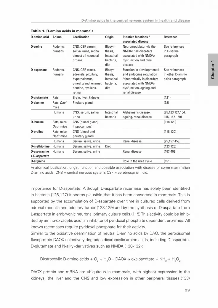
Table 1. D-amino acids in mammals
D-amino acid Animal Localization Origin Putative functions / associated disease
Reference
D-serine Rodents, humans
CNS, CSF, serum, saliva, urine, retina, almost all neonatal organs
Biosyn-thesis, intestinal bacteria, diet
Neuromodulator via the NMDAr / all disorders associated with NMDAr dysfunction and renal disease
See references in D-serine paragraph
D-aspartate Rodents, humans
CNS, CSF, testes, adrenals, pituitary, hypothalamus, pineal gland, enamel, dentine, eye lens, retina
Biosyn-thesis, intestinal bacteria, diet
Function in developmental and endocrine regulation / theoretically in disorders associated with NMDAr dysfunction, ageing and renal disease
See references in other D-amino acids paragraph
D-glutamate Rats Brain, liver, kidneys (121)
D-alanine Rats, Dao-/- mice
Pituitary gland (38)
Humans CNS, serum, saliva, urine
Intestinal bacteria
Alzheimer’s disease, ageing, renal disease
(25,123,124,154, 155, 157-159)
D-leucine Rats, mice, Dao-/- mice
CNS (pineal gland, hippocampus)
(118,120)
D-proline Rats, mice, Dao-/- mice
CNS (pineal and pituitary gland)
(118,120)
Humans Serum, saliva, urine Renal disease (25,157-159)
D-methionine Humans Serum, saliva, urine Diet (123,125)
D-asparagine + D-aspartate
Humans Serum, saliva, urine Renal disease (157-159)
D-arginine Role in the urea cycle (151)
Anatomical localization, origin, function and possible association with disease of some mammalian D-amino acids. CNS = central nervous system; CSF = cerebrospinal fluid.

Introduction
30
DAOX appears to participate at least in the metabolism of mammalian endogenous D-
aspartate.(134-137)
Biological function of D-amino acids
In the CNS, D-aspartate might potentate NMDAr mediated transmission through the glu-
tamate binding site of the NMDAr,(138,139) but the affinity of NMDArs for D-aspartate is
ten times lower than for L-glutamate (139) and localization of D-aspartate does not cor-
relate with the NMDAr.(140) D-Aspartate might however enhance NMDAr activity after
enzymatic methylation by a methyltransferase to generate NMDA in the brain.(119,141)
The properties of storage, release and uptake of D-aspartate have not been well char-
acterized. D-Aspartate may be stored in secretory granules and secreted through a Ca2+
dependent exocytotic mechanism, at least in the adrenals.(142) Several studies, mostly
using the metabolically inert [3H]D-aspartate, have suggested that the biological activ-
ity of D-aspartate is terminated after removal from the synaptic cleft by high–affinity
transporters.(143-146) Synaptically released D-aspartate might also be inactivated by
DAOX,(140) but DAOX is present in peroxisomes and not in axon terminals and synaptic
complexes.(132)
On the basis of the developmental correlation between the periods of maximal D-aspar-
tate contents in many different organs and the periods of morphological and functional
maturation of the organ,(15,22,117) it has been suggested that D-aspartate may play a
role in the maturation and differentiation of cells in tissues where it transiently appears,
such as the CNS, the retina, the adrenals and the testes.(22,117) In mammalian endo-
crine cells, D-aspartate also appears to influence the secretion of several hormones.
D-Aspartate stimulated the release of testosterone,(147,148) oxytocin,(149) growth and
luteinizing hormones (119) and prolactin (141) while inhibiting the secretion of melatonin.
(150) D-Aspartate is likely to participate in the steroidogenesis in the testes,(147,148)
possibly through cholesterol translocation into the inner mitochondrial membrane, the
rate-limiting process in steroidogenesis.(148)
D-Arginine is the only other D-amino acid that has been implicated in a biological func-
tion. It has been proposed to play a role in the urea cycle.(151)
D-Amino acids and disease
Alterations in D-aspartate concentrations have been reported in patients with AD, al-
though these data are conflicting and further studies are warranted to corroborate these
results. D-Aspartate levels were found to be significantly higher in the cerebrospinal
fluid of patients with AD than in normal cerebrospinal fluid,(152) while in contrast, free
D- aspartate content was significantly lower in the brain regions with neuropathological
changes of AD (153) and in the white matter of AD brains when compared to normal
brains.(154) Some of these discrepancies might relate to the facile degradation of D-
31
D-Amino acids in the central nervous system in health and disease
Ch
apte
r 1
asparagine to D-aspartate during storage of samples or analysis, leading to overestima-
tions of D-aspartate concentrations. Elevated levels of free and protein bound D-alanine
have also been reported in the gray matter of AD brains, the significance of which
requires clarification.(154,155)
After perinatal asphyxia, D-aspartate was synthesized specifically in the CNS, probably
from its precursor alanine.(156) The function of D-aspartate in hypoxia remains to be de-
termined. Elevated levels of serum D-serine, D-alanine, D-proline and D-asparagine and/
or D-aspartate have further been associated with ageing and renal disease,(25,157-159)
with a positive correlation between D-amino acid levels and markers for renal disease.
D-Amino acids and clinical use
Although modulation of the concentration of D-aspartate does not have an obvious
clinical purpose yet, exogenously applied D-aspartate has been evaluated in animals. Ad-
dition of 5% D,L-aspartate to the diets of rats depressed growth to a greater extent than
the L-isomer alone,(160,161) as did addition of 2% D-aspartate in the soybean protein
diets of chickens, as opposed to 2% D-serine.(162) Rats receiving 50 mg/kg D-aspartate
in their drinking water, on the other hand, did not show any signs of toxicity in their
physiological status, nor in specific organs such as liver and kidneys.(163) The threshold
value for toxicity of D-aspartate in mice after intramuscular injection was relatively high
(LD50 =1000mg/kg bodyweight).(164) Clearly, after unraveling the function of D-aspartate
in health and disease, toxicity will need to be addressed before considering clinical use.
Inclusion of D-amino acids in antibiotics and synthetic vaccines against infectious or
autoimmune diseases may be advantageous in terms of both specificity of the immune
response and efficacy through decreased digestion.(165) Use of D-amino acids might
also provide new strategies in cancer therapy. As administration of neutral D-amino acids
induces DAO activity, it increases the formation of its reaction products, including hydro-
gen peroxide. This reactive oxygen species can cause oxidative stress and cytotoxicity.
Transfection of tumor cells with a DAO-construct and subsequent D-alanine administra-
tion resulted in cytotoxicity to tumor cells without causing toxicity to parental cells.(166)
ConCLuSIonS
Up to fifty years ago, it was generally believed that D-amino acids did not occur in living
organisms with the exception of micro-organisms and some peptides. The studies from
the last few decades have undeniably established the presence of D-amino acids in
higher organisms, including humans. The specific functions of the individual D-amino
acids are far from being unraveled, despite the growing understanding provided by
recent work, which has focused on D-serine and D-aspartate. D-Serine functions as an

Introduction
32
important neuromodulator, whereas D-aspartate has been implicated in developmental
and endocrine functions. The role of D-amino acids in pathologic conditions requires
further investigation. Determination of D-amino acid concentrations in biological fluids
and measurement of the activity of the synthesizing and metabolizing enzymes and
mutations in their genes might be of use as diagnostic tools in some disorders. This is
of particular interest since D-amino acid concentrations and their function may be ma-
nipulated through exogenous application, modulation of biosynthesis and degradation
and pharmacological manipulation of target receptors, thus providing new therapeutic
strategies. Finally, use of exogenous D-amino acids, free or incorporated in peptides
and proteins, might bring valuable new treatment possibilities thanks to their specific
interactions and high efficacy.
aCknowLeDgemenTS
We gratefully acknowledge financial support from The Netherlands Organisation for
Health Research and Development (grant 920-03-345). We thank L. Dorland and M. de
Sain for helpful discussions on the topic.

33
D-Amino acids in the central nervous system in health and disease
Ch
apte
r 1
reFerenCeS
1. V.S. Lamzin, Z. Dauter, K.S. Wilson, How nature deals with stereoisomers, Curr. Opin. Struct.
Biol 5 (1995) 830-836.
2. V. Prelog, Chirality in chemistry, Science 193 (1976) 17-24.
3. S.F. Mason, Origins of biomolecular handedness, Nature 311 (1984) 19-23.
4. J.J. Corrigan, D-amino acids in animals, Science 164 (1969) 142-149.
5. J. Auclair, R. Patton, Rev Can Biol 9 (1950) 3.
6. I. Beatty, D. Magrath, A. Ennor, Occurrence of D-serine in lombricine, Nature 183 (1959) 591.
7. J.J. Corrigan, N.G. Srinivasan, The occurrence of certain D-amino acids in insects, Biochem-
istry 5 (1966) 1185-1190.
8. G. Kreil, Peptides containing a D-amino acid from frogs and molluscs, J. Biol Chem. 269
(1994) 10967-10970.
9. R.L. Preston, Occurrence of D-amino acids in higher organisms: a survey of the distribution
of D-amino acids in marine vertebrates., Comp. Biochem. Physiol. 87B (1987) 55-62.
10. H.A. Krebs, Metabolism of amino acids. III. Determination of amino acids., Biochem. J. 29
(1935) 1620-1644.
11. J.L. Still, M.V. Buell, W.E. Knox, D.E. Green, Studies on the cyclphorase system VII. D-
Aspartate oxidase, J. Biol. Chem. 179 (1949) 831-837.
12. P.M. Helfman, J.L. Bada, M.Y. Shou, Considerations on the role of aspartic acid racemization
in the aging process, Gerontology 23 (1977) 419-425.
13. D.S. Dunlop, A. Neidle, D. McHale, D.M. Dunlop, A. Lajtha, The presence of free D-aspartic
acid in rodents and man, Biochem. Biophys. Res. Commun. 141 (1986) 27-32.
14. A. Hashimoto, T. Nishikawa, T. Hayashi, N. Fujii, K. Harada, T. Oka, K. Takahashi, The presence
of free D-serine in rat brain, FEBS Lett. 296 (1992) 33-36.
15. A. Hashimoto, S. Kumashiro, T. Nishikawa, T. Oka, K. Takahashi, T. Mito, S. Takashima, N.
Doi, Y. Mizutani, T. Yamazaki, ., Embryonic development and postnatal changes in free D-
aspartate and D-serine in the human prefrontal cortex, J. Neurochem. 61 (1993) 348-351.
16. A. Hashimoto, T. Nishikawa, T. Oka, K. Takahashi, Endogenous D-serine in rat brain: N-methyl-
D-aspartate receptor-related distribution and aging, J. Neurochem. 60 (1993) 783-786.
17. Y. Nagata, K. Horiike, T. Maeda, Distribution of free D-serine in vertebrate brains, Brain Res.
634 (1994) 291-295.
18. A. Hashimoto, T. Oka, T. Nishikawa, Extracellular concentration of endogenous free D-serine
in the rat brain as revealed by in vivo microdialysis, Neuroscience 66 (1995) 635-643.
19. M.J. Schell, M.E. Molliver, S.H. Snyder, D-serine, an endogenous synaptic modulator:
localization to astrocytes and glutamate-stimulated release, Proc. Natl. Acad. Sci U. S. A 92
(1995) 3948-3952.
20. M.J. Schell, R.O. Brady, Jr., M.E. Molliver, S.H. Snyder, D-serine as a neuromodulator:
regional and developmental localizations in rat brain glia resemble NMDA receptors, J.
Neurosci. 17 (1997) 1604-1615.
21. K. Wako, N. Ma, T. Shiroyama, R. Semba, Glial uptake of intracerebroventricularly injected
D-serine in the rat brain: an immunocytochemical study, Neurosci. Lett. 185 (1995) 171-174.
22. A. Hashimoto, T. Oka, T. Nishikawa, Anatomical distribution and postnatal changes in endog-

Introduction
34
enous free D-aspartate and D-serine in rat brain and periphery, Eur. J. Neurosci. 7 (1995)
1657-1663.
23. K. Horiike, H. Tojo, R. Arai, T. Yamano, M. Nozaki, T. Maeda, Localization of D-amino acid
oxidase in Bergmann glial cells and astrocytes of rat cerebellum, Brain Res. Bull. 19 (1987)
587-596.
24. H. Bruckner, S. Haasmann, A. Friedrich, Quantification of D-amino acids in human urine
using GC-MS and HPLC, Amino Acids 6 (1994) 211.
25. Y. Nagata, R. Masui, T. Akino, The presence of free D-serine, D-alanine and D-proline in hu-
man plasma, Experientia 48 (1992) 986-988.
26. J. Rotgans, R. Wodarz, W. Schoknecht, K. Drysch, The determination of amino-acid enantio-
mers in human saliva with Chirasil-Val, Arch. Oral Biol 28 (1983) 1121-1124.
27. E.R. Stevens, M. Esguerra, P.M. Kim, E.A. Newman, S.H. Snyder, K.R. Zahs, R.F. Miller,
D-serine and serine racemase are present in the vertebrate retina and contribute to the
physiological activation of NMDA receptors, Proc. Natl. Acad. Sci U. S. A 100 (2003) 6789-
6794.
28. J. De Miranda, R. Panizzutti, V.N. Foltyn, H. Wolosker, Cofactors of serine racemase that
physiologically stimulate the synthesis of the N-methyl-D-aspartate (NMDA) receptor coago-
nist D-serine, Proc. Natl. Acad. Sci U. S. A 99 (2002) 14542-14547.
29. H. Wolosker, S. Blackshaw, S.H. Snyder, Serine racemase: a glial enzyme synthesizing D-
serine to regulate glutamate-N-methyl-D-aspartate neurotransmission, Proc. Natl. Acad. Sci
U. S. A 96 (1999) 13409-13414.
30. H. Wolosker, K.N. Sheth, M. Takahashi, J.P. Mothet, R.O. Brady, Jr., C.D. Ferris, S.H. Snyder,
Purification of serine racemase: biosynthesis of the neuromodulator D-serine, Proc. Natl.
Acad. Sci U. S. A 96 (1999) 721-725.
31. T.J. de Koning, L.W. Klomp, Serine-deficiency syndromes, Curr Opin Neurol 17 (2004) 197-
204.
32. K. Yoshida, S. Furuya, S. Osuka, J. Mitoma, Y. Shinoda, M. Watanabe, N. Azuma, H. Tanaka, T.
Hashikawa, S. Itohara, Y. Hirabayashi, Targeted disruption of the mouse 3-phosphoglycerate
dehydrogenase gene causes severe neurodevelopmental defects and results in embryonic
lethality, J. Biol Chem. 279 (2004) 3573-3577.
33. L.Z. Wang, X.Z. Zhu, Spatiotemporal relationships among D-serine, serine racemase, and
D-amino acid oxidase during mouse postnatal development, Acta Pharmacol. Sin. 24 (2003)
965-974.
34. J. De Miranda, A. Santoro, S. Engelender, H. Wolosker, Human serine racemase: moleular
cloning, genomic organization and functional analysis, Gene 256 (2000) 183-188.
35. V.N. Foltyn, I. Bendikov, J. De Miranda, R. Panizzutti, E. Dumin, M. Shleper, P. Li, M.D. Toney,
E. Kartvelishvily, H. Wolosker, Serine racemase modulates intracellular D-serine levels
through an alpha,beta-elimination activity, J. Biol Chem. 280 (2005) 1754-63.
36. L. Pollegioni, C.M. Harris, G. Molla, M.S. Pilone, S. Ghisla, Identification and role of ionizing
functional groups at the active center of Rhodotorula gracilis D-amino acid oxidase, FEBS
Lett. 507 (2001) 323-326.
37. A. Hashimoto, T. Nishikawa, R. Konno, A. Niwa, Y. Yasumura, T. Oka, K. Takahashi, Free D-
serine, D-aspartate and D-alanine in central nervous system and serum in mutant mice
lacking D-amino acid oxidase, Neurosci. Lett. 152 (1993) 33-36.

35
D-Amino acids in the central nervous system in health and disease
Ch
apte
r 1
38. A. Morikawa, K. Hamase, T. Inoue, R. Konno, A. Niwa, K. Zaitsu, Determination of free
D-aspartic acid, D-serine and D-alanine in the brain of mutant mice lacking D-amino acid
oxidase activity, J. Chromatogr. B Biomed. Sci Appl. 757 (2001) 119-125.
39. A. D’Aniello, A. Vetere, L. Petrucelli, Further study on the specificity of D-amino acid oxidase
and D-aspartate oxidase and time course for complete oxidation of D-amino acids, Comp
Biochem. Physiol B 105 (1993) 731-734.
40. M. Tada, K. Fukui, K. Momoi, Y. Miyake, Cloning and expression of a cDNA encoding mouse
kidney D-amino acid oxidase, Gene 90 (1990) 293-297.
41. Y. Nagata, R. Konno, Y. Yasumura, T. Akino, Involvement of D-amino acid oxidase in elimina-
tion of free D-amino acids in mice, Biochem. J. 257 (1989) 291-292.
42. Y. Urai, O. Jinnouchi, K.T. Kwak, A. Suzue, S. Nagahiro, K. Fukui, Gene expression of D-amino
acid oxidase in cultured rat astrocytes: regional and cell type specific expression, Neurosci.
Lett. 324 (2002) 101-104.
43. R. Dingledine, K. Borges, D. Bowie, S.F. Traynelis, The glutamate receptor ion channels,
Pharmacol. Rev 51 (1999) 7-61.
44. J.W. Johnson, P. Ascher, Glycine potentiates the NMDA response in cultured mouse brain
neurons, Nature 325 (1987) 529-531.
45. N.W. Kleckner, R. Dingledine, Requirement for glycine in activation of NMDA-receptors
expressed in Xenopus oocytes, Science 241 (1988) 835-837.
46. A.M. Thomson, Glycine is a coagonist at the NMDA receptor/channel complex, Prog. Neuro-
biol. 35 (1990) 53-74.
47. W. Danysz, E. Fadda, J.T. Wroblewski, E. Costa, [3H]D-serine labels strychnine-insensitive
glycine recognition sites of rat central nervous system, Life Sci 46 (1990) 155-164.
48. T. Matsui, M. Sekiguchi, A. Hashimoto, U. Tomita, T. Nishikawa, K. Wada, Functional com-
parison of D-serine and glycine in rodents: the effect on cloned NMDA receptors and the
extracellular concentration, J. Neurochem. 65 (1995) 454-458.
49. J.P. Mothet, A.T. Parent, H. Wolosker, R.O. Brady, Jr., D.J. Linden, C.D. Ferris, M.A. Rogawski,
S.H. Snyder, D-serine is an endogenous ligand for the glycine site of the N-methyl-D-
aspartate receptor, Proc. Natl. Acad. Sci U. S. A 97 (2000) 4926-4931.
50. S. Ahmadi, U. Muth-Selbach, A. Lauterbach, P. Lipfert, W.L. Neuhuber, H.U. Zeilhofer,
Facilitation of spinal NMDA receptor currents by spillover of synaptically released glycine,
Science 300 (2003) 2094-2097.
51. J.E. Chatterton, M. Awobuluyi, L.S. Premkumar, H. Takahashi, M. Talantova, Y. Shin, J. Cui, S.
Tu, K.A. Sevarino, N. Nakanishi, G. Tong, S.A. Lipton, D. Zhang, Excitatory glycine receptors
containing the NR3 family of NMDA receptor subunits, Nature 415 (2002) 793-798.
52. D. Boehning, S.H. Snyder, Novel neural modulators, Annu. Rev Neurosci. 26 (2003) 105-131.
53. S.P. Cook, I. Galve-Roperh, d.P. Martinez, I. Rodriguez-Crespo, Direct calcium binding results
in activation of brain serine racemase, J. Biol Chem. 277 (2002) 27782-27792.
54. H. Tanaka, S.Y. Grooms, M.V. Bennett, R.S. Zukin, The AMPAR subunit GluR2: still front and
center-stage, Brain Res. 886 (2000) 190-207.
55. J.T. Wroblewski, E. Fadda, J. Mazzetta, J.W. Lazarewicz, E. Costa, Glycine and D-serine act
as positive modulators of signal transduction at N-methyl-D-aspartate sensitive glutamate
receptors in cultured cerebellar granule cells, Neuropharmacology 28 (1989) 447-452.
56. J.W. Lazarewicz, J.T. Wroblewski, M.E. Palmer, E. Costa, Activation of N-methyl-D-aspartate-

Introduction
36
sensitive glutamate receptors stimulates arachidonic acid release in primary cultures of
cerebellar granule cells, Neuropharmacology 27 (1988) 765-769.
57. F. Vaccarino, A. Guidotti, E. Costa, Ganglioside inhibition of glutamate-mediated protein
kinase C translocation in primary cultures of cerebellar neurons, Proc. Natl. Acad. Sci U. S. A
84 (1987) 8707-8711.
58. A.M. Szekely, M.L. Barbaccia, E. Costa, Activation of specific glutamate receptor subtypes
increases C-fos proto-oncogene expression in primary cultures of neonatal rat cerebellar
granule cells, Neuropharmacology 26 (1987) 1779-1782.
59. G.L. Collingridge, R.A. Lester, Excitatory amino acid receptors in the vertebrate central
nervous system, Pharmacol. Rev 41 (1989) 143-210.
60. D.T. Monaghan, R.J. Bridges, C.W. Cotman, The excitatory amino acid receptors: their
classes, pharmacology, and distinct properties in the function of the central nervous system,
Annu. Rev Pharmacol. Toxicol. 29 (1989) 365-402.
61. S. Moreno, R. Nardacci, A. Cimini, M.P. Ceru, Immunocytochemical localization of D-amino
acid oxidase in rat brain, J. Neurocytol. 28 (1999) 169-185.
62. C.S. Ribeiro, M. Reis, R. Panizzutti, J. De Miranda, H. Wolosker, Glial transport of the neuro-
modulator D-serine, Brain Res. 929 (2002) 202-209.
63. Y. Fukasawa, H. Segawa, J.Y. Kim, A. Chairoungdua, D.K. Kim, H. Matsuo, S.H. Cha, H.
Endou, Y. Kanai, Identification and characterization of a Na(+)-independent neutral amino
acid transporter that associates with the 4F2 heavy chain and exhibits substrate selectivity
for small neutral D- and L-amino acids, J. Biol Chem. 275 (2000) 9690-9698.
64. L. Helboe, J. Egebjerg, M. Moller, C. Thomsen, Distribution and pharmacology of alanine-
serine-cysteine transporter 1 (asc-1) in rodent brain, Eur. J. Neurosci. 18 (2003) 2227-2238.
65. H. Matsuo, Y. Kanai, M. Tokunaga, T. Nakata, A. Chairoungdua, H. Ishimine, S. Tsukada, H.
Ooigawa, H. Nawashiro, Y. Kobayashi, J. Fukuda, H. Endou, High affinity D- and L-serine
transporter Asc-1: cloning and dendritic localization in the rat cerebral and cerebellar corti-
ces, Neurosci. Lett. 358 (2004) 123-126.
66. J. Nakauchi, H. Matsuo, D.K. Kim, A. Goto, A. Chairoungdua, S.H. Cha, J. Inatomi, Y. Shio-
kawa, K. Yamaguchi, I. Saito, H. Endou, Y. Kanai, Cloning and characterization of a human
brain Na(+)-independent transporter for small neutral amino acids that transports D-serine
with high affinity, Neurosci. Lett. 287 (2000) 231-235.
67. A. Hashimoto, Effect of the intracerebroventricular and systemic administration of L-serine
on the concentrations of D- and L-serine in several brain areas and periphery of rat, Brain
Res. 955 (2002) 214-220.
68. R.A. Lester, G. Tong, C.E. Jahr, Interactions between the glycine and glutamate binding sites
of the NMDA receptor, J. Neurosci. 13 (1993) 1088-1096.
69. L. Vyklicky, Jr., M. Benveniste, M.L. Mayer, Modulation of N-methyl-D-aspartic acid receptor
desensitization by glycine in mouse cultured hippocampal neurones, J. Physiol 428 (1990)
313-331.
70. Y. Yang, W. Ge, Y. Chen, Z. Zhang, W. Shen, C. Wu, M. Poo, S. Duan, Contribution of astro-
cytes to hippocampal long-term potentiation through release of D-serine, Proc. Natl. Acad.
Sci U. S. A 100 (2003) 15194-15199.
71. P.L. Wood, The co-agonist concept: is the NMDA-associated glycine receptor saturated in
vivo?, Life Sci 57 (1995) 301-310.

37
D-Amino acids in the central nervous system in health and disease
Ch
apte
r 1
72. A.J. Berger, S. Dieudonne, P. Ascher, Glycine uptake governs glycine site occupancy at
NMDA receptors of excitatory synapses, J. Neurophysiol. 80 (1998) 3336-3340.
73. R. Bergeron, T.M. Meyer, J.T. Coyle, R.W. Greene, Modulation of N-methyl-D-aspartate
receptor function by glycine transport, Proc. Natl. Acad. Sci U. S. A 95 (1998) 15730-15734.
74. L. Chen, M. Muhlhauser, C.R. Yang, Glycine tranporter-1 blockade potentiates NMDA-
mediated responses in rat prefrontal cortical neurons in vitro and in vivo, J. Neurophysiol. 89
(2003) 691-703.
75. T.E. Salt, Modulation of NMDA receptor-mediated responses by glycine and D-serine in the
rat thalamus in vivo, Brain Res. 481 (1989) 403-406.
76. A.M. Thomson, V.E. Walker, D.M. Flynn, Glycine enhances NMDA-receptor mediated synap-
tic potentials in neocortical slices, Nature 338 (1989) 422-424.
77. D. Billups, D. Attwell, Active release of glycine or D-serine saturates the glycine site of
NMDA receptors at the cerebellar mossy fibre to granule cell synapse, Eur. J. Neurosci. 18
(2003) 2975-2980.
78. H. Komuro, P. Rakic, Modulation of neuronal migration by NMDA receptors, Science 260
(1993) 95-97.
79. S. Rabacchi, Y. Bailly, N. Delhaye-Bouchaud, J. Mariani, Involvement of the N-methyl D-
aspartate (NMDA) receptor in synapse elimination during cerebellar development, Science
256 (1992) 1823-1825.
80. W. Danysz, A.C. Parsons, Glycine and N-methyl-D-aspartate receptors: physiological signifi-
cance and possible therapeutic applications, Pharmacol. Rev 50 (1998) 597-664.
81. E.H. Lo, A.R. Pierce, K. Matsumoto, T. Kano, C.J. Evans, R. Newcomb, Alterations in K+
evoked profiles of neurotransmitter and neuromodulator amino acids after focal ischemia-
reperfusion, Neuroscience 83 (1998) 449-458.
82. C.A. Hicks, M.A. Ward, N. Ragumoorthy, S.J. Ambler, C.P. Dell, D. Dobson, M.J. O’Neill,
Evaluation of glycine site antagonists of the NMDA receptor in global cerebral ischaemia,
Brain Res. 819 (1999) 65-74.
83. M.A. Petty, C. Neumann-Haefelin, J. Kalisch, S. Sarhan, J.G. Wettstein, H.P. Juretschke, In
vivo neuroprotective effects of ACEA 1021 confirmed by magnetic resonance imaging in
ischemic stroke, Eur. J. Pharmacol. 474 (2003) 53-62.
84. D.W. Choi, S.M. Rothman, The role of glutamate neurotoxicity in hypoxic-ischemic neuronal
death, Annu. Rev Neurosci. 13 (1990) 171-182.
85. A. Roldan, J. Figueras-Aloy, R. Deulofeu, R. Jimenez, Glycine and other neurotransmitter
amino acids in cerebrospinal fluid in perinatal asphyxia and neonatal hypoxic-ischaemic
encephalopathy, Acta Paediatr. 88 (1999) 1137-1141.
86. M.L. Chouinard, D. Gaitan, P.L. Wood, Presence of the N-methyl-D-aspartate-associated gly-
cine receptor agonist, D-serine, in human temporal cortex: comparison of normal, Parkinson,
and Alzheimer tissues, J. Neurochem. 61 (1993) 1561-1564.
87. S. Kumashiro, A. Hashimoto, T. Nishikawa, Free D-serine in post-mortem brains and spinal
cords of individuals with and without neuropsychiatric diseases, Brain Res. 681 (1995) 117-
125.
88. Y. Nagata, M. Borghi, G.H. Fisher, A. D’Aniello, Free D-serine concentration in normal and
Alzheimer human brain, Brain Res. Bull. 38 (1995) 181-183.
89. M. Carlsson, A. Carlsson, Interactions between glutamatergic and monoaminergic systems

Introduction
38
within the basal ganglia--implications for schizophrenia and Parkinson’s disease, Trends
Neurosci. 13 (1990) 272-276.
90. M.L. Schmitt, W. Coelho, A.S. Lopes-de-Souza, F.S. Guimaraes, A.P. Carobrez, Anxiogenic-
like effect of glycine and D-serine microinjected into dorsal periaqueductal gray matter of
rats, Neurosci. Lett. 189 (1995) 93-96.
91. E.W. Anthony, M.E. Nevins, Anxiolytic-like effects of N-methyl-D-aspartate-associated
glycine receptor ligands in the rat potentiated startle test, Eur. J. Pharmacol. 250 (1993)
317-324.
92. J.H. Kehne, T.C. McCloskey, B.M. Baron, E.M. Chi, B.L. Harrison, J.P. Whitten, M.G. Palfrey-
man, NMDA receptor complex antagonists have potential anxiolytic effects as measured
with separation-induced ultrasonic vocalizations, Eur. J. Pharmacol. 193 (1991) 283-292.
93. D.C. Goff, J.T. Coyle, The emerging role of glutamate in the pathophysiology and treatment
of schizophrenia, Am. J. Psychiatry 158 (2001) 1367-1377.
94. A.R. Mohn, R.R. Gainetdinov, M.G. Caron, B.H. Koller, Mice with reduced NMDA receptor
expression display behaviors related to schizophrenia, Cell 98 (1999) 427-436.
95. P.C. Contreras, D-serine antagonized phencyclidine- and MK-801-induced stereotyped
behavior and ataxia, Neuropharmacology 29 (1990) 291-293.
96. K. Hashimoto, T. Fukushima, E. Shimizu, N. Komatsu, H. Watanabe, N. Shinoda, M. Nakazato,
C. Kumakiri, S. Okada, H. Hasegawa, K. Imai, M. Iyo, Decreased serum levels of D-serine
in patients with schizophrenia: evidence in support of the N-methyl-D-aspartate receptor
hypofunction hypothesis of schizophrenia, Arch. Gen. Psychiatry 60 (2003) 572-576.
97. G. Tsai, P. Yang, L.C. Chung, N. Lange, J.T. Coyle, D-serine added to antipsychotics for the
treatment of schizophrenia, Biol Psychiatry 44 (1998) 1081-1089.
98. C. Dalman, H.V. Thomas, A.S. David, J. Gentz, G. Lewis, P. Allebeck, Signs of asphyxia at
birth and risk of schizophrenia. Population-based case-control study, Br. J. Psychiatry 179
(2001) 403-408.
99. B.S. Meldrum, Possible therapeutic applications of excitatory amino acid neurotransmitters,
Clin. Sci. 68 (1985) 113-122.
100. L. Singh, R.J. Oles, M.D. Tricklebank, Modulation of seizure susceptibility in the mouse by
the strychnine-insensitive glycine recognition site of the NMDA receptor/ion channel com-
plex, Br. J. Pharmacol. 99 (1990) 285-288.
101. G. De Sarro, G.R. Trimarchi, S. Sinopoli, Y. Masuda, A. De Sarro, Anticonvulsant effects of
U-54494A and U-50488H in genetically epilepsy-prone rats and DBA/2 mice: a possible
involvement of glycine/NMDA receptor complex, Gen. Pharmacol. 24 (1993) 439-447.
102. S.L. Peterson, Infusion of NMDA antagonists into the nucleus reticularis pontis oralis inhibits
the maximal electroshock seizure response, Brain Res. 702 (1995) 101-109.
103. S.L. Peterson, Anticonvulsant drug potentiation by glycine in maximal electroshock seizures
is mimicked by D-serine and antagonized by 7-chlorokynurenic acid, Eur. J. Pharmacol. 199
(1991) 341-348.
104. W. Loscher, P. Wlaz, C. Rundfeldt, H. Baran, D. Honack, Anticonvulsant effects of the glycine/
NMDA receptor ligands D-cycloserine and D-serine but not R-(+)-HA-966 in amygdala-kindled
rats, Br. J. Pharmacol. 112 (1994) 97-106.
105. H. Mori, M. Mishina, Structure and function of the NMDA receptor channel, Neuropharma-
cology 34 (1995) 1219-1237.

39
D-Amino acids in the central nervous system in health and disease
Ch
apte
r 1
106. S. Nakanishi, Molecular diversity of glutamate receptors and implications for brain function,
Science 258 (1992) 597-603.
107. J. Jaeken, M. Detheux, L. Van Maldergem, M. Foulon, H. Carchon, E. Van Schaftingen,
3-Phosphoglycerate dehydrogenase deficiency: an inborn error of serine biosynthesis, Arch.
Dis. Child 74 (1996) 542-545.
108. J. Jaeken, M. Detheux, J.P. Fryns, J.F. Collet, P. Alliet, E. Van Schaftingen, Phosphoserine
phosphatase deficiency in a patient with Williams syndrome, J. Med. Genet. 34 (1997) 594-
596.
109. R. Konno, Y. Yasumura, Mouse mutant deficient in D-amino acid oxidase activity, Genetics
103 (1983) 277-285.
110. K. Wake, H. Yamazaki, S. Hanzawa, R. Konno, H. Sakio, A. Niwa, Y. Hori, Exaggerated re-
sponses to chronic nociceptive stimuli and enhancement of N-methyl-D-aspartate receptor-
mediated synaptic transmission in mutant mice lacking D-amino-acid oxidase, Neurosci.
Lett. 297 (2001) 25-28.
111. C. Artom, W.H. Fishman, R.P. Morehead, The relative toxicity of L- and D,L-serine in rats,
Proc. Soc. Expl. Biol. Med. 60 (1945) 284-287.
112. M. Wachstein, M. Besen, Electron microscopy of renal coagulative necrosis due to DL-
serine, with special reference to mitochondrial pyknosis, Am. J. Pathol. 44 (1964) 383-400.
113. A. Hashimoto, Y. Chiba, Effect of systemic administration of d-serine on the levels of d- and
l-serine in several brain areas and periphery of rat, Eur. J. Pharmacol. 495 (2004) 153-158.
114. A. Hashimoto, T. Oka, Free D-aspartate and D-serine in the mammalian brain and periphery,
Prog. Neurobiol. 52 (1997) 325-353.
115. H. Wolosker, A. D’Aniello, S.H. Snyder, D-aspartate disposition in neuronal and endocrine
tissues: ontogeny, biosynthesis and release, Neuroscience 100 (2000) 183-189.
116. K. Sakai, H. Homma, J.A. Lee, T. Fukushima, T. Santa, K. Tashiro, T. Iwatsubo, K. Imai, Emer-
gence of D-aspartic acid in the differentiating neurons of the rat central nervous system,
Brain Res. 808 (1998) 65-71.
117. A. Neidle, D.S. Dunlop, Developmental changes in free D-aspartic acid in the chicken embryo
and in the neonatal rat, Life Sci 46 (1990) 1517-1522.
118. K. Hamase, H. Homma, Y. Takigawa, T. Fukushima, T. Santa, K. Imai, Regional distribution
and postnatal changes of D-amino acids in rat brain, Biochim. Biophys. Acta 1334 (1997)
214-222.
119. A. D’Aniello, M.M. Di Fiore, G.H. Fisher, A. Milone, A. Seleni, S. D’Aniello, A.F. Perna, D.
Ingrosso, Occurrence of D-aspartic acid and N-methyl-D-aspartic acid in rat neuroendocrine
tissues and their role in the modulation of luteinizing hormone and growth hormone release,
FASEB J. 14 (2000) 699-714.
120. K. Hamase, T. Inoue, A. Morikawa, R. Konno, K. Zaitsu, Determination of free D-proline and
D-leucine in the brains of mutant mice lacking D-amino acid oxidase activity, Anal. Biochem.
298 (2001) 253-258.
121. Y. Kera, H. Aoyama, H. Matsumura, A. Hasegawa, H. Nagasaki, R. Yamada, Presence of free
D-glutamate and D-aspartate in rat tissues, Biochim. Biophys. Acta 1243 (1995) 283-286.
122. D.W. Armstrong, M. Gasper, S.H. Lee, J. Zukowski, N. Ercal, D-amino acid levels in human
physiological fluids, Chirality 5 (1993) 375-378.

Introduction
40
123. R. Konno, A. Niwa, Y. Yasumura, Intestinal bacterial origin of D-alanine in urine of mutant
mice lacking D-amino-acid oxidase, Biochem. J. 268 (1990) 263-265.
124. R. Konno, T. Oowada, A. Ozaki, T. Iida, A. Niwa, Y. Yasumura, T. Mizutani, Origin of D-alanine
present in urine of mutant mice lacking D-amino-acid oxidase activity, Am. J. Physiol 265
(1993) G699-G703.
125. R. Konno, K. Isobe, A. Niwa, Y. Yasumura, Excessive urinary excretion of methionine in
mutant mice lacking D-amino-acid oxidase activity, Metabolism 37 (1988) 1139-1142.
126. M. Yohda, H. Okada, H. Kumagai, Molecular cloning and nucleotide sequencing of the
aspartate racemase gene from lactic acid bacteria Streptococcus thermophilus, Biochim.
Biophys. Acta 1089 (1991) 234-240.
127. M. Yohda, I. Endo, Y. Abe, T. Ohta, T. Iida, T. Maruyama, Y. Kagawa, Gene for aspartate race-
mase from the sulfur-dependent hyperthermophilic archaeum, Desulfurococcus strain SY, J.
Biol Chem. 271 (1996) 22017-22021.
128. Z. Long, J.A. Lee, T. Okamoto, N. Nimura, K. Imai, H. Homma, d-Aspartate in a prolactin-
secreting clonal strain of rat pituitary tumor cells (GH(3)), Biochem. Biophys. Res. Commun.
276 (2000) 1143-1147.
129. Z. Long, H. Homma, J.A. Lee, T. Fukushima, T. Santa, T. Iwatsubo, R. Yamada, K. Imai, Biosyn-
thesis of D-aspartate in mammalian cells, FEBS Lett. 434 (1998) 231-235.
130. G.A. Hamilton, Peroxisomal oxidases and suggestions for the mechanism of action of insulin
and other hormones, Adv. Enzymol. Relat Areas Mol. Biol 57 (1985) 85-178.
131. J.L. Still, M.V. Buell, W.E. Knox, D.E. Green, Studies on the cyclphorase system VII. D-
Aspartate oxidase, J. Biol. Chem. 179 (1949) 831-837.
132. K. Zaar, H.P. Kost, A. Schad, A. Volkl, E. Baumgart, H.D. Fahimi, Cellular and subcellular
distribution of D-aspartate oxidase in human and rat brain, J. Comp Neurol. 450 (2002) 272-
282.
133. R. Yamada, H. Nagasaki, Y. Wakabayashi, A. Iwashima, Presence of D-aspartate oxidase in rat
liver and mouse tissues, Biochim. Biophys. Acta 965 (1988) 202-205.
134. A. D’Aniello, G. D’Onofrio, M. Pischetola, G. D’Aniello, A. Vetere, L. Petrucelli, G.H. Fisher,
Biological role of D-amino acid oxidase and D-aspartate oxidase. Effects of D-amino acids, J.
Biol Chem. 268 (1993) 26941-26949.
135. H. Nagasaki, Gender-related differences of mouse liver D-aspartate oxidase in the activity
and response to administration of D-aspartate and peroxisome proliferators, Int. J. Biochem.
26 (1994) 415-423.
136. P.P. Van Veldhoven, C. Brees, G.P. Mannaerts, D-aspartate oxidase, a peroxisomal enzyme in
liver of rat and man, Biochim. Biophys. Acta 1073 (1991) 203-208.
137. R. Yamada, H. Nagasaki, Y. Nagata, Y. Wakabayashi, A. Iwashima, Administration of D-
aspartate increases D-aspartate oxidase activity in mouse liver, Biochim. Biophys. Acta 990
(1989) 325-328.
138. G.E. Fagg, A. Matus, Selective association of N-methyl aspartate and quisqualate types
of L-glutamate receptor with brain postsynaptic densities, Proc. Natl. Acad. Sci U. S. A 81
(1984) 6876-6880.
139. H.J. Olverman, A.W. Jones, K.N. Mewett, J.C. Watkins, Structure/activity relations of
N-methyl-D-aspartate receptor ligands as studied by their inhibition of [3H]D-2-amino-5-
phosphonopentanoic acid binding in rat brain membranes, Neuroscience 26 (1988) 17-31.

41
D-Amino acids in the central nervous system in health and disease
Ch
apte
r 1
140. M.J. Schell, O.B. Cooper, S.H. Snyder, D-aspartate localizations imply neuronal and neuroen-
docrine roles, Proc. Natl. Acad. Sci U. S. A 94 (1997) 2013-2018.
141. G. D’Aniello, A. Tolino, A. D’Aniello, F. Errico, G.H. Fisher, M.M. Di Fiore, The role of D-aspartic
acid and N-methyl-D-aspartic acid in the regulation of prolactin release, Endocrinology 141
(2000) 3862-3870.
142. S. Nakatsuka, M. Hayashi, A. Muroyama, M. Otsuka, S. Kozaki, H. Yamada, Y. Moriyama, D-
Aspartate is stored in secretory granules and released through a Ca(2+)-dependent pathway
in a subset of rat pheochromocytoma PC12 cells, J. Biol Chem. 276 (2001) 26589-26596.
143. J. Ferkany, J.T. Coyle, Heterogeneity of sodium-dependent excitatory amino acid uptake
mechanisms in rat brain, J. Neurosci. Res. 16 (1986) 491-503.
144. J.T. Greenamyre, D.S. Higgins, A.B. Young, Sodium-dependent D-aspartate ‘binding’ is not a
measure of presynaptic neuronal uptake sites in an autoradiographic assay, Brain Res. 511
(1990) 310-318.
145. J. Healy, A. Downes, G.J. McBean, Stimulation of synaptosomal D-[(3)H]aspartate transport
by substance P in rat brain, Neurosci. Lett. 290 (2000) 113-116.
146. Y. Kanai, M.A. Hediger, Primary structure and functional characterization of a high-affinity
glutamate transporter, Nature 360 (1992) 467-471.
147. A. D’Aniello, A. Di Cosmo, C. Di Cristo, L. Annunziato, L. Petrucelli, G. Fisher, Involvement of
D-aspartic acid in the synthesis of testosterone in rat testes, Life Sci 59 (1996) 97-104.
148. Y. Nagata, H. Homma, J.A. Lee, K. Imai, D-Aspartate stimulation of testosterone synthesis
in rat Leydig cells, FEBS Lett. 444 (1999) 160-164.
149. H. Wang, H. Wolosker, J. Pevsner, S.H. Snyder, D.J. Selkoe, Regulation of rat magnocellular
neurosecretory system by D-aspartate: evidence for biological role(s) of a naturally occurring
free D-amino acid in mammals, J. Endocrinol. 167 (2000) 247-252.
150. S. Ishio, H. Yamada, M. Hayashi, S. Yatsushiro, T. Noumi, A. Yamaguchi, Y. Moriyama, D-
aspartate modulates melatonin synthesis in rat pinealocytes, Neurosci. Lett. 249 (1998)
143-146.
151. A. Saavedra-Molina, E. Pina, Stimulation of L-ornithine uptake and L-citrulline and urea
biosynthesis by D-arginine, Biochem. Int. 24 (1991) 349-357.
152. G.H. Fisher, L. Petrucelli, C. Gardner, C. Emory, W.H. Frey, L. Amaducci, S. Sorbi, G. Sorren-
tino, M. Borghi, A. D’Aniello, Free D-amino acids in human cerebrospinal fluid of Alzheimer
disease, multiple sclerosis, and healthy control subjects, Mol. Chem. Neuropathol. 23 (1994)
115-124.
153. A. D’Aniello, J.M. Lee, L. Petrucelli, M.M. Di Fiore, Regional decreases of free D-aspartate
levels in Alzheimer’s disease, Neurosci. Lett. 250 (1998) 131-134.
154. G.H. Fisher, A. D’Aniello, A. Vetere, L. Padula, G.P. Cusano, E.H. Man, Free D-aspartate and
D-alanine in normal and Alzheimer brain, Brain Res. Bull. 26 (1991) 983-985.
155. A. D’Aniello, A. Vetere, G.H. Fisher, G. Cusano, M. Chavez, L. Petrucelli, Presence of D-
alanine in proteins of normal and Alzheimer human brain, Brain Res. 592 (1992) 44-48.
156. M. Puka-Sundvall, M. Sandberg, H. Hagberg, Brain injury after hypoxia-ischemia in newborn
rats: relationship to extracellular levels of excitatory amino acids and cysteine, Brain Res.
750 (1997) 325-328.
157. H. Bruckner, M. Hausch, Gas chromatographic characterization of free D-amino acids in the
Introduction
42
blood serum of patients with renal disorders and of healthy volunteers, J. Chromatogr. 614
(1993) 7-17.
158. T. Fukushima, T. Santa, H. Homma, R. Nagatomo, K. Imai, Determination of D-amino acids in
serum from patients with renal dysfunction, Biol Pharm. Bull. 18 (1995) 1130-1132.
159. Y. Nagata, T. Akino, K. Ohno, Y. Kataoka, T. Ueda, T. Sakurai, K. Shiroshita, T. Yasuda, Free D-
amino acids in human plasma in relation to senescence and renal diseases, Clin. Sci (Lond)
73 (1987) 105-108.
160. C.E. Graham, S.W. Hier, H.K. Waitkoff, S.M. Saper, W.G. Bibler, E.I. Pentz, Studies on natural
and racemic amino acids with rats, J. Biol Chem. 185 (1950) 102.
161. H.E. Sauberlich, Studies on the toxicity and antagonism of amino acids for weanling rats, J.
Nutr. 75 (1961) 61-72.
162. K. Maruyama, M.L. Sunde, A.E. Harper, Effect of D-alanine and D-aspartic acid on the chick,
J. Nutr. 102 (1972) 1441-1451.
163. A. Schieber, H. Bruckner, M. Rupp-Classen, W. Specht, S. Nowitzki-Grimm, H.G. Classen,
Evaluation of D-amino acid levels in rat by gas chromatography-selected ion monitoring
mass spectrometry: no evidence for subacute toxicity of orally fed D-proline and D-aspartic
acid, J. Chromatogr. B Biomed. Sci Appl. 691 (1997) 1-12.
164. Y. Inoue, Y. Zama, M. Suzuki, «D-amino acids» as immunosuppressive agents, Jpn. J. Exp.
Med. 51 (1981) 363-366.
165. M. Sela, E. Zisman, Different roles of D-amino acids in immune phenomena, FASEB J. 11
(1997) 449-456.
166. L.D. Stegman, H. Zheng, E.R. Neal, O. Ben Yoseph, L. Pollegioni, M.S. Pilone, B.D. Ross,
Induction of cytotoxic oxidative stress by D-alanine in brain tumor cells expressing Rhodo-
torula gracilis D-amino acid oxidase: a cancer gene therapy strategy, Hum. Gene Ther. 9
(1998) 185-193.

D-serine and L-serine analysis


Chapter 2a
CSF serine enantiomers and glycine in the study of neurologic and psychiatric disorders
K. Hashimoto
Editorial in Clin Chem 2008;54:1413-14


47
CSF serine enantiomers and glycine in the study of neurologic and psychiatric disorders
Ch
apte
r 2a
It was long believed that only the L-isomer of amino acids existed in mammals, and
D-amino acids were regarded as laboratory artefacts and categorized as “unnatural”
isomers. This term was widely used in textbooks of biochemistry. D-Amino acids were
known to be prominent in bacteria, and there were occasional reports of D-amino ac-
ids found in invertebrates.(1) Hans Krebs accidentally discovered in kidney tissue an
enzyme, D-amino acid oxidase (DAAO), which recognized unnatural D-amino acids (but
not their L-counterparts).(2) DAAO was found to degrade D-amino acids produced by
bacteria from foods in the gut.
With the advance of chromatographic analysis techniques, small amounts of D-amino
acids can now be measured in lower and higher animals, plants, and foods. By the
use of 2-dimensional thin-layer chromatography and HPLC, Nagata et al. (3) found free
D-amino acids, including D-serine, in kidney and blood of mutant mice lacking DAAO.
Subsequently, Hashimoto and colleagues (4) demonstrated that D-serine was present in
rat brain at high concentrations that were up to one-third those of L-serine, and that D-
serine is heterogeneously distributed throughout rat brain with a pattern resembling that
of the N-methyl D-aspartate (NMDA) subtype of glutamate receptors. Glutamate cannot
activate the NMDA receptor in the absence of the coagonist glycine, and D-serine is up
to 3 times more potent than glycine at the glycine site of the NMDA receptors. D-serine
is likely to be the predominant endogenous ligand for the NMDA receptors in most areas
of the brain. Although glycine serves this purpose in some sites, in most parts of the
brain D-serine distribution matches that of NMDA receptors more than does glycine.
(5) D-Serine is produced by serine racemase from L-serine in the brain, and D-serine is
metabolized by DAAO.(5)
Column-switching HPLC with fluorescence detection (6) and HPLC with ultraviolet-
visible detection (7) have been used for quantification of D- and L-serine. The former
method includes precolumn fluorescence derivatization with 4-fluoro-7-nitro-2,1,3-
benzoxadiazole and separation of the derivatives on a reversed-phase column and then
on Sumichiral OA-2500(S) Pirkle-type columns.(6) In this issue of Clinical Chemistry,
Fuchs et al. (8) report 2 novel mass-spectrometric techniques for separation and quanti-
fication of D- and L-serine. To enable simultaneous determination of D-serine, L-serine,
and glycine in small volumes of biological fluids, these authors developed stable isotope
dilution assays using GC-MS and LC-MS. The GC-MS system they used is a nonchiral
derivatization with chiral (Chirasil-L-val column) separation, and the LC-MS system is a
chiral derivatization with Marfeys reagent and LC-MS analysis. Quantification limits for
D-serine, L-serine, and glycine in cerebrospinal fluid (CSF) from human sample donors
were 0.14, 0.44 and 0.14 µmol/L (GC-MS) and 0.20, 0.41, and 0.14 µmol/L (LC-MS),
respectively. The concentrations of D-serine and L-serine in human CSF were also con-
sistent with those previously reported by other groups.(9, 10) Sample preparation time
was 60 min for the LC-MS system, whereas it was approximately 6-8 h for the GC-MS

D-serine and L-serine analysis
48
system. The relatively short sample preparation time indicates that the LC-MS system
is superior to the GC-MS system in its potential to be automated and used to perform
high-throughput analysis.
The ability to measure CSF amino acids raises the question of whether CSF D-serine,
L-serine, and glycine have enough potential clinical utility to warrant ongoing study.
In patients with 3-phosphoglycerate dehydrogenase deficiency (OMIM 606879), a
rare disorder of L-serine biosynthesis, CSF concentrations of D-serine, L-serine, and
glycine are much lower than those in controls.(11) In these patients postnatal L-serine
supplementation normalized CSF D-serine, L-serine, and glycine as well as the clinical
phenotype. Interestingly, in neonates whose mothers had undergone prenatal L-serine
treatment, CSF concentrations of these amino acids at birth were near reference inter-
val concentrations and the clinical phenotype was normal. Thus, it seems that L-serine
biosynthesis, leading to D-serine synthesis and NMDA receptor activation, is crucial
for early neuronal development in humans.(11) In the neonatal cerebellum of rodents,
D-serine concentrations are high and peak at the time of granule cell migration, a finding
that suggests that the principal role of D-serine in the developing cerebellum is to serve
as a coagonist for NMDA receptor-dependent granule cell migration.(12) Taken together,
these findings suggest a pivotal role of both isomers of serine in normal and aberrant
human brain development.
Alterations in blood or CSF concentrations of D-serine and L-serine have been found in
patients with schizophrenia,(9, 10, 13, 14) 3-phosphoglycerate-dehydrogenase deficien-
cy,(11) and chronic pain.(7) Furthermore, we reported increased glutamine:glutamate
ratios in CSF of first-episode and drug-naïve schizophrenic patients, suggesting that
a dysfunction in the glutamate-glutamine cycle between neurons and glia may play a
role in the pathophysiology of schizophrenia.(15) Thus the work from multiple groups
has provided evidence that serine enantiomers play important roles in neurological and
psychological diseases.
In their current report Fuchs et al. confirm their previous finding (11) that in humans
CSF concentrations of D-serine, L-serine, and glycine decrease with age. This process
was most pronounced for D-serine concentrations which decreased 4.9-fold (GC-MS)
or 9.0-fold (LC-MS) during the first 3 years of life and remained constant thereafter.
(8) The age-dependent changes in CSF amino acids illustrate the potential need for dif-
fering interpretations of neonatal, prenatal, adolescent, and adult CSF concentrations.
Fortunately the novel methods reported here (8) enable such differentiation and should
be valuable tools for continued evaluation of the role of amino acids, including D-serine
and L-serine, in investigating both normal brain function and the pathophysiology of
various neuropsychiatric disorders.

49
CSF serine enantiomers and glycine in the study of neurologic and psychiatric disorders
Ch
apte
r 2a
reFerenCeS
1. Corrigan JJ 1969 D-amino acids in animals. Science 164: 142-149
2. Krebs HA 1948 The D- and L-amino-acid oxidases. Biochem Soc Symp 1: 2-18
3. Nagata Y, Yamamoto K, Shimojo T, Konno R, Yasumura Y, Akino T 1992 The presence of free
D-alanine, D-proline and D-serine in mice. Biochim Biophys Acta 1115: 208-211
4. Hashimoto A, Nishikawa T, Oka T, Takahashi K 1993 Endogenous D-serine in rat brain: N-
methyl-D-aspartate receptor-related distribution and aging. J Neurochem 60: 783-786
5. Schell MJ 2004 The N-methyl D-aspartate receptor glycine site and D-serine metabolism: an
evolutionary perspective. Philos Trans R Soc Lond B Biol Sci 359: 943-964
6. Fukushima T, Kawai J, Imai K, Toyo’oka T 2004 Simultaneous determination of D- and L-serine
in rat brain microdialysis sample using a column-switching HPLC with fluorimetric detection.
Biomed Chromatogr 18: 813-819
7. Sethuraman R, Krishnamoorthy MG, Lee TL, Liu EH, Chiang S, Nishimura W, Sakai M,
Minami T, Tachibana S 2007 Simultaneous analysis of D- and L-serine in cerebrospinal fluid
by use of HPLC. Clin Chem 53: 1489-1494
8. Fuchs SA, de Sain-van der Velden MG, de Barse MM, Roeleveld MW, Hendriks M, Dor-
land L, Klomp LW, Berger R, de Koning TJ 2008 Two mass-spectrometric techniques for
quantifying serine enantiomers and glycine in cerebrospinal fluid: potential confounders and
age-dependent ranges. Clin Chem 54: 1443-1450
9. Bendikov I, Nadri C, Amar S, Panizzutti R, De MJ, Wolosker H, Agam G 2007 A CSF and
postmortem brain study of D-serine metabolic parameters in schizophrenia. Schizophr Res
90: 41-51
10. Hashimoto K, Engberg G, Shimizu E, Nordin C, Lindstrom LH, Iyo M 2005 Reduced D-serine
to total serine ratio in the cerebrospinal fluid of drug naive schizophrenic patients. Prog
Neuropsychopharmacol Biol Psychiatry 29: 767-769
11. Fuchs SA, Dorland L, de Sain-van der Velden MG, Hendriks M, Klomp LW, Berger R, de Koning
TJ 2006 D-serine in the developing human central nervous system. Ann Neurol 60: 476-480
12. Kim PM, Aizawa H, Kim PS, Huang AS, Wickramasinghe SR, Kashani AH, Barrow RK, Huganir
RL, Ghosh A, Snyder SH 2005 Serine racemase: activation by glutamate neurotransmission
via glutamate receptor interacting protein and mediation of neuronal migration. Proc Natl
Acad Sci U S A 102: 2105-2110
13. Hashimoto K, Fukushima T, Shimizu E, Komatsu N, Watanabe H, Shinoda N, Nakazato M,
Kumakiri C, Okada S, Hasegawa H, Imai K, Iyo M 2003 Decreased serum levels of D-serine
in patients with schizophrenia: evidence in support of the N-methyl-D-aspartate receptor
hypofunction hypothesis of schizophrenia. Arch Gen Psychiatry 60: 572-576
14. Yamada K, Ohnishi T, Hashimoto K, Ohba H, Iwayama-Shigeno Y, Toyoshima M, Okuno A,
Takao H, Toyota T, Minabe Y, Nakamura K, Shimizu E, Itokawa M, Mori N, Iyo M, Yoshikawa T
2005 Identification of multiple serine racemase (SRR) mRNA isoforms and genetic analyses
of SRR and DAO in schizophrenia and D-serine levels. Biol Psychiatry 57: 1493-1503
15. Hashimoto K, Engberg G, Shimizu E, Nordin C, Lindstrom LH, Iyo M 2005 Elevated gluta-
mine/glutamate ratio in cerebrospinal fluid of first episode and drug naive schizophrenic
patients. BMC Psychiatry 5: 6


Chapter 2b
Two mass-spectrometric techniques for quantifying serine enantiomers and glycine in cerebrospinal fluid: potential confounders and age-dependent ranges
S.A. Fuchs, M.G.M. de Sain-van der Velden, M.M.J. de Barse, M.W. Roeleveld, M. Hendriks, L. Dorland, L.W.J. Klomp, R. Berger, T.J. de Koning
Clin Chem 2008;54:1143-50

D-serine and L-serine analysis
52
abSTraCT
Background
The recent discovery and specific functions of D-amino acids in humans are bound to
lead to the revelation of D-amino acid abnormalities in human disorders. Therefore, high-
throughput analysis techniques are warranted to determine D-amino acids in biological
fluids in a routine laboratory setting.
Methods
We developed 2 chromatographic techniques, a nonchiral derivatization with chiral
(chirasil-L-val column) separation in a GC-MS system and a chiral derivatization with
Marfey’s reagent and LC-MS analysis. We validated the techniques for D-serine, L-serine
and glycine determination in cerebrospinal fluid (CSF), evaluated several confounders,
and determined age-dependent human concentration ranges.
Results
Quantification limits for D-serine, L-serine and glycine in CSF were 0.14, 0.44 and 0.14
µmol/L, respectively, for GC-MS and 0.20, 0.41 and 0.14 µmol/L for LC-MS. Within-run
imprecision was <3% for both methods and between-run imprecision was <13%.
Comparison of both techniques with Deming regression yielded coefficients of 0.90
(D-serine), 0.92 (L-serine) and 0.96 (glycine). Sample collection, handling and transport is
uncomplicated - there is no rostrocaudal CSF gradient, no effect of storage at 4°C for 1
week before storage at -80°C, and no effect of up to 3 freeze/thaw cycles. Conversely,
contamination with erythrocytes increased D-serine, L-serine and glycine concentra-
tions. CSF concentrations for 145 apparently healthy controls demonstrated markedly
and specifically increased (5 to 9 times) D-serine concentrations during early central
nervous system development.
Conclusions
These 2 clinically applicable analysis techniques will help to unravel pathophysiologic,
diagnostic, and therapeutic issues for disorders associated with central nervous sys-
tem abnormalities, NMDA-receptor dysfunction and other pathology associated with
D-amino acids.

53
Two mass-spectrometric techniques for quantifying serine enantiomers
Ch
apte
r 2b
InTroDuCTIon
Amino acids are essential molecules for all living beings. All amino acids except glycine
occur in an L- and a D-form, depending on the tetrahedral configuration around the chiral
center on the α-carbon atom. Incorporation of a particular amino acid enantiomer in
proteins or (poly)peptides determines the spatial architecture of these biological poly-
mers and hereby plays a major role in enzymatic specificity and structural interactions.
(1) Consequently, it was assumed that homochirality evolved in nature, with all living
organisms being composed only of L-amino acids.(2)
With the advance of chromatographic analysis techniques, improved determination of the
different amino acid enantiomers was achieved, revealing the unexpected but undeniable
presence of small quantities of D-amino acids in lower and higher animals, plants and foods.
(1) Research has largely focused on the D-amino acid D-serine, which has been identified in
surprisingly high concentrations in the mammalian central nervous system.(3) Subsequent
studies demonstrated endogenous D-serine metabolism and synthesis from L-serine.(4)
Like glycine, D-serine functions as a neuromodulator through binding to the N-methyl D-
aspartate (NMDA) excitatory amino acid receptor.(5) NMDA-receptors are involved in cen-
tral nervous system development, brain plasticity, memory and learning. NMDA-receptor
dysfunction has been implicated in various pathological conditions, including schizophrenia,
epilepsy, stroke and neurodegenerative conditions.(6) Recently, we reported that D-serine
might be essential for human central nervous system development and provided the first
example of human D-serine deficiency in patients with 3-phosphoglycerate dehydrogenase
deficiency.(7) Other studies have implicated altered D-serine concentrations in schizophre-
nia (8, 9) and amyotrophic lateral sclerosis,(10) and nociception.(11) Together, these studies
imply that D-serine is important in human physiology and pathology.
The presence and possible important roles of D-amino acids in higher organisms (12)
not only challenge former theories on mammalian physiology, but also contribute to new
insights in disease and treatment modalities. Therefore, it is essential to be able to study
D-amino acids in biological samples in a clinical setting. Despite technological advances
in the analysis of chiral compounds (for a comparison of the different techniques, we
refer to reviews on this subject (13-16)), analysis of D-amino acids in biological samples
remains challenging, because of interference from high concentrations of L-amino acids
and biological substances such as peptides and amines. To enable sensitive and ac-
curate simultaneous determination of different D- and L-amino acids in small volumes
of biological fluids, we optimized 2 stable isotope dilution analysis techniques (gas chro-
matography–mass spectrometry (GC-MS) (17) and liquid chromatography-mass spec-
trometry (LC-MS) (18) for a clinical laboratory setting. We focused on the simultaneous
determination of D-serine, L-serine, and glycine concentrations in human cerebrospinal
fluid (CSF), because of their mutual involvement in glutamatergic neurotransmission.

D-serine and L-serine analysis
54
maTerIaLS anD meTHoDS
Materials
We purchased acetylchloride, 2-propanol, D-serine, Nα-2,4-dinitro-5-fluorophenyl-L-
alaninamide (Marfey's reagent), and ammoniumformiate from Sigma-Aldrich Inc.;
chloroform, 5-sulfosalicylic acid, L-serine, glycine, di-sodiumtetraborate.10H2O, formic
acid and hydrochloric acid from Merck; pentafluoropropionic anhydride from Pierce; ace-
tonitrile from Rathburn Chemicals Ltd.; acetone from Fluka Chemicals Ltd.; and labeled
stable isotopes 3-13C(99%)DL-serine and 1,2-13C2(99%)glycine from Cambridge Isotope
Laboratories, Inc. All chemicals were of guaranteed grade.
CSF Samples
For validation studies and evaluation of potential confounding factors, we procured
remnants of CSF samples from the laboratory of metabolic and endocrine diseases, the
laboratory of microbiology, and the neonatal care unit of our university hospital.
D-serine concentrations were determined in human CSF samples, sent to the laboratory
of microbiology in our hospital from July 2004 to November 2007 to exclude meningitis.
To avoid interference with the normal routine in the laboratory of microbiology, these
samples were kept at 4°C for 1 week, before being stored at -80°C (no difference with
our normal routine of immediate storage at -80°C, see Supplemental Data Table 1). We
excluded samples with more than 100 erythrocytes/mL, meningitis, HIV, intracranial
pathology, perinatal asphyxia, serine biosynthesis disorders, epilepsy, schizophrenia and
neurodegenerative disorders. Sample use for our studies was approved by the medical
ethics committee of the University Medical Center Utrecht.
Quality control samples
CSF Samples from the neonatal care unit were pooled. To simulate different clinically rel-
evant concentrations, samples of 100, 200 and 300µL (QC1a-c) were derivatized follow-
ing the normal GC-MS sample preparation procedure described below, which includes
drying of the solution after internal standard addition and derivatization of the complete
residue. Compared with the 200µL sample, the 100µL and the 300µL samples simulated
concentrations that are 0.5 or 1.5 times the concentrations of the pooled CSF batch,
respectively. For recovery analysis, CSF from the laboratory of metabolic and endocrine
diseases was pooled (QC2) and spiked with aqueous amino acid solutions, theoretically
increasing L-serine concentrations with 33.33µmol/L and D-serine and glycine concen-
trations with 6.66µmol/L, thus doubling the expected concentrations (QC3). Similarly,

55
Two mass-spectrometric techniques for quantifying serine enantiomers
Ch
apte
r 2b
we prepared QC4 samples by spiking pooled CSF with aqueous solutions of 15µmol/L
D-serine, L-serine and glycine (QC4).
GC-MS analysis
Sample preparation procedure
CSF Samples were thawed at room temperature and derivatized according to the method
described by Bruckner et al.(17) After stirring, we transferred 200µL CSF to 1.5 mL
microtubes (Sarstedt) and added 50µL of internal standard solution (aqueous solution of
600µmol/L 3-13C-DL-serine and 60µmol/L 1,2-13C2-glycine). The samples were deprotein-
ized by adding 200µL of aqueous 5-sulfosalicylic acid, thoroughly mixing and centrifuging
in a Heraeus Biofuge Pico centrifuge (Dijkstra Vereenigde BV) at 10.000tours/min for
4 minutes at room temperature. We applied the supernatant to Durapole microfilters
(0.22µm; Millipore) to eliminate remaining (oligo-)peptides and transferred the filtrate to
Pyrex culture tubes with screw caps and PTFE-faced rubber lining (Sigma-Aldrich Inc.).
The solvents were removed in a nitrogen stream at 40°C. We added 250µL of 2.5mol/L
HCl in 2-propanol (acetyl chloride in 2-propanol 1:4 v/v) to the dry residue. After heating
for 45 minutes at 70°C in heating blocks, reagents were removed in a nitrogen stream
at ambient temperature. We added 400µL chloroform and 100µL pentafluoropropionic
anhydride and heated the mixture at 100°C for 20 minutes. Reagents were removed
in a nitrogen stream at ambient temperature. We dissolved the residues in 50µL chlo-
roform and subjected 2µL to GC-MS. For each analysis run, a 7-point calibration curve
was prepared with aqueous solutions of L-serine (0-112.5µmol/L), D-serine and glycine
(0-26.25µmol/L).
Chromatographic conditions
The GC comprised a HP-5890 gas chromatograph and a HP-7673 automatic sampler
(Agilent Technologies Netherlands BV). The derivatized amino acids were delivered by
automatic injection (split injection port 1:20) over a glass wool liner to the WCOT fused-
silica CP chirasil-L-val (N-propionyl-L-valine tert-butylamide polysiloxane) capillary column
(25mx0.25mm internal diameter; 0.12m film thickness; Chrompack). Helium was used
as carrier gas (1mL/min), with automatic pressure adaptation. The temperature program
started at 80°C (3 min), increased with 3°C/min to 190°C, and held at 190°C for 5 min-
utes. The injector and detector temperatures were set at 220°C.
Mass spectrometric conditions
In the quadrupole HP-5989B mass spectrometer (Agilent Technologies), the eluted
derivatized amino acids were ionized by negative chemical ionization using 5% ammonia
in methane (NTG). The ion source and the quadrupole temperatures were set at 250°C
D-serine and L-serine analysis
56
and 150°C respectively, according to the manufacturers’ protocol. The MS was run in
the selected ion monitoring mode (SIM). The appropriate ion sets were selected, using
the following characteristic mass fragments (m/z) of the N(O) pentafluoropropionyl-2-
propanol esters of the amino acids: serine (m/z 255), 13C-serine (m/z 256), glycine (m/z
243), 13C2-glycine (m/z 245). GC-MS control and data processing were performed with HP
G1034C and G1710BA MS Chemstation software respectively (Agilent Technologies).
LC-MS analysis
Sample preparation procedure
CSF Samples were thawed at room temperature and derivatized according to the
method described by Goodlett et al. (18) and Berna and Ackermann.(19) We added 50µL
internal standard solution (600.5µmol/L 3-13C-DL-serine and 61µmol/L 1,2-13C2-glycine
in 0.1mol/L HCl) to 100µL CSF. On drying in a nitrogen stream, the residues were de-
rivatized with 50µL 0.5% Marfey's reagent (wt/vol in acetone) and 100µL 0.125mol/L
di-sodiumtetraborate.10H2O during 30 minutes at 40°C. The reaction was stopped with
25µL 4mol/L HCl. We diluted the resulting solution (1:10) with eluent-buffer (250mg am-
monium formiate in 1L milliQ-water; pH adjusted to 4.6 with formic acid) and subjected
10µL to LC-MS. For each analytical run, we prepared a 6-point calibration curve with
aqueous solutions of L-serine (0-100µmol/L), D-serine, and glycine (0-20µmol/L).
Chromatographic conditions
The derivatized amino acids were resolved on an Alliance 2795 HPLC system (Waters),
with separation on an Atlantis dC18 analytical column (3µm, 3.9mmx150mm) (Waters),
using a linear gradient of 100% mobile phase A (250mg ammonium formiate in 1L milliQ-
water; pH adjusted to 4.6 with formic acid) to 50% mobile phase A and B (acetonitrile)
in 15 minutes. The flow rate was 0.3mL/min.
Mass spectrometric conditions
A Quattro Ultima triple quadrupole mass spectrometer (Waters) was used in the negative
electron spray ionization (ESI) mode. The following mass spectrometer settings were
used: capillary voltage 3.0kV, cone voltage 40V, cone gas flow 185l/hr, desolvation gas
flow 677l/hr, collision gas pressure 1.33e-3 and source temperature 150°C. The appropri-
ate ion sets were selected, using the following characteristic mass fragments (m/z)
of the dinitrophenyl-L-alanine-amides of the amino acids: serine (m/z 356.1), 13C-serine
(m/z 357.1), glycine (m/z 326.1) and 13C2-glycine (m/z 328.1). Masslynx software, which
included Quanlynx (Waters), was used for instrument control, data acquisition and data
processing.

57
Two mass-spectrometric techniques for quantifying serine enantiomers
Ch
apte
r 2b
Validation studies
For both analysis techniques, validation was performed in accordance with the guidelines
from the European Committee for Clinical Laboratory Standards,(20) using CSF samples
and the lowest standards of the calibration curves to assess the limit of detection
(LOD, concentration at a signal-to-noise ratio=3, n=10) and limit of quantification (LOQ,
concentration at a signal-to-noise ratio=10, n=10). We used quality control samples to
assess within-run (n=10 of QC1a-c, QC2 and QC3) and between-run (n=10 of QC1a-c,
QC2 and Q3) imprecision. We analyzed recovery in QC3 and QC4 samples (n=10), as-
sessed linearity and range of detection in CSF (LOQ to the highest concentration of
the calibration curve (r2>0.99)+10%) for D-serine, L-serine, and glycine and compared
the results obtained with the 2 new analysis techniques (n=68). Stability of samples
prepared for LC-MS analysis was assessed up to 13 days.
Influence of possible confounding factors
We evaluated the effect of CSF fraction on amino acid concentrations (GC-MS), using
different CSF fractions from the same lumbar puncture. These samples were available
after diagnostic CSF neurotransmitter analyses, for which CSF was divided in 6 consecu-
tive withdrawal portions: 1)0.5mL directly stored at -80°C; 2)2mL stored at 4°C until
analysis, thereafter storage at -80°C; 3)1.5mL directly stored at -80°C; 4)1mL stored
at 4°C until analysis, thereafter storage at -80°C; 5)1-2mL stored at 4°C until analysis,
thereafter storage at -80°C; 6)1-2mL directly stored at -80°C. We tested the influence
of repetitive freeze and thaw cycles by analyzing (LC-MS) 5 different CSF samples upon
1,2 and 3 freeze/thaw cycles (5 samples are required to detect a 15% difference using
a technique with a 7% imprecision with a power of 0.8, α=0.05, β=0.1). Finally, we
assessed the effect of CSF contamination with blood (>200 erythrocytes/mL) (GC-MS)
by comparing concentrations within age groups with CSF containing virtually no blood
(<100 erythrocytes/mL).
Statistical analysis
We calculated amino acid concentrations after linear regression analysis of the calibra-
tion curve (SigmaStat 3.0), using peak area ratios of the amino acids to their internal
standards. We compared the GC-MS and LC-MS techniques using Deming regression,
evaluated the influence of storage at 4°C before storage at -80°C with a paired 2-tailed
Student t test, evaluated the influence of erythrocyte contamination within the corre-
sponding age group with an unpaired 2-tailed Student t test, and analyzed CSF fraction
and freeze-thaw data with a repeated measurements model. The level of significance

D-serine and L-serine analysis
58
was set at p=0.05. CSF concentrations were categorized by age. For each group the
median and confidence interval were determined.
reSuLTS
The GC-MS and LC-MS analysis techniques
Both the GC-MS and LC-MS techniques yielded baseline separation of amino acid
enantiomers (Figure 1). Whereas GC-MS sample preparation comprised approximately
6-8 hours, LC-MS sample preparation only took 60 minutes. Elution time was similar
(GC-MS:6-12 minutes; LC-MS:12-14 minutes).
Figure 1. Chromatographs of D-serine, L-serine, and glycine using GC-MS (A) and LC-MS (B).
The chromatographs represent patient CSF samples, with the internal standard peaks (higher peaks) and endogenous peaks (lower peaks) overlain in the same figure. The x-axis shows the retention time in minutes, the y-axis the abundance in counts (A) or percentage (B; 100% in this case representing 5.89e5 counts).

59
Two mass-spectrometric techniques for quantifying serine enantiomers
Ch
apte
r 2b
Validation studies
Both techniques enabled the determination of low D-serine, L-serine, and glycine concen-
trations in CSF, with good recovery and a range of detection (Table 1) amply covering the
physiological range (Table 2). The calibration curves for both methods were reproducible
and linear over the range tested (r2>0.99, Table 1; based on previous GC-MS analyses
showing physiological D-serine concentrations varying between 2-14µmol/L, linearity for
the LC-MS was only tested up to a concentration of 30µmol/L). Furthermore, analysis
with both methods was reproducible, (within-run imprecision<3%, between-run impreci-
sion<13%) (Table 1). Analysis 13 days after sample preparation for LC-MS did not influence
D-serine, L-serine, or glycine concentrations (D-serine 8.0µmol/L, after 13 days 8.8µmol/L
(equaling 2 times the between-run imprecision of 4.7%), L-serine 46.1µmol/L, after 13 days
48.0µmol/L (within 2 times the between-run imprecision of 5.7%), and glycine 7.4µmol/L,
after 13 days: 7.3µmol/L (within 2 times the between-run imprecision of 6.0%)).
GC-MS analysis yielded on average 10% higher D-serine (concentration range tested:
0.04-39.5µmol/L), 8% higher L-serine (10.4-129.6µmol/L) and 4% higher glycine concen-
trations (4.0-101.5µmol/L) than LC-MS analysis (all within the imprecision resulting from
the between-run reproducibility of both analysis techniques for the respective amino
acids).
Table 1. Validation studies for the GC-MS and LC-MS technique
GC-MS LC-MS
D-serine L-serine glycine D-serine L-serine glycine
LOD (µmol/L) in CSF in aqueous solution
0.040.11
0.130.10
0.040.04
0.060.18
0.120.32
0.040.14
LOQ (µmol/L) in CSF in aqueous solution
0.140.35
0.440.34
0.140.13
0.200.60
0.411.07
0.140.46
Within-run imprecision (%)a 2.56 2.88 1.33 0.27 0.60 1.85
Between-run imprecision (%)a 12.69 3.09 7.14 4.73 5.68 6.01
Recovery 1.04 0.89 0.96 1.11 1.02 0.97
Range of detection (µmol/L) 0.4-125 0.3-125 0.1-30 0.6-22 1.1-110 0.5-22
Linearity of calibration curve Slope Intercept
r2=0.99670.00990.1062
r2=0.99630.01010.1125
r2=0.99900.06450.0573
r2=0.99830.00800.0028
r2=0.99900.00650.0105
r2=0.99530.02890.0152
Comparison LC-MS with GC-MS(95%CI of the proportional bias)
D-seriney=0.90x-0.08(0.84-0.96)
L-seriney=0.92x-0.86(0.82-1.02)
glyciney=0.96x-0.37(0.92-1.00)
The table represents the limit of detection (LOD, n=10), limit of quantification (LOQ, n=10), within-run imprecision (GC-MS: n=10 (QC1a-c), LC-MS: n=3 (QC2, QC3), between-run imprecision (n=10, QC1a-c, QC2, QC3), recovery (n=10, GC-MS (QC1, QC4), LC-MS (QC2, QC3), range of detection (LOQ - highest concentration of the calibration curve (r2>0.99)+10%), linearity of the calibration curve (n=10 for r2, slope and intercept) and comparison of LC-MS with GC-MS (n=68) by Deming regression (CI=Confidence Interval). aOnly values for QC1b and QC2 are presented here; values for QC1a and c and QC3 were similar.

D-serine and L-serine analysis
60
Influence of possible confounding factors
We evaluated possible clinically relevant confounders by GC-MS and LC-MS. The effect
of a rostrocaudal gradient was assessed in CSF from 10 patients from whom 2 (n=8) or
3 (n=2) fractions from the same lumbar puncture were available. Fraction analysis did
not yield significant differences between any fraction for any amino acid of interest (See
Supplemental Data Table 2). Similarly, up to 3 repetitive freeze/thaw cycles did not affect
D-serine, L-serine, or glycine concentrations (freeze/thaw standard error of the mean
in the same order of magnitude as the reproducibility standard error of the mean, See
Supplemental Data Table 3). We evaluated the effect of traumatic punctures by compar-
ing 25 samples containing >200 erythrocytes/mL with 39 samples containing <100
erythrocytes/mL within the corresponding age group. Contamination with erythrocytes
increased D-serine, L-serine, and glycine concentrations significantly in all age groups
containing more than 5 samples, except for L-serine concentrations in the age group
3-32 years (Table 3).
Table 2. CSF D-serine, L-serine, and glycine concentrations in different age groupsA
GC-MS N D-serine (µmol/L) L-serine (µmol/L) Glycine (µmol/L)
0 - 3 months 4 (m:2, f:2) 13.7(4.6 – 33.9)
60.9(37.3 – 95.5)
12.1(5.3 – 24.4)
3 months - 3 years 5 (m:1, f:4) 7.1(3.5 – 13.0)
53.6(28.0 – 95.4)
7.6(3.8 – 14.1)
3 - 16 years 5 (m:2, f:3) 2.6(1.8 – 3.7)
39.1(28.8 – 52.2)
6.6(3.7 – 11.2)
16 - 50 years 10 (m:3, f:7) 2.4(0.6 – 7.3)
32.7(26.1 – 40.5)
8.3(4.0 – 15.3)
50+ years 22 (m:12, f:10) 2.8(0.4 – 11.7)
37.4(20.4 – 63.4)
9.5(5.0 – 16.3)
B
LC-MS N D-serine (µmol/L) L-serine (µmol/L) Glycine (µmol/L)
0 - 1 month 13 (m:5, f:8) 13.5(9.4 - 19.4)
43.6(32.4 – 58.9)
6.9(2.9-16.3)
1 month - 3 months 9 (m:5, f:4) 8.4(5.2 – 13.6)
32.4(19.9 – 52.8)
6.8(4.6 – 9.9)
3 months - 3 years 17 (m:11, f:6) 5.0(2.9 – 8.7)
34.0(23.7 – 48.8)
5.6(1.5 – 21.0)
3 - 16 years 13 (m:9, f:4) 1.9(0.8 – 4.3)
27.5(17.2 – 44.0)
6.2(1.8 – 21.0)
16 - 50 years 21 (m:6, f:15) 1.4(0.4 – 4.9)
23.0(11.3 – 46.9)
5.7(1.5 – 21.7)
50+ years 26 (m:12, f:14) 1.5(0.4 – 5.7)
22.7(11.6 – 44.2)
8.4(2.4 – 29.9)
Amino acid concentrations were determined by GC-MS (A) and LC-MS (B) and are expressed as median in µmol/L (confidence interval) for different age groups because of their non-normal distribu-tion within age groups. N= number of subjects (m=males, f=females).

61
Two mass-spectrometric techniques for quantifying serine enantiomers
Ch
apte
r 2b
CSF concentrations in apparently healthy controls
We measured D-serine, L-serine and glycine concentrations in 145 CSF samples, of
which 46 were analyzed by GC-MS and 99 by LC-MS (Table 2). Amino acid concentra-
tions decreased with age, which was most pronounced for D-serine, decreasing 4.9-
fold (GC-MS) or 9.0-fold (LC-MS) during the first 3 years of life and remaining constant
thereafter. During this same period, L-serine concentrations decreased 1.6-fold (GC-MS)
or 1.9-fold (LC-MS), and neonatal glycine concentrations decreased 1.8-fold (GC-MS) or
1.2-fold (LC-MS) to the lowest concentrations in childhood, to increase 1.4-fold (GC-MS)
or 1.5-fold (LC-MS) to values >50 years of age.
DISCuSSIon
The low LOQs of both methods enable analysis of very small CSF samples. Of further
importance for general clinical practice is that CSF can be stored at 4°C for at least a
week before analysis, thereby facilitating transport and handling of samples. This concurs
with previous studies showing that glycine was stable in CSF for the complete study
duration of 8 hours at room temperature and for at least 30 days when stored at -20 or
-80°C.(21) Similarly, alanine, a neutral amino acid like glycine and serine, was stable in
Table 3. Influence of erythrocyte contamination on D-serine, L-serine, and glycine concentra-tions
Erythrocytes<100 Erythrocytes>200 Student t test
D-serine N Median (µmol/L) N Median (µmol/L) (p-value)
0-100 days 9 20.8 9 35.5 0.05*
100 days - 1 year 7 6.6 2 17.4 <0.01*
1-3 years 11 7.9 3 6.5 0.37
3-32 years 12 2.9 11 5.5 <0.01*
L-serine
0-100 days 9 74.6 9 123.2 0.05*
100 days - 1 year 7 61.0 2 70.4 0.46
1-3 years 11 52.0 3 59.0 0.81
3-32 years 12 39.8 11 46.0 0.10
Glycine
0-100 days 6 9.7 9 22.1 <0.01*
100 days - 1 year 5 10.0 2 19.5 0.05*
1-3 years 9 9.5 3 9.9 0.69
3-32 years 11 6.7 11 10.7 <0.01*
Concentrations of D-serine, L-serine, and glycine (µmol/L) were measured by GC-MS in CSF samples with <100 erythrocytes/mL (n=39) and >200 erythrocytes/mL (n=25, mean number of erythrocytes:6270, range:299-49000). The natural logarithms of the individual values (non-normal distribution) were compared within the corresponding age group using the unpaired 2-tailed Student t test, the level of significance set at p=0.05. *=significant difference, N=number of subjects.

D-serine and L-serine analysis
62
CSF at room temperature for at least 72 hours.(22) For longer storage or analysis of less
stable amino acids, such as glutamine or asparagine, more stringent storage conditions
(-80°C) are advisable.(21, 22) Once prepared for LC-MS analysis, samples were stable
for at least 13 days, allowing analysis of long runs and preparation of samples up to 2
weeks before analysis.
Because a CSF rostrocaudal concentration gradient appears to exist for monoamine
metabolites,(23, 24) we evaluated the effect of CSF fraction on amino acid concen-
trations. These data show that any CSF fraction may be used for D-serine, L-serine,
and glycine analysis. This finding replicates the absence of a rostrocaudal glycine CSF
gradient in the only other study on amino acid CSF gradients we identified.(25) Another
possible confounding factor was repetitive freeze/thaw cycles, which might occur in
clinical practice, for example upon diagnostic reevaluation, analytical problems or sample
transport. Up to 3 freeze/thaw cycles did not influence D-serine, L-serine, and glycine
concentrations. However, caution remains warranted after repetitive freezing and thaw-
ing when analyzing less stable amino acids, such as glutamine or asparagine. Finally,
as might be expected because serum amino acids usually greatly exceed CSF amino
acid concentrations,(26) traumatic punctures (>100 erythrocytes/mL) should be either
dismissed or mathematically corrected for erythrocyte content, as is done for other
blood constituents such as leukocytes or protein.(27)
The LC-MS technique has several advantages. LC-MS sample preparation was definitely
less laborious and time-consuming, and theoretically can be automated and performed
in 96-wells plates. Separation is achieved on a nonchiral column, concomitantly appli-
cable for a wide variety of polar compounds. Furthermore, the relatively short LC-MS
derivatization reaction time in alkaline solution at 40°C might facilitate simultaneous
determination of a variety of chiral amino acids, including glutamate and aspartate. These
dicarboxylic amino acids are important excitatory amino acids in human physiology and
pathology,(28) but are difficult to quantify accurately,(29) because glutamine and aspara-
gine, present in high concentrations in CSF, are very prone to conversion to glutamate
and aspartate, respectively. This problem was not encompassed by a milder propylation
step in GC-MS sample preparation (data not shown). However, in laboratories without
LC-MS, the GC-MS method is a very suitable alternative.
CSF D-serine, L-serine, and glycine concentrations were measured in a human popula-
tion of varying ages using the 2 different analytical techniques. Because of differences
in control sample sizes, we were unable to directly compare values between these
methods. Nevertheless, in the validation studies, we showed good correlation between
the 2 techniques. Because some deviations are inherent to the use of different analysis
techniques, it is advisable for clinical practice to refer to values that have been gener-
ated by the same technique. The decrease in concentrations with age underscores the
importance of age-specific reference intervals. The median values of the D-serine and

63
Two mass-spectrometric techniques for quantifying serine enantiomers
Ch
apte
r 2b
L-serine concentrations we observed here are somewhat lower than those we reported
previously.(7) This might be explained by the different and much larger population we
studied here. Nevertheless, we were able to replicate our previous finding of 7-fold
increased D-serine concentrations during the first few months of life.(7) In addition,
the values determined by both techniques correlate well with the recently published
values of total serine and glycine in CSF of 77 infants (serine, median 52µmol/L, range
25-105µmol/L; glycine, median 9µmol/L, range 3-19µmol/L) (30) and also with older stud-
ies.(31) To our knowledge, age-dependent reference intervals for the separate isomers
are not available for comparison with our results. In our previous study,(7) we showed
D-serine deficiency in patients with 3-phosphoglycerate dehydrogenase deficiency, il-
lustrating that our analysis technique differentiates between physiology and pathology.
aCknowLeDgemenTS
We gratefully acknowledge financial support from The Netherlands Organization
for Health Research and Development (to S.A.F: 920-03-345). We are indebted to A.
Uyttewaal (Department of Metabolic and Endocrine diseases, University Medical Center
Utrecht), the laboratory of microbiology, the laboratory of metabolic and endocrine
diseases and the neonatal care unit of the University Medical Center Utrecht for their
collaboration in cerebrospinal fluid collection and to dr. E. Struys (Department of Clinical
Chemistry, VU University Medical Center, Amsterdam) for directing our attention to the
HPLC analysis method using Marfey’s reagent.

D-serine and L-serine analysis
64
reFerenCeS
1. Corrigan JJ 1969 D-amino acids in animals. Science 164: 142-149
2. Lamzin VS, Dauter Z, Wilson KS 1995 How nature deals with stereoisomers. Curr Opin
Struct Biol 5: 830-836
3. Hashimoto A, Nishikawa T, Hayashi T, Fujii N, Harada K, Oka T, Takahashi K 1992 The pres-
ence of free D-serine in rat brain. FEBS Lett 296: 33-36
4. Wolosker H, Blackshaw S, Snyder SH 1999 Serine racemase: a glial enzyme synthesizing
D-serine to regulate glutamate-N-methyl-D-aspartate neurotransmission. Proc Natl Acad
Sci U S A 96: 13409-13414
5. Mothet JP, Parent AT, Wolosker H, Brady RO, Jr., Linden DJ, Ferris CD, Rogawski MA,
Snyder SH 2000 D-serine is an endogenous ligand for the glycine site of the N-methyl-D-
aspartate receptor. Proc Natl Acad Sci U S A 97: 4926-4931
6. Danysz W, Parsons AC 1998 Glycine and N-methyl-D-aspartate receptors: physiological
significance and possible therapeutic applications. Pharmacol Rev 50: 597-664
7. Fuchs SA, Dorland L, de Sain-van der Velden MG, Hendriks M, Klomp LW, Berger R, de Koning
TJ 2006 D-serine in the developing human central nervous system. Ann Neurol 60: 476-480
8. Hashimoto K, Fukushima T, Shimizu E, Komatsu N, Watanabe H, Shinoda N, Nakazato M,
Kumakiri C, Okada S, Hasegawa H, Imai K, Iyo M 2003 Decreased serum levels of D-serine
in patients with schizophrenia: evidence in support of the N-methyl-D-aspartate receptor
hypofunction hypothesis of schizophrenia. Arch Gen Psychiatry 60: 572-576
9. Hashimoto K, Engberg G, Shimizu E, Nordin C, Lindstrom LH, Iyo M 2005 Reduced D-
serine to total serine ratio in the cerebrospinal fluid of drug naive schizophrenic patients.
Prog Neuropsychopharmacol Biol Psychiatry 29: 767-769
10. Sasabe J, Chiba T, Yamada M, Okamoto K, Nishimoto I, Matsuoka M, Aiso S 2007 D-serine
is a key determinant of glutamate toxicity in amyotrophic lateral sclerosis. EMBO J 26:
4149-4159
11. Sethuraman R, Krishnamoorthy MG, Lee TL, Liu EH, Chiang S, Nishimura W, Sakai M,
Minami T, Tachibana S 2007 Simultaneous analysis of D- and L-serine in cerebrospinal fluid
by use of HPLC. Clin Chem 53: 1489-1494
12. Fuchs SA, Berger R, Klomp LW, de Koning TJ 2005 D-amino acids in the central nervous
system in health and disease. Mol Genet Metab 85: 168-180
13. Hamase K, Morikawa A, Zaitsu K 2002 D-Amino acids in mammals and their diagnostic
value. J Chromatogr B Analyt Technol Biomed Life Sci 781: 73-91
14. Maier NM, Franco P, Lindner W 2001 Separation of enantiomers: needs, challenges, per-
spectives. J Chromatogr A 906: 3-33
15. Ward TJ 2000 Chiral separations. Anal Chem 72: 4521-4528
16. Ward TJ 2002 Chiral separations. Anal Chem 74: 2863-2872
17. Bruckner H, Haasmann S, Friedrich A 1994 Quantification of D-amino acids in human urine
using GC-MS and HPLC. Amino Acids 6: 211
18. Goodlett DR, Abuaf PA, Savage PA, Kowalski KA, Mukherjee TK, Tolan JW, Corkum N,
Goldstein G, Crowther JB 1995 Peptide chiral purity determination: hydrolysis in deuter-
ated acid, derivatization with Marfey’s reagent and analysis using high-performance liquid
chromatography-electrospray ionization-mass spectrometry. J Chromatogr A 707: 233-244
65
Two mass-spectrometric techniques for quantifying serine enantiomers
Ch
apte
r 2b
19. Berna MJ, Ackermann BL 2007 Quantification of serine enantiomers in rat brain microdialy-
sate using Marfey’s reagent and LC/MS/MS. J Chromatogr B Analyt Technol Biomed Life Sci
846: 359-363
20. 1986 Guidelines for a User Laboratory to Evaluate and Select a Kit for its own use, European
Committee for Clinical Laboratory Standards, part 1: Quantitative Tests. ECCLS document 3:
21. Rizzo V, Anesi A, Montalbetti L, Bellantoni G, Trotti R, Melzi d’Eril GV 1996 Reference values
of neuroactive amino acids in the cerebrospinal fluid by high-performance liquid chromatog-
raphy with electrochemical and fluorescence detection. J Chromatogr A 729: 181-188
22. Levine J, Panchalingam K, McClure RJ, Gershon S, Pettegrew JW 2000 Stability of CSF
metabolites measured by proton NMR. J Neural Transm 107: 843-848
23. Blennow K, Wallin A, Gottfries CG, Mansson JE, Svennerholm L 1993 Concentration gra-
dients for monoamine metabolites in lumbar cerebrospinal fluid. J Neural Transm Park Dis
Dement Sect 5: 5-15
24. Malm J, Kristensen B, Ekstedt J, Wester P 1994 CSF concentration gradients of monoamine
metabolites in patients with hydrocephalus. J Neurol Neurosurg Psychiatry 57: 1026-1033
25. Crawford PM, Lloyd KG, Chadwick DW 1988 CSF gradients for amino acid neurotransmit-
ters. J Neurol Neurosurg Psychiatry 51: 1193-1200
26. Lam S, Azumaya H, Karmen A 1984 High-performance liquid chromatography of amino acids
in urine and cerebrospinal fluid. J Chromatogr 302: 21-29
27. Mehl AL 1986 Interpretation of traumatic lumbar puncture. Predictive value in the presence
of meningitis. Clin Pediatr (Phila) 25: 575-577
28. Thomas RJ 1995 Excitatory amino acids in health and disease. J Am Geriatr Soc 43: 1279-
1289
29. Schmid DG, der Mulbe FD, Fleckenstein B, Weinschenk T, Jung G 2001 Broadband detection
electrospray ionization Fourier transform ion cyclotron resonance mass spectrometry to
reveal enzymatically and chemically induced deamidation reactions within peptides. Anal
Chem 73: 6008-6013
30. Jones CM, Smith M, Henderson MJ 2006 Reference data for cerebrospinal fluid and the
utility of amino acid measurement for the diagnosis of inborn errors of metabolism. Ann Clin
Biochem 43: 63-66
31. Shih V.E. Table B3. Amino acids in CSF. In: Blau N., Duran M., Blaskovics M.E., Gibson K.M.
(eds) Physician’s Guide to the Laboratory Diagnosis of Metabolic Diseases. Springer-Verlag,
2004: 14pp.

D-serine and L-serine analysis
66
Supplemental Data Table 1. Influence of storage at 4°C for one week before storage at -80°C on D-serine, L-serine and glycine concentrations
D-serine (µmol/L) L-serine (µmol/L) Glycine (µmol/L)
Sample 1week4°C -80°C 1week4°C -80°C 1week4°C -80°C
1 2.75 2.69 25.17 24.43 10.00 10.14
2 2.55 3.17 22.67 24.54 4.76 4.49
3 5.64 5.64 34.44 33.67 7.13 5.81
4 10.41 10.46 31.08 31.04 9.79 11.33
5 1.61 1.61 23.21 22.99 10.16 9.10
6 1.37 1.39 19.88 20.35 4.31 4.86
7 1.87 1.90 17.95 17.57 7.61 7.51
8 2.08 1.81 41.16 40.18 20.94 20.41
9 3.07 3.24 53.72 56.87 29.24 28.98
10 2.50 2.62 20.81 21.43 11.57 11.70
p-value 0.367 0.490 0.655
10 different fresh CSF samples were divided in two portions, one of which was directly stored at -80°C while the other was first kept at +4°C for one week before storage at -80°C until analysis (LC-MS). D-Serine, L-serine and glycine concentrations were compared using a paired two-tailed Student’s t-test. P-values ≤0.05 were considered significant.
Supplemental Data Table 2. Influence of CSF fraction on D-serine, L-serine and glycine concentrations
Patient Fraction D-Serine (µmol/L) L-Serine (µmol/L) Glycine (µmol/L)1 1 12.9 71.6 23.8
2 15.5 75.2 20.72 1 2.6 31.6 6.5
3 2.9 33.2 6.16 2.5 29.5 5.6
3 5 3.2 30.3 4.86 3.4 30.9 4.3
4 1 7.5 53.3 5.93 8.3 58.3 6.54 6.6 45.7 5.0
5 5 10.7 44.4 5.46 9.4 41.6 5.2
6 4 4.1 30.2 4.96 4.5 31.9 7.5
7 4 3.4 38.5 5.76 4.4 39.1 5.8
8 3 16.9 71.8 8.04 16.3 72.8 7.2
9 4 7.4 46.1 5.45 6.9 44.9 5.2
10 1 15.2 53.4 5.13 16.6 59.3 6.0
p-value 0.096 0.083 0.310
Different CSF portions (n=8: 2 portions available, n=2: 3 portions available) from the same diagnostic lumbar puncture, for which 6 consecutive portions of 0.5-2mL were withdrawn, were analyzed (GC-MS). Because of the non-normal age distribution, the natural logarithms were compared with a repeated measurements model, using restricted maximum likelihood (REML) estimation. The level of significance was set at p=0.05.

67
Two mass-spectrometric techniques for quantifying serine enantiomers
Ch
apte
r 2b
Supplemental Data Table 3. Influence of repetitive freeze and thaw cycles on D-serine, L-serine and glycine concentrations
Sample F/T 1 F/T 2 F/T 3 F/Tsem Repr.sem
D-serine (µmol/L) 12345
2.524.341.251.863.33
2.143.981.211.613.36
2.564.101.341.913.51
0.051 0.055
L-serine (µmol/L) 12345
27.0453.1836.1030.8425.16
31.5753.6939.7931.8529.85
29.6750.9038.8431.7027.63
0.566 0.914
Glycine (µmol/L) 12345
4.865.3521.5110.8110.55
5.065.9020.2610.8311.21
4.605.8419.6011.0310.53
0.233 0.282
Five different CSF samples were analyzed (LC-MS) upon 1,2 and 3 freeze and thaw cycles. F/T N = number of freeze and thaw cycles. F/Tsem= freeze/thaw standard error of the mean, based on the ANOVA mean residual sum of squares. Repr.sem= reproducibility standard error of the mean, based on the between run variance (LC-MS, Table 1) and average mean concentration of 2.6 µmol/L (D-serine), 36.0 µmol/L (L-serine) and 10.5 µmol/L (glycine). Absence of a freeze/thaw effect was assumed when the freeze/thaw and the reproducibility standard error of the mean were of the same order of magnitude.


D-serine in human physiology


Chapter 3
D-Serine in the developing human central nervous system
S.A. Fuchs, L. Dorland, M.G. de Sain-van der Velden, M. Hendriks, L.W.J. Klomp, R. Berger, T.J. de Koning
Ann Neurol 2006;6-:476-80

D-serine in human physiology
72
abSTraCT
To elucidate the role of D-serine in human central nervous system, we analyzed D-
serine, L-serine, and glycine concentrations in cerebrospinal fluid of normal children and
children with a defective L-serine biosynthesis (3-phosphoglycerate dehydrogenase de-
ficiency). Healthy children showed high D-serine concentrations immediately after birth,
both absolutely and relative to glycine and L-serine, declining to low values at infancy.
D-Serine concentrations were almost undetectable in untreated 3-phosphoglycerate de-
hydrogenase deficient patients. In one patient treated prenatally, D-serine concentration
was nearly normal at birth and the clinical phenotype was normal. These observations
suggest a pivotal role for D-serine in normal and aberrant human brain development.

73
D-Serine in the developing human central nervous system
Ch
apte
r 3
InTroDuCTIon
It was long thought that only L-amino acids existed in nature. However, surprisingly high
D-serine concentrations were recently detected in mammalian central nervous system
(CNS).(1) Subsequent studies demonstrated endogenous D-serine metabolism and
synthesis from L-serine.(2) Like glycine, D-serine functions as a neuromodulator, binding
to the N-methyl D-aspartate (NMDA) receptor.(3) The NMDA receptor is involved in CNS
development, brain plasticity, memory and learning. Immunohistochemical studies have
shown elevated D-serine levels in specific rodent and human brain areas during distinct
developmental periods,(1, 4, 5) suggesting a specific function for D-serine in brain
development. Despite rapid progress in this field, the exact contribution of D-serine to
human CNS development and disease remains largely elusive, partly because of the lack
of sensitive high-throughput analytical methods.
3-Phosphoglycerate dehydrogenase (3-PGDH) deficiency (OMIM 606879) is a rare
disorder of L-serine biosynthesis, characterized by congenital microcephaly, intractable
seizures and severe psychomotor retardation.(6) The confinement of clinical features
to CNS abnormalities might be explained by the fact that L-serine transport across the
blood-brain barrier is limited,(7) except during the end of gestation, when the ASCT-1
amino acid transporter is upregulated.(8) Therefore, CNS L-serine availability depends
primarily on L-serine biosynthesis and is particularly affected by L-serine biosynthesis
disorders. Patients exhibit low serine and sometimes low glycine concentrations in
cerebrospinal fluid (CSF) and blood. Oral L-serine supplementation is very effective in
treating the seizures,(9) with marginal effects on head growth and psychomotor develop-
ment. In addition to these results of postnatal treatment, we demonstrated that prenatal
maternal L-serine treatment completely corrected the disease phenotype.(10)
The severe neurological abnormalities in 3-PGDH deficiency and the reversal of these
symptoms by early L-serine administration suggest a specific role for L-serine or its
metabolites during a specific time-frame of CNS development. Because D-serine is syn-
thesized from L-serine and functions as an NMDA receptor agonist in human CNS, we
hypothesized that D-serine plays this role, and that insufficient D-serine is responsible
for the severe pathophysiology in 3-PGDH deficiency. Consequently, we investigated
the role of D-serine in the human developing brain by analyzing the concentrations of
this amino acid together with L-serine and glycine in CSF of control subjects in the first
years of life and in patients with 3-PGDH deficiency prior to and during treatment with
L-serine. Our results suggest an important role for D-serine in human brain development
and plasticity during a critical time window.

D-serine in human physiology
74
meTHoDS
Study design
CSF was obtained from samples that were sent to the microbiology laboratory in our
hospital from July to December 2004 to exclude meningitis. Exclusion criteria were con-
tamination with erythrocytes (> 100/ml), leukocytes (>10/ml) and protein (> age-specific
reference values), meningitis, human immunodeficiency virus, intracranial hemorrhage,
epilepsy, developmental delay, schizophrenia and perinatal asphyxia. Samples were kept
at 4°C for 1 week, before being stored at -80°C. Use for our study was approved by the
medical ethics committee of the University Medical Center Utrecht.
We analyzed CSF from 4 of the 10 patients with 3-PGDH deficiency published worldwide.
(9-14) These samples had been stored at -80°C on retrieval. Written informed consent
was obtained from the patients’ parents.
Patient characteristics
The clinical details of patient 1 and 2 are described elsewhere.(6) 3-PGDH deficiency
was diagnosed by enzymatic and mutation analysis.(6) L-Serine treatment resolved the
seizures and marginally improved head circumference, without improving psychomotor
development. Patient 3 was diagnosed prenatally with the same disorder as her broth-
ers. Maternal oral L-serine supplementation from week 27 onward led to the birth of a
normocephalic girl. With postnatal L-serine treatment, she develops normally without
neurological abnormalities.(10)
Patient 4, the first child from unrelated parents, presented with congenital microcephaly.
At 2 months of age, psychomotor developmental delay was evident and he was di-
agnosed with 3-PGDH deficiency (3-PGDH activity in fibroblasts: 5.18 nmol/min/mg;
normal: 19.1 nmol/min/mg). Treatment with the recommended L-serine dosage(9) was
decreased at 1 year of age because of side effects (feeding problems, irritability, myoc-
lonus, and acoustic startles) and could not prevent the onset of seizures or normalize
psychomotor development or head circumference.
Quantitative determination of D-serine, L-serine and glycine
The concentrations of D-serine, L-serine, and glycine were measured by a stable isotope
dilution method according to Bruckner and colleagues’ (15) method with modifications.
A detailed description of our method will be published elsewhere (Fuchs et al, in prepa-
ration).

75
D-Serine in the developing human central nervous system
Ch
apte
r 3
Statistical analyses
The median and 95% confidence interval of control amino acid concentrations were
calculated for specific age groups (Table 1). Patients with 3-PGDH deficiency were com-
pared with the corresponding 95% confidence interval of the control subjects.
reSuLTS
D-Serine concentrations in CSF depended greatly on age (Table 1, Figure 1). In the first
100 days, the maximal D-serine concentration was 35.8µM and D-serine concentrations
constituted almost one third of L-serine concentrations. D-Serine concentrations rapidly
declined during the first year, then remained relatively constant until 3 years of age,
when they further decreased to 15% of the initial concentration. L-Serine and glycine
concentrations in CSF followed a milder age-related decrease, with levels declining to
52% and 74% of their postnatal values, respectively.
Because D-serine and glycine are synthesized from L-serine, we expressed these data
as percentage D-serine of total serine, percentage glycine of the sum of glycine and
L-serine, and ratio of D-serine to glycine (Table 1, Figure 1) as indicators of D-serine syn-
thesis, glycine synthesis and the relative quantity of both NMDA receptor co-agonists,
respectively. The percentage D-serine of total serine decreased after 3 years to one third
020406080
100120140
0 500 1000 1500 2000 2500 3000
L-S
erin
e (u
M)
0123456
0 500 1000 1500 2000 2500 3000
D-S
erin
e / g
lyci
ne
Age (days)
Males
Females
0
5
10
15
20
25
0 500 1000 1500 2000 2500 3000
Gly
cine
(uM
)
05
10152025303540
0 500 1000 1500 2000 2500 3000
D-S
erin
e (u
M)
Age (days)
Figure 1. D-Serine, L-serine, and glycine concentrations and ratio of D-serine to glycine in cerebrospinal fluid (CSF) of healthy subjects plotted against their age in days. D-serine and L-serine concentrations (in µM) were determined in CSF of 33 healthy subjects of varying ages (1 day-7 years). Glycine concentrations (in µM) were determined in a subset of 26 healthy subjects (1 day-7 years). After the age of 7 years, the concentrations remained constant (results not shown). Diamonds represent male subjects and squares female subjects.

D-serine in human physiology
76 77
D-Serine in the developing human central nervous system
Ch
apte
r 3
Preface
Chapter 1
Chapter 2
Chapter 3
Chapter 4
Chapter 5
Chapter 6
Chapter 7
Chapter 8
Chapter 9
Tab
le 1
. M
edia
n a
nd
95%
co
nfi
den
ce in
terv
al o
f D
-ser
ine,
L-s
erin
e, a
nd
gly
cin
e co
nce
ntr
atio
ns
and
per
cen
tage
D-s
erin
e o
f to
tal s
erin
e, p
erce
nta
ge
gly
cin
e o
f th
e su
m o
f g
lyci
ne
and
L-s
erin
e an
d r
atio
D-s
erin
e to
gly
cin
e in
cer
ebro
spin
al fl
uid
of
hea
lthy
su
bje
cts.
D-s
erin
e (µ
M)
L-se
rine
(µM
)gl
ycin
e (µ
M)
D-s
erin
e/to
tse
rine
(%)
glyc
ine/
glyc
ine+
L-se
rine
(%)
D-s
erin
e/gl
ycin
e
Age
NM
95%
CI
M95
% C
IM
95%
CI
M95
%CI
M95
%CI
M95
%CI
0 - 1
00 d
ays
9(6)
20.8
8.9–
48.8
74.6
36.3
–153
.69.
74.
2–22
.921
.715
.7-3
0.1
10.5
3.0-
36.5
2.2
0.4-
12.6
100
days
- 1
year
7(5)
6.6
3.8–
11.2
61.0
40.3
–92.
310
.03.
8–26
.59.
64.
6-20
.214
.76.
0-35
.90.
70.
2-3.
0
1 - 3
yea
rs11
(9)
7.9
4.5–
14.1
52.0
19.0
–142
.49.
54.
5–20
.113
.05.
3-32
.214
.07.
1-27
.60.
80.
4-1.
7
3 - 7
yea
rs6(
6)3.
21.
4–7.
239
.025
.1–6
0.6
7.2
4.0–
13.2
7.5
3.7-
15.5
15.6
10.2
-23.
90.
40.
3-0.
8
Bec
ause
am
ino
acid
con
cent
ratio
ns w
ere
not
norm
ally
dis
trib
uted
ove
r th
e ag
e gr
oups
, th
e m
edia
n an
d 95
% c
onfid
ence
inte
rval
s (C
Is)
wer
e ca
lcul
ated
in
diffe
rent
age
gro
ups
(0-1
00 d
ays,
100
day
s–1
year
, 1-3
yea
rs a
nd 3
-7 y
ears
). N
= n
umbe
r of s
ubje
cts
used
for s
erin
e an
alys
es, w
ith th
e nu
mbe
r use
d fo
r gly
cine
an
alys
es b
etw
een
pare
nthe
ses,
M =
med
ian.

D-serine in human physiology
76 77
D-Serine in the developing human central nervous system
Ch
apte
r 3
Tab
le 2
. D
-ser
ine,
L-s
erin
e, a
nd
gly
cin
e co
nce
ntr
atio
ns
and
per
cen
tage
D-s
erin
e o
f to
tal
seri
ne,
per
cen
tage
gly
cin
e o
f th
e su
m o
f g
lyci
ne
and
L-
seri
ne
and
rat
io o
f D
-ser
ine
to g
lyci
ne
in c
ereb
rosp
inal
flu
id o
f p
atie
nts
wit
h 3
-PG
DH
defi
cien
cy b
efo
re a
nd
aft
er L
-ser
ine
ther
apy.
NA
ge (d
ays)
D-s
erin
e (µ
M)
L-se
rine
(µM
)G
lyci
ne (µ
M)
D-s
erin
e / t
otal
se
rine
(%)
Gly
cine
/ gl
ycin
e +
L-se
rine
(%)
D-s
erin
e / g
lyci
ne
Patie
nt 1
Befo
re th
erap
y0
Afte
r the
rapy
815
94 -
3706
8.4
(4.6
-16.
3)45
.8(3
0.7-
80.1
)7.
7(4
.8-1
0.3)
15.9
(9.0
-22.
5)14
.9(1
1.0-
16.9
)1.
1(0
.8-1
.6)
Patie
nt 2
Befo
re th
erap
y1
2486
0.07
10.7
3.4
0.7
24.1
0.02
Afte
r the
rapy
725
22 -
4809
5.2
(4.0
-8.2
)44
.4(3
1.5-
72.2
)6.
6(4
.3-8
.4)
10.8
(5.2
6-13
.2)
13.7
(7.9
-18.
9)0.
8(0
.5-1
.3)
Patie
nt 3
Afte
r mat
erna
l the
rapy
11
4.6
32.1
6.0
12.6
15.8
0.8
Afte
r the
rapy
411
2-13
296.
4(4
.8-1
0.0)
32.0
(25.
5-41
.6)
6.0
(5.1
-6.8
)16
.5(1
2.3-
19.3
)16
.1(1
3.9-
19.9
)1.
1(0
.7-1
.5)
Patie
nt 4
Befo
re th
erap
y2
67-9
50.
4(0
.4-0
.5)
12.0
(11.
2-12
.9)
3.8
(3.3
-4.3
)3.
5(3
.1-3
.9)
23.9
(22.
9-24
.9)
0.1
(0.1
-0.1
)
Afte
r the
rapy
520
2-57
74.
7(3
.6-5
.8)
41.6
(17.
6-66
.4)
7.9
(5.2
-11.
5)12
.0(7
.9-2
1.6)
18.1
(11.
5-30
.9)
0.6
(0.5
-0.7
)
Patie
nt 1
, 2 a
nd 4
rec
eive
d or
al p
ostn
atal
L-s
erin
e su
pple
men
tatio
n up
on d
iagn
osis
. Due
to
adve
rse
effe
cts,
L-s
erin
e do
sage
was
low
ered
in p
atie
nt 4
at
the
age
of 1
yea
r. U
nfor
tuna
tely
, no
pret
reat
men
t sa
mpl
e of
pat
ient
1 w
as a
vaila
ble.
Pat
ient
3 r
ecei
ved
pren
atal
the
rapy
by
mat
erna
l sup
plem
enta
tion
with
ora
l L-
serin
e. T
he fi
rst
mea
sure
men
t w
as p
erfo
rmed
on
her
day
of b
irth.
N =
num
ber
of C
SF
sam
ples
ava
ilabl
e fo
r an
alys
is.

D-serine in human physiology
78
of the postnatal values, and the ratio of D-serine to glycine to one fifth of the postnatal
values. In contrast, the percentage glycine of the sum of glycine and L-serine increased
1.5-fold over time. Taken together, D-serine concentrations in CSF of healthy individuals
were markedly and specifically elevated early after birth, both absolutely and relative to
L-serine and glycine.
D-Serine was virtually absent in CSF from patients with 3-PGDH deficiency, with con-
centrations of 0.07µM (patient 2) and 0.41µM (patient 4), 100 and 70 times lower than
normal, respectively (Tables 1 and 2). In these patients, L-serine concentrations repre-
sented 20% and 13% of normal, respectively, whereas glycine concentrations were low
to normal. This resulted in decreased percentages D-serine of total serine and D-serine
to glycine ratios and normal percentages glycine of the sum of glycine and L-serine.
On therapy, all values normalized after an initial D-serine overshoot. No pre-treatment
sample was available for patient 1. The similar pattern of normalizing concentrations after
a rapid rise and initial overshoot suggests equally low pretreatment concentrations.
After prenatal maternal L-serine treatment (patient 3), immediate postnatal analysis
showed the presence of D-serine, albeit in lower concentrations than normal (22% of
normal, D-serine/total serine: 58% of normal), whereas glycine and L-serine concentra-
tions and other ratios were normal. Postnatal L-serine supplementation normalized all
values.
DISCuSSIon
In this study we present three lines of evidence consistent with an important role of
D-serine in human brain development and neural plasticity.
First, we observed 7-fold increased D-serine concentrations in the early postnatal
period, when compared to values found after 3 years of age. Conceivably, D-serine
concentrations peak at distinct developmental stages, as suggested by findings of high
D-serine concentrations in the human frontal cortex at gestational week 14.(16) Second,
the concomitantly elevated postnatal D-serine fraction of total serine indicates increased
perinatal D-serine synthesis from L-serine, conceivably through age-related changes in
expression or activity of serine racemase, the enzyme converting L-serine to D-serine.
This concurs with the striking correlation between increased serine racemase expres-
sion and increased D-serine concentrations in early postnatal mouse cortex, striatum,
and cerebellum.(17) Despite postnatally elevated L-serine and glycine concentrations,
glycine synthesis from L-serine was not increased, as evidenced by a lower postnatal
percentage glycine of the sum of glycine and L-serine when compared with older con-
trols. These data suggest that the proper homeostatic control of the two NMDA receptor
co-agonists glycine and D-serine is important during CNS development. Differences in

79
D-Serine in the developing human central nervous system
Ch
apte
r 3
the ratio of D-serine versus glycine may have relevant functional consequences during
specific developmental stages.
These specifically elevated D-serine concentrations during postnatal human CNS
development, similar to those seen for other neuromodulators, neurotransmitters and
receptors,(18) might be essential for adequate NMDA receptor activity. Pharmacological
blockade of NMDA receptors in prenatal and postnatal rats resulted in apoptosis in the
developing brain.(19) D-Serine degradation by D-amino acid oxidase and pharmacologic
inhibition of serine racemase impeded neuronal migration in neonatal mouse cerebellum,
whereas D-serine activated the process.(20) Furthermore, mutant mice lacking D-amino
acid oxidase, thereby possessing higher D-serine concentrations, exhibited enhanced
long-term potentiation and spatial learning.(21) These studies indicate the importance of
D-serine in NMDA receptor activation for proper rodent CNS development and plasticity
and support our hypothesis of a similar role for D-serine in humans.
Third, our patients with 3-PGDH deficiency failed to achieve these early elevated D-
serine concentrations and exhibited severely disturbed neuronal development. L-Serine
therapy restored D-serine concentrations and, when applied during fetal development,
corrected the complete phenotype. Obviously, L-serine biosynthesis, leading to D-serine
synthesis and NMDA receptor activation, is crucial for early neuronal development. Early
severe disturbances in L-serine synthesis lead to structural CNS anomalies, and a lethal
phenotype, as demonstrated in a 3-PGDH deficient mouse model.(22) Patients with
3-PGDH deficiency have substantial residual 3-PGDH activity and milder CNS abnor-
malities. Presumably, in these patients L-serine synthesis may be sufficient to prevent
structural anomalies, but insufficient to achieve the elevated D-serine concentrations
after 27 weeks of gestation. This would explain the specific window of opportunity dur-
ing fetal development to treat this disorder, thereby restoring a temporary deficit and not
a complete deficiency.
This study does not directly prove that depletion of D-serine, rather than L-serine or one
of its metabolites, is responsible for the pathology observed in 3-PGDH deficiency. How-
ever, D-serine is the most likely candidate, because of its function as an NMDA receptor
co-agonist and the role of NMDA receptors in CNS during late gestation.(19) There is
little evidence to suggest that L-serine or other of its metabolites like L-serine derived
phospolipids act as neuromodulators in brain plasticity. Glycine, the other potential L-
serine metabolite, is not a likely candidate either, because it is not consistently deficient
in 3-PGDH deficiency (our patients displayed low to normal concentrations), while CNS
pathology is a consistent finding. Despite being theoretically compelling, further studies
are warranted to determine the exact role of D-serine during human CNS development.
Serine racemase knock-out experiments in transgenic animals and additional analyses of
D-serine in CNS disorders are needed to ascertain our supposition.

D-serine in human physiology
80
In conclusion, the specifically elevated D-serine concentrations, both absolutely and
relative to L-serine and glycine during early CNS development, the omission of which
is associated with severe CNS abnormalities, suggest an important role for D-serine in
normal and aberrant human brain development.
aCknowLeDgemenTS
We gratefully acknowledge financial support from The Netherlands Organization for
Health Research and Development (grant 920-03-345). We are indebted to the parents of
the affected children; to the laboratory of microbiology of the University Medical Center
Utrecht for their collaboration in cerebrospinal fluid collection; to M. Roeleveld and M. de
Barse for their contributions to the validation of the analysis technique and to dr. CPMC
Peeters for the helpful discussions on the topic.

81
D-Serine in the developing human central nervous system
Ch
apte
r 3
reFerenCeS
1. Hashimoto A, Kumashiro S, Nishikawa T, Oka T, Takahashi K, Mito T, Takashima S, Doi N,
Mizutani Y, Yamazaki T, . 1993 Embryonic development and postnatal changes in free D-
aspartate and D-serine in the human prefrontal cortex. J Neurochem 61: 348-351
2. Wolosker H, Blackshaw S, Snyder SH 1999 Serine racemase: a glial enzyme synthesizing
D-serine to regulate glutamate-N-methyl-D-aspartate neurotransmission. Proc Natl Acad Sci
U S A 96: 13409-13414
3. Mothet JP, Parent AT, Wolosker H, Brady RO, Jr., Linden DJ, Ferris CD, Rogawski MA, Snyder
SH 2000 D-serine is an endogenous ligand for the glycine site of the N-methyl-D-aspartate
receptor. Proc Natl Acad Sci U S A 97: 4926-4931
4. Hashimoto A, Oka T, Nishikawa T 1995 Anatomical distribution and postnatal changes in
endogenous free D-aspartate and D-serine in rat brain and periphery. Eur J Neurosci 7:
1657-1663
5. Schell MJ, Brady RO, Jr., Molliver ME, Snyder SH 1997 D-serine as a neuromodulator:
regional and developmental localizations in rat brain glia resemble NMDA receptors. J
Neurosci 17: 1604-1615
6. Jaeken J, Detheux M, Van Maldergem L, Frijns JP, Alliet P, Foulon M, Carchon H, Van Schaft-
ingen E 1996 3-Phosphoglycerate dehydrogenase deficiency and 3-phosphoserine phospha-
tase deficiency: inborn errors of serine biosynthesis. J Inherit Metab Dis 19: 223-226
7. Smith QR, Momma S, Aoyagi M, Rapoport SI 1987 Kinetics of neutral amino acid transport
across the blood-brain barrier. J Neurochem 49: 1651-1658
8. Sakai K, Shimizu H, Koike T, Furuya S, Watanabe M 2003 Neutral amino acid transporter
ASCT1 is preferentially expressed in L-Ser-synthetic/storing glial cells in the mouse brain
with transient expression in developing capillaries. J Neurosci 23: 550-560
9. De Koning TJ, Duran M, Dorland L, Gooskens R, Van Schaftingen E, Jaeken J, Blau N, Berger
R, Poll-The BT 1998 Beneficial effects of L-serine and glycine in the management of seizures
in 3-phosphoglycerate dehydrogenase deficiency. Ann Neurol 44: 261-265
10. De Koning TJ, Klomp LW, van Oppen AC, Beemer FA, Dorland L, van dB, I, Berger R 2004
Prenatal and early postnatal treatment in 3-phosphoglycerate-dehydrogenase deficiency.
Lancet 364: 2221-2222
11. Jaeken J, Detheux M, Van Maldergem L, Foulon M, Carchon H, Van Schaftingen E 1996
3-Phosphoglycerate dehydrogenase deficiency: an inborn error of serine biosynthesis. Arch
Dis Child 74: 542-545
12. Klomp LW, De Koning TJ, Malingre HE, van Beurden EA, Brink M, Opdam FL, Duran M,
Jaeken J, Pineda M, Van Maldergem L, Poll-The BT, van dB, I, Berger R 2000 Molecular
characterization of 3-phosphoglycerate dehydrogenase deficiency--a neurometabolic disor-
der associated with reduced L-serine biosynthesis. Am J Hum Genet 67: 1389-1399
13. Pind S, Slominski E, Mauthe J, Pearlman K, Swoboda KJ, Wilkins JA, Sauder P, Natowicz MR
2002 V490M, a common mutation in 3-phosphoglycerate dehydrogenase deficiency, causes
enzyme deficiency by decreasing the yield of mature enzyme. J Biol Chem 277: 7136-7143
14. Pineda M, Vilaseca MA, Artuch R, Santos S, Garcia Gonzalez MM, Aracil A, Van Schaftingen
E, Jaeken J 2000 3-phosphoglycerate dehydrogenase deficiency in a patient with West
syndrome. Dev Med Child Neurol 42: 629-633

D-serine in human physiology
82
15. Bruckner H, Haasmann S, Friedrich A 1994 Quantification of D-amino acids in human urine
using GC-MS and HPLC. Amino Acids 6: 211
16. Hashimoto A, Kumashiro S, Nishikawa T, Oka T, Takahashi K, Mito T, Takashima S, Doi N,
Mizutani Y, Yamazaki T, . 1993 Embryonic development and postnatal changes in free D-
aspartate and D-serine in the human prefrontal cortex. J Neurochem 61: 348-351
17. Wang LZ, Zhu XZ 2003 Spatiotemporal relationships among D-serine, serine racemase, and
D-amino acid oxidase during mouse postnatal development. Acta Pharmacol Sin 24: 965-974
18. Herlenius E, Lagercrantz H 2004 Development of neurotransmitter systems during critical
periods. Exp Neurol 190 Suppl 1:S8-21
19. Ikonomidou C, Bosch F, Miksa M, Bittigau P, Vockler J, Dikranian K, Tenkova TI, Stefovska V,
Turski L, Olney JW 1999 Blockade of NMDA receptors and apoptotic neurodegeneration in
the developing brain. Science 283: 70-74
20. Kim PM, Aizawa H, Kim PS, Huang AS, Wickramasinghe SR, Kashani AH, Barrow RK, Huganir
RL, Ghosh A, Snyder SH 2005 Serine racemase: activation by glutamate neurotransmission
via glutamate receptor interacting protein and mediation of neuronal migration. Proc Natl
Acad Sci U S A 102: 2105-2110
21. Maekawa M, Watanabe M, Yamaguchi S, Konno R, Hori Y 2005 Spatial learning and long-
term potentiation of mutant mice lacking D-amino-acid oxidase. Neurosci Res 53: 34-38
22. Yoshida K, Furuya S, Osuka S, Mitoma J, Shinoda Y, Watanabe M, Azuma N, Tanaka H, Hashi-
kawa T, Itohara S, Hirabayashi Y 2004 Targeted disruption of the mouse 3-phosphoglycerate
dehydrogenase gene causes severe neurodevelopmental defects and results in embryonic
lethality. J Biol Chem 279: 3573-3577

Chapter 4
D-Serine in the developing human central nervous system: clinical implications
S.A. Fuchs, L.W.J. Klomp, T.J. de Koning
In: U. Heresco-Levy and D.C. Javitt, editors. Glutamate in neuropsychiatric
disorders. Kerala, India: Research Signpost. 2008;chapter 4:75-97

D-serine in human physiology
84
abSTraCT
Although D-amino acids were long thought to be absent in nature, recently, the D-amino
acid D-serine has been identified in high concentrations in the mammalian central nervous
system (CNS). D-serine is synthesized and metabolized endogenously and functions as
an important N-methyl D-aspartate receptor (NMDAr) co-agonist. NMDAr activation is
critical for neuronal migration, maturation, survival, dendritic outgrowth, synaptic forma-
tion, brain plasticity and the onset of long-term potentiation, a phenomenon required
for memory and learning. Consequently, as an endogenous NMDAr agonist, D-serine is
likely to play an essential role in mammalian CNS development. Support for this does
not only come from specific increases in D-serine concentrations in human and rodent
CNS during the intense period of CNS development in embryonic and early postnatal life
and the severe CNS abnormalities observed when these high D-serine concentrations
are not reached, as observed in patients with 3-phosphoglycerate dehydrogenase defi-
ciency, a rare inherited disorder in the synthesis of L-serine and hence D-serine. More
specifically, there are recent suggestions for a role of D-serine in neuronal migration,
maturation and synaptogenesis during early postnatal CNS development. Finally, the
enhanced hippocampal long-term potentiation, improved spatial learning and better mo-
tor coordination of mutant mice exhibiting increased NMDAr activation through elevated
D-serine concentrations underscore the importance of proper D-serine concentrations
for normal CNS function. The unexpected finding of D-serine as an important factor in
human CNS development and function does not only increase our insight in human
physiology, but might also yield novel diagnostic and therapeutic strategies for disorders
associated with CNS abnormalities.

85
D-Serine in the developing human central nervous system: clinical implications
Ch
apte
r 4
InTroDuCTIon
As the basic constituents of proteins and peptides, amino acids are among the most
important molecules for living beings. All amino acids, with the exception of glycine,
exist as different enantiomers (an L- and a D-form), depending on the tetrahedral con-
figuration around the chiral center on the α-carbon atom. Although the chemical and
physical properties of L-amino acids and D-amino acids are almost identical, they differ
in their spatial positioning. Just like the hands of a person, L- and D-amino acids take a
mirror image position in space, hence the derivation of the word chirality from the Greek
word for hand (Χειρ). This mirror image position leads to a different spatial architecture
of the polymers in which they are incorporated. Therefore, chirality plays a major role in
structural interactions and enzymatic specificity of these biological polymers.(1)
The dramatic experience with the chiral drug thalidomide illustrates the physiological rele-
vance and clinical impact of chirality in nature. Thalidomide is a drug that was prescribed to
pregnant women between 1957 and 1961 in almost 50 countries worldwide for morning
sickness and sleeping problems. Thalidomide use during pregnancy resulted in the birth
of approximately 10.000 children with severe malformations,(2) including pronounced
shortening of the limbs (phocomelia) and a greatly reduced life expectancy.(3) Subsequent
research revealed that the prescribed drug consisted of a racemic mixture of both the
L- and the D-form of thalidomide. While the D-form was responsible for the therapeutic
effects, only the L-form was the potent teratogen, causing the congenital malformations.
(4) This example clearly illustrates that the actions and effects of enantiomers can be
strikingly different in the presence of other chiral compounds, like in the human body.
Therefore, it is not surprising that it was long assumed that homochirality existed in
nature, with selection of only L-amino acids for polymerization and formation of peptides
and proteins in all living organisms.(1) Based on this concept of L-homochirality, detec-
tion of D-amino acids in living organisms was initially attributed to laboratory artifacts.
However, as early as the beginning of the twentieth century, various studies strongly
suggested the presence of D-amino acids in micro-organisms and in some naturally
occurring peptides.(5) After the chromatographic detection of free D-alanine in the blood
of the milkweed bug in 1950,(6) many reports on animal D-amino acids followed (free
or incorporated in proteins), first involving only invertebrates and amphibians,(7-15) but
later also mammals, including humans. The D-amino acids in mammalian tissue were
assumed to arise from endogenous microbial flora, from ingestion with the diet or from
spontaneous racemization with ageing.(16) No specific biological activity was associ-
ated with the detected amino acids.
In this context, it was remarkable that in 1986, surprisingly high concentrations of D-
aspartate were identified in the brain and other tissues of rodents and in human blood.
(17) Some years later, in 1992, a second D-amino acid, D-serine, was found to be present

D-serine in human physiology
86
in significant amounts in the brain of rodents and man.(18) These studies formed the
basis for subsequent research on this topic, confirming that some D-amino acids do
exist in the mammalian CNS and peripheral tissues in unexpectedly high concentrations,
sometimes exceeding the concentration of the L-enantiomer.(19) Furthermore, biologi-
cal functions were associated with some of these D-amino acids.(20) Most research has
focused on D-serine, which was found to play an important role in glutamatergic neu-
rotransmission as an important ligand for the N-methyl-D-aspartate receptor (NMDAr).
Recent evidence implies that D-serine is essential for normal human CNS development
and that the enzymes in the synthesis and breakdown of D-serine are associated with
psychiatric disorders such as schizophrenia and bipolar disorder.(21) The unexpected
presence and biological roles of D-serine in the mammalian brain do not only challenge
our conventional theories on mammalian development and physiology, but may also
have important clinical implications, as altered levels of D-serine might be involved in
various pathological conditions and thereby possibly provide new therapeutic targets. In
this chapter, we will discuss the role of free D-serine in mammalian CNS development,
with a special focus on the clinical implications.
D-SerIne
D-serine localization
Using mostly chromatographic techniques, free D-serine has been localized predomi-
nantly to the adult rodent and human forebrain. D-Serine concentrations were highest
in the cerebral cortex, hippocampus and striatum, followed by the limbic forebrain,
diencephalon and midbrain. In contrast, the pons, medulla, cerebellum and spinal cord
contain low D-serine levels.(19;22;23) D-Serine concentrations in the rat brain represent
approximately one third of L-serine concentrations, thereby exceeding the concentration
of many common L-amino acids.(24) Similarly, in cerebrospinal fluid of human newborns,
D-serine concentrations represent one fourth of L-serine concentrations, declining to
one thirteenth in adulthood.(20)
The first immunohistochemical localization studies of D-serine have shown a selective
localization to the cytoplasm of protoplasmic type II astrocytes. This subtype of glial cells
ensheathes nerve terminals and is particularly enriched in cortical gray matter.(25-27)
However, the use of a novel D-serine antibody has revealed that D-serine is concentrated
into vesicle-like compartments in astrocytes and radial glial cells, rather than being dis-
tributed uniformly in the cytoplasm.(28) Interestingly, D-serine was not only identified in
type II astrocytes, but also in microglia and in glutamatergic neurons,(29;30) particularly
in brainstem regions and in the olfactory bulb,(28) as well as in pyramidal neurons in the
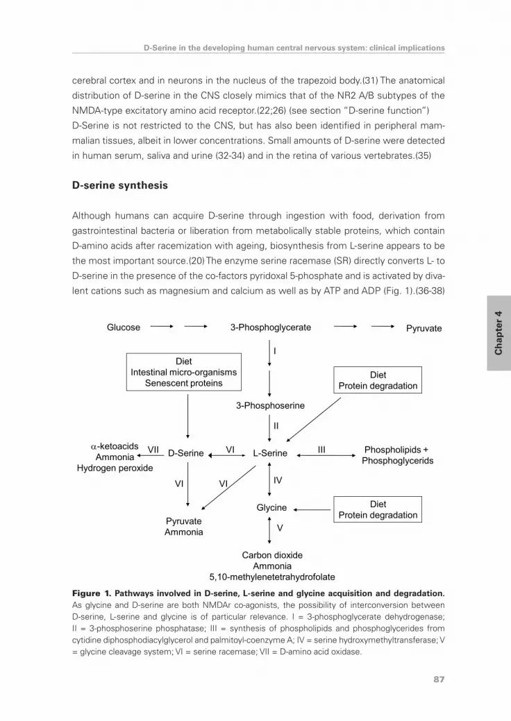
87
D-Serine in the developing human central nervous system: clinical implications
Ch
apte
r 4
cerebral cortex and in neurons in the nucleus of the trapezoid body.(31) The anatomical
distribution of D-serine in the CNS closely mimics that of the NR2 A/B subtypes of the
NMDA-type excitatory amino acid receptor.(22;26) (see section “D-serine function”)
D-Serine is not restricted to the CNS, but has also been identified in peripheral mam-
malian tissues, albeit in lower concentrations. Small amounts of D-serine were detected
in human serum, saliva and urine (32-34) and in the retina of various vertebrates.(35)
D-serine synthesis
Although humans can acquire D-serine through ingestion with food, derivation from
gastrointestinal bacteria or liberation from metabolically stable proteins, which contain
D-amino acids after racemization with ageing, biosynthesis from L-serine appears to be
the most important source.(20) The enzyme serine racemase (SR) directly converts L- to
D-serine in the presence of the co-factors pyridoxal 5-phosphate and is activated by diva-
lent cations such as magnesium and calcium as well as by ATP and ADP (Fig. 1).(36-38)
Glucose 3-Phosphoglycerate Pyruvate
3-Phosphoserine
L-SerineD-Serine
Glycine
PyruvateAmmonia
Phospholipids + Phosphoglycerids
VI
VIVI IV
II
III
I
-ketoacidsAmmonia
Hydrogen peroxide
VII
V
Carbon dioxideAmmonia
5,10-methylenetetrahydrofolate
DietIntestinal micro-organisms
Senescent proteinsDiet
Protein degradation
DietProtein degradation
Figure 1. Pathways involved in D-serine, L-serine and glycine acquisition and degradation. As glycine and D-serine are both NMDAr co-agonists, the possibility of interconversion between D-serine, L-serine and glycine is of particular relevance. I = 3-phosphoglycerate dehydrogenase; II = 3-phosphoserine phosphatase; III = synthesis of phospholipids and phosphoglycerides from cytidine diphosphodiacylglycerol and palmitoyl-coenzyme A; IV = serine hydroxymethyltransferase; V = glycine cleavage system; VI = serine racemase; VII = D-amino acid oxidase.

D-serine in human physiology
88
Inversely, SR also converts D- to L-serine, albeit with lower affinity.(38) D-Serine con-
centrations are thus highly related to L-serine concentrations and thereby also to glycine
concentrations (Fig. 1).(39)
SR is highly expressed in the brain, with lower levels in the liver and small or no detect-
able expression in other tissues. Just like the immunohistochemical localization studies
on D-serine, SR was first localized to protoplasmic astrocytes in the brain in a pattern
similar to D-serine.(37;38) Recent studies also imply the presence of SR in several
neuronal culture types, where D-serine synthesis was found to be comparable to that
observed in astrocytes.(29) In primary cultures of rat neurons, SR mRNA and protein
levels have even been found to exceed those in astrocytes.(30) These results suggest
that not only astrocytes, but also neurons represent a major source of D-serine.
Physiological synthesis of D-serine by SR was implicated by the strong spatiotemporal
correlation between D-serine and SR (40) and by the decrease in D-serine concentra-
tions in astrocytes after pharmacological inhibition of SR.(37) The cDNA encoding human
SR has been cloned and D-serine synthesis by SR has been demonstrated in living
cells after heterologous overexpression.(41) In addition to the racemase activity, SR
catalyzes the α,β-elimination of water from both L-serine and D-serine to form pyru-
vate and ammonia.(36;42) Under physiological conditions, pyruvate formation seems
to equal or excess D-serine formation. Pyruvate formed by SR may be sufficient for
the energy requirements of the astrocytes. This reaction further implies that SR is not
only involved in D-serine synthesis, but also in D-serine metabolism as a mechanism to
regulate intracellular D-serine levels and NMDAr activity.(42) In fact, SR also influences
intracellular D-serine levels by feedback regulation of its own activity. Upon glutamate
and D-serine binding, NMDArs are activated, inducing neuronal nitric oxide formation in
postsynaptic neurons, thereby stimulating S-nitrosylation of presynaptic SR. This in turn
reduces SR activity and consequently decreases D-serine concentrations.(43) Although
the factors regulating SR levels and activity are not completely unraveled, SR appears to
be degraded through the ubiquitin-proteasomal system. Ubiquitylation was decreased
by the Golgin subfamily A member 3 protein, thereby increasing SR protein half life.(44)
We are awaiting the results of the recent generation of a SR knock-out mouse strain,(45)
that might further clarify the physiological roles of SR in vivo.
D-serine metabolism
D-amino acid oxidase (DAO) and D-aspartate oxidase (DAOX) are two flavoproteins
responsible for the oxidative deamination of neutral and dicarboxylic D-amino acids
respectively and have long been known to exist in mammals.(46;47) Until recently how-
ever, they had largely been neglected, since animals and humans were not thought to
possess D-amino acids. The identification of D-amino acids in mammals finally provided

89
D-Serine in the developing human central nervous system: clinical implications
Ch
apte
r 4
a rationale for the widespread occurrence and high degree of conservation of these
enzymes in animals and humans.
DAO is a peroxisomal enzyme that metabolizes neutral D-amino acids (46) with the
concomitant reduction of the co-factor flavin adenine dinucleotide (FAD),(48) according
to the following reaction:
neutral D-amino acid + O2 + H2O – DAO → corresponding α-ketoacid + NH3 + H2O2
The affinity of DAO is highest for D-serine, D-alanine, D-proline, D-leucine and D-methi-
onine in vivo,(49;50) while glycine is not a substrate at physiological pH values, nor are
L-amino acids or dicarboxylic amino acids.(51) In humans, DAO was first identified within
the granule fraction of human neutrophilic leukocytes and proposed to be a component
of an antibacterial system by generating hydrogen peroxide.(52) Subsequent research
revealed that DAO is expressed predominantly in the kidneys, followed by the liver (with
the exception of mice livers, where DAO expression has not been detected (53)) and
the CNS. In the CNS, DAO is particularly concentrated in astrocytes of the hindbrain,
cerebellum, brainstem, medulla and spinal cord (54) with a putative preferential local-
ization to type I astrocytes (55) and to a lesser extent to type II astrocytes, whereas
D-serine is known to be localized to type II astrocytes.(25) By now, human DAO has
been cloned,(56) produced as a homogeneous protein(57) and its 3-D structure has been
characterized.(58) Human DAO differs from other known DAO’s by forming dimers in
solution.(59) There is no evidence for the existence of different DAO isoforms in the
various human (nor rat or pig) tissues.(59)
Physiological degradation of D-serine by DAO was suggested by the marked regional
and developmental variation in DAO levels in a pattern reciprocal to D-serine levels.
(25;40) Furthermore, Dao-/- mice manifest a modest but significant increase in D-serine
levels, especially in areas with low levels in wild type animals such as the cerebellum
and periphery.(49) However, in the forebrains of Dao-/- mice, D-serine concentrations
were relatively unchanged, suggesting that in these areas, other mechanisms might
regulate D-serine concentrations.(49;50) These results from Dao-/- mice contrast with
those obtained with a pharmacologic DAO inhibitor, producing increased D-serine con-
centrations in rat cortex and midbrain and influencing rat behaviour, implying that DAO
does reduce D-serine concentrations physiologically in these brain areas.(60) In analogy,
a recent study has demonstrated that a naturally occurring mouse strain (ddY/DAO-) lacks
DAO activity and has enhanced NMDAr function in vivo due to increased occupancy of
the NMDAr glycine site by elevated extracellular D-serine levels.(61) Clearly, the exact
mechanisms involved in D-serine homeostasis warrant further investigation.

D-serine in human physiology
90
D-serine function
The close correlation between the anatomical distribution of D-serine and SR with the
regional variation of the NMDA receptor (NMDAr) suggests a functional relationship.
NMDArs are broadly distributed throughout the CNS and play a major role in glutama-
tergic synaptic transmission. NMDAr activation has been implicated in physiological
processes such as CNS development (21) and the induction of long-term potentiation
and thereby long-term synaptic plasticity, which may serve as a cellular mechanism
underlying learning and memory.(62) Excitotoxicity through NMDAr overstimulation has
been implicated in a large number of acute and chronic neurodegenerative conditions,
including stroke, epilepsy, polyneuropathies, chronic pain, amyotrophic lateral sclerosis,
Parkinson’s disease, Alzheimer’s disease and Huntington’s disease.(63) NMDAr hypo-
function might play a role in the pathophysiology of schizophrenia, as NMDAr antago-
nists (64) or reduced numbers of NMDArs (65) induced positive, negative and cognitive
schizophrenia-like symptoms, which could be reversed by treatment with D-serine.(66)
NMDArs are members of a class of ionotropic receptor channels, organized as het-
eromeric assemblies composed of an NR1 subunit, combined with one or more NR2
or NR3 subunits. Although the NR1 subunit is an obligatory component of functional
NMDArs, the NR2 subunits determine the biophysical and pharmacological activity of
the receptor.(67) NMDArs require simultaneous ligand binding at two sites for activation.
Glutamate molecules bind to the NR2 subunit (68) and glycine was assumed to be
the necessary co-agonist, reacting with the “glycine site” of the NR1 subunit.(69-71)
The first suggestion for a role of D-serine as an endogenous co-agonist of this “glycine
site” of NMDArs, comes from immunohistochemical localization of glycine, D-serine
and NMDArs. D-Serine showed a specific pattern of co-localization with NMDArs in the
telencephalon and the developing cerebellum, while glycine co-localized with NMDArs in
the hindbrain, the adult cerebellum and the olfactory bulb.(26) This was reinforced by the
finding that D-serine acts as a selective and at least equally potent agonist for this “gly-
cine site” of NMDArs when compared to glycine.(72;73) Direct evidence for regulation
of NMDAr activity by endogenous D-serine comes from the decrease in NMDAr activity
in immature rat cerebellar slices, rat hippocampus slices and in primary hippocampal cell
cultures after selective removal of D-serine by adding DAO, the effect of which was fully
reversed by application of exogenous D-serine.(74) All these data favour D-serine as the
predominant endogenous ligand for most NMDArs. Nevertheless, glycine might well be
the principal ligand in some areas, such as the brainstem,(26) the spinal cord (75) and
the cerebellum.(26;74) In part, these differences in NMDAr co-agonist can be explained
by the presence of D-amino acid oxidase, which degrades D-serine and not glycine,
resulting in areas with low D-serine concentrations and high glycine concentrations.
(25;40) Similarly, areas with high D-serine concentrations are enriched with the D-serine

91
D-Serine in the developing human central nervous system: clinical implications
Ch
apte
r 4
synthesizing enzyme SR,(40) just like the areas with high glycine concentrations are
with the glycine synthesizing enzyme serine hydroxymethyltransferase.(76)
Snyder and Kim (77) proposed a model of the interaction between astrocytes and
neurons in NMDAr activation. They suggested that, upon release from the presynaptic
neuron, glutamate does not only bind to the NMDAr, but also to non-NMDArs (2-amino-
3-hydroxy-5-methyl-4-isoxazolepropionate (AMPA) receptors) on nearby astrocytes. In
these astrocytes, D-serine is then synthesized by SR and released into the synaptic
cleft, where it can bind to the NMDAr complex together with glutamate, leading to
NMDAr activation.
As an endogenous co-agonist of the NMDAr, D-serine concentrations are important
for all the physiological functions of NMDArs. Similarly, D-serine concentrations might
be altered in disorders associated with pathologic NMDAr activity and as such might
contribute to their pathogenesis.
CenTraL nerVouS SySTem DeVeLopmenT
The NMDAr in CNS development
There is substantial evidence implicating an important role for NMDArs in CNS develop-
ment. Again, the first clue was provided by immunohistochemical localization studies,
demonstrating a greater expression and number of NMDArs in the developing brain,
relative to the adult brain. In the human brainstem, NMDArs increase to a peak during
the infant period to decrease thereafter.(78) A similar pattern was observed the human
frontal,(79) temporal (80) and prefrontal cortex.(81) The subunit composition of NMDArs
also varies with age. The NR1subunit is widely expressed throughout the CNS at all
ages, but the expression profiles of the NR2 subunits are regionally and developmen-
tally regulated.(82) Currently, 4 genetically different NR2 subunits have been identified
(NR2A-NR2D).(82;83) In the neonatal rat brain, NR2B and NR2D subunits predominate,
but as development proceeds, they are supplemented with, or replaced by the NR2A and
NR2C subunits in some brain regions.(83;84) This developmental shift of NR2 subunits,
which determine the biophysical and pharmacological properties of NMDArs, might set
the optimal conditions for proper CNS development.
In analogy, receptor binding studies have revealed higher NMDAr activity in the develop-
ing brain than in the adult brain.(85-87) When comparing the developing brain to the
adult brain, differences in NMDAr features include a lower sensitivity to NMDAr blockage
by the antagonist magnesium, a higher sensitivity to the NMDAr agonist glycine, differ-
ential modulation by polyamines, increased calcium influx through the receptor channel,
longer duration of the excitatory postsynaptic potentials after receptor stimulation and

D-serine in human physiology
92
enhanced ability to induce markers of synaptic plasticity such as long-term potentiation.
(88-91)
More direct evidence for a role of the NMDAr in specific functions during CNS develop-
ment is provided by pharmacological studies. Blocking NMDArs with daily injections of
an NMDAr antagonist for 2 days in 2-days-old rats significantly increased cell proliferation
in the dentate gyrus,(92;93) while activation of NMDArs decreased proliferation of the
granule cells of the dentate gyrus.(94) On the other hand, blockage of the NMDAr during
late fetal and early neonatal rat life was associated with widespread apoptotic neurode-
generation (95;96). Other studies in which neonatal rats were given NMDAr antagonists
during the first 2 postnatal weeks demonstrate that NMDAr blockade is associated with
abnormal axonal arborization and apoptotic neurodegeneration.(97) Taken together, all
these studies imply an essential role for adequate NMDAr activation in CNS shaping by
influencing neuronal proliferation and apoptosis.
During CNS development, neurons in the brain migrate to their mature positions along
the processes of radial glia. The NMDAr has also been implicated in this process. Al-
ready before undergoing synaptogenesis, migrating cortical neurons possess functional
NMDArs (98) and migrating granular cells contain more NMDArs than premigratory
neurons,(99) implying optimal functional NMDAr content during neuronal migration.
Pharmacological studies demonstrate that blocking NMDArs in cerebellar slices or
increasing medium magnesium concentrations slow the rate of granule cell migration,
which could be reversed by magnesium removal or addition of the NMDAr agonists
glutamate and glycine.(96) Similarly, in cortical slices from the rat and mouse, NMDAr
activation was found to stimulate neuronal migration.(100;101)
Synaptic plasticity or synaptogenesis is crucial for shaping the wiring pattern of the brain
and is responsible for the mechanism of learning and memory.(102-104) Injecting NMDA
into the occipital cortex of 8-day-old rats increased the total number of synapses,(102)
while blocking NMDArs decreased the synaptic density (103) and induced deficits in
learning and memory.(105) The importance of NMDAr activation for synaptic plasticity is
further evidenced by genetically modified mice, exhibiting complete loss of the glycine
and D-serine binding NR1 subunit of the NMDAr and displaying impaired long-term
potentiation and spatial memory.(106;107)
D-serine in CNS development
A specific function for D-serine in brain development was suggested by immunohisto-
chemical localization studies,(19;26;108;109) showing elevated D-serine levels in specific
rodent and human brain areas during distinct developmental periods. On the day of birth,
D-serine was present in substantial concentrations throughout rat brain.(108) D-Serine
concentration increased in the cerebrum by the third postnatal week, remaining rather

93
D-Serine in the developing human central nervous system: clinical implications
Ch
apte
r 4
constant thereafter. Similarly, high D-serine concentrations were determined in the hu-
man frontal cortex at gestational week 14 and remained rather constant throughout
embryonic and early postnatal life.(19) D-Serine concentrations decreased later in life to
half of these levels in adolescent and aged brains.(19)
In rat cerebellum, D-serine increased in the first postnatal week to its highest levels
in the second week and thereafter declined dramatically to only trace levels, coincid-
ing well with the drastic increase in DAO activity in the cerebellum during that period.
(26;108;109) Furthermore, cerebellar D-serine was localized immunohistochemically to
astrocytes in the granular layer and Bergmann glia that regulate granule cell migration.
(26) This localization and the transient D-serine increase in the developing cerebellum,
which concurs with a transient expression of NMDArs, suggest a role in cerebellar
development. This is supported by the disrupted cerebellar ontogeny observed upon
blockage of the glycine binding site of NMDArs during rodent development.(96;110)
In addition, selective degradation of D-serine by DAO and selective inhibition of SR in
8-day old mouse cerebellar slices significantly reduced granule cell migration, whereas
D-serine activated this process.(111) Granule cell migration appears to depend on an
increase in intracellular calcium through NMDAr activation, as treatment with a SR
inhibitor markedly diminished intracellular calcium concentrations.(111) Taken together,
these results imply a role for D-serine and subsequent NMDAr activation in cerebellar
granule cell migration, which might be seen as a model for neuronal migration in the
entire CNS.
Chromatographic-mass spectrometric analysis of cerebrospinal fluid D-serine concentra-
tions in healthy children, as a global marker of brain D-serine content, revealed high D-
serine concentrations immediately after birth, both absolutely and relative to glycine and
L-serine, declining to 15% of the initial values at infancy.(21) D-Serine concentrations
were almost undetectable in untreated patients with 3-phosphoglycerate dehydroge-
nase deficiency, a rare inherited disorder of L-serine synthesis (Figure 1), characterized
by severe CNS abnormalities, including microcephaly, intractable seizures and profound
mental retardation. In one patient treated prenatally, D-serine concentrations were
nearly normal at birth (21) and the clinical phenotype was normal.(112) As an NMDAr
agonist, D-serine deficiency is much more likely to cause the severe CNS malformations
than L-serine deficiency. The SR knock-out mouse model will help to elucidate this mat-
ter. The importance for normal CNS development of L-serine synthesis and conceivably
even more so of subsequent D-serine synthesis, is further evidenced by the striking
CNS abnormalities found in the first two weeks after conception in a 3-phosphoglycerate
dehydrogenase deficient mouse model,(113) culminating in hypoplasia of the telen-
cephalon, diencephalon and mesencephalon, with particularly the olfactory bulb, the
ganglionic eminence and cerebellum appearing as indistinct structures. This establishes
that the phosphoglycerate dehydrogenase dependent pathway of L-serine biosynthesis

D-serine in human physiology
94
(Figure 1), possibly through D-serine synthesis and NMDAr activation, is essential for
embryonic CNS development, especially for brain morphogenesis.
Using the retina as a model for CNS development, the glutamate transporter excitatory
amino acid transporter-1, which is the main regulator of retinal extracellular glutamate
concentrations, and D-serine were both found to be present in human retinae, from 10
and 12 weeks of gestation on, respectively, which was followed by the appearance of
synaptophysin, a synaptic vesicle marker.(114) Furthermore, D-serine and the glutamate
transporter co-localized in radial glia-like cells that might represent Muller cells, as
evidenced by the co-localization with cellular retinaldehyde-binding protein. This implies
that the endogenous determinants of NMDAr activity glutamate and D-serine are pres-
ent before synaptic vesicle proteins are evident and may thus play a role in shaping
synaptogenesis in the developing human retina.(114) These results are consolidated by
studies using the vestibular nuclei and their main connections as a model for D-serine
function in synaptic plasticity. During the first three postnatal weeks, an intense period
of plasticity, synaptogenesis and neuronal maturation, rat vestibular nuclei contained
high D-serine concentrations in glial cells and processes, corresponding with high SR
and low DAO expression. In the mature vestibular nuclei, D-serine mainly localized to
neuronal cell bodies and dendrites and concentrations were low, corresponding with in-
creasing DAO expression.(115) These strong decreases of D-serine concentrations and
the glial-to-neuronal switch suggest that D-serine may have a role in neuronal plasticity,
maturation and synaptogenesis during early postnatal CNS development, and possibly a
distinct functional role at later developmental stages.(115)
Behavioural results of animals lacking D-serine during CNS development, such as SR
knock-out mice, are currently unavailable. However, we might enhance our understand-
ing through the behavioural results from mutant mice displaying increased NMDAr
activation through elevated D-serine or glycine concentrations. One such mouse model
is a naturally occurring mouse strain (ddY/DAO-) lacking DAO activity and thereby display-
ing elevated extracellular D-serine concentrations and enhanced NMDAr function.(61)
These mice showed enhanced hippocampal long-term potentiation,(61;116) improved
performance in a test of spatial learning (116) and better motor coordination as evi-
denced by fewer foot slips when assessed on beam walking.(61) Alternative methods
to increase activation of the glycine site of NMDAr involve blockage of the high affinity
D-serine transporter Asc-1 and the glycine transporter GlyT1. GlyT1-/- and Asc-1-/- knock-
out mice died soon after birth, GlyT1-/- with severe respiratory and motosensory deficits
(117) and Asc-1-/- from tremors and seizures.(118) The GlyT-1 heterozygote (GlyT1+/-) was
viable and displayed enhanced NMDAr function in CA1 pyramidal cells and improved
performance in a hippocampal-dependant spatial memory task.(119) Currently, there are
no behavioral data of heterozygous Asc-1 mice. Taken together, these studies of mutant
mice show that enhanced NMDAr activity through increased D-serine or glycine concen-

95
D-Serine in the developing human central nervous system: clinical implications
Ch
apte
r 4
trations influences CNS function and potentially CNS development, again underscoring
the importance of proper NMDAr agonist concentrations for normal brain physiology.
CLInICaL ImpLICaTIonS
As described in this chapter, it has now been generally accepted that NMDArs play
important roles in CNS development, with functions in neuronal proliferation, migration,
maturation and synaptogenesis. Consequently, adequate NMDAr activation is essential
for normal brain architecture, development and function, including long-term poten-
tiation, the process underlying the mechanism of learning and memory. For functional
NMDAr activation, proper concentrations of its endogenous agonists are required. As
such, it is not surprising that an increasing body of evidence implies that adequate D-
serine concentrations are necessary for the same processes in CNS development as
the NMDAr.
Up to now, only one human disease, namely 3-phosphoglycerate dehydrogenase de-
ficiency, has been associated with D-serine deficiency in cerebrospinal fluid.(21) The
severe CNS abnormalities associated with this disorder could be prevented by prenatal
maternal and early postnatal treatment with oral L-serine,(112) the endogenous pre-
cursor of D-serine (Figure 1). Patients starting oral L-serine treatment later in life only
showed improvement of the epilepsy, with marginal effects on head circumference and
psychomotor development.(120) The window of opportunity to treat the severe disease
phenotype coincides with the period of extensive brain development, maximal NMDAr
activity and peaking D-serine concentrations in healthy individuals, thereby strongly sug-
gesting normalization of D-serine concentrations and consequent restoration of NMDAr
activity as the mechanism of action.
The pathophysiology of many disorders associated with CNS malformations and/or dis-
turbed CNS function are still unknown. Analysis of D-serine concentrations in cerebro-
spinal fluid might increase our insight in the pathogenetic mechanisms. This would not
only improve our understanding of these disorders and of normal brain physiology, but
might also provide novel therapeutic strategies. In disorders associated with hypofunc-
tion of the NMDAr, such as schizophrenia, strategies to increase NMDAr activity through
administration of D-serine, glycine or D-cycloserine have shown mixed but encouraging
results, with beneficial effects on the positive, negative and cognitive domains of schizo-
phrenia.(64;121;122) There are issues with the administration of D-serine and glycine, in
particular with the large quantities of compound that need to be dosed to increase levels
in the brain. Alternative approaches include inhibition of transporters of these amino
acids to block cellular uptake, for example by inhibiting the GlyT-1 and Asc-1 transport-
ers (123) or prevention of D-serine degradation by inhibition of DAO.(124) In analogy,

D-serine in human physiology
96
modulation of NMDAr function through the glycine-binding site might also represent
a novel approach for the numerous disorders associated with excitotoxicity through
NMDAr overactivity. For several reasons, clinical trials with complete NMDAr blockers
have been largely unsuccessful, usually because of toxicity associated with influences
on either blood pressure or psychotomimetic actions.(125) Drugs that block the glycine
site of NMDArs, SR inhibitors or enzymes degrading D-serine (DAO) might be beneficial
in these disorders associated with excitotoxicity and cause fewer side-effects.
ConCLuSIon
Although it is only 15 years ago that D-serine was first identified in the mammalian
CNS, much progress has been made in unraveling its function. As an endogenous co-
agonist of the NMDAr, D-serine can be expected to play a role in all the physiological and
pathological conditions in which NMDArs are involved, which in fact, is consolidated in
a growing body of evidence. In this chapter, we focused on the role of D-serine in the
developing CNS. Similar to the NMDAr, several studies imply roles for D-serine in neu-
ronal proliferation, migration, maturation and synaptic plasticity. This unexpected finding
of D-serine as a crucial element in human brain physiology and pathology does not only
increase our insight in human CNS function, but might also yield novel diagnostic and
therapeutic strategies for disorders of CNS dysfunction.

97
D-Serine in the developing human central nervous system: clinical implications
Ch
apte
r 4
reFerenCeS
1. Lamzin VS, Dauter Z, Wilson KS. How nature deals with stereoisomers. Curr Opin Struct Biol
1995 December;5(6): 830-6.
2. Silverman WA. The schizophrenic career of a “monster drug”. Pediatrics 2002 August;110(2
Pt 1): 404-6.
3. Bren L. Frances Oldham Kelsey. FDA medical reviewer leaves her mark on history. FDA
Consum 2001 March;35(2): 24-9.
4. Blaschke G, Kraft HP, Fickentscher K, Kohler F. [Chromatographic separation of racemic tha-
lidomide and teratogenic activity of its enantiomers (author’s transl)]. Arzneimittelforschung
1979; 29(10): 1640-2.
5. Corrigan JJ. D-amino acids in animals. Science 1969 April 11; 164(876): 142-9.
6. Auclair JL, Patton RL. On the occurrence of d-Alanine in the haemolymph of the milkweed
bug, oncopeltus fasciatus. Rev Can Biol 1950 April;9(1): 3-8.
7. Beatty I, Magrath D, Ennor A. Occurrence of D-serine in lombricine. Nature 1959 February
28; 183(4661): 591.
8. Corrigan JJ, Srinivasan NG. The occurrence of certain D-amino acids in insects. Biochemistry
1966 April;5(4): 1185-90.
9. Ennor AH, Rosenberg H, Rossiter RJ, Beatty IM, Gaffney T. The isolation and characterization
of D-serine ethanolamine phosphodiester from earthworms. Biochem J 1960 April;75: 179-
82.
10. Kreil G. Peptides containing a D-amino acid from frogs and molluscs. J Biol Chem 1994 April
15; 269(15): 10967-70.
11. Okuma E, Watanabe K, Abe H. Distribution of free D-amino acids in bivalve mollusks and
the effects of physiological conditions on the levels of D- and L-alanine in the tissues of hard
clam, Meretrix lusoria. Fish Sci 1998; 64: 606-11.
12. Preston RL. Occurrence of D-amino acids in higher organisms: a survey of the distribution
of D-amino acids in marine vertebrates. Comp Biochem Physiol 1987; 87B:55-62.
13. Seliger HH, McElroy WD, White EH, Field GF. Stereo-specificity and firefly bioluminescence,
a comparison of natural and synthetic luciferins. Proc Natl Acad Sci U S A 1961 August;47:
1129-34.
14. Srinivasan NG, Corrigan JJ, Meister A. D-Serine in the blood of the silkworm Bombyx mori
and other lepidoptera. J Biol Chem 1962 December;237: 3844-5.
15. Yasuda-Kamatani Y, Nakamura M, Minakata H, Nomoto K, Sakiyama F. A novel cDNA se-
quence encoding the precursor of the D-amino acid-containing neuropeptide fulicin and
multiple alpha-amidated neuropeptides from Achatina fulica. J Neurochem 1995 May;64(5):
2248-55.
16. Helfman PM, Bada JL, Shou MY. Considerations on the role of aspartic acid racemization in
the aging process. Gerontology 1977; 23(6): 419-25.
17. Dunlop DS, Neidle A, McHale D, Dunlop DM, Lajtha A. The presence of free D-aspartic acid
in rodents and man. Biochem Biophys Res Commun 1986 November 26; 141(1): 27-32.
18. Hashimoto A, Nishikawa T, Hayashi T, Fujii N, Harada K, Oka T et al. The presence of free
D-serine in rat brain. FEBS Lett 1992 January 13; 296(1): 33-6.
19. Hashimoto A, Kumashiro S, Nishikawa T, Oka T, Takahashi K, Mito T et al. Embryonic develop-
D-serine in human physiology
98
ment and postnatal changes in free D-aspartate and D-serine in the human prefrontal cortex.
J Neurochem 1993 July;61(1): 348-51.
20. Fuchs SA, Klomp LW, de Koning TJ. D-amino acids in the central nervous system in health
and disease. Mol Genet Metab 2005 July;85(3): 168-80.
21. Fuchs SA, Dorland L, de Sain-van der Velden MG, Hendriks M, Klomp LW, Berger R et al.
D-serine in the developing human central nervous system. Ann Neurol 2006 October;60(4):
476-80.
22. Hashimoto A, Nishikawa T, Oka T, Takahashi K. Endogenous D-serine in rat brain: N-methyl-D-
aspartate receptor-related distribution and aging. J Neurochem 1993 February;60(2): 783-6.
23. Nagata Y, Horiike K, Maeda T. Distribution of free D-serine in vertebrate brains. Brain Res
1994 January 21; 634(2): 291-5.
24. Hashimoto A, Oka T, Nishikawa T. Extracellular concentration of endogenous free D-serine in
the rat brain as revealed by in vivo microdialysis. Neuroscience 1995 June;66(3): 635-43.
25. Schell MJ, Molliver ME, Snyder SH. D-serine, an endogenous synaptic modulator: localiza-
tion to astrocytes and glutamate-stimulated release. Proc Natl Acad Sci U S A 1995 April 25;
92(9): 3948-52.
26. Schell MJ, Brady RO, Jr., Molliver ME, Snyder SH. D-serine as a neuromodulator: regional
and developmental localizations in rat brain glia resemble NMDA receptors. J Neurosci 1997
March 1; 17(5): 1604-15.
27. Wako K, Ma N, Shiroyama T, Semba R. Glial uptake of intracerebroventricularly injected D-serine
in the rat brain: an immunocytochemical study. Neurosci Lett 1995 February 13; 185(3): 171-4.
28. Williams SM, Diaz CM, Macnab LT, Sullivan RK, Pow DV. Immunocytochemical analysis of D-
serine distribution in the mammalian brain reveals novel anatomical compartmentalizations
in glia and neurons. Glia 2006 March;53(4): 401-11.
29. Kartvelishvily E, Shleper M, Balan L, Dumin E, Wolosker H. Neuron-derived D-serine release
provides a novel means to activate N-methyl-D-aspartate receptors. J Biol Chem 2006 May
19; 281(20): 14151-62.
30. Yoshikawa M, Nakajima K, Takayasu N, Noda S, Sato Y, Kawaguchi M et al. Expression of the
mRNA and protein of serine racemase in primary cultures of rat neurons. Eur J Pharmacol
2006 October 24; 548(1-3): 74-6.
31. Yasuda E, Ma N, Semba R. Immunohistochemical evidences for localization and production
of D-serine in some neurons in the rat brain. Neurosci Lett 2001 February 16; 299(1-2): 162-4.
32. Bruckner H, Haasmann S, Friedrich A. Quantification of D-amino acids in human urine using
GC-MS and HPLC. Amino Acids 1994; 6(205): 211.
33. Nagata Y, Masui R, Akino T. The presence of free D-serine, D-alanine and D-proline in human
plasma. Experientia 1992 October 15; 48(10): 986-8.
34. Rotgans J, Wodarz R, Schoknecht W, Drysch K. The determination of amino-acid enantio-
mers in human saliva with Chirasil-Val. Arch Oral Biol 1983; 28(12): 1121-4.
35. Stevens ER, Esguerra M, Kim PM, Newman EA, Snyder SH, Zahs KR et al. D-serine and
serine racemase are present in the vertebrate retina and contribute to the physiological
activation of NMDA receptors. Proc Natl Acad Sci U S A 2003 May 27; 100(11): 6789-94.
36. De Miranda J, Panizzutti R, Foltyn VN, Wolosker H. Cofactors of serine racemase that physi-
ologically stimulate the synthesis of the N-methyl-D-aspartate (NMDA) receptor coagonist
D-serine. Proc Natl Acad Sci U S A 2002 October 29; 99(22): 14542-7.

99
D-Serine in the developing human central nervous system: clinical implications
Ch
apte
r 4
37. Wolosker H, Blackshaw S, Snyder SH. Serine racemase: a glial enzyme synthesizing D-
serine to regulate glutamate-N-methyl-D-aspartate neurotransmission. Proc Natl Acad Sci U
S A 1999 November 9; 96(23): 13409-14.
38. Wolosker H, Sheth KN, Takahashi M, Mothet JP, Brady RO, Jr., Ferris CD et al. Purification
of serine racemase: biosynthesis of the neuromodulator D-serine. Proc Natl Acad Sci U S A
1999 January 19; 96(2): 721-5.
39. de Koning TJ, Klomp LW. Serine-deficiency syndromes. Curr Opin Neurol 2004 April;17(2):
197-204.
40. Wang LZ, Zhu XZ. Spatiotemporal relationships among D-serine, serine racemase, and
D-amino acid oxidase during mouse postnatal development. Acta Pharmacol Sin 2003
October;24(10): 965-74.
41. De Miranda J, Santoro A, Engelender S, Wolosker H. Human serine racemase: moleular
cloning, genomic organization and functional analysis. Gene 2000 October 3; 256(1-2): 183-8.
42. Foltyn VN, Bendikov I, De Miranda J, Panizzutti R, Dumin E, Shleper M et al. Serine race-
mase modulates intracellular D-serine levels through an alpha,beta-elimination activity. J
Biol Chem 2005; 280 (3): 1754-63.
43. Mustafa AK, Kumar M, Selvakumar B, Ho GP, Ehmsen JT, Barrow RK et al. Nitric oxide
S-nitrosylates serine racemase, mediating feedback inhibition of D-serine formation. Proc
Natl Acad Sci U S A 2007 February 20; 104(8): 2950-5.
44. Dumin E, Bendikov I, Foltyn VN, Misumi Y, Ikehara Y, Kartvelishvily E et al. Modulation of
D-serine levels via ubiquitin-dependent proteasomal degradation of serine racemase. J Biol
Chem 2006 July 21; 281(29): 20291-302.
45. Ying-Luan Z, Zhao YL, Mori H. [Roles of D-serine in the mammalian brain]. Brain Nerve 2007
July;59(7): 725-30.
46. Krebs HA. Metabolism of amino acids. III. Determination of amino acids. Biochem J 1935;
29: 1620-44.
47. Still J.L, Buell M.V, Knox WE, Green DE. Studies on the cyclphorase system VII. D-Aspartate
oxidase. J Biol Chem 1949; 179: 831-7.
48. Pollegioni L, Harris CM, Molla G, Pilone MS, Ghisla S. Identification and role of ionizing
functional groups at the active center of Rhodotorula gracilis D-amino acid oxidase. FEBS
Lett 2001 November 2; 507(3): 323-6.
49. Hashimoto A, Nishikawa T, Konno R, Niwa A, Yasumura Y, Oka T et al. Free D-serine, D-
aspartate and D-alanine in central nervous system and serum in mutant mice lacking D-
amino acid oxidase. Neurosci Lett 1993 April 2; 152(1-2): 33-6.
50. Morikawa A, Hamase K, Inoue T, Konno R, Niwa A, Zaitsu K. Determination of free D-aspartic
acid, D-serine and D-alanine in the brain of mutant mice lacking D-amino acid oxidase activ-
ity. J Chromatogr B Biomed Sci Appl 2001 June 5; 757(1): 119-25.
51. D’Aniello A, Vetere A, Petrucelli L. Further study on the specificity of D-amino acid oxidase
and D-aspartate oxidase and time course for complete oxidation of D-amino acids. Comp
Biochem Physiol B 1993 July;105(3-4): 731-4.
52. Cline MJ, Lehrer RI. D-amino acid oxidase in leukocytes: a possible D-amino-acid-linked
antimicrobial system. Proc Natl Acad Sci U S A 1969 March;62(3): 756-63.
53. Tada M, Fukui K, Momoi K, Miyake Y. Cloning and expression of a cDNA encoding mouse
kidney D-amino acid oxidase. Gene 1990 June 15; 90(2): 293-7.

D-serine in human physiology
100
54. Nagata Y, Konno R, Yasumura Y, Akino T. Involvement of D-amino acid oxidase in elimination
of free D-amino acids in mice. Biochem J 1989 January 1; 257(1): 291-2.
55. Urai Y, Jinnouchi O, Kwak KT, Suzue A, Nagahiro S, Fukui K. Gene expression of D-amino acid
oxidase in cultured rat astrocytes: regional and cell type specific expression. Neurosci Lett
2002 May 17; 324(2): 101-4.
56. Momoi K, Fukui K, Watanabe F, Miyake Y. Molecular cloning and sequence analysis of cDNA
encoding human kidney D-amino acid oxidase. FEBS Lett 1988 September 26; 238(1): 180-4.
57. Molla G, Sacchi S, Bernasconi M, Pilone MS, Fukui K, Polegioni L. Characterization of human
D-amino acid oxidase. FEBS Lett 2006 April 17; 580(9): 2358-64.
58. Kawazoe T, Tsuge H, Pilone MS, Fukui K. Crystal structure of human D-amino acid oxidase:
context-dependent variability of the backbone conformation of the VAAGL hydrophobic
stretch located at the si-face of the flavin ring. Protein Sci 2006 December;15(12): 2708-17.
59. Pollegioni L, Piubelli L, Sacchi S, Pilone MS, Molla G. Physiological functions of D-amino acid
oxidases: from yeast to humans. Cell Mol Life Sci 2007 June;64(11): 1373-94.
60. Adage T, Trillat AC, Quattropani A, Perrin D, Cavarec L, Shaw J et al. In vitro and in vivo phar-
macological profile of AS057278, a selective d-amino acid oxidase inhibitor with potential
anti-psychotic properties. : Eur Neuropsychopharmacol 2008 Mar; 18(3): 200-14.
61. Almond SL, Fradley RL, Armstrong EJ, Heavens RB, Rutter AR, Newman RJ et al. Behavioral
and biochemical characterization of a mutant mouse strain lacking D-amino acid oxidase
activity and its implications for schizophrenia. Mol Cell Neurosci 2006 August;32(4): 324-34.
62. Yang Y, Ge W, Chen Y, Zhang Z, Shen W, Wu C et al. Contribution of astrocytes to hippo-
campal long-term potentiation through release of D-serine. Proc Natl Acad Sci U S A 2003
December 9; 100(25): 15194-9.
63. Danysz W, Parsons AC. Glycine and N-methyl-D-aspartate receptors: physiological signifi-
cance and possible therapeutic applications. Pharmacol Rev 1998 December;50(4): 597-664.
64. Goff DC, Coyle JT. The emerging role of glutamate in the pathophysiology and treatment of
schizophrenia. Am J Psychiatry 2001 September;158(9): 1367-77.
65. Mohn AR, Gainetdinov RR, Caron MG, Koller BH. Mice with reduced NMDA receptor ex-
pression display behaviors related to schizophrenia. Cell 1999 August 20; 98(4): 427-36.
66. Contreras PC. D-serine antagonized phencyclidine- and MK-801-induced stereotyped behav-
ior and ataxia. Neuropharmacology 1990 March;29(3): 291-3.
67. Takahashi T, Feldmeyer D, Suzuki N, Onodera K, Cull-Candy SG, Sakimura K et al. Functional cor-
relation of NMDA receptor epsilon subunits expression with the properties of single-channel
and synaptic currents in the developing cerebellum. J Neurosci 1996 July 15; 16(14): 4376-82.
68. Dingledine R, Borges K, Bowie D, Traynelis SF. The glutamate receptor ion channels. Pharma-
col Rev 1999 March;51(1): 7-61.
69. Johnson JW, Ascher P. Glycine potentiates the NMDA response in cultured mouse brain
neurons. Nature 1987 February 5; 325(6104): 529-31.
70. Kleckner NW, Dingledine R. Requirement for glycine in activation of NMDA-receptors ex-
pressed in Xenopus oocytes. Science 1988 August 12; 241(4867): 835-7.
71. Thomson AM. Glycine is a coagonist at the NMDA receptor/channel complex. Prog Neuro-
biol 1990; 35(1): 53-74.
72. Danysz W, Fadda E, Wroblewski JT, Costa E. [3H]D-serine labels strychnine-insensitive
glycine recognition sites of rat central nervous system. Life Sci 1990; 46(3): 155-64.

101
D-Serine in the developing human central nervous system: clinical implications
Ch
apte
r 4
73. Matsui T, Sekiguchi M, Hashimoto A, Tomita U, Nishikawa T, Wada K. Functional comparison
of D-serine and glycine in rodents: the effect on cloned NMDA receptors and the extracel-
lular concentration. J Neurochem 1995 July;65(1): 454-8.
74. Mothet JP, Parent AT, Wolosker H, Brady RO, Jr., Linden DJ, Ferris CD et al. D-serine is an
endogenous ligand for the glycine site of the N-methyl-D-aspartate receptor. Proc Natl Acad
Sci U S A 2000 April 25; 97(9): 4926-31.
75. Ahmadi S, Muth-Selbach U, Lauterbach A, Lipfert P, Neuhuber WL, Zeilhofer HU. Facilitation
of spinal NMDA receptor currents by spillover of synaptically released glycine. Science 2003
June 27; 300(5628): 2094-7.
76. Daly EC, Aprison MH. Distribution of serine hydroxymethyltransferase and glycine trans-
aminase in several areas of the central nervous system of the rat. J Neurochem 1974
June;22(6): 877-85.
77. Snyder SH, Kim PM. D-amino acids as putative neurotransmitters: focus on D-serine. Neu-
rochem Res 2000 May;25(5): 553-60.
78. Panigrahy A, Rosenberg PA, Assmann S, Foley EC, Kinney HC. Differential expression of glu-
tamate receptor subtypes in human brainstem sites involved in perinatal hypoxia-ischemia.
J Comp Neurol 2000 November 13; 427(2): 196-208.
79. Piggott MA, Perry EK, Perry RH, Court JA. [3H]MK-801 binding to the NMDA receptor com-
plex, and its modulation in human frontal cortex during development and aging. Brain Res
1992 August 21; 588(2): 277-86.
80. Slater P, McConnell SE, D’Souza SW, Barson AJ. Postnatal changes in N-methyl-D-aspartate
receptor binding and stimulation by glutamate and glycine of [3H]-MK-801 binding in human
temporal cortex. Br J Pharmacol 1993 April;108(4): 1143-9.
81. Chahal H, D’Souza SW, Barson AJ, Slater P. Modulation by magnesium of N-methyl-D-
aspartate receptors in developing human brain. Arch Dis Child Fetal Neonatal Ed 1998
March;78(2):F116-F120.
82. Ishii T, Moriyoshi K, Sugihara H, Sakurada K, Kadotani H, Yokoi M et al. Molecular charac-
terization of the family of the N-methyl-D-aspartate receptor subunits. J Biol Chem 1993
February 5; 268(4): 2836-43.
83. Monyer H, Burnashev N, Laurie DJ, Sakmann B, Seeburg PH. Developmental and regional
expression in the rat brain and functional properties of four NMDA receptors. Neuron 1994
March;12(3): 529-40.
84. Akazawa C, Shigemoto R, Bessho Y, Nakanishi S, Mizuno N. Differential expression of five
N-methyl-D-aspartate receptor subunit mRNAs in the cerebellum of developing and adult
rats. J Comp Neurol 1994 September 1; 347(1): 150-60.
85. Crair MC, Malenka RC. A critical period for long-term potentiation at thalamocortical syn-
apses. Nature 1995 May 25; 375(6529): 325-8.
86. Feldman DE, Nicoll RA, Malenka RC, Isaac JT. Long-term depression at thalamocortical
synapses in developing rat somatosensory cortex. Neuron 1998 August;21(2): 347-57.
87. Ramoa AS, McCormick DA. Enhanced activation of NMDA receptor responses at the im-
mature retinogeniculate synapse. J Neurosci 1994 April;14(4): 2098-105.
88. Aamodt SM, Constantine-Paton M. The role of neural activity in synaptic development and
its implications for adult brain function. Adv Neurol 1999; 79: 133-44.
89. McBain CJ, DiChiara TJ, Kauer JA. Activation of metabotropic glutamate receptors differen-

D-serine in human physiology
102
tially affects two classes of hippocampal interneurons and potentiates excitatory synaptic
transmission. J Neurosci 1994 July;14(7): 4433-45.
90. Scheetz AJ, Constantine-Paton M. Modulation of NMDA receptor function: implications for
vertebrate neural development. FASEB J 1994 July;8(10): 745-52.
91. Waters KA, Machaalani R. NMDA receptors in the developing brain and effects of noxious
insults. Neurosignals 2004 July;13(4): 162-74.
92. Gould E, Cameron HA, McEwen BS. Blockade of NMDA receptors increases cell death and
birth in the developing rat dentate gyrus. J Comp Neurol 1994 February 22; 340(4): 551-65.
93. Gould E, Cameron HA. Early NMDA receptor blockade impairs defensive behavior and
increases cell proliferation in the dentate gyrus of developing rats. Behav Neurosci 1997
February;111(1): 49-56.
94. Cameron HA, McEwen BS, Gould E. Regulation of adult neurogenesis by excitatory input
and NMDA receptor activation in the dentate gyrus. J Neurosci 1995 June;15(6): 4687-92.
95. Ikonomidou C, Bosch F, Miksa M, Bittigau P, Vockler J, Dikranian K et al. Blockade of NMDA
receptors and apoptotic neurodegeneration in the developing brain. Science 1999 January
1; 283(5398): 70-4.
96. Komuro H, Rakic P. Modulation of neuronal migration by NMDA receptors. Science 1993
April 2; 260(5104): 95-7.
97. Haberny KA, Paule MG, Scallet AC, Sistare FD, Lester DS, Hanig JP et al. Ontogeny of the
N-methyl-D-aspartate (NMDA) receptor system and susceptibility to neurotoxicity. Toxicol
Sci 2002 July;68(1): 9-17.
98. LoTurco JJ, Blanton MG, Kriegstein AR. Initial expression and endogenous activation of
NMDA channels in early neocortical development. J Neurosci 1991 March;11(3): 792-9.
99. Rossi DJ, Slater NT. The developmental onset of NMDA receptor-channel activity during
neuronal migration. Neuropharmacology 1993 November;32(11): 1239-48.
100. Behar TN, Scott CA, Greene CL, Wen X, Smith SV, Maric D et al. Glutamate acting at NMDA
receptors stimulates embryonic cortical neuronal migration. J Neurosci 1999 June 1; 19(11):
4449-61.
101. Hirai K, Yoshioka H, Kihara M, Hasegawa K, Sakamoto T, Sawada T et al. Inhibiting neuronal
migration by blocking NMDA receptors in the embryonic rat cerebral cortex: a tissue culture
study. Brain Res Dev Brain Res 1999 April 12; 114(1): 63-7.
102. Brooks WJ, Petit TL, Leboutillier JC, Lo R. Rapid alteration of synaptic number and postsyn-
aptic thickening length by NMDA: an electron microscopic study in the occipital cortex of
postnatal rats. Synapse 1991 May;8(1): 41-8.
103. Brooks WJ, Petit TL, Leboutillier JC. Effect of chronic administration of NMDA antagonists
on synaptic development. Synapse 1997 June;26(2): 104-13.
104. Contestabile A. Roles of NMDA receptor activity and nitric oxide production in brain develop-
ment. Brain Res Brain Res Rev 2000 April;32(2-3): 476-509.
105. Brooks WJ, Weeks AC, Leboutillier JC, Petit TL. Altered NMDA sensitivity and learning
following chronic developmental NMDA antagonism. Physiol Behav 1997 November;62(5):
955-62.
106. Tsien JZ, Huerta PT, Tonegawa S. The essential role of hippocampal CA1 NMDA receptor-
dependent synaptic plasticity in spatial memory. Cell 1996 December 27; 87(7): 1327-38.
107. Niewoehner B, Single FN, Hvalby O, Jensen V, Borgloh SM, Seeburg PH et al. Impaired

103
D-Serine in the developing human central nervous system: clinical implications
Ch
apte
r 4
spatial working memory but spared spatial reference memory following functional loss of
NMDA receptors in the dentate gyrus. Eur J Neurosci 2007 February;25(3): 837-46.
108. Hashimoto A, Oka T, Nishikawa T. Anatomical distribution and postnatal changes in endog-
enous free D-aspartate and D-serine in rat brain and periphery. Eur J Neurosci 1995 August
1; 7(8): 1657-63.
109. Horiike K, Tojo H, Arai R, Yamano T, Nozaki M, Maeda T. Localization of D-amino acid oxidase in
Bergmann glial cells and astrocytes of rat cerebellum. Brain Res Bull 1987 November;19(5):
587-96.
110. Rabacchi S, Bailly Y, Delhaye-Bouchaud N, Mariani J. Involvement of the N-methyl D-aspartate
(NMDA) receptor in synapse elimination during cerebellar development. Science 1992 June
26; 256(5065): 1823-5.
111. Kim PM, Aizawa H, Kim PS, Huang AS, Wickramasinghe SR, Kashani AH et al. Serine race-
mase: activation by glutamate neurotransmission via glutamate receptor interacting protein
and mediation of neuronal migration. Proc Natl Acad Sci U S A 2005 February 8; 102(6):
2105-10.
112. de Koning TJ, Klomp LW. Prenatal and early postnatal treatment in 3-phosphoglycerate-
dehydrogenase deficiency. 2004 December 18; 364(9452): 2221-2.
113. Yoshida K, Furuya S, Osuka S, Mitoma J, Shinoda Y, Watanabe M et al. Targeted disruption
of the mouse 3-phosphoglycerate dehydrogenase gene causes severe neurodevelopmental
defects and results in embryonic lethality. J Biol Chem 2004 January 30; 279(5): 3573-7.
114. Diaz CM, Macnab LT, Williams SM, Sullivan RK, Pow DV. EAAT1 and D-serine expression are
early features of human retinal development. Exp Eye Res 2007 May;84(5): 876-85.
115. Puyal J, Martineau M, Mothet JP, Nicolas MT, Raymond J. Changes in D-serine levels and
localization during postnatal development of the rat vestibular nuclei. J Comp Neurol 2006
August 1; 497(4): 610-21.
116. Maekawa M, Watanabe M, Yamaguchi S, Konno R, Hori Y. Spatial learning and long-term
potentiation of mutant mice lacking D-amino-acid oxidase. Neurosci Res 2005 Septem-
ber;53(1): 34-8.
117. Gomeza J, Hulsmann S, Ohno K, Eulenburg V, Szoke K, Richter D et al. Inactivation of the
glycine transporter 1 gene discloses vital role of glial glycine uptake in glycinergic inhibition.
Neuron 2003 November 13; 40(4): 785-96.
118. Xie X, Dumas T, Tang L, Brennan T, Reeder T, Thomas W et al. Lack of the alanine-serine-
cysteine transporter 1 causes tremors, seizures, and early postnatal death in mice. Brain
Res 2005 August 9; 1052(2): 212-21.
119. Tsai G, Ralph-Williams RJ, Martina M, Bergeron R, Berger-Sweeney J, Dunham KS et al.
Gene knockout of glycine transporter 1: characterization of the behavioral phenotype. Proc
Natl Acad Sci U S A 2004 June 1; 101(22): 8485-90.
120. de Koning TJ. Treatment with amino acids in serine deficiency disorders. J Inherit Metab Dis
2006 April;29(2-3): 347-51.
121. Heresco-Levy U. Glutamatergic neurotransmission modulators as emerging new drugs for
schizophrenia. Expert Opin Emerg Drugs 2005 November;10(4): 827-44.
122. Tsai G, Yang P, Chung LC, Lange N, Coyle JT. D-serine added to antipsychotics for the treat-
ment of schizophrenia. Biol Psychiatry 1998 December 1; 44(11): 1081-9.

D-serine in human physiology
104
123. Sur C, Kinney GG. The therapeutic potential of glycine transporter-1 inhibitors. Expert Opin
Investig Drugs 2004 May;13(5): 515-21.
124. Konno R, Yasumura Y. D-amino-acid oxidase and its physiological function. Int J Biochem
1992 April;24(4): 519-24.
125. Kemp JA, McKernan RM. NMDA receptor pathways as drug targets. Nat Neurosci 2002
November;5 Suppl:1039-42.

Chapter 5
D-serine influences synaptogenesis in a p19 cell model
S.A. Fuchs, M.W. Roeleveld, L.W.J. Klomp, R. Berger, T.J. de Koning
Submitted

D-serine in human physiology
106
abSTraCT
To investigate the role of NMDA receptor co-agonist D-serine in central nervous system
development, we studied rat P19 cells, an established model for neuronal and glial dif-
ferentiation, showing that 1. the D-serine synthesizing enzyme serine racemase was
expressed upon differentiation, 2. extracellular D-serine concentrations increased upon
differentiation, which was inhibited by serine racemase antagonism and 3. inhibition of
D-serine synthesis or prevention of D-serine binding to the NMDA receptor increased
synaptophysin expression and intercellular connections, supporting a role for NMDA
receptor activation by D-serine, synthesized by serine racemase, in shaping synaptogen-
esis and neuronal circuitry during central nervous system development.

107
D-serine influences synaptogenesis in a P19 cell model
Ch
apte
r 5
InTroDuCTIon
Central nervous system (CNS) development is a complex process, in which N-Methyl
D-Aspartate receptors (NMDArs) play an essential role; NMDAr activation is involved in
neuronal migration, proliferation, maturation and survival, dendritic outgrowth, synaptic
formation, brain plasticity and the onset of long-term potentiation.(1) For activation,
NMDArs require simultaneous binding by glutamate to their NR2 subunit and glycine or
D-serine to their NR1 subunit. D-serine appears to be the main co-agonist in most areas
of human CNS,(2) where it can be synthesized from L-serine by serine racemase (SR)
and metabolized by D-amino acid oxydase (DAO).
As an endogenous NMDAr agonist, D-serine is likely to be involved in CNS development.
This is supported by specifically elevated D-serine concentrations in human and rodent
CNS during the intense period of embryonic and early postnatal CNS development,
which coincides with a transient expression and increased activity of NMDArs.(1) The
severe CNS abnormalities upon failure to achieve these high D-serine concentrations,
as seen in patients (3) and mutant mice (4) with 3-phosphoglycerate dehydrogenase
(3-PGDH) deficiency, a rare inherited disorder in L-serine and hence D-serine synthesis,
underscore the putative role of D-serine in CNS development. Degradation of D-serine
by DAO and selective inhibition of SR in 8-day old mouse cerebellar slices significantly
reduced granule cell migration, whereas D-serine activated this.(5) However, no evidence
of disrupted neuronal migration was observed in mutant mice with a targeted disruption
in exon 1 of SR, thereby lacking the ability to produce D-serine endogenously.(6) These
mice displayed altered glutamatergic neurotransmission and attenuated synaptic plastic-
ity and subtle behavioural and memory abnormalities.
To clarify the role of D-serine in CNS development, we studied rat P19 cells, an estab-
lished model for neuronal and glial differentiation.(7) Neurons developing from these cells
strongly resemble normal mammalian embryonic neurons, with functional glutamatergic
receptors (NR1, NR2A/B, AMPA/kainate receptors and non-NMDArs (GluR1-4)). In this
model, D-serine was actively synthesized by SR upon differentiation and appeared to
shape synaptogenesis, potentially by preventing widespread untargeted synaptogen-
esis. As an NMDAr co-agonist, our results contribute to the expanding evidence indicat-
ing a role for NMDAr activation in synaptic shaping and wiring of neuronal circuitry,(8-10)
and provide new evidence for a role of D-serine and SR in this process.

D-serine in human physiology
108
maTerIaLS anD meTHoDS
Cell differentiation
Confluent P19 cells were plated 1:30 in bacterial dishes in 2 ml F12/DMEM Glutamax
medium (Dulbecco), containing 10% fetal calf serum and 100µg/ml penicillin/streptomy-
cin (Gibco Life Technologies). To induce differentiation, all-trans retinoic acid (RA, Sigma
Aldrich) in DMSO was added (1µM). Control cells received DMSO without RA. Differ-
ent conditions were applied by adding nothing, the SR antagonist L-serine-O-sulphate
(LSOS, Sigma-Aldrich) (100µM), the competitive NR1 antagonist dichlorokynurenic acid
monohydrate (DCKA, Sigma-Aldrich) (30µM) and a rescue with D-serine (Sigma-Aldrich)
(10µM for LSOS, 50µM for DCKA). On day 5, cells were replated in 0.1% gelatine coated
6-well plates. Every 3-4 days, medium was changed and the different conditions were
applied as before. On day 10, cytosine β-D-arabinofuranoside (ARA-C, Sigma-Aldrich)
was added (20µM) to increase the proportion of neuronal cells. On day 14-17 superna-
tant was withdrawn for analysis, cells were photographed, harvested in Laemli sample
buffer and stored at -80°C for further analyses. All experiments were performed in duplo
on 7 different occasions (n=7x2).
D-serine analysis
D-serine was quantified according to the stable isotope dilution LC-MS method de-
scribed previously.(11)
Western blots
Cells were lysed in Laemli sample buffer, subjected to SDS-PAGE and electrotransferred
to Immobilon membranes (Millipore). Purified mouse anti-SR antibody (BD Biosciences),
mouse neuron specific β-III-tubulin antibody, rabbit glial fibrillary acidic protein (GFAP)
antibody and rabbit synaptophysin (neuronal presynaptic membrane protein) antibody
(all from Abcam) were used to probe for the respective proteins. These were visualized
by HRP-conjugated secondary antibodies and ECL (Amersham Biosciences).

109
D-serine influences synaptogenesis in a P19 cell model
Ch
apte
r 5
reSuLTS
1. Undifferentiated P19 cells
Undifferentiated P19 cells did not express the neuronal marker neuron specific β-III-
tubulin, the glial marker GFAP nor SR (Figure 1A) and did not excrete D-serine in super-
natant (Figure 1B). In fact, D-serine concentrations in supernatant were somewhat lower
than in medium (3.13µM).
2. Differentiated P19 cells
We observed dendritic outgrowth from P19 cells upon differentiation, suggesting the
emergence of neurons and/or astrocytes (Figure 2). Differentiated P19 cells expressed
neuron specific β-III-tubulin, GFAP and SR and expression of the neuronal marker
increased when differentiation was driven towards neurons with ARA-C, (Figure 1A)
as opposed to GFAP and SR. Fifteen days after inducing differentiation, extracellular
D-serine concentrations increased significantly, which was prevented by LSOS (Figure
1B). Together, these results demonstrate that P19 cells were differentiated into neurons
and glia expressing SR, subsequently leading to D-serine synthesis.
A.
Hoofdstuk 5:
Figure 1. Expression of neuron specific β-III-tubulin, GFAP and functional SR upon
differentiation.
A.
B.
Day 1 Day 5 Day 8 Day 11 Day 15
RA + ARA-C 2.88 3.23 2.27 1.89 7.30
+ LSOS 2.88 3.26 2.51 2.68 2.77
B.
Day 1 Day 5 Day 8 Day 11 Day 15
RA + ARA-C 2.88 3.23 2.27 1.89 7.30
+ LSOS 2.88 3.26 2.51 2.68 2.77
Figure 1. Expression of neuron specific β-III-tubulin, GFAP and functional SR upon differentia-tion. The upper panel (1A) represents a western blot, that shows expression of neuron specific β-III-tubulin (neuronal marker), glial fibrillary acidic protein (GFAP, glial marker) and serine racemase (SR) in undifferentiated cells (without addition of retinoic acid (-RA), lane 1), differentiated cells (after addition of RA, lane 2) and differentiated cells with additional treatment with cytosine β-D-arabinofuranoside (ARA-C, lane 3) to increase neuronal cell proportion.The lower panel (1B) depicts D-serine concentrations in supernatant (µM), as determined by LC-MS, during differentiation with RA and ARA-C without addition of the SR inhibitor LSOS (RA+ARA-C) or after addition of LSOS to a concentration of 100 µM (+LSOS). D-serine concentration in medium (not subjected to cells) was 3.13µM.

D-serine in human physiology
110
3. Is D-serine synthesis by SR cause or consequence of differentiation?
To distinguish between these options, we attempted to create conditions without D-
serine induced NMDAr activation by inhibiting D-serine synthesis (LSOS) and D-serine
binding to NMDArs (DCKA). Figure 2 demonstrates that differentiation was not prevented
by LSOS or DCKA. Interestingly, dendritic outgrowths appeared less concentrated and
cells less interconnected upon exogenous D-serine addition, while dendritic outgrowths
appeared more concentrated and cells more interconnected upon treatment with LSOS
and DCKA. D-serine appeared to overcome this in the sample treated with LSOS, but
not in the sample treated with DCKA. Similarly, western blots of these samples (Figure
3) showed decreased synaptophysin expression upon exogenous D-serine addition and
increased expression of synaptophysin upon treatment with LSOS and DCKA, when
Figure 2. Differentiation in the presence and absence of D-serine induced NMDAr activation
Undifferentiated cells Differentiated cells + D-serine (10µM)
+ LSOS (100µM) + LSOS (100µM) + D-serine (10µM)
+ DCKA (30µM) + DCKA (30µM) + D-serine (50µM)
Undifferentiated cells Differentiated cells + D-serine (10µM)
Figure 2. Differentiation in the presence and absence of D-serine induced NMDAr activation
Undifferentiated cells Differentiated cells + D-serine (10µM)
+ LSOS (100µM) + LSOS (100µM) + D-serine (10µM)
+ DCKA (30µM) + DCKA (30µM) + D-serine (50µM)
+ LSOS (100µM) + LSOS (100µM) + D-serine (10µM)
Figure 2. Differentiation in the presence and absence of D-serine induced NMDAr activation
Undifferentiated cells Differentiated cells + D-serine (10µM)
+ LSOS (100µM) + LSOS (100µM) + D-serine (10µM)
+ DCKA (30µM) + DCKA (30µM) + D-serine (50µM) + DCKA (30µM) + DCKA (30µM) + D-serine (50µM)
Figure 2. Differentiation in the presence and absence of D-serine induced NMDAr activation. The top panels show undifferentiated and differentiated cells (at day 15) under normal conditions and with exogenous supplementation of D-serine. In order to simulate conditions without NMDAr activa-tion by D-serine, LSOS, a SR antagonist (middle panel) and DCKA, an antagonist of the NR1 subunit of the NMDAr (lower panel) were employed. By addition of D-serine, we attempted to rescue these conditions.

111
D-serine influences synaptogenesis in a P19 cell model
Ch
apte
r 5
compared with differentiated P19 cells with normal NMDAr activation. D-serine rescue
normalized synaptophysin expression in LSOS treated cells, but not in DCKA treated
cells.
DISCuSSIon
In this study, we induced and visualized P19 cell differentiation into neurons and glia and
evidenced for the first time D-serine synthesis by SR upon differentiation. Similar to GFAP,
SR expression was not evidently increased by ARA-C, concurring with the predominant
glial localization of D-serine and SR.(12) Inhibition of D-serine synthesis or D-serine bind-
ing to NMDArs increased synaptophysin expression and the former was overcome by
D-serine supplementation. This might signify that suppression of NMDAr activation by
decreased D-serine synthesis or NR1 antagonism leads to increased synaptic forma-
tion. This concurs with the finding that NMDAr activation is critical in the regression of
functional synapses in the developing rat cerebellum.(8) Similarly, partial deletion of NR1
subunits of NMDArs in organotypic hippocampal cells profoundly increased numbers of
functional synapses between neurons and strength of unitary connections in vitro and
in vivo.(10) Conversely, reintroduction of NMDArs in NR1-deficient neurons reduced the
number of functional inputs. Based on these findings, the authors proposed a new model
for maturation of excitatory synapses in which ongoing activation of NMDArs prevents
premature synaptic maturation by ensuring that only punctuated bursts of activity lead to
induction of functional synapses for the activity-dependent wiring of neuronal circuitry.
34 kDa
40 kDa
50 kDa
Synaptophysin
Actin
Neuron specificβ III tubulin
-RA C C+DS LSOS LSOS DCKA DCKA
+DS +DS
Figure 3. Expression of synaptophysin in the presence or absence of D-serine induced NMDAr activation. This western blot shows the expression of synaptophysin, neuron specific β-III-tubulin and loading control actin in undifferentiated P 19 cells (-RA, lane 1), differentiated cells (C, lane 2), differentiated cells with additional exogenous D-serine to 10µM (C+DS, lane 3), differentiated cells in the presence of the SR antagonist LSOS (LSOS, lane 4), the rescue of the former with D-serine to 10µM (LSOS+DS), differentiated cells in the presence of the NR1 antagonist DCKA (DCKA, lane 5) and the rescue of the latter with D-serine to 50µM (DCKA+DS).

D-serine in human physiology
112
(10) Our results imply that endogenous D-serine might be a crucial factor in activating
NMDArs, thereby preventing premature synaptic maturation.
Evaluation of CNS development in SR knock-out mice is interesting in this respect.
These mice display attenuated synaptic plasticity, a spatial memory deficit and subtle
behavioural abnormalities, including mild hyperactivity and increased anxiety.(6) Accord-
ing to our results, this might be attributable to differences in synaptic shaping induced
by decreased D-serine concentrations. In fact, NMDAr antagonists influenced rat hippo-
campal mossy fiber synaptogenesis and inhibited spatial learning.(13) Similarly, NMDArs
seem to be involved in synaptic plasticity in the amygdala, which appears to play a role
in anxiety.(14) Since no structural CNS abnormalities were described in the SR knock-
out mice, a vast migration defect induced by D-serine depletion seems less likely, but
compensations in the constitutive knockout and residual D-serine concentrations (10%
of the wild type (6)) may occlude the effects of D-serine depletion.
Schizophrenia has been strongly associated with both altered synaptic shaping (15) and
decreased D-serine concentrations, alterations in D-serine synthesizing and metabolizing
enzymes and genes encoding for these enzymes.(16) Our results putatively link altered
synaptic shaping and decreased D-serine concentrations in schizophrenia. Similarly, in
bipolar disorder, an increase in synaptophysin and synaptosomal-associated protein-25
was observed in post-mortem brains, when compared to control brains,(17) which,
considering our results, might be due to decreased D-serine concentrations, caused
by altered D-serine metabolism, because genes coding for enzymes associated with
D-serine metabolism have been implicated in bipolar disorder.(16) Likewise, D-serine
showed some anxiolytic properties in patients with post-traumatic stress disorder,(18)
suggesting an absolute or relative D-serine deficiency, potentially leading to altered
synaptic plasticity, which has been associated with anxiety.(14)
Support for our finding of D-serine in a regulatory role in tissue development comes
from chondrogenesis, where SR negatively regulated maturation in chondrocytes.(19)
D-Serine suppressed several chondrocytic maturation markers in rat chondrocytes and
delayed chondral mineralization in mouse metatarsals.(20) D-Serine, synthesized by SR,
may thus negatively regulate chondrocyte differentiation, similar to the negative regula-
tory activity during neuronal differentiation in our results.
In conclusion, our P19 cell studies showed that 1. SR was expressed upon differentia-
tion, 2. extracellular D-serine concentrations increased upon differentiation, which was
inhibited by SR antagonism, 3. inhibition of D-serine synthesis or prevention of D-serine
binding to NMDArs appeared to lead to altered synaptogenesis, supporting a role for
NMDAr activation by D-serine, synthesized by SR, in shaping synaptogenesis and neu-
ronal circuitry.

113
D-serine influences synaptogenesis in a P19 cell model
Ch
apte
r 5
aCknowLeDgemenTS
We gratefully acknowledge financial support from The Netherlands Organisation for
Health Research and Development (grant 920-03-345). We thank O. van Beekum for
directing our attention to and supplying us with rat P19 cells.

D-serine in human physiology
114
reFerenCeS
1. Fuchs SA, Klomp LWJ, de Koning TJ. D-serine in the developing human central nervous
system: Clinical implications. In: Heresco-Levy U, Javitt DC, editors. Glutamate in Neuropsy-
chiatric Disorders.Kerala, India: Research Signpost; 2008. p. 75-97.
2. Mothet JP, Parent AT, Wolosker H, Brady RO, Jr., Linden DJ, Ferris CD et al. D-serine is an
endogenous ligand for the glycine site of the N-methyl-D-aspartate receptor. Proc Natl Acad
Sci U S A 2000 April 25; 97(9): 4926-31.
3. de Koning TJ, Klomp LW, van Oppen AC, Beemer FA, Dorland L, van dB, I et al. Prenatal and
early postnatal treatment in 3-phosphoglycerate-dehydrogenase deficiency. Lancet 2004
December 18; 364(9452): 2221-2.
4. Yoshida K, Furuya S, Osuka S, Mitoma J, Shinoda Y, Watanabe M et al. Targeted disruption
of the mouse 3-phosphoglycerate dehydrogenase gene causes severe neurodevelopmental
defects and results in embryonic lethality. J Biol Chem 2004 January 30; 279(5): 3573-7.
5. Kim PM, Aizawa H, Kim PS, Huang AS, Wickramasinghe SR, Kashani AH et al. Serine race-
mase: activation by glutamate neurotransmission via glutamate receptor interacting protein
and mediation of neuronal migration. Proc Natl Acad Sci U S A 2005 February 8; 102(6): 2105-10.
6. Basu AC, Tsai GE, Ma CL, Ehmsen JT, Mustafa AK, Han L et al. Targeted disruption of serine
racemase affects glutamatergic neurotransmission and behavior. Mol Psychiatry 2009
July;14(7): 719-27.
7. Bain G, Ray WJ, Yao M, Gottlieb DI. From embryonal carcinoma cells to neurons: the P19
pathway. Bioessays 1994 May;16(5): 343-8.
8. Rabacchi S, Bailly Y, Delhaye-Bouchaud N, Mariani J. Involvement of the N-methyl D-aspartate
(NMDA) receptor in synapse elimination during cerebellar development. Science 1992 June
26; 256(5065): 1823-5.
9. Yang Y, Ge W, Chen Y, Zhang Z, Shen W, Wu C et al. Contribution of astrocytes to hippo-
campal long-term potentiation through release of D-serine. Proc Natl Acad Sci U S A 2003
December 9; 100(25): 15194-9.
10. Adesnik H, Li G, During MJ, Pleasure SJ, Nicoll RA. NMDA receptors inhibit synapse unsi-
lencing during brain development. Proc Natl Acad Sci U S A 2008 April 8; 105(14): 5597-602.
11. Fuchs SA, de Sain-van der Velden MG, de Barse MM, Roeleveld MW, Hendriks M, Dorland
L et al. Two mass-spectrometric techniques for quantifying serine enantiomers and glycine
in cerebrospinal fluid: potential confounders and age-dependent ranges. Clin Chem 2008
September;54(9): 1443-50.
12. Schell MJ, Molliver ME, Snyder SH. D-serine, an endogenous synaptic modulator: localiza-
tion to astrocytes and glutamate-stimulated release. Proc Natl Acad Sci U S A 1995 April 25;
92(9): 3948-52.
13. Ramirez-Amaya V, Balderas I, Sandoval J, Escobar ML, Bermudez-Rattoni F. Spatial long-term
memory is related to mossy fiber synaptogenesis. J Neurosci 2001 September 15; 21(18):
7340-8.
14. Pape HC, Pare D. Plastic synaptic networks of the amygdala for the acquisition, expression,
and extinction of conditioned fear. Physiol Rev 2010 April;90(2): 419-63.
15. Eastwood SL. The synaptic pathology of schizophrenia: is aberrant neurodevelopment and
plasticity to blame? Int Rev Neurobiol 2004; 59: 47-72.

115
D-serine influences synaptogenesis in a P19 cell model
Ch
apte
r 5
16. Detera-Wadleigh SD, McMahon FJ. G72/G30 in schizophrenia and bipolar disorder: review
and meta-analysis. Biol Psychiatry 2006 July 15; 60(2): 106-14.
17. Scarr E, Gray L, Keriakous D, Robinson PJ, Dean B. Increased levels of SNAP-25 and syn-
aptophysin in the dorsolateral prefrontal cortex in bipolar I disorder. Bipolar Disord 2006
April;8(2): 133-43.
18. Heresco-Levy U, Vass A, Bloch B, Wolosker H, Dumin E, Balan L et al. Pilot controlled trial
of D-serine for the treatment of post-traumatic stress disorder. Int J Neuropsychopharmacol
2009 October;12(9): 1275-82.
19. Takarada T, Hinoi E, Takahata Y, Yoneda Y. Serine racemase suppresses chondrogenic differen-
tiation in cartilage in a Sox9-dependent manner. J Cell Physiol 2008 May;215(2): 320-8.
20. Takarada T, Takahata Y, Iemata M, Hinoi E, Uno K, Hirai T et al. Interference with cellular dif-
ferentiation by D-serine through antagonism at N-methyl-D-aspartate receptors composed
of NR1 and NR3A subunits in chondrocytes. J Cell Physiol 2009 September;220(3): 756-64.


D-serine in human pathology

Chapter 6
Increased concentrations of both nmDa receptor co-agonists D-serine and glycine in global ischemia: a potential novel treatment target for perinatal asphyxia
S.A. Fuchs, C.M.P.C.D. Peeters-Scholte, M.M.J. de Barse, M.W. Roeleveld, L.W.J. Klomp, R. Berger, T.J. de Koning
Submitted

D-serine in human pathology
120
abSTraCT
Worldwide, perinatal asphyxia is an important cause of morbidity and mortality among
term-born children. Overactivation of the N-Methyl-D-Aspartate receptor (NMDAr) plays
a central role in the pathogenesis of cerebral hypoxia-ischemia, but the role of both
endogenous NMDAr co-agonists D-serine and glycine remains largely elusive. Methods:
We investigated D-serine and glycine concentration changes in 1. Rat glioma cells,
subjected to oxygen and glucose deprivation (OGD), 2. cerebrospinal fluid (CSF) from
human newborns affected by perinatal asphyxia 3. CSF from piglets exposed to hypoxia-
ischemia by occlusion of both carotid arteries and hypoxia. Results: Extracellular concen-
trations of glycine and D-serine were markedly increased in rat glioma cells exposed to
OGD, presumably through increased synthesis from L-serine. Upon reperfusion glycine
concentrations normalized and D-serine concentrations were significantly lowered. The
in vivo studies corroborated the finding of initially elevated and then normalizing concen-
trations of glycine and decreased D-serine concentrations upon reperfusion. Conclusion:
Global cerebral ischemia leads to significant increases of both endogenous NMDAr
co-agonists, which, in combination with elevated glutamate concentrations, are bound
to lead to massive NMDAr activation, excitotoxicity and neuronal damage. Influencing
these NMDAr co-agonist concentrations provides an interesting treatment target for this
common, devastating and currently untreatable condition.

121
Increased concentrations of both NMDA receptor co-agonists D-serine and glycine in global ischemia
Ch
apte
r 6
InTroDuCTIon
Perinatal asphyxia is the consequence of disturbed gas exchange between mother and
fetus, for example after placental pathology or umbilical cord accidents. The impairment
in blood supply leads to insufficient delivery of oxygen, glucose and other blood-borne
fuels to fetal organs, including the brain. The short-term clinical consequences include
persistent low Apgar scores, multi-organ failure and neurological sequelae. In the long-
term, this might result in cerebral palsy, mental retardation, visual and acoustic impair-
ment and epilepsy.(1, 2)
Perinatal asphyxia is one of the most important causes of morbidity and mortality among
term-born children and in spite of improvements in perinatal care, the incidence and
outcome remain relatively unchanged. Of the yearly estimated 4 million neonatal deaths
worldwide, approximately 1 million is caused by perinatal asphyxia.(3) Of infants with
moderate encephalopathy, ten percent die, and thirty percent of those who survive,
have disabilities. Among infants with severe encephalopathy, sixty percent die, and
many, if not all survivors are handicapped.(1, 2) Treatment is currently aimed at sup-
portive intensive care, recently combined with hypothermia.(4)
It has been firmly established that excessive excitation of the N-methyl-D-aspartate recep-
tor (NMDAr) plays a central role in the pathogenesis of hypoxic-ischemic damage, espe-
cially in the developing brain.(5) The NMDAr requires simultaneous binding by glutamate
and a co-agonist for activation. It had long been assumed that glycine was the endogenous
co-agonist. However, recent studies have shown that D-serine is the main endogenous
co-agonist for most NMDAr subtypes in most brain areas.(6) Since NMDArs appear not
to be saturated during physiological conditions,(7) elevations in glycine or D-serine con-
centrations may lead to increased toxicity during pathological conditions, such as hypoxia-
ischemia. The synthesizing (8) and metabolizing (9) enzymes of D-serine and glycine have
recently been characterized (Figure 1). By pharmacological manipulation, this might yield
new potential candidates for pharmacological intervention after perinatal asphyxia.
Despite this exciting hypothesis, most research has focused on glutamate, and the
contribution of D-serine and glycine to the pathogenesis of perinatal asphyxia remains
largely elusive. Therefore, the aim of this study was to investigate D-serine and glycine
concentration changes in three different models for global ischemia.
1. Cell cultures: we subjected rat glioma cells to oxygen-glucose deprivation (OGD),
a commonly used in-vitro model for global ischemia, although duration and depth of
oxygen and glucose deprivation vary between studies.(10) Although neurons are far
more susceptible to ischemic brain damage than neighboring astrocytes, glioma cells
were studied, because of emerging evidence that astrocytes contribute substantially to
neuronal fate in cerebral hypoxia-ischemia,(11) for instance by releasing neurotransmit-
ters such as glutamate and serving as lactate shuttles for neurons.(12)

D-serine in human pathology
122
2. Human newborns: analysis of cerebrospinal fluid (CSF) from newborns with and
without perinatal asphyxia. Despite being a global marker for concentration differences
in brain regions, CSF is the most accessible central nervous system marker in these
critically ill newborns.
3. Piglets: analysis of CSF from piglets exposed to hypoxia-ischemia by occlusion of
both common carotid arteries, combined with hypoxia. Piglet brains correspond well
with those of term-born neonates,(13) and this animal model is commonly used to study
global ischemia.(14, 15)
Studies in our in vitro and in vivo models demonstrated time dependent changes in
D-serine and glycine concentrations upon hypoxia-ischemia and reperfusion. The initial
increases in concentrations of both NMDAr co-agonists, together with the known
increases in extracellular glutamate concentrations upon hypoxia-ischemia,(16-19) are
bound to lead to massive NMDAr activation, resulting in excitotoxicity and neuronal dam-
age and thus provide an interesting therapeutic target for this common, devastating and
currently untreatable condition.
Glucose 3-Phosphoglycerate Pyruvate
3-Phosphoserine
L-SerineD-Serine
GlycinePyruvateAmmonia
Phospholipids + Phosphoglycerids
VI
VIVI IV
II
III
I
-ketoacidsAmmonia
Hydrogen peroxide
VII
V
Carbon dioxideAmmonia
Figure 1. Pathways involved in L-serine and D-serine biosynthesis, catabolism and exogenous sources. I= 3-phosphoglycerate dehydrogenase, II= 3-phosphoserine phosphatase, III= synthesis of phospholipids and phosphoglycerides from cytidine diphosphodiacylglycerol and palmitoyl-coenzyme A, IV = serine hydroxymethyltransferase, V = glycine cleavage system, VI = serine racemase, VII= D-amino acid oxidase.

123
Increased concentrations of both NMDA receptor co-agonists D-serine and glycine in global ischemia
Ch
apte
r 6
meTHoDS anD maTerIaLS
Cell cultures
Materials
Rat Glioma C6 cells were cultured in F-12 Kaighn’s medium supplemented with 10%
fetal calf serum and 100µg/ml penicillin / streptomycin (all from Gibco Life Technologies,
Invitrogen). Medium with and without glucose (RPMI 1640 BE-12 702F and BE-12 752F,
respectively, Bio Whittaker) were used to create normoglycemic and hypoglycemic con-
ditions, respectively. Hypoxia was applied by culturing samples in an in VIVO2 Hypoxia
workstation 1000 (Biotrace International), equipped with a Ruskinn gas mixer module V2
(Biotrace International). Oxygen level was set at 1%, CO2 at 5% and H2 at 0%.
Experimental design
The experiment was started by washing 6 x 6 well plates (C2-C4 and S2-S4 started on
day 1; C1 and S1 started 24 hours (h) later) of near confluent glioma C6 cells twice with
phosphate buffered saline and applying the experimental conditions (6 different samples
per condition) as depicted in Figure 2. L-serine-O-sulphate (LSOS), an inhibitor of serine
racemase (SR) and thus of D-serine synthesis,(20) was added to two experimental
conditions (C3 and S3). Supernatant medium was collected at the end of each condition
and stored at -20ºC until analysis. This experiment (8 conditions; n=6 for each condition)
was repeated on 4 different occasions.
Human newborns
CSF samples were obtained from full-term newborn infants (n=4; gestational age>37
weeks), who had undergone a lumbar puncture at our hospital within 48h of life to rule
out meningitis, but bacterial culture and cell count negated infection. The diagnosis of
asphyxia was based upon the following criteria:
1. Intrapartum distress as indicated by the cardiotochographic pattern (late decelerations ≥
1 hour, or severe abnormalities as absent variability or persistent bradycardia ≥ 30 minutes
prior to birth), early passage of thick meconium or scalp-pH<7.2 immediately before birth.
2. Need for neonatal resuscitation with positive pressure ventilation for >3 minutes,
Apgar score≤5, or umbilical /first postnatal pH<7.1 and/or base deficit>10 mmol/l.
Subjects with central nervous system pathology or malformations and hemorrhagic CSF
samples were excluded from analysis. Samples were kept at 4ºC for 1 week, before be-
ing stored at -80ºC until analysis. Use of the samples for our study was approved by the
Medical Ethics Committee of the University Medical Center Utrecht, the Netherlands.

D-serine in human pathology
124
Piglets
7 Newborn Dutch store piglets with a postnatal age range from 1-3 days were exposed
to hypoxia-ischemia by occlusion of both common carotid arteries and reduction the
fraction of inspired oxygen for 1h as described previously.(15) CSF was withdrawn at 3h
(n=2) or 24h (n=5) after the start of 1h hypoxia-ischemia. CSF was also withdrawn in 3
control piglets, which underwent the same surgical procedure but were not exposed to
hypoxia-ischemia.
Quantitative amino acid determination
D-Serine, L-serine and glycine concentrations were measured by stable isotope dilution
gas and liquid chromatographic-mass spectrometric methods as described before.(21)
C1
C2
C3
C4
T=24 hours T=48 hoursT=0 hours
S1
S2
S3
S4
T=2 hours T=26 hours T=50 hours
Figure 2. Experimental design of the Glioma C6 studies. normoxia and normoglycemia (21% O2; RPMI 12-702 medium) oxygen and glucose deprivation (1% O2; RPMI 12-752 medium)
C1-C4=control 1-4; S1-S4 =sample 1-4.The competitive serine racemase antagonist L-serine O-sulphate (LSOS ) was added to a medium concentration of 100 µM to C3 and S3.

125
Increased concentrations of both NMDA receptor co-agonists D-serine and glycine in global ischemia
Ch
apte
r 6
Statistical analyses
Amino acid concentrations in supernatant of glioma C6 cells were compared between
experimental and control conditions using the unpaired Student’s T-test. Similarly, amino
acid concentrations in CSF from patients and piglets after perinatal asphyxia were com-
pared with control CSF, using the unpaired Student’s T-test. The level of significance was
set at p<0.05.
reSuLTS
Rat glioma C6 cells
Cells were incubated according to our experimental protocol (see methods and materials
section). In control conditions, D-serine concentrations in supernatant medium increased
after 24h to +0.42µM (Figure 3) and after 48h to 0.64µM (p<0.05 when comparing 24h
with 48h). LSOS did not affect the increase in D-serine (0.64µM after 48h versus 0.78µM
after 48h + LSOS, non-significant). Glycine and L-serine concentrations decreased dur-
ing 24-48h of normal conditions, suggesting uptake by glioma cells.
Application of OGD for 24h or 48h resulted in increases in D-serine concentrations from
+0.42µM (control condition) to +0.69µM at 24h and from 0.64µM (control condition) to
0.91µM at 48h (p<0.05 in both cases). After addition of the SR antagonist LSOS, the
difference in D-serine concentrations between control and OGD conditions disappeared
(+0.78µM (48h control conditions + LSOS) versus +0.89µM (48h OGD + LSOS), non-
significant). After OGD, glycine concentrations increased from -78µM (control condition)
to +109µM at 24h and from -151µM (control condition) to +175µM at 48h (p<< 0.01 in
both cases; p<0.05 when comparing 24 to 48h OGD). This significant increase was not
affected by addition of LSOS.
Interestingly, when simulating reperfusion by applying normoxia/normoglycemia (NO/
NG) after 24h OGD, D-serine concentrations were significantly lowered when compared
to control conditions (+0.66µM (control condition) versus +0.34µM (24h OGD + 24h NO/
NG), p<0.01). Concentrations of glycine normalized, by decreasing to a lesser extent
than during control conditions (-116µM (control condition) versus -52µM (24h OGD +
24h NO/NG).
Concentrations of L-serine, the common precursor of D-serine and glycine, and other
amino acids (including aspartate, another excitatory amino acid not involved in NMDAr
activity, data not shown) decreased after OGD and in the reperfusion experiment, but less
than during control conditions, suggesting that the rise in NMDAr agonists is caused by
specific synthesis and not mere efflux due to for instance increased cell wall permeability.

D-serine in human pathology
126
In order to gain insight in the mechanism responsible for changes in D-serine and glycine
concentrations, a fifth condition was added (similar to S1 and C1, Figure 2) by addition
of 3mg of the labelled stable isotope 13C315N L-serine (Cambridge Isotope Laboratories,
Inc.) to 100ml medium (to a concentration of 285 µM), to which NO/NG, OGD, normoxia/
hypoglycemia and hypoxia/normoglycemia was applied during 48h, because the effects
were most evident after 48h (Figure 3). By labelling L-serine, the common endogenous
precursor of D-serine and glycine, we investigated D-serine and glycine synthesis from
L-serine by quantifying arising labelled D-serine and glycine (Table 1). In this experiment,
D-serine concentrations remained unaffected by 48h OGD when compared to NO/NG
(0.04µM and 0.03µM respectively, non-significant), but these concentrations were very
low, nearing the limit of detection of the analytical method. Glycine concentrations in-
creased significantly and markedly after 48h OGD (57.0µM), when compared to control
conditions (34.3µM, p<0.01). Synthesis of labeled glycine after 48h of control conditions
is interesting when considering the decrease in total glycine concentrations in medium
D-serine
24hOGD 48hOGD 48hOGD+LSOS
24hOGD/24hNONG-0,2
0,0
0,2
0,4
0,6
0,8
1,0
1,2
1,4
1,6D
-ser
ine
conc
entra
tion
diffe
renc
e w
ith m
ediu
m (u
M)
Contro l Glioma C6
Experimental Condition
* * ***
Control Condition
A Glycine
24hOGD 48hOGD 48hOGD+LSOS
24hOGD/24hNONG
-300
-200
-100
0
100
200
300
400
Gly
cine
con
cent
ratio
n di
ffere
nce
with
m
ediu
m (u
M)
Contro l Glioma C6
Experimental Condition
***
*
*
*
B
Control Condition
L-serine
24hOGD 48hOGD48hOGD+LSOS
24hOGD/24hNONG
-300
-250
-200
-150
-100
-50
0
L-se
rine
conc
entra
tion
diffe
renc
e w
ith
med
ium
(uM
)
Contro l Glioma C6
Experimental Condition* *
** Control Condition
C
Figure 3. Medium concentration differences of the NMDAr agonists D-serine, glycine and their common precursor L-serine after oxygen and glucose deprivation (OGD). The figures represent the mean ± standard deviation of the concentration differences of D-serine (A), glycine (B) and L-serine (C) between fresh medium and supernatant after 24h OGD, 48h OGD, 48h OGD with the serine racemase antagonist LSOS, and 24h OGD followed by 24h normoxia/normoglycemia (NO/NG). These graphs represent the mean of 6 different samples and 6 different controls during 4 different experiments. * represents a significant change with a p-value<0.05 and ** 0.05<p<0.1.

127
Increased concentrations of both NMDA receptor co-agonists D-serine and glycine in global ischemia
Ch
apte
r 6
after 48 hours (Figure 3), implying simultaneous synthesis and use of glycine under
normal conditions. Under NO/NG conditions, D-serine synthesis accounted for 0.02%
and glycine synthesis for 18% of L-serine use. D-Serine synthesis from L-serine was
not significantly increased by hypoxia, hypoglycemia or the combination (0.05<p<0.1
for OGD), but again, concentrations of labeled D-serine were very low for quantification
with our analytical method. Glycine synthesis from L-serine almost doubled to 28%
(p<0.01) upon hypoxia, decreased to 4.6% (p<0.01) upon hypoglycemia and almost
tripled to 51.5% (p<0.01) upon OGD.
Human newborns
Despite the limited number of subjects (n=4), we found a significant and marked increase
in glycine concentrations (+50%, p<0.05) and percentage glycine of the sum of L-serine
and glycine (an indication for the amount of glycine synthesis from L-serine; +100%,
p<0.01) in CSF of patients with perinatal asphyxia when compared with controls (Table
2). D-Serine concentrations showed a trend to be lower (-25%) in CSF withdrawn 24-
48h after ischemia asphyxia and so did the percentage D-serine of the sum of D- and
L-serine (an indication for the quantity of D-serine synthesis from L-serine; -50%), when
compared with control CSF.
Piglets
Three hours after starting hypoxia-ischemia, piglets showed an almost 8-fold increase
in glycine concentrations in CSF when compared to control piglets (Figure 4). Glycine
concentrations remained markedly increased (4-fold p<0.05) in CSF 24h after starting
hypoxia-ischemia when compared to control piglets, but less than 3h after hypoxia-
Table 1. Changes in D-serine and glycine synthesis from L-serine after hypoxia, hypoglycemia and oxygen-glucose deprivation.
13C3 L-serine use (µM)
13C3 D-serine synthesis (µM)
13C2 glycine synthesis (µM)
13C3 D-serine synthesis/13C3 L-serine use (%)
13C2 glycine synthesis/13C3 L-serine use (%)
NO/NG 191.3 (SD 3.8) 0.03 (SD 0.04) 34.3 (SD 1.8) 0.02 (SD 0.02) 18.0 (SD 1.0)
HO 162.8 (SD 6.7) 0.12 (SD 0.07) 45.8 (SD 4.4) 0.08 (SD 0.05, p=0.05) 28.3 (SD 4.0, p<0.01)
HG 231.7 (SD 4.6) 0.12 (SD 0.07) 10.7 (SD 1.3) 0.05 (SD 0.03, p=0.09) 4.6 (SD 0.6, p<0.01)
OGD 111.7 (SD 10.8) 0.04 (SD 0.06) 57.0 (SD 3.6) 0.04 (SD 0.06, p=0.51) 51.5 (SD 7.4, p<0.01)
D-serine and glycine synthesis from L-serine was studied by analysis of arising 13C3 D-serine and 13C2 glycine concentrations in the medium upon addition of 13C3 L-serine. The table depicts medium concentration changes in 13C3 L-serine (added to a medium concentration of 285 µM), 13C3 D-serine and 13C2 glycine after rat glioma C6 cells were exposed to 48 hours of NO/NG, hypoxia (HO), hypogly-cemia (HG) or OGD (n=6 for each condition). The last two columns represent the percentage of 13C3 D-serine and 13C2 glycine synthesis, respectively, of total 13C3 L-serine use and the p-value (Student’s T-test) after comparison with the NO/NG condition (N=6 for all conditions).

D-serine in human pathology
128
ischemia (2-fold reduction). D-Serine concentrations were not significantly increased 3h
after starting hypoxia-ischemia, but they were reduced at 24h when compared with both
Table 2. Amino acid concentrations and their ratios in cerebrospinal fluid from neonates with and without perinatal asphyxia.
N D-serine (µM) L-serine(µM)
Glycine (µM) D-ser/tot ser (%)
Gly/L-ser+gly (%)
D-ser/gly(%)
Controls 2 12.1 (0.2) 48.0 (0.1) 9.4 (1.3) 20.1 (0.2) 16.4 (1.9) 1.3 (0.2)
Asphyxia 2 9.4 (3.2) 34.6 (0.4) 14.8 (0.2) 21.1 (5.5) 29.9 (0.1) 0.6 (0.2)
p-value 0.35 <0.01* 0.03* 0.82 <0.01* 0.08
CSF amino acid concentrations determined by LC-MS and their ratios in neonates with and without perinatal asphyxia. All samples were taken within the first 48 hours of life. The table represents mean values (standard deviation). Asterixes represent statistically significant values.
Figure 4. D-serine, glycine and L-serine concentrations in CSF from control piglets and 3 and 24h after application of cerebral hypoxia-ischemia. CSF concentrations of D-serine (A), glycine (B), and L-serine (C) as determined by GC-MS in control piglets (n=3), 3h after start of hypoxia-ischemia (HI) (n=2) and 24h after start of hypoxia-ischemia (n=5) *p<0.05; **p<0.1 (0.068).

129
Increased concentrations of both NMDA receptor co-agonists D-serine and glycine in global ischemia
Ch
apte
r 6
control piglets and 3h after starting hypoxia-ischemia (40% reduction, p<0.05 in both
cases). L-Serine concentrations increased significantly in CSF 3h after hypoxia-ischemia
(50%, p<0.05) and returned to control values 24h after hypoxia-ischemia.
DISCuSSIon
Since perinatal asphyxia is an important contributor to neonatal mortality and morbidity
without curative treatment options, this study was aimed to elucidate the mechanism of
NMDAr associated excitotoxicity in hypoxia-ischemia in order to develop future treatment
strategies. The necessity for NMDArs to be activated not only by glutamate but also by
glycine or D-serine may be regarded as a safety mechanism to prevent excitotoxicity.
Our results imply that this safety mechanism fails in global cerebral ischemia, since we
found that concentrations of both glycine and D-serine significantly increased during
hypoxic-ischemic conditions, followed by increased but normalizing glycine concentra-
tions and decreased D-serine concentrations upon reperfusion.
Both D-serine and glycine concentrations appear to rise through increased synthesis
from L-serine. For D-serine, this is evidenced by the observation that after antagonism
of D-serine synthesis, the rise in D-serine concentrations upon OGD was prevented,
when compared to control conditions. This concurred with our isotope studies, showing
a trend for increased synthesis of labelled D-serine from labelled L-serine upon hypoxia,
hypoglycemia and OGD. These results did not reach significance, presumably because
of the small sample size (n=6) and the very low concentrations of labelled D-serine,
reaching the limit of detection of our analytical method. The decreases in neurotoxicity
after ischemia in SR knock-out mice (22) further consolidate a central role for D-serine
synthesis from L-serine by SR during OGD and subsequent neuronal damage. Similarly,
glycine synthesis of L-serine, estimated by determining the percentage glycine from the
total of L-serine and glycine, almost doubled in CSF from asphyxiated neonates, when
compared with non-asphyxiated neonates. In our isotope studies, glycine synthesis
from L-serine almost tripled from 18% under control conditions to 51.5% after OGD.
Since use of labelled L-serine decreased after OGD, this might represent a specific shift
of L-serine use towards D-serine and glycine synthesis.
The relative contribution of D-serine and glycine to NMDAr induced neuronal death fol-
lowing ischemia remains unclear. Addition of D-amino acid oxidase (DAO), the enzyme
that degrades D-serine, led to diminished cell death upon application of NMDA or OGD,
which was reversed by addition of either D-serine or glycine.(23) Another study showed
that NMDA-elicited neurotoxicity in rat hippocampal slices was virtually abolished by
complete removal of D-serine.(24) The effect of endogenous glycine could be observed
only after simultaneous removal of endogenous D-serine and blockage of the glycine

D-serine in human pathology
130
transporter GlyT1. Thus, although levels of glycine were 10-fold higher than D-serine in
this study, endogenous D-serine appeared to be the dominant co-agonist for NMDA-
elicited neurotoxicity.(24) Similarly, mutant mice with a targeted deletion of SR showed
dramatically diminished infarct volume in several brain regions following middle cerebral
artery occlusion,(22) and brain cultures from SR knock-out mice exhibited markedly
diminished nitric oxide formation and neurotoxicity. Concluding, most recent studies
identify D-serine as the main excitotoxic co-agonist after cerebral ischemia, despite
robust increases in glycine concentrations.(25) Moreover, simultaneous elevation of
both NMDAr co-agonists as seen in our cell culture experiments, in combination with
the known increases in extracellular glutamate concentrations, (16-19) is bound to lead
to NMDAr associated excitotoxicity.
During the reperfusion phase, we observed increased but normalizing glycine concentra-
tions in vitro and in vivo. This concurs with the few reports describing increased glycine
concentrations in mammalian CSF or microdialysates after global ischemia.(26-28) To
our knowledge, we are the first to have investigated extracellular concentrations of both
D-serine and glycine after OGD. Only one paper describing focal ischemia demonstrated
large elevations of glutamate, glycine and D-serine concentrations in microdialysis
samples from rabbit cortex during ischemia and also during reperfusion.(29) In contrast
to these results, we observed significantly reduced D-serine concentrations during the
reperfusion phase, both in vitro and in vivo. This concurs with the finding of delayed
transient decreases of SR (mRNA and protein) and D-serine in the ipsilateral temporo-
parietal mouse cortex after permanent focal ischemia.(30) In accordance with the model
of a safety mechanism, this reduction in D-serine concentrations during reperfusion
might represent an attempt to compensate for increased NMDAr activity. Potentially,
elevated glycine concentrations contribute to the decrease in D-serine concentrations,
because glycine inhibits SR activity at these concentrations.(31) In the piglet studies, we
observed a remarkable rise in L-serine concentrations 3h after hypoxia-ischemia, which
might concur with decreased L-serine use, which was observed in the glioma cell studies
and confirmed upon L-serine isotope analysis. This rise in L-serine concentrations was
not seen in human newborns with perinatal asphyxia, which might be due to the fact
that these samples were taken later than 3h after the asphyxiating incident, explaining
the resemblance between their CSF pattern and piglet CSF 24h after hypoxia-ischemia.
L-Serine has recently been found to exert a neuroprotective effect against ischemia,(32)
but speculating on this L-serine rise seems precarious, since there were only two piglets
studied 3h after hypoxia-ischemia.
Our in vitro and in vivo results and their concurrence have several more implications.
First, it underscores the usability of rat glioma C6 cells and this study design as a
model to study glia in global ischemia. Second, it provides further evidence that glia are
relevant signalling partners and not merely supporting cells for neurons, as was long

131
Increased concentrations of both NMDA receptor co-agonists D-serine and glycine in global ischemia
Ch
apte
r 6
believed.(33-36) Third, despite the relatively small sample size, we found alterations in
the concentrations of the NMDAr co-agonists in human and piglet CSF, which represents
just a global marker for changes in the central nervous system. This emphasizes the
magnitude of concentration changes of the NMDAr co-agonists in specific brain areas.
In conclusion, our results encourage the development of effective and safe drugs
preventing NMDAr overactivation after global ischemia. Therapeutic agents for hypoxia-
ischemia, including competitive and non-competitive NMDAr inhibitors, as well as NR1
antagonists have been largely disappointing,(37) partly due to significant side effects.
(38, 39) New strategies, such as manipulating co-agonist concentrations, might form
a more subtle alternative. The protective effect of hypothermia, a currently promising
method to decrease neuronal damage in asphyxiated infants with severe encepha-
lopathy, might at least in part rely on reducing glutamate (26, 40) and glycine (26, 41)
concentrations, as observed in rabbit hippocampus microdialysates and in superfusates
from rat cerebral cortical slices. Although unfortunately D-serine concentrations have
not been investigated in hypothermia as of yet, these results show that reduction of
the concentrations of the NMDAr (co)-agonists might be beneficial in global ischemia.
Targeting the glycine and particularly the D-serine synthetic pathway may provide an
attractive novel treatment option for asphyxiated neonates.
aCknowLeDgemenTS
We gratefully acknowledge financial support from The Netherlands Organization for
Health Research and Development (to S.A.F: 920-03-345). We thank Marc Vooijs of the
department of Pathology of the University Medical Center Utrecht for using the hypoxia
working station obtained with financial support from the Maurits and Anna de Kock
Foundation. We thank Bart van de Sluis and Leyla Tabataie for their expert assistance
and helpful discussions.

D-serine in human pathology
132
reFerenCeS
1. Robertson CM, Finer NN, Grace MG 1989 School performance of survivors of neonatal
encephalopathy associated with birth asphyxia at term. J Pediatr 114: 753-760
2. Shankaran S, Woldt E, Koepke T, Bedard MP, Nandyal R 1991 Acute neonatal morbidity and
long-term central nervous system sequelae of perinatal asphyxia in term infants. Early Hum
Dev 25: 135-148
3. Lawn JE, Cousens S, Zupan J 2005 4 million neonatal deaths: when? Where? Why? Lancet
365: 891-900
4. Selway LD 2010 State of the science: hypoxic ischemic encephalopathy and hypothermic
intervention for neonates. Adv Neonatal Care 10: 60-66
5. Sattler R, Tymianski M 2001 Molecular mechanisms of glutamate receptor-mediated excito-
toxic neuronal cell death. Mol Neurobiol 24: 107-129
6. Mothet JP, Parent AT, Wolosker H, Brady RO, Jr., Linden DJ, Ferris CD, Rogawski MA, Snyder
SH 2000 D-serine is an endogenous ligand for the glycine site of the N-methyl-D-aspartate
receptor. Proc Natl Acad Sci U S A 97: 4926-4931
7. Bergeron R, Meyer TM, Coyle JT, Greene RW 1998 Modulation of N-methyl-D-aspartate
receptor function by glycine transport. Proc Natl Acad Sci U S A 95: 15730-15734
8. Wolosker H, Blackshaw S, Snyder SH 1999 Serine racemase: a glial enzyme synthesizing
D-serine to regulate glutamate-N-methyl-D-aspartate neurotransmission. Proc Natl Acad Sci
U S A 96: 13409-13414
9. Molla G, Sacchi S, Bernasconi M, Pilone MS, Fukui K, Pollegioni L 2006 Characterization of
human D-amino acid oxidase. FEBS Lett 580: 2358-2364
10. Jones PA, May GR, McLuckie JA, Iwashita A, Sharkey J 2004 Apoptosis is not an invariable
component of in vitro models of cortical cerebral ischaemia. Cell Res 14: 241-250
11. Rossi DJ, Brady JD, Mohr C 2007 Astrocyte metabolism and signaling during brain ischemia.
Nat Neurosci 10: 1377-1386
12. Mangia S, Simpson IA, Vannucci SJ, Carruthers A 2009 The in vivo neuron-to-astrocyte
lactate shuttle in human brain: evidence from modeling of measured lactate levels during
visual stimulation. J Neurochem 109 Suppl 1: 55-62
13. Dobbing J, Sands J 1979 Comparative aspects of the brain growth spurt. Early Hum Dev 3:
79-83
14. Fritz KI, Groenendaal F, Andersen C, Ohnishi ST, Mishra OP, Delivoria-Papadopoulos M 1999
Deleterious brain cell membrane effects after NMDA receptor antagonist administration to
newborn piglets. Brain Res 816: 438-445
15. Peeters-Scholte C, Koster J, Veldhuis W, van den Tweel E, Zhu C, Kops N, Blomgren K, Bar
D, van Buul-Offers S, Hagberg H, Nicolay K, van BF, Groenendaal F 2002 Neuroprotection by
selective nitric oxide synthase inhibition at 24 hours after perinatal hypoxia-ischemia. Stroke
33: 2304-2310
16. Benveniste H, Drejer J, Schousboe A, Diemer NH 1984 Elevation of the extracellular concen-
trations of glutamate and aspartate in rat hippocampus during transient cerebral ischemia
monitored by intracerebral microdialysis. J Neurochem 43: 1369-1374
17. Gucuyener K, Atalay Y, Aral YZ, Hasanoglu A, Turkyilmaz C, Biberoglu G 1999 Excitatory

133
Increased concentrations of both NMDA receptor co-agonists D-serine and glycine in global ischemia
Ch
apte
r 6
amino acids and taurine levels in cerebrospinal fluid of hypoxic ischemic encephalopathy in
newborn. Clin Neurol Neurosurg 101: 171-174
18. Puka-Sundvall M, Gilland E, Bona E, Lehmann A, Sandberg M, Hagberg H 1996 Develop-
ment of brain damage after neonatal hypoxia-ischemia: excitatory amino acids and cysteine.
Metab Brain Dis 11: 109-123
19. Ueda Y, Obrenovitch TP, Lok SY, Sarna GS, Symon L 1992 Changes in extracellular glutamate
concentration produced in the rat striatum by repeated ischemia. Stroke 23: 1125-1130
20. Panizzutti R, De MJ, Ribeiro CS, Engelender S, Wolosker H 2001 A new strategy to decrease
N-methyl-D-aspartate (NMDA) receptor coactivation: inhibition of D-serine synthesis by
converting serine racemase into an eliminase. Proc Natl Acad Sci U S A 98: 5294-5299
21. Fuchs SA, de Sain-van der Velden MG, de Barse MM, Roeleveld MW, Hendriks M, Dor-
land L, Klomp LW, Berger R, de Koning TJ 2008 Two mass-spectrometric techniques for
quantifying serine enantiomers and glycine in cerebrospinal fluid: potential confounders and
age-dependent ranges. Clin Chem 54: 1443-1450
22. Mustafa AK, Ahmad AS, Zeynalov E, Gazi SK, Sikka G, Ehmsen JT, Barrow RK, Coyle JT,
Snyder SH, Dore S 2010 Serine racemase deletion protects against cerebral ischemia and
excitotoxicity. J Neurosci 30: 1413-1416
23. Katsuki H, Nonaka M, Shirakawa H, Kume T, Akaike A 2004 Endogenous D-serine is involved
in induction of neuronal death by N-methyl-D-aspartate and simulated ischemia in rat cere-
brocortical slices. J Pharmacol Exp Ther 311: 836-844
24. Shleper M, Kartvelishvily E, Wolosker H 2005 D-serine is the dominant endogenous co-
agonist for NMDA receptor neurotoxicity in organotypic hippocampal slices. J Neurosci 25:
9413-9417
25. Katsuki H, Watanabe Y, Fujimoto S, Kume T, Akaike A 2007 Contribution of endogenous gly-
cine and d-serine to excitotoxic and ischemic cell death in rat cerebrocortical slice cultures.
Life Sci 81: 740-749
26. Baker AJ, Zornow MH, Grafe MR, Scheller MS, Skilling SR, Smullin DH, Larson AA 1991
Hypothermia prevents ischemia-induced increases in hippocampal glycine concentrations in
rabbits. Stroke 22: 666-673
27. Phillis JW, Walter GA 1989 Effect of a brief hypoxic/hypotensive episode on the in vivo
release of cerebral cortical gamma-aminobutyric acid and glycine. Brain Res 504: 121-123
28. Roldan A, Figueras-Aloy J, Deulofeu R, Jimenez R 1999 Glycine and other neurotransmit-
ter amino acids in cerebrospinal fluid in perinatal asphyxia and neonatal hypoxic-ischaemic
encephalopathy. Acta Paediatr 88: 1137-1141
29. Lo EH, Pierce AR, Matsumoto K, Kano T, Evans CJ, Newcomb R 1998 Alterations in K+
evoked profiles of neurotransmitter and neuromodulator amino acids after focal ischemia-
reperfusion. Neuroscience 83: 449-458
30. Wang LZ, Zhu XZ 2004 Serine racemase expression in mouse cerebral cortex after perma-
nent focal cerebral ischemia. Acta Pharmacol Sin 25: 436-441
31. Strisovsky K, Jiraskova J, Mikulova A, Rulisek L, Konvalinka J 2005 Dual substrate and
reaction specificity in mouse serine racemase: identification of high-affinity dicarboxylate
substrate and inhibitors and analysis of the beta-eliminase activity. Biochemistry 44: 13091-
13100
D-serine in human pathology
134
32. Wang GH, Jiang ZL, Chen ZQ, Li X, Peng LL 2010 Neuroprotective effect of L-serine against
temporary cerebral ischemia in rats. J Neurosci Res 88: 2035-2045
33. Billard JM 2008 D-serine signalling as a prominent determinant of neuronal-glial dialogue in
the healthy and diseased brain. J Cell Mol Med 12: 1872-1884
34. Halassa MM, Fellin T, Haydon PG 2007 The tripartite synapse: roles for gliotransmission in
health and disease. Trends Mol Med 13: 54-63
35. Oliet SH, Mothet JP 2009 Regulation of N-methyl-D-aspartate receptors by astrocytic D-
serine. Neuroscience 158: 275-283
36. Panatier A, Theodosis DT, Mothet JP, Touquet B, Pollegioni L, Poulain DA, Oliet SH 2006 Glia-
derived D-serine controls NMDA receptor activity and synaptic memory. Cell 125: 775-784
37. Madden K 2002 NMDA receptor antagonists and glycine site NMDA antagonists. Curr Med
Res Opin 18 Suppl 2:s27-s31
38. Groenendaal F, Rademaker CM, Toet MC, de Vries LS 2002 Effects of magnesium sulphate
on amplitude-integrated continuous EEG in asphyxiated term neonates. Acta Paediatr 91:
1073-1077
39. Levene M, Blennow M, Whitelaw A, Hanko E, Fellman V, Hartley R 1995 Acute effects of two
different doses of magnesium sulphate in infants with birth asphyxia. Arch Dis Child Fetal
Neonatal Ed 73:F174-F177
40. Illievich UM, Zornow MH, Choi KT, Scheller MS, Strnat MA 1994 Effects of hypothermic
metabolic suppression on hippocampal glutamate concentrations after transient global
cerebral ischemia. Anesth Analg 78: 905-911
41. Simpson RE, Walter GA, Phillis JW 1991 The effects of hypothermia on amino acid neu-
rotransmitter release from the cerebral cortex. Neurosci Lett 124: 83-86

Chapter 7
Cerebrospinal fluid D-serine and glycine concentrations are unaltered and unaffected by olanzapine therapy in male schizophrenic patients
S.A. Fuchs, M.M.J. De Barse, F.E. Scheepers, W. Cahn, L. Dorland, M.G. de Sain-van der Velden, L.W.J. Klomp, R. Berger, R.S. Kahn, T.J. de Koning
Eur Neuropsychopharmacol 2008;18:333-8

D-serine in human pathology
136
abSTraCT
N-Methyl D-aspartate (NMDA)-receptor hypofunction has been implicated in the patho-
physiology of schizophrenia and D-serine and glycine add-on therapy to antipsychotics
has shown beneficial effects in schizophrenic patients. Nevertheless, previous studies
have not shown consistently altered D-serine concentrations in cerebrospinal fluid (CSF)
of schizophrenic patients. To confirm and extend these results, CSF concentrations of
both endogenous NMDA-receptor co-agonists D-serine and glycine and their common
precursor L-serine were analyzed simultaneously in 17 healthy controls and 19 schizo-
phrenic patients before and 6 weeks after daily olanzapine (10mg) treatment. CSF D-
serine, L-serine and glycine concentrations and their relative ratios were similar between
schizophrenic patients and controls and no differences were observed before and after
olanzapine therapy. Thus, the NMDA-receptor hypofunction hypothesis in schizophrenia
is not explained by olanzapine therapy-dependent absolute or relative decreases in CSF
D-serine and glycine concentrations in this series of male patients, thereby not providing
convenient markers for the disorder.

137
Cerebrospinal fluid D-serine and glycine concentrations in male schizophrenic patients
Ch
apte
r 7
InTroDuCTIon
Schizophrenia is a serious psychiatric condition, affecting 0.5-1% of the general popula-
tion (Goldner et al., 2002). It is characterized by positive symptoms (including hallu-
cinations and delusions), negative symptoms (including blunted affect and emotional
withdrawal) and cognitive symptoms. The pathophysiology remains elusive. Abnormal
glutamatergic neurotransmission through N-methyl D-aspartate (NMDA)-receptors
might be involved, as NMDA-receptor antagonists induce schizophrenia-like psychoses
in healthy individuals and exacerbate psychotic symptoms in schizophrenics (Lahti et
al., 2001). In addition, some effective antipsychotic drugs, such as clozapine, potenti-
ate NMDA-receptor mediated neurotransmission (Arvanov et al., 1997) and increase
glutamate concentrations in serum (Goff et al., 1996;Evins et al., 1997) and specific brain
areas (Daly and Moghaddam, 1993;Yamamoto and Cooperman, 1994). Both clozapine
(Olney and Farber, 1994) and olanzapine (Farber et al., 1996) block NMDA-receptor
antagonist-induced neurotoxicity, suggesting that these atypical antipsychotics share
glutamatergic properties (Heresco-Levy, 2003). Furthermore, mutant mice with absent
or reduced expression of NMDA-receptor subunits, display behavioral abnormalities
similar to those observed in pharmacologically-induced animal models of schizophrenia
(Miyamoto et al., 2001;Mohn et al., 1999). Finally, a number of genetic schizophrenia
susceptibility loci comprise genes with roles in NMDA-receptor function or glutamatergic
neurotransmission (Harrison and Owen, 2003). Together, all these studies strongly imply
contribution of NMDA-receptor dysfunction to the pathophysiology of schizophrenia,
collectively referred to as the NMDA-receptor hypofunction hypothesis (Goff and Coyle,
2001).
NMDA-receptors require simultaneous binding by glutamate and one of the endogenous
co-agonists D-serine or glycine for activation. D-Serine and glycine add-on treatment to
antipsychotics induces beneficial effects especially on negative symptoms of schizo-
phrenia (Tuominen et al., 2006). Therefore, it was hypothesized that altered concen-
trations of these obligatory NMDA-receptor co-agonists contribute to NMDA-receptor
dysfunction in schizophrenia (Hashimoto et al., 2003;Kumashiro et al., 1995) and several
studies have investigated cerebrospinal fluid (CSF) D-serine concentrations (Bendikov
et al., 2007;Hashimoto et al., 2005). Despite reporting lowered D-serine to total serine
ratio (Hashimoto et al., 2005) or decreased D-serine in only the female schizophrenic
patients (Bendikov et al., 2007), none showed unequivocally decreased CSF D-serine or
glycine concentrations in schizophrenia. In this study, we hoped to confirm and extend
these results with the simultaneous analysis of CSF D-serine and glycine. Furthermore,
since some atypical antipsychotics possess glutamatergic properties (Heresco-Levy,
2003) that might be effected by binding to NMDA-receptors or influencing endogenous
NMDA-receptor agonist concentrations, we hypothesized that alterations in D-serine

D-serine in human pathology
138
and glycine concentrations might constitute a therapeutic target for olanzapine. If in
fact, olanzapine acts by increasing decreased D-serine or glycine concentrations, this
would provide a helpful diagnostic marker and a clinical predictor of drug efficacy. Conse-
quently, we analyzed absolute and relative CSF concentrations of D-serine, glycine, and
their common endogenous precursor L-serine (Fu et al., 2001;Herbig et al., 2002;Wolo-
sker et al., 1999) in healthy individuals and patients with schizophrenia before and after
olanzapine therapy.
experImenTaL proCeDureS
Subjects
Nineteen male schizophrenic patients participated in this study. Subject characteristics
are summarized in Table 1. Design of the olanzapine study has been published elsewhere
(Scheepers et al., 2002). Briefly, patients were evaluated using the Comprehensive As-
sessment for Symptoms and History (CASH) interview (Andreasen et al., 1992) and met
DSM-IV criteria for schizophrenia, paranoid type (n=15) or disorganized type (n=4). Upon
giving written informed consent, patients underwent a lumbar puncture after being free
of antipsychotic medication for at least 2 weeks (4 weeks for clozapine; exclusion of
patients with depot medication ≤two months prior to this trial) and after 6 weeks of
daily treatment with 10mg olanzapine. Since 3 subjects failed to complete the study
(1 receiving increased olanzapine dosage upon aggressive behavior, 1 failure of the
second lumbar puncture and 1 lost to follow-up), and 3 CSF samples were not usable (1
hemorrhagic; 2 not enough material), olanzapine effects were evaluated in 13 patients.
Samples were kept at -80°C until analysis.
Control CSF was obtained from samples, submitted for exclusion of meningitis from July
to December 2004. Exclusion criteria were contamination with erythrocytes (>100/ml),
leukocytes (>10/ml) and protein (>0.5 mg/ml), age <10 years or >55 years, intracranial
pathology, epilepsy and psychiatric disorders. Both male and female subjects were in-
cluded, since CSF D-serine, L-serine and glycine concentrations were not sex-dependent
in healthy controls (99 healthy subjects, 48 males and 51 females, data not shown). Not
to interfere with the normal routine in the laboratory of microbiology, samples were kept
at 4°C for 1 week, before being stored at -80°C (we demonstrated that this procedure
did not affect D-serine, L-serine or glycine concentrations, Fuchs et al., submitted). The
study was approved by the medical ethics committee of the University Medical Center
Utrecht.
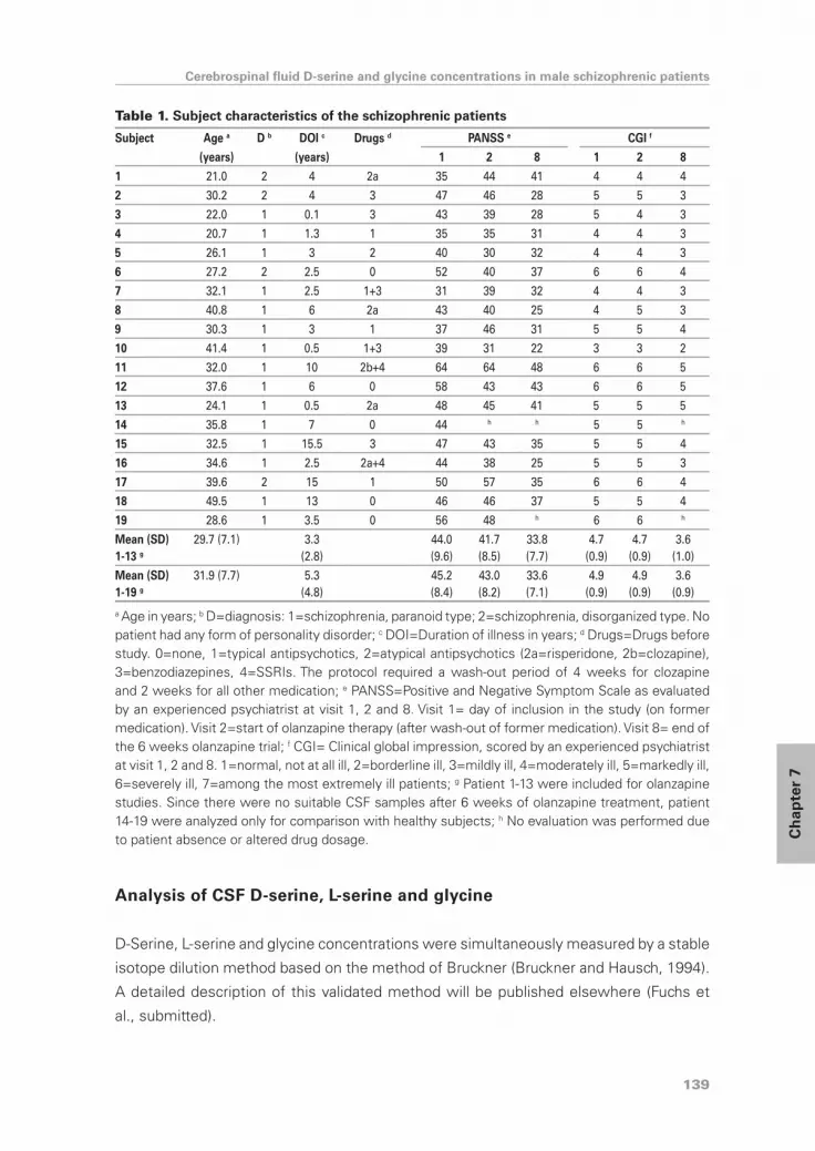
139
Cerebrospinal fluid D-serine and glycine concentrations in male schizophrenic patients
Ch
apte
r 7
Analysis of CSF D-serine, L-serine and glycine
D-Serine, L-serine and glycine concentrations were simultaneously measured by a stable
isotope dilution method based on the method of Bruckner (Bruckner and Hausch, 1994).
A detailed description of this validated method will be published elsewhere (Fuchs et
al., submitted).
Table 1. Subject characteristics of the schizophrenic patients
Subject Age a D b DOI c Drugs d PANSS e CGI f
(years) (years) 1 2 8 1 2 8
1 21.0 2 4 2a 35 44 41 4 4 4
2 30.2 2 4 3 47 46 28 5 5 3
3 22.0 1 0.1 3 43 39 28 5 4 3
4 20.7 1 1.3 1 35 35 31 4 4 3
5 26.1 1 3 2 40 30 32 4 4 3
6 27.2 2 2.5 0 52 40 37 6 6 4
7 32.1 1 2.5 1+3 31 39 32 4 4 3
8 40.8 1 6 2a 43 40 25 4 5 3
9 30.3 1 3 1 37 46 31 5 5 4
10 41.4 1 0.5 1+3 39 31 22 3 3 2
11 32.0 1 10 2b+4 64 64 48 6 6 5
12 37.6 1 6 0 58 43 43 6 6 5
13 24.1 1 0.5 2a 48 45 41 5 5 5
14 35.8 1 7 0 44 h h 5 5 h
15 32.5 1 15.5 3 47 43 35 5 5 4
16 34.6 1 2.5 2a+4 44 38 25 5 5 3
17 39.6 2 15 1 50 57 35 6 6 4
18 49.5 1 13 0 46 46 37 5 5 4
19 28.6 1 3.5 0 56 48 h 6 6 h
Mean (SD) 1-13 g
29.7 (7.1) 3.3(2.8)
44.0 (9.6)
41.7 (8.5)
33.8 (7.7)
4.7 (0.9)
4.7 (0.9)
3.6 (1.0)
Mean (SD) 1-19 g
31.9 (7.7) 5.3(4.8)
45.2 (8.4)
43.0 (8.2)
33.6 (7.1)
4.9 (0.9)
4.9 (0.9)
3.6 (0.9)
a Age in years; b D=diagnosis: 1=schizophrenia, paranoid type; 2=schizophrenia, disorganized type. No patient had any form of personality disorder; c DOI=Duration of illness in years; d Drugs=Drugs before study. 0=none, 1=typical antipsychotics, 2=atypical antipsychotics (2a=risperidone, 2b=clozapine), 3=benzodiazepines, 4=SSRIs. The protocol required a wash-out period of 4 weeks for clozapine and 2 weeks for all other medication; e PANSS=Positive and Negative Symptom Scale as evaluated by an experienced psychiatrist at visit 1, 2 and 8. Visit 1= day of inclusion in the study (on former medication). Visit 2=start of olanzapine therapy (after wash-out of former medication). Visit 8= end of the 6 weeks olanzapine trial; f CGI= Clinical global impression, scored by an experienced psychiatrist at visit 1, 2 and 8. 1=normal, not at all ill, 2=borderline ill, 3=mildly ill, 4=moderately ill, 5=markedly ill, 6=severely ill, 7=among the most extremely ill patients; g Patient 1-13 were included for olanzapine studies. Since there were no suitable CSF samples after 6 weeks of olanzapine treatment, patient 14-19 were analyzed only for comparison with healthy subjects; h No evaluation was performed due to patient absence or altered drug dosage.

D-serine in human pathology
140
Statistical analyses
CSF of schizophrenic patients was analyzed in duplicate and mean amino acid concentra-
tions and ratios were compared to control amino acid concentrations using two-tailed
Student’s t-tests. The effect of olanzapine was analyzed with two-tailed paired Student’s
t-tests. The level of significance was set at p=0.05.
reSuLTS
19 Male schizophrenic patients (mean age:31.9 years (SD=7.7)), mean duration of ill-
ness:5.3 years (SD=4.8)) and 17 healthy individuals (6 males, 11 females, mean age:38.3
years (SD=11.5)) participated in this study. Age did not differ significantly (p=0.07;Table
1+2). Patient characteristics are presented in Table 1.
Table 2. D-Serine, L-serine and glycine in cerebrospinal fluid of healthy subjects and schizo-phrenic patients
N Age in years
D-serine(µmol/l)
L-serine(µmol/l)
Glycine(µmol/l)
D-serine/total serine
(%)
Glycine/glycine+
L-serine (%)
D-serine/ glycine
Controls 17 38.3 (11.5) 3.3 (1.9) 34.6 (5.8) 8.4 (2.9) 8.4 (4.1) 19.6 (6.2) 0.4 (0.2)
Patients with schizophrenia
19 31.9 (7.7) 3.1 (1.0) 32.9 (5.0) 7.1 (1.7) 8.5 (2.0) 18.0 (4.4) 0.5 (0.2)
p-value t-test 0.07 0.75 0.36 0.12 0.95 0.37 0.67
D-Serine, L-serine and glycine concentrations (in µmol/l) and the percentage D-serine of total serine (%), percentage glycine of the sum of glycine and L-serine (%) and the ratio of D-serine to glycine were determined in cerebrospinal fluid from 17 healthy control subjects and 19 patients with schizo-phrenia. The table represents the mean values (standard deviations) and p-values of the two-tailed Student’s t-tests (level of significance: p<0.05). N = number of subjects.
Table 3. D-Serine, L-serine and glycine in cerebrospinal fluid of patients with schizophrenia before and after olanzapine therapy.
Before treatment After treatment p-value
D-serine (µmol/l) 3.1 (0.8) 3.2 (0.9) 0.78
L-serine (µmol/l) 34.5 (4.1) 35.8 (3.3) 0.22
glycine (µmol/l) 7.0 (1.2) 6.9 (1.1) 0.46
D-serine/total serine (%) 8.3 (1.8) 8.3 (2.2) 0.95
glycine/(glycine+L-serine) (%) 16.9 (3.4) 16.2 (3.1) 0.18
D-serine/glycine 0.5 (0.1) 0.5 (0.1) 0.59
In cerebrospinal fluid of 13 male schizophrenic patients, D-serine, L-serine and glycine concentra-tions (in µmol/l) and the percentage D-serine of total serine (%), percentage glycine of the sum of glycine and L-serine (%) and ratio of D-serine to glycine were determined before and after 6 weeks of daily treatment with 10mg olanzapine. The table represents the mean values (standard deviations) and p-values of the two-tailed paired Student’s t-tests (level of significance: p<0.05).

141
Cerebrospinal fluid D-serine and glycine concentrations in male schizophrenic patients
Ch
apte
r 7
CSF D-serine, L-serine and glycine concentrations were non-significantly different be-
tween schizophrenic patients and healthy individuals (p>0.1;Table 2). Since both D-serine
and glycine are synthesized from L-serine, we expressed these data as percentage
D-serine of total serine and percentage glycine of the sum of glycine and L-serine. The
ratio of D-serine to glycine represents the relative contribution of each NMDA-receptor
co-agonist. None of these ratios differed significantly between healthy individuals and
schizophrenic patients (p>0.3;Table 2). Similarly, D-serine, L-serine and glycine concen-
trations and the ratios all remained unaffected by olanzapine therapy (p>0.1;Table 3).
DISCuSSIon
Despite accumulating evidence that NMDA-receptor dysfunction contributes to the
pathogenesis of schizophrenia, the underlying mechanism remains unclear. A deficiency
of NMDA-receptor agonists would provide a plausible pathophysiological mechanism
(Boks et al., 2007), and restitution of this deficiency would then account for (part of) the
effectiveness of some antipsychotic medication. However, CSF concentrations of D-
serine, L-serine, glycine and their ratios were not significantly different between healthy
individuals and male schizophrenic patients before and after olanzapine therapy.
Our results partly concur with previous studies on CSF D-serine concentrations in
schizophrenic patients. Similar to our results, one study reported unaltered D-serine
concentrations compared to healthy individuals, but the percentage D-serine of total
serine was significantly decreased (Hashimoto et al., 2005). However, we would rather
attribute this to the observed increased L-serine concentrations (21.5µM versus 18.6µM,
p=0.069) than to decreased D-serine concentrations (1.3µM versus 1.4µM, p=0.778).
Another recent study reported 25% decreased CSF D-serine concentrations in schizo-
phrenic patients, compared with healthy controls (Bendikov et al., 2007). Interestingly,
protein levels of D-serine synthesizing and metabolizing enzymes were also altered in
schizophrenic brains. However, if we compare male schizophrenic patients with male
controls, CSF D-serine concentrations were non-significantly different. When combining
the results from these three studies, there is no consistent CSF D-serine decrease in
schizophrenia, at least not in males. In these patients, CSF D-serine can not serve as a
reliable diagnostic marker for the disorder. Since all three study populations were small,
these results should be confirmed in larger populations, which is hampered by the obvi-
ous difficulty to obtain CSF from large groups of schizophrenic patients and healthy
controls.
An important extension in the present study is the simultaneous determination of D-
serine and glycine. Concentrations of both NMDA-receptor co-agonists influence each
other, not only by interconversion through L-serine (Fu et al., 2001;Herbig et al., 2002;Wo-

D-serine in human pathology
142
losker et al., 1999), but also through competitive inhibition of the D-serine synthesizing
enzyme serine racemase by glycine (Dunlop and Neidle, 2005;Strisovsky et al., 2005).
Furthermore, alterations in the proper homeostatic control of the two NMDA-receptor
co-agonists may have relevant functional consequences (Fuchs et al., 2006). However,
patient CSF amino acid concentrations and ratios in the present study did not differ from
healthy controls nor from a healthy younger population we reported previously (Fuchs
et al., 2006). Moreover, the normal CSF glycine concentrations in schizophrenic patients
are important in the light of the beneficial effect of glycine transporter type-I inhibitors in
patients with schizophrenia (Tsai et al., 2004), and the possible consequential hypothesis
of altered glycine transporter function in schizophrenia (Tsai et al., 2006).
To our knowledge, we are the first to evaluate the effect of an atypical antipsychotic
on CSF D-serine, L-serine and glycine concentrations. Olanzapine did not affect any of
these concentrations. Similarly, previous analyses in this same patient group failed to
show an effect of olanzapine on CSF glutamate concentrations (Scheepers et al., 2002).
Thus, despite evidence suggestive of glutamatergic properties of atypical antipsychotics
(Heresco-Levy, 2003), CSF concentrations of none of the endogenous NMDA-receptor
agonists were affected by olanzapine therapy.
There are several explanations for the unaltered NMDA-receptor agonist concentrations
in our study. First, D-serine, glycine and/or glutamate concentrations are altered in
the schizophrenic brain, but these alterations are not reflected in our results. D-serine
and glycine were implied in the pathophysiology of schizophrenia because negative
symptoms ameliorated upon D-serine and glycine addition to antipsychotic treatment
(Tuominen et al., 2006). Additionally, schizophrenia was genetically associated with G72,
whose product activates the D-serine degrading enzyme D-amino acid oxidase (DAO)
(Chumakov et al., 2002;Detera-Wadleigh and McMahon, 2006). Similarly, both a single-
nucleotide polymorphism in the Serine Racemase gene (Morita et al., 2007) and the
Protein interacting with C-kinase (PICK) gene, which interacts with serine racemase
(Fujii et al., 2006), were associated with schizophrenia. All these studies imply a role
for D-serine and glycine in the pathophysiology of schizophrenia. Conceivably, we did
not detect significant olanzapine dependent CSF alterations in schizophrenic patients,
because these occur only in specific brain areas and are not discernable in global bio-
fluids as CSF (Tsai et al., 1998). In analogy, altered serine racemase expression was
detected only in the hippocampus (Steffek et al., 2006) and frontal cortex (Bendikov et
al., 2007), but not in other areas of schizophrenic brains, thereby potentially not affecting
CSF amino acid concentrations. Alternatively, we possibly failed to detect significant
differences, because the study population was small and so is the expected difference
in CSF concentrations between patients and controls (Bendikov et al., 2007;Hashimoto
et al., 2005). Finally, the distinctive heterogeneity in schizophrenic patients might result
in subgroups with and without amino acid deficiencies.

143
Cerebrospinal fluid D-serine and glycine concentrations in male schizophrenic patients
Ch
apte
r 7
The second explanation for our results supposes another pathophysiological mechanism
than olanzapine dependent D-serine, glycine or glutamate deficiency in schizophrenia.
In that case, the NMDA-receptor hypofunction hypothesis must be explained by some
other defect in NMDA-receptor neurotransmission, like fewer glycine/D-serine binding-
sites, altered NMDA-receptor subunit composition, or decreased sensitivity of the
subunits for their agonists. In accordance, mice with genetically engineered mutations
affecting the glycine/D-serine binding-site show persistent hyperactivity and stereotypic
behaviors, indicating that dysfunction of the glycine/D-serine binding-site itself can cause
behavioral abnormalities similar to those seen in schizophrenia (Ballard et al., 2002).
In conclusion, we detected normal CSF D-serine and glycine concentrations in male
schizophrenic patients, before and after olanzapine therapy. We conclude from our and
previous CSF studies that D-serine and glycine cannot serve as reliable biomarkers for
this disorder.

D-serine in human pathology
144
reFerenCeS
1. Andreasen, N.C., Flaum, M., Arndt, S. (1992) The Comprehensive Assessment of Symptoms
and History (CASH). An instrument for assessing diagnosis and psychopathology. Arch. Gen.
Psychiatry. 49, 615-623.
2. Arvanov, V.L., Liang, X., Schwartz, J., Grossman, S., Wang, R.Y. (1997) Clozapine and halo-
peridol modulate N-methyl-D-aspartate- and non-N-methyl-D-aspartate receptor-mediated
neurotransmission in rat prefrontal cortical neurons in vitro. J. Pharmacol. Exp. Ther. 283,
226-234.
3. Ballard, T.M., Pauly-Evers, M., Higgins, G.A., Ouagazzal, A.M., Mutel, V., Borroni, E., Kemp,
J.A., Bluethmann, H., Kew, J.N. (2002) Severe impairment of NMDA receptor function in
mice carrying targeted point mutations in the glycine binding site results in drug-resistant
nonhabituating hyperactivity. J. Neurosci. 22, 6713-6723.
4. Bendikov, I., Nadri, C., Amar, S., Panizzutti, R., De, M.J., Wolosker, H., Agam, G. (2007)
A CSF and postmortem brain study of D-serine metabolic parameters in schizophrenia.
Schizophr. Res. 90, 41-51.
5. Boks, M.P., Rietkerk, T., van de Beek, M.H., Sommer, I.E., De Koning, T.J., Kahn, R.S. (2007)
Reviewing the role of the genes G72 and DAAO in glutamate neurotransmission in schizo-
phrenia. Eur. Neuropsychopharmacol. 17, 567-572.
6. Bruckner, H. and Hausch, M. (1994) Gas chromatographic characterization of free D-amino
acids in the blood serum of patients with renal disorders and of healthy volunteers. J. Chro-
matogr. 614, 7-17.
7. Chumakov, I., Blumenfeld, M., Guerassimenko, O., Cavarec, L., Palicio, M., Abderrahim,
H., Bougueleret, L., Barry, C., Tanaka, H., La Rosa, P., Puech, A., Tahri, N., Cohen-Akenine,
A., Delabrosse, S., Lissarrague, S., Picard, F.P., Maurice, K., Essioux, L., Millasseau, P., Grel,
P., Debailleul, V., Simon, A.M., Caterina, D., Dufaure, I., Malekzadeh, K., Belova, M., Luan,
J.J., Bouillot, M., Sambucy, J.L., Primas, G., Saumier, M., Boubkiri, N., Martin-Saumier,
S., Nasroune, M., Peixoto, H., Delaye, A., Pinchot, V., Bastucci, M., Guillou, S., Chevillon,
M., Sainz-Fuertes, R., Meguenni, S., Aurich-Costa, J., Cherif, D., Gimalac, A., Van Duijn, C.,
Gauvreau, D., Ouellette, G., Fortier, I., Raelson, J., Sherbatich, T., Riazanskaia, N., Rogaev,
E., Raeymaekers, P., Aerssens, J., Konings, F., Luyten, W., Macciardi, F., Sham, P.C., Straub,
R.E., Weinberger, D.R., Cohen, N., Cohen, D. (2002) Genetic and physiological data implicat-
ing the new human gene G72 and the gene for D-amino acid oxidase in schizophrenia. Proc.
Natl. Acad. Sci. U. S. A. 99, 13675-13680.
8. Daly, D.A. and Moghaddam, B. (1993) Actions of clozapine and haloperidol on the extracel-
lular levels of excitatory amino acids in the prefrontal cortex and striatum of conscious rats.
Neurosci. Lett. 152, 61-64.
9. Detera-Wadleigh, S.D. and McMahon, F.J. (2006) G72/G30 in Schizophrenia and Bipolar
Disorder: Review and Meta-analysis. Biol. Psychiatry. 60, 106-114.
10. Dunlop, D.S. and Neidle, A. (2005) Regulation of serine racemase activity by amino acids.
Brain Res. Mol. Brain Res. 133, 208-214.
11. Evins, A.E., Amico, E.T., Shih, V., Goff, D.C. (1997) Clozapine treatment increases serum
glutamate and aspartate compared to conventional neuroleptics. J. Neural Transm. 104,
761-766.
145
Cerebrospinal fluid D-serine and glycine concentrations in male schizophrenic patients
Ch
apte
r 7
12. Farber, N.B., Foster, J., Duhan, N.L., Olney, J.W. (1996) Olanzapine and fluperlapine mimic
clozapine in preventing MK-801 neurotoxicity. Schizophr. Res. 21, 33-37.
13. Fu, T.F., Rife, J.P., Schirch, V. (2001) The role of serine hydroxymethyltransferase isozymes
in one-carbon metabolism in MCF-7 cells as determined by (13)C NMR. Arch. Biochem.
Biophys. 393, 42-50.
14. Fuchs, S.A., Dorland, L., De Sain-Van Der Velden, M.G., Hendriks, M., Klomp, L.W., Berger,
R., De Koning, T.J. (2006) D-serine in the developing human central nervous system. Ann.
Neurol. 60, 476-480.
15. Fujii, K., Maeda, K., Hikida, T., Mustafa, A.K., Balkissoon, R., Xia, J., Yamada, T., Ozeki, Y.,
Kawahara, R., Okawa, M., Huganir, R.L., Ujike, H., Snyder, S.H., Sawa, A. (2006) Serine
racemase binds to PICK1: potential relevance to schizophrenia. Mol. Psychiatry. 11, 150-157.
16. Goff, D.C. and Coyle, J.T. (2001) The emerging role of glutamate in the pathophysiology and
treatment of schizophrenia. Am. J. Psychiatry. 158, 1367-1377.
17. Goff, D.C., Tsai, G., Manoach, D.S., Flood, J., Darby, D.G., Coyle, J.T. (1996) D-cycloserine
added to clozapine for patients with schizophrenia. Am. J. Psychiatry. 153, 1628-1630.
18. Goldner, E.M., Hsu, L., Waraich, P., Somers, J.M. (2002) Prevalence and incidence studies
of schizophrenic disorders: a systematic review of the literature. Can. J. Psychiatry. 47,
833-843.
19. Harrison, P.J. and Owen, M.J. (2003) Genes for schizophrenia? Recent findings and their
pathophysiological implications. Lancet. 361, 417-419.
20. Hashimoto, K., Engberg, G., Shimizu, E., Nordin, C., Lindstrom, L.H., Iyo, M. (2005) Reduced
D-serine to total serine ratio in the cerebrospinal fluid of drug naive schizophrenic patients.
Prog. Neuropsychopharmacol. Biol. Psychiatry. 29, 767-769.
21. Hashimoto, K., Fukushima, T., Shimizu, E., Komatsu, N., Watanabe, H., Shinoda, N., Nakazato,
M., Kumakiri, C., Okada, S., Hasegawa, H., Imai, K., Iyo, M. (2003) Decreased serum levels
of D-serine in patients with schizophrenia: evidence in support of the N-methyl-D-aspartate
receptor hypofunction hypothesis of schizophrenia. Arch. Gen. Psychiatry. 60, 572-576.
22. Herbig, K., Chiang, E.P., Lee, L.R., Hills, J., Shane, B., Stover, P.J. (2002) Cytoplasmic serine
hydroxymethyltransferase mediates competition between folate-dependent deoxyribo-
nucleotide and S-adenosylmethionine biosyntheses. J. Biol. Chem. 277, 38381-38389.
23. Heresco-Levy, U. (2003) Glutamatergic neurotransmission modulation and the mechanisms
of antipsychotic atypicality. Prog. Neuropsychopharmacol. Biol. Psychiatry. 27, 1113-1123.
24. Kumashiro, S., Hashimoto, A., Nishikawa, T. (1995) Free D-serine in post-mortem brains and
spinal cords of individuals with and without neuropsychiatric diseases. Brain Res. 681, 117-
125.
25. Lahti, A.C., Weiler, M.A., Tamara Michaelidis, B.A., Parwani, A., Tamminga, C.A. (2001) Ef-
fects of ketamine in normal and schizophrenic volunteers. Neuropsychopharmacology. 25,
455-467.
26. Miyamoto, Y., Yamada, K., Noda, Y., Mori, H., Mishina, M., Nabeshima, T. (2001) Hyperfunc-
tion of dopaminergic and serotonergic neuronal systems in mice lacking the NMDA receptor
epsilon1 subunit. J. Neurosci. 21, 750-757.
27. Mohn, A.R., Gainetdinov, R.R., Caron, M.G., Koller, B.H. (1999) Mice with reduced NMDA
receptor expression display behaviors related to schizophrenia. Cell. 98, 427-436.
28. Morita, Y., Ujike, H., Tanaka, Y., Otani, K., Kishimoto, M., Morio, A., Kotaka, T., Okahisa, Y.,

D-serine in human pathology
146
Matsushita, M., Morikawa, A., Hamase, K., Zaitsu, K., Kuroda, S. (2007) A genetic variant of
the serine racemase gene is associated with schizophrenia. Biol. Psychiatry. 61, 1200-1203.
29. Olney, J.W. and Farber, N.B. (1994) Efficacy of clozapine compared with other antipsychotics
in preventing NMDA-antagonist neurotoxicity. J. Clin. Psychiatry. 55 Suppl B, 43-46.
30. Scheepers, F.E., Gispen-De Wied, C.C., Westenberg, H.G., Kahn, R.S. (2002) Effect of
olanzapine on glutamate levels in cerebrospinal fluid of patients with schizophrenia. J. Clin.
Psychopharmacol. 22, 333-334.
31. Steffek, A.E., Haroutunian, V., Meador-Woodruff, J.H. (2006) Serine racemase protein ex-
pression in cortex and hippocampus in schizophrenia. Neuroreport. 17, 1181-1185.
32. Strisovsky, K., Jiraskova, J., Mikulova, A., Rulisek, L., Konvalinka, J. (2005) Dual substrate
and reaction specificity in mouse serine racemase: identification of high-affinity dicarboxyl-
ate substrate and inhibitors and analysis of the beta-eliminase activity. Biochemistry. 44,
13091-13100.
33. Tsai, G., Lane, H.Y., Yang, P., Chong, M.Y., Lange, N. (2004) Glycine transporter I inhibitor,
N-methylglycine (sarcosine), added to antipsychotics for the treatment of schizophrenia.
Biol. Psychiatry. 55, 452-456.
34. Tsai, G., van Kammen, D.P., Chen, S., Kelley, M.E., Grier, A., Coyle, J.T. (1998) Glutamatergic
neurotransmission involves structural and clinical deficits of schizophrenia. Biol. Psychiatry.
44, 667-674.
35. Tsai, S.J., Cheng, C.Y., Hong, C.J., Liao, D.L., Hou, S.J., Yen, F.C., Liou, Y.J. (2006) Associa-
tion study of polymorphisms in glycine transporter with schizophrenia. J. Neural Transm.
113, 1545-1549.
36. Tuominen, H.J., Tiihonen, J., Wahlbeck, K. (2006) Glutamatergic drugs for schizophrenia.
Cochrane. Database. Syst. Rev. CD003730.
37. Wolosker, H., Blackshaw, S., Snyder, S.H. (1999) Serine racemase: a glial enzyme synthe-
sizing D-serine to regulate glutamate-N-methyl-D-aspartate neurotransmission. Proc. Natl.
Acad. Sci. U. S. A. 96, 13409-13414.
38. Yamamoto, B.K. and Cooperman, M.A. (1994) Differential effects of chronic antipsychotic
drug treatment on extracellular glutamate and dopamine concentrations. J. Neurosci. 14,
4159-4166.

Discussion


Chapter 8
general discussion
Submitted in adapted form


151
General discussion
Ch
apte
r 8
D-SerIne: THe rIgHT or wrong ISoForm?
Only a few decades ago, D-amino acids were considered to be restricted to some bacteria
and insects. In the last few years, evidence is accumulating that D-amino acids occur in
mammals (chapter 1) and we are only starting to unravel their biological function. Most
research has focused on D-serine, which was found to occur in high concentrations in
the mammalian brain.(1) D-Serine can be synthesized and metabolized endogenously
by the enzymes serine racemase (SR) (2) and D-amino acid oxidase (DAO),(3) respec-
tively. In mammalian central nervous system (CNS), D-serine was found to function as
a neurotransmitter by binding to the N-methyl-D-aspartate receptor (NMDAr). NMDArs
are widely expressed in the CNS, where they play important roles in physiological and
pathological processes,(4) including synaptic plasticity and neurotoxicity. NMDArs are
tetrameric glutamate-gated ion channels, occurring as multiple subtypes which differ in
their subunit composition, which mostly comprises an NR1 subunit, combined with at
least one of four NR2 (A-D) subunits. Depending on the subunit composition, the bio-
physical and pharmacological properties of NMDArs vary.(5) One of the unique features
of NMDArs is that they are only activated upon simultaneous binding by glutamate to
their NR2 subunit and by a co-agonist to their NR1 subunit. Similar to glycine, which
was long thought to be the endogenous NMDAr co-agonist, D-serine can bind to the
NR1 subunit. Binding to the co-agonist site is not only essential for NMDAr activation,
but also exerts a neuromodulatory role, by increasing the affinity of the NMDAr for
glutamate,(6) decreasing its desensitization (7) and promoting NMDAr turnover by inter-
nalization.(8) This regulation of NMDAr activity by a second agonist might be regarded
as a safety mechanism to protect against the deleterious effects of overstimulation of
the NMDAr (excitotoxicity). Had this safety mechanism not existed, then the brain would
potentially have been more at risk for excitotoxicity and subsequent neurotoxicity, with
the abundance of glutamate in the CNS.
To investigate this putatively important role of D-serine in human CNS, we developed
chiral chromatographic separation techniques, combined with mass spectrometric
detection to determine concentrations of D-serine, glycine and their common precursor
L-serine simultaneously in biological fluids (chapter 2). These stable isotope dilution
analysis techniques represent valuable tools to address scientific research questions on
the role of D-serine (or other D-amino acids) in mammalian physiology and pathology,
with the ultimate goal of developing novel diagnostic, therapeutic and prognostic strate-
gies for human diseases. In this respect, our generation of age-dependent reference
values in human cerebrospinal fluid (CSF) (chapters 2-3) is pivotal in enabling diagnosis
and follow-up of patients with disorders associated with alterations in CNS D-serine,
L-serine or glycine concentrations. The studies presented in this thesis - all using these

Discussion
152
analytical techniques - contribute to the emergent awareness that D-serine plays an
important role as an endogenous NMDAr co-agonist in human physiology and pathology,
as underscored by following lines of evidence:
1. D-SerIne In mammaLIan pHySIoLogy
One of the main reasons to suspect D-serine of being a mammalian endogenous NMDAr
co-agonist, was the close anatomical co-localization between D-serine and NMDArs.
(9;10) D-Serine and NMDAr immunoreactivity was high in cranial areas of rat brain (espe-
cially in the frontal cortex) and lower in caudal areas,(9;10) where the D-serine degrading
enzyme DAO was present.(11) In contrast, glycine immunoreactivity predominated in
caudal areas of the brain, where the density of NMDArs was lower, with lowest glycine
immunoreactivity in the telencephalon.(9) In addition, extracellular D-serine concentra-
tions in the brain, as determined by microdialysis, exceeded the concentrations of many
L-amino acids, and were similar to glycine concentrations in the cerebral cortex.(12)
Moreover, in cloned NMDArs, D-serine affinity for the NR1 subunit was high,(13) indeed
threefold higher than glycine. This might be explained by the crystal structure of the NR1
subunit of NMDArs, where D-serine displaces a water molecule from the binding pocket
and makes three additional hydrogen bonds, when compared to glycine.(14)
Evidence for a contribution of endogenous D-serine to physiological NMDAr co-activation
came from the decrease in spontaneous NMDAr activity (up to 60%) in immature rat
cerebellar slices, rat hippocampus slices and primary hippocampal cell cultures upon
removal of extracellular D-serine by DAO, the effect of which was fully reversed by ap-
plication of exogenous D-serine.(15) Similarly, using enzymatic degradation of D-serine,
endogenous D-serine was found to be required for NMDAr mediated light-evoked
responses in the vertebrate retina.(16;17) In fact, upon enzymatic degradation of D-
serine, light-evoked NMDAr contribution was similar to that observed after blockage of
NMDArs with antagonists.(16) Additionally, electrophysiological experiments performed
in the supra-optic nucleus of the hypothalamus demonstrated that endogenous D-serine
degradation by recombinant DAO blocked NMDAr responses in hypothalamic slices, as
opposed to endogenous glycine degradation by a glycine oxidase enzyme.(18) Finally,
SR knock-out mice displaying 90% decreased D-serine concentrations showed severely
altered glutamatergic neurotransmission,(19) with an overall decrease in NMDAr me-
diated neurotransmission, as demonstrated by lower S-nitrosylation of intracellular
proteins. Together, these studies point to an important role for D-serine as a determinant
of NMDAr mediated neurotransmission in mammalian physiology.

153
General discussion
Ch
apte
r 8
2. D-SerIne In SpeCIFIC pHySIoLogICaL proCeSSeS
CNS development
The first clue for a specific role for D-serine in CNS development came from the finding
of specifically and markedly elevated D-serine concentrations in human and rodent CNS
(10;20;21) during the intense period of embryonic and early postnatal CNS develop-
ment, coinciding with a transient expression (22-25) and increased activity (26-28) of
NMDArs. Similarly, we found peaking D-serine concentrations in human CSF during the
early postnatal (and presumably embryonic) period (chapter 3, (29)). The severe CNS
abnormalities upon failure to achieve these specific and marked increases in D-serine
concentrations, as seen in patients (30) and mutant mice (31) with 3-phosphoglycerate
dehydrogenase deficiency (3-PGDH), a rare inherited disorder in L-serine and hence D-
serine synthesis, suggest a role for D-serine in CNS development (chapter 3).
More specifically, degradation of D-serine by DAO and selective inhibition of SR and
hence of D-serine synthesis in 8-day old mouse cerebellar slices significantly reduced
granule cell migration, whereas D-serine activated this process,(32) implying a specific
role for D-serine in neuronal migration, putatively through NMDAr activation.(33) This
is supported by the unambiguous mass spectrometric identification of SR in the peri-
reticular nucleus, a transient structure of the developing brain in humans, suggested
to be mainly involved in neuronal migration.(34) Moreover, our P19 studies (chapter
5) imply a role for D-serine, synthesized by SR, in shaping synaptogenesis, as specific
inhibition of SR and NR1 antagonism increased synaptophysin expression and inter-
connections between differentiated neuronal and glial cells, which was overcome by
D-serine supplementation in the case of SR inhibition. Similarly, partial deletion of NR1
subunits of NMDArs in organotypic hippocampal cells profoundly increased numbers of
functional synapses between neurons and strength of unitary connections in vitro and in
vivo, which was overcome by reintroduction of NMDArs.(35) Conceivably, endogenous
D-serine is a crucial factor in activating NMDArs, thereby preventing premature synap-
tic maturation by ensuring that only punctuated bursts of activity lead to induction of
functional synapses for the activity-dependent wiring of neuronal circuitry.(35;36) This
specific role for D-serine in synaptogenesis is further consolidated by observations in SR
knock-out mice.(19) The subtle behavioural abnormalities these mice display have been
associated with altered synaptic shaping, including mild hyperactivity and increased
anxiety (37) and a spatial memory deficit.(38) In contrast, the naturally occurring mouse
strain (ddY/DAO-), lacking DAO activity and thereby displaying elevated extracellular
D-serine concentrations and enhanced NMDAr function,(39) show improved spatial
learning (40) and better motor coordination,(39) which might be associated with altered
synaptic shaping due to increased D-serine concentrations.

Discussion
154
Learning and memory
Long-term potentiation (LTP) of synaptic transmission in the hippocampus refers to an
enhancement in signal transmission between neurons upon synchronic stimulation and
is one of the processes underlying synaptic plasticity. It is widely considered one of the
major cellular mechanisms underlying learning and memory in vertebrates.(41) The best
understood form of LTP is induced by NMDAr activation, and D-serine released from
astrocytes appears to play a role in this process. In fact, LTP could be induced in neurons
grown on a layer of astrocytes, as opposed to neurons cultured in glial conditioned
medium, which was overcome by D-serine supplementation.(42) LTP induction was sup-
pressed by NMDAr antagonists and enzymatic D-serine degradation. Similarly, in adult
rat CA1 pyramidal hippocampus cells, LTP depended on calcium dependent release of
D-serine from astrocytes.(43) LTP could be blocked by decreasing NR1 occupancy by
D-serine depletion (through depletion of stored D-serine and SR inhibition) or disrup-
tion of exocytosis, which was reversible by exogenous D-serine or glycine application.
Further support comes from SR knock-out mice displaying a 90% reduction in D-serine
concentrations, impaired NMDAr transmission and attenuated LTP,(19) while ddY/DAO-
mice display elevated extracellular D-serine concentrations, enhanced NMDAr function
and enhanced hippocampal LTP.(39;40) Additional evidence for a role of D-serine in LTP
comes from studies evaluating learning and memory deterioration occurring with ag-
ing. SR Expression, D-serine concentrations, NMDAr mediated synaptic potentials and
LTP were all significantly decreased in CA1 hippocampal slices from aged rats when
compared with young rats, which was rescued by exogenous D-serine supplementation.
(44;45) Similarly, when comparing two rat strains which model healthy aging versus
accelerated aging with cognitive impairments, the latter model showed decreased hip-
pocampal SR mRNA and protein and decreased D-serine concentrations, combined with
normal DAO expression, and marked reductions in NMDAr mediated synaptic potentials
and LTP, which were restored by exogenous D-serine addition.(46) Correspondingly,
hippocampal slices from a senescence-accelerated mouse model showed significant
and exaggerated LTP suppression with age, when compared to normal mice, which
was overcome by D-serine supplementation.(47) Addition of DAO also decreased LTP,
which was rescued by exogenous application of D-serine. D-serine effects were inhib-
ited by antagonists of the NMDAr co-agonist site. Together, these results all robustly
demonstrate the importance of D-serine, synthesized by SR, for NMDAr activation and
subsequent LTP induction, which underlies learning and memory.

155
General discussion
Ch
apte
r 8
3. D-SerIne In mammaLIan paTHoLogy
Excitotoxicity
Glutamate toxicity, or excitotoxicity, occurs mainly through excessive NMDAr activa-
tion and subsequent massive calcium influx in the cell.(4) This appears to be the major
reason for neurotoxicity and cell death upon hypoxia/ischemia and in neurodegenerative
conditions.(48) D-Serine appears to be an important endogenous co-agonist in this pro-
cess, since removal of endogenous D-serine with DAO (49) or D-serine deaminase,(50)
a D-serine degrading enzyme with higher affinity and specificity to D-serine than com-
mercially available DAO, virtually abolished NMDA-elicited neurotoxicity in rat cerebro-
cortical and organotypic hippocampal slices. The effect of endogenous glycine could be
observed only after simultaneous removal of endogenous D-serine and blockage of the
glycine transporter GlyT1.(50) Moreover, SR knock-out mice displaying a 90% reduction
in extracellular D-serine concentrations demonstrated reduced neurotoxicity induced by
NMDA injections in their forebrains.(51)
Perinatal asphyxia
Perinatal asphyxia refers to hypoxic/ischemic pathology occurring around birth, and
represents one of the main causes of morbidity and mortality among term-born children
without curative treatment options.(52) Excitotoxicity plays an essential role in the
pathogenesis.(48) The role of D-serine in this process was implicated by the reduction of
cell death after addition of DAO to rat cerebrocortical slices exposed to oxygen-glucose
deprivation (OGD), which was reversed by addition of either D-serine or glycine.(49)
Similarly, mutant mice with a targeted deletion of SR showed dramatically diminished
infarct volume in several brain regions following middle cerebral artery occlusion,(53)
and brain cultures from SR knock-out mice exhibited markedly diminished nitric oxide
formation and neurotoxicity. Our OGD studies (chapter 6) showed marked increases
in glycine and D-serine concentrations, putatively through increased synthesis from
L-serine, which together with robustly increased glutamate concentrations,(54) are
bound to lead to massive NMDAr activation and neurotoxicity. During our reperfusion
experiments, glycine concentrations normalized but remained increased, while D-serine
concentrations were significantly reduced, both in vitro and in vivo. This concurs with
the finding of delayed transient decreases of D-serine concentrations and SR mRNA and
protein in the ipsilateral temporoparietal mouse cortex after permanent focal ischemia.
(55) These reduced D-serine concentrations during reperfusion might be due to SR
inhibition by elevated glycine concentrations.(56) This might represent an attempt to

Discussion
156
compensate for increased NMDAr activity, providing additional evidence for a role for
D-serine in regulating NMDAr activity.
Collectively, D-serine synthesized by SR appears to play a neurotoxic role in hypoxia-
ischemia. Manipulation of CNS D-serine concentrations might provide an interesting
treatment target for patients with this devastating condition, especially since results
from treatment with NMDAr antagonists, targeting both NR1 and NR2 subunits, have
been largely disappointing.(57)
Schizophrenia
Schizophrenia is a severely debilitating psychiatric condition, characterized by psychotic
features (including hallucinations), negative symptoms (including social withdrawal) and
cognitive defects (including impairment in attention, learning and memory). Schizophrenia
affects 1% of the population worldwide. As described in chapter 7, there is vast support
for NMDAr hypofunction as a pathophysiological mechanism in schizophrenia. Some
ambiguous evidence for a contribution of D-serine to the NMDAr hypofunction, comes
from the finding of reduced D-serine concentrations (58) and a reduced D-serine to
total serine ratio in CSF (58;59) and serum (60) of patients with schizophrenia. However,
differences with controls were small and inconsistent between sexes, and we were
unable to replicate these findings (chapter 7). Potentially, CSF or serum as a marker for
extracellular concentration differences might be too global to detect subtle differences
in specific brain areas.(61) Moreover, no reductions in D-serine concentrations were
detected in the parietal or prefrontal cortex from patients with schizophrenia.(58;62)
More convincing results for a role of D-serine in the pathology of schizophrenia comes
from genetic association studies, showing an association between schizophrenia with the
gene G72,(63-71) whose product was thought to activate the D-serine degrading enzyme
DAO, although recent studies have not been able to replicate this activating interaction with
DAO.(72) In addition, several meta-analyses of genetic association studies in schizophre-
nia provide some support for an association between the DAO gene and schizophrenia.
(73-75) More robust is the finding of increased DAO expression (76-78) and activity (76)
in cerebellum, parietal cortex (79) and hippocampus (80) of patients with schizophrenia,
when compared with controls. In addition, duration of illness correlated with DAO expres-
sion and activity in the hippocampus (58) and cerebellum.(76) Presumably, this increase
in DAO expression and activity causes decreased D-serine concentrations in specific brain
areas and thereby contributes to NMDAr hypofunction in schizophrenia.
Single-nucleotide polymorphisms in SR (81) and SR genetic variants in humans showed
an association with schizophrenia.(82) Similarly, the protein interacting with C-kinase
(PICK), which interacts with SR,(83) has been implicated as a susceptibility gene for
schizophrenia.(83;84) Results from studies on SR expression and immunoreactivity are

157
General discussion
Ch
apte
r 8
ambiguous and SR activity has not been assessed in schizophrenia as of yet. No change
in SR expression was observed in the parietal cortex,(77) cerebellum or prefrontal
cortex, when comparing schizophrenic patients with controls.(78) SR protein levels and
ratio of SR/DAO were reduced in postmortem prefrontal cortex and hippocampus from
patients with schizophrenia,(58) while in other patient groups SR immunoreactivity was
increased in the hippocampus (85) and the prefrontal cortex,(78) and unchanged in other
brain areas when compared to controls.(78;85) Heterogeneity of patients, the disease
entity and the different brain areas might contribute to these conflicting results. SR
exon 1 knock-out mice displaying 90% D-serine reduction exhibited relatively subtle be-
havioural abnormalities, reflecting hyperactivity, impaired spatial memory and elevated
anxiety, which are all relevant to schizophrenia.(19) Prepulse inhibition was not reduced,
while this is a consistent finding in schizophrenia.(86) Mice with an ENU-induced muta-
tion resulting in complete loss of SR activity and 95% reduced D-serine concentrations
displayed similar behavioural deficits relevant to schizophrenia, including impairment in
spatial discrimination and sociability, but no alteration in anxiety.(82) Conversely, these
mice showed impaired prepulse inhibition. The behavioural symptoms of these mice
were aggravated by NMDAr antagonists and ameliorated by D-serine. Expression profil-
ing revealed that the SR mutation influenced several genes, linked to schizophrenia and
cognitive ability, and altered transcript levels were normalized by D-serine. The signs of
subtle negative symptoms and cognitive impairment associated with schizophrenia ob-
served in both SR knock-out mice models might explain the beneficial effect of D-serine
or glycine addition to conventional antipsychotic treatment on negative symptoms, with
a trend effect on cognitive symptoms.(87) Failure to reproduce the complete schizophre-
nia phenotype in the SR knock-out mice might be due to compensatory mechanisms,
as evidenced for NMDAr kinetics and molecular composition.(19) Moreover, given the
current belief that schizophrenia represents a disorder of complex genetics with a strong
environmental component, alterations in one genetic pathway implicated in the disorder
would not be expected to replicate the entire phenotype.
Some of the difficulties in unveiling the underlying molecular mechanisms in schizo-
phrenia, specifically for the role of gene G72, are associated with the lack of appropriate
animal models. It has been hypothesized that genes responsible for higher cognitive
functions have developed very recently on the evolutionary scale, especially those
unique to humans, and thus relevant to psychiatric conditions such as schizophrenia.(88)
This might explain the poor genetic conservation of rodent and human gene sequences
of genes implicated in schizophrenia, including catechol-O-methyltransferase (COMT)
and disrupted-in-schizophrenia-1 (DISC1).(89) Similarly, no G72 ortholog was found in
mice.(90) This poor genetic homology might be crucial in interpreting mouse behavioural
phenotypes as a model for human symptoms of schizophrenia and might hamper the
use of rodent models in schizophrenia research.

Discussion
158
Summarizing, there is strong evidence for NMDAr dysfunction in schizophrenia, and
numerous reports on altered extracellular D-serine concentrations or their regulation
converge to a role for D-serine in the NMDAr hypofunction theory. The cognitive symp-
toms in schizophrenia, including impairment in learning and memory concur with the
role of D-serine in LTP generation. Hopefully, future studies, potentially using primate
animal models, will unveil the exact role of D-serine and the underlying mechanisms
in schizophrenia and thereby contribute to improved treatment and quality of life for
patients with this incapacitating condition.
Amyotrophic Lateral Sclerosis (ALS)
ALS is the most common adult-onset motor neuron disease. It is a currently incurable
rapidly progressive disorder, characterized by muscle weakness, atrophy, paralysis and
death from respiratory failure. Pathologically, it is characterized by massive motoneuronal
loss, inclusion bodies in remaining neurons and astrocytes and gliosis around dying neu-
rons in the ventral horns of spinal cords.(91) The mechanism of selective motoneuronal
death is still unclear, but excitotoxicity mediated by ionotropic glutamate receptors has
been proposed to play a principal role.(91-93) While approximately 90% of ALS patients
are sporadic, 10% are inherited and 20% of those have mutations in the gene encoding
superoxide dismutase 1 (SOD1). Transgenic mice with a glycine to alanine switch at
codon 93 in SOD1 have become the benchmark pre-clinical model for screening ALS
therapies.(94) In spinal cords from these mice, SR and D-serine concentrations were
increasingly elevated with disease progression,(95) while elevated glutamate concentra-
tions remained unchanged with disease progression.(92) In addition, primary spinal cord
neurons from these mice were more vulnerable to NMDA-elicited toxicity than those
from control mice in a D-serine dependent manner and removal of endogenous D-serine
protected motoneurons against NMDAr mediated cell death.(95) Exogenous addition of
glycine to these cells significantly reduced NMDA-induced toxicity, putatively through
competitive inhibition of SR.(56) In vitro, SR expression was induced by an extracellular
proinflammatory factor and also by transiently expressed mutant SOD1 in microglial
cells.(95) In spinal cords of patients with familial and sporadic forms of ALS, the levels of
SR and D-serine were also greatly increased.(95)
Based on these findings of increased D-serine concentrations in spinal cords of ALS
patients, we determined concentrations of D-serine, glycine and their common precur-
sor L-serine in CSF from patients with ALS, both sporadic and with SOD1 mutations
(Table 1). Unfortunately, we did not identify any significant D-serine, glycine or L-serine
concentration or ratio difference between CSF from controls and patients with ALS.
Potentially, concentration differences in the spinal cord are too subtle to be detected
in CSF with our analytical method. Furthermore, DAO has recently been identified im-

159
General discussion
Ch
apte
r 8
munohistochemically in the human and rat choroid plexus, potentially thereby regulating
CSF D-serine levels (96) and as such occluding concentration differences in specific
areas in the CNS. Our results signify that CSF amino acid determination with our method
does not provide a valuable diagnostic tool for ALS. Other methods to directly investigate
D-serine concentrations in the ventral horns of spinal cords, such as spinal cord (NMR)
spectroscopy (97) might form an interesting alternative in this regard.
Additional evidence for a role of D-serine in the pathophysiology of ALS comes from
a recent genome screen in families with confirmed ALS without known familial ALS
mutations, identifying a locus on chromosome 12 in a single family with three genera-
tions of affected members.(98) Upon screening of candidate genes within this region,
a mutation in DAO was found, which, upon expression in neuronal cell lines, revealed a
mutated protein with almost total loss of activity, impairing cell viability and promoting
ubiquitinated aggregate formation when compared with the wild type protein. Lentiviral
mediated transduction of this DAO mutation in primary motor neuron cultures or in
astrocytes co-cultured with motor neurons increased apoptosis in neuronal cells. In ad-
dition, ddY/DAO- mice, lacking DAO activity and displaying elevated levels of D-serine,
also display enhanced NMDAr mediated excitatory postsynaptic currents in spinal cord
dorsal horn neurons.(99) Finally, enzymes involved in L-serine and consequently in D-
serine biosynthesis, including phosphoserine phosphatase and 3-PGDH are among the
earliest transcriptional changes detected in motor neurons in the SOD1 mouse model.
(100)
In conclusion, the enhancement of glutamate toxicity by increased D-serine concentra-
tions in the spinal cord, through alterations in D-serine biosynthesis and metabolism,
might represent an underlying mechanism for ALS motoneuronal death. Further studies
on modulation of these elevated D-serine concentrations in the spinal cord may help
to develop neuroprotective strategies against this currently untreatable fatal condition.
Table 1. D-serine, L-serine and glycine concentrations in cerebrospinal fluid from patients with ALS and controls.
N Age (years)
D-serine (µM)
L-serine (µM)
Glycine (µM)
D-serine / total serine
(%)
Glycine / glycine + L-serine (%)
D-serine / glycine
Controls 19 63 (15) 1.8 (0.3) 26.2 (4.8) 7.0 (1.9) 6.5 (1.7) 21.3 (6.2) 0.27 (0.1)
ALS total 36 59 (13) 1.8 (0.4) 27.5 (6.2) 8.0 (4.8) 6.4 (1.7) 21.6 (6.1) 0.27 (0.1)
p-value 0.73 0.42 0.38 0.78 0.84 0.86
ALS SOD1 15 56 (15) 1.9 (0.5) 27.0 (5.1) 7.2 (2.9) 6.6 (1.7) 20.5 (5.0) 0.29 (0.1)
p-value 0.44 0.65 0.82 0.86 0.70 0.48
ALS sporadic
21 60 (12) 1.8 (0.3) 27.9 (6.9) 8.5 (5.7) 6.2 (1.8) 22.4 (6.7) 0.25 (0.1)
p-value 0.91 0.38 0.26 0.59 0.59 0.37
D-serine, L-serine, glycine concentrations and their ratios in CSF from controls and patients with ALS, both sporadic or with SOD1 mutation. The p-value for the difference with controls was calculated with an unpaired Student’s t-test.

Discussion
160
4. ConTrIbuTIon oF D-SerIne VerSuS gLyCIne In mammaLIan nmDar neuroTranSmISSIon
It is still a matter of debate whether D-serine or glycine serves as the main determinant
of NMDAr activity in different brain regions and in different conditions, although most
recent reports appear to favour D-serine.(18;50;101) Both amino acids are present
in CNS and display relatively comparable affinities to the NR1 subunit of NMDArs.
The relative contribution of both co-agonists might depend on their availability at
synaptic sites. Specific D-serine transporters have not yet been discovered. The affin-
ity of neutral amino acid transporters for D-serine is low to moderate,(102-104) and
encompass the higher affinity Na+-independent alanine-serine-cysteine 1 transporter
(asc-1 transporter, IC50 20µM) (105-108) and the low affinity Na+ and/or K+-dependent
transporters,(104;108;109) including the alanine-serine-cysteine transporter 2 (ASCT2,
IC50 1mM). The ASCT2 has been suggested to serve as an important release rather
than uptake mechanism.(109) In contrast, high-affinity glycine transport systems tightly
regulate glycine access to NMDAr sites.(110-112) Antagonists of the high affinity gly-
cine transporter type 1 (GlyT1) potentiated NMDAr activity in hippocampal pyramidal
neurons (111) and in prefrontal cortex slices and prefrontal neurons in anesthetized rats.
(113) Similarly, blocking GlyT1 led to full occupancy of NMDAr co-agonist sites in the
salamander retina, while blocking the glycine transporter type 2 (GlyT2) had no effect
on the light-evoked NMDAr currents or on their sensitivity to exogenous D-serine.(101)
In the retina from heterozygous mice deficient in GlyT1, NMDAr co-agonist sites were
more saturated and less enhanced by D-serine when compared with wild type controls.
(114) Ultrastructural studies in the brain revealed the adjacency of GlyT1 transporters
to NMDArs.(115-116) All these studies converge to the hypothesis that GlyT1 activity
keeps glycine concentrations near NMDArs sufficiently low to allow D-serine to be the
major regulator of NMDAr activity. In support, exogenous D-serine was more than 30
times as effective in potentiating NMDAr currents when compared with glycine in reti-
nal ganglion cells of the tiger salamander(101) and in brain stem slice preparations,(110)
requiring low physiological D-serine concentrations (micromolar range), as opposed to
supraphysiological glycine concentrations (≥100µM). Similarly, when observing NMDA-
elicited neurotoxicity in organotypic hippocampal slices, the effect of endogenous
glycine could only be observed after simultaneous removal of endogenous D-serine
and blockage of GlyT1,(50) despite ten-fold higher endogenous glycine than D-serine
concentrations.
The glycine cleavage system may also play a role in maintaining subsaturating glycine
concentrations near NMDArs. In astrocytes, the glycine cleavage system degrades
glycine efficiently, thereby generating the cytosol / extracellular concentration gradi-
ent,(117-119) which enables glycine transporters to transfer glycine from the synaptic

161
General discussion
Ch
apte
r 8
space into astrocytes. Conversely, the role of DAO remains controversial in regulating D-
serine concentrations near NMDArs, because D-serine and NMDArs are predominantly
present in the forebrain, where DAO activity is virtually undetectable.(11) This correlates
with the long half-life of D-serine in the brain (~16h).(18) When observing the in vivo
effects of administration of L-serine, D-serine and glycine to the infant rat, intraperitoneal
injection of L-serine or glycine induced elevated L-serine concentrations in the brain and
a delayed and prolonged increase in D-serine concentrations. Systemic administration of
D-serine resulted in increased neocortical L-serine and D-serine but not glycine content.
(120) Potentially, this illustrates that extracellular glycine concentrations are more tightly
regulated and that D-serine is more prone to accumulate in CNS when its production is
excessive or its metabolism reduced. Thereby, D-serine presumably diffuses more easily
to extrasynaptic locations and activates NMDArs in the proximity but also further away
from the release sites.(18) This will provide a more general activation of NMDArs, which
may play a crucial role in the massive activation that occurs in excitotoxicity.(50)
5. ImpLICaTIonS anD FuTure DIreCTIonS
Culminating evidence for a key role for endogenous D-serine in regulating NMDAr activ-
ity in physiological and pathological conditions has some interesting implications. Our
development of high-throughput analytical techniques to determine D-serine concentra-
tions in biological fluids and generation of age-dependent ranges in apparently healthy
subjects (chapter 2) will provide us with new diagnostic and prognostic strategies for
CNS disorders associated with NMDAr pathology. In addition, influencing D-serine con-
centrations might represent an interesting therapeutic target, which appears feasible
as endogenous D-serine concentrations might be manipulated by administration of
L-serine,(30;120) and by manipulation of its synthesizing and metabolizing enzymes,
which both have been identified and characterized.(2;3;121)
However, a number of issues remain to be addressed in order to increase our under-
standing of the role of D-serine and other D-amino acids in physiological and pathological
conditions for translational purposes:
1. Regulation of synaptic D-serine concentrations
As a key endogenous NMDAr regulator and thus potentially as an important contributor
to the pathophysiology of the variety of conditions associated with NMDAr dysregu-
lation, it is eminent to elucidate the mechanisms responsible for controlling synaptic
D-serine concentrations, with the ultimate potential to influence these processes for
therapeutic purposes.

Discussion
162
As described before, there are several low- and moderate affinity transporters for cel-
lular uptake of D-serine. Evidence from gene knock-out studies implicates Asc-1 as
the primary transporter mediating D-serine re-uptake in neurons,(107) while the lower
affinity transporter ASCT2 might be the dominant transporter involved in removal of
synaptic D-serine into glia (108) (Figure 1). The contribution of these transporters to the
Figure 3. Expression of synaptophysin in the presence or absence of D-serine induced NMDAr activation. Als pdf bijgevoegd
Hoofdstuk 6
Figure 1. Pathways involved in L-serine and D-serine biosynthesis, catabolism and exogenous sources. Als pdf bijgevoegd Figure 2. Experimental design of the Glioma C6 studies. Als pdf bijgevoegd
Figure 3. Medium concentration differences of the NMDAr agonists D-serine, glycine and their common precursor L-serine after oxygen and glucose deprivation (OGD). Als pdf bijgevoegd Figure 4. D-serine, glycine and L-serine concentrations in CSF from control piglets and 3 and 24h after application of cerebral hypoxia-ischemia. Als pdf bijgevoegd
Hoofdstuk 8
Figure 1. Regulation of synaptic D-serine concentrations
Figure 1. Regulation of synaptic D-serine concentrations. Regulation of synaptic D-serine concentrations has not been fully elucidated. Potentially, several low affinity (ASCT2) transporters and higher affinity (Asc-1) transporters are involved in neuronal (Asc-1) and glial (ASCT2) D-serine uptake. Upon uptake in neurons and astrocytes, D-serine might be degraded by peroxisomal and potentially also by non-peroxisomal DAO. However, DAO activity is virtually absent in the forebrain. Alternatively, SR might regulate D-serine concentrations in both astrocytes and neurons by enabling D-serine synthesis from L-serine and the reverse and by catalyzing degradation of both D- and L-serine. Regulation of SR activity appears to involve nitrosylation, which inhibits SR activity. Nitrosyl-ation is increased by NMDAr activity, since this activates neuronal nitric-oxide synthase. This effect on D-serine concentrations is enhanced by increased DAO activity by nitrosylation. SR activity is dramatically decreased by translocation of cytosolic SR to the plasma membrane, which is promoted by NMDAr activation. Similarly, increased NMDAr activity through increased glycine concentrations might also constitute a feedback loop, since glycine competitively inhibits SR. Moreover, NMDAr antagonism has been found to upregulate SR mRNA and protein expression. SR is also activated by several proteins that have been found to interact with SR, including glutamate receptor interacting protein (GRIP) and protein interacting with C kinase 1 (PICK1). Golgin subfamily A member 3 (Golga3) is another protein interacting with SR, leading to decreased ubiquitin-proteasomal degradation of SR, thereby increasing SR and subsequently D-serine concentrations.

163
General discussion
Ch
apte
r 8
regulation of synaptic D-serine concentrations in mammalian physiology requires further
investigation. Upon uptake in neurons and astrocytes, D-serine might be degraded by
peroxisomal (122) and potentially also by non-peroxisomal (72;123) DAO. However, since
DAO activity is virtually absent in the forebrain,(11) its role in regulating synaptic D-serine
concentrations might be limited. Investigation of patients with peroxisomal disorders,
and thus putatively lacking peroxisomal DAO might be helpful in clarifying the role of
DAO in D-serine metabolism. Another potential regulator of synaptic D-serine concentra-
tions is SR, which has been found to localize not only to astrocytes,(2) but possibly
preferentially to neurons,(124;125) concurring with the preferential neuronal localization
of Asc-1.(107) SR is responsible for D-serine synthesis from L-serine,(121) and the
reverse, albeit with lower affinity. In addition, SR catalyzes degradation of both D- and
L-serine by elimination of water to yield pyruvate and ammonia.(126;127) The Vmax/
Km ratio for racemization of D- into L-serine is threefold higher than for elimination to
pyruvate,(128) and the elimination reaction appears to compete with the isomerisation
reaction mechanism for regulating intracellular D-serine levels, especially in forebrain
areas, which lack DAO activity.(126)
Regulation of SR activity has not been fully elucidated. Nitrosylation was found to
physiologically inhibit SR activity, and NMDAr stimulation activating neuronal nitric-oxide
synthase enhances SR nitrosylation, supporting a feedback mechanism of NMDAr activ-
ity on D-serine concentrations.(129) This feedback mechanism might be enhanced by
increased DAO activity by nitrosylation.(130) Recently, NMDAr activation has also been
found to promote translocation of cytosolic SR to the plasma membrane and dendrites in
primary neuronal cultures of rat brain, leading to dramatically decreased SR activity (131)
and as such contribute to a feedback mechanism on D-serine concentrations and NMDAr
activity. Similarly, increased NMDAr activity through increased glycine concentrations
might also constitute a feedback loop, since glycine competitively inhibits SR (reported
Ki vary from 0.15 (56) -1.63mM (128) for mouse SR and these values are thought to be
2- to 5-fold lower for human than for mouse SR,(132) with estimated glycine astrocytic
concentrations of 3-6mM). Moreover, NMDAr antagonism has been found to upregulate
SR mRNA and protein expression.(133) All these feedback mechanisms between NMDAr
activity and SR activity and expression might serve to regulate D-serine concentrations
and thereby control NMDAr activity.
SR is also activated by several proteins that have been found to interact with SR, includ-
ing glutamate receptor interacting protein (GRIP).(32) Similarly, protein interacting with
C kinase 1 (PICK1) was found to increase D-serine concentrations upon transfection with
SR in HEK293 cells,(134) which was inhibited by decreasing PICK1 expression or using
SR or PICK1 variants that could not interact. Concurrently, D-serine concentrations were
reduced in the forebrain of PICK1 knock-out mice. Golgin subfamily A member 3 (Golga3)
is another protein interacting with SR, leading to decreased ubiquitin-proteasomal deg-

Discussion
164
radation of SR, thereby increasing SR and subsequently D-serine concentrations (135)
(Figure 1).
Further elucidation of the regulation of synaptic D-serine concentrations might contrib-
ute to the development of novel treatment approaches, including targeting D-serine
transporters and D-serine synthesizing and metabolizing enzymes, either directly or
through their interacting proteins.
2. Role of D-serine in pathology
Since D-serine appears to be an essential regulator of NMDAr activity, D-serine might be
involved in all conditions associated with altered NMDAr activity. Some of these disor-
ders, such as schizophrenia, have been extensively studied. To a lesser extent, the same
accounts for ALS, memory defects with ageing, perinatal asphyxia and stroke. However,
the exact pathophysiological mechanism and the contribution of D-serine to the patho-
physiology are still largely unknown for all these disorders and further investigation is
required. In addition, it would be interesting to expand this body of research with other
poorly treatable disorders that have been associated with NMDAr pathology, but in which
D-serine has not been comprehensively investigated, including neurodegenerative disor-
ders (Alzheimer’s disease, Parkinson’s disease, Huntington’s disease, multiple sclerosis,
epilepsy) or other psychiatric disorders (post traumatic stress disorder, bipolar disorder,
pain and anxiety disorders). Differentiating tissues constitute another interesting field to
study, since SR and D-serine play important roles in CNS development (chapter 3-5) and
a similar role has been observed in chondrogenesis.(136) The high-throughput analytical
methods to determine D-serine and glycine concentrations we developed (chapter 2)
and the age-dependent reference ranges are currently employed in the routine diagnos-
tic laboratory of our hospital and might help in the evaluation of disorders associated
with CNS pathology and/or NMDAr dysfunction and potentially yield other physiological
and pathological conditions in which D-serine plays a role.
3. Function of other D-amino acids in physiology and pathology
It is highly probable that not only D-serine, but also other mammalian D-amino acids
serve specific functions in mammalian physiology and pathology (chapter 1). The
two other most abundant D-amino acids in mammalian brain are D-aspartate and D-
alanine. Mammalian aspartate racemase, converting L- to D-aspartate, has recently
been identified and cloned.(137) Aspartate racemase co-localizes with D-aspartate in
neuroendocrine tissues and the brain, concurring with the putative neuroendocrine role
of D-aspartate and its role in regulating neuronal development, potentially as another
endogenous ligand of NMDArs.(137) D-Alanine can also act as an NMDAr co-agonist

165
General discussion
Ch
apte
r 8
(138;139) and human DAO has a higher affinity for D-alanine than for D-serine.(3) An
endogenous synthetic enzyme has not been identified as of yet. The first suggestion for
a role in human pathology comes from significant improvement of positive and cogni-
tive symptoms upon addition of D-alanine to antipsychotic treatment in patients with
schizophrenia.(140) Subsequently, D-alanine concentrations were determined in plasma
of patients with schizophrenia during the acute phase and during remission.(141) There
was no difference in D-alanine plasma levels between controls and drug-naive patients
with schizophrenia. However, in patients with schizophrenia, lower plasma D-alanine
levels correlated with more severe schizophrenic symptoms (especially positive symp-
toms). With resolution of symptoms after the acute phase, D-alanine concentrations in-
creased to significantly higher levels, when compared to controls. Consequently, plasma
D-alanine levels might constitute a therapeutic marker for schizophrenia. As a potential
NMDAr co-agonist, this contributes to the NMDAr hypofunction theory in schizophrenia.
Obviously, further investigation in the field of D-amino acids is required. This might be
facilitated by our generation of a high-throughput chromatographic-mass spectrometric
analytical method to determine numerous D-amino acids simultaneously in different
biological fluids, including blood and CSF. This will enable us to study the role of D-amino
acids in mammalian physiology and pathology. In addition to generating exciting insight
in mammalian physiology and pathology, hopefully, this will contribute to the develop-
ment of novel diagnostic, prognostic and treatment modalities.

Discussion
166
reFerenCeS
1. Hashimoto A, Nishikawa T, Hayashi T, Fujii N, Harada K, Oka T et al. The presence of free
D-serine in rat brain. FEBS Lett 1992 January 13; 296(1): 33-6.
2. Wolosker H, Blackshaw S, Snyder SH. Serine racemase: a glial enzyme synthesizing D-
serine to regulate glutamate-N-methyl-D-aspartate neurotransmission. Proc Natl Acad Sci U
S A 1999 November 9; 96(23): 13409-14.
3. Molla G, Sacchi S, Bernasconi M, Pilone MS, Fukui K, Pollegioni L. Characterization of human
D-amino acid oxidase. FEBS Lett 2006; 580: 2358-64.
4. Danysz W, Parsons CG. Glycine and N-methyl-D-aspartate receptors: physiological signifi-
cance and possible therapeutic applications. Pharmacol Rev 1998 December;50(4): 597-664.
5. Paoletti P, Neyton J. NMDA receptor subunits: function and pharmacology. Curr Opin Phar-
macol 2007 February;7(1): 39-47.
6. Fadda E, Danysz W, Wroblewski JT, Costa E. Glycine and D-serine increase the affinity of
N-methyl-D-aspartate sensitive glutamate binding sites in rat brain synaptic membranes.
Neuropharmacology 1988 November;27(11): 1183-5.
7. Lerma J, Zukin RS, Bennett MV. Glycine decreases desensitization of N-methyl-D-aspartate
(NMDA) receptors expressed in Xenopus oocytes and is required for NMDA responses. Proc
Natl Acad Sci U S A 1990 March;87(6): 2354-8.
8. Nong Y, Huang YQ, Ju W, Kalia LV, Ahmadian G, Wang YT et al. Glycine binding primes NMDA
receptor internalization. Nature 2003 March 20; 422(6929): 302-7.
9. Hashimoto A, Nishikawa T, Oka T, Takahashi K. Endogenous D-serine in rat brain: N-methyl-D-
aspartate receptor-related distribution and aging. J Neurochem 1993 February;60(2): 783-6.
10. Schell MJ, Brady RO, Jr., Molliver ME, Snyder SH. D-serine as a neuromodulator: regional
and developmental localizations in rat brain glia resemble NMDA receptors. J Neurosci 1997
March 1; 17(5): 1604-15.
11. Horiike K, Tojo H, Arai R, Nozaki M, Maeda T. D-amino-acid oxidase is confined to the lower
brain stem and cerebellum in rat brain: regional differentiation of astrocytes. Brain Res 1994
August 1; 652(2): 297-303.
12. Hashimoto A, Oka T, Nishikawa T. Extracellular concentration of endogenous free D-serine in
the rat brain as revealed by in vivo microdialysis. Neuroscience 1995 June;66(3): 635-43.
13. Matsui T, Sekiguchi M, Hashimoto A, Tomita U, Nishikawa T, Wada K. Functional comparison
of D-serine and glycine in rodents: the effect on cloned NMDA receptors and the extracel-
lular concentration. J Neurochem 1995 July;65(1): 454-8.
14. Furukawa H, Gouaux E. Mechanisms of activation, inhibition and specificity: crystal struc-
tures of the NMDA receptor NR1 ligand-binding core. EMBO J 2003 June 16; 22(12): 2873-
85.
15. Mothet JP, Parent AT, Wolosker H, Brady RO, Jr., Linden DJ, Ferris CD et al. D-serine is an
endogenous ligand for the glycine site of the N-methyl-D-aspartate receptor. Proc Natl Acad
Sci U S A 2000 April 25; 97(9): 4926-31.
16. Gustafson EC, Stevens ER, Wolosker H, Miller RF. Endogenous D-serine contributes to
NMDA-receptor-mediated light-evoked responses in the vertebrate retina. J Neurophysiol
2007 July;98(1): 122-30.
17. Stevens ER, Esguerra M, Kim PM, Newman EA, Snyder SH, Zahs KR et al. D-serine and

167
General discussion
Ch
apte
r 8
serine racemase are present in the vertebrate retina and contribute to the physiological
activation of NMDA receptors. Proc Natl Acad Sci U S A 2003 May 27; 100(11): 6789-94.
18. Panatier A, Theodosis DT, Mothet JP, Touquet B, Pollegioni L, Poulain DA et al. Glia-derived
D-serine controls NMDA receptor activity and synaptic memory. Cell 2006 May 19; 125(4):
775-84.
19. Basu AC, Tsai GE, Ma CL, Ehmsen JT, Mustafa AK, Han L et al. Targeted disruption of serine
racemase affects glutamatergic neurotransmission and behavior. Mol Psychiatry 2009
July;14(7): 719-27.
20. Hashimoto A, Kumashiro S, Nishikawa T, Oka T, Takahashi K, Mito T et al. Embryonic develop-
ment and postnatal changes in free D-aspartate and D-serine in the human prefrontal cortex.
J Neurochem 1993 July;61(1): 348-51.
21. Hashimoto A, Oka T, Nishikawa T. Anatomical distribution and postnatal changes in endog-
enous free D-aspartate and D-serine in rat brain and periphery. Eur J Neurosci 1995 August
1; 7(8): 1657-63.
22. Chahal H, D’Souza SW, Barson AJ, Slater P. Modulation by magnesium of N-methyl-D-
aspartate receptors in developing human brain. Arch Dis Child Fetal Neonatal Ed 1998
March;78(2):F116-F120.
23. Panigrahy A, Rosenberg PA, Assmann S, Foley EC, Kinney HC. Differential expression of glu-
tamate receptor subtypes in human brainstem sites involved in perinatal hypoxia-ischemia.
J Comp Neurol 2000 November 13; 427(2): 196-208.
24. Piggott MA, Perry EK, Perry RH, Court JA. [3H]MK-801 binding to the NMDA receptor com-
plex, and its modulation in human frontal cortex during development and aging. Brain Res
1992 August 21; 588(2): 277-86.
25. Slater P, McConnell SE, D’Souza SW, Barson AJ. Postnatal changes in N-methyl-D-aspartate
receptor binding and stimulation by glutamate and glycine of [3H]-MK-801 binding in human
temporal cortex. Br J Pharmacol 1993 April;108(4): 1143-9.
26. Crair MC, Malenka RC. A critical period for long-term potentiation at thalamocortical syn-
apses. Nature 1995 May 25; 375(6529): 325-8.
27. Feldman DE, Nicoll RA, Malenka RC, Isaac JT. Long-term depression at thalamocortical
synapses in developing rat somatosensory cortex. Neuron 1998 August;21(2): 347-57.
28. Ramoa AS, McCormick DA. Enhanced activation of NMDA receptor responses at the im-
mature retinogeniculate synapse. J Neurosci 1994 April;14(4): 2098-105.
29. Fuchs SA, Dorland L, de Sain-van der Velden MG, Hendriks M, Klomp LW, Berger R et al.
D-serine in the developing human central nervous system. Ann Neurol 2006 October;60(4):
476-80.
30. de Koning TJ, Klomp LW, van Oppen AC, Beemer FA, Dorland L, van dB, I et al. Prenatal and
early postnatal treatment in 3-phosphoglycerate-dehydrogenase deficiency. Lancet 2004
December 18; 364(9452): 2221-2.
31. Yoshida K, Furuya S, Osuka S, Mitoma J, Shinoda Y, Watanabe M et al. Targeted disruption
of the mouse 3-phosphoglycerate dehydrogenase gene causes severe neurodevelopmental
defects and results in embryonic lethality. J Biol Chem 2004 January 30; 279(5): 3573-7.
32. Kim PM, Aizawa H, Kim PS, Huang AS, Wickramasinghe SR, Kashani AH et al. Serine race-

Discussion
168
mase: activation by glutamate neurotransmission via glutamate receptor interacting protein
and mediation of neuronal migration. Proc Natl Acad Sci U S A 2005 February 8; 102(6): 2105-10.
33. Komuro H, Rakic P. Modulation of neuronal migration by NMDA receptors. Science 1993
April 2; 260(5104): 95-7.
34. Hepner F, Pollak A, Ulfig N, Yae-Kyung M, Lubec G. Mass spectrometrical analysis of human
serine racemase in foetal brain. J Neural Transm 2005 June;112(6): 805-11.
35. Adesnik H, Li G, During MJ, Pleasure SJ, Nicoll RA. NMDA receptors inhibit synapse unsi-
lencing during brain development. Proc Natl Acad Sci U S A 2008 April 8; 105(14): 5597-602.
36. Rabacchi S, Bailly Y, Delhaye-Bouchaud N, Mariani J. Involvement of the N-methyl D-aspartate
(NMDA) receptor in synapse elimination during cerebellar development. Science 1992 June
26; 256(5065): 1823-5.
37. Pape HC, Pare D. Plastic synaptic networks of the amygdala for the acquisition, expression,
and extinction of conditioned fear. Physiol Rev 2010 April;90(2): 419-63.
38. Ramirez-Amaya V, Balderas I, Sandoval J, Escobar ML, Bermudez-Rattoni F. Spatial long-term
memory is related to mossy fiber synaptogenesis. J Neurosci 2001 September 15; 21(18):
7340-8.
39. Almond SL, Fradley RL, Armstrong EJ, Heavens RB, Rutter AR, Newman RJ et al. Behavioral
and biochemical characterization of a mutant mouse strain lacking D-amino acid oxidase
activity and its implications for schizophrenia. Mol Cell Neurosci 2006 August;32(4): 324-34.
40. Maekawa M, Watanabe M, Yamaguchi S, Konno R, Hori Y. Spatial learning and long-term
potentiation of mutant mice lacking D-amino-acid oxidase. Neurosci Res 2005 Septem-
ber;53(1): 34-8.
41. Bliss TV, Collingridge GL. A synaptic model of memory: long-term potentiation in the hip-
pocampus. Nature 1993 January 7; 361(6407): 31-9.
42. Yang Y, Ge W, Chen Y, Zhang Z, Shen W, Wu C et al. Contribution of astrocytes to hippo-
campal long-term potentiation through release of D-serine. Proc Natl Acad Sci U S A 2003
December 9; 100(25): 15194-9.
43. Henneberger C, Papouin T, Oliet SH, Rusakov DA. Long-term potentiation depends on
release of D-serine from astrocytes. Nature 2010 January 14; 463(7278): 232-6.
44. Junjaud G, Rouaud E, Turpin F, Mothet JP, Billard JM. Age-related effects of the neuromodu-
lator D-serine on neurotransmission and synaptic potentiation in the CA1 hippocampal area
of the rat. J Neurochem 2006 August;98(4): 1159-66.
45. Mothet JP, Rouaud E, Sinet PM, Potier B, Jouvenceau A, Dutar P et al. A critical role for the
glial-derived neuromodulator D-serine in the age-related deficits of cellular mechanisms of
learning and memory. Aging Cell 2006 June;5(3): 267-74.
46. Turpin FR, Potier B, Dulong JR, Sinet PM, Alliot J, Oliet SH et al. Reduced serine racemase
expression contributes to age-related deficits in hippocampal cognitive function. Neurobiol
Aging 2009 October 1.
47. Yang S, Qiao H, Wen L, Zhou W, Zhang Y. D-serine enhances impaired long-term potentiation
in CA1 subfield of hippocampal slices from aged senescence-accelerated mouse prone/8.
Neurosci Lett 2005 April 29; 379(1): 7-12.
48. Choi DW, Rothman SM. The role of glutamate neurotoxicity in hypoxic-ischemic neuronal
death. Annu Rev Neurosci 1990; 13: 171-82.
49. Katsuki H, Nonaka M, Shirakawa H, Kume T, Akaike A. Endogenous D-serine is involved in

169
General discussion
Ch
apte
r 8
induction of neuronal death by N-methyl-D-aspartate and simulated ischemia in rat cerebro-
cortical slices. J Pharmacol Exp Ther 2004 November;311(2): 836-44.
50. Shleper M, Kartvelishvily E, Wolosker H. D-serine is the dominant endogenous coagonist for
NMDA receptor neurotoxicity in organotypic hippocampal slices. J Neurosci 2005 October
12; 25(41): 9413-7.
51. Inoue R, Hashimoto K, Harai T, Mori H. NMDA- and beta-Amyloid1-42-induced neurotoxicity
is attenuated in serine racemase knock-out mice. J Neurosci 2008 December 31; 28(53):
14486-91.
52. Lawn JE, Cousens S, Zupan J. 4 million neonatal deaths: when? Where? Why? Lancet 2005
March 5; 365(9462): 891-900.
53. Mustafa AK, Ahmad AS, Zeynalov E, Gazi SK, Sikka G, Ehmsen JT et al. Serine racemase
deletion protects against cerebral ischemia and excitotoxicity. J Neurosci 2010 January 27;
30(4): 1413-6.
54. Benveniste H, Drejer J, Schousboe A, Diemer NH. Elevation of the extracellular concentra-
tions of glutamate and aspartate in rat hippocampus during transient cerebral ischemia
monitored by intracerebral microdialysis. J Neurochem 1984 November;43(5): 1369-74.
55. Wang LZ, Zhu XZ. Serine racemase expression in mouse cerebral cortex after permanent
focal cerebral ischemia. Acta Pharmacol Sin 2004 April;25(4): 436-41.
56. Dunlop DS, Neidle A. Regulation of serine racemase activity by amino acids. Brain Res Mol
Brain Res 2005 February 18; 133(2): 208-14.
57. Madden K. NMDA receptor antagonists and glycine site NMDA antagonists. Curr Med Res
Opin 2002; 18 Suppl 2:s27-s31.
58. Bendikov I, Nadri C, Amar S, Panizzutti R, De MJ, Wolosker H et al. A CSF and postmortem
brain study of D-serine metabolic parameters in schizophrenia. Schizophr Res 2007 Febru-
ary;90(1-3): 41-51.
59. Hashimoto K, Engberg G, Shimizu E, Nordin C, Lindstrom LH, Iyo M. Reduced D-serine
to total serine ratio in the cerebrospinal fluid of drug naive schizophrenic patients. Prog
Neuropsychopharmacol Biol Psychiatry 2005 June;29(5): 767-9.
60. Hashimoto K, Fukushima T, Shimizu E, Komatsu N, Watanabe H, Shinoda N et al. Decreased
serum levels of D-serine in patients with schizophrenia: evidence in support of the N-methyl-
D-aspartate receptor hypofunction hypothesis of schizophrenia. Arch Gen Psychiatry 2003
June;60(6): 572-6.
61. Tsai G, van Kammen DP, Chen S, Kelley ME, Grier A, Coyle JT. Glutamatergic neurotransmis-
sion involves structural and clinical deficits of schizophrenia. Biol Psychiatry 1998 October
15; 44(8): 667-74.
62. Kumashiro S, Hashimoto A, Nishikawa T. Free D-serine in post-mortem brains and spinal
cords of individuals with and without neuropsychiatric diseases. Brain Res 1995 May 29;
681(1-2): 117-25.
63. Chumakov I, Blumenfeld M, Guerassimenko O, Cavarec L, Palicio M, Abderrahim H et al. Ge-
netic and physiological data implicating the new human gene G72 and the gene for D-amino
acid oxidase in schizophrenia. Proc Natl Acad Sci U S A 2002 October 15; 99(21): 13675-80.
64. Detera-Wadleigh SD, McMahon FJ. G72/G30 in Schizophrenia and Bipolar Disorder: Review
and Meta-analysis. Biol Psychiatry 2006 July 15; 60(2): 106-14.
65. Goldberg TE, Straub RE, Callicott JH, Hariri A, Mattay VS, Bigelow L et al. The G72/G30 gene

Discussion
170
complex and cognitive abnormalities in schizophrenia. Neuropsychopharmacology 2006
September;31(9): 2022-32.
66. Korostishevsky M, Kaganovich M, Cholostoy A, Ashkenazi M, Ratner Y, Dahary D et al. Is
the G72/G30 locus associated with schizophrenia? single nucleotide polymorphisms, haplo-
types, and gene expression analysis. Biol Psychiatry 2004 August 1; 56(3): 169-76.
67. Korostishevsky M, Kremer I, Kaganovich M, Cholostoy A, Murad I, Muhaheed M et al.
Transmission disequilibrium and haplotype analyses of the G72/G30 locus: suggestive link-
age to schizophrenia in Palestinian Arabs living in the North of Israel. Am J Med Genet B
Neuropsychiatr Genet 2006 January 5; 141B(1): 91-5.
68. Ma J, Qin W, Wang XY, Guo TW, Bian L, Duan SW et al. Further evidence for the associa-
tion between G72/G30 genes and schizophrenia in two ethnically distinct populations. Mol
Psychiatry 2006 May;11(5): 479-87.
69. Schumacher J, Jamra RA, Freudenberg J, Becker T, Ohlraun S, Otte AC et al. Examination of
G72 and D-amino-acid oxidase as genetic risk factors for schizophrenia and bipolar affective
disorder. Mol Psychiatry 2004 February;9(2): 203-7.
70. Wang X, He G, Gu N, Yang J, Tang J, Chen Q et al. Association of G72/G30 with schizophrenia
in the Chinese population. Biochem Biophys Res Commun 2004 July 9; 319(4): 1281-6.
71. Zou F, Li C, Duan S, Zheng Y, Gu N, Feng G et al. A family-based study of the association
between the G72/G30 genes and schizophrenia in the Chinese population. Schizophr Res
2005 March 1; 73(2-3): 257-61.
72. Sacchi S, Bernasconi M, Martineau M, Mothet JP, Ruzzene M, Pilone MS et al. pLG72
modulates intracellular D-serine levels through its interaction with D-amino acid oxidase:
effect on schizophrenia susceptibility. J Biol Chem 2008 August 8; 283(32): 22244-56.
73. Allen NC, Bagade S, McQueen MB, Ioannidis JP, Kavvoura FK, Khoury MJ et al. Systematic
meta-analyses and field synopsis of genetic association studies in schizophrenia: the Sz-
Gene database. Nat Genet 2008 July;40(7): 827-34.
74. Shi J, Gershon ES, Liu C. Genetic associations with schizophrenia: meta-analyses of 12
candidate genes. Schizophr Res 2008 September;104(1-3): 96-107.
75. Sun J, Kuo PH, Riley BP, Kendler KS, Zhao Z. Candidate genes for schizophrenia: a survey
of association studies and gene ranking. Am J Med Genet B Neuropsychiatr Genet 2008
October 5; 147B(7): 1173-81.
76. Burnet PW, Eastwood SL, Bristow GC, Godlewska BR, Sikka P, Walker M et al. D-amino
acid oxidase activity and expression are increased in schizophrenia. Mol Psychiatry 2008
July;13(7): 658-60.
77. Kapoor R, Lim KS, Cheng A, Garrick T, Kapoor V. Preliminary evidence for a link between
schizophrenia and NMDA-glycine site receptor ligand metabolic enzymes, d-amino acid
oxidase (DAAO) and kynurenine aminotransferase-1 (KAT-1). Brain Res 2006 August 23;
1106(1): 205-10.
78. Verrall L, Walker M, Rawlings N, Benzel I, Kew JN, Harrison PJ et al. d-Amino acid oxidase
and serine racemase in human brain: normal distribution and altered expression in schizo-
phrenia. Eur J Neurosci 2007 September;26(6): 1657-69.
79. Madeira C, Freitas ME, Vargas-Lopes C, Wolosker H, Panizzutti R. Increased brain D-amino
acid oxidase (DAAO) activity in schizophrenia. Schizophr Res 2008 April;101(1-3): 76-83.
80. Habl G, Zink M, Petroianu G, Bauer M, Schneider-Axmann T, von WM et al. Increased D-

171
General discussion
Ch
apte
r 8
amino acid oxidase expression in the bilateral hippocampal CA4 of schizophrenic patients: a
post-mortem study. J Neural Transm 2009 December;116(12): 1657-65.
81. Morita Y, Ujike H, Tanaka Y, Otani K, Kishimoto M, Morio A et al. A genetic variant of the
serine racemase gene is associated with schizophrenia. Biol Psychiatry 2007 May 15; 61(10):
1200-3.
82. Labrie V, Fukumura R, Rastogi A, Fick LJ, Wang W, Boutros PC et al. Serine racemase is
associated with schizophrenia susceptibility in humans and in a mouse model. Hum Mol
Genet 2009 September 1; 18(17): 3227-43.
83. Fujii K, Maeda K, Hikida T, Mustafa AK, Balkissoon R, Xia J et al. Serine racemase binds to
PICK1: potential relevance to schizophrenia. Mol Psychiatry 2006 February;11(2): 150-7.
84. Dev KK, Henley JM. The schizophrenic faces of PICK1. Trends Pharmacol Sci 2006 Novem-
ber;27(11): 574-9.
85. Steffek AE, Haroutunian V, Meador-Woodruff JH. Serine racemase protein expression in
cortex and hippocampus in schizophrenia. Neuroreport 2006 July 31; 17(11): 1181-5.
86. Braff DL, Grillon C, Geyer MA. Gating and habituation of the startle reflex in schizophrenic
patients. Arch Gen Psychiatry 1992 March;49(3): 206-15.
87. Tuominen HJ, Tiihonen J, Wahlbeck K. Glutamatergic drugs for schizophrenia: a systematic
review and meta-analysis. Schizophr Res 2005 January 1; 72(2-3): 225-34.
88. Clark AG, Glanowski S, Nielsen R, Thomas PD, Kejariwal A, Todd MA et al. Inferring nonneu-
tral evolution from human-chimp-mouse orthologous gene trios. Science 2003 December
12; 302(5652): 1960-3.
89. Bord L, Wheeler J, Paek M, Saleh M, Lyons-Warren A, Ross CA et al. Primate disrupted-
in-schizophrenia-1 (DISC1): high divergence of a gene for major mental illnesses in recent
evolutionary history. Neurosci Res 2006 November;56(3): 286-93.
90. Chumakov I, Blumenfeld M, Guerassimenko O, Cavarec L, Palicio M, Abderrahim H et al. Ge-
netic and physiological data implicating the new human gene G72 and the gene for D-amino
acid oxidase in schizophrenia. Proc Natl Acad Sci U S A 2002 October 15; 99(21): 13675-80.
91. Bruijn LI, Miller TM, Cleveland DW. Unraveling the mechanisms involved in motor neuron
degeneration in ALS. Annu Rev Neurosci 2004; 27: 723-49.
92. Rothstein JD, Tsai G, Kuncl RW, Clawson L, Cornblath DR, Drachman DB et al. Abnormal ex-
citatory amino acid metabolism in amyotrophic lateral sclerosis. Ann Neurol 1990 July;28(1):
18-25.
93. Spreux-Varoquaux O, Bensimon G, Lacomblez L, Salachas F, Pradat PF, Le FN et al. Gluta-
mate levels in cerebrospinal fluid in amyotrophic lateral sclerosis: a reappraisal using a new
HPLC method with coulometric detection in a large cohort of patients. J Neurol Sci 2002
January 15; 193(2): 73-8.
94. Gurney ME, Pu H, Chiu AY, Dal Canto MC, Polchow CY, Alexander DD et al. Motor neuron
degeneration in mice that express a human Cu,Zn superoxide dismutase mutation. Science
1994 June 17; 264(5166): 1772-5.
95. Sasabe J, Chiba T, Yamada M, Okamoto K, Nishimoto I, Matsuoka M et al. D-serine is a key
determinant of glutamate toxicity in amyotrophic lateral sclerosis. EMBO J 2007 September
19; 26(18): 4149-59.
96. Ono K, Shishido Y, Park HK, Kawazoe T, Iwana S, Chung SP et al. Potential pathophysiological
role of D-amino acid oxidase in schizophrenia: immunohistochemical and in situ hybridization
Discussion
172
study of the expression in human and rat brain. J Neural Transm 2009 October;116(10):
1335-47.
97. Ciccarelli O, Wheeler-Kingshott CA, McLean MA, Cercignani M, Wimpey K, Miller DH et
al. Spinal cord spectroscopy and diffusion-based tractography to assess acute disability in
multiple sclerosis. Brain 2007 August;130(Pt 8): 2220-31.
98. Mitchell J, Paul P, Chen HJ, Morris A, Payling M, Falchi M et al. Familial amyotrophic lateral
sclerosis is associated with a mutation in D-amino acid oxidase. Proc Natl Acad Sci U S A
2010 April 20; 107(16): 7556-61.
99. Wake K, Yamazaki H, Hanzawa S, Konno R, Sakio H, Niwa A et al. Exaggerated responses
to chronic nociceptive stimuli and enhancement of N-methyl-D-aspartate receptor-mediated
synaptic transmission in mutant mice lacking D-amino-acid oxidase. Neurosci Lett 2001
January 5; 297(1): 25-8.
100. Lobsiger CS, Boillee S, Cleveland DW. Toxicity from different SOD1 mutants dysregulates
the complement system and the neuronal regenerative response in ALS motor neurons.
Proc Natl Acad Sci U S A 2007 May 1; 104(18): 7319-26.
101. Stevens ER, Gustafson EC, Miller RF. Glycine transport accounts for the differential role
of glycine vs. D-serine at NMDA receptor coagonist sites in the salamander retina. Eur J
Neurosci 2010 March;31(5): 808-16.
102. Hayashi F, Takahashi K, Nishikawa T. Uptake of D- and L-serine in C6 glioma cells. Neurosci
Lett 1997 December 19; 239(2-3): 85-8.
103. Nakauchi J, Matsuo H, Kim DK, Goto A, Chairoungdua A, Cha SH et al. Cloning and charac-
terization of a human brain Na(+)-independent transporter for small neutral amino acids that
transports D-serine with high affinity. Neurosci Lett 2000 June 30; 287(3): 231-5.
104. Ribeiro CS, Reis M, Panizzutti R, De MJ, Wolosker H. Glial transport of the neuromodulator
D-serine. Brain Res 2002 March 8; 929(2): 202-9.
105. Helboe L, Egebjerg J, Moller M, Thomsen C. Distribution and pharmacology of alanine-
serine-cysteine transporter 1 (asc-1) in rodent brain. Eur J Neurosci 2003 October;18(8):
2227-38.
106. Matsuo H, Kanai Y, Tokunaga M, Nakata T, Chairoungdua A, Ishimine H et al. High affinity
D- and L-serine transporter Asc-1: cloning and dendritic localization in the rat cerebral and
cerebellar cortices. Neurosci Lett 2004 March 25; 358(2): 123-6.
107. Rutter AR, Fradley RL, Garrett EM, Chapman KL, Lawrence JM, Rosahl TW et al. Evidence
from gene knockout studies implicates Asc-1 as the primary transporter mediating d-serine
reuptake in the mouse CNS. Eur J Neurosci 2007 March;25(6): 1757-66.
108. Shao Z, Kamboj A, Anderson CM. Functional and immunocytochemical characterization of
D-serine transporters in cortical neuron and astrocyte cultures. J Neurosci Res 2009 August
15; 87(11): 2520-30.
109. Dun Y, Mysona B, Itagaki S, Martin-Studdard A, Ganapathy V, Smith SB. Functional and mo-
lecular analysis of D-serine transport in retinal Muller cells. Exp Eye Res 2007 January;84(1):
191-9.
110. Berger AJ, Dieudonne S, Ascher P. Glycine uptake governs glycine site occupancy at NMDA
receptors of excitatory synapses. J Neurophysiol 1998 December;80(6): 3336-40.
111. Bergeron R, Meyer TM, Coyle JT, Greene RW. Modulation of N-methyl-D-aspartate receptor
function by glycine transport. Proc Natl Acad Sci U S A 1998 December 22; 95(26): 15730-4.

173
General discussion
Ch
apte
r 8
112. Supplisson S, Bergman C. Control of NMDA receptor activation by a glycine transporter
co-expressed in Xenopus oocytes. J Neurosci 1997 June 15; 17(12): 4580-90.
113. Chen L, Muhlhauser M, Yang CR. Glycine tranporter-1 blockade potentiates NMDA-mediated
responses in rat prefrontal cortical neurons in vitro and in vivo. J Neurophysiol 2003 Febru-
ary;89(2): 691-703.
114. Reed BT, Sullivan SJ, Tsai G, Coyle JT, Esguerra M, Miller RF. The glycine transporter GlyT1
controls N-methyl-D-aspartic acid receptor coagonist occupancy in the mouse retina. Eur J
Neurosci 2009 December;30(12): 2308-17.
115. Cubelos B, Gimenez C, Zafra F. Localization of the GLYT1 glycine transporter at glutamatergic
synapses in the rat brain. Cereb Cortex 2005 April;15(4): 448-59.
116. Smith KE, Borden LA, Hartig PR, Branchek T, Weinshank RL. Cloning and expression of a
glycine transporter reveal colocalization with NMDA receptors. Neuron 1992 May;8(5): 927-35.
117. Sakata Y, Owada Y, Sato K, Kojima K, Hisanaga K, Shinka T et al. Structure and expression of
the glycine cleavage system in rat central nervous system. Brain Res Mol Brain Res 2001
October 19; 94(1-2): 119-30.
118. Sato K, Yoshida S, Fujiwara K, Tada K, Tohyama M. Glycine cleavage system in astrocytes.
Brain Res 1991 December 13; 567(1): 64-70.
119. Zafra F, Aragon C, Gimenez C. Molecular biology of glycinergic neurotransmission. Mol
Neurobiol 1997 June;14(3): 117-42.
120. Takahashi K, Hayashi F, Nishikawa T. In vivo evidence for the link between L- and D-serine
metabolism in rat cerebral cortex. J Neurochem 1997 September;69(3): 1286-90.
121. Wolosker H, Sheth KN, Takahashi M, Mothet JP, Brady RO, Jr., Ferris CD et al. Purification
of serine racemase: biosynthesis of the neuromodulator D-serine. Proc Natl Acad Sci U S A
1999 January 19; 96(2): 721-5.
122. Moreno S, Nardacci R, Cimini A, Ceru MP. Immunocytochemical localization of D-amino acid
oxidase in rat brain. J Neurocytol 1999 March;28(3): 169-85.
123. Verrall L, Burnet PW, Betts JF, Harrison PJ. The neurobiology of D-amino acid oxidase and its
involvement in schizophrenia. Mol Psychiatry 2010 February;15(2): 122-37.
124. Miya K, Inoue R, Takata Y, Abe M, Natsume R, Sakimura K et al. Serine racemase is predomi-
nantly localized in neurons in mouse brain. J Comp Neurol 2008 October 20; 510(6): 641-54.
125. Yoshikawa M, Takayasu N, Hashimoto A, Sato Y, Tamaki R, Tsukamoto H et al. The serine
racemase mRNA is predominantly expressed in rat brain neurons. Arch Histol Cytol 2007
July;70(2): 127-34.
126. Foltyn VN, Bendikov I, De Miranda J, Panizzutti R, Dumin E, Shleper M et al. Serine race-
mase modulates intracellular D-serine levels through an alpha,beta-elimination activity. J
Biol Chem 2005 January 21; 280(3): 1754-63.
127. Strisovsky K, Jiraskova J, Barinka C, Majer P, Rojas C, Slusher BS et al. Mouse brain serine
racemase catalyzes specific elimination of L-serine to pyruvate. FEBS Lett 2003 January 30;
535(1-3): 44-8.
128. Strisovsky K, Jiraskova J, Mikulova A, Rulisek L, Konvalinka J. Dual substrate and reaction
specificity in mouse serine racemase: identification of high-affinity dicarboxylate substrate
and inhibitors and analysis of the beta-eliminase activity. Biochemistry 2005 October 4;
44(39): 13091-100.
129. Mustafa AK, Kumar M, Selvakumar B, Ho GP, Ehmsen JT, Barrow RK et al. Nitric oxide

Discussion
174
S-nitrosylates serine racemase, mediating feedback inhibition of D-serine formation. Proc
Natl Acad Sci U S A 2007 February 20; 104(8): 2950-5.
130. Shoji K, Mariotto S, Ciampa AR, Suzuki H. Mutual regulation between serine and nitric oxide
metabolism in human glioblastoma cells. Neurosci Lett 2006 February 20; 394(3): 163-7.
131. Balan L, Foltyn VN, Zehl M, Dumin E, Dikopoltsev E, Knoh D et al. Feedback inactivation
of D-serine synthesis by NMDA receptor-elicited translocation of serine racemase to the
membrane. Proc Natl Acad Sci U S A 2009 May 5; 106(18): 7589-94.
132. Hoffman HE, Jiraskova J, Ingr M, Zvelebil M, Konvalinka J. Recombinant human serine
racemase: enzymologic characterization and comparison with its mouse ortholog. Protein
Expr Purif 2009 January;63(1): 62-7.
133. Hashimoto A, Yoshikawa M, Andoh H, Yano H, Matsumoto H, Kawaguchi M et al. Effects of
MK-801 on the expression of serine racemase and d-amino acid oxidase mRNAs and on the
D-serine levels in rat brain. Eur J Pharmacol 2007 January 19; 555(1): 17-22.
134. Hikida T, Mustafa AK, Maeda K, Fujii K, Barrow RK, Saleh M et al. Modulation of D-serine
levels in brains of mice lacking PICK1. Biol Psychiatry 2008 May 15; 63(10): 997-1000.
135. Dumin E, Bendikov I, Foltyn VN, Misumi Y, Ikehara Y, Kartvelishvily E et al. Modulation of
D-serine levels via ubiquitin-dependent proteasomal degradation of serine racemase. J Biol
Chem 2006 July 21; 281(29): 20291-302.
136. Takarada T, Hinoi E, Takahata Y, Yoneda Y. Serine racemase suppresses chondrogenic differen-
tiation in cartilage in a Sox9-dependent manner. J Cell Physiol 2008 May;215(2): 320-8.
137. Kim PM, Duan X, Huang AS, Liu CY, Ming GL, Song H et al. Aspartate racemase, generating
neuronal D-aspartate, regulates adult neurogenesis. Proc Natl Acad Sci U S A 2010 February
16; 107(7): 3175-9.
138. Sakata K, Fukushima T, Minje L, Ogurusu T, Taira H, Mishina M et al. Modulation by L- and
D-isoforms of amino acids of the L-glutamate response of N-methyl-D-aspartate receptors.
Biochemistry 1999 August 3; 38(31): 10099-106.
139. Tanii Y, Nishikawa T, Hashimoto A, Takahashi K. Stereoselective antagonism by enantiomers
of alanine and serine of phencyclidine-induced hyperactivity, stereotypy and ataxia in the rat.
J Pharmacol Exp Ther 1994 June;269(3): 1040-8.
140. Tsai GE, Yang P, Chang YC, Chong MY. D-alanine added to antipsychotics for the treatment of
schizophrenia. Biol Psychiatry 2006 February 1; 59(3): 230-4.
141. Hatano T, Ohnuma T, Sakai Y, Shibata N, Maeshima H, Hanzawa R et al. Plasma alanine levels
increase in patients with schizophrenia as their clinical symptoms improve-Results from the
Juntendo University Schizophrenia Projects (JUSP). Psychiatry Res 2010 May 15; 177(1-2):
27-31.

To conclude


177
To c
on
clu
de
Summary
Amino acids are among the most important molecules for living beings, since they are
used to build peptides and proteins. Depending on their spatial positioning, amino acids
can occur as D- or L-amino acids. This spatial configuration determines the structural
interactions and is essential for the function of peptides and proteins in complex chiral
environments such as the human body. It was long thought that in nature all amino
acids occurred in the L-form. In this respect, identification of D-amino acids in differ-
ent organisms, including mammals, was revolutionary. In chapter 1, an overview is
given of the different D-amino acids occurring in mammals and their putative function,
focusing on D-serine. D-Serine was found to be synthesized and metabolized in the
human central nervous system (CNS). Moreover, D-serine appeared to function as a
neurotransmitter by binding to the N-methyl-D-aspartate receptor (NMDAr). It had long
been known that the NMDAr is an exceptional receptor, requiring binding by two differ-
ent agonists for its activation. In addition to glutamate, glycine had long been believed to
be the necessary co-agonist. However, recent studies have demonstrated that D-serine
is another endogenous co-agonist of the NMDAr in the human CNS. The NMDAr is
broadly distributed throughout the mammalian CNS and has been implicated in physi-
ological processes such as CNS development, memory and learning and in pathological
processes including neurodegenerative conditions (such as amyotrophic lateral sclerosis
(ALS), polyneuropathies, Parkinson’s, Alzheimer’s and Huntington’s disease), but also
in stroke, chronic pain, epilepsy and psychiatric disorders such as schizophrenia. Since
by now, it is generally appreciated that D-serine is an important endogenous obligatory
co-agonist in most regions of the brain, D-serine might well be involved in all processes
associated with NMDAr (dys-)function.
The first part of the thesis (D-serine and L-serine analysis) describes how we devel-
oped two different chiral chromatographic separation techniques (liquid and gas chroma-
tography), combined with mass spectrometric detection to determine concentrations of
D-serine, glycine and their common precursor L-serine in biological fluids (chapter 2).
In the second part of the thesis (D-serine in human physiology), these analytical tech-
niques were employed to determine reference ranges in human cerebrospinal fluid (CSF).
We observed very high D-serine concentrations directly after birth, both absolutely and
relative to glycine and L-serine, followed by a rapid decline over the first 3 years to low
micromolar adult values (chapter 3). These specifically elevated D-serine concentrations
in human CNS during the intense period of embryonic and early postnatal CNS develop-
ment, which coincides with a transient expression and increased activity of NMDArs,
led us to hypothesize that the NMDAr co-agonist D-serine plays an important role in
To conclude
178
CNS development. To address this hypothesis, we investigated D-serine concentrations
in CSF from patients with 3-phosphoglycerate dehydrogenase (3-PGDH) deficiency, a
rare inherited disorder in L-serine and hence D-serine synthesis. These patients display
severe neurological symptoms, including microcephaly, mental retardation and intrac-
table seizures. In fact, we found that these patients failed to achieve the early postnatal
peaking CSF D-serine concentrations. Conversely, the one patient identified and treated
pre- and postnatally did achieve the peaking D-serine concentrations after birth and
showed a complete reversal of the severe disease phenotype. As an NMDAr co-agonist,
D-serine deficiency might well be responsible for the severe CNS abnormalities observed
in patients and mutant mice with 3-PGDH deficiency. In chapter 4, we elaborate on the
role of the NMDAr co-agonist D-serine in CNS development, focusing on the clinical
implications. In chapter 5, we attempt to gain more mechanistic insight in the role of
D-serine in CNS development by studying rat teratoma P19 cells. These cells can be
differentiated into glia and neurons, expressing glutamatergic receptors, including the
NMDAr. They represent an established model for neuronal and glial differentiation. Upon
differentiation, P19 cells expressed the D-serine synthesizing enzyme serine racemase
and synthesized and excreted D-serine. Inhibition of D-serine synthesis or prevention of
D-serine binding to the NMDAr increased intercellular connections between differentiat-
ing cells and increased synaptophysin expression, which served as a marker of synapse
formation. This unexpected finding might imply a role for NMDAr activation by D-serine,
synthesized by serine racemase, in shaping synaptogenesis and neuronal circuitry dur-
ing CNS development by preventing premature synaptic maturation by ensuring that
only punctuated bursts of activity lead to induction of functional synapses.
In the third part of the thesis (D-serine in human pathology), we focus on pathological
conditions associated with altered NMDAr activity, potentially induced by altered D-
serine concentrations. This is of particular interest since these concentrations might
by pharmacologically manipulated as the synthesizing and metabolizing enzymes of D-
serine are known, thereby providing interesting therapeutic targets. As pediatricians, we
chose to investigate perinatal asphyxia, currently one of the greatest causes of neonatal
mortality and morbidity worldwide. Perinatal asphyxia is the consequence of disturbed
blood circulation between mother and fetus, for example after placental pathology or
umbilical cord accidents, leading to insufficient delivery of oxygen, glucose and other
blood-borne fuels to the fetal organs, including the brain. In the long term, this can
lead to mental retardation, epilepsy and spasticity. Treatment is limited to supportive
intensive care, recently combined with hypothermia treatment. The pathophysiology has
not been fully elucidated, but overstimulation of the NMDAr appears to play a central
role. In chapter 6, we describe our D-serine and glycine concentration studies in 3 dif-
ferent models: 1. Rat glioma cells, subjected to oxygen and glucose deprivation, 2. CSF
from human newborns affected by perinatal asphyxia 3. CSF from piglets exposed to

179
Summary
To c
on
clu
de
hypoxia-ischemia by occlusion of both carotid arteries, combined with hypoxia. We found
that extracellular concentrations of glycine and D-serine were markedly increased in rat
glioma cells exposed to oxygen and glucose deprivation, presumably through increased
synthesis from L-serine. Upon reperfusion glycine concentrations normalized and D-
serine concentrations were significantly lowered. The in vivo studies corroborated the
finding of initially elevated and then normalizing concentrations of glycine and decreased
D-serine concentrations upon reperfusion. The significant increases of both endogenous
NMDAr co-agonists, as observed in our experiments, in combination with known el-
evated glutamate concentrations upon global ischemia, are bound to lead to massive
NMDAr activation, excitotoxicity and neuronal damage. Influencing these NMDAr
co-agonist concentrations provides an interesting treatment target for this common,
devastating and currently untreatable condition.
Another pathological condition strongly associated with NMDAr dysfunction and
potentially with altered D-serine concentrations is schizophrenia. This is a serious and
relatively common psychiatric disorder, characterized by positive symptoms (including
hallucinations), negative symptoms (blunted affect, emotional withdrawal) and cognitive
defects. In chapter 7, we show no differences in D-serine or glycine concentrations
between control CSF and CSF from patients with schizophrenia before and/or after treat-
ment with the antipsychotic drug olanzapine. This implies that CSF D-serine and glycine
analysis does not represent an easy diagnostic marker for schizophrenia. Potentially, CSF
concentrations do not reflect subtle differences in specific brain regions that might exist
in schizophrenia.
Finally, in chapter 8, the results of the studies presented in this thesis together with
some preliminary data (from ALS patients) are discussed, emphasizing the role of D-
serine as an essential endogenous regulator of NMDAr activity in human physiological
and pathological processes, with a view towards the future.


181
To c
on
clu
de
nederlandse samenvatting
Als bouwstenen voor peptiden en eiwitten, zijn aminozuren essentieel voor alle le-
vende wezens. Afhankelijk van hun ruimtelijke positie, kunnen aminozuren voorkomen
in een D- en een L-vorm. Deze ruimtelijke configuratie is bepalend voor de structurele
interacties en is essentieel voor de functie van peptiden en eiwitten in een complexe
chirale omgeving als het menselijk lichaam. Er is lang gedacht dat in de natuur alleen
L-aminozuren voorkwamen. In dit opzicht was de identificatie van D-aminozuren in
verschillende organismen, waaronder zoogdieren, revolutionair. In hoofdstuk 1 wordt
een overzicht gegeven van de verschillende D-aminozuren, waarvan op het moment
van schrijven bekend was dat ze voorkwamen in zoogdieren, met hun bijbehorende
functie. De nadruk lag in dit overzicht op D-serine, aangezien hierover het meest bekend
was. D-Serine wordt gevormd (uit L-serine) en afgebroken in het menselijk centraal
zenuwstelsel. Bovendien bleek D-serine in de hersenen te fungeren als een neurotrans-
mitter door te binden aan de N-methyl-D-aspartaat receptor (NMDAr). De NMDAr is
een ionkanaal dat zich bevindt op de rand van een zenuwcel, waar deze samenkomt
met een andere zenuwcel, hetgeen de synaps genoemd wordt. Door stofjes uit te
scheiden en receptoren zoals de NMDAr te activeren, kunnen zenuwcellen met elkaar
communiceren en kunnen signalen doorgegeven worden. Het was al lang bekend dat
de NMDAr een bijzondere receptor was, omdat deze pas geactiveerd wordt na bin-
ding door twee verschillende stoffen. Naast agonist glutamaat, werd lang gedacht dat
glycine de noodzakelijke co-agonist was. Recente studies laten zien dat ook D-serine
een co-agonist voor de NMDAr is in het menselijk centraal zenuwstelsel. De NMDAr
komt in het menselijk centraal zenuwstelsel veelvuldig voor en lijkt een rol te spelen in
fysiologische processen zoals centraal zenuwstelsel ontwikkeling, geheugen en leren,
maar ook in pathologische processen, zoals neurodegeneratieve aandoeningen (zoals
amyotrofe lateraal sclerose (ALS), de ziekte van Parkinson, Alzheimer en Huntington,
polyneuropathieën), maar ook in infarcten, epilepsie, chronische pijn en psychiatrische
ziekten zoals schizofrenie. Aangezien nu algemeen aangenomen wordt dat D-serine een
essentiële NMDAr co-agonist is in de meeste gebieden van de menselijke hersenen, zou
D-serine een rol kunnen spelen in alle processen die geassocieerd worden met NMDAr
(dis-) functie.
Het eerste deel van dit proefschrift (D-serine and L-serine analysis) beschrijft de
ontwikkeling van twee verschillende chirale chromatografische scheidingstechnieken
(vloeistof en gas chromatografie), gecombineerd met massa spectrometrische detectie
om concentraties van D-serine, glycine en hun gemeenschappelijke precursor L-serine
in biologische vloeistoffen te bepalen (hoofdstuk 2).

To conclude
182
In het tweede deel van dit proefschrift (D-serine in human physiology), zijn deze ana-
lytische technieken gebruikt om referentiewaarden in menselijk hersenvloeistof te bepa-
len. We vonden zeer hoge D-serine concentraties direct na de geboorte, zowel absoluut
als ten opzichte van glycine en L-serine, gevolgd door een snelle daling in de eerste 3
jaar tot lage micromolaire volwassen waarden (hoofdstuk 3). Deze specifiek verhoogde
D-serine concentraties in het menselijk centraal zenuwstelsel gedurende de intensieve
periode van embryonale en vroeg postnatale hersenontwikkeling, die overeenstemt met
een voorbijgaande verhoogde expressie en activiteit van NMDArs, deed ons vermoeden
dat de NMDAr co-agonist D-serine een belangrijke rol speelt in hersenontwikkeling. Om
deze hypothese te onderzoeken, hebben we D-serine concentraties bepaald in hersen-
vloeistof van patiënten met 3-phosphoglyceraat dehydrogenase (3-PGDH) deficiëntie,
een zeldzame aangeboren stoornis in de synthese van L-serine en dus ook van D-serine.
Deze patiënten hebben ernstige neurologische symptomen, waaronder microcefalie,
mentale retardatie en moeilijk behandelbare epilepsie. Deze patiënten hadden geen
hoge D-serine concentraties in hun hersenvloeistof na de geboorte. De enige patiënt
die tijdens de zwangerschap al geïdentificeerd en behandeld was, bereikte wel de
piekende D-serine concentraties kort na de geboorte en deze patiënt had een normale
schedelomtrek en geen epilepsie. Zij volgt inmiddels regulier onderwijs. Als een NMDAr
co-agonist, zou het gebrek aan D-serine verantwoordelijk kunnen zijn voor de ernstige
centraal zenuwstelsel afwijkingen van patiënten en genetisch gemanipuleerde muizen
met 3-PGDH deficiëntie. In hoofdstuk 4 wijden we uit over de rol van D-serine als
NMDAr co-agonist in hersenontwikkeling, met de nadruk op de klinische implicaties. In
hoofdstuk 5, proberen we meer inzicht te krijgen in de mechanismen waarmee D-serine
hersenontwikkeling beïnvloedt door P19 cellen te bestuderen. Dit zijn teratoomcellen,
oorspronkelijk van de rat, die gedifferentieerd kunnen worden tot glia en neuronen, die
receptoren als de NMDAr tot expressie brengen. Dit is een gevalideerd experimenteel
model voor zenuwcel ontwikkeling voor zowel neuronen (zenuwcellen) als glia, die lang
beschouwd werden als ondersteunende cellen voor zenuwcellen, maar die steeds meer
als belangrijke signalerende partners van zenuwcellen gezien worden. Na differentiatie
bleken P19 cellen het D-serine synthetiserende enzym serine racemase tot expressie
te brengen, hetgeen leidde tot synthese en uitscheiding van D-serine. Remming van
D-serine synthese en blokkade van D-serine binding aan de NMDAr intensiveerde
intercellulaire connecties tussen gedifferentieerde cellen en verhoogde de expressie
van synaptophysine, een eiwit van de synaptische membraan. Mogelijk zorgt NMDAr ac-
tivatie door D-serine, gesyntheseerd door serine racemase, ervoor dat alleen specifieke
activiteit leidt tot de inductie van functionele synapsen, waardoor premature synaptische
rijping wordt voorkomen. Deze onverwachte bevinding zou impliceren dat D-serine een
rol speelt in het vormen en modelleren van synaptogenese en neuronale netwerken
tijdens de vroege hersenontwikkeling.

183
Nederlandse samenvatting
To c
on
clu
de
In het derde deel van dit proefschrift (D-serine in human pathology) wordt aandacht
besteed aan ziekten, die geassocieerd zijn met veranderde NMDAr activiteit, mogelijk
door veranderde D-serine concentraties. Dit is bijzonder interessant omdat deze concen-
traties farmacologisch gemanipuleerd zouden kunnen worden, aangezien de enzymen
die D-serine vormen en afbreken bekend zijn en dus potentiële therapeutische targets
vormen. Als kinderartsen hebben we gekozen voor het bestuderen van perinatale as-
fyxie, momenteel wereldwijd een van de belangrijkste oorzaken van neonatale sterfte
en morbiditeit. Perinatale asfyxie is het gevolg van verstoorde bloed circulatie tussen
moeder en foetus, bijvoorbeeld bij problemen met de placenta of navelstreng. Hierdoor
wordt onder andere te weinig zuurstof, suiker en andere voedingstoffen afgegeven
aan de foetale organen, waaronder de hersenen. Op de lange termijn kan dit leiden tot
mentale retardatie, epilepsie en spasticiteit. Behandeling is beperkt tot intensive care
ondersteuning, waarbij sinds kort hypothermie wordt toegepast. De pathofysiologie is
nog niet volledig opgehelderd, maar overstimulatie van de NMDAr lijkt een centrale rol
te spelen. In hoofdstuk 6 beschrijven we onze analyses van D-serine en glycine concen-
traties in 3 verschillende modellen: 1. Ratten glioma cellen, blootgesteld aan zuurstof en
glucose gebrek, 2. hersenvloeistof van humane pasgeborenen na het doormaken van
perinatale asfyxie 3. hersenvloeistof van biggetjes, blootgesteld aan hypoxie-ischemie
door occlusie van beide halsslagaders, gecombineerd met verlaagde zuurstofconcentra-
tie in de ingeademde lucht. We zagen dat extracellulaire glycine en D-serine concentra-
ties duidelijk stegen in de glioma cellen blootgesteld aan zuurstof en glucose gebrek,
klaarblijkelijk door verhoogde biosynthese vanuit L-serine. Na reperfusie normaliseerden
glycine concentraties en D-serine concentraties waren significant verlaagd. De in vivo
studies bevestigden de bevinding van initieel verhoogde en later normaliserende glycine
concentraties en verlaagde D-serine concentraties na reperfusie. Deze significante stij-
ging van beide endogene NMDAr co-agonisten in onze experimenten, in combinatie met
de bekende stijging van glutamaat concentraties na globale ischemie, moet leiden tot
massieve NMDAr activatie, excitotoxiciteit en neuronale schade. Beïnvloeding van deze
NMDAr co-agonist concentraties biedt een interessante therapeutische optie voor deze
veelvoorkomende, verwoestende en momenteel onbehandelbare aandoening.
Schizofrenie is ook een ziekte die sterk geassocieerd wordt met NMDAr disfunctie en
mogelijk met veranderde D-serine concentraties. Dit is een ernstige en relatief veel
voorkomende psychiatrische ziekte, gekarakteriseerd door positieve symptomen (waar-
onder hallucinaties), negatieve symptomen (afgevlakt affect, emotionele terugtrekking)
en cognitieve defecten. In hoofdstuk 7 laten we zien dat wij geen verschil vonden in
D-serine of glycine concentraties tussen controle hersenvloeistof en dat van patiënten
met schizofrenie voor en/of na behandeling met het antipsychotische geneesmiddel
olanzapine. Dit impliceert dat bepaling van D-serine of glycine concentraties in hersen-
vloeistof niet een eenvoudige diagnostische marker is voor patiënten met schizofrenie.

To conclude
184
Mogelijk is in dit geval hersenvloeistof een te globale marker om potentieel subtiele
verschillen in specifieke hersengebieden te detecteren.
Tot slot worden in hoofdstuk 8 de resultaten van de studies in dit proefschrift samen met
enige preliminaire data (van patiënten met ALS) bediscussieerd, met de nadruk op de rol
van D-serine als essentiële endogene modulator van NMDAr activiteit in fysiologische
en pathologische processen in het menselijk lichaam, met een blik naar de toekomst.

185
To c
on
clu
de
Contributors
M.M.J. de Barse
Department of Metabolic and Endocrine Diseases and Department of Biomedical Ge-
netics, University Medical Center Utrecht, Utrecht, the Netherlands.
R. Berger
Department of Metabolic and Endocrine Diseases and Department of Biomedical Ge-
netics, University Medical Center Utrecht, Utrecht, the Netherlands.
W. Cahn
Department of Psychiatry, University Medical Center Utrecht, Utrecht, the Netherlands.
L. Dorland
Department of Inherited Metabolic Diseases, Academic Hospital Maastricht, Maastricht,
the Netherlands.
M. Hendriks
Department of Metabolic and Endocrine Diseases and Department of Biomedical Ge-
netics, University Medical Center Utrecht, Utrecht, the Netherlands.
R.S. Kahn
Department of Psychiatry, University Medical Center Utrecht, Utrecht, the Netherlands.
L.W.J. Klomp
Department of Metabolic and Endocrine Diseases and Department of Biomedical Ge-
netics, University Medical Center Utrecht, Utrecht, the Netherlands.
T.J. de Koning
Department of Metabolic Diseases, University Medical Center Utrecht, the Netherlands.
C.M.P.C.D. Peeters-Scholte
Department of Pediatric Neurology, Leiden University Medical Center, Leiden, the
Netherlands.

To conclude
186
M.W. Roeleveld
Department of Metabolic and Endocrine Diseases and Department of Biomedical Ge-
netics, University Medical Center Utrecht, Utrecht, the Netherlands.
M.G.M. de Sain-van der Velden
Department of Metabolic and Endocrine Diseases and Department of Biomedical Ge-
netics, University Medical Center Utrecht, Utrecht, the Netherlands.
F.E. Scheepers
Department of Psychiatry, University Medical Center Utrecht, Utrecht, the Netherlands.

187
To c
on
clu
de
Dankwoord
“LeSS IS more”
Een van de belangrijkste dingen die ik geleerd heb tijdens het schrijven van dit proef-
schrift is “less is more”. Ik zal proberen een kort dankwoord schrijven, waardoor hopelijk
de waardering en dank voor alle hulp des te groter zal lijken.
Allereerst mijn promotoren en co-promotoren: Ruud (Berger), aanvankelijk op de achter-
grond en in de laatste maanden direct betrokken bij mijn project door alle ontwikkelingen
op het laboratorium: veel dank voor je betrokkenheid, je visie en het gevoel dat ik altijd bij
je kon aankloppen - voor wat dan ook. Ik hoop dat je snel kan genieten van je verdiende
“volgende fase”. Jan (Kimpen), zowel als opleider en als promotor heb jij mij altijd de
indruk gegeven veel vertrouwen in mij en het project te hebben, zelfs al kwam ik alleen
om te vertellen dat ik zwanger was. Tom (de Koning), toen ik jou ontmoette dacht ik
nog chirurg te worden en was ik niet - zeker niet concreet - van plan te promoveren. Ik
weet niet precies wat jouw rol hierin is geweest maar voor ik het wist kreeg ik thuis
een rooster met een gedetailleerd plan tot 2011 om een gepromoveerde kinderarts te
worden. Door alle vrijheid die je me gaf heb ik er een “eigen” project van kunnen maken.
Dankzij je gezellige optimisme heb ik altijd het gevoel gehad dat het goed zou komen
met ons project. Zou het ook goed komen met het apple peel atresia stuk? Leo (Klomp),
aanvankelijk direct betrokken en in de laatste maanden helaas meer op de achtergrond,
dank voor je kritische en enthousiaste blik op het onderzoek. Sterkte de komende tijd!
Cacha (Peeters), dank voor het meedenken, het gebruik van je biggenliquor, het verkrijgen
van die bizarre Zweedse monsters, je hulp tijdens het schrijven van de NWO aanvraag en
het kritisch beoordelen van het asfyxie manuscript! Ik ben benieuwd naar de toekomst
van je asfyxie behandeling, wellicht is het raadzaam je ooit eens op D-serine te richten?!
Tijdens het eerste deel van mijn onderzoek heb ik me in het diagnostisch laboratorium
van de Metabole en Endocriene ziekten bezig gehouden met chromatografie / massa
spectrometrie, hetgeen me vaag deed denken aan een practicum farmacie. Graag
wil ik alle medewerkers van de diagnostiekafdeling bedanken voor hun geduld, de
bemoedigende woorden en adviezen. In het bijzonder wil ik Bert (Dorland) en Monique
(de Sain) noemen vanwege de inhoudelijke discussies en analysetijd. Daarnaast wil
ik Martina (de Barse) en Martin (Roeleveld) bedanken voor al hun analyses en meer
nog voor de gezelligheid! En natuurlijk ook Margriet (Hendriks) voor de statistische
adviezen, Astrid (Uyttewaal) voor het halen van mijn liquormonsters toen ik in Tilburg
werkte en Monique (de Gooijer) voor alle kopjes koffie en de ondersteuning vooral

To conclude
188
tijdens de afronding van het proefschrift. Voor mij was het hoogtepunt van deze tijd de
wintersportweek (met roomie Maria (van der Ham), Martina (de Barse), Ans (Geboers),
Berthil (Prinsen), Siebolt (de Boer) en Gerda (Vaarkamp): wat een heerlijke week,
waarin ik (in tegenstelling tot Berthil) geen seconde aan “injectienaaldjes” heb gedacht,
maar meer aan goudvisjes, waterscooters en Pictionary. Het tweede deel van mijn
onderzoek zat ik op de research afdeling van het laboratorium Metabole en Endocriene
ziekten en ook deze afdeling zou ik graag als gehele groep bedanken voor alle hulp aan
deze - op dit laboratorium - ongebruikelijke onderzoeker: een parttime werkende arts,
die nog niet veel van doen had gehad met moleculaire celbiologie. Speciaal wil ik mijn
kamergenoten Olivier (van Beekum), Wendy (van Veelen), Ellen (Jeninga), serine-maatje
Leyla (Tabataie) en Yuan (Gao) bedanken voor alle gezellige gesprekken en inhoudelijke
discussies (zelfs af en toe werkgerelateerd). Verder ben ik veel dank verschuldigd aan
Bart (van de Sluis) voor de gezellige en voor mij nuttige tochtjes naar de hypoxie stoof
in het UMC-Utrecht!
Voor mijn onderzoek heb ik vele liquormonsters gemeten. Allereerst natuurlijk veel dank
aan alle patiënten en controle personen zelf. Via het laboratorium medische microbio-
logie (via dr. A. Fleer) had ik toegang tot een unieke collectie liquor van patiënten van
alle leeftijden waarbij gedacht was aan meningitis, maar waarbij liquoranalyse geen
bijzonderheden opleverde. Dank voor het bewaren van de monsters en de wekelijkse
overdracht hiervan! Floor (Scheepers), dank voor je liquormonsters van jouw studies bij
patiënten met schizofrenie, waarvoor ik tijdens mijn zwangerschapsverlof nog eindeloze
mappen heb doorgespit. Michael (van Es), een huwelijksfeest blijkt een mooie gelegen-
heid om liquormonsters van ALS patiënten te verzamelen. Helaas nog geen spannende
resultaten, maar wat mij betreft blijft de hypothese staan!
Ook de arts-assistenten en artsen van het WKZ wil ik graag als groep bedanken voor
de fijne samenwerking. De combinatie onderzoek, kliniek en ook nog een druk sociaal
leven (vooral inclusief jonge kinderen) blijft uitdagend en het is altijd fijn om hierover van
gedachten te kunnen wisselen!
Lieve vrienden, schoonfamilie en allen die ik graag meer had gezien in de afgelopen
jaren: hopelijk komt daar verandering in! Helaas zitten er maar 24 uur in een dag en zijn
er zoveel leuke dingen in het leven… Maar ik ga mijn best doen!
En dan mijn paranimfen Sanne (Hoeks) en Ilse (Jansen). Sanne, jouw interesse in mijn
onderzoek werkt altijd aanstekelijk! Op de een of andere manier is niet iedereen zo
geïnteresseerd in D-serine. Jij bent vast binnenkort in deze positie en ik ben heel be-
nieuwd naar jouw boekje. Dat alles maar mag lukken! Ilse, met jou heb ik voor het eerst
aan onderzoek geroken; samen in Edinburgh heb ik het gevoel een half jaar vakantie te
hebben gehad ondanks onze dubbele baan in het ziekenhuis en de pub. Naast farmacie
daarna samen toch nog geneeskunde gestudeerd, samen gewoond op de Zandhofse
straat, samen door Thailand gereisd, samen je - inmiddels - man ontmoet en samen je

189
Dankwoord
To c
on
clu
de
huwelijk gevierd. Het mag duidelijk zijn: ik kon me niet voorstellen dat ik dit zonder jou
zou doen!
Lieve familie: Make; op uw 96e bent u me voorgegaan in het schrijven van een weten-
schappelijk boek. Klein verschil is dat dit boek daadwerkelijk verkocht wordt. In alles
was u ruimdenkend en geïnteresseerd en zo ook in mijn onderzoek. Jammer dat u de
afronding van mijn proefschrift niet meer mee heeft kunnen maken. Papa en mama,
dank voor jullie interesse in een niet zo toegankelijk onderzoek. Maar meer nog, dank
voor jullie open positieve blik naar de wereld, waarin elke verandering nieuwe kansen of
verrassingen biedt. Ik hoop dat ik onze kinderen net zo’n gelukkige liefdevolle basis mee
kan geven als jullie aan ons! Zusjes Saskia en Coralie: met de eerste heb ik – ondanks
4 luttele jaren leeftijdsverschil – samen geneeskunde gestudeerd en met de tweede in
hetzelfde ziekenhuis aan een promotieonderzoek gewerkt. Meer verstrengelde levens
zullen niet veel zussen hebben. Daarnaast is het altijd een feest om jullie en Pieter en
Pim te zien! Coralie, ik hoop dat je binnenkort ook zo enthousiast over mij schrijft in
jouw proefschrift! En Sas, je wordt een geweldige plastisch chirurg en ik hoop dat al
je dromen uitkomen! Lieve Bas, dank voor het aanhoren van mijn D-serine frustraties,
presentaties, publicaties, bijna succesjes en net-niet-revolutionaire ontdekkingen, terwijl
je heel gelukkig had geleefd zonder ooit over D-serine gehoord te hebben. Dank ook
voor je IT-ondersteuning. Volgens mij heb je in de afgelopen jaren Reference Manager
wel 10x geïnstalleerd. Maar meer nog, dank voor alle kleine en grote dingen, waardoor ik
geniet van elke dag! Sophie, Job en Pepijn; als enigen hebben jullie alle 3 van binnenuit
ervaren hoe dit proefschrift tot stand kwam. Voor mij zijn jullie verreweg mijn meest
translationele en indrukwekkende resultaat van de afgelopen jaren!
Onder het mom “less is more” blijkt er - zelfs aan het einde van dit proefschrift - nog
veel te leren!


191
To c
on
clu
de
Curriculum Vitae
Sabine Annemijn Fuchs was born the 27th of February in 1974 as the eldest of 3 daugh-
ters in Heerlen, a small town in the south of Holland. From 9-14 years of age, she went
to an international School (requiring bilinguality) near Paris, where the family lived for her
father’s work. She finished high school at the St. Maartens College in Maastricht in 1992
(gymnasium, 8 subjects, cum laude) and then went to Granada (Spain) to study Spanish
for a year. Because of the Dutch lottery system for admission to medical school, she
studied pharmacy at the University of Utrecht, where she graduated in 1998 (cum laude)
after performing a 6 month research project on a new technique to determine vascular
compliance and predict cardiovascular disease at the clinical pharmacology unit / clinical
research center (prof. dr. D.J. Webb, Western General Hospital, Edinburgh (Scotland)).
During her subsequent 2-year post graduate pharmacy course, the lottery system for
medical school was changed, which allowed her to combine the final 6 months of this
post graduate course (graduation 2000) with medical school (graduation 2001, cum
laude) at the University of Utrecht, followed by 2 years of internships (ENT at Harvard
medical school, Boston, USA) (graduation 2003 (cum laude)). Subsequently, she started
an alternating program of specialist registrar training in paediatrics (prof. dr. J.J. Kimpen
and currently dr. J. Frenkel and prof. dr. E. Nieuwenhuis) in the Wilhelmina Children’s
Hospital in Utrecht, combined with a clinical research fellowship in the laboratory of
Metabolic and Endocrine Diseases (prof. dr. R. Berger), which resulted in this thesis. She
is planning to finish her paediatric training in 2012, after which she hopes to be able to
continue combining clinical work with research and a busy family life (Bas 1974, Sophie
2005, Job 2007, Pepijn 2009), while also enjoying the good things of life!
Sabine Annemijn Fuchs is op 27 februari 1974 geboren in Heerlen als oudste van 3 doch-
ters. Toen ze 9 jaar oud was, verhuisde de familie naar Parijs, waar ze tot haar 14e naar
het Lycée International ging voor tweetalig onderwijs (Frans / Nederlands). Ze behaalde
haar eindexamen aan het St. Maartens College in Maastricht in 1992 (gymnasium, 8 vak-
ken, cum laude) en ging een jaar naar Granada (Spanje) om Spaans te studeren. Vanwege
ongunstige loting voor de studie geneeskunde, is ze farmacie aan de Universiteit van
Utrecht gaan studeren. Ze studeerde in 1998 af (cum laude), na een 6-maanden durend
onderzoeksproject naar een nieuwe techniek om vasculaire compliantie te bepalen en
daarmee het risico op cardiovasculaire ziekte te voorspellen. Dit onderzoek vond plaats
in de clinical pharmacology unit / clinical research center in het Western General Hospital
in Edinburgh, Schotland (prof. dr. D.J. Webb). Tijdens de 2-jarige postdoctorale apothe-
kersopleiding is het lootsysteem voor de studie geneeskunde veranderd, waardoor ze

To conclude
192
de laatste 6 maanden van haar postdoctorale opleiding (apothekersdiploma 2000) kon
combineren met de studie geneeskunde (doctoraal 2001, cum laude) aan de Universiteit
van Utrecht. Na 2 jaar co-schappen (waaronder KNO op Harvard medical school, Boston,
USA) behaalde ze in 2003 haar arts examen (cum laude). Aansluitend startte ze in het
Wilhelmina Kinderziekenhuis (UMC-Utrecht) als assistent-geneeskunde in opleiding tot
klinisch onderzoeker (AGIKO) met een combinatie van opleiding kindergeneeskunde
(prof. dr. J.J. Kimpen, thans dr. J. Frenkel en prof. dr. E. Nieuwenhuis) en promotie-
onderzoek in het laboratorium Metabole en Endocriene Ziekten (prof. dr. R. Berger),
hetgeen geresulteerd heeft in dit proefschrift. Ze is van plan haar opleiding tot kinderarts
af te ronden in 2012, waarna ze klinisch werk hoopt te kunnen blijven combineren met
onderzoek, een gezellig druk familie leven (Bas 1974, Sophie 2005, Job 2007, Pepijn
2009) en alle andere leuke dingen van het leven!

193
To c
on
clu
de
publications
Wilkinson IB, Fuchs SA, Jansen IM, Spratt JC, Murray GD, Cockcroft JR, Webb DJ.
Reproducibility of pulse wave velocity and augmentation index measured by pulse wave
analysis. J Hypertens 1998;16:2079-2084.
Fuchs SA, Koopmans RP, Guchelaar HJ, Brodie-Meijer CC, Meyboom RH. Are angioten-
sin II receptor antagonists safe in patients with previous angiotensin-converting enzyme
inhibitor-induced angioedema? Hypertension 2001;37:E1.
Fuchs SA, Meyboom RHB, van Puijenbroek EP, Guchelaar HJ. Caution remains warranted
with angiotensin receptor antagonists in patients with a history of angioedema caused
by angiotensin-converting-enzyme inhibitors. Pharm World Sci 2004;26:191-192.
Fuchs SA, Langius E, Rijnvos WPM, Nikkels PGJ, Wijburg FA, Visser G. De ziekte van
Niemann-Pick B: behandeling in zicht. TvK 2004;72:235-238.
Fuchs SA, Berger R, Klomp LW, de Koning TJ. D-amino acids in the central nervous
system in health and disease. Mol Genet Metab. 2005;85:168-180.
Fuchs SA, Dorland L, de Sain-van der Velden MG, Hendriks M, Klomp LWJ, Berger R,
de Koning TJ. D-serine in the developing human central nervous system. Ann Neurol.
2006;60:476-480.
de Koning TJ, Fuchs SA, Klomp LWJ (2007) Serine, glycine and threonine.
In: Oja S, Schousboe A, Saransaari P (eds). The handbook of Neurochemistry and mo-
lecular neurobiology, 3rd edition, volume 6. Springer, New York:23-45.
Fuchs SA, Klomp LWJ, de Koning TJ (2008) D-serine in the developing human central
nervous system: clinical implications. In: Heresco-Levy U and Javitt DC (eds). Glutamate
in neuropsychiatric disorders. Research Signpost, Kerala; Chapter 4:75-97.
Fuchs SA, de Barse MMJ, Scheepers FE, Cahn W, Dorland L, de Sain-van der Velden MG,
Klomp LWJ, Berger R, Kanh RS, de Koning TJ. Cerebrospinal fluid D-serine and glycine
concentrations are unaltered and unaffected by olanzapine therapy in male schizophrenic
patients. Eur Neuropsychopharmacol. 2008;18:333-8.
To conclude
194
Fuchs SA, de Sain-van der Velden MG, de Barse MMJ, Roeleveld M, Hendriks M, Dor-
land L, Klomp LWJ, Berger R, De Koning TJ. Two mass-spectrometric techniques for
quantifying serine enantiomers and glycine in cerebrospinal fluid: potential confounders
and age-dependent ranges. Clin Chem 2008;54:1143-50.
Moat S, Carling R, Nix A, Henderson M, Briddon A, Prunty H, Talbot R, Powell A, Wright
K, Fuchs S, de Koning T. Multicentre age related reference intervals for cerebrospinal
fluid serine concentrations: Implications for the diagnosis and follow-up of serine biosyn-
thesis disorders. Mol Genet Metab, accepted for publication.

195
To c
on
clu
de
List of abbreviations
ARA-C cytosine arabinofuranoside
CNS central nervous system
CSF cerebrospinal fluid
GC-MS gas chromatography-mass spectrometry (separation & quantification method)
DAO D-amino acid oxidase (D-serine metabolizing enzyme), EC 1.4.3.3
DCKA Dichlorokynurenic acid (antagonist of the NR1 subunit of NMDArs)
LC-MS liquid chromatography-mass spectrometry (separation& quantification method)
LSOS L-serine-O-sulphate (SR inhibitor)
LTP Long-term potentiation (experimental model to study memory)
NMDAr N-methyl D-aspartate receptor
NO/NG normoxia / normoglycemia
OGD oxygen and glucose deprivation
NR1 glycine / D-serine binding subunit of the NMDAr
NR2 glutamate binding subunit of the NMDAr
3-PGDH 3-phosphoglycerate dehydrogenase (L-serine synthesizing enzyme), EC
1.1.1.95
RA retinoic acid (differentiation inducer for P19 cells)
SR serine racemase (D-serine synthesizing enzyme), EC number 5.1.1.18