cavum septi pellucidi in first-episode schizophrenia and first-episode affective psychosis: an mri...
TRANSCRIPT
Cavum septi pellucidi in first-episode schizophrenia and first-episode affective psychosis: an MRI study
Kiyoto Kasaia,b, Robert W. McCarleya,*, Dean F. Salisburya,c, Toshiaki Onitsukaa, SusanDemeoa, Deborah Yurgelun-Toddd, Ron Kikinise, Ferenc A. Jolesze, and Martha E.Shentona,e,*aLaboratory of Neuroscience, Clinical Neuroscience Division, Department of Psychiatry, Boston VAHealthcare System, Brockton Division, and Harvard Medical School, 940 Belmont St., Brockton, MA02301, USAbDepartment of Neuropsychiatry, Graduate School of Medicine, University of Tokyo, 7-3-1 Hongo,Bunkyo, Tokyo 113-8655, JapancCognitive Neuroscience Laboratory, McLean Hospital, 115 Mill St., Belmont, MA 02478, USAdBrain Imaging Center, McLean Hospital, 115 Mill St., Belmont, MA 02478, USAeSurgical Planning Laboratory, MRI Division, Brigham and Women’s Hospital, Department ofRadiology, Harvard Medical School, 75 Francis St., Boston, MA 02115, USA
AbstractA high prevalence of abnormal cavum septi pellucidi (CSP) in schizophrenia may reflectneurodevelopmental abnormalities in midline structures of the brain. The relationship, however,between abnormal CSP and clinical symptoms, and with abnormalities in other limbic structuresremains unclear, as does the question of whether a similar abnormality is present in affectivepsychosis. Seventy-four patients at their first hospitalization, 33 with schizophrenia and 41 withaffective (mainly manic) psychosis, and 56 healthy control subjects underwent high-spatial-resolution magnetic resonance imaging (MRI). CSP on six slices or more on 0.9375-mm resampledcoronal images was categorized as abnormal. The prevalence of abnormal CSP in both schizophrenicpatients (26.1%) and affective psychosis patients (18.2%) was significantly higher than was observedin control subjects (8.2%). In schizophrenic patients only, larger CSP was significantly associatedwith more severe thinking disturbance and smaller left parahippocampal gyrus gray matter volumes.While the relationships between CSP ratings and clinical symptoms did not significantly differbetween the two psychosis groups as assessed by the comparison of regression slopes, the associationwith limbic volumes appeared to be specific to schizophrenic patients. These results suggest thatpsychosis associated with schizophrenia and affective disorder share, at least to some extent,neurodevelopmental abnormalities involving midline structures and associated psychopathologicalconsequences. However, the association between abnormal CSP and limbic systems may be morespecific to schizophrenia.
© 2004 Elsevier B.V. All rights reserved.*Corresponding authors. Department of Psychiatry (116A), Boston VA Healthcare System, Brockton Division, Harvard Medical School,940 Belmont St., VAMC Brockton, MA 02301, USA. Tel.: +1-508-583-4500x3723 or x2473; fax: [email protected] (R.W. McCarley), [email protected] (M.E. Shenton).
NIH Public AccessAuthor ManuscriptSchizophr Res. Author manuscript; available in PMC 2010 January 27.
Published in final edited form as:Schizophr Res. 2004 November 1; 71(1): 65–76. doi:10.1016/j.schres.2003.12.010.
NIH
-PA Author Manuscript
NIH
-PA Author Manuscript
NIH
-PA Author Manuscript

KeywordsCavum septum pellucidum; Magnetic resonance imaging; Neurodevelopment; Schizophrenia;Affective disorder; First-episode
1. IntroductionSchizophrenia has been characterized, at least in part, as a neurodevelopmental disorder (e.g.,Akil and Weinberger, 2000), although a possible progressive process in cortical regions hasrecently been suggested by longitudinal structural magnetic resonance imaging (MRI) studies(Gur et al., 1998; Kasai et al., 2003; Lieberman et al., 2001; Mathalon et al., 2001).
Interest in a neurodevelopmental hypothesis stems primarily from etiological (e.g., maternalviral infections, obstetric complications), phenotypic (e.g., minor physical anomalies,premorbid intellectual, motor, and behavioral abnormalities), and neuropathological (e.g.,presence of structural brain changes at first-episode, absence of gliosis in postmortem brains,gray matter heterotopias) evidence. One reliable indicator, from a neuropathologicalperspective, has been provided by in vivo MRI studies, where a higher-than-normal prevalenceof cavum septi pellucidi (CSP) (Degreef et al., 1992; DeLisi et al., 1993; Rajarethinam et al.,2001; Shioiri et al., 1996) and/or large (abnormal) CSP (Kwon et al., 1998; Nopoulos et al.,1997; Shioiri et al., 1996) has been observed in patients with schizophrenia.
The CSP is the space between the two leaflets of the septum pellucidum (Hopkins and Lewis,2000). In normal development, fusion of the septi pellucidi occurs within 3–6 months of birthdue to rapid growth of midline structures of the brain including the corpus callosum and limbicsystem structures such as the amygdala, hippocampus, and parahippocampal gyrus. Incompletefusion results in the persistence of a CSP, which reflects possible neurodevelopmentalabnormalities of these midline and limbic structures (Rakic and Yakovlev, 1968; Sarwar,1989; Shaw and Alvord, 1969). Particularly noteworthy, however, are recent high-spatial-resolution MRI studies (Fukuzako et al., 1996; Hagino et al., 2001; Kwon et al., 1998; Nopouloset al., 1997; Rajarethinam et al., 2001) that tend to report a higher prevalence of CSP (38.0–84.8%) in normal adults than previous MRI studies with lower spatial resolution (1.1–29.8%)(Degreef et al., 1992; DeLisi et al., 1993; Jurjus et al., 1993; Shioiri et al., 1996). These morerecent in vivo MRI studies suggest that a small CSP is likely a normal variant.
Of further note, out of nine independent research studies investigating the prevalence and/orqualifying the rate of abnormal CSP in patients with schizophrenia compared with normalsubjects using MRI (Degreef et al., 1992; DeLisi et al., 1993; Fukuzako et al., 1996; Haginoet al., 2001; Jurjus et al., 1993; Kwon et al., 1998; Nopoulos et al., 1997; Rajarethinam et al.,2001; Shioiri et al., 1996), six reported positive findings (Degreef et al., 1992; DeLisi et al.,1993; Kwon et al., 1998; Nopoulos et al., 1997; Rajarethinam et al., 2001; Shioiri et al.,1996). It is not clear, however, what the relationship of CSP findings is to clinical characteristicsand abnormalities in limbic structures in patients with schizophrenia. Here, we note that of thefour studies that assessed relationships between CSP and clinical symptoms (Jurjus et al.,1993; Kirkpatrick et al., 1997; Kwon et al., 1998; Nopoulos et al., 2000), only Kirkpatrick etal. (1997) reported an association with more severe formal thought disorder. Additionally,associations with lower general intelligence (Nopoulos et al., 2000) and poorer prognosis(Fukuzako et al., 1996) have been reported. For relationships with brain morphology, anassociation with a left-sided volume reduction and a left-less-than-right asymmetry of thetemporal lobe (Nopoulos et al., 1996), and with reduced volumes of bilateral posterioramygdale–hippocampal complex (Kwon et al., 1998) have been reported, whereas DeLisi et
Kasai et al. Page 2
Schizophr Res. Author manuscript; available in PMC 2010 January 27.
NIH
-PA Author Manuscript
NIH
-PA Author Manuscript
NIH
-PA Author Manuscript
al. (1993) failed to find significant associations with corpus callosum areas, or ventricular ortemporal lobe volumes.
A comparison with affective psychosis is also a critical issue, since whether psychosisassociated with schizophrenia and affective disorder represent manifestations of differentdisorders or the same disorder is an important but unresolved question in psychiatry. Indeed,neurodevelopmental aspects of affective disorder have also been suggested in the literature(Bearden et al., 2001; Nasrallah, 1991). However, findings from previous MRI studies of CSPin affective disorder have been mixed (Jurjus et al., 1993; Shioiri et al., 1996) (reviewed anddiscussed in detail in Discussion), and these studies did not specifically evaluate psychoticpatients with affective disorder except for our preliminary observation of 16 patients withpsychotic affective disorder (Kwon et al., 1998). Our previous findings of gray matter reductionof left posterior amygdala–hippocampal complex, common to first-episode psychotic patientswith schizophrenia and affective disorder (Hirayasu et al., 1998), together with a possiblerelationship between neurodevelopment of septum and the limbic structures, bear on theinteresting question of whether or not both psychoses share neurodevelopmental abnormalitiesin midline structures and associated limbic systems. Moreover, the comparison of patterns inthe relationship of CSP to clinical and morphological indices between schizophrenic andaffective psychosis patients may also be important to characterize similarities and differencesin the etiology and pathophysiology of psychosis associated with the two disorders.
In our previous study (Kwon et al., 1998), we reported: (1) patients with schizophrenia (first-episode and chronic schizophrenia combined) have increased prevalence of abnormally largeCSP (evaluated using 1.5-mm-thickness MR slices) compared with control subjects, (2) first-episode patients with affective psychosis have a rate of large CSP intermediate betweensubjects with schizophrenia and controls, but the sample size was relatively small (N = 16),(3) no relationship between symptom ratings and CSP in subjects with chronic or first-episodepatients with schizophrenia, but again the sample size was small (N = 15 for both chronics andfirst-episodes), (4), significant relationships in chronic schizophrenia only (N = 15) betweenlarger CSP size and lower gray matter volume of posterior amygdala–hippocampal complex.Accordingly, in the current study we quantified CSP using high-spatial-resolution MRI(approximately 1-mm voxels in resampled slices) in an extended sample of first-episodepatients with schizophrenia and affective psychosis, and in healthy control subjects.Correlational analyses were also performed between CSP ratings and clinical symptoms andmedial temporal lobe gray matter volumes.
2. Methods2.1. Subjects
Participating in the study were 74 first-episode patients with psychosis, 33 (5 women) withschizophrenia and 41 (10 women) with affective psychosis, and 56 (12 women) healthy controlsubjects. This study is an extension of our previous study of CSP (Kwon et al., 1998), andsubjects in the current study included N = 15 patients with first-episode schizophrenia, N = 16patients with first-episode affective psychosis, and N = 18 healthy controls from Kwon et al.study.
Age, gender distribution, and handedness (the Edinburgh Inventory; Oldfield, 1971) were notsignificantly different among groups (Table 1). Socioeconomic status (SES) of subjects wasmeasured using the Hollingshead two-factor index (Hollingshead, 1965). Schizophrenicpatients showed significantly lower SES than affective psychosis patients and control subjects(Table 1). Parental SES was slightly different among groups, but not significantly differentbetween any combination of groups based on post hoc analyses (Table 1).
Kasai et al. Page 3
Schizophr Res. Author manuscript; available in PMC 2010 January 27.
NIH
-PA Author Manuscript
NIH
-PA Author Manuscript
NIH
-PA Author Manuscript

The affective psychosis patient group (all psychotic) included 37 bipolar disorder patients ina manic phase and 4 major depressive (unipolar) disorder patients. The statistical conclusionsreported below remained the same when only the manic affective psychosis patients wereincluded. Patients were recruited from inpatients at McLean Hospital, a private psychiatrichospital affiliated with Harvard Medical School. Control subjects were recruited throughnewspaper advertisement. Our local Institutional Review Board approved this study. After acomplete description of the study, written informed consent was obtained from all participants.
The protocols for diagnosis and clinical evaluations have been described in detail in previousstudies (Salisbury et al., 1998; Hirayasu et al., 1998, 1999, 2000, 2001; Lee et al., 2002). Briefly,patients and control subjects met criteria for age (18–55 years), IQ above 75, right-handedness,and negative history for seizures, for head trauma with loss of consciousness, for neurologicdisorder, and for any lifetime history of alcohol or other drug dependence. Control subjectswere screened using the Structured Clinical Interview for DSM-III-R-Non-Patient Edition(SCID-NP) (Spitzer et al., 1990a) by trained interviewers (DFS, MES). No control subject hadan Axis-I or II psychiatric disorder or a first-degree relative with Axis-I psychiatric disorder.
The same trained interviewers (DFS, MES) diagnosed patients based on the DSM-IV criteria,using the SCID interview (Spitzer et al., 1990b), and information from the medical records.Diagnoses were confirmed at follow-up interview. Consistent with the literature (e.g., DeLisiet al., 1997; Salisbury et al., 1998; Hirayasu et al., 1998, 1999, 2000, 2001; Lee et al., 2002;Lieberman et al., 2001), first-episode was operationally defined as the first psychiatrichospitalization. Median duration of psychotropic medication prior to MRI was short (Table 1).At the time of the first scan, patients were variously receiving: no medication (2 schizophrenic,5 affective); neuroleptics [typical (9, 18), atypical (17, 15), or both (4, 3)]; mood stabilizers[lithium (3, 8), valproate (6, 14), or both (0, 2)]; while typical/atypical neuroleptic status forone schizophrenic subject was unknown due to enrollment in a double-blinded Olanzapine/Haloperidol cross-over protocol. Medication dose and age at first medication use were notsignificantly different between the two psychosis groups (Table 1).
Clinical evaluations included the Mini-Mental State Examination (MMSE) (Folstein et al.,1975), the information and digits-forward and -backward subscales of the Wechsler AdultIntelligence Scale-Revised (WAIS-R) (Wechsler, 1981), the Global Assessment Scale (GAS)(Endicott et al., 1976), and the total and four syndrome factors scores of the Brief PsychiatricRating Scale (BPRS) (Overall and Gorham, 1962, Overall et al., 1967). Of note, scores onMMSE, WAIS-R subscales, GAS, and total BPRS were not significantly different between thetwo psychosis groups (Table 1).
2.2. MRI image acquisition and processingMR images were acquired with a 1.5-T scanner (GE Medical Systems, Milwaukee). The MRacquisition protocol and the post-processing of images have elsewhere been described in detail(Wible et al., 1995). Briefly, a 1.5-mm-thick coronal series of contiguous SPGR images(repetition time = 35 ms, echo time = 5 ms, voxel dimensions = 0.9375 × 0.9375 × 1.5 mm)was used for quantifying CSP. An anisotropic diffusion filter (k = 13 for SPGR and 90 forproton/T2 images, iteration = 3) (Gerig et al., 1992) was applied to the images to reduce noiseprior to processing each set of scans. Images were aligned using the line between the anteriorand posterior commissures (AC–PC) and the sagittal sulcus to correct head tilt, and were alsoresampled to make voxels isotropic (sides measured 0.9375 mm) (Hirayasu et al., 2000; Leeet al., 2002).
Kasai et al. Page 4
Schizophr Res. Author manuscript; available in PMC 2010 January 27.
NIH
-PA Author Manuscript
NIH
-PA Author Manuscript
NIH
-PA Author Manuscript

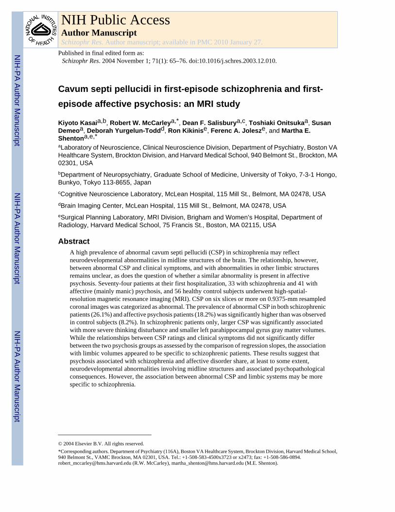
2.3. CSP measuresFor quantifying the CSP, the number of slices containing CSP (Fig. 1) was counted on a seriesof coronal images without knowledge of diagnosis. When a certain slice appeared to have apartial volume effect, we considered the slice to contain CSP. This definition may be tooinclusive, but we regard this to be objective to enable a reliable measurement. Since the imageswere 0.9375-mm thick without gaps, the multiplication of the rating by 0.9375 was a reflectionof the actual anterior-to-posterior length of the cavum, although partial volume effects rendersthis an approximation only. For example, a CSP with a rating of 6 would be approximately 5.6mm long.
From the results of recent high-spatial-resolution MRI studies (Fukuzako et al., 1996; Haginoet al., 2001; Kwon et al., 1998; Nopoulos et al., 1997; Rajarethinam et al., 2001) that reporthigh prevalence of CSP (38.0–84.8%) in adults, it is clear that the presence of a very smallCSP may have no pathological significance. Therefore, it was necessary to distinguish CSPthat were considered to be larger than the normal variant CSP. For this determination we usedcriteria similar to that of Kwon et al. (1998), where CSP on one to five slices was categorizedas normal, and on six or more slices (5.6 mm) as abnormal (see Fig. 2 for three-dimensionalpresentation of abnormal CSP). The rationale for this categorization was based on severalreferences that indicate the best estimate of normal variant cavum is that of 1–4 mm (Barr andKiernan, 1988; Martin, 1989; Roberts et al., 1987; Sarwar, 1989; Shaw and Alvord, 1969).
For interrater reliability, three raters (K.K., T.O., and S.D.), blinded to group membership,independently rated CSP for 50 cases. The intraclass correlation coefficient was 0.98.
2.4. Statistical analyses2.4.1. Group differences in CSP—The prevalence of abnormal CSP was comparedbetween each combination of two diagnostic groups using the goodness of fit test. Forcomparison purpose, the same statistical procedure was applied to the data where the subjectswith complete fusion of septum (CSP = 0) were included in the analysis.
2.4.2. Correlations with clinical measures—Spearman’s rho was used in plannedanalyses of the correlations between CSP ratings and four syndrome factor scores of the BPRSseparately for each psychosis group. The number of subjects for whom BPRS scores wereavailable was N = 32 for the schizophrenia group and N = 38 for the affective group. Here, weused p < 0.05 as the cut-off for reporting statistical significance. In addition, we used one-tailedtests since negative correlations between CSP ratings and severity of clinical symptoms werepredicted. We did not use BPRS individual items (18 items) in order to avoid type I errors. Wethen tested whether there were differences between groups in the relationship between CSPratings and clinical measures using the test for comparison of two regression slopes. Here, weused p < 0.05 as the cut-off for reporting statistical significance.
Spearman’s rho was also used in exploratory analyses of the correlations between ratings ofCSP and other clinical measures including MMSE, WAIS-R subscales, and GAS, separatelyfor each diagnostic group (GAS scores were only applicable for the two psychosis group),where we used Bonferroni correction for multiple correlations.
2.4.3. Correlations with limbic structure volumes—Spearman’s rho was calculated forplanned analyses of the correlations between CSP ratings and medial temporal lobe gray mattervolumes (left and right anterior amygdala–hippocampal complex [mostly amygdala], left andright posterior amygdale–hippocampal complex [mostly hippocampus], and left and rightparahippocampal gyri) (Hirayasu et al., 1998), separately for each diagnostic group. Thenumber of subjects for whom regions of interest (ROIs) measures were available was N = 17
Kasai et al. Page 5
Schizophr Res. Author manuscript; available in PMC 2010 January 27.
NIH
-PA Author Manuscript
NIH
-PA Author Manuscript
NIH
-PA Author Manuscript

for the schizophrenia group, N = 16 for the affective group, and N = 18 for the control group.Here, we considered any correlation to be significant if the values for both absolute and relative([absolute volume/intracranial content] × 100) volumes reached p < 0.05. Here, we did not useBonferroni correction, since these analyses were planned and significant correlations betweenCSP ratings and volumes of hippocampus and/or parahippocampal gyrus only in psychoticgroups were predicted. In addition, we used one-tailed tests since negative correlations betweenCSP ratings and volume measures were predicted. The rationale for these decisions was asfollows: (1) in normal neurodevelopment, the fusion of CSP is associated with the growth oflimbic structures; (2) previous findings in the literature suggest that abnormalities in limbicstructures, including hippocampal formation and parahippocampal gyrus, may be related topsychosis (e.g., Bogerts, 1997); and, (3) our previous study (Kwon et al., 1998) found asignificant negative correlation between CSP ratings and hippocampal volumes in patients withchronic schizophrenia. We then tested whether there were differences between groups for therelationship between CSP ratings and volume measures using the test for comparison of tworegression slopes in each combination of the three diagnostic groups. Here, we used p < 0.05as the cut-off for reporting statistical significance.
The detailed methods for measurements of medial temporal lobe ROIs are provided elsewhere(Hirayasu et al., 1998). Briefly, the amygdala–hippocampal complex and parahippocampalgyrus were outlined manually on a Sun workstation. The most anterior slice used for theamygdala–hippocampal complex was the one in which the white matter tract linking thetemporal lobe with the rest of the brain (temporal stem) could be seen. The most posterior slicewas the last appearance of fibers of the crux of the fornix. The posterior segment of theamygdala/hippocampal complex began with the first appearance of the mammillary bodies.The parahippocampal gyrus (non-subicular portions) was defined laterally by the collateralsulcus and a demarcation line drawn across the narrow portion of the gyral isthmus at thedeepest portion of the collateral sulcus. Medial temporal lobe structures, rather than neocorticalareas [i.e., STG (Hirayasu et al., 1998), Heschl’s gyrus and planum temporale (Hirayasu et al.,2000), prefrontal neocortex (Hirayasu et al., 2001), fusiform gyrus (Lee et al., 2002) as reportedin our previous studies] were selected, since an association between CSP and these limbicstructures was predicted.
3. ResultsAge, SES, parental SES, handedness, age of first medication, duration of medication, or doseof medication did not correlate with the number of slices containing CSP in any group (rho =−0.19 to 0.30; p = 0.091 to 0.88). There were also no gender differences in the prevalence ofCSP or abnormal CSP for any group, nor were there gender differences when the diagnoseswere collapsed.
3.1. Group differences in CSPThe CSP was present (1 slice or more) for N = 23/33 (69.7%) of schizophrenic patients, N =33/41 (80.5%) of affective psychosis patients, and N = 49/56 (87.5%) of control subjects. Theprevalence of CSP was not significantly different among groups (chi-square = 4.24, df = 2, p= 0.12).
We found that abnormal CSP was present in 26.1% of schizophrenic patients, 18.2% ofaffective psychosis patients, and 8.2% of control subjects (Table 2). The schizophrenic patientsshowed significantly higher prevalence than control subjects (the goodness of fit test: chi-square = 9.86, df = 1, p = 0.002). Affective psychosis patients also showed significantly higherprevalence than control subjects (chi-square = 4.42, df = 1, p = 0.036), while the two psychosisgroup did not differ significantly in the prevalence of abnormal CSP (chi-square = 1.07, df =1, p = 0.30). When we included subjects who have complete fusion of septum, the statistical
Kasai et al. Page 6
Schizophr Res. Author manuscript; available in PMC 2010 January 27.
NIH
-PA Author Manuscript
NIH
-PA Author Manuscript
NIH
-PA Author Manuscript

conclusion did not change (schizophrenia vs. control: chi-square = 6.06, df = 1, p = 0.014;affective vs. control: chi-square = 3.47, df = 1, p = 0.063; schizophrenia vs. affective: chi-square = 0.347, df = 1, p = 0.56).
3.2. Correlations with clinical measuresSchizophrenic patients showed a positive correlation between the number of slices containingCSP and scores for thinking disturbance factor of the BPRS (comprised of items of conceptualdisorganization, hallucinatory behavior, and unusual thought content) (rho = 0.365, N = 32,p = 0.040; regression line: Y = 6.46 + 0.446X). There was no significant correlation betweenCSP ratings and thinking disturbance factor scores (rho = 0.027, N = 38, p = 0.87; regressionline: Y = 7.06 − 0.0469X) for affective psychosis patients. The comparison of regression slopes,however, did not show significant difference between the two psychosis groups (t[66] = 1.49,p>0.1, two-tailed). After Bonferroni correction for multiple correlations, there were nosignificant correlations between ratings of CSP and other clinical measures including MMSE,WAIS-R subscales, and GAS, separately for each diagnostic group (GAS scores were onlyapplicable for the two psychosis groups) (Table 3).
3.3. Correlations with limbic structure volumesThere was a significantly negative correlation between CSP ratings and left parahippocampalgyrus gray matter volume in the schizophrenia group (rho = −0.462, N = 17, p = 0.031;regression line: Y = 2.45 − 0.0650X), but not in the affective group (rho = 0.039, N = 16, p =0.44; regression line: Y = 1.93 + 0.0332X) or the control group (rho = 0.353, N = 18, p = 0.076;regression line: Y = 1.83 + 0.143X). Based on the comparison of regression slopes, there wasa significant difference between schizophrenic patients and affective psychosis patients (t[29]= −2.12, p < 0.05, two-tailed), and between schizophrenic patients and control subjects (t[31]= −2.84, p < 0.01, two-tailed), but not between affective psychosis patients and control subjects(t[30] = −0.37, p>0.6, two-tailed), in the relationship between CSP ratings and leftparahippocampal gyrus gray matter volume.
4. DiscussionThe results of the current study can be summarized as follows: (1) schizophrenic patients andaffective psychosis patients did not significantly differ in the prevalence of abnormal CSP, andboth groups had significantly higher prevalence than control subjects; (2) larger CSP wasassociated with more severe thinking disturbance factor scores of the BPRS and smaller leftparahippocampal gyrus gray matter volumes in schizophrenic patients; (3) schizophrenic andaffective psychosis patients were not significantly different in the relationship of CSP withsymptoms, but the relationships with parahippocampal volumes were specific to schizophrenicpatients. The high prevalence of abnormal CSP in both psychoses and a lack of specificity inthe association with positive symptoms suggest that schizophrenia and affective psychosis mayshare, at least to a certain extent, neurodevelopmental abnormalities and associatedpsychopathological consequences. On the other hand, neurodevelopmental abnormalitiesinvolving midline structures and associated limbic systems appeared to be more specific toschizophrenic patients compared with affective psychosis and healthy subjects.
We found a high prevalence of presence of CSP for all groups (69.7% of schizophrenic patients,80.5% of affective psychosis patients, and 87.5% of control subjects). These findings areconsistent with recent high-resolution MRI studies (Fukuzako et al., 1996; Hagino et al.,2001; Kwon et al., 1998; Nopoulos et al., 1997; Rajarethinam et al., 2001), and further suggestthat a small CSP is a normal anatomical variant. In contrast, the prevalence of abnormal CSP(approximately 6 mm or longer) was higher for first-episode schizophrenic patients than for
Kasai et al. Page 7
Schizophr Res. Author manuscript; available in PMC 2010 January 27.
NIH
-PA Author Manuscript
NIH
-PA Author Manuscript
NIH
-PA Author Manuscript

the control subjects, in line with recent studies (Kwon et al., 1998; Nopoulos et al., 1997;Shioiri et al., 1996).
For associations with clinical symptoms, schizophrenic patients showed a positive correlationbetween the CSP ratings and scores for thinking disturbance factor of the BPRS. To ourknowledge, four studies (Jurjus et al., 1993; Kirkpatrick et al., 1997; Kwon et al., 1998;Nopoulos et al., 2000) have assessed relationships between CSP abnormalities and clinicalsymptoms, but only one study reported significant associations with more severe formalthought disorder (Kirkpatrick et al., 1997). Thus, the current study confirms an associationbetween CSP and positive symptoms in patients with schizophrenia. Although clinicalsymptoms may be unstable at first-hospitalization, patients may show potentially highersymptom severity especially for positive symptoms compared with remitted or chronic status,resulting in a broader distribution of data. The use of 1.0-mm resampled slices may also havean advantage in yielding broadly distributed data for CSP ratings compared with the previousthree studies using 1.5 to 5 mm slices. The septum pellucidi is a component of the limbic systemand serves as an important relay station connecting the hypothalamic autonomic system tohippocampus, amygdala, habenula, and brainstem reticular formation (Sarwar, 1989).Structural and functional abnormalities in the limbic system including hippocampal formationand parahippocampal gyrus are thought to be associated with positive symptoms ofschizophrenia (reviewed in Bogerts, 1997). The disturbance in this system in the perinatalneurodevelopment may thus form a potential basis for developing positive symptoms inpatients with schizophrenia. However, here we note that the CSP itself is associated with awide range of developmental conditions related to cognitive impairment that do not usuallyresult in manifest psychosis (e.g., Bodensteiner et al., 1998; Kim and Peterson, 2003). Inschizophrenia, early insults in the perinatal period and later mal-maturation perhapspredominantly in frontotemporal neocortex may, at least in part, form a basis for frankpsychosis. Abnormal CSP and associated limbic structures may thus be understood as an indexof the severity of neurodevelopmental insult that, if coupled with mal-maturation offrontotemporal neocortex in the peri-onset period, may form a basis for the manifestation ofpsychosis.
Partially consistent with the above interpretation, we found a significant correlation betweenCSP ratings and gray matter volumes of the left parahippocampal gyrus, a part of the limbicsystem. Moreover, this association appeared to be more specific to schizophrenia comparedwith affective psychosis and control groups based on the comparison of regression slopes.However, we failed to replicate the previous finding of a significant association between CSPratings and bilateral posterior amygdala–hippocampal (mostly hippocampus) gray mattervolumes in patients with chronic schizophrenia (Kwon et al., 1998). The reason for thediscrepancy is unclear, since using first-episode patients has theoretically fewer confounds ofchronicity of illness and the effects of chronic treatment, which should thus have an advantagein exploring the association between abnormalities of neurodevelopmental origin and ROIvolumes. One possible explanation may be differences in the clinical characteristics of the twosamples. For example, in the Kwon study, some of the most severe cases of schizophrenia wereincluded, and these individuals had an average of 15 years illness duration. In our sample, itis not known which of the first-episode cases of schizophrenia will go on to have such a pooroutcome.
Our earlier study (Kwon et al., 1998) evaluated CSP in 30 patients with schizophrenia (15chronic, 15 first-episode), 16 first-episode patients with affective psychosis, and 46 controlsubjects (21 subjects with schizotypal personality disorder were also included). In that study,the prevalence of abnormal CSP in affective psychosis was intermediate (20%) betweenschizophrenic patients (30.4%) and control subjects (10.3%), with no significant differencefrom either group. In the present study, with larger sample sizes, the prevalence of abnormal
Kasai et al. Page 8
Schizophr Res. Author manuscript; available in PMC 2010 January 27.
NIH
-PA Author Manuscript
NIH
-PA Author Manuscript
NIH
-PA Author Manuscript

CSP for affective psychosis patients was shown to be significantly higher than that for controlsubjects. Moreover, based on analyses using the comparison of regression slopes, therelationship of abnormal CSP to abnormalities in clinical presentation were also notsignificantly different between schizophrenic and affective psychosis patients. Thus, thepresent study indicates that psychosis associated with schizophrenia and affective disorder mayshare an abnormal neurodevelopment of the brain midline structures and associatedpsychopathological outcome. These results are in line with those of Shioiri et al. (1996), whoreported significantly higher prevalence of CSP in bipolar patients (7.2%) compared withcontrol subjects (1.1%), but not significantly different from schizophrenic patients (17.5%). Incontrast, Jurjus et al. (1993) reported that affective patients (bipolar and schizoaffectivedisorder) were not different in the prevalence of CSP (10.0%) from normal controls (18.9%),and showed significantly lower prevalence than schizophrenic patients (25.4%). However,since these previous studies did not assess psychotic patients with affective disorder only, directcomparison with the present study should be regarded with caution.
Some methodological issues in our study need to be commented upon. First, we need toconsider the issue of statistical power regarding our results of lack of significant differencebetween schizophrenia and affective psychosis in the clinical relationships with CSP. Althoughthe required N was fairly large for the correlation with BPRS (N = 120 for both groups) to besignificantly different between groups, the results of similarities in the clinical correlationsshould be confirmed by studies with larger sample size. Second, the results of our correlationalanalyses should be interpreted with caution since there were multiple statistical comparisonsthus being liable to type I errors, and the N for analyses of associations with volume measureswas relatively small. Third, we need to comment on the fact that our first-episode psychosissample included some later-onset subjects aged above 30 at first-hospitalization (9 out of 74psychosis subjects). However, the rate of the presence of abnormal CSP did not differsignificantly between the younger (10/65) and older (2/9) subjects (Fisher’s exact test, p =0.63). Therefore, this sample characteristic did not appear to affect the conclusion of the presentstudy.
In conclusion, the similarities in the high prevalence of abnormal CSP and its association withpositive symptoms suggest the importance of CSP as an index of neurodevelopmentalabnormalities and its psychopathological consequences in the pathophysiology of psychosisassociated with schizophrenia and affective disorder. On the other hand, associations betweenneurodevelopmental abnormalities involving midline structures and limbic systems may bespecific to schizophrenia.
AcknowledgmentsThis study was supported, in part, by the Department of Veterans Affairs Merit Awards (RWM and MES), a MiddletonAward from the Department of Veterans Affairs (RWM), grants from the National Institute of Health (K02 MH 01110[MES] and R01 MH 50747 [MES], and R01 MH 40799 [RWM], R01 RR 11747 [RK], and P41 PR13218 [FAJ]), theWelfide Medicinal Research Foundation, Japan (KK), and the Uehara Memorial Foundation, Japan (KK). The authorsgratefully acknowledge the administrative support of Marie Fairbanks and the research assistant support of MagdalenaSpencer and Margaret Fagan.
ReferencesAkil, M.; Weinberger, DR. Neuropathology and the neurodevelopmental model. In: Harrison, PJ.;
Roberts, GW., editors. The Neuropathology of Schizophrenia. New York: Oxford Univ. Press; 2000.p. 189-212.
Barr, M.; Kiernan, J. The Human Nervous System: An Anatomical Viewpoint. Philadelphia: J.B.Lippincott; 1988.
Bearden CE, Hoffman KM, Cannon TD. The neuropsychology and neuroanatomy of bipolar affectivedisorder: a critical review. Bipolar Disord 2001;3:106–150. [PubMed: 11465675]
Kasai et al. Page 9
Schizophr Res. Author manuscript; available in PMC 2010 January 27.
NIH
-PA Author Manuscript
NIH
-PA Author Manuscript
NIH
-PA Author Manuscript

Bodensteiner JB, Schaefer GB, Craft JM. Cavum septi pellucidi and cavum vergae in normal anddevelopmentally delayed populations. J. Child Neurol 1998;13:120–121. [PubMed: 9535237]
Bogerts B. The temporolimbic system theory of positive schizophrenic symptoms. Schizophr. Bull1997;23:423–435. [PubMed: 9327507]
Degreef G, Bogerts B, Falkai P, Greve B, Lantos G, Ashtari M, Lieberman J. Increased prevalence ofthe cavum septum pellucidum in magnetic resonance scans and post-mortem brains of schizophrenicpatients. Psychiatry Res. Neuroimaging 1992;45:1–13.
DeLisi LE, Hoff AL, Kushner M, Degreef G. Increased prevalence of cavum septum pellucidum inschizophrenia. Psychiatry Res. Neuroimaging 1993;50:193–199.
DeLisi LE, Sakuma M, Tew W, Kushner M, Hoff AL, Grimson R. Schizophrenia as a chronic activebrain process: a study of progressive brain structural change subsequent to the onset of schizophrenia.Psychiatry Res. Neuroimaging 1997;74:129–140.
Endicott J, Spitzer RL, Fleiss JL, Cohen J. The global assessment scale. A procedure for measuring overallseverity of psychiatric disturbance. Arch. Gen. Psychiatry 1976;33:766–771. [PubMed: 938196]
Folstein MF, Folstein SE, McHugh PR. “Mini-Mental State”: a practical method for grading the cognitivestate of patients for the clinician. J. Psychiatr. Res 1975;12:189–198. [PubMed: 1202204]
Fukuzako T, Fukuzako H, Kodama S, Hashiguchi T, Takigawa M. Cavum septum pellucidum inschizophrenia: a magnetic resonance imaging study. Psychiatry Clin. Neurosci 1996;50:125–128.[PubMed: 9201757]
Gerig G, Kubler O, Kikinis R, Jolesz FA. Nonlinear anisotropic filtering of MRI data. IEEE Trans. Med.Imag 1992;11:221–232.
Gur RE, Cowell P, Turetsky BI, Gallacher F, Cannon T, Bilker W, Gur RC. A follow-up magneticresonance imaging study of schizophrenia. Relationship of neuroanatomical changes to clinical andneurobehavioral measures. Arch. Gen. Psychiatry 1998;55:145–152. [PubMed: 9477928]
Hagino H, Suzuki M, Kurokawa K, Mori K, Nohara S, Takahashi T, Yamashita I, Yotsutsuji T, KurachiM, Seto H. Magnetic resonance imaging study of the cavum septi pellucidi in patients withschizophrenia. Am. J. Psychiatry 2001;158:1717–1719. [PubMed: 11579008]
Hirayasu Y, Shenton ME, Salisbury DF, Dickey CC, Fisher IA, Mazzoni P, Kisler T, Aarakaki H, KwonJS, Anderson JE, Yurgelun-Todd D, Tohen M, McCarley RW. Lower left temporal lobe MR volumesin patients with first-episode schizophrenia compared with psychotic patients with first-episodeaffective disorder and normal subjects. Am. J. Psychiatry 1998;155:1384–1391. [PubMed: 9766770]
Hirayasu Y, Shenton ME, Salisbury DF, Kwon JS, Wible CG, Fischer IA, Yurgelun-Todd D, Zarate C,Kikinis R, Jolesz FA, McCarley RW. Subgenual cingulate cortex volume in first-episode psychosis.Am. J. Psychiatry 1999;156:1091–1093. [PubMed: 10401458]
Hirayasu Y, McCarley RW, Salisbury DF, Tanaka S, Kwon JS, Frumin M, Snyderman D, Yurgelun-Todd D, Kikinis R, Jolesz FA, Shenton ME. Planum temporale and Heschl gyrus volume reductionin schizophrenia. Arch. Gen. Psychiatry 2000;57:692–699. [PubMed: 10891040]
Hirayasu Y, Tanaka S, Shenton ME, Salisbury DF, DeSantis MA, Levitt JJ, Wible C, Yurgelun-Todd D,Kikinis R, Jolesz FA, McCarley RW. Prefrontal gray matter volume reduction in first-episodeschizophrenia. Cereb. Cortex 2001;11:374–381. [PubMed: 11278200]
Hollingshead, AB. Two Factor Index of Social Position. New Haven: Yale Univ. Press; 1965.Hopkins, R.; Lewis, S. Structural imaging findings and macroscopic pathology. In: Harrison, PJ.; Roberts,
GW., editors. The Neuropathology of Schizophrenia. New York: Oxford Univ. Press; 2000. p. 5-56.Jurjus G, Nasrallah HA, Olson SC, Schwarzkopf SB. Cavum septum pellucidum in schizophrenia,
affective disorder, and healthy controls: a magnetic resonance imaging study. Psychol. Med1993;23:319–322. [PubMed: 8332648]
Kasai K, Shenton ME, Salisbury DF, Hirayasu Y, Lee CU, Ciszewski AA, Yurgelun-Todd DA, KikinisR, Jolesz FA, McCarley RW. Progressive decrease of left superior temporal gyrus gray matter volumein first-episode schizophrenia. Am. J. Psychiatry 2003;160:156–164. [PubMed: 12505815]
Kim KJ, Peterson BS. Cavum septi pellucidi in Tourette syndrome. Biol. Psychiatry 2003;54:76–85.[PubMed: 12842311]
Kirkpatrick B, Litman D, Kim JW, Vladar K, Breier A, Buchanan RW. Failure of fusion of the septumpellucidum and the heterogeneity of schizophrenia. J. of Nerv. Ment. Dis 1997;185:639–641.[PubMed: 9345255]
Kasai et al. Page 10
Schizophr Res. Author manuscript; available in PMC 2010 January 27.
NIH
-PA Author Manuscript
NIH
-PA Author Manuscript
NIH
-PA Author Manuscript

Kwon JS, Shenton ME, Hirayasu Y, Salisbury DF, Fischer IA, Dickey CC, Yurgelun-Todd D, Tohen M,Kikinis R, Jolesz FA, McCarley RW. MRI study of cavum septi pellucidi in schizophrenia, affectivedisorder, and schizotypal personality disorder. Am. J. Psychiatry 1998;155:509–515. [PubMed:9545997]
Lee CU, Shenton ME, Salisbury DF, Kasai K, Onitsuka T, Dickey CC, Yurgelun-Todd D, Kikinis R,Jolesz FA, McCarley RW. Fusiform gyrus volume reduction in first-episode schizophrenia: an MRIStudy. Arch. Gen. Psychiatry 2002;59:775–781. [PubMed: 12215076]
Lieberman JA, Chakos M, Wu H, Alvir J, Hoffman E, Robinson D, Bilder R. Longitudinal study of brainmorphology in first episode schizophrenia. Biol. Psychiatry 2001;49:487–499. [PubMed: 11257234]
Martin, J. Neuroanatomy: Text and Atlas. New York: Elsevier; 1989.Mathalon DH, Sullivan EV, Lim KO, Pfefferbaum A. Progressive brain volume changes and the clinical
course of schizophrenia in men: a longitudinal magnetic resonance imaging study. Arch. Gen.Psychiatry 2001;58:148–157. [PubMed: 11177116]
Nasrallah HA. Neurodevelopmental aspects of bipolar affective disorder. Biol. Psychiatry 1991;29:1–2.[PubMed: 2001443]
Nopoulos P, Swayze V, Andreasen NC. Pattern of brain morphology in patients with schizophrenia andlarge cavum septi pellucidi. J. Neuropsychiatry Clin. Neurosci 1996;8:147–152. [PubMed: 9081549]
Nopoulos P, Swayze V, Flaum M, Ehrhardt JC, Yuh WTC, Andreasen NC. Cavum septi pellucidi innormals and patients with schizophrenia as detected by magnetic resonance imaging. Biol. Psychiatry1997;41:1102–1108. [PubMed: 9146821]
Nopoulos P, Krie A, Andreasen NC. Enlarged cavum septi pellucidi in patients with schizophrenia:clinical and cognitive correlates. J. Neuropsychiatry Clin. Neurosci 2000;12:344–349. [PubMed:10956567]
Oldfield RC. The assessment and analysis of handedness: the Edinburgh Inventory. Neuropsychologia1971;9:97–113. [PubMed: 5146491]
Overall JE, Gorham DR. The brief psychiatric rating scale. Psychol. Rep 1962;10:799–812.Overall JE, Hollister LE, Pichot P. Major psychiatric disorders: a four-dimensional model. Arch. Gen.
Psychiatry 1967;16:146–151. [PubMed: 6019329]Rajarethinam R, Miedler J, DeQuardo J, Smet CI, Brunberg J, Kirbat R, Tandon R. Prevalence of cavum
septum pellucidum in schizophrenia studied with MRI. Schizophr. Res 2001;48:201–205. [PubMed:11295373]
Rakic P, Yakovlev PI. Development of corpus callosum and cavum septi in man. J. Comp. Neurol1968;132:355–362.
Roberts, M.; Hanaway, J.; Morest, D. Atlas of the Human Brain in Section. 2nd ed.. Philadelphia: Lea& Febiger; 1987.
Salisbury DF, Shenton ME, Sherwood AR, Fischer IA, Yurgelun-Todd DA, Tohen M, McCarley RW.First-episode schizophrenic psychosis differs from first-episode affective psychosis and controls inP300 amplitude over left temporal lobe. Arch. Gen. Psychiatry 1998;55:173–180. [PubMed:9477932]
Sarwar M. The septum pellucidum: normal and abnormal. Am. J. Neuroradiol 1989;10:989–1005.[PubMed: 2505543]
Shaw CM, Alvord EC. Cavum septi pellucidi et vergae: their normal and pathological state. Brain1969;92:213–224. [PubMed: 5774029]
Shioiri T, Oshitani Y, Kato T, Murashita J, Hamakawa H, Inubushi T, Nagata T, Takahashi S. Prevalenceof cavum septum pellucidum detected by MRI in patients with bipolar disorder, major depressionand schizophrenia. Psychol. Med 1996;26:431–434. [PubMed: 8685300]
Spitzer, RL.; Williams, JBW.; Gibbson, M.; First, M. The Structured Clinical Interview for DSM-III-R-Non-Patient Edition (SCID-NP). Washington, DC: American Psychiatric Association; 1990a.
Spitzer, RL.; Williams, JBW.; Gibbson, M.; First, M. The Structured Clinical Interview for DSM-III-R(SCID). Washington, DC: American Psychiatric Association; 1990b.
Wechsler, D. Wechsler Adult Intelligence Scale-Revised. New York: Harcourt Brace Jovanovich; 1981.
Kasai et al. Page 11
Schizophr Res. Author manuscript; available in PMC 2010 January 27.
NIH
-PA Author Manuscript
NIH
-PA Author Manuscript
NIH
-PA Author Manuscript

Wible CG, Shenton ME, Hokama H, Kikinis R, Jolesz FA, Metcalf D, McCarley RW. Prefrontal cortexand schizophrenia: a quantitative magnetic resonance imaging study. Arch. Gen. Psychiatry1995;52:279–288. [PubMed: 7702444]
Kasai et al. Page 12
Schizophr Res. Author manuscript; available in PMC 2010 January 27.
NIH
-PA Author Manuscript
NIH
-PA Author Manuscript
NIH
-PA Author Manuscript

Fig. 1.MR images of cavum septi pellucidi. Panel A: a normal subject showing no cavum septipellucidi. Panel B: a first-episode patient with schizophrenia showing abnormal cavum septipellucidi on the coronal plane.
Kasai et al. Page 13
Schizophr Res. Author manuscript; available in PMC 2010 January 27.
NIH
-PA Author Manuscript
NIH
-PA Author Manuscript
NIH
-PA Author Manuscript

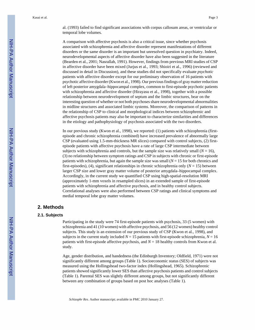
Fig. 2.Three-dimensional reconstruction of a large cavum septi pellucidi superimposed on the axialand coronal planes. The MRI image presented here is from the same patient with schizophreniaas in Panel B of Fig. 1. The CSF content of the large cavum can be seen between the two leafletsof the septi pellucidi and is labeled blue. The anterior portions of the leaflets, which areseparated to form the cavum between the septi pellucidi, are green on subject left and gold onsubject right. The posterior portions of the leaflets, where they fuse, are light blue on subjectleft and red on subject right.
Kasai et al. Page 14
Schizophr Res. Author manuscript; available in PMC 2010 January 27.
NIH
-PA Author Manuscript
NIH
-PA Author Manuscript
NIH
-PA Author Manuscript

NIH
-PA Author Manuscript
NIH
-PA Author Manuscript
NIH
-PA Author Manuscript
Kasai et al. Page 15
Tabl
e 1
Dem
ogra
phic
and
clin
ical
cha
ract
eris
tics o
f stu
dy g
roup
s
Var
iabl
eSc
hizo
phre
nic
patie
nts (
N =
33)
Affe
ctiv
e di
sord
erpa
tient
s (N
= 4
1)C
ontr
ol su
bjec
ts(N
= 5
6)F
or t
orch
i val
ues
dfa
p
Mea
nS.
D.
Mea
nS.
D.
Mea
nS.
D.
Age
(ran
ge),
year
24.7
(18–
41)
6.5
22.8
(18–
41)
4.6
24.0
(18–
35)
3.9
1.52
2127
0.22
Mal
e/fe
mal
e28
/531
/10
44/1
20.
972
0.61
Han
dedn
essb
0.8
0.2
0.8
0.2
0.8
0.2
0.26
2124
0.77
SESc
3.5
1.4
2.7
1.2
2.3
1.0
12.1
2127
<0.0
01
Pare
ntal
SES
d1.
90.
71.
50.
81.
60.
93.
4121
260.
036
MM
SE28
.22.
428
.71.
529
.11.
02.
5221
220.
085
WA
IS-R
Inf
orm
atio
ne11
.63.
112
.42.
913
.52.
35.
3421
200.
006
Dig
its fo
rwar
df8.
92.
58.
72.
111
.22.
612
.521
20<0
.001
Dig
its b
ackw
ardf
6.5
2.9
6.7
2.2
8.6
2.3
9.96
2120
<0.0
01
GA
S35
.99.
136
.211
.0–
–−0
.35
680.
72
Tota
l BPR
S40
.812
.734
.410
.5–
–0.
7768
0.45
Med
icat
ion
dose
(CPZ
equi
v.),
m
g/da
y
264
335
223
186
––
0.67
710.
50
Age
firs
t med
icat
ed (r
ange
),ye
ar24
.5 (1
8–41
)6.
422
.6 (1
7–41
)4.
7–
–1.
5172
0.14
Med
ian
dura
tion
(ran
ge)
of
med
icat
ion
use,
mon
ths
0 (0
–24)
–0
(0–6
0)–
––
––
–
SES,
soci
oeco
nom
ic st
atus
; MM
SE, M
ini-M
enta
l Sta
te E
xam
inat
ion;
WA
IS-R
, Wec
hsle
r Adu
lt In
telli
genc
e Sc
ale-
Rev
ised
; GA
S, G
loba
l Ass
essm
ent S
cale
; BPR
S, B
rief P
sych
iatri
c R
atin
g Sc
ale;
CPZ
equ
iv.,
chlo
rpro
maz
ine
equi
vale
nt; a
nd e
llips
es, d
ata
not a
pplic
able
.
a The
degr
ees o
f fre
edom
diff
er a
mon
g va
riabl
es d
ue to
una
vaila
bilit
y of
dat
a in
som
e su
bjec
ts.
b Rig
ht-h
ande
dnes
s bei
ng a
bove
0.
c Hig
her s
core
s ind
icat
ing
low
er so
cio-
econ
omic
stat
us. T
he sc
hizo
phre
nic p
atie
nts s
how
ed si
gnifi
cant
ly lo
wer
SES
than
affe
ctiv
e psy
chos
is p
atie
nts a
nd co
ntro
l sub
ject
s (Tu
key’
s Hon
estly
Sig
nific
ant D
iffer
ence
test
s, p
< 0.
05).
d Bas
ed o
n th
e po
st h
oc a
naly
ses,
neith
er g
roup
was
sign
ifica
ntly
diff
eren
tiate
d fr
om e
ach
othe
r for
the
pare
ntal
SES
(Tuk
ey’s
Hon
estly
Sig
nific
ant D
iffer
ence
test
s, p>
0.05
).
Schizophr Res. Author manuscript; available in PMC 2010 January 27.

NIH
-PA Author Manuscript
NIH
-PA Author Manuscript
NIH
-PA Author Manuscript
Kasai et al. Page 16e Th
e sc
hizo
phre
nic
patie
nts s
how
ed si
gnifi
cant
ly lo
wer
scor
es th
an c
ontro
l sub
ject
s (Tu
key’
s Hon
estly
Sig
nific
ant D
iffer
ence
test
s, p
< 0.
05).
f Bot
h sc
hizo
phre
nic
and
affe
ctiv
e ps
ycho
sis p
atie
nts s
how
ed si
gnifi
cant
ly lo
wer
scor
es th
an c
ontro
l sub
ject
s (Tu
key’
s Hon
estly
Sig
nific
ant D
iffer
ence
test
s, p
< 0.
05).
Schizophr Res. Author manuscript; available in PMC 2010 January 27.

NIH
-PA Author Manuscript
NIH
-PA Author Manuscript
NIH
-PA Author Manuscript
Kasai et al. Page 17
Table 2
Frequency of normal and abnormal cavum septi pellucidi in first-episode patients with schizophrenia, first-episode patients with affective psychosis, and healthy control subjects
Group Cavum septi pellucidi
Normal(1–5 slices)
Abnormal(6 slices or more)
N % N %
Schizophrenic 17 73.9 6 26.1a
patients
Affective 27 81.8 6 18.2b
psychosis
patients
Control 45 91.8 4 8.2
subjects
aSignificantly higher than expected in control subjects (the goodness of fit test: chi-square = 9.86, df = 1, p = 0.002).
bSignificantly higher than expected in control subjects (the goodness of fit test: chi-square = 4.42, df = 1, p = 0.036).
Schizophr Res. Author manuscript; available in PMC 2010 January 27.

NIH
-PA Author Manuscript
NIH
-PA Author Manuscript
NIH
-PA Author Manuscript
Kasai et al. Page 18
Table 3
Correlation between cavum septi pellucidi ratings and clinical and volume measures
Variables Spearman’s rhoa
Schizophrenicpatients
Affectivepatients
Controlsubjects
Clinical measure (BPRS factor) X CSP rating
Thinking disturbance 0.365b 0.027 –
(N = 32) (N = 38)
Hostility– −0.058 −0.047 –
suspiciousness (N = 32) (N = 38)
Withdrawal– 0.227 −0.082 –
retardation (N = 32) (N = 38)
Anxiety–depression 0.020 −0.067 –
(N = 32) (N = 38)
Limbic gray matter volume X CSP rating
Left amygdala 0.040 0.181 0.563c
(N = 17) (N = 16) (N = 18)
Right amygdala 0.227 0.169 0.204
(N = 17) (N = 16) (N = 18)
Left hippocampus 0.468 0.226 0.342
(N = 17) (N = 16) (N = 18)
Right hippocampus 0.173 0.355 0.512c
(N = 17) (N = 16) (N = 18)
Left parahippocampal −0.462d 0.039 0.353
gyrus (N = 17) (N = 16) (N = 18)
Right parahippocampal −0.034 −0.279 0.609c
gyrus (N = 17) (N = 16) (N = 18)
BPRS, Brief Psychiatric Rating Scale; ellipses, data not applicable.
aThe number of subjects varied according to the availability of data.
bStatistically significant (p = 0.020, one-tailed).
cStatistically significant positive correlations. However, the values reported here are for absolute volumes, and none of them remained significant
when relative volumes were used. Therefore, these correlations were not considered important, since they may simply have been the result of differencesin overall brain/head size.
dStatistically significant (p = 0.031, one-tailed). Remained significant when the relative volume was used (rho = −0.440, p = 0.038).
Schizophr Res. Author manuscript; available in PMC 2010 January 27.