case files gynecologic surgery (lange case files) - 1 file
TRANSCRIPT
CASE FILES® Gynecologic Surgery
Eugene C. Toy, MD
The John S. Dunn Senior Academic Chair and Program Director
Obstetrics and Gynecology Residency Program
Vice Chair of Academic Affairs
Department of Obstetrics and Gynecology
The Methodist Hospital-Houston Clerkship Director and Clinical Professor
Department of Obstetrics and Gynecology
University of Texas Medical School at Houston
Houston, Texas
Konrad P. Harms, MD
Assistant Professor of Obstetrics and Gynecology
Weill Cornell Medical College
Associate Program Director, The Methodist Hospital Ob/Gyn Residency Program
Houston, Texas
Keith O. Reeves, MD
Clinical Professor of Obstetrics and Gynecology
Weill Medical College of Cornell University
Medical Director, Methodist Center for Restorative Pelvic Medicine
Houston, Texas
Cristo Papasakelariou, MD, FACOG
Clinical Professor Department of Obstetrics/Gynecology
University of Texas Medical Branch
www.cambodiamed.blogspot.com Best Medical Book | Chy Yong

Galveston, Texas
Director of Gynecologic Surgery
St Joseph Medical Center
Houston, Texas
Copyright © 2011 by The McGraw-Hill Companies, Inc. All rights reserved. Printed in the UnitedStates of America. Except as permitted under the United States Copyright Act of 1976, no part of thispublication may be reproduced or distributed in any form or by any means, or stored in a database orretrieval system, without the prior written permission of the publisher.
ISBN: 978-0-07-159279-6
MHID: 0-07-159279-2
The material in this eBook also appears in the print version of this title: ISBN: 978-0-07-159280-2,MHID: 0-07-159280-6.
All trademarks are trademarks of their respective owners. Rather than put a trademark symbol afterevery occurrence of a trademarked name, we use names in an editorial fashion only, and to the benefitof the trademark owner, with no intention of infringement of the trademark. Where such designationsappear in this book, they have been printed with initial caps.
McGraw-Hill eBooks are available at special quantity discounts to use as premiums and salespromotions, or for use in corporate training programs. To contact a representative please e-mail us [email protected].
Notice
Medicine is an ever-changing science. As new research and clinical experience broaden ourknowledge, changes in treatment and drug therapy are required. The authors and the publisher of thiswork have checked with sources believed to be reliable in their efforts to provide information that iscomplete and generally in accord with the standards accepted at the time of publication. However, inview of the possibility of human error or changes in medical sciences, neither the authors nor thepublisher nor any other party who has been involved in the preparation or publication of this workwarrants that the information contained herein is in every respect accurate or complete, and theydisclaim all responsibility for any errors or omissions or for the results obtained from use of theinformation contained in this work. Readers are encouraged to confirm the information containedherein with other sources. For example and in particular, readers are advised to check the productinformation sheet included in the package of each drug they plan to administer to be certain that theinformation contained in this work is accurate and that changes have not been made in therecommended dose or in the contraindications for administration. This recommendation is ofparticular importance in connection with new or infrequently used drugs.
TERMS OF USE
This is a copyrighted work and The McGraw-Hill Companies, Inc. (“McGraw-Hill”) and its licensorsreserve all rights in and to the work. Use of this work is subject to these terms. Except as permittedunder the Copyright Act of 1976 and the right to store and retrieve one copy of the work, you may notdecompile, disassemble, reverse engineer, reproduce, modify, create derivative works based upon,

transmit, distribute, disseminate, sell, publish or sublicense the work or any part of it withoutMcGraw-Hill’s prior consent. You may use the work for your own noncommercial and personal use;any other use of the work is strictly prohibited. Your right to use the work may be terminated if youfail to comply with these terms.
THE WORK IS PROVIDED “AS IS.” McGRAW-HILL AND ITS LICENSORS MAKE NOGUARANTEES OR WARRANTIES AS TO THE ACCURACY, ADEQUACY OR COMPLETENESSOF OR RESULTS TO BE OBTAINED FROM USING THE WORK, INCLUDING ANYINFORMATION THAT CAN BE ACCESSED THROUGH THE WORK VIA HYPERLINK OROTHERWISE, AND EXPRESSLY DISCLAIM ANY WARRANTY, EXPRESS OR IMPLIED,INCLUDING BUT NOT LIMITED TO IMPLIED WARRANTIES OF MERCHANTABILITY ORFITNESS FOR A PARTICULAR PURPOSE. McGraw-Hill and its licensors do not warrant orguarantee that the functions contained in the work will meet your requirements or that its operationwill be uninterrupted or error free. Neither McGraw-Hill nor its licensors shall be liable to you oranyone else for any inaccuracy, error or omission, regardless of cause, in the work or for any damagesresulting therefrom. McGraw-Hill has no responsibility for the content of any information accessedthrough the work. Under no circumstances shall McGraw-Hill and/or its licensors be liable for anyindirect, incidental, special, punitive, consequential or similar damages that result from the use of orinability to use the work, even if any of them has been advised of the possibility of such damages.This limitation of liability shall apply to any claim or cause whatsoever whether such claim or causearises in contract, tort or otherwise.

DEDICATION
To the aspiring gynecologic surgeons everywhere; to those who dedicate themselves to scholarlydiscernment of who needs medicines or the scalpel; and to those who are committed to obtainingmanual dexterity, may those hands be graced with divine guidance and unlimited compassion.
— ECT
CONTENTS
Contributors
Acknowledgments
Introduction
Section I How to Approach Clinical ProblemsPart 1. Approach to the PatientPart 2. Approach to Clinical Problem SolvingPart 3. Approach to Surgical Therapy
Section II Clinical CasesForty Case Scenarios
Section III Listing of CasesListing by Case NumberListing by Disorder (Alphabetical)
Index
CONTRIBUTORS
Barrett Blaue, MDCentral Texas Medical CenterSan Marcos, Texas16. Ectopic Pregnancy-Laparotomy22. Surgical Management of Postpartum Hemorrhage37. Ovarian Cystectomy
Tri A. Dinh, MDAssistant ProfessorDepartment of Obstetrics and GynecologyWeill Cornell Medical CollegeChief, Division of Gynecologic OncologyThe Methodist HospitalHouston, Texas18. Ovarian Cancer Surgery19. Radical Hysterectomy
Jonathan M. Espana, MDFellow, Minimally Invasive Gynecological SurgeryDepartment of Obstetrics and GynecologyThe Methodist HospitalHouston, Texas4. Laparoscopic Diagnostic5. Robotic Surgery
Tametra Johnson Garnier, MDObstetrics and Gynecology Specialist for WomenMemorial Hermann HospitalThe Woodlands, Texas40. Treatment of Cervical Dysplasia41. Cold Knife Conization
R. Moss Hampton, MDAssociate ProfessorDepartment of Obstetrics and GynecologyTexas Tech University Health Sciences Center of the Permi BasinOdessa, Texas6. Electrocautery with Laparoscopy (bowel injury)17. Total Abdominal Hysterectomy
Jeané Simmons Holmes, MD, FACOGAssistant ProfessorWeill Cornell Medical College
The Methodist HospitalObstetrics and Gynecology Residency ProgramHouston, Texas34. Surgical Indications for DUB35. Surgical Indications for Chronic Pelvic Pain36. Hysteroscopic Complications
Eric A. Hurtado, MDUrogynecology and Reconstructive Pelvic SurgeryMid-Atlantic Pelvic Surgery AssociatesAssistant Clinical Professor of Obstetrics and GynecologyThe George Washington University School of MedicineFairfax, Virginia27. Sling Procedures28. Cystoscopy
Alan L. Kaplan, MDProfessor, Obstetrics and GynecologyWeill Medical College of Cornell UniversityChairman, Department of Obstetrics and GynecologyThe Methodist HospitalHouston, Texas18. Ovarian Cancer Surgery
George A. Pados, MD, PhDAssistant Professor, Obstetrics and GynecologyDepartment of Obstetrics and GynceologyAristotle UniversityPapageorgiou General HospitalThessaloniki, Greece11. Laparoscopic Myomectomy13. Laparoscopic Endometriosis
Harry Reich, MD, FACOGEmeritus StaffWilkes-Barre General HospitalWilkes-Barre, Pennsylvania9. Total Laparoscopic Hysterectomy
Gwyn Richardson, MD, JDAssistant ProfessorDepartment of Obstetrics and GyneoclogyUniversity of Texas Medical BranchDivision of Gynecologic OncologyGalverston, Texas18. Ovarian Cancer Surgery19. Radical Hysterectomy
Priti P. Schachel, MD, FACOGFaculty, The Methodist Hospital-HoustonObstetrics and Gynecology Residency ProgramAssistant Professor, Obstetrics and GynecologyWeill Cornell Medical CollegeHouston, Texas8. Laparoscopic Complication14. Laparoscopic Appendectomy20. Myomectomy33. Surgical Therapy for Fibroids
Dimitris Tsolakidis, MD, PhDScientific FellowDepartment of Obstetrics and GynceologyAristotle UniversityPapageorgiou General HospitalThessaloniki, Greece11. Laparoscopic Myomectomy13. Laparoscopic Endometriosis
David E. Zepeda, MDClinical Associate ProfessorBaylor College of MedicineDirector of Gynecologic Minimally Invasive SurgeryThe Methodist HospitalHouston, Texas4. Laparoscopic Diagnostic5. Robotic Surgery
ACKNOWLEDGMENTS
The curriculum that evolved into the ideas for this series was inspired by Larry C. Gilstrap III, MD,when he was chairman of obstetrics and gynecology at the University of Texas Medical School atHouston. Dr. Gilstrap is a man of such a myriad of talents and is my personal inspiration for much ofthe teaching that I do today. It has been a tremendous joy to work with my excellent coauthors:Konrad Harms, who is a loyal and talented associate program director and excellent surgeon; Dr.Keith O. Reeves who as codirector of the Methodist Hospital Pelvic and Reconstructive SurgicalProgram is an outstanding scientist, vaginal surgeon, and leader; and to my dear friend and colleague,Dr. Cristo Papasakelariou, whose expertise in minimally invasive surgery is known worldwide. I amgreatly indebted to my editor, Catherine Johnson, whose exuberance, experience, and vision helped toshape this series. I appreciate McGraw-Hill’s believing in the concept of teaching through clinicalcases, and I would like to especially acknowledge Cindy Yoo for her editing expertise and CatherineSaggese and Rajni Pisharody for the excellent production. I appreciate Linda Bergstrom for her sageadvice and support. At Methodist, I appreciate Drs. Alan Kaplan, Judy Paukert, Dirk Sostman, MarcBoom, and Karin Larson-Pollock; Ayse McCracken, and David Campbell for their leadership; andBarbara Hagemeister and Tyler Kinney, who hold the department together. Without my dearcolleagues, Drs. Jeane Holmes and Priti Schachel, this book could not have been written. Most of all, Iappreciate my ever-loving wife Terri, and our four wonderful children, Andy, Michael, Allison, andChristina, for their patience and understanding.
Eugene C. Toy
INTRODUCTION
HOW TO USE THIS BOOK
Mastering the surgical approaches to clinical problems within a field as broad such as obstetrics andgynecology is a formidable task. It requires drawing on a knowledge base, to procure and filterthrough the clinical and laboratory data, to develop a differential diagnosis, and finally to make arational treatment plan. To gain these skills, the clinician is best guided and instructed by experiencedteachers and accomplished surgeons, and inspired toward self-directed, diligent reading and practicingone’s craft. Clearly, there is no replacement for experience at the bedside and the surgical suite.Unfortunately, younger physicians will not have encountered the diversity of clinical situations, ordealt with the more unusual surgical complications. Perhaps the best alternative is a carefully craftedpatient case designed to stimulate the clinical and surgical approach and decision making. In anattempt to achieve that goal, we have constructed a collection of clinical vignettes to teach diagnostic,therapeutic, and surgical approaches relevant to obstetrics and gynecology. Most importantly, theexplanations for the cases emphasize the underlying principles, rather than merely rote questions andanswers.
This book is organized for versatility: It allows the physician “in a rush” to go quickly throughthe scenarios and check the corresponding answers, and it provides more detailed information for theclinician who wants thought-provoking explanations. The answers are arranged from simple tocomplex: a summary of the pertinent points, the bare answers, an analysis of the case, an approach tothe topic, a comprehension test at the end for reinforcement and emphasis, and a list of resources forfurther reading. The clinical vignettes are purposely placed in random order to simulate the way thatreal patients present to the practitioner. A listing of cases is included in Section III. The information ispresented with the degree of evidence of support. Several multiple-choice questions (MCQs) areincluded at the end of each case discussion (comprehension questions) to reinforce concepts orintroduce related topics.
Each case is designed to simulate a patient encounter with open-ended questions. At times, thepatient’s complaint is different from the most concerning issue, and sometimes extraneousinformation is given. The answers are organized into four different parts:
PART I
1. Summary: The salient aspects of the case are identified, filtering out the extraneous informationto identify the key issues(s).
2. A straightforward answer is given to each open-ended question, often with a differentialdiagnosis.
3. The analysis consists of two parts:
a. Objectives: A listing of the two or three main principles that are crucial for a practitioner tomanage the patient. Again, the students are challenged to make educated “guesses” about theobjectives of the case upon initial review of the case scenario, which helps to sharpen their
clinical and analytical skills.b. Considerations: A discussion of the relevant points and brief approach to the specific patient.
PART II
Approach to the Disease Process: It consists of two distinct parts:a. Definitions: Terminology pertinent to the disease process.b. Clinical Approach: A discussion of the approach to the clinical problem in general, including
tables, figures, and algorithms.
PART III
Comprehension Questions: Each case contains several MCQs, which reinforce the material, or whichintroduce new and related concepts. Questions about material not found in the text will haveexplanations in the answers.
PART IV
Clinical Pearls: Several clinically important points are reiterated as a summation of the text. Thisallows for easy review, such as before an examination.
SECTION IHow to Approach Clinical Problems
Part 1. Approach to the Patient
Part 2. Approach to Clinical Problem Solving
Part 3. Approach to Surgical Therapy
Part 1. Approach to the Patient
As delineated in nearly every clinical book and guide, the first step in the approach to the patient isgathering information and establishing the database. This includes taking the history, performing thephysical examination, and obtaining selective laboratory examinations or special evaluations, such asurodynamic testing and/or imaging tests. Of these, the historical examination is most important anduseful. The gynecologist should be unbiased and balanced in the approach to the patient; disciplineshould be exercised to refrain from being influenced by preconceived ideas of the patient’s findings orbest therapy. The practitioner should use an appropriate balance of open-ended and directivequestioning to efficiently determine the diagnosis without ignoring other patient concerns, orovereagerly focusing on one diagnosis too early.
Clinical Pearl
The history is usually the single most important tool in obtaining a diagnosis. The art ofseeking the information in a nonjudgmental, sensitive, and thorough manner cannot beoveremphasized.
HISTORY
1. Basic information:a. Age must be recorded because some conditions are more common at certain ages; for instance,
women younger than 30 years with an adnexal mass are more likely to have a benign cysticteratoma or other germ cell tumors, whereas women older than 30 years with an adnexal massare more likely to have epithelial tumors.
b. Gravidity: Number of pregnancies, including current pregnancy (includes miscarriages, ectopicpregnancies, and stillbirths).
c. Parity: Number of pregnancies that have ended at gestational age(s) greater than 20 weeks.d. Abortuses: Number of pregnancies that have ended at gestational age(s) less than 20 weeks
(includes ectopic pregnancies, induced abortions, and spontaneous abortions).
2. Last menstrual period (LMP): The first day of the LMP. In obstetric patients, the certainty ofthe LMP is important in determining the gestational age in pregnancy. Because of delay in
ovulation in some cycles, this is not always accurate. The LMP and menstrual history is alsoimportant in assessing dysfunctional uterine bleeding, or the menorrhagia associated withuterine leiomyomata.
3. Chief complaint: What is it that brought the patient into the hospital or office? Is it a scheduledappointment, or an unexpected symptom, such as abdominal pain or vaginal bleeding inpregnancy? The duration and character of the complaint, associated symptoms, andexacerbating and relieving factors should be recorded. The chief complaint engenders adifferential diagnosis, and the possible etiologies should be explored by further inquiry. Forexample, if the chief complaint is postmenopausal bleeding, the concern is endometrial cancer.Thus, some of the questions should be related to the risk factors for endometrial cancer, suchas hypertension, diabetes, anovulation, early age of menarche, late age of menopause, obesity,infertility, nulliparity, and so forth.
Clinical Pearl
The chief complaint, as voiced by the patient or identified by the physician as most urgent, isprobed through the clinical database, engendering a differential diagnosis.
4. Past gynecologic history:a. Menstrual history:i. Age of menarche (should normally be > 9 years and < 16 years).ii. Character of menstrual cycles: Interval from the first day of one menses to the first day of the
next menses (normal is 28 +/– 7 days, or between 21 and 35 days).iii. Quantity of menses: Menstrual flow should last less than 7 days (or be < 80 mL in total
volume). Menstrual flow that is excessive, that is, menorrhagia, should be furthercharacterized as associated with clots, pain, or pressure. The number of pads used and degreethat they are saturated are helpful.
iv. Menometrorrhagia, which involves both excessive and irregular bleeding, should bedistinguished from menorrhagia, and usually involves anovulatory cycles or genital lesionssuch as endometrial or cervical cancer.
b. Contraceptive history: Duration, type, and last use of contraception, and any side effects. Someagents such as the intrauterine contraceptive device may be associated with ectopic pregnancyin a pregnant woman, or pelvic inflammatory disease.
c. Sexually transmitted diseases: A positive or negative history of herpes simplex virus, syphilis,gonorrhea, Chlamydia, human immunodeficiency virus (HIV), pelvic inflammatory disease, orhuman papillomavirus (HPV). Number of sexual partners, whether a recent change in partners,and use of barrier contraception.
5. Obstetric history: Date and gestational age of each pregnancy at termination, and outcome; ifinduced abortion, then gestational age and method. If delivered, then whether the delivery wasvaginal or cesarean; if applicable, vacuum or forceps delivery, or type of cesarean (low-transverse vs classical). All complications of pregnancies should be listed.
6. Past medical history: Any illnesses, such as hypertension, hepatitis, diabetes mellitus, cancer,
heart disease, pulmonary disease, and thyroid disease, should be elicited. Duration, severity,and therapies should be included. Any hospitalizations should be listed with reason foradmission, intervention, and location of hospital.
7. Past surgical history: Year and type of surgery should be elucidated and any complicationsdocumented. Type of incision (laparoscopy vs laparotomy) should be recorded. The operativereport is useful particularly with attention to the intra-abdominal findings, surgery performed,and possible complications.
8. Allergies: Reactions to medications should be recorded, including severity and temporalrelationship to medication. Nonmedicine allergies such as to latex or iodine are also importantto note. Immediate hypersensitivity should be distinguished from an adverse reaction.
9. Medications: A list of medications, dosage, route of administration and frequency, andduration of use should be obtained. Prescription, over-the-counter, and herbal remedies are allrelevant. The patient’s symptoms and whether there is improvement or change with the use ofmedications are important to record. Use or abuse of illicit drugs, tobacco, or alcohol shouldalso be recorded.
10. Review of systems: A systematic review should be performed but focused on the morecommon diseases. For example, in pregnant women, the presence of symptoms referable topreeclampsia, such as headache, visual disturbances, epigastric pain, or facial swelling shouldbe queried. In an elderly woman, symptoms suggestive of cardiac disease, such as chest pain,shortness of breath, fatigue, weakness, or palpitations should be elicited.
PHYSICAL EXAMINATION
1. General appearance: Cachectic versus well-nourished, anxious versus calm, alert versusobtunded.
2. Vital signs: Temperature, blood pressure, heart rate, and respiratory rate. Height and weightare often placed here, including body mass index (weight in kg/height in m2).
3. Head and neck examination: Evidence of trauma, tumors, facial edema, goiter, and carotidbruits should be sought. Cervical and supraclavicular nodes should be palpated.
4. Breast examination: Inspection for symmetry, skin or nipple retraction with the patient’s handson her hips (to accentuate the pectoral muscles), and with arms raised. With the patient supine,the breasts should then be palpated systematically to assess for masses. The nipple should beassessed for discharge, and the axillary and supraclavicular regions should be examined foradenopathy.
5. Cardiac examination: The point of maximal impulse (PMI) should be ascertained, and theheart auscultated at the apex of the heart as well as base. Heart sounds, murmurs, and clicksshould be characterized. Systolic flow murmurs are fairly common due to the increased cardiacoutput, but prolonged or louder systolic or significant diastolic murmurs are unusual.
6. Pulmonary examination: The lung fields should be examined systematically and thoroughly.
Wheezes, rales, rhonchi, and bronchial breath sounds should be recorded.
7. Abdominal examination: The abdomen should be inspected for scars, dis-tension, masses ororganomegaly (ie, spleen or liver), and discoloration. For instance, the Grey Turner sign ofdiscoloration at the flank areas may indicate intra-abdominal or retroperitoneal hemorrhage.Auscultation of bowel sounds should be accomplished to identify normal versus high-pitchedand hyperactive versus hypoactive sounds. The abdomen should be percussed for the presenceof shifting dullness (indicating ascites). Careful palpation should begin initially away from thearea of pain, involving one hand on top of the other to assess for masses, tenderness, andperitoneal signs. Tenderness should be recorded on a scale (eg, 1 to 4, where 4 is the mostsevere pain). Guarding, whether it is voluntary or involuntary, should be noted.
8. Back and spine examination: The back should be assessed for symmetry, tenderness, ormasses. In particular, the flank regions are important to assess for pain on percussion since thatmay indicate renal disease.
9. Pelvic examination (adequate preparation of the patient is crucial, including counseling aboutwhat to expect, adequate lubrication, and sensitivity to pain and discomfort):
a. The external genitalia should be observed for masses or lesions, discoloration, redness, ortenderness. Ulcers in this area may indicate herpes simplex virus, vulvar carcinoma, orsyphilis; a vulvar mass at the 5:00 or 7:00 o’clock positions can suggest a Bartholin gland cystor abscess. Pigmented lesions may require biopsy since malignant melanoma is not uncommonin the vulvar region. The level of estrogen effect should also be characterized, such as vaginalrugae and vaginal pH.
Clinical Pearl
The vaginal pH of less than 4.5 correlates with estrogen effect, whereas a vaginal pH greaterthan 4.5 can indicate a hypoestrogenic state or various microbial infections.
b. Speculum examination: The vagina should be inspected for lesions, discharge, estrogen effect(well-rugated vs atrophic), and presence of a cystocele or a rectocele. The appearance of thecervix should be described, and masses, vesicles, or other lesions should be noted.
c. Bimanual examination: Initially, the index and middle finger of the one gloved hand should beinserted into the patient’s vagina, systematically probing the urethra, bladder, vagina, andfinally, underneath the cervix, while the clinician’s other hand is placed on the abdomen at theuterine fundus. With the uterus trapped between the two hands, the examiner should identifywhether there is cervical motion tenderness, and evaluate the size, shape, and directional axisof the uterus. The adnexa should then be assessed with the vaginal hand in the lateral vaginalfornices. The normal ovary is approximately the size of a walnut.
d. Rectal examination: A rectal examination will reveal masses in the posterior pelvis, and mayidentify occult blood in the stool. Nodularity and tenderness in the uterosacral ligament can besigns of endometriosis. The posterior uterus and palpable masses in the cul-de-sac can beidentified by rectal examination. Occult blood should not be assessed through digitalexamination since false positives may occur.
10. Extremities and skin: The presence of joint effusions, tenderness, skin edema, and cyanosisshould be recorded.
11. Neurologic examination: Patients who present with neurologic complaints usually require athorough assessment, including evaluation of the cranial nerves, strength, sensation, andreflexes.
12. Laboratory assessment for obstetric patients:a. Screening laboratory tests usually includei. Complete blood count, to assess for anemia and thrombocytopenia.ii. Basic or comprehensive metabolic panel to assess for electrolytes and renal and liver function
tests.iii. Hepatitis B surface antigen: Indicates that the patient is infectious. Further testing will
determine whether this is a chronic carrier status (normal liver function tests) or activehepatitis (elevated liver function tests).
iv. Syphilis nontreponemal test (rapid plasma reagin [RPR] or Venereal Disease ResearchLaboratories [VDRL]): A positive test necessitates confirmation with a treponemal test, suchas micro-hemagglutination-Treponema pallidum (MHA-TP) or fluorescent treponemalantibody absorbed (FTA-ABS).
v. HIV test: The screening test is usually the enzyme-linked immunosorbent assay (ELISA) and,when positive, will necessitate the Western blot or other confirmatory test.
vi. Urine culture or urinalysis: To assess for asymptomatic bacteriuria.vii. Cytological examination: To assess for cervical dysplasia or cervical cancer; involves both
ectocervical component and endocervical sampling. Evidence points toward the liquid-basedmedia as being superior cellular sampling and allows for HPV subtyping.
viii. Endocervical assays for gonorrhea and/or Chlamydia trachomatis for high-risk patients.ix. Pregnancy test: Urine pregnancy assays are both sensitive and specific, and quantitative serum
human chorionic gonadotropin (hCG) assays can be used to follow the progress of a pregnancy.x. Endometrial sampling: Sampling the endometrium is useful to assess for endometrial
hyperplasia or malignancy as well as to assess for hormonal alterations.
13. Other tests are dependent on age, presence of coexisting disease, and chief complaint.a. Common scenarios:i. Threatened abortion: Serum quantitative hCG and/or progesterone levels may help to establish
the viability of a pregnancy and risk of ectopic pregnancy.ii. Menorrhagia due to uterine fibroids: Complete blood cell count (CBC), endometrial biopsy,
and Papanicolaou (Pap) smear. The endometrial biopsy is performed to assess for endometrialcancer and the Pap smear for cervical dysplasia or cancer.
iii. A woman 55 years or older with an adnexal mass: CA-125, carcinoembryonic antigen (CEA),and/or CA 19-9 tumor markers for epithelial ovarian tumors.
iv. A woman aged 25 with a complex adnexal mass: hCG level, a-fetoprotein level, and lacticacid dehydrogenase (LDH) level for germ cell tumor markers.
14. Imaging procedures:a. Ultrasound examination:i. Adnexal masses evaluated by sonography are assessed for size and echogenic texture; simple
(fluid-filled) versus complex (fluid and solid components) versus solid. Various scoring
systems are used to assess for malignancy, taking into account septations and the thickness ofthe septa, papillations, and solid components. Doppler flow may help to distinguish benignversus malignant processes, usually with high flow, low resistance being consistent withmalignancy.
ii. The uterus can be characterized for presence of masses, such as uterine fibroids, and theendometrial stripe can be measured. In postmenopausal women, a thickened endometrial stripeexceeding 5 mm may indicate malignancy. Fluid in the cul-de-sac may indicate ascites.
iii. The gynecologic ultrasound examination usually also includes investigation of the kidneys,because hydronephrosis may suggest a pelvic process (ureteral obstruction).
iv. Saline infusion into the uterine cavity via a transcervical catheter can enhance the ultrasoundexamination of intrauterine growths such as polyps. Emerging frontiers include the use ofultrasonic contrast agents to assess for tubal patency.
Clinical Pearl
Sonohysterography is a special ultrasound examination of the uterus that involves injecting asmall amount of sterile saline into the endometrial cavity to better define the intrauterinecavity. It can help to identify endometrial polyps or submucous myomata.
b. Computed tomography (CT) scan:i. Because of radiation concerns, this procedure is usually not performed on pregnant women
unless sonography is not helpful and it is deemed necessary.ii. The CT scan is useful in women with possible abdominal and/or pelvic masses, and may help
to delineate the lymph nodes and retroperitoneal disorders.c. Magnetic resonance imaging (MRI):i. Identifies soft tissue planes very well and may assist in defining müllerian defects, such as
vaginal agenesis or uterine didelphys (condition of double uterus and double cervix), and inselected circumstances may also aid in the evaluation of uterine pathology.
ii. May be helpful in establishing the location of a pregnancy, such as in differentiating a normalpregnancy from a cervical pregnancy.
d. Intravenous pyelogram (IVP):i. Intravenous dye is used to assess the concentrating ability of the kidneys, the patency of the
ureters, and the integrity of the bladder.ii. It is also useful in detecting hydronephrosis, ureteral stone, or ureteral obstruction.e. Hysterosalpingogram (HSG):i. A small amount of radiopaque dye is introduced through a transcervical cannula and
radiographs are taken.ii. It is useful for the detection of intrauterine abnormalities (submucous fibroids or intrauterine
adhesions) and patency of the fallopian tubes (tubal obstruction or hydrosalpinx).
Part 2. Approach to Clinical Diagnosis and Staging
There are typically six distinct steps that a clinician undertakes to solve most clinical problemssystematically:
1. Identifying the most important clinical condition2. Developing a differential diagnosis3. Making the diagnosis4. Assessing the severity and/or stage of the disease5. Rendering a treatment based on the stage of the disease6. Following the patient’s response to the treatment
IDENTIFYING THE MOST IMPORTANT CONDITION
The patient’s chief complaint is generally the problem to be evaluated and worked up; however, attimes, the physician may identify an issue that is more concerning than the patient’s reason forseeking care. The practitioner should clearly define and communicate that key clinical condition to thepatient. If the clinical problem is different from the patient’s chief complaint, then the reason for itspriority should also be explained so as not to alienate the patient. Patients or family members oftenfeel as though their concerns are not addressed if this step is not taken. Other clinical problems shouldlikewise be listed and noted, but the primary condition should be given first attention.
DEVELOPING A DIFFERENTIAL DIAGNOSIS
After the key issue or issues have been identified and prioritized, the next step is to develop adifferential diagnosis. The differential diagnosis is usually between three and five disease processes,based on clinical presentation, risk factors, disease prevalence, and potential danger of the disease. Aseasoned clinician will “key in” on the most important possibilities. A good clinician also knows howto ask the same question in several different ways, and use different terminology. For example,patients at times may deny having been treated for “pelvic inflammatory disease,” but will answeraffirmatively to being hospitalized for “a tubal infection.” Reaching a diagnosis may be achieved bysystematically reading about each possible cause and disease. The patient’s presentation is thenmatched against each of these possibilities, and each is either placed high up on the list as a potentialetiology or moved lower down because of disease prevalence, the patient’s presentation, or otherclues. A patient’s risk factors may influence the probability of a diagnosis.
Usually, a long list of possible diagnoses can be pared down to two to three most likely ones,based on selective laboratory or imaging tests. For example, a woman who complains of lowerabdominal pain and has a history of a prior sexually transmitted disease may have salpingitis; anotherpatient who has abdominal pain, amenorrhea, and a history of prior tubal surgery may have an ectopicpregnancy. Furthermore, yet another woman with a 1-day history of periumbilical pain localizing tothe right lower quadrant may have acute appendicitis.
MAKING THE DIAGNOSIS
The diagnosis is made by a careful evaluation strategy. An efficient, cost-effective, and evidence-based approach is best. The clinician should be careful not to have “blinders” to only focus on onediagnosis, such as a 25-year-old woman with a pelvic mass has uterine fibroids, but rather keep an“open mind” to various diagnoses and be on the alert for “red flags” that may indicate inconsistencieswith the primary diagnosis. Patients are conscious of the time, convenience, and number of visitsrequired to reach a diagnosis, and these factors should also be taken into account in formulating the
diagnostic plan. Finally, the diagnostic plan should be individualized for the particular patient, since apreconceived algorithm is rarely “one size fits all.” Surgery is sometimes performed for diagnosticpurposes to establish the diagnosis. In general, surgery should be reserved for those instances whennoninvasive methods are unrevealing, or when urgent conditions exist.
Clinical Pearl
The first three steps in clinical problem solving include identifying the key issue(s),developing a differential diagnosis, and making the diagnosis.
ASSESSING THE SEVERITY OF THE DISEASE
After ascertaining the diagnosis, the next step is to characterize the severity of the disease process; inother words, describe “how bad” a disease is. With malignancy, this is done formally by staging thecancer. Most cancers are categorized from stage I (least severe) to stage IV (most severe). Somediseases, such as preeclampsia, may be designated as mild or severe. With other ailments, there is amoderate category. With some infections, such as syphilis, the staging depends on the duration andextent of the infection and follows the natural history of the infection (ie, primary syphilis, secondary,latent period, and tertiary/neurosyphilis).
Clinical Pearl
The fourth step is to establish the severity or stage of disease. There is usually prognostic ortreatment significance based on the stage.
TREATING BASED ON STAGE
Many illnesses are stratified according to severity because prognosis and treatment often vary basedon the severity. If neither the prognosis nor the treatment was influenced by the stage of the diseaseprocess, there would not be a reason to subcategorize a disease as mild or severe. As another example,urinary tract infections may be subdivided into lower tract infections (cystitis) that are treated by oralantibiotics on an outpatient basis and upper tract infections (pyelonephritis) that generally requirehospitalization and intravenous antibiotics.
Bacterial vaginosis (BV), which has been associated with preterm delivery, endometritis, andvaginal cuff cellulitis (following hysterectomy), does not have a severe or mild substaging. Thepresence of BV may slightly increase the risk of problems, but neither the prognosis nor the treatmentis affected by “more” BV or “less” BV. Hence, the student should approach a new disease by learningthe mechanism, clinical presentation, staging, and the treatment based on stage.
Treatment is broadly divided into medical therapy and surgical therapy. The astute clinician willbe aware of the various types of medical therapy available and the indications for surgery. Often, therewill be various types of surgical approaches and possible associated or prophylactic proceduresconsidered with the primary operation. For instance, in a 44-year-old woman undergoing a
hysterectomy for symptomatic uterine fibroids that have failed medical management, should theovaries be removed? Current review of the literature, assessment of the risks and benefits of eachalternative, and a careful discussion with the patient and her family are paramount.
Clinical Pearl
The treatment, whether medical or surgical, is tailored to the extent or “stage” of the disease.
FOLLOWING THE RESPONSE TO TREATMENT
The final step in the approach to disease is to follow the patient’s response to the therapy. The“measure” of response should be recorded and monitored. Some responses are clinical, such asimprovement (or lack of improvement) in a patient’s abdominal pain, temperature, or pulmonaryexamination. Obviously, the physician must work on being more skilled in eliciting the data in anunbiased and standardized manner. Subjective complaints such as chronic pelvic pain due toendometriosis may be followed by an analogue pain scale and validated questionnaires. Otherresponses may be followed by imaging tests, such as a CT scan, to establish retroperitoneal node sizein a patient receiving chemotherapy, or a tumor marker, such as the CA-125 level in a womanreceiving chemotherapy for ovarian cancer. When the patient’s condition does not improve, it may betime to reconsider the diagnosis, or to repeat the metastatic workup, or to follow up with another morespecific test. Because different physicians may follow the same patient, the methodology and plan forfollow-up should be clearly documented so that the clinical assessment is reproducible.
Clinical Pearl
The final step is to monitor treatment response or efficacy, which may be measured indifferent ways. It may be symptomatic (patient feels better), or based on physical examination(fever), a laboratory test (CA-125 level), or an imaging test (ultrasound size of ovarian cyst).
Part 3. Approach to the Surgical Therapy
When surgery is contemplated in treating a patient, the timing, operative approach, optimization ofcomorbidities, risk-benefit analysis, and alternatives should be explored.
1. Timing: When the patient presents with an urgent clinical finding, urgent surgical intervention iswarranted. For instance, when a woman has abdominal pain and hypotension consistent with aruptured ectopic pregnancy, expeditious surgery is indicated. Nevertheless, even with emergencysituations, patient stabilization is critical. For instance, it may be prudent to initiate intravenousfluid hydration, and perhaps ensure the availability of cross-matched blood. In most conditions,the patient should be treated with medical therapy first, and the symptoms monitored.Nevertheless, with some diseases, surgery is the best initial treatment, such as the
postmenopausal woman with a 10 cm ovarian mass, due to the concern of ovarianneoplasm/malignancy.
2. Operative approach: Once surgery is decided as the best treatment alternative, the surgeon shouldconsider the best operative approach. In women with a vaginal vault prolapse, should theapproach be vaginal and a sacrospinous ligament fixation, or a uterosacral ligament fixation, orshould it be an abdominal route such as an sacrocolpopexy. Although physicians will naturallyhave preferences for their favorite method, the patient should not be counseled toward oneapproach due to the surgeon’s limitations. In other words, if the best technique is not within thescope of the physician’s expertise, then the patient should be referred to a colleague who canperform that procedure. The patient’s underlying pathophysiology should be sought, so that notjust the “tip of the iceberg” symptom is addressed, but also the etiologies under the waterline. Forinstance, in women with vaginal vault prolapse, an enterocele is almost always present, and needsto be repaired to prevent recurrence.
1. Optimization of comorbidities: The patient’s medical conditions, such as cardiovasculardisease, pulmonary disease, diabetes, hypothyroidism, and other processes, need to beexplored and optimized to reduce the perioperative complications. An understanding of thepatient’s anesthesia risk is also important. Thus, a history of snoring and uneasy sleep mayindicate sleep apnea. Consultants’ recommendations are vitally important; yet, blindlyfollowing recommendations is unwise, as the consummate surgeon should be aware of theimportant complications of the more common diseases. For instance, the gynecologistshould assess for end-organ involvement from processes such as diabetes mellitus orhypertension.
2. Risk-benefit analysis: Each individual patient should be assessed for the benefits of theproposed procedure and the risks of doing nothing, an alternative, or the surgerycontemplated. An evidence-based approach is best—literature should be used rather thanspeculation or impression of the past experience. A clear idea of the indication for thesurgery should be documented, and the realistic incidence of complications associated withthe procedure. Although patients are counseled every day on the possibility ofcomplications, most surgeons and patients do not believe that those complications willhappen to them. Thus, it is an important discipline to “fast forward” to the possibility ofcomplications, and ask oneself the question: “If this patient develops a serious complicationfrom the surgery, will the indication and surgery still be viewed as appropriate?”
3. Alternatives: Before embarking on surgery, the gynecologist should review the alternativetherapies one last time before proceeding to the operative approach. The clinician is bestserved by taking a dispassionate view, trying to look objectively at the patient’s conditionfrom an “outsider’s view.” This exercise allows for the patient to have the best treatment.The short-and long-term clinical course should be projected for the recommended surgery aswell as the alternatives, including doing nothing.
REFERENCES
Rock JA, Jones HW. TeLinde’s Operative Gynecology. 10th ed. Philadelphia, PA: LippincottWilliams and Wilkins; 2008.
Baggish MS, Karram MM. Atlas of Pelvic Anatomy and Gynecologic Surgery. 2nd ed. New

York, NY: Saunders; 2006.

SECTION IIClinical Cases
Case 1
A 43-year-old G3P2002 woman presents to the emergency department (ED) complaining ofnausea, vomiting, abdominal distention, and vaginal bleeding of 2-day duration. Her last normalLMP (last menstrual period) was 9 weeks ago. She has no known medical problems and twopreviously normal-term vaginal deliveries. She has not been using any contraception. Onexamination, she is in no acute distress. Her blood pressure (BP) is 110/90 mm Hg and heart rate(HR) 100 beats/min. The pelvic examination reveals an enlarged uterus, closed cervix, andminimal vaginal bleeding. Her serum quantitative β human chorionic gonadotropin (β-hCG) levelis 120,000 mIU/mL, and a transvaginal ultrasound reveals an 18-cm uterus with a “snowstorm”pattern within the uterine cavity consistent with a hydatidiform mole.
What is your management plan?
What are some risk factors for the development of a hydatidiform mole?
What are some differences between complete and partial hydatidiform moles?
ANSWERS TO CASE 1:D&C Indications (Molar Pregnancy)
Summary: This is a 43-year-old G3P2002 woman at 9 weeks’ gestation by LMP, who has nauseaand vomiting, and ultrasound findings consistent with a molar pregnancy.
Management plan: Metastatic workup including chest radiograph, CBC, liver functiontests, and then suction dilation and curettage (D&C) or possible total abdominalhysterectomy.
Risk factors: History of molar pregnancy, ethnicity, maternal age.
Differences between complete and partial hydatidiform moles: Complete moles aregenetically composed of only paternal genetic material after a process of androgenesis.There are a total of 46 chromosomes with a complete mole (nearly always 46, XX) ascompared to a partial mole which has 69 chromosomes and is derived from both maternaland paternal genotypes. Gestational trophoblastic tumors are more likely to be associatedwith complete moles rather than partial ones. Unlike a complete mole, partial moles maycontain fetal tissue and may present similar to a missed abortion.
ANALYSIS
Objectives
1. Understand the different indications for a D&C.2. Review the surgical technique of D&C.3. Familiarize yourself with the differences between an obstetrical versus gynecologic D&C.
Considerations
The case described is typical of a patient with a molar pregnancy. She presents with vaginalbleeding, elevated hCG level, enlarged uterus, and ultrasound findings of “snowstorm”appearance. Her only risk factor for the development of a molar pregnancy was her age. The mostimportant risk factor in general is history of previous molar pregnancy. Clinical symptoms of amolar pregnancy include abnormal bleeding/passage of villi, development of preeclampsia in lessthan 20 weeks’ gestation, hyperemesis, enlarged uterus for gestational age, abdominal pain,hyperthyroidism, and theca luteal cysts. Confirmation of a molar pregnancy is usually establishedwith ultrasound.
One of the important goals of the gynecologist is to determine if this is an uncomplicatedmolar pregnancy situation or a complicated gestational trophoblastic disease. Uncomplicatedmolar pregnancy is treated by evacuation of the uterus or possible hysterectomy (if childbearingis completed). In contrast, gestational trophoblastic disease should be referred to the specialist,such as the gynecologic oncologist or special referral centers, if feasible. Complications includehigh-risk metastases (brain or liver) or high risk for choriocarcinoma. Although fertility planswere not mentioned in this case scenario, at age 43, she would be a good candidate for anabdominal hysterectomy. Approximately 80% of molar pregnancies will resolve with D&C, andthe remainder develop persistent disease or other malignant features. Even though ahysterectomy does not eliminate the risk of gestational trophoblastic tumor (GTT), it reduces itsrisk as compared to that of D&C alone. In addition, patients older than 40 years with a molarpregnancy have at least a 33% chance of the development of GTT, making hysterectomy a goodtreatment option for this patient. Prior to the treatment, the patient should be assessed for somepotential medical problems which may occur with molar pregnancies such as preeclampsia,anemia, hyperthyroidism, hyperemesis, and possibly pulmonary insufficiency.
After uterine evacuation, the patient should be followed closely for the development of GTT.To evaluate for the presence of possible GTT, serum β-hCG values are monitored for normalregression. Weekly β-hCG values are followed until a negative value is obtained. After the β-hCGvalue has reached a negative level, it may then be measured monthly for the next 6 to 12 months.Patients are usually started on some form of reliable birth control as to not become pregnantwhile β-hCG values are being followed. If normal regression of the β-hCG does not occur, or ifthe β-hCG value increases after reaching normal levels, the patient should be evaluated for thepresence of GTT.1-5
APPROACH TO D&C
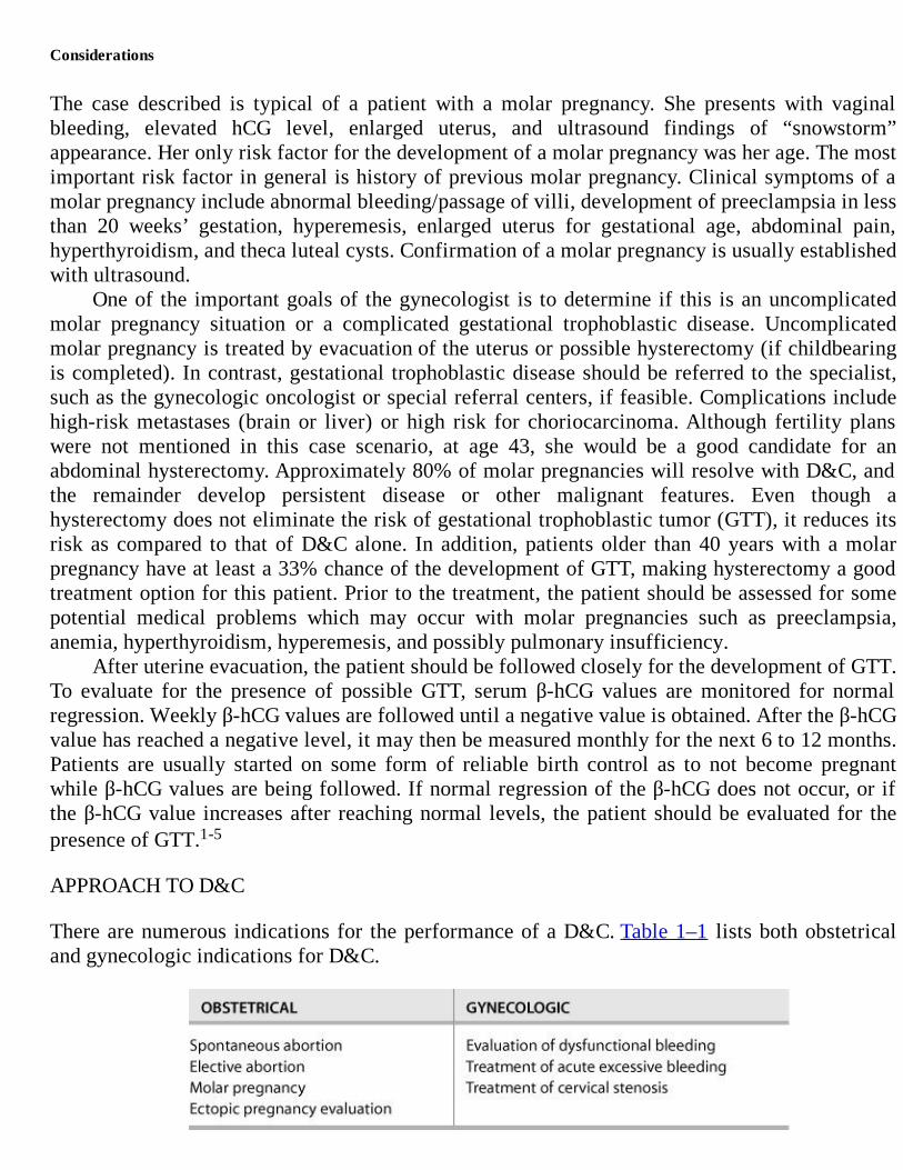
There are numerous indications for the performance of a D&C. Table 1–1 lists both obstetricaland gynecologic indications for D&C.
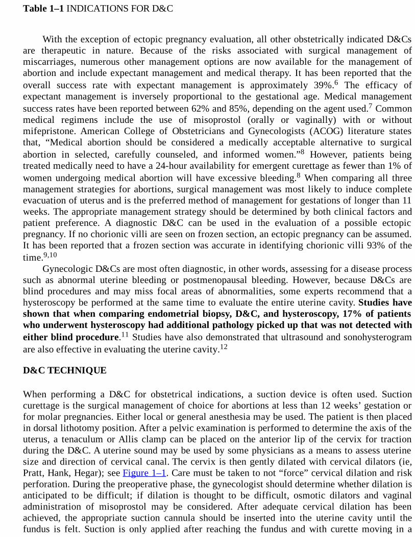
Table 1–1 INDICATIONS FOR D&C
With the exception of ectopic pregnancy evaluation, all other obstetrically indicated D&Csare therapeutic in nature. Because of the risks associated with surgical management ofmiscarriages, numerous other management options are now available for the management ofabortion and include expectant management and medical therapy. It has been reported that theoverall success rate with expectant management is approximately 39%.6 The efficacy ofexpectant management is inversely proportional to the gestational age. Medical managementsuccess rates have been reported between 62% and 85%, depending on the agent used.7 Commonmedical regimens include the use of misoprostol (orally or vaginally) with or withoutmifepristone. American College of Obstetricians and Gynecologists (ACOG) literature statesthat, “Medical abortion should be considered a medically acceptable alternative to surgicalabortion in selected, carefully counseled, and informed women.”8 However, patients beingtreated medically need to have a 24-hour availability for emergent curettage as fewer than 1% ofwomen undergoing medical abortion will have excessive bleeding.8 When comparing all threemanagement strategies for abortions, surgical management was most likely to induce completeevacuation of uterus and is the preferred method of management for gestations of longer than 11weeks. The appropriate management strategy should be determined by both clinical factors andpatient preference. A diagnostic D&C can be used in the evaluation of a possible ectopicpregnancy. If no chorionic villi are seen on frozen section, an ectopic pregnancy can be assumed.It has been reported that a frozen section was accurate in identifying chorionic villi 93% of thetime.9,10
Gynecologic D&Cs are most often diagnostic, in other words, assessing for a disease processsuch as abnormal uterine bleeding or postmenopausal bleeding. However, because D&Cs areblind procedures and may miss focal areas of abnormalities, some experts recommend that ahysteroscopy be performed at the same time to evaluate the entire uterine cavity. Studies haveshown that when comparing endometrial biopsy, D&C, and hysteroscopy, 17% of patientswho underwent hysteroscopy had additional pathology picked up that was not detected witheither blind procedure.11 Studies have also demonstrated that ultrasound and sonohysterogramare also effective in evaluating the uterine cavity.12
D&C TECHNIQUE
When performing a D&C for obstetrical indications, a suction device is often used. Suctioncurettage is the surgical management of choice for abortions at less than 12 weeks’ gestation orfor molar pregnancies. Either local or general anesthesia may be used. The patient is then placedin dorsal lithotomy position. After a pelvic examination is performed to determine the axis of theuterus, a tenaculum or Allis clamp can be placed on the anterior lip of the cervix for tractionduring the D&C. A uterine sound may be used by some physicians as a means to assess uterinesize and direction of cervical canal. The cervix is then gently dilated with cervical dilators (ie,Pratt, Hank, Hegar); see Figure 1–1. Care must be taken to not “force” cervical dilation and riskperforation. During the preoperative phase, the gynecologist should determine whether dilation isanticipated to be difficult; if dilation is thought to be difficult, osmotic dilators and vaginaladministration of misoprostol may be considered. After adequate cervical dilation has beenachieved, the appropriate suction cannula should be inserted into the uterine cavity until thefundus is felt. Suction is only applied after reaching the fundus and with curette moving in a
downward direction (Figure 1–2). Evacuation is usually complete when bubbles appear in thecannula and a gritty sensation of the cavity is felt. The appropriate cannula is usually 1 mmsmaller than the weeks’ gestation (although one must be aware that the smaller the diameter ofthe cannula, the higher the risk of uterine perforation). After suctioning contents from uterus,some physicians use a sharp curette to confirm evacuation. Oxytocic agents are given at thediscretion of the physician as studies are conflicting.
Because of the significant risk of hemorrhage and uterine perforation, a suction D&Cperformed for a molar pregnancy is different from that of an abortion. The procedure should beperformed in a fully staffed and equipped operating room (OR) under general anesthesia.Intravenous access must be secured and blood products available. As compared to traditionalD&Cs, cervical dilation should be performed gently and enough to get a 10- to 12-mm suctioncannula into the uterine cavity. Uterine sounding is not recommended due to risk of incitinghemorrhage and uterine perforation. Once the cervix is dilated and suction cannula is introducedinto the lower uterine segment, oxytocin is infused to help reduce blood loss. Evacuation shouldbegin in the lower uterine segment and proceed toward the fundus carefully. Simultaneous use ofan ultrasound can aid with evacuation and the prevention of uterine perforation. After contentsare evacuated, a sharp curette is used to gently verify evacuation of the uterus. Oxytocic agentsshould then be continued following the procedure to minimize blood loss. If a hysterectomy isperformed, the ovaries need not be removed unless other pathology is present. The ovarian bloodsupply is usually secured prior to uterine manipulation and the uterine vessels are then securedwith minimal uterine manipulation. By securing the blood supply to the uterus beforemanipulation, the chance of molar villous transportation is diminished.13,14
Figure 1–1. Dilation of the cervix with care to introduce the dilator in the direction of theendocervical canal. (Reproduced, with permission, from Schorge JO, Schaffer JI, Halvorson LM,et al. Williams Gynecology. New York: McGraw-Hill, 2008:899.)
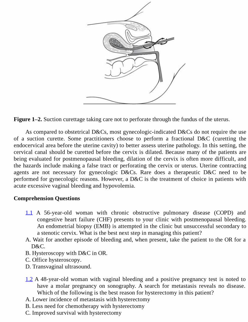
Figure 1–2. Suction curettage taking care not to perforate through the fundus of the uterus.
As compared to obstetrical D&Cs, most gynecologic-indicated D&Cs do not require the useof a suction curette. Some practitioners choose to perform a fractional D&C (curetting theendocervical area before the uterine cavity) to better assess uterine pathology. In this setting, thecervical canal should be curetted before the cervix is dilated. Because many of the patients arebeing evaluated for postmenopausal bleeding, dilation of the cervix is often more difficult, andthe hazards include making a false tract or perforating the cervix or uterus. Uterine contractingagents are not necessary for gynecologic D&Cs. Rare does a therapeutic D&C need to beperformed for gynecologic reasons. However, a D&C is the treatment of choice in patients withacute excessive vaginal bleeding and hypovolemia.
Comprehension Questions
1.1 A 56-year-old woman with chronic obstructive pulmonary disease (COPD) andcongestive heart failure (CHF) presents to your clinic with postmenopausal bleeding.An endometrial biopsy (EMB) is attempted in the clinic but unsuccessful secondary toa stenotic cervix. What is the best next step in managing this patient?
A. Wait for another episode of bleeding and, when present, take the patient to the OR for aD&C.
B. Hysteroscopy with D&C in OR.C. Office hysteroscopy.D. Transvaginal ultrasound.
1.2 A 48-year-old woman with vaginal bleeding and a positive pregnancy test is noted tohave a molar pregnancy on sonography. A search for metastasis reveals no disease.Which of the following is the best reason for hysterectomy in this patient?
A. Lower incidence of metastasis with hysterectomyB. Less need for chemotherapy with hysterectomyC. Improved survival with hysterectomy
D. More cost-effective management with hysterectomy
1.3 A 54-year-old woman is taken to the OR for a fractional D&C for postmenopausalbleeding. During sounding the uterus, the fundus of the uterus is perforated. Thepatient’s vital signs are normal. Which of the following is the best management at thisstage?
A. Abandon the procedure and observe the patient in the hospital overnight.B. Continue the procedure as long as the vital signs are normal.C. Continue the procedure under laparoscopic guidance.D. Perform a hysterectomy.
1.4 Which of the following describes the appropriate technique for performing a fractionalD&C?
A. The curette should be held tightly in palm.B. Pressure with tip of curette should be applied as it is advanced to the fundus.C. The curette should be advanced to fundus and then pressure exerted on uterine wall as it
is withdrawn.D. The cervix should be dilated before attempting the endocervical curetting.
ANSWERS
1.1 D. Although a diagnostic hysteroscopy with D&C could be considered in this patient, hersignificant medical conditions (COPD and CHF) place her at higher risk forintraoperative surgical complications. A less invasive way to evaluate the uterus wouldbe a transvaginal ultrasound to evaluate the endometrial stripe. If the stripe is less than4 mm, no further workup is needed unless the patient continues to have bleeding.Numerous studies have validated the use of the endometrial stripe and postmenopausalbleeding.11
1.2 B. Hysterectomy may be considered in the treatment of a nonmetastatic gestationaltrophoblastic disease in women who do not desire future fertility. The primary reasonfor early hysterectomy is the decreased need and shorter duration of chemotherapyneeded.
1.3 C. Perforation of the uterus usually occurs at the fundus, and is usually asymptomatic.Perforation with a blunt sound typically does not injure any structures; conversely,suction D&C with perforation can lead to bowel injury. Laparoscopic guidance canallow for investigation of injuries or bleeding, and also allows for uterine curettagewithout fear of further damage.
1.4 C. To minimize the risk of uterine perforation, the curette should be held loosely, as withholding a pencil, with pressure on uterine wall only when curetting away from thefundus. The endocervical sample of the fractional D&C should be obtained beforecervical dilation and endometrial sampling.
Table 1–2 LEVELS OF EVIDENCE AND STRENGTH OF RECOMMENDATION
Level A (randomized controlled trial/meta-analysis): High-quality randomized controlled trial(RCT) that considers all important outcomes. High-quality meta-analysis (quantitativesystematic review) using comprehensive search strategies.Level B (other evidence): A well-designed, nonrandomized clinical trial. A nonquantitativesystematic review with appropriate search strategies and well-substantiated conclusions. Includeslower quality RCTs, clinical cohort studies, and case-controlled studies with nonbiased selectionof study participants and consistent findings. Other evidence, such as high-quality, historical,uncontrolled studies or well-designed epidemiological studies with compelling findings, is alsoincluded.Level C (consensus/expert opinion): Consensus viewpoint or expert opinion.
Clinical PearlsSee Table 1-2 for definition of level of evidence and strength of recommendation
Suction D&C is the treatment of choice for molar pregnancies in patients desiring futurefertility (Level A).
A medically managed abortion is an acceptable alternative to surgical management (Level A).
Transvaginal ultrasound is an effective screening tool for uterine pathology (Level A).
The addition of hysteroscopy to a D&C increases diagnostic yield (Level A).
REFERENCES
1. Katz VL, Lentz GM, Lobo RA, Gershenson DM. Gestational trophoblastic disease andabnormal uterine bleeding. In: Comprehensive Gynecology. 5th ed. Philadelphia, PA: Mosby;2007:889-900, 915-929.
2. Gilstrap III LC, Cunningham FG, Vandorsten JP. Gestational trophoblastic disease. In:Operative Obstetrics. 2nd ed. New York, NY: McGraw-Hill; 2002:615-628.
3. Goldstein DP, Berkowitz RS, Berstein MR. Reproductive performance after molar pregnancyand gestational trophoblastic tumor. Clin Obstet Gynecol. 1984;27:221.
4. Cunningham FG, Leveno KJ, Bloom SL, Gilstrap III LC, Wenstrum KD. Abortion andgestational trophoblastic disease and abortion. In: Williams Obstetrics. 22nd ed. New York,NY: McGraw-Hill; 2005:241-247, 273-283.
5. American College of Obstetricians and Gynecologists. Diagnosis and Treatment of GestationalTrophoblastic Disease. ACOG Practice Bulletin, 53. Washington, DC; 2004.
6. Sotiriadis A, Makrydimas G, Papatheodorou S, Ioannidis JP. Expectant, medical, or surgicalmanagement of first trimester miscarriage: a meta-analysis. Obstet Gynecol. 2005;105:1104-
1113.
7. Chen BA, Creinin MD. Contemporary management of early pregnancy failure. Clin ObstetGynecol. 2007;50:67-88.
8. American College of Obstetricians and Gynecologists. Medical Management of Abortion.ACOG Practice Bulletin, 67. Washington, DC; 2005.
9. Spandorfer SD, Menzin A, Barnhart KT, et al. Efficacy of frozen section evaluation of uterinecurettings in the diagnosis of pregnancy. Am J Obstet Gynecol. 1996; 175:603.
10. American College of Obstetricians and Gynecologists. Management of Anovulatory Bleeding.ACOG Practice Bulletin, 14. Washington, DC; 2000.
11. Gimpelson RJ, Rappold HO. A comparative study between panoramic hysteroscopy withdirected biopsies and dilation and curettage. Am J Obstet Gynecol. 1988;158:489.
12. Goldstein SR. The role of transvaginal ultrasound or endometrial biopsy in the evaluation ofthe menopausal endometrium. Am J Obstet Gynecol. 2009;201:5-11.
13. Baggish M, Karram M. Trophoblastic disease. Atlas of Pelvic Anatomy and GynecologicSurgery. 2nd ed. Philadelphia, PA: Elsevier Saunders; 2006:1150-1159.
14. Rock J, Jones H. Normal and abnormal bleeding. In: Te Linde’s Operative Gynecology. 10thed. Philadelphia, PA: Lippincott Williams & Wilkins; 2008:595-605.
Case 2
A 23-year-old G3P2002 woman at 11 weeks’ gestation by last menstrual period presents to the ERwith a 4-day history of abdominal pain, vaginal bleeding, and fever. Upon further questioning, shereports possible passage of tissue with the blood clots. She had been seen in the same ER 1 weekearlier and diagnosed with an embryonic demise. She was discharged with expectant management ofthe miscarriage. On examination, she is febrile to 101°F, has HR 100 beats/min, and BP 100/60 mmHg. She appears ill. Her uterus is extremely tender on examination and cervix dilated 1 cm with“tissue” just inside the cervical os. Minimal bleeding is noted from the cervix. Her white blood cellcount is 21,000/mm3, hemoglobin is 9 g/dL, and blood type is O positive. A transvaginal ultrasounddemonstrates “debris” in the uterus but normal ovaries, no adnexal masses, and no free fluid. Afterbeginning intravenous (IV) antibiotics for a septic abortion, she is taken to the OR for a suction D&C.During the D&C, the uterus is suspected to be perforated by the suction curette since there is a suddenloss of resistance and the catheter advances much farther into the cervix than previously.
What is your management plan?
What are some risk factors for uterine perforation during a D&C?
ANSWERS TO CASE 2:D&C Complications
Summary: This is a 23-year-old G3 P2002 woman has a septic incomplete abortion at 11 weeks’gestation. She experiences a uterine perforation while undergoing a suction D&C procedure.
Management plan: Laparoscopy/laparotomy to determine the extent of injury to intra-abdominal organs/vessels and completion of the evacuation of the uterus under directvisualization.
Risk factors: Enlarged uterus (advanced gestational age), stenotic cervix, uterine infection,multiparity.
ANALYSIS
Objectives
1. Become familiar with the different management strategies for first-trimester abortions (surgical,medical, and expectant management).
2. Understand potential complications of a D&C: immediate, delayed, and late.3. Become familiar with ways to minimize complication risks.
Considerations
This is a 23-year-old G3P2002 woman at 11 weeks’ gestation with septic incomplete abortion. Thepatient had been diagnosed with an embryonic demise at 10 weeks’ gestation and was undergoingexpectant management of the abortion. Given her presentation to the ER, she is no longer a candidatefrom expectant management. Once the products of conception have become infected, evacuation ofthe contents is critical. The standard surgical management in this case would be suction D&C.Although many D&Cs can be performed as an outpatient under a local anesthetic, this patient is besttreated with hospitalization and D&C in the OR. She is at greater risk for surgical complications(perforation, bleeding) than other routine D&Cs performed as an outpatient. Hospital admission isrequired to monitor for signs of sepsis, bacterial shock, disseminated intravascular coagulopathy, andacute renal failure. Prior to beginning the D&C, it is necessary to begin broad-spectrum antibioticsintravenously. The cause of the infection is usually polymicrobial, including anaerobic organisms. Upto 25% of septic abortions have been reported to be associated with positive blood cultures. This isone of the reasons that antibiotics should be administered prior to D&C. Tissue levels of antibioticsare usually reached about 1 hour after administration intravenously and the patient can then be takento the OR. After evacuation, a bacterial culture (aerobic and anaerobic) may be obtained from thecuretted specimen. Postoperatively, the patient should be continued on antibiotics and observedclosely for signs/symptoms of septic shock. Uterine perforation is a known complication of a suctionD&C.
APPROACH TO Complications of a D&C
Early pregnancy failure is clinically recognized in 15% to 20% of pregnancies and can be managedsurgically, medically, and expectantly. Many studies have compared the various treatment options, butdifferences in terminology, medical management, and definitions of failed treatment makecomparison difficult. However, medical management seems to be a possible and effective acceptablealternative to surgical curettage. The use of vaginal misoprostol 400 to 800 micrograms has beenfound to be 80% to 88% successful in achieving a complete miscarriage as compared to 96% to 100%of surgically managed pregnancy failures.1 The primary side effects of medical therapy includenausea, vomiting, and diarrhea, and those with failed complete miscarriage often require suctionD&C. As with any surgery, the risks and benefits of the surgery must be weighted. The benefits ofsurgical management include high success rate and prompt treatment of pregnancy failure. As thegestational age of the abortion increases, so does the risk of complications. However, not all D&Csperformed are for obstetrical indications. Gynecologic indications for D&C include evaluation ofdysfunctional bleeding, treatment of excessive bleeding, and treatment of cervical stenosis.2-5
Surgical complications of D&Cs are rare and can be categorized as occurring in the immediate ordelayed/late period (Table 2–1). D&Cs performed for obstetrical indications carry a slightly higherrisk than nonobstetrical indications due to the increased risk of perforation/bleeding from the graviduterus. Ben-Baruch et al6 reported that the risk for nonobstetrical D&Cs varies between 0.5% and1.8%, depending on the indication. A recent retrospective study confirmed this low complication riskfor nonobstetrical D&Cs.7
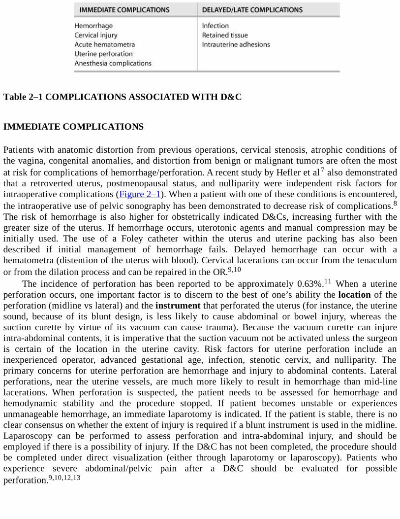
Table 2–1 COMPLICATIONS ASSOCIATED WITH D&C
IMMEDIATE COMPLICATIONS
Patients with anatomic distortion from previous operations, cervical stenosis, atrophic conditions ofthe vagina, congenital anomalies, and distortion from benign or malignant tumors are often the mostat risk for complications of hemorrhage/perforation. A recent study by Hefler et al 7 also demonstratedthat a retroverted uterus, postmenopausal status, and nulliparity were independent risk factors forintraoperative complications (Figure 2–1). When a patient with one of these conditions is encountered,the intraoperative use of pelvic sonography has been demonstrated to decrease risk of complications.8The risk of hemorrhage is also higher for obstetrically indicated D&Cs, increasing further with thegreater size of the uterus. If hemorrhage occurs, uterotonic agents and manual compression may beinitially used. The use of a Foley catheter within the uterus and uterine packing has also beendescribed if initial management of hemorrhage fails. Delayed hemorrhage can occur with ahematometra (distention of the uterus with blood). Cervical lacerations can occur from the tenaculumor from the dilation process and can be repaired in the OR.9,10
The incidence of perforation has been reported to be approximately 0.63%.11 When a uterineperforation occurs, one important factor is to discern to the best of one’s ability the location of theperforation (midline vs lateral) and the instrument that perforated the uterus (for instance, the uterinesound, because of its blunt design, is less likely to cause abdominal or bowel injury, whereas thesuction curette by virtue of its vacuum can cause trauma). Because the vacuum curette can injureintra-abdominal contents, it is imperative that the suction vacuum not be activated unless the surgeonis certain of the location in the uterine cavity. Risk factors for uterine perforation include aninexperienced operator, advanced gestational age, infection, stenotic cervix, and nulliparity. Theprimary concerns for uterine perforation are hemorrhage and injury to abdominal contents. Lateralperforations, near the uterine vessels, are much more likely to result in hemorrhage than mid-linelacerations. When perforation is suspected, the patient needs to be assessed for hemorrhage andhemodynamic stability and the procedure stopped. If patient becomes unstable or experiencesunmanageable hemorrhage, an immediate laparotomy is indicated. If the patient is stable, there is noclear consensus on whether the extent of injury is required if a blunt instrument is used in the midline.Laparoscopy can be performed to assess perforation and intra-abdominal injury, and should beemployed if there is a possibility of injury. If the D&C has not been completed, the procedure shouldbe completed under direct visualization (either through laparotomy or laparoscopy). Patients whoexperience severe abdominal/pelvic pain after a D&C should be evaluated for possibleperforation.9,10,12,13
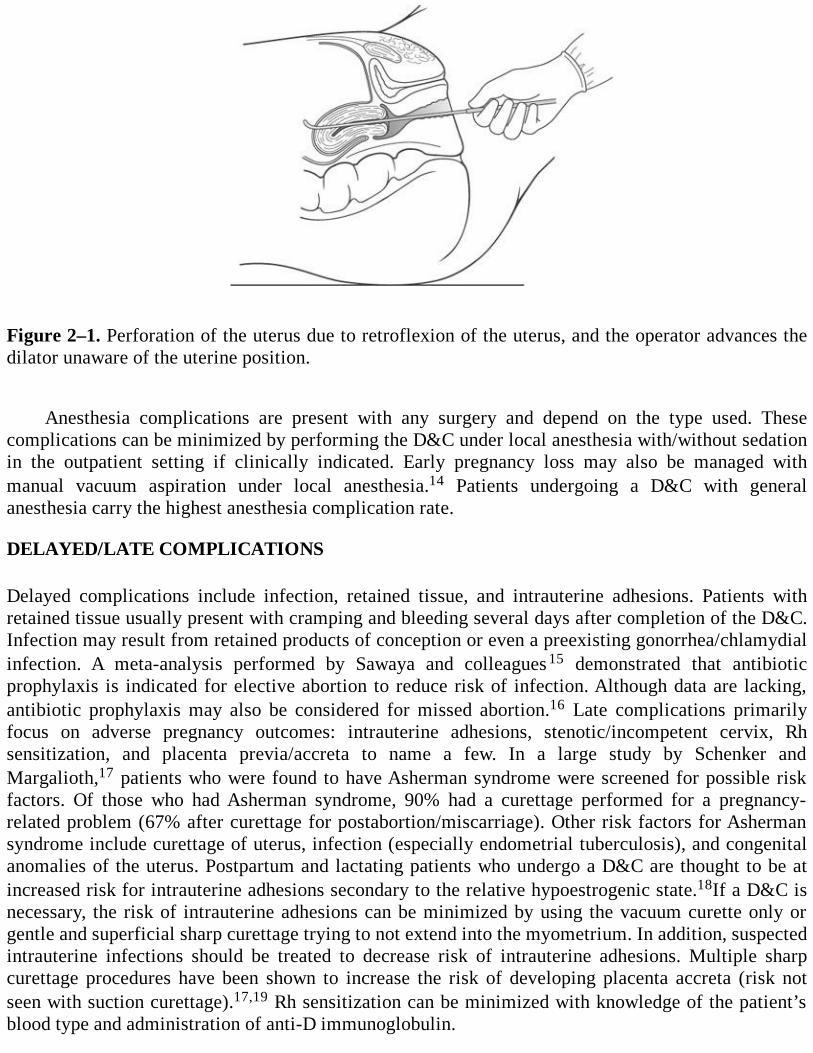
Figure 2–1. Perforation of the uterus due to retroflexion of the uterus, and the operator advances thedilator unaware of the uterine position.
Anesthesia complications are present with any surgery and depend on the type used. Thesecomplications can be minimized by performing the D&C under local anesthesia with/without sedationin the outpatient setting if clinically indicated. Early pregnancy loss may also be managed withmanual vacuum aspiration under local anesthesia.14 Patients undergoing a D&C with generalanesthesia carry the highest anesthesia complication rate.
DELAYED/LATE COMPLICATIONS
Delayed complications include infection, retained tissue, and intrauterine adhesions. Patients withretained tissue usually present with cramping and bleeding several days after completion of the D&C.Infection may result from retained products of conception or even a preexisting gonorrhea/chlamydialinfection. A meta-analysis performed by Sawaya and colleagues 15 demonstrated that antibioticprophylaxis is indicated for elective abortion to reduce risk of infection. Although data are lacking,antibiotic prophylaxis may also be considered for missed abortion.16 Late complications primarilyfocus on adverse pregnancy outcomes: intrauterine adhesions, stenotic/incompetent cervix, Rhsensitization, and placenta previa/accreta to name a few. In a large study by Schenker andMargalioth,17 patients who were found to have Asherman syndrome were screened for possible riskfactors. Of those who had Asherman syndrome, 90% had a curettage performed for a pregnancy-related problem (67% after curettage for postabortion/miscarriage). Other risk factors for Ashermansyndrome include curettage of uterus, infection (especially endometrial tuberculosis), and congenitalanomalies of the uterus. Postpartum and lactating patients who undergo a D&C are thought to be atincreased risk for intrauterine adhesions secondary to the relative hypoestrogenic state.18If a D&C isnecessary, the risk of intrauterine adhesions can be minimized by using the vacuum curette only orgentle and superficial sharp curettage trying to not extend into the myometrium. In addition, suspectedintrauterine infections should be treated to decrease risk of intrauterine adhesions. Multiple sharpcurettage procedures have been shown to increase the risk of developing placenta accreta (risk notseen with suction curettage).17,19 Rh sensitization can be minimized with knowledge of the patient’sblood type and administration of anti-D immunoglobulin.
Comprehension Questions
2.1 A 23-year-old woman undergoes an uncomplicated suction D&C for a missed abortion at 12weeks’ gestation. About 2 hours after the procedure, the nurse notices the patient to haveweakness/diaphoresis. Her uterus is noted to be enlarged and scant minimal bleeding isnoted. What is the most likely etiology?
A. Acute hematometraB. Uterine perforationC. Uterine infectionD. Intrauterine adhesions
2.2 A 78-year-old postmenopausal woman is being evaluated for postmenopausal bleeding. Hercervix is noted to be stenotic and she is taken to the OR for a diagnostic D&C. Which of thefollowing statements is most accurate?
A. The risk of uterine perforation is no greater than other diagnostic D&C.B. To minimize the intraoperative and anesthesia risks, this patient is a good candidate for an
outpatient D&C.C. Intraoperative use of pelvic sonography would be beneficial in decreasing surgical risks.D. A D&C is the only way to evaluate this patient’s endometrium.
2.3 Which of the following is the major predisposing factor to the development of intrauterineadhesions?
A. InfectionB. Trauma to nonpregnant uterusC. Trauma to pregnant uterusD. Congenital anomalies
ANSWERS
2.1 A. Acute hematometra, “postabortal syndrome,” occurs about 2 hours after surgery and thepatient presents with scant vaginal bleeding, diaphoresis/weakness, and an enlarged/tenderuterus. The etiology is unknown and treatment is prompt repeat curettage.
2.2 C. The use of intraoperative ultrasound has been found to be useful in the prevention ofsurgical complications such as uterine perforation in difficult D&Cs. In this case, the patienthas three risk factors (postmenopausal state, atrophic vagina, and stenotic cervix) forincreased surgical complications. Other risk factors include distortion of anatomy fromprevious surgery, congenital anomalies, or tumors. The endometrial cavity in a patient withpostmenopausal bleeding can be assessed with measurement of the endometrial stripe usingpelvic ultrasonography.
2.3 C. Trauma to the pregnant uterus is the major predisposing factor for the development ofintrauterine adhesions. Two theories for this development are the low level of estrogen at thetime of the procedure (or immediately after) doesn’t allow for proper endometrialregeneration and that the uterus may be in a vulnerable state after pregnancy (basal layermore easily damaged).20
Clinical PearlsSee Table 1-2 for definition of level of evidence and strength of recommendation
Medical management of an early pregnancy failure has success rate of approximately 80%(Level A).
Antibiotic prophylaxis is indicated for elective suction curettage abortion (Level A).
Patients at greatest risk for intraoperative complications are postmenopausal state, vaginalatrophy, cervical stenosis, retroverted uterus, and distorted anatomy from previoussurgery/tumors (Level B).
Patients who have a D&C after a pregnancy have higher risk of developing Ashermansyndrome as compared to a nonpregnant uterus (Level B).
Complications after a D&C can be classified as immediate and delayed/late (Level C).
The risk of developing intrauterine adhesions in an obstetrically indicated D&C can beminimized by using the suction curette rather than sharp curettage and curetting gently and notdeep into the myometrium (Level C).
The morbidity after a uterine perforation depends on the location of the perforation and theinstrument used (Level C).
REFERENCES
1. Chen BA, Creinin MD. Contemporary management of early pregnancy failure. Clin ObstetGynecol. 2007;50:67-88.
2. Graziosi GC, Mol BW, Ankum WM, Bruinse HW. Management of early pregnancy loss. Int JGynaecol Obstet. 2004;86:337-346.
3. Sotiriadis A, Makrydimas G, Papatheodorou S, Ioannidis J. Expectant, medical, or surgicalmanagement of first-trimester miscarriage: a meta-analysis. Obstet Gynecol. 2005;105:1104-1113.
4. Stubblefield PG, Carr-Ellis S, Borgatta L. Methods for induced abortion. Obstet Gynecol.2004;104:174-184.
5. Trinder J, Brocklehurst P, Porter R, Read M, Vyas S, Smith L. Management of miscarriage:expectant, medical, or surgical? Results of a randomized controlled trial (miscarriagetreatment (MIST) trial). BMJ. 2006 May;332:1235-1240.
6. Ben-Baruch G, Menczer J, Shalev J, Romen Y, Serr DM. Uterine perforation during curettage:perforation rates and postperforation management. Isr J Med Sci. 1980;16:821-824.
7. Hefler L, Lemach A, Seebacher V, Polterauer S, Tempfer C, Reinthaller A. The intraoperative
complication rate of nonobstetrical dilation and curettage. Obstet Gynecol. 2009;113:1268-1271.
8. Hunter RE, Reuter K, Kopin E. Use of ultrasonography in the difficult post-menopausaldilation and curettage. Obstet Gynecol. 1989;73:813-816.
9. Gilstrap III LC, Cunningham FG, Vandorsten JP. Pregnancy termination: first and secondtrimesters. In: Operative Obstetrics. 2nd ed. New York, NY: McGraw-Hill; 2002:543-546.
10. Rock J, Jones H. Normal and abnormal bleeding. In: TeLinde’s Operative Gynecology. 10thed. Philadelphia, PA: Lippincott Williams & Wilkins; 2008:595-605.
11. McElin TW, Burd CC, Reeves BE, et al. Diagnostic dilation and curettage. Obstet Gynecol.1969;33:807.
12. Cunningham FG, Leveno KJ, Bloom SL, Gilstrap III LC, Wenstrum KD. Abortion. In:Williams Obstetrics. 22nd ed. New York, NY: McGraw-Hill; 2005:241-247.
13. Katz VL, Lentz GM, Lobo RA, Gershenson DM. Spontaneous and recurrent abortion. In:Comprehensive Gynecology. 5th ed. Philadelphia, PA: Mosby; 2007:368-369.
14. Milingos DS, Mathur M, Smith NC, Ashok PW. Manual vacuum aspiration: a safe alternativefor the surgical management of early pregnancy loss. BJOG. 2009; 116:1268-1271.
15. Sawaya GF, Grady D, Kerlikowske K, Grimes DA. Antibiotics at the time of inducedabortion: the case for universal prophylaxis based on a meta-analysis. Obstet Gynecol. 1996May; 87:884-890.
16. American College of Obstetricians and Gynecologists. Antibiotic Prophylaxis forGynecologic Procedures. ACOG Practice Bulletin No.104. Washington, DC; 2009.
17. Schenker JG, Margalioth EJ. Intrauterine adhesions: an updated appraisal. Fertil Steril.1982;37:593-610.
18. Westendorp IC, Ankum WM, Mol BW, Vonk J. Prevalence of Asherman’s syndrome aftersecondary removal of placental remnants or a repeat curettage for incomplete abortion. HumReprod. 1998;13:3347-3350.
19. Johnson LG, Mueller BA, Daling JR. The relationship of placenta previa and history ofinduced abortion. Int J Gynaecol Obstet 2003;81:191.
20. Yu D, Wong Y, Cheong Y, Xia E, Li T. Asherman syndrome-one century later. Fertil Steril.2008;89:759-779.
Case 3
A 25-year-old G2P1001 woman at 19 weeks’ gestation presented to the clinic for routine prenatal care.On examination, she was found to have a uterus which was consistent with 18 weeks’ gestation, but nofetal heart tones were heard. An ultrasound was performed and a fetal demise was found with theestimated gestational age of 16 weeks. She denies any vaginal bleeding or uterine cramping. Hercervix is closed on pelvic digital examination. She has no known medical problems and her firstpregnancy was a term spontaneous vaginal delivery.
What are your management options?
How might the management affect future pregnancies?
ANSWERS TO CASE 3:Dilation and Evacuation
Summary: A 25-year-old G2P1001 at 19 weeks’ gestation has an intrauterine demise.
Management options: Dilation and evacuation (D&E) or medical evacuation of the uterus(misoprostol).
Risks for future pregnancies: No increase in clinically significant complications insubsequent pregnancies is anticipated.
ANALYSIS
Objectives
1. Become familiar with management options of second-trimester fetal demise.2. Review options for cervical dilation.3. Understand some basic surgical principles of D&E.4. Understand complications which can occur with a D&E.
Considerations
The management of a second-trimester fetal demise depends on both the clinical expertise of thephysician as well as the patient’s desires after counseling. Options for management include medicalinduction of labor, D&E, hysterotomy, and hysterectomy. Because of the safety, efficiency, and cost-effectiveness of D&E, it has become the most common surgical management of second-trimesterabortions (> 98% of all second-trimester abortions in 2003). A suction D&C is no longer an optionafter the first trimester, when fetal skeletal parts have begun to calcify, due to a greater risk ofcomplications (uterine perforation, hemorrhage, cervical laceration, and incomplete removal ofproducts). After 15 to 16 weeks’ gestation, a D&E is the preferred method of surgical evacuation of
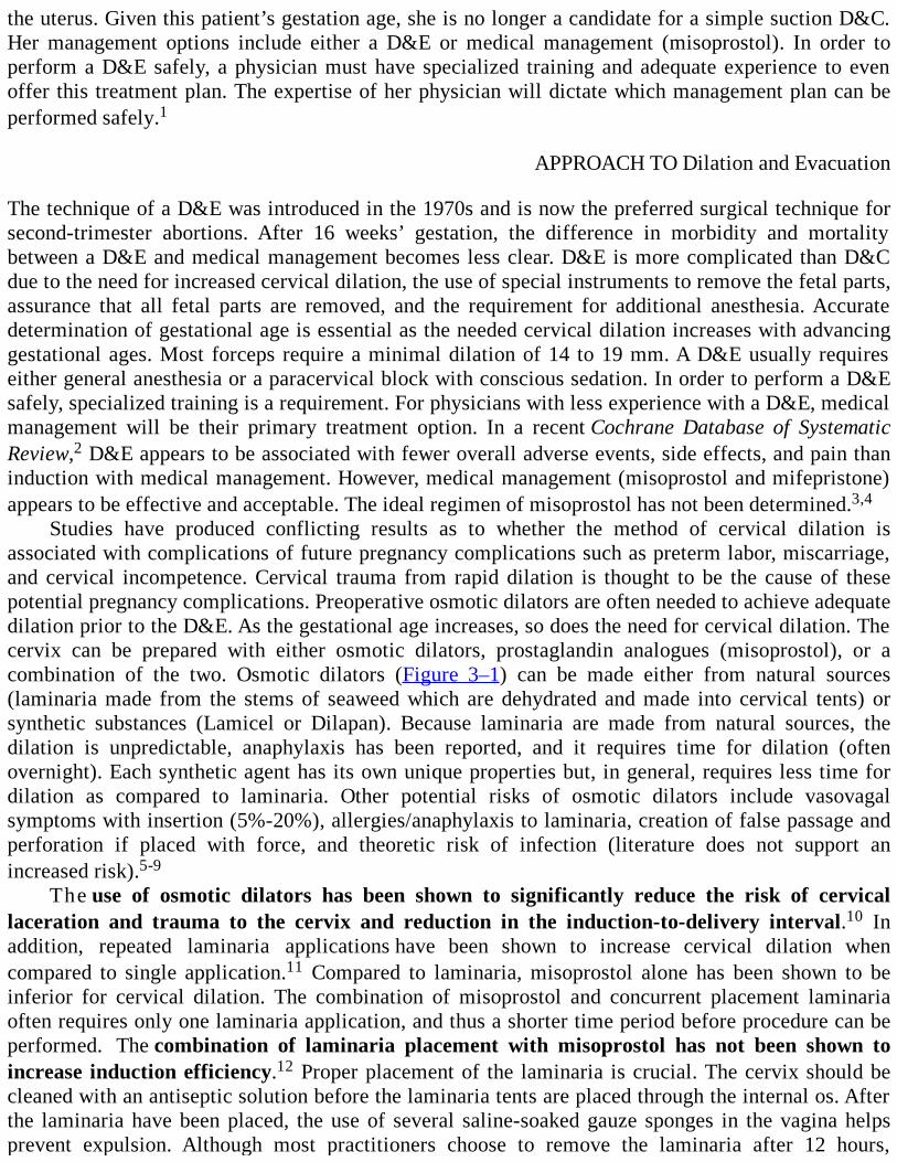
the uterus. Given this patient’s gestation age, she is no longer a candidate for a simple suction D&C.Her management options include either a D&E or medical management (misoprostol). In order toperform a D&E safely, a physician must have specialized training and adequate experience to evenoffer this treatment plan. The expertise of her physician will dictate which management plan can beperformed safely.1
APPROACH TO Dilation and Evacuation
The technique of a D&E was introduced in the 1970s and is now the preferred surgical technique forsecond-trimester abortions. After 16 weeks’ gestation, the difference in morbidity and mortalitybetween a D&E and medical management becomes less clear. D&E is more complicated than D&Cdue to the need for increased cervical dilation, the use of special instruments to remove the fetal parts,assurance that all fetal parts are removed, and the requirement for additional anesthesia. Accuratedetermination of gestational age is essential as the needed cervical dilation increases with advancinggestational ages. Most forceps require a minimal dilation of 14 to 19 mm. A D&E usually requireseither general anesthesia or a paracervical block with conscious sedation. In order to perform a D&Esafely, specialized training is a requirement. For physicians with less experience with a D&E, medicalmanagement will be their primary treatment option. In a recent Cochrane Database of SystematicReview,2 D&E appears to be associated with fewer overall adverse events, side effects, and pain thaninduction with medical management. However, medical management (misoprostol and mifepristone)appears to be effective and acceptable. The ideal regimen of misoprostol has not been determined.3,4
Studies have produced conflicting results as to whether the method of cervical dilation isassociated with complications of future pregnancy complications such as preterm labor, miscarriage,and cervical incompetence. Cervical trauma from rapid dilation is thought to be the cause of thesepotential pregnancy complications. Preoperative osmotic dilators are often needed to achieve adequatedilation prior to the D&E. As the gestational age increases, so does the need for cervical dilation. Thecervix can be prepared with either osmotic dilators, prostaglandin analogues (misoprostol), or acombination of the two. Osmotic dilators (Figure 3–1) can be made either from natural sources(laminaria made from the stems of seaweed which are dehydrated and made into cervical tents) orsynthetic substances (Lamicel or Dilapan). Because laminaria are made from natural sources, thedilation is unpredictable, anaphylaxis has been reported, and it requires time for dilation (oftenovernight). Each synthetic agent has its own unique properties but, in general, requires less time fordilation as compared to laminaria. Other potential risks of osmotic dilators include vasovagalsymptoms with insertion (5%-20%), allergies/anaphylaxis to laminaria, creation of false passage andperforation if placed with force, and theoretic risk of infection (literature does not support anincreased risk).5-9
The use of osmotic dilators has been shown to significantly reduce the risk of cervicallaceration and trauma to the cervix and reduction in the induction-to-delivery interval.10 Inaddition, repeated laminaria applications have been shown to increase cervical dilation whencompared to single application.11 Compared to laminaria, misoprostol alone has been shown to beinferior for cervical dilation. The combination of misoprostol and concurrent placement laminariaoften requires only one laminaria application, and thus a shorter time period before procedure can beperformed. The combination of laminaria placement with misoprostol has not been shown toincrease induction efficiency.12 Proper placement of the laminaria is crucial. The cervix should becleaned with an antiseptic solution before the laminaria tents are placed through the internal os. Afterthe laminaria have been placed, the use of several saline-soaked gauze sponges in the vagina helpsprevent expulsion. Although most practitioners choose to remove the laminaria after 12 hours,
laminaria have been noted to continue to expand for up to 24 hours.13 When a patient’s membraneshave been ruptured, laminaria should be removed in less than 12 hours and procedure completed. Theplacement of multiple laminaria is preferred over just one and the number inserted should be recordedin the patient’s record. Care should be taken in placement to not break the dilators, since a fragment inthe uterus is problematic and may even require hysterotomy for retrieval.
Figure 3–1. Osmotic dilators are introduced into the cervical canal, and slowly enlarge as they absorbwater. The tip of the dilator should be placed just above the level of the internal os. (Reproduced, withpermission, from Schorge JO, Schaffer JI, Halvorson LM, et al. Williams Gynecology. New York:McGraw-Hill, 2008:898.)
Between 13 and 16 weeks’ gestation, vacuum extraction with a 14-mm cannula is probablysufficient after adequate cervical dilation. Beyond 16 weeks’ gestation, forcep (Sopher and Bierer)extraction is necessary. These specialized forceps have been found to be superior to standard ringforceps. To minimize perforation risk, the extraction should begin in the lower uterus rather thanfundus and continuous ultrasound guidance may aid in the removal of fetal parts. After the cervix isadequately dilated, the large suction cannula is introduced into the uterus and amniotic fluid removed.The surgeon will then extract the fetal parts. At the conclusion of the procedure, the physician mustinspect the products and identify all major fetal parts (skull, spine, four extremities) and evaluate thequantity of the placenta.14
Reported complications of D&E include cervical trauma, hemorrhage, perforation of the uterus,infection, and retained products. Intracervical vasopressin (as component of cervical block) has beendemonstrated to decrease intraoperative blood loss.15 Intraoperative administration of uterotonicagents (oxytocin or methylergonovine) has been used by some practitioners to minimize blood loss.However, some practitioners prefer not to use these agents because a contracted uterus can make theevacuation more difficult. Recently, there have been case reports published on the use of uterine arteryembolization (UAE) to treat hemorrhage from D&E with some success.16 However, it should be notedthat UAE should be considered only when the etiology of the bleeding is from an intracervical injury,abnormal placentation, or presence of submucosal fibroids.16 Studies show that very fewhysterectomies (11/10,000 or 0.11%) are required due to hemorrhage or other complications in thosepatients who undergo D&E.4,15,17
Comprehension Questions
3.1 A 23-year-old woman undergoes a D&E after she was found to have an 18-week fetal demise.Which of the following fetal structures is most likely to be left behind in the uterus after aD&E?
A. CalvariumB. ExtremitiesC. AbdomenD. Spine
3.2 Which type of osmotic dilator requires the longest time before maximum cervical dilation?A. LamicelB. DilapanC. LaminariaD. Misoprostol
3.3 A 32-year-old female patient undergoes a D&E for a fetal demise at 16 weeks. Despiteadequate dilation of the cervix, all the fetal parts are not able to be removed. What is themost appropriate next step?
A. Give a uterine relaxing agent and proceed with D&E.B. Proceed with hysterotomy and removal of remaining fetal parts.C. Expectant management for spontaneous delivery of fetal parts.D. Administer oxytocin for 2 to 3 hours and attempt repeat D&E.
ANSWERS
3.1 A. The calvarium is the most likely fetal part to be left behind. The use of ultrasound oftenwill help to identify remaining fetal parts. It is imperative for the surgeon to identify all fourmajor body parts prior to concluding the procedure. If the fetal part (most often thecalvarium) cannot be removed even under direct ultrasound visualization, oxytocin can beinfused for several hours. The remaining fetal part is often closer to the internal os and mucheasier to remove.
3.2 C. The natural product, laminaria, require a longer period of time before the cervix ismaximally dilated (usually overnight). Lamicel, a synthetic agent, works within 2 to 6hours; however, it does not achieve as much dilation as other osmotic agents. Dilapan, also asynthetic agent, works within 4 to 6 hours and achieves the greatest cervical dilation withinthe shortest time frame. The original Dilapan product was removed from the market between1995 and 2002 due to its increased risk of retained fragments with removal. The newerproduct has stronger core to decrease this risk. Misoprostol is not an osmotic dilator.
3.3 D. Oxytocin, given intravenously aids in the expulsion of products if D&E is not successfulinitially. With the combination of ruptured membranes and oxytocin, the remaining fetalparts are often found closer to the cervix and much easier to remove. D&E do not have to becompleted in one step. The use of uterine relaxing agent would significantly increase therisk of uterine perforation and hemorrhage and would not be a good option for this patient.Although expectant management would be a possible alternative management strategy, the
uncertain time from rupture to delivery of products could possibly increase the risk ofinfection and therefore is not the best choice. Hysterotomy should only be considered whenboth surgical and medical managements have failed.
Clinical PearlsSee Table 1-2 for definition of level of evidence and strength of recommendation
The use of osmotic dilators decreases the induction-to-delivery time (Level A).
A D&E has been found to be superior to intrauterine injection with prostaglandin F2a (LevelB).
Medical management of second-trimester abortions is as efficacious as a D&E but often withincreased pain and side effects (Level B).
The use of osmotic dilators minimizes cervical trauma and decreases the incidence of cervicallacerations and possibly future pregnancy complications (Level B).
In experienced hands, D&E is a more effective and safer treatment compared to medicalmanagement, hysterotomy, and hysterectomy (Level C).
Because of the complexity of the procedure and its difficulty as compared to D&C, a D&Eprocedure should only be performed by practitioners with specialized training (Level C).
REFERENCES
1. Autry AM, Hayes EC, Jacobson GF, Kirby RS. A comparison of medical induction and dilationand evacuation for second-trimester abortion. Am J Obstet Gynecol. 2002;187:393-397.
2. Lohr PA, Hayes JL, Gemzell-Danielsson K. Surgical versus medical methods for secondtrimester induced abortion. Cochrane Database Syst Rev. 2008;(1): CD006714.
3. Cunningham FG, Leveno KJ, Bloom SL, Gilstrap III LC, Wenstrum KD. Abortion. In:Williams Obstetrics. 22nd ed. New York, NY: McGraw-Hill; 2005:241-243.
4. Gilstrap III LC, Cunningham FG, Vandorsten JP. Pregnancy termination: first and secondtrimesters. In: Operative Obstetrics. 2nd ed. New York, NY: McGraw-Hill; 2002:540-546.
5. Turok DK, Curtcheff SE, Esplin MS, et al. Second trimester termination of pregnancy: areview by site and procedure type. Contraception. 2008;77:155-161.
6. Jackson JE, Grobman WA, Haney E, Casele H. Mid-trimester dilation and evacuation withlaminaria does not increase the risk for severe subsequent pregnancy complications. Int JGynaecol Obstet. 2007;96:12-15.
7. Kalish RB, Chasen ST, Rosenzweig LB, Rashbaum WK, Chervenak FA. Impact of
midtrimester dilation and evacuation on subsequent pregnancy outcome. Am J Obstet Gynecol.2002;187:613-614.
8. Lichtenberg ES. Complications of osmotic dilators. Obstet Gynecol Surv. 2004;59:528-535.
9. Society of Family Planning. Cervical preparation for second-trimester surgical abortion priorto 20 weeks of gestation. Contraception. 2007;76:486-495.
10. Atlas RO, Lemus J, Reed J, Atkins D, Alger LS. Second trimester abortion usingprostaglandin E2 suppositories with or without intracervical laminaria japonica: a randomizedstudy. Obstet Gynecol. 1998;92:398.
11. Stubblefield PG, Altman AM, Goldstein SP. A randomized trial of one versus two days oflaminaria treatment prior to late midtrimester abortion by uterine evacuation: a pilot study. AmJ Obstet Gynecol. 1982;143:481.
12. Jain JK, Mishell DR. A comparison of misoprostol with and without laminaria tents forinduction of second trimester abortion. Am J Obstet Gynecol. 1996; 175:173.
13. Wheeler RG, Scheider K. Properties and safety of cervical dilators. Am J Obstet Gynecol.1983;146:597.
14. Chasen ST, Kalish RB, Gupta M, Kaufman JE, Rashbaum WK, Chervenak FA. Dilation andevacuation at ≥ 20 weeks: comparison of operative techniques. Am J Obstet Gynecol.2004;190:1180-1183.
15. Peterson WF, Berry FN, Grace MR, Bulbranson CL. Second-trimester abortion by dilationand evacuation: an analysis of 11,747 cases. Obstet Gynecol. 1983;62:185-190.
16. Haddad L, Delli-Bovi L. Uterine artery embolization to treat hemorrhage following second-trimester abortion by dilatation and surgical evacuation. Contraception. 2009;79:452-455.
17. Rock J, Jones H. Management of abortion. In: Te Linde’s Operative Gynecology. 10th ed.Philadelphia, PA: Lippincott Williams & Wilkins; 2008:788-791.
Case 4
A 28-year-old G2P2 woman complains of lower abdominal and pelvic pain for 2 years which seems tobe worsening. She states that the pain is exacerbated by menses and is located in the lower abdomen inthe mid-line. She describes it as cramping with radiation to the back. She has tried nonsteroidal anti-inflammatory drugs (NSAIDs) with no relief. She denies gastrointestinal symptoms. The patientdenies a history of sexually transmitted infections, and has menses every month. She was on the oralcontraceptive agent last year without affecting the pelvic pain. On examination, she is 5 ft 5 in tall andweighs 120 lb (54.43 kg). Her abdomen is without surgical scars. She has mild tenderness of the lowerabdominal quadrants with normal bowel sounds. On pelvic examination, the external genitalia appearnormal. The cervix and vagina are normal. The uterus is retroverted and fixed and tender. The adnexaare bilaterally tender. There are no nodules or barbs noted. The rectovaginal examination reveals sometenderness of the posterior uterus.
What is your diagnostic plan?
What is your differential diagnosis?
What are options for establishing a gynecologic diagnosis?
ANSWERS TO CASE 4:Diagnostic Laparoscopy
Summary: A 28-year-old G2P2 woman has chronic pelvic pain, consistent with dysmenorrhea, whichis not amenable to medical therapy. The examination shows a retroverted uterus that is fixed.
Diagnostic plan: Assays for gonorrhea, Chlamydia, pregnancy test, and pelvic ultrasound;consider a pain diary.
Differential diagnosis: Gynecologic conditions: endometriosis, pelvic adhesions, chronicpelvic inflammatory disease (PID), adenomyosis; Urologic: interstitial cystitis;Gastrointestinal: irritable bowel syndrome, diverticulitis; Psychological: anxiety disorder,depression, somatization, sexual abuse, marital discord, drug abuse; Neurologic: pudendalneuralgia, vulvodynia; Musculoskeletal: pelvic floor disorder, fibromyalgia.
Establishing a gynecologic diagnosis: Diagnostic laparoscopy. The history, physicalexamination, and limited tests are the key to determining which etiology is most likely. With agynecologic likelihood, diagnostic laparoscopy is indicated to help to establish the diagnosis.
ANALYSIS
Objectives
1. Describe the role of diagnostic laparoscopy in gynecological conditions.
2. Describe the major surgical approach in minimizing injuries associated with diagnosticlaparoscopy.
3. List the most common complications associated with diagnostic laparoscopy.
Considerations
Chronic pelvic pain is one of the most difficult diagnostic challenges that a gynecologist faces. Thehistory is essential in trying to establish the body system etiology. The pain in this patient has beenpresent for 2 years and is worsening. The factors that lean toward a gynecologic cause include thelocation of the lower abdomen, worsening with menses, and midline in nature. There is no mention ofpain with intercourse, dyspareunia, but this is also very important in the investigation process. Afterestablishing a good idea of the location, character, duration, and radiation of the pain, the physicianmust look for associated conditions. For instance, if the patient has dysuria or urinary urgency orfrequency, then a urinary disorder may be considered. Psychological issues are very common, and thephysician should spend time inquiring about depression, anxiety, and the patient’s relationship withher partner. Past history of sexual or physical abuse is also relevant. Gastrointestinal symptoms shouldbe sought such as bloating, constipation and diarrhea, or upper abdominal discomfort. When areasonable etiology belonging to a nongynecologic system exists, the patient should be referred to theappropriate consultant. When a gynecologic disorder is likely, further testing or imaging and, ifneeded, diagnostic laparoscopy can help to establish the diagnosis.
APPROACH TO Diagnostic Laparoscopy
The advantages of laparoscopy are well known when compared to laparotomy. Advantages includefaster recovery to normal activities, decreased cost, decreased adhesion formation, smaller incisionsand therefore decreased postoperative pain, and shorter operative times (although this may not holdtrue for some procedures).1 Large meta-analysis published in 2002 comparing laparoscopy tolaparotomy for benign gynecologic disease found that the overall risk of minor complications such asfever, urinary tract infection, or wound infection was lower in woman undergoing laparoscopicprocedures, but both groups had the same risk for major complication such as pulmonary embolism,major bleeding, fistula formation, or damage to surrounding organs.1 Laparoscopic procedures ingynecology are standard for many conditions, and its widespread use has made the transition to theminimally invasive procedures as standard of care in many respects.
Laparoscopy is the best diagnostic/treatment tool in endometriosis because it allows thegynecologist to assess the extent of the disease and at the same time treat lesions with fulguration andresection. The accuracy of laparoscopic diagnosis depends on location and type of lesion, as well assurgeon experience (see Figure 4–1). Direct biopsies of suspicious lesions are necessary when directvisualization can lead to errors in certain cases.2 Most common anatomical sites where endometrioticimplants are located include the ovaries and the posterior cul-de-sac, although it is not uncommon tosee them at the broad ligament, uterosacral ligament, posterior uterine wall, fallopian tubes, sigmoidcolon, and appendix. Multiple extra-abdominal organs can be affected as well.
Basic elements for any gynecologic laparoscopic procedure start with positioning the patient indorsal lithotomy with padded candy cane–type stirrups or Allen stirrups; knees and legs should be in aresting nonflexed position in order to avoid nerve injury. Entry into the peritoneal cavity can be byopen or closed technique, although no major differences have been found in the avoidance of major
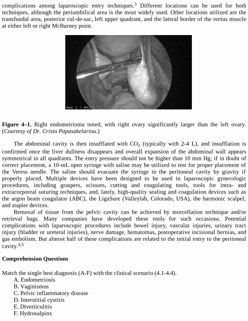
complications among laparoscopic entry techniques.3 Different locations can be used for bothtechniques, although the periumbilical area is the most widely used. Other locations utilized are thetransfundal area, posterior cul-de-sac, left upper quadrant, and the lateral border of the rectus muscleat either left or right McBurney point.
Figure 4–1. Right endometrioma noted, with right ovary significantly larger than the left ovary.(Courtesy of Dr. Cristo Papasakelariou.)
The abdominal cavity is then insufflated with CO2 (typically with 2-4 L), and insufflation isconfirmed once the liver dullness disappears and overall expansion of the abdominal wall appearssymmetrical in all quadrants. The entry pressure should not be higher than 10 mm Hg; if in doubt ofcorrect placement, a 10-mL open syringe with saline may be utilized to test for proper placement ofthe Veress needle. The saline should evacuate the syringe in the peritoneal cavity by gravity ifproperly placed. Multiple devices have been designed to be used in laparoscopic gynecologicprocedures, including graspers, scissors, cutting and coagulating tools, tools for intra- andextracorporeal suturing techniques, and, lately, high-quality sealing and coagulation devices such asthe argon beam coagulator (ABC), the LigaSure (Valleylab, Colorado, USA), the harmonic scalpel,and stapler devices.
Removal of tissue from the pelvic cavity can be achieved by morcellation technique and/orretrieval bags. Many companies have developed these tools for such occasions. Potentialcomplications with laparoscopic procedures include bowel injury, vascular injuries, urinary tractinjury (bladder or ureteral injuries), nerve damage, hematomas, postoperative incisional hernias, andgas embolism. But almost half of these complications are related to the initial entry to the peritonealcavity.4,5
Comprehension Questions
Match the single best diagnosis (A-F) with the clinical scenario (4.1-4.4).A. EndometriosisB. VaginismusC. Pelvic inflammatory diseaseD. Interstitial cystitisE. DiverticulitisF. Hydrosalpinx
4.1 A 35-year-old woman with cyclic pelvic pain related to menses has tried multiplemedications, including birth control pills, without success. Occasionally, she experiencesrectal bleeding with bloating, and bimanual examination reveals a fixed retroverted uterus.
4.2 A 26-year-old nulligravida woman with pelvic pain mainly on the left side states she has hadsome malodorous vaginal discharge for the previous 2 days, which she has not had before.She has multiple sexual partners, states she is very careful, and is taking oral contraceptives.Urine pregnancy test is negative.
4.3 A 28-year-old woman with infertility, despite two rounds of treatment with clomiphenecitrate, has been unsuccessful in attempts at pregnancy. Otherwise asymptomatic, she has ahistory of Chlamydia 5 years prior.
4.4 A 57-year-old woman presents with left lower quadrant pain and fever; at physicalexamination, rebound tenderness was noticed. She has a history of ovarian cysts and asurgery for torsion of a left necrotic ovary.
ANSWERS
4.1 A. Cyclic pain associated with rectal bleeding and a fixed uterus should lead one toward thediagnosis of endometriosis.
4.2 C. A nulliparous woman with multiple sexual partners and unprotected intercourse associatedwith pelvic pain is very suggestive of pelvic inflammatory disease. Other possibilities wouldinclude the presence of a hydrosalpinx, although this condition is not usually associated withmalodorous vaginal discharge.
4.3 F. Hydrosalpinx would be the best explanation for this reproductive age woman after tworounds of ovulation induction drugs. Hydrosalpinx is almost always bilateral in the presenceof infertility.
4.4 E. Diverticulitis is the most likely diagnosis, given that the patient had previously had a leftsalpingo-oophorectomy and was experiencing left lower quadrant pain and fever.
Clinical PearlsSee Table 1-2 for definition of level of evidence and strength of recommendation
Overall risk of minor complications is lower in woman undergoing operative laparoscopicprocedures when compared to laparotomy; however, major complication risks are the same inboth groups (Level B).
The diagnosis of endometriosis is by direct visualization, but confirmatory biopsy of lesions isrecommended in cases of suspicious lesions (Level B).
No major differences have been found in major complications among laparoscopic entrytechniques (Level A).
Roughly half of the complications related to laparoscopic procedures are related to initial entry
Roughly half of the complications related to laparoscopic procedures are related to initial entryinto peritoneal cavity (Level B).
REFERENCES
1. Chapron C, Fauconnier A, Goffinet F, et al. Laparoscopic surgery is not inherently dangerouspresenting with benign gynaecologic pathology. Result of a meta-analysis. Hum Reprod.2002;17:1334.
2. Wykes CB, Clark TJ, Khan KS. Accuracy of laparoscopy in the diagnosis of endometriosis: asystematic quantitative review. BJOG. 2004;111:1204.
3. Ahmad G, Duffy JM, Phillips K, Watson A. Laparoscopic entry techniques. CochraneDatabase Syst Rev. 2008;16:CD006583.
4. Jansen FW, Kolkman W, Bakkum EA, de Kroon CD, Trimbos-Kemper TC, Trimbos JB.Complications of laparoscopy: an inquiry about closed- versus open-entry technique. Am JObstet Gynecol. 2004;190:634.
5. Shirk GJ, Johns A, Redwine DB. Complications of laparoscopic surgery: how to avoid themand how to repair them. J Minim Invasive Gynecol. 2006;13:352.
Case 5
A 35-year-old woman is noted to have severe menorrhagia over the past 2 years that is worsening andis associated with severe anemia. She has tried various medical therapies, including oralcontraceptives and NSAIDs, which do not help. The patient has been diagnosed with uterine fibroids.She desires more children. On examination, her abdomen is normal with the exception of anirregularly shaped nontender uterus that is approximately 14-week size. She denies having previoussurgeries. In counseling her, you review various surgical options. She has heard about robotic surgeryand desires more information about this technique.
What are your management options?
What are the advantages and disadvantages of robotic surgery versus other techniques?
ANSWERS TO CASE 5:Robotic Surgery
Summary: This is a 35-year-old woman with significant menorrhagia and anemia associated withuterine fibroids. Her symptoms have failed medical therapy. Her uterus is 14-week size.
Management options: Myomectomy with options of open technique versus laparoscopy versusrobotic surgery. She desires to preserve fertility; therefore, less invasive treatment should be atthe top of the treatment plan list. Nonsurgical option, such as magnetic resonance–guidedfocused ultrasound (MRgFUS), is an option that must be considered, but few patients qualifyas candidates. Normally, the procedure is reserved for premenopausal women who havecompleted childbearing1; therefore, myomectomy will provide the most benefit in this patient,with the laparoscopic approach providing faster recovery and similar pregnancy rates as theopen approach.2
Robotic surgery advantages:
1. Less postoperative pain as compared to open laparotomy.2. Shorter hospitalizations.3. Better visualization (three-dimensional [3D] vision) versus open surgery and conventional
laparoscopy.4. Improved dexterity and precision given the 3D visualization and replication of full range of
motion of a surgeon’s hand.5. Reduction in surgeon’s fatigue.6. Decreased blood loss.7. Correction of tremor amplification.8. New concept in minimally invasive surgery (remotely performed surgery or telesurgery).
Robotic surgery disadvantages:
1. Expensive initial investment.2. Bulky equipment, therefore large-size ORs are required.3. Lack of haptics or tactile feedback.4. Limited uterine manipulation is less when compared with conventional laparoscopy.
ANALYSIS
Objectives
1. Describe the advantages and disadvantages of robotic surgery versus laparoscopy andlaparotomy.
2. Describe the robotic manual dexterity capabilities.3. Describe the 3D visual capability of robotic surgery.4. Be aware of the limitations of robotic surgery.
Considerations
This 35-year-old woman with severe menorrhagia and anemia has failed medical management withoral control pills and NSAIDs. She has been diagnosed with uterine fibroids, and she desires futurefertility. Therefore, procedures like endometrial ablation, uterine artery embolization, or hysterectomyare not options for treatment. Myomectomy is still considered the procedure of choice in women withsevere symptoms due to fibroids who desire to preserve fertility. The aforementioned MRgFUS usesfocused ultrasound directed into the substance of the large fibroid masses, leading to theirdegeneration. Unfortunately, many third-party carriers do not pay for this procedure, and manypatients fail as candidates due to the nature, location, and size of the myomatous tumors. Managementof the leiomyomas endoscopically is one of the major advances in minimally invasive gynecologicsurgery, especially in a 35-year-old female patient with no previous abdominal surgery, althoughcurrently in the United States the majority of the cases are still performed using laparotomy.2,3
When comparing open myomectomy versus robotic approach, the robotic group had showedlonger operative times and increased cost, but decreased blood loss and shorter length of stay.2 Also,comparison between conventional laparoscopy versus robotic myomectomy seems to raise someconcerns about the level of difficulty encountered in order to enucleate fibroids and performmultilayer closure in the conventional group. Pregnancy rates after myomectomy are similar whethermanaged endoscopically (either conventional or robotic) or via laparotomy.2 Given these facts,usually the less invasive approach is indicated, especially in a 35-year-old patient with history of noprevious surgeries.
APPROACH TO Robotic Surgery
Surgical robotics were first used in 1985 in neurosurgery; applications soon followed in the fields of
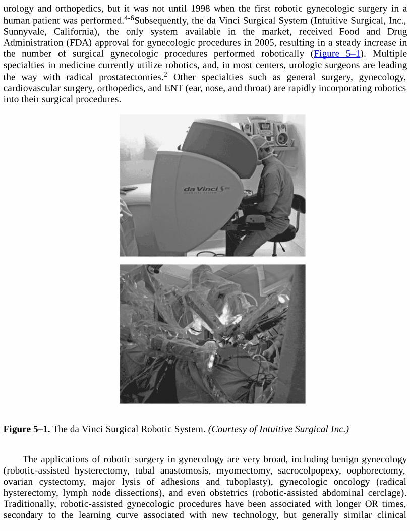
urology and orthopedics, but it was not until 1998 when the first robotic gynecologic surgery in ahuman patient was performed.4-6Subsequently, the da Vinci Surgical System (Intuitive Surgical, Inc.,Sunnyvale, California), the only system available in the market, received Food and DrugAdministration (FDA) approval for gynecologic procedures in 2005, resulting in a steady increase inthe number of surgical gynecologic procedures performed robotically (Figure 5–1). Multiplespecialties in medicine currently utilize robotics, and, in most centers, urologic surgeons are leadingthe way with radical prostatectomies.2 Other specialties such as general surgery, gynecology,cardiovascular surgery, orthopedics, and ENT (ear, nose, and throat) are rapidly incorporating roboticsinto their surgical procedures.
Figure 5–1. The da Vinci Surgical Robotic System. (Courtesy of Intuitive Surgical Inc.)
The applications of robotic surgery in gynecology are very broad, including benign gynecology(robotic-assisted hysterectomy, tubal anastomosis, myomectomy, sacrocolpopexy, oophorectomy,ovarian cystectomy, major lysis of adhesions and tuboplasty), gynecologic oncology (radicalhysterectomy, lymph node dissections), and even obstetrics (robotic-assisted abdominal cerclage).Traditionally, robotic-assisted gynecologic procedures have been associated with longer OR times,secondary to the learning curve associated with new technology, but generally similar clinical
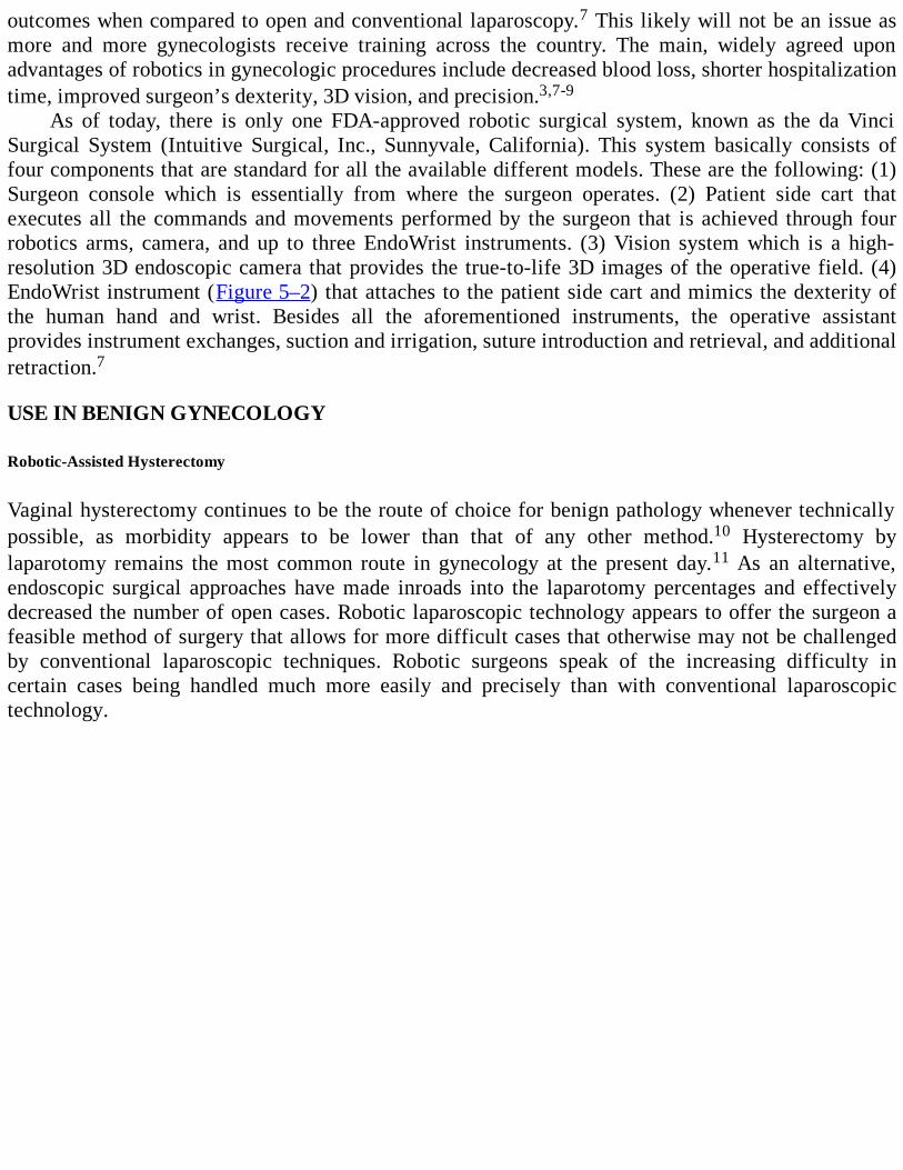
outcomes when compared to open and conventional laparoscopy.7 This likely will not be an issue asmore and more gynecologists receive training across the country. The main, widely agreed uponadvantages of robotics in gynecologic procedures include decreased blood loss, shorter hospitalizationtime, improved surgeon’s dexterity, 3D vision, and precision.3,7-9
As of today, there is only one FDA-approved robotic surgical system, known as the da VinciSurgical System (Intuitive Surgical, Inc., Sunnyvale, California). This system basically consists offour components that are standard for all the available different models. These are the following: (1)Surgeon console which is essentially from where the surgeon operates. (2) Patient side cart thatexecutes all the commands and movements performed by the surgeon that is achieved through fourrobotics arms, camera, and up to three EndoWrist instruments. (3) Vision system which is a high-resolution 3D endoscopic camera that provides the true-to-life 3D images of the operative field. (4)EndoWrist instrument (Figure 5–2) that attaches to the patient side cart and mimics the dexterity ofthe human hand and wrist. Besides all the aforementioned instruments, the operative assistantprovides instrument exchanges, suction and irrigation, suture introduction and retrieval, and additionalretraction.7
USE IN BENIGN GYNECOLOGY
Robotic-Assisted Hysterectomy
Vaginal hysterectomy continues to be the route of choice for benign pathology whenever technicallypossible, as morbidity appears to be lower than that of any other method.10 Hysterectomy bylaparotomy remains the most common route in gynecology at the present day.11 As an alternative,endoscopic surgical approaches have made inroads into the laparotomy percentages and effectivelydecreased the number of open cases. Robotic laparoscopic technology appears to offer the surgeon afeasible method of surgery that allows for more difficult cases that otherwise may not be challengedby conventional laparoscopic techniques. Robotic surgeons speak of the increasing difficulty incertain cases being handled much more easily and precisely than with conventional laparoscopictechnology.
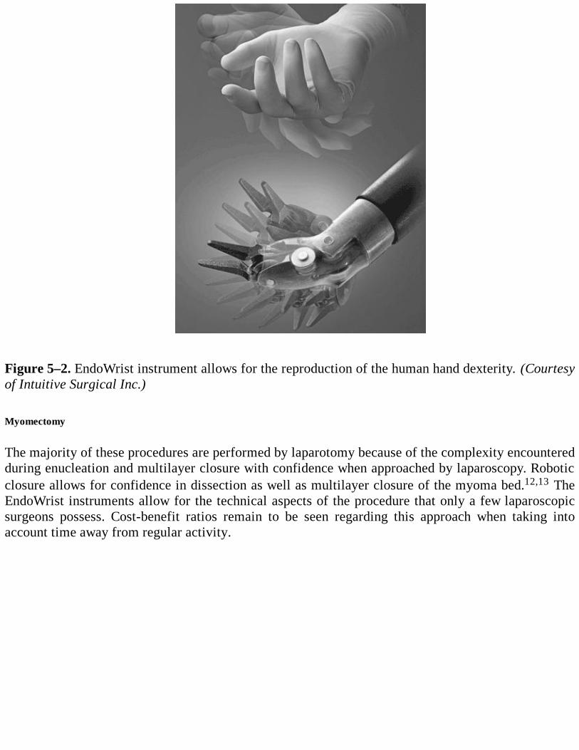
Figure 5–2. EndoWrist instrument allows for the reproduction of the human hand dexterity. (Courtesyof Intuitive Surgical Inc.) Myomectomy
The majority of these procedures are performed by laparotomy because of the complexity encounteredduring enucleation and multilayer closure with confidence when approached by laparoscopy. Roboticclosure allows for confidence in dissection as well as multilayer closure of the myoma bed.12,13 TheEndoWrist instruments allow for the technical aspects of the procedure that only a few laparoscopicsurgeons possess. Cost-benefit ratios remain to be seen regarding this approach when taking intoaccount time away from regular activity.
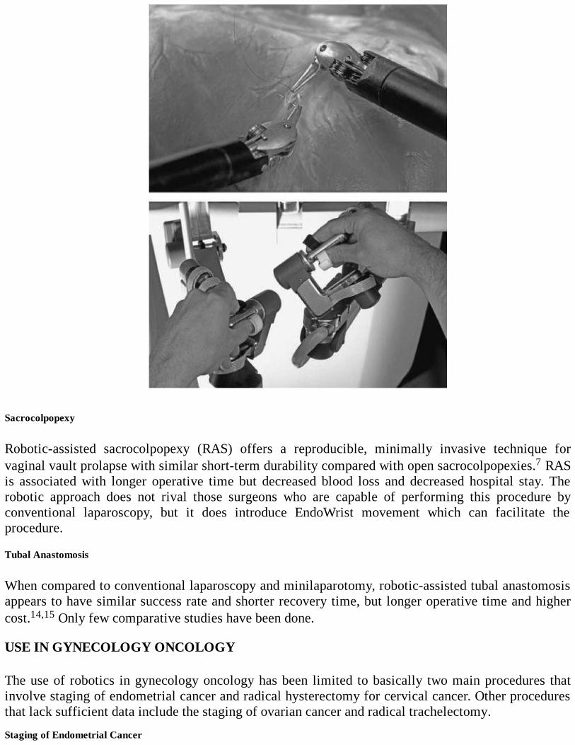
Sacrocolpopexy
Robotic-assisted sacrocolpopexy (RAS) offers a reproducible, minimally invasive technique forvaginal vault prolapse with similar short-term durability compared with open sacrocolpopexies.7 RASis associated with longer operative time but decreased blood loss and decreased hospital stay. Therobotic approach does not rival those surgeons who are capable of performing this procedure byconventional laparoscopy, but it does introduce EndoWrist movement which can facilitate theprocedure.
Tubal Anastomosis
When compared to conventional laparoscopy and minilaparotomy, robotic-assisted tubal anastomosisappears to have similar success rate and shorter recovery time, but longer operative time and highercost.14,15 Only few comparative studies have been done.
USE IN GYNECOLOGY ONCOLOGY
The use of robotics in gynecology oncology has been limited to basically two main procedures thatinvolve staging of endometrial cancer and radical hysterectomy for cervical cancer. Other proceduresthat lack sufficient data include the staging of ovarian cancer and radical trachelectomy.
Staging of Endometrial Cancer
All literature available in this field seems to be very optimistic regarding the use of robotics in uterinecancer. When compared to laparoscopy or laparotomy, robotic-assisted hysterectomy for endometrialcancer staging yields similar outcomes such as lymph node retrieval, blood loss, and complicationrates. Robotic outcomes are comparable to, or even better than, the other two surgical techniques.16,17
Robotic and conventional laparoscopic operative times are comparable and are slightly longer thanopen approaches.
Staging of Cervical Cancer/Radical Hysterectomy
Robotic-assisted radical hysterectomy has been shown to have shorter or similar operative times thanlaparotomy or conventional laparoscopy.18,19 Major advantages of robotics in this procedure areshorter hospitalization, decreased blood loss, and faster recovery to normal activities. Roboticlaparoscopic surgery applies to a very broad scope of applicability, and more and more gynecologicsurgeons seem to be incorporating robotics in their practices.20,21 Although there is not a nationalconsent about the development and training of this innovative technology, it is definitely here to stay.
Comprehension Questions
5.1 A 50-year-old woman has a history of menorrhagia refractory to medical treatment. She hasbeen diagnosed with uterine polyps by hysteroscopy. Her endometrial biopsy is negative formalignancy. She requests a hysterectomy with salpingo-oophorectomy. Her past surgicalhistory includes three prior cesarean sections, an appendectomy secondary to rupturedappendix, and an exploratory laparoscopy for the evaluation of chronic pelvic pain. Thepelvic examination reveals a large 14-week-size uterus, which is fixed and not very mobileand somewhat tender. Which of the following would be the best surgical approach for thispatient?
A. Vaginal hysterectomy with bilateral salpingo-oophorectomyB. Laparoscopic-assisted vaginal hysterectomy with sacrocolpopexyC. Total abdominal hysterectomy with bilateral salpingo-oophorectomyD. Robotic-assisted supracervical hysterectomy
5.2 With regard to robotic-assisted gynecologic procedures, which of the following statements istrue?
A. As compared to the open technique, robotic-assisted myomectomy has better outcomes.B. Robotic-assisted radical hysterectomy for early stages of cervical cancer has the same
operative time as open procedure and shorter times than conventional laparoscopy.C. Robotic-assisted sacrocolpopexy has better outcomes and less blood loss than open
sacrocolpopexy.D. When adhesive disease is suspected in a patient, laparoscopic approach is preferred over
robotic.
5.3 Which of the following statements is most accurate?A. Robotic machinery is more precise than conventional laparoscopy because of the bulkiness
and initial investment.B. If vaginal hysterectomy is feasible, this approach should be offered first rather than robotics.C. Robotic-assisted sacrocolpopexy has longer operative time than laparoscopy sacrocolpopexy.D. Disadvantages to the robotic technology include the lack of the option of morcellation.
ANSWERS
5.1 C. Total abdominal hysterectomy with bilateral salpingo-oophorectomy would be the safestchoice in this patient, given her extensive past surgical history that includes possibleendometriosis and extensive postoperative adhesions. Laparoscopic-assisted vaginalhysterectomy could be considered, except for the fact that this patient has an enlarged uteruswhich would be less likely to be amenable to vaginal approach; sacrocolpopexy is notindicated in this patient. Robotic-assisted supracervical hysterectomy would be asubstandard treatment since the patient desires total hysterectomy that includes cervix,tubes, and ovaries.
5.2 B. Data available today suggest that robotic-assisted radical hysterectomy for early cervicalcancer stages has similar operative times as the open approach and shorter operative timesthan conventional laparoscopy. Considering that this procedure is minimally invasive, fasterrecovery and less blood loss are major advantages. Option A robotic myomectomy hassimilar short-term outcomes when compared to open approach. Option C robotic-assistedsacrocolpopexy has similar outcomes when compared to open and also less blood loss.Finally, when postoperative or postinflammatory adhesions are suspected, option D, that is,the use of robotics, would be a better option, given the improved dexterity, precision, andimproved visualization (3D).
5.3 B. Whenever technically feasible and medically appropriate, patients requiring hysterectomyshould be offered the vaginal approach because morbidity appears to be lower than any othermethod.12
Clinical Pearls
See Table 1-2 for definition of level of evidence and strength of recommendation
Robotic sacrocolpopexy has demonstrated similar short-term vaginal vault support comparedwith abdominal sacrocolpopexy, although with longer operative time, less blood loss, andshorter length of stay (Level B).
Vaginal hysterectomy has the lower morbidity than any other method (Level A).
Some advantages of robotic technology over conventional laparoscopy are better dexterity,precision, and 3D imaging (Level C).
REFERENCES
1. Stewart EA, Gedroyc WM, Regan L, et al. Focused ultrasound treatment of uterine fibroidtumors: safety and feasibility of a noninvasive thermoablative technique. Am J Obstet Gynecol.2003;189:48.
2. Mohamed N, Akl MD, Javier Magrina MD. Will robots transform gynecologic surgery?
Contemporary Ob/Gyn. 2009 Sep;54(9):26-32.
3. Senapati S, Advincula AP. Surgical techniques: robot-assisted laparoscopic myomectomy withthe Da Vinci surgical system. J Robotic Surg. 2007;1:69-74.
4. Dharia SP, Falcone T. Robotics in reproductive medicine. Fertil Steril. 2005;84:1.
5. Satava RM. Robotic surgery: from the past to future- a personal journey. Surg Clin North Am.2003;83:1491.
6. Falcone T, Goldberg J, Garcia-Ruiz A, et al. Full robotic assistance for laparoscopic tubalanastomosis: a case report. J Laparoendosc Adv Surg Tech A. 1999;9:107.
7. Anthony G, Visco MD, Arnold Advincula MD. Robotic gynecologic surgery. Obstet Gynecol.2008 Dec;112(6):1369-1384.
8. Reynolds RK, Advincula AP. Robot assisted laparoscopic hysterectomy: technique and initialexperience. Am J Surg. 2006;191:555-560.
9. Elizabeth J, Geller MD, Nazema Y, et al. Short term outcomes of robotic sacrocolpopexycompared with abdominal sacrocolpopexy. Obstet Gynecol. 2008 Dec;112(6):1201-1213.
10. American College of Obstetricians and Gynecologists, et al. Appropriate use oflaparoscopically assisted vaginal hysterectomy. ACOG Committee Opinion No. 311. April2005. Obstet Gynecol. 2005;105:929-930.
11. Wu JM, Wechter ME, Geller EJ, Nguyen TV, Visco AG. Hysterectomy rates in the UnitedStates, 2003. Obstet Gynecol. 2007;110:1091-1095.
12. Advincula AP, Song A. The role of robotic surgery in gynecology. Curr Opin Obstet Gynecol.2007;19:331-336.
13. Manyonda I, Sinthamoney E, Belli AM. Controversies and challenges in the modernmanagement of uterine fibroids. BJOG. 2004;111:95-102.
14. Goldberg JM, Falcone T. Laparoscopic microsurgical tubal anastomosis with and withoutrobotic assistance. Hum Reprod. 2003;18:145.
15. Dharia Patel SP, Steinkampf MP, Whitten SJ, Malizia BA. Robotic tubal anastomosis:surgical technique and cost effectiveness. Fertil Steril. 2008;90:1175.
16. Veljovich DS, Paley PJ, Drescher CW, et al. Robotic surgery in gynecology oncology:program initiation and outcomes after the first year with comparison with laparotomy forendometrial cancer staging. Am J Obstet Gynecol. 2008;198:679.
17. Boggess JF, Gehrig PA, Cantrell L, et al. A comparative study of 3 surgical methods forhysterectomy with staging for endometrial cancer: robotic assistance, laparoscopy,laparotomy. Am J Obstet Gynecol. 2008;199:360.
18. Sert B, Abeler V. Robotic radical hysterectomy in early stage cervical carcinoma patients,
18. Sert B, Abeler V. Robotic radical hysterectomy in early stage cervical carcinoma patients,comparing results with total laparoscopic radical hysterectomy cases. The future is now? Int JMed Robot. 2007;3:224.
19. Boggess JF, Gehrig PA, Cantrell L, et al. A case control study of robot-assisted type IIIradical hysterectomy with pelvic lymph node dissection compared with open radicalhysterectomy. Am J Obstet Gynecol. 2008;199:357.
20. Nezhat C, Lavie O, Hsu S, Watson J, Barnett O, Lemyre M. Robotic-assisted laparoscopicmyomectomy compared with standard laparoscopic myomectomy—a retrospective matchedcontrol study. Fertil Steril. 2009 Feb;91(2):556-559.
21. Magrina JF, Kho RM, Weaver AL, Montero RP, Magtibay PM. Robotic radical hysterectomy:comparison with laparoscopy and laparotomy. Gynecol Oncol. 2008;109:86-91.
Case 6
You are performing an operative laparoscopy on a 28-year-old woman with a history of pelvic pain,dyspareunia, and a left adnexal mass. She had a history of PID 2 years ago and had an exploratorylaparotomy for endometriosis 10 years ago. You place the initial trocar infraumbilically using an opentechnique. Upon inserting the laparoscope, you find a loop of bowel adherent to the abdominal wallnext to your trocar. Multiple adhesions are also present in the lower abdomen and pelvis. Additionaltrocars are placed under direct visualization. The procedure is tedious and challenging, but you areable to expose the pelvic organs. You free the adnexal mass from the rectosigmoid colon using sharpand blunt dissection. Hemostasis was achieved using electrosurgical devices. A left salpingo-oophorectomy was performed to remove a large endometrioma. The patient is discharged the next day.Three days later, she calls your clinic late in the afternoon complaining of lower abdominaldiscomfort. You see her the next morning in your clinic, and she has diffuse abdominal pain, nausea,and feels feverish. On examination, she appears ill. She has a temperature of 101°F, an HR of 120beats/minute, and BP of 90/60 mm Hg. Her abdomen has hypoactive bowel sounds and reboundtenderness.
What is the most likely diagnosis?
What are the mechanisms of the condition?
What factors contribute to this patient’s condition?
ANSWERS TO CASE 6:Electrocautery (Bowel Injury) with Laparoscopy
Summary: A 28-year-old white woman underwent laparoscopy for pelvic pain and a left adnexal mass.The surgery was difficult. Three days later, the patient has lower abdominal pain, nausea, fever, andrebound tenderness.
Most likely diagnosis: Probable peritonitis from an unrecognized bowel injury. I would alsoconsider an unrecognized bladder injury, a severe urinary tract infection, reactivation of PID,and a ruptured appendix.
Mechanism of bowel injuries in laparoscopic surgery: Most bowel injuries occur withVeress needle insertion or initial trocar placement.1 Other causes include electrical burns ortraumatic injury during adhesiolysis or bowel manipulation.
What factors contributed to the risk of bowel injury: The main risk factor for bowel injurywas adhesions from previous abdominal surgery. PID and endometriosis also contributed to adifficult dissection. The use of electro-surgical devices, especially monopolar instruments, canlead to unrecognized thermal injuries.
ANALYSIS
Considerations
Initial entry into the abdomen with the laparoscope has always been a cause for concern, especiallywhen the patient has had previous abdominal surgery. Many different techniques, such as the “open”technique, and many types of trocars have been advocated to reduce this risk, but none have beencompletely successful. The problem is that bowel is adherent in close proximity to the trocar andlaparoscope, and injury is not completely avoidable. Currently, many authors are advocating for usingthe left upper quadrant as a “safe” entry point in patients at high risk for intra-abdominal adhesions.2,3
The trocar is placed in the midclavicular line 2 cm below the costal margin. Using the left upperquadrant as an entry point is probably a better choice than the infraumbilical site. Once the trocar issafely inserted inside the abdominal cavity, the adhesions can be evaluated, and any further trocarsmust be inserted under direct visualization only. The use of electrosurgical instruments iscommonplace with laparoscopy, but one must be cognizant of the risks and how to avoid them. Goodvisualization of the surgical site is critical. Certainly, the surgeon should always be able to clearly seethe tips of the scissors when dissecting adhesions. Traction on the adhesions using a grasping device ishelpful, but care must be taken when activating an electrode near a metal grasper, or other conductiveinstrument, and causing an injury due to the direct coupling effect. Such would be the concern with thesegment of bowel that was adherent at the insertion site and lying close to the laparoscope. Effectivesmoke evacuation is necessary for good visualization. Good hemostasis is also important as smallamounts of blood may obscure the operating field when viewed through the laparoscope. Once thepelvis is visualized, one must make a decision about the safest way to proceed. Dissecting anendometrioma off the rectosigmoid colon can be extremely difficult in any circumstance. The surgeonmust possess the laparoscopic skills necessary to safely complete the dissection. If the surgeon doesnot possess those skills, a more experienced surgeon should be consulted or the case should beconverted to an open laparotomy. Slow, deliberate sharp dissection using good surgical technique isimportant in preventing iatrogenic injury. When utilizing electrosurgical instruments, always try tokeep bowel away from the active electrode and be aware of the proximity of other devices capable ofconducting electrical current to the active electrode. This is especially important when dissectingadhesions away from bowel or other structures in tight spaces where additional instruments are neededto help with exposure. Electrical energy should be used judiciously, for the shortest duration, and atthe lowest power settings to minimize tissue damage. At the completion of the procedure, the surgeonmust carefully inspect the surgical site for any sign of injury. Serosal and partial thickness defects ofthe bowel wall should be repaired. Any evidence for perforation necessitates an intraoperative surgicalconsult. If there was a question about a possible perforation, the pelvis can be filled with fluid and airintroduced into the rectum.4 Bubbles resulting from this would indicate a perforation. Depending onthe size of the defect, bowel resection, with or without a colostomy, may be indicated. The patientshould be started on broad-spectrum antibiotics and the pelvis irrigated with copious amounts ofirrigation fluid to reduce bacterial contamination.
Objectives
1. Discuss basic concept of an electrical current and how it applies to electrosurgery.2. Understand the differences between cutting waveforms and coagulating waveforms.3. Understand the differences between monopolar electrodes and bipolar electrodes.4. Know the causes of burn injuries with electrosurgical instruments and how to avoid them.
APPROACH TO Bowel Injuries from Laparoscopy
DEFINITIONS
ELECTRO SURGERY: Term used to describe the passage of high-frequency electrical currentthrough tissue to create a desired tissue effect
CAPACITOR COUPLING: The establishment of currents between two conductors that are separatedby an insulator
DIRECT COUPLING: Intended or unintended contact between an active electrode and tissue
BIPOLAR: Electrosurgical current that is passed between two electrodes with a small volume oftissue between the two electrodes to complete the electrical circuit
MONOPOLAR: Electrosurgical current that is dispersed through the patient to a return electrodewhich then returns energy to the generator to complete the electrical circuit
CLINICAL APPROACH
Heat and cautery have been used for hundreds of years to control bleeding. In the early nineteenthcentury, a French physicist, Becquerel, used direct current electricity to heat a wire which was thenused to cauterize tissue.5 In the late 1920s, William Bovie and Harvey Cushing developed the firstelectrosurgical unit to be used in human surgery.5 Nearly 100 years later, electrosurgery is still anintegral part of most surgeons’ practice. In laparoscopic gynecologic procedures, electrosurgery isstill a mainstay for cutting tissue and obtaining hemostasis.
A basic understanding of electricity and electrical currents is necessary to understand theprinciples behind electrosurgery. Electrical current is generated when electrons are forced to movethrough a circuit. Power is the energy produced by the current.6 Voltage is the force which drives theelectrons through the circuit, and heat is generated when the electrons meet resistance within thecircuit. Lastly, electricity needs a continuous circuit to flow. The electro-surgical unit provides thevoltage for the flow of electrons. Modern electrosurgical generators utilize alternating current atradiofrequency levels.5 When the electrosurgical unit is activated, current flows through the activeelectrode which is applied to the patient. The patient’s tissue provides the resistance and heat isgenerated which produces the clinical effects desired by the surgeon. The current then flows eitherthrough the patient back to the electrosurgical unit through a return electrode (“grounding pad”) orthrough another conducting instrument. The different tissue effects depend on the density of theelectrical current, the time the current is applied to the tissue, the resistance of the tissue, the electrodesize, and the waveform of the current.7 Some common uses of electrosurgical energy in gynecologiclaparoscopy include lysing adhesions, controlling bleeding, destroying areas of endometriosis,interrupting tubal patency for female sterilization, and treating ectopic pregnancies.
Two basic waveforms of electrical current are generated by the electrosurgical unit. They are a“cut” waveform and a “coagulation” waveform. The “cut” waveform is a continuous radiofrequencysine wave which creates higher current but lower voltage (Figure 6–1A and B).5 This waveformproduces intense local heat and is used to vaporize tissue. It is the vaporization of the tissue whichcreates the “cutting” effect. Cutting waveforms produce less tissue charring and less tissue damage.The “coagulation” waveform is composed of intermittent radiofrequency sine waves which have
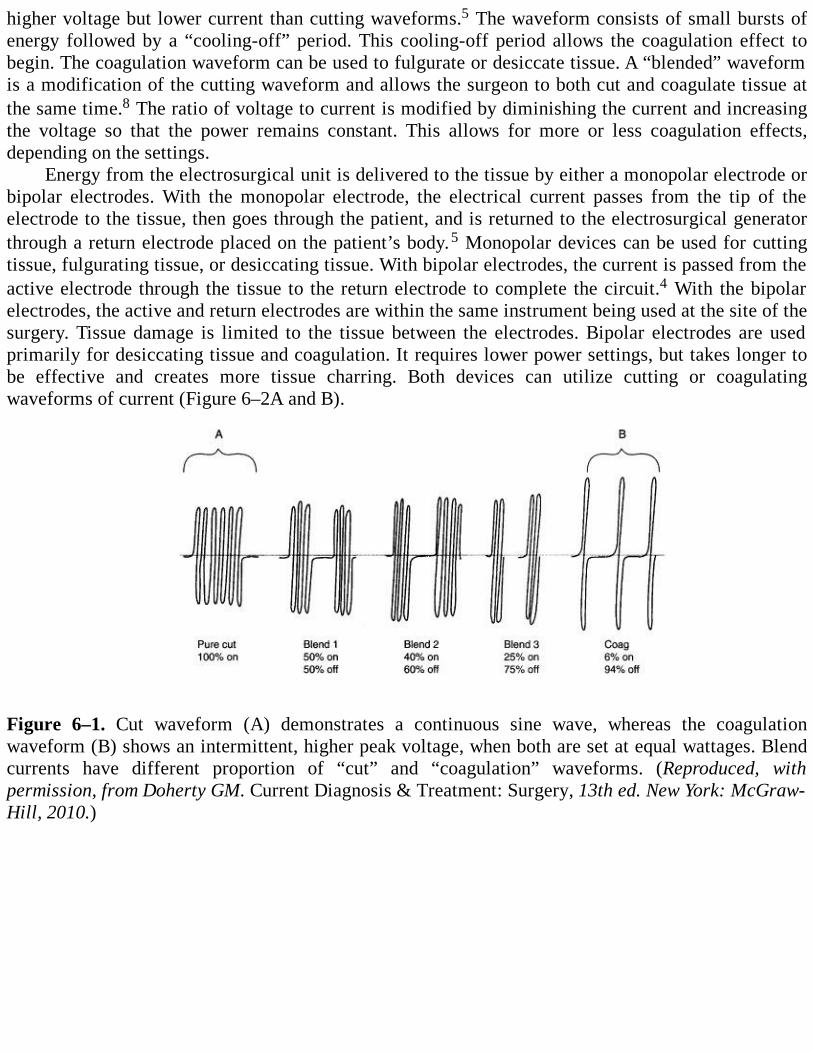
higher voltage but lower current than cutting waveforms.5 The waveform consists of small bursts ofenergy followed by a “cooling-off” period. This cooling-off period allows the coagulation effect tobegin. The coagulation waveform can be used to fulgurate or desiccate tissue. A “blended” waveformis a modification of the cutting waveform and allows the surgeon to both cut and coagulate tissue atthe same time.8 The ratio of voltage to current is modified by diminishing the current and increasingthe voltage so that the power remains constant. This allows for more or less coagulation effects,depending on the settings.
Energy from the electrosurgical unit is delivered to the tissue by either a monopolar electrode orbipolar electrodes. With the monopolar electrode, the electrical current passes from the tip of theelectrode to the tissue, then goes through the patient, and is returned to the electrosurgical generatorthrough a return electrode placed on the patient’s body.5 Monopolar devices can be used for cuttingtissue, fulgurating tissue, or desiccating tissue. With bipolar electrodes, the current is passed from theactive electrode through the tissue to the return electrode to complete the circuit.4 With the bipolarelectrodes, the active and return electrodes are within the same instrument being used at the site of thesurgery. Tissue damage is limited to the tissue between the electrodes. Bipolar electrodes are usedprimarily for desiccating tissue and coagulation. It requires lower power settings, but takes longer tobe effective and creates more tissue charring. Both devices can utilize cutting or coagulatingwaveforms of current (Figure 6–2A and B).
Figure 6–1. Cut waveform (A) demonstrates a continuous sine wave, whereas the coagulationwaveform (B) shows an intermittent, higher peak voltage, when both are set at equal wattages. Blendcurrents have different proportion of “cut” and “coagulation” waveforms. (Reproduced, withpermission, from Doherty GM. Current Diagnosis & Treatment: Surgery, 13th ed. New York: McGraw-Hill, 2010.)
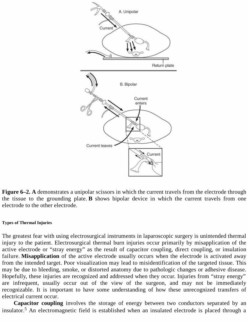
Figure 6–2. A demonstrates a unipolar scissors in which the current travels from the electrode throughthe tissue to the grounding plate. B shows bipolar device in which the current travels from oneelectrode to the other electrode.
Types of Thermal Injuries
The greatest fear with using electrosurgical instruments in laparoscopic surgery is unintended thermalinjury to the patient. Electrosurgical thermal burn injuries occur primarily by misapplication of theactive electrode or “stray energy” as the result of capacitor coupling, direct coupling, or insulationfailure. Misapplication of the active electrode usually occurs when the electrode is activated awayfrom the intended target. Poor visualization may lead to misidentification of the targeted tissue. Thismay be due to bleeding, smoke, or distorted anatomy due to pathologic changes or adhesive disease.Hopefully, these injuries are recognized and addressed when they occur. Injuries from “stray energy”are infrequent, usually occur out of the view of the surgeon, and may not be immediatelyrecognizable. It is important to have some understanding of how these unrecognized transfers ofelectrical current occur.
Capacitor coupling involves the storage of energy between two conductors separated by aninsulator.5 An electromagnetic field is established when an insulated electrode is placed through a
metal cannula/trocar and activated. Electrical current is induced into the metal cannula through aprocess known as capacitance.9 Energy is stored in the capacitator until the generating force of thecurrent is deactivated or a pathway to complete the electrical circuit is established. In most instances,the current stored in the metal cannula/trocar is in direct contact with the patient and dispersedthrough the patient back to the return electrode. If there is an insulator (ie, a plastic anchoring collar)between the metal cannula and the patient, current cannot pass through to the patient and is stored inthe cannula (ie, a capacitator). When the net charge in the capacitor exceeds the insulator’s capacity,current is produced and passed to other conductive material (ie, tissue). Thermal injuries can result.
Capacitative coupling is highest when voltages are high. This happens when the electrode isactivated in an “open circuit” away from the targeted tissue. High voltage coagulation current createsthe same problem.9 Using all metal or all plastic cannulas/trocars avoids the problem entirely, buthybrid systems are to be avoided. Direct coupling or “probe” coupling occurs when the activeelectrode comes in contact with another metal instrument, thus directly passing the electrical currentto the other instrument.10 This may be intentional when a bleeding point is grasped with a metalgrasper; the grasper is then touched by the active electrode to produce a cauterizing effect. Inadvertentinjury occurs when the active electrode accidently touches a metal object (ie, probes, graspers, suctiondevices, etc) which is in contact with other nontargeted tissue.
Insulation failure occurs when there are breaks in the insulation covering the active electrodeshaft, allowing current to leave the electrode and find an alternate pathway to complete its circuit backto the return electrode.9 Thus, energy can be transferred from the shaft of the electrode to otherconductive, nontargeted tissue that might be in contact with the shaft, resulting in an unrecognizedinjury. These injuries typically occur on the portion of the instrument that is beyond the field of viewof the laparoscope but distal to the protective trocar. A recent study revealed that 20% of reusableinstruments had insulation defects, and that 18% of the defects were in the portion of the electrodethat is exposed but “out of sight” of the surgeon.11 All electrodes should be examined or tested priorto use for insulation defects, although these defects may be quite small and difficult to appreciate.
Prevention of Thermal Injuries
Prevention of electrosurgical injuries is paramount. Safe laparoscopic surgery requires a commonsense approach to the use of electrosurgical devices. All laparoscopic instruments should be in goodworking order. All OR personnel should be familiar with the electrosurgical generators, how they areto be operated, and the safety features of each particular unit. They should also know how to assembleall the electrosurgical devices and have the proper connecting cords immediately available. Thesurgeon should specify the type of energy source he/she would like to use and be familiar with allaspects of its use. Bipolar rather than unipolar electrosurgery should be used when possible. Itminimizes damage to the surrounding tissue and avoids the problems of “stray energy.” Whenmonopolar electrodes are going to be used, the surgeon must ensure that the return electrode pad isproperly placed on the patient. The surgeon should inspect the electrodes for any breaks in theinsulation. The tip of the electrode should always be visible to the surgeon when it is activated, andthe electrode should only be activated when the surgeon is sure of its intended target. The electrodeshould never be activated in an open circuit (ie, in the abdomen away from the intended target)! Caremust always be taken when activating the electrode when it is in close proximity to another metal orother conductive instrument. Metal cannulas are the safest operating channels for active electrodes,and any metal/plastic hybrid cannulas should not be used.7
When using either monopolar or bipolar instruments, the lowest possible power settings shouldbe used for the shortest period of time necessary to achieve the desired clinical effect. Cut waveforms
should be used whenever possible because they require lower voltage settings.5 The electrodes (orpaddles with bipolar devices) should always have any coagulum removed as this increases resistanceand causes the electrode to adhere more to the tissue. Finally, when the electrode is not in use, itshould be placed in a protective, insulated holster rather than just left lying in the surgical field. Thissimple step eliminates accidental burns to the patient and reduces the risk of starting a fire in the OR.
Safety Features
The surgeon should also know which safety features are being used with their particularelectrosurgical unit. The modern electrosurgical unit is an isolated solid-state generator.5 In thissystem, the current can only return to the generator through the return electrode pad. This has been amajor factor in reducing alternate site burns. Contact quality monitoring systems now being used alsoprevent the unit from being activated if the return electrode pad is not in proper contact with thepatient. This eliminates patient burns at the site of an improperly placed pad.5 An active electrodemonitoring system is available which detects and shields the patient from stray energy from insulationfailure or capacitor coupling.12 Recently, tissue response technology has come into use. Vessel sealingdevices using bipolar energy and tissue response technology can seal arterial vessels up to 7 mm indiameter while reducing thermal spread to surrounding tissue.13 This technology uses computers tosense tissue resistance and then automatically adjusts the output voltage to maintain a constant effectto the targeted tissue. These devices have been associated with less blood loss, shorter operatingtimes, and shorter hospital stays.14
In the end, however, nothing is more important than a well-prepared, knowledgeable surgeon whopossesses good manual dexterity, excellent hand-eye coordination, and sound clinical judgment.Knowledge of electrosurgical principles and proper use of electrosurgical devices should enable thelaparoscopic surgeon to operate safely and effectively.
Comprehension Questions
6.1 A 48-year-old woman is undergoing laparoscopy for assessment of a 6-cm left adnexal mass.Which of the following is the most significant risk factor for bowel injury?
A. Prior cesareanB. Prior PIDC. EndometriosisD. Prior cervical dysplasia with cryotherapy
6.2 A 44-year-old woman underwent a laparoscopy for chronic pelvic pain. The patient developedbowel symptoms 5 days after the surgery. Which of the following is the most likelymechanism for this complication?
A. Laceration with the Veress needleB. Laceration with the trocarC. Thermal injuryD. CO2 embolism
6.3 In considering the possibility of bowel injury, which of the following can decrease theseverity of these complications?
A. Bowel preparationB. Open laparoscopy technique
C. Use of a pneumoperitoneumD. Use of a bipolar rather than unipolar current
ANSWERS
6.1 C. A prior cesarean typically does not lead to significant adhesions of the bowel to theabdominal wall. Prior PID leads to adnexal adhesions and tubal scarring, but unless therewas tubo-ovarian abscess, there is usually no bowel involvement. Endometriosis can lead towidespread adhesions and fibrosis.
6.2 C. The delay in symptoms is likely related to thermal injury, which can be manifest up to aweek postoperatively. Lacerations during surgery typically cause symptoms immediately.
6.3 A. A bowel preparation with both cleansing the bowel (mechanical) as well as antibiotics candecrease the bacterial load of the intestinal contents. Thus, if bowel laceration or injuryoccurs, less contamination occurs. Neither the use of a pneumoperitoneum or Veress needleor open laparoscopy techniques has been shown to decrease the incidence of bowel injuries.
Clinical PearlsSee Table 1-2 for definition of level of evidence and strength of recommendation
Tissue necrosis and subsequent perforation of affected bowel can occur 72-96 hours aftersurgery (Level B).
The return electrode pad should be placed on a clean, dry area of skin over a large muscle mass(Level C).
Superficial thermal injuries of the bowel should be oversewn with suture being placed inhealthy tissue well beyond the area of injury (Level C).
Insulation defects are found in 20% of reusable electrodes (Level B).
An all metal cannula system is the safest choice for the operating cannula containing the activeelectrode (Level B).
REFERENCES
1. Schäfer M, Lauper M, Krähenbühl L. Trocar and Veress needle injuries during laparoscopy.Surg Endosc. 2001;15:275-280.
2. Parker J, Reid G, Wong F. Microlaparoscopic left upper quadrant entry in patients at high riskof periumbilical adhesions. Aust N Z J Obstet Gynaecol. 199;39:88-92.
3. Sepilian V, Ku L, Wong H, Liu CJ, Phelps JY. Prevalence of infraumbilical adhesions inwomen with previous laparoscopy. JSLS. 11;1:41-44.
4. Shirk J, Johns A, Redwine DB. Complications of laparoscopic surgery: how to avoid them and
4. Shirk J, Johns A, Redwine DB. Complications of laparoscopic surgery: how to avoid them andhow to repair them. J Minim Invasive Gynecol. 2006 Jul-Aug;13(4):352-359.
5. Wang K, Advincula AP. “Current Thoughts” in electrosurgery. Int J Gynaecol Obstet. 2007Jun;97(3):245-250.
6. Jones CM, Pierre KB, Nicoud IB, Stain SC, Melvin WV. Electrosurgery. Curr Surg. 2006 Nov-Dec;63(6):458-463.
7. Wu MP, Ou CS, Chen SL, Yen YET, Rowbotham R. Complications and recommendedpractices for electrosurgery in laparoscopy. Am J Surg. 2000 Jan;179(1): 67-73.
8. McCauley G. Understanding electrosurgery. Bovie/Aaron Medical. Copyright 2003.
9. Nezhet C, Siegler A, Nezhat F, Nezhat C, Seidman D, Luciano A. Electrosurgery. In: OperativeGynecologic Laparoscopy: Principles and Techniques . 2nd ed. New York: McGraw-Hill;2000:73-80.
10. Odell RC. Electrosurgery: principles and safety issues. Clin Obstet Gynecol. 1995;38:610-621.
11. Montero PN, Robinson NR, Weaver JS, Stiegmann GV. Insulation failure in laparoscopicinstruments. Surg Endosc. 2009;24:462-465 [Epub 2009 Jul 2].
12. Safety technology for laparoscopic monopolar electro surgery; devices for managing burnrisks. Health Devices/ECRI. 2005 Aug;34(8):259-272.
13. Hubner M, Demartines N, Muller S, Dindo D, Calvien P-A, Hahnloser D. Prospectiverandomized study of monopolar scissors, bipolar vessel sealer and ultrasonic shears inlaparoscopic colorectal surgery. Br J Surg. 2008;95:1098-1104.
14. Campagnacci R, de Sanctis A, Baldarelli M, Lezoche G, Guerrieri M. Electrothermal bipolarvessel sealing device vs. ultrasonic coagulating shears in laparoscopic colectomies: acomparative study. Surg Endosc. 2007 Sep;21(9):1525-1531 [Epub 2007 Feb 8].
Case 7
A 37-year-old G4P3013 woman presents to your office requesting a sterilization procedure. Shedesires to have a sterilization and would like to have a surgical technique that would allow her toresume her daily duties in a short period of time. Her last delivery was over a year ago, and she has anegative past surgical history. She is 5 ft 4 in in height and weighs 165 lb (75 kg). She is currentlyusing condoms for contraception.
What is the clinical condition?
What counseling is important for this patient?
What is your next step?
ANSWERS TO CASE 7:Laparoscopic Tubal Occlusion
Summary: This is a 37-year-old G4P3013 woman with a negative past surgical and medical historydesiring sterilization.
Clinical condition: Multiparous patient desiring sterilization.
Counseling: The patient should be given the options of contraception, male sterilization, andfemale sterilization. She should be aware of the irreversibility, failure rate, and risk of ectopicpregnancy with female sterilization.
Next step: Outline the surgical options, including vasectomy, and explain the risks, benefits,and alternatives of the operation.
ANALYSIS
Objectives
1. Be able to provide proper counseling for sterilization.2. Be familiar with different methods of surgical sterilization and methods of occlusion.3. Be familiar with risks, complications, and failure rates.
Considerations
This is a 37-year-old G4P3013 woman who desires permanent sterilization. The first step for thephysician is to provide proper counseling. It is important for the patient to understand that tubalsterilization is permanent. Despite the availability of surgical techniques that allow the reversal oftubal anatomy and in vitro fertilization, both options are complicated, costly, and not always
successful. Sterilization accounts for 39% of contraceptive method used in the United States bywomen aged 15 to 44 years and their partners. Of those who chose sterilization, 28% had tubalsterilization and 11% had partners who had vasectomy. In comparison, in the same group, 27% useoral contraceptives, 21% use male condoms, 3% use injectable contraceptives, 2% use diaphragms,and 1% use intrauterine devices. Approximately 700,000 tubal sterilizations and 500,000 vasectomiesare performed annually in the United States.1 Laparoscopic tubal occlusion is the most commonmethod of interval sterilization in the United States, with approximately 200,000 performed everyyear. Interval sterilization is defined as a nonpregnant state at least 4 to 6 weeks after the completionof the previous pregnancy. However, tubal sterilization can also be performed postpartum and after aspontaneous or therapeutic abortion. Postpartum sterilization is performed after 10% of hospitaldeliveries and approximately 3.5% after spontaneous or therapeutic abortions. Postpartumsterilization can be performed at the time of a cesarean section or after a vaginal delivery preferablythrough a minilaparotomy. Postpartum sterilization optimally involves counseling and informedconsent during prenatal care and prior to labor and delivery. The patient should be explained that thesterilization might be postponed in cases of intra- or postpartum medical or obstetrical complications.The physician should be aware of federal and state regulations that govern the timing of consent of theprocedure.
Interval sterilization can be performed at any time during the menstrual cycle, although theperformance during the follicular phase and/or the use of an effective contraceptive method by thepatient greatly reduces the risk of a concurrent pregnancy. In addition, the utilization of a highlysensitive pregnancy test prior to the sterilization is a wise and necessary option. The routineperformance of a D&C concurrent with all interval sterilizations is not recommended.
Methods of Laparoscopic Sterilization
Bipolar Coagulation Laparoscopic sterilization has evolved in the United States from the unipolarcoagulation originally described by Palmer and Steptoe, to the bipolar techniques described by Riouxand Kleppinger.2 Unipolar sterilization has lost its initial popularity as a result of a series ofmysterious bowel injuries that were reported in the 1970s. It is quite possible that these injuries were aresult of capacitance coupling (as explained in Case 6) or from mechanical laceration at the time ofsterilization. As a result, bipolar laparoscopic tubal occlusion became the most commonly usedtechnique. Approximately 3 cm of the isthmic portion of the fallopian tubes are coagulated using atleast 25 W of energy in a cutting waveform (see Figure 7–1). The 3 cm length of the fallopian tubesinvolved with the sterilization corresponds to the three applications of the bipolar forceps (Figure 7–1). It is imperative to use a current meter (ammeter) to ensure complete desiccation, and this findingof “power used until no current flowed as per ammeter” should be documented in the operative report.Visual inspection can lead to incomplete coagulation of the fallopian tube and increased failures. Thetube is grasped approximately 2 to 4 cm from the uterus in order to minimize the possibility ofuteroperitoneal fistula formation and ectopic pregnancy. It is probably a good practice to leave astump of isthmus of approximately 1 to 2 cm after all techniques of sterilization. Coagulation of 3 cmof fallopian tube without division has been demonstrated to be as effective as coagulation anddivision. Division can lead to bleeding from underlying vessels that were not occluded during thecoagulation. Also, coagulation and division may be associated with the destruction of a larger segmentof the tube. Division is unnecessary as a result of the normal healing process after bipolar coagulationof the fallopian tube. Bipolar coagulation and the resulting desiccated or devitalized fallopian tissueinduce the body to create a state of hypervascularization around the site with gradual absorption ofdead tissue within 3 weeks. The proximal and distal stumps of the desiccated fallopian tube are drawn
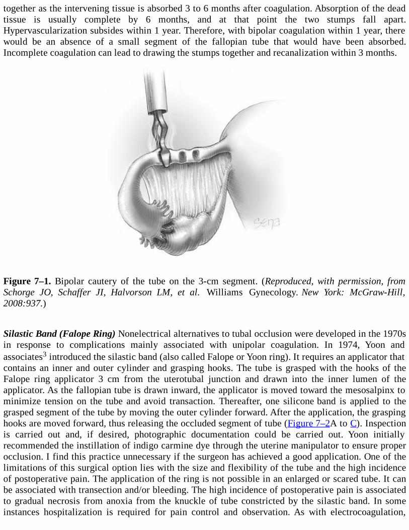
together as the intervening tissue is absorbed 3 to 6 months after coagulation. Absorption of the deadtissue is usually complete by 6 months, and at that point the two stumps fall apart.Hypervascularization subsides within 1 year. Therefore, with bipolar coagulation within 1 year, therewould be an absence of a small segment of the fallopian tube that would have been absorbed.Incomplete coagulation can lead to drawing the stumps together and recanalization within 3 months.
Figure 7–1. Bipolar cautery of the tube on the 3-cm segment. (Reproduced, with permission, fromSchorge JO, Schaffer JI, Halvorson LM, et al. Williams Gynecology. New York: McGraw-Hill,2008:937.)
Silastic Band (Falope Ring) Nonelectrical alternatives to tubal occlusion were developed in the 1970sin response to complications mainly associated with unipolar coagulation. In 1974, Yoon andassociates3 introduced the silastic band (also called Falope or Yoon ring). It requires an applicator thatcontains an inner and outer cylinder and grasping hooks. The tube is grasped with the hooks of theFalope ring applicator 3 cm from the uterotubal junction and drawn into the inner lumen of theapplicator. As the fallopian tube is drawn inward, the applicator is moved toward the mesosalpinx tominimize tension on the tube and avoid transaction. Thereafter, one silicone band is applied to thegrasped segment of the tube by moving the outer cylinder forward. After the application, the graspinghooks are moved forward, thus releasing the occluded segment of tube (Figure 7–2A to C). Inspectionis carried out and, if desired, photographic documentation could be carried out. Yoon initiallyrecommended the instillation of indigo carmine dye through the uterine manipulator to ensure properocclusion. I find this practice unnecessary if the surgeon has achieved a good application. One of thelimitations of this surgical option lies with the size and flexibility of the tube and the high incidenceof postoperative pain. The application of the ring is not possible in an enlarged or scared tube. It canbe associated with transection and/or bleeding. The high incidence of postoperative pain is associatedto gradual necrosis from anoxia from the knuckle of tube constricted by the silastic band. In someinstances hospitalization is required for pain control and observation. As with electrocoagulation,
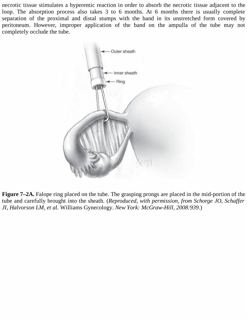
necrotic tissue stimulates a hyperemic reaction in order to absorb the necrotic tissue adjacent to theloop. The absorption process also takes 3 to 6 months. At 6 months there is usually completeseparation of the proximal and distal stumps with the band in its unstretched form covered byperitoneum. However, improper application of the band on the ampulla of the tube may notcompletely occlude the tube.
Figure 7–2A. Falope ring placed on the tube. The grasping prongs are placed in the mid-portion of thetube and carefully brought into the sheath. (Reproduced, with permission, from Schorge JO, SchafferJI, Halvorson LM, et al. Williams Gynecology. New York: McGraw-Hill, 2008:939.)
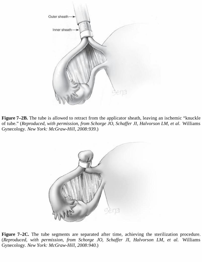
Figure 7–2B. The tube is allowed to retract from the applicator sheath, leaving an ischemic “knuckleof tube.” (Reproduced, with permission, from Schorge JO, Schaffer JI, Halvorson LM, et al. WilliamsGynecology. New York: McGraw-Hill, 2008:939.)
Figure 7–2C. The tube segments are separated after time, achieving the sterilization procedure.(Reproduced, with permission, from Schorge JO, Schaffer JI, Halvorson LM, et al. WilliamsGynecology. New York: McGraw-Hill, 2008:940.)
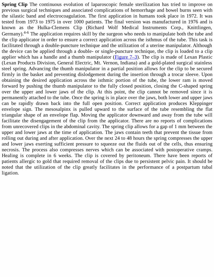
Spring Clip The continuous evolution of laparoscopic female sterilization has tried to improve onprevious surgical techniques and associated complications of hemorrhage and bowel burns seen withthe silastic band and electrocoagulation. The first application in humans took place in 1972. It wastested from 1973 to 1975 in over 1000 patients. The final version was manufactured in 1976 and isknown as the Hulka-Clemens Clip (Richard Wolf Medical Instruments Corp., Knittlingen,Germany).4-6 The application requires skill by the surgeon who needs to manipulate both the tube andthe clip applicator in order to ensure a correct application across the isthmus of the tube. This task isfacilitated through a double-puncture technique and the utilization of a uterine manipulator. Althoughthe device can be applied through a double- or single-puncture technique, the clip is loaded to a clipapplier which has a handle and a thumb manipulator (Figure 7–3). The clip is made of Lexan Plastic(Lexan Products Division, General Electric, Mt. Vernon, Indiana) and a gold-plated surgical stainlesssteel spring. Advancing the thumb manipulator in a partial position allows for the clip to be securedfirmly in the basket and preventing dislodgement during the insertion through a trocar sleeve. Uponobtaining the desired application across the isthmic portion of the tube, the lower ram is movedforward by pushing the thumb manipulator to the fully closed position, closing the C-shaped springover the upper and lower jaws of the clip. At this point, the clip cannot be removed since it ispermanently attached to the tube. Once the spring is in place over the jaws, both lower and upper jawscan be rapidly drawn back into the full open position. Correct application produces Kleppingerenvelope sign. The mesosalpinx is pulled upward to the surface of the tube resembling the flattriangular shape of an envelope flap. Moving the applicator downward and away from the tube willfacilitate the disengagement of the clip from the applicator. There are no reports of complicationsfrom unrecovered clips in the abdominal cavity. The spring clip allows for a gap of 1 mm between theupper and lower jaws at the time of application. The jaws contain teeth that prevent the tissue fromrolling out during and after application. Over the next 24 to 48 hours the spring compresses the upperand lower jaws exerting sufficient pressure to squeeze out the fluids out of the cells, thus ensuringnecrosis. The process also compresses nerves which can be associated with postoperative cramps.Healing is complete in 6 weeks. The clip is covered by peritoneum. There have been reports ofpatients allergic to gold that required removal of the clips due to persistent pelvic pain. It should benoted that the utilization of the clip greatly facilitates in the performance of a postpartum tuballigation.
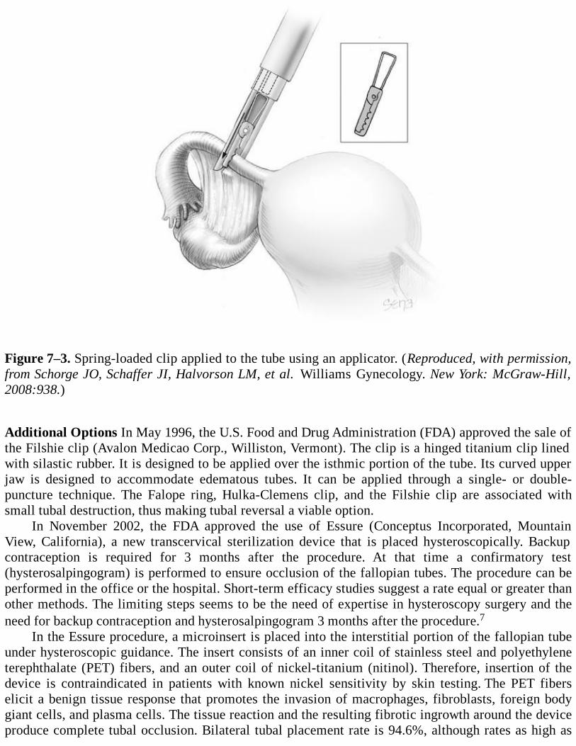
Figure 7–3. Spring-loaded clip applied to the tube using an applicator. (Reproduced, with permission,from Schorge JO, Schaffer JI, Halvorson LM, et al. Williams Gynecology. New York: McGraw-Hill,2008:938.)
Additional Options In May 1996, the U.S. Food and Drug Administration (FDA) approved the sale ofthe Filshie clip (Avalon Medicao Corp., Williston, Vermont). The clip is a hinged titanium clip linedwith silastic rubber. It is designed to be applied over the isthmic portion of the tube. Its curved upperjaw is designed to accommodate edematous tubes. It can be applied through a single- or double-puncture technique. The Falope ring, Hulka-Clemens clip, and the Filshie clip are associated withsmall tubal destruction, thus making tubal reversal a viable option.
In November 2002, the FDA approved the use of Essure (Conceptus Incorporated, MountainView, California), a new transcervical sterilization device that is placed hysteroscopically. Backupcontraception is required for 3 months after the procedure. At that time a confirmatory test(hysterosalpingogram) is performed to ensure occlusion of the fallopian tubes. The procedure can beperformed in the office or the hospital. Short-term efficacy studies suggest a rate equal or greater thanother methods. The limiting steps seems to be the need of expertise in hysteroscopy surgery and theneed for backup contraception and hysterosalpingogram 3 months after the procedure.7
In the Essure procedure, a microinsert is placed into the interstitial portion of the fallopian tubeunder hysteroscopic guidance. The insert consists of an inner coil of stainless steel and polyethyleneterephthalate (PET) fibers, and an outer coil of nickel-titanium (nitinol). Therefore, insertion of thedevice is contraindicated in patients with known nickel sensitivity by skin testing. The PET fiberselicit a benign tissue response that promotes the invasion of macrophages, fibroblasts, foreign bodygiant cells, and plasma cells. The tissue reaction and the resulting fibrotic ingrowth around the deviceproduce complete tubal occlusion. Bilateral tubal placement rate is 94.6%, although rates as high as
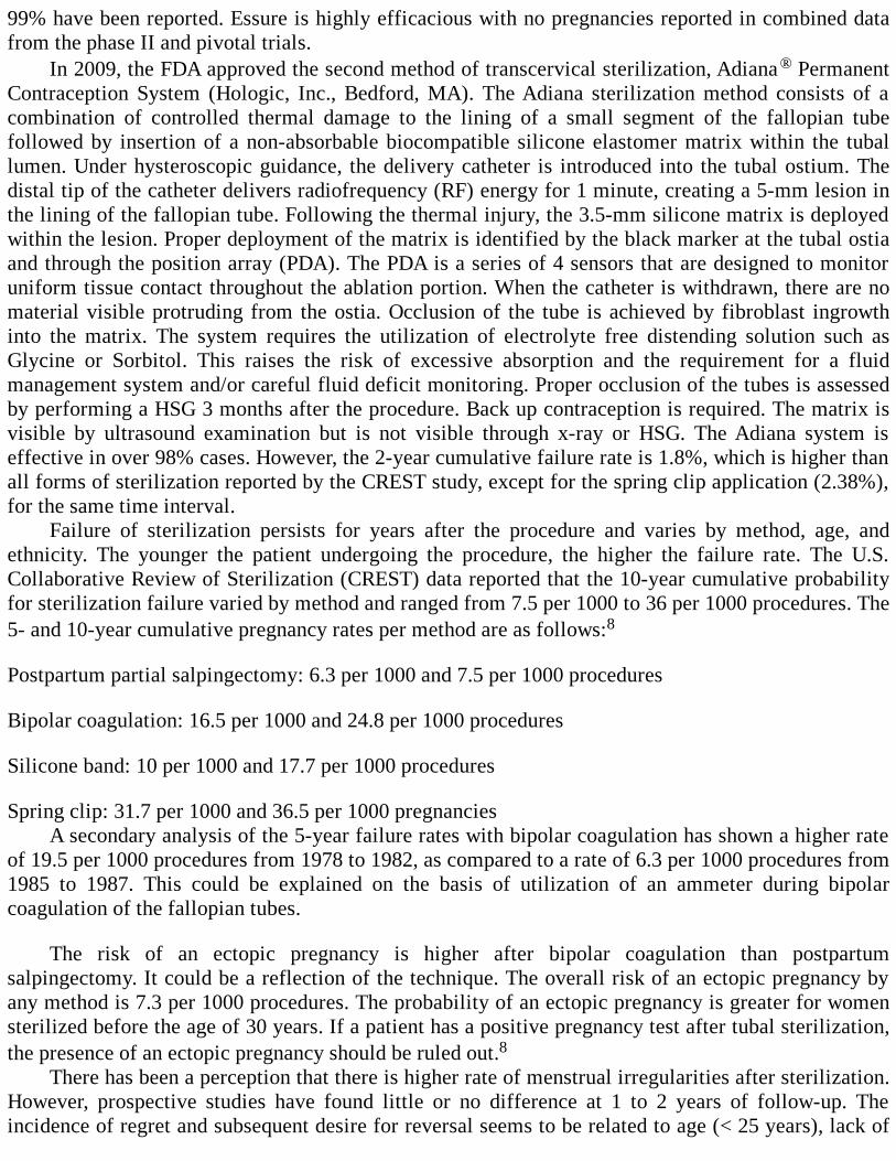
99% have been reported. Essure is highly efficacious with no pregnancies reported in combined datafrom the phase II and pivotal trials.
In 2009, the FDA approved the second method of transcervical sterilization, Adiana® PermanentContraception System (Hologic, Inc., Bedford, MA). The Adiana sterilization method consists of acombination of controlled thermal damage to the lining of a small segment of the fallopian tubefollowed by insertion of a non-absorbable biocompatible silicone elastomer matrix within the tuballumen. Under hysteroscopic guidance, the delivery catheter is introduced into the tubal ostium. Thedistal tip of the catheter delivers radiofrequency (RF) energy for 1 minute, creating a 5-mm lesion inthe lining of the fallopian tube. Following the thermal injury, the 3.5-mm silicone matrix is deployedwithin the lesion. Proper deployment of the matrix is identified by the black marker at the tubal ostiaand through the position array (PDA). The PDA is a series of 4 sensors that are designed to monitoruniform tissue contact throughout the ablation portion. When the catheter is withdrawn, there are nomaterial visible protruding from the ostia. Occlusion of the tube is achieved by fibroblast ingrowthinto the matrix. The system requires the utilization of electrolyte free distending solution such asGlycine or Sorbitol. This raises the risk of excessive absorption and the requirement for a fluidmanagement system and/or careful fluid deficit monitoring. Proper occlusion of the tubes is assessedby performing a HSG 3 months after the procedure. Back up contraception is required. The matrix isvisible by ultrasound examination but is not visible through x-ray or HSG. The Adiana system iseffective in over 98% cases. However, the 2-year cumulative failure rate is 1.8%, which is higher thanall forms of sterilization reported by the CREST study, except for the spring clip application (2.38%),for the same time interval.
Failure of sterilization persists for years after the procedure and varies by method, age, andethnicity. The younger the patient undergoing the procedure, the higher the failure rate. The U.S.Collaborative Review of Sterilization (CREST) data reported that the 10-year cumulative probabilityfor sterilization failure varied by method and ranged from 7.5 per 1000 to 36 per 1000 procedures. The5- and 10-year cumulative pregnancy rates per method are as follows:8
Postpartum partial salpingectomy: 6.3 per 1000 and 7.5 per 1000 procedures
Bipolar coagulation: 16.5 per 1000 and 24.8 per 1000 procedures
Silicone band: 10 per 1000 and 17.7 per 1000 procedures
Spring clip: 31.7 per 1000 and 36.5 per 1000 pregnanciesA secondary analysis of the 5-year failure rates with bipolar coagulation has shown a higher rate
of 19.5 per 1000 procedures from 1978 to 1982, as compared to a rate of 6.3 per 1000 procedures from1985 to 1987. This could be explained on the basis of utilization of an ammeter during bipolarcoagulation of the fallopian tubes.
The risk of an ectopic pregnancy is higher after bipolar coagulation than postpartumsalpingectomy. It could be a reflection of the technique. The overall risk of an ectopic pregnancy byany method is 7.3 per 1000 procedures. The probability of an ectopic pregnancy is greater for womensterilized before the age of 30 years. If a patient has a positive pregnancy test after tubal sterilization,the presence of an ectopic pregnancy should be ruled out.8
There has been a perception that there is higher rate of menstrual irregularities after sterilization.However, prospective studies have found little or no difference at 1 to 2 years of follow-up. Theincidence of regret and subsequent desire for reversal seems to be related to age (< 25 years), lack of
preoperative information regarding the procedure, lack of information on contraceptive options,pressure from a spouse, and due to medical indications. In terms of sexual interest or pleasure aftersterilization, most prospective studies have shown either no consistent change or no improvement.9,10
Comprehension Questions
7.1 A 32-year-old G4P4 woman has had a bilateral tubal occlusion via laparoscopy 2 yearspreviously. She understands that she is at risk for ectopic pregnancy. As compared to womenwho do not have sterilization, which of the following is most accurate?
A. Female sterilization is associated with a greater risk of ectopic pregnancy.B. Female sterilization is associated with the same risk of ectopic pregnancy.C. Female sterilization is associated with a lower risk of ectopic pregnancy.
7.2 Which of the following method of female sterilization is associated with the highest failurerate?
A. Unipolar cautery at laparoscopyB. Bipolar cautery at laparoscopyC. Tubal ligation at the time of cesareanD. Falope ring via laparoscopyE. Spring clips
7.3 A 37-year-old woman strongly desires female sterilization. She has had numerous abdominalsurgeries in the past and has had a ruptured appendicitis previously. Which of the followingis the best surgical approach in this patient?
A. Laparoscopic Falope ringB. Laparoscopic electrocauteryC. Hysteroscopic approachD. Minilaparotomy with tubal occlusion
ANSWERS
7.1 C. Women who have female sterilization have a lower rate of ectopic pregnancy since thereare far less pregnancies than in the nonsterilized cohort. However, if a woman who has had atubal occlusion does become pregnant, the risk of ectopic pregnancy is fairly high.
7.2 E. Spring clips are associated with the highest failure rate, in other words, the highestpregnancy rate.
7.3 C. Hysteroscopic use of coils in the tubal ostia (Essure) is a good option for patients to avoidthe abdominal cavity, given her abdominal adhesions and multiple prior abdominalsurgeries. A hysterosalpingogram is recommended following the procedure to ensure thatthe tubes are occluded.
Clinical PearlsSee Table 1-2 for definition of level of evidence and strength of recommendation
Sterilization by tubal occlusion is chosen by women at a rate of approximately three times that

of vasectomy (Level B).
Laparoscopic tubal occlusion is the most common type of female sterilization (Level A).
Bipolar sterilization is best performed using a 3-cm segment of tube (three applications of thebipolar device) and an ammeter to determine that the tissue is adequately desiccated (Level B).
Division of the tube after bipolar cautery is unnecessary and may lead to bleeding (Level B).
The spring clip device is associated with the highest risk of sterilization failure (Level B).
The hysteroscopic placement of coils in the tubal ostia (Essure system), and the insertion of anon-absorbable biocompatible silicone elastomer matrix (Adiana) seems to be effective andcan be performed in the office. A hysterosalpingogram needs to be performed to ensure tubalocclusion (Level B).
The incidence of regret after sterilization seems to be associated with age, especially youngerthan the age of 25 years (Level B).
REFERENCES
1. Piccinino LJ, Mosher WD. Trends in contraceptive use in the United States: 1982-1995. FamPlann Perspect. 1998;30:10,46.
2. Pollack AE, Soderstrom RM. Female tubal sterilization. In: Corson SL, Derman RJ, Tyler LB,eds, Fertility Control. 2nd ed. London, ON: Goldin Publishers; 1994:293-317.
3. Yoon IB, King TM, Parmley TH. A two-year experience with the Falope ring sterilizationprocedure. Am J Obstet Gynecol. 1977;127:109-112.
4. Hulka JF, Mercer JP, Fishburne JI, et al. Spring clip sterilization: one year follow-up of 1,079cases. Am J Obstet Gynecol. 1976;125:1039-1043.
5. Sokal D, Gates D, Amatya R, Dominik R. Two randomized controlled trials comparing thetubal ring and Filshie clip for tubal sterilization. Fertil Steril. 2000;74:525-533.
6. Dominik R, Gates D, Sokal D, et al. Two randomized controlled trials comparing the Hulkaand Filshie Clips for tubal sterilization. Contraception. 2000;62:169-175.
7. Bradley L. Long-term follow-up of hysteroscopic sterilization with the Essure Microinsert.Supplement to the Journal of Minimally Invasive Gynecology. Fertil Steril. 2008;15(6):S14-S15.
8. Peterson HB, Jeng G, Folger SG, Hillis SA, Marchbanks PA, Wilcox LS. The risk of ectopicpregnancy after tubal sterilization. U.S. Collaborative Review of Sterilization Working Group.N Engl J Med. 1997;336:762-767.
9. Peterson HB, Ory HW, Greenspan JR, Tyler CW Jr. Deaths associated with laparoscopic
9. Peterson HB, Ory HW, Greenspan JR, Tyler CW Jr. Deaths associated with laparoscopicsterilization by unipolar electrocoagulating devices, 1978 and 1979. Am J Obstet Gynecol.1981;139:141-143.
10. Peterson HB, DeStefano F, Rubin GL, Greenspan JR, Lee NC, Ory HW. Deaths attributable totubal sterilization in the United States, 1977-81. Am J Obstet Gynecol. 1983;146:131-136.
11. Magnani RJ, Haws JM, Morgan GT, Gargioullo PM, Pollack AE, Koonin LM. Vasectomy inthe United States, 1991 and 1995. Am J Public Health. 1999;89:92-94.
12. Kleppinger RK. Female outpatient sterilization using bipolar coagulation. Bull PostgradComm Med Univ Syd. 1977 Nov;33(8):144-154.
13. Soderstrom RM, Levy BS, Engel T. Reducing bipolar sterilization failures. Obstet Gynecol.1989;74:60-63.
14. Fishburne JI Jr, Hulka JF. Tubal healing following laparoscopic electrocoagulation. J ReprodMed. 1976;16:129-134.
15. Rulin MC, Davidson AR, Philliber SG, et al. Changes in menstrual symptoms amongsterilized and comparison women: a prospective study. Obstet Gynecol. 1989;74:149-154.
16. Shain RN, Miller WB, Mitchell GW, et al. Menstrual pattern change 1 year after sterilization:results of a controlled prospective study. Fertil Steril. 1989;52:192-203.
17. Wilcox LS, Chu SY, Eaker ED, et al. Risk factors for regret after tubal sterilization: 5 yearsof follow-up in a prospective study. Fertil Steril. 1991;55:27-33.
18. Jamieson DJ, Hillis SD, Duerr A, Marchbanks PA, Costello C, Peterson HB. Complications ofinterval laparoscopic tubal sterilization: findings from the United States Collaborative Reviewof Sterilization. Obstet Gynecol. 2000;96(6): 997-1002.
19. Moore CL, Vasquez NF, Lin H, Kaplan LJ. Major vascular injury after laparoscopic tuballigation. J Emerg Med. 2005;29(1):67-71.
20. Vancaille TG, Anderson TL, Johns DA. A 12-month prospective evaluation of transcervicalsterilization using implantable polymer matrices. Obstet Gynecol. 2008;112:1270-1277.
21. Levy B, Levie MD, Childers ME. A summary of reported pregnancies after hysteroscopicsterilization. J Minim Invsive Gynecol. 2007;14:271-274.
22. Connor VF. Essure: a review six years later. J Minm Invasive Gynecol. 2009; 16:282-290.
Case 8
A 35-year-old G2P2002 Hispanic woman presents to your office complaining of abdominal pain,nausea, and vomiting. Her abdominal pain has become progressively worse, and for the past 6 hours,she has had vomiting. She is postoperative day 3 from a right salpingo-oophorectomy for an 8-cmcomplex mass that was diagnosed as a mature teratoma on pathology. Some moderately denseadhesions of the omentum and small bowel were noted to be adherent to the mass and were dissecteddown. No complications were noted during the surgery, and the patient was discharged home the sameday. Her past medical history is significant for pelvic inflammatory disease (PID) and two previouscesarean deliveries. Her vitals are temperature 101.4°F, pulse 110 beats/min, respiratory rate (RR) 22breaths/min, and BP 120/70 mm Hg. On abdominal examination, the trocar sites are without erythemaor induration and are well reapproximated. She has marked tenderness throughout the abdomen withrebound and guarding.
What is the most likely diagnosis?
What imaging modality is most helpful to confirm the diagnosis?
What is the best treatment for this patient?
ANSWERS TO CASE 8:Laparoscopic Complications
Summary: A 35-year-old G2P2002 Hispanic woman presents with fever and abdominal pain withrebound tenderness. She is postoperative day 3 from a right salpingo-oophorectomy for an 8-cmcomplex mass. Some moderately dense adhesions of the omentum and small bowel were notedadherent to the mass and were dissected down.
Most likely diagnosis: Peritonitis from bowel injury.
Most helpful imaging modality: Abdominal and pelvic computed tomography (CT) withwater-soluble contrast.
Best treatment for this patient: The patient should receive broad-spectrum antibiotics thatcover anaerobic and aerobic organisms. She should then undergo a laparotomy. Meticulousinspection of the bowel should be carried out. Further treatment should take into considerationthe location of the injury (small bowel or the large bowel), the cause of the injury (thermal,laceration, puncture), the size of the injury, and presence of fecal contamination. She may needa simple closure of the bowel wall defect or a colostomy.
ANALYSIS
Objectives
1. List the different types of laparoscopic injuries.2. Know techniques to prevent laparoscopic injuries.3. Know the intra- and postoperative presentation of laparoscopic injuries.4. Know techniques for the management of various injuries.
Considerations
This is a patient who underwent a difficult laparoscopy. The patient had multiple risk factors forcomplications from surgery such as prior PID and prior cesareans. The surgical findings notedomentum and small bowel adherent to the ovarian mass. The patient’s surgery occurred 3 dayspreviously, and now she presents with peritonitis. The most important goals in the approach to thispatient are broad-spectrum antibiotics and expeditious exploratory laparotomy. Affected patients canbecome septic and hypotensive; intravenous fluid hydration and blood pressure support are important.
APPROACH TO Laparoscopic Complications
DEFINITIONS
ENTEROTOMY: Full-thickness opening in the bowel wall where the lumen is entered.
URETEROURETEROSTOMY: Anastomosis of the two transected ends of the ureter.
URETERONEOCYSTOSTOMY WITH PSOAS HITCH: When the ureter is transected within 6 cmof the ureterovesical junction, this is the treatment of choice. The distal ureter segment is ligated. Thebladder is mobilized and elongated toward the proximal injured ureter. The proximal ureter isanastomosed to the bladder. The bladder is attached to the psoas muscle to keep tension off theproximal ureter.
ILEAL INTERPOSITION: When a ureter injury takes place in the upper or middle third of theureter and length for reanastomosis is lacking, the proximal ureter is anastomosed to a segment ofileum which in turn is anastomosed to the distal ureter.
URETEROILEONEOCYSTOSTOMY: When a ureter injury takes place in the upper or middlethird of the ureter and the length for a tension-free reanastomosis is lacking, the proximal ureter isanastomosed to a segment of ileum which in turn is anastomosed to the bladder.
BOARI BLADDER FLAP: A flap of bladder wall shaped into a tube to replace a missing uretersegment.
TAMPON TEST: Helps to differentiate between a ureteral and bladder fistula. The patient wears thelargest size tampon that is comfortable. She then takes oral Phenazopyridine HCl (Pyridium) andliquids for 30 minutes. Orange-stained urine on the tampon indicates a ureteral or a cephalad bladderfistula. A fresh tampon is then placed in the vagina, and 300 cc of methylene blue–stained sterilewater is placed into the bladder via transurethral catheter. If there is no blue stain on the tampon, thefistula is from the ureter. An exception is a very small vesicular fistula.
CLINICAL APPROACH
Complications of laparoscopic surgery include injury to the gastrointestinal tract, vessels, urinarytract; related to the pneumoperitoneum; and incisional hernia. It is essential to recognize the injuriesintraoperatively. The injury can occur during insertion of Veress needle or trocars, or at the time ofthe operative laparoscopy.1 The management of the injury may include laparoscopic repair orlaparotomy, depending on the level of surgeon comfort and expertise. Consultations with specialists ingeneral surgery, urology, or vascular surgery may be invaluable.
Pneumoperitoneum
If the Veress needle fails to enter the peritoneal cavity and causes extraperitoneal insufflation, it mayresult in mediastinal emphysema and pneumothorax. If the gas from the extraperitoneal insufflationextends to the mediastinum, mediastinal emphysema will be seen. The anesthesiologist will havedifficulty ventilating the patient. As much gas as possible should be released, and the patient shouldbe carefully monitored. The patient may require assisted ventilation in severe cases. A pneumothoraxcan occur if the Veress needle is placed in the pleural cavity, and may be more likely if an upperabdomen site is chosen for insufflation. A penetrating injury into a blood vessel can cause a gasembolism. The anesthesiologist may note a “mill-wheel” murmur over the precordium. In this case, acentral venous catheter can be placed in the right atrium or the superior vena cava and the CO2 gas canbe aspirated.1
Vascular Injuries
The insertion of the Veress needle or the trocar can cause trauma to the omental or mesenteric bloodvessels and major abdominal or pelvic vessels. Small vessels in the omentum can be coagulated.Injury to the mesenteric vessels may compromise blood supply to the bowel, which may lead to bowelresection. Large vessels require immediate laparotomy, blood transfusion, and vascular repair.
The most common complication of laparoscopic surgery is the injury of the superficial or inferiorepigastric vessels.2 Laceration of these vessels is minimized by transillumination of the abdominalwall, directly visualizing the inferior epigastric vessels and inserting the trocar 6 to 7 cm lateral to themidline.1 However, transillumination identifies the superficial epigastric artery only 64% of the time,and it is less helpful in patients with dark skin and body mass index (exceeding 25 kg/m2).2 Theinferior epigastric is seen 80% of the time with transillumination.4 Injury to these vessels can presentwith bleeding from the trocar site. Techniques that can help control the bleeding from these vesselsinclude direct pressure, tamponade from the trocar, bipolar coagulation of the vessel, placing suturescephalad and caudad to the sleeve, and compression with an inflated 12-Fr Foley catheter that isclamped to the skin.2 If a hematoma forms, it should be evacuated, the incision explored, and thebleeding vessel ligated.1
Dixon and Carrillo studied a small series of pelvic vessel injuries that occurred from Veressneedle and trocar placement. They reported that right-sided iliac vessel injuries were more common,and the right iliac vein was the most common site of injury.5 This injury can be prevented by aimingthe Veress needle and trocar toward the hollow of the sacrum with the patient’s bed in the horizontalposition. Large vessel injury may present with rapid pooling of blood in the operative field, welling ofblood from the Veress needle or trocar, or with a hemodynamically unstable patient. If large vesselinjury is suspected, the needle or trocar should be kept in place to help identify the site of the injuryand immediate laparotomy should be performed. The injured area should be identified andcompressed. The repair is based on the location of the injured vessel, whether it is arterial or venous,and the degree of the hemorrhage.
Bonjer and colleagues performed a retrospective comparison of open and closed laparoscopy.They reported rates of 0.075% of vascular injury with closed laparoscopy and 0% with openlaparoscopy. Visceral injury rates were 0.083% and 0.048%, respectively. 6 Although this study foundstatistical significance between open and closed laparoscopy for rates of vascular and bowel injuries, ameta-analysis found that open laparoscopy was not superior or inferior to any other entry technique.7
Gastric Injuries
Injury to the gastrointestinal tract can involve the stomach, small bowel, or large bowel. The injurycan be sharp or thermal. A gastric injury may occur during gynecologic surgery from distention of thestomach during a difficult intubation. This can be minimized by gastric decompression prior to entry.If gastric juices are aspirated after the placement of the Veress needle or if a gastric injury issuspected, the Veress needle should be removed. The stomach should be decompressed with anasogastric tube, and the Veress needle should be replaced. Gastric injuries may be recognized afterthe placement of the periumbilical trocar. The surgeon may visualize gastric contents or the rugalfolds of the stomach.8 If the puncture is less than 5 mm in diameter and is hemostatic, the injury willusually resolve spontaneously. If the injury is larger, it may require a more extensive closure.1
Intestinal Injuries
A recent meta-analysis found that the incidence of bowel injuries to be 0.33% with major operativelaparoscopy.9 Women with previous abdominal surgeries and pelvic adhesions are at greatest risk.
Bowel perforation with the Veress needle is often undiagnosed because the perforation seals offspontaneously. If perforation is suspected, the needle is removed and replaced at another site. Oncethe laparoscope is in place, the bowel should be inspected carefully. Most needle injuries to the smallbowel do not require a laparotomy, but should be monitored closely. A hemostatic serosal injury doesnot need to be repaired.1
Trocar injuries are usually larger and require repair. A bowel injury is suspected if the trocarobturator has fecal matter upon removal from the sleeve or if the bowel lumen is visualized via thelaparoscope. If the trocar or the laparoscope creates an enterotomy, it should be left in place todecrease contamination and for identification of the injured site; a laparotomy should then beperformed. A small enterotomy that occurs during the surgical procedure can be repairedlaparoscopically. Colon injuries can be repaired laparoscopically if the patient has undergone a bowelpreparation or if fecal spill is minimal. Copious irrigation and broad-spectrum antibiotics are essentialin prevention of peritonitis. Depending on the site and nature of the injury, a colostomy may benecessary.1
A thermal bowel injury can result from direct contact of electrical, thermal, or laser energy. Thearea of tissue destruction is larger than the initial area of contact due to thermal spread. Unrecognizedthermal injury can result in tissue necrosis and subsequent perforation up to 72 to 96 hours after thesurgery. One should suspect thermal bowel injury in a patient who presents several days afteroperative laparoscopic surgery with fever and abdominal pain. Significant thermal bowel injuryshould be treated with wide resection and reanastomosis.1
Approximately 15% of bowel injuries are not recognized at the time of laparoscopy. Brosens andassociates reported that one in five cases of delayed diagnosis of bowel injury resulted in death.9
Bladder Injuries
Bladder injury can take place with trocar insertion. Women with previous pelvic surgery are at highest
risk. To avoid this complication, the bladder should be drained prior to initiating the surgicalprocedure. If the surgery is anticipated to take longer than 30 minutes, a catheter should be placed forcontinuous drainage. The size of the injury will determine the treatment. A Veress needle puncture canbe managed expectantly. Lacerations less than 5 mm will resolve spontaneously with continuousbladder drainage for 4 to 5 days postoperatively. Larger injuries should be closed in two layers. Themucosa should be reapproximated with 2-0 chromic suture, and the detrusor muscle should bereapproximated with 2-0 polyglycolic acid suture. Care should be taken to avoid involvement of theureteral orifices in the repair. The repair should be tension free with a good seal. A catheter should beplaced in the bladder for continuous drainage for 5 to 7 days.1
Ureteral Injuries
Ureteral injuries occur less frequently than those involving the bladder. The best way to preventureteral injury is to identify the course of the ureter prior to clamping the pedicle.
Leonard and associates performed a review of ureteral injuries during total laparoscopichysterectomies and found an incidence of 0.3% with all injuries occurring at the level of the uterineartery and the uterosacral ligament.10 Laparoscopic ureteral injuries are more likely to result fromthermal injuries. Furthermore, these injuries have a significant delay of diagnosis.1 If patients presentwith flank pain, fever, retroperitoneal fluid collection, or ileus in the postoperative period, an injury tothe ureter should be suspected. A creatinine elevation of 0.8 mg/dL may be related to a unilateralureteral ligation. A CT scan will be most helpful at this time. It will provide information about theintegrity and function of the renal collecting system while identifying urinomas and surroundingpostoperative anatomy. If intrinsic renal damage is present and there is concern for the use of contrastmedium, a renal and proximal ureter ultrasound can be performed. However, it has a 25% false-negative rate.1 A retrograde stent, anterograde stent, or a percutaneous nephrostomy tube should beperformed if an obstruction is noted and cannot be immediately relieved. If the obstruction is notrelieved, permanent renal damage may result. If there is no ureteral obstruction but leakage of urine isnoted, a ureteral stent should be placed. The best chance of healing after repair is when reoperationoccurs within the first 48 hours. With increasing delay, more edema, necrosis, and tissue damagedecrease the likelihood of a successful primary repair.
An intraoperative ureteral transection or ligation injury may be suspected if the ureter seems toenter a clamped or cut pedicle, a tubular structure is noted in a cut pedicle, there is lack of peristalsisfrom the ureter, and no urine output if both ureters are transected. If a ureteral injury is suspectedintraoperatively, indigo carmine can be injected intravenously. A cystoscopy will show dye from theureteral orifices after 5 minutes. If the ureteral injury is suspected immediately postoperatively, anintravenous pyelogram should be obtained. Up to 50% of cases of unilateral ureteral injury areasymptomatic postoperatively.11 Patients may also present with urine leakage from the vagina,indicating that an undetected ureteral or bladder injury has caused a fistula. A fistula can be diagnosedwith a tampon test.
If a ureteral injury does occur, the management is based on the location, mechanism of theinjury,4 and time of diagnosis. Repair of ureters is most often done via laparotomy. If the ureter iskinked by a ligature, simple dissection of the ureter away from the ligature is needed. In cases wherethe ureter is partially or completely ligated, the suture is removed and a stent is placed. If there is aquestion of ureteral viability, especially in cases of devascularization or thermal injury, the portion ofconcern must be resected. If a partial ureteral transection has taken place, a stent is introduced via theureterotomy and the ureter is reapproximated over the stent with 5-0 polyglycolic acid suture. Aclosed suction drain should be placed at the base of the repair. If a complete transection has occurred
in the upper or middle third of the ureter, a ureteroureterostomy over a stent is recommended. Caremust be taken to ensure the repair is tension free and the ends are spatulated to increase the lumensize. If a transection of the ureter has occurred in the upper or middle third and the repair cannot beperformed in a tension-free manner, a ureteroileal interposition or a ureteroileoneocystostomy iscarried out. If a ureteral injury takes place within 6 cm of the ureterovesical junction, there is concernfor vascular compromise of the distal portion of the ureter. The repair of choice in this situation is aureteroneocystostomy with psoas hitch over a stent.
Trocar Hernia
Incisional hernia after laparoscopy is rare. With the use of bladed trocars, the incidence of hernia wasfound to be 0.23% at the 10-mm site and 3.1% at the 12-mm site.12 The recommendation is to closethe fascia at trocar sites 10 mm or larger. With the use of bladeless trocars, the incidence of hernia is0% to 0.2% at the 10- and 12-mm sites, respectively. The lower rate of hernia is thought to result froma smaller residual defect after the bladeless trocars are removed.13
Comprehension Questions
8.1 A 34-year-old woman is undergoing a tubal sterilization procedure. Bipolar cautery isplanned to be used to cauterize the isthmic portion of the tubes. Which of the followingstatements regarding this patient is most accurate?
A. In general unipolar cautery is safer than bipolar cautery in avoiding thermal injury.B. A thermal injury to the bowel is usually not apparent immediately and takes several days to a
week to manifest.C. The most likely cause of death to this patient related to the surgery is hemorrhage.D. The ampulla of the tube is a more appropriate location rather than the isthmic portion.
8.2 A 29-year-old woman has chronic pelvic pain that has been worsening over the past 6 monthsdespite medical therapy. The patient has had a thorough work-up, and is being scheduled fora diagnostic laparoscopy. In consenting the patient for complications, the gynecologistexplains that vascular injury can occur. Which of the following is the most common vesselto be injured in this procedure?
A. Deep (inferior) epigastric arteryB. AortaC. Vena cavaD. Common iliac arteryE. Internal iliac artery
8.3 A 29-year-old woman undergoes extensive lysis of adhesions and ablation of endometriosisvia laparoscopy. Postoperatively, she has abdominal distention and some leakage ofyellowish fluid from her incision site. The fluid is sent for analysis. Which of the followingstatements is most accurate regarding this patient’s condition?
A. If the fluid has a sodium level of 140 mEq/L, it is likely to be urine.B. If the fluid has a creatinine level of 0.8 mg/L, it is unlikely to be urinary in origin.C. If the fluid has a pH of 7.40, it is likely to be urine.D. If the fluid has a cloudy appearance, it is likely to be peritoneal fluid (ascites).
ANSWERS
8.1 B. Thermal injury often does not manifest itself until several days to a week after the initialsurgery, due to the ischemia and necrosis that takes place. Bipolar is in general safer sincethe current passes from one paddle to another, whereas unipolar current can arc from thedevice to adjacent tissue. The most common cause of death due to female sterilization isanesthetic in nature. The isthmus of the tube is the most appropriate location for tuballigation/occlusion.
8.2 A. In general, the deep epigastric artery is the most commonly injured vessel in laparoscopythat is associated with significant hemorrhage. This vessel is located between the rectusmuscle and the parietal peritoneum of the anterior abdominal wall.
8.3 B. If the creatinine level is 0.8 mg/dL, it is unlikely to be urinary in origin, since thecreatinine concentration is typically in the range of 40 to 50 mg/dL in the ureteral/bladderdue to the concentrating ability of the kidneys. The sodium concentration is usually between20 to 50 mEq/L. The pH varies and is not a good differentiating criterion.
Clinical PearlsSee Table 1-2 for definition of level of evidence and strength of recommendation
Early detection of laparoscopic complication is the key to preventing patient morbidity andmortality (Level A).
Fascia should be closed at trocar sites 5 mm or larger to prevent hernias (Level B).
Repair of the ureter or bladder must be tension free for optimal healing (Level B).
Thermal injuries may not be noted intraoperatively. Urinary and gastrointestinal tract injuriescan present days later. An unrecognized bowel injury can present 72 to 96 hours from surgery(Level A).
Although not consistent, some studies have noted decreased visceral and vascularcomplications with open laparoscopy compared to closed laparoscopy (Level B).
REFERENCES
1. Namnoum AB, Murphy AA. Diagnostic and Operative Laparoscopy in TeLinde’s OperativeGynecology. 9th ed. Philadelphia, PA: Lippincott Williams & Wilkins; 2003.
2. Donnez J, Nisolle M. An Atlas of Operative Laparoscopy and Hysteroscopy. 2nd ed. New York,NY: The Parthenon Publishing Group Inc.; 2001.
3. Hurd WW, Amesse LS, Gruber JS, et al. Visualization of the epigastric vessels and bladderbefore laparoscopic trocar placement. Fertil Steril. 2003;80:209-212.
4. Stany MP, Farley JH. Complications of gynecologic surgery. Surg Clin North Am.2008;88:343-359.
5. Dixon M, Carrillo EH. Iliac vascular injuries during elective laparoscopic surgery. SurgEndosc. 1999;13:1230-1233.
6. Bonjer HJ, Hazebroek, EJ, Kazemier G, et al. Open versus closed establishment ofpneumoperitoneum in laparoscopic surgery. Br J Surg. 1997;84:599-602.
7. Vilos Ga, Ternamian A, Dempster J, et al. Laparoscopic entry: a review of techniques,technologies, and complications. J Obstet Gynaecol Can. 2007;29:434-465.
8. Mateus J, Pezzi C, Somkuti SG. Recognition and prevention of gastric injury duringgynecologic laparoscopy. Obstet Gyencol. 2006;108:804-806.
9. Brosens I, Gordon A, Campo R, et al. Bowel injury in gynecologic laparoscopy. J Am AssocGyencol Laparosc. 2003;10:9-13.
10. Leonard F, Fotso A, Borghese B, et al. Ureteral complications from laparoscopichysterectomy indicated for benign uterine pathologies: a 13-year experience in a continuousseries of 1300 patients. Hum Reprod. 2007;22:2006-2011.
11. Nezhat FNC, Nezhat CR. Averting complications of laparoscopy: pearls from 5 patients. OBGManagement. 2007;19:69.
12. Kadar N, Reich H, Liu CY, et al. Incisional hernias after major laparoscopic gynecologicprocedures. Am J Obstet Gynecol. 1993;168:1493-1495.
13. Liu CD, McFadden DW. Laparoscopic port sites do not require fascial closure whennonbladed trocars are used. Am Surg. 2000;66:853-854.
Case 9
A 37-year-old G4P3013 woman presents to your office with menorrhagia and pelvic pain. The patientalso has some feeling of incomplete emptying of her bladder. The patient has been seen by anothergynecologist and placed on medical therapy, including nonsteroidal anti-inflammatory drugs(NSAIDs) and oral contraceptives without alleviation. She desires a permanent solution as futurefertility is not an issue and would like to have a surgical technique that would allow her to resume herdaily duties in a short period of time. She has a negative past surgical history. She is 5 ft 4 in in heightand weighs 165 lb (75 kg). The abdominal examination reveals an irregular midline pelvic mass. Shealso has left uterosacral ligament tenderness without nodularity. Grade 1 uterine prolapse is noted.
What is the clinical condition?
What is your next step?
ANSWERS TO CASE 9:Total Laparoscopic Hysterectomy
Summary: This is a 37-year-old G4P3013 woman with menorrhagia and pelvic pain, which has failedmedical therapy. Examination reveals a fibroid uterus of 16-week size just below the umbilicus andleft uterosacral ligament tenderness without nodularity. Grade 1 prolapse is noted, and upon furtherquestioning some urinary retention is mentioned.
Clinical condition: Multiparous patient with symptomatic fibroids causing menorrhagia, andpossible endometriosis and minimal prolapse.
Next step: Office ultrasound of the pelvis. Then, outline the surgical options and explain therisks, benefits, and alternatives of the operation. Unfortunately, the symptoms of urinaryretention are seldom volunteered by the patient seeking hysterectomy and should beascertained by the physician during the history. The simple question is “Do you feel like youare completely emptying your bladder or do you feel that you still have to go after urinating?”
ANALYSIS
Objectives
1. Be familiar with what total laparoscopic hysterectomy (TLH) means.2. Be familiar with a TLH technique.3. Be familiar with complications of this operation.
Considerations
This is a 37-year-old G4P3013 woman who desires relief from menorrhagia and pelvic pain.
Additionally, she would be grateful if she did not feel that she has to urinate immediately after doingso. The first step for the physician is to be able to provide proper counseling. It is important for thepatient to understand that a laparoscopic approach is almost always possible. After surgical therapy ischosen, the route of surgery should be discussed. Vaginal hysterectomy (VH) is superior to theabdominal approach due to shorter hospital stay, quicker recovery, and less pain. When the VH isdifficult due to lack of descensus, a laparoscopic approach is useful. Evidence-based medicinesuggests that VH is the best method and should be performed when possible. Yet, VH is done in lessthan 20% of cases.
Most problems with VH can be solved by TLH, especially if we do the original version of TLHwhich includes vaginal cuff suspension by laparoscopic suturing!
Recent studies suggest that the incidence of pelvic organ prolapse (POP) surgery is high after allmodes and routes of hysterectomy for benign indications. In a large study from Sweden, overall riskof subsequent prolapse surgery increased by 50% after TAH, doubled after subtotal hysterectomy, andquadrupled after vaginal hysterectomy.
Indications for TLH include whenever abdominal hysterectomy is considered and VH notpossible or available, including limited vaginal access, large fibroid uterus, endometriosis, extensiveadhesions, most endometrial cancer hysterectomies, and pelvic support procedures. Of course, LAVHis the wrong operation for cul-de-sac endometriosis. TLH should include vaginal cuff suspension fromabove usually by high McCall cuff suspension.
APPROACH TO Total Laparoscopic Hysterectomy
DEFINITIONS
Many physicians use the phrase “laparoscopic hysterectomy” as an umbrella term encompassing alltypes of laparoscopically associated surgical procedures in which the uterus is removed.
In reality, laparoscopic hysterectomy (LH) is a distinct procedure. Its sine qua non islaparoscopic ligation of the uterine arteries by means of electrosurgical desiccation or suture ligature.All the following maneuvers or methods of laparoscopic-associated hysterectomy (LAH) can beaccomplished either vaginally or laparoscopically.
TOTAL LAPAROSCOPIC HYSTERECTOMY (TLH): TLH means laparoscopic dissection andligation of all vascular pedicles continuing until the uterus lies free of all attachments in the peritonealcavity. The uterus is removed through the vagina, often with laparoscopic and/or vaginal morcellation.The vagina is closed with laparoscopically placed suspension sutures. No vaginal surgery except formorcellation is done.
The surgical technique for TLH with a classification system for LAH was published in 1993 byReich and colleagues, describing all LAHs performed from April 1983 to July 1992. The conclusion ofthat paper was that LH is a substitute for abdominal hysterectomy and not for vaginal hysterectomy.Laparoscopic vaginal cuff closure with incorporation of the uterosacral ligaments was an integral partof the TLH operation since its inception and still remains so.
LAPAROSCOPIC HYSTERECTOMY (LH): LH is the laparoscopic ligation of the blood supply tothe uterus, including both uterine arteries either by electro-surgery desiccation, suture ligature, orstaples. All surgical steps after the uterine vessels have been ligated can be done either vaginally orlaparoscopically, including anterior and posterior vaginal entry, cardinal and uterosacral ligament
division, uterine removal (intact or by morcellation), and vaginal closure (vertically or transversely).Laparoscopic ligation of the uterine vessels is the sine qua non for laparoscopic hysterectomy.Ureteral identification often by isolation, and more recently by cystoscopy, has always been advised.
LAPAROSCOPIC-ASSISTED VAGINAL HYSTERECTOMY (LAVH): LAVH is a vaginalhysterectomy after laparoscopic adhesiolysis, endometriosis excision, or oophorectomy.
This term is also used when the upper uterine ligaments (eg, round, infundibulopelvic or utero-ovarian ligaments) of a relatively normal uterus are ligated with staples or bipolar desiccation beforevaginal hysterectomy. Most of these cases can be done without the laparoscope as completely vaginalhysterectomies.
CLINICAL APPROACH
While it is important that these different procedures are clearly delineated, some overlap is present,especially between LAVH and LH (LAVH is more vaginal hysterectomy) and between LH and TLHwith cuff suspension. We need to make clear what we mean.
What is a TLH? Today, much unwarranted confusion exists. After 20 years there is finally a CPTcode for billing this procedure. This coding further confuses the issue making one wonder where itcame from, certainly not from the experts doing this operation.
58570 Laparoscopy, surgical, with total hysterectomy, for uterus 250 g or less
58571 Laparoscopy, surgical, with total hysterectomy, for uterus 250 g or less; with removal oftube(s) and/or ovary(ies)
58572 Laparoscopy, surgical, with total hysterectomy, for uterus greater than 250 g
58573 Laparoscopy, surgical, with total hysterectomy, for uterus greater than 250 g; with removal oftube(s) and/or ovary(ies)
Although it is not noted in the literature, there seems to be significant difference between closingthe vagina vaginally, closing the vagina laparoscopically, and closing the vagina and its supportiveligaments laparoscopically. Seemingly, small differences in technique may lead to substantialdifferences in outcome, and are not usually included in the nomenclature.
For instance, during a vaginal hysterectomy, we know that tagging the uterosacral-cardinalcomplex vaginally helps in its identification for vaginal cuff repair, and affixing the uterosacralligaments to the vaginal cuff helps to ensure its secure suspension. However, if the ligaments aredivided laparoscopically and the vagina closed vaginally, it is much more difficult to identify theseligaments and incorporate them into the repair. Likewise, suturing just the vagina closedlaparoscopically, often with expensive disposable devices, does little for support. These nuances maycertainly lead to an increased risk of vaginal vault prolapsed.
Since the first LH in January 1988, the vast majority of surgeons have been slow to embrace thisalternative to the costlier abdominal hysterectomy and LAVH, often citing concerns about patientselection as the primary reason. There are few absolute contraindications specific to LH. Theprocedure shouldn’t be performed when vaginal hysterectomy can be accomplished with little vaginalrepair necessary. Also, the LH should not be performed when the patient has a potentially cancerouspelvic mass too large to fit intact into an impermeable sack. Stage III ovarian cancer, which requires alarge abdominal incision, is another contraindication, as is a surgeon’s inexperience. But uterineweight and size, degree of endometriosis, and similar factors should not preclude a skilled surgeon
from choosing LH.Perhaps the greatest advantage TLH offers is the surgeon’s ability to obtain better support for the
vagina and to suture stretched ligaments to the top of the vagina at a point far higher than would bepossible with other types of hysterectomy.
Total Laparoscopic Hysterectomy Technique
There are various techniques to this procedure, and the following is one such rendition. These stepsare designed to prevent complications.
Since hysterectomy is usually an elective procedure, the patient should be counseled extensivelyregarding the range of currently available options appropriate to her individual clinical situation. In2010, it is clearly not acceptable to advocate hysterectomy without detailing the risks and benefits ofother intermediary procedures, such as myomectomy and/or excision of endometriosis with uterinepreservation.
Whereas conversion to laparotomy when the surgeon becomes uncomfortable with thelaparoscopic approach has never been considered a complication, conversion rates should bemonitored to ensure that the patient has reasonable expectations. For instance, when half of attemptedlaparoscopic hysterectomies are converted to abdominal procedures, neither the patient nor thesurgeon is well served.
Preoperative Preparation The patient’s coexisting medical problems should be best optimized.Patients are encouraged to hydrate on clear liquids the day before surgery. Fleet Phospho-soda 3 ozdivided into two doses are given at 3:30 pm and 7:30 pm to evacuate the lower bowel. If the patient isprone to nausea, promethazine (Phenergan) 25 mg orally is taken 25 minutes before the bowelpreparation. Lower abdominal, pubic, and perineal hair is not shaved.
All laparoscopic procedures are performed using general endotracheal anesthesia with orogastrictube suction to minimize bowel distension. The patient’s arms are placed at her side and shoulderbraces at the acromioclavicular joint are positioned. Trendelenburg position up to 40 degrees isavailable. I use one dose of prophylactic antibiotics optimally 30 to 60 minutes prior to incision.
Incisions Three laparoscopic puncture sites including the umbilicus are used. Pneumoperitoneum 25to 30 mm Hg is obtained before primary umbilical trocar insertion and reduced to 15 mm afterward.The lower quadrant trocar sleeves are placed under direct laparoscopic vision lateral to the rectusabdominis muscles and just beside the anterior superior iliac spines in patients with large fibroids.The left lower quadrant puncture is my major portal for operative manipulation as I stand on thepatient’s left.
Sometimes, however, special entry techniques are necessary. When a patient has had numerousprior abdominal surgical procedures, may have adhesions around the umbilicus, or is extremelyoverweight, I insert the Veress needle in the left ninth intercostal space and the trocar at the leftsubcostal margin. I do this because in such a patient the peritoneum often is stuck to the undersurfaceof the ribs and can’t tent away from the Veress needle. The stomach must be decompressed with thisentry location.
Reduction in wound morbidity and scar integrity as well as cosmesis is enhanced using 5-mmsites. The use of 12-mm incisions when a 5-mm one will suffice is not an advance in minimallyinvasive surgery.
Vaginal Preparation Every year, new innovations for uterine and vaginal manipulation appear. TheValtchev uterine manipulator (Conkin Surgical Instruments, Toronto, Canada) has been around for
more than 20 years and allows anterior, posterior, and lateral manipulation of the uterus so that thesurgeon can visualize the posterior cervix and vagina. Although newer devices are currently availablethat have been developed by Pelosi, Wattiez, Hourcabie, Koninckx, Koh, McCartney, Donnez, andReich, I still use the Valtchev and the Wolf tube.
Exploration The upper abdomen is inspected, and the appendix is identified. Clear vision ismaintained throughout the operation using the I.C. Medical smoke evacuator (Phoenix, Arizona).Endometriosis is excised before starting TLH. Bleeding is controlled with microbipolar forceps.
Retroperitoneal Dissection The peritoneum is opened early with scissors in front of the roundligament to allow CO2 from the pneumoperitoneum to distend the retroperitoneum. The tip of thelaparoscope is then used to perform “optical dissection” of the retroperitoneal space by pushing it intothe loosely distended areolar tissue parallel to the uterus to identify the uterine vessels, ureter, or both.The uterine artery is often ligated at this time, especially in large uterus patients.
Ureteral Dissection (Optional) The ureter is identified medially, superiorly, or laterally (pararectalspace). Stents are not generally used as they may cause hematuria and ureteric spasm. Thelaparoscopic surgeon should dissect (skeletonize) either the ureter or the uterine vessels during theperformance of a laparoscopic hysterectomy.
Bladder Mobilization The round ligaments are divided at their midportion, and scissors or a spoonelectrode is used to divide the vesicouterine peritoneal fold starting at the left side and continuingacross the midline to the right round ligament. The upper junction of the vesicouterine fold isidentified as a white line firmly attached to the uterus, with 2 to 3 cm between it and the bladderdome. The initial incision is made below the white line while lifting the bladder. The bladder ismobilized off the uterus and upper vagina using scissors or blunt dissection until the anterior vagina isidentified. The tendinous attachments of the bladder in this area may be desiccated or dissected(Figures 9–1 to 9–4).
Upper Uterine Blood Supply When oophorectomy is indicated or desired, the peritoneum is openedon each side of the infundibulopelvic ligament with scissors and a 2-0-Polyglycolic acid (Vicryl) freeligature is passed through the window and created and tied extracorporeally using the Clarke-Reich(Cook Medical Group, Bloomington, IN) knot pusher. This maneuver is repeated until two proximaland one distal ties are placed and the ligament divided. This maneuver helps develop suturing skills.The broad ligament is divided to the round ligament just lateral to the utero-ovarian arteryanastomosis using scissors or cutting current through a spoon electrode. I rarely desiccate theinfundibulopelvic ligament as it results in too much smoke early in the operation.
When ovarian preservation is desired, the utero-ovarian ligament and fallopian tube arecompressed and coagulated until desiccated with bipolar forceps, at 25 to 35 W cutting current, andthen divided. Alternatively, the utero-ovarian ligament and fallopian tube pedicles are suture-ligatedadjacent to the uterus with 2/0-Vicryl, using a free ligature that is passed through a window that iscreated, enabling the ligature to be positioned around the ligament. Stapling devices are rarely used.
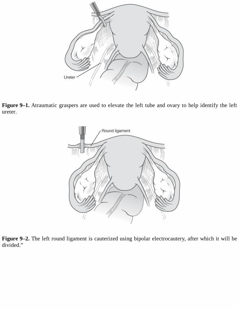
Figure 9–1. Atraumatic graspers are used to elevate the left tube and ovary to help identify the leftureter.
Figure 9–2. The left round ligament is cauterized using bipolar electrocautery, after which it will bedivided.”
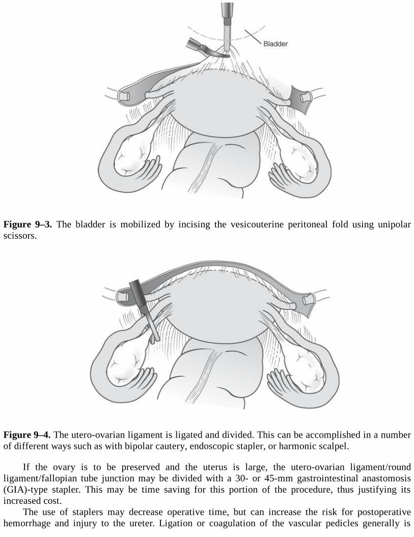
Figure 9–3. The bladder is mobilized by incising the vesicouterine peritoneal fold using unipolarscissors.
Figure 9–4. The utero-ovarian ligament is ligated and divided. This can be accomplished in a numberof different ways such as with bipolar cautery, endoscopic stapler, or harmonic scalpel.
If the ovary is to be preserved and the uterus is large, the utero-ovarian ligament/roundligament/fallopian tube junction may be divided with a 30- or 45-mm gastrointestinal anastomosis(GIA)-type stapler. This may be time saving for this portion of the procedure, thus justifying itsincreased cost.
The use of staplers may decrease operative time, but can increase the risk for postoperativehemorrhage and injury to the ureter. Ligation or coagulation of the vascular pedicles generally is
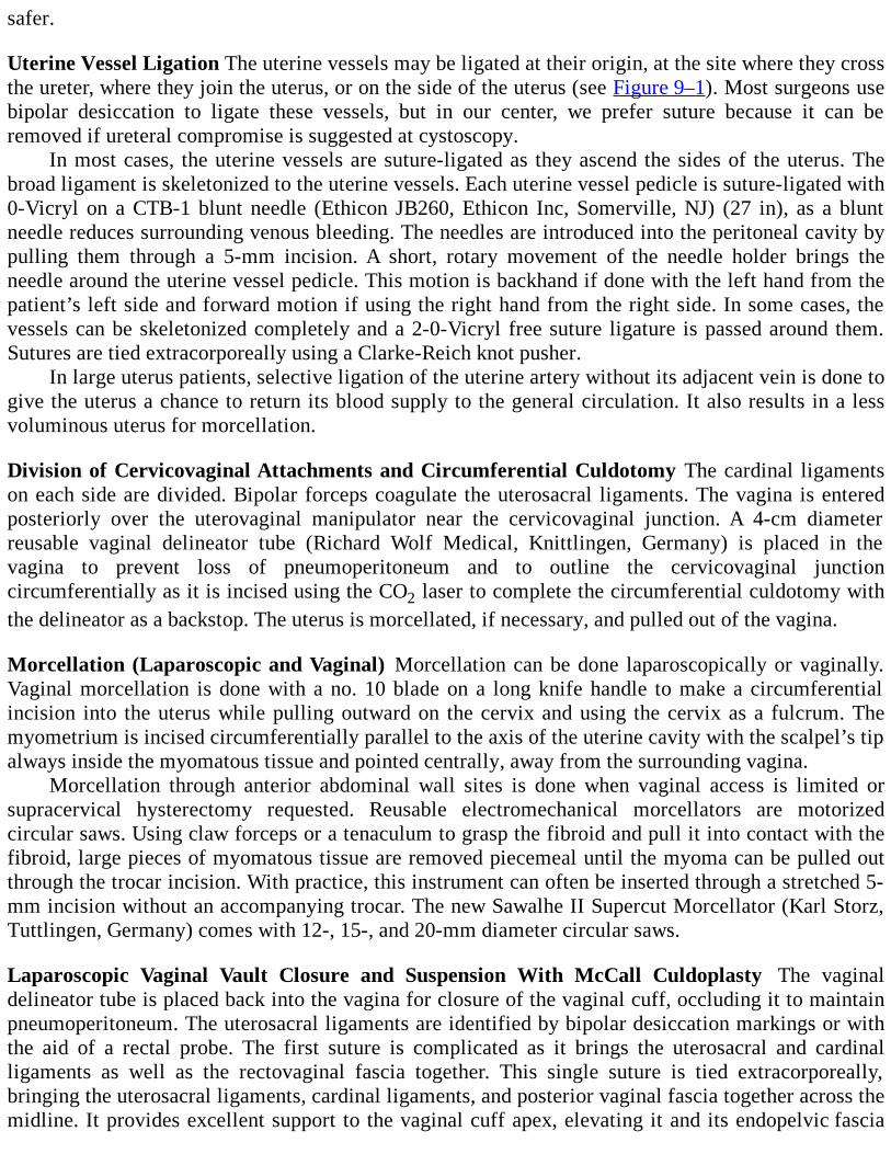
safer.
Uterine Vessel Ligation The uterine vessels may be ligated at their origin, at the site where they crossthe ureter, where they join the uterus, or on the side of the uterus (see Figure 9–1). Most surgeons usebipolar desiccation to ligate these vessels, but in our center, we prefer suture because it can beremoved if ureteral compromise is suggested at cystoscopy.
In most cases, the uterine vessels are suture-ligated as they ascend the sides of the uterus. Thebroad ligament is skeletonized to the uterine vessels. Each uterine vessel pedicle is suture-ligated with0-Vicryl on a CTB-1 blunt needle (Ethicon JB260, Ethicon Inc, Somerville, NJ) (27 in), as a bluntneedle reduces surrounding venous bleeding. The needles are introduced into the peritoneal cavity bypulling them through a 5-mm incision. A short, rotary movement of the needle holder brings theneedle around the uterine vessel pedicle. This motion is backhand if done with the left hand from thepatient’s left side and forward motion if using the right hand from the right side. In some cases, thevessels can be skeletonized completely and a 2-0-Vicryl free suture ligature is passed around them.Sutures are tied extracorporeally using a Clarke-Reich knot pusher.
In large uterus patients, selective ligation of the uterine artery without its adjacent vein is done togive the uterus a chance to return its blood supply to the general circulation. It also results in a lessvoluminous uterus for morcellation.
Division of Cervicovaginal Attachments and Circumferential Culdotomy The cardinal ligamentson each side are divided. Bipolar forceps coagulate the uterosacral ligaments. The vagina is enteredposteriorly over the uterovaginal manipulator near the cervicovaginal junction. A 4-cm diameterreusable vaginal delineator tube (Richard Wolf Medical, Knittlingen, Germany) is placed in thevagina to prevent loss of pneumoperitoneum and to outline the cervicovaginal junctioncircumferentially as it is incised using the CO2 laser to complete the circumferential culdotomy withthe delineator as a backstop. The uterus is morcellated, if necessary, and pulled out of the vagina.
Morcellation (Laparoscopic and Vaginal) Morcellation can be done laparoscopically or vaginally.Vaginal morcellation is done with a no. 10 blade on a long knife handle to make a circumferentialincision into the uterus while pulling outward on the cervix and using the cervix as a fulcrum. Themyometrium is incised circumferentially parallel to the axis of the uterine cavity with the scalpel’s tipalways inside the myomatous tissue and pointed centrally, away from the surrounding vagina.
Morcellation through anterior abdominal wall sites is done when vaginal access is limited orsupracervical hysterectomy requested. Reusable electromechanical morcellators are motorizedcircular saws. Using claw forceps or a tenaculum to grasp the fibroid and pull it into contact with thefibroid, large pieces of myomatous tissue are removed piecemeal until the myoma can be pulled outthrough the trocar incision. With practice, this instrument can often be inserted through a stretched 5-mm incision without an accompanying trocar. The new Sawalhe II Supercut Morcellator (Karl Storz,Tuttlingen, Germany) comes with 12-, 15-, and 20-mm diameter circular saws.
Laparoscopic Vaginal Vault Closure and Suspension With McCall Culdoplasty The vaginaldelineator tube is placed back into the vagina for closure of the vaginal cuff, occluding it to maintainpneumoperitoneum. The uterosacral ligaments are identified by bipolar desiccation markings or withthe aid of a rectal probe. The first suture is complicated as it brings the uterosacral and cardinalligaments as well as the rectovaginal fascia together. This single suture is tied extracorporeally,bringing the uterosacral ligaments, cardinal ligaments, and posterior vaginal fascia together across themidline. It provides excellent support to the vaginal cuff apex, elevating it and its endopelvic fascia
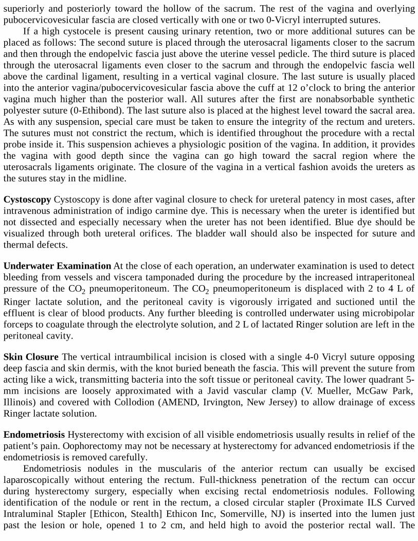
superiorly and posteriorly toward the hollow of the sacrum. The rest of the vagina and overlyingpubocervicovesicular fascia are closed vertically with one or two 0-Vicryl interrupted sutures.
If a high cystocele is present causing urinary retention, two or more additional sutures can beplaced as follows: The second suture is placed through the uterosacral ligaments closer to the sacrumand then through the endopelvic fascia just above the uterine vessel pedicle. The third suture is placedthrough the uterosacral ligaments even closer to the sacrum and through the endopelvic fascia wellabove the cardinal ligament, resulting in a vertical vaginal closure. The last suture is usually placedinto the anterior vagina/pubocervicovesicular fascia above the cuff at 12 o’clock to bring the anteriorvagina much higher than the posterior wall. All sutures after the first are nonabsorbable syntheticpolyester suture (0-Ethibond). The last suture also is placed at the highest level toward the sacral area.As with any suspension, special care must be taken to ensure the integrity of the rectum and ureters.The sutures must not constrict the rectum, which is identified throughout the procedure with a rectalprobe inside it. This suspension achieves a physiologic position of the vagina. In addition, it providesthe vagina with good depth since the vagina can go high toward the sacral region where theuterosacrals ligaments originate. The closure of the vagina in a vertical fashion avoids the ureters asthe sutures stay in the midline.
Cystoscopy Cystoscopy is done after vaginal closure to check for ureteral patency in most cases, afterintravenous administration of indigo carmine dye. This is necessary when the ureter is identified butnot dissected and especially necessary when the ureter has not been identified. Blue dye should bevisualized through both ureteral orifices. The bladder wall should also be inspected for suture andthermal defects.
Underwater Examination At the close of each operation, an underwater examination is used to detectbleeding from vessels and viscera tamponaded during the procedure by the increased intraperitonealpressure of the CO2 pneumoperitoneum. The CO2 pneumoperitoneum is displaced with 2 to 4 L ofRinger lactate solution, and the peritoneal cavity is vigorously irrigated and suctioned until theeffluent is clear of blood products. Any further bleeding is controlled underwater using microbipolarforceps to coagulate through the electrolyte solution, and 2 L of lactated Ringer solution are left in theperitoneal cavity.
Skin Closure The vertical intraumbilical incision is closed with a single 4-0 Vicryl suture opposingdeep fascia and skin dermis, with the knot buried beneath the fascia. This will prevent the suture fromacting like a wick, transmitting bacteria into the soft tissue or peritoneal cavity. The lower quadrant 5-mm incisions are loosely approximated with a Javid vascular clamp (V. Mueller, McGaw Park,Illinois) and covered with Collodion (AMEND, Irvington, New Jersey) to allow drainage of excessRinger lactate solution.
Endometriosis Hysterectomy with excision of all visible endometriosis usually results in relief of thepatient’s pain. Oophorectomy may not be necessary at hysterectomy for advanced endometriosis if theendometriosis is removed carefully.
Endometriosis nodules in the muscularis of the anterior rectum can usually be excisedlaparoscopically without entering the rectum. Full-thickness penetration of the rectum can occurduring hysterectomy surgery, especially when excising rectal endometriosis nodules. Followingidentification of the nodule or rent in the rectum, a closed circular stapler (Proximate ILS CurvedIntraluminal Stapler [Ethicon, Stealth] Ethicon Inc, Somerville, NJ) is inserted into the lumen justpast the lesion or hole, opened 1 to 2 cm, and held high to avoid the posterior rectal wall. The
proximal anvil is positioned just beyond the lesion or hole, which is invaginated into the opening, andthe device closed. The instrument is fired and removed.
Complications
Complications of laparoscopic hysterectomy are those of hysterectomy and laparoscopy combined:anesthetic accidents; respiratory compromise; thromboembolic phenomenon; urinary retention; injuryto vessels, ureters, bladder, and bowel; as well as infections, especially of the vaginal cuff. Ureteralinjury is more common when staplers or bipolar desiccation are used without ureteral identification.Complications unique to laparoscopy include large vessel injury, epigastric vessel laceration,subcutaneous emphysema, and trocar site incisional hernias.
Infection Experience with serious wound infection after laparoscopic hysterectomy is rare.Morcellation during laparoscopic or vaginal hysterectomy results in a slightly increased risk of fever,especially if prophylactic antibiotics are not used.
Hemorrhage Intraoperative hemorrhage occurs when a previously nonanemic patient loses greaterthan 1000 mL of blood or requires a blood transfusion. By doing careful laparoscopic dissection, mostprofuse hemorrhage situations are avoided or controlled as they occur.
Ureter Complications I remain committed to prevention of ureteral injury intraoperatively byureteral identification often with dissection and by cystoscopy at the conclusion of hysterectomies.
The ureters are commonly injured at the level of the infundibulopelvic ligament, uterosacralligament, or pelvic sidewall due to adhesions resulting from endometriosis, pelvic inflammatorydisease, or previous abdominal surgery. During laparoscopic hysterectomy, ureteral injury may occurwhile cutting dense adhesions and fibrotic scar tissue, trying to stop bleeding close to the ureter withbipolar cautery, or in the process of ligating the uterine vessels with bipolar electrosurgery, staples, orsuture.
Most ureteral injuries are not identified or even suspected without cystoscopy. The bottomline is that an aggressive approach to ureteral protection can reduce but not eliminate ureteral injury.However, prompt recognition and management can prevent multiple surgical procedures andsignificant patient morbidity, including organ loss.
Urinary retention is a common undetected complication. More studies are necessary to determinehow common and whether long-term compromise can occur.
Bladder Injury Bladder injury can occur during dissection of the bladder off the uterus and cervix orfrom an inflamed adnexa. In these cases the bladder is repaired using 3-0 Vicryl usually in two layers,with prolonged bladder drainage.
Bowel Injury Small bowel injury during laparoscopic hysterectomy is uncommon and is usuallyassociated with extensive intraperitoneal adhesions. Small bowel injuries can be suture-repaired.Small bowel enterotomy may require mobilization from above, delivery through the umbilicus byextending the incision 1 cm, and repair or resection. If the hole is confined to the antimesentericportion, the bowel can be closed with interrupted 3-0 silk or Vicryl. All enterotomies are suture-repaired transversely to reduce the risk of stricture. If the hole involves greater than 50% of the bowelcircumference, resection is done. An extracorporeal segmental enterectomy with side-to-side stapledanastomosis is preferred.
Rectal injury may occur during rectal endometriosis excision or during vaginal morcellation of alarge fibroid uterus. Repair is with a circular stapler.
Long-Term Complications
Bowel obstruction from adhesions: Among benign gynecologic operations, TAH was the mostcommon cause of small bowel obstruction. The median interval between TAH and obstruction was 4years. The adhesions were adherent to the previous laparotomy incision in 75% and to the vaginalvault in 25%. Obstruction did not occur after laparoscopic supracervical hysterectomy. TAH incisionsmay result in adhesions and bowel obstruction many years later.
Pelvic pain: Adhesions, adnexal remnants, and endometriosis may cause chronic pelvic pain afterhysterectomy.
ADDITIONAL COMMENTS
It took 5 years for laparoscopic cholecystectomy to be universally adopted! Laparoscopichysterectomy has been available for the last 20 years with sporadic acceptance. The low level ofreimbursement has curbed the enthusiasm for training in minimally invasive surgery in our specialtyin the United States. Practitioners faced with shrinking reimbursement and rising costs must spendmore time in the office and less in surgery.
Abdominal hysterectomy is the preferred method of treatment based on training and economics,and this poses an ethical dilemma. Are we offering the best choices to our patients? We as specialistsneed to answer this question. Why would physicians take time to learn a new technique if they aregoing to be poorly reimbursed?
Because the reimbursement of hysterectomy is so low, there is a natural dis-incentive to puttingforth the effort in learning new techniques. These are important issues that must be addressed.
Laparoscopic hysterectomy is clearly beneficial for patients in whom vaginal surgery iscontraindicated or can’t be done. When indications for the vaginal approach are equivocal,laparoscopy can be used to determine if vaginal hysterectomy is possible. With this philosophy,patients avoid an abdominal incision with resultant decrease in length of hospital stay andrecuperation time. The laparoscopic surgeon should be aware of the risks and how to minimize themand, when they occur, how to repair them laparoscopically.
A randomized trial comparing TLH to TAH may not be possible, unless the patients have no realconcern about the cosmetics of incision size. If the patient has a preference, it may take a long time toexplain why the trial is needed and why randomization is ethical. Recruitment to trials is very difficultwhen minimally invasive therapy is an available option.
I do not pretend to understand studies comparing TLH and VH as they have different indications.TLH is a substitute for TAH, not for VH. And, as discussed, there are many different variations ofTLH that may give the wrong conclusions. Presently, the studies show that if the surgeon can do a VH,it is the best possible operation. That is, unless future studies prove this wrong.
I don’t think vaginal hysterectomy is better than laparoscopic hysterectomy. Vaginal andabdominal surgery will never be as precise as laparoscopic surgery. In the latter, the surgeon is righton top of the tissue with a scope and can focus it for magnification. You can see better with alaparoscope in the peritoneal cavity than with an operating room light directed toward the proximalvagina. I think we should work on removing the technical barriers to successfully accomplishinglaparoscopic hysterectomy in a majority of patients. Because TLH mimics abdominal hysterectomy in
almost all respects, it should be easier to assimilate its practice for the majority of patients.LAVH practitioners add the potential complications of laparoscopic surgery to those of vaginal
surgery; that is why it seems as though there are more complications with laparoscopy. In actuality,ureteral and bladder injuries occur more often during the vaginal part of the LAVH. Peripheral nerveinjuries occur secondary to stirrup changes going from above to below.
If they are recognized, most complications of laparoscopic hysterectomy can be corrected bylaparoscopic surgery. Check the bladder and ureters by including cystoscopy in the procedure. Checkthe rectum and rectosigmoid by filling them with blue dye underwater. Check for bleeding by lookingunderwater at low pressure while irrigating.
Remember that laparoscopic hysterectomy used to be a substitute for abdominal hysterectomybut not for vaginal hysterectomy. In the future, the possibilities of better pelvic support from abovemay make TLH the best choice.
Comprehension Questions
9.1 A 43-year-old woman is counseled by her gynecologist regarding the need for hysterectomydue to significant menorrhagia that has not responded to medical therapy. A TLH isrecommended. Which of the following is the best situation for a TLH?
A. 4-week-size uterus with second-degree descensusB. 6-week-size uterus with first-degree prolapseC. 16-week-size uterus that is irregularD. 12-cm adnexal mass
9.2 A 28-year-old woman undergoes an LAVH. The surgeon is concerned about whether the rightureter was possibly ligated, although on laparoscopy it appears to be close but not includedin the staple ligature. Which of the following is the best next step?
A. Surgical dissection of the ureterB. Deligation of the staplingC. Intravenous pyelogramD. Cystoscopy
9.3 A 37-year-old woman is undergoing laparoscopic hysterectomy. The surgeon is attempting todissect the bladder away from the uterus. Which of the following is the appropriate surgicaltechnique on where to make the incision of the vesicouterine peritoneum?
A. Above the white lineB. At the white lineC. Below the white line
ANSWERS
9.1 C. Since TLH is most appropriate for patients who would undergo abdominal hysterectomy,the patient with a 16-week-size uterus is most appropriate for this procedure.
9.2 D. Cystoscopy is the best modality to ensure normal function of the ureter. Typically, the useof indigo carmine intravenously allows visualization of the ureteric orifices of the bladder.
9.3 C. The correct location is to incise below the white line where the vesicouterine peritoneum is
adherent to the uterus.
Clinical PearlsSee Table 1-2 for definition of level of evidence and strength of recommendation
TLH should be considered to be a substitute for abdominal hysterectomy, not vaginalhysterectomy (Level C).
Uterine vessels may be ligated by bipolar cautery, but suture ligation has the advantage that itcan be removed if ureteral kinking is suspected (Level B).
An aggressive approach to ureteral protection can reduce but not eliminate ureteral injury;however, prompt recognition and management can prevent multiple surgical procedures andsignificant patient morbidity, including organ loss (Level B).
For patients who may have adhesions around the umbilicus due to prior abdominal surgery, anentry in the left midclavicular line in the subcostal region may be advantageous. The stomachmust be decompressed (Level C).
REFERENCES
1. Altman D, Falconer C, Cnattingius S, et al. Pelvic organ prolapse (POP) surgery followinghysterectomy on benign indications. Karolinska. Am J Obstet Gynecol. 2008;198:572.e1.
2. Al-Sunaidi M, Tulandi T. Adhesion-related bowel obstruction after hysterectomy for benignconditions. Obstet Gynecol. 2006;108(5):1162-1166.
3. Clayton RD. Hysterectomy: best practice and research. Clin Obstet Gynecol. 2006;20:1-15.
4. Johnson N, Barlow D, Lethaby A, et al. Surgical approach to hysterectomy for benigngynaecological disease. Cochrane Database Syst Rev. 2005;(1):CD003677.
5. Johnson N, Barlow D, Lethaby A. Methods of hysterectomy: systematic review and meta-analysis of randomized controlled trials. BMJ. 2005;330:1478-1481.
6. Reich H, DeCaprio J, McGlynn F. Laparoscopic hysterectomy. J Gynecol Surg. 1989;5:213-216.
7. Reich H, McGlynn F, Sekel, L. Total laparoscopic hysterectomy. Gynaecol Endosc. 1993;2:59-63.
8. Reich H. Laparoscopic hysterectomy. Surgical Laparoscopy & Endoscopy . New York, NY:Raven Press; 1992;2:85-88.
9. Reich H, Roberts L. Laparoscopic hysterectomy in current gynecological practice. RevGynaecol Pract. 2003;3:32-40.

10. Reich H, Clarke HC, Sekel L. A simple method for ligating in operative laparoscopy withstraight and curved needles. Obstet Gynecol. 1992;79:143-147.
11. Ribeiro S, Reich H, Rosenberg J. The value of intra-operative cystoscopy at the time oflaparoscopic hysterectomy. Hum Reprod. 1999;14:1727-1729.
12. Reich H. Letters to the editor. Ureteral injuries after laparoscopic hysterectomy. HumanReprod. 2000;15:733-734.
Case 10
A 45-year-old G3P3003 woman presents to your office complaining of right lower quadrant painoccurring over the last 6 months which seemed to have increased in intensity during the last 30 days.She is afebrile and her vital signs are stable. She is not in acute distress but presents with tenderness todeep palpation over the right lower abdomen. On pelvic examination, her uterus is non-tender, but hasa painful mass of approximately 5 × 6 cm in the right adnexa. Vaginal ultrasound reveals an enlargedright ovary measuring 7 × 7 × 8 cm containing a 5.0 × 5.4 × 1-cm cyst which has diffuse internalechoes with possible calcifications. Cervical cytology is benign and cultures are negative. Cancerantigen (CA) 125 was 41 U/mL. Her past surgical history is significant for three vaginal deliveries andtubal sterilization. She is interested in laparoscopic surgery if possible.
What is the clinical condition?
What is your next step?
ANSWERS TO CASE 10:Laparoscopic Surgery of an Adnexal Mass
Summary: This is a 45-year-old G3P3003 woman with a painful right adnexal mass desiringlaparoscopic surgery if intervention is indicated.
Clinical condition: Multiparous patient with a painful complex adnexal mass.
Next step: Outline the surgical options and explain the risks, benefits, and alternatives of theoperation.
ANALYSIS
Objectives
1. Be familiar with the workup of an adnexal mass.2. Be familiar with different methods of surgical management of adnexal mass, including risks and
benefits.3. Be familiar with the proper patient selection for laparoscopic management of an adnexal mass.
Considerations
This 45-year-old patient has a tender 5-cm complex adnexal mass. The CA-125 tumor marker issomewhat elevated. The differential diagnosis of the adnexal mass in this patient is an endometrioma,or a serous or mucinous cystadenoma, or less likely cystadenocarcinoma. A germ cell tumor is also apossibility, but less likely due to the patient’s age. A functional cyst such as a corpus luteal cyst ispossible, but less likely due to the 6-month nature of her symptoms. Due to the patient’s symptoms, as
well as importance of assessing whether this adnexal mass is a neoplastic process, surgery should berecommended to the patient. The patient should be informed about the various surgical approaches,and the benefits of laparoscopy—outpatient setting, easier recovery time, and cosmetic ramifactions.
APPROACH TO Laparoscopic Surgery of an Adnexal Mass
DEFINITIONS
RISK OF MALIGNANCY INDEX (RMI): Use of menopausal status, CA-125, and ultrasoundcharacteristics to determine the likelihood of an adnexal mass being malignant.
GERM CELL TUMOR MARKERS: The serum lactate dehydrogenase (LDH), α-fetoprotein (AFP),and human chorionic gonadotropin (hCG) levels are elevated with germ cell tumors.
EPITHELIAL TUMOR MARKERS: The serum CA-125, serum carcinoembryonic antigen (CEA),and CA-19-9 are the tumor markers which can be elevated with epithelial neoplasms.
CLINICAL APPROACH
The management of an adnexal mass provides unique challenges to the treating physician; thedecision making is based on the characteristics of the mass, age, and expectations of the patient. Thealmost universal availability of the Internet provides vast medical information to our patients,including articles about the etiology and also treatment of a particular condition. Laparoscopictreatment is an appealing option due to its shorter hospitalization, less discomfort, faster return tonormal activity, and superior cosmetic results.1-4 In addition, laparoscopic management of adnexalmass is a cost-effective surgical option associated with decreased indirect and also direct costs.However, all these benefits should not compromise the clinical outcome in women with ovarianmalignancy. Proper patient selection and planning is very important in considering a laparoscopicapproach. It is argued that the puncture or spillage of the contents of a malignant mass cancompromise the survival of the patient. It definitely worsens the staging and has the potential ofdiffuse intra-abdominal dissemination with the CO2 gas. However, a multivariate analysis, and aretrospective study, on stage I ovarian epithelial cancers did not show an impact on survival whenintraoperative spillage occurred in properly staged patients. It seems that patients with preoperativerupture or positive washings have a worst prognosis than the patients who suffer intra-operativespillage with negative washings. It seems that the delay in definitive surgery carries the worstoutcome. It should also be noted that cyst aspiration done percutaneously or during laparoscopycarries a poor correlation between cytology and histology. Thus, a biopsy should accompany a cystaspiration. In certain types of pathology, such as ovarian endometriomas, the recurrence rate is veryhigh. It should also be noted that as many as 56% of aspirates do not contain diagnostic cells. Apatient with a highly suspicious lesion should be treated by laparotomy if the mass cannot be removedintact. Consultation with a gynecologic oncologist should be considered when the patient is suspectedof having a strong likelihood of a malignancy, since early and thorough surgical debulking isparamount in the treatment of this condition. In inconclusive cases, the laparoscope can aid thesurgeon in identifying the type of pelvic mass, thus allowing for the proper abdominal incision andtreatment. The laparoscopic visualization of a mass coupled with frozen section readings carries asensitivity and specificity of over 92%.5
Although the majority of adnexal masses are benign, the workup should exclude or at least
minimize the possibility of a malignant neoplasm. Adnexal masses are a common clinical problem. Itis estimated that a woman in the United States has a 5% to 10% lifetime risk of undergoing surgeryfor a suspected ovarian neoplasm, and within that group 13% to 21% will be diagnosed with amalignant lesion. A woman has a lifetime risk of 1 in 70 of developing ovarian cancer in her lifetime.Unfortunately, of the 22,430 new cases of ovarian cancer diagnosed in the United States, 65% to 70%are diagnosed in an advanced stage, with a survival rate of 30% to 55%. Obviously, early diagnosis ofovarian carcinoma is paramount. It will require more specific ancillary tests, physician suspicion, andpatient education. The minimally invasive nature of laparoscopy can also play an important role inearly diagnosis of ovarian malignancy.1-2
In the evaluation and subsequent management of an adnexal mass, age of the patient plays animportant role. In the reproductive age, the majority of the ovarian lesions are benign, andconservative management with ovarian preservation is very important. In contrast, in thepostmenopausal patients, malignant lesions are more frequent and adnexal removal is indicated. Thedifferential diagnosis should include gynecologic and nongynecologic lesions both of the benign andmalignant variety. In the gynecologic and benign subtypes, the following entities should beconsidered: simple ovarian cysts, endometriomas, mature teratomas, leiomyomas, tubo-ovarianabscesses, hydrosalpinx, paratubal cyst, ectopic pregnancy, serous and mucinous cystadenomas, just tomention a few. In the gynecologic malignant subtype, ovarian carcinoma should be considered. In thenongynecological benign subtype, the following entities should be considered: diverticular abscess,appendiceal abscess or mucocele, urological lesions such as pelvic kidney, ureteral diverticulum, andbladder diverticulum. In the nongynecologic malign subtype, the following entities should beconsidered: gastrointestinal carcinomas, retroperitoneal sarcomas, and metastatic lesions. Metastaticcancers from breast, colon, or stomach may first appear as adnexal masses.
Pelvic examination should be part of the clinical evaluation although it carries a very lowsensitivity and specificity in the preoperative diagnosis of an adnexal mass. Pelvic examinationcoupled with transvaginal ultrasound greatly aids in the correct diagnosis of an adnexal mass. Noalternative imaging modality has demonstrated a superior diagnostic sensitivity and specificity totransvaginal ultrasound. Although the only limitation of transvaginal ultrasound lies with its lack ofspecificity and low positive predictive value for cancer in the premenopausal group of patients;however, a mass that is less than 10 cm in diameter, unilateral, with smooth borders, no excrescencesor solid parts, and no free fluid almost excludes the possibility of an ovarian malignancy.6
The measurement of CA-125 aids in the diagnosis of nonmucinous epithelial ovarian cancers, butit is of no value in the diagnosis of other type of ovarian malignancies.7 Measurement of β-hCG, L-lactate dehydrogenase (LDH), and AFP can be useful in the diagnosis of certain malignant germ celltumors, while inhibin A and B can be markers for granulose cell tumors. CEA can be useful withendodermal sinus tumors and immature teratomas. CA-125 has a sensitivity of 61% to 90%, with aspecificity of 71% to 93%, a positive predictive value of 35% to 91%, and a negative predictive valueof 67% to 90%. Sensitive and specific methods for preoperative diagnosis of ovarian cancer wouldprovide a rational basis for referral and timely treatment. Jacobs et al. proposed a risk of malignancyindex (RMI) incorporating levels of CA-125, ultrasound findings, and menopausal status.4 An RMI of200 or more has a sensitivity of 85% and a specificity of 97%.
The RMI = ultrasound (U) × menopausal status (M) × levels of serum CA-125.Ultrasound determines the following five characteristics:
Multiloculated cystsEvidence of solid areas
Evidence of metastasisPresence of ascitesBilateral lesions
Ultrasound: points for U0 points = no characteristics1 point = 1 characteristic3 points = 2 or more characteristicsM = 1 point for menopausal, 3 points for postmenopausalDespite our best intentions and workup, an adnexal mass can be ultimately found to be malignant.
We should be prepared to offer the patient an adequate and timely surgical staging and treatment. It isalso appropriate to consider referring the patient with high probability of ovarian malignancy to aphysician with advanced training in gynecologic cancer. The Society of Gynecologic Oncologists andAmerican College of Obstetricians and Gynecologists have developed the following referralguidelines for a newly diagnosed pelvic mass.8
Premenopausal (age < 50 years)
CA-125 levels greater than 200 U/mLAscitesEvidence of abdominal or distant metastasis (by results of examination or imaging study)Family history of breast or ovarian cancer (in a first-degree relative)
Postmenopausal (age > 50 years)
Elevated CA-125 levelsAscitesNodular or fixed pelvic massEvidence of abdominal or distant metastasis (by results of examination or imaging study)Family history of breast or ovarian cancer (in a first-degree relative)
Adnexal masses in pregnancy have an incidence of around 4%. This is a recent increase whichmight be attributable to the more liberal utilization of ultrasonography, and maybe ovulationinduction. However, the great majority of adnexal masses in pregnancy will resolve spontaneously by16 weeks’ of gestation. In approximately 1 in 1300 live births, an adnexal mass will require surgicalmanagement during pregnancy. It is estimated that 3% to 13% of persistent adnexal masses inpregnancy are found to be malignant. Indication for surgical management for a persistent adnexalmass in pregnancy includes the presence of a mass 6 cm or greater at 16 to 18 weeks’ of gestation orsymptoms due to a mass during any time during pregnancy.9-13
The most frequent pathology is mature cystic teratomas (dermoids), and serous cystadenomas,followed by corpus luteum cysts, mucinous cystadenomas, paraovarian cysts, endometriomas, andmalignancies. In the past, surgical management of an adnexal mass in pregnancy consisted solely of a
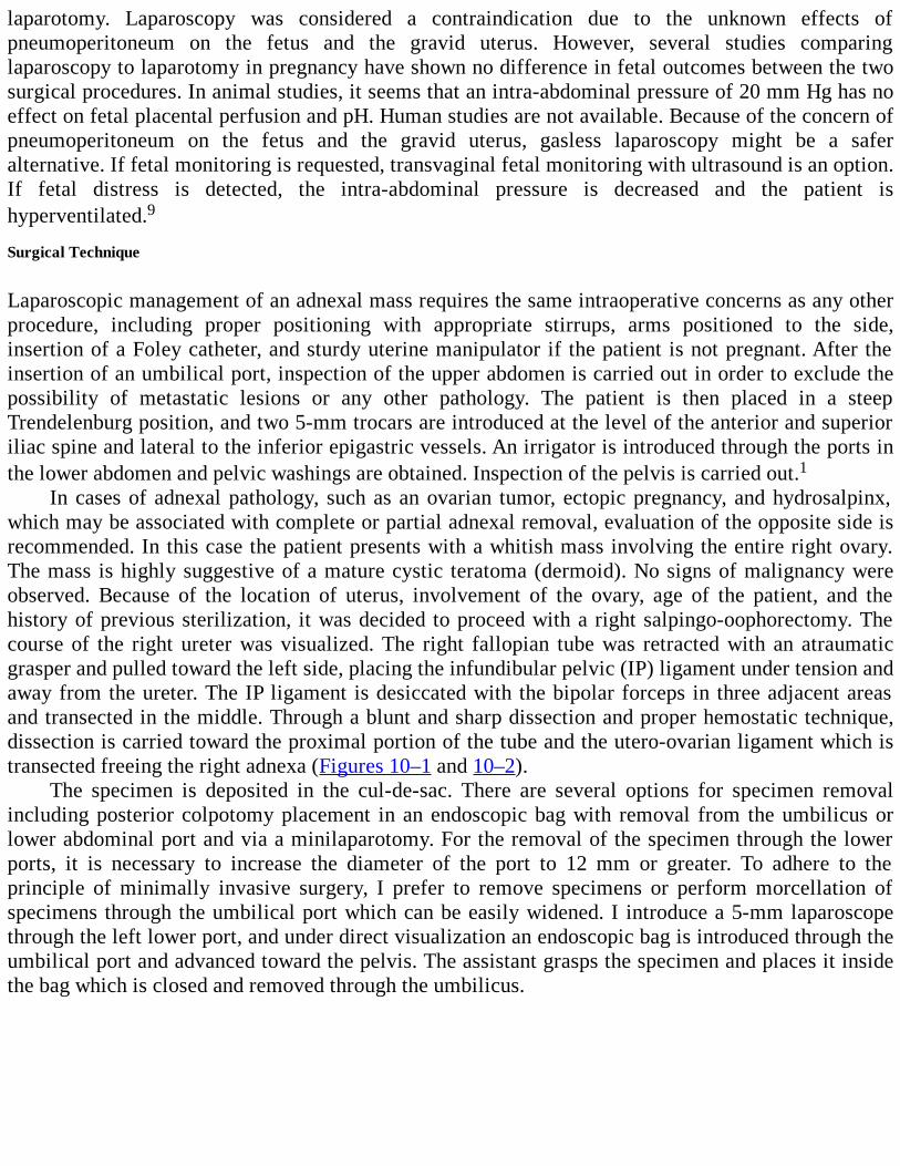
laparotomy. Laparoscopy was considered a contraindication due to the unknown effects ofpneumoperitoneum on the fetus and the gravid uterus. However, several studies comparinglaparoscopy to laparotomy in pregnancy have shown no difference in fetal outcomes between the twosurgical procedures. In animal studies, it seems that an intra-abdominal pressure of 20 mm Hg has noeffect on fetal placental perfusion and pH. Human studies are not available. Because of the concern ofpneumoperitoneum on the fetus and the gravid uterus, gasless laparoscopy might be a saferalternative. If fetal monitoring is requested, transvaginal fetal monitoring with ultrasound is an option.If fetal distress is detected, the intra-abdominal pressure is decreased and the patient ishyperventilated.9
Surgical Technique
Laparoscopic management of an adnexal mass requires the same intraoperative concerns as any otherprocedure, including proper positioning with appropriate stirrups, arms positioned to the side,insertion of a Foley catheter, and sturdy uterine manipulator if the patient is not pregnant. After theinsertion of an umbilical port, inspection of the upper abdomen is carried out in order to exclude thepossibility of metastatic lesions or any other pathology. The patient is then placed in a steepTrendelenburg position, and two 5-mm trocars are introduced at the level of the anterior and superioriliac spine and lateral to the inferior epigastric vessels. An irrigator is introduced through the ports inthe lower abdomen and pelvic washings are obtained. Inspection of the pelvis is carried out.1
In cases of adnexal pathology, such as an ovarian tumor, ectopic pregnancy, and hydrosalpinx,which may be associated with complete or partial adnexal removal, evaluation of the opposite side isrecommended. In this case the patient presents with a whitish mass involving the entire right ovary.The mass is highly suggestive of a mature cystic teratoma (dermoid). No signs of malignancy wereobserved. Because of the location of uterus, involvement of the ovary, age of the patient, and thehistory of previous sterilization, it was decided to proceed with a right salpingo-oophorectomy. Thecourse of the right ureter was visualized. The right fallopian tube was retracted with an atraumaticgrasper and pulled toward the left side, placing the infundibular pelvic (IP) ligament under tension andaway from the ureter. The IP ligament is desiccated with the bipolar forceps in three adjacent areasand transected in the middle. Through a blunt and sharp dissection and proper hemostatic technique,dissection is carried toward the proximal portion of the tube and the utero-ovarian ligament which istransected freeing the right adnexa (Figures 10–1 and 10–2).
The specimen is deposited in the cul-de-sac. There are several options for specimen removalincluding posterior colpotomy placement in an endoscopic bag with removal from the umbilicus orlower abdominal port and via a minilaparotomy. For the removal of the specimen through the lowerports, it is necessary to increase the diameter of the port to 12 mm or greater. To adhere to theprinciple of minimally invasive surgery, I prefer to remove specimens or perform morcellation ofspecimens through the umbilical port which can be easily widened. I introduce a 5-mm laparoscopethrough the left lower port, and under direct visualization an endoscopic bag is introduced through theumbilical port and advanced toward the pelvis. The assistant grasps the specimen and places it insidethe bag which is closed and removed through the umbilicus.
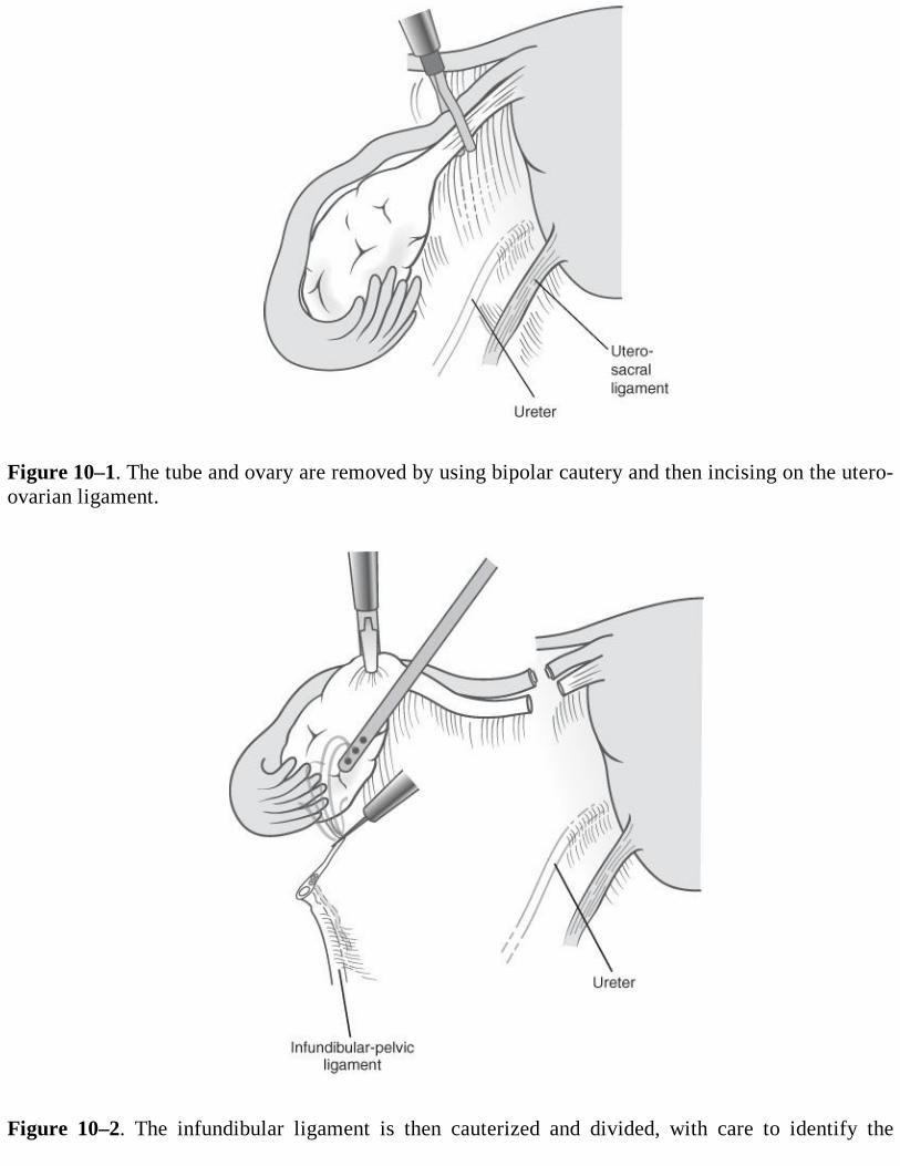
Figure 10–1. The tube and ovary are removed by using bipolar cautery and then incising on the utero-ovarian ligament.
Figure 10–2. The infundibular ligament is then cauterized and divided, with care to identify the
ureter.
The specimen can be decompressed by introducing a Veress needle attached to a syringe andaspirating the contents of the cyst, while the specimen sits inside the endoscopic bag thus preventingany spillage into the abdominal cavity.14-16 Alternatively, the endoscopic bag can be introducedthrough the posterior colpotomy incision placing the specimen in the bag and removing it through thevagina. A large mass might require decompression prior to removal in order to avoid grosscontamination even in benign cases such as endometriomas or dermoid cysts. This can beaccomplished by placing the specimen inside the bag and decompressing it with the suction irrigatorprobe, or by using endoscopic cyst aspirators. Any suspicious masses are sent for frozen section.Frozen section is a highly sensitive and specific test, except with very large masses.17-19
Comprehension Questions
10.1 An adnexal mass is diagnosed in a multiparous patient. Laparoscopic management iscontemplated. Which of the following is more likely to indicate benign rather than amalignant process?
A. SeptationsB. Size of 10 cmC. Cystic massD. Papillations
10.2 A 28-year-old woman is noted to have a 12-cm adnexal mass. Which of the following tumormarkers is most useful in this patient?
A. α-FetoproteinB. CEAC. CA-125D. CA-19-9
ANSWERS
10.1 C. A cystic mass is more likely associated with a benign process. Septations, complexities,solid component, larger size, ascites, and papillations are more indicative of a malignantprocess.
10.2 A. In a patient who is premenopausal, particularly less than age 30, germ cell tumors arelikely. These tumor markers include AFP, hCG, and LDH.
Clinical Pearls
See Table 1-2 for definition of level of evidence and strength of recommendation
The patient’s age together with the CA-125 and ultrasound characteristics of the adnexal masscan help to determine the likelihood of malignancy (Level B).
Pregnancy is not a contraindication to laparoscopic ovarian surgery (Level B).
A variety of techniques may be used to remove an adnexal mass laparoscopically such as
A variety of techniques may be used to remove an adnexal mass laparoscopically such asthrough a colpotomy incision, with a specimen bag through the umbilical incision, or a lateralport incision (Level C).
REFERENCES
1. Papasakelariou C, Saunders D, De La Rosa A. Comparative study of laparoscopicoophorectomy. J Am Assoc Gynecol Laparosc. 1995;2(4):407-410.
2. Dembo AJ, Davy M, Stenwig AE, et al. Prognostic factors with stage I epithelial cancer. ObstetGynecol. 1990;75:263-273.
3. Sevelda P, Dittrich C, Salzar H. Prognostic value of the rupture of the capsule in stage Iepithelial ovarian carcinoma. Gynecol Oncol. 1989;35:321-322.
4. Jacobs I, Oram D, Fairbanks J, et al. A risk of malignancy index incorporating Ca 125,ultrasound and menopausal status for the accurate preoperative diagnosis of ovarian cancer. BrJ Obstet Gynaecol. 1990;97:922-929.
5. National Institutes of Health Consensus Development Conference Statement. Ovarian cancer:screening, treatment, and follow up. Gynecol Oncol. 1994;55: S4-S14.
6. Schutter EM, Kenemans P, Sohn C, et al. Diagnostic value of pelvic examination, ultrasound,and serum CA 125 in postmenopausal women with pelvic mass. An international multicenterstudy. Cancer. 1994;74:1398-1406.
7. Maggino T, Gadducci A, D’Addario V, et al. Prospective multicenter study on CA 125 inpostmenopausal pelvic masses. Gynecol Oncol. 1994;54:117-123.
8. Manjunath AP, Pratapkumar, Sujatha K, et al. Comparison of three risks of malignancy indicesin evaluation of pelvic masses. Gynecol Oncol. 2001;81:225-229.
9. Yuen PM, Chang AMZ. Laparoscopic management of adnexal mass during pregnancy. ActaObstet Gynecol Scand. 1997;76:173-176.
10. Whitecar MC, Turner S, Higby K. Adnexal masses in pregnancy: a review of 130 casesundergoing surgical management. Am J Obstet Gynecol. 1999;181:19-24.
11. Platek DN, Henderson CE, Goldberg GL. The management of a persistent adnexal mass inpregnancy. Am J Obstet Gynecol. 1995;173:1236-1240.
12. Buttery BW, Beisner NA, Fortune DW, et al. Ovarian tumors in pregnancy. Med J Aust.1973;1:345-349.
13. Curet MJ, Voght DA, Schob O, et al. Effects of CO 2 pneumoperitoneum in pregnant ewes. JSurg Res. 1996;63:339-344.
14. Higgens RV, Matkins JF, Marroum MC. Comparison of fine needle aspiration cytologic
14. Higgens RV, Matkins JF, Marroum MC. Comparison of fine needle aspiration cytologicfindings of ovarian cysts with ovarian histologic findings. Am J Obstet Gynecol. 1999;180:550-553.
15. Mulvany NJ. Aspiration cytology of ovarian cysts and cystic neoplasms. A study of 235aspirates. Acta Cytol. 1996;40:911-920.
16. Vercellini P, Oldani S, Felicette I, et al. The value of cyst puncture in the differentialdiagnosis of benign ovarian tumors. Hum Reprod. 1995;10:1465-1469.
17. Smorgick N, Barel O, Halperin R, Schneider D, Pansky M. Laparoscopic removal of adnexalcysts: is it possible to decrease inadvertent intraoperative rupture rate? Am J Obstet Gynecol.2009 Mar;200(3):237.e1-3.
18. Whiteside JL, Keup HL. Laparoscopic management of the ovarian mass: a practical approach.Clin Obstet Gynecol. 2009 Sep;52(3):327-334.
19. Tinelli R, Malzoni M, Cosentino F, et al. Feasibility, safety, and efficacy of conservativelaparoscopic treatment of borderline ovarian tumors. Fertil Steril. 2009 Aug;92(2):736-741[Epub 2008].
Case 11
A 37-year-old African American nulliparous woman complains of fatigue, loss of productivity, andregular heavy prolonged menses during the last 9 months, which worsens with time. She mentions thatan asymptomatic myoma less than 4 cm was incidentally detected by transvaginal ultrasoundexamination during her annual visit to her gynecologist 1 year ago. She has always had normal Papsmear tests and she denies sexually transmitted diseases or abnormal mucopurulent vaginal discharge.Her pregnancy test is negative despite her wish to conceive during the last 12 months. Heart and lungexaminations are normal. On examination, her blood pressure is 105/70 mm Hg, heart rate 98beats/min, and temperature 97°F. On palpation, her abdomen presents a firm, round, nontenderpalpable midline mass 2 cm above the pubic symphysis. On bimanual examination, an anteverted,enlarged 15-week-size uterus is revealed with a hard painless protruding fundal mass with irregularcontour, approximately 7 cm in diameter that moves together with uterus during pushing the cervixupward or laterally. No adnexal masses are palpable. On speculum examination, no abnormalities aredetected in the vagina and cervix. Transvaginal ultrasound examination demonstrates a solitary fundalintramural myoma protruding into the uterine cavity. Her hemoglobulin level is 8.1 g/dL, leukocytecount 10,500/mm3, and platelet count 160,000/mm3.
What is the most likely diagnosis?
What is your next step?
ANSWERS TO CASE 11:Uterine Leiomyoma
Summary: An African American 37-year-old nulliparous woman, with a history of a knownasymptomatic intramural leiomyoma less than 4 cm the last year, complains of menometrorrhagia.She has also been trying to become pregnant unsuccessfully for the last 12 months. Her pregnancy testis negative. On examination, a midline hard mass seems to be contiguous to an enlarged anteverteduterus with irregular contour. Also, she has anemia and mild tachycardia.
Most likely diagnosis: Symptomatic uterine leiomyoma and infertility
Next step: Ultrasound examination to confirm the clinical diagnosis, and then discussion withthe patient about various options, including surgery including laparoscopic myomectomy
ANALYSIS
Objectives
1. Know that most myomas are asymptomatic and require intervention when they cause clinicalsymptoms.
2. Understand that the location and size of a myoma are the most crucial determinants of itspotential to become symptomatic.
3. Know that the most common symptoms associated with uterine leiomyomas in reproductive agewomen are abnormal uterine bleeding manifested as menorrhagia or hypermenorrhea.
4. Describe the treatment options for uterine leiomyomata, including conservative, minimallyinvasive surgical procedures, medical therapies, and hysterectomy.
Considerations
This 37-year-old woman complains of fatigue, loss of productivity, and irregular menstrual patterndue to hypermenorrhea and menorrhagia. Clinical examination and history are consistent with a fundalmyoma, because of the presence of a midline hard mass contiguous with the uterus that also explainsits irregular contour. The prevalence of myomas in African American women is two to three timeshigher in comparison to white Caucasian women. Moreover, this uterine abnormal mass may be apossible cause of her infertility. The movement of this mass together with the uterus during lateral orupward displacement of cervix is another argument that this mass is of uterine origin. Furthermore,the absence of pelvic tenderness and fever with normal leukocyte count excludes pelvic inflammatorydisease as a cause of abnormal uterine bleeding. The normal clinical findings on speculumexamination and, also, the negative Pap smear tests make the diagnosis of vaginal or cervicalpathology such as cervical or vaginal endometriosis, ectropion, cervical polyps, endometrialpedunculated myomas or polyps protruding through the external cervical os, cervical intraepithelialneoplasia, or malignancy remote. Dysfunctional uterine bleeding is excluded from the differentialdiagnosis, as it is frequent in adolescent and perimenopausal women and is a manifestation ofanovulatory cycles in the absence of any uterine pathology or medical illness. Furthermore, in thiscase the patient seems to have ovulatory cycles because she has regular menses. Ectopic pregnancy asa cause of abnormal uterine bleeding is impossible due to her negative pregnancy test. Benign ormalignant ovarian tumors are absent on bimanual examination. The clinical differential diagnosisbetween leiomyoma and adenomyoma as causes of menorrhagia is difficult without the use oftransvaginal ultrasound or preferably magnetic resonance imaging (MRI) (ill-defined borders of theadenomyoma within myometrium in contrast to the pseudocapsule of myomas with clear contour).However, the main dominant symptom of adenomyoma or endometriosis is chronic pelvic pain andnot menorrhagia that is the main complaint of women with symptomatic myomas. Adenomyosis ispresented in the fourth or fifth decade of life and affects 1% of usually multiparous women. Ingeneral, endometrial sampling is performed with abnormal vaginal bleeding in women older than age35 years. Finally, a Pipelle office endometrial biopsy is mandatory when the ultrasound findings raisethe suspicion of endometrial hyperplasia or carcinoma, though they appear in peri- or postmenopausalwomen with metrorrhagia and not with menorrhagia. They are rarely found as a cause of abnormaluterine bleeding in women of reproductive age and especially younger than 40 years without thecoexistence of risk factors such as excessive BMI, diabetes, or hypertension, which are not present inour case. Finally, the workup of menorrhagia except from platelet count should include furtherinvestigation of the thyroid function and clotting factors in order to exclude hyperthyroidism orcoagulation disorders such as von Willebrand disease.
APPROACH TO Uterine Leiomyoma
DEFINITIONS
HYPERMENORRHEA: It is defined as heavy, profuse periods that exceed 80 mL blood loss.
MENORRHAGIA: It is defined as heavy and prolonged periods that exceed 7 days.
METRORRHAGIA: It is defined as abnormal uterine bleeding at irregular intervals, particularlybetween the expected menstrual periods.
LEIOMYOMA: Benign smooth muscle tumor, which arises from the myometrium, also calledmyoma or fibroma. It is classified by its location in relation to the uterine wall: serosal (which may bebroad-based or pedunculated), intramural, and submucosal. The latter is further distinguished as type0 (pedunculated, totally protruding in the uterine cavity), type I (> 50% protrudes in the uterinecavity), and type II (< 50% protrudes in the uterine cavity or has an intramural portion of > 50%).
ADENOMYOSIS: It is the presence of heterotopic endometrial glands and stroma in themyometrium at least 2.5 mm below the endometrial-myometrial interface with adjacent smoothmuscle hyperplasia. It can be either diffuse or focal, leading to the formation of the so-called“adenomyoma.”
ADENOMYOMA: It is a circumscribed nodule of hypertrophic and distorted endometrium andmyometrium, which is usually embedded within the myometrium.
CLINICAL APPROACH
Uterine leiomyomas are among the most frequent pathologic entities encountered in gynecologicclinical practice. Myomas are the most common benign gynecologic neoplasms and the primaryindication for hysterectomy, accounting for over 200,000 hysterectomies per year in the United States.Although in the pathology reports of hysterectomy specimens leiomyomas can be identified in up to77%, they are clinically apparent in approximately 20% to 35% of women of reproductive age.Despite the high prevalence of these tumors, there is paucity of data available regarding the naturalclinical history of myomas. They are monoclonal-independent lesions, such that multiple tumors fromthe uterus arise independently and may have distinct chromosomal abnormalities, while their growthis estrogen and progesterone dependent. Increased prevalence of leiomyomas is observed two- tothreefold higher in African American women, who have earlier age at first diagnosis with multiple andlarger myomas in comparison to that in white women. The prevalence of myomas increases with ageuntil 50 years and then declines sharply. In pregnant women, the prevalence of myomas has beenreported to be approximately 1.4%. Oral contraceptives, parity, and smoking decrease the risk ofmyoma development due to low oestrogen levels. On the other hand, obesity with higher oestrogenlevels, diastolic hypertension with cytokine release, or injury of uterine smooth muscle as well aspelvic inflammatory disease with intrauterine irritation are associated with increased risk of fibroids.Malignant transformation of leiomyomas is extremely rare, and according to cytogenetic studies it hasbeen documented that leiomyosarcomas arise de novo and rarely from a specific subset of myomas.However, the incidence of uterine sarcoma in symptomatic premenopausal women undergoingsurgical removal is less than 0.3%. Current studies do not support any association between rapidgrowth of a leiomyoma and increased risk of malignancy. So, the rapid growth of an asymptomaticmyoma that gains 6 weeks or more in gestational size within an interval of 1 year or less is anoutdated indication for surgical intervention.
Most uterine myomas cause no symptoms. Approximately 62% of women with symptomaticmyomas present with multiple symptoms, which correlate with their location, number, size, orconcomitant degenerative changes. Excessive menstrual bleeding that leads to iron deficiency and
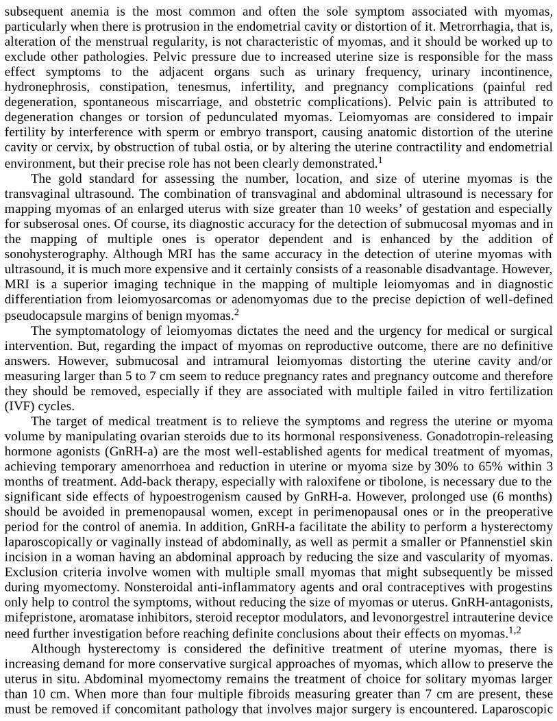
subsequent anemia is the most common and often the sole symptom associated with myomas,particularly when there is protrusion in the endometrial cavity or distortion of it. Metrorrhagia, that is,alteration of the menstrual regularity, is not characteristic of myomas, and it should be worked up toexclude other pathologies. Pelvic pressure due to increased uterine size is responsible for the masseffect symptoms to the adjacent organs such as urinary frequency, urinary incontinence,hydronephrosis, constipation, tenesmus, infertility, and pregnancy complications (painful reddegeneration, spontaneous miscarriage, and obstetric complications). Pelvic pain is attributed todegeneration changes or torsion of pedunculated myomas. Leiomyomas are considered to impairfertility by interference with sperm or embryo transport, causing anatomic distortion of the uterinecavity or cervix, by obstruction of tubal ostia, or by altering the uterine contractility and endometrialenvironment, but their precise role has not been clearly demonstrated.1
The gold standard for assessing the number, location, and size of uterine myomas is thetransvaginal ultrasound. The combination of transvaginal and abdominal ultrasound is necessary formapping myomas of an enlarged uterus with size greater than 10 weeks’ of gestation and especiallyfor subserosal ones. Of course, its diagnostic accuracy for the detection of submucosal myomas and inthe mapping of multiple ones is operator dependent and is enhanced by the addition ofsonohysterography. Although MRI has the same accuracy in the detection of uterine myomas withultrasound, it is much more expensive and it certainly consists of a reasonable disadvantage. However,MRI is a superior imaging technique in the mapping of multiple leiomyomas and in diagnosticdifferentiation from leiomyosarcomas or adenomyomas due to the precise depiction of well-definedpseudocapsule margins of benign myomas.2
The symptomatology of leiomyomas dictates the need and the urgency for medical or surgicalintervention. But, regarding the impact of myomas on reproductive outcome, there are no definitiveanswers. However, submucosal and intramural leiomyomas distorting the uterine cavity and/ormeasuring larger than 5 to 7 cm seem to reduce pregnancy rates and pregnancy outcome and thereforethey should be removed, especially if they are associated with multiple failed in vitro fertilization(IVF) cycles.
The target of medical treatment is to relieve the symptoms and regress the uterine or myomavolume by manipulating ovarian steroids due to its hormonal responsiveness. Gonadotropin-releasinghormone agonists (GnRH-a) are the most well-established agents for medical treatment of myomas,achieving temporary amenorrhoea and reduction in uterine or myoma size by 30% to 65% within 3months of treatment. Add-back therapy, especially with raloxifene or tibolone, is necessary due to thesignificant side effects of hypoestrogenism caused by GnRH-a. However, prolonged use (6 months)should be avoided in premenopausal women, except in perimenopausal ones or in the preoperativeperiod for the control of anemia. In addition, GnRH-a facilitate the ability to perform a hysterectomylaparoscopically or vaginally instead of abdominally, as well as permit a smaller or Pfannenstiel skinincision in a woman having an abdominal approach by reducing the size and vascularity of myomas.Exclusion criteria involve women with multiple small myomas that might subsequently be missedduring myomectomy. Nonsteroidal anti-inflammatory agents and oral contraceptives with progestinsonly help to control the symptoms, without reducing the size of myomas or uterus. GnRH-antagonists,mifepristone, aromatase inhibitors, steroid receptor modulators, and levonorgestrel intrauterine deviceneed further investigation before reaching definite conclusions about their effects on myomas.1,2
Although hysterectomy is considered the definitive treatment of uterine myomas, there isincreasing demand for more conservative surgical approaches of myomas, which allow to preserve theuterus in situ. Abdominal myomectomy remains the treatment of choice for solitary myomas largerthan 10 cm. When more than four multiple fibroids measuring greater than 7 cm are present, thesemust be removed if concomitant pathology that involves major surgery is encountered. Laparoscopic
myomectomy compared with abdominal myomectomy has clear advantages in terms of postoperativepain relief, recovery time, hospital stay and demand for fewer transfusions, less postoperative fever,and de novo adhesion formation, but with no significant difference in terms of pregnancy andmiscarriage rates in the few prospective, well-designed published studies. Of course, its applicationdepends on gynecologist’s skills and mastering in endoscopic suturing, and also availability of thenecessary instrumentation. Under these prerequisites, the need for conversion to laparotomy isreported only in 1.4% of cases.3,4,5
Adhesion formation can be eliminated by performing the incision (transverse incision ispreferable to vertical in order to avoid incoming arcuate vessels, except from the corneal areas) on theanterior uterine wall or fundus in order to remove myomas, even if they are located posterior, byreducing the use of electrocautery and by using absorbable adhesion barriers. The risk of uterinerupture following myomectomy is ranged from 0.002% to 5% and usually occurs between 29 and 36weeks’ of gestation. This is related to the following factors: (1) single instead of multilayer suturing,which minimizes the hematoma formation and maximizes the strength of uterine wall, (2) theexcessive use of bipolar electrocoagulation for hemostasis that might impair the healing process, and(3) the poor suturing skills of the gynecologist to achieve uniform closure of the uterine defect bysymmetric hickness of the scar in relation to the adjacent myometrium. Finally, hysteroscopicmyomectomy is established as the gold standard for the treatment of submucosal myomas types 0 andI due to the advantage of no incision or suturing on the normal myometrium (Figure 11–1). However,this approach can be rarely complicated by perforation, bleeding, infection, and intracavitary adhesionformation. Other techniques such as electromyolysis, uterine and ovarian artery ligation, orembolization have been proposed as conservative minimally invasive approaches with short-termresults in women who desire to preserve their uterus, but with unknown effect on their fertilitypotential and risk of recurrence.
Figure 11–1. A myomectomy is performed laparoscopically. The myoma is grasped with a corkscrewand dissected from the myometrium (A), and then the uterine defect is sutured closed (B).
The recurrence of fibroids is diagnosed by ultrasound demonstrating myomas at least 2 cm indiameter. The 5-year risk of recurrence is 62% with a 9% risk of additional major surgery, a crucialissue when counselling patients for myomectomy. The majority of recurrences (75%) occur between10 and 30 months after surgery. This risk is greater in patients with multiple myomas and large size ofuterus. Moreover, the decreased tactile ability of laparoscopy may account for the increased rate ofrecurrence at laparoscopy.
Comprehension Questions
11.1 A 42-year-old woman is noted on pelvic examination to have an irregularly shaped mobilemidline uterine mass. The gynecologist explains to the patient that this is likely to be uterinefibroids. The patient has no symptoms. Which of the following is the incidence rate ofclinically apparent myomas?
A. 5%B. 30%C. 70%D. 90%
11.2 Which type of myoma should be removed in infertile women to increase their pregnancyrates?
A. SubmucosalB. IntramuralC. SubserosalD. Broad ligamental
11.3 To decrease the risk of adhesion formation with laparoscopic myomectomy, which one ofthe following incisions should be avoided?
A. Midline fundalB. Anterior transverseC. Anterior horizontalD. Posterior
11.4 A gynecology case manager is reviewing charts for appropriate surgery. This month thefocus is on myomectomy. Which of the following cases would most likely be flagged as aninappropriate indication for surgery?
A. Patient complains of painB. Patient complains of vaginal bleedingC. Patient has pressure symptomsD. Patient is informed that surgery would help protect against malignant transformation
ANSWERS
11.1 B. It is estimated that myomas are clinically apparent in approximately one out of every
three or four women of the reproductive age. However, the prevalence of uterine myomas ishigher in pathology reports of hysterectomy specimens.
11.2 A. Neither intramural nor subserosal myomas seem to affect fertility rates and removal hasnot been shown to increase fertility. Broad ligament myomas are outside the uterus.
11.3 D. Posterior uterine incision should be avoided, if possible, to decrease the risk of adhesionformation. The midline fundal incision has been popularized to decrease the risk ofextending the uterine incision into the cornua. A horizontal incision has been advocated toavoid sectioning the blood vessels that run transversely to minimize blood loss.
11.4 D. Malignant transformation of uterine myomas is not acceptable indication formyomectomy in otherwise asymptomatic women. The risk of malignant transformation inan otherwise asymptomatic patient is less than 1:1000. However, if the uterus is noted to begrowing in a postmenopausal woman, or uterine fibroids are noted in a patient with priorpelvic irradiation, surgery should be strongly considered.
Clinical Pearls
See Table 1-2 for definition of level of evidence and strength of recommendation
Most myomas are asymptomatic common benign tumors of the myometrium with the highestincidence in women of reproductive age, affecting African American population three times asoften as Caucasian women (Level A).
The clinical suspicion of myomas can be confirmed by ultrasonography. MRI andsonohysterography are complementary diagnostic tools in complicated cases (Level A).
Large intramural myomas (> 5 cm) and/or those distorting the endometrial cavity mayadversely affect fertility (Level B).
Medical treatment of myomas with GnRH-agonists is indicated for symptom relief during thepreoperative period or in perimenopausal women (Level A).
Laparoscopic myomectomy is a conservative surgical treatment in women who desire topreserve their uterus. However, hysterectomy is the definitive treatment of myomas (Level B).
REFERENCES
1. Arici A, Rayburn W. Myomas. In: Obstetrics and Gynecology Clinics of North America.Philadelphia, PA: Elsevier Saunders; 2006;33(1):1-225.
2. Nowak R. Fibroids: pathophysiology and current medical treatment. Baillieres Best Pract ResClin Obstet Gynaecol. 1999;13(2):223-238.
3. Parker W. Laparoscopic myomectomy and abdominal myomectomy. Clin Obstet Gynecol.
2006;49(4):787-797.
4. Carter JE, McCarus SD. Laparoscopic myomectomy. Time and cost analysis of power vs.manual morcellation. J Reprod Med. 1997 Jul;42(7):383-388.
5. Alessandri F, Lijoi D, Mistrangelo E, et al. Randomized study of laparoscopic versusminilaparotomic myomectomy for uterine myomas. J Minim Invasive Gynecol. 2006 Mar-Apr;13:92-97.
6. Walker C, Stewart E. Uterine fibroids: the elephant in the room. Science. 2005;308: 1589-1592.
7. Hurst BS, Matthews ML, Marshburn PB. Laparoscopic myomectomy for symptomatic uterinemyomas. Fertil Steril. 2005 Jan;83(1):1-23.
8. Palomba S, Zupi E, Falbo, A, et al. A multicenter randomized, controlled study comparinglaparoscopic versus minilaparotomic myomectomy: reproductive outcomes. Fertil Steri. 2007Oct;88:933-941.
9. Sinha R, Hegde A, Warty N, Patil N. Laparoscopic excision of very large myomas. J Am AssocGynecol Laparosc. Nov 2003;10(4):461-468.
10. Parker W. Uterine myomas: management. Fertil Steril. 2007 Aug;88:255-271.
Case 12
A 28-year-old thin and nulliparous woman with a history of progressive dysmenorrhea not respondingto medical therapy is taken to surgery for laparoscopy and possible excision of endometriosis. TheVeress needle is introduced through an infraumbilical incision and the abdomen filled with 2 L of CO 2gas. A 10/11-mm trocar is introduced into the abdominal cavity without much effort. Upon insertingthe laparoscope, large amount of blood is visualized. The anesthesiologist alerts you that the patient iscurrently hypotensive.
What is the most likely diagnosis?
What is your next step?
ANSWERS TO CASE 12:Laparoscopic Vascular Complications
Summary: This is a 28-year-old woman who, during a laparoscopy, developed hypotension and intra-abdominal bleeding.
Most likely diagnosis: Major vascular injury.
Next step: Proceed with a laparotomy and obtain the assistance of a vascular surgeon.
ANALYSIS
Objectives
1. Be familiar with the definition and consequences of major vascular injury.2. Learn the differences between closed versus open technique in laparoscopy.3. Learn strategies to minimize the incidence of major vascular injury.4. Be familiar with the management of major vascular injury.
Considerations
This is a 28-year-old thin woman who suffered a major vascular injury during laparoscopic surgery.Sometimes surprising to the surgeon, it is the thin patient who is at greatest risk for vascular injuries,because of the decreased distance from the skin to the retroperitoneal space. This patient had ahypotensive episode immediately upon injury to her major vessels. It is unclear at this point whether itis the aorta or vena cava that is injured. The surgeon should immediately perform a laparotomy viavertical incision to put pressure on the vascular injury. Large bore IVs should be placed, andcrystalloid resuscitation should be initiated. Blood should be immediately ordered, and likely, thepatient will need uncross-matched O negative blood. A vascular surgeon should be immediatelysummoned. Time is of the essence, and may mean survival or death for this patient. Major vascular
injury is defined as one that threatens the patient’s life almost immediately upon its occurrence.
APPROACH TOLaparoscopic Vascular Injury
Vascular injury associated with laparoscopy has an incidence of less than 1 per 1000 cases (0.1%).Despite its low incidence, it remains one of the most feared complications in laparoscopic procedures.It is a complication that can occur even to the most experienced laparoscopic surgeon. However,timely diagnosis and proper management are paramount for a good outcome. Laparoscopy isparticularly associated with vascular injury due to the blind introduction of either the Veress needle toproduce the pneumoperitoneum or the trocar to introduce the initial laparoscope.1
Perhaps counterintuitive, it is the thin patient rather than the obese patient who is at greater riskfor these injuries, especially in those patients with well-developed abdominal walls. In the thinathletic patients, the distance from the abdominal wall to the retroperitoneal structures is less than thatin obese patients. Certain anatomical considerations and surgical techniques can minimize vascularinjuries. An intraumbilical incision at the deepest point represents the shorter distance to theabdominal cavity. In a retrospective review by Hurd and associates,2 the vertical distance from thebase of the umbilicus to the peritoneum remained very constant despite an increase in the BMI of thesubjects. It was a distance of 6 cm or less in nearly all subjects. In contrast, the distance at 45 degreesfrom the lower margin and base of the umbilicus to the peritoneum increased to a much larger degreewith an increase in the BMI. The introduction of the initial umbilical trocar remains a blind entry.Despite the utilization of an open technique, major vascular complications can occur.3-5 Thus, evenwith open trocar introduction, the surgeon should be vigilant and should exercise careful technique.
Clinical trials comparing closed versus open entry technique have not demonstrated a superiorityof one over the other. A study by the American Association of Gynecologic Laparoscopists showedmore visceral lesions with open laparoscopy entry but not significantly fewer vascular lesions.3 It isimportant to note that visceral lesions are potentially more life threatening since they are commonlymissed during laparoscopy and only recognized when the patient is admitted with peritonitis andsepsis. I prefer the closed technique. In the establishment of pneumoperitoneum, filling the abdomento a specific pressure rather than a set volume allows for different intra-abdominal distensibility andvolume capacity. In our setting, I typically insufflate the abdomen to a pressure of 20 mm Hg. Thisoffers a tight abdomen which allows for an easier insertion of the trocar. The patient should be kept ina completely horizontal position. Premature Trendelenburg position such as by the anesthesiologist oreven the surgeon direction can alter the anatomy, bringing the major vessels into closer proximity tothe site of the trocar insertion. In this case the insufflation of the abdominal cavity with 2 L wasinadequate and may have been the contributing factor to the major vascular injury. The utilization ofsharp/disposable trocars allows for an easier and controlled insertion.
Avoidance of Injury
The abdominal and pelvic examinations should be performed preoperatively not only to assess fordisease but also to assess the distance between the abdominal wall and the retroperitoneal vessels. Avery easily palpable abdominal aortic pulse, a sharply inclined sacral promontory, or pronouncedlumbar lordosis can all be hints of bony variations that push the aorta or vena cava toward theabdominal wall, putting the patient at risk for vascular injury. Additionally, patients who have hadabdominal surgery or pelvic adhesive disease may also be at risk due to a “solid interface” betweenthe anterior abdominal wall and the retroperitoneum. This lack of space does not allow the trocar
sheath guard to engage, and, thus, the surgeon may make multiple attempts to push the trocar deeperand deeper trying to get the trocar sleeve to activate. Furthermore, if the surgeon deviates off themidline even slightly upon introducing the trocar, the iliac vessels can be injured. Thus, thelaparoscopic surgeon must be much disciplined to place the trocar in the midline. During the surgicalaspect of laparoscopy, injury to vascular structures can occur due to dissection near the retroperitonealvessels. Ablation of endometriosis for instance can lead to vascular injury due to the distortedanatomy from the endometriotic implants.6 The surgeon should ensure that the patient is horizontaland not in Tredelenburg position (Figure 12-1 A and B) since the change in position can lead toinadvertent directing of the trocar into the retroperitoneal vessels.
Recognition of Vascular Injury
At times, the vascular injury is obvious with bright red blood filling the abdominal cavity. Sometimes,though, the findings are more subtle. For instance, vena cava laceration may be associated with a“hematoma” in the retroperitoneal space that can deceptively “appear stable.” As a rule of thumb, anyhematoma in the retroperitoneal area should be fully investigated and should be considered a majorvascular injury until proven otherwise. A common scenario is that a venous injury may appear stablein the operating room, only to lead to further hemorrhage postoperatively, and even exsanguination.Abdominal wall vessels can also be injured such as the inferior epigastric artery. Visualization withthe laparoscope of these vessels as they course between the rectus muscle and the parietal peritoneumcan direct lateral ports away from these vessels (Figure 12–1A and B).
Treatment
As soon as a major vascular injury is recognized, the surgeon should proceed with an immediatelaparotomy through a midline incision, while anesthesia secures additional IV lines, experiencedcardiovascular surgeon is summoned, and arrangements are being made in making blood available fortransfusion. Once laparotomy has begun, the first priority is to compress the aorta to preventexsanguination. This is accomplished by placing hand compression over the aorta or a vascular clamp.If the compression lasts for over 15 minutes, systemic anticoagulation with heparin should beconsidered once the repair is complete and the patient is stable. Injury to the vena cava is generallymore difficult to repair due to the thin wall and friable tissue. Gentle constant pressure to helpdecrease blood loss, while awaiting a vacular surgeon, is the most prudent action. Because the venacava can tear more easily, the pressure should be directed downward without excessive movement thatmay cause further vessel laceration. A number of techniques are used to address injury to the inferiorepigastric artery, including bipolar cautery via laparoscopy, the use of a Foley bulb placed into thetrocar site into the abdominal cavity with traction and held in place such as with a small clamp onFoley catheter against the anterior abdominal wall. Various needles have been designed to allow fordeep surgical ligatures, and finally enlarging the surgical incision to isolate and ligate the vessel maybe employed.7-10
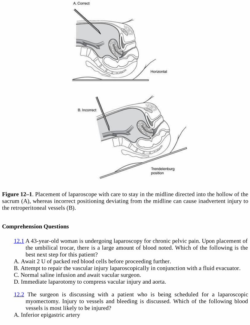
Figure 12–1. Placement of laparoscope with care to stay in the midline directed into the hollow of thesacrum (A), whereas incorrect positioning deviating from the midline can cause inadvertent injury tothe retroperitoneal vessels (B).
Comprehension Questions
12.1 A 43-year-old woman is undergoing laparoscopy for chronic pelvic pain. Upon placement ofthe umbilical trocar, there is a large amount of blood noted. Which of the following is thebest next step for this patient?
A. Await 2 U of packed red blood cells before proceeding further.B. Attempt to repair the vascular injury laparoscopically in conjunction with a fluid evacuator.C. Normal saline infusion and await vacular surgeon.D. Immediate laparotomy to compress vacular injury and aorta.
12.2 The surgeon is discussing with a patient who is being scheduled for a laparoscopicmyomectomy. Injury to vessels and bleeding is discussed. Which of the following bloodvessels is most likely to be injured?
A. Inferior epigastric artery
B. Common iliac arteryC. Internal iliac arteryD. Ovarian artery
12.3 Which of the following situations places patients at increased risk of vascular injury duringlaparoscopy?
A. Obese patients at greater risk rather than thin patientsB. Closed versus open laparoscopic trocar placementC. Trocar deviated to the right side rather than the midlineD. Use of disposable trocar instruments versus reusable instruments
ANSWERS
12.1 D. Upon discovery of a major vascular injury, the surgeon should perform an immediatelaparotomy via a vertical skin incision. Pressure should be applied at the vascular injury site,and also the aorta should be compressed. The anesthesiologist should place two large boreIVs and normal saline should be infused, while blood be cross-matched.
12.2 A. The inferior epigastric artery is the most commonly injured vessel causing majorhemorrhage. The inferior epigastric artery is located between the rectus muscle and theperitoneum, and at the medial aspect of the rectus muscle. Injury can be avoided bytransilluminating through the intra-abdominal region, to try to locate the vessels prior toplacement of the lateral ports.
12.3 C. Vascular injuries are more common with thin patients rather than obese ones. The besttechnique is to aim at the hollow of the sacrum in the midline to avoid injury of the commoniliac vessels. Because thin patients have less distance from the skin to the retroperitonealspace, the retroperitoneal vessels are more often injured.
Clinical Pearls
(See Table 1-2 for definition of level of evidence and strength of recommendation)
The most common vessel injured during laparoscopy is the inferior epigastric artery (Level B).
The most commonly injured retroperitoneal vessel is the common iliac artery or vein (LevelB).
In a hypotensive patient when a major artery is lacerated, pressure at the vessel injury site andcompression of the aorta are paramount (Level B).
A closed versus open technique for laparoscopic trocar entry has not been shown to affect theincidence of vascular injury (Level A).
REFERENCES
1. Loffer F, Pent D. Indications, contraindications and complications of laparoscopy. ObstetGynecol Surv. 1975;30:407-427.
2. Hurd WW, Bude RO, Delancey JOH, Gauvin JM, Aisen AM. Abdominal wall characteristicsby MRI and CT imaging: the effect of obesity on laparoscopic approach. J Reprod Med.1992;36:473-476.
3. Phillips JM, Hulka JF, Peterson HB. American Association of Gynecologic Laparoscopists’1982 membership survey. J Reprod Med. 1984;29:592-594.
4. Frenkel Y, Oelsner G, Ben-Baruch G, Menczer J. Major surgical complications of laparoscopy.Eur J Obstet Gynecol Reprod Biol. 1981;12:107-111.
5. Chamberlain G, Brown JC, eds. Gynecological laparoscopy: the report of the confidentialinquiry into gynaecological laparoscopy. London: Royal College of Obstetricians andGynaecologists. 1978:114.
6. Mintz M. Risks and prophylaxis in laparoscopy: a survey of 100,000 cases. J Reprod Med.1977;18:269.
7. Jansen FW, Kolkman W, Bakkum E, de Kroon C, Trimbos-Kemper TC, Trimbos JB.Complications of laparoscopy: an inquiry about closed-versus open-entry technique. Am JObstet Gynecol. 2004;190:634-638.
8. Reich H. New techniques in advanced laparoscopic surgery. Baillieres Clin Obstet Gynaecol.1989;3:655-681.
9. Pring CM. Aortic injury using the Hasson trocar: a case report and review of the literature. AnnR Coll Surg Engl. 2007 Mar;89(2):W3-W5. Review.
10. Nezhat C, Childers J, Nezhat F, Nezhat CH, Seidman DS. Major retroperitoneal vascularinjury during laparoscopic surgery. Hum Reprod. 1997 Mar;12(3):480-483.
Case 13
A 32-year-old G0P0 woman complains of painful menses during the last year as well as pelvicnonmenstrual pain and dyspareunia of recent onset. Although she stated that her menarche was at age13 and her menses were painless and regular, have recently become somewhat irregular. She deniesvaginal discharge or prior sexually transmitted disease. Also, she has stopped using any contraceptivemethod since being married. Her blood pressure is 110/70 mm Hg, heart rate 85 beats/min, andtemperature 95°F. Heart and lung examinations are normal. On palpation, she had slight abdominaltenderness at the right iliac fossa, but without guarding. On pelvic examination, a retroverted anddisplaced uterus seems to move together with a palpable cystic mass of her right adnexa. Herpregnancy test is negative. Her hemoglobulin level is 11 g/dL, leukocyte count 8500/mm3, and plateletcount 230,000/mm3. Ultrasound examination reveals a smooth 9-cm homogenic hypoechoic cysticmass of the right ovary attached to the posterior surface of a retroverted uterus with a small quantityof free hypodense fluid in the pouch of Douglas.
What is the most likely diagnosis?
What is your next step?
ANSWERS TO CASE 13:Endometriosis with Ovarian Endometrioma Formation
Summary: This is a 32-year-old nulliparous woman with a history of recent onset of dysmenorrhoea,dyspareunia, nonmenstrual pelvic pain, and perhaps infertility, because she has never becomepregnant despite her unprotected sexual intercourse. Her past gynecological and medical history wereunremarkable. Complete blood count is within the normal range. Pregnancy test is negative. The rightadnexa have a cystic mass on palpation and is attached to the retroverted uterus.
Most likely diagnosis: Endometriosis with ovarian endometrioma formation
Next step: Laparoscopy
ANALYSIS
Objectives
1. Know the symptoms of endometriosis.2. Understand the mechanism of the symptoms.3. Differential diagnosis of endometriomas from other adnexal cystic masses in reproductive aged
women.
Considerations
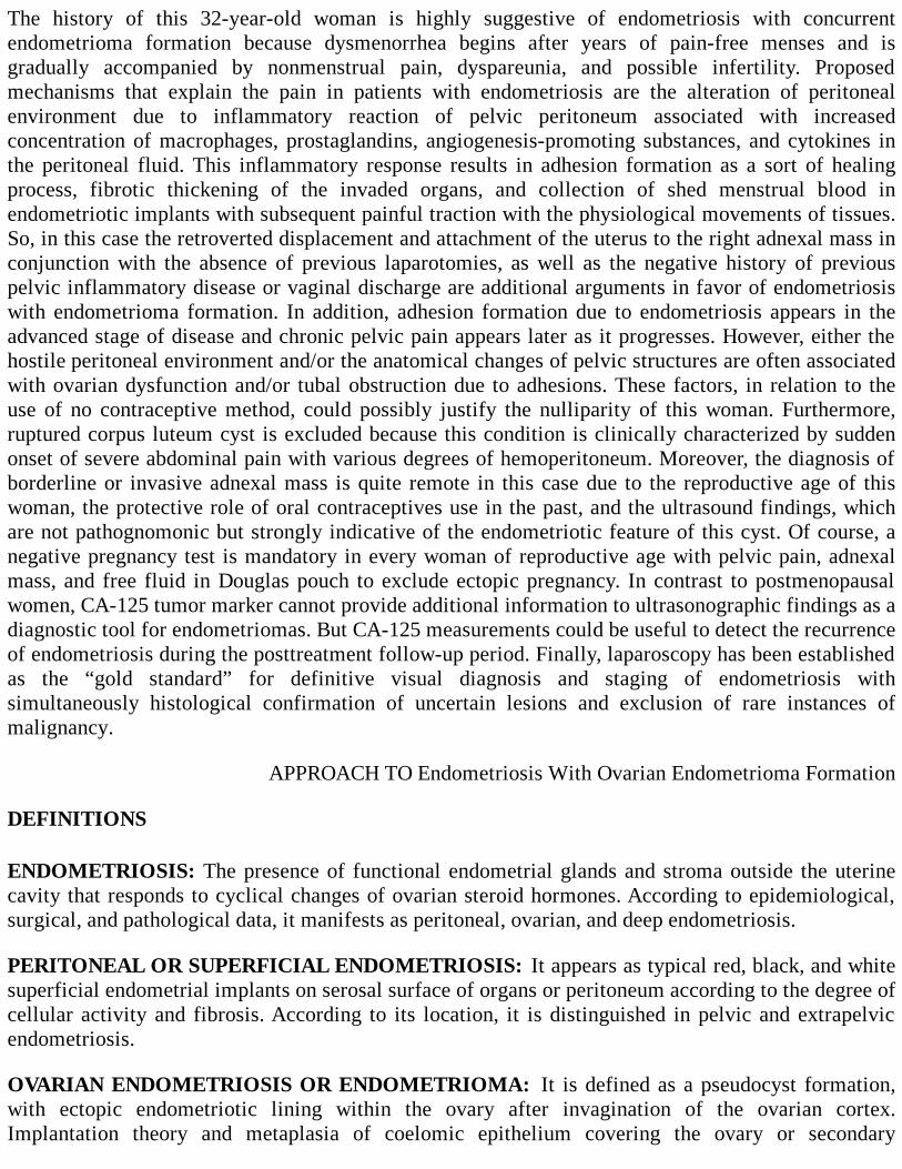
The history of this 32-year-old woman is highly suggestive of endometriosis with concurrentendometrioma formation because dysmenorrhea begins after years of pain-free menses and isgradually accompanied by nonmenstrual pain, dyspareunia, and possible infertility. Proposedmechanisms that explain the pain in patients with endometriosis are the alteration of peritonealenvironment due to inflammatory reaction of pelvic peritoneum associated with increasedconcentration of macrophages, prostaglandins, angiogenesis-promoting substances, and cytokines inthe peritoneal fluid. This inflammatory response results in adhesion formation as a sort of healingprocess, fibrotic thickening of the invaded organs, and collection of shed menstrual blood inendometriotic implants with subsequent painful traction with the physiological movements of tissues.So, in this case the retroverted displacement and attachment of the uterus to the right adnexal mass inconjunction with the absence of previous laparotomies, as well as the negative history of previouspelvic inflammatory disease or vaginal discharge are additional arguments in favor of endometriosiswith endometrioma formation. In addition, adhesion formation due to endometriosis appears in theadvanced stage of disease and chronic pelvic pain appears later as it progresses. However, either thehostile peritoneal environment and/or the anatomical changes of pelvic structures are often associatedwith ovarian dysfunction and/or tubal obstruction due to adhesions. These factors, in relation to theuse of no contraceptive method, could possibly justify the nulliparity of this woman. Furthermore,ruptured corpus luteum cyst is excluded because this condition is clinically characterized by suddenonset of severe abdominal pain with various degrees of hemoperitoneum. Moreover, the diagnosis ofborderline or invasive adnexal mass is quite remote in this case due to the reproductive age of thiswoman, the protective role of oral contraceptives use in the past, and the ultrasound findings, whichare not pathognomonic but strongly indicative of the endometriotic feature of this cyst. Of course, anegative pregnancy test is mandatory in every woman of reproductive age with pelvic pain, adnexalmass, and free fluid in Douglas pouch to exclude ectopic pregnancy. In contrast to postmenopausalwomen, CA-125 tumor marker cannot provide additional information to ultrasonographic findings as adiagnostic tool for endometriomas. But CA-125 measurements could be useful to detect the recurrenceof endometriosis during the posttreatment follow-up period. Finally, laparoscopy has been establishedas the “gold standard” for definitive visual diagnosis and staging of endometriosis withsimultaneously histological confirmation of uncertain lesions and exclusion of rare instances ofmalignancy.
APPROACH TO Endometriosis With Ovarian Endometrioma Formation
DEFINITIONS
ENDOMETRIOSIS: The presence of functional endometrial glands and stroma outside the uterinecavity that responds to cyclical changes of ovarian steroid hormones. According to epidemiological,surgical, and pathological data, it manifests as peritoneal, ovarian, and deep endometriosis.
PERITONEAL OR SUPERFICIAL ENDOMETRIOSIS: It appears as typical red, black, and whitesuperficial endometrial implants on serosal surface of organs or peritoneum according to the degree ofcellular activity and fibrosis. According to its location, it is distinguished in pelvic and extrapelvicendometriosis.
OVARIAN ENDOMETRIOSIS OR ENDOMETRIOMA: It is defined as a pseudocyst formation,with ectopic endometriotic lining within the ovary after invagination of the ovarian cortex.Implantation theory and metaplasia of coelomic epithelium covering the ovary or secondary
involvement of functional ovarian cysts (induction theory) have been proposed as possiblepathogenetic mechanisms.
DEEP RECTOVAGINAL ENDOMETRIOSIS: It is defined as endometriotic nodular or polypoidmass invading the retroperitoneal space, more than 5 mm underneath the peritoneum. This lesionconsists essentially of dense fibrotic tissue, hyperplasia of smooth muscle with active endometrialglands, and scanty stroma. Metaplasia of müllerian remnants into endometriotic glands often involvesthe rectovaginal septum or secondary infiltration of the pouch of Douglas by peritoneal endometrioticimplants.
ADENOMYOSIS: It is defined as the presence of endometrial glands and stroma deep within themyometrium. It is considered as a separate pathological entity as it affects a different population ofwomen and has a different etiology.
CLINICAL APPROACH
Although endometriosis is the second most common gynecological condition, its exact cause andpathogenesis are still controversial. It is estimated that the prevalence of endometriosis in the generalpopulation is 7% to 10% with a peak incidence between 35 and 40 years and is associated with pelvicpain and infertility. In women with infertility or pelvic pain, various incidences have been reported,ranging between 20% and 90%. This discrepancy is justified because a significant proportion ofaffected women are asymptomatic.1
Nulliparity, heavy and long menstrual flow, or short cycle length are considered risk factors forretrograde menstruation. A relative risk of 7.2 is found among first-degree relatives as it is inheritedin polygenic-multifactorial manner.1,2
Extrapelvic endometriosis should be suspected when symptoms of pain or a palpable massoutside the pelvis present a cyclical pattern. Pelvic pain and infertility raise the suspicion forunderlying endometriosis, but those symptoms alone do not establish the diagnosis, because there areother gynecological, urological, and gastroenterological conditions that can cause the same symptoms.As a result, there is often a delay in diagnosis of 8 to 12 years. There is no relation between theseverity of symptoms and the location or stage of disease. Rectovaginal examination duringmenstruation is sometimes informative of uterosacral or rectovaginal painful nodules, and lesions ofposterior vaginal fornix are visible during speculum inspection.3
Transvaginal and transrectal ultrasounds are useful noninvasive diagnostic tools for the diagnosisof ovarian endometriomas and deep infiltrating retroperitoneal nodules but not conclusive. Round-shaped homogenous cyst with low-level echoes, thick capsule with pericystic flow at the level of theovarian hilus, and scattered vascularity are considered typical sonographic features of endometriomasthat differentiated them from corpus luteum or other cysts. Elevated CA-125 may be indicative ofadvanced stage or recurrence but has no value as a diagnostic tool. MRI and barium enema studies areuseful for mapping the extent of deep endometriosis when there is strong clinical suspicion.2,3
Laparoscopy is the “gold standard” for the diagnosis and treatment of endometriosis. It facilitatesdirect visual inspection of endometriotic lesions under magnification and determines the stage ofdisease, the type, the site, and extent of all lesions and adhesions according to the American Society ofReproductive Medicine classification system (r-AFS). Superficial peritoneal lesions typically haveblue-black powder burn appearance and represent advanced, active lesions. Subtle lesions include redearly active implants (petechial, vesicular, polypoid, hemorrhagic, red flame-like) and serous or clear
vesicles. Moreover, white plaques or scarring, yellow-brown discoloration of the peritoneum, andsubovarian adhesions occur in healed and inactive lesions. Neoangiogenesis is observed in activelesions. Laparoscopic features of ovarian endometrioma include (1) ovarian cyst not greater than 12cm in diameter, (2) adhesions to the pelvic side wall and/or the posterior broad ligament, (3) burnsand minute red or blue spots adjacent puckering on the surface, and (4) tarry, thick, chocolate-coloredfluid content. Biopsies are recommended only from suspicious areas in peritoneal disease, and shouldbe obtained in all cases of ovarian endometriomas and in deep infiltrating nodules to excludemalignancy. Negative histology is possible in 24% of cases due to the limited experience of thesurgeon or pathologist to recognize subtle or atypical endometriotic lesions.4-8
The rationale for treatment of endometriosis is to treat symptoms, to remove the lesions, and toprevent recurrences. Considering that endometriosis is a chronic inflammatory disease, NSAIDs areeffective in reducing endometriosis-associated pain. In addition, as an oestrogen-dependent disease,suppression of ovarian function or a pseudopregnancy status is induced by oral contraceptives,danazol, gonadotropin-releasing hormone agonists (GnRH-a), progestins, and medroxyprogesteroneacetate. All of them are equally effective in pain reduction but with different side effects.5
Laparoscopic treatment of endometriosis can be either conservative as in most cases preservationof reproductive function is desirable or radical procedures such as oophorectomy or total abdominalhysterectomy with bilateral salpingo-oophorectomy in refractory severe cases, in which postoperativehormone replacement therapy (HRT) regimen with progestin is necessary.
Laparoscopic excision, electrocoagulation, or laser vaporization are comparable alternatives inthe treatment of superficial-peritoneal or serosal endometriosis and should be treated at the same timeduring diagnostic laparoscopy. However, laparoscopic cystectomy of endometriomas (Figure 13–1), interms of symptoms recurrence and pregnancy rate, seems to be the method of choice in comparison tolaparoscopic electrocoagulation or laser vaporization of the inner cyst lining after one-step procedureor after drainage followed by administration of 12 weeks of GnRH-a in the three-step procedure.However, there is great concern about the impact of excisional methods on ovarian reserve andfunction due to inadvertent damage to healthy ovarian tissue. Interdisciplinary approach bylaparotomy or laparoscopy is applied for excision of rectovaginal or rectosigmoidal nodules, but it canbe associated with bowel, ureteral perforations, and peritonitis in 2% to 3% of cases though treated byexperts. Preoperative hormonal treatment does not improve the success or ease of surgery.Postoperative medical treatment only with GnRH-a for 3 to 6 months achieved greater pain reliefscores and fewer recurrences, but it is not recommended in patients with infertility because it preventspregnancy. Concerning endometriosis-associated fertility, laparoscopic treatment especially inadvanced stages improves pregnancy rates, and the highest spontaneous pregnancy rates occur duringthe first 6 to 12 months after conservative surgery.7-9
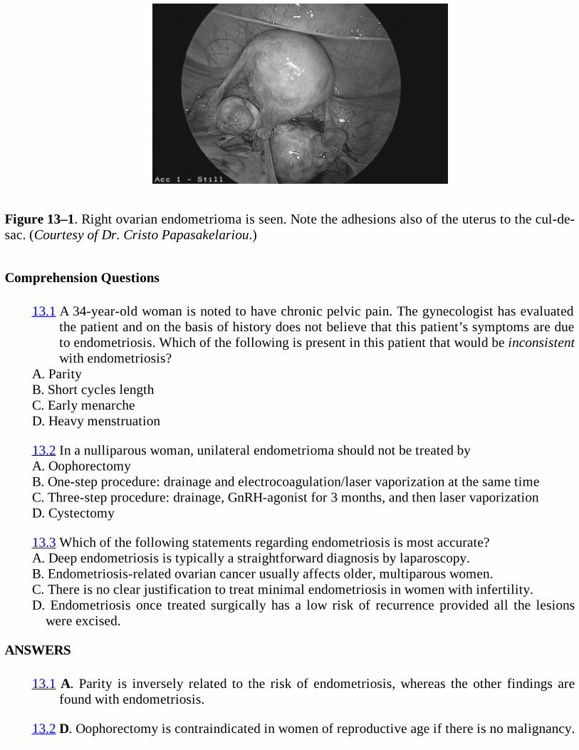
Figure 13–1. Right ovarian endometrioma is seen. Note the adhesions also of the uterus to the cul-de-sac. (Courtesy of Dr. Cristo Papasakelariou.)
Comprehension Questions
13.1 A 34-year-old woman is noted to have chronic pelvic pain. The gynecologist has evaluatedthe patient and on the basis of history does not believe that this patient’s symptoms are dueto endometriosis. Which of the following is present in this patient that would be inconsistentwith endometriosis?
A. ParityB. Short cycles lengthC. Early menarcheD. Heavy menstruation
13.2 In a nulliparous woman, unilateral endometrioma should not be treated byA. OophorectomyB. One-step procedure: drainage and electrocoagulation/laser vaporization at the same timeC. Three-step procedure: drainage, GnRH-agonist for 3 months, and then laser vaporizationD. Cystectomy
13.3 Which of the following statements regarding endometriosis is most accurate?A. Deep endometriosis is typically a straightforward diagnosis by laparoscopy.B. Endometriosis-related ovarian cancer usually affects older, multiparous women.C. There is no clear justification to treat minimal endometriosis in women with infertility.D. Endometriosis once treated surgically has a low risk of recurrence provided all the lesions
were excised.
ANSWERS
13.1 A. Parity is inversely related to the risk of endometriosis, whereas the other findings arefound with endometriosis.
13.2 D. Oophorectomy is contraindicated in women of reproductive age if there is no malignancy.
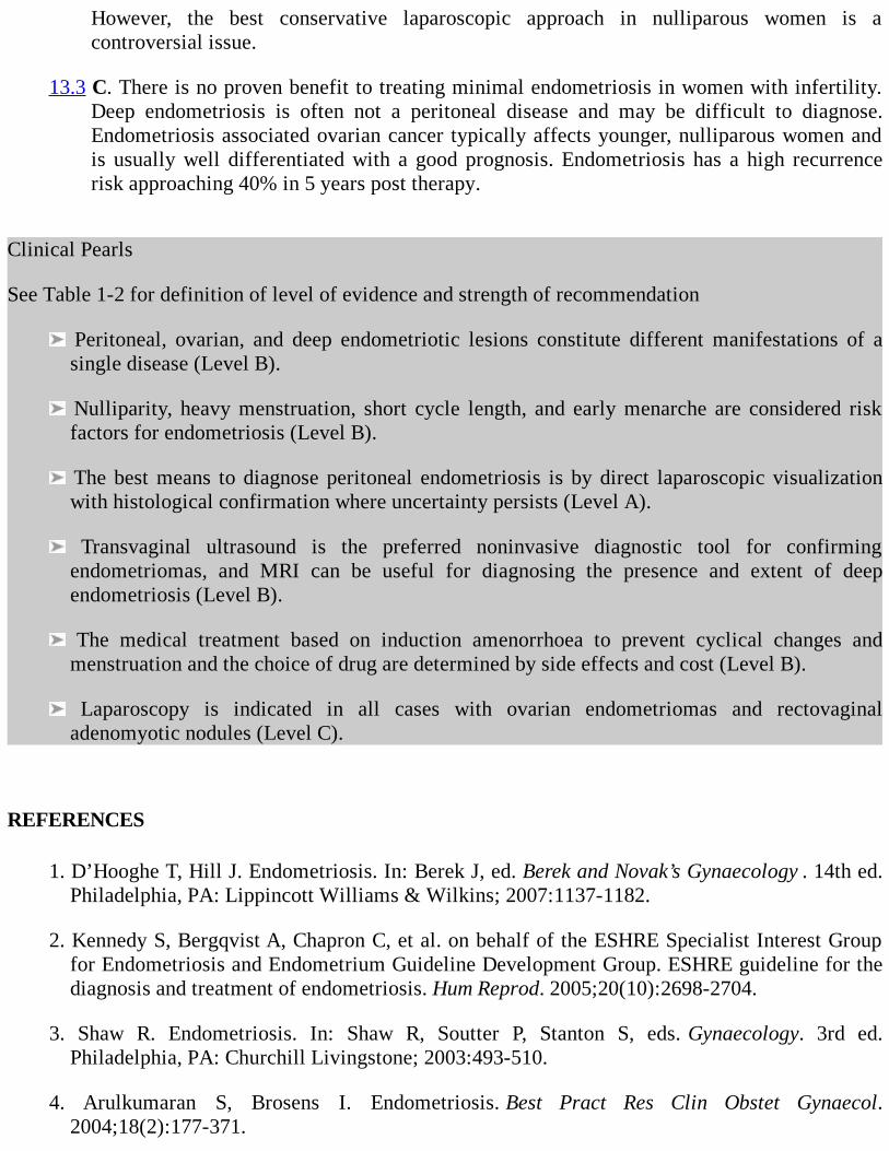
However, the best conservative laparoscopic approach in nulliparous women is acontroversial issue.
13.3 C. There is no proven benefit to treating minimal endometriosis in women with infertility.Deep endometriosis is often not a peritoneal disease and may be difficult to diagnose.Endometriosis associated ovarian cancer typically affects younger, nulliparous women andis usually well differentiated with a good prognosis. Endometriosis has a high recurrencerisk approaching 40% in 5 years post therapy.
Clinical Pearls
See Table 1-2 for definition of level of evidence and strength of recommendation
Peritoneal, ovarian, and deep endometriotic lesions constitute different manifestations of asingle disease (Level B).
Nulliparity, heavy menstruation, short cycle length, and early menarche are considered riskfactors for endometriosis (Level B).
The best means to diagnose peritoneal endometriosis is by direct laparoscopic visualizationwith histological confirmation where uncertainty persists (Level A).
Transvaginal ultrasound is the preferred noninvasive diagnostic tool for confirmingendometriomas, and MRI can be useful for diagnosing the presence and extent of deependometriosis (Level B).
The medical treatment based on induction amenorrhoea to prevent cyclical changes andmenstruation and the choice of drug are determined by side effects and cost (Level B).
Laparoscopy is indicated in all cases with ovarian endometriomas and rectovaginaladenomyotic nodules (Level C).
REFERENCES
1. D’Hooghe T, Hill J. Endometriosis. In: Berek J, ed. Berek and Novak’s Gynaecology . 14th ed.Philadelphia, PA: Lippincott Williams & Wilkins; 2007:1137-1182.
2. Kennedy S, Bergqvist A, Chapron C, et al. on behalf of the ESHRE Specialist Interest Groupfor Endometriosis and Endometrium Guideline Development Group. ESHRE guideline for thediagnosis and treatment of endometriosis. Hum Reprod. 2005;20(10):2698-2704.
3. Shaw R. Endometriosis. In: Shaw R, Soutter P, Stanton S, eds. Gynaecology. 3rd ed.Philadelphia, PA: Churchill Livingstone; 2003:493-510.
4. Arulkumaran S, Brosens I. Endometriosis. Best Pract Res Clin Obstet Gynaecol.2004;18(2):177-371.
5. Pados G, Tsolakidis D, Bontis J. Laparoscopic management of the adnexal mass. Ann N Y AcadSci. 2006;1092:211-228.
6. Catenacci M, Sastry S, Falcone T. Laparoscopic surgery for endometriosis. Clin ObstetGynecol. 2009 Sep;52(3):351-361.
7. Yeung PP Jr, Shwayder J, Pasic RP. Laparoscopic management of endometriosis:comprehensive review of best evidence. J Minim Invasive Gynecol. 2009 May-Jun;16(3):269-281.
8. Wykes CB, Clark TJ, Khan KS. Accuracy of laparoscopy in the diagnosis of endometriosis: asystematic quantitative review. BJOG. 2004 Nov;111(11):1204-1212.
9. Garry R. The effectiveness of laparoscopic excision of endometriosis. Curr Opin ObstetGynecol. 2004 Aug;16(4):299-303.
Case 14
A 25-year-old G1P1001 woman presents to the emergency room complaining of worsening right lowerquadrant pain for approximately 12 hours. The pain started around the umbilicus, then moved to theright lower quadrant region. She reports nausea and vomiting that started approximately 6 hourspreviously. She denies any vaginal discharge. Her past medical and surgical history is negative. Shereports one vaginal delivery without complications. She has been with the same partner for 7 years.The vital signs include temperature 100.6°F, pulse rate 90 beats/min, RR 20 breaths/min, and BP110/60 mm Hg. On examination, she appears to be in moderate distress. Her abdomen is tender topalpation in the right lower quadrant with guarding and possible rebound tenderness. The pelvicexamination is within normal limits. Laboratory evaluation shows a white blood (cell) count (WBC)15,000/mm3, urinalysis is negative, and the serum β human chorionic gonadotropin (β-hCG) isnegative.
What is the most likely diagnosis?
What radiological imaging should be considered to help confirm the diagnosis?
What is the best treatment for this patient?
ANSWERS TO CASE 14:Laparoscopic Appendectomy
Summary: This is a 25-year-old woman with new-onset, worsening, right lower quadrant painassociated with nausea and vomiting. She has a low-grade fever and the abdomen is tender topalpation on examination. The serum leukocyte count is elevated and the pregnancy test is negative.
Most likely diagnosis: Appendicitis.
Imaging test to help confirm the diagnosis: Helical CT.
Best treatment: The patient should be started on broad-spectrum antibiotics to cover gram-positive and -negative as well as anaerobic bacteria. She should then undergo a laparoscopicappendectomy.
ANALYSIS
Objectives
1. Know the pathophysiology of appendicitis.2. Know the most common presentation of appendicitis.3. Know the best diagnostic test for appendicitis.4. Know the advantages and disadvantages of incidental laparoscopic appendectomy.
Considerations
This is a 25-year-old woman in the reproductive years with the acute onset of right lower quadrantabdominal pain. The pregnancy test is negative. The pelvic examination is unremarkable, which ishelpful. The possibility of acute appendicitis, gastroenteritis, ruptured ovarian cyst, atypical pelvicinflammatory disease, or urinary tract infection should be considered. A CT scan of the abdomen andpelvis may be helpful in the diagnosis. General surgery consultation should be considered. If thepatient is taken to the OR, the gynecologist should be available to view the pelvis should the patienthave a tubal or ovarian pathology.
APPROACH TO Laparoscopic Appendectomy
DEFINITIONS
PSOAS SIGN: Increase in pain in the right lower quadrant of the abdomen as the right leg is extendedat the hip when the patient is in the left lateral decubitus position is a positive psoas sign. This is aresult of the inflamed appendix irritating the psoas muscle.
ROVSING SIGN: Pain in the right lower quadrant of the abdomen when palpating the left lowerquadrant.
OBTURATOR SIGN: Increased pain in the right lower quadrant of the abdomen with passive flexionof the right leg at the hip and knee with internal rotation of the leg is a positive obturator sign. This isa result of the inflamed appendix causing irritation of the obturator muscle.
MCBURNEY POINT: One-third of the distance from the right anterior superior iliac spine to theumbilicus. The inflamed appendiceal serosa causes irritation of the parietal peritoneum at thislocation, resulting in pain from appendicitis.
ELECTIVE COINCIDENTAL APPENDECTOMY (INCIDENTAL APPENDECTOMY): Theremoval of a normal appendix at the time of another surgical procedure. This is distinguished from theremoval of a normal appendix in a patient who undergoes surgery for right lower quadrant pain.
LAPAROSCOPIC APPENDECTOMY
Background
Appendectomy for acute appendicitis is the most common emergency surgery performed in the UnitedStates. Women have a lifetime risk of 7% for having appendicitis, and their overall lifetime risk ofundergoing appendectomy is 25%. Women are twice as likely to undergo a negative appendectomyduring which a normal appendix is removed for suspected appendicitis as compared to men.1
Pathophysiology
Acute appendicitis results from occlusion of the appendiceal lumen in 85% of the cases and fromunknown causes in 15% of the cases. Any process that obstructs the drainage of the appendicealsecretions can cause appendicitis.2 Hyperplasia of lymphoid follicles, fecaliths, intestinal parasites,
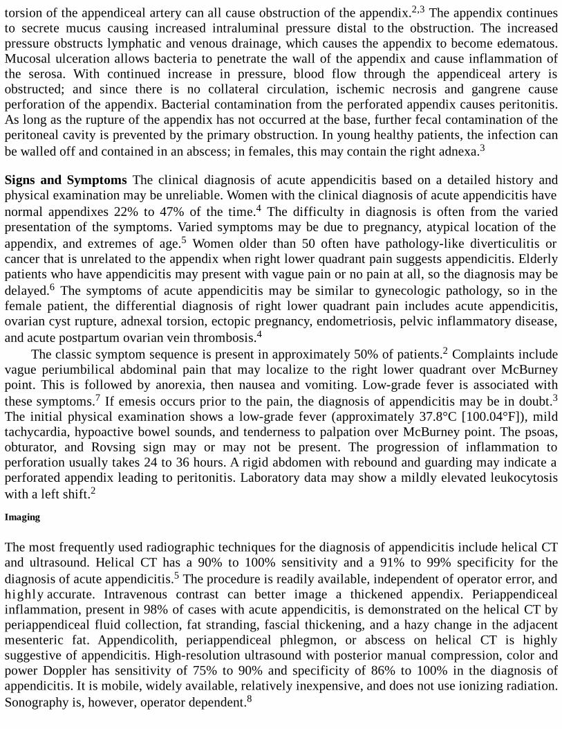
torsion of the appendiceal artery can all cause obstruction of the appendix.2,3 The appendix continuesto secrete mucus causing increased intraluminal pressure distal to the obstruction. The increasedpressure obstructs lymphatic and venous drainage, which causes the appendix to become edematous.Mucosal ulceration allows bacteria to penetrate the wall of the appendix and cause inflammation ofthe serosa. With continued increase in pressure, blood flow through the appendiceal artery isobstructed; and since there is no collateral circulation, ischemic necrosis and gangrene causeperforation of the appendix. Bacterial contamination from the perforated appendix causes peritonitis.As long as the rupture of the appendix has not occurred at the base, further fecal contamination of theperitoneal cavity is prevented by the primary obstruction. In young healthy patients, the infection canbe walled off and contained in an abscess; in females, this may contain the right adnexa.3
Signs and Symptoms The clinical diagnosis of acute appendicitis based on a detailed history andphysical examination may be unreliable. Women with the clinical diagnosis of acute appendicitis havenormal appendixes 22% to 47% of the time.4 The difficulty in diagnosis is often from the variedpresentation of the symptoms. Varied symptoms may be due to pregnancy, atypical location of theappendix, and extremes of age.5 Women older than 50 often have pathology-like diverticulitis orcancer that is unrelated to the appendix when right lower quadrant pain suggests appendicitis. Elderlypatients who have appendicitis may present with vague pain or no pain at all, so the diagnosis may bedelayed.6 The symptoms of acute appendicitis may be similar to gynecologic pathology, so in thefemale patient, the differential diagnosis of right lower quadrant pain includes acute appendicitis,ovarian cyst rupture, adnexal torsion, ectopic pregnancy, endometriosis, pelvic inflammatory disease,and acute postpartum ovarian vein thrombosis.4
The classic symptom sequence is present in approximately 50% of patients.2 Complaints includevague periumbilical abdominal pain that may localize to the right lower quadrant over McBurneypoint. This is followed by anorexia, then nausea and vomiting. Low-grade fever is associated withthese symptoms.7 If emesis occurs prior to the pain, the diagnosis of appendicitis may be in doubt.3The initial physical examination shows a low-grade fever (approximately 37.8°C [100.04°F]), mildtachycardia, hypoactive bowel sounds, and tenderness to palpation over McBurney point. The psoas,obturator, and Rovsing sign may or may not be present. The progression of inflammation toperforation usually takes 24 to 36 hours. A rigid abdomen with rebound and guarding may indicate aperforated appendix leading to peritonitis. Laboratory data may show a mildly elevated leukocytosiswith a left shift.2
Imaging
The most frequently used radiographic techniques for the diagnosis of appendicitis include helical CTand ultrasound. Helical CT has a 90% to 100% sensitivity and a 91% to 99% specificity for thediagnosis of acute appendicitis.5 The procedure is readily available, independent of operator error, andhighly accurate. Intravenous contrast can better image a thickened appendix. Periappendicealinflammation, present in 98% of cases with acute appendicitis, is demonstrated on the helical CT byperiappendiceal fluid collection, fat stranding, fascial thickening, and a hazy change in the adjacentmesenteric fat. Appendicolith, periappendiceal phlegmon, or abscess on helical CT is highlysuggestive of appendicitis. High-resolution ultrasound with posterior manual compression, color andpower Doppler has sensitivity of 75% to 90% and specificity of 86% to 100% in the diagnosis ofappendicitis. It is mobile, widely available, relatively inexpensive, and does not use ionizing radiation.Sonography is, however, operator dependent.8
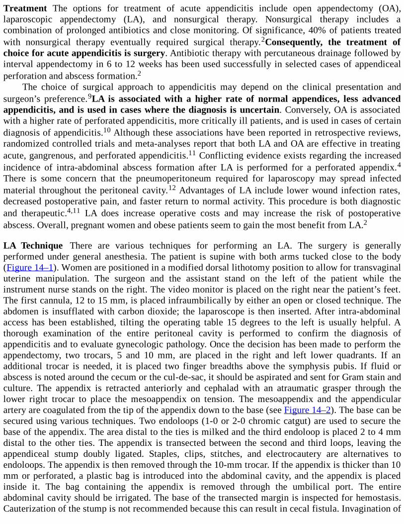
Treatment The options for treatment of acute appendicitis include open appendectomy (OA),laparoscopic appendectomy (LA), and nonsurgical therapy. Nonsurgical therapy includes acombination of prolonged antibiotics and close monitoring. Of significance, 40% of patients treatedwith nonsurgical therapy eventually required surgical therapy.2Consequently, the treatment ofchoice for acute appendicitis is surgery. Antibiotic therapy with percutaneous drainage followed byinterval appendectomy in 6 to 12 weeks has been used successfully in selected cases of appendicealperforation and abscess formation.2
The choice of surgical approach to appendicitis may depend on the clinical presentation andsurgeon’s preference.9LA is associated with a higher rate of normal appendices, less advancedappendicitis, and is used in cases where the diagnosis is uncertain. Conversely, OA is associatedwith a higher rate of perforated appendicitis, more critically ill patients, and is used in cases of certaindiagnosis of appendicitis.10 Although these associations have been reported in retrospective reviews,randomized controlled trials and meta-analyses report that both LA and OA are effective in treatingacute, gangrenous, and perforated appendicitis.11 Conflicting evidence exists regarding the increasedincidence of intra-abdominal abscess formation after LA is performed for a perforated appendix.4
There is some concern that the pneumoperitoneum required for laparoscopy may spread infectedmaterial throughout the peritoneal cavity.12 Advantages of LA include lower wound infection rates,decreased postoperative pain, and faster return to normal activity. This procedure is both diagnosticand therapeutic.4,11 LA does increase operative costs and may increase the risk of postoperativeabscess. Overall, pregnant women and obese patients seem to gain the most benefit from LA.2
LA Technique There are various techniques for performing an LA. The surgery is generallyperformed under general anesthesia. The patient is supine with both arms tucked close to the body(Figure 14–1). Women are positioned in a modified dorsal lithotomy position to allow for transvaginaluterine manipulation. The surgeon and the assistant stand on the left of the patient while theinstrument nurse stands on the right. The video monitor is placed on the right near the patient’s feet.The first cannula, 12 to 15 mm, is placed infraumbilically by either an open or closed technique. Theabdomen is insufflated with carbon dioxide; the laparoscope is then inserted. After intra-abdominalaccess has been established, tilting the operating table 15 degrees to the left is usually helpful. Athorough examination of the entire peritoneal cavity is performed to confirm the diagnosis ofappendicitis and to evaluate gynecologic pathology. Once the decision has been made to perform theappendectomy, two trocars, 5 and 10 mm, are placed in the right and left lower quadrants. If anadditional trocar is needed, it is placed two finger breadths above the symphysis pubis. If fluid orabscess is noted around the cecum or the cul-de-sac, it should be aspirated and sent for Gram stain andculture. The appendix is retracted anteriorly and cephalad with an atraumatic grasper through thelower right trocar to place the mesoappendix on tension. The mesoappendix and the appendicularartery are coagulated from the tip of the appendix down to the base (see Figure 14–2). The base can besecured using various techniques. Two endoloops (1-0 or 2-0 chromic catgut) are used to secure thebase of the appendix. The area distal to the ties is milked and the third endoloop is placed 2 to 4 mmdistal to the other ties. The appendix is transected between the second and third loops, leaving theappendiceal stump doubly ligated. Staples, clips, stitches, and electrocautery are alternatives toendoloops. The appendix is then removed through the 10-mm trocar. If the appendix is thicker than 10mm or perforated, a plastic bag is introduced into the abdominal cavity, and the appendix is placedinside it. The bag containing the appendix is removed through the umbilical port. The entireabdominal cavity should be irrigated. The base of the transected margin is inspected for hemostasis.Cauterization of the stump is not recommended because this can result in cecal fistula. Invagination of
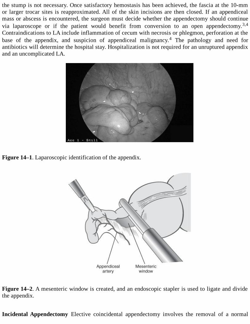
the stump is not necessary. Once satisfactory hemostasis has been achieved, the fascia at the 10-mmor larger trocar sites is reapproximated. All of the skin incisions are then closed. If an appendicealmass or abscess is encountered, the surgeon must decide whether the appendectomy should continuevia laparoscope or if the patient would benefit from conversion to an open appendectomy.3,4
Contraindications to LA include inflammation of cecum with necrosis or phlegmon, perforation at thebase of the appendix, and suspicion of appendiceal malignancy.4 The pathology and need forantibiotics will determine the hospital stay. Hospitalization is not required for an unruptured appendixand an uncomplicated LA.
Figure 14–1. Laparoscopic identification of the appendix.
Figure 14–2. A mesenteric window is created, and an endoscopic stapler is used to ligate and dividethe appendix.
Incidental Appendectomy Elective coincidental appendectomy involves the removal of a normal
appendix at the time of another surgery unrelated to the pathology of the appendix. Reasons toperform elective coincidental appendectomy are (a) to reduce the risk of mortality and morbidity fromappendicitis in the future, including infertility resulting from a perforated appendix; (b) to eliminateundiagnosed incidental pathology in the appendix; and (c) to exclude the appendix from thedifferential diagnosis when the patient has abdominal or pelvic complaints.3,13 Other groups that maybenefit from this prophylactic procedure include women who may undergo pelvic or abdominalradiation or chemotherapy, women undergoing extensive pelvic or abdominal surgery after whichextensive adhesions are anticipated, and patients in whom establishing the diagnosis of appendicitismay be difficult due to diminished ability to recognize or convey symptoms (eg, the developmentallydisabled). Studies with retrospective design, small sample size, lack of appropriate control group, andlarge number of confounding factors indicate that there is probably a small increased risk of nonfatalcomplications associated with elective coincidental appendectomy. Given this small but increased riskof complications, the debate then is whether the added cost and morbidity incurred at the time of theappendectomy outweigh the cost and morbidity from developing appendicitis in the future. Risksassociated with acute appendicitis increase with age, so the risk-benefit ratio change with the patient’sage.13 A retrospective study involving healthy women who underwent open coincidentalappendectomies at the time of gynecologic procedures concluded that the most benefit was forpatients younger than 35 years, especially if they have a history of PID, pelvic pain, orendometriosis.3,13 However, the study concluded that under specific clinical circumstances, patientsbetween the age of 35 and 50 may benefit from elective coincidental appendectomy. The data did notsupport elective coincidental appendectomy in patients older than 50 years. The decision to performthis prophylactic procedure should be based on specific clinical scenarios and after discussion of risksand benefits with the patient.13
Comprehension Questions
14.1 A 30-year-old woman presents with periumbilical pain that migrated to the right lowerquadrant pain and nausea and vomiting. Which of the following symptoms occur most rarelyin a patient with appendicitis?
A. Migration of painB. Presence of right lower quadrant painC. NauseaD. Vomiting
14.2 A 44-year-old man is diagnosed with probable acute appendicitis. He is being counseledabout the advantages of laparoscopic appendectomy. The patient has been researching aboutorgan injuries with the laparoscope. In counseling the patient, which of the following is themost accurate statement regarding laparoscopic appendectomy?
A. More chance of wound infectionsB. More chance of incisional herniaC. More pain medication needed as outpatientD. More chance of vascular injury
14.3 A 47-year-old woman undergoes a diagnostic laparoscopy for acute right lower quadrantpain. Upon examination of the abdominal cavity, the appendix appears edematous and thereis a purulent collection of fluid in the cul-de-sac. In which of the following circumstances isa laparoscopic appendectomy advisable?
A. Purulent drainage is noted from the tip of the appendixB. Purulent drainage is noted from the base of the appendixC. A 3-cm mass is noted at the base of the appendixD. The cecum is inflamed in appearance
ANSWERS
14.1 A. Frequency of common symptoms in appendicitis is abdominal pain approximately 100%,nausea 90%, vomiting 75%, and pain migration 50%.
14.2 D. Laparoscopy is associated with a higher risk of vascular injury as compared to openlaparotomy due to the insertion of the instruments. Nevertheless, it is associated withdecreased wound infection rates, shorter hospitalization, and less postoperative pain.
14.3 A. Contraindications to LA include inflammation of cecum with necrosis or phlegmon,perforation at the base of the appendix, and suspicion of appendiceal malignancy.
Clinical Pearls
See Table 1-2 for definition of level of evidence and strength of recommendation
Only 50% of women will present with the classic symptom sequence: periumbilical painmigration to right lower quadrant pain, associated anorexia and nausea and vomiting, and low-grade fever (Level B).
Helical CT is 90% to 100% sensitive for the diagnosis of appendicitis (Level B).
Contraindications to LA include inflammation of cecum with necrosis or phlegmon,perforation at the base of the appendix, and suspicion of appendiceal malignancy (Level B).
Elective coincidental appendectomy is highly beneficial for patients younger than 35 years andin selected cases for patients between the age of 35 and 50 (Level A).
REFERENCES
1. Flum D, Loepsell T. The clinical and economic correlates of misdiagnosed appendicitis:nationwide analysis. Arch Surg. 2002;137:799-804.
2. Shelton T, McKinlay R, Schwartz RW. Acute appendicitis: current diagnosis and treatment.Curr Surg. 2003;60(5):502-505.
3. Jarnigan BK. The Vermiform Appendix in Relation to Gynecology in TeLinde’s OperativeGynecology. 9th ed. Philadelphia, PA: Lippincott, Williams & Wilkins.
4. Kumar R, Erian M, Sinnot S, Knoesen R, Kimble R. Laparoscopic appendectomy in moderngynecology. J Am Assoc Gynecol Laparosc. 2002;9(3):252-263.
5. Rao PM, Feltmake CM, Rhea JT, et al. Helical computed tomography in differentiatingappendicitis and acute gynecologic conditions. Obstet Gynecol. 1999;93(2): 417-421.
6. Velanovich V, Harkabus M, Tapia F, et al. When it’s not appendicitis. Am Surg. 1998;64:7-11.
7. Hardin DJ Jr. Acute appendicitis: review and update. Am Fam Phys. 1999;60: 2027-2034.
8. Birnbaum BA, Wison SR. Appendicitis at the millennium. Radiology. 2000;215: 337-348.
9. Cervini P, Smith LC, Urbach DR. The surgeon on call is a strong factor determining the use ofa laparoscopic approach for appendectomy. Surg Endosc. 2002;16:1774-1777.
10. McGreevy JM, Finlayson SR, Alvarado R, et al. Laparoscopy may be lowering the thresholdto operate on patients with suspected appendicitis. Surg Endosc. 2002;16:1046-1049.
11. Golub R, Siddiqui F, Pohl D. Laparoscopic versus open appendectomy: a meta-analysis. J AmColl Surg. 1998;186:545-553.
12. Cuschieri A. Appendectomy—laparoscopy or open? Surg Endosc. 1997;11:319-320.
13. American College of Obstetricians and Gynecologists. Elective coincidental appendectomy.ACOG Committee Opinion No. 323, 2005.
Case 15
A 29-year-old G4P1021 Hispanic woman at 11 weeks’ gestation presents to the ED with a complaintof abdominal pain and vaginal bleeding that began 4 hours ago. She has a past obstetrical historysignificant for pelvic inflammatory disorder 10 years prior (treated as an outpatient) and twospontaneous first-trimester miscarriages both treated with dilation and curettage of the uterus. Thepatient reports an uneventful spontaneous vaginal delivery of her son 18 months ago. She has not seena physician yet for this pregnancy, but reports no abnormalities before this morning. The pain startedthis morning abruptly along with the vaginal bleeding. She reports dizziness, but denies syncope.Vaginal bleeding is the same as her menses. The vital signs taken at intake in the ED were BP 80/40mm Hg and HR 110 beats/min. Her hemoglobin level was 6.7 g/dL and quantitative β human chorionicgonadotropin (β-hCG) level was 16,000 mIU/mL. Pelvic transvaginal ultrasound reveals an emptyuterine cavity, possible chorionic sac near the cornual region of the uterus, and a moderate amount offree fluid in the cul-de-sac.
What is the most likely diagnosis?
What is your next step?
What interventions are undertaken with this diagnosis?
ANSWER TO CASE 15: Ectopic Pregnancy—Laparotomy
Summary: This is a 29-year-old G4P1021 woman at 11 weeks’ gestation with abdominal pain andvaginal bleeding. She is hypotensive, and has ultrasonographic and clinical evidence of an abnormalpregnancy and intra-abdominal hemorrhage.
Most likely diagnosis: Ruptured interstitial pregnancy.
Next step: Surgical intervention.
Interventions: Exploratory laparotomy versus operative laparoscopy.
ANALYSIS
Objectives
1. Be able to diagnose an interstitial pregnancy.2. Be familiar with management options for interstitial pregnancy.3. Be familiar with the surgical techniques employed to treat this condition.
Considerations
This is a 29-year-old G4P1021 Hispanic woman at 11 weeks’ gestation presents with a complaint ofabdominal pain and vaginal bleeding. Suspicion of an abnormal pregnancy was confirmed by pelvicultrasound. Ultrasound demonstrated an empty uterine cavity and a separate chorionic sac near thecornual region of the uterus. The patient’s vital signs and ultrasound finding of free fluid in theposterior cul-de-sac suggest a ruptured ectopic pregnancy.
Initially, it is crucial for the physician to assess the hemodynamic stability of the patient. This isgenerally screened by orthostatic blood pressure readings, J-V distension, capillary refill time, skinturgor, and laboratory readings. In cases such as acute hemorrhage, the complete blood (cell) count(CBC) level of hemoglobin may not have equilibrated with the actual blood loss amount since thepatient is bleeding whole blood. A normal hemoglobin in this patient may be falsely reassuring.Internal bleeding is suggested by the free fluid seen in the posterior cul-de-sac on ultrasound as wellas the patient’s stating that she feels light-headed. Abdominal examination may reveal tenderness topalpation and rebound tenderness suggestive of peritoneal irritation from blood in the abdominalcavity.
Identifying the location of the pregnancy in a patient presenting with abdominal complaints and apositive pregnancy test is essential. In the case of this patient, a pelvic ultrasound was performed. Nointrauterine pregnancy was visualized, and a separate chorionic sac near the cornual region was seen.These findings suggest an abnormal pregnancy in the interstitial region. Ruptured interstitial (cornual)ectopic pregnancy in a patient who demonstrates hemodynamic instability necessitates operativemanagement. The patient’s wishes for future fertility must be addressed before proceeding withsurgical management. Surgical options include laparoscopic management, but are reserved for themore stable patient and dependent on surgeon experience. Generally, in an unstable patient, time is ofthe essence and exploratory laparotomy is the method of choice.
APPROACH TO Ectopic Pregnancy—Laparotomy
DEFINITIONS
INTERSTITIAL PREGNANCY: Also known as cornual ectopic pregnancy. Pregnancy implantationwithin the tubal segment that penetrates the uterine wall.
INTERSTITIAL LINE SIGN: Ultrasonographic term used to describe the visualization of anechogenic line extending from the endometrial cavity into the cornual region and abutting theinterstitial mass or gestational sac.
ANGULAR PREGNANCY: Embryo implantation in the lateral angle of the uterine cavity medial tothe internal ostium of the fallopian tube.
CLINICAL APPROACH
Interstitial pregnancy (cornual ectopic pregnancy) is an implantation of a pregnancy within the tubalportion piercing the wall of the uterus. This form of ectopic pregnancy accounts for only 2% to 3% ofall ectopic locations.1 Overlooking this form of ectopic pregnancy due to the rarity is dangerous due tothe rich arterial blood supply from the ovarian and uterine arteries at this location.
Differentiation between an interstitial and angular pregnancy must be determined. Angularpregnancy occurs when an embryo implants in the lateral angle of the uterine cavity medial to theinternal ostium of the fallopian tube. Angular pregnancies may lead to asymmetric and symptomatic
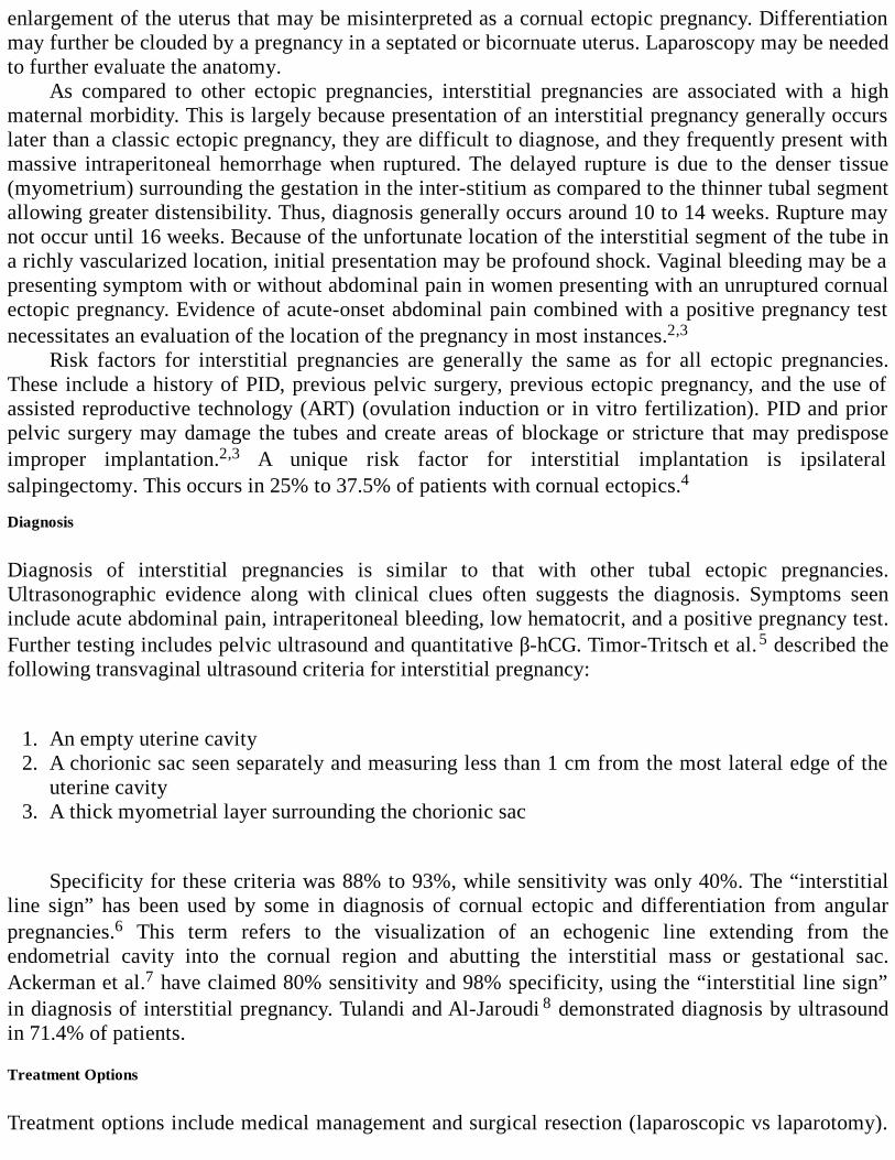
enlargement of the uterus that may be misinterpreted as a cornual ectopic pregnancy. Differentiationmay further be clouded by a pregnancy in a septated or bicornuate uterus. Laparoscopy may be neededto further evaluate the anatomy.
As compared to other ectopic pregnancies, interstitial pregnancies are associated with a highmaternal morbidity. This is largely because presentation of an interstitial pregnancy generally occurslater than a classic ectopic pregnancy, they are difficult to diagnose, and they frequently present withmassive intraperitoneal hemorrhage when ruptured. The delayed rupture is due to the denser tissue(myometrium) surrounding the gestation in the inter-stitium as compared to the thinner tubal segmentallowing greater distensibility. Thus, diagnosis generally occurs around 10 to 14 weeks. Rupture maynot occur until 16 weeks. Because of the unfortunate location of the interstitial segment of the tube ina richly vascularized location, initial presentation may be profound shock. Vaginal bleeding may be apresenting symptom with or without abdominal pain in women presenting with an unruptured cornualectopic pregnancy. Evidence of acute-onset abdominal pain combined with a positive pregnancy testnecessitates an evaluation of the location of the pregnancy in most instances.2,3
Risk factors for interstitial pregnancies are generally the same as for all ectopic pregnancies.These include a history of PID, previous pelvic surgery, previous ectopic pregnancy, and the use ofassisted reproductive technology (ART) (ovulation induction or in vitro fertilization). PID and priorpelvic surgery may damage the tubes and create areas of blockage or stricture that may predisposeimproper implantation.2,3 A unique risk factor for interstitial implantation is ipsilateralsalpingectomy. This occurs in 25% to 37.5% of patients with cornual ectopics.4
Diagnosis
Diagnosis of interstitial pregnancies is similar to that with other tubal ectopic pregnancies.Ultrasonographic evidence along with clinical clues often suggests the diagnosis. Symptoms seeninclude acute abdominal pain, intraperitoneal bleeding, low hematocrit, and a positive pregnancy test.Further testing includes pelvic ultrasound and quantitative β-hCG. Timor-Tritsch et al.5 described thefollowing transvaginal ultrasound criteria for interstitial pregnancy:
1. An empty uterine cavity2. A chorionic sac seen separately and measuring less than 1 cm from the most lateral edge of the
uterine cavity3. A thick myometrial layer surrounding the chorionic sac
Specificity for these criteria was 88% to 93%, while sensitivity was only 40%. The “interstitialline sign” has been used by some in diagnosis of cornual ectopic and differentiation from angularpregnancies.6 This term refers to the visualization of an echogenic line extending from theendometrial cavity into the cornual region and abutting the interstitial mass or gestational sac.Ackerman et al.7 have claimed 80% sensitivity and 98% specificity, using the “interstitial line sign”in diagnosis of interstitial pregnancy. Tulandi and Al-Jaroudi 8 demonstrated diagnosis by ultrasoundin 71.4% of patients.
Treatment Options
Treatment options include medical management and surgical resection (laparoscopic vs laparotomy).
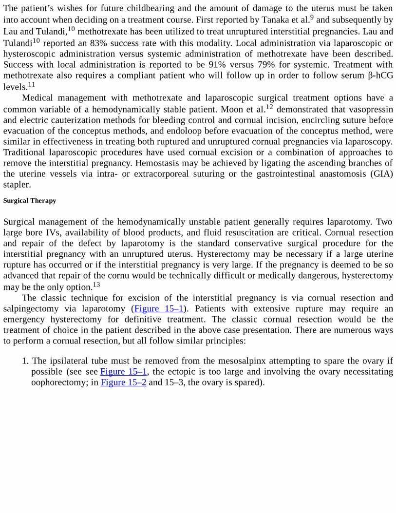
The patient’s wishes for future childbearing and the amount of damage to the uterus must be takeninto account when deciding on a treatment course. First reported by Tanaka et al.9 and subsequently byLau and Tulandi,10 methotrexate has been utilized to treat unruptured interstitial pregnancies. Lau andTulandi10 reported an 83% success rate with this modality. Local administration via laparoscopic orhysteroscopic administration versus systemic administration of methotrexate have been described.Success with local administration is reported to be 91% versus 79% for systemic. Treatment withmethotrexate also requires a compliant patient who will follow up in order to follow serum β-hCGlevels.11
Medical management with methotrexate and laparoscopic surgical treatment options have acommon variable of a hemodynamically stable patient. Moon et al.12 demonstrated that vasopressinand electric cauterization methods for bleeding control and cornual incision, encircling suture beforeevacuation of the conceptus methods, and endoloop before evacuation of the conceptus method, weresimilar in effectiveness in treating both ruptured and unruptured cornual pregnancies via laparoscopy.Traditional laparoscopic procedures have used cornual excision or a combination of approaches toremove the interstitial pregnancy. Hemostasis may be achieved by ligating the ascending branches ofthe uterine vessels via intra- or extracorporeal suturing or the gastrointestinal anastomosis (GIA)stapler.
Surgical Therapy
Surgical management of the hemodynamically unstable patient generally requires laparotomy. Twolarge bore IVs, availability of blood products, and fluid resuscitation are critical. Cornual resectionand repair of the defect by laparotomy is the standard conservative surgical procedure for theinterstitial pregnancy with an unruptured uterus. Hysterectomy may be necessary if a large uterinerupture has occurred or if the interstitial pregnancy is very large. If the pregnancy is deemed to be soadvanced that repair of the cornu would be technically difficult or medically dangerous, hysterectomymay be the only option.13
The classic technique for excision of the interstitial pregnancy is via cornual resection andsalpingectomy via laparotomy (Figure 15–1). Patients with extensive rupture may require anemergency hysterectomy for definitive treatment. The classic cornual resection would be thetreatment of choice in the patient described in the above case presentation. There are numerous waysto perform a cornual resection, but all follow similar principles:
1. The ipsilateral tube must be removed from the mesosalpinx attempting to spare the ovary ifpossible (see see Figure 15–1, the ectopic is too large and involving the ovary necessitatingoophorectomy; in Figure 15–2 and 15–3, the ovary is spared).
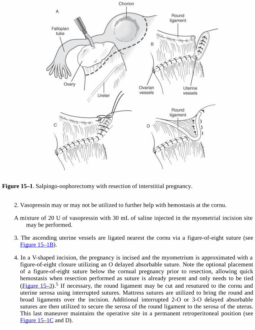
Figure 15–1. Salpingo-oophorectomy with resection of interstitial pregnancy.
2. Vasopressin may or may not be utilized to further help with hemostasis at the cornu.
A mixture of 20 U of vasopressin with 30 mL of saline injected in the myometrial incision sitemay be performed.
3. The ascending uterine vessels are ligated nearest the cornu via a figure-of-eight suture (seeFigure 15–1B).
4. In a V-shaped incision, the pregnancy is incised and the myometrium is approximated with afigure-of-eight closure utilizing an O delayed absorbable suture. Note the optional placementof a figure-of-eight suture below the cornual pregnancy prior to resection, allowing quickhemostasis when resection performed as suture is already present and only needs to be tied(Figure 15–3).5 If necessary, the round ligament may be cut and resutured to the cornu anduterine serosa using interrupted sutures. Mattress sutures are utilized to bring the round andbroad ligaments over the incision. Additional interrupted 2-O or 3-O delayed absorbablesutures are then utilized to secure the serosa of the round ligament to the serosa of the uterus.This last maneuver maintains the operative site in a permanent retroperitoneal position (seeFigure 15–1C and D).
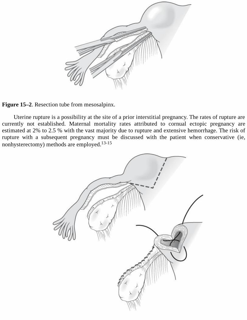
Figure 15–2. Resection tube from mesosalpinx.
Uterine rupture is a possibility at the site of a prior interstitial pregnancy. The rates of rupture arecurrently not established. Maternal mortality rates attributed to cornual ectopic pregnancy areestimated at 2% to 2.5 % with the vast majority due to rupture and extensive hemorrhage. The risk ofrupture with a subsequent pregnancy must be discussed with the patient when conservative (ie,nonhysterectomy) methods are employed.13-15
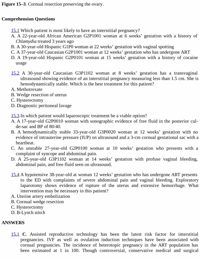
Figure 15–3. Cornual resection preserving the ovary.
Comprehension Questions
15.1 Which patient is most likely to have an interstitial pregnancy?A. A 22-year-old African American G2P1001 woman at 6 weeks’ gestation with a history of
Chlamydia treated 3 years agoB. A 30-year-old Hispanic G1P0 woman at 22 weeks’ gestation with vaginal spottingC. A 37-year-old Caucasian G2P1001 woman at 12 weeks’ gestation who has undergone ARTD. A 19-year-old Hispanic G2P0101 woman at 15 weeks’ gestation with a history of cocaine
usage
15.2 A 30-year-old Caucasian G3P1102 woman at 8 weeks’ gestation has a transvaginalultrasound showing evidence of an interstitial pregnancy measuring less than 1.5 cm. She ishemodynamically stable. Which is the best treatment for this patient?
A. MethotrexateB. Wedge resection of uterusC. HysterectomyD. Diagnostic peritoneal lavage
15.3 In which patient would laparoscopic treatment be a viable option?A. A 17-year-old G2P0010 woman with sonographic evidence of free fluid in the posterior cul-
de-sac and BP of 80/40.B. A hemodynamically stable 33-year-old G3P0020 woman at 12 weeks’ gestation with no
evidence of intrauterine pressure (IUP) on ultrasound and a 3-cm cornual gestational sac with aheartbeat.
C. An unstable 27-year-old G2P0100 woman at 10 weeks’ gestation who presents with acomplaint of syncope and abdominal pain.
D. A 25-year-old G3P1102 woman at 14 weeks’ gestation with profuse vaginal bleeding,abdominal pain, and free fluid seen on ultrasound.
15.4 A hypotensive 38-year-old at woman 12 weeks’ gestation who has undergone ART presentsto the ED with complaints of severe abdominal pain and vaginal bleeding. Exploratorylaparotomy shows evidence of rupture of the uterus and extensive hemorrhage. Whatintervention may be necessary in this patient?
A. Uterine artery embolizationB. Cornual wedge resectionC. HysterectomyD. B-Lynch stitch
ANSWERS
15.1 C. Assisted reproductive technology has been the latest risk factor for interstitialpregnancies. IVF as well as ovulation induction techniques have been associated withcornual pregnancies. The incidence of heterotopic pregnancy in the ART population hasbeen estimated at 1 in 100. Though controversial, conservative medical and surgical
management approaches have been used in small published series; Lau and Tulandi10
followed nine cases conservatively resulting in two-thirds (six out of nine) of theintrauterine pregnancies delivering at term. Certainly, this should not be viewed as standardtreatment, since massive hemorrhage has been described with these ectopic pregnancies.With increasing number of embryos transferred, the rate of interstitial heterotopicpregnancies climbs as well.
15.2 A. Methotrexate has been demonstrated as a viable option in the patient without evidence ofuterine rupture. Systemic and direct administration via laparoscopy or hysteroscopy ofmethotrexate has been performed with an overall success rate of 83%. The patient must becounseled about the risks of uterine rupture with subsequent pregnancies if she opts for aconservative approach. Reliability of the patient must also be assessed. The patient mustfollow up for serial draws of β-hCG levels in order to monitor the response to methotrexate.
15.3 B. When choosing the correct surgical modality, the patient must be assessed as a whole. Ineach case an ectopic pregnancy is suspected. The patient in option B is hemodynamicallystable and thus it would be a more suitable option for laparoscopic management. Uterinerupture is a relative contraindication to laparoscopic management. The extent of the rupturesite is difficult to assess via ultrasound. Laparoscopic management has been described forcases of minimal rupture at the site of the cornual gestation. As with any surgical technique,the skill of the surgeon and the overall state of the patient must be fully taken into accountbefore a modality is decided upon.
15.4 C. Conservative surgical management may be detrimental to the patient if a situationpresents with severe uterine rupture. The rich blood supply to the uterus may necessitateremoval of the uterus in order to achieve lifesaving hemostasis. The patient from question15.4 has a uterine rupture and severe hemorrhage. The extent of damage to the uterus mustbe assessed as well as future childbearing wishes. Hysterectomy is viewed as a last resort,but may be necessary if bleeding is unable to be controlled via conservative efforts. Thedecision to proceed with laparotomy is the correct choice because of the hemodynamicinstability.
Clinical Pearls
(See Table 1-2 for definition of level of evidence and strength of recommendation)
Methotrexate is an option for management in the appropriately selected patient (Level B).
Laparoscopic methods are viable options for treatment of both ruptured and unrupturedinterstitial pregnancies in the hemodynamically stable patient (Level B).
Uterine rupture is a potential complication for subsequent pregnancies when conservativemethods of treatment are utilized (Level C).
REFERENCES
1. Breen JL. A 21-year survey of 654 ectopic pregnancies. Am J Obstet Gynecol. 1970;106:1004.
2. Cunningham FG, Leveno KJ, Bloom SL, Gilstrap III LC, Wenstrum KD. Ectopic pregnancy.In: Williams Obstetrics. 22nd ed. New York, NY: McGraw-Hill; 2005:253-272.
3. Katz VL, Lentz GM, Lobo RA, Gershenson DM. Ectopic pregnancy. In: ComprehensiveGynecology. 5th ed. Philadelphia, PA: Mosby; 2007:389-415.
4. Society for Assisted Reproductive Technology and American Society for ReproductiveMedicine. Assisted reproduction technology in the United States: 2000 results generated fromthe American Society for Reproductive Medicine/Society for Reproductive TechnologyRegistry. Fertil Steril. 2004;81:1207.
5. Timor-Tritsch IE, Monteagudi A, Matera C, et al. Sonographic evolution of cornualpregnancies treated without surgery. Obstet Gynecol. 1992;79:1044-1049.
6. Auslender R, Arodi J, Pascal B, et al. Interstitial pregnancy: early diagnosis byultrasonography. Am J Obstet Gynecol. 1983;146:717-718.
7. Ackerman TE, Levi CS, Dashfesky SM. Interstitial line: sonographic finding in interstitial(cornual) ectopic pregnancy. Radiology. 1993;189:83-87.
8. Tulandi T, Al-Jaroudi D. Interstitial pregnancy: results generated from the Society ofReproductive Surgeons registry. Obstet Gynecol. 2004;103:47-50.
9. Tanaka T, Hayashi J, Kutsuzawa T, et al. Treatment of interstitial ectopic pregnancy withmethotrexate: report of a successful case. Fertil Steril. 1982;37:851-852.
10. Lau S, Tulandi T. Conservative medical and surgical management of interstitial ectopicpregnancy. Fertil Steril. 1999;72:207-215.
11. Weissman A, Fishman A. Uterine rupture following conservative surgery for interstitialpregnancy. Eur J Obstet Gynecol Reprod Biol. 1992;44:237-239.
12. Moon HS, Choi YJ, Park YH, et al. New simple endoscopic operations for interstitialpregnancies. Am J Obstet Gynecol. 2000;182:114-121.
13. Rock J, Jones H. Ectopic pregnancy. In: Te Linde’s Operative Gynecology . 10th ed.Philadelphia, PA: Lippincott Williams & Wilkins; 2008:798-824.
14. Baggish MS, Karram MM. Surgical management of ectopic pregnancy. In: Atlas of PelvicAnatomy and Gynecologic Surgery. 2nd ed. Philadelphia, PA: Elsevier Saunders; 2006:279-282.
15. Gilstrap III LC, Cunningham FG, Vandorsten JP. Ectopic pregnancy. In: OperativeObstetrics. 2nd ed. New York, NY: McGraw-Hill; 2002:355-378.
Case 16
Your patient for surgery is a 40-year-old obese black woman, G2P2002, with increasingly heavy,painful menses and pelvic pressure. She has a long history of symptomatic uterine fibroids which havebeen treated in the past with gonadotropin-releasing hormone agonists (GnRH-a) and myomectomy.She has also had two cesarean sections and a bilateral tubal ligation. On physical examination, herBMI is 35 kg/m2. Her uterus was enlarged to 20 weeks’ size, irregular, and there was a firm massprotruding into the left adnexa. Pelvic sonography confirmed your suspicion of multiple uterineleiomyomata, one of which is protruding into the left adnexal area.
You have decided to proceed with a total abdominal hysterectomy. A vertical midline incision isused to open the abdomen, and you immediately encounter extensive omental adhesions, involving theabdominal wall, uterus, and bowel. These structures are freed, the uterus is exposed, and a self-retaining retractor is used to provide adequate exposure. The round ligaments are located andtransected to expose the retroperitoneal space. The right ureter is easily identified, and you are able tosecure the right uterine vasculature without difficulty. The left ureter is more difficult to find and youare never sure it is positively identified because of limited exposure due to the protruding fibroid andincreased bleeding on that side.
What pre- or intraoperative strategies might have made the operation easier and safer?
What can be done to confirm the presence or absence of ureteral injury?
What complications could you anticipate in the postoperative period for this patient?
ANSWERS TO CASE 16:Total Abdominal Hysterectomy
Summary: A 40-year-old black woman with multiple abdominal surgeries in the past, a large fibroiduterus, and obesity is taken to the operating room for symptomatic uterine fibroids, which have failedmedical management and myomectomy. A leiomyoma is protruding into the left adnexal region, andthe left ureter is difficult to identify.
Preoperative strategies: Pretreatment with GnRH-agonists or antagonists, aromataseinhibitors, or progesterone receptor modulators may be useful.
Intraoperative strategies: Intraoperative myomectomy, midline vertical abdominal incision,and a large self-retaining retractor.
Strategies to safeguard ureter: Preoperative placement of a ureteral stent, cystoscopy prior tothe completion of the case.
Postoperative complications: Wound infection, ileus, deep vein thrombosis, atelectasis, bloodtransfusion, neuropathies.
ANALYSIS

Objectives
1. Describe the common indications for hysterectomy.2. Describe other surgical approaches to hysterectomy.3. List the two most common complications of abdominal hysterectomy, and how to avoid them.
Considerations
Our case presents the gynecologic surgeon with several challenges. The size of the patient and the sizeof the uterus make the procedure more difficult. GnRH-agonists or antagonists, aromatase inhibitors,or progesterone receptor modulators have all been shown to shrink the size of most leiomyomas tosome degree. Any decrease in uterine size should make the procedure less difficult and reduce bloodloss. The next question would be how to access the abdominal cavity. Depending on the skill andexperience of the surgeon, the laparoscopic approach may be feasible, and robotic surgery may be anoption if those resources were available. An abdominal hysterectomy, however, would be the classicapproach to this patient. A vertical midline incision allows for adequate exposure of the entireabdominal cavity and can easily be extended if necessary. This type of incision also makes lysis ofadhesions somewhat easier. In these cases with a large mass in an obese patient, exposure is critical.Several types of retractors are available to use, depending on the surgeon’s preference. TheBookwalter retractor (Codman Raynham, MA) is a large retractor that allows for the placement ofmultiple blades in almost any position around the incision. This can be immensely helpful. Mobilityof the uterus is the next issue that must be resolved. How easy will it be to visualize the structuresnecessary to successfully and safely complete the operation? At this point, the surgeon must decidewhether to reduce the size of the uterus. There are multiple approaches to intraoperativemyomectomy, again depending on the surgeon’s skill and experience. Once the uterus is mobileenough to expose the pelvic sidewall, the broad ligament can be opened. This allows for entry into theretroperitoneal space, which then allows for identification of the ureter and the uterine blood supply. Italso helps in freeing the bladder from the lower uterine segment, especially when the patient has hadprevious cesarean sections. Using sharp dissection with gentle “push-and-spread” blunt dissectionreduces bladder injuries when the bladder is adherent to the lower uterine segment.
Identification of the ureter is essential and should not be too difficult if one has adequateexposure. Preoperative stenting of the ureter has been proposed as a way to help identify the ureter.Lighted stents are also available. There is debate about how useful the stents really are, and their useshould be individualized. Opening the retroperitoneal space over the bifurcation of the iliac vesselshigher in the pelvis is another way to identify the ureter, especially when dissection lower in the pelvisis difficult or obscured. Assuming that the ureter can be identified, the ovary can be removed ifnecessary and the uterine arteries skeletonized and ligated. Once the blood supply to the uterus issecured, amputation of the uterine fundus should be considered. This gives the surgeon much betteraccess to the cervix if it is to be removed, or the procedure can be completed if the surgeon hasdecided to do a subtotal hysterectomy. In those instances of a deep pelvis and a long cervix, a subtotalprocedure, if appropriate, can be a wise decision.
Intraoperative cystoscopy should be considered, especially if there is difficulty taking down thebladder flap and identifying the ureter, or there is concern that the ureter may have been injured. If acystoscope is not available or if you do not routinely do cystoscopy, the dome of the bladder can be
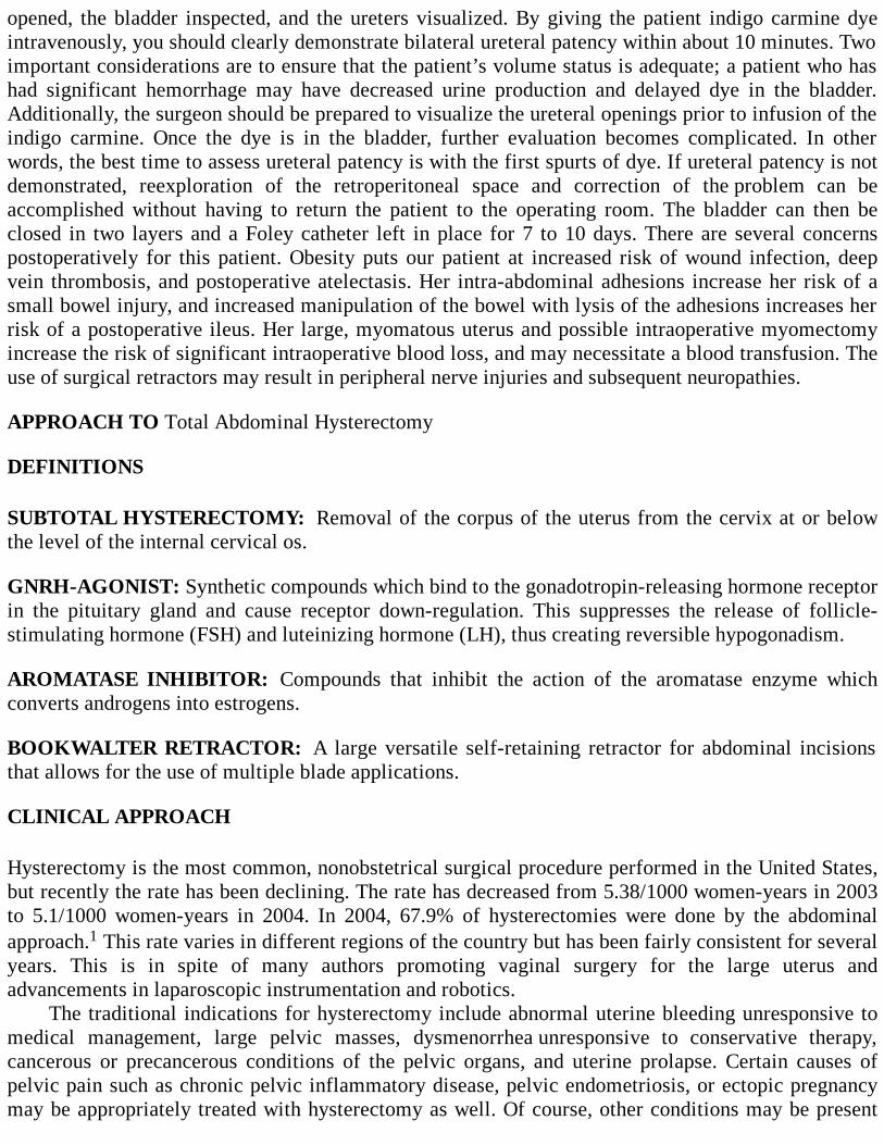
opened, the bladder inspected, and the ureters visualized. By giving the patient indigo carmine dyeintravenously, you should clearly demonstrate bilateral ureteral patency within about 10 minutes. Twoimportant considerations are to ensure that the patient’s volume status is adequate; a patient who hashad significant hemorrhage may have decreased urine production and delayed dye in the bladder.Additionally, the surgeon should be prepared to visualize the ureteral openings prior to infusion of theindigo carmine. Once the dye is in the bladder, further evaluation becomes complicated. In otherwords, the best time to assess ureteral patency is with the first spurts of dye. If ureteral patency is notdemonstrated, reexploration of the retroperitoneal space and correction of the problem can beaccomplished without having to return the patient to the operating room. The bladder can then beclosed in two layers and a Foley catheter left in place for 7 to 10 days. There are several concernspostoperatively for this patient. Obesity puts our patient at increased risk of wound infection, deepvein thrombosis, and postoperative atelectasis. Her intra-abdominal adhesions increase her risk of asmall bowel injury, and increased manipulation of the bowel with lysis of the adhesions increases herrisk of a postoperative ileus. Her large, myomatous uterus and possible intraoperative myomectomyincrease the risk of significant intraoperative blood loss, and may necessitate a blood transfusion. Theuse of surgical retractors may result in peripheral nerve injuries and subsequent neuropathies.
APPROACH TO Total Abdominal Hysterectomy
DEFINITIONS
SUBTOTAL HYSTERECTOMY: Removal of the corpus of the uterus from the cervix at or belowthe level of the internal cervical os.
GNRH-AGONIST: Synthetic compounds which bind to the gonadotropin-releasing hormone receptorin the pituitary gland and cause receptor down-regulation. This suppresses the release of follicle-stimulating hormone (FSH) and luteinizing hormone (LH), thus creating reversible hypogonadism.
AROMATASE INHIBITOR: Compounds that inhibit the action of the aromatase enzyme whichconverts androgens into estrogens.
BOOKWALTER RETRACTOR: A large versatile self-retaining retractor for abdominal incisionsthat allows for the use of multiple blade applications.
CLINICAL APPROACH
Hysterectomy is the most common, nonobstetrical surgical procedure performed in the United States,but recently the rate has been declining. The rate has decreased from 5.38/1000 women-years in 2003to 5.1/1000 women-years in 2004. In 2004, 67.9% of hysterectomies were done by the abdominalapproach.1 This rate varies in different regions of the country but has been fairly consistent for severalyears. This is in spite of many authors promoting vaginal surgery for the large uterus andadvancements in laparoscopic instrumentation and robotics.
The traditional indications for hysterectomy include abnormal uterine bleeding unresponsive tomedical management, large pelvic masses, dysmenorrhea unresponsive to conservative therapy,cancerous or precancerous conditions of the pelvic organs, and uterine prolapse. Certain causes ofpelvic pain such as chronic pelvic inflammatory disease, pelvic endometriosis, or ectopic pregnancymay be appropriately treated with hysterectomy as well. Of course, other conditions may be present
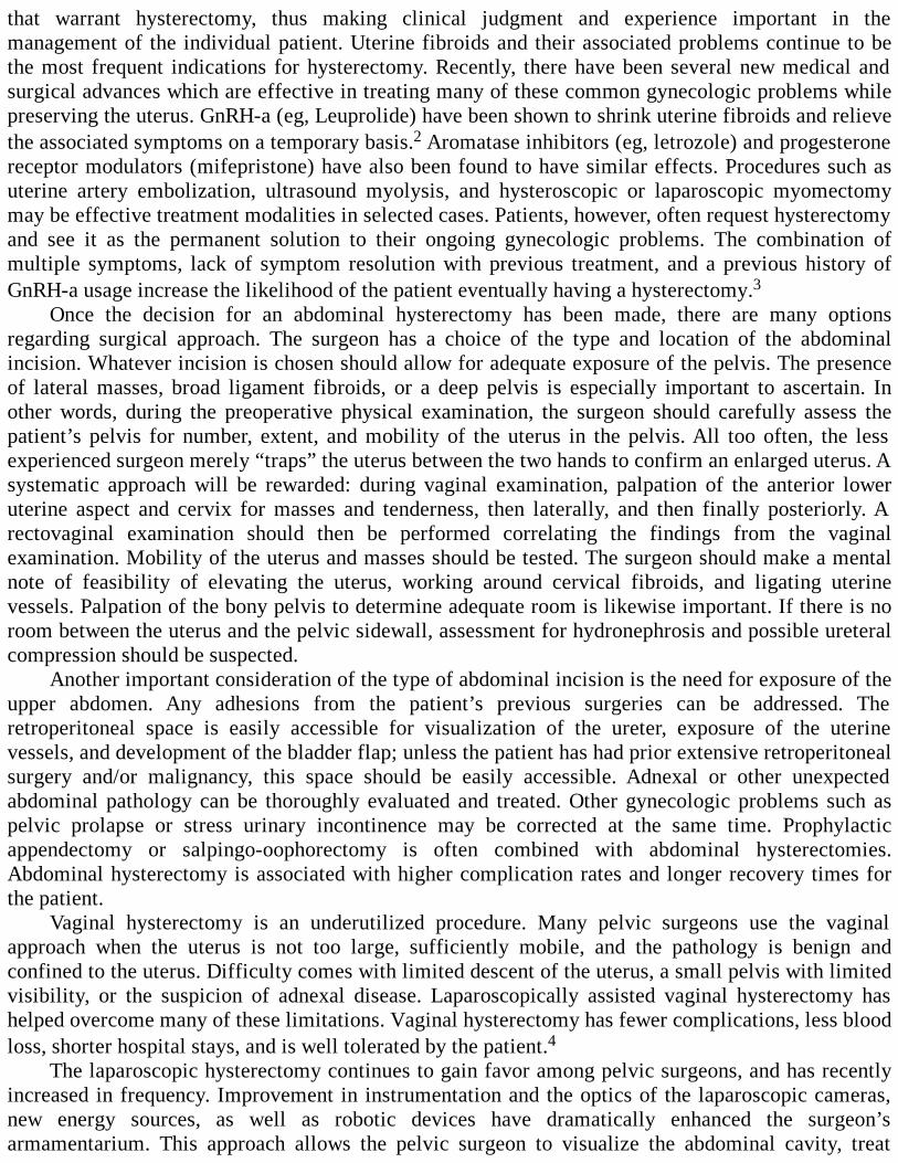
that warrant hysterectomy, thus making clinical judgment and experience important in themanagement of the individual patient. Uterine fibroids and their associated problems continue to bethe most frequent indications for hysterectomy. Recently, there have been several new medical andsurgical advances which are effective in treating many of these common gynecologic problems whilepreserving the uterus. GnRH-a (eg, Leuprolide) have been shown to shrink uterine fibroids and relievethe associated symptoms on a temporary basis.2 Aromatase inhibitors (eg, letrozole) and progesteronereceptor modulators (mifepristone) have also been found to have similar effects. Procedures such asuterine artery embolization, ultrasound myolysis, and hysteroscopic or laparoscopic myomectomymay be effective treatment modalities in selected cases. Patients, however, often request hysterectomyand see it as the permanent solution to their ongoing gynecologic problems. The combination ofmultiple symptoms, lack of symptom resolution with previous treatment, and a previous history ofGnRH-a usage increase the likelihood of the patient eventually having a hysterectomy.3
Once the decision for an abdominal hysterectomy has been made, there are many optionsregarding surgical approach. The surgeon has a choice of the type and location of the abdominalincision. Whatever incision is chosen should allow for adequate exposure of the pelvis. The presenceof lateral masses, broad ligament fibroids, or a deep pelvis is especially important to ascertain. Inother words, during the preoperative physical examination, the surgeon should carefully assess thepatient’s pelvis for number, extent, and mobility of the uterus in the pelvis. All too often, the lessexperienced surgeon merely “traps” the uterus between the two hands to confirm an enlarged uterus. Asystematic approach will be rewarded: during vaginal examination, palpation of the anterior loweruterine aspect and cervix for masses and tenderness, then laterally, and then finally posteriorly. Arectovaginal examination should then be performed correlating the findings from the vaginalexamination. Mobility of the uterus and masses should be tested. The surgeon should make a mentalnote of feasibility of elevating the uterus, working around cervical fibroids, and ligating uterinevessels. Palpation of the bony pelvis to determine adequate room is likewise important. If there is noroom between the uterus and the pelvic sidewall, assessment for hydronephrosis and possible ureteralcompression should be suspected.
Another important consideration of the type of abdominal incision is the need for exposure of theupper abdomen. Any adhesions from the patient’s previous surgeries can be addressed. Theretroperitoneal space is easily accessible for visualization of the ureter, exposure of the uterinevessels, and development of the bladder flap; unless the patient has had prior extensive retroperitonealsurgery and/or malignancy, this space should be easily accessible. Adnexal or other unexpectedabdominal pathology can be thoroughly evaluated and treated. Other gynecologic problems such aspelvic prolapse or stress urinary incontinence may be corrected at the same time. Prophylacticappendectomy or salpingo-oophorectomy is often combined with abdominal hysterectomies.Abdominal hysterectomy is associated with higher complication rates and longer recovery times forthe patient.
Vaginal hysterectomy is an underutilized procedure. Many pelvic surgeons use the vaginalapproach when the uterus is not too large, sufficiently mobile, and the pathology is benign andconfined to the uterus. Difficulty comes with limited descent of the uterus, a small pelvis with limitedvisibility, or the suspicion of adnexal disease. Laparoscopically assisted vaginal hysterectomy hashelped overcome many of these limitations. Vaginal hysterectomy has fewer complications, less bloodloss, shorter hospital stays, and is well tolerated by the patient.4
The laparoscopic hysterectomy continues to gain favor among pelvic surgeons, and has recentlyincreased in frequency. Improvement in instrumentation and the optics of the laparoscopic cameras,new energy sources, as well as robotic devices have dramatically enhanced the surgeon’sarmamentarium. This approach allows the pelvic surgeon to visualize the abdominal cavity, treat
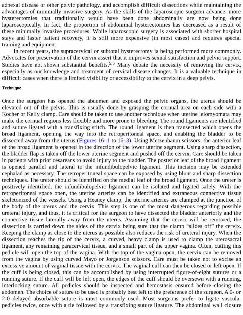
adnexal disease or other pelvic pathology, and accomplish difficult dissections while maintaining theadvantages of minimally invasive surgery. As the skills of the laparoscopic surgeon advance, morehysterectomies that traditionally would have been done abdominally are now being donelaparoscopically. In fact, the proportion of abdominal hysterectomies has decreased as a result ofthese minimally invasive procedures. While laparoscopic surgery is associated with shorter hospitalstays and faster patient recovery, it is still more expensive (in most cases) and requires specialtraining and equipment.
In recent years, the supracervical or subtotal hysterectomy is being performed more commonly.Advocates for preservation of the cervix assert that it improves sexual satisfaction and pelvic support.Studies have not shown substantial benefits.5,6 Many debate the necessity of removing the cervix,especially as our knowledge and treatment of cervical disease changes. It is a valuable technique indifficult cases when there is limited visibility or accessibility to the cervix in a deep pelvis.
Technique
Once the surgeon has opened the abdomen and exposed the pelvic organs, the uterus should beelevated out of the pelvis. This is usually done by grasping the cornual area on each side with aKocher or Kelly clamp. Care should be taken to use another technique when uterine leiomyomata maymake the cornual regions less flexible and more prone to bleeding. The round ligaments are identifiedand suture ligated with a transfixing stitch. The round ligament is then transected which opens thebroad ligament, opening the way into the retroperitoneal space, and enabling the bladder to bedissected away from the uterus (Figures 16–1 to 16–3). Using Metzenbaum scissors, the anterior leafof the broad ligament is opened in the direction of the lower uterine segment. Using sharp dissection,the bladder flap is taken off the lower uterine segment and pushed off the cervix. Care should be takenin patients with prior cesareans to avoid injury to the bladder. The posterior leaf of the broad ligamentis opened parallel and lateral to the infundibulopelvic ligament. This incision may be extendedcephalad as necessary. The retroperitoneal space can be exposed by using blunt and sharp dissectiontechniques. The ureter should be identified on the medial leaf of the broad ligament. Once the ureter ispositively identified, the infundibulopelvic ligament can be isolated and ligated safely. With theretroperitoneal space open, the uterine arteries can be identified and extraneous connective tissueskeletonized of the vessels. Using a Heaney clamp, the uterine arteries are clamped at the junction ofthe body of the uterus and the cervix. This step is one of the most dangerous regarding possibleureteral injury, and thus, it is critical for the surgeon to have dissected the bladder anteriorly and theconnective tissue laterally away from the uterus. Assuming that the cervix will be removed, thedissection is carried down the sides of the cervix being sure that the clamp “slides off” the cervix.Keeping the clamp as close to the uterus as possible also reduces the risk of ureteral injury. When thedissection reaches the tip of the cervix, a curved, heavy clamp is used to clamp the uterosacralligament, any remaining paracervical tissue, and a small part of the upper vagina. Often, cutting thispedicle will open the top of the vagina. With the top of the vagina open, the cervix can be removedfrom the vagina by using curved Mayo or Jorgenson scissors. Care must be taken not to excise anexcessive amount of vaginal tissue with the cervix. The vaginal cuff can then be closed or left open. Ifthe cuff is being closed, this can be accomplished by using interrupted figure-of-eight sutures or arunning suture. If the cuff will be left open, the edges of the cuff should be oversewn with a running,interlocking suture. All pedicles should be inspected and hemostasis ensured before closing theabdomen. The choice of suture to be used is probably best left to the preference of the surgeon. A 0- or2-0–delayed absorbable suture is most commonly used. Most surgeons prefer to ligate vascularpedicles twice, once with a tie followed by a transfixing suture ligature. The abdominal wall closure
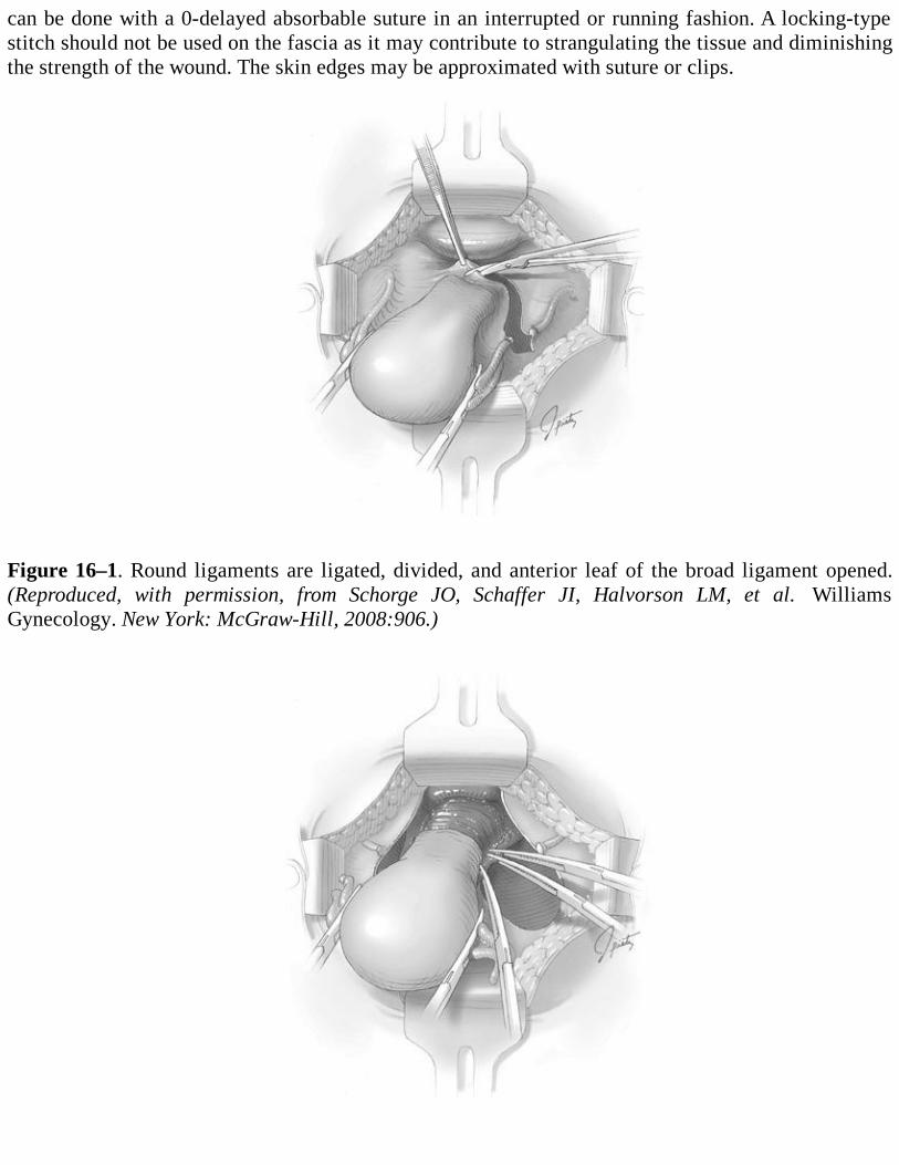
can be done with a 0-delayed absorbable suture in an interrupted or running fashion. A locking-typestitch should not be used on the fascia as it may contribute to strangulating the tissue and diminishingthe strength of the wound. The skin edges may be approximated with suture or clips.
Figure 16–1. Round ligaments are ligated, divided, and anterior leaf of the broad ligament opened.(Reproduced, with permission, from Schorge JO, Schaffer JI, Halvorson LM, et al. WilliamsGynecology. New York: McGraw-Hill, 2008:906.)
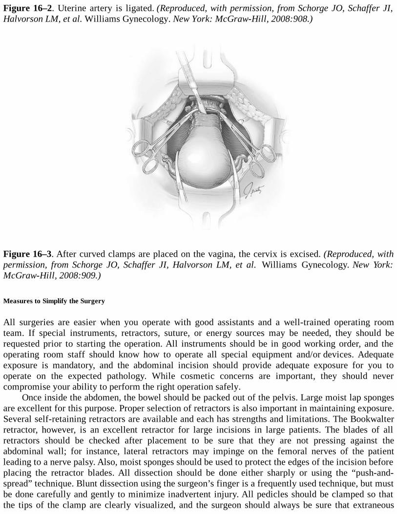
Figure 16–2. Uterine artery is ligated. (Reproduced, with permission, from Schorge JO, Schaffer JI,Halvorson LM, et al. Williams Gynecology. New York: McGraw-Hill, 2008:908.)
Figure 16–3. After curved clamps are placed on the vagina, the cervix is excised. (Reproduced, withpermission, from Schorge JO, Schaffer JI, Halvorson LM, et al. Williams Gynecology. New York:McGraw-Hill, 2008:909.) Measures to Simplify the Surgery
All surgeries are easier when you operate with good assistants and a well-trained operating roomteam. If special instruments, retractors, suture, or energy sources may be needed, they should berequested prior to starting the operation. All instruments should be in good working order, and theoperating room staff should know how to operate all special equipment and/or devices. Adequateexposure is mandatory, and the abdominal incision should provide adequate exposure for you tooperate on the expected pathology. While cosmetic concerns are important, they should nevercompromise your ability to perform the right operation safely.
Once inside the abdomen, the bowel should be packed out of the pelvis. Large moist lap spongesare excellent for this purpose. Proper selection of retractors is also important in maintaining exposure.Several self-retaining retractors are available and each has strengths and limitations. The Bookwalterretractor, however, is an excellent retractor for large incisions in large patients. The blades of allretractors should be checked after placement to be sure that they are not pressing against theabdominal wall; for instance, lateral retractors may impinge on the femoral nerves of the patientleading to a nerve palsy. Also, moist sponges should be used to protect the edges of the incision beforeplacing the retractor blades. All dissection should be done either sharply or using the “push-and-spread” technique. Blunt dissection using the surgeon’s finger is a frequently used technique, but mustbe done carefully and gently to minimize inadvertent injury. All pedicles should be clamped so thatthe tips of the clamp are clearly visualized, and the surgeon should always be sure that extraneous
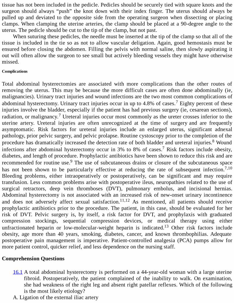
tissue has not been included in the pedicle. Pedicles should be securely tied with square knots and thesurgeon should always “push” the knot down with their index finger. The uterus should always bepulled up and deviated to the opposite side from the operating surgeon when dissecting or placingclamps. When clamping the uterine arteries, the clamp should be placed at a 90-degree angle to theuterus. The pedicle should be cut to the tip of the clamp, but not past.
When suturing these pedicles, the needle must be inserted at the tip of the clamp so that all of thetissue is included in the tie so as not to allow vascular deligation. Again, good hemostasis must beensured before closing the abdomen. Filling the pelvis with normal saline, then slowly aspirating itout will often allow the surgeon to see small but actively bleeding vessels they might have otherwisemissed.
Complications
Total abdominal hysterectomies are associated with more complications than the other routes ofremoving the uterus. This may be because the more difficult cases are often done abdominally (ie,malignancies). Urinary tract injuries and wound infections are the two most common complications ofabdominal hysterectomy. Urinary tract injuries occur in up to 4.8% of cases.7 Eighty percent of theseinjuries involve the bladder, especially if the patient has had previous surgery (ie, cesarean sections),radiation, or malignancy.7 Ureteral injuries occur most commonly as the ureter crosses inferior to theuterine artery. Ureteral injuries are often unrecognized at the time of surgery and are frequentlyasymptomatic. Risk factors for ureteral injuries include an enlarged uterus, significant adnexalpathology, prior pelvic surgery, and pelvic prolapse. Routine cystoscopy prior to the completion of theprocedure has dramatically increased the detection rate of both bladder and ureteral injuries.8 Woundinfections after abdominal hysterectomy occur in 3% to 8% of cases.7 Risk factors include obesity,diabetes, and length of procedure. Prophylactic antibiotics have been shown to reduce this risk and arerecommended for routine use.9 The use of subcutaneous drains or closure of the subcutaneous spacehas not been shown to be particularly effective at reducing the rate of subsequent infection.7,10
Bleeding problems, either intraoperatively or postoperatively, can be significant and may requiretransfusion. Less common problems arise with postoperative ileus, neuropathies related to the use ofsurgical retractors, deep vein thromboses (DVT), pulmonary embolus, and incisional hernias.Abdominal hysterectomy is not associated with an increased risk of new-onset urinary incontinenceand does not adversely affect sexual satisfaction.11,12 As mentioned, all patients should receiveprophylactic antibiotics prior to the procedure. The patient, in this case, should be evaluated for herrisk of DVT. Pelvic surgery is, by itself, a risk factor for DVT, and prophylaxis with graduatedcompression stockings, sequential compression devices, or medical therapy using eitherunfractionated heparin or low-molecular-weight heparin is indicated.13 Other risk factors includeobesity, age more than 40 years, smoking, diabetes, cancer, and known thrombophilias. Adequatepostoperative pain management is imperative. Patient-controlled analgesia (PCA) pumps allow formore patient control, quicker relief, and less dependence on the nursing staff.
Comprehension Questions
16.1 A total abdominal hysterectomy is performed on a 44-year-old woman with a large uterinefibroid. Postoperatively, the patient complained of the inability to walk. On examination,she had weakness of the right leg and absent right patellar reflexes. Which of the followingis the most likely etiology?
A. Ligation of the external iliac artery
B. Nerve compression at the right popliteal regionC. Right lateral retractor impinging on the femoral nerveD. Injury to the right obturator nerve near the obturator foramen
16.2 A 39-year-old woman is taken to the operating room for an abdominal hysterectomy. Theleft retroperitoneal space is opened from the round ligament cephalad. Where should thesurgeon look for the left ureter?
A. Adjacent to the left lateral pelvic side wall.B. At the floor of the retroperitoneal space described.C. Attached to the medial leaflet of the broad ligament.D. The ureter is not found in this area, and the surgeon should advance the incision higher in the
pelvis.
16.3 In performing a hysterectomy, the surgeon dissects the areolar tissue away from the uterinevessels (skeletonization) prior to performing ligation of the vasculature. Which of thefollowing describes the main purpose of the skeletonization process?
A. Allows for less chance of bowel injuryB. Allows for less chance of urinary tract injuryC. Allows for less chance of vascular injuryD. Allows for less chance of nerve injury
ANSWERS
16.1 C. In laparotomy surgeries, especially with an abdominal hysterectomy, the lateral retractorblades may impinge on the femoral nerve and cause femoral nerve palsy. The symptomsinclude decreased strength of the quadriceps, absent patella reflex, and inability to walk.Prevention includes ensuring sufficient padding between the lateral retractor and the lateralside wall, especially with deep retractor blades.
16.2 C. The ureter is routinely found adherent to the medial leaflet of the broad ligament uponopening the retroperitoneal space cephalad to the round ligament. If the patient, however,has had prior surgery and the retroperitoneal space has been dissected previously, the uretermay be in other locations.
16.3 B. Although the skeletonization process serves multiple purposes, including ensuring abetter ligation of the vessels, the main purpose is to release the ureter and bladder from theuterus, thus minimizing the clamps and ligation from injuring those urinary structures.
Clinical Pearls
See Table 1-2 for definition of level of evidence and strength of recommendation
The preoperative use of GnRH-a to reduce the size of the uterus may make the surgicalprocedure easier to perform and reduce blood loss (Level B).
The Bookwalter retractor is an excellent retractor to use for surgery on obese patients and largemasses (Level C).
If the ureter cannot be identified deeper in the pelvis, look for it as it crosses over the commoniliac artery (Level C).
Postoperative cystoscopy has increased the detection rates of unrecognized bladder andureteral injuries (Level B).
If you are not comfortable with cystoscopy, opening the dome of the bladder allows forvisualization of the ureteral orifices with minimal risk to bladder function (Level C).
Subtotal hysterectomy should be considered if removing the cervix is difficult (Level C).
REFERENCES
1. Whiteman MK, Hillis SD, Jamieson DJ, et al. Inpatient hysterectomy surveillance in theUnited States, 2000-2004. Obstet Gynecol Surv. 2008 May;63(5):304-305.
2. American College of Obstetricians and Gynecologists. ACOG Practice Bulletin. Alternatives toHysterectomy in the Management of Leiomyomas. Washington, DC. No. 96, August 2008.
3. Learman LA, Kuppermann M, Gates E, Gregorich SE, Lewis J, Washington AE. Predictors ofhysterectomy in women with common pelvic problems: a uterine survival analysis. J Am CollSurg. 2007 Apr;204(4).
4. Nieboer TE, Johnson N, Lethaby A, et al. Surgical approach to hysterectomy for benigngynecological disease. Cochrane Database Syst Rev. 2006;(3):CD003677.
5. Learman LA, Summitt RL Jr, Varner RE, et al. Total or Supracervical Hysterectomy (TOSH)Research Group. A randomized comparison of total or supracervical hysterectomy: surgicalcomplications and clinical outcomes. Obstet Gynecol. 2003 Sep;102(3):453-462.
6. Sokol AI, Green IC. Laparoscopic hysterectomy. Clin Obstet Gynecol. 2009 Sep;52(3):304-312.
7. Stany MP, Farley JH. Complications of gynecologic surgery. Surg Clin North Am. 2008Apr;88(2).
8. Ibeanu OA, Chesson RR, Echols KT, Nieves M, Busangu F, Nolan TE. Urinary tract injuryduring hysterectomy based on universal cystoscopy. Obstet Gynecol. 2009 Jan;113(1):6-10.
9. Tanos V, Rojansky N. Prophylactic antibiotics in abdominal hysterectomy. J Am Coll Surg.1994;179:593-600.
10. Cardosi RJ, Drake J, Holmes S, et al. Subcutaneous management of vertical incisions 3 ormore centimeters of subcutaneous fat. Am J Obstet Gynecol. 2006;195(2):607-614.
11. Gustafsson C, Ekström A, Brismar S, Altman D. Urinary incontinence after hysterectomy—three-year observational study. Urology. 2006 Oct 1;68(4):769.

12. Mokate T, Wright C, Mander T. Hysterectomy and sexual function. J Br Menopause Soc.2006 Dec;12(4):153-157.
13. American College of Obstetricians and Gynecologists. ACOG Practice Bulletin. Prevention ofDeep Vein Thrombosis and Pulmonary Embolism. No. 84, August 2007.
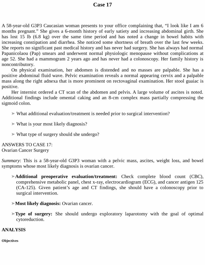
Case 17
A 58-year-old G3P3 Caucasian woman presents to your office complaining that, “I look like I am 6months pregnant.” She gives a 6-month history of early satiety and increasing abdominal girth. Shehas lost 15 lb (6.8 kg) over the same time period and has noted a change in bowel habits withincreasing constipation and diarrhea. She noticed some shortness of breath over the last few weeks.She reports no significant past medical history and has never had surgery. She has always had normalPapanicolaou (Pap) smears and underwent normal physiologic menopause without complications atage 52. She had a mammogram 2 years ago and has never had a colonoscopy. Her family history isnoncontributory.
On physical examination, her abdomen is distended and no masses are palpable. She has apositive abdominal fluid wave. Pelvic examination reveals a normal appearing cervix and a palpablemass along the right adnexa that is more prominent on rectovaginal examination. Her stool guaiac ispositive.
Her internist ordered a CT scan of the abdomen and pelvis. A large volume of ascites is noted.Additional findings include omental caking and an 8-cm complex mass partially compressing thesigmoid colon.
What additional evaluation/treatment is needed prior to surgical intervention?
What is your most likely diagnosis?
What type of surgery should she undergo?
ANSWERS TO CASE 17:Ovarian Cancer Surgery
Summary: This is a 58-year-old G3P3 woman with a pelvic mass, ascites, weight loss, and bowelsymptoms whose most likely diagnosis is ovarian cancer.
Additional preoperative evaluation/treatment: Check complete blood count (CBC),comprehensive metabolic panel, chest x-ray, electrocardiogram (ECG), and cancer antigen 125(CA-125). Given patient’s age and CT findings, she should have a colonoscopy prior tosurgical intervention.
Most likely diagnosis: Ovarian cancer.
Type of surgery: She should undergo exploratory laparotomy with the goal of optimalcytoreduction.
ANALYSIS
Objectives
1. Recognize the symptoms of ovarian cancer.2. Be familiar with requirements of optimal cytoreduction and surgical staging in patients with
early- and advanced-stage ovarian cancer.3. Describe stage I cancer and fertility preservation.4. Describe tumors of low malignant potential (LMP) and the possible need for staging.5. Describe the management of adnexal masses during pregnancy.
Considerations
This is a 58-year-old Caucasian G3P3 woman with symptoms suggestive of ovarian cancer. Symptomsassociated with ovarian cancer include pelvic/abdominal pain, urinary urgency, increasedabdominal size, and early satiety.
In the preoperative evaluation, the physician should at a minimum, obtain a CBC, comprehensivemetabolic panel, chest x-ray, and ECG for women older than 50 years. Thrombocytosis is seen in 22%of patients with ovarian cancer, which may be associated with advanced-stage disease. Serumelectrolytes and liver function tests may identify underlying medical conditions which need correctionprior to surgery. A chest x-ray should be performed preoperatively to evaluate for pulmonarymetastases and pleural effusions. A screening colonoscopy is an important part of the preoperativeevaluation if significant bowel complaints are noted, for example, change in stool caliber or anincrease in constipation, or if the physical examination or CT scan suggests the possibility of aprimary bowel carcinoma. A CA-125 level should be drawn preoperatively to serve as a baseline levelfor follow-up if ovarian carcinoma is found.
The goal of cytoreductive surgery is the removal of the maximum amount of tumor possible toenhance the effectiveness of subsequent chemotherapy. Current practice parameters suggest thatoptimal cytoreduction means that there is no residual disease greater than 1 cm (cumulative).Primary surgical cytoreduction typically includes a total abdominal hysterectomy, bilateral salpingo-oophorectomy, omentectomy, pelvic and para-aortic lymph node dissection, and resection of anymetastatic foci. Cytoreduction surgery may commonly include splenectomy, bowel resection,resection of metastatic peritoneal and diaphragmatic implants, and, rarely, liver resection andcholecystectomy.1-3
Optimal cytoreduction may not be feasible in patients with extra-abdominal metastases or withextensive disease in the hepatic parenchyma or porta hepatis, along the root of the small bowelmesentery, or in patients with bulky suprarenal lymphatic disease.4 Preoperative imaging may assistin the decision to proceed with primary surgery or neoadjuvant chemotherapy.
APPROACH TO Suspected Ovarian Cancer
DEFINITIONS
CYTOREDUCTIVE SURGERY: The surgical process by which as much of the bulk of malignanttissue as possible is removed. Debulking is considered optimal when the largest residual tumor massis less than 1 cm.
CANCER ANTIGEN 125: A protein that is found on the surface of most epithelial ovarian cancer
cells, as well as peritoneal and fallopian tube carcinomas. An elevated serum level may indicatepossible ovarian malignancy although in a premenopausal woman, there are conditions that can falselyelevate the CA-125 such as endometriosis, pregnancy, uterine fibroids, or pelvic inflammatorydisease. A level exceeding 35 mcg/mL is considered elevated.
INTRAPERITONEAL CHEMOTHERAPY: Chemotherapy administered directly into the peritonealcavity rather than intravenously.
NEOADJUVANT CHEMOTHERAPY: The use of chemotherapy prior to surgery to chemicallydebulk the cancer and make subsequent surgery less complicated and increase the chance of optimalcytoreduction. However, whether or not neoadjuvant chemotherapy improves overall survival is stillcontroversial.
CLINICAL APPROACH
Most patients with ovarian cancer will present with advanced-stage disease, as currently, there is noproven way to detect early disease. Many patients present with pelvic or abdominal pain, early satiety,and increased abdominal girth for several months preceding diagnosis. A recent study showed thatwhen affected women were carefully questioned, many of them reported having the above symptomsfor at least 12 days out of each month during the 12 months prior to diagnosis: 60% of those withearly-stage invasive disease and 79% of those with advanced-stage invasive disease. While all of thesesymptoms are nonspecific when they occur with increasing frequency and severity, they merit anevaluation for possible ovarian cancer.5
Etiology
Epidemiologic association with increased risk of ovarian cancer includes early menarche, latemenopause, low parity, infertility, the use of perineal talc, and consumption of galactose. The use oforal contraceptives for at least 5 years and history of a tubal ligation are associated with a decreasedrisk. The use of fertility-enhancing medications has not been definitively associated with an increasedrisk.
Hereditary ovarian cancer is associated with BRCA1 gene mutations, located on chromosome 17.The risk to develop ovarian cancer can be as high as 28% to 44% in patients from these high-riskfamilies. Another gene, BRCA2, located on chromosome 13, is associated with a smaller proportion ofovarian cancers. The risk of development of ovarian cancer for BRCA2 mutation carriers approaches27%.6 A third important syndrome is the Lynch syndrome, also known as hereditary nonpolyposiscolon cancer syndrome (HNPCC). Patients with Lynch syndrome have a higher-than-expected risk todevelop either endometrial and ovarian cancers or colon cancer. Thus, affected patients with a strongfamily history of ovarian or breast cancer should undergo genetic counseling and testing. Those foundto have a deleterious mutation may choose to reduce their risk for developing cancer by undergoingprophylactic salpingo-oophorectomy after completion of childbearing. Patients who desire to retaintheir childbearing potential may opt to take birth control pills, which decreases the risk of ovariancancer.
Diagnosis
The diagnosis of ovarian cancer is ideally made by obtaining tissue for histologic examination (seeFigure 17–1 in a patient who presented with an adnexal mass undergoing laparoscopy). Although
surgical evaluation for a persistent adnexal mass is preferred, there are clinical scenarios that maycontraindicate surgery. For example, patients with multiple medical problems deemed unfit forcytoreduction surgery may opt to undergo needle biopsy for tissue diagnosis, thoracentesis, orparacentesis for cytologic diagnosis. Subsequently, these patients may be considered for neoadjuvantchemotherapy if cancer is diagnosed.
Patients diagnosed with apparent Stage I (no obvious metastatic cancer outside of the ovary)invasive ovarian carcinoma need comprehensive surgical staging as the risk of undetected metastaticdisease is high. Comprehensive surgical staging includes hysterectomy with bilateral salpingo-oophorectomy, omentectomy, and pelvic and para-aortic lymphadenectomy. A biopsy is done toevaluate any clinically suspicious area in the abdominal cavity. If no abnormality is readily visible,random biopsies are taken from the bladder peritoneum, bilateral pelvic sidewall, posterior cul-de-sac, bilateral paracolic gutters, and from each side of the undersurface of the diaphragm.Collection of peritoneal washings for cytologic examination is also done.
Figure 17–1. A large cystic ovarian mass is noted on laparoscopy. (Courtesy of Dr. CristoPapasakelariou.)
Early Cancer in Women Desiring Fertility Patients with early-stage ovarian cancer who desirefuture fertility present a unique group of patients for the gynecologic oncologist. A conservativeapproach, preserving the uterus and contralateral ovary, may be used if the likelihood of cure is notcompromised while preserving reproductive potential. Comprehensive surgical staging in this groupof patients is of utmost import. As a definitive diagnosis based on frozen section diagnosis may bedifficult, these patients accept the risk that further surgical procedures may be necessary based onfinal histologic diagnosis. Generally, these patients may opt to undergo hysterectomy and removal ofthe contralateral ovary at the completion of childbearing.7,8
Low Malignant Potential Patients with a low malignant potential (LMP) or borderline tumor have amuch better prognosis than patients with invasive ovarian cancers. Surgical staging is important forthese patients because 5% of patients with borderline tumors diagnosed on frozen section analysismay have true invasive carcinoma upon final pathologic diagnosis. However, patients who have a
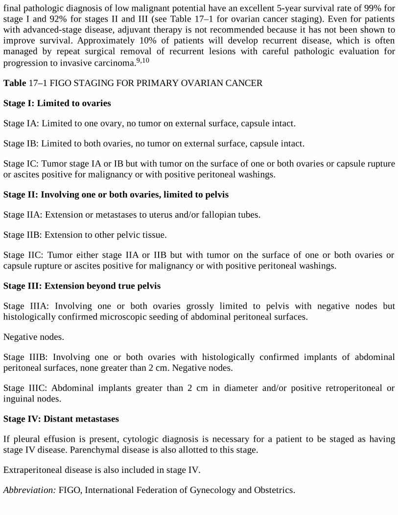
final pathologic diagnosis of low malignant potential have an excellent 5-year survival rate of 99% forstage I and 92% for stages II and III (see Table 17–1 for ovarian cancer staging). Even for patientswith advanced-stage disease, adjuvant therapy is not recommended because it has not been shown toimprove survival. Approximately 10% of patients will develop recurrent disease, which is oftenmanaged by repeat surgical removal of recurrent lesions with careful pathologic evaluation forprogression to invasive carcinoma.9,10
Table 17–1 FIGO STAGING FOR PRIMARY OVARIAN CANCER
Stage I: Limited to ovaries
Stage IA: Limited to one ovary, no tumor on external surface, capsule intact.
Stage IB: Limited to both ovaries, no tumor on external surface, capsule intact.
Stage IC: Tumor stage IA or IB but with tumor on the surface of one or both ovaries or capsule ruptureor ascites positive for malignancy or with positive peritoneal washings.
Stage II: Involving one or both ovaries, limited to pelvis
Stage IIA: Extension or metastases to uterus and/or fallopian tubes.
Stage IIB: Extension to other pelvic tissue.
Stage IIC: Tumor either stage IIA or IIB but with tumor on the surface of one or both ovaries orcapsule rupture or ascites positive for malignancy or with positive peritoneal washings.
Stage III: Extension beyond true pelvis
Stage IIIA: Involving one or both ovaries grossly limited to pelvis with negative nodes buthistologically confirmed microscopic seeding of abdominal peritoneal surfaces.
Negative nodes.
Stage IIIB: Involving one or both ovaries with histologically confirmed implants of abdominalperitoneal surfaces, none greater than 2 cm. Negative nodes.
Stage IIIC: Abdominal implants greater than 2 cm in diameter and/or positive retroperitoneal oringuinal nodes.
Stage IV: Distant metastases
If pleural effusion is present, cytologic diagnosis is necessary for a patient to be staged as havingstage IV disease. Parenchymal disease is also allotted to this stage.
Extraperitoneal disease is also included in stage IV.
Abbreviation: FIGO, International Federation of Gynecology and Obstetrics.
Ovarian Cancer in Pregnancy Patients diagnosed during the first trimester of pregnancy with anadnexal mass, measuring 6 cm or greater, merit close observation. Most of these masses will resolveby the second trimester. However, up to 28% of patients with a persistent mass may develop acutesymptoms, necessitating emergent surgery. Thus, a reasonable management schema is to surgicallyevaluate persistent masses between 15 and 19 weeks’ of gestation. Among patients who undergosurgery during pregnancy, common findings are a dermoid cyst or serous cystadenoma; the chance offinding a malignant tumor is low, usually less than 5%. Waiting beyond 23 weeks increases the risk ofadverse pregnancy outcomes, including preterm delivery. The decision to perform either laparoscopicor open surgery during pregnancy depends on the individual patient’s clinical situation and the abilityof her surgeon.11
Treatment
The initial treatment for ovarian cancer is optimal cytoreduction as survival is clearly improved withlower volume of residual disease. This step cannot be overemphasized, and meticulous care andsufficient time should be devoted to the removal of cancer to maximize the patient’s outcome. Aftermaximal cytoreduction, adjuvant chemotherapy with a platinum-based regimen is the standard.Intraperitoneal chemotherapy has recently been shown to improve median overall survival whencompared to intravenous chemotherapy in advanced-stage ovarian cancer patients. In this trialconducted by the Gynecologic Oncology Group (2006), patients received IV paclitaxel (135 mg/m2)over 24 hours, then intraperitoneal cisplatin (100 mg/m2) on day 2 and intraperitoneal paclitaxel (60mg/m2) on day 8 every 3 weeks for six cycles. Although only 42% of patients were able to completeall six cycles of intraperitoneal chemotherapy due to complications, the intraperitoneal chemotherapygroup had a 16-month increase in median overall survival. Patients undergoing intraperitoneal therapyreported a decreased quality of life when compared to patients undergoing conventional intravenouschemotherapy, but these differences equalized at 1 year following treatment. For patients who are notideal candidates for intraperitoneal chemotherapy, chemotherapy with intravenous carboplatin(starting dose area under the curve [AUC] = 5-6 mg/mL • min) and paclitaxel (175 mg/m2) every 3weeks for six cycles is given.12
Surgical Approach
Surgery for ovarian cancer is almost universally approached through a vertical skin incision, whoselength may be extended as the need arises. After entry into the abdomen, peritoneal washings areobtained and sent for cytologic evaluation. If ascites is present, this is collected and sent in lieu ofwashings. Systematic exploration of the abdominal cavity is performed to evaluate the extent ofdisease and the plan for resection of disease. Careful assessment is important for prognostic purposes,plan for the surgical resection, and later consideration for adjuvant therapy. In addition to evaluationof the pelvic organs, particular attention is paid to the surfaces of the diaphragm bilaterally, themesentery of the small bowel, and the retroperitoneal lymphatic nodes.
For patients not interested in future fertility, surgery includes removal of the uterus, bilateraltubes and ovaries, omentum, and pelvic and para-aortic lymph nodes. For patients with widespreadperitoneal metastases, removal of metastatic disease is achieved to enable optimal cytoreduction(largest residual disease < 1 cm). Commonly, the sigmoid colon and ovarian masses are coalesced intoone large tumor mass for which surgical planes are not found. In this case, a retroperitoneal approachto remove en bloc all affected organs is taken as the planes of dissection are often still preserved inthis area despite massive intra-abdominal disease.
The omentum is often infiltrated and thickened with tumor. This “omental cake” can be dissectedfrom the underlying transverse colon and stomach and removed. Bowel resection and reanastomosisare done to enable optimal cytoreduction. Disease on peritoneal surfaces may be removed by atechnique referred to as “peritoneal stripping.” This stripping technique may be done also for tumorplaques on the diaphragm in which case inadvertent entry into the thoracic cavity and resultantpneumothorax is an uncommon, but not rare, complication.
Lymphadenectomy is performed for staging from the level of the renal veins, past the bifurcationof the aorta and iliac vessels and until the circumflex vein as the vessels enter the inguinal canal.Enlarged nodes are removed as part of cytoreduction. Often, identifying the avascular areas of thepelvis, for example, the pararectal and paravesical spaces, makes surgery easier. Identifying thecourse of the ureter is important to avoid injury, especially when there is significant disease distortingpelvic anatomy.
At the end of the surgery, the abdomen is closed using a mass closure, such as the Smead-Jonestechnique or its various modifications. The strength of the closure is critical due to the patient likelyhaving recurrence of ascites, and possible delayed wound healing such as due to malnutrition. Goodtechnique is important to prevent infectious or dehiscence wound complications that may delayadjuvant chemotherapy.
Complications
When operating in the deep pelvis, injury to vascular structures and massive blood loss are possible.In anticipation of blood loss and need for transfusion, a type and cross match should be done prior tosurgery. The planned procedure is discussed with the anesthesiologist so that adequate lines andmonitoring equipment are available for emergent use. Intraoperatively, if a vessel injury occurs,pressure is quickly applied with either a sponge stick or the surgeon’s hand. Subsequently, a clampmay be used to obtain hemostasis and the vessel repaired.
Entry into the intestinal tract is a common occurrence, either intentionally or inadvertently, aspart of surgery for ovarian cancer. Prompt recognition of inadvertent entry is the key to prevention offurther complications, including abscess and fistula formation. Serosal tears and small luminal entryare easily repaired with sutures. Occasionally, larger injury may necessitate resection andreanastomosis of the normal bowel segments. Ureteral injury is best avoided by tracing the normalcourse of the ureters in the retroperitoneal space within the medial leaf of the broad ligament. Forpatients with distorted retroperitoneal anatomy (eg, patients with previous retroperitoneal dissection,severe endometriosis, or renal anomalies), preoperative ureteral stenting may assist the surgeon byallowing intraoperative palpation of the course of the ureters when visualization is impossible.Obvious injury is repaired intraoperatively. However, simple ureteral stenting will allow most smallinjuries to heal spontaneously. Occasionally, late injury due to devascularization of the ureters maynot present until more than 1 week after surgery.
The risk of postoperative complications increases with the extent of surgery and thecomorbidities of the patient. Fever and ileus are frequent occurrences, as are urinary tract and woundcomplications. Anemia is common, both from the disease process and surgical blood loss. Blood lossgreater than 1 L occurs in as many as 20% of patients.
Thromboembolic disease is a common complication of surgery. Postoperative ovarian cancerpatients have all three characteristics identified as Virchow triad: hypercoagulability due to thecancer, vessel wall injury, and stasis from the surgical procedure itself. Pre- and postoperative DVTprophylaxis is critically important. There are many published guidelines for the use of sequentialcompression devices and heparin, both unfractionated and low-molecular weight. For the
postoperative cancer patients, heparin in the immediate postoperative period is preferred. Extendedthromboprophylaxis after hospital discharge is recommended in selected high-risk patients, forexample, those with past history of DVT or a hypercoagulable state.13
Splenectomy is often part of cytoreduction for disease in the upper abdomen. Complicationsinclude pancreatic injury and pancreatic pseudocyst formation as the spleen lies in proximity to thepancreas. Most patients will also have a transient leukocytosis and thrombocytosis. Ideally, 14 daysprior to a planned splenectomy, the patient will undergo preoperative immunization for Haemophilusinfluenzae, Streptococcus pneumoniae, and Neisseria meningitidis as asplenic patients have increasedrisk of infection from these encapsulated organisms. Postoperative immunization may also be done 14days after surgery.14
Comprehension Questions
17.1 A 25-year-old nulligravida woman undergoes exploratory laparotomy and left salpingo-oophorectomy for an adnexal mass. Frozen section analysis shows a borderline seroustumor. The rest of the abdomen and pelvis is grossly normal. Which of the following is anappropriate intraoperative management?
A. Close the abdomen as she is interested in fertility and await final histologic diagnosis.B. Proceed with total abdominal hysterectomy and remove the contralateral tube and ovary.C. Proceed with surgical staging without removing the uterus and contralateral ovary.D. Biopsy the contralateral ovary and remove the ovary if abnormal.
17.2 Which of the following statements is correct for a patient who has a BRCA1 mutation?A. Prophylactic bilateral salpingo-oophorectomy is indicated after completion of childbearing.B. The patient has an 80% lifetime risk of developing ovarian cancer.C. If the patient is interested in delaying future fertility, oral contraceptive usage may increase
her risk of developing ovarian cancer.D. She has a mutation on chromosome 11.
17.3 A 21-year-old at 9 weeks’ of gestation presents for her first prenatal examination. Youdiagnose an adnexal mass measuring 6 cm with complex features. She is completelyasymptomatic. Which of the following is an appropriate management for this patient?
A. Expectant management with serial ultrasounds.B. Immediate laparotomy and unilateral salpingo-oophorectomy.C. No further evaluation; the adnexal mass is most likely a corpus luteum and should resolve.D. Immediate laparoscopic evaluation with plans for conversion to laparotomy if a cancer is
suspected.
17.4 A 22-year-old G0 woman recently had surgery for a left adnexal mass and underwent aunilateral salpingo-oophorectomy. The other ovary appeared normal during the case. Finalpathology revealed a serous tumor of low malignant potential. What is the next managementstep?
A. Immediate exploratory laparotomy and staging.B. Immediate operative laparoscopy with staging.C. Observation and close follow-up.D. Immediate exploratory laparotomy with a total abdominal hysterectomy and removal of
contralateral adnexa.
ANSWERS
17.1 C. Surgical staging is appropriate as a frozen section diagnosis of a borderline (LMP) tumorcarries approximately 5% risk of upstaging to an invasive carcinoma on final histologicexamination. Random biopsy of a grossly normal contralateral ovary is not indicatedbecause it is unlikely to find microscopic foci of tumor, but may lead to complications suchas bleeding, necessitating removal of a normal ovary. Removal of the uterus or theremaining ovary is not indicated if the patient is interested in future fertility.
17.2 B. The BRCA1 mutation is an autosomal dominant mutation located on chromosome 17.This mutation confers up to an 80% lifetime risk of development of breast cancer and a 28%to 44% lifetime risk of developing ovarian cancer. Current recommendations include the useof oral contraceptives for women who are interested in future fertility to decrease the risk ofdeveloping ovarian cancer, and prophylactic bilateral salpingo-oophorectomy aftercompletion of childbearing. Other recommendations for carriers include increased breastsurveillance and gynecologic surveillance of the ovaries with serial ultrasounds andmeasurement of CA-125. Even after prophylactic oophorectomy, there is a risk ofdeveloping peritoneal carcinoma; thus, continued gynecologic surveillance is mandatory.
17.3 A. A pregnant patient with an asymptomatic adnexal mass needs follow-up. This is mosteasily done with serial ultrasounds. Immediate surgical evaluation is not indicated unless thepatient is symptomatic. If the mass persists at 15 to 19 weeks’ gestation, surgical evaluationeither with laparoscopy or laparotomy for a mass greater than 6 cm is reasonable even if thepatient is asymptomatic. With a large mass, there is a risk of torsion and acute pain later inpregnancy, which may lead to adverse pregnancy outcomes.
17.4 C. Recent studies have shown that survival and outcome for patients with low malignantpotential tumors of the ovary are not dependent on surgical staging. As she has notcompleted childbearing, she needs observation and close follow-up but does not requirerepeat surgery for staging.
Clinical Pearls
See Table 1-2 for definition of level of evidence and strength of recommendation
Patients who have a strong family history of breast and/or ovarian cancer should undergogenetic counseling and possible testing for BRCA1 and BRCA2. Risk-reducing salpingo-oophorectomy should be offered to these patients once they have completed childbearing(Level A).
The goal of surgical staging is to leave minimal disease residual. Optimal cytoreduction meansthat there is no residual cancer mass of larger than 1 cm (Level A).
Adjuvant therapy with intraperitoneal chemotherapy has recently been shown to increasesurvival in patients with ovarian cancer (Level A).
Patients who present with less than a 1-year history of pelvic/abdominal pain, urinary urgency,
and early satiety occurring more than 12 d/mo merit an evaluation for ovarian cancer (LevelB).
Consider surgery for pregnant patients with growing or persistent adnexal masses of largerthan 6 cm at 15 to 19 weeks (Level C).
REFERENCES
1. Chen LM, Leuchter RS, Lagasse LD. Splenectomy and surgical cytoreduction for ovariancancer. Gynecol Oncol. 2000;77:362-368.
2. Eisenhauer EL, Abu-Rustum NR, Sonoda Y, et al. The addition of extensive upper abdominalsurgery to achieve optimal cytoreduction improves survival in patients with Stage IIIC-IVepithelial ovarian cancer. Gynecol Oncol. 2006;103:1083-1090.
3. Eisenkop SM, Spirtos NM, Lin WC. Splenectomy in the context of primary cytoreductiveoperations for advanced epithelial ovarian cancer. Gynecol Oncol. 2006;100:344-348.
4. Chi DS, Eisenhauer DL, Lang J, et al. What is the optimal goal of primary cytoreductivesurgery for bulky stage IIIC epithelial ovarian carcinoma? Gynecol Oncol. 2006;103:559-564.
5. Goff BA, Mandel LS, Drescher CW, et al. Development of an ovarian cancer symptom index:possibilities for earlier detection. Cancer. 2007;109:221-227.
6. King MC, Marks JH, Mandell JB for the New York Breast Cancer Study Group. Breast andovarian cancer risks due to inherited mutations in BRCA1 and BRCA2. Science. 2003;302:643-646.
7. Rao GG, Skinner EN, Gehrig PA, Duska LR, Miller DS, Schorge JO. Fertility-sparing surgeryfor ovarian low malignant potential tumors. Gynecol Oncol. 2005;98:263-266.
8. Schilder JM, Thompson AM, DePriest PD, et al. Outcome of reproductive age women withStage IA or IC invasive epithelial ovarian cancer treated with fertility-sparing therapy.Gynecol Oncol. 2002;87:1-877.
9. Ayhan A, Guven ESG, Guven S, Kucukali T. Recurrence and prognostic factors in borderlineovarian tumors. Gynecol Oncol. 2005;98:439-445.
10. Wingo SN, Knowles LM, Carrick KS, Miller DS, Schorge JO. Retrospective cohort study ofsurgical staging for ovarian low malignant potential tumors. Am J Obstet Gynecol.2006;194:20-22.
11. Stany MP, Elkas JC. Laparoscopy for adnexal masses in pregnancy? Contemporary OB/Gyn.2007;52:44-49.
12. Armstrong DK, Bundy B, Wenzel L, et al. Intraperitoneal cisplatin and paclitaxel in ovariancancer. N Engl J Med. 2006;354:34-43.

13. Kakkar AK. Prevention of venous thromboembolism in the cancer surgical patient. J ClinOncol. 2009;27:4881-4884.
1 4 . http://www.cdc.gov/mmwr/preview/mmwrhtml/mm5753a6.htm. Accessed December 17,2009.
Case 18
A 32-year-old G3P3 Latin American woman presents to your clinic and reports several episodes ofpostcoital bleeding and a foul-smelling discharge for the last few months. She had her lastPapanicolaou (Pap) smear 4 years ago after the birth of her last child but has not seen a physiciansince then. She has no history of medical problems. She has no history of abnormal Pap smears orsexually transmitted diseases. She had a tubal ligation at the time of her last delivery but no othersurgeries. She smokes one pack of cigarettes daily. She first had intercourse at 15 years of age and hashad five lifetime partners. She has been married to her current partner for 7 years.
On physical examination, she does not have a clinically visible lesion. Rectovaginal examinationreveals that the cervix is approximately 4 cm in size and that the cervix is freely mobile.Supraclavicular and groin nodes are not palpable. You perform a Pap smear that is positive for a high-grade lesion. Colposcopy of the cervix shows areas of acetowhite epithelium and abnormal vessels.Pathological examination of the cervical biopsy reveals invasive squamous cell carcinomaapproximately 2 mm below the basement membrane.
What is your next step?
What are your potential options for treatment?
Would your management change if pathology revealed adenocarcinoma?
What would you do if she were pregnant at 28 weeks’ gestation?
ANSWERS TO CASE 18:Radical Hysterectomy
Summary: A 32-year-old G3P3 woman presents with postcoital bleeding. She does not have aclinically visible lesion. The Pap smear shows a high-grade lesion. She is diagnosed oncolposcopically directed biopsy to have at least a microinvasive squamous cell carcinoma in thatspecimen.
The next step: Cervical conization.
Potential options for treatment: Stage IA1: Extrafascial (simple) abdominal or vaginal hysterectomy or cervical conizationwith clear margins. A patient with Stage IA1 cancer and lymphovascular space invasion(LVSI) diagnosed on conization specimen will need treatment with radical hysterectomy andpelvic lymphadenectomy. Radiation therapy with chemotherapy may also be considered forpatients who are not surgical candidates. Stages IA2 to IIA: Radical hysterectomy with pelvic lymphadenectomy or primary radiationtherapy and concurrent cisplatin-based chemotherapy. Stage IIB and beyond: Primary radiotherapy with concurrent cisplatin-based chemotherapy.
If adenocarcinoma was found: Patients with cervical adenocarcinoma are usually treated in asimilar manner as patients with the more common histology of squamous carcinoma. For
patients with early-stage squamous cell carcinoma, there does not appear to be a survivaladvantage whether a patient is treated primarily with surgery or concurrent chemoradiationtherapy. However, one study suggests that patients with primary adenocarcinoma may have animproved outcome if surgery is done, even if subsequent adjuvant radiotherapy is needed.
If she were pregnant: As she has no clinically apparent cervical lesion, she needs to undergocervical conization to determine the depth of invasion. This will determine her clinical stage.Therapy will depend on the stage of disease, the gestational age of the fetus, and the patient’sdesires either for immediate treatment. Considerations include possible poor outcome for thepregnancy or for delayed therapy until the fetus reaches viability.
ANALYSIS
Objectives
1. Describe how to make the diagnosis of cervical cancer.2. Describe how to stage cervical cancer.3. Understand the options for treatment of cervical cancer, depending on the clinical stage.4. List the pathologic indicators for adjuvant therapy after surgical treatment for cervical cancer.5. Learn the options for management of a pregnant patient with cervical cancer.
Considerations
This is a 32-year-old G3P3 woman who presents to your clinic with worrisome symptoms of post-coital spotting and malodorous vaginal discharge, but has no obvious cervical lesion. Any patient whopresents with unexplained postcoital bleeding requires an in-depth evaluation. Evaluation with a Papsmear is mandatory. If no visible cervical or vaginal lesions are seen, further evaluation is necessarywhich may include colposcopy with endocervical curettage. In this case, a cervical biopsy diagnosedinvasive carcinoma of 2 mm below the basement membrane in the one area that was sampled. It iscritical that the surgeon understand that the depth of this biopsy may not represent the worse invasion.The next step is cervical conization to determine the extent of the depth of invasion as this is difficultto determine the full extent on a small cervical biopsy specimen. If a cone biopsy revealsmicroinvasion less than 3 mm with clear margins (stage IA1) and no LVSI, the conization alone isadequate therapy if future fertility is desired. For patients with stage IA1 disease who are notinterested in future fertility, simple hysterectomy is indicated. A lymph node dissection isunnecessary as the incidence of lymph node metastases in this clinical scenario is very low.
APPROACH TO Radical Hysterectomy
DEFINITIONS
RADICAL HYSTERECTOMY: A type of hysterectomy in which the uterus, upper-third vagina,cervix, and parametrial tissues are removed. The ureters are also dissected to ureterovesical junction(Class III hysterectomy).
EXTRAFASCIAL HYSTERECTOMY: A hysterectomy performed that develops the pubocervicalfascia to allow total removal of uterus and cervix (Class I hysterectomy).
LYMPH-VASCULAR SPACE INVASION (LVSI): Small lymphatic and capillaries within thecervical stroma which can be seen on histologic sections. The presence of tumor near these vesselsincreases the risk of metastasis and leads to a poorer prognosis.
PARAMETRIUM: The tissue within the broad ligament and lateral to uterus and cervix.
CLINICAL APPROACH
Etiology
Epidemiologic risk factors for cervical cancer include early age at first coitus, multiple sexualpartners, tobacco usage, low socioeconomic status, and immunosuppressive states (ie, transplantpatients or patients with HIV). Human papillomaviruses 16 and 18 are most commonly associatedwith cervical dysplastic lesions and cervical carcinomas. However, not all infections of type 16 or 18will progress to cervical cancer.
Clinical Presentation
The most common presenting symptom of invasive carcinoma of the cervix is abnormal vaginalbleeding or watery vaginal discharge. Among sexually active women, postcoital bleeding is usually apresenting symptom, but inter-menstrual or postmenopausal bleeding may also occur. In some cases,symptoms of cervical cancer may not be recognized until the disease becomes advanced. As tumorsgrow, they can become infected and present with a malodorous vaginal discharge instead of vaginalbleeding. With very advanced disease, patients may present with pelvic or sciatic nerve pain or evenwith symptoms of a vesicovaginal or rectovaginal fistula.
Diagnosis
Any visible cervical lesion should be biopsied. The false-negative rate for Pap smears in patientswith invasive carcinoma may be as high as 50% due to the presence of necrosis limiting thecytologic diagnosis. If a definitive diagnosis cannot be made on the basis of office colposcopic–guided biopsies, a diagnostic cervical conization may be required.
Staging of cervical cancer is clinical and International Federation of Gynecology and Obstetrics(FIGO) standards limit radiographic evaluation to chest radiography, intravenous pyelography, andbarium enema (see Table 18–1). An examination under anesthesia is occasionally helpful if a goodexamination is not possible with the patient awake. A rectovaginal examination with emphasis on thepresence of tumor in the parametrial tissue or the uterosacral ligament is mandatory. Underanesthesia, cystoscopy or proctoscopy is indicated if the patient has symptoms that are worrisome foreither bladder or rectal involvement (hematuria or passage of stool per vagina). The FIGOclassification system is not altered by lymph nodal involvement that is diagnosed radiographically.Computed tomography (CT), magnetic resonance imaging (MRI), or positron emission tomography(PET) scans are not acceptable for formalized staging purposes although these tests are routinelyordered to assist in treatment planning.1
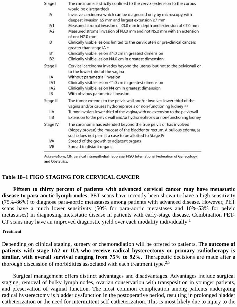
Table 18–1 FIGO STAGING FOR CERVICAL CANCER
Fifteen to thirty percent of patients with advanced cervical cancer may have metastaticdisease to para-aortic lymph nodes. PET scans have recently been shown to have a high sensitivity(75%-86%) to diagnose para-aortic metastases among patients with advanced disease. However, PETscans have a much lower sensitivity (50% for para-aortic metastases and 10%-53% for pelvicmetastases) in diagnosing metastatic disease in patients with early-stage disease. Combination PET-CT scans may have an improved diagnostic yield over each modality individually.1
Treatment
Depending on clinical staging, surgery or chemoradiation will be offered to patients. The outcome ofpatients with stage IA2 or IIA who receive radical hysterectomy or primary radiotherapy issimilar, with overall survival ranging from 75% to 92%. Therapeutic decisions are made after athorough discussion of morbidities associated with each treatment type.2,3
Surgical management offers distinct advantages and disadvantages. Advantages include surgicalstaging, removal of bulky lymph nodes, ovarian conservation with transposition in younger patients,and preservation of vaginal function. The most common complication among patients undergoingradical hysterectomy is bladder dysfunction in the postoperative period, resulting in prolonged bladdercatheterization or the need for intermittent self-catheterization. This is most likely due to injury to the
sensory and motor nerve supply of the bladder. Pulmonary embolism, lymphocyst formation, andfebrile morbidity from atelectasis and wound complications are less common, but not rarecomplications from surgery. Vesicovaginal or ureterovaginal fistulas occur in about 1% of cases.4
Some studies have also reported increased sexual dysfunction, including reduced arousal,decreased vaginal lubrication, and dyspareunia, among patients treated by radical hysterectomy.5
Lymphedema may occur in patients who undergo pelvic lymphadenectomy, particularly if they needpostoperative radiation therapy. Radiation therapy after radical pelvic surgery also increases the riskof developing bowel obstruction or fistulas.
Primary chemoradiation may be offered to patients at high risk for intra-operative complicationsto avoid surgical morbidity and mortality. However, radiation therapy may lead to vaginal shorteningas well as a decrease in lubrication and worsened sexual functioning. Other morbidities associatedwith radiation therapy include bowel obstruction, fistula formation, chronic radiation cystitis andproctitis, and ovarian failure. Complications from radiation therapy tend to occur more distant fromtreatment and are usually chronic, whereas complications from surgery usually occur in the immediatepostoperative period and do not usually persist.6-8
Patients with stage IB2 lesions (> 4 cm) are candidates for either surgery or primarychemoradiation. In a randomized trial of primary surgery versus radiation, 84% of patients withstage IB2 to IIA disease who underwent radical hysterectomy required postoperative adjuvantradiation therapy with more than 25% of patients experiencing severe morbidity. Based on theseresults, many centers counsel patients with stage IB2 or more advanced lesions to have primarychemoradiation.
Patients who undergo surgery may need postoperative chemoradiation if certain high-riskfeatures are present. If any of the following features are present, positive pelvic lymph nodes,positive parametria, or positive margins, patients should undergo concurrent radiation andcisplatin chemotherapy.
Approximately 25% of patients with stage IB1 disease will need adjuvant radiation therapy dueto the presence of certain intermediate risk factors (deep stromal invasion, LVSI, or lesion size > 4cm). In a trial randomizing patients with these risk factors between no further therapy and pelvicradiation, patients who received radiation therapy had a 46% decreased risk of recurrence (18%among patients receiving radiation therapy vs 31% in patients with no further treatment).9
Surgical Therapy
For patients with stage IA1 with LVSI, IA2, IB, and IIA cervical cancer, radical hysterectomy andpelvic lymph node dissection is the surgical treatment option. In a modified radical hysterectomy(type II), the uterine artery is ligated as it crosses the ureter. The medial half of the cardinal ligamentsand proximal uterosacral ligaments are resected. A more extensive dissection is done during a radicalhysterectomy (type III). In the type III radical hysterectomy, the uterine artery is ligated at its point oforigin at the superior vesical artery and the entire cardinal ligament is removed. The uterosacralligament is resected from the attachment to the posterior pelvis. In clinical practice, aspects of bothtypes II and III radical hysterectomies are done during each surgical case. More extensive radicalhysterectomy options have been described, involving resection of the superior vesical artery, portionsof the ureter, or the bladder. These operations are rarely done today to treat cervical cancer as patientswith larger cervical lesions are treated primarily with chemoradiation therapy.
Specific anatomic spaces are opened during the radical hysterectomy. The paravesical space isbordered by the pubic symphysis anteriorly, the cardinal ligament posteriorly, the obliteratedumbilical artery medially, and the obturator internus muscles laterally. The pararectal space is
bordered by the cardinal ligament anteriorly, the sacrum posteriorly, the rectum medially, and thehypogastric artery laterally. The rectovaginal space is developed by opening the peritoneum at thepouch of Douglas and gently opening the space between the vagina and rectum with blunt dissection.
A pelvic lymphadenectomy is performed to remove lymphatic tissue within the followinglandmarks: lateral to the ureter, medial to the psoas muscle, inferior to the middle of the common iliacartery, and superior to the deep circumflex vein. The obturator space is opened by retracting theexternal artery and vein and identifying the obturator nerve. All lymphatic and fatty tissue is thenremoved out of the obturator space.
For patients with larger stage IB lesions (> 4 cm) and patients with stage IIA disease who optedfor radical hysterectomy, there is an 80% chance that adjuvant radiation therapy will be recommendedbased on operative findings. Adjuvant radiation therapy, often with concurrent chemotherapy, isoffered when patients have positive margins on the radical hysterectomy specimen, or metastaticdisease in the lymph nodes. Adjuvant therapy is also considered when there are poor prognosticfactors, for example, lymphovascular space invasion, large primary tumor size, or deep cervicalstromal invasion, noted on the radical hysterectomy specimen.
Complications of a Radical Hysterectomy Urinary tract complications are frequently encounteredafter radical hysterectomy. It has been reported that up to 50% of radical hysterectomy cases havebladder dysfunction. The extensive dissection required for the surgery often results in denervation ofthe bladder and upper urethra. The majority of patients have normal bladder function within a year ofthe surgery. However, some patients require prolonged catheterization until bladder function returns.The use of either a suprapubic or transurethral catheter as well as intermittent self-catheterizationhave been described. Ureteral injury and fistula formation are not as common as bladder dysfunctionand can be minimized by careful surgical technique.
The risk of infection is no different than in the case of a traditional hysterectomy. Preoperativesingle-agent broad-spectrum antibiotic is all that is necessary for infection prophylaxis. Depending onthe length of surgery and blood loss, an additional dose of antibiotics may be required.
Venous thrombosis and pulmonary embolus are a particular concern to the surgeon. Given theextensive surgery, trauma to vein wall with lymphadenectomy, and immobility, these patients are atparticularly high risk for venous thrombosis formation. Patients should receive intermittent pneumaticcalf compression in the operating room, which is continued postoperatively until the patient is fullambulatory. High-risk patients may benefit from medical prophylaxis with either heparin or low-molecular-weight heparin.
Despite careful surgical technique, intraoperative hemorrhage may occur, and the surgeon mustbe prepared to deal with this complication. Blood products should be readily available at the time ofsurgery in the event that excessive bleeding occurs. The most frequent site of hemorrhage is venousbleeding from the pararectal fossa, presacral, and para-aortic regions. Unfortunately, as compared toarterial bleeding, venous bleeding is difficult to identify and is seldom improved with hypogastricartery ligation. A good understanding of anatomy is essential to try and minimize risk of hemorrhage.
Thankfully, nerve injury is not a frequent complication of a radical hysterectomy and rarelypermanent. The obturator nerve is most likely nerve to be injured and results in inability to adductlower extremities. Obturator nerve injury can occur with removal of obturator nodes and retractor use.Most nerve injuries can be minimized with proper patient placement/positioning, careful use of self-retaining retractors, careful surgical technique in dissection with good understanding of pelvicanatomy, and maintaining hemostasis.10
Radical Trachelectomy Patients with stage IA2 and IB1 (< 4 cm tumor size) lesions who desire
future fertility may elect to undergo a radical trachelectomy with pelvic lymphadenectomy. Acomplete lymphadenectomy is first performed to evaluate for the presence of metastatic disease.Subsequently, radical trachelectomy may be done to remove the cervix and the parametrial tissue. Theprocedure may be done abdominally, laparoscopically, or with a combined laparoscopic and vaginalportion. Often a cervical cerclage is placed in the newly formed exocervix, which is now locatedapproximately 1 cm below the lower uterine segment.11
Primary Radiation Therapy with Concurrent Chemotherapy For patients with stage IIB disease orhigher, the treatment is concurrent radiation and chemotherapy. This modality is also used for earlier-stage disease when the patient is not a surgical candidate. Typically, the patient receives externalbeam radiation to the pelvis followed by vaginal brachytherapy with tandem and ovoids placement.During radiation therapy, the patient will also receive concurrent platinum-based chemotherapy.Ideally, all radiation therapy is completed within a span of 56 days, as delays in treatment areassociated with a decreased overall survival.
When counseling a patient with early-stage disease who is a candidate for either chemoradiationtherapy or primary surgery, arguments in favor of primary surgery usually include better preservationof vaginal length and the avoidance of long-term risk of radiation (enteritis, bowel obstruction, etc),whereas the proponent of primary chemoradiation therapy cites the usual tolerability of treatment bymost patients and the avoidance of surgery with its attendant risks. However, stage for stage, in thosewith early disease (<4 cm), neither modality has been shown to have a survival advantage over theother.
Patients with Adenocarcinoma In general, cervical adenocarcinoma is treated similarly, stage forstage, as squamous carcinoma. However, in one study of patients with stage IB to IIA cervical cancerrandomized to primary radical surgery versus radiation therapy, those patients with adenocarcinomawho underwent primary surgery had improved progression-free and overall survival rates.
Pregnant Patients with Newly Diagnosed Cervical Cancer Delayed diagnosis of cervical cancer inpregnancy is common because symptoms of postcoital bleeding, vaginal discharge, vaginalbleeding, and pelvic pain are frequently associated with pregnancy. Most patients receive a Papsmear early on entry into prenatal care. If an abnormal Pap smear is found and the patient has novisible lesion, colposcopy without endocervical curettage is done. If the colposcopy is adequate and abiopsy reveals cervical intraepithelial neoplasia (CIN), the patient may undergo a vaginal delivery.Further evaluation and treatment are done at 6 weeks after delivery.
If the colposcopy is inadequate or the biopsy reveals microinvasion or possible carcinoma,cervical conization is needed in a similar manner as if the patient were not pregnant. Ideally,conizations are performed during the second trimester because the risk of fetal loss is less than 10%.If performed in the first trimester, the rate of fetal loss may be as high as 24%. In the third trimester,the risk of loss is less, but the risk of hemorrhage is significant.
A cervical conization in pregnancy should be performed only if necessary to diagnose or stage acervical cancer due to the high risk of potential complications, including premature labor,spontaneous abortion, infection, and hemorrhage. Pregnant patients may safely undergo MRI todetermine tumor volume, nodal enlargement, and potential metastasis.12 Consultation withgynecologic oncologists and maternal-fetal medicine specialists is highly recommended to helpmanage these difficult patients.
Careful counseling with the patient and her family must be undertaken for the management ofany patient diagnosed with cervical cancer during pregnancy as the treatment is dependent on the
stage of disease and the wishes of the mother. Patients with stage IA1 cervical cancer diagnosed byconization may be safely followed throughout pregnancy.13 Patients at less than 20 weeks’ gestationwho have more advanced lesions and do not desire continuation of the pregnancy may undergoradical surgery or radiation therapy with cisplatin as definitive treatment. Patients who havepregnancies exceeding 20 weeks’ gestation can generally be expectantly managed to await fetalmaturity. While the number of patients reported in the literature is small, the current data suggest thatdelaying treatment for stage I disease during pregnancy does not decrease survival as compared toundergoing immediate therapy.
Because patients with cervical cancer are at higher risk for hemorrhage or failure to dilate, someadvocate for cesarean section delivery. Among patients with advanced-stage disease, radiation therapyis the preferred treatment modality. Definitive data are not available regarding delays in treatment andpotential effects on advanced disease.
Comprehension Questions
18.1 A 39-year-old woman is noted to have an exophytic cervical lesion, which on biopsy revealsinvasive squamous cell carcinoma. Which of the following diagnostic aids is used in stagingcervical cancer?
A. MRIB. PET scanC. Chest x-rayD. CT scan of the abdomen and pelvis
18.2 A patient has a biopsy of a 5-cm cervical lesion showing squamous carcinoma. Arectovaginal examination shows obvious evidence of parametrial involvement on the left,but no sidewall involvement. The lesion invades the mucosa of the left vaginal fornix. A CTscan shows pathologically enlarged pelvic and para-aortic lymph nodes. Which of thefollowing is her cancer stage?
A. Stage IIAB. Stage IIABC. Stage IIIAD. Stage IVB
18.3 Which of the following is the most common complication of radical hysterectomy?A. Bladder atonyB. Pulmonary embolismC. Massive blood lossD. LymphedemaE. Small bowel obstruction
18.4 For which of the following cervical cancer patients is a radical hysterectomy andlymphadenectomy the most appropriate treatment?
A. A 46-year-old woman with a conization specimen showing 2-mm stromal invasionB. A 25-year-old nulligravida woman with a 5-cm cervical lesion with vaginal or parametrial
involvementC. A 23-year-old nulligravida woman with a cervical conization showing 5-mm stromal invasionD. A 25-year-old G1P1 woman with a cervical conization showing 3-mm stromal invasion and no
lymphovascular space invasion
ANSWERS
18.1 C. FIGO staging of cervical cancer is clinical and relies on a good pelvic examination.Radiographic evaluation is limited to chest radiography, barium enema, and intravenouspyelogram. In current practice, a CT scan, a PET scan, or combination PET-CT scan areoften done. Information obtained on these tests is included in the treatment planning for thepatient; however, it does not influence the stage of the patient.
18.2 B. The patient has a cervical cancer that involves the upper vagina (IIA), but also parametriabut not to the side wall (IIB). Thus, she has stage IIB disease. The information obtained onthe CT scan may influence treatment planning. In this case, she is a candidate for extended-field radiation therapy for the enlarged para-aortic lymph node.
18.3 A. The most common complication of radical hysterectomy is bladder atony. This is mostlikely due to bladder denervation from the extensive dissection.
18.4 C. Radical hysterectomy and lymph node dissection are appropriate therapies for patientswith stages IA2 to IIA cervical cancer. Patients with stage IA1 disease who havelymphovascular space invasion also need radical hysterectomy with lymph node dissectionas there is a risk of lymph node metastasis. A patient with 3-mm or less cervical stromalinvasion without lymphovascular space invasion is adequately treated with cervicalconization with negative margins or simple hysterectomy.
Clinical Pearls
See Table 1-2 for definition of level of evidence and strength of recommendation
After radical hysterectomy, adjuvant radiation therapy is given if the patient has certain riskfactors, including tumor size larger than 4 cm, the presence of lymphovascular space invasion,or deep stromal invasion (Level A).
The survival data for patients undergoing radical hysterectomy versus patients undergoingradiation therapy are similar in patients with stages IB to IIA disease. Patients with stage IIBor higher disease are treated with primary chemoradiation therapy (Level A).
Fifteen percent of patients with early disease will have lymphatic spread diagnosed afterradical hysterectomy and pelvic lymphadenectomy (Level B).
Patients who present with symptoms of postcoital bleeding should undergo completeevaluation, including Pap smear, endocervical curettage, and colposcopy (Level C).
All visible cervical lesion needs to be biopsied. A Pap smear is inadequate to rule out invasivecarcinoma of the cervix (Level C).
REFERENCES
1. Gold M. PET in cervical cancer—implications for “staging,” treatment planning, assessmentof prognosis and prediction of response. J Natl Compr Canc Netw. 2008;6:37-45.
2. Holtz D, Dunton C. Traditional management of invasive cervical cancer. Obstet Gynecol ClinNorth Am. 2002;29:645-657.
3. Landoni F, Maneo A, Colombo A, et al. Randomized study of radical surgery versusradiotherapy for stage IB-IIA cervical cancer. Lancet. 1997;350:535-540.
4. Covens A, Rosen B, Gibbons A, et al. Differences in the morbidity of radical hysterectomybetween gynecologic oncologists. Gynecol Oncol. 1993;51:39-45.
5. Bergmark K, Avall-Lundquist E, Dickman P, Hennignsohn L, Steineck G. Vaginal function andsexuality in women with a history of cervical cancer. N Engl J Med. 1999;340:1383-1389.
6. Gray H. Primary management of early stage cervical cancer (IA1-IB) and appropriate selectionof adjuvant therapy. J Natl Compr Canc Netw. 2008;6:47-51.
7. Delgado G, Bundy B, Zaino R, Sevin B, Creasman W, Major F. Prospective surgical-pathological study of disease-free interval in patients with stage IB cervical cancer afterradical hysterectomy and bilateral pelvic lymphadenectomy. Gynecol Oncol. 1990;38:352-357.
8. Sedlis A, Bundy B, Rotman M, Lentz S, Muderspach L, Zaino R. A randomized trial of pelvicradiation therapy versus no further therapy in selected patients with stage IB carcinoma of thecervix after radical hysterectomy and pelvic lymphadenectomy: a Gynecologic OncologyGroup Study. Gynecol Oncol. 1999;73:177-183.
9. Rose P, Bundy B, Watkins E, et al. Concurrent cisplatin-based radiotherapy and chemotherapyfor locally advanced cervical cancer. N Engl J Med. 1999;340:1144-1153.
10. Rock J, Jones H. Cancer of the cervix. In: Te Linde’s Operative Gynecology . 10th ed.Philadelphia, PA: Lippincott Williams & Wilkins; 2008:1277-1281.
11. Plante, M. Radical vaginal trachelectomy: a fertility preserving option for young women inearly stage cervical cancer. Gynecol Oncol. 2005;99:S143-S146.
12. Zanetta G, Pellegrino A, Vanzulli A, DiLelio A, Milani R, Mangioni C. Magnetic resonanceimaging of cervical cancer in pregnancy. Int J Gynecol Cancer. 1998;8:265-269.
13. Hannigan E, Whitehouse H, Atkinson W, Becker S. Cone biopsy during pregnancy. Obstetand Gynecol. 1982;60:450-455.
Case 19
A 33-year-old African American G1P0010 woman complains about increasing pelvic pressure andurinary frequency for the past 6 months. She reports that her menses have increased from lasting 5days to now 12 days in duration with heavier flow and quarter-size clots. She denies any dizziness,shortness of breath, or constipation. She has no chronic medical conditions. She has no children, andshe and her husband would like to have a child.
On examination, she is noted to be afebrile with normal vital signs. Her abdominal examinationreveals an irregular mass in the midline approximately 16 weeks in size and nontender. Pelvicexamination reveals a mobile, irregular mass in midline approximately 16 weeks in size andnontender. She has no adnexal tenderness, and no masses are palpated. A CBC reveals that she isanemic with hemoglobin of 6.8 g/dL. Her urine culture was negative.
What is the most likely diagnosis?
What is your diagnostic workup for this patient?
What is the best therapy for this patient?
What complications can result from the treatment?
ANSWERS TO CASE 19:Myomectomy
Summary: This is a 33-year-old woman, nulliparous, with menorrhagia, and a pelvic mass suggestiveof leiomyomata.
Most likely diagnosis: Uterine leiomyoma
Diagnostic workup: CBC, endometrial biopsy, prolactin, thyroid-stimulating hormone (TSH),follicle-stimulating hormone (FSH), pregnancy test, pelvic ultrasound
Best treatment: Myomectomy
Complications: Hysterectomy, recurrence, adhesions, longer hospitalization, continuedinfertility
ANALYSIS
Objectives
1. List indications for myomectomy.2. Describe several routes for myomectomy.3. List possible complications resulting from myomectomy.
Considerations
This is a 33-year-old nulliparous woman whose symptoms and physical findings are consistent withuterine fibroids. She complains of menorrhagia (with resultant anemia) and urinary frequency. Hersymptoms include both menorrhagia (with resultant anemia) and urinary frequency. Because of herasymptomatic anemia, the patient is a candidate for medical therapy and iron supplementation.Medical therapy options include the use of medroxyprogesterone acetate depot (Depo-Provera), oralcontraceptive pills, and GnRH-a. Of the options listed, GnRH-agonist would be the most effectivetreatment for her uterine fibroids. However, because of limited duration of GnRH usage (6 months)and likely recurrence after treatment, medical management would not be the best management optionat this time. Given her age, desired fertility, and symptoms, the patient is a candidate for amyomectomy. Alternative treatments for fibroids including uterine artery embolization andultrasonography-focused ablation are not options for this patient given her desired fertility.
APPROACH TO Myomectomy
CLINICAL APPROACH
Leiomyomas (also fibroids or myomas) are benign monoclonal tumors originating from a singlesmooth muscle cell that has undergone a chromosomal mutation. They are a bundle of smooth musclecells and fibrous tissue surrounded by a fibrous capsule. The leiomyomas are symptomatic in 25% to50% of women, but upon pathological review of hysterectomy specimen, the prevalence may be ashigh as 80%. Risk factors for the development of leiomyoma include increasing age, early menarche,low parity, tamoxifen use, obesity, and some studies show high-fat diet. Myomas tend to grow andbecome more symptomatic in nulliparous women. African American women have the highestincidence of fibroids. Hispanic, Asian, and Caucasian women have similar rates for the developmentof leiomyoma. There is a familial tendency to develop fibroids. Smoking has been associated with adecrease in incidence of leiomyoma. Leiomyomas can grow in any part of the body that has smoothmuscle; the uterus is the most common organ in the pelvis to develop fibroids. Leiomyoms can befound in the fallopian tubes, round ligament, and about 5% of the time on the cervix.
Uterine leiomyoma can be diagnosed with 95% certainty with physical examination alone. Onpalpation, a uterus feels enlarged, firm, and irregular. Myomas can grow laterally and may inhibit thepalpation of the adnexa. Ultrasound is recommended when the patient is obese and/or when adnexalpathology cannot be ruled out with physical examination alone.
Most leiomyoms are asymptomatic and do not require treatment. The number, size, and locationof the myoma can produce different symptoms. Symptoms include pelvic pressure, dysmenorrhea,menorrhagia, urinary frequency, and constipation. Fibroids can cause hydroureter or hydronephrosisand have been linked to infertility. Therapeutic options include medical management with progestins(norethindrone, medrogestone, medroxyprogesterone acetate), antiprogestins (mifepristone), andgonadotropin-releasing hormone analogues. Surgical procedures include myomectomy andhysterectomy. Other methods for treatment of fibroids include uterine artery embolization, high-frequency ultrasonography, laser treatment, cryotherapy, and thermoablation. Approximately 30% ofhysterectomies list symptomatic uterine fibroids as the primary indication. The choice of thetreatment method should be based on various medical and social characteristics of the patient. Thesefactors include age, parity, desire for childbearing, severity of symptoms, size and number ofleiomyoms, location of the myoma, other medical conditions, suspicion of malignancy, proximity of
menopause, and desire to preserve uterus.1
In the presence of leiomyoms, myomectomy may be the management option if the patient desiresto retain her uterus, if there is a single pedunculated myoma, or if the presence of myoma is thought tobe the cause of infertility or repeated pregnancy losses. Women with symptomatic uterine fibroidswho no longer desire to bear children may be best treated with a hysterectomy, the definitivetreatment for uterine leiomyoma. For women who desire future childbearing or who want to preservetheir uterus, myomectomy may be the best management.
Myomectomy only removes the portion of the myoma that is visible and accessible.Occasionally, small myoma may be retained intentionally. If myomectomy is the chosen therapy, themyoma can be removed via an abdominal incision, laparoscopy, or hysteroscopy. Before proceedingwith the removal of the myoma, appropriate evaluation must be done for the presence of a pelvic massand abnormal uterine bleeding. Anemia may be treated with GnRH-a and iron supplementation priorto the myomectomy. GnRH-a can also decrease the size of the leiomyoma, but the enucleation of themyoma is more difficult due to a less distinct plane between the myoma and the normal uterus.Myomectomy is associated with more adhesions and longer hospital stays than a hysterectomy.Hysterectomy is associated with more urinary tract injuries. Contraindications to a myomectomyinclude pregnancy, advanced adnexal disease, malignancy, and the situation where removal of themyoma would result in the significant reduction of the endometrial surface causing the uterus not tobe functional.2-9
Abdominal Myomectomy
An abdominal myomectomy is the route of choice when there are numerous myomas in multiplelocations (subserosal, intramural, submucosal). Multiple myomectomy is usually more difficult andtime consuming than a hysterectomy. Intraoperative blood loss for myomectomy correlates with theuterine size prior to surgery, total weight of the myomas removed, and operating time. Resolution ofmenorrhagia and pelvic pressure is overall 81%. Several studies show that the morbidity of the twoprocedures is similar. The risk of unexpected hysterectomy when performing a myomectomy is lessthan 1%. The hysterectomy can result from leiomyomatosis or from complications of themyomectomy. Recurrence of myomas is of concern after myomectomy. Studies using transvaginalultrasound show recurrence of approximately 51% at 5 years. Other studies show that clinicallysignificant myoma recurrence is 10% at 5 years, with one-third of those patients eventuallyundergoing a hysterectomy. The rate of recurrence of myomas depends on the number of myomasremoved. The recurrence risk is 11% for a single myoma and 26% for multiple myomas. Whenmyomectomy is performed in patients desiring fertility, pelvic adhesions, and dissection in the area ofthe interstitial portion of the fallopian may result in postoperative infertility.10-12
A careful pelvic examination while the patient is under anesthesia will allow for a better idea ofwhich type of skin incision to make. After the appropriate skin incision is made and peritoneumentered, the uterus and adjacent adnexal structures are to be evaluated. When multiple fibroids arenoted, the uterine incision should be the one that allows the exposure to multiple fibroids minimizingthe number of uterine incisions. To decrease intraoperative blood loss, uterine tourniquets orinjectable intrauterine vasoconstrictive agents can be used. Multiple studies have demonstrated theeffectiveness of vasoconstrictive agents in decreasing the need for blood transfusion13 and decreasingintraoperative blood loss.14 When comparing mechanical occlusion and injection of vasoconstrictiveagents, Ginsberg et al.15 found no significant differences in blood loss or transfusion requirements
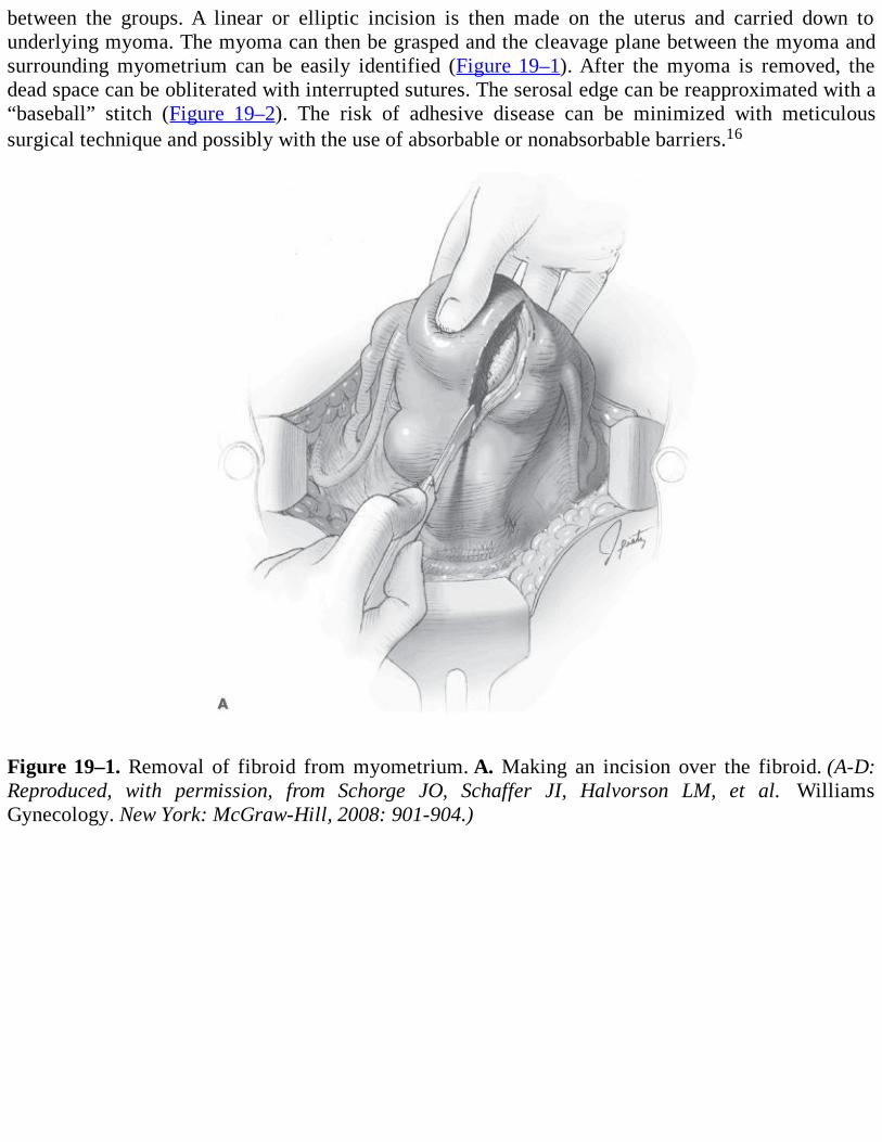
between the groups. A linear or elliptic incision is then made on the uterus and carried down tounderlying myoma. The myoma can then be grasped and the cleavage plane between the myoma andsurrounding myometrium can be easily identified (Figure 19–1). After the myoma is removed, thedead space can be obliterated with interrupted sutures. The serosal edge can be reapproximated with a“baseball” stitch (Figure 19–2). The risk of adhesive disease can be minimized with meticuloussurgical technique and possibly with the use of absorbable or nonabsorbable barriers.16
Figure 19–1. Removal of fibroid from myometrium. A. Making an incision over the fibroid. (A-D:Reproduced, with permission, from Schorge JO, Schaffer JI, Halvorson LM, et al. WilliamsGynecology. New York: McGraw-Hill, 2008: 901-904.)
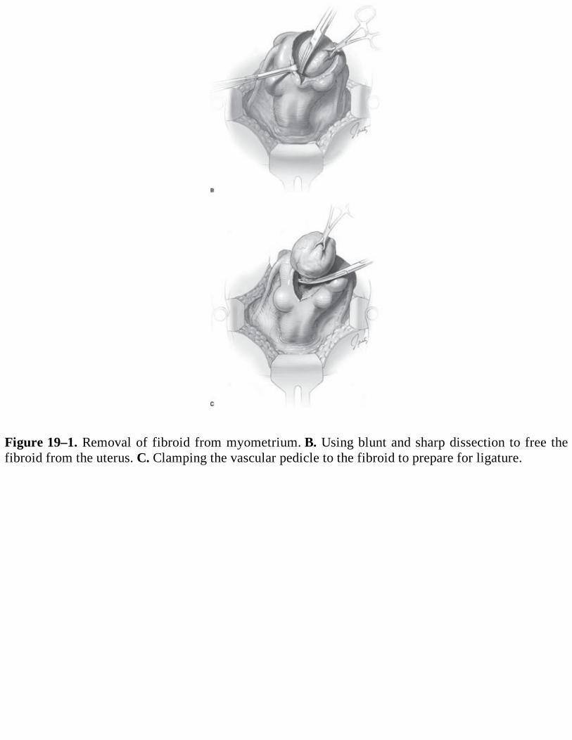
Figure 19–1. Removal of fibroid from myometrium. B. Using blunt and sharp dissection to free thefibroid from the uterus. C. Clamping the vascular pedicle to the fibroid to prepare for ligature.
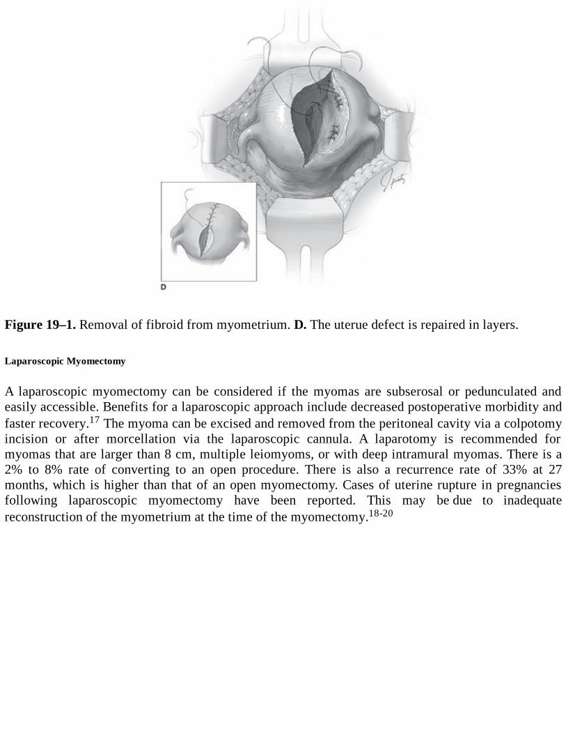
Figure 19–1. Removal of fibroid from myometrium. D. The uterue defect is repaired in layers. Laparoscopic Myomectomy
A laparoscopic myomectomy can be considered if the myomas are subserosal or pedunculated andeasily accessible. Benefits for a laparoscopic approach include decreased postoperative morbidity andfaster recovery.17 The myoma can be excised and removed from the peritoneal cavity via a colpotomyincision or after morcellation via the laparoscopic cannula. A laparotomy is recommended formyomas that are larger than 8 cm, multiple leiomyoms, or with deep intramural myomas. There is a2% to 8% rate of converting to an open procedure. There is also a recurrence rate of 33% at 27months, which is higher than that of an open myomectomy. Cases of uterine rupture in pregnanciesfollowing laparoscopic myomectomy have been reported. This may be due to inadequatereconstruction of the myometrium at the time of the myomectomy.18-20
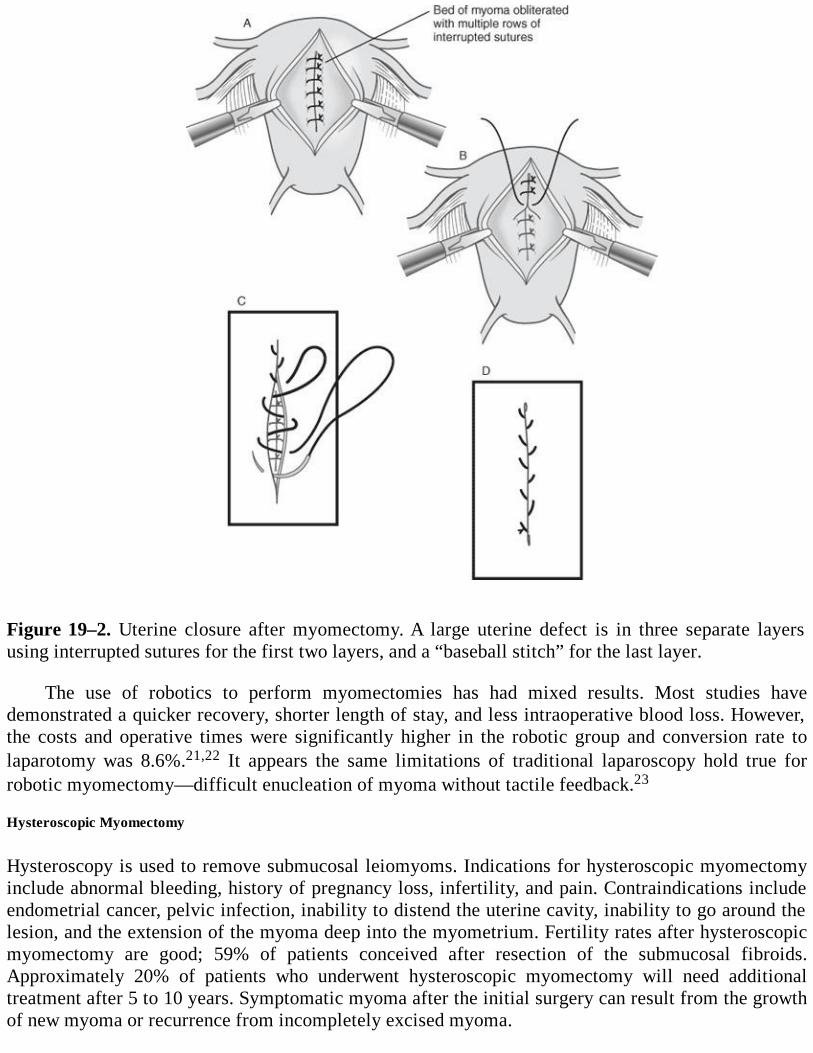
Figure 19–2. Uterine closure after myomectomy. A large uterine defect is in three separate layersusing interrupted sutures for the first two layers, and a “baseball stitch” for the last layer.
The use of robotics to perform myomectomies has had mixed results. Most studies havedemonstrated a quicker recovery, shorter length of stay, and less intraoperative blood loss. However,the costs and operative times were significantly higher in the robotic group and conversion rate tolaparotomy was 8.6%.21,22 It appears the same limitations of traditional laparoscopy hold true forrobotic myomectomy—difficult enucleation of myoma without tactile feedback.23
Hysteroscopic Myomectomy
Hysteroscopy is used to remove submucosal leiomyoms. Indications for hysteroscopic myomectomyinclude abnormal bleeding, history of pregnancy loss, infertility, and pain. Contraindications includeendometrial cancer, pelvic infection, inability to distend the uterine cavity, inability to go around thelesion, and the extension of the myoma deep into the myometrium. Fertility rates after hysteroscopicmyomectomy are good; 59% of patients conceived after resection of the submucosal fibroids.Approximately 20% of patients who underwent hysteroscopic myomectomy will need additionaltreatment after 5 to 10 years. Symptomatic myoma after the initial surgery can result from the growthof new myoma or recurrence from incompletely excised myoma.
To minimize the risk of uterine perforation, the submucosal fibroid should be evaluated closelyfor myometrial involvement. When the fibroid invades the myometrium, there is a greater risk ofuterine perforation and a simultaneous laparoscopy/hysteroscopy can be performed. After theappropriate operative hysteroscopic distending medium (sorbitol, Hyskon [dextran, high molecularweight]) is infused, the endometrial cavity can be evaluated for the extent of myoma involvement.Using the resectoscope and a cutting current of 60 to 80 W, the myoma can be sequentially resected.Care must be taken to only apply current when resectoscope loop is being withdrawn from the fundus(never toward fundus due to increased perforation risk). Careful attention must be paid to distendingmedia in/out and deficits.
Many authors recommend a hysterosalpingogram 4 months after a myomectomy to evaluate theuterine cavity and the fallopian tubes and a delay of 4 to 6 months after surgery before attempting toconceive. Previously, a cesarean delivery was recommended only if the endometrium was enteredduring the myomectomy. However, if extensive dissection of the myometrium is carried out in theprocess of a myomectomy, even if the endometrium in not disturbed, a cesarean delivery is usuallyadvised following a pregnancy. Cesarean delivery is not recommended after the removal of apedunculated fibroid or after small hysteroscopic resection.24
Comprehension Questions
19.1 A 43-year-old white G3P3 woman has symptoms of pelvic pressure and abnormal uterinebleeding. A pelvic examination reveals a 16-week-size irregular uterus. An ultrasound showsa 10-cm pedunculated leiomyoma from the fundus of the uterus. No adnexal masses werenoted. Which of the following should be the next step?
A. Endometrial biopsyB. HysterectomyC. Abdominal myomectomyD. Laparoscopic myomectomy
19.2 Which of the following cases has a higher chance of recurrence of leiomyoma?A. A 32-year-old woman who underwent hysteroscopic myomectomy for a single pedunculated
fibroidB. A 44-year-old woman who underwent a laparoscopic resection of a 5-cm subserosal fibroidC. A 32-year-old woman who underwent an abdominal myomectomy for an 8-cm myometrial
fibroidD. A 44-year-old woman who underwent an abdominal myomectomy for five uterine fibroids
19.3 Which of the following points is most important to be emphasized in counseling a patientundergoing myomectomy for infertility?
A. Complications of myomectomy can lead to a hysterectomy.B. Adhesions caused by the myomectomy surgery usually can be prevented.C. Patients with infertility are usually cured after a myomectomy.D. The location of fibroid and its resection do not affect fertility.
ANSWERS
19.1 A. Before any therapeutic measures are taken, it is critical to complete a full diagnosticevaluation. It is possible that the abnormal uterine bleeding is caused by leiomyoma;
however, endometrial pathology must be ruled out in this patient who is older than 35 yearswith abnormal uterine bleeding. This patient’s symptoms as well as physical examinationare consistent with uterine leiomyoms. If she did not wish to retain her uterus, hysterectomywould be the definitive treatment of choice. If she desired future children, myomectomywould be the best option. Since this is a pedunculated fibroid, laparoscopic removal can beconsidered.
19.2 D. The recurrence rate of leiomyoma depends on the number of myomas removed at theinitial myomectomy. There is 11% recurrence after one myoma is removed. There is 26%recurrence rate after multiple myomas are removed.
19.3 A. Submucosal leiomyoms are most often associated with infertility and recurrentpregnancy losses. Myomectomy can improve fertility rates if all other causes of infertilityhave been ruled out. Risk of myomectomy includes formation of adhesions and blockage ofthe fallopian tube. A conservative surgery for uterine preservation can lead to a definitivehysterectomy.
Clinical Pearls
See Table 1-2 for definition of level of evidence and strength of recommendation
Improvement in menorrhagia, pelvic pressure, and infertility has been noted aftermyomectomy (Level A).
Hysterectomy is the only definitive cure for leiomyoms. Myomectomy has the risk ofrecurrence (Level B).
The risks of myomectomy include possible hysterectomy, recurrence, adhesions, longerhospitalization, and continued infertility (Level B).
After significant disturbance of the myometrium during a myomectomy, a cesarean deliveryshould be offered in the event of a pregnancy even if the endometrium was not disturbed(Level C).
Myomectomy should be performed if the patient desires to retain her uterus or childbearingfunction (Level C).
Laparoscopic and hysteroscopic myomectomy should be carried out only by experiencedsurgeons (Level C).
REFERENCES
1. Buttran VC, Reiter RC. Uterine leiomyomata: etiology, symptomatology, and management.Fertil Steril. 1981;36:433-445.
2. Acien P, Quereda F. Abdominal myomectomy: results of a simple operative technique. Fertil
Steril. 1996;65:41-51.
3. American College of Obstetricians and Gynecologists. Surgical Alternatives to Hysterectomyin the Management of Leiomyomas. ACOG Practice Bulletin No. 16, May 2000. WashingtonDC.
4. Cramer SF, Patel A. The frequency of uterine leiomyomas. Am J Clin Pathol. 1990;94:435-438.
5. Ecker JL, Foster JT, Friedman AJ. Abdominal hysterectomy or abdominal myomectomy forsymptomatic leiomyoma: a comparison of preoperative demography and postoperativemorbidity. J Gynecol Surg. 1995;1:11-18.
6. Hillis SD, Marchbanks PA, Peterson HB. Uterine size and risk of complications among womenundergoing abdominal hysterectomy for leiomyomas. Obstet Gynecol. 1996;87:539-543.
7. Iverson RE Jr, Chelmow D, Strohbehn K, Waldman L, Evantash EG. Relative morbidity ofabdominal hysterectomy and myomectomy for management of uterine leiomyomas. ObstetGynecol. 1996;88:415-419.
8. Katz VL, Lentz GM, Lobo RA, Gershenson DM. Benign gynecologic lesions. In:Comprehensive Gynecology. 5th ed. Philadelphia, PA: Mosby; 2007:441-450.
9. Wallach EE, Vlahos NF. Uterine myomas: an overview of development, clinical features, andmanagement. Obstet Gynecol. 2004;104:393-406.
10. Fedele L, Parazzini F, Luchini L, Mezzopane R, Tozzi L, Villa L. Recurrence of fibroids aftermyomectomy: a transvaginal ultrasonographic study. Hum Reprod. 1995;10:1795-1796.
11. Fauconnier A, Chapron C, Babaki-Fard K, Dubuisson JB. Recurrence of leiomyomata aftermyomectomy. Hum Reprod Update. 2000;6:595-602.
12. Nezhat FR, Roemisch M, Nezhat Ch, Seidman DS, Nezhat CR. Recurrence rate afterlaparoscopic myomectomy. J Am Assoc Gynecol Laparosc. 1998;5:237-240.
13. Dillon T. Control of blood loss during gynecologic surgery. Obstet Gynecol. 1962;19:428.
14. Frederick J, Fletcher A, Simeon D, et al. Intramyometrial vasopressin as a hemostatic agent.BJOG. 1994;101:435.
15. Ginsberg E, Benson C, Garfield J, et al. The effect of operative technique and uterine size onblood loss during myomectomy: a prospective randomized study. Fertil Steril. 1993;60:956.
16. Rock J, Jones H. Leiomyomata uteri and myomectomy. In: Te Linde’s Operative Gynecology .10th ed. Philadelphia, PA: Lippincott Williams & Wilkins; 2008:707-721.
17. Mais V, Ajossa S, Guerrriero S, Mascia M, Solla E, Melis GB. Laparoscopic versusabdominal myomectomy: a prospective, randomized trial to evaluate benefits in earlyoutcome. Am J Obstet Gynecol. 2005;174:654-658.
18. Rock J, Jones H. Diagnostic and operative laparoscopy. In: Te Linde’s Operative Gynecology .10th ed. Philadelphia, PA: Lippincott Williams & Wilkins; 2008:330-331.
19. Chu J, Hu Y, Xia-Chan C, et al. Laparoscopy versus open myomectomy—a meta-analysis ofrandomized controlled trials. Eur J Obstet Gynecol Reprod Biol. 2009;145:14-21.
20. Seinera P, Arisio R, Decko A, Farina C, Crana F. Laparoscopic myomectomy: indications,surgical technique and complication. Hum Reprod. 1997;12:1927-1930.
21. Advincula AP, Xu X, Goudeau S, Ransom SB. Robotic-assisted laparoscopic myomectomyversus abdominal myomectomy: a comparison of short-term surgical outcomes and immediatecosts. J Minim Invasive Gynecol. 2007;14:698-705.
22. Advincula AP, Song A, Burke W, Reynolds RK. Preliminary experience with robot assistedlaparoscopic myomectomy. J Am Assoc Gynecol Laparosc. 2004;11:511-518.
23. Visco AG, Advincula AP. Robotic gynecologic surgery. Obstet Gynecol. 2008; 112:1369-1384.
24. Ubaldi F, Tournaye H, Camus M, Van der Pas H, Gepts E, Devroey P. Fertility afterhysteroscopic myomectomy. Hum Reprod Update. 1995;1:81-90.
Case 20
A 46-year-old G3P3 woman is coming into the office for follow-up for menorrhagia. She originallypresented to her gynecologist 2 months previously with complaints of heavy vaginal bleeding forseveral years, and the loss of a moderate amount of urine with Valsalva and cough; the loss of urinewas reported as bothersome to her and limited her activities. She denied dysuria and urgencysymptoms. She required the use of a sanitary pad to protect her clothing from her incontinence. Onexamination, she was found to have an enlarged uterus (18 cm in size) with multiple fibroids. A cottontip applicator in the urethra moved 70 degrees with Valsalva, and the patient was noted to have a smallcystocele and loss of urine with Valsalva. An endometrial biopsy was performed and revealed benignsecretory endometrium; aa urine culture was negative for growth. Medical therapy with oralcontraceptive pills had been initiated for her menorrhagia, and she was instructed on pelvic floorexercises. The patient reported that she complied with both the OCP’s and pelvic floor exercises,which provided no relief of symptoms. She is mildly anemic (hemoglobin 11.5 g/dL) requiring oraliron supplements. A urodynamic study is performed and reveals genuine stress incontinence. Becauseof the size of her uterus, a total abdominal hysterectomy is planned for definitive therapy. The patientis also counseled about a surgery for treatment of her urinary stress incontinence.
What are two urethropexy surgical therapies that could be performed for her urinaryincontinence at the time of her abdominal hysterectomy?
What is the main difference between the two most common urethropexy surgeries?
ANSWERS TO CASE 20:Urethropexy
Summary: This is a 46-year-old woman with symptomatic uterine fibroids not responsive to medicaltherapy desiring definitive therapy with a total abdominal hysterectomy. She also has stress urinaryincontinence, for which she desires surgical therapy at the same time as the hysterectomy.
Urethropexy procedures: Marshall-Marchetti-Krantz (MMK) and Burch procedures.Additionally, midurethral sling procedures could also be done.
Difference between MMK and Burch: The primary difference between the MMK and Burchprocedure is the points of attachment of the periurethral endopelvic fascia. The MMK was thefirst retropubic urethropexy procedure described and involves attachment of endopelvic fasciato the periosteum of the pubic symphysis. The Burch procedure attaches the endopelvic fasciato Cooper ligament.
ANALYSIS
Objectives
1. Describe the surgical principles of both the MMK and Burch procedures.
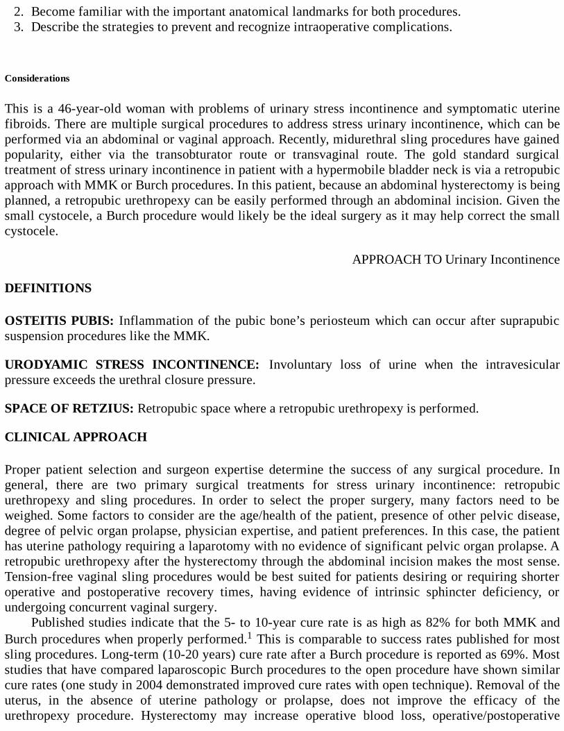
2. Become familiar with the important anatomical landmarks for both procedures.3. Describe the strategies to prevent and recognize intraoperative complications.
Considerations
This is a 46-year-old woman with problems of urinary stress incontinence and symptomatic uterinefibroids. There are multiple surgical procedures to address stress urinary incontinence, which can beperformed via an abdominal or vaginal approach. Recently, midurethral sling procedures have gainedpopularity, either via the transobturator route or transvaginal route. The gold standard surgicaltreatment of stress urinary incontinence in patient with a hypermobile bladder neck is via a retropubicapproach with MMK or Burch procedures. In this patient, because an abdominal hysterectomy is beingplanned, a retropubic urethropexy can be easily performed through an abdominal incision. Given thesmall cystocele, a Burch procedure would likely be the ideal surgery as it may help correct the smallcystocele.
APPROACH TO Urinary Incontinence
DEFINITIONS
OSTEITIS PUBIS: Inflammation of the pubic bone’s periosteum which can occur after suprapubicsuspension procedures like the MMK.
URODYAMIC STRESS INCONTINENCE: Involuntary loss of urine when the intravesicularpressure exceeds the urethral closure pressure.
SPACE OF RETZIUS: Retropubic space where a retropubic urethropexy is performed.
CLINICAL APPROACH
Proper patient selection and surgeon expertise determine the success of any surgical procedure. Ingeneral, there are two primary surgical treatments for stress urinary incontinence: retropubicurethropexy and sling procedures. In order to select the proper surgery, many factors need to beweighed. Some factors to consider are the age/health of the patient, presence of other pelvic disease,degree of pelvic organ prolapse, physician expertise, and patient preferences. In this case, the patienthas uterine pathology requiring a laparotomy with no evidence of significant pelvic organ prolapse. Aretropubic urethropexy after the hysterectomy through the abdominal incision makes the most sense.Tension-free vaginal sling procedures would be best suited for patients desiring or requiring shorteroperative and postoperative recovery times, having evidence of intrinsic sphincter deficiency, orundergoing concurrent vaginal surgery.
Published studies indicate that the 5- to 10-year cure rate is as high as 82% for both MMK andBurch procedures when properly performed.1 This is comparable to success rates published for mostsling procedures. Long-term (10-20 years) cure rate after a Burch procedure is reported as 69%. Moststudies that have compared laparoscopic Burch procedures to the open procedure have shown similarcure rates (one study in 2004 demonstrated improved cure rates with open technique). Removal of theuterus, in the absence of uterine pathology or prolapse, does not improve the efficacy of theurethropexy procedure. Hysterectomy may increase operative blood loss, operative/postoperative
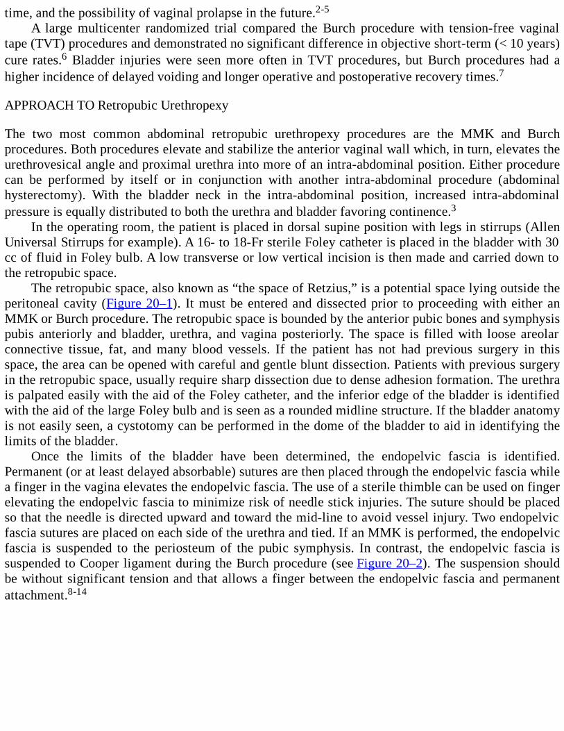
time, and the possibility of vaginal prolapse in the future.2-5
A large multicenter randomized trial compared the Burch procedure with tension-free vaginaltape (TVT) procedures and demonstrated no significant difference in objective short-term (< 10 years)cure rates.6 Bladder injuries were seen more often in TVT procedures, but Burch procedures had ahigher incidence of delayed voiding and longer operative and postoperative recovery times.7
APPROACH TO Retropubic Urethropexy
The two most common abdominal retropubic urethropexy procedures are the MMK and Burchprocedures. Both procedures elevate and stabilize the anterior vaginal wall which, in turn, elevates theurethrovesical angle and proximal urethra into more of an intra-abdominal position. Either procedurecan be performed by itself or in conjunction with another intra-abdominal procedure (abdominalhysterectomy). With the bladder neck in the intra-abdominal position, increased intra-abdominalpressure is equally distributed to both the urethra and bladder favoring continence.3
In the operating room, the patient is placed in dorsal supine position with legs in stirrups (AllenUniversal Stirrups for example). A 16- to 18-Fr sterile Foley catheter is placed in the bladder with 30cc of fluid in Foley bulb. A low transverse or low vertical incision is then made and carried down tothe retropubic space.
The retropubic space, also known as “the space of Retzius,” is a potential space lying outside theperitoneal cavity (Figure 20–1). It must be entered and dissected prior to proceeding with either anMMK or Burch procedure. The retropubic space is bounded by the anterior pubic bones and symphysispubis anteriorly and bladder, urethra, and vagina posteriorly. The space is filled with loose areolarconnective tissue, fat, and many blood vessels. If the patient has not had previous surgery in thisspace, the area can be opened with careful and gentle blunt dissection. Patients with previous surgeryin the retropubic space, usually require sharp dissection due to dense adhesion formation. The urethrais palpated easily with the aid of the Foley catheter, and the inferior edge of the bladder is identifiedwith the aid of the large Foley bulb and is seen as a rounded midline structure. If the bladder anatomyis not easily seen, a cystotomy can be performed in the dome of the bladder to aid in identifying thelimits of the bladder.
Once the limits of the bladder have been determined, the endopelvic fascia is identified.Permanent (or at least delayed absorbable) sutures are then placed through the endopelvic fascia whilea finger in the vagina elevates the endopelvic fascia. The use of a sterile thimble can be used on fingerelevating the endopelvic fascia to minimize risk of needle stick injuries. The suture should be placedso that the needle is directed upward and toward the mid-line to avoid vessel injury. Two endopelvicfascia sutures are placed on each side of the urethra and tied. If an MMK is performed, the endopelvicfascia is suspended to the periosteum of the pubic symphysis. In contrast, the endopelvic fascia issuspended to Cooper ligament during the Burch procedure (see Figure 20–2). The suspension shouldbe without significant tension and that allows a finger between the endopelvic fascia and permanentattachment.8-14
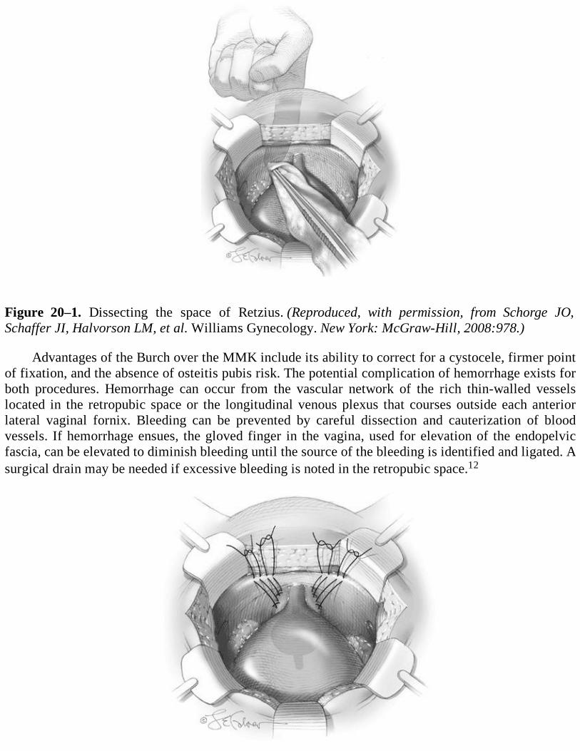
Figure 20–1. Dissecting the space of Retzius. (Reproduced, with permission, from Schorge JO,Schaffer JI, Halvorson LM, et al. Williams Gynecology. New York: McGraw-Hill, 2008:978.)
Advantages of the Burch over the MMK include its ability to correct for a cystocele, firmer pointof fixation, and the absence of osteitis pubis risk. The potential complication of hemorrhage exists forboth procedures. Hemorrhage can occur from the vascular network of the rich thin-walled vesselslocated in the retropubic space or the longitudinal venous plexus that courses outside each anteriorlateral vaginal fornix. Bleeding can be prevented by careful dissection and cauterization of bloodvessels. If hemorrhage ensues, the gloved finger in the vagina, used for elevation of the endopelvicfascia, can be elevated to diminish bleeding until the source of the bleeding is identified and ligated. Asurgical drain may be needed if excessive bleeding is noted in the retropubic space.12
Figure 20–2. Placement of sutures in the pubovesical fascia to the Cooper ligament. (Reproduced,with permission, from Schorge JO, Schaffer JI, Halvorson LM, et al. Williams Gynecology. New York:McGraw-Hill, 2008:979.)
Postsurgical voiding dysfunction can be seen with either procedure. The development of detrusorinstability occurs in 10% to 15% of patients15,16 and once infection has been ruled out, should beaddressed. Some degree of urinary retention is seen in 15% to 20% of patients. This often can bemanaged with prolonged bladder drainage and rarely with removal of suspension sutures. Theincidence of urinary retention can be reduced intraoperatively by allowing a space (1-2 cm) betweenthe suspended endopelvic fascia and fixation point.2
Osteitis pubis is a rare complication of the MMK procedure occurring in 1% to 2% of allpatients.15 This painful condition is caused by an inflammatory reaction (noninfectious) in theperiosteum of the pubic bone, resulting in pain (osteomyelitis is the infectious form). Whensupportive care is not sufficient, the suspension sutures may need removal.
Comprehension Questions
20.1 A third-year resident is being instructed in the surgical management of a patient withurinary incontinence. How many total sutures (both sides added together) should be placedin the endopelvic fascia during a Burch or MMK procedure?
A. OneB. TwoC. FourD. As many as possible
20.2 A 35-year-old woman underwent an MMK urethropexy procedure and developed symptomsconsistent with osteitis pubis. Which of the following about osteitis pubis is most accurate?
A. Often occurs after a Burch Procedure.B. Occurs in the immediate postoperative period.C. Is a delayed complication (> 2 months).D. Pain symptoms are constant with no aggravating or alleviating factors.
20.3 While performing a Burch procedure, a significant amount of bleeding is encountered in thespace of Retzius. The area has blood pooling and it is difficult to detect the origin of thebleeding. What is your first course of action?
A. Place a surgical drain.B. Elevate the endopelvic fascia with your finger in the vagina.C. Place a suture in the general area of the bleeding.D. Obtain vascular surgeon intraoperative consultation.
ANSWERS
20.1 C. Two to three sutures are recommended through the endopelvic fascia on each side of theurethra (total of four). A higher failure rate has been observed with one suture compared totwo sutures. The placement of too many sutures can lead to urinary retention.
20.2 C. A patient with osteitis pubis usually presents with pubic pain and a “waddling” gate 2months after an MMK. The pain is usually abrupt in nature, may radiate down the inneraspect of the thigh, and is aggravated by activity. In the absence of an infection, the patientis treated with bedrest, steroids, and analgesics. If symptomatic treatment is insufficient, thesuspension sutures may need removal.
20.3 B. When entering the space of Retzius, careful meticulous dissection is required to avoidsubstantial bleeding. However, if hemorrhage ensues after stitch is placed in the endopelvicfascia or with dissection, the endopelvic fascia with finger in the vagina should be gentlyelevated. This maneuver usually decreases the bleeding and allows for better visibility. Oncethe vessel is identified, it can be suture-ligated or cauterized under direct visualization.Blindly placing sutures in the space of Retzius can lead to injury to the bladder or urethra.
Clinical Pearls
See Table 1-2 for definition of level of evidence and strength of recommendation
Short-term outcomes of midurethral slings are similar to the traditional retropubic approach(Level A).
The development of de novo detrusor instability occurs in about 15% of patients after aretropubic urethropexy (Level B).
The placement of two to three permanent sutures in the endopelvic fascia during a Burchprocedure has been shown to be superior to the placement of just one (Level B).
In the event of hemorrhage in space of Retzius, elevation of the endopelvic fascia decreasesthe amount of bleeding so that the injured vessels can be visualized and ligated (Level C).
REFERENCES
1. Feyereisl J, Dreher E, Haenggi W, et al. Long-term results after Burch colposuspension. AM JObstet Gynecol. 1994;171:647.
2. American College of Obstetricians and Gynecologists. Urinary Incontinence in Women. ACOGPractice Bulletin No. 63. Washington DC; 2005.
3. Katz VL, Lentz GM, Lobo RA, Gershenson DM. Urogynecology. In: ComprehensiveGynecology. 5th ed. Philadelphia, PA: Mosby; 2007:537-563.
4. Langer R, Ron-El R, Neuman N, Herman A, Bukovsky I, Caspi E. The value of simultaneoushysterectomy during Burch colposuspension for urinary incontinence. Obstet Gynecol.1988;72:866-869.
5. Lapitan MC, Cody DJ, Grant AM. Open retropubic colposuspension for urinary incontinence inwom en. Cochrane Database Syst Rev. 2003;(1):CD009212. [DOI: 10.1002/14651858.
CD002912].
6. Ward K, Hilton P. Prospective multicentre randomized trial of tension-free vaginal tape andcolposuspension as primary treatment for stress incontinence. United Kingdom and IrelandTension-Free Vaginal Tape Trial Group. BMJ. 2002;325:67-70.
7. Alcaly M, Monga A, Stanton SL. Burch colposuspension: a 10-20 year follow up [publishederratum appears in Br J Obstet Gynaecol. 1996;103:290]. Br J Obstet Gynaecol. 1995;102:740-745.
8. Ankardal M, Ekerydh A, Crafoord K, Milsom I, Stjerndahl JH, Engh ME. A randomized trialcomparing open Burch colposuspension using sutures with laparoscopic colposuspension usingmesh and staples in women with stress urinary incontinence. BJOG. 2004;111:974-981.
9. Baggish M, Karram M. Operative setup and entry into the retropubic space and retropubicurethropexy for stress incontinence. In: Atlas of Pelvic Anatomy and Gynecologic Surgery. 2nded. Philadelphia, PA: Elsevier Mosby; 2006:333-338.
10. Hurt G. Retropubic urethropexy or colposuspension. In: Urogynecologic Surgery . 2nd ed.Baltimore: Lippincott Williams & Wilkins; 2000:80-92.
11. Moehrer B, Carey M, Wilson D. Laparoscopic colposuspension: a systematic review. BJOG.2003;110:230-235.
12. Ostergard DR, Bent AE, Cundiff GW, Swift SE. Surgical correction of stress urinaryincontinence. In: Ostergard’s Urogynecology and Pelvic Floor Dysfunction . 5th ed.Philadelphia, PA: Lippincott Williams & Wilkins; 2003:455-467.
13. Rock J, Jones H. Urinary stress incontinence. In: Te Linde’s Operative Gynecology . 10th ed.Philadelphia, PA: Lippincott Williams & Wilkins; 2008:936-955.
14. Wheeless CR. Retropubic urethropexy: Marshall-Marchetti-Krantz and Burch operations. In:Atlas of Pelvic Surgery. 3rd ed. Baltimore, MD: Williams & Wilkins; 1997:139-143.
15. Mainprize T, Drutz H. The Marshall-Marchetti-Krantz procedure: a critical review. ObstetGynecol Surv. 1988;43:724-729.
16. Jarvis GJ. Surgery for genuine stress incontinence. Br J Obstet Gynaecol. 1994;101:371-374.
Case 21
A 19-year-old G1P0 Hispanic woman presents at 41 weeks’ gestation for induction of labor. Herpregnancy was complicated by A1 gestational diabetes. Her estimated fetal weight was 9 lb (4 kg) byLeopold maneuver. The patient has a favorable cervix and adequate pelvis, and thus induction wasbegun with oxytocin (Pitocin). The patient progressed with a protracted labor course. An epiduralanesthesia was placed after the patient reached 4 cm dilation. The nurse commented that the oxytocinwas at 40 mU/min for greater than 8 hours even before the patient completed the first stage of labor.Oxytocin was continued at the same rate throughout the second stage of labor which lasted 2.5 hours.
An infant weighing 10 lb 3 oz (4.54 kg) was delivered via spontaneous vaginal vertex deliveryover a third-degree midline laceration. The laceration was repaired while awaiting placentaldetachment. The placenta was delivered spontaneously and was intact. Immediately after the placentaldelivery, significant vaginal bleeding was noted. Attempts at uterine massage and intravenous dilutePitocin administration were employed without success. The patient received methylergonine(Methergine) and prostagladin F2α (Hemabate) that were also utilized to no avail. Estimated bloodloss was nearly 2000 mL at this point. She is complaining dizziness/light-headedness and is found tohave a pulse of 128 beats/min and BP of 86/42 mm Hg.
What is the most likely diagnosis?
What is your next step?
What techniques may be utilized?
ANSWERS TO CASE 21:Surgical Management of Postpartum Hemorrhage
Summary: This is 19-year-old woman G1P0 at 41 weeks’ gestation status post a spontaneous vaginalvertex delivery complicated by postpartum hemorrhage. Medical management for this condition hasfailed.
Most likely diagnosis: Postpartum hemorrhage most likely secondary to uterine atony.
Next step: Operative management.
Techniques utilized: Arterial embolization, hypogastric artery ligation, B-lynch stitch, Gstitch, packing of uterus, Logothetopulos pack (umbrella pack), SOS Bakri balloon tamponade(Cook Medical Inc., Bloomington, IN).
ANALYSIS
Objectives
1. Describe the symptoms and signs of postpartum hemorrhage.
2. List the management of the medical and surgical options for management of postpartumhemorrhage.
3. Be familiar with the techniques available for surgical management.
Considerations
This is a 19-year-old G1P0 Hispanic woman presenting at 41 weeks’ gestation with a pregnancycomplicated by gestational diabetes. Suspicion of a macrosomic infant was encountered at intake ofthe patient. The patient progress along a protracted first stage approached the upper limits of normal.Throughout her labor course oxytocin augmentation was continued at the upper level of normal aswell. The delivered infant was indeed macrosomic. Bleeding persisted after the delivery of theplacenta and after initial routine postdelivery protocols (oxytocin and uterine massage). Uterine atonywas immediately suspected as it is the number one cause of postpartum hemorrhage. Of low suspicionwere retained placental parts. This could be initially ruled out by inspection of delivered placenta and(if necessary) manual exploration of the uterus.
Given the patient’s vital signs (mean arterial pressure of 50 mmHg) and symptoms, the patient isin advanced trauma life support (ATLS)-classified class 3 hypovolemic shock. The administration offluids and IV access are imperative. Blood should be typed/crossed if possible and ready fortransfusion. The patient needs to be monitored closely for the development of disseminatedintravascular coagulation (DIC). Once the etiology of the hemorrhage is confirmed to be atony,multiple different medications can be given, including prostaglandins (prostaglandin E1 [PGE1]analogue [misoprostol per rectum] and F2α [Hemabate] and semisynthetic ergot alkaloids[methylergonine]). Because of the patient’s clinical condition and the fact that medical managementhas failed, surgical intervention is necessary. Given the young age and future fertility desires of thepatient, a more conservative surgical course must be considered. Options available include B-lynchstitch, G-stitch, various methods of compression via endometrial tamponade, and ligature of theuterine vasculature. These efforts must be considered in this patient as long as she is stable, beforehysterectomy is deemed necessary.
APPROACH TO Postpartum Hemorrhage
DEFINITIONS
POSTPARTUM HEMORRHAGE: Hemorrhage following delivery from excessive bleeding fromthe placental implantation site, trauma to the genital tract and adjacent structures, or both. Loss of 500mL of blood or more after completion of third stage of labor of a vaginal delivery or loss of 1000 mLof blood or more during a cesarean delivery. Significant bleeding that may result in hemodynamicinstability if unabated.
UTERINE ATONY: Lack of myometrial contraction, clinically manifested by a boggy uterus.
HYPOGASTRIC ARTERY: Also known as the internal iliac artery. Posterior division branches:iliolumbar artery, lateral sacral arteries, superior gluteal artery. Anterior division: inferior glutealartery, umbilical artery, superior vesical artery, uterine artery (females) or deferential artery (males),vaginal artery (females, can also arise from uterine artery), inferior vesical artery, middle rectalartery, internal pudendal artery.
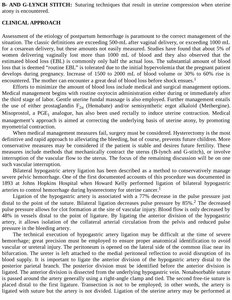
B- AND G-LYNCH STITCH: Suturing techniques that result in uterine compression when uterineatony is encountered.
CLINICAL APPROACH
Assessment of the etiology of postpartum hemorrhage is paramount to the correct management of thesituation. The classic definitions are exceeding 500-mL after vaginal delivery, or exceeding 1000 mLfor a cesarean delivery, but these amounts not easily measured. Studies have found that about 5% ofwomen delivering vaginally lost more than 1000 mL of blood and they also observed that theestimated blood loss (EBL) is commonly only half the actual loss. The substantial amount of bloodloss that is deemed “routine EBL” is tolerated due to the initial hypervolemia that the pregnant patientdevelops during pregnancy. Increase of 1500 to 2000 mL of blood volume or 30% to 60% rise isencountered. The mother can encounter a great deal of blood loss before shock ensues.1
Efforts to minimize the amount of blood loss include medical and surgical management options.Medical management begins with routine oxytocin administration either during or immediately afterthe third stage of labor. Gentle uterine fundal massage is also employed. Further management entailsthe use of either prostaglandin F2α (Hemabate) and/or semisynthetic ergot alkaloid (Methergine).Misoprostol, a PGE1 analogue, has also been used rectally to induce uterine contraction. Medicalmanagement’s approach is aimed at correcting the underlying basis of uterine atony, by promotingmyometrial contraction.
When medical management measures fail, surgery must be considered. Hysterectomy is the mostdefinitive and rapid approach to alleviating the bleeding, but of course, prevents future children. Moreconservative measures may be considered if the patient is stable and desires future fertility. Thesemeasures include methods that mechanically contract the uterus (B-lynch and G-stitch), or involveinterruption of the vascular flow to the uterus. The focus of the remaining discussion will be on onesuch vascular interruption.
Bilateral hypogastric artery ligation has been described as a method to conservatively managesevere pelvic hemorrhage. One of the first documented accounts of this procedure was documented in1893 at Johns Hopkins Hospital when Howard Kelly performed ligation of bilateral hypogastricarteries to control hemorrhage during hysterectomy for uterine cancer.1
Ligation of the hypogastric artery is associated with a 77% decrease in the pulse pressure justdistal to the point of the suture. Bilateral ligation decreases pulse pressure by 85%.2 The decreasedpulse pressure allows for clot formation at the site of vascular injury. Blood flow is only decreased by48% in vessels distal to the point of ligature. By ligating the anterior division of the hypogastricartery, it allows isolation of the collateral arterial circulation from the pelvis and reduced pulsepressure in the bleeding artery.
The technical execution of hypogastric artery ligation may be difficult at the time of severehemorrhage; great precision must be employed to ensure proper anatomical identification to avoidvascular or ureteral injury. The peritoneum is opened on the lateral side of the common iliac near itsbifurcation. The ureter is left attached to the medial peritoneal reflection to avoid disruption of itsblood supply. It is important to ligate the anterior division of the hypogastric artery distal to theposterior parietal branch. The posterior division must be identified before the anterior division isligated. The anterior division is dissected from the underlying hypogastric vein. Nonabsorbable sutureis passed around the artery generally using a right-angle clamp and tied. The second free-tie suture isplaced distal to the first ligature. Transection is not to be employed; in other words, the artery isligated with suture but the artery is not divided. Ligation of the uterine artery may be performed at
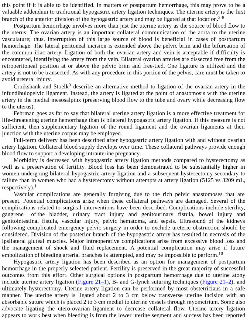
this point if it is able to be identified. In matters of postpartum hemorrhage, this may prove to be avaluable addendum to traditional hypogastric artery ligation techniques. The uterine artery is the firstbranch of the anterior division of the hypogastric artery and may be ligated at that location.3-8
Postpartum hemorrhage involves more than just the uterine artery as the source of blood flow tothe uterus. The ovarian artery is an important collateral communication of the aorta to the uterinevasculature; thus, interruption of this large source of blood is beneficial in cases of postpartumhemorrhage. The lateral peritoneal incision is extended above the pelvic brim and the bifurcation ofthe common iliac artery. Ligation of both the ovarian artery and vein is acceptable if difficulty isencountered, identifying the artery from the vein. Bilateral ovarian arteries are dissected free from theretroperitoneal position at or above the pelvic brim and free-tied. One ligature is utilized and theartery is not to be transected. As with any procedure in this portion of the pelvis, care must be taken toavoid ureteral injury.
Cruikshank and Stoelk9 describe an alternative method to ligation of the ovarian artery in theinfundibulopelvic ligament. Instead, the artery is ligated at the point of anastomosis with the uterineartery in the medial mesosalpinx (preserving blood flow to the tube and ovary while decreasing flowto the uterus).
Fehrman goes as far to say that bilateral uterine artery ligation is a more effective treatment forlife-threatening uterine hemorrhage than is bilateral hypogastric artery ligation. If this measure is notsufficient, then supplementary ligation of the round ligament and the ovarian ligaments at theirjunction with the uterine corpus may be employed.
Full-term delivery has been described after hypogastric artery ligation with and without ovarianartery ligation. Collateral blood supply develops over time. These collateral pathways provide enoughblood flow to support a developing intrauterine pregnancy.
Morbidity is decreased with hypogastric artery ligation methods compared to hysterectomy aswell as a preservation of fertility. Blood loss has been demonstrated to be substantially higher inwomen undergoing bilateral hypogastric artery ligation and a subsequent hysterectomy secondary tofailure than in women who had a hysterectomy without attempts at artery ligation (5125 vs 3209 mL,respectively).1
Vascular complications are generally forgiving due to the rich pelvic anastomoses that arepresent. Potential complications arise when these collateral pathways are damaged. Several of thecomplications related to surgical interventions have been described. Complications include sterility,gangrene of the bladder, urinary tract injury and genitourinary fistula, bowel injury andgenitointestinal fistula, vascular injury, pelvic hematoma, and sepsis. Ultrasound of the kidneysfollowing complicated emergency pelvic surgery in order to exclude ureteric obstruction should beconsidered. Division of the posterior branch of the hypogastric artery has resulted in necrosis of theipsilateral gluteal muscles. Major intraoperative complications arise from excessive blood loss andthe management of shock and fluid replacement. A potential complication may arise if futureembolization of bleeding arterial branches is attempted, and may be impossible to perform.10
Hypogastric artery ligation has been described as an option for management of postpartumhemorrhage in the properly selected patient. Fertility is preserved in the great majority of successfuloutcomes from this effort. Other surgical options in postpartum hemorrhage due to uterine atonyinclude uterine artery ligation (Figure 21–1), B- and G-lynch suturing techniques (Figure 21–2), andultimately hysterectomy. Uterine artery ligation can be performed by most obstetricians in a safemanner. The uterine artery is ligated about 2 to 3 cm below transverse uterine incision with anabsorbable suture which is placed 2 to 3 cm medial to uterine vessels through myometrium. Some alsoadvocate ligating the utero-ovarian ligament to decrease collateral flow. Uterine artery ligationappears to work best when bleeding is from the lower uterine segment and success has been reported
to be more than 95%.11 Both the B- (Figure 21–2) and G-lynch techniques are considered“compression sutures” and have some limited literature to support their use.1,12-14
Figure 21–1. Internal iliac artery ligation should be distal to posterior trunk, and not incorporate theureter. (Reproduced, with permission, from DeCherney AH, Nathan L, Goodwin TM, et al. CurrentDiagnosis & Treatment:Obstetrics & Gynecology, 10th ed. New York: McGraw-Hill, 2007:482.)
Figure 21–2. B-lynch suture is used as an external compression to the uterus. (Reproduced, withpermission, from DeCherney AH, Nathan L, Goodwin TM, et al. Current Diagnosis & Treatment:Obstetrics & Gynecology, 10th ed. New York: McGraw-Hill, 2007:483.)
Comprehension Questions
21.1 A 21-year-old woman undergoes a vaginal delivery at term. Postpartum hemorrhage isencountered due to uterine atony, which is unresponsive to medical management. Which ofthe following surgical procedures is most appropriate if the patient desires future fertility?
A. Ligation of the common iliac arteryB. Ligation of the infundibular pelvic ligamentC. Ligation of the ascending branch of the uterine arteryD. Ligation of the posterior trunk of the hypogastric artery
21.2 A 37-year-old G6P5005 woman presents at 40 weeks’ gestation with a diagnosis ofhydramnios. After a successful vaginal vertex delivery, the patient experiences postpartumhemorrhage estimated to be in excess of 2500 mL of blood. Medical management attemptsare unsuccessful. Which of the following is the next step?
A. Bilateral hypogastric artery ligationB. Sharp curettage of the uterusC. HysterectomyD. B-lynch stitch
21.3 Which of the following sutures would be preferred when performing a bilateral hypogastricartery ligation?
A. Chromic catgutB. Plain catgutC. polyglactin 910 (vicryl)D. Polydioxanone (PDS
ANSWERS
21.1 C. In a patient with uterine bleeding, decreasing the arterial perfusion is one of the primarytherapeutic strategies. Ligation of the ascending branch of the uterine artery bilaterally iseffective and allows for fertility. Ligation of the infundibular pelvic ligament meanssacrificing both ovaries which will lead to an infertile and castrated patient. Ligation of thecommon iliac artery should never be performed due to disruption of the arterial supply tothe lower extremity. Ligation of the posterior trunk of the hypogastric artery leads toischemia of the gluteal region.
21.2 C. Careful selection of patients for attempts at conservative management of hemorrhage iscrucial to the morbidity of the patient. In this case a multiparous patient with a number ofrisk factors for post-partum hemorrhage was encountered (high parity, hydramnios). Thepatient is multiparous and has an excessive blood loss secondary to uterine atony. Giventhese factors, hysterectomy would be the most appropriate measure. Sharp dilation andcurettage of the uterus is an option for patients experiencing postpartum hemorrhagesecondary to retained products of conception. The B-lynch stitch is a suture that manuallycompresses the uterus. The suture has been effective in some cases, although there arelimited published data comparing its effectiveness to that of hysterectomy.
21.3 C. Nonabsorbable suture (silk) and delayed absorbable suture (Vicryl) have been utilized inbilateral ligation of the hypogastric artery. PDS has a tendency to cut through the tissuewhen tying and thus would be contraindicated. The premise of the technique is to ligate thevessel and not to transect. Chromic is absorbable suture.
Clinical Pearls
See Table 1-2 for definition of level of evidence and strength of recommendation
The most common cause of postpartum hemorrhage is uterine atony (Level B).
Arterial embolization of uterine vasculature may be impossible to perform after bilateralhypogastric artery ligation (Level B).
Fertility may be preserved with bilateral hypogastric artery ligation (Level B).
Conservative surgical management is a possibility in the properly selected patient (Level C).
Ureteral damage is a complication with bilateral hypogastric artery ligation and must be ruledout if clinically suspected at time of operation or postoperatively (Level C).
REFERENCES
1. Cunningham FG, Leveno KJ, Bloom SL, Hauth JC, Gilstrap LC III, Wenstrom KD. Obstetricalhemorrhage. In: Williams Obstetrics. 22nd ed. New York, NY: McGraw-Hill; 2005:809-854.
2. Burchell RC. Physiology of internal iliac artery ligation. J Obstet Gynaecol Br Commonw.1969;57:642.
3. Evans S, McShane P. The efficacy of internal iliac artery ligation in obstetric hemorrhage.Surg Gynecol Obstet. 1985;160:250.
4. Fahmy K. Internal iliac artery ligation and its efficacy in controlling pelvic hemorrhage. IntSurg. 1969;51:244.
5. Reich WJ, Nechtow MJ. Ligation of the internal iliac (hypogastric) arteries: a life-savingprocedure for uncontrollable gynecologic and obstetric hemorrhage. J Int Coll Surg.1961;36:157.
6. Rock JA, Jones HW III. Control of pelvic hemorrhage. In: Te Linde’s Operative Gynecology .10th ed. Philadelphia, PA: Lippincott Williams & Wilkins; 2008:385-387.
7. Siegel P, Mengert WF. Internal iliac artery ligation in obstetrics and gynecology. JAMA.1961;178:1059.
8. Slate WG. Internal iliac ligation. Am J Obstet Gynecol. 1966;95:326.
9. Cruikshank SH, Stoelk EM. Surgical control of pelvic hemorrhage: bilateral hypogastric arteryligation and method of ovarian artery ligation. South Med J. 1985;78:539.
10. Given FT, Gates HS, Morgan BE. Pregnancy following bilateral ligation of the internal iliac(hypogastric) arteries. Am J Obstet Gynecol. 1964;89:1078.
11. O’Leary JA. Uterine artery ligation in the control of postcesarean hemorrhage. J Reprod Med.1995;40:189.
12. B-Lynch CB. Coker A, Laval AH, Abu J, Cowen MJ. The B-lynch surgical technique forcontrol of massive postpartum hemorrhage: an alternative to hysterectomy? Five casesreported. Br J Obstet Gynaecol. 1997;104:372.
13. Hayman RG, Arulkumaran S, Steer PJ. Uterine compression sutures: surgical management ofpostpartum hemorrhage. Obstet Gynecol. 2002;99:502.

14. Rock JA, Jones HW III. Obstetric problems. In: Te Linde’s Operative Gynecology . 10th ed.Philadelphia, PA: Lippincott Williams & Wilkins; 2008:827-829.
Case 22
A 45-year-old G4P4004 Caucasian woman presents with a 6-month history of increasing “pelvicheaviness.” She had a postpartum bilateral tubal ligation following delivery of her last child. Sherelates that she had a forceps delivery with her first child, and that she stands at work most of the dayas a surgical scrub tech. She has also noticed increasing dyspareunia, stating that, “It feels like mypartner is hitting something when we have intercourse.” She denies stress urinary incontinence,recurrent urinary tract infection, or difficulty with defecation. Her Pap smears have been normalthroughout her life, and there has been no change in her menstrual pattern. She relates that 6 monthsof intensive Kegel exercises have not changed her symptoms. On pelvic examination, she has normalexternal genitalia. The vaginal mucosa demonstrates good estrogen effect, and there is adequatesupport of both the anterior and posterior vaginal walls. The parous cervix descends to within 1 cm ofthe vaginal introitus with a Valsalva maneuver. The uterus is nontender, and not enlarged, and theadnexa are not palpable. The rectal examination is unremarkable.
What is the most likely diagnosis?
What should be your next step?
ANSWERS TO CASE 22:Vaginal Hysterectomy
Summary: A 45-year-old woman presents with pelvic heaviness and dyspareunia. The pelvicexamination reveals that her cervix descends to the vaginal introitus with Valsalva. According to thepelvic organ prolapse quantification (POP-Q) scoring system, this is stage II pelvic organ prolapse.1
Most likely diagnosis: Uterine prolapse.
Next step: Vaginal hysterectomy.
ANALYSIS
Objectives
1. Understand that most benign uterine pathology requiring hysterectomy is amenable to the vaginalapproach.
2. Learn the indications, advantages, and disadvantages of vaginal hysterectomy compared toabdominal or laparoscopically assisted vaginal hysterectomy.
Considerations
Uterine prolapse to the extent described in this case is a common problem for parous women, andoperative vaginal delivery is increasingly felt to be a risk factor for the later development of pelvic
organ prolapse. For a healthy patient who is not desirous of having any more children, vaginalhysterectomy is the procedure of choice. It is difficult to say with certainty whether a given patient’sprolapse will worsen, but it is not likely to improve over time. In comparison to total abdominalhysterectomy (TAH) or laparoscopic hysterectomy (LH), vaginal hysterectomy (VH) is faster,cheaper, safer, less painful, and most cosmetically and sexually satisfying to the patient. Forbenign disease, almost all types of uterine or pelvic pathology, which require hysterectomy as thetherapy of choice, can be accomplished vaginally.2 The goal of this chapter is to delineate thereasons why vaginal hysterectomy should usually be the hysterectomy route of first choice for mostbenign gynecologic conditions requiring uterine removal.
APPROACH TO Vaginal Hysterectomy
DEFINITIONS
VAGINAL HYSTERECTOMY: Removal of the entire uterus through the vagina. When appropriate,this is often combined with unilateral or bilateral salpingo-oophorectomy, removing one or bothfallopian tubes and ovaries vaginally coincident with the hysterectomy.
TOTAL ABDOMINAL HYSTERECTOMY: Removal of the entire uterus via a laparotomy incision,whether through a vertical or transverse incision. Can likewise include removal of tubes and ovaries asabove.
LAPAROSCOPIC HYSTERECTOMY: This operation has three possible variants. LAVH is aidedby the laparoscope, but not to include uterine artery ligation; LH, where the laparoscope is used toligate the uterine arteries; and TLH, in which there is no vaginal approach and the cuff is closedlaparoscopically. Tubes and ovaries may also be removed via these techniques.
CLINICAL APPROACH
Hysterectomy rates in the United States are decreasing slightly, from 5.5 per 1000 women in 1990 to5.1 per 1000 women in 2004. Abdominal hysterectomy is the most common approach, constituting63% of all types in 1997. In April 2008, French gynecologists reported their experience comparingvaginal hysterectomy to LAVH in women who had not had prior vaginal delivery. They noted thatthere was less OR time, shorter hospital stay, and less cost to the patient when the vaginal approachwas used. Further, as they progressed through the study and became increasingly comfortable with thevaginal approach, they relied less on the laparoscope for assistance.3 Newer techniques such assupracervical or laparoscopic hysterectomies do not have any advantages over vaginal or abdominalhysterectomies, especially in terms of avoiding complications, including urinary and sexual function.Urinary incontinence is more frequent in women who have supracervical hysterectomy than in womenrandomized to vaginal hysterectomy, and there is no difference in sexual function in either group,regardless of hysterectomy technique or presence of a residual cervix.4Leaving the cervix in situ as aresult of a supracervical laparoscopic procedure has been associated with an increase in latertrachelectomy procedures for cyclical monthly bleeding from the cervix, for cervical dysplasia,and for prolapse of the cervical stump, all of which can be eliminated by removing the cervix atVH. If these new technologies are being questioned, it is appropriate to reiterate the benefits of VH.This discussion will focus on the indications for vaginal hysterectomy and compare vaginalhysterectomy to other hysterectomy techniques in terms of uterine size, safety, length of hospital stay,
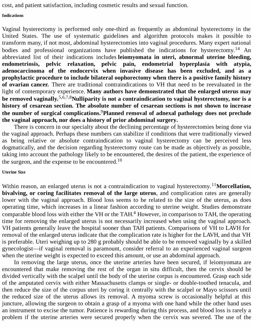
cost, and patient satisfaction, including cosmetic results and sexual function.
Indications
Vaginal hysterectomy is performed only one-third as frequently as abdominal hysterectomy in theUnited States. The use of systematic guidelines and algorithm protocols makes it possible totransform many, if not most, abdominal hysterectomies into vaginal procedures. Many expert nationalbodies and professional organizations have published the indications for hysterectomy.14 Anabbreviated list of their indications includes leiomyomata in uteri, abnormal uterine bleeding,endometriosis, pelvic relaxation, pelvic pain, endometrial hyperplasia with atypia,adenocarcinoma of the endocervix when invasive disease has been excluded, and as aprophylactic procedure to include bilateral oophorectomy when there is a positive family historyof ovarian cancer. There are traditional contraindications to VH that need to be reevaluated in thelight of contemporary experience. Many authors have demonstrated that the enlarged uterus maybe removed vaginally.5,6,7,8Nulliparity is not a contraindication to vaginal hysterectomy, nor is ahistory of cesarean section. The absolute number of cesarean sections is not shown to increasethe number of surgical complications.9Planned removal of adnexal pathology does not precludethe vaginal approach, nor does a history of prior abdominal surgery.
There is concern in our specialty about the declining percentage of hysterectomies being done viathe vaginal approach. Perhaps these numbers can stabilize if conditions that were traditionally viewedas being relative or absolute contraindication to vaginal hysterectomy can be perceived lessdogmatically, and the decision regarding hysterectomy route can be made as objectively as possible,taking into account the pathology likely to be encountered, the desires of the patient, the experience ofthe surgeon, and the expense to be encountered.10
Uterine Size
Within reason, an enlarged uterus is not a contraindication to vaginal hysterectomy.11Morcellation,bivalving, or coring facilitates removal of the large uterus, and complication rates are generallylower with the vaginal approach. Blood loss seems to be related to the size of the uterus, as doesoperating time, which increases in a linear fashion according to uterine weight. Studies demonstratecomparable blood loss with either the VH or the TAH.4 However, in comparison to TAH, the operatingtime for removing the enlarged uterus is not necessarily increased when using the vaginal approach.VH patients generally leave the hospital sooner than TAH patients. Comparisons of VH to LAVH forremoval of the enlarged uterus indicate that the complication rate is higher for the LAVH, and that VHis preferable. Uteri weighing up to 280 g probably should be able to be removed vaginally by a skilledgynecologist—if vaginal removal is paramount, consider referral to an experienced vaginal surgeonwhen the uterine weight is expected to exceed this amount, or use an abdominal approach.
In removing the large uterus, once the uterine arteries have been secured, if leiomyomata areencountered that make removing the rest of the organ in situ difficult, then the cervix should bedivided vertically with the scalpel until the body of the uterine corpus is encountered. Grasp each sideof the amputated cervix with either Massachusetts clamps or single- or double-toothed tenacula, andthen reduce the size of the corpus uteri by coring it centrally with the scalpel or Mayo scissors untilthe reduced size of the uterus allows its removal. A myoma screw is occasionally helpful at thisjuncture, allowing the surgeon to obtain a grasp of a myoma with one hand while the other hand usesan instrument to excise the tumor. Patience is rewarding during this process, and blood loss is rarely aproblem if the uterine arteries were secured properly when the cervix was severed. The use of the
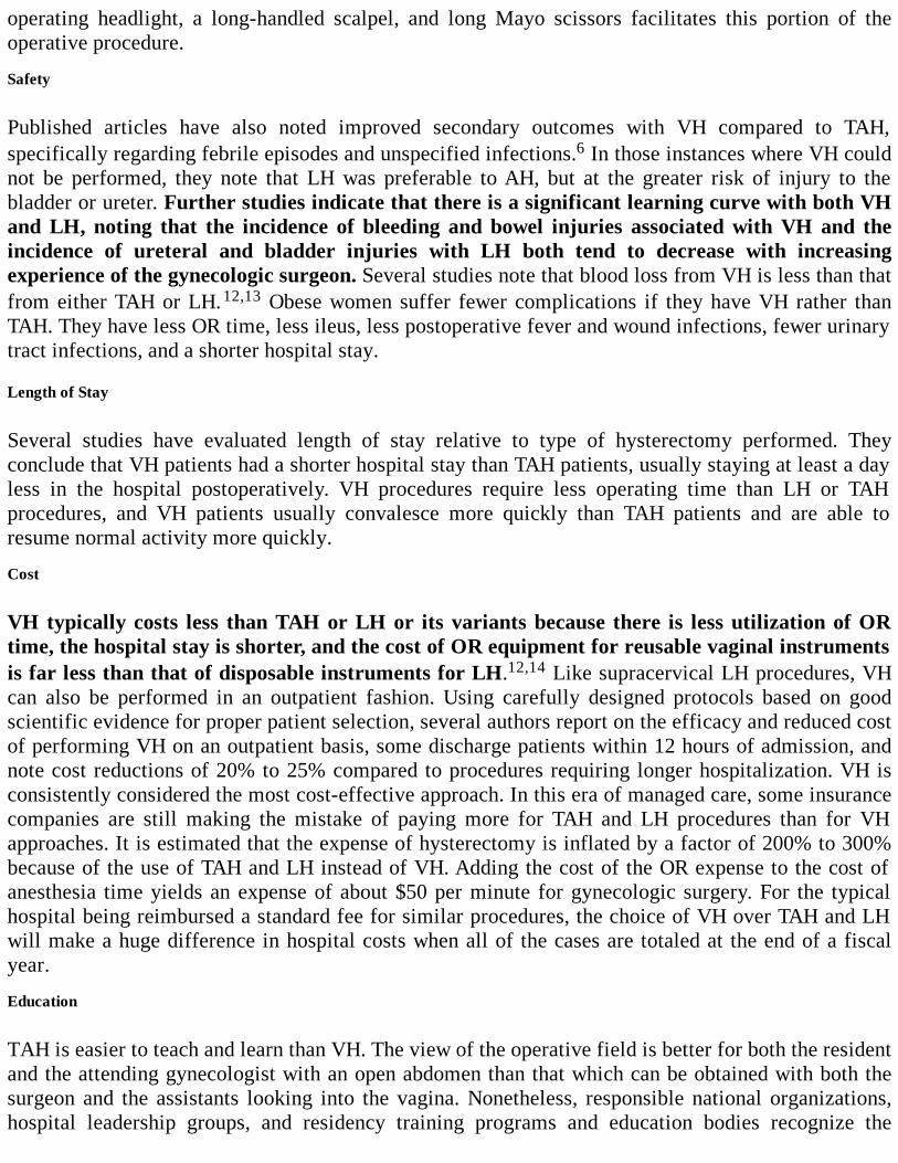
operating headlight, a long-handled scalpel, and long Mayo scissors facilitates this portion of theoperative procedure.
Safety
Published articles have also noted improved secondary outcomes with VH compared to TAH,specifically regarding febrile episodes and unspecified infections.6 In those instances where VH couldnot be performed, they note that LH was preferable to AH, but at the greater risk of injury to thebladder or ureter. Further studies indicate that there is a significant learning curve with both VHand LH, noting that the incidence of bleeding and bowel injuries associated with VH and theincidence of ureteral and bladder injuries with LH both tend to decrease with increasingexperience of the gynecologic surgeon. Several studies note that blood loss from VH is less than thatfrom either TAH or LH.12,13 Obese women suffer fewer complications if they have VH rather thanTAH. They have less OR time, less ileus, less postoperative fever and wound infections, fewer urinarytract infections, and a shorter hospital stay.
Length of Stay
Several studies have evaluated length of stay relative to type of hysterectomy performed. Theyconclude that VH patients had a shorter hospital stay than TAH patients, usually staying at least a dayless in the hospital postoperatively. VH procedures require less operating time than LH or TAHprocedures, and VH patients usually convalesce more quickly than TAH patients and are able toresume normal activity more quickly.
Cost
VH typically costs less than TAH or LH or its variants because there is less utilization of ORtime, the hospital stay is shorter, and the cost of OR equipment for reusable vaginal instrumentsis far less than that of disposable instruments for LH.12,14 Like supracervical LH procedures, VHcan also be performed in an outpatient fashion. Using carefully designed protocols based on goodscientific evidence for proper patient selection, several authors report on the efficacy and reduced costof performing VH on an outpatient basis, some discharge patients within 12 hours of admission, andnote cost reductions of 20% to 25% compared to procedures requiring longer hospitalization. VH isconsistently considered the most cost-effective approach. In this era of managed care, some insurancecompanies are still making the mistake of paying more for TAH and LH procedures than for VHapproaches. It is estimated that the expense of hysterectomy is inflated by a factor of 200% to 300%because of the use of TAH and LH instead of VH. Adding the cost of the OR expense to the cost ofanesthesia time yields an expense of about $50 per minute for gynecologic surgery. For the typicalhospital being reimbursed a standard fee for similar procedures, the choice of VH over TAH and LHwill make a huge difference in hospital costs when all of the cases are totaled at the end of a fiscalyear.
Education
TAH is easier to teach and learn than VH. The view of the operative field is better for both the residentand the attending gynecologist with an open abdomen than that which can be obtained with both thesurgeon and the assistants looking into the vagina. Nonetheless, responsible national organizations,hospital leadership groups, and residency training programs and education bodies recognize the
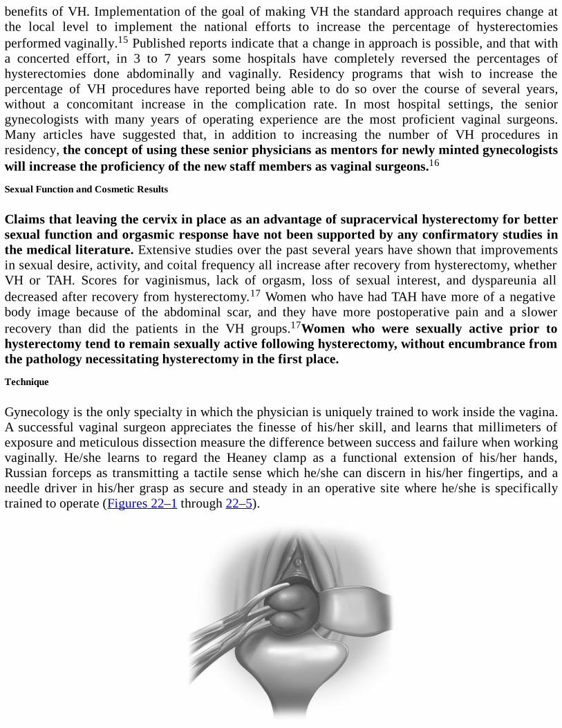
benefits of VH. Implementation of the goal of making VH the standard approach requires change atthe local level to implement the national efforts to increase the percentage of hysterectomiesperformed vaginally.15 Published reports indicate that a change in approach is possible, and that witha concerted effort, in 3 to 7 years some hospitals have completely reversed the percentages ofhysterectomies done abdominally and vaginally. Residency programs that wish to increase thepercentage of VH procedures have reported being able to do so over the course of several years,without a concomitant increase in the complication rate. In most hospital settings, the seniorgynecologists with many years of operating experience are the most proficient vaginal surgeons.Many articles have suggested that, in addition to increasing the number of VH procedures inresidency, the concept of using these senior physicians as mentors for newly minted gynecologistswill increase the proficiency of the new staff members as vaginal surgeons.16
Sexual Function and Cosmetic Results
Claims that leaving the cervix in place as an advantage of supracervical hysterectomy for bettersexual function and orgasmic response have not been supported by any confirmatory studies inthe medical literature. Extensive studies over the past several years have shown that improvementsin sexual desire, activity, and coital frequency all increase after recovery from hysterectomy, whetherVH or TAH. Scores for vaginismus, lack of orgasm, loss of sexual interest, and dyspareunia alldecreased after recovery from hysterectomy.17 Women who have had TAH have more of a negativebody image because of the abdominal scar, and they have more postoperative pain and a slowerrecovery than did the patients in the VH groups.17Women who were sexually active prior tohysterectomy tend to remain sexually active following hysterectomy, without encumbrance fromthe pathology necessitating hysterectomy in the first place.
Technique
Gynecology is the only specialty in which the physician is uniquely trained to work inside the vagina.A successful vaginal surgeon appreciates the finesse of his/her skill, and learns that millimeters ofexposure and meticulous dissection measure the difference between success and failure when workingvaginally. He/she learns to regard the Heaney clamp as a functional extension of his/her hands,Russian forceps as transmitting a tactile sense which he/she can discern in his/her fingertips, and aneedle driver in his/her grasp as secure and steady in an operative site where he/she is specificallytrained to operate (Figures 22–1 through 22–5).
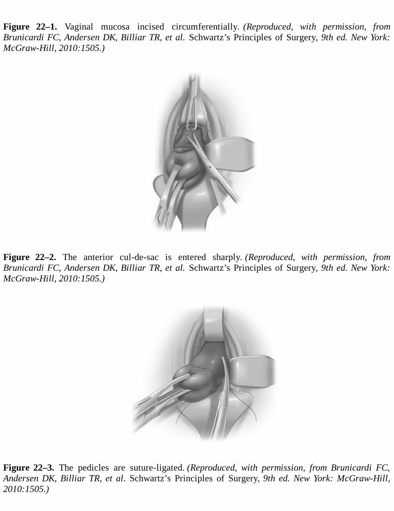
Figure 22–1. Vaginal mucosa incised circumferentially. (Reproduced, with permission, fromBrunicardi FC, Andersen DK, Billiar TR, et al. Schwartz’s Principles of Surgery, 9th ed. New York:McGraw-Hill, 2010:1505.)
Figure 22–2. The anterior cul-de-sac is entered sharply. (Reproduced, with permission, fromBrunicardi FC, Andersen DK, Billiar TR, et al. Schwartz’s Principles of Surgery, 9th ed. New York:McGraw-Hill, 2010:1505.)
Figure 22–3. The pedicles are suture-ligated. (Reproduced, with permission, from Brunicardi FC,Andersen DK, Billiar TR, et al. Schwartz’s Principles of Surgery, 9th ed. New York: McGraw-Hill,2010:1505.)
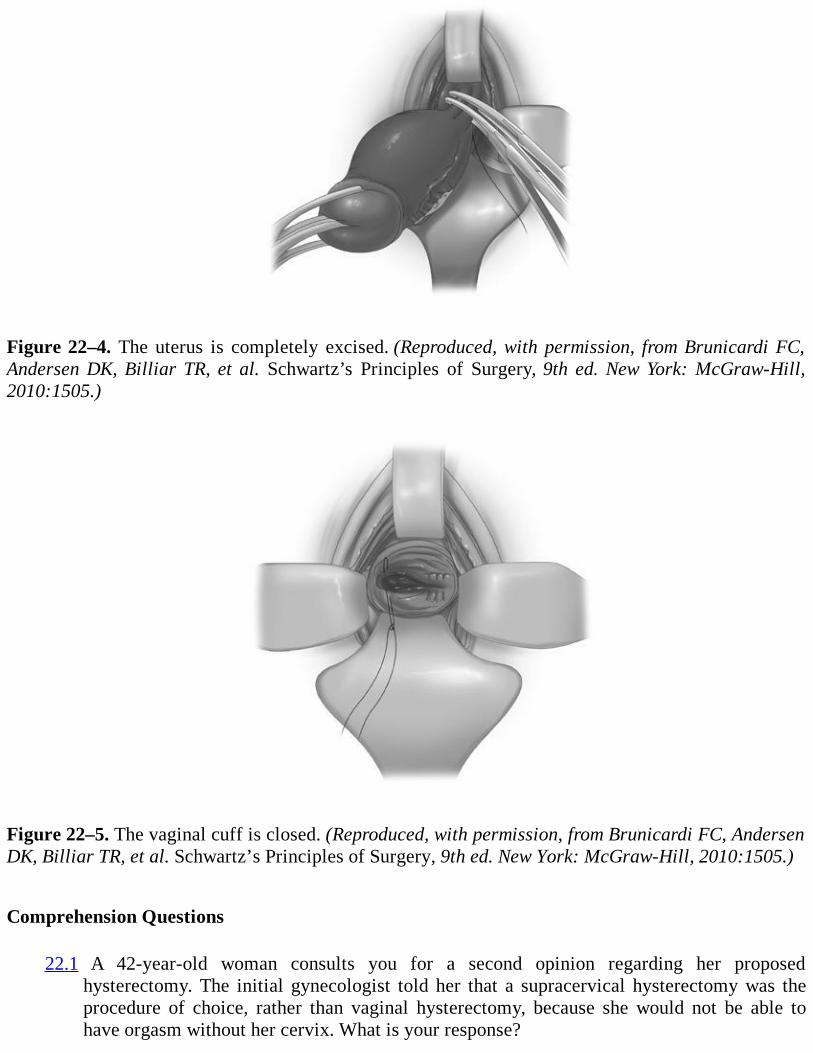
Figure 22–4. The uterus is completely excised. (Reproduced, with permission, from Brunicardi FC,Andersen DK, Billiar TR, et al. Schwartz’s Principles of Surgery, 9th ed. New York: McGraw-Hill,2010:1505.)
Figure 22–5. The vaginal cuff is closed. (Reproduced, with permission, from Brunicardi FC, AndersenDK, Billiar TR, et al. Schwartz’s Principles of Surgery, 9th ed. New York: McGraw-Hill, 2010:1505.)
Comprehension Questions
22.1 A 42-year-old woman consults you for a second opinion regarding her proposedhysterectomy. The initial gynecologist told her that a supracervical hysterectomy was theprocedure of choice, rather than vaginal hysterectomy, because she would not be able tohave orgasm without her cervix. What is your response?
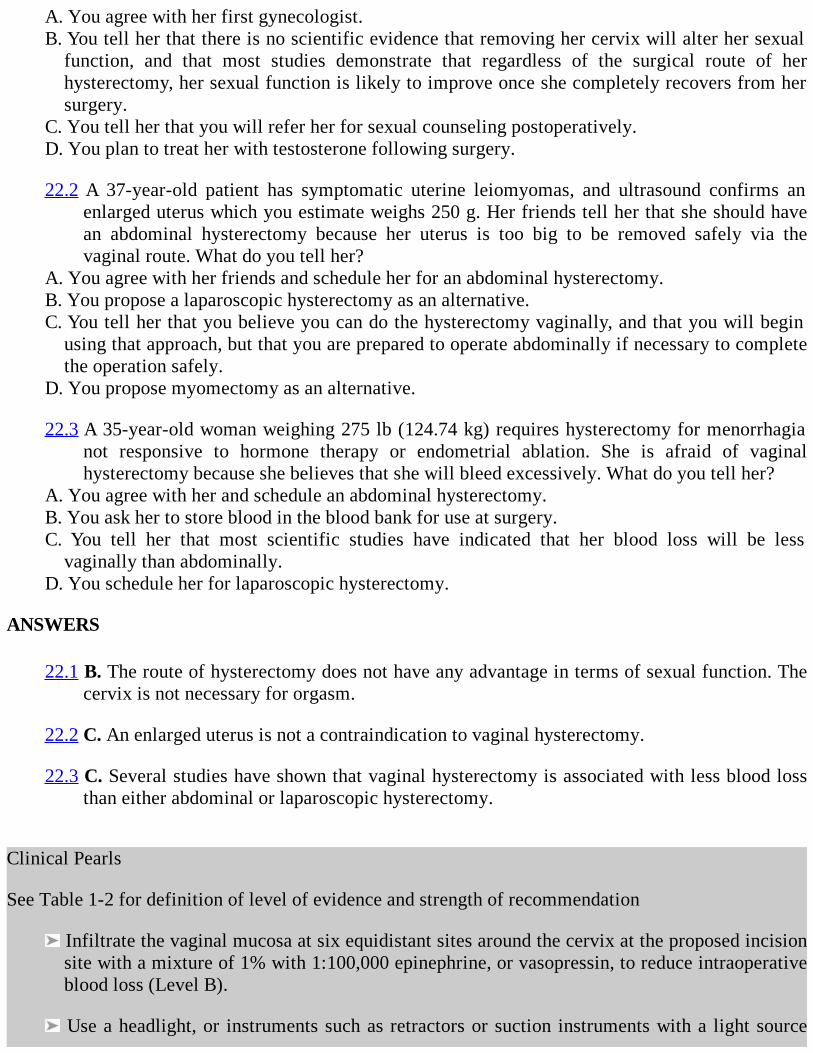
A. You agree with her first gynecologist.B. You tell her that there is no scientific evidence that removing her cervix will alter her sexual
function, and that most studies demonstrate that regardless of the surgical route of herhysterectomy, her sexual function is likely to improve once she completely recovers from hersurgery.
C. You tell her that you will refer her for sexual counseling postoperatively.D. You plan to treat her with testosterone following surgery.
22.2 A 37-year-old patient has symptomatic uterine leiomyomas, and ultrasound confirms anenlarged uterus which you estimate weighs 250 g. Her friends tell her that she should havean abdominal hysterectomy because her uterus is too big to be removed safely via thevaginal route. What do you tell her?
A. You agree with her friends and schedule her for an abdominal hysterectomy.B. You propose a laparoscopic hysterectomy as an alternative.C. You tell her that you believe you can do the hysterectomy vaginally, and that you will begin
using that approach, but that you are prepared to operate abdominally if necessary to completethe operation safely.
D. You propose myomectomy as an alternative.
22.3 A 35-year-old woman weighing 275 lb (124.74 kg) requires hysterectomy for menorrhagianot responsive to hormone therapy or endometrial ablation. She is afraid of vaginalhysterectomy because she believes that she will bleed excessively. What do you tell her?
A. You agree with her and schedule an abdominal hysterectomy.B. You ask her to store blood in the blood bank for use at surgery.C. You tell her that most scientific studies have indicated that her blood loss will be less
vaginally than abdominally.D. You schedule her for laparoscopic hysterectomy.
ANSWERS
22.1 B. The route of hysterectomy does not have any advantage in terms of sexual function. Thecervix is not necessary for orgasm.
22.2 C. An enlarged uterus is not a contraindication to vaginal hysterectomy.
22.3 C. Several studies have shown that vaginal hysterectomy is associated with less blood lossthan either abdominal or laparoscopic hysterectomy.
Clinical Pearls
See Table 1-2 for definition of level of evidence and strength of recommendation
Infiltrate the vaginal mucosa at six equidistant sites around the cervix at the proposed incisionsite with a mixture of 1% with 1:100,000 epinephrine, or vasopressin, to reduce intraoperativeblood loss (Level B).
Use a headlight, or instruments such as retractors or suction instruments with a light source
attached, when performing vaginal surgery. Standard OR lights are rarely bright enough, do notfocus sharply, and never are at an optimum angle for seeing how to work in the vagina (LevelC).
If the vaginal hysterectomy is not accompanied by colporrhaphy, avoid the use of a Foleycatheter.Tell the patient she will arrive in the recovery room with a full bladder, and that shewill be encouraged to void in recovery. If unable to void, she may be straight-catheterizedtwice over the next 6 to 8 hours, and should have a Foley placed only after the third failedvoiding attempt. Over 75% of patients will never need a Foley (Level B).
Close the vaginal cuff vertically rather than horizontally if there is concern about maintainingadequate vaginal length. Horizontal closure shortens the vagina by about 1 cm; vertical closuredoes not significantly change vaginal length (Level B).
REFERENCES
1. Bump RC, Mattiasson A, Bø K, et al. The standardization of terminology of female pelvicorgan prolapse and pelvic floor dysfunction. Am J Obstet Gynecol. 1996;175:10-17.The classic treatise describing the POPQ system for quantifying pelvic organ prolapse.
2. Johnson N, Barlow D, Lethaby A, Tavender E, Curr E, Garry R. Surgical approach tohysterectomy for benign gynaecological disease. Cochrane Database Syst Rev. 2006Apr;19(2):CD003677.
3. Le Tohic A, Dhainaut C, Yazbeck C, Hallais C, Levin I, Madelenat P. Hysterectomy for benignuterine pathology among women without previous vaginal delivery. Obstet Gynecol.2008;111:829-837.
4. Gimbel H, Zobbe V, Anderson BM, Filtenborg T, Gluud C, Tabor A. Randomised controlledtrial of total compared with subtotal hysterectomy with one-year follow up results. BJOG.2003;110:1309-1318.
5. Paparella P, Sizzi O, Rosetti A, De Benedittis F, Papaarella R. Vaginal hysterectomy ingenerally considered contraindications to vaginal surgery. Arch Gynecol Obstet. 2004;270:104-109.
6. Makinen J, Johansson J, Tomas C, et al. Morbidity of hysterectomies by type of approach. HumReprod. 2001;16:1473-1478.
7. Jacobsen G, Shaber R, Armstrong M, Hung Y. Hysterectomy rates for benign indications.Obstet Gynecol. 2006;107:1278-1283.
8. Doucette R, Sharp H, Alder S. Challenging generally accepted contraindications to vaginalhysterectomy. Am J Obstet Gynecol. 2001;184:1386-1389.
9. Poindexter Y, Sangi-Haghpeykar H, Poindexter A, et al. Previous cesarean section. A
contraindication to vaginal hysterectomy? J Reprod Med. 2001;46:840-844.
10. Julian TJ. Vaginal hysterectomy. An apparent exception to evidence-based decision making.Obstet Gynecol. 2008;111:812-813.
11. Unger J. Vaginal hysterectomy for the woman with a moderately enlarged uterus weighing200 to 700 grams. Am J Obstet Gynecol. 1999;180:1337-1344.
12. Levy B, Luciano D, Emery L. Outpatient vaginal hysterectomy is safe for patients andreduces institutional cost. J Minim Invasive Gynecol. 2005;12:494-501.
13. Olah K, Khalil M. Changing the route of hysterectomy: the results of a policy of attemptingthe vaginal approach in all cases of dysfunctional uterine bleeding. Eur J Obstet GynecolReprod Biol. 2006;125:243-247.
14. Lefebvre G, Allaire C, Jeffrey J, et al. SOCG clinical guidelines. Hysterectomy. J ObstetGynaecol Can. 2002;24:37-61.
15. Kovac S. Transvaginal hysterectomy: rationale and surgical approach. Obstet Gynecol.2004;103:1321-1325.The definitive paper justifying vaginal hysterectomy, with an excellent algorithm to determinethe correct surgical approach for a given patient.
16. Gutl P, Greimel E, Roth R, Winter R. Women’s sexual behavior, body image and satisfactionwith surgical outcomes after hysterectomy: a comparison of vaginal and abdominal surgery. JPsychosom Obstet Gynaecol. 2002;23:51-59.
17. Taylor S, Romereo A, Kammerer-Doak D, Rogers R. Abdominal hysterectomy for theenlarged myomatous uterus compared with vaginal hysterectomy with morcellation. Am JObstet Gynecol. 2003;189:1579-1582.
18. Darai E, Soriano D, Kimata P, Laplace C, Leduru F. Vaginal hysterectomy for enlarged uteri,with or without laparoscopic assistance; randomized study. Obstet Gynecol. 2001;97:712-716.
19. Falcone T, Walters MD. Hysterectomy for benign disease. Obstet Gynecol. 2008;111:753-767.A definitive paper in the “clinical expert series” from the green journal.
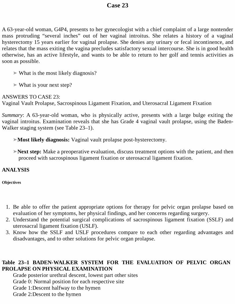
Case 23
A 63-year-old woman, G4P4, presents to her gynecologist with a chief complaint of a large nontendermass protruding “several inches” out of her vaginal introitus. She relates a history of a vaginalhysterectomy 15 years earlier for vaginal prolapse. She denies any urinary or fecal incontinence, andrelates that the mass exiting the vagina precludes satisfactory sexual intercourse. She is in good healthotherwise, has an active lifestyle, and wants to be able to return to her golf and tennis activities assoon as possible.
What is the most likely diagnosis?
What is your next step?
ANSWERS TO CASE 23:Vaginal Vault Prolapse, Sacrospinous Ligament Fixation, and Uterosacral Ligament Fixation
Summary: A 63-year-old woman, who is physically active, presents with a large bulge exiting thevaginal introitus. Examination reveals that she has Grade 4 vaginal vault prolapse, using the Baden-Walker staging system (see Table 23–1).
Most likely diagnosis: Vaginal vault prolapse post-hysterectomy.
Next step: Make a preoperative evaluation, discuss treatment options with the patient, and thenproceed with sacrospinous ligament fixation or uterosacral ligament fixation.
ANALYSIS
Objectives
1. Be able to offer the patient appropriate options for therapy for pelvic organ prolapse based onevaluation of her symptoms, her physical findings, and her concerns regarding surgery.
2. Understand the potential surgical complications of sacrospinous ligament fixation (SSLF) anduterosacral ligament fixation (USLF).
3. Know how the SSLF and USLF procedures compare to each other regarding advantages anddisadvantages, and to other solutions for pelvic organ prolapse.
Table 23–1 BADEN-WALKER SYSTEM FOR THE EVALUATION OF PELVIC ORGANPROLAPSE ON PHYSICAL EXAMINATION
Grade posterior urethral descent, lowest part other sitesGrade 0: Normal position for each respective siteGrade 1:Descent halfway to the hymenGrade 2:Descent to the hymen
Grade 3:Descent halfway past the hymenGrade 4:Maximum possible descent for each site
Data from Baden WF, Walker T. Fundamentals, symptoms and classification. In: Baden WF, WalkerT, Surgical Repair of Vaginal Detects. Philadelphia, PA: J.B. Lippincott; 1992, p. 14.
Considerations
This case is a 63 year-old woman in good health and physically active with vaginal vault prolapsedafter hysterectomy. After a careful physical examination to ensure that there is no complicated reasonfor the prolapse, such as a pelvic tumor or ovarian cancer with ascites, the patient should be counseledon the options of therapy. In a patient who is a poor surgical candidate and not sexually active, acolpocleisis procedure is an option. In summary, this patient’s problem is a common complaint. Asthe “Baby Boomers” enter their postmenopausal years, many of them are going to experience pelvicorgan prolapse and require the attention of gynecologists. Some 200,000 operative procedures forprolapse are performed annually in the United States, and some 25% to 29% of these surgicalprocedures are going to fail to achieve and/or to maintain their intended result over the rest of thepatient’s lifetime. Not every patient who presents with pelvic organ prolapse is a surgicalcandidate, and knowing which patient will best avoid surgery is an important lesson to discern.Multiple surgical procedures have been described and performed for pelvic organ prolapse. The SSLFprocedure has been a relatively popular vaginal approach to vaginal vault prolapse in the United Statesfor about the past 25 years, and more recently the USLF has been widely used for vaginal vaultprolapse. There are some unique benefits and risks for each procedure, and the gynecologic surgeonneeds to know these to be able to discuss these operations knowledgeably with the patient and to beable to perform one of these operations on her if the patient and her surgeon decide that this is thepreferred approach for her problem.
APPROACH TO Sacrospinous Ligament Fixation
David Nichols and Clyde Randall popularized SSLF in the early 1980s in the United States. In theyears since, several other vaginal approaches to vaginal vault prolapse have been introduced,including uterosacral ligament suspension, endopelvic fascial suspension, and other abdominal andlaparoscopic approaches described for prolapse. Sze and Karram indicate that 40 separate operationshave been described for these vaginal hernias.1 Of the 40 described, the two most frequentlyperformed procedures in the Unites States today are the SSLF and the USLF.
CLINICAL PRESENTATION
Etiologic factors probably include menopause and advancing age, a history of vaginal delivery,especially of large babies requiring operative vaginal deliveries, disorders of connective tissue, andany condition that increases intra-abdominal pressure, such as obesity or chronic constipation. Nostudies demonstrate that weight loss will reverse extant prolapse, although weight loss and pelvicfloor exercises may be beneficially combined with watchful waiting, and surgery may be avoided ifsymptoms do not worsen. While the use of estrogen creams will facilitate the surgical procedureitself, by making the vaginal mucosa thicker and more pliable, there is no data to support the use ofestrogen either systemically or topically to reverse or treat pelvic organ prolapse.
Most parous women will have some degree of pelvic relaxation on physical examination, but the
symptomatology she experiences does not necessarily have a direct relationship to her degree ofprolapse. The presence of some prolapse without symptoms does not merit treatment, and no studieshave been able to prove that there are effective measures that can prevent the condition fromworsening. The patient’s history, general medical condition, and severity of symptoms must becarefully weighed when considering a therapeutic choice. Nonsurgical approaches include the use of apessary, biofeedback training, and pelvic-floor rehabilitation. For the medically fragile patient, theseless vigorous solutions may be a prudent first choice, with surgery being entertained only forworsening symptoms not responsive to a nonsurgical approach. Regardless of which surgicalprocedure is being considered, most authorities agree that the prolapse patient should be given theoption of pessary use before proceeding to a surgical alternative.2 In general, most women may besuccessfully fitted with a pessary, even those with the more severe grades of prolapse (see Table 23–2for grading stage of prolpase). However, the long-term success rate for pessary use is probably relatedto severity of prolapse, and many patients prefer surgery to long-term pessary use.
Recent prospective studies have shown that prolapse actually has the potential to wax and wanein individual women, being worse in some years than in others, with the pelvic structures presentinghigher or lower in the pelvis when measured using the pelvic organ prolapse quantification (POP-Q)system.2 Based on this prospective study, it is also reasonable to tell a patient that excepting unusualcircumstances, her prolapse is not likely to worsen dramatically over short periods of time.
Many patients are especially embarrassed discussing the symptoms associated with pelvic organprolapse and will be hesitant to volunteer specific symptoms of urinary or fecal incontinence, or ofhaving to splint and/or brace the perineum or vaginal floor to evacuate stool. The gynecologist mustask these questions and know this information to plan the correct therapeutic approach.
Choice of Surgical Procedure
If surgery is indicated for vaginal vault prolapse, the considerations include the fact that there is lessapical failure with the abdominal sacrocolpopexy, and less postoperative stress urinary incontinenceand dyspareunia with the abdominal approach, but at the expense of more complications, including allof those that accompany an abdominal procedure, especially bowel obstruction. The age and overallgeneral health of the patient may tip the scales in favor of either the SSLF or the USLF procedure.Surgical series of patients older than 80 years describe successful repairs with good results, but withsome increased risk of blood loss in this patient population.3 For the patient who wishes to remainsexually active, a distinct advantage of the SSLF or the USLF is the fact that the vagina is notshortened by this procedure, and there is usually no painful coitus following SSLF or the USLF. Forwomen who are potentially too medically compromised for a major surgical procedure and who do notplan to be sexually active, colpocleisis may be considered. The surgeon and the patient need to decidetogether what procedure to use, depending on the physical findings, the potential for complicationsbased on the patient’s history, and what if any surgical procedure has failed previously. The SSLF andUSLF procedures have the advantage of a transvaginal approach and a lower complication rate, albeitwith a slightly higher failure rate over an extended time frame. For the patient wanting to avoid alaparotomy, this may be a minor consideration.
Table 23–2 STAGES OF PELVIC ORGAN PROLAPSE
Stages are based on the maximal extent of prolapse relative to the hymen, in one or morecompartments.
Stage 0: No prolapse; anterior and posterior points are all −3 cm, and C (cervix) or D (posterior
fornix) is between TVL (total vaginal length) and (TVL − 2) cm.
Stage I: The criteria for stage 0 are not met, and the most distal prolapse is > 1 cm above the level ofthe hymen < −1 cm).
Stage II: The most distal prolapse is between 1 cm above and 1 cm below the hymen (at least onepoint is −1, 0, or +1).
Stage III: The most distal prolapse is > 1 cm below the hymen but no further than 2 cm less than TVL.
Stage IV: Represents complete procidentia or vault eversion; the most distal prolapse protrudes to atleast (TVL − 2) cm.
Pelvic Organ Prolapse Quantification System
Six vaginal sites used in staging prolapse:
Points Aa and Ba anteriorly
Points Ap and Bp posteriorly
Point C for the cervix or vaginal apex
Point D for the posterior fornix (not measured after hysterectomy)
Three additional measurements:
GH—genital hiatus
PB—perineal body
TVL—total vaginal length
Data from Bump RC, Mattiasson A, Bo K, et al. The standardization of terminology of female pelvicorgan prolapse and pelvic floor dysfunction. Am J Obstet Gynecol. 1996; 175:10–17.
Recent studies, especially those using MRI data, have confirmed the importance of apical supportof the vagina when repairing prolapse, and have demonstrated that an adequately supported vaginalcuff, whether obtained via SSLF or abdominal sacrocolpopexy, is capable of obviating some minoranterior or posterior wall repairs, as some minimal degrees of both cystocele and rectocele willdisappear when the vagina is firmly anchored at its apex. This is not to state that large defects in theanterior and posterior wall should not be repaired if they are present—the best long-term results occurwhen all visible vaginal hernias are fixed. Further, apical support frequently maintains or restoresvaginal length.4 The opposite result, undesirable vaginal foreshortening, is not uncommon followingcolporrhaphy. Finally, these same studies demonstrate that the long-term success rate of all types ofsurgery for prolapse is substantially enhanced when apical support is performed as a distinct part ofthe operative procedure.1,4,5,6,7
Details of the Sacrospinous Ligament Fixation Procedure
As initially popularized, the SSLF procedure was a unilateral operation, affixing the right side of thevaginal apex to the right sacrospinous ligament. The operation optimally required direct visualizationof right sacrospinous ligament, penetration of the ligament with a Deschamps ligature carrier, andretrieval of the suture with a hook to pull the suture back out of the operative site. Then, using a freeneedle, the suture is passed through the vaginal mucosa, affixing the vaginal cuff to the ligament. Theoriginal operation is exacting in its requirements for expert knowledge of the anatomy, superbdissection technique, excellent assistance for visualization, and readily available support if there isinjury to the abundant vasculature in the region of the sacrospinous ligament. Regardless, theoperation has been quite successful in attaching the vaginal cuff to the posterior pelvis andeliminating vaginal vault prolapse. An evaluation comparing direct visualization with a needle driver,the Deschamps ligature carrier, or the Miya hook suggests that the direct visualization approach maycause the least number of complications, but this requires the greatest degree of surgical dissection toobtain this degree of visualization.8
Two refinements to the classic operative procedure merit consideration now. The first is toperform the operation using a Capio device (Boston Scientific Corporation, Natick, MA), alaparoscopic “throw-and-catch” suturing instrument that allows the surgeon to place sutures throughthe sacrospinous ligament without the degree of visualization required with the classic operation, andwith less risk of vascular or neurologic complication, because of the smaller instrument and needlesize. Second, one may consider doing the operation as a bilateral procedure if there is enough room atthe vaginal cuff to facilitate this. A two-point suspension will lessen the risk of failure; also, if oneside needs to be removed for postoperative pain, the contralateral side may remain in place andprovide adequate long-term support. The bilateral approach requires that there should not be unduetension on the vaginal apex as the cuff is stretched between the sacrospinous ligaments. Thetheoretical concerns of bowel compromise arising from the bilateral procedure have not beendemonstrated in the literature.
Initial descriptions of the operative procedure described the placement of absorbable suture intothe ligament, and later reports detail the use of delayed absorbable or permanent suture. There are norandomized, prospective trials indicating a clear benefit for one over the other, but the longer thesuture remains in place, the greater the potential for beneficial fibrosis to affix the cuff to theligament. Some experts suggest that there is a better long-term success rate with permanent suture, butif postoperative pain due to suture placement is a problem, it is likely to persist for as long as thesuture remains in place.
Sacrospinous Ligament Fixation Complications
Complications of the procedure include hemorrhage, nerve injury, and damage to adjacent pelvicstructures, including the subsequent onset of stress urinary incontinence. Hemorrhage is mostcommon and will respond to direct ligation or the placement of vascular clips if the injured vessel canbe seen and repaired. The inferior gluteal artery is probably the artery most frequently injured in doingthe SSLF procedure. Alternative methods for control include consulting an invasive radiologist forarterial embolization. Venous injury may respond to packing the operative site firmly with laparotomypacks for as long as necessary to obtain hemostasis.
Pain is a not infrequent complication of SSLF, occurring some 3% to 5% of the time, and is dueto involvement of nerve tissue in or near the coccygealsacrospinous ligament complex. Attention toplacing the suture at least two finger-breadths medial to the ischial spine will avoid most of theseproblems, but there is significant aberrant nerve supply to this region, and postoperative pain isnot uncommon. Minor pain is usually self-limited, and will resolve within 6 weeks. Severe pain
involving the buttocks and posterior thigh requires removal of the sacrospinous ligament suture.Removal of the nerve-entrapping suture will result in dramatic pain relief within a few hours in mostcases. Case reports indicate that removal of suture as long as 2 years after initial suture placement willprovide prompt pain relief for entrapped nerve tissue.9 Injury to bowel and bladder are uncommon, butneed to be recognized and repaired when they occur.
Sexual function following SSLF is usually improved or unchanged following the operation, ifcare is taken not to constrict the diameter of the vagina at its apex, especially if concurrentcolporrhaphy is performed.10
The SSLF procedure will realign the long axis of the vagina into a more posterior orientation inthe pelvis than had previously been the case, and this may result in new-onset stress urinaryincontinence. The rate of cystocele formation following SSLF ranges from 18% to 92%.1 Preoperativeurologic evaluation may delineate those patients with prolapse-reduced urinary incontinence who aremost likely to experience this problem, and the planned operative procedure may benefit from theinclusion of anterior colporrhaphy or the placement of a tension-free vaginal tape (TVT) midurethralsling to prevent the occurrence of postsurgical incontinence.
Details of the Uterosacral Ligament Fixation Procedure
The USLF procedure may be done in conjunction with vaginal hysterectomy and colporrhaphyprocedures, or as a standalone procedure in the patient who presents with vault prolapse followingprior hysterectomy. With the vaginal cuff open following vaginal hysterectomy, or after opening thevaginal apex in vaginal vault prolapse cases, any enterocele present is opened and the hernia sac isexcised and closed with a purse-string suture. Cut a laparotomy pack in half and place the half withthe tail into pelvis, elevating the bowel out of the way. Narrow Briesky-Navritol retractors facilitatevisualization at this part of the operation. Grasping the vaginal cuff at the 5 and 7 o’clock positionsrespectively and tugging with an Oschner clamp with one hand while palpating over the region of theischial spines with the other index finger will confirm identification of the uterosacral ligament in theregion posterior and medial to the spines. The ligament may be grasped with a long Allis clamp, andsutures (3-4 in number) placed through the ligament. It is important to go from lateral to medial whenplacing the sutures, to avoid the potential for ureteral injury. These sutures may be delayedabsorbable, or permanent, but permanent suture should not be tied leaving the knots in the vagina. Thesutures placed through the ligaments are then directed through the anterior and posterior vaginalmucosal (or submucosal surfaces, in the case of permanent suture), and tied after any colporrhaphyprocedures are completed, elevating the vaginal cuff to the level of the ischial spines.
Uterosacral Ligament Fixation Complications
At the conclusion of the USLF procedure, it is routine to perform cystoscopy to be sure that there is noureteral injury. IV furosemide and indigo carmine dye injected 5 to 7 minutes prior to cystoscopy willquickly and easily demonstrate normal ureteral function. The rate of ureteral injury has been reportedto be as high as 11%.6 The failure rate for the USLF procedure is reported to be in the 13% to 15%range.6,7 Significant postoperative pain is not usually a problem following the USLF, nor arehemorrhage at the time of the procedure and infection, especially if prophylactic antibiotics are usedpreoperatively.
SSLF versus USLF?
When experienced gynecologic surgeons who have extensive experience with these operative
procedures are asked which of the two they prefer, they usually choose the USLF over the SSLF. TheUSLF results in a more anatomic alignment of the vagina and has less postoperative pain. While thereis the potential for ureteral injury, this should be detected and corrected at the time of surgery, withminimal prospect for long-term sequela. The SSLF increases the risk of cystocele, and the pain whichcan occur following this operation from pudendal nerve fiber entrapment is dramatic.
Comprehension Questions
23.1 A 58-year-old patient had a seemingly uneventful bilateral SSLF procedure for vaginal vaultprolapse. On the day following surgery, she complains of extreme pain in the left buttocksradiating down the posterior left thigh, not relieved with morphine. What is your next courseof action?
A. Consult the pain management service.B. Consult a neurologist.C. Take the patient back to the OR and remove the suture from the left sacrospinous ligament.D. Send the patient for physical therapy.
23.2 A 63-year-old patient had SSLF for vaginal vault prolapse. At her 6-week postoperativecheck, she has apparently healed completely, with good support of the vaginal apex, but shecomplains of stress urinary incontinence which was not previously present. What is yournext step?
A. Prescribe an anticholinergic drug.B. Ask her to do Kegel exercises.C. Urodynamic testing.D. Counsel the patient regarding the need for an artificial urethra.
23.3 Your 61-year-old patient sought another opinion after you told her she needed anterior andposterior vaginal repairs and an SSLF or a USLF. The gynecologist she saw told her that shedid not need either the SSLF or the USLF, and that they were unnecessary, complicated, anddangerous procedures, and the problem could be solved without doing anything more thananterior and posterior repairs. How do you counsel the patient at this point?
A. At this point, it is probably best to agree with her consultant gynecologist and opt for theanterior and posterior colporrhaphy procedures.
B. Tell her that without vaginal apical support, all other vaginal repair work is likely to fail, anddiscuss with her the relative risks and benefits of SSLF and USLF.
C. Offer her a pessary instead of surgical management.D. Suggest only an abdominal sacrocolpopexy initially and vaginal repairs later if her problems
persist.
ANSWERS
23.1 C. Severe pain following SSLF is due to nerve entrapment. If conservative measures fail,then surgical removal of the involved suture is the therapy of choice. Pain relief is usuallyprompt and dramatic once the suture is removed.
23.2 C. Urodynamic testing is needed for this patient. A sling will likely be necessary to restorethe urethrovesical (U-V) angle altered by the SSLF procedure. Patients who have a SSLF
procedure should be counseled that they may develop urinary incontinence that did not existbefore the procedure. Some gynecologists will perform a prophylactic urinary procedure inanticipation of this complication.
23.3 B. The key to long-term vaginal wall support, especially anteriorly, is adequate support ofthe vaginal apex. The “gold standard” is abdominal sacrocolpopexy, and patients may beoffered an abdominal approach as part of your preoperative discussion of surgical options,but the long-term potential for success of vaginal hernias requires adequate apical support,regardless of how it is achieved.
Clinical Pearls
See Table 1-2 for definition of level of evidence and strength of recommendation
The major complication of the SSLF procedure is pain (Level A).
If the patient complains of severe pain in the buttocks, radiating down the posterior thigh,remove the suture from the sacrospinous ligament on the involved side (Level B).
When placing sutures in the sacrospinous ligament, leave the suture long enough to facilitatesuture removal if this later becomes necessary (Level C).
When doing the USLF procedure, do not cut the sutures after tying the knots until you are surethat there is no ureteral compromise. Wait until cystoscopy confirms the ureters are clear(Level B).
Suspend the apex first, and then decide whether anterior or posterior colporrhaphy is stillnecessary (Level C).
Do not do either of these operations for the first time without a more experienced gynecologistwho is skilled in this procedure as your first assistant (Level C).
REFERENCES
1. Sze EHM, Karram MM. Transvaginal repair of vault prolapse: a review. Obstet Gynecol.1997;89:466-475.
2. The American College of Obstetricians and Gynecologists. Pelvic organ prolapse. ACOGPractice Bulletin No. 85. Obstet Gynecol. 2007;110:717-730.
3. Nieminen K, Heinonen PK. Sacrospinous ligament fixation for massive genital prolapse inwomen aged over 80 years. BJOG. 2001;108:817-821.
4. Lowder JL, Park AJ, Ellison R, et al. The role of apical vaginal support in the appearance ofanterior and posterior vaginal prolapse. Obstet Gynecol. 2008;111:152-157.
5. David-Montefiore E, Barranger E, Dubernard G, Nizard V, Antoine JM, Darai E. Functionalresults and quality-of-life after bilateral sacrospinous ligament fixation for genital prolapse.Eur J Obstet Gynecol Reprod Biol. 2007;132:209-213.
6. Silva WA, Pauls RN, Segal JL Rooney CM, Kleeman SD, Karram MM. Uterosacral ligamentvault suspension: five year outcomes. Obstet Gynecol. 2006;108:255-263.
7. Shull BL, Bachofen C, Coates KW, Kuehl TJ. A transvaginal approach to repair of apical andother associated sites of pelvic organ prolapse with uterosacral ligaments. Am J ObstetGynecol. 2000;183:1365-1373.
8. Miyazaki FS. Miya hook ligature carrier for sacrospinous ligament suspension. ObstetGynecol. 1987;70:286-288.
9. Pollak J, Takacs P, Medina C. Complications of three sacrospinous ligament fixationtechniques. Int J Gynaecol Obstet. 2007;99:18-22.
10. Holley RL, Varner RE, Gleason BP, Apffel LA, Scott S. Sexual function after sacrospinousligament fixation for vaginal vault prolapse. J Reprod Med. 1996;41:355-358.
11. Bradley CS, Zimmerman MB, Qi Y, Nygaard IE. Natural history of pelvic organ prolapse inpostmenopausal women. Obstet Gynecol. 2007;109:848-854.
12. Pohl JF, Frattarelli JL. Bilateral transvaginal sacrospinous colpopexy: preliminaryexperience. Am J Obstet Gynecol. 1997;177:1356-1361.
13. Kettel LM, Hebertson RM. An anatomic evaluation of the sacrospinous ligament colpopexy.Surg Gynecol Obstet. 1989;168:318-322.
Case 24
A 59-year-old G4 P4004 woman presents with a complaint of a bulge that is exiting her vagina, whichis bothersome and she desires surgery. The patient had a vaginal hysterectomy 15 years earlier foruterine prolapsed. Two years ago, she had a vaginal sacrospinous ligament fixation (SSLF) for vaginalvault prolapsed. She does not complain of any urinary incontinence, and states that her health isotherwise excellent. She is frustrated with the failure of the prior vaginal SSLF procedure and isskeptical of another vaginal procedure for this condition.
What is the most likely diagnosis?
What is your next step?
ANSWERS TO CASE 24:Abdominal Sacral Colpopexy
Summary: A 59-year-old G4P4004 woman, who has had a prior vaginal hysterectomy and later vaginalsacrospinous ligament fixation, presents with a recurrent vaginal defect.
Most likely diagnosis: Vaginal vault prolapse following previous vaginal sacrospinousligament fixation.
Next step: Evaluate the patient for surgical repair, most likely to consist of abdominal sacralcolpopexy (ASC).
ANALYSIS
Objectives
1. Learn the advantages and disadvantages of ASC.2. Learn the types of materials to be used in the performance of this operation.3. Learn the complications associated with this operation.
Considerations
This 59 year old woman presents with recurrent vaginal vault prolapse following a prior operation,which unfortunately is not an uncommon experience. Likewise, as in the case of this patient, manypatients will not consent to another operation essentially identical to the one that failed in the firstplace. Unfortunately, operations for vaginal vault prolapse fail about 25% of the time. It is the wisesurgeon who can offer his patient either the vaginal or abdominal approach for correction of aprevious failed surgical procedure. The patient who presents with vaginal vault prolapse is not alwaysa surgical candidate, however, and the surgeon must discuss nonsurgical options with his patientbefore deciding on the surgical approach (see Case 23). As the population in the United States ages,
the number of patients presenting for potential surgical repair of vaginal vault prolapsed willinevitably increase significantly. ASC has been referred to as the “gold standard” operation forvaginal vault prolapse, because it has the lowest incidence of recurrence, ranging from 78% to 100%.1Proponents of the operation suggest that it is the procedure of choice for the younger, physicallyactive woman who wants to have one operation last her for a lifetime, since it is statistically lesslikely to fail than a vaginal or laparoscopic approach. Those who prefer other operative procedurespoint to the laparotomy incision, the increased complication rate, and the higher costs. Comparingoperative approaches is necessary for the surgeon discussing these with his/her patient, but the data ismixed, the advantages of one over another are not clearly delineated, and there is no clear answer towhich operation is the “best.” In the absence of contraindication, an abdominal sacral colpopexy islikely the best option for this patient.
APPROACH TO Abdominal Sacral Colpopexy
The basics of the ASC operation consist of a laparotomy approach to gain access to the pelvis, affixinga graft to the vaginal apex and possibly laying it under the posterior peritoneum, and then attaching itsuperiorly to the anterior surface of the first sacral vertebra. Multiple variants of this operation havebeen described, and some are discussed here.
CLINICAL APPROACH
ASC has been performed using a strip of fascia derived from the patient’s own fascia lata, a portion ofher own abdominal wall fascia, xenograft material, and synthetic materials (Figure 24–1). Whensewing the graft to the vaginal apex, care should be taken to place the permanent suture into thevaginal muscularis, but not all the way through the vagina and into the vaginal mucosa. Somesurgeons make a “Y” of the synthetic tissue they attach to the vaginal apex and secure it to both thedorsal and ventral vaginal surfaces. The graft should be placed under the posterior peritoneum, andattention paid to avoiding the course of the right ureter coursing along the right side of the graft. Thegraft should be sewn into the ventral surface of the S1 vertebrae with at least two permanentmonofilament sutures, and there should not be any tension on the graft as it courses from the top ofthe vagina to the sacral promontory. Use of this location on the S1 level has been the standard locationfor some time, and has been shown to be optimal for being able to visualize and avoid the middlesacral artery and to maintain the vaginal angle least likely to result in stress urinary incontinencepostoperatively. Placing several sutures into the portion of the mesh covering the vaginal segment ofgraft to distribute tension equally is a key surgical concept.
The issue of which graft material to use is still not clearly defined, but there has been enoughexperience to indicate that cadaveric fascia should no longer be used. Not enough literature exists tostate whether synthetic or biological material is preferable, or which is more prone to cause erosion,or which will yield the overall best result. Other variables, such as patient age, vaginal health,estrogen use, concurrent antibiotic usage, and operative technique differences, will make this adifficult question to answer. The ideal graft does not yet exist. Autologous fascia is not going to berejected and is not likely to erode through the vagina, but it is more prone to failure than syntheticmaterials.4Synthetic material pore size has been shown to be a factor in the development of infectionand mesh erosion at all sites in the pelvis (Case 25).
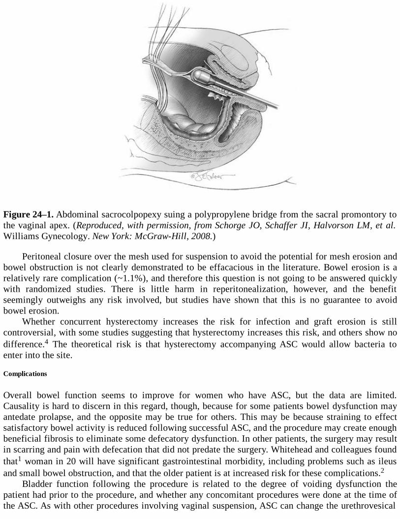
Figure 24–1. Abdominal sacrocolpopexy suing a polypropylene bridge from the sacral promontory tothe vaginal apex. (Reproduced, with permission, from Schorge JO, Schaffer JI, Halvorson LM, et al.Williams Gynecology. New York: McGraw-Hill, 2008.)
Peritoneal closure over the mesh used for suspension to avoid the potential for mesh erosion andbowel obstruction is not clearly demonstrated to be effacacious in the literature. Bowel erosion is arelatively rare complication (~1.1%), and therefore this question is not going to be answered quicklywith randomized studies. There is little harm in reperitonealization, however, and the benefitseemingly outweighs any risk involved, but studies have shown that this is no guarantee to avoidbowel erosion.
Whether concurrent hysterectomy increases the risk for infection and graft erosion is stillcontroversial, with some studies suggesting that hysterectomy increases this risk, and others show nodifference.4 The theoretical risk is that hysterectomy accompanying ASC would allow bacteria toenter into the site.
Complications
Overall bowel function seems to improve for women who have ASC, but the data are limited.Causality is hard to discern in this regard, though, because for some patients bowel dysfunction mayantedate prolapse, and the opposite may be true for others. This may be because straining to effectsatisfactory bowel activity is reduced following successful ASC, and the procedure may create enoughbeneficial fibrosis to eliminate some defecatory dysfunction. In other patients, the surgery may resultin scarring and pain with defecation that did not predate the surgery. Whitehead and colleagues foundthat1 woman in 20 will have significant gastrointestinal morbidity, including problems such as ileusand small bowel obstruction, and that the older patient is at increased risk for these complications.2
Bladder function following the procedure is related to the degree of voiding dysfunction thepatient had prior to the procedure, and whether any concomitant procedures were done at the time ofthe ASC. As with other procedures involving vaginal suspension, ASC can change the urethrovesical
angle and create urinary incontinence for the woman who did not have this problem preoperatively.The gynecologist then has to decide whether to do a routine prophylactic operation for urinaryincontinence if none is present when performing ASC, or wait until after the procedure to see if thereis incontinence and then operate for urine loss.
Complications of ASC and their mean occurrence rate include urinary tract infection (10.9%),wound problems, including hematoma and infection (4.6%), hemorrhage and/or transfusion (4.4%),cystotomy (3.1%), enterotomy (1.6%), ureteral injury (1%), and deep vein thrombosis (3.3%). Mesherosion occurred in 3% of patients reported in the largest review article. The Cochrane review of ASCconcluded that the abdominal approach had less apical failure, but a greater propensity forcomplications, while encountering less stress incontinence and postoperative dyspareunia.
Successful surgeons list some key concepts for best surgical outcomes: (1) use graft tissue ratherthan trying to approximate either the vagina or the uterus to the sacrum; (2) place the graft at the S1-S2 level; (3) do not strip the abdominal vaginal apex down to the mucosal level before affixing thesynthetic graft material to it; (4) place the graft between the anterior vagina and the sacrum withouttoo much tension to reduce the risk of postoperative stress incontinence; (5) use many sutures in thevaginal end of the graft to equilibrate tension on the graft and the vagina.4
Whether the abdominal or vaginal approach is preferable for a given patient with vaginal vaultprolapse is not clear from the extant literature. Prospective, randomized trials do not exist to answerthis question, and therefore factors which will determine the answer to this question will, of necessity,involve the patient’s age, the strength of her tissue, her lifestyle, the durability of the proposedoperative procedure, and the opinion and experience of the surgeon taking care of the patient. All ofthe above are pertinent when discussing the best approach for a given patient with vaginal prolapse.
Comprehension Questions
24.1 A 55-year-old woman presents to the gynecologist’s clinic with the complaint of “there’ssomething falling out of my vagina.” She had undergone an abdominal hysterectomy foruterine fibroids 15 years previously. She denies leakage of urine. On examination, she isnoted to have a third-degree vaginal vault prolapse. The gynecologist counsels her about theoptions of abdominal sacrocolpopexy versus sacral ligament fixation to address the problem.As compared to ASC, which of the following complications is seen more often with SSLF?
A. Urinary tract infectionB. Wound infectionC. Hemorrhage and/or transfusionD. Nerve injury
24.2 The gynecologist explains to the patient in question 24.1 that ASC has been referred to asthe “gold standard” for repair of vaginal vault prolapse. Which of the following is the reasonfor this label?
A. ASC is more cost-effective than vaginal procedures.B. ASC is less likely to fail and result in recurrent prolapse.C. ASC is associated with less pain than vaginal procedures.D. ASC is less likely to result in hemorrhage than vaginal SSLF and USLF.
24.3 A 69-year-old woman is noted to have a vaginal vault prolapse after a vaginal hysterectomyperformed 25 years previously for pelvic pain. The patient is in fairly good health and isvery active physically and sexually. She is 5 ft 4 in tall and weighs 180 lb (82 kg). After
being counseled about the various options, she agrees to sacral spinous ligament fixation.Which of the following is the most important preoperative management in her case?
A. Lose 10 lb (4.5 kg) of weight prior to surgery.B. Identify and treat any urinary tract infection.C. Identify and treat bacterial vaginosis.D. Apply topical estrogen to the vagina.
ANSWERS
24.1 D. Nerve injury is rare, occurring only in 1% to 2% of patients. With sacrospinous ligamentprocedures, the pudendal nerve can be entrapped.
24.2 B. ASC is considered the “gold standard” because there is less apical failure with thisoperation than with vaginal procedures for vault prolapse.
24.3 D. In a woman who is remote from menopause, the vaginal epithelium is typically thin andatrophic. Vaginal surgery is difficult in this circumstance and can lead to a greater incidenceof wound breakdown and infection. The technical aspects of vaginal surgery are likewisedifficult. Topical estrogen cream to the vaginal region can assist to allow the vaginalepithelium to be more pliable, thicker, and easier to manipulate in the OR.
Clinical Pearls
See Table 1-2 for definition of level of evidence and strength of recommendation
ASC is occasionally complicated by bleeding from the site of attachment of the graft tissue tothe anterior surface of the sacral vertebrae (Level B).
Visualize and avoid small arterioles coursing over the bone surface. If bleeding occurs, havesome sterile orthopedic thumbtacks available to push into the vertebra or bone wax to stop thebleeding (Level C).
The “vaginal hand” instrument, consisting of a rigid vaginal dilator attached to a right-angleretractor, is placed into the vagina to delineate the apex of the vault from within thelaparotomy incision. This instrument provides an easy method to manipulate the vagina whilemaintaining a sterile laparotomy field (Level C).
A thorough bowel preparation prior to this operation facilitates packing the empty bowel out ofthe way to gain access to the pelvic floor for correct placement of the graft (Level B).
REFERENCES
1. Nygaard IE, McCreery R, Brubaker L, et al. Abdominal sacrocolpopexy: a comprehensivereview. Obstet Gynecol. 2004:805-823.A definitive review of a challenging topic.
2. Whitehead WE, Bradley CS, Brown MB, et al. Pelvic floor network. Gastrointestinalcomplications following abdominal sacrocolpopexy for advanced pelvic organ prolapse. Am JObstet Gynecol. 2007;197:78.e1-7.
3. The American College of Obstetricians and Gynecologists. Pelvic organ prolapse. ACOGPractice Bulletin No. 85. Obstet Gynecol. 2007;110:717-730.
4. Bensinger G, Lind L, Lesser M, Guess M, Winkler HA. Abdominal sacral suspensions:analysis of complications using permanent mesh. Am J Obstet Gynecol. 2005;193:2094-2098.
5. Wu JM, Wells EC, Hundley AF, Connolly A, Williams KS, Visco AG. Mesh erosion inabdominal sacral colpopexy with and without concomitant hysterectomy. Am J Obstet Gynecol.2006;194:1418-1422.
6. Maher C, Baessler K. Surgical management of posterior vaginal wall prolapse: an evidencebased literature review. Int Urogynecol J. 2005;17:84-88.
Case 25
A 64-year-old woman presents with a chief complaint of a mass protruding from her vagina. Shedenies stress urinary incontinence, but she has to splint her posterior vagina to have bowelmovements. She had vaginal surgery for a similar problem 5 years ago, and she brought copies of theoperative reports. Your review of her records reveals that she had a routine anterior and posteriorcolporrhaphy, but no other pelvic surgery was performed at that time. She had a vaginal hysterectomy20 years earlier for vaginal prolapse. Her past medical history includes an appendectomy for aruptured appendix and two subsequent laparotomies for intestinal obstruction following theappendectomy. Physical examination confirms that the vaginal cuff exits the vagina with a Valsalvamaneuver, and she also has extremely thin, atrophic vaginal mucosa. Repositioning the vaginal cuff atthe apex and asking the patient to repeat the Valsalva maneuver demonstrates a large second-degreecystocele and a large second-degree rectocele.
What is the most likely diagnosis?
What is the best therapy for this condition?
ANSWERS TO CASE 25:Vaginal Vault Prolapse Post-hysterectomy
Summary: A 64-year-old woman presents with vaginal vault prolapse following prior hysterectomyand anterior and posterior repairs. She is not a good candidate for abdominal surgery because ofintestinal obstruction following appendectomy surgery. She has failed conventional vaginal repairsurgery.
Most likely diagnosis: Recurrent vaginal vault prolapse and failed conventional cystocele andrectocele repair.
Best therapy: Vaginal paravaginal defect repair using biological graft tissue, and posteriorcolporrhaphy using biological graft tissue. Support of the vaginal apex, whether bysacrospinous ligament fixation or uterosacral ligament suspension is critical to the long-termsuccess of the operative procedures to repair the cystocele and the rectocele.
ANALYSIS
Objectives
1. Become familiar with the concepts for using graft tissue in vaginal reconstructive surgery.2. Know the indications for using graft tissue in vaginal reconstructive surgery.3. Learn the contraindications and complications associated with the use of graft tissue in the
vagina.
Considerations
This case report details a 64-year-old patient who had undergone previous surgery for pelvic organprolapse without long-term success. Her vaginal tissue is thin, atrophic, and weak, and she is notlikely to benefit from an operative procedure which duplicates prior failed attempts. Additionally, sheis not likely to consent to an abdominal operation which will place her at risk for another episode ofintestinal obstruction. Facing such a surgical challenge, some gynecologists would consider the use ofgraft tissue to reinforce the vaginal repairs which this patient requires.
The initial approach to this patient should include urologic evaluation to see if her urinarycontinence is due to the fact that the urethra is kinked because of the degree of prolapse she isexperiencing. Whether she will need a mid-urethral sling as part of the combined operative proceduresfor repair of pelvic floor relaxation, or later, if urinary incontinence develops following surgery, needsto be discussed preoperatively. Randomized trials have shown that the patient who is stress continentprior to surgery for prolapse may become incontinent following surgery, and that placement oftension-free vaginal tape (TVT) as an adjunct to the prolapse surgery will statistically reduce thelikelihood of postoperative stress incontinence.
Grafts have been employed in vaginal vault prolapse surgery for over 50 years, but their useis still controversial. The decision to proceed with the use of graft tissue should be considered onlywhen the patient’s endogenous tissue is inadequate for a nongrafted repair. Additionally, the prolapseshould have a significant negative impact on the patient’s quality of life, and the degree of prolapseshould be to the vaginal introitus or beyond.1
Any discussion of graft tissue is incomplete without some consideration of the significant cost ofusing these materials. Several square inches of synthetic or biological materials usually cost about$1000. The cost equation also must include the increased cost of OR time to position these productscorrectly.2,3
APPROACH TO Using Graft Tissue in Vaginal Hernia Repairs
DEFINITIONS
XENOGRAFT: A surgical graft of tissue from one species to an unlike species (or genus or family).
MESH GRAFT: Prosthetic nonabsorbable porous material used to give strength or bulk for surgicalrepairs of pelvic organ prolapsed.
BIOLOGICAL GRAFT: A portion of living tissue that is used to join to another living tissue. It canbe from the same organism (autologous) or same species, or different species (xenograft).
CLINICAL APPROACH
There are two broad categories of graft material available for use in vaginal hernia repairs, those beingeither synthetic or biological products. Within these two categories, there are many of materialavailable. Any listing of the choices available is somewhat futile, as the field is being inundated on aregular basis with new products and materials too rapidly for the individual surgeon to remain currentwith the choices available. Additionally, there is a dearth of randomized, prospective, controlledtrials clearly demonstrating the superiority of one product over another.3 No matter which type ofgraft tissue is chosen, surgeons should not believe that graft tissue use will compensate for mediocre
performance in the OR, or for choosing a patient who is not a good candidate for graft surgery. Manyauthorities in this field are of the opinion that the use of any type of graft tissue should be reserved forthose select patients in whom a conventional operative procedure has already resulted in failure.4
This is a controversial arena with a dearth of prospective randomized clinical trials tosubstantiate claims of effectiveness and safety. ACOG published a practice bulletin in February 2007in reaction to the FDA warning of numerous vaginal mesh erosions and complications and the plethoraof graft products and “kits” for the correction of pelvic organ prolapse. The initial ACOG publicationdescribed these products as “experimental.” Enough protest ensued over the use of the word“experimental” that the Practice Bulletin was withdrawn and republished in August 2007 without theuse of that descriptive language.3 However, surgeons should not, based on the current state of the art,be using graft tissue indiscriminately just because it is possible to do so, and the discerning surgeonshould be able to determine those situations that appropriate and inappropriate for the use of grafttissue.
Synthetic Graft Tissue
A comparison of synthetic to biological material demonstrates that the synthetic product may have, atleast initially, greater success rates, but the biological product is probably better tolerated for a longerduration of time. Synthetic grafts are indeed permanent, and erosion of the graft through avaginal or intestinal wall into the vagina or the bowel lumen is a lifetime risk after graftplacement. Surgical case series that report several months to a few years of successful symptom reliefdo not predict the future safe compartmentalization of synthetic mesh in anterior or posterior herniarepairs. Vaginal tissue ages, atrophies, and loses its innate tensile strength, which predisposes the grafttissue to erode through a previously intact vaginal wall.
Permanent graft products are most likely to be successfully incorporated into the hernia spacesthey are designed to reinforce if their pore size is large enough to allow ingrowth of new endogenousfibrous tissue and blood supply. Synthetic graft products with small, tight interstices are less prone tobe well integrated into the surrounding tissue, which may result in graft erosion or infection. Whenusing synthetic graft tissue for repairing cystoceles and rectoceles, the vaginal mucosal thicknesscovering the graft should be as thick as possible, to reduce the risk of erosion and to ensure a goodblood supply to the endogenous tissue overlying the graft.
If graft erosion of synthetic mesh occurs into the vagina, it may respond to several weeks oftopical application of an estrogen cream to encourage over-growth of vaginal epithelium. If this is notsuccessful, trimming the protuberant mesh until it is flush with or slightly beneath the mucosalsurface may stimulate successful overgrowth of vaginal mucosa. Infected mesh must be removed.
The consequence of graft tissue eroding through the vaginal mucosa has created a newdescriptive term, “hispareunia,” describing the phenomenon experienced by the male partner of apatient who encounters sharp, stubby synthetic graft material eroding through the vagina with thepotential to lacerate his penis.5
Biological Graft Tissue
The ideal biological graft is sterile, noncarcinogenic, inert, durable, inexpensive, easy to use, andmaintains its shape. Biological grafts are contraindicated in patients with the following conditions:heavy smoking, morbid obesity, poorly controlled diabetes, on systemic corticosteroids,immunocompromised, active pelvic or vaginal infection, or prior pelvic irradiation. Biological grafttissue may be autologous graft tissue from rectus fascia of fascia lata, or allograft tissue from fascialata or dura mater, or xenograft tissue from porcine or bovine sources. Xenograft tissue has been
treated to remove the cellular component, leaving only a collagen matrix, which has minimal potentialto trigger a rejection phenomenon. Recent evidence indicates that cadaveric fascial tissue inabdominal sacrocolpopexy should no longer be used. The variable thickness and strength of cadavericdermis, compared to the consistency of bovine and porcine source tissue, make the latter preferable tothe former. Clear superiority of bovine versus porcine tissue has not been demonstrated in any long-term prospective trials. Xenograft tissue does have the advantage of providing a scaffold-type matrixfor endogenous tissue to use in fabricating the ingrowth of new fibrous tissue with its ownneovascularization, but it degenerates over time and does not provide the long-term strength ofsynthetic material.
In summary, grafts are probably more useful in repairing the anterior rather than theposterior vaginal wall. Patient selection is critical, and patients should be counseled, in a carefullydescribed and documented consent process, that although grafts are being used more commonly, thistechnology is still relatively new and expensive, with unique potential complications. Gynecologicsurgeons need to gain knowledge and experience with graft materials if they plan to use them tocorrect pelvic floor defects, and be able to handle the complications which may ensue following graftplacement. Given the rapid introduction of new graft products, surgeons should demand datademonstrating that new materials are safe and effective.6
Comprehension Questions
25.1 A healthy 55-year-old woman presents with a symptomatic cystocele, rectocele, andenterocele. She had a vaginal hysterectomy for uterine prolapse performed 10 yearspreviously. Which operative procedure should be avoided in this patient?
A. Anterior and posterior colporrhaphy using synthetic graft material, sacrospinous ligamentfixation
B. Conventional anterior and posterior colporrhaphy, enterocele repair via sacrospinous ligamentfixation or uterosacral ligament suspension
C. Laparoscopic sacrospinous ligament fixation with conventional anterior and posteriorcolporrhaphy
D. Abdominal sacrocolpopexy, Burch procedure, conventional posterior colporrhaphy
25.2 A 58-year-old patient returns 6 weeks following vaginal repair surgery for recurrentcystocele. She is afebrile, but complains of persistent vaginal discharge. She has not beenusing any medication vaginally. Bovine graft tissue was used to repair the cystocele, and a1.5 × 1.0 cm portion of graft material is visible 3 cm inside the introitus in the anteriorrepair suture line. What is the therapy of choice?
A. Return the patient to the operating room and remove the graft.B. Start the patient on oral antibiotic therapy.C. Trim the exposed graft in the clinic.D. Instruct the patient to begin using estrogen vaginal cream.
25.3 A 48-year-old woman weighing 265 lb (120.20 kg), with a 10-year history of systemic lupuserythematosus presents with cystocele and rectocele. She states that multiple types ofpessaries have not controlled her prolapse symptoms. She is taking oral steroids andmethotrexate to control her lupus. What vaginal graft tissue is indicated for repairing thispatient’s prolapse?
A. Polypropylene mesh
B. Fetal bovine fasciaC. Porcine meshD. None of the above
ANSWERS
25.1 A. The use of graft tissue is not usually needed the first time when a patient has vaginalrepair surgery if her endogenous tissue is satisfactory. Graft tissue can lead to erosion,infection, or pain.
25.2 D. Vaginal estrogen cream use for several weeks will stimulate the vaginal mucosa to coverthe visible graft tissue. This has very little systemic absorption and works well instrengthening the tissue locally.
25.3 D. The use of any type of graft tissue is contraindicated in morbidly obese patients, and inpatients who are using systemic steroids.
Clinical Pearls
See Table 1-2 for definition of level of evidence and strength of recommendation
Soak the graft in an antibiotic solution before using it to reduce the risk of postoperativeinfection (Level B).
Sew the graft into place—do not simply lay it into a cystocele or rectocele space and expect itto stay in the intended position. These materials have a propensity to migrate and bunch if notanchored with suture (Level B).
Cover the defect with the graft such that it lays flat and smooth, but not under tension (LevelC).
Use the carpenter’s adage of “Measure twice, cut once” when working with graft tissue. Thismaterial is too expensive to waste. Be sure you do not create “a square peg to go into a roundhole.” Make a pattern using some sterile paper, and then cut the graft using the pattern as aguide. Err on the side of making it too big and then trim excess graft on the edges aftersuturing it into place (Level C).
REFERENCES
1. Davila GW, Drutz H, Deprest J. International Urogynecological Association: the usage ofgrafts in pelvic reconstructive surgery symposium 2005. Int Urogynecol J. 2006;17:S51-S55.
2. Norton P. New technology in gynecologic surgery: is new necessarily better? Obstet Gynecol.2006;108:707-708.
3. American College of Obstetricians and Gynecologists. Pelvic organ prolapse. ACOG Practice

Bulletin No. 85. Obstet Gynecol. 2007;110:717-730.
4. Botros SM, Sand PK. Cystocele and rectocele repair: more success with mesh? OBG Mgmt.2006;18:30-43.
5. Brubaker L. Partner dyspareunia (hispareunia). Int Urogynecol J. 2006;17:311.
6. Karram M. Pelvic organ prolapse: which operation for which patient? OBG Mgmt. 2006;18:72-84.
Case 26
A 46-year-old G2P2002 woman complains of leakage of urine with coughing and sneezing that hasworsened over the last several years requiring her to use two to three pads per day despite pelvic floorexercises. She complains that this has adversely affected her social life and is seeking treatment forher condition. She also has occasional urgency and frequency but denies leakage of urine associatedwith the urge to void. She denies any difficulty emptying her bladder. The rest of her medical historyis unremarkable with the patient having had two vaginal deliveries without complications and no priorsurgeries. A gynecologic examination is performed and the patient is found to have a normal vagina, asmall mobile uterus without significant prolapse, and no palpable adnexal masses.
What is the most likely diagnosis?
What further physical examination should be performed?
What further testing should the patient undergo?
What minimally invasive surgical options are available for the treatment of stress urinaryincontinence (SUI) with a hypermobile urethra?
ANSWERS TO CASE 26:Stress Urinary Incontinence
Summary: This is a 46-year-old G2P2002 woman who complains of urinary leakage with activitiesassociated with increased abdominal pressure. She also has urgency and frequency, but this has notbeen associated with leakage. She has no prior surgeries and an unremarkable history other than twovaginal deliveries.
Most likely diagnosis: Stress urinary incontinence.
Additional physical examination: The urethra should be examined for urethral hypermobilityby direct visualization and/or a Q-tip test (placement of a cotton swab into the urethra to judgeurethral mobility). Additionally, a supine empty stress test (SEST) may be performed if thepatient has voided recently and a post void residual (PVR) should be determined by in-and-outcatheterization or bladder ultrasound.
Additional testing: The patient should have a urinalysis to rule out a urinary tract infectionthat may be worsening her condition. Additionally, the patient should undergo urodynamictesting to further evaluate her lower urinary tract to determine if her leakage is caused by SUI,detrusor overactivity, or both (mixed incontinence).
Treatment for SUI with urethral hypermobility: Mid-urethral slings are an excellenttreatment for women who suffer from SUI with urethral hypermobility.
ANALYSIS
Objectives
1. Know the workup for urinary incontinence.2. Know the indications for different incontinence surgeries.3. Be familiar with the intra- and postoperative complications of incontinence surgeries.
Considerations
This is a 46-year-old parous woman who has a history of urinary incontinence associated withcoughing and sneezing. She has had two prior vaginal deliveries, which are risk factors for thiscondition. The history is very important to determine if the patient has any “red flags” to indicate amore complicated condition other than pure genuine stress incontinence. These red flags includenocturia, urgency, frequency, dysuria, delay from cough to leakage, or prior urinary incontinencesurgery. This patient complains also of urgency and frequency, but these symptoms don’t seem to berelated to urinary incontinence; nevertheless, these complaints may indicate a mixed incontinence.Cytometric evaluation would be important to evaluate for the degree of stress versus urgeincontinence, since these findings would guide therapy.
APPROACH TO Stress Urinary Incontinence
DEFINITIONS
DETRUSOR OVERACTIVITY: A urodynamic observation characterized by involuntary detrusorcontractions during the filling phase which may be spontaneous or provoked.
MIXED URINARY INCONTINENCE: The complaint of involuntary leakage associated withurgency and also with exertion, effort, sneezing, or coughing.
STRESS URINARY INCONTINENCE: The complaint of involuntary leakage on effort or exertion,or on sneezing or coughing.
URGE URINARY INCONTINENCE: The complaint of involuntary leakage accompanied by orimmediately preceded by urgency.1
CLINICAL APPROACH
Multiparous patients who complain of SUI without a history of prior incontinence procedures usuallytend to have hypermobile urethras from what is thought to be injury to supportive structures duringthe birthing process. Women with SUI and hypermobile urethras are excellent candidates for mid-urethral slings whereas those with nonhypermobile urethras may be better suited for treatment by wayof intraurethral bulking agents or bladder neck slings. Currently, there are numerous products on themarket, and more being introduced every day, with some devices having been studied in more depththan others. The best procedure for the patient is the one that the surgeon is comfortable performingand best suits the patient’s specific problem. When using mid-urethral slings, it has becomeincreasingly clear that the sling material should be made of monofilament polypropylene withinterstices sufficiently wide to allow tissue incorporation and penetration of neutrophils to combat
infection. Prior to surgery, the patient should be examined by some method of objective testing tofurther evaluate the patient’s complaint.
DIAGNOSTICS
Typically before an incontinence procedure is performed, the patient undergoes urodynamic testing.This may be as simple as placing a 16-Fr red rubber catheter through the urethral meatus into thebladder and filling the bladder with sterile saline through a 60-mL syringe attached to the catheterwith the plunger removed. By filling with gravity, the saline enters more slowly than pushing the fluidwith the syringe’s plunger. It also allows the meniscus of the syringe to be observed. A rise in themeniscus indicates a bladder contraction. In this situation, the patient may be better served bymultichannel urodynamics to further evaluate the lower urinary tract. As the bladder is filled, thepatient’s first sensation and first urge to void may be recorded. Once capacity or 250 to 300 mL isreached, whichever is achieved first, the catheter is removed. The patient is asked to perform Valsalvamaneuver and cough several times, and the practitioner should look for leakage that would imply SUI.If leakage is not seen with the patient supine, the patient should be asked to stand upright with stresstesting repeated.
The advantage of simple cystometrics is that it is inexpensive and can be performed in a clinicsetting without complicated and expensive equipment. However, this technique does not allow therecording of bladder pressures during stress maneuvers to determine the leak point pressure (LPP),which is the abdominal pressure sufficient to overcome the continence mechanism to result in leakageof urine. It is also unable to determine if the leakage seen is caused by a detrusor contraction duringthe stress maneuvers.
Complex or multichannel urodynamics involves placing one catheter through the urethra into thebladder to measure vesical pressure and another into the rectum or vagina to measure abdominalpressure. These catheters are then connected to a computer that then displays the pressures generatedon a computer screen. The detrusor pressure is then calculated by subtracting the abdominal pressurefrom the vesical pressure. The fill rate can be controlled and usually varies from 50 to 80 mL/min.Needle electrodes can also be placed into the striated urethral sphincter or patch electrodes applied tothe perineum to assess the muscle activity of the striated sphincter. LPP testing is performed atdifferent intervals usually starting at 150 mL to evaluate for SUI. Detrusor overactivity is diagnosedby a rise in detrusor pressure signifying an increase in bladder pressure without an increase inabdominal pressure. Leakage during cough or Valsalva without a rise in detrusor pressure indicatesSUI. Other tests that may be performed include a urethral pressure profile or maximum urethralclosure pressure (MUCP). The MUCP is performed by placing a urethral catheter into the bladder andthen slowly removing the catheter. A peak is then generated as the pressure increases as the catheterpasses through the continence mechanism with the MUCP being the maximal pressure recorded. Thistest as well as a low LPP may help identify patients with intrinsic sphincter deficiency (ISD).
TREATMENT
History
In the early 1900s, slings were first placed at the bladder neck to treat SUI.2 In 1942, Aldridge was thefirst to describe using autologous rectus fascia at the bladder neck.3 This procedure which has beenmodified by different surgeons is the basis for the modern day bladder neck sling. Placed at the levelof the bladder neck and proximal urethra, these slings are tunneled by a variety of techniques behind
the pubis through the retropubic space and can be secured to itself, the rectus fascia, the periosteum ofthe pubis, or by way of bone anchors. Tensioned correctly, the sling gives support and an adequateamount of compression to prevent incontinence while not causing obstruction sufficient to result invoiding dysfunction. After Aldridge in 1949, Marshall described the first retropubic colposuspensionfor the treatment of SUI. Called the Marshall-Marchetti-Krantz (MMK), this procedure entailedplacing sutures into the endopelvic fascia along the bladder neck and into the periosteum.4 Bysuspending the endopelvic sutures to Cooper ligament instead of the periosteum, the MMK wasmodified and renamed the Burch colposuspension. Its first results were published in 1968.5 The Burchcolpo-suspension has been one of the most widely used and studied incontinence procedures. Althoughthe bladder neck sling and Burch colposuspension have comprised the majority of incontinenceprocedures performed since the 1950s, today they have been largely replaced by the mid-urethralslings developed in the 1990s that continue to evolve into less invasive procedures.
Types of Slings
Bladder Neck Slings As the name implies, bladder neck slings are placed at the level of the bladderneck which is located by using the balloon of a Foley catheter. These slings are placed by passingsuture down through the retropubic space into an incision at the level of the bladder neck/proximalurethra, anchored into each end of the sling material and passed through the retropubic space inreverse fashion where the suture is then tied anchoring the sling into place. Materials used in bladderneck sling construction can be autologous (ie, rectus facia, fascia lata), allograft (ie, cadaveric fascia,cadaveric dermis), xenograft (ie, porcine dermis, bovine pericardium, submucosal intestine), orsynthetic (ie, polypropylene). Some studies have demonstrated autologous slings to be superior tothose constructed from allograft material.6 Bladder neck slings are commonly used to treat patientswith severe ISD and/or a nonhypermobile urethra, previously failed midurethral sling procedures, andpatients with high risk for synthetic mesh extrusion or erosion. In a comparison study between anintraurethral bulking agent and autologous pubovaginal sling in patients with ISD and largelynonhypermobile urethras, the pubovaginal sling had a much greater objective success rate (81% vs9%, P < .001), thus demonstrating its good efficacy in this cohort of patients that has been historicallydifficult to treat.7
Complications of bladder neck slings include incidental cystotomy and new-onset urgency.Incomplete emptying and detrusor overactivity may also result from bladder outlet obstruction from asling tensioned too tightly. This surgical technique has been well studied with proven durability. Thedownside of this technique is the required suprapubic incision, increased vaginal dissection, and tissueharvesting with autologous slings that increase operative time and risk of morbidity.
Mid-urethral Slings In 1990 Petros and Ulsten published a theory paper where they described bothstress and urge symptoms being derived from a common anatomical site, a lax vagina. They proposedthat this laxity may derive from the vaginal wall itself or the supporting structures of the vaginal wallwhich include ligaments, muscles, and connective tissue insertions.8 Based on this theory, the tension-free vaginal tape (TVT) procedure was created to restore urethral support by a tunneled strip ofmonofilament polypropylene mesh placed at the mid-urethra. The mesh is passed by a trocar that istunneled from a suburethral incision at the mid-urethral level through the endopelvic fascia and intothe retropubic space. It then passes through the layers of the anterior abdominal wall and out throughthe skin just above the pubis. This was first performed under local anesthesia with a mean operativetime of 22 minutes and a cure rate of 84% at 2 years in patients with SUI and urethral hypermobility.9
Some data suggest that in patients with a low MUCP, a nonhypermobile urethra, or both the cure rate
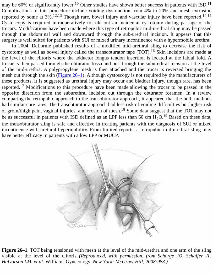
may be 60% or significantly lower.10 Other studies have shown better success in patients with ISD.11
Complications of this procedure include voiding dysfunction from 4% to 20% and mesh extrusionreported by some at 3%.12,13 Though rare, bowel injury and vascular injury have been reported.14,15
Cystoscopy is required intraoperatively to rule out an incidental cystotomy during passage of thetrocars. Modifications have been made where this type of retropubic mid-urethral sling may be passedthrough the abdominal wall and downward through the sub-urethral incision. It appears that thissurgery is well suited for patients with SUI or mixed urinary incontinence with a hypermobile urethra.
In 2004, DeLorme published results of a modified mid-urethral sling to decrease the risk ofcystotomy as well as bowel injury called the transobturator tape (TOT).16 Skin incisions are made atthe level of the clitoris where the adductor longus tendon insertion is located at the labial fold. Atrocar is then passed through the obturator fossa and out through the suburethral incision at the levelof the mid-urethra. A polypropylene mesh is then attached and the trocar is reversed bringing themesh out through the skin (Figure 26–1). Although cystoscopy is not required by the manufacturers ofthese products, it is suggested as urethral injury may occur and bladder injury, though rare, has beenreported.17 Modifications to this procedure have been made allowing the trocar to be passed in theopposite direction from the suburethral incision out through the obturator foramen. In a reviewcomparing the retropubic approach to the transobturator approach, it appeared that the both methodshad similar cure rates. The transobturator approach had less risk of voiding difficulties but higher riskof groin/thigh pain, vaginal injuries, and erosion of mesh.18 Some data suggest that the TOT may notbe as successful in patients with ISD defined as an LPP less than 60 cm H2O.19 Based on these data,the transobturator sling is safe and effective in treating patients with the diagnosis of SUI or mixedincontinence with urethral hypermobility. From limited reports, a retropubic mid-urethral sling mayhave better efficacy in patients with a low LPP or MUCP.
Figure 26–1. TOT being tensioned with mesh at the level of the mid-urethra and one arm of the slingvisible at the level of the clitoris. (Reproduced, with permission, from Schorge JO, Schaffer JI,Halvorson LM, et al. Williams Gynecology. New York: McGraw-Hill, 2008:983.)
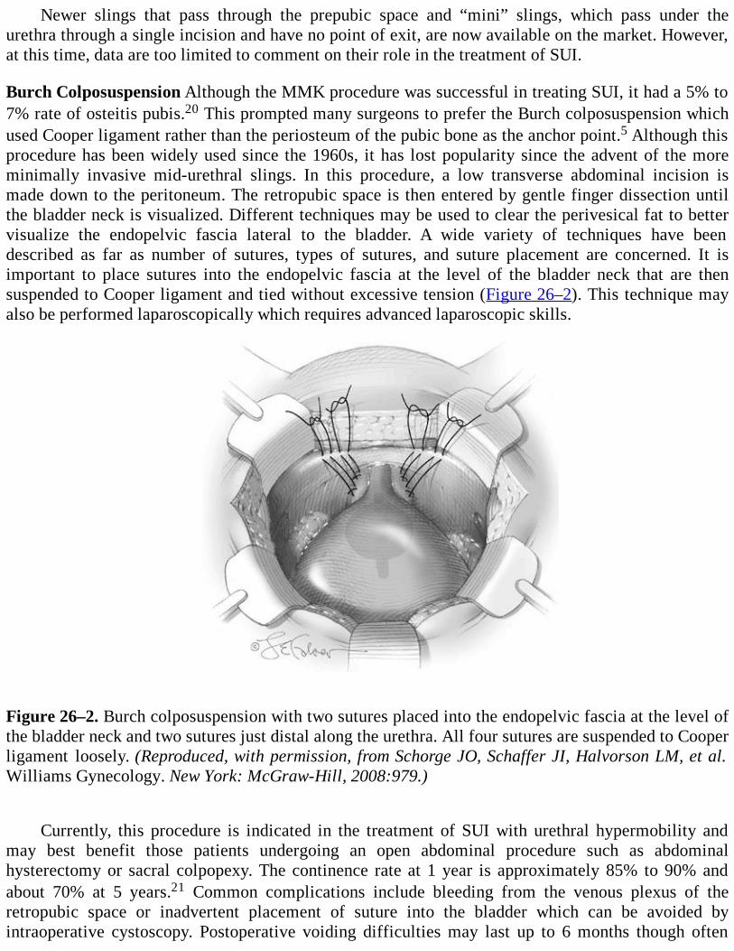
Newer slings that pass through the prepubic space and “mini” slings, which pass under theurethra through a single incision and have no point of exit, are now available on the market. However,at this time, data are too limited to comment on their role in the treatment of SUI.
Burch Colposuspension Although the MMK procedure was successful in treating SUI, it had a 5% to7% rate of osteitis pubis.20 This prompted many surgeons to prefer the Burch colposuspension whichused Cooper ligament rather than the periosteum of the pubic bone as the anchor point.5 Although thisprocedure has been widely used since the 1960s, it has lost popularity since the advent of the moreminimally invasive mid-urethral slings. In this procedure, a low transverse abdominal incision ismade down to the peritoneum. The retropubic space is then entered by gentle finger dissection untilthe bladder neck is visualized. Different techniques may be used to clear the perivesical fat to bettervisualize the endopelvic fascia lateral to the bladder. A wide variety of techniques have beendescribed as far as number of sutures, types of sutures, and suture placement are concerned. It isimportant to place sutures into the endopelvic fascia at the level of the bladder neck that are thensuspended to Cooper ligament and tied without excessive tension (Figure 26–2). This technique mayalso be performed laparoscopically which requires advanced laparoscopic skills.
Figure 26–2. Burch colposuspension with two sutures placed into the endopelvic fascia at the level ofthe bladder neck and two sutures just distal along the urethra. All four sutures are suspended to Cooperligament loosely. (Reproduced, with permission, from Schorge JO, Schaffer JI, Halvorson LM, et al.Williams Gynecology. New York: McGraw-Hill, 2008:979.)
Currently, this procedure is indicated in the treatment of SUI with urethral hypermobility andmay best benefit those patients undergoing an open abdominal procedure such as abdominalhysterectomy or sacral colpopexy. The continence rate at 1 year is approximately 85% to 90% andabout 70% at 5 years.21 Common complications include bleeding from the venous plexus of theretropubic space or inadvertent placement of suture into the bladder which can be avoided byintraoperative cystoscopy. Postoperative voiding difficulties may last up to 6 months though often
resolve. Changes in anatomy may predispose patients to posterior wall defects, and between 12% and18.5% of patients may have detrusor overactivity. Those who have had prior unsuccessful surgery,particularly with a low MUCP, the elderly, and those with poor tissue from weaker collagen may be atrisk for increased failure.20 A National Institutes of Health (NIH)-sponsored trial also has shown theBurch colposuspension to be protective against new-onset SUI in patients undergoing abdominalsacral colpopexy.22
Comparisons
Although data are limited in comparing these different surgical treatments, a few large studies deservemention. The autologous pubovaginal sling was recently compared to the Burch colposuspension in aprospective, randomized, multicenter NIH-sponsored trial. In this study at 24 months, success fortreating SUI was significantly higher in the fascial sling group (66% vs 49%, P < .001) though theprocedure was associated with more urinary tract infections, difficulty voiding, and postoperative urgeincontinence.23 When comparing the TVT to older procedures with good success such as the Burchcolposuspension, one randomized, controlled trial with 5-year follow-up found no statisticallysignificant difference between the groups in terms of a 1-hour pad test. The Burch group had anincrease in enterocele and rectocele while the TVT had tape-related complications in 8% of patients.24
Currently, multicenter studies are underway comparing the TOT to the TVT to better understand thedifferences between these procedures.
Comprehension Questions
26.1 A 34-year-old woman complains of urinary incontinence that is worsening. Which of thefollowing statements regarding treatment of the incontinence is most accurate?
A. Cystometric evaluation is necessary before performing a surgical procedure.B. The presence of nocturia is consistent with SUI.C. Mid-urethral sling procedures have been noted to be equivalent to Burch urethropexy
procedures in terms of efficacy.D. Urge incontinence is best treated by urethropexy.
26.2 A cystometric evaluation that is consistent with genuine SUI demonstrates which of thefollowing?
A. Small bladder capacityB. Detrusor contractionsC. Normal sphincter pressureD. Residual volume of 200 mL
26.3 In comparing the advantages of a TVT versus a TOT, which of the following statements ismost accurate?
A. TOT seems to have more voiding dysfunction than TVT.B. TOT seems to have a lower rate of cystotomy.C. TOT is better suited for patients with a hypermobile urethra.D. TOT is better suited for patients with a lower urethral closure pressure.
ANSWERS
26.1 C. Patients with pure SUI do not usually have nocturia, urge, or urinary frequency. If thehistory and physical examination are consistent with genuine stress incontinence, a formalcystometric evaluation does not need to be performed prior to surgery. Urge incontinence istreated medically rather than surgically.
26.2 C. Patients with genuine stress incontinence may have normal or somewhat lower urethralsphincter pressures. They usually have normal bladder capacity and no component of urge ordysuria and absence of detrusor contractions.
26.3 B. TOT procedures seem to have a lower rate of voiding dysfunction and lower rate ofcystotomy. TVT procedures seem to be better suited for patients with a hypermobile urethraor lower urethral closing pressures.
Clinical Pearls
See Table 1-2 for definition of level of evidence and strength of recommendation
Patients with ISD, especially those as defined by a nonhypermobile urethra, may be besttreated by a bladder neck sling (Level B).
Though data are limited, autologous rectus fascial slings may be more efficacious than slingsconstructed from allograft or xenograft material (Level C).
Mid-urethral slings composed of wide-spaced monofilament polypropylene have lowercomplications than those composed of multifilament weave (Level B).
The TVT may be more efficacious than the TOT in treating patients with a hypermobileurethra and low LPP or MUCP (Level B).
The TOT appears to have less voiding dysfunction than the TVT and a lower rate of cystotomy(Level B).
The Burch colposuspension is an effective procedure that may be considered in patientsalready undergoing an abdominal incision who require incontinence surgery for SUI (Level B).
REFERENCES
1. Abrams P, Cardozo L, Fall M, et al. The standardisation of terminology of lower urinary tractfunction: report from the Standardisation Sub-committee of the International ContinenceSociety. Neurourol Urodyn. 2002;21:167-178.
2. Kobashi KC, Leach GE. Stress urinary incontinence. Curr Opin Urol. 1999;9:285-290.
3. Aldridge AH. Transplantation of fascia for relief of urinary stress incontinence. Am J ObstetGynecol. 1942;44:398-411.
4. Marshall VF, Marchetti AA, Krantz KE. The correction of stress incontinence by simple
4. Marshall VF, Marchetti AA, Krantz KE. The correction of stress incontinence by simplevesicourethral suspension. Surg Gynecol Obstet. 1949;88:509-518.
5. Burch JC. Urethrovesical fixation to Cooper’s ligament for correction of stress incontinence,cystocele, and prolapse. Am J Obstet Gynecol. 1961;81:281-290.
6. Howden NS, Zyczynski HM, Moalli PA, Sagan ER, Meyn LA, Weber AM. Comparison ofautologous rectus fascia and cadaveric fascia in pubovaginal sling outcomes. Am J ObstetGyecol. 2006;194:1444-1449.
7. Maher CF, O’Reilly BA, Dwyer PL, Carey MP, Cornish A, Schlutter P. Pubovaginal slingversus transurethral Macroplastique for stress urinary incontinence and intrinsic sphincterdeficiency: a prospective randomised controlled trial. BJOG. 2005;112(6):797-801.
8. Petros PE, Ulsten UI. An integral theory of female urinary incontinence. Experimental andclinical considerations. Acta Obstet Gynecol Scand Suppl. 1990;153:7-31.
9. Ulsten U, Henriksson L, Johnson P, Varhos G. An ambulatory surgical procedure under localanesthesia for treatment of female urinary incontinence. Int Urogynecol J Pelvic FloorDysfunct. 1996;7(2):81-85.
10. Clemons JL, LaSala CA. The tension-free vaginal tape in women with a nonhypermobileurethra and low maximum urethral closure pressure. Int Urogynecol J. 2007;18:727-732.
11. Segal JL, Vassallo BJ, Kleeman SD, Hungler M, Karram MM. The efficacy of the tension-free vaginal tape in the treatment of five subtypes of stress urinary incontinence. IntUrogynecol J. 2006;17:120-124.
12. Salin A, Conquy S, Elie C, et al. Identification of risk factors for voiding dysfunctionfollowing TVT placement. Eur Urol. 2007;52(3):782-787.
13. Giri SK, Sil D, Narasimhulu G, Flood HD, Skehan M, Drumm J. Management of vaginalextrusion after tension-free vaginal tape procedure for urodynamic stress incontinence.Urology. 2007;69:1077-1080.
14. Kobashi KC, Govier FE. Perioperative complications: the first 140 polypropylenepubovaginal slings. J Urol. 2003;170(5):1918-1921.
15. Sivanesan K, Abdel-fattah M, Ghani R. External iliac artery injury during insertion oftension-free vaginal tape: a case report and literature review. Int Urogynecol J. 2007;18:1105-1108.
16. Delorme E, Droupy S, de Tayrac R, Delmas V. Transobturator tape(Uratape): a newminimally-invasive procedure to treat female urinary incontinence. Eur Urol. 2004;45:203-207.
17. Smith PP, Appell RA. Transobturator tape, bladder perforation, and paravaginal defect: a casereport. Int Urogynecol J. 2007;18(1):99-101.
18. Latthe PM, Foon R, Toozs-Hobson P. Transobturator and retropubic tape procedures in stress
18. Latthe PM, Foon R, Toozs-Hobson P. Transobturator and retropubic tape procedures in stressurinary incontinence: a systemic review and meta-analysis of effectiveness and complications.BJOG. 2007;114(5):522-531.
19. O’Conner RC, Nanigian DK, Lyon MB, Ellison LM, Bales GT, Stone AR. Early outcomes ofmid-urethral slings for female stress urinary incontinence stratified by Valsalva leak pointpressure. Neurourol Urodyn. 2006;25(7):685-688.
20. Bidmead J, Cardozo L. Retropubic urethropexy (Burch colposuspension). Int Urogynecol J.2001;12:262-265.
21. Lapitan MC, Cody DJ, Grant AM. Open retropubic colposuspension for urinary incontinencein women. Cochrane Database Syst Rev. 2003;1:CD002912.
22. Brubaker L, Cundiff GW, Fine P, et al. Abdominal sacrocolpopexy with Burchcolposuspension to reduce urinary stress incontinence. N Engl J Med. 2006;354(15):1557-1566.
23. Albo ME, Richter HE, Brubaker L, et al. Burch colposuspension versus fascial sling to reducestress urinary incontinence. N Engl J Med. 2007;355(21):2143-2155.
24. Ward KL, Hilton P, on behalf of the UK and Ireland TVT Trial Group. Tension-free vaginaltape versus colposuspension for primary urodynamic stress incontinence: 5-year follow up.BJOG. 2008;115:226-233.
Case 27
A 42-year-old G3P3003 woman is in lithotomy position to undergo a total laparoscopic hysterectomyfor a symptomatic 14-week leiomyomatous uterus. Her medical, surgical, and gynecologic historiesare unremarkable except for a history of three cesarean sections. An umbilical incision is made witheasy entry through the fascia for placement of a Hassan trocar. Several thin filmy adhesions are notedand taken down. The hysterectomy is then started by identifying and ligating the uterine-ovarianligaments and round ligaments with a tissue sealing device using thermal energy. Upon creation of thebladder flap, the bladder is noted to be adherent to the lower uterine segment. This is taken downsharply with some difficulty.
Once the uterine arteries are skeletonized bilaterally, they are ligated using the tissue sealingdevice. When the pedicle is divided, it appears that the device has moved and brisk bleeding is notedfrom what appears to be the right uterine artery pedicle. The pedicle is quickly grasped and anabsorbable suture is used to ligate the area of bleeding. The rest of the hysterectomy is then performedup to amputation of the cervix from the vagina without difficulty. During closure of the vaginal cuff,the bladder is noted to be in close proximity to the suture line of the cuff closure.
Where are potential points of injury to the urinary tract during this hysterectomy?
What are possible complications of unrecognized injury?
What is the next step in management of this patient?
ANSWERS TO CASE 27:Urinary Tract Injury During Gynecologic Surgery
Summary: This is a 42-year-old G3P3003 woman with three prior cesarean section deliveries and a 14-week leiomyomatous uterus who is undergoing a total laparoscopic hysterectomy. During surgery,further ligation by suture was required at the right uterine artery pedicle to obtain hemostasis. Thebladder was also adherent to the lower uterine segment and cervix. During closure of the vaginal cuff,the suture line was noted to be in close proximity to the bladder.
Points of injury: The bladder is at risk during creation of the bladder flap as well as closure ofthe vaginal cuff. The ureter is at greatest risk during ligation of the ovarian and uterine vessels.In this case, the ovarian vessels were not at risk as the uterine-ovarian ligament was divided.However, when dividing the uterine artery, further ligation was required to achieve hemostasis.
Unrecognized injury: Unrecognized injuries to the bladder could result in urinoma formation,urinary peritonitis, or vesicovaginal fistulas. With foreign bodies present, the bladder is at riskfor bladder calculi and recurrent infections. Unrecognized injury to the ureter could also resultin urinoma formation as well as hydronephrosis and loss of renal function caused by ureteralstricture or ligation.
Next step: Cystourethroscopy with intravenous indigo carmine is a minimally invasivediagnostic method to ensure ureteral patency and absence of bladder wall injury fromcystotomy or foreign body (suture, mesh, etc). When thermal energy is used, injury to the
urinary tract may present in a delayed fashion. Careful dissection of the ureter would also needto be performed to evaluate the distance of the suspected site of injury from the point ofapplication of thermal energy.
ANALYSIS
Objectives
1. Know the possible complications to the urinary tract during gynecologic surgery.2. Know the indications for cystourethroscopy in gynecologic surgery.3. Know the parts that comprise a cystourethroscope and appropriate uses of these parts for
intraoperative use during gynecologic procedures.
Considerations
This is a 42-year-old woman with three prior cesarean sections who is undergoing a total laparoscopichysterectomy for a symptomatic 14-week leiomyomatous uterus. There was significant difficultydeveloping the bladder flap and vesicovaginal space. Additionally, incomplete ligation of the rightuterine artery pedicle required further lateral suture ligation deeper into the surrounding tissue.Finally, the bladder was noted to be in close proximity to the suture line during vaginal cuff closure.These events have led the right ureter and bladder to be placed at significant risk for injury. Theretroperitoneal space may be opened at the time of surgery to allow identification of the ureter and itscourse to assure the surgeon of ureteral patency. Near the area where the uterosacral ligament insertsinto the uterus, the ureter may be as close as 0.9 ± 0.4 cm from the cervical portion of the uterosacralligament.1 Cystoscopy provides an easy, less invasive method to check the patency of the ureter at theureteral orifice in the bladder. It is especially helpful in cases where there is no abdominal accessduring surgery or in those patients with multiple adhesions or poor exposure that lead to a difficultretroperitoneal dissection. The bladder is also at risk for cystotomy during a difficult sharp dissectionfrom the lower uterine segment and vagina which may not have been readily identifiable. If bladderfibers are immediately adjacent to the cuff closure, visual inspection alone may not be sufficient torecognize if the suture line incorporates bladder tissue. Cystourethroscopy allows for close inspectionof integrity of the bladder lumen, presence of foreign bodies, and ureteral patency.
APPROACH TO Ureteral Injury
DEFINITIONS
BRIDGE: The portion of the cystourethroscope that connects the sheath to the telescope and oftencontains one or more operative ports.
INDIGO CARMINE: A substance that is injected, usually intravenously, which colors the urine blueto aid in identification of the ureteral orifices and diagnosis of ureteral obstruction.
SHEATH: The portion of the cystourethroscope which is introduced into the urethral meatus and
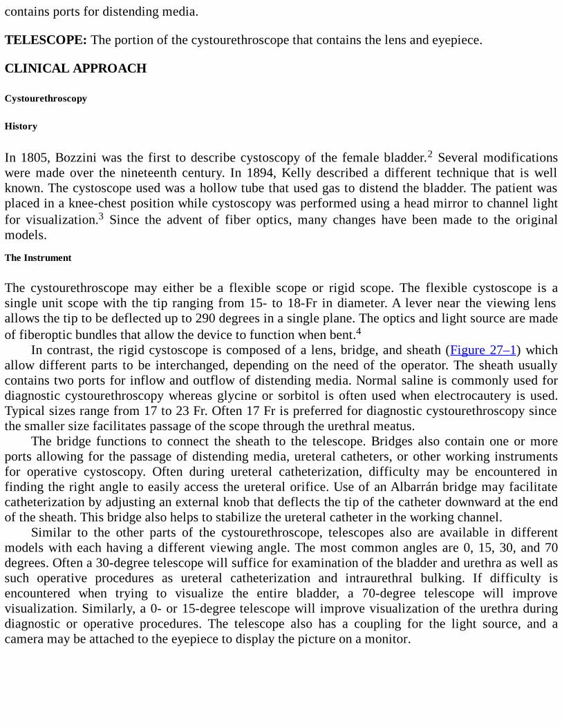
contains ports for distending media.
TELESCOPE: The portion of the cystourethroscope that contains the lens and eyepiece.
CLINICAL APPROACH
Cystourethroscopy
History
In 1805, Bozzini was the first to describe cystoscopy of the female bladder.2 Several modificationswere made over the nineteenth century. In 1894, Kelly described a different technique that is wellknown. The cystoscope used was a hollow tube that used gas to distend the bladder. The patient wasplaced in a knee-chest position while cystoscopy was performed using a head mirror to channel lightfor visualization.3 Since the advent of fiber optics, many changes have been made to the originalmodels.
The Instrument
The cystourethroscope may either be a flexible scope or rigid scope. The flexible cystoscope is asingle unit scope with the tip ranging from 15- to 18-Fr in diameter. A lever near the viewing lensallows the tip to be deflected up to 290 degrees in a single plane. The optics and light source are madeof fiberoptic bundles that allow the device to function when bent.4
In contrast, the rigid cystoscope is composed of a lens, bridge, and sheath (Figure 27–1) whichallow different parts to be interchanged, depending on the need of the operator. The sheath usuallycontains two ports for inflow and outflow of distending media. Normal saline is commonly used fordiagnostic cystourethroscopy whereas glycine or sorbitol is often used when electrocautery is used.Typical sizes range from 17 to 23 Fr. Often 17 Fr is preferred for diagnostic cystourethroscopy sincethe smaller size facilitates passage of the scope through the urethral meatus.
The bridge functions to connect the sheath to the telescope. Bridges also contain one or moreports allowing for the passage of distending media, ureteral catheters, or other working instrumentsfor operative cystoscopy. Often during ureteral catheterization, difficulty may be encountered infinding the right angle to easily access the ureteral orifice. Use of an Albarrán bridge may facilitatecatheterization by adjusting an external knob that deflects the tip of the catheter downward at the endof the sheath. This bridge also helps to stabilize the ureteral catheter in the working channel.
Similar to the other parts of the cystourethroscope, telescopes also are available in differentmodels with each having a different viewing angle. The most common angles are 0, 15, 30, and 70degrees. Often a 30-degree telescope will suffice for examination of the bladder and urethra as well assuch operative procedures as ureteral catheterization and intraurethral bulking. If difficulty isencountered when trying to visualize the entire bladder, a 70-degree telescope will improvevisualization. Similarly, a 0- or 15-degree telescope will improve visualization of the urethra duringdiagnostic or operative procedures. The telescope also has a coupling for the light source, and acamera may be attached to the eyepiece to display the picture on a monitor.
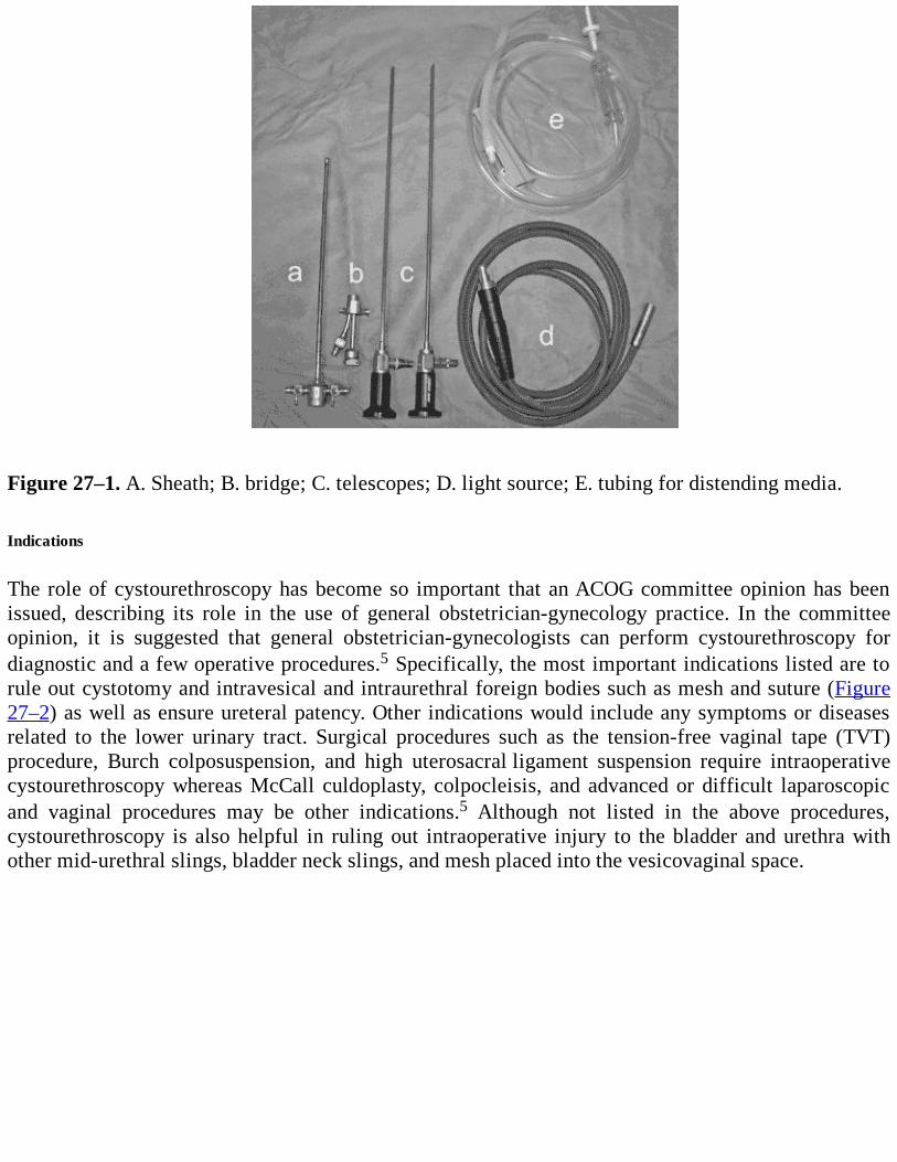
Figure 27–1. A. Sheath; B. bridge; C. telescopes; D. light source; E. tubing for distending media. Indications
The role of cystourethroscopy has become so important that an ACOG committee opinion has beenissued, describing its role in the use of general obstetrician-gynecology practice. In the committeeopinion, it is suggested that general obstetrician-gynecologists can perform cystourethroscopy fordiagnostic and a few operative procedures.5 Specifically, the most important indications listed are torule out cystotomy and intravesical and intraurethral foreign bodies such as mesh and suture (Figure27–2) as well as ensure ureteral patency. Other indications would include any symptoms or diseasesrelated to the lower urinary tract. Surgical procedures such as the tension-free vaginal tape (TVT)procedure, Burch colposuspension, and high uterosacral ligament suspension require intraoperativecystourethroscopy whereas McCall culdoplasty, colpocleisis, and advanced or difficult laparoscopicand vaginal procedures may be other indications.5 Although not listed in the above procedures,cystourethroscopy is also helpful in ruling out intraoperative injury to the bladder and urethra withother mid-urethral slings, bladder neck slings, and mesh placed into the vesicovaginal space.
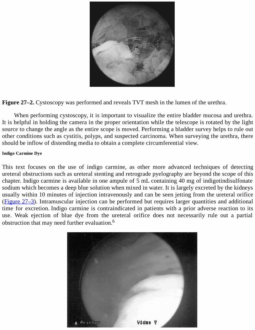
Figure 27–2. Cystoscopy was performed and reveals TVT mesh in the lumen of the urethra.
When performing cystoscopy, it is important to visualize the entire bladder mucosa and urethra.It is helpful in holding the camera in the proper orientation while the telescope is rotated by the lightsource to change the angle as the entire scope is moved. Performing a bladder survey helps to rule outother conditions such as cystitis, polyps, and suspected carcinoma. When surveying the urethra, thereshould be inflow of distending media to obtain a complete circumferential view.
Indigo Carmine Dye
This text focuses on the use of indigo carmine, as other more advanced techniques of detectingureteral obstructions such as ureteral stenting and retrograde pyelography are beyond the scope of thischapter. Indigo carmine is available in one ampule of 5 mL containing 40 mg of indigotindisulfonatesodium which becomes a deep blue solution when mixed in water. It is largely excreted by the kidneysusually within 10 minutes of injection intravenously and can be seen jetting from the ureteral orifice(Figure 27–3). Intramuscular injection can be performed but requires larger quantities and additionaltime for excretion. Indigo carmine is contraindicated in patients with a prior adverse reaction to itsuse. Weak ejection of blue dye from the ureteral orifice does not necessarily rule out a partialobstruction that may need further evaluation.6
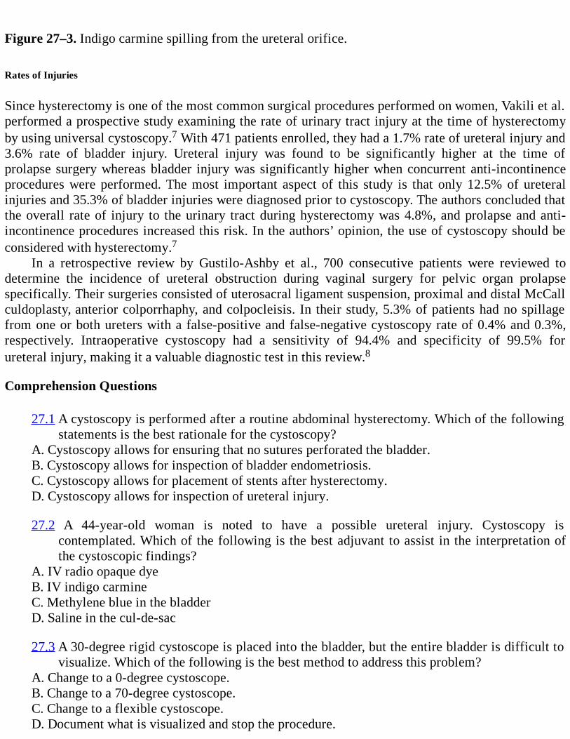
Figure 27–3. Indigo carmine spilling from the ureteral orifice. Rates of Injuries
Since hysterectomy is one of the most common surgical procedures performed on women, Vakili et al.performed a prospective study examining the rate of urinary tract injury at the time of hysterectomyby using universal cystoscopy.7 With 471 patients enrolled, they had a 1.7% rate of ureteral injury and3.6% rate of bladder injury. Ureteral injury was found to be significantly higher at the time ofprolapse surgery whereas bladder injury was significantly higher when concurrent anti-incontinenceprocedures were performed. The most important aspect of this study is that only 12.5% of ureteralinjuries and 35.3% of bladder injuries were diagnosed prior to cystoscopy. The authors concluded thatthe overall rate of injury to the urinary tract during hysterectomy was 4.8%, and prolapse and anti-incontinence procedures increased this risk. In the authors’ opinion, the use of cystoscopy should beconsidered with hysterectomy.7
In a retrospective review by Gustilo-Ashby et al., 700 consecutive patients were reviewed todetermine the incidence of ureteral obstruction during vaginal surgery for pelvic organ prolapsespecifically. Their surgeries consisted of uterosacral ligament suspension, proximal and distal McCallculdoplasty, anterior colporrhaphy, and colpocleisis. In their study, 5.3% of patients had no spillagefrom one or both ureters with a false-positive and false-negative cystoscopy rate of 0.4% and 0.3%,respectively. Intraoperative cystoscopy had a sensitivity of 94.4% and specificity of 99.5% forureteral injury, making it a valuable diagnostic test in this review.8
Comprehension Questions
27.1 A cystoscopy is performed after a routine abdominal hysterectomy. Which of the followingstatements is the best rationale for the cystoscopy?
A. Cystoscopy allows for ensuring that no sutures perforated the bladder.B. Cystoscopy allows for inspection of bladder endometriosis.C. Cystoscopy allows for placement of stents after hysterectomy.D. Cystoscopy allows for inspection of ureteral injury.
27.2 A 44-year-old woman is noted to have a possible ureteral injury. Cystoscopy iscontemplated. Which of the following is the best adjuvant to assist in the interpretation ofthe cystoscopic findings?
A. IV radio opaque dyeB. IV indigo carmineC. Methylene blue in the bladderD. Saline in the cul-de-sac
27.3 A 30-degree rigid cystoscope is placed into the bladder, but the entire bladder is difficult tovisualize. Which of the following is the best method to address this problem?
A. Change to a 0-degree cystoscope.B. Change to a 70-degree cystoscope.C. Change to a flexible cystoscope.D. Document what is visualized and stop the procedure.
ANSWERS
27.1 D. In studies, only 12.5% of ureteral injuries were discovered prior to cystoscopy afterhysterectomy. Thus, routine cystoscopy can allow for discovery of unsuspected ureteralinjury Although it is valid to use cystoscopy to examine the bladder for sutures, thiscomplication is less common during a routine uncomplicated hysterectomy.
27.2 B. Indigo carmine is concentrated in the kidneys and excreted into the bladder. Cystoscopyallows inspection of the ureteral orifices to see if there is efflux of urine through bothureteral openings.
27.3 B. The 70-degree cystoscope allows for inspection of the entire bladder when the 30-degreecystoscope may be insufficient to visualize the entire bladder.
Clinical Pearls
See Table 1-2 for definition of level of evidence and strength of recommendation
The cystourethroscope is composed of a sheath, bridge, and telescope.
Lenses with different angles may assist in evaluating the bladder and urethra. Lenses of 0, 15,and 30 degrees are commonly used to evaluate the urethra whereas 30- and 70-degree lensesare commonly used to evaluate the bladder (Level B).
Cystourethroscopy aids in the detection of urinary tract injuries, especially when concomitantprolapse and anti-incontinence procedures are performed (Level B).
Use of indigo carmine intravenously given 10 minutes prior to cystourethroscopy may aid inlocating the ureteral orifices and detect ureteral obstruction (Level B).
The ureteral orifices are most easily found by locating the intertrigonal ridge which is anelevation of the bladder mucosa just proximal to the urethral-vesical junction (Level B).
In patients with significant prolapse, placement of a sponge stick into the vagina may restoreanatomy sufficiently to aid in identification of the ureteral orifices (Level C).
Overdistending the bladder may make it more difficult to locate the ureteral orifices (Level B).
When passing ureteral catheters, an Albarrán bridge may facilitate passage of the ureteralcatheter into the orifice (Level C).
REFERENCES
1. Buller JA, Thompson JR, Cundiff GW, et al. Uterosacral ligament: description of anatomicrelationships to optimize surgical safety. Obstet Gynecol. 2001;97:873-879.
2. Bozzini P. Lichtleiter, eine erfindung zur anschung inerer theile, und krukheiten nebst
2. Bozzini P. Lichtleiter, eine erfindung zur anschung inerer theile, und krukheiten nebstabbildung. J Pract Arzeykunde. 1805;24:107.
3. Kelly HA. The direct examination of the female bladder with elevated pelvis: thecatheterization of the ureters under direct inspection, with and without elevation of the pelvis.Am J Obstet Dis Wom Child. 1894;25:1-9.
4. Cundiff GW, Bent AE. Cystourethroscopy. In: Bent AE, Cuniff GW, Swift SE, et al., eds.Ostergard’s Urogynecology and Pelvic Floor Dysfunction. 5th ed. Philadelphia, PA: LippincottWilliams & Wilkins; 2003.
5. American College of Obstetricians and Gynecologists. The role of cystourethroscopy in thegeneralist obstetrician-gynecologist practice. ACOG Committee Opinion No. 372. ObstetGynecol. 2007;110:221-224.
6. American Regent, Inc. Indigo Carmine.http://www.americanregent.com/documents/Product27PrescribingInformation.pdf. AccessedAugust 12, 2010.
7. Vakili B, Chesson RR, Kyle BL, et al. The incidence of urinary tract injury duringhysterectomy: a prospective analysis based on universal cystoscopy. Am J Obstet Gynecol.2005;192:1599-1604.
8. Gustilo-Ashby AM, Jelovsek JE, Barber MD, et al. The incidence of ureteral obstruction andthe value of intraoperative cystoscopy during vaginal surgery for pelvic organ prolapse. Am JObstet Gynecol. 2006;194:1478-1485.
Case 28
A 40-year-old G3 P3003 Caucasian woman with a body mass index (BMI) of 24 kg/m2 hasdysfunctional uterine bleeding unresponsive to hormonal therapy. The patient is desirous ofpermanent sterilization, and opts for a total abdominal hysterectomy. She denies any symptoms ofurinary or fecal incontinence. Her general physical examination is unremarkable, except that heruterus is at the upper limits of normal size and slightly tender to bimanual examination. She hasadequate vaginal support of both the anterior and posterior vaginal walls. On the day of her surgery, anemergency in the adjoining OR just preceding the patient’s general anesthesia induction required aquick substitution of one anesthesiologist for another, and during the physician transfer procedure, thepatient did not receive her scheduled dose of cefazolin, 1 g intravenously just prior to having generalanesthesia. Her vaginal hysterectomy (VH) is complicated by an 850-cc blood loss, though no intra- orpostoperative transfusion is required. On postoperative day 1, her 4:00 AM oral temperature is101.5°F, and her 8:00 AM temperature is 101.8°F.
What is the most likely diagnosis?
What is your next step?
What potential complications may ensue?
When should the prophylactic antibiotics be administered?
ANSWERS TO CASE 28:Febrile Morbidity/Pelvic Infection Complicating Vaginal Hysterectomy
Summary: A 40-year-old woman underwent a VH without receiving her scheduled dose ofpreoperative antibiotic. She has an 850-cc blood loss, and has two elevated temperatures 4 hours aparton the morning of postoperative day 1.
Most likely diagnosis: Febrile morbidity/probable pelvic infection following vaginalhysterectomy.
Next step: Treat the pelvic infection. Perform an examination to rule out other causes of fever,and likely start antibiotic therapy.
Potential complications: Development of pelvic abscess, allergic reaction to antibiotictherapy, development of septic pelvic thrombophlebitis.
Prophylactic antibiotics: Administration of antimicrobial agents usually prior to surgery or aprocedure used to decrease febrile morbidity such as wound infection.
ANALYSIS
Objectives
1. Know why, how, and when to use prophylactic antibiotic therapy when performing vaginalsurgery.
2. Be able to construct the differential diagnosis for the febrile patient following vaginalhysterectomy.
3. Learn how to treat postoperative pelvic infection following vaginal surgery.4. Be able to recognize and treat pelvic abscess and septic pelvic thrombophlebitis following
vaginal surgery.
Considerations
This 40-year-old Caucasian woman had a VH without receiving preoperative antibiotics, and shesustained an 850-cc blood loss at the time of surgery.1 She has febrile morbidity on postoperative day1, with two elevated temperatures 4 hours apart. The differential diagnosis of fever following vaginalsurgery depends to some extent on which postoperative day the elevated temperature occurs. Onpostoperative day 1, the differential should include infection at the surgical site, such as cuff cellulitis,or atelectasis, or infection at a nonsurgical site, such as the urinary tract, the respiratory tract, or at theIV site.1 A directed physical examination and appropriate laboratory and imaging studies will help todelineate the source of the fever if the patient appears significantly ill, but these workups are usuallynot likely to yield much useful clinical information.2,3 Febrile responses occurring later in thepostoperative course suggest the possibility of a pelvic abscess or pelvic vein thrombosis.
APPROACH TO Evaluation and Management of Fever after Vaginal Hysterectomy
DEFINITIONS
FEBRILE MORBIDITY: Temperature of 100.4°F or greater on two separate occasions at least 4hours apart, after the first 24 hours after surgery.
VAGINAL CUFF CELLULITIS: Infection of the vaginal mucosal and submucosal tissue involvingvaginal bacteria.
CLINICAL APPROACH
Prophylactic Antibiotic Usage
Febrile morbidity is the most common complication of VH, although this is less likely to occurwith vaginal than with abdominal hysterectomy. A major review has shown that some of thedifference in rates of febrile morbidity may relate to the increased use of prophylactic antibiotics inVH versus TAH, specifically 7.2% versus 16.8%.4 Both routes of hysterectomy experience decreasedfebrile morbidity if prophylaxis is used. Overall, the literature suggests that the incidence of febrilemorbidity varies from 10% to 30% for all types of hysterectomy.4 This case illustrates the two majorrisk factors for febrile morbidity following VH: those being failure to use prophylactic antibiotics andexperiencing excessive blood loss.
There is a unique circumstance that merits therapy before the day of surgery. Studies have shown
that the patient with bacterial vaginosis is at increased risk for postoperative pelvic cuff cellulitis.This should be searched for and treated at a preoperative visit prior to the scheduled surgicalprocedure, with therapy consisting of 4 days of therapy with metronidazole.
The rationale for using a prophylactic antibiotic prior to vaginal hysterectomy rests on thepremise that the vagina is a “clean contaminated” space, and that no amount of sterile preoperativepreparation is going to make this a sterile operative field. Upon transcervical surgical entry into theperitoneal cavity via the vagina, the operative site is exposed to a plethora of aerobic and anaerobicbacterial organisms with the potential to cause infection. Using antibiotics preoperatively enhancesthe patient’s own immune response and lowers the number of bacteria at the surgical site. The efficacyof single-dose antibiotic prophylaxis does not require that the drug used be bactericidal against all ofthe organisms in the vagina.
Conceptually, prophylactic antibiotic usage requires that the drug used be relatively inexpensive,safe, and effective. A detailed analysis of the drugs available for use for this purpose is availableelsewhere in American College of Obstetricians and Gynecologists publications,5 and differenthospitals may have unique pathogens, which require modification of any blanket recommendations.Also, efficacy and sensitivity patterns are subject to change, so hospital infection control committeesshould monitor the patterns of prophylaxis usage and benefit for their particular environment andmake specific drug recommendations accordingly. Given the overwhelming evidence regarding theeffectiveness of prophylactic antibiotic usage, hospital quality committees should enforce theirusage as a routine preoperative protocol for indicated surgical procedures, unless there is somecompelling reason for the surgeon to cancel this order.
Within that context, current studies suggest that a single dose of a cephalosporin, usuallycefazolin, 1 or 2 g IV is the drug of choice for prophylaxis, especially for vaginal and abdominalhysterectomy. The drug is low cost, has a long (1.8 hours) half-life, a low incidence of allergicreactions, and is relatively free of side effects. Antibiotics significantly more expensive than cefazolinwill decrease the theoretical cost-benefit ratio implicit in supporting the prophylaxis concept.Cefazolin is also similar in its profile of effectiveness against anaerobes to other cephalosporins.Anaphylaxis with cephalosporins occurs less than 0.02% of the time, and this is only slightlyincreased in patients with known penicillin allergy. Of the four types of immunopathologic responsesto β-lactam antibiotics, only the immediate hypersensitivity-type reaction to penicillin shouldpreclude the use of a cephalosporin for prophylaxis. For those patient that have significant allergy tobeta lactam agents, other choices include:
Clindamycin 600 mg IV and Gentamicin 1.5 mg/kg IVMetronidazole 500 mg IV and Gentamicin 1.5 mg/kg IV
Timing of administration of antibiotic therapy preoperatively is important. Most studiesdemonstrate that the antibiotic should reach therapeutic tissue levels prior to the initial incision, andthis is best accomplished by administering antibiotic 30 to 60 minutes prior to the initial incision,which effectively means that the antibiotic should be given just prior to the induction of generalanesthesia. While the strongest evidence is for the use of a single dose of antibiotic prior to the onsetof VH, there are other recommendations in the literature for the use of more than one dose. Someexperts suggest that the object of the preventative therapy is to have therapeutic drug levels on boardfor the duration of the operative procedure and, therefore, if the case is exceptionally long, that is,greater than 2 hours, a subsequent dose of prophylactic antibiotic can be considered at sequences of
one to two times the drug’s half-life.5 Given that excess blood loss is also a major risk factor forinfection, another antibiotic dose at the conclusion of the case is a consideration. Different studieshave suggested that blood loss amounts of 750 to 1500 cc may prompt additional antibiotic dosing.Bacterial resistance is a risk factor for more than routine prophylactic antibiotic usage, so the use ofmore than a single dose should be approached with caution.
Treatment of Infection/Pelvic Cellulitis
As the ptient case demonstrates, febrile morbidity may occur following VH. When used appropriately,antibiotic prophylaxis has reduced postoperative infection rates from 5% to 10% to about 4% for mostgynecologic procedures, infection may result. Pelvic cellulitis is the most likely diagnosis in theillustrative case report. The patient may complain of pelvic pain and a slight increase in vaginaldischarge, but neither pelvic examination nor imaging procedures are likely to demonstrate a masswithin 24 hours of surgery. Laboratory results may likely demonstrate an elevated white blood (cell)count (WBC) of greater than 13,000/mm3, with a left shift to greater than 90% bands andpolymorphonuclear leucocytes. The value of an extensive fever workup for febrile morbidityfollowing gynecologic procedures has been questioned.3 Comparing groups of patients withpostoperative infection did not demonstrate the value of multiple laboratory or imaging studies, andthe approach to the patient with fever following VH should be specific to the individual situation. Thetherapy of choice is a single agent IV antibiotic, usually not the drug used for prophylaxis.
Again, knowledge of hospital antibiotic resistance patterns is important, but in general, singleagent cephalosporin therapy is indicated, such as cefotetan, cefotaxime, cefoxitin, or ceftriaxone.Other choices include ampicillin/sulbactam, piperacillin, mezlocillin, or ticarcillin/clavulanic acid.
Treatment of Infection/Infected Hematoma and Pelvic Abscess
Febrile responses persisting beyond 48 to 72 hours of antibiotic suggest the presence of an abscess oran infected hematoma, and imaging studies such as ultrasound or CT may show a mass.6 Theseabscesses usually require surgical drainage, usually in the OR with sedation or anesthesia to allow fortransvaginal access to the mass, bluntly opening the cuff enough to facilitate drainage. If imagingstudies suggest that the hematoma or abscess is not accessible via the vaginal route, ultrasound- orCT-guided needle or catheter drainage is a consideration. IV antibiotic therapy must continue until thepatient becomes afebrile for 24 to 48 hours. The concept of continuing prolonged oral antibiotictherapy for several days after the patient is afebrile is no longer viable and only serves to increaseantibiotic resistance.
In general, cultures for anaerobic organisms are costly and not done routinely. Additionally, theantibiotic choices must be made long before the culture results are available. If the fever persistsdespite therapy for 48 to 72 hours, reevaluation of the cultures and antibiotic choices is in order, andconsultation with the infectious disease service is a consideration. Another study of febrile morbidityin patients following VH or TAH has shown that blood cultures were not cost-effective, and that nonedemonstrated bacteremia in febrile patients.
“Drug fever,” as indicated by persistent temperature elevation in the patient with no sign ofinfection after an exhaustive evaluation, and eosinophilia on the complete blood (cell) count (CBC)should be entertained as a diagnosis.
Treatment of Infection/Septic Thrombophlebitis
If the febrile response continues in spite of 72 hours of antibiotic therapy, the diagnosis of septic
pelvic thrombophlebitis should be considered. Physical examination signs are variable and mayinclude pelvic tenderness and pain over the upper thigh along with edema, suggesting ileofemoral veininvolvement. Imaging studies such as CT or MRI may show the involved region, with the radiographiccriteria for diagnosis, including (1) venous enlargement, (2) the vessel wall having a low-densitylumen, and (3) contrast media enhancing a sharply defined vessel wall.6 Medical therapy is indicatedfor this problem, not another surgical procedure. Some practitioners will add heparin therapy to theantibiotic regimen. Prolonged therapy is often required for adequate response. Oral anticoagulants arenot required following defervescence.
Comprehension Questions
28.1 Which of the following classes of antibiotics is usually the first choice for use as aprophylactic antibiotic in vaginal surgery?
A. CephalosporinsB. PenicillinsC. QuinolonesD. Tetracyclines
28.2 When pelvic cellulitis is suspected following vaginal hysterectomy, which of the followingtherapeutic measures should constitute your initial approach?
A. Consult the infectious disease service.B. Begin therapy with heparin.C. Begin triple antibiotic therapy with ampicillin, gentamycin, and metronidazole.D. Begin single-drug therapy with a drug not used for prophylaxis.
28.3 Once the patient described in question 28.2 has responded to IV antibiotic therapy with anormal temperature and no longer appears ill, which of the following is the best next step?
A. Oral antibiotic therapy should commence and continue for another 5 to 7 days.B. Blood cultures should be obtained to be sure that the patient is well.C. Antifungal agents should be begun.D. Antibiotic therapy may end 24 hours following the last elevated temperature.
ANSWERS
28.1 A. The cephalosporins, as a class, are generally preferred for antibiotic prophylaxis invaginal surgery. They are effective, relatively inexpensive, and safe, rarely causinganaphylaxis.
28.2 D. Therapy for straightforward pelvic cellulitis should begin with single agent antibiotictherapy. The other treatment options listed should be reserved for infectious problems thatdo not resolve easily.
28.3 D. Continuing antibiotic therapy either IV or orally, after the patient is well, only serves toincrease the potential for bacterial resistance and to cause drug fever.
Clinical Pearls
See Table 1-2 for definition of level of evidence and strength of recommendation
Timing is critical in using prophylactic antibiotics—have the anesthesia service administeryour antibiotic of choice before the initiation of general anesthesia, usually within the hourprior to your incision (Level A).
Keep blood loss to a minimum—the greater the blood loss, the greater the chance for infection(Level B).
Work quickly and efficiently. The longer the case, the greater the potential for infection. If thecase is obviously going to last significantly longer than 2 hours, consider a second dose ofprophylactic antibiotic at the 2-hour mark (Level B).
Consider doing VH without electrocautery. Cauterized tissue provides a nidus for infection,and meticulous surgical technique combined with a lidocaine/epinephrine mixture injected atthe vaginal cuff prior to the initial incision significantly reduces blood loss (Level B).
REFERENCES
1. Culligan P, Heit M, Blackwell L, Murphy M, Graham C, Snyder J. Bacterial colony countscuring vaginal surgery. Inf Dis Obstet Gynecol. 2003;11:161-165.
2. Swisher E, Kahleifeh B, Pohl J. Blood cultures in febrile patients after hysterectomy. J ReprodMed. 1997;42:547-550.
3. Schey D, Salom E, Papadia A, Penalver M. Extensive fever workup produces low yield indetermining infectious etiology. Am J Obstet Gynecol. 2005;192:1729-1734.
4. Peipert J, Weitzen S, Cruickshank C, Story E, Ethridge D, Lapane K. Risk factors for febrilemorbidity after hysterectomy. Obstet Gynecol. 2004;103:86-91.
5. American College of Obstetricians and Gynecologists. Antibiotic prophylaxis for gynecologicprocedures. ACOG Practice Bulletin No. 74. Obstet Gynecol. 2006;108:225-234.
6. Brown C, Lowe T, Cunningham F, Weinreb J. Puerperal pelvic thrombophlebitis: impact ondiagnosis and treatment using x-ray computed tomography and magnetic resonance imaging.Obstet Gynecol. 1986;68:789-794.
Case 29
A 45-year-old Caucasian G4 P4004 woman with uterovaginal prolapse is scheduled for a plannedprocedure of vaginal hysterectomy, anterior and posterior colporrhaphy, and sacrospinous ligamentfixation. Except for her uterovaginal prolapse, the patient is in good health and has no chronicdiseases. On the day of surgery, the patient is positioned in “candy cane” hanging stirrups, and theinitial parts of the operation are accomplished without any noticeable complication. A Capio device(Boston Scientific, Boston, MA) is employed to place the permanent suture used for securing thevaginal apex to the right sacrospinous ligament. Following the patient’s recovery from generalanesthesia, she complains of difficulty walking and getting her right foot to function normally.
What is the most likely diagnosis?
What is your next step?
ANSWERS TO CASE 29:Complications from Vaginal Surgery
Summary: A 45-year-old woman has a vaginal hysterectomy, anterior and posterior colporrhaphy, andright sacrospinous ligament fixation. She awakens with difficulty walking and altered ability to makeher right foot function normally.
Most likely diagnosis: Right foot drop from compression of the common perineal nerve bypressure on the nerve by the stirrup at the site of the lateral epicondyle.
Next step: Reassure the patient that this is likely a transient problem. Consult physical therapyfor assistance in rehabilitation to restore normal foot function.
ANALYSIS
Objectives
1. Recognize the types of neuropathy related to vaginal surgery.2. Describe the various mechanisms and preventative strategies of nerve injury associated with
vaginal surgery: positioning of the patient, intraoperative injury, and nerve entrapment/injury.3. Describe the classic presentations of nerve injury and the therapy for these conditions.
Considerations
This patient’s immediate postoperative complaint of right foot drop is a known complication ofvaginal surgery, especially if the patient was positioned in hanging stirrups which did not haveadequate lateral padding to prevent compression of the common peroneal nerve at the lateralepicondyle. This is but one of the types of neuropathy that may occur in association with vaginal
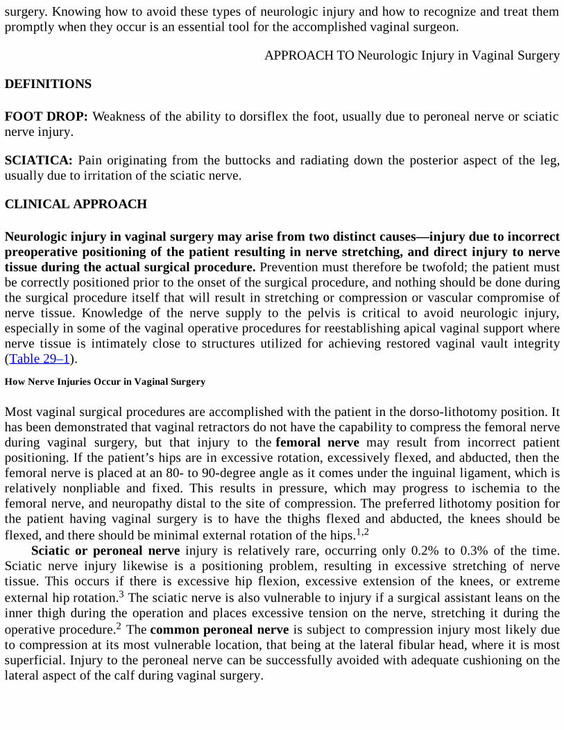
surgery. Knowing how to avoid these types of neurologic injury and how to recognize and treat thempromptly when they occur is an essential tool for the accomplished vaginal surgeon.
APPROACH TO Neurologic Injury in Vaginal Surgery
DEFINITIONS
FOOT DROP: Weakness of the ability to dorsiflex the foot, usually due to peroneal nerve or sciaticnerve injury.
SCIATICA: Pain originating from the buttocks and radiating down the posterior aspect of the leg,usually due to irritation of the sciatic nerve.
CLINICAL APPROACH
Neurologic injury in vaginal surgery may arise from two distinct causes—injury due to incorrectpreoperative positioning of the patient resulting in nerve stretching, and direct injury to nervetissue during the actual surgical procedure. Prevention must therefore be twofold; the patient mustbe correctly positioned prior to the onset of the surgical procedure, and nothing should be done duringthe surgical procedure itself that will result in stretching or compression or vascular compromise ofnerve tissue. Knowledge of the nerve supply to the pelvis is critical to avoid neurologic injury,especially in some of the vaginal operative procedures for reestablishing apical vaginal support wherenerve tissue is intimately close to structures utilized for achieving restored vaginal vault integrity(Table 29–1).
How Nerve Injuries Occur in Vaginal Surgery
Most vaginal surgical procedures are accomplished with the patient in the dorso-lithotomy position. Ithas been demonstrated that vaginal retractors do not have the capability to compress the femoral nerveduring vaginal surgery, but that injury to the femoral nerve may result from incorrect patientpositioning. If the patient’s hips are in excessive rotation, excessively flexed, and abducted, then thefemoral nerve is placed at an 80- to 90-degree angle as it comes under the inguinal ligament, which isrelatively nonpliable and fixed. This results in pressure, which may progress to ischemia to thefemoral nerve, and neuropathy distal to the site of compression. The preferred lithotomy position forthe patient having vaginal surgery is to have the thighs flexed and abducted, the knees should beflexed, and there should be minimal external rotation of the hips.1,2
Sciatic or peroneal nerve injury is relatively rare, occurring only 0.2% to 0.3% of the time.Sciatic nerve injury likewise is a positioning problem, resulting in excessive stretching of nervetissue. This occurs if there is excessive hip flexion, excessive extension of the knees, or extremeexternal hip rotation.3 The sciatic nerve is also vulnerable to injury if a surgical assistant leans on theinner thigh during the operation and places excessive tension on the nerve, stretching it during theoperative procedure.2 The common peroneal nerve is subject to compression injury most likely dueto compression at its most vulnerable location, that being at the lateral fibular head, where it is mostsuperficial. Injury to the peroneal nerve can be successfully avoided with adequate cushioning on thelateral aspect of the calf during vaginal surgery.
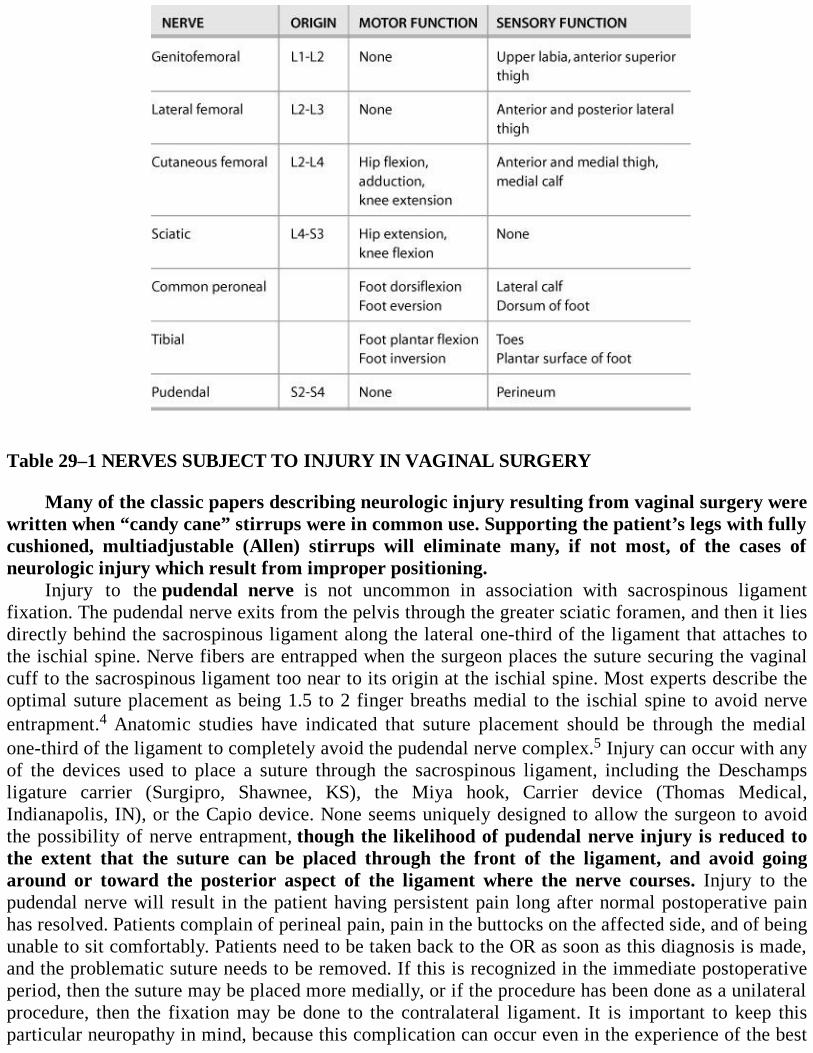
Table 29–1 NERVES SUBJECT TO INJURY IN VAGINAL SURGERY
Many of the classic papers describing neurologic injury resulting from vaginal surgery werewritten when “candy cane” stirrups were in common use. Supporting the patient’s legs with fullycushioned, multiadjustable (Allen) stirrups will eliminate many, if not most, of the cases ofneurologic injury which result from improper positioning.
Injury to the pudendal nerve is not uncommon in association with sacrospinous ligamentfixation. The pudendal nerve exits from the pelvis through the greater sciatic foramen, and then it liesdirectly behind the sacrospinous ligament along the lateral one-third of the ligament that attaches tothe ischial spine. Nerve fibers are entrapped when the surgeon places the suture securing the vaginalcuff to the sacrospinous ligament too near to its origin at the ischial spine. Most experts describe theoptimal suture placement as being 1.5 to 2 finger breaths medial to the ischial spine to avoid nerveentrapment.4 Anatomic studies have indicated that suture placement should be through the medialone-third of the ligament to completely avoid the pudendal nerve complex.5 Injury can occur with anyof the devices used to place a suture through the sacrospinous ligament, including the Deschampsligature carrier (Surgipro, Shawnee, KS), the Miya hook, Carrier device (Thomas Medical,Indianapolis, IN), or the Capio device. None seems uniquely designed to allow the surgeon to avoidthe possibility of nerve entrapment, though the likelihood of pudendal nerve injury is reduced tothe extent that the suture can be placed through the front of the ligament, and avoid goingaround or toward the posterior aspect of the ligament where the nerve courses. Injury to thepudendal nerve will result in the patient having persistent pain long after normal postoperative painhas resolved. Patients complain of perineal pain, pain in the buttocks on the affected side, and of beingunable to sit comfortably. Patients need to be taken back to the OR as soon as this diagnosis is made,and the problematic suture needs to be removed. If this is recognized in the immediate postoperativeperiod, then the suture may be placed more medially, or if the procedure has been done as a unilateralprocedure, then the fixation may be done to the contralateral ligament. It is important to keep thisparticular neuropathy in mind, because this complication can occur even in the experience of the best
of gynecologic surgeons, possibly because there is a demonstrable anatomic variability in the path ofpudendal nerve fibers along the course of the sacrospinous ligament. This complication has beenreported as being amenable to surgical correction by reexploration and removal of the offendingsuture up to 2 years following the initial surgical procedure. In that instance, pain relief was describedas being “immediate.” This nerve injury has also been reported following uterosacral ligamentsuspension of the vagina, occurring seven times in 182 procedures.6
Treatment Considerations
Fortunately, the incidence of neuropathy from vaginal surgery is relatively low, and recovery isgenerally complete in most instances, though this may take months rather than weeks if the problem isdue to a stretch injury. Neurologic consultation and rehabilitation consisting of physiotherapy, a footdrop brace if indicated, and electrical stimulation of the involved musculature is frequently required.Physical therapy will be necessary if motor deficits are identified, as this will prevent atrophy andwasting of involved muscle groups innervated by the involved nerve. Some authors have also usedmedical therapy in the form of gabapentin as an adjunct to rehabilitation procedures.
Comprehension Questions
29.1 A 33-year-old woman underwent a vaginal hysterectomy due to dys-functional uterinebleeding unresponsive to medical therapy. On postoperative day 1, the patient was noted tohave difficulty walking. On examination, she has difficulty lifting her right leg off the bed.Her right patellar reflex is absent. Which of the following is the most likely mechanism forthe nerve injury?
A. Intraoperative injury with sutureB. Intraoperative pressure with retractorC. Hyperflexion of the hipD. Lack of padding to the lateral leg
29.2 Which nerve is most subject to injury as a result of a sacrospinous ligament fixationprocedure?
A. FemoralB. Common peronealC. PudendalD. Sciatic
29.3 A 55-year-old woman is noted to have foot drop following a vaginal surgery. Which of thefollowing findings would more likely indicate a sciatic nerve problem rather than peronealnerve issue?
A. Lack of ankle reflexB. Posterior leg painC. Lack of padding to the lateral fibular areaD. Sacrospinous ligament fixation as part of the procedure
ANSWERS
29.1 C. This patient likely has a right femoral nerve injury. The femoral nerve innervates thequadriceps muscles leading to difficulty walking and a diminished or absent patella reflex.
The most common mechanism of femoral nerve palsy in vaginal surgeries involveshyperflexion of the hips leading to pressure and ischemia of the femoral nerve under theinguinal ligament.
29.2 C. The course of the pudendal nerve just behind the lateral aspect of the sacrospinousligament, near the ischial spine, makes it vulnerable to injury while performing SSLF.
29.3 B. Injury to either the sciatic nerve and peroneal nerve will result in foot drop anddiminished ankle reflex. Lack of padding to the fibular head leads to common peronealnerve palsy. Buttocks or posterior leg pain is consistent with sciatic nerve damage.
Clinical Pearls
See Table 1-2 for definition of level of evidence and strength of recommendation
When tying the sutures affixing the vaginal apex to the sacrospinous ligament, leave the cutends of the suture long enough so that they can be readily identified to facilitate removal of thesuture if pudendal nerve entrapment is diagnosed (Level C).
Caution your surgical assistants not to put any pressure on the medial aspect of the patient’sthigh while she is in lithotomy position (Level C).
Make sure the patient is correctly positioned in the stirrups before the operation starts. Nerveinjury prevention is much simpler, easier, and cheaper than treatment for postoperativeneuropathy (Level B).
REFERENCES
1. Irvin W, Andersen W, Taylor P, Rice L. Minimizing the risk of neurologic injury ingynecologic surgery. Obstet Gynecol. 2004;103:374-382.
2. Burkhart FL, Daly JW. Sciatic and peroneal nerve injury: a complication of vaginal operations.Obstet Gynecol. 1966;28:99-102.
3. Alevizon SJ, Finan MA. Sacrospinous colpopexy: management of postoperative pudendalnerve entrapment. Obstet Gynecol. 1996;88:713-715.
4. Verdeja AM, Elkins TE, Odol A, Gasser R, Lamoutte E. Transvaginal sacrospinous colpopexy:anatomic landmarks to be aware of to minimize complications. Am J Obstet Gynecol.1995;173:1468-1469.
5. Batres F, Barclay DL. Sciatic nerve injury during gynecologic procedures using the lithotomyposition. Obstet Gynecol. 1983;62(suppl):92S-94S.
6. Flynn MK, Weidner AC, Amundsen CL. Sensory nerve injury after uterosacral ligamentsuspension. Am J Obstet Gynecol. 2006;195:1869-1872.
Case 30
A 23-year-old Caucasian woman presents to her gynecologist with a chief complaint of dyspareunia.She relates that she had recently had the onset of severe coital pain on several occasions, which hadnot previously been present. Specific questioning reveals that the patient localizes the pain to thevaginal vestibule, and that this is exacerbated by touching the region, or by her partner attemptingvaginal entry. Physical examination is negative for any evidence of vaginal inflammation, and wetmount examination of the vaginal secretions is negative for the presence of any vaginal pathogens.The vaginal vestibule is exquisitely sensitive when the vaginal speculum is inserted into the vagina,and applying pressure on the perineum exacerbates the patient’s pain sensation.
What is the most likely diagnosis?
What is the next step?
ANSWERS TO CASE 30:Vulvar Vestibulitis
Summary: A 23-year-old woman has dyspareunia localized to the vaginal vestibule. Physicalexamination reveals tenderness to pressure and touch in this region, with no evidence of anyinflammatory process in the vagina.
Most likely diagnosis: Vulvar vestibulitis.
Next step: Rule out other causes of labial pathology, and then discuss treatment options withthe patient for treatment of this diagnosis.
ANALYSIS
Objectives
1. Be able to recognize and treat the benign conditions that are commonly seen on the labia.2. Know how to perform diagnostic biopsy procedures to confirm these diagnoses.3. Understand those benign conditions which the generalist gynecologist should be able to treat
surgically with confidence, how to do those procedures, and to discern those which should bereferred to the gynecologic oncologist.
Considerations
This 23-year-old woman has a recent history of dyspareunia which on examination is localized to thevaginal vestibule. There is no evidence of inflammation or other lesions. There is no evidence ofinfection by wet mount. On examination, the vaginal vestibule is very sensitive to touch. A carefulinspection of the vagina and vulva is important especially to try to identify herpes simplex virus or
other neurologic processes. Pain can be assessed and mapped by touching a cotton-tipped applicator tothe vulva. In the evaluation process, a herpes assay for polymerase chain reaction (PCR) or cultureshould be performed. A careful psychosocial history should be taken to assess for sexual abuse orphysical abuse. Once infection is ruled out, biopsy may be considered. Vulvar vestibulitis is adiagnosis of exclusion, and usually there is some erythema. Different treatments may be considered,with surgical excision being most successful.
APPROACH TO Nonmalignant Vulvar Disease
DEFINITIONS
VULVAR VESTIBULITIS: A syndrome of severe pain of the vulvar introitus without inflammation.
VULVAR INTRAEPITHELIAL NEOPLASIA: Premalignant condition with dysplastic cellsconfined within the epithelium of the vulva.
BARTHOLIN GLAND CYST: Fluid within the greater vestibular gland causing swelling.
CLINICAL APPROACH
This chapter on vulvar surgery does not intend to describe surgical therapy for malignant vulvardisease. Rather, it is intended to define practical steps for the generalist gynecologist to use in makingthe diagnosis of labial disease, to delineate commonly encountered labial conditions amenable tosurgical therapy, and to describe surgical approaches for those benign entities that will benefit fromsurgical therapy.
Vulvar lesions constitute a significant component of the gynecologist’s clinical practice. Authorsstress the necessity of the labial biopsy to confirm the pathology before embarking on a course oftreatment.1,2 The consultant gynecologist who makes the correct diagnosis and establishes the propertherapeutic plan is often the first physician to make a tissue diagnosis after multiple other physicianshave tried a myriad number of ointments and creams without success. The gynecologist should have aclose working relationship with the pathologist who is going to evaluate the tissue, and remember thatcommunication with the pathologist regarding the patient’s history and gross appearance of the lesionmay improve his/her diagnostic acumen under the microscope.
VULVAR VESTIBULITIS
Vulvar vestibulitis as an entity has been initially described by Friedrich as consisting of (1) severepain on vestibular touch; (2) tenderness when pressure is applied in the vaginal vestibule; (3)erythema of the vestibule.3 Strenuous evaluation of the validity of these criteria has been established,and it has generally been upheld, except that the erythema of the vestibule has been questioned, andthe pain and pressure components have been more accurately described, such that the pain in thevestibule has been described as having a “thermal” component, and the degree of severity is worsethan that of severe dysmenorrhea. The etiology of this entity is still unclear, though studies haveexcluded the human papillomavirus (HPV) as causative, and no other infectious process has beenimplicated as being likely. Multiple therapeutic approaches have been utilized, though none have beendemonstrated to be as effective as surgery for effective treatment of this problem. Success rates rangefrom 36% to 100%, with the overall average being in the 60% range. Women younger than 30 years
seem to benefit more from this procedure than those in older age ranges.4The surgical procedure should include complete vestibulectomy, including excision of tissue up
to the periurethral region if it is deemed to be involved on the basis of the preoperative evaluation.Undermining and advancement of the vaginal wall completes the operation. Complications of thesurgical procedure include the lack of satisfactory lubrication in 24% and the development ofBartholin gland cysts in 6%, which occurs when the Bartholin gland duct is obstructed as part of theexcision of the vaginal vestibule.4
Labial Biopsy
Office labial biopsy should be simple, fast, routine, and as painless as possible. Premedication of thearea to be sampled with topical application of 2.5% lidocaine and 2.5% prilocaine prior to the sterilepreparation and the injection of the local anesthetic will significantly reduce patient discomfort; manypatients volunteer that they do not feel the injection itself if the cream has been on the labial surfacefor 5 minutes prior to the injection. Labial tissue has an abundant blood supply, so a mixture oflidocaine and 1:100,000 epinephrine is a good choice to provide good topical anesthesia and minimalbleeding at the biopsy site. Biopsy may be accomplished with tissue forceps and scissors or a punchbiopsy instrument. Obtaining a full thickness of tissue down to the subcutaneous tissue is important;rarely is it necessary to remove so much tissue that suture closure for hemostasis is required. Mostlabial biopsy sites respond to simple application of silver nitrate and there is no need for antibioticcoverage. Homogenous appearing lesions should be sampled from the most representative site; thepotential for labial squamous cell carcinoma to be surrounded by lichen sclerosus may requireremoval of a larger specimen or sampling of multiple biopsy sites.
Vulvar Intraepithelial Neoplasia
Vulvar intraepithelial neoplasia (VIN) should be considered as a diagnosis in the patient whose labialpruritus and chronic labial pain does not have a readily apparent explanation. This is a likely diagnosisin the patient who already has confirmed cervical dysplasia. The colposcope may also be used toevaluate the labia, and acetic acid applied to the labia will create the colposcopic appearance typicallyassociated with dysplastic disease, which can then be confirmed with labial biopsy. Therapy dependson extent and severity of the disease process. Most VIN II and all VIN III lesions merit therapy;asymptomatic VIN I lesions may be followed expectantly in a reliable patient if she is willing toreturn for follow-up visits. Therapy may consist of topical imiquimod, fulguration, laser vaporization,or wide local excision. Before performing any procedure that eradicates tissue, such as fulguration orlaser vaporization, it is critical to ascertain that there is no invasive disease present. Many of theprocedures for treating VIN can be performed in the clinic with local anesthesia, or if the hospital ORis indicated, the surgery may frequently be done on an outpatient basis. It is important to stress tothe patient that no matter which modality of therapy is chosen, and no matter how thorough andcomplete the therapy is to remove the affected tissue, there is always the chance for recurrentdisease. Patients should be informed about the potentially premalignant nature of this disease processand the necessity for long-term follow-up.
Studies that have looked at cases series separated by several decades have noticed a distinctchange in the nature of this disease process. Series from the 1960s to the 1980s describe women whoaverage 50+ years of age, with many of these VIN lesions arising in area of lichen sclerosus and otherskin abnormalities and not having an HPV infection association. Series from the 1990s report averagepatient ages dropping into the 30s, and showing a strong clinical correlation with positivity for HPVinfection. Dysplasia elsewhere in the genital tract is strongly associated with VIN. Authors stress
delay in diagnosis because VIN is confused with infection, and treatment for infection proceedswithout a positive biopsy diagnosis. Smoking seems to be a cofactor in disease progression in manycases, and some cases can proceed from VIN to invasive disease in as little as 4 years.1
Surgical therapy needs to weigh the cosmetic and functional effects of excising excessiveamounts of labia in the OR with a scalpel against using a laser too superficially to eradicate thedisease process, which will likely allow for recurrent disease. Laser therapy of the labia minora shouldnot go deeper than 1 mm, and therapy to the majora, the perineum, the fourchette, and skin around theanus should not exceed 2.5 mm. Any laser use should include consideration of the adjacent and deepthermal injury margins, which will increase the effective treatment area.
Bartholin Gland Surgery
Benign conditions of the Bartholin gland, such as cysts or abscesses, affect about 2% of women.Antibiotic therapy is usually prescribed for these lesions, but it is only marginally effective asdefinitive treatment, especially for cysts. Surgical therapy consists of hospitalization formarsupialization, or outpatient therapy using the Word catheter (WC), an attractive, inexpensivealternative therapy for these lesions.5 The WC allows for a relatively painless, rapid return to normalactivity, including intercourse, and treatment results are comparable to marsupialization surgery. Ifmarsupialization is used, suturing the edges of the abscess cavity to facilitate an adequate drainagewindow and long-term drainage is preferable to simple incision and drainage of the abscess.6 The CO2laser has been used effectively on an outpatient basis for Bartholin gland abscesses and cysts. TheBartholin gland lesion which does not quickly and completely respond to one of the surgicalmodalities discussed earlier merits further evaluation, as Bartholin gland malignancies are frequentlytreated as infectious processes before a cancer diagnosis is established. Recurrent abscesses or cystsrequire a tissue diagnosis before the assumption is made that the recurrent process is in fact due toinfection and not neoplasia.7 In contrast to initial outpatient therapy for cysts or abscesses, thedecision to proceed with total excision of a Bartholin gland should not be made lightly. This is achallenging surgical procedure that should be done only after adequate informed consent and thoroughfamiliarization with the operative procedure.
Labial Hypertrophy
Patient concerns regarding the size of their labia minora are not uncommon. Excessive size of theminora may be congenital, due to chronic stretching, irritation, or androgenic hormone therapy.Symptoms may include disparate appearance of one side versus the other, inability to wear tightfitting clothing, pain with exercise, or entrapment and resultant discomfort of the enlarged labiaduring coitus. Several reports describe patient satisfaction with operative procedures to reduce the sizeof the minora. Complications are infrequent, and the vast majority of surveyed patients would havethe operation done again.8 The laser has been used to accomplish this procedure in some instances,and a wedge resection technique has also been described to remove a central portion of the labia whileleaving the lateral border intact.9,10 Surgical techniques in this region need to be fastidious withappropriate attention to detail and hemostasis, including the use of small suture and delicateinstruments.
Importantly, legitimate vulvar surgery does not include “vaginal rejuvenation,” “designervaginoplasty,” “revirgination,” or “G-spot amplification.” Some practitioners are marketing theseprocedures heavily for the sole purpose of changing labial appearance or for enhancing sexualpleasure. No studies exist to demonstrate their efficacy, safety, or patient satisfaction, and they have
the risk of significant complications, including infection, scarring, altered and/or diminished sexualresponse, and adhesion formation. Patients seeking these sorts of surgical procedures should becounseled about the normal variability of the appearance of their external genitalia and about normalsexual function not being dependent on labial appearance.11
Comprehension Questions
30.1 A 32-year-old woman is diagnosed with vulvar vestibulitis. She has searched the Internetand learned that surgery is an option for therapy. Under what circumstances should sheconsider surgical management?
A. If she has been unresponsive to other therapyB. Only if she is older than 50 years of ageC. Only if a minimal amount of tissue can be removedD. As a possible first line treatment
30.2 A 45-year-old woman is diagnosed on vulvar biopsy to have VIN II. The patient asks aboutrisk factors. You explain that the VIN:
A. Has a relationship to HPV infection ratesB. Does not seem to be related to smokingC. Is related to topical corticosteroid useD. Is related to topical estrogen
30.3 LabioplastyA. Is legitimate surgery for the proper indicationB. Has recently been noted to have scientific basis for therapyC. Is relatively straightforward and is associated with few complicationsD. Is an antiquated procedure and does not have a place in modern gynecology
ANSWERS
30.1 A. Surgery for vulvar vestibulitis provides better results than any other therapy for thisdisabling condition and is most effective in younger patients. Complete excision of allinvolved tissue provides the best functional result.
30.2 A. Vulvar dysplasia in recent years is associated primarily with HPV infection, and it isexacerbated by smoking. Alcohol intake is not known to be a contributing factor.
30.3 A. Valid indications for labial plastic surgery are well known and reasonable, but thecombination of labioplasty with questionable and ill-defined therapeutic goals associatedwith some practitioners’ heavy marketing efforts place these efforts in a non-professionalcategory.
Clinical Pearls
See Table 1-2 for definition of level of evidence and strength of recommendation
Pretreat the area on the labia to be biopsied with a topical anesthetic cream to reduce the pain
of injection and the biopsy itself (Level B).
At the preoperative visit for the patient who is to have labioplasty, especially for reduction inthe labia minora, use a marking pen and a mirror and be sure that the patient is in agreementwith the amount of tissue to be removed (Level C).
REFERENCES
1. Jones RW, Rowan DM. Vulvar intraepithelial neoplasia III: a clinical study of the outcome in113 cases with relation to the later development of invasive vulvar carcinoma. Obstet Gynecol.1994;84:741-745.
2. Baggish MS, Sze EHM, Adelson MD, Cohn G, Oates RP. Quantitative evaluation of the skinand accessory appendages in vulvar carcinoma in situ. Obstet Gynecol. 1989;74:169-174.
3. Bergeron S, Binik YM, Khalife S, Pagidas K, Glazer HI. Vulvar vestibulitis syndrome:reliability of diagnosis and evaluation of current diagnostic criteria. Obstet Gynecol.2001;98:45-51.
4. Traas MAF, Bekkers RLM, Dony JMJ, et al. Surgical treatment for the vulvar vestibulitissyndrome. Obstet Gynecol. 2006;107:256-262.A definitive treatise justifying surgery for this perplexing entity.
5. Andersen PG, Christensen S, Detlefsen GU, Kern-Hansen P. Treatment of Bartholin’s abscess.Marsupialization versus incision, curettage and suture under antibiotic cover. A randomizedstudy with 6 months’ follow-up. Acta Obstet Gynecol Scand. 1992;71:59-62.
6. Haider Z, Condous G, Kirk E, Mukri F, Bourne T. The simple outpatient management ofBartholin’s abscesses using the Word catheter: a preliminary study. Aust N Z J Obstet Gynecol.2007;47:137-140.
7. Cardosi RJ, Speights A, Fiorica JV, Grendys EC Jr, Hakam A, Hoffman MS. Bartholin’s glandcarcinoma: a 15-year experience. Gynecol Oncol. 2001;82:247-251.
8. Pardo J, Sola V, Ricci P, Guilloff E. Laser labioplasty of labia minora. Int J Gynaecol Obstet.2006;93:38-43.
9. Rouzier R, Louis-Sylvestre C, Paniel BJ, Haddad B. Hypertrophy of labia minora: experiencewith 163 reductions. Am J Obstet Gynecol. 2000;182:35-40.
10. Choi HY, Kim KT. A new method for aesthetic reduction of labia minora (thedeepithelialized reduction of labioplasty). Plast Reconstr Surg. 2000;105:419-422.
11. American College of Obstetricians and Gynecologists. Vaginal “rejuvenation” and cosmeticvaginal procedures. ACOG Committee Opinion No. 378. Obstet Gynecol. 2007;110:737-738.
Case 31
A 43-year-old African American woman presents with a history of anemia from heavy menstrualbleeding not responsive to hormone therapy. She had a bilateral tubal ligation (BTL) at the time of hersecond cesarean section. She is not interested in any therapeutic procedure that cannot guarantee herthat she will not bleed again in the future. She is otherwise in good health. On physical examination,she has a uterus that is at the upper limits of normal size, and some leiomyomata are palpable onexamination. The estimated weight of the uterus is 250 g. Ultrasound of the uterus shows slightenlargement, and some leiomyomata, the largest of which is 2 to 3 cm in size. The patient opts forvaginal hysterectomy, and the chief resident is assigned to the case. After making a 180-degreeincision around the anterior portion of the cervix, the resident encounters a fibroid and has somedifficulty gaining access to the peritoneal cavity anteriorly, and clear, slightly yellow liquid exits theoperative site. The fluid is slightly blood tinged. The resident quickly extends the incision around theposterior aspect of the cervix, and tries to force the duckbill retractor into the posterior cul-de-sac. Awatery, foul-smelling, brown substance coming from the region of the posterior cul-de-sac isencountered.
What are the most likely diagnoses?
How would you confirm the diagnoses?
What is the best therapy?
ANSWERS TO CASE 31:Incidental Cystotomy and Proctotomy
Summary: A 43-year-old woman with two prior cesareans and small fibroids presents for vaginalhysterectomy. While making the incision around the cervix, the surgeon encounters a yellow liquidanteriorly and a watery, brown substance coming from the posterior cul-de-sac.
Most likely diagnoses: Incidental cystotomy and incidental proctotomy.
Confirmation of diagnoses: Instill sterile milk into the bladder and see if it exits the woundanteriorly; place a gloved finger in the rectum and see if the finger is visible through theproctotomy site.
Best therapy: Immediate operative repair of both the injury to the bladder and to the rectum.
ANALYSIS
Objectives
1. Know the approximate potential rates of bladder and bowel injury.2. Consider what can be done at the time of vaginal hysterectomy to diminish the potential for
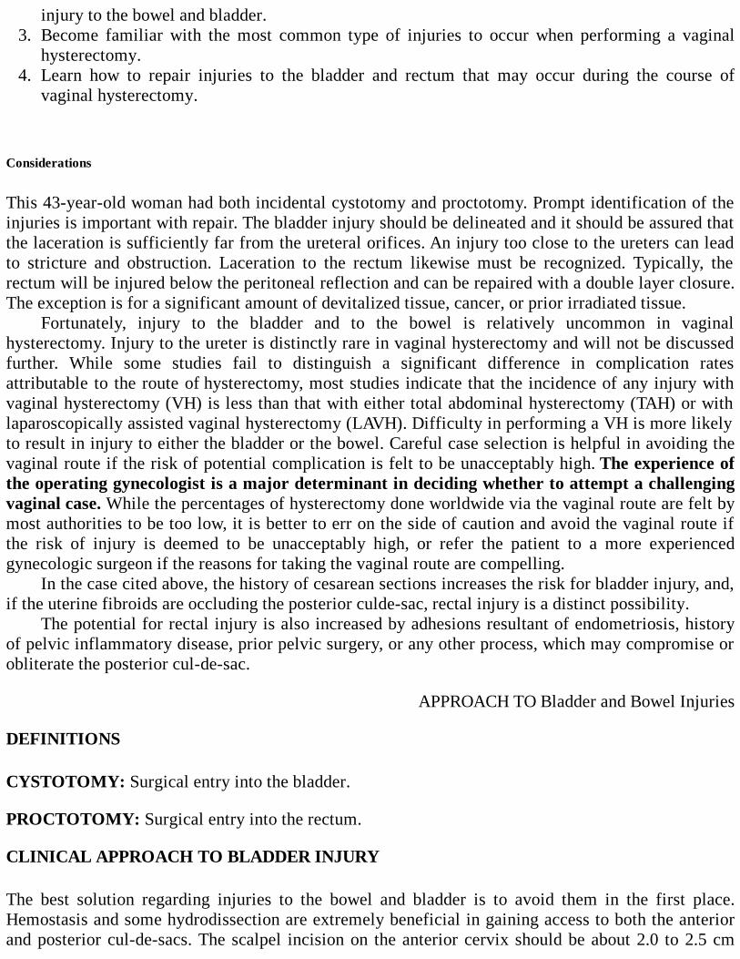
injury to the bowel and bladder.3. Become familiar with the most common type of injuries to occur when performing a vaginal
hysterectomy.4. Learn how to repair injuries to the bladder and rectum that may occur during the course of
vaginal hysterectomy.
Considerations
This 43-year-old woman had both incidental cystotomy and proctotomy. Prompt identification of theinjuries is important with repair. The bladder injury should be delineated and it should be assured thatthe laceration is sufficiently far from the ureteral orifices. An injury too close to the ureters can leadto stricture and obstruction. Laceration to the rectum likewise must be recognized. Typically, therectum will be injured below the peritoneal reflection and can be repaired with a double layer closure.The exception is for a significant amount of devitalized tissue, cancer, or prior irradiated tissue.
Fortunately, injury to the bladder and to the bowel is relatively uncommon in vaginalhysterectomy. Injury to the ureter is distinctly rare in vaginal hysterectomy and will not be discussedfurther. While some studies fail to distinguish a significant difference in complication ratesattributable to the route of hysterectomy, most studies indicate that the incidence of any injury withvaginal hysterectomy (VH) is less than that with either total abdominal hysterectomy (TAH) or withlaparoscopically assisted vaginal hysterectomy (LAVH). Difficulty in performing a VH is more likelyto result in injury to either the bladder or the bowel. Careful case selection is helpful in avoiding thevaginal route if the risk of potential complication is felt to be unacceptably high. The experience ofthe operating gynecologist is a major determinant in deciding whether to attempt a challengingvaginal case. While the percentages of hysterectomy done worldwide via the vaginal route are felt bymost authorities to be too low, it is better to err on the side of caution and avoid the vaginal route ifthe risk of injury is deemed to be unacceptably high, or refer the patient to a more experiencedgynecologic surgeon if the reasons for taking the vaginal route are compelling.
In the case cited above, the history of cesarean sections increases the risk for bladder injury, and,if the uterine fibroids are occluding the posterior culde-sac, rectal injury is a distinct possibility.
The potential for rectal injury is also increased by adhesions resultant of endometriosis, historyof pelvic inflammatory disease, prior pelvic surgery, or any other process, which may compromise orobliterate the posterior cul-de-sac.
APPROACH TO Bladder and Bowel Injuries
DEFINITIONS
CYSTOTOMY: Surgical entry into the bladder.
PROCTOTOMY: Surgical entry into the rectum.
CLINICAL APPROACH TO BLADDER INJURY
The best solution regarding injuries to the bowel and bladder is to avoid them in the first place.Hemostasis and some hydrodissection are extremely beneficial in gaining access to both the anteriorand posterior cul-de-sacs. The scalpel incision on the anterior cervix should be about 2.0 to 2.5 cm
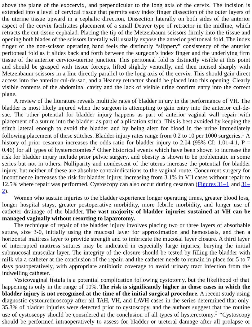
above the plane of the exocervix, and perpendicular to the long axis of the cervix. The incision isextended into a level of cervical tissue that permits easy index finger dissection of the outer layers ofthe uterine tissue upward in a cephalic direction. Dissection laterally on both sides of the anterioraspect of the cervix facilitates placement of a small Deaver type of retractor in the midline, whichretracts the cut tissue cephalad. Placing the tip of the Metzenbaum scissors firmly into the tissue andopening both blades of the scissors laterally will usually expose the anterior peritoneal fold. The indexfinger of the non-scissor operating hand feels the distinctly “slippery” consistency of the anteriorperitoneal fold as it slides back and forth between the surgeon’s index finger and the underlying firmtissue of the anterior cervico-uterine junction. This peritoneal fold is distinctly visible at this pointand should be grasped with tissue forceps, lifted slightly ventrally, and then incised sharply withMetzenbaum scissors in a line directly parallel to the long axis of the cervix. This should gain directaccess into the anterior cul-de-sac, and a Heaney retractor should be placed into this opening. Clearlyvisible contents of the abdominal cavity and the lack of visible urine confirm entry into the correctplane.
A review of the literature reveals multiple rates of bladder injury in the performance of VH. Thebladder is most likely injured when the surgeon is attempting to gain entry into the anterior cul-de-sac. The other potential for bladder injury happens as part of anterior vaginal wall repair withplacement of a suture into the bladder as part of a plication stitch. This is best avoided by keeping thestitch lateral enough to avoid the bladder and by being alert for blood in the urine immediatelyfollowing placement of these stitches. Bladder injury rates range from 0.2 to 10 per 1000 surgeries.1 Ahistory of prior cesarean increases the odds ratio for bladder injury to 2.04 (95% CI: 1.01-4.1, P =0.46) for all types of hysterectomies.2 Other historical events which have been shown to increase therisk for bladder injury include prior pelvic surgery, and obesity is shown to be problematic in someseries but not in others. Nulliparity and nondescent of the uterus increase the potential for bladderinjury, but neither of these are absolute contraindications to the vaginal route. Concurrent surgery forincontinence increases the risk for bladder injury, increasing from 3.1% in VH cases without repair to12.5% where repair was performed. Cystoscopy can also occur during cesarean (Figures 31–1 and 31–2).
Women who sustain injuries to the bladder experience longer operating times, greater blood loss,longer hospital stays, greater postoperative morbidity, more febrile morbidity, and longer use ofcatheter drainage of the bladder. The vast majority of bladder injuries sustained at VH can bemanaged vaginally without resorting to laparotomy.
The technique of repair of the bladder injury involves placing two or three layers of absorbablesuture, size 3-0, initially using the mucosal layer for approximation and hemostasis, and then ahorizontal mattress layer to provide strength and to imbricate the mucosal layer closure. A third layerof interrupted mattress sutures may be indicated in especially large injuries, burying the initialsubmucosal muscular layer. The integrity of the closure should be tested by filling the bladder withmilk via a catheter at the conclusion of the repair, and the catheter needs to remain in place for 5 to 7days postoperatively, with appropriate antibiotic coverage to avoid urinary tract infection from theindwelling catheter.
Vesicovaginal fistula is a potential complication following cystotomy, but the likelihood of thathappening is only in the range of 10%. The risk is significantly higher in those cases in which thebladder injury is not recognized at the time of the initial surgical procedure. A recent study usingdiagnostic cystourethroscopy after all TAH, VH, and LAVH cases in the series determined that only35.3% of bladder injuries were detected prior to cystoscopy, and the authors suggest that the routineuse of cystoscopy should be considered at the conclusion of all types of hysterectomy.3 “Cystoscopyshould be performed intraoperatively to assess for bladder or ureteral damage after all prolapse or
incontinence procedures during which the bladder or ureters may be at risk of injury.”4
Figure 31–1. Bladder is repaired in two layers. First layer is mucosa. (Reproduced, with permission,from Cunningham FG, Leveno KJ, Bloom SL, et al. Williams Obstetrics, 23rd ed. New York: McGraw-Hill, 2010.)
Figure 31–2. Bladder is repaired with second layer. (Reproduced, with permission, from CunninghamFG, Leveno KJ, Bloom SL, et al. Williams Obstetrics, 23rd ed. New York: McGraw-Hill, 2010.)
CLINICAL APPROACH TO BOWEL INJURY
Many of the principles that apply to avoiding bladder injury also apply to prevention of injury to therectum. Hemostasis and hydrodissection beneficially provide visibility for entry into the posterior cul-de-sac. However, in contrast to entry into the anterior cul-de-sac, lateral dissection is not helpful whenentering the posterior cul-de-sac. Tissue forceps grasp the uterine border of the incised vaginalmucosa in the midline and pull it away from the cervico-uterine junction at a 90-degree angle, makinga “tent” in the mucosa and underlying peritoneum. Mayo scissors are placed directly on top of thetissue forceps, at about a 45-degree angle, and an incision is made directly into the “tent” created bythe tissue forceps. If the initial incision does not provide entry into the peritoneal cavity, pick up the
tissue deeper again in the site of the initial incision and repeat the incision in the same tissue plane.Using the scissor tips to “open and spread” the tissue is not helpful in gaining entry posteriorly. Ifunable to gain entry at this point, stay close to the uterus and continue dissection posteriorly towardthe top of the uterine fundus until entry can be gained into the cul-de-sac. It is better to makeincisions into the posterior uterus than into the bowel.
If bowel injury is suspected, place a gloved finger into the rectum and see if rectal injury can beconfirmed. If there is bowel injury, obtain visualization with enough dissection to see the extent of theinjury and then approximate the mucosa with a running stitch of 3-0 absorbable suture. Themuscularis is closed with interrupted inverting mattress sutures of 3-0 absorbable suture.
Bowel injuries are less common than bladder injuries. Large series reported an incidence ofbowel injury with vaginal hysterectomy of 0.5%, with almost all occurring at the point of entry intothe posterior cul-de-sac.1 Rectovaginal fistula is a rare complication of this injury, as most aredetected and repaired immediately and successfully. After the patient sustains a bowel injury, sheshould be kept on a liquid diet until bowel function returns and then on a low-residue diet for thefollowing week. Judicious use of stool softeners and laxatives is indicated until recovery is complete.
CONCLUSION
Blanket recommendations regarding when or if to call a consultant urologist for bladder injury or asurgeon for bowel injury are challenging and obviously depend on the extent of the injury and theexperience of the operating gynecologist. If the injury appears straightforward, the repair goessmoothly, and if the bladder or the bowel appears intact following repair, then consultation may not berequired. If the limits of the injury are not easily delineated, the integrity of the repair cannot beconfirmed, or if the degree of confidence in the result is less than satisfactory, then ask for help.Patients with chronic diseases of the urinary tract or of the intestinal tract which would predisposesuch a patient to a complicated recovery even without unintentional injury should definitely beevaluated intraoperatively by urology or surgery as needed. Community practice and standards ofcourse also play an important role.
Comprehension Questions
31.1 Which of the following surgical approaches for hysterectomy is associated with the overalllowest incidence of bladder injury?
A. Abdominal hysterectomyB. Laparoscopic hysterectomyC. Vaginal hysterectomy
31.2 If incidental cystotomy is noted at the time of vaginal hysterectomy, which of the followingshould be the approach?
A. The location of the injury should be ascertained regarding the distance from the ureteralorifices.
B. The vaginal approach should be abandoned, and the case converted to an abdominal procedure.C. The patient should undergo an intravenous pyelogram to assess the renal function.D. A suprapubic catheter should be placed in the bladder.
ANSWERS
31.1 C. Vaginal hysterectomy results in the lowest incidence of bladder injury compared to allother surgical approaches.
31.2 A. Upon recognition of a bladder injury, its extent and location, especially with respect tothe ureteral orifices, should be established. This assessment will indicate whether thebladder can be closed with a simple two layer closure or will require possible ureteralsurgery and/or stents.
Clinical PearlsSee Table 1-2 for definition of level of evidence and strength of recommendation
Early detection, preferably intraoperatively, of injury to the bowel or bladder will allow forimmediate repair and greatly reduce the risk of significant harm to the patient, and to thereputation of the surgeon (Level B).
Infiltration of the cervix with a mixture of 1% lidocaine and 1:100,000 epinephrine or 0.5%lidocaine and 1:200,000 epinephrine prior to the circumferential incision around the cervixwill provide excellent hemostasis and allow for better visualization of anatomic landmarks andthe potential to avoid injury (Level B).
Using the more dilute solution will allow for instillation of a greater volume of fluid, and theprinciple of hydrodissection may create easier planes for entry into the anterior and posteriorcul-de-sacs (Level C).
The use of sterile milk to see the injury to the bladder may be preferable to the use of indigocarmine or methylene blue dyes, as both dyes tend to stain the tissue and may makevisualization of the tissue to be repaired more difficult (Level B).
When repairing injuries to the bladder, sutures should be tied such that the knot is outside ofthe bladder mucosa to avoid the potential for the knotted suture to cause bladder stones (LevelC).
When repairing rectal injuries, tie the suture such that the knot is inside of the rectal lumen,allowing the suture to slough into the bowel when healing is complete and the suture dissolves(Level B).
If your surgical procedure results in an injury to the bowel or bladder, do not describe this as“inadvertent” in your written or dictated operative report. Synonyms for “inadvertent” include“careless” and “inattentive.” Do not put yourself in the position of having to explain thiswording in a deposition or trial for medical malpractice (Level C).
REFERENCES
1. Mathevet P, Valencia P, Cousin C, Meiller G, Dargent D. Operative injuries during vaginalhysterectomy. Eur J Obstet Gynecol Reprod Biol. 2001;97:71-75.
2. David-Montefiore E, Rouzier R, Chapron C. Surgical routes and complications ofhysterectomy for benign disorders: a prospective observational study in French universityhospitals. Hum Reprod. 2007;22:260-265.
3. Vakili B, Chesson R, Kyle B, et al. The incidence of urinary tract injury during hysterectomy: aprospective analysis based on universal cystoscopy. Am J Obstet Gynecol. 2005;192:1599-1604.
4. American College of Obstetricians and Gynecologists. Pelvic organ prolapse. ACOG PracticeBulletin No. 85. Obstet Gynecol. 2007;110:717-730.
5. Harkki-Siren P, Sjoberg J, Tiitinen A. Urinary tract injuries after hysterectomy. ObstetGynecol. 1998;92:113-118.
6. Rooney C, Crawford A, Vassallo B, Kleeman S, Karram M. Is previous cesarean section a riskfor incidental cystotomy at the time of hysterectomy? Am J Obstet Gynecol. 2005;193:2041-2044.
7. Carley M, McIntire D, Carley J, Schaffer J. Incidence, risk factors, and morbidity ofunintended bladder or ureter injury during hysterectomy. Int Urogynecol J Pelvic FloorDysfunct. 2002;13:18-21.
8. Dorairajan G, Rani P, Habeebullah S, Dorairajan L. Urological injuries during hysterectomies:a 6-year review. J Obstet Gynaecol Res. 2004;30:430-435.
Case 32
A 44-year-old African American, G3P2012, woman complains of increasing abdominal girth andurinary frequency for the past 6 months. She has noted that she has had difficulty buttoning her pantsand has had to buy clothes several sizes larger. She denies dysuria, hematuria, or nocturia. She deniesany change in bowel habits. She reports menstrual cycles every 30 days with a flow lasting for 7 days.She has no chronic medical conditions and no family history of gynecologic malignancies. Pastsurgical history includes a tubal ligation.
The pelvic examination reveals normal external female genitalia. The vaginal and cervix arewithout lesions. The uterus is 16-week size on palpation, irregular and mobile. Six months ago, theuterus measured 10-week size on examination. The patient’s adnexa are not well palpated due to themidline mass. The rectal examination reveals a 3-cm firm mass that is continuous with the uterus. Apelvic ultrasound reveals multiple hypoechoic densities suggestive of fibroids, the largest measuring 8cm in the fundal region.
What is the most likely diagnosis?
What are the indications for surgery in the presence of myomas?
What is the best treatment for this patient?
ANSWERS TO CASE 32:Indications for Surgical Therapy for Fibroids
Summary: This is a 44-year-old multiparous woman has a rapidly enlarging pelvic mass which is mostlikely due to uterine fibroids. She has symptoms of increasing abdominal girth and urinary frequency.She does not desire more children.
Most likely diagnosis: Uterine leiomyoma.
Indications for surgery: The list of indications is lengthy, here are the common indications:
1. Abnormal uterine bleeding not responsive to conservative therapy2. High level of suspicion for pelvic malignancy3. Myoma growth after menopause4. Infertility with distortion of endometrial cavity or tubal obstruction5. Recurrent pregnancy loss due to distortion of endometrial cavity6. Pain or pressure symptoms interfering with quality of life7. Urinary tract symptoms like frequency and/or obstruction8. Iron deficiency anemia secondary to chronic blood loss9. Adnexa cannot be palpated due to uterine enlargement
Best treatment: Hysterectomy, possibly also bilateral salpingoophrorectomy. The route of
hysterectomy should be tailored to the situation.
ANALYSIS
Objectives
1. Develop a differential diagnosis of a pelvic mass.2. List symptoms caused by leiomyoma.3. List indications for fibroid surgery.4. List surgical treatment options for symptomatic uterine fibroids.
Considerations
This 44-year-old woman has worsening symptoms from her uterine fibroids, specifically theincreasing pelvic pressure and menorrhagia. She does not desire more children. Medical therapy doesnot seem to be effective. Thus, surgical therapy should be considered. Myomas are the most commonsolid pelvic tumors in women. The benign monoclonal tumors develop from a chromosomal mutationin a single smooth muscle cell of the uterus. Leiomyomas contain fibrous tissue as well as smoothmuscle cells and are bounded by a fibrous capsule. Approximately 20% to 40% of women will bediagnosed with a leiomyoma during their reproductive years, and it is the most common indication forhysterectomy.
APPROACH TO Uterine Fibroids
DEFINITIONS
MEIGS SYNDROME: Benign fibromas of the ovary associated with ascites and pleural effusion.
CARNEOUS DEGENERATION (aka Red degeneration): Necrosis of the leiomyoma that occursbecause the myoma has outgrown its blood supply, especially in pregnancy. Clinically, it causessevere pain and local peritoneal inflammation. Carneous degeneration is associated with markedsoftening and red color of the fibroid. This type of degeneration occurs in 5% to 10% of pregnantwomen with leiomyomas.
TENESMUS: Ineffectual and painful straining with bowel movement or upon urination.
MONOCLONAL: Originating from a single cell. Monoclonal smooth muscle cells in the uterus thatform leiomyoma often have aberrant chromosomes.
CLINICAL APPROACH
Symptoms
Most women with leiomyoma are asymptomatic. However, the size, number, location, anddegeneration status of the myomas can determine the symptoms that the patient will experience.
Myomas can range in size from subcentimeter to occupying the whole pelvic cavity. They can besubserosal, submucosal, or intramural in location. They can be pedunculated or prolapsed through thecervix. The myoma may become parasitic and derive its blood supply from an organ other than theuterus.
Aside from leiomyoma, other etiology for pelvic masses should be considered in order to plan forsurgery. Differential diagnoses for pelvic and lower abdominal masses include follicular cyst,hemorrhagic corpora lutea, serous cystadenoma, benign cystic teratoma, mucinous cystadenoma,malignant ovarian tumors, Meigs syndrome, endometrioma, malignant uterine tumors, metastaticdisease, benign or malignant masses of the fallopian tubes, diverticulitis, pregnancy, gestationaltrophoblastic disease, and adenomyosis.
Women with uterine fibroids may complain of abnormal uterine bleeding, pelvic pressure, andincreasing abdominal girth. Although other bleeding patterns can be present, menorrhagia is the mostcommon type of bleeding associated with myomas. Explanation for the cause of menorrhagia includesthe obstructive effect of myomas, leading to ectasia of the endometrial venules, which causescongestion in the endometrium and myometrium, contributing to an increase in bleeding duringmenstrual cycle. The increased surface area from the enlarged uterine cavity can cause menorrhagia.Endometritis is often histologically seen in the endometrial tissue overlying the submucous myomaand can contribute to excessive menstrual flow. Aberrant angiogenesis and dysregulation of localgrowth factors have been linked to abnormal uterine bleeding in patients with leiomyoma. Persistentabnormal uterine bleeding can lead to iron deficiency anemia due to chronic blood loss.
Pelvic pain is rarely caused by leiomyoma. Myomas can cause acute pain if there is torsion of apediculated myoma, cervical dilation from a prolapsing pediculated submucosal myoma, or carneousdegeneration with pregnancy. More frequently, the pelvic discomfort is described as pelvic pressure.Pelvic pressure and increasing abdominal girth are a result of the enlarging leiomyoma. Urinarysymptoms include urinary frequency, outflow obstruction, and compression of the ureters which canresult from the myoma pressing on the bladder and pelvic side wall. Gastrointestinal symptomsinclude constipation and tenesmus which are due to pressure on the rectosigmoid by a myoma on theposterior wall of the uterus. An incarcerated uterus in the posterior culde-sac or a single largeposterior wall myoma may cause rectal pressure, but this symptom is rare.
Rarely, myomas have been associated with infertility. When all the other causes of infertility areexcluded, 2% to 3% of the infertility cases can be attributed to the presence of myomas. Mechanismsby which myomas can cause infertility include the following:
1. Alteration of the endometrial cavity may inhibit implantation.2. Enlarged and deformed endometrial cavity can inhibit sperm transport.3. Displacement of the cervix can block access by ejaculated sperm.4. Alteration in uterine contractility may alter sperm movement.5. Persistent blood or clots in endometrial cavity may inhibit implantation.6. Tubal ostia may be obstructed or distorted.
In patients undergoing in vitro fertilization, decreased rates of implantation and pregnancy werenoted in patients who had a endometrial cavity distorted by leiomyomas up to 50% of the time.Recurrent pregnancy losses have also been linked to myomas. Although no prospective studiescomparing expectant management to myomectomy in asymptomatic women have been done, mostclinicians conclude that surgery does improve pregnancy rates.
Malignancy associated with uterine fibroids is rare. A study done by Parker evaluated 1332women who underwent hysterectomy or myomectomy for symptomatic leiomyoma; uterine sarcoma(leiomyosarcoma, endometrial stromal sarcoma, and mixed mesodermal tumor) was found in 0.23%.The incidence of leiomyosarcoma usually increases with age. Women in the reproductive years have a0.1% risk and women undergoing uterine surgery for myomas have a risk of approximately 1.7%. Thepossibility of sarcoma in a uterine myoma is 10 times greater in a woman in her 60s than a woman inher 40s. Although malignancy is usually suspected if the leiomyoma displays rapid growth, theincidence of leiomyosarcoma is not related to the rate of growth or the uterine size. Considering thehigh prevalence of uterine fibroids, most researchers agree that sarcomas arise spontaneously infibroid uterus. Of note, myomas tend to exhibit an accelerated rate of growth in the fifth decade oflife, likely due to unopposed estrogen stimulation from anovulatory cycles in the perimenopausalperiod.
Some clinicians advocate treatment for asymptomatic uterine myomas if the uterine size is largerthan 12 weeks’ size due to inability to palpate the adnexa which may interfere with the detection of anovarian tumor or cancer.
Summary of clinical settings that may require surgical therapy for myomas includes
1. Abnormal uterine bleeding not responsive to conservative therapy.2. High level of suspicion for pelvic malignancy.3. Myoma growth after menopause.4. Infertility with distortion of endometrial cavity or tubal obstruction.5. Recurrent pregnancy loss due to distortion of endometrial cavity.6. Pain or pressure symptoms interfering with quality of life.7. Urinary tract symptoms like frequency and/or obstruction.8. Iron deficiency anemia secondary to chronic blood loss.9. Adnexa cannot be palpated due to uterine enlargement.
Treatment
The choice and approach for therapy should take into account medical and social factors.Considerations for treatment:
1. Age2. Parity3. Desire for childbearing4. Extent and severity of symptoms5. Size and number of myomas6. Locations of myoma7. Medical conditions8. Possibility of malignancy9. Proximity to menopause
10. Desire for uterine preservation
There are various options for management of leiomyomas. These include expectant management,medical management, surgical management, uterine artery embolization, high-frequencyultrasonography, laser treatment, cryotherapy, and thermablation. Surgical management includeshysterectomy and myomectomy. Hysterectomy is the most common treatment for leiomyomasbecause it is the only therapeutic option that provides a cure and eliminates the chance of recurrence.It is second to cesarean delivery as the most common major surgical procedure performed in womenin the United States. Of note, all surgical alternatives to hysterectomy allow for the chance that newmyomas may form or leiomyomas that were already present and were too small to be seen orintentionally not removed may grow significantly and may require another procedure. Also,complications from surgical procedures other than a hysterectomy may lead to a hysterectomy. Beforeproceeding with treatment, it is important to rule out adnexal or endometrial pathology. Pathology ineither of these locations will determine which therapeutic option is chosen.
Comprehension Questions
32.1 A 43-year-old G3P3 woman has a 10-week irregular uterus on palpation. No adnexal massesare palpated. She reports normal menstrual cycles and no other symptoms. What is the besttreatment for this patient?
A. Expectant managementB. Medical managementC. MyomectomyD. Hysterectomy
32.2 A 43-year-old G1P1 woman complains of heavy menstrual bleeding lasting for 10 days for 3months. She feels chronic fatigue. She has been on iron supplements. She has had a BTL andno other medical or surgical conditions. She does not smoke. Her pelvic examination revealsa 16-week size irregularly shaped uterus. Adnexa are not well palpated due to the midlinemass. What is the next step in the management of this patient?
A. Expectant managementB. Oral contraceptivesC. Pelvic ultrasoundD. Hysterectomy
32.3 A 35-year-old woman is noted to have an irregular uterus with menorrhagia. The excessivebleeding with fibroids is caused by dysregulation of growth factors, the obstructive effect ofleiomyoma, and which of the following?
A. Associated endometritisB. CoagulopathyC. Endometrial hyperplasiaD. Carneous degeneration
ANSWERS
32.1 A. Since this patient is asymptomatic, the small uterine myomas do not require treatment.Initially she should be examined every 6 months for uterine growth and the development ofsymptoms. Annual visits can resume when the clinician is confident that the leiomyomas are
not enlarging.
33.2 C. Although this patient’s history and physical are highly suggestive of uterine leiomyoma,other gynecologic conditions should be considered in the differential diagnosis prior toinstituting therapy. Since the adnexa were not well evaluated with the pelvic examination,ovarian or adnexal pathology should be ruled out. Pelvic ultrasound is a reliable tool thatwill evaluate not only the adnexa, but it will better characterize the location and size of themyomas. The presence or absence of concurrent adnexal pathology may guide the finaltreatment that the patient receives. Furthermore, the endometrium in this patient older than35 years with abnormal uterine bleeding should be evaluated prior to surgery.
32.3 A. Associated endometritis is noted in patient with fibroids and menorrhagia.
Clinical PearlsSee Table 1-2 for definition of level of evidence and strength of recommendation
Most women with leiomyoma will be asymptomatic and will not require treatment (Level A).
Menorrhagia is the most common type of abnormal uterine bleeding associated withleiomyoma (Level B).
If all other causes of infertility are ruled out, in the presence of leiomyoma, a myomectomycan improve fertility outcome (Level B).
Hysterectomy is the only surgical procedure that will not allow myomas to form or allow thepreviously remaining ones to increase in size (Level B).
Malignancy originating from leiomyoma is very rare (Level A).
Most common differential diagnosis for pelvic mass is leiomyoma, adenomyosis, pregnancy,and adnexal pathology (Level B).
REFERENCES
1. Parker W, Berek J, Fu YS. Uterine sarcoma in patients operated on for presumed leiomyomasand rapidly growing leiomyomas. Obstet and Gynecol. 1994; 83:414-8.
2. Benson CB, Chow JS, Chang-Lee W, Hill JA 3rd, Doubilet PM. Outcome of pregnancies inwomen with uterine leiomyomas identified by sonography in the first trimester. J ClinUltrasound. 2001;29:261-264.
3. Buttran VC, Reiter RC. Uterine leiomyomata: etiology, symptomatology, and management.Fertil Steril. 1981;36:433-435.
4. Farrer-Brown G, Beilby JO, Tarbit MH. The vascular patterns in myomatous uteri. J ObstetGynaecol Br Commonw. 1970;77:967-975.
5. Farrer-Brown G, Beilby JO, Tarbit MH. Venous changes in the endometrium of myomatousuteri. Obstet Gynecol. 1971;38:743-751.
6. Katz VL. Benign gynecologic lesions. In Katz VL, Lentz GM, Lobo RA, Gershenson DM, eds.Comprehensive Gynecology. 5th ed. Philadelphia, PA: Mosby; 2007:452-557.
7. Li TC, Mortimer R, Cooke ID. Myomectomy: a retrospective study to examine reproductiveperformance before and after surgery. Hum Reprod. 1999;14:1735-1740.
8. Propst AM, Hill JA 3rd. Anatomic factors associated with recurrent pregnancy loss. SeminReprod Med. 2000;18:341-350.
9. Sehgal N, Haskins AL. The mechanism of uterine bleeding in the presence of fibromyomas.Am Surg. 1960;26:21-23.
10. Seoud MA, Patterson R, Muasher SJ, Coddington CC 3rd. Effects of myomas or priormyomectomy on in vitro fertilization (IVF) performance. J Assist Reprod Genet. 1992;9:217-221.
11. Stewart EA, Nowak RA. Leiomyoma-related bleeding: a classic hypothesis updated for themolecular era. Hum Reprod Update. 1996;2:295-306.
12. American College of Ostetricians and Gynecologists. Surgical Alternatives to Hysterectomyin the Management of Leiomyomas. ACOG Practice Bulletin No. 16. Washington, DC; May2000.
13. The Practice Committee of the American Society for Reproductive Medicine. Myomas andreproductive function. Fertil Steril. 2006;86(suppl 5):194-199.
14. Wallach EE, Vlahos NF. Uterine myomas: an overview of development, clinical features, andmanagement. Obstet Gynecol. 2004;104:393-406.
Case 33
A 42-year-old G2P2 woman, with a recent last menstrual period (LMP), presents complaining ofirregular periods for 4 years. She states that her periods are about three to four times a year and verylong in duration, up to 21 days. She has gained 50 lb (22.68 kg) in the last 2 years. Four years ago, shewas on oral contraceptive pills to regulate her periods. Her last doctor instructed her to take iron dailyfor her anemia. She is “tired of these heavy periods” and refuses further medical therapy. She wantssurgical therapy. Her review of systems is negative for thyroid disease or galactorrhea. She denies anysignificant past medical, family, or social histories. She had a bilateral tubal ligation 3 years ago.
On examination, her weight is 262 lb (118.85 kg), BP 112/72 mmHg, and pulse 60 bpm. The headand neck examination, including the thyroid gland, is normal. The abdomen is nontender and there areno masses. On pelvic examination, the uterus is noted to be 8- to 10-week size and the adnexa aredifficult to assess. A pelvic ultrasound is performed revealing a normal sized uterus, with anendometrial stripe of 10mm and no adenxal masses. A pregnancy test is negative.
What is the most likely diagnosis?
What is the next step?
ANSWERS TO CASE 33:Surgical Indications for Dysfunctional Uterine Bleeding
Summary: 42-year-old G2 P2 woman has a 4 year history of irregular menses, oligomenorrhea,menorrhagia, obesity, and anemia. She desires surgical therapy.
Diagnosis: Oligomenorrhea or dysfunctional uterine bleeding (DUB)
Next step: Endometrial biopsy
ANALYSIS
Objectives
1. Recognize that DUB is a diagnosis of exclusion.2. Know the common differential diagnoses that cause DUB.3. Understand that surgical therapy is indicated only when medical therapy fails and future fertility
is no longer desired.4. Recognize the two surgical therapies for DUB, endometrial ablation, and hysterectomy.
Considerations
This patient has a long history of irregular periods. This is a classic case of anovulation, most likelysecondary to her obesity. She weighs 100 lb (45.36 kg) over her ideal body weight. One has to
consider an underactive thyroid being a contributor to her obesity. Her age warrants that the next stepis to perform an endometrial biopsy. Women older than 35 years should receive an endometrial biopsy(EMB) for any abnormal uterine bleeding. Note that her endometrial stripe is 1 cm. Studies reveal thatan EMB using the Pipelle type of device is highly effective in detecting pathology.1 Her differentialdiagnosis includes anovulation, hypothyroidism, endometrial polyp, and uterine fibroids. Anendometrial polyp or endometrial hyperplasia/neoplasia can be the etiology for an enlargedendometrial stripe. A coagulation defect is unlikely, given the age of the patient. Since DUB is adiagnosis of exclusion, an organic etiology should be ruled out first. After obtaining a negativepregnancy test, check a thyroid-stimulating hormone (TSH), complete blood (cell) count (CBC), andprolactin, even though the patient has a negative past medical history. Premature ovarian failure canbe ruled out if the follicle-stimulating hormone (FSH) is less than 40 U/L. An EMB that revealsproliferative endometrium is consistent with anovulation. This patient should be offered medicaltreatment, first, with hormones. After medical therapy fails or in this case, is refused, surgical therapyis the next option. This patient is a candidate for an endometrial ablation because her uterine size isnormal and without evidence of any organic etiology, such as fibroids. The fact that she received atubal ligation confirms that she does not desire future fertility. Keeping in mind that endometrialablation has shorter operative time, shorter recovery, and less hospital-associated cost, this procedureshould be offered first. Even though hysterectomy is definitive, there is associated morbidity andmortality.2
APPROACH TO Dysfunctional Uterine Bleeding
DEFINITIONS
DUB: Excessive uterine bleeding without any demonstrable organic cause.
MENORRHAGIA: Prolonged uterine bleeding longer than 7 days duration or greater than 80 cc.
ENDOMETRIAL ABLATION: Operative destruction, via a hysteroscope, of the endometrium with avariety of different instruments.
OLIGOMENORRHEA: Uterine bleeding that occurs at intervals of longer than 35 days.
MENOMETRORRHAGIA: Prolonged uterine bleeding that occurs at irregular intervals.
POLYMENORRHEA: Uterine bleeding that occurs at intervals every 21 days or fewer.
CLINICAL APPROACH
Heavy menstrual flow can become very disturbing to a woman and can result in disruption of herlifestyle. These changes include absenteeism from work, avoidance of social functions, and restrictionto certain physical activities. Over a period of time, women will experience fatigue, anxiety, anemia,and a decrease in quality of life.3
Dysfunctional uterine bleeding is defined as noncyclic, menstrual flow, unrelated to organicpathology.3 It clinically manifests as abnormal volume, timing, and duration of flow. Traditionaloptions for treatment include medical therapy with hormones and surgical therapy, such asendometrial ablation and hysterectomy. The choice of treatment depends on the woman’s age and her
desire for future fertility.During a normal menstrual cycle, the endometrium is exposed to the ovarian production of
estrogen, in the first half of the cycle and after ovulation, progesterone, in the second half of the cycle.Menses occurs secondary to the estrogen and progesterone withdrawal, to begin another cycle. InDUB, or anovulatory bleeding, a corpus luteum does not form and progesterone is not produced fromthe ovary. In this clinical scenario, the endometrium continues to be primed with estrogen. As a result,the endometrial lining continues to proliferate. Once the lining is at its maximum thickness,inadvertent shedding of the endometrium will occur in a nonuniform, noncyclic manner. This occursbecause the endometrium is very vascular, fragile, and not stable. The end result is prolonged, heavy,uterine bleeding.
A detailed history should be obtained from the patient. It is important to verify what “irregular”means to her. Is the uterine bleeding more than once a month or only monthly, with variable duration?Have the patient quantify her bleeding.
Number of pads used in a 24-hour periodDoes the bleeding soil her clothes?Is her activity restricted?Does she stay home from work?Passage of clots? If so, what size … (ie, penny, nickel, dime, quarter, half-dollar size)
Obtain other relevant history. Certain medications can contribute to abnormal bleeding, such ashormones and psychotropic medications. On the physical examination, look closely for evidence ofhirsutism and obesity. Perform a detailed pelvic examination. Examine the external genitalia forlacerations. Insert the speculum and evaluate for lacerations or trauma to the cervix or vagina. Foreignbodies, such as an IUD, can also cause abnormal bleeding. Obtain a Pap smear and cultures. Performan endometrial biopsy in a woman older than 35 years. Studies show that the endometrial Pipelle ishighly effective and accurate in sampling the endometrium.3 Malignancies can cause abnormal uterinebleeding. This test will usually rule out endometrial hyperplasia and endometrial carcinoma. A TSHand a prolactin should be obtained. If hypothyroidism is diagnosed, then levothyroxine should beprescribed. Once the thyroid hormonal status corrects, usually requiring 3 months, the menorrhagiashould improve. Check the pregnancy test. A pelvic ultrasound will evaluate for any organic causes ofthe uterine bleeding, such as leiomyomas. DUB is a diagnosis of exclusion; other etiologies should beruled out as a cause of bleeding. Developing a differential diagnosis is important and will differ basedon the age of the patient (Table 33–1).
If the TSH, CBC, prolactin, and pelvic ultrasound are normal and the endometrial biopsy isnegative for hyperplasia or neoplasia, DUB can be diagnosed. Remember, it is a diagnosis ofexclusion.
For women younger than 20 years, a coagulation disorder should be eliminated. All adolescentswith prolonged uterine bleeding since menarche need routine screening for coagulation defects.Disorders such as idiopathic thrombocytopenic purpura and von Willebrand disease should beconsidered. Other disorders that cause platelet dysfunction include leukemia or sepsis. However, themost common diagnosis for DUB in this age group is immaturity of the hypothalamic, pituitary, andgonadal axis. Look for petechiae or bruising on the skin during the physical examination. In additionto the earlier-mentioned laboratory tests, also check a prothrombin time and partial thromboplastintime (PTT). An endometrial biopsy (Pipelle) is not necessarily indicated in this age group, unless
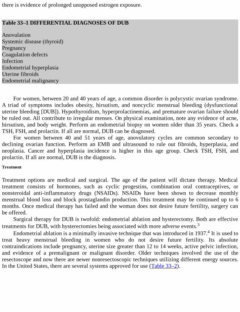
there is evidence of prolonged unopposed estrogen exposure.
Table 33–1 DIFFERENTIAL DIAGNOSES OF DUB
AnovulationSystemic disease (thyroid)PregnancyCoagulation defectsInfectionEndometrial hyperplasiaUterine fibroidsEndometrial malignancy
For women, between 20 and 40 years of age, a common disorder is polycystic ovarian syndrome.A triad of symptoms includes obesity, hirsutism, and noncyclic menstrual bleeding (dysfunctionaluterine bleeding [DUB]). Hypothyroidism, hyperprolactinemias, and premature ovarian failure shouldbe ruled out. All contribute to irregular menses. On physical examination, note any evidence of acne,hirsutism, and body weight. Perform an endometrial biopsy on women older than 35 years. Check aTSH, FSH, and prolactin. If all are normal, DUB can be diagnosed.
For women between 40 and 51 years of age, anovulatory cycles are common secondary todeclining ovarian function. Perform an EMB and ultrasound to rule out fibroids, hyperplasia, andneoplasia. Cancer and hyperplasia incidence is higher in this age group. Check TSH, FSH, andprolactin. If all are normal, DUB is the diagnosis.
Treatment
Treatment options are medical and surgical. The age of the patient will dictate therapy. Medicaltreatment consists of hormones, such as cyclic progestins, combination oral contraceptives, ornonsteroidal anti-inflammatory drugs (NSAIDs). NSAIDs have been shown to decrease monthlymenstrual blood loss and block prostaglandin production. This treatment may be continued up to 6months. Once medical therapy has failed and the woman does not desire future fertility, surgery canbe offered.
Surgical therapy for DUB is twofold: endometrial ablation and hysterectomy. Both are effectivetreatments for DUB, with hysterectomies being associated with more adverse events.3
Endometrial ablation is a minimally invasive technique that was introduced in 1937.4 It is used totreat heavy menstrual bleeding in women who do not desire future fertility. Its absolutecontraindications include pregnancy, uterine size greater than 12 to 14 weeks, active pelvic infection,and evidence of a premalignant or malignant disorder. Older techniques involved the use of theresectoscope and now there are newer nonresectoscopic techniques utilizing different energy sources.In the United States, there are several systems approved for use (Table 33–2).
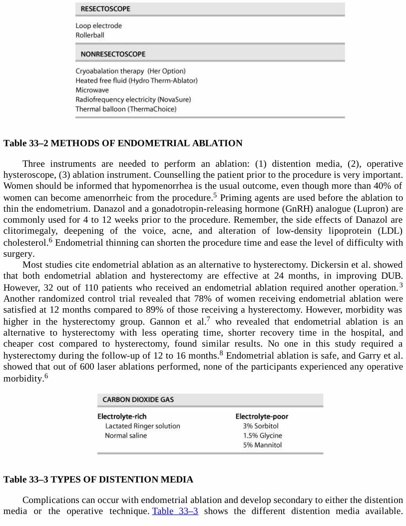
Table 33–2 METHODS OF ENDOMETRIAL ABLATION
Three instruments are needed to perform an ablation: (1) distention media, (2), operativehysteroscope, (3) ablation instrument. Counselling the patient prior to the procedure is very important.Women should be informed that hypomenorrhea is the usual outcome, even though more than 40% ofwomen can become amenorrheic from the procedure.5 Priming agents are used before the ablation tothin the endometrium. Danazol and a gonadotropin-releasing hormone (GnRH) analogue (Lupron) arecommonly used for 4 to 12 weeks prior to the procedure. Remember, the side effects of Danazol areclitorimegaly, deepening of the voice, acne, and alteration of low-density lipoprotein (LDL)cholesterol.6 Endometrial thinning can shorten the procedure time and ease the level of difficulty withsurgery.
Most studies cite endometrial ablation as an alternative to hysterectomy. Dickersin et al. showedthat both endometrial ablation and hysterectomy are effective at 24 months, in improving DUB.However, 32 out of 110 patients who received an endometrial ablation required another operation.3
Another randomized control trial revealed that 78% of women receiving endometrial ablation weresatisfied at 12 months compared to 89% of those receiving a hysterectomy. However, morbidity washigher in the hysterectomy group. Gannon et al.7 who revealed that endometrial ablation is analternative to hysterectomy with less operating time, shorter recovery time in the hospital, andcheaper cost compared to hysterectomy, found similar results. No one in this study required ahysterectomy during the follow-up of 12 to 16 months.8 Endometrial ablation is safe, and Garry et al.showed that out of 600 laser ablations performed, none of the participants experienced any operativemorbidity.6
Table 33–3 TYPES OF DISTENTION MEDIA
Complications can occur with endometrial ablation and develop secondary to either the distentionmedia or the operative technique. Table 33–3 shows the different distention media available.
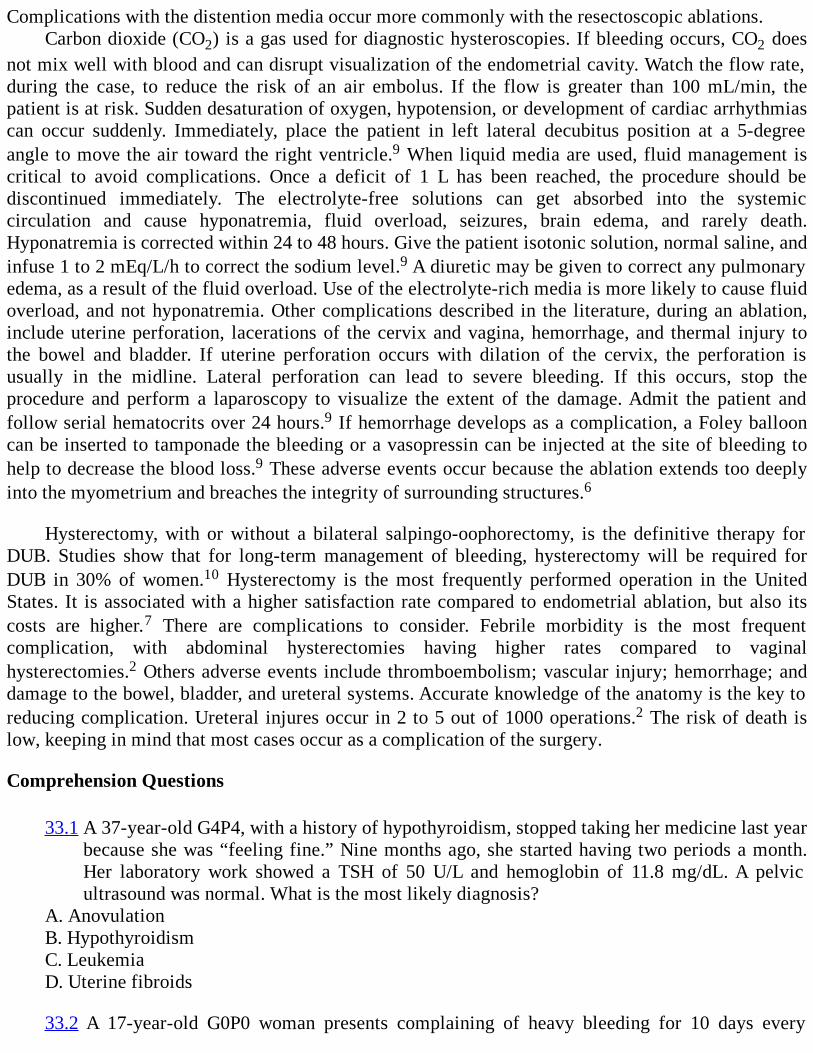
Complications with the distention media occur more commonly with the resectoscopic ablations.Carbon dioxide (CO2) is a gas used for diagnostic hysteroscopies. If bleeding occurs, CO2 does
not mix well with blood and can disrupt visualization of the endometrial cavity. Watch the flow rate,during the case, to reduce the risk of an air embolus. If the flow is greater than 100 mL/min, thepatient is at risk. Sudden desaturation of oxygen, hypotension, or development of cardiac arrhythmiascan occur suddenly. Immediately, place the patient in left lateral decubitus position at a 5-degreeangle to move the air toward the right ventricle.9 When liquid media are used, fluid management iscritical to avoid complications. Once a deficit of 1 L has been reached, the procedure should bediscontinued immediately. The electrolyte-free solutions can get absorbed into the systemiccirculation and cause hyponatremia, fluid overload, seizures, brain edema, and rarely death.Hyponatremia is corrected within 24 to 48 hours. Give the patient isotonic solution, normal saline, andinfuse 1 to 2 mEq/L/h to correct the sodium level.9 A diuretic may be given to correct any pulmonaryedema, as a result of the fluid overload. Use of the electrolyte-rich media is more likely to cause fluidoverload, and not hyponatremia. Other complications described in the literature, during an ablation,include uterine perforation, lacerations of the cervix and vagina, hemorrhage, and thermal injury tothe bowel and bladder. If uterine perforation occurs with dilation of the cervix, the perforation isusually in the midline. Lateral perforation can lead to severe bleeding. If this occurs, stop theprocedure and perform a laparoscopy to visualize the extent of the damage. Admit the patient andfollow serial hematocrits over 24 hours.9 If hemorrhage develops as a complication, a Foley ballooncan be inserted to tamponade the bleeding or a vasopressin can be injected at the site of bleeding tohelp to decrease the blood loss.9 These adverse events occur because the ablation extends too deeplyinto the myometrium and breaches the integrity of surrounding structures.6
Hysterectomy, with or without a bilateral salpingo-oophorectomy, is the definitive therapy forDUB. Studies show that for long-term management of bleeding, hysterectomy will be required forDUB in 30% of women.10 Hysterectomy is the most frequently performed operation in the UnitedStates. It is associated with a higher satisfaction rate compared to endometrial ablation, but also itscosts are higher.7 There are complications to consider. Febrile morbidity is the most frequentcomplication, with abdominal hysterectomies having higher rates compared to vaginalhysterectomies.2 Others adverse events include thromboembolism; vascular injury; hemorrhage; anddamage to the bowel, bladder, and ureteral systems. Accurate knowledge of the anatomy is the key toreducing complication. Ureteral injures occur in 2 to 5 out of 1000 operations.2 The risk of death islow, keeping in mind that most cases occur as a complication of the surgery.
Comprehension Questions
33.1 A 37-year-old G4P4, with a history of hypothyroidism, stopped taking her medicine last yearbecause she was “feeling fine.” Nine months ago, she started having two periods a month.Her laboratory work showed a TSH of 50 U/L and hemoglobin of 11.8 mg/dL. A pelvicultrasound was normal. What is the most likely diagnosis?
A. AnovulationB. HypothyroidismC. LeukemiaD. Uterine fibroids
33.2 A 17-year-old G0P0 woman presents complaining of heavy bleeding for 10 days every
month with her periods, since menarche at the age of 12. Her hemoglobin is 7.5 mg/dL.What is the next step in her workup?
A. Check liver function tests.B. Draw a TSH.C. Draw coagulation tests.D. Order a pelvic ultrasound.
33.3 A 41-year-old G3P3 woman has a resectoscopic endometrial ablation using 1.5% glycine.The ablation lasts for 2 hours and intraoperatively; the nurse states that the fluid deficit is2500 cc. What is the most likely electrolyte abnormality?
A. HyponatremiaB. HypercalcemiaC. HypoglycemiaD. Hyperkalemia
33.4 A 32-year-old G6P6 woman is having a NovaSure endometrial ablation. You dilate heruterus and insert the hysteroscope and visualize the bowel. What is the most appropriatenext step?
A. Proceed with the ablation and repair the uterus after the case.B. Stop the procedure, perform a laparoscopy, and check serial hematocrits.C. Repair the uterus first before proceeding with the ablation.D. Use a different ablation technique.
ANSWERS
33.1 B. Hypothyroidism is a systemic etiology for DUB. Once the thyroid gland is regulated withlevothyroxine, the menorrhagia will abate in 3 to 6 months.
33.2 C. In teenagers, coagulation disorders are the most common etiology for the heavy uterinebleeding. Checking a PT/PTT and von Willebrand factor is the next step to establish adiagnosis.
33.3 A. This patient has fluid overload from intravascular absorption of glycine, the distentionmedium. Hyponatremia is the abnormality that needs to be corrected.
33.4 B. Uterine perforation is a complication of this ablation. After visualization of the bowel,the ablation is discontinued and a laparoscopy is performed to judge the extent of injury tothe uterus and any other surrounding structures.
Clinical PearlsSee Table 1-2 for definition of level of evidence and strength of recommendation
Once an organic etiology is ruled out, DUB is the diagnosis for heavy menstrual bleeding(Level B).
An endometrial biopsy should be performed in all women older than 35 years with heavyuterine bleeding (Level B).
Differential diagnoses may vary, based on the age of the patient (Level A).
Surgical therapy for DUB is indicated after medical therapy has failed and future fertility is nolonger desired (Level B).
Endometrial ablation is an alternative to hysterectomy with less morbidity and mortality(Level A).
Hysterectomy is the most definitive therapy for DUB (Level A).
REFERENCES
1. Goldchmit R, Katz Z, Blickstein I, Caspi B, and Dgani R. The accuracy of endometrial Pipellesampling with and without sonographic measurement of endometrial thickness. ObstetGynecol. 1993;82:727-730.
2. Peipert JF, Weitzen S, Cruickshank C, Story E, Ethridge D, Lapane K. Risk factors for febrilemorbidity after hysterectomy. Obstet Gynecol. 2004;103:86-91.
3. Dickersin K, Munro, MG, Clark, M, et al. Hysterectomy compared with endometrial ablationfor dysfunctional uterine bleeding. Obstet Gynecol. 2007;110:1279-1289.
4. American College of Obstetricians and Gynecologists. Endometrial Ablation. ACOG PracticeBulletin. 2008:1356-1371.
5. Garry R, Shelley-Jones D, Mooney P, et al. Six hundred endometrial laser ablations. ObstetGynecol. 1995;85:24-29.
6. Garry R. Good practice with endometrial ablation. Obstet Gynecol. 1995;86:144-151.
7. Gannon MJ, Holt EM, Fairbank J, Fitzgerald M, Milne MA, Crystal AM, et al. A randomisedtrial comparing endometrial resection and abdominal hysterectomy for the treatment ofmenorrhagia. BMJ. 1991;303:1362-1364.
8. Goldenberg M, Sivan E, Bider D, et al. Endometrial resection vs. abdominal hysterectomy formenorrhagia. J Reprod Med. 1996;41:333-336.
9. American College of Obstetricians and Gynecologists. Hysteroscopy. ACOG Practice Bulletin.2008:350-353.
10. Learman LA, Summitt RL Jr, Varner RE, Richter HE, Lin. F, Ireland CC, et al. Hysterectomyversus expanded medical treatment for abnormal uterine bleeding: clinical outcomes in themedicine or surgery trial. Obstet Gynecol. 2004; 103:824-833.
11. American College of Obstetricians and Gynecologists. Anovulatory Bleeding. ACOG PracticeBulletin No. 2008:1049-1056.

12. Pinion SB, Parkin DE, Abramovich DR, Naji A, Alexander. DA, Russell IT, et al. Randomisedtrial of hysterectomy, endometrial ablation, and transcervical endometrial resection fordysfunctional uterine bleeding. BMJ. 1994;309:979-983.
13. Easterday CL, Grimes DA, Riggs JA. Hysterectomy in the United States. Obstet Gynecol.1983;62:203-212.
14. Unger JB, and Meeks GR. Hysterectomy after endometrial ablation. Am J Obstet Gynecol.1996;175:1432-1437.
Case 34
A 33-year-old G4P3013 woman, with a recent last menstrual period, presents to the officecomplaining of intermittent, lower abdominal pain for 2 years. During the last 6 months, she reportspain with her menstrual cycle and occasional pain during sexual intercourse. When she is not on hermenses, she has intermittent pain four to five times a week. She denies any urinary or bowelcomplaints associated with the pain. For the last 12 months she has been taking oral contraceptivesdaily, prescribed by her last OB/GYN physician. In addition, she is taking an NSAID two to threetimes a day for the last 4 months, with minimal relief of her pain. The pain lasts for 1 to 2 hours and isnot affected by changes in positions.
Other significant history consists of a mother with endometriosis, diagnosed in her 30s and threeprior surgeries, two cesarean sections, and a bilateral tubal ligation. She denies having any medicalproblems.
On examination, her vitals are temperature, 98.9°F; BP, 110/70 mm Hg; pulse, 90 beats/min.Pertinent findings include abdominal examination, without rebound or guarding; no distention or fluidwave, no tenderness to deep palpation. Pelvic examination reveals normal external genitalia, nocervical motion tenderness, no uterine or left adnexal tenderness, with only mild right adnexaltenderness. An ultrasound of the pelvis is normal.
What is the most likely diagnosis?
What is your next step?
ANSWERS TO CASE 34:Surgical Indications for Chronic Pelvic Pain
Summary: A 33-year-old multiparous woman presents with chronic pelvic pain, dysmenorrhea,dyspareunia, family history of endometriosis, and no specific etiology of pain clinically or withultrasound.
Most likely diagnosis: Chronic pelvic pain (possibly endometriosis).
Next step: Laparoscopy.
ANALYSIS
Objectives
1. Know some common differential diagnoses for chronic pelvic pain.2. Recognize women at risk for developing chronic pelvic pain.3. Be able to understand that surgical therapy for chronic pelvic pain is indicated once medical
management has failed.4. Identify surgical options available in treating chronic pelvic pain.
Considerations
This patient has a long history of pelvic pain. She has been taking medications, for a minimum of 4months and a maximum of 1 year, and still experiences pain. She has a pertinent negative history fordepression and domestic/sexual abuse. A positive history for either can increase the risk of developingpelvic pain. This persistent pain can also increase the chance of her losing time from work.1 Thispatient has both cyclic and noncyclic pain. She has pelvic pain, for greater than 6 months duration, andfailed medical therapy. She has an abnormal pelvic examination that shows mild right-sidedtenderness, with an ultrasound that reveals normal pelvic organs. Before surgical therapy is offered,sending her to a gastroenterologist should be considered. Studies reveal that irritable bowel syndromeis the most common nongynecologic etiology for chronic pelvic pain.2 This patient should be offereda laparoscopy as the next step. This will enable a diagnosis to be established. In the presence of anabnormal pelvic examination, studies reveal that up to 80% may have pelvic pathology onlaparoscopy.3 Kresch et al. found that 83% of women with pelvic pain had pathologic findings atlaparoscopy.4 Depending on the diagnosis, treatment can also be offered. In this patient, diagnoses canbe multiple. Endometriosis, associated with cyclic pain, is a genetically linked disease and her risk isincreased, since her mother also had it. Taking into account that her pain is also noncyclic, one has torule out other gynecologic causes, such as pelvic adhesions. Her history of three prior surgeriesincreases her risk for adhesive disease. Since she denies any urinary urgency, or frequency, interstitialcystitis is unlikely. Procedures for midline pain should not be offered in this patient because her painis not just limited to the midline of the pelvis. If after treatment with laparoscopy and other indicatedprocedures, via the laparoscope, the patient still has pain, a hysterectomy can be offered as a lastresort. Counseling the patient preoperatively is important, remembering that persistent pelvic pain canexist after a hysterectomy.5,6
APPROACH TO Chronic Pelvic Pain
DEFINITIONS
PAIN: An unpleasant sensory or emotional experience associated with actual or potential tissuedamage.
CHRONIC PELVIC PAIN: Pain in the lower abdomen for greater than 6 months’ duration.
ENDOMETRIOSIS: The presence and growth of glands and stroma identical to the lining of theuterus in an aberrant location.
PRIMARY DYSMENORRHEA: Painful, crampy sensation in the lower abdomen that occurs priorto, or during, menses and is not associated with pelvic pathology.
SECONDARY DYSMENORRHEA: Painful, crampy sensation in the lower abdomen that occursprior to, or during, menses and occurs with pelvic pathology.
PRESACRAL NEURECTOMY: A surgical procedure that consists of excision of the superiorhypogastric plexus (“presacral nerve”).
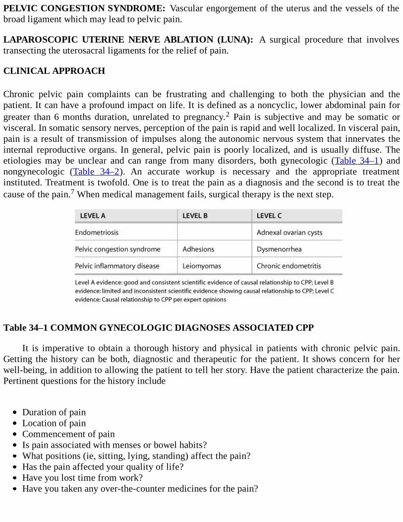
PELVIC CONGESTION SYNDROME: Vascular engorgement of the uterus and the vessels of thebroad ligament which may lead to pelvic pain.
LAPAROSCOPIC UTERINE NERVE ABLATION (LUNA): A surgical procedure that involvestransecting the uterosacral ligaments for the relief of pain.
CLINICAL APPROACH
Chronic pelvic pain complaints can be frustrating and challenging to both the physician and thepatient. It can have a profound impact on life. It is defined as a noncyclic, lower abdominal pain forgreater than 6 months duration, unrelated to pregnancy.2 Pain is subjective and may be somatic orvisceral. In somatic sensory nerves, perception of the pain is rapid and well localized. In visceral pain,pain is a result of transmission of impulses along the autonomic nervous system that innervates theinternal reproductive organs. In general, pelvic pain is poorly localized, and is usually diffuse. Theetiologies may be unclear and can range from many disorders, both gynecologic (Table 34–1) andnongynecologic (Table 34–2). An accurate workup is necessary and the appropriate treatmentinstituted. Treatment is twofold. One is to treat the pain as a diagnosis and the second is to treat thecause of the pain.7 When medical management fails, surgical therapy is the next step.
Table 34–1 COMMON GYNECOLOGIC DIAGNOSES ASSOCIATED CPP
It is imperative to obtain a thorough history and physical in patients with chronic pelvic pain.Getting the history can be both, diagnostic and therapeutic for the patient. It shows concern for herwell-being, in addition to allowing the patient to tell her story. Have the patient characterize the pain.Pertinent questions for the history include
Duration of painLocation of painCommencement of painIs pain associated with menses or bowel habits?What positions (ie, sitting, lying, standing) affect the pain?Has the pain affected your quality of life?Have you lost time from work?Have you taken any over-the-counter medicines for the pain?
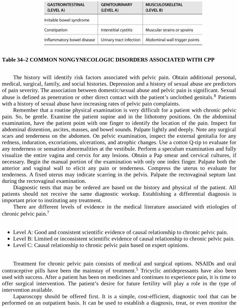
Table 34–2 COMMON NONGYNECOLOGIC DISORDERS ASSOCIATED WITH CPP
The history will identify risk factors associated with pelvic pain. Obtain additional personal,medical, surgical, family, and social histories. Depression and a history of sexual abuse are predictorsof pain severity. The association between domestic/sexual abuse and pelvic pain is significant. Sexualabuse is defined as penetration or other direct contact with the patient’s unclothed genitals.8 Patientswith a history of sexual abuse have increasing rates of pelvic pain complaints.
Remember that a routine physical examination is very difficult for a patient with chronic pelvicpain. So, be gentle. Examine the patient supine and in the lithotomy positions. On the abdominalexamination, have the patient point with one finger to identify the location of the pain. Inspect forabdominal distention, ascites, masses, and bowel sounds. Palpate lightly and deeply. Note any surgicalscars and tenderness on the abdomen. On pelvic examination, inspect the external genitalia for anyredness, induration, excoriations, ulcerations, and atrophic changes. Use a cotton Q-tip to evaluate forany tenderness or sensation abnormalities at the vestibule. Perform a speculum examination and fullyvisualize the entire vagina and cervix for any lesions. Obtain a Pap smear and cervical cultures, ifnecessary. Begin the manual portion of the examination with only one index finger. Palpate both theanterior and vaginal wall to elicit any pain or tenderness. Compress the uterus to evaluate fortenderness. A fixed uterus may indicate scarring in the pelvis. Palpate the rectovaginal septum lastduring the rectovaginal examination.
Diagnostic tests that may be ordered are based on the history and physical of the patient. Allpatients should not receive the same diagnostic workup. Establishing a differential diagnosis isimportant prior to instituting any treatment.
There are different levels of evidence in the medical literature associated with etiologies ofchronic pelvic pain.7
Level A: Good and consistent scientific evidence of causal relationship to chronic pelvic pain.Level B: Limited or inconsistent scientific evidence of causal relationship to chronic pelvic pain.Level C: Causal relationship to chronic pelvic pain based on expert opinions.
Treatment for chronic pelvic pain consists of medical and surgical options. NSAIDs and oralcontraceptive pills have been the mainstay of treatment.5 Tricyclic antidepressants have also beenused with success. After a patient has been on medicines and continues to experience pain, it is time tooffer surgical intervention. The patient’s desire for future fertility will play a role in the type ofintervention available.
Laparoscopy should be offered first. It is a simple, cost-efficient, diagnostic tool that can beperformed on an outpatient basis. It can be used to establish a diagnosis, treat, or even monitor the
course of a specific disease. If the patient has endometriosis, ablative or excisional therapy can beoffered through the laparoscope. Adhesiolysis via the laparoscope is another alternative, if the patienthas extensive pelvic adhesions.
Complications of laparoscopy include hemorrhage, bowel injuries, uterine perforation, vascularinjuries, and infection. Reports of complications in the literature range from 0.2 out of 1000 to 4.6 outof 1000.9 Cardiac arrest can occur at the time of induction of anesthesia, rapid or excessiveinsufflation of gas, or extreme Trendelenburg positions.9
In patients with midline pain, such as dysmenorrhea, and failed medical management, a presacralneurectomy or a LUNA procedure can be offered. Presacral neurectomy has become less popular overthe years. Removing the “presacral nerve” or the superior hypogastric plexus has been shown torelieve pain in 60% to 80% of patients.10 The operation can be difficult and lengthy. Acutecomplications include laceration of the middle sacral vein, hemorrhage, hypotension, and small bowelobstruction.
The LUNA is another alternative in relieving midline pain in women desiring future fertility. Itwas first described in 1899.11 Through the laparoscope, the uterus is stretched upward and theuterosacral ligaments are coagulated and transected at their insertion into the posterior cervix. Thegoal is to interrupt the afferent-efferent sympathetic and parasympathetic neuronal pathways.11 Becareful to identify the ureters to avoid injury. With the uterosacral ligaments supporting the uterus, aside effect of this procedure can be uterine prolapse.
Presacral neurectomy and LUNA, done in conjunction, do not offer any additional pain relief.12
For women with persistent pelvic pain who completed of their childbearing and failed medicalmanagement, a hysterectomy should be offered. Chronic pelvic pain is the indication for 10% ofhysterectomies reviewed.13 Before offering this final surgical alternative, it is imperative thatnongynecologic etiologies for pain have been ruled out. Stovall et al. showed that, out of 99 womenwith pelvic pain followed 12 months after a hysterectomy (with or without a bilateral salpingo-oophorectomy [BOS]), 22% still had persistent pelvic pain.5 Hillis et al. showed results consistentwith the latter, up to 24 to 40% with persistent pain, 1 year after surgery.6 If a treatable condition isfound and treated before proceeding with surgery, it is possible that the patient’s pain may resolve.Once a hysterectomy is decided, the associated morbidity should be discussed with the patient. Therisk of bleeding; infection; and damage to bowel, bladder, or vascular structures should be included inthe consent form. Thrombotic complications are increased after a hysterectomy. There is an increasein both infectious and febrile morbidity after a hysterectomy. Risk factors for febrile morbidity havebeen documented in the literature secondary to a hysterectomy.14 Peipert et al. showed that febrilemorbidity was 14% in over 680 charts reviewed. Morbidity can be decreased by the route ofhysterectomy. Vaginal cases have less febrile morbidity than abdominal cases. Administeringprophylactic antibiotics, 1 hour prior to starting the surgery, decreases febrile risk. In addition,meticulous surgical technique, by minimizing blood loss, also lessens febrile morbidity. With febrilemorbidity, there are longer hospital stays and increasing costs to the patient.14 Reports show that aftera hysterectomy, symptoms, and quality of life can be improved. The Women’s Health Study, of 1299women, showed improvement in pain and overall participation in social functions after receiving ahysterectomy.15 After 1 year, 87% of women reported improvement and after 2 years 88% improved.
Hysterectomy is the final surgical option offered for treatment of chronic pelvic pain. It may givelong-term improvement of symptoms.
Comprehension Questions
34.1 A 19-year-old G0P0 woman complains of pain with her menses that started at menarche. Herpain is relieved with monthly NSAIDs. Her pelvic examination and pelvic ultrasound arenormal. What is her most likely diagnosis?
A. Primary dysmenorrheaB. Secondary dysmenorrheaC. Pelvic inflammatory diseaseD. Ruptured right corpus luteal cyst
34.2 A 45-year-old G5P5 woman has a 5-year history of chronic pelvic pain secondary toendometriosis. Her surgical history consists of two laparoscopic laser treatments for herendometriosis. She has been on continuous oral contraceptives and NSAIDs for the last 6months with continued pain. What is the next step?
A. Total abdominal hysterectomy (TAH) with a bilateral salpingooophorectomy (BOS)B. Repeat laser therapy via the laparoscopeC. Six more months of medical therapyD. Partial hysterectomy
34.3 A 35-year-old G2P2, thin, woman with a recent last menstrual period received a laparoscopyfor persistent pelvic pain. At the initiation of the surgery, she became suddenly hypotensive,with a BP of 70/30 mm Hg and a pulse of 125 beats/min. What is the most likely surgicalcomplication?
A. Cardiac arrestB. Vascular injuryC. Ureteral injuryD. Bowel laceration
34.4 A 40-year-old G0P0 woman had a LUNA procedure for dysmenorrhea. On postoperative day1, her H/H was 10.5/32 and a creatinine 1.0. On postoperative day 2, her hemoglobin levelwas unchanged, but her creatinine increased to 1.8 and she now has back pain. What is themost likely complication of this surgery?
A. Bladder lacerationB. Bowel injuryC. Ureteral injuryD. Kidney puncture
ANSWERS
34.1 A. Primary dysmenorrhea is the pain before or during menses with no organic cause thatusually begins at menarche. Her normal pelvic ultrasound is absent of any pelvic pathology.
34.2 A. This patient has failed medical therapy and previous conservative surgery via thelaparoscope. With persistent pelvic pain secondary to her endometriosis, she requiresdefinitive therapy, a TAH/BSO.
34.3 B. This patient has a vascular injury secondary to insertion of the Veress needle. As a resultof acute hemorrhage, there is sudden hypotension and tachycardia. The laparoscopy isimmediately converted to a laparotomy for repair of the injured vessel.
34.4 C. The ureter has been compromised in this patient. Early diagnosis is the key. Her rising
34.4 C. The ureter has been compromised in this patient. Early diagnosis is the key. Her risingcreatinine is a clue.
Clinical PearlsSee Table 1-2 for definition of level of evidence and strength of recommendation
Chronic pelvic pain can be caused by gynecologic or nongynecologic disorders.
Obtaining a detailed history and physical examination is the key in establishing a diagnosis forpelvic pain (Level B).
After medical therapy has failed, a laparoscopy is offered in order to establish a diagnosis(Level B).
Surgical therapy is directed based on the patient’s desires for future fertility (Level B).
Presacral neurectomy and the LUNA procedures are performed in patients with midline orcentral pelvic pain (Level A).
Ten percent of all hysterectomies are performed for chronic pelvic pain.
REFERENCES
1. Mathias SD, Kuppermann M, Liberman RF, Lipschutz AC, Steege JF. Chronic pelvic pain:prevalance, health-related quality of life, and economic correlates. Obstet Gynecol.1996;87:321-327.
2. Zondervan KT, Yudkin PL, Vessey MP, et al. Chronic pelvic pain in the community—symptoms, investigations, and diagnoses. Am J Obstet Gynecol. 2001; 184:1149-1155.
3. Cunanan RG Jr, Courey NG, Lippes J. Laparoscopic findings in patients with pelvic pain. Am JObstet Gynecol. 1983;146(5):589-591.
4. Kresch AJ, Seifer DB, Sachs LB, Barraes I. Laparoscopy in 100 women with chronic pelvicpain. Obstet Gynecol. 1984;64(5):672-674.
5. Stovall TG, Ling FW, Crawford DA. Hysterectomy for chronic pelvic pain of presumed uterineetiology. Obstet Gynecol. 1990;75(4):676-679.
6. Hillis SD, Marchbanks PA, Peterson HB. The effectiveness of hysterectomy for chronic pelvicpain. Obstet Gynecol. 1995;86:941-945.
7. Howard FM. Chronic pelvic pain. Obstet Gynecol. 2003;101:594-611.
8. Jamieson DJ, Steege JF. The association of sexual abuse with pelvic pain complaints in aprimary care population. Am J Obstet Gynecol. 1997;177(6):1408-1412.
9. Cunanan RG, Coury NG, Lippes J. Complications of laparoscopic tubal sterilization. ObstetGynecol. 1980;55:501-506.
10. Lee RB, Stone K, Magelssen D, Belts RP, Benson WL. Presacral neurectomy for chronicpelvic pain. Obstet Gynecol. 1986;68:517-521.
11. Lichten EM, Bombard J. Surgical therapy of primary dysmenorrhea with laparoscopic uterinenerve ablation. J Reprod Med. 1987;32:37-41.
12. ACOG Practical Bulletin. Chronic Pelvic Pain. No. 51. 2004:1184-1199.
13. Carlson KJ, Nichols DH, Schiff I. Indications for hysterectomy. NEJM. 1993;328:856-861.
14. Peipert JF, Weitzen S, Cruickshank C, Story E, Ethridge D, Lapane K. Risk factors for febrilemorbidity after hysterectomy. Obstet Gynecol. 2004;103(1):86-91.
15. Kjerulff KH, Langenberg PW, Rhodes JC, et al. Effectiveness of hysterectomy. ObstetGynecol. 2000;95:319-326.
Case 35
A 62-year-old G1P1 menopausal woman with a 6-month history of post-menopausal bleeding isundergoing hysteroscopy. She states that, on many occasions, her undergarments are blood tinged. Shedenies any nausea, vomiting, or abdominal pain. She has been menopausal for 5 years. Her pastmedical history is significant for type 2 diabetes for 10 years and is controlled with an oralhypoglycemic agent daily. Socially, she runs 3 miles, two times a week. A pelvic examination wasnoncontributory, without masses. A transvaginal ultrasound reveals a small uterus, with an 8-mmendometrial stripe; normal ovaries. An endometrial biopsy was negative for hyperplasia andcarcinoma.
Prior to inserting the hysteroscope, the cervical canal is dilated with the Hegar dilators. Sheundergoes a dilation and curettage (D&C) with an operative hysteroscopy which reveals anendometrial polyp. The 30-degree hysteroscope is placed through a 10-mm outer sheath and glycine1.5% is used as the distention medium. Upon entering the uterine cavity, the endocervix, the anteriorand posterior walls, and the tubal ostia are all visualized. The endometrium appears pale, and a fundalendometrial polyp is visualized. The polyp extends into the endometrial cavity, while its broad baseremains attached to the fundus, closer to the right tubal ostia. Grasping forceps are used in an attemptto provide adequate visualization of the base of the polyp, in its entirety. The loop resectoscope isused to resect the polyp at the base. Forty-five minutes after the start of the procedure, the nurseannounces that there is a 900-cc fluid deficit. The polyp is only partially resected. The procedurecontinues, and the loop electrode is placed anterior to the base of the polyp and the resectioncontinues. After several passes with the resectoscope, there is noted to be an immediate loss inintrauterine pressure to 30 mm Hg.
What is the intraoperative diagnosis?
What are the risk factors associated with this intraoperative diagnosis?
What is the next step in managing this patient?
ANSWERS TO CASE 35:Hysteroscopic Complications
Summary: This is a 62-year-old woman with postmenopausal bleeding for 6 months. An operativehysteroscopy revealed an endometrial polyp as the etiology for her bleeding. During the surgery toresect the polyp, adequate visualization was difficult to obtain in order to resect the polyp. However,the resection began, under poor visualization and a uterine perforation occurred. Of note, at the timeof the perforation, there was a fluid deficit approaching 1 L.
Intraoperative diagnosis (points of injury): Uterine perforation is a complication ofhysteroscopy. The uterus is at the greatest risk of being damaged in two instances: (1) at thetime of cervical dilation or entry into the uterus and (2) thermal injury secondary to use ofelectrical current. Of special note, the uterine wall is very thin near both tubal ostia. Care mustbe taken when using the loop resectoscope in this area.
Risk factors associated with intraoperative diagnosis: An unrecognized injury to the uterine
wall may result in trauma to the intra-abdominal organs, or the pelvic vessels. Complicationsduring operative hysteroscopy can occur during one of the following instances: (1)intraoperatively, (2) immediately postoperatively, or (3) as a late complication.
Next step: A good surgeon prepares the patient preoperatively for the indicated procedure. Allrisks, benefits, and possible complications should be discussed during the preoperativecounseling session. A discussion has to occur to understand that if a complication occurs, theprocedure has to be discontinued. In this patient, the intraoperative diagnosis was a uterineperforation. An immediate loss of intrauterine pressure is noted at the time of perforationsecondary to the loop resectoscope, which signals the diagnosis of a uterine perforation. Thenext step is to discontinue the hysteroscopy and evaluate the extent of damage done to theuterus, via laparoscopy.
ANALYSIS
Objectives
1. Know the different categories of hysteroscopic complications and be able to recognize them.2. Be familiar with the advantages and disadvantages of each distending medium used during
hysteroscopy.3. Understand that there are legal risks associated with hysteroscopic complications.4. Know the six most common gynecologic procedures that are associated with a higher incidence
of complications.
Considerations
This is a 62-year-old G1P1 woman with postmenopausal bleeding who undergoes an operativehysteroscopy for removal of the endometrial polyp (Figure 35–1). Hysteroscopy is a quick, minimallyinvasive diagnostic and therapeutic procedure. Hysteroscopic surgery decreases morbidity and adds toa speedy recovery, but this advantage is minimal if complications occur. The experience of thesurgeon is a strong predictor of complications. A pre-operative workup revealed there to be an organiccause of the bleeding. The differential diagnosis for postmenopausal bleeding is numerous. The mostcommon cause is atrophy of the endometrium. Other diagnoses that cause postmenopausal bleedinginclude endometrial hyperplasia, endometrial polyp, and endometrial carcinoma. There should besome prerequisites in place to minimize the risk of hysteroscopic complications: (1) There has to beaccurate visualization of the uterine cavity and the lesion in its entirety. (2) Bleeding should be kept toa minimal because it impairs vision. (3) Laparoscopy may be used simultaneously, in some instances,to increase the safety of the procedure. (4) Accurate balancing of fluid intake and output must occur.
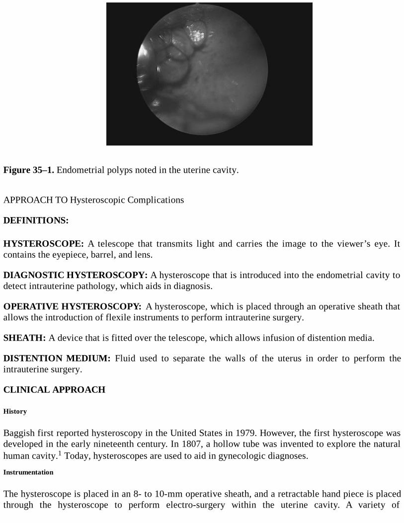
Figure 35–1. Endometrial polyps noted in the uterine cavity.
APPROACH TO Hysteroscopic Complications
DEFINITIONS:
HYSTEROSCOPE: A telescope that transmits light and carries the image to the viewer’s eye. Itcontains the eyepiece, barrel, and lens.
DIAGNOSTIC HYSTEROSCOPY: A hysteroscope that is introduced into the endometrial cavity todetect intrauterine pathology, which aids in diagnosis.
OPERATIVE HYSTEROSCOPY: A hysteroscope, which is placed through an operative sheath thatallows the introduction of flexile instruments to perform intrauterine surgery.
SHEATH: A device that is fitted over the telescope, which allows infusion of distention media.
DISTENTION MEDIUM: Fluid used to separate the walls of the uterus in order to perform theintrauterine surgery.
CLINICAL APPROACH
History
Baggish first reported hysteroscopy in the United States in 1979. However, the first hysteroscope wasdeveloped in the early nineteenth century. In 1807, a hollow tube was invented to explore the naturalhuman cavity.1 Today, hysteroscopes are used to aid in gynecologic diagnoses.
Instrumentation
The hysteroscope is placed in an 8- to 10-mm operative sheath, and a retractable hand piece is placedthrough the hysteroscope to perform electro-surgery within the uterine cavity. A variety of
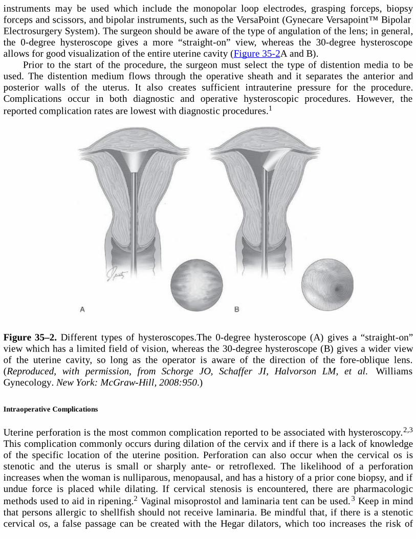
instruments may be used which include the monopolar loop electrodes, grasping forceps, biopsyforceps and scissors, and bipolar instruments, such as the VersaPoint (Gynecare Versapoint™ BipolarElectrosurgery System). The surgeon should be aware of the type of angulation of the lens; in general,the 0-degree hysteroscope gives a more “straight-on” view, whereas the 30-degree hysteroscopeallows for good visualization of the entire uterine cavity (Figure 35-2A and B).
Prior to the start of the procedure, the surgeon must select the type of distention media to beused. The distention medium flows through the operative sheath and it separates the anterior andposterior walls of the uterus. It also creates sufficient intrauterine pressure for the procedure.Complications occur in both diagnostic and operative hysteroscopic procedures. However, thereported complication rates are lowest with diagnostic procedures.1
Figure 35–2. Different types of hysteroscopes.The 0-degree hysteroscope (A) gives a “straight-on”view which has a limited field of vision, whereas the 30-degree hysteroscope (B) gives a wider viewof the uterine cavity, so long as the operator is aware of the direction of the fore-oblique lens.(Reproduced, with permission, from Schorge JO, Schaffer JI, Halvorson LM, et al. WilliamsGynecology. New York: McGraw-Hill, 2008:950.) Intraoperative Complications
Uterine perforation is the most common complication reported to be associated with hysteroscopy.2,3
This complication commonly occurs during dilation of the cervix and if there is a lack of knowledgeof the specific location of the uterine position. Perforation can also occur when the cervical os isstenotic and the uterus is small or sharply ante- or retroflexed. The likelihood of a perforationincreases when the woman is nulliparous, menopausal, and has a history of a prior cone biopsy, and ifundue force is placed while dilating. If cervical stenosis is encountered, there are pharmacologicmethods used to aid in ripening.2 Vaginal misoprostol and laminaria tent can be used.3 Keep in mindthat persons allergic to shellfish should not receive laminaria. Be mindful that, if there is a stenoticcervical os, a false passage can be created with the Hegar dilators, which too increases the risk of
uterine rupture. Once the uterus is perforated, the procedure is immediately stopped and evaluation fortrauma to intra-abdominal organs and/or pelvic vessels has to occur. After 6 weeks, the operativehysteroscopy may be rescheduled.2 Cervical mucosal lacerations can also occur from use of thetenaculum. Treatment consists of applying pressure, cautery, or suturing the cervix.
Complications can occur when measurements of inflow and outflow of distention media areinaccurately or incompletely recorded. The type of electrical unit used, such as monopolar or bipolar,will determine the type of distention media to choose. There are two types of low-viscosity fluids:hypotonic (electrolyte-poor media) and isotonic (electrolyte-rich media). In general, monopolarinstruments cannot be used in electrolyte rich media such as normal saline due to the possibility toelectrical current conduction; thus, these instruments are usually restricted for electrolyte poor media.Glycine, sorbitol, and mannitol are examples of electrolyte-poor fluid media. These media areconsidered hypoosmolar and will decrease serum osmolality and serum sodium levels. The fluidoverload that results from these media will increase the risk of pulmonary edema, cerebral edema, anddeath. Isotonic media, such as normal saline and lactated Ringer solutions, are electrolyte-rich media.Laser and bipolar electrical instruments are used with these media. The risk of fluid overload andhyponatremia is less, secondary to the isotonicity of these fluids.
Dextran 70 (Hyskon), another distention medium, is a very viscous, sticky solution that providesexcellent visibility, but is a powerful plasma expander. Instruments need to be meticulously cleanedafter being used with dextran; residual material can ruin the instrumentation. The major disadvantageis that the maximum volume to be used is 500 cc, because for every 100 cc absorbed, the plasmavolume expands 850 cc.4
Carbon dioxide gas is the final medium that can be used during hysteroscopy, but should notgenerally be used with operative hysteroscopy. This medium is immiscible with blood and has a highrisk of carbon dioxide embolism with operative procedures. Signs and symptoms of gas emboliinclude pulmonary hypertension, arrhythmia, hypercarbia, hypoxia, tachypnea, and systemichypotension.
Immediate Postoperative Complications
All fluids used with operative hysteroscopy can be associated with complications. To avoid the fluidoverload, accurate balancing of inflow and outflow of distention media is required. The surgeon andsurgical team are responsible for calculating the difference between the amount of fluid infused andthe amount of fluid expelled during the surgery. The difference, between the two, is defined as thefluid deficit. The deficit should be less than 1 L. If the deficit is greater than 1.5 L, surgery shouldgenerally be terminated immediately.5 This deficit can become absorbed rapidly and can cause fluidoverload syndrome. Absorption or intravasation may occur via several mechanisms: (1) directly intovascular structures during surgical resection, (2) across the walls of the endometrium, and (3) via thefallopian tubes with absorption from the peritoneum.6 The first clinical sign of fluid overload is therapid production of dilute urine. Besides addressing the fluid overload, the patient should be assessedfor hyponatremia, which likewise should be treated. Correction can occur over the first 24 hours byraising the sodium levels 1 to 2 mEq/L/h.4 Keep the patient well oxygenated and use diuretics tomanage the volume overload.
The best two means to prevent the fluid overload complication are to accurately measure theinflow and outflow of distention media and to control the intrauterine irrigation pressure that distendsthe uterus. Intrauterine pressures that are safe remain less than 100 mm Hg.
Hemorrhage, defined as greater than 500 cc, can occur during the procedure and immediatelypostoperatively.7 Remember, clear vision is required to adequately perform operative hysteroscopy. Afew methods, used to control intrauterine bleeding, are irrigation of the uterine cavity, insertion of aFoley catheter balloon, or injection of vasopressin at the bleeding site, or intravenous oxytocin.4 Thesemethods should not be considered if the hemorrhage is secondary to an intraoperative complication,such as uterine perforation or laceration of the surrounding pelvic vessels. In these circumstances, theoperative hysteroscopy should be stopped immediately.
Infection is rare after operative hysteroscopy. Prophylactic antibiotics are not universallyemployed, and do not prevent infection.1 Infection is reported with an incidence of less than 2%.1 Ifan infection does occur, it may involve the urinary tract or endometrium. Risk factors that increase thelikelihood include extensive endomyometrial destruction, multiple insertions with the hysteroscope,and lengthy procedures.2
Late Complications
This category of complications can occur months to years after an operative hysteroscopy. It has beenreported that late complications are associated with intrauterine fertility surgeries or after anendometrial ablation for abnormal uterine bleeding.8 Intrauterine synechiae that result from theoperative hysteroscopy can inhibit regular menstrual flow. Commonly recognized conditions that canoccur late include development of a hematometra, a pregnancy-related endometrial cancer, or post-tubal sterilization syndrome. The majority of these complications occur after an endometrial ablation.
Prompt recognition of complications is a must. Patients may complain of a myriad of complaints,such as increasing pain, bleeding, and persistent nausea and vomiting, postoperatively. Remember, ifthe procedure was completely uncomplicated, a patient will not call the office repeatedly. A patientwho repeatedly calls the office may be experiencing a late complication, which previously had beenoccult. An astute surgeon has to be prompt in the diagnosis and treatment of associated complications.Legal risks can increase if a surgeon maintains a cavalier attitude, fails to accurately document fluidbalance, and fails to respond promptly to possible complaints of a complication.
Indications
Operative hysteroscopy allows for visualization of the endometrial cavity, and it uses a variety ofelectrical current to treat intrauterine pathology. Unlike diagnostic hysteroscopy that can beperformed in an office setting under local anesthesia, operative hysteroscopy is performed in asurgical suite under general or regional anesthesia. There are a variety of angled tip lenses availableon the hysteroscope. The two most common are 0- and 30 degree. A 0-degree hysteroscope gives astraightforward, panoramic view of the anterior and posterior walls of the uterus and the tubal ostia. Itis very similar to normal vision. A 30-degree hysteroscope allows visualization of the fundus;however, in order to see the tubal ostia, the scope has to be turned to a 90-degree angle to view thetubal ostia.
Symptoms which indicate that a hysteroscopy should be performed are abnormal uterinebleeding, infertility, or postmenopausal bleeding. Contraindications for performing the procedure,include pregnancy, infection, and endometrial carcinoma. Complications related to operativehysteroscopy are directly associated with the type of surgical procedure being performed. The sixmost common procedures, performed via the hysteroscope, are myomectomy septum resection,adhesiolysis, D&C with hysteroscopy, endometrial ablation, and polypectomy (Figure 35–3).7 Whilepolypectomy and endometrial ablations have lower rates of complications, myomectomy,
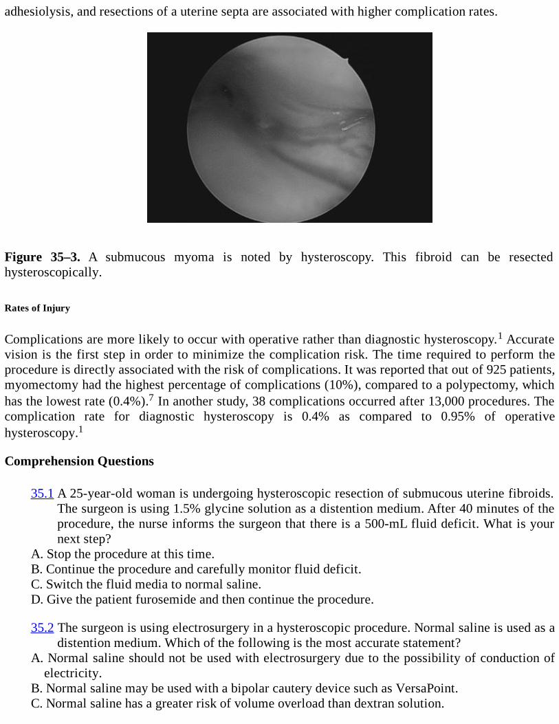
adhesiolysis, and resections of a uterine septa are associated with higher complication rates.
Figure 35–3. A submucous myoma is noted by hysteroscopy. This fibroid can be resectedhysteroscopically. Rates of Injury
Complications are more likely to occur with operative rather than diagnostic hysteroscopy.1 Accuratevision is the first step in order to minimize the complication risk. The time required to perform theprocedure is directly associated with the risk of complications. It was reported that out of 925 patients,myomectomy had the highest percentage of complications (10%), compared to a polypectomy, whichhas the lowest rate (0.4%).7 In another study, 38 complications occurred after 13,000 procedures. Thecomplication rate for diagnostic hysteroscopy is 0.4% as compared to 0.95% of operativehysteroscopy.1
Comprehension Questions
35.1 A 25-year-old woman is undergoing hysteroscopic resection of submucous uterine fibroids.The surgeon is using 1.5% glycine solution as a distention medium. After 40 minutes of theprocedure, the nurse informs the surgeon that there is a 500-mL fluid deficit. What is yournext step?
A. Stop the procedure at this time.B. Continue the procedure and carefully monitor fluid deficit.C. Switch the fluid media to normal saline.D. Give the patient furosemide and then continue the procedure.
35.2 The surgeon is using electrosurgery in a hysteroscopic procedure. Normal saline is used as adistention medium. Which of the following is the most accurate statement?
A. Normal saline should not be used with electrosurgery due to the possibility of conduction ofelectricity.
B. Normal saline may be used with a bipolar cautery device such as VersaPoint.C. Normal saline has a greater risk of volume overload than dextran solution.
D. Normal saline is not often used as a distention medium due to the danger of hypernatremia,and instead half-normal saline is recommended.
ANSWERS
35.1 B. Until the fluid deficit with glycine or sorbitol reaches 1000 mL, the procedure cancontinue.
35.2 B. Normal saline is a commonly used distention medium, and is less likely to lead to fluidoverload or DIC as compared to dextran, although these complications can occur. A bipolardevice may be used with normal saline, and because the electrical current flows between theelectrodes, it is safe to use with saline.
Clinical PearlsSee Table 1-2 for definition of level of evidence and strength of recommendation
The hysteroscope contains the eyepiece, barrel, and lens (Level B).
Accurate and clear visualization of the uterine lesions is the key to decreasing the risk ofintraoperative complications (Level B).
Uterine perforation is the most common complication associated with operative hysteroscopy(Level A).
Pharmacologic agents, such as vaginal misoprostol and laminaria, may be used to ripen thecervical os prior to an operative hysteroscopy. This decreases the risk of uterine perforation(Level B).
Each distention medium has its own advantages and disadvantages (Level A).
Accurate balancing of fluid inflow and outflow will decrease the risk of the fluid overload andhyponatremia complication (Level B).
Procedures which have longer operative times have a higher incidence of complications, suchas myomectomy, adhesiolysis, and resection of a uterine septum (Level B).
Once an intraoperative complication occurs, the hysteroscopy is immediately terminated(Level B).
Late complications may occur months to years after an operative hysteroscopy (Level B).
REFERENCES
1. Julian TM. Hysteroscopic complications. J Low Genit Tract Dis. 2002;6(1):39-47.
2. Bradley LD. Complications in hysteroscopy: prevention, treatment, and legal risk. Curr Opin
Obstet Gynecol. 2002;14:409-415.
3. Shveiky D, Rojansky N, Revel A, Benshushan A, Laufer N, Shushan A. Complications ofhysteroscopic surgery: “beyond the learning curve.” J Minim Invasive Gynecol. 2007;14:218-222.
4. ACOG Technical Assessment. Hysteroscopy. August 2005;4:350-353.
5. Konig M, Meyer A, Aydeniz B, Kurek R, Wallwiener D. Hysteroscopic surgery—complications and their prevention. Contrib Gynecol Obstet. 2000;20:161-170.
6. Hsieh MH, Chen TL, Lin YH, Chang CC, Lin CS, Lee YW. Acute pulmonary edema fromunrecognized high irrigation pressure in hysteroscopy: a report of two cases. J Clin Anesth.2009;20:614-617.
7. Propst AM, Lieberman RF, Harlow BL, Ginsburg ES. Complications of hysteroscopic surgery:predicting patients at risk. Obstet Gynecol. October 2000;96(4):517-520.
8. Cooper JM, Brady RM. Late complications of operative hysteroscopy. Obstet Gynecol ClinNorth Am. June 2000;27(2):367-374.
9. Jansen FW, Vredevoogd CB, Ulzen KV, Hermans J, Trimbos B, Trimbos-Kemper TCM.Complications of hysteroscopy: a prospective, multicenter study. Obstet Gynecol. August2000;96(2):266-270.
Case 36
A 24-year-old nulliparous Caucasian woman presents to your office for her annual well-womanexamination. She is a recent college graduate and has a monogamous relationship. Yearly Pap smearsbegan at age 19 and have always been normal. She denies any medical conditions, and her onlymedication is a combination oral contraceptive pill (OCP). She does not smoke or engage in any illicitdrug usage. She does drink alcohol socially.
On examination, you note no irregularities of the cervix or vagina on speculum examination; aPap smear is performed. Bimanual examination reveals findings suggestive of an adnexal mass on thepatient’s right side. Palpation of the mass does not provoke pain or discomfort. She denies recentunintentional weight loss or weight gain, early satiety, abdominal bloating, dyspareunia, or vaginaldischarge. She states that she has been taking her OCP regularly as directed.
Urine pregnancy test was negative in the office and a confirmatory serum β-human chorionicgonadotropin (β-hCG) level was sent to the laboratory. Transvaginal ultrasound was performed in theoffice and an 8-cm mass with both solid and cystic characteristics was seen on the right ovary. Themass also had areas that appeared to be calcified within the cystic portion of the mass.
What is the most likely diagnosis?
What is your next step?
What options for management may be utilized?
ANSWERS TO CASE 36:Ovarian Cystectomy
Summary: This is a 24-year-old nulliparous Caucasian woman having an asymptomatic ovarian masssuggestive of a cystic teratoma. The patient has an unremarkable past medical history and isasymptomatic.
Most likely diagnosis: Benign cystic teratoma (dermoid).
Next step: Operative removal.
Management options: Surgical excision either via laparotomy, laparoscopy, or a combinationof these procedures.
ANALYSIS
Objectives
1. List the surgical options available for ovarian cystectomy.2. Describe the diagnostic features of an ovarian cyst.3. Describe the potential complications that may arise and the solutions to them during removal of
cyst.
Considerations
This is a 24-year-old Caucasian woman who presents for a routine yearly health maintenance visit.During the course of the visit, a right-sided adnexal mass is palpated during bimanual examination.Formal evaluation of the mass confirmed a right-sided ovarian mass consistent with a dermoid cyst.Pregnancy was excluded initially by urine pregnancy test, thus making the mass less likely to be anectopic pregnancy.
The patient is further screened by questioning for evidence of ovarian cancer. The line ofquestioning did not provide evidence for suspicion of malignancy. Ultrasound imaging demonstratedcharacteristics of benign cystic teratoma. The image demonstrated regional diffuse bright echoes withor without posterior acoustic shadowing, shadowing echodensity, and a fluid-fluid level.
Further workup may be employed to differentiate the mass as benign or malignant.Dysgerminoma is a malignant form of germ cell tumor and the serum marker is lactate dehydrogenase1 (LDH-1) and LDH-2. If this was to be suspected in the patient, it would be prudent to draw abaseline level as a way to measure response to treatment rather than a screening method. Serummarkers have not been shown to be an accurate method for screening for ovarian malignancy.
Operative management is the treatment of choice in this patient. The possibility of bilateralinvolvement of both ovaries must be discussed with the patient. Malignant transformation is presentin approximately 2% of teratomas, and thus the possibility of malignancy and the need for a stagingprocedure must be explained to the patient. Operative technique may be either via laparoscopic orlaparotomy approach. Each approach has benefits and downfalls that the patient must be made awareof. Various methods within each modality will be discussed in detail in the following section.
As with any operation, the risk of complications must be addressed and taken into account beforeoperation is attempted.
APPROACH TO Ovarian Cystectomy
DEFINITIONS
TERATOMA: Tissue that recapitulates the three layers of the developing embryo (ectoderm,mesoderm, and endoderm). One or more of the layers may be represented. Mature (benign) orimmature (malignant) subtypes further differentiate. Arise from a single germ and have a karyotype of46, XX.
ROKITANSKY PROTUBERANCE: Area seen on both ultrasound and pathologic specimen wherefatty tissue, teeth, and bone protrude into the lumen of the teratoma.
SPILLAGE: Potential complication arising from laparoscopic (minimally from laparotomy) surgerywhere the contents of the cystic mass may escape from a ruptured cyst into the abdominal cavity.
CLINICAL APPROACH
Benign Cystic Teratomas
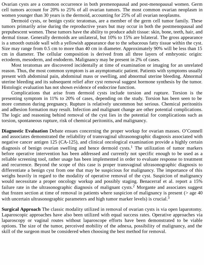
Ovarian cysts are a common occurrence in both premenopausal and post-menopausal women. Germcell tumors account for 20% to 25% of all ovarian tumors. The most common ovarian neoplasm inwomen younger than 30 years is the dermoid, accounting for 25% of all ovarian neoplasms.
Dermoid cysts, or benign cystic teratomas, are a member of the germ cell tumor family. Thesetumors primarily arise during the reproductive years but may occur in both the postmenopausal andprepubescent women. These tumors have the ability to produce adult tissue: skin, bone, teeth, hair, anddermal tissue. Generally dermoids are unilateral, but 10% to 15% are bilateral. The gross appearanceis a smooth outside wall with a yellowish appearance due to the sebaceous fatty tissue within the cyst.Size may range from 0.5 cm to more than 40 cm in diameter. Approximately 90% will be less than 15cm in diameter. The cellular composition is derived from all three layers of embryonic origin:ectoderm, mesoderm, and endoderm. Malignancy may be present in 2% of cases.
Most teratomas are discovered incidentally at time of examination or imaging for an unrelatedreason. Thus, the most common symptom is an asymptomatic patient. Patients with symptoms usuallypresent with abdominal pain, abdominal mass or swelling, and abnormal uterine bleeding. Abnormaluterine bleeding and its subsequent relief after cyst removal suggest hormone synthesis by the tumor.Histologic evaluation has not shown evidence of endocrine function.
Complications that arise from dermoid cysts include torsion and rupture. Torsion is thepresenting symptom in 11% to 20% of cases, depending on the study. Torsion has been seen to bemore common during pregnancy. Rupture is relatively uncommon but serious. Chemical peritonitisand adhesion formation may result. Infection and malignant change are other potential complications.The logic and reasoning behind removal of the cyst lies in the potential for complications such astorsion, spontaneous rupture, risk of chemical peritonitis, and malignancy.
Diagnostic Evaluation Debate ensues concerning the proper workup for ovarian masses. O’Connelland associates demonstrated the reliability of transvaginal ultrasonographic diagnosis associated withnegative cancer antigen 125 (CA-125), and clinical oncological examination provide a highly certaindiagnosis of benign ovarian swelling and hence dermoid cysts.1 The utilization of tumor markersbefore operative intervention has been addressed and currently not specific enough to be used as areliable screening tool, rather usage has been implemented in order to evaluate response to treatmentand recurrence. Beyond the scope of this case is proper transvaginal ultrasonographic diagnosis todifferentiate a benign cyst from one that may be suspicious for malignancy. The importance of thisweighs heavily in regard to the modality of operative removal of the cyst. Suspicion of malignancywould necessitate a proper oncology workup and possibly staging. Benacerraf et al. report a 15%failure rate in the ultrasonographic diagnosis of malignant cysts.2 Morgante and associates suggestthat frozen section at time of removal in patients where suspicion of malignancy is present (> age 40with uncertain ultrasonographic parameters and high tumor marker levels) is crucial.3
Surgical Approach The classic modality utilized in removal of ovarian cysts is via open laparotomy.Laparoscopic approaches have also been utilized with equal success rates. Operative approaches vialaparoscopy or vaginal routes without laparoscope efforts have been demonstrated to be viableoptions. The size of the tumor, perceived mobility of the adnexa, possibility of malignancy, and theskill of the surgeon must be considered when choosing the best method for removal.
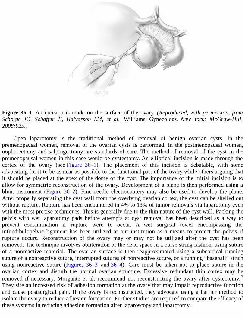
Figure 36–1. An incision is made on the surface of the ovary. (Reproduced, with permission, fromSchorge JO, Schaffer JI, Halvorson LM, et al. Williams Gynecology. New York: McGraw-Hill,2008:925.)
Open laparotomy is the traditional method of removal of benign ovarian cysts. In thepremenopausal women, removal of the ovarian cysts is performed. In the postmenopausal women,oophorectomy and salpingectomy are standards of care. The method of removal of the cyst in thepremenopausal women in this case would be cystectomy. An elliptical incision is made through thecortex of the ovary (see Figure 36–1). The placement of this incision is debatable, with someadvocating for it to be as near as possible to the functional part of the ovary while others arguing thatit should be placed at the apex of the dome of the cyst. The importance of the initial incision is toallow for symmetric reconstruction of the ovary. Development of a plane is then performed using ablunt instrument (Figure 36–2). Fine-needle electrocautery may also be used to develop the plane.After properly separating the cyst wall from the overlying ovarian cortex, the cyst can be shelled outwithout rupture. Rupture has been encountered in 4% to 13% of tumor removals via laparotomy evenwith the most precise techniques. This is generally due to the thin nature of the cyst wall. Packing thepelvis with wet laparotomy pads before attempts at cyst removal has been described as a way toprevent contamination if rupture were to occur. A wet surgical towel encompassing theinfundibulopelvic ligament has been utilized at our institution as a means to protect the pelvis ifrupture occurs. Reconstruction of the ovary may or may not be utilized after the cyst has beenremoved. The technique involves obliteration of the dead space in a purse string fashion, using sutureof a nonreactive material. The ovarian surface is then reapproximated using a subcortical runningsuture of a nonreactive suture, interrupted sutures of nonreactive suture, or a running “baseball” stitchusing nonreactive suture (Figures 36–3 and 36–4). Care must be taken not to place suture in theovarian cortex and disturb the normal ovarian structure. Excessive redundant thin cortex may beremoved if necessary. Morgante et al. recommend not reconstructing the ovary after cystectomy.3
They site an increased risk of adhesion formation at the ovary that may impair reproductive functionand cause postsurgical pain. If the ovary is reconstructed, they advocate using a barrier method toisolate the ovary to reduce adhesion formation. Further studies are required to compare the efficacy ofthese systems in reducing adhesion formation after laparoscopy and laparotomy.
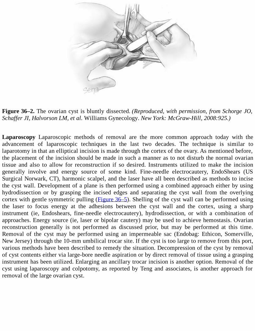
Figure 36–2. The ovarian cyst is bluntly dissected. (Reproduced, with permission, from Schorge JO,Schaffer JI, Halvorson LM, et al. Williams Gynecology. New York: McGraw-Hill, 2008:925.)
Laparoscopy Laparoscopic methods of removal are the more common approach today with theadvancement of laparoscopic techniques in the last two decades. The technique is similar tolaparotomy in that an elliptical incision is made through the cortex of the ovary. As mentioned before,the placement of the incision should be made in such a manner as to not disturb the normal ovariantissue and also to allow for reconstruction if so desired. Instruments utilized to make the incisiongenerally involve and energy source of some kind. Fine-needle electrocautery, EndoShears (USSurgical Norwark, CT), harmonic scalpel, and the laser have all been described as methods to incisethe cyst wall. Development of a plane is then performed using a combined approach either by usinghydrodissection or by grasping the incised edges and separating the cyst wall from the overlyingcortex with gentle symmetric pulling (Figure 36–5). Shelling of the cyst wall can be performed usingthe laser to focus energy at the adhesions between the cyst wall and the cortex, using a sharpinstrument (ie, Endoshears, fine-needle electrocautery), hydrodissection, or with a combination ofapproaches. Energy source (ie, laser or bipolar cautery) may be used to achieve hemostasis. Ovarianreconstruction generally is not performed as discussed prior, but may be performed at this time.Removal of the cyst may be performed using an impermeable sac (Endobag: Ethicon, Somerville,New Jersey) through the 10-mm umbilical trocar site. If the cyst is too large to remove from this port,various methods have been described to remedy the situation. Decompression of the cyst by removalof cyst contents either via large-bore needle aspiration or by direct removal of tissue using a graspinginstrument has been utilized. Enlarging an ancillary trocar incision is another option. Removal of thecyst using laparoscopy and colpotomy, as reported by Teng and associates, is another approach forremoval of the large ovarian cyst.
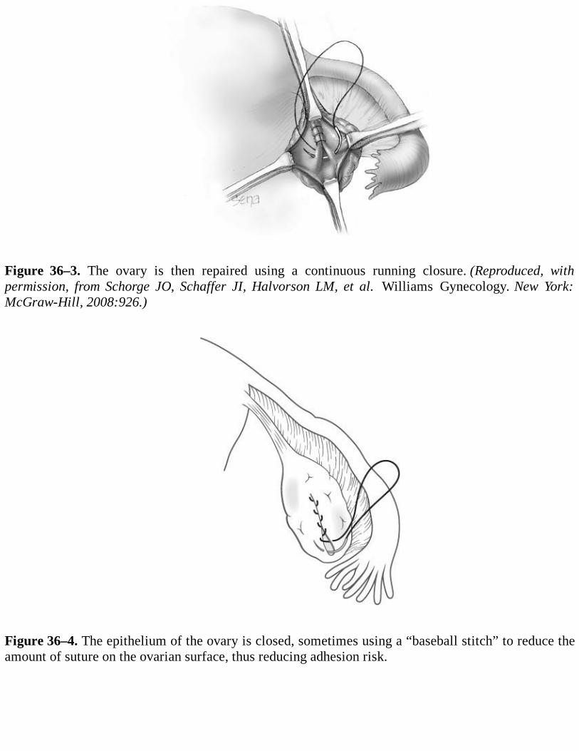
Figure 36–3. The ovary is then repaired using a continuous running closure. (Reproduced, withpermission, from Schorge JO, Schaffer JI, Halvorson LM, et al. Williams Gynecology. New York:McGraw-Hill, 2008:926.)
Figure 36–4. The epithelium of the ovary is closed, sometimes using a “baseball stitch” to reduce theamount of suture on the ovarian surface, thus reducing adhesion risk.
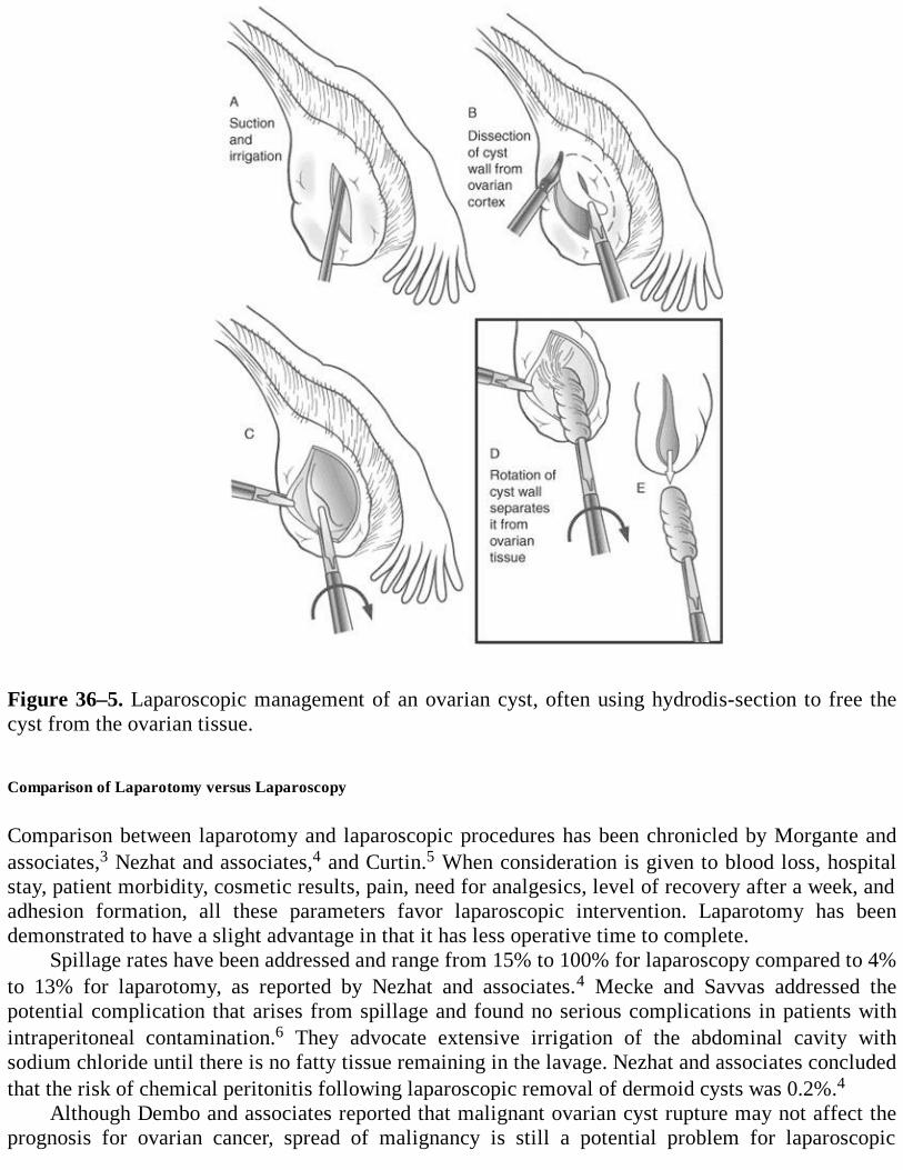
Figure 36–5. Laparoscopic management of an ovarian cyst, often using hydrodis-section to free thecyst from the ovarian tissue.
Comparison of Laparotomy versus Laparoscopy
Comparison between laparotomy and laparoscopic procedures has been chronicled by Morgante andassociates,3 Nezhat and associates,4 and Curtin.5 When consideration is given to blood loss, hospitalstay, patient morbidity, cosmetic results, pain, need for analgesics, level of recovery after a week, andadhesion formation, all these parameters favor laparoscopic intervention. Laparotomy has beendemonstrated to have a slight advantage in that it has less operative time to complete.
Spillage rates have been addressed and range from 15% to 100% for laparoscopy compared to 4%to 13% for laparotomy, as reported by Nezhat and associates.4 Mecke and Savvas addressed thepotential complication that arises from spillage and found no serious complications in patients withintraperitoneal contamination.6 They advocate extensive irrigation of the abdominal cavity withsodium chloride until there is no fatty tissue remaining in the lavage. Nezhat and associates concludedthat the risk of chemical peritonitis following laparoscopic removal of dermoid cysts was 0.2%.4
Although Dembo and associates reported that malignant ovarian cyst rupture may not affect theprognosis for ovarian cancer, spread of malignancy is still a potential problem for laparoscopic
management.7 Pelvis washings should be performed during both laparoscopic and laparotomyprocedures before cystectomy. Postoperative chemotherapy may be used in case of unexpected ruptureof a malignant cyst.
Bilateral dermoids occur in approximately 10% to 15% of cases and would be removed. Currenttreatment involves preservation of the contralateral ovary if it grossly appears normal.
Comprehension Questions
36.1 A 54-year-old postmenopausal woman presents with a complaint of abdominal bloating andpelvic pressure over the last 4 months. Ultrasound shows a 16-cm left ovarian mass withboth solid and cystic component. CA-125 levels are normal. What would be the propermanagement in this patient?
A. ObservationB. Ovarian cystectomyC. Total abdominal hysterectomy and bilateral salpingo-oophorectomyD. Operative laparoscopy with drainage of cyst
36.2 A 16-year-old G0 woman presents with acute right-sided continuous pain beginning 2 hoursprior to presentation and the finding of a large complex adnexal mass on ultrasound. Herpregnancy test result is negative. She is thought to likely have ovarian torsion. Which of thefollowing is the best initial management plan?
A. Pain medication and observationB. Immediate exploratory laparotomyC. Immediate laparoscopyD. Chemotherapy
36.3 The finding of which tissue influences the grade and prognosis of malignant immatureteratomas?
A NeuroepitheliumB. BoneC. HairD. Teeth
ANSWERS
36.1 C. Total abdominal hysterectomy along with bilateral salpingooophorectomy is the currenttherapy recommended for women beyond childbearing years. Especially in a woman withclinical symptoms that are related to malignancy (ie, abdominal bloating, early satiety,weight loss or gain, and occasionally abdominal pain), abdominal hysterectomy withremoval of tubes and ovaries should be considered. Conservative measures may beemployed if frozen section is performed and is benign. Cystic teratomas can undergomalignant degeneration, usually after menopause. This occurs rarely in only 2% of thetumors.
36.2 C. Torsion presents with approximately 16% of teratomas and may be the only presentingsymptom. In this case, a negative serum pregnancy test would exclude an ectopic pregnancy.Torsion would be very likely in a teenager with an enlarged ovarian cyst, such as a benign
cystic teratoma. This is an emergent situation. Surgical management must be employedexpediently if preservation of the ovary is the focus. Immediate laparoscopy is indicated inthis situation with evaluation of adnexal mass. A unilateral ovarian cystectomy oroophorectomy may be chosen, depending on the ability to reestablish blood flow afteruntwisting the pedicle, and no evidence of necrosis is present. Mage and associatesdemonstrated that the majority of patients with torsion can be treated with laparoscopicuntwisting of adnexa (and removal of adnexal cyst), and no further intervention is needed.8
36.3 A. Teratomas are considered either mature or immature, depending on the histologiccomposition of the tissue. Mature teratomas are benign while immature teratomas aremalignant. Immature teratomas account for 20% of the malignant ovarian tumors in womenyounger than 20 years. They comprise less than 1% of all ovarian cancers. These tumors donot occur in postmenopausal women. The prognosis for patients is related to the grade of thetumor. The grade of the tumor is based on the degree of immaturity. The highest grade(grade 3) tumors have a high proportion of neuroepithelium.
Clinical PearlsSee Table 1-2 for definition of level of evidence and strength of recommendation
Serious complications are minimal in patients with intraperitoneal spillage of cystic contentsand copious abdominal irrigation (Level B).
Torsion is a potential and common complication of benign cystic teratomas (Level B).
Malignancy occurs in less than 2% of teratomas (Level B).
The majority of adnexal torsions from benign neoplasm can be laparoscopically untwisted withconservation of the ovary if surgical intervention is undertaken rapidly (Level B).
Ultrasound fails to diagnose malignant ovarian cysts 15% of the time (Level B).
Laparoscopic ovarian cystectomy should be the elective treatment for women with suspecteddermoid cysts (Level C).
REFERENCES
1. O’Connell GJ, Ryan E, Murphy KJ, Prefontaine M. Predictive value of CA125 for ovariancarcinoma in patients presenting with pelvic masses. Obstet Gynecol. 1987;70:930-932.
2. Benacerraf B, Finkler N, Wojciechowski C, Knapp R. Sonographic accuracy in the diagnosis ofovarian masses. J Reprod Med. 1990;35:491-495.
3. Morgante G, Ditto A, la Marca A, et al. Surgical treatment of ovarian dermoid cysts. Eur JObstet Gynecol Reprod Biol. 1998;81:47-50.
4. Nezhat CR, Kalyoncu S, Nezhat H, et al. Laparoscopic management of ovarian dermoid cysts:
ten years’ experience. JSLS. 1999;3:179-184.
5. Curtin J. Management of the adnexal mass. Gynecol Oncol. 1994;55:542-546.
6. Mecke H, Savvas V. Laparoscopic surgery of dermoid cysts—intraoperative spillage andcomplications. Eur J Obstet Gynecol Reprod Biol. 2001;96:80-84.
7. Dembo AJ, Davy M, Stenwig AE, Berle EJ, Bush RS, Kjorstad K. Prognostic factors in patientswith stage I epithelial ovarian cancer. Obstet Gynecol. 1990;75:263-273.
8. Mage G, Canis M, Mandes H, et al. Laparoscopic management of adnexal torsion: a review of35 cases. J Reprod Med. 1989;34:520-524.
9. Benjapibal M, Boriboonhirunsarn D, Suphanit I, Sangkarat S. Benign cystic teratoma of theovary: a review of 608 patients. J Med Assoc Thai. 2000;83:1016-1120.
10. Callen PW. Ovarian sonography. In: Ultrasonography in Obstetrics and Gynecology. 4th ed.Philadelphia, PA: Saunders; 2000:878-880.
11. Caruso PA, Marsh MR, Minicowitz S, Karten G. An intense clinicopathologic study of 305teratomas of the ovary. Cancer. 1971;27:343-348.
12. Ferrari MM, Mezzopane R, Bulfoni A, et al. Surgical treatment of ovarian dermoid cysts: acomparison between laparoscopic and vaginal removal. Eur J Obstet Gynecol Reprod Biol.2003;109:88-91.
13. Rock JA, Jones HW III. Surgery for the benign disease of the ovary. In: Te Linde’s OperativeGynecology. 10th ed. Philadelphia: Lippincott Williams & Wilkins. 2008:629-646.
14. Sah SP, Uprety D, Rani S. Germ cell tumors of the ovary: a clinicopathologic study of 121cases from Nepal. J Obstet Gynaecol Res. 2004;30(4):303-308.
15. Stenchever MA, Droegemueller W, Herbst AL, Mischell DR. Neoplastic diseases of theovary. In: Comprehensive. Gynecology. 4th ed. St. Louis: Mosby; 2001:955-998.
16. Yazbek J, Helmy S, Ben-Nagi J, et al. Value of preoperative ultrasound examination in theselection of women with adnexal masses for laparoscopic surgery. Ultrasound Obstet Gynecol.2007;30:883-888.
Case 37
A 56-year-old G3P3 postmenopausal woman presents to your office with complaints of urinaryincontinence for the last 6 months. She reports leakage of large amounts of urine (saturating sanitarypads) after coughing, laughing, or sneezing. She denies dysuria, frequency, or hesitancy. At times, shedoes have urge symptoms and nocturia. She has no known medical history and is not taking anymedications. She denies loss of stool. Her previous primary care physician instructed her on Kegelexercises which she performs daily.
Her physical examination is within normal limits. Specifically, her pelvic examination revealed aparous cervical os and a mobile 7-week-size uterus with no adnexal masses. No cystocele or rectoceleis appreciated. Upon Valsalva maneuver, there was no evidence of loss of urine or evidence of urethralhypermobility. Urine culture performed was negative for organisms. Her fasting blood sugar is 97mg/dL.
What is the most likely diagnosis?
What would be your next management step?
ANSWERS TO CASE 37:Urodynamic Testing
Summary: This is a 56-year-old G3P3 woman with urinary incontinence of large volumes of urine withValsalva, nocturia, and some urgency. Her physical examination demonstrates no evidence of urinaryincontinence or pelvic floor relaxation.
Most likely diagnosis: This patient’s presentation is most consistent with mixed stress andurge incontinence.
Next management step: Postvoid residual (PVR), voiding diary, and urodynamic testing.
ANALYSIS
Objectives
1. List the indications for urodynamic testing.2. Be able to interpret some basic urodynamic studies.3. Describe the steps of basic office urodynamics.4. Understand how to use urodynamic results to guide management of the patient.
Considerations
The patient presented in this case has an initial complaint that appears to be most consistent withstress incontinence (loss of urine with Valsalva). However, upon further questioning, other factors are
evident. The patient reports losing large amounts of urine with each episode, nocturia, and urgesymptoms all found with urge incontinence. In addition, her physical examination did not support thediagnosis of stress incontinence. This patient would benefit from a voiding diary to adequately assessfluid intake/output and determine precise timing of incontinence. After the diary is obtained,urodynamic studies should be performed due to her urge symptoms and normal physical examination.Other indications for urodynamic studies include failed previous treatment (either withmedication or surgery), suspected neurogenic component, or suspected voiding disorders. Afterurodynamic studies are performed, the appropriate management plan can then be made.
APPROACH TO Urodynamics
DEFINITIONS
URODYNAMIC STUDIES: Study of the bladder and its pressure-volume relationship.
URODYNAMIC STRESS INCONTINENCE: Involuntary stress incontinence when intravesicalpressure exceeds the maximum urethral pressure in absence of detrusor activity.
STRESS URINARY INCONTINENCE: The symptom of involuntary urinary leakage on exertion oreffort.
URETHRAL PRESSURE PROFILE: Intraluminal pressure within the urethra while bladder is notactive.
URGE URINARY INCONTINENCE: The involuntary loss of urine associated with urgency.
CLINICAL APPROACH
Urodynamics is essentially the study of the bladder and its pressure-volume relationship. Urodynamicstudies allow for a better understanding of lower urinary tract disorders and help to guide managementplans. Quite often, simple office urodynamic studies can be performed and provide usefulinformation. Sometimes, complex multichannel devices are required. Prior to any urodynamic study, aurine culture should be obtained.
Office Urodynamics
Office urodynamic studies are sometimes referred to as eye cystometry (Figure 37–1). Below is anoutline of the steps used in office urodynamics:
1. The patient is instructed to void completely, and then the bladder is catheterized, and apostvoid residual (PVR) is then calculated. The PVR is considered normal if less than 50 ccand abnormal if greater than 200 cc. Measurements between 50 and 200 cc require clinicalcorrelation.
2. Approximately 400 to 500 cc of sterile saline (room or body temperature) is slowly infusedinto the bladder using a 60-cc Foley tip syringe. The patient is either in standing or semierectposition for the examination.
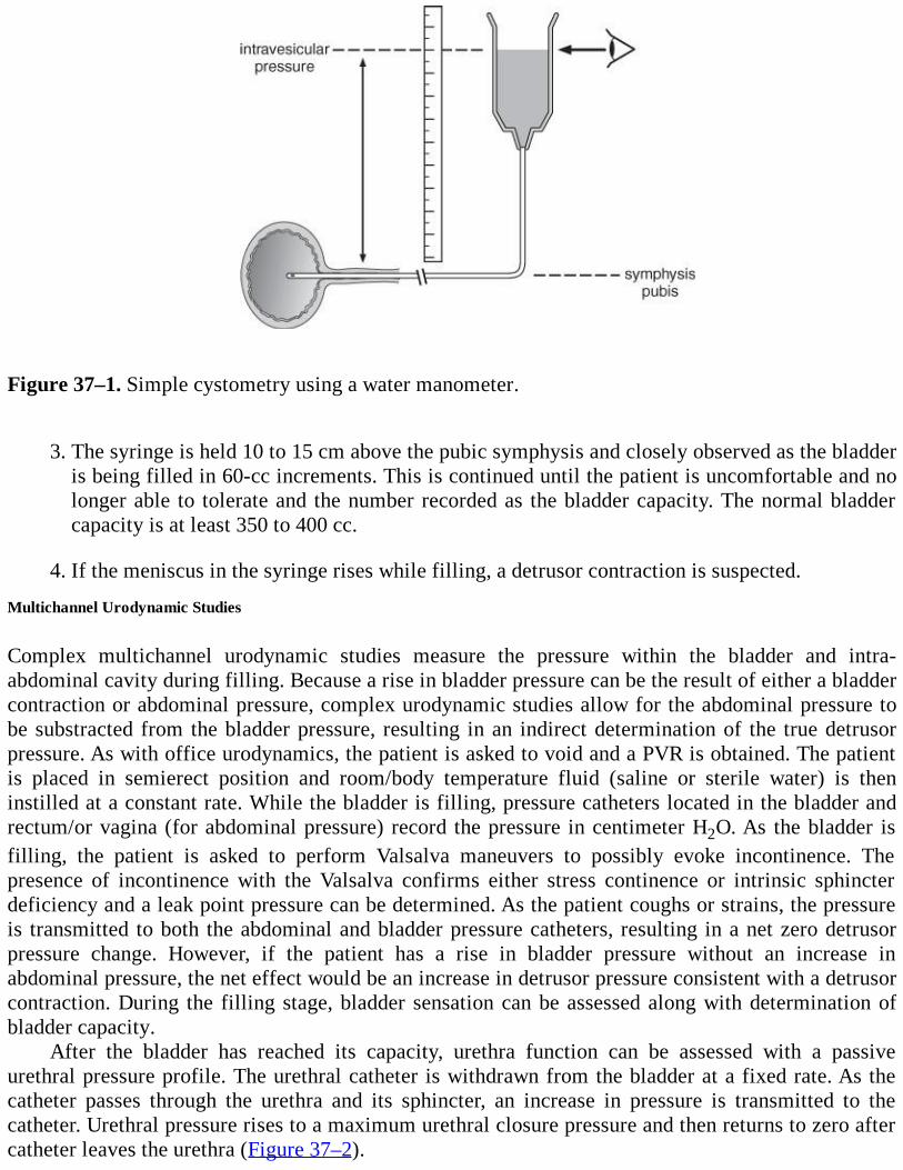
Figure 37–1. Simple cystometry using a water manometer.
3. The syringe is held 10 to 15 cm above the pubic symphysis and closely observed as the bladderis being filled in 60-cc increments. This is continued until the patient is uncomfortable and nolonger able to tolerate and the number recorded as the bladder capacity. The normal bladdercapacity is at least 350 to 400 cc.
4. If the meniscus in the syringe rises while filling, a detrusor contraction is suspected.
Multichannel Urodynamic Studies
Complex multichannel urodynamic studies measure the pressure within the bladder and intra-abdominal cavity during filling. Because a rise in bladder pressure can be the result of either a bladdercontraction or abdominal pressure, complex urodynamic studies allow for the abdominal pressure tobe substracted from the bladder pressure, resulting in an indirect determination of the true detrusorpressure. As with office urodynamics, the patient is asked to void and a PVR is obtained. The patientis placed in semierect position and room/body temperature fluid (saline or sterile water) is theninstilled at a constant rate. While the bladder is filling, pressure catheters located in the bladder andrectum/or vagina (for abdominal pressure) record the pressure in centimeter H2O. As the bladder isfilling, the patient is asked to perform Valsalva maneuvers to possibly evoke incontinence. Thepresence of incontinence with the Valsalva confirms either stress continence or intrinsic sphincterdeficiency and a leak point pressure can be determined. As the patient coughs or strains, the pressureis transmitted to both the abdominal and bladder pressure catheters, resulting in a net zero detrusorpressure change. However, if the patient has a rise in bladder pressure without an increase inabdominal pressure, the net effect would be an increase in detrusor pressure consistent with a detrusorcontraction. During the filling stage, bladder sensation can be assessed along with determination ofbladder capacity.
After the bladder has reached its capacity, urethra function can be assessed with a passiveurethral pressure profile. The urethral catheter is withdrawn from the bladder at a fixed rate. As thecatheter passes through the urethra and its sphincter, an increase in pressure is transmitted to thecatheter. Urethral pressure rises to a maximum urethral closure pressure and then returns to zero aftercatheter leaves the urethra (Figure 37–2).
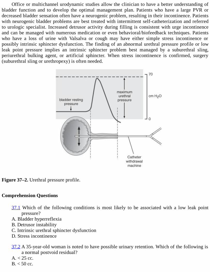
Office or multichannel urodynamic studies allow the clinician to have a better understanding ofbladder function and to develop the optimal management plan. Patients who have a large PVR ordecreased bladder sensation often have a neurogenic problem, resulting in their incontinence. Patientswith neurogenic bladder problems are best treated with intermittent self-catheterization and referredto urologic specialist. Increased detrusor activity during filling is consistent with urge incontinenceand can be managed with numerous medication or even behavioral/biofeedback techniques. Patientswho have a loss of urine with Valsalva or cough may have either simple stress incontinence orpossibly intrinsic sphincter dysfunction. The finding of an abnormal urethral pressure profile or lowleak point pressure implies an intrinsic sphincter problem best managed by a suburethral sling,periurethral bulking agent, or artificial sphincter. When stress incontinence is confirmed, surgery(suburethral sling or urethropexy) is often needed.
Figure 37–2. Urethral pressure profile.
Comprehension Questions
37.1 Which of the following conditions is most likely to be associated with a low leak pointpressure?
A. Bladder hyperreflexiaB. Detrusor instabilityC. Intrinsic urethral sphincter dysfunctionD. Stress incontinence
37.2 A 35-year-old woman is noted to have possible urinary retention. Which of the following isa normal postvoid residual?
A. < 25 cc.B. < 50 cc.
C. < 100 cc.D. Absolute normal values for PVR have not been established.
37.3 A 39-year-old woman is noted to have bladder hypersensitivity and reduced bladdercapacity. Which of the following is the most likely diagnosis?
A. Interstitial cystitisB. Stress urinary incontinenceC. Neurogenic bladderD. Diabetes mellitus
ANSWERS
37.1 C. The leak point pressure is defined as the pressure that is required to overcome urethralresistance and result in incontinence. Patients with urethral sphincter deficiency have lowleak point pressure measurement due to the lack of resistance that the sphincter providesagainst the bladder pressure. Stress incontinence may occur at low leak point pressures butnot routinely as low as intrinsic urethral sphincter deficiency.
37.2 D. No absolute value has been established for a normal PVR. However, values greater than200 mL can be considered abnormal.
37.3 A. Incontinent patients who have a history of spinal cord injury often suffer from overflowincontinence. Interstitial cystitis, cystitis, and detrusor instability all have bladderhyperactivity with reduced bladder capacity.
Clinical PearlsSee Table 1-2 for definition of level of evidence and strength of recommendation
Indications for urodynamic studies include (Level C). Mixed symptomatology Suspected voiding disorder Previous unsuccessful incontinence surgery Neurogenic bladder disorders Clinical examination not consistent with history Patients in whom conservative measures have failed
Patients with abnormal urethral pressure profiles or low leak point pressures should be referredto a urologist and treated with either a suburethral sling, periurethral bulking agent, orartificial sphincter (Level C).
No absolute value has been established for a normal postvoid residual (Level C).
Urodynamic studies are generally reliable among different observers (especially in presence ofstress urinary incontinence) (Level A).
REFERENCES
1. Abrams P. Urodynamic techniques. In: Abrams P. ed. Urodynamics 3rd ed. Bristol, UK:Springer; 2006:39-46, 99-109.
2. Cardozo L, Staskin D. Cystometry. In: Cardozo L and Staskin D. eds. Textbook of FemaleUrology and Urogynaecology. London, UK: Isis Medical Media Ltd; 2001: 198-204.
3. Cardozo L, Staskin D. Urethral pressure measurements. In: Textbook of Female Urology andUrogynaecology. 2001:216-224.
4. Diokno AC, Wells TJ, Brink CA. Urinary incontinence in elderly women: uro-dynamicevaluation. J Am Geriatr Soc. 1987;35:940-946.
5. Fritel X, Fauconnier A, Pigné A. Circumstances of leakage related to low urethral closurepressure. J Urol. 2008;180:223-236.
6. Haylen BT, Lee J, Logan V, Husselbee S, Zhou J, Law M. Immediate postvoid residualvolumes in women with symptoms of pelvic floor dysfunction. Obstet Gynecol.2008;111:1305-1312.
7. Katz VL, Lentz GM, Lobo RA, Gershenson DM. Urogynecology. In: ComprehensiveGynecology. 5th ed. Philadelphia, PA: Mosby; 2007:537-565.
8. Swift SE, Ostegard DR. Evaluation of current urodynamic testing methods in the diagnosis ofgenuine stress incontinence. Obstet Gynecol. 1995;86:85-91.
9. Theofrastous JP, Swift SE. Urodynamic Testing. In: Ostergard’s Urogynecology and PelvicFloor Dysfunction. Philadelphia, PA: Lippincott Williams & Wilkins; 2003:115-139.
10. Wall LL, Wiskind AK, Taylor PA. Simple bladder filling with a cough stress test comparedwith subtracted cystometry for the diagnosis of urinary incontinence. Am J Obstet Gynecol.1994;171:1472-1477.
11. Weber AM. Is urethral pressure profilometry a useful diagnostic test for stress urinaryincontinence? Obstet Gynecol Surv. 2001;56:720-735.
12. Weber AM. Leak point pressure measurement and stress urinary incontinence. Curr WomensHealth Rep. 2001;1:45-52.
13. Weir J, Jacques PF. Large-capacity bladder. A urodynamic survey. Urology. 1974;4:544-548.
14. Whiteside JL, Hijaz A, Imrey PB, et al. Reliability and agreement of urodynamicsinterpretations in a female pelvic medicine center. Obstet Gynecol. 2006;108:315-323.
Case 38
A 24-year-old G1P0 woman at 26 weeks’ gestation presents to your clinic for routine prenatal care.Upon reviewing her laboratory results from her initial visit, she is found to have an abnormal Papsmear. The Pap smear demonstrated low-grade squamous intraepithelial lesion (LSIL). The patient isbrought back to clinic for colposcopy which revealed no lesions.
What is your next step of management?
Was the referral for colposcopy warranted?
How does a colposcopic examination in pregnancy differ from that in a nonpregnant patient?
ANSWERS TO CASE 38:Colposcopy
Summary: A 24-year-old G1P0 woman at 26 weeks’ gestation with abnormal Pap smear demonstratingLSIL and subsequent normal colposcopy.
Next step in management: Repeat Pap in 6 and 12 months.
Referral for colposcopy warranted: Yes.
Colposcopy in pregnancy: More conservative; goal is to rule out invasive disease with follow-up after delivery; no endocervical curettage (ECC) should be performed during pregnancy.
ANALYSIS
Objectives
1. List the criteria of an adequate colposcopic examination.2. Describe the different colposcopic features of dysplasia.3. Describe common pitfalls of colposcopy and how to avoid them.4. Become familiar with how pregnancy affects colposcopy.
Considerations
This is a 24-year-old pregnant woman at 26 weeks’ gestation with cervical dysplasia noted on Papsmear. The finding of cervical dyplasia is not uncommon in pregnancy. Her access to prenatal caremay be the first time in which cervical cancer screening is done. The management of cervicaldysplasia does not change appreciably in pregnancy. Patients with LSIL and high-grade squamousintraepithelial lesion (HSIL) still require a colposcopy for further evaluation. The method in which acolposcopy is performed is not any different from that in a nonpregnant state, except that an ECC is
contraindicated in pregnancy. Colposcopic examination during pregnancy can be challenging due tothe increased vasculature and hormonal changes to the cervix. Acetic acid and Lugol solution can bothbe applied to the cervix with no risk to the pregnancy. Biopsies should only be performed in areas ofsuspected advanced disease. All minor colposcopic changes should await further evaluation afterpregnancy. The goal in pregnancy is to rule out invasive carcinoma. Treatment of dysplasia can takeplace after the pregnancy is completed. In addition, it is not too uncommon for the dysplastic cells toregress/slough off after delivery. For this patient, no lesions were seen on colposcopy and she can bereevaluated in 6 months.
APPROACH TO Colposcopy
DEFINITIONS
TRANSFORMATION ZONE: The area located between the original squamous epithelium andcolumnar epithelium.
ACETOWHITE EPITHELIUM: Areas of high nuclear activity which appear white. The moredense/pearl the epithelium appears, the higher the grade lesion.
PUNCTATION: Focal area with capillaries appearing like dots. The coarser the punctation, thehigher the grade lesion.
MOSAIC: Focal appearance in which the capillaries appear to surround abnormal areas as tiles. Ahigher-grade lesion will be found to have a coarser, wider, and more irregular mosaic.
ATYPICAL VESSELS: Focal area in which vessels appear horizontal with irregular caliber andbranching (appearing like commas, cork screw, etc.). The finding of atypical vessels is suggestive ofinvasive cancer.
CLINICAL APPROACH
Prenatal care provides many opportunities to screen for important conditions which could affect boththe mother and fetus. One important screening test is the Pap smear. Cervical dysplasia occurs in 1%of childbearing women who are screened annually, and 2% to 8% of all pregnant women will have anabnormal Pap smear. The incidence of cervical neoplasia (including carcinoma in situ [CIS] andinvasive carcinoma) in the pregnant population is estimated to be 1.5 to 12/100,000 pregnancies.Although patients with cervical neoplasia often are asymptomatic, common signs/symptoms ofinvasive cancer are often attributed to pregnancy changes when a patient is pregnant. Fortunately, ifcervical cancer is detected, it is often in the preinvasive or early invasive stage.
The management of cervical dysplasia in pregnancy is similar to, but less aggressive than innonpregnant women. In general, the management is conservative with the ultimate goal of ruling outan invasive malignancy. Treatment for cervical intraepithelial neoplasia (CIN) is generally delayeddue to the potential serious complications with therapy, minimal risk of disease progression, high rateof spontaneous disease resolution, and high rate of incomplete excision. In this case, a pregnantpatient with LSIL should be evaluated with a colposcopy. Her colposcopy can be performed in asimilar fashion to a nonpregnant patient, except for the fact that an ECC should not be done andcervical biopsy performed only if invasive disease is suspected. In the presence of this patient’snormal colposcopy, a repeat Pap can be performed postpartum.
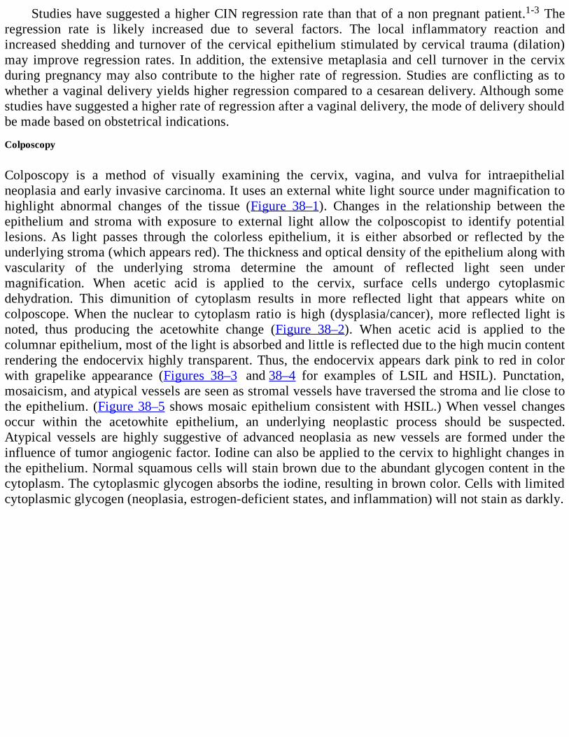
Studies have suggested a higher CIN regression rate than that of a non pregnant patient.1-3 Theregression rate is likely increased due to several factors. The local inflammatory reaction andincreased shedding and turnover of the cervical epithelium stimulated by cervical trauma (dilation)may improve regression rates. In addition, the extensive metaplasia and cell turnover in the cervixduring pregnancy may also contribute to the higher rate of regression. Studies are conflicting as towhether a vaginal delivery yields higher regression compared to a cesarean delivery. Although somestudies have suggested a higher rate of regression after a vaginal delivery, the mode of delivery shouldbe made based on obstetrical indications.
Colposcopy
Colposcopy is a method of visually examining the cervix, vagina, and vulva for intraepithelialneoplasia and early invasive carcinoma. It uses an external white light source under magnification tohighlight abnormal changes of the tissue (Figure 38–1). Changes in the relationship between theepithelium and stroma with exposure to external light allow the colposcopist to identify potentiallesions. As light passes through the colorless epithelium, it is either absorbed or reflected by theunderlying stroma (which appears red). The thickness and optical density of the epithelium along withvascularity of the underlying stroma determine the amount of reflected light seen undermagnification. When acetic acid is applied to the cervix, surface cells undergo cytoplasmicdehydration. This dimunition of cytoplasm results in more reflected light that appears white oncolposcope. When the nuclear to cytoplasm ratio is high (dysplasia/cancer), more reflected light isnoted, thus producing the acetowhite change (Figure 38–2). When acetic acid is applied to thecolumnar epithelium, most of the light is absorbed and little is reflected due to the high mucin contentrendering the endocervix highly transparent. Thus, the endocervix appears dark pink to red in colorwith grapelike appearance (Figures 38–3 and 38–4 for examples of LSIL and HSIL). Punctation,mosaicism, and atypical vessels are seen as stromal vessels have traversed the stroma and lie close tothe epithelium. (Figure 38–5 shows mosaic epithelium consistent with HSIL.) When vessel changesoccur within the acetowhite epithelium, an underlying neoplastic process should be suspected.Atypical vessels are highly suggestive of advanced neoplasia as new vessels are formed under theinfluence of tumor angiogenic factor. Iodine can also be applied to the cervix to highlight changes inthe epithelium. Normal squamous cells will stain brown due to the abundant glycogen content in thecytoplasm. The cytoplasmic glycogen absorbs the iodine, resulting in brown color. Cells with limitedcytoplasmic glycogen (neoplasia, estrogen-deficient states, and inflammation) will not stain as darkly.
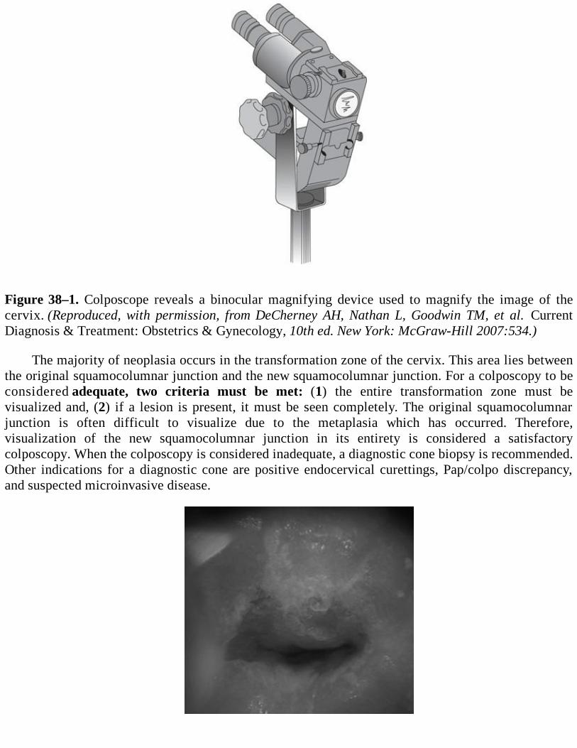
Figure 38–1. Colposcope reveals a binocular magnifying device used to magnify the image of thecervix. (Reproduced, with permission, from DeCherney AH, Nathan L, Goodwin TM, et al. CurrentDiagnosis & Treatment: Obstetrics & Gynecology, 10th ed. New York: McGraw-Hill 2007:534.)
The majority of neoplasia occurs in the transformation zone of the cervix. This area lies betweenthe original squamocolumnar junction and the new squamocolumnar junction. For a colposcopy to beconsidered adequate, two criteria must be met: (1) the entire transformation zone must bevisualized and, (2) if a lesion is present, it must be seen completely. The original squamocolumnarjunction is often difficult to visualize due to the metaplasia which has occurred. Therefore,visualization of the new squamocolumnar junction in its entirety is considered a satisfactorycolposcopy. When the colposcopy is considered inadequate, a diagnostic cone biopsy is recommended.Other indications for a diagnostic cone are positive endocervical curettings, Pap/colpo discrepancy,and suspected microinvasive disease.
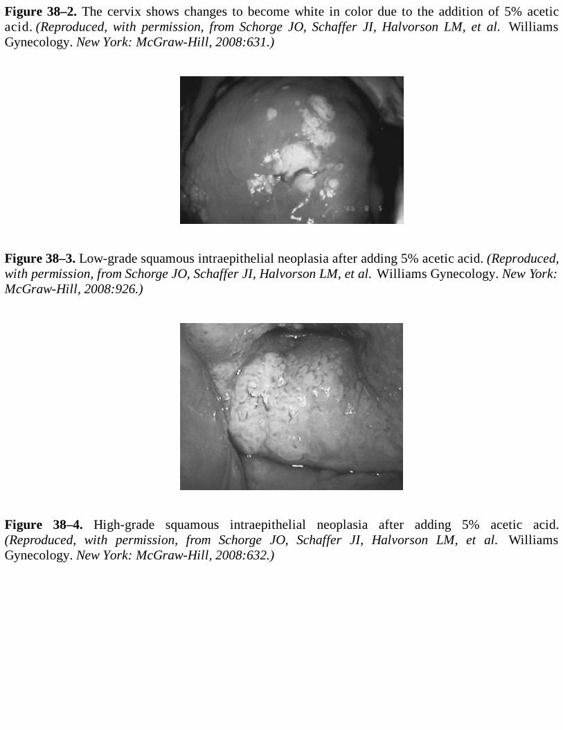
Figure 38–2. The cervix shows changes to become white in color due to the addition of 5% aceticacid. (Reproduced, with permission, from Schorge JO, Schaffer JI, Halvorson LM, et al. WilliamsGynecology. New York: McGraw-Hill, 2008:631.)
Figure 38–3. Low-grade squamous intraepithelial neoplasia after adding 5% acetic acid. (Reproduced,with permission, from Schorge JO, Schaffer JI, Halvorson LM, et al. Williams Gynecology. New York:McGraw-Hill, 2008:926.)
Figure 38–4. High-grade squamous intraepithelial neoplasia after adding 5% acetic acid.(Reproduced, with permission, from Schorge JO, Schaffer JI, Halvorson LM, et al. WilliamsGynecology. New York: McGraw-Hill, 2008:632.)
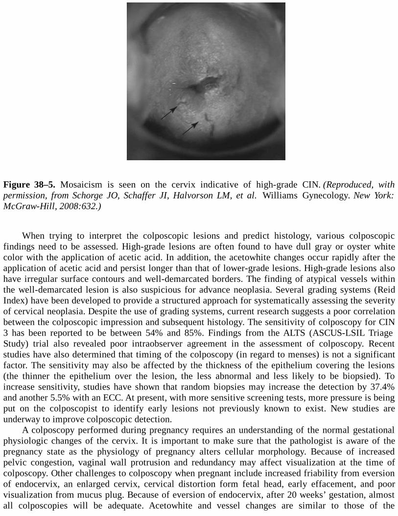
Figure 38–5. Mosaicism is seen on the cervix indicative of high-grade CIN. (Reproduced, withpermission, from Schorge JO, Schaffer JI, Halvorson LM, et al. Williams Gynecology. New York:McGraw-Hill, 2008:632.)
When trying to interpret the colposcopic lesions and predict histology, various colposcopicfindings need to be assessed. High-grade lesions are often found to have dull gray or oyster whitecolor with the application of acetic acid. In addition, the acetowhite changes occur rapidly after theapplication of acetic acid and persist longer than that of lower-grade lesions. High-grade lesions alsohave irregular surface contours and well-demarcated borders. The finding of atypical vessels withinthe well-demarcated lesion is also suspicious for advance neoplasia. Several grading systems (ReidIndex) have been developed to provide a structured approach for systematically assessing the severityof cervical neoplasia. Despite the use of grading systems, current research suggests a poor correlationbetween the colposcopic impression and subsequent histology. The sensitivity of colposcopy for CIN3 has been reported to be between 54% and 85%. Findings from the ALTS (ASCUS-LSIL TriageStudy) trial also revealed poor intraobserver agreement in the assessment of colposcopy. Recentstudies have also determined that timing of the colposcopy (in regard to menses) is not a significantfactor. The sensitivity may also be affected by the thickness of the epithelium covering the lesions(the thinner the epithelium over the lesion, the less abnormal and less likely to be biopsied). Toincrease sensitivity, studies have shown that random biopsies may increase the detection by 37.4%and another 5.5% with an ECC. At present, with more sensitive screening tests, more pressure is beingput on the colposcopist to identify early lesions not previously known to exist. New studies areunderway to improve colposcopic detection.
A colposcopy performed during pregnancy requires an understanding of the normal gestationalphysiologic changes of the cervix. It is important to make sure that the pathologist is aware of thepregnancy state as the physiology of pregnancy alters cellular morphology. Because of increasedpelvic congestion, vaginal wall protrusion and redundancy may affect visualization at the time ofcolposcopy. Other challenges to colposcopy when pregnant include increased friability from eversionof endocervix, an enlarged cervix, cervical distortion form fetal head, early effacement, and poorvisualization from mucus plug. Because of eversion of endocervix, after 20 weeks’ gestation, almostall colposcopies will be adequate. Acetowhite and vessel changes are similar to those of the
nonpregnant state.
Comprehension Questions
38.1 When does eversion of the endocervix take place in a primigravid patient?A. End of first trimesterB. End of second trimesterC. End of third trimesterD. After delivery
38.2 Which of the following is true?A. Lugol solution is more sensitive than acetic acid.B. Lugol solution stains areas of dysplasia.C. Vaginal atrophy would cause Lugol solution to not stain.D. Lugol solution stains area of cervical trauma darker.
38.3 The performance of an ECC with colposcopy (in a nonpregnant patient) increases thesensitivity by what percentage?
A. 1%B. 5%C. 10%D. Has not been shown to increase sensitivity
ANSWERS
38.1 A. Endocervical eversion occurs after the first trimester during a patient’s first pregnancy ascompared to the last trimester in multi-gravid patient.
38.2 C. Lugol solution nonstaining lesions can be caused by inflammation, atrophy, trauma, inaddition to dyplasia, making it less sensitive than acetic acid.
38.3 B. Studies have shown that adding an ECC with colposcopy increases the sensivity byapproximately 5% and should be considered on all nonpregnant colposcopies.
Clinical PearlsSee Table 1-2 for definition of level of evidence and strength of recommendation
Colposcopy has poor sensitivity in detecting early lesions but sensitivity can be improved byadding an ECC and random biopsy (Level A).
The colposcopic finding of atypical vessels is the most consistent colposcopic finding ofinvasive neoplasia (Level A).
Colposcopy is performed in a similar fashion during pregnancy as non pregnant but withprimary goal to rule out invasive disease (Level B).
In order for a colposcopy to be adequate, the transformation zone (new squamocolumnar
junction) and lesion need to be seen entirely (Level C).
REFERENCES
1. Hunter MI, Monk BJ, Tewar KS. Cervical neoplasia in pregnancy. Part 1: screening andmanagement of preinvasive disease. Am J Obstet Gynecol. 2008;199:3-9.
2. Baliga BS. Colposcopic terminology and colposcopic appearances of the cervix. Principles andPractice of Coloposcopy. New York, NY: McGraw Hill; 2004:53-59.
3. Broderick D, Matityahu D, Didhdhai M, Alter S. Histologic and colposcopic correlates ofASCUS Pap smears in pregnancy. J Low Genit Tract Dis. 2002;6:116-119.
4. American College of Obstetricians and Gynecologists. Management of Abnormal CervicalCytology and Histology. ACOG Practice Bulletin, 66. Washington DC; 2005.
5. Baliga BS. Colposcopy in pregnancy. Principles and Practice of Coloposcopy. New York, NY:McGraw Hill; 2004:138-143.
6. Cantor SB, Cárdenas-Turanzas M, Cox DD, et al. Accuracy of colposcopy in the diagnosticsetting compared with the screennig setting. Obstet Gynecol. 2007;111:7-14.
7. Carcopino X, Akkawi R, Conroy R, Prendiville W. Specific timing for colposcopy: Is itworthwhile? Obstet Gynecol. 2008;111:373-377.
8. Cox JT. More questions about the accuracy of colposcopy: What does this mean for cervicalcancer prevention? Obstet Gynecol. 2008;111:1266-1267.
9. Everson JA, Stika CS, Lurain JR. Postpartum evolution of cervical squamous intraepitheliallesions with respect to the route of delivery. J Low Genit Tract Dis. 2002;6:212-217.
10. Ferris DG, Litaker MS. Colposcopy quality control by remote review of digitized colposcopicimages. Am J Obstet Gynec. 2004;191:1934-1941.
11. Ferris DG, Litaker MS. Prediction of cervical histologic results using an abbreviated Reidcolposcopic index during ALTS. Am J Obstet Gynecol. 2006;194:704-710.
12. Jeronimo J, Schiffman M. Colposcopy at a crossroads. Am J Obstet Gynecol. 2006;195:349-353.
13. Katz VL, Lentz GM, Lobo RA, Gershenson DM. Intraepithelial neoplasia of the lower genitaltract (cervix, vulva). In: Katz VL, Lentz GM, Lobo RA, Gershenson DM. eds. ComprehensiveGynecology. 5th ed. Philadelphia, PA: Mosby; 2007:743-757.
14. Massad LS, Jeronimo J, Schiffman M. Interobserver agreement in the assessment ofcomponents of colposcopic grading. Obstet Gynecol. 2008;111:1279-1284.
15. Muller CY, Smith HO. Cervical neoplasia complicating pregnancy. Obstet Gynecol Clin
15. Muller CY, Smith HO. Cervical neoplasia complicating pregnancy. Obstet Gynecol ClinNorth Am. 2005;32:533-546.
16. Yang B, Pretorius RG, Belinson JL, Zhang X, Burchette R, Qiao Y. False negative colposcopyis associated with thinner cervical intraepithelial neoplasia 2 and 3. Gynecol Oncol.2008;111:332-336.
17. Walker JL, Wang SS, Schiffman M. Predicting absolute risk of CIN3 during post-colposcopicfollow-up: results from the ASCUS LSIL Triage Study (ALTS) group. Am J Obstet Gynecol.2006;195:341-348.
18. Wright TC, Massad LS, Dunton CJ, Spitzer M, Wilkinson EJ, Solomon D. 2006 consensusguidelines for the management of women with cervical intraepithelial neoplasia oradenocarcinoma in situ. J Low Genital Tract Dis. 2007;11:223-239.
Case 39
A 23-year-old nulliparous woman presents to your clinic for follow-up of her abnormal Pap smear.She was previously found to have LSIL on Pap smear, with a finding of CIN 1 on cervical biopsyduring her colposcopy. She was managed conservatively and a repeat Pap was performed 6 monthslater. This Pap also demonstrated LSIL and CIN 2 at the time of colposcopy. She denies postcoitalspotting, irregular bleeding, and vaginal discharge. Her sexually transmitted disease (STD) screen wasnegative.
What is your management plan?
Would checking human papillomavirus (HPV) subtypes be helpful in her management at thispoint?
Was performing a colposcopy with the first Pap smear demonstrating LSIL warranted?
ANSWERS TO CASE 39:Treatment of Cervical Dysplasia
Summary: A 23-year-old G0P0 woman with persistent cervical dysplasia (CIN 2) on colposcopy after6 months of observation.
Management plan: Treatment with either excision or ablation.
HPV testing: No, the majority (83%) of patients with CIN 1 lesions have high-risk subtypes ofHPV and testing would add little to the management.
Colposcopy for LSIL: Yes, studies have shown a 15% to 30% risk of CIN 2/3 at initialcolposcopy after an LSIL Pap.
ANALYSIS
Objectives
1. Become familiar with the management of CIN 1.2. Learn the technique of both cryotherapy and loop electrosurgical excisional procedure (LEEP).3. Become familiar with outcomes of both cryotherapy and LEEP.4. Understand the obstetrical implications after both a cryotherapy and LEEP.
Considerations
The patient is a 23-year-old nulliparous woman with persistent cervical dysplasia. She initially hadLSIL on Pap and CIN 1 on colposcopic-directed biopsy. Given the high rate of regression from CIN 1,
the patient was appropriately observed with repeat cytology at a 6-month interval. Her follow-up Paprevealed LSIL and she underwent a repeat colposcopy with CIN 2 on biopsy.
At this point, observation is no longer an option and treatment should be undertaken. Cervicaldysplasia can be treated with cryotherapy or conization. Given her nulliparity, cryotherapy or a LEEPconization would be the most appropriate management options.
APPROACH TO Treating CIN
DEFINITIONS
CERVICAL INTRAEPITHELIAL NEOPLASIA (CIN): Premalignant histologic changes found incervical epithelium. It is graded as CIN 1 through CIN 3.
LOOP ELECTROSURGICAL EXCISIONAL PROCEDURE (LEEP): A conization of the cervixusing a thin electric wire loop.
LOW-GRADE SQUAMOUS INTRAEPITHELIAL LESION (LSIL): A cytologic diagnosis whichhas changes consistent with CIN 1.
HIGH-GRADE SQUAMOUS INTRAEPITHELIAL LESION (HSIL): A cytologic diagnosis whichhas cellular changes consistent with CIN 2 and CIN 3.
CLINICAL APPROACH
Worldwide, cervical cancer remains one of the leading causes of death in women in the world.Fortunately, in developed countries where cervical cancer screening programs are in place, theincidence has decreased dramatically. Screening programs allow the practitioner the opportunity todetect and effectively treat precancerous lesions, cervical intraepithelial neoplasia, before thedevelopment of invasive cervical cancer. A survey performed by the College of American Pathologistsdemonstrated that cervical intraepithelial lesions are a common problem and that the rate of theabnormal cytology continues to rise (1 million women/year with CIN 1). Appropriate management ofcervical intraepithelial neoplasia is important for both the prevention of invasive cervical cancer andavoidance of overtreating the lesion.
Patients found to have LSIL on Pap should be referred for colposcopy due to the fact that 15% to30% of the time CIN 2/3 is identified histologically.1,2 When colposcopy confirms CIN 1 or less, thepatient should be counseled about management options of expectant management or treatment. Sheshould be counseled about the 13% risk of progression to CIN 2/3 at 2-year follow-up and 57% rate ofspontaneous regression (91% at 36 months in women aged 13 to 23 years old).3 In a younger patientwho has never been pregnant and is found to have CIN 1, expectant management is a very reasonableinitial management plan. When managed expectantly, cervical cytology should be obtained every 6months twice before returning to annual cytology. If cervical dysplasia persists on subsequentcytology, treatment with either ablation or excision is indicated. HPV subtype testing has no role inthe management of CIN 1 because it is found in 83% of all CIN 1 lesions.
Treatment
In general, there are two categories of treatment for CIN: ablative and excisional procedures. Studieshave shown a similar rate of CIN and HPV clearance with both excisional and ablative procedures.
Ablative procedures can be performed by either cryotherapy or laser vaporization. Both procedures arewell tolerated by the patient and are effective (both from a recurrence and cost-effectivenessstandpoint). However, ablations should not be performed in patients with endocervical dysplasia and,prior to any ablative procedure, an endocervical curettage (ECC) should be done to rule out dysplasiaof the endocervix. LEEP is the most common form of excisional procedure. Cold knife and laserconization are other excisional procedures less commonly used. An advantage of excisionalprocedures is their ability to evaluate the specimen histology. A major limitation of ablativeprocedures is the lack of histology for further evaluation. It has been reported that 2% to 3% ofpatients thought to only have CIN 2/3 were found to have adenocarcinoma in situ (AIS) or invasivecancer on excised specimens.4 With this knowledge, many practitioners prefer excision to ablation formanagement of CIN 2/3.
Cryotherapy utilizes either carbon dioxide or nitrous oxide to freeze the epithelium of the cervix.It can be easily performed in the office without anesthesia (when used on cervix). Water-soluble gel isoften applied to the probe to enhance contact with the cervix. A two-freeze technique is often used toachieve an ice ball extension 4 to 5 mm beyond the edge of the lesion. Freeze sessions often require 5minutes each. An ECC should be performed prior to the ablation to rule out endocervical disease. Themost common complication after cryotherapy is a profuse water vaginal discharge, although cervicalstenosis has also been observed.
A LEEP procedure (see Figure 39–1) can be done as an outpatient, in the OR or in the officesetting (with appropriate selected patient). A cervical block using lidocaine and epinephrine is usuallysufficient for anesthesia. Depending on the size of the lesion, various size loops are available based onthe size of the loop (0.5, 1, 1.5, and 2 cm). A pure cutting or blended (blend 1) current is then used toexcise the specimen with 50- to 60-W cutting power. Care should be taken to cause “charring” of themargins by using the appropriate current and not performing the procedure too slowly or quickly(dragging the loop). If endocervical involvement is suspected or a high-grade lesion is present,additional tissue may be excised from the endocervical canal using a small loop (5 mm). This iscommonly called a “top hat.” Using a smaller loop in just the endocervical canal is thought toconserve cervical stroma and minimize risk of adverse pregnancy outcomes in the future. When usingthe smaller loop, the cutting power should be reduced to 30 to 40 W. The surgical bed can then becauterized with a roller ball. Monsel solution can be applied with a cotton-tipped applicator ifbleeding persists.
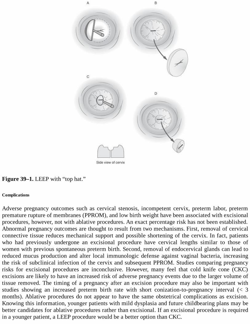
Figure 39–1. LEEP with “top hat.” Complications
Adverse pregnancy outcomes such as cervical stenosis, incompetent cervix, preterm labor, pretermpremature rupture of membranes (PPROM), and low birth weight have been associated with excisionalprocedures, however, not with ablative procedures. An exact percentage risk has not been established.Abnormal pregnancy outcomes are thought to result from two mechanisms. First, removal of cervicalconnective tissue reduces mechanical support and possible shortening of the cervix. In fact, patientswho had previously undergone an excisional procedure have cervical lengths similar to those ofwomen with previous spontaneous preterm birth. Second, removal of endocervical glands can lead toreduced mucus production and alter local immunologic defense against vaginal bacteria, increasingthe risk of subclinical infection of the cervix and subsequent PPROM. Studies comparing pregnancyrisks for excisional procedures are inconclusive. However, many feel that cold knife cone (CKC)excisions are likely to have an increased risk of adverse pregnancy events due to the larger volume oftissue removed. The timing of a pregnancy after an excision procedure may also be important withstudies showing an increased preterm birth rate with short conization-to-pregnancy interval (< 3months). Ablative procedures do not appear to have the same obstetrical complications as excision.Knowing this information, younger patients with mild dysplasia and future childbearing plans may bebetter candidates for ablative procedures rather than excisional. If an excisional procedure is requiredin a younger patient, a LEEP procedure would be a better option than CKC.
Comprehension Questions
39.1 HPV subtyping is helpful in the initial management of which of the following cytologicaldiagnosis?
A. HSILB. LSILC. ASC-USD. ASC-H
39.2 What is the best management of a LEEP specimen with CIN 3 and positive margins?A. Repeat cytology in 6 monthsB. Immediate repeat LEEP (within 72 hours)C. Delayed repeat LEEP (6 weeks after initial LEEP)D. Repeat colposcopy
39.3 Prior to performing an ablative procedure, what test should be done?A. HPV subtypingB. Cervical assays for gonorrhea/chlamydiaC. Endocervical curettageD. Vaginal culture for bacterial vaginosis
ANSWERS
39.1 C. HPV subtyping is primarily used in the triage of ASC-US. Patients with CIN already havesuch a prevalence of HPV that knowing the subtype would not impact your management.Patients with ASC-US can be managed either by repeat cytology, immediate colposcopy, orHPV subtyping. If high-risk HPV subtypes are identified, the patient is referred to forcolposcopic examination. If the high-risk subtype is negative, cytology should be repeated in1 year.
39.2 A. A specimen with positive margins can be followed with repeat cytology at 6 months.Rarely would you consider repeating the excisional procedure. Incomplete removal of CIN 3has been reported to occur in 5% to 16% of excisional procedures. It is thought that theinflammatory response from tissue healing and the cauterization of the cut surface of theconization bed help prevent persistent disease.
39.3 C. An ECC should be performed on all patients prior to an ablative procedure since tissue isnot obtained for histology. Performing the ECC detects possible underlying dysplasiapreviously not known and that may alter management.
Clinical PearlsSee Table 1-2 for definition of level of evidence and strength of recommendation
Patients with LSIL should be referred to colposcopy because of the 15% to 30% risk of CIN2/3 (Level A).
Ablative and excisional procedures have equal rates of clearance of dysplasia and HPV (Level
A).
HPV typing is primarily useful with ASC-US cytology (Level A).
All excisional procedures are associated with an increased chance of adverse obstetricaloutcomes (CKC possibly the highest) (Level B).
An ECC should be obtained prior to all ablative procedures (Level C).
REFERENCES
1. Law KS, Chang TC, Hsueh S, et al. High prevalence of high grade squamous intraepitheliallesions and microinvasive carcinoma in women with a cytologic diagnosis of low gradesquamous intraepithelial lesions. J Reprod Med. 2001;46:61.
2. Lonky NM, Sadeghi M, Tsadik GW, Petitti D. The clinical significance of the poor correlationof cervical dysplasia and cervical malignancy with referral cytologic results. Am J ObstetGynecol. 1999;181:560-566.
3. Ostor AG. Natural history of cervical intraepithelial neoplasia: a critical review. Int J GynecolPathol. 1993;12:186-192.
4. Ferenczy A, Choukroun D, Arseneau J. Loop electrosurgical excision procedure for squamousintraepithelial lesions of the cervix: advantages and potential pitfalls. Obstet Gynecol.1996;87:332-337.
5. American College of Obstetricians and Gynecologists. Management of Abnormal CervicalCytology and Histology. ACOG Practice Bulletin, 66. Washington DC; 2005.
6. Baggish MS, Karram MM. Cervical surgery: conization of cervix. In: Baggish MS, KarramMM. ed. Atlas of Pelvic Anatomy and Gynecologic Surgery. 2nd ed. Philadelphia, PA: ElsevierSaunders; 2006:497-501.
7. Crane JM, Delaney T, Hutchens D. Transvaginal ultrasound in the prediction of preterm birthafter treatment for cervical intraepithelial neoplasia. Obstet Gynecol. 2006;107:37-44.
8. Fanning J, Padratzik J. Cold knife conization vs. LEEP. Are they the same procedure? J ReprodMed. 2002;47:33-35.
9. Greenspan DL, Faubion M, Coonrod DV, Hart KW, Mathieson K. Compliance after loopelectrosurgical excision procedure or cold knife cone biopsy. Obstet Gynecol. 2007;110:675-680.
10. Himes KP, Simhan HN. Time from cervical conization to pregnancy and preterm birth. ObstetGynecol. 2007;109:314-319.
11. Jakus S, Edmonds P, Dunton C, King S. Margin status and excision of cervical intraepithelial
neoplasia: a review. Obstet Gynecol. 2000;55:520-527.
12. Jancar N, Rakar S, Poljak M, Fujs K, Kocjan BJ, Vrtacnik-Bokal E. Efficiency of threesurgical procedures in eliminating high-risk human papillomavirus infection in women withprecancerous cervical lesions. Eur J Gynaecol Oncol. 2006;27:239-242.
13. Kalliali I, Nieminen P, Dyba T, Pukkala E, Anttila A. Cancer free survival after CINtreatment: comparisons of treatment methods and histology. Gynecol Oncol. 2007;105:228-233.
14. Katz VL, Lentz GM, Lobo RA, Gershenson DM. Intraepithelial neoplasia of the lower genitaltract (cervix and vulva). In: Katz VL, Lentz GM, Lobo RA, Gershenson DM. ComprehensiveGynecology. 5th ed. Philadelphia, PA: Mosby; 2007:754-755.
15. Kyrgiou M, Koliopoulos G, Martin-Hirsch P, Arbyn M, Prendiville W, Parakevaidis E.Obstetric outcomes after conservative treatment for intraepithelial or early invasive cervicallesions: systematic review and meta-analysis. Lancet. 2006;367:489-498.
16. Luciani S, Gonzales M, Munoz S, Jeronimo J, Robles S. Effectiveness of cryotherapytreatment for cervical intraepithelial neoplasia. Inter J Gynecol Obstet. 2008;101:172-177.
17. Mathevet P, Chemali E, Roy M, Dargent D. Long-term outcome of a randomized studycomparing the three techniques of conization: cold knife, laser, and LEEP. Eur J ObstetGynecol. 2003;106:214-218.
18. Persad VL, Peirotic MA, Guijon FB. Management of cervical neoplasia: a 13-year experiencewith cryotherapy and laser. J Low Genit Tract Dis. 2001;5:199-203.
19. Schiffman M, Solomon D. Findings to date from the ASCUS-LSIL triage study (ALTS). ArchPathol Lab Med. 2003;127:946-949.
20. Stenchever M, Droegemueller W, Herbst A, Mishell D. Intraepithelial Neoplasia of theCervix. Comprehensive Gynecology. 4th ed. St. Louis, MO: Mosby 2001:877-879.
21. The ASCUS-LSIL Triage Study (ALTS) Group. A randomized trial on the management oflow-grade squamous intraepithelial lesion cytology interpretations. Am J Obstet Gynecol.2003;188:1393-1400.
22. Wright TC, Massad S, Dunton CJ, Spitzer M, Wilkinson EJ, Solomon D. 2006 consensusguidelines for the management of women with cervical intraepithelial neoplasia oradenocarcinoma in situ. J Low Genit Tract Dis. 2007;11:223-239.
Case 40
A 30-year-old African American G1P1001 woman presents for her annual well-woman examination.The patient denies any complaints at this time. She is happily married, and she and her husband desireto conceive another child in the next year. A complete history and physical examination is performedand a Pap smear is obtained. The patient has been receiving annual Pap smears since 21 years of ageand denies any history of abnormal Pap smears.
The results of the Pap smear show HSIL. The patient returns to the office and undergoescolposcopy. There is one acetowhite focal lesion present at the squamocolumnar junction extendingfrom the 5 to 7 o’clock position of the exocervix. The colposcopic impression is adequate, withmoderate-to-severe dysplasia. A punch biopsy of the lesion is taken and an ECC is performed. Theresults of the biopsy show carcinoma in situ, can’t rule out microinvasion, while the ECC is negative.Options were discussed with the patient and she desires conservative management.
What is your next step?
What are the indications for diagnostic excisional procedure?
What are potential complications of this procedure?
ANSWERS TO CASE 40:Cold Knife Cone
Summary: This is a 30-year-old G1P1001 woman with a history of carcinoma in situ (CIS) of thecervix diagnosed by colposcopically directed biopsy. The patient desires future fertility; therefore, shewould like to be managed conservatively.
Next step: Diagnostic and therapeutic cold knife conization (CKC) of the cervix to rule outmicroinvasion.
Indications for diagnostic excisional procedure: Inadequate colposcopy, two-grade Pap-colposcopy discrepancy, suspicion of microinvasion, endocervical involvement,adenocarcinoma in situ, treatment of persistent or recurrent CIN after failure of LEEP orablation.
Potential complications: Early and late postoperative bleeding, infection, cervical stenosis,cervical incompetence, infertility.
ANALYSIS
Objectives
1. List the indications for a CKC.2. Describe the technique of CKC.
3. Describe the long-term follow-up after CKC.4. List the short- and long-term complications of CKC and effects on future childbearing.
Considerations
This is a 30-year-old G1P1001 African American woman with a newly diagnosed CIS of the cervix.The patient desires future fertility and wishes to conceive another child in the next year. Aftercounseling, the patient agrees to conservative management in the form of CKC. Patients with high-grade cervical lesions can be managed conservatively until childbearing wishes are done. For thispatient, the excision is not only performed for possible treatment but more importantly to rule outmicroinvasive disease.
In general, high-grade lesions of the cervix are managed with one of the excisional procedures(LEEP, CKC, or laser conization). It has been shown that 2% to 3% of patients with high-gradedysplasia actually demonstrate unrecognized CIS or even invasive cancer. A pathologist cannot makea final diagnosis of microinvasive cervical cancer on punch biopsy or ECC. It is essential that thepathologist examines a large, well-oriented tissue specimen that is in one piece. In addition, it isimportant for the margins to be clearly seen to ensure diagnosis and possible treatment. Excisionaltechniques include LEEP, CKC, and CO2 laser. An advantage of a CKC is that the specimen has veryclear well-defined margins. When excision is performed by the LEEP or laser, coagulation artifactalong the margin may be present in 3.3% to 51% of cases.
APPROACH TO Cold Knife Conization
DEFINITIONS
CONIZATION: A large biopsy of the central cervix for the diagnosis or treatment of cervicaldysplasia.
COLD KNIFE CONIZATION: A conization of the cervix performed with a scalpel.
MICROINVASIVE CERVICAL CANCER: A small cervical carcinoma detected by microscopeonly (stage IA cervical cancer).
CLINICAL APPROACH
Indications
There are four clinical scenarios in which diagnostic excisional procedure should be performed. Theseinclude:
1. An inadequate colposcopy (entire lesion or squamocolumnar junction [SCJ] not visualized)2. Suspicion of microinvasion3. Greater than or equal to two-grade discrepancy between Pap smear and colposcopy/biopsy4. Endocervical involvement (ECC +)
Either a LEEP or CKC may be performed to obtain a cone biopsy specimen, but keep in mind thatcoagulation artifact from some LEEP specimens may make interpretation of the surgical marginsdifficult. A CKC is also the preferred treatment of adenocarcinoma in situ and treatment of persistentor recurrent CIN after failure of LEEP or ablation.
Technique
The traditional CKC (Figures 40–1 and 40–2) has been used for many years in the treatment of CIN. Inthe past, hot cautery was used to perform the conization of an inflamed, hypertrophic, chronicallyinfected cervix. However, for the excision of CIN, a standard surgical scalpel was more appropriate.Thus, the term “cold knife” was introduced to differentiate these two procedures.
Figure 40–1. Conization of the cervix shows a narrow and deep specimen for endocervical disease,and a more shallow and wide specimen for suspected ectocervical disease. (Reproduced, withpermission, from Schorge JO, Schaffer JI, Halvorson LM, et al. Williams Gynecology. New York:McGraw-Hill, 2008:893.)
A CKC is generally performed in the OR under anesthesia. The patient is placed supine in thedorsal lithotomy position. After examination under anesthesia, the patient is prepped and draped insterile fashion. A weighted speculum is placed in the vagina. Lugol solution is placed on the cervix todelineate the margins of the transformation zone and identify the areas of dysplasia. The cervix is theninjected with a vasoconstrictive agent to help decrease blood loss. Approximately 1.5 cc of a mixtureof a local anesthetic (lidocaine) with 1:100,000 epinephrine is injected directly into four quadrants ofthe cervix, that is, 2, 4, 8, and 10 o’clock. A total of 9 to 10 cc is injected and should cause“ballooning” and blanching of the cervix. Because acute hypertension may be caused by this drug,
inform the anesthesiologist of its use before injection. Lateral hemostatic sutures (stay sutures),consisting of 2-0 Vicryl or 2-0 Chromic may then be placed at the 3 and 9 o’clock positions of thecervix to ligate the cervical branches of the uterine artery and as a way to apply traction to the cervix.Studies have subsequently shown no benefit to deep lateral sutures in helping reduce blood loss.1,2
Figure 40–2. Conization of the cervix.Sutures are placed at 3 and 9 o’clock positions of the cervix forhemostasis and also stabilization of the cervix. An angled blade is used to incise the cervix to give acone biopsy. (Reproduced, with permission, from Schorge JO, Schaffer JI, Halvorson LM, et al.Williams Gynecology. New York: McGraw-Hill, 2008:892.)
With the lesion demarcated with Lugol solution, an angled scalpel is used to make acircumferential incision around the cervix, with care taken to encompass the entire transformationzone. The cervix can be stabilized with a tenaculum or the lateral stay sutures. A no. 11 scalpel bladeis commonly used; other choices include a larger no. 10 blade or a smaller no. 15 blade. As the cone iscut, it is retracted to the opposite side to provide visibility at the base of the incision. The preferredspecimen is a cylindrical cone that runs parallel and symmetric to the endocervical canal, includes thedeep glands of the canal, and has its apex in the canal. The intact cone should then be tagged withsuture at 12 or 6 o’clock so that the pathologist can orient any positive margins or foci of invasivecancer.
The surgeon can then use electrocautery to coagulate the cervical cone bed. Alternatively, thetissue between the ectocervical and endocervical surgical margin can be reapproximated in acontinuous locking fashion. Monsel solution can also be placed if needed.
Follow-up
Posttreatment, patients should receive either cytology with colposcopy or cytology only at 6-month
intervals. After two negative results, the patient may proceed with routine screening, for at least 20years following CKC. If the patient has an abnormal Pap smear at any time, a colposcopy withendocervical curettage must be performed. An alternative posttreatment plan is HPV testing at 6 to 12months following treatment. Patients who are high-risk HPV positive at testing should proceed withcolposcopy/ECC. Patients who are high-risk HPV negative can proceed with routine testing, for atleast 20 years following CKC.
CKC is usually considered the definitive treatment of CIN if the cone has encompassed the entireextent of the epithelial change; in other words, the epithelium is clear of disease at the surgicalmargins. One study evaluated the long-term outcome of patients with CIN 3/CIS after CKC withinvolved margins.3 Of the 969 patients with positive margins, 390 patients were followed expectantlyfor a mean of 19 (range 6-30) years. Results showed a 22% incidence of persistent or recurrent diseasein these patients, with 1.3% developing microinvasive cancer and 1% patients with a stage IB lesion.On the other hand, after incomplete excision of CIN 3, most patients did not develop persistent orrecurrent disease. This is probably because of the effect of diathermy on the cut cervix after removalof the cone and the inflammatory response associated with wound healing. Dysplastic or malignantepithelium within the inner zone of necrosis and an outer zone of white cell infiltration appears to beeradicated after CKC. Only lesions outside these zones can persist, regress, or progress. The findingssuggested that expectant management is reasonable for patients with CIN 3 and positive margins afterCKC, provided that careful follow-up is available, particularly during the first year.
Of note, a recent retrospective study showed that compared with office LEEP procedures, CKCpatients were significantly more compliant with follow-up appointments.4 The authors concluded thatbecause LEEP is a less invasive in-office procedure, it may convey to patients the idea that theircondition is less severe.
Complications
Cold knife conization is associated with a relatively high complication rate. Short- and long-termcomplications include early and late postoperative hemorrhage, infection, cervical stenosis, andcervical incompetence. Perioperative and postoperative hemorrhage, the most common complication,generally occurs in 10% to 20% of cases. Hemorrhage is described as early if it occurs within the first24 hours of surgery. Late hemorrhage typically occurs 10 to 21 days after surgery when the suturesdissolve. Intracervical vasopressors and topical thrombotic agents (Monsel solution) are shown tosignificantly decrease estimated blood loss, but lateral hemostatic sutures have not been shown tohave this effect. There are conflicting results regarding the superior method for obtaining hemostasisof the cervical cone bed. Some studies advocate that electrocautery is more effective in decreasingestimated blood loss, while others insist a continuous suture is the most hemostatic way to close thecone bed.
Cold knife conization, as are all excisional techniques, is associated with a roughly doubled rateof preterm labor (PTL) and delivery.5 The size of the cone is directly related to the risk of pretermlabor. CKC also increases the risk of preterm premature rupture of membranes (PPROM). Twomechanisms have been proposed to account for a higher rate of preterm birth and PPROM afterconization. One reason seems to be the disruption of cervical glands and reduced secretion of mucusafter removal of cervical tissue. This might result in an impaired defense mechanism againstmicrobial colonization in the cervix, which could facilitate ascending infections and lead to a higherrate of PROM. Another possibility is when part of the connective tissue of the cervix is removed withCKC; this leads to cervical fragility during pregnancy. There is a different collagen composition of the
original cervical tissue compared to that of the cervical scar.Cervical stenosis occurs in about 3% of patients. This is more common in patients who are not
having regular menstrual periods. This condition may result in infertility, hematometra, anddysmenorrhea. Data also indicate a slight increase in the incidence of symptomatic cervical tearsduring vaginal delivery, requiring surgical repair because of postpartum bleeding.
Young women with need for conization should be informed about potential complications insubsequent pregnancies. In addition, closer surveillance, including measurement of the cervicallength, might be useful, and information about a prior conization should be available for theobstetrician.
Comprehension Questions
40.1 A 28-year-old G3P3003 woman presents to the office for preoperative evaluation forplanned CKC. She asks the physician what risks are associated with this procedure. Whichof the following is the most common complication from CKC?
A. Preterm laborB. Cervical stenosisC. Postoperative hemorrhageD. Cervical incompetenceE. Infection
40.2 A 26-year-old G2P2002 woman who underwent CKC for CIN 3 is found to have a positivemargin on pathologic evaluation of the specimen. What is the best next step?
A. Repeat CKC immediately.B. Repeat CKC in 6 weeks, after healing takes place.C. Perform a Pap smear and colposcopy in 6 weeks after healing takes place.D. Perform a Pap smear and colposcopy in 6 months.
40.3 A 33-year-old G3P0202 woman at 9 weeks’ gestation presents for an initial OBappointment. The patient gives a history of CKC at 23 years of age with no abnormal Papsmears since that time. Her obstetric history is significant for preterm labor and delivery at32 weeks’ gestational age and again at 34 weeks. Which of the following is the best nextstep in management?
A. Placement of abdominal cerclageB. Placement of a cervical cerclage using the McDonald techniqueC. Measurement of cervical length by sonogramD. Recommendation of strict bed rest
ANSWERS
40.1 C. Early (within 24 hours of surgery) and late (24 hours or more after surgery) postoperativehemorrhage complicates 10% to 20% of cases of CKC. All of the other choices arecomplications of CKC, but these occur less often. Two options for controlling hemorrhageinclude placement of a vaginal pack or closure of the cervical bed with a continuous lockingsuture.
40.2 D. Re-exision is not necessary for patients with a positive margin following CKC. Seventy-
eight percent or more of patients with involved margins are free of disease at follow-up. Thediathermy effect and inflammatory response seems to eradicate the remaining dysplasticcells. These patients should proceed with routine follow-up at 6 months with Pap smearand/or colposcopy or high-risk HPV testing.
40.3 C. Following CKC, patients are at increased risk of PTL and PROM. Given this patient’shistory of two preterm deliveries, cervical incompetence due to previous CKC is apossibility. The next best step in management would be sonographic measurement of hercervical length. Patients with a cervical length of shorter than 2.5 cm are at a higher risk ofPTL. An elective McDonald cerclage may be an option for this patient, but it is not theimmediate next step; in addition, cerclage placement is preferable at about 12 to 14 weeks’gestational age. Bed rest and abdominal cerclage are not indicated in this situation.
Clinical PearlsSee Table 1-2 for definition of level of evidence and strength of recommendation
Excisional techniques are recommended in high-grade lesions because excision provideshistological confirmation that invasive cancer is not present (Level A).
For patients with positive margins following CKC for the treatment of CIN 2 or 3, re-excisionis not necessary. Patients should be followed at 6-month intervals with cytology and/orcolposcopy or high-risk HPV testing (Level B).
All of the excisional procedures to treat CIN present similar pregnancy-related morbidity,without apparent neonatal morbidity (Level B).
REFERENCES
1. Kamat AA, Kramer P, Soisson AP. Superiority of electrocautery over the suture method forachieving cervical cone bed hemostasis. Obstet Gynecol. 2003;102;726-730.
2. Dane C, Dane B, Cetin A, Erginbas M. Haemostasis after cold knife conization: a randomizedprospective trial comparing cerclage suture versus electrocauterization. Aust N Z J ObstetGynaecol. 2008;48:343-347.
3. Reich O, Lahousen M, Pickel H, et al. Cervical intraepithelial neoplasia III: long-term outcomeafter cold-knife conization with involved margins. Obstet Gynecol. 2002;99:193-196.
4. Wright TC Jr, Massad LS, Dunton CJ, et al. 2206 American Society for Colposcopy andCervical Pathology-Sponsored Concensus Conference. 2006 consensus guidelines for themanagement of women with abnormal cervical cancer screening tests. Am J Obstet Gynecol.2007;197:346-355.
5. Klaritsch P, Reich O, Giuliani A, et al. Delivery outcome after cold-knife conization of theuterine cervix. Gynecol Oncol. 2006;103:604-607.
6. Greenspan D, Faubion M, Coonrod D, et al. Compliance after loop electrosurgical excisionprocedure or cold knife cone biopsy. Obstet Gynecol. 2007;110:675-680.
7. Kyrgiou G, Martin-Hirsch P, Arbyn M, et al. Obstetric outcomes after conservative treatmentfor intraepithelial or early invasive cervical lesions: systematic review and meta-analysis.Lancet. 2006;367:489-498.
8. Mathevet P, Chemali E, Roy M, et al. Long-term outcome of a randomized study comparingthree techniques of conization: cold knife, laser, and LEEP. Eur J Obstet Gynecol Reprod Bio.2003;106:214-218.
9. Reich O, Pickel H, Lahousen M, et al. Cervical intraepithelial neoplasia III: long-term outcomeafter cold-knife conization with clear margins. Obstet Gynecol. 2001;97:428-430.
10. Rock J, Jones H. Cervical cancer precursors and their management. In: Rock J, Jones H. ed.Te Linde’s Operative Gynecology. 10th ed. Philadelphia, PA: Lippincott Williams & Wilkins;2008:1208-1225.

SECTION IIIListing of Cases
Listing by Case Number
Listing by Disorder (Alphabetical)
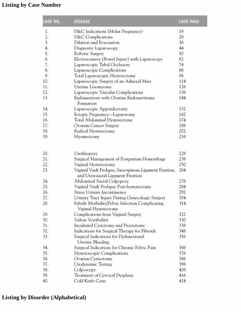
Listing by Case Number
Listing by Disorder (Alphabetical)
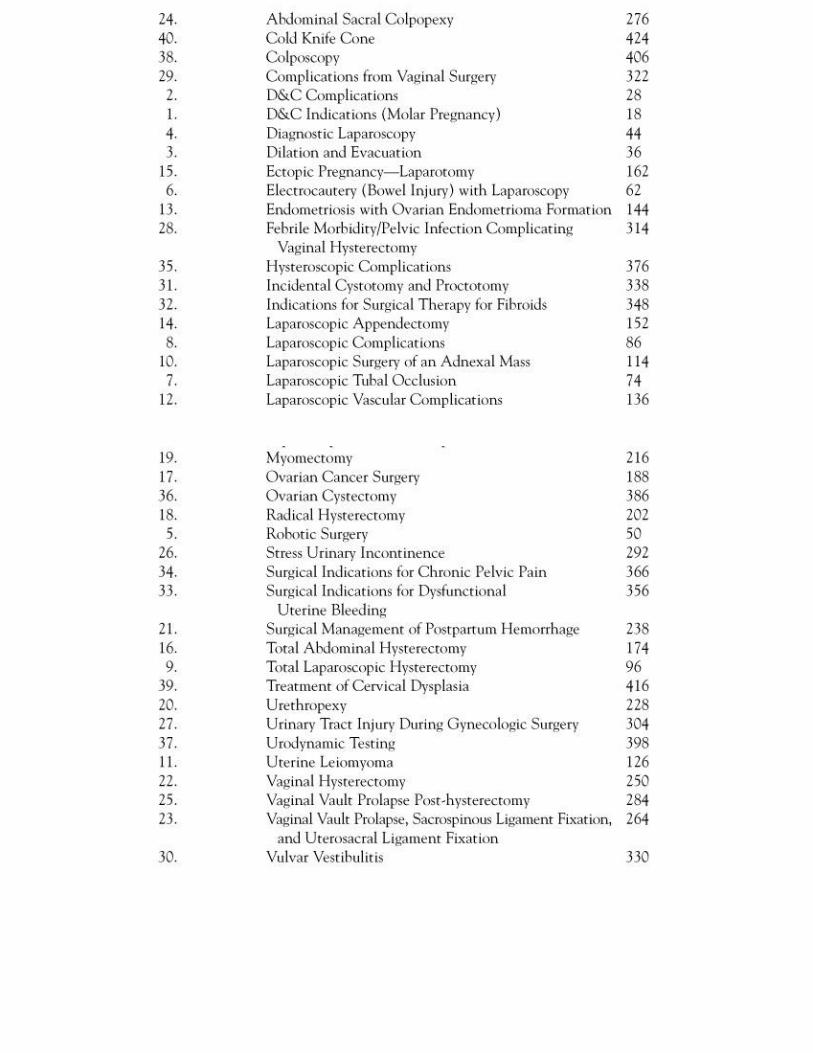
Index
A
Abdominal examinationAbdominal myomectomyAbdominal pain
after operative laparoscopypelvic pain andworsening of
Abdominal sacral colpopexy (ASC)case study ofclinical approach tocomplications withusing polypropylene bridge
Abdominal size, in ovarian cancerAbortion
medicalseptic
Acetic acidcervix withLSIL withuse of
ACOG. See American College of Obstetrics and GynecologistsAdenocarcinoma
cervicalradical hysterectomy with
AdenomyomaAdhesions
eliminating formation ofin healing processintrauterinearound umbilicus, from prior surgery
Adnexal massdiagnosis ofevaluation ofexamination ofincidence oflaparoscopic surgery ofpelvic examination ofin rectosigmoid colonremoval ofworkup of
Age
for fibroid treatment analysisas molar pregnancy risk factor
Allergies, gathering information aboutAllis clampAmerican College of Obstetrics and Gynecologists (ACOG)Anemia, menorrhagia withAnesthesia, complications ofAngular pregnancyAntibiotics
administration ofclasses oforalfor pelvic infectionprophylactic use ofsoaking graft intiming of
Appendicitisacutediagnosis ofimaging forsurgical approach totreatment of
Appendix, laparoscopic identification ofAromatase inhibitorArterial embolizationASC. See Abdominal sacral colpopexyAssisted reproductive technology
B
Balden-Walker system, for pelvic organ prolapse, on physical examinationBartholin gland cyst
definition ofsurgery for
Baseball stitchmyomectomy with, uterine closure after
Benign cystic teratomacase study ofclinical approach tosurgical approach to
Benign gynecology, robotic surgery inBenign ovarian cysts, open laparotomy forBimanual examinationBiological graft tissueBipolar cautery, tube/ovary removal byBipolar coagulation
on 3-cm segment
injuries inmethods of
Bipolar electrocautery, of round ligamentBipolar electrodes
example ofBirth control pills, for pelvic painBladder
Boari, flapinjuries to
from ASCfrom cystotomyearly detection offrom laparoscopic surgeryfrom LHrepair ofunrecognizedfrom vaginal hysterectomy
mid-urethral slingsmobilization of
example ofin total laparoscopic hysterectomy
Bleedingexcessive, treatment ofwith positive pregnancypostmenopausalin space of Retziusuterine, abnormalfrom vascular injuries
Blended waveformsBlunt needleB Lynch stitch
example ofuse of
Boari bladder flapBookwalter retractorBowels
injuries tofrom ASCclinical approach tofrom cystotomydiagnosis ofearly detection offrom LH
obstruction of, from LHBRCA genesBridge
polypropylene, to vaginal apex
Briesky-Navritol retractorsBurch
advantages ofcolposuspensioncure rates withMMK vs.procedures of
C
C section. See Cesarean sectionCA-125
in adnexal massdefinition ofin endometriosislevels ofin ovarian cancer detectionin pregnancy
CalvariumCapacitanceCapacitor couplingCarbon dioxide gasCardiac examinationCarneous degenerationCatheter withdrawal machineCephalosporinCervical canal, dilation ofCervical cancer
clinical approach toclinical presentation ofdiagnosis ofFIGO staging formicroinvasivepregnant patients withradiation therapy inrisk factors forstaging ofsurgical therapy fortreatment of
Cervical conizationCervical dysplasia
case study ofCIN 2complications frommanagement ofin pregnancytreatment of
Cervical intraepithelial neoplasia (CIN)CKC forcomplications fromcystourethroscopy indefinition ofHPV andtreatment of
Cervical stenosis, from CKCCervicovaginal attachments, division ofCervix
with acetic acidconization ofin D&Cexcision ofin situmagnification ofmosacism onsexual function and
Cesarean section (C section)history ofrisks with
Chemotherapyintraperitonealneoadjuvantprimary radiation therapy with
Chief complaintfor differential diagnosisidentification of
Chronic obstructive pulmonary disease (COPD), with postmenopausal bleedingCIN. See Cervical intraepithelial neoplasiaCIN 2. See Persistent cervical dysplasiaCircuits
openCircumferential culdotomyCKC. See Cold knife coneClinical diagnosis
approach tomaking of
Coagulation waveformsCold knife cone (CKC)
advantages ofcase study ofcervical stenosis fromfor CINcomplications withfollow-up afterhemorrhage after
indications forlugol solution inin pregnancytechniques of
Cold knife conizationCollodionColporrhaphyColposcopeColposcopy
case study ofclinical approach tocriteria fordefinitions related toECC withfor HSIL/LSILinterpretation ofmethods ofin pregnancy
Colposuspension, BurchCommon peroneal nerveComorbidity, optimization ofComplete moles
hydatidiform vs.Computed tomography (CT), use ofConization
cervicalof cervixf-427cold knifecomplications withdefinition of
Cooling periodCooper ligament, sutures inCOPD. See Chronic obstructive pulmonary diseaseCornual ectopic pregnancyCorpus luteum, cystCosmetic results, sexual function andCounseling, for sterilizationCreatinine concentrationCryotherapyCT. See Computed tomographyCuldoplastyCut waveformsCutaneous femoral nerveCyclic painCystic massCystic teratomasCystocele
with lupusrecurrentrepair ofsymptomatic
Cystometry, simple, using water manometerCystoscopy
instruments forpostoperativerationale forfor ureter
Cystotomybladder injuries frombowel injuries fromcase study ofdefinition ofincidentalvaginal hysterectomy with
Cystourethroscopyhistory ofindications forinstruments into measure injuriesuse of
CytoreductionCytoreductive surgery, definition of
D
da Vinci Surgical SystemD&C. See Dilation & curettageD&E. See Dilation & evacuationDeep vein thrombosis (DVT), from TAHDepression, history ofDermoids. See also Benign cystic teratoma
bilateralcomplications fromdefinition of
Detrusor overactivityDexterity, instrument reproducingDiagnostic hysteroscopyDiagnostic laparoscopy
analysis ofapproach tocomplications withconsiderations forelements forrisk factors in
Differential diagnosisfor chief complaintdevelopingof DUBof pelvic massof uterine fibroids
DilapanDilation & curettage (D&C)
of cervical canalcervix incomplications with
approach todelayed/lateimmediatesurgical
considerations forgynecologicalhemorrhage inindications formanagement plan forobstetrical indications forsuction curettage intechnique foruterine perforation in
Dilation & evacuation (D&E)analysis ofapproach tocomplications withconsiderations formanagement plans forstructures left behind afterstudies oftechnique ofUAE after
Direct couplingDistention media
definition offor hysteroscopysaline astypes of
Diverticulitis, pelvic pain fromDrug feverDUB. See Dysfunctional uterine bleedingDysfunctional uterine bleeding (DUB)
clinical approach todefinition ofdifferential diagnosis of
hypothyroidism andhysterectomy forphysical exam of
DysgerminomaDysmenorrhea
primaryprogressive
E
ECC. See Endocervical curettingEctopic pregnancy
cornuallaparotomy in
approach tolocation ofrisk of
Elective coincidental appendectomyElectricity, concepts ofElectrocautery
bipolar, of round ligamentconcepts ofdefinitions related towith laparoscopypreparation forrisk factors fortissue necrosis aftervaginal hysterectomy without
Electrodesexample oftypes of
Electrolyte rich mediaElectrosurgery
risk withsafety features inthermal injuries during
ELISA. See Enzyme-linked immunosorbent assayEndocervical curetting (ECC)
in HSILEndocervical eversionEndometrial ablation
complications withdefinition ofhysterectomy vs.methods ofuse ofuterine perforation from
Endometrial cancer, staging ofEndometrial polyps
case study ofin uterine cavity
Endometrioma. See also Ovarian endometrioma formation, endometriosis withdefinition ofexample ofright ovarian
Endometriosisapproach toCA-125 inclinical approach todeepdefinition ofdiagnosis ofGnRH-a forHRT forlaparoscopy forwith ovarian endometrioma formation
case study ofconsiderations forsummary of
peritonealsuperficialin total laparoscopic hysterectomytransvaginal ultrasound for
EndoWrist instrumentf-55Enlarged uterusEnterotomyEnzyme-linked immunosorbent assay (ELISA), use ofEpigastric arteryEpithelial tumor markersEssureExploration, in total laparoscopic hysterectomyExtrafascial hysterectomy
F
Falope ringFDA. See Food and Drug AdministrationFebrile morbidity
clinical approach todefinition ofpelvic infection and, in vaginal hysterectomy
Feet, difficulty with, after surgeryFemoral nerve, damage toFertility
early cancer inpostpartum hemorrhage and
Fetal heart tones, noFever, evaluation of, after vaginal hysterectomyFibroids. See also Uterine fibroids
hysteroscopic removal ofrecurrence ofremoval of, in myometriumf-221surgical therapy for, case study oftreatment of
FIGO stagingfor cervical cancerfor primary ovarian cancer
Filshie clipFluids, balance of, in hysteroscopyFood and Drug Administration (FDA), robotic surgery approvedFoot drop
G
G lynch stitch, definitions ofGastric injuriesGastrointestinal anastomosis (GIA)Genitofemoral nerveGerm cell tumor markersGestational trophoblastic disease
characteristics ofrisk factors for
GIA. See Gastrointestinal anastomosisGnRH-a. See Gonadotropin-releasing hormone agonistsGonadotropin-releasing hormone agonists (GnRH-a)
definition offor endometriosispretreatment with
Graftsantibiotics andbiologicalclinical approach tocontroversy ofkey techniques formeshsyntheticin vaginal hernia repairsvaginal paravaginal defect repair usingxeno-
Grounding plateGynecologists, American College of Obstetrics and
Gynecologybenign, robotic surgery incommon diseases inD&C type ofhistory ofoncology, robotic surgery insurgery, urinary tract injury during
H
Headlight, in vaginal hysterectomyHematoma, treatment ofHematometra, acuteHemorrhage
after CKCin D&Cfrom hysteroscopyfrom LHpostpartum
characteristics ofdefinitions related tofertility andsurgical management of
in space of RetziusHernia
trocarvaginal
repairs ofHigh-grade squamous intraepithelial lesion (HSIL)
case study ofcolposcopy fordefinition ofECC in
Historybasic information inof C sectionof chronic pelvic painof cystourethroscopyof depressionfor diagnosisgynecologicmenstrualobstetricof pelvic inflammatory diseaseof pelvic painreview of systems inof sexual abuse
before sterilizationHormone replacement therapy (HRT), for endometriosisHPV. See Human papillomavirusHRT. See Hormone replacement therapyHSG. See HysterosalpingogramHSIL. See High-grade squamous intraepithelial lesionHuman papillomavirus (HPV)
checking forCIN andsubtyping of
Hydatidiform molecase ofcomplete vs.
HydrosalpinxHypermenorrheaHypogastric artery ligation
definitions ofsutures forureteral damage withvascular injuries with
Hyponatremia, as hysteroscopic complicationHypotensive episodeHypothyroidism, DUB andHypovolemia, treatment ofHysterectomy. See also Specific type i.e Laparoscopic hysterectomy
clinical approach toconsiderations forfor DUBendometrial ablation vs.injuries from, rates offor leiomyomaafter pregnancyrates oftransvaginal ultrasound forfor uterine fibroidsvaginal vault prolapse, post-
Hysterosalpingogram (HSG)general use ofafter myomectomy
Hysteroscopecomponents ofdefinition ofhistory oftypes ofuse of
Hysteroscopic complicationscase study of
considerations forhyponatremia asimmediate postoperative complicationsinjury rates fromintraoperative complicationsintraoperative diagnosis oflate
Hysteroscopic myomectomy, indications for
I
Ileal interpositionImaging
for appendicitisCTfor laparoscopic appendectomyMRItypes of
Incidental appendectomytechniques for
Incisionson ovaryposterior uterinein total laparoscopic hysterectomyin utero-ovarian ligamentin vaginal mucosain vesicouterine peritoneal fold
Indigo carminedefinition ofspilling ofuse of
Infectionshematomafrom hysteroscopyfrom LHpelvictreatment of
Inferior epigastric arteryInfertility, myomectomy forInfundibular pelvic ligament (IP)
cauterization ofInterstitial line signInterstitial pregnancy
diagnosis ofrisk factors forsalpingo-oophorectomy insurgical therapy for
treatment options forInterval sterilizationIntestinal injuries, from laparoscopic surgeryIntraoperative ureteral transectionIntraperitoneal chemotherapy
definition oftrial of
Intrauterine adhesionsIntravenous pyelogram (IVP), general use ofIP. See Infundibular pelvic ligamentIVP. See Intravenous pyelogram
J
Javid vascular clamp
K
Kelly clampKnuckle of tubeKocher clamp
L
Labial biopsyLabial hypertrophyLabioplasty, clinical pearls forLabor, pretermLaboratory assessment, for obstetric patientsLactate dehydrogenase 1LamicelLaminaria, placement of, with misoprostolLaparoscopic appendectomy
background ofcase ofcontraindications todefinitions related toimaging forpathophysiology ofsummary ofsymptoms oftechniques for
Laparoscopic hysterectomy (LH). See also Total laparoscopic hysterectomy (TLH)benefits ofcomplications incontraindications todefinition of
use ofvaginal vs.
Laparoscopic myomectomyavoiding injuries inconsiderations foruse of
Laparoscopic surgeryof adnexal massapproach tocomplications withconsiderations forearly detection afterelectrocautery withfor endometriosisinjuries duringpregnancy andrisk inrobotic surgery vs.safety measures forscope placement intechniques forvascular complications with
approach toLaparoscopic tubal occlusion
incidence ofsummary of
Laparoscopic uterine nerve ablation (LUNA)Laparoscopic vaginal vault closureLaparoscopic-assisted vaginal hysterectomy (LAVH)
complications ofdefinition of
Laparoscopydiagnosticelectrocautery withfor endometriosislaparotomy vs.operativefor ovarian cystsrobotic surgery vs.
Laparotomyin ectopic pregnancylaparoscopy vs.open
Last menstrual period (LMP)Lateral femoral nerveLAVH. See Laparoscopic-assisted vaginal hysterectomyLeak pressure point
LEEP. See Loop electrosurgical excisional procedureLeiomyoma. See also Uterine leiomyoma
approach tocharacteristics ofdiagnosis ofhysterectomy formanagement ofmenorrhagia withrecurrence ofsymptoms of
Leiomyomatamyomectomy forsubmucosal
LH. See Laparoscopic hysterectomyLight sourceLMP. See Last menstrual period; Low malignant potentialLogothetopulos packLoop electrosurgical excisional procedure (LEEP)
definition ofobjectives oftechniques ofwith top hat
Low malignant potential (LMP)studies of
Low-grade squamous intraepithelial lesion (LSIL)with acetic acidcolposcopy fordefinition of
LSIL. See Low-grade squamous intraepithelial lesionLugol solution
in CKCnonstaining lesions
LUNA. See Laparoscopic uterine nerve ablationLupus erythematosus, with cystocele, rectocele andLymph nodes
dissection ofmetastasis to
LymphadenectomyLymph-vascular space invasion
M
Magnetic resonance imaging (MRI), general use ofMalignancy
myoma associated withof teratomasof uterine myomas
Marshall-Marchetti-Krantz (MMK)Burch vs.cure rates withprocedures ofrisks inwaddling gate after
Massachusetts clampsMayo scissorsMcBurney pointMcCall culdoplastyMedications, information aboutMeigs syndromeMenometrorrhagiaMenorrhagia
analysis ofwith anemiaconsiderations withdefinition ofwith leiomyomapelvic mass withworkup of
Mensesheavy/prolongedpelvic pain inafter sterilization
Menstrual historyMesh graftMesosalpinx, resection tube fromMethotrexate, for uterine ruptureMetrorrhagiaMicroinvasion
less than 3 mmMicroinvasive cervical cancerMid-urethral slingsMilk, sterileMisoprostol
for laminaria placementas management tooluse of
Mixed urinary incontinenceMMK. See Marshall-Marchetti-KrantzMolar pregnancy
clinical pearls ofevidence levels forrisk factors foruncomplicated
Monoclonal smooth muscle cells, in uterus
Monoclonal-independent lesionsMonopolar electrodesMorcellation technique
laparoscopicvaginal
Mosacism, on cervixMRI. See Magnetic resonance imagingMucosa, repair ofMultichannel urodynamic studiesMyomas
in African American womenmalignancy associated withmalignant transformation ofobjectives aboutsmall uterinesubmucoussymptoms oftreatment oftypes ofultrasound for
Myomectomyabdominalbenefits ofcase study ofclinical approach toHSG afterfor infertilityfor leiomyomatarisks ofuterine closure after, with baseball stitchfor uterine leiomyoma
Myometrium, fibroid removal from
N
NeedlesbluntVeress
Neoadjuvant chemotherapyNerve ablation, LUNANeurologic injuries
physical therapy forin vaginal surgery
Nonabsorbable sutureNonmalignant vulvar diseaseNonresectoscope

O
Obstetric historyObturator signObturator spaceOligomenorrheaOmentumOophorectomyOpen appendectomyOpen circuitOpen laparotomy, for benign ovarian cystsOperative approach, in generalOperative hysteroscopy
definition ofof fibroidsindications for
Operative laparoscopy, diffuse abdominal pain afterOschner clampOsmotic dilators
example ofuse of
types ofOsteitis pubisOvarian cancer
CA-125 forclinical approach toclinical pearls forcomplications withdefinitions related todiagnosis ofetiology ofFIGO staging forincidence ofin pregnancystage I ofsurgery for, case ofsurgical approach tosymptoms oftreatment ofin women, desiring fertility
Ovarian cystectomycase study ofclinical approach toconsiderations fordefinitions related tolaparotomy for, laparoscopy vs.
Ovarian cysts
benign, open laparotomy forbluntly dissectedlaparoscopy forrupture ofsurgical approach toTAH for, beyond child bearing years
Ovarian endometrioma formation, endometriosis withcase study ofconsiderations forsummary of
Ovariesendometrioma ofepithelial closure ofincision onmass in, example ofreconstruction of
using running closureremoval ofuneven
Overflow incontinence, spinal cord injuries andOxytocin, intravenous
P
Pain. See also Pelvic painabdominalcyclicdefinition ofafter operative laparoscopyfrom pelvic inflammatory diseaseperiumbilicalfrom SSLF procedure
Pap smear. See Papanicolaou smearPapanicolaou smear (Pap smear)
false-negative rates innormalfor radical hysterectomy
Para-aortic lymph nodes, metastasis toParametriumPararectal spaceParavesical spacePatient
approach tolaboratory assessment ofthin
Patient positioning, during surgerycorrect
injuries fromPelvic abscessPelvic cellulitis
treatment ofPelvic congestion syndromePelvic examination
for adnexal massrelaxation infor urinary incontinence
Pelvic infectionfebrile morbidity and, in vaginal hysterectomyprophylaxis fortreatment of
Pelvic inflammatory diseasehistory ofpain from
Pelvic lymphadenectomyradical trachelectomy with
Pelvic massdifferential diagnosis ofguidelines forwith menorrhagiaultrasound for
Pelvic organ prolapseBalden-Walker system for, on physical examinationstage of
Pelvic organ prolapse (POP), studies ofPelvic pain
analysis ofbirth control pills forchronic
approach tohistory ofnongynecologic disorders associated withsurgical indications fortreatment of
considerations forfrom diverticulitisexamination ofhistory oflower abdominalin mensesin ovarian cancerphysical examination forpsychological issues with
Periappendiceal inflammationPeritoneal endometriosis

Periumbilical painPeroneal nerve, damage toPersistent cervical dysplasia (CIN 2)Physical examination
Balden-Walker system for, for pelvic organ prolapsecomponents offor DUBfor pelvic pain
Physical therapy, for neurologic injuryPipelle office endometrial biopsyPneumoperitoneumPolymenorrheaPolypropylene bridge, to vaginal apexPOP. See Pelvic organ prolapsePostabortal syndromePosterior uterine incisionPostmenopausal bleedingPostoperative cystoscopy, use ofPostpartum hemorrhage
characteristics ofdefinitions related tofertility andsurgical management of, case study of
Pregnancyangularbleeding inCA-125 incervical cancer incervical dysplasia inCKC incolposcopy inectopicfuture, risk forhysterectomy afterinterstitiallaparoscopic surgery andmolarovarian cancer inradical hysterectomy withultrasound in
Presacral neurectomyPreterm labor, risk ofPreterm premature rupture of membranes (PRROM)Primary radiation therapy, with concurrent chemotherapyProbe couplingProctotomy
case study of

definition ofincidental
PRROM. See Preterm premature rupture of membranesPsoas hitch, ureteroneocystostomy withPsoas signPsychological issues, with pelvic painPubovesical fascia, sutures inPudendal nerve, injury toPulmonary examination
Q
Q-tip test
R
Radiation therapyin cervical cancerwith concurrent chemo
Radical hysterectomywith adenocarcinomaadjuvant therapy forcase study ofclinical approach tocomplications ofdefinitions related toPap smear forin pregnancystaging ofsurvival data for
Radical trachelectomyRectal examinationRectocele
lupus erythematosus andrepair of
Rectosigmoid colon, adnexal mass inRectovaginal spaceResection tube, from mesosalpinxResectoscopeResidency programs, teaching vaginal hysterectomyResponse, measure ofRetroperitoneal dissection, in total laparoscopic hysterectomyRetroperitoneal vessels, reducing injury toRetropubic urethropexy, approach toReview of systems, in historyRisk factors
with C section
for cervical cancerin diagnostic laparoscopyfor ectopic pregnancyfor electrocauterywith electrosurgeryfor fibroidsfor future pregnancyfor gestational trophoblastic diseasefor hysteroscopic complications, intraoperative diagnosis offor interstitial pregnancywith MMKfor molar pregnancyfor myomectomyfor preterm labor
Risk of malignancy index (RMI)definition ofequation for
Risk-benefit analysisRMI. See Risk of malignancy indexRobotic surgery
advantages ofapplications ofin benign gynecologydevelopment ofdisadvantages ofexample ofFDA-approvedin gynecologic oncologylaparoscopy vs.sacrocolpopexy
Robotic-assisted hysterectomyRound ligament
bipolar electrocautery ofligation of
Running closure, ovary reconstruction usingRuptured interstitial pregnancy
S
Sacrocolpopexyrobotic
Sacrospinous ligament fixationapproach toclinical presentation ofcomplications withprocedures for
Safety

with electrosurgeryof vaginal hysterectomy
Saline, as distention mediaSalpingo-oophorectomy
for interstitial pregnancytotal abdominal hysterectomy with
Sciatic nerve, damage toSciatica, after vaginal surgeryScope, placement ofSecond-trimester fetal demiseSeptic abortionSeptic thrombophlebitisSexual abuse, history ofSexual function
cervix andcosmetic results and
Sheathdefinition ofexample of
Silastic band, methods ofSkeletonization, purpose ofSkin, closure ofSlings
complications withnewtypes of
Snowstorm patternSonohysterography, use ofSOS Bakri balloon tamponadeSpace of Retzius
bleeding indefinitions ofdissection ofhemorrhage in
Spillagecomplications ofdefinition ofrates of
Spinal cord injuries, overflow incontinence andSplenectomy, as ovarian cancer complicationSpring-loaded clip, methods ofSquamous carcinomaSSLF procedure
counseling forpain fromUSLF vs.
Staging
approach totreating based on
Sterile milkSterilization
additional options forconsiderations forcounseling forfailure ofhistory forintervalmenses aftermethods ofpostpartumtube segments in
Stray energyStress urinary incontinence (SUI)
case study ofclinical approach todefinition ofdiagnostics forslings fortreatment of
comparisons ofTVT in
Submucous myomaSubtotal hysterectomy
commonality ofSuction curettageSUI. See Stress urinary incontinenceSuperficial endometriosisSupracervical hysterectomySurgical therapy, approach toSuspensionSutures
in Cooper ligament, in pubovesical fasciafor hypogastric artery ligationnonabsorbableplacement offor urinary incontinence
Synthetic graft tissue
T
TAH. See Total abdominal hysterectomyTampon testTelescopeTenesmus
Tension-free vaginal tape (TVT)in lumen, of urethrain SUI treatmentTOT vs.in urinary incontinence treatment
Teratomabenign cystic
case study ofclinical approach tosurgical approach to
cysticdefinition ofdiagnosis ofmalignantmaturetorsion with
Thermal injuriesearly detection ofprevention ofsuperficialtypes of
Thromboembolic disease, as ovarian cancer complicationTibial nerveTimingTissue necrosis, after electrocauteryTLH. See Total laparoscopic hysterectomyTop hat, LEEP withTorsion, with teratomasTOT. See Transobturator tapeTotal abdominal hysterectomy (TAH)
with bilateral salpingo-oophorectomycase ofchoice ofclinical approach tocomplications withdefinitions related toDVT fromfor ovarian cysts, beyond child bearing yearspostoperative considerations forpreoperative strategies forsimplification ofsurgical approach totechniques forTLH vs.ureteral injuries from
Total laparoscopic hysterectomy (TLH). See also Laparoscopic hysterectomyanalysis of
bladder mobilization inclinical approach toconsiderations fordefinition ofendometriosis inexploration inincisions inpreoperative preparation forretroperitoneal dissection inTAH vs.techniques ofupper uterine blood supply invaginal preparation in
Transobturator tape (TOT)TVT vs.
Transvaginal ultrasoundfor endometriosisfor hysterectomy
Trocar hernia, after laparoscopic surgeryTubal anastomosisTubingTumor markersTVT. See Tension-free vaginal tape
U
UAE. See Uterine artery embolizationUltrasound. See also Transvaginal ultrasound
general use ofintraoperativefor myomasfor pelvic massin pregnancy
Underwater examinationUnipolar scissors
bladder mobilization withUpper uterine blood supply, in total laparoscopic hysterectomyUreter
cystoscopy forfinding ofidentification ofinjuries to, from LHsafety of
Ureteral damage, with hypogastric artery ligationUreteral dissection, in total laparoscopic hysterectomyUreteral injuries
approaches to
after laparoscopic surgeryas ovarian cancer complicationfrom TAH
Ureteroneocystostomy, with psoas hitchUrethra
hypermobility ofTVT mesh in
Urethral pressure profileabnormalexample of
Urethropexycase study ofprocedures of
Urge urinary incontinencedefinition of
Urinary incontinence. See also Stress urinary incontinence (SUI)case study ofclinical approach todefinitions related todiagnostics formixedpelvic examination forsutures for
Urinary tract injuries, during gynecological surgeryUrinary urgency, in ovarian cancerUrodynamic stress incontinence
definition oftesting for
Urodynamic studiesindications formultichannel
Urodynamic testingcase study ofclinical approach toindications for
Urodynamicsclinical approach tooffice
USLF. See Uterosacral ligament fixationUterine artery embolization (UAE), after D&EUterine artery ligation, example ofUterine atonyUterine bleeding
abnormaldysfunctional
clinical approach todifferential diagnosis of

hypothyroidism andphysical exam of
Uterine closure, after myomectomy, with baseball stitchUterine fibroids
differential diagnosis ofhysterectomy formanagement ofsymptoms of
Uterine leiomyomaclinical approach todiagnosis ofmyomectomy forobjectives aboutsummary ofsymptoms oftreatment of
Uterine perforationin D&Cfrom endometrial ablationfrom extreme retroflexionfrom hysteroscopyoccurrence of
Uterine polyps, examination ofUterine prolapse
considerations forGrade 1
Uterine rupturediagnosis ofmethotrexate for
Uterine vessel ligationUtero-ovarian ligament
incisions inligation of
Uterosacral ligament fixation (USLF)choice ofcomplications withcounseling forprocedures ofSSLF vs.
Uterusdebris inendometrial polyps inenlargedexcision ofmass in
approach tomonoclonal smooth muscle cells
nontenderpacking ofpregnant, trauma torapidly growingretroflexion ofsize ofupper, blood supply to, in total laparoscopic hysterectomy
V
Vacuumcuretteextraction
Vaginaapex of
polypropylene bridge fromsupport of
cuff ofcellulitisclosure of
hernia ofrepairs for, grafts in
mucosa of, circumferentially incisedpH ofpreparation of, in total laparoscopic hysterectomysurgery
complications fromneurologic injury in
incidence ofspecific nerves injured in
wall of, support ofVaginal deliveries, normalVaginal hand instrumentVaginal hysterectomy
bladder injuries fromcase study ofclinical approach tocystotomy withdefinitions related toeducation inwithout electrocauteryfever evaluation afterheadlight inindications forlaparoscopic vs.pelvic infection in, febrile morbidity andsafety of
techniques inuse ofuterine size and
Vaginal paravaginal defect repair, using graft tissueVaginal vault closureVaginal vault prolapse
benefits ofpost-hysterectomy
case study ofrecurrence ofstudies ofsurgical procedures for
Valsalva maneuversVascular artery ligation, approach toVascular injuries
avoidance ofbleeding fromwith hypogastric artery ligationwith laparoscopic surgerymajorrecognition ofin thin patientstreatment offrom Veress needle
Veress needlevascular injuries from
Vesicouterine peritoneal fold, incisions inVesicovaginal fistulaVisceral lesionsVulvar dysplasiaVulvar intraepithelial neoplasia
approach todefinition ofdiagnosis ofII
Vulvar vestibulitisapproach tocase study ofconsiderations ofdefinitions of
W
Waddling gate, after MMKWater manometerWaveformsWell-woman examination

Word catheter
X
Xenograft