cardiac mitochondrial dysfunction during hyperglycemia—the role of oxidative stress and p66shc...
TRANSCRIPT

Cs
CAPa
b
c
d
Ae
a
AA
KHMRp
1p
Xiida
t
DS
1h
The International Journal of Biochemistry & Cell Biology 45 (2013) 114– 122
Contents lists available at SciVerse ScienceDirect
The International Journal of Biochemistry& Cell Biology
jo ur nal homep ag e: www.elsev ier .com/ locate /b ioce l
ardiac mitochondrial dysfunction during hyperglycemia—The role of oxidativetress and p66Shc signaling�
atia V. Diogoa,b, Jan M. Suskib,d, Magdalena Lebiedzinskab, Agnieszka Karkucinska-Wieckowskac,leksandra Wojtalab, Maciej Pronicki c, Jerzy Duszynskib, Paolo Pintond, Piero Portincasae,aulo J. Oliveiraa, Mariusz R. Wieckowskib,∗
CNC - Center for Neuroscience and Cell Biology, University of Coimbra, PortugalNenckiInstitute of Experimental Biology, Department of Biochemistry, PAS, Warsaw, PolandDepartment of Pathology, The Children’s Memorial Heath Institute, Warsaw, PolandDepartment of Experimental and Diagnostic Medicine, Section of General Pathology, Interdisciplinary Center for the Study of Inflammation (ICSI), Laboratory for Technologies ofdvanced Therapies (LTTA), University of Ferrara, Ferrara, ItalyClinicaMedica “A. Murri”, Department of Internal Medicine and Public Medicine, University of Bari, Medical School, Bari, Italy
r t i c l e i n f o
rticle history:vailable online 7 July 2012
eywords:yperglycemiaitochondrial dysfunction
OS66Shc
a b s t r a c t
Diabetes mellitus is a chronic disease caused by a deficiency in the production of insulin and/or by theeffects of insulin resistance. Insulin deficiency leads to hyperglycemia which is the major initiator ofdiabetic cardiovascular complications escalating with time and driven by many complex biochemicaland molecular processes. Four hypotheses, which propose mechanisms of diabetes-associated patho-physiology, are currently considered. Cardiovascular impairment may be caused by an increase in polyolpathway flux, by intracellular advanced glycation end-products formation or increased flux through thehexosamine pathway. The latter of these mechanisms involves activation of the protein kinase C.
Cellular and mitochondrial metabolism alterations observed in the course of diabetes are partiallyassociated with an excessive production of reactive oxygen species (ROS). Among many processes andfactors involved in ROS production, the 66 kDa isoform of the growth factor adaptor shc (p66Shc protein)
is of particular interest. This protein plays a key role in the control of mitochondria-dependent oxidativebalance thus it involvement in diabetic complications and other oxidative stress based pathologies isrecently intensively studied. In this review we summarize the current understanding of hyperglycemiainduced cardiac mitochondrial dysfunction with an emphasis on the oxidative stress and p66Shc protein.This article is part of a Directed Issue entitled: Bioenergetic dysfunction, adaptation and therapy.
. Cardiovascular complications in diabetes – a clinicalerspective
Diabetes mellitus (DM) is considered the epidemic of the XX andXI centuries (Zimmet et al., 2001). The prevalence of this disease
s higher in developed countries and in men (King et al., 1998). DM
s classified in two forms. 5–10% of the patients have Type I, insulin-ependent diabetes, or juvenile onset diabetes (IDDM), caused byutoimmune destruction of pancreatic �-cells, which promotes� This article is part of a Directed Issue entitled: Bioenergetic dysfunction, adap-ation and therapy.∗ Corresponding author at: Laboratory of Bioenergetics and Biomembranes,epartment of Biochemistry, Nencki Institute of Experimental Biology, Pasteur 3t., 02-093 Warsaw, Poland.
E-mail address: [email protected] (M.R. Wieckowski).
357-2725/$ – see front matter © 2012 Elsevier Ltd. All rights reserved.ttp://dx.doi.org/10.1016/j.biocel.2012.07.004
© 2012 Elsevier Ltd. All rights reserved.
insulin deficiency. The majority of diabetes patients have Type 2diabetes which is non-insulin-dependent (NIDDM), an adult-onsetform, which results from the combination between insulin resis-tance and/or �-cell secretory defect. In both types of diabetes, thedecrease in the uptake of glucose by muscle and adipose tissuepromotes extracellular hyperglycemia which leads to pathophysi-ological complications and damaged tissue (Carley and Severson,2005; Niedowicz and Daleke, 2005). Retinopathy, nephropathy,nerve damage, heart disease and atherosclerosis are some of thecomplications induced by hyperglycemia that are responsible forthe mortality and morbidity associated with diabetes (Table 1)(Brownlee and Cerami, 1981; Kuusisto et al., 1994; Luscher et al.,2003; Resnick and Howard, 2002).
The progression of atherosclerotic cardiovascular diseaseis clearly associated with the presence of insulin resistance,hyperinsulinemia, and elevated blood glucose. In this respect,coronary heart disease (CHD) is a major cause of morbidity and
C.V. Diogo et al. / The International Journal of Bioc
Table 1Pathophysiology of diabetes, including organ-specific consequences.
General
Type I Type II
Autoimmune destruction of pancreatic �-cells Insulin resistanceInsulin deficiency �-Cell secretory defectDecrease in the uptake of glucose Decrease in the uptake
of glucoseHyperglycemia Hyperglycemia
Organ-specificRetinopathyNephropathyNerve damageAtherosclerosisCoronary heart diseaseCoronary ischemiaMyocardial infarction
mes1hot1e1hwolTmoe2o2cF5ai2raHap(mfhfsaatoCt(c
Silent myocardial ischemia
ortality among patients with diabetes mellitus (DM) (Grundyt al., 1999). A number of important clinical studies have under-cored such harmful association (Gerstein et al., 1999; Singer et al.,992; Reaven, 1997) and have shown that individuals with DMave a higher prevalence of CHD (Fox et al., 2004), a greater extentf coronary ischemia (Granger et al., 1993), and are more likelyo have a myocardial infarction (Yusuf et al., 2004; Haffner et al.,998), and silent myocardial ischemia and infarction (Scognamigliot al., 2006; Kannel, 1985; Margolis et al., 1973; Niakan et al.,986), compared to individuals without diabetes. In the Framing-am Heart Study, the age-adjusted risk for cardiovascular diseaseas doubled in men and tripled in women with DM independently
f other important risk factors (Kannel and McGee, 1979). Simi-ar data were available from the Multiple Risk Factor Interventionrial Data where cardiovascular death rates were 9.7% among 5163en on antidiabetic medications vs. 2.6% in the 342,815 men with-
ut medications for diabetes over a period of 12 years (Stamlert al., 1993). Also the Copenhagen Heart Study demonstrated type
DM patients, the risk of having an incident myocardial infarctionr stroke is increased 2–3-fold and the risk of death is increased-fold, independent of other known risk factors for cardiovas-ular diseases (Almdal et al., 2004). Recently, the Emerging Riskactors Collaboration study constructed a meta-analysis including30,083 patients across 102 studies and showed that DM confersbout a two-fold excess risk for a wide range of vascular diseases,ndependently from other conventional risk factors (Sarwar et al.,010). Of note, both DM and impaired glucose tolerance can stillemain undiagnosed in a consistent number of patients developingcute myocardial infarction (Norhammar et al., 2002). In the INTER-EART study, 15,152 cases and 14,820 controls were enrolled in
standardized case–control study of acute myocardial infarctionerformed in 52 countries, representing every inhabited continentYusuf et al., 2004). DM, among several other conditions (abnor-
al lipids, smoking, hypertension, abdominal obesity, psychosocialactors, low consumption of fruits and vegetables, increased alco-ol intake, and absence of regular physical activity) accounted
or most of the risk of myocardial infarction worldwide in bothexes and at all ages in all regions (Yusuf et al., 2004). Overall, thell-cause mortality risk associated with DM is comparable to thell-cause mortality risk associated with a prior myocardial infarc-ion (Vaccaro et al., 2004). The importance of diabetes in the genesisf coronary heart disease has been further outlined by the National
holesterol Education Program in 2002 which considers type 2 DMo be a CHD equivalent, placing it into the highest risk categoryHaffner et al., 1998; NCEP, 2002). The relative risk for cardiovas-ular disease is even greater in those with type 1 DM, comparedhemistry & Cell Biology 45 (2013) 114– 122 115
to non-diabetics of similar age (Krolewski et al., 1987). The overallimpact of diabetes in cardiovascular disease is further aggravatedby the frequent pre-coexistence of other atherogenic predisposingrisk factors, namely obesity, hypertension, dyslipidemia, sedentarylife, albuminuria, hyperhomocysteinemia, smoking, and elevatedplasma fibrinogen (Grundy et al., 1999; Gerstein et al., 2001;Wachtell et al., 2003; Hoogeveen et al., 2000). Many of such fac-tors are aggregated within the metabolic syndrome (Grundy, 2005;Petruzzelli et al., 2007; Palasciano et al., 2007).
The relative risk of coronary heart disease may also be increasedin a pre-diabetic condition when higher levels of blood glucose andglycated hemoglobin are already present (Coutinho et al., 1999;Qiao et al., 2002; Ford et al., 2010). In a prospective study of181 apparently non-diabetic patients with an acute myocardialinfarction, impaired glucose tolerance was 35% while undiagnoseddiabetes was 31% by glucose tolerance test but only 10% by fast-ing glycemia (Norhammar et al., 2002). The Emerging Risk FactorsCollaboration study confirmed that in people without DM, fastingblood glucose concentration is modestly and non-linearly associ-ated with risk of vascular disease (Sarwar et al., 2010).
Several factors are responsible for the increased cardiovascu-lar risk and clinical presentations in diabetic and pre-diabeticpatients. Apart from blood pressure and dyslipidemia, mecha-nisms of increased risk include increased rate of atherosclerosis(Moreno et al., 2000), structural abnormalities (e.g. coronaryartery calcifications (Anand et al., 2006)), endothelial dysfunction,reduced myocardial flow reserve with impaired coronary vasodila-tor capacity (Yokoyama et al., 1997; Di Carli et al., 1999) (thislatter abnormality inversely related to glycemic control (Yokoyamaet al., 1997)), platelet activation (a predisposing factor to coro-nary thrombosis) (Davi et al., 1999), abnormalities in coagulation,hemostasis, and fibrinolysis (Kwaan, 1992). Mechanisms underly-ing the increased rate of silent ischemia and myocardial infarctionin diabetic patients, by contrast, imply increased frequency ofsilent ST segment depression and coronary perfusion abnormali-ties during stress testing, autonomic (sympathetic) denervation ofthe heart associated with myocardial electrical instability (a factorpotentially leading to fatal arrhythmias (Kwaan, 1992; Langer et al.,1995) and longer anginal perceptual threshold following exercise(Ranjadayalan et al., 1990). When going deeper into the cellularrealm, things can be equally complex.
2. An unifying theory for DM complications?
Some hypotheses have been forwarded to explain howhyperglycemia causes diabetic-associated complications, includingpolyol pathway flux; increased advanced glycation end-products(AGE) formation; activation of protein kinase C (PKC) isoforms; andincreased hexosamine pathway flux.
2.1. 1st hypothesis – increased polyol pathway flux
The enzyme aldose reductase is a monomeric oxidoreductaselocated in the cytosol that is able to catalyze NADPH-dependentreduction of several carbonyl compounds, including glucose,despite its low affinity for this compound (Brownlee, 2001; Folliet al., 2011; Giacco and Brownlee, 2010). Aldose reductase is thefirst enzyme in the polyol pathway and metabolism of glucose atnormal concentrations by this pathway represents only a smallpart of the total use of glucose. Under hyperglycemic conditions,the increase in glucose increases the amount of sorbitol, which
leads to the decrease in NADPH since it is consumed during thereaction. In this pathway, the enzyme sorbitol dehydrogenase isresponsible for the oxidation of sorbitol into fructose, with thereduction of NAD+ to NADH (Brownlee, 2001; Folli et al., 2011;
1 f Bioc
GdiacapbTla
NimWNtr2i2pNgtri
2a
w(spFhptriIaCbcwsoepwTtBtAettTtc
16 C.V. Diogo et al. / The International Journal o
iacco and Brownlee, 2010). There are several explanations for theeleterious effects of hyperglycemia through this pathway, which
nclude sorbitol-induced osmotic stress, decreased (Na+/K+)ATPasectivity, an increase in cytosolic NADH/NAD+ and a decrease inytosolic NADPH (Brownlee, 2001). The decrease of (Na+/K+)ATPasectivity was originally thought to be mediated by a decrease inhosphatidylinositol synthesis mediated by the polyol pathway,ut it has been recently shown to result instead from PKC activation.he activation of PKC promotes an increase in cytosolic phospho-ipase A2 activity, which increases the production of arachidonatend PGE2, inhibitors of (Na+/K+) ATPase (Xia et al., 1995).
The oxidation process of sorbitol increases the ratio betweenADH and NAD+, inhibiting the activity of GAPDH and increas-
ng the concentrations of triose phosphate, which leads to increaseethylglyoxal and diacylglycerol, activating PKC (Brownlee, 2001;illiamson et al., 1993). Even if hyperglycemia increases the
ADH:NAD+ ratio, this reflects a decrease in the total concen-ration of NAD+ as a result of consumption by activated PARP,ather than reduction of NAD+ to NADH (Garcia Soriano et al.,001). The activation of PARP by hyperglycemia is mediated by
ncreased production of reactive oxygen species (ROS) (Brownlee,001). Besides the explanations already mentioned, it has also beenroposed that reduction of glucose to sorbitol by NADPH consumesADPH. Since NADPH is required for the generation of reducedlutathione (GSH), this could cause or worsen intracellular oxida-ive stress (Brownlee, 2001). Although there are several studiesegarding inhibition of the polyol pathway in vivo, the results arenconsistent.
.2. 2nd hypothesis – increased intracellular formation ofdvanced glycation end-products
AGE are nonenzymatically glycosylated proteins and lipidshich have altered biochemical and physiological properties
Ahmed, 2005). Type 2 diabetes leads to a combination of exces-ive protein and glucose in plasma, resulting in the formation ofrotein glycation end products (AGE) (Ahmed, 2005; Schleicher andriess, 2007). These products are now recognized to exert severalarmful effects (Ahmed, 2005; Schleicher and Friess, 2007). Therotein glycation process starts with a nucleophilic addition reac-ion between free amino group of protein and carbonyl group of aeducing sugar, which is normally glucose. A Schiff’s base is formedn the initial reaction, which is slow and reversible (Ahmed, 2005).n a second phase, Schiff’s base reorganizes itself into ketoaminesnd Amadori products, which are irreversible products (Ulrich anderami, 2001). Besides, glycated proteins can also react with car-onyl intermediates to form the final AGE products. Increasedontent in AGEs have been detected in type 2 diabetes, being relatedith the pathophysiology and progression of diabetes. There is a
econd pathway for AGE production, consisting in glucose auto-xidation, which also generates ROS (Wolff and Dean, 1987; Huntt al., 1993). In the presence of transition metals, the enediols,resent in vivo, generate anion and dicarbonyl-keto-aldehydeshich react with amino groups of proteins to form ketoimines.
hese ketoimines are more reactive than Amadori products, con-ributing more to form AGE than the first pathway mentioned.esides the above mentioned pathways, there is a third pathwayhat results in the formation of AGE (Ahmed, 2005). In this pathway,madori products are formed and converted into AGE with proteinnedios and protein dicarbonyl compounds. AGE are by themselvesoxic products, exerting harmful effects through ROS including
he initiation of lipid peroxidation and oxidation of nucleic acids.here are several types of cell receptors for AGE and the interac-ion between them is believed to contribute and worsen diabeticomplications (Ahmed, 2005).hemistry & Cell Biology 45 (2013) 114– 122
2.3. 3rd hypothesis – activation of protein kinase C (PKC)
The PKC family includes at least eleven isoforms, represent-ing major targets for lipid second messengers or phorbol esters(Nishizuka, 1992, 1995; Liscovitch and Cantley, 1994). PKC isoformsare divided into conventional and new PKCs. The conventional typeis dependent on Ca2+ and binding site of DAG. The so-called newPKCs are sensitive to DAG but are independent of Ca2+ (Koya andKing, 1998). The source of DAG that is responsible for the activa-tion of PKC can derive from the hydrolysis of phosphatidylinositidesor from the metabolism of phosphatidylcholine (Koya and King,1998). It has been demonstrated that intracellular hyperglycemiaincreases the amount of DAG in a variety of tissues such as retina,heart and renal glomeruli, which interestingly are affected duringdiabetic complications (Shiba et al., 1993; Inoguchi et al., 1992;Craven et al., 1990; Ishii et al., 1996). These effects seem to be causedby the increase in de novo synthesis of DAG which consequentlyactivates PKC (Brownlee, 2001). Isoforms ̌ and ı appear to be acti-vated first, although others are also activated (Brownlee, 2001).Besides the direct ways, hyperglycemia can activate PKC isoformsthrough ligation of AGE receptors and polyol pathway by increasedROS generation (Portilla et al., 2000; Keogh et al., 1997). The abnor-mal activation of PKC has been implicated in decreased productionof nitric oxide in smooth muscle cells and glumeruli (Brownlee,2001). Besides all the already mentioned effects, the activation ofPKC by hiperglycemia is also involved in the overexpression of thefibrinolytic inhibitor PAI-1, activation of NF-�B and the regulationand activation (Brownlee, 2001; Yerneni et al., 1999; Pieper andRiaz ul, 1997) of several membrane-bound NAD(P)H-dependentoxidases (Feener et al., 1996). Several studies demonstrate thatthe inhibition of PKC activation is able to ameliorate the diabeticpatients condition (Brownlee, 2001).
2.4. 4th hypothesis – increased flux through the hexosaminepathway
Another pathway of glucose metabolism that can mediatesome deleterious effects is the hexosamine pathway (Brownlee,2001; Du et al., 2000). Around 5% of the total glucose that enterscells is incorporated in this pathway which begins with the con-version of fructose 6-phosphate to glucosamine 6-phosphate byglutamine:fructose-6-phosphate amidotransferase (James et al.,2002). Under hyperglycemic conditions, in which a significantincrease in the availability of nutrients exists, glucose is directedto the hexosamine pathway, in which UDP-N-acetylglucosamineis produced as an end-product (McClain and Crook, 1996). Thiscompound is a substrate for glycosylation of intracellular factors,affecting the expression of several genes, including plasminogenactivator inhibitor-1 (PAI-1) leading to the development of themicrovascular complications that characterize diabetes (Gabrielyet al., 2002; Goldberg et al., 2002).
Although there is no hypothesis capable of unifying all fourmechanisms, there is strong evidence that the overproduction ofsuperoxide anion by the mitochondrial electron transport chainmay be one possible unifying factor, cause and consequence (Roloand Palmeira, 2006). Superoxide anion is the initial oxygen freeradical formed by mitochondria, which is then converted in otherreactive species that cause mitochondrial perturbations in theheart, contributing to or synergizing with other deleterious effectsof each one of the four above described pathways (Brownlee, 2001;Du et al., 2000; Nishikawa et al., 2000).
3. Mitochondrial abnormalities in the diabetic heart
As described in a previous section, diabetic patients areat increased risk of hypertension, coronary heart disease and
f Bioc
mcSteefRa(dephfod(2aomtrtnBFotier
cwid2d2oPiFamd(aoeh
bashWbdhte2c
C.V. Diogo et al. / The International Journal o
yocardial infarction, being cardiovascular disease the leadingause of mortality (Stamler et al., 1993; Abbott et al., 1988; Cohen-olal et al., 2008). Diabetic cardiomiopathy is now the term usedo describe ventricular dysfunction in diabetic patients (Margolist al., 1973; Bell, 2003). Diabetes is characterized by high lev-ls of circulating fatty acids (FA) resulting in increased cardiacatty acid uptake, storage and metabolism (Stanley et al., 1997;odrigues et al., 1995). Cardiomyocytes accumulate fatty acids thatre transported to mitochondria and oxidized to generate ATPLopaschuk et al., 2010). Similarly to what occurs in mitochon-ria, fatty acids can also be beta-oxidized in peroxisomes (Reszkot al., 2004; Marin-Garcia and Goldenthal, 2002) or instead incor-orated into triglycerides (TAG) (Duncan, 2011). Normally, theeart does not accumulate significant amounts of lipids but when
atty acid supply is high, triglycerides can accumulate in cardiomy-cytes. Increased intra-cellular triglyceride content was alreadyemonstrated in human and animal models of Type 2 diabetesSharma et al., 2004; Murthy and Shipp, 1977; Szczepaniak et al.,007). Substrate preference in the heart varies between glucosend fatty acids in a dynamic way in order to achieve the needsf this very active tissue (Duncan, 2011). The diabetic heart usesainly fatty acid �-oxidation (FAO) to obtain energy since glucose
ransport through the sarcolemma is diminished because of insulinesistance (Duncan, 2011). In response to the increase in FA concen-ration, cardiomyocytes upregulate the expression of the enzymesecessary for �-oxidation (Marin-Garcia and Goldenthal, 2002).ecause of this metabolic shift and insufficient mitochondrialAO �-oxidation, inhibition of pyruvate dehydrogenase activityccurs, which impairs myocardial energy production and leadso accumulation of glycolytic and lipid intermediates, promot-ng lipo-glucotoxicity. Despite acting as an adaptative mechanism,xtensive FAO can result in an impairment of the mitochondrialespiratory chain (Duncan, 2011).
Being the fulcrum of glucose and fatty acid metabolism, mito-hondrial physiology can be altered by the impaired metabolismhich is associated with diabetes (Duncan, 2011). Abnormalities
n mitochondria are present in the skeletal muscle of humans withiabetes and insulin resistance (Mootha et al., 2003; Patti et al.,003). Besides, the expression of genes associated with mitochon-rial oxidative phosphorylation is also decreased (Mootha et al.,003; Patti et al., 2003). There was a notable decrease in per-xisome proliferator-activated receptors (PPAR), co-activator ofGC-�, which coordinates gene expression of pathways that arenvolved in the biogenesis of mitochondria (Finck and Kelly, 2002).urthermore, type 2 diabetes promotes a reduction in ATP synthesisnd mitochondrial content (Morino et al., 2005). In skeletal muscleitochondria from diabetic patients, morphological alterations and
ecreased activity of the oxidative phosphorylation was observedKelley et al., 2002; Ritov et al., 2005). Although fewer data are avail-ble for cardiac mitochondria, still several studies showed resultsf altered mitochondrial function in diabetic individuals (Diamantt al., 2003). As opposed to humans, cardiac mitochondrial functionas been well explored in animal models for diabetes.
Using Type 1 diabetes animal models, increased mitochondrialiogenesis associated with a decrease in mitochondrial functionnd mitochondrial structural abnormalities were already demon-trated (Shen et al., 2004). Oliveira et al. demonstrated thateart mitochondria from streptozotocin (STZ)-treated diabeticistar rats not only have decreased mitochondrial respiration
ut also increased induction of the calcium-mediated mitochon-rial permeability transition (Oliveira et al., 2003). The majority ofyperglycemia-induced cardiac mitochondrial alterations occur in
he interfibrillar sub-population, which is critical for the process ofnergy generation for muscle contractility (Lumini-Oliveira et al.,011). Interestingly, the deleterious effects of STZ-induced mito-hondrial dysfunction in the heart were attenuated by endurancehemistry & Cell Biology 45 (2013) 114– 122 117
exercise (Lumini-Oliveira et al., 2011). A relationship betweenmitochondrial dysfunction and diastolic dysfunction of STZ-treatedrats was already demonstrated by Flarsheim et al. (1996), sug-gesting that loss of cardiac calcium control by mitochondria indiabetic animals may also contribute to the deleterious effects ofhyperglycemia on hemodynamics. A specific work indicated thathyperglycemia per se is not the culprit for mitochondrial alter-ations; instead, the authors pointed the finger to a multi-factorialmechanism including the overall metabolic remodeling (Lashin andRomani, 2004).
Data regarding type 2 diabetes models also indicate that mito-chondrial physiology is altered (reviewed in Rolo and Palmeira,2006). One piece of data shows that calcium tolerance is actu-ally increased in the hearts of 26-week-old Goto-Kakizaki (GK)rats, a model for type 2 diabetes (Oliveira et al., 2001), whichsuggests possible adaptations at the mitochondrial level. The exper-iment was not repeated with older animals, in order to questionwhether advancing age would lead to progressive mitochondrialalterations. Decreased cardiac mitochondrial antioxidant defenseswere present in the hearts of 12-week-old GK rats, which explainedincreased susceptibility to lipid peroxidation (Santos et al., 2003). Inorder to maintain ATP production, the heart is continuously main-taining high oxidative phosphorylation fluxes, which explains whyabout 40% of the total volume of cardiomyocytes is occupied bymitochondria, a high number when comparing to other tissues(Duncan, 2011). Although FAO oxidation is the main generator ofATP in the heart, a further increase in this type of metabolism isobserved in the diabetic heart, which may contribute to increaseddamage to the contractile apparatus (Duncan, 2011; Buchananet al., 2005; Mazumder et al., 2004). In tissues with high rates ofrespiration, such as cardiomyocytes, mitochondria are the majorsource of ROS production (Song et al., 2007). In the respiratorychain, the electron transport produces superoxide anion radicalsat complexes I and III (Raha and Robinson, 2000; Turrens, 2003).Besides mitochondria, other mechanisms contribute to the increasein ROS in diabetes including accumulation of AGE (mentionedabove) and NADPH oxidase activity (Coughlan et al., 2009; Li andRenier, 2006). Intra-mitochondrial-generated ROS are involved inmitochondrial DNA (mtDNA) damage, oxidative protein alterationand lipids peroxidation products, which can further contribute toinduce protein and phospholipid damage (Duncan, 2011; Wallace,1992). Moreover, superoxide anion can generate peroxynitrite afterreacting with nitric oxide (NO) which causes intracellular nitrosy-lation and damage to mitochondrial structures (Turko et al., 2003).Oxidative stress has been implicated in the pathophysiology of dia-betes and its complications (Fox et al., 2004; Folli et al., 2011), butthis issue will be discussed in further sections.
4. p66Shc and intracellular oxidative stress
The p66Shc protein (ShcA family member), a growth factoradapter protein, is important in oxidative stress-induced dele-terious events and evidence exists that putative therapeuticopportunities using p66Shc as a suitable target for the preventionof ROS-related tissue damage may be achievable (Arany et al., 2010;Zaccagnini et al., 2004).
The p66Shc protein differs from other ShcA family members(p56Shc and p46Shc) by the presence of an additional N-terminaldomain (CH2) with a critical serine in position 36 (Ser36). It hasbeen repeatedly demonstrated that its phosphorylation is impor-tant for the “pro-oxidative” and “pro-apoptotic” properties of
p66Shc protein (Migliaccio et al., 1999; Pellegrini et al., 2005).Under physiological conditions, p66Shc takes part in the signaltransduction from epidermal growth factor (EGF) receptor to thenucleus as a dominant negative regulator of Ras signaling pathway,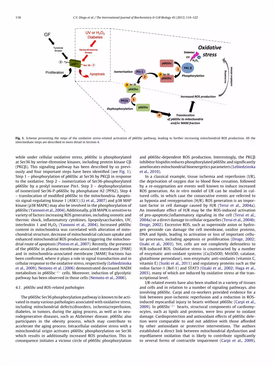
118 C.V. Diogo et al. / The International Journal of Biochemistry & Cell Biology 45 (2013) 114– 122
F p66Shi
wa(oStpo–skpvticcedoabcemp
4
vidrpamwc
ig. 1. Scheme presenting the steps of the oxidative stress-related activation of
ntermediate steps are described in more detail in Section 4.
hile under cellular oxidative stress, p66Shc is phosphorylatedt Ser36 by serine-threonine kinases, including protein kinase C�PKC�). This signaling pathway has been described by us previ-usly and four important steps have been identified (see Fig. 1).tep 1 – phosphorylation of p66Shc at Ser36 by PKC� in responseo the oxidative. Step 2 – isomerization of Ser36-phosphorylated66Shc by a prolyl isomerase Pin1. Step 3 – dephosphorylationf isomerized Ser36-P-p66Shc by phosphatase A2 (PPA2). Step 4
translocation of modified p66Shc to the mitochondria. Apopto-is signal-regulating kinase 1 (ASK1) (Li et al., 2007) and p38 MAPinase (p38 MAPK) may also be involved in the phosphorylation of66Shc (Yannoni et al., 2004). Additionally, p38 MAPK is sensitive toariety of factors increasing ROS generation, including osmotic andhermic shock, inflammatory cytokines, lipopolysaccharides, UV,nterleukin 1 and H2O2 (Yannoni et al., 2004). Increased p66Shcontent in mitochondria was correlated with alteration of mito-hondrial structure, decrease of mitochondrial calcium uptake andnhanced mitochondrial ROS production triggering the mitochon-rial route of apoptosis (Pinton et al., 2007). Recently, the presencef the p66Shc in plasma membrane-associated membrane (PAM)nd in mitochondria-associated membrane (MAM) fractions haseen confirmed, where it plays a role in signal transduction and inellular response to the oxidative stress, respectively (Lebiedzinskat al., 2009). Nemoto et al. (2006) demonstrated decreased NADHetabolism in p66Shc−/− cells. Moreover, induction of glycolytic
athway has been observed in those cells (Nemoto et al., 2006).
.1. p66Shc and ROS-related pathologies
The p66Shc Ser36 phosphorylation pathway is known to be acti-ated in many various pathologies associated with oxidative stress,ncluding mitochondrial defects/disorders, ischemia/reperfusion,iabetes, in tumors, during the aging process, as well as in neu-odegenerative diseases, such as Alzheimer disease. p66Shc alsoarticipates in the obesity process, which may contribute to
ccelerate the aging process. Intracellular oxidative stress with aitochondrial origin activates p66Shc phosphorylation on Ser36hich results in additionally increased ROS production. This inonsequence initiates a vicious circle of p66Shc phosphorylation
c pathway, leading to further increasing mitochondrial ROS production. All the
and p66Shc-dependent ROS production. Interestingly, the PKC�inhibitor hispidin reduces phosphorylated p66Shc and significantlyameliorates mitochondrial bioenergetics parameters (Lebiedzinskaet al., 2010).
In a classical example, tissue ischemia and reperfusion (I/R),the deprivation of oxygen due to blood flow cessation, followedby a re-oxygenation are events well known to induce increasedROS generation. An in vitro model of I/R can be studied in cul-tured cells, in which case the consecutive events are referred toas hypoxia and reoxygenation (H/R). ROS generation is an impor-tant factor in cell damage caused by H/R (Terui et al., 2004a).An immediate effect of H/R may be the ROS-induced activationof pro-apoptotic/inflammatory signaling in the cell (Terui et al.,2004a) or a direct damage to cellular organelles (Terui et al., 2004b;Droge, 2002). Excessive ROS, such as superoxide anion or hydro-gen peroxide can damage the cell membrane, oxidize proteins,DNA and lipids, leading to activation or loss of important cellu-lar processes, including apoptosis or proliferation (Droge, 2002;Ozaki et al., 2003). Yet, cells are not completely defenseless toH/R-induced ROS. Oxidative stress is counteracted by a numberof enzymatic anti-oxidant systems (Cu/ZnSOD, MnSOD, catalase,glutathione peroxidase), non-enzymatic anti-oxidants (vitamin C,vitamin E) (Suski et al., 2011) and regulatory proteins such as theredox factor-1 (Ref-1) and STAT3 (Ozaki et al., 2002; Haga et al.,2003), many of which are induced by oxidative stress at the tran-scriptional level.
I/R-related events have also been studied in a variety of tissuesand cells and in relation to a number of signaling pathways, alsoinvolving p66Shc. Carpi and co-workers provided evidence for alink between post-ischemic reperfusion and a reduction in ROS-induced myocardial injury in hearts without p66Shc (Carpi et al.,2009). In p66Shc−/− hearts, structural components of cardiomy-ocytes, such as lipids and proteins, were less prone to oxidantdamage. Cardioprotection and antioxidant effects of p66Shc dele-tion were comparable to and not additive with those afforded
by other antioxidant or protective interventions. The authorsestablished a direct link between mitochondrial dysfunction andmyofilament oxidation that is likely to contribute significantlyto several forms of contractile impairment (Carpi et al., 2009).
f Bioc
Ttkcpt
4
aRcwcseswrvdetcaitttkaicrpiieitemMbpdahpattgwoagpkoah2cS
C.V. Diogo et al. / The International Journal o
he beneficial effect of p66Shc ablation during renal proximalubule cells I/R was also demonstrated (Arany et al., 2010). Bynocking down p66shc or inhibiting its Ser36 phosphorylation orytochrome c binding site, the authors managed to attenuate ROSroduction and consequently prevent mitochondrial depolariza-ion as well as kidney injury (Arany et al., 2010).
.2. p66Shc and ROS in hyperglycemia
As it was previously mentioned, at least a part of cellular dam-ge resulting from hyperglycemic conditions is caused by enhancedOS production. On the other hand, enhanced ROS productionan be the result of the activation of intracellular signaling path-ays, e.g. phosphorylation of p66Shc at Serine36. If hyperglycemic
onditions induce oxidative stress, and p66Shc triggers apopto-is in response to oxidative stress, a link between these twovents/processes should exist. There are data about an increasedynthesis of p66Shc mRNA in mononuclear blood cells of patientsith type 2 DM, suggesting that increased level of p66Shc may
esult from prolonged high level of glucose in blood of these indi-iduals, which contributes to the plasma increase in the oxidativeamage marker, 8-isoprostanein these diabetic patients (Pagnint al., 2005). Another link between p66Shc and hyperglycemia ishe fact that p66Shc is essential for glucose uptake in skeletal mus-le. Glucose transporters are constitutively present or are activatednd relocalized to plasma membrane after hormone stimulation orn response to high energetic demands during hypoxia or oxida-ive stress conditions (Foley et al., 2011). It has been establishedhat p66Shc inhibits intracellular signaling cascades in response toyrosine receptor kinase activation. Inhibition of members of MAPinase family results in abnormal glucose transport due to affectedctin network. ERK1/2-associated pathway is constitutively activen myoblasts lacking p66Shc, resulting in the up-regulation of glu-ose transporters synthesis. In contrast, p66Shc overexpressionesults in decreased glucose transporters levels and glucose trans-ort is reduced by 50% (Natalicchio et al., 2009). These findings
mplicate p66Shc protein in the regulation of glucose transportnto cells, but the evidence for p66Shc involvement in pathogen-sis of hyperglycemia still needs to be strengthened. With thisn mind, the link between hyperglycemia-associated ROS produc-ion and activation of p66shc phosphorylation pathway has beenxplored in different diabetes models. In STZ-induced diabetes inouse models, a strong induction of p66Shc synthesis is observed.ore ONOO− is produced in aorta epithelium of wild type dia-
etic mice that in p66Shc−/− mice. This leads to an increase inrotein nitrosilation which is considered a marker for nitrosativeamage. Additionally, ablation of p66Shc prevents NO depletionnd keeps the proper vessels epithelium relaxation, which underyperglycemic condition is impaired. Although ablation of p66Shcrotein had no effect on the SOD1 and SOD2 levels in a STZ-inducednimal model, it significantly contributes to increased HO-1 con-ent and activity (Camici et al., 2007). Rota et al. hypothesizedhat the premature cardiac aging due to the loss of cardiac pro-enitor cells can be in part driven by p66Shc signaling, since itas prevented by its deletion. In cardiac progenitor cells with-
ut p66Shc, replication predominates while enhanced apoptosisnd necrosis is observed in diabetic wild type cells. Cardiac pro-enitor cells proliferate and differentiate into myocytes preservingroper heart function in diabetic p66Shc−/− animals, because MAPinases are not inhibited by this protein (Rota et al., 2006). Thesebservations led to the conclusion that down-regulation of p66Shcssociated signaling cascades might be a therapeutic target in
yperglycemia-induced heart failure (Messina and Giacomello,006). Independently, Malhotra et al. showed that cardiac mus-le damage due to hyperglycemia is connected with increaseder36 p66Shc phosphorylation (Malhotra et al., 2009). Thus thehemistry & Cell Biology 45 (2013) 114– 122 119
observation of reduced cardiac progenitor cells death in p66Shc−/−
model (Rota et al., 2006) can be explained by decreased p66Shcassociated ROS production. Besides p66Shc serine phosphoryla-tion, hyperglycemia induces also translocation of the protein tomitochondrial fractions and formation of p66Shc-cytochrome ccomplexes. Moreover, a 40% increase in apoptosis rate due to up-regulation of Bax, p53 and caspase 3 cleavage was also observed.In contrast, transfection of rat ventricular myocytes with Ser36-mutated p66Shc protein prevents high glucose-induced oxidativestress and damage. These data was confirmed by in vivo exper-iments on Akita diabetic mice suggesting that hyperglycemiainduced oxidative stress and damage is mediated by Ser36 p66Shcphosphorylation pathway (Malhotra et al., 2009). Hyperglycemiacontributes to advanced glycation end products (AGEs) whichcan trigger specific receptors activating pathways causing furtheroxidative damage by increasing oxidative stress in cells (RAGE)or receptors that activate prosurvival pathways (AGER1), withcell fate depending on the balance between them. Cai and col-leagues showed that some AGEs induce ROS production in cellwhich directly leads to an increase in Ser36 p66Shc phosphoryla-tion and enhanced oxidative stress. Interestingly, this effect causesFKHRL factor inhibition, thus hyperglycemia additionally attenu-ates antioxidant defense through a p66Shc pathway, since FKHRL1(FOXO3A) is a transcription factor responsible for SOD2 and cata-lase synthesis (Lam et al., 2006; Purdom and Chen, 2003; Pani et al.,2009). Overexpression of AGER1 stops ROS-induced p66Shc phos-phorylation (Cai et al., 2008). Several pieces of evidence appearedsuggesting a crosstalk between p66Shc and FKHRL1. Cells trans-fected with mutated Ser36 in p66Shc or with siRNA are resistant tohyperglycemia induced oxidative stress and to oxidant DNA dam-age (Chintapalli et al., 2007). In animal models, AGE treatmentinduces renal injury from which p66Shc KO mice are protected.In p66Shc WT animals increased oxidative stress involves elevatedlevel of NF�B and induction of NOX4 (NADP(P)H oxidase4) (Meniniet al., 2007), which again confirms a central role for p66Shc in avariety of signaling pathways that are altered in hyperglycemia.
5. Concluding remarks
The present review clearly confirms a series of clinically impor-tant observations, such as: (a) diabetes have a negative impactin the cardiovascular system, (b) mitochondria and oxidativestress are central players in the cardiac pathophysiology of hyper-glycemia and (c) p66Shc is a novel hub for several cell andmitochondrial signaling pathways that can be modulated by hyper-glycemia from an apparent normal function to a pro-oxidant,pro-apoptotic function. Considering the previous observations,therapeutic strategies involving mitochondrial-directed antioxi-dants including mitoQ, SkQ and others (Foley et al., 2011; Skulachevet al., 2010; Armstrong, 2008) can be designed. General antioxi-dants including vitamin E may have marginal effects only (Oliveiraet al., 2004). The p66Shc signaling pathway may also present agood target to prevent mitochondrial degeneration during hyper-glycemia. In this regard the use of hispidin, a PKC� inhibitor, maypresent a good opportunity. Future research aimed at develop-ing more selective inhibitors of p66Shc signaling of mitochondrialdegeneration is clearly needed.
It is also clear that more work is needed in animal models ofdiabetes in order to understand whether “negative” p66Shc sig-naling occurs mostly in the more affected tissues or is instead a
widespread phenomenon and if this is the case, what differencesexist in the entire activation network. The next step is to under-stand if pharmacological strategies aimed at controlling diabetesalter the role of p66Shc in the diabetic tissues and if this has a clear
1 f Bioc
ir
A
HJtATaN(daeSsTtto
R
A
A
A
A
A
A
B
B
B
B
C
C
C
C
C
C
C
20 C.V. Diogo et al. / The International Journal o
mpact in the quality of life of patients. If so, a novel avenue ofesearch can be pursued.
cknowledgments
This work was supported by the Polish Ministry of Science andigher Education under grant NN407 075 137 for JMS, AKW, ML,
D, MP and MRW, by the grant from the National Science Cen-re – decision number DEC-2011/01/M/NZ3/02128 for JMS, ML, JD,W and MRW and by the Portuguese Foundation for Science andechnology (FCT), research grants PTDC/SAU-TOX/110952/2009nd PTDC/SAU-TOX/117912/2010 (funded by COMPETE/FEDER andational Funds), as well as Ph.D. Fellowship SFRH/BD/48133/2008
for CD). JMS was also supported by PhD fellowship from the Foun-ation for Polish Science, EU, European Regional Development Fundnd Operational Programme ‘Innovative economy’. ML was recipi-nt of a fellowship from the Foundation for Polish Science (Programtart) and the L’Oreal fellowship (For Women in Science). P.P. wasupported by the Italian Association for Cancer Research (AIRC),elethon (GGP09128), local funds from the University of Ferrara,he Italian Ministry of Education, University and Research (COFIN),he Italian Cystic Fibrosis Research Foundation and Italian Ministryf Health.
eferences
bbott RD, Donahue RP, Kannel WB, Wilson PW. The impact of diabetes on sur-vival following myocardial infarction in men vs women. The Framingham Study.JAMA 1988;260:3456–60.
hmed N. Advanced glycation endproducts—role in pathology of diabetic compli-cations. Diabetes Research and Clinical Practice 2005;67:3–21.
lmdal T, Scharling H, Jensen JS, Vestergaard H. The independent effect of type 2diabetes mellitus on ischemic heart disease, stroke, and death: a population-based study of 13,000 men and women with 20 years of follow-up. Archives ofInternal Medicine 2004;164:1422–6.
nand DV, Lim E, Lahiri A, Bax JJ. The role of non-invasive imaging in therisk stratification of asymptomatic diabetic subjects. European Heart Journal2006;27:905–12.
rany I, Faisal A, Clark JS, Vera T, Baliga R, Nagamine Y. p66Shc-mediated mito-chondrial dysfunction in renal proximal tubule cells during oxidative injury.American Journal of Physiology Renal Physiology 2010;298:F1214–21.
rmstrong JS. Mitochondria-directed therapeutics. Antioxidants and Redox Sig-nalling 2008;10:575–8.
ell DS. Heart failure: the frequent, forgotten, and often fatal complication of dia-betes. Diabetes Care 2003;26:2433–41.
rownlee M. Biochemistry and molecular cell biology of diabetic complications.Nature 2001;414:813–20.
rownlee M, Cerami A. The biochemistry of the complications of diabetes mellitus.Annual Review of Biochemistry 1981;50:385–432.
uchanan J, Mazumder PK, Hu P, Chakrabarti G, Roberts MW, Yun UJ, et al. Reducedcardiac efficiency and altered substrate metabolism precedes the onset of hyper-glycemia and contractile dysfunction in two mouse models of insulin resistanceand obesity. Endocrinology 2005;146:5341–9.
ai W, He JC, Zhu L, Chen X, Striker GE, Vlassara H. AGE-receptor-1 coun-teracts cellular oxidant stress induced by AGEs via negative regulation ofp66shc-dependent FKHRL1 phosphorylation. American Journal of PhysiologyCell Physiology 2008;294:C145–52.
amici GG, Schiavoni M, Francia P, Bachschmid M, Martin-Padura I, Hers-berger M, et al. Genetic deletion of p66(Shc) adaptor protein preventshyperglycemia-induced endothelial dysfunction and oxidative stress. Proceed-ings of the National Academy of Sciences of the United States of America2007;104:5217–22.
arley AN, Severson DL. Fatty acid metabolism is enhanced in type 2 diabetic hearts.Biochimica et Biophysica Acta 2005;1734:112–26.
arpi A, Menabo R, Kaludercic N, Pelicci P, Di Lisa F, Giorgio M. The cardioprotectiveeffects elicited by p66(Shc) ablation demonstrate the crucial role of mitochon-drial ROS formation in ischemia/reperfusion injury. Biochimica et BiophysicaActa 2009;1787:774–80.
hintapalli J, Yang S, Opawumi D, Goyal SR, Shamsuddin N, Malhotra A, et al. Inhibi-tion of wild-type p66ShcA in mesangial cells prevents glycooxidant-dependentFOXO3a regulation and promotes the survival phenotype. American Journal ofPhysiology Renal Physiology 2007;292:F523–30.
ohen-Solal A, Beauvais F, Logeart D. Heart failure and diabetes mellitus: epidemi-
ology and management of an alarming association. Journal of Cardiac Failure2008;14:615–25.oughlan MT, Thorburn DR, Penfold SA, Laskowski A, Harcourt BE, Sourris KC, et al.RAGE-induced cytosolic ROS promote mitochondrial superoxide generation indiabetes. Journal of the American Society of Nephrology 2009;20:742–52.
hemistry & Cell Biology 45 (2013) 114– 122
Coutinho M, Gerstein HC, Wang Y, Yusuf S. The relationship between glucose andincident cardiovascular events. A metaregression analysis of published datafrom 20 studies of 95,783 individuals followed for 12.4 years. Diabetes Care1999;22:233–40.
Craven PA, Davidson CM, DeRubertis FR. Increase in diacylglycerol mass in iso-lated glomeruli by glucose from de novo synthesis of glycerolipids. Diabetes1990;39:667–74.
Davi G, Ciabattoni G, Consoli A, Mezzetti A, Falco A, Santarone S, et al. In vivo forma-tion of 8-iso-prostaglandin f2alpha and platelet activation in diabetes mellitus:effects of improved metabolic control and vitamin E supplementation. Circula-tion 1999;99:224–9.
Di Carli MF, Bianco-Batlles D, Landa ME, Kazmers A, Groehn H, Muzik O, et al. Effectsof autonomic neuropathy on coronary blood flow in patients with diabetes mel-litus. Circulation 1999;100:813–9.
Diamant M, Lamb HJ, Groeneveld Y, Endert EL, Smit JW, Bax JJ, et al. Diastolic dys-function is associated with altered myocardial metabolism in asymptomaticnormotensive patients with well-controlled type 2 diabetes mellitus. Journalof the American College of Cardiology 2003;42:328–35.
Droge W. Free radicals in the physiological control of cell function. PhysiologicalReviews 2002;82:47–95.
Du XL, Edelstein D, Rossetti L, Fantus IG, Goldberg H, Ziyadeh F, et al.Hyperglycemia-induced mitochondrial superoxide overproduction acti-vates the hexosamine pathway and induces plasminogen activatorinhibitor-1 expression by increasing Sp1 glycosylation. Proceedings ofthe National Academy of Sciences of the United States of America 2000;97:12222–6.
Duncan JG. Mitochondrial dysfunction in diabetic cardiomyopathy. Biochimica etBiophysica Acta 2011;1813:1351–9.
Feener EP, Xia P, Inoguchi T, Shiba T, Kunisaki M, King GL. Role of protein kinase C inglucose- and angiotensin II-induced plasminogen activator inhibitor expression.Contributions to Nephrology 1996;118:180–7.
Finck BN, Kelly DP. Peroxisome proliferator-activated receptor alpha (PPARalpha)signaling in the gene regulatory control of energy metabolism in the nor-mal and diseased heart. Journal of Molecular and Cellular Cardiology 2002;34:1249–57.
Flarsheim CE, Grupp IL, Matlib MA. Mitochondrial dysfunction accompaniesdiastolic dysfunction in diabetic rat heart. American Journal of Physiology1996;271:H192–202.
Foley K, Boguslavsky S, Klip A. Endocytosis, recycling, and regulated exocytosis ofglucose transporter 4. Biochemistry 2011;50:3048–61.
Folli F, Corradi D, Fanti P, Davalli A, Paez A, Giaccari A, et al. The role of oxidativestress in the pathogenesis of type 2 diabetes mellitus micro- and macrovascularcomplications: avenues for a mechanistic-based therapeutic approach. CurrentDiabetes Reviews 2011;7:313–24.
Ford ES, Zhao G, Li C. Pre-diabetes and the risk for cardiovascular disease: a sys-tematic review of the evidence. Journal of the American Medical Association2010;55:1310–7.
Fox CS, Coady S, Sorlie PD, Levy D, Meigs JB, D’Agostino Sr RB, et al. Trends incardiovascular complications of diabetes. JAMA 2004;292:2495–9.
Gabriely I, Yang XM, Cases JA, Ma XH, Rossetti L, Barzilai N. Hyperglycemia inducesPAI-1 gene expression in adipose tissue by activation of the hexosamine biosyn-thetic pathway. Atherosclerosis 2002;160:115–22.
Garcia Soriano F, Virag L, Jagtap P, Szabo E, Mabley JG, Liaudet L, et al. Diabeticendothelial dysfunction: the role of poly(ADP-ribose) polymerase activation.Nature Medicine 2001;7:108–13.
Gerstein HC, Pais P, Pogue J, Yusuf S. Relationship of glucose and insulin levels tothe risk of myocardial infarction: a case–control study. Journal of the AmericanMedical Association 1999;33:612–9.
Gerstein HC, Mann JF, Yi Q, Zinman B, Dinneen SF, Hoogwerf B, et al. Albuminuria andrisk of cardiovascular events, death, and heart failure in diabetic and nondiabeticindividuals. JAMA 2001;286:421–6.
Giacco F, Brownlee M. Oxidative stress and diabetic complications. CirculationResearch 2010;107:1058–70.
Goldberg HJ, Whiteside CI, Fantus IG. The hexosamine pathway regulates the plas-minogen activator inhibitor-1 gene promoter and Sp1 transcriptional activationthrough protein kinase C-beta I and -delta. Journal of Biological Chemistry2002;277:33833–41.
Granger CB, Califf RM, Young S, Candela R, Samaha J, Worley S, et al. Outcomeof patients with diabetes mellitus and acute myocardial infarction treatedwith thrombolytic agents. The Thrombolysis and Angioplasty in MyocardialInfarction (TAMI) Study Group. Journal of the American College of Cardiology1993;21:920–5.
Grundy SM. Metabolic syndrome scientific statement by the American Heart Asso-ciation and the National Heart, Lung, and Blood Institute. Arteriosclerosis,Thrombosis, and Vascular Biology 2005;25:2243–4.
Grundy SM, Benjamin IJ, Burke GL, Chait A, Eckel RH, Howard BV, et al.Diabetes and cardiovascular disease: a statement for healthcare pro-fessionals from the American Heart Association. Circulation 1999;100:1134–46.
Haffner SM, Lehto S, Ronnemaa T, Pyorala K, Laakso M. Mortality from coronaryheart disease in subjects with type 2 diabetes and in nondiabetic subjects with
and without prior myocardial infarction. New England Journal of Medicine1998;339:229–34.Haga S, Terui K, Zhang HQ, Enosawa S, Ogawa W, Inoue H, et al. Stat3 protects againstFas-induced liver injury by redox-dependent and -independent mechanisms.Journal of Clinical Investigation 2003;112:989–98.

f Bioc
H
H
I
I
J
K
K
K
K
K
K
K
K
K
L
L
L
L
L
L
L
LL
L
L
M
M
M
M
M
C.V. Diogo et al. / The International Journal o
oogeveen EK, Kostense PJ, Jakobs C, Dekker JM, Nijpels G, Heine RJ, et al. Hyper-homocysteinemia increases risk of death, especially in type 2 diabetes: 5-yearfollow-up of the Hoorn Study. Circulation 2000;101:1506–11.
unt JV, Bottoms MA, Mitchinson MJ. Oxidative alterations in the experimental gly-cation model of diabetes mellitus are due to protein-glucose adduct oxidation.Some fundamental differences in proposed mechanisms of glucose oxidationand oxidant production. Biochemical Journal 1993;291(Pt 2):529–35.
noguchi T, Battan R, Handler E, Sportsman JR, Heath W, King GL. Preferential eleva-tion of protein kinase C isoform beta II and diacylglycerol levels in the aorta andheart of diabetic rats: differential reversibility to glycemic control by islet celltransplantation. Proceedings of the National Academy of Sciences of the UnitedStates of America 1992;89:11059–63.
shii H, Jirousek MR, Koya D, Takagi C, Xia P, Clermont A, et al. Amelioration ofvascular dysfunctions in diabetic rats by an oral PKC beta inhibitor. Science1996;272:728–31.
ames LR, Tang D, Ingram A, Ly H, Thai K, Cai L, et al. Flux through the hexosaminepathway is a determinant of nuclear factor kappaB-dependent promoter acti-vation. Diabetes 2002;51:1146–56.
annel WB. Lipids, diabetes, and coronary heart disease: insights from the Framing-ham Study. American Heart Journal 1985;110:1100–7.
annel WB, McGee DL. Diabetes and cardiovascular risk factors: the Framinghamstudy. Circulation 1979;59:8–13.
elley DE, He J, Menshikova EV, Ritov VB. Dysfunction of mitochondria in humanskeletal muscle in type 2 diabetes. Diabetes 2002;51:2944–50.
eogh RJ, Dunlop ME, Larkins RG. Effect of inhibition of aldose reductase on glucoseflux, diacylglycerol formation, protein kinase C, and phospholipase A2 activa-tion. Metabolism: Clinical and Experimental 1997;46:41–7.
ing H, Aubert RE, Herman WH. Global burden of diabetes, 1995–2025: prevalence,numerical estimates, and projections. Diabetes Care 1998;21:1414–31.
oya D, King GL. Protein kinase C activation and the development of diabetic com-plications. Diabetes 1998;47:859–66.
rolewski AS, Kosinski EJ, Warram JH, Leland OS, Busick EJ, Asmal AC,et al. Magnitude and determinants of coronary artery disease in juvenile-onset, insulin-dependent diabetes mellitus. American Journal of Cardiology1987;59:750–5.
uusisto J, Mykkanen L, Pyorala K, Laakso M. NIDDM and its metabolic controlpredict coronary heart disease in elderly subjects. Diabetes 1994;43:960–7.
waan HC. Changes in blood coagulation, platelet function, and plasminogen-plasmin system in diabetes. Diabetes 1992;41(Suppl. 2):32–5.
am EW, Francis RE, Petkovic M. FOXO transcription factors: key regulators of cellfate. Biochemical Society Transactions 2006;34:722–6.
anger A, Freeman MR, Josse RG, Armstrong PW. Metaiodobenzylguanidine imag-ing in diabetes mellitus: assessment of cardiac sympathetic denervation and itsrelation to autonomic dysfunction and silent myocardial ischemia. Journal ofthe American College of Cardiology 1995;25:610–8.
ashin O, Romani A. Hyperglycemia does not alter state 3 respiration in cardiacmitochondria from type-I diabetic rats. Molecular and Cellular Biochemistry2004;267:31–7.
ebiedzinska M, Duszynski J, Rizzuto R, Pinton P, Wieckowski MR. Age-relatedchanges in levels of p66Shc and serine 36-phosphorylated p66Shc in organsand mouse tissues. Archives of Biochemistry and Biophysics 2009;486:73–80.
ebiedzinska M, Karkucinska-Wieckowska A, Giorgi C, Karczmarewicz E, PronickaE, Pinton P, et al. Oxidative stress-dependent p66Shc phosphorylation in skinfibroblasts of children with mitochondrial disorders. Biochimica et BiophysicaActa 2010;1797:952–60.
i L, Renier G. Activation of nicotinamide adenine dinucleotide phosphate (reducedform) oxidase by advanced glycation end products links oxidative stress toaltered retinal vascular endothelial growth factor expression. Metabolism: Clin-ical and Experimental 2006;55:1516–23.
i M, Chiou KR, Kass DA. Shear stress inhibition of H(2)O(2) induced p66(Shc)phosphorylation by ASK1-JNK inactivation in endothelium. Heart and Vessels2007;22:423–7.
iscovitch M, Cantley LC. Lipid second messengers. Cell 1994;77:329–34.opaschuk GD, Ussher JR, Folmes CD, Jaswal JS, Stanley WC. Myocardial fatty acid
metabolism in health and disease. Physiological Reviews 2010;90:207–58.umini-Oliveira J, Magalhaes J, Pereira CV, Moreira AC, Oliveira PJ, Ascensao
A. Endurance training reverts heart mitochondrial dysfunction, permeabilitytransition and apoptotic signaling in long-term severe hyperglycemia. Mito-chondrion 2011;11:54–63.
uscher TF, Creager MA, Beckman JA, Cosentino F. Diabetes and vascular disease:pathophysiology, clinical consequences, and medical therapy: part II. Circulation2003;108:1655–61.
alhotra A, Vashistha H, Yadav VS, Dube MG, Kalra SP, Abdellatif M, et al. Inhibitionof p66ShcA redox activity in cardiac muscle cells attenuates hyperglycemia-induced oxidative stress and apoptosis. American Journal of Physiology Heartand Circulatory Physiology 2009;296:H380–8.
argolis JR, Kannel WS, Feinleib M, Dawber TR, McNamara PM. Clinical featuresof unrecognized myocardial infarction—silent and symptomatic. Eighteen yearfollow-up: the Framingham study. American Journal of Cardiology 1973;32:1–7.
arin-Garcia J, Goldenthal MJ. Fatty acid metabolism in cardiac failure: biochemical,genetic and cellular analysis. Cardiovascular Research 2002;54:516–27.
azumder PK, O’Neill BT, Roberts MW, Buchanan J, Yun UJ, Cooksey RC, et al.Impaired cardiac efficiency and increased fatty acid oxidation in insulin-resistant ob/ob mouse hearts. Diabetes 2004;53:2366–74.
cClain DA, Crook ED. Hexosamines and insulin resistance. Diabetes1996;45:1003–9.
hemistry & Cell Biology 45 (2013) 114– 122 121
Menini S, Iacobini C, Ricci C, Oddi G, Pesce C, Pugliese F, et al. Ablation of thegene encoding p66Shc protects mice against AGE-induced glomerulopathy bypreventing oxidant-dependent tissue injury and further AGE accumulation. Dia-betologia 2007;50:1997–2007.
Messina E, Giacomello A. Diabetic cardiomyopathy: a cardiac stem cell diseaseinvolving p66Shc, an attractive novel molecular target for heart failure therapy.Circulation Research 2006;99:1–2.
Migliaccio E, Giorgio M, Mele S, Pelicci G, Reboldi P, Pandolfi PP, et al. The p66shcadaptor protein controls oxidative stress response and life span in mammals.Nature 1999;402:309–13.
Mootha VK, Lindgren CM, Eriksson KF, Subramanian A, Sihag S, Lehar J, et al.PGC-1alpha-responsive genes involved in oxidative phosphorylation are coor-dinately downregulated in human diabetes. Nature Genetics 2003;34:267–73.
Moreno PR, Murcia AM, Palacios IF, Leon MN, Bernardi VH, Fuster V, et al. Coro-nary composition and macrophage infiltration in atherectomy specimens frompatients with diabetes mellitus. Circulation 2000;102:2180–4.
Morino K, Petersen KF, Dufour S, Befroy D, Frattini J, Shatzkes N, et al. Reducedmitochondrial density and increased IRS-1 serine phosphorylation in muscle ofinsulin-resistant offspring of type 2 diabetic parents. Journal of Clinical Investi-gation 2005;115:3587–93.
Murthy VK, Shipp JC. Accumulation of myocardial triglycerides ketotic diabetes;evidence for increased biosynthesis. Diabetes 1977;26:222–9.
Natalicchio A, De Stefano F, Perrini S, Laviola L, Cignarelli A, Caccioppoli C, et al.Involvement of the p66Shc protein in glucose transport regulation in skele-tal muscle myoblasts. American Journal of Physiology Endocrinology andMetabolism 2009;296:E228–37.
Nemoto S, Combs CA, French S, Ahn BH, Fergusson MM, Balaban RS, et al. Themammalian longevity-associated gene product p66shc regulates mitochondrialmetabolism. Journal of Biological Chemistry 2006;281:10555–60.
Niakan E, Harati Y, Rolak LA, Comstock JP, Rokey R. Silent myocardial infarction anddiabetic cardiovascular autonomic neuropathy. Archives of Internal Medicine1986;146:2229–30.
Niedowicz DM, Daleke DL. The role of oxidative stress in diabetic complications. CellBiochemistry and Biophysics 2005;43:289–330.
Nishikawa T, Edelstein D, Brownlee M. The missing link: a single unifying mechanismfor diabetic complications. Kidney International Supplement 2000;77:S26–30.
Nishizuka Y. Intracellular signaling by hydrolysis of phospholipids and activation ofprotein kinase C. Science 1992;258:607–14.
Nishizuka Y. Protein kinase C and lipid signaling for sustained cellular responses.FASEB Journal 1995;9:484–96.
Norhammar A, Tenerz A, Nilsson G, Hamsten A, Efendic S, Ryden L, et al. Glucosemetabolism in patients with acute myocardial infarction and no previous diag-nosis of diabetes mellitus: a prospective study. Lancet 2002;359:2140–4.
Oliveira PJ, Rolo AP, Seica R, Palmeira CM, Santos MS, Moreno AJ. Decreased suscepti-bility of heart mitochondria from diabetic GK rats to mitochondrial permeabilitytransition induced by calcium phosphate. Bioscience Reports 2001;21:45–53.
Oliveira PJ, Seica R, Coxito PM, Rolo AP, Palmeira CM, Santos MS, et al.Enhanced permeability transition explains the reduced calcium uptake incardiac mitochondria from streptozotocin-induced diabetic rats. FEBS Letters2003;554:511–4.
Oliveira PJ, Seica R, Santos DL, Rolo AP, Sardao VA, Ferreira FM, et al. Vitamin E orcoenzyme Q10 administration is not fully advantageous for heart mitochondrialfunction in diabetic goto kakizaki rats. Mitochondrion 2004;3:337–45.
Ozaki M, Suzuki S, Irani K. Redox factor-1/APE suppresses oxidative stress by inhibit-ing the rac1 GTPase. FASEB Journal 2002;16:889–90.
Ozaki M, Haga S, Zhang HQ, Irani K, Suzuki S. Inhibition of hypoxia/reoxygenation-induced oxidative stress in HGF-stimulated antiapoptotic signaling: role of PI3-Kand Akt kinase upon rac1. Cell Death and Differentiation 2003;10:508–15.
Pagnin E, Fadini G, de Toni R, Tiengo A, Calo L, Avogaro A. Diabetes inducesp66shc gene expression in human peripheral blood mononuclear cells: rela-tionship to oxidative stress. Journal of Clinical Endocrinology and Metabolism2005;90:1130–6.
Palasciano G, Moschetta A, Palmieri VO, Grattagliano I, Iacobellis G, Portincasa P.Non-alcoholic fatty liver disease in the metabolic syndrome. Current Pharma-ceutical Design 2007;13:2193–8.
Pani G, Koch OR, Galeotti T. The p53-p66shc-manganese superoxide dismutase(MnSOD) network: a mitochondrial intrigue to generate reactive oxygen species.International Journal of Biochemistry and Cell Biology 2009;41:1002–5.
Patti ME, Butte AJ, Crunkhorn S, Cusi K, Berria R, Kashyap S, et al. Coordinated reduc-tion of genes of oxidative metabolism in humans with insulin resistance anddiabetes: Potential role of PGC1 and NRF1. Proceedings of the National Academyof Sciences of the United States of America 2003;100:8466–71.
Pellegrini M, Pacini S, Baldari CT. p66SHC: the apoptotic side of Shc proteins. Apo-ptosis 2005;10:13–8.
Petruzzelli M, Lo Sasso G, Portincasa P, Palasciano G, Moschetta A. Targeting the liverin the metabolic syndrome: evidence from animal models. Current Pharmaceu-tical Design 2007;13:2199–207.
Pieper GM, Riaz ul H. Activation of nuclear factor-kappaB in cultured endothelialcells by increased glucose concentration: prevention by calphostin C. Journal ofCardiovascular Pharmacology 1997;30:528–32.
Pinton P, Rimessi A, Marchi S, Orsini F, Migliaccio E, Giorgio M, et al. Protein kinase
C beta and prolyl isomerase 1 regulate mitochondrial effects of the life-spandeterminant p66Shc. Science 2007;315:659–63.Portilla D, Dai G, Peters JM, Gonzalez FJ, Crew MD, Proia AD. Etomoxir-inducedPPARalpha-modulated enzymes protect during acute renal failure. AmericanJournal of Physiology Renal Physiology 2000;278:F667–75.
1 f Bioc
P
Q
R
R
R
R
R
R
R
R
R
S
S
S
S
S
S
S
S
S
S
S
22 C.V. Diogo et al. / The International Journal o
urdom S, Chen QM. Linking oxidative stress and genetics of aging with p66Shcsignaling and forkhead transcription factors. Biogerontology 2003;4:181–91.
iao Q, Pyorala K, Pyorala M, Nissinen A, Lindstrom J, Tilvis R, et al. Two-hour glucoseis a better risk predictor for incident coronary heart disease and cardiovascularmortality than fasting glucose. European Heart Journal 2002;23:1267–75.
aha S, Robinson BH. Mitochondria, oxygen free radicals, disease and ageing. Trendsin Biochemical Sciences 2000;25:502–8.
anjadayalan K, Umachandran V, Ambepityia G, Kopelman PG, Mills PG, Timmis AD.Prolonged anginal perceptual threshold in diabetes: effects on exercise capac-ity and myocardial ischemia. Journal of the American College of Cardiology1990;16:1120–4.
eaven GM. Banting Lecture 1988. Role of insulin resistance in human disease.Nutrition 1997;13:65 [discussion 64, 66].
esnick HE, Howard BV. Diabetes and cardiovascular disease. Annual Review ofMedicine 2002;53:245–67.
eszko AE, Kasumov T, David F, Jobbins KA, Thomas KR, Hoppel CL, et al. Peroxisomalfatty acid oxidation is a substantial source of the acetyl moiety of malonyl-CoAin rat heart. Journal of Biological Chemistry 2004;279:19574–9.
itov VB, Menshikova EV, He J, Ferrell RE, Goodpaster BH, Kelley DE. Deficiencyof subsarcolemmal mitochondria in obesity and type 2 diabetes. Diabetes2005;54:8–14.
odrigues B, Cam MC, McNeill JH. Myocardial substrate metabolism: implica-tions for diabetic cardiomyopathy. Journal of Molecular and Cellular Cardiology1995;27:169–79.
olo AP, Palmeira CM. Diabetes and mitochondrial function: role of hyperglycemiaand oxidative stress. Toxicology and Applied Pharmacology 2006;212:167–78.
ota M, LeCapitaine N, Hosoda T, Boni A, De Angelis A, Padin-Iruegas ME, et al.Diabetes promotes cardiac stem cell aging and heart failure, which are preventedby deletion of the p66shc gene. Circulation Research 2006;99:42–52.
antos DL, Palmeira CM, Seica R, Dias J, Mesquita J, Moreno AJ, et al. Diabetes andmitochondrial oxidative stress: a study using heart mitochondria from the dia-betic Goto-Kakizaki rat. Molecular and Cellular Biochemistry 2003;246:163–70.
arwar N, Gao P, Seshasai SR, Gobin R, Kaptoge S, Di Angelantonio E, et al. Diabetesmellitus, fasting blood glucose concentration, and risk of vascular disease: a col-laborative meta-analysis of 102 prospective studies. Lancet 2010;375:2215–22.
chleicher E, Friess U. Oxidative stress, AGE, and atherosclerosis. Kidney Interna-tional Supplement 2007:S17–26.
cognamiglio R, Negut C, Ramondo A, Tiengo A, Avogaro A. Detection of coronaryartery disease in asymptomatic patients with type 2 diabetes mellitus. Journalof the American College of Cardiology 2006;47:65–71.
harma S, Adrogue JV, Golfman L, Uray I, Lemm J, Youker K, et al. Intramyocardiallipid accumulation in the failing human heart resembles the lipotoxic rat heart.FASEB Journal 2004;18:1692–700.
hen X, Zheng S, Thongboonkerd V, Xu M, Pierce Jr WM, Klein JB, et al. Cardiac mito-chondrial damage and biogenesis in a chronic model of type 1 diabetes. AmericanJournal of Physiology Endocrinology and Metabolism 2004;287:E896–905.
hiba T, Inoguchi T, Sportsman JR, Heath WF, Bursell S, King GL. Correlation of dia-cylglycerol level and protein kinase C activity in rat retina to retinal circulation.American Journal of Physiology 1993;265:E783–93.
inger DE, Nathan DM, Anderson KM, Wilson PW, Evans JC. Association of HbA1cwith prevalent cardiovascular disease in the original cohort of the FraminghamHeart Study. Diabetes 1992;41:202–8.
kulachev VP, Antonenko YN, Cherepanov DA, Chernyak BV, Izyumov DS, KhailovaLS, et al. Prevention of cardiolipin oxidation and fatty acid cycling as two antiox-idant mechanisms of cationic derivatives of plastoquinone (SkQs). Biochimicaet Biophysica Acta 2010;1797:878–89.
ong Y, Du Y, Prabhu SD, Epstein PN. Diabetic cardiomyopathy in OVE26 mice shows
mitochondrial ROS production and divergence between in vivo and in vitrocontractility. The Review of Diabetic Studies 2007;4:159–68.tamler J, Vaccaro O, Neaton JD, Wentworth D. Diabetes, other risk factors, and12-yr cardiovascular mortality for men screened in the Multiple Risk FactorIntervention Trial. Diabetes Care 1993;16:434–44.
hemistry & Cell Biology 45 (2013) 114– 122
Stanley WC, Lopaschuk GD, McCormack JG. Regulation of energy substratemetabolism in the diabetic heart. Cardiovascular Research 1997;34:25–33.
Suski J, Lebiedzinska M, Machado NG, Oliveira PJ, Pinton P, Duszynski J, et al. Mito-chondrial tolerance to drugs and toxic agents in ageing and disease. CurrentDrug Targets 2011;12:827–49.
Szczepaniak LS, Victor RG, Orci L, Unger RH. Forgotten but not gone: the rediscoveryof fatty heart, the most common unrecognized disease in America. CirculationResearch 2007;101:759–67.
Terui K, Haga S, Enosawa S, Ohnuma N, Ozaki M. Hypoxia/re-oxygenation-induced,redox-dependent activation of STAT1 (signal transducer and activator of tran-scription 1) confers resistance to apoptotic cell death via hsp70 induction.Biochemical Journal 2004a;380:203–9.
Terui K, Enosawa S, Haga S, Zhang HQ, Kuroda H, Kouchi K, et al. Stat3 confersresistance against hypoxia/reoxygenation-induced oxidative injury in hepa-tocytes through upregulation of Mn-SOD. Journal of Hepatology 2004b;41:957–65.
Third Report of the National Cholesterol Education Program (NCEP) Expert Panel onDetection, Evaluation, and Treatment of High Blood Cholesterol in Adults (AdultTreatment Panel III) final report. Circulation 2002;106:3143–3421.
Turko IV, Li L, Aulak KS, Stuehr DJ, Chang JY, Murad F. Protein tyrosine nitrationin the mitochondria from diabetic mouse heart. Implications to dysfunctionalmitochondria in diabetes. Journal of Biological Chemistry 2003;278:33972–7.
Turrens JF. Mitochondrial formation of reactive oxygen species. The Journal of Phys-iology 2003;552:335–44.
Ulrich P, Cerami A. Protein glycation, diabetes, and aging. Recent Progress in Hor-mone Research 2001;56:1–21.
Vaccaro O, Eberly LE, Neaton JD, Yang L, Riccardi G, Stamler J. Impact of diabetes andprevious myocardial infarction on long-term survival: 25-year mortality follow-up of primary screenees of the Multiple Risk Factor Intervention Trial. Archivesof Internal Medicine 2004;164:1438–43.
Wachtell K, Ibsen H, Olsen MH, Borch-Johnsen K, Lindholm LH, Mogensen CE, et al.Albuminuria and cardiovascular risk in hypertensive patients with left ven-tricular hypertrophy: the LIFE study. Annals of Internal Medicine 2003;139:901–6.
Wallace DC. Mitochondrial genetics: a paradigm for aging and degenerative dis-eases? Science 1992;256:628–32.
Williamson JR, Chang K, Frangos M, Hasan KS, Ido Y, Kawamura T, et al. Hyper-glycemic pseudohypoxia and diabetic complications. Diabetes 1993;42:801–13.
Wolff SP, Dean RT. Glucose autoxidation and protein modification. The poten-tial role of ‘autoxidative glycosylation’ in diabetes. Biochemical Journal1987;245:243–50.
Xia P, Kramer RM, King GL. Identification of the mechanism for the inhibition ofNa+,K(+)-adenosine triphosphatase by hyperglycemia involving activation ofprotein kinase C and cytosolic phospholipase A2. Journal of Clinical Investigation1995;96:733–40.
Yannoni YM, Gaestel M, Lin LL. P66(ShcA) interacts with MAPKAP kinase 2 andregulates its activity. FEBS Letters 2004;564:205–11.
Yerneni KK, Bai W, Khan BV, Medford RM, Natarajan R. Hyperglycemia-induced acti-vation of nuclear transcription factor kappaB in vascular smooth muscle cells.Diabetes 1999;48:855–64.
Yokoyama I, Momomura S, Ohtake T, Yonekura K, Nishikawa J, Sasaki Y, et al.Reduced myocardial flow reserve in non-insulin-dependent diabetes mellitus.Journal of the American College of Cardiology 1997;30:1472–7.
Yusuf S, Hawken S, Ounpuu S, Dans T, Avezum A, Lanas F, et al. Effect of potentiallymodifiable risk factors associated with myocardial infarction in 52 countries(the INTERHEART study): case–control study. Lancet 2004;364:937–52.
Zaccagnini G, Martelli F, Fasanaro P, Magenta A, Gaetano C, Di Carlo A,et al. p66ShcA modulates tissue response to hindlimb ischemia. Circulation2004;109:2917–23.
Zimmet P, Alberti KG, Shaw J. Global and societal implications of the diabetes epi-demic. Nature 2001;414:782–7.