canal cristae growth and fiber extension to the outer hair cells of the mouse ear require prox1...
TRANSCRIPT
Canal Cristae Growth and Fiber Extension to the OuterHair Cells of the Mouse Ear Require Prox1 ActivityBernd Fritzsch1*, Miriam Dillard2, Alfonso Lavado2, Natasha L. Harvey2¤, Israt Jahan1
1 Department of Biology, University of Iowa, Iowa City, Iowa, United States of America, 2 Department of Genetics and Tumor Cell Biology, St. Jude Children’s Research
Hospital, Memphis, Tennessee, United States of America
Abstract
Background: The homeobox gene Prox1 is required for lens, retina, pancreas, liver, and lymphatic vasculature developmentand is expressed in inner ear supporting cells and neurons.
Methodology/Principal Findings: We have investigated the role of Prox1 in the developing mouse ear taking advantage ofavailable standard and conditional Prox1 mutant mouse strains using Tg(Pax2-Cre) and Tg(Nes-Cre). A severe reduction in thesize of the canal cristae but not of other vestibular organs or the cochlea was identified in the E18.5 Prox1Flox/Flox; Tg(Pax2-Cre) mutant ear. In these mutant embryos, hair cell differentiated; however, their distribution pattern was slightlydisorganized in the cochlea where the growth of type II nerve fibers to outer hair cells along Prox1 expressing supportingcells was severely disrupted. In the case of Nestin-Cre, we found that newborn Prox1Flox/Flox; Tg(Nestin-Cre) exhibit only adisorganized innervation of outer hair cells despite apparently normal cellular differentiation of the organ of Corti,suggesting a cell-autonomous function of Prox1 in neurons.
Conclusions/Significance: These results identify a dual role of Prox1 during inner ear development; growth of the canalcristae and fiber guidance of Type II fibers along supporting cells in the cochlea.
Citation: Fritzsch B, Dillard M, Lavado A, Harvey NL, Jahan I (2010) Canal Cristae Growth and Fiber Extension to the Outer Hair Cells of the Mouse Ear RequireProx1 Activity. PLoS ONE 5(2): e9377. doi:10.1371/journal.pone.0009377
Editor: Karl-Wilhelm Koch, University of Oldenburg, Germany
Received July 31, 2009; Accepted February 4, 2010; Published February 23, 2010
Copyright: � 2010 Fritzsch et al. This is an open-access article distributed under the terms of the Creative Commons Attribution License, which permitsunrestricted use, distribution, and reproduction in any medium, provided the original author and source are credited.
Funding: This work was supported by NIH grant R01-DC005590 (to BF). The funders had no role in study design, data collection and analysis, decision to publish,or preparation of the manuscript. The authors acknowledge the use of the confocal microscope facility of the NCCB, supported by EPSCoR EPS-0346476 (CFD47.076) and the Carver Center for Imaging, supported by the Roy J. Carver Foundation.
Competing Interests: The authors have declared that no competing interests exist.
* E-mail: [email protected]
¤ Current address: Division of Hematology, The Hanson Institute, Adelaide, South Australia, Australia
Introduction
The mammalian inner ear is composed of the cochlea that mediates
the auditory function, and the vestibule that mediates the gravitational
and angular acceleration sensing. In mammals, six epithelial sensory
patches found in the cochlear and vestibular regions of the inner ear
mediate auditory and vestibular functions: the organ of Corti is the
sensory patch found in the cochlea and three cristae and two maculae
are the sensory patches of the vestibule. Each of these sensory patches
includes mechanosensory hair cells and non-sensory supporting cells.
Both of these cell types originate from epithelial progenitors that
become specified as prosensory precursors. According to their position
in the ear, these prosensory patches will give rise to the definite
vestibular or cochlear sensory patches. Cells in those sensory patches
ultimately assume final fates as either hair cells (e.g., inner and outer
hair cells in the cochlea) or a variable number of non-sensory
supporting cells (distributed between hair cells). While the molecular
machinery governing the development of hair cells has received much
attention [1,2] far less is known about the molecular basis of cell fate
decision in supporting cells [3,4]. In the mammalian cochlea, at least
five unique types of supporting cell can be identified: Pillar cells,
Deiter’s cells, Hensen cells, Claudius cells and inner sulcus cells [3,5].
We and others have proposed that the development of the
vertebrate ear sensory epithelium shares certain similarities with the
development of the sensilla in insects [6,7,8]. In Drosphila, the
homeobox gene prospero plays important roles in cell fate decision
during glia, sensory sensilla, and eye development [9,10,11,12,13,14].
Prox1, the vertebrate counterpart of prospero [15] is expressed in
several murine cell types where its function is essential for proper
development and differentiation [15,16,17,18,19,20,21,22,23,24].
Interestingly, in addition to the developing retina [15,20] and
spinal cord [25], Prox1 expression was also identified in another
sensory organ; i.e., the developing ear of zebrafish [26], chicken
[27], and mice [28,29]. By taking advantage of available standard
and conditional Prox1 mouse mutant strains [30,31], we have now
determined that Prox1 is an important new player during the
development of the mammalian vestibular and auditory systems.
We demonstrate that in the canal cristae, lack of Prox1 function
affects the overall growth of these vestibular sensory epithelia. In
contrast, in the cochlea, absence of Prox1 disrupts stereotyped
cellular organization and fiber guidance of Type II neurons
apparently in a cell autonomous fashion.
Methods
MiceProx1+/LacZ, Prox1flox/flox, Atoh1, and Tg(Pax2-Cre) and Tg(Nes-Cre)
mice have been previously reported [30,31,32,33,34,35,36]. The
PLoS ONE | www.plosone.org 1 February 2010 | Volume 5 | Issue 2 | e9377
developmental stage of mouse embryos was determined by
considering noon of the day the vaginal plug was detected in the
pregnant dam as E0.5. All of the mouse experiments were
approved by the Creighton University, University of Iowa, and St.
Jude Children’s Research Hospital Animal Care and Use
Committees.
Detection of b-Galactosidase ActivityTo detect b-gal activity, ears were dissected and X-gal staining
was performed as described previously [37]. Whenever required,
we enhanced the X-gal reaction using 2-photon photoactivation
on whole mounts and sections [38]. In addition, we ran some ears
without fixation to avoid any quenching of the b-galactosidase
activity. Stained ears were mounted flat or alternatively, they were
embedded in epoxy resin, sectioned (20 mm) and imaged using a
compound lightmicroscope (Nikon Eclipse 800) and captured
using a Coolsnap camera and Metamorph software. Some ears
were processed for transmission electron microscopy and viewed
in a Hitachi TEM as previously described [39]. Unfortunately, use
of either Tg(Pax2-Cre) or Tg(Nes-Cre) leads to early postnatal
lethality; therefore, we were not able to analyze the conditional
mutant ear beyond P1.
Prox1 in situ hybridization. Whole mount in situ hybridization
was performed using a riboprobe as previously described [15].
ImmunohistochemistryPrimary antibodies were rabbit anti–b-gal (ICN), rabbit (Covance
Research Products) anti–mouse Prox1 (Promega), rat anti–mouse b-
tubulin (Sigma), Hoechst nuclear stain (Sigma), Myo VII (gift of T.
Hasson, San Diego), Sox2 and BDNF (Invitrogen). Secondary
antibodies were Alexa 488, 543, and 634–conjugated donkey anti-
rabbit (Molecular Probes), Cy3-conjugated donkey anti–guinea pig
(Jackson ImmunoResearch Laboratories), and Cy3-conjugated
donkey anti-rat (Jackson ImmunoResearch Laboratories) were used
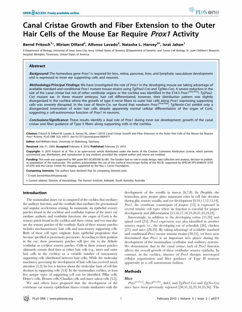
Figure 1. The early onset of Prox1 expression is revealed by b-galactosidase expression and in situ hybridization. Whole mount b-galactosidase histochemcial reaction using X-Gal was performed in Prox1 heterozygous and nullizygous embryos. A. Starting at E11.0, a progressiveupregulation of Prox1 is seen in the anterior (AC) and posterior (PC) canal cristae. B. By E13.5, expression is also detected in the horizontal canal crista(HC), the striolar region of the utricle (U), the canals and the endolymphatic duct (ED); expression in the saccule is barely detected (S). In the cochlea,upregulation of b-galactosidase expression is detected in the apex and decreases toward the base. Arrows indicate expression in anterior andposterior canal with their expression. C. Expression of b-galactosidase is identical in heterozygous and nullizygous mice with the exception that thesignal is stronger in nullizygous mice. Faint b-galactosidase expression is also detected in the delaminating spiral ganglion neurons (SPG; C and insertin B,C). D. In situ hybridization shows at E14.5 expression in the canal cristae and the cochlea, but indicates a more prominent upregulation in thebase at this stage. Only spiral ganglion sensory neurons are faintly positive for Prox1 in situ (SPG in D). E,F At postnatal stages, Prox1 expressionremains in the canal cristae as revealed by in situ hybridization for Prox1 mRNA or X-Gal reaction, but does not show the extensive expression in thenon-sensory parts of the canals as in earlier stages (insert in F). Bar, 100 mm.doi:10.1371/journal.pone.0009377.g001
Prox1 in Mouse Inner Ear
PLoS ONE | www.plosone.org 2 February 2010 | Volume 5 | Issue 2 | e9377
predominantly on whole mounted microdissected sensory epithelia
[40]. Sections and whole mounts were imaged using a confocal
system (Zeiss LSM 510 or Leica SP5). Images were assembled into
plates using CorelDraw software. Size of sensory epithelia was
measured using ImagePro software on fully calibrated confocal
images. PTI lipophilic tracers (NV Maroon) were used for afferent
and efferent fibers [41]. Briefly, dyes were inserted into central
targets or as small local injections and the fibers were filled with the
diffusible dye, epithelia were microdissected and viewed with a
confocal system (Zeiss LSM 510 or Leica SP5).
QuantificationIn order to evaluate the qualitative effects of lack of Prox1
function on the growth of the vestibular epithelia we measured the
length of the anterior canal crista and the utricle using the
calibration setting of the Zeiss LSM 510 system in six flat mounted
vestibular organs of Prox1flox/flox; Tg(Pax2-Cre) (E18.5 mutant) and
Pax2-Cre (E18.5 control). Differences were evaluated for signifi-
cance suing a T-test. We also counted the number of hair cells
using Myo VII immunocytochemistry to identify hair cells and
Hoechst nuclear staining to label the nuclei in three of these
vestibular areas of control and mutant mice. Counting was done
on flat mounts of three anterior canal cristae by grabbing a
Figure 3. Prox1 inactivation reduces the size of the anteriorcrista. As measured at E14.5, the length of the anterior cristae (AC) ofProx1 mutant embryos is 20% reduced when compare vs. that of wild-type littermates. The size reduction is 30% when compared with thesize of E18.5 Prox1 flox/flox; Tg(Pax2-Cre) mutant embryos. No significantchanges in the length of the utricle were observed. Asterisks indicate alevel of significance (p,0.05; t-test).doi:10.1371/journal.pone.0009377.g003
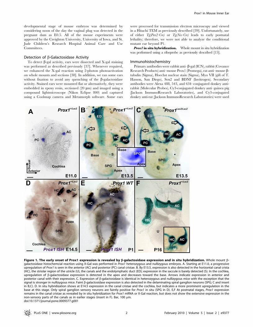
Figure 2. Effects of Prox1 loss-of function in the vestibular epithelia. A. X-gal staining of E14.5 Prox1 heterozygous embryos reveals b-galactosidase activity in the anterior (AC) and horizontal (HC) parts of the canal cristae. B. Although morphologically normal, a reduction in the size ofthe crista epithelia is detected of Prox1-null littermates (white bar in the AC); gravistatic sensors such as utricle (U) show only transient Prox1expression and no apparent reduction in size. C, E. Hair cells are revealed using antibodies against Myo VII in a normal E18.5 Prox1flox/flox conditionalembryo. Note absence of imunoreactivity in the cruciate eminence (CE) of the anterior canal crista. E9. As shown by 2-photon activation, at this laterstage, Prox1 expression is high in supporting cells, but is also found in hair cells of the canal cristae as well as outside the sensory epithelium. Dottedline in B indicate the plane of sections through the horizontal canal crista, white arrows align lateral walls of the whole mount with the section. E,F.Despite the overlap of some Prox1 expression with hair cells in the canal cristae there is no morphologically obvious defects in hair cell differentiationother than reduced intensity of Myo VII staining are observed in Prox1 flox/flox; Tg(Pax2-Cre) as compared to Prox1flox/flox littermates. However thereduction in size of the anterior canal crista (AC) is becoming more obvious at this late stage (C–F). CE-Cruciate eminence. Bar, 100 mm.doi:10.1371/journal.pone.0009377.g002
Prox1 in Mouse Inner Ear
PLoS ONE | www.plosone.org 3 February 2010 | Volume 5 | Issue 2 | e9377
confocal stack at 6 mm interval (slightly wider than the average
nuclear diameter to avoid double counting). Shrinking or other
counting artifacts should be equal but this procedure will slightly
underestimate the total number of hair cells [42,43]. A non-
parametric rank correlation test was used to assess statistical
significance of cell counts.
Results
Prox1 Expression in the Developing Inner EarPrevious work using immunohistochemistry reported that Prox1
expression in the inner ear starts around E11.0 in three vestibular
sensory patches and around E11.5 is highly expressed in the canal
cristae and saccule of the sensory epithelia and weakly in the
utricle [28]. Expression in the cochlea starts at around E14.5 in
Pillar cells, Deiter’s cells and outer hair cells, and also extends
weakly to nonsensory parts of the ear [29].
In order to precisely compare the profile of Prox1 expression
with the well known onset of hair cell proliferation [40,44] and
differentiation [45,46] we took advantage of an available Prox1
heterozygous strain in which the b-galactosidase reporter gene was
inserted in frame into the Prox1 genomic locus [31]. As shown in
Fig. 1A, at E11.0 Prox1 expression was restricted to two X-gal
positive patches corresponding to the anterior and posterior canal
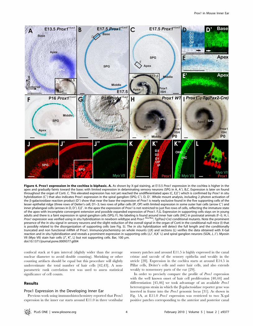
Figure 4. Prox1 expression in the cochlea is biphasic. A. As shown by X-gal staining, at E13.5 Prox1 expression in the cochlea is higher in theapex and gradually faints toward the base; with limited expression in delaminating sensory neurons (SPG in A, A9). B,C. Expression is later on foundthroughout the organ of Corti. C. This elevated expression has not yet reached the undifferentiated apex (C, E,E9) which is confirmed by Prox1 in situhybridization (C9) that also indicates Prox1 expression in the spiral ganglion (SPG; C9). D, D9. Whole mount analysis, including 2 photon activation ofthe b-galactosidase reaction product (D9) show that near the base the expression of Prox1 is nearly exclusive found in the five supporting cells of thelesser epithelial ridge (three rows of Deiter’s cell, D1–3; two rows of pillar cells (IP, OP) with limited expression in some outer hair cells (arrow C9) andinner phalangeal cells (arrows in D, D9). E,E9. In the apex the expression of Prox1 is not restricted to just five rows of cells, reflecting the immature stateof the apex with incomplete convergent extension and possible expanded expression of Prox1. F,G. Expression in supporting cells stays on in youngadults and there is a faint expression in spiral ganglion cells (SPG; F). No labeling is found around inner hair cells (IHC) in postnatal animals (F–I). H, I.Prox1 expression was verified using in situ hybridization in newborn wildtype and Prox1 flox/flox; Tg(Pax2-Cre) conditional mutants. Note the prominentpresence of the in situ signal in sensory neurons and the slight reduction of the overall signal in the organ of Corti in the conditional null mice (I) thatis possibly related to the disorganization of supporting cells (see Fig. 5). The in situ hybridization will detect the full length and the conditionallytruncated and non functional mRNA of Prox1. Immunocytochemistry on whole mounts (J,K) and sections (L) verifies the data obtained with X-Galreaction and in situ hybiridization and reveals a prominent expression in supporting cells (J,J9, K,K9 L) and spiral ganglion neurons (SGN, J, J9). MyosinVII (Myo VII) stain hair cells (J0, K0, L) but not supporting cells. Bar, 100 mm.doi:10.1371/journal.pone.0009377.g004
Prox1 in Mouse Inner Ear
PLoS ONE | www.plosone.org 4 February 2010 | Volume 5 | Issue 2 | e9377
cristae. Two days later, an additional third patch of expression was
seen in the region corresponding to the horizontal crista (Fig. 1B).
Around E13.5 Prox1 expression also starts to be detected in what
appears to be the striola region of the utricle and is barely detected
in the saccule (Fig. 1B). It is only at around this stage that Prox1
expression starts to be detected in the cochlea where Prox1
upregulation begins broadly in the apex and expands toward the
base (Fig. 1B). Prox1 expression is not restricted to sensory epithelia
but is also found in the forming canals and the endolymphatic duct
(Fig. 1B). In addition, Prox1 expression starts in the spiral ganglia
around that time (Fig. 1C, insert). Prox1 in situ hybridization
detects signal in the canal cristae but in the organ of Corti of the
cochlear duct only at E14.5 (Fig. 1D). As indicated by X-gal
staining (Fig. 1F) and in situ hybridization (Fig. 1E), Prox1
expression remains in the newborn canal cristae but is lost in the
non-sensory part of the canal (insert in Fig. 1F).
Canal Cristae Are Smaller in Prox1-Null EmbryosNext, and in order to identify possible functional roles of Prox1
during the development of the ear, we characterized the inner ear
of E14.5 Prox1-null embryos [31]. It was previously reported that
Prox1-null embryos die at around E14.5 [31]. In agreement with
the lack of Prox1 expression in developing sensory neurons at early
developmental stages, no obvious phenotypic alterations were
identified in the Prox1-null ears prior to E14.5 (Fig. 1B,C). This
data indicated that Prox1 activity is not required for sensory
neuron differentiation at these early stages.
As indicated above, high levels of Prox1 expression are detected
in the developing canal cristae (Fig. 1). In agreement with this
expression and as revealed by X-gal and Myo VII stainings [an
early marker of hair cell differentiation; [47]], the size of the
anterior canal cristae (AC) was clearly reduced in E14.5 Prox1-null
embryos (Fig. 2A–D, Fig. 3). The posterior canal cristae (PC) was
similarly affected (data not shown) and the horizontal canal cristae
(HC) was not as affected (Fig. 2A–D). In addition to the high level
of expression in the canal cristae, X-gal staining of E14.5 Prox1
heterozygous and nullizygous embryos confirmed that Prox1
expression was only transient and weak in the utricle (Fig. 2A,B)
and almost not detectable in the saccule (Fig. 1C). In situ
hybridization verified that a weak but detectable signal persisted in
the utricle at least until P1 (Fig. 1F) as previously described [29].
We determined that on average (N = 6), the size of the anterior
canal cristae in Prox1-null embryos was 20% smaller (p,0.05; t-
test) than in their heterozygous littermates (Fig. 3) (no differences
in size were found between wild-type and Prox1 heterozygous
littermates; data not shown). We also counted the number of hair
cells and found that the anterior canal cristae of Prox1-null
embryos had only about 605 (+265) hair cells compared to the
control littermate that had about 913 (+278) hair cells (p,0.05).
To confirm and expand this observation indicating that removal
of Prox1 activity affects the size of the vestibular sensory epithelia,
we took advantage of a previously generated Prox1 conditional
knock-out strain [30] to remove Prox1 activity from the inner ear in
a time and tissue specific manner. To this end, Tg(Pax2-Cre)
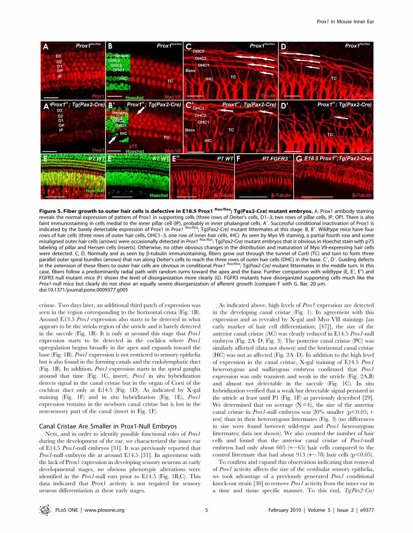
Figure 5. Fiber growth to outer hair cells is defective in E18.5 Prox1 flox/flox; Tg(Pax2-Cre) mutant embryos. A. Prox1 antibody stainingreveals the normal expression of pattern of Prox1 in supporting cells (three rows of Deiter’s cells, D1–3; two rows of pillar cells, IP, OP). There is alsofaint immunostaining in cells medial to the inner pillar cell (IP), probably in inner phalangeal cells. A9. Successful conditional inactivation of Prox1 isindicated by the barely detectable expression of Prox1 in Prox1 flox/flox; Tg(Pax2-Cre) mutant littermates at this stage. B, B9. Wildtype mice have fourrows of hair cells (three rows of outer hair cells, OHC1–3, one row of inner hair cells, IHC). As seen by Myo VII staining, a partial fourth row and somemisaligned outer hair cells (arrows) were occasionally detected in Prox1 flox/flox; Tg(Pax2-Cre) mutant embryos that is obvious in Hoechst stain with p75labeling of pillar and Hensen cells (inserts). Otherwise, no other obvious changes in the distribution and maturation of Myo VII-expressing hair cellswere detected. C, D. Normally and as seen by b-tubulin immunostaining, fibers grow out through the tunnel of Corti (TC) and turn to form threeparallel outer spiral bundles (arrows) that run along Deiter’s cells to reach the three rows of outer hair cells (OHC) in the base. C9, D9. Guiding defectsin the extension of these fibers to outer hair cells are obvious in conditional Prox1 flox/flox; Tg(Pax2-Cre) mutant littermates in the middle turn. In thiscase, fibers follow a predominantly radial path with random turns toward the apex and the base. Further comparison with wildtype (E, E9, E0) andFGFR3 null mutant mice (F) shows the level of disorganization more clearly (G). FGFR3 mutants have disorganized supporting cells much like theProx1-null mice but clearly do not show an equally severe disorganization of afferent growth (compare F with G. Bar, 20 mm.doi:10.1371/journal.pone.0009377.g005
Prox1 in Mouse Inner Ear
PLoS ONE | www.plosone.org 5 February 2010 | Volume 5 | Issue 2 | e9377
transgenic mice [33] were used to delete Prox1 from E9.0 onward
in all cells of the ear, including all hair cells and sensory neurons
[48]. Using this approach we also expected to overcome the early
embryonic lethality of standard Prox1-null embryos [31]. Analysis
of Prox1flox/flox;Tg(Pax2-Cre) conditional mutant embryos at E18.5
identified phenotypic alterations similar to those described in the
E14.5 Prox1-null embryos; e.g., the size of the anterior cristae was
significantly reduced (30% N = 6; p,0.05; T-test) (Fig. 2D, Fig. 3).
Despite this size reduction, the overall shape and morphology of
the cristae, and the formation of the non-sensory cruciate
eminence (CE) were not affected in these mutant embryos
(Fig. 2D, F). As indicated by Myo VII staining, no obvious gross
morphological alterations were detected in the development and
distribution of the vestibular hair cells of the canal cristae at these
later stages (Fig. 2D, F). No obvious alterations in the distribution
and morphology of supporting cells (indicated by Hoechst stained
nuclei), or in the size of the utricle were identified in these
conditional mutant embryos (Figs. 2,3). In E18.5 Prox1 heterozy-
gous animals, expression as revealed with 2 photon photoactiva-
tion of the b-galactosidase reaction product [38], is found
throughout all supporting cells of the canal cristae. In agreement
with a recent report [29], at this stage Prox1 expression was also
detected in some hair cells and non-sensory cells adjacent to the
canal cristae (Fig. 2E9).
In summary, these initial results revealed that removal of Prox1
function from the developing ear resulted in a significant reduction
in the size of the canal cristae.
Lack of Prox1 Function Results in Hair Cell Misalignmentand Disrupted Type II Spiral Ganglion Cell Guidance
Previous work has shown that cell cycle exit of hair cells in the
canal cristae starts around E11.5 [44]. Accordingly, Prox1
expression is detected prior and during cell cycle exit of hair cells
and supporting cells of the canal cristae (Fig. 1). In contrast, in the
cochlea Prox1 expression started to be detected in the cells of the
apex at around E13.5; although, it was faintly expressed in cells
near the base at this stage (Fig. 4A) and clearly is upregulated only
after hair cells have exited the cell cycle [40].
Multiple rows of hair cells and supporting cells form initially as a
short aggregate, but undergo convergent-extension movement to
eventually form three rows of outer and one row of inner hair cells
[49]. At around this stage of convergent extension, hair cells have
already exited the cell cycle which progresses from the apex to the
base of the cochlea between E11.5–E14.5 [40,44,45]. X-gal
staining of Prox1+/LacZ embryos and Prox1 in situ hybridization at
different developmental stages revealed that in the cochlea, Prox1
expression progressed initially from the apex to the base (Fig. 1B,C;
Fig. 4A); a result suggesting that its expression is in cells that have
already exited the cell cycle [45]. As shown in Fig. 4B–D, by
E17.5, Prox1 expression is prominent throughout the cochlea and
near the base is almost exclusively detected in the five supporting
cells of the lesser epithelial ridge (the three rows of Deiter’s cells
and the two rows of pillar cells); only limited expression was seen in
some outer hair cells and inner phalangeal cells (arrows in Fig. 4D,
D9). This limited expression in inner phalangeal cells seen in the
X-gal stained and photoactivated organ of Corti (Fig. 4C9, D9), is
also observed when using Prox1 antibodies (Fig. 5A). At this stage,
Prox1 expression in the apex is fainter and not organized into the
five rows of supporting cells (Fig. 4E, E9; [29]. The prominent
expression in supporting cells remained during postnatal stages, at
least until P16 as shown by X-gal staining of Prox1+/LacZ (Fig. 4F,G;
[29]. In later stages, a faint Prox1 signal was also detected in
sensory neurons (Fig. 4F). This signal was more prominent using in
situ hybridization (Fig. 4H,I). We verified the expression of Prox1
as revealed by X-gal staining using in situ hybridization (inserts in
Fig. 4A, C; Fig. 4H,I0 and immunocytochemistry. For unknown
reasons, X-gal staining of Prox1+/LacZ was easily lost after fixation
in sensory neurons and could be demonstrated only in unfixed ears
(Fig. 1D and insert). We also verified the supporting cell and
neuronal expression that was so obvious with in situ hybridization
starting at E15.5 (Fig. 4A, insert; Fig. 4C, insert) with
immunocytochemistry (Fig. 4J). Combined, all three techniques
show a profound upregulation of Prox1 in supporting cells and
sensory neurons (with the caveat of suppression of X-gal staining of
Prox1+/LacZ in sensory neurons following fixation).
In order to determine whether Prox1 expression in supporting
cells (Fig. 4C,D, D9, J,K,L9) is an indication that its functional
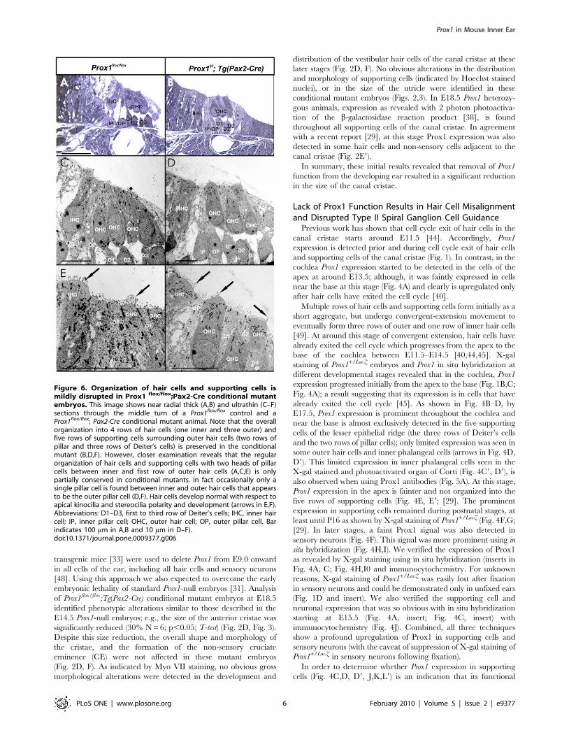
Figure 6. Organization of hair cells and supporting cells ismildly disrupted in Prox1 flox/flox;Pax2-Cre conditional mutantembryos. This image shows near radial thick (A,B) and ultrathin (C–F)sections through the middle turn of a Prox1flox/flox control and aProx1flox/flox; Pax2-Cre conditional mutant animal. Note that the overallorganization into 4 rows of hair cells (one inner and three outer) andfive rows of supporting cells surrounding outer hair cells (two rows ofpillar and three rows of Deiter’s cells) is preserved in the conditionalmutant (B,D,F). However, closer examination reveals that the regularorganization of hair cells and supporting cells with two heads of pillarcells between inner and first row of outer hair cells (A,C,E) is onlypartially conserved in conditional mutants. In fact occasionally only asingle pillar cell is found between inner and outer hair cells that appearsto be the outer pillar cell (D,F). Hair cells develop normal with respect toapical kinocilia and stereocilia polarity and development (arrows in E,F).Abbreviations: D1–D3, first to third row of Deiter’s cells; IHC, inner haircell; IP, inner pillar cell; OHC, outer hair cell; OP, outer pillar cell. Barindicates 100 mm in A,B and 10 mm in D–F).doi:10.1371/journal.pone.0009377.g006
Prox1 in Mouse Inner Ear
PLoS ONE | www.plosone.org 6 February 2010 | Volume 5 | Issue 2 | e9377
activity is required to control any developmental aspect of this cell
type, we analyzed the cochlea of E18.5 Prox1f/f;Tg(Pax2-Cre)
mutant embryos. Using this approach, Prox1 expression was
extensively removed from the developing cochlea (Fig. 5A9).
Although as discussed above (Fig. 2D, F), no obvious alterations in
hair cell differentiation were observed, hair cells patterning was
found to be occasionally disrupted. At this stage and as shown by
Myo VII staining (Fig. 5B), wild-type hair cells exhibit the typical
one row of inner hair cells and three parallel rows of outer hair
cells. In the mutant littermates, inner and particularly outer hair
cells appeared disorganized, misaligned, and containing extra rows
near the apex (Fig. 5B9arrows). These results indicated that lack of
Prox1 function did not affect hair cell differentiation (hair cell
differentiation markers Myo VII and BDNF were normally
expressed in the mutant hair cells; Figs. 5B, B9, 6); however, hair
cell patterning was slightly defective. Light and electron micro-
scopic radial sections confirmed the near normal development of
hair cells and supporting cells but also some degree of
disorganization of both cell types (Fig. 6).
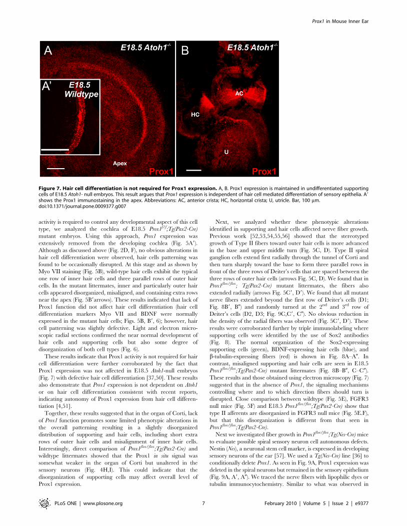
These results indicate that Prox1 activity is not required for hair
cell differentiation were further corroborated by the fact that
Prox1 expression was not affected in E18.5 Atoh1-null embryos
(Fig. 7) with defective hair cell differentiation [37,50]. These results
also demonstrate that Prox1 expression is not dependent on Atoh1
or on hair cell differentiation consistent with recent reports,
indicating autonomy of Prox1 expression from hair cell differen-
tiation [4,51].
Together, these results suggested that in the organ of Corti, lack
of Prox1 function promotes some limited phenotypic alterations in
the overall patterning resulting in a slightly disorganized
distribution of supporting and hair cells, including short extra
rows of outer hair cells and misalignment of inner hair cells.
Interestingly, direct comparison of Prox1flox/flox;Tg(Pax2-Cre) and
wildtype littermates showed that the Prox1 in situ signal was
somewhat weaker in the organ of Corti but unaltered in the
sensory neurons (Fig. 4H,I). This could indicate that the
disorganization of supporting cells may affect overall level of
Prox1 expression.
Next, we analyzed whether these phenotypic alterations
identified in supporting and hair cells affected nerve fiber growth.
Previous work [52,53,54,55,56] showed that the stereotyped
growth of Type II fibers toward outer hair cells is more advanced
in the base and upper middle turn (Fig. 5C, D). Type II spiral
ganglion cells extend first radially through the tunnel of Corti and
then turn sharply toward the base to form three parallel rows in
front of the three rows of Deiter’s cells that are spaced between the
three rows of outer hair cells (arrows Fig. 5C, D). We found that in
Prox1flox/flox; Tg(Pax2-Cre) mutant littermates, the fibers also
extended radially (arrows Fig. 5C9, D9). We found that all mutant
nerve fibers extended beyond the first row of Deiter’s cells (D1;
Fig. 8B9, B0) and randomly turned at the 2nd and 3rd row of
Deiter’s cells (D2, D3; Fig. 9C,C9, C0). No obvious reduction in
the density of the radial fibers was observed (Fig. 5C9, D9). These
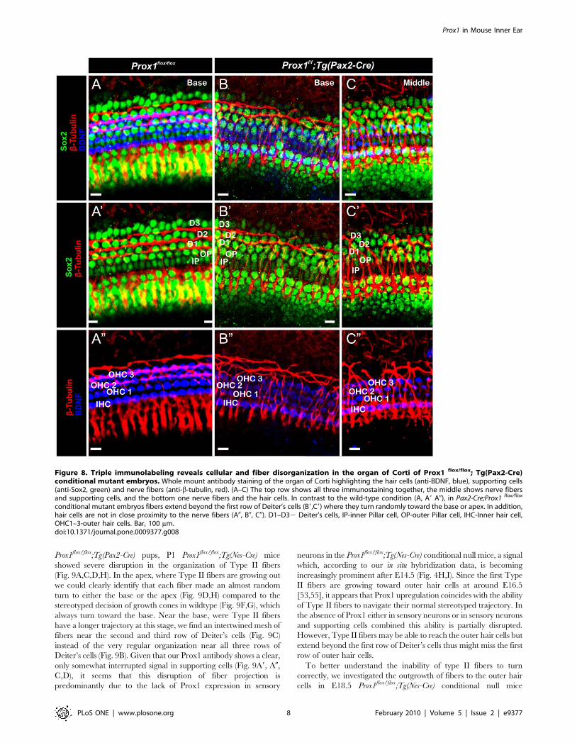
results were corroborated further by triple immunolabeling where
supporting cells were identified by the use of Sox2 antibodies
(Fig. 8). The normal organization of the Sox2-expressing
supporting cells (green), BDNF-expressing hair cells (blue), and
b-tubulin-expressing fibers (red) is shown in Fig. 8A–A0. In
contrast, misaligned supporting and hair cells are seen in E18.5
Prox1flox/flox;Tg(Pax2-Cre) mutant littermates (Fig. 8B–B0, C–C0).
These results and those obtained using electron microscopy (Fig. 7)
suggested that in the absence of Prox1, the signaling mechanisms
controlling where and to which direction fibers should turn is
disrupted. Close comparison between wildtype (Fig. 5E), FGFR3
null mice (Fig. 5F) and E18.5 Prox1flox/flox;Tg(Pax2-Cre) show that
type II afferents are disorganized in FGFR3 null mice (Fig. 5E.F),
but that this disorganization is different from that seen in
Prox1flox/flox;Tg(Pax2-Cre).
Next we investigated fiber growth in Prox1flox/flox;Tg(Nes-Cre) mice
to evaluate possible spiral sensory neuron cell autonomous defects.
Nestin (Nes), a neuronal stem cell marker, is expressed in developing
sensory neurons of the ear [57]. We used a Tg(Nes-Cre) line [36] to
conditionally delete Prox1. As seen in Fig. 9A, Prox1 expression was
deleted in the spiral neurons but remained in the sensory epithelium
(Fig. 9A, A9, A0). We traced the nerve fibers with lipophilic dyes or
tubulin immunocytochemistry. Similar to what was observed in
Figure 7. Hair cell differentiation is not required for Prox1 expression. A, B. Prox1 expression is maintained in undifferentiated supportingcells of E18.5 Atoh1- null embryos. This result argues that Prox1 expression is independent of hair cell mediated differentiation of sensory epithelia. A9shows the Prox1 immunostaining in the apex. Abbreviations: AC, anterior crista; HC, horizontal crista; U, utricle. Bar, 100 mm.doi:10.1371/journal.pone.0009377.g007
Prox1 in Mouse Inner Ear
PLoS ONE | www.plosone.org 7 February 2010 | Volume 5 | Issue 2 | e9377
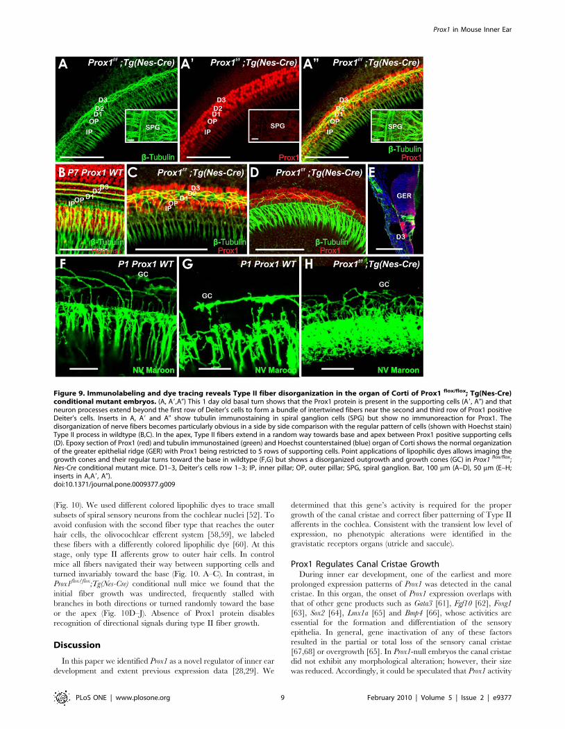
Prox1flox/flox;Tg(Pax2-Cre) pups, P1 Prox1flox/flox;Tg(Nes-Cre) mice
showed severe disruption in the organization of Type II fibers
(Fig. 9A,C,D,H). In the apex, where Type II fibers are growing out
we could clearly identify that each fiber made an almost random
turn to either the base or the apex (Fig. 9D,H) compared to the
stereotyped decision of growth cones in wildtype (Fig. 9F,G), which
always turn toward the base. Near the base, were Type II fibers
have a longer trajectory at this stage, we find an intertwined mesh of
fibers near the second and third row of Deiter’s cells (Fig. 9C)
instead of the very regular organization near all three rows of
Deiter’s cells (Fig. 9B). Given that our Prox1 antibody shows a clear,
only somewhat interrupted signal in supporting cells (Fig. 9A9, A0,
C,D), it seems that this disruption of fiber projection is
predominantly due to the lack of Prox1 expression in sensory
neurons in the Prox1flox/flox;Tg(Nes-Cre) conditional null mice, a signal
which, according to our in situ hybridization data, is becoming
increasingly prominent after E14.5 (Fig. 4H,I). Since the first Type
II fibers are growing toward outer hair cells at around E16.5
[53,55], it appears that Prox1 upregulation coincides with the ability
of Type II fibers to navigate their normal stereotyped trajectory. In
the absence of Prox1 either in sensory neurons or in sensory neurons
and supporting cells combined this ability is partially disrupted.
However, Type II fibers may be able to reach the outer hair cells but
extend beyond the first row of Deiter’s cells thus might miss the first
row of outer hair cells.
To better understand the inability of type II fibers to turn
correctly, we investigated the outgrowth of fibers to the outer hair
cells in E18.5 Prox1flox/flox;Tg(Nes-Cre) conditional null mice
Figure 8. Triple immunolabeling reveals cellular and fiber disorganization in the organ of Corti of Prox1 flox/flox; Tg(Pax2-Cre)conditional mutant embryos. Whole mount antibody staining of the organ of Corti highlighting the hair cells (anti-BDNF, blue), supporting cells(anti-Sox2, green) and nerve fibers (anti-b-tubulin, red). (A–C) The top row shows all three immunostaining together, the middle shows nerve fibersand supporting cells, and the bottom one nerve fibers and the hair cells. In contrast to the wild-type condition (A, A9 A0), in Pax2-Cre;Prox1 flox/flox
conditional mutant embryos fibers extend beyond the first row of Deiter’s cells (B9,C9) where they turn randomly toward the base or apex. In addition,hair cells are not in close proximity to the nerve fibers (A0, B0, C0). D1–D32 Deiter’s cells, IP-inner Pillar cell, OP-outer Pillar cell, IHC-Inner hair cell,OHC1–3-outer hair cells. Bar, 100 mm.doi:10.1371/journal.pone.0009377.g008
Prox1 in Mouse Inner Ear
PLoS ONE | www.plosone.org 8 February 2010 | Volume 5 | Issue 2 | e9377
(Fig. 10). We used different colored lipophilic dyes to trace small
subsets of spiral sensory neurons from the cochlear nuclei [52]. To
avoid confusion with the second fiber type that reaches the outer
hair cells, the olivocochlear efferent system [58,59], we labeled
these fibers with a differently colored lipophilic dye [60]. At this
stage, only type II afferents grow to outer hair cells. In control
mice all fibers navigated their way between supporting cells and
turned invariably toward the base (Fig. 10. A–C). In contrast, in
Prox1flox/flox;Tg(Nes-Cre) conditional null mice we found that the
initial fiber growth was undirected, frequently stalled with
branches in both directions or turned randomly toward the base
or the apex (Fig. 10D–J). Absence of Prox1 protein disables
recognition of directional signals during type II fiber growth.
Discussion
In this paper we identified Prox1 as a novel regulator of inner ear
development and extent previous expression data [28,29]. We
determined that this gene’s activity is required for the proper
growth of the canal cristae and correct fiber patterning of Type II
afferents in the cochlea. Consistent with the transient low level of
expression, no phenotypic alterations were identified in the
gravistatic receptors organs (utricle and saccule).
Prox1 Regulates Canal Cristae GrowthDuring inner ear development, one of the earliest and more
prolonged expression patterns of Prox1 was detected in the canal
cristae. In this organ, the onset of Prox1 expression overlaps with
that of other gene products such as Gata3 [61], Fgf10 [62], Foxg1
[63], Sox2 [64], Lmx1a [65] and Bmp4 [66], whose activities are
essential for the formation and differentiation of the sensory
epithelia. In general, gene inactivation of any of these factors
resulted in the partial or total loss of the sensory canal cristae
[67,68] or overgrowth [65]. In Prox1-null embryos the canal cristae
did not exhibit any morphological alteration; however, their size
was reduced. Accordingly, it could be speculated that Prox1 activity
Figure 9. Immunolabeling and dye tracing reveals Type II fiber disorganization in the organ of Corti of Prox1 flox/flox; Tg(Nes-Cre)conditional mutant embryos. (A, A9,A0) This 1 day old basal turn shows that the Prox1 protein is present in the supporting cells (A9, A0) and thatneuron processes extend beyond the first row of Deiter’s cells to form a bundle of intertwined fibers near the second and third row of Prox1 positiveDeiter’s cells. Inserts in A, A9 and A0 show tubulin immunostaining in spiral ganglion cells (SPG) but show no immunoreaction for Prox1. Thedisorganization of nerve fibers becomes particularly obvious in a side by side comparison with the regular pattern of cells (shown with Hoechst stain)Type II process in wildtype (B,C). In the apex, Type II fibers extend in a random way towards base and apex between Prox1 positive supporting cells(D). Epoxy section of Prox1 (red) and tubulin immunostained (green) and Hoechst counterstained (blue) organ of Corti shows the normal organizationof the greater epithelial ridge (GER) with Prox1 being restricted to 5 rows of supporting cells. Point applications of lipophilic dyes allows imaging thegrowth cones and their regular turns toward the base in wildtype (F,G) but shows a disorganized outgrowth and growth cones (GC) in Prox1 flox/flox;Nes-Cre conditional mutant mice. D1–3, Deiter’s cells row 1–3; IP, inner pillar; OP, outer pillar; SPG, spiral ganglion. Bar, 100 mm (A–D), 50 mm (E–H;inserts in A,A9, A0).doi:10.1371/journal.pone.0009377.g009
Prox1 in Mouse Inner Ear
PLoS ONE | www.plosone.org 9 February 2010 | Volume 5 | Issue 2 | e9377
is necessary to maintain and expand the pool of neurosensory
progenitor cells. Atoh1 is essential for hair cell differentiation [50]
and Atoh1-null mice fail to differentiate hair cells and supporting
cells [37]. Therefore, our finding that Prox1 expression remained
normal in Atoh1-null ears, and that Prox1-null hair cells expressed
typical hair cell markers eliminates the possibility that Prox1 was
required for hair cell differentiation at the level of neurosensory
progenitors. This does not rule out that misexpression of Prox1 in
hair cells can result in their degeneration, as was recently shown
for cochlea but not for vestibular hair cells [29].
Prox1 Regulates Fiber Guidance of Type II Spiral Neuronsin a Cell Autonomous Way
Similar to what has been reported for the cell cycle kinase
inhibitor p27 [45,69], the neurotrophin Bdnf [70,71] and the
growth factor Fgf10 [62], Prox1 expression in the cochlea starts to
be detected almost a day after hair cell precursors exited the cell
cycle [40,45]. While Prox1 is not expressed in hair cell progenitor
cells, it is expressed transiently in differentiating hair cells [29].
However, its continued expression in organ of Corti cells of Atoh1-
null mice [4,51], who have only hair cell precursors that fail to
differentiate [37], indicates that at least the expression in
supporting cells is not regulated by Atoh1 or other genes
specifically expressed in differentiated hair cells (Fig. 7). Given
that Prox1 expression persists at least until P26 in supporting cells
[29], it is possible that this gene remains expressed after at least
neonatal hair cell loss and its promoter could be used to drive
molecular expression toward reconstitution of the a functional
organ of Corti.
As previously reported [28], later during embryogenesis Prox1
expression is detected in the five supporting cells of the lesser
epithelial ridge (Fig. 4). In these cells, lack of Prox1 function lead to
subtle phenotypic alterations; e.g., defective alignment of hair cells
and supporting cells (Fig. 5) However, major pathfinding defects
Figure 10. Dye tracing reveals Type II fiber outgrowth problems in the organ of Corti of Prox1 flox/flox; Tg(Nes-Cre) conditionalmutant embryos. NV Maroon (green) was inserted into the cochlear nucleus and NV Orange (red) was inserted into the olivocochlear bundle tolabel a small population of afferents (green) and all efferents (red). Efferents show a similarly organized intraganglionic spiral bundles in wildtype (A–C)and Prox1 flox/flox; Nes-Cre conditional mutant mice (D–J) and grow toether with afferents in radial fiber bundles (RF) to the organ of Corti. Note that atthis stage only occasional efferents extent to outer hair cells. In contrast, type II afferents grow to the second or third row of outer hair cells (OHC) wherethey invariably turn toward the base (B,C). At this stage, none of the multiple type II afferents of Prox1 flox/flox; Nes-Cre conditional mutant mice showthis coordinated growth pattern. Instead, fibers grow randomly toward the base or apex but mostly seem to stall with multiple branches extendingtoward the base and the apex (F,G). IGSB, intraganglionic spiral bundle; OHC, outer hair cells; RF, radial fibers; SPG, spiral ganglion. Bar, 100 mm (A–D),50 mm (E–H; inserts in A,A9, A0).doi:10.1371/journal.pone.0009377.g010
Prox1 in Mouse Inner Ear
PLoS ONE | www.plosone.org 10 February 2010 | Volume 5 | Issue 2 | e9377
were identified in Type II spiral ganglion fibers. In this case, the
turning of these fibers toward the base [52,55] was severely
disrupted (Fig. 5,8,9,10). We found that in conditional null
mutants fibers abnormally extended toward the second and third
rows where they turned randomly instead of turning toward the
base in front of each of the three rows of Deiter’s cells. Radial fiber
growth beyond the inner pillar cells was not affected. It is worth
mentioning that pathfinding defects have been identified in the
CNS of Prospero mutant flies [72].
While Prox1 is the first gene that plays a cell autonomous role in
Type II pathfinding, at the moment it is not known how Prox1
affects fiber pathfinding of these neurons. It is known that Fgf8 and
Fgf10 mediated activation of Fgfr1, 2b and 3 signaling participates
in the differentiation of supporting cells of the lesser epithelial
ridge [3,62,73,74,75,76], and Fgfr3 -null mice also exhibit short
extra rows of outer hair cells [3,77] with some minor fiber
disorganization that is clearly distinct from the Prox1 effects
(Fig. 5F,G), but where exactly Prox1 fits into these interactions
remains to be determined.
Acknowledgments
We would like to thank Dr. G. Oliver for generously providing technical
expertise, mouse lines and suggestions throughout this work. We thank Dr.
A. Grove and T. Ohyama for the Pax2-Cre mice, Dr. H. Zoghbi for the
Atoh1 mutant mice, Dr. Betz for the Nes-Cre strain, Dr. C Puligilla for the
FGFR3 null ears, and Jennifer Kersigo for expert technical assistance.
Author Contributions
Conceived and designed the experiments: BF. Performed the experiments:
BF MD AL. Analyzed the data: BF NH. Contributed reagents/materials/
analysis tools: BF MD AL IJ. Wrote the paper: BF NH. Added in situ
hybridization and immunocytochemical work for the final submission: IJ.
References
1. Fritzsch B, Beisel KW, Hansen LA (2006) The molecular basis of neurosensorycell formation in ear development: a blueprint for hair cell and sensory neuron
regeneration? Bioessays 28: 1181–1193.
2. Kelley MW (2006) Regulation of cell fate in the sensory epithelia of the innerear. Nat Rev Neurosci 7: 837–849.
3. Puligilla C, Feng F, Ishikawa K, Bertuzzi S, Dabdoub A, et al. (2007) Disruption
of fibroblast growth factor receptor 3 signaling results in defects in cellulardifferentiation, neuronal patterning, and hearing impairment. Dev Dyn 236:
1905–1917.
4. Dabdoub A, Puligilla C, Jones JM, Fritzsch B, Cheah KS, et al. (2008) Sox2
signaling in prosensory domain specification and subsequent hair celldifferentiation in the developing cochlea. Proc Natl Acad Sci U S A 105:
18396–18401.
5. Kelley MW (2006) Hair cell development: Commitment through differentiation.Brain Res.
6. Fritzsch B, Beisel KW, Bermingham NA (2000) Developmental evolutionary
biology of the vertebrate ear: conserving mechanoelectric transduction anddevelopmental pathways in diverging morphologies. Neuroreport 11: R35–44.
7. Adam J, Myat A, Le Roux I, Eddison M, Henrique D, et al. (1998) Cell fate
choices and the expression of Notch, Delta and Serrate homologues in the chick
inner ear: parallels with Drosophila sense-organ development. Development125: 4645–4654.
8. Caldwell JC, Eberl DF (2002) Towards a molecular understanding of Drosophila
hearing. J Neurobiol 53: 172–189.
9. Jackson Behan K, Fair J, Singh S, Bogwitz M, Perry T, et al. (2005) Alternativesplicing removes an Ets interaction domain from Lozenge during Drosophila eye
development. Dev Genes Evol 215: 423–435.
10. Domingos PM, Brown S, Barrio R, Ratnakumar K, Frankfort BJ, et al. (2004)
Regulation of R7 and R8 differentiation by the spalt genes. Dev Biol 273:121–133.
11. Reddy GV, Rodrigues V (1999) A glial cell arises from an additional division
within the mechanosensory lineage during development of the microchaete onthe Drosophila notum. Development 126: 4617–4622.
12. Sen A, Reddy GV, Rodrigues V (2003) Combinatorial expression of Prospero,
Seven-up, and Elav identifies progenitor cell types during sense-organdifferentiation in the Drosophila antenna. Dev Biol 254: 79–92.
13. Cook T, Pichaud F, Sonneville R, Papatsenko D, Desplan C (2003) Distinction
between color photoreceptor cell fates is controlled by Prospero in Drosophila.
Dev Cell 4: 853–864.
14. Hayashi T, Saigo K (2001) Diversification of cell types in the Drosophila eye bydifferential expression of prepattern genes. Mech Dev 108: 13–27.
15. Oliver G, Sosa-Pineda B, Geisendorf S, Spana EP, Doe CQ, et al. (1993) Prox 1,
a prospero-related homeobox gene expressed during mouse development. MechDev 44: 3–16.
16. Risebro CA, Searles RG, Melville AA, Ehler E, Jina N, et al. (2009) Prox1
maintains muscle structure and growth in the developing heart. Development136: 495–505.
17. Wang J, Kilic G, Aydin M, Burke Z, Oliver G, et al. (2005) Prox1 activity
controls pancreas morphogenesis and participates in the production of
‘‘secondary transition’’ pancreatic endocrine cells. Dev Biol 286: 182–194.
18. Wigle JT, Chowdhury K, Gruss P, Oliver G (1999) Prox1 function is crucial formouse lens-fibre elongation. Nat Genet 21: 318–322.
19. Wigle JT, Harvey N, Detmar M, Lagutina I, Grosveld G, et al. (2002) An
essential role for Prox1 in the induction of the lymphatic endothelial cellphenotype. Embo J 21: 1505–1513.
20. Dyer MA, Livesey FJ, Cepko CL, Oliver G (2003) Prox1 function controls
progenitor cell proliferation and horizontal cell genesis in the mammalian retina.Nat Genet 34: 53–58.
21. Lavado A, Oliver G (2007) Prox1 expression patterns in the developing and
adult murine brain. Dev Dyn 236: 518–524.
22. Burke Z, Oliver G (2002) Prox1 is an early specific marker for the developing
liver and pancreas in the mammalian foregut endoderm. Mech Dev 118:
147–155.
23. Johnson NC, Dillard ME, Baluk P, McDonald DM, Harvey NL, et al. (2008)
Lymphatic endothelial cell identity is reversible and its maintenance requires
Prox1 activity. Genes Dev 22: 3282–3291.
24. Petrova TV, Nykanen A, Norrmen C, Ivanov KI, Andersson LC, et al. (2008)
Transcription factor PROX1 induces colon cancer progression by promoting
the transition from benign to highly dysplastic phenotype. Cancer Cell 13:
407–419.
25. Misra K, Gui H, Matise MP (2008) Prox1 regulates a transitory state for
interneuron neurogenesis in the spinal cord. Dev Dyn 237: 393–402.
26. Glasgow E, Tomarev SI (1998) Restricted expression of the homeobox gene
prox 1 in developing zebrafish. Mech Dev 76: 175–178.
27. Stone JS, Shang JL, Tomarev S (2003) Expression of Prox1 defines regions of the
avian otocyst that give rise to sensory or neural cells. J Comp Neurol 460:
487–502.
28. Bermingham-McDonogh O, Oesterle EC, Stone JS, Hume CR, Huynh HM, et
al. (2006) Expression of Prox1 during mouse cochlear development. J Comp
Neurol 496: 172–186.
29. Kirjavainen A, Sulg M, Heyd F, Alitalo K, Yla-Herttuala S, et al. (2008) Prox1
interacts with Atoh1 and Gfi1, and regulates cellular differentiation in the inner
ear sensory epithelia. Dev Biol 322: 33–45.
30. Harvey NL, Srinivasan RS, Dillard ME, Johnson NC, Witte MH, et al. (2005)
Lymphatic vascular defects promoted by Prox1 haploinsufficiency cause adult-
onset obesity. Nat Genet 37: 1072–1081.
31. Wigle JT, Oliver G (1999) Prox1 function is required for the development of the
murine lymphatic system. Cell 98: 769–778.
32. Hayashi S, McMahon AP (2002) Efficient recombination in diverse tissues by a
tamoxifen-inducible form of Cre: a tool for temporally regulated gene
activation/inactivation in the mouse. Dev Biol 244: 305–318.
33. Ohyama T, Groves AK (2004) Generation of Pax2-Cre mice by modification of
a Pax2 bacterial artificial chromosome. Genesis 38: 195–199.
34. Ben-Arie N, Bellen HJ, Armstrong DL, McCall AE, Gordadze PR, et al. (1997)
Math1 is essential for genesis of cerebellar granule neurons. Nature 390:
169–172.
35. Srinivasan RS, Dillard ME, Lagutin OV, Lin F-J, Tsai S, et al. (2007) Lineage
tracing demonstrates the venous origin of the mammalian lymphatic vasculature.
Genes & Dev in press.
36. Betz UA, Vosshenrich CA, Rajewsky K, Muller W (1996) Bypass of lethality
with mosaic mice generated by Cre-loxP-mediated recombination. Curr Biol 6:
1307–1316.
37. Fritzsch B, Matei VA, Nichols DH, Bermingham N, Jones K, et al. (2005) Atoh1
null mice show directed afferent fiber growth to undifferentiated ear sensory
epithelia followed by incomplete fiber retention. Dev Dyn 233: 570–583.
38. Matei VA, Feng F, Pauley S, Beisel KW, Nichols MG, et al. (2006) Near-
infrared laser illumination transforms the fluorescence absorbing X-Gal reaction
product BCI into a transparent, yet brightly fluorescent substance. Brain Res
Bull 70: 33–43.
39. Ma Q, Anderson DJ, Fritzsch B (2000) Neurogenin 1 null mutant ears develop
fewer, morphologically normal hair cells in smaller sensory epithelia devoid of
innervation. J Assoc Res Otolaryngol 1: 129–143.
40. Matei V, Pauley S, Kaing S, Rowitch D, Beisel KW, et al. (2005) Smaller inner
ear sensory epithelia in Neurog1 null mice are related to earlier hair cell cycle
exit. Dev Dyn 234: 633–650.
Prox1 in Mouse Inner Ear
PLoS ONE | www.plosone.org 11 February 2010 | Volume 5 | Issue 2 | e9377
41. Fritzsch B, Muirhead KA, Feng F, Gray BD, Ohlsson-Wilhelm BM (2005)
Diffusion and imaging properties of three new lipophilic tracers, NeuroVuetrademark Maroon, NeuroVuetrade mark Red and NeuroVuetrade mark Green and
their use for double and triple labeling of neuronal profile. Brain Res Bull 66:
249–258.42. von Bartheld CS (2001) Comparison of 2-D and 3-D counting: the need for
calibration and common sense. Trends Neurosci 24: 504–506.43. Ward TS, Rosen GD, von Bartheld CS (2007) Optical disector counting in
cryosections and vibratome sections underestimates particle numbers: Effects of
tissue quality. Microsc Res Tech.44. Ruben RJ (1967) Development of the inner ear of the mouse: a radioautographic
study of terminal mitoses. Acta Otolaryngol Suppl 220: 221–244.45. Lee YS, Liu F, Segil N (2006) A morphogenetic wave of p27Kip1 transcription
directs cell cycle exit during organ of Corti development. Development 133:2817–2826.
46. Chen P, Johnson JE, Zoghbi HY, Segil N (2002) The role of Math1 in inner ear
development: Uncoupling the establishment of the sensory primordium fromhair cell fate determination. Development 129: 2495–2505.
47. Xiang M, Gao WQ, Hasson T, Shin JJ (1998) Requirement for Brn-3c inmaturation and survival, but not in fate determination of inner ear hair cells.
Development 125: 3935–3946.
48. Ohyama T, Mohamed OA, Taketo MM, Dufort D, Groves AK (2006) Wntsignals mediate a fate decision between otic placode and epidermis.
Development 133: 865–875.49. Jones C, Chen P (2007) Planar cell polarity signaling in vertebrates. Bioessays 29:
120–132.50. Bermingham NA, Hassan BA, Price SD, Vollrath MA, Ben-Arie N, et al. (1999)
Math1: an essential gene for the generation of inner ear hair cells. Science 284:
1837–1841.51. Pauley S, Kopecky B, Beisel K, Soukup G, Fritzsch B (2008) Stem cells and
molecular strategies to restore hearing. Panminerva Med 50: 41–53.52. Rubel EW, Fritzsch B (2002) Auditory system development: primary auditory
neurons and their targets. Annu Rev Neurosci 25: 51–101.
53. Morris JK, Maklad A, Hansen LA, Feng F, Sorensen C, et al. (2006) Adisorganized innervation of the inner ear persists in the absence of ErbB2. Brain
Res 1091: 186–199.54. Fritzsch B (2003) Development of inner ear afferent connections: forming
primary neurons and connecting them to the developing sensory epithelia. BrainRes Bull 60: 423–433.
55. Koundakjian EJ, Appler JL, Goodrich LV (2007) Auditory neurons make
stereotyped wiring decisions before maturation of their targets. J Neurosci 27:14078–14088.
56. Huang LC, Thorne PR, Housley GD, Montgomery JM (2007) Spatiotemporaldefinition of neurite outgrowth, refinement and retraction in the developing
mouse cochlea. Development 134: 2925–2933.
57. Li H, Liu H, Heller S (2003) Pluripotent stem cells from the adult mouse innerear. Nat Med 9: 1293–1299.
58. Simmons DD (2002) Development of the inner ear efferent system acrossvertebrate species. J Neurobiol 53: 228–250.
59. Bruce LL, Kingsley J, Nichols DH, Fritzsch B (1997) The development ofvestibulocochlear efferents and cochlear afferents in mice. Int J Dev Neurosci 15:
671–692.
60. Jensen-Smith H, Gray B, Muirhead K, Ohlsson-Wilhelm B, Fritzsch B (2007)
Long-distance three-color neuronal tracing in fixed tissue using NeuroVue dyes.
Immunol Invest 36: 763–789.
61. Karis A, Pata I, van Doorninck JH, Grosveld F, de Zeeuw CI, et al. (2001)
Transcription factor GATA-3 alters pathway selection of olivocochlear neurons
and affects morphogenesis of the ear. J Comp Neurol 429: 615–630.
62. Pauley S, Wright TJ, Pirvola U, Ornitz D, Beisel K, et al. (2003) Expression and
function of FGF10 in mammalian inner ear development. Dev Dyn 227:
203–215.
63. Pauley S, Lai E, Fritzsch B (2006) Foxg1 is required for morphogenesis and
histogenesis of the mammalian inner ear. Dev Dyn 235: 2470–2482.
64. Kiernan AE, Pelling AL, Leung KK, Tang AS, Bell DM, et al. (2005) Sox2 is
required for sensory organ development in the mammalian inner ear. Nature
434: 1031–1035.
65. Nichols DH, Pauley S, Jahan I, Beisel KW, Millen KJ, et al. (2008) Lmx1a is
required for segregation of sensory epithelia and normal ear histogenesis and
morphogenesis. Cell Tissue Res 334: 339–358.
66. Morsli H, Choo D, Ryan A, Johnson R, Wu DK (1998) Development of the
mouse inner ear and origin of its sensory organs. J Neurosci 18: 3327–3335.
67. Chang W, Brigande JV, Fekete DM, Wu DK (2004) The development of
semicircular canals in the inner ear: role of FGFs in sensory cristae.
Development 131: 4201–4211.
68. Fritzsch B, Pauley S, Beisel KW (2006) Cells, molecules and morphogenesis: the
making of the vertebrate ear. Brain Res 1091: 151–171.
69. Chen P, Segil N (1999) p27(Kip1) links cell proliferation to morphogenesis in the
developing organ of Corti. Development 126: 1581–1590.
70. Farinas I, Jones KR, Tessarollo L, Vigers AJ, Huang E, et al. (2001) Spatial
shaping of cochlear innervation by temporally regulated neurotrophin
expression. J Neurosci 21: 6170–6180.
71. Fritzsch B, Tessarollo L, Coppola E, Reichardt LF (2004) Neurotrophins in the
ear: their roles in sensory neuron survival and fiber guidance. Prog Brain Res
146: 265–278.
72. Hidalgo A, Griffiths R (2004) Coupling glial numbers and axonal patterns. Cell
Cycle 3: 1118–1120.
73. Colvin JS, Bohne BA, Harding GW, McEwen DG, Ornitz DM (1996) Skeletal
overgrowth and deafness in mice lacking fibroblast growth factor receptor 3. Nat
Genet 12: 390–397.
74. Pirvola U, Ylikoski J, Trokovic R, Hebert J, McConnell S, et al. (2002) FGFR1
Is Required for the Development of the Auditory Sensory Epithelium. Neuron
35: 671.
75. Pirvola U, Spencer-Dene B, Xing-Qun L, Kettunen P, Thesleff I, et al. (2000)
FGF/FGFR-2(IIIb) signaling is essential for inner ear morphogenesis. J Neurosci
20: 6125–6134.
76. Shim K, Minowada G, Coling DE, Martin GR (2005) Sprouty2, a mouse
deafness gene, regulates cell fate decisions in the auditory sensory epithelium by
antagonizing FGF signaling. Dev Cell 8: 553–564.
77. Hayashi T, Cunningham D, Bermingham-McDonogh O (2007) Loss of Fgfr3
leads to excess hair cell development in the mouse organ of Corti. Dev Dyn 236:
525–533.
Prox1 in Mouse Inner Ear
PLoS ONE | www.plosone.org 12 February 2010 | Volume 5 | Issue 2 | e9377