bsg inflammatory bowel disease symposium - gut
TRANSCRIPT
BSG inflammatory bowel disease symposium
O-01 DIETARY DOCOSAHEXAENOIC ACID REDUCES THE RISK OFDEVELOPING ULCERATIVE COLITIS – A MULTI-CENTREEUROPEAN PROSPECTIVE COHORT STUDY
AR Hart for The IBD in EPIC Study Investigators. School of Medicine, University of EastAnglia, Norwich, UK
Introduction: Dietary docosahexaenoic acid (DHA), an n-3polyunsaturated fatty acid obtained principally from fish, hasanti-inflammatory effects on many cellular processes, and mayprevent the development of ulcerative colitis. Currently, there areno prospective epidemiological data from multicentre studiesinvestigating this hypothesis.Aims and Methods: The aim was to examine the prospectiverelationship between the dietary intake of DHA and the develop-ment of ulcerative colitis in participants enrolled in a large Europeancohort study. The populations comprised 189 610 men and women,aged 30–74 years, participating in a cohort study (EPIC EuropeanProspective Investigation Into Cancer & Nutrition). Participantswere resident in either the UK, Sweden, Denmark or Germany.They provided information on diet at recruitment, by completingfood frequency questionnaires, and were followed up for thediagnosis of ulcerative colitis. Each incident case was matched withfour controls and the intake of DHA divided into quartiles. Theanalysis was performed using logistic regression adjusted for gender,age, total energy intake, smoking, centre and other fatty acidswhich affect DHA formation.Results: A total of 118, initially healthy participants, developedulcerative colitis (53 women, 65 men) after a median follow-up timeof 4.0 years (range 1.7–11.3 years). The odds ratio for the highestversus the lowest quartile of intake of DHA acid was 0.24 (95% CI0.06 to 0.99).Conclusion: These results, together with plausible biologicalmechanisms, support the hypothesis that dietary DHA may reducethe risk of developing ulcerative colitis. Further epidemiologicalstudies are needed to investigate this finding, which if confirmed,has implications for the prevention of ulcerative colitis. If theassociation is causal then increasing the population’s intake of DHAmay prevent 30% of cases of ulcerative colitis.
O-02 AUTOLOGOUS STEM CELL TRANSPLANTATION FOR CROHN’SDISEASE (ASTIC) TRIAL: EARLY REPORT OF TOXICITY ANDEFFICACY
CJ Hawkey. Nottingham Digestive Diseases Centre and Biomedical Research Unit,Nottingham University Hospital, Nottingham, UK
Introduction: Some patients with Crohn’s disease are resistant toavailable treatments and these control but do not cure the disease. By
resetting immune responses and by other mechanisms, autologousstem cell transplantation has the potential to cure Crohn’s disease.Case reports suggest this is the case for some but not all patients.Aims and Methods: The ASTIC Trial randomises patients withpoor quality of life despite 3 immunosuppressive agents to undergostem cell mobilisation followed by ablation and transplantationimmediately or after 1 year, and compares the number in drug freeclinical and endoscopic remission at the end of 1 year.Results: Eighteen patients have been considered by the SteeringCommittee. Eight have been approved unconditionally and fivesubject to specific improvements in health or management. Ninepatients who have entered the study are shown in the table. Fourpatients did not proceed to trial entry because of spontaneousimprovements. As at November 1 2008, 4 serious adverse eventshave been reported (3 infective, 3 serious, 1 SUSARs). Data onefficacy will be analysed on March 1 2009 and will be presented.Conclusion: There are a significant number of patients withCrohn’s disease for whom stem cell transplantation is an appro-priate course of action. The main risks are related to infection.
Pathology free papers
O-03 DOES THE ECTOPIC EXPRESSION OF CDX2 PROVOKEEXPRESSION OF INTESTINAL GENES IN OESOPHAGEALCELLS?
1BJ Colleypriest, 2JM Farrant, 1D Tosh, 1JMW Slack. 1Biology and Biochemistry,University of Bath, UK; 2Gastroenterology, Royal United Hospital, Bath, UK
Introduction: Oesophageal adenocarcinoma and its precursorlesion, Barrett’s metaplasia, are both exhibiting a rapidly increasingincidence. Whilst many of the molecular mechanisms in theprogression from Barrett’s metaplasia to cancer are understoodthe initial step to intestinal metaplasia is not. The transcriptionfactor caudal type homeobox transcription factors 2 (cdx2) has beenimplicated in the pathogenesis of Barrett’s. The expression of cdx2is normally limited to the post pyloric epithelium of thegastrointestinal tract, but it is present in Barrett’s tissue.Transgenic studies have demonstrated that ectopic expression ofcdx2 in the gastric mucosa results in ectopic intestinal tissue; thishas not however been demonstrated in oesophageal tissue. Wepresent a novel long term in vitro model of squamous mouseoesophagus which recapitulate the full repertoire of cell phenotypesfound in the oesophagus. We have investigated the result of ectopiccdx2 expression within this model.Aims and Methods: Explants of mouse oesophageal epitheliumare cultured and characterised using immunofluorescent immuno-histochemistry. Transgenes are expressed within the explantculture using recombinant adenovirus such as ad-cmv-GFP andad-cmv-cdx2. Changes in gene expression, particularly the induc-tion of intestinal genes, are assessed by PCR.Results: The explant culture system is viable in culture for over6 months. There is a connective tissue and epithelial components,as demonstrated by smooth muscle actin and e-cadherin expressingcells, within the culture. The epithelial cells within the modelexpress markers of full stratification; cytokeratins 5, 14 and 4,involucrin and loricrin as well as the squamous transcription factorp63. Incubation with recombinant adenovirus, ad-cmv-cdx2 resultsin epithelial expression of cdx2 protein assessed by immunofluor-escence. The ectopic expression of cdx2 within the epithelium ofthis model does not promote the transcription of any intestinalgenes. Mucin 2, sucrose isomaltase, lactase phlorizin, alkalinephosphatase, trefoil factor 3, villin, chromogranin A and cryptdin 1were all looked for using PCR.
Abstract 02
Age Sex Duration Montreal Surgery Status
23 M 9 years A1L3L4B2p 0 Transplant
23 F 12 years A1L3L4B3p 9 Transplant
33 F 7 years A2L1B2 0 Withdrawn
39 M 17 years A2L3B1p 2 Transplant
36 M 15 years A2L3B1 0 Mobilised
29 F 15 years A2L2B1 0 Baseline
22 F 3 years A1L3L4B1 0 Baseline
44 F 15 years A2L4B1p 4 Baseline
26 F 10 years A1L4B2p 2 Mobilised
BSG abstracts
Gut 2009;58(Suppl I):A1–A156 A1
on July 20, 2022 by guest. Protected by copyright.
http://gut.bmj.com
/G
ut: first published as on 23 March 2009. D
ownloaded from
Conclusion: The long-term in vitro model of squamous oesopha-gus recapitulates the full repertoire of oesophageal cells found invivo. Ectopic gene expression, and the effects thereof, is possibleusing adenoviral vectors. Cdx2 expression within oesophagealsquamous cells does not result in the expression of intestinal genes.These findings contrast with previous studies in gastric mucosasuggesting that other factors are necessary in the oesophageal cells.
O-04 HUMAN SMALL INTESTINAL CRYPTS ARE CLONAL, CONTAINMULTIPLE STEM CELLS AND MUTATED CRYPTS DIVIDE BYFISSION
1L Gutierrez-Gonzalez, 1M Deheragoda, 1SJ Leedham, 1G Elia, 2A Shankar, 2C Imber,3DM Turnbull, 4M Novelli, 5JA Jankowski, 6NW Wright, 5SAC McDonald. 1HistopathologyUnit, Cancer Research UK, London, UK; 2Surgery, University College London Hospitals,London, UK; 3School of Neurology, Neurobiology and Psychiatry, University of Newcastleupon Tyne, Newcastle upon Tyne, UK; 4Histopathology, University College LondonHospitals, UK; 5Centre for Gastroenterology, London, UK; 6Institute of Cell and MolecularSciences, Barts and the London School of Medicine and Dentistry, London, UK
Introduction: Little is known about the clonal structure or stem cellarchitecture of human small intestinal crypts and how mutationsspread through the small bowel. Data from our laboratory, usingmitochondrial (mt)DNA mutations as a marker of clonal expansion ofstem cell progeny has shown that normal colonic crypts are clonalwith every cell type within a crypt containing the same mtDNAmutation. Furthermore we have shown that clonal patches can occurwith multiple crypts each containing the same mutation.Aims and Methods: Here we investigate the clonal makeup of thenormal human small bowel crypt and determine how mutationsspread. Enzyme histochemistry (for cytochrome c oxidase (CCO)and succinate dehydrogenase) was performed on 6 mm frozensections of normal human duodenum. Laser-capture microdissec-tion was used to isolate CCO-ve crypts, PCR was used to amplifythe entire mt genome and mutations were identified by sequencing.Immunohistochemistry was performed for markers of all differ-entiated epithelial cells.Results: CCO-ve small bowel crypts were observed within allsections. This suggests that crypts are clonal, and sequencingconfirmed that all cells from a negative crypt contained the samemtDNA mutation. Immunohistochemistry revealed that all the majordifferentiated lineages were present in such crypts. Interestingly mixedcrypts were also present (containing both CCO+ve and 2ve cells)suggesting multiple stem cells are present in these crypts. Furthermore,we observed crypts whose Paneth cells were CCO+ve but the rest ofthe crypt was negative suggesting that Paneth cells have a specificprogenitor or are long lived. Patches of CCO-ve crypts which allcontain the same mtDNA mutation were also observed, suggestingthat a founder crypt has expanded by fission.Conclusion: These data suggest that small bowel crypts are clonal,contain multiple stem cells and that fission is a method by whichmutations spread through the small bowel. Partially-mutatedcrypts revealed some interesting features of small bowel crypt stemcell biology where Paneth cells, in some cases remained CCO+ve inotherwise CCO2ve crypts. This suggests slow turnover of Panethcells or a committed, specific long-lived progenitor was present.These results may have important implications for the developmentof small bowel tumours and demonstrate how mutations are fixedand spread in the small intestine.
O-05 METHODS FOR TRACING CRYPT STEM CELLS ANCESTRY ANDCLONAL ARCHITECTURE USING CPG ISLAND METHYLATIONAND MITOCHONDRIAL DNA MUTATIONS
TA Graham, A Humphries, SJ Leedham, SA McDonald, NA Wright. HistopathologyUnit, Cancer Research UK, London Research Institute, London, UK
Introduction: We have been investigating the utility of methyla-tion patterns to investigate the clonal expansion of human colonic
crypts. Methylation of the CpG islands of some non-expressedgenes has been shown to be a useful clonal marker to study cellturnover within crypts.1 2 However, doubt remains about theefficacy of methylation as a clonal marker to investigate expansionof the crypts themselves. However, despite this uncertainty,methylation patterns are used to study clonality throughout thegastrointestinal tract. Recently, we have shown that mitochondrialDNA (mtDNA) mutations provide an excellent method toinvestigate clonal expansion of colonic crypts.3 Patches of adjacentmutated crypts, deficient in the mitochondrially encoded enzymecytochrome-c-oxidase, are frequently observed: these patches formthrough repeated rounds of fission of the original mutated crypt.Mutated crypts share the exact same mtDNA mutation, confirmingtheir shared ancestry. Here, we have investigated whethermethylation patterns can reveal the shared ancestry of crypts thathave been shown to be related by mtDNA. We considered how thedynamics of stem cell division may cause the methylation patternsof two related crypts to diverge.Aims and Methods: Serial sections of normal colonic mucosa werehistochemically stained for cytochrome-c-oxidase activity. A secondstain for succinate dehydrogenase activity was used to highlightcytochrome-c-oxidase deficiency. Crypt relatedness was confirmedby direct sequencing of the entire mtDNA genome. Mutated crypts,and nearby wild-type crypts, were then individually laser-capturemicro-dissected. DNA was extracted, bisulphite treated, and theBGN and CSX loci were PCR amplified. Single cell resolution wasachieved by TA-cloning of the PCR products. Individual clones werethen sequenced. The methylation patterns were analysed using amathematical model of stem cell division and crypt fission.Results: Two crypts shown to be related by mtDNA mutationtended to have methylation patterns that were as dissimilar to oneanother, as the methylation patterns from two unrelated crypts.This result could be explained by a rapid turnover of stem cellswithin a crypt, asymmetric segregation of stem cells betweendaughter crypts during fission, or a divergence of methylationpatterns in the long intervals between successive crypt fissionevents.Conclusion: Methylation patterns do not confirm the sharedancestry of crypts that are clonal for the same mtDNA mutation.Methylation patterns are changeable over long time periods andreflect the dynamic processes of stem cell turnover within a crypt.
1. Yatabe, et al. PNAS USA 2001;98:10839–44.2. Kim, et al. Am J Pathol 2004;164:1369–77.3. Greaves, et al. PNAS USA 2006;103:714–9.
O-06 ANTIGEN RETRIEVAL AND PRIMARY ANTIBODY TYPE AFFECTSENSITIVITY BUT NOT SPECIFICITY OF CD117IMMUNOHISTOCHEMISTRY
NA Wong, Z Melegh. Department of Histopathology, Bristol Royal Infirmary, Bristol, UK
Introduction: CD117 (c-kit) immunohistochemistry plays impor-tant roles in the diagnosis of gastrointestinal stromal tumours(GISTs) and the assessment of whether a neoplasm may be suitablefor tyrosine kinase inhibitor therapy. There is concern that usingantigen retrieval with such immunohistochemistry will producefalse positive results, and there are some data suggesting differentCD117 antibodies stain different cell and neoplasm types. However,this concern has not been formally investigated, and the data derivefrom only a handful of studies looking at a few neoplasm types.Aims and Methods: This study was designed to comprehensivelyinvestigate the effects of antigen retrieval and primary antibodyselection on specificity and sensitivity of CD117 immunohisto-chemistry, using a wide range of neoplasm types including thosepreviously described as suitable for tyrosine kinase inhibitortherapy. A survey and literature review were performed todetermine the most commonly used CD117 antibodies. All the
BSG abstracts
A2 Gut 2009;58(Suppl I):A1–A156
on July 20, 2022 by guest. Protected by copyright.
http://gut.bmj.com
/G
ut: first published as on 23 March 2009. D
ownloaded from
antibodies thus identified were tested with various immunohisto-chemical protocols to see whether optimal staining was achievableboth with and without antigen retrieval. All the optimisableantibodies were used to immunostain (with and without antigenretrieval) 32 GISTs and 139 neoplasms (comprising 24 neoplasmtypes) that are differential diagnoses for GIST and/or have beenreported to express CD117.Results: Of the six most commonly used CD117 antibodieshighlighted by the survey and literature review, three(Neomarkers polyclonal RB-1518, Novocastra monoclonal T595,and Santa Cruz polyclonal C19) were rejected as only suboptimalstaining was achieved. With the three remaining antibodies (CellMarque polyclonal CMC766, Dako polyclonal A4502 and Epitomicsmonoclonal YR145), antigen retrieval generally increased thesensitivity (amongst GIST and non-GIST neoplasms), but did notalter the specificity of immunostaining. The different antibodiesshowed variations in sensitivity but did not stain differentspectrums of neoplasm type. A small number of neoplasms showedscattered nuclear staining (particularly seen without antigenretrieval), which was regarded to represent cross-reactivity.Conclusion: Antigen retrieval and changing between the threeantibodies tested affect sensitivity but not specificity of CD117immunohistochemistry. Antigen retrieval does not produce falsepositive CD117 immunostaining.
O-07 CITED 1 IS A NOVEL COLORECTAL CANCER GENE WHOSEDEFICIENCY INHIBITS THE GROWTH OF COLORECTAL CANCER
1F Song, 2T Phesse, 1J Jenkins, 2A Clarke, 1AJM Watson. 1School of ClinicalSciences, University of Liverpool, Liverpool, UK; 2School of Bioscience, University ofCardiff, Cardiff, UK
Introduction: APC gene mutation is one of the early geneticlesions in the pathogenesis of colorectal cancer. To investigate theprimary consequences of APC loss in the intestine, a conditionalknock-out mouse model has been developed. We have shown thatmost of the genes that are deregulated following deletion of APC inmouse intestinal epithelium are also deregulated in human colo-rectal cancer. We found that Cited1 (CBP/p300-interacting trans-activators with glutamic acid [E]/aspartic acid [D]-rich carboxyl-terminal domain, 1) is over-expressed 2.74-fold (n = 40, p,0.00001)in human colorectal cancers compared to adjacent uninvolvedcolonic mucosa. Little is known about Cited1 but has beencharacterised as a non-DNA binding transcriptional co-factor. Ithas been implicated in embryogenesis and breast and thyroidcarcinogenesis. These preliminary data suggested that Cited1 maybe involved in colorectal carcinogenesis.Aims and Methods: We investigated the effects of Cited1deficiency on colorectal carcinogenesis both in vivo and in vitro.ApcMin/+Cited1+/+, ApcMin/+Cited12/+and Apc Min/+Cited12/Y mice were generated by crossing Cited1 null mice onto anApcMin/+background. Semi-QPCR was performed to characteriserecombined Cited1 expression in the adenomas taken from thethree cohorts. siRNA was transfected into human colon cancer celllines HT29 and HCT116 to reduce Cited1 expression (mRNAexpression was detected by taqman QPCR). Cell survival andproliferation post transfection were measured by clonogenic andSRB assays.Results: As Cited1 is X-linked, the homozygous null mice are allmales. The median life span of ApcMin/+Cited1+/+(n = 17) was250 days, which was significantly increased to 333 days onApcMin/+Cited12/+ (n = 20, p = 0.0008), and further to 378 dayson ApcMin/+Cited1Y/2 (n = 6, p = 0.0004). Cited1 expression wasreduced by target siRNA by 80% in HT29 (n = 3, p = 0.018) and95% in HCT116 cells (n = 4, p = 0.007). In clonogenic assay, bothcell lines were less viable when treated with target Cited1 siRNA
than when treated with non-target siRNA (n = 6, p,0.002),suggesting that loss of Cited1 increases cell death. Cell proliferationwas most dramatically reduced 5 days post transfection in HT29(44% reduction, n = 6, p,0.001) and 4 days in HCT116 cells (34%reduction, n = 6, p,0.001).Conclusion: Cited1 is over-expressed in human colorectal cancer,which implies that Cited1 is involved in intestinal carcinogenesis.Our data demonstrates that deficiency of one or both Cited1 allelesleads to a stepwise increase in life span of MIN mice. In culturedhuman colon cancer cells, reduction in expression of Cited1increases cell death and significantly reduces cell proliferation. Weconclude that Cited1 is a previously unrecognised component ofcolorectal carcinogenesis.
Small bowel and nutrition free papers
O-08 RECENT EXPERIENCE OF ADULT SMALL INTESTINALTRANSPLANTATION AND MULTIVISCERALTRANSPLANTATION AT ADDENBROOKE’S HOSPITAL,CAMBRIDGE, UK
1A Wiles, 2NV Jamieson, 1J Woodward, 2C Watson, 2A Butler, 2P Gibbs, 2RPraseedom, 3S Gabe, 1S Duncan, 1SJ Middleton. 1Gastroenterology, CambridgeUniversity Hospital, Cambridge, uK; 2Surgery, Cambridge University Hospital,Cambridge, UK; 3Gastroenterology, St Mark’s Hospital, London, UK
Introduction: The first intestinal transplantation in the UK wasundertaken in Cambridge in 1991. Despite comparatively goodresults, only 14 further adult patients underwent the procedure inthe UK over the next 15 years. However, over the last 12 months 5patients have received small intestinal grafts in Cambridge.Aims and Methods: All candidates for transplantation arediscussed at the national adult intestinal transplantation forum(NASIT) by a multidisciplinary team from the intestinal failure andtransplantation centres before being listed for transplantation.Patients receive lymphocyte depleting induction therapy and singleagent maintenance immunosuppression.Results: In the last 12 months in Cambridge, 5 patients receivedintestinal grafts either isolated or part of a cluster with otherorgans. All patients survived and are independent of parenteralnutrition. A male (35 years) with visceral myopathy, loss of venousaccess, and intractable vomiting received a modified multivisceralgraft (stomach, pancreas, small intestine, spleen). An episode ofcutaneous graft versus host disease resolved with treatment. Afemale (24 years) with long segment Hirschsprung’s and loss ofvenous access received an isolated intestinal graft and had anuncomplicated post operative course. A male (34 years) with shortbowel from Crohn’s disease and PN related cirrhosis received amultivisceral graft (liver, stomach, small intestine, pancreas),experienced a single episode of mild rejection and remains well. Afemale (36 years) with FAP, a large unresectable duodenal polyp,extensive intra-abdominal desmoid disease encasing mesentericvessels and causing ureteric obstruction, received a multivisceralgraft (stomach, small intestine, pancreas, liver) and made anuncomplicated recovery. A female (35 years) with FAP and desmoiddisease has recently undergone a modified multivisceral procedure(stomach, intestine, pancreas, kidney); she remains well and is PNindependent 2 weeks after surgery. The postoperative inpatientstay of completed episodes ranged from 41 to 84 days, with a meanof 45 days.Conclusion: Our recent experience demonstrates that short termsurvival after intestinal transplantation is now excellent and graftsare fully functional restoring normal oral nutrition. Multivisceralgrafting resolves problems with associated organ dysfunction orfailure. It is imperative to refer patients before they develop co-morbidity that precludes transplantation. Clinicians are welcome toattend the NASIT Forum.
BSG abstracts
Gut 2009;58(Suppl I):A1–A156 A3
on July 20, 2022 by guest. Protected by copyright.
http://gut.bmj.com
/G
ut: first published as on 23 March 2009. D
ownloaded from
O-09 THE BRITISH INTESTINAL FAILURE SURVEY (BIFS) – AREGISTRY TO DETERMINE THE FREQUENCY AND OUTCOMEOF CHILDHOOD INTESTINAL FAILURE
1HSM Gowen, 2JWL Puntis, 3C Lloyd. 13rd Floor Registry Office, Institute of ChildHealth, Birmingham, UK; 2Room 142, B Floor, Clarendon Wing, The General Infirmary,Leeds, UK; 3Liver Unit, Birmingham Children’s Hospital, Birmingham, UK
Introduction: Intestinal failure (IF) is a complex, life threateningdisorder requiring highly specialised treatment and with greatvariation in outcome. Most patients recover, many require continuingsupport on home parenteral nutrition and a proportion may progressto intestinal transplantation (ITx). The paucity of information in theUK about incidence, causes and outcome of IF has impeded planningof long term clinical services including transplantation.Aims and Methods: The British Intestinal Failure Survey (BIFS)aim is to prospectively identify all cases of IF (defined as parenteralnutrition dependency for .28 days) in children throughout the UK.In an initial pilot study set up by the British Society of PaediatricGastroenterology, Hepatology and Nutrition (BSPGHAN), children,19 years of age were enrolled; outcome data (PN dependencystatus; complications; transplantation; death) were solicited at 6monthly intervals.Results: Currently 22 centres have Trust R&D approval toparticipate in BIFS, 10 of which have so far registered patients; 3more centres are currently seeking approval. Between July 2005 andSeptember 2008, 162 infants and children (83 M, 79 F) wereregistered; the median age at commencement of PN was 11 days(range from birth to 17 years).Conclusion: This pilot study has successfully collected national dataon children with IF through a cooperative effort on the part of theBSPGHAN and BAPS (British Association of Paediatric Surgeons).Maintaining recruitment over time and obtaining follow up data willdepend on continuing commitment fostered through close linksbetween the BIFS Registry Manager and reporters in participatingcentres.
O-10 META-ANALYSIS: THE USEFULNESS OF ANTIBODIES TODEAMIDATED GLIADIN PEPTIDES TO EXCLUDE OR DIAGNOSECOELIAC DISEASE
NR Lewis. Division of Epidemiology and Public Health, University of Nottingham,Nottingham, UK
Introduction: With the appreciation of the high prevalence ofcoeliac disease there is increasing use of serology in screeningasymptomatic people and testing those with suggestive features.Both endomysial antibody and tissue transglutaminase antibody(tTG) have very high sensitivities (93% for both) and specificities(.99% and .98% respectively) for the diagnosis of typical coeliacdisease with villous atrophy,1 though with a number of practicaladvantages tTG is the most commonly used test. Food-derivedgliadin peptides are deamidated in the gut by tissue transglutami-nase as part of the immune response to gluten in coeliac disease;quantitative detection of antibodies to these deamidated gliadinpeptides has been proposed as a new and reliable tool for diagnosingcoeliac disease.
Aims and Methods: To compare the sensitivities and specificitiesof the deamidated gliadin peptide (DGP) and tTG antibody tests.MEDLINE and EMBASE databases were searched up to October2008 for relevant papers using the terms deamidated gliadin peptideand tissue transglutaminase antibody. Statistical heterogeneityacross the studies was assessed using Cochran’s Q statistic.Random effects meta-analysis modelling was used to derive pooledestimates of sensitivity and specificity using Stata software.Results: 10 relevant studies were identified containing 1414controls and 774 (35%) people with untreated coeliac disease thatcompared DGP with tTG. Pooled sensitivity of IgA DGP antibodytest was lower (90.0%, 95% CI 86.1 to 94.1) than that of IgA tTG(94.3%, 95% CI 91.5 to 97.1). Pooled specificity of IgA DGP (95.2%,95% CI 93.4 to 97.1) was similar to that of IgA tTG (96.9%, 95% CI95.1 to 98.7). The highest positive likelihood ratio (i.e. the mostpowerful at confirming a diagnosis of coeliac disease) was providedby IgA tTG (30.4) versus 18.8 by IgA DGP. The lowest negativelikelihood ratio (i.e. most powerful at excluding coeliac disease) wasprovided by IgA tTG (0.06) versus 0.11 by IgA DGP.Conclusion: IgA DGP has equivalent specificity to IgA tTG for thepurposes of ruling out coeliac disease but somewhat lowersensitivity for making the diagnosis. When compared with DGP,IgA tTG remains the preferred test for screening asymptomaticpeople and for excluding coeliac disease in symptomatic individualswith a low pre-test probability.
1. APT 2006;24:47–54.
O-11 LONG TERM FOLLOW UP OF PATIENTS WITH REFRACTORYCOELIAC DISEASE: A SINGLE CENTRE EXPERIENCE
1SS Salunke, 1M Priest, 2R Jackson, 1AJ Morris. 1Gastroenterology Unit, GlasgowRoyal Infirmary, Glasgow, UK; 2Pathology, Glasgow Royal Infirmary, Glasgow, UK
Introduction: Coeliac disease (CD) is a common condition seen inpeople of North European/Celtic decent. A proportion of patientsdevelop refractory coeliac disease (RCD) and subsequent complica-tions like ulcerative jejunitis (UJ) and enteropathy associated T-celllymphoma (EATL). A European study has suggested a very high riskof transformation from refractory coeliac to lymphoma.Aims and Methods: We aimed to study our large single centrecohort of RCD patients to assess their long term outcome. Weidentified patients with RCD or abnormal immunophenotype ofintraepithelial lymphocytes (IELs) from our Pathology databasebetween 1995 and 2008. Each of the patients was analysed to findout the timing of onset of RCD, abnormal immunophenotype anddevelopment of UJ or EATL. We also studied the mean survival ofthe group.Results: 35 patients with RCD or abnormal intraepithelial T-cellimmunophenotype were identified. Of these 8 were excluded asthey were not under follow up in our unit. Of the remaining 27, 16(59%) were females. In 55% of patients there was documentedevidence of adherence to gluten free diet. The tissue transglutami-nase (TTG)/anti-Endomyseal antibody (anti-EMA Ab) status wasnegative in 11 of the patients at the time refractory status wasconfirmed. 22 patients (81%) expressed the high risk CD3+/CD82
IEL immunophenotype. In 5 patients, it was noted that theyexpressed CD8+ve IELs. 30% of patients had coexisting CD30+velymphocytes. 75% showed evidence of monoclonality to T-cellreceptor gene rearrangement. Of these patients, 41% (11 of 27)showed evidence of UJ during the disease course. 5 patients (18%) (3males) developed either EATL or extraintestinal lymphoma withthe same clone as found in the small bowel. All 5 were CD8negative, 4 were CD30+ve. 4 expressed a monoclonal T-cell generearrangement. 3 patients in the lymphoma group suffered from UJ.The average time from development of RCD to lymphoma in thisgroup was 3.68 years. Median survival of patients with EATL was 2years (range 0.1 to 13 years, mean 4.4 years).
Abstract 09 Diagnosis, age and outcomes of patients registered withBIFS
Main diagnosis NoMedian age at startof PN
Referred to BCH for Txassessment (Tx)
Short bowel syndrome 111 4 days 28 (9)
Disorder of motility 13 44 days 4 (1)
Enteropathy 17 ,9 months 6 (0)
Other 21 ,1 years 0
BSG abstracts
A4 Gut 2009;58(Suppl I):A1–A156
on July 20, 2022 by guest. Protected by copyright.
http://gut.bmj.com
/G
ut: first published as on 23 March 2009. D
ownloaded from
Conclusion: In our experience of patients with RCD, 41% developulcerative jejunitis. Of our patients with RCD 18% percent developenteropathy associated T-cell lymphoma (EATL).
1. Al-toma A, Verbeek WHM, Hadithi M, et al. Survival in refractory celiac diseaseand enteropathy associated T-cell lymphoma: retrospective evaluation of single-centre experience.Gut 2007;56:1373–1378.
O-12 CAUSES OF DEATH IN PEOPLE WITH COELIAC DISEASE:OBSERVATIONS MADE IN DERBY, UK, OVER A QUARTER OF ACENTURY
1J West, 1MJ Grainge, 1TR Card, 2GKT Holmes. 1Epidemiology and Public Health,University of Nottingham, Nottingham, UK; 2Gastroenterology, Derby Royal Infirmary,Derby, UK
Introduction: Cause-specific mortality has been described infre-quently in coeliac disease, primarily due to a lack of populationbased cohorts of adequate size and follow up. We therefore aimed toquantify the risks of major causes of death in a large, unselectedcohort that has been followed for over a quarter of a century.Aims and Methods: We used a prospective cohort study of peoplewith coeliac disease who were identified in Derby, UK during 1978to 2006 with follow up for death until 31 December 2006. Wecalculated Standardised Mortality Ratios (SMR) for all causemortality and various cause-specific groups using population datafrom the Office of National Statistics for the period 2 years afterdiagnosis of coeliac disease.Results: There were 1505 people with coeliac disease identified (themajority of which .90% were incident) in Derby during the study ofwhom 1224 had greater than 2 years of follow up. 47 deaths occurredwithin 2 years of diagnosis and 151 occurred in the post-diagnosisperiod during 10554 person years of follow up. The mean age at entryto the study was 45 years and the majority of individuals were female(69%). We found statistically significant increases in all causemortality (SMR 1.4), deaths from cancer (SMR 1.5) and deaths fromdigestive diseases (SMR 2.3). Three of the deaths from digestivedisease causes were due to coeliac disease itself.Conclusion: Two years after diagnosis people with coeliac diseasehave a 40% increase in all cause mortality compared with thegeneral population. This excess is mostly explained by deaths fromcancer and respiratory disease. There was a 2-fold increased risk ofdying from non-malignant digestive disease which was partlyexplained by deaths from coeliac disease. Coeliac disease confersonly a small increase in mortality risk compared with the generalpopulation.
O-13 SELECTIVE USE OF METOCLOPRAMIDE IMPROVES THECOMPLETION RATE OF SMALL BOWEL CAPSULE ENDOSCOPY
R Sidhu, K Drew, R Sood, DS Sanders, ME McAlindon. Gastroenterology, RoyalHallamshire Hospital, Sheffield, UK
Introduction: Capsule endoscopy is a first line investigativemodality for the small bowel. However 15–20% of examinationsterminate before the capsule reaches the caecum. The real timeviewer (RTV) allows the clinician to view live transmitted imagesand determine whether or not the capsule has left the stomach.
Aims and Methods: To determine if the selective use ofmetoclopramide in patients with delayed gastric emptying of thecapsule improves small bowel examination completion rate. Datacollected included demographics, co-morbidity, medications(including opiates and tricyclic antidepressants), indication forprocedure, gastric and small bowel emptying times and diagnosticyield. All patients underwent a 12 hour fast and had 2Lpolyethylene glycol electrolyte solution the day before theprocedure. Data were compared (using SPSS) before and after theintroduction of a protocol whereby trained nursing staff adminis-tered metoclopramide (10 mg iv) if the RTV showed gastricretention of the capsule at 30 mins.Results: Data were collected on 245 consecutive patients (group 1(n = 145): standard procedure; group 2 (n = 100): new protocol).The mean age in the entire cohort was 52 years (16–86 years). Theprevalence of comorbidities or use of drugs affecting gastricemptying did not differ between the groups. Metoclopramide wasadministered in 14% of patients in group 2. There was nosignificant difference in the gastric emptying times (GET) betweenthe two groups (mean¡SE GET 76 min¡15 and 64 min¡24(p = 0.67) for groups 1 and 2, respectively), although small boweltransit time was reduced in group 2 (329 min¡24 compared to411 min¡27 in group 1 (p = 0.025, 95% CI 10.3 to 153)). Thuscomplete small bowel examination was significantly more likely ingroup 2 (90% compared to 77% in group 1 (p = 0.01)). On logisticregression, the presence of co-morbidity or medication did notaffect completion of small bowel examinations. There was nosignificant difference in the diagnostic yields between the twogroups (50% versus 43%, p = 0.3).Conclusion: A protocol using the RTV to guide the selective use ofmetoclopramide reduces small bowel transit time and improvescomplete small bowel examinations without affecting diagnostic yield.
O-14 PANCREATIC INSUFFICIENCY IN ADULT COELIAC DISEASE:DO PATIENTS NEED PROLONGED ENZYMESUPPLEMENTATION?
1JS Leeds, 2SM Morley, 1DS Sanders. 1Gastroenterology and Liver Unit, Royal HallamshireHospital, Sheffield, UK; 2Clinical Chemistry, Royal Hallamshire Hospital, Sheffield, UK
Introduction: Coeliac disease (CD) is associated with exocrinepancreatic insufficiency which may lead to continued or recurrentgastrointestinal symptoms. We previously reported on a largecohort of patients with CD (n = 209) and found that 20/66 patientswith current/persistent diarrhoea had exocrine insufficiency. Inaddition, 19/20 patients improved on pancreatic supplementation.There are, however, no data on long term need for treatment.Aims and Methods: The 20 patients who had initially receivedtherapy for exocrine pancreatic insufficiency were prospectivelyfollowed up for 4 years since the intervention with pancreaticsupplementation. Current gastrointestinal symptoms wererecorded along with coeliac antibody status and duration/dose ofenzyme supplementation. Faecal elastase-1 was repeated to reassessexocrine pancreatic function.Results: 19/20 patients were reviewed as 1 had died (mean age 59.7, 6males). Mean duration of coeliac disease was 13.2 years. 11/19 were
Abstract 12 Risk of death in people with coeliac disease stratified by cause of death
Cause of death (ICD 10 code) Observed deaths Crude risk/1000 Expected deaths SMR (95% CI)
All deaths 151 14.3 108.2 1.40 (1.18 to 1.64)
Cardiovascular (I00–I99) 49 4.6 42.0 1.17 (0.86 to 1.54)
Cancer (C00–C97) 48 4.5 31.1 1.54 (1.14 to 2.04)
Accidental* (S00–Y98) 5 0.5 3.1 1.60 (0.52 to 3.74)
Respiratory (J00–J99) 22 2.1 14.1 1.56 (0.98 to 2.36)
Digestive (K00–K93) 11 1.0 4.8 2.30 (1.15 to 4.12)
*Including suicide; SMR, standardised mortality ratio; CI, confidence interval.
BSG abstracts
Gut 2009;58(Suppl I):A1–A156 A5
on July 20, 2022 by guest. Protected by copyright.
http://gut.bmj.com
/G
ut: first published as on 23 March 2009. D
ownloaded from
still taking enzyme supplementation at a mean dose of 45,000 units oflipase per day. Only 1/11 did not feel there was symptomatic benefit.8/19 patients had discontinued supplementation as their diarrhoeahad improved. When comparing those who were still on supplemen-tation and those who had discontinued supplementation, there wasno difference in median number of stools per day (p = 0.21), durationof CD (p = 0.86), mean age (p = 1.0), median tTG (p = 0.67) or meanfaecal elastase-1 levels (p = 0.13), but there were significantly moremales still taking supplementation (6/11 vs 0/8, p = 0.01). In thewhole group there was a significant increase in faecal elastase-1 levelsfrom diagnosis, at six months and currently with median values of 90,212 and 365, respectively (p,0.0001).Conclusion: Faecal elastase-1 is helpful in identifying CD patientswith diarrhoea and exocrine pancreatic insufficiency. Our long-itudinal data suggest that pancreatic enzyme supplementationcould be discontinued in a substantial proportion of patients assymptoms improve. Females are more likely to discontinuesupplements than males.
O-15 SMALL BOWEL BACTERIAL OVERGROWTH BREATH TESTING:COMPARISON OF GLYCOCHOLATE AND HYDROGEN BREATHTESTS
1SC Cooper, 1R Kalaiselvan, 2P Nightingale, 3W Johns, 3J Shaffer. 1Intestinal FailureUnit, Hope Hospital, Salford, UK; 2Wolfson Computer Laboratory, Queen ElizabethHospital, Birmingham; 3Nuclear Medicine Department, Hope Hospital, Salford, UK
Introduction: As jejunal aspiration and culture, the gold standardfor diagnosing small bowel bacterial overgrowth (SBBO), is bothinvasive and impractical, hospitals employ breath testing, compro-mising practicality for reduced sensitivity and specificity. HopeHospital performs both glycocholate and hydrogen breath testing,attempting to improve accuracy. We examined the agreement ofbreath tests, identifying causes for any disparity.Aims and Methods: All breath test reports for SBBO at HopeHospital, Salford 1992–2008 were examined. Kappa statisticidentified degree of agreement. Patients with heterogeneous resultshad their electronic records examined, with comparison of variablesincluding anatomy and primary diagnosis using Fisher’s exact test.Results: 1903 patients underwent simultaneous glycocholate andhydrogen breath testing. Test agreement in 77% (1472/1903), kappacoefficient 0.45 (moderate agreement): 306 cases glycocholate +veand hydrogen 2ve (45 notes examined), and 95 cases glycocholate2ve and hydrogen +ve (27 examined). Presence of stoma, intactstomach or small bowel did not differ between groups. Crohn’sdisease, and absence of an intact colon was more common in theglycocholate +ve, hydrogen 2ve group (p = 0.019 and p = 0.023,respectively).Conclusion: Agreement is only moderate between breath tests,with glychocholate testing positive more frequently than hydrogen,as a consequence of confounding from Crohn’s disease and theabsence of an intact colon. Hydrogen breath testing may offergreater accuracy, but reports from breath testing need to becorrelated with clinical findings and gastrointestinal anatomy,especially the presence of the colon. Combining two tests does notappear to clarify reports.
O-16 PREVALENCE AND CONSEQUENCE OFHYPERTRANSAMINASAEMIA IN INCIDENT COELIAC DISEASE:HOW COMMON IS IT AND DOES IT MATTER?
1NR Lewis, 2DS Sanders, 3K Teahon, 1RFA Logan, 1KM Fleming, 1J West. 1Division ofEpidemiology and Public Health, University of Nottingham, Nottingham, UK;2Department of Gastroenterology, Royal Hallamshire Hospital, Sheffield, UK;3Department of Gastroenterology, Nottingham City Hospital, Nottingham, UK
Introduction: Small-sized and historical case series report thatelevated serum transaminases in the absence of autoimmune liver
disorders is common in coeliac disease, affecting 40% of untreatedcoeliacs. Such reversible, gluten-related hepatic injury has beencoined ‘‘coeliac hepatitis’’.1 However, we have little idea of the truecontemporary prevalence of hypertransaminasaemia in incidentcoeliac disease and what happens on treatment.Aims and Methods: To quantify the prevalence of hypertransa-minasaemia in a contemporary cohort of adults with incidentcoeliac disease and response to treatment with a gluten-free diet(GFD). 616 people newly diagnosed with coeliac disease between2002–2006 at Nottingham University Hospital and RoyalHallamshire Hospital were studied. Alanine aminotransferase(ALT), alkaline phosphatase (ALP), albumin and platelet countswere measured at diagnosis and following a mean 12.3 monthstreatment with GFD. Abnormal tests were defined as above theupper limit of normal range (ULN) for the laboratory of eachhospital.Results: ALT was elevated in 60 of 572 patients (10.49%; 95% CI8.24 to 13.27) at diagnosis of coeliac disease with ALT elevatedwithin 26ULN in 52 patients (9.09%; 95% CI 7.00 to 11.73). Only1.40% (95% CI 0.70 to 2.74; n = 8) of the incident coeliac cohort hadALT value above 26ULN. Abnormal ALT at diagnosis of coeliacdisease was associated with those presenting with malabsorption(OR 2.89; 95% CI 1.88 to 7.01) or with a more severe histologicalabnormality (OR 2.00; 95% CI 1.09 to 3.69). ALP was elevated in 61of 580 (10.52%; 95% CI 8.28 to 13.28) newly diagnosed coeliacsthough only 1.90% (95% CI 1.06 to 3.37; n = 11) of the cohort hadALP above 26ULN. Presence of osteomalacia was associated with 8-fold increased risk of having abnormal ALP at diagnosis of coeliacdisease (OR 8.00; 95%CI 1.11, 57.78). Significant mean reductionsin ALT (3.67 U/L; SD 25.69) and ALP (18.90 U/L SD 61.23) occurredon treatment with GFD.Conclusion: Clinically important abnormalities of liver profiletests are uncommon in incident coeliac disease with only 9% ofincident coeliacs having an elevated ALT. With proportion ofhypertransaminasaemia similar to that expected in the generalpopulation2 and significant reduction in ALT occurring on treat-ment, our results suggest that a rigorous search for liver disease inpeople newly diagnosed with coeliac disease may not be warranted.
1. JPGN 2002;37:117–19.2. Am J Gast 2003;98:960–7.
O-17 IDIOPATHIC BILE ACID MALABSORPTION IS AN IMPORTANTCAUSE OF SYMPTOMS FREQUENTLY MISDIAGNOSED ASDIARRHOEA-PREDOMINANT IRRITABLE BOWEL SYNDROME
1L Wedlake, 2R A’Hern, 3D Russell, 4K Thomas, 5JRF Walters, 6J Andreyev. 1Dept ofDietetics, Royal Marsden Hospital, London, UK; 2Statistics, Institute of Cancer Research,London, UK; 3Knowledge Resources, Royal Marsden Hospital, London and Sutton, UK;4Statistics, Royal Marsden Hospital, London, UK; 5GI Unit, Hammersmith Hospital,London, UK; 6GI Unit, Royal Marsden Hospital, London, UK
Introduction: Chronic or recurrent, watery diarrhoea affects onethird of patients diagnosed as having irritable bowel syndrome(IBS). In the last 30 years, repeated suggestions have been made thatidiopathic bile acid malabsorption (I-BAM) may be the cause of thisdiarrhoea in patients whose clinicians have failed to consider this asa possibility, or have lacked access to appropriate diagnostic tools.Aims and Methods: To determine the prevalence of idiopathic bileacid malabsorption as a cause for unexplained chronic diarrhoea inpatients suffering from diarrhoea-predominant irritable bowelsyndrome (IBS-D). A systematic search was performed of allpublications reporting the proportion of patients presenting withdiarrhoea-predominant irritable bowel type symptoms, who weresubsequently confirmed as having I-BAM on the basis of a(SeHCAT) test. Data were combined to produce summaryestimates of the prevalence of I-BAM amongst this patient group.Results: 18 (English language) studies, 15 prospective, publishedbetween 1985 and 2007 comprising 1223 patients were identified in
BSG abstracts
A6 Gut 2009;58(Suppl I):A1–A156
on July 20, 2022 by guest. Protected by copyright.
http://gut.bmj.com
/G
ut: first published as on 23 March 2009. D
ownloaded from
which patients diagnosed with IBS-D type were investigated for bileacid malabsorption. Pooled data from 5 studies (429 patients)indicated a 10% (CI: 7% to 13%) prevalence of I-BAM at 7 day (7d)SeHCAT cut-off ,5%; at 7dSeHCAT cut-off ,10%, 17 studies(1073 patients) indicated prevalence to be 32% (CI: 29% to 35%); 7studies (618 patients) using a 7dSeHCAT cut-off ,15% indicatedprevalence of I-BAM to be 26% (CI: 23% to 30%). Pooled data from15 studies showed a dose-response relationship according to severityof malabsorption to treatment with a bile acid binder: response tocolestyramine occurred in 96% of patients with ,5% retention,80% at ,10% retention and 70% at ,15% retention.Conclusion: Idiopathic adult-onset bile acid malabsorption is notrare. International guidelines for the management of IBS need to berevised so that clinicians become more aware of this possibility.
Endoscopy free papers
O-18 EFFICACY OF BISPECTRAL MONITORING AS AN ADJUNCT TOPROPOFOL DEEP SEDATION FOR ENDOSCOPIC RETROGRADECHOLANGIOPANCREATOGRAPHY (ERCP): A RANDOMISEDCONTROLLED TRIAL
1I Chainaki, 2G Tribonias, 2K Konstantinidis, 1M Parasiri, 1R Rouchota, 3K Psaras,2A Theodoropoulou, 2E Vardas, 1M Manolaraki, 2GA Paspatis. 1Anesthesiology,Benizelion, Heraklion, Greece; 2Gastroenterology, Benizelion, Heraklion, Greece;3Radiology, Benizelion, Heraklion, Greece
Introduction: Bispectral (BIS) monitoring provides an objectivemeasure of the level of consciousness in sedated patients.Occasionally, therapeutic endoscopic retrograde cholangiopancrea-tography (ERCP) lasts for an hour or even longer and so ‘‘proper’’sedation is a reasonable request. In this study, we hypothesised thatif we achieved the desired level of deep sedation with minimal dosesof propofol by using BIS, then the risk of respiratory depressionwould be reduced. We sought to determine whether BIS is a usefuladjunct to the administration of propofol titrated to deep sedationas measured by reductions of doses of propofol administered duringERCP. To the best of our knowledge, this is the only prospective,randomised, comparative study on this subject.Aims and Methods: Sample size calculation showed that at least41 patients on each group were needed in order to achieve astatistical power of 80% to detect a 0.05 mg/kg/min reduction inthe amount of propofol given between the two groups at the 5%level of significance. Ninety consecutive patients undergoing ERCPwere randomised to receive propofol titrated to deep sedation withBIS visible to anaesthesiologist (n = 46) versus BIS invisible toanaesthesiologist – control group (n = 44). In the BIS group, theanaesthesiologist was instructed to use BIS as the primary endpointfor titration of sedation, and to target BIS value (60. In the controlgroup, the anaesthesiologist was instructed to titrate propofolaccording to routine practice in the unit using the modifiedassessment of alertness/sedation (MOAA/S) scale. In both groups,propofol was administered by continuous infusion using a medicalpump.Results: The mean (SD) propofol dose (mg/min/kg) was 0.13(0.02) and 0.19 (0.02) for BIS and controls, respectively (p,0.001).The mean (SD) total propofol dose was 477.46 (187.3) and 584.5(182.7) for BIS and controls, respectively (p = 0.007). The mean(SD) BIS value throughout the procedure was 61.68 (7.5) and 56.93(4.77) for BIS and controls, respectively (p = 0.01). During themaintenance phase of sedation (MOAA/S: 0 or 1), the mean (SD)BIS value was 53.73 (8.67) and 45.65 (4.39) for BIS and controls,respectively (p,0.001).Conclusion: Our data suggest that BIS led to a reduction in the meanpropofol dose when used as the primary target for sedation in ERCPprocedures. These results need to be confirmed in larger studies.
O-19 PROPHYLACTIC PANCREATIC DUCT STENTS IN THEPREVENTION OF POST- ENDOSCOPIC RETROGRADECHOLANGIOPANCREATOGRAPHY (ERCP) PANCREATITIS: ANANALYSIS OF PRACTICE IN A SINGLE UK TERTIARY REFERRALCENTRE
P Kennedy, EA Russo, N Kumar, N Powell, D Bansi, A Thillainayagam, P Vlavianos,D Westaby. Gastroenterology, Hammersmith Hospital, London, UK
Introduction: Post-ERCP pancreatitis is associated with signifi-cant morbidity and mortality. In selected cases, temporaryprophylactic pancreatic duct (PD) stent insertion reduces theincidence of post-ERCP pancreatitis. There are few data availableregarding the utility of PD stent insertion in British ERCP clinicalpractice.Aims and Methods: The aim of this study was to report ‘‘real-life’’clinical data from a single UK tertiary referral pancreatobiliarycentre, where prophylactic PD stent insertion is routine practice inselected high risk endoscopic retrograde cholangiopancreatography(ERCP) procedures. We report data including indications for PDstenting, rates and severity of pancreatitis and clinical outcome foreach procedure. We also assessed a number of clinical andlaboratory parameters in patients developing post-ERCP pancreati-tis with and without prophylactic PD stent insertion.Results: Data are reported from 1000 consecutive ERCPsperformed in 688 patients (377 female) in our unit.Prophylactic PD stents were inserted in 5.8% of procedures.Indications for stent placement were difficult or prolongedcannulation (50%), sphincter of Oddi dysfunction (SOD)(31.1%), pre-cut sphincterotomy (15.5%) and pancreatic sphinc-terotomy (3.4%). The overall incidence of post-ERCP pancreatitiswas 3.6%. Post-ERCP pancreatitis occurred in 22.4% of patientsreceiving prophylactic PD stents. However, amongst patientsdeveloping post-ERCP pancreatitis the frequency of moderate orsevere pancreatitis was significantly lower in stented patients(31%) compared to pancreatitis in unstented patients (74%,p,0.002). Similarly, the mean peak serum CRP concentrationwas also significantly lower in stented patients developingpancreatitis (76.2 mg l21) compared to unstented patientsdeveloping pancreatitis (152.9 mg l21), p,0.03. The mean peakblood leukocytosis was significantly lower in stented pancreatitis(9.36109 l21) compared to stented pancreatitis (14.36109 l21)and the mean nadir in serum albumin was significantly higherstented pancreatitis (33.5 g l21) compared to unstented pancrea-titis (26.2 g l21), p,0.003. In all instances prophylactic PD stentswere removed electively without complication.Conclusion: In our practice, prophylactic PD stents are a safe andeffective preventive measure against the development of moderateand severe post-ERCP pancreatitis in selected patients.
O-20 ENDOSCOPIC BALLOON SPHINCTEROPLASTY AS A USEFULADJUNCT TO ENDOSCOPIC BILIARY SPHINCTEROTOMY FOREXTRACTION OF LARGE BILE DUCT STONE – AN EXPERIENCEFROM PAKISTAN
1F Tariq Berlas, 1SSG Ghazanfar, 1SSQ Qureshi, 2AAS Siddiqui, 1SSN Niaz,1SSQ Quraishy. 1Surgical Unit 4, Civil Hospital Karachi, Karachi, Pakistan; 2DowUniversity of Health Sciences, Karachi, Pakistan
Introduction: Endoscopic biliary sphincterotomy is used regularlyin therapeutic endoscopic retrograde cholangiopancreatography(ERCP) during extraction of bile duct stones, but sometimes incases of large bile duct stones it may not be as effective andendoscopic biliary sphincteroplasty may then be used in combina-tion with sphincterotomy to facilitate stone removal.
BSG abstracts
Gut 2009;58(Suppl I):A1–A156 A7
on July 20, 2022 by guest. Protected by copyright.
http://gut.bmj.com
/G
ut: first published as on 23 March 2009. D
ownloaded from
Aim: Our study aims to evaluate the effectiveness of combinedendoscopic biliary sphincterotomy and sphincteroplasty in removalof difficult common bile duct (CBD) stones.Methods: From February 2007 to September 2008, 60 patients withlarge CBD stones who underwent combined sphincterotomy andsphincteroplasty were included in the study. Sphincteroplasty wasonly done when it was either felt that stone size made it unlikelyfor it to be removed by standard sphincterotomy or the standardattempts failed in removing the stone. Patient bio data, clinical andimaging details and procedural findings were recorded. Endoscopicsphincterotomy followed by endoscopic balloon dilatation usingCRE balloon of diameter 15 to 18 mm was performed. Stoneextraction and duct clearance were achieved by balloon sweep andusing basket or mechanical lithotripter where needed. Immediateand short term complications arising within 1 week of procedurewere recorded.Results: Combined endoscopic sphincterotomy and sphinctero-plasty was performed in 60 patients, 24 (40%) male and 36 (60%)female patients. Mean stone diameter was 18.1 mm (range 9 mm–30.2 mm). CRE balloon diameter used ranged from 15 to 18 mm.Complete stone removal was achieved in 57 (95%) patients; stoneremoval was achieved in single session in 56 (93.3%) patients while4 (6.7%) patients required two sessions. Stone was removed byballoon sweep alone in 41 (68.3%) patients, basket was used in 14(23.3%) and mechanical lithotripsy in 5 (8.3%) patients. 3 patientshad minor intra procedural bleeding, in 1 patient it stoppedspontaneously while in the other two it responded to adrenalineinjection at bleeding site. There was no major immediate or shortterm complications such as perforation or severe pancreatitis. Therewere no procedure related deaths.Conclusion: Endoscopic biliary sphincterotomy combined withsphincteroplasty is a relatively safe and effective means of stoneremoval in difficult and large CBD stone extraction
O-21 COLONOSCOPY – HEALTH GAINS BUT HEART STRAINS ?
1AT George, 1A Rangaraj, 1C Davis, 2MC Allison, 3A Brian, 2C Edwards, 2H Khan, 2PGCampbell, 1VL Chamary, 1KJ Swarnkar. 1General Surgery, The Royal Gwent Hospital,Newport, UK; 2General Medicine, The Royal Gwent Hospital, Newport, UK;3Biochemistry, The Royal Gwent Hospital, Newport, UK
Introduction: Colonoscopy is classed by the American HeartAssociation as a low risk procedure. However, there have been nostudies which have examined in detail, the cardiac effects ofundergoing colonoscopyAims and Methods: To identify whether colonoscopy canprovoke cardiac rhythm disturbances, myocardial ischaemia orcardiac injury as evidenced by troponin I elevation. Patientsparticipating in this prospective cohort study were stratified intothree groups according to their risk factors for cardiovasculardisease. Group A included patients with any documentedsignificant cardiac co-morbidities. Group B included patientswith risk factors for but no proven heart disease. Group Cincluded patients with no risk factors for heart disease. Allpatients underwent 12-lead Holter monitoring to record theirECGs during the colonoscopic procedure along with a pre and24-hour post-procedure troponin I estimation. All results wereblinded with the ECGs being evaluated independently, by 2cardiologists.Results: Two hundred patients participated in the study. Therewas one 15-day and one 30-day mortality in Group A, frommyocardial infarction. Three patients – two in Group A and one inGroup B – had high pre-procedure troponin I concentrations (0.12,0.15 and 0.21 mg/L) with some rise post-procedure to 0.13, 0.16 and0.23 mg/L respectively (normal troponin I value ,0.08 mg/L). None
of these three patients had underlying renal dysfunction. Of the 150ECG analyses which have been completed to date, 3 patients (8%)in the high risk group had significant ST depression and 3 patients(8%) had bundle branch blocks during the procedure. Two patients(5%) developed significant pauses .2 seconds, bradycardia or heartblock during the procedure. In Group B, 4 patients (4%) developedsupraventricular tachycardias or ST segment depression during theprocedure. No patient in Group C had any significant ECG changesduring the procedure.Conclusion: This study demonstrates that colonoscopy canprovoke significant cardiac strain and rhythm disturbances. Thehigh incidence of cardiac strain identified in both the high andmedium risk group could indicate that non-invasive bowel imagingprocedures may need to be discussed as an alternative diagnosticprocedure. If confirmed by others, these findings could haveimplications for colonoscopy-based bowel screening programmes.This study also adds a new dimension in the informed consent ofpatients with known heart disease or at risk of heart disease, forcolonoscopy.
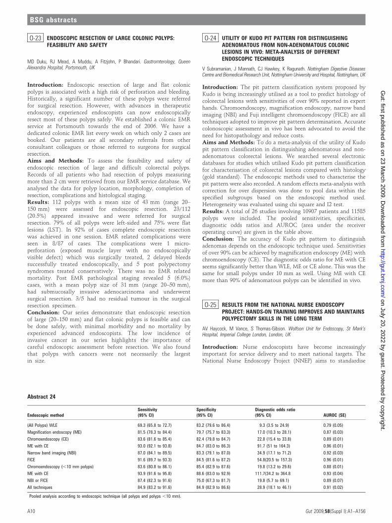
O-22 DEVELOPMENT OF A NEW FREEHAND RADIAL 3D-EUSSYSTEM FOR THE EXAMINATION OF OESOPHAGUS
1S Inglis, 1D Christie, 2JN Plevris. 1Department of Medical Physics, The RoyalInfirmary, Edinburgh, UK; 2Centre for Liver and Digestive Disorders, The Royal Infirmary,Edinburgh, UK
Introduction: Endoscopic ultrasound is an established techniquefor the examination of malignant and benign GI disorders.However, the procedure is complex and operator dependent, andit is possible to miss subtle changes during examination. Theapplication of 3D-EUS could increase accuracy, provide accuratedimensional and volume measurements, and reduce the subjectivenature of the examination.Aims and Methods: To develop and evaluate a freehand radialthree-dimensional EUS system that can be used in the upper GItract. This technique consisted of image (Matrox Morphis card) andpositional (Microscribe 3D digitising arm) capture devices, con-trolled by a custom program (IC3D), to simultaneously acquireimage and position at 12.5 fps, as the scope is withdrawn. The armtip was secured to the guiding hand of the endoscopist. Thereconstruction, analysis and display package was written in Matlaband displayed the volume using different views and alloweddistance and volume measurements to be performed. The 3D-EUSsystem was evaluated in-vitro in a custom EUS ultrasoundphantom and in-vivo in different upper GI pathology.Results: From in-vitro phantom measurements, the average Zdimensional error was ,1% and volume errors were 1.2% (objectvolume = 48930 mm3) and 4.1% (object volume = 5100 mm3).This technique provided accurate dimensional and volumemeasurements. There is no direct interface with the patient orscope, and can be used with any radial EUS system. It wascapable of detecting structures that were missed or not fullyinvestigated by conventional EUS. It could detect an increasednumber of nodes and localise pathology relative to anatomicalstructures, to aid treatment planning. In a typical example of the3D acquisition phase a 30 cm pullback, at an average speed of0.5 cm/s and captured at a rate of 12.5 fps would last 48 secondsand result in the capture of 600 images, at a 400 mm interval.Examples of a malignant T3N1 tumour and benign cyst areshown in the figures.Conclusion: 3D reconstruction of EUS images in the endoscopyroom results in better characterisation of oesophageal structures,and accurate dimensional/volume measurements. It also enables theendoscopist to review ‘‘off-line’’ the EUS examination in caseimportant information was missed during examination.
BSG abstracts
A8 Gut 2009;58(Suppl I):A1–A156
on July 20, 2022 by guest. Protected by copyright.
http://gut.bmj.com
/G
ut: first published as on 23 March 2009. D
ownloaded from
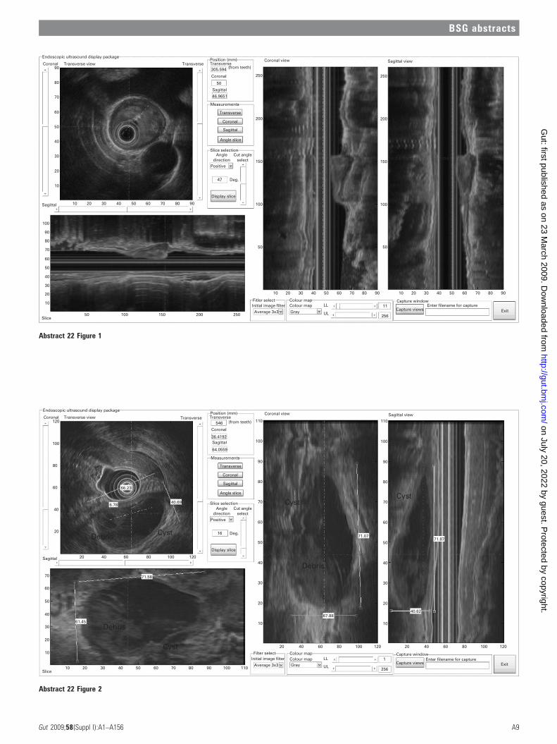
Abstract 22 Figure 1
Abstract 22 Figure 2
BSG abstracts
Gut 2009;58(Suppl I):A1–A156 A9
on July 20, 2022 by guest. Protected by copyright.
http://gut.bmj.com
/G
ut: first published as on 23 March 2009. D
ownloaded from
O-23 ENDOSCOPIC RESECTION OF LARGE COLONIC POLYPS:FEASIBILITY AND SAFETY
MD Duku, RJ Mead, A Muddu, A Fitzjohn, P Bhandari. Gastrornterology, QueenAlexandra Hospital, Portsmouth, UK
Introduction: Endoscopic resection of large and flat colonicpolyps is associated with a high risk of perforation and bleeding.Historically, a significant number of these polyps were referredfor surgical resection. However, with advances in therapeuticendoscopy, experienced endoscopists can now endoscopicallyresect most of these polyps safely. We established a colonic EMRservice at Portsmouth towards the end of 2006. We have adedicated colonic EMR list every week on which only 2 cases arebooked. Our patients are all secondary referrals from otherconsultant colleagues or those referred to surgeons for surgicalresection.Aims and Methods: To assess the feasibility and safety ofendoscopic resection of large and difficult colorectal polyps.Records of all patients who had resection of polyps measuringmore than 2 cm were retrieved from our EMR service database. Weanalysed the data for polyp location, morphology, completion ofresection, complications and histological staging.Results: 112 polyps with a mean size of 43 mm (range 20–150 mm) were assessed for endoscopic resection. 23/112(20.5%) appeared invasive and were referred for surgicalresection. 79% of all polyps were left-sided and 75% were flatlesions (LST). In 92% of cases complete endoscopic resectionwas achieved in one session. EMR related complications wereseen in 8/87 of cases. The complications were 1 micro-perforation (exposed muscle layer with no endoscopicallyvisible defect) which was surgically treated, 2 delayed bleedssuccessfully treated endoscopically, and 5 post polypectomysyndromes treated conservatively. There was no EMR relatedmortality. Post EMR pathological staging revealed 5 (6.0%)cases, with a mean polyp size of 31 mm (range 20–50 mm),had submucosally invasive adenocarcinoma and underwentsurgical resection. 3/5 had no residual tumour in the surgicalresection specimen.Conclusion: Our series demonstrate that endoscopic resectionof large (20–150 mm) and flat colonic polyps is feasible and canbe done safely, with minimal morbidity and no mortality byexperienced advanced endoscopists. The low incidence ofinvasive cancer in our series highlights the importance ofcareful endoscopic assessment before resection. We also foundthat polyps with cancers were not necessarily the largestin size.
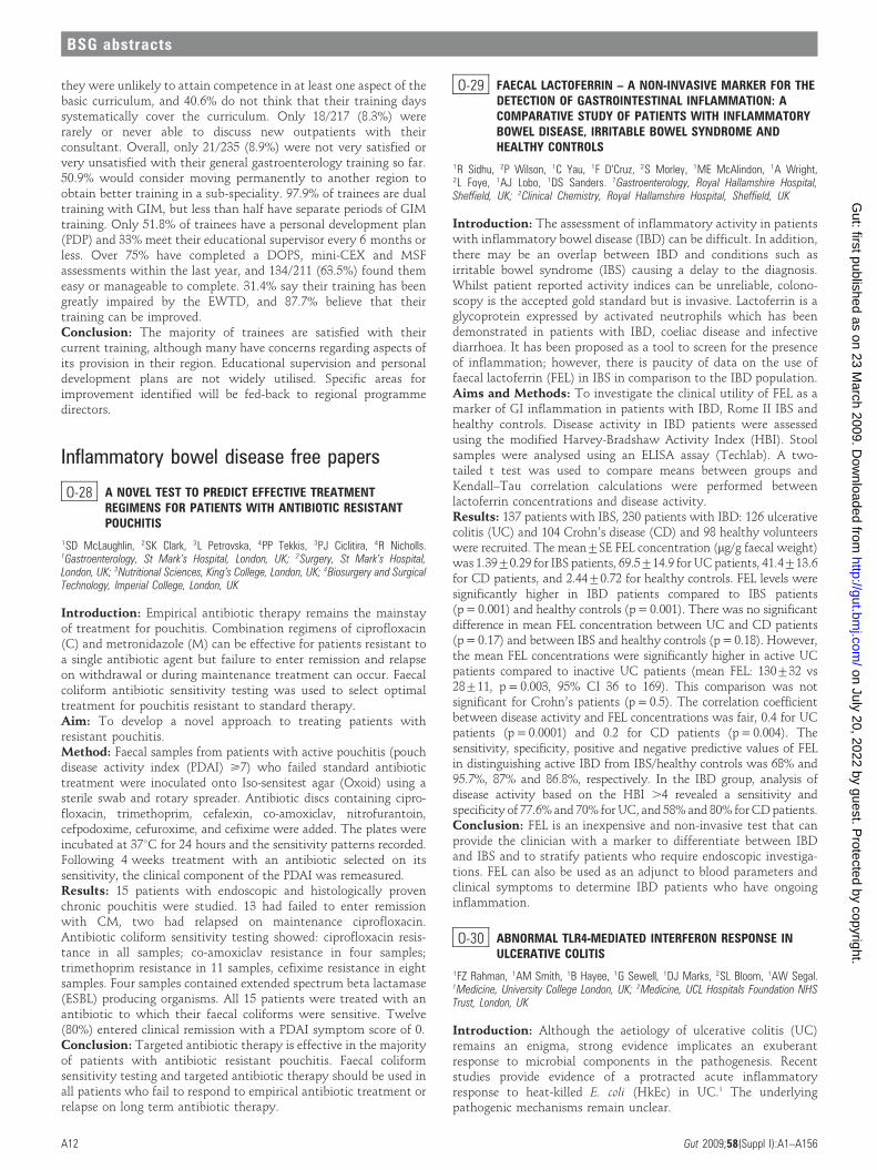
O-24 UTILITY OF KUDO PIT PATTERN FOR DISTINGUISHINGADENOMATOUS FROM NON-ADENOMATOUS COLONICLESIONS IN VIVO: META-ANALYSIS OF DIFFERENTENDOSCOPIC TECHNIQUES
V Subramanian, J Mannath, CJ Hawkey, K Ragunath. Nottingham Digestive DiseasesCentre and Biomedical Research Unit, Nottingham University and Hospital, Nottingham, UK
Introduction: The pit pattern classification system proposed byKudo is being increasingly utilised as a tool to predict histology ofcolorectal lesions with sensitivities of over 90% reported in experthands. Chromoendoscopy, magnification endoscopy, narrow bandimaging (NBI) and Fuji intelligent chromoendoscopy (FICE) are alltechniques adopted to improve pit pattern determination. Accuratecolonoscopic assessment in vivo has been advocated to avoid theneed for histopathology and reduce costs.Aims and Methods: To do a meta-analysis of the utility of Kudopit pattern classification in distinguishing adenomatous and non-adenomatous colorectal lesions. We searched several electronicdatabases for studies which utilised Kudo pit pattern classificationfor characterisation of colorectal lesions compared with histology(gold standard). The endoscopic methods used to characterise thepit pattern were also recorded. A random effects meta-analysis withcorrection for over dispersion was done to pool data within thespecified subgroups based on the endoscopic method used.Heterogeneity was evaluated using chi square and I2 test.Results: A total of 26 studies involving 10907 patients and 11585polyps were included. The pooled sensitivities, specificities,diagnostic odds ratios and AUROC (area under the receiveroperating curve) are given in the table above.Conclusion: The accuracy of Kudo pit pattern to distinguishadenomas depends on the endoscopic technique used. Sensitivitiesof over 90% can be achieved by magnification endoscopy (ME) withchromoendoscopy (CE). The diagnostic odds ratio for ME with CEseems significantly better than WLE, ME or CE alone. This was thesame for small polyps under 10 mm as well. Using ME with CEmore than 90% of adenomatous polyps can be identified in vivo.
O-25 RESULTS FROM THE NATIONAL NURSE ENDOSCOPYPROJECT: HANDS-ON TRAINING IMPROVES AND MAINTAINSPOLYPECTOMY SKILLS IN THE LONG TERM
AV Haycock, M Vance, S Thomas-Gibson. Wolfson Unit for Endoscopy, St Mark’sHospital, Imperial College London, London, UK
Introduction: Nurse endoscopists have become increasinglyimportant for service delivery and to meet national targets. TheNational Nurse Endoscopy Project (NNEP) aims to standardise
Abstract 24
Endoscopic methodSensitivity(95% CI)
Specificity(95% CI)
Diagnostic odds ratio(95% CI) AUROC (SE)
(All Polyps) WLE 69.3 (65.8 to 72.7) 83.2 (79.6 to 86.4) 9.3 (3.5 to 24.9) 0.79 (0.05)
Magnification endoscopy (ME) 81.5 (78.3 to 84.4) 79.7 (75.7 to 83.3) 17.0 (10.3 to 28.1) 0.87 (0.03)
Chromoendoscopy (CE) 83.6 (81.6 to 85.4) 82.4 (79.8 to 84.7) 22.8 (15.4 to 33.8) 0.89 (0.01)
ME with CE 93.0 (92.1 to 93.8) 84.7 (83.0 to 86.3) 91.7 (51 to 164.3) 0.96 (0.01)
Narrow band imaging (NBI) 87.0 (84.1 to 89.5) 83.3 (79.1 to 87.0) 34.9 (17.1 to 71.2) 0.92 (0.03)
FICE 91.6 (89.7 to 93.3) 84.5 (81.6 to 87.2) 54.8(20.5 to 157.3) 0.96 (0.01)
Chromoendoscopy (,10 mm polyps) 83.6 (80.8 to 86.1) 85.4 (82.9 to 87.6) 19.8 (13.2 to 29.6) 0.88 (0.01)
ME with CE 93.9 (91.6 to 95.8) 88.6 (83.0 to 92.9) 111.7(34.2 to 364.8 0.93 (0.04)
NBI or FICE 87.4 (82.3 to 91.6) 75.0 (67.3 to 81.7) 19.8 (5.7 to 69.1) 0.89 (0.07)
All techniques 84.9 (83.2 to 91.6) 84.9 (82.9 to 86.6) 28.9 (18.1 to 46.1) 0.91 (0.02)
Pooled analysis according to endoscopic technique (all polyps and polyps ,10 mm).
BSG abstracts
A10 Gut 2009;58(Suppl I):A1–A156
on July 20, 2022 by guest. Protected by copyright.
http://gut.bmj.com
/G
ut: first published as on 23 March 2009. D
ownloaded from
training and ensure competence. To utilise the workforce effec-tively, it is necessary for nurse endoscopists to be competent atbasic polypectomy. A dedicated training day was designed involvingknowledge-based teaching and hands-on training sessions inpolypectomy. Quality assurance evaluation was inbuilt withformative and summative skills assessments, clinical audit andsix-month follow-up.Aims and Methods: Nine nurse endoscopists were trained in thefirst cohort. A questionnaire completed at their base hospitals wasused for pre-assessment of clinical practice and a multiple choicequestionnaire was used as a formative assessment of core knowl-edge underpinning safe polypectomy technique. A pre-coursepractical skills assessment was performed on ex-vivo animal modelsusing validated assessment tools. Knowledge-based teaching con-sisted of structured clinical lectures and an interactive DVD trainingmodule on the theory and practice of diathermy and polypectomy.Structured practical hands-on skills training was delivered by expertinstructors on ex-vivo animal models and computer simulations. Asummative assessment was performed at the end of the course. Allnurses completed a 6 month prospective clinical audit of practice atbase hospitals. All were recalled for follow-up with a repeat of theknowledge assessment and skills assessment at 6 months.Results: Following training, nurses improved their skills assessmentscores from a median (IQR) of 22 (21–26) pre-course to 39 (36–41)post-course (p = 0.003). Assessment scores at six month follow-upshowed skills were maintained with median scores of 39 (37–42)(p = 0.78 compared to end of course scores). Knowledge improvedsignificantly with median MCQ scores of 63% pre-course to 77% atsix-month follow-up (p = 0.034). The nurses’ continuous auditpractice improved with 2/9 (22%) recording their polyp detectionrates pre-course to 7/8 (87.5%) at follow-up. Audit of procedures doneduring the follow-up period was extremely variable, with some nurseshaving performed less than 5 polypectomies.Conclusion: A structured hands-on model-based training daysignificantly improved nurses’ practical skills in polypectomy. Atsix-month follow-up, skills were maintained and knowledge wasimproved despite limited ongoing procedural exposure. Such qualityassurance indicators confirm the clinical efficacy of this skills basedtraining programme. The NNEP should aim to ensure nurses obtainregular exposure to polypectomy following the course to maximisethe educational benefit from the training.
BSG trainee symposium
O-26 ACCEPTABILITY OF SIMULATOR TRAINING IN COLONOSCOPY:RESULTS FROM A MULTINATIONAL RANDOMISED BLINDEDCONTROLLED TRIAL
1AV Haycock, 2AD Koch, 3P Familiari, 4F van Delft, 5JS Bladen, 4E Dekker,3L Petruziello, 2J Haringsma, 1S Thomas-Gibson. 1Wolfson Unit for Endoscopy,Imperial College London, London, UK; 2Department of Gastroenterology and Hepatology,Erasmus Medical Centre, Rotterdam, The Netherlands; 3Digestive Endoscopy Unit,Catholic University-A.Gemelli University Hospital, Rome, Italy; 4Department ofGastroenterology and Hepatology, Academic Medical Center, Amsterdam, TheNetherlands; 5JSB Medical Systems, Sheffield, UK
Introduction: Simulators are now recommended for use inendoscopic training. However, first generation colonoscopy simu-lators have suffered from the lack of accurately modelled colonic
looping, and ratings of realism and usefulness diminish withincreasing experience. The new second-generation Olympus colo-noscopy simulator has been specifically designed to model coloniclooping, and initial validation studies reported a high degree ofusefulness as judged by experts. A multinational trial utilising theOlympus colonoscopy simulator randomised novices to 16 hoursstructured simulator training or patient-based training. A ‘‘like-with-like’’ comparison of acceptability was made between simu-lator training compared to real-life training in colonoscopy.Aims and Methods: 37 novice trainees from four centres in theUK, Italy and the Netherlands were randomised to two days ofsimulator training with minimal supervision (N = 19) or hands-onpatient training under expert guidance (N = 18). Participantscompleted an initial questionnaire regarding their expectations oftraining. Following their training, all participants were assessed onboth simulator test cases and patient test cases, and completedfeedback regarding the quality of their training.Results: Initial expectations were high for both groups, with VisualAnalogue Scores above 7 out of 10 for all measures. Post trainingscores were equivalent for quality, usefulness and enjoyment, andboth groups felt their training met or exceeded their expectationsfor quality and usefulness. Both groups felt they were reasonablywell prepared for their assessments with scores of 6.0 for simulatortrainees and 7.0 for patient trainees respectively (p = 0.40), withsimilar anxiety levels.Conclusion: Training using the second generation Olympuscolonoscopy simulator was highly acceptable to novice traineesand rates as equivalent to hands-on training with expert instruc-tors. As the simulator requires minimal instructor time and effortand no exposure to patient risk it should be considered as a valuabletraining tool for skill acquisition in colonoscopy.
O-27 GASTROENTEROLOGY TRAINING IN 2008: RESULTS FROM THETIG/BSG NATIONAL TRAINING SURVEY
AV Haycock, P Flanagan, A Ignjatovic, S Kendrick, R Mead, P Mizen, M Mottershead,V Patel, K Pollock, T Valliani, J Williams. Trainees Committee, British Society ofGastroenterology, London, UK
Introduction: The 2004 national survey of gastroenterologytrainees revealed wide variation in both the quality and quantityof training. Significant changes to postgraduate medical educationhave taken place since that time in the UK. Modernising MedicalCareers and the European Working Time Directive (EWTD) havealtered the structure, delivery, and evaluation of training ingastroenterology.Aims and Methods: The aim of the 2008 survey was to investigatethe impact of these changes and gather data on current standards ofteaching, training, and supervision. A web-based questionnaire wasdeveloped with questions in eight domains: demographics, generalgastroenterology, specialist training, general medicine, study leave,assessments, endoscopy and general questions. An invitation tocomplete the survey was emailed by the regional TiG representa-tives to all trainees in their region. Data were collected duringOctober 2008.Results: 249 out of 411 trainees from 20 different training regionscompleted the survey (response rate 60.6%). 217 (88.6%) were ingeneral gastroenterology posts. 55/234 (23.4%) trainees thought
Abstract 26 Results of VAS scores from initial and final questionnaire (median and IQR)
Initial simulator Initial patient p Value Final simulator Final patient p Value
Quality 7.2 (6.4–8) 7.3 (5–8) 0.46 8 (7–8.5) 8 (6–9.8) 0.92
Usefulness 8 (7.1–8.8) 8 (7.2–9.2) 0.37 9 (7–9) 8.5 (8–10) 0.13
Enjoyment 8.4 (7–9.5) 8.8 (8–9.6) 0.32 8 (7–10) 9 (8.3–10) 0.18
BSG abstracts
Gut 2009;58(Suppl I):A1–A156 A11
on July 20, 2022 by guest. Protected by copyright.
http://gut.bmj.com
/G
ut: first published as on 23 March 2009. D
ownloaded from
they were unlikely to attain competence in at least one aspect of thebasic curriculum, and 40.6% do not think that their training dayssystematically cover the curriculum. Only 18/217 (8.3%) wererarely or never able to discuss new outpatients with theirconsultant. Overall, only 21/235 (8.9%) were not very satisfied orvery unsatisfied with their general gastroenterology training so far.50.9% would consider moving permanently to another region toobtain better training in a sub-speciality. 97.9% of trainees are dualtraining with GIM, but less than half have separate periods of GIMtraining. Only 51.8% of trainees have a personal development plan(PDP) and 33% meet their educational supervisor every 6 months orless. Over 75% have completed a DOPS, mini-CEX and MSFassessments within the last year, and 134/211 (63.5%) found themeasy or manageable to complete. 31.4% say their training has beengreatly impaired by the EWTD, and 87.7% believe that theirtraining can be improved.Conclusion: The majority of trainees are satisfied with theircurrent training, although many have concerns regarding aspects ofits provision in their region. Educational supervision and personaldevelopment plans are not widely utilised. Specific areas forimprovement identified will be fed-back to regional programmedirectors.
Inflammatory bowel disease free papers
O-28 A NOVEL TEST TO PREDICT EFFECTIVE TREATMENTREGIMENS FOR PATIENTS WITH ANTIBIOTIC RESISTANTPOUCHITIS
1SD McLaughlin, 2SK Clark, 3L Petrovska, 4PP Tekkis, 3PJ Ciclitira, 4R Nicholls.1Gastroenterology, St Mark’s Hospital, London, UK; 2Surgery, St Mark’s Hospital,London, UK; 3Nutritional Sciences, King’s College, London, UK; 4Biosurgery and SurgicalTechnology, Imperial College, London, UK
Introduction: Empirical antibiotic therapy remains the mainstayof treatment for pouchitis. Combination regimens of ciprofloxacin(C) and metronidazole (M) can be effective for patients resistant toa single antibiotic agent but failure to enter remission and relapseon withdrawal or during maintenance treatment can occur. Faecalcoliform antibiotic sensitivity testing was used to select optimaltreatment for pouchitis resistant to standard therapy.Aim: To develop a novel approach to treating patients withresistant pouchitis.Method: Faecal samples from patients with active pouchitis (pouchdisease activity index (PDAI) >7) who failed standard antibiotictreatment were inoculated onto Iso-sensitest agar (Oxoid) using asterile swab and rotary spreader. Antibiotic discs containing cipro-floxacin, trimethoprim, cefalexin, co-amoxiclav, nitrofurantoin,cefpodoxime, cefuroxime, and cefixime were added. The plates wereincubated at 37uC for 24 hours and the sensitivity patterns recorded.Following 4 weeks treatment with an antibiotic selected on itssensitivity, the clinical component of the PDAI was remeasured.Results: 15 patients with endoscopic and histologically provenchronic pouchitis were studied. 13 had failed to enter remissionwith CM, two had relapsed on maintenance ciprofloxacin.Antibiotic coliform sensitivity testing showed: ciprofloxacin resis-tance in all samples; co-amoxiclav resistance in four samples;trimethoprim resistance in 11 samples, cefixime resistance in eightsamples. Four samples contained extended spectrum beta lactamase(ESBL) producing organisms. All 15 patients were treated with anantibiotic to which their faecal coliforms were sensitive. Twelve(80%) entered clinical remission with a PDAI symptom score of 0.Conclusion: Targeted antibiotic therapy is effective in the majorityof patients with antibiotic resistant pouchitis. Faecal coliformsensitivity testing and targeted antibiotic therapy should be used inall patients who fail to respond to empirical antibiotic treatment orrelapse on long term antibiotic therapy.
O-29 FAECAL LACTOFERRIN – A NON-INVASIVE MARKER FOR THEDETECTION OF GASTROINTESTINAL INFLAMMATION: ACOMPARATIVE STUDY OF PATIENTS WITH INFLAMMATORYBOWEL DISEASE, IRRITABLE BOWEL SYNDROME ANDHEALTHY CONTROLS
1R Sidhu, 2P Wilson, 1C Yau, 1F D’Cruz, 2S Morley, 1ME McAlindon, 1A Wright,2L Foye, 1AJ Lobo, 1DS Sanders. 1Gastroenterology, Royal Hallamshire Hospital,Sheffield, UK; 2Clinical Chemistry, Royal Hallamshire Hospital, Sheffield, UK
Introduction: The assessment of inflammatory activity in patientswith inflammatory bowel disease (IBD) can be difficult. In addition,there may be an overlap between IBD and conditions such asirritable bowel syndrome (IBS) causing a delay to the diagnosis.Whilst patient reported activity indices can be unreliable, colono-scopy is the accepted gold standard but is invasive. Lactoferrin is aglycoprotein expressed by activated neutrophils which has beendemonstrated in patients with IBD, coeliac disease and infectivediarrhoea. It has been proposed as a tool to screen for the presenceof inflammation; however, there is paucity of data on the use offaecal lactoferrin (FEL) in IBS in comparison to the IBD population.Aims and Methods: To investigate the clinical utility of FEL as amarker of GI inflammation in patients with IBD, Rome II IBS andhealthy controls. Disease activity in IBD patients were assessedusing the modified Harvey-Bradshaw Activity Index (HBI). Stoolsamples were analysed using an ELISA assay (Techlab). A two-tailed t test was used to compare means between groups andKendall–Tau correlation calculations were performed betweenlactoferrin concentrations and disease activity.Results: 137 patients with IBS, 230 patients with IBD: 126 ulcerativecolitis (UC) and 104 Crohn’s disease (CD) and 98 healthy volunteerswere recruited. The mean¡SE FEL concentration (mg/g faecal weight)was 1.39¡0.29 for IBS patients, 69.5¡14.9 for UC patients, 41.4¡13.6for CD patients, and 2.44¡0.72 for healthy controls. FEL levels weresignificantly higher in IBD patients compared to IBS patients(p = 0.001) and healthy controls (p = 0.001). There was no significantdifference in mean FEL concentration between UC and CD patients(p = 0.17) and between IBS and healthy controls (p = 0.18). However,the mean FEL concentrations were significantly higher in active UCpatients compared to inactive UC patients (mean FEL: 130¡32 vs28¡11, p = 0.003, 95% CI 36 to 169). This comparison was notsignificant for Crohn’s patients (p = 0.5). The correlation coefficientbetween disease activity and FEL concentrations was fair, 0.4 for UCpatients (p = 0.0001) and 0.2 for CD patients (p = 0.004). Thesensitivity, specificity, positive and negative predictive values of FELin distinguishing active IBD from IBS/healthy controls was 68% and95.7%, 87% and 86.8%, respectively. In the IBD group, analysis ofdisease activity based on the HBI .4 revealed a sensitivity andspecificity of 77.6% and 70% for UC, and 58% and 80% for CD patients.Conclusion: FEL is an inexpensive and non-invasive test that canprovide the clinician with a marker to differentiate between IBDand IBS and to stratify patients who require endoscopic investiga-tions. FEL can also be used as an adjunct to blood parameters andclinical symptoms to determine IBD patients who have ongoinginflammation.
O-30 ABNORMAL TLR4-MEDIATED INTERFERON RESPONSE INULCERATIVE COLITIS
1FZ Rahman, 1AM Smith, 1B Hayee, 1G Sewell, 1DJ Marks, 2SL Bloom, 1AW Segal.1Medicine, University College London, UK; 2Medicine, UCL Hospitals Foundation NHSTrust, London, UK
Introduction: Although the aetiology of ulcerative colitis (UC)remains an enigma, strong evidence implicates an exuberantresponse to microbial components in the pathogenesis. Recentstudies provide evidence of a protracted acute inflammatoryresponse to heat-killed E. coli (HkEc) in UC.1 The underlyingpathogenic mechanisms remain unclear.
BSG abstracts
A12 Gut 2009;58(Suppl I):A1–A156
on July 20, 2022 by guest. Protected by copyright.
http://gut.bmj.com
/G
ut: first published as on 23 March 2009. D
ownloaded from
Aims and Methods: The acute inflammatory response to microbialstimulation was compared in UC and age/sex-matched healthycontrol (HC) subjects. UC patients were on either no medication or a5-ASA alone (,2.4 g/day). Serum cytokine levels at baseline and 24,48 and 72 hours following subcutaneous injection of 36107 HkEcwere quantified (13 UC, 6 HC). We then characterised the response ofprimary monocyte-derived macrophages to microbial stimulation(HkEc, LPS (TLR4), Pam3-CSK4 (TLR2) and Flagellin (TLR5)) byanalysing TLR pathway-associated gene expression (8 UC, 8 HC) andcytokine secretion (15 UC, 9 HC).Results: Serum IP-10 levels were significantly elevated at 48 (2.5-fold, p,0.01) and 72 (8.6-fold, p,0.05) hours after HkEc injectionin UC, the time points when blood flow was raised in thesepatients.1 Analysis of TLR pathway-associated gene expressionfollowing HkEc stimulation showed that macrophages frompatients with UC significantly over-express the interferon (IFN)family-related genes; IFN-\alpha, IFN-\beta (both 6-fold, p,0.02),CD80 (8-fold, p,0.01) and CD86 (4.5-fold, p,0.05) (T cell co-stimulatory molecules). After stimulation with HkEc (p,0.01) andLPS (p,0.001), but not Pam3-CSK4 and Flagellin, macrophage IP-10secretion was significantly elevated in UC.Conclusion: Increasing evidence highlights the importance of IP-10, a potent T cell chemoattractant, in the pathogenesis of UC.Raised levels have been reported locally2 and systemically3 inpatients with active disease and its inhibition has been shown toameliorate inflammation in mouse models of colitis.4 Here wedemonstrate that abnormally elevated IP-10 levels in UC occur inresponse to bacterial stimulation. The underlying defect appears tolie at the level of the macrophage, and specifically relates todysregulated TLR4 signalling. Over-expression of IFN family-relatedand T cell co-stimulatory genes during the acute inflammatoryresponse may eventuate in the T cell-mediated chronic inflamma-tion characteristic of UC. Work to define the precise moleculardefects underlying dysregulated TLR4 signalling in UC is ongoingand may offer novel therapeutic targets in the future.
1. Gastroenterology 2008;4(Suppl.1):A512.2. Am J Pathol 1999;155(2):331–6.3. J Gastroenterol 2007;42:947–956.4. Pathol Int 2007;57:413–420.
O-31 THE REGULATION OF COLONIC HUMAN BETA-DEFENSIN 2(HBD2) PRODUCTION IN INFLAMMATORY BOWEL DISEASE
MC Aldhous, K Soo, CL Noble, J Satsangi. Gastrointestinal Unit, University ofEdinburgh, Edinburgh, UK
Introduction: The beta-defensins are antimicrobial peptidesexpressed in the gastrointestinal tract. Low copy number andconcomitant low mRNA expression of the human beta-defensin-2(HBD2) gene have been implicated in susceptibility to colonic CD.1
In analysis of microarray expression data2 we found HBD2 mRNAto be differentially expressed in inflamed biopsies from Crohn’sdisease (CD) and ulcerative colitis (UC): in CD mRNA was highestin proximal colon, whereas in UC expression was highest in sigmoidcolon. This study aimed to extend these findings by furtherinvestigating the effects of environmental and genetic factors onHBD2 protein production using ex vivo organ culture.Aims and Methods: Biopsies were obtained from the sigmoidcolon of CD (n = 22), UC (n = 26) and control (n = 21) patientsundergoing endoscopy. Biopsies were stimulated in ex vivo organculture3 with bacterial lipopolysaccharide (LPS) and/or nicotine for24 hours. HBD2 and IL8 production were measured in culturesupernatants by ELISA. HBD2 copy number variation analysis wascarried out by qPCR on paired DNA samples. Sequences similar toNF-kappaB consensus binding sequences were identified by TESS4
in the HBD2 gene promoter region and were sequenced.Results: HBD2 production was increased in inflamed sigmoidcolon biopsies from UC compared with CD or HC (2-way ANOVA:
inflammation p = 0.0078). In UC patients, HBD2 protein levelssignificantly increased with degree of inflammation defined byhistology, and also correlated with IL8 production (p,0.001) – thisrelationship was not seen in CD patients. HBD2 protein productionwas induced by LPS (p = 0.0246) and LPS + nicotine (p = 0.0463) inIBD but not controls. Induction by LPS was stronger in CD(p = 0.0375) than in UC (p = 0.2017). Induction of HBD2 proteinwas augmented by nicotine in UC (p = 0.0308) but not CD.Nicotine alone had no effect on HBD2 production. HBD2production did not correlate with HBD2 gene copy number.Variations in the promoter sequences were found, but nopolymorphism changed the site to the NF-kappaB bindingconsensus sequence, nor affected HBD2 production.Conclusion: In colonic inflammatory bowel disease (IBD), HBD2 isinduced by bacterial LPS – this response is differentially modified bynicotine in CD and UC. In UC, HBD2 production is increased byacute inflammation. HBD2 CNV does not influence proteinproduction. These results provide insights into the environmentaland genetic determinants of dysregulated innate immunity in IBD.
1. Fellerman, et al. Am J Hum Genet 2006;79:439–48.2. Noble C, et al. Gut 2008;57:1398–1405 and personal communication.3. Dionne S, et al. Clin Exp Immunol 2003;133:108–14.4. Shug J, et al. 1997 http://www.cbil.upenn.edu/tess.
O-32 ARACHIDONIC ACID INCREASES THE RISK OF ULCERATIVECOLITIS – A PROSPECTIVE COHORT STUDY IN EPIC-DENMARKUSING BIOMARKER DATA
1PSA de Silva, 2A Olsen, 2A Tjonneland, 3K Overvad, 4E Berg Schmidt, 5AR Hart.1Department of Gastroenterology, Norfolk and Norwich University Hospital, Norwich,UK; 2Institute of Cancer Epidemiology, Danish Cancer Society, Copenhagen, Denmark;3Departments of Cardiology and Clinical Epidemiology, Aarhus University Hospital,Denmark; 4Department of Cardiology, Aalborg Hospital, Aalborg, Denmark; 5School ofMedicine, University of East Anglia, Norwich, UK
Introduction: Arachidonic acid (AA), an n-6 polyunsaturated fattyacid (n-6 PUFA), is a component of the phospholipid bilayer ofcolonocyte membranes and can be metabolised to the pro-inflamma-tory eicosanoids PGE2 and LTB4. The latter are found in increasedconcentrations in the mucosa of patients with ulcerative colitis (UC).Aims and Methods: The aim of this study was to measure, for thefirst time, in a prospective cohort investigation, if an increased AAconcentration in adipose tissue biopsies, which reflect dietaryintake, increases the risk of developing incident ulcerative colitis. Atotal of 57 053 men and women, aged 50-64 years, were recruited inthe EPIC-Denmark Study (European Prospective Investigation In toCancer) between 1993–97. Adipose tissue biopsies were taken fromthe gluteal region of participants at recruitment and AA fatty acidconstituents measured. The cohort was monitored to up to June2004 to identify individuals who developed UC. Each case wasmatched with 4 controls for age and gender, and levels of AAdivided into quartiles. Unconditional logistic regression was used tocalculate univariate odds ratios for the level of AA and an additionalmultivariate analysis performed adjusted for cigarette smoking andmarine n-3 PUFAs in adipose tissue.Results: During the follow-up, 34 subjects (15 women, 19 men)developed incident UC with the diagnoses confirmed by physicians.The median age at diagnosis was 58.8 years (range 53.3–71.1 years) witha median time between recruitment and diagnosis of 3.7 years (1.7–7.1years). The highest adipose concentration of AA was associated with anodds ratio for UC of 12.3 (95% CI 2.6 to 57.8, p = 0.002) with astatistically significant trend across quartiles (OR for trend = 1.9, 95%CI 1.3 to 2.8, p = 0.001) in the univariate analysis. Adjustment for co-variates did not affect the magnitude of the findings.Conclusion: Initially, well participants, who have higher adiposetissue concentrations of AA, which represent dietary intake, have asignificantly higher risk of developing ulcerative colitis compared with
BSG abstracts
Gut 2009;58(Suppl I):A1–A156 A13
on July 20, 2022 by guest. Protected by copyright.
http://gut.bmj.com
/G
ut: first published as on 23 March 2009. D
ownloaded from
those with lower levels. These findings support previous data fromdietary questionnaire-based epidemiological studies showing n-6PUFAs may be involved in the aetiology of this condition. Loweringthe dietary intake of AA may prevent the development of UC.
O-33 METRONIDAZOLE 10% OINTMENT IMPROVES SYMPTOMS INPERIANAL CROHN’S DISEASE WITH LOW SYSTEMICEXPOSURE
1Y Maeda, 2SC Ng, 3P Durdey, 3C Burt, 4J Torkington, 4P Dhruva-Rao, 5J Mayberry,5T Moshkovska, 6C Stone, 2CJ Vaizey. 1Physiology Unit, St Marks Hospital, Harrow,UK; 2Physiology Unit, St Mark’s Hospital, Harrow, UK; 3Department of Surgery, BristolRoyal Infirmary, Bristol, UK; 4Department of Surgery, Llandough Hospital, Penarth, UK;5Department of Gastroenterology, Leicester General Hospital, Leicester, UK;6Department of Gastroenterology, Washington University in St Louis, Missouri, USA
Introduction: Pain and discharge cause significant morbidity inperianal Crohn’s disease. In a prospective, controlled study, wedemonstrate here the potential for metronidazole (MET) 10%ointment to exert therapeutic benefit in perianal Crohn’s disease(CD) whilst minimising adverse effects seen with oral MET.Systemic exposure to MET was examined in a separate study.Aims and Methods: Patients with perianal CD were prospectivelyrandomised to MET 10% ointment (S.L.A. Pharma, Watford, UK) orplacebo ointment (0.7 g applied perianally tds for 4 weeks). PerianalCrohn’s Disease Activity Index (PCDAI; 0–20; entry score >5) wasscored at baseline and at 4 weeks. Each PCDAI component(discharge; induration; pain; type of disease; restriction of sexualactivity) was analysed individually. Perianal pain was assessed on a10 cm VAS line. Patient Global Impression of Improvement (PGI-I)was scored on a 7-point Likert scale. Data were analysed byANCOVA, Fisher’s exact or Wilcoxon tests as appropriate. In aseparate study, blood samples taken from 12 healthy volunteers(HV) and 7 patients with perianal CD before a single application ofMET 10% ointment and over 24 h after were analysed formetronidazole and hydroxymetronidazole. A single sample wastaken on Day 7 after tds dosing for 6 days.Results: 61 patients (27 MET; 34 placebo) with perianal CD wereevaluated. At week 4, mean (¡SEM) reduction in PCDAI was3.2¡0.5 (metronidazole) vs 2.1¡0.5 (placebo) (p = 0.093). Thedischarge component of the PCDAI improved significantly in theMET group compared to the placebo group (p = 0.012). The VASscore for perianal pain fell 16.7 mm (MET) vs 5.7 mm (placebo;p = 0.059). There was a significant difference in favour of MET forthe PGI-I (p = 0.015). No serious adverse events were reported withMET 10% ointment. Maximum plasma levels of MET andhydroxymetronidazole were somewhat higher and more variablein the CD subjects compared to HVs after a single application ofointment (CD 52.3¡88.0 vs HV 12.4¡6.2 ng/mL) and after repeatdosing (CD 46.2¡26.1 vs HV 29.1¡28.1 ng/mL). This compareswith values of 13 ng/mL for single oral doses of 500 mg MET.Conclusion: MET 10% ointment reduced perianal discharge andpain and was associated with better impressions of globalimprovement in subjects with perianal CD. MET 10% ointmentwas well tolerated, with minimal adverse effects, in keeping withobserved blood levels ,1% of those seen with standard oral doses,and has potential as adjunctive therapy for perianal CD.
O-34 CIGARETTE SMOKE DOWN REGULATES NOD2 EXPRESSIONAND NF-KAPPAB ACTIVATION IN SW480 CELLS
1MC Aldhous, 1K Soo, 1A Ulanicka, 1JE Pearce, 2LA Stark, 2MG Dunlop, 1J Satsangi.1Gastrointestinal Unit, University of Edinburgh, Edinburgh, UK; 2Colon Cancer GeneticsGroup, University of Edinburgh, Edinburgh, UK
Introduction: The mechanisms whereby smoking influencessusceptibility and phenotype in CD are unknown. Germlinevariations in the NOD2 gene predispose to fistulising/stenosing
small bowel CD, the same phenotype associated with smoking.NOD2 signalling involves critical interactions with RIPK2 leadingto activation of NF-kappaB, and associated effects on chemokine,cytokine and defensin production. We investigated whethercigarette smoke extract (CSE) or chemicals within cigarette smokealtered NOD2 expression and downstream effects in the SW480intestinal epithelial cell line, which express wild-type NOD2.Aims and Methods: CSE was made by bubbling cigarette smokethrough PBS. SW480 cells were stimulated with TNF-alpha withand without 2% CSE for 4 hours. NOD2 mRNA expression wasmeasured by qRT-PCR. To determine effects of specific compo-nents of cigarette smoke, cells were also stimulated with combina-tions of TNF-alpha and nicotine, acrolein, hydroxy-nonenal (HNE)or hydrogen peroxide (HP). In separate experiments, NF-kappaBactivity was measured at 18 hours by luciferase reporter assays. Co-immunoprecipitation assays were used to determine the interactionbetween NOD2 and RIPK2 in cells stimulated with combinations ofCSE, nicotine and TNF-alpha. CXCL8/IL8 and CCL20/MIP3-alphawere measured in culture supernatants of cells stimulated withcombinations of CSE, nicotine and TNF-alpha after 24 hours byELISA.Results: TNF-alpha induced NOD2 expression, which wasinhibited by CSE in a dose-dependent manner, when CSE wastitrated out from 2 to 0.0625%. TNF-alpha-induced NOD2expression was reduced by acrolein, HNE and HP but not affectedby nicotine. NF-kappaB activity was significantly reduced by CSE(p,0.001) but not by nicotine, acrolein, HNE or HP. Basal andTNF-alpha-induced levels of CXCL8/IL8 and CCL20/MIP3-alphawere decreased by CSE but not nicotine. Preliminary co-immuno-precipitation experiments suggest that TNF-alpha increased RIPK2/NOD2 interactions and that these were reduced by CSE.Conclusion: CSE down-regulated the increased NOD2 expression inresponse to TNF-alpha, as well as inducing effects on down-streamsignalling consistent with reduced NOD2 function. These data suggestthat smoking may reduce innate immune responses in intestinal cells,thereby identifying a novel gene-environmental interaction whichmay be critical in the pathogenesis of inflammatory bowel disease.
O-35 BACTERIAL CLEARANCE IS DELAYED IN PATIENTS WITHCROHN’S DISEASE
1B Hayee, 1FZ Rahman, 1AM Smith, 1DJB Marks, 1GW Sewell, 2TJ Sweeting,3SL Bloom, 1AW Segal. 1Molecular Medicine, University College London, UK;2Statistical Science, University College London, UK; 3Gastroenterology, UCLH NHSFoundation Trust, London, UK
Introduction: Recent work provides evidence for a failure of acuteinflammation in Crohn’s disease (CD), supporting the hypothesisthat a defective innate immune response is a primary defect.1 Theabnormalities observed suggested that bacteria or debris breachingthe intestinal mucosal barrier would not be adequately cleared.Bacterial persistence would then provide the stimulus for chronicgranulomatous inflammation.2
Aims and Methods: This study was conducted to determinewhether bacterial clearance is abnormal in CD. Ethical permissionwas obtained from the Joint UCL/UCLH Committees on the Ethicsof Human Research and the Administration of RadioactiveSubstances Advisory Committee (ARSAC). An antibiotic-sensitiveclinical isolate of E. coli was labelled in culture with 32P then killedby UV-irradiation. Labelled bacteria (1 kBq activity) were mixedwith identical, unlabelled UV-irradiated E. coli (36107 bacteria perinoculum). The mixture was injected subcutaneously into bothforearms of study subjects, and its clearance determined bymeasuring radioactivity at the injection sites over the subsequent72 hours. Clearance kinetics were modelled using a nested mixedeffects model with non-linear dynamics.Results: 14 healthy controls (HC), 16 patients with quiescent CDand 3 patients with ulcerative colitis (UC) were studied. All subjects
BSG abstracts
A14 Gut 2009;58(Suppl I):A1–A156
on July 20, 2022 by guest. Protected by copyright.
http://gut.bmj.com
/G
ut: first published as on 23 March 2009. D
ownloaded from
displayed two-phase clearance kinetics with an initial rapid phase.Clearance was significantly delayed in CD compared to HC(p,1025) with a trend toward more rapid clearance in UC(p = 0.08). Extrapolating clearance curves to a point when 99% ofinjected bacteria should have been removed gave total clearancetimes of 10.2 days (95% CI: 8.3 to 13.0) for HC, 7.1 days (5.4 to10.4) for UC, and 44.3 days (21.8 to inf.) in CD.Conclusion: This is the first study to use radiolabelled bacteria inhuman subjects to examine their clearance kinetics. It clearlydemonstrates a gross defect in bacterial clearance in CD, and thatthese organisms persist for an unusually long duration in theirtissues. These are precisely the conditions known to elicitgranulomatous chronic inflammation,3 and therefore likely to beof primary relevance to the pathogenesis of CD.
BHH and FZR contributed equally to this work.
1. Lancet 2006;368:668.2. J Pathol 2008;214:260.3. J Clin Pathol 1983;36:723.
O-36 METHOTREXATE FOR PAEDIATRIC INFLAMMATORY BOWELDISEASE IN THE BIOLOGICAL ERA
1A Tybulewicz, 1P Rogers, 2D Hoole, 3J Satsangi, 1PM Gillett, 4DC Wilson. 1PaediatricGastroenterology, Royal Hospital for Sick Children, Edinburgh, UK; 2Pharmacy, RoyalHospital for Sick Children, Edinburgh, UK; 3GI Unit, University of Edinburgh, Edinburgh,UK; 4Child Life and Health, University of Edinburgh, Edinburgh, UK
Introduction:Thereare limiteddataontheuseofmethotrexate(MTX)for induction and maintenance of remission in children with Crohn’sdisease (CD), and many proceed to biological therapy if azathioprine(AZA) fails. We therefore aimed to review our patients treated withMTX, in terms of both induction and maintenance of remission.Aims and Methods: A cohort study of all 244 patients diagnosedwith early-onset (,18 years of age) IBD and managed in a UKregional centre over a 10 year period (1/8/97–31/12/07). Within thiscohort were 165 patients with CD; we studied those resistant to orintolerant of AZA/6-MP who were treated with MTX (16 weekinduction course of s/c MTX (15 mg/m2) followed by theequivalent oral dose). Paediatric Crohn’s Disease Activity Index(PCDAI) was used to objectively measure disease activity duringtreatment; PCDAI (10 indicated full remission, (15 indicatedpartial remission and a score .30 indicated severe disease.Results: 122 with CD were treated with AZA/6-MP; 53 children(52 with previous ADA, 1 who refused ADA) received a MTXinduction course. These 53 patients had CD diagnosed at a median(range) age of 10.9 years (4.0–16.5) and received MTX at 14.0 years(6.8–17.9). The median PCDAI at start of treatment was 35 (IQR20–37.5). 41 (78%) patients entered remission; median (range) timeto partial/complete remission was 11 (3–16) weeks. 2 of 41 hadrecalcitrant OFG and were excluded from maintenance evaluation;the remaining 39 had luminal CD and a median (range) duration offollow-up of 85 (26–310) weeks. 20 (51%) maintained long-termremission with no relapse; 7 (18%) had only a single relapse ofwhich 6 (15%) re-entered remission, and 12 (31%) had .1 relapses.The median (range) time to first relapse was 39 (20–181) weeks. Ofthe 19 relapsing, 14 proceeded to repeat s/c courses of MTX, 9 toinfliximab, 6 to surgery and 4 to adalimumab. On MTX treatment,24 (46%) had nausea/vomiting, 18 (34%) had raised ALT (1subsequently undergoing liver biopsy, with treatment thencontinued), 1 had shingles and none had bone marrow toxicity.Conclusion: MTX appears to be effective in inducing (78%) andmaintaining (51%) remission in patients who have failed to remiton AZA, even if biological therapy is available. MTX also appears tobe well tolerated and safe. Those who have had multiple relapseshave all had complicated clinical courses and severe intractabledisease. However, a stronger evidence base is needed from well-designed RCTs for both induction and maintenance of remission ofearly-onset CD by MTX.
O-37 WHICH ACTIVITY INDEX FOR ULCERATIVE COLITIS?EVALUATION OF INTER-OBSERVER VARIATION IN CLINICAL,ENDOSCOPIC AND COMPOSITE INDICES
1AJ Walsh, 1AOS Brain, 1S Keshav, 1OC Buchel, 1S Jacobovits, 1B Merrin, 1M Rolinski,1S Thomas, 1L White, 2A Von Herbay, 2BF Warren, 3DG Altman, 1SPL Travis.1Gastroenterology, The John Radcliffe Hospital, Oxford, UK; 2Cellular Pathology, The JohnRadcliffe Hospital, Oxford, UK; 3Biomedical Statistics, University of Oxford, Oxford, UK
Introduction: Clinical trials of ulcerative colitis (UC) use manydifferent activity indices, making it difficult to compare data betweentrials. It is unclear which index is most reproducible and how inter-observer variation (IOV) impacts on trial inclusion and outcomes.Aims and Methods: Our aims were to assess IOV in all commonlyused indices and to determine the impact of this variability oninclusion criteria and outcomes in key published trials. 100 patientswith UC were seen independently, each patient on the same day, inrandom order, by 4 gastroenterologists. Each physician completed apro forma to calculate the following indices: Mayo Index (MAYO),UC Disease Activity Index (UCDAI), Simple Colitis ClinicalActivity Index (SCCAI), Seo Index (SEO), Modified Truelove andWitts Severity Index (MTW), and Colitis Activity Index (CAI). Aseparate physician performed video-recorded sigmoidoscopy on thesame day. The video record was later scored by the 4 assessingphysicians, blinded to other results. To assess the impact of IOV inrecruiting patients to, or meeting outcome criteria of clinical trials,pivotal papers were selected for each index. Quadratic weightedkappa statistics assessed agreement within and between indices, inwhich disagreements are weighted in relation to their magnitude(number of categories).Results: Of 100 patients, 50% were male, median age 49 years(range 19–82), and median duration of disease 107 months (range0–575). Maximum disease extent: proctitis 27%, left-sided colitis39% and pancolitis 34%. 23% were in remission, 40% had mild, 32%moderate and 5% severe colitis. The mean inter-rater kappa forMAYO, UCDAI and SCCAI were 0.88, 0.90 and 0.88, respectively;the SEO (kappa = 0.95) scored highest and CAI lowest(kappa = 0.78) but this does not imply best or worst indices, asutility depends on several factors. The kappa for agreement ofactivity category between MAYO and UCDAI, and MAYO andSCCAI were 0.92 and 0.80, respectively. For endoscopic indices,kappa for agreement between Baron and Mayo, and Baron andmodified Baron indices were 0.83 and 0.89, respectively. For trialinclusion, the median mismatch was 23% (complete concordance in65–97%), with most agreement for severe UC by MTWSI and leastfor mild-moderate UC by UCDAI. For MAYO, there wasdisagreement by . = 1 observer estimating current stool frequencyin 53% which changed the score by 1 in 22% and by 2 in 2%.Median mismatch for remission was 21% (complete concordance in77–91%) with most agreement for SCCAI.Conclusion: IOV in the assessment of UC is substantial andshould be taken into account when calculating the power of clinicaltrials. With 4 independent specialists assessing the same patients inthe context of this study, about 1 in 5 patients would have beenaffected by disagreement whether they met entry criteria orachieved remission for already published clinical trials.
Liver free papers
O-38 ALCOHOLIC LIVER DISEASE – THE SOUTH INDIAN DIMENSION
1C Chong, 1E Harazi, 1TM Tian, 2T Darch, 2J Wells, 1MY Morgan. 1Centre forHepatology, Department of Medicine, Royal Free Campus, University College LondonMedical School, London, UK; 2Institute of Child Heath, University College LondonMedical School, London, UK
Introduction: Alcohol produces a spectrum of liver injury but lessthan 20% of individuals who misuse alcohol will develop cirrhosis.
BSG abstracts
Gut 2009;58(Suppl I):A1–A156 A15
on July 20, 2022 by guest. Protected by copyright.
http://gut.bmj.com
/G
ut: first published as on 23 March 2009. D
ownloaded from
Recently, it was reported that South Indian (non-Moslem) men aresignificantly over-represented in the cirrhotic population inBirmingham, UK (Douds et al, 2003); additionally 37% of theAsian men presented aged ,40 years compared with only 17% ofwhite men. Women develop cirrhosis at an earlier age and afteringestion of less alcohol than men because their total body water(TBW) is smaller. No information is available on alcohol pharma-cokinetics and body composition in the Asian population.Aims and Methods: The aim was to assess alcohol kinetics inhealthy white and Asian volunteers in relation to body compositionvariables. Twenty-four Asian and 24 white volunteers (50% female;age: 20–34 years; BMI:17–28) drank 0.5 g/kg of 99% ethanol, as a25% v/v solution in orange juice, 2 hours after a standard breakfast.Breath alcohol was measured every 5 min for 6 hours. Bodycomposition was assessed using a 3-component model from directmeasurements of body density (densitometry), TBW (deuteriumdilution) and body weight. Pharmacokinetic parameters werecalculated using a modified one compartment model with zero-order kinetics (Mumenthaler et al, 2000). Correlations betweenbody composition and pharmacokinetic variables were assessedusing Pearson’s r.Results: The one compartment model did not describe thepharmacokinetics of alcohol as well in the Asians. No significantdifferences were observed in Cmax, AUC and elimination ratesbetween the Asian and white populations, although significantdifferences were observed in both Tmax and the apparent volume ofdistribution (aVd). TBW was significantly higher in men than womenand in Asians compared with whites. The calculated aVd showed ahighly significant correlation with TBW in the whites (r = 0.96;p,0.0001), but not in the Asians (r = 0.30; p = 0.48). Differences infat mass and fat free mass were observed between the two populationgroups but were insufficient to explain this discrepancy.Conclusion: Significant differences were observed in alcoholpharmacokinetics in the Asian population which might explaintheir increased propensity to develop significant liver disease. Thesedifferences could only be explained, in part, by differences in bodycomposition. No significant differences were observed in alcoholelimination rates suggesting that differences must, therefore, existin the absorption, distribution or non-hepatic elimination of alcoholbetween these two populations. There is a clear need to establish arobust model for alcohol pharmacokinetics in the Asian population.
O-39 COMPARISON OF TRANSIENT ELASTOGRAPHY, APRI SCORE,AST/ALT RATIO, HYALURONIC ACID AND CLINICAL SIGNSFOR THE DIAGNOSIS OF COMPENSATED CIRRHOSIS
1R Malik, 2M Lai, 2I Nasser, 2M Curry, 2D Schuppan, 2N Afdhal. 1Hepatology, TheInstitute of Hepatology, UCL, London, UK; 2Medicine, Beth Israel Deaconess MedicalCenter/Harvard Medical School, Boston, USA
Introduction: Non-invasive diagnosis of compensated cirrhosis isimportant as liver biopsy has limitations.Aims and Methods: We compared transient elastography, APRIscore, AST/ALT ratio, hyaluronic acid (HA) and clinical signs todetermine which modality performed best at identifying cirrhosis.Patients undergoing evaluation at a single academic centre inBoston, USA were recruited and had clinical evaluation, serologicalstudies, endoscopy, radiological imaging, liver stiffness measure-ment and liver biopsy.Results: 404 patients were stratified into two groups depending onwhether cirrhosis was present. There were 124 patients in thecirrhotic group out of whom 54 were biopsy proven (Metavir stage4) and 70 had a clinical diagnosis (radiology+endoscopy). Therewere 280 patients in the non-cirrhotic group all of whom had a liverbiopsy (Metavir stage 0–3). The mean transient elastographymeasurement in the cirrhotic group (29.1¡3 kPa) was significantlyhigher than the non-cirrhotic group (8.1¡2.9 kPa, p,0.05).Transient elastography (AUC 0.9¡0.04) was superior to HA
(AUC 0.81¡0.04, p,0.05), clinical signs (AUC 0.74¡0.04,p,0.05), APRI score (AUC 0.71¡0.03, p,0.05), and AST/ALTratio (AUC 0.66¡0.03, p,0.05) at identifying cirrhosis. In addition,using optimal cut offs for each modality, TE .12 kPa yielded asensitivity and specificity of 89% and 87% respectively, which wassignificantly higher that HA .150 ng/ml (72%; 79%), positiveclinical signs (69%; 81%), APRI .1 (73%; 54%), and AST/ALT .1(64%; 59%) (p,0.05 for all). We performed further analysis tocompare biopsy proven cirrhosis (stage 4) with the control group(all biopsy proven stage 0–3) to determine the performance atidentifying histologically proven cirrhosis. Transient elastographyyielded an AUC 0.88¡0.03, which was significantly higher than theother four tests (p,0.05 for all). A high degree of diagnosticaccuracy was seen for transient elastography on sub-group analysisin hepatitis C (AUC 0.899¡0.02) and non-hepatitis C (AUC0.928¡0.03) groups, with elastography diagnostically superior tothe other four tests (p,0.05 for all). We identified 38 Childs Pugh Apatients with biopsy proven cirrhosis in whom a definitivediagnosis of cirrhosis was uncertain prior to liver biopsy.Transient elastography performed the best in this importantcohort, yielding an AUC 0.87¡0.04 compared to HA (AUC0.77¡0.05, p,0.05), APRI Score (AUC 0.64¡0.03, p,0.05) andAST/ALT ratio (AUC 0.63¡0.03, p,0.05).Conclusion: Transient elastography accurately identified compen-sated cirrhosis using a liver stiffness cut off of 12 kPa. It performedbetter than hyaluronic acid, APRI, AST/ALT ratio and clinical signsas a non-invasive modality.
O-40 CRITICAL FLICKER FUSION FOR THE DIAGNOSIS OF HEPATICENCEPHALOPATHY
1HR Wilson, 2CD Jackson, 3RW Morris, 2BD Youl, 1MY Morgan. 1Centre forHepatology, Royal Free Campus, UCL Medical School, London, UK; 2Department ofNeurophysiology, Royal Free Hospital, London, UK; 3Department of Primary Care andPopulation Studies, Royal Free Campus, UCL Medical School, London, UK
Introduction: The development of hepatic encephalopathy (HE) hasa significant negative effect on survival in patients with cirrhosis.Nevertheless, there is still no ‘‘gold standard’’ for its diagnosis. Thus,unless floridly manifest, its presence is often missed, and, inconsequence, patients are often suboptimally managed.Aims and Methods: The aim of this study was to evaluate criticalflicker fusion thresholds for the diagnosis of HE in patients withcirrhosis. The patient population comprised 76 individuals (48 men,28 women) of mean (range) age 57 (32 to 81) years, with biopsyproven alcohol-related (n = 57) or non-alcohol related (n = 19)cirrhosis. Patients were classified using clinical, electroencephalo-graphic and psychometric variables, as either neuropsychiatricallyunimpaired (n = 43) or as having minimal (n = 19) or overt HE(n = 14). The reference population comprised 40 healthy volunteers(17 men, 23 women) of mean age 39.8 (21 to 70) years. Critical flickerfusion was assessed using a Lafayette model 12021. The thresholdfrequencies at which light pulses were perceived as fused (fusionfrequency) or flickering (flicker frequency) were assessed and themean calculated to provide the critical flicker fusion frequency (CFF).Results: Fusion, flicker and CFF values were corrected for age andgender effects. The fusion frequency and CFF were significantly lowerin the patients overall, and significantly distinguished the populationsubgroups, e.g. fusion frequency at 1 Hz/sec: controls (36.6 Hz) vsunimpaired patients (34.1 Hz) vs minimal HE (30.3 Hz) vs overt HE(25.9 Hz) (p,0.0001). A threshold of 33.4 Hz for fusion frequency,recorded at 1 Hz/sec, diagnosed HE with a sensitivity of 79% and aspecificity of 63%. Separate thresholds were identified in patientswith alcohol-related cirrhosis: 32.0 Hz (sensitivity 75%, specificity78%) and non-alcohol related cirrhosis: 34.9 Hz (sensitivity 85%,specificity 83%). All fusion and CFF indices correlated significantlywith the classification variables and the Pugh’s score. CFF valuesaccurately mirrored changes in clinical status.
BSG abstracts
A16 Gut 2009;58(Suppl I):A1–A156
on July 20, 2022 by guest. Protected by copyright.
http://gut.bmj.com
/G
ut: first published as on 23 March 2009. D
ownloaded from
Conclusion: CFF is a simple, reliable measure which could be usefulin the screening and monitoring of HE in patients with cirrhosis.
O-41 LONGITUDINAL ANALYSIS OF HEPATITIS C VIRUS SPECIFIC TCELL RESPONSES IN EXPOSED UNINFECTED INTRAVENOUSDRUG USERS
1P Thurairajah, 1U Warshow, 1A Demaine, 2E Kaminski, 1D Hegazy, 3ME Cramp.1Institute of Biomedical Sciences, Peninsula Medical School, Plymouth, UK;2Department of Immunology, Derriford Hospital, Plymouth, UK; 3Department ofHepatology, Derriford Hospital, Plymouth, UK
Introduction: Hepatitis C virus (HCV) specific T lymphocyteresponses of low magnitude have been demonstrated amongst highrisk injection drug users (IDU) who test negative for HCV infection,suggesting viral exposure without infection.1 We have termed thesecases as ‘‘Exposed Uninfected’’ (EU). We report a longitudinal studyof HCV-specific T cell responses in EU cases and correlate this withlikely ongoing HCV exposure through drug injecting patterns.Aims and Methods: 21 high risk EU cases have been studied on 3or more occasions. All tested negative for HCV antibodies and HCVRNA. Details of injecting behaviour were ascertained by structuredquestionnaire and cases were subdivided into 3 groups according todrug injecting patterns. Group 1: EU cases who stopped injectingduring follow-up, Group 2: EU cases in prisons, Group 3: EU caseswho continued to inject in the community. Lymphocytes wereisolated from fresh blood and an enzyme linked immunospot assay(ELIPSOT) was used to measure interferon gamma (IFN-g)production in response to a panel of HCV proteins (Core, NS3-NS5). 7 healthy controls were studied at similar intervals.Results: Only 2 control cases exhibited any ELISPOT response toHCV antigens, which were not reproducible on subsequent testing.In contrast, EU’s demonstrated more frequent, stronger andmultispecific ELISPOT responses. On follow up, ELISPOT responseswere frequently maintained in group 3, with all but two casesmaintaining HCV specific responses over time. In contrast, most ofgroups 1 and 2 lost their HCV specific responses during follow up.Pooled IFN-g responses to all HCV antigens were significantlystronger in group 3 (continued IDU, p = 0.005) and in group 2(prisoners, p = 0.035) than group 1 (stopped injecting).Conclusion: Injection drug users can show HCV-specific T cellresponses to a variety of HCV antigens in the absence of anyevidence of HCV infection. Maintenance of these weak T cellresponses over a period of several months appears to be dependenton continuing injection drug use and, presumably, ongoing HCVexposure. In those entering rehabilitation and stopping injecting,HCV-specific responses mostly waned rapidly. Whether these T cellresponses merely indicate priming of the immune system by a sub-infectious inoculum of HCV or a response that is able to preventinfection becoming established remains to be clarified. Furtherstudy of the development of these responses early in the course ofinjection drug use would be of interest.
1. Thurairajah PH, et al. Hepatitis C virus (HCV)-specific T cell responses in injectiondrug users with apparent resistance to HCV infection. J Infect Dis 2008 Oct 29.[Epub ahead of print].
O-42 PLASMA PROTEOMIC PROFILING OF PATIENTS WITHCHOLANGIOCARCINOMA
1P Nassim, 2RJ Edwards, 3H Wasan, 4I Cox, 5MB Toledano, 1SD Taylor-Robinson, 1SAKhan. 1Hepatology and Gastroenterology, Imperial College London, London, UK;2Experimental Medicine & Toxicology, Imperial College London, London, UK; 3CancerMedicine, Imperial College London, London, UK; 4Imaging Sciences, Imperial CollegeLondon, London, UK; 5Epidemiology & Public Health, Imperial College London, London, UK
Introduction: Cholangiocarcinoma (CC) incidence and mortalityrates are steadily increasing. Overall prognosis is poor and there arecurrently no diagnostic biomarkers with good sensitivity and
specificity. Proteomic profiling of plasma is a promising techniquefor new biomarker discovery to improve the early diagnosis of CC.Aims and Methods: The aim of this study was to use surfaceenhanced laser desorption/ionisation time-of-flight mass spectroscopy(SELDI-TOF MS) Protein-Chips to compare the proteomic profiles inplasma of patients to identify any potential biomarkers that candifferentiate malignant CC from benign biliary disease. Plasma samplesfrom 19 patients were collected for analysis. Ten patients had aconfirmed diagnosis of CC and the remaining nine were undergoingendoscopic retrograde cholangiopancreatography for benign biliarydisease (choledocholelithiasis). All samples were analysed usinghydrophobic (H50) and carboxymethyl (CM10) Protein-Chips viaSELDI-TOF MS and biomarker models were developed.Results: Univariate analysis of the SELDI-TOF MS spectra revealed10 and 13 peaks that varied significantly between the CC andbenign disease subjects on H50 and CM10 Protein-Chips, respec-tively (p,0.05, in each case, Mann-Whitney U test). One, withmass-to-charge (m/z) of 8403 had a sensitivity of 89% andspecificity of 90% (p = 0.001, receiver operating characteristics(ROC) area under the curve (AUC) = 0.92). Several biomarkermodels were developed using multiple regression. This included oneusing two potential biomarkers with m/z 2701 and 3311 peaks thatdistinguished CC from non-malignant samples with 100% sensi-tivity and specificity (p = 0.0002, ROC AUC = 1.0). Several othermodels that performed similarly were also produced.Conclusion: This preliminary study identified several potentialplasma biomarkers of CC. The results highlight the potential role ofproteomic profiling in biomarker discovery that may lead to newdiagnostic tests to distinguish CC from non-malignant disease.Further studies in larger numbers of patients and other HBP cancersare warranted.
O-43 ROLE OF ENDOSCOPIC ULTRASOUND IN THE MANAGEMENTOF LIVER DISEASE PATIENTS WITH ECTOPIC OR GASTRICVARICES
1JN Plevris, 2S Inglis, 1AM Lennon, 1N McAvoy, 1PC Hayes. 1Centre for Liver andDigestive Disorders, The Royal Infirmary, University of Edinburgh, Edinburgh, UK;2Department of Medical Physics, The Royal Infirmary, University of Edinburgh,Edinburgh, UK
Introduction: The availability of new endoscopic ultrasound(EUS) scopes with Doppler ultrasound facilities has made it possibleto assess the severity of portal hypertension and to control varicealbleeding under EUS guidance. In addition the varied endoscopicappearances of ectopic varices in the stomach or small bowel oftenpresent a diagnostic challenge as biopsy of these lesions can putpatients at risk of severe bleeding.Aims and Methods: To report our experience in the use of EUS toassist the management of patients with chronic liver disease andportal hypertension. 15 patients (13 male, 2 female) with knownchronic liver disease and portal hypertension presented to our Unitas diagnostic or management challenge. All patients followingstandard diagnostic and/or therapeutic endoscopy, required furtherexamination with an electronic radial or linear echoendoscope and/or EUS miniprobes for further management.Results: In 10 patients polypoid looking lesions were seen in thefundus, body or gastric outlet not entirely typical of varices. In 8/10cases these were varices under EUS while in 2 cases these were truepolyps that were biopsied under EUS guidance. In 3/10 cases therewere stigmata of recent bleeding and in all three cases thrombinwas injected (250 iu/ml) with excellent effect and clot formationunder EUS. Five patients with actively bleeding varices despitestandard endoscopic therapy or TIPSS received thrombin injectionsunder EUS guidance with a 22-gauge FNA needle or a standardinjection needle. In 4/5 patients treatment was carried out in ITU/HDU and in one patient with chronic blood loss in the EndoscopyUnit. In 2 ITU patients (both ALD), 5–10 ml of thrombin was
BSG abstracts
Gut 2009;58(Suppl I):A1–A156 A17
on July 20, 2022 by guest. Protected by copyright.
http://gut.bmj.com
/G
ut: first published as on 23 March 2009. D
ownloaded from
injected using a 22G FNA needle through an Olympus linear EUSscope until a clot was formed with real-time abolition of bloodflow. One of the 2 patients had a variceal pseudo-aneurysm in theoesophagus directly communicating with the azygos vein and thesecond had bled from an ectopic duodenal varix directly commu-nicating with the portal vein despite a successful TIPSS insertion. In3 patients the Fujinon EUS miniprobes (12 MHz) were usedthrough the biopsy channel of a standard endoscope before andafter 5–10 ml of thrombin injection to assess clot formation. One ofthe 3 patients bled from gastric varices due to PV thrombosis, onefrom an oesophageal varix at 30 cm post-TIPSS and one had chronicbleeding due to ectopic duodenal varices secondary to SMVthrombosis and had been transfusion dependent. In all 5 caseshaemostasis was achieved although one patient subsequently diedin ITU due to liver failure.Conclusion: (a) EUS with Doppler is a valuable technique to assistin the diagnosis and management of ectopic or gastric varices.(b) EUS-guided or assisted thrombin injection is a safe and effectiveway of achieving haemostasis in complex cases of bleeding variceswhere other therapies had failed.
O-44 PERSISTENT HISTOLOGICAL INFLAMMATION INAUTOIMMUNE HEPATITIS DESPITE BIOCHEMICALREMISSION: FREQUENCY AND PROGNOSTIC SIGNIFICANCE
1BSR Hoeroldt, 2AK Dube, 2JCE Underwood, 3E McFarlane, 1P Basumani, 3D Gleeson.1Gastroenterology, Rotherham General Hospital, Rotherham, UK; 2Histopathology,Sheffield Teaching Hospitals, Sheffield, UK; 3Hepatology, Sheffield Teaching Hospitals,Sheffield, UK
Introduction: Most patients with autoimmune hepatitis (AIH)achieve clinical and biochemical remission following treatmentwith prednisolone ¡ azathioprine. Despite this, active inflamma-tion persists on follow-up biopsy in some patients. There arelimited data on the frequency and clinical significance of thisphenomenon; follow-up biopsy is not routinely performed inpatients with AIH in many centres.Aims and Methods: Evaluation of the frequency and significanceof persistent inflammation on follow-up biopsy in patients withAIH who have achieved clinical and biochemical remission. 81patients with definite or probable AIH by International AIHGroup criteria 1 who underwent repeat liver biopsy median (range)25 (7–112) months after initial biopsy. All had achieved clinical andbiochemical remission, defined as disappearance of symptoms andserum ALT,50 U/L. Biopsies were scored for inflammationand fibrosis using the Ishak Scoring system by one of twopathologists without reference to the clinical details.Results: Ishak necro-inflammatory and fibrosis scores fell from (mean+SD) 11.4+4.7 and 3.6+1.7 respectively on initial biopsy to 3.1+2.9 and2.5+0.5 on follow-up biopsy (p,0.01 by Wilcoxon paired test).However, 27 of the 81 patients had mild inflammation (necro-inflammatory score (NIS) 4–6) and 5 had moderate inflammation (NIS.6) on follow-up biopsy. Follow-up necro-inflammatory score (NIS)was positively associated with serum AST (p = 0.001) (but not serumALT or globulin) at the time of biopsy and was negatively associatedwith time interval since initial biopsy (p = 0.008). In multipleregression analysis, follow-up NIS was independently associated withfibrosis stage (p = 0.005) on follow-up biopsy, with progression offibrosis since initial biopsy (p = 0.047) and with disease relapse rate(p = 0.003). All-cause death or liver transplant rate was higher inpatients with mild or moderate inflammation (NIS .3) than in thosewithout (NIS 0–3): 14+9% vs 3+3% after 10 years and 57+14% vs 8+6%after 20 years. In Cox regression analysis, follow-up NIS was thestrongest independent factor predictive of all-cause death or transplan-tation (p = 0.002). A similar association between follow-up NIS andliver-related death/transplantation was only significant in univariate.Conclusion: Immunosuppressive treatment of AIH results inmarked improvement in inflammation, and also improvement in
fibrosis. However, almost 40% of patient with AIH who achievebiochemical remission have persistent histological activity on follow-up biopsy. This is predicted by a raised serum AST and is associatedwith disease relapse, fibrosis progression and increased mortality.
1. Alvarez, et al. Journal of Hepatology 1999.
O-45 ALPHA1-ANTITRYPSIN HETEROZYGOSITY IS A SIGNIFICANTCO-FACTOR FOR PARENCHYMAL LIVER DISEASES
1W Gelson, 2G Maguire, 3S Davies, 1W Griffiths. 1Hepatology, Addenbrooke’s Hospital,Cambridge, UK; 2Biochemistry, Addenbrooke’s Hospital, Cambridge, UK;3Histopathology, Addenbrooke’s Hospital, Cambridge, UK
Introduction: The degree to which heterozygous forms of alpha1-antitrypsin, principally MZ, cause liver disease is uncertain. Around2% of the population carry the MZ phenotype. If heterozygosity isa relevant co-factor, over-representation in patients with end stageliver disease would be predicted.Aims and Methods: The aim of this study was to evaluate acohort of patients with severe liver disease for frequencies ofabnormal alpha1-antitrypsin phenotype. We retrospectively ana-lysed 278 consecutive patients with chronic liver disease assessed forliver transplantation in our unit between January 2005 and May2007. Alpha1-antitrypsin heterozygotes were identified on the basisof agarose gel serum electrophoresis and/or histology demonstratingalpha1 globule deposition consistent with an abnormal phenotype(non serum-derived). Patients with PiZZ alpha1 anti-trypsindeficiency were excluded.Results: 37 patients were identified as having an abnormalphenotype (35 MZ, 1 SZ, 1 MF). The prevalences of alpha1-antitrypsin heterozygosity according to main aetiological groups areshown in the table.Conclusion: In our cohort, alpha1-antitrypsin heterozygosity wasstrongly over-represented in patients with advanced parenchymalliver disease, particularly NASH, alcohol and autoimmune-related.These figures are higher than those previously reported. Theabsence of alpha1 heterozygotes in patients with biliary diseasefurther suggests that alpha1-antitrypsin deposition, in the hetero-zygote form, causes clinically-relevant hepatocyte damage. Alpha1-antitrypsin heterozygosity should be actively sought in patientswith non-biliary liver disease and considered a significant risk factorfor progression.
O-46 CIRCULATING CYFRA 21–1 IS A HIGHLY SPECIFICDIAGNOSTIC AND PROGNOSTIC BIOMARKER IN BILIARYTRACT CANCER
1MH Chapman, 2N Sandanayake, 2D Dhar, 2F Andreola, 3JS Dooley, 1SP Pereira.1Institute of Hepatology, University College London and UCL Hospitals NHS Trust,London, UK; 2Institute of Hepatology, University College London, UK; 3Centre forHepatology, UCL Medical School, London, UK
Introduction: Biliary tract cancer (BTC) has a poor prognosis, inpart related to difficulties in diagnosis. The most commonly usedbiomarker, CA 19–9, has limited accuracy for diagnosis or for
Abstract 45 Prevalences of alpha1-antitrypsinheterozygosity by liver disease aetiology
Aetiology Affected/total Percent abnormal
NASH 11/34 32%
Alcohol 17/89 19%
Autoimmune 4/13 31%
Hepatitis C 4/57 7%
PBC/PSC 0/46 0%
BSG abstracts
A18 Gut 2009;58(Suppl I):A1–A156
on July 20, 2022 by guest. Protected by copyright.
http://gut.bmj.com
/G
ut: first published as on 23 March 2009. D
ownloaded from
screening of high risk groups such as those with primary sclerosingcholangitis (PSC). Cytokeratin 19 (CK19) is a constituent of theintermediate filament proteins responsible for the structuralintegrity of epithelial cells. CK19 fragments (CYFRA 21–1) arerarely identified in the blood of healthy individuals, but have beenrecently reported to be associated with poor postoperative out-comes in Japanese patients with intrahepatic cholangiocarcinoma(CC).1 We assessed the utility of CYFRA 21–1 as a diagnostic andprognostic marker of BTC in a UK population.Aims and Methods: Blood samples (n = 129) were collectedprospectively from patients with benign biliary disease (n = 42),PSC (n = 20), PSC-related CC (n = 6) and BTC (n = 61; CC 57,gallbladder cancer 4). CYFRA 21–1 levels were measured induplicate by ELISA (DRG Instruments, Marburg, Germany), eitherin serum (n = 81) or plasma (n = 48), after calculation of acorrection factor using paired serum and plasma samples (n = 13).Results: The sensitivity, specificity, positive predictive value (PPV)and negative predictive value (NPV) for CYFRA 21–1 and CA19–9,and both markers combined, are shown in the table. The area underthe curve was 73% (95% CI 64% to 82%) for CYFRA 21–1 and 84%(76% to 91%) for CA19–9. Using the Kaplan-Meier method, neitherthe usual cut-off of 37 U/ml nor a higher cut-off of 129 U/ml forCA19–9 was a prognostic marker in BTC. However, CYFRA 21–1(3.0 ng/ml) alone, or in combination with CA19–9, was a strongpredictor of prognosis (median survival 4 months vs 10 months,p = ,0.001). In PSC, the sensitivity and specificity for BTC were82% and 87% using the combined lower cut-offs, and 57% and 94%for the higher cut-offs.Conclusion: Elevated circulating CYFRA 21–1 is a highly specificdiagnostic and prognostic marker in BTC. The combination ofCYFRA 21–1 and CA19–9 further improves diagnostic accuracy.Multicentre validation studies of these biomarkers for the diagnosisof BTC in patients with and without PSC are planned.
1. Uenishi T, et al. Ann Surg Oncol 2008;15(2):583–9.
O-47 ANTIMITOCHONDRIAL ANTIBODY POSITIVE WITH NORMALLFTS – 20 YEARS ON: IS THIS PBC AS WE KNOW IT?
1RE Swann, 2M Bassendine, 3O James, 2DE Jones, 4H Mitchison, 5M Prince,6J Metcalf. 1Gastroenterology, Royal Victoria Infirmary, Newcastle Upon Tyne, UK;2Department of Hepatology, School of Medicine, Newcastle University, Newcastle UponTyne, UK; 3School of Medicine, Newcastle University, Newcastle Upon Tyne, UK;4Department of Gastroenterology, Sunderland Royal Infirmary, Sunderland, UK;5Department of Gastroenterology, Manchester Royal Infirmary, Manchester, UK;6Department of Gastroenterology, University Hospital of North Tees, Stockton on Tees, UK
Introduction: In 1986 we reported 29 antimitochondrial antibody(AMA) +ve (indirect immunofluorescence 1/40 or greater) patients,with normal lever function tests (LFTs). All had liver histology: 24had histology compatible with PBC.1 By 1996, 20 had ‘‘definitePBC’’, 9 ‘‘possible’’. (Definite PBC was defined as cholestatic LFTs,AMA positivity and compatible biopsy. Probable PBC was definedas 2 out of 3 of the above). 21/29 were found to be PDC-E2 M2ELISA +ve, 62ve, 2NA.2
Aims and Methods: We aimed to determine outcomes of thesepatients compared with ‘‘classical’’ PBC patients after 20 years offollow up. We collected lab and clinical details from case records,
and Death Certificates. The results were compared with those forour whole unselected cohort of 770 PBC patients3 and times tospecific outcomes calculated using Kaplan-Meier plots. A smallergroup of 48 matched controls were selected for comparison ofmortality data.Results: The average age of our cohort at first AMA+ve was 52years (33–69), mean follow up was 21.5 years (6.1 to 30.5). By thistime 22 had definite PBC, 4 probable PBC, 1 overlap syndrome and 1was unlikely to have PBC (developing negative AMA and normalLFTs). The time to death from symptom or abnormal LFTsdevelopment was much greater in our 29 patients than would beexpected from the classical PBC cohort data (see table), with a 10year survival from symptom development of 89% (compared with50% in the symptomatic classical PBC cohort). Mortality data fromthe group of matched PBC patients showed a trend towards longersurvival in our AMA+ve/N LFTs group; however this did not reachsignificance over the follow up period (p 0.09). 6 patients developedclinical signs of cirrhosis, 2 of which died (7.8% liver deaths vs41.7% in main cohort). All these patients were PDC-E2 ELISA+ve.Overall 16/29 died, mean age of death 77.8 years (range 66.7–90.6years) with the mean age for liver death being 77.6 years.
Conclusion: After over 20 years of follow up it appears the vastmajority of AMA positive patients who have normal LFTs do havePBC. However, we can confidently reassure them their illness islikely to take a benign course.
1. Hepatology 1986;6:1279–84.2. Lancet 1996;384:1399–402.3. Gut 2004;53:865–70.
Service development free papers
O-48 A 12 MONTH STUDY OF JAUNDICE CLINIC OUTCOME IN THEMODERN NATIONAL HEALTH SERVICE
1R Arulraj, 2J Dowman, 3A Simons, 4S Roy-Choudhury, 3I Chesner. 1Gastroenterology,Walsall Manor Hospital, Walsall, UK; 2Liver unit, Queen Elizabeth Hospital, Birmingham,UK; 3Gastroenterology, Birmingham Heartlands Hospital, Birmingham, UK; 4Radiology,Birmingham Heartlands Hospital, Birmingham, UK
Introduction: Jaundice clinic and hotline shortens time todiagnosis, length of stay in hospital and treatment. We run a twiceweekly jaundice clinic with a 24/7 fax/phone referral service(maximum 4/week) from 2003.
Abstract 46
CA 19–937 U/ml
Ca 19–9129 U/ml
CYFRA 21–11.5 ng/ml
CYFRA 21–13.0 ng/ml
CYFRA (1.5) +CA 19–9(37)
CYFRA (3.0) +CA 19–9(129)
Sensitivity 79 71 57 31 82 53
Specificity 78 92 90 97 93 98
PPV 81 92 86 91 91 95
NPV 75 72 65 56 85 75
Abstract 47 Median time to abnormal LFTs, symptoms and death(years)
AMA+ve normal LFTsMedian time(years)
Classical PBC cohortMedian time(years)
AMA+ve to abnormal LFTs 5.7 N/A
AMA+ve to symptoms 4.8 N/A
LFT abnormality to death 20.4 9.6
Symptoms to death 21.8 8.0
BSG abstracts
Gut 2009;58(Suppl I):A1–A156 A19
on July 20, 2022 by guest. Protected by copyright.
http://gut.bmj.com
/G
ut: first published as on 23 March 2009. D
ownloaded from
Aims and Methods: We audited our jaundice clinic outcomes over a12 month period with an aim to demonstrate jaundice clinic’sefficiency in keeping with modern NHS targets through outpatientbased service. All referred patients (maximum 4/week) have ultra-sound on arrival followed by a gastro registrar/consultant review. Wereviewed the electronic records (vortal system) of all patients referredto the jaundice clinic over 12 months (October 06–October 07).Results: 82 patients (M: F 47: 35) with a mean age of 64.5 yearswere referred. On the initial ultrasound 40 patients had dilatedducts and 16 had mass demonstrated. Common presentingsymptoms were weight loss 50% (41/82) and abdominal pain 47%(39/82); common signs were jaundice 94% (77/82) and organome-galy 24% (20/82). The 3 most common diagnoses were pancreaticcancer 24% (20/82), common bile duct stones 21% (17/82), andcholangiocarcinoma 10% (8/82). 98% (80/82) of patients were seenwithin a week’s time of referral. 2 week rule was met in all cancers;62 day treatment rule was met in 90% (26/29). In benign diseases 18week treatment rule was met in 91% (48/53). The average waitingtime for CT/MRI was 5.4 days (61%, 50/82) and ERCP/PTC was7.6 days (59%, 48/82). Only 1 patient needed inpatient admission(cholangitis) via jaundice clinic; rest were managed as outpatients(9 were admitted post ERCP for failed stenting/complications). 6%of referrals (5/82) were inappropriate; 40% (79/186) of liver screentests were inappropriate (done in patients with dilated ducts).Conclusion: Most of our patients were managed as outpatientswith ultra short waiting time for review/duct drainage/furthermanagement. A GI consultant/registrar led jaundice clinic is aneffective way of meeting modern NHS targets in an outpatientsetting without compromising patient care and workforce.
O-49 A YEAR IN THE LIFE OF AN INDEPENDENT SECTORTREATMENT CENTRE (ISTC): THE ENDOSCOPY SERVICE AT STMARY’S NHS TREATMENT CENTRE
1S Caronia, 1A Murkin, 2H Ludford. 1Endoscopy Unit, St Mary’s NHS Treatment Centre,Portsmouth, UK; 2General Manager, St Mary’s NHS Treatment Centre, Portsmouth, UK
Introduction: Independent Sector Treatment Centres (ISTC) werecontroversially launched throughout the UK with concerns abouttheir ability to deliver a high quality health service. This abstractillustrates 1 year of activity of the endoscopy Unit at St Mary’sNHS Treatment Centre in Portsmouth (SMTC).Aims and Methods: Clinical quality and safety data from allconsecutive endoscopic procedures performed at SMTC werecollected and analysed as outlined by the Global Rating Scale(GRS) assessment of UK endoscopy units’ key quality and safetyindicators. Patient satisfaction surveys were conducted by twodifferent means: 1) continuous survey by an independent company,published on a monthly basis; 2) in-house survey administeredevery 3 months. Decontamination standards were monitored andaudited on a regular basis. A multiple levels clinical governancestructure was implemented. SMTC was entered as pilot ISTC forJAG accreditation.Results: Between October 2007 and October 2008, 452 colonosco-pies, 65 sigmoidoscopies and 1210 gastroscopies were performed atSMTC. ‘‘A’’ status was achieved for all the GRS key quality andsafety indicators. Patient satisfaction was rated as 98% in bothmeans of testing. Decontamination standards were met in all cases.In September 2008, SMTC received full JAG accreditation with ‘‘A’’status at first visit. SMTC was commended by JAG for, its ‘‘patientcentred services, excellent teamwork, strong governance structure,excellent environment and facilities, productivity tracking andreporting, strong training and competence culture, its quality focus,auditing and performance’’.Conclusion: The example of SMTC demonstrates that ISTCs candeliver a first class endoscopy service and should be regarded as avaluable addition to the UK health system.
O-50 STRAIGHT TO TEST FOR COLONOSCOPY CAN REDUCE TIME TODIAGNOSIS FOR SUSPECTED COLORECTAL CANCER?
NR O’Shea, M Walshe, V Wong. Centre of Gastroenterology, Whittington Hospital NHSTrust, London, UK
Introduction: Colorectal cancer (CRC) is a common cause ofmortality in the UK. Early diagnosis is crucial in determiningprognosis. Patients with suspected CRC are being referred via TwoWeek Wait (2WW) to expedite diagnosis. We have previouslypiloted a Straight To Test (STT) for colonoscopy whereby patientscould be triaged directly to colonoscopy without a prior gastro-enterology clinic consultation and this showed encouraging results(BSG 2008). This study has been extended to include Type IIdiabetic patients and urgent referrals with alarm symptoms.Aims and Methods: GP referrals for urgent and 2WW colonosco-pies were faxed to endoscopy between Jan 07 and Oct 08(21 months) and reviewed by a Gastroenterology Consultant. TheColorectal Nurse Specialist triaged the patients by telephoneconsultation, explained the procedure and bowel preparation.Data were collected using a standard proforma to assess anindividual’s suitability. Patients had the choice to be seen in clinicfirst if they preferred. Exclusion criteria: Age .75, ASA score .2, onwarfarin, unable to speak English or consent.Results: 87 patients were eligible for STT (46 males, 41 females,aged 32–78). 89% (77/87) of patients were triaged and contacted bytelephone within 6 days of GP referral. 88% (64/73) werecolonoscoped within 14 days, 16% (14/87) did not attend (DNA).All patients referred via 2WW who did not proceed STT had acolonoscopy within 14 days of their hospital clinic appointment.97% (71/73) of STT colonoscopies were successfully carried out tocaecum. 49% (35/71) of the colonoscopies were normal to thecaecum. Abnormal findings in 51% (36/71) were 1 colorectal cancer,20 polyps (7 moderate dysplasia, 6 mild dysplasia, 4 hyperplastic, 1inflammatory, 2 no histology), 5 colitis, 5 haemorrhoids, 4diverticulosis, 1 melanosis coli, 1 angiodysplasia. No adverse eventsor patient dissatisfaction documented.Conclusion: These findings suggest that it is possible to proceedSTT for colonoscopy if patients are selected carefully using a simpleexclusion criteria. Our experience suggests this method is acceptableto patients and potentially cost and time effective. It could be usedas a strategy for reducing waiting times and decision makingregarding management. Based on these findings we intend toexpand our STT service and include patients referred for urgent androutine colonoscopies who fulfil a pre-determined criteria.
O-51 IMPLEMENTING AN UPPER GASTROINTESTINAL BLEEDINGROTA AT THE JAMES PAGET UNIVERSITY HOSPITAL
BT Brett, A de Silva, G Vautier, J Dron, K Muhdi, R Guruswamy, M Williams.Department of Gastroenterology, James Paget University Hospital, Great Yarmouth, UK
Introduction: Acute upper gastrointestinal bleeding (AUGIB) is aserious common and life threatening condition. Despite this manyhospitals including our own did not or continue not to offer an outof hours service. The BSG has worked with partner organisations toencourage Trusts to develop such services either on their own or incollaboration with neighbouring Trusts.Aims and Methods: The development of an out of hours AUGIBservice required several different approaches: the Chief Executive,Executive Team and Trust Board had to be convinced of the needfor such a service; a business case had to be developed and presentedto the Trust Investment Group; negotiations with the Trusts’general physicians had to take place and an agreement needed to bereached; negotiations and agreement also had to be reached withthe endoscopy nursing staff and endoscopists.Results: Financial approval for an AUGIB out of hours service wasobtained from the Trust Investment Group after gaining backing
BSG abstracts
A20 Gut 2009;58(Suppl I):A1–A156
on July 20, 2022 by guest. Protected by copyright.
http://gut.bmj.com
/G
ut: first published as on 23 March 2009. D
ownloaded from
from the Chief Executive, Executive Team and Trust Board. A newhybrid GI bleeding/General Medical rota was negotiated, withgastroenterologists remaining on the General Medical rota duringthe week and supporting the Emergency Assessment and DischargeUnit on weekend mornings whilst on for GI bleeding. The servicewas commenced in September 2008. Each week so far there hasbeen between 1 and 4 out of hours referrals with AUGIB, virtuallyall of which were endoscoped within 24 hours (in 1 case it was notfelt appropriate to offer further endoscopy). In every case manage-ment has been altered – ranging from early discharge to earlyreferral for surgery. We continue to monitor the impact of this newservice.Conclusion: It is possible to establish a viable acute upper GIbleeding service in a hospital serving less than 250 000 population.Novel solutions such as hybrid GI bleeding/General Medical rotasmay need to be explored in other similar Trusts. Early cases anddata suggest that such a rota is likely to influence the managementof the majority of patients. This fledgling service will needmodifying in time to ensure it offers the maximal benefit topatients whilst remaining acceptable to hospital staff.
O-52 RESULTS FROM AN ENDOSCOPY WAITING LIST VALIDATIONPROJECT
D Chan, R Shah, MW Johnson, DH Dewar. Gastroenterology, Surrey and SussexHealthcare NHS Trust, Redhill, UK
Introduction: Our institution was identified as having increasingand unacceptable waiting times for endoscopic procedures, asituation which evolved in part due to merging of two hospitalsand their waiting lists. In total 3627 patients were waiting for initialendoscopy or recall as part of their gastroenterology follow-up. AnEndoscopy Validation Project was set up comprising an indepen-dent Clinical Lead, two locum Consultant Gastroenterologists, andthree administrative staff. We wish to report the successfulmechanisms and outcome of this project.Aims and Methods: An algorithm was established for validatingall active waiting list and recall patients. Waiting list patients wereprioritised and a rapid decision was made whether to proceed toendoscopy. BSG guidelines were used to validate the indications forendoscopy with allowance made for clinical judgement wherestated. All recall patients had their medical records, previousendoscopy reports and previous histology examined before avalidation decision was made. Where insufficient information wasavailable, further clarification was sought from patient, GP orreferring Consultant. Patients were given the opportunity to bereviewed in clinic. Provision was made for additional endoscopylists. The aims were clearance of the existing waiting list andattaining compliance with a target of a maximum 5 week wait atthe end of a six month period.Results: New referrals were matched with existing capacity toensure 98% compliance with a maximum wait of 5 weeks fromreferral with an average waiting list of 230 patients (5 samples over3 months). Out of 2684 patients on the initial existing waiting list,1991 (74%) patients required their procedure and of these, 1732(87%) underwent endoscopy (median wait 7.5 months). Sixtypatients (3%) declined their procedures and 199 (10%) had movedout of the area. The recall list comprised 974 patients overdue at themid-point of the project. After validation, 202 procedures (21%)were overdue by a median 6 months and underwent theirprocedure, 303 (31%) were postponed by a median of 23 monthsand 469 (48%) were deemed not required based on clinicalindications or co-morbidity. Clerical or administrative error affected17% of waiting list entries. Following additional informationprovided in 13 cases, colonoscopy was re-instated. We received 19written complaints about the Endoscopy Validation Project, 17from GPs and 2 from patients. Apart from clinical risk associated
with delay in clinically indicated endoscopy, two adverse eventswere reported with no evidence of patient harm. No patientsremained outstanding or unaccounted for after six months.Conclusion: Our Endoscopy Validation Project was highlysuccessful in eliminating a large waiting list and restoring a balancebetween demand and capacity in our endoscopy unit.
BSG information group symposium
O-53 UNDERSTANDING VARIATION IN HOSPITAL LENGTH OF STAYFOR GASTROINTESTINAL DISORDERS ADMITTED ASEMERGENCIES IN ENGLAND: CLINICALLY-DRIVEN ANALYSISOF HOSPITAL EPISODE STATISTICS
1K Bodger, 2C Grant-Pearce, 2E Thompson, 2D Lowe, 2M Pearson. 1GastroenterologyDivision, School of Clinical Sciences, University of Liverpool, Liverpool, UK; 2Departmentof Clinical Evaluation, University of Liverpool, Liverpool, UK
Introduction: Efficient use of costly inpatient resources is apriority in all healthcare systems. Managerial focus on mean‘‘length of stay’’ (mLOS) is inevitable but often LOS figures arepresented for heterogeneous cohorts of patients (eg. by ‘‘hospital’’or ‘‘ward’’) making interpretation and action difficult. This studyaimed to analyse national LOS data for gastroenterologicalemergencies in a statistically robust and clinically meaningfulmanner with a view to producing comparative local data.Aims and Methods: HES data for 2006/7 (relating to 2.236 millionemergency admissions to 151 acute Trusts in England) wasanalysed. A steering group comprising BSG representatives,physicians, surgeons, public health, patient groups and ITspecialists guided the project. Coded episodes were converted tospells of care (‘‘admissions’’) and primary diagnosis fields were usedto categorise patients into logical clinical baskets of disorders basedon ICD-10 codes. The national data were used to construct a look-up table representing the expected LOS (eLOS) for patient age (in 5-year age bands) and diagnostic sub-groups. For each emergencyadmission, the difference between eLOS and actual LOS werecalculated and averaged across each Trust.Results: 286 316 emergency gastrointestinal (GI) admissions werecoded (13% of acute medical workload in England). National mLOSwas 8.4d with ‘‘prolonged’’ stay (21+d) in 9.9% and strongcorrelation between the two measures (r = 0.94). LOS rises by.1d/decade increment in age. Range of mLOS across trusts for GIemergencies was 5.5–12.5d compared to a predicted range of 6.9–9.9d, suggesting unexplained variation. Longer mLOS was asso-ciated with GI cancer (14.47d) and hepatology/pancreaticobiliarydiagnoses (11.96d), whereas admissions coded with ‘‘Symptom/Sign’’ ICD-10 codes (eg. ‘‘Vomiting’’) had shorter stays (5.15d).Tabulation of excess bed days by age- and diagnostic-group mayidentify areas for targeted action (eg. a trust with selective ‘‘excess’’LOS for ‘‘benign luminal conditions’’ in younger patients mayidentify non-urgent inpatient endoscopy provision as a limitingfactor). There was no trend for large city conurbations to be at oneend of the spectrum nor a strong effect of deprivation variables.Conclusion: Emergency care for GI disorders accounts for asignificant proportion of acute workload in English hospitals(13%). mLOS varies widely between hospitals but a minority of‘‘prolonged stay’’ patients are a major contributing factor to crudemLOS figures. Using national data to derive ‘‘expected’’ values byage and diagnostic group revealed unexplained variations betweenhospitals which may allow local teams to better target areas forpotential action. The next phase of the project will deploy the dataon a website for gastroenterologists in order to seek feedback, driveimprovements in coding quality and encourage local use of the data.Analysis of additional co-morbidity variables is under evaluation.(Funding: NHS Information Centre)
BSG abstracts
Gut 2009;58(Suppl I):A1–A156 A21
on July 20, 2022 by guest. Protected by copyright.
http://gut.bmj.com
/G
ut: first published as on 23 March 2009. D
ownloaded from
O-54 INTEGRATION OF QUALITY AND SAFETY INDICATORS FORENDOSCOPY FROM THREE DATABASES ACROSS FOURHOSPITAL SITES
RM Valori, JL Brown, K Dowler. Gastroenterology, Gloucestershire Hospitals,Gloucester, UK
Introduction: For revalidation to be meaningful endoscopists will,in future, be required to submit performance data for all theprocedures they perform. Endoscopists often work on different sitesand there may be more than one database that captures essentialquality and safety information. Thus there is a need to collect,collate and integrate data from different databases to providemeaningful performance data for revalidation.Aims and Methods: The aim of this work was to provide a singleperformance record for individual endoscopists that containsquality, comfort and safety indicators, and productivity data.Data are entered into one of two endoscopy reporting systems(ERS) on four different hospital sites. The reporting systems are alocal, custom-made ERS and a commercial system (Unisoft). Keyquality indicators are extracted from these two reporting systemsand downloaded into an endoscopist performance database.Immediate adverse outcomes, patient and nurse comfort assess-ments, and productivity data are collected on the patient admin-istration system (PAS) and then extracted and downloaded into theperformance database. Data from the different systems is matchedusing unique patient identifiers.Results: Data from two endoscopy reporting systems, situated infour endoscopy units, was successfully combined with PAS data toproduce a combined performance record for each endoscopist. Thisrecord includes endoscopy outcomes, patient and nurse assessedcomfort, productivity data and immediate adverse events.Conclusion: It is possible to combine key quality and safetyperformance data for individual endoscopists from differentendoscopy reporting systems and PAS, on four discrete endoscopysites. The methodology underpinning this work could be used tocreate a regional or national database of endoscopist performance.
BSG endoscopy videos and free papers
VC-55 ENDOSCOPIC RESECTION OF LARGE FLAT RIGHT-SIDEDCOLONIC POLYPS: PUSHING THE BOUNDARIES OFTHERAPEUTIC ENDOSCOPY
MD Duku, RJ Mead, P Bhandari. Gastrornterology, Queen Alexandra Hospital,Portsmouth, UK
Introduction: Endoscopic resection of large flat polyps in the rightcolon is technically challenging. Amongst the most challenging arelarge flat polyps in the periappendicular region, on the ileocaecalvalve and extending into the terminal ileum. Traditionally most ofthese polyps were referred for surgical resection; however, withadvances in the endoscopic therapy most of these polyps can besafely resected endoscopically.Aims and Methods: To illustrate, using three video clips, theprinciples of safe and effective resection of large flat polyps in theright colon and treatment of intraoperative bleeding. The polyps arelocated at:1. The Ileocaecal valve extending into the terminal ileum.
2. The periappendicular region
3. The hepatic flexure
A gastroscope was used in both straight and retroflexed positionsto assess and resect the polyps. The polyps were first assessed forthe absence of invasive cancer features and their margins clearlydefined using indigo carmine chromoendoscopy and/or FICE. Thiswas followed by submucosal injection, and depending on size, thepolyp is snared en bloc or piece meal. All resected specimens wereretrieved for histological staging.
Results: In the last 24 months, we have resected 89 large (.2 cm)flat colonic polyps of which 12 were in the right colon. Completeendoscopic resection was achieved in all 12 cases in one session.There was one intraoperative bleeding, successfully controlledendoscopically, but no delayed bleeding, and no perforations.Histological assessment of the resected polyps showed only low-grade or high-grade dysplasia.Conclusion: Endoscopic resection of large flat right colonic polypsis feasible and can be performed safely in experienced hands. Theyare best resected using a gastroscope after careful endoscopicassessment for the absence of features of invasive cancer.
VC-56 ENDOSCOPIC SUBMUCOSAL DISSECTION/ENDOSCOPICMUCOSAL RESECTION (ESD/EMR) HYBRID: A NOVEL KNIFEASSISTED EMR TECHNIQUE FOR RESECTING FLATCOLORECTAL LESION
MD Duku, RJ Mead, P Bhandari. Gastroenterology, Queen Alexandra Hospital,Portsmouth, UK
Introduction: Traditional piece meal endoscopic resection of flatpolyps greater than 30 mm is associated with a high recurrence ratedue to residual adenoma at the lateral resection margins. This led tothe development of knife-assisted technique of endoscopic sub-mucosal dissection (ESD), which allows en bloc resection with verylow recurrence rate. In skilled (Japanese) hands ESD is a very goodtechnique but even in Japanese hands this technique has a longlearning curve, long procedure time (1–5 hours) and a significantrisk of perforation and bleed. We have developed a novel techniquewhich significantly reduces the procedure time, the complicationrate and can still achieve the low recurrence rate as seen with ESD.Aims and Methods: We aim to illustrate the principles of this newtechnique using five video clips resecting large flat colorectal polypsand submucosal tumours in the upper gastrointestinal (GI) tract.All lesions are carefully assessed using chromoendoscopy and/orFICE to assess for features of invasive cancer and to clearly definetheir margins. We have used a series of endoscopic knives includingneedle knife, IT knife-1 and 2, flush knife, hook knife and flex knife.The technique involves identification of margins, premarking (ifnecessary) peripheral submucosal injection, a knife assisted circum-ferential incision which isolates the neoplastic tissue from the nonneoplastic tissue, further submucosal injection into the neoplasia,and finally snare resection of the lesion in one or multiple pieces.Results: We have used this technique in more then 25 lesions ofvarious sizes in upper and lower GI tract. We haven’t had anyperforations or delayed bleeds. Intraoperative ooze is not uncom-mon but can be effectively controlled by endotherapy. Time of theprocedure depends on the size of the lesion and in our experience ithas been between 30–120 minutes.Conclusion: We have shown the safety and feasibility of our noveltechnique. We believe that our technique is best suited for westernendoscopists as it has a short learning curve and avoids the need forradical and unnecessary surgery or ESD.
VC-57 DIFFICULT, LARGE, AND FLAT ANORECTAL POLYPS – ISENDOSCOPIC RESECTION FEASIBLE?
R Mead, MD Duku, P Bhandari. 1Gastroenterology, Queen Alexandra Hospital,Portsmouth, UK
Introduction: Advances in endoscopic techniques and pathologicalunderstanding (pit pattern) are allowing endoscopists to resect verylarge, flat, and challenging lesions in the recto-sigmoid region.Traditionally .3 cm lesions, or those touching the dentate line,have been treated with transanal resection of tumour/transanalendoscopic microsurgery (TART/TEMS), or anterior resection. Wehave developed an EMR service in Portsmouth Hospital where all
BSG abstracts
A22 Gut 2009;58(Suppl I):A1–A156
on July 20, 2022 by guest. Protected by copyright.
http://gut.bmj.com
/G
ut: first published as on 23 March 2009. D
ownloaded from
these lesions are now treated endoscopically. We will illustrate theprinciples and techniques we use in their resection.Aims and Methods: We use a wide range of ESD knives (flex,hook, IT, and flush, needle) and snares to achieve these resections.We will illustrate: endoscopic control of bleeding; pain control indentate line lesions; use of the gastroscope and the J manoeuvre;stepwise techniques common to all endoscopic resections; new andestablished techniques for endoscopic resection.Results: We have resected 35 rectal adenomas in the last 2 years,with a mean size of 5.6 cm (2.5–15 cm). These are all referral casesfrom colorectal surgeons or gastroenterologists. Two complicationsoccurred in the first 7 days; 1 micro-perforation; and 1 delayed bleedcontrolled endoscopically.Conclusion: We have demonstrated the feasibility of resecting large,flat and difficult recto-sigmoid polyps, showing novel and establishedendoscopic resection techniques. These patients would have other-wise required a surgical approach, including anterior resection.
VC-58 CONTROL OF BLEEDING DURING ENDOSCOPIC PIECEMEALMUCOSAL RESECTION AND ENDOSCOPIC SUBMUCOSALDISSECTION
N Suzuki, BP Saunders. Wolfson Unit for Endoscopy, St Mark’s Hospital, Harrow, UK
Introduction: Endoscopic piecemeal mucosal resection (EPMR)and endoscopic submucosal dissection (ESD) are effective thera-peutic modalities for removal of large colorectal neoplasms. Controlof haemorrhage is crucial for successful endoscopic therapy.Aims and Methods: This video demonstrates methods ofhaemostasis during EPMR and ESD.Results: Case 1—Small oozing was encountered during mucosalincision. Accurate identification of a small bleeding vessel wasachieved by irrigation of the visual field with the Flush knifeH(Fujinon) and treatment with the tip of the knife using forcedcoagulation 35W (ICC 200, ERBE). Cases 2 and 3—Coagulationforceps are essential for control of more significant bleeding fromlarger vessels during ESD. Moderate bleeding shown in case 2 wasmanaged with the CograsperH (Olympus). The exact bleeding pointis grasped with the forceps, resulting in cessation of blood flowfrom mechanical pressure prior to the application of soft coagula-tion 60W. In extreme cases as shown in case 3, spurting arterialbleeding is encountered during submucosal dissection. Severalattempts may be required to coagulate the responsible vessels.The grasped vessel should be tented up during application ofcoagulation to avoid heat injury to the deeper bowel wall. Case 4—A combination use of haemostatic modalities may be required insome cases. The application of endoclips before the dissection/resection is complete is best avoided because early application ofendoclips may obscure the view and obstruct the procedure. In case4, piecemeal polypectomy was carried out without treating thebleeding vessel in order to provide a better field of view. Once spacewas created around the bleeding vessel it was initially treated withargon plasma coagulation and then forceps coagulation. This wasineffective therefore finally three endoclips were required.Conclusion: With the introduction of bowel cancer screeningprogrammes the need for advanced polypectomies may increase.Colonoscopists must be familiar with haemostatic techniques andbe confident in managing complications to ensure patient safetyand avoid unnecessary surgery.
VC-59 ENDOTHERAPY WITH BALLOON ENTEROSCOPES
S John, ML Remedios, MN Appleyard. Gastroenterology, Royal Brisbane and Women’sHospital, Brisbane, Australia
Introduction: Balloon enteroscopy allows endoscopic evaluationand therapeutic intervention in the small bowel. The commonest
indication is obscure gastrointestinal bleeding. However, balloonenteroscopy is also performed in the context of an abnormalcapsule study and less commonly for abnormal small bowelradiology.Aims and Methods: Several therapeutic interventions are possiblewith a balloon enteroscope. These include treatment of bleedinglesions with endoclipping, argon plasma coagulation or injection ofadrenaline or glue and also tattooing of lesions, performing balloondilatation, polypectomy and endoscopic retrograde cholangiopan-creatography (ERCP) in a surgically altered small bowel. Thesubmitted video shows some of these.Results: The video shows a case of endoclipping in the jejunumfollowed by injection of small bowel varices with glue, performingERCP and balloon dilatation of a hepatico-jejunostomy andpolypectomy and retrieval of specimen from the jejunum.Conclusion: Balloon enteroscopy allows a variety of endotherapyanywhere in the small bowel. The availability of these has changedthe way we manage small bowel pathology.
O-60 ENDOSCOPIC ULTRASOUND GUIDED ELASTOGRAPHY FORLYMPH NODE ASSESSMENT IN UPPER GASTROINTESTINALCANCER
S Paterson, AJ Stanley. Gastroenterology, Glasgow Royal Infirmary, Glasgow, UK
Introduction: Endoscopic ultrasound (EUS) is an integral investi-gation in the staging of oesophageal and selected gastric cancers asper national and international guidelines. Determination of lymphnode disease in particular is of critical prognostic importance. Therehave been a limited number of studies assessing the accuracy of EUSelastography for nodal assessment in upper GI malignancy but nopublished UK data. Elastography relies on the stiffer consistencyand reduced deformity of pathologically abnormal tissue comparedwith surrounding normal tissue. This difference can be detectedduring EUS as a strain ratio. This may provide complementaryinformation to standard EUS nodal assessment and possibly avoidthe need for fine needle aspiration biopsy (FNAB) of suspiciousnodes.Aims and Methods: The aim was to apply elastography at thetime of EUS-FNAB for upper GI cancer staging to determine ifdiscrimination between benign and malignant nodes is possible.Patients undergoing EUS-FNAB for staging of upper GI tractcancer during the period July 2007 to September 2008 hadelastography of the node prior to sampling. The strain ratio wasdetermined between the node and surrounding tissue. The meanof three strain ratio values was calculated. Lymph nodes werealso characterised using standard EUS assessments of site, size,echogenicity, shape and the distinction of node boundary. FNABwas performed on 3 passes using a 22 gauge needle beforecytological analysis.Results: Elastography and FNAB were performed on 35 lymphnodes in 33 patients. Both interventions were successful in 34nodes, with one technical failure in the elastography measure-ment. Cytological malignancy was found in 12, one wasindeterminate, one was a GIST and 20 were negative formalignant disease. Excluding the indeterminate node and GISTand applying a strain ratio of .9 as a cut-off for malignantinvolvement gave a sensitivity of 100% (10/10), specificity of91% (20/22), positive predictive value of 83% (10/12) andnegative predictive value of 100% (20/20). Elastography wasmore sensitive and specific in determining malignant nodaldisease than the standard EUS combined criteria of size,echogenicity, shape and clarity of boundary (p,0.05).Conclusion: EUS elastography is a promising modality that maycomplement standard EUS in the differentiation of benign andmalignant lymph nodes in upper GI tract cancer.
BSG abstracts
Gut 2009;58(Suppl I):A1–A156 A23
on July 20, 2022 by guest. Protected by copyright.
http://gut.bmj.com
/G
ut: first published as on 23 March 2009. D
ownloaded from
O-61 THE LARGEST UK EXPERIENCE ON CAPSULE ENDOSCOPY
R Sidhu, DS Sanders, K Drew, ME McAlindon. Gastroenterology, Royal HallamshireHospital, Sheffield, UK
Introduction: Internationally small bowel capsule endoscopy (CE)is well established. However, there is a paucity of published datafrom the UK pertaining specifically to the clinical utility of CE andits impact on subsequent patient management.Aims and Methods: To evaluate the diagnostic yield and effect ofCE on patient management in routine clinical practice, in a tertiarysetting. All patients who underwent CE since June 2002 wererecruited. Data were collected prospectively including demo-graphics, indication of CE, diagnosis and subsequent change inclinical outcome. Follow up data were obtained from the referringhospitals. The data were analysed using SPSS.Results: 950 examinations were performed over 76 months.Females represented 58% with a mean age of 51 years. 50% of thereferrals were from outside the North Trent region. Indicationsincluded obscure gastrointestinal bleeding (OGB): iron deficiencyanaemia (IDA) (n = 373), overt bleeding (OB) (n = 125), Crohn’sdisease (n = 260), coeliac disease (n = 73) and miscellaneous(n = 92); 27 examinations failed due to gastric retention or loss oftransmission. The overall diagnostic yield was 40%; but wasnotably higher in patients with OGB: 50% when compared toother referral indications (p,0.001, OR 2.4, 95% CI 1.8 to 3.2).There was no significant difference in the diagnostic yield betweenpatients who presented with IDA (48%) or OB (55%, p = 0.15). Thecommonest finding was angiodysplasia in 52% of patients withOGB whilst small bowel tumours accounted for 3% of thediagnoses. In 11.7% of patients with OGB, the pathology foundon CE was located in the stomach (9.4%) or colon (2.3%). Patientswho were diagnosed with active Crohn’s disease on CE had five ormore ulcers and mucosal erythema ¡ additional erosions andmucosal oedema. In the Crohn’s subgroup, the diagnostic yield was65% in the established Crohn’s disease and 23% in the suspectedCrohn’s disease (p,0.001, OR 6.2, 95% CI 3.1 to 12.2). As a result ofCE, management was altered in 27% of patients in the entirecohort. This was mainly in the form of endoscopic treatment ofangiodysplasia (heater probe/argon plasma coagulation) or referralfor surgery for small bowel tumours/strictures in patients withOGB (29%). Management was also altered in 79% of patients whowere found to have Crohn’s disease on CE: step up of inflammatorybowel disease therapy/referral for surgery.Conclusion: This is the largest series of CE from the UK. Thisstudy has shown that CE has both a high diagnostic yield and apositive impact on patient management. Our results support therole of CE in routine clinical practice in the UK.
Joint IBD and pathology symposium
O-62 A FUNCTIONAL STUDY OF THE EFFECT OF THE 14 BPINSERTION DELETION POLYMORPHISM OF THE HLA-G GENEON RESPONSE TO IMMUNOMODULATORY THERAPY ININFLAMMATORY BOWEL DISEASE
1B Baburajan, 2N Prescott, 1B Hudspith, 1A Ansari, 1M Smith, 3M Arenas-Hernandes,2C Mathew, 4L Fairbanks, 2C Lewis, 4A Marinaki, 1J Sanderson. 1Nutritional SciencesDivision, Kings College London, UK; 2Department of Medical and Molecular Genetics,Kings College London, UK; 3Purine Research Laboratory, St Thomas Hospital, London,UK; 4Purine Research Laboratory, Kings College London, London, UK
Introduction: In inflammatory bowel disease (IBD), immunosup-pression is standard therapy but success is limited by side effectsand non-response. The effect of a 14bp ins/del polymorphism in theHLA-G gene on chromosome 6 on individual response tomethotrexate (MTX) is recognised.
Aims and Methods: To study the influence of a 14bp ins/delpolymorphism in the HLA-G gene on response to azathioprine(AZA) and to examine the functional effects of this polymorph-ism on the expression of soluble HLA-G and the closely relatedIL10 by stimulated peripheral blood mononuclear cells (PBMCs)when exposed to 6 mercaptopurine (6-MP) and MTX in vitro.Patients with IBD (n = 99) were prospectively studied forresponse to AZA in a dose of 2 mg/kg body weight. Drugresponse was correlated with genotype. PBMCs from 16 normalvolunteers were stimulated with LPS and then cultured with 6MP and MTX for 48 hours. sHLA-G and IL10 levels were thenestimated.Results: There is a strong influence of the 14bp del/delpolymorphism on favourable clinical response to AZA(p = 0.001, OR = 5.469, 95% CI 1.869 to 15.9). Normal subjectswith the 14bp del/del polymorphism expressed significantlyhigher levels of soluble HLA-G (sHLA-G) after incubation with6-MP (p = 0.02) and MTX (p = 0.006) than carriers of theinsertion allele. sHLA-G is thought to mediate the immuno-tolerant effects of HLA-G. No correlation was noted with IL10expression.Conclusion: This novel effect appears to be a strong marker ofresponse to immunomodulatory therapy in patients with IBD andmay identify a group of subjects with the potential for poortherapeutic response to standard treatment protocols and whocould instead be treated with early anti-TNF strategies.
Pancreatic free papers
O-63 POSTERIOR MARGIN INVOLVEMENT ALONE DOES NOTINFLUENCE SURVIVAL IN RESECTED PANCREATIC CANCER
1NB Jamieson, 1P Glen, 2KA Oien, 2JJ Going, 2AK Foulis, 1E Dickson, 1CW Imrie, 1CJMcKay, 1R Carter. 1West of Scotland Pancreatic Unit, Glasgow Royal Infirmary,Glasgow, UK; 2Department of Pathology, Glasgow Royal Infirmary, Glasgow, UK
Introduction: Pancreatio-duodenectomy for ductal adenocarci-noma of the pancreas results in resection margin involvement inaround 75% of specimens. The significance of which margins areinvolved has not been previously studied. No lymphatics drainthrough into the posterior margin and it was felt that this marginwas less likely to influence survival. The aim of the present studywas to assess correlation of posterior margin involvement withsurvival.Aims and Methods: Patient and tumour characteristics wereextracted from a prospective database. Patients who died within30 days were excluded from the analysis. Margin involvement wasdefined as positive if tumour was present within 1 mm ofcircumferential or transection margin. Margins were defined asanterior, posterior, medial and transection margin.1
Results: 126 consecutive patients undergoing pancreato-duode-nectomy for pancrentic cancer were studied. 31 were defined as R0and 95 (75%) as R1, with median survival 27.5 months and13.6 months, respectively (p = 0.030). In 16 patients, posterior
Abstract 62 Genotype frequencies stratified for disease type and clinicalresponse to azathioprine
HLA-G 14bpINS/DEL
All patientresponse
All patientfailure
UCresponse
UCfailure
CDresponse
CDfailure
INS/INS 7 15 3 8 4 7
INS/DEL 25 20 14 10 10 10
DEL/DEL 25 5 10 3 15 2
p(del/del) p = 0.0015 p = 0.11 p = 0.005
BSG abstracts
A24 Gut 2009;58(Suppl I):A1–A156
on July 20, 2022 by guest. Protected by copyright.
http://gut.bmj.com
/G
ut: first published as on 23 March 2009. D
ownloaded from
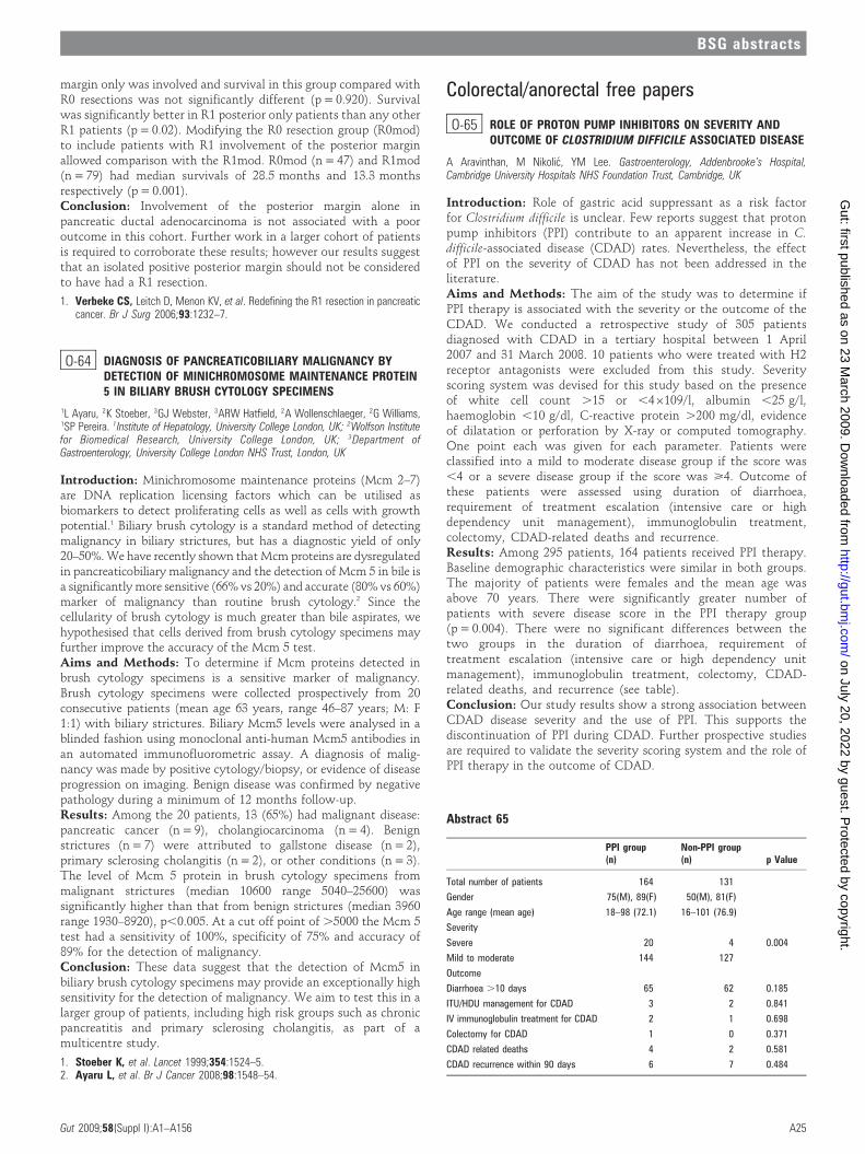
margin only was involved and survival in this group compared withR0 resections was not significantly different (p = 0.920). Survivalwas significantly better in R1 posterior only patients than any otherR1 patients (p = 0.02). Modifying the R0 resection group (R0mod)to include patients with R1 involvement of the posterior marginallowed comparison with the R1mod. R0mod (n = 47) and R1mod(n = 79) had median survivals of 28.5 months and 13.3 monthsrespectively (p = 0.001).Conclusion: Involvement of the posterior margin alone inpancreatic ductal adenocarcinoma is not associated with a pooroutcome in this cohort. Further work in a larger cohort of patientsis required to corroborate these results; however our results suggestthat an isolated positive posterior margin should not be consideredto have had a R1 resection.
1. Verbeke CS, Leitch D, Menon KV, et al. Redefining the R1 resection in pancreaticcancer. Br J Surg 2006;93:1232–7.
O-64 DIAGNOSIS OF PANCREATICOBILIARY MALIGNANCY BYDETECTION OF MINICHROMOSOME MAINTENANCE PROTEIN5 IN BILIARY BRUSH CYTOLOGY SPECIMENS
1L Ayaru, 2K Stoeber, 3GJ Webster, 3ARW Hatfield, 2A Wollenschlaeger, 2G Williams,1SP Pereira. 1Institute of Hepatology, University College London, UK; 2Wolfson Institutefor Biomedical Research, University College London, UK; 3Department ofGastroenterology, University College London NHS Trust, London, UK
Introduction: Minichromosome maintenance proteins (Mcm 2–7)are DNA replication licensing factors which can be utilised asbiomarkers to detect proliferating cells as well as cells with growthpotential.1 Biliary brush cytology is a standard method of detectingmalignancy in biliary strictures, but has a diagnostic yield of only20–50%. We have recently shown that Mcm proteins are dysregulatedin pancreaticobiliary malignancy and the detection of Mcm 5 in bile isa significantly more sensitive (66% vs 20%) and accurate (80% vs 60%)marker of malignancy than routine brush cytology.2 Since thecellularity of brush cytology is much greater than bile aspirates, wehypothesised that cells derived from brush cytology specimens mayfurther improve the accuracy of the Mcm 5 test.Aims and Methods: To determine if Mcm proteins detected inbrush cytology specimens is a sensitive marker of malignancy.Brush cytology specimens were collected prospectively from 20consecutive patients (mean age 63 years, range 46–87 years; M: F1:1) with biliary strictures. Biliary Mcm5 levels were analysed in ablinded fashion using monoclonal anti-human Mcm5 antibodies inan automated immunofluorometric assay. A diagnosis of malig-nancy was made by positive cytology/biopsy, or evidence of diseaseprogression on imaging. Benign disease was confirmed by negativepathology during a minimum of 12 months follow-up.Results: Among the 20 patients, 13 (65%) had malignant disease:pancreatic cancer (n = 9), cholangiocarcinoma (n = 4). Benignstrictures (n = 7) were attributed to gallstone disease (n = 2),primary sclerosing cholangitis (n = 2), or other conditions (n = 3).The level of Mcm 5 protein in brush cytology specimens frommalignant strictures (median 10600 range 5040–25600) wassignificantly higher than that from benign strictures (median 3960range 1930–8920), p,0.005. At a cut off point of .5000 the Mcm 5test had a sensitivity of 100%, specificity of 75% and accuracy of89% for the detection of malignancy.Conclusion: These data suggest that the detection of Mcm5 inbiliary brush cytology specimens may provide an exceptionally highsensitivity for the detection of malignancy. We aim to test this in alarger group of patients, including high risk groups such as chronicpancreatitis and primary sclerosing cholangitis, as part of amulticentre study.
1. Stoeber K, et al. Lancet 1999;354:1524–5.2. Ayaru L, et al. Br J Cancer 2008;98:1548–54.
Colorectal/anorectal free papers
O-65 ROLE OF PROTON PUMP INHIBITORS ON SEVERITY ANDOUTCOME OF CLOSTRIDIUM DIFFICILE ASSOCIATED DISEASE
A Aravinthan, M Nikolic, YM Lee. Gastroenterology, Addenbrooke’s Hospital,Cambridge University Hospitals NHS Foundation Trust, Cambridge, UK
Introduction: Role of gastric acid suppressant as a risk factorfor Clostridium difficile is unclear. Few reports suggest that protonpump inhibitors (PPI) contribute to an apparent increase in C.difficile-associated disease (CDAD) rates. Nevertheless, the effectof PPI on the severity of CDAD has not been addressed in theliterature.Aims and Methods: The aim of the study was to determine ifPPI therapy is associated with the severity or the outcome of theCDAD. We conducted a retrospective study of 305 patientsdiagnosed with CDAD in a tertiary hospital between 1 April2007 and 31 March 2008. 10 patients who were treated with H2receptor antagonists were excluded from this study. Severityscoring system was devised for this study based on the presenceof white cell count .15 or ,46109/l, albumin ,25 g/l,haemoglobin ,10 g/dl, C-reactive protein .200 mg/dl, evidenceof dilatation or perforation by X-ray or computed tomography.One point each was given for each parameter. Patients wereclassified into a mild to moderate disease group if the score was,4 or a severe disease group if the score was >4. Outcome ofthese patients were assessed using duration of diarrhoea,requirement of treatment escalation (intensive care or highdependency unit management), immunoglobulin treatment,colectomy, CDAD-related deaths and recurrence.Results: Among 295 patients, 164 patients received PPI therapy.Baseline demographic characteristics were similar in both groups.The majority of patients were females and the mean age wasabove 70 years. There were significantly greater number ofpatients with severe disease score in the PPI therapy group(p = 0.004). There were no significant differences between thetwo groups in the duration of diarrhoea, requirement oftreatment escalation (intensive care or high dependency unitmanagement), immunoglobulin treatment, colectomy, CDAD-related deaths, and recurrence (see table).Conclusion: Our study results show a strong association betweenCDAD disease severity and the use of PPI. This supports thediscontinuation of PPI during CDAD. Further prospective studiesare required to validate the severity scoring system and the role ofPPI therapy in the outcome of CDAD.
Abstract 65
PPI group(n)
Non-PPI group(n) p Value
Total number of patients 164 131
Gender 75(M), 89(F) 50(M), 81(F)
Age range (mean age) 18–98 (72.1) 16–101 (76.9)
Severity
Severe 20 4 0.004
Mild to moderate 144 127
Outcome
Diarrhoea .10 days 65 62 0.185
ITU/HDU management for CDAD 3 2 0.841
IV immunoglobulin treatment for CDAD 2 1 0.698
Colectomy for CDAD 1 0 0.371
CDAD related deaths 4 2 0.581
CDAD recurrence within 90 days 6 7 0.484
BSG abstracts
Gut 2009;58(Suppl I):A1–A156 A25
on July 20, 2022 by guest. Protected by copyright.
http://gut.bmj.com
/G
ut: first published as on 23 March 2009. D
ownloaded from
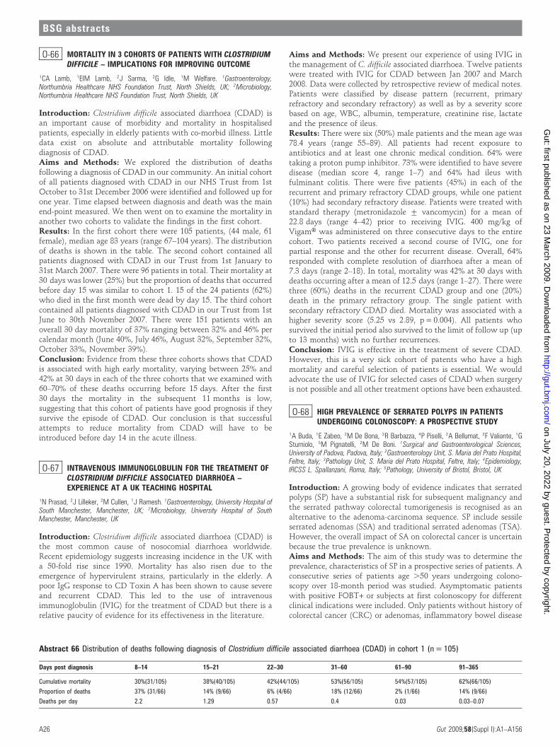
O-66 MORTALITY IN 3 COHORTS OF PATIENTS WITH CLOSTRIDIUMDIFFICILE – IMPLICATIONS FOR IMPROVING OUTCOME
1CA Lamb, 1EIM Lamb, 2J Sarma, 2G Idle, 1M Welfare. 1Gastroenterology,Northumbria Healthcare NHS Foundation Trust, North Shields, UK; 2Microbiology,Northumbria Healthcare NHS Foundation Trust, North Shields, UK
Introduction: Clostridium difficile associated diarrhoea (CDAD) isan important cause of morbidity and mortality in hospitalisedpatients, especially in elderly patients with co-morbid illness. Littledata exist on absolute and attributable mortality followingdiagnosis of CDAD.Aims and Methods: We explored the distribution of deathsfollowing a diagnosis of CDAD in our community. An initial cohortof all patients diagnosed with CDAD in our NHS Trust from 1stOctober to 31st December 2006 were identified and followed up forone year. Time elapsed between diagnosis and death was the mainend-point measured. We then went on to examine the mortality inanother two cohorts to validate the findings in the first cohort.Results: In the first cohort there were 105 patients, (44 male, 61female), median age 83 years (range 67–104 years). The distributionof deaths is shown in the table. The second cohort contained allpatients diagnosed with CDAD in our Trust from 1st January to31st March 2007. There were 96 patients in total. Their mortality at30 days was lower (25%) but the proportion of deaths that occurredbefore day 15 was similar to cohort 1. 15 of the 24 patients (62%)who died in the first month were dead by day 15. The third cohortcontained all patients diagnosed with CDAD in our Trust from 1stJune to 30th November 2007. There were 151 patients with anoverall 30 day mortality of 37% ranging between 32% and 46% percalendar month (June 40%, July 46%, August 32%, September 32%,October 33%, November 39%).Conclusion: Evidence from these three cohorts shows that CDADis associated with high early mortality, varying between 25% and42% at 30 days in each of the three cohorts that we examined with60–70% of these deaths occurring before 15 days. After the first30 days the mortality in the subsequent 11 months is low,suggesting that this cohort of patients have good prognosis if theysurvive the episode of CDAD. Our conclusion is that successfulattempts to reduce mortality from CDAD will have to beintroduced before day 14 in the acute illness.
O-67 INTRAVENOUS IMMUNOGLOBULIN FOR THE TREATMENT OFCLOSTRIDIUM DIFFICILE ASSOCIATED DIARRHOEA –EXPERIENCE AT A UK TEACHING HOSPITAL
1N Prasad, 2J Lilleker, 2M Cullen, 1J Ramesh. 1Gastroenterology, University Hospital ofSouth Manchester, Manchester, UK; 2Microbiology, University Hospital of SouthManchester, Manchester, UK
Introduction: Clostridium difficile associated diarrhoea (CDAD) isthe most common cause of nosocomial diarrhoea worldwide.Recent epidemiology suggests increasing incidence in the UK witha 50-fold rise since 1990. Mortality has also risen due to theemergence of hypervirulent strains, particularly in the elderly. Apoor IgG response to CD Toxin A has been shown to cause severeand recurrent CDAD. This led to the use of intravenousimmunoglobulin (IVIG) for the treatment of CDAD but there is arelative paucity of evidence for its effectiveness in the literature.
Aims and Methods: We present our experience of using IVIG inthe management of C. difficile associated diarrhoea. Twelve patientswere treated with IVIG for CDAD between Jan 2007 and March2008. Data were collected by retrospective review of medical notes.Patients were classified by disease pattern (recurrent, primaryrefractory and secondary refractory) as well as by a severity scorebased on age, WBC, albumin, temperature, creatinine rise, lactateand the presence of ileus.Results: There were six (50%) male patients and the mean age was78.4 years (range 55–89). All patients had recent exposure toantibiotics and at least one chronic medical condition. 64% weretaking a proton pump inhibitor. 73% were identified to have severedisease (median score 4, range 1–7) and 64% had ileus withfulminant colitis. There were five patients (45%) in each of therecurrent and primary refractory CDAD groups, while one patient(10%) had secondary refractory disease. Patients were treated withstandard therapy (metronidazole ¡ vancomycin) for a mean of22.8 days (range 4–42) prior to receiving IVIG. 400 mg/kg ofVigamH was administered on three consecutive days to the entirecohort. Two patients received a second course of IVIG, one forpartial response and the other for recurrent disease. Overall, 64%responded with complete resolution of diarrhoea after a mean of7.3 days (range 2–18). In total, mortality was 42% at 30 days withdeaths occurring after a mean of 12.5 days (range 1–27). There werethree (60%) deaths in the recurrent CDAD group and one (20%)death in the primary refractory group. The single patient withsecondary refractory CDAD died. Mortality was associated with ahigher severity score (5.25 vs 2.89, p = 0.004). All patients whosurvived the initial period also survived to the limit of follow up (upto 13 months) with no further recurrences.Conclusion: IVIG is effective in the treatment of severe CDAD.However, this is a very sick cohort of patents who have a highmortality and careful selection of patients is essential. We wouldadvocate the use of IVIG for selected cases of CDAD when surgeryis not possible and all other treatment options have been exhausted.
O-68 HIGH PREVALENCE OF SERRATED POLYPS IN PATIENTSUNDERGOING COLONOSCOPY: A PROSPECTIVE STUDY
1A Buda, 1E Zabeo, 2M De Bona, 3R Barbazza, 4P Piselli, 2A Bellumat, 2F Valiante, 1GSturniolo, 5M Pignatelli, 2M De Boni. 1Surgical and Gastroenterological Sciences,University of Padova, Padova, Italy; 2Gastroenterology Unit, S. Maria del Prato Hospital,Feltre, Italy; 3Pathology Unit, S. Maria del Prato Hospital, Feltre, Italy; 4Epidemiology,IRCSS L. Spallanzani, Roma, Italy; 5Pathology, University of Bristol, Bristol, UK
Introduction: A growing body of evidence indicates that serratedpolyps (SP) have a substantial risk for subsequent malignancy andthe serrated pathway colorectal tumorigenesis is recognised as analternative to the adenoma-carcinoma sequence. SP include sessileserrated adenomas (SSA) and traditional serrated adenomas (TSA).However, the overall impact of SA on colorectal cancer is uncertainbecause the true prevalence is unknown.Aims and Methods: The aim of this study was to determine theprevalence, characteristics of SP in a prospective series of patients. Aconsecutive series of patients age .50 years undergoing colono-scopy over 18-month period was studied. Asymptomatic patientswith positive FOBT+ or subjects at first colonoscopy for differentclinical indications were included. Only patients without history ofcolorectal cancer (CRC) or adenomas, inflammatory bowel disease
Abstract 66 Distribution of deaths following diagnosis of Clostridium difficile associated diarrhoea (CDAD) in cohort 1 (n = 105)
Days post diagnosis 8–14 15–21 22–30 31–60 61–90 91–365
Cumulative mortality 30%(31/105) 38%(40/105) 42%(44/105) 53%(56/105) 54%(57/105) 62%(66/105)
Proportion of deaths 37% (31/66) 14% (9/66) 6% (4/66) 18% (12/66) 2% (1/66) 14% (9/66)
Deaths per day 2.2 1.29 0.57 0.4 0.03 0.03–0.07
BSG abstracts
A26 Gut 2009;58(Suppl I):A1–A156
on July 20, 2022 by guest. Protected by copyright.
http://gut.bmj.com
/G
ut: first published as on 23 March 2009. D
ownloaded from
were enrolled. All colonoscopies were performed with white lightconventional endoscopes, the mean withdrawal time was 11.4 minand the caecum was reached in 94% of cases. For purposes of theanalysis, the junction of the splenic flexure and the descendingcolon, as determined by the endoscopist, defined the borderbetween the proximal and the distal colon. All retrieved polypswere examined by 2 independent pathologists (RB, MP) and SPclassified using the system described by Torlakovich et al.1
Results: Among 2036 patients included (48.9% M, mean age59.3¡6 years, and 51.1% F, mean age 60.1¡4 years), 707 (34.7%)had polyps. Most were adenomas (68.6%), followed by SA (20.8%,SSA 18%, TSA 2.8%, respectively) and hyperplastic polyps (5.4%).SSA were less than 5 mm in 81.5% of cases (p,0.001) and mostlylocated in the distal colon (88%, p,0.001). TSA were moreprevalent in distal colon (77.8%, p,0.001) and the size was lessthan 5 mm in 78.3% of cases. SP were significantly associated withadenomas (p,0.001). The presence of at least one SSA but not TSAwas associated with malignant polyp (p,0.001) and colorectalcancer (p,0.010). The prevalence of SA was significantly higher inthe FOBT+ group compared to symptomatic consecutive patients(11.8% vs 5.4%, p,0.0001).Conclusion: In our series SP prevalence was higher than previouslydescribed, particularly in the asymptomatic screening subgroupwith FOBT+ (11.8%) and associated with adenomas. SSA were flat,small (,5 mm) and significantly associated with malignant polypburden and CRC. Because of their potential for malignanttransformation, SSA should be identified and completely removed.
1. Torlakovic EE, Gomez JD, Driman DK, et al. Sessile serrated adenoma (SSA) vs.traditional serrated adenoma (TSA). Am J Surg Pathol 2008;32:21–9.
O-69 COMPARISON OF ANATOMICAL AND STAGE DISTRIBUTIONIN SYMPTOMATIC AND SCREEN DETECTED COLORECTALCANCERS
1I Kostourou, 2G Libby, 3FA Carey, 1RJC Steele. 1Surgery and Molecular Oncology,Ninewells Hospital and Medical School, Dundee, UK; 2Scottish Bowel ScreeningCentre; 3Pathology, Ninewells Hospital and Medical School, Dundee, UK
Introduction: Although screening is known to improve Dukesstage at diagnosis in colorectal cancer, little is known about theeffect on T stage, anatomical distribution or the interactionbetween stage and anatomical site.Aims and Methods: The study comprised patients diagnosed withcolorectal cancer (CRC) in Tayside, Scotland between March 2000and September 2007. Data came from the CRC database held atNinewells Hospital. Patients with FOBT screen detected cancers(SCR) were identified from the Scottish Bowel Screening PilotStudy and were then electronically linked to the cancer database bya unique patient identifier. Patients not identified as screen detectedwere deemed to be symptomatic (SYM). The age range of SYMpatients was limited to that of subjects who had been invited forbowel screening (50–69 years).Results: There were 437 (60.2% males) SYM and 187 (64.7%males) SCR patients. As expected, there was a significant differencein stage distribution with 83 (19.0%) of the SYM group comparedwith 77 (41.6%) of the SCR group diagnosed at Dukes A(p,0.0001). By the end of the study period 115 (26.3%) of theSYM group had died from CRC compared to 25 (13.4%) of the SCRgroup (P,0.0001). Within all three Dukes stages there weresignificant differences in the T stage distribution in the two groupswith consistently more favourable distributions for the SCR group(all p,0.04). In Dukes A 40.9% were staged as T1 and 59% as T2 inthe SYM group compared to 58.4% and 41.6% in SCR group.Within Dukes B 90.6% were stage T3 and 9.4% as T4 in the SCRgroup compared to 77.3% and 22.7% in the SYM group. WithinDukes C a similarly favourable shift in the T stage was seen in theSCR group. When anatomical site was examined there was asignificant difference (p = 0.02) in the anatomical distribution of
cancers in the two groups with a preponderance of left colontumours in the SCR group at the expense of right colonic and rectaltumours. When the stage distribution in the different anatomicalsites was scrutinised the proportion of Stage A cancers in the rightcolon, left colon and rectum was 6.6%, 20.6% and 26.6%respectively compared with 24.3%, 43.8% and 50% respectivelyfor the SCR group.Conclusion: In this cohort of patients defined by age andgeographical location, it was found that Dukes staging may bemisleading is assessing screen detected cancers as they had a morefavourable T stage distribution within each Dukes stage. Inaddition, screening appeared to slightly underdiagnose right sidedand rectal cancers, and although stage at presentation improvedfrom the right colon to the rectum in the symptomatic cancers thiswas mirrored by significantly better stage distributions in allanatomical sites in the screen detected group.
O-70 CONSENTING FOR COLORECTAL CANCER SCREENING – DOESPROVISION OF NUMERICAL DATA AND PICTOGRAPHS ONBENEFIT AND RISK DIMINISH UPTAKE?
I Patanwala, V Brocklebank, PN Trewby. Gastroenterology, Memorial Hospital,Darlington, UK
Introduction: Information given to patients invited for bowelcancer screening in the UK is descriptive and gives little numericalinformation on individual benefit or risk.1 Although the screeningfigures are impressive in the context of population benefit, it is theindividual, not the population, that decides whether to be screened.Graphical displays of information increase the effectiveness of riskcommunication, but understanding numerical information isdifficult. To allow true informed consent, information must betailored to the individual.Aims and Methods: We aimed to clarify: if presenting numericalfacts including absolute risks and benefits affected uptake of bowelcancer screening; if pictorial representation of data affected theuptake and understanding of the risks and benefits of colorectalcancer (CRC) screening; and, which of the many established factsconcerning screening might incline individuals more towardsaccepting screening. We gave 239 subjects (Group 1) an enhancedinformation pack containing numerical information expressedpictorially, including numbers needed to screen to accrue benefit(862); cancers missed by screening (50%); effect on survival (none);numbers of CRC prevented by earlier polyp pick up (0.5% at 18years). We asked, on the basis of information presented, whethersubjects wished to be screened and the points most influencingtheir decision and compared responses to a matched group of 239subjects (Group 2) sent the standard NHS pack. We asked ifpictorial representation of data on risks and benefits had an impacton their decision to be screened or not and if patients found themuseful in making an informed decision.Results: 53% replied, of whom 85% in group 1 and 90% in group 2wished to be screened. The use of pictographs with incorporatednumerical data significantly informed decision making (p = 0.0127,two sided Fisher exact test). This significance persisted when datawere shown numerically without the aid of a pictogram (p = 0.039,two sided Fisher exact test). Those wanting to be screened foundthe statement ‘‘bowel cancer is the second commonest cause ofdeath’’ had the highest impact. For those in group 1 not wanting tobe screened the statement ‘‘the programme will miss 50% ofcancers’’ had the highest negative impact. 83% of the respondents inthe intervention group found the numerical data helpful and 61%felt that the pictograms helped understand these data better.Conclusion: Providing numerical information did not diminishoverall uptake of screening in this hypothetical study but did helpinformed decision making. For this reason, and to allay futureconcerns about adequacy of informed consent, numerical data
BSG abstracts
Gut 2009;58(Suppl I):A1–A156 A27
on July 20, 2022 by guest. Protected by copyright.
http://gut.bmj.com
/G
ut: first published as on 23 March 2009. D
ownloaded from
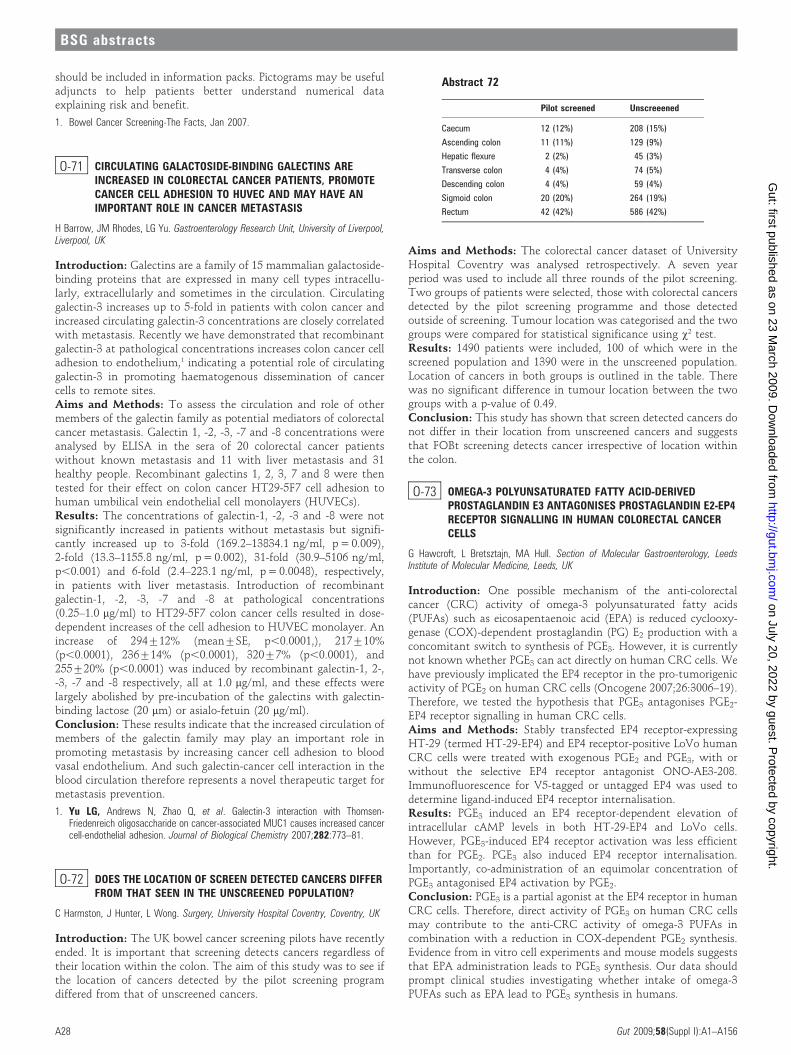
should be included in information packs. Pictograms may be usefuladjuncts to help patients better understand numerical dataexplaining risk and benefit.
1. Bowel Cancer Screening-The Facts, Jan 2007.
O-71 CIRCULATING GALACTOSIDE-BINDING GALECTINS AREINCREASED IN COLORECTAL CANCER PATIENTS, PROMOTECANCER CELL ADHESION TO HUVEC AND MAY HAVE ANIMPORTANT ROLE IN CANCER METASTASIS
H Barrow, JM Rhodes, LG Yu. Gastroenterology Research Unit, University of Liverpool,Liverpool, UK
Introduction: Galectins are a family of 15 mammalian galactoside-binding proteins that are expressed in many cell types intracellu-larly, extracellularly and sometimes in the circulation. Circulatinggalectin-3 increases up to 5-fold in patients with colon cancer andincreased circulating galectin-3 concentrations are closely correlatedwith metastasis. Recently we have demonstrated that recombinantgalectin-3 at pathological concentrations increases colon cancer celladhesion to endothelium,1 indicating a potential role of circulatinggalectin-3 in promoting haematogenous dissemination of cancercells to remote sites.Aims and Methods: To assess the circulation and role of othermembers of the galectin family as potential mediators of colorectalcancer metastasis. Galectin 1, -2, -3, -7 and -8 concentrations wereanalysed by ELISA in the sera of 20 colorectal cancer patientswithout known metastasis and 11 with liver metastasis and 31healthy people. Recombinant galectins 1, 2, 3, 7 and 8 were thentested for their effect on colon cancer HT29-5F7 cell adhesion tohuman umbilical vein endothelial cell monolayers (HUVECs).Results: The concentrations of galectin-1, -2, -3 and -8 were notsignificantly increased in patients without metastasis but signifi-cantly increased up to 3-fold (169.2–13834.1 ng/ml, p = 0.009),2-fold (13.3–1155.8 ng/ml, p = 0.002), 31-fold (30.9–5106 ng/ml,p,0.001) and 6-fold (2.4–223.1 ng/ml, p = 0.0048), respectively,in patients with liver metastasis. Introduction of recombinantgalectin-1, -2, -3, -7 and -8 at pathological concentrations(0.25–1.0 mg/ml) to HT29-5F7 colon cancer cells resulted in dose-dependent increases of the cell adhesion to HUVEC monolayer. Anincrease of 294¡12% (mean¡SE, p,0.0001,), 217¡10%(p,0.0001), 236¡14% (p,0.0001), 320¡7% (p,0.0001), and255¡20% (p,0.0001) was induced by recombinant galectin-1, 2-,-3, -7 and -8 respectively, all at 1.0 mg/ml, and these effects werelargely abolished by pre-incubation of the galectins with galectin-binding lactose (20 mm) or asialo-fetuin (20 mg/ml).Conclusion: These results indicate that the increased circulation ofmembers of the galectin family may play an important role inpromoting metastasis by increasing cancer cell adhesion to bloodvasal endothelium. And such galectin-cancer cell interaction in theblood circulation therefore represents a novel therapeutic target formetastasis prevention.
1. Yu LG, Andrews N, Zhao Q, et al. Galectin-3 interaction with Thomsen-Friedenreich oligosaccharide on cancer-associated MUC1 causes increased cancercell-endothelial adhesion. Journal of Biological Chemistry 2007;282:773–81.
O-72 DOES THE LOCATION OF SCREEN DETECTED CANCERS DIFFERFROM THAT SEEN IN THE UNSCREENED POPULATION?
C Harmston, J Hunter, L Wong. Surgery, University Hospital Coventry, Coventry, UK
Introduction: The UK bowel cancer screening pilots have recentlyended. It is important that screening detects cancers regardless oftheir location within the colon. The aim of this study was to see ifthe location of cancers detected by the pilot screening programdiffered from that of unscreened cancers.
Aims and Methods: The colorectal cancer dataset of UniversityHospital Coventry was analysed retrospectively. A seven yearperiod was used to include all three rounds of the pilot screening.Two groups of patients were selected, those with colorectal cancersdetected by the pilot screening programme and those detectedoutside of screening. Tumour location was categorised and the twogroups were compared for statistical significance using x2 test.Results: 1490 patients were included, 100 of which were in thescreened population and 1390 were in the unscreened population.Location of cancers in both groups is outlined in the table. Therewas no significant difference in tumour location between the twogroups with a p-value of 0.49.Conclusion: This study has shown that screen detected cancers donot differ in their location from unscreened cancers and suggeststhat FOBt screening detects cancer irrespective of location withinthe colon.
O-73 OMEGA-3 POLYUNSATURATED FATTY ACID-DERIVEDPROSTAGLANDIN E3 ANTAGONISES PROSTAGLANDIN E2-EP4RECEPTOR SIGNALLING IN HUMAN COLORECTAL CANCERCELLS
G Hawcroft, L Bretsztajn, MA Hull. Section of Molecular Gastroenterology, LeedsInstitute of Molecular Medicine, Leeds, UK
Introduction: One possible mechanism of the anti-colorectalcancer (CRC) activity of omega-3 polyunsaturated fatty acids(PUFAs) such as eicosapentaenoic acid (EPA) is reduced cyclooxy-genase (COX)-dependent prostaglandin (PG) E2 production with aconcomitant switch to synthesis of PGE3. However, it is currentlynot known whether PGE3 can act directly on human CRC cells. Wehave previously implicated the EP4 receptor in the pro-tumorigenicactivity of PGE2 on human CRC cells (Oncogene 2007;26:3006–19).Therefore, we tested the hypothesis that PGE3 antagonises PGE2-EP4 receptor signalling in human CRC cells.Aims and Methods: Stably transfected EP4 receptor-expressingHT-29 (termed HT-29-EP4) and EP4 receptor-positive LoVo humanCRC cells were treated with exogenous PGE2 and PGE3, with orwithout the selective EP4 receptor antagonist ONO-AE3-208.Immunofluorescence for V5-tagged or untagged EP4 was used todetermine ligand-induced EP4 receptor internalisation.Results: PGE3 induced an EP4 receptor-dependent elevation ofintracellular cAMP levels in both HT-29-EP4 and LoVo cells.However, PGE3-induced EP4 receptor activation was less efficientthan for PGE2. PGE3 also induced EP4 receptor internalisation.Importantly, co-administration of an equimolar concentration ofPGE3 antagonised EP4 activation by PGE2.Conclusion: PGE3 is a partial agonist at the EP4 receptor in humanCRC cells. Therefore, direct activity of PGE3 on human CRC cellsmay contribute to the anti-CRC activity of omega-3 PUFAs incombination with a reduction in COX-dependent PGE2 synthesis.Evidence from in vitro cell experiments and mouse models suggeststhat EPA administration leads to PGE3 synthesis. Our data shouldprompt clinical studies investigating whether intake of omega-3PUFAs such as EPA lead to PGE3 synthesis in humans.
Abstract 72
Pilot screened Unscreeened
Caecum 12 (12%) 208 (15%)
Ascending colon 11 (11%) 129 (9%)
Hepatic flexure 2 (2%) 45 (3%)
Transverse colon 4 (4%) 74 (5%)
Descending colon 4 (4%) 59 (4%)
Sigmoid colon 20 (20%) 264 (19%)
Rectum 42 (42%) 586 (42%)
BSG abstracts
A28 Gut 2009;58(Suppl I):A1–A156
on July 20, 2022 by guest. Protected by copyright.
http://gut.bmj.com
/G
ut: first published as on 23 March 2009. D
ownloaded from
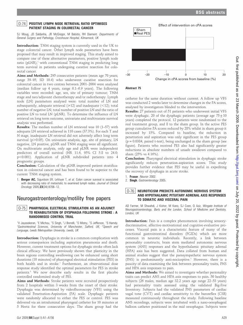
O-74 POSITIVE LYMPH NODE RETRIEVAL RATIO OPTIMISESPATIENT STAGING IN COLORECTAL CANCER
SJ Moug, JD Saldanha, JR McGregor, M Balsitis, RH Diament. Departments ofGeneral Surgery and Pathology, Crosshouse Hospital, Kilmarnock, UK
Introduction: TNM staging system is currently used in the UK tostage colorectal cancer. Other lymph node parameters have beenproposed that may result in improved staging. This study aimed tocompare one of these alternative parameters, positive lymph noderatio (pLNR),1 with conventional TNM staging in predicting longterm survival in patients undergoing curative resection for colo-rectal cancer.Aims and Methods: 295 consecutive patients (mean age 70 years;range 39–95; SD 10.4) who underwent curative resection forcolorectal cancer in two centres between 2001–2004 were analysed(median follow up 4 years, range 0.1–6.9 years). The followingvariables were recorded: age, sex, site of primary tumour; TNMstage and neo/adjuvant chemotherapy and/or radiotherapy. Lymphnode (LN) parameters analysed were: total number of LN andsubsequently, adequate retrieval (>12) and inadequate (,12); totalnumber of negative LN; total number of positive LN and the ratio ofpositive LN to total LN (pLNR). To determine the influence of LNretrieval on long term outcome, univariate and multivariate survivalanalysis was performed.Results: The median number of LN retrieved was 10 (1–57) withadequate LN retrieval achieved in 110 cases (37.3%). For each T andN stage, inadequate LN retrieval did not adversely affect long termsurvival (p.0.05). On univariate analysis, age, site of primary, LNnegative, LN positive, pLNR and TNM staging were all significant.On multivariate analysis, only age and pLNR were independentpredictors of overall survival (HR 11.6, 95% CI 5.8 to 24.0;p,0.001). Application of pLNR subdivided patients into 4prognostic groups.Conclusion: Calculation of the pLNR improved patient stratifica-tion in colorectal cancer and has been found to be superior to thecurrent TNM staging system.
1. Berger AC, Sigurdson ER, LeVoyer T, et al. Colon cancer survival is associatedwith decreasing ratio of metastatic to examined lymph nodes. Journal of ClinicalOncology 2005;23(34):8706–12.
Neurogastroenterology/motility free papers
O-75 PHARYNGEAL ELECTRICAL STIMULATION AS AN ADJUNCTTO REHABILITATION OF DYSPHAGIA FOLLOWING STROKE : ARANDOMISED CONTROL TRIAL
1V Jayasekeran, 1E Michou, 1S Singh, 2J Borrelli, 1S Mistry, 1S Jefferson, 1S Hamdy.1Gastrointestinal Sciences, University of Manchester, Salford, UK; 2Speech andLanguage, Leeds Metropolitan University, Leeds, UK
Introduction: Dysphagia in stroke is a common complication withserious consequences including aspiration pneumonia and death.However, current treatment options for dysphagic stroke often lackclinical efficacy. We have previously shown that plasticity in thebrain regions controlling swallowing can be enhanced using shortdurations (10 minutes) of pharyngeal electrical stimulation (PES) inboth health and in stroke.1 Furthermore, an observational doseresponse study identified the optimal parameters for PES in strokepatients.2 We now describe early results in the first placebocontrolled randomised study of PES.Aims and Methods: Stroke patients were recruited prospectivelyfrom 2 hospitals within 3 weeks from the onset of their stroke.Dysphagia was determined by videofluoroscopy (VFS) using thevalidated Penetration-Aspiration (PA) scale. Dysphagic patientswere randomly allocated to either the PES or control. PES wasdelivered via an intraluminal pharyngeal catheter for 10 minutes at5 Hertz for three consecutive days. The sham group had the
catheter for the same duration without current. A follow up VFSwas conducted 2 weeks later to determine changes in the PA scores,analysed by investigators blinded to the intervention.Results: 27 patients out of 51 patients who underwent initial VFSwere dysphagic. 20 of the dysphagic patients (average age 75¡10years) completed the protocol. 12 patients were randomised to thereal treatment group, and 8 to the sham group. In the active PESgroup cumulative PA scores reduced by 28% whilst in sham group itincreased by 15%. Compared to baseline, the reduction inpenetration and aspiration was only significant in the PES group(p = 0.0384, paired t-test), being unchanged in the sham group (seefigure). Patients who received PES also had significantly greaterreductions in absolute numbers of unsafe swallows compared tosham (25% vs 4.16%).Conclusion: Pharyngeal electrical stimulation in dysphagic strokesignificantly reduces penetration-aspiration scores. This studyprovides further evidence that PES may be useful in expeditingthe recovery of dysphagia in acute stroke.
1. Fraser. Neuron 2002.2. Singh. Gastroenterology 2005.
O-76 NEUROTICISM PREDICTS AUTONOMIC NERVOUS SYSTEMAND HYPOTHALAMIC PITUITARY ADRENAL AXIS RESPONSESTO SOMATIC AND VISCERAL PAIN
AD Farmer, M Shwahdi, J Kishor, M Kano, SJ Coen, Q Aziz. Wingate Institute ofNeurogastroenterology, Barts and the London, School of Medicine and Dentistry,London, UK
Introduction: Pain is a complex phenomenon involving sensory-discriminative, affective-motivational and cognitive-evaluative pro-cesses. Visceral pain is a characteristic feature of many of thefunctional gastrointestinal disorders (FGIDs) which are morecommon in neurotic individuals. Recently, a link betweenpersonality constructs, brain stem mediated autonomic nervoussystem (ANS) responses and the hypothalamic pituitary adrenal(HPA) axis has been suggested. Data collated from human andanimal studies suggest that the parasympathetic nervous system(PNS) is predominantly anti-nociceptive.1 However, there is apaucity of data examining the link between personality traits, PNSand HPA axis responses to pain.Aims and Methods: We aimed to investigate whether personalitytraits can predict ANS and HPA axis responses to pain. 50 healthysubjects (26 males, median age 32.2 years age range 22–53 years)had personality traits assessed using the validated Big-FiveInventory. Subjects had the validated PNS parameters of cardiacvagal tone (CVT) and cardiac sensitivity to the baroreflex (CSB)measured continuously throughout the study. Following baselineANS recordings, subjects were intubated with a naso-oesophagealballoon catheter positioned in the mid oesophagus. Subjects were
Abstract 75
BSG abstracts
Gut 2009;58(Suppl I):A1–A156 A29
on July 20, 2022 by guest. Protected by copyright.
http://gut.bmj.com
/G
ut: first published as on 23 March 2009. D
ownloaded from
randomised to receive visceral followed by somatic pain, or viceversa, and asked to rate pain intensity and unpleasantness on averbal rating scale. Seven painful visceral stimuli were administered,by oesophageal distension through inflation of the oesophagealballoon, to the subject’s pain tolerance with an inter-stimulusinterval of 2-minutes. Seven painful somatic stimuli, using nail bedpressure with a 2-minute inter-stimulus interval, were administeredto the subject’s pain tolerance. Cortisol was measured at baseline,5 minutes post visceral and 5 minutes post somatic stimulation.Results: Baseline CVT and CSB correlated negatively with neuroti-cism (r = 20.6, p = ,0.0001 and r = 20.4, p = 0.01 respectively).Baseline CVT and CSB correlated with CVT (r = 0.86, p,0.0001 andr = 0.7, p,0.0001) and CSB (r = 0.8, p,0.0001 and r = 0.62,p,0.0001) response to somatic and visceral pain. Subjects ratedvisceral pain as more unpleasant (p,0.0001), but not more intense,than somatic pain. Neuroticism correlated with baseline cortisol levelsand also to cortisol levels following somatic and visceral pain stimuli(r = 0.35, p = 0.01 and r = 0.37, p = 0.008 and r = 0.38, p = 0.006).Conclusion: These results demonstrate that the personalityconstruct of neuroticism can predict baseline PNS and HPA toneand responses to somatic and visceral pain. Neuroticism, cortisollevels and PNS measures may represent clinically applicablebiomarkers for pain responsiveness in health. These findingswarrant further assessment in a larger cohort of healthy volunteersand in those with FGIDs.
1. Hisata, et al. Cardiac nociceptors innervated by vagal afferents in rats. AutonNeurosci 2006;126–127:174–8.
O-77 ELEVATED CHEMOKINE MRNA IN RECTAL MUCOSA INIRRITABLE BOWEL SYNDROME; INCREASED BYGASTROINTESTINAL INFECTION BUT UNRELATED TO GUTPERMEABILITY
1RC Spiller, 1E Campbell, 1M Richards, 1K Garsed, 1A Zaitoun, 2M Hastings,1Y Mahida, 3I Hall, 1K Neal, 4J Cox, 4F Kelly, 4J Wilde, 4G Dukes, 2P Whorwell.1Nottingham Digestive Diseases Centre BRU, University of Nottingham, Nottingham, UK;2Department of Gastroenterology, University of South Manchester, Wythenshawe Hospital,Manchester, UK; 3Department of Therapeutics and Molecular Medicine, University ofNottingham, Nottingham, UK; 4Discovery Technology Group, Molecular DiscoveryResearch, GSK, Harlow, UK
Introduction: Previous studies have suggested inflammatorychanges in colonic mucosa peripheral blood mononuclear cells(PBMC) in irritable bowel syndrome (IBS) patients with diarrhoea(IBS-D). We have previously reported persistent IBS symptomsfollowing C. jejuni enteritis. We hypothesised that genes upregu-lated by infection may also be upregulated in other subtypes of IBS.Aims and Methods: 25 healthy volunteers (HV), 31 patients6 months after C. jejuni infection, 37 IBS-D patients and 19 IBSwith constipation (IBS-C) provided stool and blood samples and 24hour urine collection for measuring gut permeability. Rectalmucosal biopsy mRNA was assessed using an Affymetrix genechipto identify genes whose expression was altered by C. jejuni infectionor in IBS. Findings were confirmed using TaqMan RT-PCR andcorrelated with histology, PBMC cytokine production, gut perme-ability, bowel and psychological symptoms.Results: Expression of the chemokines genes CCL11, CCL 13 andCalpain 8 were significantly increased in rectal biopsies 6 months
after C. jejuni infection while NR1D1 was decreased (see table). Thesame changes were also seen in IBS-D and also IBS-C. Gutpermeability, mucosal mast cells and PMBC production of TNF-alpha and IL6 were significantly elevated 6 months following C.jejuni infection. Individuals with persistent bowel dysfunction hadhigher mast cell numbers but otherwise showed no differences inmucosal mRNA nor PBMC cytokine production from those whorecovered completely, though they were more likely to have rectalbleeding. PI-BD patients were also more likely to be female, youngerand have significantly higher PHQ12, anxiety and depression scoresthan PI-non-IBS. Overall PBMC TNF, IL10 and IL1-beta productioncorrelated closely (r = 0.8–0.90) but none correlated with gutpermeability. TNF showed a weak correlation with days per weekwith loose stool, r = 0.24, p,0.05. Stool cytokines, plasma CRP andurine neopterin showed no group differences.Conclusion: Mucosal expression of proinflammatory chemokinesgenes, mast cell numbers and PBMC cytokine production areelevated 6 months following C. jejuni infection. Similar increasedchemokine mRNA and PCMC cytokine production are also seen inIBS patients regardless of subtype.
O-78 SOUP MEAL DELAYS GASTRIC EMPTYING, ENHANCESGALLBLADDER CONTRACTION AND INCREASES SMALLBOWEL WATER CONTENT AND SATIETY COMPARED TOEQUICALORIC MIXED SOLID/LIQUID MEAL
1L Marciani, 1N Hall, 2SE Pritchard, 3JJ Totman, 2CL Hoad, 2PA Gowland, 1RC Spiller.1Nottingham Digestive Diseases Centre Biomedical Research Unit, NottinghamUniversity Hospitals, Nottingham, UK; 2Sir Peter Mansfield Magnetic ResonanceCentre, Nottingham, UK; 3Brain and Body Centre, University of Nottingham,Nottingham, UK
Introduction: There is currently great interest in manipulating thephysical form of food to enhance satiety. The stomach can ‘‘sieve’’water from the solid component of a meal leading to faster gastricemptying. We hypothesised that if sieving was prevented byblending a solid/liquid meal to a soup: (a) gastric volumes would fallmore slowly; (b) satiety would be enhanced; (c) the CCK duodenalresponse (as inferred from gallbladder contraction)1 would beenhanced; and (d) the small bowel secretion would be greater.Aims and Methods: To investigate how blending a solid/liquidmeal to a soup affects satiety, gastric emptying, gallbladdercontraction and small bowel gastric content. Satiety study 1: 22healthy volunteers attended in the morning, having fasted over-night and were fed either a roasted chicken and vegetables mealwith a glass of water (241 kcal) or the same meal and water blendedto a soup. The volunteers were asked to fill in satiety questionnairesfasting and at intervals for 3 hours postprandially. Mechanistic study2: 18 healthy volunteers attended in the morning, having fastedovernight, and were scanned in a 1.5T MRI scanner. They werethen fed the same meals as above and underwent scanning atintervals for 3 hours. Gastric contents and gallbladder contractionwere measured from the abdominal volume scans. Small bowelwater content was measured from magnetic resonance cholangio-pancreatography (MRCP) scans, as previously described, andvalidated by intubation studies in which measured values wereshown to closely parallel infused volumes.2 Two-way analysis ofvariance and paired comparisons were used.
Abstract 77 Gene expression as assessed by TaqMan Real-Time PCR
Median (range) CCL11 CCL13 TNFSF15 Calpain 8 NR1D1 GPR161
Healthy volunteers 0.21 (0.06–1.0) 0.23 (0.1–10.6) 1.7 (0.9–2.9) 14.9 (6.7–28.4) 6.4 (2.0–15.2) 3.9 (2.2–5.9)
6 mths post infection 0.53 (0.1–1.5)* 0.53 (0.1–2.3)* 1.8 (1.1–4.1) 17.2 (12.3–29)* 3.2 (1.6–6.9)* 2.8 (1.7–6.6)
IBS-D 0.35 (0.06–2.5) 0.35 (0.1–1.4)* 2.2 (1.1–3.3)* 18.1 (9.4–46)* 3.3 (1.4–9)* 3.1 (1.9–4.4)*
All values 6 10–3.*p,0.05 after Bonferonni correction.
BSG abstracts
A30 Gut 2009;58(Suppl I):A1–A156
on July 20, 2022 by guest. Protected by copyright.
http://gut.bmj.com
/G
ut: first published as on 23 March 2009. D
ownloaded from
Results: (mean¡SEM) Study 1: The soup meal induced lowerfeelings of hunger (p,0.02). Study 2: Gastric volumes were greaterafter the soup than solid meal at 30 min (360¡15 ml vs 265¡8 mlrespectively, p,0.0003) and 75 min (198¡10 ml vs 256¡22 ml,p = 0.02). The % gallbladder contraction was greater after the soupmeal at 30 min (58¡2% vs 48¡2%, p = 0.0002) and 75 min (44¡2%vs 32¡2%, p,0.0001). Small bowel water content was initiallyhigher for the solid meal (111¡15 ml vs 70¡8 ml immediately afterfeeding, p,0.02) whilst the soup meal induced greater secretion atlater times (186¡23 vs 124¡16 ml at 165 min, p,0.007).Conclusion: Blending the test meal to a soup abolished gastric‘‘sieving’’ resulting in larger gastric volumes and enhanced gallbladdercontraction, a marker of CCK release.1 The small bowel water contentwas higher for the soup meal at the later time points, implying thatthe latter induced greater secretory stimulation. These combinedgastrointestinal factors induced an enhanced satiety after the soupmeal. MRI provided exquisite insights into the functional gastro-intestinal response to altered physical form of meals.
1. Froehlich, et al. Dig Dis Sci 1995;40:529–33.2. Hoad, et al. Phys Med Biol 2007;52:6909–22.
O-79 AN ‘‘INSIGHT’’ INTO DIABETIC GASTROENTEROPATHY USINGCORNEAL CONFOCAL MICROSCOPY
1P Begum, 2M Tavakoli, 2RA Malik, 1J McLaughlin. 1GI Sciences, University ofManchester, Salford, Manchester, UK; 2Cardiovascular & Endocrine Sciences,University of Manchester, Manchester, UK
Introduction: Diabetes causing autonomic dysfunction is asso-ciated with various gastrointestinal (GI) motility disorders such asgastroparesis and diabetic diarrhoea (diabetic gastroenteropathy,DGE). However, symptoms are often poorly predictive of motorabnormalities and vice-versa, despite underlying diabetic autonomicneuropathy (DAN). Accurate quantification of the severity of DANremains time consuming and challenging, but corneal confocalmicroscopy (CCM) is a novel, non-invasive technique whichquantifies small C-nerve fibre pathology.Aims and Methods: This study aimed to determine whether cornealC-fibre status would mirror DAN in diabetic patients with gastro-intestinal (GI) symptoms. Twenty diabetic subjects (14 Type 1, 6 Type2) with symptoms of gastroparesis and/or diarrhoea, and 14 controlsubjects underwent cardiovascular autonomic profiling using theComposite Autonomic Scoring Scale (CASS). The severity of gastro-paresis was evaluated using the Gastroparesis Cardinal Symptom Index(GCSI), whilst nuclear scintigraphy or the 13C-sodium acetate breathtest determined gastric emptying. Corneal nerve morphology wasquantified using CCM and corneal aesthesiometry assessed CornealSensitivity (CS). All data are expressed as mean ¡ SD.Results: DAN was seen in 18/20 diabetic subjects. The severity byCASS was moderate-severe in 12 and mild in 6 patients (mean CASS4.0¡2.2). The severity of gastroparesis was also moderate (meanGCSI 2.3¡1.0), yet gastric emptying studies confirmed delay in only10/18 (56%) subjects (mean GCSI 2.4¡1.1; CASS 4.6¡1.8). Rapidemptying/dysmotility was seen in 3 subjects (GCSI 2.9¡0.9; CASS1.7¡2.1), whilst 5 patients had normal emptying (GCSI 1.7¡0.5;CASS 5.0¡1.6). Correlation of CASS vs GCSI was poor (r = 20.3;p = 0.254). Corneal nerve fibre density (NFD, 17.8¡13.3 vs48.3¡12.4), nerve branch density (NBD, 8.9¡7.9 vs 30.1¡5.3) andnerve fibre length (NFL, 3.6¡3.1 vs 9.7¡2.5) were all significantlyreduced in diabetics vs controls (p,0.0001). CS was also reduced withsignificant elevation of corneal sensory thresholds (1.6¡0.9 vs0.7¡0.2; p,0.0004). All abnormalities were reduced in proportionto the severity of DAN, with highly significant correlations(p,0.0001) between CASS and NFD (r = 20.713), NBD(r = 20.726), NFL (r = 20.786) and CS (r = 0.659).Conclusion: In patients with DGE, DAN was common. GIsymptoms were not necessarily reflective of motor dysfunction,nor representative of the severity of underlying neuropathy.
Corneal nerve morphology was also abnormal in these patientscompared with healthy controls and the severity of nerve damagewas very strongly associated with the severity of autonomicneuropathy. This suggests that CCM may be a useful rapid, simpleand non-invasive diagnostic marker of DAN or indeed DGE.
Supported by Diabetes UK/CORE
Oesophageal free papers
O-80 GENERATION AND VALIDATION OF A PROGNOSTIC GENESIGNATURE FOR OESOPHAGEAL ADENOCARCINOMA
1CJ Peters, 2JRE Rees, 3JS Hardwick, 3C Zhan, 4RH Hardwick, 1RC Fitzgerald, and onbehalf of the OCCAMS Study Group11MRC Cancer Cell Unit, Hutchison-MRC ResearchCentre, Cambridge, UK; 2Department of Surgery, Bristol Royal Infirmary, Bristol, UK;3Molecular Profiling, Merck Research Laboratories, North Wales, USA; 4CambridgeOesophago-Gastric Centre, Addenbrookes Hospital, Cambridge, UK
Introduction: Oesophageal adenocarcinoma is an increasinglycommon cancer with a poor prognosis. The incidence has increasedmore than 6 fold in the last 3 decades and survival at five yearsremains less than 14%. Complex management decisions and thehighly invasive nature of oesophageal surgery mean that patientsand clinicians need better prognostic information.Aims and Methods: This study aims to generate and validate amolecular prognostic signature for oesophageal adenocarcinoma.Snap frozen tumour samples were collected from 91 patientsundergoing potentially curative surgery at Bristol Royal Infirmaryfor oesophageal and junctional adenocarcinoma. Median length offollow-up was 1.7 years for all patients and 7.0 years for patients aliveat last follow up. RNA was extracted and hybridised to a 44K 60-meroligo-microarray. The data were detrended and normalised and theresulting expression correlated with clinical features to derive amolecular prognostic signature. The prognostic signature is beingvalidated at the protein level via immunohistochemistry bothinternally on the same samples and externally with tissue microarraysfrom a further 600 cases of adenocarcinoma.Results: High quality data were obtained from 76 of the tumoursamples. Lists of 119 genes associated with survival and 270 genesassociated with the number of involved lymph nodes were created.Gene list filtering using statistical and biological criteria produced ashort list of the 20 genes most predictive of survival. Combined thislist of genes was highly prognostic. Patients with dysregulation of atleast 4 of the 20 genes had a median survival of 11.4 months comparedwith 52.5 months for those with dysregulation of ,4 genes(p,0.0001). This prognostic signature outperforms all the clinicalindicators in predicting survival in multivariate analysis. To date twogenes have been validated internally: PAPPS2- if not expressed mediansurvival is 6.4 months compared with 27.2 months if expressed(p = 0.002); and HEF1- if not expressed median survival 7.8 monthscompared with 17.0 months if expressed (p = 0.027).Conclusion: We have determined that a 20 gene expressionsignature may outperform clinical indicators in predicting outcome.Protein validation is underway with the aim that a combination ofa smaller number of genes and the existing clinical staging coulddramatically improve our ability to predict survival and allow bettertargeting of novel treatments.
O-81 PERSONALISING CHEMOTHERAPY TO IMPROVE OESOPHAGO-GASTRIC CANCER OUTCOMES
1AL Paterson, 2TM Gilmer, 3RF Wooster, 1RC Fitzgerald. 1MRC Cancer Cell Unit,Hutchison MRC Research Centre, Cambridge, UK; 2Translational Medicine,GlaxoSmithKline, North Carolina, USA; 3Translational Medicine, GlaxoSmithKline,Philadelphia, USA
Introduction: Oesophago-gastric cancers have been identified as amajor public health concern due to their increasing incidence and
BSG abstracts
Gut 2009;58(Suppl I):A1–A156 A31
on July 20, 2022 by guest. Protected by copyright.
http://gut.bmj.com
/G
ut: first published as on 23 March 2009. D
ownloaded from
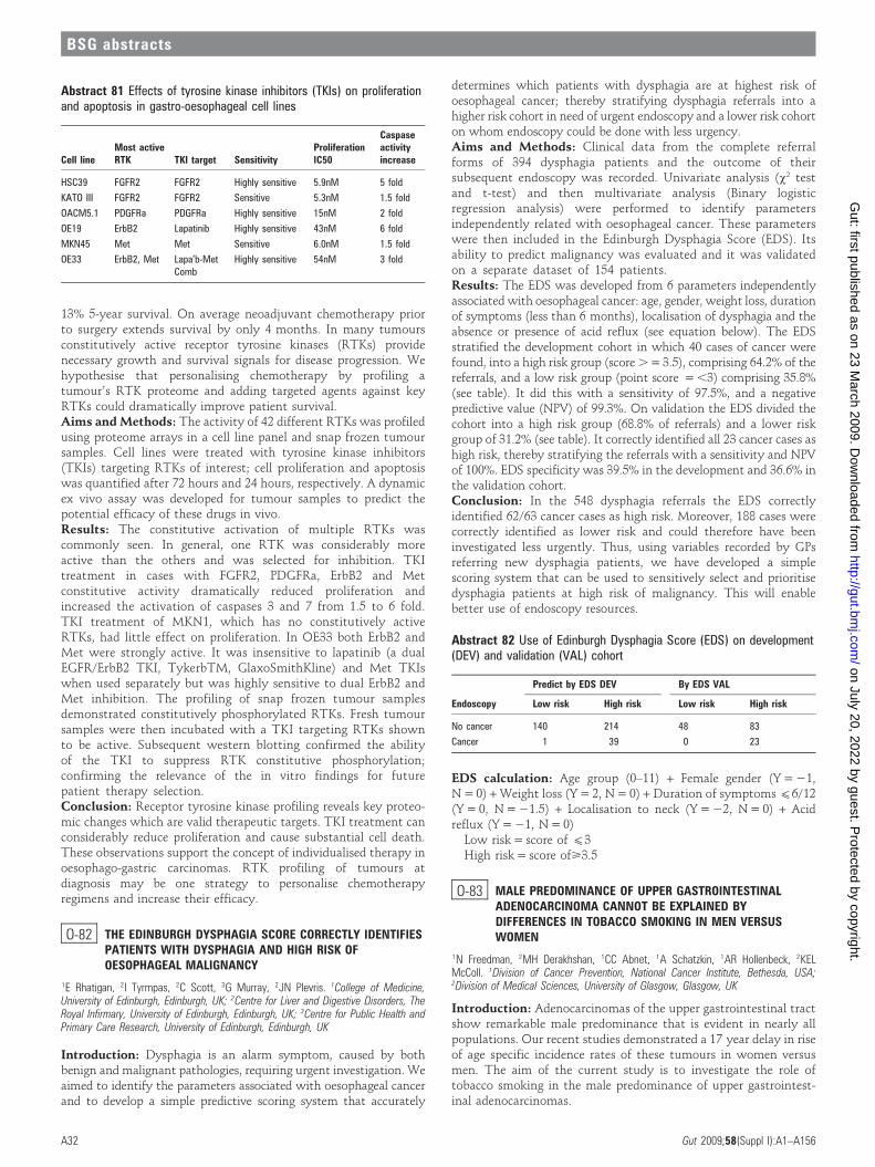
13% 5-year survival. On average neoadjuvant chemotherapy priorto surgery extends survival by only 4 months. In many tumoursconstitutively active receptor tyrosine kinases (RTKs) providenecessary growth and survival signals for disease progression. Wehypothesise that personalising chemotherapy by profiling atumour’s RTK proteome and adding targeted agents against keyRTKs could dramatically improve patient survival.Aims and Methods: The activity of 42 different RTKs was profiledusing proteome arrays in a cell line panel and snap frozen tumoursamples. Cell lines were treated with tyrosine kinase inhibitors(TKIs) targeting RTKs of interest; cell proliferation and apoptosiswas quantified after 72 hours and 24 hours, respectively. A dynamicex vivo assay was developed for tumour samples to predict thepotential efficacy of these drugs in vivo.Results: The constitutive activation of multiple RTKs wascommonly seen. In general, one RTK was considerably moreactive than the others and was selected for inhibition. TKItreatment in cases with FGFR2, PDGFRa, ErbB2 and Metconstitutive activity dramatically reduced proliferation andincreased the activation of caspases 3 and 7 from 1.5 to 6 fold.TKI treatment of MKN1, which has no constitutively activeRTKs, had little effect on proliferation. In OE33 both ErbB2 andMet were strongly active. It was insensitive to lapatinib (a dualEGFR/ErbB2 TKI, TykerbTM, GlaxoSmithKline) and Met TKIswhen used separately but was highly sensitive to dual ErbB2 andMet inhibition. The profiling of snap frozen tumour samplesdemonstrated constitutively phosphorylated RTKs. Fresh tumoursamples were then incubated with a TKI targeting RTKs shownto be active. Subsequent western blotting confirmed the abilityof the TKI to suppress RTK constitutive phosphorylation;confirming the relevance of the in vitro findings for futurepatient therapy selection.Conclusion: Receptor tyrosine kinase profiling reveals key proteo-mic changes which are valid therapeutic targets. TKI treatment canconsiderably reduce proliferation and cause substantial cell death.These observations support the concept of individualised therapy inoesophago-gastric carcinomas. RTK profiling of tumours atdiagnosis may be one strategy to personalise chemotherapyregimens and increase their efficacy.
O-82 THE EDINBURGH DYSPHAGIA SCORE CORRECTLY IDENTIFIESPATIENTS WITH DYSPHAGIA AND HIGH RISK OFOESOPHAGEAL MALIGNANCY
1E Rhatigan, 2I Tyrmpas, 2C Scott, 3G Murray, 2JN Plevris. 1College of Medicine,University of Edinburgh, Edinburgh, UK; 2Centre for Liver and Digestive Disorders, TheRoyal Infirmary, University of Edinburgh, Edinburgh, UK; 3Centre for Public Health andPrimary Care Research, University of Edinburgh, Edinburgh, UK
Introduction: Dysphagia is an alarm symptom, caused by bothbenign and malignant pathologies, requiring urgent investigation. Weaimed to identify the parameters associated with oesophageal cancerand to develop a simple predictive scoring system that accurately
determines which patients with dysphagia are at highest risk ofoesophageal cancer; thereby stratifying dysphagia referrals into ahigher risk cohort in need of urgent endoscopy and a lower risk cohorton whom endoscopy could be done with less urgency.Aims and Methods: Clinical data from the complete referralforms of 394 dysphagia patients and the outcome of theirsubsequent endoscopy was recorded. Univariate analysis (x2 testand t-test) and then multivariate analysis (Binary logisticregression analysis) were performed to identify parametersindependently related with oesophageal cancer. These parameterswere then included in the Edinburgh Dysphagia Score (EDS). Itsability to predict malignancy was evaluated and it was validatedon a separate dataset of 154 patients.Results: The EDS was developed from 6 parameters independentlyassociated with oesophageal cancer: age, gender, weight loss, durationof symptoms (less than 6 months), localisation of dysphagia and theabsence or presence of acid reflux (see equation below). The EDSstratified the development cohort in which 40 cases of cancer werefound, into a high risk group (score . = 3.5), comprising 64.2% of thereferrals, and a low risk group (point score = ,3) comprising 35.8%(see table). It did this with a sensitivity of 97.5%, and a negativepredictive value (NPV) of 99.3%. On validation the EDS divided thecohort into a high risk group (68.8% of referrals) and a lower riskgroup of 31.2% (see table). It correctly identified all 23 cancer cases ashigh risk, thereby stratifying the referrals with a sensitivity and NPVof 100%. EDS specificity was 39.5% in the development and 36.6% inthe validation cohort.Conclusion: In the 548 dysphagia referrals the EDS correctlyidentified 62/63 cancer cases as high risk. Moreover, 188 cases werecorrectly identified as lower risk and could therefore have beeninvestigated less urgently. Thus, using variables recorded by GPsreferring new dysphagia patients, we have developed a simplescoring system that can be used to sensitively select and prioritisedysphagia patients at high risk of malignancy. This will enablebetter use of endoscopy resources.
EDS calculation: Age group (0–11) + Female gender (Y = 21,N = 0) + Weight loss (Y = 2, N = 0) + Duration of symptoms (6/12(Y = 0, N = 21.5) + Localisation to neck (Y = 22, N = 0) + Acidreflux (Y = 21, N = 0)
Low risk = score of (3High risk = score of>3.5
O-83 MALE PREDOMINANCE OF UPPER GASTROINTESTINALADENOCARCINOMA CANNOT BE EXPLAINED BYDIFFERENCES IN TOBACCO SMOKING IN MEN VERSUSWOMEN
1N Freedman, 2MH Derakhshan, 1CC Abnet, 1A Schatzkin, 1AR Hollenbeck, 2KELMcColl. 1Division of Cancer Prevention, National Cancer Institute, Bethesda, USA;2Division of Medical Sciences, University of Glasgow, Glasgow, UK
Introduction: Adenocarcinomas of the upper gastrointestinal tractshow remarkable male predominance that is evident in nearly allpopulations. Our recent studies demonstrated a 17 year delay in riseof age specific incidence rates of these tumours in women versusmen. The aim of the current study is to investigate the role oftobacco smoking in the male predominance of upper gastrointest-inal adenocarcinomas.
Abstract 81 Effects of tyrosine kinase inhibitors (TKIs) on proliferationand apoptosis in gastro-oesophageal cell lines
Cell lineMost activeRTK TKI target Sensitivity
ProliferationIC50
Caspaseactivityincrease
HSC39 FGFR2 FGFR2 Highly sensitive 5.9nM 5 fold
KATO III FGFR2 FGFR2 Sensitive 5.3nM 1.5 fold
OACM5.1 PDGFRa PDGFRa Highly sensitive 15nM 2 fold
OE19 ErbB2 Lapatinib Highly sensitive 43nM 6 fold
MKN45 Met Met Sensitive 6.0nM 1.5 fold
OE33 ErbB2, Met Lapa’b-MetComb
Highly sensitive 54nM 3 fold
Abstract 82 Use of Edinburgh Dysphagia Score (EDS) on development(DEV) and validation (VAL) cohort
Endoscopy
Predict by EDS DEV By EDS VAL
Low risk High risk Low risk High risk
No cancer 140 214 48 83
Cancer 1 39 0 23
BSG abstracts
A32 Gut 2009;58(Suppl I):A1–A156
on July 20, 2022 by guest. Protected by copyright.
http://gut.bmj.com
/G
ut: first published as on 23 March 2009. D
ownloaded from
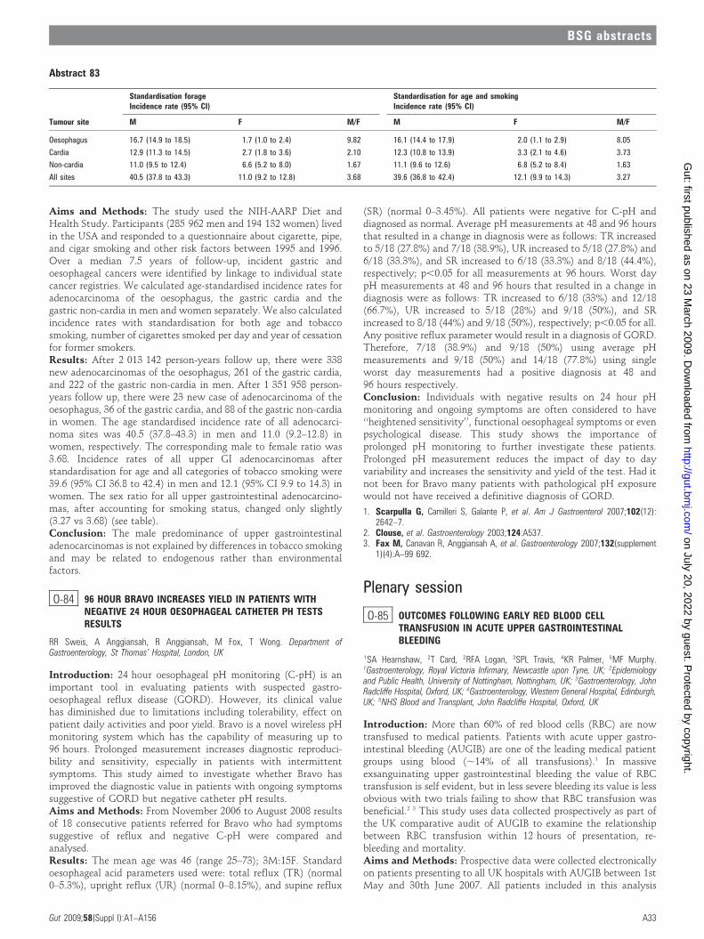
Aims and Methods: The study used the NIH-AARP Diet andHealth Study. Participants (285 962 men and 194 132 women) livedin the USA and responded to a questionnaire about cigarette, pipe,and cigar smoking and other risk factors between 1995 and 1996.Over a median 7.5 years of follow-up, incident gastric andoesophageal cancers were identified by linkage to individual statecancer registries. We calculated age-standardised incidence rates foradenocarcinoma of the oesophagus, the gastric cardia and thegastric non-cardia in men and women separately. We also calculatedincidence rates with standardisation for both age and tobaccosmoking, number of cigarettes smoked per day and year of cessationfor former smokers.Results: After 2 013 142 person-years follow up, there were 338new adenocarcinomas of the oesophagus, 261 of the gastric cardia,and 222 of the gastric non-cardia in men. After 1 351 958 person-years follow up, there were 23 new case of adenocarcinoma of theoesophagus, 36 of the gastric cardia, and 88 of the gastric non-cardiain women. The age standardised incidence rate of all adenocarci-noma sites was 40.5 (37.8–43.3) in men and 11.0 (9.2–12.8) inwomen, respectively. The corresponding male to female ratio was3.68. Incidence rates of all upper GI adenocarcinomas afterstandardisation for age and all categories of tobacco smoking were39.6 (95% CI 36.8 to 42.4) in men and 12.1 (95% CI 9.9 to 14.3) inwomen. The sex ratio for all upper gastrointestinal adenocarcino-mas, after accounting for smoking status, changed only slightly(3.27 vs 3.68) (see table).Conclusion: The male predominance of upper gastrointestinaladenocarcinomas is not explained by differences in tobacco smokingand may be related to endogenous rather than environmentalfactors.
O-84 96 HOUR BRAVO INCREASES YIELD IN PATIENTS WITHNEGATIVE 24 HOUR OESOPHAGEAL CATHETER PH TESTSRESULTS
RR Sweis, A Anggiansah, R Anggiansah, M Fox, T Wong. Department ofGastroenterology, St Thomas’ Hospital, London, UK
Introduction: 24 hour oesophageal pH monitoring (C-pH) is animportant tool in evaluating patients with suspected gastro-oesophageal reflux disease (GORD). However, its clinical valuehas diminished due to limitations including tolerability, effect onpatient daily activities and poor yield. Bravo is a novel wireless pHmonitoring system which has the capability of measuring up to96 hours. Prolonged measurement increases diagnostic reproduci-bility and sensitivity, especially in patients with intermittentsymptoms. This study aimed to investigate whether Bravo hasimproved the diagnostic value in patients with ongoing symptomssuggestive of GORD but negative catheter pH results.Aims and Methods: From November 2006 to August 2008 resultsof 18 consecutive patients referred for Bravo who had symptomssuggestive of reflux and negative C-pH were compared andanalysed.Results: The mean age was 46 (range 25–73); 3M:15F. Standardoesophageal acid parameters used were: total reflux (TR) (normal0–5.3%), upright reflux (UR) (normal 0–8.15%), and supine reflux
(SR) (normal 0–3.45%). All patients were negative for C-pH anddiagnosed as normal. Average pH measurements at 48 and 96 hoursthat resulted in a change in diagnosis were as follows: TR increasedto 5/18 (27.8%) and 7/18 (38.9%), UR increased to 5/18 (27.8%) and6/18 (33.3%), and SR increased to 6/18 (33.3%) and 8/18 (44.4%),respectively; p,0.05 for all measurements at 96 hours. Worst daypH measurements at 48 and 96 hours that resulted in a change indiagnosis were as follows: TR increased to 6/18 (33%) and 12/18(66.7%), UR increased to 5/18 (28%) and 9/18 (50%), and SRincreased to 8/18 (44%) and 9/18 (50%), respectively; p,0.05 for all.Any positive reflux parameter would result in a diagnosis of GORD.Therefore, 7/18 (38.9%) and 9/18 (50%) using average pHmeasurements and 9/18 (50%) and 14/18 (77.8%) using singleworst day measurements had a positive diagnosis at 48 and96 hours respectively.Conclusion: Individuals with negative results on 24 hour pHmonitoring and ongoing symptoms are often considered to have‘‘heightened sensitivity’’, functional oesophageal symptoms or evenpsychological disease. This study shows the importance ofprolonged pH monitoring to further investigate these patients.Prolonged pH measurement reduces the impact of day to dayvariability and increases the sensitivity and yield of the test. Had itnot been for Bravo many patients with pathological pH exposurewould not have received a definitive diagnosis of GORD.
1. Scarpulla G, Camilleri S, Galante P, et al. Am J Gastroenterol 2007;102(12):2642–7.
2. Clouse, et al. Gastroenterology 2003;124:A537.3. Fax M, Canavan R, Anggiansah A, et al. Gastroenterology 2007;132(supplement
1)(4):A–99 692.
Plenary session
O-85 OUTCOMES FOLLOWING EARLY RED BLOOD CELLTRANSFUSION IN ACUTE UPPER GASTROINTESTINALBLEEDING
1SA Hearnshaw, 2T Card, 2RFA Logan, 3SPL Travis, 4KR Palmer, 5MF Murphy.1Gastroenterology, Royal Victoria Infirmary, Newcastle upon Tyne, UK; 2Epidemiologyand Public Health, University of Nottingham, Nottingham, UK; 3Gastroenterology, JohnRadcliffe Hospital, Oxford, UK; 4Gastroenterology, Western General Hospital, Edinburgh,UK; 5NHS Blood and Transplant, John Radcliffe Hospital, Oxford, UK
Introduction: More than 60% of red blood cells (RBC) are nowtransfused to medical patients. Patients with acute upper gastro-intestinal bleeding (AUGIB) are one of the leading medical patientgroups using blood (,14% of all transfusions).1 In massiveexsanguinating upper gastrointestinal bleeding the value of RBCtransfusion is self evident, but in less severe bleeding its value is lessobvious with two trials failing to show that RBC transfusion wasbeneficial.2 3 This study uses data collected prospectively as part ofthe UK comparative audit of AUGIB to examine the relationshipbetween RBC transfusion within 12 hours of presentation, re-bleeding and mortality.Aims and Methods: Prospective data were collected electronicallyon patients presenting to all UK hospitals with AUGIB between 1stMay and 30th June 2007. All patients included in this analysis
Abstract 83
Tumour site
Standardisation forageIncidence rate (95% CI)
Standardisation for age and smokingIncidence rate (95% CI)
M F M/F M F M/F
Oesophagus 16.7 (14.9 to 18.5) 1.7 (1.0 to 2.4) 9.82 16.1 (14.4 to 17.9) 2.0 (1.1 to 2.9) 8.05
Cardia 12.9 (11.3 to 14.5) 2.7 (1.8 to 3.6) 2.10 12.3 (10.8 to 13.9) 3.3 (2.1 to 4.6) 3.73
Non-cardia 11.0 (9.5 to 12.4) 6.6 (5.2 to 8.0) 1.67 11.1 (9.6 to 12.6) 6.8 (5.2 to 8.4) 1.63
All sites 40.5 (37.8 to 43.3) 11.0 (9.2 to 12.8) 3.68 39.6 (36.8 to 42.4) 12.1 (9.9 to 14.3) 3.27
BSG abstracts
Gut 2009;58(Suppl I):A1–A156 A33
on July 20, 2022 by guest. Protected by copyright.
http://gut.bmj.com
/G
ut: first published as on 23 March 2009. D
ownloaded from
underwent upper gastrointestinal endoscopy. A Rockall risk score4
was calculated for each patient and used with initial haemoglobinconcentration in univariate and multivariate logistic regressionmodels to examine the relationship between early RBC transfusion,re-bleeding and death.Results: Data were received from 6750 patients from 208 UKhospitals treating patients with AUGIB. 4441 patients wereincluded for analysis having had inpatient endoscopy for AUGIBand having complete data on early RBC transfusion. 44% ofincluded patients received early RBCs. There was an associationbetween early RBC transfusion and re-bleeding (OR 4.05, 95% CI3.36 to 4.87) which remained after adjusting for both Rockall scoreand haemoglobin concentration (OR 2.26, 95% CI 1.76 to 2.90).Early RBC transfusion was also associated with a 28% increase inmortality (after risk adjustment) but this was not statisticallysignificant (OR 1.28, 95% CI 0.94 to 1.74).Conclusion: After adjusting for case mix and initial haemoglobinearly RBC transfusion in AUGIB was associated with a two foldincrease in risk of re-bleeding and a non-significant increase inmortality. A large randomised clinical trial is urgently required.
1. Wallis JP, et al. Transfusion Med 2006;16:411–17.2. Blair SD, et al. Br J Surg 1986;73:783–5.3. Villarejo DF, et al. Acta Gastroenterol Latinoam 1999;29:261–70.4. Rockall TA, et al. Gut 1996;38:316–21.
O-86 ALL-CAUSE MORTALITY FOLLOWING ENDOSCOPICRETROGRADE CHOLANGIOPANCREATOGRAPHY IN ENGLAND:ANALYSIS OF HOSPITAL EPISODE STATISTICS LINKED TODEATH REGISTRATION DATA
1K Bodger, 2K Bowering, 2S Sarkar, 3C Grant-Pearce, 3E Thompson, 3M Pearson.1Gastroenterology Division, School of Clinical Sciences, University of Liverpool,Liverpool, UK; 2Digestive Diseases Centre, University Hospital Aintree, Aintree, UK;3Department of Clinical Evaluation, University of Liverpool, Liverpool, UK
Introduction: The UK National Confidential Enquiry into PatientOutcome and Death report (NCEPOD, ‘‘Scoping our practice’’2004) and the UK Global Rating Scale encourage endoscopy units toaudit 30-day post-procedure mortality. A UK study reported crudemortality rate of 2.7% at 30 days for first endoscopic retrogradecholangiopancreatography (ERCP) for a sample of UK hospitals.1
The true ‘‘national’’ figure is unknown. This study aimed to:(a) Determine feasibility of analysing all-cause post-ERCP mortality(at 7 and 30 days) using the national Hospital Episode Statistics(HES) dataset linked to Office of National Statistics (ONS) deathcertification data; and (b) Identify factors associated with crudemortality rates. This provided an opportunity to generate data forall English hospitals and provide local teams with pilot analyses forlocal verification.Aims and Methods: HES data for 2006/7 was linked to ONS alive-dead status. Episodes of care (including day cases) containingOPCS-4 procedure codes for ERCP were identified during the first11 months of the data year and deaths within 7 days and 30 daysidentified. For patients having .1 ERCP, the ‘‘last occurring’’ ERCPwas analysed. Diagnostic fields containing ICD-10 codes wereanalysed for diagnosis. Factors associated with death were identifiedby univariate and multiple logistic analyses. Crude mortality rate ateach hospital Trust were analysed versus ERCP number to explore‘‘volume’’ effect.Results: 34 817 ERCPs were coded (26 171 were ‘‘last’’ occurring;mean age: 66 years; emergency admission: 47.4%; cancer diagnosiscoded: 16%). Deaths: n = 427 (7 days) and 1670 (30 days). Crude allcause mortality rate: 1.2% (7 days) and 4.8% (30 days). Univariateanalyses confirmed age (.8 fold increased risk for .85 years vs ,55years), a coded cancer diagnosis (.8 fold risk vs non-cancer) andemergency admission (2.5 fold vs elective) as factors associated withdeath (p,0.001). At Trust level no significant correlation was
observed between ERCP volume and mortality (Pearson r = 20.02,p = 0.8) and all units had mortality rates within appropriateconfidence limits.Conclusion: Linkage of HES-ONS data provides a tool forexploring crude mortality data and may provide local teams withaudit data relevant to their endoscopy activity. Based on nationaldata, 30 day mortality following ERCP is higher than previousreports1 but this reflects capture of all coded procedures nationallyand mortality from any cause. Crude mortality is stronglyassociated with age and underlying disease (eg. acute illnessrequiring emergency admission or a malignant indication), con-sistent with the major role of disease progression and co-morbidityas predictors of short-term outcome. This is the largest-scale UK-based study available and provides a model for predicting outcomebased on demographic and simple clinical variables. (Funding: NHSInformation Centre)
1. Williams, et al. Gut 2007;56:821–9.
Gastrointestinal physiology free papers
O-87 INCREASED SIGMOID COMPLIANCE MAY EXPLAIN GUTSYMPTOMS AND RESPONSE TO TREATMENT INSUPRACONAL SPINAL CORD INJURY
1PM Trivedi, 1A Bajwa, 2PB Boulos, 3MC Craggs, 1AV Emmanuel. 1Physiology Unit,University College Hospital, London, UK; 2Department of Surgery, University College,London, UK; 3Spinal Injuries Unit, Royal National Orthopaedic Hospital, Stanmore, UK
Introduction: Supraconal spinal cord injury (SCI) results in majorgut dysfunction, most typically constipation and faecal incon-tinence. The underlying mechanisms are not well understood due toincomplete knowledge of colon physiology and the role of theenteric neural networks. Compliance is a measure of howdistensible a hollow viscus is, and comparison of compliancebetween SCI and spinal intact (SI) controls may help define thepathophysiology of bowel dysfunction following SCI. The aim ofthis study was to prospectively investigate sigmoid and rectalcompliance in SCI.Aims and Methods: Fifteen (10 sigmoid compliance and 5 rectalcompliance) SCI persons (7 male, mean age 42 years) werecompared with 18 (10 sigmoid compliance and 8 rectal compliance)SI controls (7 male, mean age 50 years). The barostat catheter wasplaced into the sigmoid colon under direct vision using a flexiblesigmoidoscope. Staircase distensions were performed (0–40 mmHg)in 4 mmHg increments. Anorectal manometry was performed usingthe station pull-through technique. Data are presented as mean¡standard deviation (SCI vs control).Results: SCI persons had a significantly higher sigmoid compliancethan controls (8.54¡1.93 ml.mmHg21 vs 5.24¡1.68 ml.mmHg21:p = 0.0021). There was no difference in rectal compliance(13.39¡4.70 ml.mmHg21 vs 11¡1.29 ml.mmHg21). In both SCIpersons and SI controls, sigmoid compliance is significantly lowerthan rectal compliance (p = 0.0125 and p,0.0001 respectively). Theamplitude of the recto-anal inhibitory reflex (RAIR) was signifi-cantly greater in SCI than SI (61.2%¡20 vs 43.2%¡13.3:p = 0.0064).Conclusion: Sigmoid, but not rectal, compliance is significantlyincreased in supraconal SCI persons with constipation. Thisphenomenon may firstly explain why suppositories are oftenineffective, whilst irrigation regimes clearing the left colon arehelpful. It also suggests that the sigmoid colon is more dependentupon descending neuromodulation than is the rectum. An increasedRAIR amplitude in SCI is coupled with an unchanged rectalcompliance; this suggests there may be descending modulatoryeffects on this reflex. Finally, the difference between sigmoid andrectal compliance is preserved in SCI, suggesting that enteric nerve
BSG abstracts
A34 Gut 2009;58(Suppl I):A1–A156
on July 20, 2022 by guest. Protected by copyright.
http://gut.bmj.com
/G
ut: first published as on 23 March 2009. D
ownloaded from
factors may be important in maintaining this differential. Thephysiological meaning of this differential, and the potential toexploit this therapeutically, will be determined in future studies.
O-88 LOWER MOTOR NEURONE SPINAL CORD INJURY REDUCESRECTAL COMPLIANCE WHILST PRESERVING SIGMOIDCOMPLIANCE
1PM Trivedi, 1N Thoua, 2PB Boulos, 3MC Craggs, 1AV Emmanuel. 1Physiology Unit,University College Hospital, London, UK; 2Department of Surgery, University College,London, UK; 3Spinal Injuries Unit, Royal National Orthopaedic Hospital, Stanmore, UK
Introduction: The most common gut symptoms of lower motorneurone spinal cord injury (LMN-SCI) are constipation and faecalincontinence. The pathophysiology remains poorly understood.Compliance is a measure of how distensible a hollow viscus is, andcomparison of compliance between LMN-SCI and spinal intact (SI)controls may help define the pathophysiology of bowel dysfunctionfollowing LMN-SCI. It may also further understanding of normalcolorectal physiology. The aim of this study was to prospectivelyinvestigate sigmoid and rectal compliance in LMN-SCI.Aims and Methods: Eleven (8 rectal compliance and 3 sigmoidcompliance) LMN-SCI persons (4 male, mean age 40 years) werecompared with 18 (8 rectal compliance and 10 sigmoid compliance)SI controls (7 male, mean age 50 years). The barostat catheter wasplaced into the sigmoid colon under direct vision using a flexiblesigmoidoscope. Staircase distensions were performed (0–40 mmHg)in 4 mmHg increments. Standard anal manometry and rectaldistension sensitivity testing were performed in all. Data arepresented as mean¡standard deviation (LMN-SCI vs control).Results: LMN-SCI persons had a significantly lower rectal compli-ance than controls (6.77¡1.45 ml.mmHg21 vs 11¡1.29 ml.mmHg21:p = 0.0002). There was no difference in sigmoid compliance(6.56¡2.03 ml.mmHg21 vs 5.24¡1.68 ml.mmHg21). In SI controls,sigmoid compliance is significantly lower than rectal compliance(p,0.0001). However, this difference is not observed in LMN-SCI.Urge volume (p,0.05) and maximal tolerated volume (p,0.05) weresignificantly reduced in LMN-SCI. The amplitude of the recto-analinhibitory reflex (RAIR) was significantly greater in LMN-SCI thancontrols (66%¡21 vs 43%¡13: p = 0.0033).Conclusion: In LMN-SCI, rectal compliance is reduced, whereassigmoid compliance remains unchanged. This may result fromdamage to the sympathetics, allowing unchecked parasympatheticdischarge, hence reduced compliance. The physiological significanceof a differential between sigmoid and rectal compliance, and whythis is abolished in LMN-SCI, remains to be elucidated. Theincreased amplitude of the RAIR in LMN-SCI fits with the findingof reduced rectal wall compliance, as a stiffer rectal wall will relaxmore. Finally, this study suggests that the pathophysiology of faecalincontinence symptoms after LMN-SCI is related to rectalhypersensitivity and reduced rectal compliance overcoming thenormal resting anal pressures which reduce to an exaggerated degreein response to rectal distension.
Gastroduodenal free papers
O-89 COULD MUCOSAL IRON MEDIATE THE HIGHER INCIDENCE OFGASTRIC CARCINOMA IN MALES?
1H Watabe, 1JP Seenan, 1E Combet, 2A Duncan, 1V Fyfe, 1D Gillen, 3JW Winter,1KEL McColl. 1Medical Sciences, Western Infirmary, University of Glasgow, Glasgow,UK; 2Clinical Biochemistry, Glasgow Royal Infirmary, Glasgow, UK; 3Gastroenterology,Glasgow Royal Infirmary, Glasgow, UK
Introduction: The incidence of gastric carcinoma is higher in malesthan females. Male predominance is more apparent in the intestinalrather than the diffuse histological subtype. Epidemiological studies
reveal a lag period of 17 years between the development of thiscancer in males and females, with the peak difference occurringbetween the ages of 50 and 60 years and approximating the femalemenopause. In addition to the differences in sex hormoneconcentrations over this period there are differences in systemiciron levels between males and menstruating females. Iron is wellrecognised to induce free radicals which can produce DNA damage.However, little is known regarding the iron status of gastric mucosain males or females.Aims and Methods: To compare the mucosal iron status betweenmales and females of reproductive years with and withoutHelicobacter pylori (Hp) infection. A total of 67 subjects (35 males,mean age 38.6) aged between 20 and 50 years were recruited. 24tested positive for Hp infection (13 male). During endoscopy, 6biopsy samples were obtained from the antrum of the stomach. A15 ml blood sample was obtained for full blood count and serumferritin. Mucosal iron concentration was measured using massspectrometry. Mucosal ferritin and haemoglobin concentrationwere measured by enzyme-linked immunosorbent assay (ELISA).To eliminate the effect of blood contamination, iron concentrationwas corrected by mucosal haemoglobin concentration.Concentrations were calculated as per wet weight of each biopsyspecimen.Results: Blood haemoglobin (Hb) and serum ferritin (sFR) levelswere higher in males than females; 15.1¡0.2 g/dL vs 13.6¡0.3g/dL(p = 0.0001) for Hb and 113.3¡15.2 ng/dL vs 44.1¡7.5 ng/dL(p,0.0001) for sFR. There was no significant difference in mucosaliron concentration between Hp negative males (13.3¡1.6 mg/g) andHp negative females (12.7¡2.8 mg/g) or between Hp positive males(11.3¡2.5 mg/g) and Hp positive females (11.7¡0.6 mg/g). Therewas also no difference in mucosal ferritin level between Hp negativemales (21.3¡2.7 mg/g) and Hp negative females (15.3¡1.6 mg/g) orbetween Hp positive males (53.5¡8.9 mg/g) and Hp positivefemales (65.4¡14.8 mg/g). However, mucosal ferritin level wasincreased in Hp positive versus Hp negative subjects irrespective ofgender: 53.5¡8.9 mg/g vs 21.3¡2.7 mg/g in males (p = 0.0001) and65.4¡14.8 mg/g vs 15.3¡1.6 mg/g in females (p = 0.0001).Conclusion: Differences in mucosal iron are unlikely to contributeto the gender bias in gastric carcinoma. The increased mucosalferritin concentration in the Hp positive subjects is consistent withits induction by inflammation.
O-90 IMPLICATIONS OF EOSINOPHILS IN DUODENAL BIOPSIES: ANASSOCIATION WITH ATOPY, ALLERGY, MEDICATION ANDEARLY SATIETY
1MM Walker, 1SS Salehian, 1CE Murray, 2JM Hoare, 2R Negus, 2N Powell, 3NJTalley. 1Histopathology, Imperial College, London, UK; 2Gastroenterology, ImperialCollege Healthcare NHS Trust St Mary’s Hospital, London, UK; 3Gastroenterology, MayoClinic College of Medicine, Jacksonville, USA
Introduction: Eosinophils are a normal constituent of thegastrointestinal (GI) mucosa. Emerging evidence suggests that theymay preferentially accumulate in the small bowel mucosa ofpatients with specific upper gastrointestinal symptoms in func-tional dyspepsia.1 Patients with atopy and allergy have beenreported to have an accumulation of eosinophils in the GI tractresembling the inflammatory reaction occurring in the airways.2 3
Aims and Methods: This study aimed to assess the prevalence ofeosinophilia in duodenal biopsies of patients attending for routineendoscopy and audit associated clinical conditions and symptoms.Consecutive patients (n = 100, mean age 54 years, SD17.5, range16–83 years, 38 males) attending for oesophogastroduodenoscopybetween 2004 and 2007 with duodenal biopsies (reported normal byhistology) were randomly selected for audit from the histopathol-ogy files. Eosinophil counts at microscopy in 5 high power fields(HPF) were assessed by two independent, blinded observers.Duodenal eosinophilia was defined by a sum eosinophil count of
BSG abstracts
Gut 2009;58(Suppl I):A1–A156 A35
on July 20, 2022 by guest. Protected by copyright.
http://gut.bmj.com
/G
ut: first published as on 23 March 2009. D
ownloaded from
.22/5HPF.1 The clinical records of patients were analysed forsymptoms, clinical diagnosis and medications.Results: The prevalence of duodenal eosinophilia was 25% in thispopulation. Inter-observer concordance (un-weighted kappa score,0.81) showed almost perfect agreement. An association ofeosinophilia was found with symptoms of early satiety (odds ratio(OR) 9.1, 95% CI 1.646 to 50.60; p = 0.01), a history of atopy (OR3.7, 95% CI 1.43 to 9.58; p = 0.01) and medications including non-steroidal anti-inflammatory drugs (NSAIDs) (OR 9.96, CI 1.27 to78.26, p = 0.01). There was no association for eosinophilia withbody mass index, smoking, age, Helicobacter pylori infection, HIV,irritable bowel syndrome or with intraepithelial lymphocyte count.Conclusion: Subtle duodenal eosinophilia may be relativelycommon in a healthcare seeking population but has been previouslyoverlooked; it may indicate a hypersensitivity mechanism in somepatients with symptoms of functional dyspepsia and is associatedwith allergy, atopy and medication.
1. Talley, et al. Clin Gastroenterol Hepatol 2007;5:1175–83.2. Wallaert, et al. J Exp Med 1995;182:1897–904.3. Pires, et al. Allergy Asthma Proc 2004;25:253–9.
O-91 INCREASED ASPIRIN USE AND UPPER GASTROINTESTINALBLEEDING RATES IN SOCIALLY DEPRIVED PATIENTS
1Y Vivogravada, 2M Leighton, 1A Avery, 2RFA Logan, 1D Kendrick, 2JC Atherton,A Shonde J Hippisley-Cox, 2CJ Hawkey. 1Primary Care, School of Community HealthSciences, University of Nottingham, Nottingham, UK; 2Nottingham Digestive DiseasesCentre and Biomedical Research Unit, Nottingham University Hospital, Nottingham, UK
Introduction: Cardiovascular morbidity is associated with socialdeprivation but it is not clear whether this results in greater use ofaspirin nor whether more aspirin associated gastro toxicity results.QRESEARCH is a new larger database that includes information ondeprivation.Aims and Methods: We used QRESEARCH to investigate aspirinuse in relation to the Townsend Index of deprivation and to co-morbidity and whether this was associated with a higher incidenceof gastrointestinal bleeding. All patients aged 45–100 years withcontinuous aspirin use (definition: new prescription (8 weeksafter previous) at any time during the period 1 April 2003–31 March2007 were included.Results: 459 practices met inclusion criteria to generate 4.87million person years of observation. Over 99% of aspirin use was for(300 mg per day. Aspirin use was higher at younger ages in menbut similar between sexes by age 80. It increased from 9.9% in 2003to 14% in 2007. Use was higher in deprived compared to affluentareas (11.8% in lowest quintile versus 8.6% in highest quintile in2003, 15.9% and 12.5% in 2007). Of patients with coronary heartdisease 55.2% were prescribed aspirin in 2003 and 59.1% in 2007,50.8% and 54.2% for stroke, 33.7% and 44.1% for diabetes, 24.3%and 29.2% for hypertension. Coronary heart disease was morecommon in deprived than affluent areas (12.0% vs 8.8%). Theincidence of upper gastrointestinal bleeding rose with age (from44.3 per 100 000 at aged 60 to 325.2 per 100 000 at age 85 forwomen and 62.7 per 100 000 and 378.4 per 100 000 for men) butnot over the four years of the study (80.1 per 100 000 in 2003/4,72.3 per 100 000 in 2006/7); more men than women were affectedand rates were higher in deprived than affluent areas (100.9 per100 000 vs 58.3 per 100 000). Upper gastrointestinal bleeding wasmore common in patients with cardiovascular disease, diabetes orhypertension. Aspirin increased the risk of upper gastrointestinalbleeding, even after adjustment for age, sex, deprivation and co-morbidities (1.59, 1.47–1.73).Conclusion: Patients in deprived areas are prescribed more aspirinthan in affluent areas and have higher rates of upper gastrointest-inal bleeding. Aspirin may contribute to this. However, uppergastrointestinal bleeding appears not to be increasing despiteincreased use of aspirin.
O-92 FAMOTIDINE FOR THE PREVENTION OF PEPTIC ULCERS INUSERS OF LOW-DOSE ASPIRIN: PLACEBO-CONTROLLEDPROSPECTIVE TRIAL
1AS Taha, 1C McCloskey, 1R Prasad, 2V Bezlyak. 1Gastroenterology, CrosshouseHospital, Kilmarnock, UK; 2Robertson Centre for Biostatistics, University of Glasgow,Glasgow, UK
Introduction: The use of low-dose aspirin in a wide range ofconditions has been increasingly associated with peptic ulcers andtheir complications, with limited preventative options.Aims and Methods: We aimed at testing the efficacy offamotidine at standard dose of 20 mg twice daily, in the preventionof oesophagitis, gastric, and duodenal ulcers in patients taking low-dose aspirin (75 mg daily). Adult patients were recruited from thecardiovascular, neurovascular (strokes, transient ischaemic attacks,etc), or diabetes clinics if they took aspirin with or withoutclopidogrel or dipyridamole, and regardless of the level ofcomorbidity. Patients were endoscoped at baseline and 12 weeksafter taking famotidine 20 mg twice daily or its placebo. Atbaseline, patients were still included if they had erosions or ulcerscars, and excluded if they had active ulcers, malignancy, or if theytook warfarin or other anti-ulcer agents. Comorbidity was gradedby the Charlson score and oesophagitis by the LA system.Results: Of 14 515 patients screened for suitability to consider thestudy, 404 were randomised. The rest were either unsuitable (nottaking aspirin, or having ulcers at initial endoscopy, etc), or refusedto consider the trial. In the randomised group (n = 404), 277(68.6%) were males, the median (range) age was 63 (36–86) years,128 (31.8%) had Helicobacter pylori gastritis, and 354 (87.6%) haderosions or scars at baseline endoscopy. These characteristics wereevenly distributed between the famotidine and placebo groups.After 12 weeks of treatment, the primary end-points are as shownin the table (intention-to-treat analysis). Also, haematemesis,melaena, cardiac events, or death were observed in 4 patients(1.96%) taking famotidine and 9 (4.5%) taking placebo (p = 0.17,not significant).Conclusion: Famotidine is effective in the prevention of erosiveoesophagitis, gastric and duodenal ulcers in patients taking low-dose aspirin. Also, famotidine is well tolerated in patients withcardiovascular or neurovascular diseases, or those with diabetes,who require low-dose aspirin.
O-93 DETERMINANTS OF HELICOBACTER PYLORI LOCALISATIONAND IL8 EXPRESSION IN THE HUMAN GASTRIC MUCOSA
SR Patel, DP Letley, NR Hussein, K Robinson, JC Atherton. Nottingham DigestiveDiseases Centre BRU, Centre for Biomolecular Sciences, Nottingham, UK
Introduction: The density of Helicobacter pylori on the gastricmucosa may be influenced by a number of factors, including thehost immune response and virulence determinants expressed by thecolonising strain. The cag pathogenicity island is a majordeterminant of mucosal IL8 secretion and is associated withduodenal ulceration (DU) and gastric cancer. Duodenal ulcerpromoting gene A (dupA) is associated with IL8 secretion andDU. Gastric cancer is associated with pan-gastritis, and DU with
Abstract 92 Primary end-points at the completion of the trial
Famotidine(n = 204)
Placebo(n = 200)
Total(n = 404)
OR(95% CI) p Value
All lesions 12 (5.9%) 66 (33%) 78 (19.3%) 0.13 (0.07 to 0.24) ,0.0001
Oesophagitis 9 (4.4%) 38 (19%) 47 (11.6%) 0.2 (0.09 to 0.4) ,0.0001
Gastric ulcers 7 (3.4%) 30 (15%) 37 (9.2%) 0.2 (0.09 to 0.4) = 0.00021
Duodenalulcers
1 (0.5%) 17 (8.5%) 18 (4.5%) 0.05 (0.01 to 0.4) = 0.0045
95% CI, 95% confidence intervals; OR, odds ratios.
BSG abstracts
A36 Gut 2009;58(Suppl I):A1–A156
on July 20, 2022 by guest. Protected by copyright.
http://gut.bmj.com
/G
ut: first published as on 23 March 2009. D
ownloaded from
antral-predominant gastritis, but the determinants of distributionof inflammation in the stomach are unknown.Aims and Methods: We aimed to investigate the effect of H. pylorivirulence factor expression on the gastric cytokine response toinfection, and determine the impact of these on bacterial localisa-tion and colonisation density in humans. Twenty H. pylori-positivepatients attending an upper gastrointestinal (GI) endoscopydonated gastric biopsy samples. Total DNA and RNA wereextracted, and colonising bacterial strains were isolated. To assessthe host immune response, IL8 mRNA expression was quantified byreverse transcriptase-polymerase chain reaction (RT-PCR).Virulence genotypes of colonising strains were determined byPCR for cagA and dupA. Bacterial colonisation density was assayedby real-time PCR to provide the numbers of bacteria/human cell.Results: Colonisation with a dupA+ strain was associated with a 5-fold increase in IL8 expression compared to dupA- infections(median 120 vs 23 units, p = 0.027). In contrast, cagA+ strains wereassociated with similar IL8 expression levels as cagA- strains(median 42 units vs 32 units). A 6-fold increase in antral:corpusbacterial colonisation was observed with dupA+ (median 2.8 units)compared with dupA- infections (median 0.5 units, p = 0.025),suggesting that dupA+ strains have a tendency towards antralpredominant colonisation. No such trend was seen with cagA. Anegative association between IL8 expression and bacterial densitywas seen, in that very high expression of IL8 was observed only inbiopsies with low bacterial loads, and very high colonisationdensities were seen only in biopsies with low IL8 expression.Conclusion: Infection with a dupA+ strain was associated with amore aggressive inflammatory response and antral-predominantcolonisation, consistent with the increased risk of duodenalulceration attributed to this virulence factor. Bacterial colonisationdensity appeared to follow a reciprocal association with inflamma-tory cytokine expression implying that inflammation may limit H.pylori growth in the human gastric mucosa.
Interactive case presentations
CC-94 UNSUSPECTED CAUSE OF HEPATOSPLENOMEGALY IN ANADULT SRI LANKAN IMMIGRANT ON A BACKGROUND OFCHRONIC ALCOHOLISM AND VITILIGO LIKE SKIN LESIONS
RL Satarasinghe, AAA Riyaaz, MAR Jayawardana. Department of Medicine, SriJayewardenepura General Hospital and Postgraduate Training Center, Nugegoda, SriLanka
Introduction: Hepatosplenomegaly is a common clinical problemencountered in gastroenterological practice. Chronic alcoholism isone of the causes of hepatosplenomegaly which is of interest to thegastroenterologist and it is commonly attributed to the presence ofportal hypertension with or without ascites, but at times needsconsideration of other causes in the differential diagnoses asillustrated by the following case description.Case History: A 40 year old Sri Lankan male who had lived in Italyfor 5 years, spending 2 years as a gardener in the countryside,presented with 6 month history of progressive asthenia, anorexiaand weight loss. He had been investigated for anaemia without adefinitive diagnosis. He consumed alcohol above safe limits formore than 15 years. Examination revealed cachexia, pallor, general-ised macular, depigmented skin rash resembling vitiligo, grade 2finger clubbing, a non-tender smooth hepatomegaly of 3 cm and afirm splenomegaly of 10 cm below respective costal marginswithout ascitis.Results: Routine investigations revealed a pancytopenia with ahaemoglobin of 5.4 g/dl, white cell count of 2300 k/microlitre andplatelets of 64 k/microlitre, an ESR of 160 mm, a C-RP of 96 mg/dl,a polyclonal gammapathy, normal liver function tests except for araised alkaline phosphatase of 580 u/l, a hypoalbuminaemia of
24 g/l, a normal clotting profile, a normoglycaemia, normal renalfunction tests, negative malarial antigen tests, hepatitis B serology,C serology and an HIV screening test. His blood picture confirmedpancytopenia with a relative neutrophilia, and an ultrasonographyconfirmed hepatosplenomegaly without any other abnormalities. Abone marrow aspiration and trephine biopsy showed intracyto-plasmic inclusions and the histology of skin lesions too showedsimilar findings. Subsequent tissue culture confirmed the diagnosis.Hepatosplenomegaly and pancytopenia in this instance could havebeen attributed to chronic alcoholism and hypersplenism, and thetrue diagnosis could have been overlooked. Skin lesions could havebeen misdiagnosed as vitiligo. Nevertheless, globalisation ofemployment opportunities has made this a reality, thus addinguncommon flavours to the differential diagnosis.Conclusion: This case history illustrates that with increasing travelabroad, rare acquired causes should be entertained in the differentialdiagnosis of a clinical problem which are otherwise non-existent inthat geographical distribution.
CC-95 AN UNUSUAL CAUSE FOR ABDOMINAL PAIN ANDOBSTRUCTIVE JAUNDICE IN A YOUNG MALE
1AD Dhanda, 2A Selman, 2V Wong. 1Department of Gastroenterology, North BristolNHS Trust, Bristol, UK; 2Department of Gastroenterology, Whittington Hospital NHSTrust, London, UK
Introduction: A 37 year old male city worker presented with acutesevere ulcerative colitis and was treated with intravenous steroidsand ciclosporin. He was commenced on maintenance azathioprine2 mg/kg and was discharged in remission. He presented 4 monthslater with severe upper abdominal pain radiating to the back withassociated nausea and vomiting.Aims and Methods: Not applicable.Results: He had biochemical markers of an inflammatory process.Acute pancreatitis was excluded with a normal serum amylase andnormal appearance of the pancreas on CT scan. This CT did notidentify any abnormality within the abdomen. His abdominal painworsened over the following 3 days, and he then developedjaundice. Liver biochemistry showed a cholestatic picture. A furtherCT scan of his abdomen now showed a dilated common bile ductand intrahepatic ducts with a dilated gallbladder, but no calculiseen. The pancreas looked bulky but did not appear to have anyfocal abnormalities. An ERCP showed a distal tight common bileduct stricture with proximal dilatation. There was also irregularnarrowing of the pancreatic duct within the body of the pancreas.An autoimmune profile was normal but brushings taken at ERCPwere weakly positive for IgG4. This man was treated with oralglucocorticoid therapy, and 3 months later a repeat ERCP showedcomplete resolution of the CBD stricture and the irregularnarrowing of the pancreatic duct.Conclusion: We describe the first case, to our knowledge, of anacute presentation of autoimmune pancreatitis which was success-fully treated with oral glucocorticoid therapy and relief of the acuteobstructive jaundice with a plastic stent inserted at ERCP.
CC-96 GASTROINTESTINAL BLEEDING—OCCULT AND ACUTE
1B Lee, 1JJ Hurley, 1J Turner, 1K Axe, 1S Dolwani, 2J Torkington, 1G Swift, 1J Green.1Gastroenterology; 2Surgery, Llandough Hospital, Vale of Glamorgan, Wales, UK
Introduction: Gastrointestinal bleeding is extremely common. Wepresent two rare and interesting causes of gastrointestinal bleedingpresenting in the same patient.Aims and Methods: A 66 year old man presented to the medicalassessment unit with a 2 week history of breathlessness, vomitingand 1 stone weight loss following a holiday in Jamaica. He also hada 3 week history of melaena prior to admission. His initialinvestigations showed a microcytic anaemia (haemoglobin 8.7 g/dl
BSG abstracts
Gut 2009;58(Suppl I):A1–A156 A37
on July 20, 2022 by guest. Protected by copyright.
http://gut.bmj.com
/G
ut: first published as on 23 March 2009. D
ownloaded from
MCV 77), thrombocytosis (platelet count 5346109), normal liver andrenal function tests but a raised CRP at 142 mg/l. He had a swingingpyrexia but urine and urine cultures were negative. A chest x ray andechocardiogram were normal.Results: The initial plan was to perform an upper GI endoscopy;however whilst awaiting this, a CT abdomen was performed toexclude an infective cause of his symptoms as he continued to spikehigh grade fevers. This was reported as showing bilateral largeadrenal masses and a 6 cm mass in the mid small bowel wall. Theurinary catecholamines and 24 hour urinary free cortisol werenormal. However, there was evidence of adrenal insufficiency onthe short synacthen test. The patient underwent a laparoscopy toobtain tissue diagnosis of the small bowel mass. The histology ofthis was reported as typical of a ‘‘starry sky pattern.’’ He wasstarted on appropriate treatment which was complicated by apersistent pyrexia. The patient then had a large lower gastro-intestinal bleed with clinical signs of peritonitis necessitating anemergency laparotomy. The findings at laparotomy were ‘‘tumourmasses’’ in the caecum, sigmoid and proximal small bowel with agangrenous area at the ileocaecal junction. The tumour wasresected en masse. Surprisingly the histology of the resected massdid not show any malignancy but revealed a different aetiology forthe lower gastrointestinal haemorrhage. This was treated but thepatient died 2 months later due to aggressive recurrence of theinitial pathology.Conclusion: In the presence of a microcytic anaemia and a historyof melaena, occult bleeding from the gastrointestinal tract shouldalways be considered. The presence of his other symptomssuggested involvement of other systems and a search for a unifyingdiagnosis needed to be sought. The subsequent lower gastrointest-inal bleed was a rather unexpected, but not unknown complicationof treatment of the initial disease.
CC-97 DILEMMA WITH ABNORMAL LIVER FUNCTIONS
1C Puli, 1M Khattak, 1D Joy, 1M Patel, 2DJ Mutimer. 1Gastroenterology, Prince CharlesHospital, Merthyr Tydfil, UK; 2Gastroenterology, Queen Elizabeth Hospital, Birmingham,UK
Introduction: Seronegative (also known as non-A, non-B or non-A,non-B, non-C or non-A–E) acute liver failure refers to an idiopathichepatitis resulting in acute liver failure (ALF). Well recognisedcauses of ALF are excluded and serological markers for hepatitis A–Eserology are negative. The development of liver failure in a patientwith seronegative hepatitis is associated with a poor prognosis, andspontaneous recovery is unlikely in most cases.Aims and Methods: We are reporting a 52 year old lady who wasdiagnosed with acute seronegative hepatitis and treated with oralprednisolone. The diagnosis was based on clinical features and
results of biochemical markers like LFTs, viral hepatitis screen,autoimmune screen, ferritin, iron studies, serum immunoglobulins,anti-TTG, copper, caeruloplasmin, alpha-1-antitrypsin levels andimaging studies such as USS abdomen, CT Abdomen, MRCP.Results: Prednisolone was commenced about 6 weeks after firstpresentation of symptoms. Improvement of serum transaminaseslevel was noticed within a week of starting steroid and whichcontinued over next 4 weeks.Conclusion: Steroids should be considered in all patients withacute seronegative hepatitis prior to developing acute liver failure.The association of immune dysregulation and response to steroidtreatment could be indicative for an autoimmune pathogenesis ofthe disease. Further large-scale controlled trials are required tovalidate the role of steroid and its duration of treatment inseronegative hepatitis.
1. Alan J Wigg, Bridget K Gunson, David J Mutimer. Outcomes Following LiverTransplantation for Seronegative Acute Liver Failure: Experience During a 12-YearPeriod With More Than 100 Patients. Liver Transplantation, Vol 11, No 1 (January),2005:27–34.
2. Gow P, Hathaway M, Gunson B, et al. Association of fulminant non-A non-Bhepatitis with homozygosity for HLA A1-B8-DR3. J Gastro and Hepatol2005;20:555–561.
3. Potthoff A, Deterding K, Trautwein C, et al. Steroid treatment for severe acutecryptogenic hepatitis. Z Gastroenterol 2007 Jan;45(1):15–19.
CC-98 AN UNUSUAL CASE OF NOCTURNAL DIARRHOEA
1JR Neale, 2S Bridger, 2C Hovell. 1Gastroenterology, Southampton General Hospital,Southampton, UK; 2Gastroenterology, Dorset County Hospital, Dorchester, UK
Introduction: We present a case of chronic diarrhoea in a 65 yearold gentleman with an unusual aetiology. We highlight the processof investigation and describe the pathophysiology behind hiscondition.Aims and Methods: This 65 year old gentleman presented to thegastroenterology department with a 6 month history of frequentloose motions, including 2–3 times at night. He had previouslyundergone an outpatient colonoscopy and biopsies which hadproved to be unhelpful. He had no other ‘‘red flag’’ symptoms, buthis nocturnal bowel opening raised suspicions of a significantdisease process.Results: Further initial investigations also proved to be unhelpful.These included coeliac antibodies, gastroscopy and duodenalbiopsies, carcinoid screening and faecal elastase. He subsequentlypresented to the acute ENT services with stridor and dyspnoea.After further investigation he was diagnosed with medullary cellcarcinoma of his thyroid gland (MTC).Conclusion: This is a rare cause for a common presenation togastroenterology services. It is also an unusual way for thesetumours to present. MTC can cause a secretory diarrhoea. Theymay be sporadic or genetic, in association with defects in p53 andret oncogenes. There is also an association with multiple endocrineneoplasia syndromes. Genetic and family screening is thereforerequired. We use this case to report an unusual case and highlightthe need for vigilance in the investigation of patients withsymptoms such as nocturnal diarrhoea.
CC-99 CHRONIC ABDOMINAL PAIN IN A YOUNG SRI LANKANLADY—A DIAGNOSTIC AND THERAPEUTIC DILEMMA IN THETROPICS
MAR Jayawardana, RL Satarasinghe, AAA Riyaaz. Department of Medicine, SriJayewardenepura General Hospital and Postgraduate Training Center, Nugegoda, SriLanka
Introduction: The incidence of chronic bowel disorders and certainspecific disorders have progressively increased globally over the lastseveral decades. There is a close resemblance in pathogenic, clinical,radiological, endoscopic and histological features between certainAbstract CC-97
BSG abstracts
A38 Gut 2009;58(Suppl I):A1–A156
on July 20, 2022 by guest. Protected by copyright.
http://gut.bmj.com
/G
ut: first published as on 23 March 2009. D
ownloaded from
specific bowel diseases, which makes the accurate diagnosis of thecondition quite difficult in the odd instance, thus causing atherapeutic dilemma as illustrated by the following case presenta-tion.Case Report: A 21 year old Sri Lankan housewife, married withone child presented with a history of abdominal pain and loss ofappetite of 3 months’ duration. Although she was suffering fromnon-specific abdominal pain for couple of years, she had nosignificant past medical or surgical condition necessitating treat-ment except for a bout of bleeding per rectum which wasuninvestigated. Her family and social history was unremarkable.Clinical examination revealed a distressed lean lady with a BMI of19, with mild pallor and some right iliac fossa tenderness. Routinelaboratory tests revealed the following. A hypochromic microcyticanaemia with a Hb of 8.9 g/dl, ESR 48 mm in the first hour, serumferritin 7.7 ng/ml (normal 12–237 ng/ml), C-RP of ,6 mg/dl. Theblood sugars, thyroid, renal, liver profiles, stool tests, CXR-PA,abdominal and pelvic ultrasound were all normal. The Mantouxtest was equivocal with a value of 7 mm. A colonoscopy revealednodules in the caecum with a normal CT scan of the abdomen andpelvis, done subsequently. The histology of the caecal nodulesrevealed non-caseating granuloma with no other specifications.Histology revealed characteristic granulomatous lesions whichinclude a differential diagnosis out of which two conditions weremost likely. PCR for a specific bacterial infection was not done as itwas thought irrelevant at the time of biopsy of the colonic lesions.A quadruple therapy was initiated commonly administered as asingle combined tablet with appropriate omission of the relevantdrugs. Apparently a favourable response was shown and anaemiaimproved with haematinics, and she was discharged from the clinicat the end of 6 months, with a normal ESR. Twenty months laterin 2005 she presented again with fever, abdominal pain andvomiting, and repeat colonoscopy revealed the same endoscopicappearance and histology. She was commenced on treatment whichwas most appropriate for the most likely diagnosis. Clinicalimplications are discussed in the text of the interactive session.Conclusion: The differential diagnosis of the two conditions couldbe a difficult task in the tropics and the decision of initialtherapeutic modality. It is always wiser to treat the most likelycondition in this instance, as the treatment of less likely conditionscould be detrimental. Certain bowel disorders could also run a veryindolent course, as in this instance. Certain drugs with antibacterialproperties could have immunomodulatory actions which couldmodify the natural history of disorders with an immunologicalaetiology.
CC100 A CASE OF DELAYED DIAGNOSIS IN A PATIENT WHOPRESENTED WITH RECURRENT DIARRHOEA
1T Lim, 2BHL Tan, 3R Pepple, 1E Mohamed, 1N Radhakrishnan, 4S Sabir, 3S Afify, 1RGeorge. 1Gastroenterology and Medicine, Rochdale Infirmary, Rochdale, UK; 2Surgery,Edinburgh Royal Infirmary, Edinburgh, UK; 3Surgery; 4Radiology, Rochdale Infirmary,Rochdale, UK
Introduction: We report an unusual and complicated case ofdiarrhoea in an elderly lady.Aims and Methods: As above.Results: A 71 year old woman was referred to our hospital withabdominal pain, vomiting, diarrhoea and pyrexia. Full blood countwas normal. C-reactive protein (CRP) was raised at 54 mg/l. Liverfunction tests were unremarkable apart from a raised serum alaninetransaminase (ALT) of 70 U/l. Stool and blood cultures werenegative. Clostridium difficile toxin (CDT) was not detected. A weeklater, she developed severe upper abdominal pain, rigors and pyrexiaof 39uC and was readmitted to hospital. The abdominal ultrasoundshowed a thickened and inflamed gall bladder wall suggestingpossible gall stones. However, she failed to respond to medical
treatment, and further investigations with contrast CT abdomenrevealed concurrent mesenteric ischaemia. An exploratory lapar-otomy was then performed and a 17 cm segment of the jejunumwas found to be ischaemic and gangrenous. The affected section ofsmall bowel was resected followed by an end to end anastomosis.She made an uneventful recovery.Conclusion: Symptoms of mesenteric ischaemia can be vague andnon-specific particularly in the elderly patients. Consider mesentericischaemia in patients presenting with unexplained abdominal painand diarrhoea.
1. Kozuch PL, Brandt LJ. Review article: diagnosis and management of mesentericischaemia with an emphasis on pharmacotherapy. Aliment Pharmacol Ther2005;21(3):201–215.
2. Schneider TA, Longo WE, Ure T, et al. Mesenteric ischemia. Acute arterialsyndromes. Dis Colon Rectum 1994;37(11):1163–1174.
3. Lyon C, Clark DC. Diagnosis of acute abdominal pain in older patients. Am FamPhysician 2006;74(9):1537–1544.
4. Sleisenger and Fordtran’s Gastrointestinal and Liver Disease; 7th Edition,Volume 2.
5. Bradbury AW, Brittenden J, McBride K, et al. Mesenteric ischaemia: amultidisciplinary approach. Br J Surg 1995;82(11):1446–1459.
6. Bjorck M, Troeng T, Bergqvist D. Risk factors for intestinal ischaemia afteraortoiliac surgery: a combined cohort and case-control study of 2824 operations.Eur J Vasc Endovasc Surg 1997;13(6):531–539.
7. Sanson TG, O’Keefe KP. Evaluation of abdominal pain in the elderly. Emerg MedClin North Am 1996;14(3):615–627.
CC101 ‘‘PRIMUM NON NOCERE’’
V Jairath, E Wood, A Hall, C Blanshard. Department of Gastroenterology, HomertonUniversity Hospital, London, UK
Introduction: We report an interactive case of a patient withcomplex Crohn’s disease who presented with a life-threateningopportunistic infection secondary to a combination of immunosu-pressant medications. We hope to highlight the difficult clinicalchallenge facing all inflammatory bowel disease(IBD) physicians indeciding which concomitant medication to use, and which todiscontinue in order to maximise clinical response whilst keepingthe risk of adverse effects to a minimum.Aims and Methods: A 24-year-old Asian man was diagnosedwith Crohn’s colitis in 2002 based on a characteristic colono-scopy and biopsies demonstrating active granulomatous colitis.Over the next 12 months his disease flared requiring 2 prolongedcourses of steroids and then surgery to lay open a peri-analabscess and fistulo-in-ano. He was commenced on azathioprinein 2003 and steroids were successfully weaned. However, despitea maximum dose of azathioprine and trial of elemental feed hisdisease remained active and steroid dependent; therefore he wascommenced on infliximab (5 mg/kg) in 2004. He achievedremission after 3 doses of infliximab after which he wasmaintained on azathioprine monotherapy throughout 2005.After further flares in 2006 he was switched from azathioprineto methotrexate which kept him in steroid free remission for1 year. Following further relapses requiring steroids he was re-commenced on regular infliximab infusions in October 2007;however weaning steroids proved to be difficult. Thus his doseof infliximab was increased to 10 mg/kg in addition to hismethotrexate (25 mg/week) and prednisolone 40 mg daily.Results: 3 weeks later he presented with myalgia, fever andnight sweats. On examination he was systemically unwell,tachycardic, hypotensive, pyrexial with neck stiffness and alteredmental state. There were no focal neurological signs. Bloodcultures confirmed Listeria monocytogenes in 6/6 bottles. CT brainwas unremarkable with no signs of raised intracranial pressure;hence we proceeded to lumbar puncture. This contained .500white cells (70% polymorphs, 30% lymphocytes), raised protein,low glucose and Gram-positive rods consistent with a diagnosisof Listeria meningitis.
BSG abstracts
Gut 2009;58(Suppl I):A1–A156 A39
on July 20, 2022 by guest. Protected by copyright.
http://gut.bmj.com
/G
ut: first published as on 23 March 2009. D
ownloaded from

Conclusion: Listeria is a facultative intracellular parasite with aprimary habitat in soil and decaying vegetation. It is an importantpathogen in neonates, the elderly, pregnant women, immunosu-pressed patients and occasionally in previously healthy individuals.The mortality of treated Listeria meningitis is 20–40%, and 100% ifuntreated. We discuss in detail the serious potential risks ofcombination immunosupressant medication in complex IBD.Furthermore we must not forget the important role of surgery inpatients with chronic severe Crohn’s disease. As with any clinicaldecision, we must assess risks/benefits and remember to ‘‘First dono harm.’’
Plenary posters
PP102 ANEUPLOIDY ASSOCIATED WITH HELICOBACTER PYLORIINFECTION IS COMMON IN EARLY STAGES OF GASTRICCANCER
1A Somasekar, 1J Williams, 2L Williams, 3J Davies, 4P Griffiths, 3G Jenkins.1Gastroenterology, Neath Port Talbot Hospital, Port Talbot, UK; 2Gastroenterology, RoyalGlamorgan Hospital, Llantrisant, UK; 3Institute of Life Science, Swansea University,Swansea, UK; 4Pathology, ABM Trust Hospitals, Swansea, UK
Introduction: Losses and/or gains of whole chromosomes (aneu-ploidy) are extremely common in most cancers. Indeed aneuploidyis thought to occur early in carcinogenesis and the geneticinstability induced is thought to provide a pool of selectable clonesfor tumour evolution. We have been interested in aneuploidy as abiomarker for cancer risk for some time. Here we aimed todetermine how early aneuploidy occurred in the development ofgastric cancer and if it could be related to infection with H pylori.We also concurrently assessed NF-kB activation (by examining up-regulation of IL-8 and presence of phosphorylated p65).Aims and Methods: We collected 52 cytology brush samples ofgastric mucosa during routine endoscopic examination. Weexamined aneuploidy levels in these exfoliated cells using fluor-escent in situ hybridisation, specifically looking at copy numbers ofchromosome 1. We assessed chromosome copy numbers in anaverage of 180 cells per patient. Concurrently, during endoscopybiopsy samples were obtained for histological assessment and forCLO testing for H pylori presence. Further biopsies were collectedfor IL-8 analysis by realtime PCR using extracted RNA and for p-p65 analysis by Western blotting of extracted proteins.Results: Aneuploidy levels (percentage of cells with abnormal copynumbers) of 2.6% of cells were noted in the histologically normalgastric controls (n = 11). Interestingly, aneuploidy levels mirroredhistological progression in the stomach with gastritis specimens(n = 22) having an average aneuploidy level of 3.8%, rising to 5.5%when H pylori infection was present (n = 7). In the small number ofsamples with atrophy or intestinal metaplasia (n = 3) the aneu-ploidy levels rose further to 6.2%. The increases in aneuploidy levelsin patients with gastritis and H pylori +ve gastritis were significant(p,0.05), whilst the increase in aneuploidy in patients withatrophy/IM was highly significant (p,0.01). In terms of IL-8expression analysis and p-p65 abundance, the highest levels wereevident in the H pylori +ve gastritis specimens, tailing off in theatrophic gastritis/IM samples.Conclusion: Aneuploidy levels mirror the histological progressionin early stages of gastric neoplasia. Infection with H pylorisignificantly increases the level of aneuploidy in the gastric mucosa.Whilst H pylori also led to increased IL-8 expression and p-p65, thisreceded in atrophic gastritis/IM specimens. Hence NF-kB activationmay represent a marker solely of inflammation. In contrast, theaneuploidy introduced early in gastric carcinogenesis persists andmay contribute to neoplastic progression. Importantly, aneuploidymight be a suitable marker of cancer risk in pre-malignant diseasesof the GI tract.
PP103 THE DOWNSTAGING OF COLORECTAL CANCERS BY THEUNITED KINGDOM NATIONAL BOWEL CANCER SCREENINGPROGRAMME—DATA FROM THE FIRST SCREENING CENTRE
1P Ellul, 1EN Fogden, 2B Rowlands, 3C Simpson, 3BC McKaig, 3ET Swarbrick, 3AMVeitch. 1Department of Gastroenterology; 2Department of Histopathology; 3Bowel CancerScreening Centre, Royal Wolverhampton Hospital NHS Trust, Wolverhampton, UK
Introduction: A high proportion of patients with colorectal cancer(CRC) present late with an associated poor prognosis. Data from tworandomised controlled trials of CRC screening in Nottingham andFunen demonstrated significant downstaging of the cancers diag-nosed in the screened population with a reduction in CRC-associatedmortality at 11–13 years’ follow-up respectively. Pilot data from theNational Bowel Cancer Screening Programme (NBCSP) demonstratedpredominantly early stage disease amongst the screened population.Wolverhampton was the first centre to start screening and hascompleted the prevalent round (2 years) of screening.Aims and Methods: A case-control study was conducted toinvestigate whether downstaging occurred in the NBCSP atWolverhampton. Patients aged 60–69 diagnosed with CRC at colono-scopy during NBCSP in the 2 years since screening started in July 2006were included in the study group. Control data were obtained regardingpatients aged 60–69 from the screening catchment area diagnosed withCRC prior to the introduction of the NBCSP (July 2004–June 2006).Results: The total population in the screening area is 850 000.87 876 FOB kits were sent out with 45 849 returned (52% uptake).913 colonoscopies were performed with a 94.75% caecal intubationrate. There were 3 complications (3 haemorrhages and noperforations). The NBCSP in Wolverhampton identified 90 CRCin the first 2 years, and complete staging data are available for 85 todate. 67.8% were male. 256 CRC were identified in the controlgroup (61.3% male). There was a highly significant shift towardsearlier stage disease in the screened group (see table).Conclusion: The 2 year data from the first centre to start bowelcancer screening demonstrate significant downstaging of cancer andare consistent with both the RCT and pilot data. This is likely to leadto a reduction in CRC-associated mortality in the screened population.
PP104 ADENOMA DETECTION RATE IN THE BOWEL CANCERSCREENING PROGRAMME AND IMPLICATIONS FORRESOURCE ALLOCATION AND SERVICE PROVISION
N Powell, J Hoare, G Smith, J Martin. Gastroenterology, Imperial College NHS Trust,London, UK
Introduction: The NHS bowel cancer screening programme(BCSP) has been implemented to improve early diagnosis ofcolorectal cancer (CRC) and premalignant adenomas in a bid toincrease survival. Pilot studies have been used to model the impactof the programme on resource and service provision, sinceadditional colonoscopic surveillance is recommended followingadenoma detection during the index colonoscopy. Accordingly,data from local screening centres regarding adenoma detection ratesin local populations are needed to facilitate appropriate allocation ofresources and service provision.
Abstract PP-103
Colorectal cancers by Duke’s Stage in screened and control groups
Duke’s StageNBCSP screenedpopulation n (%) Control group n (%)
A 37 (43.5) 26 (10.2)
B 23 (27.1) 128 (50)
C 23 (27.1) 93 (36.3)
D 2 (2.3) 9 (3.5)
Total 85 256
Chi2 = 47.9, p,0.0001
BSG abstracts
A40 Gut 2009;58(Suppl I):A1–A156
on July 20, 2022 by guest. Protected by copyright.
http://gut.bmj.com
/G
ut: first published as on 23 March 2009. D
ownloaded from
Aims and Methods: The frequency and profile of adenomas detectedin our local BCSP experience were calculated and compared withexpected frequencies based on data from the second bowel cancerscreening pilot.1 Adenomas were categorised as low, intermediate orhigh risk based on the number and size of adenomas detected, which inturn was used to dictate the subsequent colonoscopic surveillanceintervals required, in accordance with current BSG guidelines,2 whichhave been adopted by the BCSP. In this study adenomas with highgrade dysplasia, or carcinoma in situ were regarded as high risk,irrespective of the number or size of adenomas present.Results: Data from 265 consecutive BCSP colonoscopies are reported.We detected significantly more adenomas in our screening population(124 adenomas, prevalence 46.8%) compared with an expectedfrequency of 35%, expected from experience reported from the secondpilot study (p,0.005). Unsuspected carcinomas were identified in anadditional 7 endoscopically resected polyps. High risk adenomasaccounted for 26.9% of adenomas in our cohort compared with only15.7% in the second pilot study (p,0.025). Extrapolation of thesedata to a primary care population of 500 000 individuals, where 600first round screening colonoscopies are anticipated (following the ageextension to 74 years), would predict the requirement of 46 additionalsurveillance colonoscopies each year to provide appropriate follow-upfor the high risk adenoma group alone. This translates to an additionalcost of £22 448 (based on a payment by results tariff of £488 percolonoscopy) and an extra 9.2 colonoscopy lists per annum for each500 000 head of population served by a screening centre.Conclusion: The adenoma detection rate, particularly of high risklesions, was significantly higher than anticipated in our population ofBCSP subjects. These findings have important implications for localresource and service provision. Data from other local BCSP centresregarding adenoma detection rates are needed to determine whetherthis experience is replicated nationwide, and whether remodelling ofcosting and service provision structures are necessary.
1. NHS Cancer Screening Programmes. http://www.cancerscreening.nhs.uk/bowel/pilot-2nd-round-evaluation.pdf.
2. Atkin WS. Gut 2002;51:6–9.
PP105 LINEAR EUS AND EUS-FNA IN THE DIAGNOSIS OFPANCREATIC CYSTIC LESIONS IN A SINGLE TERTIARYREFERRAL CENTRE
1KW Oppong, 1K Elamin, 1M Nayar, 2V Wadehra, 1P Matthews, 1D Manas,1B Jaques, 1S White, 1R Charnley. 1HPB Unit, Freeman Hospital, Newcastle, UK;2Cytopathology, Royal Victoria Infirmary, Newcastle, UK
Introduction: As a consequence of the increased use of highquality cross-sectional imaging, pancreatic cysts are increasinglydiagnosed. 10–30% of these will be cystic neoplasms. The accurateclassification of pancreatic cysts into malignant/potentially malig-nant or benign would facilitate management decisions. Enoscopicultrasound with FNA has the potential to provide high resolitionimaging as well as aspirating fluid and tissue for analysis.Aims and Methods: To assess EUS with EUS guided FNA in our highvolume pancreatic unit in correctly classifying pancreatic cysts asmalignant/malignant potential or benign. Inclusion of consecutivepatients with suspected neoplastic pancreatic cysts between June 03and December 06. Linearechoendoscope assessment of cyst appearance,
fluid viscosity and FNA for cytology and fluid amylase, CEA and CA19-9 assays were done. Final diagnosis was positive cytology, histology orcyst resolution. Analysis was performed for the differentiation ofmalignant and potentially malignant from benign (see table).Results: 173 EUS-FNA were performed in 149 patients. Averageage was 64 (19–94). Female to male ratio was 1.6:1. 50 patients hadsurgery. A final diagnosis was reached in 80 patients (93procedures); they formed the study group. 56 patients hadmalignant or premalignant lesions. 24 patients had benign lesions.Conclusion: The global performance of EUS and EUS-FNA in ourseries compares favourably with previous studies. However, wehave demonstrated an improved performance by combining the testresults. Our data support decision-making based on results of linearEUS with EUS-FNA.
PP106 IS ANTIBIOTIC PROPHYLAXIS REQUIRED IN SUCCESSFULERCP?
PG Mayhead, M Wilkinson, T Wong. Gastroenterology, St Thomas’ Hospital, London, UK
Introduction: The UK ERCP audit revealed a 1.1% rate of postERCP cholangitis.1 Guidelines recommend ciproxin, a cephalosporinor gentamicin.2 The most important predictor of post ERCP sepsis isincomplete bile duct drainage.3 We aimed to demonstrate that ourantibiotic policy was safe and effective within a teaching hospitalpractice.Aims and Methods: Ciproxin or cefuroxime 750 mg were givenpre ERCP. Bile was aspirated from patients undergoing ERCPfollowing wire cannulation of the bile duct before injection ofcontrast. Using electronic records, the clinical course for 30 dayspost ERCP was evaluated for evidence of post ERCP cholangitis.Results: Bile was collected from 80 ERCPs. 59 were positive (74%).7 had isolates resistant to either ciproxin or cefuroxime or both. 51underwent first ERCP. 54% of these had positive cultures. All haddefinitive treatment for stone disease or had stents placed toestablish biliary drainage. No post ERCP cholangitis was seen.Cholangitis was suspected prior to the procedure in 11 ERCPs. Allhad their biliary obstruction relieved. One with Primary SclerosingCholangitis (PSC) who had 2 ERCPs grew an isolate that wasresistant to the ciproxin he had been given in both times. Hisstricture was stented, a stone removed and he recovered unevent-fully from both ERCPs. All but one stented patient had a positiveculture. Only one who had ERCP for a stent that had migrateddistal to his hilar tumour required a percutaneous approach to drainthe affected liver, but despite positive biliary cultures, cholangitishad not been suspected pre ERCP.Conclusion: No deterioration in infection developed after success-ful ERCP in this small series. Cultures were similar to otherpublished data.2 Infection can be avoided after ERCP without liberaluse of antibiotics if biliary drainage is complete.4 Giving ciproxin preERCP when incomplete drainage was likely to occur resulted in only26% of 4039 cases receiving antibiotics with a post ERCP cholangitisrate of 0.23%.4 Our antibiotic policy is safe and no antibioticassociated diarrhoea was seen post ERCP. Resistance, however,could be a problem. Whilst our practice conforms to currentguidelines and the use of antibiotics in all patients reflects thetherapeutic nature of the procedures undertaken, timely drainage ofthe bile duct is likely to allow limited use of antibiotics withoutincreasing post ERCP cholangitis.
1. Lombard M, et al. Are we meeting the standards set for endoscopy? Results of alarge-scale prospective survey of ERCP practice. Gut 2007;56:821–9.
2. Antibiotic Prophylaxis in Gastrointestinal Endoscopy. BSG Guidelines inGastroenterology January 2001.
3. Motte S, et al. Risk factors for septicaemia following endoscopic biliary stenting.Gastroenterology 1991;101:1374–81.
4. Cotton PB, et al. Infection after ERCP, and antibiotic prophylaxis: a sequentialquality-improvement approach over 11 years. Gastrointestinal Endoscopy2008;67:3,471–5.
Abstract PP-105 Identification of malignant/malignant potential
Accuracy Sensitivity Specificity PPV NPV
EUS diagnosis 80% 86% 61% 88% 58%
FNA cytology 66% 56% 96% 97% 43%
Fluid 68% 64% 68% 81% 47%
CEA .159 ng/ml 67% 51% 95% 95% 51%
Combination 82% 90% 62% 86% 70%
BSG abstracts
Gut 2009;58(Suppl I):A1–A156 A41
on July 20, 2022 by guest. Protected by copyright.
http://gut.bmj.com
/G
ut: first published as on 23 March 2009. D
ownloaded from
PP107 ENDOSCOPIC SPHINCTEROTOMY VERSUSCHOLECYSTECTOMY AS DEFINITIVE TREATMENT OFGALLSTONE PANCREATITIS
1RU Uwechue, 2G David, 3M Deakin, 1DJ Corless, 1JP Slavin. 1General Surgery,Leighton Hospital, Crewe, UK; 2General Surgery, Macclesfield General Hospital,Macclesfield, UK; 3General Surgery, University Hospital of North Staffordshire, Stoke-on-Trent, UK
Introduction: Gallstone pancreatitis is a common condition,accounting for 30–40% of all cases of pancreatitis. British Societyof Gastroenterology guidelines advocate definitive treatment, thatis cholecystectomy or endoscopic sphincterotomy (ES) at the indexadmission or within 2 weeks. The evidence that ES is effective atpreventing GSP is derived largely from small case series and onerandomised study.Aims and Methods: To investigate the rate of recurrentpancreatitis in a cohort of patients admitted with GSP and treatedwith either cholecystectomy or ES. Hospital Episode Statistics(HES) data from England from the years 2002–3 till 2005–6 wereused. The index cohort included all patients admitted with GSP in2002–3 who underwent ES, or cholecystectomy. This cohort wasthen followed until April 2006 to identify any patients admitted toEnglish hospitals with a further bout of pancreatitis (see table).Results: 3831 patients were admitted with GSP in 2002. 571 (15%)underwent ES as the sole treatment, and 1310 (34%) underwentcholecystectomy. Patients undergoing ES were older (median age68) compared with patients undergoing cholecystectomy (medianage 55). Recurrent pancreatitis was more common in patients whohad undergone ES as compared with cholecystectomy in the firstyear; in subsequent years there was no difference.Conclusion: Following an attack of GSP, ES is as effective ascholecystectomy at preventing further bouts of pancreatitis afterthe first year and is an appropriate treatment in elderly patientswho are unfit for surgery.
PP108 COLONOSCOPY COMPLETION RATES IN THE ENGLISHBOWEL CANCER SCREENING PROGRAMME
1RM Valori, 2J Patnick, 2C Nickerson. 1Gastroenterology, Gloucestershire Hospitals,Gloucester, UK; 2NHS Cancer Screening Programmes, Fulwood House, Sheffield, UK
Introduction: The English Bowel Cancer Screening Programme(BCSP) began roll out in August 2006 using faecal occult blood test(FOBT) as the primary screening test. Colonoscopy is the principalinvestigation following a positive FOBT. Completeness of theprocedure (or caecal intubation rate) is a key quality indicator ofcolonoscopy. This abstract describes the early results of caecalintubation rates within the programme.
Aims and Methods: A 90% unadjusted completion rate is theminimum standard for colonoscopy. Completion is defined by ilealintubation or visualisation of the ileo-caecal valve. Completion ratesare not adjusted for obstruction or other reasons such as poor bowelprep. Completion rate data for all screening centres are recorded atthe time of the procedure on a centralised database.Results: Up to the end of October 2008, 15,495 colonoscopies hadbeen performed in the BCSP by 128 colonoscopists. Because of thesequential nature of implementation there was wide variation inthe number of colonoscopies performed (1–675). The completionrates are summarised in the table. The numbers in each columnrefer to the number of colonoscopists within each completion raterange. Each row illustrates data for colonoscopists that haveperformed fewer or more than 100 procedures in the programme.Four colonoscopists that have not yet reached 100 procedures in theprogramme have intubation rates below the standard.Colonoscopists that have performed more than 100 colonoscopiesin the programme have intubation rates above the minimumstandard of 90%.Conclusion: The caecal intubation rate of each colonoscopist thathas performed more than 100 colonoscopies in the English BCSP isabove the standard. The majority of colonoscopists, regardless ofhow many procedures they have done in the programme, areachieving rates well above the standard.
PP109 CAGA/CAG PAI INDEPENDENT H PYLORI-INDUCED ERKACTIVATION IN GASTRIC EPITHELIAL CELLS
1A Ishiguro, 2C Josenhans, 1K Bransfield, 2N Lotzing, 1PA Robinson, 1JE Crabtree.1Leeds Institute of Molecular Medicine, St James’s University Hospital, Leeds, UK;2Department of Medical Microbiology, Hannover Medical School, Hannover, Germany
Introduction: H pylori activates extracellular-signal related kinases1 and 2 (ERK1/2) in gastric epithelial cells, in part, following EGFRtransactivation. Epithelial cell hyperproliferation induced by Hpylori may be a consequence of activation of this signalling cascade.Identification of bacterial virulence factors contributing to ERK1/2activation will provide insight into the bacterial mechanismsinvolved in the increased risk of carcinogenesis following H pyloriinfection.Aims and Methods: Isogenic mutants (Dcag PAI, DcagA, DcagC,DcagL and DHp525) were constructed in H pylori strains cag positive7.13, 42GX, 48GX and 36.9. Wild-type strains and mutants were co-cultured with AGS gastric epithelial cells for 20–180 min andphosphorylation of ERK1/2 (pERK) quantified by ‘‘In Cell Western’’assay.Results: All four wild type strains induced significant ERK1/2activation relative to unstimulated controls, but levels weremarkedly increased with strain 48GX compared with 7.13, 36.9and 42GX. Mutant strain 7.13 Dcag PAI induced significantlyreduced (p,0.05) pERK compared with 7.13 at 45 min, but therewas no difference in stimulation at 90 min. ERK1/2 activation wasalso reduced at 45 min with 7.13 DcagA but not at later timerpoints. In contrast, 48GX DcagA stimulated pERK to the sameextent as 48GX. Levels of pERK were also reduced (N.S) followingculture with 42GX D525 than with 42GX at all time points.However 7.13 D525 induced significantly lower (p,0.05) ERK
Abstract PP-107
TreatmentGSP admissions2002
Readmissionswith GSP 2002 2003 2004 2005
ES 571 34 16 6 3
Cholecystectomy 1310 40* 28 10 7
*p,0.05.
Abstract PP-108
Number of colonoscopists within each completion rate range90% 90–92.4% 92.5–95% .95%
,100 procedures 4 4 10 52
.100 procedures 0 6 17 35
Total colonoscopists: 128
BSG abstracts
A42 Gut 2009;58(Suppl I):A1–A156
on July 20, 2022 by guest. Protected by copyright.
http://gut.bmj.com
/G
ut: first published as on 23 March 2009. D
ownloaded from
phosphorylation than 7.13 at 45 min and 90 min post-infection,but not at 180 min. ERK activation was significantly higher(p,0.05) following stimulation with 36.9 DcagC than with thewild type 36.9 strain at 20 and 45 min. ERK1/2 activation inducedby 36.9 DcagL, however, did not differ from the wild type 36.9strain.Conclusion: In strain 7.13 the CagA protein and Hp0525 have asmall role in increasing ERK1/2 phosphorylation in AGS gastricepithelial cells at early time points following stimulation with Hpylori. However substantial levels of ERK phosphorylation are alsoinduced by isogenic mutants lacking these virulence genes, anddeletion of cagC increased ERK activation in strain 36.9. H pylori isthus stimulating ERK activation in a CagA/cag PAI independentmanner. Strain-related differences in ERK activation are evident andthe relation to gastric pathology requires further investigation.
PP110 HYDROCORTISONE AND NORADRENALINE DOWN-REGULATE THE EXPRESSION OF CONSTITUTIVELYEXPRESSED COLONIC EPITHELIAL ANTIBACTERIALPEPTIDES
A Javaid, J Goodhand, DS Rampton. Centre for Gastroenterology, Barts and theLondon School of Medicine and Dentistry, London, UK
Introduction: Psychological stress worsens inflammation ininflammatory bowel disease (IBD). In experimental animals, stressincreases intestinal mucosal ingress of bacteria1 and in the skinmodulates the expression of antibacterial peptides (ABPs).2
Aims and Methods: To test the hypothesis that constitutivelyexpressed ABPs are inhibited by stress and that this predisposesto relapse in patients with quiescent IBD, we have assessed theeffects of three putative neurohumoral stress mediators, hydro-cortisone, noradrenaline and histamine on colonic epithelialexpression of human beta defensin-1 (hBD1) and lysozyme invitro. Confluent cells from the human colon cancer-derivedHT29 cell line were incubated with each of the mediators for24 h. RNA was subsequently extracted, reverse-transcribed andgene expression of lysozyme and hBD1 determined using real-time RT-PCR. Gene expression was calculated by relativequantification against the housekeeping gene, GAPDH, and bycalibrating the samples to untreated controls. For eachmediator, >5 samples were included in the analysis, anddifferences sought using the Mann–Whitney U test. Resultsare expressed as fold-reduction (FR) from the vehicle control-treated samples for each mediator.Results: Hydrocortisone dose-dependently down-regulated expres-sion of both hBD1 and lysozyme. Fold-reductions for hBD1compared with untreated controls were FR 4.42(mean)¡0.01[SEM] for 10–9 M, and 32¡7.9, p,0.01 for 10–6 Mhydrocortisone; for lysozyme, the equivalent FRs were 1.19¡0.53for 10–9 M and 9.1¡2.2, p,0.01 for 10–6 M hydrocortisone.Noradrenaline (10–6 M) also reduced hBD1 (FR 4.8¡0.9, p,0.01)and lysozyme expression (FR 6.5¡2.0). Histamine (10–6 M) had noeffect on the expression of either gene.Conclusion: These results suggest that psychological stress, byreleasing hydrocortisone and noradrenaline, may down-regulate theexpression of constitutive antibacterial peptides, potentially allow-ing bacterial ingress and precipitating inflammation in the laminapropria. The inhibitory effects on antibacterial peptide expression ofhydrocortisone could underlie its tendency to cause sepsis whenused therapeutically in IBD and other settings.
1. Demaude J, Salvador-Cartier C, Fioramonti J, et al. Phenotypic changes incolonocytes following acute stress or activation of mast cells in mice: implicationsfor delayed epithelial barrier dysfunction. Gut 2006;55:655–61.
2. Aberg KM, Radek KA, Choi EH, et al. Psychological stress downregulatesepidermal antimicrobial peptide expression and increases severity of cutaneousinfections in mice. J Clin Invest 2007;117:3339–49.
PP111 IN VITRO RESPONSES OF MACROPHAGES TO E COLICHALLENGE IN CROHN’S DISEASE
1BN Hudspith, 1NB Rayment, 2NJ Prescott, 2K Dominy, 1GC Parkes, 1L Petrovska, 1JBrostoff, 2CG Mathew, 1JD Sanderson. 1Nutritional Sciences Division, King’s CollegeLondon, London, UK; 2Medical & Molecular Genetics, King’s College London, London, UK
Introduction: The aetiology of Crohn’s disease (CD) remainsunknown but a genetically determined dysregulated immuneresponse to resident intestinal microflora is a favoured hypothesis.We and others have shown the consistent presence of viable E coli inlamina propria macrophages in patients with CD consistent with afailure in the process of bacterial clearance and an ineffectiveinflammatory response. The association of CD with polymorphismin genes controlling autophagy (ATG16L and IRGM) is alsoconsistent with these findings. The cellular mechanisms involvedare unclear.Aims and Methods: To measure the response of CD peripheralblood monocytes and their derived macrophages to specific E colistrains isolated from patients. Cytokine release was measured16 hours following challenge with E coli and a control laboratorystrain (K12) in 32 CD patients and 10 health controls. This responsewas compared with the ability of bacteria to survive within the cellsas measured by the gentamicin survival assay and with specificknown genotypes (NOD2/CARD15, ATG16LI and IRMG).Results: Four E coli isolates and the K12 strain, stimulatedproduction of IL-8 and IL-10 with levels similar in controls andCrohn’s patients. However cells isolated from Crohn’s patientstended to release less IL-23 and TNFa than control cells. We foundthat the initial uptake of E coli was similar in CD and control. Incontrol cells there was an initial growth phase cells but they wererapidly killed; however in the patient macrophages we saw a rapidreplication of the bacteria (doubling time: control = 130 minutesversus CD = 54 minutes, p = 0.0034). In the majority of cases thiswas short lived with bacteria being undetected by 24 hours; wecould isolate viable E coli up to 48 hours in 28% of the patientsstudied. Although we found no correlation between cytokine andsurvival we did note a correlation in patients homologous for theCD related polymorphism ATG16LI with a significantly lowerTNFa production from monocytes compared with wild type(p = 0.01).Conclusion: Peripheral blood myeloid cells have a reducedbactericidal capacity and TNFa production compared with thoseisolated from healthy subjects and this correlated with an ATG16LIpolymorphism. These results appear to be at odds with the clinicalsituation given the effectiveness of anti-TNFa therapy in CD.However given the known compartmentalisation of the immunesystem these peripheral cells may reflect the early innate responsewhereas colonic macrophages are part of the late inflammatorystage of this disease.
PP112 AN INVESTIGATION OF MEDICATION ADHERENCE TO 5-AMINOSALICYLIC ACID THERAPY IN PATIENTS WITHULCERATIVE COLITIS
1TA Moshkovska, 2MA Stone, 3R Baker, 4RM Smith, 5J Clatworthy, 5R Horne, 1JFMayberry. 1Digestive Disease Centre, Leicester General Hospital, Leicester, UK;2Department of Health Sciences, University of Leicester, Leicester, UK; 3Department ofHealth Sciences, Leicester General Hospital, Leicester, UK; 4Department of Chemistry,Loughborough University, Loughborough, UK; 5Centre for Behavioural Medicine,University of London, London, UK
Introduction: Non-adherence to 5-aminosalicylic acid (5-ASA)therapy in patients with ulcerative colitis (UC) is an importantfactor in predicting disease relapse and there is some evidence that itis associated with an increased risk of developing colorectal cancerin the long term. There is a need to identify predictors of non-adherence so that these factors can be addressed.
BSG abstracts
Gut 2009;58(Suppl I):A1–A156 A43
on July 20, 2022 by guest. Protected by copyright.
http://gut.bmj.com
/G
ut: first published as on 23 March 2009. D
ownloaded from
Aims and Methods: We aimed to identify determinants andpotential risk factors of non-adherence to 5-ASA therapy bypatients with UC in a range of UK communities. Medicationadherence was assessed using self-report questionnaires and urinesampling. Participants completed two validated questionnaires: theBeliefs about Medication Questionnaire (BMQ; Horne 1999) andthe Satisfaction with Information about Medicines Scale (SIMS;Horne 2002), together with a further study-specific questionnaire.Urinary 5-aminosalicylic and N-acetyl-5-ASA were measured byhigh-performance liquid chromatography. Logistic regression wasused to investigate predictors of self-reported non-adherence.Results: One hundred and seventy patients from three UK siteswere studied. Urine data were available for a sub-cohort of 151cases. 34% of patients reported missing prescribed medication.Urine from 20 patients (13%) contained no detectable 5-ASA andN-acetyl-5-ASA, and 41 patients (27%) had levels well below thoseexpected. Thirteen (14%) of the 93 people who self-reported highadherence and from whom urine data were collected had nodetectable metabolites of 5-ASA in their urine. Logistic regressionindicated that the independent predictors of self-reported non-adherence were South Asian (SA) ethnicity and younger age(,40 years) regardless of ethnicity (p,0.001, p,0.001 respectively).Questionnaire responses indicated that SA patients had signifi-cantly higher levels of concern about treatment than non-Asianpatients (p,0.001).Conclusion: Non-adherence with maintenance 5-ASA therapy iscommon in patients with UC, with younger age and SA ethnicitybeing predictors of non-adherence. Beliefs about medicine arepotentially modifiable predictors of non-adherence.
PP113 CYCLOSPORINE ENEMA IN CONVENTIONAL¡IMMUNOMODULATOR REFRACTORY DISTAL ULCERATIVECOLITIS: READY FOR PRIME TIME OR HYPE
1VN Mahesh, 2R Campbell, 2NK Ahluwalia. 1Gastroenterology, The Royal OldhamHospital, Manchester, UK; 2Gastroenterology, Stepping Hill Hospital, Stockport, UK
Introduction: A significant proportion of patients with distalUlcerative Colitis (U.C) remain refractory to both steroids andimmunomodulators. The role of both, intravenous and oralcyclosporine in refractory extensive U.C is well described, but thereis a paucity of data on the role of cyclosporine enema in refractorydistal U.C. Cyclosporine enema is considered to be safe with nosystemic absorption, and hence no toxicity.Aims and Methods: To review the role and efficacy ofcyclosporine enema in refractory distal ulcerative colitis. A retro-spective study. Cyclosporine enema 250 mg was specially formu-lated and prepared at Stepping Hill Hospital pharmacy (in thenorthwest of England). Patients who had failed to respond to oraland/or local 5 ASA preparations, corticosteroids and/or immuno-modulators were offered cyclosporine enema. Data were collectedfor the periods of Jan 2000–June 2008. Composite clinical andendoscopic activity index (Mayo score-disease activity index) wascalculated prior to and 4–12 weeks post cyclosporine enemaadministration.Results: Altogether, 27 adults (15F: 12M); mean age 42 years(range 24 to 75 years) with refractory distal UC were offeredcyclosporine enema. 8 patients had proctitis, 11 procto-sigmoiditsand 8 were left-sided (up to splenic flexure) with median diseaseduration of 3.9 years (range 1 to 13 years). 16 (59%) patients werealso on immunomodulator therapy (azathioprine 9,Mercaptopurine 3, Methotrexate 3, adalumimab 1) prior toinitiation of cyclosporine enemas. All patients had endoscopicassessment prior to cyclosporine enema treatment but only 16 hadpost treatment endoscopy. Average Mayo scores were 8.1 (range 4to 11) prior to initiation of therapy and 3.4 (range 0 to 6) posttherapy. 21 (77.7%) patients achieved remission (defined as
symptomatic improvement and endoscopic healing) on an averagefollow-up of 37 months. 6 (22%) patients did not respond tocyclosporine enema; 1 was re-classified as Crohn’s and treated withinfliximab; the other 6 progressed to surgery. Average duration ofuse was 13.7 months. No major side effects were noted. 6 patientsneeded repeat cyclosporine enema therapy and 10 patients are beingtreated regularly (2–3 times/week) within the follow-up period. Nopatients had detectable blood levels whilst on therapy.Conclusion: We have demonstrated a 78% response rate tocyclosporine enema in patients with refractory distal colitis.Cyclosporine enema is a safe and effective treatment in patientswith refractory left-sided colitis.We recommend further large scaledouble-blind multi-centre randomised clinical trials before universaladoption of this otherwise safe, efficacious treatment.
PP114 HEPATITIS TREATMENT OUTCOMES OF INDIVIDUALS WITHA RECENT HISTORY OF INJECTING DRUG USE
JM Tait, D Knight, JF Dillon. Department of Gastroenterology, Ninewells Hospital,Dundee, UK
Introduction: 80% to 90% of individuals diagnosed with HepatitisC will be either current or former drug users. Drug users oftenexperience multiple problems and often require support from drugservices, psychiatric services and social services. Current guidelinesdo not restrict the use of this treatment to individuals with a recenthistory of substance misuse; however it is recognised that they mayrequire more support to complete treatment.Aims and Methods: The aim of this study was to evaluate theoutcomes of Interferon and Ribavirin treatment on individuals whohave injected drugs within the last 12 months. We carried out aprospective audit on all patients who had received treatment in ourcentre between 2004 and 2007. Exclusions included those who wereco-infected with HIV or were an inmate in prison.Results: 133 individuals received combination Interferon andRibavirin therapy. 68.4% (91/133) had no intravenous (IV) druguse within the last 12 months, (Group 1). 31.5% (42/133) werecurrently under the care of Drug Problem Services and had usedintravenous drugs within the last 12 months (Group 2). Genotypeand sex of individuals were similar in both groups. 56% (51/91) ofgroup 1 and 59.5% (25/42) of group 2 were Genotype 2/3. 70.3%(64//91) of group 1, and 73.8% (31/42) of group 2 were male. Themean age was slightly higher at 46.2 in group 1 as opposed to 39.1 ingroup 2. Overall 73.7% (98/133) completed treatment. Thepercentage of individuals who completed treatment was similar inboth groups with 73.6% (67/91) in group 1 and 73.8% (31/42) ingroup 2 completing treatment. Overall sustained viral response(SVR) was 60.9%. 62.2% (57/91) in group 1 and 57.1% (24/42) ofgroup 2 achieved a SVR and there was no statistical difference inthese results. Relapses were 9.8% and 11.9% respectively.Conclusion: Hepatitis C treatment has similar outcomes inindividuals who have injected drugs within 12 months and are onopiate substation therapy compared with conventional treatmentgroups. Gold standard SVR rates can be achieved in HCV infectedpatients who have recently used IV drugs.
PP115 ALCOHOL INTAKE AND DEVELOPMENT OF GALLSTONES: ANINVERSE ASSOCIATION—A UK PROSPECTIVE COHORTSTUDY
1PJR Banim, 2R Luben, 1H Bulluck, 3N Wareham, 3S Sharp, 2K Khaw, 4AR Hart.1Gastroenterology, Norfolk and Norwich University Hospital, Norwich, UK; 2Institute ofPublic Health; 3MRC Epidemiology, University of Cambridge, Cambridge, UK; 4School ofMedicine, University of East Anglia, Norwich, UK
Introduction: Alcohol may prevent gallstone formation bydecreasing biliary cholesterol saturation. The magnitude of this
BSG abstracts
A44 Gut 2009;58(Suppl I):A1–A156
on July 20, 2022 by guest. Protected by copyright.
http://gut.bmj.com
/G
ut: first published as on 23 March 2009. D
ownloaded from
effect has never been investigated in a cohort study usinginformation on alcohol intake obtained from the most accurateform of dietary assessment, namely food diaries. The aim of thisstudy was to estimate the risk reduction in a UK cohort study usingfood diaries and, for the first time, document the effect by units ofalcohol consumed per week.Aims and Methods: A total of 25 639 men and women, aged 45–74years, participated in the EPIC-Norfolk Study (European ProspectiveInvestigation into Cancer). Alcohol intake was estimated from seven-day food diaries, with detailed information on both the frequency andquantity of alcohol intake. The diaries were coded using a computerprogram with information on the alcohol content of 433 differentdrinks consisting of beers (183), wines (150), spirits (78) andliqueurs (22). The cohort was monitored for participants whodeveloped symptomatic gallstones over a 10 year period. The analysiswas performed using Cox regression to calculate a hazard ratio ofdeveloping gallstones between quartiles of alcohol intake (g/week),with the lowest quartile including both non-drinkers and those withthe lowest intake. Secondly, a hazard ratio was calculated for everyextra unit of alcohol/week, with all analyses being adjusted for age,gender and body mass index (BMI).Results: During the follow-up period, 265 participants (68.3%women) developed symptomatic gallstones at a median age of61.8 years (range 42.7 to 76.5 years). The highest quartile of alcoholintake was associated with a hazard ratio of developing gallstonesof 0.68 (95% CI 0.47 to 1.0, p = 0.05). Each increase in unit ofalcohol consumed per week decreased the risk of gallstones by 3%(95% CI 1% to 4%, hazard ratio = 0.97 (CI 0.96 to 0.99, p = 0.009)).Conclusion: This study confirmed an apparent protective effect ofalcohol against gallstone formation using an accurate dietaryassessment method. For the first time, the precise quantitativeeffect for each unit of alcohol was estimated. Alcohol appears tohave a protective role in preventing gallstone aetiology and shouldbe measured accurately in epidemiological studies.
PP116 INCREASED GASTROINTESTINAL SYMPTOMS IN PATIENTSWITH ALLERGIC AIRWAYS DISEASE
1N Powell, 1E Ng, 1V Loh, 2M Walker, 1J Hoare, 3NJ Talley, 4O Kon, 1T Orchard.1Gastroenterology; 2Histopathology, Imperial College NHS Trust, London, UK;3Gastroenterology and Hepatology, Mayo Clinic, Jacksonville, USA; 4RespiratoryMedicine, Imperial College NHS Trust, London, UK
Introduction: Patients with allergic diseases such as asthma andallergic rhinitis have been reported to have microscopic inflamma-tory changes in the gastrointestinal (GI) tract resembling theinflammatory reaction occurring in the airways, with accumulationof eosinophils, T-cells and pro-allergic cytokines.1 2 It is not certainwhether these subtle microscopic changes of the GI tract mightmanifest with increased GI symptoms.Aims and Methods: We conducted a prospective, case controlstudy assessing GI symptoms in patients with atopic asthma and/orrhinitis compared with controls. Atopy was defined by a positiveskin prick test (or RAST) and asthma was defined by reversibility offorced expiratory volume in 1 second of .15%. All patientscompleted a validated GI symptom questionnaire (GSRS ques-tionnaire).3 The GSRS assesses 5 individual GI symptom complexes(reflux, indigestion, abdominal pain, constipation and diarrhoea)and yields a total GI symptoms score. We also evaluated whether GIsymptoms correlated with the clinical severity of asthma, using thevalidated Aas score.4
Results: Data were available in 70 atopic patients and 69 controls.Our study groups were comparable with regard to age, gender,smoking history and alcohol consumption. The overall GSRS scorewas significantly higher in atopic patients (2.17) compared withcontrols (1.46), p,0.0001. For each symptom complex assessed,scores were also significantly higher in atopics compared with
controls, including reflux (1.73 vs 1.34, p,0.02), indigestion (2.7 vs1.9, p,0.0001), abdominal pain (2.4 vs 1.5, p,0.0001), diarrhoea(1.9 vs 1.3, p,0.0001) and constipation (2.0 vs 1.3, p,0.0001). Thetotal GSRS score also significantly correlated with increasingasthma severity, r = 0.50, p,0.002.Conclusion: Our data are consistent with the hypothesis that GIsymptoms are increased in patients with atopic asthma and rhinitis.GI symptoms were also observed to escalate with increasing asthmaseverity. Whether these symptoms are attributable to activatedeosinophils entering gut mucosal sites deserves additional researchattention.
1. Wallaert, et al. J Exp Med 1995;182:1897–904.2. Pires, et al. Allergy Asthma Proc 2004;25:253–9.3. Svedlund, et al. Dig Dis Sci. 1988;33:129–34.4. Aas. Allergy 1981;149:911–21.
PP117 LYMPH NODE LOCATION IMPROVES THE PROGNOSTICPOWER OF OESOPHAGEAL ADENOCARCINOMA STAGING
1CJ Peters, 2RH Hardwick, 3SL Vowler, 1RC Fitzgerald, and on behalf of the OCCAMSStudy Group11MRC Cancer Cell Unit, Hutchison-MRC Research Centre, Cambridge, UK;2Cambridge Oesophago-Gastric Centre, Addenbrookes Hospital, Cambridge, UK;3Bioinformatics Core, Cancer Research UK Cambridge Research Institute, Cambridge, UK
Introduction: Despite now being the commonest oesophagealmalignancy in the west, adenocarcinoma is still staged using asystem developed for squamous cell carcinomas. This system doesnot take into account the number of involved lymph nodes andplaces undue prominence on the presence or absence of coeliac axisnodes, despite growing evidence they should not be consideredmetastatic for tumours around the gastro-oesophageal junction.Aims and Methods: We aimed to determine if the location ofinvolved lymph nodes in relation to the diaphragm wasprognostic and then to incorporate this feature into a revisedclassification for oesophageal and junctional adenocarcinomas.444 patients with oesophageal adenocarcinoma (Siewert type Iand II) undergoing oesophagectomy with curative intent wererandomly assigned to generation (313) and validation (131) datasets. In the generation dataset patients with involved lymphnodes on both sides of the diaphragm were compared with thosewith involved nodes on only one side of the diaphragm. Thelocation of involved lymph nodes in relationship to thediaphragm and a revised classification based on number ofinvolved lymph nodes were then combined and applied to thevalidation dataset to test their prognostic power.Results: In the generation dataset lymph node positive patientswith involved nodes both sides of the diaphragm have a mediansurvival of 17 months compared with 30 months for node positivepatients with involved nodes on only one side of the diaphragm(p = 0.008). This feature was combined with a revised classificationincorporating the number of involved lymph nodes to produce ahybrid system: N0 = no involved lymph nodes; N1 = 1–5 involvedlymph nodes on one side of the diaphragm; N2 = 6 or . involvedlymph nodes OR involved lymph nodes on both sides of thediaphragm. In the validation dataset this revised system providedgreater discrimination between node positive patients than theexisting TNM system (p,0.001); in addition we demonstrated thatpatients with low nodal burden (1–5 involved lymph nodes) whowere upstaged to N2 by having nodes both sides of the diaphragmhad a significantly worse outcome than those who were notupstaged (p = 0.014).Conclusion: Our revised N-stage based on number and location ofinvolved lymph nodes provides improved prognostic power.Moreover this system incorporates features that it should bepossible to assess before surgery, using clinically relevant imagingmodalities.
BSG abstracts
Gut 2009;58(Suppl I):A1–A156 A45
on July 20, 2022 by guest. Protected by copyright.
http://gut.bmj.com
/G
ut: first published as on 23 March 2009. D
ownloaded from
PP118 CAN WE FIND THE MISSING HEPATITIS C PATIENTS?
JM Tait, S McLeod, J Dillon. Department of Gastroenterology, Ninewells Hospital,Dundee, UK
Introduction: The long-term consequences of the hepatitis c virus(HCV) are significant; however it is estimated that manyindividuals are not in contact with specialist care. In 2004 aHepatitis C Managed Clinical Network (MCN) was formed in ourregion. The aim of the MCN was to improve patient centred care byintroducing a new care pathway, which included open referrals andoutreach clinics, and to establish a database that would establishaccurate follow-up data on all HCV positive individuals in ourregion.Aims and Methods: The aim of this study was to determine thenumber of individuals in our region who had accessed care. Wecarried out a retrospective cohort study on all hepatitis C antibodypositive individuals tested in our region between September 1991and 2008. We are unique in the fact that we can identify and traceindividuals in the hospital and community, as they will have beenallocated a Community Health Index (CHI) number.Results: There were 1767 hepatitis C antibody individualsidentified. 1331 (75.3%) were male. 23 (1.3%) had been tested asa neonate and 1292 (73.1%) tested were aged between 16 and39. 447 (25.2%) of tests had been carried out in a GP practice,364 (20.5%) in the Prison service, 328 (18.5%) in hospital.Follow-up HCV PCR was available on 1243 and 928 (74.6%)were positive. Transmission risk was known in 1304 and in 1122(86%) cases this was injecting drug use. 892 (50.4%) individualshad not been referred to the specialist centre; 269 (30.1%) weredead, 164 (18.3%) were PCR negative, 118 (13.2%) had relocatedfrom our area, 141 (15.8%) had moved Prison, 26 (2.9%) werebeing followed up by other units and 37 (3.6%) were unable tobe traced. 137 (15.3%) are known to be in our region and hadnot been referred to our service. Of these, 67 (48.9%) are incontact with local drug services, 6 (4.3%) are currently in Prisonand 6 (4.3%) are under the care of the Mental Health Team. 40(29.1%) had attended their General Practitioner in the last2 years, and 18 (13.1%) had no contact with Health Serviceswithin the last 2 years. 1021 individuals are currently within ourregion and 875 (85.7%) had been referred to the SpecialistService. 721 (82.4%) had attended the Specialist Clinic. 379(52.5%) are receiving continued follow-up, 122 (16.9%) havebeen discharged, 44 (6.1%) are dead, 45 (6.2%) have moved fromour locality, and 133 (18.4%) have been lost to follow-up.Conclusion: Our data show that the creation of a MCN hasresulted in 85.7% of individuals being referred to the clinic and 82%of referrals attending the specialist services. This demonstrates thatsimple service redesign within an MCN can have a radical effect atimproving access to care in this traditionally hard to reachpopulation.
PP119 A PROSPECTIVE SURVEY ON THERAPEUTIC ENDOSCOPYTRAINING FOR ENDOSCOPY NURSE ASSISTANTS
1R Saravanan, 1C King, 1T Creed, 2P Sylvester, 3A Beale. 1Gastroenterology; 2GeneralSurgery, University Hospitals Bristol NHS Foundation Trust, Bristol, UK;3Gastroenterology, Royal Gwent Hospital, Newport, UK
Introduction: There are currently no mandatory formalisedtraining courses for endoscopy nurse assistants despite the long-standing recognised need for such courses.1 The recent UKcomparative audit of upper gastrointestinal bleeding emphasisedthe need for the appropriate skill mix for emergency therapeuticendoscopy.2 In addition, the high therapeutic burden associatedwith the introduction of bowel cancer screening has increased theneed for highly skilled nurses.
Aims and Methods: A prospective survey on the impact of formaltherapeutic endoscopy training for endoscopy nurse assistants whoattended the Therapeutic Endoscopy Course for Nurse Assistantsand Nurse Consultants (TECNA course). Forty eight endoscopynurse assistants attended the Therapeutic Endoscopy Course forNurse Assistants and Nurse Consultants (TECNA, Bristol RoyalInfirmary) and were surveyed prospectively, responding to a pre(sent 2 weeks prior) and two post course questionnaires (on the dayof the course and 3 months following the course).Results:c 48 delegates attended from 17 institutions.
c 43 (89%) delegates completed the questionnaires.
c Median endoscopic experience was 3.5 years (range 0 to 15 years).
c 21/43 (49%) had previously attended some form of endoscopytraining.
c Prior to the course, 34% considered their therapeutic knowledge tobe only basic.
c After the course, just 5% considered their therapeutic knowledge tobe only basic.
c 100% of the delegates felt that their practical skills had beenimproved by the course.
c Confidence in assisting the endoscopist with therapeutic proceduresincreased from 21 (48%) to 38 (88%) of attendees after the courseand this confidence was maintained at 3 months.
c 95% (41/43) of the attendees agreed that all endoscopy nurseassistants should undergo a mandatory training course within thefirst year of their post on joining an endoscopy unit.
Conclusion: Just over half of the endoscopy nursing staff attendinghad no formal training in endoscopy procedures, despite a mediantime working in an endoscopy unit of 3.5 years. The TECNAtraining course improved their confidence and ability, and thisimprovement was maintained for at least 3 months. Formaltraining for all endoscopy nurse assistants in therapeutic skills isdesirable and ideally should be undertaken within the first year ofjoining an endoscopy unit. Funding of such courses remains themajor barrier to wider uptake of such courses. Central funding forsuch courses should be considered.
1. Provision of gastrointestinal endoscopy and related services for a districtgeneral hospital. Working Party of the Clinical Services Committee of the BritishSociety of Gastroenterology. Gut 1991;32(1):95–105.
2. UK Comparative Audit of Upper Gastrointestinal Bleeding and the use ofblood. British Society of Gastroenterology (http://www.bsg.org.uk).
PP120 INTESTINAL PROTECTIVE EFFECT OF A COMMERCIAL FISHPROTEIN HYDROLYSATE PREPARATION
1T Marchbank, 2G Elia, 1RJ Playford. 1Gastroenterology, Barts and The London Schoolof Medicine, Queen Mary University of London, London, UK; 2Histopathology Unit,Cancer Research UK-London Research Institute, London, UK
Introduction: A partially hydrolysed, dried, product of Pacificwhiting fish is marketed as a health food supplement supporting‘‘intestinal health’’ but scientific data supporting these claims areseverely limited. We, therefore, examined if it influenced intestinalinjury caused by the NSAID, indomethacin.Aims and Methods: Effects of various concentrations of fishhydrolysate on proliferation ([3H]-thymidine incorporation) andindomethacin-induced apoptosis (active caspase-3 immunostaining)were determined using in vitro assays of HT29 cells. In vivo studiesemployed six groups of mice (n = 8/group). 4/6 groups had fishhydrolysate (25 or 50 mg/ml) supplemented to their drinking waterfor 7 days. All mice received a subcutaneous injection of indo-methacin (85 mg/kg) or placebo, 12 h prior to killing. Smallintestinal injury was assessed using morphometry and morphologyand changes in proliferation and apoptosis determined using cryptBrdU labelling and active caspase-3 immunostaining.Results: Fish hydrolysate stimulated proliferation of HT29 cells ina dose-dependent manner. Apoptosis was increased by about 3-fold
BSG abstracts
A46 Gut 2009;58(Suppl I):A1–A156
on July 20, 2022 by guest. Protected by copyright.
http://gut.bmj.com
/G
ut: first published as on 23 March 2009. D
ownloaded from
following incubation with indomethacin. Co-presence of the fishhydrolysate given 2 h prior, or at the time of indomethacinadministration, truncated this effect by about 40% (p,0.01). Inmice, fish hydrolysate did not affect baseline morphology butreduced villus damaging effects of indomethacin by 60% (p,0.05).Indomethacin increased intestinal proliferation by 65%, irrespectiveof presence of hydrolysate. In contrast, intestinal caspase-3 activityincreased by 83% in animals given indomethacin and placebo butthis rise was truncated by 70% by co-presence of hydrolysate(p,0.01).Conclusion: This natural bioactive product reduced apoptosis andthe gut damaging effects of indomethacin.
PP121 FAECAL LACTOFERRIN, CAPSULE ENDOSCOPY AND SMALLBOWEL CROHN’S DISEASE—IS THERE A THREE WAYRELATIONSHIP? A PILOT STUDY
1R Sidhu, 1DS Sanders, 2P Wilson, 2S Morley, 2L Foye, 1ME McAlindon.1Gastroenterology; 2Clinical Chemistry, Royal Hallamshire Hospital, Sheffield, UK
Introduction: There is a reported delay of 1–7 years in thediagnosis of Crohn’s disease particularly as conventional radiologi-cal methods are reported to have a poor yield. Capsule endoscopy(CE) has been shown to be useful in diagnosing Crohn’s diseasewith a reported yield of 40–70%. Parameters such as elevatedinflammatory markers and family history have been suggested asfactors which may help the diagnostic yield of CE. Faecal lactoferrin(FEL) has been shown to have a high sensitivity and specificity indiscriminating between IBD and IBS. Based on this observation itcould be suggested that FEL could be used as a marker to determinewhich patients should have further investigations. There have beenno studies on the use of FEL in the setting of suspected Crohn’sdisease using CE.Aims and Methods: To investigate the clinical utility of faecallactoferrin in patients with suspected Crohn’s disease using CE.Prospective data were collected on patients referred for CE withsuspected Crohn’s disease. Patient symptoms (presence ofabdominal pain, weight loss, diarrhoea, abdominal mass, extra-intestinal manifestations of IBD), family history and bloodparameters (full blood count, albumin, elevated inflammatorymarkers) were noted. Patients were requested to return a stoolsample, and quantitative and qualitative analysis using sand-wich ELISA was performed for FEL. The expert CE reader, wasblinded to the results of FEL. The data were analysed usingSPSS, and sensitivity, specificity, PPV and NPV were calculated.Kendall–Tau correlation calculations were performed betweenlactoferrin concentrations and CE findings. Logistic regressionwas used to identify factors which helped to predict a positiveyield with CE.Results: Fifteen patients were recruited with 53.3% female, averageage 45.7 years (range 25 to 73). All patients had more than onecriterion for referral (symptoms/family history or elevated bloodparameters).The diagnostic yield of CE was 40%. Sixty-sevenpercent of patients in this positive cohort group had an elevatedFEL (correlation co-efficient 0.5, p = 0.03). The sensitivity, specifi-city, positive predictive value (PPV) and negative predictive value(NPV) of FEL using the quantitative analysis were 67%, 100%, 100%and 81% respectively. The corresponding values using the qualita-tive FEL (positive/negative) test were 33%, 78%, 50% and 64%respectively. On logistic regression, the presence of anaemia(p = 0.04) and an elevated FEL (p = 0.039) was helpful in predictinga positive yield with CE.Conclusion: FEL has a high PPV and NPV for the diagnosis of smallbowel Crohn’s disease, detected by CE. FEL is a useful marker (inconjunction with clinical parameters) to determine which patientsshould be referred for CE.
Cellular and molecular pathology posters
PM122 CHANGES IN GENE EXPRESSION IN HCT116 COLORECTALCANCER CELLS FOLLOWING RETROVIRAL VECTOR GENETRANSFER OF COX-2
1EN Fogden, 2M Wilson, 1T Hagen, 2C Hodgman, 1CJ Hawkey. 1Wolfson DigestiveDiseases Centre, University of Nottingham; 2Multidisciplinary Centre for IntegrativeBiology, University of Nottingham, Nottingham, UK
Introduction: Increased expression of Cyclooxygenase-2 (COX-2)and the synthesis of Prostaglandin E2 (PGE2) plays a central role inthe pathogenesis of colorectal cancer (CRC). The non-COX-2expressing cell line HCT116 was used to create a pair of stablytransfected cell lines with and without COX-2 expression for use inexperiments to study aspirin’s effects on colorectal epithelium.Aims and Methods: Microarray experiments were carried out toinvestigate the effects on gene expression from COX-2 transductionand the process of retroviral vector gene transfer. A pseudotypedlentiviral vector gene transfer technique was used to transducemurine COX-2 into HCT116 cells to create a pair of stablytransfected HCT116 cell lines with and without COX-2 expression(VSV-G:COX-2 and VSV-G:Empty Vector respectively). 293GPpackaging cells were transfected with Vesicular Stomatitis Virus-Genvelope protein and either muCOX-2-FLAG-Puro-Marx or Puro-Marx-Empty Vector (EV) using GeneJuice (Novagen). After 72 h,fresh retroviral vector was applied to HCT116 cells with Polybrene8 mu/ml. Neoresistant clones were picked after selection withpuromycin (1 mu/ml). COX-2 expression was confirmed withWestern Blotting and PGE2 synthesis demonstrated by ELISA. Cellswere passaged for over 6 months prior to the gene expressionexperiments, and COX-2 expression was stable over this period.Total RNA was isolated from parent HCT116, VSV-G:EV and VSV-G:COX-2 HCT116 cells, labelled and hybridised to Ocimum 30 KHuman arrays. Data were analysed using R software.Results: Expression of TCF-4 and LEF-1 (transcription factorsinvolved in the Wnt signalling pathway), c-myc (a proto-oncogene)and COX-2 was increased in COX-2 cells relative to EV. Expressionof Bcl-2 (anti-apoptotic gene), 15-PGDH (the enzyme whichcatabolises PGE2) and the PGE2 receptor EP1 was significantlyreduced in COX-2 cells compared with EV. Expression of COX-1,mPGES and EP2-4 showed no significant changes in COX-2compared with EV. The process of retroviral vector gene transferitself resulted in few significant changes in gene expression.Conclusion: 15-PGDH is a tumour suppressor, and loss ofexpression has been shown in CRC. 15-PGDH-knockout mousemodels have higher numbers of colonic tumours. These resultssuggest that the expression of 15-PGDH is dependent on COX-2.Several of the gene expression changes found in the VSV-G:COX-2cell line are in genes linked to the Wnt signalling pathway,consistent with previous studies linking PGE2 to activation of thepathway. The process of retroviral vector gene transfer itself hadlittle effect on gene expression. Retroviral vector gene transfer is auseful technique to create stably transfected cell line with long-termpersistence of the gene of interest in the host cell genome.
PM123 PRIMARY PERITONEAL MESOTHELIOMA—DIAGNOSIS ANDSURVIVAL IN THE 21ST CENTURY
H Sharma, G Bird. Gastroenterology, Maidstone General Hospital, Maidstone, UK
Introduction: Primary peritoneal mesothelioma is a rare, aggressivetumour with a median survival time of 6–12 months. Unlike pleuralmesothelioma, the diagnosis and treatment are often delayed due toits non-specific clinical and radiological presentation. The main riskfactor is asbestos exposure yet 50% of cases do not give a history ofasbestos contact. Mesothelioma is increasing in incidence, and TheBritish Thoracic Society predicts a peak from 2011 to 2015 with an
BSG abstracts
Gut 2009;58(Suppl I):A1–A156 A47
on July 20, 2022 by guest. Protected by copyright.
http://gut.bmj.com
/G
ut: first published as on 23 March 2009. D
ownloaded from
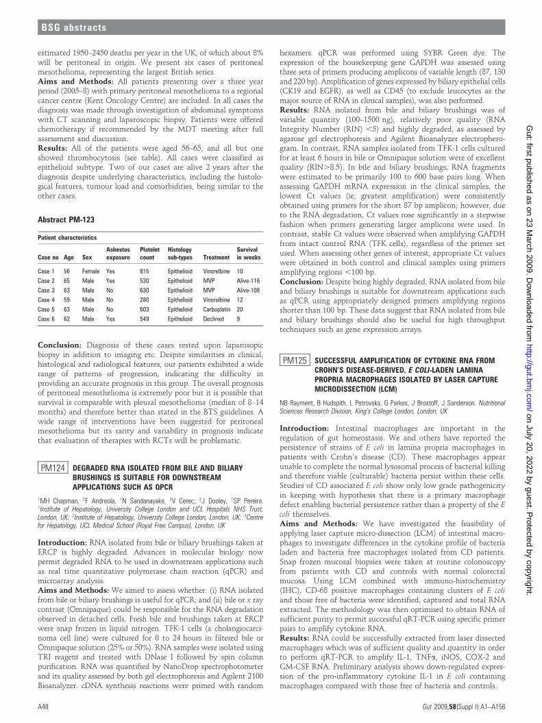
estimated 1950–2450 deaths per year in the UK, of which about 8%will be peritoneal in origin. We present six cases of peritonealmesothelioma, representing the largest British series.Aims and Methods: All patients presenting over a three yearperiod (2005–8) with primary peritoneal mesothelioma to a regionalcancer centre (Kent Oncology Centre) are included. In all cases thediagnosis was made through investigation of abdominal symptomswith CT scanning and laparoscopic biopsy. Patients were offeredchemotherapy if recommended by the MDT meeting after fullassessment and discussion.Results: All of the patients were aged 56–65, and all but oneshowed thrombocytosis (see table). All cases were classified asepithelioid subtype. Two of our cases are alive 2 years after thediagnosis despite underlying characteristics, including the histolo-gical features, tumour load and comorbidities, being similar to theother cases.
Conclusion: Diagnosis of these cases rested upon laparosopicbiopsy in addition to imaging etc. Despite similarities in clinical,histological and radiological features, our patients exhibited a widerange of patterns of progression, indicating the difficulty inproviding an accurate prognosis in this group. The overall prognosisof peritoneal mesothelioma is extremely poor but it is possible thatsurvival is comparable with pleural mesothelioma (median of 8–14months) and therefore better than stated in the BTS guidelines. Awide range of interventions have been suggested for peritonealmesothelioma but its rarity and variability in prognosis indicatethat evaluation of therapies with RCTs will be problematic.
PM124 DEGRADED RNA ISOLATED FROM BILE AND BILIARYBRUSHINGS IS SUITABLE FOR DOWNSTREAMAPPLICATIONS SUCH AS QPCR
1MH Chapman, 2F Andreola, 1N Sandanayake, 2V Cerec, 3J Dooley, 1SP Pereira.1Institute of Hepatology, University College London and UCL Hospitals NHS Trust,London, UK; 2Institute of Hepatology, University College London, London, UK; 3Centrefor Hepatology, UCL Medical School (Royal Free Campus), London, UK
Introduction: RNA isolated from bile or biliary brushings taken atERCP is highly degraded. Advances in molecular biology nowpermit degraded RNA to be used in downstream applications suchas real time quantitative polymerase chain reaction (qPCR) andmicroarray analysis.Aims and Methods: We aimed to assess whether: (i) RNA isolatedfrom bile or biliary brushings is useful for qPCR; and (ii) bile or x raycontrast (Omnipaque) could be responsible for the RNA degradationobserved in detached cells. Fresh bile and brushings taken at ERCPwere snap frozen in liquid nitrogen. TFK-1 cells (a cholangiocarci-noma cell line) were cultured for 0 to 24 hours in filtered bile orOmnipaque solution (25% or 50%). RNA samples were isolated usingTRI reagent and treated with DNase I followed by spin columnpurification. RNA was quantified by NanoDrop spectrophotometerand its quality assessed by both gel electrophoresis and Agilent 2100Bioanalyzer. cDNA synthesis reactions were primed with random
hexamers. qPCR was performed using SYBR Green dye. Theexpression of the housekeeping gene GAPDH was assessed usingthree sets of primers producing amplicons of variable length (87, 130and 220 bp). Amplification of genes expressed by biliary epithelial cells(CK19 and EGFR), as well as CD45 (to exclude leucocytes as themajor source of RNA in clinical samples), was also performed.Results: RNA isolated from bile and biliary brushings was ofvariable quantity (100–1500 ng), relatively poor quality (RNAIntegrity Number (RIN) ,5) and highly degraded, as assessed byagarose gel electrophoresis and Agilent Bioanalyzer electrophero-gram. In contrast, RNA samples isolated from TFK-1 cells culturedfor at least 6 hours in bile or Omnipaque solution were of excellentquality (RIN.8.5). In bile and biliary brushings, RNA fragmentswere estimated to be primarily 100 to 600 base pairs long. Whenassessing GAPDH mRNA expression in the clinical samples, thelowest Ct values (ie, greatest amplification) were consistentlyobtained using primers for the short 87 bp amplicon; however, dueto the RNA degradation, Ct values rose significantly in a stepwisefashion when primers generating larger amplicons were used. Incontrast, stable Ct values were observed when amplifying GAPDHfrom intact control RNA (TFK cells), regardless of the primer setused. When assessing other genes of interest, appropriate Ct valueswere obtained in both control and clinical samples using primersamplifying regions ,100 bp.Conclusion: Despite being highly degraded, RNA isolated from bileand biliary brushings is suitable for downstream applications suchas qPCR using appropriately designed primers amplifying regionsshorter than 100 bp. These data suggest that RNA isolated from bileand biliary brushings should also be useful for high throughputtechniques such as gene expression arrays.
PM125 SUCCESSFUL AMPLIFICATION OF CYTOKINE RNA FROMCROHN’S DISEASE-DERIVED, E COLI-LADEN LAMINAPROPRIA MACROPHAGES ISOLATED BY LASER CAPTUREMICRODISSECTION (LCM)
NB Rayment, B Hudspith, L Petrovska, G Parkes, J Brostoff, J Sanderson. NutritionalSciences Research Division, King’s College London, London, UK
Introduction: Intestinal macrophages are important in theregulation of gut homeostasis. We and others have reported thepersistence of strains of E coli in lamina propria macrophages inpatients with Crohn’s disease (CD). These macrophages appearunable to complete the normal lysosomal process of bacterial killingand therefore viable (culturable) bacteria persist within these cells.Studies of CD associated E coli show only low grade pathogenicityin keeping with hypothesis that there is a primary macrophagedefect enabling bacterial persistence rather than a property of the Ecoli themselves.Aims and Methods: We have investigated the feasibility ofapplying laser capture micro-dissection (LCM) of intestinal macro-phages to investigate differences in the cytokine profile of bacterialaden and bacteria free macrophages isolated from CD patients.Snap frozen mucosal biopsies were taken at routine colonoscopyfrom patients with CD and controls with normal colorectalmucosa. Using LCM combined with immuno-histochemistry(IHC), CD-68 positive macrophages containing clusters of E coliand those free of bacteria were identified, captured and total RNAextracted. The methodology was then optimised to obtain RNA ofsufficient purity to permit successful qRT-PCR using specific primerpairs to amplify cytokine RNA.Results: RNA could be successfully extracted from laser dissectedmacrophages which was of sufficient quality and quantity in orderto perform qRT-PCR to amplify IL-1, TNFa, iNOS, COX-2 andGM-CSF RNA. Preliminary analysis shows down-regulated expres-sion of the pro-inflammatory cytokine IL-1 in E coli containingmacrophages compared with those free of bacteria and controls.
Abstract PM-123
Patient characteristics
Case no Age SexAsbestosexposure
Plateletcount
Histologysub-types Treatment
Survivalin weeks
Case 1 56 Female Yes 815 Epithelioid Vinorelbine 10
Case 2 65 Male Yes 530 Epithelioid MVP Alive-116
Case 3 63 Male No 630 Epithelioid MVP Alive-108
Case 4 59 Male No 280 Epithelioid Vinorelbine 12
Case 5 63 Male No 603 Epithelioid Carboplatin 20
Case 6 62 Male Yes 549 Epithelioid Declined 9
BSG abstracts
A48 Gut 2009;58(Suppl I):A1–A156
on July 20, 2022 by guest. Protected by copyright.
http://gut.bmj.com
/G
ut: first published as on 23 March 2009. D
ownloaded from
Conclusion: As a proof of concept, we have shown that LCM canbe successfully applied to isolate E coli laden macrophages from thelamina propria in CD and specific cytokine RNA amplified. Alteredexpression of cytokines in E coli containing macrophages maysupport the hypothesis of an innate defect permitting bacterialpersistence in CD.
PM126 B CELLS IN THE PATHOGENESIS OF THE OROFACIALGRANULOMATOSIS/ORAL CROHN’S DISEASE SPECTRUM
1P Patel, 2F Barone, 2L Boursier, 3C Nunes, 4E Odell, 5M Escudier, 5S Challacombe, 3JBrostoff, 2J Spencer, 3J Sanderson. 1Nutritional Sciences Division, King’s CollegeLondon, London, UK; 2Immunobiology; 3Nutritional Sciences Division; 4Oral Pathology;5Oral Medicine, King’s College London, London, UK
Introduction: The oral mucosa is an immunologically responsivesite associated with the generation of protective mucosal andsystemic immune responses to vaccination and also hyper-respon-siveness to allergens in some individuals. Classically, immuneresponses in oral mucosa are considered to be mediated by mucosa-associated lymphoid tissues (MALT), secondary lymphoid folliclesthat are intimately associated with epithelia. Orofacial granuloma-tosis (OFG), which includes oral Crohn’s disease, is a chronicinflammatory condition presenting characteristically with lipswelling but also affecting a number of other sites in the oralcavity. Although the cause remains unknown, dietary triggers andallergic phenomena are established features.Aims and Methods: To investigate the B cell component of theinflammatory infiltrate in OFG.Methods: Using immunohistochemistry and PCR on paraffinembedded (n = 44) and fresh frozen (n = 8) oral mucosal biopsies,we characterised in detail the B cell component of the inflammatoryinfiltrate in OFG (n = 14) and a variety of non-OFG inflammatoryoral conditions (n = 38).Results: CD20 staining revealed large, active, dendritic B cells inoral mucosa, closely associated with other cellular components ofthe immune response such as T cells and macrophages. They werenot associated with any organised lymphoid tissues in the localsubepithelial microenvironment. These B cells expressed ActivationInduced cytidine Deaminase (AID), essential for immunoglobulingene diversification by somatic hypermutation and class switchrecombination. AID transcripts were also demonstrated in frozenoral biopsies by PCR. They were negative for naıve B cell markersand a proportion (10%) expressed the nuclear proliferation marker,Ki67. These dendritic B cells also expressed surface IgE and thisexpression was significantly greater in OFG compared with otheroral conditions (mean % of B cells expressing surface IgE 6.5% vs0.2%; p,0.0001). Epsilon transcripts, which provide furtherevidence for IgE production locally, were also demonstrated in thesingle frozen OFG biopsy by PCR.Conclusion: Subepithelial dendritic B cells described here do not alignwith any other previously described B cell subset in secondarylymphoid tissues in terms of morphology, proliferative activity orphenotype. We propose that these cells contribute to the immuneresponsiveness of the oral mucosa including IgE mediated allergicresponses, notably in OFG but also in other oral inflammatory disease.
Endoscopy posters
PM127 DOUBLE BALLOON ENTEROSCOPY—TOLERABILITY IN 119CASES, WHO NEEDS PROPOFOL?
A Deo, S Hughes. Gastroenterology, North Bristol NHS Trust, Bristol, UK
Introduction: Double balloon enteroscopy (DBE) is a difficult, time-consuming and uncomfortable procedure. There is no consensusregarding sedation. Sedation with propofol, midazolam and pethidineas well as general anaesthesia have been used in different centres. We
present our experience of 137 consecutive cases where patients weresedated with a combination of midazolam and pethidine.Aims and Methods: 137 procedures were performed in ourendoscopy unit as a day case. All procedures were performed underconscious intravenous sedation using a combination of midazolamand pethidine. Initial doses were midazolam 5 mg and pethidine50 mg given intravenously. Additional midazolam was given in1 mg boluses. Sedated patients were monitored in the standard wayin accordance with local guidelines. Tolerability was assessed usinga 3-point scale and assessed independently by the endoscopist and 2nurses and agreed at the end of the procedure. Well tolerated wasdefined as patient being comfortable throughout the procedure.Moderately well tolerated was defined as patient being uncomfor-table at times and the examination was not curtailed. Poorlytolerated was defined as patient uncomfortable most of the timeand the procedure was curtailed.Results: Tolerability data were calculated on 119 procedures (75via the oral route and 44 via the rectal route). DBE was welltolerated in 94 patients (80%), moderately tolerated in 19 patients(15%) and poorly tolerated in 6 patients (5%). DBE via the oralroute was well tolerated in 57 (76%) patients, moderately toleratedin 12 (16%) patients and poorly tolerated in 6 (8%) patients. DBEvia the rectal route was well tolerated in 37 (84%) patients andmoderately tolerated in 7 (16%) patients. There were no patientswhose tolerance was poor via the rectal route. 135 patient’s datawere used to calculate averages for age, time taken and drug doses.Data were incomplete in 2 procedures and therefore discarded. Theaverage dose midazolam used was 9 mg for the oral route and 6 mgfor the rectal route. The average dose of pethidine was 50 mg forboth routes. Average time taken for the DBE was 50 and 62 minutesfor oral and rectal routes respectively. One patient required reversalof sedation with Flumazenil. The average age of patients was 61and 60 years for oral and rectal routes respectively. Seventy-fourmales and 61 females were included in the study; the average age ofmales was 60.09 years (32–84 years) and for females 60.61 years(41–89 years). There were no complications in this series ofpatients.Conclusion: Double balloon enteroscopy is a safe and welltolerated procedure under conscious intravenous sedation using acombination of pethidine and midazolam. DBE via the oral routewas shorter in duration and less well tolerated than DBE via therectal route. There were no complications and the procedure can besafely performed as a day case.
PM128 EUS-GUIDED CYSTOGASTROSTOMY FOR PANCREATICPSEUDOCYSTS; ANALYSIS OF A CASE SERIES USINGMINIMAL ENDOSCOPIC PROCEDURES
1AD Farmer, 2S Menon, 3S Kapadia, 2NC Fisher. 1Wingate Institute ofNeurogastroenterology, Barts and the London, Whitechapel, UK; 2Department ofGastroenterology, Russell’s Hall Hospital, Dudley, UK; 3Department of Gastroenterology,New Cross Hospital, Wolverhampton, UK
Introduction: EUS-guided cystogastrostomy is a valuable techni-que for drainage of persistent pancreatic pseudocysts. Practice variesin terms of the number of endoscopic procedures that are plannedfor a successful outcome. Herein we review our own experience ofusing a limited number of interventions.Aims and Methods: The aim of this study was to evaluate thetechnical success and complication rates of EUS-guided cystogastrost-omy at our institution. A retrospective case note review was done of allcases of attempted cystogastrostomy. The procedure was performedwith a linear echoendoscope and using a cystotome cannula (CookUK). One or two guidewires were inserted under fluoroscopic guidanceto facilitate placement of 1 or 2 stents. For recent cases we haveadopted routine usage of a nasocystic drain in place of 1 stent. Furtherendoscopic intervention was not routinely planned.
BSG abstracts
Gut 2009;58(Suppl I):A1–A156 A49
on July 20, 2022 by guest. Protected by copyright.
http://gut.bmj.com
/G
ut: first published as on 23 March 2009. D
ownloaded from
Results: Twenty-two procedures were performed on 20 patientsincluding 2 cases of late cyst recurrence (median age 52, range 15–78 years). Median pseudocyst size was 10.1 cm68.3 cm. Cystpuncture was successful in 21 out of 22 cases; of these, stentinsertion was successful in 20 out of 21 patients (9 patients had asingle stent, 9 patients had twin stents and 4 patients had singlestent and nasocystic drain). One procedural complication occurred(pneumothorax, 1 patient). Seven cases required further interven-tion within 30 days (4 endoscopic, 2 radiological and 1 surgicalintervention); all of these cases had sepsis due to incomplete cystdrainage and most (5/7) were early pseudocysts (,3 months afterfirst presentation); in each case re-intervention was successful.Stents were removed after a median of 19 weeks.Conclusion: EUS-guided cystogastrostomy with twin stents orstent/nasocystic drain was successful in most cases in this series.There is a small but important risk of complications requiringfurther intervention with this approach, especially if intervention isdone early in the disease course.
PM129 COLONOSCOPY SKILLS TRANSFER FROM A SECOND-GENERATION VIRTUAL REALITY SIMULATOR TO PATIENTS:A MULTINATIONAL RANDOMISED BLINDED CONTROLLEDTRIAL
1AV Haycock, 2AD Koch, 3P Familiari, 4F van Delft, 5JS Bladen, 4E Dekker, 3LPetruziello, 2J Haringsma, 1S Thomas-Gibson. 1Wolfson Unit for Endoscopy, ImperialCollege London, London, UK; 2Department of Gastroenterology and Hepatology,Erasmus Medical Centre, Rotterdam, The Netherlands; 3Digestive Endoscopy Unit,Catholic University—A.Gemelli University Hospital, Rome, Italy; 4Department ofGastroenterology and Hepatology, Academic Medical Center, Amsterdam, TheNetherlands; 5JSB Medical Systems, Sheffield, UK
Introduction: Training on virtual reality simulators has been shownto improve skills for novice endoscopists compared with no training.To date simulators have not accurately modelled colonic looping andits management, which is a key factor in skills development. Asecond-generation colonoscopy simulator has been specificallydesigned to model colonic looping and has the potential to teachknowledge and skills as effectively as standard training with no risk topatients and minimal instructor input. The Olympus colonoscopysimulator was assessed for training in transferrable skills.Aims and Methods: 37 novice trainees from four centres in theUK, Italy and The Netherlands were pre-assessed on the simulatorusing three previously validated test cases. They were randomisedto receive 16 hours’ training on the simulator (subjects, N = 19) oron patients (controls, N = 18) in a standardised fashion. Allparticipants were then re-assessed on the same three simulator testcases and on three patient test cases by blinded expert endoscopistsusing validated assessments.Results: On simulator test cases, subjects achieved significantlybetter post-training results on previously validated metrics such ascompletion rate (95% vs 70%, p = 0.001), completion time (407 vs743, p = 0.001) and loop resolution (p,0.001) than the controls. Theyalso demonstrated superior technical skills on safety aspects such asexcessive insertion with an embedded tip (35% vs 74%, p,0.001) andobscured lens (5% vs 41%, p,0.001), and on patient factors such asmaximum patient pain scores (0.24 vs 0.45, p = 0.002). On the patienttest cases, the subjects achieved equivalent success in terms ofcompletion rates (11% vs 7%, p = 0.51), distance intubated (48 cm vs52 cm, p = 0.35), Directly Observed Procedure Scores (16 vs 18,p = 0.92) and Global Scores (16 vs 17, p = 0.35). Much less instructortime (4 hours vs 16 hours), organisation and effort was required fortraining in the simulator group.Conclusion: There is excellent skills transfer from the simulator toreal colonoscopy. The simulator trained group demonstratedequivalent performance outcomes on real patients, suggestinginitial training can effectively be employed utilising a simulator
rather than real patients. This would reduce the burden both onpatients and on instructors. The simulator should be considered as avaluable training and assessment tool in the development ofcolonoscopy skills.
PM130 ENDOSCOPY TRAINING IN 2008: RESULTS FROM THE TIG/BSG NATIONAL TRAINING SURVEY
AV Haycock, P Flanagan, A Ignjatovic, S Kendrick, R Mead, P Mizen, M Mottershead,V Patel, K Pollock, T Valliani, J Williams. Trainees Committee, British Society ofGastroenterology, London, UK
Introduction: The 2004 national survey of gastroenterologytrainees revealed wide variation in both the quality and quantityof training in endoscopy. The level of supervision was of particularconcern, with 55% of trainees saying they were not beingsupervised for over half their procedures. Since that time, the UKNational Endoscopy Training Programme has aimed to standardisetraining and assessment. Central funding for 3 years provided JAGaccredited courses free of charge to trainees. Training-the-trainerscourses were implemented to improve the quality of training atbase hospitals. Summative and formative assessments are nowmandatory to document competency.Aims and Methods: The aim of the 2008 survey was to investigatethe impact of these changes and gather data on current standards ofteaching, training and supervision in endoscopy. A web-basedquestionnaire was developed with questions in eight domainsincluding endoscopy. An invitation to complete the survey wasemailed by the regional TiG representatives to all trainees in theirregion. Data were collected during October 2008.Results: 249 out of 411 trainees from 20 different training regionscompleted the survey (response rate 60.6%). 217 (88.6%) were ingeneral gastroenterology posts. 115/220 (52.3%) attend the recom-mended minimum of 2 endoscopy sessions per week, although only43% of these are dedicated training lists, and 37% of trainees saythat training actually occurs less than half the time. Eighteen percent of trainees say they have no dedicated training lists at all. 79%of trainees had been on at least one accredited training course. Only95/223 (43%) routinely get formative DOPS filled in for their lists,and 52% have not yet had any kind of summative DOPS. 184/236(78%) are fairly or very satisfied with their endoscopy training sofar, with only 5/236 (2.1%) very unsatisfied. By far the mostcommon complaint from trainees was lack of access to regularsupervised training lists.Conclusion: The majority of trainees are satisfied with their currenttraining, although the provision of training lists does not seem to havechanged substantially since 2004 and is the main cause of concern fortrainees. Less than half of trainees routinely use DOPS forms duringtheir training lists, which should be addressed by training leads, as it isnow a requirement for annual RITA appraisal.
PM131 DEVELOPMENT AND IMPLEMENTATION OF ASTANDARDISED, EVIDENCE-BASED COLONOSCOPICTATTOOING PROTOCOL
1AV Haycock, 2RH Kennedy, 1S Thomas-Gibson, 1BP Saunders. 1Wolfson Unit forEndoscopy, St Mark’s Hospital, Imperial College London, London, UK; 2Department ofSurgery, St Mark’s Hospital, Imperial College London, London, UK
Introduction: Placing tattoos in the colon is now common practicein order to mark lesions prior to surgery or for endoscopic follow-up. A recent audit in our unit showed wide variation in thetechnique used as well as the number and placement of tattoos.Published studies confirm such variation in technique as well assubsequent variation in serosal visualisation at surgery.Aims and Methods: The aim was to develop a standardised,evidence-based protocol for tattooing during colonoscopy and toreview its use in practice in a single unit. A review of the literature
BSG abstracts
A50 Gut 2009;58(Suppl I):A1–A156
on July 20, 2022 by guest. Protected by copyright.
http://gut.bmj.com
/G
ut: first published as on 23 March 2009. D
ownloaded from
was performed regarding all aspects of endoscopic tattoo place-ment. A standardised protocol was drawn up utilising the evidencebase by experienced colonoscopists and colorectal surgeons. Theprocedure involves a ‘‘no-spill technique’’ with a saline lift tominimise leakage and resulting complications. To maximisevisualisation at surgery (particularly at laparoscopy) three tattoosare placed at 120 degrees to each other 3 cm from the lesion. Site ofplacement (distal for right sided lesions, proximal for left sidedlesions and distal for rectosigmoid lesions) facilitates surgicaldecision making regarding resection margins. The protocol waspresented at an endoscopy user meeting, distributed by email to allendoscopists and displayed in all endoscopy rooms. A prospectivedatabase was kept of all tattoos placed.Results: In 10 months, tattoos were placed during colonoscopy on53 occasions; 29 (55%) for suspected malignancy and 24 (45%) forpolyp follow-up. Overall compliance with all aspects of the protocol(documentation, number and location) was 45.3%. For cancers,documentation of the location and number of tattoos was 96.6%and 93.1% respectively, with location and number both compliantwith the protocol on 65.5% of occasions. For polyp follow-ups,documentation of the location and number of tattoos was 83.3%,with only 20.8% compliant with the protocol.Conclusion: Compliance with the protocol was more likely withsuspected cancers compared with polyp follow-up. Re-education ofendoscopists is needed before investigation of the efficacy of theprococol in terms of visualisation at surgery can be undertaken.
PM132 DO OUTLIERS FACE A GREATER RISK OF HARM? ACOMPARISON OF PATIENTS TREATED FOR ACUTE UPPERGASTROINTESTINAL (UGI) BLEED ON A SPECIALIST OROUTLYING WARD
A Ireland, A Baldwin, M Renshaw. Digestive Diseases, Royal Sussex County Hospital,Brighton, UK
Introduction: The term outlier refers to hospital in-patients whooccupy a different specialty bed to that of their admitting specialty.Outliers are assumed to face a greater risk of harm than patientstreated on specialist wards, and evidence suggests outliers face alonger in-patient stay. This hypothesis was tested by comparingmortality rate, length of hospital stay and time to endoscopy ofpatients with acute UGI bleed treated on an outlying ward topatients treated on a specialist gastrointestinal ward.Aims and Methods: Retrospective analysis of clinical informationsystems and case note review were undertaken to identify patientspresenting from June 2006 to May 2007 with a confirmed diagnosison admission of acute UGI bleed. Two criteria were used tocategorise patients as ideal or outlier. Criterion 1 (C1) defined idealpatients solely by appropriate ward location, and Criterion 2 (C2)defined ideal patients by appropriate ward location and a time toendoscopy of less than 24 hours from admission. Any patient whodid not fit either ideal criterion was classified as an outlier.Retrospective calculation of a patient’s Rockall Score allowedpatients to be matched for severity of bleed.Results: 171 patients were identified. Over two-thirds wereclassified as outliers (C1: 70.8%, 121/171; C2: 78.4%, 134/171).Overall mortality was 9.4%. Mortality for C1 was 12.0% (6/50) forideal patients and 8.3% (10/121) for outliers (p = 0.563). For C2,mortality was 13.5% (5/37) for ideal patients and 8.2% (11/134) foroutliers (p = 0.343). Of those undergoing endoscopy, 73.7% of idealpatients did so within 24 hours of admission (28/38) comparedwith 64.1% of outlying patients (50/78) (C1: p = 0.302). In-patientmortality was reduced significantly when endoscopy was per-formed within 24 hours (all patients), 1.28% vs 10.52%, p = 0.039,as was length of stay (all patients) (,24 hours 4.58 vs .24 hours8.08 days, p = 0.002). C2 outlying patients had a significantlylonger length of stay (Ideal 4.16 vs Outlier 5.93 days, p = 0.034) and
fewer medical reviews per day (Ideal 2.74 vs Outlier 2.00 reviews/day, p = 0.034). There was no significant difference in length of staybetween ideal and outlying patients by C1 (Ideal 4.88 vs Outlier5.82 days, p = 0.264) or in the number of medical reviews per day(C1: Ideal 2.52 vs Outlier 2.01 reviews/day, p = 0.065).Conclusion: There was no significant difference in mortality ratebetween outliers and ideal patients. There was however asignificant reduction in mortality in patients undergoing endoscopywithin 24 hours. Criteria 2 patients had a significantly shorter in-patient stay and a higher number of medical reviews. Theclassification of outliers however remains problematic.
PM133 INTENSIVE DAY CASE COLONOSCOPY FOR THE UNRELIABLE,FRAIL OR ELDERLY PATIENT
1A Ghanbari, 1TTC Cuming, 2AAB Ballinger, 1HHP Pardoe. 1Academic Gastroenterology,Homerton University Hospital, London, UK; 2Gastroenterology, Queen Elizabeth theQueen Mother Hospital, Margate, UK
Introduction: We observed that many patients admitted for aninpatient bowel preparation prior to colonoscopy were still badlyprepared and commonly stayed more than scheduled overnightadmission.Aims and Methods: The objective of our study was to see if it issafe, feasible and acceptable for patients who are normally admittedas an inpatient for bowel preparation to undergo day stay fullbowel cleansing using an oral polyethylene glycol (PEG) Klean-prepsolution and same day colonoscopy without needing hospitaladmission. Patients who would fulfil the criteria in our institutionfor in patient bowel preparation prior to colonoscopy wereadmitted to the endoscopy unit and underwent bowel cleansingin the morning using an oral PEG solution followed by colonoscopyin the afternoon and discharge on the same day. The first 10patients were interviewed at the end of the procedure to assess theprocedure for patient acceptability. All colonoscopies using anintensive day case regime with PEG Klean Prep were assessed usingGRS criteria for quality control.Results: Patients who would fulfil the criteria in our institution forin patient bowel preparation prior to colonoscopy were admitted tothe endoscopy unit and underwent bowel cleansing in the morningusing an oral PEG solution followed by colonoscopy in theafternoon and discharge on the same day. The first 10 patientswere interviewed at the end of the procedure to assess theprocedure for patient acceptability. All colonoscopies using anintensive day case regime with PEG Klean Prep were assessed usingGRS criteria for quality control.Conclusion: 43 patients have now undergone intensive day casecolonoscopy. The unadjusted completion was 90.2% and 91.5% forthe Klean prep and inpatient bowel preparation groups respectively.The bowel preparation was classified as satisfactory or good in 76.7%of Klean prep cases and only 40% inpatient cases. Polyp detection forthe Klean Prep group was 44.2% compared with 20% for theinpatients. The total bed occupancy rate for elective colonoscopy wasreduced from 184 days to 0 over a 12 month period. Ninety per centof patients interviewed found the process acceptable and preferredKlean-prep as their method of bowel cleansing.
PM134 OPTICAL BIOPSY AT COLONOSCOPY: ARE WE READY?DISCARD STUDY: EARLY RESULTS
1A Ignjatovic, 1N Suzuki, 1M Vance, 2T Guenther, 1N Palmer, 1BP Saunders. 1WolfsonUnit for Endoscopy, St Mark’s Hospital, Harrow, UK; 2Department of AcademicPathology, St Mark’s Hospital, Harrow, UK
Introduction: 90% of polyps removed at colonoscopy are ,10 mmin size, and only 50% of these are neoplastic. The number ofneoplastic polyps is the key determinant of future surveillanceintervals, and small adenomas, especially those less than 5 mm in
BSG abstracts
Gut 2009;58(Suppl I):A1–A156 A51
on July 20, 2022 by guest. Protected by copyright.
http://gut.bmj.com
/G
ut: first published as on 23 March 2009. D
ownloaded from
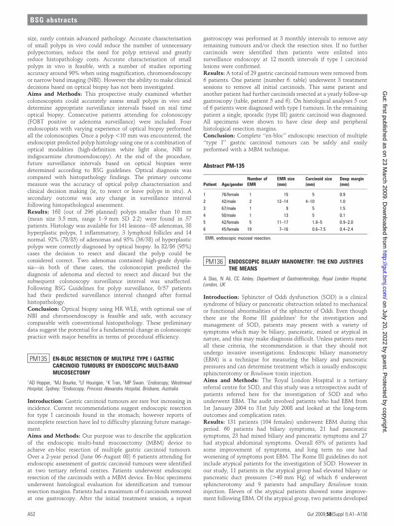
size, rarely contain advanced pathology. Accurate characterisationof small polyps in vivo could reduce the number of unnecessarypolypectomies, reduce the need for polyp retrieval and greatlyreduce histopathology costs. Accurate characterisation of smallpolyps in vivo is feasible, with a number of studies reportingaccuracy around 90% when using magnification, chromoendoscopyor narrow band imaging (NBI). However the ability to make clinicaldecisions based on optical biopsy has not been investigated.Aims and Methods: This prospective study examined whethercolonoscopists could accurately assess small polyps in vivo anddetermine appropriate surveillance intervals based on real timeoptical biopsy. Consecutive patients attending for colonoscopy(FOBT positive or adenoma surveillance) were included. Fourendoscopists with varying experience of optical biopsy performedall the colonoscopies. Once a polyp ,10 mm was encountered, theendoscopist predicted polyp histology using one or a combination ofoptical modalities (high-definition white light alone, NBI orindigocarmine chromoendoscopy). At the end of the procedure,future surveillance intervals based on optical biopsies weredetermined according to BSG guidelines. Optical diagnosis wascompared with histopathology findings. The primary outcomemeasure was the accuracy of optical polyp characterisation andclinical decision making (ie, to resect or leave polyps in situ). Asecondary outcome was any change in surveillance intervalfollowing histopathological assessment.Results: 168 (out of 296 planned) polyps smaller than 10 mm(mean size 3.5 mm, range 1–9 mm SD 2.2) were found in 57patients. Histology was available for 141 lesions—85 adenomas, 38hyperplastic polyps, 1 inflammatory, 3 lymphoid follicles and 14normal. 92% (78/85) of adenomas and 95% (36/38) of hyperplasticpolyps were correctly diagnosed by optical biopsy. In 82/86 (95%)cases the decision to resect and discard the polyp could beconsidered correct. Two adenomas contained high-grade dyspla-sia—in both of these cases, the colonoscopist predicted thediagnosis of adenoma and elected to resect and discard but thesubsequent colonoscopy surveillance interval was unaffected.Following BSG Guidelines for polyp surveillance, 0/57 patientshad their predicted surveillance interval changed after formalhistopathology.Conclusion: Optical biopsy using HR WLE, with optional use ofNBI and chromoendoscopy is feasible and safe, with accuracycomparable with conventional histopathology. These preliminarydata suggest the potential for a fundamental change in colonoscopicpractice with major benefits in terms of procedural efficiency.
PM135 EN-BLOC RESECTION OF MULTIPLE TYPE I GASTRICCARCINOID TUMOURS BY ENDOSCOPIC MULTI-BANDMUCOSECTOMY
1AD Hopper, 1MJ Bourke, 2LF Hourigan, 1K Tran, 1MP Swan. 1Endoscopy, WestmeadHospital, Sydney; 2Endoscopy, Princess Alexandra Hospital, Brisbane, Australia
Introduction: Gastric carcinoid tumours are rare but increasing inincidence. Current recommendations suggest endoscopic resectionfor type I carcinoids found in the stomach; however reports ofincomplete resection have led to difficulty planning future manage-ment.Aims and Methods: Our purpose was to describe the applicationof the endoscopic multi-band mucosectomy (MBM) device toachieve en-bloc resection of multiple gastric carcinoid tumours.Over a 2-year period (June 06–August 08) 6 patients attending forendoscopic assessment of gastric carcinoid tumours were identifiedat two tertiary referral centres. Patients underwent endoscopicresection of the carcinoids with a MBM device. En-bloc specimensunderwent histological evaluation for identification and tumourresection margins. Patients had a maximum of 6 carcinoids removedat one gastroscopy. After the initial treatment session, a repeat
gastroscopy was performed at 3 monthly intervals to remove anyremaining tumours and/or check the resection sites. If no furthercarcinoids were identified then patients were enlisted intosurveillance endoscopy at 12 month intervals if type I carcinoidlesions were confirmed.Results: A total of 29 gastric carcinoid tumours were removed from6 patients. One patient (number 6: table) underwent 3 treatmentsessions to remove all initial carcinoids. This same patient andanother patient had further carcinoids resected at a yearly follow-upgastroscopy (table, patient 5 and 6). On histological analyses 5 outof 6 patients were diagnosed with type I tumours. In the remainingpatient a single, sporadic (type III) gastric carcinoid was diagnosed.All specimens were shown to have clear deep and peripheralhistological resection margins.Conclusion: Complete ‘‘en-bloc’’ endoscopic resection of multiple‘‘type I’’ gastric carcinoid tumours can be safely and easilyperformed with a MBM technique.
PM136 ENDOSCOPIC BILIARY MANOMETRY: THE END JUSTIFIESTHE MEANS
A Dias, N Ali, CC Ainley. Department of Gastroenterology, Royal London Hospital,London, UK
Introduction: Sphincter of Oddi dysfunction (SOD) is a clinicalsyndrome of biliary or pancreatic obstruction related to mechanicalor functional abnormalities of the sphincter of Oddi. Even thoughthere are the Rome III guidelines1 for the investigation andmanagement of SOD, patients may present with a variety ofsymptoms which may be biliary, pancreatic, mixed or atypical innature, and this may make diagnosis difficult. Unless patients meetall these criteria, the recommendation is that they should notundergo invasive investigations. Endoscopic biliary manometry(EBM) is a technique for measuring the biliary and pancreaticpressures and can determine treatment which is usually endoscopicsphincterotomy or Botulinum toxin injection.Aims and Methods: The Royal London Hospital is a tertiaryreferral centre for SOD, and this study was a retrospective audit ofpatients referred here for the investigation of SOD and whounderwent EBM. The audit involved patients who had EBM from1st January 2004 to 31st July 2008 and looked at the long-termoutcomes and complication rates.Results: 131 patients (104 females) underwent EBM during thisperiod. 60 patients had biliary symptoms, 21 had pancreaticsymptoms, 23 had mixed biliary and pancreatic symptoms and 27had atypical abdominal symptoms. Overall 63% of patients hadsome improvement of symptoms, and long term no one hadworsening of symptoms post EBM. The Rome III guidelines do notinclude atypical patients for the investigation of SOD. However inour study, 11 patients in the atypical group had elevated biliary orpancreatic duct pressures (.40 mm Hg) of which 6 underwentsphincterotomy and 9 patients had ampullary Botulinum toxininjection. Eleven of the atypical patients showed some improve-ment following EBM. Of the atypical group, two patients developed
Abstract PM-135
Patient Age/genderNumber ofEMR
EMR size(mm)
Carcinoid size(mm)
Deep margin(mm)
1 76/female 1 15 5 0.9
2 42/male 2 12–14 4–10 1.0
3 67/male 1 9 5 1.5
4 50/male 1 13 5 0.1
5 42/female 5 11–17 1.8–5 0.9–2.0
6 45/female 19 7–16 0.6–7.5 0.4–2.4
EMR, endoscopic mucosal resection.
BSG abstracts
A52 Gut 2009;58(Suppl I):A1–A156
on July 20, 2022 by guest. Protected by copyright.
http://gut.bmj.com
/G
ut: first published as on 23 March 2009. D
ownloaded from
post-ERCP pancreatitis, and one patient suffered a fatal guide wireperforation. In total, 19 patients developed post-ERCP pancreatitis(12 following sphincterotomy and 5 post-Botulinum toxin injection),and 5 patients developed perforation.Conclusion: Rome III guidelines for the diagnosis of SOD areunnecessarily restrictive and therefore may lead to the under-diagnosis and treatment of this condition. Many patients, who maynot fit these criteria, including those with atypical symptoms, maypotentially benefit from undergoing EBM.
1. Behar J, Corazziari E, Guelrud M, et al. Functional Gallbladder and Sphincter ofOddi Disorders. Gastroenterology 2006;130:1498–1509.
PM137 CAECAL INTUBATION WITH A GASTROSCOPE FOLLOWINGAN INCOMPLETE COLONOSCOPY—IS IT FEASIBLE?
B Paranandi, N Anglim, A Sawyerr, E Carty, E Seward. Department ofGastroenterology and Hepatology, Whipps Cross University Hospital, London, UK
Introduction: It is common practice in the majority of endoscopyunits in the UK to use shorter, more flexible endoscopes in order tocomplete a difficult colonoscopy. Current published data are verylimited but suggest that this may achieve caecal intubation in up to62% of cases.1
Aims and Methods: Between October 2005 and October 2008, theendoscopy reports of all patients undergoing colonoscopy atWXUH with a conventional gastroscope were audited. Thesepatients were identified from our database using the departmentalreporting tool. A broad range of variables were assessed includingtotal number of procedures, indication for endoscopy, caecal/terminal ileum (TI) intubation rate, reasons for failing to completean examination and sedation usage.Results: During the audit period, there were 7495 colonoscopiesundertaken, 94 of which were performed using gastroscopes (1.3%).Seven procedures were excluded from our results because they wereperformed on resected colons. The most common documentedindication for switching from a colonoscope to a gastroscope wascomplex diverticular disease in 36/87 [41%]. Of the 87 procedureswe audited, the caecal intubation rate was 69/87 [79%] and the TIintubation rate was 40/87 [46%]. Using conventional colonoscopes,the caecal and TI intubation rates of our unit are 91% and 62%respectively. In the 18 [21%] cases where there was failure tocomplete the colonoscopy even with a gastroscope, the mainidentifiable causes included diverticular disease [50%], looping[22%] and strictures [22%]. Sedation usage was less than or equalto 2 mg of midazolam per procedure in 72/87 [83%].Conclusion: Gastroscopes can be useful when trying to complete adifficult colonoscopy. We have shown that this is especially the casein complex diveticular disease. The completion rate with gastro-scopes in our unit exceeds that of other studies.
1. Paonessa et al. Using the gastroscope for incomplete colonoscopy; TheAmerican Society of Colon and Rectal Surgeons Annual Meeting; 2005;48(4):851–854.
PM138 ISOLATED IRON DEFIENCY ANAEMIA: AT WHAT LEVEL OFHAEMOGLOBIN SHOULD WE INVESTIGATE?
1CC Henson, 2A Saeed. 1Gastroenterology, Royal Preston Hospital, Preston, UK;2Gastroenterology, Queen Elizabeth Hospital, Gateshead, UK
Introduction: Upper and lower GI endoscopy is the gold standardinvestigation for isolated iron deficiency anaemia. The NationalInstitute for Clinical Excellence (NICE) guidelines1 advise investiga-tion of isolated iron deficiency anaemia with upper and lower GIendoscopy at levels of ,11 g/dL Hb for men of any age and ,10 g/dL Hb for post-menopausal women in the absence of anotherobvious cause. The British Society of Gastroenterology (BSG)2
advises such investigation when haemoglobin levels fall below thelower limit of the normal range.
Aims and Methods: This retrospective study aims to determinethe level of haemoglobin below which investigation of irondeficiency anaemia should occur to ensure the identification ofsignificant GI pathology, that is colorectal carcinoma/significantpolyps, yet avoid unnecessary invasive interventions.3 An Endosoftsearch was performed to identify all patients having colonoscopyfor isolated iron deficiency anaemia in 2005. Patients withadditional indications for colonoscopy were excluded, for exampleGI bleeding/change in bowel habit. Patients in which irondeficiency was not be confirmed by low MCV/ferritin in the6 months prior to colonoscopy were also excluded. The results ofcolonoscopies were defined as positive, that is GI carcinoma/significant polyp (intermediate or high risk according to BSGguidelines), negative, that is no significant pathology, or excluded,that is colitis, angiodysplasia, etc. The lowest haemoglobin readingin the 6 months preceding the colonoscopy was recorded. Receiveroperator characteristic (ROC) curve analysis was used to define thehaemoglobin level under which significant colorectal pathology islikely to be identified.Results: 385 patients were identified in the initial search. 196patients were eligible for inclusion: 28 positive and 168 negativeaccording to the defined criteria. Data were analysed using ROCcurve analysis. The area under the ROC curve was 0.521 with astandard error of 0.0569. p = 0.3544. This suggests that haemoglo-bin levels alone in iron deficiency anaemia are a poor test to predictthe presence of colorectal cancer or significant polyps. In fact 27.3%(3) of female and 30.8% (4) of male malignancies or significantpolyps would not have been detected if NICE guidelines werefollowed.Conclusion: All patients with proven iron deficiency anaemia ofany severity should have a colonoscopy to examine for colorectalcancer or significant polyps in line with the current BSG Guidelinesto avoid missing significant pathology.
1. http://www.NICE.org.uk Clinical Guideline 27: Referral Guidelines for SuspectedCancer. 2005.
2. http://www.bsg.org.uk British Society of Gastroenterology: Guidelines for theManagement of Iron Deficiency Anaemia. 2005.
3. Atkin WS, et al. Surveillance guidelines after removal of colorectal adenomatouspolyps. Gut 2002;51(Suppl V):v6–v9.
PM139 IS GASTROINTESTINAL ENDOSCOPY A RISK FACTOR FORCLOSTRIDIUM DIFFICILE ASSOCIATED DIARRHOEA?
CP Selinger, S Greer. Department of Gastroenterology, Royal Albert Edward Infirmary,Wigan, UK
Introduction: Although endoscopy has been described as a riskfactor for the development of Clostridium difficile associateddiarrhoea (CDAD) in several review articles no conclusive evidenceexists to support this claim.1
Aims and Methods: This retrospective Case-Control study aimsto explore in a large patient cohort whether gastrointestinal (GI)endoscopy is an independent risk factor for CDAD. Cases were allpatients who newly tested positive for Clostridium difficile Toxin Aand/or B between March and December 2006. Age and sex matchedcontrols were drawn from a cohort of patients with diarrhoea andnegative Clostridium difficile Toxin A and B tests. Gastrointestinalendoscopy in the 30, 60 and 90 days prior to Clostridium difficileToxin A and B testing in both groups was recorded and dataanalysed using exact McNemara’s significance probability test. Theabsolute risk of developing CDAD within 30, 60 and 90 days afterGI endoscopy was calculated for all patients undergoing endoscopybetween March and September 2006.Results: The 287 case-control pairs had a mean age of 78 years, and65% were female. The endoscopy exposure rate for cases was higherthan for controls at all time points, but this apparent increase inendoscopy exposure was clearly not of statistical significance for the
BSG abstracts
Gut 2009;58(Suppl I):A1–A156 A53
on July 20, 2022 by guest. Protected by copyright.
http://gut.bmj.com
/G
ut: first published as on 23 March 2009. D
ownloaded from
90 (p = 0.18) day data although it was of borderline statisticalsignificance for the 30 (p = 0.057) and 60 (p = 0.053) day data. Oddsratios vary between 1.55 (95% CI 0.83 to 2.98) at 90 days and 2.09(95% CI 0.97 to 4.75) at 30 days. 25 of 4959 patients undergoing GIendoscopies between March and September 2006 developed CDAD.The absolute risk ranged between 0.32% at 30 days and 0.5% at90 days.Conclusion: This study suggests there may indeed be a modest (oddsratio of 2) temporary increase in CDAD risk in the first 60 daysfollowing endoscopy. The absolute risk of CDAD after endoscopy ishowever very low. Further studies by other centres are required toconfirm our findings. CDAD prevention strategies should in ouropinion focus on well established risk factors with higher odds ratiosrather than on endoscopy until further evidence arises.
1. Bignardi, GE. Risk factors for Clostridium difficile infection. J Hosp Infect1998;40:1–15.
PM140 CAPABILITY INDEX: AN EFFECTIVE TOOL TO MEASURECLINICAL PERFORMANCE IN COLONOSCOPY
CP Challand, E Wu, KB Hosie. GI Directorate, Derriford Hospital, Plymouth, UK
Introduction: Clinical performance requires quality and competencybeing delivered in a cost-effective way. The defining characteristic ofhigh performing teams is their willingness to measure performanceand make continuous improvements accordingly.Aims and Methods: Data from all endoscopy proceduresperformed within the main endoscopy unit at Derriford Hospitalbetween January and December 2007 were analysed using theEndosoftTM database. Points were allocated as follows: two pointsper colonoscopy and one point for gastroscopy. A local healtheconomic analysis revealed that >8 points (or 4 colonoscopies)must be performed to meet costs.Results: In total, 3884 colonoscopies were performed with meancrude Caecal Intubation Rate (CIR) 89.6% and 8.3 Points/List.‘‘Clinical Performance’’ was compared by plotting a capability indexof individual crude CIRs against Points/List (see fig). Only 6/20endoscopists consistently achieved the JAG standard in a cost-effective way. Endoscopists’ polyp detection rate and sedation ratewere not discriminatory. An annual colonoscopy rate >150 caseswas associated with higher points per list (p = 0.04). Endoscopistsoffering >15% training cases had significantly higher crude CIRsand Points/List (p = 0.045; p = 0.022).Conclusion: Clinical performance needs quality and competencybeing delivered in a cost-effective way. Our capability index is aneffective and reproducible way of measuring clinical performanceand potentially developing quality improvement programmes.Training was not associated with reduced volume and was morelikely to be offered by the best performing colonoscopists.
PM141 ENDOSCOPIC THERAPY FOR LARGE COMMON BILE DUCTSTONES (CBDS) IN PATIENTS WITH PREVIOUSSPHINCTEROTOMY—CAN THIS BE MADE EASIER?
D Nylander, H Mitchison. Gastroenterology, City Hospitals Sunderland NHS FoundationTrusts, Sunderland, UK
Introduction: There are several concerns about performingendoscopic sphincterotomy (EST) in patients with CBDS whohave had previous EST. These include some reports of increasedrisks of perforation and bleeding. Also, adequate repeat EST can beunsafe or difficult especially if CBDS are large or there is aperiampullary diverticulum. These factors can cause reduced successat common bile duct (CBD) clearance. Ampullary balloon dilatation(ABD) immediately after small sphincterotomy has been shown tobe effective in the treatment of large CBDS.1 There are minimaldata on use of APB long after previous EST.Aims and Methods: We report our preliminary experience in theuse of ABD in patients who present with large CBDS a minimum of6 weeks after previous EST. Olympus duodenoscopes were usedand passed to the second part of the duodenum in a standard way.Cholangiogram was obtained using a balloon or standard catheter.If the stone size was 1 cm of more, balloon dilatation wasperformed of the previous sphincterotomy using a wire guided‘‘CRE’’ dilating balloon (Boston Scientific). The diameter of thedilating balloon used depended on the endoscopist’s judgement ofthe diameter of the longest stone.Results: Details of procedures are shown in the table. Medianprocedure duration—25 minutes (range 18 to 32). Minor oozingsettled spontaneously in all cases with no drop in haemoglobin.Lithotripsy was never required. All but one of these patients havebeen seen in the follow-up clinic around 6 weeks after ERCP.
Conclusion: APB can be used in patients with large CBDSrecurring long after previous EST and in whom adequate repeatEST would be unsafe or risky. In our experience, CBD clearancerates are high and procedure times are short for these otherwisedifficult cases. Short/medium term complications are insignificant.
1. GI Endosc. 2007;66.
Health service research & IT posters
PM142 AN ELECTRONIC ADVICE AND TRIAGE SERVICE FORGASTROENTEROLOGY
1JIW Jones, 2S Riding, 3A Elves, 4I Crane. 1Gastroenterology, Royal ShrewsburyHospital, Shrewsbury, UK; 2The Medical Centre, Easthope Road, Church Stretton, UK;3Urology; 4IT, Royal Shrewsbury Hospital, Shrewsbury, UK
Introduction: Traditional secondary care referral has manylimitations. It is a crude tool for obtaining specialist advicehampered by administrative delay and a very limited means for
Abstract PM-140 Capability index plotting crude CIR against Points List forindividual endoscopists
Abstract PM-141
Time since EST(weeks)
Stones/diameter(mm)
Balloon diameter(mm) Comment
52 Single/15 15
14 Single/12 12 2 prev. ERCP
6 Multiple/10 10 Minor oozing
124 Single/20 20 Polya. 2 prev. ERCP
112 Single/5 10 Stenosed EST
92 Multiple/12 15 EST in diverticulum
116 Multiple/15 15 Stenosed EST
6 Single/20 20
56 Single/15 15 Minor ooz. 2 prev ER
BSG abstracts
A54 Gut 2009;58(Suppl I):A1–A156
on July 20, 2022 by guest. Protected by copyright.
http://gut.bmj.com
/G
ut: first published as on 23 March 2009. D
ownloaded from
communicating advice. Many unnecessary outpatient appoint-ments are created where simple advice or a direct-to-test approachwould have been more appropriate. The work pattern of both GPsand consultants makes telephone communication difficult and candescend into farce with messages left with various administrativestaff. Choose-and-book software has exacerbated the problem byfurther breaking the link between GPs and individual consultants.Although the choose-and-book software has an ‘‘advice’’ option themodule is very limited with poor data recording and lack of clarityover accountability.Aims and Methods: We aimed to design an in-house softwarepackage ‘‘GP Portal’’ that allowed rapid communication betweenGP and consultant to minimise unnecessary clinic appointmentsand journeys to hospital for a predominantly rural population. Thedatabase was based on the hospital intranet and accessed remotelyfrom the GP practices within one rural practice based commission-ing group. From September 2008 all GPs within a rural practicebased commissioning group were asked to send all referrals/requestsfor advice via this portal. ‘‘Two-week’’ rule referrals were excludedfrom this.Results: During the first 2 months of the service 43 referrals havebeen received via the GP portal. Average consultant response timewas 1.3¡1.3 days (mean¡SD). Average time for GP to readresponse was 1.3¡2.0 days. Of these referrals only 7 (16%) wereadvised to be seen directly in clinic. One patient (2.3%) was advisedto have primary care tests and then to be referred. Of the rest 20(47%) were diverted directly to endoscopy and 15 (35%) just givenadvice electronically.Conclusion: The electronic GP portal provided rapid personalisedadvice to GPs with the majority of patients not having to be seen inclinic. The system has the flexibility to support patient preferencesuch that we could proceed directly to investigations for manypatients, give simple advice for others but maintain the ability tosee patients in clinic where necessary or desired. We believe thiscommunication process supports both GPs and their patients.
PM143 ANALYSIS OF THE IMPACT OF THE MES PROGRAMME BYTHE ENIGMA STUDY USING SERVICE-RELATED ENDOSCOPYDATA
1K Thorne, 1HA Hutchings, 2G Elwyn, 1JG Williams. 1School of Medicine, SwanseaUniversity, Swansea, UK; 2Department of Primary Care and Public Health, CardiffUniversity, Cardiff, UK
Introduction: The Modernising Endoscopy Services (MES) pro-gramme was set up by the NHS Modernisation Agency (NHSMA)to facilitate modernisation in 26 NHS endoscopy units in England.The ‘‘Evaluating innovations in Gastroenterology by the NHSMA’’(ENIGMA) study was set up to independently evaluate the MESprogramme by comparing 10 MES-funded sites (Intervention sites)with 10 Control sites who redesigned their services independently.The ENIGMA study was a mixed methods study using patientquality of life scores as a primary outcome measure and interviewswith patients and health professionals, health economics, GPquestionnaires and service-related data as secondary outcomemeasures.Aims and Methods: One aspect of the ENIGMA study involvedanalysing service-related data collected from Intervention andControl sites for eight specific time points to (1) determine whetherthe services in both Intervention and Control sites had changed overtime and (2) identify whether there were any significant differencesbetween the Intervention and Control sites at specific points intime. All 20 study sites were asked to submit service-related datapertaining to Referral numbers, Number of patients waiting morethan 3 months (Wait .3 m), Total number of patients waiting(Snapshot), Number of lost appointment slots (Lost slots) andActivity for eight time points between Jan 03 and Apr 06. Thesedata were chosen as they closely reflected the data capture
requirements of the MES Toolkit used by the Intervention sites.Where endoscopy units did not provide data, Trust data wereobtained. Data were aggregated into years (2003, 2004, 2005/06)and by site type (Intervention and Control groups) for statisticalanalysis using a two-way ANOVA.Results: Referral numbers, Wait .3 m, Snapshot, Lost slots orActivity did not change significantly over time within either theIntervention or the Control group. There was also no significantdifference between Intervention and Control group data at anypoint in time for these data measures. These findings were alsomirrored in all other aspects of the ENIGMA study.Conclusion: Based on the data analysed in this study and by theENIGMA study in its entirety, we conclude that the MES projecthad no significant impact on the services of participating endoscopyunits as a whole, and had limited benefits when compared with theControl site endoscopy units. It appears that while the MES projectmay have been a focus for thinking about redesigning services forboth Intervention and Control sites during the application phase,the project itself did not appear to significantly improve endoscopyservices overall in the Intervention sites over and above what couldhave been achieved independently.
PM144 ACCESS TO CLINICAL INFORMATION AND THE LITERATURE:A SURVEY OF BSG MEMBERS
M Gregory, H Ellison, T Smith, T Heymann. Information Group, BSG, London, UK
Introduction: Muir Gray has observed that National HealthService (NHS) clinicians are overwhelmed with information butcannot find particular information when and where it is needed.1
The support to access such information appears to vary.2 Suchdifferences create challenges in delivering uniform care nationally.Aims and Methods: The Information Group of the British Society ofGastroenterology (BSG) aimed to quantify the challenge for gastro-enterologists working in the UK by establishing what access isavailable, to whom and satisfaction levels with current arrangements.In August 2008 we developed, piloted then revised a questionnairewhich we e mailed to 1500 gastroenterologists, consultants andtrainees, all members of the BSG working in clinical practice in theUK. We asked for information by Strategic Health Authority orcountry, nature of institution (teaching hospital or district general(DGH)) and seniority. We asked about clinical information sourcesused, frequency and ease of access to each and user satisfaction withaccess arrangements. We sought free text commentResults: We received 107 responses in 2 weeks. Respondentsincluded ,10% of trainees surveyed and 7% of consultants. Theteaching hospital/DGH split was 51:56. Clinicians from all parts ofthe UK replied in broadly equal numbers. Those in Scotlandapproached ‘‘very satisfied’’ with their access to information, meanscore 4.5/5. Elsewhere respondents were neither satisfied nordissatisfied (mean scores from 2.8 to 3.7, Northern Ireland to SouthEast Coast). Respondents made frequent use of online informationresources, journals and guidelines, sometimes accessible only by costlypersonal subscription. Twelve per cent reported no use of suchresources. Institutional firewalls were one reason given. Others wereput off by a need for multiple usernames and passwords. Some usewas also made of institutional intranets (80%), NHS websites (65%),commercial websites such as Uptodate.com (56%), National Libraryfor Health (39%), Google and institutional libraries.Conclusion: Gastroenterologists working in the NHS access a widevariety of information sources to help with their work. Only thosein Scotland appear to be satisfied with current arrangements.Personal subscriptions to journals and commercial sites such asUptodate.com are used by many. Some appear to have no ease ofaccess to, or less plausibly use for, online information. For all, theissue of multiple user names and passwords remains, and for somewith more limited institutional subscriptions the need to pay for
BSG abstracts
Gut 2009;58(Suppl I):A1–A156 A55
on July 20, 2022 by guest. Protected by copyright.
http://gut.bmj.com
/G
ut: first published as on 23 March 2009. D
ownloaded from
access highlights a need for a revised approach if the NHS is todeliver uniform care nationally, based on best available information,a policy objective. Click-though full paper access in particular couldbe improved. The BSG could offer the member’s area of its webportal as a single, password-protected route of auditable access topertinent online clinical information for NHS gastroenterologistswere the funding available.
1. Muir Gray JA. BMJ 1998;317:832–840.2. LaPelle N. BMC Public Health 2006;6:89.
PM145 HOW ACCURATE ARE THE PERFORMANCE DATA COLLECTEDBY THE BCSS?
1T Brooklyn, 1S Boyle, 1S Brooklyn, 1M Mulrenan, 1C Quance, 2K Dowler,1J Anderson. 1Gastroenterology; 2IT, Cheltenham General Hospital, Cheltenham, UK
Introduction: As part of quality assurance for the National BowelCancer Screening Programme (BCSP), data are prospectivelycollected by Specialist Screening Practitioners during each screeningcolonoscopy. These data are used to monitor the performance ofeach accredited colonoscopist and could potentially be used torevoke their accreditation if National Targets are not met. The dataare entered onto the Bowel Cancer Screening System (BCSS), aweb-based programme that is designed to manage the BCSPincluding the colonoscopy. Concerns have been raised about theaccuracy of these data as the BCSS has experienced teethingproblems during the roll out of the BCSP.Aims and Methods: Bowel Cancer Screening began inGloucestershire in January 2007 and data from all the screeningcolonoscopies have been recorded simultaneously on the BCSS andthe local electronic reporting systems (ERS) (Unisoft in Cheltenhamand SQL Scope in Gloucester). The BCSS was interrogated toprovide performance data on both screening colonoscopists from22/01/2007 up to and including 08/10/2007. Information on totalnumber of colonoscopies plus completion rates, polyp detection andpolyp recovery were obtained. Colonoscopies performed on theBCSP lists were identified by interrogating the PatientAdministration System and then performance data were extractedfrom these procedures.Results: We demonstrated a discrepancy between the total number ofcases performed with Colononscopist A (CA) having 185 proceduresrecorded on BCSS, but 203 recorded on the local ERS and colonoscopistB (CB) recording 216 on BCSS and 236 on the ERS. The completionrates, polyp detection and recovery rates were all similar for bothcolonoscopists. The table shows the comparative results.Conclusion: There are discrepancies between the two sets of data,but these seem to relate to the number of cases only, and there aretwo factors that may account for this. This may reflect that screeningbegan in Gloucestershire before the investigation data set on BCSSwas live and colonoscopy data were recorded manually. These datahave not all been added to BCSS. Another factor is that early in theprogramme, non-BCSP patients were scoped on the BCSP lists toensure that they were full. The completion rates, polyp detectionrates and polyp recovery rates are all reassuringly similar and suggestthat the BCSS is collecting these data accurately.
Inflammatory bowel disease posters
PM146 METHOTREXATE IN CROHN’S DISEASE; A USEFULALTERNATIVE?
AJ Cairns, A Al-Rifai, A Robinson, J Shaffer. Department of Gastroenterology, SalfordRoyal NHS Foundation Trust, Manchester, UK
Introduction: The use of methotrexate in refractory Crohn’sdisease remains limited despite a positive Cochrane review.1 Weexamined our use of methotrexate over the last 7 years with theaim of establishing clinicians’ reasons for initiating methotrexatetreatment, side effects and tolerance of treatment and the benefitsof methotrexate.Aims and Methods: We examined case records of all patients whostarted Methotrexate between 2001 and 2008 in a tertiaryGastroenterology department. A record was made of the distribu-tion and phenotype of Crohn’s disease, the reasons for treatment,the length of treatment and its outcome. A relapse was defined asan increase of steroid greater than 10 mg, an increase inmethotrexate or the addition of immunomodulators.Results: 34 methotrexate treatment episodes were recorded in 33patients between 2001 and 2008. Fourteen patients had stricturingdisease, 8 inflammatory and 7 penetrating. Eleven patients weresmokers. 21 Patients were started on 25 mg im weekly for 16 weeksthen 15 mg weekly. The remaining 13 patients had a starting dosebetween 7.5 mg and 15 mg, with the dose increased as tolerated(either im or oral). The median length of treatment was 511 days(range 31–2162 days), and median total dose 1212 mg (100–3440 mg).22 of the 34 patients were still on Methotrexate at the time of datacollection. The indications for methotrexate were azathioprine failure(n = 14), azathioprine side effects (n = 16) and physician decision (4).22 of 35 patients reported no side effects. Nausea (4) and lethargy (4)were the most common problems. No serious complications occurred.There was no significant difference in incidence of side effects with25 mg of methotrexate compared with 15 mg or less (7/21 vs 5/13).At 6 months 25/32 (78%) patients remained relapse free, and at 1 year18/28 (64%) patients were relapse free. 21 patients were on steroids atthe time of starting MTX; of these 20 (95%) experienced a steroidsparing effect.Conclusion: In this cohort of patients Methotrexate has been awell tolerated, effective treatment for steroid dependent or difficultCrohn’s Disease. The majority of patients (95%) experience asteroid sparing effect and more than half (64%) the patients werefree of relapses at 1 year. There were no serious side effects oftreatment, and there was no increased risk of side effects with therecommended higher starting dose (1) of 25 mg a week. We believethat methotrexate is a useful alternative if azathioprine fails or isnot tolerated. Concerns about potential side effects should not limitthe starting dose or the use of methotrexate.
1. Alfadhli AA, McDonald JW, Feagan BG. Cochrane Database Syst Rev. 2005 Jan25;(1):CD003459.
PM147 DETAILED HAPLOTYPE-TAGGING STUDY OF GERMLINEVARIATION OF MUC19 IN INFLAMMATORY BOWEL DISEASE
AM Phillips, J van Limbergen, ER Nimmo, H Drummond, L Smith, J Satsangi. GI Unit,University of Edinburgh, Edinburgh, UK
Introduction: A recent meta-analysis of three genome wideassociation studies (GWAS) has identified novel loci associatedwith the genetic susceptibility to Crohn’s disease (CD).1 Thegenome-wide significant rs11175593 allele has implicated theChromosome 12q12-locus which contains the autophagy gene,LRRK2 (leucine-rich repeat kinase 2) as well as the MUC19 gene,one of a family of genes encoding mucin monomers which areinvolved in the maintenance of epithelial barrier integrity.Alteration of the mucin-layer composition through dysregulated
Abstract PM-145 Table comparing the BCSS data with the local data
Performance data for Colonoscopist A (CA) and Colonoscopist B (CB)
Colonoscopist Procedures Completion (%) Polyp detection Polyp recovery (%)
CA BCSS 185 98.8 57.3 100
CA ERS 203 99.5 54 100
CB BCSS 216 97.8 58.3 91.5
CB ERS 236 97.5 59.6 96.6
BSG abstracts
A56 Gut 2009;58(Suppl I):A1–A156
on July 20, 2022 by guest. Protected by copyright.
http://gut.bmj.com
/G
ut: first published as on 23 March 2009. D
ownloaded from
autophagy has recently been demonstrated in CD.2 In order tofurther narrow the association signal on this locus, we performed ahaplotype-tagging study of germline variation of the MUC19 gene.Aims and Methods: 437 CD patients, 451 ulcerative colitis (UC)patients and 428 population controls were genotyped for 3 MUC19alleles. The alleles were chosen using solid spine of linkagedisequilibrium (LD), to tag haplotypic variation of the MUC19gene including the extended 59 and 39 regions (haplotype freq.5%).Given the association of rs11175593 with CD with a case-controlOdds Ratio (OR) of 1.54, our study had more than 95% power todetect an association with CD for each of the 3 tagging variants. AllIBD cases were phenotyped using the Montreal classification.3
Results: Single marker and haplotype susceptibility analysis didnot show an association with any of the MUC19 haplotype-taggingvariants, in IBD overall, CD and UC. Allelic frequencies in cases andcontrols are shown in the table. Genotype–phenotype analysis,assessing disease location and behaviour in CD and UC, was alsonegative. Detailed analysis of available HapMap data showed acomplete lack of LD between rs11175593 and the 3 MUC19 taggingvariants (r2 = 0).
Conclusion: We have demonstrated that the genome-widesignificant association of the 12q12 locus with CD is not due togermline variation of the MUC19 gene. Our findings support thedetailed investigation of other genes on this locus, notably theautophagy-gene, LRRK2.
1. Barrett JC, et al. Genome-wide association defines more than 30 distinctsusceptibility loci for Crohn’s disease. Nat Genet 2008;40(8):955–62.
2. Cadwell K, et al. A key role for autophagy and the autophagy gene Atg16l1 inmouse and human intestinal Paneth cells. Nature 2008;advanced onlinepublication.
3. Silverberg MS, et al. Toward an integrated clinical, molecular and serologicalclassification of inflammatory bowel disease: Report of a Working Party of the2005 Montreal World Congress of Gastroenterology. Can J Gastroenterol2005;19(Suppl A):5A–36A.
PM148 MMXTM MESALAZINE THERAPY INDUCES MUCOSALHEALING IN PATIENTS WITH MILD-TO-MODERATELY ACTIVEULCERATIVE COLITIS REGARDLESS OF ESTABLISHED ORNEWLY DIAGNOSED DISEASE
1GR Lichtenstein, 2W Sandborn, 3MA Kamm, 4D Solomon, 4R Karlstadt, 5K Barrett,5B Abhyankar, 4RE Joseph. 1Division of Gastroenterology, University of Pennsylvania,Philadelphia, USA; 2IBD Clinic, Mayo Clinic, Rochester, USA; 3St Vincent’s Hospital,Melbourne, Australia; 4Shire Pharmaceuticals Inc., Wayne, USA; 5Shire PharmaceuticalsInc., Basingstoke, UK
Introduction: MMX mesalazine (Mezavant XLTM; ShirePharmaceuticals, Wayne, Philadelphia, USA) is a once-daily (QD),oral 5-aminosalicylic acid (5-ASA) therapy for the induction and
maintenance of remission of mild-to-moderate ulcerative colitis(UC). This formulation utilises MMX Multi Matrix System(Cosmo, Milan, Italy) technology to delay and extend delivery ofmesalazine (1.2 g/tablet) throughout the colon. Following twophase III induction-of-remission trials, we evaluated the ability ofMMX mesalazine to induce mucosal healing in patients withrecently diagnosed UC, compared with patients who had a longerduration of disease and a history of clinical relapse.Aims and Methods: We pooled data from two 8-week, rando-mised, double-blind, placebo-controlled, phase III studies (SPD4763011 and 23022), with the same entry criteria and endpoints.Patients with mild-to-moderate UC (modified UC Disease ActivityIndex [UC-DAI] score: 4–10) received MMX mesalazine 2.4 g/d(QD or twice daily [BID]), 4.8 g QD, or placebo. The followingsubgroups were explored, patients with ,1 year disease duration,and patients with >1 year disease duration with >3 relapses in thelast 2 years prior to study entry. The primary endpoint wasmucosal healing; defined as a modified UC-DAI sigmoidoscopyscore of (1 (patients with mucosal friability were assigned a scoreof >2).Results: Overall, 340 patients met the criteria for one of thesubgroups. In both subgroups, a greater proportion of patients whoreceived MMX mesalazine achieved mucosal healing compared withplacebo (table).Conclusion: MMX mesalazine 2.4 or 4.8 g/d therapy is effectivefor mucosal healing in mild-to-moderate UC regardless of diseasehistory. Particular benefit was seen for patients with longer diseaseduration and a history of clinical relapse. MMX mesalazine 4.8 g/dappears to be highly effective for inducing mucosal healing inpatients with recently diagnosed disease.
1. Lichtenstein GR, Kamm MA, Boddu P, et al. Clin Gastroenterol Hepatol2007;5:95–102.
2. Kamm MA, Sandborn WJ, Gassull M, et al. Gastroenterology 2007;132:66–75.
PM149 5-AMINOSALICYLIC ACID (5-ASA) FOR THE TREATMENT OFULCERATIVE COLITIS: EXPLORING THE LINK BETWEENPHARMACOKINETICS, COLONIC CONCENTRATIONS ANDCLINICAL OUTCOMES
1GR Lichtenstein, 2D Solomon, 3B Abhyankar, 2S Pampati. 1Div. Gastroenterol, Uni ofPennsylvania, Philadelphia, USA; 2Shire Pharmaceuticals Inc., Wayne, USA; 3ShirePharmaceuticals Inc., Basingstoke, UK
Introduction: We reviewed published data to evaluate therelationship between pharmacokinetic (PK) analysis and mucosalconcentrations (MC) of mesalazine. Specifically, we asked (1) doincreasing serum concentrations of 5-ASA correlate with increasingconcentrations at the colonic mucosa and (2) is MC linked toclinical efficacy?Aims and Methods: Medline and Embase were searched forclinical trials in humans (1980–present) that included the termsmesalazine and sulfasalazine. The search was further refined tostudies relating to MC and PK.Results: The PK analysis found higher plasma 5-ASA withincreasing doses. In a study by Hussain et al1 an increase in mucosal5-ASA concentration correlated with increasing plasma concentra-tion with a dose increase from 1.2 g to 2.4 g. However, patients
Abstract PM-147 Allelic frequency in CD, UC and controls for 3MUC19 haplotype-tagging variants
CD p UC p Controls
rs4768291T 0.857 0.4868 0.845 0.9676 0.844
rs1352938A 0.328 0.7361 0.345 0.2955 0.320
rs17128462T 0.805 0.3216 0.815 0.1285 0.785
Abstract PM-148
Treatment Placebo Placebo MMX 2.4 g/d MMX 2.4 g/d MMX 4.8 g/d MMX 4.8 g/d
Disease duration/relapsehistory
,1Y >1Y+>3R ,1Y >1Y+>3R ,1Y >1Y+>3R
Total n 55 57 53 54 65 56
Sig score (1, n (%) 29 (52.7) 19 (33.3) 34 (64.2) 36 (66.7) 53 (81.5) 37 (66.1)
MMX, MMX mesalazine; g/d, grams/day; Y, year; R, relapses.
BSG abstracts
Gut 2009;58(Suppl I):A1–A156 A57
on July 20, 2022 by guest. Protected by copyright.
http://gut.bmj.com
/G
ut: first published as on 23 March 2009. D
ownloaded from
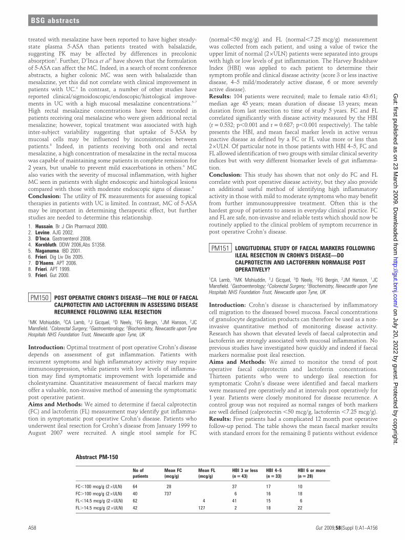
treated with mesalazine have been reported to have higher steady-state plasma 5-ASA than patients treated with balsalazide,suggesting PK may be affected by differences in precolonicabsorption2. Further, D’Inca et al3 have shown that the formulationof 5-ASA can affect the MC. Indeed, in a search of recent conferenceabstracts, a higher colonic MC was seen with balsalazide thanmesalazine, yet this did not correlate with clinical improvement inpatients with UC.4 In contrast, a number of other studies havereported clinical/sigmoidoscopic/endoscopic/histological improve-ments in UC with a high mucosal mesalazine concentrations.5–7
High rectal mesalazine concentrations have been recorded inpatients receiving oral mesalazine who were given additional rectalmesalazine; however, topical treatment was associated with highinter-subject variability suggesting that uptake of 5-ASA bymucosal cells may be influenced by inconsistencies betweenpatients.8 Indeed, in patients receiving both oral and rectalmesalazine, a high concentration of mesalazine in the rectal mucosawas capable of maintaining some patients in complete remission for2 years, but unable to prevent mild exacerbations in others.6 MCalso varies with the severity of mucosal inflammation, with higherMC seen in patients with slight endoscopic and histological lesionscompared with those with moderate endoscopic signs of disease.9
Conclusion: The utility of PK measurements for assessing topicaltherapies in patients with UC is limited. In contrast, MC of 5-ASAmay be important in determining therapeutic effect, but furtherstudies are needed to determine this relationship.
1. Hussain. Br J Clin Pharmacol 2000.2. Levine. AJG 2002.3. D’Inca. Gastroenterol 2008.4. Kornbluth. DDW 2006,Abs S1358.5. Naganuma. IBD 2001.6. Frieri. Dig Liv Dis 2005.7. D’Haens. APT 2006.8. Frieri. APT 1999.9. Frieri. Gut 2000.
PM150 POST OPERATIVE CROHN’S DISEASE—THE ROLE OF FAECALCALPROTECTIN AND LACTOFERRIN IN ASSESSING DISEASERECURRENCE FOLLOWING ILEAL RESECTION
1MK Mohiuddin, 2CA Lamb, 3J Gicquel, 3D Neely, 1FG Bergin, 1JM Hanson, 2JCMansfield. 1Colorectal Surgery; 2Gastroenterology; 3Biochemistry, Newcastle upon TyneHospitals NHS Foundation Trust, Newcastle upon Tyne, UK
Introduction: Optimal treatment of post operative Crohn’s diseasedepends on assessment of gut inflammation. Patients withrecurrent symptoms and high inflammatory activity may requireimmunosuppression, while patients with low levels of inflamma-tion may find symptomatic improvement with loperamide andcholestyramine. Quantitative measurement of faecal markers mayoffer a valuable, non-invasive method of assessing the symptomaticpost operative patient.Aims and Methods: We aimed to determine if faecal calprotectin(FC) and lactoferrin (FL) measurement may identify gut inflamma-tion in symptomatic post operative Crohn’s disease. Patients whounderwent ileal resection for Crohn’s disease from January 1999 toAugust 2007 were recruited. A single stool sample for FC
(normal,50 mcg/g) and FL (normal,7.25 mcg/g) measurementwas collected from each patient, and using a value of twice theupper limit of normal (26ULN) patients were separated into groupswith high or low levels of gut inflammation. The Harvey BradshawIndex (HBI) was applied to each patient to determine theirsymptom profile and clinical disease activity (score 3 or less inactivedisease, 4–5 mild/moderately active disease, 6 or more severelyactive disease).Results: 104 patients were recruited; male to female ratio 43:61;median age 45 years; mean duration of disease 13 years; meanduration from last resection to time of study 5 years. FC and FLcorrelated significantly with disease activity measured by the HBI(r = 0.532; p,0.001 and r = 0.687; p,0.001 respectively). The tablepresents the HBI, and mean faecal marker levels in active versusinactive disease as defined by a FC or FL value more or less than26ULN. Of particular note in those patients with HBI 4–5, FC andFL allowed identification of two groups with similar clinical severityindices but with very different biomarker levels of gut inflamma-tion.Conclusion: This study has shown that not only do FC and FLcorrelate with post operative disease activity, but they also providean additional useful method of identifying high inflammatoryactivity in those with mild to moderate symptoms who may benefitfrom further immunosuppressive treatment. Often this is thehardest group of patients to assess in everyday clinical practice. FCand FL are safe, non-invasive and reliable tests which should now beroutinely applied to the clinical problem of symptom recurrence inpost operative Crohn’s disease.
PM151 LONGITUDINAL STUDY OF FAECAL MARKERS FOLLOWINGILEAL RESECTION IN CROHN’S DISEASE—DOCALPROTECTIN AND LACTOFERRIN NORMALISE POSTOPERATIVELY?
1CA Lamb, 2MK Mohiuddin, 3J Gicquel, 3D Neely, 2FG Bergin, 2JM Hanson, 1JCMansfield. 1Gastroenterology; 2Colorectal Surgery; 3Biochemistry, Newcastle upon TyneHospitals NHS Foundation Trust, Newcastle upon Tyne, UK
Introduction: Crohn’s disease is characterised by inflammatorycell migration to the diseased bowel mucosa. Faecal concentrationsof granulocyte degradation products can therefore be used as a non-invasive quantitative method of monitoring disease activity.Research has shown that elevated levels of faecal calprotectin andlactoferrin are strongly associated with mucosal inflammation. Noprevious studies have investigated how quickly and indeed if faecalmarkers normalise post ileal resection.Aims and Methods: We aimed to monitor the trend of postoperative faecal calprotectin and lactoferrin concentrations.Thirteen patients who were to undergo ileal resection forsymptomatic Crohn’s disease were identified and faecal markerswere measured pre operatively and at intervals post operatively for1 year. Patients were closely monitored for disease recurrence. Acontrol group was not required as normal ranges of both markersare well defined (calprotectin ,50 mcg/g, lactoferrin ,7.25 mcg/g).Results: Five patients had a complicated 12 month post operativefollow-up period. The table shows the mean faecal marker resultswith standard errors for the remaining 8 patients without evidence
Abstract PM-150
No ofpatients
Mean FC(mcg/g)
Mean FL(mcg/g)
HBI 3 or less(n = 43)
HBI 4–5(n = 33)
HBI 6 or more(n = 28)
FC,100 mcg/g (26ULN) 64 28 37 17 10
FC.100 mcg/g (26ULN) 40 737 6 16 18
FL,14.5 mcg/g (26ULN) 62 4 41 15 6
FL.14.5 mcg/g (26ULN) 42 127 2 18 22
BSG abstracts
A58 Gut 2009;58(Suppl I):A1–A156
on July 20, 2022 by guest. Protected by copyright.
http://gut.bmj.com
/G
ut: first published as on 23 March 2009. D
ownloaded from
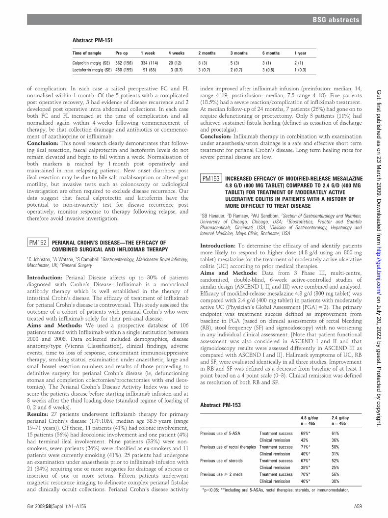
of complication. In each case a raised preoperative FC and FLnormalised within 1 month. Of the 5 patients with a complicatedpost operative recovery, 3 had evidence of disease recurrence and 2developed post operative intra abdominal collections. In each caseboth FC and FL increased at the time of complication and allnormalised again within 4 weeks following commencement oftherapy, be that collection drainage and antibiotics or commence-ment of azathioprine or infliximab.Conclusion: This novel research clearly demonstrates that follow-ing ileal resection, faecal calprotectin and lactoferrin levels do notremain elevated and begin to fall within a week. Normalisation ofboth markers is reached by 1 month post operatively andmaintained in non relapsing patients. New onset diarrhoea postileal resection may be due to bile salt malabsorption or altered gutmotility, but invasive tests such as colonoscopy or radiologicalinvestigation are often required to exclude disease recurrence. Ourdata suggest that faecal calprotectin and lactoferrin have thepotential to non-invasively test for disease recurrence postoperatively, monitor response to therapy following relapse, andtherefore avoid invasive investigation.
PM152 PERIANAL CROHN’S DISEASE—THE EFFICACY OFCOMBINED SURGICAL AND INFLIXIMAB THERAPY
1C Johnston, 2A Watson, 1S Campbell. 1Gastroenterology, Manchester Royal Infirmary,Manchester, UK; 2General Surgery
Introduction: Perianal Disease affects up to 30% of patientsdiagnosed with Crohn’s Disease. Infliximab is a monoclonalantibody therapy which is well established in the therapy ofintestinal Crohn’s disease. The efficacy of treatment of infliximabfor perianal Crohn’s disease is controversial. This study assessed theoutcome of a cohort of patients with perianal Crohn’s who weretreated with infliximab solely for their peri-anal disease.Aims and Methods: We used a prospective database of 106patients treated with Infliximab within a single institution between2000 and 2008. Data collected included demographics, diseaseanatomy/type (Vienna Classification), clinical findings, adverseevents, time to loss of response, concomitant immunosuppressivetherapy, smoking status, examination under anaesthetic, large andsmall bowel resection numbers and results of those proceeding todefinitive surgery for perianal Crohn’s disease (ie, defunctioningstomas and completion colectomies/proctectomies with end ileos-tomies). The Perianal Crohn’s Disease Activity Index was used toscore the patients disease before starting infliximab infusion and at8 weeks after the third loading dose (standard regime of loading of0, 2 and 6 weeks).Results: 27 patients underwent inflixiamb therapy for primaryperianal Crohn’s disease (17F:10M, median age 38.5 years (range19–71 years)). Of these, 11 patients (41%) had colonic involvement,15 patients (56%) had ileocolonic involvement and one patient (4%)had terminal ileal involvement. Nine patients (33%) were non-smokers, seven patients (26%) were classified as ex-smokers and 11patients were currently smoking (41%). 25 patients had undergonean examination under anaesthesia prior to infliximab infusion with21 (84%) requiring one or more surgeries for drainage of abscess orinsertion of one or more setons. Fifteen patients underwentmagnetic resonance imaging to delineate complex perianal fistulaeand clinically occult collections. Perianal Crohn’s disease activity
index improved after infliximab infusion (preinfusion: median, 14,range 4–19; postinfusion: median, 7.5 range 4–18). Five patients(18.5%) had a severe reaction/complication of infliximab treatment.At median follow-up of 24 months, 7 patients (26%) had gone on torequire defunctioning or proctectomy. Only 3 patients (11%) hadachieved sustained fistula healing (defined as cessation of dischargeand proctalgia).Conclusion: Infliximab therapy in combination with examinationunder anaesthesia/seton drainage is a safe and effective short termtreatment for perianal Crohn’s disease. Long term healing rates forsevere perinal disease are low.
PM153 INCREASED EFFICACY OF MODIFIED-RELEASE MESALAZINE4.8 G/D (800 MG TABLET) COMPARED TO 2.4 G/D (400 MGTABLET) FOR TREATMENT OF MODERATELY ACTIVEULCERATIVE COLITIS IN PATIENTS WITH A HISTORY OFMORE DIFFICULT TO TREAT DISEASE
1SB Hanauer, 2D Ramsey, 3WJ Sandborn. 1Section of Gastroenterology and Nutrition,University of Chicago, Chicago, USA; 2Biostatistics, Procter and GamblePharmaceuticals, Cincinnati, USA; 3Division of Gastroenterology, Hepatology andInternal Medicine, Mayo Clinic, Rochester, USA
Introduction: To determine the efficacy of and identify patientsmore likely to respond to higher dose (4.8 g/d using an 800 mgtablet) mesalazine for the treatment of moderately active ulcerativecolitis (UC) according to prior medical therapies.Aims and Methods: Data from 3 Phase III, multi-centre,randomised, double-blind, 6-week active-controlled studies ofsimilar design (ASCEND I, II, and III) were combined and analysed.Efficacy of modified-release mesalazine 4.8 g/d (800 mg tablet) wascompared with 2.4 g/d (400 mg tablet) in patients with moderatelyactive UC (Physician’s Global Assessment [PGA] = 2). The primaryendpoint was treatment success defined as improvement frombaseline in PGA (based on clinical assessments of rectal bleeding(RB), stool frequency (SF) and sigmoidoscopy) with no worseningin any individual clinical assessment. [Note that patient functionalassessment was also considered in ASCEND I and II and thatsigmoidoscopy results were assessed differently in ASCEND III ascompared with ASCEND I and II]. Hallmark symptoms of UC, RBand SF, were evaluated identically in all three studies. Improvementin RB and SF was defined as a decrease from baseline of at least 1point based on a 4 point scale (0–3). Clinical remission was definedas resolution of both RB and SF.
Abstract PM-151
Time of sample Pre op 1 week 4 weeks 2 months 3 months 6 months 1 year
Calpro’tin mcg/g (SE) 562 (156) 334 (114) 20 (12) 8 (3) 5 (3) 3 (1) 2 (1)
Lactoferrin mcg/g (SE) 450 (159) 91 (68) 3 (0.7) 3 (0.7) 2 (0.7) 3 (0.8) 1 (0.3)
Abstract PM-153
4.8 g/dayn = 465
2.4 g/dayn = 465
Previous use of 5-ASA Treatment success 69%* 61%
Clinical remission 42% 36%
Previous use of rectal therapies Treatment success 71%* 58%
Clinical remission 40%* 31%
Previous use of steroids Treatment success 67%* 52%
Clinical remission 38%* 25%
Previous use > 2 meds Treatment success 70%* 56%
Clinical remission 40%* 30%
*p,0.05; **including oral 5-ASAs, rectal therapies, steroids, or immunomodulator.
BSG abstracts
Gut 2009;58(Suppl I):A1–A156 A59
on July 20, 2022 by guest. Protected by copyright.
http://gut.bmj.com
/G
ut: first published as on 23 March 2009. D
ownloaded from
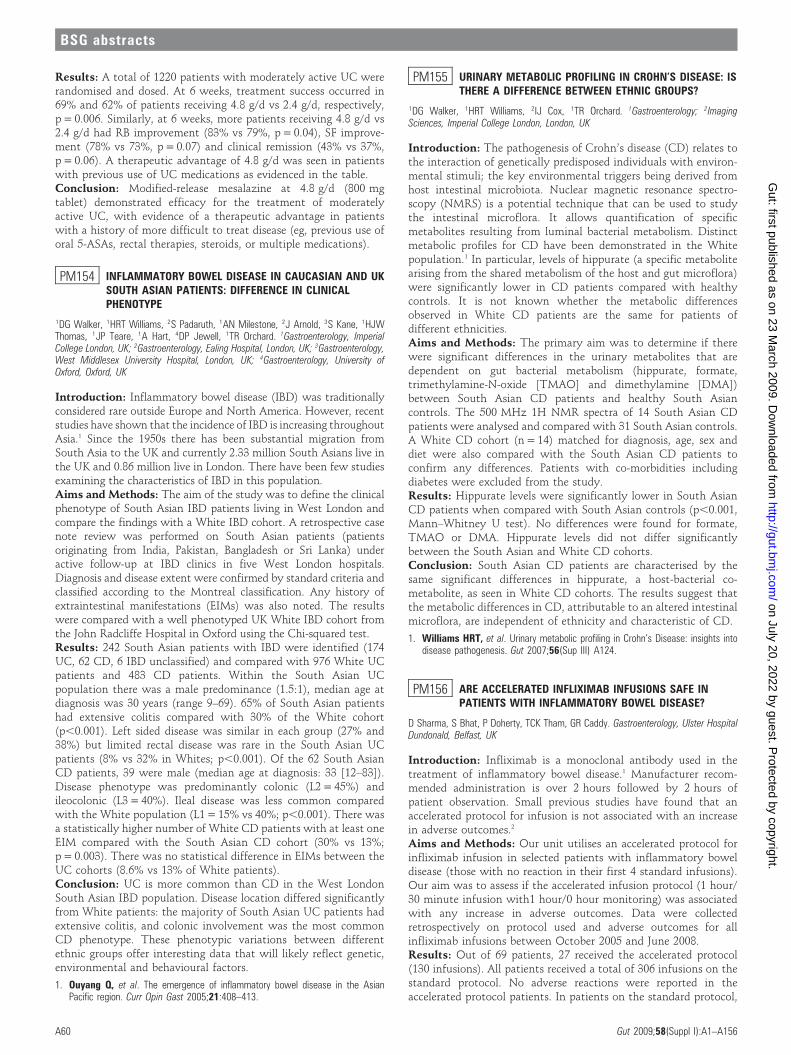
Results: A total of 1220 patients with moderately active UC wererandomised and dosed. At 6 weeks, treatment success occurred in69% and 62% of patients receiving 4.8 g/d vs 2.4 g/d, respectively,p = 0.006. Similarly, at 6 weeks, more patients receiving 4.8 g/d vs2.4 g/d had RB improvement (83% vs 79%, p = 0.04), SF improve-ment (78% vs 73%, p = 0.07) and clinical remission (43% vs 37%,p = 0.06). A therapeutic advantage of 4.8 g/d was seen in patientswith previous use of UC medications as evidenced in the table.Conclusion: Modified-release mesalazine at 4.8 g/d (800 mgtablet) demonstrated efficacy for the treatment of moderatelyactive UC, with evidence of a therapeutic advantage in patientswith a history of more difficult to treat disease (eg, previous use oforal 5-ASAs, rectal therapies, steroids, or multiple medications).
PM154 INFLAMMATORY BOWEL DISEASE IN CAUCASIAN AND UKSOUTH ASIAN PATIENTS: DIFFERENCE IN CLINICALPHENOTYPE
1DG Walker, 1HRT Williams, 2S Padaruth, 1AN Milestone, 2J Arnold, 3S Kane, 1HJWThomas, 1JP Teare, 1A Hart, 4DP Jewell, 1TR Orchard. 1Gastroenterology, ImperialCollege London, UK; 2Gastroenterology, Ealing Hospital, London, UK; 3Gastroenterology,West Middlesex University Hospital, London, UK; 4Gastroenterology, University ofOxford, Oxford, UK
Introduction: Inflammatory bowel disease (IBD) was traditionallyconsidered rare outside Europe and North America. However, recentstudies have shown that the incidence of IBD is increasing throughoutAsia.1 Since the 1950s there has been substantial migration fromSouth Asia to the UK and currently 2.33 million South Asians live inthe UK and 0.86 million live in London. There have been few studiesexamining the characteristics of IBD in this population.Aims and Methods: The aim of the study was to define the clinicalphenotype of South Asian IBD patients living in West London andcompare the findings with a White IBD cohort. A retrospective casenote review was performed on South Asian patients (patientsoriginating from India, Pakistan, Bangladesh or Sri Lanka) underactive follow-up at IBD clinics in five West London hospitals.Diagnosis and disease extent were confirmed by standard criteria andclassified according to the Montreal classification. Any history ofextraintestinal manifestations (EIMs) was also noted. The resultswere compared with a well phenotyped UK White IBD cohort fromthe John Radcliffe Hospital in Oxford using the Chi-squared test.Results: 242 South Asian patients with IBD were identified (174UC, 62 CD, 6 IBD unclassified) and compared with 976 White UCpatients and 483 CD patients. Within the South Asian UCpopulation there was a male predominance (1.5:1), median age atdiagnosis was 30 years (range 9–69). 65% of South Asian patientshad extensive colitis compared with 30% of the White cohort(p,0.001). Left sided disease was similar in each group (27% and38%) but limited rectal disease was rare in the South Asian UCpatients (8% vs 32% in Whites; p,0.001). Of the 62 South AsianCD patients, 39 were male (median age at diagnosis: 33 [12–83]).Disease phenotype was predominantly colonic (L2 = 45%) andileocolonic (L3 = 40%). Ileal disease was less common comparedwith the White population (L1 = 15% vs 40%; p,0.001). There wasa statistically higher number of White CD patients with at least oneEIM compared with the South Asian CD cohort (30% vs 13%;p = 0.003). There was no statistical difference in EIMs between theUC cohorts (8.6% vs 13% of White patients).Conclusion: UC is more common than CD in the West LondonSouth Asian IBD population. Disease location differed significantlyfrom White patients: the majority of South Asian UC patients hadextensive colitis, and colonic involvement was the most commonCD phenotype. These phenotypic variations between differentethnic groups offer interesting data that will likely reflect genetic,environmental and behavioural factors.
1. Ouyang Q, et al. The emergence of inflammatory bowel disease in the AsianPacific region. Curr Opin Gast 2005;21:408–413.
PM155 URINARY METABOLIC PROFILING IN CROHN’S DISEASE: ISTHERE A DIFFERENCE BETWEEN ETHNIC GROUPS?
1DG Walker, 1HRT Williams, 2IJ Cox, 1TR Orchard. 1Gastroenterology; 2ImagingSciences, Imperial College London, London, UK
Introduction: The pathogenesis of Crohn’s disease (CD) relates tothe interaction of genetically predisposed individuals with environ-mental stimuli; the key environmental triggers being derived fromhost intestinal microbiota. Nuclear magnetic resonance spectro-scopy (NMRS) is a potential technique that can be used to studythe intestinal microflora. It allows quantification of specificmetabolites resulting from luminal bacterial metabolism. Distinctmetabolic profiles for CD have been demonstrated in the Whitepopulation.1 In particular, levels of hippurate (a specific metabolitearising from the shared metabolism of the host and gut microflora)were significantly lower in CD patients compared with healthycontrols. It is not known whether the metabolic differencesobserved in White CD patients are the same for patients ofdifferent ethnicities.Aims and Methods: The primary aim was to determine if therewere significant differences in the urinary metabolites that aredependent on gut bacterial metabolism (hippurate, formate,trimethylamine-N-oxide [TMAO] and dimethylamine [DMA])between South Asian CD patients and healthy South Asiancontrols. The 500 MHz 1H NMR spectra of 14 South Asian CDpatients were analysed and compared with 31 South Asian controls.A White CD cohort (n = 14) matched for diagnosis, age, sex anddiet were also compared with the South Asian CD patients toconfirm any differences. Patients with co-morbidities includingdiabetes were excluded from the study.Results: Hippurate levels were significantly lower in South AsianCD patients when compared with South Asian controls (p,0.001,Mann–Whitney U test). No differences were found for formate,TMAO or DMA. Hippurate levels did not differ significantlybetween the South Asian and White CD cohorts.Conclusion: South Asian CD patients are characterised by thesame significant differences in hippurate, a host-bacterial co-metabolite, as seen in White CD cohorts. The results suggest thatthe metabolic differences in CD, attributable to an altered intestinalmicroflora, are independent of ethnicity and characteristic of CD.
1. Williams HRT, et al. Urinary metabolic profiling in Crohn’s Disease: insights intodisease pathogenesis. Gut 2007;56(Sup III) A124.
PM156 ARE ACCELERATED INFLIXIMAB INFUSIONS SAFE INPATIENTS WITH INFLAMMATORY BOWEL DISEASE?
D Sharma, S Bhat, P Doherty, TCK Tham, GR Caddy. Gastroenterology, Ulster HospitalDundonald, Belfast, UK
Introduction: Infliximab is a monoclonal antibody used in thetreatment of inflammatory bowel disease.1 Manufacturer recom-mended administration is over 2 hours followed by 2 hours ofpatient observation. Small previous studies have found that anaccelerated protocol for infusion is not associated with an increasein adverse outcomes.2
Aims and Methods: Our unit utilises an accelerated protocol forinfliximab infusion in selected patients with inflammatory boweldisease (those with no reaction in their first 4 standard infusions).Our aim was to assess if the accelerated infusion protocol (1 hour/30 minute infusion with1 hour/0 hour monitoring) was associatedwith any increase in adverse outcomes. Data were collectedretrospectively on protocol used and adverse outcomes for allinfliximab infusions between October 2005 and June 2008.Results: Out of 69 patients, 27 received the accelerated protocol(130 infusions). All patients received a total of 306 infusions on thestandard protocol. No adverse reactions were reported in theaccelerated protocol patients. In patients on the standard protocol,
BSG abstracts
A60 Gut 2009;58(Suppl I):A1–A156
on July 20, 2022 by guest. Protected by copyright.
http://gut.bmj.com
/G
ut: first published as on 23 March 2009. D
ownloaded from
16 adverse reactions were observed: 7 were acute (occurring duringinfusion); 9 were delayed (occurring within 1–7 days followinginfusion). No patient required intramuscular adrenaline or hospi-talisation.Conclusion: Our findings suggest that an accelerated protocol forinfliximab infusion is well tolerated in selected patients. Themonitoring period following infusion may not be necessary as allacute reactions occurred within an hour of initiating infusion anddid not warrant hospitalisation. The accelerated infusion may allowmore efficient utilisation of hospital resources and reduce patientinconvenience.
1. Hanauer SB, Feagan BG, Lichtenstein GR, et al. Maintenance infliximab forCrohn’s disease: the ACCENT I randomised trial. Lancet 2002;359:1541–1549.
2. Buch MH, Bryer D, Lindsay S, et al. shortening infusion times for infliximabadministration. Rheumatology 2006;45:485–495.
PM157 A YEAST TWO-HYBRID SCREEN USING CARD15/NOD2IMPLICATES NOVEL PATHWAYS IN IBD SUSCEPTIBILITY
1ER Nimmo, 1AJ Smith, 1MC Aldhous, 1M Quail, 1K Soo, 1H Drummond, 2J vanLimbergen, 3RK Russell, 1CL Noble, 4DC Wilson, 1J Satsangi. 1Gastrointestinal Unit;2Clinical Lecturer in Child Life and Health, University of Edinburgh, Edinburgh, UK;3Department of Paediatric Gastroenterology, Yorkhill Hospital, Glasgow, UK; 4PaediatricGastroenterology and Nutrition Department, University of Edinburgh, Edinburgh, UK
Introduction: NOD2 was first identified as a Crohn’s diseasesusceptibility gene in 2001. Since then more than 30 susceptibilitygenes have been identified by large-scale association studies, but it isestimated that these genes still account for less than 30% of thoseinvolved. In order to identify other genes and pathways involved inthe development of IBD we have performed a yeast two hybridscreen to identify NOD2 interacting proteins and thereby candidategenes for association studies.Aims and Methods: Full length NOD2 cDNA was used as the baitin a yeast two-hybrid screen to identify interacting proteins using acDNA library from the intestinal epithelial cell line, SW480.Interacting proteins were confirmed by co-immunoprecipitation.Genotyping was performed on the Illumina Goldengate platformand analysis performed using Haploview 4.1.Results: 700 bacterial colonies generated by the yeast two-hybridscreen were analysed and from these 16 interacting proteins wereidentified. These include proteins in pathways involved in regula-tion of transcription (TLE1, EGR1); NFkB regulation (MAP3K11,TLE1); Wnt signalling (TIP60, TLE1); apoptosis (TIP60, PA2G4,BPY2IP1, TTC11, VIMENTIN); androgen receptor (TIP60, FKBP4,PA2G4) and in epigenetic control of expression (TLE1, EGR1, TIP60,PA2G4). Interaction with NOD2 in SW480 cells was confirmed forTIP60, vimentin, PPP2R5E, TLE1, GALNT2, PA2G4, TTC11 andEGR1 by co-immunoprecipitation in SW480 cells. No interactionwith Erbin or Grim-19 was detected. Genotyping data will bepresented.Conclusion: These interactions broaden our understanding ofNOD2 function and implicate NOD2 signalling in regulation of anumber of molecular pathways. As well as NFkB regulation andapoptosis, we identify for the first time a potential role for NOD2 inWnt signalling and epigenetic regulation in intestinal epithelialcells.
PM158 LOSS OF HUMAN GAMMA DELTA T-CELLS IN CROHN’SDISEASE AND ULCERATIVE COLITIS
1ER Mann, 1NE McCarthy, 1AN Milestone, 2SA Cochrane, 3AJ Stagg, 1AL Hart,1SC Knight. 1Antigen Presentation Research Group, Imperial College London, St Mark’sHospital, Harrow, UK; 2SEAC, Unilever Colworth, Bedford, UK; 3Centre for InfectiousDisease, Barts and the London School of Medicine and Dentistry, London, UK
Introduction: cd T-cells are innate lymphocytes that are increasedin number in the gut of patients with inflammatory bowel disease(IBD). Some mouse models of IBD suggest that cd T-cells are
immuno-protective in the mucosa, while others indicate a role inexacerbation of disease. The role of cd T-cells in gut immunity andIBD is far from clear.Aims and Methods: Human peripheral blood was obtained frompatients (n = 10) with Crohn’s disease or ulcerative colitis onminimal treatment (5-aminosalicylic acid or azathioprine) or fromhealthy volunteers (n = 11). Mononuclear cells were isolated onFicoll gradients and labelled with monoclonal antibodies foranalysis by flow cytometry. cd T-cells were analysed for expressionof tissue-homing molecules such as b7 integrin, a moleculeassociated with leucocyte homing to the gut.Results: In healthy control subjects, the majority of cd T-cells wereCD3hi and expressed b7 and memory cell marker, CD45RO. Theco-stimulatory receptor CD28 was expressed on 20–30% of cd T-cells. Expression of skin homing markers was minimal. There was asignificant decrease in both the number and proportion of cd T-cellsin the blood of IBD patients compared with healthy controls. Theproportion of cd T-cells exhibiting the memory cell phenotype wasalso significantly reduced in IBD, while preliminary data indicatedthat expression of the lymph node homing marker, CCR7, wasincreased on cd T-cells in IBD.Conclusion: Taken with reports that cd T-cells are increased in thegut of IBD patients, it seems likely that the reduced numbers of cdT-cells in the circulation are a result of cd T-cell migration to gut.The reduced proportion of blood cd T-cells expressing CD45RO inIBD, and possible increases in expression of CCR7, may indicate aloss of effector memory phenotype in IBD.
PM159 EXPERIENCE ON THE USE OF ADALIMUMAB ASMAINTENANCE THERAPY IN CROHN’S DISEASE INENGLAND AND IRELAND
1EA Russo, 2J Lindsay, 3S Campbell, 4AL Hart, 5PJ Hamlin, 6TR Orchard, 4N Arebi,4J Nightingale, 4MR Jacyna, 4M O’Connor, 7AW Harris, 8CA O’Morain, 1S Gosh.1Gastroenterology, Hammersmith Hospital, London, UK; 2Gastroenterology, Barts and theLondon NHS Trust, London, UK; 3Gastroenterology, Manchester Royal Infirmary,Manchester, UK; 4Gastroenterology, St Mark’s Hospital, London, UK; 5Gastroenterology,Leeds General Infirmary, Leeds, UK; 6Gastroenterology, St Mary’s Hospital, London, UK;7Gastroenterology, Kent & Sussex Hospital, Tunbridge Wells, UK; 8Gastroenterology,Adelaide and Meath Hospital, Dublin, Ireland
Introduction: Adalimumab (ADA) has been shown to be effectivein inducing and maintaining response/remission in patients withCrohn’s Disease (CD) both naive to biological therapies andfollowing secondary failure of infliximab (IFX). Recent data fromScotland suggested that 13 of 22 patients (59%) required doseescalation to 40 mg every week. We present the first ‘‘real-life’’ datafrom England and Ireland on the use of ADA in induction andmaintenance of response/remission in patients with moderate tosevere CD.Aims and Methods: We carried out secure web-based datacollection from 8 hospitals on the use of ADA in CD. We collecteddata on disease distribution and duration, and previous/current useof 5-aminosalicylic acid, steroids, thiopurines, methotrexate andIFX. We report on response rates (change in CDAI .70, or HBI >3or as per Physician Global Assessment (PGA)), remission rates(CDAI,150, or HBI (4 or as per PGA) and the side-effect profile ofADA.Results: We report on 61 patients (35 female) with a mean age of35 years (17–71). The mean follow-up period was 8 months. 46%had ileo-colonic, 21% had ileal and 25% colonic disease. Rectalinvolvement was observed in 54%, perianal disease in 46% and 41%had fistulising disease. All patients had previously requiredcorticosteroids. Four patients were biologic-naive (6.6%); of theremainder, IFX was discontinued due to intolerance (41%), lack ofresponse (5%), loss of response (44%) or both (3.3%). 46% wereinduced with 160 mg and 80 mg of ADA on weeks 0 and 2,respectively, and 54% with 80–40 mg. 100% received 40 mg every
BSG abstracts
Gut 2009;58(Suppl I):A1–A156 A61
on July 20, 2022 by guest. Protected by copyright.
http://gut.bmj.com
/G
ut: first published as on 23 March 2009. D
ownloaded from
other week (EOW) as initial maintenance dose, of which 13% wereescalated to 40 mg weekly and 3% to 80 mg weekly. 83.6% ofpatients achieved response on maintenance ADA, of which 96% hadsteroid-free response, 67% remained off an immunomodulator and59% were on ADA monotherapy. 57% of patients were in completeremission from their CD, of which 94% had steroid-free remission,57% remained off an immunomodulator and 51% were on ADAmonotherapy. An additional 8.2% of participants had a secondaryloss of response to ADA, at an average of 8.4 months (range 2–17)from onset. Fourteen of 61 patients (23%) had adverse effects (localpain 4, infection 5, headaches 2, leucopenia (on Azathioprine) 1,painful rash 1, serum-sickness type reaction 1, the latter 2 of whichled to discontinuation of therapy.Conclusion: Results from an English/Irish cohort show satisfac-tory response/remission and safety profile of ADA in the treatmentof CD. In contrast to previous data from Scotland, dose escalationwas only seen in 16% of patients.
PM160 THE USE OF EMAIL AND TEXT MESSAGING TO REMINDPATIENTS TO HAVE BLOOD MONITORING WHILST ONIMMUNOSUPPRESSANTS
H Ludlow, J Hurley, S Dolwani. Gastroenterology, University Hospital Llandough,Penarth, UK
Introduction: Patients on long term Azathioprine or 6-Mercaptopurine for Inflammatory Bowel Disease (IBD) requirefrequent blood tests and may often forget to have these takenresulting in delays and subsequent safety concerns. Email and textmessaging have been shown to improve concordance for otherhealth issues, including smoking cessation, asthma and diabetesmanagement. Increased access to the internet and mobile phoneownership make this an ideal method of communication with thisgroup of patients.Aims and Methods: We aimed to determine whether the use ofemail and text messaging could improve delays in blood monitoringcompared with standard telephone call reminders and to comparethis with delays prior to the IBD nurse being in post. BetweenSeptember 2007 and August 2008 patients on established treatmentfor .1 year were invited to participate in the study and also thosenewly commenced once maintenance dose reached. Test delaysduring the study were compared with previous delays, when onlytelephone calls were used. The table shows the delays pre-IBD nursepost and then pre- and post-study delays and reminders required.Results: 115 patients were included in the study—63 had been ontreatment for at least 1 year and 52 new starters (63 UC, 49 CD, 3indeterminate). The median ages of those who chose contact byemail was 41 years (n = 44), text messaging 40 years (n = 42) andtelephone contact or did not reply (control) 61 years (n = 39).Reasons for not wishing contact by email or text were: no computeror mobile telephone (n = 19, median age 68.5), too intrusive (n = 1),prefers telephone (n = 1) and no reason given (n = 5).Conclusion: Following the introduction of the IBD nurse the delayin blood tests fell dramatically from a maximum of 307 days to
34 days. There are many issues/causes surrounding delays includingholidays and restricted GP phlebotomy clinics. The text groupshowed further reduction in delay whereas the control groupwithout intervention actually increased. Multiple call attempts andanswer machine messages are avoided (reducing issues regardingconfidentiality), thus reducing time involved. Texts and emails arecost-effective in relation to frequent calls to mobile numbers. Anyform of intervention by the nurse reduces delays although emailand text messaging are convenient and preferred by both patientsand the nurse.
PM161 USE OF TARGETED ANTIBIOTICS TO TREAT ULCERATIVECOLITIS
1T Nakayama, 1KL Blackett, 1H Steed, 1S Macfarlane, 2N Reynolds, 1JH Cummings,1GT Macfarlane. 1Pathology and Neuroscience, University of Dundee, UK;2Gastroenterology, NHS Tayside, Dundee, UK
Introduction: The cause of ulcerative colitis (UC) is unclear, butbacteria colonising the colonic mucosa are thought to play a keyrole in disease aetiology. Despite this, antibiotics have beenunsuccessful in managing acute UC, and have had only limitedbenefit in long-term treatment. The failure of antibiotics might bebecause the target organisms are unknown, or that the organismsare protected in mucosal biofilms.Aims and Methods: To identify putative pathogens on mucosalsurfaces in the large bowel in active UC patients, and to determinetheir antibiotic sensitivities, with the aim of designing therapies fortheir suppression. 33 patients with active UC were enrolled in thetrial. Clinical status was scored using the simple clinical colitisactivity index (SCCAI), and rectal biopsies were taken for analysisof biofilm community structure and antibiotic sensitivity tests.Patients received two antibiotics (coamoxiclav, ciprofloxacin,clarithromycin, doxycycline, rifaximin) for 4 weeks, and clinicalassessments were repeated. Patients were included if they had stabledrug therapies for 3 months or were non-responders to oral steroids.Twenty-four patients completed the study.Results: E coli, enterobacteria, peptostreptococci, staphylococci andstreptococci were the predominant pathogenic species present inrectal biopsies. After 1 month of treatment with combinationantibiotic therapy, there was a significant reduction in the totalnumber of bacteria present in rectal biopsies (p,0.0001) and asignificant reduction in the number of eubacteria (p = 0.0051) from5.016102 to 0.656101. There was no significant change in thenumbers of lactobacilli or bifidobacteria, which are potentiallybeneficial bacteria. There was a significant improvement in SCCAIfrom 7.1 to 4.8 (p = 0.005) and a significant improvement inpatients IBDQ from 139 to 157 (p = 0.002). There was nosignificant improvement in the Baron’s score (p = 0.07), but therewas a significant improvement in the histological score from 11.04to 8.91 (p = 0.029). Analysis of bacteria in patients with noimprovement, or a worsening of their SCCAI showed that therewas a significant reduction in the number of prevotella from1.46105 to complete eradication (p = 0.002), and a reduction in the
Abstract PM-160
.1 year groupemail
.1 year grouptext
.1 year groupcontrol
New startersemail
New starterstext
New starterscontrol
Days late before IBDnurse post
0 (0–144) 2 (0–186) 28 (0–307) x x x
Reminders before study 1 (0–2) 1 (0–1.5) 1 (0–3) x x x
Days late before study 1 (0–22) 4 (0–28) 1 (0–54) x x x
Reminders during study 1 (1–4) 1.5 (1–3) 1 (0–3.5) 1 (1–2) 1 (1–2) 1 (0–2)
Days late during study 1.5 (0–28) 2.5 (0–34) 7 (0–30) 1.5 (0–16) 2 (0–10) 1 (0–20)
BSG abstracts
A62 Gut 2009;58(Suppl I):A1–A156
on July 20, 2022 by guest. Protected by copyright.
http://gut.bmj.com
/G
ut: first published as on 23 March 2009. D
ownloaded from
number of bacteroides from 2.46104 to complete eradication(p = 0.022). In the group with improved clinical scores, there wasa significant increase in bacteroides (p = 0.049) (2.36103 to 2.46105)and enterobacteria (2.36101 to 9.66102) and a significant reductionin eubacteria (1.96102 to 2.66101).Conclusion: Targeting specific mucosal populations on the rectalmucosa demonstrated that antibiotics were unsuccessful inselectively eradicating immunogenic bacterial species in UCbiofilms. However, targeted antibiotic treatment did facilitate there-establishment of benefical populations in the mucosal micro-biota, which was associated with improved clinical and histologicalstatus.
PM162 VOLATILE ORGANIC COMPOUNDS AS DIAGNOSTIC FAECALBIOMARKERS IN INFLAMMATORY BOWEL DISEASE—METHOD DEVELOPMENT
I Ahmed, S Smith, CS Probert. Gastroenterology, Bristol Royal Infirmary, Bristol, UK
Introduction: Despite better understanding in recent years, thediagnosis of inflammatory bowel disease requires invasive andexpensive procedures. Current studies are focusing on non-invasivemarkers which could assist in diagnosis and monitoring the disease,and also in early prediction of relapses and complications. Amongthe other non-invasive modalities, faecal biomarkers such asCalprotectin, Lactoferrin, etc have appeared recently in thediagnostic armamentarium. The diagnostic role of volatile organiccompounds (VOCs) emitted in faeces of patients with IBD remainsto be explored further. As little is known about VOCs in humanfaeces, a convenient analytical method to quantify them has notbeen addressed. This work presents a facile and rapid methodologyto quantify faecal VOCs by using solid phase micro extraction(SPME) combined with gas chromatography mass spectrometry(GC/MS) by identifying suitable variables for sample preparationand analysis.Aims and Methods: 10 healthy volunteers produced 70 samplesfor experiments. Headspace SPME was used as a sample preconcentration technique prior to the determination of VOCs byGC-MS. Samples were collected in a standard 50 ml faecal samplebottle and aliquoted to a 10 ml Supelco phial. Samples were placedin thermostatted water at 60uC for 1 hour to equilibrate prior toSPME extraction and fibre was then transferred to the injectionport of the GC/MS for analysis. The VOCs were identified usingNIST library followed by manual visual inspection and retentiontime matching of selected standards. The VOCs obtained by theseexperiments were compared in order to ascertain the most efficientmethodology. The effects of extraction variables, such as the type offibre, extraction time (10 min vs 20 min), sample quantity (1 gm/2 gm/4 gm), and effect of time at room temperature and freezingon 220uC, on the amount of extracted VOCs were studied.Results: Results showed that use of PDMS/Car fibre identifiedmaximum numbers of VOCs as compared with DVB fibre. MoreVOCs were detected in 2 gm of sample in comparison with 1 gmand 4 gm quantities. There was no meaningful difference inVOCs quantification by 10 min extraction by SPME fibre incomparison with 20 min. Samples left over a 24 hour period atroom temperature were found to produce lower numbers ofVOCs compared with samples analysed within 6 hours ofproduction. Samples which were analysed following freezingwithin 6 hours of production showed only minimal reductions intotal VOCs.Conclusion: The optimum conditions for precise VOC quantifica-tion in faecal samples are: 2 gm quantity of faecal sample analysedwithin 6 hours of sample production with a use of PDMS/CARtype of SPME fibre injected in headspace above sample for 10 minextraction. If samples have to keep for longer then they should befrozen at 220uC to preserve VOCs.
PM163 THE STRESS MEDIATORS ADENOSINE ANDHYDROCORTISONE IMPAIR COLONIC EPITHELIAL WOUNDHEALING IN VITRO
J Goodhand, M Wahed, DS Rampton. Centre for Gastroenterology, Barts and theLondon School of Medicine and Dentistry, London, UK
Introduction: Psychological stress worsens gut inflammation ininflammatory bowel disease (IBD). In some models, stress delaysmucosal ulcer healing.1 This process is initiated by restitution, theingress of motile epithelial cells into the wound. The pro-inflammatory chemokine, interleukin (IL)-8, accelerates epithelialcell restitution.2
Aims and Methods: To determine the effects of four putativeneurohumoral mediators of the stress response on colonic epithelialcell restitution and IL8 production in vitro. Cell restitution velocitywas measured over 24 h in linear wounds made in confluentmonolayers of human colon cancer-derived HT-29 cells. Cells wereincubated with noradrenaline, adenosine, histamine and hydro-cortisone in the presence and absence of fetal calf serum (FCS)(positive and negative control, respectively) and photomicrographsat 24 h analysed with Digimizer software. For the IL8 studies, HT-29 cells were pre-incubated with each stress mediator for 1 h, thenstimulated with IL1b (1 ng/ml). IL8 in supernatants collected at 6 hwas assayed by ELISA. In each case n>4, and ANOVA was used tocompare differences; restitution results for FCS are shown.Results: Restitution velocity was dose-dependently inhibited byadenosine (10–6 M: 3.3 (mean) ¡0.42 (SD), p,0.01; 10–3 M: 2.4¡0.31,p,0.01, compared with relevant controls 4.9¡0.21) and by hydro-cortisone (10–9 M: 4.8¡0.75, p,0.05; 10–6 M: 4.4¡0.66, p,0.01 vscontrols 6.0¡0.73). Adenosine (10–6 M: 327 pg/ml ¡27, p,0.01; 10–3
M: 127¡17, p,0.01) and hydrocortisone (10–9 M: 325¡25, p,0.01;10–6 M: 294¡16, p,0.01) each reduced IL8 production comparedwith controls (449¡30). At 10–9 and 10–6 M, neither noradrenalinenor histamine altered wound healing or IL8 production.Conclusion: These results suggest that psychological stress, bycausing release of adenosine and hydrocortisone with subsequentreduction of epithelial IL8 production, could impair mucosal healingin IBD and perhaps other inflammatory diseases such as pepticulcer. The adverse effects of hydrocortisone on epithelial restitutionin vitro could also contribute to its failure to induce mucosal healingin Crohn’s disease.
1. Marucha PT, Kiecolt-Glaser JK, Favagehi M. Mucosal wound healing is impairedby examination stress. Psychosom Med 1998;60:362–5.
2. Wilson AJ, Byron K, Gibson PR. Interleukin-8 stimulates the migration of humancolonic epithelial cells in vitro. Clin Sci (Lond) 1999;97:385–90.
PM164 INFLIXIMAB USE IN ADOLESCENTS WITH INFLAMMATORYBOWEL DISEASE
1J Goodhand, 2J Tang, 1S Naik, 1NM Croft, 1JO Lindsay. 1Centre for Gastroenterology,Barts and the London School of Medicine and Dentistry, London, UK; 2Department ofMedicine, University of Toronto, Toronto, Canada
Introduction: The incidence of inflammatory bowel disease inadolescence is increasing.1 Compared with adults, the proportion ofadolescents with Crohn’s disease is greater, disease is moreextensive, and patients more frequently require treatment withimmunosuppressants and surgery.2
Aims and Methods: To compare the clinical use and response toinfliximab in adolescents versus adults at a tertiary IBD centre witha specialist transitional adolescent clinic. We performed a retro-spective study of infliximab use at Barts and the London NHS trust,in adults and adolescents with Crohn’s disease (CD), ulcerativecolitis (UC) and indeterminate colitis (IC). Demographic details,disease extent and course were recorded as well as response toinduction and maintenance therapy. Differences between catego-rical data were detected with chi-squared analysis and forcontinuous data with unpaired t-tests.
BSG abstracts
Gut 2009;58(Suppl I):A1–A156 A63
on July 20, 2022 by guest. Protected by copyright.
http://gut.bmj.com
/G
ut: first published as on 23 March 2009. D
ownloaded from
Results: Between 2000 and 2008, 28 adolescents (age,25, in 2008)[26CD:1UC:1IC] and 40 adults [33CD:5UC:2IC] were treated withinfliximab. Adolescents (YA) received infliximab earlier in theirdisease course than adults (AD) [YA 5 years vs AD 11.4 years,p,0.001 (95% CI = 2.79 to 10.5)]. Primary non-response toinfliximab was not observed in the adolescent group comparedwith 7 out of 40 in the adult group (p,0.05). However, there wasno difference between the proportion of patients losing response[YA 31% vs AD 37%, p = 1] or time to loss of response [YA16.8 months vs AD 15.4 months, p = 0.87] during maintenancetherapy; likewise the frequency of infusion reactions was compar-able in the two groups. Patients who lost response to infliximab haddose escalation, increase in infusion frequency or switch toadalimumab with no difference in strategy between groups.Conclusion: In keeping with their more severe disease phenotype,adolescents receive infliximab earlier in the natural course of theirdisease. Induction therapy in adolescents is more successful thanadults. Prolonged response during maintenance therapy in adoles-cence is similar to randomised control clinical trial data.3
1. Sawczenko A, Sandhu BK, Logan RF, et al. Prospective survey of childhoodinflammatory bowel disease in the British Isles. Lancet 2001;357:1093–4.
2. Van Limbergen J, Russell RK, Drummond HE, et al. Definition of phenotypiccharacteristics of childhood-onset inflammatory bowel disease. Gastroenterology2008;135:1114–22.
3. Hanauer SB, Feagan BG, Lichtenstein GR, et al. Maintenance infliximab forCrohn’s disease: the ACCENT I randomised trial. Lancet 2002;359:1541–9.
PM165 EPIDEMIOLOGICAL RISK FACTORS FOR CHILDHOOD ONSETINFLAMMATORY BOWEL DISEASE IN SCOTLAND: A CASE-CONTROL STUDY
1JE Van Limbergen, 1H Spiers, 1R Farhadi, 2ML Wilson, 3RK Russell, 4G Mahdi,5J Satsangi, 2DC Wilson. 1Paediatric Gastroenterology and Nutrition, Royal Hospital forSick Children, Edinburgh, UK; 2Child Life and Health, University of Edinburgh, Edinburgh,UK; 3Paediatric Gastroenterology, Yorkhill Hospital, Glasgow, UK; 4PaediatricGastroenterology, Royal Aberdeen Children’s Hospital, Aberdeen, UK; 5GastrointestinalUnit, University of Edinburgh, Edinburgh, UK
Introduction: The incidence of inflammatory bowel disease (IBD)is increasing among children in Scotland and is among the highestworldwide, yet the aetiology of IBD remains largely unknown.Genetic and environmental risk factors have been implicated. Wehave shown that the phenotype of childhood-onset IBD is moreextensive than adult-onset IBD in Scotland, but also that thecarriage of variants of genes implicated in IBD susceptibility(NOD2, IBD5, DLG5 and IL23R) does not differ between theseScottish IBD populations.1 2
Aims and Methods: We aimed to investigate the influence of arange of potential environmental factors in the development ofpaediatric IBD. 126 paediatric IBD cases (diagnosed ,17 years ofage; median age at diagnosis 9.8 years (Q1–Q3: 7.5 to 11.7)) wereeach matched with one control subject by age, sex and geographicallocation (to match for socio-economic status). Children with IBDand their parents were interviewed face-to-face to obtain data onbreastfeeding, immunisation history, surgical and medical historyand family history (FH). Control data were obtained via a postalquestionnaire. Unifactorial analyses using the Chi-squared test andmultifactorial analysis using binary logistic regression were per-formed.Results: FH of IBD was associated with IBD (OR3.34; 95% (1.70 to6.56) p = 0.0003). History of asthma, eczema and food allergy (OR2.48 (1.28 to 4.78) p = 0.005, OR 2.83 (1.50 to 5.36) p = 0.001 andOR 2.98 (1.04 to 8.54) p = 0.03, respectively) show a significantassociation with paediatric IBD and CD. There were no significantdifferences between cases and controls regarding breastfeeding (52%vs 55%, p = 0.71). On multivariate analysis the significant factorswere FH of IBD, bowel cancer and history of asthma and eczema(p,0.001, 0.033, 0.045 and 0.015, respectively). In contrast withfindings on unifactorial analysis, no association of IBD with
parental smoking during pregnancy, at birth and currently, wasfound in the multifactorial analysis, irrespective of FH of IBD.Conclusion: We have shown an association between atopy andpaediatric IBD but none with breastfeeding practices. Familyhistory of IBD or bowel cancer is associated with IBD. Ourepidemiological data are especially noteworthy in the context of theincreasing number of genetic associations related to commoninflammatory pathways identified in IBD and cancer.
1. Van Limbergen, et al. Gastroenterology 2008;135(4):1114–22.2. Van Limbergen, et al. American Journal of Gastroenterology 2007;102:2820–31.
PM166 2 YEAR FOLLOW-UP OF ADALIMUMAB THERAPY INPATIENTS WITH CROHN’S DISEASE
1K Kemp, 2M Dibb, 2AJ Makin, 2SS Campbell. 1Department of Gastroenterology andSchool of Nursing, Manchester Royal Infirmary and University of Manchester,Manchester, UK; 2Department of Gastroenterology, Manchester Royal Infirmary,Manchester, UK
Introduction: Adalimumab (Ad), a fully humanised, anti-TNFantibody, has demonstrated ability to induce and maintainremission in Crohn’s Disease (CD). There remains relatively littledata on long-term safety data as well as long-term remissionmaintenance in CD.Aims and Methods: A database of pts who received Ad over a 27mth period (June 2006–September 2008) was created. Demographicdata, SAEs, durability, time to loss of response, previous exposure toinfliximab (IFx), concomitant immunosuppressive therapy andsmoking status were evaluated. Loss of response to IFx was definedas the first occasion a reduction in dosing interval or increase in dosewas required. Remission/relapses were based upon standard clinical,radiological and endoscopic criteria. Kaplan–Meier survival analysiswas used to compare durability of response.Results: A total of 25 pts (16F:9M), median age 44 years (17–73years), were included. Twenty-one pts received an induction regimeof 80/40 mg, 4 pts 160/80 mg followed by 40 mg EOW main-tenance. Median follow-up was 6 months (2–27 months). Diseaseanatomy included colonic disease 4 (16%); ileocolonic 20 (80%);duodenal disease 1 (4%). Twenty-three (92%) pts received IFx priorto Ad with a median duration of IFx of 17.5 months (4–58 months).Patients were grouped into 3 categories: primary non-responders, 3(12%), defined as failure to improve after 0, 2 infusions; loss ofresponse 9 (36%) defined as an interval time of ,5 weekly; anddiscontinuation of IFx due to side effects 11 (44%). Eight ptssmoked and 12 pts were on concomitant immunosuppressivetherapy. Median Rx with Ad was 6 months (2–27 months). Doseescalation of Ad (40 mg EOW to 40 WKLY) occurred in 5 (20%) ptswith a median time to dose escalation of 10 months (range 2–12months). The 5 pts received a loading dose of 80/40 mg with 1 pt onconcomitant immunosupression therapy at time of dose escalation.SAEs were recorded, 1 cellulitis, 1 pneumonia and 1 viral meningitis.The SAEs resulted in the discontinuation of Ad in 2 pts (pneumoniaand phial meningitis). No malignancy was detected. Comparisonswere made between smoker and non-smoker as well as primary IFxnon-responders vs secondary IFx non-responders. Time to doseescalation for both comparison groups was not significant. The 3primary non responders to IFx are in remission with Ad. Sixteen(64%) of the 25 pts are in remission with Ad.Conclusion: Concurring with Edinburgh, Ad is efficacious in thetreatment of patients with CD who have lost response to, orsuffered a reaction to IFx. The number of pts that required doseescalation is lower in our cohort. This may be due to a number offactors including shorter median follow-up (6 mths vs 12 mths) anddifferences in concomitant immunosuppressive therapy (48% vs68%) raise the possibility that our pts had less severe disease. Thisstudy adds to the growing body of evidence and safety profile of Adin every day clinical practice.
BSG abstracts
A64 Gut 2009;58(Suppl I):A1–A156
on July 20, 2022 by guest. Protected by copyright.
http://gut.bmj.com
/G
ut: first published as on 23 March 2009. D
ownloaded from
PM167 THE PROFILE OF INFLAMMATORY BOWEL DISEASE (IBD) INTHE UK. LESSONS LEARNED FROM THE IBD DATABASE (DB)
1KD Bardhan, 2A Dhar, 1C Royston, 3N Simmonds, 4CM Edwards. 1Department ofGeneral Medicine, Rotherham NHS Foundation Trust, Rotherham, UK; 2Department ofGastroenterology, County Durham & Darlington NHS Foundation Trust, Bishop Auckland,UK; 3Department of Gastroenterology, Luton & Dunstable NHS Foundation Trust, Luton,UK; 4Department of Gastroenterology, South Devon Healthcare NHS Trust, Torbay, UK
Introduction: Safe clinical management of the IBD patientrequires ready access to complete information, particularly forthose with complex disease, often difficult using paper based casenotes. The Rotherham IBD database, created to meet local needs,was developed for wider use for both individual patient manage-ment and collating information across centres. Results from 2 pilotcentres were presented here previously 1. On behalf of the IBD-DBUser Group we now present our results from across the UK.Aims and Methods: AccessH based, with a minimum dataset.Data were censored to the end of October 2006 and anonymised foranalysis.Results: See table. 11 432 patients from 21 centres across Englandand Northern Ireland were included. The demographic profile wasbroadly similar across centres.Conclusion: We have learned that data need to be simple, welldefined, validated and continuously updated. Although self-evident,these ‘‘simple needs’’ are not always achieved, particularly wheninformation changes (eg, smoking), hence the need for updatingdata at every patient contact. A little data collected systematicallyfrom each patient and updated at every visit is more helpful thandetailed data collected from few.Observations: This large cohort shows the profile of IBD acrossthe UK is similar to other large series. Each centre practicesindependently yet achieved reassuringly similar results suggestingwe all strive towards ‘‘best practice.’’ Despite time and effortinvested, gaps have emerged in our data, the result of variableinterpretation of definition; thus there is room for improvement inthe process of multi-centre data collection and handling. Ourobservations have bearing on the intended development of aNational IBD-DB and give insight into the real effort required tosustain electronic patient records.Database Facilities: ‘‘At a glance’’ summary, automated produc-tion of clinic letters, monitoring and audit. In conclusion, we havelearned as much from the process of developing the IBD-DB as fromthe data generated.
1. Edwards CM, Royston C, Durbin H, et al. Gut 2006;55(Suppl 11):A75.
PM168 WHO LOOKS AFTER PATIENTS WHEN THEY AREHOSPITALISED AS AN EMERGENCY: NATIONAL-LEVELANALYSIS OF A SPECIALIST GASTROENTEROLOGICALCONDITION
1K Bodger, 2C Grant-Pearce, 2E Thompson, 2D Lowe, 2M Pearson. 1GastroenterologyDivision, School of Clinical Sciences; 2Department of Clinical Evaluation, University ofLiverpool, Liverpool, UK
Introduction: Crohn’s disease (CD) and Ulcerative colitis (UC) aretreatable chronic disorders but decisions about when to use morecomplex drugs or opt for surgery are likely to require specialist care.Most patients expect to be cared for by a specialist but in the past,emergencies would be under the care of a physician on-callregardless of specialty. We examined Hospital Episode Statistics forEngland and assessed the proportion of patients with definedconditions discharged from the care of a known GI specialist (GI-Sp).Aims and Methods: HES data for 2006/7 contain information on2.236 million emergency admissions to 151 acute Trusts in England.A steering group of BSG representatives, physicians, surgeons,public health, patient groups and IT specialists guided the project.We identified all medical admissions with a gastroenterologicaldiagnosis (13%) and defined Inflammatory Bowel Disease (IBD) asbeing the predominant reason for admission both when coded inthe first diagnosis field and when ICD-10 codes for UC/CD were inthe second or third fields but with diagnostic/symptom codes in thefirst field likely to be IBD-related (adding 25% to number of casesidentified). There is no nationally complete list of GI-Sp, so listswere created with the help of the BSG, the national electronic staffrecord, augmented by telephone contacts to hospitals.Results: There were 286 316 emergency medical GI admissions(13% of acute medical workload in England). 45.9% were withBenign Luminal Conditions, of which just 30.9% (IQR for Trusts:26–37%) were coded at discharge under a ‘‘known’’ GI-Sp. Therewere 10 476 (3.6%) IBD admissions (5739 CD, 4737 UC) across 151hospitals—a median of 34 (IQR 22 to 49) CD and 27 (IQR 19 to 40)UC patients per Trust. 62% of IBD patients were discharged under aknown GI-Sp (IQR 53–72%; range 15–95% across trusts). 80% ofIBD patients were aged ,65 years so there was little overall effectfrom age-related admission policies. A plot of % under a GI-Sp vstotal numbers of admissions showed a typical funnel plot withgreater range in smaller hospitals but with no trends related tohospital size.Conclusion: These data suggest that most patients admitted withGI problems will not be under the care of a GI-Sp at discharge. Localcoding quality issues are a correctable source of error, the list of GI-Sp may be incomplete and obviously patients will be seen by GI-Spas ‘‘ward consults’’ but remain under the care of a non-GIphysician. Nevertheless, although twice as many patients withIBD seem to reach a GI-Sp at the time of discharge, there remains anapparently wide range between hospitals that is unrelated to size.Significant numbers may be managed by non-specialists. Thisprobably reflects internal organisation/triage of hospitals but ashortfall in GI-Sp beds relative to the casemix of the emergency takemay be a factor in some centres. [This project was funded by theNHS Information Centre]
PM169 EXPRESSION OF WNT FAMILY OF GENES IN PRIMARYHUMAN INTESTINAL MYOFIBROBLASTS
KR Hughes, YR Mahida. Institute of Infection, Immunity and Inflammation, University ofNottingham, Nottingham, UK
Introduction: The Wnt family of secreted glycoproteins areimportant signalling molecules that regulate functions of intestinalepithelial stem cells and their progeny. Wnt signals are transducedafter binding Frizzled (Fz) receptors and low-density lipoprotein-receptor-related protein (LRP) 5 and 6. Members of secreted
Abstract PM-167
Total n = 11432Ulcerative Colitis (UC)n = 6412
Crohn’s Disease (CD)n = 4546
Type of IBD (%) * 56 40
Extent of disease
UC: Extensive 34
UC: Left sided 22
UC: Distal 44
CD: Colonic 38
CD: Terminal ileal 24
CD: Ileo-colonic 22
Gender ratio M:F 1.08:1 0.72:1
Age at diagnosis (mean,years)
39 30
Operated (%) 16 47 (61% once, 20%
(41% with pouch) twice and 19% >63)
Thiopurine use (%) (range) 16 (7.7–17.5) 27 (25.5–27.7)
Monoclonal use (%) 0.1 2.8
Mortality (%) 0.4 1.2
*4% of patients had indeterminate colitis.
BSG abstracts
Gut 2009;58(Suppl I):A1–A156 A65
on July 20, 2022 by guest. Protected by copyright.
http://gut.bmj.com
/G
ut: first published as on 23 March 2009. D
ownloaded from
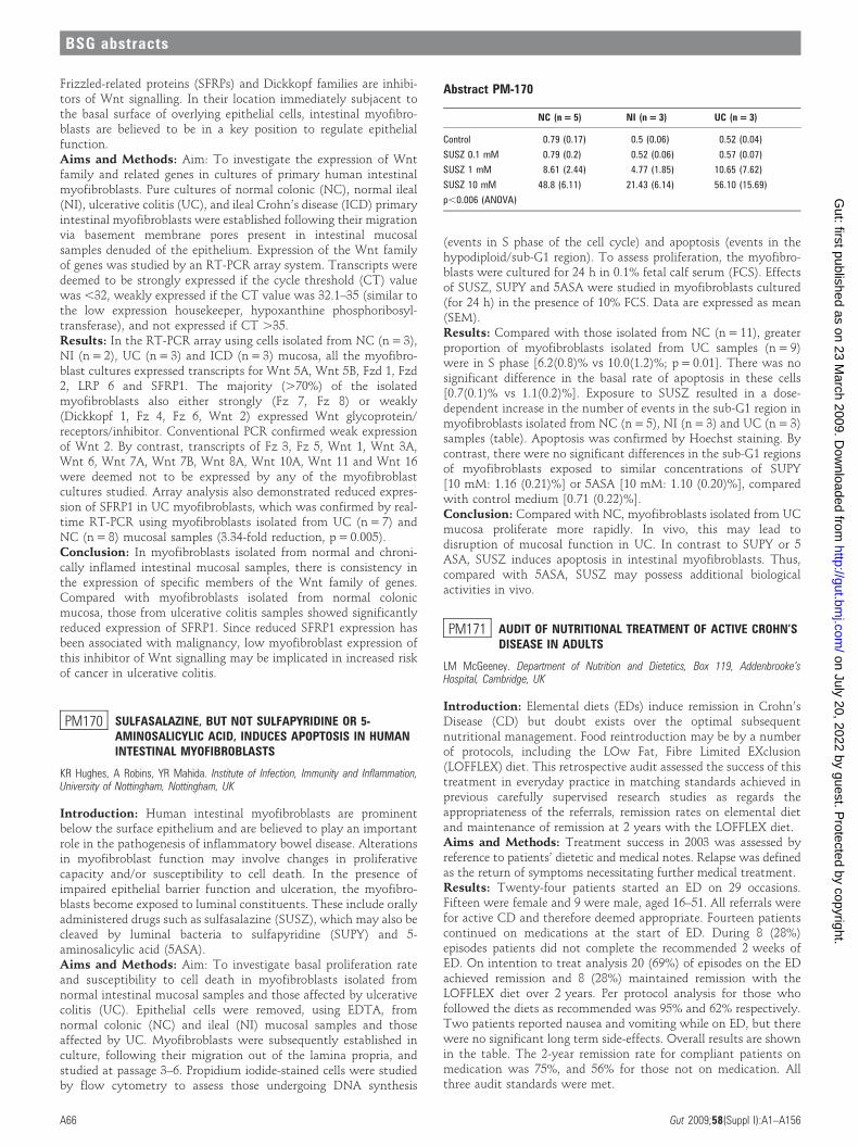
Frizzled-related proteins (SFRPs) and Dickkopf families are inhibi-tors of Wnt signalling. In their location immediately subjacent tothe basal surface of overlying epithelial cells, intestinal myofibro-blasts are believed to be in a key position to regulate epithelialfunction.Aims and Methods: Aim: To investigate the expression of Wntfamily and related genes in cultures of primary human intestinalmyofibroblasts. Pure cultures of normal colonic (NC), normal ileal(NI), ulcerative colitis (UC), and ileal Crohn’s disease (ICD) primaryintestinal myofibroblasts were established following their migrationvia basement membrane pores present in intestinal mucosalsamples denuded of the epithelium. Expression of the Wnt familyof genes was studied by an RT-PCR array system. Transcripts weredeemed to be strongly expressed if the cycle threshold (CT) valuewas ,32, weakly expressed if the CT value was 32.1–35 (similar tothe low expression housekeeper, hypoxanthine phosphoribosyl-transferase), and not expressed if CT .35.Results: In the RT-PCR array using cells isolated from NC (n = 3),NI (n = 2), UC (n = 3) and ICD (n = 3) mucosa, all the myofibro-blast cultures expressed transcripts for Wnt 5A, Wnt 5B, Fzd 1, Fzd2, LRP 6 and SFRP1. The majority (.70%) of the isolatedmyofibroblasts also either strongly (Fz 7, Fz 8) or weakly(Dickkopf 1, Fz 4, Fz 6, Wnt 2) expressed Wnt glycoprotein/receptors/inhibitor. Conventional PCR confirmed weak expressionof Wnt 2. By contrast, transcripts of Fz 3, Fz 5, Wnt 1, Wnt 3A,Wnt 6, Wnt 7A, Wnt 7B, Wnt 8A, Wnt 10A, Wnt 11 and Wnt 16were deemed not to be expressed by any of the myofibroblastcultures studied. Array analysis also demonstrated reduced expres-sion of SFRP1 in UC myofibroblasts, which was confirmed by real-time RT-PCR using myofibroblasts isolated from UC (n = 7) andNC (n = 8) mucosal samples (3.34-fold reduction, p = 0.005).Conclusion: In myofibroblasts isolated from normal and chroni-cally inflamed intestinal mucosal samples, there is consistency inthe expression of specific members of the Wnt family of genes.Compared with myofibroblasts isolated from normal colonicmucosa, those from ulcerative colitis samples showed significantlyreduced expression of SFRP1. Since reduced SFRP1 expression hasbeen associated with malignancy, low myofibroblast expression ofthis inhibitor of Wnt signalling may be implicated in increased riskof cancer in ulcerative colitis.
PM170 SULFASALAZINE, BUT NOT SULFAPYRIDINE OR 5-AMINOSALICYLIC ACID, INDUCES APOPTOSIS IN HUMANINTESTINAL MYOFIBROBLASTS
KR Hughes, A Robins, YR Mahida. Institute of Infection, Immunity and Inflammation,University of Nottingham, Nottingham, UK
Introduction: Human intestinal myofibroblasts are prominentbelow the surface epithelium and are believed to play an importantrole in the pathogenesis of inflammatory bowel disease. Alterationsin myofibroblast function may involve changes in proliferativecapacity and/or susceptibility to cell death. In the presence ofimpaired epithelial barrier function and ulceration, the myofibro-blasts become exposed to luminal constituents. These include orallyadministered drugs such as sulfasalazine (SUSZ), which may also becleaved by luminal bacteria to sulfapyridine (SUPY) and 5-aminosalicylic acid (5ASA).Aims and Methods: Aim: To investigate basal proliferation rateand susceptibility to cell death in myofibroblasts isolated fromnormal intestinal mucosal samples and those affected by ulcerativecolitis (UC). Epithelial cells were removed, using EDTA, fromnormal colonic (NC) and ileal (NI) mucosal samples and thoseaffected by UC. Myofibroblasts were subsequently established inculture, following their migration out of the lamina propria, andstudied at passage 3–6. Propidium iodide-stained cells were studiedby flow cytometry to assess those undergoing DNA synthesis
(events in S phase of the cell cycle) and apoptosis (events in thehypodiploid/sub-G1 region). To assess proliferation, the myofibro-blasts were cultured for 24 h in 0.1% fetal calf serum (FCS). Effectsof SUSZ, SUPY and 5ASA were studied in myofibroblasts cultured(for 24 h) in the presence of 10% FCS. Data are expressed as mean(SEM).Results: Compared with those isolated from NC (n = 11), greaterproportion of myofibroblasts isolated from UC samples (n = 9)were in S phase [6.2(0.8)% vs 10.0(1.2)%; p = 0.01]. There was nosignificant difference in the basal rate of apoptosis in these cells[0.7(0.1)% vs 1.1(0.2)%]. Exposure to SUSZ resulted in a dose-dependent increase in the number of events in the sub-G1 region inmyofibroblasts isolated from NC (n = 5), NI (n = 3) and UC (n = 3)samples (table). Apoptosis was confirmed by Hoechst staining. Bycontrast, there were no significant differences in the sub-G1 regionsof myofibroblasts exposed to similar concentrations of SUPY[10 mM: 1.16 (0.21)%] or 5ASA [10 mM: 1.10 (0.20)%], comparedwith control medium [0.71 (0.22)%].Conclusion: Compared with NC, myofibroblasts isolated from UCmucosa proliferate more rapidly. In vivo, this may lead todisruption of mucosal function in UC. In contrast to SUPY or 5ASA, SUSZ induces apoptosis in intestinal myofibroblasts. Thus,compared with 5ASA, SUSZ may possess additional biologicalactivities in vivo.
PM171 AUDIT OF NUTRITIONAL TREATMENT OF ACTIVE CROHN’SDISEASE IN ADULTS
LM McGeeney. Department of Nutrition and Dietetics, Box 119, Addenbrooke’sHospital, Cambridge, UK
Introduction: Elemental diets (EDs) induce remission in Crohn’sDisease (CD) but doubt exists over the optimal subsequentnutritional management. Food reintroduction may be by a numberof protocols, including the LOw Fat, Fibre Limited EXclusion(LOFFLEX) diet. This retrospective audit assessed the success of thistreatment in everyday practice in matching standards achieved inprevious carefully supervised research studies as regards theappropriateness of the referrals, remission rates on elemental dietand maintenance of remission at 2 years with the LOFFLEX diet.Aims and Methods: Treatment success in 2003 was assessed byreference to patients’ dietetic and medical notes. Relapse was definedas the return of symptoms necessitating further medical treatment.Results: Twenty-four patients started an ED on 29 occasions.Fifteen were female and 9 were male, aged 16–51. All referrals werefor active CD and therefore deemed appropriate. Fourteen patientscontinued on medications at the start of ED. During 8 (28%)episodes patients did not complete the recommended 2 weeks ofED. On intention to treat analysis 20 (69%) of episodes on the EDachieved remission and 8 (28%) maintained remission with theLOFFLEX diet over 2 years. Per protocol analysis for those whofollowed the diets as recommended was 95% and 62% respectively.Two patients reported nausea and vomiting while on ED, but therewere no significant long term side-effects. Overall results are shownin the table. The 2-year remission rate for compliant patients onmedication was 75%, and 56% for those not on medication. Allthree audit standards were met.
Abstract PM-170
NC (n = 5) NI (n = 3) UC (n = 3)
Control 0.79 (0.17) 0.5 (0.06) 0.52 (0.04)
SUSZ 0.1 mM 0.79 (0.2) 0.52 (0.06) 0.57 (0.07)
SUSZ 1 mM 8.61 (2.44) 4.77 (1.85) 10.65 (7.62)
SUSZ 10 mM 48.8 (6.11) 21.43 (6.14) 56.10 (15.69)
p,0.006 (ANOVA)
BSG abstracts
A66 Gut 2009;58(Suppl I):A1–A156
on July 20, 2022 by guest. Protected by copyright.
http://gut.bmj.com
/G
ut: first published as on 23 March 2009. D
ownloaded from
Conclusion: ED followed by LOFFLEX has been shown to be aneffective treatment in everyday practice for adults with active CDwho are able to follow the diets. Supporting patients to aidadherence is essential to success and may be improved by increasedspecialist dietetic input. Concomitant medication is not essential toachieve remission.
Liver posters
PM172 POLYCHLORINATED BIPHENYLS IN BILE OF PATIENTS WITHBILIARY TRACT CANCER
1A Adenugba, 2SA Khan, 2SD Taylor-Robinson, 3I Cox, 4MB Toledano,2AV Thillainayagam, 2DS Bansi, 1RW Gibson, 2HC Thomas, 1AJ Beck. 1Faculty of NaturalSciences; 2Hepatology and Gastroenterology; 3Imaging Sciences; 4Epidemiology &Public Health, Imperial College London, London, UK
Introduction: Polychlorinated biphenyls (PCBs) are anthropogenic,organic compounds. Although banned in the 1970s, PCBs are poorlybiodegradable and hence ubiquitous in the environment. Theyaccumulate in adipose tissue and are implicated in variousmalignancies, including breast and pancreatic cancer. The hepato-biliary system is the main excretory route for such xenobiotictoxins. Incidence rates of intrahepatic biliary tract cancer areincreasing worldwide. Measurement and comparison of PCB levelsin bile from human patients with benign and malignant bile ductdisease have not previously been done.Aims and Methods: The aims were to compare PCB concentra-tions in bile from patients with malignant (n = 8) and non-malignant (n = 7) biliary disease. Fifteen human bile samples,collected endoscopically, were analysed using gas chromatographymass spectrometry for seven target PCB congeners (28, 52, 101, 118,153, 138 and 180), known to occur in the environment and food.Results: Amongst males, total PCB concentrations in bile rangedfrom 6 ng/ml (aged 73 years) to 49 ng/ml (aged 90 years), and infemales from 8 ng/ml (aged 33 years) to 43 ng/ml (aged 67 years)bile. Although there was no overall difference in mean PCB levelsbetween non-cancer and cancer patients, levels of congener 28 weresignificantly higher in patients with biliary tract cancer (p,0.05).Conclusion: Despite the banning of PCBs over 30 years ago, thesexenobiotics are present in the bile of patients with biliary disease.PCB levels tend to increase with age, suggesting chronic bioaccu-mulation. Further research is necessary to investigate the relevanceof increased levels of congener 28 in bile in biliary tract cancer.
PM173 UPDATE OF INCIDENCE AND MORTALITY RATES ANDREVIEW OF DIAGNOSTIC TRENDS FOR INTRAHEPATICCHOLANGIOCARCINOMA AND SELECTED HEPATOBILIARYTUMOURS IN ENGLAND AND WALES
1MB Toledano, 2A Mehtan, 1P Elliott, 2SD Taylor-Robinson, 2HC Thomas, 2SA Khan.1Epidemiology & Public Health; 2Hepatology and Gastroenterology, Imperial CollegeLondon, London, UK
Introduction: The increasing mortality from primary liver cancerin England and Wales has largely resulted from a marked rise in age-standardised incidence and mortality rates (ASIR and ASMR) ofintrahepatic bile duct (IHBD) malignancy.
Aims and Methods: The primary aim was to update age- and sex-specific trends in mortality and incidence rates of pancreatico-hepatobiliary malignancy in England and Wales. The secondary aimwas to analyse trends in histology and the basis of diagnosis, usingcancer registry data. Incidence and mortality data for England andWales were obtained from the Small Area Health Statistics Unit.Directly age-standardised rates (based on the European standardpopulation) and age-specific rates for selected tumour sites in malesand females were calculated up to 2004 for incidence and 2005 formortality. For selected sites, the annual percentage of cases wasdetermined according to major morphological subtypes andmodalities employed in diagnosis.Results: ASIR and ASMR for primary liver tumours (PLT,predominantly representing hepatocellular carcinoma) have risento their highest levels. The rate of increase has acceleratedconsiderably in the over-65 age group since 2001. ASMR andASIR have continued to rise for IHBD malignancy, whereas forEHBD malignancy they have continued to fall. Rates for gallbladdercarcinoma have remained stable. Trends in morphological codingand basis of diagnosis suggest a possible influence of improvementin case ascertainment on the epidemiological trends of hepatobiliarytumours.Conclusion: Although PLT rates have increased, IHBD malignancyremains the most common cause of death from a liver tumour inEngland and Wales. Trends in PLT are likely to represent thedelayed manifestation of progression to hepatocellular carcinoma inHCV-related cirrhosis. Further research is required in order toelucidate the reason for the divergent trends in IHBD and EHBDmalignancy, which cannot be entirely attributed to artefact.Continued monitoring of incidence and mortality trends inhepatobiliary tumours is recommended.
PM174 LONG TERM OUTCOME IN AUTOIMMUNE HEPATITIS (AIH):RELATIONSHIP TO AZATHIOPRINE TREATMENT
1BSR Hoeroldt, 2E McFarlane, 1P Basumani, 2D Gleeson. 1Gastroenterology, RotherhamGeneral Hospital, Rotherham, UK; 2Hepatology, Sheffield Teaching Hospital, Sheffield, UK
Introduction: Standard therapy of AIH consists of Prednisoloneplus Azathioprine (AZA) 1 mg/kg BW/day to induce remission.Subsequently, AZA maintenance therapy 1–2 mg/kg BW/day iscommonly employed in an attempt to prevent disease relapse.However, the long-term effects of AZA treatment on diseaseprogression are not established.Aims and Methods: Aim: Retrospective analysis of the relation-ship between AZA treatment and long-term outcome in AIH.Methods: Kaplan–Meier life table analysis, log rank test, Coxmultiple regression analysis.Results: 248 patients with definite (n = 160) or probable AIH byInternational AIH Group criteria1 presenting to a single centrebetween 1971 and 2007 were included. 208 patients received AZAfor median (range) 5 (0.1 to 30) years, comprising a median of 85%of the total period of follow-up for per patient (range 0–100%).Median AZA dose throughout the period of treatment was 83 mg/d(1.3 mg/BW). 231 patients achieved initial clinical and biochemicalremission (serum ALT,50) within 12 months. Liver related deathor transplant rate was 13+3% and 33+5% after 10 and 20 years
Abstract PM-171 Numbers of patients completing each diet and remission achieved
Total Completed 2 weeks ED Remission on ED Completed LOFFLEX In remission at 2 years
On medication at ED 14 8 7 4 3
On no medication at ED 15 13 13 9 5
Total 29 21 20 13 8
BSG abstracts
Gut 2009;58(Suppl I):A1–A156 A67
on July 20, 2022 by guest. Protected by copyright.
http://gut.bmj.com
/G
ut: first published as on 23 March 2009. D
ownloaded from
respectively and: (a) as previously reported2 it was significantlycorrelated with decompensation at presentation, failure of serumALT to normalise and relapse rate; (b) it was higher in patient notgiven AZA (n = 40) than in those receiving AZA (n = 208); howeverin patients (n = 240) followed up for .6 months, rates wereidentical; (c) it was inversely related to length of AZA treatment (asexpected because this was closely correlated with total follow-upperiod); (d) however it was not significantly related to % of totalfollow-up period on AZA; (e) it was not significantly related tolength of AZA treatment (categorised as 0, 0.1–2, 2–5, 5–10 and.10 years) in patients (n = 116) already followed up for 10 years ormore; (f) in patients given AZA, it was not significantly related tomean dose or dose/kg BW over the treatment period. Results wereessentially the same if all-cause death or liver transplantation wasconsidered and also after excluding patients (n = 43) who alsoreceived other immunosuppressive agents apart from Prednisolone.Conclusion: In this large retrospective analysis, we were unable todetect a relationship between long-term AZA treatment of AIH andoutcome. The efficacy of long-term AZA maintenance treatment inAIH requires formal prospective evaluation.
1. Alvarez, et al. J Hepatol 1999.2. Hoeroldt, et al. Gut 2007 (Abstract).
PM175 PATIENTS WITH M2 ANTIBODIES—WHAT DIAGNOSIS ANDFOLLOW-UP ARE THEY GIVEN?
1EAL Byrne, 1R Katbamna, 2C Scott, 1SW Coppack, 1GR Foster. 1Hepatology;2Immunology, ICMS, Barts and The London School of Medicine, London, UK
Introduction: The M2 substrate of antimitochondrial antibody(AMA) is over 95% sensitive and specific for the diagnosis ofPrimary Biliary Cirrhosis (PBC) or its future development.1–3 TheAmerican Association for the Study of Liver Disease recommends adiagnosis of PBC can be made with a positive AMA and cholestaticliver biochemistry (biopsy can be used to confirm) and that patientswho are AMA positive only should be followed up with annual liverbiochemistry.1
Aims and Methods: Our aim was to evaluate whether the AASLDguidelines were followed in patients testing positive for M2antibody between January 2004 and July 2008. The records ofthese patients were reviewed to address this issue.Results: See figure. Of the 39 patients who were M2 positive butwithout a diagnosis of PBC, 15 have had liver biochemistry withinthe last year. Five of the 39 patients have had no liver tests at all. 32of the 39 patients have been seen in clinic since the test, in 7 casesclinic letters mention the positive M2 but do not discuss itsrelevance, 3 mention the positive M2 but say it is irrelevant, 2mention the positive M2 and give the correct advice but this wasnot subsequently followed up, and in 20 cases the positive M2 is notcommented on at all.Conclusion: Early PBC is being underdiagnosed and underinvesti-gated. Less than 50% (35/74) of patients testing positive for M2antibodies were followed up appropriately. Of the patients withpositive M2 antibody who were not diagnosed with early PBC,
61.5% (24/39) were not receiving the advised annual follow-up; thisfigure could be higher as we are yet to see whether patients testingpositive in 2008 (n = 5) are appropriately followed up. 28% (11/39)of the patients not diagnosed with PBC had a raised Alk Phos at thetime of the test, this should have led to a diagnosis of probable PBC.Many patients with a positive M2 antibody test are being seen inclinic yet are not receiving the appropriate diagnosis and follow-up,suggesting a need for better education.
1. Heathcote J. Management of Primary Biliary Cirrhosis. Hepatology2000;31(4):1005–13.
2. Kaplan M, Gershwin E. Primary Biliary Cirrhosis. The New England Journal ofMedicine 2005;353(12):1261–73.
3. Talwalkar J, Lindor K. Primary Biliary Cirrhosis. The Lancet 2003;362:53–61.
PM176 APOPTOSIS AND ITS REGULATORY PROTEINS FAS AND BAXIN EXPERIMENTAL SCHISTOSOMIASIS: RELATIONSHIP TOHEPATIC FIBROSIS
1HA El Aggan, 1EH El Kashef, 2NM Baddour, 3MM Essa. 1Department of Medicine;2Department of Pathology; 3Department of Parasitology, Faculty of Medicine, Universityof Alexandria, Alexnadria, Egypt
Introduction: Apoptosis plays an important role in the pathogen-esis of different liver diseases. Schistosomiasis leads to granulomaformation and fibrosis in the liver. Therefore, the present study wasdesigned to evaluate the role of apoptosis and its regulatory pro-apoptotic proteins, Fas and Bax, in the progression of liver disease inexperimental schistosomiasis.Aims and Methods: Ninety Swiss Albino mice were infected with150 Schistosoma mansoni cercaria. Every 10 infected mice weresequentially sacrificed at 3, 5, 7, 8, 9, 10, 13, 15 and 20 weeks post-infection and 10 non-infected mice were used as controls. The liversof sacrificed mice were examined using the in situ end labelling(ISEL) method for detection of apoptosis and by immunohisto-chemical staining for identification of the pro-apoptotic proteins,Fas and Bax and for the assessment of the proliferating cell nuclearantigen labelling index (PCNA-LI) as a marker of cell proliferation.Hepatic fibrosis was evaluated by a semi-quantitative scoringsystem using Masson trichrome stain.Results: The liver of infected mice showed significant increases inthe apoptosis rate, Fas and Bax expression and fibrosis scorecompared with normal liver tissues (p,0.05). During the course ofinfection, apoptosis and Bax and Fas proteins in the liver weredetected in the early stages of the disease starting from in the 8thweek post-infection and continued to increase progressively withthe advance of infection in parallel with the progressive increases inperiportal fibrosis (p,0.05). The apoptosis rate/PCNA-LI ratio wassignificantly higher in the livers of infected mice than in the controllivers during all stages of the disease particularly at the 13th week(p,0.05). The apoptosis rate and Fas and Bax expression werepositively correlated with the fibrosis score in the liver (p,0.05).Conclusion: It can be concluded that the increase in apoptosis ratein the liver with overexpression of the pro-apoptotic proteins, Fasand bax may play a role in the development and progression ofhepatic fibrosis in schistosomiasis. Meanwhile, the increase in theabsolute proliferating capacity of the liver cells cannot compensatefor the enhancement of apoptosis in this disease.
PM177 BACTERIAL OVERGROWTH AND TRANSLOCATION INCIRRHOTIC LIVER DISEASE USING 16S RNA TECHNIQUES
H Steed, KL Blackett, S Macfarlane, M Miller, GT Macfarlane, JF Dillon. Pathologyand Neuroscience, University of Dundee, Dundee, UK
Introduction: A third of deaths in cirrhotic patients are attribu-table to sepsis. Most of these infections are due to Gram-negativebacteria, predominantly E coli. In animals, the gut has been shownto be the principal source of these organisms, which invade througha process known as bacterial translocation. The primary mechanismAbstract PM-175
BSG abstracts
A68 Gut 2009;58(Suppl I):A1–A156
on July 20, 2022 by guest. Protected by copyright.
http://gut.bmj.com
/G
ut: first published as on 23 March 2009. D
ownloaded from
of translocation has been assumed to be bacterial overgrowth of thesmall bowel. Examinations of jejunal aspirates in chronic alcoholicsshow a higher incidence of anaerobic bacteria, while patients withsevere chronic hepatitis have lower numbers of faecal bifidobacteriaand bacteroides, and increased yeasts and enterobacteria. Thesechanges are associated with raised levels of serum TNF-a.Aims and Methods: To identify, quantitate and visualise specificbacterial species colonising small bowel mucosae in patients withchronic liver disease, and compare these with subjects without liverdisease. Thirty-three consecutive patients were recruited into thestudy, 10 without cirrhosis and 23 with cirrhotic liver disease (18Child’s A, 5 Child’s B, 14 with alcoholic liver disease with theremainder being a mixture of other aetiologies). Patients wereexcluded if they had received antibiotics in the last 3 months, usedprobiotics or H2 antagonists. Six distal duodenal biopsies wereobtained and snap frozen for RNA and DNA extraction, or frozenfor fluorescence in situ hybridisation. Peripheral venous bloodsamples were obtained from 30 patients, 8 controls, 4 NAFLD, 1post liver transplant and 17 chronic liver disease patients (10 Child’sA, 5 Child’s B, 2 Child’s C). Tayside tissue bank provided 5 samplesof liver tissue from hepatocellular carcinoma patients. BacterialDNA and RNA were extracted and analysed by real-time PCR,using group, genus and species-specific primers to assess totalbacteria, bifidobacteria, bacteroides, enterobacteria, staphylococci,streptococci, lactobacilli, enterococci, Helicobacter pylori and morax-ella, as well as TNF-a, IL8 and IL-18.Results: Bacteria were present in low numbers in the small bowelat 101–102 biopsy. Based on 16S rRNA gene copy numbers per 1000GAPDH molecules, there was no evidence of bacterial overgrowthof total bacteria or any of the individual bacterial groups, with theexception of enterococci, which were present in significantly highernumbers in the cirrhotic group. There were no statisticallysignificant differences between the patient groups in any of thecytokines investigated. There was no difference in the level of E colifound in any of the blood samples compared with the control groupand the liver tissue group.Conclusion: This investigation found no evidence of bacterialovergrowth or translocation in cirrhotic patients.Acknowledgments: Foundation of Liver Research, Tayside TissueBank.
PM178 WITHDRAWN
PM179 PREVALENCE OF CLOSTRIDIUM DIFFICILE ASSOCIATEDDIARRHOEA AFTER LIVER TRANSPLANTATION AND RISKFACTORS FOR ITS DEVELOPMENT: A UK-BASED SINGLECENTRE CASE CONTROL STUDY
IC Coltart, N Stoesser, A Gera, A Verma, E Sizer, G Auzinger, W Bernal, J Wendon.Institute of Liver Studies, King’s College Hospital, London, UK
Introduction: Clostridium difficile associated diarrhoea (CDAD) isthe leading cause of nosocomial diarrhoea, and its incidence isincreasing in hospitals worldwide. Liver transplant recipients arehigh risk candidates for CDAD. Rates of CDAD after livertransplantation are quoted between 3.5% and 8%; however, thereare no recent studies in a UK transplant unit.Aims and Methods: We retrospectively identified all cases ofCDAD in patients who received a liver transplant at King’s CollegeHospital between January 2006 and April 2007. A diagnosis ofCDAD was made on the basis of clinical symptoms (abdominalpain and diarrhoea) and a positive Clostridium difficile toxin stoolassay. We reviewed data for all patients who developed CDAD inthe first 90 days post-operatively. Detailed demographic, physiolo-gical, laboratory and microbiological data were collected usinghospital notes, prescription charts and clinical databases.
Additionally we reviewed donor characteristics, and transplantoperation details. These data were compared with a cohort of 31patients, transplanted between 2005 and 2007, matched for age, sexand aetiology of liver disease.Results: 191 adult patients underwent liver transplantation duringthe study period and 16% (31/191) developed CDAD. Ninety percent (28/31) developed CDAD in the first 90 days post-transplant.Median age was 50.2 years (IQR = 40.8 to 56.7), and 17 (59%) werefemale. Most were transplanted for chronic liver disease; however 6(21%) had acute liver failure. Median MELD score prior totransplant was 16.8 (IQR 14.2 to 30.0). Median number of dayspost transplant until diagnosis of CDAD was 15.5 days (IQR 7.3 to31.0). Risk factors for the development of CDAD included longerlength of stay prior to transplantation: (4.0 days vs 0.0 daysp = 0.017), longer total length of stay in hospital (60.0 days vs23.0 days p,0.001), steatotic graft (50% vs 23% p = 0.028) and highdonor body mass index (26.8 kg/m2 vs 24.7 kg/m2 p = 0.005). Thosepatients with CDAD also had a higher rate of colonisation withAcinetobacter (14% vs 0% p = 0.045). There was no statisticaldifference in number of courses of antibiotics post transplantion,type of immunosuppression, use of proton pump inhibitors or rateof complications (bleeding, bile leak, episode of acute cellularrejection). Additionally, there was no difference in the rates ofpatients with organ failure following transplantation, or pre-transplant MELD score. No patient had clinical evidence ofmegacolon, required surgery or died as a direct consequence oftheir CDAD.Conclusion: CDAD is common after liver transplantation. Riskfactors for its development in the first 90 days relate to time inhospital and graft steatosis.
PM180 IDENTIFICATION AND INTERVENTIONS IN ALCOHOL MISUSERELATED ACUTE HOSPITAL ADMISSIONS
JJ Hurley, B Lee, J Turner, K Axe, GL Swift. Gastroenterology, Univeristy HospitalLlandough, Penarth, UK
Introduction: Fifteen per cent of acute hospital admissions can bedirectly attributed to the misuse of alcohol, taking up 30 000 beddays a year and costing in excess of £85 million a year to the WelshNHS.1 Up to 20% of patients admitted to hospital for non-alcoholrelated conditions are found to be consuming hazardous levels ofalcohol defined as alcohol intake exceeding the recommended limitand likely to cause harm.2 Hazardous drinkers who are not alcoholdependent have been found to benefit from ‘‘brief interventions’’which include discussions raising the awareness of potentiallyharmful drinking. This can be carried out by doctors or trainednurses, and have been shown to cut alcohol intake by up to 20% inthose patients not directly seeking treatment for alcohol depen-dence.3
Aims and Methods: A case note review of all inpatients admittedto University Hospital Llandough was carried out over a 48 hourperiod in May 2008. The reason for admission, documentation ofalcohol history, and crude biochemical markers such as the meancorpuscular volume (MCV), and gamma GT (GGT) were recordedin order to identify patients admitted as a direct result of alcohol orlikely to be at risk of alcohol-related complications. Any utilisationof alcohol intervention was also recorded.Results: 335 patients (148 male and 187 female) were included inthe study. 37 (11%) patients were admitted as a direct result ofalcohol, including decompensated liver disease and acute intoxica-tion. 32 (9.5%) patients (14 males, 18 females) were identified asdrinking over the recommended limit. Only 8 of these patientsreceived any form of brief intervention, 3 were offered alcoholliaison services, and 5 were given verbal advice. 158 (47%) patients(65 males and 93 females) had no documentation of alcohol intake.33% of patients with no documentation of alcohol intake had
BSG abstracts
Gut 2009;58(Suppl I):A1–A156 A69
on July 20, 2022 by guest. Protected by copyright.
http://gut.bmj.com
/G
ut: first published as on 23 March 2009. D
ownloaded from
elevated liver function tests and 9% had raised MCV (no otheraetiology identified). GGT was rarely performed. Two patientsadmitted for reasons unrelated to alcohol and without a documen-ted alcohol history developed features of alcohol withdrawalsyndrome on the ward.Conclusion: Failure to identify alcohol misuse amongst acuteinpatients may result in missed opportunities to intervene beforehazardous drinking becomes harmful as well as leading to episodesof alcohol withdrawal during the admission. The psycho-social careof hazardous and harmful drinkers requires improvement, with lownumbers of patients appearing to have had any form of briefintervention and uptake of alcohol liaison services underutilised. Acombined approach, considering clues in the medical history andexamination, self-reported consumption, biochemical markers and ahigh score on standardised screening questionnaires needs to beundertaken to identify those who consume alcohol above therecommended levels.
1. Coles EC. Alcohol and health in Wales.2. Gilmore I, et al. Alcohol—Can the NHS afford it? February 2001.3. Freemantle N, et al. Qual Health Care 1993;2:267–73.
PM181 EARLY EXPERIENCE OF GRANULOCYTAPHERESIS(ADACOLUMN) IN SEVERE ALCOHOLIC HEPATITIS
1JM Morris, 1M Neilson, 1S Dickson, 2P Hodgins, 1EH Forrest. 1Gastroenterology,Glasgow Royal Infirmary, Glasgow, UK; 2Otsuka Pharmaceuticals (UK), London, UK
Introduction: Controversy continues regarding the managementof alcoholic hepatitis (AH). Reports from Japan have suggestedGranulocytapheresis (GCAP) as a possible treatment for severeAH.12 This treatment has also been advocated for other TNFadriven diseases such as rheumatoid arthritis and Crohn’s disease.Aims and Methods: We aimed to assess the tolerability ofGCAP therapy in a group of patients with severe AH. Patientswith severe AH were identified with a Glasgow AlcoholicHepatitis Score greater than or equal to 9. Patients underwentan initial GCAP treatment with another planned for 1 week laterif possible. Clinical and laboratory responses to this treatmentwere noted.Results: Six patients were identified and received GCAP therapy.All patients had received prednisolone treatment (40 mg/day), but 4were non-responders (,25% fall in bilirubin after 1 week) at thetime of GCAP and had been converted to pentoxifylline treatment(400 mg tds). All but two patients received only one GCAPtreatment. All patients tolerated GCAP without haemodynamiccompromise. Patient characteristics at the time of GCAP andsubsequent outcome are shown in the table. The mean fall inbilirubin 48 h after treatment was 12¡16% (p = 0.2). Cause ofdeath was variceal haemorrhage in one patient, pneumonia in onepatient and multi-organ failure in the remaining 3 patients. Thesurviving patient was a steroid-responder.Conclusion: GCAP was tolerated by this group of patients withsevere AH. There was a trend towards a reduction in serumbilirubin in the 48 hours after treatment. However this small studyfailed to identify any improvement in outcome with GCAP.
1. Horie, et al. Severe Alcoholic Hepatitis in Japan: Prognosis and Therapy. Alc ClinExp Res 2005;29:251S–258S.
2. Kumashiro, et al. Granulocytapheresis (GCAP) for Severe Alcoholic Hepatitis—apreliminary report. Hep Res 2006;36:229–236.
PM182 AUTONOMIC DYSFUNCTION MEASURED BY BAROREFLEXSENSITIVITY IS MARKEDLY ABNORMAL IN STABLECIRRHOSIS DESPITE MINIMAL SYSTEMIC HAEMODYNAMICCHANGES
KA Rye, G Mortimore, A Austin, J Freeman. Liver Unit, Digestive Diseases Centre,Derby City General Hospital, Derby, UK
Introduction: Baroreceptor sensitivity (BRS) is well recognised as acomposite marker of the overall integrity of the autonomic nervoussystem, maintaining cardiovascular status both at rest and duringphysiological stress. Autonomic dysfunction occurs in 43–80% ofcases of cirrhosis, affecting both sympathetic and parasympatheticbranches. BRS impairment occurs independently of aetiology andcorrelates with disease severity and the hyperdynamic circulation.BRS has been studied extensively in advanced disease, especiallypre-transplantation but less so in more compensated disease.Impaired BRS is associated with a 5-fold increase in mortality,independent of cirrhosis stage, yet can be improved by drugs andliver transplantation.Aims and Methods: The aim of this study was to determine theprevalence of BRS abnormalities in a stable population of cirrhotics.We studied 16 cirrhotic patients with stable disease for .6 months.Systemic haemodynamics and BRS were assessed non-invasively inthe supine position on two different days using the FinometerH(TNO instruments, Amsterdam). Data were downloaded to a PC-based analysis program (BeatscopeH). Spontaneous BRS wasassessed using software studying the relationship between inter-beat variability and beat-to-beat changes in systolic blood pressure.Portal pressure was assessed by measurement of the hepatic venouspressure gradient (HVPG).Results: Median age 47 (30 to 67) years, 63% male, median Child–Pugh (CP) score 6 and MELD 11. 94% alcoholic aetiology, 69%abstinent. 9/16 (56%) concomitant spironolactone. Median haemo-dynamic data as follows: systolic BP 147 (115 to 169) mm Hg,diastolic BP 82 (65 to 103) mm Hg, MAP 104 (87 to 131) mm Hg,HR 89 (54 to 117) bpm, SV 89 (36 to 164) ml, CO 7.0 (3.5 to 12.0)lpm, PVR 0.98 (0.45 to 2.14) MU, HVPG 18 (7 to 25) mm Hg. 12/16(75%) had abnormal BRS (normal 8 to 10 ms/mm Hg) with medianBRS 3.38 (1.14 to 11.19) ms/mm Hg. Sequential BRS readings werenot significantly different (3.38 vs 3.98 ms/mm Hg, p = 0.87).Systemic haemodynamics were not significantly different inpatients with impaired BRS compared with those with normalBRS. BRS did not correlate with disease severity (CP A 2.96 vs CP B3.80 ms/mm Hg, p = 1.0), systemic haemodynamics, serum sodiumor variceal size. There was a significant negative correlationbetween BRS and HVPG (r = 20.523, p = 0.045).Conclusion: Autonomic function as assessed by BRS is frequentlyabnormal in stable cirrhotic patients. Abnormalities of BRS are notassociated with marked haemodynamic changes, suggesting that itis predominantly the vagal aspect that is impaired in stable disease.
Abstract PM-181
Patient Age Bilirubin Urea White cell count 48 h change in bilirubin Outcome after GCAP
1 54 671 24 16.9 237% Died day 4
2 43 513 21.4 12.9 29% Died day 4
3 47 462 27.8 25.8 22% Died day 3
4 62 262 6 24.7 26% Died day 25
5 52 361 7.8 16.7 224% Survived
6 53 347 6.8 14.5 +7% Died day 18
BSG abstracts
A70 Gut 2009;58(Suppl I):A1–A156
on July 20, 2022 by guest. Protected by copyright.
http://gut.bmj.com
/G
ut: first published as on 23 March 2009. D
ownloaded from
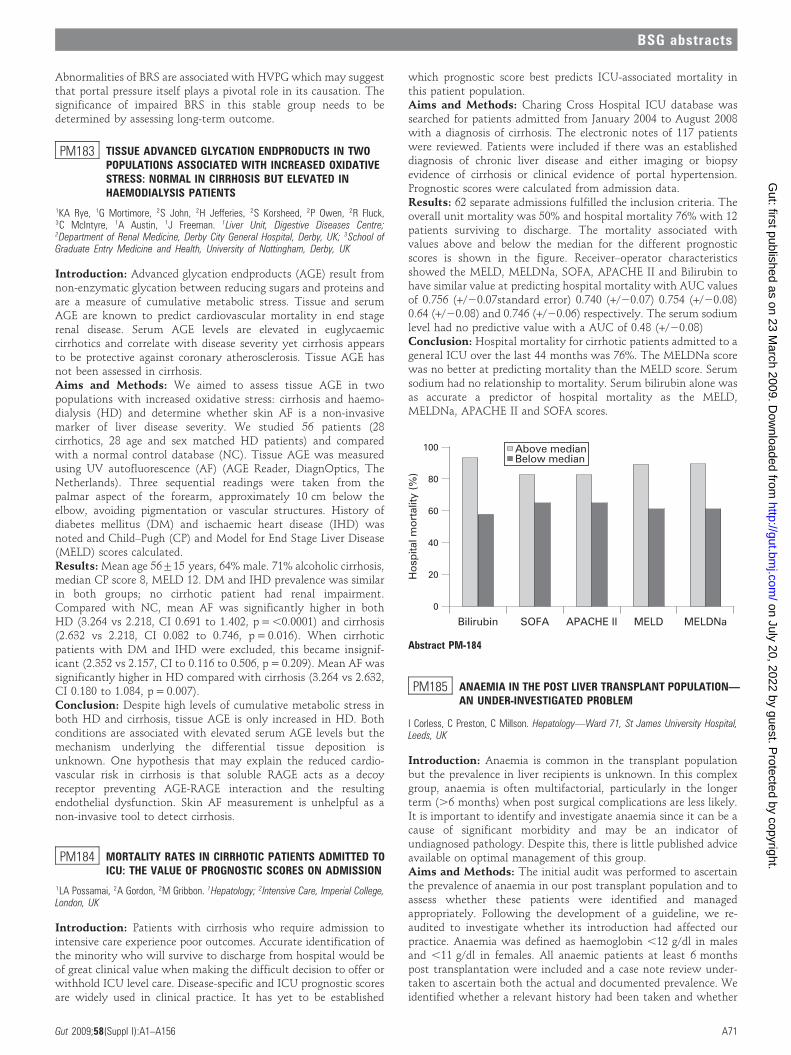
Abnormalities of BRS are associated with HVPG which may suggestthat portal pressure itself plays a pivotal role in its causation. Thesignificance of impaired BRS in this stable group needs to bedetermined by assessing long-term outcome.
PM183 TISSUE ADVANCED GLYCATION ENDPRODUCTS IN TWOPOPULATIONS ASSOCIATED WITH INCREASED OXIDATIVESTRESS: NORMAL IN CIRRHOSIS BUT ELEVATED INHAEMODIALYSIS PATIENTS
1KA Rye, 1G Mortimore, 2S John, 2H Jefferies, 2S Korsheed, 2P Owen, 2R Fluck,3C McIntyre, 1A Austin, 1J Freeman. 1Liver Unit, Digestive Diseases Centre;2Department of Renal Medicine, Derby City General Hospital, Derby, UK; 3School ofGraduate Entry Medicine and Health, University of Nottingham, Derby, UK
Introduction: Advanced glycation endproducts (AGE) result fromnon-enzymatic glycation between reducing sugars and proteins andare a measure of cumulative metabolic stress. Tissue and serumAGE are known to predict cardiovascular mortality in end stagerenal disease. Serum AGE levels are elevated in euglycaemiccirrhotics and correlate with disease severity yet cirrhosis appearsto be protective against coronary atherosclerosis. Tissue AGE hasnot been assessed in cirrhosis.Aims and Methods: We aimed to assess tissue AGE in twopopulations with increased oxidative stress: cirrhosis and haemo-dialysis (HD) and determine whether skin AF is a non-invasivemarker of liver disease severity. We studied 56 patients (28cirrhotics, 28 age and sex matched HD patients) and comparedwith a normal control database (NC). Tissue AGE was measuredusing UV autofluorescence (AF) (AGE Reader, DiagnOptics, TheNetherlands). Three sequential readings were taken from thepalmar aspect of the forearm, approximately 10 cm below theelbow, avoiding pigmentation or vascular structures. History ofdiabetes mellitus (DM) and ischaemic heart disease (IHD) wasnoted and Child–Pugh (CP) and Model for End Stage Liver Disease(MELD) scores calculated.Results: Mean age 56¡15 years, 64% male. 71% alcoholic cirrhosis,median CP score 8, MELD 12. DM and IHD prevalence was similarin both groups; no cirrhotic patient had renal impairment.Compared with NC, mean AF was significantly higher in bothHD (3.264 vs 2.218, CI 0.691 to 1.402, p = ,0.0001) and cirrhosis(2.632 vs 2.218, CI 0.082 to 0.746, p = 0.016). When cirrhoticpatients with DM and IHD were excluded, this became insignif-icant (2.352 vs 2.157, CI to 0.116 to 0.506, p = 0.209). Mean AF wassignificantly higher in HD compared with cirrhosis (3.264 vs 2.632,CI 0.180 to 1.084, p = 0.007).Conclusion: Despite high levels of cumulative metabolic stress inboth HD and cirrhosis, tissue AGE is only increased in HD. Bothconditions are associated with elevated serum AGE levels but themechanism underlying the differential tissue deposition isunknown. One hypothesis that may explain the reduced cardio-vascular risk in cirrhosis is that soluble RAGE acts as a decoyreceptor preventing AGE-RAGE interaction and the resultingendothelial dysfunction. Skin AF measurement is unhelpful as anon-invasive tool to detect cirrhosis.
PM184 MORTALITY RATES IN CIRRHOTIC PATIENTS ADMITTED TOICU: THE VALUE OF PROGNOSTIC SCORES ON ADMISSION
1LA Possamai, 2A Gordon, 2M Gribbon. 1Hepatology; 2Intensive Care, Imperial College,London, UK
Introduction: Patients with cirrhosis who require admission tointensive care experience poor outcomes. Accurate identification ofthe minority who will survive to discharge from hospital would beof great clinical value when making the difficult decision to offer orwithhold ICU level care. Disease-specific and ICU prognostic scoresare widely used in clinical practice. It has yet to be established
which prognostic score best predicts ICU-associated mortality inthis patient population.Aims and Methods: Charing Cross Hospital ICU database wassearched for patients admitted from January 2004 to August 2008with a diagnosis of cirrhosis. The electronic notes of 117 patientswere reviewed. Patients were included if there was an establisheddiagnosis of chronic liver disease and either imaging or biopsyevidence of cirrhosis or clinical evidence of portal hypertension.Prognostic scores were calculated from admission data.Results: 62 separate admissions fulfilled the inclusion criteria. Theoverall unit mortality was 50% and hospital mortality 76% with 12patients surviving to discharge. The mortality associated withvalues above and below the median for the different prognosticscores is shown in the figure. Receiver–operator characteristicsshowed the MELD, MELDNa, SOFA, APACHE II and Bilirubin tohave similar value at predicting hospital mortality with AUC valuesof 0.756 (+/20.07standard error) 0.740 (+/20.07) 0.754 (+/20.08)0.64 (+/20.08) and 0.746 (+/20.06) respectively. The serum sodiumlevel had no predictive value with a AUC of 0.48 (+/20.08)Conclusion: Hospital mortality for cirrhotic patients admitted to ageneral ICU over the last 44 months was 76%. The MELDNa scorewas no better at predicting mortality than the MELD score. Serumsodium had no relationship to mortality. Serum bilirubin alone wasas accurate a predictor of hospital mortality as the MELD,MELDNa, APACHE II and SOFA scores.
PM185 ANAEMIA IN THE POST LIVER TRANSPLANT POPULATION—AN UNDER-INVESTIGATED PROBLEM
I Corless, C Preston, C Millson. Hepatology—Ward 71, St James University Hospital,Leeds, UK
Introduction: Anaemia is common in the transplant populationbut the prevalence in liver recipients is unknown. In this complexgroup, anaemia is often multifactorial, particularly in the longerterm (.6 months) when post surgical complications are less likely.It is important to identify and investigate anaemia since it can be acause of significant morbidity and may be an indicator ofundiagnosed pathology. Despite this, there is little published adviceavailable on optimal management of this group.Aims and Methods: The initial audit was performed to ascertainthe prevalence of anaemia in our post transplant population and toassess whether these patients were identified and managedappropriately. Following the development of a guideline, we re-audited to investigate whether its introduction had affected ourpractice. Anaemia was defined as haemoglobin ,12 g/dl in malesand ,11 g/dl in females. All anaemic patients at least 6 monthspost transplantation were included and a case note review under-taken to ascertain both the actual and documented prevalence. Weidentified whether a relevant history had been taken and whether
Abstract PM-184
BSG abstracts
Gut 2009;58(Suppl I):A1–A156 A71
on July 20, 2022 by guest. Protected by copyright.
http://gut.bmj.com
/G
ut: first published as on 23 March 2009. D
ownloaded from
appropriate investigations had been performed. A guideline wassubsequently developed as a recommended management pathwayfor anaemic patients. Six months later, a subset of the populationwas re-audited to see if improvements had occurred followingintroduction of the guideline.Results: The initial audit demonstrated a prevalence of anaemia of11.7%. This was documented by medical staff in 51% of cases.Haematinics were checked in 61%. No cause was found for anaemiain 30.6% of the identified patients. Medication was identified as thecause of anaemia in 11% of people and this resulted in a change ofdrug in one patient. In the second audit, the prevalence was similarat 13.7%. Anaemia was documented in a greater proportion of thegroup (69.4%) and haematinics were checked in 80%. 64% of theidentified group had a documented cause for the anaemia and afurther 32% were undergoing investigation. There was nodocumented cause and no plan for investigation in 4%. A significantproportion of the patients with anaemia had not been identified bymedical staff. Across the whole group, 19% of those with anaemiahad not been investigated in accordance with the guideline.Conclusion: Following introduction of the guideline, we found agreater number of anaemic patients were identified. The majority ofthese were appropriately investigated to rule out significantpathology. However, despite the improvements, a sizeable propor-tion of the group were still not being identified. The initial auditcycle has demonstrated a need to increase staff awareness ofanaemia in the post transplant population and to repeat the auditprocess in future.
PM186 SHORT T-LYMPHOCYTE TELOMERE LENGTH COMPROMISESSUSTAINED VIROLOGICAL RESPONSE WITH PEGYLATEDINTERFERON-ALPHA AND RIBAVIRIN IN CHRONICHEPATITIS C VIRUS (HCV) INFECTION
1M Hoare, 1WTH Gelson, 2SE Davies, 3MD Curran, 4SL Vowler, 5AN Akbar, 1GJMAlexander. 1Department of Medicine; 2Department of Pathology, University ofCambridge, Cambridge, UK; 3Clinical Microbiology and Public Health Laboratory,Health Protection Agency; 4Centre for Applied Medical Statistics, Department of PublicHealth and Primary Care, University of Cambridge, Cambridge, UK; 5Department ofImmunology and Molecular Pathology, Royal Free and University College MedicalSchool, London, UK
Introduction: Ageing is associated with impaired immunity andfeatures of immune senescence, including shortened lymphocytetelomeres. In patients with chronic HCV, increasing age isassociated with higher fibrosis stage and a failure to respond toantiviral therapy. We have shown strong correlations betweenmeasures of clinical outcome and lymphocyte telomere length inchronic HCV infection, suggesting accelerated immune senescencein chronic HCV infection and in this study assessed whetherimmune senescence was also related to the response to antiviraltherapy.Aims and Methods: Telomere length was measured throughantiviral therapy for HCV infection in peripheral CD8+ andCD4+T-cells from 85 patients (75% male, 45% genotype 1, meanIshak fibrosis stage 3.6) by Flow-FISH. Patients were followed toSVR. Results were analysed by multiple logistic regression (LR); age,sex, Ishak fibrosis stage, viral genotype, viral load, BMI,CD8+CD45RO+ and CD4+CD45RO+ telomere length were inputvariables.Results: Baseline median (IQR) CD8+CD45RO+(128.1 (113.4 to142.3) and CD4+CD45RO+(119.0 (107.8 to 126.8) telomere lengthswere longer in the 38 patients (44.7%) who achieved SVR thanthose who did not: 112.5 (104.5 to 118.2) (p = 0.0006) and 105.5(99.4 to 114.2) (p = 0.0003) respectively. CD8+CD45RO+ andCD4+CD45RO+ telomere lengths were unaffected by therapy. Byunivariate LR viral load (p = 0.02) and CD8+CD45RO+ telomerelength (p = 0.01) were associated with SVR; in this model age
(p = 0.19), male sex (p = 0.51), severe fibrosis (p = 0.11), genotype(p = 0.18) and BMI (p = 0.94) were not associated with SVR.Utilising ‘‘backwards stepwise regression’’ to remove non-signifi-cant variables age (p = 0.048), viral load (p = 0.02) andCD8+CD45RO+ telomere length (p = 0.007) were associatedindependently with SVR. Each single point increase inCD8+CD45RO+ telomere length was associated with an OR ofachieving SVR of 1.05 (1.01 to 1.08). CD4+CD45RO+ lymphocytetelomere length (p = 0.006) and viral load (p = 0.02) were alsoassociated with SVR; in this model age (p = 0.37), male sex(p = 0.48), presence of severe fibrosis (p = 0.07) genotype(p = 0.19) and BMI (p = 0.89) were not associated with SVR.Utilising ‘‘backwards stepwise regression’’ viral load (p = 0.02) andCD4+CD45RO+ telomere length (p = 0.001) were associatedindependently with SVR. Each single point increase inCD4+CD45RO+ telomere length was associated with an OR ofachieving SVR of 1.08 (1.03 to 1.13).Conclusion: Immune senescence, measured as shortenedCD8+CD45RO+ and CD4+CD45RO+ T-lymphocyte telomeres,predicted a failure to respond to antiviral therapy for HCVinfection, independent of other known factors.
PM187 DIAGNOSIS OF SIGNIFICANT LIVER FIBROSIS SECONDARYTO CHRONIC HEPATITIS C VIRUS INFECTION WITH SERUMPROTEOMIC PROFILING
1ML Cowan, 2SJ Thomson, 3P Rice, 2DM Forton, 2TM Rahman, 1S Krishna. 1Divisionof Cellular and Molecular Medicine, St George’s, University of London, London, UK;2Department of Gastroenterology and Hepatology; 3Department of Virology, St George’sHospital, London, UK
Introduction: Chronic infection with hepatitis C virus (HCV)causes hepatic fibrosis that may progress to cirrhosis. Staging ofliver fibrosis requires liver biopsy, an expensive invasive procedurewhose accuracy is compromised in small biopsy specimens. Previousstudies of non-invasive fibrosis markers have been criticised forinclusion of suboptimal liver biopsies. Serum profiling with surfaceenhanced laser desorption ionisation time-of-flight mass spectro-metry (SELDI ToF MS) offers an opportunity for high throughputscreening for potential biomarkers.Aims and Methods: Serum samples from 103 subjects withparenchymal liver biopsies for assessment of hepatic fibrosissecondary to chronic HCV were identified in the long term storagefacility of the Department of Virology. Only patients with biopsyspecimens longer than 20 mm or containing more than 11 portaltracts were considered. Proteomic spectra were obtained intriplicate with a ProteinChip System 4000 SELDI ToF MS.Potential biomarkers were identified with ProteinChip DataManager software.Results: 165 protein clusters were detected on CM10 weak cationexchange arrays and 184 on Q10 anion exchange arrays. Thirty-seven protein peaks were differentially expressed in patients withsignificant fibrosis (Ishak stage.1). The median value of 10differentially expressed peaks (p,0.01) increased with increasingfibrosis group (Ishak stages 0/1, 2–4, 5/6). The median value of 7peaks decreased across the three groups. The ability of anyindividual peak to diagnose fibrosis was modest with an area underreceiver operator curve (AUROC) of 0.63 to 0.71. A logisticregression analysis model with the two most significant peaksclassified 73% of subjects correctly, with an AUROC of 0.83.Conclusion: This study of HCV fibrosis biomarkers has beenlimited to patients with ‘‘gold standard’’ staging liver biopsies.SELDI ToF MS identified potential biomarkers of liver fibrosissecondary to chronic HCV. A significant proportion of these proteinpeaks correlate with fibrosis stage. Accurate diagnosis of significantfibrosis was possible in 73% of subjects with a composite model.These proteins merit identification and validation in future studies.
BSG abstracts
A72 Gut 2009;58(Suppl I):A1–A156
on July 20, 2022 by guest. Protected by copyright.
http://gut.bmj.com
/G
ut: first published as on 23 March 2009. D
ownloaded from
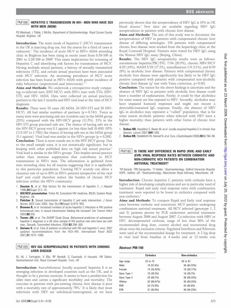
PM188 HEPATITIS C TRANSMISSION IN HIV+ MEN WHO HAVE SEXWITH MEN (MSM)
PG Mayhead, J Tibble, L McKie. Department of Gastroenterology, Royal Sussex CountyHospital, Brighton, UK
Introduction: The main mode of hepatitis C (HCV) transmissionin the UK is injecting drug use, but the reason for a third of cases isunknown.1 The incidence of acute HCV in HIV+ MSM attendingclinic in Brighton has been rising in recent years from 0.39/100 in2001 to 3.26/100 in 20062 This raises implications for screening ofHepatitis C and identifying risk factors for transmission of HCV.Having multiple sexual partners, a history of sexually transmittedinfections (STI), and certain sexual practices have been associatedwith HCV infection. An increasing prevalence of HCV acuteinfection has been found in HIV+ MSM with greater incidence ofrisky behaviour (unprotected anal intercourse).3 4
Aims and Methods: We undertook a retrospective study compar-ing co-infected men (HIV-HCV) with HIV+ men with STIs (HIV-STI) and HIV- MSM. Data were collected on sexual practices,partners in the last 3 months and HIV viral load at the time of HCVdiagnosis.Results: There were 92 cases (42 MSM, 24 HIV-STI and 26 HIV-HCV). All had similar numbers of partners (p = 0.741). Twice asmany men were practising safe sex (condom use) in the MSM group(23%) compared with the HIV-HCV group (11.5%). 21% in theHIV-STI group practised safe sex. The chance of having safe sex inthe HIV-HCV group was 8:1 against (or less than half (0.430) (95%CI 0.107 to 1.738)) the chance of having safe sex in the MSM group(3:1 against). Viral load was similar in the HIV+ groups (p = 0.268).Conclusion: There is more unsafe sex in the HIV-HCV group. Dueto the small sample sizes, it is not statistically significant, but inkeeping with other published data on high risk sexual practice.5
Viral load is similar in the HIV+ groups. This implies sexual practicerather than immune suppression that contributes to HCVtransmission in HIV+ men. The information is gathered frommen attending clinic for all reasons suggesting that it is applicableto this particular population. Treating HCV acutely can allow aclearance rate of up to 80% in HIV+ patients irrespective of the viralload6 and could therefore reduce the burden of chronic HCVinfection within the HIV+ community.
1. Zeuzem S, et al. Risk factors for the transmission of hepatitis C. J Hepatol1996;24(Suppl 2):3–10.
2. HIV/HCV presentation. Fisher M, Consultant HIV medicine, BSUH, Eastern Road,Brighton, UK.
3. Fletcher S. Sexual transmission of hepatitis C and early intervention. J AssocNurses AIDS Care 2003, Sep–Oct;14(Suppl 5):87S–94S.
4. Browne R, et al. Increased numbers of acute hepatitis C infections in HIV positivehomosexual men; is sexual transmission feeding the increase? Sex Transm Infect2004;80:326.
5. Turner JM, et al. The SHARP Study Group. Behavioural predictors of subsequenthepatitis C diagnosis in a UK clinic sample of HIV positive men who have sex withmen. Sexually Transmitted Infections 2006;82:298–300.
6. Soriano V, et al. Care of patients co-infected with HIV and hepatitis C virus: 2007updated recommendations from the HCV–HIV. International Panel AIDS2007;21:1073–1089.
PM189 HEV IGG SEROPREVALENCE IN PATIENTS WITH CHRONICLIVER DISEASE
RJ Ali, L Farrington, V Ellis, RP Bendall, G Courtauld, H Hussaini, HR Dalton.Gastrointestinal Unit, Royal Cornwall Hospital, Truro, UK
Introduction: Autochthonous (locally acquired) hepatitis E is anemerging infection in developed countries such as the UK, and isthought to be a porcine zoonosis. It seems to have a predilection forolder men and carries a significant morbidity and mortality. Theoutcome in patients with pre-existing chronic liver disease is poorwith a mortality rate of approximately 70%.1 It is likely that mostinfections with HEV are subclinical/unrecognised, as we have
previously shown that the seroprevalence of HEV IgG is 16% in UKblood donors.2 Few data are available regarding HEV IgGseroprevalence in patients with chronic liver disease.Aims and Methods: The aim of this study was to document theseroprevalence of HEV in patients with compensated chronic liverdisease of differing aetiologies. 136 patients with compensatedchronic liver disease were studied from the hepatology clinic at theRoyal Cornwall Hospital. Patients were tested for HEV IgG usingthe Wantai HEV IgG assay (Beijing, China).Results: The HEV IgG seropositivity results were as follows:autoimmune hepatitis/PBC/PSC 7/34 (20.5%), chronic HBV/HCV4/25 (16%), NASH 5/29 (17.2%), miscellaneous 1/9 (11%), and 0/39(0%) in alcoholic liver disease. Patients with compensated chronicalcoholic liver disease were significantly less likely to be HEV IgGpositive compared with patients with compensated non-alcoholicchronic liver disease (x2 test with Yates correction, p,0.05).Conclusion: The reason for the above findings is uncertain and theabsence of HEV IgG in patients with alcoholic liver disease couldhave a number of explanations. Firstly, it could be that alcoholicsfor some reason are less exposed to HEV. Secondly, alcoholics mayhave impaired humoral responses and might not mount adetectable/sustained IgG response. Finally, the absence of HEVIgG in alcoholics may represent a ‘‘culled’’ population, that is forsome reason alcoholic patients when infected with HEV have ahigher mortality than patients with other forms of chronic liverdisease.
1. Dalton HR, Hazeldine S, Banks M, et al. Locally acquired hepatitis E in chronic liverdisease. Lancet 2007;369:1260.
2. Dalton HR, Stableforth W, Hamad N, et al. Euro J Gastrohepatol 2008;20(8):784–90.
PM190 IS THERE ANY DIFFERENCE IN RAPID (RVR) AND EARLY(EVR) VIRAL RESPONSE RATES BETWEEN CIRRHOTIC ANDNON-CIRRHOTIC HCV PATIENTS ON COMBINATIONANTIVIRAL TREATMENT?
1M Ganesh, 2RN Ganapathy, 1S Halder. 1Gastroenterology, Salford University Hospital,HOPE, Salford, UK; 2Gastroenterology, Manchester Royal Infirmary, Manchester, UK
Introduction: Chronic hepatitis C patients with cirrhosis have ahigher risk of developing complications and are in particular need oftreatment. Rapid and early viral response rates with combinationtreatment were reported to be lower in cirrhotics compared withnon-cirrhotics.1
Aims and Methods: To compare Rapid and Early viral responserates between cirrhotic and noncirrotic HCV patients undergoingcombination antiviral treatment. All HCV infected (genotype 1, 2and 3) patients proven by PCR underwent antiviral treatmentbetween August 2006 and August 2007. Co-infection with HBV orHIV, decompensated cirrhosis, usage of less than 80% of therecommended drug dose, current alcohol and recreational drugabuse were the exclusion criteria. Pegylated Interferon and Ribavirinwere used at the recommended dosage for treatment. A 2 log dropin viral load from baseline at 4 weeks and at 12 weeks was
Abstract PM-190
Cirrhotics Non-cirrhotics
Age range 33 to 72 28 to 67
Male 19 (57.6%) 36 (64.33%)
Female 14 (42.42%) 16 (30.77%)
Geno Type 1 10 (30.3%) 16 (30.7%)
Geno Type 2 23 (69.7%) 36 (69.33%)
Whites 22 (66.67%) 36 (69.33%)
RVR 24 (72.9%) 43 (82.6%)
EVR 31 (93.9%) 49 (94.23%)
BSG abstracts
Gut 2009;58(Suppl I):A1–A156 A73
on July 20, 2022 by guest. Protected by copyright.
http://gut.bmj.com
/G
ut: first published as on 23 March 2009. D
ownloaded from
considered a rapid and early viral response respectively. The samplesize was 85. Chi-squared test was used to assess statisticalsignificance between comparing groups. Age, gender, race andgenotype presence of cirrhosis were taken into univariate andmultivariate analysis as predictors of RVR and EVR.Results: Out of the total study population 79% had RVR and 94%had EVR. There was no significant difference demonstratedbetween cirrhotic (83%) and non-cirrhotic groups (72.73%) inRVR and EVR rates (p = 0.273) and (p = 0.956), respectively (seetable). Statistically, distributions of viral genotypes among the twogroups were not different. In the non-cirrhotic group with genotype2 or 3, no significant difference in rapid viral response (RVR) wasseen between Asians and Whites (p = 0.541). In a multivariateanalysis increasing age (odds ratio 1.11) and genotype 1 (odds ratio2.8) stood as independent predictors for poor RVR and EVR rates.Conclusion: In our study there was statistically no significantdifference in rapid and early viral response rates between cirrhoticsand non-cirrhotics. 69% of patients in the study group hadgenotype 2 or 3 and all patients received .80% dose of combinationtreatment. This could explain the good RVR and EVR achieved inour study.
1. Shiffman M, et al. APASL abstract o-182, 2007.
PM191 PROSPECTIVE COMPARISON OF SEVERITY SCORES INALCOHOLIC HEPATITIS
1R Parker, 1CA McCune, 1FH Gordon, 2A di Mambro, 3H Taylor, 1PL Collins.1Department of Hepatology, Bristol Royal Infirmary, Bristol, UK; 2Department ofMedicine, University of Bristol, Bristol, UK; 3R&D, Bristol Royal Infirmary, Bristol, UK
Introduction: Alcoholic hepatitis is the most florid manifestationof alcoholic liver disease and is associated with a poor prognosis.Several scoring systems have been used to predict prognosis inpatients with alcoholic hepatitis since Maddrey described theDiscriminant Function in 1979,1 but it is often unclear which isthe most useful in routine clinical practise.Aims and Methods: This study aimed to determine which of fiveclinical scoring systems is most useful in predicting outcome in anunselected cohort of patients with alcoholic hepatitis presenting toa busy UK teaching hospital. 56 consecutive patients diagnosedwith alcoholic hepatitis had scores calculated for: modifiedDiscriminant Function (mDF), Glasgow Alcoholic Hepatitis Score(GAHS), MELD, Child–Pugh and the Lille model. The primaryoutcome measure was survival to 6 months, assessed by logisticalregression analysis. As a secondary outcome, length of hospital staywas analysed by fitting Cox proportional hazard models to eachscore.Results: GAHS and the Lille Model were significantly associatedwith survival at 6 months (p = 0.01 and p = 0.017 respectively).Other scores did not achieve significance. An increase of 1 in theGlasgow score was associated with an OR of dying within6 months of 1.72 [95% CI 1.14 to 2.60]. An increase of 0.1 in thelog Lille Score was associated with an OR of dying within 6 monthsof 1.28 [95% CI 1.07 to 1.52]. GAHS, Lille Model and Child–Pughscore were significantly associated with length of stay (p = 0.021,p = 0.018, p = 0.006 respectively).Conclusion: The Glasgow Alcoholic Hepatitis Score accuratelypredicts survival in alcoholic hepatitis in this unselected alcoholichepatitis cohort. This score has the added advantage of being simplycalculated when patients are admitted to hospital. The modifiedDiscriminant Function, widely used to guide practice, is notsignificantly associated with survival in this dataset. We suggestthat the Glasgow Alcoholic Hepatitis Score is of the most value inclinical practice, but further data should be collected to demonstratethis.
1. Maddrey WC, Boitnott JK, Bedine MS, et al. Corticosteroid therapy of alcoholichepatitis. Gastroenterology 1978;75:193–9.
PM192 DOSE REDUCTIONS IN THROMBOCYTOPAENIC PATIENTS INTHE TREATMENT OF HEPATITIS C WITH PEGYLATEDINTERFERON
RL Shenderey, M Aldersley, R Jones. Leeds Liver Unit, St James University Hospital,Leeds, UK
Introduction: Sustained Virological Response (SVR) rates totreatment of Hepatitis C vary greatly depending on a large numberof factors including genotype, stage of disease, comorbidity andimportantly compliance with therapy. The two licensed pegylatedinterferon products in the UK are Pegylated alpha2a (PegasysH,Roche) and Pegylated alpha2b (ViraferonpegH, Schering Plough).The product licences state the platelet count must be above1006109/l for Pegylated interferon alpha2b and 906109/l forPegylated Interferon alpha2a in order to commence treatment andthat the dose must be reduced if the platelet count drops below506109/l and treatment stopped if less than 256109/l. It is knownthat dose reduction will reduce the ability to achieve an SVR.Aims and Methods: This department has aimed to avoid dosereduction and has not followed the product licenses with respect tothrombocytopaenia. We reviewed the database of all our patientstreated for Hepatitis C during the period January 2004–July 2007 todetermine the number of patients with thrombocytopenia, thosewho underwent Peginterferon dose reduction and the overall SVR.Results: 178 patients were treated during this 3.5 year period. 27patients were commenced on full dose treatment with plateletcounts less than 100 (range 50 to 97). Records could be traced for23/27 patients. Treatment was given for the complete course in 9patients of which 5 achieved SVR. Only two patients had a dosereduction in their Peginterferon, and neither of these achieved SVR.Reasons for not completing treatment included: 2 failed to attendfor follow-up, 3 had treatment discontinued for non-response as perNICE guidelines, 2 had decompensation of their liver disease, 3found side-effects of treatment intolerable, 1 developed colitis, 1sepsis, 1 cancer and only 1 for haematological reasons. None ofthese patients achieved SVR. No patients died during treatment. 17patients developed platelet counts less than 50 during treatment. 16of these started with platelet counts under 1006109/l. Two patientshad a dose reduction and failed to achieve SVR. Two patientsachieved an SVR and neither had a dose reduction. There were noclinically significant bleeding episodes in any of these patients.Conclusion: Most patients with thrombocytopenia can be treatedsafely with careful monitoring of platelet count in order to try tomaximise SVR rate in the treatment of Hepatitis C.
PM193 HOW ARE ABNORMAL RESULTS FOR LIVER FUNCTIONTESTS DEALT WITH IN SECONDARY CARE?
RI Fearn, N Van Someren, R D’Souza, K Besherdas. Gastroenterology, Barnet andChase Farm NHS Trust, Enfield, UK
Introduction: Liver function tests (LFTs) are commonly requestedto identify severe, but potentially treatable conditions. In primarycare abnormal LFTs are often not adequately investigated. Of 873patients who had one or more abnormal liver function test 157(18%) were inadequately followed up, and over half of these werelater found to have undetected major liver disease.1 This has neverbeen investigated in secondary care. The AGA guidelines state thatabnormal LFTs should either be followed until normal orinvestigated in a stepwise approach until a cause is found.2
Aims and Methods: This study aimed to determine whetherpatients found to have deranged LFTs during a hospital presenta-tion are followed up adequately. We identified all patients who hadan alanine aminotransferase (ALT) result greater than twice theupper limit of normal on tests performed in Chase Farm Hospital,Enfield, UK (a district general hospital) in September and October
BSG abstracts
A74 Gut 2009;58(Suppl I):A1–A156
on July 20, 2022 by guest. Protected by copyright.
http://gut.bmj.com
/G
ut: first published as on 23 March 2009. D
ownloaded from
2007 (n = 99). After 6 months we used the computer database toidentify whether the result had been followed up to normal. Thosewith persistently abnormal results had the database queried to findwhat investigations had been performed and then GPs werecontacted to assess follow-up.Results: 99 patients with ALT greater than twice the upperlimit of normal were identified. Eighty were admitted to wardsand 19 were discharged from Accident and Emergency (A&E). 39resolved on repeat testing and 16 patients died during admission.Of the remaining 44 patients, at 6 months 33 patients (33%)were either lost to follow-up (18 patients) or known to their GPbut no further tests or follow-up had been organised (15patients). Of these 15, 8 had been admitted under hospitalconsultants and 7 had been assessed and discharged from A&E.The remaining 11 patients (11%) were being investigated bytheir GP or consultant.Conclusion: This study shows that 33% of patients found tohave raised ALT did not receive adequate investigation or follow-up when discharged from secondary care. It can be concludedthat the AGA guidelines of following deranged LFTs up toresolution or until an underlying cause is found are not beingimplemented.2 Early diagnosis and treatment in chronic liverdisease can improve prognosis,3 but our findings suggest thatimportant cases may be missed. Thorough discharge summariesfor all patients who have abnormal results in hospital along withconcise advice for GPs may improve implementation of the AGAguidance.
1. Sherwood P, Lyburn L, Brown S, et al. How are abnormal results for liver functiontests dealt with in primary care? BMJ 2001;322:276–278.
2. Green R, Flamm S. AGA technical review on the evaluation of liver chemistrytests. Gastro 2002;123:1367.
3. Goddard C, Warnes T. Raised liver enzymes in asymptomatic patients. Dig Dis Sci1992;10:218.
PM194 DEVELOPMENT OF HEPATOCELLULAR CARCINOMA AND OFPORTAL VEIN THROMBOSIS IN PATIENTS WITHDECOMPENSATED ALCOHOLIC LIVER DISEASE
SJ Bonner, C Barclay, E Mcfarlane, K Shafique, J Jones, D Gleeson. Liver Unit, RoyalHallamshire Hospital, Sheffield, UK
Introduction: Hepatocellular carcinoma (HCC) may develop incirrhosis from any cause, but may be commoner in some forms thanin others. There are few recent data on incidence of HCC in alcoholicliver disease (ALD). We have previously reported (Mcfarlane Gut 2006Abst) on outcome in 249 patients (162 men, median age 49 years)with first episode of decompensated ALD (Childs Grade B or C),presenting consecutively between 1998 and 2005.Aims and Methods: Aim: To assess the subsequent incidence ofHCC and also of Portal Vein Thrombosis (PVT) in this cohort.Available ultrasound and laboratory reports up to October 2008were viewed. Patients were not formally screened for HCC unlessabsent from alcohol, but ultrasound scans (USS) were usuallyperformed in the event of hospital readmission. Follow-up wasdefined as time between initial presentation with decompensatedALD and the most recent ultrasound scan. HCC was defined as acompatible focal liver space-occupying lesion on ultrasound plus araised serum alfa feta protein.Results: Liver histology was available in 67 patients and suggestedcirrhosis in 52 (78%). Of the remaining 182 patients, 128 (71%)were presumed to have cirrhosis based on presence of varices and/orascites. Follow-up was (median (range)) 15 (0 to 112) months andwas 43 (6 to 112) months in the 149 patients followed up for.6 months. Four men, all with cirrhosis, were diagnosed withHCC: one at presentation, the others after 35–106 months. SerumAFP levels were elevated (1163–17 800 units/l) in all four. All four
subsequently died of HCC (average survival = 54 days). 153 otherpatients have died, none apparently of HCC, although circum-stances of death are still unknown in 20. Incidence of HCC was 0.7/100 patient-year follow-up. Actuarial incidence of HCC after 1, 2and 5 years was 1+/20.1%, 1.4+/20.1% and 3.6+/20.2% respec-tively in the cohort as a whole and was 1+/20.1%, 2+/20.1% and5+/20.2% in the 180 patients with confirmed or presumed cirrhosis.Three females and two males (2.0%) developed PVT. One patienthad both HCC and PVT.Conclusion: Although under-diagnosis is possible, the incidence ofHCC in this cohort of patients with decompensated ALD, most ofwhom had cirrhosis, appears to be lower than that in some reportsof cirrhosis due to HBV and HCV.
PM195 ANTI-HBC AND HBSAG SCREENING AMONG NIGERIANHEALTH CARE WORKERS
1,4SO Ola, 1JA Otegbayo, 2OD Olaleye, 3WA Shokunbi, 4CB Summerton. 1Departmentof Medicine; 2Virology; 3Haematology, University College Hospital, Ibadan, Nigeria;4Gastroenterology, Trafford General Hospital, Manchester, UK
Introduction: HBV infection is common among Nigerian healthcare workers,1 2 and as such efforts should be made to determine thestatus of the workers prior vaccination against the infection.Aims and Methods: In order to determine anti-HBsAg and anti-HBc status among Nigerian health care workers at UCH, Ibadan,Nigeria, blood specimens of 804 volunteered subjects were screenedfor the presence of Hepatitis B surface antigen (HBsAg) andantibody to Hepatitis B-core antigen(anti-HBc) using EnzymeLinked Immuno-sorbent Assay. Statistical analysis was carried outusing Student t at p,0.05.Results: The subjects were in a male:female ratio of 1:2 andaged 21–65 years. The detection rates of HbsAg and anti-HBcwere 6.1% and 59.1% (p,0.05) in all the subjects respectively.Lone HBsAg (1.9%) and anti-HBc (54.8%) were present amongthe subjects while the combination of the two markers waspresent in only 4.3%. HBsAg and anti-HBc were detected in6.2% and 69% of males respectively and in 6% and 54.3%respectively of females. Using both markers, HBV infection wasdetected in 60.9% of the subjects (table). Among the dental andmedical personnel, the infection was commonest among theGynaecologist and Obstreticians (93.3%) and lowest among thePaediatricians (52.6%). There was no difference in the rates ofthe infection among the different risk groups for the infectionamong the workers and only 39% of them were sero-negative forboth markers of the HBV infection.Conclusion: Both HBsAg and anti-HBc should be tested amongNigerian heath care workers in detection of their status for HBVinfection prior vaccination. The infection is very common amongthe workers regardless of their professional and risk groups.
1. Van Damme P. Hepatitis B vaccination of Health Case Workers. Viral hepatitis1995;3(2):8–10.
2. Olubuyide IO, Ola SO, Aliyu B, et al. Hepatitis B and C in doctors and dentists inNigeria. Quarterly Jour Med 1997;90:417–422.
Abstract PM-195
HCWs Number Anti-HBc+ (%) HBsAg+ (%) HBsAg+ only (%) HBV (%)
High 709 419 (52.1) 43 (6.1) 14 (2) 433 (60.1)
Medium 37 25 (3.1) 0 0 25 (57.6)
Low 52 31 (3.9) 6 (10.3) 1 (1.7) 37 (55.2)
Total 804 475 (59.1) 49 (1.9) 15 (1.9) 490 (60.9)
HCWs, Healthcare workers risk goup.
BSG abstracts
Gut 2009;58(Suppl I):A1–A156 A75
on July 20, 2022 by guest. Protected by copyright.
http://gut.bmj.com
/G
ut: first published as on 23 March 2009. D
ownloaded from
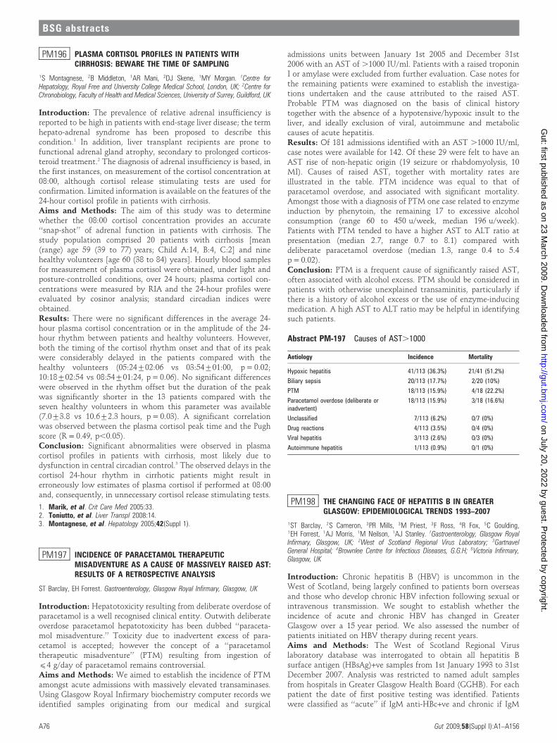
PM196 PLASMA CORTISOL PROFILES IN PATIENTS WITHCIRRHOSIS: BEWARE THE TIME OF SAMPLING
1S Montagnese, 2B Middleton, 1AR Mani, 2DJ Skene, 1MY Morgan. 1Centre forHepatology, Royal Free and University College Medical School, London, UK; 2Centre forChronobiology, Faculty of Health and Medical Sciences, University of Surrey, Guildford, UK
Introduction: The prevalence of relative adrenal insufficiency isreported to be high in patients with end-stage liver disease; the termhepato-adrenal syndrome has been proposed to describe thiscondition.1 In addition, liver transplant recipients are prone tofunctional adrenal gland atrophy, secondary to prolonged corticos-teroid treatment.2 The diagnosis of adrenal insufficiency is based, inthe first instances, on measurement of the cortisol concentration at08:00, although cortisol release stimulating tests are used forconfirmation. Limited information is available on the features of the24-hour cortisol profile in patients with cirrhosis.Aims and Methods: The aim of this study was to determinewhether the 08:00 cortisol concentration provides an accurate‘‘snap-shot’’ of adrenal function in patients with cirrhosis. Thestudy population comprised 20 patients with cirrhosis [mean(range) age 59 (39 to 77) years; Child A:14, B:4, C:2] and ninehealthy volunteers [age 60 (38 to 84) years]. Hourly blood samplesfor measurement of plasma cortisol were obtained, under light andposture-controlled conditions, over 24 hours; plasma cortisol con-centrations were measured by RIA and the 24-hour profiles wereevaluated by cosinor analysis; standard circadian indices wereobtained.Results: There were no significant differences in the average 24-hour plasma cortisol concentration or in the amplitude of the 24-hour rhythm between patients and healthy volunteers. However,both the timing of the cortisol rhythm onset and that of its peakwere considerably delayed in the patients compared with thehealthy volunteers (05:24¡02:06 vs 03:54¡01:00, p = 0.02;10:18¡02:54 vs 08:54¡01:24, p = 0.06). No significant differenceswere observed in the rhythm offset but the duration of the peakwas significantly shorter in the 13 patients compared with theseven healthy volunteers in whom this parameter was available(7.0¡3.8 vs 10.6¡2.3 hours, p = 0.03). A significant correlationwas observed between the plasma cortisol peak time and the Pughscore (R = 0.49, p,0.05).Conclusion: Significant abnormalities were observed in plasmacortisol profiles in patients with cirrhosis, most likely due todysfunction in central circadian control.3 The observed delays in thecortisol 24-hour rhythm in cirrhotic patients might result inerroneously low estimates of plasma cortisol if performed at 08:00and, consequently, in unnecessary cortisol release stimulating tests.
1. Marik, et al. Crit Care Med 2005:33.2. Toniutto, et al. Liver Transpl 2008:14.3. Montagnese, et al. Hepatology 2005;42(Suppl 1).
PM197 INCIDENCE OF PARACETAMOL THERAPEUTICMISADVENTURE AS A CAUSE OF MASSIVELY RAISED AST:RESULTS OF A RETROSPECTIVE ANALYSIS
ST Barclay, EH Forrest. Gastroenterology, Glasgow Royal Infirmary, Glasgow, UK
Introduction: Hepatotoxicity resulting from deliberate overdose ofparacetamol is a well recognised clinical entity. Outwith deliberateoverdose paracetamol hepatotoxicity has been dubbed ‘‘paraceta-mol misadventure.’’ Toxicity due to inadvertent excess of para-cetamol is accepted; however the concept of a ‘‘paracetamoltherapeutic misadventure’’ (PTM) resulting from ingestion of(4 g/day of paracetamol remains controversial.Aims and Methods: We aimed to establish the incidence of PTMamongst acute admissions with massively elevated transaminases.Using Glasgow Royal Infirmary biochemistry computer records weidentified samples originating from our medical and surgical
admissions units between January 1st 2005 and December 31st2006 with an AST of .1000 IU/ml. Patients with a raised troponinI or amylase were excluded from further evaluation. Case notes forthe remaining patients were examined to establish the investiga-tions undertaken and the cause attributed to the raised AST.Probable PTM was diagnosed on the basis of clinical historytogether with the absence of a hypotensive/hypoxic insult to theliver, and ideally exclusion of viral, autoimmune and metaboliccauses of acute hepatitis.Results: Of 181 admissions identified with an AST .1000 IU/ml,case notes were available for 142. Of these 29 were felt to have anAST rise of non-hepatic origin (19 seizure or rhabdomyolysis, 10MI). Causes of raised AST, together with mortality rates areillustrated in the table. PTM incidence was equal to that ofparacetamol overdose, and associated with significant mortality.Amongst those with a diagnosis of PTM one case related to enzymeinduction by phenytoin, the remaining 17 to excessive alcoholconsumption (range 60 to 450 u/week, median 196 u/week).Patients with PTM tended to have a higher AST to ALT ratio atpresentation (median 2.7, range 0.7 to 8.1) compared withdeliberate paracetamol overdose (median 1.3, range 0.4 to 5.4p = 0.02).Conclusion: PTM is a frequent cause of significantly raised AST,often associated with alcohol excess. PTM should be considered inpatients with otherwise unexplained transaminitis, particularly ifthere is a history of alcohol excess or the use of enzyme-inducingmedication. A high AST to ALT ratio may be helpful in identifyingsuch patients.
PM198 THE CHANGING FACE OF HEPATITIS B IN GREATERGLASGOW: EPIDEMIOLOGICAL TRENDS 1993–2007
1ST Barclay, 2S Cameron, 3PR Mills, 3M Priest, 3F Ross, 4R Fox, 5C Goulding,1EH Forrest, 1AJ Morris, 1M Neilson, 1AJ Stanley. 1Gastroenterology, Glasgow RoyalInfirmary, Glasgow, UK; 2West of Scotland Regional Virus Laboratory; 3GartnavelGeneral Hospital; 4Brownlee Centre for Infectious Diseases, G.G.H; 5Victoria Infirmary,Glasgow, UK
Introduction: Chronic hepatitis B (HBV) is uncommon in theWest of Scotland, being largely confined to patients born overseasand those who develop chronic HBV infection following sexual orintravenous transmission. We sought to establish whether theincidence of acute and chronic HBV has changed in GreaterGlasgow over a 15 year period. We also assessed the number ofpatients initiated on HBV therapy during recent years.Aims and Methods: The West of Scotland Regional Viruslaboratory database was interrogated to obtain all hepatitis Bsurface antigen (HBsAg)+ve samples from 1st January 1993 to 31stDecember 2007. Analysis was restricted to named adult samplesfrom hospitals in Greater Glasgow Health Board (GGHB). For eachpatient the date of first positive testing was identified. Patientswere classified as ‘‘acute’’ if IgM anti-HBc+ve and chronic if IgM
Abstract PM-197 Causes of AST.1000
Aetiology Incidence Mortality
Hypoxic hepatitis 41/113 (36.3%) 21/41 (51.2%)
Biliary sepsis 20/113 (17.7%) 2/20 (10%)
PTM 18/113 (15.9%) 4/18 (22.2%)
Paracetamol overdose (deliberate orinadvertent)
18/113 (15.9%) 3/18 (16.6%)
Unclassified 7/113 (6.2%) 0/7 (0%)
Drug reactions 4/113 (3.5%) 0/4 (0%)
Viral hepatitis 3/113 (2.6%) 0/3 (0%)
Autoimmune hepatitis 1/113 (0.9%) 0/1 (0%)
BSG abstracts
A76 Gut 2009;58(Suppl I):A1–A156
on July 20, 2022 by guest. Protected by copyright.
http://gut.bmj.com
/G
ut: first published as on 23 March 2009. D
ownloaded from
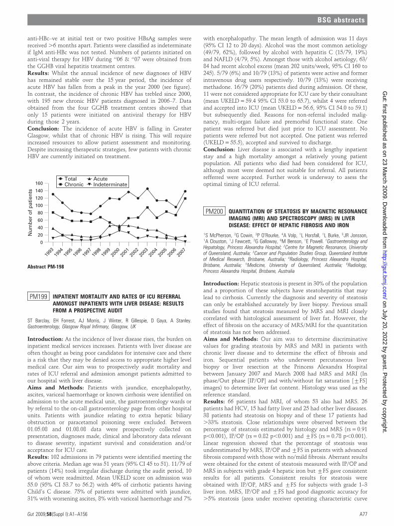
anti-HBc–ve at initial test or two positive HBsAg samples werereceived .6 months apart. Patients were classified as indeterminateif IgM anti-HBc was not tested. Numbers of patients initiated onanti-viral therapy for HBV during ‘‘06 & ‘‘07 were obtained fromthe GGHB viral hepatitis treatment centres.Results: Whilst the annual incidence of new diagnoses of HBVhas remained stable over the 15 year period, the incidence ofacute HBV has fallen from a peak in the year 2000 (see figure).In contrast, the incidence of chronic HBV has trebled since 2000,with 195 new chronic HBV patients diagnosed in 2006–7. Dataobtained from the four GGHB treatment centres showed thatonly 15 patients were initiated on antiviral therapy for HBVduring those 2 years.Conclusion: The incidence of acute HBV is falling in GreaterGlasgow, whilst that of chronic HBV is rising. This will requireincreased resources to allow patient assessment and monitoring.Despite increasing therapeutic strategies, few patients with chronicHBV are currently initiated on treatment.
PM199 INPATIENT MORTALITY AND RATES OF ICU REFERRALAMONGST INPATIENTS WITH LIVER DISEASE: RESULTSFROM A PROSPECTIVE AUDIT
ST Barclay, EH Forrest, AJ Morris, J Winter, R Gillespie, D Gaya, A Stanley.Gastroenterology, Glasgow Royal Infirmary, Glasgow, UK
Introduction: As the incidence of liver disease rises, the burden oninpatient medical services increases. Patients with liver disease areoften thought as being poor candidates for intensive care and thereis a risk that they may be denied access to appropriate higher levelmedical care. Our aim was to prospectively audit mortality andrates of ICU referral and admission amongst patients admitted toour hospital with liver disease.Aims and Methods: Patients with jaundice, encephalopathy,ascites, variceal haemorrhage or known cirrhosis were identified onadmission to the acute medical unit, the gastroenterology wards orby referral to the on-call gastroenterology page from other hospitalunits. Patients with jaundice relating to extra hepatic biliaryobstruction or paracetamol poisoning were excluded. Between01.05.08 and 01.08.08 data were prospectively collected onpresentation, diagnoses made, clinical and laboratory data relevantto disease severity, inpatient survival and consideration and/oracceptance for ICU care.Results: 102 admissions in 79 patients were identified meeting theabove criteria. Median age was 51 years (95% CI 45 to 51). 11/79 ofpatients (14%) took irregular discharge during the audit period, 10of whom were readmitted. Mean UKELD score on admission was55.0 (95% CI 53.7 to 56.2) with 46% of cirrhotic patients havingChild’s C disease. 75% of patients were admitted with jaundice,31% with worsening ascites, 8% with variceal haemorrhage and 7%
with encephalopathy. The mean length of admission was 11 days(95% CI 12 to 20 days). Alcohol was the most common aetiology(49/79, 62%), followed by alcohol with hepatitis C (15/79, 19%)and NAFLD (4/79, 5%). Amongst those with alcohol aetiology, 63/84 had recent alcohol excess (mean 202 units/week, 95% CI 160 to245). 5/79 (6%) and 10/79 (13%) of patients were active and formerintravenous drug users respectively. 10/79 (13%) were receivingmethadone. 16/79 (20%) patients died during admission. Of these,11 were not considered appropriate for ICU care by their consultant(mean UKELD = 59.4 95% CI 53.0 to 65.7), whilst 4 were referredand accepted into ICU (mean UKELD = 56.6, 95% CI 54.0 to 59.1)but subsequently died. Reasons for non-referral included malig-nancy, multi-organ failure and premorbid functional state. Onepatient was referred but died just prior to ICU assessment. Nopatients were referred but not accepted. One patient was referred(UKELD = 55.5), accepted and survived to discharge.Conclusion: Liver disease is associated with a lengthy inpatientstay and a high mortality amongst a relatively young patientpopulation. All patients who died had been considered for ICU,although most were deemed not suitable for referral. All patientsrefferred were accepted. Further work is underway to asess theoptimal timing of ICU referral.
PM200 QUANTITATION OF STEATOSIS BY MAGNETIC RESONANCEIMAGING (MRI) AND SPECTROSCOPY (MRS) IN LIVERDISEASE: EFFECT OF HEPATIC FIBROSIS AND IRON
1S McPherson, 2G Cowin, 3P O’Rourke, 4A Volp, 1L Horsfall, 1L Burke, 5JR Jonsson,1A Clouston, 1J Fawcett, 2G Galloway, 6M Benson, 1E Powell. 1Gastroenterology andHepatology, Princess Alexandra Hospital; 2Centre for Magnetic Resonance, Universityof Queensland, Australia; 3Cancer and Population Studies Group, Queensland Instituteof Medical Research, Brisbane, Australia; 4Radiology, Princess Alexandra Hospital,Brisbane, Australia; 5Medicine, University of Queensland, Australia; 6Radiology,Princess Alexandra Hospital, Brisbane, Australia
Introduction: Hepatic steatosis is present in 30% of the populationand a proportion of these subjects have steatohepatitis that maylead to cirrhosis. Currently the diagnosis and severity of steatosiscan only be established accurately by liver biopsy. Previous smallstudies found that steatosis measured by MRS and MRI closelycorrelated with histological assessment of liver fat. However, theeffect of fibrosis on the accuracy of MRS/MRI for the quantitationof steatosis has not been addressed.Aims and Methods: Our aim was to determine discriminativevalues for grading steatosis by MRS and MRI in patients withchronic liver disease and to determine the effect of fibrosis andiron. Sequential patients who underwent percutaneous liverbiopsy or liver resection at the Princess Alexandra Hospitalbetween January 2007 and March 2008 had MRS and MRI (Inphase/Out phase [IP/OP] and with/without fat saturation [¡FS]images) to determine liver fat content. Histology was used as thereference standard.Results: 66 patients had MRI, of whom 53 also had MRS. 26patients had HCV, 15 had fatty liver and 25 had other liver diseases.38 patients had steatosis on biopsy and of these 17 patients had.33% steatosis. Close relationships were observed between thepercentage of steatosis estimated by histology and MRS (rs = 0.91p,0.001), IP/OP (rs = 0.82 p,0.001) and ¡FS (rs = 0.78 p,0.001).Linear regression showed that the percentage of steatosis wasunderestimated by MRS, IP/OP and ¡FS in patients with advancedfibrosis compared with those with no/mild fibrosis. Aberrant resultswere obtained for the extent of steatosis measured with IP/OP andMRS in subjects with grade 4 hepatic iron but ¡FS gave consistentresults for all patients. Consistent results for steatosis wereobtained with IP/OP, MRS and ¡FS for subjects with grade 1–3liver iron. MRS, IP/OP and ¡FS had good diagnostic accuracy for.5% steatosis (area under receiver operating characteristic curve
Abstract PM-198
BSG abstracts
Gut 2009;58(Suppl I):A1–A156 A77
on July 20, 2022 by guest. Protected by copyright.
http://gut.bmj.com
/G
ut: first published as on 23 March 2009. D
ownloaded from

[AUROC] 0.96, 0.91, 0.89 respectively) and .33% steatosis(AUROC 0.96, 0.91 0.96 respectively) for all patients. Whenpatients with advanced fibrosis (Metavir stage 3 and 4) wereexcluded, the diagnostic performance for .33% steatosis improvedfor MRS, IP/OP and ¡FS (AUROC 0.99, 0.99 and 0.99 respec-tively).Conclusion: MRS and MRI had good accuracy for the diagnosisand quantitation of steatosis in subjects with chronic liver disease.However, MR may underestimate the percentage of steatosis insubjects with advanced fibrosis.
PM201 HEPATITIS B STATUS AND FLARE IN ASIAN PATIENTS WITHINFLAMMATORY BOWEL DISEASE RECEIVINGIMMUNOSUPPRESSION THERAPY—ARE WE TESTING ANDDO WE NEED TO?
V Appleby, PB Southern, J Healey, D Patterson, O Bebb, S Moreea. DigestiveDiseases Centre, Bradford Teaching Hospitals NHS Foundation Trust, Bradford, UK
Introduction: Bradford has a significant ethnic minority popula-tion originating from South Asia (estimated 26% by 2011) wherethe prevalence of Hepatitis B (HBV) may be 3–4%. It is recognisedthat HBV can flare during immunosuppressive therapy but data inpatients with inflammatory bowel disease (IBD) are sparse. Groupssuch as the European Crohn’s and Colitis organization (ECCO) aredue to release guidelines which will clarify the status of pre-treatment testing for HBV.Aims and Methods: To establish the incidence of HBV flare in ourAsian IBD patients treated with immunomodulators. Secondaryoutcomes are analysis of characteristics of our Asian patients withIBD, HBV prevalence and outcome of HBV flare. Our IBD databaseand the hospital’s computerised results service were analysed toobtain the following data: demographics, ethnicity, immunosup-pression treatment, ALT levels and hepatitis B serology/virology forour IBD patients. Immunosuppression therapy consisted ofazathioprine, methotrexate, 6 mercaptopurine (6MP) and inflix-imab with or without steroids. Patients receiving steroids alonewere excluded.Results: There were 1243 patients (675 ulcerative colitis, 490Crohn’s disease and 78 indeterminate colitis) on the IBD database:603 (48.5%) males (M)—mean age 50 years and 639 (51.5%)females (F)—mean age 48 years. 290 patients (23%) were of Asianorigin—167M (57.5% of Asians) mean age 41 years, 123F (42.5% ofAsians) mean age 42 years. 35 (22F,13M) Asians (12%) had anHBsAg test, and 2 patients (2M, 0F) were HBV positive, prevalencerate 5.7%. Most female patients (20/22) were tested for HBV duringpregnancy and males tested if they had an abnormal ALT ratherthan at the start of treatment. One patient was found to be HBVimmune. There were 58 Asians on immunosuppression therapy (53on azathioprine, 5 6MP)—this represented 20% of all Asians and4.7% of the total population. 26/58 (50%) had an ALT flare—17M,9F. Six of the 23 patients (23%, 4M,2F) were tested for HBV and 1male patient was found to have HBV (3.8% of patients with an ALTrise). This patient was eAg negative with viral breakthrough andwas treated successfully with Lamivudine.Conclusion: The prevalence of IBD in Asians (23%) reflects theethnic mix of the region.1 Asian patients with IBD are younger(mean age of Asians 41.4, non-Asians 50.2 (p 0.03)) and there is apreponderance of males in our Asian cohort (57.6%) compared withour non-Asian group (45.9%), p,0.001. Our figures suggest a HBVprevalence rate of 5.7% in the local Asian population with IBD,which is higher than in our previous study. This work lends furtherweight to the calls to screen patients for viral hepatitis prior toinitiation of immunomodulator therapy, particularly ethnic groupswith high prevalence.
1. http://www.bradford.gov.uk
PM202 IMMUNE SENESCENCE CHARACTERISES LIVERTRANSPLANT RECIPIENTS WITH ESTABLISHED GRAFTS
1W Gelson, 1M Hoare, 1A Shankar, 2A Akbar, 1G Alexander. 1Hepatology,Addenbrooke’s Hospital, Cambridge, UK; 2Immunology, University College, London, UK
Introduction: Immune senescence is the normal process wherebythe human immune system ages, becoming less effective.Lymphocyte immune senescence is characterised by progressivetelomere shortening, reduced expression of CD27 and CD28 andincreased expression of membrane proteins including CD45RO,KLRG1 and CD57. Immune senescence is accelerated by persistentviral infections such as CMV, EBV and HIV, as well as by smokingand obesity.Aims and Methods: (1) To evaluate whether liver transplantationis associated with premature immune senescence and (2) todetermine which clinical and demographic characteristics of livergraft recipients are related to immune senescence. Lymphocytesfrom 97 liver transplant recipients with established grafts and 41age and sex-matched controls were subjected to an 8-colour flowcytometry assay to measure lymphocyte expression of KLRG1,CD127, CD45RO, CD27, CD28, CD4, CD8 and CD57. Lymphocytetelomere length was assessed in cells positive for CD4 and CD45ROor CD8 and CD57 by flow-FISH. For each marker of immunesenescence, cases were compared with controls using a Mann–Whitney Test. For liver transplant recipients, simple linearregression assessed for associations between each marker ofimmune senescence and clinical or demographic characteristics:age, sex, paediatric or adult recipient, transplant number, diseaseaetiology, HCC at transplantation, CMV status, normal liverbiochemistry, type of immune suppression, and number of infectiveepisodes, acute rejection episodes, malignancies and pre-cancerouslesions. Significant associations (p,0.1) were carried through tomultiple linear regressions to assess for independent associations(p,0.05).Results: There was significantly greater expression of all markersof lymphocyte immaturity on lymphocytes in controls comparedwith liver transplant recipients and significantly greater expressionof all markers of lymphocyte maturity (except KLRG1) onlymphocytes in liver transplant recipients compared with controls.Lymphocyte telomeres were shorter in liver transplant recipientsthan controls, with significant and pronounced differences in thesubsets: CD4+, CD8+, CD4+CD45RO- and CD8+CD57-. Age andprevious CMV were associated independently and positively withmost cell surface markers of maturity but independently andnegatively with most markers of immaturity. Age, HCC attransplantation and skin malignancy developing after transplanta-tion were associated independently and negatively with telomerelength in most lymphocyte subsets.Conclusion: Lymphocytes from liver transplant recipients are‘‘biologically’’ older than lymphocytes from age and sex-matchedcontrols. HCC at transplantation, skin malignancy and CMV statusare associated independently with lymphocyte senescence in livertransplant recipients.
Nutrition posters
PM203 CAN NUTRITIONAL STATE ON ADMISSION BE USED TOPREDICT 30-DAY MORTALITY IN PATIENTS PRESENTINGWITH ALCOHOLIC LIVER DISEASE?
A Claridge, T Smith. Gastroenterology, Royal Bournemouth Hospital, Bournemouth, UK
Introduction: Alcoholic liver disease (ALD) is a common present-ing problem, with 92% of gastroenterologists reporting a rise in theamount they see.1 A number of prognostic tools already exist tohelp guide the management of ALD: the Childs–Pugh score,Glasgow alcoholic hepatitis score, Mayo end-stage liver disease
BSG abstracts
A78 Gut 2009;58(Suppl I):A1–A156
on July 20, 2022 by guest. Protected by copyright.
http://gut.bmj.com
/G
ut: first published as on 23 March 2009. D
ownloaded from
score (MELD) and the discriminant function. These, however, oftenrequire complex equations and the results of biochemical tests.Measurements of nutritional state, including the malnutritionuniversal screening tool (MUST), are easy to calculate at the bedsideand have previously been shown to be useful predictors of mortalityin a variety of patient groups.2
Aims and Methods: This study set out to find if the commonlyused markers of nutrition, weight, height, body mass index, MUSTscore and mid-arm circumference are useful prognostic markers ofsurvival in patients with ALD. Data were prospectively collected onall patients with a diagnosis of ALD admitted to the gastrointestinalward at Royal Bournemouth Hospital for a 6-month period from 1March to 31 August 2008 inclusive. The diagnosis of ALD was madeon clinical history with other appropriate investigations performedto rule out an alternative aetiology. All patients were followed upfor a minimum of 30 days.Results: 55 consecutive patients were included, 69% (38/55) weremen; median age 54 years. 22% of patients (12/55) died within 30 daysof admission. Body mass index, weight and mid-arm circumferencewere not useful in helping to predict survival. However, 90% ofpatients with a MUST score of 0 on admission survived more than30 days compared with only 56% of patients with a MUST score of2 or more. A MUST score of 2 or more gives a relative risk of survivingless than 30 days of 4.4. 18 patients had a Childs–Pugh score of C, ofwhich 61% (11/18) survived more than 30 days (see table).
Conclusion: This study suggests that the MUST score is at leastequally as effective at predicting 30-day survival as traditionalmethods. The simplicity of the MUST tool makes it an excellentbedside prognostic tool in patients with ALD.
1. Sheron N, Olsen N, Gilmore I. An evidence based Alcohol policy. Gut2008;57:1342–8.
2. Stratton RJ, King C, Stroud MA, et al. ‘Malnutrition universal screening tool’predicts mortality and length of hospital stay in acutely ill elderly. Br J Nutr2006;95:325–30.
3. Sheth M, Riggs M, Patel T. Utility of the Mayo end-stage liver disease (MELD)score in assessing prognosis of patients with alcoholic hepatitis. BMCGastroenterol 2002;2:2.
4. Forrest EH, Evans CDJ, Stewart S, et al. The Glasgow alcoholic hepatitis scorealcoholic hepatitis and derivation and validation of. Gut 2005;54:1174–9.
PM204 SURVEY OF UK GASTROENTEROLOGISTS’ PRACTICEREGARDING DIETARY ADVICE AND FOOD EXCLUSION INIRRITABLE BOWEL SYNDROME AND INFLAMMATORYBOWEL DISEASE
SJ Inns, SL Bloom, AV Emmanuel. Department of Gastroenterology, University CollegeHospital, London, UK
Introduction: Patients with irritable bowel syndrome (IBS) andinflammatory bowel disease (IBD) frequently express the percep-tion that they are intolerant to certain foodstuffs. Many undertakedietary restriction secondary to this and medical staff have avariable opinion of the available evidence.Aims and Methods: The aim of this study was to assess thedietary advice practice of UK gastroenterologists regarding IBD andIBS. A questionnaire that investigated aspects of dietary advice
practice in patients with IBD and IBS was e-mailed and mailed to allconsultant gastroenterologists in the UK registered as members ofthe British Society of Gastroenterologists (n = 983).Results: 363 questionnaires were returned (response rate 37%).Respondents more often gave specific dietary advice to more than25% of patients with IBS than IBD (84% vs 27%, p = 0.001) andgave advice about dietary exclusions to more than 25% of patientsmore commonly in IBS than IBD (61% vs 13%, p,0.001). The foodsmost commonly advised to avoid were fibre, dairy and wheat. Thevast majority of respondents (97% in IBD and 89% in IBS) did notuse allergy testing. Respondents were most likely to provide dietaryadvice to patients with small bowel Crohn’s disease, difficult tocontrol IBD, diarrhoea-predominant IBS and ‘‘difficult-to-control’’IBS. The majority of respondents agreed strongly or a little thatdietary exclusion was effective in the treatment of IBS, comparedwith the minority in IBD (71% vs 39%, p,0.05).Conclusion: UK gastroenterologists give dietary advice, includingadvice regarding dietary exclusion, more commonly to IBS than IBDpatients. The majority of UK gastroenterologists have someconfidence in the use of dietary exclusion in IBS, the converse istrue in IBD. However, the advice given is largely empiric and mostlycomprises the exclusion of fibre, dairy and wheat. Utilisation ofallergy testing was low.
PM205 WITHDRAWN
PM206 THE INTRODUCTION OF ‘‘PROTECTED MEALTIMES’’INCREASES NUTRITIONAL INTAKE IN ACUTE MEDICALPATIENTS
CM Stuckey, G O’Malley, S Matthias, A Hawkins, S Geldart, SA Weaver, TR Smith.Nutrition and Gastroenterology, Royal Bournemouth Hospital, Bournemouth, UK
Introduction: Malnutrition, both a cause and consequence ofdisease, affects up to 40% of patients admitted to hospital.Nutritional status often deteriorates during admission due to poornutritional intake and underlying disease. ‘‘Protected mealtimes’’,one of the 10 key characteristics of good nutritional care in hospital,aims to stop all non-urgent clinical activity during mealtimes. Thisaims to promote an environment conducive to eating and allowingward staff to support those patients who require assistance to eattheir meals. We investigated the impact of implementing ‘‘pro-tected mealtimes’’ on the number of non-urgent interruptions andnutritional intake during mealtimes on an acute medical ward.Aims and Methods: The number of non-urgent interruptions andnutritional intake of all patients (excluding those nil by mouth oron clear fluids) was assessed during five separate mealtimes over a 5-week period both before and following the introduction of‘‘protected mealtimes’’. The menu choices of each patient andamount consumed were observed and recorded. The energy contentof the meal consumed was estimated by an experienced dietitian.Results: 94 meals were observed before the introduction of‘‘protected mealtimes’’. 50% of patients experienced one or morenon-urgent interruptions during their meal (total of 74 interrup-tions over 94 meals). Drug rounds and routine observationsaccounted for 48% and 19% of interruptions, respectively. Themean energy intake per meal was 346 kcal, with only 40% ofpatients managing 400 kcal or more. 78 meals were observedfollowing the introduction of ‘‘protected mealtimes’’. 18% ofpatients experienced one or more non-urgent interruptions (totalof 14 interruptions during 78 meals). Drug rounds accounted for29% of interruptions, with none due to routine observations. Themajority of interruptions (64%) were due to the activities of non-clinical staff that were not routinely based on the ward. The meanenergy intake per meal increased to 432 kcal, with 56% of patientsmanaging 400 kcal or more.
Table PM-203 Comparison of prognostic scores
Sensitivity (%) Specificity (%)
MELD score (.11)3 86 82
Discriminant function (.32)3 86 48
GAHS (.9)4 54 89
CP score C (this study) 68 75
MUST >2 (this study) 73 74
CP, Childs–Pugh; GAHS, Glasgow alcoholic hepatitis score; MELD, Mayo end-stage liverdisease; MUST, malnutrition universal screening tool.
BSG abstracts
Gut 2009;58(Suppl I):A1–A156 A79
on July 20, 2022 by guest. Protected by copyright.
http://gut.bmj.com
/G
ut: first published as on 23 March 2009. D
ownloaded from
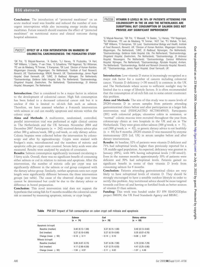
Conclusion: The introduction of ‘‘protected mealtimes’’ on anacute medical ward was feasible and reduced the number of non-urgent interruptions while also increasing energy intake duringmealtimes. Future research should examine the effect of ‘‘protectedmealtimes’’ on nutritional status and clinical outcome duringhospital admission.
PM207 EFFECT OF A FISH INTERVENTION ON MARKERS OFCOLORECTAL CARCINOGENESIS: THE FISHGASTRO STUDY
1GK Pot, 2G Majsak-Newman, 1A Geelen, 2LJ Harvey, 2K Przybylska, 3A Hart,4MP Williams, 2J Dainty, 1P van t’Veer, 1G Schaafsma, 5FM Nagenast, 6BJ Witteman,7PC van de Meeberg, 8R Timmer, 1E Kampman, 2EK Lund. 1Division of Human Nutrition,Wageningen University, Wageningen, The Netherlands; 2Institute of Food Research,Norwich, UK; 3Gastroenterology, NNUH, Norwich, UK; 4Gastroenterology, James PagetHospital, Great Yarmouth, UK; 5UMC, St Radboud, Nijmegen, The Netherlands;6Gastroenterology, Gelderse Vallei Hospital, Ede, The Netherlands; 7Gastroenterology,Slingeland Hospital, Doetinchem, The Netherlands; 8Gastroenterology, St AntoniusHospital, Nieuwegein, The Netherlands
Introduction: Diet is considered to be a major factor in relationto the development of colorectal cancer. High fish consumptionhas been linked to a decreased colorectal cancer risk but it isunclear if this is limited to oil-rich fish such as salmon.Therefore, we have assessed whether a 6-month interventionwith salmon or cod can modify apoptosis and mitosis within thecolonic crypt.Aims and Methods: A multicentre, randomised, controlled,parallel intervention trial was performed at eight clinical centresin The Netherlands and the UK between November 2004 andDecember 2007. Participants (n = 242) received dietary advice pluseither 300 g salmon/week, 300 g cod/week, or only dietary advice.Colonic biopsies were collected before the intervention by colono-scopy and after by sigmoidoscopy. Crypts were stained withFeulgen’s stain, microdissected and the numbers of mitotic andapoptotic cells per crypt were counted. Serum fatty acids were alsomeasured. Results were analysed by analysis of covariance.Results: Salmon consumption significantly increased circulating n-3 fatty acids. Overall, there was no significant benefit of consumingeither salmon or cod in relation to mitosis and apoptosis. After theintervention, the number of mitotic cells per crypt was notsignificantly different in the salmon or cod group compared withthe dietary advice group. Similarly, neither apoptosis rates not cryptlength were significantly different between the three interventiongroups (see table). The cause of the observed change over timecannot be determined but could be due to the dietary advice ordifference in bowel preparation.Conclusion: This novel intervention trial does not support thehypothesis that eating fish for 6 months modifies the colorectal cancerrisk as assessed by measuring apoptosis, mitosis, or crypt length.
PM208 VITAMIN D LEVELS IN 70% OF PATIENTS ATTENDING FORCOLONOSCOPY IN THE UK AND THE NETHERLANDS ARESUBOPTIMAL BUT CONSUMPTION OF SALMON DOES NOTPROVIDE ANY SIGNIFICANT IMPROVEMENT
1G Majsak-Newman, 2GK Pot, 1E Maxwell, 2A Geelen, 1LJ Harvey, 3FM Nagengast,4BJ Witteman, 5PC van de Meeberg, 6R Timmer, 7ACIT Tan, 8PJ Wahab, 9A Hart,10MP Williams, 2E Kampman, 1EK Lund. 1Gastrointestinal Biology and Health, Instituteof Food Research, Norwich, UK; 2Division of Human Nutrition, Wagenigen University,Wageningen, The Netherlands; 3UMC, St Radboud, Nijmengen, The Netherlands;4Gastroenterology, Gelderse Vallei Hospital, Ede, The Netherlands; 5Gastroenterology,Slingeland Hospital, Doetinchem, The Netherlands; 6Gastroenterology, St AntoniusHospital, Nieuwegein, The Netherlands; 7Gastroenterology, Canisius WilhelminaHospital, Nijmegen, The Netherlands; 8Gastroenterology, Rijnstate Hospital, Arnhem,The Netherlands; 9Gastroenterology, Norfolk and Norwich University Hospital, Norwich,UK; 10Gastroenterology, James Paget Hospital, Great Yarmouth, UK
Introduction: Low vitamin D status is increasingly recognised as amajor risk factor for a number of cancers including colorectalcancer. Vitamin D deficiency (,50 mmol/l) is common in the UKand The Netherlands where access to sufficient sunlight may belimited due to a range of lifestyle factors. It is often recommendedthat the consumption of oil-rich fish can to some extent counteractthis problem.Aims and Methods: The aim of this study was to assess levels of25OH-vitamin D in serum samples from patients attendinggastrointestinal clinics before and after participation in a larger fishintervention trial (FISHGASTRO NCT00145015). Participants(201) with colorectal polyps, ulcerative colitis in remission, or‘‘normal’’ colonic mucosa were recruited throughout the year fromcolonoscopy clinics at two hospitals in the UK and six in TheNetherlands. They were given either salmon (300 g/week; n = 71),cod (300 g/week; n = 62), or generic dietary advice to eat healthily(n = 64) for 6 months. 25OH-vitamin D was measured by enzymeimmunoassay (IDS Ltd, UK) in serum samples before and afterdietary interventions.Results: At baseline, 32% of patients were vitamin D deficient and71% had suboptimal levels, higher than previously reported for aUK middle-aged population. As expected, deficiency was greatest inJanuary (69%), with 94% having suboptimal levels (,80 nmol/l).Even in the summer months approximately 10% of patients weredeficient and 50% had suboptimal levels. Patients gained nosignificant benefit in terms of their vitamin D levels fromconsuming salmon for 6 months.Conclusion: Patients attending gastrointestinal clinics are verylikely to have suboptimal levels of vitamin D. They should bestrongly encouraged to have a sensible outdoor lifestyle in order torectify this problem. Any nutritional advice should be more targetedtowards cod-liver oil and herring or fortified foods as better sourcesof vitamin D than salmon.Funding: This work was funded under EU FP6 SEAFOODplusproject 506359, the UK Food Standards Agency and BBSRC.
Table PM-207 Impact of fish consumption on colon crypt cell mitosis and apoptosis
Salmon(n = 74)
Cod(n = 70)
Dietary advice(n = 69)
Apoptosis (n/crypt)
Baseline (median) 0.40 (0.13–1.00) 0.37 (0.15–1.00) 0.40 (0.13–0.82)
End (median) 0.27 (0.10–0.80) 0.27 (0.10–0.80) 0.35 (0.07–0.70)
Change (mean ¡ SE) 20.18 ¡ 0.11 20.14 ¡ 0.10 20.08 ¡ 0.07
Mitosis (n/crypt)
Baseline (median) 6.08 (4.67–8.15) 5.67 (4.36–7.90) 4.70 (3.50–7.29)
End (median) 4.17 (2.90–6.58) 4.37 (2.75–6.93) 4.47 (3.25–5.80)
Change (mean ¡ SE) 21.94 ¡ 4.97 22.11 ¡ 4.63 21.07 ¡ 4.46
BSG abstracts
A80 Gut 2009;58(Suppl I):A1–A156
on July 20, 2022 by guest. Protected by copyright.
http://gut.bmj.com
/G
ut: first published as on 23 March 2009. D
ownloaded from
PM209 THE NASAL BRIDLE EFFECTIVELY SECURES NASOGASTRICFEEDING TUBES AND AVOIDS UNNECESSARY INVASIVEENTERAL AND PARENTERAL NUTRITION
1JA Bennell, 1S Black, 1CD Murray, 2K Moore. 1Gastroenterology, Royal Free Hospital,London, UK; 2Hepatology, Royal Free Hospital, London, UK
Introduction: Nasogastric feeding tubes are used in patients whoare unable to manage an oral diet and who have a functioninggastrointestinal tract. However, many patients (17–67%) repeatedlydislodge their feeding tubes1 leading to potential malnutrition,patient discomfort and increased nursing time. Alternative nutri-tion delivery such as a feeding gastrostomy tube or parenteralnutrition each have inherent risks. Bridles, in which the nasogastrictube is secured round the nasal septum, have been used for manyyears, and lead to improved patient outcomes.2 3
Aims and Methods: All patients referred to the nutrition nurseover 12 months were included in the audit. Referral criteriaincluded patient dependent on enteral feeding, with dislodgementof the nasogastric tube at least three times in one week resulting inmissed feeds, or feeding tubes requiring endoscopic or radiologicalguidance and which had been dislodged once. All nasogastric tubesand bridles were placed by one clinical nutrition nurse specialist.Results: Sixty bridle placements were requested (median age75 years, range 28–100). Of the 60 requested, 51 requests werejudged to be appropriate, and successful bridle placement wascarried out in 45 of these 51 patients. Five patients were seen to pulltheir bridles, were restless or wore mittens. No nasal trauma wasseen in any of the patients. Five stayed in for less than 24 h. Fifteenof 45 bridles stayed in place for 1–5 days. Fifteen patients kept thebridles in situ for 6–20 days and eight for more than 20 days (twounknown because patient transferred to another hospital). Sixteenof 45 bridles were unplanned removals by the patient, of which sixcontinued with a form of tube feeding, four progressed to oralnutrition and six died. Of the remaining 29 patients who kept theirbridles in, 12 patients progressed to oral feeding, seven togastrostomy feeding, five had a second nasogastric tube and bridleplaced, three coped with a nasogastric tube alone, two patientstransferred to another hospital, eight patients had their treatmentwithdrawn and bridles removed and eight patients died with bridlesin situ.Conclusion: Bridles were safe, well tolerated and effective in thisgroup of patients in delivering enteral nutrition. Bridles facilitatenutrition delivery until the patient recovers sufficiently to managean oral intake. It avoids unnecessary escalation to percutaneousendoscopic gastrostomy feeding or intravenous nutrition.
1. Carrion MI, et al. Accidental removal of endotracheal and nasogastric tubes andintravenous catheters. Crit Care Med 2000;28:63–6.
2. Armstrong C, et al. A technique for preventing extubation of feeding tubes. 5thClinical Congress Abstracts. Nov/Dec 1980:125.
3. Anderson MR, et al. The nasal loop provides an alternative to percutaneousendoscopic gastrostomy in high-risk dysphagia stroke patients. Clin Nutr2004;23:501–6.
PM210 USE OF NASAL BRIDLES TO SECURE NASOGASTRIC TUBESIMPROVES DELIVERY OF ENTERAL NUTRITION IN AT-RISKPATIENTS
1KT Cheung, 1E Donaldson, 1T Earley, 2P Coulthurst, 1T Shaw, 1PL Shields.1Gastroenterology, Royal Preston Hospital, Preston, UK; 2Dietetics, Royal PrestonHospital, Preston, UK
Introduction: One of the major limitations of nasogastric feedingtubes is their frequent displacement, contributing to suboptimaldelivery of nutrition. The repeated insertion of nasogastric tubescan be uncomfortable and is associated with risks includinginterruptions in the delivery of feed and repeated chest x rays.The use of nasal bridles allows for the securing of nasogastric tubes,avoiding the need for repeated nasogastric tube reinsertion, and
theoretically this should result in less disruption to the delivery ofprescribed feed. We have incorporated the use of nasal bridles intoroutine practice since 2005.1
Aims and Methods: The aim of the study was to determine ifnasal bridles were effectively improving delivery of nutrition bypreventing nasogastric tube displacement. A retrospective review ofrecords was performed of patients who had a nasal bridle placedbetween March 2007 and February 2008. The indication for nasalbridle insertion was established. The previous number of repeatednasogastric tube insertions was determined. Delivered nasogastricfeed was assessed for a 7-day period before nasal bridle insertion anda maximum of 30 days post-nasal bridle insertion. Reasons forinadequate feed delivery were determined.Results: 134 patients were referred for nasal bridle insertion overthe 12-month period. Notes were retrieved and feed delivery wasassessed in 69 patients. 48 patients were referred for nasal bridleplacement due to repeated nasogastric tube displacement (mean of2.3 nasogastric tubes per patient). There were no significantcomplications associated with bridle insertion. Pre-bridle insertion,feed delivery was less than 50% prescribed in 67.3% (298/443) oftotal patient days. Post-nasal bridle insertion, days of feed deliveryless than 50% fell to 13.4% (168/1256) (p,0.0001). 33% of patientsrequired more than one nasal bridle (mean 1.4 reinsertions) duringthe follow-up period. This was mainly due to intentional removalby patients (n = 23) but nasogastric tube blockage (n = 3) alsooccurred.Conclusion: Our experience shows that the use of nasal bridles is asafe and effective means of improving the delivery of enteralnutrition in malnourished patients. They also reduce nasogastrictube displacement. Nasal bridles can still be removed intentionallyby patients and do not prevent feed disruption secondary to othercauses. Nonetheless, in patients who continuously displace theirnasogastric tubes, a nasal bridle should be used to limit disruptionto feed delivery and the risks associated with repeated reinsertion.
1. Donaldson E, Earley T, Shields PL. The nasal bridle—its place within an integratednutrition service: a prospective audit of 1 years data. Gut 2007;56S2:A137.
PM211 DEFINING THE NON-SURGICAL CARE PATHWAY FORPATIENTS WITH ENCAPSULATING PERITONEAL SCLEROSIS
1N Abdul Satar, 1M McGregor, 2C Bebb, 2C Byrne, 2M Devonald, 2G Mchaffie, 2SRoe, 1K Teahon. 1Gastroenterology, Nottingham City University Hospital, Nottingham,UK; 2Renal, Nottingham City University Hospital, Nottingham, UK
Introduction: Encapsulating peritoneal sclerosis (EPS) is a rare butrecognised complication seen in patients who have had peritonealdialysis.1 It is an inflammatory process that transforms theperitoneum into calcified, fibrous tissue enveloping the bowel intoa ‘‘cocoon’’ formation. Because of its gastrointestinal manifesta-tions, these patients are likely to present to gastroenterologists.However, their diagnosis and presentation are poorly defined,making management challenging.Aims and Methods: Retrospective review of patients diagnosedwith EPS in the past 5 years in Nottingham city hospital. Data werecollected on: symptoms, investigations, management and complica-tions.Results: We identified nine patients (six men). The average agewas 41 years (range 29–58). Risk factors are shown in the table. Ofthe nine, seven had had renal transplants. Four transplants hadfailed and of these, three had had two failed transplants. EPSpresented with recurrent symptoms of ascites (33%), peritonitis(44%), small bowel obstruction (33%), vomiting (22%) and bloodyascites (11%). Other dominant symptoms were nausea and weightloss. The diagnosis was made on clinical suspicion confirmed withcomputed tomography (89%) and histology (66%) taken atlaparotomy. Significant co-morbidities included diabetes,Goodpasture’s disease, anti-phospholipid syndrome and cystinosis.Complications included small bowel perforation, intra-abdominal
BSG abstracts
Gut 2009;58(Suppl I):A1–A156 A81
on July 20, 2022 by guest. Protected by copyright.
http://gut.bmj.com
/G
ut: first published as on 23 March 2009. D
ownloaded from
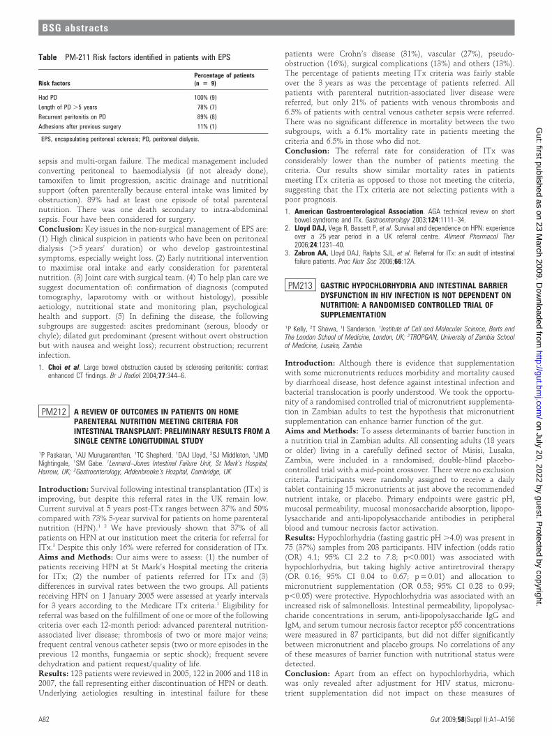
sepsis and multi-organ failure. The medical management includedconverting peritoneal to haemodialysis (if not already done),tamoxifen to limit progression, ascitic drainage and nutritionalsupport (often parenterally because enteral intake was limited byobstruction). 89% had at least one episode of total parenteralnutrition. There was one death secondary to intra-abdominalsepsis. Four have been considered for surgery.Conclusion: Key issues in the non-surgical management of EPS are:(1) High clinical suspicion in patients who have been on peritonealdialysis (.5 years’ duration) or who develop gastrointestinalsymptoms, especially weight loss. (2) Early nutritional interventionto maximise oral intake and early consideration for parenteralnutrition. (3) Joint care with surgical team. (4) To help plan care wesuggest documentation of: confirmation of diagnosis (computedtomography, laparotomy with or without histology), possibleaetiology, nutritional state and monitoring plan, psychologicalhealth and support. (5) In defining the disease, the followingsubgroups are suggested: ascites predominant (serous, bloody orchyle); dilated gut predominant (present without overt obstructionbut with nausea and weight loss); recurrent obstruction; recurrentinfection.
1. Choi et al. Large bowel obstruction caused by sclerosing peritonitis: contrastenhanced CT findings. Br J Radiol 2004;77:344–6.
PM212 A REVIEW OF OUTCOMES IN PATIENTS ON HOMEPARENTERAL NUTRITION MEETING CRITERIA FORINTESTINAL TRANSPLANT: PRELIMINARY RESULTS FROM ASINGLE CENTRE LONGITUDINAL STUDY
1P Paskaran, 1AU Murugananthan, 1TC Shepherd, 1DAJ Lloyd, 2SJ Middleton, 1JMDNightingale, 1SM Gabe. 1Lennard–Jones Intestinal Failure Unit, St Mark’s Hospital,Harrow, UK; 2Gastroenterology, Addenbrooke’s Hospital, Cambridge, UK
Introduction: Survival following intestinal transplantation (ITx) isimproving, but despite this referral rates in the UK remain low.Current survival at 5 years post-ITx ranges between 37% and 50%compared with 73% 5-year survival for patients on home parenteralnutrition (HPN).1 2 We have previously shown that 37% of allpatients on HPN at our institution meet the criteria for referral forITx.3 Despite this only 16% were referred for consideration of ITx.Aims and Methods: Our aims were to assess: (1) the number ofpatients receiving HPN at St Mark’s Hospital meeting the criteriafor ITx; (2) the number of patients referred for ITx and (3)differences in survival rates between the two groups. All patientsreceiving HPN on 1 January 2005 were assessed at yearly intervalsfor 3 years according to the Medicare ITx criteria.1 Eligibility forreferral was based on the fulfillment of one or more of the followingcriteria over each 12-month period: advanced parenteral nutrition-associated liver disease; thrombosis of two or more major veins;frequent central venous catheter sepsis (two or more episodes in theprevious 12 months, fungaemia or septic shock); frequent severedehydration and patient request/quality of life.Results: 123 patients were reviewed in 2005, 122 in 2006 and 118 in2007, the fall representing either discontinuation of HPN or death.Underlying aetiologies resulting in intestinal failure for these
patients were Crohn’s disease (31%), vascular (27%), pseudo-obstruction (16%), surgical complications (13%) and others (13%).The percentage of patients meeting ITx criteria was fairly stableover the 3 years as was the percentage of patients referred. Allpatients with parenteral nutrition-associated liver disease werereferred, but only 21% of patients with venous thrombosis and6.5% of patients with central venous catheter sepsis were referred.There was no significant difference in mortality between the twosubgroups, with a 6.1% mortality rate in patients meeting thecriteria and 6.5% in those who did not.Conclusion: The referral rate for consideration of ITx wasconsiderably lower than the number of patients meeting thecriteria. Our results show similar mortality rates in patientsmeeting ITx criteria as opposed to those not meeting the criteria,suggesting that the ITx criteria are not selecting patients with apoor prognosis.
1. American Gastroenterological Association. AGA technical review on shortbowel syndrome and ITx. Gastroenterology 2003;124:1111–34.
2. Lloyd DAJ, Vega R, Bassett P, et al. Survival and dependence on HPN: experienceover a 25 year period in a UK referral centre. Aliment Pharmacol Ther2006;24:1231–40.
3. Zabron AA, Lloyd DAJ, Ralphs SJL, et al. Referral for ITx: an audit of intestinalfailure patients. Proc Nutr Soc 2006;66:12A.
PM213 GASTRIC HYPOCHLORHYDRIA AND INTESTINAL BARRIERDYSFUNCTION IN HIV INFECTION IS NOT DEPENDENT ONNUTRITION: A RANDOMISED CONTROLLED TRIAL OFSUPPLEMENTATION
1P Kelly, 2T Shawa, 1I Sanderson. 1Institute of Cell and Molecular Science, Barts andThe London School of Medicine, London, UK; 2TROPGAN, University of Zambia Schoolof Medicine, Lusaka, Zambia
Introduction: Although there is evidence that supplementationwith some micronutrients reduces morbidity and mortality causedby diarrhoeal disease, host defence against intestinal infection andbacterial translocation is poorly understood. We took the opportu-nity of a randomised controlled trial of micronutrient supplementa-tion in Zambian adults to test the hypothesis that micronutrientsupplementation can enhance barrier function of the gut.Aims and Methods: To assess determinants of barrier function ina nutrition trial in Zambian adults. All consenting adults (18 yearsor older) living in a carefully defined sector of Misisi, Lusaka,Zambia, were included in a randomised, double-blind placebo-controlled trial with a mid-point crossover. There were no exclusioncriteria. Participants were randomly assigned to receive a dailytablet containing 15 micronutrients at just above the recommendednutrient intake, or placebo. Primary endpoints were gastric pH,mucosal permeability, mucosal monosaccharide absorption, lipopo-lysaccharide and anti-lipopolysaccharide antibodies in peripheralblood and tumour necrosis factor activation.Results: Hypochlorhydria (fasting gastric pH .4.0) was present in75 (37%) samples from 203 participants. HIV infection (odds ratio(OR) 4.1; 95% CI 2.2 to 7.8; p,0.001) was associated withhypochlorhydria, but taking highly active antiretroviral therapy(OR 0.16; 95% CI 0.04 to 0.67; p = 0.01) and allocation tomicronutrient supplementation (OR 0.53; 95% CI 0.28 to 0.99;p,0.05) were protective. Hypochlorhydria was associated with anincreased risk of salmonellosis. Intestinal permeability, lipopolysac-charide concentrations in serum, anti-lipopolysaccharide IgG andIgM, and serum tumour necrosis factor receptor p55 concentrationswere measured in 87 participants, but did not differ significantlybetween micronutrient and placebo groups. No correlations of anyof these measures of barrier function with nutritional status weredetected.Conclusion: Apart from an effect on hypochlorhydria, whichwas only revealed after adjustment for HIV status, micronu-trient supplementation did not impact on these measures of
Table PM-211 Risk factors identified in patients with EPS
Risk factorsPercentage of patients(n = 9)
Had PD 100% (9)
Length of PD .5 years 78% (7)
Recurrent peritonitis on PD 89% (8)
Adhesions after previous surgery 11% (1)
EPS, encapsulating peritoneal sclerosis; PD, peritoneal dialysis.
BSG abstracts
A82 Gut 2009;58(Suppl I):A1–A156
on July 20, 2022 by guest. Protected by copyright.
http://gut.bmj.com
/G
ut: first published as on 23 March 2009. D
ownloaded from
barrier function at the doses used. Important effects of HIV andhighly active antiretroviral therapy on gastric pH may explainsome of the increased susceptibility of HIV-infected adults tointestinal infection.
PM214 HEALTHCARE PROFESSIONALS’ KNOWLEDGE ABOUTPERCUTANEOUS GASTROSTOMY FEEDING: HOW WELL AREWE DOING?
1S Thomas, 2FC Leslie. 1School of Medicine, University of Keele, Keele, UK;2Department of Gastroenterology, University Hospital of North Staffordshire, Stoke-on-Trent, UK
Introduction: There is a reasonable body of evidence to supportthe use of percutaneous gastrostomy (PEG) feeding for strokepatients, but no evidence to support this practice in advanceddementia. A previous study showed that 34% of UK consultantswould refer a patient with dementia for a PEG.1 In the light of thenew Mental Capacity Act it is vital that healthcare professionals areable to make evidence-based decisons and advise patients andfamilies appropriately.Aims and Methods: We were keen to explore healthcareprofessionals’ knowledege about PEG feeding. We were also keento identify whether teaching had any influence on knowledge. Asimple questionnaire was given to a range of healthcare profes-sionals in four hospitals across the midlands and to an expert group(Midlands Gastroenterogy Group). Participants were asked abouttwo scenarios of dysphagic stroke and advanced dementia. Theywere also asked if they had received any teaching and how muchrelevant experience they had.Results: 556 of 714 questionnaires were collected (78%) fromhospital doctors of all levels and general practitioners (GPs), medicalstudents, nurses and dieticians. More respondents answeredquestions about dysphagic stroke correctly compared with demen-tia. Only 10% of hospital consultants would refer the dementiapatient for a PEG compared with 31% of GPs, 37% of F1/F2 doctors,36% of medical students and 45% of nurses. Only 29% of allrespondents felt that PEG would prevent aspiration pneumonia inthe dementia patient (although nurses’ opinion was split 45% vs45%). 20% of respondents felt PEG would improve life expectancy.Dieticians as a group were most likely to answer questionscorrectly. 64% of respondents had experience with similar patientsincluding 77% of GPs and 69% of foundation doctors. Teachingsignificantly influenced answers for the dementia scenario(p = 0.001). Only 29% had received teaching and only 4% of GPshad attended relevant teaching. Experts were more likely to answercorrectly (p = 0.004).Conclusion: Despite improvements in consultants’ knowledgecompared with previous studies, there are still significant gaps inknowledge. Training improves knowledge and this study suggestsmore teaching should be planned, particularly aimed at nurses,medical students, junior doctors and GPs.
1. Sinha U, et al. Arch Ger Geront 2001;32:113–18.
PM215 GLUTEN LEVELS IN COMMERCIAL QUINOA PRODUCTS
1VF Zevallos, 1HJ Ellis, 2LI Herencia, 1PJ Ciclitira. 1Gastroenterology, St Thomas’sHospital, London, UK; 2Produccion Vegetal, Universidad Politecnica de Madrid,Madrid, Spain
Introduction: The only effective treatment for patients affected bycoeliac disease is to follow a gluten-free diet (GFD). Permanentexclusion of gluten from the diet is essential to maintain normalvillous architecture and to reduce the risk of other complications. AGFD includes naturally occurring gluten-free cereals such as rice,
millet and maize. Quinoa can be an alternative for the diet of coeliacdisease patients1 but its incorporation as a food ingredient could beaccompanied by the risk of contamination. Therefore, we haveassessed the safety of various gluten-free quinoa products in the UK.Aims and Methods: We used dot immunobinding assays and fourmurine monoclonal antibodies to known coeliac-toxic glutenproteins and peptides. Furthermore, we used ELISA2 to quantifytoxic peptides. Prolamins were extracted from quinoa products andcommercially obtained wheat starches of known gluten contentusing an in-house extraction buffer with a reducing agent. Allsamples were spotted onto a nitrocellulose membrane and exposedseparately to the antibodies, enzyme-linked anti-mouse secondaryantibody and chromogen. The ELISA was performed using amonoclonal antibody to a toxic A-gliadin peptide.Results: The wheat starches had strong, moderate and negativereactions in the dot assays. Most quinoa products were negative, afew borderline and one positive to one or both of the antigliadinantibodies. ELISA indicated that products with borderline resultshad no detectable gliadin, as measured by PN3 (anti-A-gliadin 31–49)but one product had a gluten content of 76 mg/kg.Conclusion: Quinoa products may be a safe alternative for coeliacdisease patients, the majority of gluten-free products containingquinoa have levels of gluten below 20 mg/kg, which is in agreementwith the current recommendations.3 However, one product mayneed to be relabelled as it contains more than 20 mg/kg but lessthan 100 mg/kg. New legislation to regulate the labelling of gluten-free products is expected to come in the near future. Therefore,further tests will be undertaken to assess the safety of new andexisting gluten-free products.
1. Zevallos V, Ciclitira PJ, Suligoj T, et al. Proceedings of the 22nd Meeting of theProlamin Working Group. 2008:95–8.
2. Ellis HJ, Rosen-Bronson S, O’Reilly N, et al. Gut 1998;43:190–5.3. Codex Standards for ‘‘Gluten-free foods’’. In: Report of the 29th Session of the
Codex Committee on Nutrition and Foods for Especial Dietary Uses. Geneva: CodexAlimentarius Commission FAO/WHO, 2008.
Colorectal/anorectal posters
PT216 PREDICTION OF SEVERE 5-FLUOROURACIL TOXICITY INGASTROINTESTINAL CANCER CHEMOTHERAPY: ROLE OFDIHYDROPYRIMIDINE DEHYDROGENASE DEFICIENCY
1A Loganayagam, 2L Fairbanks, 2M Arenas-Hernandez, 3P Ross, 2T Marinaki,1JD Sanderson. 1Gastroenterology, Guys and St Thomas NHS Trust, London, UK; 2PurineResearch, Guys and St Thomas NHS Trust, London, UK; 3Oncology, Guys and StThomas NHS Trust, London, UK
Introduction: 5-Fluorouracil (5-FU) and its analogues remain a keycomponent of chemotherapeutic regimens for the adjunctivetreatment of gastrointestinal cancer. Up to 20% of patientsexperience severe (grades 3 and 4) toxicity, particularly severemucositis, limiting effective therapy and resulting in significantmorbidity, healthcare costs and, occasionally, fatality. 5-FU toxicityhas been reported in association with polymorphic variationdihydropyrimidine dehydrogenase (DPD) activity but such poly-morphism has previously been considered absent in UK popula-tions. The DPYD IVS14+1G.A mutation has a reported frequencyof 1.8% in European populationsAims and Methods: To assess the contribution of DPYDpolymorphism to 5-FU toxicity among UK patients treated forgastrointestinal cancer. Sequencing of the coding region of theDPYD gene was undertaken in 47 patients (27 women, mean age61 years), mainly with colorectal cancer, experiencing grade 3 or 4toxicity on 5-FU according to common terminology criteria foradverse events.
BSG abstracts
Gut 2009;58(Suppl I):A1–A156 A83
on July 20, 2022 by guest. Protected by copyright.
http://gut.bmj.com
/G
ut: first published as on 23 March 2009. D
ownloaded from
Results: Myelotoxicity (38%) and diarrhoea (38%) were the mostfrequent toxicities, followed by mucositis (18%), hand–footsyndrome (4%) and neurotoxicity (2%). Four of 47 (8.5%) patientscarried the DPYD IVS14+1G.A mutation. All four cases werewomen and three of the four had severe diarrhoea. A further sixcases carried other DPYD mutations (D949V n = 4, I560S n = 1,V7321 n = 1). In total, 10 (21%) patients carried DPYD mutations.Conclusion: Contrary to previous estimates for a UK population,genetic DPD deficiency may account for approximately 20% ofcases of severe 5-FU toxicity. Higher frequencies of 25–30% havebeen reported in non-UK European series, also with a gender bias.The metabolic influence of DPD deficiency is such that toxicityshould be avoidable by previous testing and an appropriate 5-FUdose or regimen alteration.
PT217 20 MHZ HIGH-FREQUENCY MINIPROBE ULTRASOUND AS ANAIDE TO TRANSANAL ENDOSCOPIC MICROSURGERY:COMPARISON WITH MAGNETIC RESONANCE IMAGING
1AK Haji, 2S Ryan, 3I Bjarnason, 1S Papagrigoriadis. 1Colorectal Surgery, Kings CollegeHospital, London, UK; 2Radiology, Kings College Hospital, London, UK;3Gastroenterology, Kings College Hospital, London, UK
Introduction: Transanal endoscopic microsurgery (TEMS) is atechnique for the resection of rectal tubulovillous adenomas andearly cancer. Accurate local staging of these lesions is of paramountimportance to guide management before TEMS resection. Thisstudy compares the local staging of magnetic resonance imaging(MRI) and 20 MHz miniprobe ultrasound of consecutive patientsdeemed suitable for the TEMS procedure.Aims and Methods: Inclusion criteria included all rectal tumoursbelow the peritoneal reflection and benign histology on endoscopicbiopsy. Informed consent was obtained in all cases. All patients hadMRI of the anorectum and pelvis (unless contraindicated),colonoscopy and 20 MHz miniprobe ultrasound (OlympusKeymed UM-3R, Japan). Depth of invasion (T stage) of the lesionby MRI and 20 MHz ultrasound were compared with finalhistology. Statistical differences were analysed using x2 andFisher’s exact t tests with Graphpad Prism 5 (version 5.1, 2007).Results: 19 patients were studied, mean age 69.5 years (range 27–89).Mean distance of the tumour from the anal verge was 10 ¡3 cm(SE 0.7, range 5–15 cm). The tumours occupied 10–50% of the rectalcircumference, with a mean of 30 ¡ 15% (SE 3.4). Histologicalassessment revealed 11 benign lesions with dysplasia (five highgrade, six low grade) and eight adenocarcinomas (seven T1 withthree mucosal and four submucosal cancers, one T3). 16/19 (84%)patients had clear histological resection margins. MRI did notdiscriminate between mucosal and submucosal lesions and thesewere grouped together for analysis. Therefore, 18 patients hadlesions superficial to the muscularis propria (11 benign and sevenmalignant). Four patients did not have preoperative MRI includingthe patient with histological T3 stage. For lesions superficial to themuscularis propria (T0/T1), MRI correctly staged five of 15 lesions(33%), overstaging 10 patients (67%): six as T2 lesions and four asT3. 20 MHz ultrasound was superior to MRI for lesions superficialto the muscularis propria (p = 0.002). The accuracy of 20 MHzultrasound for assessing the depth of lesion was 95% (18/19correctly identified). In addition, 20 MHz ultrasound was able todifferentiate between mucosal and submucosal lesions with asensitivity of 93% and specificity of 75%, giving a positivepredictive value of 93% and a likelihood ratio of 3.7.Conclusion: 20 MHz miniprobe ultrasonography is superior to theconventionally used MRI for the assessment of T1 lesions with highsensitivity in differentiation between mucosal and submucosallesions. This may become an essential adjunct for the assessment ofpatients undergoing TEMS.
PT218 DIFFERENTIAL EXPRESSION PROTEOMICS IN APCMIN/+ MICECOLON FOLLOWING TREATMENT WITH PEROXISOMEPROLIFERATOR-ACTIVATED RECEPTORS ALPHA LIGAND,FENOFIBRATE
1A Shonde, 1V Subramanian, 2A Bennett, 1C Hawkey. 1Wolfson Digestive DiseasesCentre, Nottingham University Hospital, Nottingham, UK; 2Biological Sciences,University of Nottingham, Nottingham, UK
Introduction: Peroxisome proliferator-activated receptors (PPAR)are ligand-activated nuclear receptors that were discovered in 1990.PPARa is expressed at low levels in human colorectal cancers.APCMin/+ mice fed with fenofibrate (a PPARa ligand) havesignificantly less tumour burden in the small intestine and coloncompared with controls. Our group has looked at PPARa geneexpression changes, but RNA expression does not predict thesynthesis of a corresponding protein.Aims and Methods: 25–50 mg flash-frozen fragmented colonictissue from three APCMin/+ mice treated with fenofibrate 100 mg/kgper day and three APCMin/+ mice treated with vehicle were used forthis experiment. The cytosolic protein fraction was extracted usingProteoExtract (Merck, Nottingham, UK) subcellular proteomeextraction kit. Samples were fractionated using reversed-phase solidphase extraction followed by sodium dodecylsulphate polyacryla-mide gel electrophoresis of the fractions. Differentially expressedproteins were subjected to in-gel tripsinolysis. The peptides werethen analysed by matrix-assisted laser desorption ionisation time offlight mass spectrometry. Proteins of interest were then identifiedby interrogating the Mascot database. The identity of the proteinswas reconfirmed by standard Western blotting.Results: We identified four proteins with significantly alteredexpression in colonic tissue of APCMin/+ mice colon tissue treatedwith fenofibrate compared with controls. Two of these proteins,superoxide dismutase 2 (SOD2) gene (increased expression byfenofibrate) and alpha-enolase 1 (decreased expression by fenofibrate)expressions were confirmed by using Western blot analysis. Theothers were carbonic anhydrase II and transgelin, both of whichshowed reduced expression in treated colonic tissue. SOD2 has beenimplicated as a candidate tumour suppressor gene for humanmalignant melanoma and SOD2 mutations have also been found in60% of colon cancer cell lines examined.1 Transgelin and alpha-enolase1 protein expression has been shown to be decreased in colitis-associated colon cancer. In addition, transgelin expression has beenshown to be significantly reduced in human colon tumours comparedwith adjacent non-tumorous tissues.2 Interestingly, the expression ofcarbonic anhydrase I has been shown to be correlated with thebiological aggressiveness of colorectal cancer.3
Conclusion: In APCMin/+ mice, fenofibrate treatment results in thedifferential expression of proteins that could mediate the PPARaactivation-related suppression of colon tumour formation.
1. Xu Y, et al. Oncogene 1999;18:93–102.2. Yeo M, et al. Proteomics 2006;6:1158–65.3. Bekku S, et al. Hepatogastroenterology 2000;47:998–1001.
PT219 WHAT ARE THE CHARACTERISICS OF POLYPS DETECTED BYTHE NHS BOWEL CANCER SCREENING PROGRAMME
C Harmston, J Hunter, L Wong. Surgery, University Hospital Coventry, Coventry, UK
Introduction: The NHS bowel cancer screening programme iscurrently being introduced following three rounds of pilot screen-ing. This study outlines the important characteristics of polypsdetected by pilot screening.Aims and Methods: The first 100 patients with screen-detectedpolyps at University Hospital Coventry and Warwickshire wereidentified from the colorectal cancer screening database. Polypcharactersitics were drawn from the hospital’s computerised
BSG abstracts
A84 Gut 2009;58(Suppl I):A1–A156
on July 20, 2022 by guest. Protected by copyright.
http://gut.bmj.com
/G
ut: first published as on 23 March 2009. D
ownloaded from
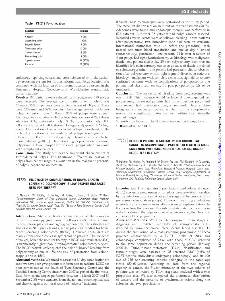
endoscopy reporting system and cross-referenced with the pathol-ogy reporting system for further information. Polyp location wascompared with the location of symptomatic cancers detected in theUniversity Hospital Coventry and Warwickshire symptomaticcancer database.Results: 100 patients were selected for investigation. 179 polypswere detected. The average age of patients with polyps was61 years. 35% of patients were under the age of 60 years. Therewere 68% men and 32% women. The average size of the largestpolyp per patient was 13.8 mm. 85% of polyps were excised.Histology was available on 162 polyps: tubulovillous 43%, tubularadenoma 33%, metaplastic polyp 9.3%, hyperplastic polyp 8%,villous adenoma 4%. 90% showed low-grade dysplasia, 10% highgrade. The location of screen-detected polyps is outlined in thetable. The location of screen-detected polyps was significantlydifferent from that of the location of symptomatic cancers detectedin our database (p,0.01). There was a higher proportion of sigmoidpolyps and a lower proportion of caecal polyps when comparedwith symptomatic cancers.Conclusion: This study outlines the important characteristics ofscreen-detected polyps. The significant difference in location ofpolyps from cancer suggests a variation in the malignant potentialof polyps, dependent on location.
PT220 INCIDENCE OF COMPLICATIONS IN BOWEL CANCERSCREENING COLONOSCOPY IS LOW DESPITE INCREASEDNEED FOR THERAPY
1D Nylander, 2M Ritchie, 1J Painter, 3M Rutter, 3L Hurst, 2J Singh, 4C Rees.1Gastroenterology, South of Tyne Screening Centre, Sunderland Royal Hospital,Sunderland, UK; 2South of Tyne Screening Centre, QE Hospital, Gateshead, UK;3Teesside Screening Centre, North Tees, UK; 4South of Tyne Screening Centre, SouthTyneside Hospital, South Shields, UK
Introduction: Many publications have estimated the complica-tions of colonoscopy (summarised by Bowes et al).1 These are usedto help inform patients undergoing colonoscopy. The estimates arealso used in NHS publications given to patients attending for bowelcancer screening colonoscopy (BCSC). However, these data aremainly from colonoscopies in symptomatic patients. The incidenceof polyps, hence the need for therapy in BCSC (approximately 48%)is significantly higher than in ‘‘symptomatic’’ colonoscopy services.The BCSC patient leaflet quotes the risk of ‘‘heavy’’ bleeding frompolypectomy as one in 150; the risk of perforation from colono-scopy is one in 1500.Aims and Methods: We aimed to assess our 30-day complications tosee if we have been giving accurate information to patients. BCSC hasbeen carried out at the South of Tyne Screening Centre and theTeesside Screening Centre since March 2007 as part of the first wave.Data from colonoscopies performed between 1 March 2007 and 30September 2008 were extracted from the national screening databaseand checked against our local record of ‘‘adverse’’ incidents.
Results: 1386 colonoscopies were performed in the study period.The caecal intubation rate on an intention to treat basis was 98.3%.Adenomas were found and endoscopic therapy was performed in622 patients. A further 36 patients had polyp cancers resected.Recorded adverse events were as follows: bleeding—three patientsafter polypectomy, two immediate (one had been on warfarin;international normalised ratio 1.4 before the procedure), eachneeded two units blood transfusion and one at day 4 settledspontaneously; perforation—one patient, 24 h after resection of4 cm polyp, had right hemicolectomy as histology was malignant;death—one patient died at day 25 post-polypectomy, post-mortemidentified left main coronary occlusion as cause of death, unrelatedto colonoscopy; other—one patient had persistent clinical obstruc-tion after polypectomy within tight sigmoid diverticular stricture,histology—malignant with complete extraction, sigmoid colectomyconfirmed stricture with no complications of polypectomy; onepatient had chest pain on day 10 post-polypectomy, felt to beunrelated.Conclusion: The incidence of bleeding from polypectomy wasone in 219. This incidence would be lower if it was quoted perpolypectomy, as several patients had more than one polyp andalso several had metaplastic polyps removed. Despite therebeing more therapeutic procedures than in the symptomaticservice the complication rates are well within internationallyquoted ranges.Submitted on behalf of the Northern Regional Endoscopy Group.
1. Bowes et al. Gut 2004;53.
PT221 REDUCED PREDICTED MORTALITY FOR COLORECTALCANCER IN ASYMPTOMATIC PATIENTS DETECTED BY MASSSCREENING WITH IMMUNOCHEMICAL FAECAL OCCULTBLOOD TEST IN ITALY
1F Parente, 1B Marino, 1A Anderloni, 2P Tricomi, 3G Ucci, 3M Gilardoni, 4P Carzaniga,4M Costa, 5N Devecchi, 5F Tortorella, 6M Pirona, 5R Moretti. 1Gastrointestinal Unit, AManzoni Hospital, Lecco, Italy; 2Pathology Service, A Manzoni Hospital, Lecco, Italy;3Oncology Department, A Manzoni Hospital, Lecco, Italy; 4Surgical Department, AManzoni Hospital, Lecco, Italy; 5Screening Unit, Local Health Care Centre, Lecco, Italy;6Screening Unit, Regional Reference Centre, Milan, Italy
Introduction: The major aim of population-based colorectal cancer(CRC) screening programmes is to reduce disease-related mortalityby the detection of cancers at an earlier stage and by removal of itsprecursors (adenomatous polyps). However, measuring a reductionof mortality takes many years after screening implementation. Inthe mean time there is a need for intermediate outcome measures inorder to estimate the improvement of prognosis and, therefore, theefficiency of the programme.Aims and Methods: We aimed to compare tumour stages atdiagnosis and predicted mortality of asymptomatic CRCdetected by immunochemical faecal occult blood test (FOBT)during the first round of a mass-screening programme of Leccoprovince (characterised by a FOBT uptake of 50% andcolonoscopy compliance of 92%) with those of CRC detectedin the same population during the screening period (January2006–8). Tumour–node–metastasis (TNM) classification andtumour stages were assessed in 95 screened CRC (4.6% ofFOBT-positive individuals undergoing colonoscopy) and in 100out of 285 non-screening cancers belonging to the same agestrata (50–69 years). According to data from the regionalregistry of cancer, the 5-year mortality of the two cohorts ofpatients was estimated by TNM stage and analysed with a twoproportion test. We also compared the anatomical distributionof cancers and the presence of synchronous lesions along thecolon in the two populations.
Table PT-219 Polyp location
Location Number
Caecum 7 (5%)
Ascending colon 5(4%)
Hepatic flexure 7 (5%)
Transverse colon 10 (8%)
Splenic flexure 2 (2%)
Descending colon 7 (5%)
Sigmoid colon 64 (50%)
Rectum 26 (22%)
BSG abstracts
Gut 2009;58(Suppl I):A1–A156 A85
on July 20, 2022 by guest. Protected by copyright.
http://gut.bmj.com
/G
ut: first published as on 23 March 2009. D
ownloaded from

Results: Predicted 5-year-specific mortality was significantly higherin non-screening CRC patients compared with asymptomaticcancers detected by FOBT (43% vs 18%, p,0.0001). Treatmentcould be confined to colonoscopy and polypectomy/mucosectomyalone in 34% of screen-detected cancers compared with only 5%(p.0.0001) of non-screening CRC, whereas chemotherapy/radio-therapy was necessary in 35% and 92% of patients of the twogroups, respectively (p.0.0001). No significant difference in theanatomical distribution of cancers nor in the presence of synchro-nous lesions along the colon was found between symptomatic andasymptomatic patients.Conclusion: CRC are detected at earlier stages in asymptomaticindividuals with positive immunochemical FOBT compared withsymptomatic patients not participating in the screening pro-gramme. Therefore, the predicted 5-year disease-related mortalityrate is significantly reduced already by the first round of screeningwith immunochemical FOBT. In as many as 34% of asymptomaticscreen-detected CRC, treatment could be confined to colonoscopyand polypectomy/mucosectomy alone, thus saving significanteconomic resources.
PT222 POST-POLYPECTOMY COLONOSCOPY SURVEILLANCE:ADENOMA RECURRENCE AND RISK AT FOLLOW-UP
GJ Sadler, J Fallaha, BC McKaig. Gastroenterology, New Cross Hospital,Wolverhampton, UK
Introduction: Patients who have adenomas removed at colono-scopy are at increased risk of developing colorectal cancer (CRC) inthe future. Current British Society of Gastroenterology guidelinesstratify adenomas at baseline as low risk (1–2 small; ,1 cm),intermediate risk (3–4 small or at least one .1 cm) or high risk (.5adenomas or .3 adenomas at least one of which is .1 cm). Sessilelesions are classified separately. Risk is refined at each subsequentcolonoscopy and determines surveillance after adenoma removal.1
Aims and Methods: We audited our database to determinecolonoscopy findings at follow-up for patients undergoing polypsurveillance and compared these with the findings at the mostrecent previous colonoscopy.Results: Data from 180 colonoscopies performed for the purpose of‘‘polyp follow-up’’ are presented. 84% of patients whose mostrecent previous examination was negative and 63% of thosepatients with low-risk adenomas at the most recent previouscolonoscopy were found to have adenomas at the next follow-up.Of those with intermediate-risk, high-risk and sessile adenomas atthe most recent previous colonoscopy, 69%, 63% and 77%,respectively, were found to have intermediate, high-risk or sessileadenomas at the next follow-up. CRC was found in 6.6% of thetotal population undergoing surveillance—91% of cases occurred inpatients in whom the adenoma risk at the most recent previouscolonoscopy was classed as intermediate, high or sessile. Nosignificant relationships were found between adenoma risk atprevious and follow-up colonoscopy, other than for sessile polyps,which occurred significantly more frequently in those with aprevious history of sessile polyps (p,0.05).Conclusion: With the exclusion of sessile lesions, polyp risk at themost recent previous colonoscopy does not appear to predict polyprisk at the follow-up examination. Although CRC tended to occurin patients with a previous polyp risk of intermediate grade orgreater, which supports the shorter screening interval proposed forhigher-risk lesions, it is clear that adenomas are found at subsequentcolonoscopy in the vast majority of patients with a history ofpolyps, even in those whose most recent examination was negative.Given these findings, should we consider discharging patients witha history of adenomatous polyps from follow-up at all?
1. Atkins WS, Saunders BP. Surveillance guidelines after removal of colorectaladenomatous polyps. Gut 2002;51(Suppl V):v6–v9.
PT223 CLINICAL SIGNIFICANCE OF FLUORINE-18FLUORODEOXYGLUCOSE POSITRON EMISSIONTOMOGRAPHY FOR RECURRENT COLORECTAL CANCER
H Ishikawa, A Watanabe, T Ohyama, T Mukogawa, T Inoue, S Kinoshita, N Kawai,K Nakagawa, K Emoto, S Ogawa. Department of Surgery, Nara Prefectural Hospital,Nara, Japan
Introduction: Fluorine-18 fluorodeoxyglucose positron emissiontomography (FDG-PET) has been accepted as an effective tool ofdiagnosis and staging for patients with known or suspectedrecurrent colorectal cancer.Aims and Methods: This study is to analyse the clinicalsignificance of FDG-PET on the diagnosis and indication for surgicalintervention for recurrent colorectal cancer. 31 consecutive patients,with known or suspected recurrence of colorectal cancer based onthe elevation of tumour markers or abnormal findings on thefollow-up computed tomography (CT) image, underwent FDG-PET49 times between December 2003 and August 2007. Patients wereaged between 35 and 79 years (median 61), 18 were men, 10 wereDuke’s A or B stage and 17 had a history of colon cancer. Theaverage of recurrence was two times (range 1–6). The average periodbetween operation and first FDG-PET was 28 months (range 1–89).Of 31 cases, 30 had elevated serum carcinoembryonic antigen orCA19-9 (96.7%). For each case the diagnosis of FDG-PET image wascompared with that of CT image and the final diagnosis.Results: 31 patients were divided into three groups according tothe purposes of FDG-PET: to identify recurrent colorectal cancer byFDG-PET (identification group, n = 4); to determine malignancyand disease spread (disease spreading group, n = 23); and toevaluate the possibility of surgical intervention in recurrence-confirmed cases (follow-up group, n = 4). In the identificationgroup, FDG-PET alone could identify recurrence and indicateoperation for three cases (six times). One of three cases showeddiease-free survival for 18 months after a common iliac replacementoperation. In the disease-spreading group, 11 cases with localiseddisease spread by FDG-PET findings were indicated for operation,nine cases with diffuse disease spread were judged as contra-indicated for operation. The average survival period of the operationseries was longer than that of the contraindicated series (24 vs12 months). In the follow-up group, no case was indicated forsalvage operation because FDG-PET could detect lesions of thewhole body at one study. FDG-PET was shown to be false positivein one case and false negative in one case. The sensitivity of FDG-PET was 93.8% and its accuracy was 91.8%, whereas those of CTwere 83.3% and 81.6%, respectively.Conclusion: FDG-PET could visualise malignant lesions and wasan effective modality to evaluate not only disease spread but distantmetastasis for the recurrence of colorectal cancer. FDG-PET is veryuseful in detecting recurrence at an earlier stage and in decision-making for surgical intervention.
PT224 THE USE AND EFFICACY OF STERCULIA FOR PATIENTS WITHDEFAECATORY DISORDERS
1U Khalid, 2K Thomas, 1A Lalji, 1J Andreyev. 1Gastrointestinal Unit, Royal MarsdenHospital, London, UK; 2Computing, Royal Marsden Hospital, London, UK
Introduction: Normacol is a bulk-forming laxative that consists of62% sterculia granules, a vegetable gum extract from the karayatree. Sterculia absorbs water and can swell to more than 60 timesits original volume, allowing it to bulk stool and stimulateperistalsis. It is poorly fermented and thus does not significantlyincrease flatulence. There is very little research-based evidence tosupport its use.Aims and Methods: The aim of this study was to evaluate the useof normacol and its efficacy in patients with bowel symptomsrelated to previous cancer treatments. A retrospective audit wasconducted for all patients who were prescribed normacol between
BSG abstracts
A86 Gut 2009;58(Suppl I):A1–A156
on July 20, 2022 by guest. Protected by copyright.
http://gut.bmj.com
/G
ut: first published as on 23 March 2009. D
ownloaded from
January 2005 and August 2008 and who had returned for follow-upevaluation.Results: 76 men and 47 women, median age 68 years, wereidentified. In 84% previous pelvic radiotherapy was the likely causeof their symptoms. 62% of patients were prescribed normacol forfaecal loading/constipation, 83% for urgency/loose stool, 70% forfrequency, 52% for faecal incontinence, 28% for tenesmus and 26%for wind. Follow-up was available for a median 63 days (range 8–1085) after the first date of prescription. 57% of the constipatedgroup, 68% of the urgency group, 35% of those with frequency, 39%of those with incontinence, 44% of those with tenesmus and 41% ofthose with wind reported significant benefit from the use of thisbulking agent. No serious adverse effects were seen. 66% of patientswere fully compliant, 15% partly compliant and 10% non-compliant with their prescription of normacol. Of those who werepartly or non-compliant, 52% found it ineffective and 22% wereunable to tolerate it (due to taste and aggravated symptoms). Atleast 50% of patients continued to use this bulking agent long term.Conclusion: This audit suggests that normacol can be an effectivetreatment in many patients with a range of defaecatory disorders,especially following pelvic radiotherapy. Further prospective eva-luation of the efficacy of normacol is warranted.
PT225 THE IMPACT OF CHRONIC CONSTIPATION ON QUALITY OFLIFE IN ADULTS AND CHILDREN: A SYSTEMATIC REVIEW
1J Belsey, 2S Greenfield, 3D Candy, 4M Geraint. 1JB Medical Ltd, Sudbury, UK; 2QE2Hospital, Welwyn Garden City, UK; 3Royal West Sussex Trust, Chichester, UK; 4NorginePharmaceuticals Ltd, Harefield, UK
Introduction: The objective was to identify published quality oflife assessments in patients with constipation and to place theimpact of constipation within the context of other chronicconditions.Aims and Methods: A systematic review was carried out usingMEDLINE, EMBASE, Cochrane Library and Google Scholar,supplemented by hand searching of journals. Studies that assessedhealth-related quality of life (HRQoL) in patients of any age withchronic functional constipation were identified. The results werethen limited to those studies that used a generic, rather thangastrointestinal-specific assessment tool.Results: The search identified 14 studies that fulfilled the inclusioncriteria. Eight adult studies used the short form 36 (SF-36)questionnaire, one the short form 12 (SF-12) and two thepsychological general well being index. Two studies in childrenused the PedsQL questionnaire and one the CHQ-PF50. All studiesshowed reduced HRQoL in patients with constipation comparedwith population norms. Pooled and normalised results from studiesusing the SF-36 and SF-12 showed quality of life impairmentcompared with control populations on both the mental composite
score (MCS; 45.8 vs 48.8) and the physical composite score (PCS;47.5 vs 51.3). Although scores were reduced across all domains, themain reductions contributing to this difference were in thedomains: general health, mental health and social functioning.The table compares the results for MCS and PCS in constipationwith values obtained for other common chronic conditions.1 Thesefindings were consistent with the results from studies using otherHRQoL tools and were noted equally in adult and elderlypopulations. The paediatric studies yielded similar qualitativeresults.Conclusion: The impairment in HRQoL observed in chronicfunctional constipation is comparable with that seen in conditionsthat might be regarded as more ‘‘serious’’, such as osteoarthritis,rheumatoid arthritis and diabetes. Unlike in these conditions,constipation is amenable to alleviation using a range of well-established, safe and low-cost treatments. Given that the popula-tion prevalence of constipation is approximately 15%, the adoptionof strategies to improve management may be expected to yieldsignificant gains in quality of life in a cost-effective fashion.
1. Ware JE, et al. User’s manual for the SF-36 v2 health survey, 2nd ed. Lincoln, RI:QualityMetric Inc, 2007.
PT226 MEDIUM-TERM OUTCOME WITH TRANSANAL IRRIGATIONFOR NEUROGENIC BOWEL DYSFUNCTION IS RELATED TORECTAL COMPLIANCE
JB Storrie, S Harding, AJ Raeburn, PM Trivedi, G Preziosi, AV Emmanuel.Gastrointestinal Physiology, University College Hospital, London, UK
Introduction: Management of neurogenic bowel dysfunction(NBD) is empirical and often of limited success, leaving bowelsymptoms as a major source of physical and psychologicaldistress in patients with neurological disease. Transanal irrigation(Peristeen) has been shown in the short-term to improveconstipation, faecal incontinence and symptom-related qualityof life in these patients. However, longer-term data are notavailable and it is not clear whether response can be predicted bybaseline physiology parameters. We hypothesised that rectalcompliance, a measure of distensibility, would predict responseto such an irrigation regime.Aims and Methods: Ten patients (seven women, median age47.5 years) were enrolled. Three had multiple sclerosis, threesupraconal spinal injury, two cauda equina lesions and two spinabifida (median disease duration 10.5 years). Compliance wasassessed (at baseline and at the end of treatment) using abarostat catheter placed into the rectum, with staircase disten-sions performed (0–40 mm Hg) in 4 mm Hg increments. Thevalidated NBD score was assessed before training in theirrigation system and again after at least 4 months (median6 months) of treatment.Results: The median baseline NBD score was 17.5 (severe .14),with eight of 10 patients having scores greater than 14. At a medianof 6 months of transanal irrigation, the median NBD score wassignificantly reduced (p = 0.0003) to 9.5 (minor 7–9), with one of 10patients having a score greater than 14. Compliance did not changewith treatment (median 12.7 ¡ 5.7 ml/mm Hg vs 12.4 ¡ 4.8 ml/mm Hg). However, baseline compliance was significantly nega-tively associated with a reduction in the NBD score, implying thatreduced rectal compliance was predictive of good response totransanal irrigation.Conclusion: Transanal irrigation provides medium-term benefit inpatients with neurogenic bowel dysfunction secondary to a range ofneurological disorders. Reduced rectal compliance predicts goodoutcome. This may represent a phenomenon that patients with agreater resistance to rectal overdistension are the most likely torespond to an irrigation regime.
Table PT-225 Comparison of normalised SF-36 scores for chronicconditions
Population No Mean MCS Mean PCS
General population 7003 49.9 50.0
Chronic allergies 2967 49.1 49.5
Diabetes (all types) 537 48.7 41.1
Osteoarthritis 1006 47.8 38.3
Back pain/sciatica 2635 47.6 45.7
Rheumatoid arthritis 510 47.3 39.1
Constipation 2322 45.8 47.5
Anaemia 310 43.5 45.3
Depression 933 36.1 45.4
MCS, mental composite score; PCS, physical composite score; SF-36, short form 36.
BSG abstracts
Gut 2009;58(Suppl I):A1–A156 A87
on July 20, 2022 by guest. Protected by copyright.
http://gut.bmj.com
/G
ut: first published as on 23 March 2009. D
ownloaded from
PT227 COLORECTAL CANCER DETECTION: TIME TO ABANDONBARIUM ENEMA?
1K Sheikh, 1MK Shariff, 2N Carroll, 3D Greenberg, 3K Wright, 1EA Cameron, 1MParkes. 1Department of Gastroenterology, Addenbrooke’s Hospital, Cambridge, UK;2Department of Radiology, Addenbrooke’s Hospital, Cambridge, UK; 3Eastern CancerRegistration and Information Centre, Cambridge, UK
Introduction: The sensitivity of double contrast barium enema(DCBE) for colorectal cancer (CRC) has been reported as 85–95%,but many hospitals in the UK still rely on DCBE to investigate asubstantial proportion of symptomatic patients—mainly as ademand management strategy. Given its low sensitivity for seriouspathology, we wanted to address the question of whether, in theera of colorectal cancer (CRC) screening, this strategy is still valid.Aims and Methods: Our aim was to identify what proportion ofpatients diagnosed with CRC in Cambridge had undergone DCBEwithin 2 years of diagnosis without CRC having been identified.We have previously presented data from 2000–3 and partial data for2004. Here we update this with complete data from 2004 to end-2006. All CRC cases were identified retrospectively from the EasternCancer Registration and Information Centre (ECRIC). For each casewe identified whether they had undergone DCBE within 2 years ofdiagnosis and whether this was reported to show the CRC, to betechnically inadequate or to be clear of CRC. In addition, weinterrogated the Department of Health (DoH) website http://www.performance.doh.gov.uk/diagnostics/provider.html to ascer-tain the approximate proportion of patients in the UK havingDCBE versus colonoscopy.Results: 1310 CRC were recorded during the study period. DCBEhad been undertaken in 215 of these patients (16%) within 2 yearsof diagnosis. CRC or large polyps were not identified in 53 (24%)cases, 32 in 2000–4 and 21 in 2004–6. After excluding DCBEreported as technically inadequate, 40/215 patients with CRC hadundergone barium enema within 2 years reported as normal—providing an overall sensitivity of just 81.3% for the diagnosis ofCRC. From our web search it is evident that 34% of symptomaticpatients in the UK are still undergoing DCBE rather thancolonoscopy.Conclusion: A negative result might reflect a true negative (cancerdeveloped between DCBE and diagnosis) or a false negative (cancerpresent but not visible on DCBE or visible but missed). Setting a 2-year window makes a large number of the former unlikely. The low‘‘prior probability’’ of CRC might have adversely impacted sensitivity:over 8000 DCBE were done in Cambridge in the 7-year study windowand patients strongly suspected of CRC were streamed for colono-scopy. A large proportion of CRC are not detected on DCBE. In theera of CRC screening, patients with established colonic symptomsshould be investigated using the more sensitive modalities ofcolonoscopy or computed tomography colonography. Adequatecapacity needs to be developed to allow this.
1. Winawer SJ, et al. N Engl J Med 2000;342:1766–772.2. Mohammed et al. Berlin: UEGW, 2006.
PT228 AN ASSOCIATION STUDY OF A MITOCHONDRIAL TUMOURSUPPRESSOR 1 COPY NUMBER POLYMORPHISM ANDCOLORECTAL CANCER
1KJ Monahan, 2S Spain, 1HJW Thomas, 2IP Tomlinson. 1Family Cancer Clinic, CancerResearch UK, Imperial College, London, UK; 2Molecular and Population GeneticsLaboratory, Cancer Research UK, London, UK
Introduction: Mitochondrial tumour suppressor 1 (MTUS1) lies ina region of chromosome 8p deleted in approximately 50–60% ofcolorectal cancer. A common 162 bp insertion/deletion polymorph-ism includes exon 4 of this gene, and a significant association of thedeletion variant with a decreased risk of both familial and high-riskfamilial breast cancer has been demonstrated in a recent study. This
is the first case–control study of this copy number polymorphism(CNP) in colorectal cancer.Aims and Methods: Agarose gel electrophoresis was used todifferentiate genotypes among 993 colorectal cancer cases (enrichedfor family history) and 919 controls from the Colorectal GeneIdentification (CORGI) study, with subsequent 100% confirmationof approximately 5% of genotypes by direct sequencing.Associations of genotypes with the following clinicopathologicalfeatures were tested: sex; site of tumour; Duke’s stage; age of onset;presence of adenomas. Using genotype data obtained from theHap550 platform by colleagues1 plus the genotypes at the insertion/deletion, we reconstructed haplotype blocks around the MTUS1gene in the controls using 85 tagging single-nucleotide polymorph-isms.Results: There is no significant association of the MTUS1 deletionpolymorphism with colorectal neoplasia (allelic p = 0.177, x2; oddsratio 1.26, 95% CI 0.9 to 1.78) or with any clinicopathologicalvariable. Two large r2 linkage disequilibrium blocks (,77 kb and,105 kb) lie on either side of this copy number polymorphism,which lies at a recombination hotspot, and notably none of theHap550 markers are in linkage disequilibrium with the CNP(r2 = 0).Conclusion: Despite being proposed as an important tumoursuppressor gene in colorectal cancer, there is no evidence of anassociation of a common CNP within MTUS1 and colorectalneoplasia in this study. However, similar CNP are likely to yieldmuch interesting data in future case–control studies.
1. Tomlinson I, Webb E, Carvajal-Carmona L, et al. A genome-wide association scanof tag SNPs identifies a susceptibility variant for colorectal cancer at 8q24.21. NatGenet 2007.
PT229 INITIAL FINDINGS OF A FIRST WAVE BOWEL CANCERSCREENING CENTRE: PROSPECTIVE AUDIT OF FIRST18 MONTHS EXPERIENCE
MAP Hughes, GS Duthie. Humber and Yorkshire Coast Bowel Cancer Screen Centre,Hull and East Yorkshire Hospitals NHS Trust, Cottingham, UK
Introduction: The Humber and Yorkshire Coast Bowel CancerScreening Centre became ‘‘live’’ from February 2007. Participantswith postive faecal occult blood tests (FOBT) were referred to thescreening centre by the screening hub. We report our first18 months findings as our participants journey through thescreening programme.Aims and Methods: To report initial findings of screeningpartipants who were referred to the screening centre followingpositive FOBT. Participants referred to the screening centrebetween February 2007 and August 2008 were offered an appoint-ment within community screening clinics to discuss their FOBTfinding with a bowel screening nurse specialist. During thisdiscussion, the participant discussed and received further informa-tion with regard to the significance of the FOBT result and wasoffered colonoscopy to see if any significant pathology within thecolon and rectum (polyps or cancer) were present. For those whowere unfit for colonoscopy but fit for bowel preparation alternativetests (combined flexible sigmoidoscopy and barium enema orcomputed tomography (CT) colonography) were offered. Gender,age, colonoscopy findings, histology, screen episode outcomes werecollected prospectively and entered into the screen centre’sdatabase.Results: 407 screening particpants were referred to the screeningcentre following a positive FOBT. 65% (265) were men and 35%(142) were women, 99% attended (n = 404). 404 were seen withinthe clinic and 85% (n = 337) of these wished to proceed tocolonoscopy. Those who did not proceed to colonoscopy wereeither paused within their screening episode or could not proceed(eg, declined colonoscopy but wished recall in 2 years, undersurveillance, etc). Of those who chose to proceed to colonoscopy
BSG abstracts
A88 Gut 2009;58(Suppl I):A1–A156
on July 20, 2022 by guest. Protected by copyright.
http://gut.bmj.com
/G
ut: first published as on 23 March 2009. D
ownloaded from
(n = 337), 19 are currently waiting, one was undertaken withinthe private sector and one within the symptomatic service. 316colonoscopies were carried out by two accredited screeningcolonoscopists (94% completion rate). 24% were normal, 15% haddiverticular disease, 48% had polyps, 12% had cancer and 1% hadirritable bowel disease. Of the 152 individuals who were found tohave polyps, 17 were hyperplastic, 129 were adenomatous and sixcontained cancer. Of the alternative tests (flexible sigmoidoscopyand barium enema, n = 14; CT colonography, n = 11; plain CT,n = 2) 15 were normal, five had diverticular disease, four hadpolyps and three had cancers. One cancer was an incidental renalcarcinoma picked up at CT colonography. A further four flexiblesigmoidoscopies either to remove polyps or check if polyps werepresent were undertaken. Three large adenomas were removed andno polyp was found in the other.Conclusion: Significant pathology (polyps and cancers) in 60% ofindividuals were detected within individuals who continued withthe screening programme. Although still in the ‘‘prevalent’’ roundof screening, approximately one in two individuals will have asignificant finding.
PT230 IS BOWEL CANCER SCREENING ACCEPTABLE TO PATIENTS?A REVIEW OF THE 30-DAY PATIENT SATISFACTIONQUESTIONNAIRE AT 18 MONTHS
1MC Ritchie, 1GM Clifford, 1NJ Dempsey, 1BA Tingle, 1J Singh, 2DL Nylander,2JE Painter, 3CJ Rees. 1South of Tyne Bowel Cancer Screening Centre, Queen ElizabethHospital, Gateshead, UK; 2South of Tyne Bowel Cancer Screening Centre, SunderlandRoyal Hospital, Sunderland, UK; 3South of Tyne Bowel Cancer Screening Centre, SouthTyneside District Hospital, South Shields, UK
Introduction: Participation of eligible men and women is vital tothe success of the National Health Service (NHS) bowel cancerscreening programme (BCSP). Patient satisfaction surveys are aneffective strategy to evaluate care given and monitor patients’perceptions. In addition they are a useful tool in identifyingpotential barriers to attendance.Aims and Methods: To evaluate the experience of faecal occultblood (FOB) positive patients attending the bowel cancer screeningservice at the South of Tyne screening centre. A comprehensivemultiple choice patient satisfaction survey is automatically gener-ated 30 days after each completed patient episode. This excludesrepeat examinations, patients diagnosed with cancer and surveil-lance patients discharged from screening due to age. Patientsevaluate each stage of their screening experience from invitation toscreening to post-colonoscopy with regard to communication andinformation given; privacy and dignity and comfort/sedation levelwhen applicable.Results: A total population of 680 000 is served and 1.6% ofpatients undertaking FOB tests were positive. 634 patients wereassessed regarding fitness for colonoscopy. 654 procedures wereperformed between March 2007 and September 2008. 482 patientswere sent questionnaires and 413 (86%) were returned. 99% ofpatients considered the invitation and pre-clinic information very/quite useful and easy to understand. All respondents were happywith the pre-assessment service and all patients were happy withthe process of consent. 28% of patients reported no discomfortduring the colonoscopy; 28% reported less discomfort thanexpected; 31% reported discomfort at a level they had expected;12% reported more discomfort than expected and 1% did notcomment. 91% of patients reported being discharged knowing theoutcome of their test and only 9% of patients reported post-procedure discomfort. No patients reported being treated withoutprivacy or dignity.Conclusion: Examination of the 30-day patient satisfactionquestionnaire demonstrates that most patients have a positiveexperience of bowel cancer screening. Patients are well informedregarding the procedure. The overwhelming majority of patients
experienced discomfort at a level they expected or less thanexpected.Abstract submitted on behalf of the Northern Region EndoscopyGroup.
PT231 CAN COLONOSCOPISTS SIZE COLONIC POLYPS ACCURATELYAND DOES IT MATTER?
NS Anglim, EW Seward, AM Sawyerr, E Carty. Gastroenterology, Whipps CrossUniversity Hospital, London, UK
Introduction: Colonic polyp follow-up guidelines depend on polypsize. It is not clear from published guidelines whether follow-upshould be based on estimated polyp size at the time of colonoscopyor histological assessment with the risk of polyp shrinkage duringprocessing. Moreover, the sensitivity of computed tomography(CT) colonography for colonic polyp detection uses colonoscopy asthe gold standard. We have previously reported that polyp detectionmirrors adenoma detection. In our unit most polyp surveillancecolonoscopy requests are made at the time of the index colonoscopyand before histological examination. We were concerned thatcolonoscopists may overestimate polyp size and thus requestinappropriate colonoscopic follow-up.Aims and Methods: Patients were identified using the endoscopyreporting tool. Polyps excised piecemeal or not intact at the time ofhistological assessment were excluded. The size of 100 colonicpolyps recorded by the colonoscopists was compared with the sizeof the polyp recorded at histological assessment. Mann–Whitney Utests were used to compare the two sets of size data.Results: 100 polyps from 82 colonoscopies in 79 patients werecompared. For the total group of polyps, although a trend tooverestimating size was seen there was no significant differencebetween the size assessment of the polyps by the colonoscopist andthe size at histological assessment (p = 0.09, n = 100). For polypsless than 10 mm, however, colonoscopists overestimated the size ofthe polyps (p = 0.009, n = 82). For polyps 10 mm or largercolonoscopists and histological sizing were not significantlydifferent (p = 0.85, n = 18). For non-bowel cancer screening(BCS) programme-trained colonoscopists there was a significantoverestimating of polyp size for polyps less than 10 mm (p,0.0001,n = 56), whereas BCS programme-trained colonoscopists weremore accurate assessors of size for polyps less than 10 mm(p = 0.18, n = 29).Conclusion: Colonoscopists overestimate the size of small(,10 mm) colonic polyps. However, this may not be clinicallyrelevant as the cut-off for the current British Society ofGastroenterology recommendations for follow-up procedures isfor polyps 10 mm and larger. BCS-trained colonoscopists in our unitsize small polyps more accurately than other colonoscopists. Thisdisparity suggests that histological processing does not shrink polypsize. Using colonoscopy sizing of polyps as a gold standard forcomparisons with CT colonography may not be fair unlesshistological measurements are used.
PT232 CACO-2 INTESTINAL EPITHELIAL CELLS ARE MORESENSITIVE TO CLOSTRIDIUM DIFFICILE TOXIN A-INDUCEDCELL DEATH THAN HT29 CELLS
N Mullan, YR Mahida. Institute of Infection, Immunity and Inflammation, University ofNottingham, Nottingham, UK
Introduction: Clostridium difficile infection is a common clinicalproblem in which colonic inflammation and disease is mediated bysecreted toxins A and B. Intestinal epithelial cells are believed to bethe first host cells that interact with luminal bacterial toxins andthere is little information regarding differences between epithelialcell types in their responses to C difficile toxins.
BSG abstracts
Gut 2009;58(Suppl I):A1–A156 A89
on July 20, 2022 by guest. Protected by copyright.
http://gut.bmj.com
/G
ut: first published as on 23 March 2009. D
ownloaded from
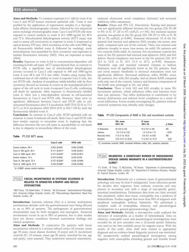
Aims and Methods: To compare responses to C difficile toxin A byCaco-2 and HT29 human intestinal epithelial cells. Toxin A waspurified by the application of supernatant samples to a thyroglo-bulin affinity chromatography column, followed by two sequentialanion exchange chromatographic steps. Caco-2 and HT29 cells wereexposed to control medium or toxin A (0.1–1000 ng/ml) for 48 hand 72 h. Mitochondrial dehydrogenase activity (MTT assay) wasused to assess cell cytotoxicity in response to toxin A (expressed asoptical density 570 nm). After incubation of the cells with 5000 ng/ml fluorescently labelled toxin A (followed by washing), toxininternalisation was quantified by flow cytometry and expressed asmedian fluorescence intensity (MFI). Data are expressed as mean(SEM).Results: Exposure to toxin A led to concentration-dependent cellrounding in both cell types. MTT assays showed that, in contrast toHT29 cells, a significant loss of mitochondrial dehydrogenaseactivity occurred in Caco-2 cells exposed to 100 and 1000 ng/mltoxin A over 48 h and 72 h (see table). Studies using trypan blueconfirmed loss of cell viability in toxin A-exposed Caco-2 cells, butnot HT29 cells. Analysis of propidium iodide-labelled cells by flowcytometry showed an increase in events in the hypodiploid/sub-G1region of the cell cycle in toxin A-exposed Caco-2 cells, confirmingcell death by apoptosis. After exposure to fluorescently labelledtoxin A, there was a time-dependent increase in cell-associatedfluorescence in Caco-2 and HT29 cells. However, there were nosignificant differences between Caco-2 and HT29 cells in cell-associated fluorescence after 5 h incubation (MFI 75.0 (21.0) vs 71.3(6.7)) or 24 h incubation (MFI 259.0 (6.9) vs 332.7 (30.4)) with thefluorescently labelled toxin A.Conclusion: In contrast to Caco-2 cells, HT29 epithelial cells areresistant to toxin A-induced cell death. Both Caco-2 and HT29 cellshave similar capacity to internalise toxin A, implying that thedifference between the two cell types in susceptibility to cell deathis due to disparity in intracellular effects of the toxin.
PT233 FAECAL INCONTINENCE IN SYSTEMIC SCLEROSIS ISRELATED TO SPHINCTER ATROPHY AND REFLEXDYSFUNCTION
1NM Thoua, 1M Abdel-Halim, 2C Denton, 1AV Emmanuel. 1Gastrointestinal PhysiologyUnit, University College Hospital, London, UK; 2Rheumatology Department, Royal FreeHospital, London, UK
Introduction: Systemic sclerosis (SSc) is a chronic multisystemautoimmune disorder with the gastrointestinal tract being affectedin up to 90% of patients. The pathogenesis of gastrointestinalabnormalities may be both myogenic and neurogenic. Anorectalinvolvement occurs in up to 50% of patients, but to date studieshave not shown correlation between manometric findings andclinical symptoms.Aims and Methods: 21 consecutive SSc patients with faecalincontinence referred to a tertiary referral centre (18 women, meanage 58 years, mean disease duration 13 years) and 21 incontinentcontrols (IC; 19 women, mean age 56 years), matched for sex, ageand parity, were assessed. They underwent anorectal manometry,
endoanal ultrasound, rectal compliance (barostat) and rectoanalinhibitory reflex assessment.Results: (Mean and 95% CI). Manometry: Resting anal pressurewas not significantly different between the two groups (SSc 52 (40to 63) vs IC 37 (28 to 47) cmH2O, p = NS), but maximal squeezepressure was greater in the SSc group (SSc 120 (97 to 143) vs IC 58(38 to 78) cmH2O, p,0.001). Endoanal ultrasound: 15 of the SScpatients had internal anal sphincter (IAS) atrophy (six severe, ninemild), compared with one of the controls. There was external analsphincter atrophy in seven (one severe, six mild) SSc patients andtwo controls (both mild). In contrast, five incontinent controls butnone of the SSc patients had external anal sphincter (EAS) and IASdefects. Rectal compliance was lower in the SSc patients (SSc 10.5(8.4 to 12.6) vs IC 14.3 (11.9 to 16.7), p,0.03). Sensation:Threshold, urge and maximal tolerated volumes to balloondistension were all significantly lower in the SSc patients. Analand rectal sensory thresholds to electrical stimulation were notsignificantly different. Rectoanal inhibitory reflex (RAIR): sevenSSc patients, five with IAS atrophy, had an absent RAIR comparedwith only one of the controls. Latency and duration components ofthe RAIR were prolonged in SSc (see table).Conclusion: There is both IAS and EAS atrophy in some SScincontinent patients, which influences reflex anal function morethan just pressures. This reflex dysfunction is contributed to bysensory dysfunction and reduced rectal compliance as a result ofrectal infiltration. Future studies investigating SSc patients withoutanorectal symptoms may identify early changes.
PT234 HELMINTHS: A SIGNIFICANT BURDEN OF UNDIAGNOSEDDISEASE AMONG MIGRANTS IN A GASTROENTEROLOGYCLINIC?
1PJ Smith, 1B Theis, 1S McCartney, 2M Brown. 1Department of Gastroenterology,University College Hospital, London, UK; 2Department of Infectious Diseases, Hospitalfor Tropical Diseases, London, UK
Introduction: Helminths are a common cause of gastrointestinalpathology and may be fatal if undiagnosed. Some infections persistfor decades after migration from endemic countries and maypresent to secondary care with a range of non-specific gastro-intestinal manifestations. Preliminary data from our clinic revealedthat peripheral, rather than tissue, eosinophilia was a predictor ofhelminthiasis. Studies suggest that more than 50% of migrants withperipheral eosinophilia harbour helminths. We performed acomplete audit of eosinophilia, ethnicity and parasitologicalinvestigations in a general gastroenterology clinic.Aims and Methods: Initially a retrospective audit explored therelevance of eosinophilia as a marker of helminthiasis. Data onethnicity, eosinophil count and parasitological investigations wererecorded for all new attenders over a 12-month period. Ethnicitywas used as the best available proxy for migrant status. With theresults of this audit, clinic staff were trained in appropriatediagnosis and an evidence-based diagnostic protocol was instituted.We prospectively audited parasitological investigation amongmigrants with eosinophilia attending general and irritable bowel
Table PT-232 MTT assay
Caco-2 cells HT29 cells
Control medium, 48 h 0.353 (0.044) 0.454 (0.036)
Toxin A 100 ng/ml, 48 h 0.177 (0.041)* 0.429 (0.051)
Toxin A 1000 ng/ml, 48 h 0.127 (0.033)* 0.429 (0.051)
Control medium, 72 h 0.372 (0.044) 0.443 (0.53)
Toxin A, 100 ng/ml, 72 h 0.127 (0.030)* 0.435 (0.049)
Toxin A, 1000 ng/ml, 72 h 0.102 (0.028)* 0.438 (0.050)
*p,0.0001 versus control medium.
Table PT-233 Components of RAIR in SSc and incontinent controls
RAIRSSc mean(95% CI)
IC mean(95% CI) p Value
% Relaxation 42 (33 to 52) 47.2 (37 to 58) NS
Latency 2.4 (1.5 to 3.2) 1.4 (1.0 to 1.7) ,0.03
Recovery time 11.6 (7 to 16.2) 6.5 (4.1 to 8.8) NS
Duration 19.2 (13.7 to 24.6) 11.2 (8.7 to 13.7) 0.02
IC, incontinent controls; RAIR, rectoanal inhibitory reflex; SSc, systemic sclerosis.
BSG abstracts
A90 Gut 2009;58(Suppl I):A1–A156
on July 20, 2022 by guest. Protected by copyright.
http://gut.bmj.com
/G
ut: first published as on 23 March 2009. D
ownloaded from
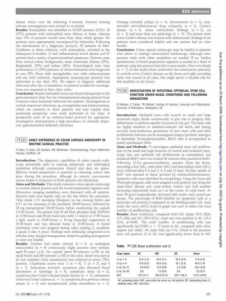
disease clinics over the following 4 months. Patients missingrelevant investigations were invited to re-attend.Results: Eosinophilia was identified in 34/426 patients (8.8%). 10(37%) patients with eosinophilia were African or Asian, whereasonly 19% of patients overall were from these ethnic groups. Nopatients were appropriately investigated for helminths. Followingthe introduction of a diagnostic protocol, 38 patients of Afro-Caribbean or Asian ethnicity, with eosinophilia, attended in thesubsequent 4 months. 11 had inflammatory bowel disease and 27patients had a variety of gastrointestinal symptoms. Patients camefrom various ethnic backgrounds, most commonly African (29%),Bangladeshi (29%) and Indian (18%). Parasitological tests wereperformed in 11 (29%) patients, in whom helminths were diagnosedin over 50% (three with strongyloides, two with schistosomiasisand one with trichuris). Appropriate screening per protocol wasperformed in less than 10%. We expect to diagnose additionalinfections after the re-attendance of patients recalled for investiga-tions not requested at their clinic visits.Conclusion: Raised eosinophil counts are found infrequently in thegastrointestinal clinic but are overrepresented among patients fromcountries where helminth infections are endemic. Investigations toexclude important infections, eg, strongyloides and schistosomiasis,which are common in these patients and may explain theirpresenting symptoms, were rarely performed in our clinic. Aprospective audit of an evidence-based protocol for appropriateinvestigation demonstrated a high prevalence of clinically impor-tant gastrointestinal helminth infections.
PT235 EARLY EXPERIENCE OF COLON CAPSULE ENDOSCOPY INROUTINE CLINICAL PRACTICE
R Sidhu, K Drew, DS Sanders, ME McAlindon. Gastroenterology, Royal HallamshireHospital, Sheffield, UK
Introduction: The diagnostic capabilities of colon capsule endo-scopy potentially adds to existing endoscopic and radiologicalmodalities although comparative clinical trial data are sparse.Effective bowel preparation is essential as cleansing cannot takeplace during the procedure, although its remote, non-invasivenature makes it attractive to both patients and physicians.Aims and Methods: This study evaluates colon capsule endoscopyin routine clinical practice and the bowel preparation regimen used.Alternative imaging modalities were discussed with all patients.Patients took clear liquids only on the day before the procedure.They drank 1.5 l moviprep (Norgine) on the evening before and0.5 l on the morning of the procedure (09:00 hours), followed by20 mg domperidone (10:45 hours) before swallowing the capsule(11:00 hours). The patient had 45 ml Fleet phospho-soda (DeWitt)at 13:00 hours and 30 ml (each time with 1 l water) at 17:00 hours,a light snack at 18:00 hours, a 10 mg bisacodyl suppository at19:30 hours and was allowed home at 21:00 hours. A bowelcleanliness score was assigned during video reading (1, excellent;2, good; 3, fair; 4, poor). Findings were arbitrarily categorised as towhether they changed management, helped in guiding managementor were not helpful.Results: Patients had either refused (n = 3) or undergoneunsuccessful (n = 6) colonoscopy. Eight patients were women,aged 43 years (¡6), the capsule spent 90 minutes (¡30) in thesmall bowel and 248 minutes (¡62) in the colon. Ileum was seen inall and complete colon examination was achieved in seven (78%)patients. Cleanliness scores were 2 (n = 6), 3 (n = 2) and 4(n = 1). Indications included: symptoms plus abnormal bloodparameters or histology (n = 3); symptoms alone (n = 2);symptoms plus Crohn’s disease family history (n = 1); assessmentof known Crohn’s disease (n = 1); symptoms plus previous colonicpolyps (n = 1) and asymptomatic polyp surveillance (n = 1).
Findings included: polyps (n = 3); diverticulosis (n = 3); non-steroidal anti-inflammatory drug colopathy (n = 1); Crohn’sdisease (n = 1); minor extracolonic findings (n = 2); no(n = 2) and more than one pathology (n = 3). The patient withactive Crohn’s disease was treated with adalimumab; findings in sixpatients were considered helpful and one patient had no clearbenefit.Conclusion: Colon capsule endoscopy may be helpful in patientswho refuse or undergo unsuccessful colonoscopy, although com-parative trials with other modalities are needed. If so, furtheroptimisation of bowel preparation regimens is needed as a third ofpatients using this protocol had fair or poor results. Over two-thirds(n = 7) of this small cohort underwent the examination primarilyto exclude active Crohn’s disease; as the ileum and right ascendingcolon was viewed in all cases, this might prove a valuable role forthis modality in the future.
PT236 INVESTIGATION OF INTESTINAL EPITHELIAL STEM CELLFUNCTION UNDER BASAL CONDITIONS AND FOLLOWINGIRRADIATION
R Gandara, C Potten, YR Mahida. Institute of Infection, Immunity and Inflammation,University of Nottingham, Nottingham, UK
Introduction: Epithelial stem cells located in small and largeintestinal crypts divide continuously to give rise to progeny thatdifferentiate to perform specific functions in the gut. The stem cellsare highly sensitive to radiation-induced cell death and duringrecovery post-irradiation generation of new stem cells and theirproliferative function can be investigated using a synthetic analogueof thymidine (bromodeoxyuridine; BrdU) that is incorporated innewly synthesised DNA.Aims and Methods: To investigate epithelial stem cell prolifera-tion in the small and large intestine of control and irradiated mice.Crypt size and epithelial cell proliferation in unirradiated andirradiated BDF1 mice was studied 40 minutes after parenteral BrdU.Following 12 Gy gamma-irradiation, samples (from the ileum,ascending colon (AC), mid-colon (MC) and descending colon (DC))were collected after 6 h and 2, 4, 8 and 12 days. Nuclear uptake ofBrdU was assessed in tissue sections by immunohistochemistry.Apoptotic cells were identified by morphology. Using the Score andWincrypts program, cells were assigned a cell position (cp) along thecrypt-villus (ileum) and crypt-surface (colon) axis (cell numberincreasing sequentially from cp 1 at the centre of crypt base). Atleast 50 good longitudinally sectioned half crypts were scored permouse. The percentage of BrdU-labelled (or apoptotic) cells at aparticular cell position is expressed as the labelling index (LI). Areaunder the curve (AUC) from LI graph was used to reflect the totalnumber of proliferating cells.Results: Basal conditions: compared with MC (mean 23.9 (SEM4.7) cells) and DC (26.9 (8.2)), crypt size was smallest in AC (13.4(2.4); p,0.04). The total number of proliferating cells wassignificantly (p,0.003; n = 7) lower in AC, compared with otherregions (see table). At crypt base (cp 1–3), which is the putativestem cell position, LI in AC was significantly lower than in MC
Table PT-236 Basal proliferation and LI
Crypt region AC MC DC Ileum
LI cp 1–3 15.0 (1.2) 25.2 (4.1) 26.9 (3.3) 17.5 (3.0)
LI cp 4–9 13.6 (2.2) 25.8 (3.4) 22.1 (2.3) 44.8 (1.6)
LI cp 10–15 0.6 (0.2) 10.3 (0.9) 3.7 (0.8) 29.2 (2.9)
LI cp 16–20 0.0 1.5 (0.4) 0.0 3.0 (0.7)
AUC 130 (15.1) 300.9 (27.4) 245.7 (26.7) 514.3 (30.9)
AC, ascending colon; AUC, area under the curve; cp, cell position; DC, descending colon; LI,labelling index; MC, mid-colon.
BSG abstracts
Gut 2009;58(Suppl I):A1–A156 A91
on July 20, 2022 by guest. Protected by copyright.
http://gut.bmj.com
/G
ut: first published as on 23 March 2009. D
ownloaded from
(p = 0.009) and DC (p = 0.041). After irradiation, apoptosis wasmost prominent in cp 1–6 in the ileum and AC and in cp 1 in MCand DC. In all regions post-irradiation, total epithelial proliferationwas 1.5–4-fold higher than controls at days 4–12, when an increasein crypt size was also prominent.Conclusion: Basal cell proliferation is significantly lower in AC,compared with other regions of the colon. Distribution profiles ofproliferating cells and apoptotic cells (post-irradiation) in AC showsome similarities to those in the ileum. Post-irradiation, thedynamic pattern of cell proliferation and change in crypt size issimilar in all regions.
PT237 HOW ALERT ARE WE IN DETECTING MICROSCOPIC COLITIS?
SK Butt, H Defoe, N van Someren, K Besherdas. Gastroenterology, Chase FarmHospital, Middlesex, UK
Introduction: Microscopic colitis is diagnosed when a patient withchronic watery non-bloody diarrhoea has an endoscopically orradiographically normal colon but colonic biopsies show uniqueinflammatory changes. As the mucosa is not ulcerated or otherwisedisrupted, the diarrhoea generally does not contain blood or pus.The true incidence is not known, although the disease has beenincreasingly diagnosed over the past 20 years. The incidence ofmicroscopic colitis has increased significantly from 1.1 per 100 000persons in the late 1980s to 19.6 per 100 000 persons by the end of2001. The key in detecting microscopic colitis is obtaining biopsiesfrom a macroscopically normal colon. It is recommended thatbiopsies should be taken from the rectosigmoid and from the rightside of the colon. But how good are we at detecting microscopiccolitis in clinical practice?
Aims and Methods: To assess the frequency of colonic biopsyobtained in patients with non-bloody diarrhoea who have a normalmacroscopic appearance at colonoscopy. This was a single centre,retrospective analysis of patients with diarrhoea attending for lowergastrointestinal endoscopy. Patient details were obtained from theUnisoft endoscopy database. Patients who had a normal colono-scopy were assessed to see if colonic biopsies were obtained.Results: A total of 398 colonoscopies was undertaken for theindication of chronic non-bloody diarrhoea between October 2005and October 2008 by both medical and surgical endoscopists. 208(52%) colonoscopies were deemed to be normal macroscopically. Ofthese normal colonoscopies, 137 (66%) were biopsied by theendoscopist for further investigation, 130 (95%) were biopsied bya gastroenterologist, the remainder being done by a surgeon. A totalof 71 (34%) patients was not biopsied, of which 32 (45%) wereendoscopies completed by surgeons and 39 (55%) were completedby physicians.Conclusion: This study demonstrates that in a third of patientswith diarrhoea (who have a macroscopically normal colon) biopsiesare not obtained to look for microscopic colitis. When biopsies areobtained, it tends to be the physician endoscopist rather than thesurgical endoscopist who performs them. The diagnosis of micro-scopic colitis may be more prevalent in clinical practice and shouldbe investigated. All clinicians should have a higher index ofsuspicion for this and therefore colonic biopsies should be obtainedin all patients with diarrhoea who have a macroscopically normalcolonoscopy.
PT238 HOW ACCURATE IS AN ENDOSCOPIST’S HUNCH FORCANCER? CORRELATION WITH HISTOLOGY
1SK Butt, 1K Jaggs, 2S Warren, 2D Francis, 2M Ward, 2M Klein, 1N van Someren, 1KBesherdas. 1Gastroenterology, Chase Farm Hospital, Middlesex, UK; 2Surgery, ChaseFarm Hospital, Middlesex, UK
Introduction: The cancer plan in the UK states that thegovernment believes the ultimate goal in the treatment of cancer
should be to ‘‘offer patients a maximum one month wait from anurgent referral for suspected cancer to the beginning of treatment’’.When patients are required to wait longer, this should be because ofthe needs of the diagnostic process or their personal choice and notbecause of any inbuilt delay in the system of care. Any reduction inthe time taken between obtaining a diagnosis and staging ofcolorectal cancer is helpful. One such step is the interval betweenendoscopy and histological confirmation before arranging furtherimaging (ie, staging computed tomography (CT)/magnetic reso-nance imaging (MRI)). If endoscopic diagnosis has a strongcorrelation with histology in the diagnosis of cancer then arrangingCT/MRI scans in the staging process in cancer management couldnarrow the time from diagnosis to treatment.Aims and Methods: To assess the accuracy of the endoscopist’simpression of a malignant lesion in the colon and rectum withhistology. A retrospective single centre study looking atconsecutive patients from July 2005 to April 2008 with anendoscopic report of ‘‘colorectal cancer’’ was undertaken.Patients with a potential cancer diagnosis at endoscopy wereidentified from the Unisoft endoscopy reporting software. Thehistology of these patients was obtained from the ‘‘Powerchart’’hospital database of histology and from the colorectal cancermultidisciplinary meeting records. The percentage of patientswith histologically confirmed cancer in comparison with endo-scopic findings was evaluated.Results: Records show that 166 patients were diagnosed with amalignant-looking colonic lesion at the time of colonoscopy. In 141of these, histology was obtained. Following from this, 131 (93%)histologies confirmed malignancy (130 with adenocarcinoma of thecolon, one with metastatic carcinoma). In the 10 patients withmalignant-looking lesions but negative histology, three had severedysplasia, one had moderate dysplasia, one had a normal biopsywith liver metastases, four had inflammatory polyps and one wasnormal.Conclusion: The majority (93%) of patients thought to have amalignant lesion at endoscopy was confirmed to be so histologi-cally. Arrangements to stage colorectal lesions thought to bemalignant at endoscopy should therefore be made followingendoscopy rather than waiting until histology is available. Thiswould save valuable time in the diagnosis to treatment pathway inpatients suspected of having colorectal cancer.
PT239 CONSTIPATION AND EXPOSURE TO STRESSFUL LIFE EVENTSIN 10–16 YEAR OLDS: A SRI LANKAN EXPERIENCE
1S Rajindrajith, 2NM Devanarayana. 1Paediatrics, Faculty of Medicine, University ofKelaniya, Ragama, Sri Lanka; 2Physiology, Faculty of Medicine, University of Kelaniya,Ragama, Sri Lanka
Introduction: Constipation is a common paediatric problem, butlittle is known regarding its aetiology. The majority have functionalconstipation. Emotional stress is considered to be associated withfunctional gastrointestinal diseases including constipation.Aims and Methods: The aim of this study was to assess theassociation between constipation and exposure to stressful lifeevents in Sri Lankan school children and adolescents. A validated,self-administered questionnaire was distributed to randomlyselected children, aged 10–16 years, in five randomly selectedschools, in three geographically and socioeconomically differentprovinces in Sri Lanka. Constipation was defined using Rome IIIcriteria.Results: A total of 2770 questionnaires was distributed and 2699(97.4%) were included in the analysis (1368 (50.6%) males, meanage 13.17 years, SD 1.72 years). According to Rome III criteria 416(15.4%) had constipation and they were compared with 2278children without defaecation disorders (eg, constipation, diarrhoea,functional faecal retention, non-retentive faecal soiling, etc). The
BSG abstracts
A92 Gut 2009;58(Suppl I):A1–A156
on July 20, 2022 by guest. Protected by copyright.
http://gut.bmj.com
/G
ut: first published as on 23 March 2009. D
ownloaded from
prevalence of constipation was higher in those who were exposed toat least one stressful life event during the previous 3 months (oddsratio (OR) 0.38, p,0.0001). During univariate analysis the stressfullife events associated with constipation were change in school oraddress, corporal punishment in school, separation from their bestfriend, preparation for government exams, exam failure, beingbullied at school, sibling birth, severe illness or death in a familymember, loss of job by a parent, separation or divorce of parents,parent remarriage to a step-parent, hospitalisation of the childhimself/herself for other illness, frequent punishment by parents,father’s alcoholism, domestic fights and living in an area affected byongoing separatist war (p,0.005). During multiple logistic regres-sion analysis, separation from the best friend (adjusted OR 0.737,p = 0.016), severe illness in a family member (adjusted OR 0.588,p = 0.001), loss of job by a parent (adjusted OR 0.55, p = 0.017),frequent punishment by parents (adjusted OR 0.581, p = 0.005) andliving in a war-affected area (adjusted OR 0.666, p = 0.0001)remained to be significantly associated with constipation.Conclusion: Childhood constipation was significantly higher inthose exposed to stressful life events. Modulation of gut motilitythrough the brain–gut axis probably delays colonic transit causingconstipation.
PT240 MAINTENANCE ANTIBIOTIC THERAPY FOR CHRONICPOUCHITIS, COMPLICATIONS AND CLINICAL OUTCOME
1SD McLaughlin, 2SK Clark, 3S Shafi, 4PP Tekkis, 5PJ Ciclitira, 4R Nicholls.1Gastroenterology, St Mark’s Hospital, London, UK; 2Surgery, St Mark’s Hospital,London, UK; 3Microbiology, St Mark’s Hospital, London, UK; 4Biosurgery and SurgicalTechnology, Imperial College, London, UK; 5Nutritional Sciences, King’s College,London, UK
Introduction: Approximately 5% of restorative proctocolectomypatients have chronic pouchitis. British Society of Gastroenterologyguidelines recommend maintenance therapy with antibiotics or theprobiotic VSL#3.1 Many patients do not achieve endoscopicremission after antibiotics. VSL#3 has been shown to be effectivein those who achieve clinical and endoscopic remission afterantibiotic treatment but further experience has been disappointing,with less than 20% of patients able to maintain remission.2
Therefore, many patients with chronic pouchitis require antibioticmaintenance therapy.Aims and Methods: We report our experience with maintenanceantibiotic treatment. Patients treated with antibiotic maintenancetherapy were identified from the hospital pouch database. Dataincluding stool testing, functional outcome, side effects andCleveland global quality of life score (CGQoL) were analysed.Results: 24 patients (16 (67%) men) treated for a mean of15.7 months were identified. Seven (29%) had previously failedtreatment with VSL#3 and 11 (46%) did not enter endoscopicremission and therefore were ineligible for this treatment. Atfollow-up the mean 24-h stool frequency was seven (range 4–11),the mean clinical pouchitis disease activity index was 0 (range 0–1)and CQGoL score was 0.7 (range 0.5–1.0). Side effects includedvaginal thrush (n = 1) and occasional nausea (n = 1). All patientsreported improved quality of life after starting maintenancetreatment. Six (25%) patients developed antibiotic resistance duringfollow-up but treatment with rotating antibiotics was successful inall cases. No patient developed Clostridium difficile infection.Conclusion: Antibiotic maintenance therapy is safe, well toleratedand efficacious even in those who fail or are eligible for VSL#3. Itresults in improved quality of life and function. Antibioticresistance can be managed with rotating antibiotics.
1. Carter MJ, Lobo AJ, Travis SP. Guidelines for the management of inflammatorybowel disease in adults. Gut 2004;53(Suppl 5):V1–16.
2. Shen B, Brzezinski A, Fazio VW, et al. Maintenance therapy with a probiotic inantibiotic-dependent pouchitis: experience in clinical practice. Aliment PharmacolTher 2005;22:721–8.
PT241 QUALITY OF LIFE AND FUNCTIONAL OUTCOME IN PATIENTSUSING A MEDINA CATHETER FOLLOWING RESTORATIVEPROCTOCOLECTOMY
1SD McLaughlin, 2SK Clark, 2ZL Perry-Woodford, 3PP Tekkis, 4PJ Ciclitira, 3R Nicholls.1Gastroenterology, St Mark’s Hospital, London, UK; 2Surgery, St Mark’s Hospital,London, UK; 3Biosurgery and Surgical Technology, Imperial College, London, UK;4Nutritional Sciences, King’s College, London, UK
Introduction: Intubation of the pouch is required by some patientswith failure of spontaneous defaecation after restorative proctoco-lectomy (RPC). We assessed function, social, work and dietaryrestrictions, and quality of life (QoL) in patients who were using aMedina catheter to evacuate. The Cleveland global quality of lifescore (CGQoL) has been reported to be 0.8 in the global RPCpopulation.1
Aims and Methods: 31 RPC patients prescribed a Medina catheterwere identified from the pouch database and were sent aquestionnaire by post. CGQoL and data on function, social, workand dietary restrictions were recorded.Results: 23 (74%) of 31 patients (median age 56 years; 15 men(68%)) returned the questionnaire. Pouch configurations were: J:8,W:10, S:5. The median duration of catheter usage was 9 (0.5–30)years. 22 (71%) patients reported improved QoL after startingregular intubation. Median 24-h bowel frequency was 4 (range 2–14). 16 (48%) patients used the catheter for every defaecation, eight(26%) experienced social or work-life restriction, seven (22%)reported catheter blockage and 12 (52%) reported dietary restric-tion. The median CGQoL score was 0.72.Conclusion: Medina catheter usage is tolerated in the long termand is associated with satisfactory quality of life of RPC patientswith outflow obstruction. Frequency of defaecation and CGQoLscores are comparable with the global RPC population.
1. Fazio VW, O’Riordain MG, Lavery IC, et al. Long-term functional outcome andquality of life after stapled restorative proctocolectomy. Ann Surg 1999;230:575–84.
PT242 HAS THE INTRODUCTION OF THE BOWEL CANCERSCREENING PROGRAMME LED TO AN EARLY CHANGE INTHE STAGE OF COLORECTAL CANCER AT DIAGNOSIS?
1TJW Lee, 2C Zwart, 2C Rees, 1M Rutter. 1Gastroenterology, University Hospital ofNorth Tees, Stockton upon Tees, UK; 2Gastroenterology, South Tyneside GeneralHospital, South Tyneside, UK
Introduction: Colorectal cancer usually presents once the patientis symptomatic, as a result it tends to present at an advanced stage(Duke’s stage at diagnosis: A or B 46%, C or D 54%).1 Pilot studiesof the bowel cancer screening programme (BCSP) showed areduction in the stage of cancer at diagnosis in the screeningpopulation (Duke’s stage at diagnosis: A or B 71.8%, C or D 28.2%).2
The BCSP was rolled out in our region in February 2007.Aims and Methods: This abstract examines whether theintroduction of the BCSP has had an immediate impact on thestage of colorectal cancer at diagnosis in screening and non-screening populations. Data relating to BCSP patients wereobtained from the Tees BCSP database. Data on non-BCSP patientswas obtained from local cancer network databases. Two one-yearperiods were examined: April 2006 to March 2007 and April 2007 toMarch 2008, the second period being after the introduction of theBCSP across the region.Results: The table shows the number of cancers and theircharacteristics for each time period.Conclusion: A higher proportion of Duke’s A or B cancers arediagnosed in the BCSP population. Our results are in keeping withpreviously published data from a bowel cancer screening pilotstudy.2 Seven of the 22 cancers diagnosed in the BCSP group werepolyp cancers. These have been included in the Duke’s A group. The
BSG abstracts
Gut 2009;58(Suppl I):A1–A156 A93
on July 20, 2022 by guest. Protected by copyright.
http://gut.bmj.com
/G
ut: first published as on 23 March 2009. D
ownloaded from
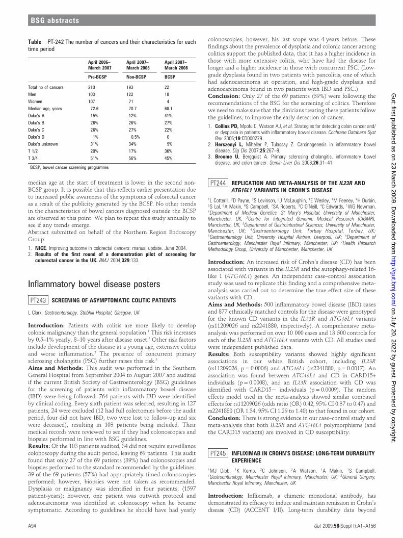
median age at the start of treatment is lower in the second non-BCSP group. It is possible that this reflects earlier presentation dueto increased public awareness of the symptoms of colorectal canceras a result of the publicity generated by the BCSP. No other trendsin the characteristics of bowel cancers diagnosed outside the BCSPare observed at this point. We plan to repeat this study annually tosee if any trends emerge.Abstract submitted on behalf of the Northern Region EndoscopyGroup.
1. NICE. Improving outcome in colorectal cancers: manual update. June 2004.2. Results of the first round of a demonstration pilot of screening for
colorectal cancer in the UK. BMJ 2004;329:133.
Inflammatory bowel disease posters
PT243 SCREENING OF ASYMPTOMATIC COLITIC PATIENTS
L Clark. Gastroenterology, Stobhill Hospital, Glasgow, UK
Introduction: Patients with colitis are more likely to developcolonic malignancy than the general population.1 This risk increasesby 0.5–1% yearly, 8–10 years after disease onset.2 Other risk factorsinclude development of the disease at a young age, extensive colitisand worse inflammation.2 The presence of concurrent primarysclerosing cholangitis (PSC) further raises this risk.3
Aims and Methods: This audit was performed in the SouthernGeneral Hospital from September 2004 to August 2007 and auditedif the current British Society of Gastroenterology (BSG) guidelinesfor the screening of patients with inflammatory bowel disease(IBD) were being followed. 764 patients with IBD were identifiedby clinical coding. Every sixth patient was selected, resulting in 127patients, 24 were excluded (12 had full colectomies before the auditperiod, four did not have IBD, two were lost to follow-up and sixwere deceased), resulting in 103 patients being included. Theirmedical records were reviewed to see if they had colonoscopies andbiopsies performed in line with BSG guidelines.Results: Of the 103 patients audited, 34 did not require surveillancecolonoscopy during the audit period, leaving 69 patients. This auditfound that only 27 of the 69 patients (39%) had colonoscopies andbiopsies performed to the standard recommended by the guidelines.39 of the 69 patients (57%) had appropriately timed colonoscopiesperformed; however, biopsies were not taken as recommended.Dysplasia or malignancy was identified in four patients, (1597patient-years); however, one patient was outwith protocol andadenocarcinoma was identified at colonoscopy when he becamesymptomatic. According to guidelines he should have had yearly
colonoscopies; however, his last scope was 4 years before. Thesefindings about the prevalence of dysplasia and colonic cancer amongcolitics support the published data, that it has a higher incidence inthose with more extensive colitis, who have had the disease forlonger and a higher incidence in those with concurrent PSC. (Low-grade dysplasia found in two patients with pancolitis, one of whichhad adenocarcinoma at operation, and high-grade dysplasia andadenocarcinoma found in two patients with IBD and PSC.)Conclusion: Only 27 of the 69 patients (39%) were following therecommendations of the BSG for the screening of colitics. Thereforewe need to make sure that the clinicians treating these patients followthe guidelines, to improve the early detection of cancer.
1. Collins PD, Mpofu C, Watson AJ, et al. Strategies for detecting colon cancer and/or dysplasia in patients with inflammatory bowel disease. Cochrane Database SystRev 2006;19:CD000279.
2. Herszenyi L, Miheller P, Tulassay Z. Carcinogenesis in inflammatory boweldisease. Dig Dis 2007;25:267–9.
3. Broome U, Bergquist A. Primary sclerosing cholangitis, inflammatory boweldisease, and colon cancer. Semin Liver Dis 2006;26:31–41.
PT244 REPLICATION AND META-ANALYSIS OF THE IL23R ANDATG16L1 VARIANTS IN CROHN’S DISEASE
1L Cotterill, 2D Payne, 3S Levinson, 3J McLaughlin, 4E Wesley, 4M Feeney, 4H Durbin,5S Lal, 6A Makin, 6S Campbell, 7SA Roberts, 3C O’Neill, 4C Edwards, 1WG Newman.1Department of Medical Genetics, St Mary’s Hospital, University of Manchester,Manchester, UK; 2Centre for Integrated Genomic Medical Research (CIGMR),Manchester, UK; 3Department of Gastrointestinal Sciences, University of Manchester,Manchester, UK; 4 Gastroenterology Unit, Torbay Hospital, Torbay, UK;5Gastroenterology Unit, University Hospital Aintree, Liverpool, UK; 6Department ofGastroenterology, Manchester Royal Infirmary, Manchester, UK; 7Health ResearchMethodology Group, University of Manchester, Manchester, UK
Introduction: An increased risk of Crohn’s disease (CD) has beenassociated with variants in the IL23R and the autophagy-related 16-like 1 (ATG16L1) genes. An independent case–control associationstudy was used to replicate this finding and a comprehensive meta-analysis was carried out to determine the true effect size of thesevariants with CD.Aims and Methods: 500 inflammatory bowel disease (IBD) casesand 877 ethnically matched controls for the disease were genotypedfor the known CD variants in the IL23R and ATG16L1 variants(rs11209026 and rs2241880, respectively). A comprehensive meta-analysis was performed on over 10 000 cases and 13 500 controls foreach of the IL23R and ATG16L1 variants with CD. All studies usedwere independent published data.Results: Both susceptibility variants showed highly significantassociations in our white British cohort, including IL23R(rs11209026, p = 0.0006) and ATG16L1 (rs2241880, p = 0.0017). Anassociation was found between ATG16L1 and CD in CARD15+individuals (p = 0.0008), and an IL23R association with CD wasidentified with CARD152 individuals (p = 0.0009). The randomeffects model used in the meta-analysis showed similar combinedeffects for rs11209026 (odds ratio (OR) 0.42, 95% CI 0.37 to 0.47) andrs2241880 (OR 1.34, 95% CI 1.29 to 1.40) to that found in our cohort.Conclusion: There is strong evidence in our case–control study andmeta-analysis that both IL23R and ATG16L1 polymorphisms (andthe CARD15 variants) are involved in CD susceptibility.
PT245 INFLIXIMAB IN CROHN’S DISEASE: LONG-TERM DURABILITYEXPERIENCE
1MJ Dibb, 1K Kemp, 2C Johnson, 2A Watson, 1A Makin, 1S Campbell.1Gastroenterology, Manchester Royal Infirmary, Manchester, UK; 2General Surgery,Manchester Royal Infirmary, Manchester, UK
Introduction: Infliximab, a chimeric monoclonal antibody, hasdemonstrated its efficacy to induce and maintain remission in Crohn’sdisease (CD) (ACCENT I/II). Long-term durability data beyond
Table PT-242 The number of cancers and their characteristics for eachtime period
April 2006–March 2007
April 2007–March 2008
April 2007–March 2008
Pre-BCSP Non-BCSP BCSP
Total no of cancers 210 193 22
Men 103 122 18
Women 107 71 4
Median age, years 72.6 70.7 68.1
Duke’s A 15% 12% 41%
Duke’s B 26% 26% 27%
Duke’s C 26% 27% 22%
Duke’s D 1% 0.5% 0
Duke’s unknown 31% 34% 9%
T 1/2 20% 17% 36%
T 3/4 51% 56% 45%
BCSP, bowel cancer screening programme.
BSG abstracts
A94 Gut 2009;58(Suppl I):A1–A156
on July 20, 2022 by guest. Protected by copyright.
http://gut.bmj.com
/G
ut: first published as on 23 March 2009. D
ownloaded from
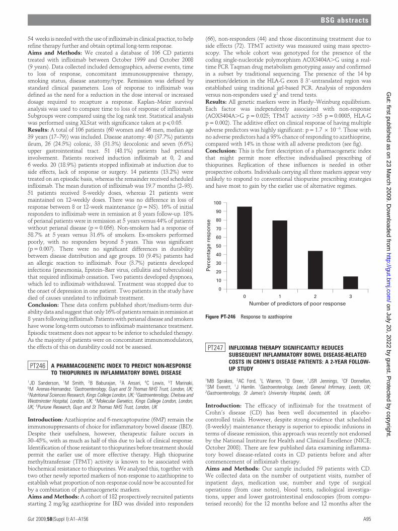
54 weeks is needed with the use of infliximab in clinical practice, to helprefine therapy further and obtain optimal long-term response.Aims and Methods: We created a database of 106 CD patientstreated with infliximab between October 1999 and October 2008(9 years). Data collected included demographics, adverse events, timeto loss of response, concomitant immunosuppressive therapy,smoking status, disease anatomy/type. Remission was defined bystandard clinical parameters. Loss of response to infliximab wasdefined as the need for a reduction in the dose interval or increaseddosage required to recapture a response. Kaplan–Meier survivalanalysis was used to compare time to loss of response of infliximab.Subgroups were compared using the log rank test. Statistical analysiswas performed using XLStat with significance taken at p(0.05.Results: A total of 106 patients (60 women and 46 men, median age39 years (17–79)) was included. Disease anatomy: 40 (37.7%) patientsileum, 26 (24.5%) colonic, 33 (31.3%) ileocolonic and seven (6.6%)upper gastrointestinal tract. 51 (48.1%) patients had perianalinvolvement. Patients received induction infliximab at 0, 2 and6 weeks. 20 (18.9%) patients stopped infliximab at induction due toside effects, lack of response or surgery. 14 patients (13.2%) weretreated on an episodic basis, whereas the remainder received scheduledinfliximab. The mean duration of infliximab was 19.7 months (2–93).51 patients received 8-weekly doses, whereas 21 patients weremaintained on 12-weekly doses. There was no difference in loss ofresponse between 8 or 12-week maintenance (p = NS). 16% of initialresponders to infliximab were in remission at 8 years follow-up. 18%of perianal patients were in remission at 5 years versus 44% of patientswithout perianal disease (p = 0.056). Non-smokers had a response of58.7% at 5 years versus 31.6% of smokers. Ex-smokers performedpoorly, with no responders beyond 5 years. This was significant(p = 0.007). There were no significant differences in durabilitybetween disease distribution and age groups. 10 (9.4%) patients hadan allergic reaction to infliximab. Four (3.7%) patients developedinfections (pneumonia, Epstein–Barr virus, cellulitis and tuberculosis)that required infliximab cessation. Two patients developed dyspnoea,which led to infliximab withdrawal. Treatment was stopped due tothe onset of depression in one patient. Two patients in the study havedied of causes unrelated to infliximab treatment.Conclusion: These data confirm published short/medium-term dur-ability data and suggest that only 16% of patients remain in remission at8 years following infliximab. Patients with perianal disease and smokershave worse long-term outcomes to infliximab maintenance treatment.Episodic treatment does not appear to be inferior to scheduled therapy.As the majority of patients were on concomitant immunomodulators,the effects of this on durability could not be assessed.
PT246 A PHARMACOGENETIC INDEX TO PREDICT NON-RESPONSETO THIOPURINES IN INFLAMMATORY BOWEL DISEASE
1JD Sanderson, 1M Smith, 2B Baburajan, 3A Ansari, 4C Lewis, 5T Marinaki,5M Arenas-Hernandez. 1Gastroenterology, Guys and St Thomas NHS Trust, London, UK;2Nutritional Sciences Research, Kings College London, UK; 3Gastroenterology, Chelsea andWestminster Hospital, London, UK; 4Molecular Genetics, Kings College London, London,UK; 5Puriune Research, Guys and St Thomas NHS Trust, London, UK
Introduction: Azathioprine and 6-mercaptopurine (6MP) remain theimmunosuppressants of choice for inflammatory bowel disease (IBD).Despite their usefulness, however, therapeutic failure occurs in30–45%, with as much as half of this due to lack of clinical response.Identification of those resistant to thiopurines before treatment shouldpermit the earlier use of more effective therapy. High thiopurinemethyltransferase (TPMT) activity is known to be associated withbiochemical resistance to thiopurines. We analysed this, together withtwo other newly reported markers of non-response to azathioprine toestablish what proportion of non-response could now be accounted forby a combination of pharmacogenetic markers.Aims and Methods: A cohort of 182 prospectively recruited patientsstarting 2 mg/kg azathioprine for IBD was divided into responders
(66), non-responders (44) and those discontinuing treatment due toside effects (72). TPMT activity was measured using mass spectro-scopy. The whole cohort was genotyped for the presence of thecoding single-nucleotide polymorphism AOX3404A.G using a real-time PCR Taqman drug metabolism genotyping assay and confirmedin a subset by traditional sequencing. The presence of the 14 bpinsertion/deletion in the HLA-G exon 8 39-untranslated region wasestablished using traditional gel-based PCR. Analysis of respondersversus non-responders used x2 and trend tests.Results: All genetic markers were in Hardy–Weinburg equilibrium.Each factor was independently associated with non-response(AOX3404A.G p = 0.025; TPMT activity .35 p = 0.0005, HLA-Gp = 0.002). The additive effect on clinical response of having multipleadverse predictors was highly significant: p = 1.7 6 1026. Those withno adverse predictors had a 95% chance of responding to azathioprine,compared with 14% in those with all adverse predictors (see fig).Conclusion: This is the first description of a pharmacogenetic indexthat might permit more effective individualised prescribing ofthiopurines. Replication of these influences is needed in otherprospective cohorts. Individuals carrying all three markers appear veryunlikely to respond to conventional thiopurine prescribing strategiesand have most to gain by the earlier use of alternative regimes.
PT247 INFLIXIMAB THERAPY SIGNIFICANTLY REDUCESSUBSEQUENT INFLAMMATORY BOWEL DISEASE-RELATEDCOSTS IN CROHN’S DISEASE PATIENTS: A 2-YEAR FOLLOW-UP STUDY
1MB Sprakes, 2AC Ford, 1L Warren, 1D Greer, 1JSR Jennings, 1CF Donnellan,1SM Everett, 1J Hamlin. 1Gastroenterology, Leeds General Infirmary, Leeds, UK;2Gastroenterology, St James’s University Hospital, Leeds, UK
Introduction: The efficacy of infliximab for the treatment ofCrohn’s disease (CD) has been well documented in placebo-controlled trials. However, despite strong evidence that scheduled(8-weekly) maintenance therapy is superior to episodic infusions interms of disease remission, this approach was recently not endorsedby the National Institure for Health and Clinical Excellence (NICE;October 2008). There are few published data examining inflamma-tory bowel disease-related costs in CD patients before and aftercommencement of infliximab therapy.Aims and Methods: Our sample included 59 patients with CD.We collected data on the number of outpatient visits, number ofinpatient days, medication use, number and type of surgicaloperations (from case notes), blood tests, radiological investiga-tions, upper and lower gastrointestinal endoscopies (from compu-terised records) for the 12 months before and 12 months after the
Figure PT-246 Response to azathioprine
BSG abstracts
Gut 2009;58(Suppl I):A1–A156 A95
on July 20, 2022 by guest. Protected by copyright.
http://gut.bmj.com
/G
ut: first published as on 23 March 2009. D
ownloaded from
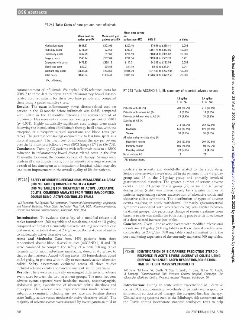
commencement of infliximab. We applied NHS reference costs for2006–7 to these data to derive a total inflammatory bowel disease-related cost per patient for these two time periods and comparedthese using a paired samples t test.Results: The mean inflammatory bowel disease-related cost perpatient in the 12 months before infliximab was £4848, comparedwith £1836 in the 12 months following the commencement ofinfliximab. This represents a mean cost saving per patient of £3011(p,0.001). Highly statistically significant cost savings were madefollowing the introduction of infliximab therapy in all areas, with theexception of subsequent surgical operations and blood tests (seetable). The greatest cost savings occurred due to less time spent as ahospital inpatient. The mean cost of infliximab therapy per patientover the 12 months of follow-up was £8962 (range £1748 to £36 708).Conclusion: Treating CD patients with infliximab leads to a £3000reduction in inflammatory bowel disease-related costs in the first12 months following the commencement of therapy. Savings weremade in all areas of patient care, but the majority of savings accrued asa result of less time spent as an inpatient in hospital, which may alsolead to an improvement in the overall quality of life for patients.
PT248 SAFETY OF MODIFIED-RELEASE ORAL MESALAZINE 4.8 G/DAY(800 MG TABLET) COMPARED WITH 2.4 G/DAY(400 MG TABLET) FOR TREATMENT OF ACTIVE ULCERATIVECOLITIS: COMBINED ANALYSIS FROM THREE RANDOMISED,DOUBLE-BLIND, ACTIVE-CONTROLLED TRIALS
1WJ Sandborn, 2M Spravka, 2M Hosterman. 1Division of Gastroenterology, Hepatologyand Internal Medicine, Mayo Clinic, Rochester, New York, USA; 2Pharmacovigilance,Procter and Gamble Pharmaceuticals, Cincinnati, Ohio, USA
Introduction: To evaluate the safety of a modified-release oraltablet formulation (800 mg tablet) of mesalazine dosed at 4.8 g/day,compared with that of a currently marketed 400 mg modified-releaseoral mesalazine tablet dosed at 2.4 g/day for the treatment of mildlyto moderately active ulcerative colitis.Aims and Methods: Data from 1459 patients from threerandomised, double-blind, 6-week studies (ASCEND I, II and III)were combined to compare the safety of a new 800 mg tabletformulation of modified-release mesalazine, dosed at 4.8 g/day, tothat of the marketed Asacol 400 mg tablet (US formulation), dosedat 2.4 g/day, in patients with mildly to moderately active ulcerativecolitis. Safety assessments evaluated across all three studiesincluded adverse events and baseline and exit serum creatinine.Results: There were no clinically meaningful differences in adverseevent rates between the two treatment groups. The most frequentadverse events reported were headache, nausea, nasopharyngitis,abdominal pain, exacerbation of ulcerative colitis, diarrhoea anddyspepsia. The adverse event experience was similar across thesubgroups examined, including age, sex, race and baseline diseasestate (mildly active versus moderately active ulcerative colitis). Themajority of adverse events were assessed by investigators as mild or
moderate in severity and doubtfully related to the study drug.Serious adverse events were reported in six patients in the 4.8 g/daygroup and 13 in the 2.4 g/day group and primarily involvedgastrointestinal disorders. The greater number of serious adverseevents in the 2.4 g/day dosing group (22) versus the 4.8 g/daydosing group (eight) was driven largely by a greater number ofpatients experiencing serious exacerbation of ulcerative colitis andulcerative colitis symptoms. The distribution of types of adverseevents resulting in study withdrawal (primarily gastrointestinalsymptoms associated with ulcerative colitis) was similar betweentreatment groups. The percentage change of serum creatinine frombaseline to exit was similar for both dosing groups with no evidenceof a dose-related increase (see table).Conclusion: Overall, the adverse events with modified-release oralmesalazine 4.8 g/day (800 mg tablet) in these clinical studies werecomparable to 2.4 g/day (400 mg tablet) and consistent with thepost-marketing experience of the currently marketed 400 mg tablet.
PT249 IDENTIFICATION OF BIOMARKERS PREDICTING STEROIDRESPONSE IN ACUTE SEVERE ULCERATIVE COLITIS USINGSURFACE-ENHANCED LASER DESORPTION/IONISATION–TIME OF FLIGHT MASS SPECTROMETRY
1NC Hare, 2M Imrie, 1AJ Smith, 1K Soo, 1L Smith, 2R Gray, 1G Ho, 1ID Arnott,1J Satsangi. 1Gastrointestinal Unit, Western General Hospital, Edinburgh, UK;2Molecular Medicine Centre, Western General Hospital, Edinburgh, UK
Introduction: During an acute severe exacerbation of ulcerativecolitis (UC), approximately two-thirds of patients will respond tointravenous corticosteroid therapy, the accepted first-line therapy.Clinical scoring systems such as the Edinburgh risk assessment andthe Travis criteria incorporate standard serological tests to help
PT-247 Table Costs of care pre and post-infliximab
Mean cost perpatient pre-IFX
Mean cost perpatient post-IFX
Mean cost savingperpatient post-IFX 95% CI p Value
Medication costs £681.37 £473.92 £207.46 £76.51 to £338.41 0.002
Radiology costs £311.39 £73.58 £237.81 £161.78 to £313.85 ,0.001
Endoscopy costs £347.29 £57.69 £289.59 £183.51 to £395.67 ,0.001
Surgery costs £345.24 £132.69 £212.54 £128.61 to £533.70 0.22
Outpatient visit costs £473.83 £356.12 £117.71 £42.83 to £192.59 0.003
Blood test costs £50.07 £38.88 £11.19 £0.43 to £21.94 0.04
Inpatient stay costs £2638.98 £703.59 £1935.39 £947.83 to £2922.95 ,0.001
Total costs £4848.34 £1836.47 £3011.86 £1786.14 to £4237.59 ,0.001
IFX, infliximab.
PT-248 Table ASCEND I, II, III: summary of reported adverse events
4.8 g/dayn = 727
2.4 g/dayn = 732
Patients with AE (%) 209 (28.7%) 211 (28.8%)
Patients with serious AE (%) 6 (0.8%) 13 (1.8%)
Patients withdrawn due to AE (%) 28 (3.9%) 31 (4.2%)
Severity of AE (%)
Mild 218 (55.3%) 257 (62.8%)
Moderate 146 (37.1%) 121 (29.6%)
Severe 30 (7.6%) 31 (7.6%)
AE relationship to study drug (%)
Doubtfully related 266 (67.5%) 297 (72.6%)
Possibly related 105 (26.6%) 93 (22.7%)
Probably related 23 (5.8%) 19 (4.6%)
No of serious AE 8 22
AE, adverse event.
BSG abstracts
A96 Gut 2009;58(Suppl I):A1–A156
on July 20, 2022 by guest. Protected by copyright.
http://gut.bmj.com
/G
ut: first published as on 23 March 2009. D
ownloaded from
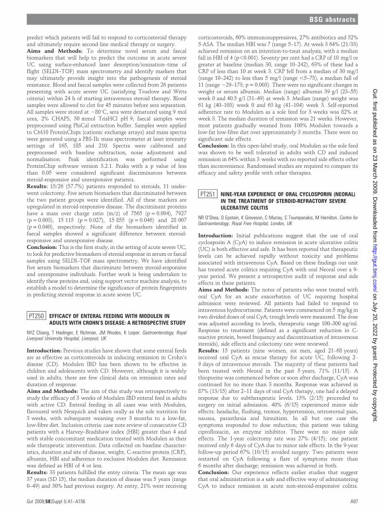
predict which patients will fail to respond to corticosteroid therapyand ultimately require second-line medical therapy or surgery.Aims and Methods: To determine novel serum and faecalbiomarkers that will help to predict the outcome in acute severeUC using surface-enhanced laser desorption/ionisation–time offlight (SELDI–TOF) mass spectrometry and identify markers thatmay ultimately provide insight into the pathogenesis of steroidresistance. Blood and faecal samples were collected from 26 patientspresenting with acute severe UC (satisfying Truelove and Wittscriteria) within 24 h of starting intravenous steroid therapy. Bloodsamples were allowed to clot for 45 minutes before sera separation.All samples were stored at 280uC, sera were denatured using 9 molurea, 2% CHAPS, 50 mmol TrisHCl pH 9; faecal samples werepreprocessed using PhiCal extraction buffer. Samples were appliedto CM10 ProteinChips (cationic exchange arrays) and mass spectrawere generated using a PBS-IIc mass spectrometer at laser intensitysettings of 165, 185 and 210. Spectra were calibrated andpreprocessed with baseline subtraction, noise adjustment andnormalisation. Peak identification was performed usingProteinChip software version 3.2.1. Peaks with a p value of lessthan 0.05 were considered significant discriminators betweensteroid-responsive and unresponsive patients.Results: 15/26 (57.7%) patients responded to steroids, 11 under-went colectomy. Five serum biomarkers that discriminated betweenthe two patient groups were identified. All of these markers areupregulated in steroid-responsive disease. The discriminant proteinshave a mass over charge ratio (m/z) of 7565 (p = 0.004), 7927(p = 0.003), 15 115 (p = 0.027), 15 855 (p = 0.046) and 28 007(p = 0.040), respectively. None of the biomarkers identified infaecal samples showed a significant difference between steroid-responsive and unresponsive disease.Conclusion: This is the first study, in the setting of acute severe UC,to look for predictive biomarkers of steroid response in serum or faecalsamples using SELDI–TOF mass spectrometry. We have identifiedfive serum biomarkers that discriminate between steroid-responsiveand unresponsive individuals. Further work is being undertaken toidentify these proteins and, using support vector machine analysis, toestablish a model to determine the significance of protein fingerprintsin predicting steroid response in acute severe UC.
PT250 EFFICACY OF ENTERAL FEEDING WITH MODULEN INADULTS WITH CROHN’S DISEASE: A RETROSPECTIVE STUDY
NYZ Chiang, T Haslinger, E Richman, JM Rhodes, K Leiper. Gastroenterology, RoyalLiverpool University Hospital, Liverpool, UK
Introduction: Previous studies have shown that some enteral feedsare as effective as corticosteroids in inducing remission in Crohn’sdisease (CD). Modulen IBD has been shown to be effective inchildren and adolescents with CD. However, although it is widelyused in adults, there are few clinical data on remission rates andduration of response.Aims and Methods: The aim of this study was retrospectively tostudy the efficacy of 3 weeks of Modulen IBD enteral feed in adultswith active CD. Enteral feeding in all cases was with Modulen,flavoured with Nesquick and taken orally as the sole nutrition for3 weeks, with subsequent weaning over 3 months to a low-fat,low-fibre diet. Inclusion criteria: case note review of consecutive CDpatients with a Harvey–Bradshaw index (HBI) greater than 4 andwith stable concomitant medication treated with Modulen as theirsole therapeutic intervention. Data collected on baseline character-istics, duration and site of disease, weight, C-reactive protein (CRP),albumin, HBI and adherence to exclusive Modulen diet. Remissionwas defined as HBI of 4 or less.Results: 33 patients fulfilled the entry criteria. The mean age was37 years (SD 15), the median duration of disease was 5 years (range0–49) and 30% had previous surgery. At entry, 21% were receiving
corticosteroids, 60% immunosuppressives, 27% antibiotics and 52%5-ASA. The median HBI was 7 (range 5–17). At week 3 64% (21/33)achieved remission on an intention-to-treat analysis, with a medianfall in HBI of 4 (p,0.001). Seventy per cent had a CRP of 10 mg/l orgreater at baseline (median 30, range 10–242), 63% of these had aCRP of less than 10 at week 3. CRP fell from a median of 30 mg/l(range 10–242) to less than 5 mg/l (range ,5–73), a median fall of11 (range 229–173; p = 0.003). There were no significant changes inweight or serum albumin. Median (range) albumin 39 g/l (23–55)week 0 and 40.5 g/l (31–49) at week 3. Median (range) weight was61 kg (40–103) week 0 and 63 kg (41–104) week 3. Self-reportedadherence rate to Modulen as a sole feed for 3 weeks was 82% atweek 3. The median duration of remission was 21 weeks. However,most patients gradually weaned from 100% Modulen towards alow-fat low-fibre diet over approximately 3 months. There were nosignificant side effects.Conclusion: In this open-label study, oral Modulen as the sole feedwas shown to be well tolerated in adults with CD and inducedremission in 64% within 3 weeks with no reported side effects otherthan inconvenience. Randomised studies are required to compare itsefficacy and safety profile with other therapies.
PT251 NINE-YEAR EXPERIENCE OF ORAL CYCLOSPORIN (NEORAL)IN THE TREATMENT OF STEROID-REFRACTORY SEVEREULCERATIVE COLITIS
NR O’Shea, O Epstein, K Greveson, C Murray, C Toumpanakis, M Hamilton. Centre forGastroenterology, Royal Free Hospital, London, UK
Introduction: Initial publications suggest that the use of oralcyclosporin A (CyA) to induce remission in acute ulcerative colitis(UC) is both effective and safe. It has been reported that therapeuticlevels can be achieved rapidly without toxicity and problemsassociated with intravenous CyA. Based on these findings our unithas treated acute colitics requiring CyA with oral Neoral over a 9-year period. We present a retrospective audit of response and sideeffects in these patients.Aims and Methods: The notes of patients who were treated withoral CyA for an acute exacerbation of UC requiring hospitaladmission were reviewed. All patients had failed to respond tointravenous hydrocortisone. Patients were commenced on 5 mg/kg intwo divided doses of oral CyA; trough levels were measured. The dosewas adjusted according to levels, therapeutic range 100–300 ng/ml.Response to treatment (defined as a significant reduction in C-reactive protein, bowel frequency and discontinuation of intravenoussteroids), side effects and colectomy rate were reviewed.Results: 15 patients (nine women, six men, aged 21–68 years)received oral CyA as rescue therapy for acute UC, following 2–9 days of intravenous steroids. The majority of these patients hadbeen treated with Neoral in the past 5 years, 71% (11/15). Athiopurine was commenced before or soon after discharge, CyA wascontinued for no more than 3 months. Response was achieved in87% (13/15) after 2–11 days of oral CyA therapy, one had a delayedresponse due to subtherapeutic levels. 13% (2/15) proceeded tosurgery on initial admission. 40% (6/15) experienced minor sideeffects: headache, flushing, tremor, hypertension, retrosternal pain,nausea, parasthesia and hirsuitism. In all but one case thesymptoms responded to dose reduction; this patient was takingciprofloxacin, an enzyme inhibitor. There were no major sideeffects. The 1-year colectomy rate was 27% (4/15); one patientreceived only 6 days of CyA due to minor side effects. In the 9-yearfollow-up period 67% (10/15) avoided surgery. Two patients wererestarted on CyA following a flare of symptoms more than6 months after discharge; remission was achieved in both.Conclusion: Our experience reflects earlier studies that suggestthat oral administration is a safe and effective way of administeringCyA to induce remission in acute non-steroid-responsive colitis.
BSG abstracts
Gut 2009;58(Suppl I):A1–A156 A97
on July 20, 2022 by guest. Protected by copyright.
http://gut.bmj.com
/G
ut: first published as on 23 March 2009. D
ownloaded from
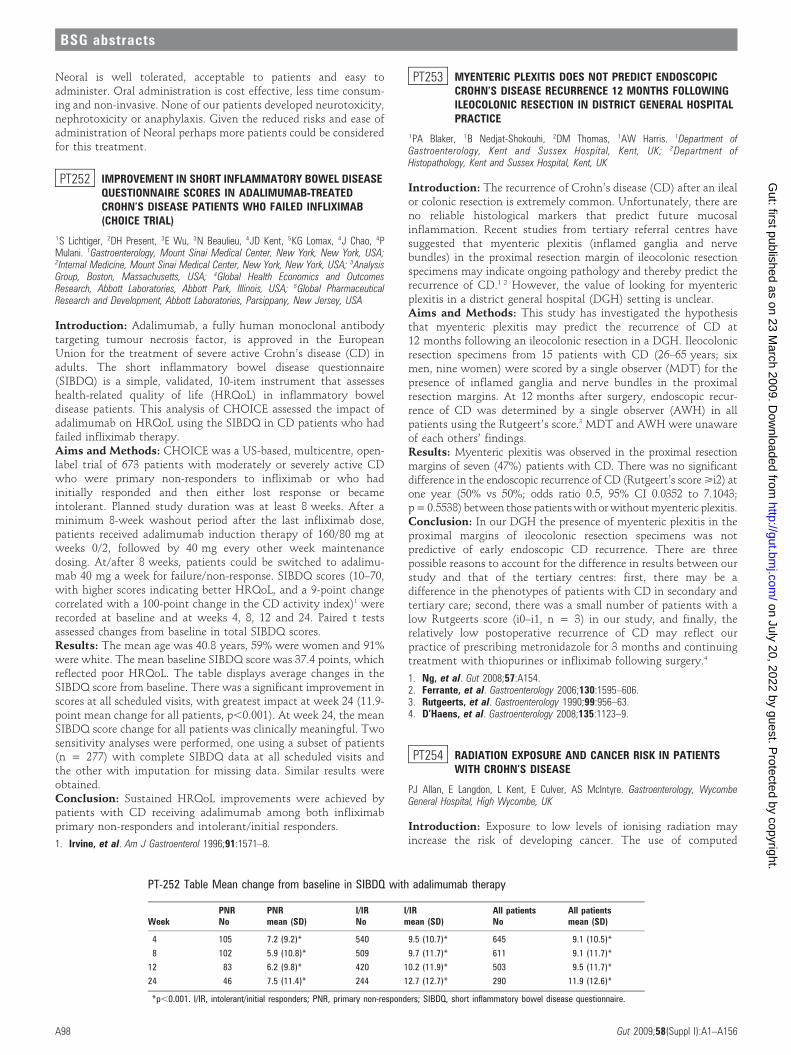
Neoral is well tolerated, acceptable to patients and easy toadminister. Oral administration is cost effective, less time consum-ing and non-invasive. None of our patients developed neurotoxicity,nephrotoxicity or anaphylaxis. Given the reduced risks and ease ofadministration of Neoral perhaps more patients could be consideredfor this treatment.
PT252 IMPROVEMENT IN SHORT INFLAMMATORY BOWEL DISEASEQUESTIONNAIRE SCORES IN ADALIMUMAB-TREATEDCROHN’S DISEASE PATIENTS WHO FAILED INFLIXIMAB(CHOICE TRIAL)
1S Lichtiger, 2DH Present, 3E Wu, 3N Beaulieu, 4JD Kent, 5KG Lomax, 4J Chao, 4PMulani. 1Gastroenterology, Mount Sinai Medical Center, New York, New York, USA;2Internal Medicine, Mount Sinai Medical Center, New York, New York, USA; 3AnalysisGroup, Boston, Massachusetts, USA; 4Global Health Economics and OutcomesResearch, Abbott Laboratories, Abbott Park, Illinois, USA; 5Global PharmaceuticalResearch and Development, Abbott Laboratories, Parsippany, New Jersey, USA
Introduction: Adalimumab, a fully human monoclonal antibodytargeting tumour necrosis factor, is approved in the EuropeanUnion for the treatment of severe active Crohn’s disease (CD) inadults. The short inflammatory bowel disease questionnaire(SIBDQ) is a simple, validated, 10-item instrument that assesseshealth-related quality of life (HRQoL) in inflammatory boweldisease patients. This analysis of CHOICE assessed the impact ofadalimumab on HRQoL using the SIBDQ in CD patients who hadfailed infliximab therapy.Aims and Methods: CHOICE was a US-based, multicentre, open-label trial of 673 patients with moderately or severely active CDwho were primary non-responders to infliximab or who hadinitially responded and then either lost response or becameintolerant. Planned study duration was at least 8 weeks. After aminimum 8-week washout period after the last infliximab dose,patients received adalimumab induction therapy of 160/80 mg atweeks 0/2, followed by 40 mg every other week maintenancedosing. At/after 8 weeks, patients could be switched to adalimu-mab 40 mg a week for failure/non-response. SIBDQ scores (10–70,with higher scores indicating better HRQoL, and a 9-point changecorrelated with a 100-point change in the CD activity index)1 wererecorded at baseline and at weeks 4, 8, 12 and 24. Paired t testsassessed changes from baseline in total SIBDQ scores.Results: The mean age was 40.8 years, 59% were women and 91%were white. The mean baseline SIBDQ score was 37.4 points, whichreflected poor HRQoL. The table displays average changes in theSIBDQ score from baseline. There was a significant improvement inscores at all scheduled visits, with greatest impact at week 24 (11.9-point mean change for all patients, p,0.001). At week 24, the meanSIBDQ score change for all patients was clinically meaningful. Twosensitivity analyses were performed, one using a subset of patients(n = 277) with complete SIBDQ data at all scheduled visits andthe other with imputation for missing data. Similar results wereobtained.Conclusion: Sustained HRQoL improvements were achieved bypatients with CD receiving adalimumab among both infliximabprimary non-responders and intolerant/initial responders.
1. Irvine, et al. Am J Gastroenterol 1996;91:1571–8.
PT253 MYENTERIC PLEXITIS DOES NOT PREDICT ENDOSCOPICCROHN’S DISEASE RECURRENCE 12 MONTHS FOLLOWINGILEOCOLONIC RESECTION IN DISTRICT GENERAL HOSPITALPRACTICE
1PA Blaker, 1B Nedjat-Shokouhi, 2DM Thomas, 1AW Harris. 1Department ofGastroenterology, Kent and Sussex Hospital, Kent, UK; 2Department ofHistopathology, Kent and Sussex Hospital, Kent, UK
Introduction: The recurrence of Crohn’s disease (CD) after an ilealor colonic resection is extremely common. Unfortunately, there areno reliable histological markers that predict future mucosalinflammation. Recent studies from tertiary referral centres havesuggested that myenteric plexitis (inflamed ganglia and nervebundles) in the proximal resection margin of ileocolonic resectionspecimens may indicate ongoing pathology and thereby predict therecurrence of CD.1 2 However, the value of looking for myentericplexitis in a district general hospital (DGH) setting is unclear.Aims and Methods: This study has investigated the hypothesisthat myenteric plexitis may predict the recurrence of CD at12 months following an ileocolonic resection in a DGH. Ileocolonicresection specimens from 15 patients with CD (26–65 years; sixmen, nine women) were scored by a single observer (MDT) for thepresence of inflamed ganglia and nerve bundles in the proximalresection margins. At 12 months after surgery, endoscopic recur-rence of CD was determined by a single observer (AWH) in allpatients using the Rutgeert’s score.3 MDT and AWH were unawareof each others’ findings.Results: Myenteric plexitis was observed in the proximal resectionmargins of seven (47%) patients with CD. There was no significantdifference in the endoscopic recurrence of CD (Rutgeert’s score >i2) atone year (50% vs 50%; odds ratio 0.5, 95% CI 0.0352 to 7.1043;p = 0.5538) between those patients with or without myenteric plexitis.Conclusion: In our DGH the presence of myenteric plexitis in theproximal margins of ileocolonic resection specimens was notpredictive of early endoscopic CD recurrence. There are threepossible reasons to account for the difference in results between ourstudy and that of the tertiary centres: first, there may be adifference in the phenotypes of patients with CD in secondary andtertiary care; second, there was a small number of patients with alow Rutgeerts score (i0–i1, n = 3) in our study, and finally, therelatively low postoperative recurrence of CD may reflect ourpractice of prescribing metronidazole for 3 months and continuingtreatment with thiopurines or infliximab following surgery.4
1. Ng, et al. Gut 2008;57:A154.2. Ferrante, et al. Gastroenterology 2006;130:1595–606.3. Rutgeerts, et al. Gastroenterology 1990;99:956–63.4. D’Haens, et al. Gastroenterology 2008;135:1123–9.
PT254 RADIATION EXPOSURE AND CANCER RISK IN PATIENTSWITH CROHN’S DISEASE
PJ Allan, E Langdon, L Kent, E Culver, AS McIntyre. Gastroenterology, WycombeGeneral Hospital, High Wycombe, UK
Introduction: Exposure to low levels of ionising radiation mayincrease the risk of developing cancer. The use of computed
PT-252 Table Mean change from baseline in SIBDQ with adalimumab therapy
WeekPNRNo
PNRmean (SD)
I/IRNo
I/IRmean (SD)
All patientsNo
All patientsmean (SD)
4 105 7.2 (9.2)* 540 9.5 (10.7)* 645 9.1 (10.5)*
8 102 5.9 (10.8)* 509 9.7 (11.7)* 611 9.1 (11.7)*
12 83 6.2 (9.8)* 420 10.2 (11.9)* 503 9.5 (11.7)*
24 46 7.5 (11.4)* 244 12.7 (12.7)* 290 11.9 (12.6)*
*p,0.001. I/IR, intolerant/initial responders; PNR, primary non-responders; SIBDQ, short inflammatory bowel disease questionnaire.
BSG abstracts
A98 Gut 2009;58(Suppl I):A1–A156
on July 20, 2022 by guest. Protected by copyright.
http://gut.bmj.com
/G
ut: first published as on 23 March 2009. D
ownloaded from
tomography pulmonary angiogram for pulmonary emboli inwomen may add an additional 100 breast cancers per 100 000patients exposed. Should we be concerned for Crohn’s disease (CD)patients who often need multiple x ray investigations to diagnoseand assess disease at relatively young ages?
Aims and Methods: A retrospective study of 122 CD patientsunder the care of one gastroenterologist studied x ray exposure overthe lifetime of their disease to estimate radiation exposure. Acomparison was made with a similar patient group with ulcerativecolitis (UC) and national population norms. Published nationalaverage radiation exposures were used for each procedure inmilliSieverts (mSv) abdominal x rays (AXR, 0.7 mSv), small bowelenemas (SBE, 3), barium enemas (BaE, 7.2), computed tomographyabdomen pelvis without (CTAP, 10) and with contrast (CTAP+C,10). Total exposure (mSv) for each patient was used to estimate theexcess risk of developing cancer.Results: The 122 CD cohort (62.3% women) had a mean diagnosisage of 32.0 years (+2.7, 95% CI) compared with the 122 UC cohort(42.0% women), 42.0 years (+2.8). Mean disease duration was CD18.0 years (+1.9) and UC 19.0 years (+1.9). CD disease distributionwas 22.5% ileal, 45.8% ileocolonic, 30.0% colonic, 1.7% perianalonly and 8.3% perianal plus other site. The number of inflamma-tory bowel diseases diagnosed per decade: 1950s four (50% CD),1960s 11 (46% CD), 1970s 29 (48% CD), 1980s 54 (50% CD), 1990s99 (49% CD), 2000–4 46 (57% CD). Total investigations performed,expressed as mSv: AXR 237.3 mSv (mean effective dose per CD vsUC patient 1.14 vs 0.80; p = ns); SBE 576 mSv (4.30 vs 0.42;p,0.01); BaE 1252.8 mSv (6.08 vs 4.19; p = 0.03); CTAP 450 mSv(2.95 vs 0.74; p,0.01); CTAP+C 550 mSv (3.28 vs 1.23; p = 0.04)and the mean number of 18 magnetic resonance imaging scansperformed (0.07 vs 0.07; p = ns) and 238 ultrasound abdomen (1.53vs 0.42; p,0.01). The mean total individual radiation dose (mSv)was CD 17.75 (+3.22, 95% CI) vs UC 7.38 (+1.60) p,0.01. Meanpopulation norm exposures are 2.2 mSv/year (range 1.5–7.5), ie,39.6 mSv over 18 years. Increased exposure for patients with CD isthus 57.35 mSv over 18 years, an increase of 44.8% over populationnorms, and for UC over 19 years is 49.18 mSv, a 17.7% increase; CDhas a 23.1% increase over UC. CD and UC each occurs in 60 6 103
patients in the UK and radiation exposure (10 mSv 1 : 1000–10 000risk) related to disease management might result in 10.6–106 extracancers in CD and 4.4–44 in UC patients.Conclusion: CD patients are exposed to substantially moreradiation than either population norms or similar UC patients.We estimate this risk to be a 23.1% increase over UC patients and44.8% above population norms. Given practices in previous decadesthis may underrepresent the radiation risk. Radiation exposureshould be considered and minimised when investigating CDpatients.
PT255 THE ALLERGIC BASIS OF THE OROFACIALGRANULOMATOSIS/ORAL CROHN’S DISEASE SPECTRUM
1P Patel, 2F Barone, 2L Boursier, 1C Nunes, 3E Odell, 4M Escudier, 5S Challacombe,1J Brostoff, 2J Spencer, 1J Sanderson. 1Nutritional Sciences Division, King’s CollegeLondon, London, UK; 2Immunobiology, King’s College London, London, UK; 3OralPathology, Guy’s and St Thomas’ NHS Trust, London, UK; 4Oral Medicine, Guy’s and StThomas’ NHS Trust, London, UK; 5Oral Medicine, King’s College London, London, UK
Introduction: Orofacial granulomatosis (OFG), which includesoral Crohn’s disease, is a chronic inflammatory condition presentingcharacteristically with lip swelling but also affecting the gingivae,buccal mucosa, floor of the mouth and a number of other sites inthe oral cavity. Although the cause remains unknown, acute boutsof swelling and frequently reported atopy suggest an allergiccomponent. There is evidence for the involvement of dietarytriggers, with objective improvement on a cinnamon and benzoate-free diet.
Aims and Methods: To investigate the role of allergy in OFG/oralCrohn’s disease.Methods: Using immunohistochemistry and PCR on paraffinembedded (n = 44) and fresh frozen (n = 8) oral mucosalbiopsies, we looked for evidence of IgE-mediated hypersensitivityin patients with OFG and compared this with non-OFG oralinflammation. We also determined the incidence of atopy in acohort of patients with OFG and performed skin prick tests tocommon allergens (n = 58).Results: Large, active, dendritic B cells in the oral mucosa werepresent in the inflammatory infiltrate in OFG and these wereindependent of follicular structures. They expressed nuclearactivation-induced cytidine deaminase, an enzyme that is essentialfor the process of class switch recombination to IgE. These dendriticB cells also expressed surface IgE, and this expression wassignificantly greater in OFG compared with other oral conditions.Epsilon transcripts, which provide further evidence for IgEproduction locally, were also demonstrated in OFG by PCR.Clinical allergy data from history and skin prick tests revealed anincidence of allergy of 83% in the oral OFG cohort compared with areported incidence of between 4% and 20% in the generalpopulation. The incidence of the oral allergy syndrome was 30%,significantly higher than population estimates. OFG patients agedbelow 30 years had a significantly higher incidence of atopy andoral allergy syndrome than patients over 30 years (93% vs 74% and41% vs 19%, respectively).Conclusion: Patients with oral OFG have a high incidence ofatopy. In addition, we provide evidence for local class switching toIgE in the oral mucosa in OFG in the absence of follicular structuresand significantly greater IgE expression on dendritic B cells in OFGthan non-OFG controls. It is already known that removing certaindietary components will improve oral inflammation in OFG. Thesesubepithelial dendritic B cells may be cellular mediators of localhypersensitivity in OFG.
PT256 AUDIT OF ANTI-TUMOUR NECROSIS FACTOR THERAPY INCROHN’S DISEASE: BETTER RESULTS IN PATIENTS WITH NOPREVIOUS SURGERY
RN Patel, B Gregg, A Kneebone, KP Leiper, A Ellis, AJ Watson, JM Rhodes.Gastroenterology, Royal Liverpool and Broadgreen University Hospital Trust, Liverpool,UK
Introduction: The UK inflammatory bowel disease (IBD) audit hasshown considerable variations in access to and use of infliximab inCrohn’s disease. Maintenance versus episodic treatment is alsocontentious.Aims and Methods: The aim of this audit was to assess the usageand results of anti-tumour necrosis factor (TNF) therapy in Crohn’sdisease in routine clinical practice in a large university hospital. Thehospital currently has 460 active patients with Crohn’s disease onits register. 102 of these have received infliximab since 2002, ofwhom 77 have received treatment for at least 12 months and arethe subject of this audit. All 77 case notes were audited.Results: Harvey–Bradshaw (HB) scores on initiation of therapywere 8, 2–40 (median, range), and 49 (64%) patients had a score of 8or more, ie, treated within National Institute for Health andClinical Excellence guidance. 36 patients had ileocolonic disease, 31colon only and 10 ileal only. 28 had enterocutaneous fistulae, ofwhich 22 were perineal. 53% had had previous surgery, 84% werereceiving concomitant immunosuppression and 32% were receivingconcomitant corticosteroids. 100% had had recent chest x ray. Theoverall remission rates (HB (4) were 48%, 39% and 43% at 3, 6 and12 months, respectively. 47% received ‘‘maintenance’’ treatments,defined as six or more infusions in the first 12 months and 53%episodic (median three infusions/12 months, range one to five).Two patients developed a systemic lupus erythematosis-likesyndrome. There were no drug-associated fatalities. 55% of patients
BSG abstracts
Gut 2009;58(Suppl I):A1–A156 A99
on July 20, 2022 by guest. Protected by copyright.
http://gut.bmj.com
/G
ut: first published as on 23 March 2009. D
ownloaded from
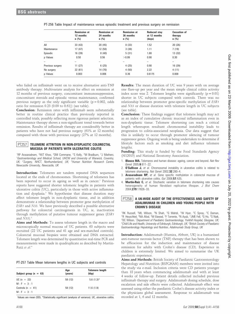
who failed on infliximab went on to receive alternative anti-TNFantibody therapy. Multivariate analysis for effect on remission at12 months of previous surgery, concomitant immunosuppression,concomitant steroids and episodic versus maintenance, confirmedprevious surgery as the only significant variable (p = 0.002, oddsratio for remission 0.23 (0.09 to 0.61)) (see table).Conclusion: Remission rates with infliximab seem substantiallybetter in routine clinical practice than previously reported incontrolled trials, possibly reflecting more rigorous patient selection.Maintenance therapy shows a non-significant trend towards betterremission. Results of infliximab therapy are considerably better inpatients who have not had previous surgery (61% at 12 months)compared with those with previous surgery (27% at 12 months).
PT257 TELOMERE ATTRITION IN NON-DYSPLASTIC COLORECTALMUCOSA OF PATIENTS WITH ULCERATIVE COLITIS
1RP Arasaradnam, 2KKT Khoo, 3DM Commane, 2S Kelly, 2M Bradburn, 3J Mathers.1Gastroenterology and Medical School, UHCW and University of Warwick, Coventry,UK; 2Surgery, NHCT, Northumberland, UK; 3Human Nutrition Research Centre,Newcastle University, Newcastle, UK
Introduction: Telomeres are tandem repeated DNA sequenceslocated at the ends of chromosomes. Shortening of telomeres hasbeen reported to occur in ageing as well as in cancer.1 Previousreports have suggested shorter telomeric lengths in patients withulcerative colitis (UC), particularly in those with active inflamma-tion and dysplasia.2 We hypothesise that disease duration mayaffect telomere length in non-dysplastic tissue, and in additiondemonstrate a relationship between promoter gene methylation ofESR1 and N33. We have previously described a possible alternativepathway for colorectal carcinogenesis in UC, ie, inactivationthrough methylation of putative tumour suppressor genes (ESR1and N33).3
Aims and Methods: To assess telomere length in the macro andmicroscopically normal mucosa of UC patients. 63 subjects wererecruited (22 UC patients and 41 age and sex-matched controls).Colorectal mucosal biopsies were obtained and DNA extracted.Telomere length was determined by quantitative real-time PCR andmeasurements were made in quadruplicate as described by Martin-Ruiz et al.4
Results: The mean duration of UC was 9 years with on averageone flare-up per year and the mean simple clinical colitis activityindex score was 2. Telomere lengths were significantly (p = 0.01)shorter in UC subjects compared with controls. There was norelationship between promoter gene-specific methylation of ESR1and N33 or disease duration with telomere length in UC subjects(see table).Conclusion: These findings suggest that telomere length may actas an index of cumulative chronic mucosal inflammation even innon-dysplastic tissue. Telomere shortening can reach a criticallength whereupon resultant chromosomal instability leads toprogression to colitis-associated neoplasia. Our data suggest thatthis is unlikely to occur through promoter silencing of tumoursuppressor genes. Ongoing work is being undertaken to determine iflifestyle factors such as smoking and diet influence telomerelengths.Funding: This study is funded by the Food Standards Agency(N12015) and National Ileostomy Association.
1. Blasco MA. Telomeres and human disease: ageing, cancer and beyond. Nat RevGenet 2005;6:611–22.
2. O’Sullivan J, et al. Chromosomal instability in ulcerative colitis is related totelomere shortening. Nat Genet 2002;32:280–4.
3. Arasaradnam RP, et al. Gene specific methylation in colorectal mucosa ofpatients with ulcerative colitis. Gut 2008;57:A35.
4. Martin-Ruiz C, et al. Stochastic variation in telomere shortening rate causesheterogeneity of human fibroblast replicative lifespan. J Biol Chem2004;279:17826–33.
PT258 A UK-WIDE AUDIT OF THE EFFECTIVENESS AND SAFETY OFADALIMUMAB IN CHILDREN AND YOUNG PEOPLE WITHCROHN’S DISEASE
1RK Russell, 2ML Wilson, 3N Shah, 3G Mahdi, 3W Hyer, 3C Spray, 3C Daman,3R Heuschkel, 3NA Afzal, 3M Elawad, 3F Torrente, 3N Ayub, 3JME Fell, 3G Ho, 3S Naik,2DC Wilson. 1Department of Paediatric Gastroenterology, Yorkhill Hospital, Glasgow, UK;2Child Life and Health, University of Edinburgh, Edinburgh, UK; 3British Society of PaediatricGastroenterology Hepatology and Nutrition, Adalimumab Study Group, UK
Introduction: Adalimumab (Humira, Abbott, UK) is a humanisedanti-tumour necrosis factor (TNF) therapy that has been shown tobe efficacious for the induction and maintenance of diseaseremission for adults with Crohn’s disease (CD). Experience inchildren is extremely limited. We aimed to summarise the UKpaediatric experience.Aims and Methods: British Society of Paediatric GastroenterologyHepatology and Nutrition (BSPGHAN) members were invited intothe study via e-mail. Inclusion criteria were CD patients youngerthan 18 years when commencing adalimumab and with at least4 weeks of follow-up. Patient details collected included previousinfliximab therapy and surgery. Adalimumab dosing schedule, doseescalation and side effects were collected. Adalimumab effect wasassessed using either the paediatric Crohn’s disease activity index orthe physicians global assessment. Response to adalimumab wasrecorded at 1, 6 and 12 months.
PT-256 Table Impact of maintenance versus episodic treatment and previous surgery on remission
Remission at12 monthsn (%)
Remission at24 monthsn (%)
Remission at36 monthsn (%)
Reduced stayat 12 months(days)
Cessation oftherapyn (%)
All 33 (43) 20 (45) 8 (33) 1.52 20 (26)
Maintenance 17 (47) 12 (50) 3 (38) 1.11 7 (19)
Episodic 16 (39) 8 (40) 5 (31) 1.88 13 (32)
p Values 0.50 0.56 .0.99 0.90 0.30
– – – – – –
Previous surgery 11 (27) 6 (25) 4 (25) 0.90 16 (39)
No surgery 22 (61) 14 (70) 4 (50) 2.22 4 (11)
p Values 0.003 0.006 0.36 0.6175 0.008
PT-257 Table Mean telomere lengths in UC subjects and controls
Subject group (n = 63)Age(years)
Telomere length(kbp)
UC (n = 22) 58 (12) 5.6 (1.3)*
M : F = 3 : 1
Controls (n = 41) 58 (12) 11.6 (1.9)
M : F = 3 : 1
Values are mean (SD). *Statistically significant (analysis of variance). UC, ulcerative colitis.
BSG abstracts
A100 Gut 2009;58(Suppl I):A1–A156
on July 20, 2022 by guest. Protected by copyright.
http://gut.bmj.com
/G
ut: first published as on 23 March 2009. D
ownloaded from
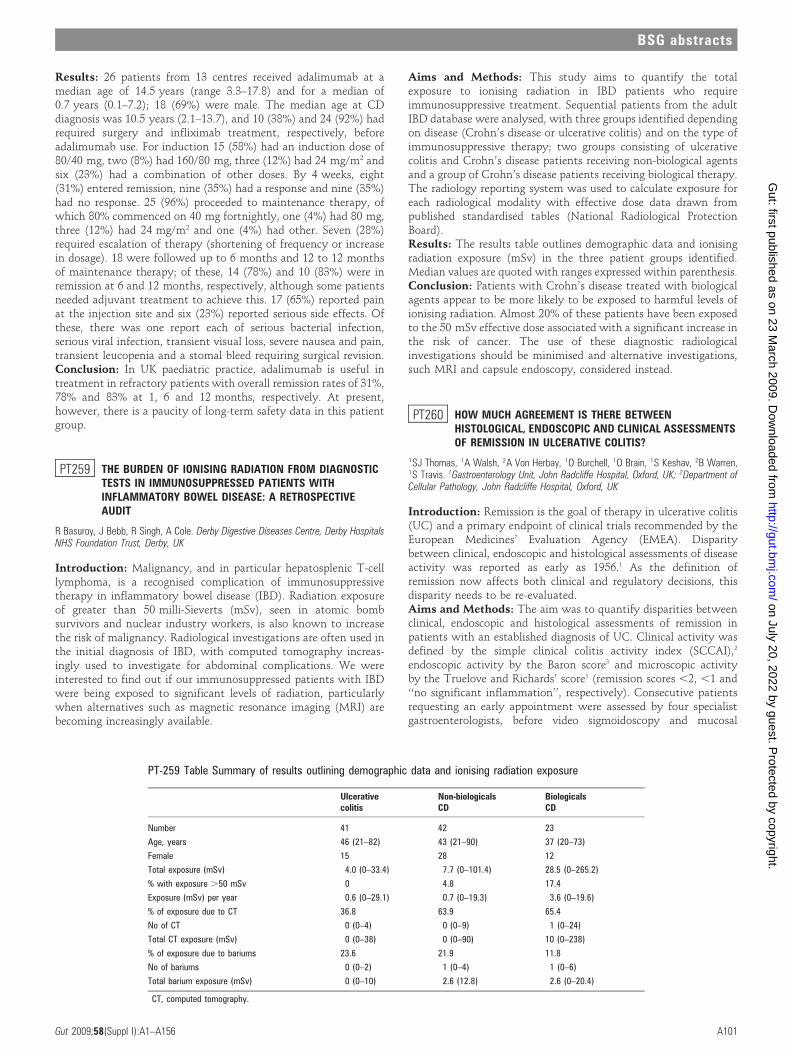
Results: 26 patients from 13 centres received adalimumab at amedian age of 14.5 years (range 3.3–17.8) and for a median of0.7 years (0.1–7.2); 18 (69%) were male. The median age at CDdiagnosis was 10.5 years (2.1–13.7), and 10 (38%) and 24 (92%) hadrequired surgery and infliximab treatment, respectively, beforeadalimumab use. For induction 15 (58%) had an induction dose of80/40 mg, two (8%) had 160/80 mg, three (12%) had 24 mg/m2 andsix (23%) had a combination of other doses. By 4 weeks, eight(31%) entered remission, nine (35%) had a response and nine (35%)had no response. 25 (96%) proceeded to maintenance therapy, ofwhich 80% commenced on 40 mg fortnightly, one (4%) had 80 mg,three (12%) had 24 mg/m2 and one (4%) had other. Seven (28%)required escalation of therapy (shortening of frequency or increasein dosage). 18 were followed up to 6 months and 12 to 12 monthsof maintenance therapy; of these, 14 (78%) and 10 (83%) were inremission at 6 and 12 months, respectively, although some patientsneeded adjuvant treatment to achieve this. 17 (65%) reported painat the injection site and six (23%) reported serious side effects. Ofthese, there was one report each of serious bacterial infection,serious viral infection, transient visual loss, severe nausea and pain,transient leucopenia and a stomal bleed requiring surgical revision.Conclusion: In UK paediatric practice, adalimumab is useful intreatment in refractory patients with overall remission rates of 31%,78% and 83% at 1, 6 and 12 months, respectively. At present,however, there is a paucity of long-term safety data in this patientgroup.
PT259 THE BURDEN OF IONISING RADIATION FROM DIAGNOSTICTESTS IN IMMUNOSUPPRESSED PATIENTS WITHINFLAMMATORY BOWEL DISEASE: A RETROSPECTIVEAUDIT
R Basuroy, J Bebb, R Singh, A Cole. Derby Digestive Diseases Centre, Derby HospitalsNHS Foundation Trust, Derby, UK
Introduction: Malignancy, and in particular hepatosplenic T-celllymphoma, is a recognised complication of immunosuppressivetherapy in inflammatory bowel disease (IBD). Radiation exposureof greater than 50 milli-Sieverts (mSv), seen in atomic bombsurvivors and nuclear industry workers, is also known to increasethe risk of malignancy. Radiological investigations are often used inthe initial diagnosis of IBD, with computed tomography increas-ingly used to investigate for abdominal complications. We wereinterested to find out if our immunosuppressed patients with IBDwere being exposed to significant levels of radiation, particularlywhen alternatives such as magnetic resonance imaging (MRI) arebecoming increasingly available.
Aims and Methods: This study aims to quantify the totalexposure to ionising radiation in IBD patients who requireimmunosuppressive treatment. Sequential patients from the adultIBD database were analysed, with three groups identified dependingon disease (Crohn’s disease or ulcerative colitis) and on the type ofimmunosuppressive therapy; two groups consisting of ulcerativecolitis and Crohn’s disease patients receiving non-biological agentsand a group of Crohn’s disease patients receiving biological therapy.The radiology reporting system was used to calculate exposure foreach radiological modality with effective dose data drawn frompublished standardised tables (National Radiological ProtectionBoard).Results: The results table outlines demographic data and ionisingradiation exposure (mSv) in the three patient groups identified.Median values are quoted with ranges expressed within parenthesis.Conclusion: Patients with Crohn’s disease treated with biologicalagents appear to be more likely to be exposed to harmful levels ofionising radiation. Almost 20% of these patients have been exposedto the 50 mSv effective dose associated with a significant increase inthe risk of cancer. The use of these diagnostic radiologicalinvestigations should be minimised and alternative investigations,such MRI and capsule endoscopy, considered instead.
PT260 HOW MUCH AGREEMENT IS THERE BETWEENHISTOLOGICAL, ENDOSCOPIC AND CLINICAL ASSESSMENTSOF REMISSION IN ULCERATIVE COLITIS?
1SJ Thomas, 1A Walsh, 2A Von Herbay, 1O Burchell, 1O Brain, 1S Keshav, 2B Warren,1S Travis. 1Gastroenterology Unit, John Radcliffe Hospital, Oxford, UK; 2Department ofCellular Pathology, John Radcliffe Hospital, Oxford, UK
Introduction: Remission is the goal of therapy in ulcerative colitis(UC) and a primary endpoint of clinical trials recommended by theEuropean Medicines’ Evaluation Agency (EMEA). Disparitybetween clinical, endoscopic and histological assessments of diseaseactivity was reported as early as 1956.1 As the definition ofremission now affects both clinical and regulatory decisions, thisdisparity needs to be re-evaluated.Aims and Methods: The aim was to quantify disparities betweenclinical, endoscopic and histological assessments of remission inpatients with an established diagnosis of UC. Clinical activity wasdefined by the simple clinical colitis activity index (SCCAI),2
endoscopic activity by the Baron score3 and microscopic activityby the Truelove and Richards’ score1 (remission scores ,2, ,1 and‘‘no significant inflammation’’, respectively). Consecutive patientsrequesting an early appointment were assessed by four specialistgastroenterologists, before video sigmoidoscopy and mucosal
PT-259 Table Summary of results outlining demographic data and ionising radiation exposure
Ulcerativecolitis
Non-biologicalsCD
BiologicalsCD
Number 41 42 23
Age, years 46 (21–82) 43 (21–90) 37 (20–73)
Female 15 28 12
Total exposure (mSv) 4.0 (0–33.4) 7.7 (0–101.4) 28.5 (0–265.2)
% with exposure .50 mSv 0 4.8 17.4
Exposure (mSv) per year 0.6 (0–29.1) 0.7 (0–19.3) 3.6 (0–19.6)
% of exposure due to CT 36.8 63.9 65.4
No of CT 0 (0–4) 0 (0–9) 1 (0–24)
Total CT exposure (mSv) 0 (0–38) 0 (0–90) 10 (0–238)
% of exposure due to bariums 23.6 21.9 11.8
No of bariums 0 (0–2) 1 (0–4) 1 (0–6)
Total barium exposure (mSv) 0 (0–10) 2.6 (12.8) 2.6 (0–20.4)
CT, computed tomography.
BSG abstracts
Gut 2009;58(Suppl I):A1–A156 A101
on July 20, 2022 by guest. Protected by copyright.
http://gut.bmj.com
/G
ut: first published as on 23 March 2009. D
ownloaded from
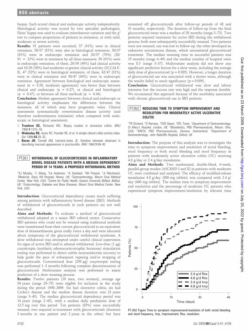
biopsy. Each scored clinical and endoscopic activity independently.Histological activity was scored by two specialist pathologists.Fleiss’ kappa was used to evaluate interobserver variation and the x2
test to compare proportions of patients in remission, or with mild,moderate or severe activity.Results: 91 patients were recruited; 37 (41%) were in clinicalremission, 30/37 (81%) were also in histological remission, 30/37(81%) were in endoscopic remission and 20/37 (76%) (20/91 = 22%) were in remission by all three measures. 56 (61%) werein endoscopic remission; of these, 26/56 (46%) had clinical activityand 10/26 (38%) had moderate or greater clinical activity (SCCAI 6–8). 47 (52%) were in histological remission; of these, 42/47 (81%)were in clinical remission and 30/47 (64%) were in endoscopicremission. Agreement between histological and endoscopic assess-ment (k = 0.58, moderate agreement) was better than betweenclinical and endoscopic (k = 0.27), or clinical and histological(k = 0.47), or between all three methods (k = 0.44).Conclusion: Modest agreement between clinical and endoscopic orhistological activity emphasises the differences between themeasures, all of which may have prognostic value. Clinicalassessment systematically overestimates disease activity (andtherefore underestimates remission) when compared with endo-scopic or histological assessment.
1. Truelove SC, Richards WC. Biopsy studies in ulcerative colitis. BMJ1956;1:1315–18.
2. Walmsley RS, Ayres RC, Pounder RE, et al. A simple clinical colitis activity index.Gut 1998;43:29–32.
3. Baron JH, Connell AM, Lennard-Jones JE. Variation between observers indescribing mucosal appearances in proctocolitis. BMJ 1964;1:89–92.
PT261 WITHDRAWAL OF GLUCOCORTICOIDS IN INFLAMMATORYBOWEL DISEASE PATIENTS WITH A MEDIAN DEPENDENCYPERIOD OF 14 YEARS: A PROPOSED WITHDRAWAL METHOD
1SJ Murphy, 2L Wang, 3LA Anderson, 2A Steinlauf, 2DH Present, 4JI Mechanick.1Medicine, Daisy Hill Hospital, Newry, UK; 2Gastroenterology, Mount Sinai MedicalCenter, New York, USA; 3Centre for Public Health, Queens University Belfast, Belfast,UK; 4Endocrinology, Diabetes and Bone Diseases, Mount Sinai Medical Center, NewYork, USA
Introduction: Glucocorticoid dependency causes much sufferingamong patients with inflammatory bowel disease (IBD). Methodsof withdrawal of glucocorticoids in such patients are not welldescribed.Aims and Methods: To evaluate a method of glucocorticoidwithdrawal adopted at a major IBD referral centre. ConsecutiveIBD patients who could not be weaned using traditional methodswere transitioned from their current glucocorticoid to an equivalentdose of dexamethasone given orally twice a day and were educatedabout symptoms of the glucocorticoid withdrawal syndrome. Aslow withdrawal was attempted under careful clinical supervisionfor signs of active IBD and/or adrenal withdrawal. Low-dose (1 mg)cosyntropin (synthetic adrenocorticotrophic hormone) stimulationtesting was performed to detect subtle neuroendocrine recovery tohelp guide the pace of subsequent tapering and/or stopping ofglucocorticoids. Conventional dose (250 mg) cosyntropin testingwas performed 1–3 months following complete discontinuation ofglucocorticoid. Multivariate analysis was performed to assesspredictors of a slow weaning process.Results: Twelve patients (10 men, two women), average age54 years (range 29–75) were eligible for inclusion in the studyduring the period 1998–2006. Six had ulcerative colitis, six hadCrohn’s disease and the median disease duration was 21 years(range 3–45). The median glucocorticoid dependency period was14 years (range 2–45), with a median daily prednisone dose of12.5 mg over this period. Ten patients (83%) were successfullyweaned; two required re-treatment with glucocorticoids (duration3 months in one patient and 2 years in the other) but have
remained off glucocorticoids after follow-up periods of 48 and31 months, respectively. The duration of follow-up from the finalglucocorticoid wean was a median of 38 months (range 3–73). Twopatients required treatment for active IBD during the withdrawalprocess; both were subsequently successfully weaned. Two patientswere not weaned; one was lost to follow-up, the other developed anunknown autoimmune disease, which necessitated glucocorticoidtreatment. The median weaning time in successful patients was15 months (range 4–49) and the median number of hospital visitswas 8.5 (range 3–37). Multivariate analysis did not show anysignificant predictive factors for a slow wean, including the averagedaily dose of glucocorticoid (p = 0.985). However, a longer durationof glucocorticoid use was associated with a slower wean, althoughthe results failed to reach significance (p = 0.056).Conclusion: Glucocorticoid withdrawal was slow and labourintensive but the success rate was high and the response durable.We recommend this approach because of the morbidity associatedwith chronic glucocorticoid use in IBD patients.
PT262 REDUCING TIME TO SYMPTOM IMPROVEMENT ANDRESOLUTION FOR MODERATELY ACTIVE ULCERATIVECOLITIS
1TR Orchard, 2D Ramsey, 3SVD Geest, 4SPL Travis. 1Department of Gastroenterology,St Mary’s Hospital, London, UK; 2Biostatistics, P&G Pharmaceuticals, Mason, Ohio,USA; 3M&TA, P&G Pharmaceuticals, Geneva, Switzerland; 4Department ofGastroenterology, John Radcliffe Hospital, Oxford, UK
Introduction: The purpose of this analysis was to investigate thetime to symptom improvement and resolution of rectal bleeding,stool frequency or both rectal bleeding and stool frequency inpatients with moderately active ulcerative colitis (UC) receiving4.8 g/day or 2.4 g/day mesalazine.Aims and Methods: Two randomised, double-blind, 6-week,parallel-group studies (ASCEND I and II) in patients with moderateUC were combined and analysed. The efficacy of modified-releasemesalazine 4.8 g/day (800 mg tablets) was compared with 2.4 g/day (400 mg tablets). The median time to symptom improvementand resolution and the percentage of moderate UC patients whoexperienced symptom improvement/resolution by relevant time
PT-262 Figure Time to symptom improvement/resolution of both rectal bleedingand stool frequency. Imp, improvement; Res, resolution.
BSG abstracts
A102 Gut 2009;58(Suppl I):A1–A156
on July 20, 2022 by guest. Protected by copyright.
http://gut.bmj.com
/G
ut: first published as on 23 March 2009. D
ownloaded from
points were calculated. Time to symptom improvement/resolutionwas defined as the number of days between the first day of dosingand the first day of symptom improvement/resolution. Symptomimprovement was defined as a decrease from a baseline of 1 point ormore. Symptom resolution was defined as a score of 0.Results: 423 analysable patients had moderate UC. Kaplan–Meierresults for time to symptom improvement/resolution for 4.8 g/dayand 2.4 g/day are shown. The median time to symptom resolutionof both rectal bleeing and stool frequency was 19 days versus29 days for 4.8 g/day versus 2.4 g/day (p = 0.02). By day 14, rectalbleeding resolution was achieved in 64% versus 49%, stoolfrequency resolution in 58% versus 50% and both rectal bleedingand stool frequency resolution in 43% versus 30% for 4.8 g/day and2.4 g/day, respectively.Conclusion: Mesalazine 4.8 g/day for patients with moderate UCresults in faster symptom improvement/resolution than 2.4 g/day.14 days is a nodal point for deciding whether to escalate therapy;mesalazine 4.8 g/day from the start of a flare could avoid the needfor steroids in some patients.
PT263 PREOPERATIVE INFLIXIMAB AND RISK OF POSTOPERATIVECOMPLICATIONS IN PATIENTS WITH INFLAMMATORYBOWEL DISEASE: META-ANALYSIS OF OBSERVATIONALSTUDIES
V Subramanian, CJ Hawkey. Nottingham Digestive Diseases Centre and BiomedicalResearch Unit, Nottingham University and Hospital, Nottingham, UK
Introduction: Abdominal surgery for inflammatory bowel disease(IBD) is not without risks. Inflximab has been approved for use inboth Crohn’s disease (CD) and ulcerative colitis (UC) by the USFood and Drug Administration. The use of high-dose steroids hasbeen shown in a previous meta-analysis to increase the risk ofpostoperative complications in patients with IBD.1 There is stilldebate whether the use of infliximab would also increase post-operative complications in this population.Aims and Methods: To estimate the risk of postoperativecomplications following abdominal surgery in patients with IBDwho received infliximab preoperatively, a meta-analysis of observa-tional studies was conducted. We searched electronic databases forfull journal articles published after 1990 reporting on postoperativecomplications in patients with IBD provided they comparedpatients on infliximab with those not on infliximab. We handsearched the reference lists of all retrieved articles. We carried outrandom effects meta-analysis and checked for bias using Egger’smethod. We also calculated Cochran’s Q and the I2 statistic toassess for heterogeneity.Results: A total of eight observational studies involving 1795patients with IBD met the inclusion criteria for infectiouscomplications and six studies involving 1306 patients met thecriteria for total complications. Pooled analysis showed that the riskof total complications was significantly increased (odds ratio (OR)1.38, 95% CI 1.01 to 1.90) and the risk of infectious complicationswas raised with the confidence intervals almost reaching signifi-cance (OR 1.69, 95% CI 0.96 to 2.97). Analysis on studies involvingUC patients showed an increase in both infectious (OR 3.36, 95%CI 1.59 to 7.07) and total complications (OR 1.86, 95% CI 1.15 to3.00). Subgroup analysis on studies involving only patients with CDshowed no increase in pooled odds ratios of both infectious (OR1.49, 95% CI 0.89 to 2.51) and total complications (OR 1.11, 95% CI0.62 to 2.02). Heterogeneity among studies was higher whenanalysing all studies with IBD and was low for analysing studiesinvolving patients with CD and UC separately.Conclusion: There is an increased risk of both infectious and totalpostoperative complications among patients with UC exposed toinfliximab but not for patients with CD exposed to infliximab.When the entire cohort of IBD patients was assessed the risk was
higher for total complications and almost reached significance forinfectious complications but there was a lot of heterogeneity whencombining these studies.
1. Subramanian V, Saxena S, Kang JY, et al. Preoperative steroid use and risk ofpostoperative complications in patients with inflammatory bowel diseaseundergoing abdominal surgery. Am J Gastroenterol 2008;103:2373–81.
PT264 MUCOSAL BARRIER BREAKDOWN IN ULCERATIVE COLITIS:THE CASE OF NITROSOTHIOLS IMPAIRING COLONOCYTES
1WE Roediger, 2A Cummins, 1P Cowled. 1Surgery, University of Adelaide and TheQueen Elizabeth Hospital, Woodville, Australia; 2Gastroenterology, University ofAdelaide and The Queen Elizabeth Hospital, Woodville, Australia
Introduction: The regional breakdown of the mucosal barrier inulcerative colitis (UC) lacks clear explanation. A combination ofnitric oxide derived from bacterial denitrification and sulphidederived from bacterial fermentation reliably reproduce the meta-bolic lesions of colonocytes in UC.1 Nitric oxide and sulphide mayproduce cellular nitrosothiols inactivating the cysteine groups ofenzymes, proteins and coenzyme A.Aims and Methods: To measure nitrosothiol levels in active UCand controls. Fresh rectal biopsies obtained at colonoscopies of caseswith acute colitis, newly diagnosed or after recurrent attacks wereanalysed. Exposure to glutaraldehyde/formaldehyde was avoidedand samples were stored in the dark at 270uC. After mechanicalhomogenisation nitrosothiols were measured with the Saville–Griess reaction with and without exposure to mercuric chloride.The reaction with sulfanilamide was measured at 540 nm after azodye coupling. By calibration curves with S-nitrosoglutathione andtissue protein measurement (Bradford reaction) results wereexpressed as nmoles/mg protein.Results: Nitrosothiols were not measured in tissues exposed toformaldehyde. Nitrosothiol levels were 129.3 ¡ 25.6 (n = 6) inacute UC compared with healthy controls of 53.8 ¡ 13.8 (n = 6)(p,0.02). Spectrometrically, nitrosothiols were produced frompurified coenzyme A in the presence of nitric oxide at pH 2.0 andto a lesser degree at pH 6.9.Conclusion: Elevated nitrosothiol formation (of which S-nitrosocoenzyme A is an example) could explain older observations thatfree coenzyme A is depleted in experimental2 and human3 UC.Depletion of free coenzyme A diminishes lipid synthesis and betaoxidation that could initiate barrier breakdown in UC. Ourpreliminary results need to be confirmed by more extensive studiesand related to the degree of nitric oxide production by bacterialdenitrification.
1. Roediger WEW, Babidge WJ. Nitric oxide effect on colonocyte metabolism: co-action of sulfides and peroxide. Mol Cell Biochem 2000;206:159–67.
2. Nelson RA. Intestinal transport, coenzyme A, and colitis in pantothenic aciddeficiency. Am J Clin Nutr 1968;21:495–501.
3. Ellestad-Sayed JJ, Nelson RA, Adson MA, et al. Pantothenic acid, coenzyme A,and human chronic ulcerative and granulomatous colitis. Am J Clin Nutr1976;29:1333–8.
PT265 BONE MARROW CELL CONTRIBUTIONS IN MURINEEXPERIMENTAL COLITIS: A COMBINEDIMMUNOHISTOCHEMICAL AND IN SITU HYBRIDISATIONSTUDY
1CY Lee, 1R Jeffery, 2G Hutchinson, 1R Poulsom, 1NA Wright, 1WR Otto.1Histopathology Laboratory, Cancer Research UK, London, UK; 2Biological Services,Cancer Research UK, London, UK
Introduction: We wished to improve upon the ability to scorebone marrow (BM)-derived cells to epithelial tissues in the inflamedgut. Occasional reports have shown only individual cells engraftingwithin crypts, but the scoring of their BM derivation has been based
BSG abstracts
Gut 2009;58(Suppl I):A1–A156 A103
on July 20, 2022 by guest. Protected by copyright.
http://gut.bmj.com
/G
ut: first published as on 23 March 2009. D
ownloaded from
on one parameter per cell, commonly the Y-chromosome in gender-mismatched BM transplants.Aims and Methods: We have undertaken gender mismatched BMtransplants from male Swiss Webster mice to B and T-cell-deficientfemale Rag-2 mice, which, 5 weeks later, were given 3% dextransodium sulphate in the drinking water to induce colitis. A furtherBM-treated group of animals with a graft versus host-like diseasewas studied. We developed a new method to combine triplebrightfield or fluorescent lectin or immunohistochemistry withfluorescent in situ hybridisation for the Y and X chromosomes toenable us unequivocally to analyse BM-derived male cells thatpresented as different cell types in the gastrointestinal tract.Results: Entire intestinal preparations were scanned at severaltissue levels. All Y chromosome-positive cells within crypt epitheliawere also CD45 positive. We saw no evidence of cell fusion (XXXY).Levels of CD45-positive stromal and lymphoid cells and pericryptalmyofibroblasts (positive for a-smooth muscle actin) increased withtime up to a plateau, in parallel with untreated controls.Conclusion: We conclude that BM-transplanted cells are unlikelyto contribute to intestinal epithelial lineages in this model, evenafter ulceration. This method can usefully assist in the criticalanalyses of cell phenotypes following BM transplant.
PT266 ADACOLUMN APHERESIS THERAPY IN REFRACTORYULCERATIVE COLITIS
Y Siaw, M O’Connor, A Hart. Department of Gastroenterology, St Mark’s Hospital,Harrow, UK
Introduction: Activated granulocytes and monocytes play a pivotalrole in the pathogenesis of inflammatory bowel disease (IBD).1
Aims and Methods: This study aimed to evaluate the safety andefficacy of leucocytapheresis in the treatment of patients withulcerative colitis (UC) refractory to conventional therapy, includingazathioprine, 6-mercaptopurine, ciclosporin, tacrolimus, methotrex-ate and infliximab. This was a retrospective review of all patientswith UC treated with the adacolumn apheresis system (OtsukaPharmaceuticals Europe Ltd, Uxbridge, UK). Each course oftreatment consisted of five sessions of adacolumn apheresis over5 weeks. Disease activity was evaluated by the Truelove–Wittsindex.2 Patients were monitored for benefits and side effects at eachweekly apheresis session and approximately every 2 weeks to3 months after adacolumn treatment. Clinical remission wasdefined as an inactive score on the Truelove–Witts index. Clinicalresponse was defined as an improvement of the activity score on theTruelove–Witts index and/or a reduction in concurrent therapy.Results: 14 patients underwent 17 courses of adacolum apheresis(three patients underwent two courses each). No patients achievedclinical remission. Six of 14 patients (42.9%) experienced a clinicalresponse. Three of these six patients had an improvement of theirTruelove–Witts activity scores only and the responses weremaintained for 4 weeks, 3 months and one year, respectively.Three patients were able to stop steroids completely and had animprovement of their Truelove–Witts activity scores. The steroid-free period was one year in one patient and 2 years in two patients.Eight patients (57.1%) had no response to their adacolumn therapy,seven of whom had surgery. Time to surgery from adacolumn was6 months (N = 5), one year (N = 1) and 2 years (N = 1). 84 of85 adacolumn sessions were completed, one session was discon-tinued due to poor access. Three cases of side effects were reportedincluding bruising, nausea and fainting.Conclusion: Adacolumn apheresis is well tolerated and appears toplay a role in the treatment of UC refractory to conventional therapies.Further controlled evaluation in this patient group is warranted.
1. Fiocchi C. Inflammatory bowel disease: aetiology and pathogenesis.Gastroenterology 1998;115:182–205.
2. Truelove SC, Witts LJ. Cortisone in ulcerative colitis: final report on a therapeutictrial. BMJ 1955;2:1041–8.
Neurogastroenterology/motility posters
PT267 SOMATISATION PREDICTS SYMPTOM SEVERITY ANDCONSULTING BEHAVIOUR IN IRRITABLE BOWEL SYNDROMEPATIENTS AND THOSE WITH SYMPTOMATIC DIVERTICULARDISEASE
1R Spiller, 1D Humes, 1M Richards, 2M Hastings, 1K Neal, 2P Whorwell. 1NottinghamDigestive Diseases Centre BRU, University of Nottingham, Nottingham, UK; 2Division ofGastroenterology, University of South Manchester, Wythenshawe Hospital,Manchester, UK
Introduction: When assessing patients with abdominal pain anddisturbed bowel habit associated with normal physical findings it isuseful also to assess psychological variables to aid management.However, although standard measures of anxiety and depressionare often abnormal in irritable bowel syndrome (IBS) these fail todistinguish IBS reliably from organic disease. Non-gastrointestinalsymptoms are also often prominent but their diagnostic value hasnot been formally assessed.Aims and Methods: To assess the value of a modified version ofthe personal health questionnaire 15 (PHQ15),1 which excludesgastrointestinal symptoms, here called the PHQ12, in differentiat-ing various subtypes of IBS from healthy volunteers and a diseasecontrol group with diverticulosis. 151 healthy volunteers were ageand sex matched to 319 IBS patients (39 postinfective, 148 withdiarrhoea and 132 with constipation) and compared with 91subjects who had had campylobacter enteritis 6 months previouslyand 296 patients with diverticular disease (DD) (113 asymptomatic,173 with symptoms, of whom 70 had a past history of diverticulitisand 103 had no such history, symptomatic DD).Results: PHQ12 scores for all IBS groups were significantly higherthan healthy volunteers and the diverticulosis patients except forthe past history of diverticulitis group. Receiver operator curvesshowed that a cut-off of 6 gave the best combination of sensitivity66.4% and specificity 94.7%. An elevated anxiety score (.7) gave asimilar sensitivity of 74.4% but lower specificity at 74.8%, whereasa depression score greater than 7 gave sensitivity of 30.1% andspecificity of 95.4%. Restricting analysis just to IBS patients showedthat PHQ12 correlated more strongly with the IBS severity scalethan anxiety or depression (r = 0.41 vs 0.29 and 0.34, all p,0.00,n = 845). PHQ12 was a significant predictor of general practitioner(GP) visits in IBS and DD. Visits were related to neither anxiety nordepression. The PHQ12 score is surprisingly unaffected by age andis only partly explained by associated anxiety and depression. Thosewith a past history of diverticulitis also scored highly on PHQ12,with 58% abnormal, whereas 40% were abnormally anxious and51% were depressed.Conclusion: The PHQ12 questionnaire is a simple useful clinicaltool because it predicts IBS severity and GP visits. Elevated valuesafter acute diverticulitis are unexplained but suggest that gutinflammation could contribute to somatisation.
1. Kroenke K, Spitzer RL, Williams JB. The PHQ-15: validity of a new measure forevaluating the severity of somatic symptoms. Psychosom Med 2002;64:258–66.
PT268 A NEW APPROACH TO STUDYING PAIN IN IRRITABLEBOWEL SYNDROME USING FUNCTIONAL MAGNETICRESONANCE IMAGING: ARTERIAL SPIN LABELLING
1EA Johns, 2BJ MacIntosh, 2IMC Tracey. 1Department of Gastroenterology, JohnRadcliffe Hospital, Oxford, UK; 2FMRIB Centre, John Radcliffe Hospital, Oxford, UK
Introduction: Functional magnetic resonance imaging (fMRI) hasbeen used to study the neural correlates of acute experimentallyinduced pain in irritable bowel syndrome (IBS). Research into theneural correlates of spontaneous tonic pain that patients experiencein IBS has been limited, because of technical considerations
BSG abstracts
A104 Gut 2009;58(Suppl I):A1–A156
on July 20, 2022 by guest. Protected by copyright.
http://gut.bmj.com
/G
ut: first published as on 23 March 2009. D
ownloaded from
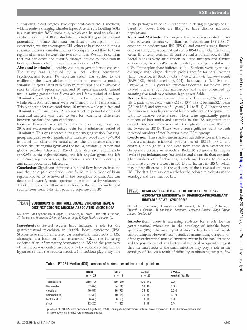
surrounding blood oxygen level-dependent-based fMRI methods,which require a changing stimulus input. Arterial spin labelling (ASL)is a non-invasive fMRI technique, which can be used to calculatecerebral blood flow (CBF) in absolute units (ml/100 g per minute) andpotentially, to study the neural correlates of tonic pain. In thisexperiment, we aim to compare CBF values at baseline and during asustained noxious stimulus in order to compare blood flow to brainregions of interest between the two conditions. We aim to confirmthat ASL can detect and quantify changes induced by tonic pain inhealthy volunteers before using it in patients with IBS.Aims and Methods: 14 healthy volunteers gave informed consent.The study was approved by a local ethics committee.Psychophysics: topical 1% capsaicin cream was applied to themidline of the lower abdomen in order to generate a noxiousstimulus. Subjects rated pain every minute using a visual analoguescale in which 0 equals no pain and 10 equals extremely painfuluntil a rating greater than 5 was achieved for a period of at least10 minutes (predicted length of ASL perfusion scan). Imaging:whole brain ASL sequences were performed on a 3 Tesla SiemansTrio scanner under two conditions, 10 minutes while pain free and10 minutes of tonic pain. A non-parametric permutation-basedstatistical analysis was used to test for voxel-wise differencesbetween baseline and pain conditions.Results: Psychophysics: all 14 subjects (four men, mean age29 years) experienced sustained pain for a minimum period of10 minutes. This was repeated during the imaging session. Imaging:group analysis revealed significantly increased blood flow (p,0.05)in the left dorsolateral prefrontal cortex, the left anterior cingulatecortex, the left angular gyrus and the insula, caudate, putamen andglobus pallidus bilaterally. Blood flow decreased significantly(p,0.05) in the right thalamus, the left angular gyrus, the leftsupplementary motor area, the precuneus and the hippocampusand parahippocampus bilaterally.Conclusion: Significant differences in blood flow between baselineand the tonic pain condition were found in a number of brainregions known to be involved in the perception of pain. ASL candetect and quantify tonic experimental pain in healthy volunteers.This technique could allow us to determine the neural correlates ofspontaneous tonic pain that patients experience in IBS.
PT269 SUBGROUPS OF IRRITABLE BOWEL SYNDROME HAVE ADISTINCT COLONIC MUCOSA-ASSOCIATED MICROBIOTA
GC Parkes, NB Rayment, BN Hudspith, L Petrovska, M Lomer, J Brostoff, K Whelan,JD Sanderson. Nutritional Sciences Division, Kings College London, London, UK
Introduction: Several studies have indicated a role for thegastrointestinal microbiota in irritable bowel syndrome (IBS).Studies have shown an altered gastrointestinal microbiota in IBS,although most focus on faecal microbiota. Given the increasingevidence of an inflammatory component to IBS and the proximityof the mucosa-associated microbiota to the colonic epithelium, wehypothesise that the mucosa-associated microbiota play a key role
in the pathogenesis of IBS. In addition, differing subgroups of IBSbased on bowel habit are likely to have distinct microbialpopulations.Aims and Methods: To compare the mucosa-associated micro-biota between patients with diarrhoea-predominant IBS (IBS-D),constipation-predominant IBS (IBS-C) and controls using fluores-cent in-situ hybridisation. Patients with IBS-D were identified usingRome III criteria, whereas all controls had a normal bowel habit.Rectal biopsies were snap frozen in liquid nitrogen and 6\mumsections cut, fixed in 4% paraformaldehyde and permeabilised in0.2% Triton/phosphate-buffered saline. Sections were hybridisedovernight with oligonucleotide probes specific for total bacteria(EUB), bacteroides (Bac303), Clostridium coccoides–Eubacterium rectale(EREC482), bifidobacteria (Bif164), lactobacillus (Lab154) andEscherichia coli. Hybridised mucosa-associated microbiota wereviewed under a confocal microscope and were quantified bycounting five randomly selected high power fields.Results: Results are summarised in the table. The mean (95% CI) age ofIBS-D patients was 36.2 years (32.1 to 40.3), IBS-C patients 32.4 years(28.1 to 36.7) and controls 46.1 years (41.4 to 51.1). All bacteria werefound in a mucinous layer adjacent but not adherent to the epithelium,with no invasive bacteria seen. There were significantly greaternumbers of bacteroides and clostridia in the IBS subgroups thancontrols. Bifidobacteria were found in the highest numbers in IBS-C andthe lowest in IBS-D. There was a non-significant trend towardsincreased numbers of total bacteria in the IBS subgroups.Conclusion: This study demonstrates clear differences in the rectalmucosa-associated microbial populations of IBS-D, IBS-C andcontrols, although it is not clear from these data whether thechanges are primary or secondary. Both IBS subgroups had higherlevels of pro-inflammatory bacteroides and clostridia than controls.The numbers of bifidobacteria, which are known to be anti-inflammatory, were lowest in IBS-D and highest in IBS-C, whichmay reflect differences in the aetiology of these two subgroups ofIBS. The data here support a role for the colonic microbiota in theaetiology and treatment of IBS.
PT270 DECREASED LACTOBACILLI IN THE ILEAL MUCOSA-ASSOCIATED MICROBIOTA IN DIARRHOEA-PREDOMINANTIRRITABLE BOWEL SYNDROME
GC Parkes, L Petrovska, IJ Woodman, NB Rayment, BN Hudpsith, M Lomer, JBrostoff, K Whelan, JD Sanderson. Nutritional Sciences Division, Kings CollegeLondon, London, UK
Introduction: There is increasing evidence for a role for thegastrointestinal microbiota in the aetiology of irritable bowelsyndrome (IBS). The majority of studies to date have used faecal/colonic samples. However, recent studies demonstrating upregulationof the gastrointestinal mucosal immune system in the small intestineand the possible role of small intestinal bacterial overgrowth suggestthat the microbiota of the small intestine may play a role in theaetiology of IBS. As a result of difficulty in obtaining samples, few
Table PT-269 Median (IQR) numbers of bacteria per millimetre of epithelium
IBS-Dn = 27
IBS-Cn = 19
Controln = 27
p ValueKruskall–Wallis
Total bacteria 218 (189) 159 (249) 130 (145) 0.05
Bacteroides 67 (62) 74 (81) 16 (40) 0.001
Clostridia 40 (57) 66 (79) 25 (43) 0.010
Bifidobacteria 24 (33) 50 (95) 36 (35) 0.019
Lactobacillus 6 (40) 6 (23) 9 (18) 0.90
E coli 12 (25) 11 (30) 8 (16) 0.44
p Values of ,0.025 were considered significant. IBS-C, constipation-predominant irritable bowel syndrome; IBS-D, diarrhoea-predominantirritable bowel syndrome; IQR, interquartile range.
BSG abstracts
Gut 2009;58(Suppl I):A1–A156 A105
on July 20, 2022 by guest. Protected by copyright.
http://gut.bmj.com
/G
ut: first published as on 23 March 2009. D
ownloaded from
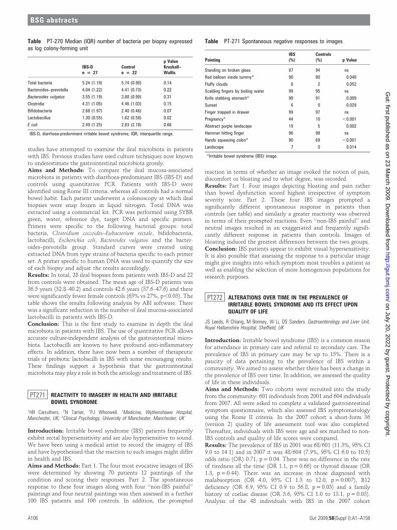
studies have attempted to examine the ileal microbiota in patientswith IBS. Previous studies have used culture techniques now knownto underestimate the gastrointestinal microbiota grossly.Aims and Methods: To compare the ileal mucosa-associatedmicrobiota in patients with diarrhoea-predominant IBS (IBS-D) andcontrols using quantitative PCR. Patients with IBS-D wereidentified using Rome III criteria, whereas all controls had a normalbowel habit. Each patient underwent a colonoscopy at which ilealbiopsies were snap frozen in liquid nitrogen. Total DNA wasextracted using a commercial kit. PCR was performed using SYBRgreen, water, reference dye, target DNA and specific primers.Primers were specific to the following bacterial groups: totalbacteria, Clostridium coccoides–Eubacterium rectale, bifidobacteria,lactobacilli, Escherichia coli, Bacteroides vulgatus and the bacter-oides–prevotella group. Standard curves were created usingextracted DNA from type strains of bacteria specific to each primerset. A primer specific to human DNA was used to quantify the sizeof each biopsy and adjust the results accordingly.Results: In total, 28 ileal biopsies from patients with IBS-D and 22from controls were obtained. The mean age of IBS-D patients was36.5 years (32.8–40.2) and controls 42.6 years (37.6–47.6) and therewere significantly fewer female controls (63% vs 27%, p,0.03). Thetable shows the results following analysis by ABI software. Therewas a significant reduction in the number of ileal mucosa-associatedlactobacilli in patients with IBS-D.Conclusion: This is the first study to examine in depth the ilealmicrobiota in patients with IBS. The use of quantitative PCR allowsaccurate culture-independent analysis of the gastrointestinal micro-biota. Lactobacilli are known to have profound anti-inflammatoryeffects. In addition, there have now been a number of therapeutictrials of probiotic lactobacilli in IBS with some encouraging results.These findings support a hypothesis that the gastrointestinalmicrobiota may play a role in both the aetiology and treatment of IBS.
PT271 REACTIVITY TO IMAGERY IN HEALTH AND IRRITABLEBOWEL SYNDROME
1HR Carruthers, 2N Tarrier, 1PJ Whorwell. 1Medicine, Wythenshawe Hospital,Manchester, UK; 2Clinical Psychology, University of Manchester, Manchester, UK
Introduction: Irritable bowel syndrome (IBS) patients frequentlyexhibit rectal hypersensitivity and are also hypersensitive to sound.We have been using a medical artist to record the imagery of IBSand have hypothesised that the reaction to such images might differin health and IBS.Aims and Methods: Part 1. The four most evocative images of IBSwere determined by showing 70 patients 12 paintings of thecondition and scoring their responses. Part 2. The spontaneousresponse to these four images along with four ‘‘non-IBS painful’’paintings and four neutral paintings was then assessed in a further100 IBS patients and 100 controls. In addition, the prompted
reaction in terms of whether an image evoked the notion of pain,discomfort or bloating and to what degree, was recorded.Results: Part 1. Four images depicting bloating and pain ratherthan bowel dysfunction scored highest irrespective of symptomseverity score. Part 2. These four IBS images prompted asignificantly different spontaneous response in patients thancontrols (see table) and similarly a greater reactivity was observedin terms of their prompted reactions. Even ‘‘non-IBS painful’’ andneutral images resulted in an exaggerated and frequently signifi-cantly different response in patients than controls. Images ofbloating induced the greatest differences between the two groups.Conclusion: IBS patients appear to exhibit visual hypersensitivity.It is also possible that assessing the response to a particular imagemight give insights into which symptom most troubles a patient aswell as enabling the selection of more homogenous populations forresearch purposes.
PT272 ALTERATIONS OVER TIME IN THE PREVALENCE OFIRRITABLE BOWEL SYNDROME AND ITS EFFECT UPONQUALITY OF LIFE
JS Leeds, R Chiang, M Bonney, W Li, DS Sanders. Gastroenterology and Liver Unit,Royal Hallamshire Hospital, Sheffield, UK
Introduction: Irritable bowel syndrome (IBS) is a common reasonfor attendance in primary care and referral to secondary care. Theprevalence of IBS in primary care may be up to 15%. There is apaucity of data pertaining to the prevalence of IBS within acommunity. We aimed to assess whether there has been a change inthe prevalence of IBS over time. In addition, we assessed the qualityof life in these individuals.Aims and Methods: Two cohorts were recruited into the studyfrom the community: 601 individuals from 2001 and 604 individualsfrom 2007. All were asked to complete a validated gastrointestinalsymptom questionnaire, which also assessed IBS symptomatologyusing the Rome II criteria. In the 2007 cohort a short-form 36(version 2) quality of life assessment tool was also completed.Thereafter, individuals with IBS were age and sex matched to non-IBS controls and quality of life scores were compared.Results: The prevalence of IBS in 2001 was 68/601 (11.3%, 95% CI9.0 to 14.1) and in 2007 it was 48/604 (7.9%, 95% CI 6.0 to 10.5)odds ratio (OR) 0.71, p = 0.04. There was no difference in the rateof tiredness all the time (OR 1.1, p = 0.66) or thyroid disease (OR1.3, p = 0.44). There was an increase in those diagnosed withmalabsorption (OR 4.0, 95% CI 1.3 to 12.0, p = 0.007), B12deficiency (OR 6.9, 95% CI 0.9 to 56.8, p = 0.03) and a familyhistory of coeliac disease (OR 3.6, 95% CI 1.0 to 13.1, p = 0.03).Analysis of the 48 individuals with IBS in the 2007 cohort
Table PT-270 Median (IQR) number of bacteria per biopsy expressedas log colony-forming unit
IBS-Dn = 27
Controln = 22
p ValueKruskall–Wallis
Total bacteria 5.24 (1.19) 5.74 (0.90) 0.14
Bacteroides–prevotella 4.04 (1.22) 4.41 (0.73) 0.22
Bacteroides vulgatus 3.55 (1.19) 3.80 (0.99) 0.31
Clostridia 4.31 (1.05) 4.46 (1.03) 0.15
Bifidobacteria 2.68 (1.97) 2.40 (0.46) 0.07
Lactobacillus 1.30 (0.55) 1.62 (0.58) 0.02
E coli 2.49 (1.25) 2.83 (2.18) 0.66
IBS-D, diarrhoea-predominant irritable bowel syndrome; IQR, interquartile range.
Table PT-271 Spontaneous negative responses to images
PaintingIBS(%)
Controls(%) p Value
Standing on broken glass 87 94 ns
Red balloon inside tummy* 90 80 0.048
Fluffy clouds 8 2 0.052
Scalding fingers by boiling water 99 95 ns
Knife stabbing stomach* 99 91 0.009
Sunset 6 0 0.029
Finger trapped in drawer 99 97 ns
Pregnancy* 44 10 ,0.001
Abstract purple landscape 19 5 0.002
Hammer hitting finger 96 98 ns
Hands squeezing colon* 90 69 ,0.001
Landscape 7 0 0.014
*Irritable bowel syndrome (IBS) image.
BSG abstracts
A106 Gut 2009;58(Suppl I):A1–A156
on July 20, 2022 by guest. Protected by copyright.
http://gut.bmj.com
/G
ut: first published as on 23 March 2009. D
ownloaded from
(compared with controls) revealed no differences in rates of thyroiddisease (OR 1.5), diabetes (OR 1.0) or anaemia (OR 1.3). However,there were significant differences in rates of abnormal bowel habit(OR 12.5), flatulence (OR 2.8), bloating (OR 2.8), floating stools(OR 3.7), abdominal pain (OR 3.3) and tiredness (OR 5.7).Comparison of short-form 36 (version 2) scores revealed asignificant reduction in physical functioning (p = 0.003), rolelimitations due to physical health (p = 0.023), bodily pain(p = 0.001), general health perceptions (p = 0.001), social function-ing domains (p = 0.048) and physical composite score (p,0.001).Conclusion: In our region there has been a significant reduction inIBS symptoms in the community; however, this has been coupledwith an increase in the diagnosis of malabsorption. Individuals withIBS symptoms have significantly reduced quality of life scoresparticularly in the physical domains by comparison with non-IBScontrols.
PT273 AUTONOMIC REACTIVITY TO PSYCHOLOGICAL ANDPHYSICAL STRESSORS DIFFERENTIALLY CORRELATES WITHMEASURES OF ANXIETY AND DEPRESSION IN PATIENTSWITH IRRITABLE BOWEL SYNDROME
1NM Thoua, 2A Hobson, 2G Dukes, 2D Kelleher, 2K Hicks, 2R Boardley, 1A Raeburn, 1AEmmanuel. 1Gastrointestinal Physiology Unit, University College Hospital, London, UK;2Gastrointestinal Discovery Medicine, GSK R&D Ltd, London, UK
Introduction: Psychological co-morbidity is prevalent in patientswith irritable bowel syndrome (IBS) and stress-induced exacerba-tion of IBS symptoms is also commonly reported. The relationshipbetween stress responsiveness and psychological phenotype in IBSis unknown. We investigated whether two key psychologicaldomains, anxiety and depression, correlate with stress-inducedchanges in rectal autonomic tone and sensitivity in IBS patients.Aims and Methods: Nine Rome II compliant IBS patients (eightwomen) were enrolled into the study. At the beginning of the studyall patients completed the hospital anxiety and depression (HAD)questionnaire. Baseline measures of rectal electrosensitivity andrectal mucosal blood flow (RMBF) were established and thenreassessed during 10-minute periods of psychological stress (dichot-omous listening), physical stress (cold-pressor) and during a 10-minute recovery period. Data are reported as mean change frombaseline with 95% CI.Results: Confirming our previous findings,1 both periods of stressreduced RMBF (physical: 275.4 (2109 to 241.8); psychological:259.5 (293.1 to 225.9); p,0.01). Anxiety scores correlatedsignificantly with the magnitude of change in RMBF during physicalstress (r = 20.471, p = 0.0175). In contrast, during psychologicalstress, depression scores correlated significantly with the magnitudeof change in RMBF (r = 20.442, p = 0.0271). Stress periods alsoreduced rectal sensory threshold (RST) (physical: 25.33 mA (28.01to 22.66); psychological: 24.56 mA (27.23 to 21.88); p,0.02) andpain threshold (RPT) (physical: 28.61 mA (212.56 to 24.66);psychological: 27.11 mA (211.06 to 23.16); p,0.01). Althoughdepression scores correlated positively with absolute pain thresholds(r = 0.432, p = 0.031), there was no correlation between HAD scoresand the magnitude of change of RST and RPT.Conclusion: We have shown that anxiety and depression correlatewith the magnitude of the gut autonomic response (as measured byRMBF) elicited by experimental stress, but do so in a selectivemanner. Anxiety, which is commonly associated with hypersensitiv-ity, correlated with physical stress, whereas depression, commonlyassociated with negative mood, correlated with psychological stress.The ability to assess the complex interplay between psychologicalprofile and stress responsiveness objectively may help furtherdifferentiate clinical phenotypes in the IBS population.
1. Murray, et al. Gastro 2004;127:1695–703.
PT274 THE EFFECT OF CENTRAL ADIPOSITY ON OESOPHAGEALACID EXPOSURE AND SYMPTOMS OF GASTRO-OESOPHAGEAL REFLUX DISEASE
R Anggiansah, RR Sweis, A Anggiansah, T Wong, M Fox. Department ofGastroenterology, St Thomas’ Hospital, London, UK
Introduction: Epidemiological evidence suggests a link betweengastro-oesophageal reflux disease (GORD) and obesity.1 2
Mechanistic studies suggest that this relationship is due to theeffects of central adiposity on intragastric pressure with disruptionof the gastro-oesophageal junction (GOJ) reflux barrier;3 however,the clinical relevance of these findings has not been proved.Aims and Methods: From August 2005 to December 2006, patientsreferred to a tertiary referral centre for oesophageal investigation ofsuspected GORD were studied. Height, weight and waist circumfer-ence (WC) were measured. Stationary manometry and 24-hambulatory pH studies off acid-suppressant medications wereperformed and symptom severity was assessed by a validatedquestionnaire. Linear regression models were used to analyseassociations between various covariates of interest and Pearson’scorrelation coefficient (PC) was used for continuous variables.Results: 582/676 (86%) consecutive patients (age 48 years (range14–89), 56% women) had complete data for analysis. Theprevalence of obesity defined by a body mass index (BMI) of30 kg/m2 or greater in men and women was 16% and 23% (p = ns),respectively. More men had WC of 99 cm or greater (men 41%,women 28%; p,0.001). The percentage of time with pH less than 4(%time pH,4) at 5 cm above the lower oesophageal sphincter(LOS) increased with obesity (PC 0.255 and 0.240 for BMI and WC,respectively; both p,0.001) and age (PC 0.162; p,0.001) and washigher in men than women (p = 0.014). An increase in the %timepH,4 was associated with a reduction in LOS pressure andabdominal LOS length (PC 20.327, 20.344, respectively; p,0.001).There was a weak negative association of BMI with LOS pressure(PC 20.140; p = 0.005) but no link with abdominal LOS length. Incontrast, there was a significant negative association of WC withLOS pressure (PC 20.225; p,0.001) and abdominal LOS length (PC20.213; p,0.001). Symptom severity increased with %time pH,4(PC 0.209; p,0.001); however, there was no change in symptomseverity with BMI (PC 0.056; p = 0.17) and the effect of WC onsymptom severity was weak and of borderline significance (PC0.083; p,0.05).Conclusion: In this large cohort of clinical patients referred forinvestigation of reflux symptoms, gastro-oesophageal reflux (%timepH,4) increased with obesity (BMI and WC) and was mostprevalent in elderly men with central adiposity. WC but not BMIwas associated with both reduced LOS pressure and abdominallength. These findings confirm the mechanistic link betweenincreasing WC and the risk of reflux via raised intragastric pressureand disruption of the GOJ. In contrast, there was little change insymptom severity as assessed by questionnaire. The relativeinsensitivity to acid reflux may explain the increased prevalenceof severe and complicated reflux in the obese population.1 2
1. Dent J, El-Serag HB, et al. Gut 2005;54:710–17.2. Locke GR, et al. Am J Med 1999;106:642–9.3. Pandolfino JE, El-Serag HB, et al. Gastroenterology 2006;130(3):639–49.
PT275 NORMAL VALUES FOR HIGH RESOLUTION MANOMETRYASSESSMENT OF LIQUID, SOLID AND MULTIPLE REPEATEDSWALLOWS IN THE UPRIGHT AND SUPINE POSITIONS
RR Sweis, T Wong, A Anggiansah, M Fox. Department of Gastroenterology, StThomas’ Hospital, London, UK
Introduction: High resolution manometry (HRM) is an advance inoesophageal measurement.1 2 Functionally relevant HRM measure-ments acquired by HRM include: peristaltic coordination between
BSG abstracts
Gut 2009;58(Suppl I):A1–A156 A107
on July 20, 2022 by guest. Protected by copyright.
http://gut.bmj.com
/G
ut: first published as on 23 March 2009. D
ownloaded from
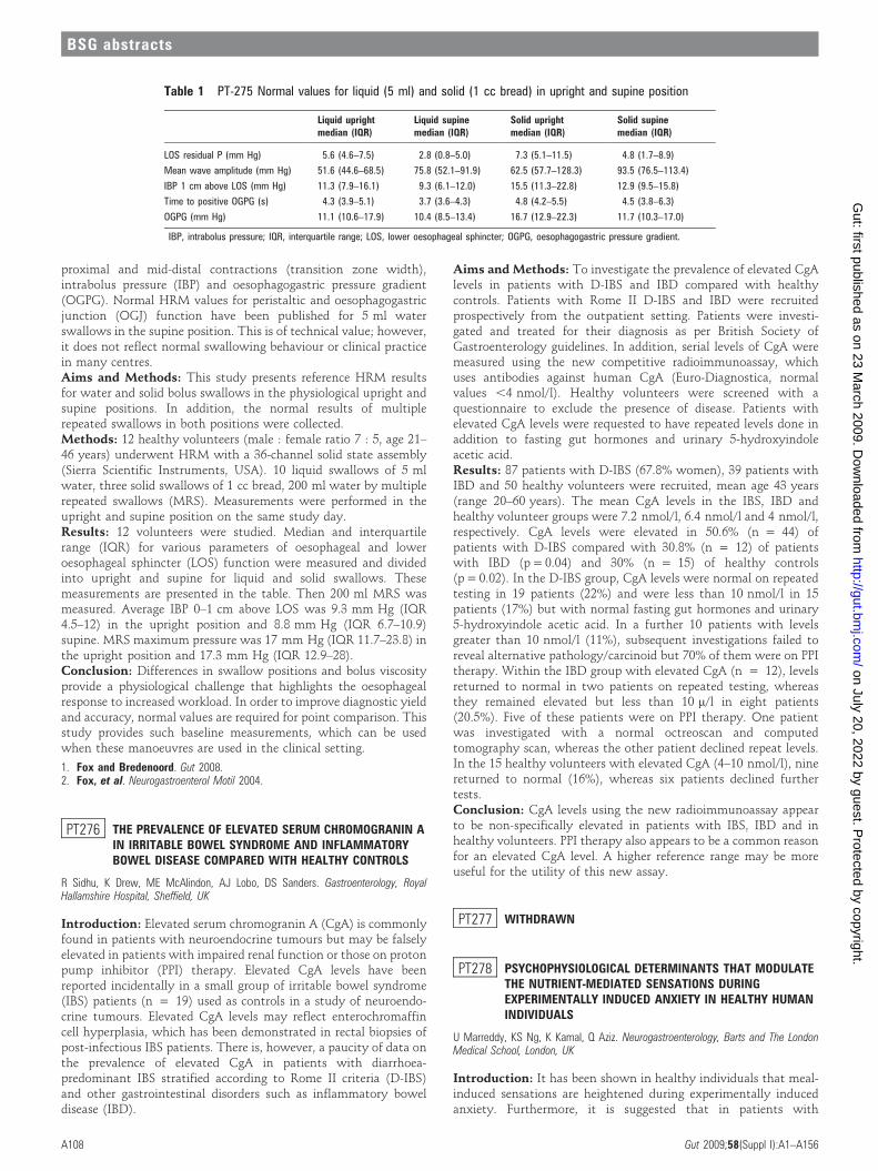
proximal and mid-distal contractions (transition zone width),intrabolus pressure (IBP) and oesophagogastric pressure gradient(OGPG). Normal HRM values for peristaltic and oesophagogastricjunction (OGJ) function have been published for 5 ml waterswallows in the supine position. This is of technical value; however,it does not reflect normal swallowing behaviour or clinical practicein many centres.Aims and Methods: This study presents reference HRM resultsfor water and solid bolus swallows in the physiological upright andsupine positions. In addition, the normal results of multiplerepeated swallows in both positions were collected.Methods: 12 healthy volunteers (male : female ratio 7 : 5, age 21–46 years) underwent HRM with a 36-channel solid state assembly(Sierra Scientific Instruments, USA). 10 liquid swallows of 5 mlwater, three solid swallows of 1 cc bread, 200 ml water by multiplerepeated swallows (MRS). Measurements were performed in theupright and supine position on the same study day.Results: 12 volunteers were studied. Median and interquartilerange (IQR) for various parameters of oesophageal and loweroesophageal sphincter (LOS) function were measured and dividedinto upright and supine for liquid and solid swallows. Thesemeasurements are presented in the table. Then 200 ml MRS wasmeasured. Average IBP 0–1 cm above LOS was 9.3 mm Hg (IQR4.5–12) in the upright position and 8.8 mm Hg (IQR 6.7–10.9)supine. MRS maximum pressure was 17 mm Hg (IQR 11.7–23.8) inthe upright position and 17.3 mm Hg (IQR 12.9–28).Conclusion: Differences in swallow positions and bolus viscosityprovide a physiological challenge that highlights the oesophagealresponse to increased workload. In order to improve diagnostic yieldand accuracy, normal values are required for point comparison. Thisstudy provides such baseline measurements, which can be usedwhen these manoeuvres are used in the clinical setting.
1. Fox and Bredenoord. Gut 2008.2. Fox, et al. Neurogastroenterol Motil 2004.
PT276 THE PREVALENCE OF ELEVATED SERUM CHROMOGRANIN AIN IRRITABLE BOWEL SYNDROME AND INFLAMMATORYBOWEL DISEASE COMPARED WITH HEALTHY CONTROLS
R Sidhu, K Drew, ME McAlindon, AJ Lobo, DS Sanders. Gastroenterology, RoyalHallamshire Hospital, Sheffield, UK
Introduction: Elevated serum chromogranin A (CgA) is commonlyfound in patients with neuroendocrine tumours but may be falselyelevated in patients with impaired renal function or those on protonpump inhibitor (PPI) therapy. Elevated CgA levels have beenreported incidentally in a small group of irritable bowel syndrome(IBS) patients (n = 19) used as controls in a study of neuroendo-crine tumours. Elevated CgA levels may reflect enterochromaffincell hyperplasia, which has been demonstrated in rectal biopsies ofpost-infectious IBS patients. There is, however, a paucity of data onthe prevalence of elevated CgA in patients with diarrhoea-predominant IBS stratified according to Rome II criteria (D-IBS)and other gastrointestinal disorders such as inflammatory boweldisease (IBD).
Aims and Methods: To investigate the prevalence of elevated CgAlevels in patients with D-IBS and IBD compared with healthycontrols. Patients with Rome II D-IBS and IBD were recruitedprospectively from the outpatient setting. Patients were investi-gated and treated for their diagnosis as per British Society ofGastroenterology guidelines. In addition, serial levels of CgA weremeasured using the new competitive radioimmunoassay, whichuses antibodies against human CgA (Euro-Diagnostica, normalvalues ,4 nmol/l). Healthy volunteers were screened with aquestionnaire to exclude the presence of disease. Patients withelevated CgA levels were requested to have repeated levels done inaddition to fasting gut hormones and urinary 5-hydroxyindoleacetic acid.Results: 87 patients with D-IBS (67.8% women), 39 patients withIBD and 50 healthy volunteers were recruited, mean age 43 years(range 20–60 years). The mean CgA levels in the IBS, IBD andhealthy volunteer groups were 7.2 nmol/l, 6.4 nmol/l and 4 nmol/l,respectively. CgA levels were elevated in 50.6% (n = 44) ofpatients with D-IBS compared with 30.8% (n = 12) of patientswith IBD (p = 0.04) and 30% (n = 15) of healthy controls(p = 0.02). In the D-IBS group, CgA levels were normal on repeatedtesting in 19 patients (22%) and were less than 10 nmol/l in 15patients (17%) but with normal fasting gut hormones and urinary5-hydroxyindole acetic acid. In a further 10 patients with levelsgreater than 10 nmol/l (11%), subsequent investigations failed toreveal alternative pathology/carcinoid but 70% of them were on PPItherapy. Within the IBD group with elevated CgA (n = 12), levelsreturned to normal in two patients on repeated testing, whereasthey remained elevated but less than 10 m/l in eight patients(20.5%). Five of these patients were on PPI therapy. One patientwas investigated with a normal octreoscan and computedtomography scan, whereas the other patient declined repeat levels.In the 15 healthy volunteers with elevated CgA (4–10 nmol/l), ninereturned to normal (16%), whereas six patients declined furthertests.Conclusion: CgA levels using the new radioimmunoassay appearto be non-specifically elevated in patients with IBS, IBD and inhealthy volunteers. PPI therapy also appears to be a common reasonfor an elevated CgA level. A higher reference range may be moreuseful for the utility of this new assay.
PT277 WITHDRAWN
PT278 PSYCHOPHYSIOLOGICAL DETERMINANTS THAT MODULATETHE NUTRIENT-MEDIATED SENSATIONS DURINGEXPERIMENTALLY INDUCED ANXIETY IN HEALTHY HUMANINDIVIDUALS
U Marreddy, KS Ng, K Kamal, Q Aziz. Neurogastroenterology, Barts and The LondonMedical School, London, UK
Introduction: It has been shown in healthy individuals that meal-induced sensations are heightened during experimentally inducedanxiety. Furthermore, it is suggested that in patients with
Table 1 PT-275 Normal values for liquid (5 ml) and solid (1 cc bread) in upright and supine position
Liquid uprightmedian (IQR)
Liquid supinemedian (IQR)
Solid uprightmedian (IQR)
Solid supinemedian (IQR)
LOS residual P (mm Hg) 5.6 (4.6–7.5) 2.8 (0.8–5.0) 7.3 (5.1–11.5) 4.8 (1.7–8.9)
Mean wave amplitude (mm Hg) 51.6 (44.6–68.5) 75.8 (52.1–91.9) 62.5 (57.7–128.3) 93.5 (76.5–113.4)
IBP 1 cm above LOS (mm Hg) 11.3 (7.9–16.1) 9.3 (6.1–12.0) 15.5 (11.3–22.8) 12.9 (9.5–15.8)
Time to positive OGPG (s) 4.3 (3.9–5.1) 3.7 (3.6–4.3) 4.8 (4.2–5.5) 4.5 (3.8–6.3)
OGPG (mm Hg) 11.1 (10.6–17.9) 10.4 (8.5–13.4) 16.7 (12.9–22.3) 11.7 (10.3–17.0)
IBP, intrabolus pressure; IQR, interquartile range; LOS, lower oesophageal sphincter; OGPG, oesophagogastric pressure gradient.
BSG abstracts
A108 Gut 2009;58(Suppl I):A1–A156
on July 20, 2022 by guest. Protected by copyright.
http://gut.bmj.com
/G
ut: first published as on 23 March 2009. D
ownloaded from
functional dyspepsia psychological factors modulate satiationsignals and are partly responsible for the symptom burdenexperienced. However, assessment of meal-induced sensations inresponse to psychological stressors is subjective and it is not knownif activation of the stress response such as the autonomic nervoussystem (ANS) also occurs and whether this ANS activation can actas an objective biomarker of the effect of stress on meal-inducedsymptoms. In this study we explored the interaction betweenpsychological factors and autonomic reactivity patterns in theperception of meal sensations during experimentally inducedanxiety in healthy individuals.Aims and Methods: 25 healthy volunteers (aged 28 ¡ 9 years, 14men and 11 women) received standardised nutrient challenge(Fortisip; Nutricia, 50 ml/minute) and were exposed to neutral andanxiogenic stimuli (shock threat). Anxiety ratings were assessed.Sympathetic and parasympathetic activity was measured (eg, heartrate, mean arterial blood pressure and cardiac vagal tone usingNeuroScope. We assessed the personality traits and perceived stressand anxiety.Results: 25 healthy volunteers with a mean age of 28 ¡ 9 yearscompleted both the stress and neutral experiments. Anxiety visualanalogue scale (VAS) scores and Spielberger trait anxiety index(STAI) anxiety scores confirmed anxiety induction during the stressexperiment (anxiety VAS neutral 6.5 ¡ 1.9 vs anxiety 2.6 ¡ 1.2;p,0.02), (STAI scores, neutral 34.0 ¡ 1.8 vs anxiety 42.2 ¡ 2.4;p = 0.01). The intensity of epigastric symptom ratings scores tonutrient challenge for discomfort (1.4 ¡ 0.4 vs 2.9 ¡ 0.4;p = 0.01), fullness (5.1 ¡ 0.6 vs 6.0 ¡ 0.5; p = 0.01) and belching(1.5 ¡ 0.3 vs 2.6 ¡ 0.5; p = 0.01) were significantly higher duringthe anxious state. Autonomic reactivity patterns revealed increasedsympathetic activity (coping strategies index baseline 1.6 ¡ 0.2 vsstress 2.3 ¡ 0.2; p = 0.03) and decreased parasympathetic tone(baseline 12.9 ¡ 1.8 vs 6.7 ¡ 0.8; p = 0.01) during the anxietyinduction in most subjects. No significant correlation betweenpsychological factors and symptom scores was seen, neither wasany correlation found between symptom scores and ANS measures.Conclusion: In our study we have found that experimental anxietyenhances the meal sensations and increases the intensity ofperceived epigastric symptoms when compared with a neutralcondition. A typical autonomic reactivity pattern was observedduring anxiety induction, ie, increased sympathetic activity andwithdrawal of parasympathetic activity. This ANS reactivity duringanxiety, especially the lowering of vagal activity, might beresponsible for the heightened perception of symptoms and requiresfurther study in a larger cohort of healthy subjects and patientswith functional dyspepsia.
PT279 PREVALENCE AND SYMPTOM PROFILING OFOROPHARYNGEAL DYSPHAGIA IN A COMMUNITY-DWELLING ELDERLY POPULATION: A SELF-REPORTINGQUESTIONNAIRE SURVEY
1G Holland, 1V Jayasekeran, 2N Pendleton, 2M Horan, 2M Jones, 1S Hamdy.1Gastrointestinal Sciences, School of Translational Medicine, University of Manchester,Manchester, UK; 2Age and Cognitive Performance Research Centre (ACPRC), School ofTranslational Medicine, University of Manchester, Manchester, UK
Introduction: Although the prevalence and clinical consequencesof oropharyngeal dysphagia in diseases such as neuromusculardisorders and cancer are well established, virtually nothing isknown about the prevalence of dysphagia in the healthy olderpopulation. Furthermore, the symptomatic basis for oropharyngealdysphagia remains unclear. Here, we describe a detailed question-naire-based survey of swallowing dysfunction in a large otherwisehealthy community-dwelling older population in the UK.
Aims and Methods: A postal survey of the Sydney oropharyngealdysphagia questionnaire1 was sent to 800 residences in the north ofEngland that formed part of the University of Manchester Age andCognitive Performance Longitudinal Study.2 This cohort comprisesolder (mean age 81 years, range 69–97) individuals who areotherwise healthy on entry, with no history of previous neurolo-gical disease. The postal questionnaire was a validated self-reportinventory to measure the symptomatic severity of oral-pharyngealdysphagia, which assessed a total of 17 domains of swallowingfunction. The maximal score obtainable is 1700, with a score of over200 indicating significant swallowing difficulty. For the purposes ofthis study, domain questions were also subcategorised intoobstructive or functional type symptoms. All data were analysedusing SPSS.Results: Of the 800 questionnaires sent out, 627 were returned,giving a response rate of 78%. 24 were later discarded as incomplete,giving a completed response rate of 76%. There was a femalepreponderance at 76.3% of the total respondents. 13.6% of thepopulation indicated that they had significant dysphagia.Unsurprisingly, the levels of dysphagia severity were directlycorrelated with the subjects’ age (r = 0.12, p = 0.009). Whensubcategorised, 71% of the dysphagic subjects gave symptomssuggestive of functional dysphagia producing airway compromise,with 68% indicating symptoms more of an obstructive nature. Thethree commonest symptoms included a feeling of food sticking inthe throat, a sensation of choking or coughing on swallowing anddifficulty swallowing hard foods.Conclusion: This is the first study carefully to quantifyoropharyngeal dysphagia in the healthy old and shows thatdysphagia is prevalent in older people, affecting nearly one in sixpeople who are deemed otherwise healthy and living independentlyin the community. Clinicians should therefore be vigilant to thepossibility of swallowing problems even in the seemingly well olderpopulation, which may require further evaluation.
1. Wallace, et al. Gastroenterology 2000.2. Rabbitt, et al. Aging, Neuropsychol Cogn 1999.
Oesophagus posters
PT280 OESOPHAGEAL CONTRAST STUDIES BEFORE GASTROSCOPYIN THE INVESTIGATION OF HIGH DYSPHAGIA
1A Mawas, 1E Cameron, 1S Middleton, 2N Carroll, 1I Nasr. 1Gastroenterology,Addenbrooke’s Hospital, Cambridge, UK; 2Radiology, Addenbrooke’s Hospital,Cambridge, UK
Introduction: It is contentious whether radiological investigationshould precede endoscopy in patients with high dysphagia.Radiology may identify patients with pathology in whom it ispotentially hazardous to perform flexible endoscopy (eg, those withpharyngeal pouches), although some suggest that endoscopy isnecessary in almost all cases and careful intubation is safe. It hasbeen our practice for some time to perform a water-soluble swallowin all patients with dysphagia above the manubrium beforeendoscopy. We have retrospectively reviewed all cases from 2005until 2007 to establish the rate of significant high pathology andalso to identify potential factors associated with significantproximal pathology.Aims and Methods: All contrast oesophageal studies requested bythe dyspepsia service to investigate high dysphagia between 2005and 2007 were identified from the radiology database. Data wereretrospectively collected regarding patient demographics and theresults of contrast oesophageal studies.Results: 376 patients underwent water-soluble swallow. 156(41.5%) were men and the mean age was 64 years (SD 17).Pharyngeal pouches were identified in five of 376 (1.3%) patients.
BSG abstracts
Gut 2009;58(Suppl I):A1–A156 A109
on July 20, 2022 by guest. Protected by copyright.
http://gut.bmj.com
/G
ut: first published as on 23 March 2009. D
ownloaded from
Four of 166 (2.4%) were older than 70 years and one was 54 yearsold, giving an incidence of oesophageal pouch of one in 211 (0.47%)in patients younger than 70 years. Cricopharyngeal web was foundin four of 376 (1%), with an incidence of three in 166 (1.8%) inpatients 70 years and older and one of 211 (0.47%) in patientsyounger than 70 years (one patient aged 69 years).Conclusion: 1.3% of unselected patients referred with dysphagiaabove the manubrium will have a pharyngeal pouch. Almost all ofthese are over 70 years. These data suggest that, if pre-endoscopicoesophageal imaging for high dysphagia is limited to this patientgroup, the incidence of important findings will be seven in 166(4.2%) and oesophageal contrast studies could be avoided with verylittle risk in 55% of all referrals.
PT281 A POPULATION-BASED STUDY OF POLYMORPHISMS OF THEINSULIN-LIKE GROWTH FACTOR AXIS AND THEOESOPHAGEAL INFLAMMATION, METAPLASIA,ADENOCARCINOMA SEQUENCE
1AR McElholm, 2AJ McKnight, 2S Heggarty, 1C Patterson, 1BT Johnston, 3L Hardie, 1LMurray. 1Cancer Epidemiology and Prevention Research Group, Centre for Clinical andPopulation Sciences, Queens University of Belfast, Belfast, UK; 2Genomics Core Facility,Queens University of Belfast, Belfast City Hospital, Belfast, UK; 3Molecular EpidemiologyGroup, Leeds University, Leeds, UK
Introduction: The insulin-like growth factor (IGF) axis plays a keyrole in cell development, proliferation and survival and has beenimplicated in the aetiology of many cancers, including obesity-related cancers. To date, no studies have examined the relationshipbetween this axis and oesophageal adenocarcinoma (OAC) or itsprecursors.Aims and Methods: We investigated the association of commonpolymorphisms of the IGF axis with all stages of the oesophagealinflammation, metaplasia, adenocarcinoma sequence. The genesexamined were IGF-1 and IGF-2, IGF-1 receptor (IGF-1R), IGFbinding protein 3 (IGFBP3), the growth hormones (GH-1 and GH-2)and GH receptor (GHR). The Factors Influencing the Barrett’sAdenocarcinoma Relationship (FINBAR) study is an all-Irelandpopulation-based, case–control study of OAC and its precursors.We analysed DNA from subjects with reflux oesophagitis (RO,n = 230), Barrett’s oesophagus (BO, >3 cm BO at endoscopy withspecialised intestinal metaplasia on biopsy, n = 189), OAC(n = 207) and normal controls (n = 223). Using a gene-basedapproach common single-nucleotide polymorphisms (SNP) wereidentified for each gene using a variety of publicly available onlineresources. Genotyping was performed using the SequenomMassARRAY genotyping system and Taqman assay. Results wereanalysed using Haploview, a software package that providescomputation of linkage disequilibrium statistics (including permu-tation testing) and population haplotype patterns from primarygenotype data. 90 IGF axis SNP (tagging 299 alleles) met theinclusion criteria and were analysed. Three polymorphisms wereassociated with disease status.Results: The IGF-1 SNP rs6214 was associated with BO (p = 0.005),even after multiple testing (10 000 permutations; p = 0.0389). UsingAA genotype as a reference, the odds ratio (OR) for GG (‘‘wild-type’’)and BO was 2.40 (95% CI 1.31 to 4.41). The GHR SNP rs6898743 wasassociated with OAC, p = 0.0012 (10 000 permutations; p = 0.0112).With GG as the reference, the OR for CC (‘‘wild-type’’) genotype andOAC was 0.42 (95% CI 0.23 to 0.76). The IGF-1 (CA)19 ‘‘185’’ repeatwas associated with ‘‘189’’ as a reference, the OR for ‘‘185’’ and ROwas 7.67 (95% CI 1.73 to 69.92)Conclusion: We systematically examined genetic variations of theIGF axis in a population-based case–control study of OAC, BO andRO. A microsatellite repeat and two SNP were statisticallysignificantly associated with disease states: IGF-1 (CA)19 ‘‘185’’repeat with RO, rs6214 (IGF-1) with BO and rs6898743 (GHR) with
OAC. These SNP could potentially be used as a marker for diseaserisk, but independent validation of our findings is required. Theseresults also suggest that the IGF pathway may be involved in thedevelopment of OAC and its precursors.
PT282 LONG-TERM OUTCOME OF BARRETT’S OESOPHAGUS-ASSOCIATED CANCERS IN SURVEYED AND NON-SURVEYEDPATIENTS
1AK Muddu, 1J Fallowfield, 2M Lesn, 3N Davies, 1P Winwood. 1Department ofGastroenterology, Royal Bournemouth Hospital, Bournemouth, UK; 2Department ofHistopathology, Royal Bournemouth Hospital, Bournemouth, UK; 3Department ofSurgery, Royal Bournemouth Hospital, Bournemouth, UK
Introduction: Barrett’s oesophagus (BO) is a precursor ofoesophageal adenocarcinoma. Endoscopic surveillance programmesaim to detect BO-associated adenocarcinomas at an earlier stagewhen intervention is likely to result in better survival. Other studieshave demonstrated that surveillance-detected cancers have a betteroutcome than de novo cancers, but lead time bias may account forsome or all of the survival benefit.Aims and Methods: Our aim was to determine the long-termoutcome of patients diagnosed with BO-associated adenocarcinomabetween 1998 and 2002. The patients had originally been identifiedby review of the pathology computer databases and were dividedinto surveillance and de novo cancers and further subdivided intopatients who had undergone resection and those who receivedpalliative therapy. Information about survival was obtainedthrough review of the notes and hospital administration systems.Results: 54 BO-associated adenocarcinomas were identifiedbetween 1998 and 2002. Eight were detected in patients undergoingsurveillance and all patients underwent oesophagectomy or endo-scopic mucosal resection. Six patients (75%) had a survival greaterthan 5 years. 46 patients presented with ‘‘de novo’’ cancers andonly 15 patients were suitable for resection. Of these, nine (64%)were alive at 5 years. 31 patients were only suitable for palliativetherapy, three patients were lost to follow-up and of the remaining28, one patient (4%) was alive at 5 years. In total, 5-year survival ofthe de novo group was 28% compared with 75% of surveillancecancers.Conclusion: These data suggest that patients who have cancersdetected during BO surveillance have an increased probability ofbeing offered potentially curative resection with improved long-term survival compared with de novo cancers. Although lead timebias cannot be totally excluded, the sustained survival suggests thatthis is likely to be minimal and continued surveillance is justified.
PT283 ERBB2 EXPRESSION IN OESOPHAGEAL ADENOCARCINOMAAND BARRETT’S OESOPHAGUS: DEMONSTRATING THEPOTENTIAL FOR A PRE-EMPTIVE STRIKE
1AL Paterson, 2E Provenzano, 2M O’Donovan, 2SE Davies, 3MR Novelli, 4LB Lovat, 1RCFitzgerald. 1MRC Cancer Cell Unit, Hutchison MRC Research Centre, Cambridge, UK;2Department of Pathology, Addenbrooke’s Hospital, Cambridge, UK; 3Department ofPathology, University College Hospital, London, UK; 4Department of Gastroenterology,University College Hospital, London, UK
Introduction: The incidence of oesophageal adenocarcinoma(OAC) has increased fourfold in the west in 30 years and has beenhighlighted as a major public health concern. It has a dismal 15% 5-year survival due to its late presentation and the limited benefit ofcurrent chemotherapy regimens. Targeted therapies have thepotential to revolutionise chemotherapy as shown in ErbB2-overexpressing breast tumours, which account for 25% of cases.The role of the ErbB family in Barrett’s oesophagus and OACdevelopment and progression remains to be fully explored.
BSG abstracts
A110 Gut 2009;58(Suppl I):A1–A156
on July 20, 2022 by guest. Protected by copyright.
http://gut.bmj.com
/G
ut: first published as on 23 March 2009. D
ownloaded from
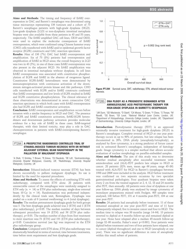
Aims and Methods: The timing and frequency of ErbB2 over-expression in OAC and Barrett’s oesophagus was determined usingtissue microarrays representing 136 tumours and a cohort of 73Barrett’s oesophagus patients with high-grade dysplasia (HGD).Low-grade dysplasia (LGD) or non-dysplastic intestinal metaplasiabiopsies were also available from these patients in 43 and 53 cases,respectively. The ErbB2 amplified OAC cell lines, OE19 and OE33,were used to explore the molecular consequences of ErbB2overexpression, which were confirmed using Chinese hamster ovary(CHO) cells transfected with ErbB2 and/or epidermal growth factorreceptor (EGFR) constructs and OAC resection specimens.Results: Nine of 136 (7%) OAC had ErbB2 overexpression andamplification. Six of 73 (8%) patients had overexpression andamplification of ErbB2 in HGD areas; the overall frequency in LGDwas two in 43 (5%), in one of these cases ErbB2 overexpression wasalso present in the adjacent HGD. No ErbB2 amplification wasobserved in intestinal metaplasia without dysplasia. In cell linesErbB2 overexpression was associated with constitutive phosphor-ylation of EGFR and ErbB2 in the absence of exogenous ligand.Constitutive EGFR-ErbB2 heterodimers were demonstrated byimmunoprecipitation with continuous activation of the down-stream mitogen-activated protein kinase and Akt pathways. CHOcells transfected with EGFR and/or ErbB2 constructs confirmedthat ErbB2 overexpression and low levels of EGFR resulted in ErbB2and EGFR constitutive phosphorylation. The clinical relevance ofthese observations was demonstrated in seven consecutive OACresection specimens in which both cases with ErbB2 overexpressionalso had EGFR and ErbB2 constitutive activation.Conclusion: ErbB2 overexpression and amplification occurs beforeinvasion with a similar frequency in HGD and OAC. The presenceof EGFR and ErbB2 constitutive activation, ErbB2-EGFR hetero-dimers and downstream pathway activation provides molecularevidence for a key role of ErbB2 in these cases. ErbB-targetedtherapies, with their limited toxicity, may play a role in OACchemoprevention in patients with ErbB2-overexpressing Barrett’soesophagus.
PT284 A PROSPECTIVE RANDOMISED CONTROLLED TRIAL OFETHANOL-INDUCED TUMOUR NECROSIS WITH OR WITHOUTADJUVANT EXTERNAL BEAM RADIOTHERAPY IN THEPALLIATION OF MALIGNANT DYSPHAGIA
1A Shah, 1C Grimley, 2I Brown, 2R Grieve, 1CU Nwokolo, 1DE Loft. 1Gastroenterology,University Hospital Walsgrave, Coventry, UK; 2Radiotherapy, University HospitalWalsgrave, Coventry, UK
Introduction: Ethanol-induced tumour necrosis (ETN) has beenshown successfully to palliate malignant dysphagia. Its use islimited by the need for repeated procedures.Aims and Methods: To assess the effects of combining ETN withradiotherapy, compared with ETN alone. 28 patients withunresectable cancer of the oesophagus were randomly assigned toETN only (n = 14) or ETN plus radiotherapy, single-dose externalbeam 10 Gy (n = 14). Randomisation was performed indepen-dently by the clinical trials unit, Birmingham. Dysphagia wasgraded on a scale of 0 (normal swallowing) to 4 (total dysphagia).Results: The median pretreatment dysphagia grade for both groupswas 3. The best dysphagia grade achieved was 2 (ETN) and 1 (ETNplus radiotherapy), p,0.05. The median number of days betweensubsequent procedures was 19 (ETN) and 56 (ETN plus radio-therapy), p,0.01. The median number of days from first treatmentto stent insertion was 51 (ETN) and 191 (ETN plus radiotherapy),p,0.05. Cumulative survival (see fig) was improved in the ETNplus radiotherapy group.Conclusion: Compared with ETN alone, ETN plus radiotherapy wasdramatically beneficial in terms of survival, time between treatments,time before stent requirement and best dysphagia grade.
PT285 DNA PLOIDY AS A PROGNOSTIC BIOMARKER AFTERAMINOLEVULINIC ACID PHOTODYNAMIC THERAPY FORHIGH-GRADE DYSPLASIA IN BARRETT’S OESOPHAGUS
1JM Dunn, 1GD Mackenzie, 2D Oukrif, 1CA Mosse, 2S Green, 2Y Chew, 1S Thorpe, 2MNovelli, 1SG Bown, 3LB Lovat. 1National Medical Laser Centre, London, UK;2Department of Histopathology, University College London, London, UK; 3Departmentof Gastroenterology, University College Hospital, London, UK
Introduction: Photodynamic therapy (PDT) is an approvedminimally invasive treatment for high-grade dysplasia (HGD) inBarrett’s oesophagus. Complete reversal of HGD at one year post-therapy occurs in up to 90% of patients, but late relapse has beendocumented in 20%. DNA ploidy (aneuploidy/tetraploidy), asanalysed by flow cytometry, is a strong predictor of future cancerrisk in untreated Barrett’s oesophagus, independent of histologygrade. Image cytometry is a simpler method that allows accurateevaluation of nuclear morphology on paraffin-embedded samples.Aims and Methods: The aim of this study was to determinewhether residual aneuploidy after successful treatment withaminolevulinic acid (ALA)-PDT predicts late relapse to HGD orcancer. 28 patients (82% men, mean age 66 years; range 45–86)treated with ALA-PDT for HGD in Barrett’s oesophagus between1998 and 2006 were included in the analysis. HGD before treatmentwas confirmed on two separate occasions by two specialistgastrointestinal pathologists. Four quadrant biopsies were takenevery 2 cm from the treated segment at 2, 4, 12, 18 and 24 monthsafter PDT, then annually. All patients were clear of dysplasia at oneyear follow-up. DNA ploidy was analysed by image cytometry ofmonolayer preparations stained with Feulgen. 405 samples wereanalysed; 148 before PDT; 131 at 4 months post-PDT; 104 at oneyear post-PDT.Results: 24 patients had aneuploidy before treatment. 11 of thoseremained aneuploid at one year post-PDT and nine of 11 havesubsequently relapsed; five to invasive cancer and four to HGD. Themedian time to relapse was 30 months (range 14–64). 13 patientsreverted to diploid at 4 months follow-up and remained diploid atone year. None have relapsed after a median 45-month follow-up(range 32–60 months; Fisher’s exact p,0.001). Four patients werediploid before treatment and two of four patients have relapsed, oneto cancer (diploid throughout) and one to HGD (aneuploidy at oneyear). There was no significant difference in rates of aneuploidywithin this small subset of patients.
Figure PT-284 Survival curve. DXT, radiotherapy; ETN, ethanol-induced tumournecrosis.
BSG abstracts
Gut 2009;58(Suppl I):A1–A156 A111
on July 20, 2022 by guest. Protected by copyright.
http://gut.bmj.com
/G
ut: first published as on 23 March 2009. D
ownloaded from
Conclusion: Following successful PDT for HGD in Barrett’soesophagus, patients who remain aneuploid are significantly morelikely to develop HGD or cancer than those who become diploid.This may allow change in surveillance patterns to be tailored to thepatient, with persistent aneuploidy one year post-PDT necessitat-ing closer follow-up. Moreover, patients who have reverted todiploid after one year may return to 2-yearly follow-up and bereassured that the risk of cancer after 3 years is very low. These datademonstrate the utility of DNA ploidy as a prognostic biomarkerfollowing ablative therapy.
PT286 RADIOFREQUENCY ABLATION OF HIGH-GRADE DYSPLASIAIN BARRETT’S OESOPHAGUS AFTER FAILEDPHOTODYNAMIC THERAPY: FIRST RESULTS OF THE UK HALORADIOFREQUENCY ABLATION REGISTRY
1JM Dunn, 2G Fullarton, 1S Thorpe, 3M Novelli, 1SG Bown, 4LB Lovat. 1NationalMedical Laser Centre, University College London, London, UK; 2Department of Surgery,Glasgow Royal Infirmary, Glasgow, UK; 3Department of Histopathology, UniversityCollege London, London, UK; 4Department of Gastroenterology, University CollegeHospital, London, UK
Introduction: Photodynamic therapy (PDT) is an approvedminimally invasive treatment for high-grade dysplasia (HGD) inBarrett’s oesophagus in the UK, but has a failure rate of between15% and 50%. Radiofrequency ablation (RFA) is a new treatmentfor the eradication of non-dysplastic and dysplastic Barrett’soesophagus. There are no studies of its use to eradicate residualHGD after failed PDT.Aims and Methods: The aim of this prospective multicentre studywas to assess the safety and efficacy of RFA for Barrett’soesophagus with residual HGD following PDT. Ethical approvalwas obtained and all patients consented to be treated in the UKHALO Registry. 15 patients with residual HGD in Barrett’soesophagus post-PDT (nine aminolevulinic acid (ALA), sixPhotofrin) were treated with RFA between April 2007 andOctober 2008. The presence of HGD was confirmed by twoindependent specialist gastrointestinal pathologists. All patientshad endoscopic ultrasonography and endoscopic resection of anyvisible nodules before ablation. Patients received either circumfer-ential balloon-based ablation (CA) or focal ablation (FA) every3 months until there was no visible evidence of Barrett’soesophagus or after three consecutive ablations. Four quadrantbiopsies of the treated area were then taken.Results: 30 ablations (18 CA, 12 FA) were carried out at twocentres. The mean age was 66 years (range 50–83 years) and 80%were men. Median endoscopic Barrett’s oesophagus length post-PDT but pre-RFA was 5 cm (range 1–10 cm). Eight of 15 patientshave completed treatment and are included in the analysis. Initialreversal of HGD was achieved in 88% (seven out of eight) with amedian of two ablations (range one to five). The patient who failedhad unifocal HGD in a nodule 2 months post-CA, which wascompletely excised by endoscopic resection. Complete reversal ofintestinal metaplasia (IM) was achieved in 38%. Mean total follow-up is 10 months (range 2–14). Three patients with residual IM hadrecurrent low-grade dysplasia at the second follow-up endoscopy.This has been re-eradicated in one and the others are beingretreated. Subsquamous IM was identified in one of eight patients(one of 141 follow-up biopsies). All patients were treated as daycases under conscious sedation. There was one overnight admissionfor minor bleeding from a hypopharyngeal abrasion, not requiringtransfusion. One patient (failed ALA-PDT) developed a stricturepost-RFA that was successfully dilated in one session.Conclusion: This is the first report of the use of RFA for Barrett’soesophagus in the UK. Preliminary data suggest that RFA is bothsafe and effective for the eradication of HGD in patients who havefailed current standard minimally invasive therapy.
Finance: This work is supported by the CRUK Experimental CancerMedicine Centre at UCL and the NIHR Comprehensive BiomedicalResearch Centre at University College Hospital, London.
PT287 ENDOSCOPIC MUCOSAL RESECTION FOR BARRETT’S HIGH-GRADE DYSPLASIA AND INTRAMUCOSAL CANCER: ATERTIARY CENTRE EXPERIENCE
J Mannath, V Subramanian, T Thomas, PV Kaye, K Ragunath. Nottingham DigestiveDiseases Centre and Biomedical Research Unit, Queens Medical Centre, NottinghamUniversity Hospital NHS Trust, Nottingham, UK
Introduction: Barrett’s oesophagus is a well-recognised precursor ofoesophageal adenocarcinoma. Once high-grade dysplasia (HGD) orintramucosal cancer (IMC) is identified during surveillance, oesopha-gectomy has been the standard practice. In recent years endoscopictherapy with resection or ablation has become a viable alternative.Aims and Methods: To determine the outcome of endoscopicmucosal resection (EMR) in terms of the eradication of dysplasiaand complications. Electronic case records and endoscopy reports ofall patients who underwent EMR since 2004 were analysed.Results: 25 patients (56% men, mean age 67 years) withpretreatment diagnosis of HGD (21/25) or IMC (4/25) underwentEMR. Average length of Barrett’s segment; Prague criteria C 3 cm(0–13 cm) and M 4.7 cm (1–13 cm). 14 (56%) patients had shortsegment Barrett’s oesophagus ((3 cm). All patients had Paris type0–II lesions with 17 having flat lesions (type 0–II B), seven withraised lesions (type 0–II A) and one with a depressed lesion (type 0–II C). High-resolution endoscopy supplemented with autofluores-cence and narrow band imaging was used to identify the area andassist EMR resection. A multiband mucosectomy device (Duette,Wilson–Cook) was used in 17 (68%) cases and EMR-cap (Olympus)in the rest. Post-resection specimens showed IMC in 11 of 25patients, confirmed HGD in nine of 25 patients, intestinalmetaplasia only in three of 25 cases and submucosal invasivedisease in two of 25. EMR upstaged the pretreatment diagnosis in11 patients (44%) and downstaged it in three (12%). Two patientswith submucosal invasive disease underwent oesophagectomy.Excluding the three patients with only intestinal metaplasia inthe resection specimens, deep resection margins were clear in 20(90%), involved with disease in two (1%) and uncertain in one(0.5%). Six patients (24%) underwent a second EMR for visiblemucosal lesions (post-EMR histology IMC one, HGD two, low-grade dysplasia two). During a median follow up of 15 months(interquartile range 9–34) with a mean number of two (range noneto four) endoscopies, none of the patients developed oesophagealcancer and complete remission was achieved in 21 of 23 patients(91%). Two patients had flat HGD on follow-up and areundergoing radio frequency ablation. Minor bleeding occurredduring the procedure in five cases (20%), controlled by endoclipsand argon plasma coagulation. No delayed complications wereobserved in any of the patients.Conclusion: EMR is safe and effective in treating early neoplasiaassociated with Barrett’s oesophagus and helps in accurate localstaging. It should be considered as an alternative to oesophagect-omy. This series also highlights the importance of surveillance inshort-segment Barrett’s oesophagus.
PT288 HIGH-RESOLUTION PH AND MANOMETRY CONFIRMSDISTAL OPENING OF THE LOWER OESOPHAGEAL SPHINCTERAFTER MEALS
JP Seenan, AA Wirz, AT Clarke, AW Kelman, KEL McColl. Division of MedicalSciences, University of Glasgow, Glasgow, UK
Introduction: 30–50% of upper gastrointestinal tract adenocarci-nomas occur at the gastro-oesophageal junction (GOJ) or gastriccardia. Unlike oesophageal adenocarcinoma, the association with
BSG abstracts
A112 Gut 2009;58(Suppl I):A1–A156
on July 20, 2022 by guest. Protected by copyright.
http://gut.bmj.com
/G
ut: first published as on 23 March 2009. D
ownloaded from
reflux symptoms is weak. In studies using pull-through pH andmanometry, we have observed loss of the distal lower oesophagealsphincter (LOS) with the pH step-up point moving closer to or evenacross the squamocolumnar junction (SCJ). This distal openingmay explain the high incidence of GOJ pathology in asymptomaticindividuals.Aims and Methods: To investigate, using static high-resolutionpH manometry, anatomical and physiological changes at the GOJin normal individuals following a meal. 20 healthy volunteers (11men) were studied. Each underwent endoscopy with clipping of aradio-opaque endoclip to the SCJ. On a separate day, high-resolution 12-sensor pH and 36-sensor manometry was performedduring 15 minutes fasting and for 90 minutes following a standar-dised meal. A 10-s analysis period of stable sphincter tone (outwithsphincter relaxations) was selected during fasting and for the sixconsecutive 15-minute periods after the meal. The position of theproximal border of the high-pressure zone (HPZ; PHPZ), respiratoryinversion point (RIP), pH step-up and distal border of the HPZ(DHPZ) was calculated for each time point. Videofluoroscopy wasperformed fasted and after the meal in 13 subjects. The location ofthe SCJ was determined using image analysis software and themeasured distance of a pH sensor (1 cm) was used as a scale. Resultsare given as medians and ranges.Results: Following the meal, there was no change in the position ofthe PHPZ relative to the nares; however, there was significantproximal movement of the DHPZ, a maximum 2.14 cm (21.37–4.23,p,0.001). Sphincter length was reduced, a maximum 2.08 cm(20.48–5.59, p,0.001). There was proximal movement of the RIPrelative to the PHPZ, a maximum 1.09 cm (0–1.43, p,0.001). Inaddition, there was proximal movement of the pH step-up pointrelative to the PHPZ, a maximum 1.61 cm (21.49–5.15, p,0.001).From the screening data, the DHPZ moved 1.72 cm (20.37–2.93,p = 0.004) closer to the SCJ but there was no significant change in theposition of the SCJ relative to the pH step-up point.Conclusion: Distal opening of the LOS and proximal migration ofthe pH step-up point occurs after a meal. This may partly bemediated by proximal movement of the diaphragm. This mechan-ism may be important in the pathogenesis of disease at the GOJ.
PT289 STRONG CORRELATION BETWEEN SQUAMOCOLUMNARJUNCTION POSITION AND AGE IN HEALTHY VOLUNTEERS
JP Seenan, AA Wirz, AT Clarke, AW Kelman, KEL McColl. Division of MedicalSciences, University of Glasgow, Glasgow, UK
Introduction: The gastric cardia is an important site of pathologywith a high incidence of inflammation, metaplasia and neoplasia.However, this complex area is poorly understood and it has beenproposed that the cardia mucosa itself may be pathological, arisingfrom columnar metaplasia of the distal oesophageal squamousmucosa. Previous autopsy studies show a near absence of cardiaepithelium in neonates and an increase in length with age.Aims and Methods: To determine if the position of thesquamocolumnar junction (SCJ) within the lower oesophageal high-pressure zone (HPZ) changes with age in healthy individuals. 15Helicobacter pylori-negative individuals (nine men) were studied. Eachunderwent upper gastrointestinal endoscopy with clipping of a radio-opaque endoclip at the SCJ. On a separate study day, synchronised 36sensor high-resolution manometry and videofluoroscopy was per-formed with subjects fasted and in a semi-recumbent position. A 10-sanalysis period of stable sphincter tone (outwith sphincter relaxa-tions) was selected. The proximal HPZ was identified frommanometry data as a step-down in pressure to the intrathoracicpressure. The location of the SCJ was determined using image analysissoftware with the measured distance of a pH sensor (known to be1 cm) used as a scale. The distance between the position of the SCJand the proximal HPZ was calculated for each individual.
Results: The median age of the subjects was 25 years (range 20–59).The median distance between the SCJ position and the proximalHPZ position for the group was 3.59 cm (range 0.31–4.46). Therewas a strong negative correlation between age and distancebetween the SCJ and proximal HPZ (r = 20.831, p,0.001) withthe SCJ closer to the proximal HPZ with increasing age. Thecorrelation was even stronger in men alone (r = 20.961, p,0.001).In multiple regression analysis the combination of both increasingage and body mass index was significantly associated withmovement of the SCJ closer to the proximal HPZ (R2 = 73.6%,p,0.001) with a strong trend towards significance for bothindividual variables (p = 0.053 and p = 0.088, respectively).Conclusion: The position of the SCJ relative to the proximal HPZis strongly correlated to age in healthy subjects. This is consistentwith proximal migration of the SCJ due to columnar metaplasia ofthe distal oesophageal squamous mucosa.
PT290 EOSINOPHILIC OESOPHAGITIS: GASTRO-OESOPHAGEALREFLUX DISEASE INTERFACE
1K Alexandropoulou, 2R Ramakrishnan, 2H Chong, 2C Finlayson, 2N Al Nasiri,1A Poullis, 1J Kang. 1Gastroenterology, St George’s Hospital, London, UK;2Pathology, St George’s Hospital, London, UK
Introduction: Eosinophilic oesophagitis (EOE) in adults presentswith recurrent food bolus obstruction (FBO) and/or intermittentdysphagia. Eosinophil counts in oesophageal biopsies are muchhigher than normally found in reflux oesophagitis. Current guide-lines require that gastro-oesophageal reflux disease (GORD) beexcluded by normal pH monitoring of the distal oesophagus or lackof response to high-dose proton pump inhibitor (PPI) treatment.1
However, recent literature suggests that the two conditions can co-exist.2 We have therefore studied the prevalence of GORD in a seriesof patients with clinicopathological features of EOE.Aims and Methods: Since 2005, patients presenting with FBOand/or dysphagia in St George’s Hospital, who had more than 20eosinophils per high-power field (eo/HPF) in their oesophagealbiopsies were offered oesophageal manometry and pH studies, offantisecretory treatment.Results: Of 21 patients (19 men, three women) with EOE onhistological criteria, 12 presented with FBO and nine withdysphagia. Four patients with dysphagia and one with FBO alsohad heartburn and another odynophagia. The median age atpresentation was 38 years (range 18–55). Eosinophil counts rangedfrom 20 to 240 eo/HPF (median 40). 13 patients (11 men, twowomen) underwent oesophageal manometry and pH studies (fiveFBO, seven dysphagia). Manometry was abnormal in three patients(23%): low-amplitude lower oesophageal peristaltic waves in onecase; diffuse oesophageal spasm in the second and poorlypropagated low-amplitude waves in the third. Pathological acidreflux, defined as total oesophageal acid exposure (AET) to pH lessthan 4 for more than 4% of the time, was present in 10 patients(77%). Three patients had severe reflux (AET .11%), relatively loweosinophilic counts of less than 25 eo/HPF and endoscopicappearance of oesophagitis. Mild reflux (AET 4–11%) was presentin seven other patients (median 62 eo/HPF). Only three patientshad a normal pH study with AET less than 4% (one FBO, two
Table PT-290 Characteristics of patients with EOE as per severity ofacid reflux
% time pH,4 n M : FAge(median)
Eosinophil/HPF(median)
Endoscopicoesophagitis
,4% (normal) 3 3 : 0 38 40 0
4–11% (mild reflux) 7 6 : 1 41 62 1
.11% (severe reflux) 3 2 : 1 38 20 2
EOE, eosinophilic oesophagitis; HPF, high-power field.
BSG abstracts
Gut 2009;58(Suppl I):A1–A156 A113
on July 20, 2022 by guest. Protected by copyright.
http://gut.bmj.com
/G
ut: first published as on 23 March 2009. D
ownloaded from
dysphagia, median 40 eo/HPF). Eosinophil count, mode ofpresentation, presence of atopy or age at presentation could notpredict the presence or severity of reflux. Five patients confirmed tohave pathological reflux did not improve with standard-dose PPItreatment, but eight of nine patients, including one without reflux,improved markedly with high-dose PPI.Conclusion: Ten of 13 patients (77%) presenting with FBO and/ordysphagia and who satisfied the histological criteria for thediagnosis of EOE had pathological gastro-oesophageal reflux.Patients with minimal or no reflux tended to have higher eosinophilcounts compared with those with severe reflux. GORD may havean additive effect with EOE in causing FBO and dysphagia insusceptible patients.
1. Furuta GT, et al. Gastroenter 2007;133:1342–63.2. Spechler SJ, et al. Am J Gastroent 2007;102:1301–6.
Pancreas posters
PT291 BETA3-INTEGRIN DEFICIENCY IN BONE MARROWENHANCES ANGIOGENESIS IN A MOUSE MODEL OFPANCREATIC CANCER
1AR Watson, 1S Schier, 1NA Wright, 2KM Hodivala-Dilke. 1Histopathology Unit, CancerResearch, London, UK; 2Tumour Biology Laboratory, Queen Mary University of London,London, UK
Introduction: Angiogenesis—the development of new blood ves-sels—is vital for tumour growth and progression. Bone marrow-derived cells contribute to areas of tissue damage and repair and alsoto tumour stroma and blood vessels. Mice globally deficient in the celladhesion molecule b3-integrin show increased tumour growth andincreased pathological angiogenesis. Transplantation of bone marrowfrom b3-integrin null mice into mice that spontaneously developpancreatic tumours enables the role of bone marrow b3-integrindeficiency in pancreatic cancer to be studied.Aims and Methods: We aimed to study tumour frequency, size,vessel density and patency and to correlate this to survival.Recipient mice transgenic for the rat insulin promoter II genelinked to the large-T antigen of SV40 (RIPTag) develop solid b-celltumours of the pancreas. Six to 8-week-old female RIPTag micewere irradiated to ablate the bone marrow and rescued with bonemarrow from male, b3-integrin null, GFP+ donors. Control micereceived transplants of male, b3-integrin wild type (WT), GFP+bone marrow. Mice were killed and tumours harvested at 16 weeksof age or when ill. Pancreata were measured, weighed and examinedfor signs of macroscopic tumours, then fixed, embedded andsectioned. Tumour number, size and appearance was noted andblood vessel density, functionality and morphology was studied.Results: Survival in b3-null-transplanted and WT transplantedgroups was not affected; all mice survived to 16 weeks of age.Pancreatic weight was unaffected and was similar between groups.Very few macroscopic tumours were noted. Although there weresimilar numbers of microscopic tumours seen in each group, size-matched groups of tumours showed significantly increased bloodvessel density in mice that had received b3-integrin null bonemarrow transplantation (n = 15–22 tumours, p(0.05).Conclusion: The absence of b3-integrin in bone marrow confers apro-angiogenic phenotype in the RIPTag model of murinepancreatic cancer. Bone marrow b3-integrin status does not affectthe number or size of tumours and does not affect survival. Theseresults mirror our recent findings in subcutaneous xenograft modelsof tumour growth in b3-null bone marrow-transplanted mice. Thelikely mechanism is differential adhesion to, and mobilisation of,progenitor cells from the bone marrow niche. Ongoing workexamines the contribution of bone marrow b3-integrin to thedegree and distribution of engraftment of bone marrow-derivedcells in RIPTag tumours.
PT292 RISK STRATIFICATION IN FAMILIAL PANCREATIC CANCERKINDREDS USING A MATHEMATICAL MODEL
1CJ Grocock, 2A Taktak, 2A Eleuteri, 2F Tortora, 1M Harcus, 1L Vitone, 1J Butler, 1RSutton, 1M Raraty, 1J Neoptolemos, 1W Greenhalf. 1School of Cancer Studies, TheUniversity of Liverpool, Liverpool, UK; 2Department of Clinical Engineering, RoyalLiverpool University Hospital, Liverpool, UK
Introduction: Screening for pancreatic cancer is being trialled inhigh-risk groups. Improved risk stratification will increase thepositive predictive value, minimise harm and reduce financial costs.Aims and Methods: The aim was to create a mathematical modelthat would predict pancreatic cancer in high-risk individuals fromfamilial pancreatic cancer (FPC) kindreds. Training and test setswere created from 1251 individuals (total affected 297) from 85families with three or more pancreatic cancers. A hazard model wascreated and validated that estimated the likelihood of an individualbelonging to a high-risk family, being a gene carrier and developingcancer within fixed periods. Results were tested with Hosmer–Lemeshow statistics and Harrell’s C index (modified receiveroperator characteristic; ROC). An internet-compatible interfacewas developed and used to enter data for 138 individuals from high-risk families, as if it were the year 2000, to see if the model coulddetect a difference between those who subsequently developedpancreatic cancer.Results: The model was accurate in terms of both discrimination(area under the curve 0.72; 95% CI 0.68 to 0.76) and calibration (nosignificant difference between estimated and observed events, eg, at60–64 years p = 0.935, (ideal p = 1.0), goodness of fit: x2 = 0.83).Discrimination points from ROC analysis would have allowedscreening to be avoided in 23 of 138 individuals. All 27pseudoprospective cancers would have fallen within the screenedgroup, with none missed.Conclusion: This is the most in-depth work on risk stratificationin FPC so far. The model gives an individual probability ofdeveloping cancer within a set period and the online user interfacepermits registered clinicians to enter patient data and perform theprobability calculation during a consultation. It has roles in bothpatient counselling and the selection of high-risk individuals forsecondary screening. As secondary screening progresses, it will bepossible to calculate values at which screening is clinically justifiableand cost effective.
PT293 CEREBRAL INVOLVEMENT IN AUTOIMMUNE PANCREATITIS/IGG4 SYSTEMIC DISEASE
1GJ Webster, 2R Jager, 3S Hurel, 3S Damodharan, 1S Pereira, 4A Winstanley, 4MDeheregoda, 2N Losseff. 1Gastroenterology, University College Hospital, London, UK;2Neurology, National Hospital Queen Square, London, UK; 3Endocrinology, UniversityCollege Hospital, London, UK; 4Histopathology, University College Hospital, London, UK
Introduction: Autoimmune pancreatitis (AIP) is a chronic inflam-matory disease, characterised by raised serum IgG4 levels andpancreatic features that may mimick malignancy. Extrapancreaticinvolvement may occur in more than 50% of cases, especially biliarydisease, with an IgG4+ lymphoplasmacytic infiltrate in affectedorgans. Several individual cases of cerebral/pituitary disease in AIPhave been reported in the world literature.1 2 We investigated thepresence of cerebral disease in our patient group.Aims and Methods: All patients referred from 2004 to 2008 to ourunit, in whom a diagnosis of AIP was made (according toestablished criteria), were followed prospectively. Oral steroidtherapy was given for active disease. Neurological investigationswere performed in those patients with overt symptoms or clinicalproblems suggestive of possible cerebral involvement.Results: A new diagnosis of AIP was made in 34 patients over thestudy period. All were treated with a course of steroids. Clinicalcerebral disease was identified in two of 34 (6%) patients. Case 1:
BSG abstracts
A114 Gut 2009;58(Suppl I):A1–A156
on July 20, 2022 by guest. Protected by copyright.
http://gut.bmj.com
/G
ut: first published as on 23 March 2009. D
ownloaded from
67-year-old man who had undergone biliary bypass for presumedpancreatic cancer, with subsequent sclerosing cholangitis, raisedserum IgG4, and IgG4+ plasma cell infiltrate on liver biopsy (whichshowed cirrhosis). Excellent response to steroids (with control ofhepatic, pancreatic and biliary disease), but on withdrawaldeveloped profound fatigue and hypotension, with hormone profileconfirming panhypopituitarism. Computed tomography (CT) andmagnetic resonance imaging (MRI) showed pituitary mass.Resolution of mass, hormone imbalance and symptoms withfurther immunosuppression and hormone replacement. Patientremains well on maintenance prednisolone and azathioprine. Case2: 56-year-old man with diffuse pancreatic enlargement, sclerosingcholangitis, raised serum IgG4 and IgG4+ plasma cell infiltrate insalivary gland. Excellent response to steroids. Confusion andprecoma developed on low-dose steroid, with CT, gadoliniumMRI, EEG and neurological assessment consistent with ‘‘auto-immune encephalopathy’’.Conclusion: Data show that cerebral disease may occur as anextrapancreatic feature of AIP and can occur independent of activityof pancreaticobiliary disease. A skewed referral population makeinterpretation of frequency of cerebral disease in AIP unreliable.Findings reinforce the multisystem nature of the disease, the needfor a better term than autoimmune pancreatitis (eg, IgG4 systemicdisease) and the importance of maintaining clinical awareness of itsmyriad clinical manifestations.
1. Wong S, et al. Hum Pathol 2007;38:1720–3.2. Taniguchi T, et al. Endocr J 2006;53:563–6.
PT294 VITAMIN D STATUS IN CHRONIC PANCREATITIS
1N Prasad, 2PL Selby, 1AJ Makin. 1Gastroenterology, Manchester Royal Infirmary,Manchester, UK; 2Medicine, Manchester Royal Infirmary, Manchester, UK
Introduction: Chronic pancreatitis (CP) is associated withexocrine deficiency and malabsorption of fats and proteins. Thismay lead to deficiency of fat-soluble vitamins such as vitamin D,which has effects on bone and calcium metabolism. The extent ofthis is well documented in cystic fibrosis patients but there remainsa relative paucity of information on CP in the literature.Aims and Methods: Our objective was to assess the extent andseverity of vitamin D deficiency in our cohort of patients with CP.We included 111 CP patients attending the gastroenterology clinic.Retrospective data were gathered on demographics, aetiology,disease classification, presence of endocrine deficiency, vitamin Dstudies, serum calcium and parathyroid hormone (PTH) assay.Pancreatic exocrine function was evaluated using the PABA test toobtain a pancreatic excretion index (PEI: normal .0.7) and by faecalelastase-1 (FE1: normal .200 mg/g).Results: 85 patients were men (76.6%) with a median age of49 years (range 14–79). Alcohol was the most common aetiology,being the cause in 54 patients (49%). Idiopathic pancreatitis hadbeen diagnosed in 13 patients (12%) but a further 27% did not havea specific cause documented and were also likely to be idiopathic.The remaining 12% were due to obstructive, metabolic, genetic andautoimmune causes. 89 (80%) patients had chronic calcific, six (6%)had non-calcific and seven (6%) had recurrent acute pancreatitis.Nine (8%) patients were not classified. PEI was abnormal in 97(88.2%) patients and, when available, FE1 was low in 44 of 49patients (89.8%). All patients had normal serum calcium but PTHwas elevated in 20.0%. 25-hydroxy-vitamin-D3 (25OHD) levelswere graded as deficient (,10 ng/ml), insufficient (10–20 ng/ml),suboptimal (20–30 ng/ml) and normal (.30 ng/ml), with 49.5%,32.4%, 11.7% and 5.4% in these groups, respectively. 25OHD wasnot linked to the 1,25-(hydroxy)2-vitamin-D level. Male patientshad significantly lower mean 25OHD levels than women (11.5 vs15.9, p = 0.05). Alcohol aetiology was associated with a lower mean25OHD compared with other causes (10.6 vs 16.8, p = 0.01). There
was no difference in 25OHD levels between patients with calcific,non-calcific and recurrent acute disease. The presence of endocrinedeficiency was not related to lower 25OHD (p = 0.45). There was apoor correlation of 25OHD with age (r = 0.01), PEI (r = 0.19) andFE1 (r = 0.01) but there was a weak correlation with PTH levels(r = 20.40, p = 0.02).Conclusion: Disturbance of vitamin D status in CP is verycommon. In our cohort, 94.6% had abnormal 25OHD levels andalmost half had severe deficiency. Alcohol aetiology and male sexwere significant risk factors, but the 25OHD level was not linked tothe classification of CP, PEI, FE1 or the presence of diabetes. There isa weak negative association of 25OHD and PTH, which may reflectdisordered calcium and bone metabolism. Further studies arerequired to evaluate the effects of vitamin D deficiency on bonemineral density and the long-term fracture risk among CP patients.
PT295 IDIOPATHIC PANCREATITIS: IS IT A CONSEQUENCE OF ANALTERING SPECTRUM OF BILE NUCLEATION TIME?
1V Abeysuriya, 2KI Deen, 3NMM Navaratne, 2SK Kumarage. 1Clinical Anatomy, Facultyof Medicine, Ragama, Sri Lanka; 2Surgery, Faculty of Medicine, Ragama, Sri Lanka;3Gastroenterology, National Hospital of Sri lanka, Colombo, Sri Lanka
Introduction: The pathogenesis of idiopathic pancreatitis (IP)remains poorly understood. Our hypothesis is that IP is a sequel ofthe microcrystallisation of hepatic bile.Aims and Methods: A prospective case–control study compared55 patients (symptomatic cholelithiasis, 30: 14 men, 16 women,median age 36 years, body mass index (BMI) 25.1 ¡ 0.33 kg/m2;gallstone pancreatitis, nine: six women, three men, median age35 years, BMI 24.86 ¡ 0.23 kg/m2; and idiopathic pancreatitis,16: seven women, nine men, median age 34 years, BMI23.34 ¡ 0.2 kg/m2) with 30 controls (15 men, 15 women, medianage 38 years, BMI 24.5 ¡ 0.23 kg/m2, undergoing laparotomy andwho had normal gallbladder and no demonstrable stones onultrasonography). Ultrafiltered bile from the common hepaticduct in patients and controls was anaerobically incubated andexamined by polarised light microscopy, for nucleation time(NT). Ethical approval was obtained.Results: Patients were similar to controls. Mean NT in all groups ofpatients was significantly shorter than controls (establishedgallstones cumulative mean NT, 1.73 ¡ 0.2 vs controls,12.74 ¡ 0.4 days vs, p = 0.001, t test and IP patients mean NT,3.1 ¡ 0.24 days vs controls, 12.74 ¡ 0.4 days, p = 0.001, t test).However, NT in those with IP was significantly longer comparedwith those with established gallstones (mean NT in IP, 3.1 ¡ 0.24SEM days, vs cumulative mean in patients with establishedsymptomatic gallstones, 1.73 ¡ 0.2 days, p = 0.002, t test).Conclusion: NT in bile in patients with IP is abnormal and isintermediate in NT of lithogenic and non-lithogenic bile.
Small bowel posters
PT296 SMALL BOWEL STRICTURE DILATATION BY DOUBLEBALLOON ENTEROSCOPY: EXPANDING THE EXPERIENCE
1EJ Despott, 1E Tripoli, 1A Polecina, 2K Konieczko, 1C Fraser. 1The Wolfson Unit forEndoscopy, St Mark’s Hospital, Imperial College, London, UK; 2Department ofAnaesthesia, St Mark’s Hospital, Imperial College, London, UK
Introduction: Double balloon enteroscopy (DBE) facilitates theendotherapy of deep small bowel pathology. Our preliminaryprospective proof of concept analysis on DBE dilatation of Crohn’sdisease (CD) small bowel strictures was presented to the BritishSociety of Gastroenterology Annual Meeting in 2008. We nowreport further on our prospective data of DBE stricture dilatation inan expanding cohort of patients with CD and non-steroidal anti-inflammatory drug (NSAID)-induced small bowel strictures.
BSG abstracts
Gut 2009;58(Suppl I):A1–A156 A115
on July 20, 2022 by guest. Protected by copyright.
http://gut.bmj.com
/G
ut: first published as on 23 March 2009. D
ownloaded from
Aims and Methods: Since the introduction of DBE to our unit in2005, data on cases of DBE small bowel stricture dilatation wereprospectively collected for outcome, the need for repeat dilatationand surgery. The DBE were performed using the EN-450T5 scope(Fujinon, Saitama, Japan). Balloon dilatation was performed usingcontrolled radial expansion balloon dilators (Boston Scientific,Massachusetts, USA). A standardised 10 cm visual analogue scale(VAS) characterised symptoms and dietary restriction before DBEstricture dilatation and at follow-up.Results: A total of 14 DBE were done in 12 consecutive cases (meanage 46.4 ¡ 7.8 years). In all but two cases, the strictures werecharacterised radiologically before DBE. In 11 of the cases, the smallbowel strictures were CD related, in one case they were NSAIDinduced. Twenty-six small bowel stricture dilatations were per-formed in 10 of 12 patients. The patient with NSAID-relatedstrictures had successful dilatation of eight strictures in one session.In another case of CD small bowel strictures, a retained videocapsule was retrieved after dilatation of the culprit strictures. Meanstricture dilatation diameter was 14.5 mm (range 12–20 mm). Inthe two cases in which stricture dilatation was not performed, DBEhindrance by adhesions made reaching the strictures impossible.These two cases were then managed surgically. One case ofcomplex CD stricture dilatation was complicated by a delayedperforation. This case required a temporary jejunostomy, which hassince been reversed. In the other 10 cases small bowel stricturedilatation by DBE was a success; the symptom and dietaryrestriction scores improved dramatically and to date (meanfollow-up 19.4 months, range 1–40) none of these cases hasrequired surgery for small bowel strictures. During follow-up, twopatients required a repeat straightforward DBE dilatation (at 6.5and 13 months, respectively) due to the recurrence of some of theirsymptoms. Although the numbers in this series are small, theclinical improvements in pre and post-DBE VAS scores were largeenough (means of 9.2 vs 1.7, respectively; p,0.001).Conclusion: This series adds to the small body of evidence thatDBE small bowel stricture dilatation can be very effective, with thepotential to help avoid the risks of surgery, small bowel resectionand the associated risks of short bowel syndrome.
PT297 SMALL BOWEL POLYPECTOMY BY DOUBLE BALLOONENTEROSCOPY: ADVANCING ENDOSCOPIC THERAPY FORPATIENTS WITH PEUTZ–JEGHER’S SYNDROME
1EJ Despott, 2R Phillips, 2S Clark, 1A Postgate, 1A Fitzpatrick, 1E Tripoli, 3K Konieczko,1C Fraser. 1The Wolfson Unit for Endoscopy, London, UK; 2National Polyposis Registry,London, UK; 3Department of Anaesthesia, St Mark’s Hospital, Imperial College, London,UK
Introduction: Small bowel obstruction and gastrointestinal bleed-ing due to polyps are major causes of morbidity in patients withPeutz–Jegher’s syndrome (PJS). Before double balloon enteroscopy(DBE), a technique that allows complete enteroscopy, the maintherapeutic option available for PJS patients was laparotomy withintra-operative enteroscopy (IOE). We report our experience of theendoscopic management by DBE of small bowel polyps in thelargest cohort of PJS patients from the UK.Aims and Methods: Data on PJS cases managed by DBE at StMark’s since 2005 were prospectively collected. Patients deemed tohave significant small bowel polyps (.15 mm), at capsule endo-scopy or magnetic resonance enterography or both, were referredfor elective DBE polypectomy. DBE procedures were performedunder general anaesthetic. In one case, an emergency DBE wasperformed in the setting of small bowel obstruction due to polyp-induced intussusception. All patients remain under follow-up andsurveillance.Results: Nine patients (mean age 34 years, range 16–45) under-went 13 DBE procedures (three patients had two DBE). Althoughall patients had previous laparotomies (some multiple), adhesions
did not cause significant hindrance to DBE. On average three polypswere removed per patient (mean size 18 mm, range 8–37). Polypstalks were injected with lifting solution (dilute adrenaline andmethylene blue) with additional endolooping on occasion beforesnaring to reduce bleeding risk. Sessile polyps were elevated withlifting solution to minimise perforation risk. One patient requiredthree DBE procedures, one of which was laparoscopically assistedfor a sessile distal duodenal polyp; one patient had a small post-polypectomy bleed that settled spontaneously and one patient, inwhom an emergency DBE was attempted, polypectomy of a sessile30 mm polyp led to perforation with conversion to IOE through thedefect.Conclusion: We highlight the role of DBE as an emergingtherapeutic option for small bowel polyps in PJS that avoids theneed for laparotomy and prolonged recovery; however, experiencedsurgical cover should always be available for this complex group ofpatients in view of the risk of potential complications.
PT298 TROUBLED TRABECULAE: OSTEOPOROSIS AND OSTEOPENIAIN COELIAC DISEASE AT OXFORD
1JB Canavan, 1P Trivedi, 1AA Bailey, 2MK Javaid, 1S Keshav, 1SPL Travis.1Gastroenterology Unit, John Radcliffe Hospital, Oxford, UK; 2Botnar ResearchCentre, University of Oxford, Oxford, UK
Introduction: Coeliac disease is a well-recognised risk factor forosteopenia and osteoporosis, but factors influencing changes inbone mineral density (BMD) following diagnosis are little under-stood.Aims and Methods: The aim of this retrospective study was toidentify factors associated with change in BMD over time in coeliacdisease. Patients with biopsy-confirmed gluten-sensitive enteropa-thy and two or more subsequent dual-energy x ray absorptiometry(DEXA) scans were included. Data were acquired from a review ofmedical notes and laboratory records. Dietary adherence wasassessed from dietetic records and/or negative endomysial antibody(EMA) serology. Positive EMA serology was categorised as poorcompliance. We defined osteopaenia and osteoporosis according toWorld Health Organization definitions. Logistic regression analysiswas performed and continuous variables were presented as median(interquartile range).Results: 121 patients were included, 100 (83%) women, reflectingthe number of DEXA scans in women with a perceived risk ofosteoporosis. The median age at diagnosis was 44 years (34–57),with median symptom duration before presentation of 1.25 years(0.5–5) and median follow-up 10.9 years (6.9–16.9). The mostfrequent modes of presentation were anaemia (56%), diarrhoea(52%) and weight loss (21%). A positive family history of coeliacdisease was present in 21%; 13% with a first-degree relative. 48(40%) had osteopenia and 21 (17%) had osteoporosis at first DEXAscan. The median interval between the first and last DEXA scanswas 5.4 years (3.9–6.5). 66% had more than one and 34% more thantwo risk factors for fracture related to low BMD. Age at scanningwas an independent risk factor for reduced BMD at first DEXA(p = 0.002), whereas increasing body mass index was protective(p = 0.03). 92 (76%) had good dietary adherence during follow-up.Good dietary adherence (p = 0.006) and bisphosphonate prescrip-tion after first DEXA (p = 0.03) were independently associated withan improvement in BMD. Indeed, good dietary adherence was thestrongest predictor of an increase in BMD of greater than 1% perannum, or above the 75th centile; odds ratio 11.5 (95% CI 1.4 to94.2) p = 0.03. By the end of follow-up, 91% with osteopenia wereappropriately taking calcium/vitamin D supplements and 86% withosteoporosis were on bisphosphonates.Conclusion: Good adherence to a gluten-free diet and appropriateprescribing of bone-protecting medicines may not only maintain,but also improve, BMD in patients with coeliac disease.
BSG abstracts
A116 Gut 2009;58(Suppl I):A1–A156
on July 20, 2022 by guest. Protected by copyright.
http://gut.bmj.com
/G
ut: first published as on 23 March 2009. D
ownloaded from
PT299 CAPSULE ENDOSCOPY FOR OBSCURE GASTROINTESTINALBLEEDING: THE LEEDS EXPERIENCE
JSR Jennings, SM Everett. Gastroenterology, Leeds Teaching Hospital NHS Trust,Leeds, UK
Introduction: Capsule endoscopy (CE) plays an established role inthe investigation of obscure gastrointestinal bleeding (OGB), smallbowel Crohn’s disease, coeliac disease and the surveillance offamilial polyposis syndromes. A meta-analysis identified a diag-nostic yield of 63% for CE in patients with OGB.1 CE has beenshown to be superior to push enteroscopy, barium follow throughand computed tomography (CT) enteroclysis in OGB. The rate ofrebleeding in patients with a negative CE is much lower than inthose with a diagnostic CE.2 British Society of Gastroenterology(BSG) guidelines support the use of CE in patients with OGB after anegative gastroscopy and colonoscopy.3
Aims and Methods: We examined what effect the new BSGguidelines would have on our diagnostic service for OGB. Aprospective database of patients undergoing CE has been kept at theLeeds General Infirmary since October 2003. The CE system waspurchased from Given Imaging Ltd and images were analysed usingRapid 5 software. We looked at the characteristics of patients withOGB, the drug history, previous investigations and pathologydetection rates.Results: A total of 194 procedures was performed with 43% in menand a mean age of 50 years. 131 (67.5%) cases were for recurrentanaemia with OGB. In suspected OGB, eight (6.1%) described asingle visible bleed, 39 (29.8%) recurrent bleeding episodes, 11(8.4%) were iron dependent and 20 (15.3%) transfusion dependent.The following drug histories were obtained: warfarin 15 (11.5%);anti-platelet agents 18 (13.7%); non-steroidal anti-inflammatorydrugs 10 (7.6%). Before CE the following negative diagnostic testshad been performed: colonoscopy 128 (97.7%); gastroscopy 128(97.7%); barium follow through 90 (68.7%); push enteroscopy 28(21.4%); Meckle’s scan 19 (14.5%); mesenteric angiogram 10 (7.6%);red blood cell scan 2 (1.5%); double balloon enteroscopy 1 (0.8%);laparoscopy 1 (0.8%); CT/magnetic resonance imaging 7 (5.3%).Overall for OGB, CE was normal in 37 (28.2%), it demonstrated avascular abnormality in 55 (42.0%) and mucosal disease in 20(15.3%).Conclusion: The detection rate for CE in our OGB series was71.8%. If the new BSG guidelines had been available then aconsiderable number of invasive procedures could have beenavoided. This has clear implications for cost, time to diagnosisand safety.
1. Triester SL, Leighton JA, et al. Am J Gastroenterol 2005;100:2407–18.2. Lai LH, Wong GL, et al. Am J Gastroenterol 2006;101:1224–8.3. Sidhu R, Sanders DS, et al. Gut 2008;57:125–36.
PT300 A 4-YEAR EXPERIENCE WITH COLESEVELAMHYDROCHLORIDE: A NEW TREATMENT FOR BILE ACIDMALABSORPTION?
1L Wedlake, 2K Thomas, 3A Lalji, 4C Anagnostopoulos, 3J Andreyev. 1Dietetics, RoyalMarsden Hospital, London, UK; 2Statistics, Royal Marsden Hospital, London, UK;3Gastrointestinal Unit, Royal Marsden Hospital, London, UK; 4Nuclear Medicine,Chelsea and Westminster Hospital, London, UK
Introduction: Currently available bile acid sequestrants arefrequently poorly tolerated by patients.Aims and Methods: To describe the 4-year experience of usingcolesevelam hydrochloride, a newly available bile binder, in patientswith bile acid malabsorption intolerant of, unresponsive to, orunsuitable for, treatment with colestyramine and in whom othercauses of symptoms had been defined. Review of hospital notes anddata obtained from a questionnaire sent to surviving patients wereused to assess outcomes.
Results: Eight men, 38 women, median age 58 years (range 20–89)with bile acid malabsorption treated with colesevelam wereidentified. 67% had previously not responded to colestyramine.Symptom improvement following colesevelam treatment includeddiarrhoea (86% (notes)/80% (patient assessment)), frequency ofdefaecation (77%/83%), urgency of defaecation (76%/80%), steator-rhoea (76%/80%), faecal incontinence (69%/74%) and abdominalpain (74%/58%). Six patients stopped taking colesevelam because itwas ineffective, three for side effects, seven for other reasons. 65%of patients have continued long-term colesevelam.Conclusion: In this retrospective study, colesevelam appears to bea very useful new therapy for bile acid malabsorption and was ofteneffective in patients who had failed treatment with colestyramine.Prospective studies are warranted.
PT301 CLINICOPATHOLOGICAL CORRELATION OF CLASSICGASTROINTESTINAL AND ATYPICAL NON-GASTROINTESTINAL PRESENTATIONS OF COELIAC DISEASE
1JH Topping, 1V Verma, 2C Bloxham, 3MR Novelli, 1A Dhar. 1Gastroenterology, BishopAuckland General Hospital, Bishop Auckland, UK; 2Pathology, County Durham andDarlington NHS Foundation Trust, Durham, UK; 3Histopathology, University CollegeLondon Hospitals NHS Foundation Trust, London, UK
Introduction: The epidemiological presentation of coeliac diseaseis changing, and we have previously presented to this society thechanging epidemiology with the predominance of atypical non-gastrointestinal manifestations of coeliac disease (Gut 2006). Thehistological appearances of atypical/non-gastrointestinal coeliacdisease are presumed to be milder than classic gastrointestinalcoeliac disease but there is little literature about the Marsh gradingof these presentations.Aims and Methods: The aim of this study was to correlatethe histological Marsh grading of coeliac disease in SouthDurham presenting with typical gastrointestinal and non-gastrointestinal symptoms. This was aimed to test the hypoth-esis that non-gastrointestinal coeliac disease has a milderhistology and the typical subtotal or total villous atrophypatterns of classic coeliac disease may not be seen. The Southof Durham coeliac database was used to carry out a retro-spective case note study of patients between 2001 and 2004.Histology slides of these patients were retrieved and coded for ablinded review by two experienced gastrointestinal pathologists.Biopsies were graded according to the modified Marsh grading.Any discordance in grading between pathologists was resolvedin favour of the higher grade. The clinical presentation wasthen correlated to the Marsh grade.Results: 87 patients with a final diagnosis of coeliac disease werestudied. The mean age at diagnosis was 51 years, with 69% ofpatients being women. Endomysial antibody (EmA) positivity was80%, 15% patients were EmA negative and 5% had no serology.Common atypical manifestations were iron deficiency anaemia,osteoporosis, menstrual irregularities, infertility, recurrent miscar-riage, anxiety and depression. The commonest Marsh grade overallwas IIIb (37%). 23% of EmA-negative patients had Marsh grade I. Inpatients presenting with gastrointestinal symptoms alone, nopatients had either Marsh grade I or II histology, whereas almosta third of patients presenting with non-gastrointestinal/atypicalsymptoms alone showed Marsh grade I (7%) or II (21%) histology,p = 0.02 by x2.Conclusion: We conclude that patients with atypical non-gastrointestinal manifestations of coeliac disease have a milderMarsh grade on duodenal histology. Diagnosis may therefore bedifficult and requires specialist pathologist expertise. Patients whohad predominantly gastrointestinal symptoms had a more severeMarsh grade on histology (the mildest grading in this group beingIIIa) and are easier to diagnose.
BSG abstracts
Gut 2009;58(Suppl I):A1–A156 A117
on July 20, 2022 by guest. Protected by copyright.
http://gut.bmj.com
/G
ut: first published as on 23 March 2009. D
ownloaded from
PT302 WHAT ARE THE IMPLICATIONS OF NEWLY IDENTIFIEDCOELIAC DISEASE IN ADULT PATIENTS WITH TYPE 1DIABETES MELLITUS? EFFECT UPON GLYCAEMIC CONTROL,QUALITY OF LIFE, CARDIAC RISK FACTORS, ALBUMINURIAAND PERIPHERAL NERVE FUNCTION
1JS Leeds, 1AD Hopper, 2M Hadjivassiliou, 3S Tesfaye, 1DS Sanders.1Gastroenterology and Liver Unit, Royal Hallamshire Hospital, Sheffield, UK;2Neurology, Royal Hallamshire Hospital, Sheffield, UK; 3Diabetes, Royal HallamshireHospital, Sheffield, UK
Introduction: The effect of undetected coeliac disease (CD) onadults with type 1 diabetes (DM) is unknown. Our previous study(n = 1000) diagnosed 12 new cases of CD (CDDM). Weprospectively assessed glycaemic control, quality of life, cardiac riskfactors, renal function and peripheral neural function on theseindividuals and made comparisons against age and sex-matchedcontrols with type 1 diabetes. All participants were re-assessed at12 months and the impact of a gluten-free diet (GFD) on theCDDM patients was determined.Aims and Methods: The CDDM patients (n = 12) were matchedwith patients having type 1 diabetes alone (controls from the initial1000 cohort). Haemoglobin A1c (HbA1c), short-form 36 (version 2)(SF-36v2), urinary albumin/creatinine ratio, lipid profile andperipheral nerve function were assessed at baseline and one yearfollowing a GFD.Results: At baseline, median HbA1c was 8.5% in CDDM patientsversus 7.3% in controls (p,0.001) and median albumin/creatinineratios were 1.8 in CDDM patients versus 0.4 in controls (p = 0.027).There were no significant differences in quality of life or lipid profileat baseline but radial and common peroneal nerve conductionvelocities were significantly reduced in those with CDDMcompared with controls (p = 0.007 and p = 0.0001, respectively).In those with CDDM following one year on a GFD, median HbA1cchanged from 8.2% to 8.6% (p = 0.45) and SF-36v2 scores did notdiffer significantly. There were no significant changes in totalcholesterol or triglycerides but the cholesterol/high-density lipo-protein (HDL) ratio decreased from 3.7 to 3.2 (p = 0.033). Themedian albumin/creatinine ratio reduced from 1.8 to 1.2 (p = 0.11).Peripheral nerve function is still being reassessed.Conclusion: Undetected CD has an adverse effect on glycaemiccontrol, renal protein loss and peripheral nerve function but notquality of life or lipid profile. Following treatment with a GFD,glycaemic control, quality of life and renal protein loss are unalteredbut HDL cholesterol improves. Longer follow-up is required in thisgroup of patients.
PT303 ARE THYROID PEROXIDASE ANTIBODIES HELPFUL INPREDICTING THYROID DISEASE IN PATIENTS WITH ADULTCOELIAC DISEASE? THE LARGEST PROSPECTIVELONGITUDINAL STUDY
JS Leeds, AJ Ball, RC Bunn, DS Sanders. Gastroenterology and Liver Unit, RoyalHallamshire Hospital, Sheffield, UK
Introduction: The association between coeliac disease (CD) andautoimmune thyroid disease is described. Screening using thyroidperoxidase antibodies (TPO) has not been established in CD. Weassessed the value of TPO and its predictive value for thyroiddisease in CD patients.Aims and Methods: Patients were recruited from the specialistcoeliac clinic. Demographic details, current thyroid status (includ-ing TPO level), duration of both CD and thyroid disease wererecorded. Compliance was measured using tissue-transglutaminaseantibodies. Non-CD controls were also recruited.Results: 414 CD patients (mean age 53.3 years, 110 men, medianduration 6 years) and 41 controls (mean age 54.4 years, 12 men)
were included. 95 of 414 (22.9%) CD patients were TPO positivecompared with three of 41 (7.3%) controls (odds ratio (OR) 3.9,p = 0.01); however, 73 of 414 (17.6%) CD patients had thyroiddisease compared with four of 41 (9.8%) controls (OR 1.9, p = 0.27).Of the 73 patients, 52 had thyroid disease (34 TPO positive) atdiagnosis of CD, whereas 21 developed thyroid disease (nine TPOpositive) during follow-up. TPO positivity was associated with anincreased risk of having thyroid disease overall (OR 7.9, 95% CI 4.6to 13.8, p,0.0001) and developing thyroid disease during follow-up(OR 4.2, 95% CI 1.7 to 10.4, p = 0.003). Women were more likely tobe TPO positive (OR 1.7, 95% CI 0.9 to 3.1, p = 0.055). The tissue-transglutaminase titre was not associated with TPO positivity orthyroid disease. There was no difference in duration of CD inpatients who developed thyroid disease during follow-up comparedwith euthyroid patients. On multivariable analysis only TPO wasan independent predictor of thyroid disease (OR 2.8, 95% CI 2.3 to3.7, p,0.001).Conclusion: TPO positivity is more frequent in CD patientsthan controls. TPO positivity is associated with an increased riskof thyroid disease at diagnosis and during follow-up.Measurement of TPO in CD would identify those at higher riskof thyroid disease.
PT304 EXPRESSION OF SMALL INTESTINAL ANTIMICROBIALGENES IN ZAMBIAN ADULTS: A RANDOMISED CONTROLLEDTRIAL OF MICRONUTRIENT SUPPLEMENTATION
1P Kelly, 1W Dhaliwal, 2T Shawa, 1I Sanderson. 1Institute of Cell and MolecularScience, Barts and The London School of Medicine, London, UK; 2TROPGAN, Universityof Zambia School of Medicine, Lusaka, Zambia
Introduction: Diarrhoeal disease remains a major contributor tomorbidity and mortality in Africa, and evidence suggests thatmicronutrient supplementation confers protection. In order to testthe hypothesis that one mechanism of an effect could be on innateimmunity in the gut, we carried out a randomised controlled trial ofmultiple micronutrient supplementation in adults living in apopulation in which there is evidence of borderline micronutrientdeficiency.Aims and Methods: All consenting adults living in a carefullydefined residential area in Lusaka, Zambia, were included. Therewere no exclusion criteria. Participants were cluster-randomlyassigned to a supplement containing 15 micronutrients at aroundthe recommended nutrient intake in the form of a daily tablet, orplacebo. Treatment allocation was crossed over at the mid-point.Small intestinal biopsies were taken at approximately annualintervals and messenger RNA of the intestinal Paneth cell defensinsHD5 and HD6, epithelial human beta defensins 1 and 2 (hBD1,hBD2) and the cathelicidin LL-37 were quantified by real-timereverse transcriptase PCR. In addition, mRNA was compared duringdiarrhoea episodes and after convalescence.Results: mRNA was quantified in 511 biopsies from 287participants. There was no difference in mRNA transcripts forany of the five genes between micronutrient supplementation andplacebo groups. However, in malnourished adults (body mass index,18.5 kg/m2), HD5 mRNA was increased from 3.0 log transcripts/mg total RNA in placebo recipients to 3.8 log transcripts (p = 0.007)and hBD1 was increased in HIV-negative participants only(p = 0.01). During diarrhoea, HD5 expression was reduced by0.8 log transcripts/mg total RNA in placebo recipients (p = 0.02),but this effect was not seen in supplement recipients, nor was itseen after the cross-over. In further exploratory analysis, sex-specific correlations between HD5 and nutritional status werefound that included serum leptin.Conclusion: Although the trial showed no benefit overall, micro-nutrient supplementation was associated with the upregulation
BSG abstracts
A118 Gut 2009;58(Suppl I):A1–A156
on July 20, 2022 by guest. Protected by copyright.
http://gut.bmj.com
/G
ut: first published as on 23 March 2009. D
ownloaded from
solely of HD5 in malnourished adults. We also found sex-dependentinteractions between antimicrobial gene expression and nutritionalstatus, which could be of wider interest in understanding innateimmunity in the gut, especially as leptin has recently been identifiedas a Paneth cell product upregulated by the Crohn’s disease mutationin the gene ATG16L1.
PT305 PROGNOSTIC FACTORS IN GASTROENTEROPANCREATICNEUROENDOCRINE TUMOURS
R Srirajaskanthan, C Toumpanakis, B Warner, S Sundaram, K Desai, M Caplin. Centreof Gastroenterology, Royal Free Hospital, London, UK
Introduction: Gastroenteropancreatic neuroendocrine tumours(NET) are relatively uncommon tumours, with a reported incidenceof 2.5–5 per 100 000 population. There are limited data available onlong-term clinical outcome and useful prognostic markers.Aims and Methods: To perform a retrospective analysis toidentify prognostic factors in patients who were referred to ourunit from 1996 to 2008 who have subsequently died. Data werecollected from 132 patient records (63 men, 69 women) median age58.5 years (range 26–84) and a database, incorporating clinicalfindings, histopathological features, as well as anti-tumour thera-pies was created. Statistical analysis was performed using GraphPadsoftware. Detailed survival analyses of multiple parameters wereperformed using the Kaplan–Meier method.Results: In the studied cohort primary tumours arose from the fore,mid and hindgut in 49 (37.1%), 62 (47.0%), six (4.5%), respectively,and 15 (11.4%) patients with unknown primary sites. Distantmetastases were seen in 114 (86.4%) of cases, the most frequentmetastatic sites were liver in 107 (93.2%) cases, bones in 22 (19.3%)cases and lungs in 14 (12.2%) cases. The overall 5 and 10-year survivalrates were 76.1% and 95.4% at 10 years. Time to progression wassignificantly shorter for pancreatic compared with midgut NET(p,0.05). Survival analysis showed a better clinical outcome for low-grade compared with high-grade tumours. No difference in survivalwas identified between low and intermediate-grade tumours. Survivalwas better in patients without metastatic disease at presentation thanthose with metastatic disease (p,0.001). There was improvedsurvival for patients who underwent surgery at the time of diagnosiscompared with those who did not.Conclusion: This study has demonstrated a number of prognosticmarkers, including site of the primary tumour, histological grade,metastatic disease at presentation, which are important prognosticfactors that should be considered in the management of NET.
PT306 MESENTERIC FIBROSIS IN PATIENTS WITH SMALL BOWELCARCINOID TUMOURS: IMPACT ON QUALITY OF LIFE ANDSURVIVAL
1R Srirajaskanthan, 1C Toumpanakis, 1A Sogbodjor, 1K Desai, 1A Krepska, 1L Marelli,2M Winslett, 2G Ogunbyi, 1M Caplin. 1Centre of Gastroenterology, Royal Free Hospital,London, UK; 2Academic Department of Surgery, Royal Free Hospital, London, UK
Introduction: Small bowel carcinoid tumours are generallyindolent, slowly progressive malignancies. The development ofmesenteric fibrosis is a well-documented feature in these tumours;it is probably associated with biogenic amine secretion and canresult in the development of a plethora of complications.Aims and Methods: To perform a retrospective analysis of 62patients with small bowel carcinoids, who all had radiologicalevidence of mesenteric fibrosis to identify systemic complications,management and impact on morbidity and mortality. 62 out of 216patients with small bowel carcinoid tumours had radiologicalevidence of mesenteric fibrosis and were included in the study. Thetumours were classified into low-grade, intermediate-grade and
high-grade neuroendocrine tumours. The cohort was divided intotwo groups dependent on the presence of signs or symptoms relatedto mesenteric fibrosis; these comprised small bowel obstructionand/or post-prandial abdominal pain. The groups were comparedfor surgical intervention, the presence of carcinoid heart disease andsurvival.Results: 45 patients had clinical features related to mesentericfibrosis, whereas the remaining 17 patients were asymptomatic.The Pearson x2 test comparing the symptomatic group with theasymptomatic group showed no difference in terms of histologicalgrade of tumour, sex and carcinoid heart disease. The averageduration of follow-up for all patients was 59.7 months. Surgicalintervention had been performed in 30 of the 45 symptomaticpatients. The indication for surgery was related to small bowelobstruction in 26 cases and hydronephrosis in four cases. Seven of17 patients (41.4%) in the asymptomatic group have died, whereas24 of 45 patients (53.3%) in the symptomatic group died. Theoverall survival for both asymptomatic and symptomatic patientswas the same. However, Kaplan–Meier analysis showed improvedsurvival for patients who underwent surgery for complicationsrelated to mesenteric fibrosis compared with those who had nosurgical intervention at any point of their treatment (p,0.001).Kaplan–Meier analysis showed that tumour grade was also aprognostic marker, with low-grade tumours having a significantlybetter outcome than intermediate or high-grade tumours(p,0.001).Conclusion: In symptomatic patients, complications from mesen-teric fibrosis are a major cause of mortality and morbidity. Theprimary cause of death is related to complications from mesentericfibrosis rather than the progression of disease. In asymptomaticpatients with midgut tumours, the cause of death is often related totumour load. This study showed that surgical intervention forbowel obstruction improved outcome when compared with thosewithout surgical intervention. These data imply that surgicalintervention, when possible in patients with small bowel obstruc-tion due to mesenteric fibrosis, can improve survival.
PT307 INTESTINAL FAILURE AND SMALL BOWEL BACTERIALOVERGROWTH: ASSOCIATED RISK FACTORS
1SC Cooper, 2P Nightingale, 3W Johns, 1J Shaffer. 1Intestinal Failure Unit, HopeHospital, Salford, UK; 2Wolfson Computer Laboratory, Queen Elizabeth Hospital,Birmingham, UK; 3Nuclear Medicine Department, Hope Hospital, Salford, UK
Introduction: Intestinal failure (IF), with altered gastrointestinalphysiology and/or anatomy, can promote small bowel bacterialovergrowth (SBBO).Aims and Methods: We sought to identify features of IFassociated with SBBO at a tertiary IF unit, Hope Hospital, whereboth glycocholate and hydrogen breath tests are employed. All IFpatients undergoing breath testing from 2003 to 2008 wereidentified, and available notes examined. The kappa coefficientidentified the degree of agreement between breath tests in IF and allpatients tested. Kendall’s t-b identified associations betweenpotential risk factors for SBBO and breath test results.Results: 92 IF patients underwent breath testing in 2003–8 (763 intotal). The kappa coefficient for IF patients was 0.58 (37 bothpositive, 35 both negative and 18 heterogenous results) comparedwith 0.41 for all tested (both moderate agreement). Examination of47 IF patients revealed that an intact colon and a blind loop wereassociated with a positive breath test result (p,0.0001 andp = 0.002, respectively), whereas the presence of a stoma, especiallya high-output stoma, was associated with negative tests (bothp,0.0001). Response to treatment was associated with a positivebreath test (p = 0.024). No association was identified with intact
BSG abstracts
Gut 2009;58(Suppl I):A1–A156 A119
on July 20, 2022 by guest. Protected by copyright.
http://gut.bmj.com
/G
ut: first published as on 23 March 2009. D
ownloaded from
stomach, jejunum or length of remaining small bowel. SBBO wasnot associated with a particular cause of IF (malabsorption, shortgut or enteritis).Conclusion: Breath tests in IF have a better agreement than non-IFpatients. Combined with a response to treatment, this suggests agreater incidence of SBBO, associated with an intact colon and blindloop. The presence of a stoma, especially a high-output stoma, wasunlikely to be associated with a positive result.
Endoscopy posters
PW308 MIDAZOLAM AND COMFORT DURING COLONOSCOPY: IS ITTIME TO QUESTION ITS BENEFITS?
1GR Longcroft-Wheaton, 1J Fox, 1D Sheppard, 1P Goggin, 2N Cripps, 2J Simson, 1PBhandari. 1Department of Gastroenterology, Portsmouth Hospitals NHS Trust,Portsmouth, UK; 2Colorectal Surgery, St. Richard’s Hospital, Chichester, UK
Introduction: A combination of midazolam and pethidine duringcolonoscopy is standard UK practice. However, the role of midazolamin improving patient comfort and the quality of the procedure isunclear. It is widely accepted that excessive sedation increases riskduring endoscopy. The bowel cancer screening programme targetsindividuals above the age of 60 years and a significant proportion ofthese individuals have co-morbidities. National colonoscopy auditclearly revealed sedation related complications. This study examinedwhether performing colonoscopy without any midazolam had anyaffect on patient comfort scores, time taken to complete theprocedure or polyp detection rate.Aims and Methods: The results from colonoscopies performed onthe bowel cancer screening programme across PortsmouthHospitals NHS Trust and St. Richard’s hospital were examined.59 colonoscopies performed by a clinician who did not usemidazolam (group A) were compared to 80 colonoscopies withinthe programme performed by clinicians who routinely administeredmidazolam (group B). The mean quantity of pethidine used wasrecorded, along with total procedure time, withdrawal time, meannumber of polyps detected, level of sedation achieved and comfortscores during the procedure. Level of sedation was quantified as:3 = awake, 2 = drowsy, 1 = asleep but responds to voice. Comfortwas quantified as: 0 = no discomfort, 1 = mild discomfort,2 = moderate discomfort, 3 = severe discomfort. Complicationswere recorded, as were caecal intubation rates.Results: The caecal intubation rate for group A was 95%.Caecal intubation in group B was 94%. There was 1 complica-tion encountered in each group, both minor bleeds which settledspontaneously. More pethidine was used in group A, but failedto reach statistical significance (P = 0.091). Retroversion of theendoscope was performed 90% of the time in group A, 92% ingroup B. 11.9% of the cases had cancers in Group A comparedwith 6.3% group B. The polyp detection rate was 57.6% in groupA and 67.5% group B, with no statistically significant differencein the mean number of polyps detected. No other differences
were observed. See table. The level of sedation was limited inboth groups.Conclusion: Midazolam does not affect the comfort of colono-scopy and has no affect on completion time, polyp detection rate,complications encountered or completion rates. In both groupsthe level of sedation achieved was low. As a whole colonoscopywas not found to be an uncomfortable procedure. It is possiblethat good technique, rather than sedation, is the key factor incomfort. Bowel cancer screening examinations are all performedby experienced clinicians, and this may have been a factor in thelow pain scores seen.
PW309 ULTRAFINE TRANSNASAL ENDOSCOPY: AN AUDIT OFINITIAL EXPERIENCE IN A DISTRICT GENERAL HOSPITAL
GR Longcroft-Wheaton, A Fitzjohn, T Trebble, P Bhandari. Department ofGastroenterology, Portsmouth Hospitals NHS Trust, Portsmouth, UK
Introduction: The use of an ultrafine endoscope for examinationof the upper gastrointestinal tract transnasally (TN) may offerbenefits in terms of patient experience and reduced requirement forsedation. However, its effectiveness in the secondary care clinicalsetting in the UK has been poorly described. We undertook aninitial audit of this practice in a busy endoscopy unit in a DGH.Aims and Methods: A consultant gastroenterologist and nurseendoscopist undertook training in nasal intubation in an ENTdepartment. Unselected patients referred for gastroscopy wereoffered TN endoscopy in place of routine oro-gastric endoscopy.Patients receiving anticoagulation medication, a history of nosebleeds or altered nasal anatomy were excluded. Informed consentwas obtained. Patients were assessed by a trained endoscopy nurse.Lidocaine 5% with phenylephrine 0.5% spray was applied to thenasal cavities, xylocaine spray was applied to the oro-pharynx, andan 18 Fr dilator was inserted 2–3 minutes prior to the procedure.TN endoscopy was performed with a Fujinon EG-530N transnasalgastroscope (5.9 mm diameter with 2.0 mm biopsy channel).Patients underwent the procedure in an upright position with theendoscopist to their right. Where nasal intubation was not possible,oral intubation was undertaken with the TG endoscope in thestandard left lateral position. An audit was undertaken regardingcompletion rate, complications and histological reporting.Results: Thirty six patients agreed to the procedure. 29 (80.5%)underwent successful nasal intubation. In 3 patients nasal intuba-tion was not possible due to nasal anatomy, all of whomunderwent oral intubation with the ultra thin nasal endoscope(two requiring sedation). Among 10 patients who had experienced aprevious oro-gastric endoscope, 80% stated that they preferred thetransnasal approach (three had previously required sedation). Theendoscopists reported acceptable views in all procedures, other thana single case where dye spray was required for Barrett’s surveillance.There were no difficulties reported with obtaining biopsies or onthe quality of the specimens taken for histological examination.Patients undergoing TN endoscopy required only a single endo-scopy nurse during the procedure and were able to stand and walkimmediately without the need for a recovery area. No additionalproblems were encountered by either endoscopist, however, theinduction of nasal anaesthesia increased the preparation time, andthe procedure was thought to take slightly longer.Conclusion: We feel these data demonstrate both feasibility andsafety of transnasal endoscopy in a district general hospital setting.Where nasal intubation is possible, there is an 88% non-sedatedcompletion rate including patients who had previously requiredsedated oral endoscopy. We would expect this to improve asclinicians become more experienced in the technique. Further workis required to assess the cost-effectiveness of the procedureregarding reduced sedation and staff costs.
Abstract 308
Group A Group B p Value
Mean pethidine (mg) 43.8 28.2 0.091
Level of sedation 3 2.3 0.19
Level of comfortduring the procedure
0.067 0.1 0.92
Total procedure time(minutes)
37 31 0.67
Mean no polypsdetected
1.1 1.8 0.67
BSG abstracts
A120 Gut 2009;58(Suppl I):A1–A156
on July 20, 2022 by guest. Protected by copyright.
http://gut.bmj.com
/G
ut: first published as on 23 March 2009. D
ownloaded from
PW310 COMPARISON OF STANDARD VERSUS HIGH DEFINITIONCOLONOSCOPY FOR POLYP DETECTION: A RANDOMISEDCONTROLLED TRIAL
1G Tribonias, 1K Konstantinidis, 1A Theodoropoulou, 1E Vardas, 1K Karmiris,2G Chlouverakis, 1GA Paspatis. 1Gastroenterology, Benizelion; 2Social Medicine,University of Crete, Heraklion, Greece
Introduction: Colonoscopy is the most commonly performed gastro-intestinal procedure due to its utility in colorectal cancer screening.Detection of polyps is an important goalofcolonoscopy; therefore it’sofgreat interest to find out endoscopic techniques associated with thehighest polyps’ detection rate. In the present study, we sought tocompare the performance of colonoscopy using a high definition, wide-angle colonoscope versus a standard colonoscope for the detection ofpolyps. The literature data regarding this issue is limited.1 2
Aims and Methods: A total of 390 patients, aged 50 years or olderwith intact colons, submitted to routine colonoscopy, wereprospectively randomised between high definition colonoscopy(HD group, n = 193) and standard colonoscopy (SC group, n = 197).Times required to reach and withdraw from the caecum weremeasured. All the examinations were performed by a singleexperienced endoscopist with a known high polyps’ detection rate.3
Sample size calculation showed that at least 182 patients on eachgroup were needed in order to achieve a statistical power of 80% todetect a 15% increase in the number of detected polyps between thetwo groups at the 5% level of significance.Results: The per-patient basis analyses demonstrated that there wereno significant differences between the 2 groups, as far as the overalldetection rate of polyps (SC, 1.31¡1.90; HD, 1.68¡2.31; P = 0.085),of large polyps (size>10 mm; SC, 0.39¡0.89; HD, 0.48¡0.80;P = 0.29) and of medium polyps (10 mm.size>5 mm; SC,0.60¡1.46; HD, 0.58¡1.25; P = 0.83). In contrast, a significantdifference was observed in the detection rates of small polyps(size,5 mm; SC, 0.32¡0.86; HD, 0.71¡1.65; P = 0.004). Moreover,no significant differences between the 2 groups were observed, in theoverall detection rates of adenomas, large adenomas, mediumadenomas, large hyperplastic polyps and medium hyperplastic polyps.On the other hand, the detection rates of small hyperplastic polyps(SC, 0.11¡0.38; HD, 0.27¡0.63; P = 0.003) and small adenomas (SC,0.21¡0.63; HD, 0.44¡1.17; P = 0.018), as well as the overall detectionrate of hyperplastic polyps (SC, 0.20¡0.48; HD, 0.35¡0.69; P = 0.011)were significantly increased with high definition colonoscopes.Conclusion: High definition colonoscopy led to a significantincrease in the detection of small polyps including adenomas andhyperplastic polyps.
1. East JE, et al. A comparative study of standard vs. high definition colonoscopy foradenoma and hyperplastic polyp detection with optimized withdrawal technique.Aliment Pharmacol Ther 2008;28:768–776.
2. Pellise M, et al. Impact of wide-angle, high-definition endoscopy in the diagnosisof colorectal neoplasia: a randomized controlled trial. Gastroenterology2008;135:1062–1068.
3. Paspatis GA, et al. Complications of colonoscopy in a large public county hospitalin Greece. A 10-year study. Dig Liver Dis 2008.
PW311 COLONIC POLYPS ARE DIFFERENT IN THE SCREENINGPOPULATION AS COMPARED TO THE SYMPTOMATICPOPULATION: RESULTS FROM THE NORTH EASTENDOSCOPY GROUP AND BOWEL CANCER SCREENINGPROGRAMME, UK
1G Pascall, 2T Wood, 1C Westwood, 2K Gunning, 3M Ritchie, 3B Tingle, 3C Rees,1PM Moncur, 4A Dhar. 1Bowel Cancer Screening Programme, County Durham &Darlington NHS Foundation Trust, Bishop Auckland, UK; 2Colorectal Surgery, DarlingtonMemorial Hospital, Darlington, UK; 3Bowel Cancer Screening Programme, South of TyneBowel Cancer Screening Programme & Gateshead Foundation Trust, South Tyneside, UK;4Gastroenterology, Bishop Auckland General Hospital, Bishop Auckland, UK
Introduction: The clinico-pathological profile of colonic polypsfound in the symptomatic population in Durham has recently been
compared with those found in the first cohort of County DurhamBowel Cancer Screening Programme (BCSP) (Gut, October 2008).The BCSP continues to report larger and greater number ofadenomatous polyps than the symptomatic population.Aims and Methods: This study was aimed at comparing theclinico-pathological characteristics of colonic polyps between thesymptomatic and the screening population in the North East ofEngland. 160 symptomatic patients who underwent colonoscopyfor evaluation of their bowel symptoms and were found to havecolonic polyps were compared with 160 asymptomatic BCSPpatients with colonic polyps (101 from County DurhamScreening Programme, 59 from South of Tyne Screening Centre).Results: 213 polyps were found in 160 symptomatic patients (1.33polyps per patient) while 360 polyps were found in 160 BCSPpatients (2.25 polyps per patient), p,0.001. Multiple (.3) polypswere significantly more common in the asymptomatic BCSP cohort(31%) as compared with symptomatic population (7%). Polyps inthe BCSP cohort were more frequently adenomatous (84%)compared with the symptomatic population (55%), p,0.0001.Polyps in the BCSP cohort were also twice as likely to be larger than10 mm (28%) when compared with the symptomatic patients(14%), p = 0.0018. The prevalence of flat polyps and high gradedysplasia was comparable. Histological completeness of excisionwas reported in 38% of BCSP group compared to only 20% of thesymptomatic population.Conclusion: As compared to the symptomatic population under-going colonoscopy, the Bowel Cancer Screening Programme hasconsistently detected a greater number of adenomatous polyps perpatient, and larger polyp sizes. This epidemiological observationneeds to be co-related to polyp genetics to determine if thebiological behaviour of BCSP patients is different. The data alsosuggest that the BCSP is likely to be effective in decreasing coloncancer in the UK.
PW312 LEAKS AFTER LAPAROSCOPIC CHOLECYSTECTOMY ANDSALVAGE BY ENDOSCOPIC RETROGRADECHOLANGIOPANCREATOGRAPHY
H Sharma, G Bird. Gastroenterology, Maidstone Hospital, Maidstone, UK
Introduction: Laparoscopic cholecystectomy (LC) has become thepreferred operation for gallstones but is associated with anincreased risk of bile duct injury compared with open cholecys-tectomy. Regardless of the nature of the injury, the majority are notrecognised at the time of surgery and it is common for anendoscopic retrograde cholangiopancreatography (ERCP) to berequested in cases of suspected injury to identify a bile leak and ifnecessary to decompress the biliary system.Aims and Methods: The aim of the study was to evaluate thenature of bile duct injuries and the success of ERCP in theiridentification and management over a long term period in a districtgeneral hospital setting. Data were collected prospectively inpatients referred with suspected biliary leaks post LC fromOctober 1994 to August 2008. All cases underwent ultrasoundand/or CT imaging before ERCP showing features compatible witha biliary leak. ERCP was carried out by a single operator (GB) undersedation or general anaesthetic using standard techniques andprocedures.Results: 46 patients were referred during the study period andERCP was carried out with IV sedation in 44 and generalanaesthesia in 2. A satisfactory cholangiogram was obtained in 45(98%). Of these, 32 (71%) had cystic duct stump leaks, 3 (6%) hadcommon hepatic duct leaks, 2 (4%) had common bile duct (CBD)leaks, 3 (6%) had strictures in the CBD and 1 (2%) had a completetransection of the CBD. Of the 5 (12%) patients with a T tube leftin situ at the time of surgery, stones were also present in the CBD(although in one patient no leak was present).
BSG abstracts
Gut 2009;58(Suppl I):A1–A156 A121
on July 20, 2022 by guest. Protected by copyright.
http://gut.bmj.com
/G
ut: first published as on 23 March 2009. D
ownloaded from
Patients with confirmed leaks and strictures on cholangiographywere stented with 7 Fr or 10 Fr plastic stents which were removedafter 2–6 weeks (with stone extraction if indicated) after apparentresolution of their symptoms. There was one case of mildpancreatitis but no other complications. All 3 cases with CBDstrictures subsequently underwent hepaticojejunostomy and thepatient with a transected CBD underwent reconstructive surgery.Conclusion: Patients referred for ERCP with a suspected biliaryleak after LC most commonly have a leaking cystic duct stumpwhich responds well to a temporary stent placement. ERCP isvaluable in detecting and treating these straightforward casesalthough a range of more serious biliary tree injuries can beexpected.
1. Katsinelos P, Kountouras J, Paroutoglou G, Chatzimavroudis G. A comparativestudy of 10-Fr vs. 7-Fr straight plastic stents in the treatment ofpostcholecystectomy bile leak. Surg Endosc. 2008 Jan
2. Kaffes AJ, Hourigan L, De Luca N, Byth K. Impact of endoscopic intervention in100 patients with suspected postcholecystectomy bile leak. Gastrointest Endosc.2005 Feb.
PW313 COMFORT DURING COLONOSCOPY: DO NURSES TELL USMORE?
J Falvey, C King, A Dixit, P Marden, T Creed. Department of Gastroenterology, BristolRoyal Infirmary, Bristol, UK
Introduction: Comfort during colonoscopy has been recom-mended as an auditable quality indicator within the GlobalRating Scale. Comfort scores are largely reported by endoscopists,but JAG have suggested that for endoscopists failing to reachnational standard further training might include comfort assess-ment by the nurses assisting the endoscopist and comparison ofthese scores against peers in the same institution.1 A previous studyhas shown that colonoscopist and endoscopy nurse estimates ofpatient discomfort correlate significantly with patients ownreporting, although nurses were more accurate.2 We set out to testthe ‘‘real life’’ relationship between colonoscopist and nurserecording of patient comfort by dual reporting comfort duringcolonoscopy without the knowledge of the endoscopist.Aims and Methods: Data were collected from 210 colonoscopiesprospectively. The endoscopist routinely completed a comfort scoreas part of an electronic reporting system (5-no pain, 4-little pain, 3-quite a lot of pain, 2-very bad pain, 1-as much pain as a patient cantolerate). Unknown to the endoscopist, the endoscopy nursespresent (at least 2) also recorded patient comfort using the samescore and without conferring. The duration of each nurses’experience in endoscopy was noted.Results: 13 colonoscopists performed a mean of 16 procedures(range 3–39). 28 endoscopy nurses took part in the study. Patientswere sedated with a combination of midazolam plus eitherpethidine or fentanyl (mean dose 3 mg, 42 mg and 62 mg,respectively). The overall caecal intubation rate was 91%. Whilethere was a significant correlation between comfort scores recordedby colonoscopists and endoscopy nurses (Spearman r = 0.5832;p,0.0001) and a high level of internurse agreement (Spearmanr = 0.5824; p,0.0001), nurses recorded significantly more discom-fort than colonoscopists (3.847 vs 4.162: p = 0.012; MannWhitney). When individual endoscopists were considered, themajority, 11, recorded scores that were not significantly differentto those of the nursing staff. Two endoscopists were found to havesignificantly under-recorded discomfort when compared with nursedata (and yet had among the highest throughput and completionrates). The amount of experience a nurse had in endoscopy did notaffect perception of patient discomfort.Conclusion: There is a highly significant correlation betweenendoscopist and endoscopy nurse scoring of patient comfort duringcolonoscopy. Nurse scoring of comfort enabled outlying endosco-pists to be identified. The addition of comparative comfort scoring
to standard outcome measures such as completion rate would bevaluable to quality assurance.
1. Guidance on supporting colonoscopists that do not achieve national standards.JAG April 2007.
2. Ramakrishnan, et al. Gut 2002;50(Suppl1);A39.
PW314 ENDOSCOPIC RETROGRADECHOLANGIOPANCREATOGRAPHY IN A DISTRICT GENERALHOSPITAL: A REVIEW OF 1550 PROCEDURES OVER 9 YEARS
1J Penston, 1PB Southern, 2VK Penston, 1J Daws. 1Gastroenterology, ScunthorpeGeneral Hospital, Scunthorpe, UK; 2Medical School, University Of Liverpool, Liverpool,UK
Introduction: With the introduction of alternative methods forthe investigation of the biliary tract and pancreas, as well anincreased awareness of the complications of invasive procedures,the number of endoscopic retrograde cholangiopancreatographies(ERCPs) has declined in recent years. This trend has raisedquestions about the ERCP service in the UK and the training ofspecialist registrars in this technically difficult procedure.Aims and Methods: Data relating to demographic characteristics,technical success rates and complications were collected prospec-tively over a period of 9 years in a district general hospital serving apopulation of 160 000 in northern England. Analysis was performedon a strict intention-to-treat basis.Results: Between 1997 and 2006, a total of 1550 ERCPs wereperformed. Technical success rates for cannulation of the commonbile duct (CBD), sphincterotomy, pre-cut incision, stent insertionand stone removal were high. Cannulation of the CBD wasachieved in 95% of all ERCPs, increasing to 98% between 2001–2006. The rate of complications was low: acute pancreatitis 3%,perforation 0.3% and haemorrhage 0.4%; the overall procedure-related mortality was 0.2%. Over time, the indications changed,resulting in a fall in the number of ERCPs and a reduction in theproportion of normal cholangiograms, together with an increase intherapeutic procedures; these trends were accompanied by increasesin the success rates of diagnostic and therapeutic procedures, and adecrease in the rate of complications. During the period 2001–2006,96% of patients were assessed beforehand by the endoscopistperforming the ERCP who also reviewed 100% of patients within6–8 hours after the procedure.Conclusion: This study documents important trends in thepractice of ERCP in the UK over the past decade and emphasisesthe benefits of patient selection and consultant-based care before,during and after the procedure. It also confirms that ERCP may beperformed to a high level of expertise and with a low risk ofcomplications in district general hospitals. However, the changes inpractice have unwelcome implications for the training of specialistregistrars.
PW315 SURFACE VISUALISATION AT CT COLONOGRAPHYSIMULATED COLONOSCOPE WITHDRAWAL: EFFECT OFHYOSCINE BUTYLBROMIDE AS ANTISPASMODIC
1JE East, 1BP Saunders, 2D Burling, 2E Tam, 3D Boone, 3S Halligan, 3SA Taylor.1Wolfson Unit for Endoscopy, UK; 2Intestinal Imaging Centre, St. Marks Hospital, UK;3Department of Specialist Radiology, University College Hospital, London, UK
Introduction: Antispasmodics have been recommended by someexperts to improve mucosal visualisation during colonoscopewithdrawal, potentially improving polyp and adenoma detectionby reducing smooth muscle tone and flattening haustral folds;however, routine use of such agents is uncommon, only 20% ofcolonoscopists in a large UK audit,1 and their effectiveness unclear.Aims and Methods: To assess the impact of hyoscine butylbro-mide on surface visualisation at CT colonography (CTC) simulatedcolonoscope withdrawal. Datasets for 86 patients suspected of
BSG abstracts
A122 Gut 2009;58(Suppl I):A1–A156
on July 20, 2022 by guest. Protected by copyright.
http://gut.bmj.com
/G
ut: first published as on 23 March 2009. D
ownloaded from
having colorectal neoplasia from a previous CTC study examiningthe effect of antispasmodic were re-analysed with customised CTCsoftware, adjusted to give a 140 degree field of view, equivalent to astandard colonoscope. Patients received intravenous hyoscinebutylbromide 20 mg, 40 mg or no antispasmodic. A unidirectional3D endoluminal flythrough facing the caecum simulated the viewat colonoscope withdrawal.2 The software automatically calculatesthe percentage colonic surface visualisation, numbers and sizes ofunseen areas, and colonic length.Results: Use of antispasmodic was associated with a significantabsolute increase in percentage surface visualisation of 2.3–3.4%(prone-supine), equivalent to an approximate 20% relative decreasein unseen area, compared to no antispasmodic. Total numbers ofmissed areas and intermediate sized (300–1000 mm2) missed areaswere significantly decreased, by approximately 20%. There was nodifference between 20 mg and 40 mg doses. Results for prone andsupine datasets were similar. Mean colonic length was unchangedby the use of antispasmodic. More than 6% of patients had acolonic length .200 cm.Conclusion: Hyoscine butylbromide 20 mg IV, a low costintervention, at CTC simulated colonoscope withdrawal wasassociated with modest but significant improvements in surfacevisualisation, and significantly decreased numbers of clinicallyimportant sized missed areas. These data support the use ofantispasmodics to potentially maximise polyp detection duringcolonoscopy.
1. Bowles CJA, et al. Gut 2004;53:277–283.2. East JE, et al. Am J Gastroenterol 2007;102:2529–35.
PW316 ROLE OF ENDOSCOPIC ULTRASOUND AND ENDOSCOPICMUCOSAL RESECTION IN THE EVALUATION OF PATIENTSWITH EARLY BARRETT’S NEOPLASIA
T Thomas, J Mannath, PV Kaye, GP Aithal, K Ragunath. Nottingham DigestiveDiseases Centre and Biomedical Research Unit, Queens Medical Centre, NottinghamUniversity Hospital, Nottingham, UK
Introduction: The role of endoscopic ultrasound (EUS) in the pre-operative evaluation of Barrett’s early neoplasia remains conten-tious. Its diagnostic accuracy is dependent on the endoscopist’sexperience, location of dysplasia and lesion morphology. Endoscopicmucosal resection (EMR) is now increasingly used to stage and treatBarrett’s early neoplasia.Aims and Methods: Determine the role of EUS and EMR inmanaging patients with early Barrett’s neoplasia and evaluate itssensitivity to detect submucosal invasion. Patients with histologicalconfirmation of early Barrett’s cancer from March 2003–March2008 were initially evaluated with high-resolution endoscopy(Olympus Lucera video endoscope system, GIF H260 or Q240FZ)and visible lesions classified based on the Paris classification. Lesionswere then staged by conventional radial scanning echoendoscope(7.5–20MH GF-UM 2000, Olympus) followed by EMR (only ifmucosal disease suspected) or went direct for surgical resection.
Results: EUS was performed in 35 patients (21 males) with amedian age of 69 years (IQR: 60–79) and a median Barrett’s lengthof 4 cm (IQR: 2–8). The Paris classification of lesions were asfollows, type 0-IIa: 11, 0-IIb: 19, 0-IIa+c: 3, 0-IIc: 2. Mucosalbiopsies showed high grade dysplasia in 28/35 (77.8%) andadenocarcinoma in the remainder. EMR in 27/35 (77%) patientsshowed adenocarcinoma in 13/27 (48%), HGIN in 12 (45%) andBarrett’s metaplasia in 2 (7%). Submucosal invasion or beyond wasdetected by EUS in 5/35 (14%) confirmed on oesophagectomy in 4patients (one patient with T2N1 staging on chemotherapy),thereby impacting management strategy. In 3/35 (8%) casessubmucosal invasion seen on EMR was not detected on EUS (falsenegatives).Two additional patients with submucosal invasion onEUS had T2 lesion or above on oesophagectomy. Therefore EUSunderstaged lesions in 5/35 (14%) cases but did not overstagelesions. Overall the sensitivity, specificity, negative predictive valueand diagnostic accuracy of EUS to detect submucosal invasion was57%, 100%, 90% and 91% respectively. Of the 22/25 (86%) ofpatients with intensive endoscopic follow up 19/22 (86%) were freeof dysplasia or cancer at a median follow up duration of 13 months.Conclusion: EUS prior to EMR changed management in 14% ofpatients with early Barrett’s neoplasia. The sensitivity of EUS todetect submucosal invasion remains poor at 57% and it understagedlesions in 14% of cases. Hence adding EMR to the algorithmfollowing EUS can improve the accuracy of local staging and selectpatients suitable for curative endoscopic therapy.
PW317 ADEQUATE UPTAKE? A SURVEY OF THE USE OFCHROMOENDOSCOPY FOR COLITIS SURVEILLANCE INENGLISH ENDOSCOPY UNITS
J Parr, M Rutter. Gastroenterology, University Hospital North Tees, Stockton on Tees, UK
Introduction: In 2002 the BSG issued guidelines on colitissurveillance. These guidelines recommended taking quadranticbiopsies at 10 cm intervals throughout the colon looking forevidence of dysplasia. Since the publication of the guidelines,evidence from 4 prospective controlled trials has demonstrated thatchromoendoscopy (dye-spraying) with targeted biopsies of abnor-mal mucosa significantly increases the dysplasia yield. Little isknown about the current use of chromoendoscopy as the primarymodality for colitis surveillance within endoscopy units in England.Aims and Methods: A short questionnaire was constructed, aimedat gathering data on the use of, and opinions of, chromoendoscopyfor colitis surveillance within English endoscopy units. Thisquestionnaire contained a mix of yes/no answers and also allowedroom for free text. The survey was distributed electronically toendoscopy leads in England.Results: Responses were obtained from 47% (94/199). Of thosewho responded, 82% of units had a policy in place for colitissurveillance. Chromoendoscopy was a formal part of this policy inonly 13%. Only 65% of units had at least one endoscopist with theability to carry out chromoendoscopy procedures. In those who did
Abstract 315
Hyoscine butylbromideNo (n = 33)
Hyoscine butylbromideYes (n = 53)* P value
Age, years 65.8¡11.0 (41–89) 62.2¡12.4 (34–85) 0.16
Sex, male (%) 17 (52%) 29 (55%) 0.27
% Colonic surface visualisation 86.4¡4.3 (75–95) 89.8¡3.3 (78–96) ,0.001
Total number missed areas 89.0¡34.8 (28–156) 68.6¡27.7 (22–178) 0.011
Missed areas 300–1000 mm2 25.5¡12.5 (8–54) 19.6¡10.1 (7–62) 0.044
Missed areas .1000 mm2 7.2¡5.9 (1–28) 6.6¡4.3 (0–23) 0.73
Total colonic length, cm 161.3¡25.2 (109.9–231.2) 161.3¡26.3 (106.7–212.4) 1.0
*20 or 40 mg IV; Supine data shown; Data presented as mean ¡SD (range)
BSG abstracts
Gut 2009;58(Suppl I):A1–A156 A123
on July 20, 2022 by guest. Protected by copyright.
http://gut.bmj.com
/G
ut: first published as on 23 March 2009. D
ownloaded from
offer the service, there were no units who reported using it in everycase of colitis surveillance. In units where chromoendoscopy wasbeing used, only 36% of respondents reported allocating extra timeto procedures involving dye spray. 94% of units performedsurveillance colonoscopies on a general list. Where chromoendo-scopy was not the favoured technique for colitis surveillance, themost commonly cited reason was a lack of experience (71%), with55% of respondents feeling the use of dye spray techniques to beunnecessary. When considering experience of colonoscopists in theuse of chromoendoscopy techniques, 46% of units had noendoscopists with experience in this field. Where experience hadbeen gained, the most common method was through watchingvideos (70%), followed by reading relevant literature (65%). Only37% had attended a hands on course. Of those who replied, 74%thought a formal course teaching chromoendoscopy techniqueswould be useful.Conclusion: Despite evidence favouring the use of chromoendo-scopy in colitis surveillance, English endoscopy units have beenslow to adopt the technique, with only 13% of units usingchromoendoscopy as a formal part of their surveillance protocol.Cited reasons for the low uptake include the lack of familiarity withthe technique and its evidence base. The majority of units felt that aformal training course would be beneficial.
(Submitted on behalf of NREG – Northern Region EndoscopyGroup).
PW318 COMPARISON OF THE SHEFFIELD GASTROSTOMY SCOREWITH AN ARTIFICIAL NEURAL NETWORK ANALYSIS
1JS Leeds, 2SR Morley, 1HE Robson, 1J Grant, 3J Hensman, 3K Worden, 1MEMcAlindon, 1DS Sanders. 1Gastroenterology and Liver Unit, Royal Hallamshire Hospital,Sheffield, UK; 2Department of Clinical Chemistry, Royal Hallamshire Hospital, Sheffield,UK; 3Department of Mechanical Engineering, University of Sheffield, Sheffield, UK
Introduction: Despite gastrostomy insertion being a well acceptedmethod for medium and long term feeding, there is still asubstantial morbidity and mortality rate. We have previouslyreported on a simple clinical scoring system that provides estimatedmortality rates at 7, 30 and 90 days. This scoring system provides amortality range and not an absolute risk of death. Otherinvestigators have tested the accuracy of clinical scoring systemswith an artificial neural network (ANN). Our aim was to determinewhether an artificial neural network performs with a higheraccuracy than the clinical scoring system.Aims and Methods: The original cohort (n = 403) of patients usedto derive the scoring system was entered into the ANN. Eachindividual comprised of 12 clinical variables. The classifier in thiscase was a multi-layer perceptron which can attempt to determineif a given patient survives N days, given the measured features fromthe data. The multi-layer perceptron is simply a collection ofconnected processing elements called nodes or neurons, arrangedtogether in layers. Signals pass into the input layer nodes, progressforward through the networks hidden layers and finally emergefrom the output layer. Simple statistical analysis plus receiveroperator curves (ROC) were calculated and compared.Results: The cohort examined consisted of 403 new gastrostomyinsertions (median age 64, 268 males) with an overall 30 daymortality rate of 51/403 (12.7%). Risk factor analysis using multiplelogistic regression identified that age and albumin were independentpredictors of mortality. Modelling of these risk factors andattribution of scores (age scoring 0 or 1 and albumin scoring 0, 1or 2) provided composite scores from 0 to 3. Scores of 0, 1, 2 and 3gave 30 day mortalities (95% confidence interval (CI)) of 0% (0 to2.1), 7% (2.9 to 13.9), 21.3% (13.5 to 30.9) and 37.3% (24.1 to 51.9),respectively. Analysis using the ANN gave sensitivities of 66.7%,60.9%, 70.6% and 63.2% and specificities of 62.1%, 66.1%, 73.8%and 71.8% at 7, 30, 60 and 90 days, respectively. ROC curves for theSheffield gastrostomy score gave an area under the curve of 0.75
(95% CI 0.68 to 0.82, p,0.001) and the ANN analysis gave an areaunder the curve of 0.72 (95% CI 0.62 to 0.82, p,0.001).Conclusion: The Sheffield gastrostomy score and ANN bothperform well when predicting mortality rates following percuta-neous endoscopic gastrostomy (PEG) insertion. The accuracy ofthese systems may be improved by conducting larger multicentrestudies.
PW319 PATIENT SELECTION FOR PERCUTANEOUS ENDOSCOPICGASTROSTOMY: IS CLINICAL ACUMEN SUFFICIENT?
JS Leeds, J Grant, HE Robson, ME McAlindon, DS Sanders. Gastroenterology andLiver Unit, Royal Hallamshire Hospital, Sheffield, UK
Introduction: Percutaneous endoscopic gastrostomy (PEG) inser-tion is the method of choice for medium/long term enteralnutrition. There have been a number of studies examiningoutcomes in patients undergoing PEG insertion but little is knownabout those patients who are deferred on clinical grounds. The aimof this study was to prospectively examine outcomes in this specificcohort of patients and to make comparisons against those patientsin whom a PEG was inserted.Aims and Methods: All patients referred for PEG insertion in ourinstitute are reviewed by the PEG nurse specialist (¡ a gastro-enterologist and/or MDT) and are prospectively included in adatabase. Demographic, biochemical and outcome data are alsoincluded. PEG referrals from February 2004 to February 2007 wereanalysed.Results: During the study period 556 patients had a PEG insertedand 144 (20.6%) were deferred. 62 patients were clinically deemedto be too unfit for PEG, or died prior to PEG insertion; 28 patientshad an improvement in swallowing; 16 patients refused PEGinsertion; 11 patients were deferred to re-assessment at 7 days; in 6patients PEG failed and 21 patients were not placed for miscella-neous reasons. Median age was 64 years in those receiving a PEG vs77.5 years in those without a PEG (p,0.001), but there were nodifferences in gender distribution. 30 day mortality rate was 75/556(13.4%) in those who received a PEG and 66/144 (45.8%) in thosewithout a PEG (OR 5.4, 95% CI 3.6 to 8.2, p,0.001). Mediansurvival was 298 days in those receiving a PEG compared to 38 daysin those without a PEG (p,0.001). Patients undergoing PEGinsertion had lower serum creatinine (76.9 vs 93.6, p = 0.01) and ahigher serum albumin (29.4 vs 25.6, p,0.001) compared to thosenot undergoing PEG insertion. Within the deferred PEG grouppredictors of survival for more than 30 days were age (younger: 63.8years vs 79.3 years, p,0.001) and a higher serum albumin (27.8 vs22.9, p,0.001). Multivariable analysis revealed that independentpredictors of surviving more than 30 days without a PEG were age.60 (adjusted OR 2.8, 95% CI 1.9 to 5.9, p,0.001) and albumin.25 g/L (adjusted OR 6.2, 95% CI 3.0 to 25, p = 0.05).Conclusion: In our institute 20.6% of PEG referrals were deferred.Predictors of mortality were age, renal impairment and a low serumalbumin. It would appear that clinical acumen enhances appro-priate patient selection.
PW320 ‘‘JUST IN TIME’’ COLONOSCOPY: A CHANGE IN THEPARADIGM FOR COLONIC IMAGING IN SYMPTOMATICPATIENTS
1L Marelli, 1L Jackson, 1M Bose, 2J Bell, 2O Epstein. 1Centre for Gastroenterology,Royal Free Hospital, London, UK; 2Department of Radiology, Royal Free Hospital,London, UK
Introduction: Primary 3D virtual reality colonoscopy (V3D –Viatronix) has similar sensitivity to optical colonoscopy (OC) inboth screening and investigation of patients with colorectal
BSG abstracts
A124 Gut 2009;58(Suppl I):A1–A156
on July 20, 2022 by guest. Protected by copyright.
http://gut.bmj.com
/G
ut: first published as on 23 March 2009. D
ownloaded from
symptoms.1 2 V3D is minimally invasive, does not require sedationand carries no risk of severe complications such as perforation.Aims and Methods: We have assessed the potential impact ofV3D as a primary colonic imaging modality in symptomaticpatients followed by interventional OC for patients with polypsand/or tumours. A total of 163 patients (63M, 100F; mean age 63years) underwent V3D. Patients with abnormal V3D were thenconsidered for OC. All patients undergoing V3D were preparedwith polyethylene glycol (PEG) and diluted stool tagging; thispreparation is suitable for immediate follow-on OC. V3D wasperformed under CO2 insufflation.Results: V3D was successful in 159 of 163 patients (98% – 1 unableto retain air and 3 technical failures). Indications for V3D werechange in bowel habits (39%), abdominal pain (18%), anaemia(14%), rectal bleeding (6%), family history (8%), previous polyps orcolon cancer (7%) and others (8%). V3D revealed a normal colon in76 patients (48%), diverticular disease in 50 (31%), ‘‘possible polypsor faeces’’ .6 mm in 30 (19%) and cancer in 3 (2%). Polyp size was6–10 mm in 25 of 30 patients (83%) and more than 1 cm in 5. Thepolyps were located in the right colon in 3 patients (10%), in thetransverse colon in 2 (7%) and in the left colon in 25 (83%). Thirty-three of 159 patients (21%) required further investigation with OC.Of these, 27 (82%) required only left sided OC. OC confirmedcancer in 3 patients. In 5 patients with V3D showing ‘‘possiblepolyps’’ .6 mm of the right or transverse colon, OC confirmed thepresence of polyps in 2 patients and was normal in 3. Of 25 patientswith left colon ‘‘possible polyps’’ .6 mm at V3D, flexiblesigmoidoscopy confirmed the presence of polyps in 12 patientsand was normal in 13.Conclusion: The use of V3D to image the colon in patients withcolorectal symptoms allows identification of patients requiringoptical intervention; this can be done the same day with the samebowel preparation. Using this strategy only 21% of patients in thisstudy would have been directed to same day ‘‘just in time’’ OC;moreover, 82% of them, having only left sided lesions, would haverequired only unsedated flexible sigmoidoscopy. With the emer-gence of high quality V3D imaging, a new paradigm for imagingsymptomatic patients might be V3D followed by immediate ‘‘justin time’’ OC or flexible sigmoidoscopy.
1. Bose M, et al. Virtual vs. optical colonoscopy in symptomatic gastroenterologyout-patients. APT 2007.
2. Pickhardt PJ, et al. Computed tomographic virtual colonoscopy to screen forcolorectal neoplasia in asymptomatic adults. NEJM 2003.
PW321 NEEDLE KNIFE FISTULOTOMY IN DIFFICULT AMPULLARYCANNULATION INCREASES THE RATE OF BILIARYCANNULATION: 10 YEAR EXPERIENCE IN A DISTRICTGENERAL HOSPITAL
1M Aljabiri, 2 N Joshi, 2 N van Someren, 2 K Besherdas. 1Gastroenterology, Barnet AndChase Farm Hospitals NHS Trust, London, UK; 2 Gastroenterology, Chase Farm Hospital,Enfield, Middlesex, UK
Introduction: Biliary cannulation is the first step in therapeuticendoscopic retrograde cholangiopancreatography (ERCP). Bile ductcannulation failure rate is estimated around 5–10% more often thannot due to anatomical and physiological factors such as luminalfolds in common cholangiopancreatic duct, duodenal diverticulum,small ampullary orifice and inversion or eversion of the ampulla. Weretrospectively assessed our use of needle-knife fistulotomy (KNF)techniques in our district general hospital (DGH) in patients withdifficult biliary cannulation.Aims and Methods: To assess the success rate of early NKF inpatients with difficult standard biliary cannulation in our DGH. Weretrospectively assessed 2520 ERCPs performed over a period of 8years. Two operators attempted standard biliary cannulation; if 4–5ampullary attempts and 2 pancreatic duct cannulation were
unsuccessful a NKF was performed. Standard biliary cannulationwas attempted with a sphincterotome and a guide-wire. If thisfailed within 10 minutes or if there were more than 2 pancreaticcannulations, fistulotomy with sparing of the ampullary sphincterwas performed. If access to the biliary tract was still unsuccessfulthe procedure was abandoned and rescheduled in 48 hours.Results: The initial failure rate for cannulation with a sphincter-otome was 214/2520 (8.4%), cannulation success rate pre KNF2010/2520 (,80%). 510 KNF were performed out of 2520therapeutic ERCPs (,20%), successful cannulation post KNF in411/510 (80.5%), raising the overall success rate of biliarycannulation post KNF 2421 (96%). 451/510 (88.4%) had nocomplications in our KNF, the rest had complications ranging fromooze of blood to minor bleeding in 51 patients (1 significant bleeddied after 36 hours post KNF), pancreatitis 7/510 (1.3%) (mildpancreatitis in 5 patients, 1 worsening pancreatitis, 1 died 5 dayslater from sever pancreatitis); there were no perforations in ourexperience post KNF.Conclusion: In an experienced hand, the early pre-cuttingtechnique for biliary cannulation is safe and effective as standardtechnique. This ultimately leads to an increased success rate ofbiliary cannulation and shortening the time for biliary cannulation,and avoids oedema of the papilla that can increase the risk ofperforation. We recommended early KNF should be performed andshould be standard technique for difficult biliary cannulation.
PW322 ENDOSCOPIC ULTRASOUND: A LARGE SINGLE CENTREEXPERIENCE AT UK DISTRICT GENERAL HOSPITAL(TERTIARY REFERRAL UNIT)
M Bhalme, M Ng, V Kaushik. Department of Gastroenterology, Royal BlackburnHospital, Blackburn, UK
Introduction: Prospective data on experience of endoscopicultrasound (EUS) is sparse from the UK. This study aimed toreview the high volume diagnostic and interventional service at acentre in UK.Aims and Methods: Data on all consecutive EUS at the RoyalBlackburn Hospital was collected prospectively from March 2004 toFebruary 2007. Patient demographics, indications, findings and fine-needle aspiration (FNA) were extracted from the database. Subsetof 86 patients was studied retrospectively to assess the stagingaccuracy from post-operative and FNA histology.Results: 532 patients had 561 procedures. Men comprised 46%(n = 243). Mean age was 60 years (range, 17–90 years). Meanduration for diagnostics was 28 minutes (range, 5–60 minutes) andfor intervention was 45 minutes (range, 10–95 minutes).Indications were: (1) Luminal 35.2% (197), of which wereoesophageal 19.3% (108), gastric 12.5% (70), duodenal 3.4% (19);(2) Pancreatico-biliary (PB) 63.3% (355); and (3) Mediastinal andlung 1.6% (9). The overall accuracy for oesophageal cancer stagingwas 50% (6/12) in first and 75% (9/12) in second half, indicating animprovement with more experience. This relative low accuracy wasattributed to the mean interval between EUS and surgery whichwas 10 weeks (range, 2–20 weeks) allowing some tumours toprogress. Two patients in the second half who were understagedhad their surgery in over 2 months. The T-staging accuracy for PBcancers was 60% (12/20). This low accuracy was due to no FNAbeing performed (some patients had chronic pancreatitis) andconfusion about the site of tumour, whether peri-ampullary, lowercholangiocarcinoma or pancreatic tumour. The mean intervalbetween EUS and surgery in this group was 4 weeks (range, 1–9weeks). EUS-FNA was carried out in 6.7% (37/561) procedures,with accuracy for pancreatic (both solid and cystic) and submucosallesions of 70.8% (17/24) and 60% (6/10) respectively. Lymph nodeFNA accuracy was 87.5% (7/8). In total 6 patients had EUS guideddrainage of pancreatic pseudocyst with 100% success rate and
BSG abstracts
Gut 2009;58(Suppl I):A1–A156 A125
on July 20, 2022 by guest. Protected by copyright.
http://gut.bmj.com
/G
ut: first published as on 23 March 2009. D
ownloaded from

requiring only one stent insertion. There was one major complica-tion, bleeding from an area of severe duodenitis which settled withadrenaline injection.Conclusion: This is the largest reported UK experience of EUS todate. Overall, EUS was accurate and safe with no associatedmortality. With newly improved non-invasive diagnostic imaging,the role of EUS is moving more towards therapeutic interventions.
PW323 SAFETY OF ENDOSCOPIC BALLOON DILATATION OF THEPAPILLA OF VATER FOR COMMON BILE DUCT STONES
MSH Smith, MR Andrew. Department of Gastroenterology, Royal Shrewsbury Hospital,Shrewsbury, UK
Introduction: Recently published British Society ofGastroenterology guidelines on the management of common bileduct (CBD) stones advise against balloon dilatation of the Papilla ofVater on the grounds of a higher risk of severe pancreatitis whencompared with sphincterotomy.1 However, balloon dilatationfollowing sphincterotomy has been suggested as an option forlarge (.15 mm) or multiple CBD stones.2 We have performedballoon dilatation following sphincterotomy for these indicationssince 2006 and report our own experience.Aims and Methods: The procedure reports of all patientsundergoing endoscopic retrograde cholangiopancreatography(ERCP) since 2006 in our institution were screened in order toidentify cases in which balloon dilatation of the Papilla of Vater wasperformed. The medical records for these patients were reviewedand the success of the procedure, complications, length of stay,emergency surgery within 8 days and 30 day mortality wererecorded.Results: 18 patients, mean age 74 years, underwent ERCP,sphincterotomy and balloon dilatation of the Papilla of Vater witha 15 mm or 18 mm balloon for multiple or large gallstones. TheCBD was completely cleared of stones in each procedure. The meanlength of stay was 2 days. There were no complications. No patientrequired emergency surgery and all patients survived beyond30 days.Conclusion: Our preliminary experience of balloon dilatation ofthe Papilla of Vater following endoscopic sphincterotomy formultiple or large CBD stones suggests that it is a safe and effectivealternative to other modalities such as mechanical lithotripsy andallows complete clearance of the CBD at a single session.
1. BSG guidelines on the management of CBD stones 2008 http://www.bsg.org.uk/pdf_word_docs/cbds_08.pdf
2. Lee JH. Is combination biliary sphincterotomy and balloon dilation a better optionthan either alone in endoscopic removal of large bile duct stones? GastrointestinalEndoscopy 2007;66:727–729.
PW324 IS INTRAVENOUS CANNULATION NECESSARY INCOLONOSCOPY?
M Kurien, JM Hebden, M Smith, MT Donnelly. Department of Gastroenterology,Sheffield Teaching Hospitals NHS Trust, Sheffield, UK
Introduction: Colonoscopy is established as the gold standardinvestigation for colonic symptoms as well as being the modality ofchoice for the Bowel Cancer Screening Programme (BCSP). Mostcolonoscopists feel it is mandatory to establish secure intravenous(iv) access before commencing the procedure. Indeed this is one ofthe criteria for passing the BCSP accreditation assessment. Therationale would appear to be able to quickly resuscitate the patientshould complications occur, as well as allowing the easy adminis-tration of additional medication including antidotes. At the sametime, the amount of non-sedated colonoscopy performed (or usinginhaled nitrous oxide alone) is increasing. In such cases, anintravenous cannula is still inserted, in contrast to gastroscopy
performed without sedation. With the increased emphasis oncannula related sepsis, including MRSA, we wanted to examine thenecessity of cannula insertion in patients undergoing unsedatedcolonoscopy.Aims and Methods: We interrogated our endoscopy recordssystem and analysed the records of patients undergoing colono-scopy over a 6 month period. Patient demographics, iv sedation,inhaled analgesia used and complications, including flumazenil use,were recorded.Results: 3117 patients were examined (M:F = 1444:1673) with amedian age of 64 years, all of whom had an iv cannula inserted.1077 / 3117 (34.6%) patients received iv midazolam (meandose = 2.3 mg). 422 (39.2%) patients in this group also used ivbuscopan. 389 (12.5%) patients had buscopan alone. 610/1077(56.6%) patients also used inhaled nitrous oxide, and a further 39(3.6%) had iv fentanyl. Intravenous access was required in 3patients for prophylactic antibiotics. Therefore 1648 (52.8%)patients had no intravenous medication. In this group, there wereno reported complications during or immediately after theprocedure (including desaturation, brady- or tachy-arrhythmia, orhypotension).Conclusion: We have previously shown that unsedated colono-scopy is safe and well tolerated in a large number of patients offeredthis option.1 We believe this study demonstrates that in suchpatients, iv cannulation is an unnecessary expense with previouslywell defined risks. In patients undergoing unsedated colonoscopy, ivcannulation should not be considered mandatory.
1. Sarkar A, Smith KS, Johal SS, Donnelly MT. Unsedated colonoscopy: for themany or for the few? Gut 2006;55(Supplement 2):a1–a119.
PW325 THE MANAGEMENT OF SUB-CENTIMETRE COLONIC POLYPS:STILL A HOT TOPIC?
MJ Shale, J Martin, D Bansi, P Vlavianos, G Smith, D Westaby. Gastroenterology,Imperial College Healthcare NHS Trust, London, UK
Introduction: Colonoscopic polypectomy can prevent colorectalcancer but is not without potential complications. Multiplepolypectomy techniques exist including the use of biopsy forcepsor snares, each with or without electric current. While the removalof polyps .10 mm generally mandates snare polypectomy, thepossibilities for removal of smaller polyps are more varied.Technique related differences in the efficacy and safety of polypremoval have been reported, and vary by polyp size and location.Information regarding the management of small polyps in routinepractice in the UK is limited.Aims and Methods: A retrospective analysis of colonoscopiesundertaken within two teaching hospital endoscopy units in theperiod 2003–07 was performed. Polypectomy procedures wereidentified from electronic records, and details of the polyp andexcision method extracted. Reports not including an estimatedpolyp size, location or method of removal were excluded, as werethose where polyp size was reported .10 mm.Results: 1508 polyps were removed at 972 colonoscopy procedures(range 1–10 polyps per procedure). 762 (50%) polypectomies wereperformed by consultant gastroenterologists (n = 12), 699 (47%) bytrainee gastroenterologists (n = 34), and the remainder by surgical
Abstract 325
Cold biopsy%
Cold snare%
Hot biopsy%
Hot snare%
Submucosalinjection/snare
,3 mm 25 5 56 13 1
4–6 mm 8 7 28 54 3
7–10 mm 2 2 5 85 6
BSG abstracts
A126 Gut 2009;58(Suppl I):A1–A156
on July 20, 2022 by guest. Protected by copyright.
http://gut.bmj.com
/G
ut: first published as on 23 March 2009. D
ownloaded from
endoscopists (n = 8). Mean polypectomies per operator 28, range1–166. 947 (63%) polyps were left sided. Estimated polyp size was,3 mm in 49% (n = 744); 4–6 mm in 36% (n = 538) and 7–10 mmin 15% (n = 226). Polypectomy technique according to polyp sizefor the overall period is shown in the table. Hot snaring wasconsistently the predominant method of removal of polyps7–10 mm, however significant inter-operator variation was seenin the management of polyps ,7 mm. Within the period of study,there was a significant trend away from hot biopsy, noted in alloperator groups and for all polyp sizes (2003: 62% overall, 2007: 8%;p,0.01). In contrast, in the same period the use of cold forceps andcold snares increased (2003: 3% and 0.3% respectively; 2007: 25%and 7%; each p,0.05). The use of snaring techniques for polyps,7 mm increased during the period of study (polyps ,3 mm: 7.6%to 35.2%; 4–6 mm 50.8% to 74.2%). Similarly, the use ofsubmucosal injection increased from 0.5–6% overall. In 2007 itwas used in removing 3% of ,3 mm polyps, 8% of 4–6 mm and12% of 7–10 mm.Conclusion: Polypectomy technique for small polyps is highlyvariable. Significant changes in practice have occurred in the last 5years, with cold techniques largely replacing hot biopsy. The use ofsubmucosal injection to facilitate removal of small polyps isgrowing. Better definition of optimal polypectomy strategies shouldbe addressed in clinical trials.
PW326 A NOVEL SCORING SYSTEM TO PREDICT POOR BOWELPREPARATION AT COLONOSCOPY
N Prasad, PD Mullins. Gastroenterology, Royal Blackburn Hospital, Blackburn, UK
Introduction: Poor bowel preparation at colonoscopy is associatedwith lower completion rates, missed lesions, increased patientdiscomfort, longer procedure time and higher risk of complications.Inadequate or incomplete examinations may result in the need forrepeat procedures or alternative investigations. This has an impacton cost and resources. Good bowel preparation is, therefore,essential for high quality colonoscopy.Aims and Methods: The aim of this study was to analyse thecharacteristics of patients referred for colonoscopy and to devise arisk score to predict inadequate bowel preparation. This score couldbe used to identify those patients who require additional measuresto ensure good bowel preparation. Data were gathered retro-spectively using endoscopy reports, medical notes and nursingrecords. All patients had received the same bowel preparation(Citramag and Senna). The endoscopist’s judgement of the qualityof bowel preparation (good, satisfactory and poor) was obtainedfrom the colonoscopy report. Two groups were formed for analysis:adequate (those with good bowel preparation) and inadequate(those with satisfactory and poor bowel preparation). A total of 44demographic and clinical parameters were analysed in order todevise the risk score using univariate statistical techniques andmultivariate logistic regression.Results: Data from 127 patient procedures was analysed (56.7%female, median age 62 years, range 21–88). 115 (90.6%) procedureswere performed on out-patients. 52 (40.9%) had adequate and 75(59.1%) had inadequate bowel preparation. Of the inadequatepreparation group, 40.2% had satisfactory and 18.9% had poormucosal views on colonoscopy. Univariate analysis identified 7possible predictive factors which were significantly associated withinadequate bowel preparation: advancing age (p = 0.045), residencein a nursing home (p = 0.006), lack of capacity to consent(p = 0.049), male gender (p = 0.002), restricted mobility(p = 0.007), procedure not on Monday (p = 0.039) and procedureon a morning list (p = 0.001). Following multivariate analysis, onlythe latter four factors reached statistical significance as independentrisk factors for inadequate bowel preparation. Each of these fourfactors was assigned 1 point on a scoring system. Using this score,
80.3% of patients with a score of >2 and all patients with a score of>3 had inadequate bowel preparation in this cohort.Conclusion: This study has identified risk factors for inadequatebowel preparation. Using these factors a predictive four point riskscore has been devised. A score of >3 was highly predictive ofinadequate bowel preparation. A further study is planned tovalidate the scoring system prospectively.
PW327 EFFICACY, ACCEPTABILITY AND SAFETY OF MOVIPREP VSCITRAMAG VS KLEANPREP IN PATIENTS UNDERGOINGCOLONOSCOPY
NM Kelly, C Rodgers, N Patterson, G Jacob, I Mainie. Gastroenterology, WhiteabbeyHospital, Belfast, UK
Introduction: Colonoscopy is considered to be the gold standardfor investigation of the large bowel. Several proprietary prepara-tions are available for bowel cleansing and KleanPrep (Macrogol3350; Norgine Pharmaceuticals) is most commonly used in our unit.Patients frequently report that its large volume and bad taste makeit difficult to take.Aims and Methods: To compare tolerability and efficacy ofKleanPrep, MoviPrep (PEG 3350; Norgine Pharmaceuticals Ltd) andCitramag (MgCO3; Sanochemia UK Ltd) for elective colonoscopy. Asingle-blinded study was carried out from October 2007–February2008. Patients attending the nurse led pre-assessment clinic wereallocated to one of the three treatment groups. Patients with GradeIII–IV NYHA heart failure or significant renal impairment(eGFR,30) were excluded from the study. Patients completed aquestionnaire reporting acceptability of the agent used. A blindedcolonoscopist, using a standardised rating scale, scored the coloncleansing for each procedure.Results: 259 patients recruited (86 MoviPrep, 92 KleanPrep, 81Citramag). 45.65% of patients rated KleanPrep as tasting unplea-sant (vs 10.47% MoviPrep p = 0.008; vs 9.88% Citramag p,0.0001).19.6% of patients prescribed KleanPrep were unable to take thecomplete dose (vs 1.16% MoviPrep p,0.0001; vs 1.23% Citramagp,0.0001). Cleansing scores rated as ‘‘pass’’ in 3 groups (KleanPrep,MoviPrep and Citramag, respectively) were 73.9%, 74.5% and86.5% (p,0.05 for Citramag vs KleanPrep). No adverse events wererecorded.Conclusion: Patients frequently reported that KleanPrep tastesunpleasant and a significant proportion of patients did not take theprescribed dose. Low volume cleansing agents were better toleratedand cleaned the colon equally effectively as KleanPrep. In clinicalpractice these advantages may lead to improved colonoscopycompletion rates and enhanced diagnostic accuracy.
PW328 DO ‘‘HANDS ON’’ TRAINING COURSES IN ENDOSCOPICRETROGRADE CHOLANGIOPANCREATOGRAPHY AFFECTSUCCESS RATES OF PROCEDURES?
NS Stern, R Kuruvilla, HL Smart. Gastroenterology, Royal Liverpool University Hospital,Liverpool, UK
Introduction: Endoscopic retrograde cholangiopancreatography(ERCP) is a highly technical procedure requiring skilled operatorsfor successful outcomes. Based at the Mersey School of Endoscopywe run ‘‘hands on’’ training courses for basic skills in ERCP toimprove standards. Concerns have been raised about the outcomesof procedures performed as part of training courses.Aims and Methods: Our aim was to compare ERCP success ratesbetween those performed on a training course and those under-taken as part of normal departmental activity. A retrospective auditwas performed using departmental records and computer system toaccess demographic data, indications and outcome. Endoscopy unitadverse event records were used to look for complications of ERCP.All patients having procedures as part of ERCP training courses
BSG abstracts
Gut 2009;58(Suppl I):A1–A156 A127
on July 20, 2022 by guest. Protected by copyright.
http://gut.bmj.com
/G
ut: first published as on 23 March 2009. D
ownloaded from
(n = 12) from October 2003 to June 2008 were included. A controlgroup of procedures, performed over 1 week, 2 weeks before thetraining course, was used as a comparator.Results: 144 patients attended on courses with 134 controls. Age,gender and indications were similar in both groups (see table)except for tertiary pancreatic work only being done outside ofcourses. There was no significant difference in identification of thepapilla (98.5 vs 94.8%, p = 0.078) or cannulation of the common bileduct (CBD) (92.1 vs 94.2%, p = 0.512). More pre-cut papillotomieswere performed outside of courses (4.2 vs 11.2%, p = 0.032). Mostcases were grade 1 ERCPs (77.8 vs 79.9%, p = 0.975) with similarnumbers of grade 2 cases (18.8 vs 16.4%, p = 0.536). Known grade 3cases were done outside of courses (0.7 vs 3.7%, p = 0.088). Completestone extraction was successful in similar numbers of patients(,1 cm, 100 vs 95.5%, p = 0.117; .1 cm, 66.7 vs 70%, p = 0.856).Stent insertion was successful in all patients in both groups, whetherlow stents or hilar. Recorded complications were infrequent and didnot differ between the two groups.Conclusion: Endoscopy training courses are important in promot-ing high quality and safe procedures. In terms of ERCP ‘‘hands on’’basic skills training, our data confirm that patients are as likely tohave a successful procedure as part of the course with no increase inreported adverse events, when compared to those performed as partof normal clinical practice.
PW329 ACCURACY OF COLORECTAL CANCER LOCATION BYCOLONOSCOPISTS
N Chandra, SD Sharma, G Davies, AS McIntyre. Gastroenterology, Wycombe Hospital,High Wycombe, UK
Introduction: The National Bowel Cancer Screening Programme(NBCSP) has set a standard that .95% of colorectal cancers (CRC)should be identified in the correct segment of colon at the time ofcolonoscopy.1 A previous UK study reported that localisation bycolonoscopists was poor with only 61% of tumours correctlyidentified.2
Aims and Methods: The aim of this study was to determine theaccuracy of colonoscopists in identifying the location of CRC. Aretrospective analysis was performed for all endoscopically diag-nosed CRCs at our institution between January 2005 and June2008. For each patient, data including tumour location according tobowel segment was extracted from the colonoscopy report. Tumourlocation was compared with the CRC site as stated on thehistopathology report once the patient had undergone surgicalresection. A discrepancy of more than one segment deemed thecolonoscopist tobe inaccurate in their identification of tumourposition.
Results: 174 patients were found to have histologically confirmedtumours. 3 patients had colorectal cancer in two locations giving atotal of 177 cases of CRC cancer. 90 patients were male and 84 werefemale. 60% of all tumours were located within the rectosigmoidarea, and 62% of patients were aged between 60–79 years at thetime of diagnosis. Endoscopists identified tumour location in thecorrect segment of the colon in 153 (86.4%) cases, while locationwithin one bowel segment was found in 174 (98.3%) cases(Spearman’s Rank correlation coefficient = 0.994, p,0.001).Consultants were significantly better at localising CRC at colono-scopy compared with Specialist Registrars (92% vs 69%, x2 = 13.3,p,0.001). Correct identification of tumour position was compar-able between surgeons and gastroenterologists (89% vs 84%,x2 = 0.840, p = ns). Two out of 12 colonoscopies performed bygeneral practitioners were incorrectly localised. Correct localisationof CRC within the rectosigmoid area was not significantly betterthan localisation of lesions within the proximal colon (86% vs 87%,x2 = 0.048, p = ns). The reasons given for the 3 CRCs with .1segment discrepancy included inadequate bowel preparation andpathology encountered.Conclusion: This study demonstrates the highest accuracy ofcolonoscopists in correctly identifying CRC position to date.2 3
Despite this, the standard set by the NBCSP has still not beenachieved. This highlights the need for the widespread use ofadjunctive localising techniques such as endoscopic tattooing anduse of scope guides. Given these results, a region-wide audit ofendoscopic localisation is currently underway.
1. BSG. Quality and Safety Indicators for Endoscopy (2007/2008); http://www.bsg.org.uk
2. Colleypriest BJ, et al. Gut 2008;57(Suppl 1):A1–A172.3. Piscatelli N, et al. Arch Surg 2005;140:932–5.
PW330 CURRENT PRACTICES FOR ASSESSING COLONOSCOPYBOWEL PREPARATION IN THE UK AND THE EVALUATION OFA NOVEL STANDARDISED CLASSIFICATION
1P Marden, 1T Creed, 2B Colleypriest, 2D Gavin, 2D Robertson. 1Gastroenterology,Bristol Royal Infirmary, Bristol, UK; 2Gastroenterology, Royal United Hospitals Bath,Bath, UK
Introduction: Interpreting studies assessing efficacy of bowelpreparations is difficult due to the lack of a universal classification.We assessed how UK endoscopists currently reported bowelpreparation and to what extent a novel standardised classificationmethod was usable and reliable in clinical practice.Aims and Methods: Colonoscopists were surveyed by writtenquestionnaire. Methods of recording bowel preparation, yearlyendoscopic activity and attitudes to a novel classification—theMarden Bowel Preparation Classification (MBPC*)—were recorded.To assess usability and reproducibility, colonoscopists completed aquestionnaire grading bowel preparation for 10 colonoscopypictures showing procedural luminal views using the MBPC.Pictorial data were analysed using the proportion of overallagreement method, and non-parametric bootstrap to calculateconfidence intervals. Subsequently the MBPC was validated in 50consecutive colonoscopies by two independent endoscopists toassess its inter-user reproducibility in clinical practice. Colonoscopyinter-observer reliability was assessed using Pearson’s correlationcoefficient and linear regression modelling.Results: 128 endoscopists completed both questionnaires. Tenseparate methods of assessing bowel preparation were described.54% of respondents used 2–4 subjective descriptive categories suchas poor, adequate, good and excellent. 19% wrote findings in thefree text of reports and 11% used the Endoscribe reporting tool. 10%never commented on bowel preparation. 75% agreed a standardisedgrading scale for assessing the quality of bowel preparation was
Abstract 328 Demographics
ERCP course Control Statistics
Number 144 134
Age (years) Mean (SD) 62.9 (19.4) 64.2 (17.4) p = 0.571
Gender M:F 58:86 50:84 p = 0.613
Indication N (%)
Obstructive jaundice 55 (38.2) 42 (31.3) p = 0.169
CBD stones 50 (34.7) 52 (38.8) p = 0.597
Acute pancreatitis 16 (11.1) 9 (6.7) p = 0.182
Chronic pancreatitis 0 (0) 4 (3) p = 0.039
Post surgery 2 (1.4) 7 (5.2) p = 0.078
Stent service: biliary 13 (9) 14 (10.4) p = 0.747
Stent service: pancreatic 0 (0) 2 (1.5) p = 0.147
Other 4 (2.8) 4 (3)
BSG abstracts
A128 Gut 2009;58(Suppl I):A1–A156
on July 20, 2022 by guest. Protected by copyright.
http://gut.bmj.com
/G
ut: first published as on 23 March 2009. D
ownloaded from
desirable. For the pictorial questionnaire, the proportions of overallagreement were 0.645 with a 95% confidence interval (0.602 to0.707) for endoscopists who performed ,100 colonoscopies/year(n = 90) and 0.683 with a 95% confidence interval (0.660 to 0.712)for endoscopists who performed .100 colonoscopies/year (n = 38).During colonoscopy the Pearson’s correlation coefficient (r) forbowel preparation classifications by independent endoscopists was0.9672 (p,0.0001) and using linear regression the y intercept was0.0023 and the slope 0.986. This shows a high level of correlationand score reproducibility between two endoscopic assessors.Conclusion: Reporting of bowel preparation findings is currentlydisparate and highly subjective. The MBPC is a validated classifica-tion which standardises bowel preparation reporting with goodlevels of prospective reproducibility.c *MBPC. Grade 1 = .90% mucosa assessable—suction/moving
patient allowed. Grade 2 = .90% mucosa assessable neededwashing. Grade 3 = ,90% mucosa assessable despite washing.Grade 4 = Stool obstructing lumen or preventing safe advancementof colonoscope. Each segment is assessed upon withdrawal (caecum,ascending, transverse, descending, sigmoid and rectum).
PW331 STUDY OF THE ILEOCAECAL VALVE CUSP MOVEMENTS INLIVE NORMAL ADULTS USING VIDEO RECORDINGS DURINGCOLONOSCOPY
1PS Fernando, 1SS Salgadu, 2KI Deen. 1Anatomy, Faculty of Medicine, Ragama, SriLanka; 2Surgery, Faculty of Medicine, Ragama, Sri Lanka
Introduction: Direct visualisation is the best method to observethe morpho-functional features of a biological structure thatchanges its shape according to its functional needs. Currentlyavailable investigative tools cannot be used for prolonged observa-tion of the ileocaecal valve. Colonoscopy may be useful to observethis valve, the function of which is still poorly understood.Aims and Methods: Patients undergoing colonoscopy wereselected to visualise the ileocaecal valve after obtaining informedwritten consent. 13 healthy individuals undergoing screening werestudied through 121 minutes of continuous video recording. Allindividuals had had similar bowel preparation.
Colonoscopic views were recorded on magnetic tape. Ileocaecalvideography was used to analyse the morphology and function ofthe ileocaecal valve. The following features were observed; shape ofthe valve orifice at resting position, appearance and size of the valvecusps, contractions of the valve cusps per minute, ejection ofeffluent per minute and active movements of the caecum perminute.Results: The valve orifice was observed to be at rest alternatingwith periods of contraction. Contractions of the valve cusps seemedto change the shape of its orifice. In 11 subjects, during the restingperiod of the valve, the orifice was semilunar and either closed orpartially closed. These subjects had a longer and more curvedsuperior cusp. The valve cusps were thickened in 6 subjects, while 7had narrow valve cusps. In all, 60 (mean – 5 per subject) activemovements were observed in the ileocaecal valve cusps and 14active caecal contractions were observed. The frequency of activemovements of the valve was 0.5 movements per minute. Ejection ofeffluent was observed on 49 occasions (mean – 4 ejections persubject). Chime was ejected in 15 instances and air in 34. Theejection of effluent was always associated with active movementsof the valve cusps. By contrast, some cusp movements wereobserved to be independent of the onward flow of effluent.Conclusion: Videographic assessment suggests the presence of avalve mechanism in the majority. Valve movements were notalways associated with propulsion of chime into the caecum, aphenomenon that may be associated with ‘‘housekeeping’’ activityof the terminal ileum. Thus, the ileocaecal valve demonstratedfeatures of a flap valve.
PW332 VIDEO CAPSULE ENDOSCOPY: WHAT NEXT?
PT Rajasekhar, C Davison, SJ Panter. Gastroenterology, South Tyneside DistrictHospital, South Shields, UK
Introduction: Diagnostic video capsule endoscopy (VCE) is usefulbut if histology or therapeutics are required a further endoscopicmodality must be chosen. We reviewed our data to evaluate howmany needed further procedures, including repeat VCE, and bywhich modality.Aims and Methods: All VCEs at South Tyneside District Hospital(June 2004–February 2008) were reviewed. We recorded theindication and, in relevant cases, the position of the lesions.Times of duodenal and colonic entry were noted and small boweltransit time (SBT) calculated. We used lesion time to determinewhich subsequent modality to choose. Where more than one lesionwas present, the single modality that allowed access to all waschosen. Where the examination was incomplete but lesionsamenable to treatment or biopsy were noted, results were includedin the relevant endoscopic modality and repeat VCE groups. Themodalities were chosen as follows: push enteroscopy (PE) 1, lesionwithin 60 minutes of duodenal entry, PE 2 within 10% of SBT,double balloon enteroscopy (DBE) oral within 75% of SBT, DBEanal beyond 75% SBT and ileocolonoscopy if the lesion was at theterminal ileum, ileocaecal valve or colon.Results: 219 VCE studies were performed. 14 were repeat VCE dueto initial incomplete examinations/poor views. 8 required endo-scopic duodenal placement. 71 had positive findings (explainingsymptoms) of which 67 potentially required further procedures, 30for therapeutics and 37 for histology (see table). 74 had normal orinsignificant findings and 72 were inconclusive, 45 due to anincomplete examination of the small bowel or poor views and 27due to findings not explaining symptoms. 2 capsules failed.Conclusion: Video capsule endoscopy is a simple and welltolerated method of small bowel examination but withouthistology is simply descriptive. From our data a significantproportion (21.9%) of patients require PE or DBE to providediagnostic histology or perform therapeutics. With the increasinguse of VCE, we suggest the demand for DBE will also rise requiringinvestment in service provision and training.
PW333 VIDEO CAPSULE RETENTION: A GOOD THING?
PT Rajasekhar, C Davison, SJ Panter. Gastroenterology, South Tyneside DistrictHospital, South Shields, UK
Introduction: Video capsule endoscopy (VCE) is increasingly usedto visualise the small bowel. The principal indications include theinvestigation of obscure gastrointestinal bleeding and the diagnosisand assessment of Crohn’s disease. The most serious complicationis capsule retention. It has been shown that the use of a patencycapsule can successfully predict those in whom this is most likely.
Abstract 332
Modality Number (% of total VCEs)
PE 1 16 (7.3%)
PE 2 6 (2.7%)
DBE oral 12 (5.5%)
DBE anal 15 (6.8%)
DBE both routes 5 (2.3%)
OGD 7 (3.2%)
Ileocolonoscopy 4 (1.8%)
Repeat VCE 17 (7.8%)
BSG abstracts
Gut 2009;58(Suppl I):A1–A156 A129
on July 20, 2022 by guest. Protected by copyright.
http://gut.bmj.com
/G
ut: first published as on 23 March 2009. D
ownloaded from
Aims and Methods: We reviewed the results of patients whounderwent video capsule endoscopy preceded by a patency capsuleat South Tyneside District Hospital between June 2004 andFebruary 2008. A radiofrequency scanner was used to detect thepresence of the patency capsule at 24 to 30 hours after ingestion.Patients who scanned positive went on to have a plain abdominal x-ray (AXR) and subsequently a limited CT scan if localisation wasdifficult.Results: 219 patients underwent VCE of which 61 (28%) had apreceding patency capsule. Of those, 10 scanned positive with theradiofrequency scanner. All went on to have a plain abdominal x-ray and this identified the patency capsule in the colon in 6 cases.Where there was doubt (n = 4) a limited CT scan the same day wasperformed. The CT scan identified the patency capsule to be stillwithin the small bowel in 2, the remainder being in the colon. Allpatients in whom the scan was negative or in whom the patencycapsule was shown to have reached the colon had an uncomplicatedVCE study. The 2 patients in whom the patency capsule was notedto be in the ileum elected to go ahead with the VCE, accepting theextremely high risk of retention. Predictably this complicationoccurred requiring a laparotomy which diagnosed small bowelstrictures. The presence of the VCE aided localisation of thepathology at surgery.Conclusion: The use of a patency capsule prior to VCE in high riskpatients does provide accurate information as to the risk of VCEretention. In the majority of cases the position of retained patencycapsule can be accurately identified using plain abdominalradiology. A limited CT is occasionally required in cases wheredoubt remains. We would certainly recommend the use of patencycapsule prior to VCE in those patients where small bowelnarrowing is suspected. Subsequent predictable VCE retention isnot always a negative outcome and can be useful in localisingpathology when surgical intervention is required.
PW334 AUDIT OF CLINICAL INDICATION FOR RECALL ENDOSCOPYIN 974 PATIENTS
R Shah, D Chan, DH Dewar. Gastroenterology, Surrey and Sussex Healthcare NHSTrust, Redhill, UK
Introduction: Our institution comprises 13 GastrointestinalConsultants or Associate Specialists across three separate hospitals,although all endoscopy is carried out in a single unit. As part of amajor overhaul of endoscopy governance, we wished to establishaudit standards for patients awaiting recall endoscopy in ourinstitution.Aims and Methods: In line with JAG standards, we wished tovalidate all patients on our unified endoscopy database who werescheduled for recall procedures. The aim was to audit recall patientsagainst BSG standards and rationalise our recall list appropriately.Results: In total 974 patients were overdue for recall endoscopy,comprising 720 colonoscopies (74%) and 254 gastroscopies. Aftervalidation only 202/974 procedures (21%) were considered overdue(43 gastroscopies, 159 colonoscopies). Overall 469 (48%) patientswere deemed not to require their procedure (92 gastroscopies, 377colonoscopies). The clinical indications in 299 patients were outsideof BSG standards and a further 98 patients were no longer suitableon grounds of age or co-morbidity. 303 (31%) patients had theirendoscopy recall date postponed by a median of 23 months (26gastroscopies, 277 colonoscopies). Overall 772 (79%) patients wereincorrectly appointed for recall (118 gastroscopies, 654 colonosco-pies). Of these 88/118 gastroscopies (75%) concerned Barrett’soesophagus with advanced age and co-existing comorbidity beingthe commonest reason for adjusting recall. Out of 654 adjustedcolonoscopies, 401 (61%) concerned polyp follow-up, 75 (11%)concerned colorectal cancer follow-up, 104 (16%) related to family
history of colorectal cancer and 70 (11%) to inflammatory boweldisease surveillance. In review of the 401 adjusted colonoscopies forpolyp follow-up, the commonest reason for alteration wascontinued follow-up after an appropriate number of negativecolonoscopies (45%). In review of the 104 cases of family historyscreening, the predominant reason for adjustment was incorrectinterpretation of low risk family history as moderate or high risk in85% of cases.Conclusion: Using BSG guidelines to retrospectively audit ourendoscopy recall requests, 48% of cases were not clinically indicatedand a further 31% could be postponed by a median of 23 months.This significantly contributed to reducing endoscopy demand andassisting in attaining our 18 week target.
PW335 DOES THE BLIND SLIM ENDOSCOPIC ULTRASOUND SCOPEUSED IN STRICTURING OESOPHAGEAL MALIGNANCYSTAGING, INCREASE COMPLICATION RATE AND NEED FOREXTRA SEDATION?
1M Ganesh, 2R Kalaiselvan, 2J Vickers. 1Gastroenterology, Salford University Hospital,HOPE, UK; 2Surgical Gastroenterology, HOPE, Manchester, UK
Introduction: Endoscopic ultrasound (EUS) has revolutionisedthe staging of oesophageal malignancies. The use of EUS forstaging oesophageal tumour is on the rise. Slim EUS scope ismainly used to stage stricturing oesophageal tumours whereforward viewing large EUS scopes could not pass through. Theslim scope is blind, without any forward viewing facility,theoretically increasing the risk of perforations. The scope isintroduced blindly into the oesophagus through a wire guidedsystem, traversing the stricture. Endoscopic ultrasound scanningis performed over the wire guided system.Aims and Methods: This study is aimed to compare any differencein complication rate and the need for extra sedation used betweenthe slim and large radial EUS scope. A retrospective analysis wasperformed on 171 patients who underwent EUS proceduresbetween May 2006 and August 2008 in a tertiary care hospital.Results: 75 (43.9%) patients underwent slim EUS scope examina-tion for staging stricturing oesophageal tumours. Majority of thepatients had lower oesophageal stricture 55 (73.3%), 14 midoesophageal and 5 upper oesophageal stricturing tumour. None ofthem needed dilation of the strictures. 96 (56.1%) patients did nothave strictures and underwent large EUS probe examination. Allpatients received a combination of midazolam and pethidine forsedation along with xylocaine throat spray. Out of 171 patients 150(87.7%) received IV midazolam 5 mg and 162 (94.7%) received IVpethidine 50 mg as a baseline sedation. 21 patients received ,5 mgmidazolam dose. 9 patients who had ,5 mg midazolam also hadlesser dose of pethidine (25 mg). 7 patients had COPD, only 2 ofthem needed ,5 mg midazolam, and none of them had anycomplications reported. There were no reported complicationsincluding perforation or aspiration pneumonia in both groups. 1patient in the large probe group dropped oxygen saturation to 90%needing flumazenil for reversal. 15 (8.8%) out of 171 patientsneeded extra added sedation from the baseline 5 mg. 9 (60%)patients were from the large EUS scope group and 6 (40%) from theslim scope group. None of them received extra pethidine on the topof the base line dose. There was no statistically significantdifference (p,0.59) between extra amount of sedation used in slimand large EUS scope groups.Conclusion: There were no complications reported with the use ofblind slim EUS scope in our study population. There is no differencein extra amount of sedation used between slim and large EUS scopegroup. Our study shows that blind slim EUS scope is as safe as thelarge forward viewing radial EUS scopes.
BSG abstracts
A130 Gut 2009;58(Suppl I):A1–A156
on July 20, 2022 by guest. Protected by copyright.
http://gut.bmj.com
/G
ut: first published as on 23 March 2009. D
ownloaded from
PW336 ENDOSCOPIC APPLICABILITY OF LOW COST VARICEALBANDING LIGATION KITS TURNED OUT FROM URINARYCATHETERS: A BREAKTHROUGH INNOVATION FOR THESOCIO-ECONOMICALLY DEPRIVED
RL Satarasinghe, MAR Jayawardana, UDUK Wickramasingha, AAA Riyaaz, RWijesinghe. Department of Medicine, Sri Jayewardenepura General Hospital andPostgraduate Training Center, Nugegoda, Sri Lanka
Introduction: To invent an alternative, low cost, safe, effectiveand reproducible kit for banding ligation of oesophageal varicessuitable for developing countries, and to study the immediate andlong term outcome of the results of such therapy as bandingligation of significant oesophageal varices has become the safestcornerstone treatment to save life in variceal bleeding.Aims and Methods: The alcoholic and non-alcoholic patients whowere endoscopically diagnosed to have Grade III–IV oesophagealvarices admitted to the principal author’s unit from 1 November2003 to 30 April 2008 were offered banding ligation, with bandsturned out by slicing of 1 mm bands from a 14 G urinary catheterwhich was meticulously molded on to a ligating unit, using a stringmechanism. The ligating unit was fitted onto a standard gastro-scope. The bands were freed using a release mechanism followingsucking of the varix into the ligating unit. The kit was disinfectedusing biodegradable PERAsafeH 10 minute medical device sterilant,prior to usage, similar to the endoscope disinfection. The patientswere explained about the procedure and were given the option toselect either the commercially available banding kit (cost $160 USD)and the locally made kit (cost $30 USD). Written consent wasobtained prior to the procedure.Results: The total population selected to buy the low cost bandingkit. The total study population comprised 165 males and 25females, with the characteristics shown in the table. Both groupswere followed up to 1–5 years. Both groups who underwentsecondary banding ligation, had effective control of varicealbanding, without recurrences until the next session of banding,and had no complications during the follow up period.Conclusion: The low cost banding set produced from the urinarycatheters are very safe and effective in arresting variceal bleedinglike the costly imported commercially available bands, which is anideal alternative for developing countries.
We are extremely grateful to our endoscopic assistant, Mr AAAPerera, who was the inventor of the low cost bands.
PW337 COLONOSCOPIC PHOTOGRAPHY: IS IT ENOUGH TO PROVECOMPLETION OF COLONOSCOPY?
RMP Mathew, MM Ahmed, CH Lim. Gastroenterology, Good Hope Hospital,Birmingham, UK
Introduction: Ileocaecal intubation is considered the gold standardat colonoscopy. Permanent documentation of completion can beachieved by photographing the caecum/ terminal ileum (TI) or bytaking biopsies of the TI. The latter practice is discouraged in viewof fears concerning transmission of prion disease.
Aims and Methods: To assess if photography is adequate indocumenting completion of colonoscopy. Between 27 March 2006and 11 September 2006, two consultant gastroenterologists per-formed 144 consecutive colonoscopies. In all cases the caecum wasreached and the terminal ileum was intubated and biopsied toconfirm completion. In each case, at least three ‘‘good’’ photographswere taken showing the TI, caecum and elsewhere in the colon.Photographs were digitalised and presented in a random order toraters (medical and surgical colonoscopists and endoscopy nurses).Raters were asked to determine if the photograph was of TI, caecumor elsewhere. Individual accuracies were determined and inter-observer agreement was calculated using kappa statistics (Stata v 8).Results: 144 colonoscopies were performed (63 male; 81 female;median age 55.5 years, range 19–87 y). Median times to reach caecum,TI and completion were 13 minutes, 15 minutes and 28 minutes,respectively. A total of 569 photographs were taken (TI: 163, caecum:240, other: 166). The 11 raters comprised 6 consultants (5 physicians,1 surgeon; all colonoscopists), 3 SpRs (2 physicians, 1 surgeon) and 2experienced endoscopy nurses. Individual accuracy in correctlyassessing the site of the photograph ranged from 43.4% to 73.6%.The kappa between any two assessors ranged from 0.19 to 0.83. Thekappa among consultants, SpRs and nurses were 0.38, 0.43 and 0.83,respectively. Agreement according to site was as follows: TI (kappa0.54), caecum (kappa 0.43), other (kappa 0.33), combined (kappa0.42). The two original colonoscopists were asked to rate thephotographs 1 year later and the individual accuracies were 80.5%and 81.7% with a kappa agreement of 0.75.Conclusion: Even when assessed by experienced colonoscopists,photographs alone do not ‘‘prove’’ completion of colonoscopy andinter-observer agreement is far from perfect. This may have medico-legal ramifications. In our view, a biopsy of the terminal ileumremains the gold standard to document completion.
PW338 CAN MICROSCOPIC COLITIS BE DIAGNOSED FROM LEFTSIDED COLONIC BIOPSIES ALONE?
1RM Palmer, 1FM Thompson, 2M McCole, 1AS Mee. 1Gastroenterology, Royal BerkshireHospital, Reading, UK; 2Histopathology, Royal Berkshire Hospital, Reading, UK
Introduction: Microscopic colitis (MC) is an important and easilyoverlooked cause of chronic diarrhoea1 characterised by watery,non-bloody stools.2 Colonoscopy reveals macroscopically normalmucosa but biopsies show characteristic histological features3
traditionally said to be most prominent in the right colon, althoughthese data are based on small studies.4 5
Aims and Methods: If MC could be diagnosed from left sidedbiopsies, this would both minimise patient discomfort and risk byavoiding the need for colonoscopy, and reduce demand onendoscopy resources. Our aim was to elucidate whether left sidedcolonic biopsies might be sufficient for a diagnosis of MC. Weidentified 123 patients with histologically proven MC from biopsiestaken at rigid or flexible sigmoidoscopy and colonoscopy over a 5year period (Feb 03–Feb 08). 12 were excluded as no left sidedbiopsies were taken, or site of biopsy was not recorded.Results: In total there were 111 cases of MC. Of these, 72 (65%) werefemale; 51 cases were lymphocytic colitis, 53 collagenous colitis, and 7cases showed mixed features or were not otherwise specified. 105/111(95%) had left sided biopsies that were diagnostic of MC. 43 patientshad undergone colonoscopy, of whom 37 had left sided biopsiesconfirming a histological diagnosis of MC. In this cohort, 11 of 17rectal biopsies and 29 of 34 sigmoid biopsies were diagnostic, while all(13/13) descending colon biopsies were diagnostic.Conclusion: In the series of patients who underwent full colonoscopywe demonstrated a diagnostic yield of 65% for rectal biopsies, 85% forsigmoid biopsies, and 100% for descending colon biopsies. In total, 86%of the cases of MC diagnosed at colonoscopy had diagnostic changeson biopsies from the left colon. Interestingly, of the 14% of cases
Abstract 336 Comparison between alcoholic and non-alcoholicsubgroups
Characteristic Alcoholic groupNon-alcoholicgroup
Male: female ratio 135:4 31:20
Patients who underwent primary banding 58 21
Patients who underwent secondary banding 81 30
Average number of banding sessions 4–9 2–4
Mean age 55.9¡9.9 61.5¡15.3
BSG abstracts
Gut 2009;58(Suppl I):A1–A156 A131
on July 20, 2022 by guest. Protected by copyright.
http://gut.bmj.com
/G
ut: first published as on 23 March 2009. D
ownloaded from
requiring right sided biopsies for diagnosis, none had descending colonbiopsies taken. This raises the intriguing possibility that a left sideddiagnosis could have been made if the descending colon had beenexamined. Our study indicates that flexible sigmoidoscopy withdescending colon biopsies is adequate to diagnose the vast majority ofcases of MC. Therefore we propose that flexible sigmoidoscopy is theinvestigation of choice in patients with suspected MC with no otherworrying features indicating the need for full colonoscopic assessment.This practice would help minimise the risk and discomfort to thepatient and reduce demand on endoscopy resources.
1. Pokorny CS, et al. Microscopic colitis: an underdiagnosed cause of chronicdiarrhoea--the clue is in the biopsies. Intern Med J 2003;33:305–9.
2. Nyhlin N, et al. Systematic review: microscopic colitis. Aliment Pharmacol Ther2006;23:1525–34.
3. Warren BF, et al. ‘Microscopic colitis’: classification and terminology.Histopathology 2002;40:374–6.
4. Tanaka M, et al. Distribution of collagenous colitis: utility of flexiblesigmoidoscopy. Gut 1992;33:65–70.
5. Bohr J, et al. Microscopic Colitis. Medicine 1998;26:93–5.
PW339 DOUBLE BALLOON ENTEROSCOPY EXPERIENCE FROM ATERTIARY CENTRE IN THE UK
R Sidhu, ME McAlindon, S Hardcastle, DS Sanders. Gastroenterology, RoyalHallamshire Hospital, Sheffield, UK
Introduction: Double balloon enteroscopy (DBE) is a noveltechnique that has the potential to allow pan-endoscopic visualisa-tion of the SB, with the ability to perform therapeutic intervention.Despite this, there are few centres in the UK which offer thismodality of investigation.Aims and Methods: To assess the diagnostic yield of DBE andimpact on patient management in a UK tertiary setting. Data werecollected prospectively on all patients referred for DBE over26 months. Case notes were reviewed for patient demographicsand referral indications. All patients had capsule endoscopy (CE)prior to DBE and the route for DBE was directed by the location ofabnormality on CE. DBE findings, therapeutic interventionperformed and subsequent change in management were noted.Results: 58 DBE procedures were performed in 41 patients betweenJuly 2006 and September 2008. 66.7% of the referrals originated fromoutside the region. 25 patients had DBE via the oral route and 33patients had DBE via the anal route. 63% were female with a mean ageof 53 years (range 20–77 years). The indications were obscuregastrointestinal bleeding–overt bleeding (n = 6) and iron deficiencyanaemia (n = 22), suspected Crohn’s disease (n = 11), malabsorption(n = 1) and assessment of polyps (n = 1). Five patients were transfusiondependent while 12% had significant cardiovascular co-morbidity. Themean procedure time was 70 minutes (range 2–120) with no reportedcomplications. The overall diagnostic yield was 44%. The diagnoseswere angiodysplasia (n = 5 /12%), ulcers (n = 10 /24%), tumour (n = 1 /2.4%) and lymphangiectasia (n = 1/2.4%). DBE was normal in 56% andunsuccessful in 4 patients (3 anal route, 1 oral route) due to poor patienttolerance. Intervention was performed in 15% of patients, in the formofargon plasma coagulation in 83.3% and application of clips in 16.7%.Management was altered in 32% of patients. This included interventionto bleeding points at DBE (n = 6), inflammatory bowel disease (IBD)directed therapy (n = 4), referral for surgery (n = 1), helicobactereradication (n = 1) and cessation of non-steroidal anti-inflammatorydrugs (NSAIDs, n = 1). The diagnostic yield was not significantlydifferent when comparing referral indications (p = 1) or whetherreferred from outside the region (p = 0.4).Conclusion: Our experience from a tertiary centre in the UK hasshown that DBE is a useful modality for diagnostic and therapeuticintervention in patients with suspected small bowel pathology. BEhas a high diagnostic yield, low complication rate and a positiveimpact on patient management. Larger studies are requiredparticularly in high risk patient groups before advocating its usein routine clinical practice.
PW340 IS CONSCIOUS SEDATION ADEQUATE FOR DOUBLEBALLOON ENTEROSCOPY? A STUDY ON PATIENTTOLERABILITY WITH CONTROLS
R Sidhu, ME McAlindon, S Hardcastle, DS Sanders. Gastroenterology, RoyalHallamshire Hospital, Sheffield, UK
Introduction: Double balloon enteroscopy (DBE) is a novelendoscopic technique but the optimal type of sedation/anaesthesiafor it is currently unknown. International DBE studies report usingconscious sedation, propofol or a general anaesthetic. In the UK,endoscopic procedures are generally done under conscious sedationwith benzodiazepines ¡ an opiate. The use of a general anaesthetic forendoscopy is rare and propofol is unlicensed without an anaesthetist.Aims and Methods: To assess patient tolerability for DBE in theUK, using conscious sedation. Data were collected prospectively onall patients referred for DBE over 26 months including DBE route,procedure length and amount of sedation. A previously validatedsedation scoring system was used. The quality of sedation andtolerability was evaluated by the endoscopists (1 = excellent,2 = good, 3 = fair and 4 = poor) and the patient (1 = no discomfort/unable to remember, 2 = slightly uncomfortable, 3 = extremelyuncomfortable, 4 = unacceptable). Patients were asked to comparetheir DBE experience to conventional endoscopy (colonoscopy andgastroscopy) and if they would prefer DBE under a generalanaesthetic. Patients undergoing colonoscopy and gastroscopyunder sedation by a single endoscopist were used as controls.Multiple logistic regression was used to investigate the relationshipbetween procedural parameters and patients’ tolerability. Theagreement of tolerability between the endoscopist and the patientswas assessed using kappa statistics.Results: 33 patients underwent 48 DBE procedures (22 anterogradeand 26 retrograde) over 25 months, 62% females, mean age 55 years(range 20–77 years). The median doses of midazolam and fentanylused was 6 mg (range 1.5–36 mg) and 50 mg (25–75) respectively(median dose midazolam for oral DBE: 8 mg and anal DBE: 5 mg(p = 0.6)).The agreement between the patient and the endoscopiston tolerability of the procedure was fair (kappa = 0.27). There wasno significant difference between patient scores and gender(p = 0.39) and the route of DBE (oral/anal) (p = 0.64).There wasalso no correlation between tolerability and either duration ofprocedure (p = 0.4) or degree of sedation (p = 0.9). Although 62.5%of patients found DBE extremely uncomfortable or unacceptable,67% would have DBE repeated if clinically necessary. However, 65%would choose a general anaesthetic if offered. Patients undergoingroutine gastroscopy (n = 35) and colonoscopy (57) under sedation¡ entonox were used as a comparative group to DBE patients,mean age 56.5 years (range 20–81 years). The mean dose ofmidazolam used in the control group was 2.5 mg (range 0–5 mg). Incomparison to oral DBE, patients tolerated gastroscopy significantlybetter (p = 0.045). Similarly, patients tolerated colonoscopy betterthan anal DBE (p,0.001).Conclusion: A significant proportion of patients tolerate DBE sub-optimally compared to conventional endoscopic procedures. Ourdata suggest that the combination of midazolam and fentanyl maybe inadequate for this procedure.
PW341 THE USE OF FAECAL LACTOFERRIN AS A NON-INVASIVEMARKER IN THE DIAGNOSIS OF COLORECTAL DISEASES:PILOT DATA
1R Sidhu, 2P Wilson, 2L Foye, 1ME McAlindon, 2S Morley, 1AJ Lobo, 1DS Sanders.1Gastroenterology, Royal Hallamshire Hospital, Sheffield, UK; 2Clinical Chemistry, RoyalHallamshire Hospital, Sheffield, UK
Introduction: Lower gastrointestinal (GI) symptoms are acommon presentation for referral to the gastroenterology service.Alarm features such as weight loss, a change in bowel habit andincreasing age are factors which help predict a higher diagnostic
BSG abstracts
A132 Gut 2009;58(Suppl I):A1–A156
on July 20, 2022 by guest. Protected by copyright.
http://gut.bmj.com
/G
ut: first published as on 23 March 2009. D
ownloaded from
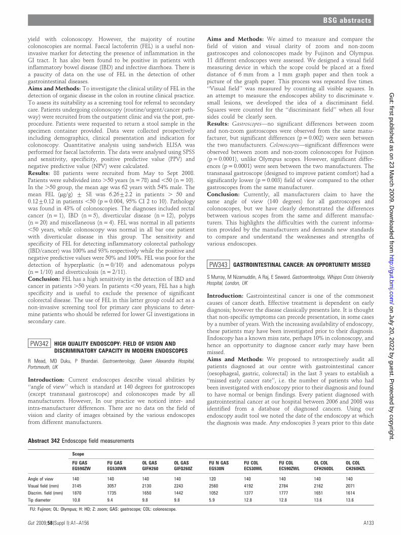
yield with colonoscopy. However, the majority of routinecolonoscopies are normal. Faecal lactoferrin (FEL) is a useful non-invasive marker for detecting the presence of inflammation in theGI tract. It has also been found to be positive in patients withinflammatory bowel disease (IBD) and infective diarrhoea. There isa paucity of data on the use of FEL in the detection of othergastrointestinal diseases.Aims and Methods: To investigate the clinical utility of FEL in thedetection of organic disease in the colon in routine clinical practice.To assess its suitability as a screening tool for referral to secondarycare. Patients undergoing colonoscopy (routine/urgent/cancer path-way) were recruited from the outpatient clinic and via the post, pre-procedure. Patients were requested to return a stool sample in thespecimen container provided. Data were collected prospectivelyincluding demographics, clinical presentation and indication forcolonoscopy. Quantitative analysis using sandwich ELISA wasperformed for faecal lactoferrin. The data were analysed using SPSSand sensitivity, specificity, positive predictive value (PPV) andnegative predictive value (NPV) were calculated.Results: 88 patients were recruited from May to Sept 2008.Patients were subdivided into .50 years (n = 78) and ,50 (n = 10).In the .50 group, the mean age was 62 years with 54% male. Themean FEL (mg/g) ¡ SE was 6.26¡2.2 in patients . 50 and0.12¡0.12 in patients ,50 (p = 0.004, 95% CI 2 to 10). Pathologywas found in 43% of colonoscopies. The diagnoses included rectalcancer (n = 1), IBD (n = 5), diverticular disease (n = 12), polyps(n = 20) and miscellaneous (n = 4). FEL was normal in all patients,50 years, while colonoscopy was normal in all bar one patientwith diverticular disease in this group. The sensitivity andspecificity of FEL for detecting inflammatory colorectal pathology(IBD/cancer) was 100% and 93% respectively while the positive andnegative predictive values were 50% and 100%. FEL was poor for thedetection of hyperplastic (n = 0/10) and adenomatous polyps(n = 1/10) and diverticulosis (n = 2/11).Conclusion: FEL has a high sensitivity in the detection of IBD andcancer in patients .50 years. In patients ,50 years, FEL has a highspecificity and is useful to exclude the presence of significantcolorectal disease. The use of FEL in this latter group could act as anon-invasive screening tool for primary care physicians to deter-mine patients who should be referred for lower GI investigations insecondary care.
PW342 HIGH QUALITY ENDOSCOPY: FIELD OF VISION ANDDISCRIMINATORY CAPACITY IN MODERN ENDOSCOPES
R Mead, MD Duku, P Bhandari. Gastroenterology, Queen Alexandra Hospital,Portsmouth, UK
Introduction: Current endoscopes describe visual abilities by‘‘angle of view’’ which is standard at 140 degrees for gastroscopes(except transnasal gastroscope) and colonoscopes made by allmanufacturers. However, In our practice we noticed inter- andintra-manufacturer differences. There are no data on the field ofvision and clarity of images obtained by the various endoscopesfrom different manufacturers.
Aims and Methods: We aimed to measure and compare thefield of vision and visual clarity of zoom and non-zoomgastroscopes and colonoscopes made by Fujinon and Olympus.11 different endoscopes were assessed. We designed a visual fieldmeasuring device in which the scope could be placed at a fixeddistance of 6 mm from a 1 mm graph paper and then took apicture of the graph paper. This process was repeated five times.‘‘Visual field’’ was measured by counting all visible squares. Inan attempt to measure the endoscopes ability to discriminate v.small lesions, we developed the idea of a discriminant field.Squares were counted for the ‘‘discriminant field’’ when all foursides could be clearly seen.Results: Gastroscopes—no significant differences between zoomand non-zoom gastroscopes were observed from the same manu-facturer, but significant differences (p = 0.002) were seen betweenthe two manufacturers. Colonoscopes—significant differences wereobserved between zoom and non-zoom colonoscopes for Fujinon(p = 0.0001), unlike Olympus scopes. However, significant differ-ences (p = 0.0001) were seen between the two manufacturers. Thetransnasal gastroscope (designed to improve patient comfort) had asignificantly lower (p = 0.003) field of view compared to the othergastroscopes from the same manufacturer.Conclusion: Currently, all manufacturers claim to have thesame angle of view (140 degrees) for all gastroscopes andcolonoscopes, but we have clearly demonstrated the differencesbetween various scopes from the same and different manufac-turers. This highlights the difficulties with the current informa-tion provided by the manufacturers and demands new standardsto compare and understand the weaknesses and strengths ofvarious endoscopes.
PW343 GASTROINTESTINAL CANCER: AN OPPORTUNITY MISSED
S Murray, M Nizamuddin, A Raj, E Seward. Gastroenterology, Whipps Cross UniversityHospital, London, UK
Introduction: Gastrointestinal cancer is one of the commonestcauses of cancer death. Effective treatment is dependent on earlydiagnosis; however the disease classically presents late. It is thoughtthat non-specific symptoms can precede presentation, in some casesby a number of years. With the increasing availability of endoscopy,these patients may have been investigated prior to their diagnosis.Endoscopy has a known miss rate, perhaps 10% in colonoscopy, andhence an opportunity to diagnose cancer early may have beenmissed.Aims and Methods: We proposed to retrospectively audit allpatients diagnosed at our centre with gastrointestinal cancer(oesophageal, gastric, colorectal) in the last 3 years to establish a‘‘missed early cancer rate’’, i.e. the number of patients who hadbeen investigated with endoscopy prior to their diagnosis and foundto have normal or benign findings. Every patient diagnosed withgastrointestinal cancer at our hospital between 2006 and 2008 wasidentified from a database of diagnosed cancers. Using ourendoscopy audit tool we noted the date of the endoscopy at whichthe diagnosis was made. Any endoscopies 3 years prior to this date
Abstract 342 Endoscope field measurements
Scope
FU GASEG590ZW
FU GASEG530WR
OL GASGIFH260
OL GASGIFQ260Z
FU N GASEG530N
FU COLEC530WL
FU COLEC590ZWL
OL COLCFH260DL
OL COLCH260HZL
Angle of view 140 140 140 140 120 140 140 140 140
Visual field (mm) 3145 3057 2130 2243 2560 4192 2784 2162 2071
Discrim. field (mm) 1870 1735 1650 1442 1052 1377 1777 1651 1614
Tip diameter 10.8 9.4 9.8 9.8 5.9 12.8 12.8 13.6 13.6
FU: Fujinon; OL: Olympus; H: HD; Z: zoom; GAS: gastrocope; COL: colonoscope.
BSG abstracts
Gut 2009;58(Suppl I):A1–A156 A133
on July 20, 2022 by guest. Protected by copyright.
http://gut.bmj.com
/G
ut: first published as on 23 March 2009. D
ownloaded from
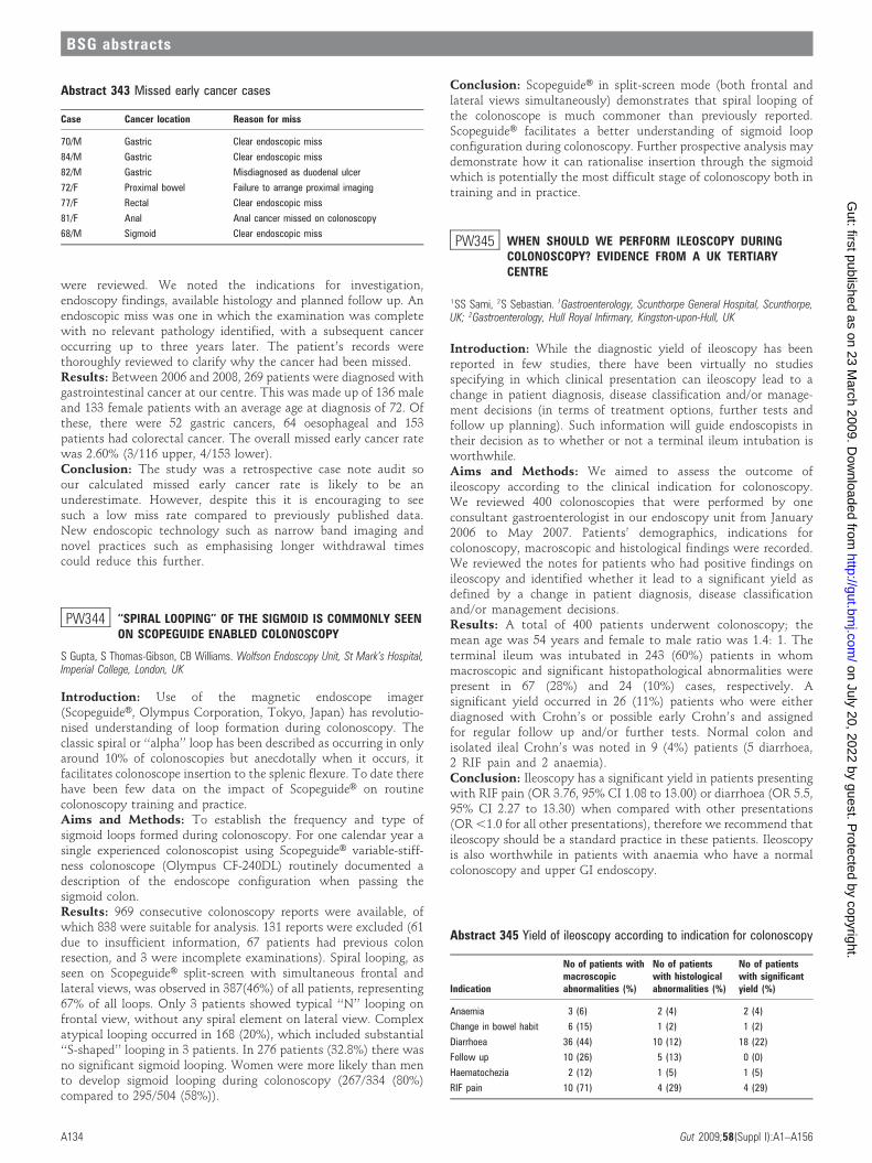
were reviewed. We noted the indications for investigation,endoscopy findings, available histology and planned follow up. Anendoscopic miss was one in which the examination was completewith no relevant pathology identified, with a subsequent canceroccurring up to three years later. The patient’s records werethoroughly reviewed to clarify why the cancer had been missed.Results: Between 2006 and 2008, 269 patients were diagnosed withgastrointestinal cancer at our centre. This was made up of 136 maleand 133 female patients with an average age at diagnosis of 72. Ofthese, there were 52 gastric cancers, 64 oesophageal and 153patients had colorectal cancer. The overall missed early cancer ratewas 2.60% (3/116 upper, 4/153 lower).Conclusion: The study was a retrospective case note audit soour calculated missed early cancer rate is likely to be anunderestimate. However, despite this it is encouraging to seesuch a low miss rate compared to previously published data.New endoscopic technology such as narrow band imaging andnovel practices such as emphasising longer withdrawal timescould reduce this further.
PW344 ‘‘SPIRAL LOOPING’’ OF THE SIGMOID IS COMMONLY SEENON SCOPEGUIDE ENABLED COLONOSCOPY
S Gupta, S Thomas-Gibson, CB Williams. Wolfson Endoscopy Unit, St Mark’s Hospital,Imperial College, London, UK
Introduction: Use of the magnetic endoscope imager(ScopeguideH, Olympus Corporation, Tokyo, Japan) has revolutio-nised understanding of loop formation during colonoscopy. Theclassic spiral or ‘‘alpha’’ loop has been described as occurring in onlyaround 10% of colonoscopies but anecdotally when it occurs, itfacilitates colonoscope insertion to the splenic flexure. To date therehave been few data on the impact of ScopeguideH on routinecolonoscopy training and practice.Aims and Methods: To establish the frequency and type ofsigmoid loops formed during colonoscopy. For one calendar year asingle experienced colonoscopist using ScopeguideH variable-stiff-ness colonoscope (Olympus CF-240DL) routinely documented adescription of the endoscope configuration when passing thesigmoid colon.Results: 969 consecutive colonoscopy reports were available, ofwhich 838 were suitable for analysis. 131 reports were excluded (61due to insufficient information, 67 patients had previous colonresection, and 3 were incomplete examinations). Spiral looping, asseen on ScopeguideH split-screen with simultaneous frontal andlateral views, was observed in 387(46%) of all patients, representing67% of all loops. Only 3 patients showed typical ‘‘N’’ looping onfrontal view, without any spiral element on lateral view. Complexatypical looping occurred in 168 (20%), which included substantial‘‘S-shaped’’ looping in 3 patients. In 276 patients (32.8%) there wasno significant sigmoid looping. Women were more likely than mento develop sigmoid looping during colonoscopy (267/334 (80%)compared to 295/504 (58%)).
Conclusion: ScopeguideH in split-screen mode (both frontal andlateral views simultaneously) demonstrates that spiral looping ofthe colonoscope is much commoner than previously reported.ScopeguideH facilitates a better understanding of sigmoid loopconfiguration during colonoscopy. Further prospective analysis maydemonstrate how it can rationalise insertion through the sigmoidwhich is potentially the most difficult stage of colonoscopy both intraining and in practice.
PW345 WHEN SHOULD WE PERFORM ILEOSCOPY DURINGCOLONOSCOPY? EVIDENCE FROM A UK TERTIARYCENTRE
1SS Sami, 2S Sebastian. 1Gastroenterology, Scunthorpe General Hospital, Scunthorpe,UK; 2Gastroenterology, Hull Royal Infirmary, Kingston-upon-Hull, UK
Introduction: While the diagnostic yield of ileoscopy has beenreported in few studies, there have been virtually no studiesspecifying in which clinical presentation can ileoscopy lead to achange in patient diagnosis, disease classification and/or manage-ment decisions (in terms of treatment options, further tests andfollow up planning). Such information will guide endoscopists intheir decision as to whether or not a terminal ileum intubation isworthwhile.Aims and Methods: We aimed to assess the outcome ofileoscopy according to the clinical indication for colonoscopy.We reviewed 400 colonoscopies that were performed by oneconsultant gastroenterologist in our endoscopy unit from January2006 to May 2007. Patients’ demographics, indications forcolonoscopy, macroscopic and histological findings were recorded.We reviewed the notes for patients who had positive findings onileoscopy and identified whether it lead to a significant yield asdefined by a change in patient diagnosis, disease classificationand/or management decisions.Results: A total of 400 patients underwent colonoscopy; themean age was 54 years and female to male ratio was 1.4: 1. Theterminal ileum was intubated in 243 (60%) patients in whommacroscopic and significant histopathological abnormalities werepresent in 67 (28%) and 24 (10%) cases, respectively. Asignificant yield occurred in 26 (11%) patients who were eitherdiagnosed with Crohn’s or possible early Crohn’s and assignedfor regular follow up and/or further tests. Normal colon andisolated ileal Crohn’s was noted in 9 (4%) patients (5 diarrhoea,2 RIF pain and 2 anaemia).Conclusion: Ileoscopy has a significant yield in patients presentingwith RIF pain (OR 3.76, 95% CI 1.08 to 13.00) or diarrhoea (OR 5.5,95% CI 2.27 to 13.30) when compared with other presentations(OR ,1.0 for all other presentations), therefore we recommend thatileoscopy should be a standard practice in these patients. Ileoscopyis also worthwhile in patients with anaemia who have a normalcolonoscopy and upper GI endoscopy.
Abstract 343 Missed early cancer cases
Case Cancer location Reason for miss
70/M Gastric Clear endoscopic miss
84/M Gastric Clear endoscopic miss
82/M Gastric Misdiagnosed as duodenal ulcer
72/F Proximal bowel Failure to arrange proximal imaging
77/F Rectal Clear endoscopic miss
81/F Anal Anal cancer missed on colonoscopy
68/M Sigmoid Clear endoscopic miss
Abstract 345 Yield of ileoscopy according to indication for colonoscopy
Indication
No of patients withmacroscopicabnormalities (%)
No of patientswith histologicalabnormalities (%)
No of patientswith significantyield (%)
Anaemia 3 (6) 2 (4) 2 (4)
Change in bowel habit 6 (15) 1 (2) 1 (2)
Diarrhoea 36 (44) 10 (12) 18 (22)
Follow up 10 (26) 5 (13) 0 (0)
Haematochezia 2 (12) 1 (5) 1 (5)
RIF pain 10 (71) 4 (29) 4 (29)
BSG abstracts
A134 Gut 2009;58(Suppl I):A1–A156
on July 20, 2022 by guest. Protected by copyright.
http://gut.bmj.com
/G
ut: first published as on 23 March 2009. D
ownloaded from
PW346 AUDIT OF ENDOSCOPIC ULTRASOUND GUIDED FINE NEEDLEASPIRATION OF SOLID PANCREATIC LESIONS
1S Rajkumar, 2K Harris, 3M Aldersley. 1Gastroenterology, Leeds General Infirmary,Leeds, UK; 2Radiology, Leeds General Infirmary, Leeds, UK; 3Hepatology, Leeds GeneralInfirmary, Leeds, UK
Introduction: Endoscopic ultrasound (EUS) guided fine needleaspiration (FNA) is probably the best method of obtainingcytological specimens from pancreatic lesions. The diagnosticaccuracy of EUS-FNA improves with participation of on-sitecytopathologist1 and performing 5 6 passes into pancreatic solidmasses.2 Suction improves cellularity but not diagnostic yield due toblood contamination.3
Aims and Methods: We reviewed all EUS-FNAs of solid pancreaticmasses performed in our tertiary referral hospital from January 2004to December 2007, comparing the diagnostic yield of cytology fromFNA with final diagnosis based on histology and clinical/radiologyfollow-up. The reference standard for overall diagnostic accuracy ofEUS-FNA was 71% based on multicentre studies.4
Results: FNA was performed using the Wilson-Cook 22 gauge FNAneedle and Olympus Curvilinear Echoendoscope without/withsuction based on specimen quantity on visual examination. TheFNA samples were sent in CytoRich Red fluid to cytopathologydepartment for analysis. 86 EUS-FNAs (n = 86) were performed on85 patients, mean age 63.4 years (range 19–89 years), male 58.8%(n = 50), female 41.2% (n = 35). The site of FNAs were head/neck74.4% (n = 64), body/tail 25.6% (n = 22). The cytology wasreported as malignant cells in 27.9% (n = 24), suspicious ofmalignancy 10.5% (n = 9), no malignant cells 55.8% (n = 48) andinsufficient specimen 5.8% (n = 5). 69.4% (59/85) of patients hadfinal diagnosis of cancer, confirmed by cytology from EUS-FNA40% (n = 24), histology by other methods 33.3% (n = 20) andradiological/clinical follow-up in the rest. The yearly sensitivity ofEUS-FNA was 37.5% (2004), 30% (2005), 33.3% (2006), 53.3%(2007). All samples reported as suspicious of malignancy and 80%(4/5) of insufficient specimens were malignant on follow-up.Conclusion: The recommendations to improve sensitivity of EUS-FNA:
1: 5 6passes with the FNA needle per pancreatic lesion2: FNA without suction if feasible3: FNA with 25 gauge vs 22 gauge FNA needle5
4: Immediate cytological evaluation of FNA material.
1. Kinney T, et al. Clinical impact of on-site cytopathologic interpretation.Gastrointest Endosc 2005;61:AB287.
2. Erickson RA, et al. Factors predicting the number of EUS-guided fine-needlepasses. Gastrointest Endosc 2000;51:184–90.
3. Wallace MB, et al. Randomised controlled trial of EUS-guided fine needleaspiration techniques. Gastrointest Endosc 2001;54:441–7.
4. Savides TJ, et al. EUS-guided FNA diagnostic yield of malignancy in solidpancreatic masses. Gastrointest Endosc 2007;66:277–82.
5. Nguyen TT, et al. A comparison of diagnostic yield between 22 and 25 gaugeneedle. Gastrointest Endosc 2008;67:AB100.
PW347 THE USE OF COMBINED ENDOSCOPIC SPHINCTEROTOMYAND LARGE BALLOON DILATATION FOR MANAGEMENT OFCOMPLEX COMMON BILE DUCT STONES
1SK Putta, 1AP Holt, 1DA Freshwater, 1GH Haydon, 2S Pathmakanthan. 1LiverMedicine, Queen Elizabeth Hospital, Birmingham, UK; 2Gastroenterology, QueenElizabeth Hospital, Birmingham, UK
Introduction: It is recommended that symptomatic bile ductstones are removed whenever possible. Sometimes large stones aredifficult to extract despite endoscopic sphincterotomy (ESP) andmechanical lithotripsy due to technical limitations or anatomicalconstraints. Sphincterotomy in combination with large balloondilation (LBD) across the ampulla and distal bile duct has recentlybeen reported as a safe and effective alternative to intra-ductallithotripsy.
Aims and Methods: To report our early experience of combinedESP and LBD to facilitate removal of complex bile duct stones.University Hospital Birmingham is a tertiary referral centre forhepatobiliary disease. We report our experience of using ESP andLBD in 13 patients referred with complex biliary stone diseasebetween March and September 2008. All patients had failedconventional endoscopic management including lithotripsy.Primary sphincterotomy or extension of a pre-existing sphincter-otomy was performed using a short-wire sphincterotome(FusionTM, Cooke). A JagwireH (Boston Scientific) was insertedinto the bile duct and LBD performed using a wireguided 15 mmCRETM balloon (Boston Scientific) positioned across the ampullaand distal bile duct via the sphincterotomy. The CRE balloon wasinflated to 8 ATM for 30 seconds before the duct was cleared withan extraction balloon (FusionTM, Cooke). Electronic admissionrecords were reviewed and patients were followed up via telephoneenquiry or clinic review after discharge.Results: Thirteen patients underwent combined ESP and LBDbetween March 2008 and October 2008. Median age was 65 years(21–84). Four patients had previously undergone a failed ERCP astheir stones could not be extracted. Ten patients had multiple CBDstones. Stone size (the largest stone measured) was recorded in 11patients. Median stone size was 13.8 mm (quartiles). Mean doses ofsedation required to permit LBD were as follows: fentanyl 92 mg (50–100 mg) and midazolam 5.1 mg (3–8.5 mg). LBD of the biliarysphincterotomy allowed complete stone removal in 11/13 (88%)patients. In all 11 patients the duct was cleared at the indexendoscopy. None of the patients developed any of the recognisedcomplications of ERCP such as early or delayed bleeding, cholangitis,perforation or pancreatitis. The 2 patients in whom ERCP failedunderwent open duct clearance at surgery. There were no deaths.Conclusion: ESP and LBD is a safe and effective means ofextracting complex bile duct stones in patients with multiple orlarge stones for whom conventional endoscopic techniques areunsuccessful.
1. Guidelines on the management of common bile duct stones.2. Williams EJ, Green J, Beckingham I, Parks R, Martin D, Lombard M, et al. Gut
2008;57:1004–21.
PW348 ROLE OF SELF EXPANDING METALLIC STENTS IN THEMANAGEMENT OF MALIGNANT OBSTRUCTION OFPROXIMAL COLON
1S Ramamurthy, 2SS Dronamraju, 3M Hayat. 1Gastroenterology, Royal VictoriaInfirmary, Newcastle Upon Tyne, UK; 2Colorectal Surgery, North Tyneside GeneralHospital, North Shields, UK; 3Gastroenterology, North Tyneside General Hospital,Newcastle Upon Tyne, UK
Introduction: Expandable metal stents have shown to be effectivein the management of malignant large bowel obstruction both as abridge to definitive surgery and for palliation.1 However, rightcolonic lesions account for less than 5% of all reported cases ofcolonic stenting.2
Aims and Methods: The aim of this study was to determine theoutcomes following stenting for malignant obstruction of proximalcolon (obstructing lesion proximal to splenic flexure). Hospitalrecords of patients undergoing colonic stenting for large bowelobstruction at a district general hospital in north England duringthe period 2001–2007 were retrospectively reviewed. Data wereanalysed (SPSS version 15) to identify patient characteristics, site ofobstructing lesion, intent for stenting and the outcomes measuredincluded relief of obstruction, hospital stay and post procedurecomplications.Results: Stenting was attempted in 90 patients (median age 73years, 52 male) with large bowel obstruction. Of these 14 (15.5%)patients had lesions in proximal colon including 8 patients withlesions in ascending colon and 6 patients with lesions in transversecolon. SEMS was successful in relieving obstruction in 12 (85.7%)
BSG abstracts
Gut 2009;58(Suppl I):A1–A156 A135
on July 20, 2022 by guest. Protected by copyright.
http://gut.bmj.com
/G
ut: first published as on 23 March 2009. D
ownloaded from
patients with proximal colonic lesions. Stenting was attempted as abridge to definitive surgery in 5 patients and for palliation in 9patients. One patient had post-stent bleeding which was managedconservatively and there were no perforation or stent dislodgement.The mean post procedure hospital stay was 1.6 days.Conclusion: SEMS are safe and effective in the management ofmalignant large bowel obstruction in proximal colon. The technicalsuccess rates and post procedural complications are comparable tothose for distal colonic lesions.
1. Sebastian S, Johnston S, et al. Pooled analysis of the efficacy and safety of self-expanding metal stenting in malignant colorectal obstruction. Am J Gastroenterol2004;99:2051–7.
2. Khot UP, Lang AW, et al. Systematic review of the efficacy and safety ofcolorectal stents. Br J Surg 2002;89:1096–102.
PW349 SINGLE BALLOON ENTEROSCOPY IN SMALL BOWELPATHOLOGY: A PROSPECTIVE OUTCOME STUDY
S John, M Remedios, M Appleyard. Gastroenterology, Royal Brisbane and Women’sHospital, Brisbane, Australia
Introduction: Single balloon enteroscopy (SBE) allows endoscopicevaluation and intervention in the small bowel. Its role andperformance are expected to be similar to double balloon entero-scopy but data regarding this are limited.Aims and Methods: The aim of our study is to prospectively assessthe use of single balloon enteroscopy in small bowel pathology.Consecutive patients referred to the Royal Brisbane & Women’sHospital for balloon enteroscopy between April and October 2008were invited to participate in the study. Informed consent wasobtained. The initial enteroscopy approach was determined bycapsule endoscopy findings. Oral (antegrade) examination wasperformed in those with capsule findings in the proximal two thirdsof the bowel and rectal (retrograde) examinations if the abnormalitywas expected to be in the distal third. An Olympus 180 series singleballoon enteroscope with overtube was used. All procedures wereperformed by endoscopists competent in double balloon enteroscopy.Data were collected on indications, capsule findings, transfusionrequirements and haemoglobin pre-procedure as well as pathologydetected, therapeutic interventions and any complications. Thecohort was followed up at 3 months and data were collectedprospectively about further bleeding, transfusion requirements,haemoglobin and any further intervention.Results: 32 procedures were performed in 28 patients betweenApril and October 2008.The commonest indication was obscure GIbleeding with 12 occult and 10 overt bleeding. Capsule endoscopywas done in 26 patients pre-enteroscopy and 25 had small bowelabnormality detected. Based on these, 26 procedures wereperformed by the antegrade or oral route and 6 by the rectalapproach. SBE detected pathology in 22 procedures (68%). Thecommonest pathology detected was small bowel angioectasia(n = 14). Four polypoidal lesions were detected (1 lymphoma, 1hamartoma, 2 adenomas). Endotherapy was performed in 19 cases(59%). This included 13 argon plasma coagulation, 1 gold probeapplication, 2 endoclips and 3 polypectomy. SBE detected pathologywas seen on capsule in 19 of 25 patients (76%). There were no
major complications. At 3 months follow up (n = 18), 4 patientshad further bleeding requiring endoscopic intervention. Surgerywith small bowel resection was performed for 2 of these patients.72% of patients had no further bleeding or symptoms. There was astatistically significant improvement in haemoglobin post-entero-scopy in those patients with obscure bleeding (p = 0.013).Conclusion: This is an ongoing study. Our early experiencesuggests that the diagnostic yield and therapeutic capability of thesingle balloon enteroscope in small bowel pathology is similar todouble balloon enteroscopy.
PW350 ENDOSCOPIC VARICEAL GRADING IN PREGNANCY: DOES ITREFLECT THE TRUE COLOUR?
S Suganya Devi, S Nandhini, P Rajesh. Gastroenterology, Govt. Theni Medical College,Theni, India
Introduction: Liver disease complicating pregnancy contributessignificantly to the maternal and fetal morbidity and mortality.Hence it is mandatory to identify the factors which contribute to themajor complications like variceal bleed. The role of endoscopic varicealscreening and risk assessment in pregnancy needs validation.Aims and Methods: To study the efficacy of endoscopy inassessing the variceal grades before and after delivery in pregnancywith decompensated chronic liver disease. 20 pregnant women withcirrhosis and portal hypertension due to varied aetiology, who werenon-bleeders with endoscopic early variceal grade (Grade I to III),were included. Portal hypertension due to other causes likeextrahepatic portal vein obstruction, non-cirrhotic portal hyperten-sion, with endoscopic advanced grade of varices (Grade IV), andpast bleeders were excluded. All were subjected to endoscopicvariceal assessment once before delivery (in II trimester) and withinsix months after delivery. Patients with grade III varices weretreated with prophylactic propranolol 20 to 40 mg daily, withadequate maternal and fetal heart rate monitoring. The varicealgrades before and after delivery were compared.Results: Mean age was 26.3 ¡ 7.34 years. 11 (61.1%) wereprimigravida. Among the 20 cases, 2 were excluded as they hadvariceal bleeding and required endoscopic band ligation. Out of the18 cases, 7 (39%) were hepatitis B related, 2 (11.1%) hepatitis Crelated, 1 (5.6%) Wilson’s related, and the rest (44.4%) werecryptogenic. Before delivery, 3 (16.7%), 7 (39%) and 8 (44.4%)patients had grade I, II and III varices, respectively. After delivery,the variceal grade were I, II, and III in 7 (39%), 7 (39%), and 4(22.2%), respectively. Among the 7 who had grade II before delivery,on endoscopic reassessment after delivery, 3 had grade I, and therest had grade II. Among the 8 who had grade III before delivery, 1had grade I, 3 had grade II and the remaining had grade III.Statistically significant downgrading of varices was documentedafter delivery in 50% of cases reported as grade III before delivery.Conclusion: Endoscopic grading of varices in pregnancy isinfluenced by others factors like haemodynamic changes andextrinsic compression of major vessels by the gravid uterus andmay not reflect the true colour.
PW351 ONE YEAR FOLLOW-UP COLONOSCOPIES WITHIN THEBOWEL CANCER SCREENING PROGRAMME: DATA FROMTHE NORTHERN REGION ENDOSCOPY GROUP
1TJW Lee, 1L Hurst, 2D Nylander, 2J Painter, 3J Singh, 4C Rees, 4M Rutter.1Gastroenterology, University Hospital of North Tees, Stockton upon Tees, UK;2Gastroenterology, Sunderland Royal Hospital, Sunderland, UK; 3Gastroenterology,Queen Elizabeth Hospital, Gateshead, UK; 4Gastroenterology, South Tyneside GeneralHospital, South Tyneside, UK
Introduction: 542 patients underwent colonoscopy as part of theBowel Cancer Screening Programme (BCSP) in the Tees and Southof Tyne area of England between February and September 2007. 41
Abstract 348 Patient demographics and outcomes following stenting
Proximal colon Distal colon P value
No of patients 14 (15.6) 76 (84.4)
Male : female 9 : 5 40 : 36 0.4
Bridge : palliative 5 : 9 25 : 51 0.8
Successful stenting (%) 12 (85.7%) 60 (78.9) 0.6
Complications (%) 1 (7.1) 7 (9.2) 0.3
Hospital stay (days) 1.6 2 0.9
BSG abstracts
A136 Gut 2009;58(Suppl I):A1–A156
on July 20, 2022 by guest. Protected by copyright.
http://gut.bmj.com
/G
ut: first published as on 23 March 2009. D
ownloaded from
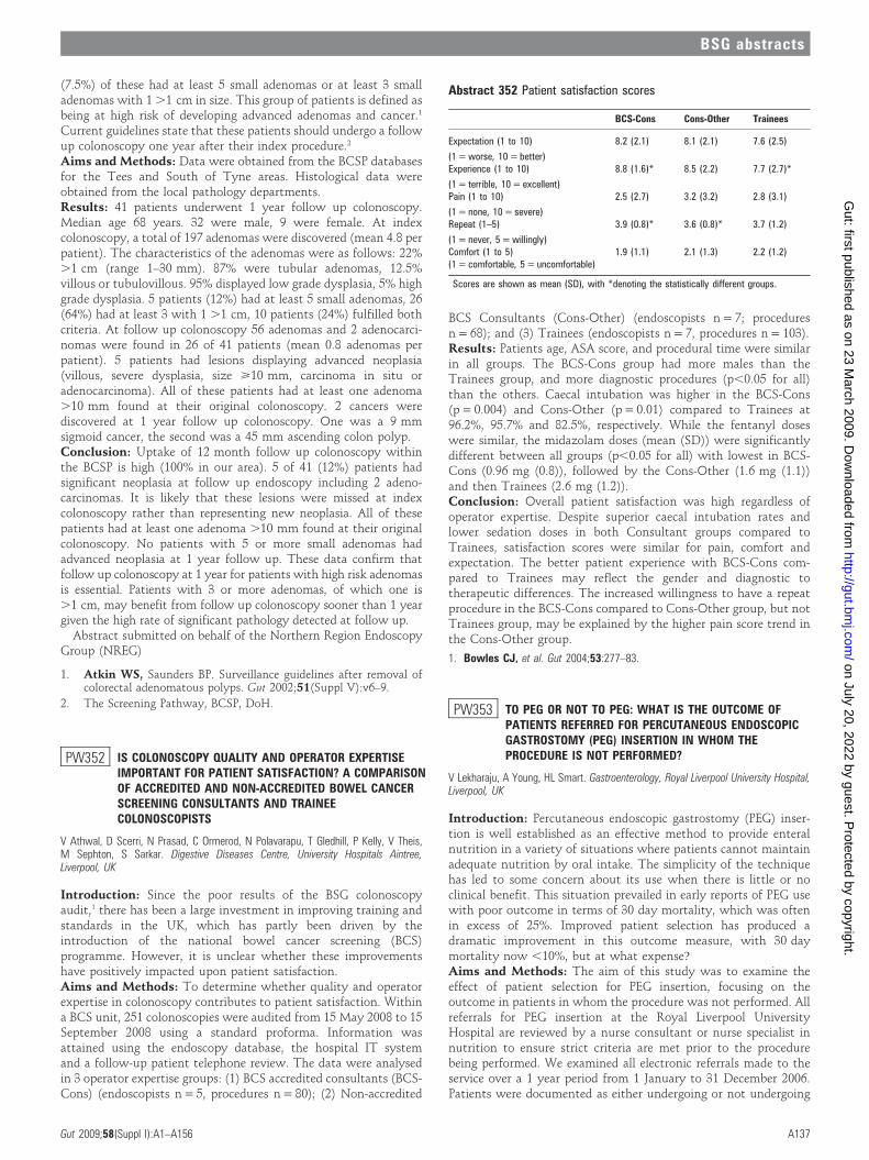
(7.5%) of these had at least 5 small adenomas or at least 3 smalladenomas with 1 .1 cm in size. This group of patients is defined asbeing at high risk of developing advanced adenomas and cancer.1
Current guidelines state that these patients should undergo a followup colonoscopy one year after their index procedure.2
Aims and Methods: Data were obtained from the BCSP databasesfor the Tees and South of Tyne areas. Histological data wereobtained from the local pathology departments.Results: 41 patients underwent 1 year follow up colonoscopy.Median age 68 years. 32 were male, 9 were female. At indexcolonoscopy, a total of 197 adenomas were discovered (mean 4.8 perpatient). The characteristics of the adenomas were as follows: 22%.1 cm (range 1–30 mm). 87% were tubular adenomas, 12.5%villous or tubulovillous. 95% displayed low grade dysplasia, 5% highgrade dysplasia. 5 patients (12%) had at least 5 small adenomas, 26(64%) had at least 3 with 1 .1 cm, 10 patients (24%) fulfilled bothcriteria. At follow up colonoscopy 56 adenomas and 2 adenocarci-nomas were found in 26 of 41 patients (mean 0.8 adenomas perpatient). 5 patients had lesions displaying advanced neoplasia(villous, severe dysplasia, size >10 mm, carcinoma in situ oradenocarcinoma). All of these patients had at least one adenoma.10 mm found at their original colonoscopy. 2 cancers werediscovered at 1 year follow up colonoscopy. One was a 9 mmsigmoid cancer, the second was a 45 mm ascending colon polyp.Conclusion: Uptake of 12 month follow up colonoscopy withinthe BCSP is high (100% in our area). 5 of 41 (12%) patients hadsignificant neoplasia at follow up endoscopy including 2 adeno-carcinomas. It is likely that these lesions were missed at indexcolonoscopy rather than representing new neoplasia. All of thesepatients had at least one adenoma .10 mm found at their originalcolonoscopy. No patients with 5 or more small adenomas hadadvanced neoplasia at 1 year follow up. These data confirm thatfollow up colonoscopy at 1 year for patients with high risk adenomasis essential. Patients with 3 or more adenomas, of which one is.1 cm, may benefit from follow up colonoscopy sooner than 1 yeargiven the high rate of significant pathology detected at follow up.
Abstract submitted on behalf of the Northern Region EndoscopyGroup (NREG)
1. Atkin WS, Saunders BP. Surveillance guidelines after removal ofcolorectal adenomatous polyps. Gut 2002;51(Suppl V):v6–9.
2. The Screening Pathway, BCSP, DoH.
PW352 IS COLONOSCOPY QUALITY AND OPERATOR EXPERTISEIMPORTANT FOR PATIENT SATISFACTION? A COMPARISONOF ACCREDITED AND NON-ACCREDITED BOWEL CANCERSCREENING CONSULTANTS AND TRAINEECOLONOSCOPISTS
V Athwal, D Scerri, N Prasad, C Ormerod, N Polavarapu, T Gledhill, P Kelly, V Theis,M Sephton, S Sarkar. Digestive Diseases Centre, University Hospitals Aintree,Liverpool, UK
Introduction: Since the poor results of the BSG colonoscopyaudit,1 there has been a large investment in improving training andstandards in the UK, which has partly been driven by theintroduction of the national bowel cancer screening (BCS)programme. However, it is unclear whether these improvementshave positively impacted upon patient satisfaction.Aims and Methods: To determine whether quality and operatorexpertise in colonoscopy contributes to patient satisfaction. Withina BCS unit, 251 colonoscopies were audited from 15 May 2008 to 15September 2008 using a standard proforma. Information wasattained using the endoscopy database, the hospital IT systemand a follow-up patient telephone review. The data were analysedin 3 operator expertise groups: (1) BCS accredited consultants (BCS-Cons) (endoscopists n = 5, procedures n = 80); (2) Non-accredited
BCS Consultants (Cons-Other) (endoscopists n = 7; proceduresn = 68); and (3) Trainees (endoscopists n = 7, procedures n = 103).Results: Patients age, ASA score, and procedural time were similarin all groups. The BCS-Cons group had more males than theTrainees group, and more diagnostic procedures (p,0.05 for all)than the others. Caecal intubation was higher in the BCS-Cons(p = 0.004) and Cons-Other (p = 0.01) compared to Trainees at96.2%, 95.7% and 82.5%, respectively. While the fentanyl doseswere similar, the midazolam doses (mean (SD)) were significantlydifferent between all groups (p,0.05 for all) with lowest in BCS-Cons (0.96 mg (0.8)), followed by the Cons-Other (1.6 mg (1.1))and then Trainees (2.6 mg (1.2)).Conclusion: Overall patient satisfaction was high regardless ofoperator expertise. Despite superior caecal intubation rates andlower sedation doses in both Consultant groups compared toTrainees, satisfaction scores were similar for pain, comfort andexpectation. The better patient experience with BCS-Cons com-pared to Trainees may reflect the gender and diagnostic totherapeutic differences. The increased willingness to have a repeatprocedure in the BCS-Cons compared to Cons-Other group, but notTrainees group, may be explained by the higher pain score trend inthe Cons-Other group.
1. Bowles CJ, et al. Gut 2004;53:277–83.
PW353 TO PEG OR NOT TO PEG: WHAT IS THE OUTCOME OFPATIENTS REFERRED FOR PERCUTANEOUS ENDOSCOPICGASTROSTOMY (PEG) INSERTION IN WHOM THEPROCEDURE IS NOT PERFORMED?
V Lekharaju, A Young, HL Smart. Gastroenterology, Royal Liverpool University Hospital,Liverpool, UK
Introduction: Percutaneous endoscopic gastrostomy (PEG) inser-tion is well established as an effective method to provide enteralnutrition in a variety of situations where patients cannot maintainadequate nutrition by oral intake. The simplicity of the techniquehas led to some concern about its use when there is little or noclinical benefit. This situation prevailed in early reports of PEG usewith poor outcome in terms of 30 day mortality, which was oftenin excess of 25%. Improved patient selection has produced adramatic improvement in this outcome measure, with 30 daymortality now ,10%, but at what expense?Aims and Methods: The aim of this study was to examine theeffect of patient selection for PEG insertion, focusing on theoutcome in patients in whom the procedure was not performed. Allreferrals for PEG insertion at the Royal Liverpool UniversityHospital are reviewed by a nurse consultant or nurse specialist innutrition to ensure strict criteria are met prior to the procedurebeing performed. We examined all electronic referrals made to theservice over a 1 year period from 1 January to 31 December 2006.Patients were documented as either undergoing or not undergoing
Abstract 352 Patient satisfaction scores
BCS-Cons Cons-Other Trainees
Expectation (1 to 10)
(1 = worse, 10 = better)
8.2 (2.1) 8.1 (2.1) 7.6 (2.5)
Experience (1 to 10)
(1 = terrible, 10 = excellent)
8.8 (1.6)* 8.5 (2.2) 7.7 (2.7)*
Pain (1 to 10)
(1 = none, 10 = severe)
2.5 (2.7) 3.2 (3.2) 2.8 (3.1)
Repeat (1–5)
(1 = never, 5 = willingly)
3.9 (0.8)* 3.6 (0.8)* 3.7 (1.2)
Comfort (1 to 5)(1 = comfortable, 5 = uncomfortable)
1.9 (1.1) 2.1 (1.3) 2.2 (1.2)
Scores are shown as mean (SD), with *denoting the statistically different groups.
BSG abstracts
Gut 2009;58(Suppl I):A1–A156 A137
on July 20, 2022 by guest. Protected by copyright.
http://gut.bmj.com
/G
ut: first published as on 23 March 2009. D
ownloaded from
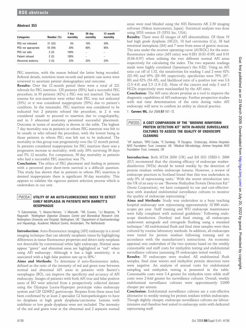
PEG insertion, with the reason behind the latter being recorded.Referral details, nutrition team records and patient case notes werereviewed to ascertain patient demographics and outcome.Results: Over the 12 month period there were a total of 221referrals for PEG insertion. 128 patients (58%) had a successful PEGprocedure; in 93 patients (42%) a PEG was not inserted. The mainreasons for non-insertion were either that PEG was not indicated(33%) or it was considered inappropriate (59%) due to patient’scondition. In the remainder, PEG insertion was considered to beindicated but 2 patients refused the procedure, in 2 it wasconsidered unsafe to proceed to insertion due to coagulopathy,and in 3 abnormal anatomy prevented successful placement.Outcome in terms of mortality is shown in the table. The highest7 day mortality was in patients in whom PEG insertion was felt tobe unsafe or who refused the procedure, with the lowest being inthose patients in whom PEG was felt not to be indicated. Themortality in this group rose progressively over the 12 month period.In patients considered inappropriate for PEG insertion there was aprogressive increase in mortality, with only 20% surviving 30 daysand 5% 12 months. For comparison, 30 day mortality in patientswho had a successful PEG insertion was 7%.Conclusion: The ethics of PEG placement and feeding in patientswith a perceived poor short-term survival is a difficult question.This study has shown that in patients in whom PEG insertion isdeemed inappropriate there is significant 30 day mortality. Thisappears to validate the rigorous patient selection process which isundertaken in our unit.
PW354 UTILITY OF AN AUTO-FLUORESCENCE INDEX TO DETECTEARLY NEOPLASIA IN PATIENTS WITH BARRETT’SOESOPHAGUS
1V Subramanian, 2L Alvarez-Herrero, 1J Mannath, 2WL Curvers, 2JJ Bergman, 1KRagunath. 1Nottingham Digestive Diseases Centre and Biomedical Research Unit,Nottingham University and Hospital, Nottingham, UK; 2Department of Gastroenterologyand Hepatology, Academic Medical Centre, Amsterdam, The Netherlands
Introduction: Auto-fluorescence imaging (AFI) endoscopy is a novelimaging technique that can identify neoplastic tissue by highlightingdifferences in tissue fluorescence properties and detect early neoplasianot detectable by conventional white light endoscopy. Normal areasappear ‘‘green’’ and abnormal areas are highlighted as ‘‘red’’ whenusing AFI endoscopy. Although it has a high sensitivity, it isassociated with a high false positive rate up to 80%.1
Aims and Methods: To determine if auto-fluorescence index,defined as the ratio of the intensity of red and green tone betweennormal and abnormal AFI areas in patients with Barrett’soesophagus (BO), can improve the specificity and accuracy of AFIendoscopy. Images of patients with both AFI true and false positiveareas of BO were selected from a prospectively collected datasetusing the Olympus Lucera-Hyperpro prototype video endoscopysystem and GIF Q240FZ gastroscope. Biopsies from these areas hadbeen confirmed by at least 2 specialist GI histopathologists to haveno dysplasia or high grade dysplasia/carcinoma. Lesions withindefinite or low grade dysplasia were not included. The intensityof the red and green tone at the abnormal and 2 separate normal
areas were read blinded using the NIS Elements AR 2.30 imagingsoftware (Nikon instruments, Japan). Statistical analysis was doneusing SPSS version 15 (SPSS Inc, USA).Results: There were 82 images of AFI abnormalities. Of these 19had high grade dysplasia (HGD), 18 had carcinoma (Ca), 38 hadintestinal metaplasia (IM) and 7 were from areas of gastric mucosa.The area under the receiver operating curve (AUROC) for the auto-fluorescence index ratio (AFI ratio) was 0.881 (0.81–0.95) and 0.91(0.84–0.97) when utilising the two different normal AFI areasrespectively for calculating the index. The two separate readingsmade were highly correlated (Spearman’s rho 0.82). Using an AFIratio cut off of 1.32, the sensitivities for reading 1 and 2 were 92%(82–99) and 95% (85–99) respectively, specificities were 76% (67–80) and 62% (54–65), and likelihood ratio of a positive test was 3.8(2.5–4.6) and 2.5 (1.9–2.8). None of the cancers and only 3 and 2HGDs respectively were misclassified by the AFI ratio.Conclusion: The AFI ratio shows promise as a tool to improve thediagnostic capabilities of AFI endoscopy. Further prospective studieswith real time determination of the ratio during video AFIendoscopy will serve to confirm its utility in clinical practice.
1. Curvers WL. Gut 2008;57:167–72.
PW355 A COST COMPARISON OF THE ‘‘BROWNE NINHYDRINPROTEIN DETECTION KIT’’ WITH IN-HOUSE SURVEILLANCECULTURES TO ASSESS THE QUALITY OF ENDOSCOPECLEANING
1VP Jackson, 2RPD Cooke, 2S Sundeep, 1R Sturgess. 1Endoscopy, Aintree HospitalsNHS Foundation Trust, Liverpool, UK; 2Medical Microbiology, Aintree Hospitals NHSFoundation Trust, Liverpool, UK
Introduction: Both HTM 2030 (UK) and EN ISO 15883-1: 2006(EU) recommend that the cleaning efficacy of endoscope washer-disinfectors (WDs) should be tested weekly by the detection ofprotein residues within endoscope lumens. However, a review ofendoscope practices in Scotland found that this was undertaken inonly 8% of reprocessing units.1 With the recent introduction intothe UK of an easy to use ‘‘Browne Ninhydrin Protein Detection Kit’’(Steris Corporation), we have compared its use and cost-effective-ness with standard endoluminal surveillance cultures to monitorthe quality of endoscope reprocessing.2
Aims and Methods: Study was undertaken in a busy teachinghospital endoscopy unit reprocessing approximately 18 000 endo-scopes per year. Staff training and WD endoscope maintenancewere fully compliant with national guidelines.3 Following endo-scope disinfection (Sterilox) and final rinsing, all endoscopesunderwent antegrade and retrograde flushing using a standardtechnique.3 All endoluminal flush and final rinse samples were thencultured by routine laboratory methods. In addition, all endoscopeswere tested for protein residues following training and inaccordance with the manufacturer’s instructions. An economicappraisal was undertaken of the two systems based on the weeklyconsumable and staff costs for ninhydrin testing and endoluminalsurveillance cultures (assuming 55 endoscopes tested weekly).Results: 55 endoscopes were studied. All endoluminal flushsamples, final rinse waters and ninhydrin protein detection testswere negative. An analysis of annual costs for endoluminalsampling and ninhydrin testing is presented in the table.Consumable costs were 3.4 greater for ninhydrin tests while staffcosts were 2-fold greater for surveillance cultures. Overall, weeklyendoluminal surveillance cultures were approximately £1000cheaper per annum.Conclusion: Endoluminal surveillance cultures are a cost-effectivealternative to weekly testing for protein residues within endoscopes.Though slightly cheaper, endoscope surveillance cultures are labour-intensive and therefore best suited to endoscopy units with dedicatedreprocessing staff.
Abstract 353
Categories Number (%)7 daymortality
30 daymortality
12 monthmortality
PEG not indicated 31 (33) 6% 19% 39%
PEG not appropriate 55 (59) 38% 80% 95%
PEG not safe 2 (2) 100% – –
Patient refused 2 (2) 100% – –
Abnormal anatomy 3 (3) 33% 33% 33%
BSG abstracts
A138 Gut 2009;58(Suppl I):A1–A156
on July 20, 2022 by guest. Protected by copyright.
http://gut.bmj.com
/G
ut: first published as on 23 March 2009. D
ownloaded from
1. Health Protection Scotland. Review of endoscope decontamination practice inScotland; November 2004 – May 2005. ISBN 07559 13345 www.scotland.gov.uk
2. Moses FM, Lee J. Surveillance cultures to monitor quality of gastrointestinalendoscope reprocessing. Am J Gastroenterol 2003;98:77–81.
3. Medical Devices Agency. Decontamination of endoscopes. MDA DB 2002 (05).
Gastroduodenal posters
PW356 PROTON PUMP INHIBITORS: ARE WE WASTING MONEYAND IGNORING THE RISK?
AJ Kent, B Graf, M Harbord. Gastroenterology, Chelsea and Westminster Hospital,London, UK
Introduction: The National Health Service spends £300 millionannually on proton pump inhibitors (PPIs). The National Institute forHealth and Clinical Excellence (NICE) advise that patients withdyspepsia/gastro-oesophageal reflux disease (GORD) should receive aPPI for 1–2 months and if symptoms recur it should be prescribed at thelowest dose. £50 million could be saved annually if their guidelines werefollowed more strictly. There is growing concern regarding complica-tions associated with PPI use, including increased risk of entericinfections (specifically Clostridium difficile infection1), community-acquired pneumonia, hip fractures and disturbed calcium homeostasis.Aims and Methods: We aimed to assess the degree of inappropri-ate PPI prescription and assess potential associated complications.We prospectively reviewed prescriptions of medical patientsadmitted through Chelsea and Westminster Hospital. Informationgathered included: patient demographics, comorbidities, body massindex (BMI), smoking/ETOH history, medications, indication forPPI and current symptoms. We contacted general practitioners forinformation not available from the patient or hospital records.Results: Demographics—254 patients were assessed, of whom 129were prescribed a PPI. Mean age was 72 years and 48% of patientswere male. There was no statistical difference between patientdemographics, alcohol or smoking history between the PPI andcontrol groups. PPI—PPI used included omeprazole (67%), lanso-prazole (32%) and esomeprazole (1%). Indications were dyspepsia(13%), peptic ulcer disease (13%) and prophylaxis (47%). Theindication was unknown by patient, GP or hospital records in 27%.Over 77% patients had been taking PPI for greater than 1 year,despite only 25% having documentation stating long term PPI wasrequired. Only 2 patients had undergone a trial without PPI.Complications—C difficile-associated diarrhoea (CDAD) occurredsignificantly more in patients on PPI (16% vs 5%, p,0.004). 2patients from the PPI group died from CDAD, compared to none inthe control group. Chest infections were more common in patientson PPI (25% vs 17%), but the PPI group had a higher incidence ofunderlying lung disease, so significance was impossible to deter-mine. Osteoporosis was significantly more common in the PPIgroup (26% vs 10%, p 0.003) and subsequently bisphosphonate usewas also higher (22% vs 9%, p,0.005).Conclusion: Over-prescription of PPI is evident, with an increasedincidence of potentially associated complications. We would advisedoctors to review prescriptions in all patients, in an attempt toreduce cost and complications of unnecessary PPI use.
1. Aseeri M, et al. Gastric acid suppression by proton pump inhibitors as a risk factorfor Clostridium difficile-associated diarrhea in hospitalized patients.Am J Gastroenterol 2008;103:2308–13.
PW357 DIAGNOSTIC PRECISION OF PRIMARY CARE ELECTRONICDATABASE FOR STUDY OF GASTROINTESTINAL BLEEDINGAND ITS ABILITY TO DETECT THE INFLUENCE OF ASPIRIN
1M Leighton, 2Y Vivogravada, 2A Shonde, 1JC Atherton, 3RFA Logan, 2A Avery,2D Kendrick, 2J Hippisley-Cox, 1CJ Hawkey. 1Nottingham Digestive Diseases Centre andBiomedical Research Unit, Nottingham University Hospital, Nottingham, UK; 2Primary Care,Nottingham University Hospital, Nottingham, UK; 3Epidemiology & Public Health,Nottingham University Hospital, Nottingham, UK
Introduction: Large primary care electronic databases offer goodopportunities for systematic epidemiological research. However,general practitioners do not necessarily code events in the same wayas Hospital Events Statistics do. We therefore investigated the extentto which coding influences recorded gastrointestinal (GI) bleedingevent rates using QRESEARCH, a new larger electronic database.Aims and Methods: Patients were included in the analysis if theywere aged 45–100 years and had at least 12 months of electronichealth records and were registered with a participating practice at anytime between 1 April 2003 and 31 March 2007. All codes withmention of gastrointestinal bleeding were extracted and summarisedto produce eight categories, as shown in the table.Results: 459 practices met inclusion criteria to generate 4.87million person years of observation. Over 99% of aspirin use was for(300 mg per day. As shown in the table, high rates were recordedfor upper gastrointestinal bleeding and rectal bleeding/melena,while specific ulcer bleeding was under recorded. Nevertheless, thestudy was able to detect a 1.58 (1.45–1.71) fold hazard ratio forupper gastrointestinal bleeding and a 1.47 (1.42–1.52) hazard ratiofor rectal bleeding/melena, related to aspirin use.Conclusion: Coding imprecision limits the exactitude of conclu-sions but is not enough to obscure the impact of aspirin on eventrates. The impact of aspirin is likely to be higher when coding andexposure imprecisions are allowed for.
PW358 FEASIBILITY OF REDUCING ASPIRIN-ASSOCIATED ULCERBLEEDING BY HELICOBACTER PYLORI ERADICATION
1CJ Hawkey, 2JC Atherton, 3RFA Logan, 4A Avery, 2A Shonde, 2M Leighton,4D Kendrick. 1Nottingham Digestive Diseases Centre and Biomedical Research Unit,Nottingham University Hospital, Nottingham, UK; 2Nottingham Research Centre andBiomedical Research Unit, Nottingham University Hospital, Nottingham, UK;3Epidemiology & Public Health, Nottingham University Hospital, Nottingham, UK;4Division of Primary Care, Nottingham University Hospital, Nottingham, UK
Introduction: In contrast to non-steroidal anti-inflammatorydrugs, ulcers and ulcer complications on aspirin occur selectivelyin Helicobacter pylori positive individuals. The main effect ofaspirin may be to provoke bleeding rather than cause ulceration.
Abstract 357
Results
Broad termRate/100000 Specific term
Rate/100000
Ulcer bleeding All specific ulcerbleeding
6.25 Not haemorrhagicgastritis
6.17
Upper GI
bleeding
All upper GIbleeding
72.3 With ulcer code and nillower
3.77
Rectal bleeding/melena
All melaena/rectal bleeding
564 No lower GI code 564
Any GI bleeding All GI bleeding 5.49 No lower GI code 5.49
Abstract 355 Cost analysis of weekly antegrade/retrograde endoscopeflush samples versus weekly ninhydrin test (based on 55 scopes beingtested weekly)
Annual consumablecosts (£)
Annual staffcosts (£)
Total annualcosts (£)
Antegrade/retrogradeflush samples
2660 11700 14560
Ninhydrin testing 9809 5800 15609
BSG abstracts
Gut 2009;58(Suppl I):A1–A156 A139
on July 20, 2022 by guest. Protected by copyright.
http://gut.bmj.com
/G
ut: first published as on 23 March 2009. D
ownloaded from
If so, H pylori eradication should reduce aspirin-associated ulcerbleeding and enlarge the population for which aspirin results innet benefit.Aims and Methods: In preparation for a proposed 9000 patientoutcomes study, with ulcer bleeding as an end point, weinvestigated the prevalence of H pylori (by breath testing), thewillingness to participate and the rate of eradication in patientstaking aspirin (100 mg daily in 9 general practices in the NorthEast, Midlands and South West of England.Results: Of 2525 patients approached, 1199 replied (47.5%) and945 (37.4%, range 32.6–48.8%). Of these, 84 (3.3%) wereineligible leaving 34.1% suitable for participation. Among thesepatients, 24.1% had a positive H pylori breath test (range 13.3–44.0%, IQR 21.4–25.9%). These patients underwent eradicationtreatment, with an eradication rate so far of 89.1%. In responseto a questionnaire, 71.7% of patients in this non-randomisedstudy said they would participate in a study with randomisationto immediate or delayed eradication treatment. A survey of 97practices participating in the GP Primary Care ComprehensiveResearch Network found that 71.1% thought a controlled trialstudy was important and said they would definitely or veryprobably participate in.Conclusion: Overall, 8.2% of patients taking aspirin are both Hpylori +ve and willing to participate in a trial of eradicationtreatment. To enrol enough patients for an outcomes study,approximately 110 000 patients would need to be approached.Our study shows this is easy to achieve and the study would besuitable to be conducted through the comprehensive researchnetwork.
PW359 GASTRIC HELICOBACTER INFECTION INDUCES IRONDEFICIENCY IN THE INS-GAS MOUSE
1SA Boxall, 2DM Pritchard, 2A Abuderman, 3TC Wang, 2A Varro, 4JE Crabtree. 1LeedsInstitute of Molecular Medicine, St Jame’s University Hospital, Leeds, UK; 2Division ofGastroenterology, University of Liverpool, Liverpool, UK; 3Columbia University MedicalCentre, Columbia University, New York, USA; 4Leeds Institute of Molecular Medicine, StJames’s University Hospital, Leeds, UK
Introduction: Iron deficiency is the most common nutritionaldisorder in the world. There is increasing evidence from clinicaland population studies for a role of Helicobacter pylori infection inthe aetiology of iron deficiency anaemia. Hypochlorhydriaassociated with initial infection, and gastric atrophy in latestages of infection, may be relevant to iron deficiency. The aimsof this study were to determine the effects of gastric Helicobacterinfection in INS-GAS mice on iron deficiency and the relation toparietal cell number.Aims and Methods: Male transgenic hypergastrinaemic INS-GASmice were inoculated with Helicobacter felis by oral gavage. Infectedmice and uninfected controls were sacrificed 3, 6 and 9 monthspost-inoculation. Blood was collected and serum iron, total ironbinding capacity (TIBC) and plasma amidated gastrin levels weremeasured. Gastric parietal cells were immunolabelled withH+K+ATPase and quantified.Results: In uninfected controls there was no significant change inserum iron levels at 3, 6 or 9 months. In contrast, at 6 months, ironlevels in H felis infected mice were significantly lower (p,0.002)than in 3 month H felis infected mice, and also significantly reduced(p,0.05) relative to 6 month uninfected controls. No furtherreduction in iron levels was evident at 9 months in H felis infectedmice. At 3 months TIBC levels in H felis infected mice weresignificantly increased (p,0.05) compared to controls. TIBC levelsin infected mice were further increased (p,0.02) at 9 monthscompared to 6 month infected mice. The decrease in iron levels at
6 months in H felis infected mice was associated with aconcomitant decrease in parietal cell numbers relative to controls(p,0.05), which was also evident at 9 months (p,0.03). Thepercentage of transferrin saturation was also decreased in infectedmice, becoming significantly reduced (p,0.03) by 9 months post-infection compared to control mice. Although in these hypergas-trinaemic mice H felis infection was associated with significantlyincreased (p,0.001) gastrin concentrations at 9 months, there wasno significant correlation between iron levels and plasma gastrinconcentrations.Conclusion: In INS-GAS mice H felis infection is associatedwith decreased serum iron, increased TIBC and decreasedtransferrin saturation. Changes in iron levels evident at6 months are associated with decreased parietal cell number.Further studies are required to elucidate the exact mechanismsinvolved in Helicobacter-induced iron deficiency in the INS-GASmodel.
PW360 HELICOBACTER PYLORI SEROPREVALENCE IN A HOSPITALCOHORT BETWEEN 2001–2007
1K Alexandropoulou, 2H Al-Ghusein, 3R Holliman, 1A Poullis, 1J Kang.1Gastroenterology, St George’s Hospital, London, UK; 2IT Department, St George’sHospital, London, UK; 3Microbiology, St George’s Hospital, London, UK
Introduction: Over the last century, Helicobacter pylori (HP)prevalence decreased in developed countries in the context ofimproved living conditions. Widespread adoption of the ‘‘test andtreat’’ strategy for dyspepsia may have further decreased theprevalence of HP infection in recent years. Although serology mayremain positive after successful eradication of HP infection, it ispossible that large-scale use of eradication treatment could haveaffected the sero-epidemiology of HP. We have studied theprevalence rate of positive serology to HP in an ethnically diversearea in London over a 7 year period.Aims and Methods: Records of HP serology results from StGeorge’s Hospital, London, between 2001 and 2007 were retrieved.Data were analysed to determine prevalence trends over this timeperiod (x2 test for trend). The effect of age, gender and race was alsoassessed.Results: A total of 12307 patients were tested between 2001 and2007. There was a statistically significant reduction in theproportion of positive HP tests over the study period from 42% to37% (p,0.005). Two thirds (77%) of patients tested were under theage of 50. Mean HP prevalence was 39% but varied among the agegroups, being lowest in ,25 group at 30%, 38% for 26–50, 48% for50–75 and 49% for .75 age group. There were comparable rates ofdecline in HP prevalence in the ,50 and .50 age groups. More menthan women tested positive for HP serology consistently over the 7year period although the difference did not achieve statisticalsignificance (p = 0.07).Conclusion: HP prevalence in our population cohort is decreasing,even over a short period of seven years. While HP infection wasmore prevalent in older patients, a significant proportion of youngerpatients tested positive in this ethnically diverse population and therate of decline was unaffected by age.
Abstract 360 Helicobacter pylori (HP) seroprevalence, 2001–07
HP serology 2001 2002 2003 2004 2005 2006 2007
% positive 42 43 38 38 38 39 37
% negative 57 53 58 55 56 57 58
BSG abstracts
A140 Gut 2009;58(Suppl I):A1–A156
on July 20, 2022 by guest. Protected by copyright.
http://gut.bmj.com
/G
ut: first published as on 23 March 2009. D
ownloaded from
PW361 INCREASED PLASMA GHRELIN IN ALCOHOLIC CIRRHOSIS:EFFECT OF AN ORAL GLUCOSE CHALLENGE ON POST-PRANDIAL MECHANISMS
1SJ Goodyear, 1M Mottershead, 1EZH Sung, 2L Wong, 3P McTernan, 3S Kumar, 1CUNwokolo. 1Gastroenterology, University Hospital of Coventry, Coventry, UK; 2Surgery,University Hospital of Coventry, Coventry, UK; 3Endocrinology and Metabolism,University Hospital of Coventry, Coventry, UK
Introduction: Ghrelin abnormalities have been reported in chronicliver disease. This study assessed the response of anabolic peptideslike ghrelin, growth hormone (GH) and insulin-like growth factor 1(IGF-1) in alcoholic cirrhotics and healthy subjects to oral glucose.In a previous study using a similar oral glucose challenge weidentified loss of ghrelin regulation in non-alcoholic steato-hepatitis.Aims and Methods: 14 patients with alcoholic cirrhosis werecompared with 11 healthy subjects. The cirrhotics were studiedafter period of hospital admission minimising the effect of fluidshifts, infection and persisting blood alcohol. After an overnight fastthey ingested 100 g glucose in 250 ml of water. Blood was sampledbefore and every 20 min for 120 min. Plasma acylated and des acylghrelin, GH, IGF-1 and insulin were assayed by ELISA.Results: Expressed as median 95% CI. 120 min integrated acylatedghrelin increased from 26 (19–66) in controls to 170 (129–252) pg/ml.hr in cirrhotics; p,0.001, Mann Whitney U Test. Both groupsexhibited a normal post-glucose plasma ghrelin profile. Amongcirrhotics (compared to controls) growth hormone was increased15-fold and IGF-1 decreased 4-fold. Acylated ghrelin correlated withGH (Spearman r = 0.69, p = 0.0015) in controls but not incirrhotics.Conclusion: Acylated ghrelin is markedly increased in alcoholiccirrhosis with preservation of normal post prandial mechanisms ofgastric ghrelin secretion. GH is also increased but regulation byacylated ghrelin confirmed in healthy subjects is lost. Despiteincreased ghrelin and GH alcoholic cirrhotics remain anorexic andcatabolic, pointing to tissue resistance to these anabolic peptides.
GI physiology posters
PW362 SYMPTOMS, ENDOSCOPY AND PHYSIOLOGY: DO THEYCORRELATE?
AJ Kent, B Graf, MR Banks. Gastroenterology, Chelsea and Westminster Hospital,London, UK
Introduction: Oesophageal manometry and pH studies are well-established tools in the assessment of oesophageal symptoms, butare only available in tertiary referral centres in the UK. Criteria havebeen proposed to define diagnoses, but the overall benefit ofmanometric diagnoses that do not correlate with symptoms isunknown.Aims and Methods: Our aim was to assess the correlationbetween symptoms, upper GI endoscopy findings and physiologicalfindings. 200 consecutive patients undergoing oesophageal physiol-ogy investigations had details entered into a database designed forthe study. Information gathered included patient demographics,past medical history, previous investigations, medications, symp-toms and manometric findings.Results: Symptoms—Dysphagia resulted in the following mano-metric diagnoses: normal 47%, achalasia 39%, ineffective oesopha-geal motility (IOM) 8%, diffuse oesophageal spasm (DOS) 6%. Allpatients with achalasia suffered with dysphagia. Patients with chestpain were diagnosed with achalasia 34%, DOS 8% and normalmanometry 58%. Conversely patients with a manometric diagnosisof DOS suffered with chest pain 11%, dysphagia 22% and epigastricpain or reflux 67%. 78% of patients with IOM had acid reflux, andaccounted for a total of 11% of all patients referred for pH studies to
investigate persistent acid reflux. Furthermore 56% of patients withIOM had a hypotensive lower oesophageal sphincter, which mayhave contributed to their reflux events. Endoscopy—The majority ofpatients undergoing physiology testing had previously normalOGDs. All gastroscopies with findings suspicious for achalasia ledto manometry that confirmed the diagnosis. However, only 50% ofpatients with achalasia had OGDs which suggested this diagnosis.Unsurprisingly, oesophagitis was significantly associated withevidence of acid reflux compared to normal endoscopies.However, 54% of endoscopically diagnosed oesophagitis had normalpH studies, despite all studies being performed off acid suppression.Furthermore, only 50% of patients with Barrett’s oesophagus hadpathological acid reflux.Conclusion: Oesophageal symptoms have a poor predictive valuefor clinical diagnoses. Patients with dysphagia should alwaysundergo manometry testing to exclude achalasia, if endoscopy isnon-diagnostic. Given the variety of symptoms for motilitydisorders, and lack of ‘‘classic’’ symptoms for conditions such asDOS, manometric testing should be used more prevalently inpatients with oesophageal symptoms.
1. Bodger K, Trudgill N. BSG guidelines for oesophageal manometry and pHmonitoring. Gut 2006.
PW363 OBJECTIVE ASSESSMENT OF RECTAL AFFERENTSENSITIVITY AND PRIMARY CORTICAL PROCESSING INPATIENTS WITH IRRITABLE BOWEL SYNDROME
1DC Bullas, 2AR Hobson, 1S Gurmany, 2KJ Hicks, 2GE Dukes, 1MA Kamm, 1WS Atkin,1N Arebi. 1St Mark’s Hospital Campus, Imperial College, Harrow, UK; 2Immuno-Inflammation CEDD, GlaxoSmithKline Medicines Research Centre, Stevenage, UK
Introduction: Abnormalities in endogenous pain modulation andsensitisation of afferent nerve pathways may contribute to thedevelopment and maintenance of symptoms in irritable bowelsyndrome (IBS). Differentiating these distinct pathophysiologicalmechanisms in IBS is limited using conventional physiologicalassessments. Cortical evoked potentials (CEPs) allow a dynamic andobjective assessment of visceral sensory function and could providenovel insight into IBS aetiology.Aims and Methods: To measure primary rectal hypersensitivityby applying CEPs in IBS. Rome 2-positive patients and controlswere recruited in a prospective study for 3 days of testing. Sensoryand pain thresholds (ST, PT) were calculated, and CEPs werecaptured at a stimulus intensity of 75% PT. CEP morphologyincludes P1, P2, P3, P4, and P5 components, distinguished by latency(ms) and amplitude (microV). Anxiety scores were provided bySpielberger State Trait Anxiety Index (SSTAI). Data were analysedusing 1-way ANOVA, presented as mean differences ¡ 95% CI.Results: 33 IBS patients (27 females, mean age 40.1) and 15controls (9 females, mean age 28.6) enrolled. IBS subjects hadincreased P1 and P3 latencies on day 1 (20.89 (6.20 to 35.59)) andday 3 (30.64 (2.82 to 5.46)) respectively. IBS subjects had decreasedP2 amplitudes: (day 1: 26.96 (213.89 to 20.03)); (day 2: 28.62(216.11 to 21.12)). IBS subjects had decreased P3 amplitudes: (day1: 28.46 (214.21 to 22.71)); (day 2: 23.36 (25.66 to 21.06)); (day3: 210.65 (216.13 to 25.18)). IBS subjects had decreased P4amplitudes on day 1 (23.36 (25.66 to 21.06)). ST and PT did notdiffer between groups on day 1. PT was decreased on day 3 (215.63(229.24 to 22.03) in IBS. SSTAI-State scores (day 1: 5.92 (0.43 to11.40)); (day 2: 8.87 (2.58 to 15.17)) and SSTAI-Trait scores (day 1:9.55 (3.45 to 15.65)); (day 2: 8.84 (2.35 to 15.32)); (day 3: 11.88 (5.02to 18.74)) were increased in IBS.Conclusion: Analysis of cortical evoked potentials allows neuro-physiological differentiation between IBS and controls at a grouplevel. Primary rectal afferent hypersensitivity was not commonlyseen in the IBS group. Pain thresholds showed a negative trend inIBS patients compared to controls on days 1 and 2 that reachedstatistical significance on day 3. This was a result of an increase in
BSG abstracts
Gut 2009;58(Suppl I):A1–A156 A141
on July 20, 2022 by guest. Protected by copyright.
http://gut.bmj.com
/G
ut: first published as on 23 March 2009. D
ownloaded from
the pain thresholds of the controls rather than a relative decrease inIBS patients. In the context of persistent anxiety levels, thissuggests that IBS patients maintain a level of visceral hypervigilancenot shared by controls, who become acclimatised to the testprotocol by day 3. The inability to inhibit central pain amplifica-tion, in a process of habituation to repetitive noxious stimuli, maybe important factor in the maintenance of IBS symptomatology.
PW364 PROTON PUMP INHIBITORS, STATINS AND ACUTECORONARY SYNDROME
MA Butt, JWL Yong, NC Pegge. Cardiology, Worthing Hospital, Worthing, UK
Introduction: Proton pump inhibitors (PPI) are one of the mostfrequently prescribed medications in the UK as they combine a highlevel of efficacy with low toxicity. Patients with acute coronarysyndrome (ACS) often receive PPIs to counteract the risk ofgastrointestinal haemorrhage secondary to dual anti-platelet ther-apy. Both PPIs and statins interact with cytochrome p450pathways. This interaction may reduce the effectiveness of statins.Aims and Methods: We therefore conducted a retrospectiveanalysis of patients treated for ACS. Two groups were selected;those treated with PPIs in addition to routine ACS treatment andmatched controls. Data on age, sex, type of statin and PPIprescribed, admission serum cholesterol and triglyceride, and serumcholesterol and triglyceride 3 months post discharge were collected.Results: 69 patients’ notes were reviewed. There were 36 patientstreated with PPIs in addition to ACS medication and 33 controls.61% of patients were male, mean age 69 years. For those prescribedPPI medication, 78% received omeprazole, 19% lansoprazole and 3%rabeprazole. Documented indications for PPI prescription includeddyspepsia (28%), previous gastric ulcer (17%), gastro-oesophagealreflux (8%), gastrointestinal haemorrhage (3%) and alendronate(3%). The majority of patients received simvastatin (90%) by thetime of discharge. The following mean scores were observed;admission cholesterol in the PPI group (5.3¡1.8) vs control(5.1¡0.9), admission triglyceride in the PPI group (2.6¡3) vscontrol (2.1¡1.7), 3 month post-discharge cholesterol in the PPIgroup (4.7¡1.2) vs control (3.7¡0.6) and 3 month post-dischargetriglyceride in the PPI group (1.5¡0.7) vs control (1.3¡0.2). Toascertain whether PPI therapy influenced the effectiveness of statintreatment, we used the Students one tailed t-test to compare if thepercentage reduction in cholesterol and triglyceride between the PPIand control groups was significant. The following P values wereobtained; for cholesterol reduction p = 0.15, for triglyceride reduc-tion p = 0.08.Conclusion: Our study demonstrates that the percentage reduc-tion in cholesterol and triglycerides with statins at 3 months waslower in patients treated with PPIs, than matched controls.Although this difference did not reach significance, this was likelydue to the small sample size in our study. Future prospective studieswith narrower criteria, looking at patients treated with PPIs andstatins of one type and at specific doses, may confirm a significantinteraction.
Oesophagus posters
PW365 IS BARRETT’S OESOPHAGUS UNDERDIAGNOSED INPATIENTS WITH EROSIVE OESOPHAGITIS?
KKV Kosuri, J de Caestecker, J Jankowski. Gastroenterology, University Hospitals ofLeicester, Leicester, UK
Introduction: Barrett’s oesophagus (BE) could be underdiagnosedin oesophagitis patients either because the ulcerative oesophagitisitself obscures BE or because BE could develop after healing.
Aims and Methods: Our aim is to evaluate the frequency ofBarrett’s oesophagus on follow up upper GI endoscopy (oesopha-gogastroduodenoscopy, OGD) for reflux oesophagitis (RE).Retrospective observational study on patients undergoing repeatupper GI endoscopy following diagnosis of RE. All patientsdiagnosed with RE graded with Los Angeles (LA) or Savary-Millercriteria between 1997–2008 (LA grading not used till after 2001)were identified from the endoscopy database. Patients who did nothave repeat OGD, diagnoses other than RE were excluded from thestudy. Along with demographics (age and sex), data collectedincluded date of initial and repeat OGD, interval between OGDs,LA grade or Savary-Miller grade, presence of oesophageal strictures,whether oesophageal biopsies performed on initial and repeatendoscopies and the presence of Barrett mucosa. Results wereanalysed using Microsoft Excel 2007 and SPPS v13.0 software.Results: Of the total 114 patients identified by the software asfollow up of oesophagitis, 24 patients were excluded as they eitherhad other oesophageal diagnoses or were not oesophagitis followups. Of the 90 patients with follow up endoscopies, 60 (65%) weremales and 30 (35%) were females with a median age of 68 years.Mean number of follow up OGDs performed were 2.7 (range 2–10).Mean interval between the index OGD and repeat OGD was75 days. Overall 40 patients had BE detected on follow upendoscopy. Of the 90 patients, 79 had biopsies performed on initialOGD and 64 patients had biopsies performed on follow upendoscopy. Though only 10 had Barrett’s segment detected alongwith RE on initial OGD, 40 patients had Barrett’s oesophagusdetected on follow up endoscopy; mean length of BE was 3.8 cm(range 1–11). Further, when the RE was grouped as Mildoesophagitis (LA grade A & B; Savary Miller 1 & 2) and Severeoesophagitis (LA grade C & D or Savary Miller 3 and 4), BE is seento occur in 50% of patients with severe oesophagitis (see table).Conclusion: BE occurs with all grades of RE but most frequentlywith severe oesophagitis (LA grade C & D or Savary Miller 3 and 4)on initial endoscopy. Follow up endoscopy is recommended in thissubgroup of patients to detect BE. It is not clear whether BEdevelops as RE heals or whether it is obscured by initial RE.
PW366 CDX2-DEPENDENT HOX GENE REGULATION IN BARRETT’SOESOPHAGUS
M di Pietro, RC Fitzgerald. Cancer Cell Unit, MRC, Cambridge, UK
Introduction: Intestinal metaplasia (IM) of the oesophagus,commonly referred to as Barrett’s oesophagus, is a predisposingcondition to oesophageal adenocarcinoma (OAC). The caudal-related homeobox gene CDX2 has been showed to be up-regulatedin Barrett’s where it may act a master transcriptional regulator.Homeobox genes represent a large family comprising clustered(HOXs) and non-clustered genes and have been shown totranscriptionally regulate each other. HOX genes have beenimplicated in the antero-posterior body patterning and theirmutation can be a cause of a homeotic shift, where specific bodysegments resemble others located more rostrally or caudally.
Abstract 365 Barrett’s oesophagus (BE) on follow upoesophagogastroduodenoscopy (OGD) by oesophagitis group
Grade ofoesophagitis
BE on follow up OGD (%)Absent/not seen
BE on follow up OGD (%)Present Total
Mild oesophagitis 19 (68%) 9 (32%) 28
Severeoesophagitis
31 (50%) 31 (50%) 62
Total 50 (55.5%) 40 (44.5%) 90
BSG abstracts
A142 Gut 2009;58(Suppl I):A1–A156
on July 20, 2022 by guest. Protected by copyright.
http://gut.bmj.com
/G
ut: first published as on 23 March 2009. D
ownloaded from
Aims and Methods: Oesophageal IM can be considered as ahomeotic shift, therefore we set out to determine if HOX geneshave a role in Barrett’s development and to establish a correlationwith CDX2 transcriptional activity. To test this hypothesis weevaluated the expression of HOX genes in oesophageal tissuesamples and in an in vitro model of gastro-oesophageal refluxinduced cellular damage. Quantitative real time PCR and immuno-histochemistry were used to assess HOX gene expression in anindependent set of oesophageal normal mucosa and Barrett’sendoscopic samples. The OAC cell lines OE33 and OE19 (CDX2positive) were exposed to acid (pH 4.5) and/or a mixture of bilesalts. CDX2-negative HET1A cells were transfected with a CDX2expression vector and/or siRNA targeting CDX2. CDX2 and HOXgene expression were quantified by real time PCR and westernblotting.Results: A number of HOX genes including HOXD3, HOXB5,HOXB6 and HOXC10 were significantly up-regulated in tissuesamples of Barrett’s compared to normal squamous mucosa. OE33and OE19, when co-treated with acid + bile salts, showed an up-regulation of HOXD3. In HET1A transfection with the CDX2expression vector brought about up-regulation of HOXD3 andHOXB5, which was reverted by co-transfection with siRNAtargeting CDX2.Conclusion: Barrett’s oesophagus is characterised by derange-ment of specific HOX genes. In an in vitro model of gastro-oesophageal reflux induced cellular damage, HOXD3 expressionwas activated by exposure to bile salts and pH acid. The co-transfection of HET1A with CDX2 expression vector and siRNAtargeting CDX2 showed that HOX genes are transcriptionallyregulated by CDX2.
PW367 ALARM SIGNS IN PATIENTS WITH GASTRO-OESOPHAGEALREFLUX DISEASE?
N Powell, E Russo, G Mansfield, J Teare, R Negus, H Thomas, T Orchard, J Hoare.Gastroenterology, Imperial College NHS Trust, London, UK
Introduction: Guidance from the National Institute for Healthand Clinical Excellence (NICE) indicates that patients withdyspepsia and ‘‘alarm signs’’ should be offered urgent oesophago-gastroduodenoscopy (OGD), as they are perceived to be at increasedrisk of cancer. However, there are few data regarding the predictivevalue of alarm signs in patients with symptomatic gastro-oesophageal reflux disease (GORD).Aims and Methods: We analysed results from a database of 49 177oesophagogastroduodenoscopies (OGD) performed at our institu-tion over a 23 year period. Data including patient demographics,indications for endoscopy and endoscopic findings were recordedfor each procedure performed. We identified all patients undergoingOGD for reflux/heartburn symptoms and correlated endoscopicfindings (upper gastrointestinal (GI) cancers, erosive oesophagitisand Barrett’s epithelium) with the presence or absence of NICEdefined alarm signs, including dysphagia, weight loss, vomiting andGI bleeding (anaemia, haematemesis or melaena). We alsocompared the prevalence and distribution of cancers in patientswith reflux/heartburn and alarm signs compared to patients withdyspepsia and alarm signs.Results: 5358 patients with reflux/heartburn symptoms wereidentified, comprising 10.9% of all endoscopies performed. Alarmsigns were present in 14.3% of patients with GORD symptoms.Upper GI cancer was significantly more common in GORD patientswith alarm signs (4.4%) compared to those without alarm signs(0.5%, P,0.0001). Similarly, erosive oesophagitis was significantlymore common in patients with alarm signs (44.7%) compared tothose without (31.9%, P,0.0001). However, the prevalence ofBarrett’s epithelium was not significantly different in patients with
alarm signs (3.8%) compared to those without (4.1%). Although theprevalence of upper GI cancer was similar in patients with GORDsymptoms and alarm signs (4.4%) and patients with dyspepsia andalarm signs (4.9%), the distribution of cancers was significantlydifferent. In patients with GORD symptoms, cancers of theoesophagus or gastric cardia predominated (76.3% of cancers),whereas these cancers only comprised 34.5% of cancers in patientswith dyspepsia, P,0.0001.Conclusion: In patients with GORD symptoms, the presence ofalarm signs confers a significantly increased risk of upper GI cancerand erosive disease of the oesophagus. In contrast to patients withdyspepsia, the majority of cancers observed in patients with GORDare located in the oesophagus or gastric cardia.
PW368 THE EVOLVING EPIDEMIOLOGY OF GASTRO-OESOPHAGEALREFLUX DISEASE: AN ENDOSCOPIC PERSPECTIVE
N Powell, E Russo, G Mansfield, D Adjogatse, K O’Callaghan, J Naguib, H Thomas, JTeare, R Negus, J Hoare, T Orchard. Gastroenterology, Imperial College NHS Trust,London, UK
Introduction: A number of competing factors are potentiallychanging the epidemiology of gastro-oesophageal reflux disease(GORD). On the one hand, better acid suppressing therapiessuch as proton pump inhibitors are likely to improve symptomcontrol and reduce the development of erosive disease and otherreflux complications. However, risk factor profiles are alsochanging such as increasing obesity. From the perspective ofthe endoscopist, identifying meaningful changes in GORDepidemiology is yet further complicated by changes in guidanceregarding which patients should undergo oesophagogastroduode-noscopy (OGD).Aims and Methods: We analysed data from all OGDs performedin patients referred with GORD symptoms (heartburn/reflux)between 1984 and 2006 inclusive. We excluded incompleteexaminations and patients diagnosed with malignancy duringOGD. Endoscopic findings were defined, including the prevalenceof erosive disease and oesophageal stricture, a well recognisedcomplication of GORD. Temporal change in these variables wasanalysed.Results: Data from over 45 000 OGDs were analysed. Weidentified 5294 patients referred for OGD to investigate GORDsymptoms. Endoscopic findings in this cohort included macroscopicoesophagitis (33.1%), hiatus hernia (30.5%) and oesophagealstricture (2.3%). The prevalence of macroscopic oesophagitis inour GORD population fell significantly from 38% in the 1980s to29% in the 1990s (P,0.0001). However, compared to the 1990s,macroscopic oesophagitis has subsequently significantly increasedto 36% in the 2000s (P,0.0001). The severity of erosive disease hassignificantly decreased over time. In patients with erosive diseasethe proportion with severe oesophagitis has fallen from 30% in the1980s to 23% in the 1990s and 15% in the 2000s, P,0.0001 for allcomparisons. Over the same time period the prevalence ofoesophageal strictures has also significantly fallen (4.2% in the1980s, 2.7% in the 1990s and 1.6% in 2000s, P,0.005). Regardingreferral practices, although there has been a statistically significantincrease in the proportion of OGDs performed for reflux symptomsover time, these changes were small (9.6% in the 1980s, 10.8% inthe 1990s and 12.5% in the 2000s, P,0.0001).Conclusion: The epidemiology of GORD has likely changed overthe last 23 years, at least from an endoscopic perspective. Althoughthe prevalence of erosive disease in patients with GORD symptomsfell between the 1980s and 1990s, it appears that this benefit hasbeen lost over the last decade. However, the prevalence of severeoesophagitis and oesophageal stricture formation has consistentlyfallen over time.
BSG abstracts
Gut 2009;58(Suppl I):A1–A156 A143
on July 20, 2022 by guest. Protected by copyright.
http://gut.bmj.com
/G
ut: first published as on 23 March 2009. D
ownloaded from
PW369 COMPARISON OF STAGING ACCURACY OF CONVENTIONALAND THREE DIMENSIONAL ENDOSCOPIC ULTRASOUND INSURGICALLY TREATED PATIENTS WITH STAGE IIIOESOPHAGEAL CANCER
1S Inglis, 2D Patel, 3S Paterson-Brown, 4JN Plevris. 1Department of Medical Physics,The Royal Infirmary, University of Edinburgh, Edinburgh, UK; 2Department of Radiology,The Royal Infirmary, University of Edinburgh, Edinburgh, UK; 3Department of Surgery,The Royal Infirmary, University of Edinburgh, Edinburgh, UK; 4Centre for Liver andDigestive Disorders, The Royal Infirmary, University of Edinburgh, Edinburgh, UK
Introduction: Accurate staging of oesophageal cancer is importantto rationalise treatment because survival is closely correlated withtumour, nodal involvement and the presence of metastases (TNMstage). To improve survival of more advanced cancers of theoesophagus, it is often necessary to consider additional treatment tosurgery alone (for example, neo-adjuvant chemotherapy (NAC)).Aims and Methods: To investigate the accuracy of conventionaland three dimensional endoscopic ultrasound (3D-EUS) as a meansto provide detailed and accurate information in the staging ofadvanced (T3 N1) cancers of the oesophagus when compared withthe gold standard of surgically resected tissue histology. 24 patients(7 females and 17 males with mean ages of 69.9¡8.2 and 60.8¡10.7years, respectively) had as part of their staging received a CT ofthorax/abdomen and EUS with subsequent 3D reconstruction (3D-EUS). A reconstruction of the oesophagus was generated fromimages acquired during a free pullback of the echo-endoscope at aconstant speed and at a rate of 12.5 fps. All patients subsequentlyhad a surgical resection and were confirmed on histology to haveT3N1 oesophageal cancer.Results: In all cases EUS visualised the disease. Conventional EUSobtained correct staging in 75% (18) for T and 79.2% (19) for Nstaging. Three-dimensional EUS obtained correct staging in 91.2%(22) for T and 87.5% (21) for N staging. In addition to standard EUS,3D–EUS provided more information and accurately determinedtumour dimensions, layer involvement and in particular lengthrelative to reference anatomical structures such as the aortic arch, sub-carinal area or the OGJ, as well as nodal burden. 18/24 patientsreceived NAC. From the 6 patients who did not receive NAC, 4 werestaged T3N1 by conventional EUS and 2 were staged T2N0 and T3N0respectively. All 6 cases were staged as T3N1 by 3D-EUS.Conclusion: 3D-EUS increased the accuracy of EUS staging andprovided additional detailed information regarding tumour dimen-sions and layer involvement and number/size of nodes. Suchinformation is important in treatment planning, in particular incases where additional treatment to surgery alone is required.
PW370 THE EFFECT OF SMOKING, ALCOHOL, BODY MASS INDEX,AND WAIST CIRCUMFERENCE IN THE DEVELOPMENT OFOESOPHAGEAL ADENOCARCINOMA: RESULTS FROM MOSES(MIDLANDS OESOPHAGEAL ADENOCARCINOMAEPIDEMIOLOGY STUDY)
1SC Cooper, 1S Prew, 1L Podmore, 2P Nightingale, 1NJ Trudgill. 1Department ofGastroenterology, Sandwell Hospital, West Bromwich, UK; 2Wolfson ComputerLaboratory, Queen Elizabeth Hospital, Birmingham, UK
Introduction: The UK has the highest incidence of oesophagealadenocarcinoma (OAC) in the world. We have undertaken thelargest case-control study to date, to identify risk factors associatedwith the development of OAC.Aims and Methods: Incident cases of OAC were recruited from 12hospitals in the Midlands and age-, gender-, ethnically- andgeographically-matched community control subjects were recruitedfrom primary care. Logistic regression generated odds ratios (OR),corrected for age and gender, for potential aetiological factors 1 and10 years prior to diagnosis.Results: 191 OAC (163 men, median (range) age 68 (38–88) years)and 253 community subjects (216 men, age 67 (38–92) years) were
recruited. Ever having smoked (OR (95% CI) 2.7 (1.72 to 4.26)),duration of smoking (1.02 (1.01 to 1.03) p,0.0001), and quantitysmoked 1 year (1.05 (1.02 to 1.09) p = 0.002) and 10 years prior todiagnosis (1.02 (1.00 to 1.05) p = 0.025) were associated with OAC.OAC patients had a greater body mass index (BMI) 1 year beforediagnosis than control subjects (27.8 (19.7 to 54.2) vs 26.6 (17.5 to50.3), OR 1.07 (1.02 to 1.11) p = 0.004). Waist circumference wasalso greater among OAC patients 1 year before diagnosis (38 (24 to60) inches vs 36 (30 to 46) inches, 1.07 (1.02 to 1.13) p = 0.009).Conclusion: Smoking is strongly associated with the developmentof OAC with clear evidence of a dose-response relationship.Obesity, as defined by BMI, is also associated with OAC.Moreover, since increasing waist circumference was also associatedwith OAC, this suggests that male-pattern obesity in particular isassociated with the development of OAC.
PW371 THE INFLUENCE OF SYMPTOMS OF GASTRO-OESOPHAGEALREFLUX IN THE DEVELOPMENT OF OESOPHAGEALADENOCARCINOMA: RESULTS FROM MOSES (MIDLANDSOESOPHAGEAL ADENOCARCINOMA EPIDEMIOLOGY STUDY)
1SC Cooper, 1S Prew, 1L Podmore, 2P Nightingale, 1NJ Trudgill. 1Department ofGastroenterology, Sandwell Hospital, West Bromwich, UK; 2Wolfson ComputerLaboratory, Queen Elizabeth Hospital, Birmingham, UK
Introduction: Barrett’s oesophagus is the result of chronic gastro-oesophageal reflux disease (GORD) and is associated with thedevelopment of oesophageal adenocarcinoma (OAC). We haveundertaken the largest case-control study to date to examine therelationship between GORD symptoms and the development of OAC.Aims and Methods: Incident cases of OAC were recruited from 12hospitals in the Midlands and age-, gender-, ethnically- andgeographically-matched community control subjects were recruitedfrom primary care. Logistic regression generated odds ratios (OR),correcting for age and gender, for duration, frequency and nocturnalsymptoms of heartburn and acid regurgitation 1 and 10 years priorto diagnosis of OAC.Results: 191 OAC (163 men, median (range) age 68 (38–88) years)and 253 community subjects (216 men, age 67 (38–92) years) wererecruited. The presence of heartburn and acid regurgitation symp-toms, but not duration, were associated with OAC: OR (95% CI) 2.92(1.96 to 4.35) and 2.07 (1.42 to 3.04) respectively, both p,0.0001.Increasing frequency of symptoms was also associated with OAC:daily heartburn 4.65 (2.18 to 9.9) p,0.0001, daily acid regurgitation13.51 (3.04 to 60.09) p = 0.001, 1 year prior to diagnosis; dailyheartburn 4.98 (2.45 to 10.12) p,0.0001, daily acid regurgitation 4.3(1.33 to 13.87) p = 0.015, 10 years prior to diagnosis. Finally,nocturnal GORD symptoms were associated with OAC: 1 year priorto diagnosis acid regurgitation 2.54 (1.43 to 4.52) p = 0.002; 10 yearsprior to diagnosis heartburn 1.89 (1.11 to 3.24) p = 0.02 and acidregurgitation 2.42 (1.32 to 4.44) p = 0.004.Conclusion: Subjects with GORD symptoms are at increased riskof developing OAC. Duration of GORD symptoms is not associatedwith OAC. The increased risk of developing OAC is most marked inthose whose symptoms occur daily and when symptoms wakeindividuals from sleep.
PW372 PROTON PUMP INHIBITOR RELATED ACUTE INTERSTITIALNEPHRITIS
1S Ray, 2M Delaney, 1AF Muller. 1Dept of Gastroenterology, The Kent and CanterburyHospital, Canterbury, UK; 2Dept of Nephrology, The Kent and Canterbury Hospital,Canterbury, UK
Introduction: An increasing number of case reports have identifiedacute kidney injury (AKI) secondary to acute interstitial nephritis(AIN) provoked by proton pump inhibitor (PPI) therapy.1 The presentcollaborative study identifies experience from a major UK renal unit.
BSG abstracts
A144 Gut 2009;58(Suppl I):A1–A156
on July 20, 2022 by guest. Protected by copyright.
http://gut.bmj.com
/G
ut: first published as on 23 March 2009. D
ownloaded from
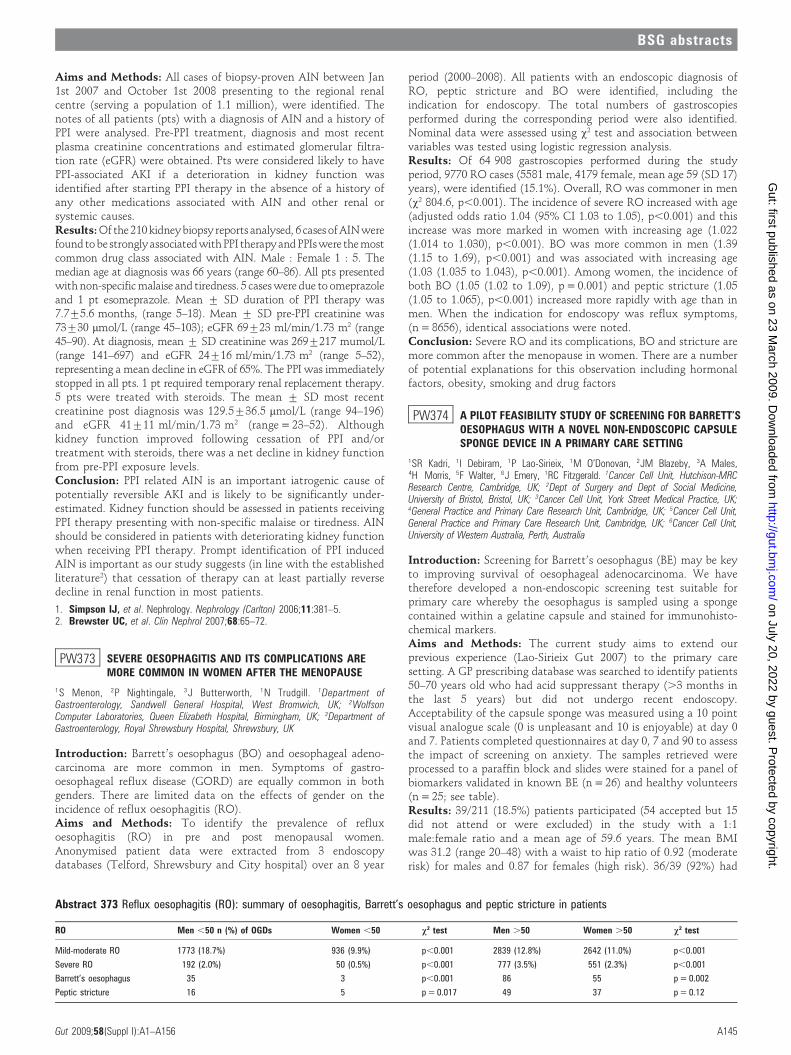
Aims and Methods: All cases of biopsy-proven AIN between Jan1st 2007 and October 1st 2008 presenting to the regional renalcentre (serving a population of 1.1 million), were identified. Thenotes of all patients (pts) with a diagnosis of AIN and a history ofPPI were analysed. Pre-PPI treatment, diagnosis and most recentplasma creatinine concentrations and estimated glomerular filtra-tion rate (eGFR) were obtained. Pts were considered likely to havePPI-associated AKI if a deterioration in kidney function wasidentified after starting PPI therapy in the absence of a history ofany other medications associated with AIN and other renal orsystemic causes.Results: Of the 210 kidneybiopsy reports analysed, 6 cases of AIN werefound to bestrongly associated withPPI therapy and PPIs were the mostcommon drug class associated with AIN. Male : Female 1 : 5. Themedian age at diagnosis was 66 years (range 60–86). All pts presentedwith non-specific malaise and tiredness. 5 cases were due to omeprazoleand 1 pt esomeprazole. Mean ¡ SD duration of PPI therapy was7.7¡5.6 months, (range 5–18). Mean ¡ SD pre-PPI creatinine was73¡30 mmol/L (range 45–103); eGFR 69¡23 ml/min/1.73 m2 (range45–90). At diagnosis, mean ¡ SD creatinine was 269¡217 mumol/L(range 141–697) and eGFR 24¡16 ml/min/1.73 m2 (range 5–52),representing a mean decline in eGFR of 65%. The PPI was immediatelystopped in all pts. 1 pt required temporary renal replacement therapy.5 pts were treated with steroids. The mean ¡ SD most recentcreatinine post diagnosis was 129.5¡36.5 mmol/L (range 94–196)and eGFR 41¡11 ml/min/1.73 m2 (range = 23–52). Althoughkidney function improved following cessation of PPI and/ortreatment with steroids, there was a net decline in kidney functionfrom pre-PPI exposure levels.Conclusion: PPI related AIN is an important iatrogenic cause ofpotentially reversible AKI and is likely to be significantly under-estimated. Kidney function should be assessed in patients receivingPPI therapy presenting with non-specific malaise or tiredness. AINshould be considered in patients with deteriorating kidney functionwhen receiving PPI therapy. Prompt identification of PPI inducedAIN is important as our study suggests (in line with the establishedliterature2) that cessation of therapy can at least partially reversedecline in renal function in most patients.
1. Simpson IJ, et al. Nephrology. Nephrology (Carlton) 2006;11:381–5.2. Brewster UC, et al. Clin Nephrol 2007;68:65–72.
PW373 SEVERE OESOPHAGITIS AND ITS COMPLICATIONS AREMORE COMMON IN WOMEN AFTER THE MENOPAUSE
1S Menon, 2P Nightingale, 3J Butterworth, 1N Trudgill. 1Department ofGastroenterology, Sandwell General Hospital, West Bromwich, UK; 2WolfsonComputer Laboratories, Queen Elizabeth Hospital, Birmingham, UK; 3Department ofGastroenterology, Royal Shrewsbury Hospital, Shrewsbury, UK
Introduction: Barrett’s oesophagus (BO) and oesophageal adeno-carcinoma are more common in men. Symptoms of gastro-oesophageal reflux disease (GORD) are equally common in bothgenders. There are limited data on the effects of gender on theincidence of reflux oesophagitis (RO).Aims and Methods: To identify the prevalence of refluxoesophagitis (RO) in pre and post menopausal women.Anonymised patient data were extracted from 3 endoscopydatabases (Telford, Shrewsbury and City hospital) over an 8 year
period (2000–2008). All patients with an endoscopic diagnosis ofRO, peptic stricture and BO were identified, including theindication for endoscopy. The total numbers of gastroscopiesperformed during the corresponding period were also identified.Nominal data were assessed using x2 test and association betweenvariables was tested using logistic regression analysis.Results: Of 64 908 gastroscopies performed during the studyperiod, 9770 RO cases (5581 male, 4179 female, mean age 59 (SD 17)years), were identified (15.1%). Overall, RO was commoner in men(x2 804.6, p,0.001). The incidence of severe RO increased with age(adjusted odds ratio 1.04 (95% CI 1.03 to 1.05), p,0.001) and thisincrease was more marked in women with increasing age (1.022(1.014 to 1.030), p,0.001). BO was more common in men (1.39(1.15 to 1.69), p,0.001) and was associated with increasing age(1.03 (1.035 to 1.043), p,0.001). Among women, the incidence ofboth BO (1.05 (1.02 to 1.09), p = 0.001) and peptic stricture (1.05(1.05 to 1.065), p,0.001) increased more rapidly with age than inmen. When the indication for endoscopy was reflux symptoms,(n = 8656), identical associations were noted.Conclusion: Severe RO and its complications, BO and stricture aremore common after the menopause in women. There are a numberof potential explanations for this observation including hormonalfactors, obesity, smoking and drug factors
PW374 A PILOT FEASIBILITY STUDY OF SCREENING FOR BARRETT’SOESOPHAGUS WITH A NOVEL NON-ENDOSCOPIC CAPSULESPONGE DEVICE IN A PRIMARY CARE SETTING
1SR Kadri, 1I Debiram, 1P Lao-Sirieix, 1M O’Donovan, 2JM Blazeby, 3A Males,4H Morris, 5F Walter, 6J Emery, 1RC Fitzgerald. 1Cancer Cell Unit, Hutchison-MRCResearch Centre, Cambridge, UK; 2Dept of Surgery and Dept of Social Medicine,University of Bristol, Bristol, UK; 3Cancer Cell Unit, York Street Medical Practice, UK;4General Practice and Primary Care Research Unit, Cambridge, UK; 5Cancer Cell Unit,General Practice and Primary Care Research Unit, Cambridge, UK; 6Cancer Cell Unit,University of Western Australia, Perth, Australia
Introduction: Screening for Barrett’s oesophagus (BE) may be keyto improving survival of oesophageal adenocarcinoma. We havetherefore developed a non-endoscopic screening test suitable forprimary care whereby the oesophagus is sampled using a spongecontained within a gelatine capsule and stained for immunohisto-chemical markers.Aims and Methods: The current study aims to extend ourprevious experience (Lao-Sirieix Gut 2007) to the primary caresetting. A GP prescribing database was searched to identify patients50–70 years old who had acid suppressant therapy (.3 months inthe last 5 years) but did not undergo recent endoscopy.Acceptability of the capsule sponge was measured using a 10 pointvisual analogue scale (0 is unpleasant and 10 is enjoyable) at day 0and 7. Patients completed questionnaires at day 0, 7 and 90 to assessthe impact of screening on anxiety. The samples retrieved wereprocessed to a paraffin block and slides were stained for a panel ofbiomarkers validated in known BE (n = 26) and healthy volunteers(n = 25; see table).Results: 39/211 (18.5%) patients participated (54 accepted but 15did not attend or were excluded) in the study with a 1:1male:female ratio and a mean age of 59.6 years. The mean BMIwas 31.2 (range 20–48) with a waist to hip ratio of 0.92 (moderaterisk) for males and 0.87 for females (high risk). 36/39 (92%) had
Abstract 373 Reflux oesophagitis (RO): summary of oesophagitis, Barrett’s oesophagus and peptic stricture in patients
RO Men ,50 n (%) of OGDs Women ,50 x2 test Men .50 Women .50 x2 test
Mild-moderate RO 1773 (18.7%) 936 (9.9%) p,0.001 2839 (12.8%) 2642 (11.0%) p,0.001
Severe RO 192 (2.0%) 50 (0.5%) p,0.001 777 (3.5%) 551 (2.3%) p,0.001
Barrett’s oesophagus 35 3 p,0.001 86 55 p = 0.002
Peptic stricture 16 5 p = 0.017 49 37 p = 0.12
BSG abstracts
Gut 2009;58(Suppl I):A1–A156 A145
on July 20, 2022 by guest. Protected by copyright.
http://gut.bmj.com
/G
ut: first published as on 23 March 2009. D
ownloaded from
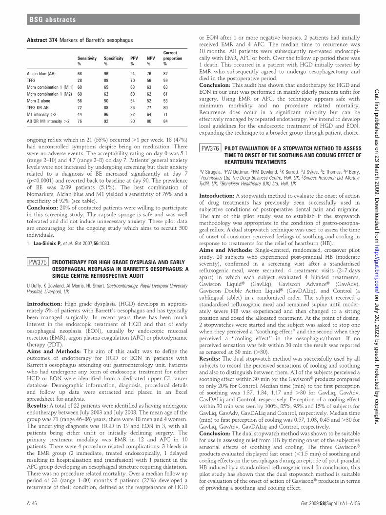
ongoing reflux which in 21 (55%) occurred .1 per week. 18 (47%)had uncontrolled symptoms despite being on medication. Therewere no adverse events. The acceptability rating on day 0 was 5.1(range 2–10) and 4.7 (range 2–8) on day 7. Patients’ general anxietylevels were not increased by undergoing screening but their anxietyrelated to a diagnosis of BE increased significantly at day 7(p,0.0001) and reverted back to baseline at day 90. The prevalenceof BE was 2/39 patients (5.1%). The best combination ofbiomarkers, Alcian blue and M1 yielded a sensitivity of 76% and aspecificity of 92% (see table).Conclusion: 20% of contacted patients were willing to participatein this screening study. The capsule sponge is safe and was welltolerated and did not induce unnecessary anxiety. These pilot dataare encouraging for the ongoing study which aims to recruit 500individuals.
1. Lao-Sirieix P, et al. Gut 2007;56:1033.
PW375 ENDOTHERAPY FOR HIGH GRADE DYSPLASIA AND EARLYOESOPHAGEAL NEOPLASIA IN BARRETT’S OESOPHAGUS: ASINGLE CENTRE RETROSPECTIVE AUDIT
U Duffy, K Gowland, AI Morris, HL Smart. Gastroenterology, Royal Liverpool UniversityHospital, Liverpool, UK
Introduction: High grade dysplasia (HGD) develops in approxi-mately 5% of patients with Barrett’s oesophagus and has typicallybeen managed surgically. In recent years there has been muchinterest in the endoscopic treatment of HGD and that of earlyoesophageal neoplasia (EON), usually by endoscopic mucosalresection (EMR), argon plasma coagulation (APC) or photodynamictherapy (PDT).Aims and Methods: The aim of this audit was to define theoutcomes of endotherapy for HGD or EON in patients withBarrett’s oesophagus attending our gastroenterology unit. Patientswho had undergone any form of endoscopic treatment for eitherHGD or EON were identified from a dedicated upper GI cancerdatabase. Demographic information, diagnosis, procedural detailsand follow up data were extracted and placed in an Excelspreadsheet for analysis.Results: A total of 22 patients were identified as having undergoneendotherapy between July 2003 and July 2008. The mean age of thegroup was 71 (range 46–86) years; there were 18 men and 4 women.The underlying diagnosis was HGD in 19 and EON in 3, with allpatients being either unfit or initially declining surgery. Theprimary treatment modality was EMR in 12 and APC in 10patients. There were 4 procedure related complications: 3 bleeds inthe EMR group (2 immediate, treated endoscopically, 1 delayedresulting in hospitalisation and transfusion) with 1 patient in theAPC group developing an oesophageal stricture requiring dilatation.There was no procedure related mortality. Over a median follow upperiod of 33 (range 1–80) months 6 patients (27%) developed arecurrence of their condition, defined as the reappearance of HGD
or EON after 1 or more negative biopsies. 2 patients had initiallyreceived EMR and 4 APC. The median time to recurrence was10 months. All patients were subsequently re-treated endoscopi-cally with EMR, APC or both. Over the follow up period there was1 death. This occurred in a patient with HGD initially treated byEMR who subsequently agreed to undergo oesophagectomy anddied in the postoperative period.Conclusion: This audit has shown that endotherapy for HGD andEON in our unit was performed in mainly elderly patients unfit forsurgery. Using EMR or APC, the technique appears safe withminimum morbidity and no procedure related mortality.Recurrence does occur in a significant minority but can beeffectively managed by repeated endotherapy. We intend to developlocal guidelines for the endoscopic treatment of HGD and EON,expanding the technique to a broader group through patient choice.
PW376 PILOT EVALUATION OF A STOPWATCH METHOD TO ASSESSTIME TO ONSET OF THE SOOTHING AND COOLING EFFECT OFHEARTBURN TREATMENTS
1V Strugala, 1PW Dettmar, 2PM Dewland, 3K Sarratt, 3J Sykes, 3E Thomas, 3P Berry.1Technostics Ltd, The Deep Business Centre, Hull, UK; 2Simbec Research Ltd, MerthyrTydfil, UK; 3Benckiser Healthcare (UK) Ltd, Hull, UK
Introduction: A stopwatch method to evaluate the onset of actionof drug treatments has previously been successfully used insubjective conditions of postoperative dental pain and migraine.The aim of this pilot study was to establish if the stopwatchmethodology was appropriate in the condition of gastro-oesopha-geal reflux. A dual stopwatch technique was used to assess the timeof onset of consumer-perceived feelings of soothing and cooling inresponse to treatments for the relief of heartburn (HB).Aims and Methods: Single-centred, randomised, crossover pilotstudy. 20 subjects who experienced post-prandial HB (moderateseverity), confirmed in a screening visit after a standardisedrefluxogenic meal, were recruited. 4 treatment visits (2–7 daysapart) in which each subject evaluated 4 blinded treatments,Gaviscon LiquidH (GavLiq), Gaviscon AdvanceH (GavAdv),Gaviscon Double Action LiquidH (GavDALiq), and Control (asublingual tablet) in a randomised order. The subject received astandardised refluxogenic meal and remained supine until moder-ately severe HB was experienced and then changed to a sittingposition and dosed the allocated treatment. At the point of dosing,2 stopwatches were started and the subject was asked to stop onewhen they perceived a ‘‘soothing effect’’ and the second when theyperceived a ‘‘cooling effect’’ in the oesophagus/throat. If noperceived sensation was felt within 30 min the result was reportedas censored at 30 min (.30).Results: The dual stopwatch method was successfully used by allsubjects to record the perceived sensations of cooling and soothingand also to distinguish between them. All of the subjects perceived asoothing effect within 30 min for the GavisconH products comparedto only 20% for Control. Median time (min) to the first perceptionof soothing was 1.37, 1.34, 1.17 and .30 for GavLiq, GavAdv,GavDALiq and Control, respectively. Perception of a cooling effectwithin 30 min was seen by 100%, 85%, 95% and 15% of subjects forGavLiq, GavAdv, GavDALiq and Control, respectively. Median time(min) to first perception of cooling was 0.57, 1.03, 0.45 and .30 forGavLiq, GavAdv, GavDALiq and Control, respectively.Conclusion: The dual stopwatch method was shown to be suitablefor use in assessing relief from HB by timing onset of the subjectivesensorial effects of soothing and cooling. The three GavisconHproducts evaluated displayed fast onset (,1.5 min) of soothing andcooling effects on the oesophagus during an episode of post-prandialHB induced by a standardised refluxogenic meal. In conclusion, thispilot study has shown that the dual stopwatch method is suitablefor evaluation of the onset of action of GavisconH products in termsof providing a soothing and cooling effect.
Abstract 374 Markers of Barrett’s oesophagus
Sensitivity%
Specificity%
PPV%
NPV%
Correctproportion%
Alcian blue (AB) 68 96 94 76 82
TFF3 28 88 70 56 59
Mcm combination 1 (M 1) 60 65 63 63 63
Mcm combination 1 (M2) 60 62 60 62 61
Mcm 2 alone 56 50 54 52 53
TFF3 OR AB 72 88 86 77 80
M1 intensity .2 44 96 92 64 71
AB OR M1 intensity .2 76 92 90 80 84
BSG abstracts
A146 Gut 2009;58(Suppl I):A1–A156
on July 20, 2022 by guest. Protected by copyright.
http://gut.bmj.com
/G
ut: first published as on 23 March 2009. D
ownloaded from
Radiology posters
PW377 PROSPECTIVE COMPARISON OF HIGH FREQUENCY MINIPROBE ULTRASOUND AND CONVENTIONAL COMPUTEDTOMOGRAPHY IN THE LOCAL STAGING OF COLONICCANCERS
1AK Haji, 2S Ryan, 3I Bjarnason, 1S Papagrigoriadis. 1Colorectal Surgery, Kings CollegeHospital, London, UK; 2Radiology, Kings College Hospital, London, UK;3Gastroenterology, Kings College Hospital, London, UK
Introduction: The introduction of the colorectal cancer screeningprogramme may see an increase in mucosal lesions and early cancersamenable to advanced colonoscopic and laparoscopic techniques. Inaddition, locally advanced colonic cancers are being subject to neo-adjuvant chemotherapy. Therefore, accurate pre-operative localstaging of colonic cancers is becoming of paramount importance.Aims and Methods: We prospectively compared the performance ofendoscopic high frequency mini probe ultrasound (HFUS) andconventional computed tomography (CT) in the local staging ofcolonic cancers. 25 consecutive patients with colonic cancer wererecruited after informed consent. Pre-operative investigationsincluded colonoscopy and multi-slice CT with multiplanar recon-struction read by an experienced colorectal radiologist pre-operatively.All patients underwent both 12.5 MHz and 20 MHz US (OlympusKeymed UM-3R, Japan) either during the primary colonoscopy or ontable prior to colonic resection. The images were read by 2 authorswho were blind to the results of the CT. The local staging by bothmodalities were compared to the histological stage of the resectedspecimen. Statistical differences were analysed using x2 and Fisher’sexact t tests with Graphpad Prism 5 (Version 5.1, 2007).Results: The distribution of colonic cancers were: sigmoid (n = 8),caecum (n = 7), descending (n = 4), recto-sigmoid (n = 4), hepatic(n = 1) and splenic flexure (n = 1). Histological assessment of theresected specimens revealed 5 T1, 2 T2 and 18 T3 cancers.Conventional CT overstaged all T1 tumours, 4 as T2 and 1 as T3.For T3 tumours, the sensitivity and specificity of CT was 72% and71%, respectively. This was significantly different to both 12 and20 MHz US which accurately staged all T1 and T3 cancers offering100% sensitivity and specificity (p = ,0.005). All imaging modalitiescorrectly staged only 1 out of 2 T2 tumours, overstaging to T3. Overall,accuracy of T staging by CT was 56% compared to 96% by both 12 and20 MHz US (p = ,0.005). Histologically 9/25 (36%) patients werepositive for nodal disease. Both CT and 12 MHz US were significantlybetter than 20 MHz US for detection of nodal disease (p = ,0.005),with no significant difference between CT and 12 MHz US (p = 0.30).The sensitivity and specificity for detection of nodal disease byconventional CT was 89% and 38%, respectively, with a positivepredictive value (PPV) of 44% and negative predictive value (NPV) of86%. In comparison, staging by 12 MHz US had a sensitivity of 100%and a specificity of 75%, significantly better than that offered by20 MHz US at 25% sensitivity and 86% specificity (p = ,0.005).Conclusion: Colonic ultrasound with miniprobes is significantlymore accurate than CT staging of colorectal cancers. The techniquehas potential application as a routine procedure during colonoscopy.
PW378 A CATHETER-MOUNTED MAGNETIC RESONANCE DETECTORCOIL FOR BILIARY IMAGING: FIRST IN VITRO BILIARY IMAGES
1CA Wadsworth, 2RRA Syms, 1SA Khan, 3RJ Dickinson, 4WMW Gedroyc, 1MRThursz, 2MM Ahmad, 3SA Awan, 4W Caspersz, 1SD Taylor-Robinson, 2IR Young.1Department of Hepatology and Gastroenterology, Imperial College London, London,UK; 2Department of Electrical and Electronic Engineering, Imperial College London,London, UK; 3Department of Bioengineering, Imperial College London, London, UK;4IMR Unit, Imperial College London, London, UK
Introduction: The incidence of cholangiocarcinoma (CC) is risingglobally. CC has increased to become the commonest cause of death
from a primary liver cancer in the UK. Despite advances in CT andMR technology, the correct classification of biliary strictures asbenign or malignant remains difficult. This is particularly so inpatients with primary sclerosing cholangitis, the commonestknown predisposing factor for CC. Even endoscopic retrogradecholangiopancreatography (ERCP) with brush cytology has a lowsensitivity for neoplasm detection. Traditional axial MR systemshave an external detector radio-coil built into the scanner gantry.An MR system in which the detection radio coil is closely apposedto the tissue of interest could improve the resolution of the imagesobtained. Our group has developed a prototype version of such acoil, designed such that it could be passed into the biliary tree via anendoscope to improve tissue conspicuity.Aims and Methods: We aimed to confirm the utility of aprototype coil in obtaining MR images, collect quantitativeresolution data and prove that the probe could function in wetconditions. Images were acquired using a 1.5T GE SignaTM scanner,using the main body coil for excitation and a wrap-around microcoilprobe for detection. The microcoil is a 35 mm long 2-turn thin filmdevice, tuned and matched at 63.8 MHz, with discrete SMDcapacitors; and is attached to an 8F biliary catheter. Overall, theprobe is 2.7 mm in diameter and is fully MR compatible. Range andresolution were established first, by placing the coil between astandard spherical water phantom and cuvettes containing micro-fabricated resolution test samples. The micro-coil was located at themagnet isocentre and arranged parallel to the magnet bore. 3D GREsequences containing 28 slices, each 1.2 mm thick, were used(TR = 33 ms, TE = 15 ms, 2566256 image points, 8 cm field ofview). Imaging was then carried out using meat models with thecoil inserted into the biliary tree and liver parenchyma.Results: Scanning microfabricated test samples produced imagesshowing entire cuvettes (1 cm deep) with 1 mm resolution test barsclearly visible, together with a similar depth of water phantom.Imaging of porcine biliary tree revealed anatomical detail to a depthof more than 1.5 cm, 360 degrees around the coil, along 3.2 cm of itslength. Branching bile ducts and blood vessels can be visualisedclearly in the images presented.Conclusion: The MR probe developed by our group can reliablyproduce images of a resolution of at least 1 mm and demonstrateanatomical detail in meat models. Further work is required toincrease the useful length of the probe and improve the imagesobtained. Work is underway on a clinical trial of this device.
1. Khan SA, et al. Lancet 2005;366:1303–14.2. Taylor-Robinson SD, et al. Gut 2001;48:816–20.
PW379 USE OF MOLECULAR IMAGING TO DIFFERENTIATE LIVERMETASTASES ORIGINATING FROM TWO DIFFERENTPRIMARY NEOPLASMS IN THE SAME PATIENT
1KK Desai, 1A Jayratnam, 1VM Robinson, 1E Carras, 2A Quigley, 2J Buscombe, 1CToumpanakis, 1M Caplin. 1Neuroendocrine Tumour Unit, Royal Free Hospital, London,UK; 2Nuclear Medicine Department, Royal Free Hospital, London, UK
Introduction: Synchronous malignant neoplasms in a singlepatient are well documented in the literature. At least 10% ofpatients with neuroendocrine tumour may develop another cancer,not infrequently colonic adenocarcinoma. FDG PET is recognised as‘‘standard’’ imaging for staging metastatic disease in colorectalcancer and recently Gallium-68 DOTA Octreotate PET has beendeveloped as molecular imaging for neuroendocrine tumour staging.Aims and Methods: To assess molecular imaging utilising (18F)FDG PET scan and Gallium 68 DOTATATE PET scan fordifferentiating liver metastases arising from colorectal cancer orneuroendocrine tumour in an individual patient. We studied apatient who had recently undergone left hemicolectomy (Duke’s Badenocarcinoma) of the sigmoid colon, and was found to have
BSG abstracts
Gut 2009;58(Suppl I):A1–A156 A147
on July 20, 2022 by guest. Protected by copyright.
http://gut.bmj.com
/G
ut: first published as on 23 March 2009. D
ownloaded from
multiple liver metastases and a pancreatic tail mass compatible withneuroendocrine tumour. The patient underwent (18F) FDG PETscan and (68Ga) DOTATATE PET scan. He also underwentultrasound guided liver biopsy.Results: The two types of PET scan showed differential uptake inthe liver. Some metastases had avid uptake only on the FDG PETand the others only on the Gallium 68 PET, the latter alsoidentifying the pancreatic NET primary. Ultrasound guided liverbiopsy specimen confirmed the two types of metastases: moder-ately differentiated colonic adenocarcinoma and well differentiatedneuroendocrine tumour with low grade proliferation index Ki67,2%.Conclusion: Uptake of specific peptides by tumours is dependenton the biology of the tumour. (18F) FDG PET is very sensitive indetecting metastasis arising from colon cancer in keeping with thefact that FDG PET targets the glucose transporter in these activelymultiplying cells. However, neuroendocrine tumours are moreindolent, and studies have shown that sensitivity of FDG PET indetecting metastasis in these tumours is much lower. NETs expresssomatostatin receptors, predominantly subtype-2, which has led tothe development of new radio peptides such as (68) GalliumDOTATATE PET, which is much more sensitive and specific indefining distant metastasis related to NETs. We therefore demon-strate the utility of different PET agents for identifying differentcancers in an individual patient.
1. Veit-Haibach P, Kuehle CA, Beyer T, et al. JAMA 2006;296:2590–60.2. Kayani I, Bomanji JB, Groves A, et al. Cancer 2008;112:2447–55.
PW380 AUDIT OF ULTRASOUND GUIDED PERCUTANEOUS LIVERBIOPSY: ARE WE DOING ENOUGH?
1SS Webster, 2M Sriharan, 1M Anderson, 2N Khan. 1Gastroenterology, Chelsea andWestminster Hospital, London, UK; 2Radiology, Chelsea and Westminster Hospital,London, UK
Introduction: Liver biopsy is an important investigative tool in themanagement of a wide range of hepatic disease. Ultrasound guidedpercutaneous liver biopsy (UG-PLB) is an invasive but relatively safeprocedure. To avoid sampling error and histological inaccuracy, it isimportant that technique maximises sample size. Recent evidencesuggests that especially in chronic viral hepatitis (CVH) largersample sizes give more accurate histological stage and grade ofdisease activity.Aims and Methods: We aimed to retrospectively assess demo-graphics, indications, operator experience, needle type, sample size,complications and overall helpfulness of PLBs at our centre over a 12month period against standards suggested recently for CVH,1 tonational audit data,2 to BSG guidelines3 and to our own previousaudit (unpublished).Results: 101 patients had UG-PLB during the 12 months analysed.67% of patients were male, mean age 50.9 (range 16–85). 35% ofpatients had CVH (HBV or HCV). All PLBs were image guided andall were performed by radiologists. 56% of procedures wereperformed by radiology registrars. 66% were performed using a16Fr Tru-Cut needle. 91% of procedures involved 1 or 2 passes. 98%of samples were suitable for histological analysis. 92% of samplesexceeded aggregate length of 10 mm. 31% of samples had anaggregate length >2 cm and portal tracts >11. The histology wasclinically helpful in 81%. The complication rate was 8%. Onepatient died within 30 days of the procedure.Conclusion: The number of USG-PLB performed in our hospital(100/yr) is comparable between 2005 and 2007. The indicationswere consistent with BSG guidelines (2004). Documentation ofprocedure was generally good. 100% PLB were performed usingultrasound guidance compared to 38% nationally in 1991. All USG-PLB were performed by radiologists in contrast to 1991 national
data (1/3 general physicians, 1/3 gastroenterologists, 1/3 radiolo-gists). Complication rates were comparable with national data (8%vs 5%). One death within 30 days was not procedure related.Sample adequacy and usefulness was satisfactory. However, biggersample sizes may be needed for CVH patients. To try and improvesample adequacy and histological accuracy our local policy haschanged to advise 2 16Fr passes. This can be done safely withoutsignificantly affecting the complication rate.3 We are currently re-auditing our data with a larger subset of patients with CVH toassess whether sample adequacy has improved without compromis-ing our complication rate.
1. Guido M, et al. Guidelines on the use of liver biopsy in clinical practice. Seminarsin Liver Disease 2004;24(1).
2. Gilmore IT, et al. Indications, methods, and outcomes of percutaneous liverbiopsy in England and Wales: an audit by the British Society of Gastroenterologyand the Royal College of Physicians of London. Gut 1995;36:437–41.
3. Neuberger J, et al. Guidelines on the use of liver biopsy in clinical practice. BritishSociety of Gastroenterology 2004.
PW381 DILATED COMMON BILE DUCT WITHOUT AN OBVIOUSCAUSE ON MAGNETIC RESONANCE CHOLANGIO-PANCREATOGRAM: A 5 YEAR RETROSPECTIVE STUDY
1Z Ibrahim, 2C Jones, 3M Warren, 3S Chatterjee. 1Medicine, North Tyneside GeneralHospital, North Shields, UK; 2Medicine, Wansbeck General Hospital, Ashington, UK;3Gastroenterology, North Tyneside General Hospital, North Shields, UK
Introduction: Magnetic resonance cholangio-pancreatogram(MRCP) is now widely available as a modality for imaging thebiliary tree. We aimed to define the profile of patients who had adilated common bile duct (CBD) on MRCP, but without an obviouscause being demonstrated.Aims and Methods: We reviewed the MRCP reports of all patientsat North Tyneside Hospital in the past 5 years and excluded all thepatients in whom the scan could assign a cause for a dilated CBD.All patients in whom the MRCP reported a dilated CBD, but thecause was not apparent, were included in the study.Results: We identified 86 such patients (24 male; 62 female): 11patients were under 40 years age, 23 between 40 to 60 years, and 52were more than 60 years age. The indication for MRCP wasabdominal pain in 29, abnormal liver function tests (LFTs) in 11,and both in 38 patients. LFTs were done pre-MRCP in 83 patients.The notes of 8 patients could not be obtained. 56 had obstructiveand 19 had normal LFTs. 75 patients had some form of imagingprior to MRCP (ultrasound - 66; CT abdomen/pancreas - 7; both -2). Ultrasound or CT demonstrated dilated CBD in 55 patients,normal in 15 patients and was not commented upon in 5. Theresults of MRCP were correlated with the indications for imaging,LFT abnormality and pre-MRCP scans. Out of the 75 patients withdilated CBD on MRCP, 29 had evidence of stones/sludge in the gallbladder (GB) and 8 had thickened GB suggestive of cholecystitis inpre-MRCP ultrasound scans. Seven patients had cholecystectomy inthe past and the dilated CBD was considered to be normal in thatsubgroup. Six patients had evidence of chronic pancreatitis withabnormal CBD and pancreatic duct (PD). Five had a CBD stricture,2 had ampullary stenosis, 4 had metastatic cancer, and 1 hadhepatic cystic disease. One was later diagnosed with primarysclerosing cholangitis. True CBD dilatation without a cause wasrecognised in only 12 patients. The main indication for MRCP inthis group was abdominal pain. Seven had normal LFTs and 5 ofthem had minimally obstructive LFTs (max ALP 133; max GGT155). The diameter of CBD on MRCP ranged from 7 to 10 mm.These patients were followed up for 2 years. Two patients weresuspected to have sphincter of Oddi dysfunction and 2 neededcholecystectomy in the follow-up period; one for chronic cholecys-titis and the other for empyema of the GB.
BSG abstracts
A148 Gut 2009;58(Suppl I):A1–A156
on July 20, 2022 by guest. Protected by copyright.
http://gut.bmj.com
/G
ut: first published as on 23 March 2009. D
ownloaded from
Conclusion: MRCP is a very sensitive investigation in documentingCBD pathology. True dilatation without a cause is uncommon. Thesemight be normal variants and do not herald a sinister pathology as wehave observed in our 2 year follow-up period. Sphincter of Oddidysfunction is an important consideration in this subgroup. Furtherinvestigations are needed to find out whether an association betweena dilated CBD and abdominal pain is significant.
Service development posters
PW382 ARE WE USING TUMOUR MARKERS APPROPRIATELY?
CL Gan, L Sunder Raj, S Malik, MA Czajkowski. Department of Gastroenterology, RoyalGwent Hospital, Newport, UK
Introduction: Tumour markers are increasingly becoming part ofroutine investigations nowadays. Currently there are no clearguidelines on their use throughout the UK and the use of tumourmarkers has rarely been studied properly in the past. To investigatefurther the use of tumour markers, requests for gastrointestinaltumour markers, i.e. CEA, CA19–9 and AFP, were studied andcompared with recommendations made by European Group OnTumour Markers (EGTM) and American Society Of ClinicalOncology (ASCO).Aims and Methods: All the inpatient and outpatient requestsmade for CEA, CA19–9 or AFP within Royal Gwent Hospital and StWoolos Hospital between 3 March 2008 and 7 March 2008 werestudied prospectively. Medical records and test results from clinicalworkstations were reviewed to ascertain the indication for theirrequests.Results: In total, 140 requests in 81 patients were analysed. 91% ofCEA, 98% of CA19–9 and 44% of AFP were requested inappropri-ately, according to EGTM and ASCO recommendations. 54% of thepatients had more than two tumour markers requested. Incidentalfindings also revealed 3 requests for CA153 and 13 requests forCA125 were made in male patients from our study, which wereinappropriate. Most of these inappropriate requests were made toscreen for malignancy in patients suspected for cancers of anyorigin. Medical and surgical departments were equally responsiblefor these inappropriate requests. Estimated wastage of resourcesbased on test reagents alone exceeded £8000/pa. Also, wastage ofconsultants’ time, outpatient appointments and unnecessarysubsequent investigations were noted but difficult to quantify.Conclusion: The majority of requests for gastrointestinal tumourmarkers are inappropriate, according to the international guidelines.This is probably also the case for many hospitals in the UK. A trustwide guideline is now being created to help prevent theseinappropriate requests. The findings and recommendations couldalso apply to other hospitals in the UK in order to improve practice.
PW383 THE YIELD OF ENDOSCOPY IN PATHOLOGY DETECTION ISMUCH HIGHER THAN EXPECTED
CS Kothari, NV Someren, K Besherdas. Gastroenterology, Barnet and Chase FarmHospitals NHS Trust, Enfield, Middlesex, UK
Introduction: The demand for gastrointestinal endoscopy isincreasing with a resultant rise in overall costs and waiting listsfor endoscopic procedures. Therefore, adherence to appropriateindications for these procedures is essential for the rational use offinite resources. Appropriateness of endoscopy is an importantaspect of the Global Rating Scale. There is evidence from publishedstudies from Europe that the proportion of patients with negativeendoscopies is increasing.1 Further to development of a newendoscopy referral form and vetting of endoscopy requests by aconsultant gastroenterologist, we set out to assess the detection ofpathology rate in endoscopies performed within our unit.
Aims and Methods: To determine the pathology detection rate atendoscopy following the introduction of consultant triaging ofprocedures. In addition to assess any difference in pathologydetected in patients being referred for inpatient and outpatientendoscopy. A single centre retrospective analysis of 1500 consecu-tive patients referred for endoscopy within an associated teachinghospital was performed. All completed referral forms for endoscopy,inpatient or outpatient, were vetted by consultant gastroenterol-ogist for appropriateness of indication. Patients undergoing endo-scopy were analysed for detection of pathology.Results: Of the 1500 endoscopies, pathology was detected in 927(62%). Out of a total of 382 inpatients, 252 had a pathology found(65.96%). Out of a total of 1118 outpatients, 675 had a pathologyfound (60.37%). Pathology included various lesions ranging frombenign polyps, ulcers, oesophageal strictures, Mallory Weiss tears toneoplastic growths.Conclusion: There is a very high pick-up rate of pathologies withinour unit in comparison to published reports from Europe ofreducing rates of pathology detection. This may well reflect theselection criterion of endoscopy and consultant vetting. Overall,there is a greater chance of inpatients having pathology comparedto outpatients although in both groups the pathology pick up rate isgreater than 60%.
1. Changes in indications for upper gastrointestinal tract endoscopy and endoscopicfindings during the last fifteen years in South-Western Greece. American Journal ofthe Medical Sciences July 2008;336:21–6.
PW384 THE ONE STOP SWALLOWING SHOP: A BETTER CHOICE FORSUSPECTED CANCER
DL Morris, J Deacon. Gastroenterology, Queen Elizabeth II Hospital, Welwyn GardenCity, UK
Introduction: A succession of cancer targets over the last 5 yearshave led to streamlining the patient pathway in the management ofupper gastrointestinal cancers. The role of the clinical nursespecialist has had a similar impact on the quality of the multi-disciplinary care provided. We report on a new combined nurse andconsultant led service established to improve patient care andreduce waiting times.Aims and Methods: A weekly rapid access combined clinic andendoscopy service was established in our District General Hospital,catchment population 250 000. All suspected upper GI cancer 2-week GP referrals from 3 January 2006 to 30 September 2008 werevetted by a consultant gastroenterologist along with all otherreferrals where dysphagia was a main symptom or suspicion ofmalignancy raised. Patients were offered a combined assessmentwith the nurse specialist followed by same morning gastroscopy bya consultant gastroenterologist. Further investigations and manage-ment plan were organised according to endoscopic and clinicalfindings and all malignancies referred directly to the multidisci-plinary meeting.Results: 167 patients (81 male) were seen over 33 months, aged 30to 98 years. 41 (25%) were 2-week referrals, 116 (69%) urgent and10 (6%) routine. Dysphagia was the main referring symptom in 120(72%), dyspepsia in 17 (10%), reflux in 11 (6.6%), and weight loss in9 (5.4%) patients, respectively. Other symptoms included chestpain, nausea, abdominal pain and pain on swallowing. 23 cancerswere diagnosed at endoscopy, 14% of all patients and 18% of thosewith dysphagia. Including benign disease 34 (20.6%) of all patientshad oesophageal strictures. 1 patient had a failed intubation and 1procedure was abandoned due to food residue.Conclusion: The rapid access clinic/endoscopy service has donewhat it states. More than 1/3 of all upper GI cancers are nowdiagnosed in this clinic enabling direct links into the multi-disciplinary team and prompt staging investigations. Patients withbenign disease are reassured and discharged directly on treatmentreducing need for outpatient follow up. Only 6.7% endoscopies
BSG abstracts
Gut 2009;58(Suppl I):A1–A156 A149
on July 20, 2022 by guest. Protected by copyright.
http://gut.bmj.com
/G
ut: first published as on 23 March 2009. D
ownloaded from
were normal which compares favourably with direct accessendoscopy. As 1 in 5 patients seen had oesophageal strictures thisis an excellent method of promptly identifying and managingsignificant pathology. A patient satisfaction survey is underway.
PW385 MORE CANCERS ARE DIAGNOSED IN PATIENTS WITHSYMPTOMS COMPLIANT WITH THE 2 WEEK REFERRALGUIDANCE THAN ORIGINALLY PREDICTED
G Masterton, S Chatterjee, D Nylander. Gastroenterology, Sunderland Royal Hospital,Sunderland, UK
Introduction: In 2000, the Department of Health (DoH) publishedguidance for referral of patients with symptoms suggesting cancer,also termed as 2 week referral scheme (2WRS). In this document, itwas estimated that there would be 1 cancer for every 15 patientsreferred with the suggested symptoms.1
Aims and Methods: To assess the proportion of patients referred tomedical gastroenterology (MGD) via 2WRS pathway, we performed aretrospective notes audit of referrals over 3 months from 1 April 2007.Notes were recalled in late 2008. From the notes, we obtainedinformation on diagnoses, time from referral to hospital attendanceand for those with a diagnosis of cancer, the time to therapy. We alsoassessed whether patients referred via the 2WRS pathway had theappropriate symptoms as per the DoH guidance.Results: 1074 referrals were made to MGD of which 221 (21%) werevia 2WRS pathway (and all these notes were obtained and viewed).All 2WRS patients were seen within 2 weeks of referral. First hospitalcontact was outpatient clinic (OPC) in 43%, direct to test in 53% and4% initially had radiological investigation prior to OPC. In 194patients (88%) the referral complied with 2WRS. A final diagnosis ofcancer was made in 31 patients (sites were oesophageal–4, pancreatic–5, gastric–2, cholangiocarcinoma–2, colorectal–8, hepatoma–1, andothers–9). Cancer was suspected at the first point of contact in 97% ofthese cases. 90% of the cancers were diagnosed within 31 days ofreferral and 95% were diagnosed and started therapy within 61 daysof referral. Gastro-oesophageal reflux disease was diagnosed in 39patients (18%). 9 patients (4%) were diagnosed with inflammatorybowel disease (ulcerative colitis–2; Crohn’s–4; indeterminate colitis–3). 3 patients were diagnosed as microscopic colitis and 1 hadcollagenous colitis. 1 patient was diagnosed with coeliac disease. Inother patients referred for diarrhoea, the diagnoses were bile saltmalabsorption (3), diverticular disease (9), proton pump inhibitorrelated diarrhoea (2), irritable bowel syndrome (6).Conclusion: Despite initial concerns, the 2WRS predicted aboutdouble the original proportion of cancers estimated by DoH (1cancer for every 7.1 referrals in this group). The majority (88%) ofpatients referred via this pathway were compliant with 2WRS. Themajority of patients without cancer were found to have organicrather than functional disorders. In our trust, all patients referred toMGD with 2WRS were seen within 2 weeks of referral and 95% ofthose with cancer started treatment within 61 days.
1. http://www.nice.org.uk/nicemedia/pdf/CG027quickrefguide.pdf.
PW386 APPROPRIATENESS OF THE ‘‘STRAIGHT TO TEST’’GASTROSCOPY REQUEST FOR PATIENTS WITH SUSPECTEDGASTROINTESTINAL CANCERS
1I Amin, 2P Steer, 2R Madhotra. 1Gastroenterology, Milton Keynes General Hospital,Milton Keynes, UK; 2Gastroenterology, Milton Keynes Hospital NHS Trust, MiltonKeynes, UK
Introduction: In our experience ‘‘Straight to Test’’ upper GI cancerassessment service is efficient, safe, and cost-effective in establishingearly diagnosis of and appropriate management in patients withnew diagnosis of gastrointestinal cancer. However, the ‘‘Straight toTest’’ service carries a risk of being used inappropriately and addsfurther workload to our endoscopy units. In the UK the NationalInstitute for Health and Clinical Excellence (NICE) has providedguidelines for requesting urgent endoscopy in patients withsuspected gastrointestinal cancer.Aims and Methods: The aim of this study is to evaluateappropriateness of the urgent gastroscopy referrals for suspectedgastrointestinal cancer in our endoscopy unit at Milton KeynesGeneral Hospital. Between December 2005 and March 2008 allconsecutive patients with requests for urgent gastroscopy fromtheir general practitioners under ‘‘Straight to Test’’ rule wereevaluated for their appropriateness and followed until finaldiagnosis. Data were collected prospectively. Each endoscopyrequest was blindly analysed by a single observer and comparedagainst the current NICE recommendations and categorising theminto Appropriate or Inappropriate. Percentage of cancer diagnosisand other endoscopic diagnosis were calculated in both populationgroups.Results: A total of 509 referrals were studied prospectively. Themean age was 66.7 years. Commonest clinical category for referralswas dyspepsia 342 (67%), followed by dysphagia 230 (45%). Weightloss, iron deficiency anaemia and jaundice was seen in 40%, 17%,and 4%, respectively. Using NICE guidelines 54 (10.6%) referralswere found to be inappropriate and 453 (88.9%) were in keepingwith recommended guidelines. No cancers were diagnosed in theInappropriate group. In the Appropriate group 69 (15%) patientswere diagnosed with cancer. Gastric ulcer, duodenal ulcer andoesophageal ulcer was seen in 7, 4 and 8 patients, respectively inAppropriate group compared to one gastric ulcer and no duodenalulcer or oesophageal ulcer in Inappropriate group. Reasons forinappropriate referrals included: long duration of symptoms greaterthan 12 months 18 (31%), anaemia not investigated before referring13 (22%), dyspepsia under 55 without alarm symptoms 9 (16%),vomiting without alarming symptoms 4 (7%), and miscellaneous6(10%).Conclusion: A significant number of patients are referred forcancer assessment outside the recommended guidelines. Patientsreferred outside the guidelines generally have a low risk of beingdiagnosed with cancer or significant gastrointestinal pathology. Asignificant number of endoscopies could be avoided if the primarycare physicians strictly adhere to professional guidelines. Modifyingour referral form will further aim to reduce the inappropriaterequests.
PW387 AN AUDIT OF NURSE PRESCRIBING IN GASTROENTEROLOGY
I Mason, T Vaghjiani, O Epstein. Gastroenterology, Royal Free Hospital, London, UK
Introduction: After identifying the need for independent nurseprescribing in a series of audits of nurse led outpatient services, theClinical Nurse Specialist (CNS) qualified as an independentprescriber. This audit is the first review of the nurse prescriberspractice since qualification.Aims and Methods: All outpatient prescriptions over a 3 monthperiod were reviewed. The prescriptions were generated in theNurse Led Dyspepsia, Coeliac and Inflammatory Bowel Disease
Abstract 384 Endoscopic diagnosis in a rapid access upper GI clinic
Endoscopic/histological diagnosisNo totaln = 165 %
Oesophageal cancer 19 11.5
Gastric cancer 4 2.4
Benign stricture 11 6.7
GORD/hiatus hernia/oesophagitis (Barrett’s) 67 (10) 40.6 (6)
Oesophageal dysmotility (achalasia) 19 (3) 11.5 (1.8)
Gastritis/duodenitis/ulcer 28 17
Other (eosinophilic oesophagitis) 6 (1) 3.6 (0.6)
Normal 11 6.7
BSG abstracts
A150 Gut 2009;58(Suppl I):A1–A156
on July 20, 2022 by guest. Protected by copyright.
http://gut.bmj.com
/G
ut: first published as on 23 March 2009. D
ownloaded from
clinics. Results were reviewed by the designated medical practi-tioner and the specialist gastroenterology pharmacist.Results: A total of 89 prescriptions were given. Total of 147 items(mean 1.6 per prescription) and 30 different named drugs wereprescribed. Proton pump inhibitors (PPI) were the most commonlyprescribed. Omeprazole was the main PPI of choice (95%) as perlocal guidelines. Two prescribing errors were identified: 1. Amethotrexate prescription for 1 week instead of 4 weeks;2. Calcichew D3 should be prescribed as Calcium D3 forte.Conclusion: This audit found that independent nurse prescribinghad been implemented effectively and safely into the CNS practice.There was no prescribing identified that was inappropriate. Noserious prescribing errors were identified.
1. Nursing and Midwifery Council (2006) Standards and proficiency for nurse andmidwife prescribers.
2. National Prescribing Centre (1999) Signposts for prescribing for nurses - generalprinciples of good prescribing. Prescribing Nurse Bulletin 1 (1) 1–4.
PW388 GUT-DIRECTED HYPNOTHERAPY IN A DISTRICT HOSPITALSETTING SIGNIFICANTLY IMPROVES THE SYMPTOMS OFPATIENTS WITH IRRITABLE BOWEL SYNDROME
GG Robins, LA Reed, SM Kelly, AJ Turnbull, JL Turvill, SJ Smale. Gastroenterology,York Hospital, York, UK
Introduction: Irritable bowel syndrome (IBS) is a chronic disorderaffecting 10–20% of the population, with such patients making upto 50% of gastroenterology referrals.1 Recent UK guidelinesrecommend consideration of hypnotherapy for these patients inprimary care,2 but there is sparse information on its use in anintegrated secondary care setting. York Hospital serves a populationof approximately 280,000 and set up an IBS nurse specialist-ledhypnotherapy service in July 2006.
Aims and Methods: The primary aim of this study is to assessprospectively symptom change in IBS patients referred to thisservice. By definition these patients have failed to respond toconventional advice and medications for IBS. A course ofhypnotherapy typically entails 5–7 sessions over 6–12 weeks.Patients are assessed at each session to check ongoing suitability,in terms of compliance with therapy (use of hypnosis CD at homebetween sessions) and stable mental state. All patients were askedto fill out a Hospital Anxiety and Depression Scale (HADS) pre-therapy, 3 months post- and 6 months post-finishing hypnother-apy. This is a validated Likert-scale based questionnaire thatassesses psychological symptoms in two domains – anxiety(HADA) and depression (HADD). A score of 11+ in either domainindicates ‘‘probable presence’’ of mood disorder. A score of 8–10 isborderline. Patients were also asked to complete visual analoguescales from 1–100 on the severity of the following symptoms: painintensity (PI), pain frequency (PF), bloating (B) and urgency (U).Pre- and post-hypnotherapy scores were compared by the relevantstatistical method (Student’s paired t-test or Wilcoxon signed rankstest). All p values are two-tailed.Results: 62 patients (male 8, female 54) with a mean age (SD) of40.6 (15.1) years have been referred to this service as of March 2008.Conclusion: An IBS nurse specialist-led hypnotherapy servicesignificantly improves short-term patient outcomes in a secondarycare setting. Longer-term data is needed to determine whether thisbenefit is sustained.1. Wilson S, et al. Aliment Pharmacol Ther 24(5):769–80
2. National Institute for Health and Clinical Excellence. http://www.nice.org.uk/CG061.
PW389 IMPACT OF CAPSULE ENDOSCOPY IN THE MANAGEMENTOF IRON DEFICIENCY ANAEMIA
M Guerra del Rio, M Mortimer, D Kerajiwal, L Baldwin, Y Miao. Gastroenterology,Ipswich Hospital NHS Trust, Ipswich, UK
Introduction: Establishing a diagnosis for recurrent iron deficiencyanaemia (IDA) in the absence of gastric or colonic pathology isdifficult. A number of alternative investigations including bariumfollow through (BaFT) is available. However, the diagnostic yield issmall and frequently patients undergo repeated inconclusive tests.Following recent National Institute for Health and ClinicalExcellence (NICE) guidelines, capsule endoscopy has been gainingwide acceptance as a diagnostic tool.Aims and Methods: To evaluate the impact of the introduction ofcapsule endoscopy (CE) services in our hospital in the diagnosis andmanagement of patients with recurrent IDA. A retrospectiveanalysis of patients with recurrent IDA and negative upper GIand lower GI endoscopy was undertaken. CE was introduced in ourunit in June 2004. From July 2005 a patency capsule has been used.Three patient groups were identified : Group A n = 22 Jan 2003 toJune 2004 (patients who had BaFT only); Group B n = 24 June 2004to March 2007 (patients who had a BaFT followed by CE); andGroup C n = 19 July 2005 to July 2007 (patients who had a patency
Abstract 387 Breakdown of prescriptions given
Drug No of prescriptions
Proton pump inhibitor 38
Azathioprine/mercaptopurine 12
Oral 5ASA’s 12
Oral steroids 8
Calcichew D3 5
Loperamide 10
Rectal 5ASA 10
Domperidone 4
Movicol 3
Lactulose 2
Methotrexate 1
Alendronic acid 1
Codydramol 1
Questran 1
Sucralfate 1
Tramadol 1
Abstract 388 Pre- and post-hypnotherapy scores
Pre-therapy 3 months post-therapy 6 months post-therapy
HADA median (n) 11 (61) 7.5 (34; p = 0.004) 7 (26; p = 0.011)
HADA median (n) 5 (61) 2 (34; p,0.001) 1 (26; p = 0.003)
PI mean (n) 42.1 (61) 27.1 (34; p = 0.007) 31.8 (26; p = 0.14)
PF mean (n) 51.4 (61) 32.0 (34; p = 0.001) 33.9 (25; p = 0.009)
B mean (n) 52.5 (61) 40.1 (34; p = 0.01) 38.0 (25; p = 0.15)
U mean (n) 46.4 (61) 35.9 (34; p,0.001) 39.3 (26; p = 0.008)
BSG abstracts
Gut 2009;58(Suppl I):A1–A156 A151
on July 20, 2022 by guest. Protected by copyright.
http://gut.bmj.com
/G
ut: first published as on 23 March 2009. D
ownloaded from
capsule followed by CE). Patients were identified via a diagnosticimaging database and a gastroenterology CE database. Results ofthe investigations were independently reviewed. Hospital casenotes were obtained and the number of alternative investigationswas determined.Results: 65 patients were identified (25M: 40F, mean age 57.7 years(range 31 to 88)) with IDA and negative OGD and negativecolonoscopy. CE findings included vascular lesions (angiodysplasiasGroup A 9 (37.5%) Group B 4 (21.0%), venous ectasias A 1 (4.1%),multiple vascular malformations A 1 (4.1%), multiple telangiectaticlesions A 1 (5.2%)) or ulceration (A 6 (25%), B 4 (21%)) Findingswere classified as negative, positive, suspicious or incomplete (seetable). The total number of investigations needed to establishdiagnosis in IDA was 83 in group A, 135 in B and 90 in C, leading toan index mean diagnostic investigation per patient of 2.8 (A), 5.6(B), 4.7(C).Conclusion: Use of capsule endoscopy increases the diagnosticyield as compared to BaFT leading to a definite diagnosis in 63% ofpatients. Replacement of BaFT with CE as a third line investigationreduces the number of diagnostic investigations needed to establishdiagnosis. Therefore, use of CE has significant impact in improvingoutcomes in the investigation of recurrent IDA.
PW390 BISPECTRAL INDEX MONITORING DURING CONSCIOUSSEDATION IN ENDOSCOPIC RETROGRADECHOLANGIOPANCREATOGRAPHY
1P Kennedy, 2D Adjogatse, 1N Kumar, 1EA Russo, 1N Powell, 1D Bansi, 1AThillainayagam, 1P Vlavianos, 1D Westaby. 1Gastroenterology, Hammersmith Hospital,London, UK; 2Gastroenterology, St Mary’s Hospital, London, UK
Introduction: Patient sedation is often cited as a cause forprocedure failure during endoscopic retrograde cholangiopancreato-graphy (ERCP). While general anaesthesia is an option to overcomepatient intolerance, it is not common practice in the UK. BispectralIndex (BIS) measurement during conscious sedation has beenadopted as a tool to monitor the depth of consciousness duringprocedures.Aims and Methods: The purpose of this study was to evaluatewhether BIS monitoring during conscious sedation in ERCPimpacted on sedation doses administered for 300 consecutiveERCPs. Data were analysed retrospectively through procedurereports. We compared 150 procedures performed with BISmonitoring and 150 without. Where BIS monitoring was employed,a score of ,80 was considered moderate sedation and guidedcommencement of the procedure. Patients with low or poor signalquality were excluded from the study.Results: There were no significant differences in patient demo-graphics in patients monitored by BIS compared to unmonitoredpatients. There was no significant difference between the mediandose of midazolam administered to patients monitored by BIS(median 5 mg, range 0–12 mg) compared to unmonitored patients(median 5 mg, range 1–14 mg). Similarly, there was no statistically
significant difference between the median dose of fentanyl in BISmonitored patients (median 100 mg, range 25–200 mg) comparedto unmonitored patients (median 100 mg, range 0–200 mg). Only 1in 150 patients required reversal of sedation in the unmonitoredpatients compared to 0 in 150 in the BIS monitored group.Conclusion: There was no difference in the dose of sedationadministered to patients monitored by BIS compared to unmoni-tored patients. Likewise, BIS monitoring did not reduce thefrequency of oversedation measured by the need to administerreversal agents. Although BIS permits an objective measure of theeffects of conscious sedation, it appears that it does not influencethe dose of sedative administered, above and beyond normal clinicaljudgement.
PW391 PROCESS MAPPING IS A VALUABLE TOOL TO IDENTIFYIMPROVEMENTS IN ENDOSCOPY SERVICES FOR UPPERGASTROINTESTINAL HAEMORRHAGE IN A DISTRICTGENERAL HOSPITAL
1PJ Basford, 1TM Trebble, 2S Courtney, 1RW Bowers, 1N Gildeh. 1Gastroenterology,Queen Alexandra Hospital, Portsmouth, UK; 2Practice Transformation Team, QueenAlexandra Hospital, Portsmouth, UK
Introduction: Process mapping is a system of healthcare auditrelating to a specified patient journey and is a key component ofperformance management. The aims of mapping are to identifyareas of potential service improvement. Upper gastrointestinalhaemorrhage (UGIH) is a common cause of acute medicaladmission and continues to place a significant demand on resourceswithin the acute hospital setting. Endoscopy is an effective mode ofdiagnosis and treatment for UGIH. We investigated the patientjourney in a busy district general hospital through a processmapping exercise.Aims and Methods: Information about the process relating toadmission, initial management, transfer to and treatment in theendoscopy unit was obtained through direct interview of clerical,nursing and medical staff. The individual steps were assessed withrespect to maximum and minimum estimated times, delays andlimitations to patient flow and the transfer of information. Thejourney was visualised diagrammatically on a process map, allowinganalysis of the individual steps in terms of the overall process time,contribution to the outcome, duplication of work and coordinationbetween departments. Further analysis was undertaken withrespect to urgency of the procedure.Results: 53 steps were identified in the patient journey fromadmission to completion of treatment within the endoscopy unit.The total process time ranged from 6 hours 21.5 mins (minimum inurgent cases) to 6 days, 8 hours and 2 mins (maximum in non-urgent cases). Delays in the process ranged from 4 hours 13 mins to6 days 4 hours 2 mins. The proportion of overall process timerepresenting active management (value score) varied from 49.1% to3.1%. The principle contributory factors to delay in the patientjourney identified by the process mapping included: patientstratification, access to endoscopy timeslots and portering services.Inherent system delays also occurred due to parallel flow of patientrelated information and unnecessary duplication of history taking,examination and assessment. Early specialist gastroenterologicalreview allowed effective patient stratification. Delays awaitingendoscopy could be associated with patients remaining nil bymouth for more than 48 hours.Conclusion: Process mapping is a useful tool for identifying areasfor service improvement and increased efficiency for acute endo-scopy for patients presenting with UGIH. Changes to the processmay have benefits in terms of utilisation of hospital resources andthe patient experience.
Abstract 389 Barium follow through and capsule endoscopy findings
Group An = 22
Group Bn = 24
Group Bn = 24
Group Cn = 19
BaFT BaFT CE CE
Negative 21 (94.5%) 24 (100%) 6 (25%) 5 (26.3%)
Positive 0 0 15(62.5%) 12 (63.1)
Suspicious 1 (4.5%) 0 2 (8.3%) 1 (5.2%)
Incomplete 0 0 1(4.1%) 1 (5.2%)
22 24 24 19
BSG abstracts
A152 Gut 2009;58(Suppl I):A1–A156
on July 20, 2022 by guest. Protected by copyright.
http://gut.bmj.com
/G
ut: first published as on 23 March 2009. D
ownloaded from
PW392 GUIDED SELF-MANAGEMENT FOR PATIENTS WITHULCERATIVE COLITIS AND CROHN’S DISEASE
S Gethins, T Duckett, C Shatford, RRJ Robinson. Gastroenterology, Leicester GeneralHospital, Leicester, UK
Introduction: The NHS plan proposed that health professionalsshould be supporting self care initiatives. A landmark study fromManchester in 2001 highlighted the benefits of self-managementin patients with chronic ulcerative colitis. Our aim was toevaluate the potential of using the self-management programmein patients with stable inflammatory bowel disease (IBD)attending secondary care.Aims and Methods: Patients (pts) with stable IBD wererecruited from the out-patients. Eligibility criteria: Diagnosis ofIBD .12 months, stable on first line medical-management, inremission (defined by Walmsley/HBI scores). Exclusion criteria:on immunosuppressants, steroid use in the last 6-months,admission for active IBD in the last 12-months, complicateddisease, e.g. fistula/short bowel, etc. Suitable pts were recruitedby the specialist nurse in clinic and provided with a personalisedmanagement plan.Results: 148 pts recruited (114 = UC, 28 = Crohn’s, 4 = indetermi-nate) (male = 76, female = 72, age range 20–80 years). No suitablepatient refused to participate. 2 pts have been re-referred fortreatment changes (one as a result of abnormal bloods (letter fromGP) and one not responding to plan.) One pt was brought in forinvestigations due to ongoing flare-up.Conclusion: Our results suggest a self-management programme ispossible for large numbers of patients with IBD and appears to bepopular with patients. The programme should lead to significantimprovements in the efficiency of gastroenterology out-patientsclinics. The views of patients is now being sought.
PW393 THE ‘‘ONE-STOP’’ CLINIC FOR PRISONERS WITH HEPATITISC: AN INNOVATIVE APPROACH
1S Masson, 1M Hewett, 2D Jones, 2B Docherty, 1S Saksena. 1Hepatology, UniversityHospital of North Durham, UK; 2County Durham PCT, Durham Prison Services, Durham, UK
Introduction: In order to provide more equitable healthcare forprisoners, an integrated care pathway (ICP) for the manage-ment of chronic hepatitis C virus (HCV) in the Durham prisoncluster was established. Central to this, we created a novel‘‘one-stop’’ clinic entailing a single visit to secondary care,during which the complete assessment, clinical investigationsand counselling could be undertaken, prior to initiating therapywhere appropriate.Aims and Methods: We describe an innovative approach inproviding access to clinical investigations and treatment forprisoners with HCV in our ‘‘one-stop’’ clinic. On reception, allprisoners are offered confidential testing for blood borne viruses. AllHCV antibody positive patients are offered a PCR test; those whoare PCR positive are offered an appointment in the ‘‘one-stop’’clinic. Prisoners are transferred to the University Hospital for the‘‘one-stop’’. In a single visit, they are first seen by a hepatologyclinical nurse specialist who obtains a clinical history andexamination, routine blood tests and offers counselling regardingharm reduction and the rationale and risks of therapy. They attendfor abdominal ultrasound before a final review with the consultant,with further discussion and decisions about treatment. Patientseligible for treatment are then offered further counselling andtreatment is delivered to the prison where subsequent monitoring isundertaken.Results: During 15 months, 60 (37 male, 23 female) prisoners whotested positive for HCV RNA were referred to our service. These
included patients with genotype 1 (n = 34), 2 (n = 4) and 3 (n = 22).Risk factors for disease acquisition were intravenous drug use(IVDU) (n = 54, 90%), tattoos done in prison (n = 2, 3.3%),deliberate self-harm with shared blades (n = 2, 3.3%), and unknown(n = 2, 3.3%). The median time from referral to ‘‘one-stop’’ clinicappointment was 6.9 weeks (range 3–14). Of those referred, themajority attended (n = 53, 88%), though a small number of these(n = 6, 10%) required a second visit to hospital for radiologicalimaging, for logistical reasons. The vast majority (n = 51, 96%) weredeemed eligible and have been offered treatment; only 2 wereineligible on grounds of psychiatric illness. So far, 35 prisoners havebeen treated through this pathway, with 4 due to start soon; afurther 11 have declined treatment for various reasons.Conclusion: With the introduction of the ‘‘one stop clinic’’, prisonreferrals can be managed in a timely fashion, enabling adherencewith the forthcoming 18-week referral to treatment target, whileminimising the pressures on the associated burden of time, securityand cost for both healthcare and prison services. We believe this tobe an effective management strategy, and a novel and appropriateinitiative in this high risk group.
PW394 ENDOSCOPIC BALLOON GASTROSTOMY WITH GASTROPEXY:MORBIDITY AND MORTALITY 28 DAYS POST INSERTION
1Y Pasha, 1DG Walker, 2M Baulf, 1Y Chen, 1J Nightingale, 1SM Gabe. 1Lennard-JonesIntestinal Failure Unit, UK; 2Wolfson Endoscopy Unit, St Mark’s Hospital, London, UK
Introduction: In oropharyngeal malignancy, placement of endo-scopic balloon gastrostomy with gastropexy is preferable totraditional percutaneous gastrostomy. This avoids seeding malig-nancy along the gastrostomy tract and is paramount for patientsundergoing potentially curative surgery.Aims and Methods: The aim of the study was to assess the28 day outcome of patients post PEG-gastropexy insertion.Patients were identified over a 1 year period and an assessmentwas carried out 28 days post-insertion. For each patient theclinical notes, histology and microbiology were reviewed. Allcomplications were recorded. Main outcome measures weresurvival and development of complications (pain, infection,bleeding, malposition).Results: Over a one year period, PEG-gastroplexy was attempted in15 patients using a Freka gastrostomy tube (PexactH). The patients’median age was 58 (range 38–81). All patients had oropharyngealmalignancy (11 male: 4 female) and required post-operative/radiotherapy feeding. Insertion was successful in 14 patients(93%). The single procedure failure was due to inability to visualisethe introducer needle. 8/15 were placed under general anaesthesia(GA), prior to surgery and 7/15 inserted with midazolam sedation.The abdominal operator was a consultant (7/15), nurse endoscopist(5/15) or specialist registrar (3/15). 28-day survival was 100%, 43%were uncomplicated. There were 3 major complications; 2 patientsdeveloped pneumonia (1 had sedation and 1 had GA) and 1 patienthad serious haemorrhage post procedure (the patient had anti-phospholipid syndrome, and bleeding occurred despite prior clottingcorrection; repeat endoscopy (within 24 hours) showed no activebleeding). Minor complications included: 2 minor bleeding episodesat insertion, requiring no action, and 1 gastrostomy-site infection(Day 9, Staphylococcus aureus and mixed anaerobes, treated withAugmentin). 2 tubes were inadvertently removed by patients (days11, 14) but both were replaced at the bedside without furthercomplication or subsequent infection.Conclusion: Gastropexy can be performed by nurse endoscopists,specialist registrars and consultants, with sedation or GA. There isan acceptable 28-day survival and serious complication rate. Longerfollow-up is necessary to examine incidence of seeding along thegastrostomy tract.
BSG abstracts
Gut 2009;58(Suppl I):A1–A156 A153
on July 20, 2022 by guest. Protected by copyright.
http://gut.bmj.com
/G
ut: first published as on 23 March 2009. D
ownloaded from
Trainees posters
PW395 TRAINING IN PRACTICAL PROCEDURES REQUIRED FORGENERAL INTERNAL MEDICINE ACCREDITATION: WHAT ISTHE PERCEPTION AND EXPERIENCE OFGASTROENTEROLOGY SPECIALIST REGISTRARS?
1CP Selinger, 2X McFarlane. 1Department of Gastroenterology, Royal Albert EdwardInfirmary, Wigan. UK; 2Department of Gastroenterology, North Manchester GeneralHospital, Manchester, UK
Introduction: Gastroenterology trainees are expected to becompetent in performing the following practical proceduresrequired for their General Internal Medicine (GIM) accreditation:central venous lines (CVP) (with or without ultrasound guidance),arterial lines, chest drains, ascites drains, lumbar punctures, invasivetemporary cardiac pacemakers and DC cardioversion.Aims and Methods: The aim of this survey was to evaluate theexperience and self-assessed competence and confidence of allGastroenterology Specialist Registrars (SpR) of the North WesternDeanery. Data were collected via an online survey. This includednumber of procedures performed (overall and within the last year)and a trainee’s self-assessment of their own Confidence Score andCompetence Score on a scale of 0 to 10. Satisfactory experience wasdefined as >10 procedures overall. Confidence and CompetenceScores of >7 were deemed satisfactory.Results: 23 of 32 (76.6%) of trainees took part. 47.8% ofparticipants were in their first and second year of SpR training.The vast majority of trainees showed satisfactory levels ofexperience, confidence and competence with chest drains, ascitesdrains, CVP lines, lumbar puncture and DC cardioversion. Only43.8% used ultrasound guidance when inserting CVP lines. 20% oftrainees had little experience (,5), and low confidence andcompetence with arterial lines. Levels of confidence and competencewere low (average scores of 4.47 and 4.53) for cardiac pacemakerinsertion and 39.1% trainees had little experience (,5) in perform-ing this procedure. Most SpRs performed ,5 of each procedurewithin the last year.Conclusion: Most trainees have adequate experience and compe-tence in the practical procedures required for GIM accreditation.Three problems were identified: 1. Ultrasound guidance for CVPline insertion is commonly not used despite NICE guidance. 2. Mosttrainees perform repeat procedures (five times a year. 3. Regardinginvasive temporary cardiac pacemakers, competence, confidenceand experience are poor. The majority of hospitals within thisDeanery do have Cardiologists on call; this skill may no longer berelevant. To improve shortcomings, structured training may berequired for some trainees. All SpRs should be encouraged to seekopportunities to maintain interventional skills.
PW396 HOW USEFUL IS YOUR CUSUM AND COMPLETIONRATE?
BJ Colleypriest, D Gavin, G Longcroft-Wheaton, PF Marden. Gastroenterology, RoyalUnited Hospital, Bath, UK
Introduction: CUmulative SUM (cusum) is a technique tomonitor change in an outcome. All colonoscopists are required tomaintain a 90% caecal intubation and a cusum can facilitate themonitoring of this. Increasingly, an endoscopist’s completion rate asreflected by their cusum is used as a quality indicator to assess andcompare competence. For cusums to be compared the input datahave to be standardised. Currently there is minimal guidance as towhat constitutes a completed examination and can lead toinaccuracy of cusums.Aims and Methods: The aim of this study was to assessvariation in cusum calculation among trainee colonoscopistspresented with a uniform set of endoscopy scenarios. Ananonymous questionnaire was distributed to all trainee gastro-enterologists in the Severn and Peninsula Deaneries. SpRs wereasked questions concerning their knowledge of national stan-dards, use of and opinions about cusums. To assess variation incusum calculation, a set of 12 common scenarios was outlined,and trainees were asked how they would be scored. The optionswere 1 (complete examination), 0 (incomplete), or to discountfrom the cusum calculation.Results: An 80% response rate was achieved. 92% of SpRs hadattended an accredited training course but only 54% kept acusum. 83% knew the required standard for caecal intubation.In 67% of scenarios there was considerable disagreement inscoring.Conclusion: Our results show an unacceptable variability in theway trainee gastroenterologists complete their cusums, reflectingdisparate interpretation of a complete examination. Given thisdiscrepancy in input data used to calculate a cusum, it iscurrently not a tool that can be used to compare competence.For cusums to become standard quality indicators, discretion andsubjectivity need to be removed from its calculation. Detailedguidelines from JAG and BSG are needed to describe whatconstitutes a complete or failed colonoscopy. Only then will itbecome a useful standardised tool, to assess the competence oftrainees’ colonoscopy practice.
Abstract 395 Survey results
Procedure
Traineeswith .10procedurestotal
Traineeswith ,5within pastyear
Averageconfidence
Averagecompetence
CVP line 78.2% 65.2% 8.2 8.2
Arterial line 73.1% 60.8% 6.8 6.7
Chest drain 82.6% 95.6% 7 7
Ascites drain 100% 27.3% 9.3 9.2
Lumbar puncture 95.6% 65.2% 8.1 8.1
Invasive cardiac pacing 34.8% 100% 4.57 4.43
DC cardioversion 56.5% 95.6% 7.52 8.52
Abstract 396
Scenario
% of SpRsresponding‘‘1’’
% of SpRsresponding‘‘0’’
% of SpRs notcounting
Incomplete, impassable tumour 27 50 23
Incomplete, impassable stool 14 82 4
Incomplete, patient discomfort 0 100 0
Incomplete diverticular disease 0 100 0
Incomplete stenosed diverticular disease 18 64 18
Complete, adequate views 100 0 0
Complete, inadequate views 91 9 0
Incomplete, withdrawal of consent 0 77 23
Incomplete, previous hysterectomy 0 82 18
Incomplete, uncontrolled looping 0 100 0
Incomplete, passable surgery + adhesions 0 95 5
Incomplete, passable stenosis with pain 0 91 9
BSG abstracts
A154 Gut 2009;58(Suppl I):A1–A156
on July 20, 2022 by guest. Protected by copyright.
http://gut.bmj.com
/G
ut: first published as on 23 March 2009. D
ownloaded from
PW397 ATTITUDES OF UK GASTROENTEROLOGY TRAINEES TORESEARCH AND OUT OF PROGRAMME EDUCATION
1EN Fogden, 2A Holt, 3H Ellison, 4G Sadler, 5DG Thompson, 6MA Hull, 1MJ Brookes.1Department of Gastroenterology, Royal Wolverhampton Hospital NHS Trust,Wolverhampton, UK; 2The Liver Unit, University Hospitals Birmingham NHSFoundation Trust, Birmingham, UK; 3 IT Department, British Society ofGastroenterology, UK; 4Department of Gastroenterology, St George’s Healthcare NHSTrust, London, UK; 5School of Medicine, University of Manchester, Manchester. UK;6Leeds Institute of Molecular Medicine, University of Leeds, Leeds, UK
Introduction: Changes in training have meant that career pathwaysand progression have changed significantly. A recruitment crisis inacademic medicine exists with up to 10–20% of UK academic postsbeing vacant. This has been addressed by the Walport report recently;however, these changes may have implications for trainees wishing toundertake a period of research or out-of-programme experience(OOPE) in non-academic training posts.Aims and Methods: The aim was to determine trainees’ experienceof OOPE and evaluate perceived barriers to undertaking a period ofOOPE and through a national survey of trainees in gastroenterology.Results: 56.7% (n = 274) of trainees responded, 71.2% (n = 195)male and 28.8% (n = 79) female. 85.0% (n = 233) trainees expresseda desire to undertake OOPE; 38.3% (n = 105) wanted to undertake aPhD and 40.5% (n = 111) an MD. 49.6% (n = 136) were currently inor had previously undertaken OOPE; 44.8% PhD (n = 61) and 41.9%(n = 57) MD, predominantly in clinical or biological science. Thepercentage success rate for clinical fellowship applications was up to29.9%, however this was dependent on gender and training region.21.9% (n = 60) of trainees wish to become NHS general physicians,37.6% (n = 103) NHS gastroenterologists or hepatologists, 31.4%(n = 86) NHS gastroenterologists or hepatologists with an academicinterest, and 8.8% (n = 24) wanted to become an academic. 93.8%(n = 257) of trainees felt a postgraduate degree would make themmore competitive at NHS consultant interview. 50% of traineesrated a PhD or MD as most valuable OOPE for improving futurecareer prospects, with 17.3% rating endoscopy fellowships as mostvaluable. 83.8% of final year trainees had undertaken research orOOPE. 54.4% had completed their OOPE at the time of survey and47.3% of this group had received their degree.Conclusion: The majority of trainees expressed a desire toundertake OOPE and a proportion wish to maintain some academicinterest. With changes in training it is likely that career aspirationsmay change in the future.
PW398 GENDER INEQUALITIES IN ACADEMIA IN UKGASTROENTEROLOGY TRAINEES
1EN Fogden, 2A Holt, 3H Ellison, 4G Sadler, 5DG Thompson, 6MA Hull, 1MJ Brookes.1Department of Gastroenterology, Royal Wolverhampton Hospital NHS Trust,Wolverhampton, UK; 2The Liver Unit, University Hospitals Birmingham NHSFoundation Trust, Birmingham, UK; 3 IT department, British Society ofGastroenterology, UK; 4Department of Gastroenterology, St George’s Healthcare NHSTrust, London, UK; 5School of Medicine, University of Manchester, Manchester, UK;6Leeds Institute of Molecular Medicine, University of Leeds, Leeds, UK
Introduction: 60% of medical graduates are female yet women areunder-represented in academic medicine. Only 11% of professorsand 36% of lecturers are women (2007 CMHS staffing report). 1 in5 medical schools have no female professors. Studies from the US
report that compared to their male counterparts, female GIacademics have lower levels of NIH funding, have less laboratoryspace, publish less and a salary gap exists which increases with age.UK initiatives are in place to address the under-representation ofwomen in academic medicine and include the Athena project andthe Women in Academic Medicine (WAM) project.Aims and Methods: A national survey of trainees in gastroenter-ology was carried out to determine trainees’ experience of Out-of-Programme Experience (OOPE), their success at research fellowshipapplications, and evaluate perceived barriers to undertaking a periodof OOPE.Results: 56.7% (n = 274) of trainees responded, 71.2% (n = 195)male and 28.8% (n = 79) female. Overall, 85.0% (n = 233) traineesexpressed a desire to undertake OOPE, 38.3% (n = 105) wanted toundertake a PhD and 40.5% (n = 111) an MD. 49.6% (n = 136) werecurrently in or had previously undertaken OOPE (43.5% of femalesand 52.8% males.) The majority of OOPE completed or in progresswas in clinical or biological sciences with 44.8% studying for a PhD(n = 61) and 41.9% (n = 57) an MD. Fewer women studied for PhD(38% F, 47% M) and MDs (38% F, 43% M). Fewer women wishedto pursue careers in academia (6.3% F, 9.7% M) or NHS posts withacademic interest (29% F, 44% M.) The overall success rates for allclinical fellowship applications was up to 29.9%; however, therewere marked differences in the rates of success in fellowshipapplications to the major funding bodies, dependent on gender (seetable). The number of applications per trainee was less in females(1: 1 F and 1.2:1 M for table). In applications to prefunded posts thesuccess rates were similar (66% F, 59% M) but the number ofapplications per head was lower (1.16:1 F, 1.68:1 M).Conclusion: This survey demonstrates significant gender inequal-ities in academic gastroenterology at the trainee level, particularlywith regards to rates of success in fellowship applications to themajor funding bodies.
PW399 ENDOSCOPIC MUCOSAL RESECTION/ENDOSCOPICSUBMUCOSAL DISSECTION (EMR/ESD) MODEL :DEVELOPMENT OF A FIRST UK TISSUE MODEL FORPRACTICAL TRAINING IN ADVANCED ENDOSCOPY
R Mead, MD Duku, P Bhandari. Gastroenterology, Queen Alexandra Hospital,Portsmouth, UK
Introduction: Endoscopic techniques are evolving rapidly remov-ing larger deeper, and previously un-resectable lesions withadvanced endoscopic mucosal resection (EMR) and endoscopicsubmucosal dissection (ESD) techniques. These procedures are,however, technically challenging, associated with an increased levelof risk, and require a considerable learning curve. There aresimulators or models available, but not all are suitable, and mostare not practical for training of this type. With the widerbackground of decreasing doctor hours and trainee time inendoscopy units this means that currently there is no ability totrain in these procedures even in specialist centres.Aims and Methods: We aimed to develop a model that accuratelymimicked the human situation, could re-create the upper and lowergastrointestinal tract (GIT), allowed training of advanced endo-scopic techniques, had readily accessible components, minimal cost,
Abstract 398 Success in Fellowship applications by gender
Funding body Female trainees Applications Success (%) Male trainees Applications Success (%)
CRUK 7 7 0 (0) 8 10 4 (50)
Wellcome 9 9 0 (0) 39 41 12 (30.7)
CORE 13 13 4 (30.7) 39 46 10 (25.6)
MRC 10 10 3 (33.3) 49 67 19 (40.8)
BSG abstracts
Gut 2009;58(Suppl I):A1–A156 A155
on July 20, 2022 by guest. Protected by copyright.
http://gut.bmj.com
/G
ut: first published as on 23 March 2009. D
ownloaded from
and rapid accessibility to fit in with training opportunities. A studyof the legislation to understand the regulations, and licensingrequirements for the proposed approaches was completed. A riskassessment of the cleaning and disposal process, using currentguidelines and legislation was completed. Local components wereassessed and trialled in combination and alone, to achieve afunctional and practical model. Modification to the initial modelwas made to overcome difficulties, and improve the simulationaccuracy during the evaluation trials.Results: We have developed a working accurate therapeuticendoscopy tissue model of the oesophagus, stomach and colon.We have performed evaluations of dye spray, electrocauterymarking, injection, snare, duette, cap and snare, ESD, and clippingin over 40 procedures, using fresh and frozen tissues. Initial
components and approaches have been modified to achieve accuratereproduction of the normal human GIT. The model can be availableat less than 24 hours notice, with minimal costs, and componentsthat can either be re-used, or disposed of as necessary. Allcomponents are from local suppliers, and require minimal skill toassemble. Problems remain with the duette kit in the oesophagus,stomach, and colon, and sub-mucosal injection of the stomach ischallenging. However, other ESD and EMR techniques are veryrealistic and safe.Conclusion: We have developed a model allowing all trainees theopportunity to practice and refine their EMR and ESD skills,achieving competency before live patient cases. This approach couldteach most therapeutic upper and lower GI techniques as part of askills training programme.
BSG abstracts
A156 Gut 2009;58(Suppl I):A1–A156
on July 20, 2022 by guest. Protected by copyright.
http://gut.bmj.com
/G
ut: first published as on 23 March 2009. D
ownloaded from