brs cell biology and histology
TRANSCRIPT


Cell Biology and Histology
Leslie P. Gartner, PhDProfessor of Anatomy (Retired)Department of Biomedical SciencesUniversity of Maryland Dental SchoolBaltimore, Maryland
James L. Hiatt, PhDProfessor EmeritusDepartment of Biomedical SciencesUniversity of Maryland Dental SchoolBaltimore, Maryland
Judy M. Strum, PhDProfessor (Retired)Department of Anatomy and NeurobiologyUniversity of Maryland School of MedicineBaltimore, Maryland
LWBK615-FM_pi-x.qxd 05/31/2010 4:21 PM Page i Aptara
Acquisitions Editor: Crystal TaylorProduct Managers: Catherine Noonan and Stacey SebringVendor Manager: Alicia JacksonDesigner: Holly McLaughlinCompositor: Aptara, Inc.
Copyright © 2011 Lippincott Williams & Wilkins
351 West Camden StreetBaltimore, MD 21201
Two Commerce Square, 2001 Market StreetPhiladelphia, PA 19103
All rights reserved. This book is protected by copyright. No part of this book may be reproduced in any form or by anymeans, including photocopying, or utilized by any information storage and retrieval system without written permissionfrom the copyright owner.
The publisher is not responsible (as a matter of product liability, negligence, or otherwise) for any injury resulting fromany material contained herein. This publication contains information relating to general principles of medical care thatshould not be construed as specific instructions for individual patients. Manufacturers’ product information and pack-age inserts should be reviewed for current information, including contraindications, dosages, and precautions.
Printed in the United States of AmericaFirst Edition, 1988Second Edition, 1993Third Edition, 1998Fourth Edition, 2003Fifth Edition, 2007Korean Translation, 2005, published by ShinHeung Medscience, Inc.Spanish Translation, 2008, published by Lippincott Williams & Wilkins
Library of Congress Cataloging-in-Publication Data
Gartner, Leslie P., 1943-Cell biology and histology / Leslie P. Gartner, James L. Hiatt, Judy
M. Strum. — 6th ed.p. ; cm. — (Board review series)
Includes bibliographical references and index.Summary: “BRS Cell Biology and Histology is an outline-format review
for USMLE and course exams, with review questions at the end of eachchapter and a comprehensive USMLE-format examination at the end of thebook. Each chapter also features a high-yield section on clinicalcorrelations. The book is concise and well illustrated, with linedrawings and electron micrographs”—Provided by publisher.
ISBN 978-1-60831-321-1 (pbk. : alk. paper) 1. Histology—Outlines,syllabi, etc. 2. Cytology—Outlines, syllabi, etc. I. Hiatt, James L.,1934- II. Strum, Judy M. (Judy May) III. Title. IV. Series: Board review series.
[DNLM: 1. Histological Techniques—Outlines. 2. CytologicalTechniques—Outlines. QS 18.2 G244c 2011]
QM553.G37 2011611�.018—dc22
2010018178
DISCLAIMER
Care has been taken to confirm the accuracy of the information present and to describe generally accepted prac-tices. However, the authors, editors, and publisher are not responsible for errors or omissions or for any consequencesfrom application of the information in this book and make no warranty, expressed or implied, with respect to thecurrency, completeness, or accuracy of the contents of the publication. Application of this information in a particular sit-uation remains the professional responsibility of the practitioner; the clinical treatments described and recommendedmay not be considered absolute and universal recommendations.
The authors, editors, and publisher have exerted every effort to ensure that drug selection and dosage set forth inthis text are in accordance with the current recommendations and practice at the time of publication. However, in view ofongoing research, changes in government regulations, and the constant flow of information relating to drug therapy anddrug reactions, the reader is urged to check the package insert for each drug for any change in indications and dosage andfor added warnings and precautions. This is particularly important when the recommended agent is a new or infrequentlyemployed drug.
Some drugs and medical devices presented in this publication have Food and Drug Administration (FDA) clearancefor limited use in restricted research settings. It is the responsibility of the health care provider to ascertain the FDA sta-tus of each drug or device planned for use in their clinical practice.
To purchase additional copies of this book, call our customer service department at (800) 638-3030 or fax orders to (301) 223-
2320. International customers should call (301) 223-2300.
Visit Lippincott Williams & Wilkins on the Internet: http://www.lww.com. Lippincott Williams & Wilkins customer servicerepresentatives are available from 8:30 am to 6:00 pm, EST.
LWBK615-FM_pi-x.qxd 05/31/2010 4:21 PM Page ii Aptara
We were very pleased with the reception of the fifth edition of this book, as well as withthe many favorable comments we received from students who used it in preparationfor the USMLE Step 1 or as an outline and study guide for their histology and/or cellbiology courses in professional schools or undergraduate colleges.
All of the chapters have been revised and updated to incorporate current infor-mation, and we have attempted to refine the content of the text to present materialemphasized on National Board Examinations as succinctly as possible while stillretaining the emphasis on the relationship between cell structure and functionthrough the vehicle of cell and molecular biology. A tremendous amount of materialhas been compressed into a concise but highly comprehensive presentation, usingsome new and revised illustrations. The relevancy of cell biology and histology toclinical practice is illustrated by the presence of clinical considerations within eachchapter as appropriate.
The greatest change that occurred in the evolution of this book from its previousedition is that we have enhanced the art program by adding four color art to the fig-ures, inserted four color summarizing photomicrographs, as well as numerous elec-tron micrographs to illustrate the histological structures that we discuss in the variouschapters.
As always, we welcome comments, suggestions, and constructive criticism of thisbook. These may be addressed at LWW.com.
Leslie P. Gartner, PhDJames L. Hiatt, PhDJudy M. Strum, PhD
iii
Preface
LWBK615-FM_pi-x.qxd 05/31/2010 4:21 PM Page iii Aptara

Acknowledgments
We thank the following individuals for their help and support during the preparationof this book: Crystal Taylor, our acquisition editor; and Catherine Noonan and StaceySebring, our product managers, who helped us weave all of the loose ends into aseamless whole.
iv
LWBK615-FM_pi-x.qxd 05/31/2010 4:21 PM Page iv Aptara
v
Preface iiiAcknowledgments iv
1. PLASMA MEMBRANE 1
I. Overview—The Plasma Membrane (Plasmalemma; Cell Membrane) 1
II. Fluid Mosaic Model of the Plasma Membrane 1III. Plasma Membrane Transport Processes 4IV. Cell-to-Cell Communication 7V. Plasmalemma–Cytoskeleton Association 9
Review Test 11
Answers and Explanations 13
2. NUCLEUS 14
I. Overview—The Nucleus 14II. Nuclear Envelope 14
III. Nucleolus 16IV. Nucleoplasm 17V. Chromatin 17
VI. Chromosomes 18VII. DNA 19
VIII. RNA 20IX. Cell Cycle 23X. Apoptosis (Programmed Cell Death) 26
XI. Meiosis 26
Review Test 29
Answers and Explanations 31
3. CYTOPLASM AND ORGANELLES 32
I. Overview—The Cytoplasm 32II. Structural Components 32
III. Interactions Among Organelles 45
Review Test 53
Answers and Explanations 55
Contents
LWBK615-FM_pi-x.qxd 05/31/2010 4:21 PM Page v Aptara
4. EXTRACELLULAR MATRIX 56
I. Overview—The Extracellular Matrix 56II. Ground Substance 56
III. Fibers 59
Review Test 64
Answers and Explanations 66
5. EPITHELIA AND GLANDS 67
I. Overview—Epithelia 67II. Lateral Epithelial Surfaces 69
III. Basal Epithelial Surfaces 71IV. Apical Epithelial Surfaces 72V. Glands 73
Review Test 76
Answers and Explanations 78
6. CONNECTIVE TISSUE 79
I. Overview—Connective Tissue 79II. Extracellular Matrix 79
III. Connective Tissue Cells 80IV. Classification of Connective Tissue 86
Review Test 89
Answers and Explanations 91
7. CARTILAGE AND BONE 92
I. Overview—Cartilage 92II. Bone 95
III. Joints 105
Review Test 106
Answers and Explanations 108
8. MUSCLE 109
I. Overview—Muscle 109II. Structure of Skeletal Muscle 109
III. Contraction of Skeletal Muscle 114IV. Innervation of Skeletal Muscle 116V. Cardiac Muscle 117
VI. Smooth Muscle 120VII. Contractile Nonmuscle Cells 122
Review Test 123
Answers and Explanations 125
vi Contents
LWBK615-FM_pi-x.qxd 05/31/2010 4:21 PM Page vi Aptara
Contents vii
9. NERVOUS SYSTEM 126
I. Overview—Nervous System 126II. Histogenesis of the Nervous System 126
III. Cells of Nervous System 127IV. Synapses 132V. Nerve Fibers 134
VI. Nerves 136VII. Ganglia 137
VIII. Histophysiology of Nervous System 138IX. Somatic Nervous System and Autonomic Nervous System (ANS) 139X. CNS 140
XI. Degeneration and Regeneration of Nerve Tissue 141
Review Test 144
Answers and Explanations 146
10. BLOOD AND HEMOPOIESIS 148
I. Overview—Blood 148II. Blood Constituents 148
III. Blood Coagulation 153IV. Bone Marrow 154V. Prenatal Hemopoiesis 155
VI. Postnatal Hemopoiesis 155VII. Hemopoietic Growth Factors (Colony-Stimulating Factors [CSFs]) 159
Review Test 160
Answers and Explanations 162
11. CIRCULATORY SYSTEM 163
I. Overview—Blood Vascular System 163II. Overview—Lymphatic Vascular System 173
Review Test 174
Answers and Explanations 176
12. LYMPHOID TISSUE 178
I. Overview—The Lymphoid (Immune) System 178II. Cells of the Immune System 179
III. Antigen Presentation and the Role of MHC Molecules 185IV. Immunoglobulins 186V. Diffuse Lymphoid Tissue 187
VI. Lymphoid Organs 188
Review Test 193
Answers and Explanations 195
LWBK615-FM_pi-x.qxd 05/31/2010 4:21 PM Page vii Aptara
13. ENDOCRINE SYSTEM 196
I. Overview—The Endocrine System 196II. Hormones 196
III. Overview—Pituitary Gland (Hypophysis) 196IV. Overview—Thyroid Gland 201V. Parathyroid Glands 206
VI. Overview—Adrenal (Suprarenal) Glands 207VII. Pineal Gland (Pineal Body, Epiphysis) 211
Review Test 212
Answers and Explanations 214
14. SKIN 215
I. Overview—The Skin 215II. Epidermis 215
III. Dermis 220IV. Glands in the Skin 220V. Hair Follicle and Arrector Pili Muscle 222
VI. Nails 223
Review Test 225
Answers and Explanations 227
15. RESPIRATORY SYSTEM 228
I. Overview—The Respiratory System 228II. Conducting Portion of the Respiratory System 228
III. Overview—Respiratory Portion of the Respiratory System 233
IV. Lung Lobules 239V. Pulmonary Vascular Supply 239
VI. Pulmonary Nerve Supply 240
Review Test 241
Answers and Explanations 243
16. DIGESTIVE SYSTEM: ORAL CAVITY AND ALIMENTARY TRACT 244
I. Overview—The Digestive System 244II. Oral Region 244
III. Divisions of the Alimentary Canal 248IV. Digestion and Absorption 257
Review Test 259
Answers and Explanations 261
viii Contents
LWBK615-FM_pi-x.qxd 05/31/2010 4:21 PM Page viii Aptara
Contents ix
17. DIGESTIVE SYSTEM: GLANDS 262
I. Overview—Extrinsic Glands of the Digestive System 262II. Major Salivary Glands 262
III. Overview—Pancreas 263IV. Liver 266V. Gallbladder 270
Review Test 272
Answers and Explanations 274
18. THE URINARY SYSTEM 275
I. Overview—The Urinary System 275II. Kidneys 275
III. Uriniferous Tubules 276IV. Renal Blood Circulation 284V. Regulation of Urine Concentration 286
VI. Excretory Passages 287Review Test 291
Answers and Explanations 293
19. FEMALE REPRODUCTIVE SYSTEM 294
I. Overview—Female Reproductive System 294II. Ovaries 294
III. Oviducts (Fallopian Tubes) 299IV. Uterus 300V. Cervix 303
VI. Fertilization and Implantation 303VII. Placenta 304
VIII. Vagina 305IX. External Genitalia (Vulva) 305X. Mammary Glands 305
Review Test 308
Answers and Explanations 310
20. MALE REPRODUCTIVE SYSTEM 311
I. Overview—Male Reproductive System 311II. Testes 311
III. Genital Ducts 318IV. Accessory Genital Glands 320V. Penis 322
Review Test 323
Answers and Explanations 325
LWBK615-FM_pi-x.qxd 05/31/2010 4:21 PM Page ix Aptara

21. SPECIAL SENSES 326
I. Overview—Special Sense Receptors 326II. Specialized Diffuse Receptors 326
III. Sense of Sight—Eye 328IV. Sense of Hearing—Ear (Vestibulocochlear Apparatus) 337
Review Test 343
Answers and Explanations 345
Comprehensive Examination 346
Index 365
x Contents
LWBK615-FM_pi-x.qxd 05/31/2010 4:21 PM Page x Aptara
c h a p t e r 1 Plasma Membrane
1
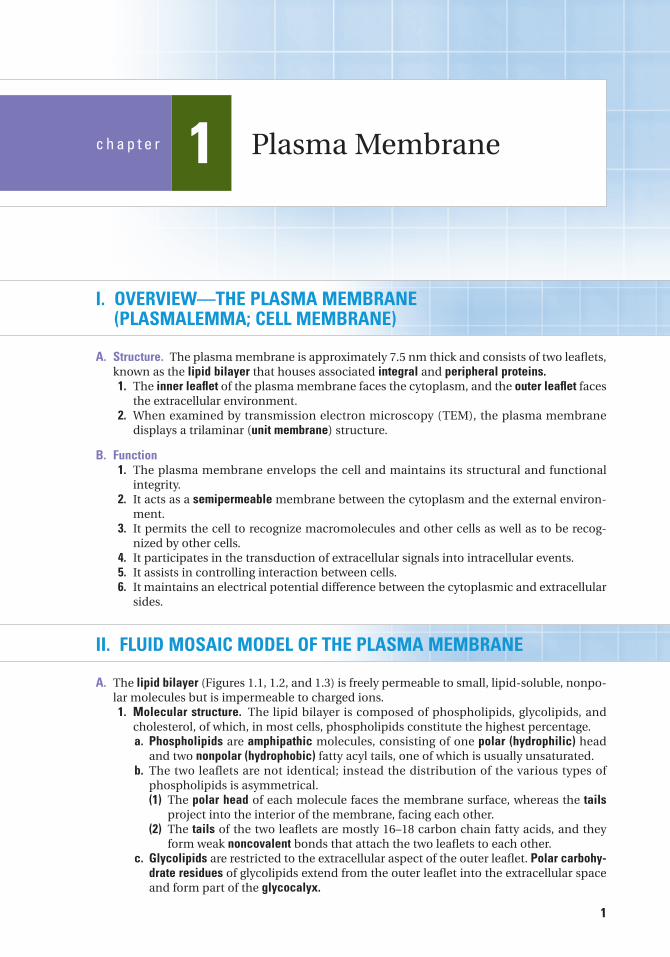
I. OVERVIEW—THE PLASMA MEMBRANE (PLASMALEMMA; CELL MEMBRANE)
A. Structure. The plasma membrane is approximately 7.5 nm thick and consists of two leaflets,known as the lipid bilayer that houses associated integral and peripheral proteins.1. The inner leaflet of the plasma membrane faces the cytoplasm, and the outer leaflet faces
the extracellular environment.2. When examined by transmission electron microscopy (TEM), the plasma membrane
displays a trilaminar (unit membrane) structure.
B. Function1. The plasma membrane envelops the cell and maintains its structural and functional
integrity.2. It acts as a semipermeable membrane between the cytoplasm and the external environ-
ment.3. It permits the cell to recognize macromolecules and other cells as well as to be recog-
nized by other cells.4. It participates in the transduction of extracellular signals into intracellular events.5. It assists in controlling interaction between cells.6. It maintains an electrical potential difference between the cytoplasmic and extracellular
sides.
II. FLUID MOSAIC MODEL OF THE PLASMA MEMBRANE
A. The lipid bilayer (Figures 1.1, 1.2, and 1.3) is freely permeable to small, lipid-soluble, nonpo-lar molecules but is impermeable to charged ions.1. Molecular structure. The lipid bilayer is composed of phospholipids, glycolipids, and
cholesterol, of which, in most cells, phospholipids constitute the highest percentage.a. Phospholipids are amphipathic molecules, consisting of one polar (hydrophilic) head
and two nonpolar (hydrophobic) fatty acyl tails, one of which is usually unsaturated.b. The two leaflets are not identical; instead the distribution of the various types of
phospholipids is asymmetrical.(1) The polar head of each molecule faces the membrane surface, whereas the tails
project into the interior of the membrane, facing each other.(2) The tails of the two leaflets are mostly 16–18 carbon chain fatty acids, and they
form weak noncovalent bonds that attach the two leaflets to each other.c. Glycolipids are restricted to the extracellular aspect of the outer leaflet. Polar carbohy-
drate residues of glycolipids extend from the outer leaflet into the extracellular spaceand form part of the glycocalyx.
LWBK615-c01[01-13].qxd 05/31/2010 10:07 AM Page 1 Aptara
2 BRS Cell Biology and Histology
d. Cholesterol, constituting 2% of plasmalemma lipids, is present in both leaflets, andhelps maintain the structural integrity of the membrane.
e. Cholesterol and phospholipids can form microdomains, known as lipid rafts, that canaffect the movement of integral proteins of the plasmalemma.
2. Fluidity of the lipid bilayer is crucial to exocytosis, endocytosis, membrane trafficking, andmembrane biogenesis.
Carbohydrate boundto lipid and protein
Integral proteins
Peripheralprotein
Polar head
Oligosaccharide
Outer leaflet
Inner leaflet
Fatty acyl tail
Integralprotein
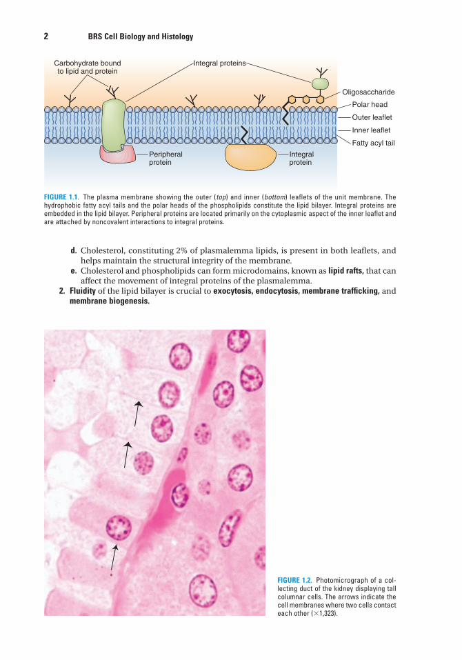
FIGURE 1.1. The plasma membrane showing the outer (top) and inner (bottom) leaflets of the unit membrane. Thehydrophobic fatty acyl tails and the polar heads of the phospholipids constitute the lipid bilayer. Integral proteins areembedded in the lipid bilayer. Peripheral proteins are located primarily on the cytoplasmic aspect of the inner leaflet andare attached by noncovalent interactions to integral proteins.
FIGURE 1.2. Photomicrograph of a col-lecting duct of the kidney displaying tallcolumnar cells. The arrows indicate thecell membranes where two cells contacteach other (�1,323).
LWBK615-c01[01-13].qxd 05/31/2010 10:07 AM Page 2 Aptara
Chapter 1 Plasma Membrane 3
a. Fluidity increases with increased temperature and with decreased saturation of thefatty acyl tails.
b. Fluidity decreases with an increase in the membrane’s cholesterol content.
B. Membrane proteins (see Figure 1.1) include integral proteins and peripheral proteins and, inmost cells, constitute approximately 50% of the plasma membrane composition.1. Integral proteins are dissolved in the lipid bilayer.
a. Transmembrane proteins span the entire thickness of the plasma membrane and mayfunction as membrane receptors, enzymes, cell adhesion molecules, cell recognitionproteins, molecules that function in message transduction, and transport proteins.(1) Most transmembrane proteins are glycoproteins.(2) Transmembrane proteins are amphipathic and contain hydrophilic and hydropho-
bic amino acids, some of which interact with the hydrocarbon tails of the mem-brane phospholipids.
(3) Most transmembrane proteins are folded so that they pass back and forth acrossthe plasmalemma; therefore, they are also known as multipass proteins.
b. Integral proteins may also be anchored to the inner (or occasionally outer) leaflet viafatty acyl or prenyl groups.
c. In freeze-fracture preparations, integral proteins remain preferentially attached tothe P-face, the outer (protoplasmic face) surface of the inner leaflet, rather than theE-face (extracellular face) (Figure 1.4).
2. Peripheral proteins do not extend into the lipid bilayer.a. These proteins are located on the cytoplasmic aspect of the inner leaflet.b. The outer leaflets of some cells possess covalently linked glycolipids to which periph-
eral proteins are anchored; these peripheral proteins thus project into the extracellu-lar space.
c. Peripheral proteins bind to the phospholipid polar groups or integral proteins of themembrane via noncovalent interactions.
MM
M
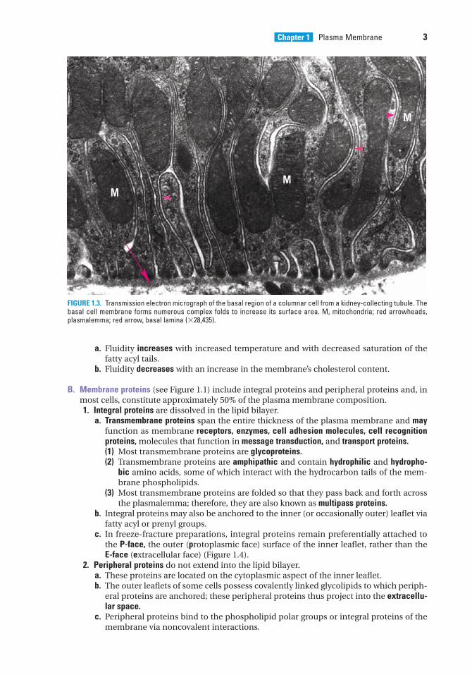
FIGURE 1.3. Transmission electron micrograph of the basal region of a columnar cell from a kidney-collecting tubule. Thebasal cell membrane forms numerous complex folds to increase its surface area. M, mitochondria; red arrowheads,plasmalemma; red arrow, basal lamina (�28,435).
LWBK615-c01[01-13].qxd 05/31/2010 10:07 AM Page 3 Aptara
4 BRS Cell Biology and Histology
d. They usually function as electron carriers (e.g., cytochrome c) part of the cytoskeletonor as part of an intracellular second messenger system.
e. They include a group of anionic, calcium-dependent, lipid-binding proteins knownas annexins, which act to modify the relationships of other peripheral proteins withthe lipid bilayer and also to function in membrane trafficking and the formation ofion channels; synapsin I, which binds synaptic vesicles to the cytoskeleton; and spec-trin, which stabilizes cell membranes of erythrocytes.
3. Functional characteristics of membrane proteinsa. The lipid-to-protein ratio (by weight) in plasma membranes ranges from 1:1 in most
cells to as much as 4:1 in myelin.b. Some membrane proteins diffuse laterally in the lipid bilayer; others are immobile and
are held in place by cytoskeletal components.
C. Glycocalyx (cell coat), located on the outer surface of the outer leaflet of the plasmalemma,varies in appearance (fuzziness) and thickness (up to 50 nm).1. Composition. The glycocalyx consists of polar oligosaccharide side chains linked cova-
lently to most proteins and some lipids (glycolipids) of the plasmalemma. It also con-tains proteoglycans (glycosaminoglycans bound to integral proteins).
2. Functiona. The glycocalyx aids in attachment of some cells (e.g., fibroblasts but not epithelial
cells) to extracellular matrix components.b. It binds antigens and enzymes to the cell surface.c. It facilitates cell-cell recognition and interaction.d. It protects cells from injury by preventing contact with inappropriate substances.e. It assists T cells and antigen-presenting cells in aligning with each other in the proper
fashion and aids in preventing inappropriate enzymatic cleavage of receptors andligands.
f. In blood vessels, it lines the endothelial surface to decrease frictional forces as theblood rushes by and it also diminishes loss of fluid from the vessel.
III. PLASMA MEMBRANE TRANSPORT PROCESSES
These processes include transport of a single molecule (uniport) or cotransport of two differentmolecules in the same (symport) or opposite (antiport) direction.
1 2
43
5
A
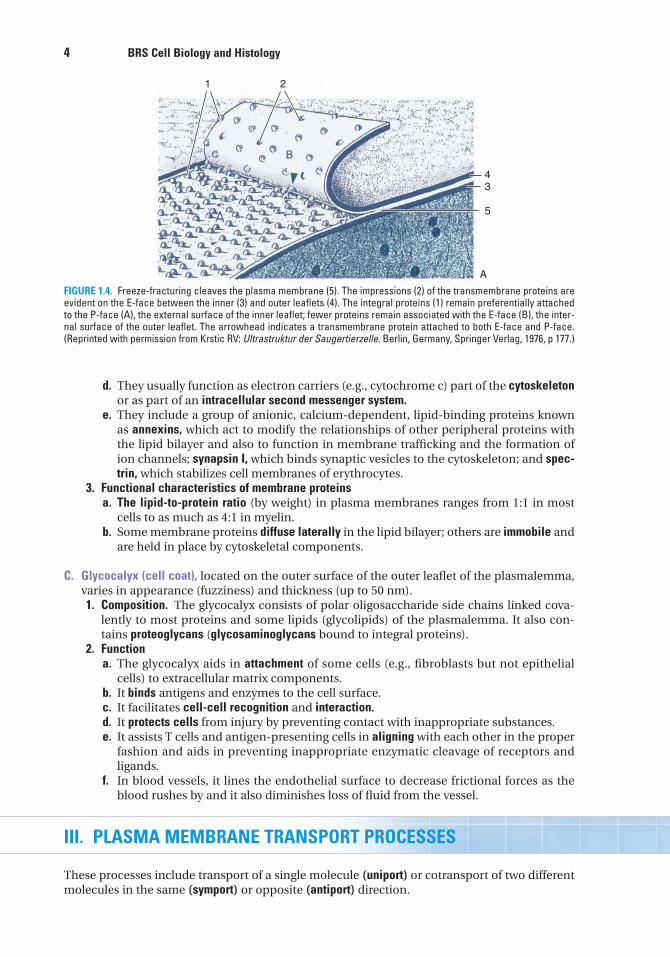
FIGURE 1.4. Freeze-fracturing cleaves the plasma membrane (5). The impressions (2) of the transmembrane proteins areevident on the E-face between the inner (3) and outer leaflets (4). The integral proteins (1) remain preferentially attachedto the P-face (A), the external surface of the inner leaflet; fewer proteins remain associated with the E-face (B), the inter-nal surface of the outer leaflet. The arrowhead indicates a transmembrane protein attached to both E-face and P-face.(Reprinted with permission from Krstic RV: Ultrastruktur der Saugertierzelle. Berlin, Germany, Springer Verlag, 1976, p 177.)
LWBK615-c01[01-13].qxd 05/31/2010 10:07 AM Page 4 Aptara
Chapter 1 Plasma Membrane 5
A. Passive transport (Figure 1.5) includes simple and facilitated diffusion. Neither of theseprocesses requires energy because molecules move across the plasma membrane down aconcentration or electrochemical gradient.1. Simple diffusion transports small nonpolar molecules (e.g., O2 and N2) and small,
uncharged, polar molecules (e.g., H2O, CO2, and glycerol). It exhibits little specificity, andthe diffusion rate is proportional to the concentration gradient of the diffusing molecule.
2. Facilitated diffusion occurs via ion channels and/or carrier proteins, structures that exhibitspecificity for the transported molecules. Not only is it faster than simple diffusion but itis also responsible for providing a pathway for ions and large polar molecules to traversemembranes that would otherwise be impermeable to them.a. Ion channel proteins are multipass transmembrane proteins that form small aqueous
pores across membranes through which specific small water-soluble molecules andions pass down an electrochemical gradient (passive transport).
b. Aquaporins are channels designed for the rapid transport of water across the cellmembrane without permitting an accompanying flow of protons to pass through thechannels. They accomplish this by forcing the water molecules to flip-flop halfwaydown the channel, so that water molecules enter aquaporins with their oxygen lead-ing into the channel and leave with their oxygen trailing the hydrogen atoms.
c. Carrier proteins are multipass transmembrane proteins that undergo reversible con-formational changes to transport specific molecules across the membrane; these pro-teins function in both passive transport and active transport.
Exterior
InteriorSimple
diffusionIon channel–
mediateddiffusion
Carrier protein–mediateddiffusion
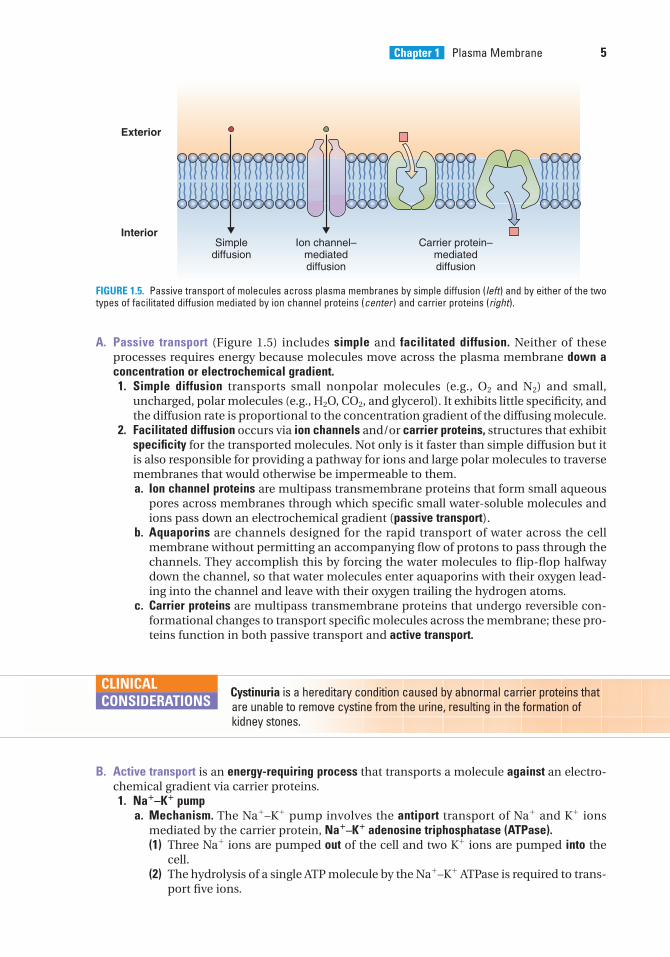
FIGURE 1.5. Passive transport of molecules across plasma membranes by simple diffusion (left) and by either of the twotypes of facilitated diffusion mediated by ion channel proteins (center ) and carrier proteins (right).
Cystinuria is a hereditary condition caused by abnormal carrier proteins thatare unable to remove cystine from the urine, resulting in the formation ofkidney stones.
CLINICAL
CONSIDERATIONS
B. Active transport is an energy-requiring process that transports a molecule against an electro-chemical gradient via carrier proteins.1. Na�–K� pump
a. Mechanism. The Na�–K� pump involves the antiport transport of Na� and K� ionsmediated by the carrier protein, Na�–K� adenosine triphosphatase (ATPase).(1) Three Na� ions are pumped out of the cell and two K� ions are pumped into the
cell.(2) The hydrolysis of a single ATP molecule by the Na�–K� ATPase is required to trans-
port five ions.
LWBK615-c01[01-13].qxd 05/31/2010 10:07 AM Page 5 Aptara
6 BRS Cell Biology and Histology
b. Function(1) The primary function is to maintain constant cell volume by decreasing the intra-
cellular ion concentration (and thus the osmotic pressure) and increasing theextracellular ion concentration, thus decreasing the flow of water into the cell.
(2) The Na�–K� pump also plays a minor role in the maintenance of a potential differ-ence across the plasma membrane.
2. Glucose transport involves the symport movement of glucose across an epithelium(transepithelial transport). Transport is frequently powered by an electrochemical Na�
gradient, which drives carrier proteins located at specific regions of the cell surface.3. ATP-binding cassette transporters (ABC-transporters) are transmembrane proteins that
have two domains, the intracellularly facing nucleotide-binding domain (ATP bindingdomain) and the membrane-spanning domain (transmembrane domain). In eukaryotes, ABC-transporters function in exporting materials, such as toxins and drugs, from the cyto-plasm into the extracellular space, using ATP as an energy source. ABC-transporters mayhave additional functions, such as those of the placenta, which presumably protect thedeveloping fetus from xenobiotics, macromolecules such as antibiotics, not manufac-tured by cells of the mother.
Multidrug-resistant proteins (MDR proteins) are ABC-transporters that are present in certain cancer cells that are able to transport the cytotoxic
drugs administered to treat the malignancy. It has been shown that in more than one-third of thecancer patients, the malignant cells develop MDR proteins that interfere with the treatment modalitybeing used.
CLINICAL
CONSIDERATIONS
C. Facilitated diffusion of ions can occur via ion channel proteins or ionophores.1. Selective ion channel proteins permit only certain ions to traverse them.
a. K� leak channels are the most common ion channels. These channels are ungated andleak K�, the ions most responsible for establishing a potential difference across theplasmalemma.
b. Gated ion channels open only transiently in response to various stimuli. They includethe following types:(1) Voltage-gated channels open when the potential difference across the membrane
changes (e.g., voltage-gated Na� channels, which function in the generation ofaction potentials; see Chapter 9 VIII B 1 e).
(2) Mechanically gated channels open in response to a mechanical stimulus (e.g., thetactile response of the hair cells in the inner ear).
(3) Ligand-gated channels open in response to the binding of a signaling molecule orion. These channels include neurotransmitter-gated channels, nucleotide-gatedchannels, and G protein–gated K� channels of cardiac muscle cells.
Ligand-gated ions channels are probably the location where anesthetic agents act to block the spread of action potentials.
CLINICAL
CONSIDERATIONS
2. Ionophores are lipid-miscible molecules that form a complex with ions and insert intothe lipid bilayer to transport those ions across the membrane. There are two ways inwhich they perform this function:a. They enfold the ion and pass through the lipid bilayer.b. They insert into the cell membrane to form an ion channel whose lumen is
hydrophilic.Ionophores are frequently fed to cattle and poultry as antibiotic agents and growth-
enhancing substances.
LWBK615-c01[01-13].qxd 05/31/2010 10:07 AM Page 6 Aptara
IV. CELL-TO-CELL COMMUNICATION
A. Signaling molecules, secreted by signaling cells, bind to receptor molecules of target cells,and in this fashion, these molecules function in cell-to-cell communication in order tocoordinate cellular activities. Examples of such signaling molecules that effect communi-cations include neurotransmitters, which are released into the synaptic cleft (see Chapter8 IV A 1 b; Chapter 9 IV B 5); endocrine hormones, which are carried in the bloodstreamand act on distant target cells; and hormones released into the intercellular space, whichact on nearby cells (paracrine hormones) or on the releasing cell itself (autocrine hormones).1. Lipid-soluble signaling molecules penetrate the plasma membrane and bind to receptors
within the cytoplasm or inside the nucleus, activating intracellular messengers. Examplesinclude hormones that influence gene transcription.
2. Hydrophilic signaling molecules bind to and activate cell-surface receptors (as do somelipid-soluble signaling molecules) and have diverse physiologic effects (see Chapter 13).Examples include neurotransmitters and numerous hormones (e.g., serotonin, thyroid-stimulating hormone, insulin).
B. Membrane receptors are primarily integral membrane glycoproteins. They are embeddedin the lipid bilayer and have three domains, an extracellular domain that protrudes into theextracellular space and has binding sites for the signaling molecule, a transmembranedomain that passes through the lipid bilayer, and an intracellular domain that is located onthe cytoplasmic aspect of the lipid bilayer and contacts either peripheral proteins or cel-lular organelles, thereby transducing the extracellular contact into an intracellular event.
Venoms, such as those of some poisonous snakes, inactivate acetylcholinereceptors of skeletal muscle sarcolemma at neuromuscular junctions.
Autoimmune diseases may lead to the production of antibodies that specifically bind to andactivate certain plasma membrane receptors. An example is Graves disease (hyperthyroidism) (see Chapter 13 IV B).
CLINICAL
CONSIDERATIONS
1. Functiona. Membrane receptors control plasmalemma permeability by regulating the conformation
of ion channel proteins.b. They regulate the entry of molecules into the cell (e.g., the delivery of cholesterol via
low-density lipoprotein receptors).c. They bind extracellular matrix molecules to the cytoskeleton via integrins, which are
essential for cell-matrix interactions.d. They act as transducers to translate extracellular events into an intracellular response
via the second messenger systems.e. They permit pathogens that mimic normal ligands to enter cells.
2. Types of membrane receptorsa. Channel-linked receptors bind a signaling molecule that temporarily opens or closes
the gate, permitting or inhibiting the movement of ions across the cell membrane.Examples include nicotinic acetylcholine receptors on the muscle-cell sarcolemma atthe myoneural junction (see Chapter 8 IV A).
b. Catalytic receptors are single-pass transmembrane proteins.(1) Their extracellular moiety is a receptor and their cytoplasmic component is a pro-
tein kinase.(2) Some catalytic receptors lack an extracytoplasmic moiety and as a result are con-
tinuously activated; such defective receptors are coded for by some oncogenes.(3) Examples of catalytic receptors include the following:
(a) Insulin, which binds to its receptor, which autophosphorylates. The cell thentakes up the insulin-receptor complex by endocytosis, enabling the complex tofunction within the cell.
Chapter 1 Plasma Membrane 7
LWBK615-c01[01-13].qxd 05/31/2010 10:07 AM Page 7 Aptara
8 BRS Cell Biology and Histology
(b) Growth factors (e.g., epidermal growth factor, platelet-derived growth factor)bind to specific catalytic receptors and induce mitosis.
c. G protein–linked receptors are transmembrane proteins associated with an ion chan-nel or with an enzyme that is bound to the cytoplasmic surface of the cell membrane.(1) These receptors interact with heterotrimeric G protein (guanosine triphosphate
[GTP]-binding regulatory protein) after binding of a signaling molecule. The het-erotrimeric G protein is composed of three subunits: � and � and � complex. Thebinding of the signaling molecule causes either(a) the dissociation of the � subunit from the � and � complex where the � sub-
unit interacts with its target or(b) the three subunits do not dissociate, but either the � subunit and/or the � and
� complex become activated and can interact with their targets.This interaction results in the activation of intracellular second messengers,
the most common of which are cyclic adenosine monophosphate (cAMP), Ca2�,and the inositol phospholipid–signaling pathway.
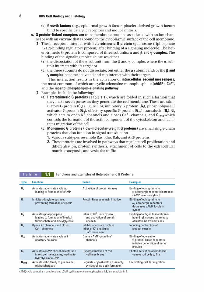
(2) Examples include the following:(a) Heterotrimeric G proteins (Table 1.1), which are folded in such a fashion that
they make seven passes as they penetrate the cell membrane. These are stim-ulatory G protein (Gs) (Figure 1.6), inhibitory G protein (Gi), phospholipase Cactivator G protein (Gq), olfactory-specific G protein (Golf), transducin (Gt), Go
which acts to open K� channels and closes Ca2� channels, and G12/13 whichcontrols the formation of the actin component of the cytoskeleton and facili-tates migration of the cell.
(b) Monomeric G proteins (low-molecular-weight G proteins) are small single-chainproteins that also function in signal transduction.1. Various subtypes resemble Ras, Rho, Rab, and ARF proteins.2. These proteins are involved in pathways that regulate cell proliferation and
differentiation, protein synthesis, attachment of cells to the extracellularmatrix, exocytosis, and vesicular traffic.
t a b l e 1.1 Functions and Examples of Heterotrimeric G Proteins
Type Function Result Examples
Gs Activates adenylate cyclase, Activation of protein kinases Binding of epinephrine to leading to formation of cAMP �-adrenergic receptors increases
cAMP levels in cytosol
Gi Inhibits adenylate cyclase, Protein kinases remain inactive Binding of epinephrine to preventing formation of cAMP �2-adrenergic receptors
decreases cAMP levels in cytosol
Gq Activates phospholipase C, Influx of Ca2� into cytosol Binding of antigen to membrane-leading to formation of inositol and activation of protein bound IgE causes the releasetriphosphate and diacylglycerol kinase C of histamine by mast cells
Go Opens K� channels and closes Inhibits adenylate cyclase Inducing contraction of Ca2� channels Influx of K� and limits smooth muscle
Ca2� movement
Golf Activates adenylate cyclase in Opens cAMP-gated Na� Binding of odorant to olfactory neurons channels G protein–linked receptors
initiates generation of nerve impulse
Gt Activates cGMP phosphodiesterase Hyperpolarization of rod Photon activation of rhodopsinin rod cell membranes, leading to cell membrane causes rod cells to firehydrolysis of cGMP
G12/13 Activates Rho family of guanosine Regulates cytoskeleton assembly Facilitating cellular migrationtriphosphatases by controlling actin formation
cAMP, cyclic adenosine monophosphate; cGMP, cyclic guanosine monophosphate; IgE, immunoglobulin E.
LWBK615-c01[01-13].qxd 05/31/2010 10:07 AM Page 8 Aptara
Chapter 1 Plasma Membrane 9
Exterior
Interior
Signalingmolecule
Receptor
R R
cAMP+
PPi
ATP
Adenylatecyclase
Activation
γ β γ βα
Ad
α
Adc
GTP GTP
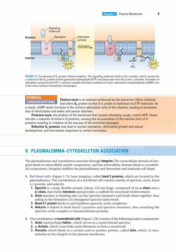
FIGURE 1.6. Functioning of Gs protein–linked receptors. The signaling molecule binds to the receptor, which causes the�-subunit of the Gs protein to bind guanosine triphosphate (GTP) and dissociate from the � and � subunits. Activation ofadenylate cyclase by the GTP-�-subunit complex stimulates synthesis of cyclic adenosine monophosphate (cAMP), oneof the most common intracellular messengers.
Cholera toxin is an exotoxin produced by the bacterium Vibrio choleraethat alters Gs protein so that it is unable to hydrolyze its GTP molecule. As
a result, cAMP levels increase in the surface-absorptive cells of the intestine, leading to excessiveloss of electrolytes and water and severe diarrhea.
Pertussis toxin, the product of the bacterium that causes whooping cough, inserts ADP-riboseinto the � subunits of trimeric G proteins, causing the accumulation of the inactive form of Gproteins resulting in irritation of the mucosa of the bronchial passages.
Defective Gs proteins may lead to mental retardation, diminished growth and sexualdevelopment, and decreased responses to certain hormones.
CLINICAL
CONSIDERATIONS
V. PLASMALEMMA–CYTOSKELETON ASSOCIATION
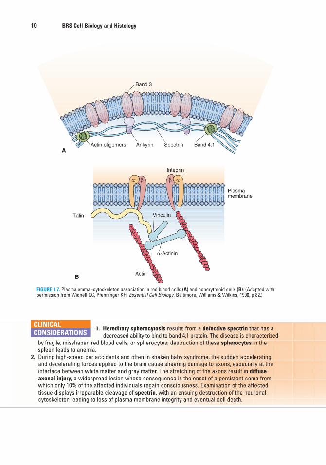
The plasmalemma and cytoskeleton associate through integrins. The extracellular domain of inte-grins binds to extracellular matrix components, and the intracellular domain binds to cytoskele-tal components. Integrins stabilize the plasmalemma and determine and maintain cell shape.
A. Red blood cells (Figure 1.7A) have integrins, called band 3 proteins, which are located in theplasmalemma. The cytoskeleton of a red blood cell consists mainly of spectrin, actin, band4.1 protein, and ankyrin.1. Spectrin is a long, flexible protein (about 110 nm long), composed of an �-chain and a
�-chain, that forms tetramers and provides a scaffold for structural reinforcement.2. Actin attaches to binding sites on the spectrin tetramers and holds them together, thus
aiding in the formation of a hexagonal spectrin latticework.3. Band 4.1 protein binds to and stabilizes spectrin–actin complexes.4. Ankyrin is linked to both band 3 proteins and spectrin tetramers, thus attaching the
spectrin–actin complex to transmembrane proteins.
B. The cytoskeleton of nonerythroid cells (Figure 1.7B) consists of the following major components:1. Actin (and perhaps fodrin), which serves as a nonerythroid spectrin.2. �-Actinin, which cross-links actin filaments to form a meshwork.3. Vinculin, which binds to �-actinin and to another protein, called talin, which, in turn,
attaches to the integrin in the plasma membrane.
LWBK615-c01[01-13].qxd 05/31/2010 10:07 AM Page 9 Aptara
10 BRS Cell Biology and Histology
A
B
Band 3
Plasmamembrane
Vinculin
α-Actinin
Actin
Talin
Actin oligomers Ankyrin Spectrin
Integrin
Band 4.1
βα αβ
FIGURE 1.7. Plasmalemma–cytoskeleton association in red blood cells (A) and nonerythroid cells (B). (Adapted withpermission from Widnell CC, Pfenninger KH: Essential Cell Biology. Baltimore, Williams & Wilkins, 1990, p 82.)
1. Hereditary spherocytosis results from a defective spectrin that has a decreased ability to bind to band 4.1 protein. The disease is characterized
by fragile, misshapen red blood cells, or spherocytes; destruction of these spherocytes in thespleen leads to anemia.
2. During high-speed car accidents and often in shaken baby syndrome, the sudden acceleratingand decelerating forces applied to the brain cause shearing damage to axons, especially at theinterface between white matter and gray matter. The stretching of the axons result in diffuseaxonal injury, a widespread lesion whose consequence is the onset of a persistent coma fromwhich only 10% of the affected individuals regain consciousness. Examination of the affectedtissue displays irreparable cleavage of spectrin, with an ensuing destruction of the neuronalcytoskeleton leading to loss of plasma membrane integrity and eventual cell death.
CLINICAL
CONSIDERATIONS
LWBK615-c01[01-13].qxd 05/31/2010 10:07 AM Page 10 Aptara
1. A herpetologist is bitten by a poisonoussnake and is taken to the emergency depart-ment with progressive muscle paralysis. Thevenom is probably incapacitating his
(A) Na� channels.(B) Ca2� channels.(C) phospholipids.(D) acetylcholine receptors.(E) spectrin.
2. Cholesterol functions in theplasmalemma to
(A) increase fluidity of the lipid bilayer.(B) decrease fluidity of the lipid bilayer.(C) facilitate the diffusion of ions through
the lipid bilayer.(D) assist in the transport of hormones
across the lipid bilayer.(E) bind extracellular matrix molecules.
3. The cell membrane consists of variouscomponents, including integral proteins.These integral proteins
(A) are not attached to the outer leaflet.(B) are not attached to the inner leaflet.(C) include transmembrane proteins.(D) are preferentially attached to the E-face.(E) function in the transport of cholesterol-
based hormones.
4. Which one of the following transportprocesses requires energy?
(A) Facilitated diffusion(B) Passive transport(C) Active transport(D) Simple diffusion
5. Which one of the following substances isunable to traverse the plasma membrane bysimple diffusion?
(A) O2
(B) N2
(C) Na�
(D) Glycerol(E) CO2
6. Symport refers to the process oftransporting
(A) a molecule into the cell.(B) a molecule out of the cell.(C) two different molecules in opposite
directions.(D) two different molecules in the same
direction.(E) a molecule between the cytoplasm and
the nucleus.
7. One of the ways that cells communicatewith each other is by secretion of variousmolecules. The secreted molecule is knownas
(A) a receptor molecule.(B) a signaling molecule.(C) a spectrin tetramer.(D) an integrin.(E) an anticodon.
Review Test
Directions: Each of the numbered items or incomplete statements in this section is followed byanswers or by completions of the statement. Select the ONE lettered answer or completion thatis BEST in each case.
11
LWBK615-c01[01-13].qxd 05/31/2010 10:07 AM Page 11 Aptara
10. Which of the following statements con-cerning plasma membrane components isTRUE?
(A) All G proteins are composed of threesubunits.
(B) The glycocalyx is usually composed ofphospholipids.
(C) Ion channel proteins are energy depend-ent (require adenosine triphosphate).
(D) Gated channels are always open.(E) Ankyrin binds to band 3 of the red blood
cell plasma membrane.
8. Adrenocorticotropic hormone (ACTH)travels through the bloodstream, entersconnective tissue spaces, and attaches tospecific sites on target-cell membranes.These sites are
(A) peripheral proteins.(B) signaling molecules.(C) G proteins.(D) G protein–linked receptors.(E) ribophorins.
9. Examination of the blood smear of ayoung patient reveals misshapen red bloodcells, and the pathology report indicateshereditary spherocytosis. Defects in whichone of the following proteins cause thiscondition?
(A) Signaling molecules(B) G proteins(C) Spectrin(D) Hemoglobin(E) Ankyrin
12 BRS Cell Biology and Histology
LWBK615-c01[01-13].qxd 05/31/2010 10:07 AM Page 12 Aptara
13
Answers and Explanations
1. D. Snake venom usually blocks acetylcholine receptors, preventing depolarization of themuscle cell. The Na� and Ca2� channels are not incapacitated by snake venoms (seeChapter 1 IV B).
2. B. The fluidity of the lipid bilayer is decreased in three ways: (1) by lowering the tempera-ture, (2) by increasing the saturation of the fatty acyl tails of the phospholipid molecules,and (3) by increasing the membrane’s cholesterol content (see Chapter 1 II A 2).
3. C. Integral proteins are not only closely associated with the lipid bilayer but also tightlybound to the cell membrane. These proteins frequently span the entire thickness of theplasmalemma and are thus termed transmembrane proteins (see Chapter 1 II B 1).
4. C. Active transport requires energy. Facilitated diffusion, which is mediated by membraneproteins, and simple diffusion, which involves passage of material directly across the lipidbilayer, are types of passive transport (see Chapter 1 III B).
5. C. Na� and other ions require channel (carrier) proteins for their transport across theplasma membrane. The other substances are small nonpolar molecules and smalluncharged polar molecules. The molecules can traverse the plasma membrane by simplediffusion (see Chapter 1 III A 2).
6. D. The coupled transport of two different molecules in the same direction is termed “sym-port” (see Chapter 1 III B).
7. B. Cells can communicate with each other by releasing signaling molecules, which attachto receptor molecules on target cells (see Chapter 1 IV A).
8. D. G protein–linked receptors are sites where ACTH and some other signaling moleculesattach. Binding of ACTH to its receptor causes Gs protein to activate adenylate cyclase, set-ting in motion the specific response elicited by the hormone (see Chapter 1 IV B 2 c).
9. C. Hereditary spherocytosis is caused by a defect in spectrin that renders the protein inca-pable of binding to band 4.1 protein, thus destabilizing the spectrin–actin complex of thecytoskeleton. Although defects in hemoglobin (the respiratory protein of erythrocytes) alsocause red blood cell anomalies, hereditary spherocytosis is not one of them (see Chapter 1V A).
10. E. Ankyrin is linked both to band 3 proteins and to spectrin tetramer, thus attaching thespectrin–actin complex to transmembrane proteins of the erythrocyte. There are two typesof G proteins: trimeric and monomeric; glycocalyx (the sugar coat on the membranesurface) is composed mostly of polar carbohydrate residues; only carrier proteins can beenergy requiring; gated channels are open only transiently (see Chapter 1 V A).
LWBK615-c01[01-13].qxd 05/31/2010 10:07 AM Page 13 Aptara
c h a p t e r 2 Nucleus
14
I. OVERVIEW—THE NUCLEUS (Figure 2.1)
A. Structure. The nucleus, the largest organelle of the cell, includes the nuclear envelope, nucle-olus, nucleoplasm, and chromatin and contains the genetic material encoded in the deoxyri-bonucleic acid (DNA) of chromosomes.
B. Function. The nucleus directs protein synthesis in the cytoplasm via ribosomal ribonucleicacid (rRNA), messenger RNA (mRNA), and transfer RNA (tRNA). All forms of RNA are synthe-sized in the nucleus.
II. NUCLEAR ENVELOPE (Figure 2.2)
The nuclear envelope surrounds the nuclear material and consists of two parallel membranesseparated from each other by a narrow perinuclear cisterna. These membranes fuse at intervals,forming openings called nuclear pores in the nuclear envelope.
A. Outer nuclear membrane1. This membrane is about 6 nanometers (nm) thick.2. It faces the cytoplasm and is continuous at certain sites with the rough endoplasmic
reticulum (RER).3. A loosely arranged mesh of intermediate filaments (vimentin) surrounds the outer
nuclear membrane on its cytoplasmic aspect.4. Ribosomes stud the cytoplasmic surface of the outer nuclear membrane. These ribo-
somes synthesize proteins that enter the perinuclear cisterna.
B. Inner nuclear membrane1. The inner nuclear membrane is about 6 nm thick.2. It faces the nuclear material but is separated from it and is supported on its inner surface
by the nuclear lamina, fibrous lamina that is 80 to 300 nm thick and composed primarilyof lamins A, B, and C. These intermediate filament proteins help organize the nuclearenvelope and perinuclear chromatin. In addition, they are essential during the mitoticevents, when they are responsible for the disassembly and reassembly of the nuclearenvelope. Phosphorylation of lamins leads to disassembly, and dephosphorylationresults in reassembly of the nuclear envelope.
C. Perinuclear cisterna1. The perinuclear cisterna is located between the inner and outer nuclear membranes and
is 20 to 40 nm wide.
LWBK615-c02[14-31].qxd 05/31/2010 10:18 AM Page 14 Aptara
Chapter 2 Nucleus 15
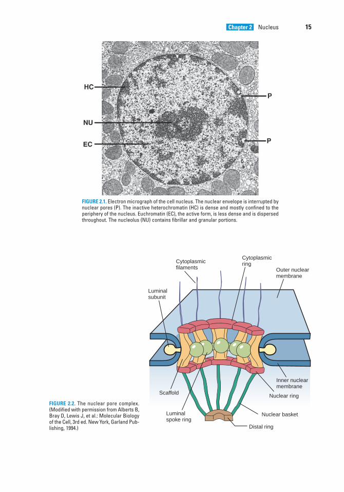
FIGURE 2.1. Electron micrograph of the cell nucleus. The nuclear envelope is interrupted bynuclear pores (P). The inactive heterochromatin (HC) is dense and mostly confined to theperiphery of the nucleus. Euchromatin (EC), the active form, is less dense and is dispersedthroughout. The nucleolus (NU) contains fibrillar and granular portions.
FIGURE 2.2. The nuclear pore complex.(Modified with permission from Alberts B,Bray D, Lewis J, et al.: Molecular Biologyof the Cell, 3rd ed. New York, Garland Pub-lishing, 1994.)
Inner nuclearmembrane
Outer nuclearmembrane
Cytoplasmicfilaments
Cytoplasmicring
Scaffold
Luminalspoke ring
Luminalsubunit
Nuclear ring
Nuclear basket
Distal ring
LWBK615-c02[14-31].qxd 05/31/2010 10:18 AM Page 15 Aptara
16 BRS Cell Biology and Histology
2. It is continuous with the cisterna of the RER.3. It is perforated by nuclear pores at various locations.
D. Nuclear pores1. Nuclear pores average 80 nm in diameter and number from dozens to thousands
depending upon metabolic activity of the cell; they are associated with the nuclear porecomplex (NPC).
2. They are formed by fusion of the inner and outer nuclear membranes.3. They permit passage of certain molecules in either direction between the nucleus and
the cytoplasm via a 9-nm channel opening.4. NPCs are aided in communicating with each other by the nuclear lamina.
E. The NPC represents protein subunits surrounding the nuclear pore (Figure 2.2).1. Structure. The NPC is composed of nearly 100 proteins, some of which are arranged in
eightfold symmetry around the margin of the pore. The nucleoplasmic side of the poreexhibits a nuclear basket, whereas the cytoplasmic side displays fibers extending into thecytoplasm. A transporter protein is located in the central core and is believed to beresponsible for transporting proteins into and out of the nucleus via receptor-mediatedtransport.a. The cytoplasmic ring is located around the cytoplasmic margin of the nuclear pore
and is composed of eight subunits, each possessing a cytoplasmic filament composedof a Ran-binding protein (GTP-binding protein) extending into the cytoplasm. Thesefibers may serve as a staging area prior to protein transport.
b. The nucleoplasmic ring is located around the nucleoplasmic margin of the nuclearpore and is composed of eight subunits. Extending from this ring into the nucleo-plasm is a basket-like structure, the nuclear basket. Attached to the distal end of thenuclear basket is the distal ring. This innermost ring assists in the export of RNA intothe cytoplasm.
c. The luminal ring is interposed between the cytoplasmic and nucleoplasmic rings.Eight transmembrane proteins project into the lumen of the nuclear pore, anchoringthe complex into the pore rim. The lumen may be a gated channel that impedes pas-sive diffusion. A moiety of each of these transmembrane proteins also project into theperinuclear cistern.
d. A structure described by some as the hourglass-shaped transporter or central plug inthe center of the luminal ring is believed to be cargo being transported through theNPC rather than a structural component of the NPC.
2. Function. The NPC permits passive movement across the nuclear envelope via a 9- to 11-nm open channel for simple diffusion. Most proteins, regardless of size, pass in eitherdirection only by receptor-mediated transport. These proteins have clusters of certainamino acids known as nuclear localization segments (NLS) that act as signals for transport.
3. Transport mechanisms involve a group of proteins, exportins and importins. The function ofthese proteins is regulated by Ran, a group of guanosine triphosphate–binding proteins.The other group of proteins called nucleoporins facilitates the shuttling of cargo in bothdirections. Transport signals of this type are called nucleocytoplasmic shuttling (NS) signals.
III. NUCLEOLUS
A. Structure. The nucleolus is a nuclear inclusion that is not surrounded by a membrane. It isobserved in interphase cells that are actively synthesizing proteins; more than one nucleo-lus can be present in the nucleus. It contains mostly rRNA and protein along with a modestamount of DNA. It possesses nucleolar organizer regions (NORs), portions of the chromo-somes (in humans, chromosomes 13, 14, 15, 21, and 22) where rRNA genes are located; theseregions are involved in reconstituting the nucleolus during the G1 phase of the cell cycle. Thenucleolus contains four distinct regions.
LWBK615-c02[14-31].qxd 05/31/2010 10:18 AM Page 16 Aptara
Chapter 2 Nucleus 17
1. Fibrillar centers are composed of inactive DNA, where DNA is not being transcribed; NORsare also located here.
2. The pars fibrosa is composed of 5-nm fibrils surrounding the fibrillar centers and con-tains transcriptionally active DNA and the rRNA precursors that are being transcribed.
3. The pars granulosa is composed of 15-nm maturing ribosomal precursor particles.4. Nucleolar matrix is a fiber network participating in the organization of the nucleolus.
B. Function. The nucleolus is involved in the synthesis of rRNA and its assembly into ribosomeprecursors. The nucleolus also sequesters certain nucleolar proteins that function as cellcycle checkpoint signaling proteins. Cell cycle regulator proteins have been identified withinthe nucleolus, in which they remain sequestered until their release is required for targets inthe nucleus and/or the cytoplasm.
IV. NUCLEOPLASM
Nucleoplasm is the protoplasm within the nuclear envelope. It consists of a matrix and varioustypes of particles.
A. Nuclear matrix acts as a scaffold that aids in organizing the nucleoplasm.1. Structural components include fibrillar elements, nuclear pore–nuclear lamina complex,
residual nucleoli, and a residual ribonucleoprotein (RNP) network.2. Functional components are involved in the transcription and processing of mRNA and
rRNA, steroid receptor-binding sites, carcinogen-binding sites, heat shock proteins, DNAviruses, viral proteins (T antigen), and perhaps many other functions that are as yet notknown.
3. A nucleoplasmic reticulum is continuous with the endoplasmic reticulum (ER) of thecytoplasm and the nuclear envelope. It contains nuclear calcium functioning within thenucleus and possesses receptors for inositol 1,4,5-trisphosphate, regulating calcium sig-nals within compartments of the nucleus related to gene transcription, protein trans-port, and perhaps other functions.
B. Nuclear particles1. Interchromatin granules are clusters of irregularly distributed particles (20–25 nm in
diameter) that contain RNP and various enzymes.2. Perichromatin granules (Figure 2.1) are single dense granules (30–50 nm in diameter) sur-
rounded by a less-dense halo. They are located at the periphery of heterochromatin andexhibit a substructure of 3-nm packed fibrils.a. Perichromatin granules contain 4.7S RNA and two peptides similar to those found in
heterogeneous nuclear RNPs (hnRNPs).b. They may represent messenger RNPs (mRNPs).c. The number of granules increases in liver cells exposed to carcinogens or tempera-
tures above 37�C.3. The hnRNP particles are complexes of precursor mRNA (pre-mRNA) and proteins and are
involved in processing of pre-mRNA.4. Small nuclear RNPs (snRNPs) are complexes of proteins and small RNAs and are involved
in hnRNP splicing or in cleavage reactions.
V. CHROMATIN (Figure 2.1)
A. Structure. Chromatin consists of double-stranded DNA complexed with histones and acidicproteins. It resides within the nucleus as heterochromatin and euchromatin. Theeuchromatin/heterochromatin ratio is higher in malignant cells than in normal cells.
LWBK615-c02[14-31].qxd 05/31/2010 10:18 AM Page 17 Aptara
18 BRS Cell Biology and Histology
1. Heterochromatin, condensed inactive chromatin, is concentrated at the periphery of thenucleus and around the nucleolus and scattered throughout the nucleoplasm.a. When examined under the light microscope (LM), it appears as basophilic clumps of
nucleoprotein.b. Although heterochromatin is transcriptionally inactive, recent evidence indicates that
it plays a role in interchromosomal interactions and chromosomal segregation dur-ing meiosis.
c. Heterochromatin corresponds to one of two X chromosomes and is therefore presentin nearly all somatic cells of female mammals. During interphase, the inactive Xchromosome is visible as a dark-staining body within the nucleus. This structure iscalled the Barr body, or sex chromatin.
2. Euchromatin is the transcriptionally active form of chromatin that appears in the LM as alightly stained region of the nucleus. It appears in transmission electron microscope(TEM) as electron-lucent regions among heterochromatin and is composed of 30-nmstrings of nucleosomes (see section VI) and the DNA double helix.
B. Function. Chromatin is responsible for RNA synthesis.
VI. CHROMOSOMES
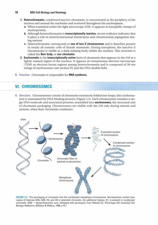
A. Structure. Chromosomes consist of chromatin extensively folded into loops; this conforma-tion is maintained by DNA-binding proteins (Figure 2.3). Each chromosome contains a sin-gle DNA molecule and associated proteins, assembled into nucleosomes, the structural unitof chromatin packaging. Chromosomes are visible with the LM only during mitosis andmeiosis, when their chromatin condenses.
DNA
Chromatin
Nucleosome
Chromatin fiber ofpacked nucleosomes
Extended sectionof chromosome
Condensed sectionof chromosome
Metaphasechromosome
FIGURE 2.3. The packaging of chromatin into the condensed metaphase chromosome. Nucleosomes contain twocopies of histones H2A, H2B, H3, and H4 in extended chromatin. An additional histone, H1, is present in condensedchromatin. DNA � deoxyribonucleic acid. (Adapted with permission from Widnell CC, Pfenninger KH: Essential CellBiology. Baltimore, Williams & Wilkins, 1990, p 47.)
LWBK615-c02[14-31].qxd 05/31/2010 10:18 AM Page 18 Aptara
Chapter 2 Nucleus 19
1. Extended chromatin forms the nucleosome core, around which the DNA double helix iswrapped two full turns.a. The nucleosome core consists of two copies each of histones H2A, H2B, H3, and H4.
Nucleosomes are spaced at intervals of 200 base pairs.b. When viewed with TEM, extended chromatin resembles beads on a string; the
beads represent nucleosomes and the string represents linker DNA. DNA is sup-ported by the nucleosomes that function. Nucleosomes support DNA and regulateits accessibility for replication and transcription as well as for its repair. Chromatinis packaged into 30-nm threads as helical coils of six nucleosomes per turn andbound with histone H1.
2. Condensed chromatin contains an additional histone, H1, which wraps around groups ofnucleosomes, thus forming 30-nm-diameter fibers of helical coils of six nucleosomes perturn, which is the structural unit of the chromosome.
B. G-banding is observed in chromosomes during mitosis after staining with Giemsa, which isspecific for DNA sequences rich in adenine (A) and thymine (T). Banding is thought to repre-sent highly folded DNA loops. G-banding is characteristic for each species and is used toidentify chromosomal anomalies.
C. Karyotype refers to the number and morphology of chromosomes and is characteristic for eachspecies.1. Haploid number (n) is the number of chromosomes in germ cells (23 in humans).2. Diploid number (2n) is the number of chromosomes in somatic cells (46 in humans).
D. Genome, the total genetic complement of an individual, is stored in its chromosomes. Inhumans, the genome consists of 22 pairs of autosomes and 1 pair of sex chromosomes (eitherXX or XY), totaling 23 pairs, or 46 chromosomes.
VII. DNA
DNA is a long double-stranded linear molecule composed of multiple nucleotide sequences. Itacts as a template for the synthesis of RNA.
A. Nucleotides are composed of a base (purine or pyrimidine), a deoxyribose sugar, and a phos-phate group.1. The purines are adenine (A) and guanine (G).2. The pyrimidines are cytosine (C) and thymine (T).
B. The DNA double helix consists of two complementary DNA strands held together by hydrogenbonds between the base pairs A-T and G-C.
C. Exons are regions of the DNA molecule that code for specific RNAs.
D. Introns are regions of the DNA molecule, between exons, that do not code for RNAs.
E. A codon is a sequence of three bases in the DNA molecule that codes for a single amino acid.
F. A gene is a segment of the DNA molecule that is responsible for the formation of a singleRNA molecule.
G. According to the Human Genome Study, there are approximately 25,000 genes in the humangenome.
LWBK615-c02[14-31].qxd 05/31/2010 10:18 AM Page 19 Aptara
20 BRS Cell Biology and Histology
VIII. RNA
RNA is a linear molecule similar to DNA; however, it is single stranded and contains riboseinstead of deoxyribose sugar and uracil (U) instead of thymine (T). RNA is synthesized by tran-scription of DNA. Transcription is catalyzed by three RNA polymerases: I for rRNA, II for mRNA,and III for tRNA.
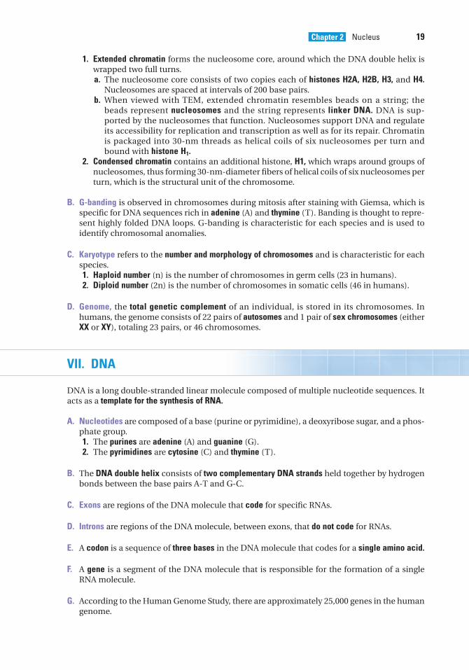
A. mRNA carries the genetic code to the cytoplasm to direct protein synthesis (Figure 2.4).1. This single-stranded molecule consists of hundreds to thousands of nucleotides.2. mRNA contains codons that are complementary to the DNA codons from which it was
transcribed, including one start codon (AUG) for initiating protein synthesis and one ofthree stop codons (UAA, UAG, or UGA) for terminating protein synthesis.
3. mRNA is synthesized in the following series of steps.a. RNA polymerase II recognizes a promoter on a single strand of the DNA molecule and
binds tightly to it.b. The DNA helix unwinds about two turns, separating the DNA strands and exposing
the codons that act as the template for synthesis of the complementary RNAmolecule.
c. RNA polymerase II moves along the DNA strand and promotes base pairing betweenDNA and complementary RNA nucleotides.
d. When RNA polymerase II recognizes a chain terminator (stop codons—UAA, UAG, orUGA) on the DNA molecule, it terminates its association with the DNA and is releasedto repeat transcription.
e. The primary transcript, pre-mRNA after the introns are removed, associates with pro-teins to form hnRNP.
f. Exons are spliced through several steps, involving spliceosomes producing anmRNP.
g. Proteins are removed as the mRNP enters the cytoplasm, resulting in functionalmRNA.
h. RNA segments remaining from the transcription process as introns were oncethought to be degraded and recycled because they were believed to have no function.However, recent evidence shows that these RNA segments perform regulatory func-tions that parallel regulatory proteins related to development, gene expression, andevolution.
B. tRNA is folded into a cloverleaf shape and contains approximately 80 nucleotides, terminat-ing in adenylic acid (where amino acids attach).1. Each tRNA combines with a specific amino acid that has been activated by an
enzyme.2. One end of the tRNA molecule possesses an anticodon, a triplet of nucleotides that rec-
ognizes the complementary codon in mRNA. If recognition occurs, the anticodonensures that the tRNA transfers its activated amino acid molecule in the propersequence to the growing polypeptide chain.
Oncogenes are the result of mutations of certain regulatory genes, called proto-oncogenes, which normally stimulate or inhibit cell proliferationand development.
1. Genetic accidents or viruses may lead to the formation of oncogenes. 2. Whatever be their origin, oncogenes dominate the normal alleles (proto-oncogenes), causing
deregulation of cell division, which leads to a cancerous state.3. Bladder cancer and acute myelogenous leukemia are caused by oncogenes.
CLINICAL
CONSIDERATIONS
LWBK615-c02[14-31].qxd 05/31/2010 10:18 AM Page 20 Aptara
Chapter 2 Nucleus 21
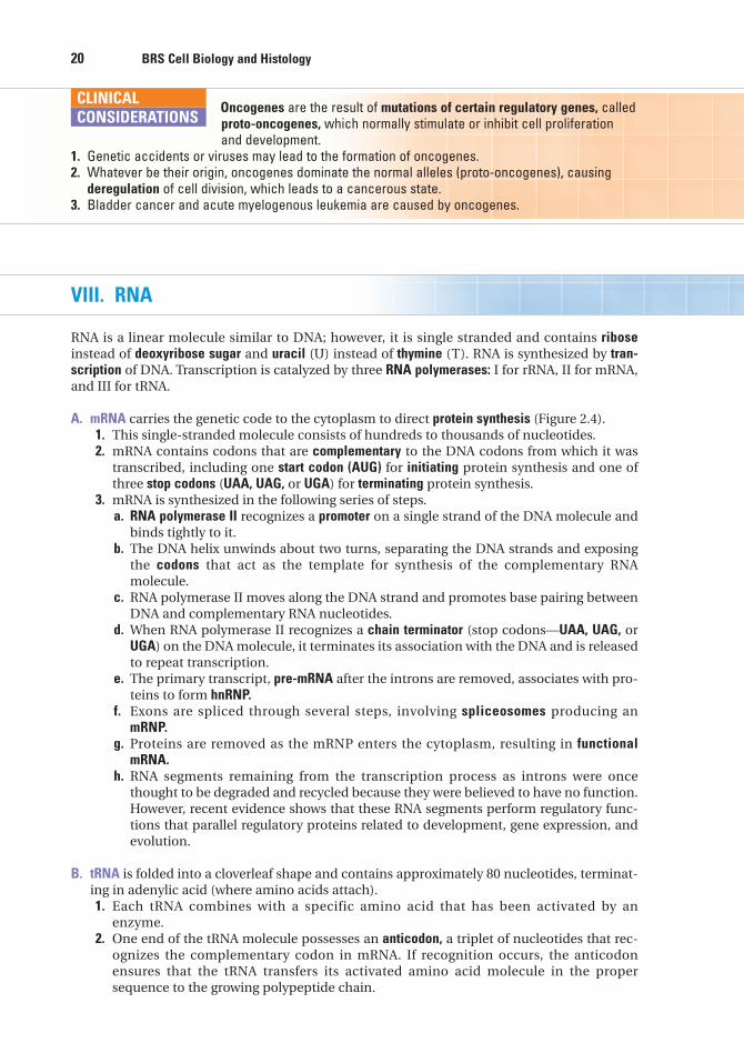
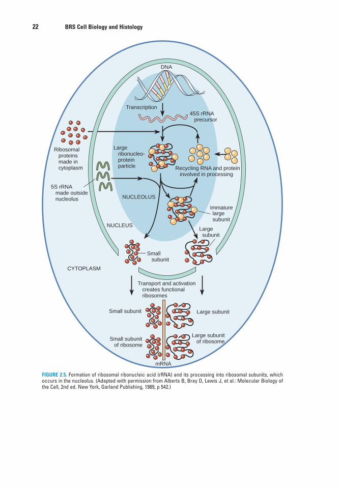
C. Ribosomal RNA associates with many different proteins (including enzymes) to form ribo-somes.1. rRNA associates with mRNA and tRNA during protein synthesis.2. rRNA synthesis takes place in the nucleolus and is catalyzed by RNA polymerase I. A sin-
gle 45S precursor rRNA (pre-rRNA) is formed and processed to form ribosomes as follows(Figure 2.5):a. Pre-rRNA associates with ribosomal proteins and is cleaved into the three sizes (28S,
18S, and 5.8S) of rRNAs present in ribosomes.b. The RNP containing 28S and 5.8S rRNA then combines with 5S rRNA, which is syn-
thesized outside of the nucleolus, to form the large subunit of the ribosome.c. The RNP containing 18S rRNA forms the small subunit of the ribosome.
DNA
DNA transcription
Nuclear binding proteins
RNA processing
Pre-mRNA
mRNA transport
Ribosomes
Translation of mRNA
Protein
FIGURE 2.4. Steps by which genetic information encoded in deoxyribonucleic acid (DNA) istranscribed into messenger ribonucleic acid (mRNA) and ultimately converted into proteins inthe cytoplasm. (Adapted with permission from Alberts B, Bray D, Lewis J, et al.: MolecularBiology of the Cell, 2nd ed. New York, Garland Publishing, 1989, p 482.)
LWBK615-c02[14-31].qxd 05/31/2010 10:18 AM Page 21 Aptara
22 BRS Cell Biology and Histology
Ribosomal proteins made in cytoplasm
Large ribonucleo- protein particle
DNA
Transcription45S rRNA precursor
Recycling RNA and protein involved in processing
5S rRNA made outside nucleolus
CYTOPLASM
NUCLEUS
NUCLEOLUS
Small subunit
Small subunit Large subunit
Large subunit
large subunit
Immature
Large subunit of ribosome
Transport and activation creates functional ribosomes
Small subunit of ribosome
mRNA
FIGURE 2.5. Formation of ribosomal ribonucleic acid (rRNA) and its processing into ribosomal subunits, whichoccurs in the nucleolus. (Adapted with permission from Alberts B, Bray D, Lewis J, et al.: Molecular Biology ofthe Cell, 2nd ed. New York, Garland Publishing, 1989, p 542.)
LWBK615-c02[14-31].qxd 05/31/2010 10:18 AM Page 22 Aptara
Chapter 2 Nucleus 23
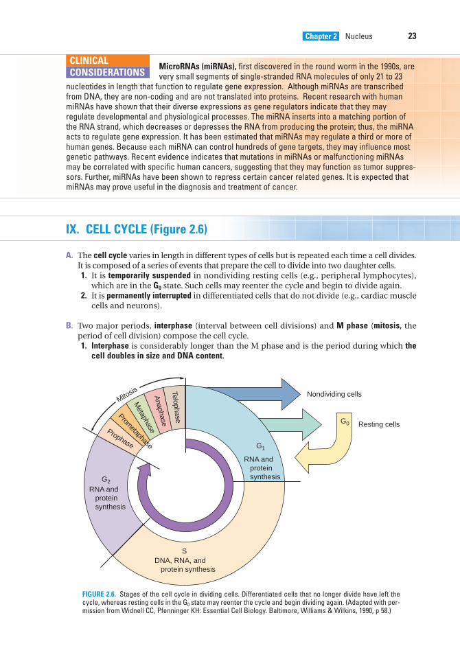
IX. CELL CYCLE (Figure 2.6)
A. The cell cycle varies in length in different types of cells but is repeated each time a cell divides.It is composed of a series of events that prepare the cell to divide into two daughter cells.1. It is temporarily suspended in nondividing resting cells (e.g., peripheral lymphocytes),
which are in the G0 state. Such cells may reenter the cycle and begin to divide again.2. It is permanently interrupted in differentiated cells that do not divide (e.g., cardiac muscle
cells and neurons).
B. Two major periods, interphase (interval between cell divisions) and M phase (mitosis, theperiod of cell division) compose the cell cycle.1. Interphase is considerably longer than the M phase and is the period during which the
cell doubles in size and DNA content.
MicroRNAs (miRNAs), first discovered in the round worm in the 1990s, are very small segments of single-stranded RNA molecules of only 21 to 23
nucleotides in length that function to regulate gene expression. Although miRNAs are transcribedfrom DNA, they are non-coding and are not translated into proteins. Recent research with humanmiRNAs have shown that their diverse expressions as gene regulators indicate that they mayregulate developmental and physiological processes. The miRNA inserts into a matching portion ofthe RNA strand, which decreases or depresses the RNA from producing the protein; thus, the miRNAacts to regulate gene expression. It has been estimated that miRNAs may regulate a third or more ofhuman genes. Because each miRNA can control hundreds of gene targets, they may influence mostgenetic pathways. Recent evidence indicates that mutations in miRNAs or malfunctioning miRNAsmay be correlated with specific human cancers, suggesting that they may function as tumor suppres-sors. Further, miRNAs have been shown to repress certain cancer related genes. It is expected thatmiRNAs may prove useful in the diagnosis and treatment of cancer.
CLINICAL
CONSIDERATIONS
Mitosis
G0
Nondividing cells
Resting cells
G1
G0
RNA and protein synthesisG2
S
RNA and protein synthesis
DNA, RNA, and protein synthesis
Prometaphase
Prophase
Anaphase
Telophase
Metaphase
FIGURE 2.6. Stages of the cell cycle in dividing cells. Differentiated cells that no longer divide have left thecycle, whereas resting cells in the G0 state may reenter the cycle and begin dividing again. (Adapted with per-mission from Widnell CC, Pfenninger KH: Essential Cell Biology. Baltimore, Williams & Wilkins, 1990, p 58.)
LWBK615-c02[14-31].qxd 05/31/2010 10:18 AM Page 23 Aptara
24 BRS Cell Biology and Histology
a. Interphase is divided into three separate phases (G1, S, and G2) during which specificcellular functions occur.(1) G1 phase (gap one phase) lasts for hours to several days.
(a) Occurring after mitosis, it is the period during which the cell grows and pro-teins are synthesized, restoring the daughter cells to normal volume and size.
(b) Certain trigger proteins are synthesized; these proteins enable the cell to reacha threshold (restriction point) and proceed to the S phase. Cells that fail to reachthe restriction point become resting cells and enter the G0 (outside phase) state.
(2) S phase (synthetic phase) lasts 8 to 12 hours in most cells.(a) DNA is replicated and proteins are synthesized, resulting in duplication of the
chromosomes.(b) Centrosomes are also duplicated.
(3) G2 phase (gap two phase) lasts 2 to 4 hours.(a) This phase follows the S phase and extends to mitosis.(b) The cell prepares to divide: the centrioles grow to maturity; energy required for
the completion of mitosis is stored; and RNA and proteins necessary for mito-sis are synthesized, including tubulin for the spindle apparatus.
b. Several control factors have been identified. These include a category of proteinsknown as cyclins as well as cyclin-dependent kinases (CDKs), which initiate and/orinduce progression through the cell cycle.(1) During the G1 phase, cyclins D and E bind to their respective CDKs; these com-
plexes enable the cell to enter and advance through the S phase.(2) Cyclin A binds to its CDKs, thus enabling the cell to leave the S phase and enter the
G2 phase as well as to manufacture cyclin B.(3) Cyclin B binds to its CDK, inducing the cell to leave the G2 phase and enter the
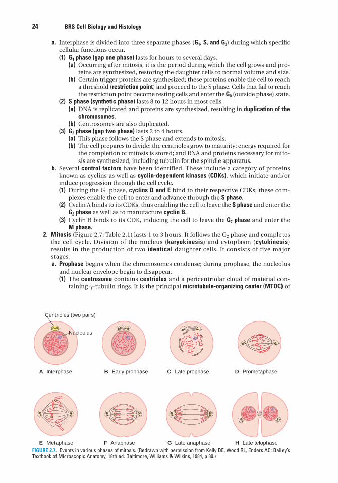
M phase.2. Mitosis (Figure 2.7; Table 2.1) lasts 1 to 3 hours. It follows the G2 phase and completes
the cell cycle. Division of the nucleus (karyokinesis) and cytoplasm (cytokinesis)results in the production of two identical daughter cells. It consists of five majorstages.a. Prophase begins when the chromosomes condense; during prophase, the nucleolus
and nuclear envelope begin to disappear.(1) The centrosome contains centrioles and a pericentriolar cloud of material con-
taining �-tubulin rings. It is the principal microtubule-organizing center (MTOC) of
A Interphase
E Metaphase F Anaphase G Late anaphase H Late telophase
B Early prophase C Late prophase D Prometaphase
Nucleolus
Centrioles (two pairs)
FIGURE 2.7. Events in various phases of mitosis. (Redrawn with permission from Kelly DE, Wood RL, Enders AC: Bailey’sTextbook of Microscopic Anatomy, 18th ed. Baltimore, Williams & Wilkins, 1984, p 89.)
LWBK615-c02[14-31].qxd 05/31/2010 10:18 AM Page 24 Aptara
Chapter 2 Nucleus 25
the cell. Centrosomes migrate to opposite poles of the cell, and from them spindlefibers and astral rays of the mitotic spindle polymerize.
(2) Chromosomes consist of two parallel sister chromatids (future daughter chromo-somes) attached at the centromere, a constriction along the chromosome. Kineto-chores develop at the centromere region and function as MTOCs.
b. Prometaphase begins when the nuclear envelope disappears, allowing the chromo-somes to disperse apparently randomly in the cytoplasm.(1) The kinetochores complete development and attach to specific spindle micro-
tubules, forming kinetochore microtubules.(2) Spindle microtubules that do not attach to kinetochores are called polar micro-
tubules.c. Metaphase is the phase during which the duplicated condensed chromosomes align
at the equatorial plate of the mitotic spindle and become attached to spindle micro-tubules at their kinetochore.
d. Anaphase begins as the chromatids separate at the centromere and daughter chro-mosomes move to opposite poles of the cell.(1) The spindle elongates.(2) In the later stages of anaphase, a cleavage furrow begins to form around the cell as
the contractile ring, a band of actin filaments, contracts.e. Telophase is characterized by each set of chromosomes reaching the pole, a deepen-
ing of the cleavage furrow; the midbody (containing overlapping polar microtubules)is now between the newly forming daughter cells.(1) Microtubules in the midbody are depolymerized, facilitating cytokinesis and
formation of two identical daughter cells.(2) The nuclear envelope is reestablished around the condensed chromosomes in the
daughter cells, and nucleoli reappear. Nucleoli arise from the specific NORs (calledsecondary constriction sites), which are carried on five separate chromosomes inhumans.
(3) The daughter nuclei gradually enlarge, and the condensed chromosomes disperseto form the typical interphase nucleus with heterochromatin and euchromatin.
(4) It appears that at the end of cytokinesis the mother centriole of the duplicated pairmoves from the newly forming nuclear pole to the intercellular bridge. This eventis necessary to initiate disassembly of the midbody microtubules and completethe separation of the daughter cells. If this event fails, DNA replication is arrestedat one of the G1 checkpoints during the next interphase.
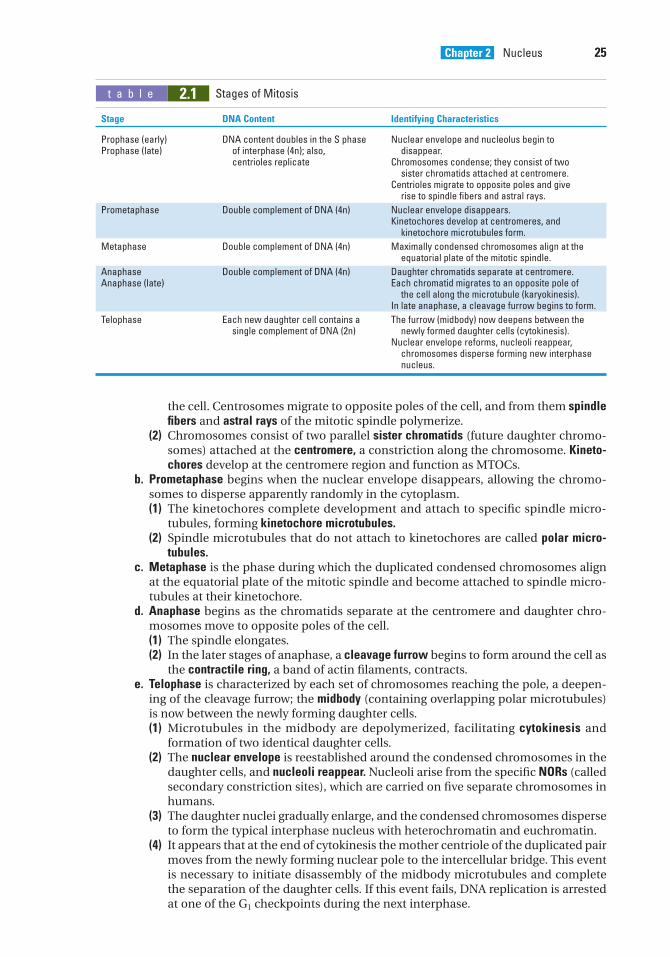
t a b l e 2.1 Stages of Mitosis
Stage DNA Content Identifying Characteristics
Prophase (early) DNA content doubles in the S phase Nuclear envelope and nucleolus begin toProphase (late) of interphase (4n); also, disappear.
centrioles replicate Chromosomes condense; they consist of two sister chromatids attached at centromere.
Centrioles migrate to opposite poles and give rise to spindle fibers and astral rays.
Prometaphase Double complement of DNA (4n) Nuclear envelope disappears.Kinetochores develop at centromeres, and
kinetochore microtubules form.
Metaphase Double complement of DNA (4n) Maximally condensed chromosomes align at the equatorial plate of the mitotic spindle.
Anaphase Double complement of DNA (4n) Daughter chromatids separate at centromere.Anaphase (late) Each chromatid migrates to an opposite pole of
the cell along the microtubule (karyokinesis).In late anaphase, a cleavage furrow begins to form.
Telophase Each new daughter cell contains a The furrow (midbody) now deepens between the single complement of DNA (2n) newly formed daughter cells (cytokinesis).
Nuclear envelope reforms, nucleoli reappear, chromosomes disperse forming new interphase nucleus.
LWBK615-c02[14-31].qxd 05/31/2010 10:18 AM Page 25 Aptara
26 BRS Cell Biology and Histology
X. APOPTOSIS (PROGRAMMED CELL DEATH)
Apoptosis is the method whereby cells are removed from tissues in an orderly fashion as a partof normal maintenance or during development.
A. Cells that undergo programmed cell death have several morphological features.1. They include chromatin condensation, breaking up of the nucleus, and blebbing of the
plasma membrane.2. The cell shrinks and is fragmented into membrane-enclosed fragments called apoptotic
bodies.
B. Apoptotic cells do not pose a threat to surrounding cells, because changes in their plasmamembranes make them subject to rapid phagocytosis by macrophages and by neighboringcells. Macrophages that phagocytose apoptotic cells do not release cytokines that initiatethe inflammatory response.
C. The signals that induce apoptosis may occur through several mechanisms.1. Genes that code for enzymes, called caspases, play an important role in the process.2. Certain cytokines, such as tumor necrosis factor (TNF), may also activate caspases that
degrade regulatory and structural proteins in the nucleus and cytoplasm, leading to themorphological changes characteristic of apoptosis.
D. Defects in the process of programmed cell death contribute to many major diseases.1. Excessive apoptosis causes extensive nerve cell loss in Alzheimer disease and stroke.2. Insufficient apoptosis has been linked to cancer and other autoimmune diseases.
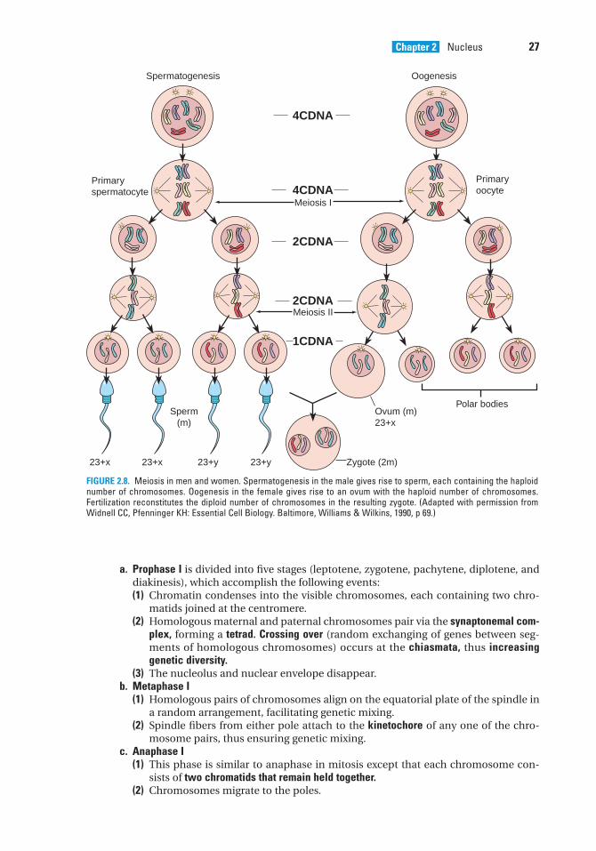
XI. MEIOSIS (Figure 2.8)
A. Meiosis is a special form of cell division in germ cells (oogonia and spermatozoa) in whichthe chromosome number is reduced from diploid (2n) to haploid (n).1. It occurs in developing germ cells in preparation for sexual reproduction. Subsequent
fertilization results in diploid zygotes.2. DNA content of the original diploid cell is doubled (4n) in the S phase preparatory to
meiosis.a. This phase is followed by two successive cell divisions that give rise to four haploid
cells.b. In addition, recombination of maternal and paternal genes occurs by crossing over and
random assortment, yielding the unique haploid genome of the gamete.
B. The stages of meiosis are meiosis I (reductional division) and meiosis II (equatorial division).1. Reductional division (meiosis I) occurs after interphase during the cell cycle, when the
DNA content is duplicated, whereas the chromosome number (46) remains unchanged,giving the cell a 4CDNA content (considered to be the total DNA content of the cell).
Transformed cells1. Transformed cells have lost their ability to respond to regulatory
signals controlling the cell cycle, and by this, they may undergo cell division indefinitely, thusbecoming cancerous.
2. Vinca alkaloids may arrest these cells in mitosis, whereas drugs that block purine and pyrimidinesynthesis may arrest these cells in the S phase of the cell cycle.
CLINICAL
CONSIDERATIONS
LWBK615-c02[14-31].qxd 05/31/2010 10:18 AM Page 26 Aptara
Chapter 2 Nucleus 27
a. Prophase I is divided into five stages (leptotene, zygotene, pachytene, diplotene, anddiakinesis), which accomplish the following events:(1) Chromatin condenses into the visible chromosomes, each containing two chro-
matids joined at the centromere.(2) Homologous maternal and paternal chromosomes pair via the synaptonemal com-
plex, forming a tetrad. Crossing over (random exchanging of genes between seg-ments of homologous chromosomes) occurs at the chiasmata, thus increasinggenetic diversity.
(3) The nucleolus and nuclear envelope disappear.b. Metaphase I
(1) Homologous pairs of chromosomes align on the equatorial plate of the spindle ina random arrangement, facilitating genetic mixing.
(2) Spindle fibers from either pole attach to the kinetochore of any one of the chro-mosome pairs, thus ensuring genetic mixing.
c. Anaphase I(1) This phase is similar to anaphase in mitosis except that each chromosome con-
sists of two chromatids that remain held together.(2) Chromosomes migrate to the poles.
Spermatogenesis
Primaryspermatocyte
Primaryoocyte
Oogenesis
Meiosis I
Meiosis II
Sperm(m)
Ovum (m)23+x
23+x 23+x 23+y 23+y Zygote (2m)
Polar bodies
4CDNA
4CDNA
2CDNA
2CDNA
1CDNA
FIGURE 2.8. Meiosis in men and women. Spermatogenesis in the male gives rise to sperm, each containing the haploidnumber of chromosomes. Oogenesis in the female gives rise to an ovum with the haploid number of chromosomes.Fertilization reconstitutes the diploid number of chromosomes in the resulting zygote. (Adapted with permission fromWidnell CC, Pfenninger KH: Essential Cell Biology. Baltimore, Williams & Wilkins, 1990, p 69.)
LWBK615-c02[14-31].qxd 05/31/2010 10:18 AM Page 27 Aptara
28 BRS Cell Biology and Histology
d. Telophase I is similar to telophase in mitosis in that the nuclear envelope is reestab-lished and two daughter cells are formed via cytokinesis.(1) Each daughter cell now contains 23 chromosomes (n) number but has a 2CDNA
content (the diploid amount).(2) Each chromosome is composed of two similar sister chromatids (but not geneti-
cally identical following recombination).2. Equatorial division (meiosis II) begins soon after the completion of meiosis I, following a
brief interphase without DNA replication.a. The sister chromatids are portioned out among the two daughter cells formed in meio-
sis I. The two daughter cells then divide, resulting in the distribution of chromosomesinto four daughter cells, each containing its own unique recombined genetic material(1CDNA;n). Thus, every gamete contains its own unique set of genetic materials.
b. The stages of meiosis II are similar to those of mitosis; thus, the stages are named sim-ilarly (prophase II, metaphase II, anaphase II, and telophase II).
c. Meiosis II occurs more rapidly than mitosis.
Nondisjunction of ChromosomesDuring prophase I of meiosis I, chromosome pairs align themselves at the
equatorial plate and exchange genetic materials. During anaphase I, the chromosome pairs will sep-arate and begin their migrations to opposite poles. Sometimes the members of a pair fail toseparate, resulting in one daughter cell containing an extra chromosome (n � 1 � 24), whereas thedaughter cell at the opposite pole is minus a chromosome (n � 1 � 22). This development is knownas nondisjunction. Upon fertilization with a normal gamete containing 23 chromosomes, the result-ing zygote will contain either 47 chromosomes (trisomy for that extra chromosome) or 45chromosomes (monosomy for that missing chromosome). Chromosomes 8, 9, 13, 18, and 21 are thosechromosomes most frequently affected by nondisjunction.
Aneuploidy, defined as an abnormal number of chromosomes, can be detected by karyotyping.
1. Down syndrome (trisomy 21) is characterized by mental retardation, short stature, stubbyappendages, congenital heart malformations, and other defects.
2. Klinefelter syndrome (XXY) is aneuploidy of the sex chromosomes, characterized by infertility,variable degrees of masculinization, and small testes.
3. Turner syndrome (XO) is monosomy of the sex chromosomes, characterized by short stature,sterility, and various other abnormalities.
CLINICAL
CONSIDERATIONS
LWBK615-c02[14-31].qxd 05/31/2010 10:18 AM Page 28 Aptara
1. The nuclear pore complex
(A) permits free communication betweenthe nucleus and the cytoplasm.
(B) is bridged by a unit membrane.(C) is located only at specific nuclear pore
sites.(D) permits passage of proteins via receptor-
mediated transport.(E) has a luminal ring that faces the
cytoplasm.
2. Which one of the following nucleotides ispresent only in RNA?
(A) Thymine(B) Adenine(C) Uracil(D) Cytosine(E) Guanine
3. Anticodons are located in
(A) mRNA.(B) rRNA.(C) tRNA.(D) snRNP.(E) hnRNP.
4. DNA is duplicated in the cell cycle duringthe
(A) G2 phase.(B) S phase.(C) M phase.(D) G1 phase.(E) G0 phase.
5. A male child at puberty is determined tohave Klinefelter syndrome. Although theparents have been informed of the clinicalsignificance, they have asked for an explana-tion of what happened. Identify the itemthat should be discussed with the parents.
(A) Trisomy of chromosome 21(B) Loss of an autosome during mitosis(C) Loss of the Y chromosome during meiosis(D) Nondisjunction of the X chromosome(E) Loss of the X chromosome
6. Which one of the following is an inclusionnot bounded by a membrane that is observ-able only during interphase?
(A) Nuclear pore complex(B) Nucleolus(C) Heterochromatin(D) Outer nuclear membrane(E) Euchromatin
7. A structure that is continuous with RER isthe
(A) nuclear pore complex.(B) nucleolus.(C) heterochromatin.(D) outer nuclear membrane.(E) euchromatin.
Review Test
Directions: Each of the numbered items or incomplete statements in this section is followed byanswers or by completions of the statement. Select the ONE lettered answer or completion thatis BEST in each case.
29
LWBK615-c02[14-31].qxd 05/31/2010 10:18 AM Page 29 Aptara
30 BRS Cell Biology and Histology
8. Identify the structure that controls move-ment of proteins in and out of the nucleus.
(A) Nuclear pore complex(B) Nucleolus(C) Heterochromatin(D) Outer nuclear membrane(E) Euchromatin
9. The site of transcriptional activity is the
(A) nuclear pore complex.(B) nucleolus.(C) heterochromatin.(D) outer nuclear membrane.(E) euchromatin.
10. Clumps of nucleoprotein concentratednear the periphery of the nucleus are called
(A) nuclear pore complex.(B) nucleolus.(C) heterochromatin.(D) outer nuclear membrane.(E) euchromatin.
LWBK615-c02[14-31].qxd 05/31/2010 10:18 AM Page 30 Aptara
Answers and Explanations
1. D. The nuclear pore complex contains a central aqueous channel that permits passage ofsmall water-soluble molecules. However, movement of proteins in and out of the nucleusis selectively controlled by the nuclear pore complex via receptor-mediated transport (seeChapter 2 II E).
2. C. DNA contains the purines, adenine and guanine, and the pyrimidines, cytosine andthymine. In RNA, uracil, a pyrimidine, replaces thymine (see Chapter 2 VII A, VIII).
3. C. Each tRNA possesses a triplet of nucleotides, called an anticodon, which recognizes thecomplementary codon in mRNA (see Chapter 2 VIII B).
4. B. The S (synthesis) phase of the cell cycle is the period during which DNA replication andhistone synthesis occur, resulting in duplication of the chromosomes. At the end of the Sphase, each chromosome consists of two identical chromatids attached to one another atthe centromere (see Chapter 2 IX B).
5. D. Klinefelter syndrome occurs only in men. This condition results from nondisjunction ofthe X chromosome during meiosis, resulting in an extra X chromosome in somatic cells.These cells therefore have a normal complement of autosomal chromosomes (22 pairs),and instead of one pair of sex chromosomes (XY), there is an extra X chromosome. Theseindividuals have an XXY genotype, resulting in 47 total chromosomes rather than the nor-mal complement of 46. This syndrome is an example of trisomy of the sex chromosomes.Down syndrome is an example of an autosomal trisomy, specifically trisomy ofchromosome number 21. Both syndromes have profound complications (see Chapter 2 XIB Clinical Considerations).
6. B. The nucleolus is an inclusion, not bounded by a membrane, within the nucleus. It isobservable during interphase but disappears during mitosis (see Chapter 2 III A).
7. D. The outer nuclear membrane is continuous with the rough endoplasmic reticulum (seeChapter 2 II A).
8. A. The nuclear pore complex selectively controls movements of water-soluble moleculesand proteins in and out of the nucleus (see Chapter 2 II E).
9. E. The pale-staining euchromatin is the transcriptionally active chromatin in the nucleus(see Chapter 2 V A).
10. C. Heterochromatin is the dark-staining nucleoprotein near the periphery of the nucleus.It is transcriptionally inactive but may be responsible for proper chromosome segregationduring meiosis (see Chapter 2 V A).
31
LWBK615-c02[14-31].qxd 05/31/2010 10:18 AM Page 31 Aptara
c h a p t e r 3 Cytoplasm and Organelles
32
I. OVERVIEW—THE CYTOPLASM
The cytoplasm contains three main structural components: organelles, inclusions, and thecytoskeleton. The fluid component is called the cytosol. The functional interactions among cer-tain organelles result in the uptake and release of material by the cell, protein synthesis, andintracellular digestion.
II. STRUCTURAL COMPONENTS
A. Organelles (Figure 3.1) are metabolically active units of cellular matter.1. The plasma membrane, which envelops the cell and forms a boundary between it and
adjacent structures, is discussed in Chapter 1.2. Ribosomes
a. Structure. Ribosomes are 12 nanometers (nm) wide and 25 nm long and consist of asmall and a large subunit. The subunits are composed of several types of ribosomalribonucleic acid (rRNA) and numerous proteins (Table 3.1; Figure 2.5).
b. Ribosomes may be free in the cytosol or bound to membranes of the rough endo-plasmic reticulum (RER) or outer nuclear membrane. Whether free or bound, theribosomes constitute a single interchangeable population.
c. A polyribosome (polysome) is a cluster of ribosomes along a single strand of messengerribonucleic acid (mRNA) that is engaged in the synthesis of protein.
d. Function. Ribosomes are the sites where mRNA is translated into protein. Proteins des-tined for transport (secretory, membrane, and lysosomal) are synthesized on polyri-bosomes bound to the RER, whereas proteins not destined for transport are synthe-sized on polyribosomes in the cytosol.(1) The small ribosomal subunit binds mRNA and activated transfer ribonucleic acids
(tRNAs); the codons of the mRNA then base-pair with the corresponding anti-codons of the tRNAs.
(2) Next, an initiator tRNA recognizes the start codon (AUG) on the mRNA.(3) The large ribosomal subunit then binds to the complex. Peptidyl transferase in the
large subunit catalyzes peptide bond formation, resulting in addition of aminoacids to the growing polypeptide chain.
(4) A chain-terminating codon (UAA, UAG, or UGA) causes release of the polypeptidefrom the ribosome, and the ribosomal subunits dissociate from the mRNA.
3. RER (Figures 3.1 and 3.2) a. Structure. RER is a system of membrane-bounded sacs, or cavities. The outer surface
of RER is studded with ribosomes, which makes it appear rough. The interior regionof RER is called the cisterna, or the lumen. The outer nuclear membrane is continuouswith the RER membrane, which brings the perinuclear cisterna into continuity with
LWBK615-c03[32-55].qxd 05/31/2010 10:30 AM Page 32 Aptara
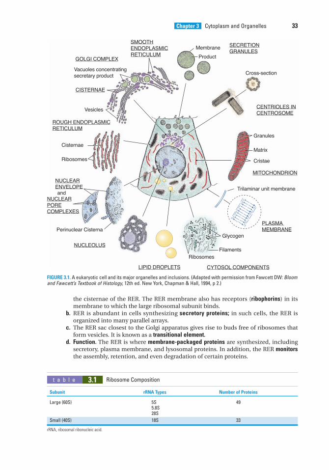
Chapter 3 Cytoplasm and Organelles 33
the cisternae of the RER. The RER membrane also has receptors (ribophorins) in itsmembrane to which the large ribosomal subunit binds.
b. RER is abundant in cells synthesizing secretory proteins; in such cells, the RER isorganized into many parallel arrays.
c. The RER sac closest to the Golgi apparatus gives rise to buds free of ribosomes thatform vesicles. It is known as a transitional element.
d. Function. The RER is where membrane-packaged proteins are synthesized, includingsecretory, plasma membrane, and lysosomal proteins. In addition, the RER monitorsthe assembly, retention, and even degradation of certain proteins.
SMOOTHENDOPLASMICRETICULUM
GOLGI COMPLEX
CISTERNAE
ROUGH ENDOPLASMICRETICULUM
NUCLEARENVELOPE
NUCLEARPORECOMPLEXES
NUCLEOLUS
LIPID DROPLETS CYTOSOL COMPONENTS
PLASMA MEMBRANE
MITOCHONDRION
CENTRIOLES INCENTROSOME
SECRETIONGRANULES
Vesicles
Cisternae
Ribosomes
Perinuclear Cisterna
RibosomesFilaments
Glycogen
Trilaminar unit membrane
Granules
Cross-section
Membrane
Product
Matrix
Cristae
Vacuoles concentratingsecretary product
and
FIGURE 3.1. A eukaryotic cell and its major organelles and inclusions. (Adapted with permission from Fawcett DW: Bloomand Fawcett’s Textbook of Histology, 12th ed. New York, Chapman & Hall, 1994, p 2.)
t a b l e 3.1 Ribosome Composition
Subunit rRNA Types Number of Proteins
Large (60S) 5S 495.8S28S
Small (40S) 18S 33
rRNA, ribosomal ribonucleic acid.
LWBK615-c03[32-55].qxd 05/31/2010 10:30 AM Page 33 Aptara
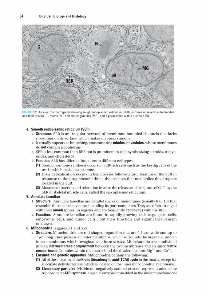
34 BRS Cell Biology and Histology
4. Smooth endoplasmic reticulum (SER)a. Structure. SER is an irregular network of membrane-bounded channels that lacks
ribosomes on its surface, which makes it appear smooth.b. It usually appears as branching, anastomosing tubules, or vesicles, whose membranes
do not contain ribophorins.c. SER is less common than RER but is prominent in cells synthesizing steroids, triglyc-
erides, and cholesterol.d. Function. SER has different functions in different cell types.
(1) Steroid hormone synthesis occurs in SER-rich cells such as the Leydig cells of thetestis, which make testosterone.
(2) Drug detoxification occurs in hepatocytes following proliferation of the SER inresponse to the drug phenobarbital; the oxidases that metabolize this drug arelocated in the SER.
(3) Muscle contraction and relaxation involve the release and recapture of Ca2� by theSER in skeletal muscle cells, called the sarcoplasmic reticulum.
5. Annulate lamellaea. Structure. Annulate lamellae are parallel stacks of membranes (usually 6 to 10) that
resemble the nuclear envelope, including its pore complexes. They are often arrangedwith their annuli (pores) in register and are frequently continuous with the RER.
b. Function. Annulate lamellae are found in rapidly growing cells (e.g., germ cells,embryonic cells, and tumor cells), but their function and significance remainunknown.
6. Mitochondria (Figures 3.1 and 3.2) a. Structure. Mitochondria are rod-shaped organelles that are 0.2 �m wide and up to
7 �m long. They possess an outer membrane, which surrounds the organelle, and aninner membrane, which invaginates to form cristae. Mitochondria are subdividedinto an intermembrane compartment between the two membranes and an inner matrixcompartment. Granules within the matrix bind the divalent cations Mg2� and Ca2�.
b. Enzymes and genetic apparatus. Mitochondria contain the following:(1) All of the enzymes of the Krebs (tricarboxylic acid [TCA]) cycle in the matrix, except for
succinate dehydrogenase, which is located on the inner mitochondrial membrane.(2) Elementary particles (visible on negatively stained cristae) represent adenosine
triphosphate (ATP) synthase, a special enzyme embedded in the inner mitochondrial
CN
RERM
MG
FIGURE 3.2. An electron micrograph showing rough endoplasmic reticulum (RER), portions of several mitochondriaand their cristae (C), matrix (M), and matrix granules (MG), and a peroxisome with a nucleoid (N).
LWBK615-c03[32-55].qxd 05/31/2010 10:30 AM Page 34 Aptara
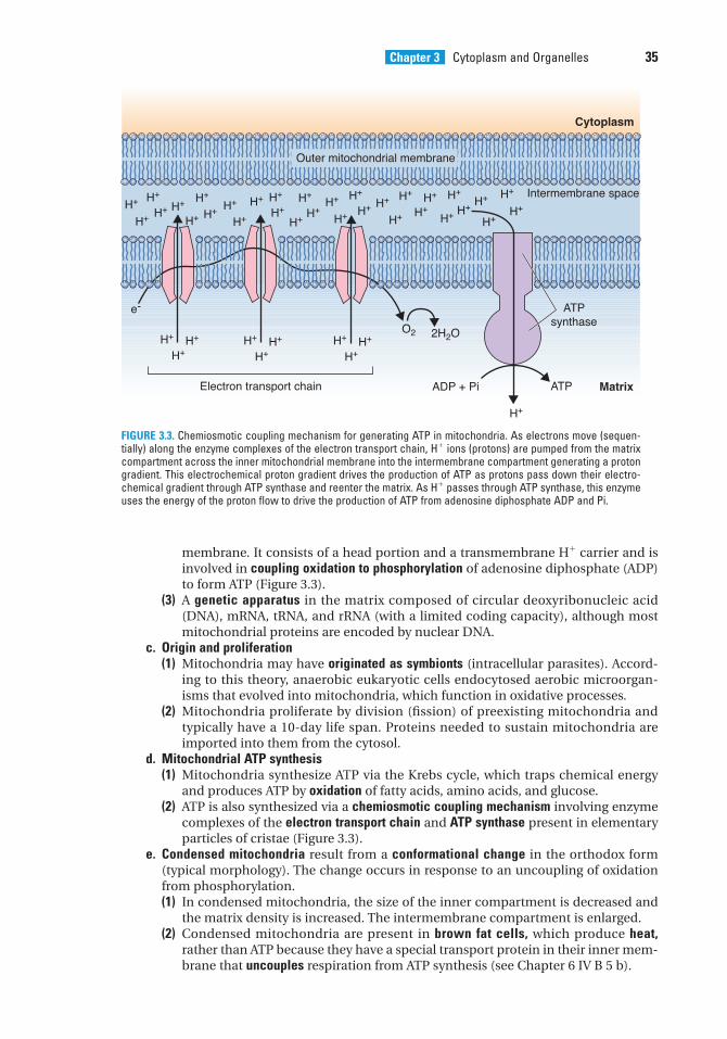
Chapter 3 Cytoplasm and Organelles 35
membrane. It consists of a head portion and a transmembrane H� carrier and isinvolved in coupling oxidation to phosphorylation of adenosine diphosphate (ADP)to form ATP (Figure 3.3).
(3) A genetic apparatus in the matrix composed of circular deoxyribonucleic acid(DNA), mRNA, tRNA, and rRNA (with a limited coding capacity), although mostmitochondrial proteins are encoded by nuclear DNA.
c. Origin and proliferation(1) Mitochondria may have originated as symbionts (intracellular parasites). Accord-
ing to this theory, anaerobic eukaryotic cells endocytosed aerobic microorgan-isms that evolved into mitochondria, which function in oxidative processes.
(2) Mitochondria proliferate by division (fission) of preexisting mitochondria andtypically have a 10-day life span. Proteins needed to sustain mitochondria areimported into them from the cytosol.
d. Mitochondrial ATP synthesis(1) Mitochondria synthesize ATP via the Krebs cycle, which traps chemical energy
and produces ATP by oxidation of fatty acids, amino acids, and glucose.(2) ATP is also synthesized via a chemiosmotic coupling mechanism involving enzyme
complexes of the electron transport chain and ATP synthase present in elementaryparticles of cristae (Figure 3.3).
e. Condensed mitochondria result from a conformational change in the orthodox form(typical morphology). The change occurs in response to an uncoupling of oxidationfrom phosphorylation.(1) In condensed mitochondria, the size of the inner compartment is decreased and
the matrix density is increased. The intermembrane compartment is enlarged.(2) Condensed mitochondria are present in brown fat cells, which produce heat,
rather than ATP because they have a special transport protein in their inner mem-brane that uncouples respiration from ATP synthesis (see Chapter 6 IV B 5 b).
H+ H+
H+ H+ H+ H+
H+ H+ H+
H+
H+ H+
H+
H+ H+H+
H+
H+
H+
H+ H+
H+ H+H+ H+
H+
H+
H+ H+ H+
H+H+
H+
H+H+H+
H+H+ H+
H+H+
Intermembrane space
Cytoplasm
ATPsynthase
MatrixElectron transport chain
O2 2H2O
ADP + Pi ATP
H+
e-
Outer mitochondrial membrane
FIGURE 3.3. Chemiosmotic coupling mechanism for generating ATP in mitochondria. As electrons move (sequen-tially) along the enzyme complexes of the electron transport chain, H� ions (protons) are pumped from the matrixcompartment across the inner mitochondrial membrane into the intermembrane compartment generating a protongradient. This electrochemical proton gradient drives the production of ATP as protons pass down their electro-chemical gradient through ATP synthase and reenter the matrix. As H� passes through ATP synthase, this enzymeuses the energy of the proton flow to drive the production of ATP from adenosine diphosphate ADP and Pi.
LWBK615-c03[32-55].qxd 05/31/2010 10:30 AM Page 35 Aptara
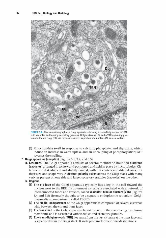
36 BRS Cell Biology and Histology
(3) Mitochondria swell in response to calcium, phosphate, and thyroxine, whichinduce an increase in water uptake and an uncoupling of phosphorylation; ATPreverses the swelling.
7. Golgi apparatus (complex) (Figures 3.1, 3.4, and 3.5) a. Structure. The Golgi apparatus consists of several membrane-bounded cisternae
(saccules) arranged in a stack and positioned and held in place by microtubules. Cis-ternae are disk-shaped and slightly curved, with flat centers and dilated rims, buttheir size and shape vary. A distinct polarity exists across the Golgi stack with manyvesicles present on one side and larger secretory granules (vacuoles) on the other.
b. Regions(1) The cis face of the Golgi apparatus typically lies deep in the cell toward the
nucleus next to the RER. Its outermost cisterna is associated with a network ofinterconnected tubes and vesicles, called vesicular–tubular clusters (VTC) (Figures3.4 and 3.5) (formerly thought to be a separate endoplasmic reticulum–Golgi-intermediate compartment called ERGIC).
(2) The medial compartment of the Golgi apparatus is composed of several cisternaelying between the cis and trans faces.
(3) The trans face of the Golgi apparatus lies at the side of the stack facing the plasmamembrane and is associated with vacuoles and secretory granules.
(4) The trans-Golgi network (TGN) lies apart from the last cisterna at the trans face andis separated from the Golgi stack. It sorts proteins for their final destinations.
C
TGN
N
CG
vv
FIGURE 3.4. Electron micrograph of a Golgi apparatus showing a trans-Golgi network (TGN)with vacuoles and forming secretory granules, Golgi cisternae (C), and a VTC delivering pro-teins to the cis Golgi (CG) via tiny vesicles (vv). A portion of a nucleus (N) is also evident.
LWBK615-c03[32-55].qxd 05/31/2010 10:30 AM Page 36 Aptara
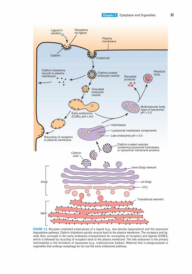
Chapter 3 Cytoplasm and Organelles 37
Clathrin-coatedendocytic vesicle
Uncoatedendocytic vesicle
Coated pit
cis Golgi
trans Golgi network
VTC
Transitional element
Clathrin-coated vesiclescontaining lysosomal hydrolasesor lysosomal membrane proteins
Lysosomal membrane components
Late endosome pH > 5.5
Hydrolases
Clathrin coat
Multivesicular body (type of lysosome)pH > 5.0Early endosome
(CURL) pH > 6.0
Recycling of receptors to plasma membrane
Clathrin
Clathrin triskelionsrecycle to plasmamembrane
Ligand in solution
Receptorsfor ligand
Residualbody
Reusableproducts
Plasma membrane
Golgi
RER
FIGURE 3.5. Receptor-mediated endocytosis of a ligand (e.g., low-density lipoproteins) and the lysosomaldegradative pathway. Clathrin triskelions quickly recycle back to the plasma membrane. The receptors and lig-ands then uncouple in the early endosome (compartment for uncoupling of receptors and ligands [CURL]),which is followed by recycling of receptors back to the plasma membrane. The late endosome is the primaryintermediate in the formation of lysosomes (e.g., multivesicular bodies). Material that is phagocytosed ororganelles that undergo autophagy do not use the early endosomal pathway.
LWBK615-c03[32-55].qxd 05/31/2010 10:30 AM Page 37 Aptara
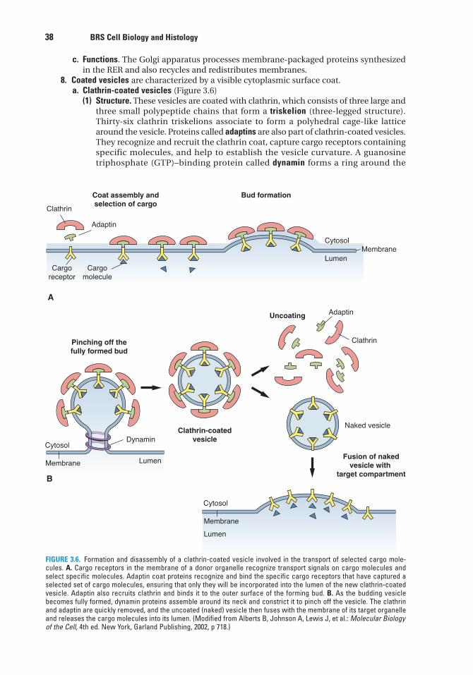
38 BRS Cell Biology and Histology
c. Functions. The Golgi apparatus processes membrane-packaged proteins synthesizedin the RER and also recycles and redistributes membranes.
8. Coated vesicles are characterized by a visible cytoplasmic surface coat.a. Clathrin-coated vesicles (Figure 3.6)
(1) Structure. These vesicles are coated with clathrin, which consists of three large andthree small polypeptide chains that form a triskelion (three-legged structure).Thirty-six clathrin triskelions associate to form a polyhedral cage-like latticearound the vesicle. Proteins called adaptins are also part of clathrin-coated vesicles.They recognize and recruit the clathrin coat, capture cargo receptors containingspecific molecules, and help to establish the vesicle curvature. A guanosinetriphosphate (GTP)–binding protein called dynamin forms a ring around the
Clathrin
Clathrin
Naked vesicle
Adaptin
Adaptin
Cytosol
LumenMembrane
Cytosol
Membrane
Dynamin
Cargoreceptor
Cargomolecule
Coat assembly andselection of cargo
Bud formation
Clathrin-coatedvesicle
Pinching off thefully formed bud
Fusion of nakedvesicle with
target compartment
A
B
Lumen
Cytosol
Membrane
Lumen
Uncoating
FIGURE 3.6. Formation and disassembly of a clathrin-coated vesicle involved in the transport of selected cargo mole-cules. A. Cargo receptors in the membrane of a donor organelle recognize transport signals on cargo molecules andselect specific molecules. Adaptin coat proteins recognize and bind the specific cargo receptors that have captured aselected set of cargo molecules, ensuring that only they will be incorporated into the lumen of the new clathrin-coatedvesicle. Adaptin also recruits clathrin and binds it to the outer surface of the forming bud. B. As the budding vesiclebecomes fully formed, dynamin proteins assemble around its neck and constrict it to pinch off the vesicle. The clathrinand adaptin are quickly removed, and the uncoated (naked) vesicle then fuses with the membrane of its target organelleand releases the cargo molecules into its lumen. (Modified from Alberts B, Johnson A, Lewis J, et al.: Molecular Biologyof the Cell, 4th ed. New York, Garland Publishing, 2002, p 718.)
LWBK615-c03[32-55].qxd 05/31/2010 10:30 AM Page 38 Aptara
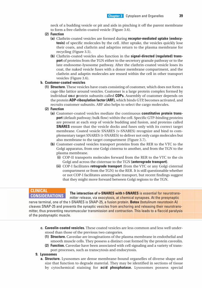
Chapter 3 Cytoplasm and Organelles 39
neck of a budding vesicle or pit and aids in pinching it off the parent membraneto form a free clathrin-coated vesicle (Figure 3.6).
(2) Function(a) Clathrin-coated vesicles are formed during receptor-mediated uptake (endocy-
tosis) of specific molecules by the cell. After uptake, the vesicles quickly losetheir coats, and clathrin and adaptins return to the plasma membrane forrecycling (Figure 3.5).
(b) Clathrin-coated vesicles also function in the signal-directed (regulated) trans-port of proteins from the TGN either to the secretory granule pathway or to thelate endosome–lysosome pathway. After the clathrin-coated vesicle loses itscoat, the naked vesicle fuses with a donor membrane compartment, and theclathrin and adaptin molecules are reused within the cell in other transportvesicles (Figure 3.6).
b. Coatomer-coated vesicles(1) Structure. These vesicles have coats consisting of coatomer, which does not form a
cage-like lattice around vesicles. Coatomer is a large protein complex formed byindividual coat protein subunits called COPs. Assembly of coatomer depends onthe protein ADP-ribosylation factor (ARF), which binds GTP, becomes activated, andrecruits coatomer subunits. ARF also helps to select the cargo molecules.
(2) Function(a) Coatomer-coated vesicles mediate the continuous constitutive protein trans-
port (default pathway; bulk flow) within the cell. Specific GTP-binding proteinsare present at each step of vesicle budding and fusion, and proteins calledSNARES ensure that the vesicle docks and fuses only with its correct targetmembrane. Coated vesicle SNARES (v-SNARES) recognize and bind to com-plementary target SNARES (t-SNARES) to deliver not only cargo molecules butalso membrane to the target compartment (Figure 3.7).
(b) Coatomer-coated vesicles transport proteins from the RER to the VTC to theGolgi apparatus, from one Golgi cisterna to another, and from the TGN to theplasma membrane.(i) COP-II transports molecules forward from the RER to the VTC to the cis
Golgi and across the cisternae to the TGN (anterograde transport).(ii) COP-I facilitates retrograde transport (from the VTC or any Golgi cisternal
compartment or from the TGN) to the RER. It is still questionable whetheror not COP-I facilitates anterograde transport, but recent findings suggestthat they might move forward between Golgi regions to the TGN.
The interaction of v-SNARES with t-SNARES is essential for neurotrans-mitter release, via exocytosis, at chemical synapses. At the presynaptic
nerve terminal, one of the t-SNARES is SNAP-25, a fusion protein. Botox (botulinum neurotoxin A)cleaves SNAP-25 and prevents the synaptic vesicles from anchoring and releasing their neurotrans-mitter, thus preventing neuromuscular transmission and contraction. This leads to a flaccid paralysisof the postsynaptic muscle.
CLINICAL
CONSIDERATIONS
c. Caveolin-coated vesicles. These coated vesicles are less common and less well under-stood than those of the previous two categories.(1) Structure. Caveolae are invaginations of the plasma membrane in endothelial and
smooth muscle cells. They possess a distinct coat formed by the protein caveolin.(2) Function. Caveolae have been associated with cell signaling and a variety of trans-
port processes, such as transcytosis and endocytosis.9. Lysosomes
a. Structure. Lysosomes are dense membrane-bound organelles of diverse shape andsize that function to degrade material. They may be identified in sections of tissueby cytochemical staining for acid phosphatase. Lysosomes possess special
LWBK615-c03[32-55].qxd 05/31/2010 10:30 AM Page 39 Aptara
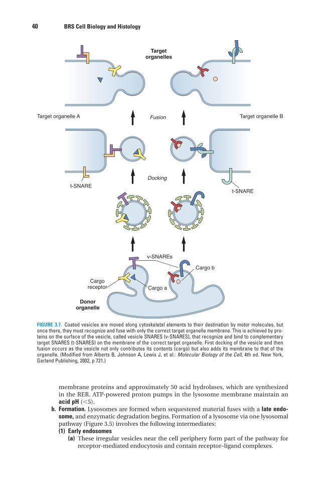
40 BRS Cell Biology and Histology
membrane proteins and approximately 50 acid hydrolases, which are synthesizedin the RER. ATP-powered proton pumps in the lysosome membrane maintain anacid pH (�5).
b. Formation. Lysosomes are formed when sequestered material fuses with a late endo-some, and enzymatic degradation begins. Formation of a lysosome via one lysosomalpathway (Figure 3.5) involves the following intermediates:(1) Early endosomes
(a) These irregular vesicles near the cell periphery form part of the pathway forreceptor-mediated endocytosis and contain receptor–ligand complexes.
Cargo a
Cargo b
Targetorganelles
Donororganelle
v-SNAREs
Target organelle BTarget organelle A
Cargoreceptor
t-SNAREt-SNARE
Docking
Fusion
FIGURE 3.7. Coated vesicles are moved along cytoskeletal elements to their destination by motor molecules, butonce there, they must recognize and fuse with only the correct target organelle membrane. This is achieved by pro-teins on the surface of the vesicle, called vesicle SNARES (v-SNARES), that recognize and bind to complementarytarget SNARES (t-SNARES) on the membrane of the correct target organelle. First docking of the vesicle and thenfusion occurs as the vesicle not only contributes its contents (cargo) but also adds its membrane to that of theorganelle. (Modified from Alberts B, Johnson A, Lewis J, et al.: Molecular Biology of the Cell, 4th ed. New York,Garland Publishing, 2002, p 721.)
LWBK615-c03[32-55].qxd 05/31/2010 10:30 AM Page 40 Aptara
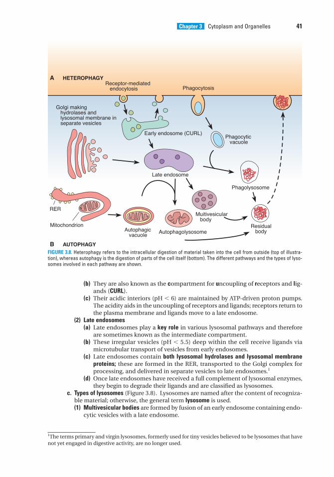
Chapter 3 Cytoplasm and Organelles 41
(b) They are also known as the compartment for uncoupling of receptors and lig-ands (CURL).
(c) Their acidic interiors (pH � 6) are maintained by ATP-driven proton pumps.The acidity aids in the uncoupling of receptors and ligands; receptors return tothe plasma membrane and ligands move to a late endosome.
(2) Late endosomes(a) Late endosomes play a key role in various lysosomal pathways and therefore
are sometimes known as the intermediate compartment.(b) These irregular vesicles (pH � 5.5) deep within the cell receive ligands via
microtubular transport of vesicles from early endosomes.(c) Late endosomes contain both lysosomal hydrolases and lysosomal membrane
proteins; these are formed in the RER, transported to the Golgi complex forprocessing, and delivered in separate vesicles to late endosomes.1
(d) Once late endosomes have received a full complement of lysosomal enzymes,they begin to degrade their ligands and are classified as lysosomes.
c. Types of lysosomes (Figure 3.8). Lysosomes are named after the content of recogniza-ble material; otherwise, the general term lysosome is used.(1) Multivesicular bodies are formed by fusion of an early endosome containing endo-
cytic vesicles with a late endosome.
1The terms primary and virgin lysosomes, formerly used for tiny vesicles believed to be lysosomes that havenot yet engaged in digestive activity, are no longer used.
A HETEROPHAGY
B AUTOPHAGY
Receptor-mediated endocytosis
Golgi making hydrolases and lysosomal membrane in separate vesicles
Early endosome (CURL)
Phagocytosis
Phagocytic vacuole
Phagolysosome
Residual body
Muitivesicular body
Late endosome
AutophagolysosomeAutophagic vacuole
Mitochondrion
RER
FIGURE 3.8. Heterophagy refers to the intracellular digestion of material taken into the cell from outside (top of illustra-tion), whereas autophagy is the digestion of parts of the cell itself (bottom). The different pathways and the types of lyso-somes involved in each pathway are shown.
LWBK615-c03[32-55].qxd 05/31/2010 10:30 AM Page 41 Aptara
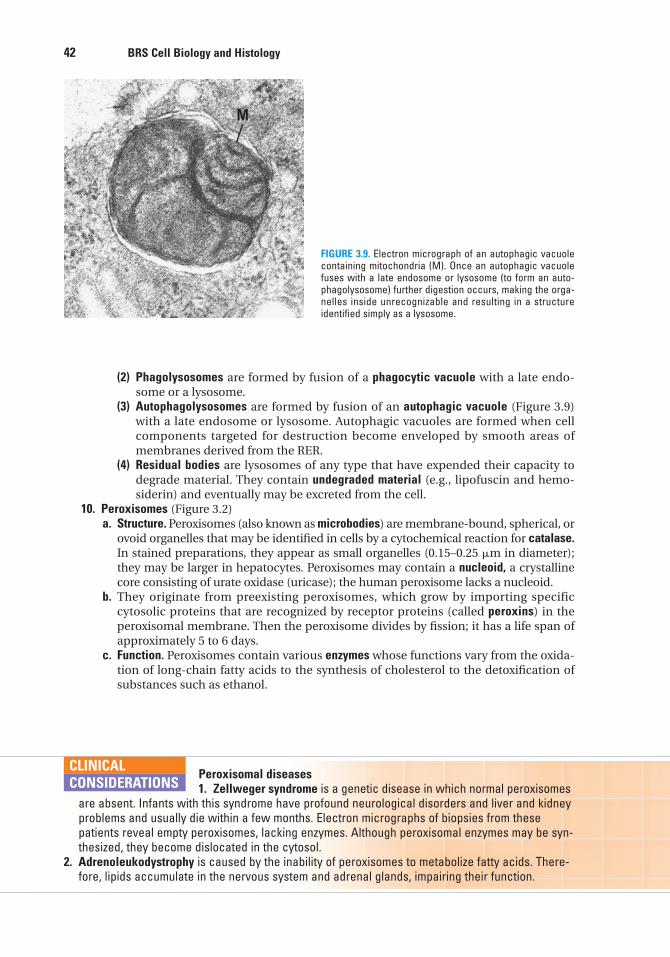
42 BRS Cell Biology and Histology
(2) Phagolysosomes are formed by fusion of a phagocytic vacuole with a late endo-some or a lysosome.
(3) Autophagolysosomes are formed by fusion of an autophagic vacuole (Figure 3.9)with a late endosome or lysosome. Autophagic vacuoles are formed when cellcomponents targeted for destruction become enveloped by smooth areas ofmembranes derived from the RER.
(4) Residual bodies are lysosomes of any type that have expended their capacity todegrade material. They contain undegraded material (e.g., lipofuscin and hemo-siderin) and eventually may be excreted from the cell.
10. Peroxisomes (Figure 3.2)a. Structure. Peroxisomes (also known as microbodies) are membrane-bound, spherical, or
ovoid organelles that may be identified in cells by a cytochemical reaction for catalase.In stained preparations, they appear as small organelles (0.15–0.25 �m in diameter);they may be larger in hepatocytes. Peroxisomes may contain a nucleoid, a crystallinecore consisting of urate oxidase (uricase); the human peroxisome lacks a nucleoid.
b. They originate from preexisting peroxisomes, which grow by importing specificcytosolic proteins that are recognized by receptor proteins (called peroxins) in theperoxisomal membrane. Then the peroxisome divides by fission; it has a life span ofapproximately 5 to 6 days.
c. Function. Peroxisomes contain various enzymes whose functions vary from the oxida-tion of long-chain fatty acids to the synthesis of cholesterol to the detoxification ofsubstances such as ethanol.
M
FIGURE 3.9. Electron micrograph of an autophagic vacuolecontaining mitochondria (M). Once an autophagic vacuolefuses with a late endosome or lysosome (to form an auto-phagolysosome) further digestion occurs, making the orga-nelles inside unrecognizable and resulting in a structureidentified simply as a lysosome.
Peroxisomal diseases1. Zellweger syndrome is a genetic disease in which normal peroxisomes
are absent. Infants with this syndrome have profound neurological disorders and liver and kidneyproblems and usually die within a few months. Electron micrographs of biopsies from thesepatients reveal empty peroxisomes, lacking enzymes. Although peroxisomal enzymes may be syn-thesized, they become dislocated in the cytosol.
2. Adrenoleukodystrophy is caused by the inability of peroxisomes to metabolize fatty acids. There-fore, lipids accumulate in the nervous system and adrenal glands, impairing their function.
CLINICAL
CONSIDERATIONS
LWBK615-c03[32-55].qxd 05/31/2010 10:30 AM Page 42 Aptara
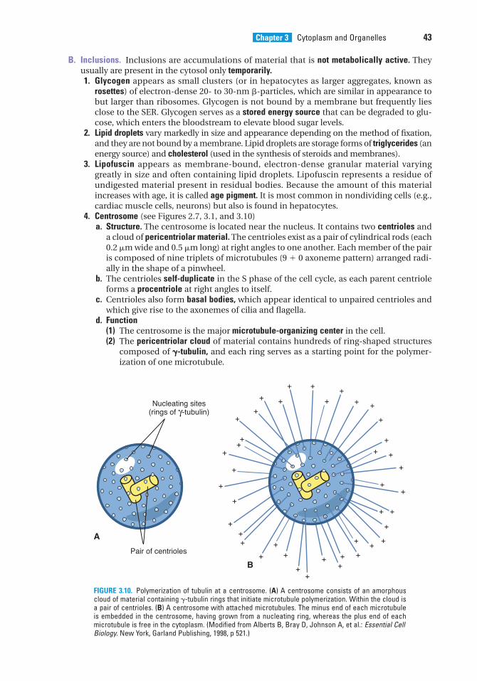
Chapter 3 Cytoplasm and Organelles 43
B. Inclusions. Inclusions are accumulations of material that is not metabolically active. Theyusually are present in the cytosol only temporarily.1. Glycogen appears as small clusters (or in hepatocytes as larger aggregates, known as
rosettes) of electron-dense 20- to 30-nm �-particles, which are similar in appearance tobut larger than ribosomes. Glycogen is not bound by a membrane but frequently liesclose to the SER. Glycogen serves as a stored energy source that can be degraded to glu-cose, which enters the bloodstream to elevate blood sugar levels.
2. Lipid droplets vary markedly in size and appearance depending on the method of fixation,and they are not bound by a membrane. Lipid droplets are storage forms of triglycerides (anenergy source) and cholesterol (used in the synthesis of steroids and membranes).
3. Lipofuscin appears as membrane-bound, electron-dense granular material varyinggreatly in size and often containing lipid droplets. Lipofuscin represents a residue ofundigested material present in residual bodies. Because the amount of this materialincreases with age, it is called age pigment. It is most common in nondividing cells (e.g.,cardiac muscle cells, neurons) but also is found in hepatocytes.
4. Centrosome (see Figures 2.7, 3.1, and 3.10)a. Structure. The centrosome is located near the nucleus. It contains two centrioles and
a cloud of pericentriolar material. The centrioles exist as a pair of cylindrical rods (each0.2 �m wide and 0.5 �m long) at right angles to one another. Each member of the pairis composed of nine triplets of microtubules (9 � 0 axoneme pattern) arranged radi-ally in the shape of a pinwheel.
b. The centrioles self-duplicate in the S phase of the cell cycle, as each parent centrioleforms a procentriole at right angles to itself.
c. Centrioles also form basal bodies, which appear identical to unpaired centrioles andwhich give rise to the axonemes of cilia and flagella.
d. Function(1) The centrosome is the major microtubule-organizing center in the cell.(2) The pericentriolar cloud of material contains hundreds of ring-shaped structures
composed of �-tubulin, and each ring serves as a starting point for the polymer-ization of one microtubule.
Nucleating sites(rings of γ-tubulin)
Pair of centrioles
++
+ +
+ +
++
+ +
+
+
+ +
++
+
+
+
+
+++
+
++
++
+++ +
++
+
+ +
++
+
+A
B
FIGURE 3.10. Polymerization of tubulin at a centrosome. (A) A centrosome consists of an amorphouscloud of material containing �-tubulin rings that initiate microtubule polymerization. Within the cloud isa pair of centrioles. (B) A centrosome with attached microtubules. The minus end of each microtubuleis embedded in the centrosome, having grown from a nucleating ring, whereas the plus end of eachmicrotubule is free in the cytoplasm. (Modified from Alberts B, Bray D, Johnson A, et al.: Essential CellBiology. New York, Garland Publishing, 1998, p 521.)
LWBK615-c03[32-55].qxd 05/31/2010 10:30 AM Page 43 Aptara
44 BRS Cell Biology and Histology
(3) Centrioles play no role in nucleating microtubules, but they help to maintain theorganization of the centrosome.
(4) The centrosome itself is also duplicated during interphase (S phase), and thenseparates to form the poles of the mitotic spindle, where microtubules originateand converge.
C. Cytoskeleton. The cytoskeleton is the structural framework within the cytosol. It functions inmaintaining cell shape, stabilizing cell attachments, facilitating endocytosis and exocytosis,and promoting cell movement. It includes the following major components:1. Microtubules
a. Structure. Microtubules are straight, hollow tubules 25 nm in diameter and made oftubulin. They have a rigid wall composed of 13 protofilaments, each of which consistsof a linear arrangement of tubulin dimers; each dimer consists of nonidentical �- and�-tubulin subunits.
b. Microtubules are polar, with polymerization (assembly) and depolymerization (disas-sembly) occurring preferentially at the plus end when GTP is bound to tubulin dimers.
c. Microtubules have microtubule-associated proteins (MAPs), which stabilize them andbind them to other cytoskeletal components and organelles. They also are associatedwith kinesin and cytoplasmic dynein, two force-generating proteins, which serve asmotors for vesicle or organelle movement. Kinesin moves cargo toward the plus end ofthe microtubule (outward), whereas cytoplasmic dynein moves it toward the minus end(inward).
d. Function. Microtubules maintain cell shape; aid in the transport of macromoleculeswithin the cytosol; assemble into the mitotic spindle during mitosis and ensure thecorrect distribution of chromosomes to daughter cells; and assist in the formation ofcell appendages called cilia and flagella, which beat rhythmically and precisely.
2. Actin filaments (microfilaments)a. Structure. Actin filaments measure 7 nm in diameter and are composed of globular
actin monomers (G actin) linked into a double helix (F actin). They are thin, flexible,and abundant in cells.
b. Actin filaments display polarity similar to that of microtubules; that is, their polymer-ization and depolymerization occur preferentially at the plus end when ATP is boundby G actin.
c. Many actin-binding proteins associate with G actin and modify their properties.d. Actin filaments are abundant at the periphery of the cell, where they are anchored to
the plasma membrane via one or more intermediary proteins (e.g., �-actinin, vin-culin, and talin).
e. Function. Actin filaments play a role in many cellular processes, such as establishingfocal contacts between the cell and the extracellular matrix, locomotion of nonmus-cle cells, formation of the contractile ring (in dividing cells), and the folding of epithe-lia into tubes during development.
3. Intermediate filaments are 8 to 10 nm in diameter. They constitute a population of hetero-geneous filaments that includes keratin, vimentin, desmin, glial fibrillary acidic protein(GFAP), lamins, and neurofilaments (Table 3.2). (Desmin and GFAP sometimes copoly-merize with vimentin and may be categorized as vimentin-like filaments.) In general,intermediate filaments provide mechanical strength to cells. They lack polarity and do notrequire GTP or ATP for assembly, which occurs along the entire length of the filament.
Tumor diagnosis is often based on immunocytochemical identification of the intermediate filaments in the tumor cells because the type of
intermediate filament present identifies the tissue from which the metastatic cancer cells originated.For example, an undifferentiated tumor that has metastasized to the bladder can be identified as acarcinoma (of epithelial origin) if it stains immunocytochemically in a histological section afterapplying a cytokeratin antibody.
CLINICAL
CONSIDERATIONS
LWBK615-c03[32-55].qxd 05/31/2010 10:31 AM Page 44 Aptara
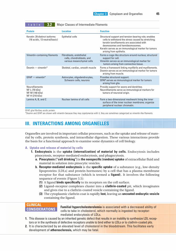
Chapter 3 Cytoplasm and Organelles 45
III. INTERACTIONS AMONG ORGANELLES
Organelles are involved in important cellular processes, such as the uptake and release of mate-rial by cells, protein synthesis, and intracellular digestion. These various interactions providethe basis for a functional approach to examine some dynamics of cell biology.
A. Uptake and release of material by cells1. Endocytosis is the uptake (internalization) of material by cells. Endocytosis includes
pinocytosis, receptor-mediated endocytosis, and phagocytosis.a. Pinocytosis (“cell drinking”) is the nonspecific (random) uptake of extracellular fluid and
material in solution into pinocytic vesicles.b. Receptor-mediated endocytosis is the specific uptake of a substance (e.g., low-density
lipoproteins [LDLs] and protein hormones) by a cell that has a plasma membranereceptor for that substance (which is termed a ligand). It involves the followingsequence of events (Figure 3.5):(1) A ligand binds specifically to its receptors on the cell surface.(2) Ligand–receptor complexes cluster into a clathrin-coated pit, which invaginates
and gives rise to a clathrin-coated vesicle containing the ligand.(3) The cytoplasmic clathrin coat is rapidly lost, leaving an uncoated endocytic vesicle
containing the ligand.
t a b l e 3.2 Major Classes of Intermediate Filaments
Protein Location Function
Keratin: 29 distinct isoforms Epithelial cells Structural support and tension-bearing role; enables (16 acidic, 13 neutral/basic) cells to withstand the stress caused by stretching;
keratin tonofilaments are associated with desmosomes and hemidesmosomes.
Keratin serves as an immunological marker for tumors arising from epithelia.
Vimentin-containing filaments Fibroblasts, endothelial Forms a cage-like structure around nucleus; structural cells, chondroblasts, and support for cell.various mesenchymal cells Vimentin serves as an immunological marker for
tumors arising from connective tissue.
Desmin � vimentin* Skeletal, cardiac, smooth muscle Forms a framework linking myofibrils and myofilaments.Desmin serves as an immunological marker for tumors
arising from muscle.
GFAP � vimentin* Astrocytes, oligodendrocytes, Provides structural support.Schwann cells, neurons GFAP serves as immunological marker for tumors
arising from glia.
Neurofilaments Provide support for axons and dendrites.NF-L (70 kDa) Neurofilaments serve as immunological markers forNF-M (140 kDa) tumors of neuronal origin.NF-H (210 kDa)
Lamina A, B, and C Nuclear lamina of all cells Form a two-dimensional meshwork lining the inner surface of the inner nuclear membrane; organize peripheral nuclear chromatin.
GFAP, glial fibrillary acidic protein.*Desmin and GFAP are shown with vimentin because they may copolymerize with it; they are sometimes categorized as vimentin-like filaments.
Familial hypercholesterolemia is associated with a decreased ability ofcells to take in cholesterol, which normally is ingested by receptor-mediated endocytosis of LDLs.
1. This disease is caused by an inherited genetic defect that results in an inability to synthesize LDL recep-tors or in the synthesis of defective receptors unable to bind either to LDLs or to clathrin-coated pits.
2. It is characterized by an elevated level of cholesterol in the bloodstream. This facilitates earlydevelopment of atherosclerosis, which may be fatal.
CLINICAL
CONSIDERATIONS
LWBK615-c03[32-55].qxd 05/31/2010 10:31 AM Page 45 Aptara
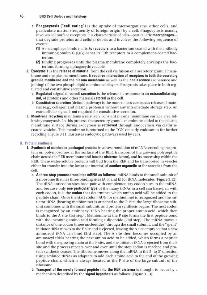
46 BRS Cell Biology and Histology
c. Phagocytosis (“cell eating”) is the uptake of microorganisms, other cells, andparticulate matter (frequently of foreign origin) by a cell. Phagocytosis usuallyinvolves cell surface receptors. It is characteristic of cells—particularly macrophages—that degrade proteins and cellular debris and involves the following sequence ofevents:(1) A macrophage binds via its Fc receptors to a bacterium coated with the antibody
immunoglobulin G (IgG) or via its C3b receptors to a complement-coated bac-terium.
(2) Binding progresses until the plasma membrane completely envelops the bac-terium, forming a phagocytic vacuole.
2. Exocytosis is the release of material from the cell via fusion of a secretory granule mem-brane and the plasma membrane. It requires interaction of receptors in both the secretorygranule membrane and the plasma membrane as well as the coalescence (adherence andjoining) of the two phospholipid membrane bilayers. Exocytosis takes place in both reg-ulated and constitutive secretion.a. Regulated (signal directed) secretion is the release, in response to an extracellular sig-
nal, of proteins and other materials stored in the cell.b. Constitutive secretion (default pathway) is the more or less continuous release of mate-
rial (e.g., collagen and plasma proteins) without any intermediate storage step. Anextracellular signal is not required for constitutive secretion.
3. Membrane recycling maintains a relatively constant plasma membrane surface area fol-lowing exocytosis. In this process, the secretory granule membrane added to the plasmamembrane surface during exocytosis is retrieved through endocytosis via clathrin-coated vesicles. This membrane is returned to the TGN via early endosomes for furtherrecycling. Figure 3.11 illustrates endocytic pathways used by cells.
B. Protein synthesis1. Synthesis of membrane-packaged proteins involves translation of mRNAs encoding the pro-
tein on polyribosomes at the surface of the RER, transport of the growing polypeptidechain across the RER membrane and into the cisterna (lumen), and its processing within theRER. These water-soluble proteins will bud from the RER and be transported in vesicleseither for transfer into the lumen (or interior) of another organelle or for secretion from thecell.a. A three-step process translates mRNA as follows: mRNA binds to the small subunit of
a ribosome that has three binding sites (A, P, and E) for tRNA molecules (Figure 3.12).The tRNA anticodon sites base-pair with complementary codon sites in the mRNA,and because only one particular type of the many tRNAs in a cell can base-pair witheach codon, it is the codon that determines which amino acid will be added to thepeptide chain. Once the start codon (AUG for methionine) is recognized and the ini-tiator tRNA (bearing methionine) is attached to the P site, the large ribosome sub-unit combines with the small subunit, and protein synthesis begins. The next codonis recognized by an aminoacyl tRNA bearing the proper amino acid, which thenbinds to the A site (1st step). Methionine at the P site forms the first peptide bondwith the incoming amino acid forming a dipeptide (2nd step). The mRNA moves adistance of one codon (three nucleotides) through the small subunit, and the “spent”initiator tRNA moves to the E site and is ejected, leaving the A site empty so that a newaminoacyl tRNA can bind (3rd step). The A site then becomes occupied by anaminoacyl tRNA bearing the next amino acid to be added, which forms a peptidebond with the growing chain at the P site, and the initiator tRNA is ejected from the Esite and the process repeats over and over until the stop codon is reached and pro-tein synthesis ceases. The ribosome moves along the mRNA in the 5� to 3� directionusing acylated tRNAs as adapters to add each amino acid to the end of the growingpeptide chain, which is always located at the P site of the large subunit of theribosome.
b. Transport of the newly formed peptide into the RER cisterna is thought to occur by amechanism described by the signal hypothesis as follows (Figure 3.13):
LWBK615-c03[32-55].qxd 05/31/2010 10:31 AM Page 46 Aptara
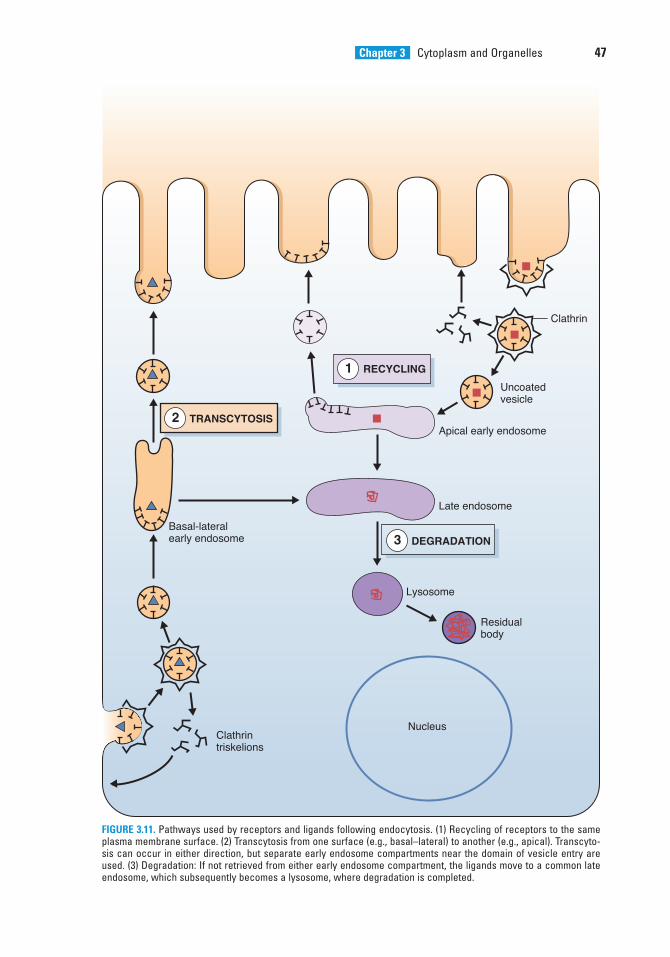
Chapter 3 Cytoplasm and Organelles 47
Residualbody
Lysosome
Late endosome
Basal-lateralearly endosome
Apical early endosome
Clathrin
Uncoatedvesicle
Clathrintriskelions
Nucleus
RECYCLING1
TRANSCYTOSIS2
DEGRADATION3
FIGURE 3.11. Pathways used by receptors and ligands following endocytosis. (1) Recycling of receptors to the sameplasma membrane surface. (2) Transcytosis from one surface (e.g., basal–lateral) to another (e.g., apical). Transcyto-sis can occur in either direction, but separate early endosome compartments near the domain of vesicle entry areused. (3) Degradation: If not retrieved from either early endosome compartment, the ligands move to a common lateendosome, which subsequently becomes a lysosome, where degradation is completed.
LWBK615-c03[32-55].qxd 05/31/2010 10:31 AM Page 47 Aptara
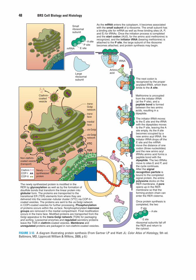
48 BRS Cell Biology and Histology
mRNA
A site
tRNA
Smallribosomalsubunit
As the mRNA enters the cytoplasm, it becomes associatedwith the small subunit of a ribosome. The small subunit has a binding site for mRNA as well as three binding sites (A, P,and E) for tRNAs. Once the initiation process is completed and the start codon (AUG, for the amino acid methionine) is recognized, and the initiator tRNA (bearing methionine) is attached to the P site, the large subunit of the ribosomebecomes attached, and protein synthesis may begin.
The next codon is recognized by the proper acylated tRNA, which then binds to the A site.
Methionine is uncoupled from the initiator tRNA (at the P site), and a peptide bond is formed between the two amino acids, resulting in a dipeptide.
After the signal recognition particle is bound to the completed signal protein, the entire polysome docks on the RER membrane. A pore opens up in the RER membrane so that the forming protein chain can enter the RER cisterna.
Once protein synthesis is completed, the two
ribosomal subunits fall off the RER and return to the cytosol.
Largeribosomalsubunit
Clathrin-coatedvesicle
Non clathrin-coated vesicle(transport)
Newlysynthesizedprotein
ClathrinCOP-ICOP-II
trans-Golginetwork
transface
medialface
cis Golginetwork
VTC
TER
RER
P siteE site
The newly synthesized protein is modified in the RER by glycosylation as well as by the formation of disulfide bonds that transform the linear protein into globular form. The proteins are transported to the transitional ER (TER) elements from where they are delivered into the vesicular–tubular cluster (VTC) via COP-II–coated vesicles. The proteins are sent to the cis Golgi networkin COPI-coated vesicles for further processing. Phosphorylationof proteins occurs within the cis face. Nonphosphorylated mannosegroups are removed in the medial compartment. Final modificationoccurs in the trans face. Modified proteins are transported from theGolgi apparatus to the trans-Golgi network (TGN) for packagingand sorting. Lysosomal enzymes and regulated secretory proteins leave the TGN in clathrin-coated vesicles. Membrane and unregulated proteins are packaged in non-clathrin-coated vesicles.
Aminoacid
cis Golgi
The initiator tRNA moves to the E site and the tRNA with the dipeptides moves to the P site, leaving the A site empty. As the A site becomes occupied by a new amino acyl tRNA, the initiator tRNA drops off theE site and the mRNA move the distance of one codon (three nucleotides) and the new amino acyl tRNAs amino acid forms a peptide bond with the dipeptide. The two tRNAsmove to sites E and P, and the cycle continues.
P siteA site
E site
FIGURE 3.12. A diagram illustrating protein synthesis (From Gartner LP and Hiatt JL: Color Atlas of Histology, 5th ed.Baltimore, MD, Lippincott William & Wilkins, 2009, p 8.)
LWBK615-c03[32-55].qxd 05/31/2010 10:31 AM Page 48 Aptara
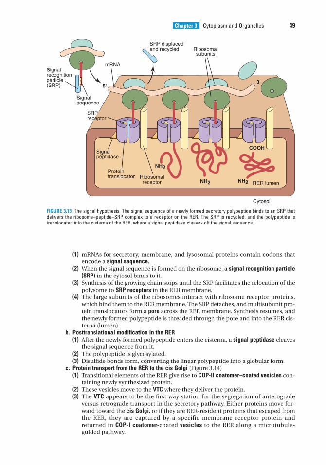
Chapter 3 Cytoplasm and Organelles 49
(1) mRNAs for secretory, membrane, and lysosomal proteins contain codons thatencode a signal sequence.
(2) When the signal sequence is formed on the ribosome, a signal recognition particle(SRP) in the cytosol binds to it.
(3) Synthesis of the growing chain stops until the SRP facilitates the relocation of thepolysome to SRP receptors in the RER membrane.
(4) The large subunits of the ribosomes interact with ribosome receptor proteins,which bind them to the RER membrane. The SRP detaches, and multisubunit pro-tein translocators form a pore across the RER membrane. Synthesis resumes, andthe newly formed polypeptide is threaded through the pore and into the RER cis-terna (lumen).
b. Posttranslational modification in the RER(1) After the newly formed polypeptide enters the cisterna, a signal peptidase cleaves
the signal sequence from it.(2) The polypeptide is glycosylated.(3) Disulfide bonds form, converting the linear polypeptide into a globular form.
c. Protein transport from the RER to the cis Golgi (Figure 3.14) (1) Transitional elements of the RER give rise to COP-II coatomer–coated vesicles con-
taining newly synthesized protein.(2) These vesicles move to the VTC where they deliver the protein. (3) The VTC appears to be the first way station for the segregation of anterograde
versus retrograde transport in the secretory pathway. Either proteins move for-ward toward the cis Golgi, or if they are RER-resident proteins that escaped fromthe RER, they are captured by a specific membrane receptor protein andreturned in COP-I coatomer-coated vesicles to the RER along a microtubule-guided pathway.
NH2
NH2 NH2
COOH
RER lumen
Cytosol
5'3'
SRP displacedand recycled
Signal sequence
Signal peptidase
Signal recognitionparticle(SRP)
mRNA
Ribosomalreceptor
Proteintranslocator
Ribosomalsubunits
SRP receptor
FIGURE 3.13. The signal hypothesis. The signal sequence of a newly formed secretory polypeptide binds to an SRP thatdelivers the ribosome–peptide–SRP complex to a receptor on the RER. The SRP is recycled, and the polypeptide istranslocated into the cisterna of the RER, where a signal peptidase cleaves off the signal sequence.
LWBK615-c03[32-55].qxd 05/31/2010 10:31 AM Page 49 Aptara
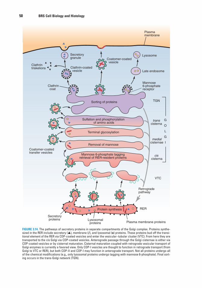
50 BRS Cell Biology and Histology
Sulfation and phosphorylation of amino acids
Terminal glycosylation
Removal of mannose
Plasma membrane
Mannose 6-phosphate taggingretrieval of RER-resident proteins
Protein synthesis
Lysosomal proteins
Secretory proteins
Plasma membrane proteins
Coatomer-coatedtransfer vesicles
Sorting of proteins
Clathrin coat
Clathrin triskelions Clathrin-coated
vesicle
Coatomer-coated vesicle
Lysosome
Mannose6-phosphatereceptor
Secretorygranule
cis cisterna
RER
TGN
transcisterna
medialcisternae
G
O
L
G
I
Retrogradepathway
Late endosome
VTC
FIGURE 3.14. The pathways of secretory proteins in separate compartments of the Golgi complex. Proteins synthe-sized in the RER include secretory (�), membrane (/), and lysosomal (o) proteins. These proteins bud off the transi-tional element of the RER via COP-coated vesicles and enter the vesicular–tubular cluster (VTC). From here they aretransported to the cis Golgi via COP-coated vesicles. Anterograde passage through the Golgi cisternae is either viaCOP-coated vesicles or by cisternal maturation. Cisternal maturation coupled with retrograde vesicular transport ofGolgi enzymes is currently a favored view. Only COP-I vesicles are thought to function in retrograde transport (fromGolgi to VTC or RER), but both COP-II and COP-I may function in anterograde transport. Not all proteins undergo allof the chemical modifications (e.g., only lysosomal proteins undergo tagging with mannose 6-phosphate). Final sort-ing occurs in the trans-Golgi network (TGN).
LWBK615-c03[32-55].qxd 05/31/2010 10:31 AM Page 50 Aptara
Chapter 3 Cytoplasm and Organelles 51
d. Anterograde transport from the VTC to the cis Golgi is via COP-II coatomer–coated vesi-cles.
e. Movement of material anterograde among the Golgi subcompartments may occur by cis-ternal maturation and/or by vesicular transport, as follows:(1) Cisternae containing proteins may change in biochemical composition as they
move intact across the stack.(2) COP-II–coated vesicles may bud off one cisterna and fuse with the dilated rim of
another cisterna.(3) Although both mechanisms have been observed, the precise way that anterograde
transport occurs across the Golgi stack of cisternae is unresolved.(4) Retrograde vesicular transport occurs between Golgi cisternae and between the
Golgi and the VTC or RER via COP-I–coated vesicles. f. Protein processing in the Golgi complex (Figure 3.14) occurs as proteins move from the
cis to the trans face of the Golgi complex through distinct cisternal subcompartments.Protein processing may include the following events, each of which occurs in a dif-ferent cisternal subcompartment:(1) Proteins targeted for lysosomes are tagged with mannose 6-phosphate in the cis
cisterna.(2) Mannose residues are removed in cis and medial cisternae.(3) Some proteins undergo terminal glycosylation with sialic acid residues and galac-
tose.(4) Sulfation and phosphorylation of amino acid residues take place.(5) A membrane similar in composition and thickness to the plasma membrane is
acquired.g. Sorting of proteins in TGN (Figure 3.14)
(1) Regulated secretory proteins are sorted from membrane and lysosomal proteinsand delivered via clathrin-coated vesicles to condensing vacuoles, in whichremoval of water via ionic exchanges yields secretory granules.
(2) Lysosomal proteins are sorted into clathrin-coated regions of the TGN that havereceptors for mannose 6-phosphate and are delivered to late endosomes viaclathrin-coated vesicles.
(3) Plasma membrane proteins are sorted into coatomer-coated regions of the TGNand delivered to the plasma membrane in COP-II coatomer–coated vesicles.
2. Synthesis of transmembrane proteins also takes place on polyribosomes at the surface ofthe RER, but rather than entering the lumen, the transfer process is halted (by a stop-transfer sequence), and the transmembrane protein becomes anchored in the RER mem-brane. The ultimate destination of this protein will be the RER membrane, the mem-brane of another organelle, or the plasma membrane.
3. Synthesis of cytosolic proteins takes place on polyribosomes lying free in the cytosol andis directed by mRNAs that lack signal codons. Such proteins (e.g., protein kinase andhemoglobin) are released directly into the cytosol.
C. Intracellular digestion1. Nonlysosomal digestion is the degradation of cytosolic constituents by mechanisms out-
side of the vacuolar lysosomal pathway. The major site for the degradation of unwantedproteins is the proteosome, a cylindrical complex of nonlysosomal proteases. Proteinsmarked for destruction are enzymatically tagged with ubiquitin, which delivers them tothe proteosome, where they are broken down to small peptides.
2. Lysosomal digestion (Figure 3.8) is the degradation of material within various types oflysosomes by lysosomal enzymes. Different lysosomal compartments are involved,depending on the origin of the material to be degraded.a. Heterophagy is the ingestion and degradation of foreign material taken into the cell by
receptor-mediated endocytosis or phagocytosis.(1) Digestion of endocytosed ligands occurs in multivesicular bodies (Figure 3.8). (2) Digestion of phagocytosed microorganisms and foreign particles begins and may
be completed in phagolysosomes.
LWBK615-c03[32-55].qxd 05/31/2010 10:31 AM Page 51 Aptara
52 BRS Cell Biology and Histology
b. Autophagy is the segregation of an organelle or other cell constituents within mem-branes from the RER to form an autophagic vacuole (Figure 3.9), which is subse-quently digested in an autophagolysosome.
c. Crinophagy is the fusion of hormone secretory granules and lysosomes and their sub-sequent digestion. Crinophagy is used to remove excess numbers of secretory gran-ules from the cell.
Lysosomal storage diseases are hereditary disorders caused by adeficiency in specific lysosomal acid hydrolases. Therefore, lysosomes
are unable to degrade certain compounds, which accumulate and interfere with cell function.1. Tay-Sachs disease is characterized by glycolipids (namely, GM2 gangliosides) accumulating in the
lysosomes of neurons as a result of a deficiency of the enzyme hexosaminidase A. The disease ismost common in children of central European Jewish descent. The large buildup of gangliosidesin neurons of the brain results in marked degenerative changes in the central nervous system,and death usually occurs before the age of 4 years.
2. A hallmark of Hurler syndrome is that glycosaminoglycans (GAGs) and mucopolysaccharidesaccumulate in the heart, brain, liver, and other organs. This rare inherited disease is caused by adeficiency in any 1 of 10 lysosomal enzymes involved in the sequential degradation of GAGs. Clini-cal features of Hurler syndrome include skeletal deformities, enlarged organs, progressive mentaldeterioration, deafness, and death before the age of 10 years.
3. Glycogen storage diseases are caused by a hereditary defect in an enzyme involved in either thesynthesis or the degradation of glycogen. As a result, glycogen accumulates most often in theliver, skeletal muscle, and the heart, but the major organ involvement depends on the particularenzymatic defect. There are at least 10 distinct glycogen storage diseases.
CLINICAL
CONSIDERATIONS
LWBK615-c03[32-55].qxd 05/31/2010 10:31 AM Page 52 Aptara
1. Which of the following organelles dividesby fission?
(A) Golgi complex(B) RER(C) Peroxisome(D) SER(E) Centriole
2. A 30-year-old man with very high bloodcholesterol levels (290 mg/dL) has beendiagnosed with premature atherosclerosis.His father died of a heart attack at age 45,and his mother, aged 44, has coronary arterydisease. Which of the following is the mostlikely explanation of his condition?
(A) He has a lysosomal storage disease andcannot digest cholesterol.
(B) He has a peroxisomal disorder and pro-duces low levels of hydrogen peroxide.
(C) The SER in his hepatocytes has prolifer-ated and produced excessive amounts ofcholesterol.
(D) He has a genetic disorder andsynthesizes defective LDL receptors.
(E) He is unable to manufacture endosomes.
3. Movement of protein from the RER to theVTC takes place in which of the followingcell components?
(A) A caveolin-coated vesicle(B) A clathrin-coated vesicle(C) A coatomer I–coated vesicle(D) A coatomer II–coated vesicle(E) An early endosome
4. The retrieval of secretion granulemembrane immediately after exocytosisoccurs in which of the following cell compo-nents?
(A) A caveolin-coated vesicle(B) A clathrin-coated vesicle(C) A coatomer I–coated vesicle(D) A coatomer II–coated vesicle(E) An early endosome
5. Movement of protein from trans to cisGolgi cisternae occurs in which of thefollowing cell components?
(A) A caveolin-coated vesicle(B) A clathrin-coated vesicle(C) A coatomer I–coated vesicle(D) A coatomer II–coated vesicle(E) An early endosome
6. Uncoupling of endocytosed ligands fromreceptors takes place in which of the follow-ing cell components?
(A) A caveolin-coated vesicle(B) A clathrin-coated vesicle(C) A coatomer I–coated vesicle(D) A coatomer II–coated vesicle(E) An early endosome
7. Movement of acid hydrolases from thetrans-Golgi network to a late endosometakes place in which of the following cellcomponents?
(A) A caveolin-coated vesicle(B) A clathrin-coated vesicle(C) A coatomer I–coated vesicle(D) A coatomer II–coated vesicle(E) An early endosome
8. Which of the following cytoskeletal com-ponents is associated with kinesin?
(A) Keratin(B) Lamin A(C) Microfilament(D) Microtubule(E) Neurofilament
Review Test
Directions: Each of the numbered items or incomplete statements in this section is followed byanswers or by completions of the statement. Select the ONE lettered answer or completion thatis BEST in each case.
53
LWBK615-c03[32-55].qxd 05/31/2010 10:31 AM Page 53 Aptara
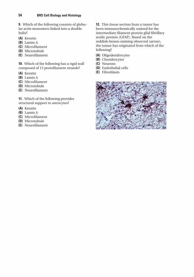
54 BRS Cell Biology and Histology
9. Which of the following consists of globu-lar actin monomers linked into a doublehelix?
(A) Keratin(B) Lamin A(C) Microfilament(D) Microtubule(E) Neurofilament
10. Which of the following has a rigid wallcomposed of 13 protofilament strands?
(A) Keratin(B) Lamin A(C) Microfilament(D) Microtubule(E) Neurofilament
11. Which of the following providesstructural support to astrocytes?
(A) Keratin(B) Lamin A(C) Microfilament(D) Microtubule(E) Neurofilament
12. This tissue section from a tumor hasbeen immunochemically stained for theintermediate filament protein glial fibrillaryacidic protein (GFAP). Based on thereddish-brown staining observed (arrow),the tumor has originated from which of thefollowing?
(A) Oligodendrocytes(B) Chondrocytes(C) Neurons(D) Endothelial cells(E) Fibroblasts
LWBK615-c03[32-55].qxd 05/31/2010 10:31 AM Page 54 Aptara
Answers and Explanations
1. C. A peroxisome originates from preexisting peroxisomes. It imports specific cytosolic pro-teins and then undergoes fission. The other organelle that divides by fission is the mito-chondrion (see Chapter 3 II A 10).
2. D. Cells import cholesterol by the receptor-mediated uptake of LDLs in coated vesicles.Certain individuals inherit defective genes and cannot make LDL receptors, or they makedefective receptors that cannot bind to clathrin-coated pits. The result is an inability tointernalize LDLs, which leads to high levels of LDLs in the bloodstream. High LDL levelspredispose a person to premature atherosclerosis and increase the risk of heart attacks (seeChapter 3 III A 1 Clinical Considerations).
3. D. Transport of protein from the RER to the VTC occurs via (COP-II) coatomer-coated vesi-cles (see Chapter 3 III B c).
4. B. Membrane recycling after exocytosis of the contents of a secretion granule occurs viaclathrin-coated vesicles (see Chapter 3 III A 3).
5. C. Transfer of material among the cisternae of the Golgi complex in a retrograde directiontakes place via (COP-I) coatomer-coated vesicles (see Chapter 3 III B e).
6. E. The uncoupling of ligands and receptors internalized by receptor-mediated endocytosisoccurs in the early endosome (see Chapter 3 II A 9 b).
7. B. Proteins targeted for lysosomes (via late endosomes) leave the trans-Golgi network inclathrin-coated vesicles (see Chapter 3 II A 8 a).
8. D. Kinesin is a force-generating protein associated with microtubules. It serves as a molec-ular motor for the transport of organelles and vesicles outward, away from the centrosome(see Chapter 3 II C 1).
9. C. Globular actin monomers (G actin) polymerize into a double helix of filamentous actin(F actin), also called a microfilament, in response to the regulatory influence of a numberof actin-binding proteins (see Chapter 3 II C 2).
10. D. A microtubule consists of �- and �-tubulin dimers polymerized into a spiral around ahollow lumen to form a fairly rigid tubule. When cross-sectioned, the microtubule reveals13 protofilament strands, which represent the tubulin dimers present in one completeturn of the spiral (see Chapter 3 II C 1).
11. E. Glial filaments are a type of intermediate filament composed of glial fibrillary acidicprotein and present in fibrous astrocytes. These filaments are supportive, but they mayplay additional roles in both normal and pathologic processes in the central nervous sys-tem (see Chapter 3 II C 3).
12. A. The intermediate filament protein glial fibrillary acidic protein (GFAP) is present inglial cells, including microglia, oligodendrocytes, fibrous astrocytes, and Schwann cells.Vimentin is found in cells of connective tissue origin, which include fibroblasts, chondro-cytes, and endothelial cells. Neurons contain intermediate neurofilaments, which wouldnot stain for either GFAP or vimentin (Chapter 3 II C 3 Clinical Considerations).
55
LWBK615-c03[32-55].qxd 05/31/2010 10:31 AM Page 55 Aptara
c h a p t e r 4 Extracellular Matrix
56
I. OVERVIEW—THE EXTRACELLULAR MATRIX
A. Structure. The extracellular matrix is an organized meshwork of macromolecules surroundingand underlying cells. Although it varies in composition, in general it consists of an amor-phous ground substance (containing primarily glycosaminoglycans [GAGs], proteoglycans,and glycoproteins) and fibers (Figure 4.1).
B. Functions. The extracellular matrix, along with water and other small molecules (e.g., nutri-ents, ions), constitutes the extracellular environment. By affecting the metabolic activities ofcells in contact with it, the extracellular matrix may alter the cells and influence their shape,migration, division, and differentiation.
II. GROUND SUBSTANCE
A. GAGs are long, unbranched polysaccharides composed of repeating identical disaccharide units.1. An amino sugar, either N-acetylglucosamine or N-acetylgalactosamine, is always one of
the repeating disaccharides.2. Because GAGs are commonly sulfated and usually possess a uronic acid sugar, which has
a carboxyl group in the repeating disaccharide unit, they have a strong negative charge.3. GAGs are generally linked to a core protein.4. The attraction of osmotically active cations (e.g., Na�) to GAGs results in a heavily
hydrated matrix that strongly resists compression.5. Their extended random coils occupy large volumes of space because they do not fold com-
pactly.6. GAGs may be classified into four main groups on the basis of their chemical structure
(Table 4.1).a. Hyaluronic acid is a very large unsulfated molecule up to 20 �m in length that is not
attached to a core protein.b. The other three GAG groups are chondroitin sulfate and dermatan sulfate, heparin and
heparan sulfate, and keratan sulfate.
Taking the popular dietary supplement chondroitin sulfate and/or glucosamine has been reported to reduce the risk of osteoarthritis, joint
degeneration, and cartilage deterioration, but the findings are equivocal. In a recent large-scalestudy, patients with knee osteoarthritis experienced significantly less pain and increased functionwhile taking these compounds when compared with patients on placebo, but radiographic evidencefrom examining knee joint degeneration was complicated by the fact that the placebo group had asmaller loss of cartilage than had been anticipated on the basis of prior research results, so nodefinite conclusions could be reached.
CLINICAL
CONSIDERATIONS
LWBK615-c04[56-66].qxd 05/31/2010 10:23 AM Page 56 Aptara
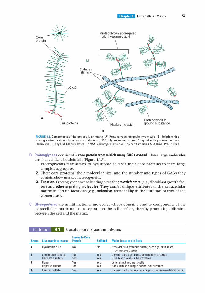
Chapter 4 Extracellular Matrix 57
Proteoglycan aggregated with hyaluronic acid
Hyaluronic acid
Collagen fibrils
Coreprotein
Link proteinsProteoglycan inground substance
GAG
B
A
FIGURE 4.1. Components of the extracellular matrix. (A) Proteoglycan molecule, two views. (B) Relationshipsamong various extracellular matrix molecules. GAG, glycosaminoglycan. (Adapted with permission fromHenrikson RC, Kaye GI, Mazurkiewicz JE: NMS Histology. Baltimore, Lippincott Williams & Wilkins, 1997, p 104.)
B. Proteoglycans consist of a core protein from which many GAGs extend. These large moleculesare shaped like a bottlebrush (Figure 4.1A).1. Proteoglycans may attach to hyaluronic acid via their core proteins to form large
complex aggregates.2. Their core proteins, their molecular size, and the number and types of GAGs they
contain show marked heterogeneity.3. Function. Proteoglycans act as binding sites for growth factors (e.g., fibroblast growth fac-
tor) and other signaling molecules. They confer unique attributes to the extracellularmatrix in certain locations (e.g., selective permeability in the filtration barrier of theglomerulus).
C. Glycoproteins are multifunctional molecules whose domains bind to components of theextracellular matrix and to receptors on the cell surface, thereby promoting adhesionbetween the cell and the matrix.
t a b l e 4.1 Classification of Glycosaminoglycans
Linked to Core Group Glycosaminoglycans Protein Sulfated Major Locations in Body
I Hyaluronic acid No No Synovial fluid, vitreous humor, cartilage, skin, most connective tissues
II Chondroitin sulfate Yes Yes Cornea, cartilage, bone, adventitia of arteriesDermatan sulfate Yes Yes Skin, blood vessels, heart valves
III Heparin Yes Yes Lung, skin, liver, mast cellsHeparan sulfate Yes Yes Basal laminae, lung, arteries, cell surfaces
IV Keratan sulfate Yes Yes Cornea, cartilage, nucleus pulposus of intervertebral disks
LWBK615-c04[56-66].qxd 05/31/2010 10:23 AM Page 57 Aptara
58 BRS Cell Biology and Histology
1. Fibronectina. Types and location
(1) Matrix fibronectin, an adhesive glycoprotein, forms fibrils in the extracellularmatrix.
(2) Cell surface fibronectin is a protein that transiently attaches to the surface of cells.(3) Plasma fibronectin is a circulating plasma protein that functions in blood clotting,
wound healing, and phagocytosis.b. Function. Fibronectin is a multifunctional molecule.
(1) Fibronectin has domains for binding collagen, heparin, various cell-surface recep-tors, and cell adhesion molecules (CAMs).
(2) It mediates cell adhesion to the extracellular matrix by binding to fibronectin recep-tors on the cell surface.
Wound healing in adults involves the formation of fibronectin tracksalong which cells migrate to their destinations.
1. In connective tissue, wound healing is often characterized by migration of fibroblasts acrossblood clots, where they adhere to fibronectin.
2. In epithelia, wound healing involves reepithelialization, which depends on the basal laminaserving as a scaffold for cell migration to cover the denuded area; epithelial cell proliferation andreplacement then occur.
CLINICAL
CONSIDERATIONS
2. Laminin is located in basal laminae, where it is synthesized by adjacent epithelial cells,and in external laminae surrounding muscle cells and Schwann cells.a. The arms of this large cross-shaped glycoprotein have binding sites for cell surface
receptors (integrins), heparan sulfate, type IV collagen, and entactin.b. Function. Laminin mediates interaction between epithelial cells and the extracellular
matrix by anchoring the cell surface to the basal lamina.3. Entactin is a component of all basal (and external) laminae.
a. This sulfated adhesive glycoprotein binds laminin.b. Function. Entactin links laminin with type IV collagen in the lamina densa.
4. Tenascin is an adhesive glycoprotein most abundant in embryonic tissues.a. Tenascin is secreted by glial cells in the developing nervous system.b. Function. Tenascin promotes cell–matrix adhesion and thus plays a role in cell migration.
5. Chondronectin, a glycoprotein in cartilage, attaches chondrocytes to type II collagen.a. This multifunctional molecule has binding sites for collagen proteoglycans and cell
surface receptors.b. Function. By influencing the composition of its extracellular matrix, chondronectin
plays a role in the development and maintenance of cartilage.6. Osteonectin
a. This extracellular-matrix calcium-binding glycoprotein found in bone is synthesizedby osteoblasts.
b. It has binding sites for type I collagen and for integrins of osteoblasts and osteocytes.c. Function. Osteonectin plays a role in bone formation and remodeling and in main-
taining bone mass by influencing calcification.
D. Fibronectin receptors, which belong to the integrin family of receptors, are transmembraneproteins consisting of two polypeptide chains.1. Because they enable cells to adhere to the extracellular matrix, they are known as CAMs.2. They bind to fibronectin via a specific tripeptide sequence (Arg-Gly-Asp; RGD sequence);
other extracellular adhesive proteins also contain this sequence.3. Function. They link fibronectin outside the cell to cytoskeletal components (e.g., actin)
inside the cell (Figure 4.2) and may activate cell-signaling pathways which determine the
LWBK615-c04[56-66].qxd 05/31/2010 10:23 AM Page 58 Aptara
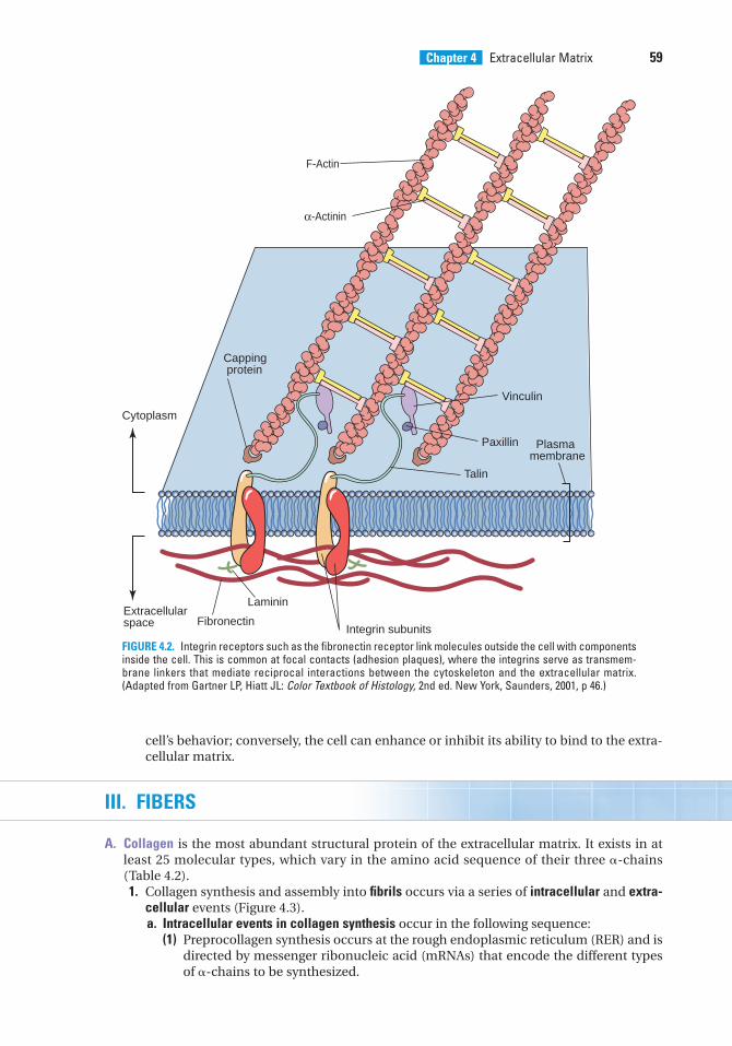
Chapter 4 Extracellular Matrix 59
cell’s behavior; conversely, the cell can enhance or inhibit its ability to bind to the extra-cellular matrix.
III. FIBERS
A. Collagen is the most abundant structural protein of the extracellular matrix. It exists in atleast 25 molecular types, which vary in the amino acid sequence of their three �-chains(Table 4.2).1. Collagen synthesis and assembly into fibrils occurs via a series of intracellular and extra-
cellular events (Figure 4.3).a. Intracellular events in collagen synthesis occur in the following sequence:
(1) Preprocollagen synthesis occurs at the rough endoplasmic reticulum (RER) and isdirected by messenger ribonucleic acid (mRNAs) that encode the different typesof �-chains to be synthesized.
Plasma membrane
Capping protein
Talin
FibronectinIntegrin subunits
Vinculin
Paxillin
Cytoplasm
Extracellularspace
F-Actin
α-Actinin
Laminin
FIGURE 4.2. Integrin receptors such as the fibronectin receptor link molecules outside the cell with componentsinside the cell. This is common at focal contacts (adhesion plaques), where the integrins serve as transmem-brane linkers that mediate reciprocal interactions between the cytoskeleton and the extracellular matrix.(Adapted from Gartner LP, Hiatt JL: Color Textbook of Histology, 2nd ed. New York, Saunders, 2001, p 46.)
LWBK615-c04[56-66].qxd 05/31/2010 10:23 AM Page 59 Aptara
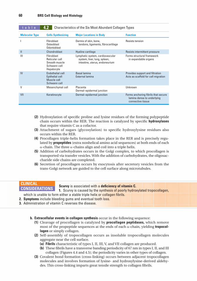
60 BRS Cell Biology and Histology
(2) Hydroxylation of specific proline and lysine residues of the forming polypeptidechain occurs within the RER. The reaction is catalyzed by specific hydroxylasesthat require vitamin C as a cofactor.
(3) Attachment of sugars (glycosylation) to specific hydroxylysine residues alsooccurs within the RER.
(4) Procollagen triple-helix formation takes place in the RER and is precisely regu-lated by propeptides (extra nonhelical amino acid sequences) at both ends of each�-chain. The three �-chains align and coil into a triple helix.
(5) Addition of carbohydrates occurs in the Golgi complex, to which procollagen istransported via transfer vesicles. With the addition of carbohydrates, the oligosac-charide side chains are completed.
(6) Secretion of procollagen occurs by exocytosis after secretory vesicles from thetrans-Golgi network are guided to the cell surface along microtubules.
t a b l e 4.2 Characteristics of the Six Most Abundant Collagen Types
Molecular Type Cells Synthesizing Major Locations in Body Function
I Fibroblast Dermis of skin, bone, Resists tensionOsteoblast tendons, ligaments, fibrocartilageOdontoblast
II Chondroblast Hyaline cartilage Resists intermittent pressure
III Fibroblast Lymphatic system, cardiovascular Forms structural frameworkReticular cell system, liver, lung, spleen, in expandable organsSmooth muscle intestine, uterus, endoneuriumSchwann cellHepatocyte
IV Endothelial cell Basal lamina Provides support and filtrationEpithelial cell External lamina Acts as scaffold for cell migrationMuscle cellSchwann cell
V Mesenchymal cell Placenta UnknownDermal–epidermal junction
VII Keratinocyte Dermal–epidermal junction Forms anchoring fibrils that securelamina densa to underlying connective tissue
Scurvy is associated with a deficiency of vitamin C.1. Scurvy is caused by the synthesis of poorly hydroxylated tropocollagen,
which is unable to form either a stable triple helix or collagen fibrils.2. Symptoms include bleeding gums and eventual tooth loss.3. Administration of vitamin C reverses the disease.
CLINICAL
CONSIDERATIONS
b. Extracellular events in collagen synthesis occur in the following sequence:(1) Cleavage of procollagen is catalyzed by procollagen peptidases, which remove
most of the propeptide sequences at the ends of each �-chain, yielding tropocol-lagen or simply collagen.
(2) Self-assembly of tropocollagen occurs as insoluble tropocollagen moleculesaggregate near the cell surface.(a) Fibrils characteristic of types I, II, III, V, and VII collagen are produced.(b) These fibrils have a transverse banding periodicity of 67 nm in types I, II, and III
collagen (Figures 4.4 and 4.5); the periodicity varies in other types of collagen.(3) Covalent bond formation (cross-linking) occurs between adjacent tropocollagen
molecules and involves formation of lysine- and hydroxylysine-derived aldehy-des. This cross-linking imparts great tensile strength to collagen fibrils.
LWBK615-c04[56-66].qxd 05/31/2010 10:23 AM Page 60 Aptara
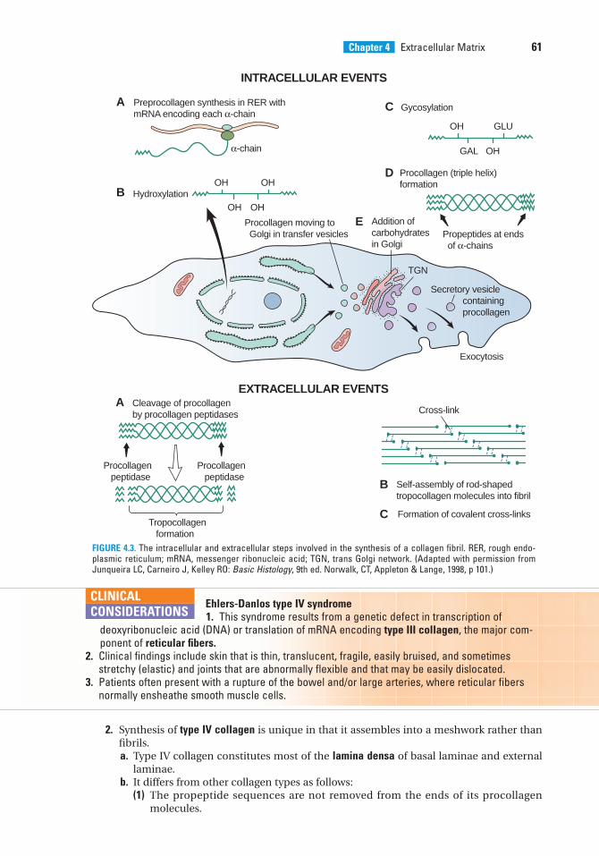
Chapter 4 Extracellular Matrix 61
FIGURE 4.3. The intracellular and extracellular steps involved in the synthesis of a collagen fibril. RER, rough endo-plasmic reticulum; mRNA, messenger ribonucleic acid; TGN, trans Golgi network. (Adapted with permission fromJunqueira LC, Carneiro J, Kelley RO: Basic Histology, 9th ed. Norwalk, CT, Appleton & Lange, 1998, p 101.)
INTRACELLULAR EVENTS
EXTRACELLULAR EVENTS
A Preprocollagen synthesis in RER withmRNA encoding each α-chain
Hydroxylation
Gycosylation
Procollagen (triple helix)formation
Addition ofcarbohydratesin Golgi
Propeptides at ends of α-chains
Cleavage of procollagenby procollagen peptidases
Procollagen peptidase
Tropocollagen formation
Procollagen peptidase
Cross-link
Exocytosis
Self-assembly of rod-shapedtropocollagen molecules into fibril
Formation of covalent cross-links
Procollagen moving to Golgi in transfer vesicles
OH OH
OH OH
A
C
D
E
B
B
C
α-chain
OH GLU
GAL OH
TGN
Secretory vesicle containing procollagen
2. Synthesis of type IV collagen is unique in that it assembles into a meshwork rather thanfibrils.a. Type IV collagen constitutes most of the lamina densa of basal laminae and external
laminae.b. It differs from other collagen types as follows:
(1) The propeptide sequences are not removed from the ends of its procollagenmolecules.
Ehlers-Danlos type IV syndrome1. This syndrome results from a genetic defect in transcription of
deoxyribonucleic acid (DNA) or translation of mRNA encoding type III collagen, the major com-ponent of reticular fibers.
2. Clinical findings include skin that is thin, translucent, fragile, easily bruised, and sometimesstretchy (elastic) and joints that are abnormally flexible and that may be easily dislocated.
3. Patients often present with a rupture of the bowel and/or large arteries, where reticular fibersnormally ensheathe smooth muscle cells.
CLINICAL
CONSIDERATIONS
LWBK615-c04[56-66].qxd 05/31/2010 10:23 AM Page 61 Aptara
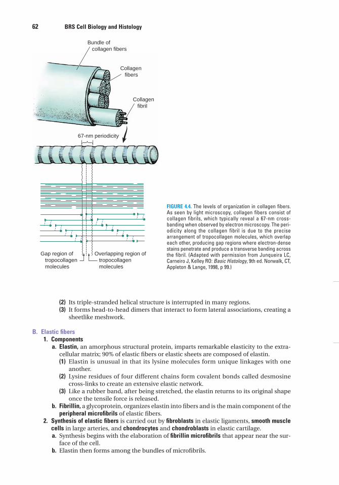
62 BRS Cell Biology and Histology
(2) Its triple-stranded helical structure is interrupted in many regions.(3) It forms head-to-head dimers that interact to form lateral associations, creating a
sheetlike meshwork.
B. Elastic fibers1. Components
a. Elastin, an amorphous structural protein, imparts remarkable elasticity to the extra-cellular matrix; 90% of elastic fibers or elastic sheets are composed of elastin.(1) Elastin is unusual in that its lysine molecules form unique linkages with one
another.(2) Lysine residues of four different chains form covalent bonds called desmosine
cross-links to create an extensive elastic network.(3) Like a rubber band, after being stretched, the elastin returns to its original shape
once the tensile force is released.b. Fibrillin, a glycoprotein, organizes elastin into fibers and is the main component of the
peripheral microfibrils of elastic fibers.2. Synthesis of elastic fibers is carried out by fibroblasts in elastic ligaments, smooth muscle
cells in large arteries, and chondrocytes and chondroblasts in elastic cartilage.a. Synthesis begins with the elaboration of fibrillin microfibrils that appear near the sur-
face of the cell.b. Elastin then forms among the bundles of microfibrils.
Bundle of collagen fibers
Collagen fibers
Collagen fibril
67-nm periodicity
Gap region of tropocollagen molecules
Overlapping region of tropocollagen molecules
FIGURE 4.4. The levels of organization in collagen fibers.As seen by light microscopy, collagen fibers consist ofcollagen fibrils, which typically reveal a 67-nm cross-banding when observed by electron microscopy. The peri-odicity along the collagen fibril is due to the precisearrangement of tropocollagen molecules, which overlapeach other, producing gap regions where electron-densestains penetrate and produce a transverse banding acrossthe fibril. (Adapted with permission from Junqueira LC,Carneiro J, Kelley RO: Basic Histology, 9th ed. Norwalk, CT,Appleton & Lange, 1998, p 99.)
LWBK615-c04[56-66].qxd 05/31/2010 10:23 AM Page 62 Aptara
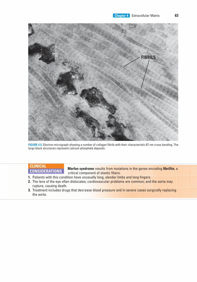
Chapter 4 Extracellular Matrix 63
FIBRILS
FIGURE 4.5. Electron micrograph showing a number of collagen fibrils with their characteristic 67-nm cross-banding. Thelarge black structures represent calcium phosphate deposits.
Marfan syndrome results from mutations in the genes encoding fibrillin, a critical component of elastic fibers.
1. Patients with this condition have unusually long, slender limbs and long fingers.2. The lens of the eye often dislocates; cardiovascular problems are common; and the aorta may
rupture, causing death.3. Treatment includes drugs that decrease blood pressure and in severe cases surgically replacing
the aorta.
CLINICAL
CONSIDERATIONS
LWBK615-c04[56-66].qxd 05/31/2010 10:23 AM Page 63 Aptara
4. Which one of the following statementsabout hyaluronic acid is true?
(A) It is a component of elastic fibers.(B) It is a glycosaminoglycan.(C) It is a proteoglycan with a shape resem-
bling a bottlebrush.(D) It is sulfated.(E) It is a small molecule.
5. Which one of the following statementsabout osteonectin is true?
(A) It is present in the lacunae of bone.(B) It is a proteoglycan.(C) It binds to type II collagen.(D) It influences calcification of bone.(E) It is synthesized by osteoclasts.
6. Which of the following statements aboutscurvy is true?
(A) One of its symptoms is bowlegs.(B) It is caused by excessive glycosylation of
tropocollagen.(C) It is caused by a deficiency of vitamin A.(D) It is associated with structurally
defective elastic fibers.(E) It is alleviated by eating citrus fruits.
7. Which one of the following is a glycopro-tein across which fibroblasts migrate duringwound healing?
(A) Fibrillin(B) Fibronectin(C) Elastin(D) Entactin(E) Laminin
1. Which one of the following statementsabout the fibronectin receptor is true?
(A) It is located exclusively in the basal lam-ina.
(B) It is a cross-shaped glycoprotein.(C) It mediates the linkage of molecules out-
side the cell with cytoskeletal elementsinside the cell.
(D) It belongs to the entactin family ofreceptors.
(E) Its absence is associated with scurvy.
2. Which one of the following events in col-lagen synthesis occurs outside of the cell?
(A) Synthesis of preprocollagen(B) Hydroxylation of lysine residues(C) Triple helix formation(D) Carbohydrate addition to procollagen(E) Cleavage of procollagen by procollagen
peptidases
3. A medical student goes to the emergencydepartment and is diagnosed with aruptured bowel, the result of a genetic con-dition called Ehlers-Danlos type IVsyndrome. Which one of the following state-ments about this patient’s condition is true?
(A) He has a defect in the synthesis ofmRNA encoding type I collagen.
(B) He has a defect in the genes encodingtype IV collagen.
(C) He has defective type II collagen.(D) He has an increased risk of breaking his
bones.(E) He has a defect in the translation of
mRNA for type III collagen.
Review Test
Directions: Each of the numbered items or incomplete statements in this section is followed byanswers or by completions of the statement. Select the ONE lettered answer or completion thatis BEST in each case.
64
LWBK615-c04[56-66].qxd 05/31/2010 10:23 AM Page 64 Aptara
Chapter 4 Extracellular Matrix 65
8. Which of the following is an adhesive gly-coprotein that links type IV collagen withlaminin in the lamina densa?
(A) Fibrillin(B) Fibronectin(C) Elastin(D) Entactin(E) Tenascin
9. Which one of the following is a maincomponent of peripheral microfibrils in anelastic fiber?
(A) Fibrillin(B) Fibronectin(C) Elastin(D) Entactin(E) Laminin
10. Which one of the following is present inthe basement membrane and ismanufactured by connective tissue cells?
(A) Fibrillin(B) Fibronectin(C) Elastin(D) Entactin(E) Laminin
LWBK615-c04[56-66].qxd 05/31/2010 10:23 AM Page 65 Aptara
66
Answers and Explanations
1. C. The fibronectin receptor is a transmembrane protein that enables cells to adhere to theextracellular matrix. Laminin is a cross-shaped glycoprotein in the basal lamina, whereentactin is also present (see Chapter 4 II D).
2. E. In the extracellular space, peptidases cleave off end sequences of procollagen to yieldtropocollagen, which self-assembles to form collagen fibrils (see Chapter 4 III A 1).
3. E. Ehlers-Danlos type IV syndrome is associated with a defect in the synthesis and trans-lation of mRNA for type III reticular collagen (see Chapter 4 III A 1 Clinical Considerations).
4. B. Hyaluronic acid is a glycosaminoglycan, not a proteoglycan. The core protein ofproteoglycans can attach to hyaluronic acid forming large aggregates (see Chapter 4 II A).
5. D. Osteonectin synthesized by osteoblasts influences the calcification of bone and binds totype I collagen in the bone matrix. Type II collagen is found in cartilage (see Chapter 4 II C).
6. E. Scurvy is caused by a deficiency of vitamin C, a necessary cofactor in the hydroxylationof preprocollagen. Citrus fruits are rich in vitamin C (see Chapter 4 III A 1 Clinical Consid-erations).
7. B. Fibronectin forms tracks along which cells migrate. During wound healing in connec-tive tissue, fibroblasts adhere to fibronectin in blood clots, facilitating the healing process(see Chapter 4 II C 1 Clinical Considerations).
8. D. Entactin is a sulfated adhesive glycoprotein in basal and external laminae that bindsboth type IV collagen and laminin (see Chapter 4 II C).
9. A. Fibrillin is the major component of the peripheral microfibrils of elastic fibers (seeChapter 4 III B).
10. B. Fibronectin is synthesized by cells of the connective tissue, usually fibroblasts, and islocated in the lamina reticularis near the lamina densa (see Chapter 4 II C 1).
LWBK615-c04[56-66].qxd 05/31/2010 10:23 AM Page 66 Aptara
c h a p t e r 5 Epithelia and Glands
67
I. OVERVIEW—EPITHELIA
A. Structure. Epithelia are specialized layers that line the internal and cover the external sur-faces of the body. An epithelium consists of a sheet of cells lying close together with littleintercellular space. These cells have distinct biochemical, functional, and structural domainsthat confer polarity, or sidedness, to epithelia.1. The basement membrane separates the epithelium from underlying connective tissue and
blood vessels.2. Epithelia are avascular and receive nourishment by diffusion of molecules through the
basal lamina.
B. Classification (Table 5.1). Epithelia are classified into various types on the basis of the numberof cell layers (one cell layer is simple; more than one is stratified) and the shape of the super-ficial cells (Figure 5.1). Pseudostratified epithelia appear to have multiple cell layers, but allcells are in contact with the basal lamina (Figure 5.2).
C. Function1. Transcellular transport of molecules from one epithelial surface to another occurs by var-
ious processes, including the following:a. Diffusion of oxygen and carbon dioxide across the epithelial cells of lung alveoli and
capillariesb. Carrier protein–mediated transport of amino acids and glucose across intestinal
epitheliac. Vesicle-mediated transport of immunoglobulin A (IgA) and other molecules
2. Absorption occurs via endocytosis or pinocytosis (see Chapter 3 III A) in various organs(e.g., the proximal convoluted tubule of the kidney).
3. Secretion of various molecules (e.g., hormones, mucus, proteins) occurs by exocytosis.4. Selective permeability results from the presence of tight junctions between epithelial cells
and permits fluids with different compositions and concentrations to exist on separatesides of an epithelial layer (e.g., intestinal epithelium).
5. Protection from abrasion and injury is provided by the epidermis, the epithelial layer ofthe skin.
First-degree burns are lesions caused by heat, friction, or other agents.1. Damage is limited to the superficial layers of the epithelium (usually
the epidermis of the skin).2. Redness and edema occur, but blisters do not form.
a. Mitotically active cells remain viable in the deeper layers of the epidermis.b. They divide and replace the damaged or destroyed cells.
CLINICAL
CONSIDERATIONS
LWBK615-c05[67-78].qxd 05/31/2010 10:25 AM Page 67 Aptara
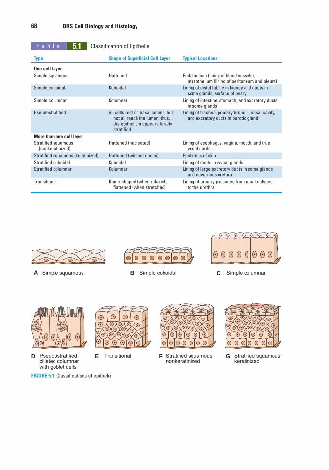
Simple squamousA
D
Simple cuboidalB Simple columnarC
Pseudostratifiedciliated columnarwith goblet cells
Transitional Stratified squamousnonkeratinized
Stratified squamouskeratinized
E F G
FIGURE 5.1. Classifications of epithelia.
t a b l e 5.1 Classification of Epithelia
Type Shape of Superficial Cell Layer Typical Locations
One cell layer
Simple squamous Flattened Endothelium (lining of blood vessels), mesothelium (lining of peritoneum and pleura)
Simple cuboidal Cuboidal Lining of distal tubule in kidney and ducts in some glands, surface of ovary
Simple columnar Columnar Lining of intestine, stomach, and excretory ducts in some glands
Pseudostratified All cells rest on basal lamina, but Lining of trachea, primary bronchi, nasal cavity,not all reach the lumen; thus, and excretory ducts in parotid glandthe epithelium appears falsely stratified
More than one cell layer
Stratified squamous Flattened (nucleated) Lining of esophagus, vagina, mouth, and true(nonkeratinized) vocal cords
Stratified squamous (keratinized) Flattened (without nuclei) Epidermis of skin
Stratified cuboidal Cuboidal Lining of ducts in sweat glands
Stratified columnar Columnar Lining of large excretory ducts in some glands and cavernous urethra
Transitional Dome-shaped (when relaxed), Lining of urinary passages from renal calyces flattened (when stretched) to the urethra
68 BRS Cell Biology and Histology
LWBK615-c05[67-78].qxd 05/31/2010 10:25 AM Page 68 Aptara
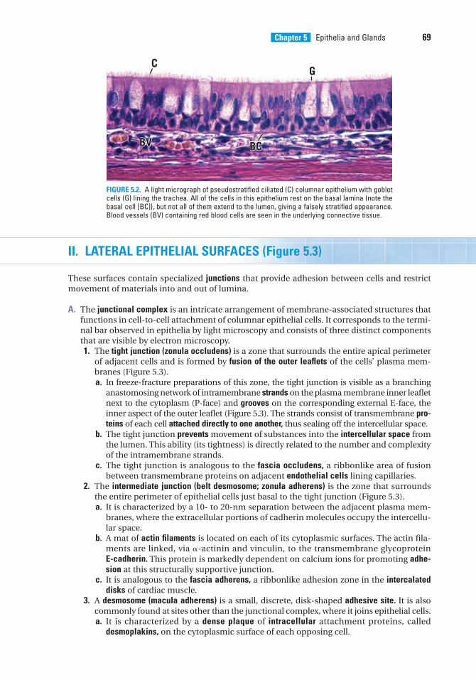
Chapter 5 Epithelia and Glands 69
II. LATERAL EPITHELIAL SURFACES (Figure 5.3)
These surfaces contain specialized junctions that provide adhesion between cells and restrictmovement of materials into and out of lumina.
A. The junctional complex is an intricate arrangement of membrane-associated structures thatfunctions in cell-to-cell attachment of columnar epithelial cells. It corresponds to the termi-nal bar observed in epithelia by light microscopy and consists of three distinct componentsthat are visible by electron microscopy.1. The tight junction (zonula occludens) is a zone that surrounds the entire apical perimeter
of adjacent cells and is formed by fusion of the outer leaflets of the cells’ plasma mem-branes (Figure 5.3).a. In freeze-fracture preparations of this zone, the tight junction is visible as a branching
anastomosing network of intramembrane strands on the plasma membrane inner leafletnext to the cytoplasm (P-face) and grooves on the corresponding external E-face, theinner aspect of the outer leaflet (Figure 5.3). The strands consist of transmembrane pro-teins of each cell attached directly to one another, thus sealing off the intercellular space.
b. The tight junction prevents movement of substances into the intercellular space fromthe lumen. This ability (its tightness) is directly related to the number and complexityof the intramembrane strands.
c. The tight junction is analogous to the fascia occludens, a ribbonlike area of fusionbetween transmembrane proteins on adjacent endothelial cells lining capillaries.
2. The intermediate junction (belt desmosome; zonula adherens) is the zone that surroundsthe entire perimeter of epithelial cells just basal to the tight junction (Figure 5.3).a. It is characterized by a 10- to 20-nm separation between the adjacent plasma mem-
branes, where the extracellular portions of cadherin molecules occupy the intercellu-lar space.
b. A mat of actin filaments is located on each of its cytoplasmic surfaces. The actin fila-ments are linked, via �-actinin and vinculin, to the transmembrane glycoprotein E-cadherin. This protein is markedly dependent on calcium ions for promoting adhe-sion at this structurally supportive junction.
c. It is analogous to the fascia adherens, a ribbonlike adhesion zone in the intercalateddisks of cardiac muscle.
3. A desmosome (macula adherens) is a small, discrete, disk-shaped adhesive site. It is alsocommonly found at sites other than the junctional complex, where it joins epithelial cells.a. It is characterized by a dense plaque of intracellular attachment proteins, called
desmoplakins, on the cytoplasmic surface of each opposing cell.
CG
BCBCBVBV BCBV
FIGURE 5.2. A light micrograph of pseudostratified ciliated (C) columnar epithelium with gobletcells (G) lining the trachea. All of the cells in this epithelium rest on the basal lamina (note thebasal cell [BC]), but not all of them extend to the lumen, giving a falsely stratified appearance.Blood vessels (BV) containing red blood cells are seen in the underlying connective tissue.
LWBK615-c05[67-78].qxd 05/31/2010 10:25 AM Page 69 Aptara
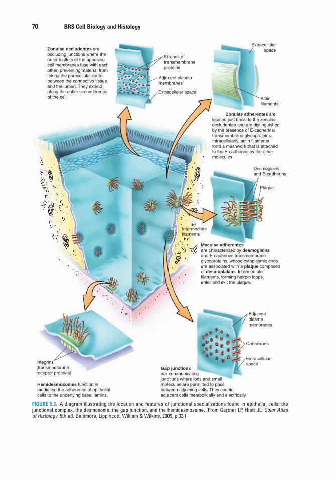
70 BRS Cell Biology and Histology
Desmogleinsand E-cadherins
�����������Zonulae adherentes arelocated just basal to the zonulaeoccludentes and are distinguishedby the presence of E-cadherins,transmembrane glycoproteins.Intracellularly, actin filamentsform a meshwork that is attachedto the E-cadherins by the othermolecules.
Plaque
Maculae adherentesare characterized by desmogleinsand E-cadherins transmembraneglycoproteins, whose cytoplasmic endsare associated with a plaque composedof desmoplakins. Intermediatefilaments, forming hairpin loops,enter and exit the plaque.
Adjacentplasmamembranes
Connexons
Extracellularspace
Gap junctionsare communicatingjunctions where ions and smallmolecules are permitted to passbetween adjoining cells. They coupleadjacent cells metabolically and electrically.
Hemidesmosomes function inmediating the adherence of epithelialcells to the underlying basal lamina.
Integrins(transmembranereceptor proteins)
Zonulae occludentes areoccluding junctions where theouter leaflets of the apposingcell membranes fuse with eachother, preventing material fromtaking the paracellular routebetween the connective tissueand the lumen. They extendalong the entire circumferenceof the cell.
Extracellular space
Strands oftransmembraneproteins
Adjacent plasmamembranes
Actinfilaments
Extracellularspace
Intermediatefilaments
FIGURE 5.3. A diagram illustrating the location and features of junctional specializations found in epithelial cells: thejunctional complex, the desmosome, the gap junction, and the hemidesmosome. (From Gartner LP, Hiatt JL: Color Atlasof Histology, 5th ed. Baltimore, Lippincott, William & Wilkins, 2009, p 33.)
LWBK615-c05[67-78].qxd 05/31/2010 10:25 AM Page 70 Aptara
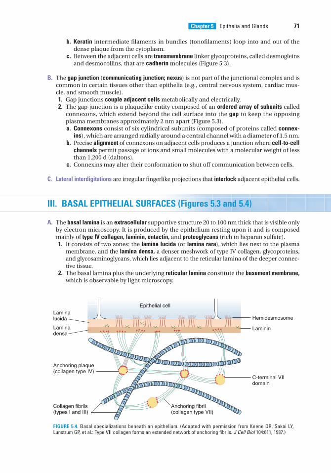
Chapter 5 Epithelia and Glands 71
b. Keratin intermediate filaments in bundles (tonofilaments) loop into and out of thedense plaque from the cytoplasm.
c. Between the adjacent cells are transmembrane linker glycoproteins, called desmogleinsand desmocollins, that are cadherin molecules (Figure 5.3).
B. The gap junction (communicating junction; nexus) is not part of the junctional complex and iscommon in certain tissues other than epithelia (e.g., central nervous system, cardiac mus-cle, and smooth muscle).1. Gap junctions couple adjacent cells metabolically and electrically.2. The gap junction is a plaquelike entity composed of an ordered array of subunits called
connexons, which extend beyond the cell surface into the gap to keep the opposingplasma membranes approximately 2 nm apart (Figure 5.3).a. Connexons consist of six cylindrical subunits (composed of proteins called connex-
ins), which are arranged radially around a central channel with a diameter of 1.5 nm.b. Precise alignment of connexons on adjacent cells produces a junction where cell-to-cell
channels permit passage of ions and small molecules with a molecular weight of lessthan 1,200 d (daltons).
c. Connexins may alter their conformation to shut off communication between cells.
C. Lateral interdigitations are irregular fingerlike projections that interlock adjacent epithelial cells.
III. BASAL EPITHELIAL SURFACES (Figures 5.3 and 5.4)
A. The basal lamina is an extracellular supportive structure 20 to 100 nm thick that is visible onlyby electron microscopy. It is produced by the epithelium resting upon it and is composedmainly of type IV collagen, laminin, entactin, and proteoglycans (rich in heparan sulfate).1. It consists of two zones: the lamina lucida (or lamina rara), which lies next to the plasma
membrane, and the lamina densa, a denser meshwork of type IV collagen, glycoproteins,and glycosaminoglycans, which lies adjacent to the reticular lamina of the deeper connec-tive tissue.
2. The basal lamina plus the underlying reticular lamina constitute the basement membrane,which is observable by light microscopy.
Epithelial cell
Laminadensa
Laminin
C-terminal VIIdomain
Hemidesmosome
Anchoring plaque(collagen type IV)
Collagen fibrils(types I and III)
Anchoring fibril(collagen type VII)
Laminalucida
FIGURE 5.4. Basal specializations beneath an epithelium. (Adapted with permission from Keene DR, Sakai LY,Lunstrum GP, et al.: Type VII collagen forms an extended network of anchoring fibrils. J Cell Biol 104:611, 1987.)
LWBK615-c05[67-78].qxd 05/31/2010 10:25 AM Page 71 Aptara
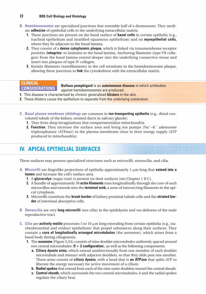
Bullous pemphigoid is an autoimmune disease in which antibodies against hemidesmosomes are produced.
1. This disease is characterized by chronic generalized blisters in the skin.2. These blisters cause the epithelium to separate from the underlying substratum.
CLINICAL
CONSIDERATIONS
72 BRS Cell Biology and Histology
B. Hemidesmosomes are specialized junctions that resemble half of a desmosome. They medi-ate adhesion of epithelial cells to the underlying extracellular matrix.1. These junctions are present on the basal surface of basal cells in certain epithelia (e.g.,
tracheal epithelium and stratified squamous epithelium) and on myoepithelial cells,where they lie adjacent to the basal lamina.
2. They consist of a dense cytoplasmic plaque, which is linked via transmembrane receptorproteins (integrins) to laminins in the basal lamina. Anchoring filaments (type VII colla-gen) from the basal lamina extend deeper into the underlying connective tissue andinsert into plaques of type IV collagen.
3. Keratin filaments (tonofilaments) in the cell terminate in the hemidesmosome plaque,allowing these junctions to link the cytoskeleton with the extracellular matrix.
C. Basal plasma membrane infoldings are common in ion-transporting epithelia (e.g., distal con-voluted tubule of the kidney, striated ducts in salivary glands).1. They form deep invaginations that compartmentalize mitochondria.2. Function. They increase the surface area and bring ion pumps (Na�–K� adenosine
triphosphatase [ATPase]) in the plasma membrane close to their energy supply (ATPproduced in mitochondria).
IV. APICAL EPITHELIAL SURFACES
These surfaces may possess specialized structures such as microvilli, stereocilia, and cilia.
A. Microvilli are fingerlike projections of epithelia approximately 1 �m long that extend into alumen and increase the cell’s surface area.1. A glycocalyx (sugar coat) is present on their surfaces (see Chapter 1 II C).2. A bundle of approximately 30 actin filaments runs longitudinally through the core of each
microvillus and extends into the terminal web, a zone of intersecting filaments in the api-cal cytoplasm.
3. Microvilli constitute the brush border of kidney proximal tubule cells and the striated bor-der of intestinal absorptive cells.
B. Stereocilia are very long microvilli (not cilia) in the epididymis and vas deferens of the malereproductive tract.
C. Cilia are actively motile processes 5 to 10 �m long extending from certain epithelia (e.g., tra-cheobronchial and oviduct epithelium) that propel substances along their surfaces. Theycontain a core of longitudinally arranged microtubules (the axoneme), which arises from abasal body during ciliogenesis.1. The axoneme (Figure 5.5A) consists of nine doublet microtubules uniformly spaced around
two central microtubules (9 � 2 configuration), as well as the following components:a. Ciliary dynein arms, which extend unidirectionally from one member of each doublet
microtubule and interact with adjacent doublets, so that they slide past one another.These arms consist of ciliary dynein, with a head that is an ATPase that splits ATP toliberate the energy necessary for active movement of a cilium.
b. Radial spokes that extend from each of the nine outer doublets toward the central sheath.c. Central sheath, which surrounds the two central microtubules; it and the radial spokes
regulate the ciliary beat.
LWBK615-c05[67-78].qxd 05/31/2010 10:25 AM Page 72 Aptara
Immotile cilia syndrome results from a genetic defect that causes an abnormal ciliary beat or the absence of a beat.
1. In this syndrome, cilia have axonemes that lack ciliary dynein arms and have other abnormalities.2. The syndrome is associated with recurrent lower respiratory tract infections and reduced fertility
in women and sterility in men.
CLINICAL
CONSIDERATIONS
Chapter 5 Epithelia and Glands 73
A Shared protofilaments
B Triplet microtubules
Ciliary dynein arm
Ciliary dynein arm
Basal body
PlasmalemmaCentral singlets
Doublet microtubules
9 + 2 Axoneme pattern
Radial spoke
Central sheath
Nexin
FIGURE 5.5. Cross-sections of a cilium and basal body. (Part A adapted with permission from Junqueira LC, Carneiro J,Kelley RO: Basic Histology, 9th ed. Norwalk, CT, Appleton & Lange, 1998, p 45. Part B adapted with permission fromWest JB: Best and Taylor Physiological Basis of Medical Practice, 12th ed. Baltimore, Williams & Wilkins, 1991, p 12.)
d. Nexin, an elastic protein that connects adjacent doublet microtubules and helpsmaintain the shape of the cilium.
2. The basal body (Figure 5.5B) is a cylindrical structure at the base of each cilium that con-sists of nine triplet microtubules arranged radially in the shape of a pinwheel (9 � 0 con-figuration). It resembles a centriole (see Figures 3.1 and 5.6) but has a less complexcentral organization. The inner two triplets of the basal body give rise to the doubletmicrotubules of the cilium axoneme.
V. GLANDS
They originate from an epithelium that penetrates the connective tissue and forms secretory units.
A. Structure. A gland consists of a functional portion (parenchyma) of secretory and ductalepithelial cells, which is separated by a basal lamina from supporting connective tissue ele-ments (stroma).
LWBK615-c05[67-78].qxd 05/31/2010 10:25 AM Page 73 Aptara
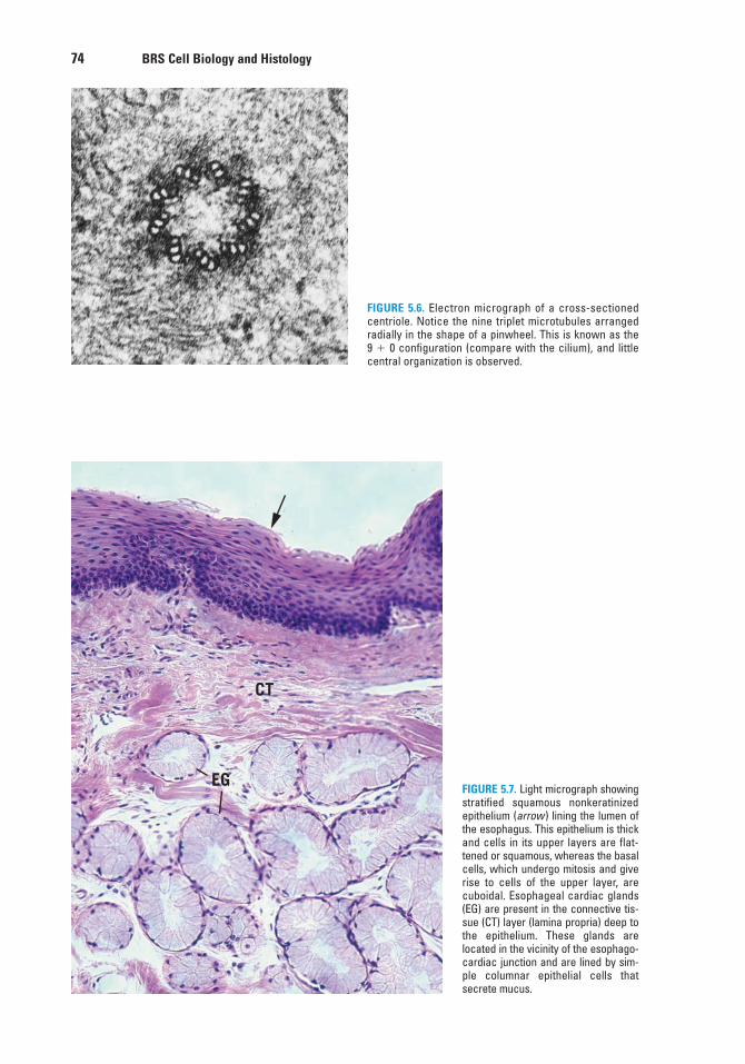
74 BRS Cell Biology and Histology
FIGURE 5.6. Electron micrograph of a cross-sectionedcentriole. Notice the nine triplet microtubules arrangedradially in the shape of a pinwheel. This is known as the9 � 0 configuration (compare with the cilium), and littlecentral organization is observed.
CT
EGFIGURE 5.7. Light micrograph showingstratified squamous nonkeratinizedepithelium (arrow ) lining the lumen ofthe esophagus. This epithelium is thickand cells in its upper layers are flat-tened or squamous, whereas the basalcells, which undergo mitosis and giverise to cells of the upper layer, arecuboidal. Esophageal cardiac glands(EG) are present in the connective tis-sue (CT) layer (lamina propria) deep tothe epithelium. These glands arelocated in the vicinity of the esophago-cardiac junction and are lined by sim-ple columnar epithelial cells thatsecrete mucus.
LWBK615-c05[67-78].qxd 05/31/2010 10:25 AM Page 74 Aptara
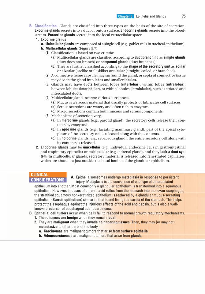
Chapter 5 Epithelia and Glands 75
B. Classification. Glands are classified into three types on the basis of the site of secretion.Exocrine glands secrete into a duct or onto a surface. Endocrine glands secrete into the blood-stream. Paracrine glands secrete into the local extracellular space.1. Exocrine glands
a. Unicellular glands are composed of a single cell (e.g., goblet cells in tracheal epithelium).b. Multicellular glands (Figure 5.7)
(1) Classification is based on two criteria:(a) Multicellular glands are classified according to duct branching as simple glands
(duct does not branch) or compound glands (duct branches).(b) They are further classified according to the shape of the secretory unit as acinar
or alveolar (saclike or flasklike) or tubular (straight, coiled, or branched).(2) A connective tissue capsule may surround the gland, or septa of connective tissue
may divide the gland into lobes and smaller lobules.(3) Glands may have ducts between lobes (interlobar), within lobes (intralobar),
between lobules (interlobular), or within lobules (intralobular), such as striated andintercalated ducts.
(4) Multicellular glands secrete various substances.(a) Mucus is a viscous material that usually protects or lubricates cell surfaces.(b) Serous secretions are watery and often rich in enzymes.(c) Mixed secretions contain both mucous and serous components.
(5) Mechanisms of secretion vary.(a) In merocrine glands (e.g., parotid gland), the secretory cells release their con-
tents by exocytosis.(b) In apocrine glands (e.g., lactating mammary gland), part of the apical cyto-
plasm of the secretory cell is released along with the contents.(c) In holocrine glands (e.g., sebaceous gland), the entire secretory cell along with
its contents is released.2. Endocrine glands may be unicellular (e.g., individual endocrine cells in gastrointestinal
and respiratory epithelia) or multicellular (e.g., adrenal gland), and they lack a duct sys-tem. In multicellular glands, secretory material is released into fenestrated capillaries,which are abundant just outside the basal lamina of the glandular epithelium.
A. Epithelia sometimes undergo metaplasia in response to persistent injury. Metaplasia is the conversion of one type of differentiated
epithelium into another. Most commonly a glandular epithelium is transformed into a squamousepithelium. However, in cases of chronic acid reflux from the stomach into the lower esophagus,the stratified squamous nonkeratinized epithelium is replaced by a glandular mucus-secretingepithelium (Barrett epithelium) similar to that found lining the cardia of the stomach. This helpsprotect the esophagus against the injurious effects of the acid and pepsin, but is also a well-known precursor of esophageal adenocarcinoma.
B. Epithelial cell tumors occur when cells fail to respond to normal growth regulatory mechanisms.1. These tumors are benign when they remain local.2. They are malignant when they invade neighboring tissues. Then, they may (or may not)
metastasize to other parts of the body.a. Carcinomas are malignant tumors that arise from surface epithelia.b. Adenocarcinomas are malignant tumors that arise from glands.
CLINICAL
CONSIDERATIONS
LWBK615-c05[67-78].qxd 05/31/2010 10:25 AM Page 75 Aptara
Review Test
76
1. Which one of the following statementsabout the desmosome is true?
(A) It is sometimes called a nexus.(B) It permits the passage of large proteins
from one cell to an adjacent cell.(C) It has a plaque made up of many
connexons.(D) It facilitates metabolic coupling between
adjacent cells.(E) It is a disk-shaped adhesion site between
epithelial cells.
2. A medical student who has chronic lowerrespiratory infections seeks the advice of anear, nose, and throat specialist. A biopsy ofthe student’s respiratory epithelium revealsalterations in certain epithelial structures.This patient is most likely to have abnormal
(A) microvilli.(B) desmosomes.(C) cilia.(D) hemidesmosomes.(E) basal plasmalemma infoldings.
3. Which one of the following statementsabout the gap junction is true?
(A) It extends as a zone around the apicalperimeter of adjacent cells.
(B) It possesses dense plaques composed inpart of desmoplakins.
(C) It permits the passage of ions from onecell to an adjacent cell.
(D) Its adhesion is dependent upon calciumions.
(E) It possesses transmembrane linkerglycoproteins.
4. Which one of the following statementsabout glands is true?
(A) Exocrine glands lack ducts.(B) Simple glands have ducts that branch.(C) Endocrine glands secrete into ducts.(D) Serous secretions are watery.(E) Holocrine glands release their contents
by exocytosis.
5. Which one of the following statementsabout epithelia is true?
(A) They are polarized.(B) They are vascular.(C) They are completely surrounded by a
basal lamina.(D) They contain wide intercellular spaces.(E) They are not part of the wall of blood
vessels.
6. Which one of the following statementsabout cilia is true?
(A) They possess a 9 � 0 configuration ofmicrotubules.
(B) They do not contain an axoneme.(C) They contain ciliary dynein arms.(D) They are nearly identical to centrioles.(E) They play a major function in
absorption.
Directions: Each of the numbered items or incomplete statements in this section is followed byanswers or by completions of the statement. Select the ONE lettered answer or completion thatis BEST in each case.
LWBK615-c05[67-78].qxd 05/31/2010 10:25 AM Page 76 Aptara
Chapter 5 Epithelia and Glands 77
10. Which one of the following is a tumorarising from glandular epithelium?
(A) Adenocarcinoma(B) Bullous pemphigoid(C) Carcinoma(D) Edema(E) Immotile cilia syndrome
11. Which of the following is a conditionaffecting the epidermis of the skin in whichblisters do not form?
(A) Adenocarcinoma(B) Bullous pemphigoid(C) Carcinoma(D) First-degree burn(E) Immotile cilia syndrome
7. Which one of the following statementsabout stratified squamous epithelium istrue?
(A) The surface layer of cells is always kera-tinized.
(B) The cells in its most superficial layer areflattened.
(C) Its basal cells rest on an elastic lamina.(D) Its cells lack desmosomes.(E) It lines the ducts of sweat glands.
8. Which one of the following is an autoim-mune disease?
(A) Adenocarcinoma(B) Bullous pemphigoid(C) Carcinoma(D) First-degree burn(E) Immotile cilia syndrome
9. Which one of the following is a hereditarydisease that may be associated with infertility?
(A) Adenocarcinoma(B) Bullous pemphigoid(C) Carcinoma(D) Edema(E) Immotile cilia syndrome
LWBK615-c05[67-78].qxd 05/31/2010 10:25 AM Page 77 Aptara
Answers and Explanations
78
1. E. Desmosomes are sites of adhesion characterized by dense cytoplasmic plaques andassociated keratin filaments. Only gap junctions permit cell-to-cell communication ofsmall molecules via their connexon channels (see Chapter 5 II A).
2. C. Individuals with abnormal respiratory cilia commonly have recurrent respiratory infec-tions if the cilia are unable to clear the respiratory epithelium of microorganisms, debris,and so forth. The student may have immotile cilia syndrome, which is caused by a geneticdefect resulting in cilia with axonemes that lack ciliary dynein arms and thus are unable tobeat (see Chapter 5 IV C 2 Clinical Considerations).
3. C. The gap junction channel regulates the passage of ions and small molecules from cellto cell, excluding those having a molecular weight greater than 1,200 d. The tight junctionis the zone of adhesion around the apical perimeter of adjacent cells. The otherstatements are characteristics of desmosomes (see Chapter 5 II B).
4. D. Serous secretions produced by glands are often rich in enzymes and watery in consis-tency. Exocrine glands secrete into ducts and endocrine glands lack ducts. Merocrineglands use exocytosis to release their products (see Chapter 5 V B).
5. A. Epithelia are polarized, meaning they show sidedness and have apical and basolateralsurfaces with specific functions (see Chapter 5 I A).
6. C. Cilia contain an axoneme with ciliary dynein arms extending unidirectionally from onemember of each doublet. Ciliary dynein has ATPase activity, and when it splits ATP, theadjacent doublets slide past one another and the cilium moves. Microvilli, not cilia, func-tion in absorption (see Chapter 5 IV C).
7. B. Stratified squamous epithelium is characterized by flattened cells with or withoutnuclei in its superficial layer. It may or may not be keratinized, and it rests on a basal lam-ina produced by the epithelium. Stratified cuboidal epithelium lines the ducts in sweatglands (see Chapter 5 I B).
8. B. Bullous pemphigoid is an autoimmune disease. Affected individuals form antibodiesagainst their own hemidesmosomes (see Chapter 5 III B Clinical Considerations).
9. E. Immotile cilia syndrome results from a genetic defect that prevents synthesis of ciliarydynein ATPase, resulting in cilia that cannot actively move. Men are sterile because theirsperm are not motile (the flagella in their tails lack this enzyme). Women may be infertilebecause cilia along their oviducts may fail to move oocytes toward the uterus (see Chapter5 IV C 2 Clinical Considerations).
10. A. Adenocarcinomas are epithelial tumors that originate in glandular epithelia.Carcinomas originate from surface epithelia (see Chapter 5V B 2 Clinical Considerations).
11. D. First-degree burns damage the upper layers of the epidermis only, and blisters do notform in the skin (see Chapter 5 I C Clinical Considerations).
LWBK615-c05[67-78].qxd 05/31/2010 10:25 AM Page 78 Aptara
c h a p t e r 6 Connective Tissue
79
I. OVERVIEW—CONNECTIVE TISSUE
A. Structure. Connective tissue is formed primarily of extracellular matrix, consisting of groundsubstance, and fibers, in which various connective tissue cells are embedded.
B. Function. Connective tissue supports organs and cells, acts as a medium for exchange of nutri-ents and wastes between the blood and tissues, protects against microorganisms, repairsdamaged tissues, and stores fat.
II. EXTRACELLULAR MATRIX
The extracellular matrix provides a medium for the transfer of nutrients and waste materialsbetween connective tissue cells and the bloodstream.
A. Ground substance is a colorless, transparent, gel-like material in which the cells and fibers ofconnective tissue are embedded.1. It is a complex mixture of glycosaminoglycans, proteoglycans, and glycoproteins (see
Chapter 4 for details of these components).2. Ground substance serves as a lubricant, helps prevent invasion of tissues by foreign
agents, and resists forces of compression.
B. Fibers (collagen, reticular, and elastic) are long, slender protein polymers present in differ-ent proportions in different types of connective tissue.1. Collagen fibers. Although there are at least 25 different types of collagen, the most com-
mon collagen types in connective tissue proper are type I and type III collagen, bothconsisting of many closely packed tropocollagen fibrils. The diameter of individual type Icollagen fibrils varies greatly (10–300 nm). These fibrils may aggregate and form cable-like structures up to several centimeters in length and display 67-nm periodicity (seeChapter 4 and Figure 4.4).a. Collagen fibers are produced in a two-stage process, involving both intracellular and
extracellular events (see Chapter 4 Figure 4.3).b. Collagen fibers have great tensile strength, which imparts both flexibility and strength
to tissues containing them.c. Bone, skin, cartilage, tendon, and many other structures of the body contain collagen
fibers.2. Reticular fibers are extremely thin (0.5–2.0 �m) in diameter and are composed primarily
of type III collagen; they have higher carbohydrate content than other collagen fibers.a. Type III collagen fibers constitute the architectural framework of certain organs and
glands.
LWBK615-c06[79-91].qxd 05/31/2010 10:29 AM Page 79 Aptara
80 BRS Cell Biology and Histology
b. Because of their high carbohydrate content, they stain black with silver salts.3. Elastic fibers are coiled branching fibers 0.2 to 1.0 �m in diameter that sometimes form
loose networks.a. These fibers may be stretched up to 150% of their resting length.b. They are composed of elastin and microfibrils of fibrillin (see Chapter 4 III B).c. Elastic fibers require special staining to be observed by light microscopy.
III. CONNECTIVE TISSUE CELLS
Connective tissue cells include many types with different functions. Some originate locally andremain in the connective tissue (fixed cells), whereas others originate elsewhere and remainonly temporarily in connective tissue (transient cells) (Figure 6.1). Fixed connective tissue cellsinclude fibroblasts, pericytes, adipose cells, mast cells, and fixed macrophages. Transient con-nective tissue cells include certain macrophages, lymphocytes, plasma cells, neutrophils,eosinophils, and basophils.
A. Fixed Cells of Connective Tissue1. Fibroblasts arise from mesenchymal cells and are the predominant cells in connective
tissue proper. (Figure 6.2) They often possess an oval nucleus with two or more nucleoli.Fibroblasts seldom undergo mitosis except in wound healing. They may differentiateinto other cell types under certain conditions.
Active fibroblasts are spindle shaped (fusiform) and contain well-developed roughendoplasmic reticulum (RER) and many Golgi complexes. Myosin is located throughoutthe cytoplasm, and actin and �-actinin are located at the cell periphery. Syntheticallyactive, they produce procollagen and other components of the extracellular matrix.
Fibroblasts undergo mitosis only during wound healing. However, undercertain conditions, fibroblasts may differentiate into adipose cells, or dur-
ing fibrocartilage formation, they may differentiate into chondrocytes. In addition, under pathologicalconditions, fibroblasts may differentiate into osteoblasts.
CLINICAL
CONSIDERATIONS
2. Pericytes are derived from embryonic mesenchymal cells and may retain a pluripotentialrole.a. They possess characteristics of endothelial cells as well as smooth muscle cells
because they contain actin, myosin, and tropomyosin, suggesting that they may func-tion in contraction.
b. They are smaller than fibroblasts and are located mostly along capillaries, yet they liewithin their own basal lamina.
c. Pericytes contain actin, myosin, and tropomyosin and may function as contractilecells that modify capillary blood flow.
d. During blood vessel formation and repair, they may differentiate into smooth musclecells as well as endothelial cells.
e. In response to injury, pericytes may give rise to endothelial cells, fibroblasts, andsmooth muscle cells of blood vessel walls.
3. Adipose cells (adipocytes) (Figures 6.1 and 6.3) arise from mesenchymal cells and per-haps from fibroblasts. They do not normally undergo cell division because they arefully differentiated cells. However, they do increase in number in early neonatal life.There is debate about normal proliferation of adipocytes beyond 2 years after birth.They are surrounded by a basal lamina and are responsible for the synthesis, storage,and release of fat.
LWBK615-c06[79-91].qxd 05/31/2010 10:29 AM Page 80 Aptara
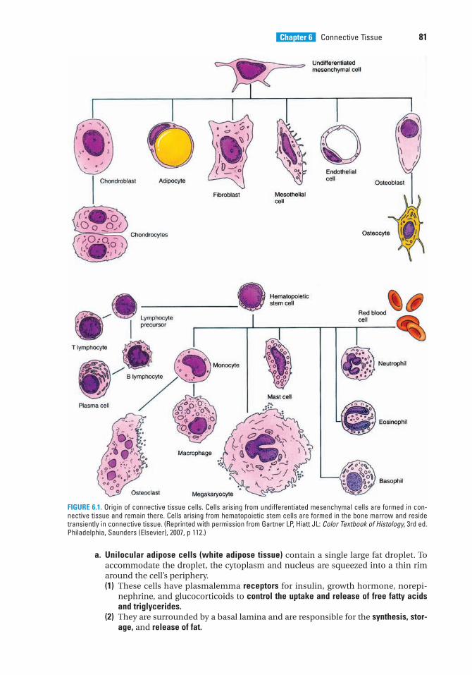
Chapter 6 Connective Tissue 81
a. Unilocular adipose cells (white adipose tissue) contain a single large fat droplet. Toaccommodate the droplet, the cytoplasm and nucleus are squeezed into a thin rimaround the cell’s periphery.(1) These cells have plasmalemma receptors for insulin, growth hormone, norepi-
nephrine, and glucocorticoids to control the uptake and release of free fatty acidsand triglycerides.
(2) They are surrounded by a basal lamina and are responsible for the synthesis, stor-age, and release of fat.
FIGURE 6.1. Origin of connective tissue cells. Cells arising from undifferentiated mesenchymal cells are formed in con-nective tissue and remain there. Cells arising from hematopoietic stem cells are formed in the bone marrow and residetransiently in connective tissue. (Reprinted with permission from Gartner LP, Hiatt JL: Color Textbook of Histology, 3rd ed.Philadelphia, Saunders (Elsevier), 2007, p 112.)
LWBK615-c06[79-91].qxd 05/31/2010 10:29 AM Page 81 Aptara
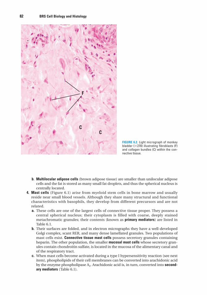
82 BRS Cell Biology and Histology
b. Multilocular adipose cells (brown adipose tissue) are smaller than unilocular adiposecells and the fat is stored as many small fat droplets, and thus the spherical nucleus iscentrally located.
4. Mast cells (Figure 6.1) arise from myeloid stem cells in bone marrow and usuallyreside near small blood vessels. Although they share many structural and functionalcharacteristics with basophils, they develop from different precursors and are notrelated.a. These cells are one of the largest cells of connective tissue proper. They possess a
central spherical nucleus; their cytoplasm is filled with coarse, deeply stainedmetachromatic granules; their contents (known as primary mediators) are listed inTable 6.1.
b. Their surfaces are folded, and in electron micrographs they have a well-developedGolgi complex, scant RER, and many dense lamellated granules. Two populations ofmast cells exist. Connective tissue mast cells possess secretory granules containingheparin. The other population, the smaller mucosal mast cells whose secretory gran-ules contain chondroitin sulfate, is located in the mucosa of the alimentary canal andof the respiratory tract.
c. When mast cells become activated during a type I hypersensitivity reaction (see nextitem), phospholipids of their cell membranes can be converted into arachidonic acidby the enzyme phospholipase A2. Arachidonic acid is, in turn, converted into second-ary mediators (Table 6.1).
F
CC
FIGURE 6.2. Light micrograph of monkeybladder (�270) illustrating fibroblasts (F)and collagen bundles (C) within the con-nective tissue.
LWBK615-c06[79-91].qxd 05/31/2010 10:29 AM Page 82 Aptara
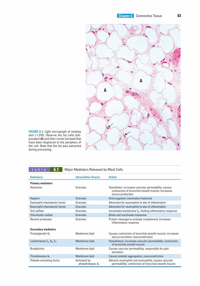
Chapter 6 Connective Tissue 83
A
A
FIGURE 6.3. Light micrograph of monkeyskin (�270). Observe the fat cells (adi-pocytes) (A) and their nuclei (arrows) thathave been displaced to the periphery ofthe cell. Note that the fat was extractedduring processing.
t a b l e 6.1 Major Mediators Released by Mast Cells
Substance Intracellular Source Action
Primary mediators
Histamine Granules Vasodilator; increases vascular permeability; causescontraction of bronchial smooth muscle; increases mucus production
Heparin Granules Anticoagulant; inactivates histamine
Eosinophil chemotactic factor Granules Attractant for eosinophils to site of inflammation
Neutrophil chemotactic factor Granules Attractant for neutrophils to site of inflammation
Aryl sulfate Granules Inactivates leukotriene C4, limiting inflammatory response
Chondroitin sulfate Granules Binds and inactivates histamine
Neutral proteases Granules Protein cleavage to activate complement; increases inflammatory response
Secondary mediators
Prostaglandin D2 Membrane lipid Causes contraction of bronchial smooth muscle; increases mucus secretion; vasoconstriction
Leukotrienes C4, D4, E4 Membrane lipid Vasodilators; increases vascular permeability; contraction of bronchial smooth muscle
Bradykinins Membrane lipid Causes vascular permeability; responsible for pain sensation
Thromboxane A2 Membrane lipid Causes platelet aggregation; vasoconstriction
Platelet-activating factor Activated by Attracts neutrophils and eosinophils; causes vascular phospholipase A2 permeability; contraction of bronchial smooth muscle
LWBK615-c06[79-91].qxd 05/31/2010 10:29 AM Page 83 Aptara
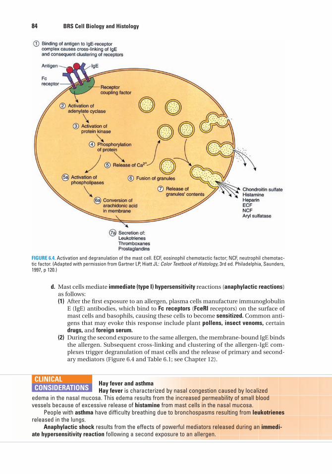
84 BRS Cell Biology and Histology
d. Mast cells mediate immediate (type I) hypersensitivity reactions (anaphylactic reactions)as follows:(1) After the first exposure to an allergen, plasma cells manufacture immunoglobulin
E (IgE) antibodies, which bind to Fc receptors (FceRI receptors) on the surface ofmast cells and basophils, causing these cells to become sensitized. Common anti-gens that may evoke this response include plant pollens, insect venoms, certaindrugs, and foreign serum.
(2) During the second exposure to the same allergen, the membrane-bound IgE bindsthe allergen. Subsequent cross-linking and clustering of the allergen-IgE com-plexes trigger degranulation of mast cells and the release of primary and second-ary mediators (Figure 6.4 and Table 6.1; see Chapter 12).
FIGURE 6.4. Activation and degranulation of the mast cell. ECF, eosinophil chemotactic factor; NCF, neutrophil chemotac-tic factor. (Adapted with permission from Gartner LP, Hiatt JL: Color Textbook of Histology, 3rd ed. Philadelphia, Saunders,1997, p 120.)
Hay fever and asthmaHay fever is characterized by nasal congestion caused by localized
edema in the nasal mucosa. This edema results from the increased permeability of small bloodvessels because of excessive release of histamine from mast cells in the nasal mucosa.
People with asthma have difficulty breathing due to bronchospasms resulting from leukotrienesreleased in the lungs.
Anaphylactic shock results from the effects of powerful mediators released during an immedi-ate hypersensitivity reaction following a second exposure to an allergen.
CLINICAL
CONSIDERATIONS
LWBK615-c06[79-91].qxd 05/31/2010 10:29 AM Page 84 Aptara
Chapter 6 Connective Tissue 85
B. Transient Cells of Connective Tissue1. Macrophages (Figure 6.1) are the principal phagocytosing cells of connective tissue. They
are responsible for removing large particulate matter and assisting in the immuneresponse. They also secrete substances that function in wound healing.a. Macrophages originate in the bone marrow as monocytes, circulate in the blood-
stream, then migrate into the connective tissue, where they mature into functionalmacrophages (see Chapter 10 VI E). Macrophages increase in number because of theactivity of the macrophage colony-stimulating factor (M-CSF). In addition, thecolony-forming unit monocyte (CFU-M) facilitates the mitosis and differentiation ofmonocytes to form macrophages.
b. Macrophages are members of the mononuclear phagocyte system. They display FceRIreceptors and receptors for complement.
c. When activated, they display filopodia, an eccentric kidney-shaped nucleus, phago-cytic vacuoles, lysosomes, and residual bodies.
d. When stimulated, they may fuse to form foreign body giant cells. These multinucleatedcells surround and phagocytose large foreign bodies.
2. Lymphoid cells (Figure 6.1) arise from lymphoid stem cells during hemopoiesis (seeChapter 10 VI G). They are located throughout the body in the subepithelial connectivetissue and accumulate in the respiratory system, gastrointestinal tract, and elsewhere inareas of chronic inflammation. (For more information concerning lymphoid cells, seeChapter 12 II.)a. T lymphocytes (T cells) initiate the cell-mediated immune response.b. B lymphocytes (B cells), following activation by an antigen, differentiate into plasma
cells, which function in the humoral immune response.c. Natural killer cells (NK cells) lack the surface determinants characteristic of T and B
lymphocytes but may display cytotoxic activity against tumor cells.3. Plasma cells (Figure 6.1) are antibody-manufacturing cells that arise from activated B lym-
phocytes and are responsible for humoral immunity.a. These ovoid cells contain an eccentric nucleus possessing clumps of heterochromatin,
which appear to be arranged in a wheel-spoke fashion.b. Their cytoplasm is deeply basophilic because of an abundance of RER.c. A prominent area adjacent to the nucleus appears pale and contains the Golgi com-
plex (negative Golgi image).d. They are most abundant at wound entry sites or in areas of chronic inflammation.
4. Granulocytes (Figure 6.1) are white blood cells that possess cytoplasmic granules andarise from myeloid stem cells during hemopoiesis. At sites of inflammation, they leavethe bloodstream and enter the loose connective tissue, where they perform their specificfunctions (see Chapter 10 II B 2 a).a. Neutrophils phagocytose, kill, and digest bacteria at sites of acute inflammation. Pus is
an accumulation of dead neutrophils, bacteria, extracellular fluid, and additionaldebris at an inflammatory site.
b. Eosinophils bind to antigen–antibody complexes on the surface of parasites (e.g.,helminths) and then release cytotoxins that damage the parasites.(1) They are most prevalent at sites of chronic or allergic inflammation.(2) Eosinophils are attracted by eosinophil chemotactic factor (ECF), which is
secreted by mast cells and basophils, to sites of allergic inflammation. There,eosinophils release enzymes that cleave histamine and leukotriene C, thus moder-ating the allergic reaction.
(3) These cells also phagocytose antibody–antigen complexes.
1. This reaction can occur within seconds or minutes after contact with an allergen.2. Signs and symptoms include shortness of breath, decreasing blood pressure, and other signs and
symptoms of shock.3. Anaphylactic shock may be life-threatening if untreated.
LWBK615-c06[79-91].qxd 05/31/2010 10:29 AM Page 85 Aptara
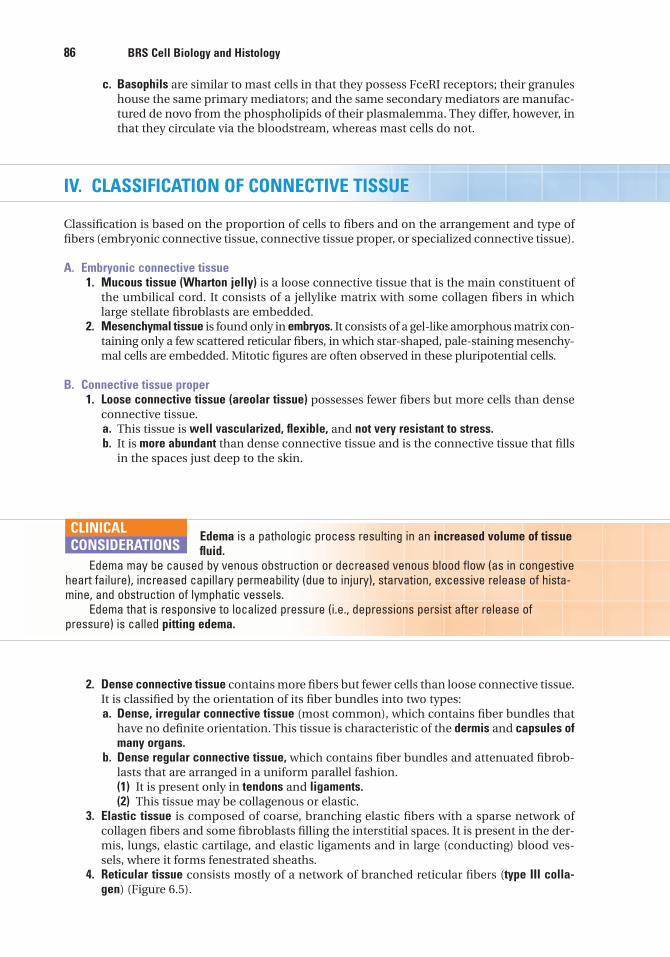
86 BRS Cell Biology and Histology
c. Basophils are similar to mast cells in that they possess FceRI receptors; their granuleshouse the same primary mediators; and the same secondary mediators are manufac-tured de novo from the phospholipids of their plasmalemma. They differ, however, inthat they circulate via the bloodstream, whereas mast cells do not.
IV. CLASSIFICATION OF CONNECTIVE TISSUE
Classification is based on the proportion of cells to fibers and on the arrangement and type offibers (embryonic connective tissue, connective tissue proper, or specialized connective tissue).
A. Embryonic connective tissue1. Mucous tissue (Wharton jelly) is a loose connective tissue that is the main constituent of
the umbilical cord. It consists of a jellylike matrix with some collagen fibers in whichlarge stellate fibroblasts are embedded.
2. Mesenchymal tissue is found only in embryos. It consists of a gel-like amorphous matrix con-taining only a few scattered reticular fibers, in which star-shaped, pale-staining mesenchy-mal cells are embedded. Mitotic figures are often observed in these pluripotential cells.
B. Connective tissue proper1. Loose connective tissue (areolar tissue) possesses fewer fibers but more cells than dense
connective tissue.a. This tissue is well vascularized, flexible, and not very resistant to stress.b. It is more abundant than dense connective tissue and is the connective tissue that fills
in the spaces just deep to the skin.
Edema is a pathologic process resulting in an increased volume of tissuefluid.
Edema may be caused by venous obstruction or decreased venous blood flow (as in congestiveheart failure), increased capillary permeability (due to injury), starvation, excessive release of hista-mine, and obstruction of lymphatic vessels.
Edema that is responsive to localized pressure (i.e., depressions persist after release ofpressure) is called pitting edema.
CLINICAL
CONSIDERATIONS
2. Dense connective tissue contains more fibers but fewer cells than loose connective tissue.It is classified by the orientation of its fiber bundles into two types:a. Dense, irregular connective tissue (most common), which contains fiber bundles that
have no definite orientation. This tissue is characteristic of the dermis and capsules ofmany organs.
b. Dense regular connective tissue, which contains fiber bundles and attenuated fibrob-lasts that are arranged in a uniform parallel fashion.(1) It is present only in tendons and ligaments.(2) This tissue may be collagenous or elastic.
3. Elastic tissue is composed of coarse, branching elastic fibers with a sparse network ofcollagen fibers and some fibroblasts filling the interstitial spaces. It is present in the der-mis, lungs, elastic cartilage, and elastic ligaments and in large (conducting) blood ves-sels, where it forms fenestrated sheaths.
4. Reticular tissue consists mostly of a network of branched reticular fibers (type III colla-gen) (Figure 6.5).
LWBK615-c06[79-91].qxd 05/31/2010 10:29 AM Page 86 Aptara
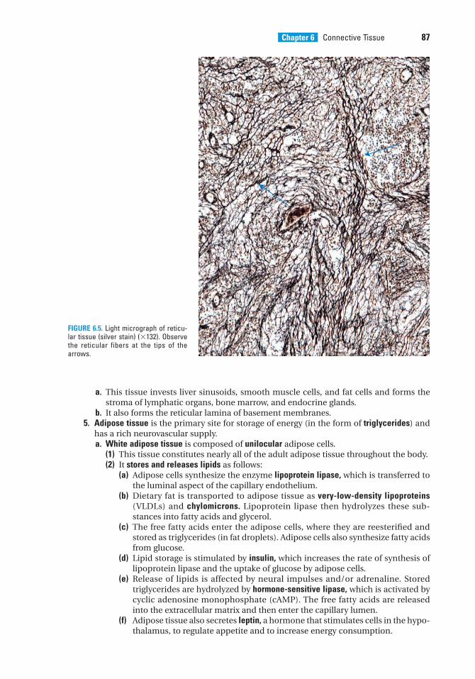
Chapter 6 Connective Tissue 87
a. This tissue invests liver sinusoids, smooth muscle cells, and fat cells and forms thestroma of lymphatic organs, bone marrow, and endocrine glands.
b. It also forms the reticular lamina of basement membranes.5. Adipose tissue is the primary site for storage of energy (in the form of triglycerides) and
has a rich neurovascular supply.a. White adipose tissue is composed of unilocular adipose cells.
(1) This tissue constitutes nearly all of the adult adipose tissue throughout the body.(2) It stores and releases lipids as follows:
(a) Adipose cells synthesize the enzyme lipoprotein lipase, which is transferred tothe luminal aspect of the capillary endothelium.
(b) Dietary fat is transported to adipose tissue as very-low-density lipoproteins(VLDLs) and chylomicrons. Lipoprotein lipase then hydrolyzes these sub-stances into fatty acids and glycerol.
(c) The free fatty acids enter the adipose cells, where they are reesterified andstored as triglycerides (in fat droplets). Adipose cells also synthesize fatty acidsfrom glucose.
(d) Lipid storage is stimulated by insulin, which increases the rate of synthesis oflipoprotein lipase and the uptake of glucose by adipose cells.
(e) Release of lipids is affected by neural impulses and/or adrenaline. Storedtriglycerides are hydrolyzed by hormone-sensitive lipase, which is activated bycyclic adenosine monophosphate (cAMP). The free fatty acids are releasedinto the extracellular matrix and then enter the capillary lumen.
(f) Adipose tissue also secretes leptin, a hormone that stimulates cells in the hypo-thalamus, to regulate appetite and to increase energy consumption.
FIGURE 6.5. Light micrograph of reticu-lar tissue (silver stain) (�132). Observethe reticular fibers at the tips of thearrows.
LWBK615-c06[79-91].qxd 05/31/2010 10:29 AM Page 87 Aptara
88 BRS Cell Biology and Histology
b. Brown adipose tissue is composed of multilocular adipose cells, which contain manylarge mitochondria.(1) This tissue is capable of generating heat by uncoupling oxidative phosphorylation.
Thermogenin, a transmembrane protein in mitochondria, causes the release ofprotons away from adenosine triphosphate (ATP) synthesis, resulting in heat pro-duction.
(2) This tissue is found in infants (also in hibernating animals) and is much reducedin adults.
Adipose tumors may be either benign or malignant. Lipomas are benign fatty tissue tumors. Liposarcomas are the most common malignant
adipose tumors. Although liposarcomas are most frequently located in the leg and/or the retroperi-toneal tissues, they are not restricted to these sites. Liposarcomas are difficult to diagnose sincethey resemble lipomas. Adipocytes of the malignant tissue resemble either unilocular or multilocularadipocytes. Presently, chromosome markers are used to differentially diagnose liposarcomas.
CLINICAL
CONSIDERATIONS
C. Specialized connective tissue1. Cartilage and bone are discussed in Chapter 7.2. Blood is discussed in Chapter 10.
Obesity occurs as either hypertrophic obesity, characterized by anincrease in adipose cell size resulting from increased fat storage (adult
onset), or hypercellular (hyperplastic) obesity, characterized by an increase in the number ofadipose cells that begins in childhood and is usually lifelong.
A genetic basis for obesity—is the result of mutations in the gene for the hormone leptin, thuspreventing the production of leptin or producing an inactive form of the hormone. Since leptin func-tions in the regulation of appetite, persons affected by either of these two conditions possess aninsatiable appetite bringing about unrestrained weight gain.
CLINICAL
CONSIDERATIONS
LWBK615-c06[79-91].qxd 05/31/2010 10:29 AM Page 88 Aptara
Review Test
89
5. Which one of the following cell typesarises from monocytes?
(A) Plasma cells(B) Fibroblasts(C) Lymphocytes(D) Macrophages(E) Mast cells
6. Foreign body giant cells are formed by thecoalescence of
(A) macrophages.(B) lymphocytes.(C) fibroblasts.(D) adipose cells.(E) plasma cells.
7. Which one of the following cell types inthe connective tissue arises from myeloidstem cells?
(A) Pericytes(B) Eosinophils(C) Fibroblasts(D) Osteoblasts(E) Adipocytes
8. Which of the following cell types isresponsible for anaphylactic shock?
(A) Fibroblasts(B) Eosinophils(C) Pericytes(D) Mast cells(E) Macrophages
1. Which one of the following statementsregarding collagen is true?
(A) It is composed of tropocollagen.(B) Reticular fibers are composed of type II
collagen.(C) It is synthesized mostly by mast cells.(D) Elastic fibers are composed of type IV
collagen.(E) Type II collagen is most common in con-
nective tissue proper.
2. Dense regular connective tissue is presentin
(A) capsules of organs.(B) basement membrane.(C) tendons.(D) skin.(E) dermis.
3. Of the following cell types found in connec-tive tissue, which is most often present alongcapillaries and resembles fibroblasts?
(A) Plasma cell(B) Lymphocyte(C) Macrophage(D) Mast cell(E) Pericyte
4. Synovial fluids of normal joints areusually devoid of collagen. Patients withrheumatoid diseases have various types ofcollagen in their synovial fluid, dependingon the tissue being damaged. If a patient hastype II collagen in the synovial joint, whichof the following tissues is being eroded?
(A) Vascular endothelium(B) Compact bone(C) Vascular smooth muscle(D) Articular cartilage(E) Synovial membrane
Directions: Each of the numbered items or incomplete statements in this section is followed byanswers or by completions of the statement. Select the ONE lettered answer or completion thatis BEST in each case.
LWBK615-c06[79-91].qxd 05/31/2010 10:29 AM Page 89 Aptara
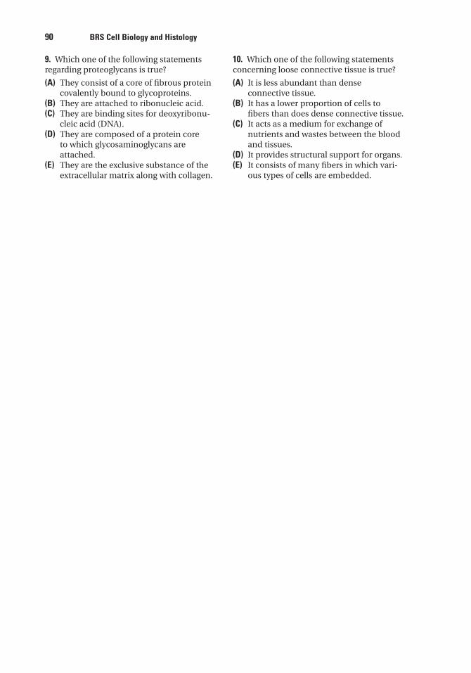
90 BRS Cell Biology and Histology
9. Which one of the following statementsregarding proteoglycans is true?
(A) They consist of a core of fibrous proteincovalently bound to glycoproteins.
(B) They are attached to ribonucleic acid.(C) They are binding sites for deoxyribonu-
cleic acid (DNA).(D) They are composed of a protein core
to which glycosaminoglycans areattached.
(E) They are the exclusive substance of theextracellular matrix along with collagen.
10. Which one of the following statementsconcerning loose connective tissue is true?
(A) It is less abundant than denseconnective tissue.
(B) It has a lower proportion of cells tofibers than does dense connective tissue.
(C) It acts as a medium for exchange ofnutrients and wastes between the bloodand tissues.
(D) It provides structural support for organs.(E) It consists of many fibers in which vari-
ous types of cells are embedded.
LWBK615-c06[79-91].qxd 05/31/2010 10:29 AM Page 90 Aptara
Answers and Explanations
91
1. A. Collagen is composed of closely packed tropocollagen molecules. Reticular fibers arecomposed of type III collagen, whereas elastic fibers are composed of elastin microfibrilsrather than collagen. Fibrocytes are inactive nonsecreting fibroblasts that synthesize theprocollagen molecules (see Chapter 6 II B 1).
2. C. Tendons are composed of dense regular connective tissue containing collagen fibersarranged in a uniform parallel fashion (see Chapter 6 IV B 2).
3. E. Pericytes are pluripotential cells that resemble fibroblasts, although they are smaller,and are adjacent to capillaries (see Chapter 6 III B).
4. D. Type II collagen is present only in hyaline and elastic cartilages; therefore, finding typeII collagen in the synovial fluid of a joint indicates erosion of the articular cartilage (seeChapter 6 II B 1).
5. D. Monocytes leave the bloodstream and migrate into the connective tissue, where theymature into functional macrophages (see Chapter 6 III E).
6. A. Foreign body giant cells result when macrophages coalesce (see Chapter 6 III E 3).
7. B. Eosinophils arise from myeloid stem cells during hemopoiesis and migrate to sites ofinflammation within the connective tissue. Pericytes, fibroblasts, osteoblasts, andadipocytes arise from undifferentiated mesenchymal cells (see Chapter 6 III H 2).
8. D. After first exposure to an allergen, plasma cells make IgE antibodies that bind to FceRIreceptors on mast cells (and basophils), sensitizing them. At the second exposure, theallergen binds to IgE, initiating degranulation of mast cells and releasing several mediatorsthat give rise to type I hypersensitivity reaction (see Chapter 6 III D 4).
9. D. Proteoglycans consist of a protein core to which glycosaminoglycans are attached (seeChapter 6 II A).
10. C. Both loose and dense connective tissues are composed of three elements: anamorphous ground substance, fibers, and various types of cells. The amorphous groundsubstance of loose connective tissue is the medium of exchange between the connectivetissue cells and the bloodstream (see Chapter 6 IV B 1).
LWBK615-c06[79-91].qxd 05/31/2010 10:29 AM Page 91 Aptara
c h a p t e r 7 Cartilage and Bone
92
I. OVERVIEW—CARTILAGE
Cartilage is an avascular specialized fibrous connective tissue. It has a firm extracellular matrixthat is less pliable than that of connective tissue proper, and it contains chondrocytes embed-ded in the matrix. Cartilage functions primarily to support soft tissues and assist in the devel-opment and growth of long bones. The three types of cartilage—hyaline cartilage, elasticcartilage, and fibrocartilage—vary in certain matrix components (Table 7.1).
A. Hyaline cartilage (Figures 7.1 and 7.2; Table 7.1) is the most abundant cartilage in the bodyand it also serves as a temporary skeleton in the fetus until it is replaced by bone.1. Structure
a. Matrix(1) The matrix is composed of an amorphous ground substance containing proteo-
glycan aggregates and chondronectin, in which type II collagen is embedded (seeTables 4.2 and 7.1).
(2) The matrix that is adjacent to chondrocytes is called the territorial matrix. This partof the matrix is poor in collagen but rich in proteoglycans, and it stains moredeeply than does the interterritorial matrix.
b. Perichondrium is a layer of dense, irregular connective tissue that surrounds hyalinecartilage except at articular surfaces.(1) It consists of an outer fibrous layer containing type I collagen, fibroblasts, and blood
vessels and an inner cellular layer containing chondrogenic cells and chondrob-lasts.
(2) It provides the nearest blood supply to the avascular cartilaginous tissue.c. Chondroblasts manufacture the cartilage matrix through which nutrients and waste
materials pass to and from the cells, respectively. These cells contain an extensiveGolgi complex, abundant rough endoplasmic reticulum (RER), lipid droplets, andglycogen. Mesenchymal cells can be induced to become secreting chondroblasts inthe proper environment, but if removed and grown as a monolayer in a low-densitysubstrate, they will discontinue secreting cartilage matrix, become fibroblastlike, andsecrete type I rather than type II collagen.
d. Chondrocytes are mature cartilage cells that are embedded within lacunae in thematrix.(1) They arise by differentiation of mesenchymal chondrogenic cells and from
chondrogenic cells within the inner layer of the perichondrium into chondrob-lasts, which are the earliest cells to produce cartilage matrix. Once these cellsbecome totally enveloped by matrix, they are referred to as chondrocytes (seeFigure 6.1).
(2) Chondrocytes located superficially are ovoid and positioned with their longitudi-nal axis parallel to the cartilage surface. Those located deeper are more nearlyspherical and may occur in groups of four to eight cells (isogenous groups).
LWBK615-c07[92-108].qxd 05/31/2010 10:37 AM Page 92 Aptara
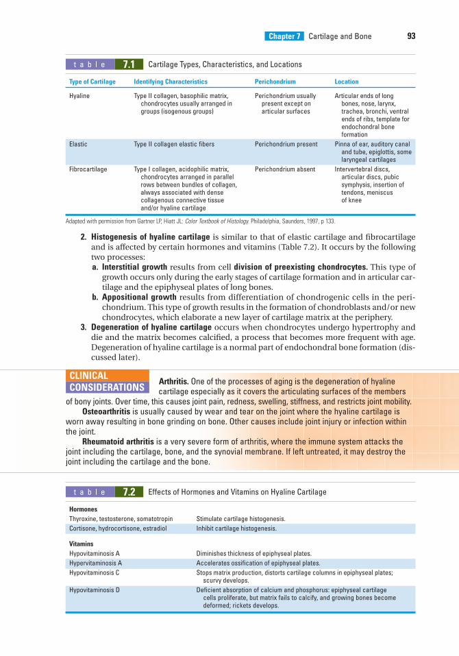
Chapter 7 Cartilage and Bone 93
2. Histogenesis of hyaline cartilage is similar to that of elastic cartilage and fibrocartilageand is affected by certain hormones and vitamins (Table 7.2). It occurs by the followingtwo processes:a. Interstitial growth results from cell division of preexisting chondrocytes. This type of
growth occurs only during the early stages of cartilage formation and in articular car-tilage and the epiphyseal plates of long bones.
b. Appositional growth results from differentiation of chondrogenic cells in the peri-chondrium. This type of growth results in the formation of chondroblasts and/or newchondrocytes, which elaborate a new layer of cartilage matrix at the periphery.
3. Degeneration of hyaline cartilage occurs when chondrocytes undergo hypertrophy anddie and the matrix becomes calcified, a process that becomes more frequent with age.Degeneration of hyaline cartilage is a normal part of endochondral bone formation (dis-cussed later).
t a b l e 7.1 Cartilage Types, Characteristics, and Locations
Type of Cartilage Identifying Characteristics Perichondrium Location
Hyaline Type II collagen, basophilic matrix, Perichondrium usually Articular ends of long chondrocytes usually arranged in present except on bones, nose, larynx,groups (isogenous groups) articular surfaces trachea, bronchi, ventral
ends of ribs, template for endochondral bone formation
Elastic Type II collagen elastic fibers Perichondrium present Pinna of ear, auditory canal and tube, epiglottis, somelaryngeal cartilages
Fibrocartilage Type I collagen, acidophilic matrix, Perichondrium absent Intervertebral discs, chondrocytes arranged in parallel articular discs, pubic rows between bundles of collagen, symphysis, insertion of always associated with dense tendons, meniscus collagenous connective tissue of kneeand/or hyaline cartilage
Adapted with permission from Gartner LP, Hiatt JL: Color Textbook of Histology. Philadelphia, Saunders, 1997, p 133.
Arthritis. One of the processes of aging is the degeneration of hyaline cartilage especially as it covers the articulating surfaces of the members
of bony joints. Over time, this causes joint pain, redness, swelling, stiffness, and restricts joint mobility.Osteoarthritis is usually caused by wear and tear on the joint where the hyaline cartilage is
worn away resulting in bone grinding on bone. Other causes include joint injury or infection withinthe joint.
Rheumatoid arthritis is a very severe form of arthritis, where the immune system attacks thejoint including the cartilage, bone, and the synovial membrane. If left untreated, it may destroy thejoint including the cartilage and the bone.
CLINICAL
CONSIDERATIONS
t a b l e 7.2 Effects of Hormones and Vitamins on Hyaline Cartilage
Hormones
Thyroxine, testosterone, somatotropin Stimulate cartilage histogenesis.
Cortisone, hydrocortisone, estradiol Inhibit cartilage histogenesis.
Vitamins
Hypovitaminosis A Diminishes thickness of epiphyseal plates.
Hypervitaminosis A Accelerates ossification of epiphyseal plates.
Hypovitaminosis C Stops matrix production, distorts cartilage columns in epiphyseal plates; scurvy develops.
Hypovitaminosis D Deficient absorption of calcium and phosphorus: epiphyseal cartilage cells proliferate, but matrix fails to calcify, and growing bones become deformed; rickets develops.
LWBK615-c07[92-108].qxd 05/31/2010 10:37 AM Page 93 Aptara
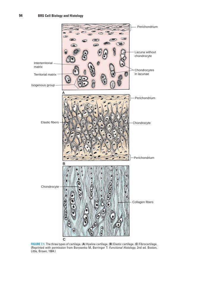
94 BRS Cell Biology and Histology
Interterritorialmatrix
Territorial matrix
Isogenous group
Elastic fibers
Chondrocyte
Perichondrium
Perichondrium
Perichondrium
Lacuna withoutchondrocyte
Chondrocytesin lacunae
Chondrocyte
Collagen fibers
A
B
CFIGURE 7.1. The three types of cartilage. (A) Hyaline cartilage. (B) Elastic cartilage. (C) Fibrocartilage.(Reprinted with permission from Borysenko M, Berringer T: Functional Histology, 2nd ed. Boston,Little, Brown, 1984.)
LWBK615-c07[92-108].qxd 05/31/2010 10:37 AM Page 94 Aptara
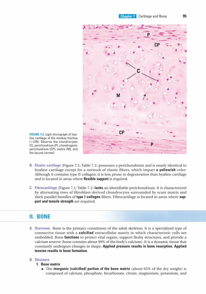
Chapter 7 Cartilage and Bone 95
C
M
P
P
CP
CPFIGURE 7.2. Light micrograph of hya-line cartilage of the monkey trachea(�270). Observe the chondrocytes(C), perichondrium (P), chondrogenicperichondrium (CP), matrix (M), andthe lacuna (arrow ).
B. Elastic cartilage (Figure 7.1; Table 7.1) possesses a perichondrium and is nearly identical tohyaline cartilage except for a network of elastic fibers, which impart a yellowish color.Although it contains type II collagen, it is less prone to degeneration than hyaline cartilageand is located in areas where flexible support is required.
C. Fibrocartilage (Figure 7.1; Table 7.1) lacks an identifiable perichondrium. It is characterizedby alternating rows of fibroblast-derived chondrocytes surrounded by scant matrix andthick parallel bundles of type I collagen fibers. Fibrocartilage is located in areas where sup-port and tensile strength are required.
II. BONE
A. Overview. Bone is the primary constituent of the adult skeleton. It is a specialized type ofconnective tissue with a calcified extracellular matrix in which characteristic cells areembedded. Bone functions to protect vital organs, support fleshy structures, and provide acalcium reserve (bone contains about 99% of the body’s calcium). It is a dynamic tissue thatconstantly undergoes changes in shape. Applied pressure results in bone resorption. Appliedtension results in bone formation.
B. Structure1. Bone matrix
a. The inorganic (calcified) portion of the bone matrix (about 65% of the dry weight) iscomposed of calcium, phosphate, bicarbonate, citrate, magnesium, potassium, and
LWBK615-c07[92-108].qxd 05/31/2010 10:37 AM Page 95 Aptara
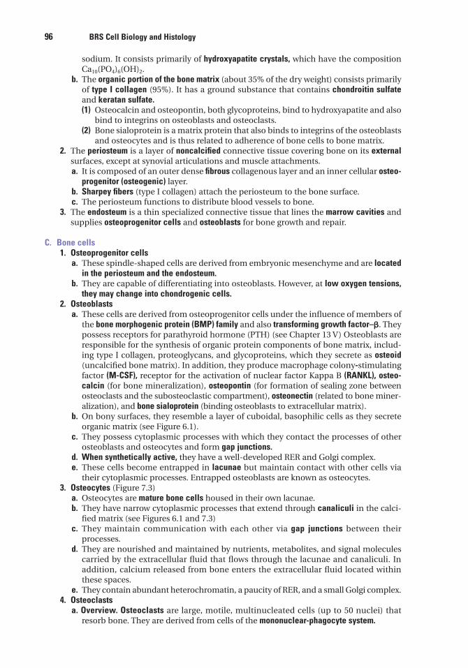
96 BRS Cell Biology and Histology
sodium. It consists primarily of hydroxyapatite crystals, which have the compositionCa10(PO4)6(OH)2.
b. The organic portion of the bone matrix (about 35% of the dry weight) consists primarilyof type I collagen (95%). It has a ground substance that contains chondroitin sulfateand keratan sulfate.(1) Osteocalcin and osteopontin, both glycoproteins, bind to hydroxyapatite and also
bind to integrins on osteoblasts and osteoclasts.(2) Bone sialoprotein is a matrix protein that also binds to integrins of the osteoblasts
and osteocytes and is thus related to adherence of bone cells to bone matrix.2. The periosteum is a layer of noncalcified connective tissue covering bone on its external
surfaces, except at synovial articulations and muscle attachments.a. It is composed of an outer dense fibrous collagenous layer and an inner cellular osteo-
progenitor (osteogenic) layer.b. Sharpey fibers (type I collagen) attach the periosteum to the bone surface.c. The periosteum functions to distribute blood vessels to bone.
3. The endosteum is a thin specialized connective tissue that lines the marrow cavities andsupplies osteoprogenitor cells and osteoblasts for bone growth and repair.
C. Bone cells1. Osteoprogenitor cells
a. These spindle-shaped cells are derived from embryonic mesenchyme and are locatedin the periosteum and the endosteum.
b. They are capable of differentiating into osteoblasts. However, at low oxygen tensions,they may change into chondrogenic cells.
2. Osteoblastsa. These cells are derived from osteoprogenitor cells under the influence of members of
the bone morphogenic protein (BMP) family and also transforming growth factor–�. Theypossess receptors for parathyroid hormone (PTH) (see Chapter 13 V) Osteoblasts areresponsible for the synthesis of organic protein components of bone matrix, includ-ing type I collagen, proteoglycans, and glycoproteins, which they secrete as osteoid(uncalcified bone matrix). In addition, they produce macrophage colony-stimulatingfactor (M-CSF), receptor for the activation of nuclear factor Kappa B (RANKL), osteo-calcin (for bone mineralization), osteopontin (for formation of sealing zone betweenosteoclasts and the subosteoclastic compartment), osteonectin (related to bone miner-alization), and bone sialoprotein (binding osteoblasts to extracellular matrix).
b. On bony surfaces, they resemble a layer of cuboidal, basophilic cells as they secreteorganic matrix (see Figure 6.1).
c. They possess cytoplasmic processes with which they contact the processes of otherosteoblasts and osteocytes and form gap junctions.
d. When synthetically active, they have a well-developed RER and Golgi complex.e. These cells become entrapped in lacunae but maintain contact with other cells via
their cytoplasmic processes. Entrapped osteoblasts are known as osteocytes.3. Osteocytes (Figure 7.3)
a. Osteocytes are mature bone cells housed in their own lacunae.b. They have narrow cytoplasmic processes that extend through canaliculi in the calci-
fied matrix (see Figures 6.1 and 7.3)c. They maintain communication with each other via gap junctions between their
processes.d. They are nourished and maintained by nutrients, metabolites, and signal molecules
carried by the extracellular fluid that flows through the lacunae and canaliculi. Inaddition, calcium released from bone enters the extracellular fluid located withinthese spaces.
e. They contain abundant heterochromatin, a paucity of RER, and a small Golgi complex.4. Osteoclasts
a. Overview. Osteoclasts are large, motile, multinucleated cells (up to 50 nuclei) thatresorb bone. They are derived from cells of the mononuclear-phagocyte system.
LWBK615-c07[92-108].qxd 05/31/2010 10:37 AM Page 96 Aptara
Chapter 7 Cartilage and Bone 97
(1) Osteoclasts possess cell surface receptors: colony-stimulating factor-1 receptor, cal-citonin receptor; and RANK (nuclear factor kappa B).
(2) Osteoblasts that have been stimulated by PTH promote osteoclast formation,whereas osteoblasts that have been stimulated by calcitonin inhibit osteoclast for-mation by stimulating osteoid synthesis and calcium deposition.
(3) Via a series of three osteoblast signals, osteoclast precursors (macrophages) arestimulated by M-CSF to undergo mitosis. Another signaling molecule, RANKL,binds to the precursor, inducing it to differentiate into the multinucleated osteo-clast, thus activating it to commence bone resorption. A third signal, osteoprote-gerin (OPG), a member of the tumor necrosis factor receptor (TNFR) family producedby osteoblasts and other cells, can prohibit RANKL from binding to the macrophage,thus prohibiting osteoclast formation.
b. Osteoclast cytoplasm is usually acidophilic. Osteoclasts function in the resorption ofbone (osteolysis). They form and reside in depressions known as Howship’s lacunae,which represent areas of bone resorption.
c. Morphology. Osteoclasts display four regions in electron micrographs.(1) Basal zone is the region that houses most of the organelles of the osteoclast and is
the farthest from the subosteoclastic compartment(2) The ruffled border is the site of active bone resorption. It is composed of irregular
fingerlike cytoplasmic projections extending into the subosteoclastic compart-ment, a slight depression that deepens as the osteoclast resorbs bone and then thatdepression is referred to as Howship’s lacuna.
(3) The clear zone surrounds the ruffled border. It contains actin filaments at theperiphery that help osteoclasts maintain contact with the bony surface andisolates the region of osteolytic activity. Osteopontin, secreted by osteoblasts, isused to seal the zone between osteoclasts and the subosteoclastic compartment.
(4) The vesicular zone contains exocytotic vesicles that transfer lysosomal enzymes toHowship’s lacunae and endocytotic vesicles that transfer degraded bone productsfrom Howship’s lacunae to the interior of the cell.
Canaliculi
Haversiancanal
Lacuna
Cancellousbone
Concentric lamella
Osteon
Outer circumferentiallamellae
Periosteum
Compact bone
Inner circumferentiallamellae
Marrowcavity
Sharpey fibers
Blood vessel
Volkmanncanal
Haversiancanal
FIGURE 7.3. Histological aspects of a long bone. Inset: cross-section of an osteon. (Adapted with permission fromDenhardt D, Guo X: A protein with diverse functions. FASEB J 7:1475–1482, 1993.)
LWBK615-c07[92-108].qxd 05/31/2010 10:37 AM Page 97 Aptara
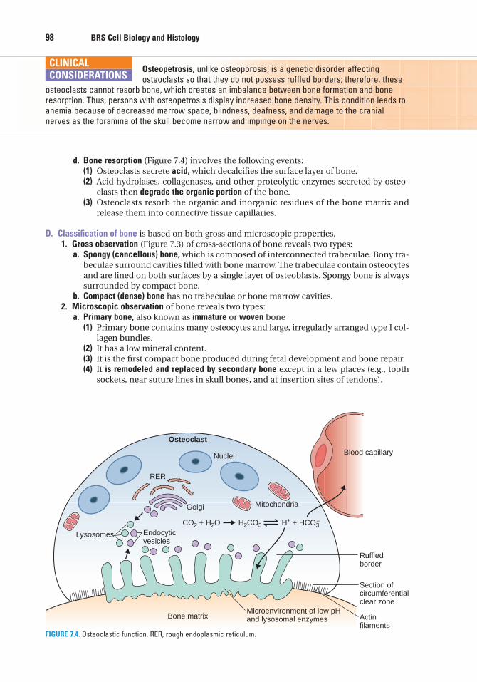
98 BRS Cell Biology and Histology
Osteopetrosis, unlike osteoporosis, is a genetic disorder affecting osteoclasts so that they do not possess ruffled borders; therefore, these
osteoclasts cannot resorb bone, which creates an imbalance between bone formation and boneresorption. Thus, persons with osteopetrosis display increased bone density. This condition leads toanemia because of decreased marrow space, blindness, deafness, and damage to the cranialnerves as the foramina of the skull become narrow and impinge on the nerves.
CLINICAL
CONSIDERATIONS
d. Bone resorption (Figure 7.4) involves the following events:(1) Osteoclasts secrete acid, which decalcifies the surface layer of bone.(2) Acid hydrolases, collagenases, and other proteolytic enzymes secreted by osteo-
clasts then degrade the organic portion of the bone.(3) Osteoclasts resorb the organic and inorganic residues of the bone matrix and
release them into connective tissue capillaries.
D. Classification of bone is based on both gross and microscopic properties.1. Gross observation (Figure 7.3) of cross-sections of bone reveals two types:
a. Spongy (cancellous) bone, which is composed of interconnected trabeculae. Bony tra-beculae surround cavities filled with bone marrow. The trabeculae contain osteocytesand are lined on both surfaces by a single layer of osteoblasts. Spongy bone is alwayssurrounded by compact bone.
b. Compact (dense) bone has no trabeculae or bone marrow cavities.2. Microscopic observation of bone reveals two types:
a. Primary bone, also known as immature or woven bone(1) Primary bone contains many osteocytes and large, irregularly arranged type I col-
lagen bundles.(2) It has a low mineral content.(3) It is the first compact bone produced during fetal development and bone repair.(4) It is remodeled and replaced by secondary bone except in a few places (e.g., tooth
sockets, near suture lines in skull bones, and at insertion sites of tendons).
CO2 + H2O H+ + HCO3
Nuclei
Bone matrixMicroenvironment of low pHand lysosomal enzymes
Section of circumferentialclear zone
Actinfilaments
Lysosomes Endocyticvesicles
Golgi
RER
Ruffledborder
Osteoclast
H2CO3–
Mitochondria
Blood capillary
FIGURE 7.4. Osteoclastic function. RER, rough endoplasmic reticulum.
LWBK615-c07[92-108].qxd 05/31/2010 10:37 AM Page 98 Aptara
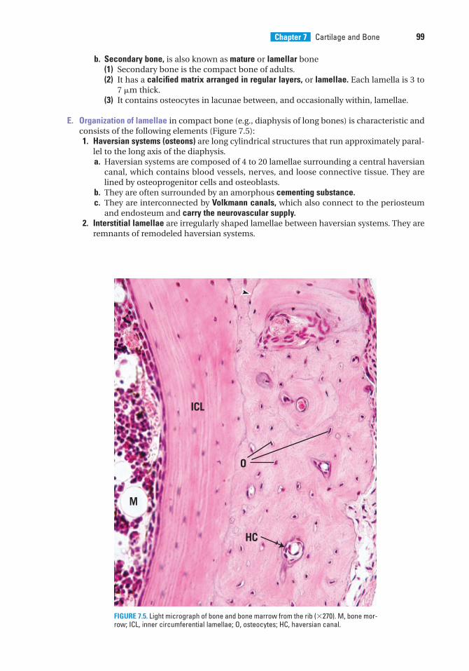
Chapter 7 Cartilage and Bone 99
b. Secondary bone, is also known as mature or lamellar bone(1) Secondary bone is the compact bone of adults.(2) It has a calcified matrix arranged in regular layers, or lamellae. Each lamella is 3 to
7 �m thick.(3) It contains osteocytes in lacunae between, and occasionally within, lamellae.
E. Organization of lamellae in compact bone (e.g., diaphysis of long bones) is characteristic andconsists of the following elements (Figure 7.5):1. Haversian systems (osteons) are long cylindrical structures that run approximately paral-
lel to the long axis of the diaphysis.a. Haversian systems are composed of 4 to 20 lamellae surrounding a central haversian
canal, which contains blood vessels, nerves, and loose connective tissue. They arelined by osteoprogenitor cells and osteoblasts.
b. They are often surrounded by an amorphous cementing substance.c. They are interconnected by Volkmann canals, which also connect to the periosteum
and endosteum and carry the neurovascular supply.2. Interstitial lamellae are irregularly shaped lamellae between haversian systems. They are
remnants of remodeled haversian systems.
M
ICL
HC
O
FIGURE 7.5. Light micrograph of bone and bone marrow from the rib (�270). M, bone mor-row; ICL, inner circumferential lamellae; O, osteocytes; HC, haversian canal.
LWBK615-c07[92-108].qxd 05/31/2010 5:01 PM Page 99 Aptara
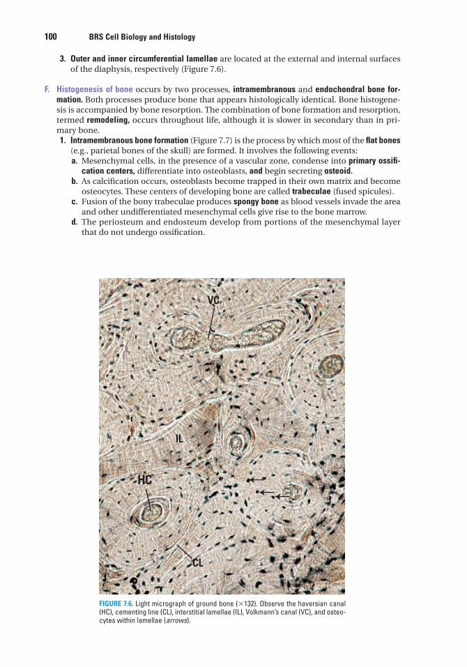
100 BRS Cell Biology and Histology
3. Outer and inner circumferential lamellae are located at the external and internal surfacesof the diaphysis, respectively (Figure 7.6).
F. Histogenesis of bone occurs by two processes, intramembranous and endochondral bone for-mation. Both processes produce bone that appears histologically identical. Bone histogene-sis is accompanied by bone resorption. The combination of bone formation and resorption,termed remodeling, occurs throughout life, although it is slower in secondary than in pri-mary bone.1. Intramembranous bone formation (Figure 7.7) is the process by which most of the flat bones
(e.g., parietal bones of the skull) are formed. It involves the following events:a. Mesenchymal cells, in the presence of a vascular zone, condense into primary ossifi-
cation centers, differentiate into osteoblasts, and begin secreting osteoid.b. As calcification occurs, osteoblasts become trapped in their own matrix and become
osteocytes. These centers of developing bone are called trabeculae (fused spicules).c. Fusion of the bony trabeculae produces spongy bone as blood vessels invade the area
and other undifferentiated mesenchymal cells give rise to the bone marrow.d. The periosteum and endosteum develop from portions of the mesenchymal layer
that do not undergo ossification.
VC
HC
CL
IL
FIGURE 7.6. Light micrograph of ground bone (�132). Observe the haversian canal(HC), cementing line (CL), interstitial lamellae (IL), Volkmann’s canal (VC), and osteo-cytes within lamellae (arrows).
LWBK615-c07[92-108].qxd 05/31/2010 10:37 AM Page 100 Aptara
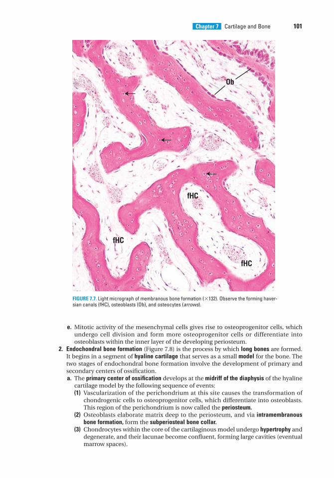
Chapter 7 Cartilage and Bone 101
e. Mitotic activity of the mesenchymal cells gives rise to osteoprogenitor cells, whichundergo cell division and form more osteoprogenitor cells or differentiate intoosteoblasts within the inner layer of the developing periosteum.
2. Endochondral bone formation (Figure 7.8) is the process by which long bones are formed.It begins in a segment of hyaline cartilage that serves as a small model for the bone. Thetwo stages of endochondral bone formation involve the development of primary andsecondary centers of ossification.a. The primary center of ossification develops at the midriff of the diaphysis of the hyaline
cartilage model by the following sequence of events:(1) Vascularization of the perichondrium at this site causes the transformation of
chondrogenic cells to osteoprogenitor cells, which differentiate into osteoblasts.This region of the perichondrium is now called the periosteum.
(2) Osteoblasts elaborate matrix deep to the periosteum, and via intramembranousbone formation, form the subperiosteal bone collar.
(3) Chondrocytes within the core of the cartilaginous model undergo hypertrophy anddegenerate, and their lacunae become confluent, forming large cavities (eventualmarrow spaces).
Ob
fHC
fHC
fHC
FIGURE 7.7. Light micrograph of membranous bone formation (�132). Observe the forming haver-sian canals (fHC), osteoblasts (Ob), and osteocytes (arrows).
LWBK615-c07[92-108].qxd 05/31/2010 10:38 AM Page 101 Aptara
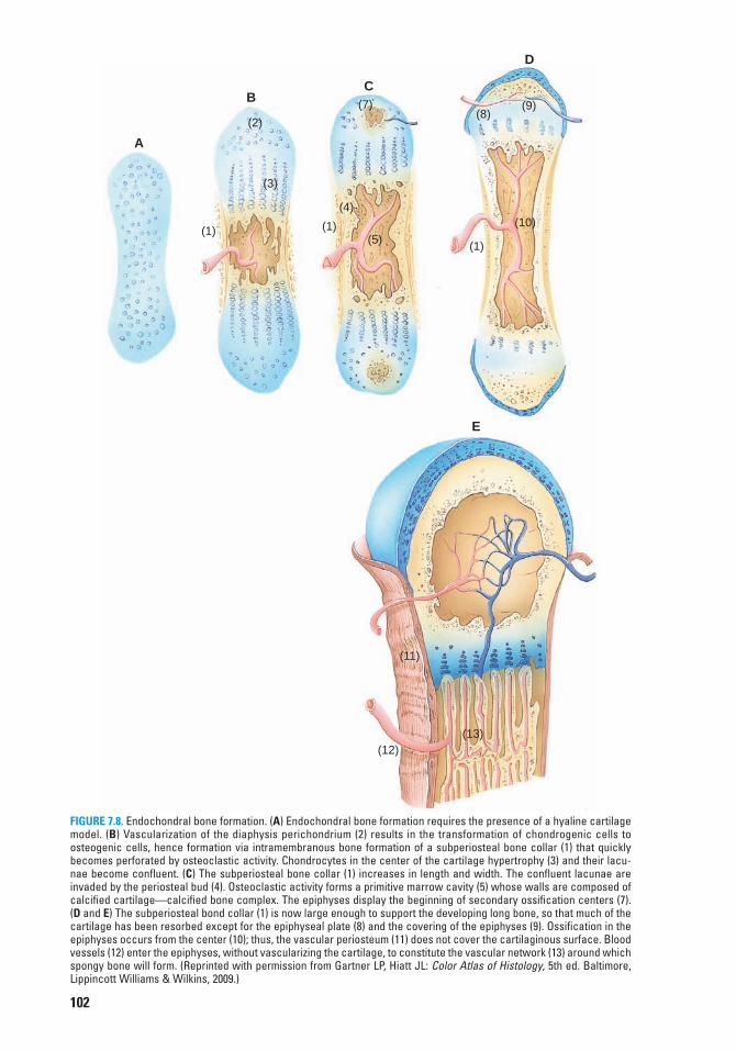
A
B
(2)
(3)
(1)
C(7)
(4)
(1)(5)
D
(9)(8)
(10)
(1)
E
(11)
(12)(13)
FIGURE 7.8. Endochondral bone formation. (A) Endochondral bone formation requires the presence of a hyaline cartilagemodel. (B) Vascularization of the diaphysis perichondrium (2) results in the transformation of chondrogenic cells toosteogenic cells, hence formation via intramembranous bone formation of a subperiosteal bone collar (1) that quicklybecomes perforated by osteoclastic activity. Chondrocytes in the center of the cartilage hypertrophy (3) and their lacu-nae become confluent. (C) The subperiosteal bone collar (1) increases in length and width. The confluent lacunae areinvaded by the periosteal bud (4). Osteoclastic activity forms a primitive marrow cavity (5) whose walls are composed ofcalcified cartilage—calcified bone complex. The epiphyses display the beginning of secondary ossification centers (7).(D and E) The subperiosteal bond collar (1) is now large enough to support the developing long bone, so that much of thecartilage has been resorbed except for the epiphyseal plate (8) and the covering of the epiphyses (9). Ossification in theepiphyses occurs from the center (10); thus, the vascular periosteum (11) does not cover the cartilaginous surface. Bloodvessels (12) enter the epiphyses, without vascularizing the cartilage, to constitute the vascular network (13) around whichspongy bone will form. (Reprinted with permission from Gartner LP, Hiatt JL: Color Atlas of Histology, 5th ed. Baltimore,Lippincott Williams & Wilkins, 2009.)
102
LWBK615-c07[92-108].qxd 05/31/2010 10:38 AM Page 102 Aptara
Chapter 7 Cartilage and Bone 103
(4) Osteoclasts create perforations in the bone collar that permit the periosteal bud(blood vessels, osteoprogenitor cells, and mesenchymal cells) to enter the newlyformed spaces in the cartilaginous model. The cartilage that constitutes the wallsof these spaces then becomes calcified.
(5) Newly developed osteoblasts elaborate bone matrix that becomes calcified on thesurface of the calcified cartilage, forming a calcified cartilage–calcified bone com-plex. In histological sections, the calcified cartilage stains basophilic, whereas thecalcified bone stains acidophilic.
(6) The subperiosteal bone collar becomes thicker and elongates toward the epiphysis.(7) Osteoclasts begin to resorb the calcified cartilage–calcified bone complex, thus
enlarging the primitive marrow cavity.(8) Repetition of this sequence of events results in bone formation spreading toward
the epiphyses.b. Secondary centers of ossification develop at the epiphyses in a sequence of events sim-
ilar to that described for the primary center, except a bone collar is not formed.(1) Development of these centers begins when osteoprogenitor cells invade the epi-
physis and differentiate into osteoblasts, which elaborate bone matrix to replacethe disintegrating cartilage. When the epiphyses are filled with bone tissue, carti-lage remains in two areas, the articular surfaces and the epiphyseal plates.
(2) Articular cartilage persists and does not contribute to bone formation.(3) Epiphyseal plates continue to grow by adding new cartilage at the epiphyseal end
while it is being replaced with bone at the diaphyseal end (lengthening the bone).(4) Ossification of the epiphyseal plates and cessation of growth occur at about
20 years of age.3. Zones of the epiphyseal plates are histologically distinctive and arranged in the following
order:a. The zone of reserve cartilage is at the epiphyseal side of the plate. It possesses small,
randomly arranged inactive chondrocytes.b. The zone of proliferation (of chondrocytes) is a region of rapid mitotic divisions giving
rise to rows of isogenous cell groups.c. The zone of cell hypertrophy and maturation is the region where the chondrocytes are
greatly enlarged.d. The zone of calcification is the region where hypertrophied chondrocytes die and the
cartilage becomes calcified.e. The zone of ossification is the area where newly formed osteoblasts elaborate bone
matrix on the calcified cartilage, forming a calcified cartilage–calcified bone complex,which is resorbed and replaced by bone.
4. Calcification of bone is not clearly understood, however,a. Osteonectin, proteoglycans, and bone sialoprotein, are known to stimulate calcification.b. Matrix vesicles, released by osteoblasts, contain high concentrations of calcium Ca2�
and PO43� ions along with several other organic compounds and enzymes.
c. Calcium pumps in the matrix membranes bring in more calcium, concentrating itand forming calcium hydroxyapatite crystals that grow and eventually puncture thematrix vesicle expelling its contents.
d. Calcium hydroxyapatite crystals that become free in the matrix become nidi of crys-tallization.
e. Released enzymes free phosphate ions that unite with the calcium forming calciumphosphate.
f. Calcium phosphate then begins to calcify the matrix around the nidi of crystallization.g. Water is removed from the matrix permitting hydroxyapatite crystals to be deposited
into gaps within the collagen fibrils.h. Nidi of mineralization, enlarge, fuse with neighboring nidi eventually calcifying the
entire matrix.
G. Bone remodeling. Bone is constantly being remodeled as necessary for growth and to alterits structural makeup to adapt to changing stresses in the environment throughout life.
LWBK615-c07[92-108].qxd 05/31/2010 10:38 AM Page 103 Aptara
104 BRS Cell Biology and Histology
1. Early on, bone development outpaces bone resorption as new haversian systems areadded and fewer are resorbed.
2. Later, when the epiphyseal plates are closed, ending bone growth, bone development,and resorption are balanced.
Several factors, including calcitonin and PTH, are responsible for this phenomenonregarding compact bone (see section II J). Remodeling of cancellous bone is under thecontrol of many factors within the bone marrow.
H. Repair of a bone fracture. A bone fracture damages the matrix, bone cells, and blood vesselsin the region and is accompanied by localized hemorrhaging and blood clot formation.1. Proliferation of osteoprogenitor cells occurs in the periosteum and endosteum in the
vicinity of the fracture. As a result of this proliferation, cellular tissue surrounds the frac-ture and penetrates between the ends of the damaged bone.
2. Formation of a bony callus occurs both internally and externally at a fracture site.a. Fibrous connective tissue and hyaline cartilage are formed in the fracture zone.b. Endochondral bone formation replaces the cartilage with primary bone.c. Intramembranous bone formation also produces primary bone in the area.d. The irregularly arranged trabeculae of primary bone join the ends of the fractured
bone, forming a bony callus.e. The primary bone is resorbed and replaced with secondary bone as the fracture heals.
Bone Repair. After severe injury where segments of bone have been lostor must be removed, the remaining bone is prevented from forming a
bony union followed by a bony callus that over time would result in completed bone repair. Whenthe bony union is not possible, a bone graft is required. For this purpose, bone fragments that arestored frozen to maintain osteogenic potential then may be utilized in bone grafts.
Autographs are most successful since the bone donor is the recipient.Homographs are of bone donated from a different individual.Heterographs are the least successful because the donor bone comes from another species.
However, calf bone that has been frozen can serve as a viable bone graft when necessary.
CLINICAL
CONSIDERATIONS
I. Role of vitamins in bone formation1. Vitamin D is necessary for absorption of calcium from the small intestine. Vitamin D defi-
ciency results in poorly calcified (soft) bone, a condition known as rickets in children andosteomalacia in adults. Vitamin D is also necessary for bone formation (ossification),whereas an excess of vitamin D causes bone resorption.
2. Vitamin A deficiency inhibits proper bone formation and growth, whereas an excessaccelerates ossification of the epiphyseal plates. Deficiency or excess of vitamin A resultsin small stature.
3. Vitamin C is necessary for collagen formation. Deficiency results in scurvy, characterizedby poor bone growth and inadequate fracture repair.
Rickets occurs in children deficient in vitamin D, which results in calciumdeficiency. It is characterized by deficient calcification in newly formed
bone and is generally accompanied by deformation of the bone spicules in epiphyseal plates; asa result, bones grow more slowly than normal and are deformed by the stress of weight bearing.Osteomalacia (rickets of adults) results from calcium deficiency.
1. It is characterized by deficient calcification in newly formed bone and decalcification of alreadycalcified bone.
2. This disease may be severe during pregnancy because the calcium requirements of the fetusmay lead to calcium loss from the mother.
CLINICAL
CONSIDERATIONS
LWBK615-c07[92-108].qxd 05/31/2010 10:38 AM Page 104 Aptara
Chapter 7 Cartilage and Bone 105
J. Role of hormones in bone formation1. Parathyroid hormone activates osteoblasts to secrete osteoclast-stimulating factor, which
then activates osteoclasts to resorb bone, thus elevating blood calcium levels. Excess PTH(hyperparathyroidism) renders bone more susceptible to fracture and subsequent depo-sition of calcium in arterial walls and certain organs, such as the kidney.
2. Calcitonin is produced by parafollicular cells (C cells) of the thyroid gland. It eliminatesthe ruffled border of osteoclasts and inhibits bone matrix resorption, preventing therelease of calcium.
3. Pituitary growth hormone (somatotropin) is produced in the pars distalis of the pituitarygland. It stimulates overall growth, especially that of epiphyseal plates, and influencesbone development via insulinlike growth factors (somatomedins), especially stimulatinggrowth of the epiphyseal plates. Children deficient in this hormone exhibit dwarfism,whereas adults with an excess of somatotropin in their growing years display pituitarygigantism and acromegaly.
Osteoporosis is a disease characterized by low bone mass (low bone mineral density) and structural deterioration of bone tissue, making the
bone fragile and susceptible to fracture. Osteoporosis is associated with an abnormal ratio ofmineral to matrix.
1. It results from increased bone resorption, decreased bone formation, or both.2. Estrogen activates bone formation by osteoblasts, and in its absence an imbalance causes osteo-
clastic activity to render bones fragile and susceptible to fracture.a. Osteoporosis is most common in postmenopausal women because of diminished estrogen
secretion and in immobile patients because of lack of physical stress on the bone.b. Estrogen therapy was employed for decades to minimize the onset of osteoporosis. Recently, it
was determined that estrogen replacement therapy increases the risk of heart disease, stroke,breast cancer, and blood clots. Now, instead of estrogen, a recent new group of drugs, thebisphosphonates, has been developed that reduce the incidence of osteoporosis.
c. Preventive measures include a balanced diet rich in calcium and vitamin D and weight-bearing exercise.
AcromegalyAcromegaly results from an excess of pituitary growth hormone in adults. It is characterized by verythick bones in the extremities and in portions of the facial skeleton.
CLINICAL
CONSIDERATIONS
III. JOINTS
A. Synarthroses are immovable joints composed of connective tissue, cartilage, or bone. Thesejoints unite the first rib to the sternum and connect the skull bones to each other.
B. Diarthroses (synovial joints) permit maximum movement and generally unite long bones. Thesejoints are surrounded by a two-layered capsule, enclosing and sealing the articular cavity.The articular cavity contains synovial fluid, a colorless, viscous fluid that is rich in hyaluronicacid and proteins.1. The external (fibrous) capsular layer is a tough, fibrous layer of dense connective tissue.2. The internal (synovial) capsular layer is also called the synovial membrane. It is lined by a
layer of squamous to cuboidal epithelial cells on its internal surface. Two cell types aredisplayed in electron micrographs of this epithelium.a. Type A cells are intensely phagocytic and have a well-developed Golgi complex, many
lysosomes, and sparse RER.b. Type B cells resemble fibroblasts and have a well-developed RER; these cells probably
secrete synovial fluid.
LWBK615-c07[92-108].qxd 05/31/2010 10:38 AM Page 105 Aptara
5. A 7-year-old boy is seen by hispediatrician because the child broke hishumerus as he tripped and fell whilewalking. The pediatrician asked about thechild’s diet and learned that he might have adietary deficiency. Which of the followingmay be lacking in his diet?
(A) Potassium(B) Calcium(C) Iron(D) Carbohydrates(E) Protein
6. A 22-year-old woman is seen for the firsttime by her new physician, who notes thatshe has very thick bones in her extremitiesand face. The physician suspects acromegaly,caused by which of the following?
(A) Hypervitaminosis A(B) Excess growth hormone(C) Hypovitaminosis A(D) Hypervitaminosis D(E) Hypovitaminosis D
7. Which of the following statements is char-acteristic of bone?
(A) Bone matrix contains primarily type IIcollagen.
(B) About 65% of the dry weight of bone isorganic.
(C) Haversian canals are interconnected viaVolkmann canals.
(D) Bone growth occurs via interstitialgrowth only.
(E) Bone growth occurs via appositionalgrowth only.
1. Which of the following statementscharacterizes osteoclasts?
(A) They are enucleated cells.(B) They produce collagen.(C) They secrete osteoid.(D) They are derived from osteoprogenitor
cells.(E) They occupy Howship’s lacunae.
2. Which one of the following statements iscorrect concerning the periosteum?
(A) It is devoid of a blood supply.(B) It produces osteoclasts.(C) It is responsible for interstitial bone
growth.(D) Its inner layer contains osteoprogenitor
cells.(E) Its outer layer is devoid of fibers.
3. Which one of the following statements ischaracteristic of osteocytes?
(A) They communicate via gap junctionsbetween their processes.
(B) They contain large amounts of RER.(C) They are immature bone cells.(D) They are housed as isogenous groups in
lacunae.(E) They give rise to osteoclasts.
4. Which one of the following statementsconcerning hyaline cartilage is correct?
(A) It is vascular.(B) It contains type IV collagen.(C) It undergoes appositional growth only.(D) It is located at the articular ends of long
bones.(E) Its chondrocytes are aligned in rows.
Review Test
Directions: Each of the numbered items or incomplete statements in this section is followed byanswers or by completions of the statement. Select the ONE lettered answer or completion thatis BEST in each case.
106
LWBK615-c07[92-108].qxd 05/31/2010 10:38 AM Page 106 Aptara
Chapter 7 Cartilage and Bone 107
8. Which one of the following inhibits histoge-nesis of cartilage?
(A) Thyroxine(B) Hypervitaminosis A(C) Hypovitaminosis D(D) Hydrocortisone(E) Hypovitaminosis C
9. Which one of the following stimulatescartilage histogenesis?
(A) Thyroxine(B) Hypervitaminosis A(C) Hypovitaminosis D(D) Hydrocortisone(E) Hypovitaminosis C
10. Which one of the following acceleratesepiphyseal ossification?
(A) Thyroxine(B) Hypervitaminosis A(C) Absence of vitamin D(D) Hydrocortisone(E) Hypovitaminosis C
11. Which one of the following makesepiphyseal cartilage matrix fail to calcify?
(A) Thyroxine(B) Hypervitaminosis A(C) Hypovitaminosis D(D) Hydrocortisone(E) Hypovitaminosis C
12. A 25-year-old patient, anemic for severalyears, complains of failing eyesight andhearing loss. During a physical examination,it is determined that the patient has lostfunction of some of the cranial nerves.The diagnosis could be which one of thefollowing?
(A) Osteoporosis(B) Osteomalacia(C) Rickets(D) Acromegaly(E) Osteopetrosis
LWBK615-c07[92-108].qxd 05/31/2010 10:38 AM Page 107 Aptara
Answers and Explanations
108
1. E. Osteoclasts are multinucleated cells that produce proteolytic enzymes and occupyHowship’s lacunae. They are not derived from osteoprogenitor cells but from monocyteprecursors (see Chapter 7 II C 4).
2. D. The inner layer of the periosteum possesses osteoprogenitor cells, whereas the outerlayer of the periosteum is fibrous. The periosteum functions to distribute blood vessels tothe bone; thus, appositional bone growth takes place here (see Chapter 7 II B 2).
3. A. Osteocytes communicate with each other via gap junctions on narrow cytoplasmicprocesses that extend through canaliculi. They are mature bone cells that occupy individ-ual lacunae as mature resting bone cells (see Chapter 7 II C 3).
4. D. Hyaline cartilage is avascular, contains type II collagen, and grows both interstitiallyand appositionally. It is located at the articulating ends of long bones (see Chapter 7 I A).
5. B. Because calcium must be maintained at a constant level in the blood and the tissues, adiet deficient in calcium leads to calcium loss from the bones. As a result, the bonesbecome fragile (see Chapter 7 II I Clinical Considerations).
6. B. Excessive growth hormone causes acromegaly. Excessive vitamin D causes boneresorption. Both an excess and a deficiency of vitamin A result in short stature (see Chapter 7 II J Clinical Considerations).
7. C. Haversian canals run longitudinally, parallel to the long axis of bone. They areconnected to one another by Volkmann canals that run perpendicular (or obliquely) tothem (see Chapter 7 II E 1).
8. D. Hydrocortisone inhibits cartilage growth and matrix formation (see Chapter 7 I A 2).
9. A. Thyroxine, testosterone, and somatotropin stimulate cartilage growth and matrixformation (see Chapter 7 I A 2).
10. B. Hypervitaminosis A accelerates ossification of epiphyseal plates, whereashypovitaminosis A reduces the width of the epiphyseal plates (see Chapter 7 II I).
11. C. In the absence of vitamin D, epiphyseal chondrocytes continue to proliferate, but theirmatrix does not calcify, which leads to rickets (see Chapter 7 II I Clinical Considerations).
12. E. Osteopetrosis is a genetic defect involving the osteoclasts. Persons with this defectpossess osteoclasts without ruffled borders, which prohibit them from resorbing bone.Therefore, bone forms but is not resorbed. This leads to increased bone density, anemia,blindness, deafness, and cranial nerve involvement because the nerves are impinged uponas they exit the cranium via their foramina (see Chapter 7 II B Clinical Considerations).
LWBK615-c07[92-108].qxd 05/31/2010 10:38 AM Page 108 Aptara
c h a p t e r 8 Muscle
109
I. OVERVIEW—MUSCLE
A. Muscle is classified into two types: striated and smooth, and striated muscle has two subdivi-sions: skeletal and cardiac muscles.
B. Muscle cells possess contractile filaments whose major components are actin and myosin.
C. Contraction may be voluntary (skeletal muscles) or involuntary (cardiac and smooth muscles).
II. STRUCTURE OF SKELETAL MUSCLE
A. Connective tissue investments convey neural and vascular elements to muscle cells and pro-vide a vehicle that harnesses the forces of muscle contraction.1. Epimysium surrounds an entire muscle and forms aponeuroses, which connect skeletal
muscle to muscle, and tendons, which connect and skeletal muscle to bone.2. Perimysium surrounds fascicles (small bundles) of muscle cells.3. Endomysium surrounds individual muscle cells and is composed of reticular fibers and an
external lamina.
B. Types of skeletal muscle cells1. Types of skeletal muscle cells (also known as muscle fibers) include red (slow contraction
but do not fatigue easily), white (fast contraction but fatigue easily), and intermediate. Allthree types may be present in a given muscle.
2. These three types differ from each other in their content of myoglobin (a protein that issimilar to hemoglobin in that it binds O2), number of mitochondria, concentration of vari-ous enzymes, and rate of contraction (Table 8.1).
3. A change in innervation can change a fiber’s type. If a red fiber is denervated and itsinnervation replaced with that of a white fiber, the red fiber will change its characteris-tics and will become a white fiber.
C. Skeletal muscle cells (Figures 8.1 and 8.2) are long, cylindrical, multinucleated and areenveloped by an external lamina and reticular fibers. Their cytoplasm is called sar-coplasm, and their plasmalemma is called the sarcolemma and forms deep tubular invagi-nations, or T (transverse) tubules, which extend into the cells. Skeletal muscle cells possesscylindrical collections of myofibrils, 1 to 2 �m in diameter, which extend the entire lengthof the cell.1. Myofibrils are composed of longitudinally arranged, cylindrical bundles of thick and thin
myofilaments observable by transmission electron microscopy (see Figure 8.1).
LWBK615-c08[109-125].qxd 05/31/2010 10:33 AM Page 109 Aptara
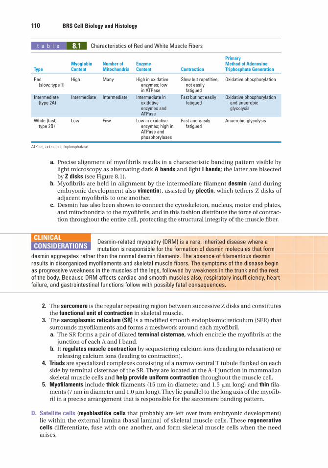
110 BRS Cell Biology and Histology
a. Precise alignment of myofibrils results in a characteristic banding pattern visible bylight microscopy as alternating dark A bands and light I bands; the latter are bisectedby Z disks (see Figure 8.1).
b. Myofibrils are held in alignment by the intermediate filament desmin (and duringembryonic development also vimentin), assisted by plectin, which tethers Z disks ofadjacent myofibrils to one another.
c. Desmin has also been shown to connect the cytoskeleton, nucleus, motor end plates,and mitochondria to the myofibrils, and in this fashion distribute the force of contrac-tion throughout the entire cell, protecting the structural integrity of the muscle fiber.
t a b l e 8.1 Characteristics of Red and White Muscle Fibers
Primary Myoglobin Number of Enzyme Method of Adenosine
Type Content Mitochondria Content Contraction Triphosphate Generation
Red High Many High in oxidative Slow but repetitive; Oxidative phosphorylation(slow; type 1) enzymes; low not easily
in ATPase fatigued
Intermediate Intermediate Intermediate Intermediate in Fast but not easily Oxidative phosphorylation (type 2A) oxidative fatigued and anaerobic
enzymes and glycolysisATPase
White (fast; Low Few Low in oxidative Fast and easily Anaerobic glycolysistype 2B) enzymes; high in fatigued
ATPase and phosphorylases
ATPase, adenosine triphosphatase.
Desmin-related myopathy (DRM) is a rare, inherited disease where amutation is responsible for the formation of desmin molecules that form
desmin aggregates rather than the normal desmin filaments. The absence of filamentous desminresults in disorganized myofilaments and skeletal muscle fibers. The symptoms of the disease beginas progressive weakness in the muscles of the legs, followed by weakness in the trunk and the restof the body. Because DRM affects cardiac and smooth muscles also, respiratory insufficiency, heartfailure, and gastrointestinal functions follow with possibly fatal consequences.
CLINICAL
CONSIDERATIONS
2. The sarcomere is the regular repeating region between successive Z disks and constitutesthe functional unit of contraction in skeletal muscle.
3. The sarcoplasmic reticulum (SR) is a modified smooth endoplasmic reticulum (SER) thatsurrounds myofilaments and forms a meshwork around each myofibril.a. The SR forms a pair of dilated terminal cisternae, which encircle the myofibrils at the
junction of each A and I band.b. It regulates muscle contraction by sequestering calcium ions (leading to relaxation) or
releasing calcium ions (leading to contraction).4. Triads are specialized complexes consisting of a narrow central T tubule flanked on each
side by terminal cisternae of the SR. They are located at the A–I junction in mammalianskeletal muscle cells and help provide uniform contraction throughout the muscle cell.
5. Myofilaments include thick filaments (15 nm in diameter and 1.5 �m long) and thin fila-ments (7 nm in diameter and 1.0 �m long). They lie parallel to the long axis of the myofib-ril in a precise arrangement that is responsible for the sarcomere banding pattern.
D. Satellite cells (myoblastlike cells that probably are left over from embryonic development)lie within the external lamina (basal lamina) of skeletal muscle cells. These regenerativecells differentiate, fuse with one another, and form skeletal muscle cells when the needarises.
LWBK615-c08[109-125].qxd 05/31/2010 10:33 AM Page 110 Aptara
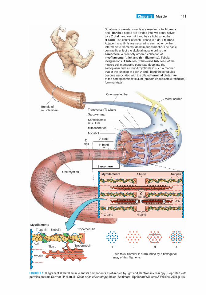
Chapter 8 Muscle 111
Striations of skeletal muscle are resolved into A bands and I bands. I bands are divided into two equal halves by a Z disk, and each A band has a light zone, the H band. The center of each H band is a dark M band. Adjacent myofibrils are secured to each other by the intermediate filaments, desmin and vimentin. The basic contractile unit of the skeletal muscle cell is the sarcomere, a precisely ordered collection of myofilaments (thick and thin filaments). Tubular invaginations, T tubules (transverse tubules), of the muscle cell membrane penetrate deep into the sarcoplasm and surround myofibrils in such a manner that at the junction of each A and I band these tubules become associated with the dilated terminal cisternae of the sarcoplasmic reticulum (smooth endoplasmic reticulum),forming triads.
One muscle fiber
Bundle of muscle fibers
Motor neuron
Transverse (T) tubule
Sarcolemma
Sarcoplasmic reticulum
Mitochondrion
Myofibril
Z disk
A band
H band
Sarcomere
Myofilaments Nebulin
Titin
One myofibril
Myofilaments
Troponin
Titin
Nebulin Tropomodulin
ActinTropomyosin
Myosin
1
1
2 3 4
2 3 4
I bandI bandI band
Each thick filament is surrounded by a hexagonal array of thin filaments.
M band
A band
H bandZ band
FIGURE 8.1. Diagram of skeletal muscle and its components as observed by light and electron microscopy. (Reprinted withpermission from Gartner LP, Hiatt JL: Color Atlas of Histology, 5th ed. Baltimore, Lippincott Williams & Wilkins, 2009, p 116.)
LWBK615-c08[109-125].qxd 05/31/2010 10:33 AM Page 111 Aptara
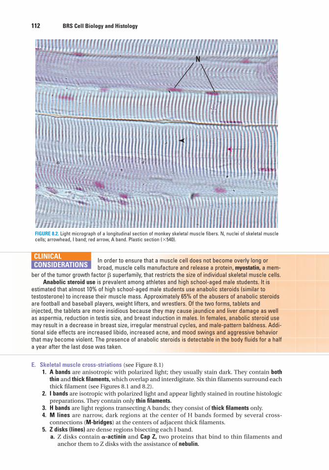
112 BRS Cell Biology and Histology
E. Skeletal muscle cross-striations (see Figure 8.1)1. A bands are anisotropic with polarized light; they usually stain dark. They contain both
thin and thick filaments, which overlap and interdigitate. Six thin filaments surround eachthick filament (see Figures 8.1 and 8.2).
2. I bands are isotropic with polarized light and appear lightly stained in routine histologicpreparations. They contain only thin filaments.
3. H bands are light regions transecting A bands; they consist of thick filaments only.4. M lines are narrow, dark regions at the center of H bands formed by several cross-
connections (M-bridges) at the centers of adjacent thick filaments.5. Z disks (lines) are dense regions bisecting each I band.
a. Z disks contain �-actinin and Cap Z, two proteins that bind to thin filaments andanchor them to Z disks with the assistance of nebulin.
N
FIGURE 8.2. Light micrograph of a longitudinal section of monkey skeletal muscle fibers. N, nuclei of skeletal musclecells; arrowhead, I band; red arrow, A band. Plastic section (�540).
In order to ensure that a muscle cell does not become overly long or broad, muscle cells manufacture and release a protein, myostatin, a mem-
ber of the tumor growth factor � superfamily, that restricts the size of individual skeletal muscle cells.Anabolic steroid use is prevalent among athletes and high school-aged male students. It is
estimated that almost 10% of high school-aged male students use anabolic steroids (similar totestosterone) to increase their muscle mass. Approximately 65% of the abusers of anabolic steroidsare football and baseball players, weight lifters, and wrestlers. Of the two forms, tablets andinjected, the tablets are more insidious because they may cause jaundice and liver damage as wellas aspermia, reduction in testis size, and breast induction in males. In females, anabolic steroid usemay result in a decrease in breast size, irregular menstrual cycles, and male-pattern baldness. Addi-tional side effects are increased libido, increased acne, and mood swings and aggressive behaviorthat may become violent. The presence of anabolic steroids is detectable in the body fluids for a halfa year after the last dose was taken.
CLINICAL
CONSIDERATIONS
LWBK615-c08[109-125].qxd 05/31/2010 10:33 AM Page 112 Aptara
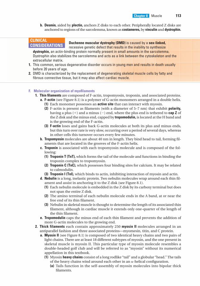
Chapter 8 Muscle 113
b. Desmin, aided by plectin, anchors Z disks to each other. Peripherally located Z disks areanchored to regions of the sarcolemma, known as costameres, by vinculin and dystrophin.
Duchenne muscular dystrophy (DMD) is caused by a sex-linked,recessive genetic defect that results in the inability to synthesize
dystrophin, an actin-binding protein normally present in small amounts in the sarcolemma.Dystrophin also stabilizes the sarcolemma and acts as a link between the cytoskeleton and theextracellular matrix.
1. This common, serious degenerative disorder occurs in young men and results in death usuallybefore 20 years of age.
2. DMD is characterized by the replacement of degenerating skeletal muscle cells by fatty andfibrous connective tissue, but it may also affect cardiac muscle.
CLINICAL
CONSIDERATIONS
F. Molecular organization of myofilaments1. Thin filaments are composed of F-actin, tropomyosin, troponin, and associated proteins.
a. F-actin (see Figure 8.1) is a polymer of G-actin monomers arranged in a double helix.(1) Each monomer possesses an active site that can interact with myosin.(2) F-actin is present as filaments (with a diameter of 5–7 nm) that exhibit polarity,
having a plus (�) and a minus (�) end, where the plus end is tethered to cap Z ofthe Z disk and the minus end, capped by tropomodulin, is located at the H band andis the growing end of the F-actin.
(3) F-actin loses and gains back G-actin molecules at both its plus and minus ends,but this turn over rate is very slow, occurring over a period of several days, whereasin other cells this turnover occurs every few minutes.
b. Tropomyosin molecules are about 40 nm in length. They bind head to tail, forming fil-aments that are located in the grooves of the F-actin helix.
c. Troponin is associated with each tropomyosin molecule and is composed of the fol-lowing:(1) Troponin T (TnT), which forms the tail of the molecule and functions in binding the
troponin complex to tropomyosin.(2) Troponin C (TnC), which possesses four binding sites for calcium. It may be related
to calmodulin.(3) Troponin I (TnI), which binds to actin, inhibiting interaction of myosin and actin.
d. Nebulin is a long, inelastic protein. Two nebulin molecules wrap around each thin fil-ament and assist in anchoring it to the Z disk (see Figure 8.1).(1) Each nebulin molecule is embedded in the Z disk by its carboxy terminal but does
not span the entire Z disk.(2) The amino terminal of each nebulin molecule ends in the A band, at or near the
free end of its thin filament.(3) Nebulin in skeletal muscle is thought to determine the length of its associated thin
filament, although in cardiac muscle it extends only one-quarter of the length ofthe thin filament.
e. Tropomodulin caps the minus end of each thin filament and prevents the addition ofmore G-actin molecules to the growing end.
2. Thick filaments each contain approximately 250 myosin II molecules arranged in anantiparallel fashion and three associated proteins—myomesin, titin, and C protein.a. Myosin II (see Figure 8.1) is composed of two identical heavy chains and two pairs of
light chains. There are at least 18 different subtypes of myosin, and the one present inskeletal muscle is myosin II. This particular type of myosin molecule resembles adouble-headed golf club and will be referred to as “myosin” without its numericalappellation in this textbook.(1) Myosin heavy chains consist of a long rodlike “tail” and a globular “head.” The tails
of the heavy chains wind around each other in an �-helical configuration.(a) Tails function in the self-assembly of myosin molecules into bipolar thick
filaments.
LWBK615-c08[109-125].qxd 05/31/2010 10:33 AM Page 113 Aptara
114 BRS Cell Biology and Histology
(b) Actin-binding sites of the heads function in contraction.(2) Myosin light chains are of two types; one molecule of each type is associated with
the globular head of each heavy chain.(3) Digestion of myosin
(a) The enzyme trypsin cleaves myosin into light meromyosin (part of the tail por-tion) and heavy meromyosin (the two heads and the remainder of the tail) (seeFigure 8.1).
(b) The enzyme papain cleaves the heavy meromyosin, releasing the short tail(S2 fragment) and the two globular heads (S1 fragments). These S1 fragmentshave adenosine triphosphatase (ATPase) activity but require interactionwith actin to release the noncovalently bound adenosine diphosphate(ADP) and Pi.
b. Myomesin is a protein at the M line that cross-links adjacent thick filaments to oneanother to maintain their spatial relations.
c. C protein binds to thick filaments in the vicinity of M lines along much of their lengths(between the M line and the end of the thin filament in the vicinity of the A–I junc-tion). This region of the A band is referred to as the C zone.
d. Titin is a large linear protein that displays axial periodicity. It forms an elastic latticethat parallels the thick and thin filaments, and two titin filaments anchor each thickfilament to the Z disk, thus maintaining their architectural relationships to each other(see Figure 8.1).(1) The amino terminal of the titin molecule spans the entire thickness of the Z disk
and binds to �-actinin and Z proteins.(2) Within the Z disk, titin overlaps with other titin molecules from the neighboring sar-
comere and probably forms bonds with them or with unidentified linker proteins.(3) The carboxyl terminal of the titin molecule spans the entire M line and overlaps
with titin molecules from the other half of the same sarcomere, and binds to theprotein myomesin.
(4) Within the I band, in the vicinity of the Z disk, titin interacts with thin filaments.(5) Within the A band, titin interacts with C protein.
III. CONTRACTION OF SKELETAL MUSCLE
The contraction cycle (Figure 8.3) involves the binding, hydrolysis, and release of adenosinetriphosphate (ATP).
A. Huxley’s sliding-filament model (Table 8.2)1. During contraction, thick and thin filaments do not shorten but increase their overlap.2. Thin filaments slide past thick filaments and penetrate more deeply into the A band,
which remains constant in length.3. I bands and H bands shorten as Z disks are drawn closer together.
B. Initiation and regulation of contraction1. Depolarization, accompanied by the release of Ca2�, triggers the binding of actin and
myosin, leading to muscle contraction.a. The sarcolemma is depolarized at the myoneural junction.b. T tubules convey the wave of depolarization to the myofibrils. Voltage-sensitive dihy-
dropyridine (DHP) receptors alter their conformation as a function of membrane depo-larization.
c. Ca2� is released into the cytosol at the A–I junctions via Ca2�-release channels (junc-tional feet, ryanodine receptors) of the SR terminal cisternae that are opened by acti-vated DHP receptors. As long as the Ca2� level is sufficiently high, the contractioncycle will continue.
2. Activation of actin by Ca2�
LWBK615-c08[109-125].qxd 05/31/2010 10:33 AM Page 114 Aptara
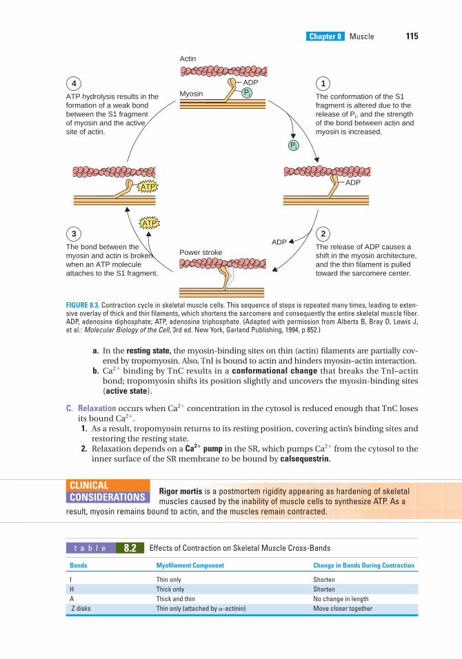
Chapter 8 Muscle 115
Actin
The conformation of the S1 fragment is altered due to therelease of Pi, and the strength of the bond between actin and myosin is increased.
ATP hydrolysis results in the formation of a weak bondbetween the S1 fragmentof myosin and the activesite of actin.
ADP
ADP
Myosin Pi
Pi
ADPATP
14
The release of ADP causes ashift in the myosin architecture,and the thin filament is pulledtoward the sarcomere center.
The bond between themyosin and actin is brokenwhen an ATP moleculeattaches to the S1 fragment.
Power stroke
23ATP
FIGURE 8.3. Contraction cycle in skeletal muscle cells. This sequence of steps is repeated many times, leading to exten-sive overlay of thick and thin filaments, which shortens the sarcomere and consequently the entire skeletal muscle fiber.ADP, adenosine diphosphate; ATP, adenosine triphosphate. (Adapted with permission from Alberts B, Bray D, Lewis J,et al.: Molecular Biology of the Cell, 3rd ed. New York, Garland Publishing, 1994, p 852.)
t a b l e 8.2 Effects of Contraction on Skeletal Muscle Cross-Bands
Bands Myofilament Component Change in Bands During Contraction
I Thin only Shorten
H Thick only Shorten
A Thick and thin No change in length
Z disks Thin only (attached by �-actinin) Move closer together
a. In the resting state, the myosin-binding sites on thin (actin) filaments are partially cov-ered by tropomyosin. Also, TnI is bound to actin and hinders myosin–actin interaction.
b. Ca2� binding by TnC results in a conformational change that breaks the TnI–actinbond; tropomyosin shifts its position slightly and uncovers the myosin-binding sites(active state).
C. Relaxation occurs when Ca2� concentration in the cytosol is reduced enough that TnC losesits bound Ca2�.1. As a result, tropomyosin returns to its resting position, covering actin’s binding sites and
restoring the resting state.2. Relaxation depends on a Ca2� pump in the SR, which pumps Ca2� from the cytosol to the
inner surface of the SR membrane to be bound by calsequestrin.
Rigor mortis is a postmortem rigidity appearing as hardening of skeletalmuscles caused by the inability of muscle cells to synthesize ATP. As a
result, myosin remains bound to actin, and the muscles remain contracted.
CLINICAL
CONSIDERATIONS
LWBK615-c08[109-125].qxd 05/31/2010 10:33 AM Page 115 Aptara
116 BRS Cell Biology and Histology
D. A motor unit consists of a neuron and every muscle cell it innervates. A muscle may contractwith varying degrees of strength because only some of the muscle cells contract, but an indi-vidual muscle cell obeys the “all or none law” (i.e., it either contracts or does not contract).All muscle cells of a single motor unit contract in unison, and a large muscle such as thetrapezius may have thousands of muscle cells in a motor unit, whereas a muscle such as thesuperior oblique muscle of the eye may have as few as 10 to 15 muscle cells in a motor unit.
Normally, as a muscle contracts the sarcomeres shorten and,consequently, the entire muscle becomes shorter, this type of contraction
is referred to as concentric contraction, as in picking up a dumbbell and doing curls. Another typeof contraction is isometric contraction, where the sarcomeres do not shorten; thus, the entire mus-cle remains the same length, as in squeezing a hard object such as a solid metal ball.
CLINICAL
CONSIDERATIONS
IV. INNERVATION OF SKELETAL MUSCLE
Innervation consists of motor nerve endings (myoneural junctions) and two types of sensorynerve endings (muscle spindles and Golgi tendon organs). Both types of sensory nerve endingsfunction in proprioception.
A. The myoneural junction (neuromuscular junction) is a synapse between a branch of a motornerve axon and a skeletal muscle cell.1. Structural components
a. The axon terminal lacks myelin but has a Schwann cell on its nonsynaptic surface.(1) The membrane on the synaptic surface of the axon terminal is called the presy-
naptic membrane.(2) The axon terminal contains mitochondria, synaptic vesicles (containing the neu-
rotransmitter acetylcholine), and SER elements.b. The synaptic cleft is a narrow space between the presynaptic membrane of the axon
terminal and the postsynaptic membrane (also known as the motor end plate) of themuscle cell. The synaptic cleft contains an amorphous external lamina, a basal lami-nalike material, derived from the muscle cell.
c. Muscle cell near the myoneural junction(1) Sarcolemmal invaginations (of the postsynaptic membrane), called junctional
folds, are lined by an external lamina and extend inward from the synaptic cleft.(2) Acetylcholine receptors are located in the postsynaptic membrane.(3) The sarcoplasm is rich in mitochondria, ribosomes, and rough endoplasmic retic-
ulum (RER).2. Conduction of a nerve impulse across a myoneural junction
a. The presynaptic membrane is depolarized and voltage-gated Ca2� channels open,permitting the entry of extracellular Ca2� into the axon terminal.
b. The rise in cytosolic Ca2� triggers the synaptic vesicles to release acetylcholine inmultimolecular quantities (quanta) into the synaptic cleft. A quantum is approxi-mately equivalent to 20,000 acetylcholine molecules.
c. The released acetylcholine binds to receptors of the postsynaptic membrane, result-ing in depolarization of the sarcolemma and generation of an action potential.
d. The enzyme, acetylcholinesterase located in the external lamina lining the junctionalfolds of the motor end plate degrades acetylcholine, thus ending the depolarizing sig-nal to the muscle cell.
e. Acetylcholine is recycled as choline and is returned to the axon terminal to be recom-bined with acetyl coenzyme A (CoA) (from mitochondria) under the influence of theenzyme choline acetyl transferase to form acetylcholine, which is then stored insynaptic vesicles.
LWBK615-c08[109-125].qxd 05/31/2010 10:33 AM Page 116 Aptara
Chapter 8 Muscle 117
f. Membranes of the emptied synaptic vesicles are recycled via clathrin-coated endocyticvesicles (see Figure 3.5).
Amyotrophic lateral sclerosis (ALS, or Lou Gehrig disease) is marked bydegeneration of motor neurons of the spinal cord, resulting in muscle
atrophy. Death is usually due to respiratory muscle failure.Myasthenia gravis is an autoimmune disease in which antibodies block acetylcholine recep-
tors of myoneural junctions, reducing the number of sites available for initiation of sarcolemmadepolarization.
1. Myasthenia gravis is characterized by gradual weakening of skeletal muscles, especially themost active ones (e.g., muscles of the eyes, tongue, face, and extremities). Death may result fromrespiratory compromise and pulmonary infections.
2. Clinical signs include thymic hyperplasia (thymoma) and the presence of circulating antibodies toacetylcholine receptors.
Botulism is a form of food poisoning caused by ingestion of Clostridium botulinum toxin,which inhibits acetylcholine release at myoneural junctions. Botulism is marked by muscleparalysis, vomiting, nausea, and visual disorders and is fatal if untreated.
CLINICAL
CONSIDERATIONS
B. The muscle spindle (neuromuscular spindle) is an elongated, fusiform sensory organ withinskeletal muscle that functions primarily as a stretch receptor.1. Structure
a. It is bounded by a connective tissue capsule enclosing the fluid-filled periaxial spaceand 8 to 10 modified skeletal muscle fibers (intrafusal fibers).
b. Normal skeletal muscle fibers (extrafusal fibers) surround it.c. It is anchored via the capsule to the perimysium and endomysium of the extrafusal
fibers.2. Function
a. Stretching of a muscle also stretches the muscle spindle and thus stimulates the affer-ent nerve endings to send impulses to the central nervous system. The response is toboth the rate (phasic response) and duration (tonic response) of stretching.
b. Depolarization of �-efferent neurons also stimulates the intrafusal nerve endings; therate and duration of the stimulation are monitored in the same way as stretching.
c. Muscle overstimulation results from stretching at too great a frequency or for too longa time. Overstimulation causes stimulation of �-efferent neurons to the muscle, initi-ating contraction and thus counteracting the stretching.
C. The Golgi tendon organ, located in tendons, counteracts the effects of muscle spindles.1. Structure. It is composed of encapsulated collagen fibers that are surrounded by terminal
branches of type Ib sensory nerves.2. It is stimulated when the muscle contracts too strenuously, increasing tension on the
tendon. Impulses from type Ib neurons inhibit �-efferent (motor) neurons to the muscle,preventing further contraction.
V. CARDIAC MUSCLE
A. General features—cardiac muscle cells (Table 8.3). Cardiac muscle cells have the followingfeatures:1. Contract spontaneously and display a rhythmic beat, which is modified by hormonal and
neural (sympathetic and parasympathetic) stimuli.2. May branch at their ends to form connections with adjacent cells.
LWBK615-c08[109-125].qxd 05/31/2010 10:33 AM Page 117 Aptara
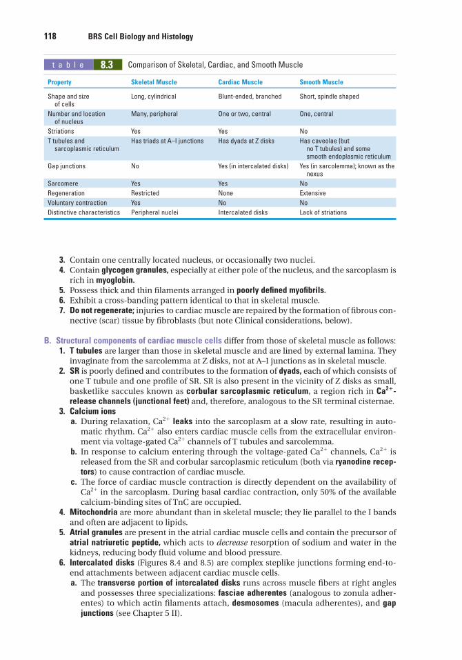
118 BRS Cell Biology and Histology
3. Contain one centrally located nucleus, or occasionally two nuclei.4. Contain glycogen granules, especially at either pole of the nucleus, and the sarcoplasm is
rich in myoglobin.5. Possess thick and thin filaments arranged in poorly defined myofibrils.6. Exhibit a cross-banding pattern identical to that in skeletal muscle.7. Do not regenerate; injuries to cardiac muscle are repaired by the formation of fibrous con-
nective (scar) tissue by fibroblasts (but note Clinical considerations, below).
B. Structural components of cardiac muscle cells differ from those of skeletal muscle as follows:1. T tubules are larger than those in skeletal muscle and are lined by external lamina. They
invaginate from the sarcolemma at Z disks, not at A–I junctions as in skeletal muscle.2. SR is poorly defined and contributes to the formation of dyads, each of which consists of
one T tubule and one profile of SR. SR is also present in the vicinity of Z disks as small,basketlike saccules known as corbular sarcoplasmic reticulum, a region rich in Ca2�-release channels (junctional feet) and, therefore, analogous to the SR terminal cisternae.
3. Calcium ionsa. During relaxation, Ca2� leaks into the sarcoplasm at a slow rate, resulting in auto-
matic rhythm. Ca2� also enters cardiac muscle cells from the extracellular environ-ment via voltage-gated Ca2� channels of T tubules and sarcolemma.
b. In response to calcium entering through the voltage-gated Ca2� channels, Ca2� isreleased from the SR and corbular sarcoplasmic reticulum (both via ryanodine recep-tors) to cause contraction of cardiac muscle.
c. The force of cardiac muscle contraction is directly dependent on the availability ofCa2� in the sarcoplasm. During basal cardiac contraction, only 50% of the availablecalcium-binding sites of TnC are occupied.
4. Mitochondria are more abundant than in skeletal muscle; they lie parallel to the I bandsand often are adjacent to lipids.
5. Atrial granules are present in the atrial cardiac muscle cells and contain the precursor ofatrial natriuretic peptide, which acts to decrease resorption of sodium and water in thekidneys, reducing body fluid volume and blood pressure.
6. Intercalated disks (Figures 8.4 and 8.5) are complex steplike junctions forming end-to-end attachments between adjacent cardiac muscle cells.a. The transverse portion of intercalated disks runs across muscle fibers at right angles
and possesses three specializations: fasciae adherentes (analogous to zonula adher-entes) to which actin filaments attach, desmosomes (macula adherentes), and gapjunctions (see Chapter 5 II).
t a b l e 8.3 Comparison of Skeletal, Cardiac, and Smooth Muscle
Property Skeletal Muscle Cardiac Muscle Smooth Muscle
Shape and size Long, cylindrical Blunt-ended, branched Short, spindle shapedof cells
Number and location Many, peripheral One or two, central One, centralof nucleus
Striations Yes Yes No
T tubules and Has triads at A–I junctions Has dyads at Z disks Has caveolae (but sarcoplasmic reticulum no T tubules) and some
smooth endoplasmic reticulum
Gap junctions No Yes (in intercalated disks) Yes (in sarcolemma); known as the nexus
Sarcomere Yes Yes No
Regeneration Restricted None Extensive
Voluntary contraction Yes No No
Distinctive characteristics Peripheral nuclei Intercalated disks Lack of striations
LWBK615-c08[109-125].qxd 05/31/2010 10:33 AM Page 118 Aptara
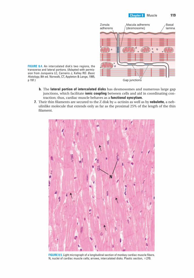
Chapter 8 Muscle 119
b. The lateral portion of intercalated disks has desmosomes and numerous large gapjunctions, which facilitate ionic coupling between cells and aid in coordinating con-traction; thus, cardiac muscle behaves as a functional syncytium.
7. Their thin filaments are secured to the Z disk by �-actinin as well as by nebulette, a neb-ulinlike molecule that extends only as far as the proximal 25% of the length of the thinfilament.
Macula adherens(desmosome)
Basallamina
Zonulaadherens
Gap junctions
FIGURE 8.4. An intercalated disk’s two regions, thetransverse and lateral portions. (Adapted with permis-sion from Junqueira LC, Carneiro J, Kelley RO: BasicHistology, 8th ed. Norwalk, CT, Appleton & Lange, 1995,p 197.)
N
FIGURE 8.5. Light micrograph of a longitudinal section of monkey cardiac muscle fibers.N, nuclei of cardiac muscle cells; arrows, intercalated disks. Plastic section, �270.
LWBK615-c08[109-125].qxd 05/31/2010 10:33 AM Page 119 Aptara
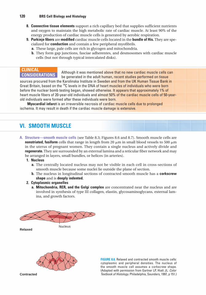
120 BRS Cell Biology and Histology
8. Connective tissue elements support a rich capillary bed that supplies sufficient nutrientsand oxygen to maintain the high metabolic rate of cardiac muscle. At least 90% of theenergy production of cardiac muscle cells is generated by aerobic respiration.
9. Purkinje fibers are modified cardiac muscle cells located in the bundle of His. They are spe-cialized for conduction and contain a few peripheral myofibrils.a. These large, pale cells are rich in glycogen and mitochondria.b. They form gap junctions, fasciae adherentes, and desmosomes with cardiac muscle
cells (but not through typical intercalated disks).
Although it was mentioned above that no new cardiac muscle cells canbe generated in the adult human, recent studies performed on tissue
sources procured from the Karolinska Institute in Sweden and from the UK Human Tissue Bank inGreat Britain, based on the 14C levels in the DNA of heart muscles of individuals who were bornbefore the nuclear bomb testing began, showed otherwise. It appears that approximately 1% ofheart muscle fibers of 20-year-old individuals and almost 50% of the cardiac muscle cells of 50-year-old individuals were formed after these individuals were born.
Myocardial infarct is an irreversible necrosis of cardiac muscle cells due to prolongedischemia. It may result in death if the cardiac muscle damage is extensive.
CLINICAL
CONSIDERATIONS
VI. SMOOTH MUSCLE
A. Structure—smooth muscle cells (see Table 8.3; Figures 8.6 and 8.7). Smooth muscle cells arenonstriated, fusiform cells that range in length from 20 �m in small blood vessels to 500 �min the uterus of pregnant women. They contain a single nucleus and actively divide andregenerate. They are surrounded by an external lamina and a reticular fiber network and maybe arranged in layers, small bundles, or helices (in arteries).1. Nucleus
a. The centrally located nucleus may not be visible in each cell in cross-sections ofsmooth muscle because some nuclei lie outside the plane of section.
b. The nucleus in longitudinal sections of contracted smooth muscle has a corkscrewshape and is deeply indented.
2. Cytoplasmic organellesa. Mitochondria, RER, and the Golgi complex are concentrated near the nucleus and are
involved in synthesis of type III collagen, elastin, glycosaminoglycans, external lam-ina, and growth factors.
RelaxedNucleus
Contracted
FIGURE 8.6. Relaxed and contracted smooth muscle cells:cytoplasmic and peripheral densities. The nucleus ofthe smooth muscle cell assumes a corkscrew shape.(Adapted with permission from Gartner LP, Hiatt JL: ColorTextbook of Histology. Philadelphia, Saunders, 1997, p 151.)
LWBK615-c08[109-125].qxd 05/31/2010 10:33 AM Page 120 Aptara
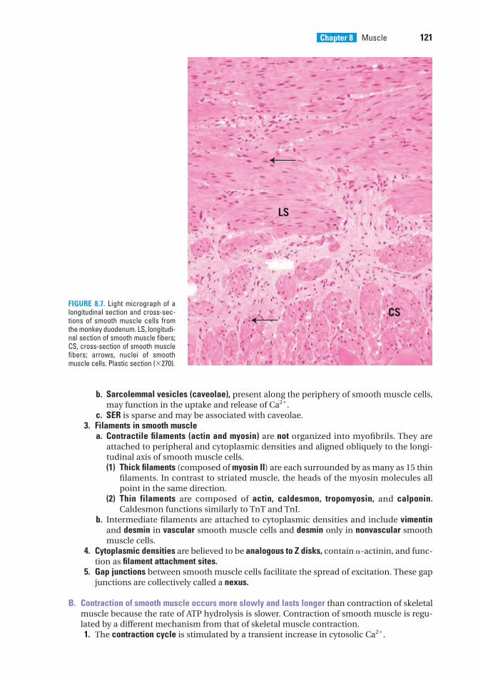
Chapter 8 Muscle 121
b. Sarcolemmal vesicles (caveolae), present along the periphery of smooth muscle cells,may function in the uptake and release of Ca2�.
c. SER is sparse and may be associated with caveolae.3. Filaments in smooth muscle
a. Contractile filaments (actin and myosin) are not organized into myofibrils. They areattached to peripheral and cytoplasmic densities and aligned obliquely to the longi-tudinal axis of smooth muscle cells.(1) Thick filaments (composed of myosin II) are each surrounded by as many as 15 thin
filaments. In contrast to striated muscle, the heads of the myosin molecules allpoint in the same direction.
(2) Thin filaments are composed of actin, caldesmon, tropomyosin, and calponin.Caldesmon functions similarly to TnT and TnI.
b. Intermediate filaments are attached to cytoplasmic densities and include vimentinand desmin in vascular smooth muscle cells and desmin only in nonvascular smoothmuscle cells.
4. Cytoplasmic densities are believed to be analogous to Z disks, contain �-actinin, and func-tion as filament attachment sites.
5. Gap junctions between smooth muscle cells facilitate the spread of excitation. These gapjunctions are collectively called a nexus.
B. Contraction of smooth muscle occurs more slowly and lasts longer than contraction of skeletalmuscle because the rate of ATP hydrolysis is slower. Contraction of smooth muscle is regu-lated by a different mechanism from that of skeletal muscle contraction.1. The contraction cycle is stimulated by a transient increase in cytosolic Ca2�.
CS
LS
FIGURE 8.7. Light micrograph of alongitudinal section and cross-sec-tions of smooth muscle cells fromthe monkey duodenum. LS, longitudi-nal section of smooth muscle fibers;CS, cross-section of smooth musclefibers; arrows, nuclei of smoothmuscle cells. Plastic section (�270).
LWBK615-c08[109-125].qxd 05/31/2010 10:33 AM Page 121 Aptara
122 BRS Cell Biology and Histology
a. Ca2� binds to calmodulin, altering its conformation.b. The Ca2�–calmodulin complex activates the enzyme myosin light-chain kinase,
which catalyzes phosphorylation of one of the light chains of myosin.c. In the presence of Ca2�, the inhibitory effect of the caldesmon–tropomyosin complex
on the actin–myosin interaction is eliminated (caldesmon masks the active site of G-actin). Another inhibitor of contraction is calponin, which, when phosphorylated,loses its inhibitory capability.
d. The globular head of phosphorylated myosin interacts with actin and stimulatesmyosin ATPase, resulting in contraction. As long as myosin is in its phosphorylatedform, the contraction cycle continues.
e. Dephosphorylation of myosin disturbs the myosin–actin interaction and leads torelaxation.
2. Initiation of contractiona. In vascular smooth muscle, contraction is usually triggered by a nerve impulse, with lit-
tle spread of the impulse from cell to cell.b. In visceral smooth muscle, it is triggered by stretching of the muscle itself (myogenic);
the signal spreads from cell to cell.c. In the uterus during labor, it is triggered by oxytocin.d. In smooth muscle elsewhere in the body, it is triggered by epinephrine.
C. Innervation of smooth muscle is by sympathetic (noradrenergic) nerves and parasympathetic(cholinergic) nerves of the autonomic nervous system, which act in an antagonistic fashionto stimulate or depress activity of the muscle.
VII. CONTRACTILE NONMUSCLE CELLS
A. Myoepithelial cells1. In certain glands, these cells share basal laminae of secretory and duct cells.2. They arise from ectoderm and can contract to express secretory material from glandular
epithelium into ducts and out of the gland.3. Although generally similar in morphology to smooth muscle cells, they have a basketlike
shape and several radiating processes.4. They are attached to the underlying basal lamina via hemidesmosomes.5. They contain actin, myosin, and intermediate filaments, as well as cytoplasmic and
peripheral densities to which these filaments attach.6. Contraction is similar to that of smooth muscle and occurs via a calmodulin-mediated
process. In lactating mammary glands, they contract in response to oxytocin. In lacrimalglands, they contract in response to acetylcholine.
B. Myofibroblasts1. Although they resemble fibroblasts, they possess higher amounts of actin and myosin
and are capable of contraction.2. They may contract during wound healing to decrease the size of the defect (wound con-
traction).
LWBK615-c08[109-125].qxd 05/31/2010 10:33 AM Page 122 Aptara
1. Which of the following is true formammalian skeletal muscle?
(A) T tubules are located at the Z disk.(B) T tubules are absent.(C) Troponin is absent.(D) It possesses triads.(E) It possesses caveolae.
2. Which of the following is true for cardiacmuscle?
(A) T tubules are located at the Z disk.(B) T tubules have a smaller diameter than
those of skeletal muscle.(C) Troponin is absent.(D) It possesses triads.(E) Oxytocin triggers contraction.
3. Which of the following is true for smoothmuscle?
(A) T tubules are located at the Z disk.(B) It possesses dyads.(C) Caveolae store and release calcium ions.(D) It possesses triads.(E) T tubules are located at the A–I interface.
4. Contraction in all types of musclerequires calcium ions. Which of thefollowing muscle components can bind orsequester calcium ions?
(A) Rough endoplasmic reticulum(B) Tropomyosin(C) Troponin(D) Active sites on actin(E) Titin
5. Each smooth muscle cell
(A) has triads associated with itscontraction.
(B) has dyads associated with itscontraction.
(C) possesses a single central nucleus.(D) is characterized by the absence of
sarcolemmal vesicles.(E) contains troponin.
6. Thick filaments are anchored to Z disks by
(A) C protein.(B) nebulin.(C) titin.(D) myomesin.(E) �-actinin.
7. The endomysium is a connective tissueinvestment that surrounds
(A) individual muscle fibers.(B) muscle fascicles.(C) individual myofibrils.(D) an entire muscle.(E) small bundles of muscle cells.
8. Which of the following statementsconcerning triads in mammalian skeletalmuscle is true?
(A) They are located in the Z disk.(B) They consist of two terminal cisternae of
the SR separated by a T tubule.(C) They can be observed with the light
microscope.(D) They are characterized by a T tubule that
sequesters calcium ions.(E) They consist of two T tubules separated
by a central terminal cisterna.
Review Test
Directions: Each of the numbered items or incomplete statements in this section is followed byanswers or by completions of the statement. Select the ONE lettered answer or completion thatis BEST in each case.
123
LWBK615-c08[109-125].qxd 05/31/2010 10:33 AM Page 123 Aptara
124 BRS Cell Biology and Histology
9. Which one of the following statementsconcerning cardiac muscle cells is true?
(A) They are spindle shaped.(B) They require an external stimulus to
undergo contraction.(C) They are multinuclear cells.(D) They are joined together end to end by
intercalated disks.(E) They possess numerous caveolae.
10. A 19-year-old male patient and his motherarrive in the emergency department, bothwith nausea, vomiting, and visual disorders.The physician taking their history notes thatthey both had canned green beans that tastedfunny. Which of the following possibilitiesshould the physician consider?
(A) Duchenne muscular dystrophy(B) Amyotrophic lateral sclerosis(C) Botulism(D) Myasthenia gravis(E) Myocardial infarct
LWBK615-c08[109-125].qxd 05/31/2010 10:33 AM Page 124 Aptara
Answers and Explanations
1. D. The T tubules of skeletal muscle cells are positioned so that they form triads with theterminal cisternae of the SR at the interface of the A and I bands (see Chapter 8 II C).
2. A. The T tubules of cardiac muscle cells are wider than those of skeletal muscle cells andare lined by external lamina (a basal lamina–like material). In contrast to skeletal muscle,the T tubules are located at the Z disk, where they often form dyads, not triads (see Chapter 8 V B 6).
3. C. Smooth muscle cells do not have T tubules. Contraction may be initiated by stretching,neural impulses, the intercellular passage of small molecules via gap junctions, or theaction of hormones such as oxytocin. Contraction is not dependent on troponin, which isabsent from the thin filament of smooth muscle. Instead, Ca2� controlled by sarcolemmalvesicles known as caveolae is released into the cytosol, where it binds with calmodulin.The calcium–calmodulin complex activates myosin light-chain kinase, which participatesin the contraction process (see Chapter 8 VI A 2).
4. C. Binding of Ca2� to the TnC subunit of troponin leads to the uncovering of myosin-binding sites on actin (thin filaments) (see Chapter 8 II F 1 c).
5. C. Smooth muscle cells contain one centrally located nucleus (see Chapter 8 VI A 1).
6. C. Titin forms an elastic lattice that anchors thick filaments to Z disks (see Chapter 8 II F 2 c).
7. A. The endomysium is a thin connective tissue layer composed of reticular fibers and anexternal lamina that invests individual muscle fibers (cells). The epimysium surrounds theentire muscle, and the perimysium surrounds bundles (fascicles) of muscle fibers (seeChapter 8 II A 3).
8. B. A triad in skeletal muscle is composed of three components, a T tubule and twoterminal cisternae of the SR that flank it. The SR, not the T tubules, sequesters Ca2�
(see Chapter 8 II C 4).
9. D. Cardiac muscle cells are joined together end to end by a unique junctional specializa-tion called the intercalated disk (see Chapter 8 V B 6).
10. C. Botulism is the only possible consideration, especially since they both had cannedfood. Duchenne muscular dystrophy is most common in young men but very rare in olderwomen. It would be highly unlikely that both mother and son would show symptoms ofamyotrophic lateral sclerosis or myasthenia gravis or have myocardial infarct at the sametime (see Chapter 8 II E Clinical Considerations).
125
LWBK615-c08[109-125].qxd 05/31/2010 10:33 AM Page 125 Aptara
c h a p t e r 9 Nervous System
126
I. OVERVIEW—NERVOUS SYSTEM
The nervous system can be organized by anatomical or functional divisions.
A. Nervous system is divided anatomically into the central nervous system (CNS), whichincludes the brain and spinal cord, and the peripheral nervous system (PNS), which includesthe nerves outside the CNS and their associated ganglia.
B. Nervous system is divided functionally into a sensory component, which transmits electricalimpulses (signals) to the CNS, and a motor component, which transmits impulses from theCNS to various structures of the body. The motor component is further divided into thesomatic and autonomic systems.
C. Nervous tissue contains two types of cells: nerve cells (neurons), which conduct electricalimpulses, and glial (neuroglial) cells, which support, nurture, and protect the neurons.
II. HISTOGENESIS OF THE NERVOUS SYSTEM (Figure 9.1)
A. The neuroepithelium thickens and differentiates to form the neural plate.
B. The neural plate invaginates and thickens to form the neural groove.
C. The neural tube, a cylindrical structure that results from fusion of the edges of the neuralgroove, enlarges at its cranial end to form the brain. The remaining portion gives rise to thespinal cord.
D. Neural crest cells stream off the edges of the neural groove before formation of the neuraltube. These cells migrate throughout the body and give rise to the following structures:1. Sensory neurons of cranial and spinal sensory ganglia2. Most sensory neurons and Schwann cells of the PNS3. Enteric and autonomic ganglia and their postganglionic neurons and associated glia4. Most of the mesenchymal (ectomesenchymal) cells of the head and anterior portion of
the neck5. Melanocytes of the skin and oral mucosa6. Odontoblasts (cells responsible for the production of dentin)7. Cells of the arachnoid and pia mater (rostral to the mesencephalon)8. Chromaffin cells of the adrenal medulla
LWBK615-c09[126-147].qxd 05/31/2010 10:35 AM Page 126 Aptara
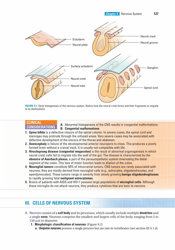
A. Abnormal histogenesis of the CNS results in congenital malformations.B. Congenital malformations
1. Spina bifida is a defective closure of the spinal column. In severe cases, the spinal cord andmeninges may protrude through the unfused areas. Very severe cases may be associated withdefective development of the viscera of the thorax and abdomen.
2. Anencephaly is failure of the developmental anterior neuropore to close. This produces a poorlyformed brain without a cranial vault. It is usually not compatible with life.
3. Hirschsprung disease (congenital megacolon) is the result of abnormal organogenesis in whichneural crest cells fail to migrate into the wall of the gut. The disease is characterized by theabsence of Auerbach plexus, a part of the parasympathetic system innervating the distalsegment of the colon. This loss of motor function leads to dilation of the colon.
4. Neuroglial tumors constitute 50% of intracranial tumors. CNS tumors are rarely associated withneurons; they are mostly derived from neuroglial cells (e.g., astrocytes, oligodendrocytes, andependymocytes). These tumors range in severity from slowly growing benign oligodendrogliomasto rapidly growing fatal malignant astrocytomas.
5. Brains of patients with AIDS and HIV-1 possess large populations of microglial cells. Althoughthese microglia do not attack neurons, they produce cytokines that are toxic to neurons.
CLINICAL
CONSIDERATIONS
Chapter 9 Nervous System 127
III. CELLS OF NERVOUS SYSTEM
A. Neurons consist of a cell body and its processes, which usually include multiple dendrites anda single axon. Neurons comprise the smallest and largest cells of the body, ranging from 5 to150 �m in diameter.1. Morphologic classification of neurons (Figure 9.2)
a. Unipolar neurons possess a single process but are rare in vertebrates (see section III A 1 d).
Neural crest
Neural groove
Neural crest
Neural tube
Surface ectoderm Ganglion
Spinal cord
Ectoderm
Neural plate
FIGURE 9.1. Early histogenesis of the nervous system. Notice how the neural crest forms and then fragments to migrateto its destinations.
LWBK615-c09[126-147].qxd 05/31/2010 10:35 AM Page 127 Aptara
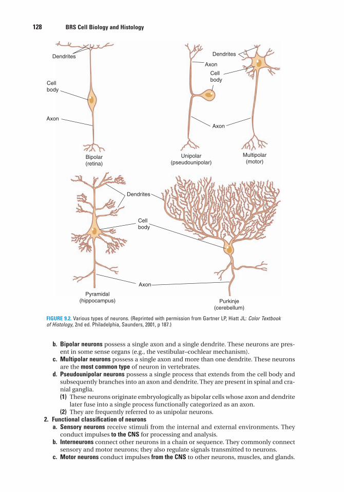
128 BRS Cell Biology and Histology
b. Bipolar neurons possess a single axon and a single dendrite. These neurons are pres-ent in some sense organs (e.g., the vestibular–cochlear mechanism).
c. Multipolar neurons possess a single axon and more than one dendrite. These neuronsare the most common type of neuron in vertebrates.
d. Pseudounipolar neurons possess a single process that extends from the cell body andsubsequently branches into an axon and dendrite. They are present in spinal and cra-nial ganglia.(1) These neurons originate embryologically as bipolar cells whose axon and dendrite
later fuse into a single process functionally categorized as an axon.(2) They are frequently referred to as unipolar neurons.
2. Functional classification of neuronsa. Sensory neurons receive stimuli from the internal and external environments. They
conduct impulses to the CNS for processing and analysis.b. Interneurons connect other neurons in a chain or sequence. They commonly connect
sensory and motor neurons; they also regulate signals transmitted to neurons.c. Motor neurons conduct impulses from the CNS to other neurons, muscles, and glands.
Dendrites
Cell
body
Axon
Bipolar
(retina)
Pyramidal
(hippocampus)
Axon
Purkinje
(cerebellum)
Dendrites
Unipolar
(pseudounipolar)
Multipolar
(motor)
Axon
Dendrites
Axon
Cell
body
Cell
body
FIGURE 9.2. Various types of neurons. (Reprinted with permission from Gartner LP, Hiatt JL: Color Textbookof Histology, 2nd ed. Philadelphia, Saunders, 2001, p 187.)
LWBK615-c09[126-147].qxd 05/31/2010 10:35 AM Page 128 Aptara
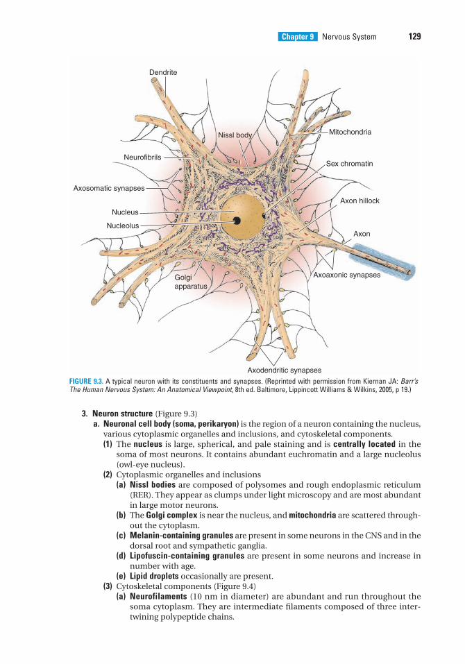
Chapter 9 Nervous System 129
3. Neuron structure (Figure 9.3)a. Neuronal cell body (soma, perikaryon) is the region of a neuron containing the nucleus,
various cytoplasmic organelles and inclusions, and cytoskeletal components.(1) The nucleus is large, spherical, and pale staining and is centrally located in the
soma of most neurons. It contains abundant euchromatin and a large nucleolus(owl-eye nucleus).
(2) Cytoplasmic organelles and inclusions(a) Nissl bodies are composed of polysomes and rough endoplasmic reticulum
(RER). They appear as clumps under light microscopy and are most abundantin large motor neurons.
(b) The Golgi complex is near the nucleus, and mitochondria are scattered through-out the cytoplasm.
(c) Melanin-containing granules are present in some neurons in the CNS and in thedorsal root and sympathetic ganglia.
(d) Lipofuscin-containing granules are present in some neurons and increase innumber with age.
(e) Lipid droplets occasionally are present.(3) Cytoskeletal components (Figure 9.4)
(a) Neurofilaments (10 nm in diameter) are abundant and run throughout thesoma cytoplasm. They are intermediate filaments composed of three inter-twining polypeptide chains.
Dendrite
Nissl body
Neurofibrils
Axosomatic synapses
Nucleus
Nucleolus
Golgi
apparatus
Axodendritic synapses
Axoaxonic synapses
Axon
Axon hillock
Sex chromatin
Mitochondria
FIGURE 9.3. A typical neuron with its constituents and synapses. (Reprinted with permission from Kiernan JA: Barr’sThe Human Nervous System: An Anatomical Viewpoint, 8th ed. Baltimore, Lippincott Williams & Wilkins, 2005, p 19.)
LWBK615-c09[126-147].qxd 05/31/2010 10:35 AM Page 129 Aptara
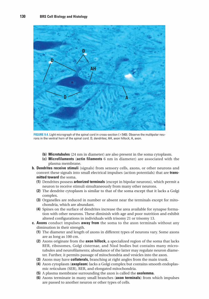
130 BRS Cell Biology and Histology
(b) Microtubules (24 nm in diameter) are also present in the soma cytoplasm.(c) Microfilaments (actin filaments 6 nm in diameter) are associated with the
plasma membrane.b. Dendrites receive stimuli (signals) from sensory cells, axons, or other neurons and
convert these signals into small electrical impulses (action potentials) that are trans-mitted toward the soma.(1) Dendrites possess arborized terminals (except in bipolar neurons), which permit a
neuron to receive stimuli simultaneously from many other neurons.(2) The dendrite cytoplasm is similar to that of the soma except that it lacks a Golgi
complex.(3) Organelles are reduced in number or absent near the terminals except for mito-
chondria, which are abundant.(4) Spines on the surface of dendrites increase the area available for synapse forma-
tion with other neurons. These diminish with age and poor nutrition and exhibitaltered configurations in individuals with trisomy 21 or trisomy 13.
c. Axons conduct impulses away from the soma to the axon terminals without anydiminution in their strength.(1) The diameter and length of axons in different types of neurons vary. Some axons
are as long as 100 cm.(2) Axons originate from the axon hillock, a specialized region of the soma that lacks
RER, ribosomes, Golgi cisternae, and Nissl bodies but contains many micro-tubules and neurofilaments; abundance of the latter may regulate neuron diame-ter. Further, it permits passage of mitochondria and vesicles into the axon.
(3) Axons may have collaterals, branching at right angles from the main trunk.(4) Axon cytoplasm (axoplasm) lacks a Golgi complex but contains smooth endoplas-
mic reticulum (SER), RER, and elongated mitochondria.(5) A plasma membrane surrounding the axon is called the axolemma.(6) Axons terminate in many small branches (axon terminals) from which impulses
are passed to another neuron or other types of cells.
D A
D
AH
D
FIGURE 9.4. Light micrograph of the spinal cord in cross-section (�540). Observe the mulitpolar neu-rons in the ventral horn of the spinal cord. D, dendrites; AH, axon hillock; A, axon.
LWBK615-c09[126-147].qxd 05/31/2010 10:35 AM Page 130 Aptara
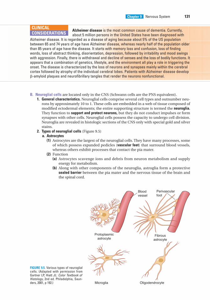
Chapter 9 Nervous System 131
B. Neuroglial cells are located only in the CNS (Schwann cells are the PNS equivalent).1. General characteristics. Neuroglial cells comprise several cell types and outnumber neu-
rons by approximately 10 to 1. These cells are embedded in a web of tissue composed ofmodified ectodermal elements; the entire supporting structure is termed the neuroglia.They function to support and protect neurons, but they do not conduct impulses or formsynapses with other cells. Neuroglial cells possess the capacity to undergo cell division.Neuroglia are revealed in histologic sections of the CNS only with special gold and silverstains.
2. Types of neuroglial cells (Figure 9.5)a. Astrocytes
(1) Astrocytes are the largest of the neuroglial cells. They have many processes, someof which possess expanded pedicles (vascular feet) that surround blood vessels,whereas others exhibit processes that contact the pia mater.
(2) Function(a) Astrocytes scavenge ions and debris from neuron metabolism and supply
energy for metabolism.(b) Along with other components of the neuroglia, astroglia form a protective
sealed barrier between the pia mater and the nervous tissue of the brain andthe spinal cord.
Alzheimer disease is the most common cause of dementia. Currently,about 5 million persons in the United States have been diagnosed with
Alzheimer disease. It is regarded as a disease of aging because about 5% of the US populationbetween 65 and 74 years of age have Alzheimer disease, whereas nearly half of the population olderthan 85 years of age have the disease. It starts with memory loss and confusion, loss of findingwords, loss of abstract thinking, disorientation, depression, followed by irritability and mood swingswith aggression. Finally, there is withdrawal and decline of senses and the loss of bodily functions. Itappears that a combination of genetics, lifestyle, and the environment all play a role in triggering theonset. The disease is characterized by the loss of neurons and synapses mainly within the cerebralcortex followed by atrophy of the individual cerebral lobes. Patients with Alzheimer disease develop�-amyloid plaques and neurofibrillary tangles that render the neurons nonfunctional.
CLINICAL
CONSIDERATIONS
Bloodvessel
Perivascularfoot
OligodendrocyteMicroglia
Protoplasmicastrocyte
Fibrousastrocyte
FIGURE 9.5. Various types of neuroglialcells. (Adapted with permission fromGartner LP, Hiatt JL: Color Textbook ofHistology, 2nd ed. Philadelphia, Saun-ders, 2001, p 192.)
LWBK615-c09[126-147].qxd 05/31/2010 10:35 AM Page 131 Aptara
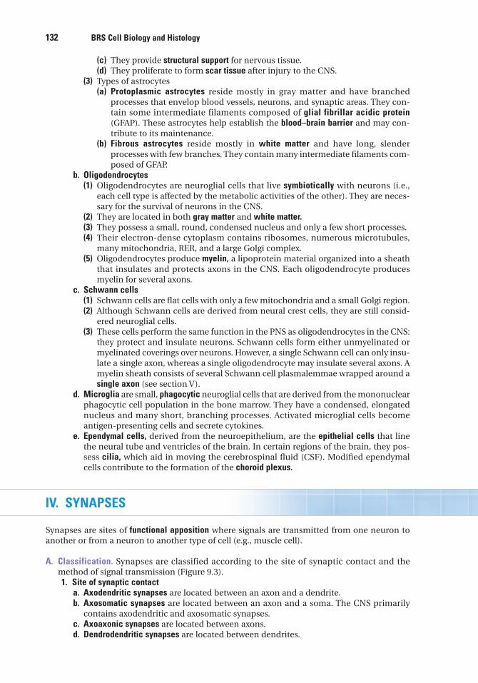
132 BRS Cell Biology and Histology
(c) They provide structural support for nervous tissue.(d) They proliferate to form scar tissue after injury to the CNS.
(3) Types of astrocytes(a) Protoplasmic astrocytes reside mostly in gray matter and have branched
processes that envelop blood vessels, neurons, and synaptic areas. They con-tain some intermediate filaments composed of glial fibrillar acidic protein(GFAP). These astrocytes help establish the blood–brain barrier and may con-tribute to its maintenance.
(b) Fibrous astrocytes reside mostly in white matter and have long, slenderprocesses with few branches. They contain many intermediate filaments com-posed of GFAP.
b. Oligodendrocytes(1) Oligodendrocytes are neuroglial cells that live symbiotically with neurons (i.e.,
each cell type is affected by the metabolic activities of the other). They are neces-sary for the survival of neurons in the CNS.
(2) They are located in both gray matter and white matter.(3) They possess a small, round, condensed nucleus and only a few short processes.(4) Their electron-dense cytoplasm contains ribosomes, numerous microtubules,
many mitochondria, RER, and a large Golgi complex.(5) Oligodendrocytes produce myelin, a lipoprotein material organized into a sheath
that insulates and protects axons in the CNS. Each oligodendrocyte producesmyelin for several axons.
c. Schwann cells(1) Schwann cells are flat cells with only a few mitochondria and a small Golgi region.(2) Although Schwann cells are derived from neural crest cells, they are still consid-
ered neuroglial cells.(3) These cells perform the same function in the PNS as oligodendrocytes in the CNS:
they protect and insulate neurons. Schwann cells form either unmyelinated ormyelinated coverings over neurons. However, a single Schwann cell can only insu-late a single axon, whereas a single oligodendrocyte may insulate several axons. Amyelin sheath consists of several Schwann cell plasmalemmae wrapped around asingle axon (see section V).
d. Microglia are small, phagocytic neuroglial cells that are derived from the mononuclearphagocytic cell population in the bone marrow. They have a condensed, elongatednucleus and many short, branching processes. Activated microglial cells becomeantigen-presenting cells and secrete cytokines.
e. Ependymal cells, derived from the neuroepithelium, are the epithelial cells that linethe neural tube and ventricles of the brain. In certain regions of the brain, they pos-sess cilia, which aid in moving the cerebrospinal fluid (CSF). Modified ependymalcells contribute to the formation of the choroid plexus.
IV. SYNAPSES
Synapses are sites of functional apposition where signals are transmitted from one neuron toanother or from a neuron to another type of cell (e.g., muscle cell).
A. Classification. Synapses are classified according to the site of synaptic contact and themethod of signal transmission (Figure 9.3).1. Site of synaptic contact
a. Axodendritic synapses are located between an axon and a dendrite.b. Axosomatic synapses are located between an axon and a soma. The CNS primarily
contains axodendritic and axosomatic synapses.c. Axoaxonic synapses are located between axons.d. Dendrodendritic synapses are located between dendrites.
LWBK615-c09[126-147].qxd 05/31/2010 10:35 AM Page 132 Aptara
Chapter 9 Nervous System 133
2. Method of signal transmissiona. Chemical synapses
(1) These synapses involve the release of a chemical substance (neurotransmitter orneuromodulator) by the presynaptic cell, which acts on the postsynaptic cell togenerate an action potential.
(2) Chemical synapses are the most common neuron–neuron synapse and the onlyneuron–muscle synapse.
(3) Signal transmission across these synapses is delayed by about 0.5 ms, the timerequired for secretion and diffusion of neurotransmitter from the presynapticmembrane of the first cell into the synaptic cleft and then to the postsynapticmembrane of the receiving cell.
(4) Neurotransmitters do not effect the change, they only activate a response in thereceiving cell.
Parkinson diseaseParkinson disease is a progressive degenerative disease characterized by
tremors, muscular rigidity, difficulty in initiating movements, slow voluntary shuffling movement, andmasklike facial expression. The cause is the loss of dopaminergic neurons from the substantia nigraof the brain. Although the cause of the loss of these cells is unclear, it is known that certain industrialtoxins, such as those to which manganese miners are exposed, and the poisonous MTPT (methyl-phenyl-tetrahydropyridine), a substance present in illegally manufactured heroin, cause Parkinsondisease. Treatment modalities include administering levodopa, which gives some relief, although thecells continue to die. Transplanting fetal adrenal gland tissue has provided only transient relief.Advances in stem cell research may be applied one day to curing this deadly disease.
Huntington choreaHuntington chorea is a fatal hereditary disease that becomes evident during the third and fourthdecades of life, first presenting as painful joints and progressing to uncontrolled flicking of joints,motor disfunction, dementia, and death. The cause is thought to be the loss of neurons that producethe neurotransmitter �-aminobutyric acid (GABA). The symptoms of dementia are thought to berelated to the loss of the cells secreting acetylcholine.
CLINICAL
CONSIDERATIONS
b. Electrical synapses(1) These synapses involve movement of ions from one neuron to another via gap
junctions, which transmit the action potential of the presynaptic cell directly to thepostsynaptic cell.
(2) Electrical synapses are much less numerous than chemical synapses.(3) Signal transmission across these synapses is nearly instantaneous.
B. Synaptic morphology1. Axon terminals may vary morphologically depending on the site of synaptic contact.
a. Boutons terminaux are bulbous expansions that occur singly at the end of axon termi-nals.
b. Boutons en passage are swellings along the axon terminal; synapses may occur at eachswelling.
2. The presynaptic membrane is the thickened axolemma of the neuron that is transmittingthe impulse. It contains voltage-gated Ca2� channels, which regulate the entry of calciumions into the axon terminal. Synaptic vesicles fuse with and become incorporated intothe presynaptic membrane before releasing their neurotransmitter substances.
3. The postsynaptic membrane is the thickened plasma membrane of the neuron or othertarget cell that is receiving the impulse.
4. The synaptic cleft is the narrow space (20–30 nm wide) between the presynaptic andpostsynaptic membranes. Neurotransmitters diffuse across the synaptic cleft.
LWBK615-c09[126-147].qxd 05/31/2010 10:35 AM Page 133 Aptara
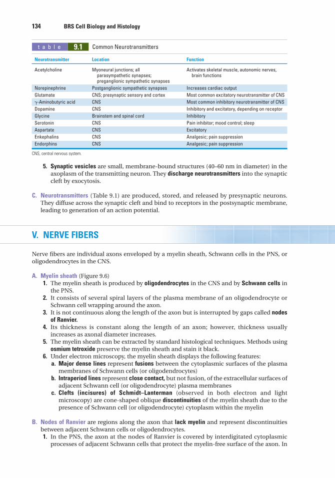
134 BRS Cell Biology and Histology
5. Synaptic vesicles are small, membrane-bound structures (40–60 nm in diameter) in theaxoplasm of the transmitting neuron. They discharge neurotransmitters into the synapticcleft by exocytosis.
C. Neurotransmitters (Table 9.1) are produced, stored, and released by presynaptic neurons.They diffuse across the synaptic cleft and bind to receptors in the postsynaptic membrane,leading to generation of an action potential.
V. NERVE FIBERS
Nerve fibers are individual axons enveloped by a myelin sheath, Schwann cells in the PNS, oroligodendrocytes in the CNS.
A. Myelin sheath (Figure 9.6)1. The myelin sheath is produced by oligodendrocytes in the CNS and by Schwann cells in
the PNS.2. It consists of several spiral layers of the plasma membrane of an oligodendrocyte or
Schwann cell wrapping around the axon.3. It is not continuous along the length of the axon but is interrupted by gaps called nodes
of Ranvier.4. Its thickness is constant along the length of an axon; however, thickness usually
increases as axonal diameter increases.5. The myelin sheath can be extracted by standard histological techniques. Methods using
osmium tetroxide preserve the myelin sheath and stain it black.6. Under electron microscopy, the myelin sheath displays the following features:
a. Major dense lines represent fusions between the cytoplasmic surfaces of the plasmamembranes of Schwann cells (or oligodendrocytes)
b. Intraperiod lines represent close contact, but not fusion, of the extracellular surfaces ofadjacent Schwann cell (or oligodendrocyte) plasma membranes
c. Clefts (incisures) of Schmidt–Lanterman (observed in both electron and lightmicroscopy) are cone-shaped oblique discontinuities of the myelin sheath due to thepresence of Schwann cell (or oligodendrocyte) cytoplasm within the myelin
B. Nodes of Ranvier are regions along the axon that lack myelin and represent discontinuitiesbetween adjacent Schwann cells or oligodendrocytes.1. In the PNS, the axon at the nodes of Ranvier is covered by interdigitated cytoplasmic
processes of adjacent Schwann cells that protect the myelin-free surface of the axon. In
t a b l e 9.1 Common Neurotransmitters
Neurotransmitter Location Function
Acetylcholine Myoneural junctions; all Activates skeletal muscle, autonomic nerves,parasympathetic synapses; brain functionspreganglionic sympathetic synapses
Norepinephrine Postganglionic sympathetic synapses Increases cardiac output
Glutamate CNS; presynaptic sensory and cortex Most common excitatory neurotransmitter of CNS
�-Aminobutyric acid CNS Most common inhibitory neurotransmitter of CNS
Dopamine CNS Inhibitory and excitatory, depending on receptor
Glycine Brainstem and spinal cord Inhibitory
Serotonin CNS Pain inhibitor; mood control; sleep
Aspartate CNS Excitatory
Enkephalins CNS Analgesic; pain suppression
Endorphins CNS Analgesic; pain suppression
CNS, central nervous system.
LWBK615-c09[126-147].qxd 05/31/2010 10:35 AM Page 134 Aptara
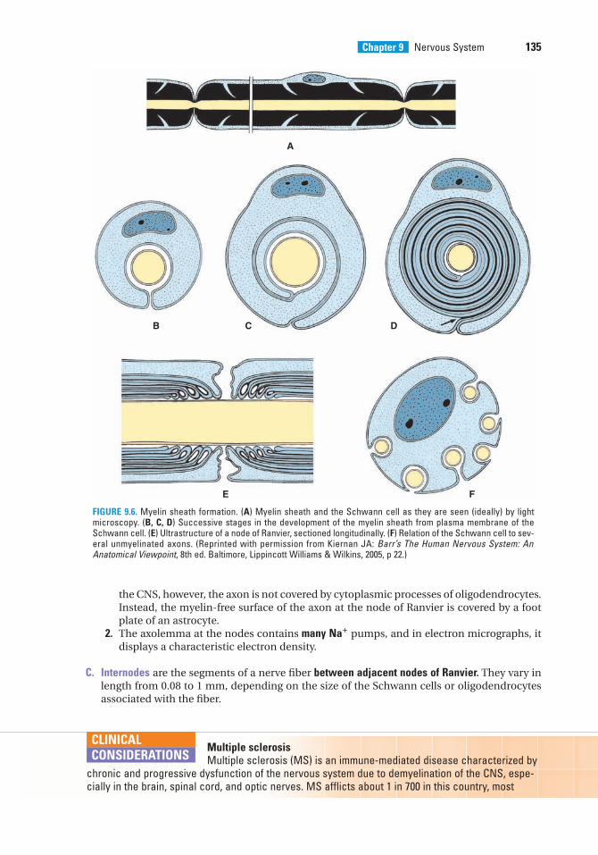
Chapter 9 Nervous System 135
the CNS, however, the axon is not covered by cytoplasmic processes of oligodendrocytes.Instead, the myelin-free surface of the axon at the node of Ranvier is covered by a footplate of an astrocyte.
2. The axolemma at the nodes contains many Na� pumps, and in electron micrographs, itdisplays a characteristic electron density.
C. Internodes are the segments of a nerve fiber between adjacent nodes of Ranvier. They vary inlength from 0.08 to 1 mm, depending on the size of the Schwann cells or oligodendrocytesassociated with the fiber.
A
B C D
E F
FIGURE 9.6. Myelin sheath formation. (A) Myelin sheath and the Schwann cell as they are seen (ideally) by lightmicroscopy. (B, C, D) Successive stages in the development of the myelin sheath from plasma membrane of theSchwann cell. (E) Ultrastructure of a node of Ranvier, sectioned longitudinally. (F) Relation of the Schwann cell to sev-eral unmyelinated axons. (Reprinted with permission from Kiernan JA: Barr’s The Human Nervous System: AnAnatomical Viewpoint, 8th ed. Baltimore, Lippincott Williams & Wilkins, 2005, p 22.)
Multiple sclerosisMultiple sclerosis (MS) is an immune-mediated disease characterized by
chronic and progressive dysfunction of the nervous system due to demyelination of the CNS, espe-cially in the brain, spinal cord, and optic nerves. MS afflicts about 1 in 700 in this country, most
CLINICAL
CONSIDERATIONS
LWBK615-c09[126-147].qxd 05/31/2010 10:35 AM Page 135 Aptara
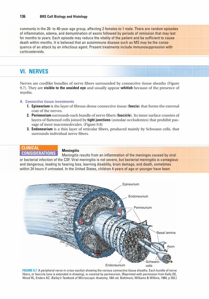
136 BRS Cell Biology and Histology
VI. NERVES
Nerves are cordlike bundles of nerve fibers surrounded by connective tissue sheaths (Figure9.7). They are visible to the unaided eye and usually appear whitish because of the presence ofmyelin.
A. Connective tissue investments1. Epineurium is the layer of fibrous dense connective tissue (fascia) that forms the external
coat of the nerves.2. Perineurium surrounds each bundle of nerve fibers (fascicle). Its inner surface consists of
layers of flattened cells joined by tight junctions (zonulae occludentes) that prohibit pas-sage of most macromolecules. (Figure 9.8)
3. Endoneurium is a thin layer of reticular fibers, produced mainly by Schwann cells, thatsurrounds individual nerve fibers.
commonly in the 20- to 40-year age group, affecting 2 females to 1 male. There are random episodesof inflammation, edema, and demyelination of axons followed by periods of remission that may lastfor months to years. Each episode may reduce the vitality of the patient and be sufficient to causedeath within months. It is believed that an autoimmune disease such as MS may be the conse-quence of an attack by an infectious agent. Present treatments include immunosuppression withcorticosteroids.
Epineurium
Endoneurium
Perineurium
Basal lamina
Axon
SchwanncellsEndoneurium
FIGURE 9.7. A peripheral nerve in cross-section showing the various connective tissue sheaths. Each bundle of nervefibers, or fascicle (one is extended in drawing), is covered by perineurium. (Reprinted with permission from Kelly DE,Wood RL, Enders AC: Bailey’s Textbook of Microscopic Anatomy, 18th ed. Baltimore, Williams & Wilkins, 1984, p 353.)
MeningitisMeningitis results from an inflammation of the meninges caused by viral
or bacterial infection of the CSF. Viral meningitis is not severe, but bacterial meningitis is contagiousand dangerous, leading to hearing loss, learning disability, brain damage, and death, sometimeswithin 24 hours if untreated. In the United States, children 4 years of age or younger have been
CLINICAL
CONSIDERATIONS
LWBK615-c09[126-147].qxd 05/31/2010 10:35 AM Page 136 Aptara
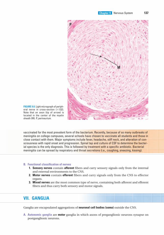
Chapter 9 Nervous System 137
B. Functional classification of nerves1. Sensory nerves contain afferent fibers and carry sensory signals only from the internal
and external environments to the CNS.2. Motor nerves contain efferent fibers and carry signals only from the CNS to effector
organs.3. Mixed nerves are the most common type of nerve, containing both afferent and efferent
fibers and thus carry both sensory and motor signals.
VII. GANGLIA
Ganglia are encapsulated aggregations of neuronal cell bodies (soma) outside the CNS.
A. Autonomic ganglia are motor ganglia in which axons of preganglionic neurons synapse onpostganglionic neurons.
M
P
FIGURE 9.8. Light micrograph of periph-eral nerve in cross-section (�132).Note that an axon (tip of arrow) islocated in the center of the myelinsheath (M). P, perineurium.
vaccinated for the most prevalent form of the bacterium. Recently, because of so many outbreaks ofmeningitis on college campuses, several schools have chosen to vaccinate all students and those inclose contact with them. Major symptoms include fever, headache, stiff neck, and alteration of con-sciousness with rapid onset and progression. Spinal tap and culture of CSF to determine the bacter-ial species is the only diagnosis. This is followed by treatment with a specific antibiotic. Bacterialmeningitis can be spread by respiratory and throat secretions (i.e., coughing, sneezing, kissing).
LWBK615-c09[126-147].qxd 05/31/2010 10:35 AM Page 137 Aptara
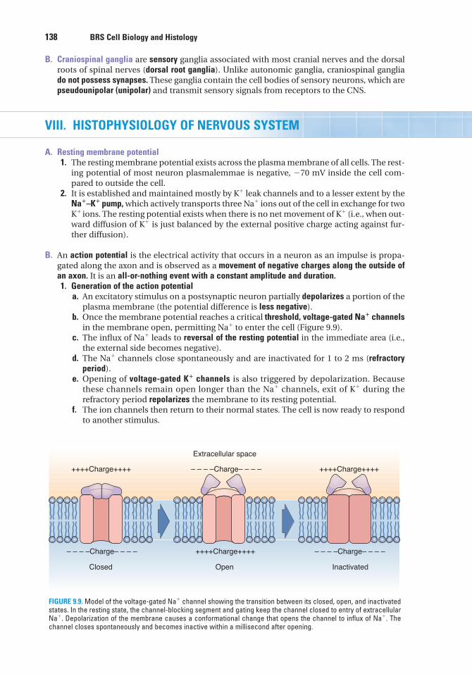
138 BRS Cell Biology and Histology
B. Craniospinal ganglia are sensory ganglia associated with most cranial nerves and the dorsalroots of spinal nerves (dorsal root ganglia). Unlike autonomic ganglia, craniospinal gangliado not possess synapses. These ganglia contain the cell bodies of sensory neurons, which arepseudounipolar (unipolar) and transmit sensory signals from receptors to the CNS.
VIII. HISTOPHYSIOLOGY OF NERVOUS SYSTEM
A. Resting membrane potential1. The resting membrane potential exists across the plasma membrane of all cells. The rest-
ing potential of most neuron plasmalemmae is negative, �70 mV inside the cell com-pared to outside the cell.
2. It is established and maintained mostly by K� leak channels and to a lesser extent by theNa�–K� pump, which actively transports three Na� ions out of the cell in exchange for twoK� ions. The resting potential exists when there is no net movement of K� (i.e., when out-ward diffusion of K� is just balanced by the external positive charge acting against fur-ther diffusion).
B. An action potential is the electrical activity that occurs in a neuron as an impulse is propa-gated along the axon and is observed as a movement of negative charges along the outside ofan axon. It is an all-or-nothing event with a constant amplitude and duration.1. Generation of the action potential
a. An excitatory stimulus on a postsynaptic neuron partially depolarizes a portion of theplasma membrane (the potential difference is less negative).
b. Once the membrane potential reaches a critical threshold, voltage-gated Na� channelsin the membrane open, permitting Na� to enter the cell (Figure 9.9).
c. The influx of Na� leads to reversal of the resting potential in the immediate area (i.e.,the external side becomes negative).
d. The Na� channels close spontaneously and are inactivated for 1 to 2 ms (refractoryperiod).
e. Opening of voltage-gated K� channels is also triggered by depolarization. Becausethese channels remain open longer than the Na� channels, exit of K� during therefractory period repolarizes the membrane to its resting potential.
f. The ion channels then return to their normal states. The cell is now ready to respondto another stimulus.
++++Charge++++
++++Charge++++– – – –Charge– – – –
Closed
++++Charge++++
– – – –Charge– – – –
Inactivated
Extracellular space
– – – –Charge– – – –
Open
FIGURE 9.9. Model of the voltage-gated Na� channel showing the transition between its closed, open, and inactivatedstates. In the resting state, the channel-blocking segment and gating keep the channel closed to entry of extracellularNa�. Depolarization of the membrane causes a conformational change that opens the channel to influx of Na�. Thechannel closes spontaneously and becomes inactive within a millisecond after opening.
LWBK615-c09[126-147].qxd 05/31/2010 10:35 AM Page 138 Aptara
Chapter 9 Nervous System 139
2. Propagation of the action potentiala. Propagation results from longitudinal diffusion of Na� (which enters the cell at the
initial site of excitation) toward the axon terminals (orthodromic spread). The longitu-dinal diffusion of Na� depolarizes the adjacent region of membrane, giving rise to anew action potential at this site.
b. Propagation does not result from diffusion of Na� toward the soma (antidromicspread), because the Na� channels are inactivated in this region.
c. Action potentials are propagated most rapidly in myelinated fibers, which exhibitsaltatory conduction. In this type of conduction, the action potential jumps from onenode of Ranvier to the next.
C. Axonal transport of proteins, organelles, and vesicles occurs at high, intermediate, and lowvelocities, depending on the nature of the transported materials.1. Anterograde transport carries material away from the soma.2. Retrograde transport carries material toward the soma for reuse, recycling, or degrada-
tion. However, some viruses (e.g., herpes simplex and rabies) spread in this fashion. Also,some toxins (e.g., tetanus) move from the periphery to the CNS in this manner.
D. Trophic function of nervous tissue1. Denervation of a muscle or gland leads to its atrophy.2. Reinnervation of a muscle or gland restores its structure and function.
IX. SOMATIC NERVOUS SYSTEM AND AUTONOMICNERVOUS SYSTEM (ANS)
Somatic and autonomic are functional concepts relating to all the neural elements involved intransmission of impulses from the CNS to the somatic and visceral components of the body,respectively.
A. The somatic nervous system contains sensory fibers that bring information to the CNS andthe motor fibers that originate in the CNS that innervate voluntary skeletal muscles.
B. The ANS is generally considered to be purely motor in function as it contains motor fibersthat control and regulate smooth muscle, cardiac muscle, and some glands. It establishes andmaintains homeostasis of the body’s visceral functions. Anatomically and functionally, theANS is divided into three parts: the sympathetic, parasympathetic, and enteric nervous systems.The sympathetic and parasympathetic nervous systems generally function antagonistically ina given organ (i.e., when the sympathetic system stimulates an organ, the parasympatheticinhibits it, and vice versa). The enteric nervous system is confined to the digestive systemwhere it controls the digestive process, however, it is modulated by the sympathetic andparasympathetic nervous systems.1. Autonomic nerve chains
a. Cell bodies of preganglionic neurons are located in the CNS and extend their pregan-glionic fibers (axons) to an autonomic ganglion located outside of the CNS.
b. In the ganglion, the preganglionic fibers synapse with postganglionic neurons, whichtypically are multipolar and surrounded by satellite cells.
c. Postganglionic fibers leave the ganglion and terminate in the effector organ (smoothmuscle, cardiac muscle, and glands).
2. Sympathetic system (thoracolumbar outflow)a. Preganglionic cell bodies of the sympathetic nervous system are located in the tho-
racic and the first two lumbar segments of the spinal cord.b. Function. The sympathetic system effects vasoconstriction. In general, it functions to
prepare the body for flight-or-fight responses by increasing heart rate, respiration,
LWBK615-c09[126-147].qxd 05/31/2010 10:35 AM Page 139 Aptara
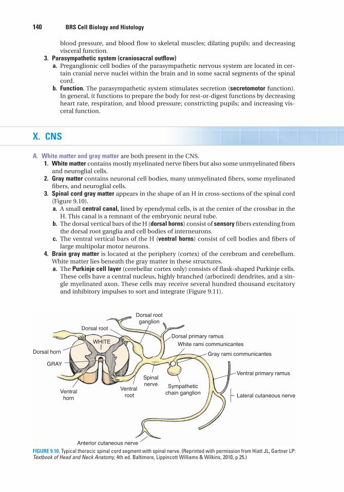
140 BRS Cell Biology and Histology
blood pressure, and blood flow to skeletal muscles; dilating pupils; and decreasingvisceral function.
3. Parasympathetic system (craniosacral outflow)a. Preganglionic cell bodies of the parasympathetic nervous system are located in cer-
tain cranial nerve nuclei within the brain and in some sacral segments of the spinalcord.
b. Function. The parasympathetic system stimulates secretion (secretomotor function).In general, it functions to prepare the body for rest-or-digest functions by decreasingheart rate, respiration, and blood pressure; constricting pupils; and increasing vis-ceral function.
X. CNS
A. White matter and gray matter are both present in the CNS.1. White matter contains mostly myelinated nerve fibers but also some unmyelinated fibers
and neuroglial cells.2. Gray matter contains neuronal cell bodies, many unmyelinated fibers, some myelinated
fibers, and neuroglial cells.3. Spinal cord gray matter appears in the shape of an H in cross-sections of the spinal cord
(Figure 9.10).a. A small central canal, lined by ependymal cells, is at the center of the crossbar in the
H. This canal is a remnant of the embryonic neural tube.b. The dorsal vertical bars of the H (dorsal horns) consist of sensory fibers extending from
the dorsal root ganglia and cell bodies of interneurons.c. The ventral vertical bars of the H (ventral horns) consist of cell bodies and fibers of
large multipolar motor neurons.4. Brain gray matter is located at the periphery (cortex) of the cerebrum and cerebellum.
White matter lies beneath the gray matter in these structures.a. The Purkinje cell layer (cerebellar cortex only) consists of flask-shaped Purkinje cells.
These cells have a central nucleus, highly branched (arborized) dendrites, and a sin-gle myelinated axon. These cells may receive several hundred thousand excitatoryand inhibitory impulses to sort and integrate (Figure 9.11).
Dorsal root
Dorsal horn
Ventral horn
Anterior cutaneous nerve
Ventral root
Spinalnerve Sympathetic
chain ganglion
GRAY
WHITE
Dorsal rootganglion
Dorsal primary ramus
White rami communicantes
Gray rami communicantes
Ventral primary ramus
Lateral cutaneous nerve
FIGURE 9.10. Typical thoracic spinal cord segment with spinal nerve. (Reprinted with permission from Hiatt JL, Gartner LP:Textbook of Head and Neck Anatomy, 4th ed. Baltimore, Lippincott Williams & Wilkins, 2010, p 25.)
LWBK615-c09[126-147].qxd 05/31/2010 10:35 AM Page 140 Aptara
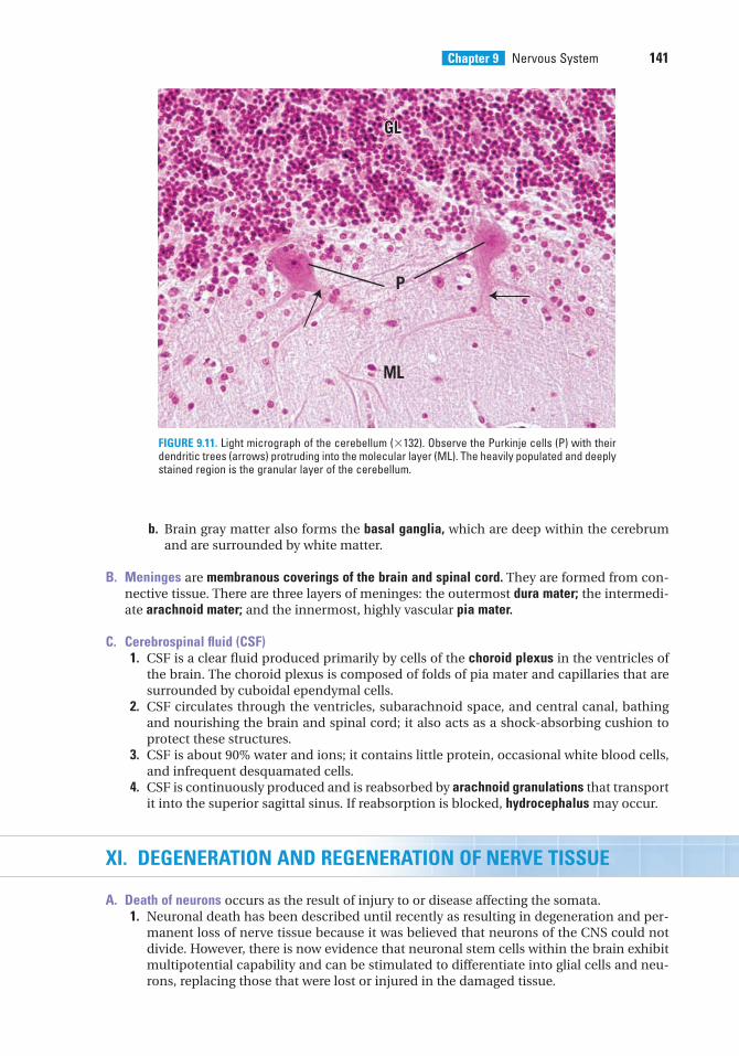
Chapter 9 Nervous System 141
b. Brain gray matter also forms the basal ganglia, which are deep within the cerebrumand are surrounded by white matter.
B. Meninges are membranous coverings of the brain and spinal cord. They are formed from con-nective tissue. There are three layers of meninges: the outermost dura mater; the intermedi-ate arachnoid mater; and the innermost, highly vascular pia mater.
C. Cerebrospinal fluid (CSF)1. CSF is a clear fluid produced primarily by cells of the choroid plexus in the ventricles of
the brain. The choroid plexus is composed of folds of pia mater and capillaries that aresurrounded by cuboidal ependymal cells.
2. CSF circulates through the ventricles, subarachnoid space, and central canal, bathingand nourishing the brain and spinal cord; it also acts as a shock-absorbing cushion toprotect these structures.
3. CSF is about 90% water and ions; it contains little protein, occasional white blood cells,and infrequent desquamated cells.
4. CSF is continuously produced and is reabsorbed by arachnoid granulations that transportit into the superior sagittal sinus. If reabsorption is blocked, hydrocephalus may occur.
XI. DEGENERATION AND REGENERATION OF NERVE TISSUE
A. Death of neurons occurs as the result of injury to or disease affecting the somata.1. Neuronal death has been described until recently as resulting in degeneration and per-
manent loss of nerve tissue because it was believed that neurons of the CNS could notdivide. However, there is now evidence that neuronal stem cells within the brain exhibitmultipotential capability and can be stimulated to differentiate into glial cells and neu-rons, replacing those that were lost or injured in the damaged tissue.
GLGL
ML
P
GL
FIGURE 9.11. Light micrograph of the cerebellum (�132). Observe the Purkinje cells (P) with theirdendritic trees (arrows) protruding into the molecular layer (ML). The heavily populated and deeplystained region is the granular layer of the cerebellum.
LWBK615-c09[126-147].qxd 05/31/2010 10:36 AM Page 141 Aptara
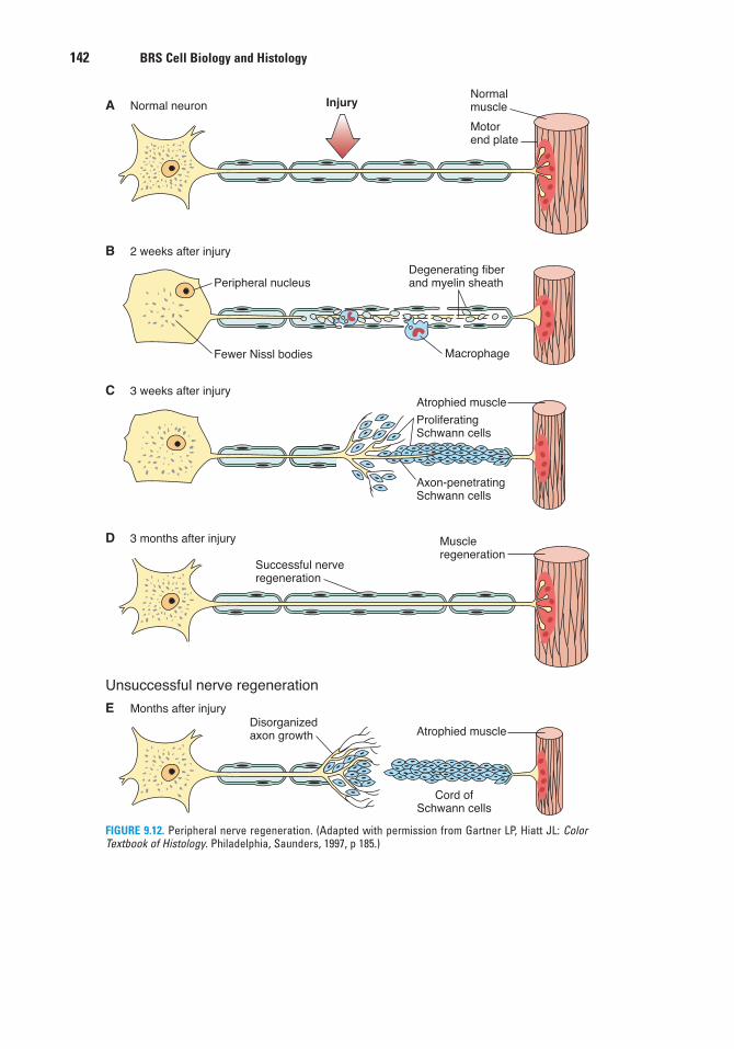
142 BRS Cell Biology and Histology
InjuryA Normal neuronNormalmuscle
Motorend plate
D 3 months after injury
E
Unsuccessful nerve regeneration
Months after injury
B 2 weeks after injury
Peripheral nucleus
Fewer Nissl bodies
Degenerating fiberand myelin sheath
Macrophage
C 3 weeks after injuryAtrophied muscle
ProliferatingSchwann cells
Axon-penetratingSchwann cells
Atrophied muscle
Cord ofSchwann cells
Muscleregeneration
Successful nerveregeneration
Disorganizedaxon growth
FIGURE 9.12. Peripheral nerve regeneration. (Adapted with permission from Gartner LP, Hiatt JL: ColorTextbook of Histology. Philadelphia, Saunders, 1997, p 185.)
LWBK615-c09[126-147].qxd 05/31/2010 10:36 AM Page 142 Aptara
Chapter 9 Nervous System 143
2. In the CNS, neuronal death may be followed by proliferation of the neuroglia, which fillsin areas left by dead neurons.
B. Transection of peripheral axons induces changes in the soma, including chromatolysis (dis-ruption of Nissl bodies with a concomitant loss of cytoplasmic basophilia), increase in somavolume, and movement of the nucleus to a peripheral position.1. Degeneration of distal axonal segment (anterograde changes)
a. The axon and its myelin sheath, which are separated from the soma, degenerate com-pletely (wallerian degeneration), and the remnants are removed by macrophages.
b. Schwann cells proliferate, forming a solid cellular column that is distal to the injuryand that remains attached to the effector cell.
2. Regeneration of proximal axonal segment (retrograde changes) (Figure 9.12)a. The distal end, closest to the wound, initially degenerates, and the remnants are
removed by macrophages.b. Growth at the distal end then begins (0.5–3 mm/day) and progresses toward the
columns of Schwann cells.c. Regeneration is successful if the sprouting axon penetrates a Schwann cell column
and reestablishes contact with the effector cell.
LWBK615-c09[126-147].qxd 05/31/2010 10:36 AM Page 143 Aptara
Review Test
144
1. Neural crest cells give rise to which of thefollowing?
(A) Dorsal horns of the spinal cord(B) Adrenal cortex(C) Sympathetic ganglia(D) Preganglionic autonomic nerves(E) Somatic motor neurons
2. Which one of the following neurotransmit-ters functions to increase cardiac output?
(A) Dopamine(B) Serotonin(C) Norepinephrine(D) Glutamate(E) GABA
3. Which of the following statements regard-ing nerve cell membrane potentials is true?
(A) Membrane potentials are maintained atrest by Na� entering the cell.
(B) Entrance of K� causes the membrane toreturn to its resting potential.
(C) Depolarization triggers the opening ofvoltage-gated K� channels.
(D) Voltage-gated Na� channels becomeactivated during the refractory period.
(E) The influx of K� reverses the restingpotential.
4. Which of the following statements is char-acteristic of the perineurium?
(A) It is a fascia surrounding many bundlesof nerve fibers.
(B) It is the fascia surrounding a single nervefiber.
(C) It is a thin layer of reticular fibers cover-ing individual nerve fibers.
(D) It is a fascia that excludesmacromolecules and forms the externalcoat of nerves.
(E) It consists in part of epithelioid cells thatsurround a bundle (fascicle) of nervefibers.
5. Acetylcholine is the only neurotransmitterin which of the following regions of thenervous system?
(A) Central nervous system(B) Presynaptic sensory cortex(C) Myoneural junctions(D) Postganglionic sympathetic synapses(E) Motor cortex
6. Nissl bodies are composed of
(A) synaptic vesicles and acetylcholine.(B) polyribosomes and rough endoplasmic
reticulum.(C) lipoprotein and melanin.(D) neurofilaments and microtubules.(E) smooth endoplasmic reticulum and
mitochondria.
7. The axon hillock contains
(A) rough endoplasmic reticulum.(B) ribosomes.(C) microtubules.(D) Golgi complex.(E) synaptic vesicles.
8. Synaptic vesicles possess which of thefollowing characteristics?
(A) Manufacture neurotransmitter(B) Enter the synaptic cleft(C) Become incorporated into the presynap-
tic membrane(D) Become incorporated into the
postsynaptic membrane(E) Release neurotransmitter via endocytosis
Directions: Each of the numbered items or incomplete statements in this section is followed byanswers or by completions of the statement. Select the ONE lettered answer or completion thatis BEST in each case.
LWBK615-c09[126-147].qxd 05/31/2010 10:36 AM Page 144 Aptara
13. Loss of neurotransmitter GABA is associ-ated with
(A) meningitis.(B) Huntington chorea.(C) spina bifida.(D) Parkinson disease.(E) multiple sclerosis.
14. Rapid onset of fever, stiff neck, headache,and an altered state of consciousness areassociated with
(A) meningitis.(B) Huntington chorea.(C) spina bifida.(D) Parkinson disease.(E) multiple sclerosis.
15. Deterioration and death of the dopamin-ergic neurons within the substantia nigra ofthe brain are associated with
(A) meningitis.(B) Huntington chorea.(C) spina bifida.(D) Parkinson disease.(E) multiple sclerosis.
9. A patient with Hirschsprung presents withwhich of the following symptoms?
(A) Absent cranial vault(B) Exposed spinal cord(C) Headache(D) Large, dilated colon(E) Absent small intestine
10. Myelination of peripheral nerves isaccomplished by
(A) astrocytes.(B) oligodendrocytes.(C) Schwann cells.(D) neural crest cells.(E) basket cells.
11. Episodes of demyelination areassociated with
(A) meningitis.(B) Huntington chorea.(C) spina bifida.(D) Parkinson disease.(E) multiple sclerosis.
12. Tremors, shuffling gate, and masklikefacial expressions are associated with
(A) meningitis.(B) Huntington chorea.(C) spina bifida.(D) Parkinson disease.(E) multiple sclerosis.
Chapter 9 Nervous System 145
LWBK615-c09[126-147].qxd 05/31/2010 10:36 AM Page 145 Aptara
Answers and Explanations
146
1. C. Neural crest cells migrate throughout the body and give rise to ganglia and other struc-tures, including portions of the adrenal medulla, but they do not contribute to the devel-opment of preganglionic autonomic nerves, adrenal cortex, or the dorsal horns of thespinal cord (see Chapter 9 II D).
2. C. Norepinephrine increases cardiac output, whereas dopamine and �-aminobutyric acidare CNS inhibitors. Glutamate is the most common excitatory neurotransmitter of theCNS. Serotonin functions as a pain inhibitor in mood control and in sleep (see Chapter 9IV C).
3. C. Once the critical threshold is reached, voltage-gated Na� channels open and Na� entersthe cell, which depolarizes the cell. Depolarization triggers the opening of voltage-gatedK� channels, and K� then exits the cell (see Chapter 9 VIII A).
4. E. Each bundle of nerve fibers is surrounded by the perineurium, which consists primarilyof several layers of epithelioid cells. Tight junctions between these cells exclude mostmacromolecules. The external coat of nerves, the epineurium, surrounds many fasciclesbut does not exclude macromolecules. The layer of reticular fibers that covers individualnerve fibers is the endoneurium; it also does not exclude macromolecules (see Chapter 9VI A).
5. C. Acetylcholine is the neurotransmitter for myoneural junctions as well as forpreganglionic sympathetic and preganglionic and postganglionic parasympatheticsynapses (Table 9.1).
6. B. Nissl bodies are large, granular basophilic bodies composed of polysomes and roughendoplasmic reticulum. They are found only in neurons (in the soma cytoplasm) (seeChapter 9 III A 3).
7. C. The axon hillock is devoid of large organelles, such as Nissl bodies and Golgi cisternae,but it does contain microtubules arranged in bundles and permits passage ofneurofilaments, mitochondria, and vesicles into the axon (see Chapter 9 III A 3 c).
8. C. Synaptic vesicles release neurotransmitter into the synaptic cleft by exocytosis. In thisprocess, the vesicle membrane is incorporated into the presynaptic membrane. Althoughthese vesicles contain neurotransmitter, they do not manufacture it (see Chapter 9 IV B 2).
9. D. Hirschsprung disease is characterized by a dilated colon caused by the absence of theparasympathetic myenteric ganglia known as Auerbach plexus (see Chapter 9 II D ClinicalConsiderations).
10. C. Schwann cells produce myelin in the peripheral nervous system, whereas oligodendro-cytes produce myelin in the central nervous system. Astrocytes, neural crest cells, and bas-ket cells do not produce myelin (see Chapter 9 V A B).
11. E. Multiple sclerosis is an immune-mediated disease exhibiting chronic and progressivedysfunction of the nervous system due to demyelination of the CNS and optic nerves,striking the 20- to 40-year age group affecting 1.5 times more women than men. There arerandom episodes of inflammation, edema, and demyelination of axons followed by peri-ods of remission. Each episode may reduce the vitality of the patient and be sufficient tocause death within months (see Chapter 9 V B Clinical Considerations).
LWBK615-c09[126-147].qxd 05/31/2010 10:36 AM Page 146 Aptara
Chapter 9 Nervous System 147
12. D. Parkinson disease is a progressive degenerative disease characterized by tremors, mus-cular rigidity, difficulty in initiating movements, slow voluntary shuffling movement, andmasklike face. The cause is the loss of dopaminergic neurons from the substantia nigra ofthe brain. Although the cause of the loss of these cells is unclear, it is known that certainpoisons and environmental factors cause Parkinson disease (see Chapter 9 IV A ClinicalConsiderations).
13. B. Huntington chorea is a fatal heredity disease that becomes evident during the third andfourth decades of life. It progresses to uncontrolled flicking of joints, motor disfunction,dementia, and death. The cause is apparently the loss of neurons that produce the neuro-transmitter GABA. Dementia symptoms are thought to be related to the loss of cells secret-ing acetylcholine (see Chapter 9 IV A Clinical Considerations).
14. A. Meningitis results from an inflammation of the meninges caused by viral or bacterialinfection in the CSF. Although viral meningitis is not severe, bacterial meningitis is conta-gious and dangerous, leading to hearing loss, learning disability, brain damage, and deathif untreated, sometimes within 24 hours. Major symptoms include fever, headache, stiffneck, and alteration of consciousness with rapid onset and progression. Spinal tap andculture of CFS to determine the bacterial species is the only diagnosis. Treatment is byspecies-specific antibiotic. Bacterial meningitis can be spread by respiratory and throatsecretions (i.e., coughing, sneezing, kissing) (see Chapter 9 VI A Clinical Considerations).
15. D. Parkinson disease is a progressive degenerative disease characterized by tremors, mus-cular rigidity, difficulty in initiating movements, slow voluntary shuffling movement, andmasklike face. It is caused by the loss of dopaminergic neurons from the substantia nigraof the brain (see Chapter 9 IV A Clinical Considerations).
LWBK615-c09[126-147].qxd 05/31/2010 10:36 AM Page 147 Aptara
c h a p t e r 10 Blood and Hemopoiesis
148
I. OVERVIEW—BLOOD
A. Blood is a specialized connective tissue that consists of formed elements (erythrocytes,leukocytes, and platelets) and a fluid component called plasma.
B. The volume of blood in an average human adult is approximately 5 L.
C. Blood circulates in a closed system of vessels and transports nutrients, waste products, hor-mones, proteins, ions, oxygen (O2), carbon dioxide (CO2), and formed elements.
D. It also regulates body temperature and assists in regulation of osmotic and acid–base balance.
E. Blood cells have short life spans and are continuously replaced by a process called hemo-poiesis.
II. BLOOD CONSTITUENTS
A. Plasma consists of 90% water; 9% organic compounds (such as proteins, amino acids, andhormones); and 1% inorganic salts, dissolved gases, and nutrients.1. Main plasma proteins
a. Albumin, a small protein (60,000 molecular weight), preserves osmotic pressure in thevascular system and helps transport some metabolites.
b. �-Globulins are antibodies (immunoglobulins) (see Chapter 12).c. �-Globulins and �-globulins transport metal ions (e.g., iron and copper) and lipids (in
the form of lipoproteins).d. Clotting proteins, including fibrinogen, a soluble protein that is converted into fibrin
during blood clotting.e. Complement proteins (C1–C9) are part of the innate immune system, and they function
in nonspecific host defense and initiate the inflammatory process.2. Serum is the yellowish fluid that remains after blood has clotted. It is similar to plasma
but lacks fibrinogen and clotting factors.
B. Formed elements of blood (Table 10.1 and Figure 10.1)1. Erythrocytes (red blood cells [RBCs])
a. General features(1) RBCs are round, anucleate, biconcave cells that stain light salmon pink with either
Wright or Giemsa stains (Figure 10.1).
LWBK615-c10[148-162].qxd 05/31/2010 10:41 AM Page 148 Aptara
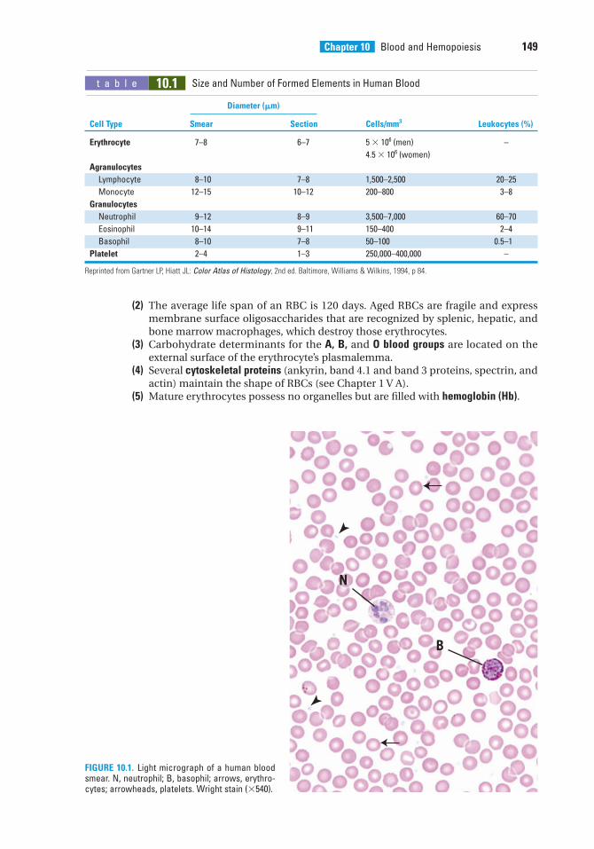
Chapter 10 Blood and Hemopoiesis 149
(2) The average life span of an RBC is 120 days. Aged RBCs are fragile and expressmembrane surface oligosaccharides that are recognized by splenic, hepatic, andbone marrow macrophages, which destroy those erythrocytes.
(3) Carbohydrate determinants for the A, B, and O blood groups are located on theexternal surface of the erythrocyte’s plasmalemma.
(4) Several cytoskeletal proteins (ankyrin, band 4.1 and band 3 proteins, spectrin, andactin) maintain the shape of RBCs (see Chapter 1 V A).
(5) Mature erythrocytes possess no organelles but are filled with hemoglobin (Hb).
t a b l e 10.1 Size and Number of Formed Elements in Human Blood
Diameter (�m)
Cell Type Smear Section Cells/mm3 Leukocytes (%)
Erythrocyte 7–8 6–7 5 � 106 (men) –
4.5 � 106 (women)
Agranulocytes
Lymphocyte 8–10 7–8 1,500–2,500 20–25
Monocyte 12–15 10–12 200–800 3–8
Granulocytes
Neutrophil 9–12 8–9 3,500–7,000 60–70
Eosinophil 10–14 9–11 150–400 2–4
Basophil 8–10 7–8 50–100 0.5–1
Platelet 2–4 1–3 250,000–400,000 –
Reprinted from Gartner LP, Hiatt JL: Color Atlas of Histology, 2nd ed. Baltimore, Williams & Wilkins, 1994, p 84.
N
B
FIGURE 10.1. Light micrograph of a human bloodsmear. N, neutrophil; B, basophil; arrows, erythro-cytes; arrowheads, platelets. Wright stain (�540).
LWBK615-c10[148-162].qxd 05/31/2010 10:41 AM Page 149 Aptara
150 BRS Cell Biology and Histology
(6) Erythrocytes also contain soluble enzymes that are responsible for glycolysis, thehexose monophosphate pathway, and the production of adenosine triphosphate(ATP).
b. The hematocrit is an estimation of the volume of packed erythrocytes per unit volume ofblood.(1) The hematocrit is expressed as a percentage.(2) Normal values are 40% to 50% in adult men, 35% to 45% in adult women, 35% in
children up to 10 years of age, and 45% to 60% in newborns.c. Hb is a protein composed of four polypeptide chains, each covalently linked to a heme
group. The four chains that normally occur in humans are �, �, �, and �. Each chaindiffers in its amino acid sequence.(1) Hb occurs in several normal forms that differ in their chain composition.
(a) The predominant form of adult Hb is HbA1 (�2�2).(b) A minor form is HbA2 (�2�2).(c) Fetal Hb is designated HbF (�2�2).
(2) Abnormal forms include HbS, which occurs as a result of a point mutation in the�-chain (substitution of the amino acid valine for glutamate). Erythrocytes con-taining HbS are sickle shaped and fragile, and they cause sickle cell anemia.
d. Transport of CO2 and O2 to and from the tissues of the body is carried out by erythro-cytes and plasma.(1) Every minute approximately 200 mL of CO2 is formed by cells of the body. Since
the partial pressure of CO2 is higher in the tissue than in the capillaries, the CO2
enters the capillaries via simple diffusion.(a) Twenty milliliters of the CO2 is transported in the plasma, 40 mL binds to the
globin moiety of Hb (forming carbaminohemoglobin), and 140 mL enters theRBC cytosol.
(b) Within the cytosol, the enzyme carbonic anhydrase forms H2CO3 by combiningCO2 with H2O. H2CO3 dissociates and the HCO3
� leaves the RBC to enter theplasma and Cl� from the plasma enters the erythrocyte cytosol to maintainelectrical equilibrium. This exchange of ions between the RBC cytosol and theplasma is referred to as the chloride shift.
(c) Meanwhile, since the partial pressure of O2 is greater in the RBC than in the tis-sue, the O2 is released from the Hb, forming deoxyhemoglobin, and the place ofO2 is taken up by the binding of 2,3-diphosphoglycerate.
(2) The partial pressure of O2 is greater in the alveolar airspace than in the alveoli of thelung than in the capillaries; therefore, O2 enters the capillaries via simple diffusion.(a) O2 enters the erythrocyte cytosol and binds to the heme moiety of the Hb mol-
ecule to form oxyhemoglobin.(b) Bicarbonate ions of the plasma reenter the RBC cytosol and in exchange Cl�
ions leave the RBC to enter the plasma (a reversal of the chloride shift). Thebicarbonate ion is combined with H� ions to form H2CO3, which is cleaved bycarbonic anhydrase to form CO2 and H2O. The CO2 enters the plasma, andfrom there the alveolar airspace via simple diffusion to be exhaled.
(3) Because carbon monoxide binds avidly to Hb, it can block binding of O2 and causecarbon monoxide asphyxiation if it is inhaled in sufficient amounts.
(4) Nitric oxide, a neurotransmitter substance, also binds to Hb and in O2-poor areasfacilitates dilation of blood vessels and a more efficacious exchange of O2 for CO2.
1. Sickle cell anemia is caused by a point mutation in the deoxyribonu-cleic acid (DNA) encoding the Hb molecule, leading to production ofan abnormal Hb (HbS).
a. Although this disease occurs almost exclusively among people of African descent (1 in 500 isaffected in the United States); among the US Hispanic population, 1 in 1,000 to 1,400 people isaffected with sickle cell anemia.
CLINICAL
CONSIDERATIONS
LWBK615-c10[148-162].qxd 05/31/2010 10:41 AM Page 150 Aptara
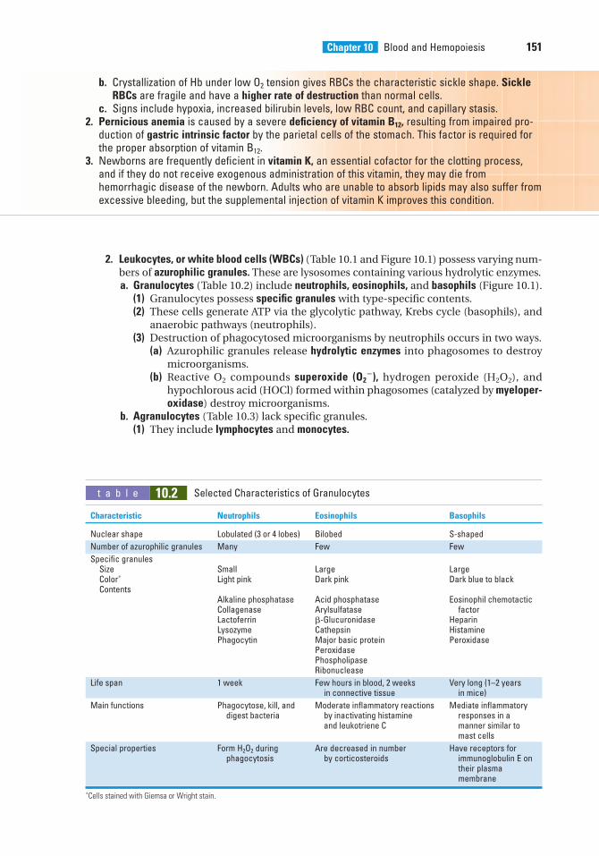
Chapter 10 Blood and Hemopoiesis 151
b. Crystallization of Hb under low O2 tension gives RBCs the characteristic sickle shape. SickleRBCs are fragile and have a higher rate of destruction than normal cells.
c. Signs include hypoxia, increased bilirubin levels, low RBC count, and capillary stasis.2. Pernicious anemia is caused by a severe deficiency of vitamin B12, resulting from impaired pro-
duction of gastric intrinsic factor by the parietal cells of the stomach. This factor is required forthe proper absorption of vitamin B12.
3. Newborns are frequently deficient in vitamin K, an essential cofactor for the clotting process,and if they do not receive exogenous administration of this vitamin, they may die fromhemorrhagic disease of the newborn. Adults who are unable to absorb lipids may also suffer fromexcessive bleeding, but the supplemental injection of vitamin K improves this condition.
t a b l e 10.2 Selected Characteristics of Granulocytes
Characteristic Neutrophils Eosinophils Basophils
Nuclear shape Lobulated (3 or 4 lobes) Bilobed S-shaped
Number of azurophilic granules Many Few Few
Specific granulesSize Small Large LargeColor∗ Light pink Dark pink Dark blue to blackContents
Alkaline phosphatase Acid phosphatase Eosinophil chemotactic Collagenase Arylsulfatase factorLactoferrin �-Glucuronidase HeparinLysozyme Cathepsin HistaminePhagocytin Major basic protein Peroxidase
PeroxidasePhospholipaseRibonuclease
Life span 1 week Few hours in blood, 2 weeks Very long (1–2 years in connective tissue in mice)
Main functions Phagocytose, kill, and Moderate inflammatory reactions Mediate inflammatory digest bacteria by inactivating histamine responses in a
and leukotriene C manner similar tomast cells
Special properties Form H2O2 during Are decreased in number Have receptors for phagocytosis by corticosteroids immunoglobulin E on
their plasma membrane
∗Cells stained with Giemsa or Wright stain.
2. Leukocytes, or white blood cells (WBCs) (Table 10.1 and Figure 10.1) possess varying num-bers of azurophilic granules. These are lysosomes containing various hydrolytic enzymes.a. Granulocytes (Table 10.2) include neutrophils, eosinophils, and basophils (Figure 10.1).
(1) Granulocytes possess specific granules with type-specific contents.(2) These cells generate ATP via the glycolytic pathway, Krebs cycle (basophils), and
anaerobic pathways (neutrophils).(3) Destruction of phagocytosed microorganisms by neutrophils occurs in two ways.
(a) Azurophilic granules release hydrolytic enzymes into phagosomes to destroymicroorganisms.
(b) Reactive O2 compounds superoxide (O2�), hydrogen peroxide (H2O2), and
hypochlorous acid (HOCl) formed within phagosomes (catalyzed by myeloper-oxidase) destroy microorganisms.
b. Agranulocytes (Table 10.3) lack specific granules.(1) They include lymphocytes and monocytes.
LWBK615-c10[148-162].qxd 05/31/2010 10:41 AM Page 151 Aptara
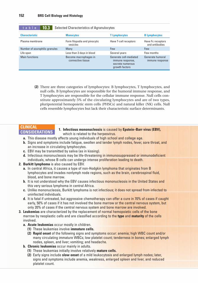
152 BRS Cell Biology and Histology
t a b l e 10.3 Selected Characteristics of Agranulocytes
Characteristic Monocytes T Lymphocytes B Lymphocytes
Plasma membrane Form filopodia and pinocytic Have T-cell receptors Have Fc receptors vesicles and antibodies
Number of azurophilic granules Many Few Few
Life span Less than 3 days in blood Several years Few months
Main functions Become macrophages in Generate cell-mediated Generate humoral connective tissue immune response, immune response
secrete numerous growth factors
(2) There are three categories of lymphocytes: B lymphocytes, T lymphocytes, andnull cells. B lymphocytes are responsible for the humoral immune response, andT lymphocytes are responsible for the cellular immune response. Null cells con-stitute approximately 5% of the circulating lymphocytes and are of two types,pluripotential hemopoietic stem cells (PHSCs) and natural killer (NK) cells. Nullcells resemble lymphocytes but lack their characteristic surface determinants.
1. Infectious mononucleosis is caused by Epstein–Barr virus (EBV),which is related to the herpesvirus.
a. This disease mostly affects young individuals of high school and college age.b. Signs and symptoms include fatigue, swollen and tender lymph nodes, fever, sore throat, and
an increase in circulating lymphocytes.c. EBV may be transmitted by saliva (as in kissing).d. Infectious mononucleosis may be life-threatening in immunosuppressed or immunodeficient
individuals, whose B cells can undergo intense proliferation leading to death.2. Burkitt lymphoma is also caused by EBV.
a. In central Africa, it causes a type of non-Hodgkin lymphoma that originates from Blymphocytes and invades nonlymph node regions, such as the brain, cerebrospinal fluid,blood, and bone marrow.
b. It is not understood why the EBV causes infectious mononucleosis in the United States andthis very serious lymphoma in central Africa.
c. Unlike mononucleosis, Burkitt lymphoma is not infectious; it does not spread from infected touninfected individuals.
d. It is fatal if untreated, but aggressive chemotherapy can offer a cure in 70% of cases if caughtearly, 50% of cases if it has not involved the bone marrow or the central nervous system, butonly 20% of cases if the central nervous system and bone marrow are involved.
3. Leukemias are characterized by the replacement of normal hemopoietic cells of the bonemarrow by neoplastic cells and are classified according to the type and maturity of the cellsinvolved.a. Acute leukemias occur mostly in children.
(1) These leukemias involve immature cells.(2) Rapid onset of the following signs and symptoms occur: anemia; high WBC count and/or
many circulating immature WBCs; low platelet count; tenderness in bones; enlarged lymphnodes, spleen, and liver; vomiting; and headache.
b. Chronic leukemias occur mainly in adults.(1) These leukemias initially involve relatively mature cells.(2) Early signs include slow onset of a mild leukocytosis and enlarged lymph nodes; later,
signs and symptoms include anemia, weakness, enlarged spleen and liver, and reducedplatelet count.
CLINICAL
CONSIDERATIONS
LWBK615-c10[148-162].qxd 05/31/2010 5:17 PM Page 152 Aptara
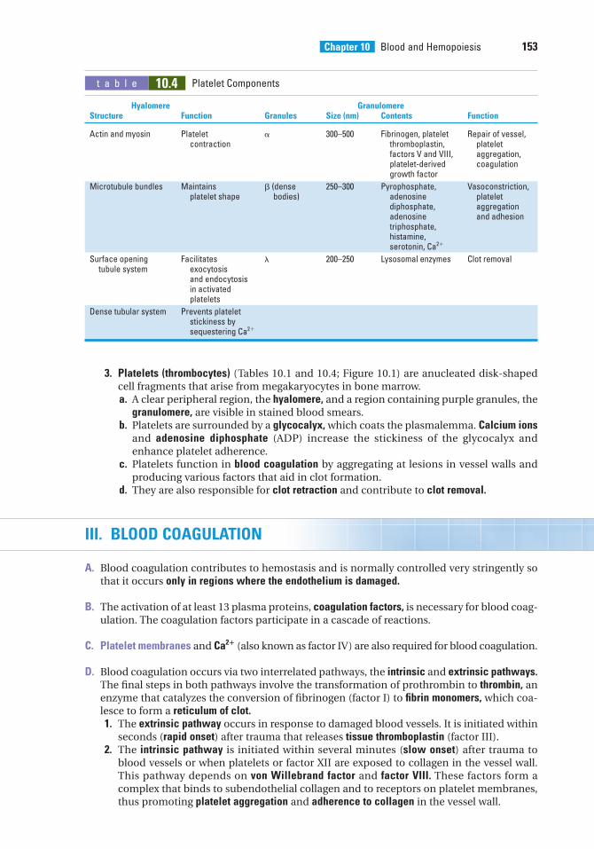
Chapter 10 Blood and Hemopoiesis 153
3. Platelets (thrombocytes) (Tables 10.1 and 10.4; Figure 10.1) are anucleated disk-shapedcell fragments that arise from megakaryocytes in bone marrow.a. A clear peripheral region, the hyalomere, and a region containing purple granules, the
granulomere, are visible in stained blood smears.b. Platelets are surrounded by a glycocalyx, which coats the plasmalemma. Calcium ions
and adenosine diphosphate (ADP) increase the stickiness of the glycocalyx andenhance platelet adherence.
c. Platelets function in blood coagulation by aggregating at lesions in vessel walls andproducing various factors that aid in clot formation.
d. They are also responsible for clot retraction and contribute to clot removal.
III. BLOOD COAGULATION
A. Blood coagulation contributes to hemostasis and is normally controlled very stringently sothat it occurs only in regions where the endothelium is damaged.
B. The activation of at least 13 plasma proteins, coagulation factors, is necessary for blood coag-ulation. The coagulation factors participate in a cascade of reactions.
C. Platelet membranes and Ca2� (also known as factor IV) are also required for blood coagulation.
D. Blood coagulation occurs via two interrelated pathways, the intrinsic and extrinsic pathways.The final steps in both pathways involve the transformation of prothrombin to thrombin, anenzyme that catalyzes the conversion of fibrinogen (factor I) to fibrin monomers, which coa-lesce to form a reticulum of clot.1. The extrinsic pathway occurs in response to damaged blood vessels. It is initiated within
seconds (rapid onset) after trauma that releases tissue thromboplastin (factor III).2. The intrinsic pathway is initiated within several minutes (slow onset) after trauma to
blood vessels or when platelets or factor XII are exposed to collagen in the vessel wall.This pathway depends on von Willebrand factor and factor VIII. These factors form acomplex that binds to subendothelial collagen and to receptors on platelet membranes,thus promoting platelet aggregation and adherence to collagen in the vessel wall.
t a b l e 10.4 Platelet Components
Hyalomere GranulomereStructure Function Granules Size (nm) Contents Function
Actin and myosin Platelet � 300–500 Fibrinogen, platelet Repair of vessel, contraction thromboplastin, platelet
factors V and VIII, aggregation, platelet-derived coagulationgrowth factor
Microtubule bundles Maintains � (dense 250–300 Pyrophosphate, Vasoconstriction, platelet shape bodies) adenosine platelet
diphosphate, aggregationadenosine and adhesiontriphosphate, histamine, serotonin, Ca2�
Surface opening Facilitates 200–250 Lysosomal enzymes Clot removaltubule system exocytosis
and endocytosis in activated platelets
Dense tubular system Prevents platelet stickiness by sequestering Ca2�
LWBK615-c10[148-162].qxd 05/31/2010 10:41 AM Page 153 Aptara
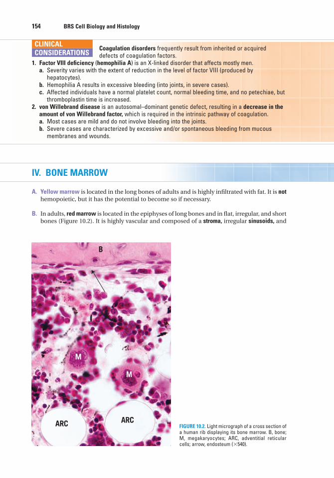
154 BRS Cell Biology and Histology
Coagulation disorders frequently result from inherited or acquireddefects of coagulation factors.
1. Factor VIII deficiency (hemophilia A) is an X-linked disorder that affects mostly men.a. Severity varies with the extent of reduction in the level of factor VIII (produced by
hepatocytes).b. Hemophilia A results in excessive bleeding (into joints, in severe cases).c. Affected individuals have a normal platelet count, normal bleeding time, and no petechiae, but
thromboplastin time is increased.2. von Willebrand disease is an autosomal–dominant genetic defect, resulting in a decrease in the
amount of von Willebrand factor, which is required in the intrinsic pathway of coagulation.a. Most cases are mild and do not involve bleeding into the joints.b. Severe cases are characterized by excessive and/or spontaneous bleeding from mucous
membranes and wounds.
CLINICAL
CONSIDERATIONS
IV. BONE MARROW
A. Yellow marrow is located in the long bones of adults and is highly infiltrated with fat. It is nothemopoietic, but it has the potential to become so if necessary.
B. In adults, red marrow is located in the epiphyses of long bones and in flat, irregular, and shortbones (Figure 10.2). It is highly vascular and composed of a stroma, irregular sinusoids, and
B
ARCARC
M
M
FIGURE 10.2. Light micrograph of a cross section ofa human rib displaying its bone marrow. B, bone;M, megakaryocytes; ARC, adventitial reticularcells; arrow, endosteum (�540).
LWBK615-c10[148-162].qxd 05/31/2010 10:41 AM Page 154 Aptara
Chapter 10 Blood and Hemopoiesis 155
islands of hemopoietic cells. Red marrow is the site of blood cell differentiation and matura-tion. The largest cells of bone marrow are the megakaryocytes (Figure 10.2), precursors ofplatelets.1. Sinusoids are large vessels with highly attenuated (very thin) endothelia. They are asso-
ciated on their extravascular surfaces with reticular fibers and adventitial reticular cells,which manufacture these fibers.
2. Stromal cells include macrophages, adventitial reticular cells, fibroblasts, and endothe-lial cells. These cells produce and release various hemopoietic growth factors.a. Macrophages are located in extravascular areas near sinusoids and extend processes
between endothelial cells into sinusoidal lumina.b. Adventitial reticular cells (Figure 10.2) are believed to subdivide the bone marrow cav-
ity into smaller compartments, which are occupied by islands of hemopoietic cells.Adventitial reticular cells may accumulate fat (instead of fat cells), thus transformingred marrow into yellow marrow.
V. PRENATAL HEMOPOIESIS
This process occurs successively in the yolk sac, liver, spleen, and bone marrow.
A. The bone marrow first participates in hemopoiesis at about 6 months’ gestation andassumes an increasingly large role thereafter.
B. The liver and spleen cease hemopoiesis at about the time of birth.
VI. POSTNATAL HEMOPOIESIS
This process involves three classes of cells: stem, progenitor, and precursor cells.
A. Comparison of stem, progenitor, and precursor cells1. Stem cells are capable of self-renewal and can undergo enormous proliferation.
a. These cells can differentiate into multiple cell lineages.b. They are present in circulation (as null cells) and in bone marrow.
2. Progenitor cells have reduced potentiality and are committed to a single cell lineage.a. They proliferate and differentiate into precursor cells in the presence of appropriate
growth factors.b. They are morphologically indistinguishable from stem cells, and both appear similar
to small lymphocytes.3. Precursor cells are all the cells in each lineage that display distinct morphological charac-
teristics.
B. Initial steps in blood formation (stem cells)1. PHSCs (pluripotential hemopoietic stem cells) give rise to multipotential hemopoietic
stem cells in the bone marrow.2. Multipotential hemopoietic stem cells are of two types: (1) colony-forming unit—
granulocyte, erythrocyte, monocyte, megakaryocyte (CFU-GEMM), (formerly known as colony-forming unit—spleen [CFU-S]) and (2) colony-forming unit—lymphocyte(CFU-Ly). These cells divide and differentiate in bone marrow to form progenitor cells.a. CFU-GEMM, the myeloid stem cell, is the multipotential stem cell that gives rise to
erythrocytes, granulocytes, monocytes, and platelets. Probably, Hox 2 genes areactive in the early stages of differentiation of the erythroid lines, and Hox 1 genesmay be active in the early stages of differentiation of granulocytes, monocytes, andplatelets.
LWBK615-c10[148-162].qxd 05/31/2010 10:41 AM Page 155 Aptara
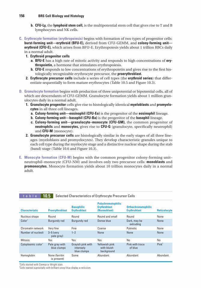
156 BRS Cell Biology and Histology
b. CFU-Ly, the lymphoid stem cell, is the multipotential stem cell that gives rise to T and Blymphocytes and NK cells.
C. Erythrocyte formation (erythropoiesis) begins with formation of two types of progenitor cells:burst-forming unit—erythroid (BFU-E), derived from CFU-GEMM, and colony-forming unit—erythroid (CFU-E), which arises from BFU-E. Erythropoiesis yields about 1 trillion RBCs dailyin a normal adult.1. Erythroid progenitor cells
a. BFU-E has a high rate of mitotic activity and responds to high concentrations of ery-thropoietin, a hormone that stimulates erythropoiesis.
b. CFU-E responds to low concentrations of erythropoietin and gives rise to the first his-tologically recognizable erythrocyte precursor, the proerythroblast.
2. Erythrocyte precursor cells include a series of cell types (the erythroid series) that differ-entiate sequentially to form mature erythrocytes (Table 10.5 and Figure 10.3).
D. Granulocyte formation begins with production of three unipotential or bipotential cells, all ofwhich are descendants of CFU-GEMM. Granulocyte formation yields about 1 million gran-ulocytes daily in a normal adult.1. Granulocyte progenitor cells give rise to histologically identical myeloblasts and promyelo-
cytes in all three cell lineages.a. Colony-forming unit—eosinophil (CFU-Eo) is the progenitor of the eosinophil lineage.b. Colony-forming unit—basophil (CFU-Ba) is the progenitor of the basophil lineage.c. Colony-forming unit—granulocyte–monocyte (CFU-GM), the common progenitor of
neutrophils and monocytes, gives rise to CFU-G (granulocyte, specifically neutrophil)and CFU-M (monocyte).
2. Granulocyte precursor cells are histologically similar in the early stages of all three line-ages (myeloblasts and promyelocytes). They develop characteristic granules unique toeach cell type during the myelocyte stage and a distinctive nuclear shape during the stab(band) stage (Table 10.6 and Figure 10.3).
E. Monocyte formation (CFU-M) begins with the common progenitor colony-forming unit–neutrophil–monocyte (CFU-NM) and involves only two precursor cells: monoblasts andpromonocytes. Monocyte formation yields about 10 trillion monocytes daily in a normaladult.
t a b l e 10.5 Selected Characteristics of Erythrocyte Precursor Cells
Polychromatophilic Basophilic Erythroblast Orthochromatophilic
Characteristic Proerythroblast Erythroblast (Normoblast) Erythroblast Reticulocyte
Nucleus shape Round Round Round and small Round None
Color∗ Burgundy red Burgundy red Dense blue Dark, may be Noneextruding
Chromatin network Very fine Fine Coarse Pyknotic None
Number of nucleoli 3–5 (very 1–2 None None Nonepale gray)
Mitosis Yes Yes Yes No No
Cytoplasmic color∗ Pale gray with Grayish pink with Yellowish pink Pink with trace Pink†
blue clumps intensely with bluish of blueblue clumps background
Hemoglobin None (ferritin Some Abundant Abundant Abundantis present)
∗Cells stained with Giemsa or Wright stain.†Cells stained supravitally with brilliant cresyl blue display a reticulum.
LWBK615-c10[148-162].qxd 05/31/2010 10:41 AM Page 156 Aptara
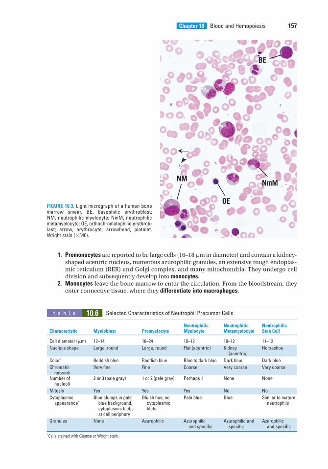
Chapter 10 Blood and Hemopoiesis 157
t a b l e 10.6 Selected Characteristics of Neutrophil Precursor Cells
Neutrophilic Neutrophilic Neutrophilic Characteristic Myeloblast Promyelocyte Myelocyte Metamyelocyte Stab Cell
Cell diameter (m) 12–14 16–24 10–12 10–12 11–12
Nucleus shape Large, round Large, round Flat (acentric) Kidney Horseshoe(acentric)
Color∗ Reddish blue Reddish blue Blue to dark blue Dark blue Dark blue
Chromatin Very fine Fine Coarse Very coarse Very coarsenetwork
Number of 2 or 3 (pale gray) 1 or 2 (pale gray) Perhaps 1 None Nonenucleoli
Mitosis Yes Yes Yes No No
Cytoplasmic Blue clumps in pale Bluish hue, no Pale blue Blue Similar to mature appearance∗ blue background, cytoplasmic neutrophils
cytoplasmic blebs blebsat cell periphery
Granules None Azurophilic Azurophilic Azurophilic and Azurophilicand specific specific and specific
∗Cells stained with Giemsa or Wright stain.
BE
NM NmM
OEFIGURE 10.3. Light micrograph of a human bonemarrow smear. BE, basophilic erythroblast; NM, neutrophilic myelocyte; NmM, neutrophilicmetamyelocyte; OE, orthochromatophilic erythrob-last; arrow, erythrocyte; arrowhead, platelet.Wright stain (�540).
1. Promonocytes are reported to be large cells (16–18 m in diameter) and contain a kidney-shaped acentric nucleus, numerous azurophilic granules, an extensive rough endoplas-mic reticulum (RER) and Golgi complex, and many mitochondria. They undergo celldivision and subsequently develop into monocytes.
2. Monocytes leave the bone marrow to enter the circulation. From the bloodstream, theyenter connective tissue, where they differentiate into macrophages.
LWBK615-c10[148-162].qxd 05/31/2010 10:41 AM Page 157 Aptara
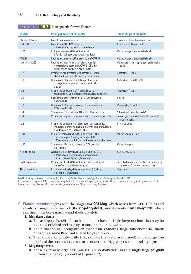
158 BRS Cell Biology and Histology
t a b l e 10.7 Hemopoietic Growth Factors
Factors Principal Action of the Factor Site of Origin of the Factor
Stem cell factor Facilitates hemopoiesis Stromal cells of bone marrow
GM-CSF Facilitates CFU-GM mitosis, T cells, endothelial cellsdifferentiation, granulocyte activity
G-CSF Induces mitosis, differentiation of Macrophages, endothelial cellsCFU-G; facilitates neutrophil activity
M-CSF Facilitates mitosis, differentiation of CFU-M Macrophages, endothelial cells
IL-1 (IL-3, IL-6) Facilitates proliferation of pluripotential Monocytes, macrophages, endothelial hemopoietic stem cell, CFU-S, CFU-Ly; cellssuppresses erythroid precursors
IL-2 Promotes proliferation of activated T cells, Activated T cellsB cells; facilitates NK cell differentiation
IL-3 Same as IL-1; also facilitates proliferation Activated T and B cellsof unipotential precursors except LyB and LyT
IL-4 Promotes activation of T cells, B cells; Activated T cellsfacilitates development of mast cells, basophils
IL-5 Facilitates proliferation of CFU-Eo; activates T cellseosinophils
IL-6 Same as IL-1; also promotes differentiation of Monocyte, fibroblastsCTLs and B cells
IL-7 Stimulates CFU-LyB and NK cell differentiation Adventitial reticular cells?
IL-8 Promotes migration and degranulation of neutrophils Leukocytes, endothelial cells, smooth muscle cells
IL-9 Promotes activation, proliferation of mast cells, T helper cellsmodulates immunoglobulin E synthesis, stimulatesproliferation of T helper cells
IL-10 Inhibits synthesis of cytokines by NK cells, Macrophages, T cellsmacrophages, T cells; promotes CTL differentiation and B cell and mast cell proliferation
IL-12 Stimulates NK cells; promotes CTL and NK Macrophagescell function
�-Interferons Activates monocytes, B cells; promotes CTL T cells, NK cellsdifferentiation; enhances expression of class II human leukocyte antigen
Erythropoietin Promotes CFU-E differentiation, proliferation of Endothelial cells of peritubular capillary burst-forming unit—erythroid network of kidney, hepatocytes
Thrombopoietin Enhances mitosis, differentiation of CFU-Meg Not knownand megakaryoblasts
Modified with permission from Gartner LP, Hiatt JL: Color Textbook of Histology, 2nd ed. Philadelphia, Saunders, 2001.CFU, colony-forming unit; CSF, colony-stimulating factor; CTL, cytotoxic lymphocyte; Eo, eosinophil; G, granulocyte; GM, granulocyte–monocyte; IL,interleukin; Ly, lymphocyte; M, monocyte; Meg, megakaryocyte; NK, natural killer; S, spleen.
F. Platelet formation begins with the progenitor CFU-Meg, which arises from CFU-GEMM andinvolves a single precursor cell (the megakaryoblast), and the mature megakaryocyte, whichremains in the bone marrow and sheds platelets.1. Megakaryoblasts
a. These large cells (25–40 m in diameter) have a single large nucleus that may beindented or lobed and displays a fine chromatin network.
b. Their basophilic, nongranular cytoplasm contains large mitochondria, manypolysomes, some RER, and a large Golgi complex.
c. They divide endomitotically (i.e., no daughter cells are formed) and enlarge; theploidy of the nucleus increases to as much as 64 N, giving rise to megakaryocytes.
2. Megakaryocytesa. These extremely large cells (40–100 m in diameter), have a single large polypoid
nucleus that is highly indented (Figure 10.2).
LWBK615-c10[148-162].qxd 05/31/2010 10:41 AM Page 158 Aptara
Chapter 10 Blood and Hemopoiesis 159
b. They possess a well-developed Golgi complex associated with the formation of �-granules, lysosomes, and dense bodies (�-granules); they also contain many mito-chondria and an extensive RER.
c. Megakaryocytes lie just outside the sinusoids in the bone marrow and form plateletdemarcation channels, which fragment into proplatelets (clusters of adhering platelets)or single platelets that are released into the sinusoidal lumen.
G. Lymphocyte formation (lymphopoiesis) begins with differentiation of CFU-Ly, the lymphoidstem cell, into the immunoincompetent progenitor cells CFU-LyB and CFU-LyT. These prelym-phocytes are processed and become mature immunocompetent cells.1. B lymphocyte (B-cell) maturation
a. Pre-B lymphocytes acquire cell surface markers, including membrane-bound anti-bodies, which confer immunocompetence.
b. In mammals, B-cell maturation occurs in the bone marrow, whereas in birds, it occursin the bursa of Fabricius (hence, B lymphocytes).
2. T-lymphocyte (T-cell) maturation involves migration of progenitor T lymphocytes to thethymic cortex, where they acquire cell surface markers, including T-cell receptors, whichconfer immunocompetence. Most of the newly formed T lymphocytes are destroyed in thecortex of the thymus and do not enter into circulation.
3. Mature B and T lymphocytes leave the bone marrow and thymus, respectively, and circu-late to peripheral organs (e.g., the lymph nodes and spleen) to establish clones ofimmunocompetent lymphocytes (see Chapter 12).
VII. HEMOPOIETIC GROWTH FACTORS (COLONY-STIMULATING FACTORS [CSFs])
A. Hemopoiesis is modulated by several growth factors and cytokines, including CSFs, stem cellfactor (steel factor), interleukins, and macrophage inhibiting protein-� (Table 10.7).
B. These factors may circulate in the bloodstream, acting as hormones, or they may act as localfactors produced in the bone marrow that facilitate and stimulate formation of blood cellsin their vicinity.
C. They act at low concentrations and bind to specific membrane receptors on single target cells.
D. Their various effects on target cells include control of mitotic rate, enhancement of cell sur-vival, control of the number of times the cells divide before they differentiate, and promo-tion of cell differentiation.
E. Hemopoietic stem cells that do not contact growth factors usually enter apoptosis and areeliminated by macrophages.
LWBK615-c10[148-162].qxd 05/31/2010 10:41 AM Page 159 Aptara
5. Which of the following is derived formCFU-GM?
(A) Red blood cell(B) Lymphocyte(C) Platelet(D) Neutrophil(E) Basophil
6. Which of the following is associated withdemarcation channels?
(A) Red blood cell(B) Lymphocyte(C) Platelet(D) Neutrophil(E) Basophil
7. Which of the following is derived frommyeloblasts?
(A) Red blood cell(B) Lymphocyte(C) Platelet(D) Monocyte(E) Basophil
8. Which one of the following cells is associ-ated with antibody production?
(A) Red blood cell(B) Lymphocyte(C) Monocyte(D) Neutrophil(E) Basophil
1. Which of the following proteinsassociated with the erythrocyte plasmamembrane is responsible for maintainingthe cell’s biconcave disk shape?
(A) HbA1
(B) HbA2
(C) Porphyrin(D) Spectrin(E) �-Actinin
2. Which of the following is animmunocompetent cell?
(A) Red blood cell(B) Lymphocyte(C) Platelet(D) Neutrophil(E) Basophil
3. Which of the following is derived formCFU-Meg?
(A) Red blood cell(B) Lymphocyte(C) Platelet(D) Neutrophil(E) Basophil
4. Which of the following is derived fromCFU-E?
(A) Red blood cell(B) Lymphocyte(C) Platelet(D) Neutrophil(E) Basophil
Review Test
Directions: Each of the numbered items or incomplete statements in this section is followed byanswers or completions of the statement. Select the ONE lettered answer that is BEST in eachcase.
160
LWBK615-c10[148-162].qxd 05/31/2010 10:41 AM Page 160 Aptara
9. Which of the following possesses specificand azurophilic granules?
(A) Red blood cell(B) Lymphocyte(C) Platelet(D) Neutrophil(E) Monocyte
10. Which of the following is derived fromreticulocytes?
(A) Red blood cell(B) Lymphocyte(C) Platelet(D) Neutrophil(E) Basophil
11. A 4-year-old boy is taken by his parentsto the pediatrician because of vomiting,headaches, and tenderness in the bones ofhis arms and legs. On palpation, the physi-cian notes that many lymph nodes areenlarged, as is the liver. The pediatricianshould order a complete blood count inorder to determine whether or not the childmay have
(A) chronic leukemia.(B) infectious mononucleosis.(C) von Willebrand disease.(D) acute leukemia.(E) pernicious anemia.
Chapter 10 Blood and Hemopoiesis 161
LWBK615-c10[148-162].qxd 05/31/2010 10:41 AM Page 161 Aptara
Answers and Explanations
1. D. Spectrin is associated with the erythrocyte cell membrane and assists in maintainingits biconcave disk shape (see Chapter 10 II B).
2. B. Lymphocytes are immunocompetent cells (see Chapter 10 VI G).
3. C. Platelets are derived from CFU-Meg (see Chapter 10 VI F).
4. A. Red blood cells are derived from CFU-E (see Chapter 10 VI C).
5. D. Neutrophils are derived from CFU-GM (see Chapter 10 VI D).
6. C. Platelets are derived from megakaryocytes, and those cells possess demarcation chan-nels (see Chapter 10 VI F).
7. E. Basophils are derived from myeloblasts (see Chapter 10 VI D).
8. C. Lymphocytes and plasma cells manufacture antibodies (see Chapter 10 VI G).
9. D. Neutrophils possess both azurophilic and specific granules (see Chapter 10 II B 2).
10. A. Red blood cells are derived from reticulocytes (see Table 10.5).
11. D. Acute leukemia is a disease of children with symptoms that include headaches; vomiting;swollen lymph nodes, liver, and spleen; and the sensation of tenderness in bones. Chronicleukemia is a disease that usually affects adults. von Willebrand disease is a coagulationdisorder and does not have the same symptoms as acute leukemia. Infectious mononucle-osis affects mostly young adults of high school and college age. Pernicious anemia is causedby vitamin B deficiency, and its symptoms do not resemble those of acute leukemia (seeChapter 10 II B 2 Clinical Considerations).
162
LWBK615-c10[148-162].qxd 05/31/2010 10:41 AM Page 162 Aptara
c h a p t e r 11 Circulatory System
163
I. OVERVIEW—BLOOD VASCULAR SYSTEM
The blood vascular system consists of the heart, arteries, veins, and capillaries. This systemtransports oxygen and nutrients to tissues, carries carbon dioxide and waste products from the tis-sues, and circulates hormones from the site of synthesis to their target cells.
A. The heart is a four-chambered pump composed of two atria and two ventricles and is sur-rounded by a fibroserous sac called the pericardium.
Tetralogy of FallotTetralogy of Fallot is a congenital malformation consisting of a defective
interventricular septum, hypertrophy of the right ventricle (due to a narrow pulmonary artery orvalve), and transposed (dextroposed) aorta. It should be repaired surgically early in life, before thepulmonary constriction becomes exacerbated.
CLINICAL
CONSIDERATIONS
The heart receives sympathetic and parasympathetic nerve fibers, which modulate the rate of theheartbeat but do not initiate it. It also produces atrial natriuretic peptide, a hormone thatincreases secretion of sodium and water by the kidneys, inhibits renin release, and decreasesblood pressure.
1. Cardiac layersa. Endocardium lines the lumen of the heart and is composed of simple squamous
epithelium (endothelium) and a thin layer of loose connective tissue. Subendocardium,a connective tissue layer that contains veins, nerves, and Purkinje fibers, underlies it.
b. Myocardium consists of layers of cardiac muscle cells arranged in a spiral fashionabout the heart’s chambers and inserted into the fibrous skeleton. The myocardiumcontracts to propel blood into arteries for distribution to the body. Specialized cardiacmuscle cells in the atria produce several peptides, including atrial natriuretic polypep-tide, atriopeptin, cardiodilatin, and cardionatrin, hormones that maintain fluid and elec-trolyte balance and decrease blood pressure.
c. Epicardium, the outermost layer of the heart, constitutes the visceral layer of the peri-cardium. It is composed of simple squamous epithelium (mesothelium) on the exter-nal surface. Beneath the mesothelium lies fibroelastic connective tissue, containingnerves and the coronary vessels, and adipose tissue.
2. The fibrous skeleton of the heart consists of thick bundles of collagen fibers oriented invarious directions. It also contains occasional foci of fibrocartilage.
3. Heart valvesa. Atrioventricular (AV) valves are composed of a skeleton of fibrous connective tissue,
arranged like an aponeurosis, and lined on both sides by endothelium. They areattached to the annuli fibrosi of the fibrous skeleton. The right AV valve is formed of
LWBK615-c11[163-177].qxd 05/31/2010 10:43 AM Page 163 Aptara
164 BRS Cell Biology and Histology
three interlocking cusps (tricuspid valve), whereas the left AV valve is formed of twointerlocking cusps (bicuspid or mitral valve). These valves prevent regurgitation ofventricular blood into the atria.
b. Semilunar valves in the pulmonary and aortic trunks are each composed of threecusps that approximate each other as they fill with arterial blood. They are lined withendothelium on both sides separated by sparse strands of connective tissue. Thesevalves prevent regurgitation of pulmonary and aortic blood into the respective ven-tricles.
Rheumatic heart valve disease is a sequel to childhood rheumaticfever (subsequent to streptococcal infection), which causes scarring
of the heart valves.1. The disease is characterized by reduced elasticity of the heart valves, making them unable to
close (incompetence) or open (stenosis) properly.2. It most commonly affects the mitral valve, followed by the aortic valve.
CLINICAL
CONSIDERATIONS
4. The impulse-generating and impulse-conducting system of the heart comprises severalspecialized structures with coordinated functions that act to initiate and regulate theheartbeat.a. The sinoatrial (SA) node, the pacemaker of the heart is composed of specialized cardiac
cells located within the wall of the right atrium. It generates impulses that initiate con-traction of atrial muscle cells; which are then conducted to the AV node.
b. The AV node is located in the wall of the right atrium, adjacent to the tricuspid valve.c. The AV bundle of His is a band of conducting tissue radiating from the AV node into the
interventricular septum, where it divides into two branches and continues as Purk-inje fibers.
d. Purkinje fibers are large, modified cardiac muscle cells (see Chapter 8 V B 9) that makecontact with cardiac muscle cells at the apex of the heart via gap junctions, desmo-somes, and fasciae adherentes.
e. The autonomic nervous system modulates the heart rate and stroke volume. Sympa-thetic innervation accelerates the heart rate, whereas parasympathetic stimulation slowsthe heart rate.
B. Arteries conduct blood away from the heart to the organs and tissues. Arterial walls are com-posed of three layers (tunicae): the tunica intima (inner), tunica media (middle), and tunicaadventitia (outer). Components of these layers and variations among types of arteries aresummarized in Table 11.1.1. Types of arteries
a. Elastic arteries (conducting arteries) are large. They include the aorta and its majorbranches (Figure 11.1 and 11.2).(1) Elastic arteries help reduce changes in blood pressure associated with the heart-
beat.(2) Small vessels (vasa vasorum) and nerves are located in their tunicae adventitia and
media. The vasa vasorum vascularize the walls of the elastic arteries.(3) Thick, concentric sheaths of elastic membranes, known as fenestrated membranes,
are located in the tunica media.b. Muscular arteries (distributing arteries) distribute blood to various organs.
(1) They include most of the named arteries of the human body.(2) These medium-sized arteries are smaller than elastic arteries but larger than arte-
rioles.(3) The tunica adventitia contains vasa vasorum.(4) The tunica media is thick, composed of layers of smooth muscle cells. Larger mus-
cular arteries possess an external elastic lamina separating their boundary with thetunica adventitia (Figure 11.3).
LWBK615-c11[163-177].qxd 05/31/2010 10:43 AM Page 164 Aptara
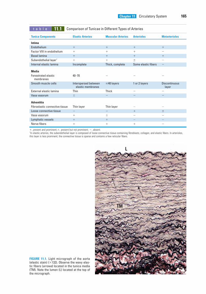
Chapter 11 Circulatory System 165
t a b l e 11.1 Comparison of Tunicae in Different Types of Arteries
Tunica Components Elastic Arteries Muscular Arteries Arterioles Metarterioles
Intima
Endothelium � � � �
Factor VIII in endothelium � � � �
Basal lamina � � � �
Subendothelial layer∗ � � � �
Internal elastic lamina Incomplete Thick, complete Some elastic fibers �
Media
Fenestrated elastic 40–70 � � �membranes
Smooth muscle cells Interspersed between �40 layers 1 or 2 layers Discontinuous elastic membranes layer
External elastic lamina Thin Thick � �
Vasa vasorum � � � �
Adventitia
Fibroelastic connective tissue Thin layer Thin layer � �
Loose connective tissue � � � �
Vasa vasorum � � � �
Lymphatic vessels � � � �
Nerve fibers � � � �
�, present and prominent;�, present but not prominent; �, absent.∗In elastic arteries, the subendothelial layer is composed of loose connective tissue containing fibroblasts, collagen, and elastic fibers. In arterioles,this layer is less prominent; the connective tissue is sparse and contains a few reticular fibers.
L
TM
FIGURE 11.1. Light micrograph of the aorta(elastic stain) (�132). Observe the wavy elas-tic fibers (arrows) located in the tunica media(TM). Note the lumen (L) located at the top ofthe micrograph.
LWBK615-c11[163-177].qxd 05/31/2010 10:43 AM Page 165 Aptara
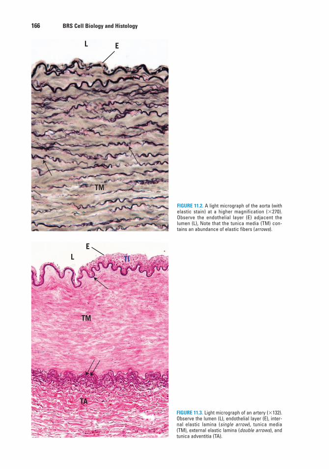
166 BRS Cell Biology and Histology
L E
TM
FIGURE 11.2. A light micrograph of the aorta (withelastic stain) at a higher magnification (�270).Observe the endothelial layer (E) adjacent thelumen (L), Note that the tunica media (TM) con-tains an abundance of elastic fibers (arrows).
LE
TA
TI
TM
FIGURE 11.3. Light micrograph of an artery (�132).Observe the lumen (L), endothelial layer (E), inter-nal elastic lamina (single arrow), tunica media(TM), external elastic lamina (double arrows), andtunica adventitia (TA).
LWBK615-c11[163-177].qxd 05/31/2010 10:43 AM Page 166 Aptara
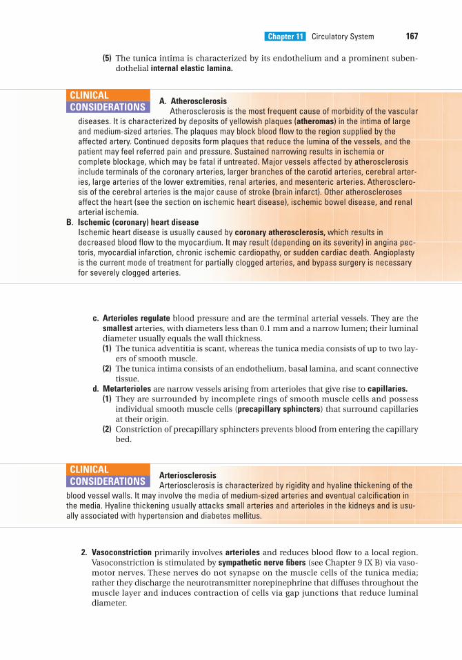
Chapter 11 Circulatory System 167
(5) The tunica intima is characterized by its endothelium and a prominent suben-dothelial internal elastic lamina.
A. AtherosclerosisAtherosclerosis is the most frequent cause of morbidity of the vascular
diseases. It is characterized by deposits of yellowish plaques (atheromas) in the intima of largeand medium-sized arteries. The plaques may block blood flow to the region supplied by theaffected artery. Continued deposits form plaques that reduce the lumina of the vessels, and thepatient may feel referred pain and pressure. Sustained narrowing results in ischemia orcomplete blockage, which may be fatal if untreated. Major vessels affected by atherosclerosisinclude terminals of the coronary arteries, larger branches of the carotid arteries, cerebral arter-ies, large arteries of the lower extremities, renal arteries, and mesenteric arteries. Atherosclero-sis of the cerebral arteries is the major cause of stroke (brain infarct). Other atherosclerosesaffect the heart (see the section on ischemic heart disease), ischemic bowel disease, and renalarterial ischemia.
B. Ischemic (coronary) heart diseaseIschemic heart disease is usually caused by coronary atherosclerosis, which results indecreased blood flow to the myocardium. It may result (depending on its severity) in angina pec-toris, myocardial infarction, chronic ischemic cardiopathy, or sudden cardiac death. Angioplastyis the current mode of treatment for partially clogged arteries, and bypass surgery is necessaryfor severely clogged arteries.
CLINICAL
CONSIDERATIONS
c. Arterioles regulate blood pressure and are the terminal arterial vessels. They are thesmallest arteries, with diameters less than 0.1 mm and a narrow lumen; their luminaldiameter usually equals the wall thickness.(1) The tunica adventitia is scant, whereas the tunica media consists of up to two lay-
ers of smooth muscle.(2) The tunica intima consists of an endothelium, basal lamina, and scant connective
tissue.d. Metarterioles are narrow vessels arising from arterioles that give rise to capillaries.
(1) They are surrounded by incomplete rings of smooth muscle cells and possessindividual smooth muscle cells (precapillary sphincters) that surround capillariesat their origin.
(2) Constriction of precapillary sphincters prevents blood from entering the capillarybed.
ArteriosclerosisArteriosclerosis is characterized by rigidity and hyaline thickening of the
blood vessel walls. It may involve the media of medium-sized arteries and eventual calcification inthe media. Hyaline thickening usually attacks small arteries and arterioles in the kidneys and is usu-ally associated with hypertension and diabetes mellitus.
CLINICAL
CONSIDERATIONS
2. Vasoconstriction primarily involves arterioles and reduces blood flow to a local region.Vasoconstriction is stimulated by sympathetic nerve fibers (see Chapter 9 IX B) via vaso-motor nerves. These nerves do not synapse on the muscle cells of the tunica media;rather they discharge the neurotransmitter norepinephrine that diffuses throughout themuscle layer and induces contraction of cells via gap junctions that reduce luminaldiameter.
LWBK615-c11[163-177].qxd 05/31/2010 10:43 AM Page 167 Aptara
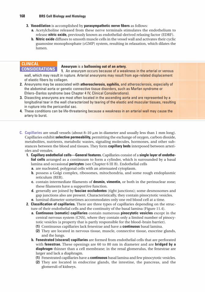
168 BRS Cell Biology and Histology
3. Vasodilation is accomplished by parasympathetic nerve fibers as follows:a. Acetylcholine released from these nerve terminals stimulates the endothelium to
release nitric oxide, previously known as endothelial-derived relaxing factor (EDRF).b. Nitric oxide diffuses to smooth muscle cells in the vessel wall and activates their cyclic
guanosine monophosphate (cGMP) system, resulting in relaxation, which dilates thelumen.
Aneurysm is a ballooning out of an artery.1. An aneurysm occurs because of a weakness in the arterial or venous
wall, which may result in rupture. Arterial aneurysms may result from age-related displacementof elastic fibers by collagen.
2. Aneurysms may be associated with atherosclerosis, syphilis, and atherosclerosis, especially ofthe abdominal aorta or genetic connective tissue disorders, such as Marfan syndrome orEhlers–Danlos syndrome (see Chapter 4 IV, Clinical Considerations).
3. Dissecting aneurysms are most often located in the ascending aorta and are represented by alongitudinal tear in the wall characterized by tearing of the elastic and muscular tissues, resultingin rupture into the pericardial sac.
4. These conditions can be life-threatening because a weakness in an arterial wall may cause theartery to burst.
CLINICAL
CONSIDERATIONS
C. Capillaries are small vessels (about 8–10 �m in diameter and usually less than 1 mm long).Capillaries exhibit selective permeability, permitting the exchange of oxygen, carbon dioxide,metabolites, nutrients, metabolic wastes, signaling molecules, hormones, and other sub-stances between the blood and tissues. They form capillary beds interposed between arteri-oles and venules.1. Capillary endothelial cells—General features. Capillaries consist of a single layer of endothe-
lial cells arranged as a continuum to form a cylinder, which is surrounded by a basallamina and occasional pericytes (see Chapter 6 III B). Endothelial cellsa. are nucleated, polygonal cells with an attenuated cytoplasm.b. possess a Golgi complex, ribosomes, mitochondria, and some rough endoplasmic
reticulum (RER).c. contain intermediate filaments of desmin, vimentin, or both in the perinuclear zone;
these filaments have a supportive function.d. generally are joined by fasciae occludentes (tight junctions); some desmosomes and
gap junctions also are present. Characteristically, they contain pinocytotic vesicles.e. luminal diameter sometimes accommodates only one red blood cell at a time.
2. Classification of capillaries. There are three types of capillaries depending on the struc-ture of their endothelial cells and the continuity of the basal lamina (Figure 11.4).a. Continuous (somatic) capillaries contain numerous pinocytotic vesicles except in the
central nervous system (CNS), where they contain only a limited number of pinocy-totic vesicles (a property that is partly responsible for the blood–brain barrier).(1) Continuous capillaries lack fenestrae and have a continuous basal lamina.(2) They are located in nervous tissue, muscle, connective tissue, exocrine glands,
and the lungs.b. Fenestrated (visceral) capillaries are formed from endothelial cells that are perforated
with fenestrae. These openings are 60 to 80 nm in diameter and are bridged by adiaphragm thinner than a cell membrane; in the renal glomerulus, the fenestrae arelarger and lack a diaphragm.(1) Fenestrated capillaries have a continuous basal lamina and few pinocytotic vesicles.(2) They are located in endocrine glands, the intestine, the pancreas, and the
glomeruli of kidneys.
LWBK615-c11[163-177].qxd 05/31/2010 10:43 AM Page 168 Aptara
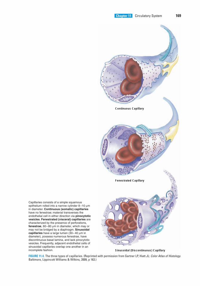
Chapter 11 Circulatory System 169
Continuous Capillary
Fenestrated Capillary
Sinusoidal (Discontinuous) Capillary
FIGURE 11.4. The three types of capillaries. (Reprinted with permission from Gartner LP, Hiatt JL: Color Atlas of Histology.Baltimore, Lippincott Williams & Wilkins, 2009, p 163.)
LWBK615-c11[163-177].qxd 05/31/2010 10:43 AM Page 169 Aptara
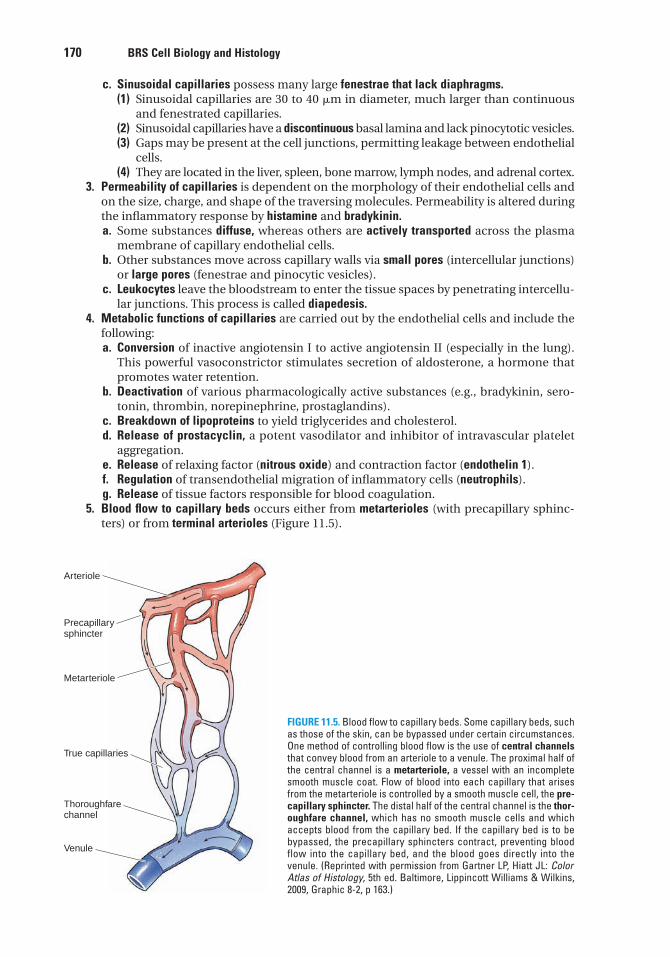
170 BRS Cell Biology and Histology
c. Sinusoidal capillaries possess many large fenestrae that lack diaphragms.(1) Sinusoidal capillaries are 30 to 40 �m in diameter, much larger than continuous
and fenestrated capillaries.(2) Sinusoidal capillaries have a discontinuous basal lamina and lack pinocytotic vesicles.(3) Gaps may be present at the cell junctions, permitting leakage between endothelial
cells.(4) They are located in the liver, spleen, bone marrow, lymph nodes, and adrenal cortex.
3. Permeability of capillaries is dependent on the morphology of their endothelial cells andon the size, charge, and shape of the traversing molecules. Permeability is altered duringthe inflammatory response by histamine and bradykinin.a. Some substances diffuse, whereas others are actively transported across the plasma
membrane of capillary endothelial cells.b. Other substances move across capillary walls via small pores (intercellular junctions)
or large pores (fenestrae and pinocytic vesicles).c. Leukocytes leave the bloodstream to enter the tissue spaces by penetrating intercellu-
lar junctions. This process is called diapedesis.4. Metabolic functions of capillaries are carried out by the endothelial cells and include the
following:a. Conversion of inactive angiotensin I to active angiotensin II (especially in the lung).
This powerful vasoconstrictor stimulates secretion of aldosterone, a hormone thatpromotes water retention.
b. Deactivation of various pharmacologically active substances (e.g., bradykinin, sero-tonin, thrombin, norepinephrine, prostaglandins).
c. Breakdown of lipoproteins to yield triglycerides and cholesterol.d. Release of prostacyclin, a potent vasodilator and inhibitor of intravascular platelet
aggregation.e. Release of relaxing factor (nitrous oxide) and contraction factor (endothelin 1).f. Regulation of transendothelial migration of inflammatory cells (neutrophils).g. Release of tissue factors responsible for blood coagulation.
5. Blood flow to capillary beds occurs either from metarterioles (with precapillary sphinc-ters) or from terminal arterioles (Figure 11.5).
Arteriole
Precapillarysphincter
Metarteriole
True capillaries
Thoroughfare channel
Venule
FIGURE 11.5. Blood flow to capillary beds. Some capillary beds, suchas those of the skin, can be bypassed under certain circumstances.One method of controlling blood flow is the use of central channelsthat convey blood from an arteriole to a venule. The proximal half ofthe central channel is a metarteriole, a vessel with an incompletesmooth muscle coat. Flow of blood into each capillary that arisesfrom the metarteriole is controlled by a smooth muscle cell, the pre-capillary sphincter. The distal half of the central channel is the thor-oughfare channel, which has no smooth muscle cells and whichaccepts blood from the capillary bed. If the capillary bed is to bebypassed, the precapillary sphincters contract, preventing bloodflow into the capillary bed, and the blood goes directly into thevenule. (Reprinted with permission from Gartner LP, Hiatt JL: ColorAtlas of Histology, 5th ed. Baltimore, Lippincott Williams & Wilkins,2009, Graphic 8-2, p 163.)
LWBK615-c11[163-177].qxd 05/31/2010 10:43 AM Page 170 Aptara
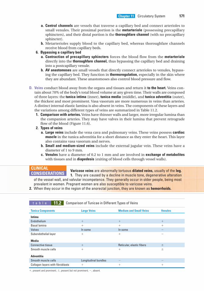
Chapter 11 Circulatory System 171
a. Central channels are vessels that traverse a capillary bed and connect arterioles tosmall venules. Their proximal portion is the metarteriole (possessing precapillarysphincters), and their distal portion is the thoroughfare channel (with no precapillarysphincter).
b. Metarterioles supply blood to the capillary bed, whereas thoroughfare channelsreceive blood from capillary beds.
6. Bypassing a capillary beda. Contraction of precapillary sphincters forces the blood flow from the metarteriole
directly into the thoroughfare channel, thus bypassing the capillary bed and draininginto a postcapillary venule.
b. AV anastomoses are small vessels that directly connect arterioles to venules, bypass-ing the capillary bed. They function in thermoregulation, especially in the skin wherethey are abundant. These anastomoses also control blood pressure and flow.
D. Veins conduct blood away from the organs and tissues and return it to the heart. Veins con-tain about 70% of the body’s total blood volume at any given time. Their walls are composedof three layers: the tunica intima (inner), tunica media (middle), and tunica adventitia (outer),the thickest and most prominent. Vasa vasorum are more numerous in veins than arteries.A distinct internal elastic lamina is also absent in veins. The components of these layers andthe variations among different types of veins are summarized in Table 11.2.1. Comparison with arteries. Veins have thinner walls and larger, more irregular lumina than
the companion arteries. They may have valves in their lumina that prevent retrogradeflow of the blood (Figure 11.6).
2. Types of veinsa. Large veins include the vena cava and pulmonary veins. These veins possess cardiac
muscle in the tunica adventitia for a short distance as they enter the heart. This layeralso contains vasa vasorum and nerves.
b. Small and medium-sized veins include the external jugular vein. These veins have adiameter of 1 to 9 mm.
c. Venules have a diameter of 0.2 to 1 mm and are involved in exchange of metaboliteswith tissues and in diapedesis (exiting of blood cells through vessel walls).
t a b l e 11.2 Comparison of Tunicae in Different Types of Veins
Tunica Components Large Veins Medium and Small Veins Venules
Intima
Endothelium � � �
Basal lamina � � �
Valves In some In some �
Subendothelial layer � � �
Media
Connective tissue � Reticular, elastic fibers �
Smooth muscle cells � � �
Adventitia
Smooth muscle cells Longitudinal bundles � �
Collagen layers with fibroblasts � � �
�, present and prominent; �, present but not prominent; �, absent.
Varicose veins are abnormally tortuous dilated veins, usually of the leg.1. They are caused by a decline in muscle tone, degenerative alteration
of the vessel wall, and valvular incompetence. They generally occur in older people, being mostprevalent in women. Pregnant women are also susceptible to varicose veins.
2. When they occur in the region of the anorectal junction, they are known as hemorrhoids.
CLINICAL
CONSIDERATIONS
LWBK615-c11[163-177].qxd 05/31/2010 10:43 AM Page 171 Aptara
172 BRS Cell Biology and Histology
E. Specialized sensory mechanisms within arteries include three types of sensors: the carotidsinus, carotid body, and the aortic bodies.1. Carotid sinus is a baroreceptor in the wall of the internal carotid artery just as it begins at
the common carotid artery. Sensory endings of the glossopharyngeal nerve embeddedwith the wall of the artery are sensitive to changes in pressure that distend the vessel,thus initiating a signal to the vasomotor center of the brain. The resultant response trig-gers adjustments to the tension on the arterial wall via the smooth muscles of the tunicamedia, effecting changes in blood pressure.
2. Carotid body is a chemoreceptor at the bifurcation of the common carotid artery. Special-ized nerve endings of the vagus and glossopharyngeal cranial nerves are sensitive to oxy-gen and carbon dioxide levels as well as H� concentration. Glomus (type 1) cells andsheath (type II) cells, which envelop the glomus cells, constitute the parenchyma. Nerveendings lose their Schwann cells upon entering the parenchyma, becoming coveredinstead by sheath cells. Impulses are shuttled to the brain by these two cranial nerves forprocessing.
3. Aortic bodies are located in the wall of the arch of the aorta at the junction of the com-mon carotid and subclavian arteries. Their structure and function are similar to those ofthe carotid body.
4. Hormonal control of low blood pressure starts with the kidney:a. Kidney produces reninb. Renin cleaves angiotensinogen circulating in the blood, forming angiotensin I, a mild
vasoconstrictor.c. Angiotensin I is converted into angiotensin II by angiotensin-converting enzyme (ACE),
located on the luminal plasmalemmas of capillary endothelia (especially capillariesof the lungs).
L V
LA
TMA
TMV
TAV
TAA
Vein
Artery
FIGURE 11.6. Light micrograph of an artery and vein(�270). Vein—lumen (LV), tunica media (TMV), andtunica adventitia (TAV). Artery—tunica adventitia(TAA), tunica media (TMA), and lumen (LA).
LWBK615-c11[163-177].qxd 05/31/2010 10:44 AM Page 172 Aptara
Chapter 11 Circulatory System 173
d. Angiotensin II, a potent vasoconstrictor, causes the walls of arterioles to contract,which raises blood pressure.
e. Antidiuretic hormone (ADH), or vasopressin, secreted by the pituitary gland, isanother potent vasoconstrictor employed after severe hemorrhage.
II. OVERVIEW—LYMPHATIC VASCULAR SYSTEM
This system consists of peripheral lymphatic capillaries, lymphatic vessels of gradually increas-ing size, and lymphatic ducts. The lymphatic vascular system collects excess tissue fluid (lymph)and returns it to the venous portion of the cardiovascular system. It drains most tissues with theexception of the nervous system and bone marrow.
A. Lymphatic capillaries are thin-walled vessels that begin as blind-ended channels (e.g.,lacteals) adjacent to capillary beds where they collect lymph.1. They are composed of a single layer of attenuated endothelial cells that lack fenestrae and
fasciae occludentes. They possess a sparse basal lamina.2. Lymph enters these leaky capillaries via spaces between overlapping endothelial cells.3. Small lymphatic anchoring filaments between the surrounding connective tissue and
the abluminal plasma membrane assist in maintaining luminal patency in these delicatevessels.
B. Large lymphatic vessels possess valves and are similar in structure to small veins, except thatthey have larger lumina and thinner walls.1. Lymph nodes that filter the lymph are interposed along their routes.2. These vessels converge to form the thoracic duct and right lymphatic duct. The thoracic
duct empties into the venous system at the junction of the left internal jugular vein withthe subclavian vein, whereas the right lymphatic duct empties into the venous system ata similar location on the right side of the neck.
Edema is a pathologic process resulting in an increased volume of tissuefluid.
Edema may be caused by venous obstruction or decreased venous blood flow (as incongestive heart failure), increased capillary permeability (due to injury), starvation, excessiverelease of histamine, and obstruction of lymphatic vessels. It is common during pregnancy and inolder persons. Edema that is responsive to localized pressure (i.e., depressions persist after releaseof pressure) is called pitting edema. Edema can be a symptom of a serious underlying disorderincluding heart disease, liver disease, or diseases of the thyroid, lymphatic system or the kidneys,with serious consequences.
CLINICAL
CONSIDERATIONS
LWBK615-c11[163-177].qxd 05/31/2010 10:44 AM Page 173 Aptara
5. Which of the following statementsconcerning innervation of blood vessels istrue?
(A) Vasoconstriction is controlled byparasympathetic nerve fibers.
(B) Acetylcholine acts directly on smoothmuscle cells.
(C) Acetylcholine acts directly on endothe-lial cells.
(D) Vasodilation is controlled bysympathetic nerve fibers.
(E) Nitric oxide acts as a vasoconstrictor.
6. Which of the following characteristicsdistinguishes somatic capillaries fromvisceral capillaries?
(A) Presence or absence of fenestrae(B) Size of the lumen(C) Thickness of the vessel wall(D) Presence or absence of pericytes(E) Thickness of the basal lamina
7. The blood–brain barrier is thought toexist because capillaries in the central nerv-ous system have which of the followingcharacteristics?
(A) Discontinuous basal lamina(B) Fenestrae with diaphragms(C) Fenestrae without diaphragms(D) A few pinocytic vesicles(E) No basement membrane
8. Which of the following statements abouthealthy, intact capillaries is true?
(A) They control blood pressure.(B) They are lined by a simple columnar
epithelium.(C) They have a smooth muscle coat.(D) They inhibit clot formation.(E) Satellite cells share their basal lamina.
1. The epicardium is one of the three layersof the heart. It is
(A) continuous with the endocardium.(B) also known as the visceral pericardium.(C) composed of modified cardiac muscle
cells.(D) capable of increasing intraventricular
pressure.(E) capable of decreasing the rate of
contraction.
2. The atrial muscle of the heart produces ahormone that
(A) decreases blood pressure.(B) increases blood pressure.(C) causes vasoconstriction.(D) facilitates the release of renin.(E) facilitates sodium resorption in the
kidneys.
3. The generation of impulses in the normalheart is the responsibility of which of thefollowing structures?
(A) Atrioventricular (AV) node(B) AV bundle of His(C) Sympathetic nerves(D) Sinoatrial (SA) node(E) Purkinje fibers
4. Metarterioles, vessels interposed betweenarterioles and capillary beds,
(A) function to control blood flow into arte-rioles.
(B) possess a complete layer of smoothmuscle cells in their tunica media.
(C) possess precapillary sphincters.(D) receive blood from thoroughfare
channels.(E) possess valves to regulate the direction
of blood flow.
Review Test
Directions: Each of the numbered items or incomplete statements in this section is followed byanswers or completions of the statement. Select the ONE lettered answer that is BEST in eachcase.
174
LWBK615-c11[163-177].qxd 05/31/2010 10:44 AM Page 174 Aptara
9. A patient complains of shortness ofbreath even after only mild exercise. Shestates that she has had this condition for 2years but recently has noticed that it hasbecome more pronounced. Her medical his-tory indicates that she had rheumatic feverwhen she was a child. Auscultation indicatesan enlarged heart. What may the physicianexpect to find with other diagnostic tests?
(A) Mitral valve stenosis(B) Tetralogy of Fallot(C) Pulmonary artery aneurysm(D) Coronary heart disease(E) Ischemic heart disease
10. Diagnostic tests for ischemic heartdisease usually reveal
(A) malformed heart valves.(B) atherosclerosis of coronary arteries.(C) irregular heartbeat.(D) faulty SA valve.(E) arteriosclerosis of coronary arteries.
11. Vasa vasorum function in a way that issimilar to
(A) AV valves.(B) semilunar valves.(C) coronary arteries.(D) elastic arteries.(E) metarterioles.
12. Which one of the following possesses adistinct internal elastic lamina?
(A) Capillary(B) Metarteriole(C) Arteriole(D) Muscular artery(E) Vein
13. Ischemic heart disease is the usualsequel to
(A) arteriosclerosis.(B) abdominal aortic aneurysm.(C) rheumatic fever.(D) varicose veins.(E) atherosclerosis.
Chapter 11 Circulatory System 175
LWBK615-c11[163-177].qxd 05/31/2010 10:44 AM Page 175 Aptara
Answers and Explanations
1. B. The pericardium is a fibroserous sac that encloses the heart. The innermost layer of thepericardium, the epicardium, is also known as the visceral pericardium (see Chapter 11 I A 1).
2. A. Atrial natriuretic peptide, which decreases blood pressure, is produced mainly by car-diac muscle cells of the right atrium. It inhibits the release of renin and causes the kidneysto decrease the resorption of sodium and water (see Chapter 11 I A 1 b).
3. D. Impulses are generated in the sinoatrial node, which is the pacemaker of the heart.They are then conducted to the atrioventricular (AV) node. The bundle of His and Purkinjefibers conduct impulses from the AV node to the cardiac muscle cells of the ventricles.Sympathetic nerves can increase the rate of the heartbeat but do not originate it (seeChapter 11 I A 4 a).
4. C. The proximal portion of a central channel is known as a metarteriole, whereas its distalportion is the thoroughfare channel. Blood from metarterioles may enter the capillary bedif their precapillary sphincters are relaxed. If the precapillary sphincters of metarteriolesare constricted, blood bypasses the capillary bed and flows directly into thoroughfarechannels and from there into a venule (see Chapter 11 I B 1 d).
5. C. Acetylcholine stimulates the endothelial cells of a vessel to release nitric oxide(endothelial-derived relaxing factor), which causes relaxation of smooth muscle cells.Thus, acetylcholine does not act directly on smooth muscle cells (see Chapter 11 I B 2).
6. A. Somatic (continuous) capillaries lack fenestrae, whereas visceral (fenestrated) capillar-ies are characterized by their presence. Both types of capillary possess a continuous basallamina and are surrounded by occasional pericytes (see Chapter 11 I C 2).
7. D. Capillaries in the central nervous system are of the continuous type and thus lack fen-estrae but have a continuous basal lamina. In contrast to continuous capillaries in otherparts of the body, they contain only a few pinocytic vesicles; this characteristic is thoughtto be partly responsible for the blood–brain barrier (see Chapter 11 I C 2).
8. D. The smooth endothelial lining of intact, healthy capillaries inhibits clot formation.Capillaries do not control blood pressure (see Chapter 11 I C 2).
9. A. A person who has had rheumatic fever as a child may develop heart valve disease later inlife. Although the mitral valve is the one most commonly affected, the other valves may alsobe involved. The mitral valve becomes inflamed, fibrotic, and eventually incompetent orstenotic. This condition leads to respiratory hypertension and edema, which restricts respi-ratory function. The key is a history of rheumatic fever, because those individuals are predis-posed to developing heart valve diseases (see Chapter 11 I A 3 Clinical Considerations).
10. B. Diagnostic tests for ischemic heart disease reveal atherosclerosis of the coronaryvessels. Over time, excessive plaque composed of cholesterol and fats is layered beneaththe intima of these vessels, thus restricting blood flow to the myocardium of the heart,leading to angina pectoris, an infarction, or perhaps sudden death (see Chapter 11 I B 1Clinical Considerations).
11. C. Vasa vasorum are the small blood vessels that serve the walls of elastic and musculararteries with oxygen and nutrients just as the coronary arteries provide for the walls of theheart (see Chapter 11 I B 1).
176
LWBK615-c11[163-177].qxd 05/31/2010 10:44 AM Page 176 Aptara
Chapter 11 Circulatory System 177
12. D. The muscular artery possesses a distinct internal elastic lamina. Elastic arteries possessan incomplete internal elastic lamina, whereas capillaries, arterioles, metarterioles andveins do not possess an internal elastic lamina (see Table 11.1 and Chapter 11 I D).
13. E. Ischemic heart disease is usually caused by coronary atherosclerosis, resulting indecreased blood flow to the myocardium. It may develop into angina pectoris, myocardialinfarction, chronic ischemic cardiopathy, or sudden cardiac death. On the other hand,arteriosclerosis (hardening of the arteries) usually attacks renal arteries and is related tohypertension and diabetes mellitus (see Chapter 11 I B Clinical Considerations).
LWBK615-c11[163-177].qxd 05/31/2010 10:44 AM Page 177 Aptara
c h a p t e r 12 Lymphoid Tissue
178
I. OVERVIEW—THE LYMPHOID (IMMUNE) SYSTEM
A. The lymphoid system consists of capsulated lymphoid tissues (thymus, spleen, tonsils, andlymph nodes); diffuse lymphoid tissue; and lymphoid cells, primarily T lymphocytes (T cells),B lymphocytes (B cells), and macrophages.
B. The immune system has two components, the innate immune system (nonspecific) and theadaptive immune system (specific).1. The innate immune system has no immunological memory; it acts rapidly, but in a non-
specific manner. Its chief constituents are complement, toll-like receptors (TLRs), mastcells, eosinophils, neutrophils, macrophages, and natural killer (NK) cells. TLRs are afamily of at least 14 receptors that are integral proteins located either in the cell mem-branes or in the cytosol of specific cells of the innate immune system. In order to per-form their function, they have to pair up with another TLRs and in that fashion have keyfunctions in the recognition of bacterial components (Table 12.1). For instance, TLR4recognizes lipopolysaccharides of gram-negative bacteria, and TLR2 recognizes pepti-doglycan of gram-positive bacteria. Some TLRs also recognize viral components. Oncethese receptors bind to their ligand, they cause the release of tumor necrosis factor �(TNF-�), interleukin 12 (IL-12), and other cytokines. Activation of TLRs also facilitatesthe induction of an adaptive immune response.
2. The adaptive immune system is evolutionarily more recent than the innate immune sys-tem. It possesses four characteristics: ability to distinguish self from nonself, memory,specificity, and diversity. The cells of the adaptive immune system, namely T lympho-cytes, B lymphocytes, and antigen-presenting cells (APCs), communicate with oneanother by the use of signaling molecules (cytokines), thus relaying information to eachother in response to antigenic invasion.
C. The immune system functions primarily to defend the organism by mounting humoralimmune responses against foreign substances (antigens) and cell-mediated immune responsesagainst microorganisms, tumor and transplanted cells, and virus-infected cells.
Individuals may suffer from congenital immunodeficiency disorders, usu-ally a recessive X-linked genetic defect that affects males to a much
greater extent than females. The most common symptoms are recurrent respiratory infectionsthat have a tendency to progress to pneumonia, additional indicators are multiple skin and oralinfections, such as yeast infections of the mouth (thrush) or of the vagina. In some cases, such asX-linked agammaglobulinemia, B lymphocytes do not develop, resulting in scarcity of plasma cellsand consequently lack of immunoglobulin production.
CLINICAL
CONSIDERATIONS
LWBK615-c12[178-195].qxd 05/31/2010 10:46 AM Page 178 Aptara
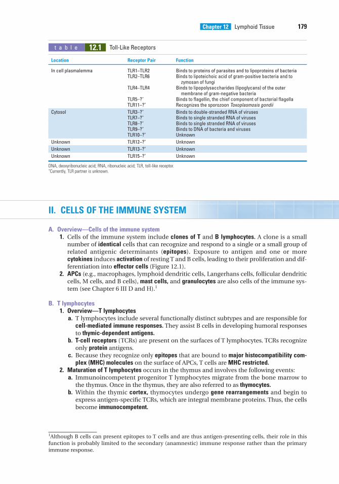
Chapter 12 Lymphoid Tissue 179
II. CELLS OF THE IMMUNE SYSTEM
A. Overview—Cells of the immune system1. Cells of the immune system include clones of T and B lymphocytes. A clone is a small
number of identical cells that can recognize and respond to a single or a small group ofrelated antigenic determinants (epitopes). Exposure to antigen and one or morecytokines induces activation of resting T and B cells, leading to their proliferation and dif-ferentiation into effector cells (Figure 12.1).
2. APCs (e.g., macrophages, lymphoid dendritic cells, Langerhans cells, follicular dendriticcells, M cells, and B cells), mast cells, and granulocytes are also cells of the immune sys-tem (see Chapter 6 III D and H).1
B. T lymphocytes1. Overview—T lymphocytes
a. T lymphocytes include several functionally distinct subtypes and are responsible forcell-mediated immune responses. They assist B cells in developing humoral responsesto thymic-dependent antigens.
b. T-cell receptors (TCRs) are present on the surfaces of T lymphocytes. TCRs recognizeonly protein antigens.
c. Because they recognize only epitopes that are bound to major histocompatibility com-plex (MHC) molecules on the surface of APCs, T cells are MHC restricted.
2. Maturation of T lymphocytes occurs in the thymus and involves the following events:a. Immunoincompetent progenitor T lymphocytes migrate from the bone marrow to
the thymus. Once in the thymus, they are also referred to as thymocytes.b. Within the thymic cortex, thymocytes undergo gene rearrangements and begin to
express antigen-specific TCRs, which are integral membrane proteins. Thus, the cellsbecome immunocompetent.
t a b l e 12.1 Toll-Like Receptors
Location Receptor Pair Function
In cell plasmalemma TLR1–TLR2 Binds to proteins of parasites and to lipoproteins of bacteriaTLR2–TLR6 Binds to lipoteichoic acid of gram-positive bacteria and to
zymosan of fungiTLR4–TLR4 Binds to lipopolysaccharides (lipoglycans) of the outer
membrane of gram-negative bacteriaTLR5–?∗ Binds to flagellin, the chief component of bacterial flagellaTLR11–?∗ Recognizes the sporozoon Toxoplasmosis gondii
Cytosol TLR3–?∗ Binds to double-stranded RNA of virusesTLR7–?∗ Binds to single stranded RNA of virusesTLR8–?∗ Binds to single stranded RNA of virusesTLR9–?∗ Binds to DNA of bacteria and virusesTLR10–?∗ Unknown
Unknown TLR12–?∗ Unknown
Unknown TLR13–?∗ Unknown
Unknown TLR15–?∗ Unknown
DNA, deoxyribonucleic acid; RNA, ribonucleic acid; TLR, toll-like receptor.∗Currently, TLR partner is unknown.
1Although B cells can present epitopes to T cells and are thus antigen-presenting cells, their role in thisfunction is probably limited to the secondary (anamnestic) immune response rather than the primaryimmune response.
LWBK615-c12[178-195].qxd 05/31/2010 10:46 AM Page 179 Aptara
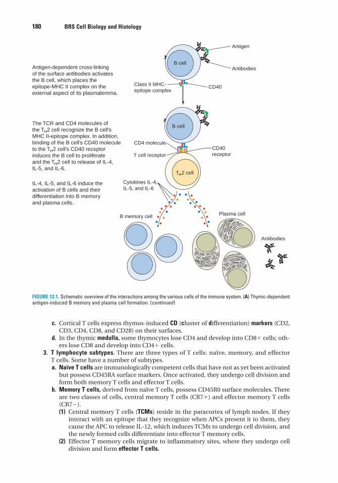
180 BRS Cell Biology and Histology
Antigen
Antibodies
CD40
CD40receptor
Plasma cell
Antibodies
B memory cell
Cytokines IL-4,IL-5, and IL-6
T cell receptor
TH2 cell
CD4 molecule
B cell
B cell
Class II MHC-epitope complex
Antigen-dependent cross-linkingof the surface antibodies activatesthe B cell, which places theepitope-MHC II complex on theexternal aspect of its plasmalemma.
The TCR and CD4 molecules ofthe TH2 cell recognize the B cell’sMHC II-epitope complex. In addition,binding of the B cell’s CD40 moleculeto the TH2 cell’s CD40 receptorinduces the B cell to proliferateand the TH2 cell to release of IL-4,IL-5, and IL-6.
IL-4, IL-5, and IL-6 induce theactivation of B cells and theirdifferentiation into B memoryand plasma cells.
FIGURE 12.1. Schematic overview of the interactions among the various cells of the immune system. (A) Thymic-dependentantigen-induced B memory and plasma cell formation. (continued )
c. Cortical T cells express thymus-induced CD (cluster of differentiation) markers (CD2,CD3, CD4, CD8, and CD28) on their surfaces.
d. In the thymic medulla, some thymocytes lose CD4 and develop into CD8� cells; oth-ers lose CD8 and develop into CD4� cells.
3. T lymphocyte subtypes. There are three types of T cells: naïve, memory, and effectorT cells. Some have a number of subtypes.a. Naïve T cells are immunologically competent cells that have not as yet been activated
but possess CD45RA surface markers. Once activated, they undergo cell division andform both memory T cells and effector T cells.
b. Memory T cells, derived from naïve T cells, possess CD45R0 surface molecules. Thereare two classes of cells, central memory T cells (CR7�) and effector memory T cells(CR7�).(1) Central memory T cells (TCMs) reside in the paracortex of lymph nodes. If they
interact with an epitope that they recognize when APCs present it to them, theycause the APC to release IL-12, which induces TCMs to undergo cell division, andthe newly formed cells differentiate into effector T memory cells.
(2) Effector T memory cells migrate to inflammatory sites, where they undergo celldivision and form effector T cells.
LWBK615-c12[178-195].qxd 05/31/2010 10:46 AM Page 180 Aptara
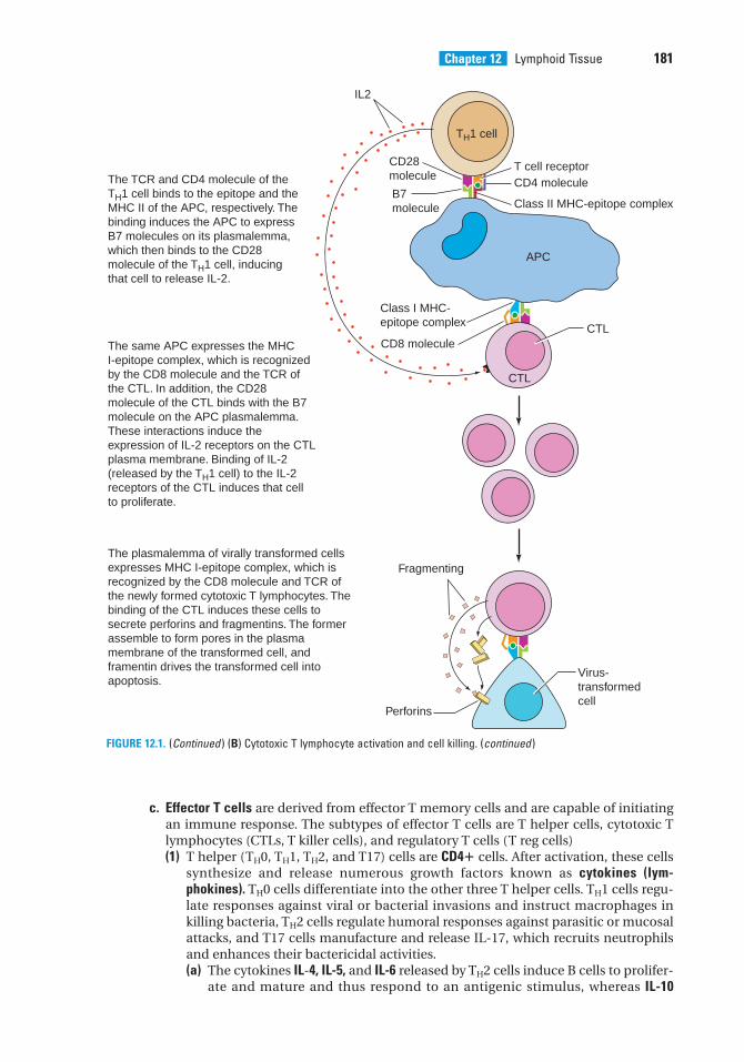
Chapter 12 Lymphoid Tissue 181
FIGURE 12.1. (Continued ) (B) Cytotoxic T lymphocyte activation and cell killing. (continued )
The TCR and CD4 molecule of the TH1 cell binds to the epitope and the MHC II of the APC, respectively. The binding induces the APC to express B7 molecules on its plasmalemma, which then binds to the CD28 molecule of the TH1 cell, inducingthat cell to release IL-2.
The same APC expresses the MHC I-epitope complex, which is recognized by the CD8 molecule and the TCR of the CTL. In addition, the CD28 molecule of the CTL binds with the B7 molecule on the APC plasmalemma. These interactions induce the expression of IL-2 receptors on the CTL plasma membrane. Binding of IL-2 (released by the TH1 cell) to the IL-2 receptors of the CTL induces that cell to proliferate.
The plasmalemma of virally transformed cells expresses MHC I-epitope complex, which is recognized by the CD8 molecule and TCR of the newly formed cytotoxic T lymphocytes. The binding of the CTL induces these cells to secrete perforins and fragmentins. The former assemble to form pores in the plasma membrane of the transformed cell, and framentin drives the transformed cell into apoptosis.
TH1 cell
T cell receptorCD4 molecule
Class II MHC-epitope complex
APC
CTL
CTL
Virus-transformedcell
Perforins
Fragmenting
CD8 molecule
Class I MHC-epitope complex
IL2
B7molecule
CD28molecule
c. Effector T cells are derived from effector T memory cells and are capable of initiatingan immune response. The subtypes of effector T cells are T helper cells, cytotoxic Tlymphocytes (CTLs, T killer cells), and regulatory T cells (T reg cells)(1) T helper (TH0, TH1, TH2, and T17) cells are CD4� cells. After activation, these cells
synthesize and release numerous growth factors known as cytokines (lym-phokines). TH0 cells differentiate into the other three T helper cells. TH1 cells regu-late responses against viral or bacterial invasions and instruct macrophages inkilling bacteria, TH2 cells regulate humoral responses against parasitic or mucosalattacks, and T17 cells manufacture and release IL-17, which recruits neutrophilsand enhances their bactericidal activities.(a) The cytokines IL-4, IL-5, and IL-6 released by TH2 cells induce B cells to prolifer-
ate and mature and thus respond to an antigenic stimulus, whereas IL-10
LWBK615-c12[178-195].qxd 05/31/2010 10:46 AM Page 181 Aptara
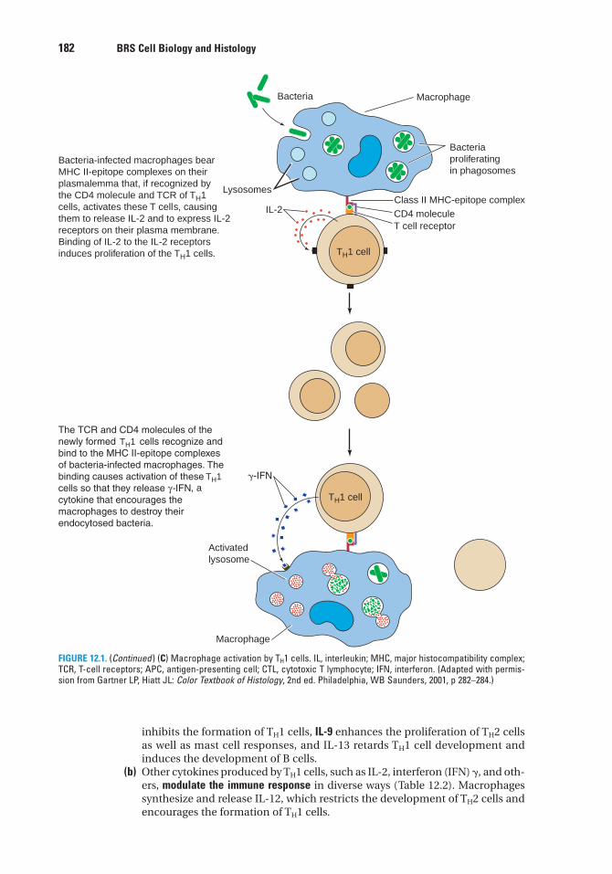
182 BRS Cell Biology and Histology
Bacteria-infected macrophages bear MHC II-epitope complexes on their plasmalemma that, if recognized by the CD4 molecule and TCR of TH1 cells, activates these T cells, causing them to release IL-2 and to express IL-2 receptors on their plasma membrane. Binding of IL-2 to the IL-2 receptors induces proliferation of the TH1 cells.
Bacteria
Macrophage
Bacteriaproliferatingin phagosomes
Class II MHC-epitope complexCD4 molecule
TH1 cell
TH1 cell
Macrophage
Activatedlysosome
IL-2
Lysosomes
T cell receptor
TH1
TH1, a
FIGURE 12.1. (Continued ) (C) Macrophage activation by TH1 cells. IL, interleukin; MHC, major histocompatibility complex;TCR, T-cell receptors; APC, antigen-presenting cell; CTL, cytotoxic T lymphocyte; IFN, interferon. (Adapted with permis-sion from Gartner LP, Hiatt JL: Color Textbook of Histology, 2nd ed. Philadelphia, WB Saunders, 2001, p 282–284.)
inhibits the formation of TH1 cells, IL-9 enhances the proliferation of TH2 cellsas well as mast cell responses, and IL-13 retards TH1 cell development andinduces the development of B cells.
(b) Other cytokines produced by TH1 cells, such as IL-2, interferon (IFN) �, and oth-ers, modulate the immune response in diverse ways (Table 12.2). Macrophagessynthesize and release IL-12, which restricts the development of TH2 cells andencourages the formation of TH1 cells.
LWBK615-c12[178-195].qxd 05/31/2010 10:46 AM Page 182 Aptara
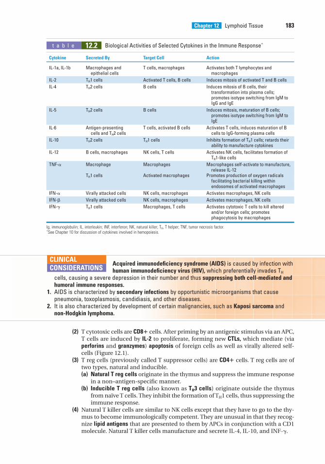
(2) T cytotoxic cells are CD8� cells. After priming by an antigenic stimulus via an APC,T cells are induced by IL-2 to proliferate, forming new CTLs, which mediate (viaperforins and granzymes) apoptosis of foreign cells as well as virally altered self-cells (Figure 12.1).
(3) T reg cells (previously called T suppressor cells) are CD4� cells. T reg cells are oftwo types, natural and inducible.(a) Natural T reg cells originate in the thymus and suppress the immune response
in a non–antigen-specific manner.(b) Inducible T reg cells (also known as TH3 cells) originate outside the thymus
from naïve T cells. They inhibit the formation of TH1 cells, thus suppressing theimmune response.
(4) Natural T killer cells are similar to NK cells except that they have to go to the thy-mus to become immunologically competent. They are unusual in that they recog-nize lipid antigens that are presented to them by APCs in conjunction with a CD1molecule. Natural T killer cells manufacture and secrete IL-4, IL-10, and INF-�.
Acquired immunodeficiency syndrome (AIDS) is caused by infection withhuman immunodeficiency virus (HIV), which preferentially invades TH
cells, causing a severe depression in their number and thus suppressing both cell-mediated andhumoral immune responses.
1. AIDS is characterized by secondary infections by opportunistic microorganisms that causepneumonia, toxoplasmosis, candidiasis, and other diseases.
2. It is also characterized by development of certain malignancies, such as Kaposi sarcoma andnon-Hodgkin lymphoma.
CLINICAL
CONSIDERATIONS
Chapter 12 Lymphoid Tissue 183
t a b l e 12.2 Biological Activities of Selected Cytokines in the Immune Response∗
Cytokine Secreted By Target Cell Action
IL-1a, IL-1b Macrophages and T cells, macrophages Activates both T lymphocytes andepithelial cells macrophages
IL-2 TH1 cells Activated T cells, B cells Induces mitosis of activated T and B cells
IL-4 TH2 cells B cells Induces mitosis of B cells, their transformation into plasma cells; promotes isotype switching from IgM to IgG and IgE
IL-5 TH2 cells B cells Induces mitosis, maturation of B cells; promotes isotype switching from IgM to IgE
IL-6 Antigen-presenting T cells, activated B cells Activates T cells, induces maturation of B cells and TH2 cells cells to IgG-forming plasma cells
IL-10 TH2 cells TH1 cells Inhibits formation of TH1 cells; retards their ability to manufacture cytokines
IL-12 B cells, macrophages NK cells, T cells Activates NK cells, facilitates formation of TH1-like cells
TNF-� Macrophage Macrophages Macrophages self-activate to manufacture, release IL-12
TH1 cells Activated macrophages Promotes production of oxygen radicals facilitating bacterial killing within endosomes of activated macrophages
IFN-� Virally attacked cells NK cells, macrophages Activates macrophages, NK cells
IFN-� Virally attacked cells NK cells, macrophages Activates macrophages, NK cells
IFN-� TH1 cells Macrophages, T cells Activates cytotoxic T cells to kill altered and/or foreign cells; promotes phagocytosis by macrophages
Ig, immunoglobulin; IL, interleukin; INF, interferon; NK, natural killer; TH, T helper; TNF, tumor necrosis factor.∗See Chapter 10 for discussion of cytokines involved in hemopoiesis.
LWBK615-c12[178-195].qxd 05/31/2010 10:46 AM Page 183 Aptara
184 BRS Cell Biology and Histology
C. B lymphocytes1. Overview—B lymphocytes
a. B lymphocytes originate and mature into immunocompetent cells within the bonemarrow. They are responsible for the humoral immune response.
b. Immunoglobulins (IgD and the monomeric form of IgM) are attached to the externalaspect of their plasma membranes; all of the immunoglobulin molecules on a partic-ular B cell recognize and bind to the same antigenic determinant (epitope).
c. CD40 molecules are present on their plasmalemma. They interact with CD40 recep-tors on TH2 cells, causing release of cytokines that(1) facilitate proliferation and transformation of B cells into B memory and plasma
cells and(2) inhibit TH1 cell proliferation.
d. The specific cytokines that are released by T helper cells depend on the invadingpathogen.(1) IL-4 and IL-5 are released by T helper cells in response to parasitic worms causing
B cells to switch to IgE formation.(2) IL-6 and IFN-� are released by T helper cells in response to bacteria and viruses in
the connective tissue, causing B cells to switch to IgG formation.(3) Transforming growth factor � (TGF-�) is released by T helper cells in response to
the presence of bacteria and viruses on mucosal surfaces causing B cells to switchto IgA formation.
e. B lymphocytes can present epitopes complexed with class II human leukocyte anti-gen (HLA) to TH1 cells.
f. When activated, B lymphocytes release IL-12 to induce TH1 cell formation and NK cellactivation.
g. During a humoral immune response to an antigenic challenge, B lymphocytes prolif-erate and differentiate to form plasma cells and B memory cells (Figure 12.1A).
2. Plasma cells lack surface antibody and actively synthesize and secrete antibody specificagainst the challenging antigen.
3. B memory cells are long-lived committed immunocompetent cells that are formed duringproliferation in response to an antigenic challenge. They do not react against the antigenbut remain in the circulation or in specific regions of the lymphoid system. Since theyincrease the size of the original clone, they provide a faster and greater secondary response(anamnestic response) against a future challenge by the same antigen.
Common variable immunodeficiency is a disease of the young, develop-ing in patients who are between 10 and 20 years of age. Although these
patients have normal B-cell population, they do not manufacture enough immunoglobulins withthe consequence of recurrent respiratory infections and the development of autoimmunedisorders such as rheumatoid arthritis, thyroiditis, and Addison disease. Affected individuals fre-quently suffer from gastrointestinal disorders, such as diarrhea and insufficient absorption ofnutrients from the alimentary canal.
Hodgkin disease is a malignant neoplastic transformation of lymphocytes.1. It occurs mostly in young men. It is characterized by the presence of Reed-Sternberg cells,
which are giant cells with two large, vacuolated nuclei, each with a dense nucleolus.2. Signs and symptoms include painless progressive enlargement of the lymph nodes, spleen, and
liver; anemia; fever; weakness; anorexia; and weight loss.
CLINICAL
CONSIDERATIONS
D. NK cells belong to a category of null cells, a small group of peripheral-blood lymphocytesthat lack the surface determinants that are characteristic of T and B lymphocytes.1. As soon as they are formed, NK cells are immunocompetent. They do not have to enter
the thymic environment.
LWBK615-c12[178-195].qxd 05/31/2010 10:46 AM Page 184 Aptara
Chapter 12 Lymphoid Tissue 185
2. Not only are NK cells not MHC restricted but they also exhibit an apparently nonspecificcytotoxicity against tumor cells and virus-infected cells. The mechanism by which NKcells recognize these target cells is not yet understood.
3. NK cells can also kill specific target cells that have antibodies bound to their surfaceantigens in a process known as antibody-dependent cell-mediated cytotoxicity (ADCC);macrophages, neutrophils, and eosinophils also exhibit ADCC.
4. NK cells use perforins and granzymes to drive the virally altered cells or tumor cells intoapoptosis.
E. Macrophages function both as APCs and as cytotoxic effector cells in ADCC.1. When acting as APCs, macrophages phagocytose antigens, fragment them into small
components, and present them to T cells. The most antigenic of these small compoundsare known as epitopes.
2. Macrophages produce and release IL-1, which helps activate TH cells and self-activatemacrophages. Moreover, they produce TNF-�, which also self-activates macrophagesand in conjunction with IFN-� facilitates killing of endocytosed bacteria (Table 12.2). Inaddition, macrophages secrete prostaglandin E2 (PGE2), which decreases certain immuneresponses.
III. ANTIGEN PRESENTATION AND THE ROLE OF MHC MOLECULES
A. MHC1. The MHC is a large genetic complex with many loci that encode two main classes of inte-
gral membrane molecules: class I molecules (MHC I), which are expressed by nearly allnucleated cells, and class II molecules (MHC II), which are expressed by the various cellsthat function as APCs.
2. In humans, the MHC is referred to as the HLA complex. Therefore, MHC I is class I HLA,and MHC II is class II HLA. As described below, MHC I molecules bind epitopes derivedfrom endogenous proteins, whereas MHC II molecules bind epitopes derived from exoge-nous proteins. Both types of MHC molecules present their epitopes to T cells.
B. Immunogens are molecules that are capable of inducing an immune response. Immunogensare antigens, that is, molecules that can react with an antibody or a TCR. Most, but not all,antigens are immunogens.1. Exogenous immunogens are derived from proteins that were endocytosed or phagocy-
tosed by APCs and degraded intracellularly, yielding antigenic peptides containing anepitope that enter the trans-Golgi network (TGN).
2. In the TGN, the complex is sorted into specialized antigenic peptides (containing an epi-tope) that associate with class II HLA molecules (MHC II molecules).a. These epitopes are relatively long, composed of 13 to 25 amino acids.b. Class II HLA molecules are synthesized on the rough endoplasmic reticulum (RER)
and are loaded within the RER cisternae with a protein known as CLIP (class II–associated invariant protein).
c. The class II HLA-CLIP complex enters the Golgi apparatus, where it is delivered toMIIC vesicles (MHC class II compartment vesicles already contain epitopes derivedfrom exogenous immunogens), where the CLIP is exchanged for the epitope.
d. The epitope–class II HLA complexes are transported to and displayed on the cell sur-face, where they are presented to T cells.
3. Endogenous immunogens are derived from proteins that were produced within host cells(these may be viral proteins synthesized in virus-infected cells or tumor proteins synthe-sized in cancerous cells).a. Class I HLA molecules (MHC I molecules), synthesized on the RER surface, enter the
RER cisternae.
LWBK615-c12[178-195].qxd 05/31/2010 10:46 AM Page 185 Aptara
186 BRS Cell Biology and Histology
b. Endogenous immunogens are degraded by organelles of the host cells, known as pro-teasomes, into short polypeptide fragments. These fragments are antigenic peptides(8–12 amino acids in length) known as epitopes.
c. The epitopes are transported by TAP1 and TAP2 (transporter proteins 1 and 2) into theRER cisternae.
d. Within the RER cisternae, the epitopes derived from endogenous immunogens areloaded on the class I HLA.
e. The peptide–class I HLA complexes are transported to the Golgi complex for sortingand eventual delivery within clathrin-coated vesicles to the cell surface, where theyare presented to T cells.
C. HLA restriction—T lymphocytes. Each subtype of T lymphocytes (except T memory cells) rec-ognizes only epitopes that are associated with either class I or class II HLA molecules asfollows:1. TH1 and TH2 cells recognize class II HLA molecules.2. Cytotoxic T cells recognize class I HLA molecules.3. T memory cells recognize both class I and class II HLA molecules.
IV. IMMUNOGLOBULINS
Immunoglobulins are glycoproteins that are synthesized and secreted by plasma cells. Theyconstitute the active agents of the humoral immune response and have specific antibody activ-ity against one antigen or a few closely related antigens. Immunoglobulins bind antigens toform antigen–antibody complexes, which are cleared from the body by various means, some ofwhich involve the complement system, whereas others involve eosinophils.
A. Structure—Immunoglobulins1. Immunoglobulins are composed of monomers containing two heavy chains and two light
chains.2. Each immunoglobulin possesses a constant region that is identical in all immunoglobu-
lin molecules of the same isotype.3. Each immunoglobulin also possesses a variable region that differs in the antibody mole-
cules that recognize different antigens. Thus, the variable regions determine the specificityof an antibody molecule (i.e., its ability to bind to a particular antigenic determinant).Large antigens may have multiple antigenic determinants, which induce production ofantibodies with different specificities.
B. Immunoglobulin classes1. Human serum contains five classes (isotypes) of immunoglobulins, which differ in the
amino acid composition of their heavy-chain constant regions.2. The different isotypes exhibit functional differences.
a. IgA is secretory immunoglobulin that is released, in the form of dimers, into tears,bile, saliva, milk, and as part of the nasal discharge to protect the body (and the nurs-ing infant) from pathogenic microorganisms and invading antigens. It is protectedfrom digestive enzymes by its secretory component synthesized by epithelial cells andhepatocytes.
b. IgD and IgE are reaginic antibodies; IgE binds to IgE receptors of mast cells andbasophils and prompts the release of pharmacologic agents from these cells to initiatean immediate hypersensitivity response. IgD binds to B-cell plasma membranes andpermits these cells to recognize antigens and thus initiate a response against antigenicchallenges by prompting B cells to differentiate into antibody secreting plasma cells.
c. IgG, the most abundant serum immunoglobulin, crosses the placental barrier toshield the fetus—a process known as passive immunity. It attaches to antigenic sites on
LWBK615-c12[178-195].qxd 05/31/2010 10:46 AM Page 186 Aptara
Chapter 12 Lymphoid Tissue 187
invading microorganisms, thus opsonizing these pathogens making them availablefor phagocytosis by macrophages and neutrophils. These antibodies activate NKcells, thus initiating ADCC.
d. IgM forms pentamers and is the first isotype to be formed in an immune response. Itactivates the complement system. Its monomeric form binds to the B-cell plasmamembrane.
V. DIFFUSE LYMPHOID TISSUE
Diffuse lymphoid tissue is especially prominent in the mucosa of the gastrointestinal and res-piratory systems. It is organized as nonencapsulated clusters of lymphoid cells or as lymphoid(lymphatic) nodules. Diffuse lymphoid tissue is collectively called mucosa-associated lymphoidtissue (MALT).
A. MALT consists of two major types, bronchus-associated lymphoid tissue (BALT) and gut-associated lymphoid tissue (GALT). Both types possess lymphoid nodules that are isolatedfrom one another, except in the case of Peyer patches.
B. Peyer patches are aggregates of lymphoid nodules found in the ileum. They are componentsof the GALT.
C. Lymphoid (lymphatic) nodules are transitory dense spherical accumulations of lymphocytes(mostly B cells). The dark, peripheral region of nodules (corona) is composed mainly ofsmall, newly formed lymphocytes. Lymphoid nodules of the GALT are isolated from thelumina of their respective tracts by microfold (M) cells, which transfer antigens from thelumen and present them (without processing them into epitopes) to lymphocytes andmacrophages lying in deep invaginations of their basal cell surfaces. From here, anappropriate immune response is mounted by lymphoid tissue in the underlying laminapropria.1. Secondary nodules, formed in response to an antigenic challenge, have a lightly staining
central area called the germinal center, which is composed of B lymphocytes (lym-phoblasts [centroblasts] as well as centrocytes). A darker region, known as the mantle(corona), is composed of resting B cells that are being displaced from the germinal cen-ter by the newly formed B cells. In addition to centroblasts and centrocytes, the germi-nal center houses B memory cells, plasma cells, migrating dendritic cells, follicular den-dritic cells, macrophages, and reticular cells.a. Centroblasts do not display surface immunoglobulins (sIgs), whereas centrocytes
have expressed sIgs.b. Centrocytes that express sIgs against self are forced into apoptosis.c. Surviving centrocytes become B memory cells or plasma cells.d. Migrating dendritic cells, derived from the bone marrow, are located in various regions
of the body, and when they encounter an antigen, they travel to a nearby lymphoidnodule or lymph node to precipitate an immune reaction.
e. Follicular dendritic cells are resident cells of lymph nodes or lymphoid nodules andchallenge the sIgs of newly formed centrocytes and force those centrocytes that donot possess the proper sIgs into apoptosis. Macrophages phagocytose the remnants ofapoptotic cells.
f. Reticular cells are fibroblastlike cells that manufacture reticular fibers (type IIIcollagen) to form the supporting skeleton of the lymphoid nodule and lymphnode.
2. Primary nodules lack germinal centers and are composed of resting B memory cells,plasma cells, migrating dendritic cells, follicular dendritic cells, macrophages, and retic-ular cells.
LWBK615-c12[178-195].qxd 05/31/2010 10:46 AM Page 187 Aptara
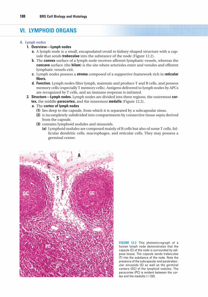
188 BRS Cell Biology and Histology
VI. LYMPHOID ORGANS
A. Lymph nodes1. Overview—Lymph nodes
a. A lymph node is a small, encapsulated ovoid to kidney-shaped structure with a cap-sule that sends trabeculae into the substance of the node (Figure 12.2).
b. The convex surface of a lymph node receives afferent lymphatic vessels, whereas theconcave surface (the hilum) is the site where arterioles enter and venules and efferentlymphatic vessels exit.
c. Lymph nodes possess a stroma composed of a supportive framework rich in reticularfibers.
d. Function. Lymph nodes filter lymph, maintain and produce T and B cells, and possessmemory cells (especially T memory cells). Antigens delivered to lymph nodes by APCsare recognized by T cells, and an immune response is initiated.
2. Structure—Lymph nodes. Lymph nodes are divided into three regions, the outermost cor-tex, the middle paracortex, and the innermost medulla (Figure 12.2).a. The cortex of lymph nodes
(1) lies deep to the capsule, from which it is separated by a subcapsular sinus.(2) is incompletely subdivided into compartments by connective tissue septa derived
from the capsule.(3) contains lymphoid nodules and sinusoids.
(a) Lymphoid nodules are composed mainly of B cells but also of some T cells, fol-licular dendritic cells, macrophages, and reticular cells. They may possess agerminal center.
A
C
T
S
GCGC
PCFIGURE 12.2 This photomicrograph of ahuman lymph node demonstrates that thecapsule (C) of the node is surrounded by adi-pose tissue. The capsule sends trabeculae(T) into the substance of the node. Note thepresence of the subcapsular and paratrabec-ular sinusoids (S) as well as the germinalcenters (GC) of the lymphoid nodules. Theparacortex (PC) is evident between the cor-tex and the medulla (�132).
LWBK615-c12[178-195].qxd 05/31/2010 10:46 AM Page 188 Aptara
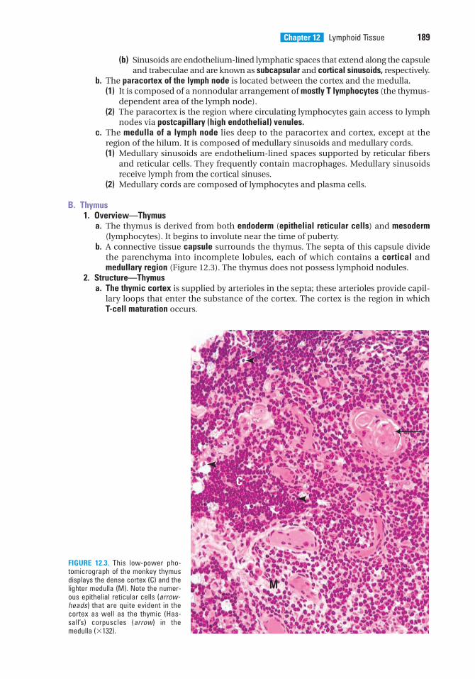
Chapter 12 Lymphoid Tissue 189
(b) Sinusoids are endothelium-lined lymphatic spaces that extend along the capsuleand trabeculae and are known as subcapsular and cortical sinusoids, respectively.
b. The paracortex of the lymph node is located between the cortex and the medulla.(1) It is composed of a nonnodular arrangement of mostly T lymphocytes (the thymus-
dependent area of the lymph node).(2) The paracortex is the region where circulating lymphocytes gain access to lymph
nodes via postcapillary (high endothelial) venules.c. The medulla of a lymph node lies deep to the paracortex and cortex, except at the
region of the hilum. It is composed of medullary sinusoids and medullary cords.(1) Medullary sinusoids are endothelium-lined spaces supported by reticular fibers
and reticular cells. They frequently contain macrophages. Medullary sinusoidsreceive lymph from the cortical sinuses.
(2) Medullary cords are composed of lymphocytes and plasma cells.
B. Thymus1. Overview—Thymus
a. The thymus is derived from both endoderm (epithelial reticular cells) and mesoderm(lymphocytes). It begins to involute near the time of puberty.
b. A connective tissue capsule surrounds the thymus. The septa of this capsule dividethe parenchyma into incomplete lobules, each of which contains a cortical andmedullary region (Figure 12.3). The thymus does not possess lymphoid nodules.
2. Structure—Thymusa. The thymic cortex is supplied by arterioles in the septa; these arterioles provide capil-
lary loops that enter the substance of the cortex. The cortex is the region in whichT-cell maturation occurs.
M
C
FIGURE 12.3. This low-power pho-tomicrograph of the monkey thymusdisplays the dense cortex (C) and thelighter medulla (M). Note the numer-ous epithelial reticular cells (arrow-heads) that are quite evident in thecortex as well as the thymic (Has-sall’s) corpuscles (arrow) in themedulla (�132).
LWBK615-c12[178-195].qxd 05/31/2010 10:47 AM Page 189 Aptara
190 BRS Cell Biology and Histology
(1) Epithelial reticular cells (Figure 12.3)(a) These cells originate from endoderm and form a meshwork with interstices in
which T cells are tightly packed.(b) They are pale cells (derived, during embryogenesis, from the third and per-
haps fourth pharyngeal pouches) and have a large ovoid lightly stainingnucleus that often displays a nucleolus.
(c) They possess long processes that surround the thymic cortex, isolating it fromboth the connective tissue septa and the medulla. These processes, which arefilled with bundles of tonofilaments, form desmosomal contacts with each other.
(d) They manufacture thymosin, serum thymic factor, and thymopoietin, hormonesthat function in the transformation of immature T lymphocytes into immuno-competent T cells.
(e) Epithelial reticular cells are of six types, each with different surface markersand presumably different functions.
(f) Some epithelial reticular cells present HLA I and HLA II molecules to thedeveloping T cells.
(2) Thymocytes(a) Thymocyte plasmalemma possesses Notch-1 receptors that permit these cells
to respond to cytokines released by epithelial reticular cells to become T cells.Once committed to the T-cell lineage, they are known as immature T lympho-cytes and are noted to be present within the thymic cortex in different stagesof differentiation.
(b) Thymocytes are surrounded by processes of epithelial reticular cells, whichhelp segregate thymocytes from antigens during their maturation.
(c) They migrate toward the medulla as they mature; however, T cells that can-not recognize the presented HLA I or HLA II molecules, or whose TCRs rec-ognize self proteins are forced into apoptosis and never reach the medulla.Most T cells die in the cortex, and the dead cells are phagocytosed bymacrophages.
(d) Surviving T cells are naïve. They leave the thymus and are distributed to sec-ondary lymphoid organs by the vascular system.
(3) Blood–thymus barrier(a) This barrier exists in the cortex only, making it an immunologically protected
region.(b) It ensures that antigens escaping from the bloodstream do not reach develop-
ing T cells in the thymic cortex.(c) It consists of the following layers: endothelium of the thymic capillaries and the
associated basal lamina, perivascular connective tissue and cells (e.g., peri-cytes and macrophages), and epithelial reticular cells and their basal laminae.
b. Thymic medulla(1) The thymic medulla is continuous between adjacent lobules and contains large
numbers of epithelial reticular cells and mature T cells, which are loosely packed,causing the medulla to stain lighter than the cortex (Figure 12.3).
(2) It also contains whorl-like accretions of epithelial reticular cells called Hassallcorpuscles (thymic corpuscles). These structures display various stages of kera-tinization and increase in number with age. Their function is unknown,although it has been shown that these epithelial reticular cells manufacturethymic stromal lymphopoietin (TSLP), a cytokine that facilitates dendritic cell mat-uration.
(3) Mature T cells exit the thymus via venules and efferent lymphatic vessels from thethymic medulla. The T cells then migrate to secondary lymphoid structures.
(4) Hormones acting on the thymus(a) Thymosin, thymopoietin, thymulin, somatotropin, and thymic humoral factor pro-
mote the formation of T cells.(b) Thyroxin encourages thymulin production by epithelial reticular cells.(c) Adrenocorticosteroids depress T-cell formation in the thymus.
LWBK615-c12[178-195].qxd 05/31/2010 10:47 AM Page 190 Aptara
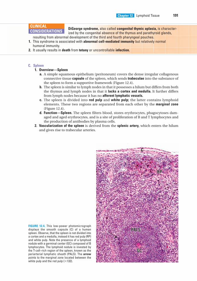
Chapter 12 Lymphoid Tissue 191
DiGeorge syndrome, also called congenital thymic aplasia, is character-ized by the congenital absence of the thymus and parathyroid glands,
resulting from abnormal development of the third and fourth pharyngeal pouches.1. This syndrome is associated with abnormal cell-mediated immunity but relatively normal
humoral immunity.2. It usually results in death from tetany or uncontrollable infection.
CLINICAL
CONSIDERATIONS
C. Spleen1. Overview—Spleen
a. A simple squamous epithelium (peritoneum) covers the dense irregular collagenousconnective tissue capsule of the spleen, which sends trabeculae into the substance ofthe spleen to form a supportive framework (Figure 12.4).
b. The spleen is similar to lymph nodes in that it possesses a hilum but differs from boththe thymus and lymph nodes in that it lacks a cortex and medulla. It further differsfrom lymph nodes because it has no afferent lymphatic vessels.
c. The spleen is divided into red pulp and white pulp; the latter contains lymphoidelements. These two regions are separated from each other by the marginal zone (Figure 12.4).
d. Function—Spleen. The spleen filters blood, stores erythrocytes, phagocytoses dam-aged and aged erythrocytes, and is a site of proliferation of B and T lymphocytes andthe production of antibodies by plasma cells.
2. Vascularization of the spleen is derived from the splenic artery, which enters the hilumand gives rise to trabecular arteries.
C
RPRP
GCGC
PALSPALS
RP
GC
PALSFIGURE 12.4. This low-power photomicrographdisplays the smooth capsule (C) of a humanspleen. Observe, that the spleen is not divided intoa cortex and a medulla, instead it has red pulp (RP)and white pulp. Note the presence of a lymphoidnodule with a germinal center (GC) composed of Blymphocytes. The lymphoid nodule is invested bythe T-cell–rich region of the spleen, known as theperiarterial lymphatic sheath (PALS). The arrowpoints to the marginal zone located between thewhite pulp and the red pulp (�132).
LWBK615-c12[178-195].qxd 05/31/2010 10:47 AM Page 191 Aptara
192 BRS Cell Biology and Histology
a. Trabecular arteries leave the trabeculae, become invested by a periarterial lymphaticsheath (PALS, described later), and are known as central arteries.
b. Central arteries branch but maintain their lymphatic sheath until they leave the whitepulp to form several straight penicillar arteries.
c. Penicillar arteries enter the red pulp. They have three regions: pulp arterioles,macrophage-sheathed arterioles, and terminal arterial capillaries. These last namedvessels either drain directly into the splenic sinusoids (closed circulation) or terminateas open-ended vessels within the splenic cords of the red pulp (open circulation).
d. Splenic sinusoids are drained by pulp veins, which are tributaries of the trabecularveins; these in turn drain into the splenic vein, which exits the spleen at the hilum.
3. Structure—Spleena. White pulp of the spleen includes all of the organ’s lymphoid tissue (diffuse and nodu-
lar), such as lymphoid nodules (mostly B cells) and PALS (mostly T cells) around thecentral arteries. It also contains macrophages and other APCs.
b. The marginal zone of the spleen(1) is a sinusoidal region between the red and white pulps at the periphery of the PALS
(Figure 12.4).(2) receives blood from capillary loops derived from the central artery and thus is the
first site where blood contacts the splenic parenchyma.(3) is richly supplied by avidly phagocytic macrophages and other APCs.(4) is the region where circulating T and B lymphocytes enter the spleen before
becoming segregated to their specific locations within the organ and where inter-digitating dendritic cells are able to display their MHC-epitope complex for recog-nition by T cells.
c. Red pulp of the spleen (Figure 12.4) is composed of an interconnected network of sinu-soids supported by a loose type of reticular tissue (splenic cords).(1) Sinusoids
(a) are lined by long fusiform endothelial cells separated by relatively large blood-containing intercellular spaces.
(b) have a discontinuous basal lamina underlying the endothelium and circum-ferentially arranged ribs of reticular fibrils.
(2) Splenic cords (cords of Billroth) contain plasma cells, stellate reticular cells, bloodcells, and macrophages enmeshed within the spaces of the reticular fiber network.Processes of the macrophages enter the lumina of the sinusoids through thespaces between the endothelial cells.
D. Tonsils are aggregates of lymphoid tissue, which sometimes lack a capsule. All tonsils are inthe upper section of the digestive tract, lying beneath but in contact with the epithelium.Tonsils assist in combating antigens entering via the nasal and oral epithelia.1. Palatine tonsils
a. possess crypts, deep invaginations of the stratified squamous epithelium covering ofthe tonsils, frequently containing debris.
b. possess primary and secondary (with germinal centers) lymphoid nodules.c. are separated from subjacent structures by a connective tissue capsule.
2. The pharyngeal tonsil is a single tonsil in the posterior wall of the nasopharynx.a. It is covered by a pseudostratified ciliated columnar epithelium.b. Instead of crypts, it has longitudinal pleats (infoldings).
3. Lingual tonsila. is on the dorsum of the posterior third of the tongue and is covered by a stratified
squamous nonkeratinized epithelium.b. possesses deep crypts, which frequently contain cellular debris. Ducts of the posterior
mucous glands of the tongue often open into the base of these crypts.
LWBK615-c12[178-195].qxd 05/31/2010 10:47 AM Page 192 Aptara
Review Test
193
1. Which of the following statementsconcerning T helper cells is true?
(A) They possess membrane-boundantibodies.
(B) They can recognize and interact withantigens in the blood.
(C) They produce numerous cytokines.(D) They function only in cell-mediated
immunity.(E) Their activation depends on interferon-�.
2. Which of the following statementsconcerning T cytotoxic (TC) cells is true?
(A) They assist macrophages in killingmicroorganisms.
(B) They possess antibodies on theirsurfaces.
(C) They possess CD8 surface markers.(D) They possess CD28 surface markers.(E) They secrete interferon-�.
3. Which of the following cell types isthought to function in preventing immuneresponses against self-antigens?
(A) T reg cells(B) B cells(C) T memory cells(D) TH cells(E) Mast cells
4. Which of the following statementsconcerning interferon-� is true?
(A) It is produced by T memory cells.(B) It is produced by T suppressor cells.(C) It activates macrophages.(D) It inhibits macrophages.(E) It induces viral proliferation.
5. A patient who was given penicillin had anadverse reaction to the antibiotic. Althoughthe reaction was due to the actions of mastcells, the response occurred because mastcells have IgE receptors in their cellmembranes. Which of the following cellsproduced the IgE decorating the plasmacell’s surface?
(A) T memory cells(B) B memory cells(C) T helper cells(D) Plasma cells(E) T cytotoxic cells
6. Which of the following statementsconcerning the thymus is true?
(A) Lymphoid nodules form much of thethymic cortex.
(B) Epithelial reticular cells form Hassallcorpuscles.
(C) T cells migrate into the medulla, wherethey become immunologicallycompetent.
(D) Most T cells that enter the thymus arekilled in the medulla.
(E) Macrophages are essential componentsof the blood–thymus barrier.
7. Which of the following statementsconcerning Hassall corpuscles is true?
(A) They are located in the thymic cortex ofyoung individuals.
(B) They are located in the thymic cortex ofold individuals.
(C) They are derived from mesoderm.(D) They are located in the thymic medulla.(E) They are derived from T memory cells.
Directions: Each of the numbered items or incomplete statements in this section is followed byanswers or completions of the statement. Select the ONE lettered answer that is BEST in eachcase.
LWBK615-c12[178-195].qxd 05/31/2010 10:47 AM Page 193 Aptara
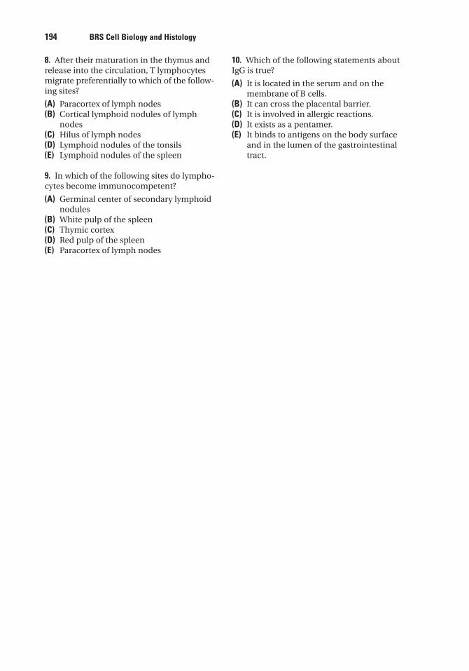
10. Which of the following statements aboutIgG is true?
(A) It is located in the serum and on themembrane of B cells.
(B) It can cross the placental barrier.(C) It is involved in allergic reactions.(D) It exists as a pentamer.(E) It binds to antigens on the body surface
and in the lumen of the gastrointestinaltract.
8. After their maturation in the thymus andrelease into the circulation, T lymphocytesmigrate preferentially to which of the follow-ing sites?
(A) Paracortex of lymph nodes(B) Cortical lymphoid nodules of lymph
nodes(C) Hilus of lymph nodes(D) Lymphoid nodules of the tonsils(E) Lymphoid nodules of the spleen
9. In which of the following sites do lympho-cytes become immunocompetent?
(A) Germinal center of secondary lymphoidnodules
(B) White pulp of the spleen(C) Thymic cortex(D) Red pulp of the spleen(E) Paracortex of lymph nodes
194 BRS Cell Biology and Histology
LWBK615-c12[178-195].qxd 05/31/2010 10:47 AM Page 194 Aptara
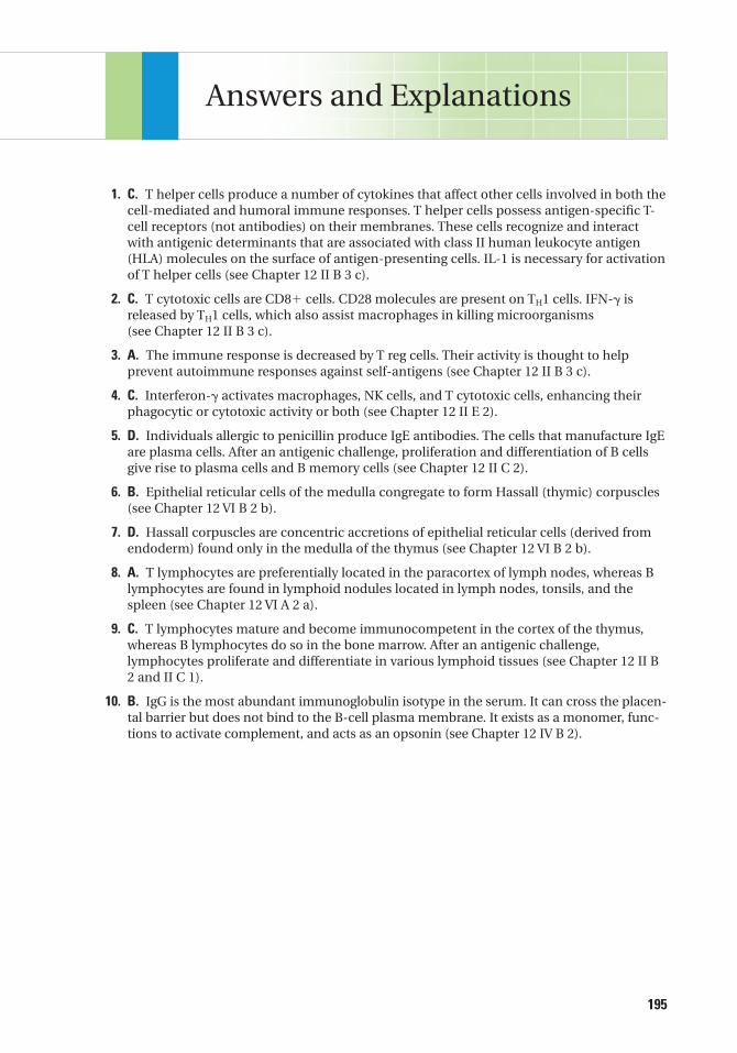
Answers and Explanations
1. C. T helper cells produce a number of cytokines that affect other cells involved in both thecell-mediated and humoral immune responses. T helper cells possess antigen-specific T-cell receptors (not antibodies) on their membranes. These cells recognize and interactwith antigenic determinants that are associated with class II human leukocyte antigen(HLA) molecules on the surface of antigen-presenting cells. IL-1 is necessary for activationof T helper cells (see Chapter 12 II B 3 c).
2. C. T cytotoxic cells are CD8� cells. CD28 molecules are present on TH1 cells. IFN-� isreleased by TH1 cells, which also assist macrophages in killing microorganisms (see Chapter 12 II B 3 c).
3. A. The immune response is decreased by T reg cells. Their activity is thought to helpprevent autoimmune responses against self-antigens (see Chapter 12 II B 3 c).
4. C. Interferon-� activates macrophages, NK cells, and T cytotoxic cells, enhancing theirphagocytic or cytotoxic activity or both (see Chapter 12 II E 2).
5. D. Individuals allergic to penicillin produce IgE antibodies. The cells that manufacture IgEare plasma cells. After an antigenic challenge, proliferation and differentiation of B cellsgive rise to plasma cells and B memory cells (see Chapter 12 II C 2).
6. B. Epithelial reticular cells of the medulla congregate to form Hassall (thymic) corpuscles(see Chapter 12 VI B 2 b).
7. D. Hassall corpuscles are concentric accretions of epithelial reticular cells (derived fromendoderm) found only in the medulla of the thymus (see Chapter 12 VI B 2 b).
8. A. T lymphocytes are preferentially located in the paracortex of lymph nodes, whereas Blymphocytes are found in lymphoid nodules located in lymph nodes, tonsils, and thespleen (see Chapter 12 VI A 2 a).
9. C. T lymphocytes mature and become immunocompetent in the cortex of the thymus,whereas B lymphocytes do so in the bone marrow. After an antigenic challenge,lymphocytes proliferate and differentiate in various lymphoid tissues (see Chapter 12 II B2 and II C 1).
10. B. IgG is the most abundant immunoglobulin isotype in the serum. It can cross the placen-tal barrier but does not bind to the B-cell plasma membrane. It exists as a monomer, func-tions to activate complement, and acts as an opsonin (see Chapter 12 IV B 2).
195
LWBK615-c12[178-195].qxd 05/31/2010 10:47 AM Page 195 Aptara
c h a p t e r 13 Endocrine System
196
I. OVERVIEW—THE ENDOCRINE SYSTEM
A. The endocrine system is composed of several ductless glands, clusters of cells within certainorgans, and isolated endocrine cells, so-called diffuse neuroendocrine system (DNES) cells,in the epithelial lining of the gastrointestinal and respiratory systems.
B. Glands of the endocrine system include the pituitary, thyroid, parathyroid, adrenal, and pinealglands.
C. Function. The endocrine system secretes hormones into nearby capillaries and interacts withthe nervous system to modulate and control the body’s metabolic activities.
II. HORMONES
Hormones are chemical messengers that are carried via the bloodstream to distant target cells.Hormones include low-molecular-weight water-soluble proteins and polypeptides (e.g., insulin,glucagon, follicle-stimulating hormone [FSH]) and lipid-soluble substances, principally thesteroid hormones (e.g., progesterone, estradiol, testosterone).
A. Water-soluble hormones interact with specific cell surface receptors on target cells, whichcommunicate a message that generates a biological response by the cell.1. G protein–linked receptors are used by some hormones (e.g., epinephrine, thyroid-
stimulating hormone [TSH], serotonin). Binding of the hormone to the G protein–linkedreceptor leads to production of a second messenger that evokes a target cell response.
2. Catalytic receptors are used by insulin and growth hormone. Binding of the hormone tothe catalytic receptor activates protein kinases that phosphorylate target proteins.
B. Lipid-soluble hormones diffuse across the plasma membrane of target cells and bind to spe-cific receptors in the cytosol or nucleus, forming hormone-receptor complexes that regulatetranscription of deoxyribonucleic acid (DNA).
III. OVERVIEW—PITUITARY GLAND (HYPOPHYSIS)
The pituitary gland lies below the hypothalamus, to which it is structurally and functionallyconnected. It is divided into two major subdivisions, the adenohypophysis and the neurohypoph-ysis (Figure 13.1). Each subdivision is derived from a distinct embryonic analog, which isreflected in its unique cellular constituents and functions.
LWBK615-c13[196-214].qxd 05/31/2010 10:48 AM Page 196 Aptara
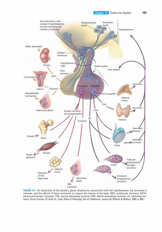
Chapter 13 Endocrine System 197
⎠
⎬
⎞
⎠
⎬
⎞⎠
⎬
⎞
Neurosecretory cellslocated in hypothalamussecrete releasing andinhibitory hormones
Paraventricularnuclei
Supraopticnuclei
Hypothalamus
Portal system
Pars distalis
Acidophil Basophil
ACTH
Adrenalcortex
Secretion
TSH
Thyroid
SecretionFSH
Testis
Spermato-genesis
Androgensecretion
LH
Ovary
Folliculardevelopment:estrogensecretion
Ovulation:progesteronesecretion
Prolactin
Mammary gland
Milksecretion
Growth hormonevia somatomedins
AdiposetissueElevation
of freefatty acids
MuscleHyper-glycemia
BoneGrowth
Mammarygland
Myoepithelialcontraction
OxytocinUterus
Contraction ADH
Kidney
Water absorption
Parsnervosa
Hypophysealstalk
Medianeminence
FIGURE 13.1. An illustration of the pituitary gland, showing its connections with the hypothalamus, the hormones itreleases, and the effects of these hormones on organs and tissues of the body. ADH, antidiuretic hormone; ACTH,adrenocorticotropic hormone; TSH, thyroid-stimulating hormone; FSH, follicle-stimulating hormone; LH, luteinizing hor-mone. (From Gartner LP, Hiatt JL: Color Atlas of Histology, 5th ed. Baltimore, Lippincott William & Wilkins, 2009, p 206.)
LWBK615-c13[196-214].qxd 05/31/2010 10:48 AM Page 197 Aptara
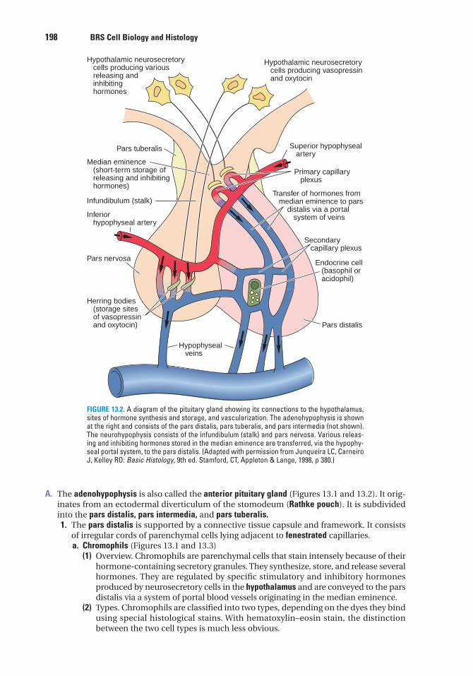
198 BRS Cell Biology and Histology
A. The adenohypophysis is also called the anterior pituitary gland (Figures 13.1 and 13.2). It orig-inates from an ectodermal diverticulum of the stomodeum (Rathke pouch). It is subdividedinto the pars distalis, pars intermedia, and pars tuberalis.1. The pars distalis is supported by a connective tissue capsule and framework. It consists
of irregular cords of parenchymal cells lying adjacent to fenestrated capillaries.a. Chromophils (Figures 13.1 and 13.3)
(1) Overview. Chromophils are parenchymal cells that stain intensely because of theirhormone-containing secretory granules. They synthesize, store, and release severalhormones. They are regulated by specific stimulatory and inhibitory hormonesproduced by neurosecretory cells in the hypothalamus and are conveyed to the parsdistalis via a system of portal blood vessels originating in the median eminence.
(2) Types. Chromophils are classified into two types, depending on the dyes they bindusing special histological stains. With hematoxylin–eosin stain, the distinctionbetween the two cell types is much less obvious.
Hypothalamic neurosecretory cells producing various releasing and inhibiting hormones
Hypothalamic neurosecretory cells producing vasopressin and oxytocin
Pars tuberalis
Median eminence (short-term storage of releasing and inhibiting hormones)
Infundibulum (stalk)
Inferior hypophyseal artery
Pars nervosa
Hypophyseal veins
Pars distalis
Endocrine cell (basophil or acidophil)
Secondary capillary plexus
Transfer of hormones from median eminence to pars distalis via a portal system of veins
Primary capillary plexus
Superior hypophyseal artery
Herring bodies (storage sites of vasopressin and oxytocin)
FIGURE 13.2. A diagram of the pituitary gland showing its connections to the hypothalamus,sites of hormone synthesis and storage, and vascularization. The adenohypophysis is shownat the right and consists of the pars distalis, pars tuberalis, and pars intermedia (not shown).The neurohypophysis consists of the infundibulum (stalk) and pars nervosa. Various releas-ing and inhibiting hormones stored in the median eminence are transferred, via the hypophy-seal portal system, to the pars distalis. (Adapted with permission from Junqueira LC, CarneiroJ, Kelley RO: Basic Histology, 9th ed. Stamford, CT, Appleton & Lange, 1998, p 380.)
LWBK615-c13[196-214].qxd 05/31/2010 10:48 AM Page 198 Aptara
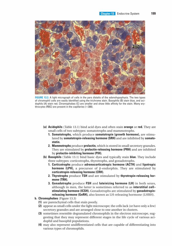
Chapter 13 Endocrine System 199
(a) Acidophils (Table 13.1) bind acid dyes and often stain orange or red. They aresmall cells of two subtypes: somatotrophs and mammotrophs.1. Somatotrophs, which produce somatotropin (growth hormone), are stimu-
lated by somatotropin-releasing hormone (SRH) and are inhibited by somato-statin.
2. Mammotrophs produce prolactin, which is stored in small secretory granules.They are stimulated by prolactin-releasing hormone (PRH) and are inhibitedby prolactin-inhibiting hormone (PIH).
(b) Basophils (Table 13.1) bind basic dyes and typically stain blue. They includethree subtypes: corticotrophs, thyrotrophs, and gonadotrophs.1. Corticotrophs produce adrenocorticotropic hormone (ACTH) and lipotropic
hormone (LPH), a precursor of �-endorphin. They are stimulated bycorticotropin-releasing hormone (CRH).
2. Thyrotrophs produce TSH and are stimulated by thyrotropin-releasing hor-mone (TRH).
3. Gonadotrophs produce FSH and luteinizing hormone (LH) in both sexes,although in men, the latter is sometimes referred to as interstitial cell–stimulating hormone (ICSH). Gonadotrophs are stimulated by gonadotropin-releasing hormone (GnRH), also known as LH-releasing hormone (LHRH).
b. Chromophobes (Figure 13.3)(1) are parenchymal cells that stain poorly.(2) appear as small cells under the light microscope; the cells lack (or have only a few)
secretory granules and are arranged close to one another in clusters.(3) sometimes resemble degranulated chromophils in the electron microscope, sug-
gesting that they may represent different stages in the life cycle of various aci-dophil and basophil populations.
(4) may also represent undifferentiated cells that are capable of differentiating intovarious types of chromophils.
B BRBC
C
A
FIGURE 13.3. A light micrograph of cells in the pars distalis of the adenohypophysis. The two typesof chromophil cells are easily identified using the trichrome stain. Basophils (B) stain blue, and aci-dophils (A) stain red. Chromophobes (C) are smaller and show little affinity for the stain. Many ery-throcytes (RBC) are present in the capillaries (�300).
LWBK615-c13[196-214].qxd 05/31/2010 10:49 AM Page 199 Aptara
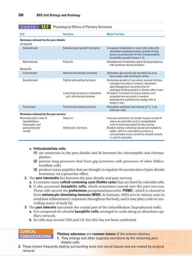
200 BRS Cell Biology and Histology
t a b l e 13.1 Physiological Effects of Pituitary Hormones
Cell Hormone Major Function
Hormones released by the pars distalis
Acidophils
Somatotroph Somatotropin (growth hormone) Increases metabolism in most cells; indirectly stimulates epiphyseal plate, growth of long bones via production of liver somatomedins (insulinlike growth factors I, II)
Mammotroph Prolactin Development of mammary gland during pregnancy,milk synthesis during lactation
Basophils
Corticotroph Adrenocorticotropic hormone Stimulates glucocorticoid secretion by zona fasciculata cells of adrenal cortex
Gonadotroph Follicle-stimulating hormone Stimulates growth of secondary ovarian follicles, estrogen secretion in women; stimulates spermatogenesis via production of androgen-binding protein in Sertoli cells in men
Luteinizing hormone or interstitial Ovulation, formation of corpus luteum, and cell–stimulating hormone progesterone secretion in women;
testosterone synthesis by Leydig cells of testis in men
Thyrotroph Thyroid-stimulating hormone Stimulates synthesis and release of T3, T4 by follicular cells
Hormones released by the pars nervosa
Neurosecretory cells of Oxytocin Induces contraction of smooth muscle in wall of hypothalamus uterus at parturition and in myoepithelial (supraoptic and cells of mammary gland during nursingparaventricular Antidiuretic hormone Renders kidney collecting tubules permeable to nuclei) water, which is resorbed to produce a
concentrated urine; constricts smooth muscle in wall of arterioles
c. Folliculostellate cells(1) are numerous in the pars distalis and lie between the chromophils and chromo-
phobes.(2) possess long processes that form gap junctions with processes of other follicu-
lostellate cells.(3) produce many peptides that are thought to regulate the production of pars distalis
hormones via a paracrine effect.2. The pars intermedia lies between the pars distalis and pars nervosa.
a. It contains many colloid-containing cysts (Rathke cysts) that are lined by cuboidal cells.b. It also possesses basophilic cells, which sometimes extend into the pars nervosa.
These cells secrete the prohormone proopiomelanocortin (POMC), which is cleaved toform melanocyte-stimulating hormone (MSH). In humans, MSH acts in various ways tomodulate inflammatory responses throughout the body, and it may play a role in con-trolling stores of body fat.
3. The pars tuberalis surrounds the cranial part of the infundibulum (hypophyseal stalk).a. It is composed of cuboidal basophilic cells, arranged in cords along an abundant cap-
illary network.b. Its cells may secrete FSH and LH, but this has not been confirmed.
Pituitary adenomas are common tumors of the anterior pituitary.1. They enlarge and often suppress secretions by the remaining pars
distalis cells.2. These tumors frequently destroy surrounding bone and neural tissues and are treated by surgical
removal.
CLINICAL
CONSIDERATIONS
LWBK615-c13[196-214].qxd 05/31/2010 10:49 AM Page 200 Aptara
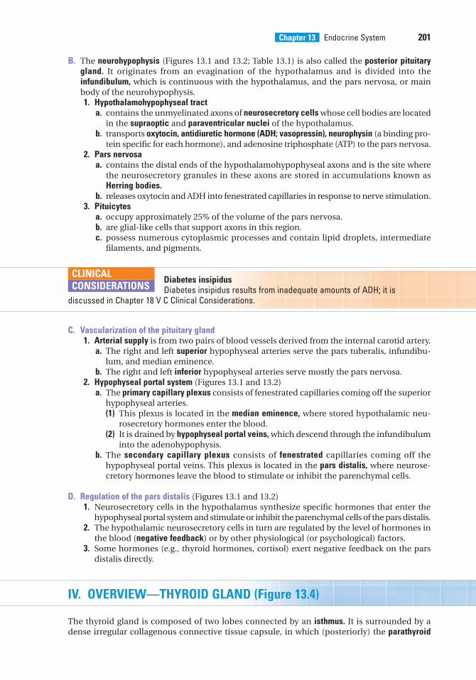
Chapter 13 Endocrine System 201
B. The neurohypophysis (Figures 13.1 and 13.2; Table 13.1) is also called the posterior pituitarygland. It originates from an evagination of the hypothalamus and is divided into theinfundibulum, which is continuous with the hypothalamus, and the pars nervosa, or mainbody of the neurohypophysis.1. Hypothalamohypophyseal tract
a. contains the unmyelinated axons of neurosecretory cells whose cell bodies are locatedin the supraoptic and paraventricular nuclei of the hypothalamus.
b. transports oxytocin, antidiuretic hormone (ADH; vasopressin), neurophysin (a binding pro-tein specific for each hormone), and adenosine triphosphate (ATP) to the pars nervosa.
2. Pars nervosaa. contains the distal ends of the hypothalamohypophyseal axons and is the site where
the neurosecretory granules in these axons are stored in accumulations known asHerring bodies.
b. releases oxytocin and ADH into fenestrated capillaries in response to nerve stimulation.3. Pituicytes
a. occupy approximately 25% of the volume of the pars nervosa.b. are glial-like cells that support axons in this region.c. possess numerous cytoplasmic processes and contain lipid droplets, intermediate
filaments, and pigments.
Diabetes insipidusDiabetes insipidus results from inadequate amounts of ADH; it is
discussed in Chapter 18 V C Clinical Considerations.
CLINICAL
CONSIDERATIONS
C. Vascularization of the pituitary gland1. Arterial supply is from two pairs of blood vessels derived from the internal carotid artery.
a. The right and left superior hypophyseal arteries serve the pars tuberalis, infundibu-lum, and median eminence.
b. The right and left inferior hypophyseal arteries serve mostly the pars nervosa.2. Hypophyseal portal system (Figures 13.1 and 13.2)
a. The primary capillary plexus consists of fenestrated capillaries coming off the superiorhypophyseal arteries.(1) This plexus is located in the median eminence, where stored hypothalamic neu-
rosecretory hormones enter the blood.(2) It is drained by hypophyseal portal veins, which descend through the infundibulum
into the adenohypophysis.b. The secondary capillary plexus consists of fenestrated capillaries coming off the
hypophyseal portal veins. This plexus is located in the pars distalis, where neurose-cretory hormones leave the blood to stimulate or inhibit the parenchymal cells.
D. Regulation of the pars distalis (Figures 13.1 and 13.2)1. Neurosecretory cells in the hypothalamus synthesize specific hormones that enter the
hypophyseal portal system and stimulate or inhibit the parenchymal cells of the pars distalis.2. The hypothalamic neurosecretory cells in turn are regulated by the level of hormones in
the blood (negative feedback) or by other physiological (or psychological) factors.3. Some hormones (e.g., thyroid hormones, cortisol) exert negative feedback on the pars
distalis directly.
IV. OVERVIEW—THYROID GLAND (Figure 13.4)
The thyroid gland is composed of two lobes connected by an isthmus. It is surrounded by adense irregular collagenous connective tissue capsule, in which (posteriorly) the parathyroid
LWBK615-c13[196-214].qxd 05/31/2010 10:49 AM Page 201 Aptara
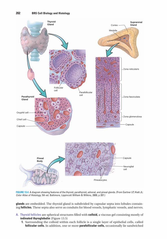
ThyroidGland
Follicularcell
Parafollicularcell
ParathyroidGland
Oxyphil cell
Chief cell
Capsule
SuprarenalGlandCortex
Medulla
Zona reticularis
Zona fasciculata
Zona glomerulosa
Capsule
PinealBody
Capsule
Neuroglialcell
Pinealocytes
FIGURE 13.4. A diagram showing features of the thyroid, parathyroid, adrenal, and pineal glands. (From Gartner LP, Hiatt JL:Color Atlas of Histology, 5th ed. Baltimore, Lippincott William & Wilkins, 2009, p 207.)
glands are embedded. The thyroid gland is subdivided by capsular septa into lobules contain-ing follicles. These septa also serve as conduits for blood vessels, lymphatic vessels, and nerves.
A. Thyroid follicles are spherical structures filled with colloid, a viscous gel consisting mostly ofiodinated thyroglobulin (Figure 13.5)1. Surrounding the colloid within each follicle is a single layer of epithelial cells, called
follicular cells. In addition, one or more parafollicular cells, occasionally lie sandwiched
202 BRS Cell Biology and Histology
LWBK615-c13[196-214].qxd 05/31/2010 10:49 AM Page 202 Aptara
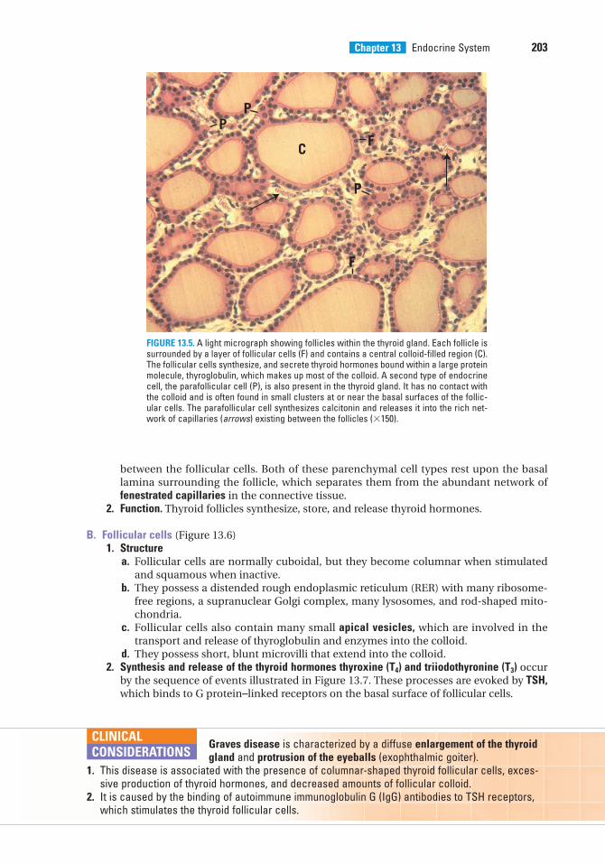
Chapter 13 Endocrine System 203
between the follicular cells. Both of these parenchymal cell types rest upon the basallamina surrounding the follicle, which separates them from the abundant network offenestrated capillaries in the connective tissue.
2. Function. Thyroid follicles synthesize, store, and release thyroid hormones.
B. Follicular cells (Figure 13.6)1. Structure
a. Follicular cells are normally cuboidal, but they become columnar when stimulatedand squamous when inactive.
b. They possess a distended rough endoplasmic reticulum (RER) with many ribosome-free regions, a supranuclear Golgi complex, many lysosomes, and rod-shaped mito-chondria.
c. Follicular cells also contain many small apical vesicles, which are involved in thetransport and release of thyroglobulin and enzymes into the colloid.
d. They possess short, blunt microvilli that extend into the colloid.2. Synthesis and release of the thyroid hormones thyroxine (T4) and triiodothyronine (T3) occur
by the sequence of events illustrated in Figure 13.7. These processes are evoked by TSH,which binds to G protein–linked receptors on the basal surface of follicular cells.
PP
C F
P
F
FIGURE 13.5. A light micrograph showing follicles within the thyroid gland. Each follicle issurrounded by a layer of follicular cells (F) and contains a central colloid-filled region (C).The follicular cells synthesize, and secrete thyroid hormones bound within a large proteinmolecule, thyroglobulin, which makes up most of the colloid. A second type of endocrinecell, the parafollicular cell (P), is also present in the thyroid gland. It has no contact withthe colloid and is often found in small clusters at or near the basal surfaces of the follic-ular cells. The parafollicular cell synthesizes calcitonin and releases it into the rich net-work of capillaries (arrows) existing between the follicles (�150).
Graves disease is characterized by a diffuse enlargement of the thyroidgland and protrusion of the eyeballs (exophthalmic goiter).
1. This disease is associated with the presence of columnar-shaped thyroid follicular cells, exces-sive production of thyroid hormones, and decreased amounts of follicular colloid.
2. It is caused by the binding of autoimmune immunoglobulin G (IgG) antibodies to TSH receptors,which stimulates the thyroid follicular cells.
CLINICAL
CONSIDERATIONS
LWBK615-c13[196-214].qxd 05/31/2010 10:49 AM Page 203 Aptara
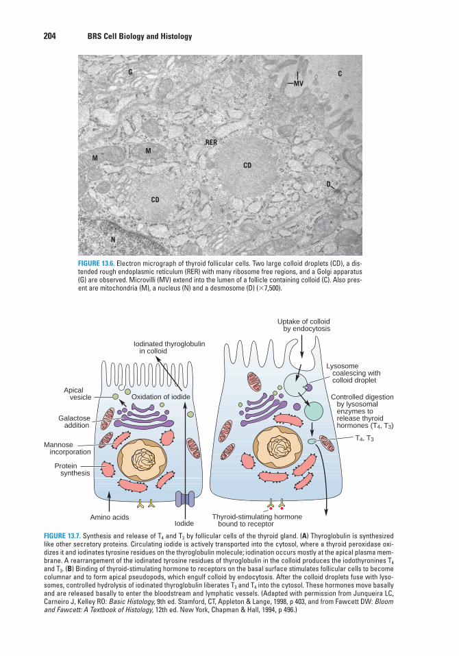
204 BRS Cell Biology and Histology
G
MM
CD
CD
RER
D
CMV
N
FIGURE 13.6. Electron micrograph of thyroid follicular cells. Two large colloid droplets (CD), a dis-tended rough endoplasmic reticulum (RER) with many ribosome free regions, and a Golgi apparatus(G) are observed. Microvilli (MV) extend into the lumen of a follicle containing colloid (C). Also pres-ent are mitochondria (M), a nucleus (N) and a desmosome (D) (�7,500).
Iodinated thyroglobulin in colloid
Uptake of colloid by endocytosis
Controlled digestion by lysosomal enzymes to release thyroid hormones (T4, T3)
Oxidation of iodideApical vesicle
Galactose addition
Mannose incorporation
Protein synthesis
Amino acidsIodide
Thyroid-stimulating hormone bound to receptor
T4, T3
Lysosome coalescing with colloid droplet
FIGURE 13.7. Synthesis and release of T4 and T3 by follicular cells of the thyroid gland. (A) Thyroglobulin is synthesizedlike other secretory proteins. Circulating iodide is actively transported into the cytosol, where a thyroid peroxidase oxi-dizes it and iodinates tyrosine residues on the thyroglobulin molecule; iodination occurs mostly at the apical plasma mem-brane. A rearrangement of the iodinated tyrosine residues of thyroglobulin in the colloid produces the iodothyronines T4
and T3. (B) Binding of thyroid-stimulating hormone to receptors on the basal surface stimulates follicular cells to becomecolumnar and to form apical pseudopods, which engulf colloid by endocytosis. After the colloid droplets fuse with lyso-somes, controlled hydrolysis of iodinated thyroglobulin liberates T3 and T4 into the cytosol. These hormones move basallyand are released basally to enter the bloodstream and lymphatic vessels. (Adapted with permission from Junqueira LC,Carneiro J, Kelley RO: Basic Histology, 9th ed. Stamford, CT, Appleton & Lange, 1998, p 403, and from Fawcett DW: Bloomand Fawcett: A Textbook of Histology, 12th ed. New York, Chapman & Hall, 1994, p 496.)
LWBK615-c13[196-214].qxd 05/31/2010 10:49 AM Page 204 Aptara
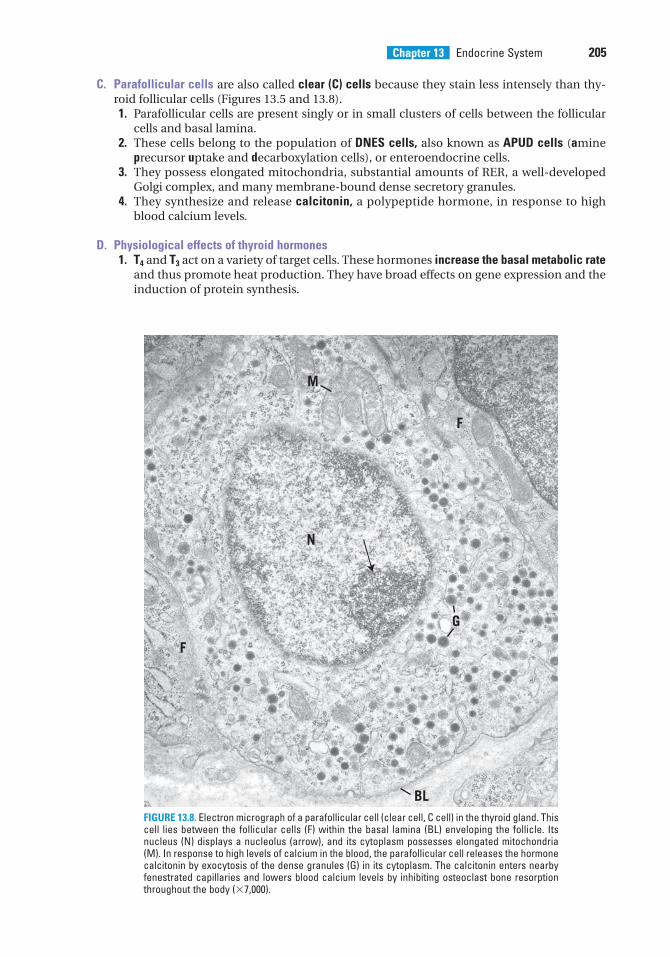
Chapter 13 Endocrine System 205
C. Parafollicular cells are also called clear (C) cells because they stain less intensely than thy-roid follicular cells (Figures 13.5 and 13.8).1. Parafollicular cells are present singly or in small clusters of cells between the follicular
cells and basal lamina.2. These cells belong to the population of DNES cells, also known as APUD cells (amine
precursor uptake and decarboxylation cells), or enteroendocrine cells.3. They possess elongated mitochondria, substantial amounts of RER, a well-developed
Golgi complex, and many membrane-bound dense secretory granules.4. They synthesize and release calcitonin, a polypeptide hormone, in response to high
blood calcium levels.
D. Physiological effects of thyroid hormones1. T4 and T3 act on a variety of target cells. These hormones increase the basal metabolic rate
and thus promote heat production. They have broad effects on gene expression and theinduction of protein synthesis.
M
F
N
F
G
BLFIGURE 13.8. Electron micrograph of a parafollicular cell (clear cell, C cell) in the thyroid gland. Thiscell lies between the follicular cells (F) within the basal lamina (BL) enveloping the follicle. Itsnucleus (N) displays a nucleolus (arrow), and its cytoplasm possesses elongated mitochondria(M). In response to high levels of calcium in the blood, the parafollicular cell releases the hormonecalcitonin by exocytosis of the dense granules (G) in its cytoplasm. The calcitonin enters nearbyfenestrated capillaries and lowers blood calcium levels by inhibiting osteoclast bone resorptionthroughout the body (�7,000).
LWBK615-c13[196-214].qxd 05/31/2010 10:49 AM Page 205 Aptara
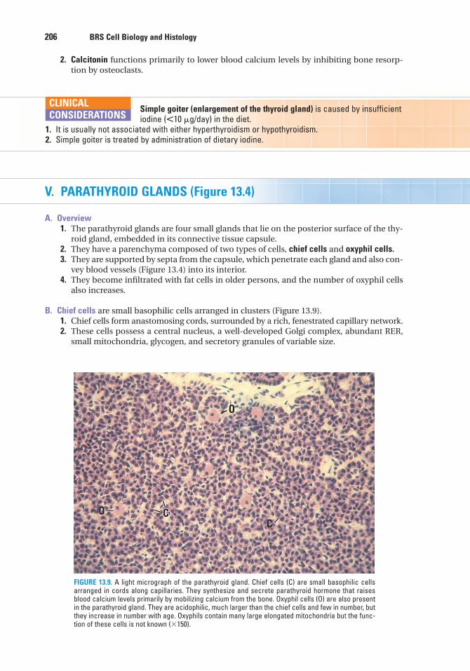
2. Calcitonin functions primarily to lower blood calcium levels by inhibiting bone resorp-tion by osteoclasts.
206 BRS Cell Biology and Histology
Simple goiter (enlargement of the thyroid gland) is caused by insufficientiodine (�10 �g/day) in the diet.
1. It is usually not associated with either hyperthyroidism or hypothyroidism.2. Simple goiter is treated by administration of dietary iodine.
CLINICAL
CONSIDERATIONS
O
CCOC
COC
FIGURE 13.9. A light micrograph of the parathyroid gland. Chief cells (C) are small basophilic cellsarranged in cords along capillaries. They synthesize and secrete parathyroid hormone that raisesblood calcium levels primarily by mobilizing calcium from the bone. Oxyphil cells (O) are also presentin the parathyroid gland. They are acidophilic, much larger than the chief cells and few in number, butthey increase in number with age. Oxyphils contain many large elongated mitochondria but the func-tion of these cells is not known (�150).
V. PARATHYROID GLANDS (Figure 13.4)
A. Overview1. The parathyroid glands are four small glands that lie on the posterior surface of the thy-
roid gland, embedded in its connective tissue capsule.2. They have a parenchyma composed of two types of cells, chief cells and oxyphil cells.3. They are supported by septa from the capsule, which penetrate each gland and also con-
vey blood vessels (Figure 13.4) into its interior.4. They become infiltrated with fat cells in older persons, and the number of oxyphil cells
also increases.
B. Chief cells are small basophilic cells arranged in clusters (Figure 13.9).1. Chief cells form anastomosing cords, surrounded by a rich, fenestrated capillary network.2. These cells possess a central nucleus, a well-developed Golgi complex, abundant RER,
small mitochondria, glycogen, and secretory granules of variable size.
LWBK615-c13[196-214].qxd 05/31/2010 10:49 AM Page 206 Aptara
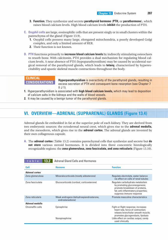
Chapter 13 Endocrine System 207
3. Function. They synthesize and secrete parathyroid hormone (PTH, or parathormone), whichraises blood calcium levels. High blood calcium levels inhibit the production of PTH.
C. Oxyphil cells are large, eosinophilic cells that are present singly or in small clusters within theparenchyma of the gland (Figure 13.9).1. Oxyphil cells possess many large, elongated mitochondria, a poorly developed Golgi
complex, and only a limited amount of RER.2. Their function is not known.
D. PTH functions primarily to increase blood calcium levels by indirectly stimulating osteoclaststo resorb bone. With calcitonin, PTH provides a dual mechanism for regulating blood cal-cium levels. A near absence of PTH (hypoparathyroidism) may be caused by accidental sur-gical removal of the parathyroid glands, which leads to tetany, characterized by hyperex-citability and spastic skeletal muscle contractions throughout the body.
Hyperparathyroidism is overactivity of the parathyroid glands, resulting inexcess secretion of PTH and consequent bone resorption (see Chapter 7II J 1).
1. Hyperparathyroidism is associated with high blood calcium levels, which may lead to depositionof calcium salts in the kidneys and the walls of blood vessels.
2. It may be caused by a benign tumor of the parathyroid glands.
CLINICAL
CONSIDERATIONS
VI. OVERVIEW—ADRENAL (SUPRARENAL) GLANDS (Figure 13.4)
Adrenal glands lie embedded in fat at the superior pole of each kidney. They are derived fromtwo embryonic sources: the ectodermal neural crest, which gives rise to the adrenal medulla,and the mesoderm, which gives rise to the adrenal cortex. The adrenal glands are invested bytheir own collagenous capsule.
A. The adrenal cortex (Table 13.2) contains parenchymal cells that synthesize and secrete but donot store various steroid hormones. It is divided into three concentric histologicallyrecognizable regions: the zona glomerulosa, zona fasciculata, and zona reticularis (Figure 13.10).
t a b l e 13.2 Adrenal Gland Cells and Hormones
Cell Hormone Function
Adrenal cortex
Zona glomerulosa Mineralocorticoids (mostly aldosterone) Regulate electrolyte, water balance via effect on cells of renal tubules
Zona fasciculata Glucocorticoids (cortisol, corticosterone) Regulate carbohydrate metabolism by promoting gluconeogenesis; promote breakdown of proteins, fat; anti-inflammatory properties; suppress immune response
Zona reticularis Weak androgens (dehydroepiandrosterone, Promote masculine characteristicsandrostenedione)
Adrenal medulla
Chromaffin cells Epinephrine Fight-or-flight response; increases heart rate, force of contraction; relaxes bronchiolar smooth muscle; promotes glycogenolysis, lipolysis
Norepinephrine Little effect on cardiac output, rarely used clinically
LWBK615-c13[196-214].qxd 05/31/2010 10:49 AM Page 207 Aptara
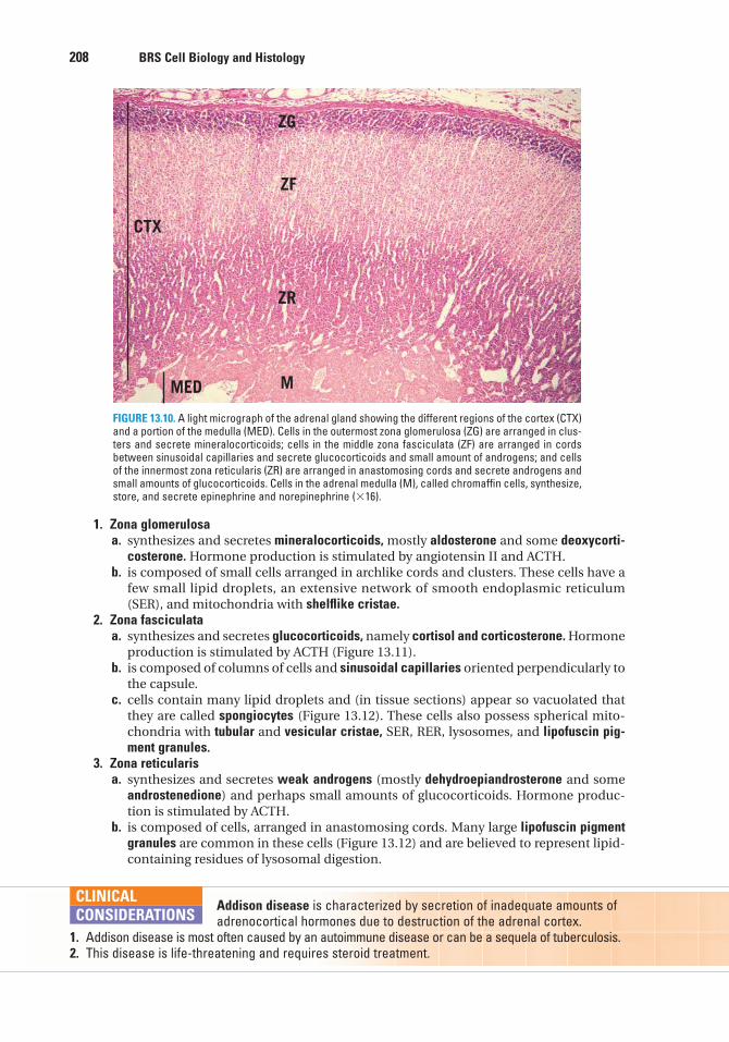
1. Zona glomerulosaa. synthesizes and secretes mineralocorticoids, mostly aldosterone and some deoxycorti-
costerone. Hormone production is stimulated by angiotensin II and ACTH.b. is composed of small cells arranged in archlike cords and clusters. These cells have a
few small lipid droplets, an extensive network of smooth endoplasmic reticulum(SER), and mitochondria with shelflike cristae.
2. Zona fasciculataa. synthesizes and secretes glucocorticoids, namely cortisol and corticosterone. Hormone
production is stimulated by ACTH (Figure 13.11).b. is composed of columns of cells and sinusoidal capillaries oriented perpendicularly to
the capsule.c. cells contain many lipid droplets and (in tissue sections) appear so vacuolated that
they are called spongiocytes (Figure 13.12). These cells also possess spherical mito-chondria with tubular and vesicular cristae, SER, RER, lysosomes, and lipofuscin pig-ment granules.
3. Zona reticularisa. synthesizes and secretes weak androgens (mostly dehydroepiandrosterone and some
androstenedione) and perhaps small amounts of glucocorticoids. Hormone produc-tion is stimulated by ACTH.
b. is composed of cells, arranged in anastomosing cords. Many large lipofuscin pigmentgranules are common in these cells (Figure 13.12) and are believed to represent lipid-containing residues of lysosomal digestion.
208 BRS Cell Biology and Histology
CTX
ZG
ZF
ZR
MMED
FIGURE 13.10. A light micrograph of the adrenal gland showing the different regions of the cortex (CTX)and a portion of the medulla (MED). Cells in the outermost zona glomerulosa (ZG) are arranged in clus-ters and secrete mineralocorticoids; cells in the middle zona fasciculata (ZF) are arranged in cordsbetween sinusoidal capillaries and secrete glucocorticoids and small amount of androgens; and cellsof the innermost zona reticularis (ZR) are arranged in anastomosing cords and secrete androgens andsmall amounts of glucocorticoids. Cells in the adrenal medulla (M), called chromaffin cells, synthesize,store, and secrete epinephrine and norepinephrine (�16).
Addison disease is characterized by secretion of inadequate amounts ofadrenocortical hormones due to destruction of the adrenal cortex.
1. Addison disease is most often caused by an autoimmune disease or can be a sequela of tuberculosis.2. This disease is life-threatening and requires steroid treatment.
CLINICAL
CONSIDERATIONS
LWBK615-c13[196-214].qxd 05/31/2010 10:49 AM Page 208 Aptara
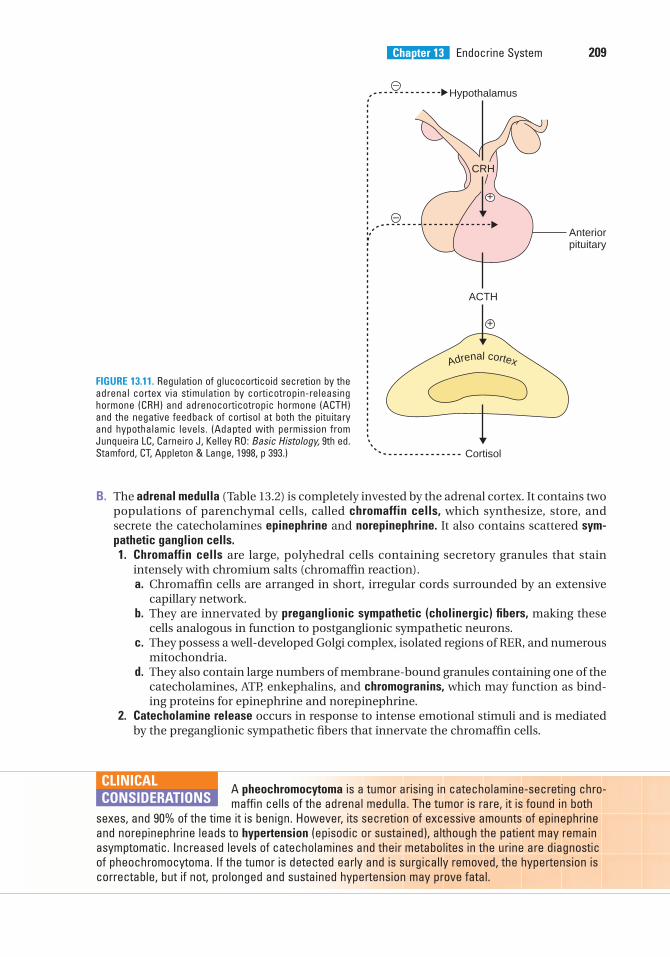
Chapter 13 Endocrine System 209
B. The adrenal medulla (Table 13.2) is completely invested by the adrenal cortex. It contains twopopulations of parenchymal cells, called chromaffin cells, which synthesize, store, andsecrete the catecholamines epinephrine and norepinephrine. It also contains scattered sym-pathetic ganglion cells.1. Chromaffin cells are large, polyhedral cells containing secretory granules that stain
intensely with chromium salts (chromaffin reaction).a. Chromaffin cells are arranged in short, irregular cords surrounded by an extensive
capillary network.b. They are innervated by preganglionic sympathetic (cholinergic) fibers, making these
cells analogous in function to postganglionic sympathetic neurons.c. They possess a well-developed Golgi complex, isolated regions of RER, and numerous
mitochondria.d. They also contain large numbers of membrane-bound granules containing one of the
catecholamines, ATP, enkephalins, and chromogranins, which may function as bind-ing proteins for epinephrine and norepinephrine.
2. Catecholamine release occurs in response to intense emotional stimuli and is mediatedby the preganglionic sympathetic fibers that innervate the chromaffin cells.
Anteriorpituitary
ACTH
Cortisol
Hypothalamus
+
+
–
–
Adrenal cortex
CRH
FIGURE 13.11. Regulation of glucocorticoid secretion by theadrenal cortex via stimulation by corticotropin-releasinghormone (CRH) and adrenocorticotropic hormone (ACTH)and the negative feedback of cortisol at both the pituitaryand hypothalamic levels. (Adapted with permission fromJunqueira LC, Carneiro J, Kelley RO: Basic Histology, 9th ed.Stamford, CT, Appleton & Lange, 1998, p 393.)
A pheochromocytoma is a tumor arising in catecholamine-secreting chro-maffin cells of the adrenal medulla. The tumor is rare, it is found in both
sexes, and 90% of the time it is benign. However, its secretion of excessive amounts of epinephrineand norepinephrine leads to hypertension (episodic or sustained), although the patient may remainasymptomatic. Increased levels of catecholamines and their metabolites in the urine are diagnosticof pheochromocytoma. If the tumor is detected early and is surgically removed, the hypertension iscorrectable, but if not, prolonged and sustained hypertension may prove fatal.
CLINICAL
CONSIDERATIONS
LWBK615-c13[196-214].qxd 05/31/2010 10:49 AM Page 209 Aptara
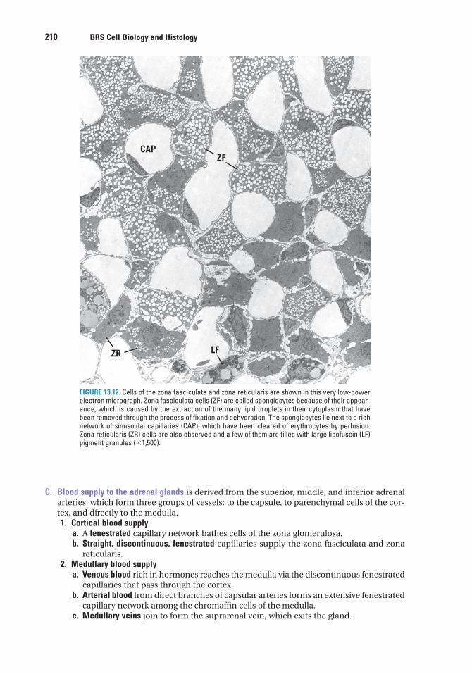
210 BRS Cell Biology and Histology
CAPZF
LFZR
FIGURE 13.12. Cells of the zona fasciculata and zona reticularis are shown in this very low-powerelectron micrograph. Zona fasciculata cells (ZF) are called spongiocytes because of their appear-ance, which is caused by the extraction of the many lipid droplets in their cytoplasm that havebeen removed through the process of fixation and dehydration. The spongiocytes lie next to a richnetwork of sinusoidal capillaries (CAP), which have been cleared of erythrocytes by perfusion.Zona reticularis (ZR) cells are also observed and a few of them are filled with large lipofuscin (LF)pigment granules (�1,500).
C. Blood supply to the adrenal glands is derived from the superior, middle, and inferior adrenalarteries, which form three groups of vessels: to the capsule, to parenchymal cells of the cor-tex, and directly to the medulla.1. Cortical blood supply
a. A fenestrated capillary network bathes cells of the zona glomerulosa.b. Straight, discontinuous, fenestrated capillaries supply the zona fasciculata and zona
reticularis.2. Medullary blood supply
a. Venous blood rich in hormones reaches the medulla via the discontinuous fenestratedcapillaries that pass through the cortex.
b. Arterial blood from direct branches of capsular arteries forms an extensive fenestratedcapillary network among the chromaffin cells of the medulla.
c. Medullary veins join to form the suprarenal vein, which exits the gland.
LWBK615-c13[196-214].qxd 05/31/2010 10:49 AM Page 210 Aptara
Chapter 13 Endocrine System 211
VII. PINEAL GLAND (PINEAL BODY, EPIPHYSIS)
A. Overview (Figure 13.4)1. The pineal gland projects from the roof of the diencephalon.2. Its secretions vary with the light and dark cycles of the day.3. This gland has a capsule formed of the pia mater, from which septa (containing blood
vessels and unmyelinated nerve fibers) extend to subdivide it into incomplete lobules.4. It is composed primarily of pinealocytes and neuroglial cells.5. It also contains calcified concretions (brain sand) in its interstitium. The function of these
concretions is unknown, but they increase during short light cycles and decrease duringperiods of darkness.
B. Pinealocytes are pale-staining cells with numerous long processes that end in dilations nearcapillaries.1. Pinealocytes contain many secretory granules, microtubules, microfilaments, and
unusual structures called synaptic ribbons.2. These cells synthesize and secrete serotonin (during the day) and melatonin (at night).
Melatonin is used to treat jet lag and seasonal affective disorder (SAD), an emotionalresponse to shorter daylight hours during the winter.
3. Pinealocytes may also produce arginine vasotocin, a peptide that appears to be an antag-onist of LH and FSH.
C. Neuroglial (interstitial) cells resemble astrocytes, with elongated processes and a small,dense nucleus. They contain microtubules and many microfilaments and intermediatefilaments.
LWBK615-c13[196-214].qxd 05/31/2010 10:49 AM Page 211 Aptara
4. Prolactin is synthesized and secreted bywhich of the following cells?
(A) Acidophils in the pars distalis(B) Basophils in the pars tuberalis(C) Somatotrophs in the pars distalis(D) Basophils in the pars intermedia(E) Gonadotrophs in the pars distalis
5. ACTH is produced by which of the follow-ing cells?
(A) Chromophobes in the pars distalis(B) Neurosecretory cells in the median emi-
nence(C) Basophils in the pars distalis(D) Neurons of the paraventricular nucleus
in the hypothalamus(E) Basophils in the pars intermedia
6. The histological appearance of a thyroidgland being stimulated by TSH would showwhich of the following?
(A) Decreased numbers of follicular cells(B) Increased numbers of parafollicular cells(C) Column-shaped follicular cells(D) An abundance of colloid in the lumen of
the follicle(E) Decreased numbers of parafollicular
capillaries
7. A 40-year-old woman is diagnosed withGraves disease. Which of the following char-acteristics would be associated with her con-dition?
(A) Inadequate levels of iodine in her diet(B) Weight gain(C) Flattened thyroid follicular cells(D) Excessive production of thyroid
hormones(E) Increased amounts of follicular colloid
1. Protein hormones act initially on targetcells by
(A) attaching to receptors on the nuclearmembrane.
(B) attaching to receptors in the nucleolus.(C) diffusing through the plasma
membrane.(D) attaching to receptors on the plasma
membrane.(E) attaching to receptors on the rough
endoplasmic reticulum membrane.
2. Which of the following statementsconcerning adrenal parenchymal cells istrue?
(A) Those of the zona fasciculata produceandrogens.
(B) Those of the adrenal medulla produceepinephrine and norepinephrine.
(C) Those of the zona glomerulosa produceglucocorticoids.
(D) Those of the cortex contain numeroussecretory granules.
(E) Those of the zona reticularis producemineralocorticoids.
3. Characteristics of pinealocytes includewhich one of the following?
(A) They produce melatonin and serotonin.(B) They resemble astrocytes.(C) They contain calcified concretions of
unknown function.(D) They act as postganglionic sympathetic
cells.(E) They are unaffected by dark and light
cycles.
Review Test
Directions: Each of the numbered items or incomplete statements in this section is followed byanswers or completions of the statement. Select the ONE lettered answer that is BEST in eachcase.
212
LWBK615-c13[196-214].qxd 05/31/2010 10:49 AM Page 212 Aptara
8. Which one of the following hormoneslowers blood calcium levels by inhibitingbone resorption?
(A) Calcitonin(B) Epinephrine(C) Parathyroid hormone(D) Prolactin(E) T3
9. A 51-year-old man underwent surgery forremoval of a carcinoma on his trachea. Aftersurgery, he suffered excessive nervousness,muscle cramps, and spasmodic skeletalmuscle contractions in his arms, legs, andfeet. Laboratory tests revealed markedly lowlevels of calcium in his blood. Treatmentwith intravenous calcium and vitamin D ledto recovery in a few weeks. Which one of thefollowing conditions is responsible for thesesymptoms in this patient following surgery?
(A) Hypothyroidism(B) Hyperthyroidism(C) Hypoparathyroidism(D) Graves disease(E) Hyperparathyroidism
10. Which one of the following hormonesplays a role in regulating body temperatureby promoting heat production?
(A) Calcitonin(B) Epinephrine(C) Parathyroid hormone(D) Prolactin(E) T3
Chapter 13 Endocrine System 213
LWBK615-c13[196-214].qxd 05/31/2010 10:49 AM Page 213 Aptara
Answers and Explanations
1. D. Protein hormones initiate their action by binding externally to transmembrane recep-tor proteins in the target-cell plasma membrane. Receptors for some hormones (e.g.,thyroid-stimulating hormone, serotonin, epinephrine) are linked to G proteins; otherreceptors, including those for insulin and growth hormone, have protein kinase activity(see Chapter 13 II A).
2. B. Chromaffin cells in the adrenal medulla synthesize and store epinephrine and norepi-nephrine in secretory granules, which also contain ATP, chromogranins, and enkephalins.The cortical parenchymal cells of the zona fasciculata produce glucocorticoids, and thoseof the zona glomerulosa produce mineralocorticoids. The cortical parenchymal cells donot store their secretory products and thus do not contain secretory granules (see Chapter13 VI).
3. A. Pinealocytes, the parenchymal cells of the pineal gland, produce melatonin at nightand serotonin during the day. The pineal gland also contains neuroglial cells that resembleastrocytes, and its interstitium has calcified concretions called brain sand (see Chapter 13VII).
4. A. Prolactin is produced by mammotrophs, one of the two types of acidophils located inthe pars distalis of the pituitary gland. As their name implies, these cells produce ahormone that regulates the development of the mammary gland during pregnancy andlactation (see Chapter 13 III A).
5. C. ACTH is produced by corticotrophs, a type of basophil, present in the pars distalis ofthe pituitary gland (see Chapter 13 III A).
6. C. Stimulation of the thyroid gland by TSH causes the follicular cells to become moreactive and column shaped. They form apical pseudopods and engulf colloid, which isremoved from the lumen of the follicle by endocytosis and broken down by controlledlysosomal hydrolysis to yield the thyroid hormones T3 and T4. Parafollicular cells andcapillaries do not contain receptors for TSH (see Chapter 13 IV).
7. D. Graves disease (exophthalmic goiter) results in an enlarged thyroid gland due to stimu-lation of the follicular cells by binding of autoimmune antibodies to TSH receptors. Follic-ular cells actively remove colloid from the lumen of the follicles. Heat intolerance andweight loss are common, but the disease is not caused by iodine deficiency (see Chapter13 IV B Clinical Considerations).
8. A. Calcitonin lowers blood calcium levels and thus has an effect antagonistic to thatof parathyroid hormone. It is produced by parafollicular cells of the thyroid gland (seeChapter 13 IV D).
9. C. Upon removal of the carcinoma from his neck, the parathyroid glands were alsoremoved or damaged, causing hypoparathyroidism (a lack of parathyroid hormone whichincreases blood calcium). Treatment with calcium (and vitamin D, which aids in itsabsorption) corrected these symptoms. The marked neuromuscular irritability in theabsence of calcium reveals its importance in regulating skeletal muscle contraction (seeChapter 13 V D).
10. E. T3 and T4 both increase the basal metabolic rate, which affects heat production andbody temperature. These thyroid hormones also have many other effects (see Chapter 13IV D).
214
LWBK615-c13[196-214].qxd 05/31/2010 10:49 AM Page 214 Aptara
c h a p t e r 14 Skin
215
I. OVERVIEW—THE SKIN
A. The skin is the heaviest organ, about 16% of the total body weight.
B. It is composed of two layers, the epidermis and dermis, which interdigitate to form an irreg-ular contour.
C. A deeper superficial fascial layer, the hypodermis, lies under the skin. This layer, which is notconsidered part of the skin, consists of loose connective tissue that binds skin loosely to thesubjacent tissue.
D. The skin contains several appendages (sweat glands, hair follicles, sebaceous glands, andnails). The skin and its appendages are called the integument.
E. Function. The skin protects the body against injury, desiccation, and infection; regulatesbody temperature; absorbs ultraviolet (UV) radiation, which is necessary for synthesis ofvitamin D; and contains receptors for touch, temperature, and pain stimuli from the exter-nal environment.
II. EPIDERMIS
A. Overview—Epidermis1. The epidermis is the superficial layer of the skin. Primarily of ectodermal origin, it is clas-
sified as stratified squamous keratinized epithelium. The epidermis is composed predomi-nantly of keratinocytes and three other types of cells: melanocytes, Langerhans cells, andMerkel cells.
2. The epidermis is constantly being regenerated. Regeneration, which occurs approxi-mately every 30 days, is carried out by the mitotic activity of keratinocytes, which nor-mally divide at night.
3. The epidermis has deep downgrowths called epidermal ridges that interdigitate with pro-jections of the dermis (dermal ridges), resulting in a highly irregular interface. Each der-mal ridge is often further subdivided into two secondary dermal ridges by a narrowdowngrowth of the epidermis, called an interpapillary peg. Where the epidermis overliesthe dermal ridges, surface ridges are produced. On the fingertips, these surface ridgesare visible as fingerprints, whose configuration is genetically determined and thus uniqueto each individual.
B. Layers of the epidermis (Figure 14.1, Table 14.1)
LWBK615-c14[215-227].qxd 05/31/2010 10:51 AM Page 215 Aptara
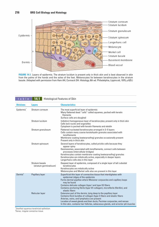
216 BRS Cell Biology and Histology
Epidermis
Stratum corneumStratum lucidum
Stratum granulosum
Stratum spinosum
Langerhans cell
Melanocyte
Merkel cell
Stratum basale
Basement membrane
Blood vesselDermis
FIGURE 14.1. Layers of epidermis. The stratum lucidum is present only in thick skin and is best observed in skinfrom the palms of the hands and the soles of the feet. Melanocytes lie between keratinocytes in the stratumbasale. (Adapted with permission from Ham AH, Cormack DH: Histology, 8th ed. Philadelphia, Lippincott, 1979, p 625.)
t a b l e 14.1 Histological Features of Skin
Divisions Layers Characteristics
Epidermis* Stratum corneum The most superficial layer of epidermisMany flattened dead “cells” called squames, packed with keratin
filamentsSurface cells are sloughed
Stratum lucidum Indistinct homogeneous layer of keratinocytes; present only in thick skinCells lack nuclei and organellesCytoplasm is packed with keratin filaments and eleidin
Stratum granulosum Flattened nucleated keratinocytes arranged in 3–5 layersCells contain many coarse keratohyalin granules associated with
tonofilamentsMembrane-coating (waterproofing) granules occasionally presentPresent only in thick skin
Stratum spinosum Several layers of keratinocytes, called prickle cells because they appear spiny
Desmosomes, associated with tonofilaments, connect cells between processes (intercellular bridges)
Keratinocytes contain membrane-coating (waterproofing) granulesKeratinocytes are mitotically active, especially in deeper layersLangerhans cells also in this layer
Stratum basale Deepest layer of epidermis, composed of a single layer of tall cuboidal (stratum germinativum) keratinocytes
Keratinocytes are mitotically activeMelanocytes and Merkel cells also are present in this layer
Dermis† Papillary layer Superficial thin layer of connective tissue that interdigitates with epidermal ridges of the epidermis
Forms dermal papillae where Meissner corpuscles and capillary loops may be found
Contains delicate collagen (type I and type III) fibersContains anchoring fibrils (type VII collagen), microfibrils (fibrillin), and
elastic fibersReticular layer Extensive part of the dermis, lying deep to the papillary layer
Contains thick bundles of collagen (type I) fibers and elastic fibersArteries, veins, and lymphatics are presentLocation of sweat glands and their ducts, Pacinian corpuscles, and nervesIn thin skin, contains hair follicles, sebaceous glands, and arrector pili muscles
*Stratified squamous keratinized epithelium.†Dense, irregular connective tissue.
LWBK615-c14[215-227].qxd 05/31/2010 10:51 AM Page 216 Aptara
Chapter 14 Skin 217
3. The stratum granulosum is the most superficial layer in which nuclei are still present. Itcomprises three to five layers of flattened keratinocytes that contain keratohyalin granules,bundles of keratin filaments (tonofilaments), and occasional membrane-coating granules.a. Keratohyalin granules (not membrane bound) contain histidine- and cystine-rich
proteins, which bind the keratin filaments together.b. The cytoplasmic aspect of the plasma membrane of keratinocytes in the stratum
granulosum is reinforced by an electron-dense layer 10 to 12 nm thick.4. The stratum lucidum is a clear, homogeneous layer just superficial to the stratum granu-
losum; it is often difficult to distinguish in histological sections. It is found only in palmarand plantar skin. This layer consists of keratinocytes that have neither nuclei nororganelles but contain keratin filaments and eleidin, a transformation product of kerato-hyalin.
5. The stratum corneum is the most superficial layer of the epidermis (Figure 14.2). It mayconsist of as many as 15 to 20 layers of flattened, nonnucleated dead “cells” filled withkeratin. These nonviable scalelike structures are called squames (or horny cells), and havethe shape of a 14-sided polygon. The outermost layer of squames is continuously shedby desquamation.
1. The stratum basale (stratum germinativum) is the deepest layer of the epidermis and iscomposed of keratinocytes that are cuboidal to columnar in shape. These mitoticallyactive cells are attached directly to the basal lamina of the basement membrane byhemidesmosomes (see Chapter 5 III B) and to each other by desmosomes. This layer alsocontains melanocytes and Merkel cells.
2. The stratum spinosum consists of a few layers of polyhedral keratinocytes (prickle cells).Their extensions, termed “intercellular bridges” by early histologists, are now known toterminate in desmosomes (see Chapter 5 II A 3). This layer also contains Langerhans cells.a. Keratinocytes in the deeper aspects of the stratum spinosum are also mitotically
active.b. The Malpighian layer (stratum malpighii) consists of the stratum spinosum and stra-
tum basale. Nearly all of the mitotic activity in the epidermis occurs in this region,and cell division occurs at night.
c. In the superficial regions of the stratum spinosum are keratinocytes that containmembrane-coating granules. The contents of these granules are released into the inter-cellular spaces in the form of lipid-containing sheets that are impermeable to waterand many foreign substances.
A. UV radiation and skin damage1. Exposure of unprotected skin to UV light can cause harmful effects
to the cells, even in the absence of sunburn.2. Sunscreen with an SPF (sun protection factor) rating of 15 or higher may protect against UVB
wavelengths but offers no protection against the longer UVA wavelengths.3. Recent studies have shown that UVA may be an important factor in photoaging and may
ultimately lead to the development of skin cancer (especially basal cell carcinoma andmelanoma) later in life.
B. Skin cancers commonly originate from cells in the epidermis. These cancers usually can betreated successfully if they are diagnosed early and surgically removed.1. Basal cell carcinoma arises from basal keratinocytes.2. Squamous cell carcinoma arises from cells of the stratum spinosum.
C. Malignant melanoma is a form of skin cancer that can be life-threatening.1. This form of cancer originates from melanocytes that divide, transform, and invade the dermis
and then enter the lymphatic and circulatory systems, metastasizing to a wide variety oforgans.
2. Treatment involves surgical removal of the skin lesion and regional lymph nodes. Chemother-apy is also required because of the extensive metastases.
3. Malignant melanoma constitutes about 3% of cancers.
CLINICAL
CONSIDERATIONS
LWBK615-c14[215-227].qxd 05/31/2010 10:51 AM Page 217 Aptara
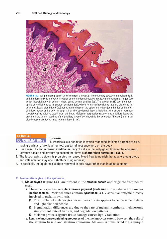
218 BRS Cell Biology and Histology
C. Nonkeratinocytes in the epidermis1. Melanocytes (Figure 14.1) are present in the stratum basale and originate from neural
crest.a. These cells synthesize a dark brown pigment (melanin) in oval-shaped organelles
(melanosomes). Melanosomes contain tyrosinase, a UV-sensitive enzyme directlyinvolved in melanin synthesis.(1) The number of melanocytes per unit area of skin appears to be the same in dark-
and light-skinned people.(2) Pigmentation differences are due to the rate of melanin synthesis, melanosome
size, content, rate of transfer, and degradation patterns(3) Melanin protects against tissue damage caused by UV radiation.
b. Long melanosome-containing processes of the melanocytes extend between the cells ofthe stratum basale and stratum spinosum. Melanin is transferred via a unique
Psoriasis1. Psoriasis is a condition in which reddened, inflamed patches of skin,
having a whitish, flaky layer on top, appear almost anywhere on the body.2. It is caused by an increase in mitotic activity of cells in the malpighian layer of the epidermis
(stratum basale and stratum spinosum) that have a shorter than normal cell cycle.3. The fast-growing epidermis promotes increased blood flow to nourish the accelerated growth,
and inflammation may occur (both causing redness).4. In psoriasis, the epidermis is often renewed in only days rather than in about a month.
CLINICAL
CONSIDERATIONS
sd dp
cfer
D
ESC
FIGURE 14.2. A light micrograph of thick skin from a fingertip. The boundary between the epidermis (E)and the dermis (D) is markedly irregular due to epidermal downgrowths, called epidermal ridges (er),which interdigitate with dermal ridges, called dermal papillae (dp). The epidermis (E) over the finger-tips is very thick due to its stratum corneum (sc), which forms surface ridges that are visible as fin-gerprints. Sweat gland ducts (sd) penetrate the base of the epidermal ridges (at a the tips of the inter-papillary pegs) and travel through all of the epidermal layers including the stratum corneum(arrowhead ) to release sweat from the body. Meissner corpuscles (arrow ) and capillary loops arepresent in the dermal papillae of the papillary layer of dermis, while thick collagen fibers (cf) and largerblood vessels are found in its reticular layer (�16).
LWBK615-c14[215-227].qxd 05/31/2010 10:51 AM Page 218 Aptara
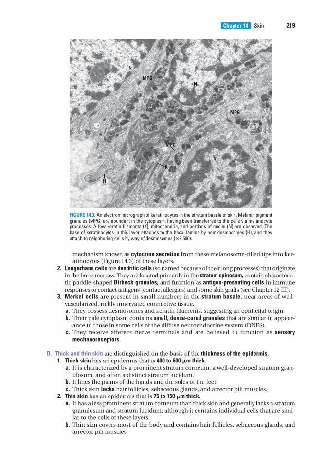
Chapter 14 Skin 219
mechanism known as cytocrine secretion from these melanosome-filled tips into ker-atinocytes (Figure 14.3) of these layers.
2. Langerhans cells are dendritic cells (so named because of their long processes) that originatein the bone marrow. They are located primarily in the stratum spinosum, contain characteris-tic paddle-shaped Birbeck granules, and function as antigen-presenting cells in immuneresponses to contact antigens (contact allergies) and some skin grafts (see Chapter 12 III).
3. Merkel cells are present in small numbers in the stratum basale, near areas of well-vascularized, richly innervated connective tissue.a. They possess desmosomes and keratin filaments, suggesting an epithelial origin.b. Their pale cytoplasm contains small, dense-cored granules that are similar in appear-
ance to those in some cells of the diffuse neuroendocrine system (DNES).c. They receive afferent nerve terminals and are believed to function as sensory
mechanoreceptors.
D. Thick and thin skin are distinguished on the basis of the thickness of the epidermis.1. Thick skin has an epidermis that is 400 to 600 �m thick.
a. It is characterized by a prominent stratum corneum, a well-developed stratum gran-ulosum, and often a distinct stratum lucidum.
b. It lines the palms of the hands and the soles of the feet.c. Thick skin lacks hair follicles, sebaceous glands, and arrector pili muscles.
2. Thin skin has an epidermis that is 75 to 150 �m thick.a. It has a less prominent stratum corneum than thick skin and generally lacks a stratum
granulosum and stratum lucidum, although it contains individual cells that are simi-lar to the cells of these layers.
b. Thin skin covers most of the body and contains hair follicles, sebaceous glands, andarrector pili muscles.
N
MPGN
MPG
N
MPG
K
H
FIGURE 14.3. An electron micrograph of keratinocytes in the stratum basale of skin. Melanin pigmentgranules (MPG) are abundant in the cytoplasm, having been transferred to the cells via melanocyteprocesses. A few keratin filaments (K), mitochondria, and portions of nuclei (N) are observed. Thebase of keratinocytes in this layer attaches to the basal lamina by hemidesmosomes (H), and theyattach to neighboring cells by way of desmosomes (�9,500).
LWBK615-c14[215-227].qxd 05/31/2010 10:51 AM Page 219 Aptara
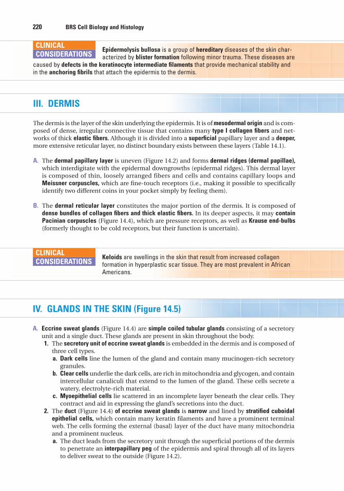
220 BRS Cell Biology and Histology
Epidermolysis bullosa is a group of hereditary diseases of the skin char-acterized by blister formation following minor trauma. These diseases are
caused by defects in the keratinocyte intermediate filaments that provide mechanical stability andin the anchoring fibrils that attach the epidermis to the dermis.
CLINICAL
CONSIDERATIONS
III. DERMIS
The dermis is the layer of the skin underlying the epidermis. It is of mesodermal origin and is com-posed of dense, irregular connective tissue that contains many type I collagen fibers and net-works of thick elastic fibers. Although it is divided into a superficial papillary layer and a deeper,more extensive reticular layer, no distinct boundary exists between these layers (Table 14.1).
A. The dermal papillary layer is uneven (Figure 14.2) and forms dermal ridges (dermal papillae),which interdigitate with the epidermal downgrowths (epidermal ridges). This dermal layeris composed of thin, loosely arranged fibers and cells and contains capillary loops andMeissner corpuscles, which are fine-touch receptors (i.e., making it possible to specificallyidentify two different coins in your pocket simply by feeling them).
B. The dermal reticular layer constitutes the major portion of the dermis. It is composed ofdense bundles of collagen fibers and thick elastic fibers. In its deeper aspects, it may containPacinian corpuscles (Figure 14.4), which are pressure receptors, as well as Krause end-bulbs(formerly thought to be cold receptors, but their function is uncertain).
Keloids are swellings in the skin that result from increased collagen formation in hyperplastic scar tissue. They are most prevalent in AfricanAmericans.
CLINICAL
CONSIDERATIONS
IV. GLANDS IN THE SKIN (Figure 14.5)
A. Eccrine sweat glands (Figure 14.4) are simple coiled tubular glands consisting of a secretoryunit and a single duct. These glands are present in skin throughout the body.1. The secretory unit of eccrine sweat glands is embedded in the dermis and is composed of
three cell types.a. Dark cells line the lumen of the gland and contain many mucinogen-rich secretory
granules.b. Clear cells underlie the dark cells, are rich in mitochondria and glycogen, and contain
intercellular canaliculi that extend to the lumen of the gland. These cells secrete awatery, electrolyte-rich material.
c. Myoepithelial cells lie scattered in an incomplete layer beneath the clear cells. Theycontract and aid in expressing the gland’s secretions into the duct.
2. The duct (Figure 14.4) of eccrine sweat glands is narrow and lined by stratified cuboidalepithelial cells, which contain many keratin filaments and have a prominent terminalweb. The cells forming the external (basal) layer of the duct have many mitochondriaand a prominent nucleus.a. The duct leads from the secretory unit through the superficial portions of the dermis
to penetrate an interpapillary peg of the epidermis and spiral through all of its layersto deliver sweat to the outside (Figure 14.2).
LWBK615-c14[215-227].qxd 05/31/2010 10:51 AM Page 220 Aptara
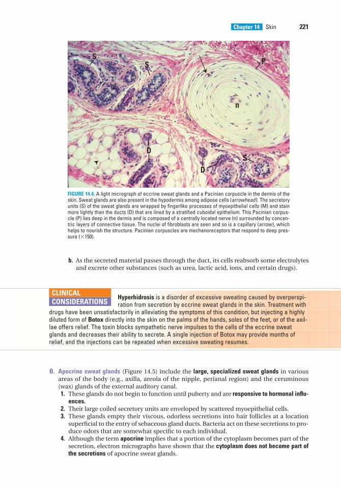
Chapter 14 Skin 221
b. As the secreted material passes through the duct, its cells reabsorb some electrolytesand excrete other substances (such as urea, lactic acid, ions, and certain drugs).
SS
n
P
D
DS
FIGURE 14.4. A light micrograph of eccrine sweat glands and a Pacinian corpuscle in the dermis of theskin. Sweat glands are also present in the hypodermis among adipose cells (arrowhead ). The secretoryunits (S) of the sweat glands are wrapped by fingerlike processes of myoepithelial cells (M) and stainmore lightly than the ducts (D) that are lined by a stratified cuboidal epithelium. This Pacinian corpus-cle (P) lies deep in the dermis and is composed of a centrally located nerve (n) surrounded by concen-tric layers of connective tissue. The nuclei of fibroblasts are seen and so is a capillary (arrow ), whichhelps to nourish the structure. Pacinian corpuscles are mechanoreceptors that respond to deep pres-sure (�150).
Hyperhidrosis is a disorder of excessive sweating caused by overperspi-ration from secretion by eccrine sweat glands in the skin. Treatment with
drugs have been unsatisfactorily in alleviating the symptoms of this condition, but injecting a highlydiluted form of Botox directly into the skin on the palms of the hands, soles of the feet, or of the axil-lae offers relief. The toxin blocks sympathetic nerve impulses to the cells of the eccrine sweatglands and decreases their ability to secrete. A single injection of Botox may provide months ofrelief, and the injections can be repeated when excessive sweating resumes.
CLINICAL
CONSIDERATIONS
B. Apocrine sweat glands (Figure 14.5) include the large, specialized sweat glands in variousareas of the body (e.g., axilla, areola of the nipple, perianal region) and the ceruminous(wax) glands of the external auditory canal.1. These glands do not begin to function until puberty and are responsive to hormonal influ-
ences.2. Their large coiled secretory units are enveloped by scattered myoepithelial cells.3. These glands empty their viscous, odorless secretions into hair follicles at a location
superficial to the entry of sebaceous gland ducts. Bacteria act on these secretions to pro-duce odors that are somewhat specific to each individual.
4. Although the term apocrine implies that a portion of the cytoplasm becomes part of thesecretion, electron micrographs have shown that the cytoplasm does not become part ofthe secretions of apocrine sweat glands.
LWBK615-c14[215-227].qxd 05/31/2010 10:51 AM Page 221 Aptara
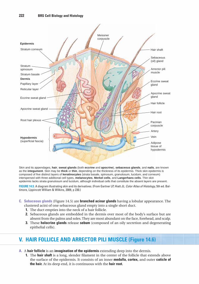
222 BRS Cell Biology and Histology
Meissnercorpuscle
Hair shaft
Sebaceous(oil) gland
Arrector pilimuscle
Eccrine sweatgland
Apocrine sweatgland
Hair follicle
Hair root
Paciniancorpuscle
Artery
Vein
Adiposetissue ofhypodermis
Hypodermis(superficial fascia)
Root hair plexus
Apocrine sweat gland
Reticular layer
Papillary layer
Dermis
Eccrine sweat gland
Stratumspinosum
Stratum corneum
Epidermis
Skin and its appendages, hair, sweat glands (both eccrine and apocrine), sebaceous glands, and nails, are knownas the integument. Skin may be thick or thin, depending on the thickness of its epidermis. Thick skin epidermis iscomposed of five distinct layers of keratinocytes (strata basale, spinosum, granulosum, lucidum, and corneum)interspersed with three additional cell types, melanocytes, Merkel cells, and Langerhans cells. Thin skinepidermis lacks strata granulosum and lucidum, although individual cells that constitute the absent layers are present.
Stratum basale
⎠
⎞
FIGURE 14.5. A diagram illustrating skin and its derivatives. (From Gartner LP, Hiatt JL: Color Atlas of Histology, 5th ed. Bal-timore, Lippincott William & Wilkins, 2009, p 230.)
C. Sebaceous glands (Figure 14.5) are branched acinar glands having a lobular appearance. Theclustered acini of one sebaceous gland empty into a single short duct.1. The duct empties into the neck of a hair follicle.2. Sebaceous glands are embedded in the dermis over most of the body’s surface but are
absent from the palms and soles. They are most abundant on the face, forehead, and scalp.3. These holocrine glands release sebum (composed of an oily secretion and degenerating
epithelial cells).
V. HAIR FOLLICLE AND ARRECTOR PILI MUSCLE (Figure 14.6)
A. A hair follicle is an invagination of the epidermis extending deep into the dermis.1. The hair shaft is a long, slender filament in the center of the follicle that extends above
the surface of the epidermis. It consists of an inner medulla, cortex, and outer cuticle ofthe hair. At its deep end, it is continuous with the hair root.
LWBK615-c14[215-227].qxd 05/31/2010 10:51 AM Page 222 Aptara
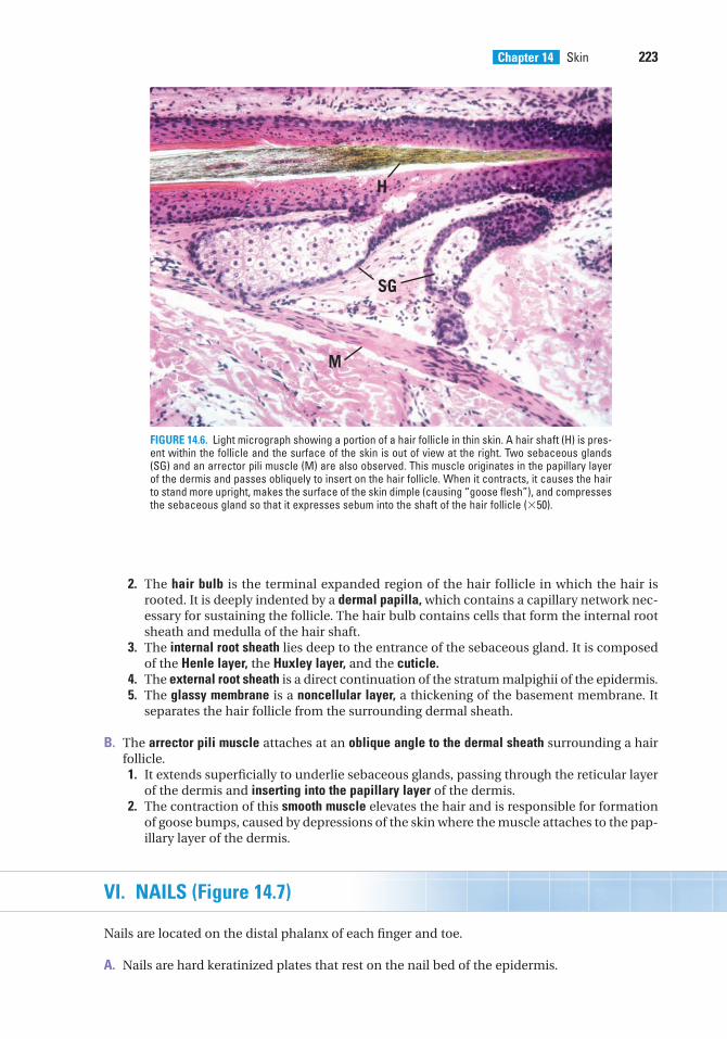
Chapter 14 Skin 223
2. The hair bulb is the terminal expanded region of the hair follicle in which the hair isrooted. It is deeply indented by a dermal papilla, which contains a capillary network nec-essary for sustaining the follicle. The hair bulb contains cells that form the internal rootsheath and medulla of the hair shaft.
3. The internal root sheath lies deep to the entrance of the sebaceous gland. It is composedof the Henle layer, the Huxley layer, and the cuticle.
4. The external root sheath is a direct continuation of the stratum malpighii of the epidermis.5. The glassy membrane is a noncellular layer, a thickening of the basement membrane. It
separates the hair follicle from the surrounding dermal sheath.
B. The arrector pili muscle attaches at an oblique angle to the dermal sheath surrounding a hairfollicle.1. It extends superficially to underlie sebaceous glands, passing through the reticular layer
of the dermis and inserting into the papillary layer of the dermis.2. The contraction of this smooth muscle elevates the hair and is responsible for formation
of goose bumps, caused by depressions of the skin where the muscle attaches to the pap-illary layer of the dermis.
VI. NAILS (Figure 14.7)
Nails are located on the distal phalanx of each finger and toe.
A. Nails are hard keratinized plates that rest on the nail bed of the epidermis.
H
SG
M
FIGURE 14.6. Light micrograph showing a portion of a hair follicle in thin skin. A hair shaft (H) is pres-ent within the follicle and the surface of the skin is out of view at the right. Two sebaceous glands(SG) and an arrector pili muscle (M) are also observed. This muscle originates in the papillary layerof the dermis and passes obliquely to insert on the hair follicle. When it contracts, it causes the hairto stand more upright, makes the surface of the skin dimple (causing “goose flesh”), and compressesthe sebaceous gland so that it expresses sebum into the shaft of the hair follicle (�50).
LWBK615-c14[215-227].qxd 05/31/2010 10:51 AM Page 223 Aptara
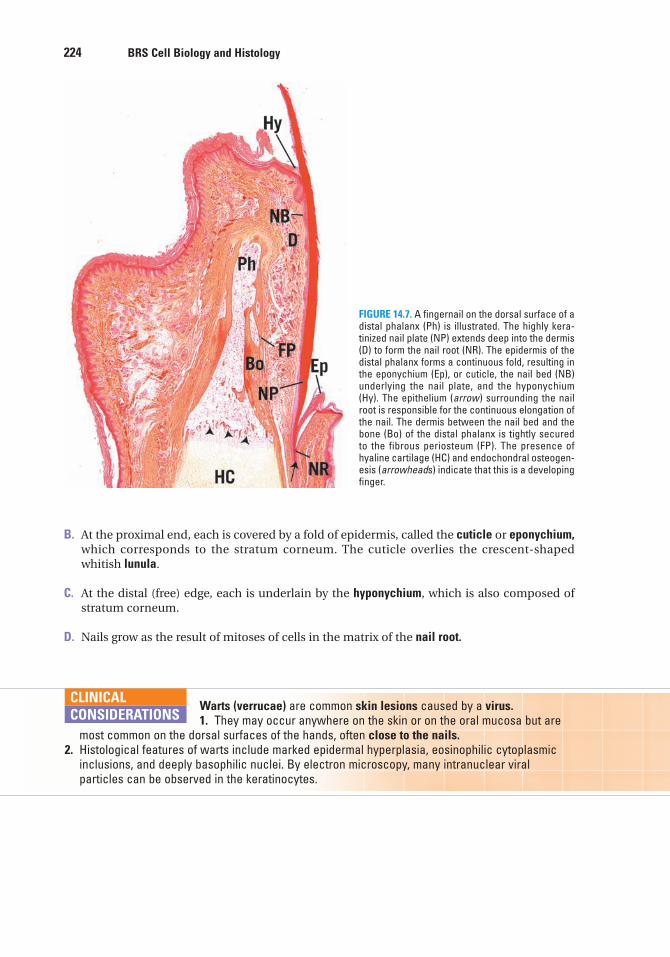
224 BRS Cell Biology and Histology
B. At the proximal end, each is covered by a fold of epidermis, called the cuticle or eponychium,which corresponds to the stratum corneum. The cuticle overlies the crescent-shapedwhitish lunula.
C. At the distal (free) edge, each is underlain by the hyponychium, which is also composed ofstratum corneum.
D. Nails grow as the result of mitoses of cells in the matrix of the nail root.
FIGURE 14.7. A fingernail on the dorsal surface of adistal phalanx (Ph) is illustrated. The highly kera-tinized nail plate (NP) extends deep into the dermis(D) to form the nail root (NR). The epidermis of thedistal phalanx forms a continuous fold, resulting inthe eponychium (Ep), or cuticle, the nail bed (NB)underlying the nail plate, and the hyponychium(Hy). The epithelium (arrow) surrounding the nailroot is responsible for the continuous elongation ofthe nail. The dermis between the nail bed and thebone (Bo) of the distal phalanx is tightly securedto the fibrous periosteum (FP). The presence ofhyaline cartilage (HC) and endochondral osteogen-esis (arrowheads) indicate that this is a developingfinger.
Warts (verrucae) are common skin lesions caused by a virus.1. They may occur anywhere on the skin or on the oral mucosa but are
most common on the dorsal surfaces of the hands, often close to the nails.2. Histological features of warts include marked epidermal hyperplasia, eosinophilic cytoplasmic
inclusions, and deeply basophilic nuclei. By electron microscopy, many intranuclear viralparticles can be observed in the keratinocytes.
CLINICAL
CONSIDERATIONS
LWBK615-c14[215-227].qxd 05/31/2010 10:52 AM Page 224 Aptara
Review Test
225
1. Intercellular bridges are characteristic ofwhich of the following layers of theepidermis?
(A) Stratum granulosum(B) Stratum lucidum(C) Stratum corneum(D) Stratum spinosum(E) Stratum basale
2. Which of the following statementsconcerning the stratum granulosum is true?
(A) It contains melanosomes.(B) It lies superficial to the stratum lucidum.(C) It is the thickest layer of the epidermis in
thick skin.(D) It contains keratohyalin granules.(E) It contains large numbers of dividing
cells.
3. Which of the following statements aboutLangerhans cells is true?
(A) They commonly are found in the dermis.(B) They function as sensory
mechanoreceptors.(C) They function as receptors for cold.(D) They play an immunological role in the
skin.(E) They are of epithelial origin.
4. Meissner corpuscles are present in whichof the following regions of the skin?
(A) Dermal reticular layer(B) Dermal papillary layer(C) Hypodermis(D) Stratum basale(E) Epidermal ridges
5. Which of the following statementsconcerning thin skin is true?
(A) It does not contain sweat glands.(B) It lacks a stratum corneum.(C) It is less abundant than thick skin.(D) It contains hair follicles.(E) Its epidermis does not rest on a
basement membrane.
6. Which of the following statements abouteccrine sweat glands is true?
(A) They are absent in thick skin.(B) They are holocrine glands.(C) They have a narrow duct lined by a strat-
ified cuboidal epithelium.(D) They secrete an oily material called
sebum.(E) They empty into hair follicles.
7. Which of the following statements abouthair follicles is true?
(A) They are always associated with aneccrine sweat gland.
(B) They are present in thin skin but not inthick skin.
(C) Their associated arrector pili muscle iscomposed of striated fibers.
(D) Their hair shaft inserts into the papillarylayer of the epidermis.
(E) They do not extend into the dermis.
Directions: Each of the numbered items or incomplete statements in this section is followed byanswers or completions of the statement. Select the ONE lettered answer that is BEST in eachcase.
LWBK615-c14[215-227].qxd 05/31/2010 10:52 AM Page 225 Aptara
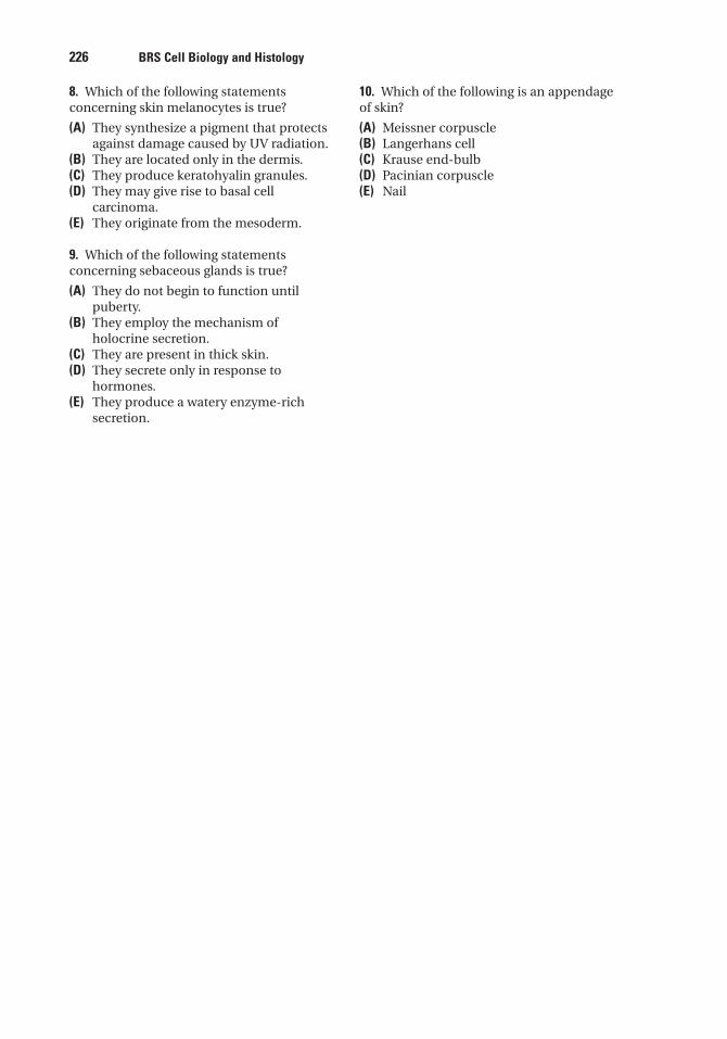
226 BRS Cell Biology and Histology
10. Which of the following is an appendageof skin?
(A) Meissner corpuscle(B) Langerhans cell(C) Krause end-bulb(D) Pacinian corpuscle(E) Nail
8. Which of the following statementsconcerning skin melanocytes is true?
(A) They synthesize a pigment that protectsagainst damage caused by UV radiation.
(B) They are located only in the dermis.(C) They produce keratohyalin granules.(D) They may give rise to basal cell
carcinoma.(E) They originate from the mesoderm.
9. Which of the following statementsconcerning sebaceous glands is true?
(A) They do not begin to function untilpuberty.
(B) They employ the mechanism ofholocrine secretion.
(C) They are present in thick skin.(D) They secrete only in response to
hormones.(E) They produce a watery enzyme-rich
secretion.
LWBK615-c14[215-227].qxd 05/31/2010 10:52 AM Page 226 Aptara
227
Answers and Explanations
1. D. Observations with an electron microscope show that intercellular bridges areassociated with desmosomes (maculae adherentes), linking the processes of adjacent cellsin the stratum spinosum. Desmosomes also link cells within the other epidermal layers,but these cells do not form processes characteristic of bridges. The keratinocytes of thestratum basale also contain hemidesmosomes, which attach the cells to the underlyingbasal lamina (see Chapter 14 II B).
2. D. The stratum granulosum contains a number of dense keratohyalin granules but notmelanosomes. It lies just deep to the stratum lucidum and is a relatively thin layer in theepidermis of thick skin. Only rarely would a cell undergo mitosis in this layer of the skin(see Chapter 14 II B).
3. D. Langerhans cells in the epidermis function as antigen-presenting cells by trappingantigens that penetrate the epidermis and transporting them to regional lymph nodes,where they are presented to T lymphocytes. In this way, these cells assist in the immunedefense of the body. They originate in the bone marrow and do not arise from epithelium(see Chapter 14 II C).
4. B. Meissner corpuscles are encapsulated nerve endings present in dermal papillae, whichare part of the papillary layer of the dermis. These corpuscles function as receptors for finetouch (see Chapter 14 III A).
5. D. In contrast to thick skin, which lacks hair follicles, thin skin contains many of them (seeChapter 14 II D).
6. C. Eccrine sweat glands are simple, coiled tubular glands that have a duct lined by a strati-fied cuboidal epithelium. They are found in both thick and thin skin and are classified asmerocrine glands, meaning they release only their secretory product, which does notinclude cells or portions of cells (see Chapter 14 IV A).
7. B. Hair follicles are present only in thin skin. They are associated with sebaceous glandsand arrector pili smooth muscle bundles (see Chapter 14 V).
8. A. Melanocytes are present in the stratum basale of the epidermis. They synthesizemelanin pigment and transfer it to keratinocytes to protect against damage caused by UVradiation. Melanocytes sometimes give rise to a form of skin cancer called malignantmelanoma. They derive from neural crest and migrate into the epidermis early duringembryonic development (see Chapter 14 II C).
9. B. Sebaceous glands produce sebum, an oily material, and release it into the upper shaftof the hair follicle by a mechanism called holocrine secretion (which means the productand cellular debris are both released from the gland) (see Chapter 14 IV C).
10. E. The nail is one appendage of the skin. Other skin appendages are hair follicles, sweatglands, and sebaceous glands (see Chapter 14 VI).
LWBK615-c14[215-227].qxd 05/31/2010 10:52 AM Page 227 Aptara
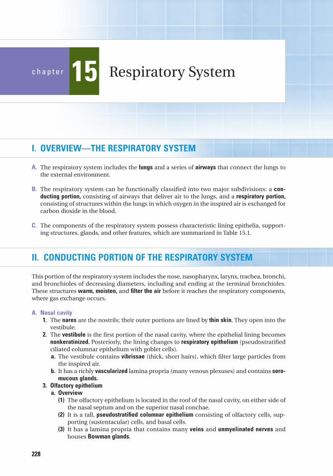
c h a p t e r 15 Respiratory System
228
I. OVERVIEW—THE RESPIRATORY SYSTEM
A. The respiratory system includes the lungs and a series of airways that connect the lungs tothe external environment.
B. The respiratory system can be functionally classified into two major subdivisions: a con-ducting portion, consisting of airways that deliver air to the lungs, and a respiratory portion,consisting of structures within the lungs in which oxygen in the inspired air is exchanged forcarbon dioxide in the blood.
C. The components of the respiratory system possess characteristic lining epithelia, support-ing structures, glands, and other features, which are summarized in Table 15.1.
II. CONDUCTING PORTION OF THE RESPIRATORY SYSTEM
This portion of the respiratory system includes the nose, nasopharynx, larynx, trachea, bronchi,and bronchioles of decreasing diameters, including and ending at the terminal bronchioles.These structures warm, moisten, and filter the air before it reaches the respiratory components,where gas exchange occurs.
A. Nasal cavity1. The nares are the nostrils; their outer portions are lined by thin skin. They open into the
vestibule.2. The vestibule is the first portion of the nasal cavity, where the epithelial lining becomes
nonkeratinized. Posteriorly, the lining changes to respiratory epithelium (pseudostratifiedciliated columnar epithelium with goblet cells).a. The vestibule contains vibrissae (thick, short hairs), which filter large particles from
the inspired air.b. It has a richly vascularized lamina propria (many venous plexuses) and contains sero-
mucous glands.3. Olfactory epithelium
a. Overview(1) The olfactory epithelium is located in the roof of the nasal cavity, on either side of
the nasal septum and on the superior nasal conchae.(2) It is a tall, pseudostratified columnar epithelium consisting of olfactory cells, sup-
porting (sustentacular) cells, and basal cells.(3) It has a lamina propria that contains many veins and unmyelinated nerves and
houses Bowman glands.
LWBK615-c15[228-243].qxd 05/31/2010 10:53 AM Page 228 Aptara
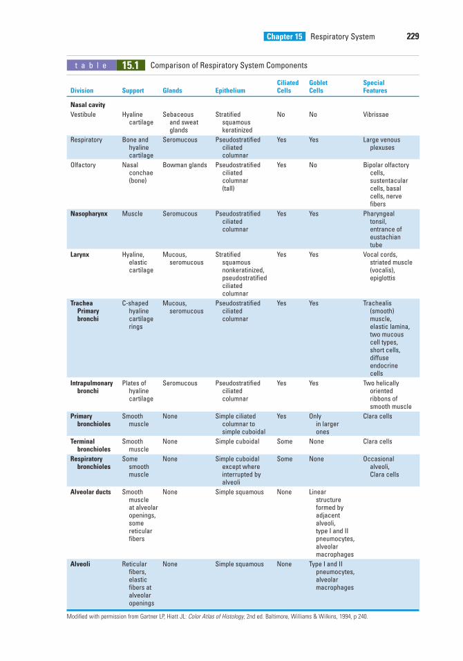
Chapter 15 Respiratory System 229
t a b l e 15.1 Comparison of Respiratory System Components
Ciliated Goblet Special Division Support Glands Epithelium Cells Cells Features
Nasal cavity
Vestibule Hyaline Sebaceous Stratified No No Vibrissaecartilage and sweat squamous
glands keratinized
Respiratory Bone and Seromucous Pseudostratified Yes Yes Large venous hyaline ciliated plexusescartilage columnar
Olfactory Nasal Bowman glands Pseudostratified Yes No Bipolar olfactory conchae ciliated cells, (bone) columnar sustentacular
(tall) cells, basal cells, nerve fibers
Nasopharynx Muscle Seromucous Pseudostratified Yes Yes Pharyngeal ciliated tonsil, columnar entrance of
eustachian tube
Larynx Hyaline, Mucous, Stratified Yes Yes Vocal cords, elastic seromucous squamous striated musclecartilage nonkeratinized, (vocalis),
pseudostratified epiglottisciliated columnar
Trachea C-shaped Mucous, Pseudostratified Yes Yes Trachealis Primary hyaline seromucous ciliated (smooth) bronchi cartilage columnar muscle,
rings elastic lamina, two mucous cell types, short cells, diffuse endocrine cells
Intrapulmonary Plates of Seromucous Pseudostratified Yes Yes Two helically bronchi hyaline ciliated oriented
cartilage columnar ribbons of smooth muscle
Primary Smooth None Simple ciliated Yes Only Clara cellsbronchioles muscle columnar to in larger
simple cuboidal ones
Terminal Smooth None Simple cuboidal Some None Clara cellsbronchioles muscle
Respiratory Some None Simple cuboidal Some None Occasional bronchioles smooth except where alveoli,
muscle interrupted by Clara cellsalveoli
Alveolar ducts Smooth None Simple squamous None Linear muscle structureat alveolar formed by openings, adjacentsome alveoli, reticular type I and II fibers pneumocytes,
alveolar macrophages
Alveoli Reticular None Simple squamous None Type I and II fibers, pneumocytes,elastic alveolar fibers at macrophagesalveolar openings
Modified with permission from Gartner LP, Hiatt JL: Color Atlas of Histology, 2nd ed. Baltimore, Williams & Wilkins, 1994, p 240.
LWBK615-c15[228-243].qxd 05/31/2010 10:53 AM Page 229 Aptara
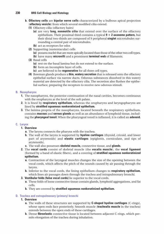
230 BRS Cell Biology and Histology
b. Olfactory cells are bipolar nerve cells characterized by a bulbous apical projection(olfactory vesicle) from which several modified cilia extend.(1) Olfactory cilia (olfactory hairs)
(a) are very long, nonmotile cilia that extend over the surface of the olfactoryepithelium. Their proximal third contains a typical 9 � 2 axoneme pattern, buttheir distal two-thirds are composed of 9 peripheral singlet microtubules sur-rounding a central pair of microtubules.
(b) act as receptors for odor.(2) Supporting (sustentacular) cells
(a) possess nuclei that are more apically located than those of the other two cell types.(b) have many microvilli and a prominent terminal web of filaments.
(3) Basal cells(a) rest on the basal lamina but do not extend to the surface.(b) form an incomplete layer of cells.(c) are believed to be regenerative for all three cell types.
(4) Bowman glands produce a thin, watery secretion that is released onto the olfactoryepithelial surface via narrow ducts. Odorous substances dissolved in this waterymaterial are detected by the olfactory cilia. The secretion also flushes the epithe-lial surface, preparing the receptors to receive new odorous stimuli.
B. Nasopharynx1. The nasopharynx, the posterior continuation of the nasal cavities, becomes continuous
with the oropharynx at the level of the soft palate.2. It is lined by respiratory epithelium, whereas the oropharynx and laryngopharynx are
lined by stratified squamous nonkeratinized epithelium.3. The lamina propria of the nasopharynx, located beneath the respiratory epithelium,
contains mucous and serous glands as well as an abundance of lymphoid tissue, includ-ing the pharyngeal tonsil. When the pharyngeal tonsil is inflamed, it is called an adenoid.
C. Larynx1. Overview
a. The larynx connects the pharynx with the trachea.b. The wall of the larynx is supported by hyaline cartilages (thyroid, cricoid, and lower
part of arytenoids) and elastic cartilages (epiglottis, corniculate, and tips ofarytenoids).
c. The wall also possesses skeletal muscle, connective tissue, and glands.2. The vocal cords consist of skeletal muscle (the vocalis muscle), the vocal ligament
(formed by a band of elastic fibers), and a covering of stratified squamous nonkeratinizedepithelium.a. Contraction of the laryngeal muscles changes the size of the opening between the
vocal cords, which affects the pitch of the sounds caused by air passing through thelarynx.
b. Inferior to the vocal cords, the lining epithelium changes to respiratory epithelium,which lines air passages down through the trachea and intrapulmonary bronchi.
3. Vestibular folds (false vocal cords) lie superior to the vocal cords.a. These folds of loose connective tissue contain glands, lymphoid aggregations, and fat
cells.b. They are covered by stratified squamous nonkeratinized epithelium.
D. Trachea and extrapulmonary (primary) bronchi1. Overview
a. The walls of these structures are supported by C-shaped hyaline cartilages (C-rings),whose open ends face posteriorly. Smooth muscle (trachealis muscle in the trachea)extends between the open ends of these cartilages.
b. Dense fibroelastic connective tissue is located between adjacent C-rings, which per-mits elongation of the trachea during inhalation.
LWBK615-c15[228-243].qxd 05/31/2010 10:53 AM Page 230 Aptara
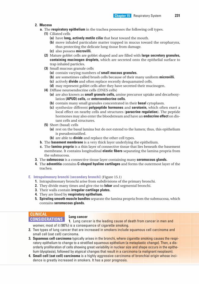
Chapter 15 Respiratory System 231
2. Mucosaa. The respiratory epithelium in the trachea possesses the following cell types.
(1) Ciliated cells(a) have long, actively motile cilia that beat toward the mouth.(b) move inhaled particulate matter trapped in mucus toward the oropharynx,
thus protecting the delicate lung tissue from damage.(c) also possess microvilli.
(2) Mature goblet cells are goblet shaped and are filled with large secretory granules,containing mucinogen droplets, which are secreted onto the epithelial surface totrap inhaled particles.
(3) Small mucous granule cells(a) contain varying numbers of small mucous granules.(b) are sometimes called brush cells because of their many uniform microvilli.(c) actively divide and often replace recently desquamated cells.(d) may represent goblet cells after they have secreted their mucinogen.
(4) Diffuse neuroendocrine cells (DNES cells)(a) are also known as small granule cells, amine precursor uptake and decarboxy-
lation (APUD) cells, or enteroendocrine cells.(b) contain many small granules concentrated in their basal cytoplasm.(c) synthesize different polypeptide hormones and serotonin, which often exert a
local effect on nearby cells and structures (paracrine regulation). The peptidehormones may also enter the bloodstream and have an endocrine effect on dis-tant cells and structures.
(5) Short (basal) cells(a) rest on the basal lamina but do not extend to the lumen; thus, this epithelium
is pseudostratified.(b) are able to divide and replace the other cell types.
b. The basement membrane is a very thick layer underlying the epithelium.c. The lamina propria is a thin layer of connective tissue that lies beneath the basement
membrane. It contains longitudinal elastic fibers separating the lamina propria fromthe submucosa.
3. The submucosa is a connective tissue layer containing many seromucous glands.4. The adventitia contains C-shaped hyaline cartilages and forms the outermost layer of the
trachea.
E. Intrapulmonary bronchi (secondary bronchi) (Figure 15.1)1. Intrapulmonary bronchi arise from subdivisions of the primary bronchi.2. They divide many times and give rise to lobar and segmental bronchi.3. Their walls contain irregular cartilage plates.4. They are lined by respiratory epithelium.5. Spiraling smooth muscle bundles separate the lamina propria from the submucosa, which
contains seromucous glands.
Lung cancer1. Lung cancer is the leading cause of death from cancer in men and
women; most of it (90%) is a consequence of cigarette smoking.2. Two types of lung cancer that are increased in smokers include squamous cell carcinoma and
small cell (oat cell) carcinoma.3. Squamous cell carcinoma typically arises in the bronchi, where cigarette smoking causes the respi-
ratory epithelium to change to a stratified squamous epithelium (a metaplastic change). Then, a dis-orderly proliferation of cells showing great variability in nuclear size and shape occurs in the epithe-lium (dysplasia), followed by atypical changes that result in a carcinoma (a malignant neoplasm).
4. Small cell (oat cell) carcinoma is a highly aggressive carcinoma of bronchial origin whose inci-dence is greatly increased in smokers. It has a poor prognosis.
CLINICAL
CONSIDERATIONS
LWBK615-c15[228-243].qxd 05/31/2010 10:54 AM Page 231 Aptara
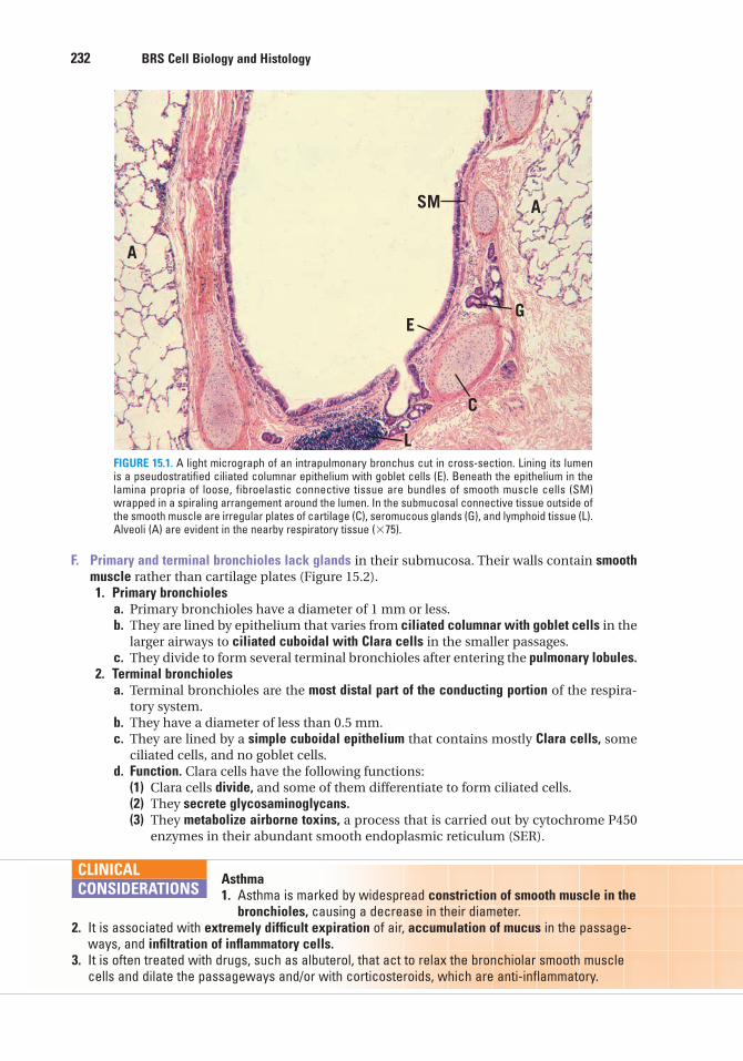
232 BRS Cell Biology and Histology
F. Primary and terminal bronchioles lack glands in their submucosa. Their walls contain smoothmuscle rather than cartilage plates (Figure 15.2).1. Primary bronchioles
a. Primary bronchioles have a diameter of 1 mm or less.b. They are lined by epithelium that varies from ciliated columnar with goblet cells in the
larger airways to ciliated cuboidal with Clara cells in the smaller passages.c. They divide to form several terminal bronchioles after entering the pulmonary lobules.
2. Terminal bronchiolesa. Terminal bronchioles are the most distal part of the conducting portion of the respira-
tory system.b. They have a diameter of less than 0.5 mm.c. They are lined by a simple cuboidal epithelium that contains mostly Clara cells, some
ciliated cells, and no goblet cells.d. Function. Clara cells have the following functions:
(1) Clara cells divide, and some of them differentiate to form ciliated cells.(2) They secrete glycosaminoglycans.(3) They metabolize airborne toxins, a process that is carried out by cytochrome P450
enzymes in their abundant smooth endoplasmic reticulum (SER).
SM
E
C
L
G
A
A
FIGURE 15.1. A light micrograph of an intrapulmonary bronchus cut in cross-section. Lining its lumenis a pseudostratified ciliated columnar epithelium with goblet cells (E). Beneath the epithelium in thelamina propria of loose, fibroelastic connective tissue are bundles of smooth muscle cells (SM)wrapped in a spiraling arrangement around the lumen. In the submucosal connective tissue outside ofthe smooth muscle are irregular plates of cartilage (C), seromucous glands (G), and lymphoid tissue (L).Alveoli (A) are evident in the nearby respiratory tissue (�75).
Asthma1. Asthma is marked by widespread constriction of smooth muscle in the
bronchioles, causing a decrease in their diameter.2. It is associated with extremely difficult expiration of air, accumulation of mucus in the passage-
ways, and infiltration of inflammatory cells.3. It is often treated with drugs, such as albuterol, that act to relax the bronchiolar smooth muscle
cells and dilate the passageways and/or with corticosteroids, which are anti-inflammatory.
CLINICAL
CONSIDERATIONS
LWBK615-c15[228-243].qxd 05/31/2010 10:54 AM Page 232 Aptara
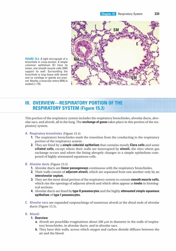
Chapter 15 Respiratory System 233
III. OVERVIEW—RESPIRATORY PORTION OF THERESPIRATORY SYSTEM (Figure 15.3)
This portion of the respiratory system includes the respiratory bronchioles, alveolar ducts, alve-olar sacs, and alveoli, all in the lung. The exchange of gases takes place in this portion of the res-piratory system.
A. Respiratory bronchioles (Figure 15.4)1. The respiratory bronchioles mark the transition from the conducting to the respiratory
portion of the respiratory system.2. They are lined by a simple cuboidal epithelium that contains mostly Clara cells and some
ciliated cells, except where their walls are interrupted by alveoli, the sites where gasexchange occurs and where the lining abruptly changes to a simple epithelium com-posed of highly attenuated squamous cells.
B. Alveolar ducts (Figure 15.5)1. Alveolar ducts are linear passageways continuous with the respiratory bronchioles.2. Their walls consist of adjacent alveoli, which are separated from one another only by an
interalveolar septum.3. They are the most distal portion of the respiratory system to contain smooth muscle cells,
which rim the openings of adjacent alveoli and which often appear as knobs in histolog-ical sections.
4. Alveolar ducts are lined by type II pneumocytes and the highly attenuated simple squamousepithelium of type I pneumocytes.
C. Alveolar sacs are expanded outpouchings of numerous alveoli at the distal ends of alveolarducts (Figure 15.5).
D. Alveoli1. Overview
a. Alveoli are pouchlike evaginations about 200 �m in diameter in the walls of respira-tory bronchioles, in alveolar ducts, and in alveolar sacs.
b. They have thin walls, across which oxygen and carbon dioxide diffuses between theair and the blood.
SM
E
MAMA
SMSM
MA
SM
FIGURE 15.2. A light micrograph of abronchiole in cross-section. A simplecolumnar epithelium (E) lines itslumen, and smooth muscle cells (SM)support its wall. Surrounding thebronchiole is lung tissue with alveoliand no cartilage or glands are pres-ent. Nearby, a muscular artery (MA) isevident (�75).
LWBK615-c15[228-243].qxd 05/31/2010 10:54 AM Page 233 Aptara
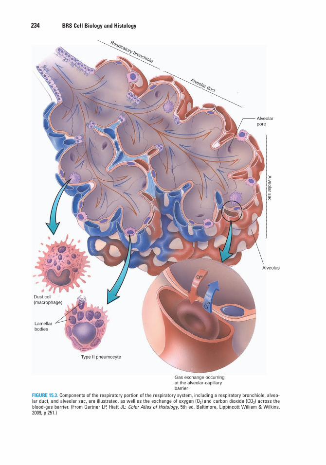
234 BRS Cell Biology and Histology
Respiratory bronchiole
Alveolar duct
Alveolarpore
Alveolar sac
Alveolus
Dust cell(macrophage)
Lamellarbodies
Type II pneumocyte
Gas exchange occurringat the alveolar-capillarybarrier
⎧⎪⎪⎪⎪⎪⎪⎪⎪⎨⎪⎪⎪⎪⎪⎪⎪⎪⎩
⎧⎪⎪⎪⎪⎪⎪⎪⎪⎪⎪⎪⎨⎪⎪⎪⎪⎪⎪⎪⎪⎪⎪⎩
⎧⎪⎪⎪⎪⎪⎪⎪⎪⎪⎪⎪⎪⎪⎨⎪⎪⎪⎪⎪⎪⎪⎪⎪⎪⎪⎪⎪⎩
FIGURE 15.3. Components of the respiratory portion of the respiratory system, including a respiratory bronchiole, alveo-lar duct, and alveolar sac, are illustrated, as well as the exchange of oxygen (O2) and carbon dioxide (CO2) across theblood-gas barrier. (From Gartner LP, Hiatt JL: Color Atlas of Histology, 5th ed. Baltimore, Lippincott William & Wilkins,2009, p 251.)
LWBK615-c15[228-243].qxd 05/31/2010 10:54 AM Page 234 Aptara
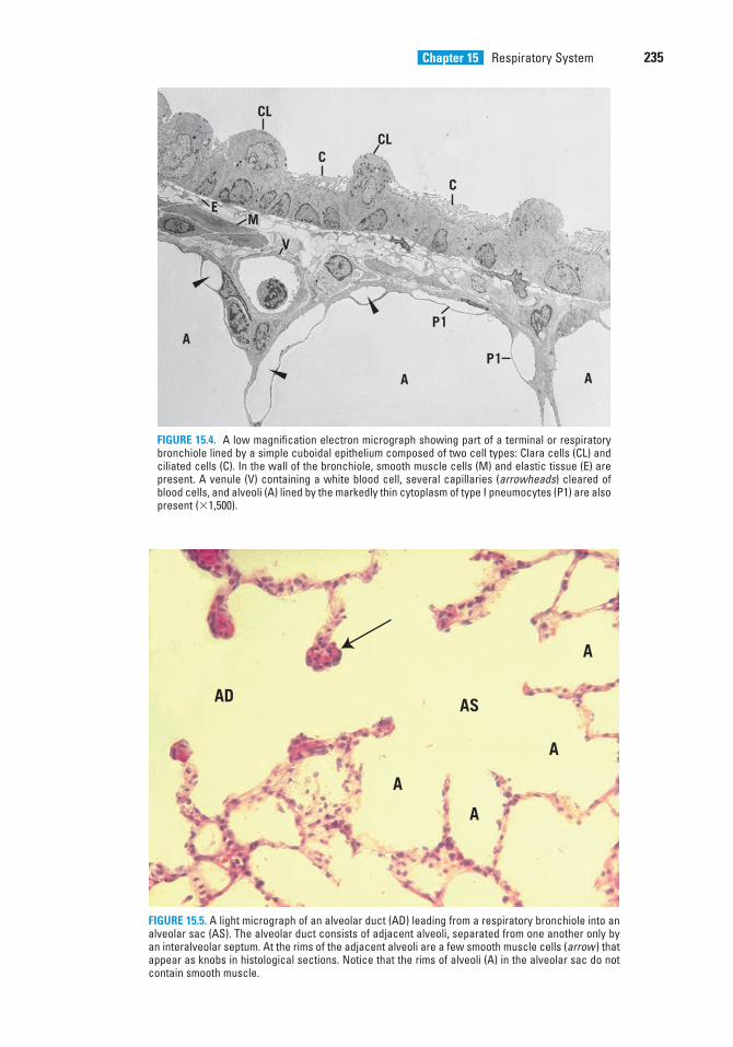
Chapter 15 Respiratory System 235
CL
EM
V
A
A
P1
P1A
C
CLC
FIGURE 15.4. A low magnification electron micrograph showing part of a terminal or respiratorybronchiole lined by a simple cuboidal epithelium composed of two cell types: Clara cells (CL) andciliated cells (C). In the wall of the bronchiole, smooth muscle cells (M) and elastic tissue (E) arepresent. A venule (V) containing a white blood cell, several capillaries (arrowheads) cleared ofblood cells, and alveoli (A) lined by the markedly thin cytoplasm of type I pneumocytes (P1) are alsopresent (�1,500).
AD
A
A
A
AS
A
AD
A
A
A
AS
A
FIGURE 15.5. A light micrograph of an alveolar duct (AD) leading from a respiratory bronchiole into analveolar sac (AS). The alveolar duct consists of adjacent alveoli, separated from one another only byan interalveolar septum. At the rims of the adjacent alveoli are a few smooth muscle cells (arrow ) thatappear as knobs in histological sections. Notice that the rims of alveoli (A) in the alveolar sac do notcontain smooth muscle.
LWBK615-c15[228-243].qxd 05/31/2010 10:54 AM Page 235 Aptara
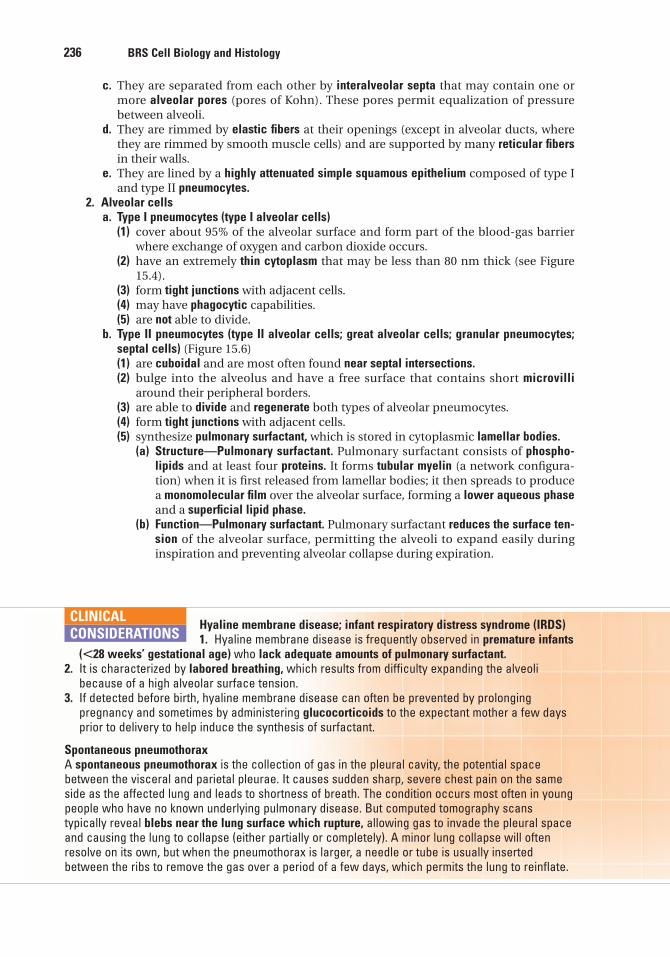
236 BRS Cell Biology and Histology
c. They are separated from each other by interalveolar septa that may contain one ormore alveolar pores (pores of Kohn). These pores permit equalization of pressurebetween alveoli.
d. They are rimmed by elastic fibers at their openings (except in alveolar ducts, wherethey are rimmed by smooth muscle cells) and are supported by many reticular fibersin their walls.
e. They are lined by a highly attenuated simple squamous epithelium composed of type Iand type II pneumocytes.
2. Alveolar cellsa. Type I pneumocytes (type I alveolar cells)
(1) cover about 95% of the alveolar surface and form part of the blood-gas barrierwhere exchange of oxygen and carbon dioxide occurs.
(2) have an extremely thin cytoplasm that may be less than 80 nm thick (see Figure15.4).
(3) form tight junctions with adjacent cells.(4) may have phagocytic capabilities.(5) are not able to divide.
b. Type II pneumocytes (type II alveolar cells; great alveolar cells; granular pneumocytes;septal cells) (Figure 15.6)(1) are cuboidal and are most often found near septal intersections.(2) bulge into the alveolus and have a free surface that contains short microvilli
around their peripheral borders.(3) are able to divide and regenerate both types of alveolar pneumocytes.(4) form tight junctions with adjacent cells.(5) synthesize pulmonary surfactant, which is stored in cytoplasmic lamellar bodies.
(a) Structure—Pulmonary surfactant. Pulmonary surfactant consists of phospho-lipids and at least four proteins. It forms tubular myelin (a network configura-tion) when it is first released from lamellar bodies; it then spreads to producea monomolecular film over the alveolar surface, forming a lower aqueous phaseand a superficial lipid phase.
(b) Function—Pulmonary surfactant. Pulmonary surfactant reduces the surface ten-sion of the alveolar surface, permitting the alveoli to expand easily duringinspiration and preventing alveolar collapse during expiration.
Hyaline membrane disease; infant respiratory distress syndrome (IRDS)1. Hyaline membrane disease is frequently observed in premature infants
(�28 weeks’ gestational age) who lack adequate amounts of pulmonary surfactant.2. It is characterized by labored breathing, which results from difficulty expanding the alveoli
because of a high alveolar surface tension.3. If detected before birth, hyaline membrane disease can often be prevented by prolonging
pregnancy and sometimes by administering glucocorticoids to the expectant mother a few daysprior to delivery to help induce the synthesis of surfactant.
Spontaneous pneumothoraxA spontaneous pneumothorax is the collection of gas in the pleural cavity, the potential spacebetween the visceral and parietal pleurae. It causes sudden sharp, severe chest pain on the sameside as the affected lung and leads to shortness of breath. The condition occurs most often in youngpeople who have no known underlying pulmonary disease. But computed tomography scanstypically reveal blebs near the lung surface which rupture, allowing gas to invade the pleural spaceand causing the lung to collapse (either partially or completely). A minor lung collapse will oftenresolve on its own, but when the pneumothorax is larger, a needle or tube is usually insertedbetween the ribs to remove the gas over a period of a few days, which permits the lung to reinflate.
CLINICAL
CONSIDERATIONS
LWBK615-c15[228-243].qxd 05/31/2010 10:54 AM Page 236 Aptara
Chapter 15 Respiratory System 237
c. Alveolar macrophages (alveolar phagocytes; dust cells) (see Figure 15.3)(1) are the principal mononuclear phagocytes of the alveolar surface.(2) remove inhaled dust, bacteria, and other particulate matter trapped in the pul-
monary surfactant, thus providing a vital line of defense in the lungs.(3) migrate to the bronchioles after filling with debris. From there, they are carried via
ciliary action to the upper airways, eventually reaching the oropharynx, wherethey are either swallowed or expectorated.
(4) may also exit by migrating into the interstitium and leaving via lymphatic vessels.
Asbestosis1. Asbestosis is a pulmonary disease caused by inhaling asbestos fibers
(used in insulation materials, tiles, etc.).2. The fibers deposit in the alveolar ducts and alveoli. The smaller fibers are phagocytosed by
macrophages, but the larger ones penetrate the lung interstitium.3. Activated macrophages release inflammatory mediators, which lead to interstitial pulmonary
fibrosis in the walls of respiratory bronchioles, alveolar ducts, and alveoli.4. Asbestos bodies, fibers 10 to 50 µm long encrusted with beads of protein, form in the interstitium
and alveolar spaces. The asbestos bodies stain strongly for iron because of the hemoglobin pro-tein released from small hemorrhages that accompany the fibrosis.
CLINICAL
CONSIDERATIONS
LB
A
LB
FIGURE 15.6. Electron micrograph of a type II pneumocyte that synthesizes surfactant and storesit in lamellar bodies (LB) in its cytoplasm. Type II pneumocytes are present mainly near the septalintersections and line only small portions of the alveoli (A). They possess microvilli (arrowhead )and are cuboidal in shape (�7,000).
LWBK615-c15[228-243].qxd 05/31/2010 10:54 AM Page 237 Aptara
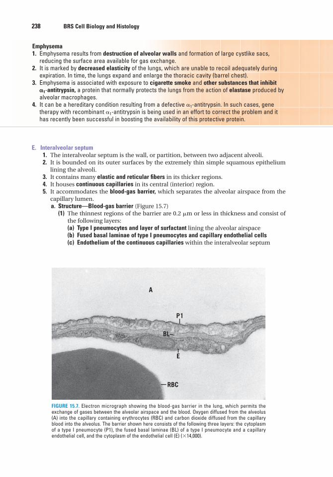
238 BRS Cell Biology and Histology
E. Interalveolar septum1. The interalveolar septum is the wall, or partition, between two adjacent alveoli.2. It is bounded on its outer surfaces by the extremely thin simple squamous epithelium
lining the alveoli.3. It contains many elastic and reticular fibers in its thicker regions.4. It houses continuous capillaries in its central (interior) region.5. It accommodates the blood-gas barrier, which separates the alveolar airspace from the
capillary lumen.a. Structure—Blood-gas barrier (Figure 15.7)
(1) The thinnest regions of the barrier are 0.2 �m or less in thickness and consist ofthe following layers:(a) Type I pneumocytes and layer of surfactant lining the alveolar airspace(b) Fused basal laminae of type I pneumocytes and capillary endothelial cells(c) Endothelium of the continuous capillaries within the interalveolar septum
Emphysema1. Emphysema results from destruction of alveolar walls and formation of large cystlike sacs,
reducing the surface area available for gas exchange.2. It is marked by decreased elasticity of the lungs, which are unable to recoil adequately during
expiration. In time, the lungs expand and enlarge the thoracic cavity (barrel chest).3. Emphysema is associated with exposure to cigarette smoke and other substances that inhibit
�1-antitrypsin, a protein that normally protects the lungs from the action of elastase produced byalveolar macrophages.
4. It can be a hereditary condition resulting from a defective �1-antitrypsin. In such cases, genetherapy with recombinant �1-antitrypsin is being used in an effort to correct the problem and ithas recently been successful in boosting the availability of this protective protein.
A
P1
E
BL
RBC
FIGURE 15.7. Electron micrograph showing the blood-gas barrier in the lung, which permits theexchange of gases between the alveolar airspace and the blood. Oxygen diffused from the alveolus(A) into the capillary containing erythrocytes (RBC) and carbon dioxide diffused from the capillaryblood into the alveolus. The barrier shown here consists of the following three layers: the cytoplasmof a type I pneumocyte (P1), the fused basal laminae (BL) of a type I pneumocyte and a capillaryendothelial cell, and the cytoplasm of the endothelial cell (E) (�14,000).
LWBK615-c15[228-243].qxd 05/31/2010 10:54 AM Page 238 Aptara
Chapter 15 Respiratory System 239
(2) Thicker regions of the barrier measure as much as 0.5 �m across and have an inter-stitial area interposed between the two unfused basal laminae.
b. Function—Blood-gas barrier. The blood-gas barrier permits the diffusion of gasesbetween the alveolar airspace and the blood. Oxygen passes from the alveolus into thecapillary, and carbon dioxide passes from the capillary blood into the alveolus.
Carbon monoxide poisoning1. Carbon monoxide is an odorless, tasteless gas that binds to hemoglobin
in red blood cells with a greater affinity than does oxygen. It is produced whenever fuel-burningappliances are used, which explains the importance of using carbon monoxide detectors.
2. Individuals exposed to carbon monoxide are often unaware of the symptoms it may cause (suchas nausea, headache, and sleepiness). Death may occur as the gas replaces oxygen in red bloodcells of the lung and blocks the delivery of oxygen to tissues of the body.
3. Treatment consists of exposing the patient to 100% oxygen (sometimes in a hyperbaric chamber)until oxygen displaces the carbon monoxide and recovery occurs.
CLINICAL
CONSIDERATIONS
IV. LUNG LOBULES
A. Lung lobules vary greatly in size and shape, but each has an apex directed toward the pul-monary hilum and a wider base directed outward.
B. Each lobule contains a single primary bronchiole, which enters at the apex and branches toform five to seven terminal bronchioles. The terminal bronchioles in turn divide, ultimatelygiving rise to alveoli at the base of the lobule.
V. PULMONARY VASCULAR SUPPLY
A. Pulmonary artery1. The pulmonary artery carries blood to the lungs to be oxygenated.2. It enters the root of each lung and extends branches along the divisions of the bronchial
tree.3. It enters lung lobules, where its branches follow the bronchioles (see Figure 15.2).
B. Pulmonary veins1. In lung lobules, pulmonary veins run in the intersegmental connective tissue, separated
from the arteries.2. After leaving the lobules, the pulmonary veins come close to divisions of the bronchial
tree and run parallel to branches of the pulmonary artery as they accompany bronchi to theroot of the lung.
C. Bronchial arteries and veins1. Bronchial arteries and veins provide nutrients to and remove wastes from the nonrespi-
ratory portions of the lung (bronchi, bronchioles, interstitium, and pleura).2. They follow the branching pattern of the bronchial tree and form anastomoses with the
pulmonary vessels near capillary beds.
LWBK615-c15[228-243].qxd 05/31/2010 10:54 AM Page 239 Aptara
240 BRS Cell Biology and Histology
VI. PULMONARY NERVE SUPPLY
The pulmonary nerve supply consists primarily of autonomic fibers to the smooth muscle ofbronchi and bronchioles. Axons are also present in the thicker parts of the interalveolar septa.
A. Parasympathetic stimulation causes contraction of pulmonary smooth muscle.
B. Sympathetic stimulation causes relaxation of pulmonary smooth muscle and can be mim-icked by certain drugs that cause dilation of bronchi and bronchioles.
LWBK615-c15[228-243].qxd 05/31/2010 10:54 AM Page 240 Aptara
Review Test
241
1. Characteristics of olfactory epitheliuminclude which one of the following?
(A) It is located in the inferior region of thenasal cavity.
(B) It is classified as simple columnar.(C) It has an underlying lamina propria con-
taining mucous glands.(D) It has modified cilia, which act as recep-
tors for odor.(E) It is unable to regenerate.
2. Which of the following statementsconcerning terminal bronchioles is true?
(A) They are part of the conducting portionof the respiratory system.
(B) They function in gas exchange.(C) They do not contain ciliated cells.(D) They have cartilage plates present in
their walls.(E) They do not contain secretory cells.
3. The trachea possesses which one of thefollowing components?
(A) Irregular cartilage plates in its wall(B) Skeletal muscle in its wall(C) An epithelium containing only two cell
types(D) A thick basement membrane underlying
its epithelium(E) Bowman glands in its lamina propria
4. Which of the following statementsconcerning respiratory bronchioles is true?
(A) No gas exchange occurs in them.(B) They do not have alveoli forming part of
their wall.(C) They contain goblet cells in their lining
epithelium.(D) They are included in the conducting
portion of the respiratory system.(E) Ciliated cells comprise a portion of their
lining epithelium.
5. True statements about asthma includewhich one of the following?
(A) It is due to a loss of lung elasticity.(B) It eventually causes the lungs to expand
and leads to a barrel chest.(C) It is associated with difficulty expiring
air from the lungs.(D) It may be helped by gene therapy using
recombinant �1-antitrypsin.(E) It is usually not associated with inflam-
mation.
6. Which of the following statementsconcerning alveolar macrophages is true?
(A) They secrete �1-antitrypsin.(B) They secrete elastase.(C) They originate from blood neutrophils.(D) They may play a role in causing hyaline
membrane disease.(E) They secrete small amounts of
surfactant.
7. Which one of the following disorders mayin some cases be successfully treated withantielastase (�1-antitrypsin)?
(A) Asbestosis(B) Asthma(C) Carbon monoxide poisoning(D) Emphysema(E) Hyaline membrane disease
8. Which one of the following ischaracterized by interstitial pulmonaryfibrosis?
(A) Asbestosis(B) Asthma(C) Carbon monoxide poisoning(D) Emphysema(E) Hyaline membrane disease
Directions: Each of the numbered items or incomplete statements in this section is followed byanswers or completions of the statement. Select the ONE lettered answer that is BEST in eachcase.
LWBK615-c15[228-243].qxd 05/31/2010 10:54 AM Page 241 Aptara

242 BRS Cell Biology and Histology
9. Which one of the following is associatedwith a barrel chest?
(A) Asbestosis(B) Asthma(C) Carbon monoxide poisoning(D) Emphysema(E) Hyaline membrane disease
10. Which one of the following is frequentlytreated successfully with glucocorticoids?
(A) Asbestosis(B) Asthma(C) Carbon monoxide poisoning(D) Emphysema(E) Hyaline membrane disease
LWBK615-c15[228-243].qxd 05/31/2010 10:54 AM Page 242 Aptara
Answers and Explanations
1. D. The olfactory epithelium possesses nonmotile cilia, which act as receptors for odor.They are extensions of the bipolar nerve cells that form part of this tall, pseudostratifiedepithelium located in the roof of the nasal cavity. Bowman glands, which lie in the laminapropria beneath this epithelium, produce a watery secretion, which moistens the olfactorysurface (see Chapter 15 II A).
2. A. Terminal bronchioles are the most distal components of the conducting portion of therespiratory system. They lack alveoli and thus do not function in gas exchange. They arelined by an epithelium composed of two cell types: secretory (Clara) cells and ciliatedcells. Cartilage is not present in bronchioles (see Chapter 15 II F).
3. D. The pseudostratified ciliated columnar epithelium lining the trachea rests on a thickbasement membrane and contains five cell types. The trachea possesses C-shapedcartilages with smooth muscle (the trachealis) extending between their ends. Bowmanglands are found only in the nasal cavity and produce a thin watery secretion (see Chapter 15 II D).
4. E. Respiratory bronchioles have alveoli interrupting their walls, so some gas exchangetakes place at this level. Their remaining walls are lined by a simple cuboidal epitheliumconsisting of Clara cells and ciliated cells. Respiratory bronchioles are categorized as partof the respiratory portion of the system (see Chapter 15 III A).
5. C. Asthma results from the constriction of smooth muscle in the bronchioles, whichdecreases their diameter and makes the expiration of air very difficult. Mucus accumulatesin the airways, and inflammatory cells invade the bronchiolar walls (see Chapter 15 II FClinical Considerations).
6. B. Alveolar macrophages secrete elastase. Normally, �1-antitrypsin, a serum protein, inter-acts with elastase, thereby protecting the lung against damage that may lead toemphysema. Alveolar macrophages, like all macrophages, arise from blood monocytes;they do not secrete surfactant; and they are unrelated to the pathogenesis of hyalinemembrane disease (see Chapter 15 III D Clinical Considerations).
7. D. Hereditary forms of emphysema are now being treated with recombinant �1-antitrypsin,which has antielastase activity (see Chapter 15 III D Clinical Considerations).
8. A. Inhaling asbestos fibers causes interstitial pulmonary fibrosis in the walls of respiratorybronchioles, alveolar ducts, and alveoli. Asbestos bodies are a classic feature of asbestosis.After prolonged heavy exposure to asbestos, this disease may progress to mesothelioma(a malignant tumor) (see Chapter 15 III D Clinical Considerations).
9. D. A loss of lung elasticity in emphysema makes it difficult for the lungs to recoil normallyduring expiration. The lungs and thoracic cavity enlarge, producing a barrel chest (seeChapter 15 III D Clinical Considerations).
10. E. Glucocorticoids, which stimulate synthesis of pulmonary surfactant, are often adminis-tered to the expectant mother a few days prior to delivery to prevent or alleviate hyalinemembrane disease in the premature infant (see Chapter 15 III D Clinical Considerations).
243
LWBK615-c15[228-243].qxd 05/31/2010 10:54 AM Page 243 Aptara
c h a p t e r 16Digestive System: OralCavity and AlimentaryTract
244
I. OVERVIEW—THE DIGESTIVE SYSTEM
A. The digestive system comprises the oral region and alimentary canal (esophagus, stomach,and small and large intestines) and several extrinsic glands.
B. It consists of a hollow tube (highly modified in the oral cavity) of varying diameter, com-posed of a mucosa, submucosa, muscularis externa, and serosa (or adventitia).
C. Function. The digestive system secretes enzymes and hormones that function in ingestion,digestion, and absorption of nutrients and in the elimination of indigestible materials.
II. ORAL REGION
The oral region includes the lips, palate, teeth and associated structures, tongue, major salivaryglands, and lingual tonsil. It is lined in most places by a stratified squamous epithelium whoseepithelial ridges interdigitate with tall connective tissue papillae (connective tissue ridges) ofthe subjacent connective tissue. The epithelial ridges and the connective tissue ridges areknown collectively as the rete apparatus. The epithelium and connective tissue together formthe mucosa. The oral cavity has three types of mucosae: lining mucosa, whose epithelium isnonkeratinized; masticatory mucosa, whose epithelium is keratinized; and specialized mucosa,whose nonkeratinized stratified squamous epithelium possesses taste buds. The dorsal sur-face of the tongue, hard palate, and gingivae are the only regions that possess masticatorymucosa; the dorsum of the tongue, soft palate, and regions of the pharynx possess specializedmucosa; the remainder of the oral cavity is lined by lining mucosa.
A. The lips are divided into the external (skin) region, the vermilion zone, and the internal(mucosal) region. The first two regions are covered by stratified squamous keratinized epithe-lium, whereas the internal region is lined by a wet stratified squamous nonkeratinizedepithelium.1. A dense irregular connective tissue core envelops skeletal muscle.2. Sebaceous glands, sweat glands, and hair follicles are present in the external region; minor
salivary glands in the internal region; and occasional, nonfunctional sebaceous glands(known as Fordyce’s granules) in the internal region and vermilion zone.
B. The palate is divided into an anterior hard palate (possessing a bony shelf in its core) and aposterior soft palate (possessing skeletal muscle in its core). The palate separates the nasalcavity from the oral cavity. Therefore, the palate has a nasal aspect and an oral aspect. Theentire nasal aspect of the palate (with the exception of the uvula) is lined by pseudostratifiedciliated columnar epithelium (respiratory epithelium).
LWBK615-c16[244-261].qxd 05/31/2010 10:55 AM Page 244 Aptara
Chapter 16 Digestive System: Oral Cavity and Alimentary Tract 245
1. The hard palate is lined on its oral aspect by stratified squamous parakeratinized to strat-ified squamous keratinized epithelium. It contains adipose tissue anteriorly and minormucous salivary glands posteriorly in the oral aspect of its connective tissue.
2. The soft palate is lined on its oral aspect by stratified squamous nonkeratinized epithe-lium. It contains minor mucous salivary glands in the oral aspect of its connective tissue.
a. Herpetic stomatitis is caused by herpes simplex virus (HSV) type I. In the dormant state, this virus resides in the trigeminal ganglia. HSV type 1
infection is very common and is transmitted by kissing. This infection is characterized by painfulfever blisters on the lips or in the vicinity of the nostrils. These blisters exude a clear fluid or arecovered by a scab.
b. Angular cheilitis (perlèche) is a painful condition, usually in patients older than 50 years of age,in which the corners of the mouth have short erythematous fissures and cracks that are inflamedand may become infected with Candida albicans or with occasional secondary bacterialcolonization by Staphylococcus aureus. Licking the corner of the mouth exacerbates andprolongs the condition. Treatment is usually with an antifungal ointment and in case of a second-ary bacterial infection, an antibiotic regimen and behavioral modification, that is, having thepatient resist the urge to lick the corners of the mouth.
c. Cancers of the oral region most commonly affect the lips, tongue, and floor of the mouth. Thesecancers initially resemble leukoplakia and are asymptomatic. Survival rate is high if thesecancers are recognized and treated in the early stages.
CLINICAL
CONSIDERATIONS
C. Teeth1. Overview—Teeth
a. Teeth are composed of an internal soft tissue, the pulp, and three calcified tissues:enamel and cementum, which form the surface layer, and dentin, which lies betweenthe surface layer and pulp. As in bone, calcium hydroxyapatite is the mineral materialin the calcified dental tissues.
b. Teeth have an enamel-covered crown, a cementum-covered root, and a cervix, theregion where the two surface materials meet.
2. Components—Teetha. Enamel
(1) has a highly calcified matrix with an organic component that is composed mostlyof the fibrous keratinlike protein, enamelin, a substance that is elaborated byameloblasts during formation of the crown and becomes calcified by the deposi-tion of calcium-hydroxyapatite crystals. It consists of only 4% organic material and96% inorganic material and, thus, is the most calcified tissue in the body.
(2) is acellular after tooth eruption and therefore cannot repair itself.
The bacterial flora of the oral cavity acts on food remnants lodgedbetween teeth and in the gingival sulcus, the normally tiny crevice
between the gum and the enamel of the tooth. As the bacteria metabolize the sugars in the fooddebris, they produce lactic acid that can dissolve enough of the calcium-hydroxyapatite crystals toform carious lesions (tooth decay), unless the individual practices good oral hygiene. The use of flu-oride in the water supply (systemic application), as well as the addition of fluoride in toothpaste(topical application), makes enamel more resistant to bacterial acids by altering the hydroxyapatitecrystals (fluoride replaces hydroxyl molecules). Moreover, the presence of fluoride on the enamelsurface catalyzes its remineralization.
CLINICAL
CONSIDERATIONS
LWBK615-c16[244-261].qxd 05/31/2010 10:55 AM Page 245 Aptara
246 BRS Cell Biology and Histology
b. Dentin(1) surrounds the central pulp chamber of the crown and pulp (root) canal.(2) has a calcified matrix containing type I collagen fibers.(3) is manufactured by odontoblasts, which persist and continue to elaborate dentin
for the life of the toothc. Cementum
(1) has a type I collagen–containing calcified matrix, which is produced by cemento-blasts.
(2) is continuously elaborated even after tooth eruption, since it compensates for thedecrease in tooth length resulting from abrasion of the enamel.
d. Dental pulp(1) is a gelatinous, richly vascularized connective tissue containing odontoblasts in its
peripheral layer (closest to the dentin), fibroblasts and mesenchymal cells, andthin types I and III collagen fibers.
(2) contains afferent nerve fibers. All sensations from the pulp are interpreted as painin the central nervous system.
3. Crown formationa. The crown begins to form 6 to 7 weeks after conception as a horseshoe-shaped band,
the dental lamina, which is derived from the oral epithelium. A dental lamina developsin each jaw and projects into the underlying ectomesenchyme.
b. The crown forms before the root formation begins.c. The sequential stages of crown formation are bud, cap, bell, and appositional stages.
4. Root formation follows completion of the crown and is accompanied by tooth eruption.
D. Dental-supporting structures1. The periodontal ligament is composed of a dense irregular collagenous connective tissue
whose type I collagen fibers are arranged in five principal fiber bundles, which extendfrom cementum to bone (alveolar crest, horizontal, oblique, apical, and interradicularfiber groups), suspending the tooth in its alveolus.
2. Gingivae (gums) are covered by stratified squamous keratinized (or parakeratinized)epithelium; their collagen fibers are also arranged in five principal fiber bundles (alve-ologingival, dentogingival, circular, dentoperiosteal, and transseptal fiber groups).
3. Alveolar bone consists of an inner layer of compact bone (cribriform plate, or alveolar boneproper). There is also an outer layer (cortical plate) of compact bone with an interveninglayer of cancellous bone (spongiosa).
E. Tongue1. Overview—Tongue
a. The tongue is divided into an anterior two-thirds and a posterior one-third by the V-shaped sulcus terminalis, whose apex ends in the foramen cecum.
b. Its dorsal surface is covered by stratified squamous parakeratinized to keratinizedepithelium, whereas its ventral surface is covered by stratified squamous nonkera-tinized epithelium. Both epithelial surfaces are underlain by a lamina propria and sub-mucosa of dense irregular collagenous connective tissue.
c. The tongue possesses a core of skeletal muscle, which forms the bulk of the tongue.2. Lingual papillae are located on the dorsal surface of the anterior two-thirds of the tongue.
a. Filiform papillae are short, narrow, highly keratinized structures lacking taste buds.b. Fungiform papillae are mushroom-shaped structures interspersed among the filiform
papillae; they contain occasional taste buds.c. Foliate papillae are shallow longitudinal furrows on the lateral aspect of the posterior
region of the anterior two-thirds of the tongue. Their taste buds degenerate shortlyafter the second year of life.
d. Circumvallate papillae are 10 to 15 large circular papillae, each of which is surroundedby a moatlike furrow. They lie just anterior to the sulcus terminalis and possess tastebuds.
LWBK615-c16[244-261].qxd 05/31/2010 10:55 AM Page 246 Aptara
Chapter 16 Digestive System: Oral Cavity and Alimentary Tract 247
(1) Taste buds(a) Each taste bud is composed of 60 to 80 spindle-shaped cells that form a barrel-
shaped intraepithelial structure. These structures are located on the superiorsurface of fungiform papillae, on the lateral surfaces of circumvallate papillae,and in the walls of the surrounding moatlike furrows. Each taste bud has asmall opening, the taste pore, from which microvilli (taste hairs) project intothe oral cavity (or into the moat surrounding the circumvallate papilla).
(b) Function. Taste buds perceive salt, sour, bitter, sweet, and umami (glutamatereceptor that senses delicious flavors) taste sensations, and some, in individu-als who have the genetic capability, may specialize in tasting fats. The chemi-cal moieties of the food substances that are recognized by taste buds areknown as tastants.
(c) Four different cells may be recognized, three of which are spindle shaped: darkcells (type I cells), light cells (type II cells), and intermediate cells (type III cells)have short life spans; the fourth type are the short, regenerative, basal cells.Types I, II, and III cells form synapses with afferent nerve fibers that deliver thetaste information to the central nervous system.
(d) When basal cells divide, they give rise to a basal cell and a dark cell. As the darkcell matures, it becomes a light cell, and as it degenerates, it becomes an inter-mediate cell. These three cell types are neuroepithelial cells whose microvilli,the taste hairs, recognize one specific tastant. The process of going from anewly formed dark cell to a dead intermediate cell takes about 10 days. A par-ticular taste bud contains a variety of neuroepithelial cells that recognize alltastants, but each taste bud appears to be specialized to respond to only oneor two of the tastants.
(e) Salt and sour tastants stimulate ion channels directly; sweet, bitter, andumami tastants stimulate G protein–linked receptors; and fatty tastants stim-ulate fatty acid transporters.
It appears that the more molecules of a particular tastant bind to aneuroepithelial cell, the stronger the taste sensation. Recently, it has
been shown that the taste receptors also have binding sites for nontastant molecules that act asenhancer molecules. Therefore, when enhancer molecules bind along with tastants, the tastantsare locked in the receptor for a longer period of time. Thus, the tastant-enhancer molecule combina-tion augments the taste sensation so that the binding of only a few tastant molecules is interpretedas if many such tastants molecules were present.
The sense of taste is a complex interaction between the information that the brain receivesfrom the taste buds, from sensory cells that determine the temperature, spiciness, and texture of thefood, as well as from retronasal olfaction. The process of retronasal olfaction depends on olfactorycells located in the posterior aspect of the nasal cavity, and the odor that they perceive arises fromthe back of the mouth rather than the more commonly thought of orthonasal olfaction, where theodor is perceived via air entering the nasal cavity. An additional component of the perception oftaste is the visual information that the brain gleams via the sense of sight. An unusually coloredversion of a particular food morsel may elicit a sense of disgust, a perceived altered taste, and theinability to consume the food substance.
CLINICAL
CONSIDERATIONS
(2) Glands of von Ebner are minor salivary glands that deliver their serous secretion intothe furrow surrounding each papilla, assisting the taste buds in perceiving stimuli.These glands also deliver their saliva into the furrows of the foliate papillae.
3. The muscular core of the tongue is composed of bundles of skeletal muscle fibers arrangedin three planes with minor salivary glands interspersed among them.
4. A lingual tonsil is located on the dorsal surface of the posterior one-third of the tongue.
LWBK615-c16[244-261].qxd 05/31/2010 10:55 AM Page 247 Aptara
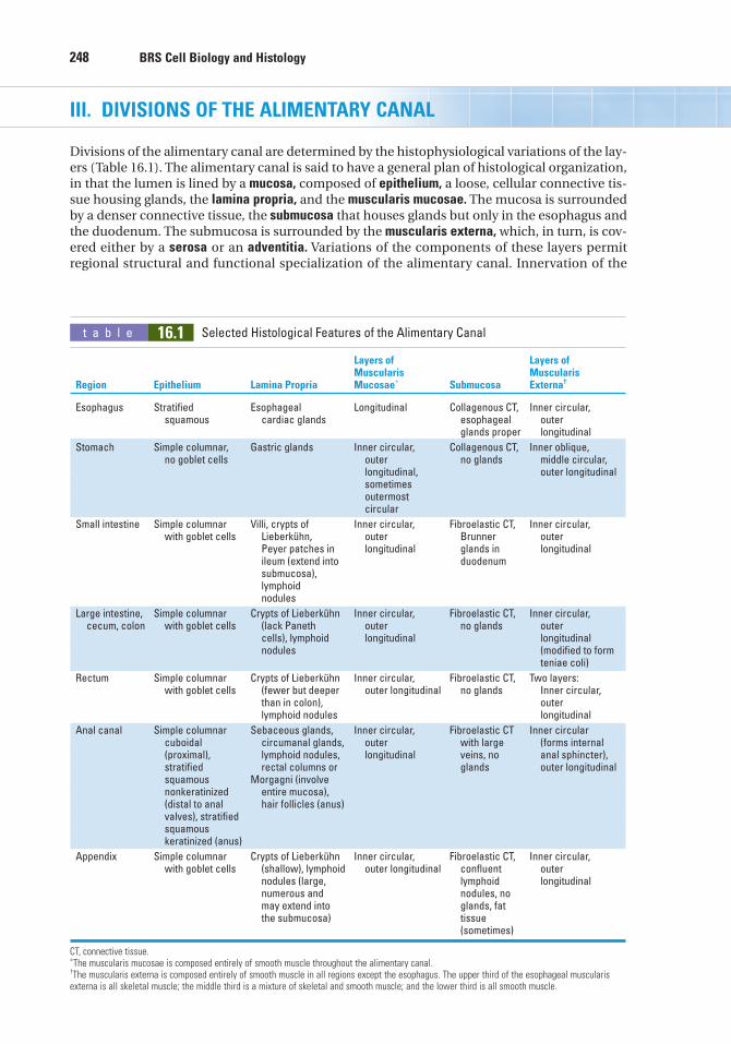
248 BRS Cell Biology and Histology
III. DIVISIONS OF THE ALIMENTARY CANAL
Divisions of the alimentary canal are determined by the histophysiological variations of the lay-ers (Table 16.1). The alimentary canal is said to have a general plan of histological organization,in that the lumen is lined by a mucosa, composed of epithelium, a loose, cellular connective tis-sue housing glands, the lamina propria, and the muscularis mucosae. The mucosa is surroundedby a denser connective tissue, the submucosa that houses glands but only in the esophagus andthe duodenum. The submucosa is surrounded by the muscularis externa, which, in turn, is cov-ered either by a serosa or an adventitia. Variations of the components of these layers permitregional structural and functional specialization of the alimentary canal. Innervation of the
t a b l e 16.1 Selected Histological Features of the Alimentary Canal
Layers of Layers ofMuscularis Muscularis
Region Epithelium Lamina Propria Mucosae∗ Submucosa Externa†
Esophagus Stratified Esophageal Longitudinal Collagenous CT, Inner circular, squamous cardiac glands esophageal outer
glands proper longitudinal
Stomach Simple columnar, Gastric glands Inner circular, Collagenous CT, Inner oblique,no goblet cells outer no glands middle circular,
longitudinal, outer longitudinalsometimes outermost circular
Small intestine Simple columnar Villi, crypts of Inner circular, Fibroelastic CT, Inner circular, with goblet cells Lieberkühn, outer Brunner outer
Peyer patches in longitudinal glands in longitudinalileum (extend into duodenumsubmucosa), lymphoid nodules
Large intestine, Simple columnar Crypts of Lieberkühn Inner circular, Fibroelastic CT, Inner circular, cecum, colon with goblet cells (lack Paneth outer no glands outer
cells), lymphoid longitudinal longitudinal nodules (modified to form
teniae coli)
Rectum Simple columnar Crypts of Lieberkühn Inner circular, Fibroelastic CT, Two layers:with goblet cells (fewer but deeper outer longitudinal no glands Inner circular,
than in colon), outer lymphoid nodules longitudinal
Anal canal Simple columnar Sebaceous glands, Inner circular, Fibroelastic CT Inner circular cuboidal circumanal glands, outer with large (forms internal(proximal), lymphoid nodules, longitudinal veins, no anal sphincter),stratified rectal columns or glands outer longitudinalsquamous Morgagni (involve nonkeratinized entire mucosa),(distal to anal hair follicles (anus)valves), stratified squamous keratinized (anus)
Appendix Simple columnar Crypts of Lieberkühn Inner circular, Fibroelastic CT, Inner circular, with goblet cells (shallow), lymphoid outer longitudinal confluent outer
nodules (large, lymphoid longitudinalnumerous and nodules, no may extend into glands, fat the submucosa) tissue
(sometimes)
CT, connective tissue.∗The muscularis mucosae is composed entirely of smooth muscle throughout the alimentary canal.†The muscularis externa is composed entirely of smooth muscle in all regions except the esophagus. The upper third of the esophageal muscularisexterna is all skeletal muscle; the middle third is a mixture of skeletal and smooth muscle; and the lower third is all smooth muscle.
LWBK615-c16[244-261].qxd 05/31/2010 10:55 AM Page 248 Aptara
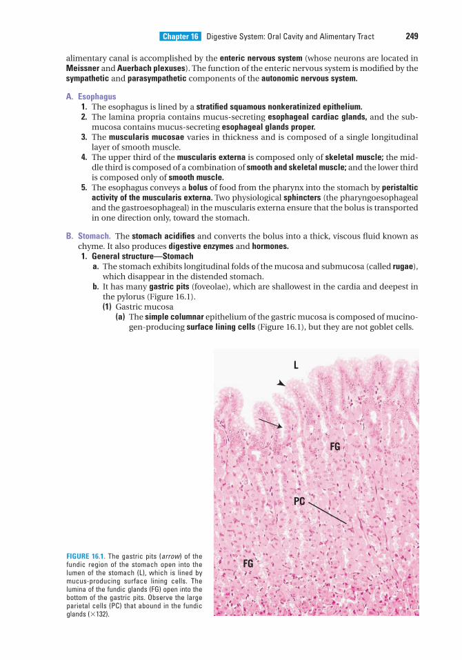
Chapter 16 Digestive System: Oral Cavity and Alimentary Tract 249
FIGURE 16.1. The gastric pits (arrow) of thefundic region of the stomach open into thelumen of the stomach (L), which is lined bymucus-producing surface lining cells. Thelumina of the fundic glands (FG) open into thebottom of the gastric pits. Observe the largeparietal cells (PC) that abound in the fundicglands (�132).
alimentary canal is accomplished by the enteric nervous system (whose neurons are located inMeissner and Auerbach plexuses). The function of the enteric nervous system is modified by thesympathetic and parasympathetic components of the autonomic nervous system.
A. Esophagus1. The esophagus is lined by a stratified squamous nonkeratinized epithelium.2. The lamina propria contains mucus-secreting esophageal cardiac glands, and the sub-
mucosa contains mucus-secreting esophageal glands proper.3. The muscularis mucosae varies in thickness and is composed of a single longitudinal
layer of smooth muscle.4. The upper third of the muscularis externa is composed only of skeletal muscle; the mid-
dle third is composed of a combination of smooth and skeletal muscle; and the lower thirdis composed only of smooth muscle.
5. The esophagus conveys a bolus of food from the pharynx into the stomach by peristalticactivity of the muscularis externa. Two physiological sphincters (the pharyngoesophagealand the gastroesophageal) in the muscularis externa ensure that the bolus is transportedin one direction only, toward the stomach.
B. Stomach. The stomach acidifies and converts the bolus into a thick, viscous fluid known aschyme. It also produces digestive enzymes and hormones.1. General structure—Stomach
a. The stomach exhibits longitudinal folds of the mucosa and submucosa (called rugae),which disappear in the distended stomach.
b. It has many gastric pits (foveolae), which are shallowest in the cardia and deepest inthe pylorus (Figure 16.1).(1) Gastric mucosa
(a) The simple columnar epithelium of the gastric mucosa is composed of mucino-gen-producing surface lining cells (Figure 16.1), but they are not goblet cells.
L
FG
FG
PC
LWBK615-c16[244-261].qxd 05/31/2010 10:55 AM Page 249 Aptara
250 BRS Cell Biology and Histology
(b) The lamina propria is a loose connective tissue housing smooth muscle cells,lymphocytes, plasma cells, mast cells, and fibroblasts. It contains gastric glands.
(c) The muscularis mucosae is composed of a poorly defined inner circular layer,an outer longitudinal layer, and occasionally an outermost circular layer ofsmooth muscle.
(2) Gastric submucosa(a) is composed of dense, irregular collagenous connective tissue.(b) contains fibroblasts, mast cells, and lymphoid elements embedded in the con-
nective tissue.(c) houses Meissner (submucosal) plexus.(d) possesses arterial and venous plexuses that respectively supply and drain the
vessels of the mucosa.(3) Gastric muscularis externa
(a) is composed of three layers of smooth muscle: an incomplete inner obliquelayer, a thick middle circular layer that forms the pyloric sphincter, and an outerlongitudinal layer. Auerbach myenteric plexus is located between the middlecircular and outer longitudinal smooth muscle layers.
(b) is responsible for mixing of gastric contents and emptying of the stomach.(c) is affected by various characteristics of the chyme (e.g., lipid content, viscos-
ity, osmolality, caloric density, and pH), which influence the emptying rate ofthe stomach and the rate of muscle contraction.
(4) A serosa covers the external surface of the stomach.2. Gastric glands are simple branched tubular glands in the lamina propria of the cardia,
fundus, and pylorus. Each gland consists of an isthmus, which connects the gland to thebase of a gastric pit; a neck; and a base.a. Cells of the fundic glands
(1) Parietal (oxyntic) cells (Figure 16.1)(a) These are pyramidal cells concentrated in the upper half of the gland.(b) They secrete hydrochloric acid (HCl) and gastric intrinsic factor. The latter is
necessary for absorption of vitamin B12 in the ileum.(c) They possess a unique intracellular tubulovesicular system, many mitochon-
dria, and secretory intracellular canaliculi (deep invaginations of the apicalplasma membrane) lined by microvilli.
(d) When the parietal cells are stimulated to secrete HCl, the number and lengthof microvilli increase and the complexity of the tubulovesicular systemdecreases (suggesting that tubulovesicle membranes are incorporated into theintracellular canaliculi, thus lengthening the microvilli).
(2) Chief (zymogenic) cells(a) are pyramidal cells residing in the lower half of the gland.(b) secrete pepsinogen (a precursor of the enzyme pepsin) and the precursors of
two other enzymes, rennin and lipase.(c) display an abundance of basal rough endoplasmic reticulum (RER), a supranu-
clear Golgi complex, and many apical zymogen (secretory) granules.(3) Mucous neck cells
(a) are located in the neck of the gland (and may be able to divide).(b) possess short microvilli, apical mucous granules, a prominent Golgi complex,
numerous mitochondria, and some basal RER.(4) Diffuse neuroendocrine system (DNES) cells
(a) are also referred to as enteroendocrine cells or as APUD cells (amine precursoruptake and decarboxylation cells).
(b) include more than a dozen types of cells that house many small hormone-containing granules, usually concentrated in the basal cytoplasm. Each par-ticular enteroendocrine cell is believed to secrete only one hormone (Table16.2).
(c) possess an abundance of mitochondria and RER and a moderately well-developed Golgi complex.
LWBK615-c16[244-261].qxd 05/31/2010 10:55 AM Page 250 Aptara
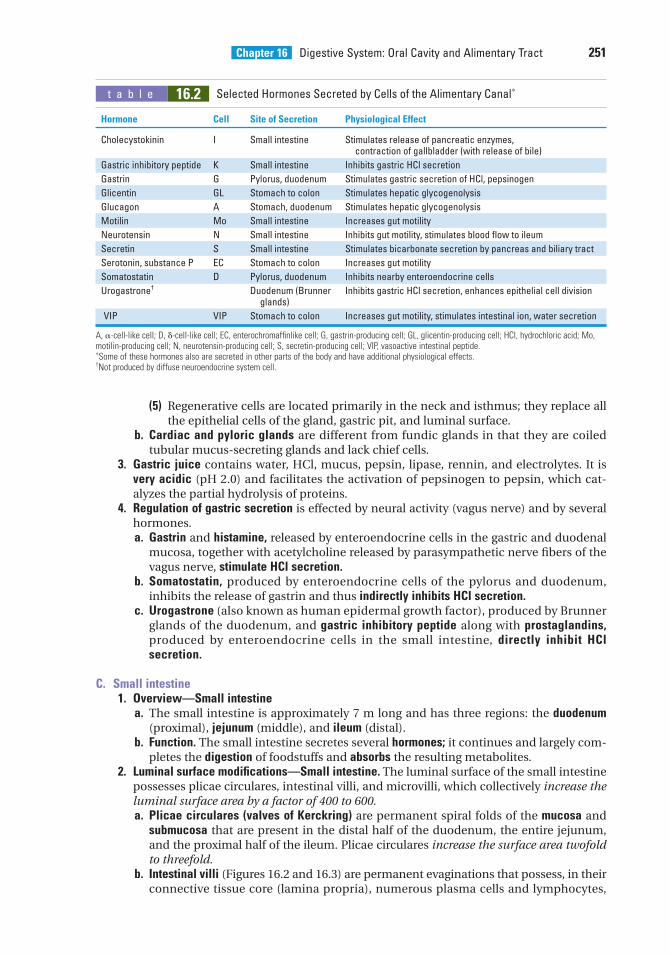
Chapter 16 Digestive System: Oral Cavity and Alimentary Tract 251
(5) Regenerative cells are located primarily in the neck and isthmus; they replace allthe epithelial cells of the gland, gastric pit, and luminal surface.
b. Cardiac and pyloric glands are different from fundic glands in that they are coiledtubular mucus-secreting glands and lack chief cells.
3. Gastric juice contains water, HCl, mucus, pepsin, lipase, rennin, and electrolytes. It isvery acidic (pH 2.0) and facilitates the activation of pepsinogen to pepsin, which cat-alyzes the partial hydrolysis of proteins.
4. Regulation of gastric secretion is effected by neural activity (vagus nerve) and by severalhormones.a. Gastrin and histamine, released by enteroendocrine cells in the gastric and duodenal
mucosa, together with acetylcholine released by parasympathetic nerve fibers of thevagus nerve, stimulate HCl secretion.
b. Somatostatin, produced by enteroendocrine cells of the pylorus and duodenum,inhibits the release of gastrin and thus indirectly inhibits HCl secretion.
c. Urogastrone (also known as human epidermal growth factor), produced by Brunnerglands of the duodenum, and gastric inhibitory peptide along with prostaglandins,produced by enteroendocrine cells in the small intestine, directly inhibit HClsecretion.
C. Small intestine1. Overview—Small intestine
a. The small intestine is approximately 7 m long and has three regions: the duodenum(proximal), jejunum (middle), and ileum (distal).
b. Function. The small intestine secretes several hormones; it continues and largely com-pletes the digestion of foodstuffs and absorbs the resulting metabolites.
2. Luminal surface modifications—Small intestine. The luminal surface of the small intestinepossesses plicae circulares, intestinal villi, and microvilli, which collectively increase theluminal surface area by a factor of 400 to 600.a. Plicae circulares (valves of Kerckring) are permanent spiral folds of the mucosa and
submucosa that are present in the distal half of the duodenum, the entire jejunum,and the proximal half of the ileum. Plicae circulares increase the surface area twofoldto threefold.
b. Intestinal villi (Figures 16.2 and 16.3) are permanent evaginations that possess, in theirconnective tissue core (lamina propria), numerous plasma cells and lymphocytes,
t a b l e 16.2 Selected Hormones Secreted by Cells of the Alimentary Canal∗
Hormone Cell Site of Secretion Physiological Effect
Cholecystokinin I Small intestine Stimulates release of pancreatic enzymes, contraction of gallbladder (with release of bile)
Gastric inhibitory peptide K Small intestine Inhibits gastric HCl secretion
Gastrin G Pylorus, duodenum Stimulates gastric secretion of HCl, pepsinogen
Glicentin GL Stomach to colon Stimulates hepatic glycogenolysis
Glucagon A Stomach, duodenum Stimulates hepatic glycogenolysis
Motilin Mo Small intestine Increases gut motility
Neurotensin N Small intestine Inhibits gut motility, stimulates blood flow to ileum
Secretin S Small intestine Stimulates bicarbonate secretion by pancreas and biliary tract
Serotonin, substance P EC Stomach to colon Increases gut motility
Somatostatin D Pylorus, duodenum Inhibits nearby enteroendocrine cells
Urogastrone† Duodenum (Brunner Inhibits gastric HCl secretion, enhances epithelial cell divisionglands)
VIP VIP Stomach to colon Increases gut motility, stimulates intestinal ion, water secretion
A, �-cell-like cell; D, �-cell-like cell; EC, enterochromaffinlike cell; G, gastrin-producing cell; GL, glicentin-producing cell; HCl, hydrochloric acid; Mo,motilin-producing cell; N, neurotensin-producing cell; S, secretin-producing cell; VIP, vasoactive intestinal peptide.∗Some of these hormones also are secreted in other parts of the body and have additional physiological effects.†Not produced by diffuse neuroendocrine system cell.
LWBK615-c16[244-261].qxd 05/31/2010 10:55 AM Page 251 Aptara
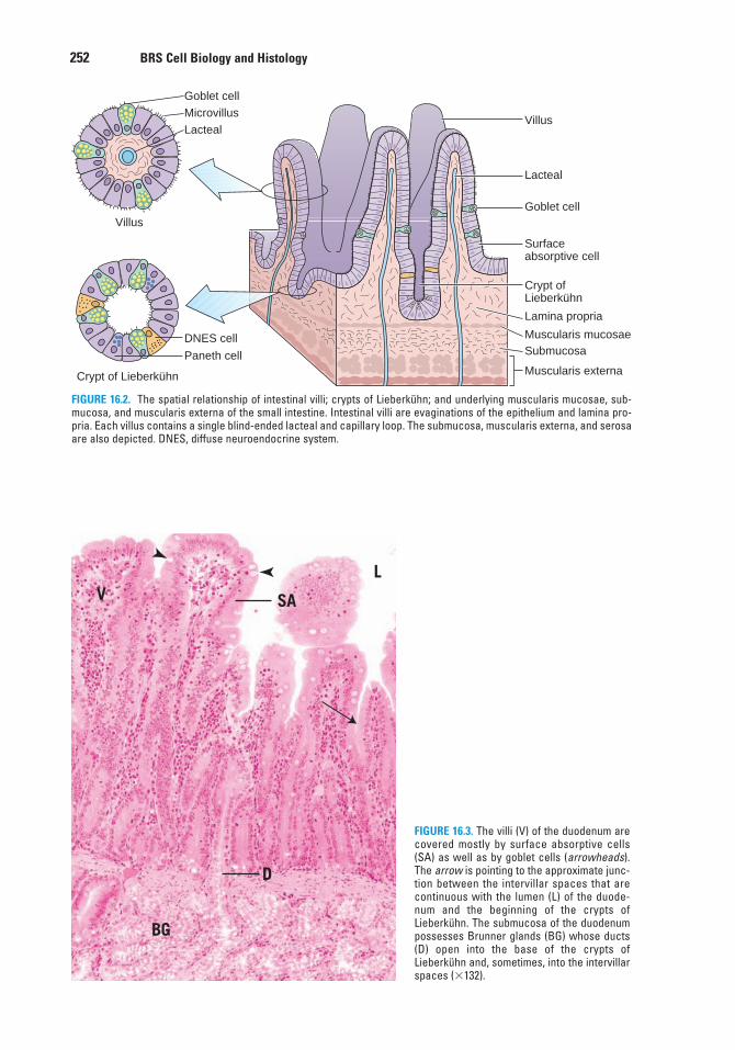
252 BRS Cell Biology and Histology
Goblet cellMicrovillusLacteal
Goblet cell
Crypt ofLieberkühn
Lamina propria
Muscularis mucosaeSubmucosa
Lacteal
Villus
Surfaceabsorptive cell
Crypt of Lieberkühn
Villus
Paneth cell
DNES cell
Muscularis externa
FIGURE 16.2. The spatial relationship of intestinal villi; crypts of Lieberkühn; and underlying muscularis mucosae, sub-mucosa, and muscularis externa of the small intestine. Intestinal villi are evaginations of the epithelium and lamina pro-pria. Each villus contains a single blind-ended lacteal and capillary loop. The submucosa, muscularis externa, and serosaare also depicted. DNES, diffuse neuroendocrine system.
LV
BG
D
SA
FIGURE 16.3. The villi (V) of the duodenum arecovered mostly by surface absorptive cells(SA) as well as by goblet cells (arrowheads).The arrow is pointing to the approximate junc-tion between the intervillar spaces that arecontinuous with the lumen (L) of the duode-num and the beginning of the crypts ofLieberkühn. The submucosa of the duodenumpossesses Brunner glands (BG) whose ducts(D) open into the base of the crypts ofLieberkühn and, sometimes, into the intervillarspaces (�132).
LWBK615-c16[244-261].qxd 05/31/2010 10:55 AM Page 252 Aptara
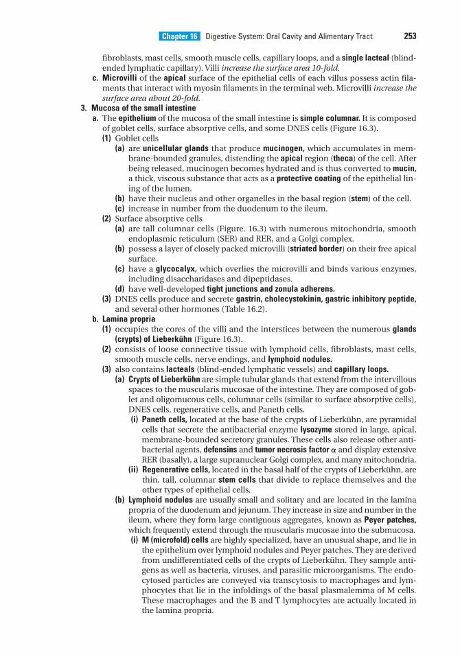
Chapter 16 Digestive System: Oral Cavity and Alimentary Tract 253
fibroblasts, mast cells, smooth muscle cells, capillary loops, and a single lacteal (blind-ended lymphatic capillary). Villi increase the surface area 10-fold.
c. Microvilli of the apical surface of the epithelial cells of each villus possess actin fila-ments that interact with myosin filaments in the terminal web. Microvilli increase thesurface area about 20-fold.
3. Mucosa of the small intestinea. The epithelium of the mucosa of the small intestine is simple columnar. It is composed
of goblet cells, surface absorptive cells, and some DNES cells (Figure 16.3).(1) Goblet cells
(a) are unicellular glands that produce mucinogen, which accumulates in mem-brane-bounded granules, distending the apical region (theca) of the cell. Afterbeing released, mucinogen becomes hydrated and is thus converted to mucin,a thick, viscous substance that acts as a protective coating of the epithelial lin-ing of the lumen.
(b) have their nucleus and other organelles in the basal region (stem) of the cell.(c) increase in number from the duodenum to the ileum.
(2) Surface absorptive cells(a) are tall columnar cells (Figure. 16.3) with numerous mitochondria, smooth
endoplasmic reticulum (SER) and RER, and a Golgi complex.(b) possess a layer of closely packed microvilli (striated border) on their free apical
surface.(c) have a glycocalyx, which overlies the microvilli and binds various enzymes,
including disaccharidases and dipeptidases.(d) have well-developed tight junctions and zonula adherens.
(3) DNES cells produce and secrete gastrin, cholecystokinin, gastric inhibitory peptide,and several other hormones (Table 16.2).
b. Lamina propria(1) occupies the cores of the villi and the interstices between the numerous glands
(crypts) of Lieberkühn (Figure 16.3).(2) consists of loose connective tissue with lymphoid cells, fibroblasts, mast cells,
smooth muscle cells, nerve endings, and lymphoid nodules.(3) also contains lacteals (blind-ended lymphatic vessels) and capillary loops.
(a) Crypts of Lieberkühn are simple tubular glands that extend from the intervillousspaces to the muscularis mucosae of the intestine. They are composed of gob-let and oligomucous cells, columnar cells (similar to surface absorptive cells),DNES cells, regenerative cells, and Paneth cells.(i) Paneth cells, located at the base of the crypts of Lieberkühn, are pyramidal
cells that secrete the antibacterial enzyme lysozyme stored in large, apical,membrane-bounded secretory granules. These cells also release other anti-bacterial agents, defensins and tumor necrosis factor � and display extensiveRER (basally), a large supranuclear Golgi complex, and many mitochondria.
(ii) Regenerative cells, located in the basal half of the crypts of Lieberkühn, arethin, tall, columnar stem cells that divide to replace themselves and theother types of epithelial cells.
(b) Lymphoid nodules are usually small and solitary and are located in the laminapropria of the duodenum and jejunum. They increase in size and number in theileum, where they form large contiguous aggregates, known as Peyer patches,which frequently extend through the muscularis mucosae into the submucosa.(i) M (microfold) cells are highly specialized, have an unusual shape, and lie in
the epithelium over lymphoid nodules and Peyer patches. They are derivedfrom undifferentiated cells of the crypts of Lieberkühn. They sample anti-gens as well as bacteria, viruses, and parasitic microorganisms. The endo-cytosed particles are conveyed via transcytosis to macrophages and lym-phocytes that lie in the infoldings of the basal plasmalemma of M cells.These macrophages and the B and T lymphocytes are actually located inthe lamina propria.
LWBK615-c16[244-261].qxd 05/31/2010 10:55 AM Page 253 Aptara
254 BRS Cell Biology and Histology
(ii) Activated B lymphocytes respond to antigenic challenge by forming moreB cells, which enter the lymph and blood circulation, then home back totheir original locations, where they populate the lamina propria and dif-ferentiate into immunoglobulin A (IgA) producing plasma cells.
(iii) Plasma cells manufacture IgA, some of which is taken up by and bound tosecretory protein within the epithelial cells and is transported across theintestinal epithelium (transcytosis) to the glycocalyx, where it remains asan immunological defense against bacteria and antigens in the lumen.Much of the IgA enters blood vessels and goes to the liver, where it ispicked up by hepatocytes to be secreted as part of bile. From the liver, IgAenters the gallbladder to be released into the lumen of the duodenum.
c. The muscularis mucosae is composed of an inner circular and an outer longitudinallayer of smooth muscle.
It is interesting to note that the immune system of the alimentary canal,under normal conditions, is tolerant of the normal bacterial flora and of
epithelial cells lining the gut that are in close contact with bacteria. It has been shown that this tol-erance is due to dendritic cells that inhibit T cells from initiating and immune response. However, ifthe conditions become inimical to health, as in an inflammatory reaction, the dendritic cells instructthe T cells to respond and elicit an immune reaction. In order to assist the dendritic cells in theirfunction of dictating tolerance, stromal cells of lymph nodes present proteins produced by cells ofthe alimentary canal to naïve T cells, instructing them to be tolerant of those proteins unlessinstructed otherwise by dendritic cells.
CLINICAL
CONSIDERATIONS
4. Submucosa of the small intestinea. consists of fibroelastic connective tissue containing blood and lymphatic vessels,
nerve fibers, and Meissner plexus.b. also houses Brunner glands (Figure 16.3), which are present only in the duodenum.
These glands produce an alkaline fluid and urogastrone. The former protects the duo-denal epithelium from the acidic chyme; the latter is a polypeptide hormone (humanepidermal growth factor) that enhances epithelial cell division and inhibits gastric HClproduction.
5. The muscularis externa of the small intestine is composed of two layers of smooth muscle:an inner circular and an outer longitudinal layer. The inner layer participates in the for-mation of the ileocecal sphincter. Auerbach (myenteric) plexus is housed between the twolayers.
6. External layer of the small intestinea. Serosa covers all of the jejunum and ileum and part of the duodenum.b. Adventitia covers the remainder of the duodenum.
D. Large intestine1. Overview—Large intestine
a. The large intestine consists of the cecum, colon (ascending, transverse, descending,and sigmoid), rectum, anal canal, and appendix.
b. The large intestine contains some digestive enzymes received from the small intes-tine.
c. It houses bacteria that produce vitamin B12 and vitamin K; the former is necessary forhemopoiesis and the latter for coagulation.
d. The large intestine produces abundant mucus, which lubricates its lining and facili-tates the passage and elimination of feces.
e. Function. The large intestine functions primarily in the absorption of electrolytes,fluids, and gases. Dead bacteria and indigestible remnants of the ingested materialare compacted into feces.
LWBK615-c16[244-261].qxd 05/31/2010 10:55 AM Page 254 Aptara
Chapter 16 Digestive System: Oral Cavity and Alimentary Tract 255
2. Cecum and colona. The mucosa of the cecum and colon lacks villi and possesses no specialized folds.
(1) The epithelium of the mucosa of the cecum and colon is simple columnar withnumerous goblet cells, surface absorptive cells, and occasional DNES cells.
(2) The lamina propria is similar to that of the small intestine, possessing lymphoidnodules, blood and lymph vessels, and closely packed crypts of Lieberkühn,which lack Paneth cells.
(3) The muscularis mucosae consists of an inner circular and outer longitudinal layerof smooth muscle cells.
b. The submucosa of the cecum and colon is composed of fibroelastic connective tissue. Itcontains blood and lymphatic vessels, nerves, and Meissner (submucosal) plexus.
c. The muscularis externa of the cecum and colon is composed of an inner circular and amodified outer longitudinal layer of smooth muscle. The outer layer is gathered intothree flat, longitudinal ribbons of smooth muscle that form the teniae coli. When con-tinuously contracted, the teniae coli form sacculations of the wall known as haustracoli. Auerbach (myenteric) plexus is housed between the two layers of smooth muscle.
d. External layer of the cecum and colon(1) Adventitia covers the ascending and descending portions of the colon.(2) Serosa covers the cecum and the remainder of the colon. Fat-filled outpocketings
of the serosa (appendices epiploicae) are characteristic of the transverse andsigmoid colon.
a. Malabsorption disorders may lead to malnutrition, resulting in wastingdiseases, if major nutrients (carbohydrates, amino acids, ions) cannotbe assimilated.
(i) Gluten enteropathy (nontropical sprue) results from the destructive effects of certain glutens(particularly of rye and wheat) on the intestinal villi, which reduce the surface area availablefor absorption. It is treated by eliminating wheat and rye products from the diet.
(ii) Malabsorption of vitamin B12 may cause pernicious anemia. It usually results frominadequate production of gastric intrinsic factor by the parietal cells of the gastric mucosa.
b. Cholera-induced diarrhea is caused by the action of cholera toxin, which blocks intestinalabsorption of sodium ions and promotes excretion of water and electrolytes. It causes deathshortly after onset unless the lost electrolytes and water are replaced.
CLINICAL
CONSIDERATIONS
3. The rectum is similar to the colon but contains fewer and deeper crypts of Lieberkühn(Table 16.1).
4. The anal canal is the constricted continuation of the rectum.a. The anal mucosa displays longitudinal folds called anal columns (or rectal columns of
Morgagni), which join each other to form anal valves. The regions between adjacentvalves are known as anal sinuses.(1) Epithelium of the anal canal
(a) is simple columnar changing to simple cuboidal proximal to the anal valves.(b) is stratified squamous nonkeratinized distal to the anal valves.(c) changes to stratified squamous keratinized (epidermis) at the anus.
(2) The lamina propria is composed of fibroelastic connective tissue and containssebaceous glands, circumanal glands, hair follicles, and large veins.
(3) The muscularis mucosae consists of an inner circular and an outer longitudinallayer of smooth muscle, both of which terminate at the anal valves.
b. The anal submucosa is composed of dense, irregular fibroelastic connective tissue thathouses large veins.
c. The anal muscularis externa is composed of an inner circular and an outer longitudi-nal layer of smooth muscle. The inner circular layer forms the internal anal sphincter.
d. Anal adventitia attaches the anus to surrounding structures.
LWBK615-c16[244-261].qxd 05/31/2010 10:56 AM Page 255 Aptara
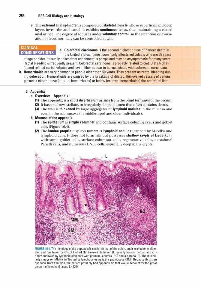
256 BRS Cell Biology and Histology
e. The external anal sphincter is composed of skeletal muscle whose superficial and deeplayers invest the anal canal. It exhibits continuous tonus, thus maintaining a closedanal orifice. The degree of tonus is under voluntary control, so the retention or evacu-ation of feces normally can be controlled at will.
a. Colorectal carcinoma is the second highest cause of cancer death in the United States. It most commonly affects individuals who are 55 years
of age or older. It usually arises from adenomatous polyps and may be asymptomatic for many years.Rectal bleeding is frequently present. Colorectal carcinoma is probably related to diet. Diets high infat and refined carbohydrates and low in fiber appear to be associated with colorectal carcinoma.
b. Hemorrhoids are very common in people older than 50 years. They present as rectal bleeding dur-ing defecation. Hemorrhoids are caused by the breakage of dilated, thin-walled vessels of venousplexuses either above (internal hemorrhoids) or below (external hemorrhoids) the anorectal line.
CLINICAL
CONSIDERATIONS
L
GC
C
MM
SM
FIGURE 16.4. The histology of the appendix is similar to that of the colon, but it is smaller in diam-eter and has fewer crypts of Lieberkühn (arrow), its lumen (L) usually houses debris, and it isrichly endowed by lymphoid elements with germinal centers (GC) and a corona (C). The muscu-laris mucosae (MM) is infiltrated by lymphocytes as is the submucosa (SM). Because this is anappendix from a human, the patient probably had appendicitis that would account for the greatamount of lymphoid tissue (�270).
5. Appendixa. Overview—Appendix
(1) The appendix is a short diverticulum arising from the blind terminus of the cecum.(2) It has a narrow, stellate, or irregularly shaped lumen that often contains debris.(3) The wall is thickened by large aggregates of lymphoid nodules in the mucosa and
even in the submucosa (in middle-aged and older individuals).b. Mucosa of the appendix
(1) The epithelium is simple columnar and contains surface columnar cells and gobletcells (Figure 16.4).
(2) The lamina propria displays numerous lymphoid nodules (capped by M cells) andlymphoid cells. It does not form villi but possesses shallow crypts of Lieberkühnwith some goblet cells, surface columnar cells, regenerative cells, occasionalPaneth cells, and numerous DNES cells, especially deep in the crypts.
LWBK615-c16[244-261].qxd 05/31/2010 10:56 AM Page 256 Aptara
Chapter 16 Digestive System: Oral Cavity and Alimentary Tract 257
(3) The muscularis mucosae is composed of an inner circular and outer longitudinallayer of smooth muscle.
c. The submucosa of the appendix is composed of fibroelastic connective tissue containingconfluent lymphoid nodules and associated cell populations.
d. The muscularis externa of the appendix is composed of an inner circular and an outer lon-gitudinal layer of smooth muscle.
e. The serosa completely surrounds the appendix.
a. The function of the appendix is not known, although recent studies demonstrated a very high concentration of bacterial biofilm in this
small portion of the gut. It has been suggested that the appendix acts as a reservoir for the nor-mal bacterial gut flora and in case of severe diarrhea, such as that caused by cholera toxins,when the normal bacterial population is decimated by the disease, reserve bacteria residing inthe appendix are able to reestablish the normal intestinal flora.
b. Appendicitis, an inflammation of the appendix, is usually associated with pain and/or discomfortin the lower right abdominal region, fever, nausea, vomiting, and an elevated white blood count.
CLINICAL
CONSIDERATIONS
IV. DIGESTION AND ABSORPTION
A. Carbohydrates1. Salivary and pancreatic amylases hydrolyze carbohydrates to disaccharides. This process
begins in the oral cavity, continues in the stomach, and is completed in the small intestine.2. Disaccharidases present in the glycocalyx of the brush border cleave disaccharides into
monosaccharides.3. Monosaccharides are actively transported into surface absorptive cells and then dis-
charged into the lamina propria, where they enter the circulation.
B. Proteins1. Pepsin in the lumen of the stomach partially hydrolyzes proteins, forming a mixture of
high-molecular-weight polypeptides. Pepsin activity is greatest at low pH.2. Pancreatic proteases within the lumen of the small intestine hydrolyze the polypeptides
received from the stomach into dipeptides.3. Dipeptides are cleaved into amino acids by dipeptidases present in the glycocalyx of the
brush border. Amino acids are transported into surface absorptive cells. Amino acids aredischarged into the lamina propria, where they enter the circulation.
C. Fats are degraded by pancreatic lipase into monoglycerides, free fatty acids, and glycerol in thelumen of the small intestine (Figure 16.5).1. Absorption of lipid digestion products occurs primarily in the duodenum and upper jejunum.
a. Bile salts act on the free fatty acids and monoglycerides, forming water-solublemicelles.
b. Micelles and glycerol then enter the surface absorptive cells.2. Formation of chyle
a. Triglycerides are resynthesized from monoglycerides and free fatty acids within theSER.
b. Chylomicrons are formed in the Golgi complex by the complexing of the resynthesizedtriglycerides with proteins. Chylomicrons are transported to the lateral cell mem-brane and released by exocytosis; after crossing the basal lamina, they enter lactealsin the lamina propria to contribute to the formation of chyle.
LWBK615-c16[244-261].qxd 05/31/2010 10:56 AM Page 257 Aptara
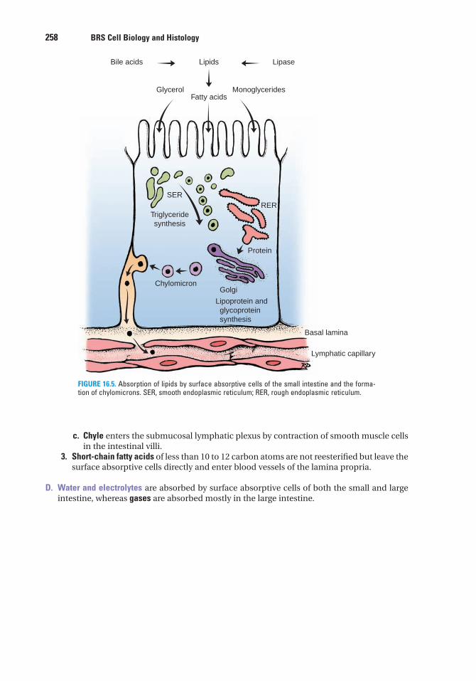
258 BRS Cell Biology and Histology
c. Chyle enters the submucosal lymphatic plexus by contraction of smooth muscle cellsin the intestinal villi.
3. Short-chain fatty acids of less than 10 to 12 carbon atoms are not reesterified but leave thesurface absorptive cells directly and enter blood vessels of the lamina propria.
D. Water and electrolytes are absorbed by surface absorptive cells of both the small and largeintestine, whereas gases are absorbed mostly in the large intestine.
Bile acids Lipids
Fatty acids
SER
Triglyceridesynthesis
Protein
Basal lamina
Lymphatic capillary
ChylomicronGolgi
Lipoprotein and glycoprotein synthesis
RER
Glycerol Monoglycerides
Lipase
FIGURE 16.5. Absorption of lipids by surface absorptive cells of the small intestine and the forma-tion of chylomicrons. SER, smooth endoplasmic reticulum; RER, rough endoplasmic reticulum.
LWBK615-c16[244-261].qxd 05/31/2010 10:56 AM Page 258 Aptara
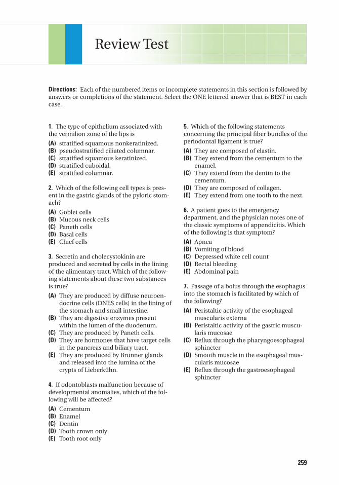
1. The type of epithelium associated withthe vermilion zone of the lips is
(A) stratified squamous nonkeratinized.(B) pseudostratified ciliated columnar.(C) stratified squamous keratinized.(D) stratified cuboidal.(E) stratified columnar.
2. Which of the following cell types is pres-ent in the gastric glands of the pyloric stom-ach?
(A) Goblet cells(B) Mucous neck cells(C) Paneth cells(D) Basal cells(E) Chief cells
3. Secretin and cholecystokinin areproduced and secreted by cells in the liningof the alimentary tract. Which of the follow-ing statements about these two substancesis true?
(A) They are produced by diffuse neuroen-docrine cells (DNES cells) in the lining ofthe stomach and small intestine.
(B) They are digestive enzymes presentwithin the lumen of the duodenum.
(C) They are produced by Paneth cells.(D) They are hormones that have target cells
in the pancreas and biliary tract.(E) They are produced by Brunner glands
and released into the lumina of thecrypts of Lieberkühn.
4. If odontoblasts malfunction because ofdevelopmental anomalies, which of the fol-lowing will be affected?
(A) Cementum(B) Enamel(C) Dentin(D) Tooth crown only(E) Tooth root only
5. Which of the following statementsconcerning the principal fiber bundles of theperiodontal ligament is true?
(A) They are composed of elastin.(B) They extend from the cementum to the
enamel.(C) They extend from the dentin to the
cementum.(D) They are composed of collagen.(E) They extend from one tooth to the next.
6. A patient goes to the emergencydepartment, and the physician notes one ofthe classic symptoms of appendicitis. Whichof the following is that symptom?
(A) Apnea(B) Vomiting of blood(C) Depressed white cell count(D) Rectal bleeding(E) Abdominal pain
7. Passage of a bolus through the esophagusinto the stomach is facilitated by which ofthe following?
(A) Peristaltic activity of the esophagealmuscularis externa
(B) Peristaltic activity of the gastric muscu-laris mucosae
(C) Reflux through the pharyngoesophagealsphincter
(D) Smooth muscle in the esophageal mus-cularis mucosae
(E) Reflux through the gastroesophagealsphincter
Review Test
Directions: Each of the numbered items or incomplete statements in this section is followed byanswers or completions of the statement. Select the ONE lettered answer that is BEST in eachcase.
259
LWBK615-c16[244-261].qxd 05/31/2010 10:56 AM Page 259 Aptara
9. Which of the following materials can beabsorbed directly by the surface lining cellsof the stomach?
(A) Vitamin B12
(B) Polysaccharides(C) Chylomicrons(D) Triglycerides(E) Alcohol
10. Which of the following is characteristicof angular cheilitis?
(A) Patients in the early 20s(B) Presence of herpes virus(C) Erythematous lesions at the corners of
the mouth(D) Apnea(E) High blood levels of secretin
8. The small intestine has three histologicallydistinct regions. Which of the following state-ments concerning the histological differencesin the three regions is true?
(A) Peyer patches are present only in theileum.
(B) Goblet cells are present only in theepithelium of the duodenum.
(C) Brunner glands are located in theduodenum and jejunum but not theileum.
(D) Lacteals are present only in the laminapropria of the ileum.
(E) The muscularis mucosae contains threelayers of smooth muscle in the ileumand two layers in the duodenum andjejunum.
260 BRS Cell Biology and Histology
LWBK615-c16[244-261].qxd 05/31/2010 10:56 AM Page 260 Aptara
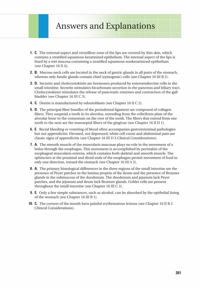
Answers and Explanations
1. C. The external aspect and vermillion zone of the lips are covered by thin skin, whichcontains a stratified squamous keratinized epithelium. The internal aspect of the lips islined by a wet mucosa containing a stratified squamous nonkeratinized epithelium (see Chapter 16 II A).
2. B. Mucous neck cells are located in the neck of gastric glands in all parts of the stomach,whereas only fundic glands contain chief (zymogenic) cells (see Chapter 16 III B 2).
3. D. Secretin and cholecystokinin are hormones produced by enteroendocrine cells in thesmall intestine. Secretin stimulates bicarbonate secretion in the pancreas and biliary tract.Cholecystokinin stimulates the release of pancreatic enzymes and contraction of the gallbladder (see Chapter 16 III C 3).
4. C. Dentin is manufactured by odontoblasts (see Chapter 16 II C 2).
5. D. The principal fiber bundles of the periodontal ligament are composed of collagenfibers. They suspend a tooth in its alveolus, extending from the cribriform plate of thealveolar bone to the cementum on the root of the tooth. The fibers that extend from onetooth to the next are the transseptal fibers of the gingivae (see Chapter 16 II D 1).
6. E. Rectal bleeding or vomiting of blood often accompanies gastrointestinal pathologiesbut not appendicitis. Elevated, not depressed, white cell count and abdominal pain areclassic signs of appendicitis (see Chapter 16 III D 5 Clinical Considerations).
7. A. The smooth muscle of the muscularis mucosae plays no role in the movement of abolus through the esophagus. This movement is accomplished by peristalsis of theesophageal muscularis externa, which contains both skeletal and smooth muscle. Thesphincters at the proximal and distal ends of the esophagus permit movement of food inonly one direction, toward the stomach (see Chapter 16 III A 5).
8. A. The primary histological differences in the three regions of the small intestine are thepresence of Peyer patches in the lamina propria of the ileum and the presence of Brunnerglands in the submucosa of the duodenum. The duodenum and jejunum lack Peyerpatches, and the jejunum and ileum lack Brunner glands. Goblet cells are presentthroughout the small intestine (see Chapter 16 III C 3).
9. E. Only a few simple substances, such as alcohol, can be absorbed by the epithelial liningof the stomach (see Chapter 16 III B 1).
10. C. The corners of the mouth have painful erythematous lesions (see Chapter 16 II B 2Clinical Considerations).
261
LWBK615-c16[244-261].qxd 05/31/2010 10:56 AM Page 261 Aptara
c h a p t e r 17 Digestive System: Glands
262
I. OVERVIEW—EXTRINSIC GLANDS OF THE DIGESTIVE SYSTEM
A. The extrinsic glands of the digestive system include the major salivary glands, the pancreas, andthe liver (with the associated gallbladder), all of which are outside the wall of the digestive tract.
B. These glands produce enzymes, buffers, emulsifiers, and lubricants that are delivered to thelumen of the digestive tract via a system of ducts.
C. They also produce hormones, blood proteins, and other products.
II. MAJOR SALIVARY GLANDS
A. Overview1. The major salivary glands consist of three paired exocrine glands: the parotid, sub-
mandibular, and sublingual.2. Function. They synthesize and secrete salivary amylase, lysozyme, lactoferrin, and secre-
tory component. The last of these, secretory component, is a molecule that complexeswith immunoglobulin A (IgA) dimers, produced by plasma cells in the connective tissue,forming a complex that resists enzymatic digestion in the saliva but can still perform itsimmune function. These glands also release the enzyme kallikrein into the connectivetissue, which enters the bloodstream, where it converts kininogens into the vasodilatorand bronchial smooth muscle contractant bradykinin.
B. Structure. The major salivary glands are compound tubuloacinar (tubuloalveolar) glands. Theyare further classified as serous, mucous, or mixed (both serous and mucous), depending onthe type of secretory acini they contain. These glands are surrounded by a capsule of denseirregular collagenous connective tissue with septa that subdivide each gland into lobes andlobules. Neurovascular elements serving these glands are conveyed to the acinar cells withinthe connective tissue septa.1. Salivary gland acini
a. Salivary gland acini consist of pyramidal serous or mucous cells arranged around acentral lumen that connects with an intercalated duct. Mucous acini may be overlaidwith a crescent-shaped collection of serous cells called serous demilunes.
b. They possess myoepithelial cells that share the basal lamina of the acinar cells. Theacinus and its associated intercalated and striated ducts form the salivon, the func-tional unit of a salivary gland.
c. They release a primary secretion that resembles extracellular fluid. This secretion ismodified in the ducts to produce the final secretion.
LWBK615-c17[262-274].qxd 05/31/2010 11:08 AM Page 262 Aptara
Chapter 17 Digestive System: Glands 263
d. Salivary glands are classified according to their types of acini.(1) Parotid glands consist of serous acini and are classified as serous.(2) Sublingual glands consist mostly of mucous acini capped with serous demilunes.
They are classified as mixed.(3) Submandibular glands consist of both serous and mucous acini (some also have
serous demilunes). They are classified as mixed.2. Salivary gland ducts
a. Intercalated ducts originate in the acini and join to form striated ducts. They maydeliver bicarbonate ions into the primary secretion.
b. Striated (intralobular) ducts(1) Striated ducts are lined by ion-transporting cells that remove sodium and chloride
ions from the luminal fluid (via a sodium pump) and actively pump potassiumand bicarbonate ions into it, thus transforming the initial saliva (primary saliva)produced by the acinar cells into secondary saliva, which leaves the striated duct(secondary saliva) and eventually enters the oral cavity.
(2) Striated ducts converge in each lobule to form interlobular (excretory) ducts, whichrun in the connective tissue septa. These ducts drain into the main duct of eachgland, which empties into the oral cavity.
C. Saliva1. Saliva is a hypotonic solution produced at the rate of about 1 L per day.2. Function
a. Saliva lubricates and cleanses the oral cavity by means of its water and glycoproteincontent.
b. It controls bacterial flora by the action of lysozyme, lactoferrin, and IgA, as well as byits cleansing action.
c. It initiates digestion of carbohydrates by the action of salivary amylase.d. It acts as a solvent for substances that stimulate the taste buds.e. It assists in the process of deglutition (swallowing).
(1) The flow of saliva is important not only for the easy swallowing of masticated food but also as a vehicle that facilitates the sensation of
taste and the initiation of digestion but also for the maintenance of proper oral health. Individu-als who had radiation treatment for cancers of the head and/or neck and those who are beingtreated with chemotherapy frequently have xerostomia, dry mouth, because of reduced salivarygland function. Frequently, these patients have to use artificial saliva to maintain a moist oralenvironment. Usually, subsequent to the cessation of radiation therapy or chemotherapy,salivary gland function returns to normal.
(2) Sjögren syndrome is believed to be an autoimmune disease whose symptoms are xerostomia(dry mouth), dry eyes, and generally dry mucous membranes. Although there is no cure for thispainful and potentially debilitating disorder, eyedrops, the frequent intake of fluids, and/or theuse of artificial saliva relieve some of the symptoms.
(3) Salivary calculus is a calcification that is formed usually in the parotid gland. It may block theflow of saliva through the parotid duct. Occasionally, the blockage results in the backflow ofsaliva, with a concomitant painful swelling of the parotid gland. Frequently, a simple massage ofthe parotid duct can dislodge the stone; otherwise a surgical approach may be necessary.
CLINICAL
CONSIDERATIONS
III. OVERVIEW—PANCREAS
The pancreas has a slender connective tissue capsule. This gland produces digestive enzymes inits exocrine portion and a number of hormones in its endocrine portion (islets of Langerhans).
LWBK615-c17[262-274].qxd 05/31/2010 11:08 AM Page 263 Aptara
264 BRS Cell Biology and Histology
A. The exocrine pancreas is a serous compound tubuloacinar gland.1. Pancreatic acinar cells
a. Pancreatic acinar cells are pyramidal serous cells arranged around a central lumen.b. They possess a round basal nucleus, abundant rough endoplasmic reticulum
(RER), an extensive Golgi complex, numerous mitochondria, and many freeribosomes.
c. Zymogen (secretory) granules are membrane bound and densely packed in the apicalregion of pancreatic acinar cells. They contain enzymes and proenzymes packaged inthe Golgi complex.
d. Their basal plasmalemma has receptors for cholecystokinin and acetylcholine.2. Pancreatic ducts
a. The initial intra-acinar portion of the intercalated ducts is formed by centroacinarcells, which are low cuboidal with a pale cytoplasm.
b. From the initial portion, the intercalated ducts converge into a small number ofintralobular ducts, which in turn empty into large interlobular ducts that empty into themain (or accessory) pancreatic duct.
c. The main pancreatic duct fuses with the common bile duct, forming the ampulla ofVater, which delivers secretions of the exocrine pancreas and the contents of thegallbladder into the duodenum at the major duodenal papilla.
3. Exocrine pancreatic secretionsa. Enzyme-poor alkaline fluid
(1) Enzyme-poor alkaline fluid is released in large quantities by intercalated duct cellsstimulated by secretin in conjunction with acetylcholine.
(2) Function. It probably neutralizes the acidic chyme as it enters the duodenum.b. Digestive enzymes
(1) Digestive enzymes are synthesized and stored in the pancreatic acinar cells. Theirrelease is stimulated by cholecystokinin, previously known as pancreozymin andcostimulated by acetylcholine released by postganglionic parasympathetic fibers.
(2) Digestive enzymes are secreted as enzymes or proenzymes that must be activatedin the intestinal lumen.
(3) Enzymes include pancreatic amylase, pancreatic lipases, ribonuclease, anddeoxyribonuclease; proenzymes include trypsinogen, chymotrypsinogen, procar-boxypeptidase, and proelastase.
(4) In order to protect themselves from the digestive enzyme trypsin, these cellsmanufacture trypsin inhibitor so that trypsinogen cannot be converted to trypsinwithin the cytosol.
B. Islets of Langerhans (endocrine pancreas) (Figure 17.1)1. Islets of Langerhans are richly vascularized spherical clusters (100–200 �m in diameter)
of endocrine cells surrounded by a fine network of reticular fibers. They are scatteredamong the acini of the exocrine pancreas in an apparently random fashion.
2. Islet cells (Table 17.1)a. Islet cells are of several types that can be differentiated from each other only by
immunocytochemistry or by the use of special stains.b. They produce several polypeptide hormones, but each cell type produces only one
hormone.3. Islet hormones
a. Glucagon is produced by �-cells and acts to elevate the blood glucose level.b. Insulin is produced by �-cells and acts to decrease the blood glucose level.c. Somatostatin is produced by �-cells. It inhibits release of hormones by nearby secretory
cells and reduces motility of the gastrointestinal tract and gallbladder by decreasingcontraction of their smooth muscles.
d. Gastrin, produced by G cells, stimulates (in conjunction with histamine and acetyl-choline) gastric hydrochloric acid (HCl) secretion.
e. Pancreatic polypeptide, produced by PP cells, inhibits release of exocrine pancreaticsecretions.
LWBK615-c17[262-274].qxd 05/31/2010 11:08 AM Page 264 Aptara
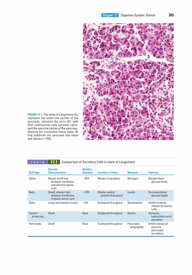
Chapter 17 Digestive System: Glands 265
IL
A
S
FIGURE 17.1. The islets of Langerhans (IL)represent the endocrine portion of thepancreas, whereas the acini (A), withtheir centroacinar cells (arrows), repre-sent the exocrine portion of the pancreas.Observe the connective tissue septa (S)that subdivide the pancreas into lobesand lobules (�270).
t a b l e 17.1 Comparison of Secretory Cells in Islets of Langerhans
Granule RelativeCell Type Characteristics Numbers Location in Islets Hormone Function
Alpha Round; small halo �20% Mostly at periphery Glucagon Elevate blood between membrane glucose levelsand electron-dense core
Beta Small; obvious halo �70% Mainly central; Insulin Decrease blood between membrane, present throughout glucose levelsirregular dense core
Delta Large and electron lucent �5% Scattered throughout Somatostatin Inhibit hormone release by nearbycells
Gastrin- Small Rare Scattered throughout Gastrin Stimulate producing hydrochloric acid
secretion
Pancreatic Small Rare Scattered throughout Pancreatic Inhibit release of polypeptide exocrine
pancreatic secretions
LWBK615-c17[262-274].qxd 05/31/2010 11:08 AM Page 265 Aptara
266 BRS Cell Biology and Histology
IV. LIVER
A. Overview1. The liver is composed of a single type of parenchymal cell, the hepatocyte.2. It is surrounded by a dense, irregular collagenous connective tissue known as Glisson
capsule, which gives rise to septa that subdivide the liver into lobes and lobules.3. Function. The liver produces bile and plasma proteins and has a myriad of other func-
tions.
B. Liver lobules (Figure 17.2)1. The classic liver lobule is a hexagonal mass of tissue primarily composed of plates of
hepatocytes, which radiate from the region of the central vein toward the periphery (Figure 17.3).a. Portal areas (portal canals or portal triads)
(1) The portal areas are regions of the connective tissue between lobules that containbranches of the portal vein, hepatic artery, lymph vessel, and bile duct.
(2) They are present at alternate corners of a classic liver lobule.
(1) Individuals who have low blood glucose and high blood insulinlevels may be suffering from insulinoma, a tumor of the �-cells of the
Islet of Langerhans. Insulinomas are benign in 90% of the cases, and the condition is usuallyresolved by surgical excision of the tumor.
(2) Type I (insulin-dependent) diabetes mellitus (IDDM)a. IDDM results from a low level of plasma insulin.b. It is characterized by polyphagia (insatiable hunger), polydipsia (unquenchable thirst), and
polyuria (excessive urination).c. It usually has a sudden onset before 20 years of age and is distinguished by damage to and
destruction of �-cells of the islets of Langerhans. Because of its early onset, IDDM is alsoknown as juvenile-onset diabetes mellitus.
d. It is treated with a combination of insulin therapy and diet.(3) Type II (non-insulin-dependent) diabetes mellitus (NIDDM)
a. NIDDM does not result from low levels of plasma insulin and is insulin resistant, which is amajor factor in its pathogenesis. The resistance to insulin is due to decreased binding ofinsulin to its plasmalemma receptors and to defects in postreceptor insulin action.
b. It commonly occurs in overweight individuals older than 40 years.c. It is usually controlled by diet.d. Older individuals with type II diabetes often present with memory problems probably because
amyloid �-derived diffusible ligands (ADDLs) bind to the presynaptic membranes of theaxons of certain neurons that are responsible for memory storage. This condition preventsthe replenishment of insulin receptors of these axons, even though the receptors,synthesized in the soma, are available in the soma’s cytoplasm. Apparently, the receptors areprevented from being transferred from the cytosol of the soma to the axoplasm of the axon.This makes these neurons insulin resistant and unable to regulate their glucose metabolismand in turn these neurons cannot function normally and the patient has memory loss andAlzheimer disease–associated dementia. Some researchers suggest this to be type III diabetes.
(4) Pancreatic cancer is a malignant neoplasm, and most patients die within 6 months to 1 yearafter diagnosis. Most of the cases are adenocarcinomas in the head of the pancreas. Itsincidence is three to four times greater in male patients than in female patients. Although itssymptoms include anorexia, flatulence, fatty stool if the bile duct is obstructed, sudden loss of weight, weakness, back pain, and jaundice, exploratory biopsy is commonly required for adefinitive diagnosis.
CLINICAL
CONSIDERATIONS
LWBK615-c17[262-274].qxd 05/31/2010 5:20 PM Page 266 Aptara
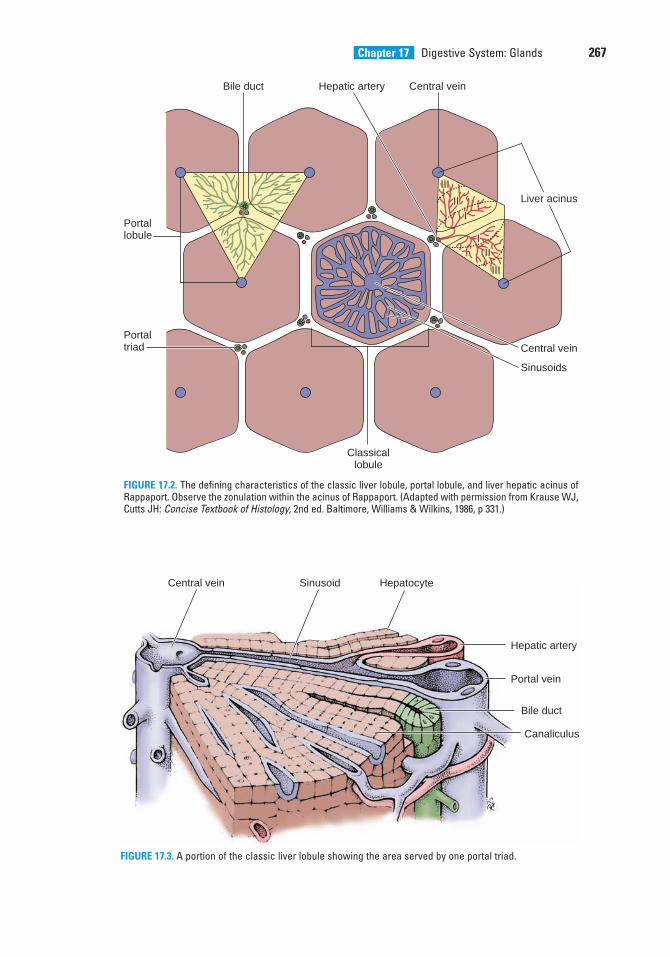
Chapter 17 Digestive System: Glands 267
Portallobule
Portaltriad Central vein
Sinusoids
Classicallobule
Liver acinus
Central veinBile duct Hepatic artery
III
III
III
III
III
III
III
III
FIGURE 17.2. The defining characteristics of the classic liver lobule, portal lobule, and liver hepatic acinus ofRappaport. Observe the zonulation within the acinus of Rappaport. (Adapted with permission from Krause WJ,Cutts JH: Concise Textbook of Histology, 2nd ed. Baltimore, Williams & Wilkins, 1986, p 331.)
Central vein Sinusoid Hepatocyte
Hepatic artery
Portal vein
Bile duct
Canaliculus
FIGURE 17.3. A portion of the classic liver lobule showing the area served by one portal triad.
LWBK615-c17[262-274].qxd 05/31/2010 11:08 AM Page 267 Aptara
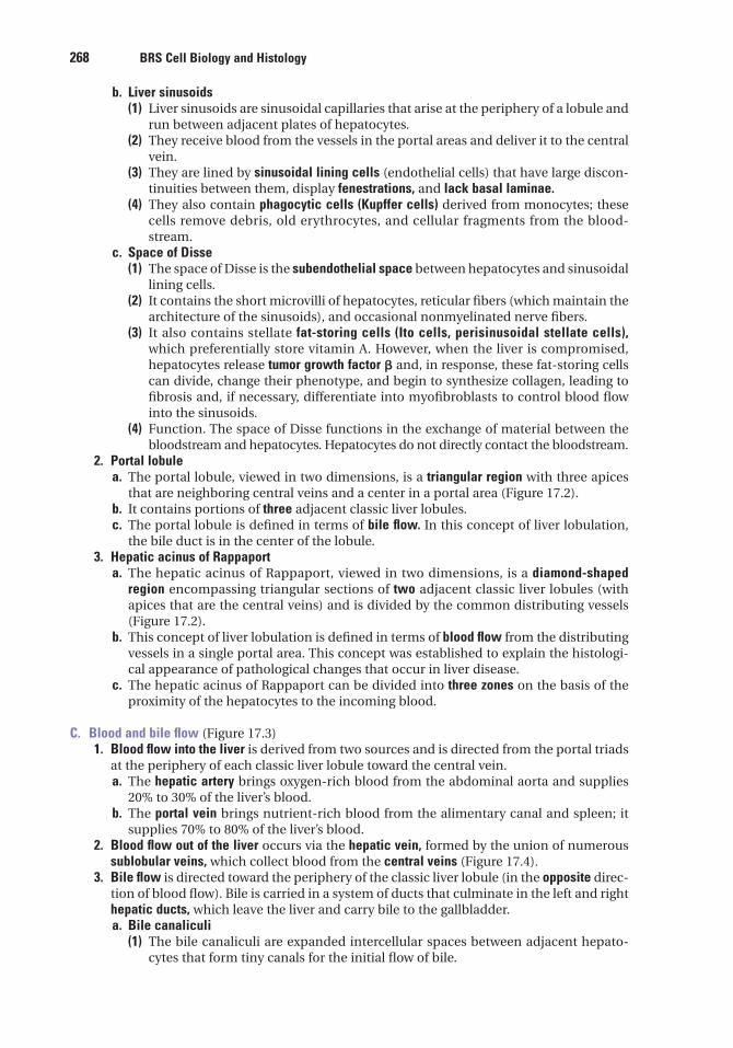
268 BRS Cell Biology and Histology
b. Liver sinusoids(1) Liver sinusoids are sinusoidal capillaries that arise at the periphery of a lobule and
run between adjacent plates of hepatocytes.(2) They receive blood from the vessels in the portal areas and deliver it to the central
vein.(3) They are lined by sinusoidal lining cells (endothelial cells) that have large discon-
tinuities between them, display fenestrations, and lack basal laminae.(4) They also contain phagocytic cells (Kupffer cells) derived from monocytes; these
cells remove debris, old erythrocytes, and cellular fragments from the blood-stream.
c. Space of Disse(1) The space of Disse is the subendothelial space between hepatocytes and sinusoidal
lining cells.(2) It contains the short microvilli of hepatocytes, reticular fibers (which maintain the
architecture of the sinusoids), and occasional nonmyelinated nerve fibers.(3) It also contains stellate fat-storing cells (Ito cells, perisinusoidal stellate cells),
which preferentially store vitamin A. However, when the liver is compromised,hepatocytes release tumor growth factor � and, in response, these fat-storing cellscan divide, change their phenotype, and begin to synthesize collagen, leading tofibrosis and, if necessary, differentiate into myofibroblasts to control blood flowinto the sinusoids.
(4) Function. The space of Disse functions in the exchange of material between thebloodstream and hepatocytes. Hepatocytes do not directly contact the bloodstream.
2. Portal lobulea. The portal lobule, viewed in two dimensions, is a triangular region with three apices
that are neighboring central veins and a center in a portal area (Figure 17.2).b. It contains portions of three adjacent classic liver lobules.c. The portal lobule is defined in terms of bile flow. In this concept of liver lobulation,
the bile duct is in the center of the lobule.3. Hepatic acinus of Rappaport
a. The hepatic acinus of Rappaport, viewed in two dimensions, is a diamond-shapedregion encompassing triangular sections of two adjacent classic liver lobules (withapices that are the central veins) and is divided by the common distributing vessels(Figure 17.2).
b. This concept of liver lobulation is defined in terms of blood flow from the distributingvessels in a single portal area. This concept was established to explain the histologi-cal appearance of pathological changes that occur in liver disease.
c. The hepatic acinus of Rappaport can be divided into three zones on the basis of theproximity of the hepatocytes to the incoming blood.
C. Blood and bile flow (Figure 17.3)1. Blood flow into the liver is derived from two sources and is directed from the portal triads
at the periphery of each classic liver lobule toward the central vein.a. The hepatic artery brings oxygen-rich blood from the abdominal aorta and supplies
20% to 30% of the liver’s blood.b. The portal vein brings nutrient-rich blood from the alimentary canal and spleen; it
supplies 70% to 80% of the liver’s blood.2. Blood flow out of the liver occurs via the hepatic vein, formed by the union of numerous
sublobular veins, which collect blood from the central veins (Figure 17.4).3. Bile flow is directed toward the periphery of the classic liver lobule (in the opposite direc-
tion of blood flow). Bile is carried in a system of ducts that culminate in the left and righthepatic ducts, which leave the liver and carry bile to the gallbladder.a. Bile canaliculi
(1) The bile canaliculi are expanded intercellular spaces between adjacent hepato-cytes that form tiny canals for the initial flow of bile.
LWBK615-c17[262-274].qxd 05/31/2010 11:08 AM Page 268 Aptara

Chapter 17 Digestive System: Glands 269
(2) They receive the liver’s exocrine secretion (bile) and carry it to the canals of Hering(bile ductules) at the very periphery of classic liver lobules.
b. Bile ducts(1) Bile ducts are located in the portal areas.(2) They receive bile from the canals of Hering.(3) They enlarge and fuse to form the hepatic ducts, which leave the liver at the porta
hepatis.
D. Hepatocytes1. Hepatocytes are large polyhedral cells (20–30 �m in diameter) that possess abundant
RER and smooth endoplasmic reticulum (SER); numerous mitochondria, lysosomes,and peroxisomes; several Golgi complexes; and many lipid droplets and glycogendeposits.
2. They usually contain one round central nucleus; about 25% of the cells are binucleated.Occasionally, nuclei are polyploid.
3. Hepatocyte surfacesa. Hepatocyte surfaces facing the space of Disse possess microvilli, which by increasing
the surface area facilitate the transfer of materials (e.g., endocrine secretions)between the hepatocytes and the blood.
b. Abutting surfaces of adjacent hepatocytes(1) frequently delineate bile canaliculi, small, tunnel-like expansions of the intercel-
lular space. The bile canaliculi are sealed off from the remaining intercellularspace by occluding junctions located on each side of each canaliculus.
(2) possess microvilli that extend into the bile canaliculus(3) also have gap junctions
FIGURE 17.4. The central vein (CV) of theliver is lined by endothelial cells (arrow-heads) that are continuous with those liningthe liver sinusoid (arrows) where these sinu-soids open (O) into the central vein. Observethe bile duct (BD) and a branch of the portalvein (HV) in a relatively large portal area(�132).
LWBK615-c17[262-274].qxd 05/31/2010 11:08 AM Page 269 Aptara
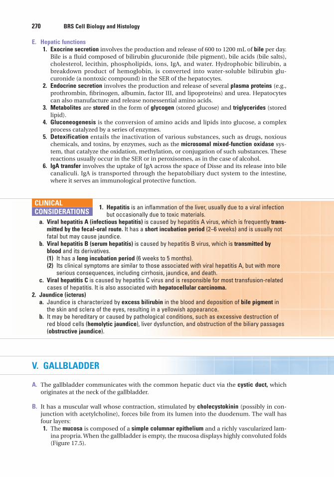
270 BRS Cell Biology and Histology
E. Hepatic functions1. Exocrine secretion involves the production and release of 600 to 1200 mL of bile per day.
Bile is a fluid composed of bilirubin glucuronide (bile pigment), bile acids (bile salts),cholesterol, lecithin, phospholipids, ions, IgA, and water. Hydrophobic bilirubin, abreakdown product of hemoglobin, is converted into water-soluble bilirubin glu-curonide (a nontoxic compound) in the SER of the hepatocytes.
2. Endocrine secretion involves the production and release of several plasma proteins (e.g.,prothrombin, fibrinogen, albumin, factor III, and lipoproteins) and urea. Hepatocytescan also manufacture and release nonessential amino acids.
3. Metabolites are stored in the form of glycogen (stored glucose) and triglycerides (storedlipid).
4. Gluconeogenesis is the conversion of amino acids and lipids into glucose, a complexprocess catalyzed by a series of enzymes.
5. Detoxification entails the inactivation of various substances, such as drugs, noxiouschemicals, and toxins, by enzymes, such as the microsomal mixed-function oxidase sys-tem, that catalyze the oxidation, methylation, or conjugation of such substances. Thesereactions usually occur in the SER or in peroxisomes, as in the case of alcohol.
6. IgA transfer involves the uptake of IgA across the space of Disse and its release into bilecanaliculi. IgA is transported through the hepatobiliary duct system to the intestine,where it serves an immunological protective function.
1. Hepatitis is an inflammation of the liver, usually due to a viral infectionbut occasionally due to toxic materials.
a. Viral hepatitis A (infectious hepatitis) is caused by hepatitis A virus, which is frequently trans-mitted by the fecal-oral route. It has a short incubation period (2–6 weeks) and is usually notfatal but may cause jaundice.
b. Viral hepatitis B (serum hepatitis) is caused by hepatitis B virus, which is transmitted byblood and its derivatives.(1) It has a long incubation period (6 weeks to 5 months).(2) Its clinical symptoms are similar to those associated with viral hepatitis A, but with more
serious consequences, including cirrhosis, jaundice, and death.c. Viral hepatitis C is caused by hepatitis C virus and is responsible for most transfusion-related
cases of hepatitis. It is also associated with hepatocellular carcinoma.2. Jaundice (icterus)
a. Jaundice is characterized by excess bilirubin in the blood and deposition of bile pigment inthe skin and sclera of the eyes, resulting in a yellowish appearance.
b. It may be hereditary or caused by pathological conditions, such as excessive destruction ofred blood cells (hemolytic jaundice), liver dysfunction, and obstruction of the biliary passages(obstructive jaundice).
CLINICAL
CONSIDERATIONS
V. GALLBLADDER
A. The gallbladder communicates with the common hepatic duct via the cystic duct, whichoriginates at the neck of the gallbladder.
B. It has a muscular wall whose contraction, stimulated by cholecystokinin (possibly in con-junction with acetylcholine), forces bile from its lumen into the duodenum. The wall hasfour layers:1. The mucosa is composed of a simple columnar epithelium and a richly vascularized lam-
ina propria. When the gallbladder is empty, the mucosa displays highly convoluted folds(Figure 17.5).
LWBK615-c17[262-274].qxd 05/31/2010 11:08 AM Page 270 Aptara
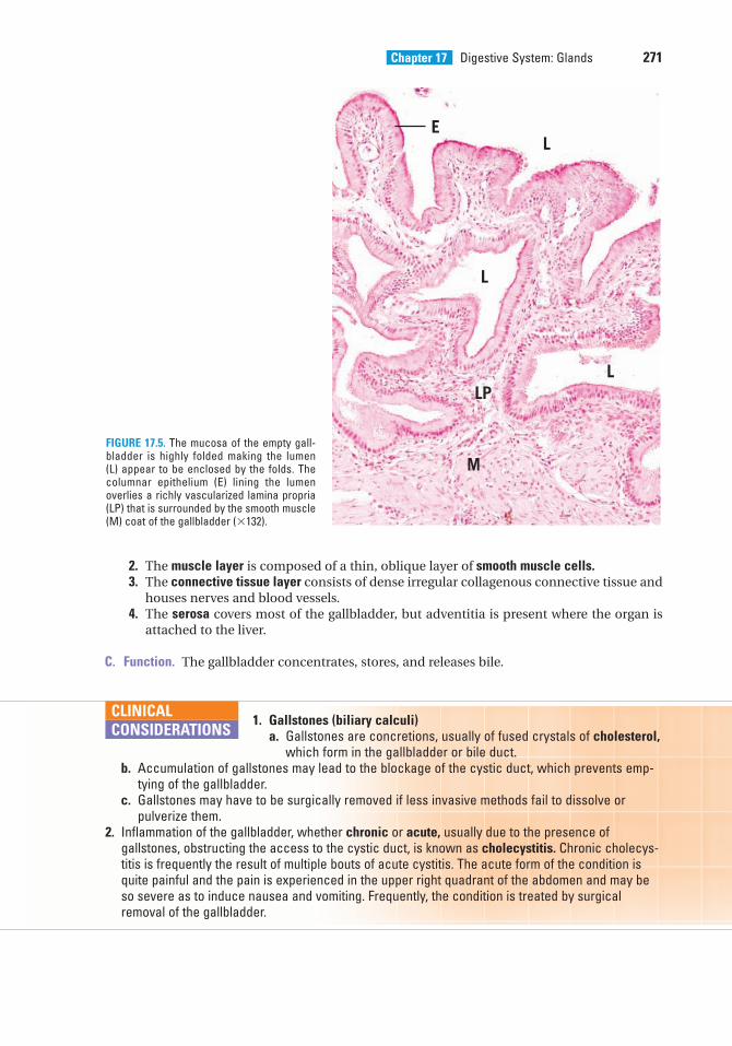
Chapter 17 Digestive System: Glands 271
L
LPL
E
M
L
FIGURE 17.5. The mucosa of the empty gall-bladder is highly folded making the lumen (L) appear to be enclosed by the folds. Thecolumnar epithelium (E) lining the lumenoverlies a richly vascularized lamina propria(LP) that is surrounded by the smooth muscle(M) coat of the gallbladder (�132).
1. Gallstones (biliary calculi)a. Gallstones are concretions, usually of fused crystals of cholesterol,
which form in the gallbladder or bile duct.b. Accumulation of gallstones may lead to the blockage of the cystic duct, which prevents emp-
tying of the gallbladder.c. Gallstones may have to be surgically removed if less invasive methods fail to dissolve or
pulverize them.2. Inflammation of the gallbladder, whether chronic or acute, usually due to the presence of
gallstones, obstructing the access to the cystic duct, is known as cholecystitis. Chronic cholecys-titis is frequently the result of multiple bouts of acute cystitis. The acute form of the condition isquite painful and the pain is experienced in the upper right quadrant of the abdomen and may beso severe as to induce nausea and vomiting. Frequently, the condition is treated by surgicalremoval of the gallbladder.
CLINICAL
CONSIDERATIONS
2. The muscle layer is composed of a thin, oblique layer of smooth muscle cells.3. The connective tissue layer consists of dense irregular collagenous connective tissue and
houses nerves and blood vessels.4. The serosa covers most of the gallbladder, but adventitia is present where the organ is
attached to the liver.
C. Function. The gallbladder concentrates, stores, and releases bile.
LWBK615-c17[262-274].qxd 05/31/2010 11:08 AM Page 271 Aptara
5. A patient complains to her physicianabout sudden weight loss, loss of appetite,weakness, and back pain. Because thepatient’s sclera and skin have a yellowishpallor, the doctor suspects
(A) type II diabetes.(B) gallstones.(C) pancreatic cancer.(D) viral hepatitis A.(E) viral hepatitis B.
6. Acinar cells of the exocrine pancreassecrete
(A) glucagon.(B) lysozyme.(C) insulin.(D) plasma proteins.(E) proteases.
7. Pancreatic �-cells secrete
(A) glucagon.(B) lysozyme.(C) insulin.(D) plasma proteins.(E) proteases.
8. Pancreatic �-cells secrete
(A) glucagon.(B) lysozyme.(C) insulin.(D) plasma proteins.(E) proteases.
1. An 18-year-old man feels faint. Symptomsinclude constant hunger, thirst, and excessiveurination. The probable diagnosis is
(A) viral hepatitis A.(B) type I diabetes mellitus.(C) type II diabetes mellitus.(D) cirrhosis.(E) mumps.
2. Which of the following statementsconcerning liver sinusoids is true?
(A) They are continuous with bile canaliculi.(B) They are surrounded by a well-
developed basal lamina.(C) They are lined by nonfenestrated
endothelial cells.(D) They deliver blood to the central vein.(E) They deliver blood to the portal vein.
3. A woman has yellow sclera and yellowishpallor. Blood test results indicate a low redblood cell count. The probable diagnosis is
(A) viral hepatitis A.(B) viral hepatitis B.(C) cirrhosis.(D) hemolytic jaundice.(E) type II diabetes mellitus.
4. Which of the following statementsconcerning the gallbladder is true?
(A) It synthesizes bile.(B) It is lined by a simple columnar epithe-
lium.(C) Bile leaves the gallbladder via the com-
mon bile duct.(D) It has no muscle cells in the walls.(E) It is affected by the hormone secretin.
Review Test
Directions: Each of the numbered items or incomplete statements in this section is followed byanswers or completions of the statement. Select the ONE lettered answer that is BEST in eachcase.
272
LWBK615-c17[262-274].qxd 05/31/2010 11:08 AM Page 272 Aptara

Chapter 17 Digestive System: Glands 273
9. Submandibular acinar cells secrete
(A) glucagon.(B) lysozyme.(C) insulin.(D) plasma proteins.(E) proteases.
10. Hepatocytes secrete
(A) glucagon.(B) lysozyme.(C) insulin.(D) plasma proteins.(E) proteases.
LWBK615-c17[262-274].qxd 05/31/2010 11:08 AM Page 273 Aptara
Answers and Explanations
1. B. The three classic signs of type I diabetes (juvenile onset) are polyphagia (excessiveeating), polyuria (excessive urination), and polydipsia (excessive drinking). The conditionoccurs in young individuals, usually before 20 years of age (see Chapter 17 III B 3 ClinicalConsiderations).
2. D. Liver sinusoids are lined by fenestrated endothelial cells, lack a basal lamina, anddeliver blood directly to the central vein (see Chapter 17 IV B 1 b).
3. D. Yellow skin and yellow sclera are indicative of jaundice. Because the patient had a lowred blood cell count, the most probable diagnosis is hemolytic jaundice (see Chapter 17 IVE 6 Clinical Considerations).
4. B. The gallbladder is lined by a simple columnar epithelium (see Chapter 17 V).
5. C. Pancreatic cancer has all of the symptoms listed, namely sudden weight loss, a loss ofappetite, weakness, back pain, and jaundice. Although jaundice is noted in hepatitis A andB and in the presence of obstructive jaundice caused by gallstones, sudden weight loss,loss of appetite, and weakness are not diagnostic of these diseases. Diabetes mellitus doesnot cause jaundice, loss of appetite, or weight loss (see Chapter 17 III B 3 ClinicalConsiderations).
6. E. Several proteases are synthesized by pancreatic acinar cells and are delivered via thepancreatic duct to the duodenum (see Chapter 17 III A 3).
7. A. Glucagon is produced by �-cells of the islets of Langerhans. They are the second mostabundant secretory cells of the endocrine pancreas (see Chapter 17 III B 3).
8. C. Insulin is produced by pancreatic �-cells, which are the most abundant cell type of theislets of Langerhans (see Chapter 17 III B 3).
9. B. Lysozyme, an enzyme with antibacterial activity, is produced primarily by thesubmandibular salivary gland acinar cells (see Chapter 17 II B 1).
10. D. Hepatocytes synthesize several plasma proteins, including fibrinogen, prothrombin,and albumin (see Chapter 17 IV E 2).
274
LWBK615-c17[262-274].qxd 05/31/2010 11:08 AM Page 274 Aptara
c h a p t e r 18 The Urinary System
275
I. OVERVIEW—THE URINARY SYSTEM
A. Structure. The urinary system is composed of the paired kidneys and ureters and the bladderand urethra.
B. Function. The urinary system produces and excretes urine, thereby clearing the blood ofwaste products. The kidneys also regulate the electrolyte levels in the extracellular fluid andsynthesize renin and erythropoietin.
II. KIDNEYS
A. General structure1. Kidneys are paired bean-shaped organs enveloped by a thin capsule of connective tissue.2. Each kidney is divided into an outer cortex and an inner medulla.3. Each kidney contains about 2 million nephrons. A nephron and the collecting tubule into
which it drains form a uriniferous tubule.
B. The renal hilum is a concavity on the medial border of the kidney. It houses arteries, veins,lymphatic vessels, nerves, and the renal pelvis.
C. The renal pelvis (Figure 18.1) is a funnel-shaped expansion of the upper end of the ureter. Itis continuous with the major renal calyces, which in turn have several small branches, theminor calyces.
D. The renal medulla lies deep to the cortex but sends extensions (medullary rays) into the cortex.1. Renal (medullary) pyramids are conical or pyramidal structures that compose the bulk of
the renal medulla.a. Each kidney contains 10 to 18 renal pyramids.b. Each pyramid consists primarily of the thin limbs of loops of Henle, blood vessels, and
collecting tubules.2. The renal papilla is located at the apex of each renal pyramid. It has a perforated tip (area
cribrosa) that projects into the lumen of a minor calyx.
E. The renal cortex is the superficial layer of the kidney beneath the capsule. It consists prima-rily of renal corpuscles and convoluted tubules.1. Renal columns of Bertin are extensions of cortical tissue between adjacent renal pyra-
mids.2. Medullary rays are groups of straight tubules that extend from the base of each renal
(medullary) pyramid into the cortex.
LWBK615-c18[275-293].qxd 05/31/2010 10:59 AM Page 275 Aptara
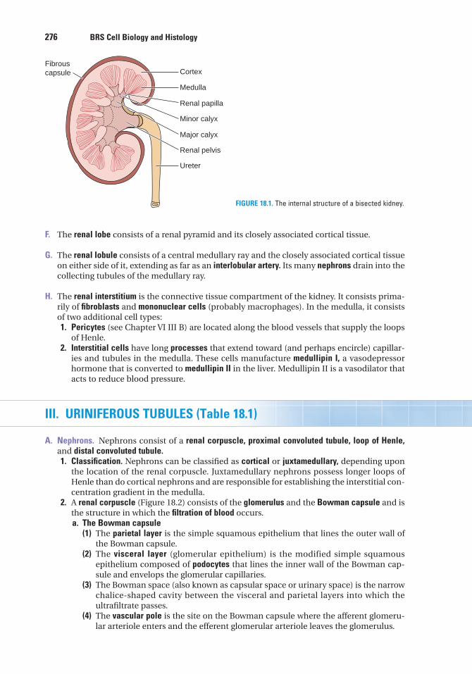
276 BRS Cell Biology and Histology
F. The renal lobe consists of a renal pyramid and its closely associated cortical tissue.
G. The renal lobule consists of a central medullary ray and the closely associated cortical tissueon either side of it, extending as far as an interlobular artery. Its many nephrons drain into thecollecting tubules of the medullary ray.
H. The renal interstitium is the connective tissue compartment of the kidney. It consists prima-rily of fibroblasts and mononuclear cells (probably macrophages). In the medulla, it consistsof two additional cell types:1. Pericytes (see Chapter VI III B) are located along the blood vessels that supply the loops
of Henle.2. Interstitial cells have long processes that extend toward (and perhaps encircle) capillar-
ies and tubules in the medulla. These cells manufacture medullipin I, a vasodepressorhormone that is converted to medullipin II in the liver. Medullipin II is a vasodilator thatacts to reduce blood pressure.
III. URINIFEROUS TUBULES (Table 18.1)
A. Nephrons. Nephrons consist of a renal corpuscle, proximal convoluted tubule, loop of Henle,and distal convoluted tubule.1. Classification. Nephrons can be classified as cortical or juxtamedullary, depending upon
the location of the renal corpuscle. Juxtamedullary nephrons possess longer loops ofHenle than do cortical nephrons and are responsible for establishing the interstitial con-centration gradient in the medulla.
2. A renal corpuscle (Figure 18.2) consists of the glomerulus and the Bowman capsule and isthe structure in which the filtration of blood occurs.a. The Bowman capsule
(1) The parietal layer is the simple squamous epithelium that lines the outer wall ofthe Bowman capsule.
(2) The visceral layer (glomerular epithelium) is the modified simple squamousepithelium composed of podocytes that lines the inner wall of the Bowman cap-sule and envelops the glomerular capillaries.
(3) The Bowman space (also known as capsular space or urinary space) is the narrowchalice-shaped cavity between the visceral and parietal layers into which theultrafiltrate passes.
(4) The vascular pole is the site on the Bowman capsule where the afferent glomeru-lar arteriole enters and the efferent glomerular arteriole leaves the glomerulus.
Cortex
Ureter
Renal pelvis
Major calyx
Minor calyx
Renal papilla
Medulla
Fibrouscapsule
FIGURE 18.1. The internal structure of a bisected kidney.
LWBK615-c18[275-293].qxd 05/31/2010 10:59 AM Page 276 Aptara
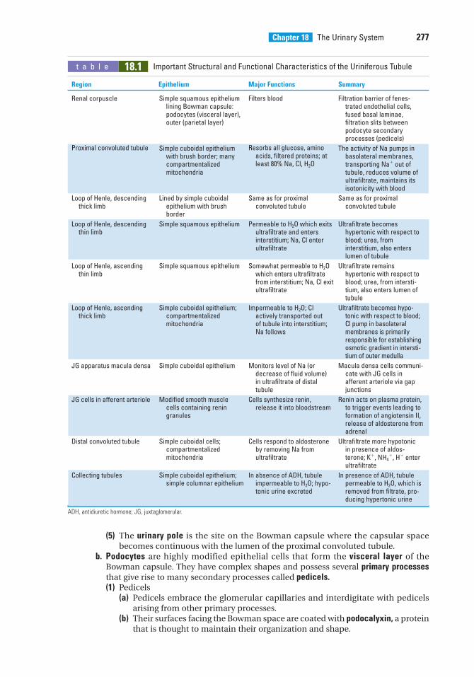
Chapter 18 The Urinary System 277
t a b l e 18.1 Important Structural and Functional Characteristics of the Uriniferous Tubule
Region Epithelium Major Functions Summary
ADH, antidiuretic hormone; JG, juxtaglomerular.
Renal corpuscle
Proximal convoluted tubule
Loop of Henle, descendingthick limb
Loop of Henle, descendingthin limb
Loop of Henle, ascendingthin limb
Loop of Henle, ascendingthick limb
JG apparatus macula densa
JG cells in afferent arteriole
Distal convoluted tubule
Collecting tubules
Simple squamous epitheliumlining Bowman capsule:podocytes (visceral layer),outer (parietal layer)
Simple cuboidal epitheliumwith brush border; manycompartmentalizedmitochondria
Lined by simple cuboidalepithelium with brushborder
Simple squamous epithelium
Simple squamous epithelium
Simple cuboidal epithelium;compartmentalizedmitochondria
Simple cuboidal epithelium
Modified smooth muscle cells containing renin granules
Simple cuboidal cells;compartmentalizedmitochondria
Simple cuboidal epithelium;simple columnar epithelium
Filters blood
Resorbs all glucose, aminoacids, filtered proteins; atleast 80% Na, Cl, H2O
Same as for proximalconvoluted tubule
Permeable to H2O which exitsultrafiltrate and entersinterstitium; Na, Cl enterultrafiltrate
Somewhat permeable to H2Owhich enters ultrafiltratefrom interstitium; Na, Cl exitultrafiltrate
Impermeable to H2O; Clactively transported outof tubule into interstitium;Na follows
Monitors level of Na (ordecrease of fluid volume) in ultrafiltrate of distaltubule
Cells synthesize renin,release it into bloodstream
Cells respond to aldosteroneby removing Na fromultrafiltrate
In absence of ADH, tubuleimpermeable to H2O; hypo-tonic urine excreted
Filtration barrier of fenes-trated endothelial cells,fused basal laminae,filtration slits betweenpodocyte secondaryprocesses (pedicels)
The activity of Na pumps inbasolateral membranes,transporting Na� out oftubule, reduces volume ofultrafiltrate, maintains itsisotonicity with blood
Same as for proximalconvoluted tubule
Ultrafiltrate becomeshypertonic with respect toblood; urea, frominterstitium, also enterslumen of tubule
Ultrafiltrate remainshypertonic with respect toblood; urea, from intersti-tium, also enters lumen oftubule
Ultrafiltrate becomes hypo-tonic with respect to blood;Cl pump in basolateralmembranes is primarilyresponsible for establishingosmotic gradient in intersti-tium of outer medulla
Macula densa cells communi-cate with JG cells in afferent arteriole via gapjunctions
Renin acts on plasma protein,to trigger events leading toformation of angiotensin II,release of aldosterone fromadrenal
Ultrafiltrate more hypotonic in presence of aldos-terone; K�, NH4
�, H� enterultrafiltrate
In presence of ADH, tubulepermeable to H2O, which isremoved from filtrate, pro-ducing hypertonic urine
(5) The urinary pole is the site on the Bowman capsule where the capsular spacebecomes continuous with the lumen of the proximal convoluted tubule.
b. Podocytes are highly modified epithelial cells that form the visceral layer of theBowman capsule. They have complex shapes and possess several primary processesthat give rise to many secondary processes called pedicels.(1) Pedicels
(a) Pedicels embrace the glomerular capillaries and interdigitate with pedicelsarising from other primary processes.
(b) Their surfaces facing the Bowman space are coated with podocalyxin, a proteinthat is thought to maintain their organization and shape.
LWBK615-c18[275-293].qxd 05/31/2010 10:59 AM Page 277 Aptara
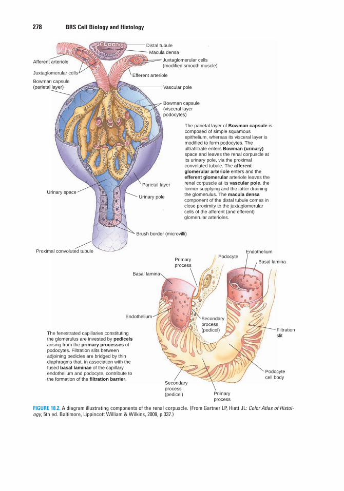
278 BRS Cell Biology and Histology
The parietal layer of Bowman capsule is composed of simple squamous epithelium, whereas its visceral layer is modified to form podocytes. The ultrafiltrate enters Bowman (urinary) space and leaves the renal corpuscle at its urinary pole, via the proximal convoluted tubule. The afferent glomerular arteriole enters and the efferent glomerular arteriole leaves the renal corpuscle at its vascular pole, the former supplying and the latter draining the glomerulus. The macula densa component of the distal tubule comes in close proximity to the juxtaglomerular cells of the afferent (and efferent) glomerular arterioles.
The fenestrated capillaries constituting the glomerulus are invested by pedicels arising from the primary processes of podocytes. Filtration slits between adjoining pedicles are bridged by thin diaphragms that, in association with the fused basal laminae of the capillary endothelium and podocyte, contribute to the formation of the filtration barrier.
Distal tubule
Macula densa
Juxtaglomerular cells(modified smooth muscle)
Efferent arteriole
Vascular pole
Bowman capsule(visceral layerpodocytes)
Parietal layer
Urinary pole
Brush border (microvilli)
Proximal convoluted tubule
Urinary space
Bowman capsule(parietal layer)
Juxtaglomerular cells
Afferent arteriole
PodocyteEndothelium
Basal lamina
Filtrationslit
Podocytecell body
Primaryprocess
Secondaryprocess(pedicel)
Endothelium
Basal lamina
Primaryprocess
Secondaryprocess(pedicel)
FIGURE 18.2. A diagram illustrating components of the renal corpuscle. (From Gartner LP, Hiatt JL: Color Atlas of Histol-ogy, 5th ed. Baltimore, Lippincott William & Wilkins, 2009, p 337.)
LWBK615-c18[275-293].qxd 05/31/2010 10:59 AM Page 278 Aptara
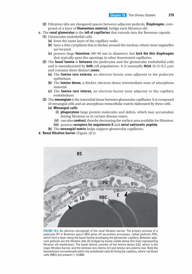
Chapter 18 The Urinary System 279
(2) Filtration slits are elongated spaces between adjacent pedicels. Diaphragms, com-posed of a layer of filamentous material, bridge each filtration slit.
c. The renal glomerulus is the tuft of capillaries that extends into the Bowman capsule.(1) Glomerular endothelial cells
(a) form the inner layer of the capillary walls.(b) have a thin cytoplasm that is thicker around the nucleus, where most organelles
are located.(c) possess large fenestrae (60–90 nm in diameter) but lack the thin diaphragms
that typically span the openings in other fenestrated capillaries.(2) The basal lamina is between the podocytes and the glomerular endothelial cells
and is manufactured by both cell populations. It is unusually thick (0.15–0.5 �m)and contains three distinct zones:(a) The lamina rara externa, an electron-lucent zone adjacent to the podocyte
epithelium(b) The lamina densa, a thicker, electron-dense intermediate zone of amorphous
material(c) The lamina rara interna, an electron-lucent zone adjacent to the capillary
endothelium(3) The mesangium is the interstitial tissue between glomerular capillaries. It is composed
of mesangial cells and an amorphous extracellular matrix elaborated by these cells.(a) Mesangial cells
(i) phagocytose large protein molecules and debris, which may accumulateduring filtration or in certain disease states.
(ii) can also contract, thereby decreasing the surface area available for filtration.(iii) possess receptors for angiotensin II and atrial natriuretic peptide.
(b) The mesangial matrix helps support glomerular capillaries.d. Renal filtration barrier (Figure 18.3)
BS
P
ex
in LD RBC
PD
E
S
FIGURE 18.3. An electron micrograph of the renal filtration barrier. The primary process of apodocyte (P) in Bowman space (BS) gives off secondary processes, called pedicels (PD),which form a layer along the basal lamina enveloping the glomerular capillary. Between adja-cent pedicels are the filtration slits (S) bridged by barely visible dense thin lines representingfiltration slit membranes. The basal lamina consists of the lamina densa (LD), which is themajor filtration barrier, and the laminae rara interna (in) and lamina rara externa (ex). Note thefenestrations (arrowheads) within the endothelial cells (E) lining the capillary, where red bloodcells (RBC) are present (�14,000).
LWBK615-c18[275-293].qxd 05/31/2010 10:59 AM Page 279 Aptara
280 BRS Cell Biology and Histology
(1) Structure. The renal filtration barrier is composed of the fenestrated endothelium ofthe glomerular capillaries, the basal lamina (laminae rarae and lamina densa), andthe filtration slits with diaphragms between pedicels.
(2) Function. The renal filtration barrier permits passage of water, ions, and smallmolecules from the bloodstream into the capsular space but prevents passage oflarge and/or most negatively charged proteins, thus forming an ultrafiltrate ofblood plasma in the Bowman space.(a) The laminae rarae contain heparan sulfate, a polyanionic glycosaminoglycan
that assists in restricting the passage of negatively charged proteins into theBowman space.
(b) The lamina densa contains type IV collagen, which acts as a selective macro-molecular filter preventing passage of large protein molecules (molecularweight greater than 69,000 daltons) into the Bowman space.
Glomerulonephritis1. Glomerulonephritis is a type of nephritis characterized by inflammation
of the glomeruli.2. It is sometimes marked by proliferation of podocytes, endothelial cells, and mesangial cells in the
glomerular tuft; infiltration of leukocytes is also common.3. This disease often occurs secondary to a streptococcal infection elsewhere in the body, which is
thought to result in deposition of immune complexes in the glomerular basal lamina. The immunecomplexes damage the glomerular basal lamina and markedly reduce its filtering ability.
4. It also may result from immune or autoimmune disorders.5. It is associated with production of urine-containing blood (hematuria), protein (proteinuria), or
both; in severe cases, decreased urine output (oliguria) is common.6. It occurs in acute, subacute, and chronic forms. The chronic form, in which the destruction of
glomeruli continues, leads eventually to renal failure and death.
Chronic renal failure1. Chronic renal failure can result from a variety of diseases (e.g., diabetes mellitus, hypertension,
atherosclerosis) in which blood flow to the kidneys is reduced, causing a decrease in glomerularfiltration and tubular ischemia.
2. It is associated with pathological changes (hyalinization) in the glomeruli and atrophy of thetubules, which impair virtually all aspects of renal function.
3. It is marked by acidosis and hyperkalemia because the acid–base balance cannot bemaintained, and by uremia because of the inability to eliminate metabolic wastes.
4. If untreated, chronic renal failure leads to neurological problems, coma, and death.
CLINICAL
CONSIDERATIONS
3. Proximal convoluted tubule (Figures 18.4 and 18.5)a. The proximal convoluted tubule is lined by a single layer of irregularly shaped
(cuboidal to columnar) epithelial cells that have microvilli forming a prominent brushborder. These cells exhibit the following structures:(1) Apical canaliculi, vesicles, and vacuoles (endocytic complex), which function in
protein absorption(2) Prominent interdigitations along their lateral borders, which interlock adjacent
cells with one another(3) Numerous mitochondria compartmentalized in the basal region by extensive
infoldings of the basal plasma membrane, which supply energy for the activetransport of Na� out of the tubule
b. Function(1) The proximal convoluted tubule drains the Bowman space at the urinary pole of
the renal corpuscle.(2) It resorbs from the glomerular filtrate all of the glucose, amino acids, and small
proteins and at least 80% of the sodium chloride and water.(3) It exchanges H� in the interstitium for HCO3
� in the filtrate.
LWBK615-c18[275-293].qxd 05/31/2010 10:59 AM Page 280 Aptara
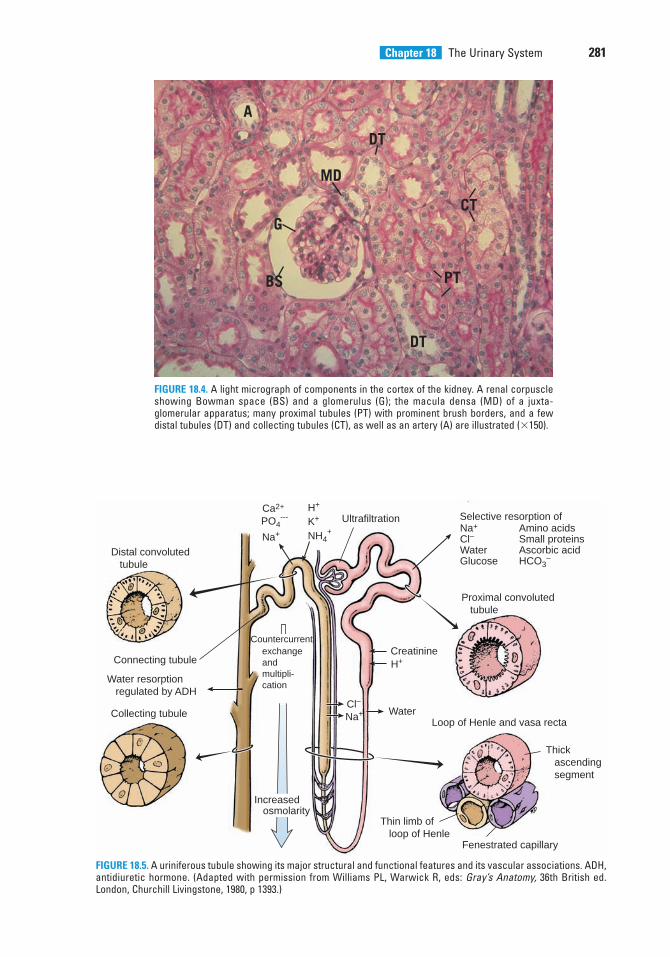
Chapter 18 The Urinary System 281
A
DT
MD
G
BS
DT
PT
CT
FIGURE 18.4. A light micrograph of components in the cortex of the kidney. A renal corpuscleshowing Bowman space (BS) and a glomerulus (G); the macula densa (MD) of a juxta-glomerular apparatus; many proximal tubules (PT) with prominent brush borders, and a fewdistal tubules (DT) and collecting tubules (CT), as well as an artery (A) are illustrated (�150).
Distal convoluted tubule
UltrafiltrationCa2+
Cl–
Na+
H+Creatinine
Selective resorption of
Proximal convoluted tubule
Loop of Henle and vasa recta
Thick ascending segment
Fenestrated capillary
Thin limb of loop of Henle
Na+
Cl–WaterGlucose
Amino acidsSmall proteinsAscorbic acidHCO3
–
Water
PO4---
NH4+
Na+
H+
K+
Connecting tubule
Collecting tubule
Water resorption regulated by ADH
exchangeandmultipli-cation
Increased osmolarity
Countercurrent
FIGURE 18.5. A uriniferous tubule showing its major structural and functional features and its vascular associations. ADH,antidiuretic hormone. (Adapted with permission from Williams PL, Warwick R, eds: Gray’s Anatomy, 36th British ed.London, Churchill Livingstone, 1980, p 1393.)
LWBK615-c18[275-293].qxd 05/31/2010 10:59 AM Page 281 Aptara
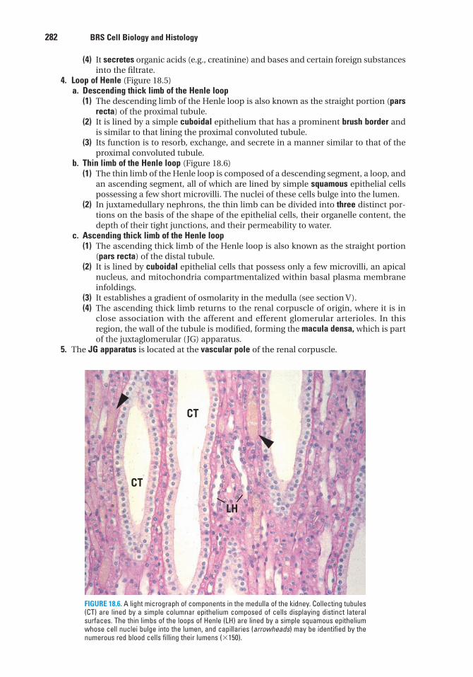
282 BRS Cell Biology and Histology
(4) It secretes organic acids (e.g., creatinine) and bases and certain foreign substancesinto the filtrate.
4. Loop of Henle (Figure 18.5)a. Descending thick limb of the Henle loop
(1) The descending limb of the Henle loop is also known as the straight portion (parsrecta) of the proximal tubule.
(2) It is lined by a simple cuboidal epithelium that has a prominent brush border andis similar to that lining the proximal convoluted tubule.
(3) Its function is to resorb, exchange, and secrete in a manner similar to that of theproximal convoluted tubule.
b. Thin limb of the Henle loop (Figure 18.6)(1) The thin limb of the Henle loop is composed of a descending segment, a loop, and
an ascending segment, all of which are lined by simple squamous epithelial cellspossessing a few short microvilli. The nuclei of these cells bulge into the lumen.
(2) In juxtamedullary nephrons, the thin limb can be divided into three distinct por-tions on the basis of the shape of the epithelial cells, their organelle content, thedepth of their tight junctions, and their permeability to water.
c. Ascending thick limb of the Henle loop(1) The ascending thick limb of the Henle loop is also known as the straight portion
(pars recta) of the distal tubule.(2) It is lined by cuboidal epithelial cells that possess only a few microvilli, an apical
nucleus, and mitochondria compartmentalized within basal plasma membraneinfoldings.
(3) It establishes a gradient of osmolarity in the medulla (see section V).(4) The ascending thick limb returns to the renal corpuscle of origin, where it is in
close association with the afferent and efferent glomerular arterioles. In thisregion, the wall of the tubule is modified, forming the macula densa, which is partof the juxtaglomerular (JG) apparatus.
5. The JG apparatus is located at the vascular pole of the renal corpuscle.
CT
CT
LH
FIGURE 18.6. A light micrograph of components in the medulla of the kidney. Collecting tubules(CT) are lined by a simple columnar epithelium composed of cells displaying distinct lateralsurfaces. The thin limbs of the loops of Henle (LH) are lined by a simple squamous epitheliumwhose cell nuclei bulge into the lumen, and capillaries (arrowheads) may be identified by thenumerous red blood cells filling their lumens (�150).
LWBK615-c18[275-293].qxd 05/31/2010 10:59 AM Page 282 Aptara
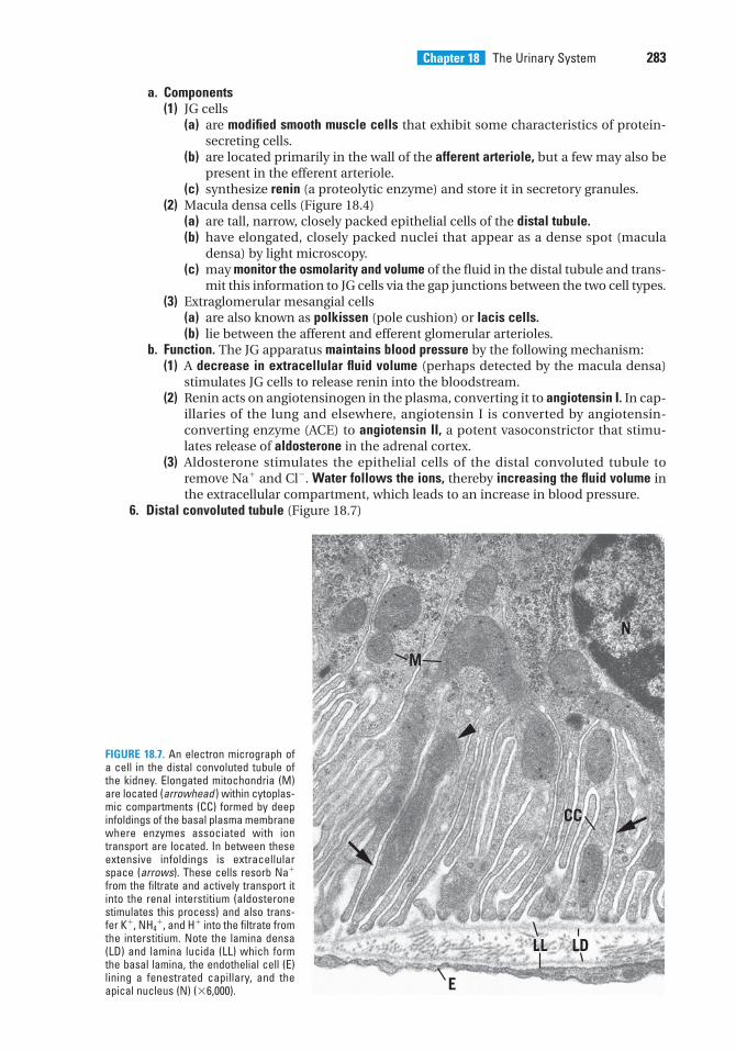
Chapter 18 The Urinary System 283
a. Components(1) JG cells
(a) are modified smooth muscle cells that exhibit some characteristics of protein-secreting cells.
(b) are located primarily in the wall of the afferent arteriole, but a few may also bepresent in the efferent arteriole.
(c) synthesize renin (a proteolytic enzyme) and store it in secretory granules.(2) Macula densa cells (Figure 18.4)
(a) are tall, narrow, closely packed epithelial cells of the distal tubule.(b) have elongated, closely packed nuclei that appear as a dense spot (macula
densa) by light microscopy.(c) may monitor the osmolarity and volume of the fluid in the distal tubule and trans-
mit this information to JG cells via the gap junctions between the two cell types.(3) Extraglomerular mesangial cells
(a) are also known as polkissen (pole cushion) or lacis cells.(b) lie between the afferent and efferent glomerular arterioles.
b. Function. The JG apparatus maintains blood pressure by the following mechanism:(1) A decrease in extracellular fluid volume (perhaps detected by the macula densa)
stimulates JG cells to release renin into the bloodstream.(2) Renin acts on angiotensinogen in the plasma, converting it to angiotensin I. In cap-
illaries of the lung and elsewhere, angiotensin I is converted by angiotensin-converting enzyme (ACE) to angiotensin II, a potent vasoconstrictor that stimu-lates release of aldosterone in the adrenal cortex.
(3) Aldosterone stimulates the epithelial cells of the distal convoluted tubule toremove Na� and Cl�. Water follows the ions, thereby increasing the fluid volume inthe extracellular compartment, which leads to an increase in blood pressure.
6. Distal convoluted tubule (Figure 18.7)
M
N
CC
E
LL LD
FIGURE 18.7. An electron micrograph ofa cell in the distal convoluted tubule ofthe kidney. Elongated mitochondria (M)are located (arrowhead ) within cytoplas-mic compartments (CC) formed by deepinfoldings of the basal plasma membranewhere enzymes associated with iontransport are located. In between theseextensive infoldings is extracellularspace (arrows). These cells resorb Na�
from the filtrate and actively transport itinto the renal interstitium (aldosteronestimulates this process) and also trans-fer K�, NH4
�, and H� into the filtrate fromthe interstitium. Note the lamina densa(LD) and lamina lucida (LL) which formthe basal lamina, the endothelial cell (E)lining a fenestrated capillary, and theapical nucleus (N) (�6,000).
LWBK615-c18[275-293].qxd 05/31/2010 11:00 AM Page 283 Aptara
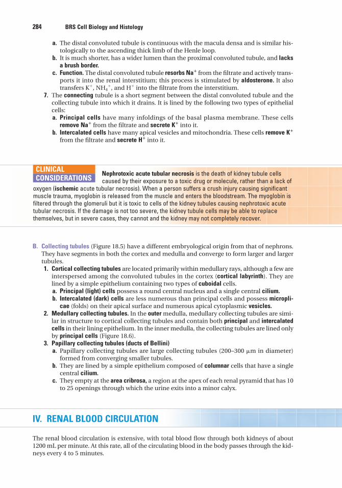
284 BRS Cell Biology and Histology
a. The distal convoluted tubule is continuous with the macula densa and is similar his-tologically to the ascending thick limb of the Henle loop.
b. It is much shorter, has a wider lumen than the proximal convoluted tubule, and lacksa brush border.
c. Function. The distal convoluted tubule resorbs Na� from the filtrate and actively trans-ports it into the renal interstitium; this process is stimulated by aldosterone. It alsotransfers K�, NH4
�, and H� into the filtrate from the interstitium.7. The connecting tubule is a short segment between the distal convoluted tubule and the
collecting tubule into which it drains. It is lined by the following two types of epithelialcells:a. Principal cells have many infoldings of the basal plasma membrane. These cells
remove Na� from the filtrate and secrete K� into it.b. Intercalated cells have many apical vesicles and mitochondria. These cells remove K�
from the filtrate and secrete H� into it.
Nephrotoxic acute tubular necrosis is the death of kidney tubule cellscaused by their exposure to a toxic drug or molecule, rather than a lack of
oxygen (ischemic acute tubular necrosis). When a person suffers a crush injury causing significantmuscle trauma, myoglobin is released from the muscle and enters the bloodstream. The myoglobin isfiltered through the glomeruli but it is toxic to cells of the kidney tubules causing nephrotoxic acutetubular necrosis. If the damage is not too severe, the kidney tubule cells may be able to replacethemselves, but in severe cases, they cannot and the kidney may not completely recover.
CLINICAL
CONSIDERATIONS
B. Collecting tubules (Figure 18.5) have a different embryological origin from that of nephrons.They have segments in both the cortex and medulla and converge to form larger and largertubules.1. Cortical collecting tubules are located primarily within medullary rays, although a few are
interspersed among the convoluted tubules in the cortex (cortical labyrinth). They arelined by a simple epithelium containing two types of cuboidal cells.a. Principal (light) cells possess a round central nucleus and a single central cilium.b. Intercalated (dark) cells are less numerous than principal cells and possess micropli-
cae (folds) on their apical surface and numerous apical cytoplasmic vesicles.2. Medullary collecting tubules. In the outer medulla, medullary collecting tubules are simi-
lar in structure to cortical collecting tubules and contain both principal and intercalatedcells in their lining epithelium. In the inner medulla, the collecting tubules are lined onlyby principal cells (Figure 18.6).
3. Papillary collecting tubules (ducts of Bellini)a. Papillary collecting tubules are large collecting tubules (200–300 �m in diameter)
formed from converging smaller tubules.b. They are lined by a simple epithelium composed of columnar cells that have a single
central cilium.c. They empty at the area cribrosa, a region at the apex of each renal pyramid that has 10
to 25 openings through which the urine exits into a minor calyx.
IV. RENAL BLOOD CIRCULATION
The renal blood circulation is extensive, with total blood flow through both kidneys of about1200 mL per minute. At this rate, all of the circulating blood in the body passes through the kid-neys every 4 to 5 minutes.
LWBK615-c18[275-293].qxd 05/31/2010 11:00 AM Page 284 Aptara
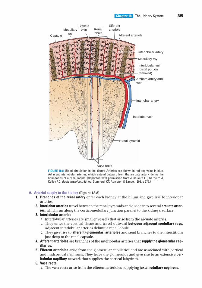
Chapter 18 The Urinary System 285
A. Arterial supply to the kidney (Figure 18.8)1. Branches of the renal artery enter each kidney at the hilum and give rise to interlobar
arteries.2. Interlobar arteries travel between the renal pyramids and divide into several arcuate arter-
ies, which run along the corticomedullary junction parallel to the kidney’s surface.3. Interlobular arteries
a. Interlobular arteries are smaller vessels that arise from the arcuate arteries.b. They enter the cortical tissue and travel outward between adjacent medullary rays.
Adjacent interlobular arteries delimit a renal lobule.c. They give rise to afferent (glomerular) arterioles and send branches to the interstitium
just deep to the renal capsule.4. Afferent arterioles are branches of the interlobular arteries that supply the glomerular cap-
illaries.5. Efferent arterioles arise from the glomerular capillaries and are associated with cortical
and midcortical nephrons. They leave the glomerulus and give rise to an extensive per-itubular capillary network that supplies the cortical labyrinth.
6. Vasa rectaa. The vasa recta arise from the efferent arterioles supplying juxtamedullary nephrons.
Capsule
Medullaryray
Stellatevein Renal
lobule
Efferentarteriole
Afferent arteriole
Interlobular artery
Medullary ray
Interlobular vein(distal portionremoved)
Arcuate artery andvein
Interlobar artery
Interlobar vein
Renal pyramid
Vasa recta
FIGURE 18.8. Blood circulation in the kidney. Arteries are shown in red and veins in blue.Adjacent interlobular arteries, which extend outward from the arcuate artery, define theboundaries of a renal lobule. (Reprinted with permission from Junqueira LC, Carneiro J,Kelley RO: Basic Histology, 9th ed. Stamford, CT, Appleton & Lange, 1998, p 375.)
LWBK615-c18[275-293].qxd 05/31/2010 11:00 AM Page 285 Aptara
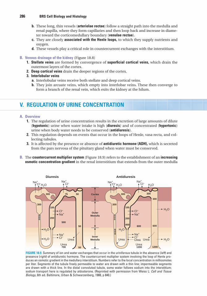
286 BRS Cell Biology and Histology
b. These long, thin vessels (arteriolae rectae) follow a straight path into the medulla andrenal papilla, where they form capillaries and then loop back and increase in diame-ter toward the corticomedullary boundary (venulae rectae).
c. They are closely associated with the Henle loops, to which they supply nutrients andoxygen.
d. These vessels play a critical role in countercurrent exchanges with the interstitium.
B. Venous drainage of the kidney (Figure 18.8)1. Stellate veins are formed by convergence of superficial cortical veins, which drain the
outermost layers of the cortex.2. Deep cortical veins drain the deeper regions of the cortex.3. Interlobular veins
a. Interlobular veins receive both stellate and deep cortical veins.b. They join arcuate veins, which empty into interlobar veins. These then converge to
form a branch of the renal vein, which exits the kidney at the hilum.
V. REGULATION OF URINE CONCENTRATION
A. Overview1. The regulation of urine concentration results in the excretion of large amounts of dilute
(hypotonic) urine when water intake is high (diuresis) and of concentrated (hypertonic)urine when body water needs to be conserved (antidiuresis).
2. This regulation depends on events that occur in the loops of Henle, vasa recta, and col-lecting tubules.
3. It is affected by the presence or absence of antidiuretic hormone (ADH), which is secretedfrom the pars nervosa of the pituitary gland when water must be conserved.
B. The countercurrent multiplier system (Figure 18.9) refers to the establishment of an increasingosmotic concentration gradient in the renal interstitium that extends from the outer medulla
Diuresis
Na+
Na+
Na+
Na+
Cl–
Cl–
Cl–
Cl–
H2O
H2O
Urea Urea
Urea
H2O
300
300100
200
500
700 50
50
50 Cortex
OuterMedulla
InnerMedulla
75
400
Antidiuresis
Na+
Na+
Na+
Na+
Cl–
Cl–
Cl–
Cl–
H2O
H2O
H2OUreaUrea
Urea
300
300100
200
400 600
1200 1200
600
300
300
400
600
H2O
H2O
FIGURE 18.9. Summary of ion and water exchanges that occur in the uriniferous tubule in the absence (left) andpresence (right) of antidiuretic hormone. The countercurrent multiplier system involving the loop of Henle pro-duces an osmotic gradient in the medullary interstitium. Numbers refer to the local concentration in milliosmolesper liter. Segments of the tubule freely permeable to water are drawn with a thin line; impermeable segmentsare drawn with a thick line. In the distal convoluted tubule, some water follows sodium into the interstitium;sodium transport here is regulated by aldosterone. (Reprinted with permission from Weiss L: Cell and TissueBiology, 6th ed. Baltimore, Urban & Schwarzenberg, 1988, p 840.)
LWBK615-c18[275-293].qxd 05/31/2010 11:00 AM Page 286 Aptara
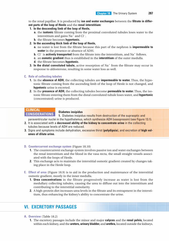
Chapter 18 The Urinary System 287
to the renal papillae. It is produced by ion and water exchanges between the filtrate in differ-ent parts of the loop of Henle and the renal interstitium:1. In the descending limb of the loop of Henle,
a. the isotonic filtrate coming from the proximal convoluted tubules loses water to theinterstitium and gains Na� and Cl�.
b. the filtrate becomes hypertonic.2. In the ascending thick limb of the loop of Henle,
a. no water is lost from the filtrate because this part of the nephron is impermeable towater in the presence or absence of ADH.
b. Cl� is actively transported from the filtrate into the interstitium, and Na� follows.c. an osmotic gradient thus is established in the interstitium of the outer medulla.d. the filtrate becomes hypotonic.
3. In the distal convoluted tubule, active resorption of Na� from the filtrate may occur inresponse to aldosterone, resulting in some water loss as well.
C. Role of collecting tubules1. In the absence of ADH, the collecting tubules are impermeable to water. Thus, the hypo-
tonic filtrate coming from the ascending limb of the loop of Henle is not changed, andhypotonic urine is excreted.
2. In the presence of ADH, the collecting tubules become permeable to water. Thus, the iso-tonic filtrate entering them from the distal convoluted tubule loses water, and hypertonic(concentrated) urine is produced.
Diabetes insipidus1. Diabetes insipidus results from destruction of the supraoptic and
paraventricular nuclei in the hypothalamus, which synthesize ADH (vasopressin) (see Figure 13.1).2. It is associated with a decreased ability of the kidney to concentrate urine in the collecting
tubules because levels of ADH are reduced.3. Signs and symptoms include dehydration, excessive thirst (polydipsia), and excretion of high vol-
umes of dilute urine.
CLINICAL
CONSIDERATIONS
D. Countercurrent exchange system (Figure 18.10)1. The countercurrent exchange system involves passive ion and water exchanges between
the renal interstitium and the blood in the vasa recta, the small straight vessels associ-ated with the loops of Henle.
2. This exchange acts to maintain the interstitial osmotic gradient created by changes tak-ing place in the Henle loop.
E. Effect of urea (Figure 18.9) is to aid in the production and maintenance of the interstitialosmotic gradient, mostly in the inner medulla.1. Urea concentrations in the filtrate progressively increase as water is lost from the
medullary collecting tubules, causing the urea to diffuse out into the interstitium andcontributing to the interstitial osmolarity.
2. A high-protein diet increases urea levels in the filtrate and its entrapment in the intersti-tium, thus enhancing the kidney’s ability to concentrate the urine.
VI. EXCRETORY PASSAGES
A. Overview (Table 18.2)1. The excretory passages include the minor and major calyces and the renal pelvis, located
within each kidney, and the ureters, urinary bladder, and urethra, located outside the kidneys.
LWBK615-c18[275-293].qxd 05/31/2010 11:00 AM Page 287 Aptara
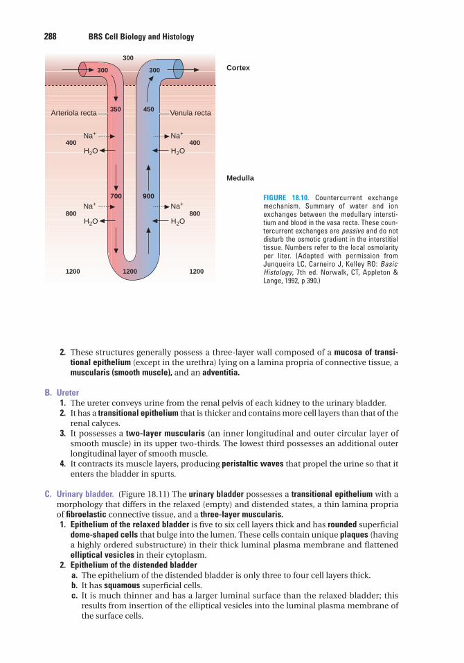
288 BRS Cell Biology and Histology
2. These structures generally possess a three-layer wall composed of a mucosa of transi-tional epithelium (except in the urethra) lying on a lamina propria of connective tissue, amuscularis (smooth muscle), and an adventitia.
B. Ureter1. The ureter conveys urine from the renal pelvis of each kidney to the urinary bladder.2. It has a transitional epithelium that is thicker and contains more cell layers than that of the
renal calyces.3. It possesses a two-layer muscularis (an inner longitudinal and outer circular layer of
smooth muscle) in its upper two-thirds. The lowest third possesses an additional outerlongitudinal layer of smooth muscle.
4. It contracts its muscle layers, producing peristaltic waves that propel the urine so that itenters the bladder in spurts.
C. Urinary bladder. (Figure 18.11) The urinary bladder possesses a transitional epithelium with amorphology that differs in the relaxed (empty) and distended states, a thin lamina propriaof fibroelastic connective tissue, and a three-layer muscularis.1. Epithelium of the relaxed bladder is five to six cell layers thick and has rounded superficial
dome-shaped cells that bulge into the lumen. These cells contain unique plaques (havinga highly ordered substructure) in their thick luminal plasma membrane and flattenedelliptical vesicles in their cytoplasm.
2. Epithelium of the distended bladdera. The epithelium of the distended bladder is only three to four cell layers thick.b. It has squamous superficial cells.c. It is much thinner and has a larger luminal surface than the relaxed bladder; this
results from insertion of the elliptical vesicles into the luminal plasma membrane ofthe surface cells.
700 900
Cortex
Arteriola recta
Na+
H2O H2O
H2O H2O
Na+
Na+ Na+
Venula recta
300
300300
350
1200 1200
800
400
1200
800
400
450
Medulla
FIGURE 18.10. Countercurrent exchangemechanism. Summary of water and ionexchanges between the medullary intersti-tium and blood in the vasa recta. These coun-tercurrent exchanges are passive and do notdisturb the osmotic gradient in the interstitialtissue. Numbers refer to the local osmolarityper liter. (Adapted with permission fromJunqueira LC, Carneiro J, Kelley RO: BasicHistology, 7th ed. Norwalk, CT, Appleton &Lange, 1992, p 390.)
LWBK615-c18[275-293].qxd 05/31/2010 11:00 AM Page 288 Aptara
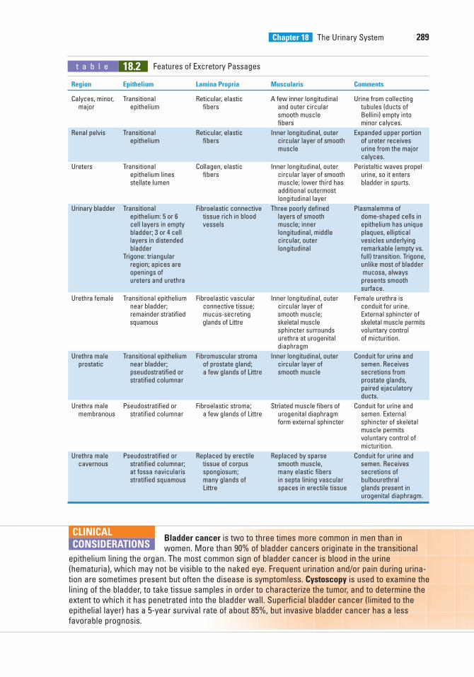
Chapter 18 The Urinary System 289
t a b l e 18.2 Features of Excretory Passages
Region Epithelium Lamina Propria Muscularis Comments
Calyces, minor, Transitional Reticular, elastic A few inner longitudinal Urine from collectingmajor epithelium fibers and outer circular tubules (ducts of
smooth muscle Bellini) empty intofibers minor calyces.
Renal pelvis Transitional Reticular, elastic Inner longitudinal, outer Expanded upper portionepithelium fibers circular layer of smooth of ureter receives
muscle urine from the major calyces.
Ureters Transitional Collagen, elastic Inner longitudinal, outer Peristaltic waves propel epithelium lines fibers circular layer of smooth urine, so it enters stellate lumen muscle; lower third has bladder in spurts.
additional outermost longitudinal layer
Urinary bladder Transitional Fibroelastic connective Three poorly defined Plasmalemma of epithelium: 5 or 6 tissue rich in blood layers of smooth dome-shaped cells in cell layers in empty vessels muscle; inner epithelium has unique bladder; 3 or 4 cell longitudinal, middle plaques, ellipticallayers in distended circular, outer vesicles underlying bladder longitudinal remarkable (empty vs.
Trigone: triangular full) transition. Trigone,region; apices are unlike most of bladderopenings of mucosa, always ureters and urethra presents smooth
surface.
Urethra female Transitional epithelium Fibroelastic vascular Inner longitudinal, outer Female urethra isnear bladder; connective tissue; circular layer of conduit for urine. remainder stratified mucus-secreting smooth muscle; External sphincter of squamous glands of Littre skeletal muscle skeletal muscle permits
sphincter surrounds voluntary control urethra at urogenital of micturition.diaphragm
Urethra male Transitional epithelium Fibromuscular stroma Inner longitudinal, outer Conduit for urine andprostatic near bladder; of prostate gland; circular layer of semen. Receives
pseudostratified or a few glands of Littre smooth muscle secretions fromstratified columnar prostate glands,
paired ejaculatory ducts.
Urethra male Pseudostratified or Fibroelastic stroma; Striated muscle fibers of Conduit for urine and membranous stratified columnar a few glands of Littre urogenital diaphragm semen. External
form external sphincter sphincter of skeletal muscle permits voluntary control of micturition.
Urethra male Pseudostratified or Replaced by erectile Replaced by sparse Conduit for urine and cavernous stratified columnar; tissue of corpus smooth muscle, semen. Receives
at fossa navicularis spongiosum; many elastic fibers secretions of stratified squamous many glands of in septa lining vascular bulbourethral
Littre spaces in erectile tissue glands present in urogenital diaphragm.
Bladder cancer is two to three times more common in men than inwomen. More than 90% of bladder cancers originate in the transitional
epithelium lining the organ. The most common sign of bladder cancer is blood in the urine(hematuria), which may not be visible to the naked eye. Frequent urination and/or pain during urina-tion are sometimes present but often the disease is symptomless. Cystoscopy is used to examine thelining of the bladder, to take tissue samples in order to characterize the tumor, and to determine theextent to which it has penetrated into the bladder wall. Superficial bladder cancer (limited to theepithelial layer) has a 5-year survival rate of about 85%, but invasive bladder cancer has a lessfavorable prognosis.
CLINICAL
CONSIDERATIONS
LWBK615-c18[275-293].qxd 05/31/2010 11:00 AM Page 289 Aptara
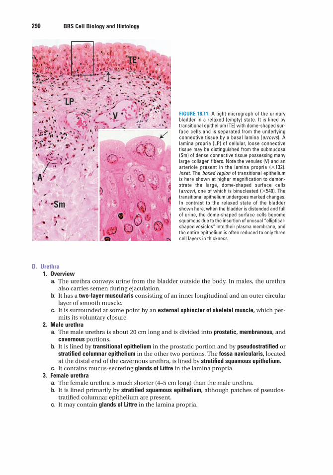
290 BRS Cell Biology and Histology
FIGURE 18.11. A light micrograph of the urinarybladder in a relaxed (empty) state. It is lined bytransitional epithelium (TE) with dome-shaped sur-face cells and is separated from the underlyingconnective tissue by a basal lamina (arrows). Alamina propria (LP) of cellular, loose connectivetissue may be distinguished from the submucosa(Sm) of dense connective tissue possessing manylarge collagen fibers. Note the venules (V) and anarteriole present in the lamina propria (�132).Inset. The boxed region of transitional epitheliumis here shown at higher magnification to demon-strate the large, dome-shaped surface cells(arrow), one of which is binucleated (�540). Thetransitional epithelium undergoes marked changes.In contrast to the relaxed state of the bladdershown here, when the bladder is distended and fullof urine, the dome-shaped surface cells becomesquamous due to the insertion of unusual “elliptical-shaped vesicles” into their plasma membrane, andthe entire epithelium is often reduced to only threecell layers in thickness.
D. Urethra1. Overview
a. The urethra conveys urine from the bladder outside the body. In males, the urethraalso carries semen during ejaculation.
b. It has a two-layer muscularis consisting of an inner longitudinal and an outer circularlayer of smooth muscle.
c. It is surrounded at some point by an external sphincter of skeletal muscle, which per-mits its voluntary closure.
2. Male urethraa. The male urethra is about 20 cm long and is divided into prostatic, membranous, and
cavernous portions.b. It is lined by transitional epithelium in the prostatic portion and by pseudostratified or
stratified columnar epithelium in the other two portions. The fossa navicularis, locatedat the distal end of the cavernous urethra, is lined by stratified squamous epithelium.
c. It contains mucus-secreting glands of Littre in the lamina propria.3. Female urethra
a. The female urethra is much shorter (4–5 cm long) than the male urethra.b. It is lined primarily by stratified squamous epithelium, although patches of pseudos-
tratified columnar epithelium are present.c. It may contain glands of Littre in the lamina propria.
LWBK615-c18[275-293].qxd 05/31/2010 11:00 AM Page 290 Aptara
1. Which of the following statementsconcerning the structure of medullary rays istrue?
(A) They contain arched collecting tubules.(B) They contain proximal convoluted
tubules.(C) They do not extend into the renal cortex.(D) They lie at the center of a renal lobule.(E) They contain thin limbs of the loops of
Henle.
2. Which one of the following structures islocated in the renal cortex?
(A) Vasa recta(B) Thin limbs of the loops of Henle(C) Afferent arterioles(D) Interlobar veins(E) Area cribrosa
3. Which of the following structures is pres-ent in the male urethra but is not present inthe female urethra?
(A) Stratified squamous epithelium(B) Transitional epithelium(C) Glands of Littre(D) External sphincter of skeletal muscle(E) Connective tissue layer underlying the
epithelium
4. Which of the following statementsconcerning cortical collecting tubules isalways true?
(A) They are lined by a simple epitheliumcontaining two types of cells.
(B) They are also known as the ducts ofBellini.
(C) They empty on the area cribrosa.(D) They are permeable to water.(E) They are continuous with the ascending
thick limb of the Henle loop.
5. A 35-year-old woman had surgery toremove a cerebral tumor. A month after theprocedure, she reports being excessivelythirsty and drinking several liters of waterper day. She must also urinate so frequentlythat she avoids leaving the house. Laboratorytests indicate that her urine has very lowspecific gravity. What is the most likely diag-nosis of this woman’s condition?
(A) Acute renal failure(B) Glomerulonephritis(C) Chronic renal failure(D) Diabetes insipidus(E) Urinary incontinence
6. The countercurrent multiplier system inthe kidney involves the exchange of waterand ions between the renal interstitium and
(A) the blood in the vasa recta.(B) the blood in the peritubular capillary
network.(C) the filtrate in the proximal convoluted
tubule.(D) the filtrate in the loop of Henle.(E) the filtrate in the medullary collecting
tubule.
7. As the glomerular filtrate passes throughthe uriniferous tubule, ions and water areexchanged (actively or passively) with therenal interstitium, resulting in the filtratebeing isotonic, hypotonic, or hypertonic rel-ative to blood plasma. During a condition ofantidiuresis, which part of the uriniferoustubule would contain a hypertonic filtrate?
(A) Ascending thick limb of the loop ofHenle
(B) Bowman (capsular) space(C) Cortical collecting tubule(D) Medullary collecting tubule(E) Proximal convoluted tubule
Review Test
Directions: Each of the numbered items or incomplete statements in this section is followed byanswers or completions of the statement. Select the ONE lettered answer that is BEST in each case.
291
LWBK615-c18[275-293].qxd 05/31/2010 11:00 AM Page 291 Aptara
292 BRS Cell Biology and Histology
8. As the glomerular filtrate passes throughthe uriniferous tubule, ions and water are exchanged (actively or passively) withthe renal interstitium, resulting in thefiltrate being isotonic, hypotonic, or hyper-tonic relative to blood plasma. During acondition of antidiuresis, which part of the uriniferous tubule would contain anisotonic filtrate?
(A) Ascending thick limb of the loop ofHenle
(B) Bottom of descending thin limb of loopof Henle
(C) Bowman (capsular) space(D) Medullary collecting tubule(E) Papillary collecting tubule
9. As the glomerular filtrate passes throughthe uriniferous tubule, ions and water areexchanged (actively or passively) with therenal interstitium, resulting in the filtratebeing isotonic, hypotonic, or hypertonic rel-ative to blood plasma. During a condition ofantidiuresis, which one of the following
parts of the uriniferous tubule wouldcontain a hypotonic filtrate?
(A) Bowman (capsular) space(B) Cortical collecting tubule(C) Distal portion of the ascending thick
limb of the loop of Henle(D) Medullary collecting tubule(E) Proximal convoluted tubule
10. As the glomerular filtrate passes throughthe uriniferous tubule, ions and water areexchanged (actively or passively) with therenal interstitium, resulting in the filtratebeing isotonic, hypotonic, or hypertonic rel-ative to blood plasma. During a condition ofantidiuresis, which part of the uriniferoustubule would contain a hypertonic filtrate?
(A) Bottom of descending thin limb of loopof Henle
(B) Bowman (capsular) space(C) Distal portion of the ascending thick
limb of the loop of Henle(D) Pars recta of the proximal tubule(E) Proximal convoluted tubule
LWBK615-c18[275-293].qxd 05/31/2010 11:00 AM Page 292 Aptara
Answers and Explanations
1. D. A medullary ray contains the straight portions of tubules projecting from the medullainto the cortex, giving the appearance of striations or rays. A renal lobule consists of a cen-tral medullary ray and its closely associated cortical tissue (see Chapter 18 II E).
2. C. Afferent arterioles, which arise from interlobular arteries and supply the glomerularcapillaries, are located in the renal cortex (see Chapter 18 III A 5).
3. B. Only the male urethra contains transitional epithelium (in the prostatic portion). Strati-fied squamous epithelium lines most of the female urethra and the distal end of thecavernous urethra in males. Mucus-secreting glands of Littre are always present in themale urethra and may be present in the female urethra (see Chapter 18 VI D).
4. A. Cortical collecting tubules are lined by a simple epithelium containing principal (light)cells and intercalated (dark) cells. They are permeable to water only in the presence ofADH; in the absence of this hormone, they are impermeable to water. The large papillarycollecting tubules, called ducts of Bellini, empty on the area cribrosa at the apex of eachrenal papilla (see Chapter 18 III B).
5. D. This woman has diabetes insipidus. Surgical removal of the cerebral tumor likely dam-aged her hypothalamus, which in turn greatly reduced or eliminated the production ofADH. Therefore, her kidney collecting tubules and distal tubules fail to resorb water,resulting in the production of vast quantities of dilute urine and causing excessive thirst(see Chapter 18 V C Clinical Considerations).
6. D. The countercurrent multiplier system in the loop of Henle involves ion and waterexchanges between the filtrate and the interstitium. It establishes an osmotic gradient inthe interstitium of the medulla, which is greatest at the papilla (see Chapter 18 V B).
7. D. The filtrate that enters the cortical collecting tubules is nearly isotonic. Whenantidiuretic hormone is present (antidiuresis), water is removed from the filtrate in thecollecting tubules, making the filtrate hypertonic by the time it reaches the medullary col-lecting tubules (see Chapter 18 V C).
8. C. Filtration of blood in the renal corpuscle yields an isotonic ultrafiltrate that enters theBowman (capsular) space (see Chapter 18 V B).
9. C. The ascending thick limb of the loop of Henle is impermeable to water even in thepresence of ADH, but it actively transports Cl� from the filtrate into the interstitium (Na�
follows passively). As a result, the filtrate becomes hypotonic as it approaches the distalconvoluted tubule (see Chapter 18 V B).
10. A. The filtrate remains isotonic from the Bowman space throughout the proximal tubule,including the pars recta (also called the thick descending limb of the loop of Henle). As itpasses through the descending thin limb of the loop of Henle, the filtrate loses water tothe interstitium and gains Na� and Cl�, becoming hypertonic (see Chapter 18 V B).
293
LWBK615-c18[275-293].qxd 05/31/2010 11:00 AM Page 293 Aptara
c h a p t e r 19Female ReproductiveSystem
294
I. OVERVIEW—FEMALE REPRODUCTIVE SYSTEM
A. The female reproductive system consists of the paired ovaries and oviducts; the uterus,vagina, and external genitalia; and the paired mammary glands.
B. It undergoes marked changes at the onset of puberty, which is initiated by menarche.
C. It exhibits monthly menstrual cycles and menses from puberty until the end of the repro-ductive years, which terminate at menopause.
II. OVARIES (Figures 19.1 and 19.2)
A. Overview1. Ovaries are covered by a simple cuboidal epithelium called the germinal epithelium.2. Deep to the germinal epithelium, the ovaries possess a capsule, the tunica albuginea, that
is composed of a dense, irregular collagenous connective tissue.3. Each ovary is subdivided into a cortex and a medulla, which are not sharply delineated.
B. The ovarian cortex consists of ovarian follicles in various stages of development and a con-nective tissue stroma containing cells that respond in unique ways to hormonal stimuli.1. Ovarian follicles (Figures 19.1 and 19.2; Table 19.1)
a. Primordial follicles are composed of a primary oocyte enveloped by a single layer of flatfollicular cells (Figure 19.2).(1) Primary oocytes
(a) display a prominent, acentric, vesicular-appearing nucleus (germinal vesicle)possessing a single nucleolus.
(b) have many Golgi complexes, mitochondria, profiles of rough endoplasmicreticulum (RER), and well-developed annulate lamellae.
(c) become arrested in prophase of meiosis I by paracrine factors produced by thefollicular cells during fetal life and remain in this stage until ovulation (per-haps for years).
(2) Follicular cells(a) are attached to one another by desmosomes.(b) are separated from the surrounding stroma by a basal lamina.
b. Growing follicles(1) Primary follicles are not dependent on follicle-stimulating hormone (FSH) for their
development. They possess an amorphous layer (zona pellucida) surrounding andproduced by the primary oocyte; a basal lamina is present at the interface of thefollicular cells with the stroma.
LWBK615-c19[294-310].qxd 05/31/2010 11:16 AM Page 294 Aptara
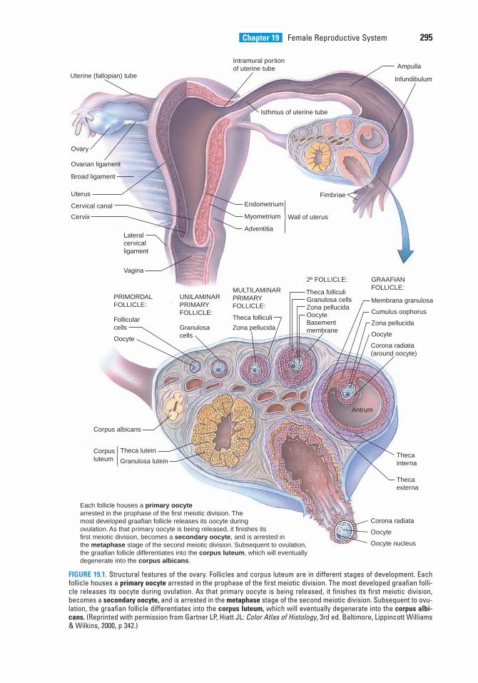
Chapter 19 Female Reproductive System 295
Uterine (fallopian) tube
Intramural portionof uterine tube Ampulla
Infundibulum
Fimbriae
Isthmus of uterine tube
Endometrium
Myometrium
Adventitia
Wall of uterus
Vagina
Lateralcervicalligament
Cervix
Cervical canal
Uterus
Broad ligament
Ovarian ligament
Ovary
PRIMORDALFOLLICLE:
Follicularcells
Oocyte
UNILAMINARPRIMARYFOLLICLE:
Granulosacells
MULTILAMINARPRIMARYFOLLICLE:
Theca folliculi
Zona pellucida
2º FOLLICLE:
Theca folliculiGranulosa cellsZona pellucidaOocyteBasementmembrane
GRAAFIANFOLLICLE:
Membrana granulosa
Cumulus oophorus
Zona pellucida
Oocyte
Corona radiata(around oocyte)
Antrum
Thecainterna
Thecaexterna
Corona radiata
Oocyte
Oocyte nucleus
Corpusluteum
Theca lutein
Granulosa lutein
Corpus albicans
Each follicle houses a primary oocytearrested in the prophase of the first meiotic division. Themost developed graafian follicle releases its oocyte during ovulation. As that primary oocyte is being released, it finishes itsfirst meiotic division, becomes a secondary oocyte, and is arrested inthe metaphase stage of the second meiotic division. Subsequent to ovulation, the gdegenerate into the corpus albicans.
raafian follicle differentiates into the corpus luteum, which will eventually
FIGURE 19.1. Structural features of the ovary. Follicles and corpus luteum are in different stages of development. Eachfollicle houses a primary oocyte arrested in the prophase of the first meiotic division. The most developed graafian folli-cle releases its oocyte during ovulation. As that primary oocyte is being released, it finishes its first meiotic division,becomes a secondary oocyte, and is arrested in the metaphase stage of the second meiotic division. Subsequent to ovu-lation, the graafian follicle differentiates into the corpus luteum, which will eventually degenerate into the corpus albi-cans. (Reprinted with permission from Gartner LP, Hiatt JL: Color Atlas of Histology, 3rd ed. Baltimore, Lippincott Williams& Wilkins, 2000, p 342.)
LWBK615-c19[294-310].qxd 05/31/2010 11:16 AM Page 295 Aptara
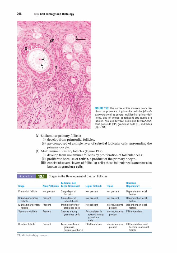
296 BRS Cell Biology and Histology
(a) Unilaminar primary follicles(i) develop from primordial follicles.
(ii) are composed of a single layer of cuboidal follicular cells surrounding theprimary oocyte.
(b) Multilaminar primary follicles (Figure 19.2)(i) develop from unilaminar follicles by proliferation of follicular cells.
(ii) proliferate because of activin, a product of the primary oocyte.(iii) consist of several layers of follicular cells; these follicular cells are now also
known as granulosa cells.
G
ZP
T
FIGURE 19.2. The cortex of this monkey ovary dis-plays the presence of primordial follicles (doublearrows) as well as several multilaminar primary fol-licles, one of whose constituent structures arelabeled. Nucleus (arrow), nucleolus (arrowhead),zona pellucida (ZP), granulosa cells (G), and theca(T) (�270).
t a b l e 19.1 Stages in the Development of Ovarian Follicles
Follicular Cell Hormone Stage Zona Pellucida Layer (Granulosa) Liquor Folliculi Theca Dependency
Primordial follicle Not present Single layer of Not present Not present Dependent on local flat cells factors
Unilaminar primary Present Single layer of Not present Not present Dependent on local follicle cuboidal cells factors
Multilaminar primary Present Multiple layers of Not present Interna, externa Dependent on local follicle granulosa cells present factors
Secondary follicle Present Spaces among Accumulates in Interna, externa FSH dependentgranulosa cells spaces among present
granulosa cells
Graafian follicle Present Forms membrana Fills the antrum Interna, externa FSH dependent until granulosa, present becomes dominant cumulus oophorus follicle
FSH, follicle-stimulating hormone.
LWBK615-c19[294-310].qxd 05/31/2010 11:16 AM Page 296 Aptara
Chapter 19 Female Reproductive System 297
(iv) are circumscribed by two layers of stromal cells: an inner cellular layer(theca interna) and an outer fibrous layer (theca externa). The theca internais separated from the granulosa cells by a basal lamina. Cells of the thecainterna manufacture androstenedione (male sex hormone) and expressluteinizing hormone (LH) receptors on their cell membranes.Androstenedione penetrates the basal lamina, enters the granulosa cellswhere the enzyme aromatase converts the male hormone into estradiol,the female hormone.
(2) Secondary (antral) follicles(a) Secondary follicles are established when fluid (liquor folliculi, an exudate of
plasma containing various hormones, such as activin, estradiol, follistatin,inhibin, and progesterone) begins to accumulate in the spaces between gran-ulosa cells. The fluid-filled spaces will begin to coalesce, eventually to form asingle large cavity called an antrum.
(b) Secondary follicles are dependent on FSH.(c) Narrow processes extend from the granulosa cells into the zona pellucida.(d) Granulosa cells contact each other via gap junctions and also form gap junc-
tions with the cell membrane of the primary oocyte.c. Graafian (mature) follicle
(1) The dominant graafian follicle is the one follicle among the secondary folliclesthat will ovulate. It is FSH independent and manufactures the hormone inhibinthat shuts off FSH release by the basophils of the anterior pituitary, causing atre-sia of the other developing follicles (secondary and nondominant graafian folli-cles).
(2) It measures approximately 2.5 cm in diameter and is evident as a large bulge onthe surface of the ovary.
(3) The primary oocyte is positioned off center on a small mound of granulosa cells(cumulus oophorus) that projects into the liquor folliculi-containing antrum of thefollicle. Granulosa cells surround the zona pellucida. Those contacting the zonapellucida are known as the corona radiata. Other granulosa cells line the antrum,forming the membrana granulosa.
(4) Theca interna cells manufacture androgens, which are transferred to granulosacells, where they are converted into estrogens.
(5) The theca externa is mostly collagenous. It contains a few smooth muscle cells andmany blood vessels, which provide nourishment to the theca interna.
(6) Ovulation(a) An LH surge from the pituitary gland, along with the local factor, meiosis-
inducing substance, triggers the primary oocyte to complete its first meioticdivision just prior to ovulation, forming a secondary oocyte and the first polarbody. The second meiotic division is triggered by the presence of local meiosis-inducing factors but is blocked at metaphase.
(b) Ovulation also occurs in response to the LH surge. The secondary oocyte andits corona radiata cells leave the ruptured follicle at the ovarian surface to enterthe fimbriated end of the oviduct.
2. The corpus hemorrhagicus is formed from the remnants of the graafian follicle.3. Corpus luteum
a. Overview(1) The corpus luteum is formed from the corpus hemorrhagicus.(2) It is composed of granulosa lutein cells (modified granulosa cells) and theca lutein
cells (modified theca interna cells).(3) The formation of this richly vascularized temporary endocrine gland is dependent
on LH.b. Granulosa lutein cells
(1) Granulosa lutein cells are large (30 �m in diameter), pale cells that possess anabundance of smooth endoplasmic reticulum (SER), RER, many mitochondria, awell-developed Golgi complex, and lipid droplets.
LWBK615-c19[294-310].qxd 05/31/2010 11:16 AM Page 297 Aptara
298 BRS Cell Biology and Histology
(2) They are derived from cells of the membrana granulosa.(3) Function. Granulosa lutein cells manufacture most of the body’s progesterone and
convert androgens formed by the theca lutein cells into estrogens.c. Theca lutein cells
(1) These small (15 �m in diameter) cells are concentrated mainly along the periph-ery of the corpus luteum.
(2) They are derived from cells of the theca interna.(3) Function. Theca lutein cells manufacture progesterone and androgens and small
amounts of estrogen.4. The corpus albicans is the remnant of the degenerated corpus luteum. Its formation is
due to the hypoxic conditions present in the corpus luteum as fibroblasts manufacturean overabundance of collagen. The fibrotic event elicits the arrival of T cells that releaseinterferon-�, a chemoattractant for macrophages. These cells release tumor necrosis fac-tor �, a cytokine that drives both granulosa lutein and theca lutein cells into apoptosis.As the cell death and fibrosis progresses, the corpus albicans contracts and becomes asmall scar on the surface of the ovary.
5. Atretic folliclesa. Atretic follicles are follicles (in various stages of maturation) that are undergoing
degeneration.b. They are commonly present in the ovary; after a dominant graafian follicle ovulates,
the remaining graafian and secondary follicles degenerate.c. They often show pyknotic changes in the nuclei of the granulosa cells and other
degenerative changes.
C. The ovarian medulla contains large blood vessels, lymphatic vessels, and nerve fibers in aloose connective tissue stroma. They also possess a small number of estrogen-secretinginterstitial cells and a few androgen-secreting hilus cells.
D. Hormonal regulation (Figure 19.3)1. Control of follicle maturation and ovulation
a. The primary oocyte of unilaminar primary follicles secretes activin, which facilitatesproliferation of granulosa cells.
b. Luteinizing hormone–releasing hormone (LHRH) (also known gonadotropin-releasinghormone or GnRH) from the hypothalamus causes the release of FSH and LH fromthe pars distalis of the pituitary gland.
c. FSH stimulates the growth and development of secondary (but not earlier stage) ovar-ian follicles and the appearance of LH receptors on the granulosa cell plasmalemma.The regulation of FSH and LH is influenced by the following:(1) Theca interna cells manufacture androgens, which are converted to estrogens by
granulosa cells.(2) Granulosa cells also secrete inhibin, follistatin, and activin, all of which (in addi-
tion to estrogen) regulate FSH secretion.(3) By approximately day 14 of the menstrual cycle, estrogen blood levels are suffi-
ciently high to facilitate a sudden, brief surge of LH.d. Surge of LH
(1) A surge of LH triggers the primary oocyte of the dominant graafian follicle to com-plete meiosis I and to enter meiosis II, where it is arrested at metaphase.
(2) The dominant graafian follicle is no longer FSH dependent, and it releases thehormone inhibin that shuts off FSH release by the anterior pituitary, causingatresia of all developing FSH-dependent follicles.
(3) The LH surge initiates ovulation of the secondary oocyte from the graafian follicle.(4) The LH surge promotes formation of the corpus luteum.
2. Fate of the corpus luteuma. Luteal hormones
(1) Progesterone, the major hormone secreted by the corpus luteum, inhibits therelease of LH by suppressing the release of LHRH but promotes development ofthe uterine endometrium.
LWBK615-c19[294-310].qxd 05/31/2010 11:16 AM Page 298 Aptara
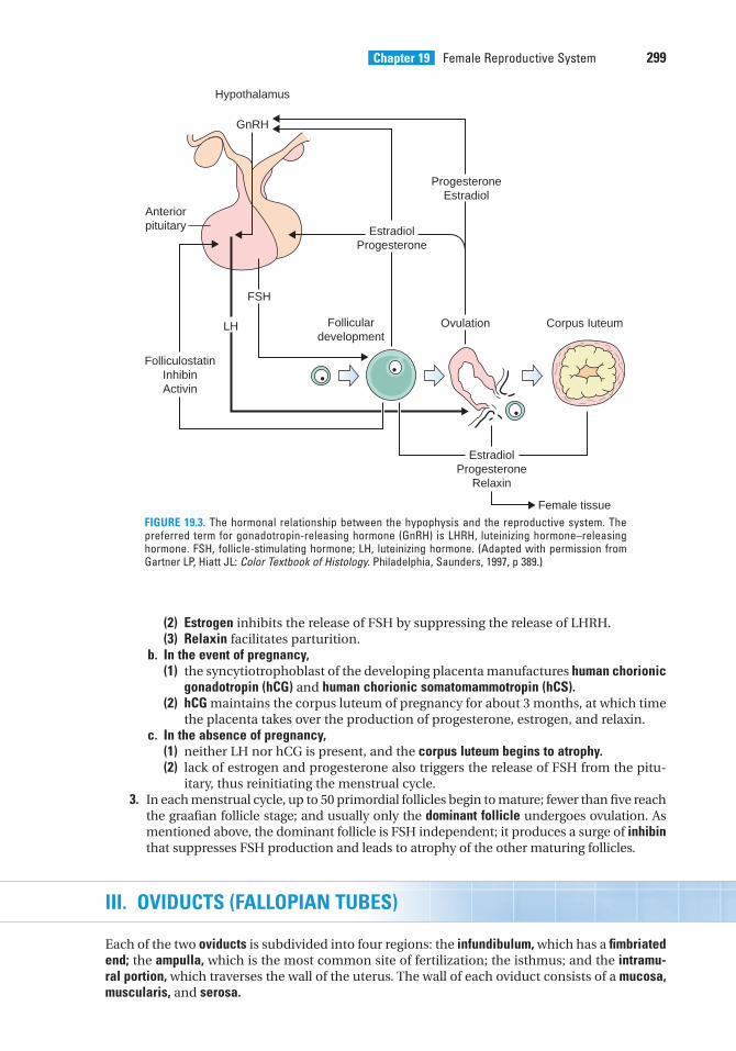
Chapter 19 Female Reproductive System 299
(2) Estrogen inhibits the release of FSH by suppressing the release of LHRH.(3) Relaxin facilitates parturition.
b. In the event of pregnancy,(1) the syncytiotrophoblast of the developing placenta manufactures human chorionic
gonadotropin (hCG) and human chorionic somatomammotropin (hCS).(2) hCG maintains the corpus luteum of pregnancy for about 3 months, at which time
the placenta takes over the production of progesterone, estrogen, and relaxin.c. In the absence of pregnancy,
(1) neither LH nor hCG is present, and the corpus luteum begins to atrophy.(2) lack of estrogen and progesterone also triggers the release of FSH from the pitu-
itary, thus reinitiating the menstrual cycle.3. In each menstrual cycle, up to 50 primordial follicles begin to mature; fewer than five reach
the graafian follicle stage; and usually only the dominant follicle undergoes ovulation. Asmentioned above, the dominant follicle is FSH independent; it produces a surge of inhibinthat suppresses FSH production and leads to atrophy of the other maturing follicles.
III. OVIDUCTS (FALLOPIAN TUBES)
Each of the two oviducts is subdivided into four regions: the infundibulum, which has a fimbriatedend; the ampulla, which is the most common site of fertilization; the isthmus; and the intramu-ral portion, which traverses the wall of the uterus. The wall of each oviduct consists of a mucosa,muscularis, and serosa.
Hypothalamus
GnRH
Anteriorpituitary
LH
FolliculostatinInhibinActivin
Folliculardevelopment
EstradiolProgesterone
ProgesteroneEstradiol
Ovulation Corpus Iuteum
Female tissue
EstradiolProgesterone
Relaxin
FSH
FIGURE 19.3. The hormonal relationship between the hypophysis and the reproductive system. Thepreferred term for gonadotropin-releasing hormone (GnRH) is LHRH, luteinizing hormone–releasinghormone. FSH, follicle-stimulating hormone; LH, luteinizing hormone. (Adapted with permission fromGartner LP, Hiatt JL: Color Textbook of Histology. Philadelphia, Saunders, 1997, p 389.)
LWBK615-c19[294-310].qxd 05/31/2010 11:16 AM Page 299 Aptara
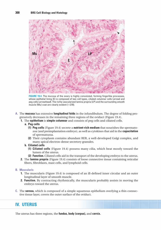
300 BRS Cell Biology and Histology
A. The mucosa has extensive longitudinal folds in the infundibulum. The degree of folding pro-gressively decreases in the remaining three regions of the oviduct (Figure 19.4).1. The epithelium is simple columnar and consists of peg cells and ciliated cells.
a. Peg cells(1) Peg cells (Figure 19.4) secrete a nutrient-rich medium that nourishes the spermato-
zoa (and preimplantation embryo), as well as cytokines that aid in the capacitationof spermatozoa.
(2) Their cytoplasm contains abundant RER, a well-developed Golgi complex, andmany apical electron-dense secretory granules.
b. Ciliated cells(1) Ciliated cells (Figure 19.4) possess many cilia, which beat mostly toward the
lumen of the uterus.(2) Function. Ciliated cells aid in the transport of the developing embryo to the uterus.
2. The lamina propria (Figure 19.4) consists of loose connective tissue containing reticularfibers, fibroblasts, mast cells, and lymphoid cells.
B. Muscularis1. The muscularis (Figure 19.4) is composed of an ill-defined inner circular and an outer
longitudinal layer of smooth muscle.2. Function. By contracting rhythmically, the muscularis probably assists in moving the
embryo toward the uterus.
C. The serosa, which is composed of a simple squamous epithelium overlying a thin connec-tive tissue layer, covers the outer surface of the oviduct.
IV. UTERUS
The uterus has three regions, the fundus, body (corpus), and cervix.
E
LPMu
FIGURE 19.4. The mucosa of the ovary is highly convoluted, forming fingerlike processes,whose epithelial lining (E) is composed of two cell types, ciliated columnar cells (arrow) andpeg cells (arrowhead). The richly vascularized lamina propria (LP) and the surrounding smoothmuscle (Mu) coat are clearly evident (�270).
LWBK615-c19[294-310].qxd 05/31/2010 11:16 AM Page 300 Aptara
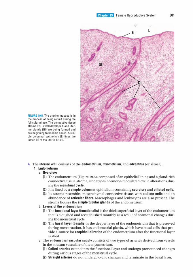
Chapter 19 Female Reproductive System 301
A. The uterine wall consists of the endometrium, myometrium, and adventitia (or serosa).1. Endometrium
a. Overview(1) The endometrium (Figure 19.5), composed of an epithelial lining and a gland-rich
connective tissue stroma, undergoes hormone-modulated cyclic alterations dur-ing the menstrual cycle.
(2) It is lined by a simple columnar epithelium containing secretory and ciliated cells.(3) Its stroma resembles mesenchymal connective tissue, with stellate cells and an
abundance of reticular fibers. Macrophages and leukocytes are also present. Thestroma houses the simple tubular glands of the endometrium.
b. Layers of the endometrium(1) The functional layer (functionalis) is the thick superficial layer of the endometrium
that is sloughed and reestablished monthly as a result of hormonal changes dur-ing the menstrual cycle.
(2) The basal layer (basalis) is the deeper layer of the endometrium that is preservedduring menstruation. It has endometrial glands, which have basal cells that pro-vide a source for reepithelialization of the endometrium after the functional layeris shed.
c. The endometrial vascular supply consists of two types of arteries derived from vesselsin the stratum vasculare of the myometrium.(1) Coiled arteries extend into the functional layer and undergo pronounced changes
during various stages of the menstrual cycle.(2) Straight arteries do not undergo cyclic changes and terminate in the basal layer.
St
Gl
E L
FIGURE 19.5. The uterine mucosa is inthe process of being rebuilt during thefollicular phase. The connective tissuestroma (St) is well developed, and uter-ine glands (Gl) are being formed andare beginning to become coiled. A sim-ple columnar epithelium (E) lines thelumen (L) of the uterus (�53).
LWBK615-c19[294-310].qxd 05/31/2010 11:16 AM Page 301 Aptara

302 BRS Cell Biology and Histology
2. Myometriuma. The myometrium is the thick smooth muscle tunic of the uterus.b. It is composed of inner and outer longitudinal layers and a thick middle circular layer.
The circular layer is richly vascularized and is often referred to as the stratum vascu-lare.
c. The myometrium thickens during pregnancy because of the hypertrophy and hyper-plasia of individual smooth muscle cells.
d. Near the end of pregnancy, the myometrium develops many gap junctions betweenits smooth muscle cells. These junctions coordinate contraction of the muscle cellsduring parturition.
e. At parturition, the myometrium undergoes powerful contractions triggered by thehormone oxytocin and by prostaglandins (both of which are increased at term).
f. After parturition, the myometrium shrinks because many of the smooth muscle cellsbecome deprived of estrogen and, therefore, undergo apoptosis.
3. External coveringa. Serosa is present over surfaces of the uterus bulging into the peritoneal cavity.b. Adventitia is present along the retroperitoneal surfaces of the uterus.
B. The menstrual cycle begins on the day menstrual bleeding appears.1. Menstrual phase (days 1–4) is characterized by a hemorrhagic discharge (menses) of the
functional layer of the endometrium.a. It is triggered by spasms of contraction and relaxation of the coiled arteries (caused by
low levels of progesterone and estrogen). Long-term (2–3 days) vasoconstriction ofthese arteries causes ischemia and eventual necrosis.
b. Vasoconstriction is followed by sudden, intermittent vasodilation of the coiled arter-ies, which ruptures their walls, flooding the stroma with blood, detaching the func-tional layer, and dislodging the necrotic tissue.
c. Because the basal layer is supplied by short straight vessels that do not undergo pro-longed vasoconstriction, it is not sloughed and does not become necrotic.
2. The proliferative (follicular) phase (Figure 19.5), days 4 to 14, follows the menstrual phaseand involves renewal of the entire functional layer, including the repair of glands, con-nective tissue, and vascular elements (specifically, the coiled arteries).a. The epithelium that lines the luminal surface of the uterus is renewed by mitotic
activity of cells remaining in uterine glands of the basal layer of the endometrium.b. Glands are straight and lined by a simple columnar epithelium.c. Stromal cells divide, accumulate glycogen, and enlarge.d. Coiled arteries extend approximately two-thirds of the way into the endometrium.
3. The secretory (luteal) phase (days 15–28) begins shortly after ovulation and is character-ized by a thickening of the endometrium, resulting from edema and secretion by theendometrial glands.a. Glands become coiled; their lumina become filled with a secretion of glycoprotein
material; and their cells accumulate large amounts of glycogen, in the basal aspect oftheir cytoplasm.
b. Coiled arteries become not only more highly coiled but also longer, extending into thesuperficial aspects of the functional layer.
Endometriosis is a condition in which the pelvic peritoneal cavitycontains uterine endometrial tissue. It is associated with hormone-
induced changes in the ectopic endometrium during the menstrual cycle. As the endometrium isshed, bleeding occurs in the peritoneal cavity, causing severe pain and the formation of cysts andadhesions. It may lead to sterility because the ovaries and oviducts become deformed andembedded in scar tissue.
CLINICAL
CONSIDERATIONS
LWBK615-c19[294-310].qxd 05/31/2010 11:16 AM Page 302 Aptara
Chapter 19 Female Reproductive System 303
V. CERVIX
A. The cervix does not participate in menstruation, but its secretions change during variousstages of the menstrual cycle.
B. The cervical wall is composed mainly of dense collagenous connective tissue interspersedwith numerous elastic fibers and a few smooth muscle cells.
C. The cervix has a simple columnar (mucus-secreting) epithelium except for the inferior portion(continuous with the lining of the vagina), which is covered by a stratified squamous nonker-atinized epithelium.
D. Branched cervical glands secrete a serous fluid near the time of ovulation that facilitates the entryof spermatozoa into the uterine lumen. During pregnancy, cervical glands produce a thick, vis-cous secretion that hinders the entry of spermatozoa (and microorganisms) into the uterus.
E. Prior to parturition, the cervix dilates and softens as a result of the lysis of the collagen fiberbundles in response to the hormone relaxin.
1. In a Papanicolaou (Pap) smear, epithelial cells are scraped from the lining of the cervix (or vagina) and are examined to detect cervical
cancer. A Pap smear shows variation in cell populations with stages of the menstrual cycle.2. Carcinoma of the cervix originates from stratified squamous nonkeratinized epithelial cells. It may
be contained within the epithelium and not invade the underlying stroma (carcinoma in situ), or itmay penetrate the basal lamina and metastasize to other parts of the body (invasive carcinoma).It occurs at a relatively high frequency but may be cured by surgery if discovered early (by Papsmear), before it becomes invasive.
CLINICAL
CONSIDERATIONS
VI. FERTILIZATION AND IMPLANTATION
A. Fertilization1. Fertilization usually takes place within the ampulla of the oviduct.2. It occurs when a spermatozoon penetrates the corona radiata and the zona pellucida
and pierces the plasma membrane of a secondary oocyte.3. It triggers the resumption and completion of the second meiotic division with the for-
mation of two new cells, the ovum and the second polar body.4. It is completed when the male haploid (n) pronucleus (derived from the spermatozoon)
and the female haploid (n) pronucleus (derived from the ovum) fuse, forming a diploid(2n) cell known as a zygote.
The default gender of the embryo is female and in order for a maleembryo to be developed, the presence of the SRY gene (sex-determining
region of the Y chromosome) and SOX9, both carried on the Y chromosome, are required. The SRYgene codes for the testis-determining factor, which is responsible for the formation of the testesputatively because it inhibits the female determining genes, DAX1 and WNT4, located on the X chro-mosomes. It is interesting to note that even as minor a mutation as a base-pair alteration in the SRYgene results in the development of a female rather than a male and, with an overabundance of DAX1genes, a genotypical male (XY) is phenotypically a female. Moreover, recent studies havedemonstrated that numerous other genes located on a number of chromosomes may play anywherefrom significant to secondary roles in gender expression.
CLINICAL
CONSIDERATIONS
LWBK615-c19[294-310].qxd 05/31/2010 11:16 AM Page 303 Aptara
304 BRS Cell Biology and Histology
B. Implantation1. The zygote undergoes mitotic cell division (known as cleavage during the early stages of
embryogenesis) and is transformed into a multicellular structure called a morula, whichrequires about 3 days to travel through the oviduct and enter the uterus.
2. The conceptus (the preimplantation embryo and its surrounding membranes) acquiresa fluid-filled cavity and becomes a blastocyst.
3. The blastocyst implants in the endometrium of the uterus and is surrounded by an innercellular layer, the cytotrophoblast, and an outer multinucleated layer, the syncytiotro-phoblast.
4. The syncytiotrophoblast further invades the endometrium in the wall of the uterus by thesixth day after fertilization. Formation of the placenta then begins.
1. Ectopic (tubal) pregnancy is the implantation of the early embryo in anabnormal site (e.g., wall of the oviduct). It can be fatal without immediate
medical intervention.2. Teratomas are germ cell tumors that fall into three groups: mature, monodermal, and immature.
a. Mature teratomas are benign (although occasionally they may become malignant) and are usu-ally present in young women. These are cysts with walls that frequently contain hair and otherepidermal structures such as sebaceous glands, as well as bone, tooth, and cartilage fragments.
b. Monodermal teratomas are rare tumors that are also known as specialized teratomas. Thetwo most frequent types of these tumors are struma ovarii and ovarian carcinoid. Strumaovarii is an ovarian tumor composed of well-developed thyroid follicles that produce thyroidhormone and may be responsible for hyperthyroidism. Ovarian carcinoid is a tumor that usu-ally produces serotonin (5-OH-tryptamine).
c. Immature teratomas are fast-growing malignant tumors with a histology that resembles that offetal rather than mature tissues. They are usually present in adolescents and very young women.
CLINICAL
CONSIDERATIONS
VII. PLACENTA
A. The placenta is a transient structure, consisting of a maternal portion and a fetal portion.
B. Function1. The placenta permits the exchange of various materials between the maternal and fetal
circulatory systems. This exchange occurs without mixing of the two separate blood sup-plies.
2. It secretes progesterone, hCG, chorionic thyrotropin, and hCS, a lactogenic and growth-promoting hormone.
3. It also produces estrogen with the assistance of the liver and adrenal cortex of the fetus.4. Decidual cells of the stroma produce prostaglandins and prolactin.
The polypeptide hormone prolactin, manufactured by basophils of theadenohypophysis (anterior pituitary), causes an increase in breast size in
the pregnant female and induces milk formation as well as lactation in the mammary glands of thepostpartum female. Interestingly, it also causes the feeling of sexual gratification subsequent to thesexual act. Because progesterone has been shown to alleviate the symptoms of multiple sclerosis(MS) in the pregnant female, recent investigations using a mouse model demonstrated thattreatment of MS-afflicted mice with prolactin resulted in remyelination of nerve fibers, whose myelinwas destroyed by the disease. It is believed that prolactin stimulates the formation or prooligoden-drocytes, which differentiate into oligodendrocytes, cells that are responsible for myelination ofaxons in the central nervous system.
CLINICAL
CONSIDERATIONS
LWBK615-c19[294-310].qxd 05/31/2010 11:16 AM Page 304 Aptara
Chapter 19 Female Reproductive System 305
VIII. VAGINA
A. Overview1. The vagina is a fibromuscular canal with a wall that is composed of three layers: an inner
mucosa, a middle muscularis, and an external adventitia.2. It is circumscribed by a skeletal muscle sphincter at its external orifice.3. It lacks glands throughout its length and is lubricated by secretions from the cervix and
by seepage of the extracellular fluid from the vascular supply of the lamina propria.
B. The mucosa is composed of a thick, stratified squamous nonkeratinized epithelium and a fibro-elastic connective tissue, the lamina propria.1. The epithelium contains glycogen, which is used by the vaginal bacterial flora to produce
lactic acid, an acid that lowers the pH during the follicular phase of the menstrual cycleand inhibits invasion by pathogens.
2. The lamina propria is a fibroelastic connective tissue that is highly vascular in its deeperaspect (which is possibly analogous to a submucosa).
C. The muscularis is composed of irregularly arranged layers of smooth muscle (thin inner cir-cular layer and a thicker outer longitudinal layer) interspersed with elastic fibers.
D. The adventitia is composed of fibroelastic connective tissue. It attaches the vagina to the sur-rounding structures.
IX. EXTERNAL GENITALIA (VULVA)
A. The labia majora are fat-laden folds of skin; hair and secretions of sebaceous glands and sweatglands are present on their external surfaces.
B. Labia minora1. The labia minora are folds of skin that possess a core of highly vascular connective tissue
containing elastic fibers.2. They lack hair follicles, but their dermis contains numerous sebaceous glands, which
open directly onto the epithelial surface.
C. The vestibule is the space between the two labia minora. Glands of Bartholin (mucus-secretingglands) and numerous smaller mucus-secreting glands around the urethra and clitoris(minor vestibular glands) open into this space.
D. Clitoris1. The clitoris is composed of two small, cylindrical erectile bodies, which terminate in the
prepuce-covered glans clitoridis.2. It contains many sensory nerve fibers and specialized nerve endings (e.g., Meissner cor-
puscles and pacinian corpuscles).
X. MAMMARY GLANDS
Mammary glands of both genders are identical for the first decade or so of life, but when thefemale reaches puberty, the flow of estrogens and progesterone as well as lactogenic hormoneinduces the mammary gland to enlarge and develop a system of lobules and terminal ductulesas well as an increase in the connective tissue mass and a deposit of adipose tissue. Each mam-mary gland of the postpubertal female is composed of numerous compound tubuloalveolarglands, each with its own lactiferous sinus and a duct that opens at the apex of the nipple.
LWBK615-c19[294-310].qxd 05/31/2010 11:16 AM Page 305 Aptara
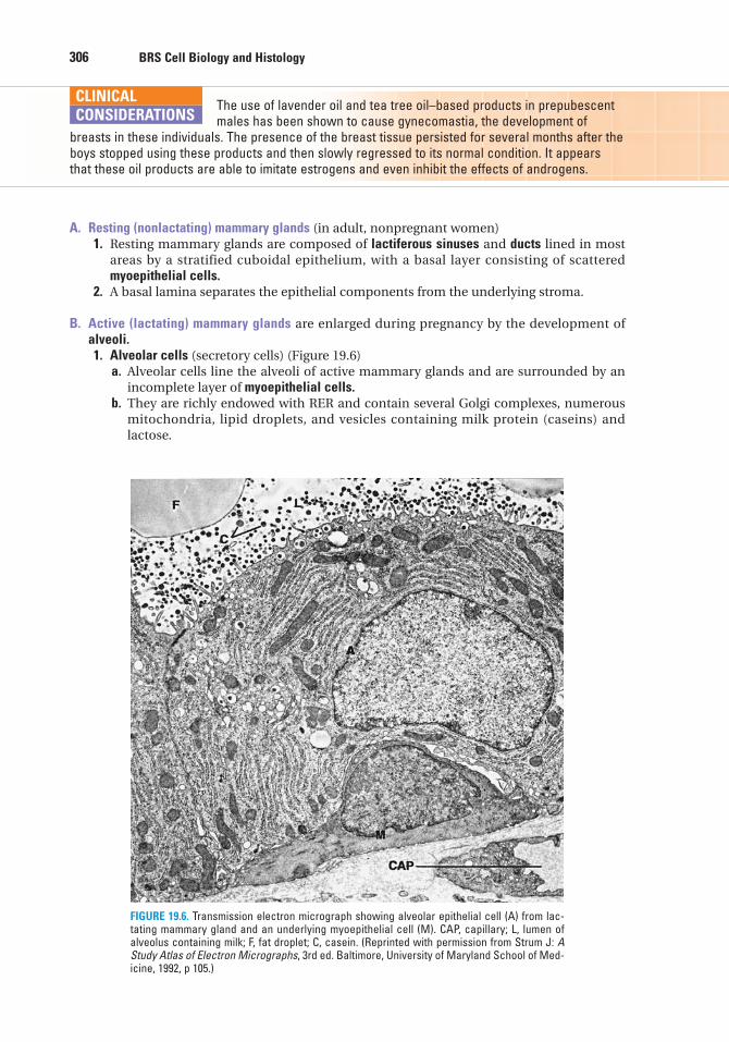
306 BRS Cell Biology and Histology
A. Resting (nonlactating) mammary glands (in adult, nonpregnant women)1. Resting mammary glands are composed of lactiferous sinuses and ducts lined in most
areas by a stratified cuboidal epithelium, with a basal layer consisting of scatteredmyoepithelial cells.
2. A basal lamina separates the epithelial components from the underlying stroma.
B. Active (lactating) mammary glands are enlarged during pregnancy by the development ofalveoli.1. Alveolar cells (secretory cells) (Figure 19.6)
a. Alveolar cells line the alveoli of active mammary glands and are surrounded by anincomplete layer of myoepithelial cells.
b. They are richly endowed with RER and contain several Golgi complexes, numerousmitochondria, lipid droplets, and vesicles containing milk protein (caseins) and lactose.
The use of lavender oil and tea tree oil–based products in prepubescentmales has been shown to cause gynecomastia, the development of
breasts in these individuals. The presence of the breast tissue persisted for several months after theboys stopped using these products and then slowly regressed to its normal condition. It appearsthat these oil products are able to imitate estrogens and even inhibit the effects of androgens.
CLINICAL
CONSIDERATIONS
FIGURE 19.6. Transmission electron micrograph showing alveolar epithelial cell (A) from lac-tating mammary gland and an underlying myoepithelial cell (M). CAP, capillary; L, lumen ofalveolus containing milk; F, fat droplet; C, casein. (Reprinted with permission from Strum J: AStudy Atlas of Electron Micrographs, 3rd ed. Baltimore, University of Maryland School of Med-icine, 1992, p 105.)
LWBK615-c19[294-310].qxd 05/31/2010 11:16 AM Page 306 Aptara
Chapter 19 Female Reproductive System 307
2. Secretion by alveolar cellsa. Lipids are released into the lumen, perhaps, via the apocrine mode of secretion.b. Proteins and sugars are released into the alveolar lumen via the merocrine mode of
secretion (exocytosis).
C. Nipple1. The nipple is composed of dense, irregular collagenous connective tissue interlaced with
smooth muscle fibers that act as a sphincter.2. It contains the openings of the lactiferous ducts.3. It is surrounded by pigmented skin (areola) that is more deeply pigmented during and
subsequent to pregnancy and contains the areolar glands (of Montgomery).
1. Breast cancer may originate from the epithelium lining the ducts (ductal carcinoma) or the terminal ductules (lobular carcinoma). If
breast cancer is not treated early, the tumor cells metastasize via lymphatic vessels to theaxillary nodes near the affected breast and later via the bloodstream to the lungs, bone, andbrain. In the United States, 180,000 new cases of breast cancer are diagnosed annually, andevery year 43,000 women die of this disease. Early detection by self-examination, mammography,or ultrasound has led to a reduction in the mortality rate associated with breast cancer.
2. Deficiency or mutation in the gene BRCA1 has been shown to decrease the stability or elevatethe incidence of the mutation rate of tumor suppressor genes such as p53. It appears that muta-tions in the BRCA1 gene result in incapacitation of the checkpoint at G2-M of the cell cycle, andconcurrently, the number of centrosomes of these cells is increased. Therefore, these mutatedcells have the capability to proliferate unchecked.
CLINICAL
CONSIDERATIONS
D. Secretions of the mammary glands1. Colostrum (protein-rich yellowish fluid)
a. Colostrum is produced during the first few days after birth.b. It is rich in cells (lymphocytes, monocytes), lactalbumin, fat-soluble vitamins, and
minerals and contains immunoglobulin A (IgA).2. Milk
a. Milk begins to be secreted by the third or fourth day after birth.b. Milk consists of proteins (caseins, IgA, lactalbumin), many lipid droplets, and lactose.c. It is released from the mammary glands via the milk ejection reflex in response to a
variety of external stimuli related to suckling. The milk ejection reflex involves releaseof oxytocin (from axons in the pars nervosa of the pituitary gland), which inducescontraction of the myoepithelial cells, forcing milk into the larger ducts and out of thebreast.
LWBK615-c19[294-310].qxd 05/31/2010 11:16 AM Page 307 Aptara
5. The basal layer of the uterineendometrium
(A) is sloughed during menstruation.(B) has no glands.(C) is supplied by coiled arteries.(D) is supplied by straight arteries.(E) is avascular.
6. One of the recognized phases of the men-strual cycle is termed the
(A) gestational phase.(B) active phase.(C) follicular phase.(D) resting phase.(E) anaphase.
7. During the proliferative phase of themenstrual cycle, the functional layer of theendometrium undergoes which of thefollowing changes?
(A) Blood vessels become ischemic.(B) The epithelium is renewed.(C) The stroma swells because of edema.(D) Glands become coiled.(E) Blood vessels break down.
8. Which of the following statementsconcerning the vaginal mucosa is true?
(A) It is lined by stratified columnar epithe-lium.
(B) It is lined by stratified squamouskeratinized epithelium.
(C) It possesses no elastic fibers.(D) It is lubricated by glands in the cervix.(E) Its cells secrete lactic acid.
1. Which of the following statementsconcerning secondary ovarian follicles istrue?
(A) They lack liquor folliculi.(B) They contain a secondary oocyte.(C) Their continued maturation requires
follicle-stimulating hormone.(D) They lack a theca externa.(E) They have a single layer of cuboidal fol-
licular cells surrounding the oocyte.
2. Colostrum contains which of the follow-ing antibodies?
(A) IgA(B) IgD(C) IgE(D) IgG(E) IgM
3. Which of the following statementsconcerning the corpus luteum is true?
(A) It produces LH.(B) It produces FSH.(C) It derives its granulosa luteal cells from
the theca externa.(D) It becomes the corpus albicans.(E) It is derived from atretic follicles.
4. LH exerts which one of the followingphysiological effects?
(A) It triggers completion of the second mei-otic division by secondary oocytes.
(B) It triggers ovulation.(C) It suppresses release of estrogens.(D) It induces primary follicles to become
secondary follicles.(E) It induces primordial follicles to become
primary follicles
Review Test
Directions: Each of the numbered items or incomplete statements in this section is followed byanswers or completions of the statement. Select the ONE lettered answer that is BEST in eachcase.
308
LWBK615-c19[294-310].qxd 05/31/2010 11:16 AM Page 308 Aptara

Chapter 19 Female Reproductive System 309
9. Which of the following statementsconcerning the oviduct is true?
(A) It is lined by a simple cuboidalepithelium.
(B) Its epithelium contains peg cells.(C) It functions in nourishing trilaminar
germ discs.(D) Fertilization most often occurs in its fim-
briated portion.(E) Its epithelium contains goblet cells.
10. Which one of the following teratomas isa malignant, fast-growing tumor?
(A) Mature teratoma(B) Monodermal teratoma(C) Immature teratoma(D) Struma ovarii(E) Ovarian carcinoid
LWBK615-c19[294-310].qxd 05/31/2010 11:16 AM Page 309 Aptara
Answers and Explanations
1. C. Secondary follicles depend on follicle-stimulating hormone for their continued devel-opment. They are established when liquor folliculi (an ultrafiltrate of plasma andgranulosa cell secretions) begins to accumulate among the granulosa cells. Secondary folli-cles contain a primary oocyte blocked in the prophase of meiosis I (see Chapter 19 II B 1 b).
2. A. IgA antibodies are present in colostrum and milk. IgG antibodies are acquired by thefetus by placental transfer from the mother (see Chapter 19 X D 1).
3. D. A corpus albicans is formed from a corpus luteum that has ceased to function. LH andFSH are both produced in the anterior pituitary gland. Granulosa luteal cells are derivedfrom the granulosa cells of an ovulated graafian follicle (see Chapter 19 II B 4).
4. B. A sudden surge of LH near the middle of the menstrual cycle triggers ovulation (seeChapter 19 II D 1 d).
5. D. The basal layer of the uterine endometrium is supplied by the straight arteries and con-tains the deeper portions of the uterine glands. Cells from these glands reepithelialize theendometrial surface after the functional layer (supplied by the coiled arteries) has beensloughed (see Chapter 19 IV A 1 b).
6. C. The recognized phases of the menstrual cycle are the follicular (proliferative), secretory(luteal), and menstrual phases. The mammary glands are characterized by active(lactating) and resting phases. The term gestational phase refers to pregnancy (see Chapter19 IV B 3).
7. B. During the proliferative phase of the menstrual cycle, the entire functional layer of theendometrium is renewed, including the epithelium lining the surface and glands. Edemain the stroma and coiled glands is characteristic of the secretory phase of the cycle, andischemia is responsible for the menstrual phase (see Chapter 19 IV B 2).
8. D. The vagina lacks glands and is lubricated by secretions from cervical glands. It is linedby a stratified squamous nonkeratinized epithelium with cells that release glycogen, whichis used by the normal bacterial flora of the vagina to manufacture lactic acid (see Chapter19 VIII A 3).
9. B. The oviduct is lined by a simple columnar epithelium composed of ciliated cells andpeg cells but no goblet cells. Fertilization most often occurs in the ampulla of the oviduct,not in the infundibulum, where fimbriae are located. Under normal circumstances, thetrilaminar germ disk stage occurs after the blastocyst is implanted in the wall of the uterusand is not present in the oviduct (see Chapter 19 III A 1).
10. C. Immature teratomas are fast-growing malignant tumors. The other teratomas listed areusually benign, but even those that do become malignant are slow growing (see Chapter19 VI B 4).
310
LWBK615-c19[294-310].qxd 05/31/2010 11:16 AM Page 310 Aptara
c h a p t e r 20 Male ReproductiveSystem
311
I. OVERVIEW—MALE REPRODUCTIVE SYSTEM
A. The male reproductive system consists of the testes, genital ducts, accessory genital glands(seminal vesicles, prostate gland, and bulbourethral glands), and the penis.
B. Function. The male reproductive system produces spermatozoa (sperm), testosterone, andseminal fluid. Seminal fluid transports and nourishes the sperm as they pass through theexcretory ducts. The penis delivers sperm to the exterior and also serves as the conduit forexcretion of urine from the body.
II. TESTES
Testes develop in the abdominal cavity and later descend into the scrotum, where they are sus-pended at the ends of the spermatic cords. They are the sites of spermatogenesis and productionof the male sex hormones, primarily testosterone (Figure 20.1).
Cryptorchidism1. Cryptorchidism is a developmental defect characterized by failure of
the testes to descend into the scrotum.2. This condition results in sterility because the temperature of the undescended testes (i.e., normal
body temperature) inhibits spermatogenesis; however, it does not affect testosterone production.3. It is associated with a much higher incidence of testicular malignancy than in normally
descended testes.4. It can be surgically corrected, usually between 5 and 7 years of age. After corrective surgery,
however, affected individuals may have abnormal sperm.
CLINICAL
CONSIDERATIONS
A. Testicular tunicae (covering of the testes)1. The tunica vaginalis is a serous sac derived from the peritoneum that partially covers the
anterior and lateral surfaces of each testis.2. Tunica albuginea
a. Tunica albuginea is the thick, fibrous connective tissue capsule of the testis.b. It is lined by a highly vascular layer of loose connective tissue, the tunica vasculosa.c. It is thickened posteriorly to form the mediastinum testis, from which incomplete con-
nective tissue septa arise to divide the organ into approximately 250 compartments(lobuli testis).
LWBK615-c20[311-325].qxd 05/31/2010 11:03 AM Page 311 Aptara
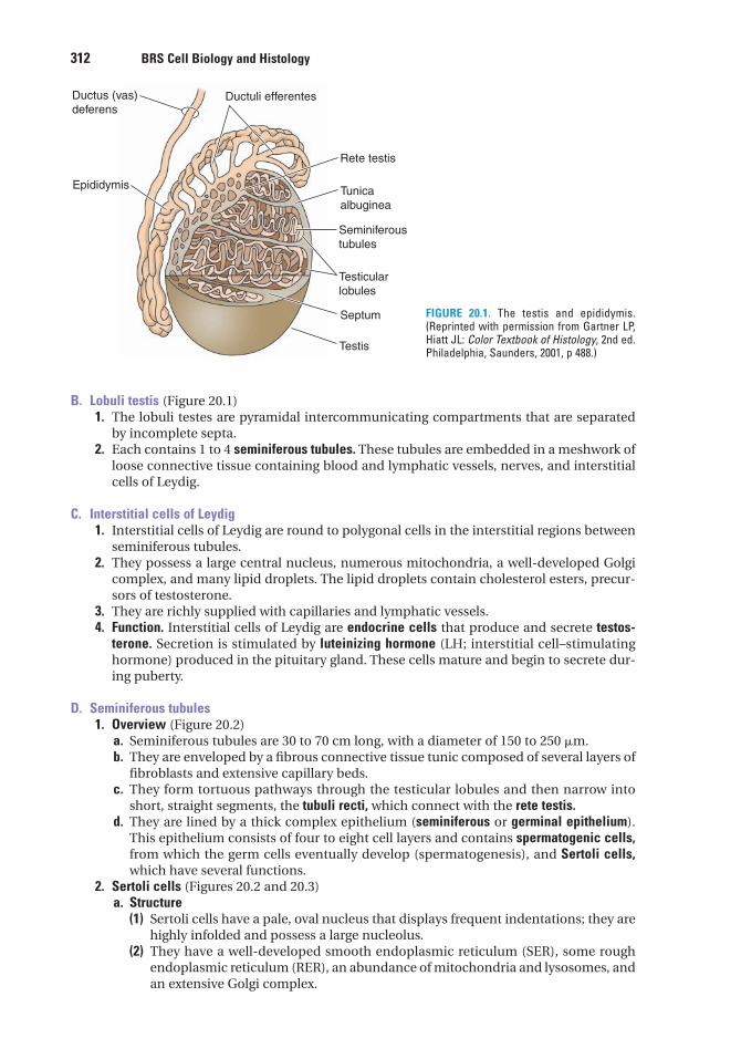
312 BRS Cell Biology and Histology
B. Lobuli testis (Figure 20.1)1. The lobuli testes are pyramidal intercommunicating compartments that are separated
by incomplete septa.2. Each contains 1 to 4 seminiferous tubules. These tubules are embedded in a meshwork of
loose connective tissue containing blood and lymphatic vessels, nerves, and interstitialcells of Leydig.
C. Interstitial cells of Leydig1. Interstitial cells of Leydig are round to polygonal cells in the interstitial regions between
seminiferous tubules.2. They possess a large central nucleus, numerous mitochondria, a well-developed Golgi
complex, and many lipid droplets. The lipid droplets contain cholesterol esters, precur-sors of testosterone.
3. They are richly supplied with capillaries and lymphatic vessels.4. Function. Interstitial cells of Leydig are endocrine cells that produce and secrete testos-
terone. Secretion is stimulated by luteinizing hormone (LH; interstitial cell–stimulatinghormone) produced in the pituitary gland. These cells mature and begin to secrete dur-ing puberty.
D. Seminiferous tubules1. Overview (Figure 20.2)
a. Seminiferous tubules are 30 to 70 cm long, with a diameter of 150 to 250 �m.b. They are enveloped by a fibrous connective tissue tunic composed of several layers of
fibroblasts and extensive capillary beds.c. They form tortuous pathways through the testicular lobules and then narrow into
short, straight segments, the tubuli recti, which connect with the rete testis.d. They are lined by a thick complex epithelium (seminiferous or germinal epithelium).
This epithelium consists of four to eight cell layers and contains spermatogenic cells,from which the germ cells eventually develop (spermatogenesis), and Sertoli cells,which have several functions.
2. Sertoli cells (Figures 20.2 and 20.3)a. Structure
(1) Sertoli cells have a pale, oval nucleus that displays frequent indentations; they arehighly infolded and possess a large nucleolus.
(2) They have a well-developed smooth endoplasmic reticulum (SER), some roughendoplasmic reticulum (RER), an abundance of mitochondria and lysosomes, andan extensive Golgi complex.
Ductus (vas)
deferensDuctuli efferentes
Rete testis
Tunica
albuginea
Seminiferous
tubules
Testicular
lobules
Septum
Testis
Epididymis
FIGURE 20.1. The testis and epididymis.(Reprinted with permission from Gartner LP,Hiatt JL: Color Textbook of Histology, 2nd ed.Philadelphia, Saunders, 2001, p 488.)
LWBK615-c20[311-325].qxd 05/31/2010 11:03 AM Page 312 Aptara
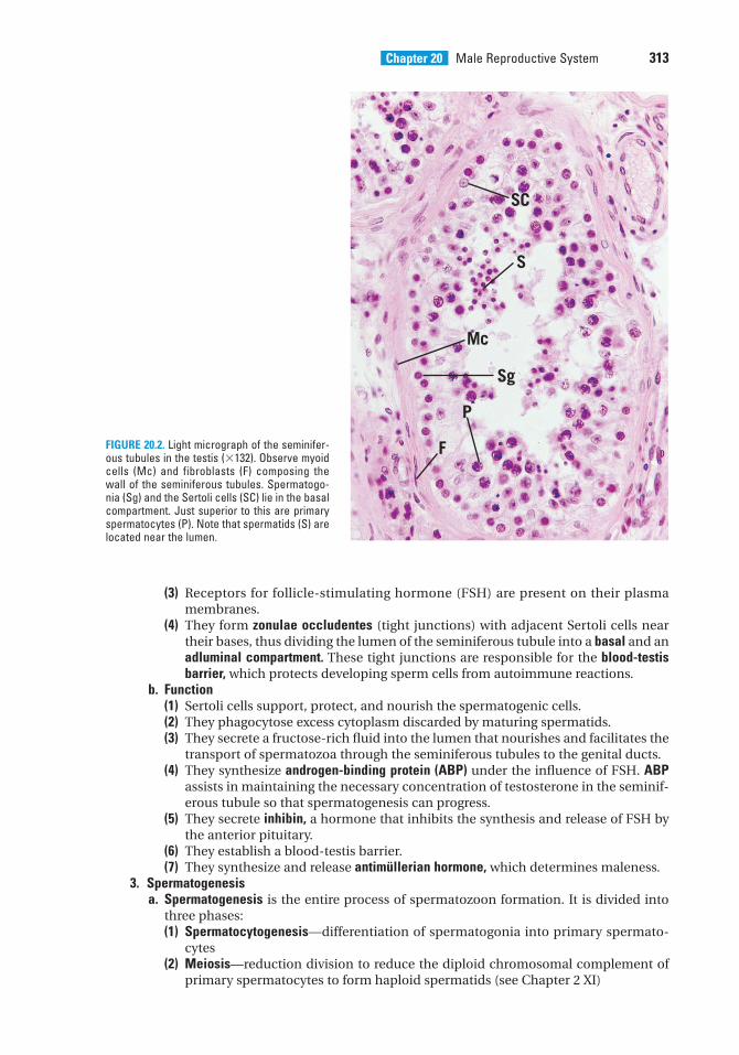
Chapter 20 Male Reproductive System 313
(3) Receptors for follicle-stimulating hormone (FSH) are present on their plasmamembranes.
(4) They form zonulae occludentes (tight junctions) with adjacent Sertoli cells neartheir bases, thus dividing the lumen of the seminiferous tubule into a basal and anadluminal compartment. These tight junctions are responsible for the blood-testisbarrier, which protects developing sperm cells from autoimmune reactions.
b. Function(1) Sertoli cells support, protect, and nourish the spermatogenic cells.(2) They phagocytose excess cytoplasm discarded by maturing spermatids.(3) They secrete a fructose-rich fluid into the lumen that nourishes and facilitates the
transport of spermatozoa through the seminiferous tubules to the genital ducts.(4) They synthesize androgen-binding protein (ABP) under the influence of FSH. ABP
assists in maintaining the necessary concentration of testosterone in the seminif-erous tubule so that spermatogenesis can progress.
(5) They secrete inhibin, a hormone that inhibits the synthesis and release of FSH bythe anterior pituitary.
(6) They establish a blood-testis barrier.(7) They synthesize and release antimüllerian hormone, which determines maleness.
3. Spermatogenesisa. Spermatogenesis is the entire process of spermatozoon formation. It is divided into
three phases:(1) Spermatocytogenesis—differentiation of spermatogonia into primary spermato-
cytes(2) Meiosis—reduction division to reduce the diploid chromosomal complement of
primary spermatocytes to form haploid spermatids (see Chapter 2 XI)
P
S
Sg
SC
Mc
FFIGURE 20.2. Light micrograph of the seminifer-ous tubules in the testis (�132). Observe myoidcells (Mc) and fibroblasts (F) composing thewall of the seminiferous tubules. Spermatogo-nia (Sg) and the Sertoli cells (SC) lie in the basalcompartment. Just superior to this are primaryspermatocytes (P). Note that spermatids (S) arelocated near the lumen.
LWBK615-c20[311-325].qxd 05/31/2010 11:03 AM Page 313 Aptara
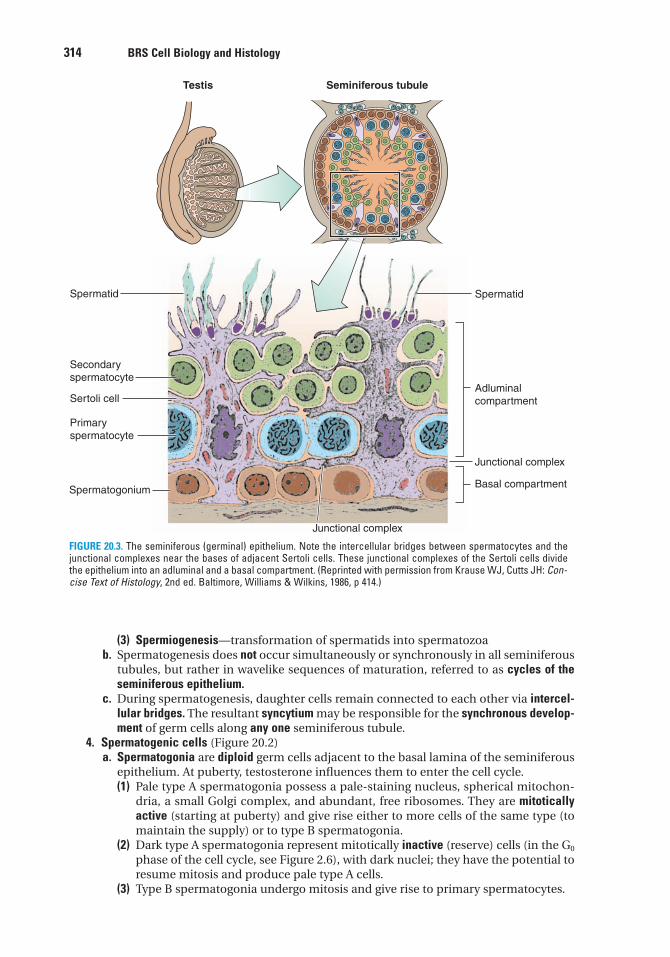
314 BRS Cell Biology and Histology
(3) Spermiogenesis—transformation of spermatids into spermatozoab. Spermatogenesis does not occur simultaneously or synchronously in all seminiferous
tubules, but rather in wavelike sequences of maturation, referred to as cycles of theseminiferous epithelium.
c. During spermatogenesis, daughter cells remain connected to each other via intercel-lular bridges. The resultant syncytium may be responsible for the synchronous develop-ment of germ cells along any one seminiferous tubule.
4. Spermatogenic cells (Figure 20.2)a. Spermatogonia are diploid germ cells adjacent to the basal lamina of the seminiferous
epithelium. At puberty, testosterone influences them to enter the cell cycle.(1) Pale type A spermatogonia possess a pale-staining nucleus, spherical mitochon-
dria, a small Golgi complex, and abundant, free ribosomes. They are mitoticallyactive (starting at puberty) and give rise either to more cells of the same type (tomaintain the supply) or to type B spermatogonia.
(2) Dark type A spermatogonia represent mitotically inactive (reserve) cells (in the G0
phase of the cell cycle, see Figure 2.6), with dark nuclei; they have the potential toresume mitosis and produce pale type A cells.
(3) Type B spermatogonia undergo mitosis and give rise to primary spermatocytes.
Testis
Spermatid
Secondary
spermatocyte
Sertoli cell
Primary
spermatocyte
Spermatogonium
Junctional complex
Junctional complex
Adluminal
compartment
Spermatid
Basal compartment
Seminiferous tubule
FIGURE 20.3. The seminiferous (germinal) epithelium. Note the intercellular bridges between spermatocytes and thejunctional complexes near the bases of adjacent Sertoli cells. These junctional complexes of the Sertoli cells dividethe epithelium into an adluminal and a basal compartment. (Reprinted with permission from Krause WJ, Cutts JH: Con-cise Text of Histology, 2nd ed. Baltimore, Williams & Wilkins, 1986, p 414.)
LWBK615-c20[311-325].qxd 05/31/2010 11:03 AM Page 314 Aptara
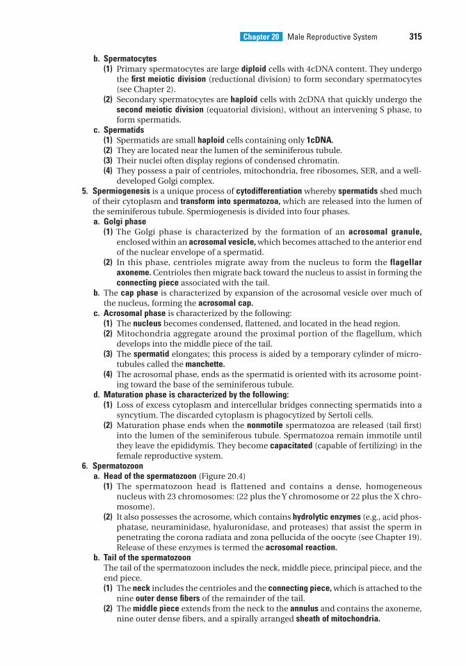
Chapter 20 Male Reproductive System 315
b. Spermatocytes(1) Primary spermatocytes are large diploid cells with 4cDNA content. They undergo
the first meiotic division (reductional division) to form secondary spermatocytes(see Chapter 2).
(2) Secondary spermatocytes are haploid cells with 2cDNA that quickly undergo thesecond meiotic division (equatorial division), without an intervening S phase, toform spermatids.
c. Spermatids(1) Spermatids are small haploid cells containing only 1cDNA.(2) They are located near the lumen of the seminiferous tubule.(3) Their nuclei often display regions of condensed chromatin.(4) They possess a pair of centrioles, mitochondria, free ribosomes, SER, and a well-
developed Golgi complex.5. Spermiogenesis is a unique process of cytodifferentiation whereby spermatids shed much
of their cytoplasm and transform into spermatozoa, which are released into the lumen ofthe seminiferous tubule. Spermiogenesis is divided into four phases.a. Golgi phase
(1) The Golgi phase is characterized by the formation of an acrosomal granule,enclosed within an acrosomal vesicle, which becomes attached to the anterior endof the nuclear envelope of a spermatid.
(2) In this phase, centrioles migrate away from the nucleus to form the flagellaraxoneme. Centrioles then migrate back toward the nucleus to assist in forming theconnecting piece associated with the tail.
b. The cap phase is characterized by expansion of the acrosomal vesicle over much ofthe nucleus, forming the acrosomal cap.
c. Acrosomal phase is characterized by the following:(1) The nucleus becomes condensed, flattened, and located in the head region.(2) Mitochondria aggregate around the proximal portion of the flagellum, which
develops into the middle piece of the tail.(3) The spermatid elongates; this process is aided by a temporary cylinder of micro-
tubules called the manchette.(4) The acrosomal phase, ends as the spermatid is oriented with its acrosome point-
ing toward the base of the seminiferous tubule.d. Maturation phase is characterized by the following:
(1) Loss of excess cytoplasm and intercellular bridges connecting spermatids into asyncytium. The discarded cytoplasm is phagocytized by Sertoli cells.
(2) Maturation phase ends when the nonmotile spermatozoa are released (tail first)into the lumen of the seminiferous tubule. Spermatozoa remain immotile untilthey leave the epididymis. They become capacitated (capable of fertilizing) in thefemale reproductive system.
6. Spermatozoona. Head of the spermatozoon (Figure 20.4)
(1) The spermatozoon head is flattened and contains a dense, homogeneousnucleus with 23 chromosomes: (22 plus the Y chromosome or 22 plus the X chro-mosome).
(2) It also possesses the acrosome, which contains hydrolytic enzymes (e.g., acid phos-phatase, neuraminidase, hyaluronidase, and proteases) that assist the sperm inpenetrating the corona radiata and zona pellucida of the oocyte (see Chapter 19).Release of these enzymes is termed the acrosomal reaction.
b. Tail of the spermatozoonThe tail of the spermatozoon includes the neck, middle piece, principal piece, and theend piece.(1) The neck includes the centrioles and the connecting piece, which is attached to the
nine outer dense fibers of the remainder of the tail.(2) The middle piece extends from the neck to the annulus and contains the axoneme,
nine outer dense fibers, and a spirally arranged sheath of mitochondria.
LWBK615-c20[311-325].qxd 05/31/2010 11:03 AM Page 315 Aptara
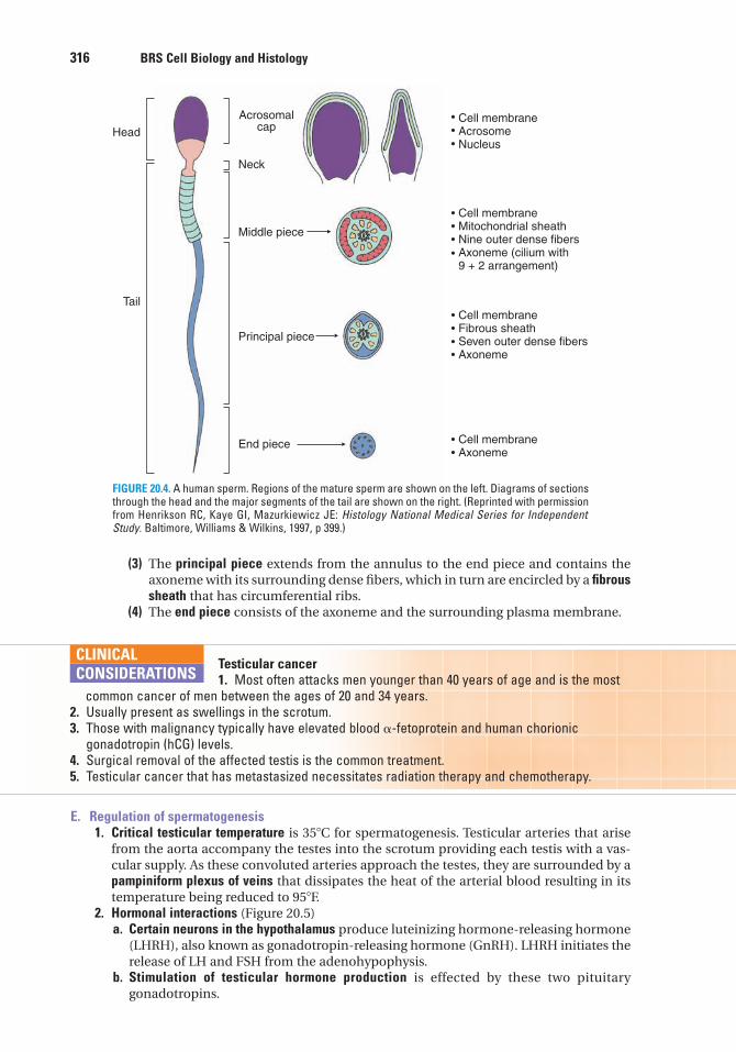
316 BRS Cell Biology and Histology
(3) The principal piece extends from the annulus to the end piece and contains theaxoneme with its surrounding dense fibers, which in turn are encircled by a fibroussheath that has circumferential ribs.
(4) The end piece consists of the axoneme and the surrounding plasma membrane.
Testicular cancer1. Most often attacks men younger than 40 years of age and is the most
common cancer of men between the ages of 20 and 34 years.2. Usually present as swellings in the scrotum.3. Those with malignancy typically have elevated blood �-fetoprotein and human chorionic
gonadotropin (hCG) levels.4. Surgical removal of the affected testis is the common treatment.5. Testicular cancer that has metastasized necessitates radiation therapy and chemotherapy.
CLINICAL
CONSIDERATIONS
E. Regulation of spermatogenesis1. Critical testicular temperature is 35�C for spermatogenesis. Testicular arteries that arise
from the aorta accompany the testes into the scrotum providing each testis with a vas-cular supply. As these convoluted arteries approach the testes, they are surrounded by apampiniform plexus of veins that dissipates the heat of the arterial blood resulting in itstemperature being reduced to 95�F.
2. Hormonal interactions (Figure 20.5)a. Certain neurons in the hypothalamus produce luteinizing hormone-releasing hormone
(LHRH), also known as gonadotropin-releasing hormone (GnRH). LHRH initiates therelease of LH and FSH from the adenohypophysis.
b. Stimulation of testicular hormone production is effected by these two pituitarygonadotropins.
Head
Acrosomalcap
Neck
Cell membraneAcrosomeNucleus
Cell membraneMitochondrial sheathNine outer dense fibersAxoneme (cilium with9 + 2 arrangement)
Cell membraneFibrous sheathSeven outer dense fibersAxoneme
Cell membraneAxoneme
Middle piece
Principal piece
End piece
Tail
FIGURE 20.4. A human sperm. Regions of the mature sperm are shown on the left. Diagrams of sectionsthrough the head and the major segments of the tail are shown on the right. (Reprinted with permissionfrom Henrikson RC, Kaye GI, Mazurkiewicz JE: Histology National Medical Series for IndependentStudy. Baltimore, Williams & Wilkins, 1997, p 399.)
LWBK615-c20[311-325].qxd 05/31/2010 11:03 AM Page 316 Aptara
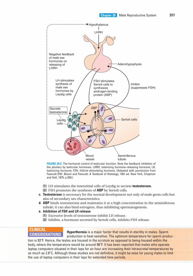
Chapter 20 Male Reproductive System 317
(1) LH stimulates the interstitial cells of Leydig to secrete testosterone.(2) FSH promotes the synthesis of ABP by Sertoli cells.
c. Testosterone is necessary for the normal development not only of male germ cells butalso of secondary sex characteristics.
d. ABP binds testosterone and maintains it at a high concentration in the seminiferoustubule; it can also bind estrogens, thus inhibiting spermatogenesis.
e. Inhibition of FSH and LH release(1) Excessive levels of testosterone inhibit LH release.(2) Inhibin, a hormone secreted by Sertoli cells, inhibits FSH release.
ABP
Hypothalamus
LHRH
Negative feedbackof male sex hormones onreleasing ofLHRH
LH stimulatessynthesis ofmale sex hormones by Leydig cells
Secretetestosterone
Leydigcells
Blood vessel
Seminiferoustubule
Sertoli cells
Inhibin(suppresses FSH)
Adenohypophysis
FSH stimulatesSertoli cells tosynthesizeandrogen-bindingprotein (ABP)
FIGURE 20.5. The hormonal control of testicular function. Note the feedback inhibition ofthe pituitary by testicular hormones. LHRH, luteinizing hormone-releasing hormone; LH,luteinizing hormone; FSH, follicle-stimulating hormone. (Adapted with permission fromFawcett DW: Bloom and Fawcett: A Textbook of Histology, 10th ed. New York, Chapmanand Hall, 1975, p 839.)
Hyperthermia is a major factor that results in sterility in males. Spermproduction is heat sensitive. The optimum temperature for sperm produc-
tion is 93�F. Hence, the testes are housed in the scrotum as opposed to being housed within thebody, where the temperature would be around 98�F. It has been reported that males who operatelaptop computers situated in their laps for an hour are increasing their intrascrotal temperatures byas much as 2.8�C. Although these studies are not definitive, it might be wise for young males to limitthe use of laptop computers in their laps for extended time periods.
CLINICAL
CONSIDERATIONS
LWBK615-c20[311-325].qxd 05/31/2010 11:03 AM Page 317 Aptara
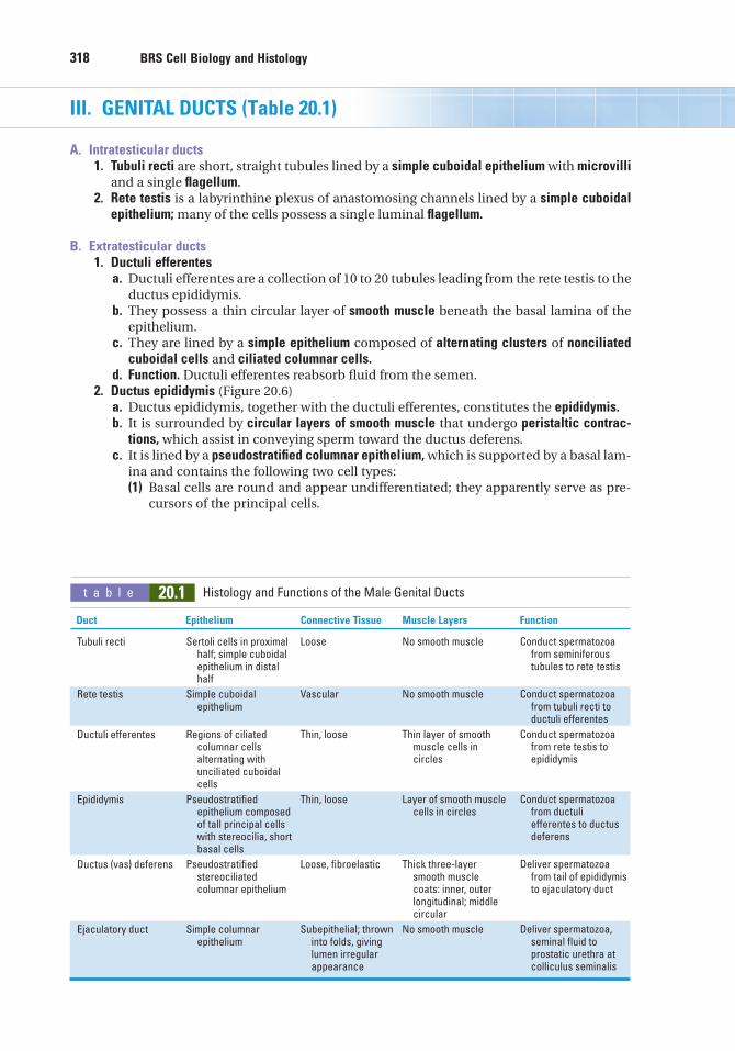
318 BRS Cell Biology and Histology
t a b l e 20.1 Histology and Functions of the Male Genital Ducts
Duct Epithelium Connective Tissue Muscle Layers Function
III. GENITAL DUCTS (Table 20.1)
A. Intratesticular ducts1. Tubuli recti are short, straight tubules lined by a simple cuboidal epithelium with microvilli
and a single flagellum.2. Rete testis is a labyrinthine plexus of anastomosing channels lined by a simple cuboidal
epithelium; many of the cells possess a single luminal flagellum.
B. Extratesticular ducts1. Ductuli efferentes
a. Ductuli efferentes are a collection of 10 to 20 tubules leading from the rete testis to theductus epididymis.
b. They possess a thin circular layer of smooth muscle beneath the basal lamina of theepithelium.
c. They are lined by a simple epithelium composed of alternating clusters of nonciliatedcuboidal cells and ciliated columnar cells.
d. Function. Ductuli efferentes reabsorb fluid from the semen.2. Ductus epididymis (Figure 20.6)
a. Ductus epididymis, together with the ductuli efferentes, constitutes the epididymis.b. It is surrounded by circular layers of smooth muscle that undergo peristaltic contrac-
tions, which assist in conveying sperm toward the ductus deferens.c. It is lined by a pseudostratified columnar epithelium, which is supported by a basal lam-
ina and contains the following two cell types:(1) Basal cells are round and appear undifferentiated; they apparently serve as pre-
cursors of the principal cells.
Tubuli recti
Rete testis
Ductuli efferentes
Epididymis
Ductus (vas) deferens
Ejaculatory duct
Sertoli cells in proximalhalf; simple cuboidalepithelium in distalhalf
Simple cuboidalepithelium
Regions of ciliatedcolumnar cellsalternating withunciliated cuboidalcells
Pseudostratifiedepithelium composedof tall principal cellswith stereocilia, shortbasal cells
Pseudostratifiedstereociliatedcolumnar epithelium
Simple columnarepithelium
Loose
Vascular
Thin, loose
Thin, loose
Loose, fibroelastic
Subepithelial; throwninto folds, givinglumen irregularappearance
No smooth muscle
No smooth muscle
Thin layer of smoothmuscle cells in circles
Layer of smooth musclecells in circles
Thick three-layer smooth muscle coats: inner, outerlongitudinal; middlecircular
No smooth muscle
Conduct spermatozoafrom seminiferoustubules to rete testis
Conduct spermatozoafrom tubuli recti toductuli efferentes
Conduct spermatozoafrom rete testis to epididymis
Conduct spermatozoafrom ductuliefferentes to ductusdeferens
Deliver spermatozoafrom tail of epididymisto ejaculatory duct
Deliver spermatozoa,seminal fluid toprostatic urethra atcolliculus seminalis
LWBK615-c20[311-325].qxd 05/31/2010 11:03 AM Page 318 Aptara
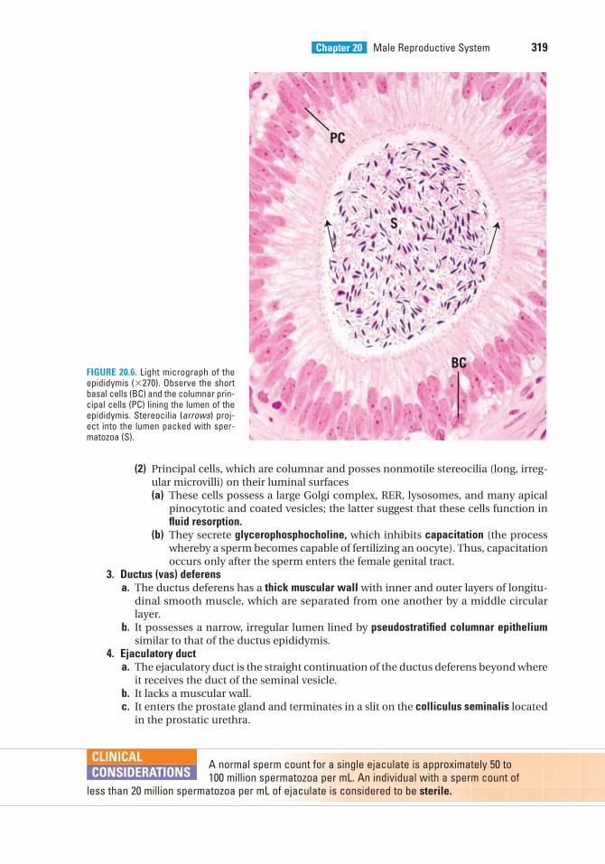
Chapter 20 Male Reproductive System 319
(2) Principal cells, which are columnar and posses nonmotile stereocilia (long, irreg-ular microvilli) on their luminal surfaces(a) These cells possess a large Golgi complex, RER, lysosomes, and many apical
pinocytotic and coated vesicles; the latter suggest that these cells function influid resorption.
(b) They secrete glycerophosphocholine, which inhibits capacitation (the processwhereby a sperm becomes capable of fertilizing an oocyte). Thus, capacitationoccurs only after the sperm enters the female genital tract.
3. Ductus (vas) deferensa. The ductus deferens has a thick muscular wall with inner and outer layers of longitu-
dinal smooth muscle, which are separated from one another by a middle circularlayer.
b. It possesses a narrow, irregular lumen lined by pseudostratified columnar epitheliumsimilar to that of the ductus epididymis.
4. Ejaculatory ducta. The ejaculatory duct is the straight continuation of the ductus deferens beyond where
it receives the duct of the seminal vesicle.b. It lacks a muscular wall.c. It enters the prostate gland and terminates in a slit on the colliculus seminalis located
in the prostatic urethra.
BC
PC
S
FIGURE 20.6. Light micrograph of theepididymis (�270). Observe the shortbasal cells (BC) and the columnar prin-cipal cells (PC) lining the lumen of theepididymis. Stereocilia (arrows) proj-ect into the lumen packed with sper-matozoa (S).
A normal sperm count for a single ejaculate is approximately 50 to 100 million spermatozoa per mL. An individual with a sperm count of
less than 20 million spermatozoa per mL of ejaculate is considered to be sterile.
CLINICAL
CONSIDERATIONS
LWBK615-c20[311-325].qxd 05/31/2010 11:03 AM Page 319 Aptara
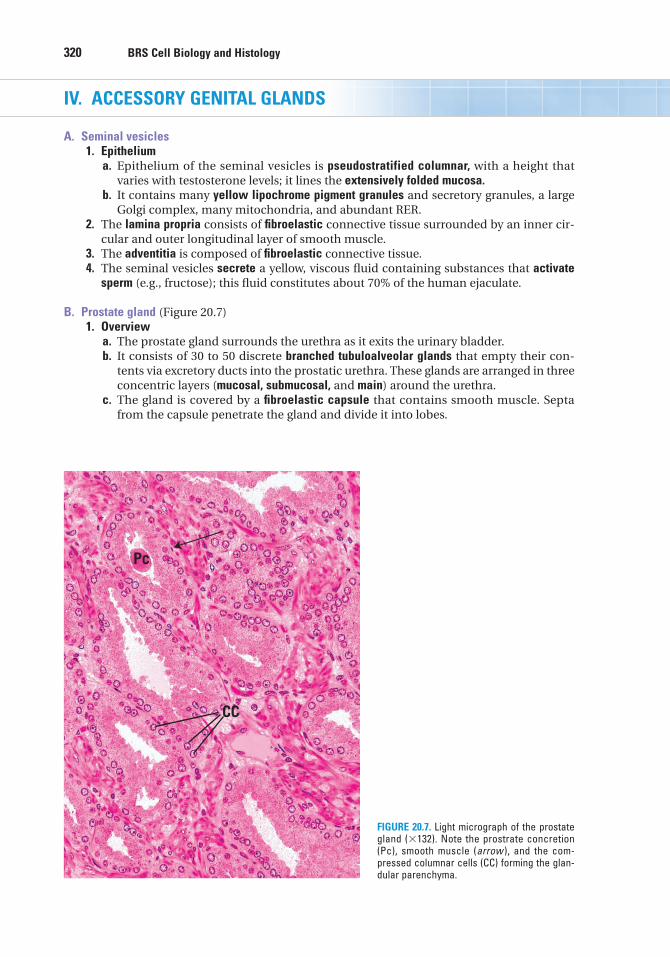
320 BRS Cell Biology and Histology
IV. ACCESSORY GENITAL GLANDS
A. Seminal vesicles1. Epithelium
a. Epithelium of the seminal vesicles is pseudostratified columnar, with a height thatvaries with testosterone levels; it lines the extensively folded mucosa.
b. It contains many yellow lipochrome pigment granules and secretory granules, a largeGolgi complex, many mitochondria, and abundant RER.
2. The lamina propria consists of fibroelastic connective tissue surrounded by an inner cir-cular and outer longitudinal layer of smooth muscle.
3. The adventitia is composed of fibroelastic connective tissue.4. The seminal vesicles secrete a yellow, viscous fluid containing substances that activate
sperm (e.g., fructose); this fluid constitutes about 70% of the human ejaculate.
B. Prostate gland (Figure 20.7)1. Overview
a. The prostate gland surrounds the urethra as it exits the urinary bladder.b. It consists of 30 to 50 discrete branched tubuloalveolar glands that empty their con-
tents via excretory ducts into the prostatic urethra. These glands are arranged in threeconcentric layers (mucosal, submucosal, and main) around the urethra.
c. The gland is covered by a fibroelastic capsule that contains smooth muscle. Septafrom the capsule penetrate the gland and divide it into lobes.
CC
Pc
FIGURE 20.7. Light micrograph of the prostategland (�132). Note the prostrate concretion(Pc), smooth muscle (arrow ), and the com-pressed columnar cells (CC) forming the glan-dular parenchyma.
LWBK615-c20[311-325].qxd 05/31/2010 11:03 AM Page 320 Aptara
Chapter 20 Male Reproductive System 321
2. Epitheliuma. The epithelium of the prostate gland is simple or pseudostratified columnar and lines
the individual glands that constitute the prostate.b. It is composed of cells that contain abundant RER, a well-developed Golgi complex,
numerous lysosomes, and many secretory granules.3. Corpora amylacea are prostatic concretions, composed of glycoprotein, which may
become calcified; their numbers increase with age.4. The prostate secretes a thin whitish fluid, a part of the semen containing proteolytic
enzymes, citric acid, acid phosphatase, fibrinolysin, and lipids. The prostatic secretionserves to liquefy the coagulated semen after it is deposited in the female genital tract. Itssynthesis and release are regulated by dihydrotestosterone.
Benign prostatic hypertrophy (BPH)1. BPH most commonly involves only an enlargement of the mucosal
glands.2. It is frequently associated with an inability to begin and cease urination because the urethra is
partially strangulated by the enlarged prostate.3. It leads to nocturia (urination at night) and sensory urgency (the desire to urinate without having
to void).4. This disease is common in older men, occurring in about 50% of men older than 50 years of age
and in 95% of those older than 80 years of age. BPH can be confirmed by a digital rectal examina-tion, however, a biopsy must be examined to rule out prostatic cancer.
Adenocarcinoma of the prostate gland1. Adenocarcinoma of the prostate gland may be indicated by palpation through the rectum.
However, a biopsy is required for positive identification of the disease.2. Although this cancer grows slowly, it commonly metastasizes to bone via the circulatory system.3. It occurs in about one-third of men older 75 years of age and is the second most common form of
cancer in men.4. It is associated with an elevated prostatic specific antigen (PSA) level in blood. Men with
elevated PSA values are candidates for more aggressive diagnostic tests for prostatic cancer.Surgical removal with or without radiation therapy is the usual treatment, although there may becomplications of impotence and/or incontinence. Newer procedures (e.g., brachytherapyincludes radioactive seeding within the gland).
CLINICAL
CONSIDERATIONS
C. Bulbourethral (Cowper) glands1. Bulbourethral glands are adjacent to the membranous urethra.2. They empty their secretion into the lumen of the membranous urethra to lubricate it.3. They are lined by a simple cuboidal or columnar epithelium.4. They are surrounded by a fibroelastic capsule containing smooth and skeletal muscle.
Vasectomy is the chosen means of permanent contraception by morethan 600,000 US males each year. A physician may perform the procedure
in the office where the scrotum is slit open, a portion of the spermatic cord is pulled out, cut, cauter-ized, and returned to the scrotum. At the conclusion of 6 weeks or so, after the procedure, semen iscollected and examined to assure that no spermatozoa are ejaculated. This procedure is nearly100% effective. The procedure may have a few complications, but they usually dissipate after ashort period of time.
CLINICAL
CONSIDERATIONS
LWBK615-c20[311-325].qxd 05/31/2010 11:03 AM Page 321 Aptara
322 BRS Cell Biology and Histology
V. PENIS
A. Corpora cavernosa1. Corpora cavernosa are paired masses of erectile tissue that contain irregular vascular
spaces lined by a continuous layer of endothelial cells. These spaces are separated fromeach other by trabeculae of connective tissue and smooth muscle.
2. The vascular spaces decrease in size toward the periphery of the corpora cavernosa.3. During erection, the vascular spaces become engorged with blood as a result of parasym-
pathetic impulses, which constrict arteriovenous shunts and dilate the helicine arteries,thus increasing flow to the vascular spaces of the two corpora cavernosa and the singlecorpus spongiosum.
B. Corpus spongiosum1. The corpus spongiosum is a single mass of erectile tissue that contains vascular spaces
of uniform size.2. It possesses trabeculae that contain more elastic fibers and less smooth muscle than
those of the corpora cavernosa.
C. Connective tissue and skin1. The tunica albuginea is a thick, fibrous connective sheath that surrounds the paired cor-
pora cavernosa and the corpus spongiosum. The arrangement of dense collagen bundlespermits extension of the penis during erection.
2. Glans penisa. The glans penis is the dilated distal end of the corpus spongiosum.b. It contains dense connective tissue and longitudinal muscle fibers.c. It is covered by retractable skin, the prepuce, which is lined by stratified squamous
lightly keratinized epithelium.3. Glands of Littre are mucus-secreting glands present throughout the length of the penile
urethra.
LWBK615-c20[311-325].qxd 05/31/2010 11:03 AM Page 322 Aptara
Review Test
323
1. Which one of the following functions isattributed to the Sertoli cells?
(A) Secretion of follicle-stimulatinghormone
(B) Secretion of testosterone(C) Secretion of androgen-binding protein(D) Secretion of LH(E) Secretion of ABP
2. Which of the following statementsconcerning the cells of Leydig is true?
(A) They become functional at puberty.(B) They are within the seminiferous
tubules.(C) They are stimulated by FSH.(D) They secrete much of the fluid portion
of semen.(E) They respond to inhibin.
3. Type A spermatogonia are germ cells that
(A) develop from secondary spermatocytes.(B) undergo meiotic activity subsequent to
sexual maturity.(C) develop through meiotic divisions.(D) give rise to primary spermatocytes.(E) may be dark or pale.
4. Which of the following statements isrelated to the ductus epididymis?
(A) It begins at the rete testis.(B) It is lined by a pseudostratified
columnar epithelium.(C) It secretes a large volume of fluid into its
lumen.(D) It possesses motile cilia.(E) It capacitates spermatozoa.
5. Testosterone is produced by
(A) interstitial cells of Leydig.(B) Sertoli cells.(C) spermatogonia.(D) spermatids.(E) spermatocytes.
6. Spermatozoa are conveyed from the sem-iniferous tubules to the rete testis via the
(A) ductus epididymis.(B) tubuli recti.(C) ductuli efferentes.(D) ductus deferens.(E) ejaculatory duct.
7. Manchette formation occurs duringwhich of the following phases ofspermiogenesis?
(A) Meiotic phase(B) Maturation phase(C) Golgi phase(D) Cap phase(E) Acrosomal phase
8. The structural feature that bestdistinguishes the ductus deferens from theother genital ducts is its
(A) smooth-bore lumen.(B) thick wall containing three muscle
layers.(C) lining of transitional epithelium.(D) flattened mucosa.(E) nonmotile stereocilia.
9. Inhibin, a hormone that inhibits synthesisand release of FSH, is secreted by
(A) prostate gland.(B) Sertoli cells.(C) seminal vesicles.(D) bulbourethral glands.(E) interstitial cells of Leydig.
Directions: Each of the numbered items or incomplete statements in this section is followed byanswers or by completions of the statement. Select the ONE lettered answer or completion thatis BEST in each case.
LWBK615-c20[311-325].qxd 05/31/2010 11:03 AM Page 323 Aptara
11. A 33-year-old man detected a swelling inhis scrotum. Careful palpation did not revealwhether the swelling was associated with histestis. His physician ordered blood tests, andit was determined that he had increased levelsof hCG in his blood. A probable diagnosis is
(A) benign lump.(B) hematocele.(C) cryptorchidism.(D) testicular cancer.(E) hydrocele.
324 BRS Cell Biology and Histology
10. A 55-year-old man has urinary complica-tions. He complains of difficulty urinatingand reduced urinary flow. He also has anelevated prostate-specific antigen (PSA)level, along with palpable hard nodules onthe prostate. The possible diagnosis is
(A) benign prostatic hyperplasia.(B) adenocarcinoma of the prostate.(C) prostatic concretions.(D) initiation of impotence.(E) incontinence.
LWBK615-c20[311-325].qxd 05/31/2010 11:03 AM Page 324 Aptara
325
1. C. Sertoli cells have many functions, including the synthesis of ABP. Testosterone issecreted by the interstitial cells of Leydig, whereas FSH and LH (also known as interstitialcell–stimulating hormone) are secreted by the pituitary gland (see Chapter 20 II D 2).
2. A. Interstitial cells of Leydig become functional at puberty as a result of the action of LHproduced in the pituitary gland (see Chapter 20 II C).
3. E. Type A spermatogonia, which may be pale or dark, are primitive germ cells. Pale type A spermatogonia become mitotically active at puberty and give rise to type Bspermatogonia, which undergo mitoses, giving rise to primary spermatocytes (see Chapter 20 II D 4).
4. B. The ductus epididymidis begins at the termination of the ductuli efferentes and is lined by a pseudostratified columnar epithelium, which has principal cells that possessnonmotile stereocilia. These cells are involved in fluid resorption and secrete glycerophos-phocholine, which inhibits capacitation (see Chapter 20 III B 2).
5. A. The hormone testosterone is produced by the interstitial cells of Leydig (see Chapter 20 II C).
6. B. The seminiferous tubules are connected to the rete testis by the tubuli recti (see Chapter 20 II D).
7. E. Spermiogenesis is the process of cytodifferentiation by which spermatids aretransformed into spermatozoa. It does not involve cell division. Manchette formationoccurs during the acrosomal phase of spermiogenesis (see Chapter 20 II D 5).
8. B. The ductus (vas) deferens possesses three layers of smooth muscle in its wall, whereasthe other genital ducts do not. Like the ductus epididymidis, the ductus deferens is linedby a pseudostratified columnar epithelium with principal cells that possess nonmotilestereocilia (see Chapter 20 III B 3).
9. B. Inhibin is secreted by Sertoli cells (see Chapter 20 II D 2).
10. B. Although some of the symptoms are characteristic of benign prostatic hypertrophy,rectal palpation indicating hard nodules, along with an elevated PSA level, indicates probable prostatic adenocarcinoma (see Chapter 20 IV B Clinical Considerations).
11. D. Palpable swellings in the scrotum should be seen by a physician, especially in menyounger than 40 years of age. Elevated blood serum levels of hCG and �-fetoprotein areusually associated with testicular cancer (see Chapter 20 II D Clinical Considerations).
Answers and Explanations
LWBK615-c20[311-325].qxd 05/31/2010 11:03 AM Page 325 Aptara
c h a p t e r 21 Special Senses
326
I. OVERVIEW—SPECIAL SENSE RECEPTORS
A. Special sense receptors are responsible for the five special senses: taste, smell, seeing, hear-ing, and feeling (which includes touch, pressure, temperature, pain, and proprioception).The sense of smell is described in Chapter 15, and the sense of taste is described in Chapter16. The remaining special senses are described here.
B. Function. Special sense receptors transduce stimuli from the environment into electricalimpulses.
II. SPECIALIZED DIFFUSE RECEPTORS (Figure 21.1)
A. Overview1. Specialized diffuse receptors are dendritic nerve endings in the skin, fascia, muscles,
joints, and tendons.2. They respond to stimuli related to touch, pressure, temperature, pain, and proprioception.3. These receptors are specialized to receive only one type of sensory stimulus, although
they will respond to other types of stimuli that are intense enough.4. They are divided morphologically into free nerve terminals and encapsulated nerve end-
ings, which are ensheathed in a connective tissue capsule.
B. Touch and pressure receptors1. Pacinian corpuscles
a. Pacinian corpuscles are large, ellipsoid encapsulated receptors in the dermis andhypodermis and in the connective tissue of the mesenteries and joints.
b. They are especially abundant in the digits and breasts.c. They are composed of a multilayer capsule consisting of fibroblasts and collagen and
bathed in tissue fluid. The capsule surrounds an inner unmyelinated nerve terminal.d. They often resemble a sliced onion in histological sections.e. Function. Pacinian corpuscles perceive pressure, touch, and vibration.
2. Ruffini endingsa. Ruffini endings are encapsulated receptors in the dermis and joints.b. They are composed of groups of branched terminals from myelinated nerve fibers and
are surrounded by a thin connective tissue capsule.c. Function. Ruffini endings function in pressure and touch reception.
3. Meissner corpusclesa. Meissner corpuscles are ellipsoid, encapsulated receptors in the dermal papillae of
thick skin, eyelids, lips, and nipples.
LWBK615-c21[326-345].qxd 05/31/2010 11:04 AM Page 326 Aptara
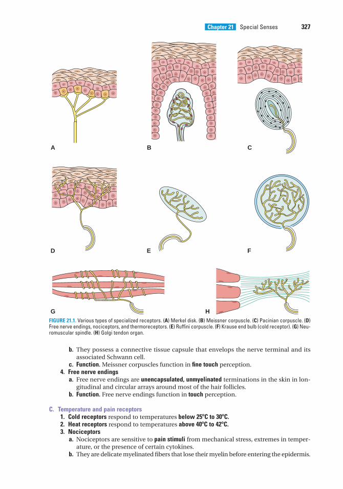
Chapter 21 Special Senses 327
b. They possess a connective tissue capsule that envelops the nerve terminal and itsassociated Schwann cell.
c. Function. Meissner corpuscles function in fine touch perception.4. Free nerve endings
a. Free nerve endings are unencapsulated, unmyelinated terminations in the skin in lon-gitudinal and circular arrays around most of the hair follicles.
b. Function. Free nerve endings function in touch perception.
C. Temperature and pain receptors1. Cold receptors respond to temperatures below 25�C to 30�C.2. Heat receptors respond to temperatures above 40�C to 42�C.3. Nociceptors
a. Nociceptors are sensitive to pain stimuli from mechanical stress, extremes in temper-ature, or the presence of certain cytokines.
b. They are delicate myelinated fibers that lose their myelin before entering the epidermis.
A B C
D E F
G HFIGURE 21.1. Various types of specialized receptors. (A) Merkel disk. (B) Meissner corpuscle. (C) Pacinian corpuscle. (D)Free nerve endings, nociceptors, and thermoreceptors. (E) Ruffini corpuscle. (F) Krause end bulb (cold receptor). (G) Neu-romuscular spindle. (H) Golgi tendon organ.
LWBK615-c21[326-345].qxd 05/31/2010 11:04 AM Page 327 Aptara
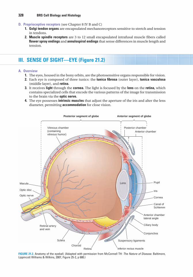
328 BRS Cell Biology and Histology
D. Proprioceptive receptors (see Chapter 8 IV B and C)1. Golgi tendon organs are encapsulated mechanoreceptors sensitive to stretch and tension
in tendons.2. Muscle spindle receptors are 3 to 12 small encapsulated intrafusal muscle fibers called
flower spray endings and annulospiral endings that sense differences in muscle length andtension.
III. SENSE OF SIGHT—EYE (Figure 21.2)
A. Overview1. The eyes, housed in the bony orbits, are the photosensitive organs responsible for vision.2. Each eye is composed of three tunics: the tunica fibrosa (outer layer), tunica vasculosa
(middle layer), and retina.3. It receives light through the cornea. The light is focused by the lens on the retina, which
contains specialized cells that encode the various patterns of the image for transmissionto the brain via the optic nerve.
4. The eye possesses intrinsic muscles that adjust the aperture of the iris and alter the lensdiameter, permitting accommodation for close vision.
Posterior segment of globe Anterior segment of globe
Posterior chamberAnterior chamber
Vitreous chamber(containingvitreous humor)
Macula
Optic disc
Optic nerve
Retinal arteryand vein
Conjunctiva
Cornea
Canal ofSchlemm
Ciliary body
Suspensory ligamentsSclera
ChoroidRetina Inferior rectus muscle
Iris
Lens Pupil
Anterior chamberlateral angle
FIGURE 21.2. Anatomy of the eyeball. (Adapted with permission from McConnell TH: The Nature of Disease. Baltimore,Lippincott Williams & Wilkins, 2007, Figure 25-2, p 680.)
LWBK615-c21[326-345].qxd 05/31/2010 11:04 AM Page 328 Aptara
Chapter 21 Special Senses 329
5. It possesses extrinsic muscles, attached to the external aspect of the orb (eyeball), whichmove the eyes in a coordinated manner to perceive the desired visual fields.
6. The orb is continually moistened on its anterior surface with lacrimal fluid (tears)secreted by the lacrimal gland.
7. The eyeball is covered by the upper and lower eyelids, which protect its anterior surface.
B. Tunica fibrosa1. The outermost tunica fibrosa is composed of the sclera and the cornea. The sclera is an
opaque, relatively avascular fibrous connective tissue layer that covers the posterior five-sixths of the eyeball. The sclera receives insertions of the extrinsic ocular muscles.
2. The cornea is the transparent, highly innervated avascular anterior one-sixth of thetunica. It joins the sclera in a region called the limbus, which is highly vascularized and iscomposed of five layers.a. Corneal epithelium
(1) The corneal epithelium lines the anterior aspect of the cornea and is continuouswith the conjunctiva (a mucous membrane covering the anterior sclera and liningthe internal surface of the eyelids).
(2) It is a stratified squamous nonkeratinized epithelium.(3) It possesses microvilli in its superficial layer; the microvilli trap moisture, protect-
ing the cornea from dehydration.b. Bowman membrane is a homogeneous noncellular layer that provides form, stability,
and strength to the cornea.c. Corneal stroma
(1) The corneal stroma is the thickest corneal layer. It is composed of many layers oftype I collagen fibers lying parallel to each other within each row and at obliqueangles to each other in adjoining layers. The collagen fibers are embedded in aground substance composed mostly of chondroitin sulfate and keratan sulfate.Thin elastic fibers and fibroblasts are also present in the corneal stroma.
(2) The stroma has channels in the region of the limbus that are lined by endothelium,forming the canal of Schlemm. This canal drains fluid (aqueous humor) from theanterior chamber of the eye into the venous system.
d. Descemet membrane is a thick (5–10 �m) basal lamina separating the stroma from theendothelium lining the cornea.
e. Corneal endothelium(1) The corneal endothelium lines the posterior aspect of the cornea.(2) It is a simple squamous epithelium with cells that exhibit numerous pinocytic vesicles.(3) It resorbs fluid from the stroma, thus contributing to the transparency of the
cornea, contributory to light refraction.
C. Tunica vasculosa (uvea, the middle tunic) is composed of three parts:1. Choroid
a. The choroid is the highly vascular, pigmented layer of the eye on the posterior wall ofthe orb; its loose connective tissue contains many melanocytes.
b. It is loosely attached to the tunica fibrosa.c. It possesses a deep choriocapillary layer and Bruch membrane (basement membrane),
which extends from the optic disk to the ora serrata.2. Ciliary body
a. The ciliary body is the wedge-shaped anterior expansion of the choroid.b. It completely encircles the lens and separates the ora serrata from the iris.c. It is lined on its inner surface by two layers of cells: an outer pigmented columnar
epithelium rich in melanin and an inner nonpigmented simple columnar epithelium.(1) Ciliary processes
(a) are radially arranged extensions (about 70) of the ciliary body.(b) have a connective tissue core containing many fenestrated capillaries.(c) are covered by two epithelial layers. The unpigmented inner layer transports
components from the plasma filtrate in the posterior chamber and thus forms
LWBK615-c21[326-345].qxd 05/31/2010 11:04 AM Page 329 Aptara
330 BRS Cell Biology and Histology
the aqueous humor, which flows to the anterior chamber via the pupillary aper-ture (see Figure 21.2).
(d) possess suspensory ligaments (zonulae) that arise from the processes andinsert into the capsule of the lens, serving to anchor it in place.
(2) Ciliary muscle(a) is attached to the sclera and ciliary body in such a manner that its contractions
stretch the ciliary body and release tension on the suspensory ligament andlens. Ciliary muscle contraction permits the lens to become more convex,allowing the eye to focus on nearby objects (accommodation). With advancingage, the lens loses its elasticity, thereby gradually losing the ability to accom-modate.
(b) The ciliary muscle is innervated via parasympathetic fibers of the oculomotornerve (cranial nerve III).
3. Irisa. Overview
(1) The iris is the most anterior extension of the choroid, separating the anterior andposterior chambers of the orb.
(2) It incompletely covers the anterior surface of the lens, forming an aperture calledthe pupil that is continually adjusted by intrinsic pupillary muscles.
(3) The iris is covered by an incomplete layer of pigmented cells and fibroblasts on itsanterior surface.
(4) It has a wall composed of loose vascular connective tissue containing melanocytesand fibroblasts.
(5) The iris is covered on its deep surface by a two-layered epithelium possessing pig-mented cells that block light from entering the interior of the eye except via thepupil.
b. Eye color is blue if only a few melanocytes are present. Increasing amounts of pigmentimpart darker colors to the eye.
c. Dilator pupillae muscle(1) Dilator pupillae muscle is a smooth muscle with fibers that radiate from the
periphery of the iris toward the pupil.(2) It contracts upon stimulation by sympathetic nerve fibers, dilating the pupil.
d. Sphincter pupillae muscle(1) Sphincter pupillae muscle is smooth muscle arranged in concentric rings around
the pupillary orifice.(2) It contracts upon stimulation by parasympathetic nerve fibers of the oculomotor
nerve (cranial nerve III), constricting the pupil.
D. Refractive media of the eye1. Aqueous humor
a. Aqueous humor is a plasmalike fluid in the anterior compartment of the eye that isformed by epithelial cells lining the ciliary processes.
b. It is constantly secreted into the posterior chamber of the eye and then flows into theanterior chamber of the eye via the pupillary aperture; from there it enters the venoussystem via the canal of Schlemm (Figure 21.2).
Glaucoma1. Glaucoma is a condition of abnormally high intraocular pressure. It is
caused by obstructions that prevent drainage of aqueous humor from the eye via the canal ofSchlemm.
2. Chronic glaucoma, the most common form of glaucoma, may be associated with few symptomsexcept for a gradual loss of peripheral vision. However, usually, this condition can be controlledwith medication in the form of eye drops.
CLINICAL
CONSIDERATIONS
LWBK615-c21[326-345].qxd 05/31/2010 11:04 AM Page 330 Aptara
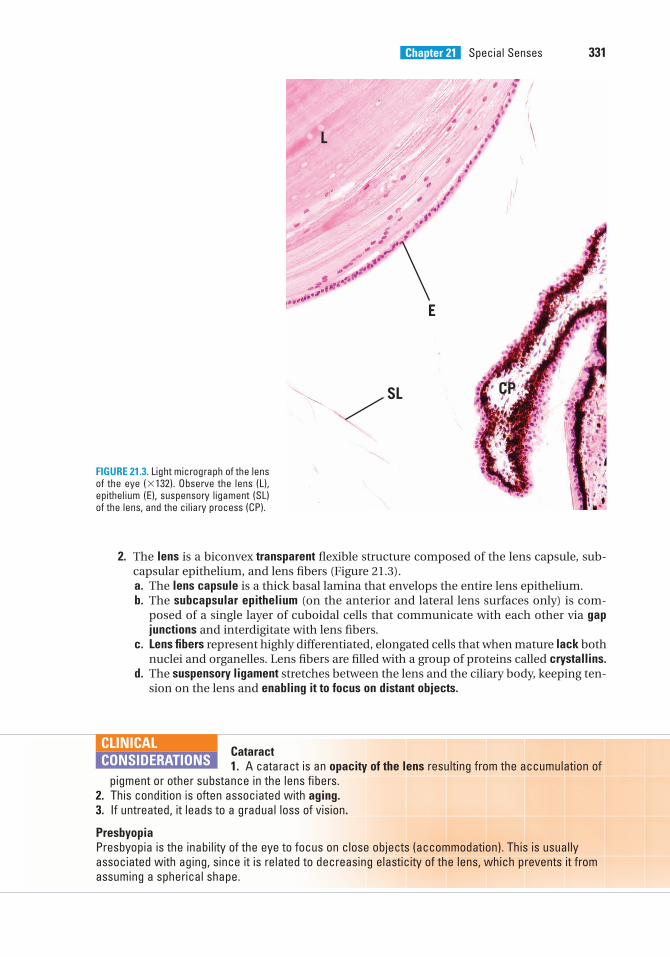
Chapter 21 Special Senses 331
2. The lens is a biconvex transparent flexible structure composed of the lens capsule, sub-capsular epithelium, and lens fibers (Figure 21.3).a. The lens capsule is a thick basal lamina that envelops the entire lens epithelium.b. The subcapsular epithelium (on the anterior and lateral lens surfaces only) is com-
posed of a single layer of cuboidal cells that communicate with each other via gapjunctions and interdigitate with lens fibers.
c. Lens fibers represent highly differentiated, elongated cells that when mature lack bothnuclei and organelles. Lens fibers are filled with a group of proteins called crystallins.
d. The suspensory ligament stretches between the lens and the ciliary body, keeping ten-sion on the lens and enabling it to focus on distant objects.
CP
L
E
SL
FIGURE 21.3. Light micrograph of the lensof the eye (�132). Observe the lens (L),epithelium (E), suspensory ligament (SL)of the lens, and the ciliary process (CP).
Cataract1. A cataract is an opacity of the lens resulting from the accumulation of
pigment or other substance in the lens fibers.2. This condition is often associated with aging.3. If untreated, it leads to a gradual loss of vision.
PresbyopiaPresbyopia is the inability of the eye to focus on close objects (accommodation). This is usuallyassociated with aging, since it is related to decreasing elasticity of the lens, which prevents it fromassuming a spherical shape.
CLINICAL
CONSIDERATIONS
LWBK615-c21[326-345].qxd 05/31/2010 11:04 AM Page 331 Aptara
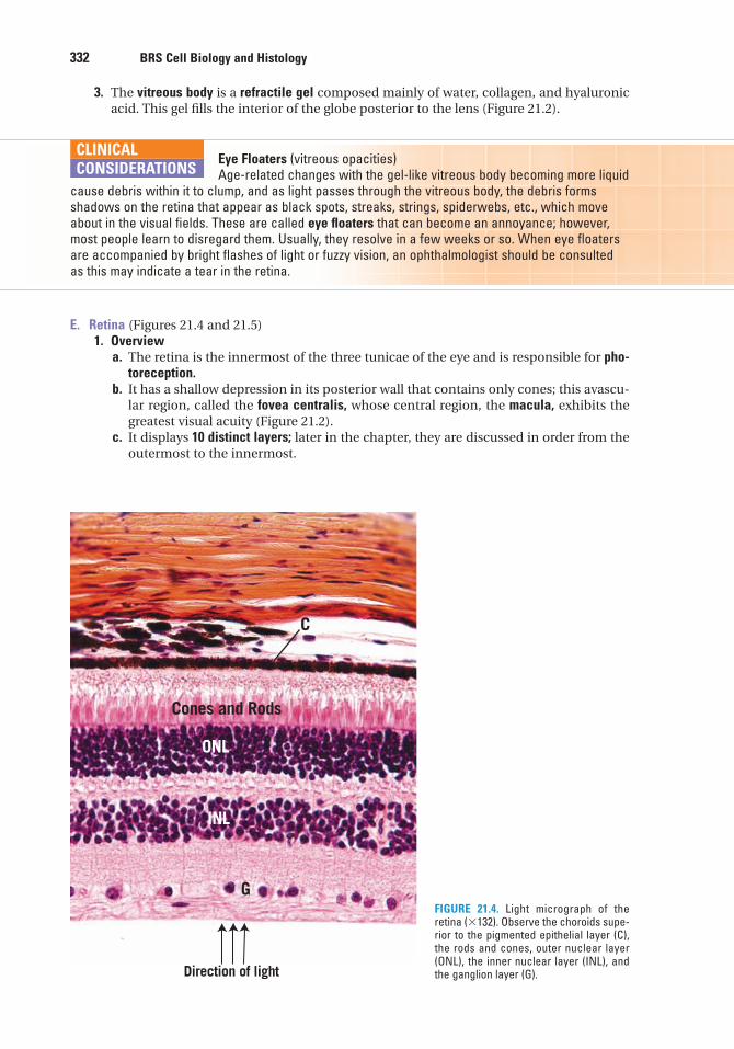
332 BRS Cell Biology and Histology
3. The vitreous body is a refractile gel composed mainly of water, collagen, and hyaluronicacid. This gel fills the interior of the globe posterior to the lens (Figure 21.2).
Eye Floaters (vitreous opacities)Age-related changes with the gel-like vitreous body becoming more liquid
cause debris within it to clump, and as light passes through the vitreous body, the debris formsshadows on the retina that appear as black spots, streaks, strings, spiderwebs, etc., which moveabout in the visual fields. These are called eye floaters that can become an annoyance; however,most people learn to disregard them. Usually, they resolve in a few weeks or so. When eye floatersare accompanied by bright flashes of light or fuzzy vision, an ophthalmologist should be consultedas this may indicate a tear in the retina.
CLINICAL
CONSIDERATIONS
E. Retina (Figures 21.4 and 21.5)1. Overview
a. The retina is the innermost of the three tunicae of the eye and is responsible for pho-toreception.
b. It has a shallow depression in its posterior wall that contains only cones; this avascu-lar region, called the fovea centralis, whose central region, the macula, exhibits thegreatest visual acuity (Figure 21.2).
c. It displays 10 distinct layers; later in the chapter, they are discussed in order from theoutermost to the innermost.
Cones and Rods
G
ONL
INL
C
Direction of light
FIGURE 21.4. Light micrograph of theretina (�132). Observe the choroids supe-rior to the pigmented epithelial layer (C),the rods and cones, outer nuclear layer(ONL), the inner nuclear layer (INL), andthe ganglion layer (G).
LWBK615-c21[326-345].qxd 05/31/2010 11:04 AM Page 332 Aptara
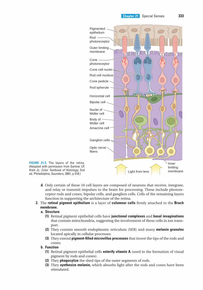
Chapter 21 Special Senses 333
d. Only certain of these 10 cell layers are composed of neurons that receive, integrate,and relay or transmit impulses to the brain for processing. These include photore-ceptor rods and cones, bipolar cells, and ganglion cells. Cells of the remaining layersfunction in supporting the architecture of the retina.
2. The retinal pigment epithelium is a layer of columnar cells firmly attached to the Bruchmembrane.a. Structure
(1) Retinal pigment epithelial cells have junctional complexes and basal invaginationsthat contain mitochondria, suggesting the involvement of these cells in ion trans-port.
(2) They contain smooth endoplasmic reticulum (SER) and many melanin granuleslocated apically in cellular processes.
(3) They extend pigment-filled microvillus processes that invest the tips of the rods andcones.
b. Function(1) Retinal pigment epithelial cells esterify vitamin A (used in the formation of visual
pigment by rods and cones).(2) They phagocytize the shed tips of the outer segments of rods.(3) They synthesize melanin, which absorbs light after the rods and cones have been
stimulated.
Light from lens
Innerlimitingmembrane
Pigmented epithelium
Rodphotoreceptor
Outer limitingmembrane
Conephotoreceptor
Cone cell nuclei
Rod cell nucleus
Cone pedicle
Rod spherule
Horizontal cell
Bipolar cell
Nuclei ofMüller cell
Body ofMüller cell
Amacrine cell
Ganglion cells
Optic nervefibers
FIGURE 21.5. The layers of the retina.(Adapted with permission from Gartner LP,Hiatt JL: Color Textbook of Histology, 2nded. Philadelphia, Saunders, 2001, p 518.)
LWBK615-c21[326-345].qxd 05/31/2010 11:04 AM Page 333 Aptara
334 BRS Cell Biology and Histology
3. The photoreceptor layer consists of neurons (photoreceptor cells) called rods and cones.Their dendrites interdigitate with cells of the pigmented epithelium, and their basesform synapses with cells of the bipolar layer.a. Rods (sensitive to light of low intensity) (Figure 21.5)
(1) Overview(a) Rods have outer and inner segments, a nuclear region, and a synaptic region.(b) They may synapse with bipolar cells, giving rise to summation.(c) They contain an incomplete cilium terminating in a basal body within the inner
segment.(d) They face the back of the eye; therefore, light must pass through all of the other
retinal layers before reaching the photosensitive region.(2) The outer segments of rods
(a) consist mainly of hundreds of flattened membranous disks that containrhodopsin.
(b) eventually shed their disks, which are subsequently phagocytized by the pig-ment epithelial cells.
(3) The inner segments of rods possess mitochondria, glycogen, polyribosomes, andproteins, which migrate to the outer segments to be incorporated into the mem-branous disks.
(4) Photoreception by rods is initiated by the interaction of light with rhodopsin (visualpurple), which is composed of the integral membrane protein opsin bound to reti-nal, the aldehyde form of vitamin A.(a) The retinal moiety of rhodopsin absorbs light in the visible range.(b) Retinal dissociates from opsin. This reaction, called bleaching, eventually
closes the Na� channels, thus permitting diffusion of bound Ca2� ions into thecytoplasm of the outer segment of a rod cell.
(c) Excess Ca2� acts to hyperpolarize the cell because Na� is prevented from enter-ing the cell.
(d) Ionic alterations in the rod generate electrical activity, which is relayed to otherrods via gap junctions.
(e) Dissociated retinal and opsin reassemble by an active process in which Müllerand pigment epithelial cells also participate.
(f) Ca2� is recaptured by the membranous disks, leading to reopening of the Na�
channels and reestablishment of the normal resting membrane potential.b. Cones (sensitive to light of high intensity) (Figure 21.5)
(1) Cones are much less numerous than rods but produce greater visual acuity than dorods; thus, the macula is populated only by cones.
(2) They are generally similar in structure to rods and mediate photoreception in thesame way, with the following exceptions:(a) The membranous disks in the outer segments of cones are invaginations of
the plasma membrane, whereas in rods they are not.(b) The proteins synthesized in the inner segments of cones are passed to the entire
outer segment, whereas in rods they are added only to newly forming disks.(c) Cones possess iodopsin in their disks. The amount of this photopigment varies in
different cones, making them differentially sensitive to red, green, or blue light.(d) Each cone synapses with a single bipolar neuron, whereas each rod may
synapse with several bipolar neurons.
Age-related macular degeneration (AMD) is a disease of the retinalocated in the central region of the macula, hence affecting central vision.
As its name implies, it is a disease of aging, occurring usually after the age of 50 years. There are twoforms: dry and wet AMD. Dry macular degeneration, the most common, is where the central vision isaffected so that objects become very blurred. This is caused by cellular debris (drusen) that appears
CLINICAL
CONSIDERATIONS
LWBK615-c21[326-345].qxd 05/31/2010 11:04 AM Page 334 Aptara
Chapter 21 Special Senses 335
4. External limiting membranea. The external limiting membrane is not a true membrane but an area where zonulae
adherentes (belt desmosomes) are located between the photoreceptor cells and theretinal Müller cells (glial cells).
b. This membrane also contains microvilli that project from the Müller cells.5. The outer nuclear layer consists primarily of the nuclei of the rods and cones.6. Outer plexiform layer
a. The outer plexiform layer contains axodendritic synapses between the axons of pho-toreceptor cells and the dendrites of bipolar and horizontal cells.
b. It displays synaptic ribbons within the rod and cone cells at synaptic sites.7. The inner nuclear layer contains the cell bodies of bipolar neurons, horizontal cells,
amacrine cells, and the nuclei of Müller cells.8. Inner plexiform layer
a. The inner plexiform layer contains axodendritic synapses between the axons of bipo-lar cells and the dendrites of ganglion cells.
b. The processes of amacrine cells are located in this layer.9. The ganglion cell layer contains the somata of ganglion cells, which form the final link in
the retina’s neural chain.a. Structure—Ganglion cells
(1) Ganglion cells are typical neurons that project their axons to a specific region ofthe retina called the optic disk.
(2) These cells are midget, diffuse, and stratified ganglion cells.b. Function—Ganglion cells. Ganglion cells are activated by hyperpolarization of rods and
cones and generate an action potential, which is transmitted to horizontal andamacrine cells and carried to the visual relay system in the brain.
10. The optic nerve fiber layer consists primarily of the unmyelinated axons of ganglion cells,which form the fibers of the optic nerve. As each fiber pierces the sclera, it acquires amyelin sheath.
11. The inner limiting membrane consists of the terminations of Müller cell processes andtheir basement membranes.
as yellow spots deposited between the choroids and the retina. Wet macular degeneration, the mostsevere, results when small blood vessels, formed between the choroid and the retina, leak into thisspace causing the retina to die, as a result a blind spot forms in the center of the visual field. Wetmacular degeneration exhibits a quick onset with a small blind spot that may quickly progress to alarger blind spot. Although an AMD patient may be unable to recognize a face in the center of vision,it is interesting to note that peripheral vision is unaffected by macular degeneration. While there is nocure, certain vitamins and high doses of antioxidants and zinc may be of some benefit for dry AMD,whereas laser surgery and injections of antiangiogenesis drugs are used to manage wet AMD.
Detachment of the retina1. Retinal detachment occurs when the neural and pigmented layers sep-
arate from each other, for example, as a result from a sudden hard jolt.2. This condition can sometimes be treated successfully by laser surgery. But extensive separation
requires cryosurgery to produce successful adhesion of the two layers. If retinal detachment isleft untreated, blindness may occur, and often even with treatment the rods and cones die, leav-ing blind spots in the visual field.
CLINICAL
CONSIDERATIONS
F. Accessory structures of the eye1. Conjunctiva (transparent mucous membrane)
a. The conjunctiva lines the eyelids and is reflected onto the anterior portion of the orbup to the cornea, where it becomes continuous with the corneal epithelium.
LWBK615-c21[326-345].qxd 05/31/2010 11:04 AM Page 335 Aptara
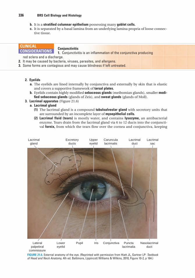
336 BRS Cell Biology and Histology
b. It is a stratified columnar epithelium possessing many goblet cells.c. It is separated by a basal lamina from an underlying lamina propria of loose connec-
tive tissue.
Conjunctivitis1. Conjunctivitis is an inflammation of the conjunctiva producing
red sclera and a discharge.2. It may be caused by bacteria, viruses, parasites, and allergens.3. Some forms are contagious and may cause blindness if left untreated.
CLINICAL
CONSIDERATIONS
2. Eyelidsa. The eyelids are lined internally by conjunctiva and externally by skin that is elastic
and covers a supportive framework of tarsal plates.b. Eyelids contain highly modified sebaceous glands (meibomian glands), smaller modi-
fied sebaceous glands (glands of Zeis), and sweat glands (glands of Moll).3. Lacrimal apparatus (Figure 21.6)
a. Lacrimal gland(1) The lacrimal gland is a compound tubuloalveolar gland with secretory units that
are surrounded by an incomplete layer of myoepithelial cells.(2) Lacrimal fluid (tears) is mostly water, and contains lysozyme, an antibacterial
enzyme. Tears drain from the lacrimal gland via 6 to 12 ducts into the conjuncti-val fornix, from which the tears flow over the cornea and conjunctiva, keeping
Pupil Conjunctiva
Lacrimalgland
Excretoryducts
Uppereyelid
Carunculalacrimalis
Lacrimalduct
Lacrimalsac
Lateralpalpebral
commissure
Lowereyelid
Iris Punctalacrimalia
Nasolacrimalduct
FIGURE 21.6. External anatomy of the eye. (Reprinted with permission from Hiatt JL, Gartner LP: Textbookof Head and Neck Anatomy, 4th ed. Baltimore, Lippincott Williams & Wilkins, 2010, Figure 10-2, p 164.)
LWBK615-c21[326-345].qxd 05/31/2010 11:05 AM Page 336 Aptara
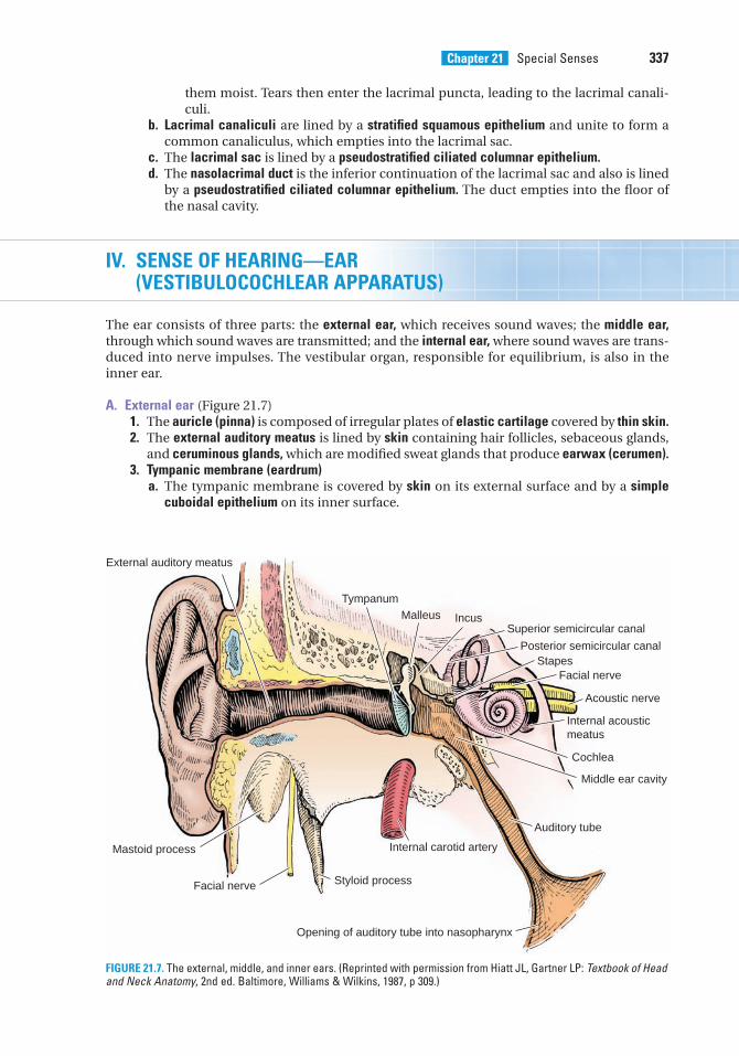
Chapter 21 Special Senses 337
them moist. Tears then enter the lacrimal puncta, leading to the lacrimal canali-culi.
b. Lacrimal canaliculi are lined by a stratified squamous epithelium and unite to form acommon canaliculus, which empties into the lacrimal sac.
c. The lacrimal sac is lined by a pseudostratified ciliated columnar epithelium.d. The nasolacrimal duct is the inferior continuation of the lacrimal sac and also is lined
by a pseudostratified ciliated columnar epithelium. The duct empties into the floor ofthe nasal cavity.
IV. SENSE OF HEARING—EAR (VESTIBULOCOCHLEAR APPARATUS)
The ear consists of three parts: the external ear, which receives sound waves; the middle ear,through which sound waves are transmitted; and the internal ear, where sound waves are trans-duced into nerve impulses. The vestibular organ, responsible for equilibrium, is also in theinner ear.
A. External ear (Figure 21.7)1. The auricle (pinna) is composed of irregular plates of elastic cartilage covered by thin skin.2. The external auditory meatus is lined by skin containing hair follicles, sebaceous glands,
and ceruminous glands, which are modified sweat glands that produce earwax (cerumen).3. Tympanic membrane (eardrum)
a. The tympanic membrane is covered by skin on its external surface and by a simplecuboidal epithelium on its inner surface.
External auditory meatus
TympanumMalleus Incus
Superior semicircular canal
Posterior semicircular canalStapes
Facial nerve
Acoustic nerve
Internal acousticmeatus
Cochlea
Middle ear cavity
Auditory tube
Opening of auditory tube into nasopharynx
Styloid processFacial nerve
Mastoid process Internal carotid artery
FIGURE 21.7. The external, middle, and inner ears. (Reprinted with permission from Hiatt JL, Gartner LP: Textbook of Headand Neck Anatomy, 2nd ed. Baltimore, Williams & Wilkins, 1987, p 309.)
LWBK615-c21[326-345].qxd 05/31/2010 11:05 AM Page 337 Aptara
338 BRS Cell Biology and Histology
b. It possesses fibroelastic connective tissue interposed between its two epithelial cover-ings.
c. Function. The tympanic membrane transmits sound vibrations that enter the ear to theossicles in the middle ear.
B. Middle ear (tympanic cavity) (Figure 21.7)1. The tympanic cavity contains the three bony ossicles (malleus, incus, and stapes), which
transmit movements of the tympanic membrane (eardrum) to the oval window (a membrane-covered opening in the bony wall of the cochlea).
2. The middle ear is connected to the pharynx via the auditory tube (eustachian tube).3. It is lined by a simple squamous epithelium, which changes to pseudostratified ciliated
columnar epithelium near its opening to the auditory tube.4. It has a lamina propria composed of dense connective tissue tightly adherent to the bony
wall.
Conductive hearing loss1. Conductive hearing loss results from a defect in the conduction of
sound waves in the external or middle ear.2. It may be caused by otitis media, a common inflammation of the middle ear; obstruction by a for-
eign body; or otosclerosis of the middle ear.
CLINICAL
CONSIDERATIONS
C. Internal ear (a bony labyrinth within the temporal bone) (Figure 21.7)1. The bony labyrinth, composed of the semicircular canals, vestibule, and cochlea are filled
with perilymph, and house the membranous labyrinth filled with endolymph.a. Semicircular canals house the semicircular ducts of the membranous labyrinth.b. The vestibule houses the saccule and utricle.c. Cochlea
(1) The cochlea winds two and a half times around a bony core (the modiolus), whichcontains blood vessels and the spiral ganglion.
(2) It is subdivided into three spaces: the scala vestibuli and scala tympani, which areboth filled with perilymph, and the scala media, or cochlear duct, which is filledwith endolymph.
2. The membranous labyrinth is filled with endolymph and possesses various sensory struc-tures that are specializations of the epithelium.a. Saccule and utricle (within the vestibule)
(1) Overview(a) The saccule and utricle are saclike bodies composed of a thin sheath of con-
nective tissue lined by simple squamous epithelium.(b) Each gives rise to a duct; the two ducts join, forming the endolymphatic sac.(c) They possess small, specialized regions, called maculae, which contain type I
and type II neuroepithelial hair cells, supporting cells, and a gelatinous layer(otolithic membrane).
(2) Vestibular hair cells(a) These neuroepithelial cells contain many mitochondria and a well-developed
Golgi complex.(b) They possess 50 to 100 elongated, rigid stereocilia (sensory microvilli)
arranged in rows and a single cilium (kinocilium). These cilia extend from theapical surface of the hair cells into the otolithic membrane. They function inthe detection of linear acceleration.
(c) Types of vestibular hair cells include the following:(i) Type I hair cells (bulbar), which are almost completely surrounded by a
cup-shaped afferent nerve ending(ii) Type II hair cells (columnar), which make contact with small afferent termi-
nals containing synaptic vesicles
LWBK615-c21[326-345].qxd 05/31/2010 11:05 AM Page 338 Aptara
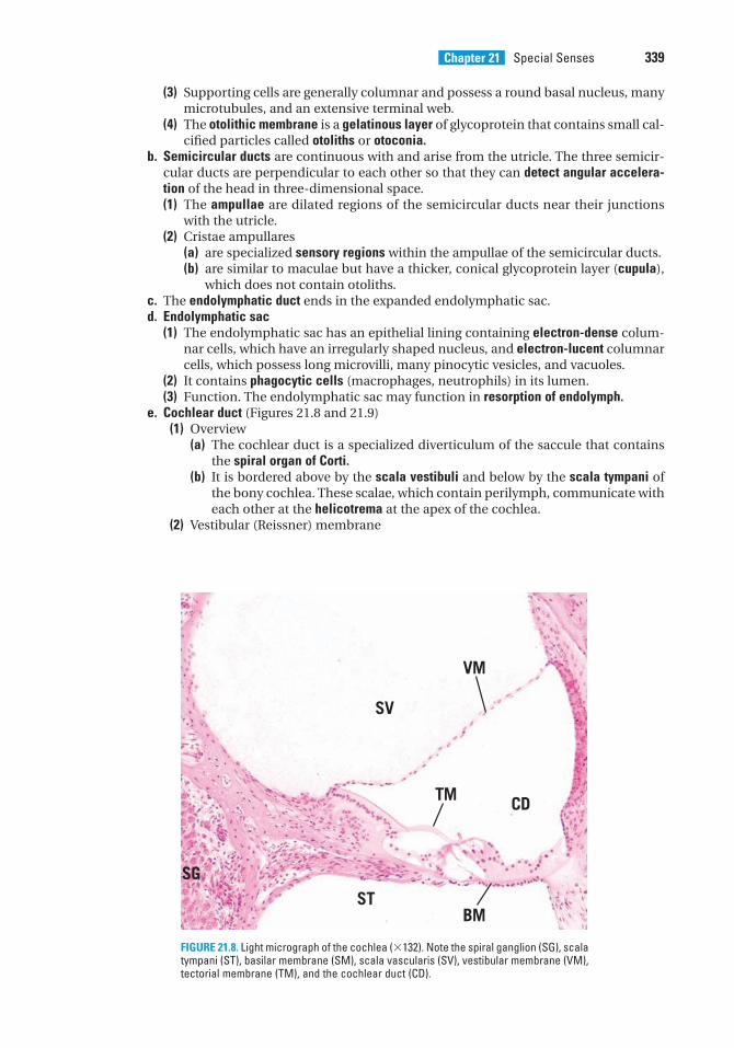
Chapter 21 Special Senses 339
(3) Supporting cells are generally columnar and possess a round basal nucleus, manymicrotubules, and an extensive terminal web.
(4) The otolithic membrane is a gelatinous layer of glycoprotein that contains small cal-cified particles called otoliths or otoconia.
b. Semicircular ducts are continuous with and arise from the utricle. The three semicir-cular ducts are perpendicular to each other so that they can detect angular accelera-tion of the head in three-dimensional space.(1) The ampullae are dilated regions of the semicircular ducts near their junctions
with the utricle.(2) Cristae ampullares
(a) are specialized sensory regions within the ampullae of the semicircular ducts.(b) are similar to maculae but have a thicker, conical glycoprotein layer (cupula),
which does not contain otoliths.c. The endolymphatic duct ends in the expanded endolymphatic sac.d. Endolymphatic sac
(1) The endolymphatic sac has an epithelial lining containing electron-dense colum-nar cells, which have an irregularly shaped nucleus, and electron-lucent columnarcells, which possess long microvilli, many pinocytic vesicles, and vacuoles.
(2) It contains phagocytic cells (macrophages, neutrophils) in its lumen.(3) Function. The endolymphatic sac may function in resorption of endolymph.
e. Cochlear duct (Figures 21.8 and 21.9)(1) Overview
(a) The cochlear duct is a specialized diverticulum of the saccule that containsthe spiral organ of Corti.
(b) It is bordered above by the scala vestibuli and below by the scala tympani ofthe bony cochlea. These scalae, which contain perilymph, communicate witheach other at the helicotrema at the apex of the cochlea.
(2) Vestibular (Reissner) membrane
SV
ST
CDTM
VM
BM
SG
FIGURE 21.8. Light micrograph of the cochlea (�132). Note the spiral ganglion (SG), scalatympani (ST), basilar membrane (SM), scala vascularis (SV), vestibular membrane (VM),tectorial membrane (TM), and the cochlear duct (CD).
LWBK615-c21[326-345].qxd 05/31/2010 11:05 AM Page 339 Aptara
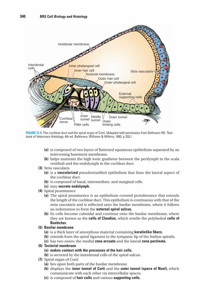
340 BRS Cell Biology and Histology
(a) is composed of two layers of flattened squamous epithelium separated by anintervening basement membrane.
(b) helps maintain the high ionic gradients between the perilymph in the scalavestibuli and the endolymph in the cochlear duct.
(3) Stria vascularis(a) is a vascularized pseudostratified epithelium that lines the lateral aspect of
the cochlear duct.(b) is composed of basal, intermediate, and marginal cells.(c) may secrete endolymph.
(4) Spiral prominence(a) The spiral prominence is an epithelium-covered protuberance that extends
the length of the cochlear duct. This epithelium is continuous with that of thestria vascularis and is reflected onto the basilar membrane, where it followsan indentation to form the external spiral sulcus.
(b) Its cells become cuboidal and continue onto the basilar membrane, wherethey are known as the cells of Claudius, which overlie the polyhedral cells ofBoettcher.
(5) Basilar membrane(a) is a thick layer of amorphous material containing keratinlike fibers.(b) extends from the spiral ligament to the tympanic lip of the limbus spiralis.(c) has two zones: the medial zona arcuata and the lateral zona pectinata.
(6) Tectorial membrane(a) makes contact with the processes of the hair cells.(b) is secreted by the interdental cells of the spiral sulcus.
(7) Spiral organ of Corti(a) lies upon both parts of the basilar membrane.(b) displays the inner tunnel of Corti and the outer tunnel (space of Nuel), which
communicate with each other via intercellular spaces.(c) is composed of hair cells and various supporting cells.
Interdentalcells
Vestibular membrane
Inner phalangeal cell
Cochlear nerve
Piller cells
Inner tunnel
Middletunnel
Outer tunnelOuter limiting cells
Inner hair cellTectorial membrane
Outer hair cellOuter phalangeal cell
Externalsupporting cells
Stria vascularis
FIGURE 21.9. The cochlear duct and the spiral organ of Corti. (Adapted with permission from Dellmann HD: Text-book of Veterinary Histology, 4th ed. Baltimore, Williams & Wilkins, 1993, p 332.)
LWBK615-c21[326-345].qxd 05/31/2010 11:05 AM Page 340 Aptara
Chapter 21 Special Senses 341
(8) Hair cells of the organ of Corti(a) are neuroepithelial cells containing a round basal nucleus surrounded by
many mitochondria.(b) possess many long, stiff stereocilia (arranged in a W formation) on their free
surfaces, as well as a basal body (but no kinocilium).(c) are divided into two types: Inner hair cells are organized in a single row along
the entire length of the cochlear duct and receive numerous afferent synap-tic terminals on their basal surface. Outer hair cells are organized in three ormore rows and are ensconced within a cup-shaped afferent nerve ending,where synaptic contacts are made.
(d) function in the reception of sound and can respond to different sound fre-quencies.
(9) Inner and outer pillar cells of the organ of Corti(a) are intimately associated with each other; both types rest on the basilar
membrane.(b) enclose and support the inner tunnel of Corti, which lies between the inner
and outer pillar cells.(c) possess a wide base and have elongated processes, which contain micro-
tubules, intermediate filaments, and actin filaments.(10) Inner and outer phalangeal cells of the organ of Corti
(a) are intimately associated with the inner and outer hair cells, respectively.(b) support the slender nerve fibers that form synapses with the hair cells.
(11) Cells of Hensen and border cells of the organ of Corti delineate the inner andouter borders of the spiral organ of Corti.
Nerve deafness1. Nerve deafness results from a lesion in any of the nerves transmitting
impulses from the spiral organ of Corti to the brain.2. It may be caused by disease, exposure to drugs, or prolonged exposure to loud noises.
CLINICAL
CONSIDERATIONS
3. Auditory function of the inner eara. Movement of the stapes at the oval window causes disturbances in the perilymph,
which cause deflection of the basilar membrane. Oscillations set in motion at the ovalwindow are dissipated at the secondary tympanic membrane covering the roundwindow of the cochlea. (At a very loud concert, the amount of energy absorbed is veryhigh, and it may take up to 72 hours for it to be dissipated. A residual humming noisemay be heard for 2–3 days.)
b. Large areas of the basilar membrane vibrate at many frequencies. However, optimalvibrations are detected at only specific areas. Sound waves of low frequency aredetected farther away from the oval window.
c. The pillar cells attached to the basilar membrane move laterally in response to thisdeflection, in turn causing a lateral shearing of the stereocilia of the sensory hair cellsof the organ of Corti against the tectorial membrane.
d. Movement of the stereocilia is transduced into electrical impulses that travel via thecochlear nerve to the brain.
4. Vestibular function of the inner eara. A change in the position of the head causes a flow of the endolymph in the semicir-
cular ducts (circular movement) or in the saccules and utricles (linear movement).b. Movement of the endolymph in the semicircular ducts displaces the cupula overlying
the cristae ampullares, causing bending of the stereocilia of the sensory hair cells.c. Movement of the endolymph in the saccules and utricles displaces the otoliths. This
deformation is transmitted to the maculae via the overlying gelatinous layer, causingbending of the stereocilia of the sensory hair cells.
LWBK615-c21[326-345].qxd 05/31/2010 11:05 AM Page 341 Aptara

342 BRS Cell Biology and Histology
d. In both cases, movement of the stereocilia is transduced into electrical impulses, whichare transmitted to the brain via vestibular nerve fibers.
Ménière disease1. Ménière disease is an inner ear disorder causing hearing loss, vertigo,
nausea, tinnitus, and vomiting. It is related to excess fluid in the endolymphatic duct.2. Drugs are used to treat the vertigo and nausea, but in severe cases, surgery may be required for
vestibular neuroectomy.
CLINICAL
CONSIDERATIONS
LWBK615-c21[326-345].qxd 05/31/2010 11:05 AM Page 342 Aptara
1. Which of the following specialized recep-tors exhibits a large ovoid capsule consistingof many concentric lamellae each separatedby a space containing tissue fluid?
(A) Cold receptors(B) Pacinian corpuscles(C) Ruffini endings(D) Meissner corpuscles(E) Krause end bulb
2. Aqueous humor drains from the eye bypassing
(A) into the ciliary processes.(B) from the anterior chamber into the pos-
terior chamber.(C) through the canal of Schlemm.(D) into the vitreous body.(E) through the pupil.
3. Which of the following statements is char-acteristic of the choroid?
(A) It is avascular.(B) It is the posterior portion of the tunica
fibrosa.(C) It is tightly attached to the sclera.(D) It contains many melanocytes.(E) It is not pigmented.
4. Communication of the scala vestibuli andscala tympani occurs at the
(A) round window.(B) oval window.(C) helicotrema.(D) endolymphatic sac.(E) inner tunnel.
5. The bony ossicles of the middle ear cavityare arranged in a series bridging thetympanic cavity beginning at the tympanicmembrane and ending at the
(A) endolymphatic duct.(B) round window.(C) helicotrema.(D) oval window.(E) cochlear duct.
6. Which of the following cells in the innerear are involved in detecting movements ofthe head?
(A) Hair cells in the maculae(B) Outer pillar cells(C) Inner pillar cells(D) Cells of Hensen(E) Hair cells in the organ of Corti
7. Rods and cones form synapses withwhich of the following cells?
(A) Amacrine(B) Bipolar(C) Ganglion(D) Müller(E) Pigmented epithelium
8. Which of the following statements is char-acteristic of the cornea?
(A) It represents the anterior portion of thetunica vasculosa.
(B) It is composed of three layers.(C) It forms the anterior boundary of the
posterior chamber of the eye.(D) It is devoid of nerve endings.(E) It is the anterior transparent portion of
the tunica fibrosa.
Review Test
Directions: Each of the numbered items or incomplete statements in this section is followed byanswers or by completions of the statement. Select the ONE lettered answer or completion thatis BEST in each case.
343
LWBK615-c21[326-345].qxd 05/31/2010 11:05 AM Page 343 Aptara
344 BRS Cell Biology and Histology
9. A patient exhibiting a high intraocularpressure in both eyes is symptomatic for
(A) cataract.(B) detached retina.(C) glaucoma.(D) conjunctivitis.(E) presbyopia.
10. Aqueous humor is produced by
(A) the corneal epithelium.(B) the canal of Schlemm.(C) ciliary epithelium.(D) conjunctiva.(E) ora serrata.
11. Which one of the following is related toan overabundance of fluid in the endolym-phatic duct?
(A) Glaucoma(B) Vertigo(C) Otis media(D) Conductive hearing loss(E) Nerve deafness
LWBK615-c21[326-345].qxd 05/31/2010 11:05 AM Page 344 Aptara
Answers and Explanations
1. B. Pacinian corpuscles are usually macroscopic. Their capsules are composed of severallamellae containing fibroblasts and collagen fibers. The lamellae are separated by sparseamounts of tissue fluid. Pacinian corpuscles respond to vibrations and pressure that distortthe lamellae. Meissner corpuscles, responsible for touch, are tapered terminals at the tipsof dermal papillae. Ruffini endings, which are sensitive to mechanical stresses, possess athin connective-tissue capsule that surrounds a fluid-filled space. Cold receptors are nakednerve endings that respond to temperatures below 25�C to 30�C (see Chapter 21 II B).
2. C. Aqueous humor exits the eye by passing into the canal of Schlemm. The vitreous bodyis a refractile gel that fills the chamber of the globe posterior to the lens and is not relatedto the aqueous humor (see Chapter 21 III D 1).
3. D. The choroid is the vascular tunic of the eye that loosely adheres to the sclera of thetunica fibrosa. It contains many melanocytes, which impart dark pigment to the eye (see Chapter 21 III C 1).
4. C. The scala vestibuli and the scala tympani are actually one perilymphatic spaceseparated by the cochlear duct (scala media). The scala vestibuli and tympani communi-cate with each other at the helicotrema (see Chapter 21 IV C 2 e).
5. D. The bony ossicles of the middle ear cavity articulate in a series from the tympanicmembrane to the oval window (see Chapter 21 IV B).
6. A. Neuroepithelial hair cells in the maculae of the saccule and the utricle detect linearmovement of the head. These cells are connected to the vestibular portion of the acousticnerve (see Chapter 21 IV C 2).
7. B. Rods and cones synapse with bipolar cells and horizontal cells (see Chapter 21 III E 3).
8. E. The cornea is the transparent anterior portion of the tunica fibrosa, the outer coveringof the eye; thus it forms the anterior wall of the anterior chamber of the eye. It is also richin sensory nerve endings. The sclera is the posterior portion of the tunica fibrosa. Thetunica vasculosa (middle coat) is composed of the choroid and ciliary body and the iris(see Chapter 21 III B).
9. C. High intraocular pressure is a symptom of glaucoma. This condition prohibits theaqueous humor from escaping from the anterior chamber of the eye via the canal ofSchlemm. If it is left untreated, a gradual loss of peripheral sight occurs; over time, blind-ness results. People with cataract have opacity of the lens that blurs the vision.Conjunctivitis is an inflammation of the conjunctiva of the eye with severe reddening ofthe sclera and the conjunctival surface of the lids, perhaps with a discharge. Thiscondition may be contagious, and if left untreated, blindness may occur (see Chapter 21III D).
10. C. The ciliary epithelium that lines the ciliary processes secretes the aqueous humor intothe posterior chamber of the eye between the iris and the lens. The aqueous humor thenflows through the pupil and into the anterior chamber of the eye and finally leaves theanterior chamber by entering the venous system via the canal of Schlemm (see Chapter 21III D 1).
11. B. An overabundance of endolymph in the endolymphatic duct causes vertigo, nausea, hear-ing loss, tinnitus, and vomiting, all symptoms of Ménière disease (see Chapter 21 IV C 4).
345
LWBK615-c21[326-345].qxd 05/31/2010 11:05 AM Page 345 Aptara
4. The centroacinar cells of the pancreassecrete
(A) an alkaline enzyme-poor fluid.(B) pancreatic digestive enzymes.(C) secretin.(D) cholecystokinin.(E) glucagon.
5. The zona fasciculata of the adrenal cortexsynthesizes and secretes
(A) mineralocorticoids.(B) glucagon.(C) epinephrine.(D) aldosterone.(E) glucocorticoids.
6. Which of the following statements aboutbony joints is true?
(A) Long bones are generally united bysynarthroses.
(B) Diarthroses are classified as synovialjoints.
(C) Type A cells of the synovial membranesecrete synovial fluid.
(D) Type B cells of the synovial membraneare phagocytic.
(E) Synarthroses are usually surrounded bya two-layered capsule.
7. All of the following characteristics can beused to distinguish neutrophils andbasophils histologically except one. Whichone is the exception?
(A) Size of specific granules(B) Shape of the nucleus(C) Number of azurophilic granules(D) The presence or absence of peroxidase(E) The presence of mitochondria
1. Which of the following statementsconcerning ribonucleic acid (RNA) synthesisis true?
(A) RNA synthesis does not require deoxyri-bonucleic acid to act as a template.
(B) Syntheses of ribosomal RNA, messengerRNA, and transfer RNA are all catalyzedby the same RNA polymerase.
(C) To yield messenger ribonucleoproteins,introns are excised, whereas exons arespliced together.
(D) Protein moieties are removed frommessenger ribonucleoproteins withinthe nucleolus, yielding functional mes-senger RNAs to exit via the nuclearpores.
(E) The start codon for RNA synthesis is UAG.
2. Which of the following factors is primarilyresponsible for causing osteoporosis in olderwomen?
(A) Decreased bone formation(B) Lack of physical exercise(C) Diminished estrogen secretion(D) Calcium deficiency(E) Increased bone formation
3. Which of the following bases that makeup deoxyribonucleic acid and ribonucleicacid (RNA) is unique to RNA?
(A) Thymine(B) Adenine(C) Cytosine(D) Guanine(E) Uracil
Comprehensive Examination
Directions: Each of the numbered items or incomplete statements in this section is followed byanswers or by completions of the statement. Select the ONE lettered answer or completion thatis BEST in each case.
346
LWBK615-exam[346-364].qxd 05/31/2010 11:07 AM Page 346 Aptara
Comprehensive Examination 347
8. A long-time user of chewing tobacconoticed several whitish, thick, painlesspatches on the lining of his cheeks. Themost probable diagnosis is
(A) aphthous ulcers.(B) adenocarcinoma.(C) keloids.(D) oral leukoplakia.(E) epidermolysis bullosa.
9. Primordial follicles of the ovary possess
(A) a layer of cuboidal follicular cells.(B) an oocyte arrested in prophase of
meiosis I.(C) an oocyte arrested in metaphase of
meiosis II.(D) well-defined thecae interna and externa.(E) a thick zona pellucida.
10. All of the following statements regardingthe membranous labyrinth of the inner ear are true except for one. Which is thatexception?
(A) It contains the saccule and utricle.(B) Maculae contain neuroepithelial cells,
which possess numerous stereocilia anda single kinocilium.
(C) Cristae ampullares in the semicircularcanals detect angular acceleration of thehead.
(D) The otolithic membrane contains smallcalcified particles.
(E) It contains the vestibular membrane.
11. Which of the following statements con-cerning euchromatin is true?
(A) It constitutes about 90% of thechromatin.
(B) It appears as basophilic clumps ofnucleoprotein when seen under the lightmicroscope.
(C) It is concentrated near the periphery ofthe nucleus.
(D) It is transcriptionally active.(E) It is transcriptionally inactive.
12. Intercalated disks function in which oneof the following?
(A) End-to-end attachments of smoothmuscle cells
(B) Intercellular movement of large proteins(C) Ionic coupling of cardiac muscle cells(D) Storage of Ca2�
(E) Release of neurotransmitters
13. Release of thyroid hormones from thefollicular cells of the thyroid gland dependson thyroid-stimulating hormone (TSH). TSHstimulation involves
(A) binding of TSH to receptors on the apical plasma membrane.
(B) formation of apical microvilli.(C) exocytosis of colloid droplets.(D) change in cell shape from flattened to
columnar.(E) secretion of lysosomes from the basal
cell surface.
14. Which one of the following statementsabout the development of the tooth crown istrue?
(A) The enamel organ is derived fromectomesenchyme (neural crest).
(B) The dental papilla is derived from theepithelium.
(C) The four-layer enamel organ appearsduring the cap stage.
(D) Dentin and enamel are formed duringthe appositional stage.
(E) Cementum is formed at the same timeas enamel.
15. Which one of the following statementsconcerning liver sinusoids is true?
(A) Their lining includes Ito cells (fat-storingcells).
(B) They receive bile from the hepatocytes.(C) They are lined by nonfenestrated
endothelial cells.(D) The space of Disse is located between
sinusoidal cells and hepatocytes.(E) Sinusoids convey blood from the central
vein to the portal vein.
16. A young college student had nausea,vomiting, visual disorders, and muscularparalysis after eating canned tuna fish. Theprobable diagnosis is botulism, caused byingestion of the Clostridium botulinum toxin.The physiological effect of this toxin is to
(A) inactivate acetylcholinesterase.(B) bind to and thus inactivate acetylcholine
receptors at myoneural junctions.(C) prevent release of calcium ions from the
sarcoplasmic reticulum, thus inhibitingmuscle contraction.
(D) inhibit release of acetylcholine frompresynaptic membranes.
(E) inhibit hydrolysis of adenosine triphos-phate during the contraction cycle.
LWBK615-exam[346-364].qxd 05/31/2010 11:07 AM Page 347 Aptara
348 BRS Cell Biology and Histology
17. Which of the following statements aboutnucleosomes is true?
(A) Histones form the nucleosome corearound which the double helix deoxyri-bonucleic acid is wound.
(B) Nucleosomes without histones form thestructural unit of the chromosome.
(C) Nucleosomes are composed ofribonucleic acid (RNA) molecules andtwo copies each of four different histones.
(D) Histone H1 forms the core of the nucleosome.
(E) Nucleosomes are linked together withRNA.
18. A high school student complains of fatigue and a sore throat. She has swollen, tender lymph nodes and a fever.Blood test results show an increased whiteblood cell count with many atypicallymphocytes; the number and appearanceof the erythrocytes are normal. This studentis likely to have
(A) AIDS.(B) pernicious anemia.(C) infectious mononucleosis.(D) Hodgkin disease.(E) factor VIII deficiency.
19. Which one of the following statementsabout the gallbladder is true?
(A) The gallbladder dilutes bile.(B) Bile enters the gallbladder via the com-
mon bile duct.(C) Bile leaves the gallbladder via the cystic
duct.(D) The gallbladder is lined by a simple
squamous epithelium.(E) Secretin stimulates the wall of the
gallbladder to contract, forcing bile fromits lumen.
20. Which one of the following statementsconcerning mitochondria is true?
(A) They change from the orthodox to thecondensed form in response to the uncou-pling of oxidation from phosphorylation.
(B) They are unable to divide.(C) They possess the enzymes of the Krebs
cycle in their cristae.(D) They contain elementary particles in
their matrix.(E) They do not contain deoxyribonucleic
acid.
21. Which one of the following componentsis present in muscular arteries but absentfrom elastic arteries?
(A) Fenestrated membranes(B) Vasa vasorum(C) Factor VIII(D) A thick, complete internal elastic lamina(E) Smooth muscle cells
22. Which one of the following statementsconcerning the pancreas is true?
(A) Islets of Langerhans secrete enzymes.(B) It possesses mucous acinar cells.(C) The endocrine pancreas has more
�-cells than �-cells.(D) Its �-cells secrete insulin.(E) Its �-cells secrete amylase.
23. Which one of the following stimulatesthe production of hydrochloric acid in thestomach?
(A) Somatostatin(B) Gastrin(C) Secretin(D) Cholecystokinin(E) Urogastrone
24. A deficiency or an excess of which of thefollowing vitamins results in short stature?
(A) Vitamin A(B) Vitamin C(C) Vitamin D(D) Vitamin K(E) Vitamin B
25. The intercellular spaces in the stratumspinosum of the epidermis contain lipid-containing sheets that are impermeable towater. This material is released from
(A) keratohyalin granules.(B) Langerhans cells.(C) membrane-coating granules.(D) sebaceous glands.(E) melanosomes.
26. Which of the following statements con-cerning basophilic erythroblasts is true?
(A) The nucleus has a fine chromatinnetwork.
(B) The nucleus is in the process of beingextruded.
(C) The cytoplasm contains specific granules.(D) The cytoplasm is pink and reveals a
dense reticulum.(E) The nucleus is bilobed.
LWBK615-exam[346-364].qxd 05/31/2010 11:07 AM Page 348 Aptara
Comprehensive Examination 349
27. The ileum includes which of the follow-ing structures?
(A) Rugae(B) Peyer patches(C) Brunner glands(D) Parietal cells(E) Chief cells
28. A 25-year-old woman complains about afrequently recurring painful lesion on herupper lip that exudes a clear fluid. She prob-ably has
(A) oral leukoplakia.(B) herpetic stomatitis.(C) aphthous ulcer.(D) bullous pemphigoid.(E) malignant melanoma.
29. Which of the following statements concerning the functions of the skin is true?
(A) Infrared radiation, necessary for synthe-sis of vitamin D, is absorbed by the skin.
(B) The skin provides no protection againstdesiccation.
(C) The skin contains temperature receptorsand plays a role in regulating body tem-perature.
(D) Melanin is synthesized by melanocytes,which are located in the dermis.
(E) Sunscreen with a sun protection factorrating of 15 or higher protects the skinfrom harmful effects caused by ultravio-let light.
30. A person with glomerulonephritis willhave which of the following signs orsymptoms?
(A) Hypotonic urine(B) Polyuria(C) Proteinuria(D) Dehydration(E) Polydipsia
31. Stratified squamous keratinized epithe-lium is always present in the
(A) rectum.(B) esophagus.(C) pyloric stomach.(D) jejunum.(E) anus.
32. Which of the following properties isexhibited in all three types of cartilage?
(A) It is involved in bone formation.(B) It possesses type II collagen.(C) It possesses type I collagen.(D) It grows interstitially and appositionally.(E) It has an identifiable perichondrium.
33. Which of the following is a receptor forfine touch?
(A) Pacinian corpuscle(B) Crista ampullaris(C) Ruffini ending(D) Krause end bulb(E) Meissner corpuscle
34. A premature infant has labored breath-ing, which is eventually alleviated by admin-istration of glucocorticoids. The most proba-ble diagnosis is
(A) immotile cilia syndrome.(B) emphysema.(C) hyaline membrane disease.(D) asthma.(E) chronic bronchitis.
35. Which of the following statements con-cerning the cribriform plate is true?
(A) It is the inner layer of the alveolar bone.(B) It lacks Sharpey fibers.(C) It is also known as the spongiosa.(D) It is composed of cancellous bone.(E) Normally it is fused with cementum
36. Which one of the following substances issynthesized in the pituitary gland?
(A) Oxytocin(B) Antidiuretic hormone(C) Somatotropin(D) Neurophysin(E) Vasopressin
37. Which one of the following is true in breastcancer cells that involve the BRCA1 gene?
(A) Mutated cells are unable to dividebecause of increased expression of theBRCA1 gene.
(B) Mutated cells fail to reach the G1-Scheckpoint.
(C) Mutated cells have an abnormal numberof endosomes.
(D) Mutated cells lose their G2-M cell cyclecheckpoint.
(E) Mutated cells become haploid due to theinteraction of the BRCA1 and p53 genes.
LWBK615-exam[346-364].qxd 05/31/2010 11:07 AM Page 349 Aptara
350 BRS Cell Biology and Histology
The following clinical consideration is thebasis of questions 38 to 40.
A 42-year-old man arrives in the emergencydepartment with a rash over much of his face,hands, and arms. He states that he was gar-dening and pulled out a number of weeds.On questioning the patient, the physicianrealized that the patient inadvertently camein contact with poison ivy.
38. Which of the following cells is responsi-ble for the release of the pharmacologicalagents that caused the rash?
(A) Diffuse neuroendocrine system cells(B) Myofibroblasts(C) Mast cells(D) Pericytes(E) Plasma cells
39. Which of the following is a secondarymediator produced by the cell in question 38?
(A) Histamine(B) Chondroitin sulfate(C) Neutral protease(D) Bradykinin(E) Aryl sulfatase
40. Which of the following white blood cellscan also participate in the reaction of thispatient to poison ivy?
(A) T cells(B) B cells(C) Neutrophils(D) Basophils(E) Eosinophils
41. A 26-year-old woman goes to the dentistbecause of a toothache. On examination, thedentist notes that the patient has a cariouslesion that involves not only the enamel butalso the dentin and the cementum of thetooth. Which of these substances cannotrepair itself?
(A) Dentin(B) Enamel(C) Cementum(D) Dentin and cementum(E) Dentin, cementum, and enamel
42. Lisa is small and thin in stature. Shespends her days indoors and rarely eatsdairy products. When she became pregnantat 25 years of age, she had severe lower backand leg pain and tenderness when pressurewas applied over bony regions of her body.Radiographs revealed excessive amounts ofpoorly mineralized osteoid in both femurs.Beneficial treatment of her conditioninvolved high doses of vitamin D, calcium,and phosphorus, a regimen that continuedafter successful delivery of her baby. Whichof the following describes Lisa’s disease?
(A) Osteogenesis imperfecta(B) Osteoporosis(C) Osteomalacia(D) Osteopetrosis(E) Osteopenia
43. Michael had visual problems within aday after being hit in the head with a ballduring a soccer game. He saw large floatersand noticed that a dark film was blockingpart of the vision in his right eye. An exami-nation by an ophthalmologist revealed thathe had a detached retina, and emergencysurgery was done to save the vision in thateye. Which of the following retinal layerswere separated from one another to causeMichael’s condition?
(A) Layer 1 from layer 2(B) Layer 2 from layer 3(C) Layer 3 from layer 4(D) Layer 5 from layer 6(E) Layer 6 from layer 7
44. Blood coagulation entails a cascade ofreactions that occur in two interrelatedpathways, the extrinsic and intrinsic. All ofthe following are associated with theintrinsic pathway of blood coagulationexcept
(A) conversion of fibrinogen to fibrin.(B) platelet aggregation.(C) release of tissue thromboplastin.(D) von Willebrand factor.(E) calcium.
LWBK615-exam[346-364].qxd 05/31/2010 11:07 AM Page 350 Aptara
Comprehensive Examination 351
45. A scientist spent a summer in a remoteregion of Africa, where he studied exoticplants. On returning to the United States,he developed a cough and fever that wouldnot go away. Laboratory tests revealed thathe had contracted a roundworm, Ascarislumbricoides. Which of the following cellswould be expected to be significantlyelevated in a differential count of hisblood?
(A) Erythrocytes(B) Lymphocytes(C) Monocytes(D) Eosinophils(E) Neutrophils
46. A 45-year-old musician who played guitar for 26 years in a rock band noticed he was having difficulty hearing. Anotolaryngologist confirmed his loss of hearing and associated it with prolongedexposure to loud sounds. Which of thefollowing structures would show degenera-tive changes that would account for thisman’s hearing loss?
(A) The epithelium lining the inner portionof the tympanic membrane
(B) Hair cells in the ampullae of the semicir-cular ducts
(C) The auditory (eustachian) tube extend-ing to the middle ear cavity
(D) Hair cells of the organ of Corti(E) Ossicles in the middle ear
47. Which of the following is a protein circu-lating in the blood that functions in woundhealing?
(A) Chondronectin(B) Plasma fibronectin(C) Osteonectin(D) Matrix fibronectin(E) Laminin
48. Which of the following is an adhesiveglycoprotein that forms fibrils in theextracellular matrix?
(A) Chondronectin(B) Plasma fibronectin(C) Osteonectin(D) Matrix fibronectin(E) Laminin
49. Which of the following is a multi-functional protein that attacheschondrocytes to type II collagen?
(A) Laminin(B) Plasma fibronectin(C) Osteonectin(D) Matrix fibronectin(E) Chondronectin
50. Which of the following cells in theconnective tissue is an antibody-manufacturing cell?
(A) Pericytes(B) Macrophages(C) T lymphocytes(D) Plasma cells(E) Mast cells
51. Principal phagocytes of connectivetissue are the
(A) pericytes.(B) macrophages.(C) T lymphocytes.(D) plasma cells.(E) mast cells.
52. Cells in the connective tissue that canbind immunoglobulin E antibodies andmediate immediate hypersensitivityreactions are
(A) pericytes.(B) macrophages.(C) T lymphocytes.(D) plasma cells.(E) mast cells.
53. Connective tissue cells responsible forinitiating cell-mediated immune responsesare
(A) pericytes.(B) macrophages.(C) T lymphocytes.(D) plasma cells.(E) mast cells.
54. Pluripotential cells located primarilyalong capillaries in the connective tissue are
(A) pericytes.(B) macrophages.(C) T lymphocytes.(D) plasma cells.(E) mast cells.
LWBK615-exam[346-364].qxd 05/31/2010 11:07 AM Page 351 Aptara
352 BRS Cell Biology and Histology
55. Which of the following functions in acti-vation of secondary messenger systems?
(A) Phospholipid(B) Glycocalyx(C) Carrier protein(D) Band 3 protein(E) G protein
56. Which of the following is primarilyresponsible for establishing the potentialdifference across the plasma membrane?
(A) K� leak channel(B) Glycocalyx(C) Carrier protein(D) Band 3 protein(E) G protein
57. Which of the following is anamphipathic molecule?
(A) Phospholipid(B) Glycocalyx(C) Carrier protein(D) Band 3 protein(E) G protein
58. Which of the following is acarbohydrate-containing coveringassociated with the outer leaflet of theplasma membrane?
(A) Phospholipid(B) Glycocalyx(C) Carrier protein(D) Band 3 protein(E) G protein
59. Which of the following functions inantiport transport?
(A) Phospholipid(B) Glycocalyx(C) Carrier protein(D) Band 3 protein(E) G protein
60. Which of the following is a surfacemarker on T killer cells (CTL)?
(A) CD4(B) CD8(C) Interleukin 1(D) Interleukin 2(E) Perforin
61. Which of the following is a surfacemarker on T helper cells?
(A) CD4(B) CD8(C) Interleukin 1(D) Interleukin 2(E) Perforin
62. Which of the following mediates lysis oftumor cells?
(A) CD4(B) CD8(C) Interleukin 1(D) Interleukin 2(E) Perforin
63. Which of the following is released bymacrophages and stimulates activated Thelper cells?
(A) CD4(B) CD8(C) Interleukin 1(D) Interleukin 2(E) Interferon-�
64. Which of the following stimulates activa-tion of natural killer cells?
(A) CD4(B) Interferon-�(C) Interleukin 1(D) Interleukin 2(E) Perforin
65. Identify the cells that replicate theirdeoxyribonucleic acid during the S phase ofthe cell cycle and undergo meiosis.
(A) Sertoli cells(B) Primary spermatocytes(C) Secondary spermatocytes(D) Interstitial cells of Leydig(E) Spermatids
66. Identify the cells that form a temporarycylinder of microtubules called themanchette.
(A) Sertoli cells(B) Primary spermatocytes(C) Secondary spermatocytes(D) Interstitial cells of Leydig(E) Spermatids
LWBK615-exam[346-364].qxd 05/31/2010 11:07 AM Page 352 Aptara
Comprehensive Examination 353
67. Identify the cells that possess receptorsfor luteinizing hormone (LH) and producetestosterone in response to binding of LH.
(A) Sertoli cells(B) Primary spermatocytes(C) Secondary spermatocytes(D) Interstitial cells of Leydig(E) Spermatids
68. Identify the cells that are responsible forformation of the blood-testis barrier.
(A) Sertoli cells(B) Primary spermatocytes(C) Secondary spermatocytes(D) Interstitial cells of Leydig(E) Spermatids
69. Identify the cells that synthesize androgen-binding protein when stimulatedby follicle-stimulating hormone.
(A) Sertoli cells(B) Primary spermatocytes(C) Secondary spermatocytes(D) Interstitial cells of Leydig(E) Spermatids
70. Which of the following organellespossesses mixed-function oxidases thatdetoxify phenobarbital and other drugs?
(A) Rough endoplasmic reticulum(B) Smooth endoplasmic reticulum(C) Mitochondrion(D) Annulate lamella(E) Lysosome
71. Which of the following organellescontains ribophorins?
(A) Rough endoplasmic reticulum(B) Smooth endoplasmic reticulum(C) Mitochondrion(D) Polyribosome(E) Lysosome
72. Which of the following is the site wheredegradation of foreign material ingested bythe cell takes place?
(A) Rough endoplasmic reticulum(B) Smooth endoplasmic reticulum(C) Mitochondrion(D) Annulate lamellae(E) Lysosome
73. Which of the following organellescontains adenosine triphosphate synthase?
(A) Rough endoplasmic reticulum(B) Smooth endoplasmic reticulum(C) Mitochondrion(D) Annulate lamellae(E) Lysosome
74. Which of the following stimulatessecretion of pepsinogen?
(A) Gastrin(B) Somatostatin(C) Motilin(D) Pepsinogen(E) Lysozyme
75. Which of the following is produced byBrunner glands and inhibits hydrochloricacid secretion by parietal cells?
(A) Gastrin(B) Somatostatin(C) Urogastrone(D) Pepsinogen(E) Lysozyme
76. Which of the following functions as anantibacterial agent?
(A) Gastrin(B) Somatostatin(C) Motilin(D) Pepsinogen(E) Lysozyme
77. Which of the following stimulatescontraction of smooth muscle in the wall ofthe digestive tract?
(A) Gastrin(B) Somatostatin(C) Motilin(D) Pepsinogen(E) Lysozyme
78. Which of the following inhibits secretionby nearby enteroendocrine cells?
(A) Gastrin(B) Somatostatin(C) Motilin(D) Pepsinogen(E) Lysozyme
LWBK615-exam[346-364].qxd 05/31/2010 11:07 AM Page 353 Aptara
354 BRS Cell Biology and Histology
79. Which of the following is involved informing cross-links between adjacenttropocollagen molecules?
(A) Lysine(B) Desmosine(C) Hydroxyproline(D) Arginine(E) Proline
80. Which of the following cross-links elastinmolecules to form an extensive network?
(A) Lysine(B) Fibrillin(C) Hydroxyproline(D) Arginine(E) Proline
81. Inadequate amounts of iodine in the dietleads to which one of the followingconditions?
(A) Simple goiter(B) Exophthalmic goiter(C) Graves disease(D) Addison disease(E) Hyperparathyroidism
82. Which of the following conditions isassociated with the destruction of the adre-nal cortex?
(A) Simple goiter(B) Exophthalmic goiter(C) Graves disease(D) Addison disease(E) Hyperparathyroidism
83. Which of the following hydrolyzesadenosine triphosphate?
(A) Troponin C(B) Globular head (S1 fragment) of myosin(C) Myoglobin(D) Actin(E) Tropomodulin
84. Which of the following binds oxygen?
(A) Troponin C(B) Globular head (S1 fragment) of myosin(C) Myoglobin(D) Actin(E) Tropomodulin
85. Which of the following binds Ca2�?
(A) Troponin C(B) Globular head (S1 fragment) of myosin(C) Myoglobin(D) Actin(E) Tropomodulin
86. Which of the following is a rare form ofskin cancer that may be fatal?
(A) Epidermolysis bullosa(B) Basal cell carcinoma(C) Malignant melanoma(D) Psoriasis(E) Warts
87. Which of the following is a hereditaryskin disease characterized by blister forma-tion after minor trauma?
(A) Epidermolysis bullosa(B) Basal cell carcinoma(C) Malignant melanoma(D) Psoriasis(E) Warts
88. Which of the following regions of the res-piratory system would concern a patientwith an adenoid?
(A) Trachea(B) Nasopharynx(C) Terminal bronchiole(D) Alveolar duct(E) Intrapulmonary bronchi
89. Which of the following possesses C-shaped rings of hyaline cartilage?
(A) Trachea(B) Nasopharynx(C) Terminal bronchiole(D) Alveolar duct(E) Intrapulmonary bronchi
90. Which of the following contains smoothmuscle at the openings into alveoli?
(A) Trachea(B) Nasopharynx(C) Terminal bronchiole(D) Alveolar duct(E) Intrapulmonary bronchi
LWBK615-exam[346-364].qxd 05/31/2010 11:07 AM Page 354 Aptara
Comprehensive Examination 355
91. Which of the following is lined by anepithelium containing ciliated cells andClara cells?
(A) Trachea(B) Nasopharynx(C) Terminal bronchiole(D) Alveolar duct(E) Intrapulmonary bronchi
92. Which of the following is associated withrupture of the oviduct and hemorrhaginginto the peritoneal cavity?
(A) Endometriosis(B) Cervical cancer(C) Ectopic tubal pregnancy(D) Breast cancer(E) Teratomas
93. Which of the following may be classifiedas lobular carcinoma?
(A) Endometriosis(B) Cervical cancer(C) Ectopic tubal pregnancy(D) Breast cancer(E) Teratomas
94. Which of the following may be detectedby a Papanicolaou smear?
(A) Endometriosis(B) Cervical cancer(C) Ectopic tubal pregnancy(D) Breast cancer(E) Teratomas
95. Which of the following commonly resultsin hemorrhaging into the peritoneal cavitydependent on the stage of the menstrualcycle?
(A) Endometriosis(B) Cervical cancer(C) Ectopic tubal pregnancy(D) Breast cancer(E) Teratomas
96. Which of the following facilitates theabsorption of vitamin B12?
(A) Pepsin(B) Enzyme associated with the glycocalyx
of the intestinal striated border(C) Lipase(D) Chylomicron(E) Gastric intrinsic factor
97. Which of the following functions in thedigestion of carbohydrates?
(A) Pepsin(B) Enzyme associated with the glycocalyx
of the intestinal striated border(C) Lipase(D) Chylomicron(E) Gastric intrinsic factor
98. Which of the following functions in thedigestion of proteins?
(A) Pepsin(B) Enzyme associated with the glycocalyx
of the intestinal striated border(C) Lipase(D) Chylomicron(E) Gastric intrinsic factor
99. Which of the following functions in thetransport of triglycerides into lacteals?
(A) Pepsin(B) Enzyme associated with the glycocalyx
of the intestinal striated border(C) Lipase(D) Chylomicron(E) Gastric intrinsic factor
100. Which of the following is manufacturedand released by parietal cells?
(A) Pepsin(B) Enzyme associated with the glycocalyx
of the intestinal striated border(C) Lipase(D) Chylomicron(E) Gastric intrinsic factor
LWBK615-exam[346-364].qxd 05/31/2010 11:07 AM Page 355 Aptara
Answers and Explanations
1. C. To yield messenger ribonucleoproteins (mRNPs), introns are excised, whereas exonsare spliced together. Deoxyribonucleic acid does act as the template for the synthesis ofribonucleic acid (RNA). Three RNA polymerases (I, II, and III) are needed to synthesizeribosomal RNA, messenger RNA, and transfer RNA, respectively. Protein moieties areremoved from the mRNPs as they leave the nucleus to yield functional mRNAs outsidethe nucleus. (See Chapter 2 VIII A.)
2. C. The most common cause of osteoporosis in older women is diminished estrogensecretion. (See Chapter 7 II J Clinical Consideration.)
3. E. Although adenine, cytosine, and guanine are found in both deoxyribonucleic acid(DNA) and ribonucleic acid (RNA), uracil is found only in RNA. Uracil substitutes for thebase thymine in DNA. (See Chapter 2 VIII.)
4. A. Pancreatic centroacinar cells form the initial segment of the intercalated duct and arepart of the exocrine pancreas. They secrete an enzyme-poor alkaline fluid whenstimulated by secretin. Pancreatic digestive enzymes are synthesized by the acinar cells ofthe exocrine pancreas; their release is stimulated by cholecystokinin. Glucagon isproduced in the endocrine pancreas (islets of Langerhans). (See Chapter 17 III A 2.)
5. E. The zona fasciculata, the largest region of the adrenal cortex, produces glucocorticoids(cortisol and corticosterone). The zona glomerulosa produces mineralocorticoids, prima-rily aldosterone. Epinephrine is produced in the adrenal medulla. Glucagon is producedin the pancreas, not in the adrenal gland. (See Chapter 13 VI A 2.)
6. B. Diarthroses, the type of joint connecting two long bones, are classified as synovialjoints, which are surrounded by a two-layered capsule housing a synovial membrane.Type A cells of the synovial membrane are phagocytic, whereas type B cells secrete thesynovial fluid. Synarthrosis joints are those found joining the bones of the skull, which areimmovable. (See Chapter 7 III B.)
7. E. Neutrophils have a nucleus with three or four lobes, many azurophilic granules, andsmall specific granules that lack peroxidase. In contrast, basophils have an S-shapednucleus, few azurophilic granules, and large specific granules that contain peroxidase.Both neutrophils and basophils possess mitochondria. (See Table 10.2.)
8. D. Oral leukoplakia, which results from epithelial hyperkeratosis, is usually of unknownetiology but often is associated with the use of chewing tobacco. Although the character-istic painless lesions are benign, they may transform into squamous cell carcinoma. Aph-thous ulcers are painful lesions of the oral mucosa that are surrounded by a red border.Adenocarcinoma is a form of cancer arising in glandular tissue. Keloids are swellings inthe skin that arise from increased collagen formation in hyperplastic scar tissue. Epider-molysis bullosa is a group of hereditary skin diseases characterized by blister formationafter minor trauma. (See Chapter 16 II.)
9. B. A primordial follicle is composed of a flattened layer of follicular cells surrounding aprimary oocyte, which is arrested in prophase of meiosis I. Well-defined thecal layers anda thick zona pellucida are found in growing follicles. A graafian (mature) follicle possessesa secondary oocyte, which becomes arrested in metaphase of meiosis II just before ovula-tion. (See Chapter 19 II B 1.)
10. E. The first four statements are true. Linear acceleration of the head is detected by the neu-roepithelial hair cells of the maculae, which are specialized regions of the saccule and utricle.However, the vestibular membrane is a part of the organ of Corti. (See Chapter 21 IV C 2.)
356
LWBK615-exam[346-364].qxd 05/31/2010 11:07 AM Page 356 Aptara
Comprehensive Examination 357
11. D. Euchromatin, the transcriptionally active form of chromatin, represents only about10% of the chromatin. In the light microscope, it appears as a light-staining, dispersedregion of the nucleus. (See Chapter 2 V 2.)
12. C. Large protein molecules cannot move across intercalated disks (the steplike junctionalcomplexes present in cardiac, not smooth, muscle). These junctional structures possessthree specializations: desmosomes, which provide end-to-end attachment of cardiacmuscle cells; fascia adherentes, to which the thin myofilaments attach; and gap junctions,which permit intercellular movement of small molecules and ions (ionic coupling). (See Chapter 8 V B 6.)
13. D. Flattened squamous cells are characteristic of an unstimulated, inactive thyroid gland.Thyroid-stimulating hormone (TSH) binds to G protein–linked receptors on the basalsurface of follicular cells. Under TSH stimulation, thyroid follicular cells becomecolumnar and form pseudopods, which engulf colloid. Lysosomal enzymes split thyroxineand triiodothyronine from thyroglobulin; the hormones are then released basally. (See Chapter 13 IV B 2.)
14. D. The enamel organ is epithelially derived, whereas the dental papilla comes fromectomesenchyme. The bell, not the cap, stage of odontogenesis is characterized bypossessing a fourth layer in its enamel organ. Formation of dentin and enamel occursduring the appositional stage of tooth development. Cementum is located on the rootand is formed only after the crown is complete and enamel ceases to be elaborated. (See Chapter 16 II C 3.)
15. D. Liver sinusoids convey blood to the central vein. Their endothelial cells arefenestrated, and material from the sinusoids may enter the space of Disse through thefenestrae, where it may be endocytosed by hepatocytes. The space of Disse houses Itocells (fat-storing cells). Because bile is the exocrine secretion of hepatocytes, it does notenter the sinusoids. (See Chapter 17 IV B 2.)
16. D. The toxin from Clostridium botulinum inhibits the release of acetylcholine, the neuro-transmitter at myoneural junctions. As a result, motor nerve impulses cannot betransmitted across the junction, and muscle cells are not stimulated to contract. (SeeChapter 8 IV 2 Clinical Considerations.)
17. A. The nucleosome, the structural unit of chromatin packing, does not containribonucleic acid. In extended chromatin, two copies each of histones H2A, H2B, H3, andH4 form the nucleosome core around which a deoxyribonucleic acid molecule is wound.Condensed chromatin contains additional histones (H1), which bind to nucleosomes,forming the condensed 30-nm chromatin fiber. (See Chapter 2 VI A.)
18. C. Only infectious mononucleosis is characterized by all of the signs and symptoms indi-cated. AIDS is associated with a decreased lymphocyte count, particularly of T helpercells. Pernicious anemia is associated with a decreased red blood cell count. Hodgkin dis-ease is associated with fatigue and enlarged lymph nodes, but the nodes are not painful,and the presence of Reed–Sternberg cells is diagnostic of this disease. Factor VIIIdeficiency, a coagulation disorder, is not associated with any of these signs andsymptoms. (See Chapter 10 II B 2 Clinical Considerations.)
19. C. The gallbladder, which concentrates and stores bile, is lined by a simple columnarepithelium. Cholecystokinin stimulates contraction of the gallbladder wall, forcing bilefrom the lumen into the cystic duct; this joins the common hepatic duct to form the com-mon bile duct, which delivers bile to the duodenum. (See Chapter 17 III A 2.)
20. A. Uncoupling of oxidation from phosphorylation induces mitochondria to change fromthe orthodox to the condensed form. Condensed mitochondria are often present inbrown fat cells, which produce heat rather than adenosine triphosphate. Mitochondriapossess circular deoxyribonucleic acid, and they reproduce (divide) by fission. (See Chapter 3 II 6 e.)
LWBK615-exam[346-364].qxd 05/31/2010 11:07 AM Page 357 Aptara
358 BRS Cell Biology and Histology
21. D. Muscular (distributing) arteries have a thick, complete internal elastic lamina in thetunica intima, whereas elastic (conducting) arteries have an incomplete internal elasticlamina. Both types of arteries have vasa vasorum, factor VIII, and smooth muscle cells intheir walls. Muscular arteries possess numerous layers of muscle cells in the tunicamedia, but elastic arteries do not. Only elastic arteries possess fenestrated (elastic) mem-branes in the tunica media, in which smooth muscle cells are dispersed. (See Chapter 11 IB 1 b.)
22. C. In the endocrine pancreas, �-cells account for about 70% of the secretory cells; �-cellsabout 20%; and �-cells less than 5%. Polypeptide hormones are synthesized by andreleased from the islets of Langerhans (endocrine pancreas). The exocrine pancreas pos-sesses serous (not mucous) acinar cells. Insulin is produced by �-cells. (See Chapter 17 IIIB 3.)
23. B. Somatostatin and urogastrone both inhibit the production of hydrochloric acid,whereas gastrin enhances it. Secretin and cholecystokinin act on the pancreas to facilitatesecretion of buffer and pancreatic enzymes, respectively. (See Chapter 16 III B 2.)
24. A. A deficiency of vitamin A inhibits bone formation and growth, whereas an excess stim-ulates ossification of the epiphyseal plates, thus leading to premature closure of theplates. Both conditions result in short stature. A deficiency of vitamin D reduces calciumabsorption from the small intestine and results in soft bones, whereas an excess ofvitamin D stimulates bone resorption. A deficiency of vitamin C results in poor bonegrowth and fracture repair. Vitamin K plays no role in bone formation. (See Chapter 7 II I.)
25. C. Membrane-coating granules are present in keratinocytes in the stratum spinosum(and stratum granulosum). The contents of these granules are released into the intercel-lular spaces to help waterproof the skin. Keratinocytes in the stratum granulosum alsopossess keratohyalin granules; these contain proteins that bind keratin filamentstogether. (See Chapter 14 II B 2.)
26. A. The nucleus of erythroblasts is not in the process of being extruded and is round witha very fine chromatin network. The cytoplasm is blue and possesses no granules. (SeeChapter 10 VI C.)
27. B. The ileum includes Peyer patches. Rugae, parietal cells, and chief cells are located in thestomach. Brunner glands are present in the submucosa of the duodenum. (See Table 16.1.)
28. B. Herpetic stomatitis is characterized by painful fever blisters on the lips or near thenostrils. These blisters exude a clear fluid. Aphthous ulcers (canker sores) do not exudefluid. Bullous pemphigoid is an autoimmune disease marked by chronic generalized blis-ters in the skin. (See Chapter 16 II B Clinical Considerations.)
29. C. The skin, which consists of the epidermis and dermis, is important in the regulation ofbody temperature and contains temperature receptors in the dermis. Ultraviolet (notinfrared) radiation absorbed by the skin is necessary for the synthesis of vitamin D.Protection against desiccation is provided by the contents of the membrane-coatinggranules of the epidermis. Melanocytes are located in the deepest layer of the epidermis(stratum basale). Sunscreen with sun protection factor rating of 15 or higher offers noprotection against ultraviolet light of longer wavelengths, that is, in the UVA spectrum.(See Chapter 14 I E.)
30. C. Patients with glomerulonephritis excrete protein in their urine. All of the other symp-toms are characteristic of patients with diabetes insipidus, who are incapable of produc-ing adequate amounts of antidiuretic hormone and therefore have polyuria (large volumeof hypotonic urine production), polydipsia, and dehydration. (See Chapter 18 III AClinical Considerations.)
31. E. The anus is lined by stratified squamous keratinized epithelium. The rectum, jejunum,and pyloric stomach are lined by simple columnar epithelium. The esophagus is lined bystratified squamous (nonkeratinized) epithelium. (See Chapter 16 III D 4.)
LWBK615-exam[346-364].qxd 05/31/2010 11:07 AM Page 358 Aptara
Comprehensive Examination 359
32. D. Hyaline cartilage, elastic cartilage, and fibrocartilage all exhibit both interstitial andappositional growth. Hyaline cartilage and elastic cartilage have type II collagen in theirmatrix and are surrounded by a perichondrium, whereas fibrocartilage has type Icollagen and lacks an identifiable perichondrium. Only hyaline cartilage is involved inendochondral bone formation. (See Chapter 7 I A, B, C.)
33. E. Meissner corpuscles, in the papillary layer of the dermis, are fine touch receptors.Pacinian corpuscles perceive pressure, touch, and vibration; they are located in thedermis, hypodermis, and connective tissue of mesenteries and joints. Cristae ampullaresare special regions of the semicircular canals that detect circular movements of the head.Ruffini endings, in the dermis and joints, function in pressure and touch perception.Krause end bulbs are cold and pressure receptors in the dermis. (See Chapter 14 III A.)
34. C. Hyaline membrane disease, which results from inadequate amounts of pulmonarysurfactant, is characterized by labored breathing and typically is observed in prematureinfants. Glucocorticoids stimulate synthesis of surfactant and can correct the condition.(See Chapter 15 VIII D Clinical Considerations.)
35. A. The cribriform plate, the inner layer of the alveolar bone, is composed of compactbone. It is attached to the principal fiber groups of the periodontal ligament via Sharpeyfibers. The outer layer of the alveolar bone is the cortical plate. The spongiosa is theregion of cancellous (spongy) bone enclosed between the cortical and cribriform plates.(See Chapter 16 II D 3.)
36. C. Somatotropin (growth hormone) is synthesized by cells called somatotrophs, whichare acidophils located in the pars distalis of the anterior lobe of the pituitary gland. Oxy-tocin and antidiuretic hormone (also called vasopressin) are produced in the hypothala-mus and transported to the pars nervosa of the pituitary. Neurophysin, a binding protein,aids in this transport. (See Chapter 13 III A 1 a.)
37. D. Mutations in the BRCA1 gene, a breast tumor suppressor gene, are the major cause ofbreast cancer. In most breast cancers involving this gene, deoxyribonucleic acid synthesisproceeds, indicating that the G1-S checkpoint is not affected; however, the mutated cells can-not control the transition between G2 and M. Moreover, the mutated cells had abnormal cen-trosome numbers and their nuclear division did not proceed normally, leading to aneuploidyand genetic instability of the daughter cells. (See Chapter 19 X C Clinical Considerations.)
38. C. The patient has an immediate (type I) hypersensitivity reaction. The cells responsiblefor releasing the primary and secondary mediators are the mast cells. (See Chapter 6 III D.)
39. D. Bradykinins are the only listed pharmacological agents that are produced via thearachidonic acid pathway. All of the others are primary mediators because they are storedin the storage granules of mast cells. (See Chapter 6 III D.)
40. D. Basophils are very similar in function to mast cells. They also participate in the imme-diate (type I) hypersensitivity reaction. (See Chapter 6 III H 3.)
41. B. The only hard tissue of the tooth that cannot be repaired by the body is enamelbecause ameloblasts, the cells that manufacture enamel, are eliminated as the toothemerges into the oral cavity. (See Chapter 16 II C 2.)
42. C. Osteomalacia, or adult rickets, is Lisa’s disorder. It is characterized by a failure of newlyformed osteoid to calcify because there is a lack of calcium, vitamin D, and phosphorus. Thebones gradually soften and bend, and pain accompanies the condition, which often becomessevere during pregnancy as the fetus removes calcium from the mother’s body. Osteogenesisimperfecta is a genetic condition affecting the synthesis of type I collagen in the bone matrixand results in extreme bone fragility and breakage. Osteoporosis is a disease characterized bylow bone mineral density and structural deterioration, making bone more susceptible tofracture, and osteopenia is reduced bone mass caused by inadequate osteoid synthesis.Osteopetrosis is the excessive formation of bone, which obliterates the marrow cavities andthus impairs the formation of blood cells. (See Chapter 7 II I Clinical Consideration.)
LWBK615-exam[346-364].qxd 05/31/2010 11:07 AM Page 359 Aptara
360 BRS Cell Biology and Histology
43. A. The pigmented epithelium (layer 1) was separated from the layer of rods and cones(layer 2), which make up the light-sensitive part of the neural retina. (See Chapter 21 III EClinical Consideration.)
44. C. The extrinsic pathway is initiated by the release of tissue thromboplastin after traumato extravascular tissue. Platelet aggregation is promoted by von Willebrand factor, whichis associated with the intrinsic pathway only. Calcium is required in both pathways, and the final reaction—the conversion of fibrinogen to fibrin—is the same in both. (See Chapter 10 III D 1.)
45. D. Eosinophils are increased in parasitic infections and allergic reactions. Botheosinophils and basophils have receptors for immunoglobulin E, which seems to beimportant in the destruction of parasites. Both neutrophils and monocytes lackimmunoglobulin E receptors. (See Table 10.2.)
46. D. The inner ear is where sound waves are transduced into nerve impulses that conveyauditory information to the brain, and the hair cells of the organ of Corti play a key role inthis process. Sound waves are initially received by the outer ear and transmitted via thetympanic membrane (eardrum) to the middle ear, where ossicles transmit the vibrationsto the inner ear. The inner ear has an auditory system for hearing (the organ of Corti) anda vestibular system of semicircular ducts that control equilibrium and spatial orientation.(See Chapter 21 IV C e 8.)
47. B. Plasma fibronectin functions in wound healing, blood clotting, and phagocytosis ofmaterial from the blood. (See Chapter 4 II C 1.)
48. D. Matrix fibronectin mediates cell adhesion to the extracellular matrix by binding tofibronectin receptors on the plasma membrane. (See Chapter 4 II C 1.)
49. E. Chondronectin has binding sites for type II collagen, proteoglycans, and chondrocytecell-surface receptors. (See Chapter 4 II C 5.)
50. D. Plasma cells, which arise from antigen-activated B lymphocytes, produce antibodiesand thus are directly responsible for humoral-mediated immunity. (See Chapter 6 III G.)
51. B. Macrophages, the principal phagocytes of connective tissue, remove large particulatematter and assist in the immune response by acting as antigen-presenting cells. (See Chapter 6 III E.)
52. E. Mast cells (and basophils) have receptors for immunoglobulin E antibodies on theirsurface. These cells release histamine, heparin, leukotriene C (slow-reacting substance ofanaphylaxis), and eosinophil chemotactic factor, which have effects that constituteimmediate hypersensitivity reactions. (See Chapter 6 III D.)
53. C. T lymphocytes initiate cell-mediated immune responses. (See Chapter 6 III F.)
54. A. Pericytes are smaller than fibroblasts and are located along capillaries. Whennecessary, they assume the pluripotential role of embryonic mesenchymal cells. (See Chapter 6 III B.)
55. E. G proteins are membrane proteins that are linked to certain cell surface receptors.Upon binding of a signaling molecule to the receptor, the G protein functions as a signaltransducer by activating a secondary messenger system that leads to a cellular response.(See Chapter 1 IV B 2 c.)
56. A. K� leak channels are ion channels that are responsible for establishing a potential dif-ference across the plasma membrane. (See Chapter 1 III C 1.)
57. A. The term “amphipathic” refers to molecules, such as phospholipids, that possess bothhydrophobic (nonpolar) and hydrophilic (polar) properties. The plasma membrane con-tains two phospholipid layers (leaflets) with the hydrophobic tails of the moleculesprojecting into the interior of the membrane and the hydrophilic heads facing outward.(See Chapter 1 II A 2.)
LWBK615-exam[346-364].qxd 05/31/2010 11:07 AM Page 360 Aptara
Comprehensive Examination 361
58. B. The glycocalyx (cell coat) is associated with the outer leaflet of the plasma membrane.It is composed primarily of proteoglycans, which possess polysaccharide side chains. (SeeChapter 1 II C 1.)
59. C. Membrane carrier proteins are highly folded transmembrane proteins that undergoreversible conformational alterations, resulting in transport of specific molecules acrossthe membrane. The Na�–K� pump is a carrier protein that mediates antiport transport,the transport of two molecules concurrently in opposite directions. (See Chapter 1 II B 1.)
60. B. T killer cells (cytotoxic T lymphocytes) have CD8 marker molecules on their surfaces.(See Chapter 12 II B 3.)
61. A. T helper cells have CD4 marker molecules on their surfaces. (See Chapter 12 II B 3.)
62. E. Perforin, which is released by cytotoxic T cells, mediates lysis of tumor cells and virus-infected cells. (See Chapter 12 II B 3.)
63. C. Interleukin 1, which is produced by macrophages, stimulates activated T helper cells.In turn, activated T helper cells produce interleukin 2 and other cytokines involved in theimmune response. (See Chapter 12 II E 2.)
64. B. Interferon-� (macrophage-activating factor) stimulates activation of natural killer cellsand macrophages, thereby increasing their cytotoxic and/or phagocytic activity. (SeeChapter 12 II D 3.)
65. B. Primary spermatocytes undergo the first meiotic division following deoxyribonucleic acidreplication in the S phase. The resulting secondary spermatocytes undergo the second mei-otic division, without an intervening S phase, forming spermatids. (See Chapter 20 II D 4 b.)
66. E. During spermiogenesis, the manchette is formed. This temporary structure aids inelongation of the spermatid. (See Chapter 20 II D 5 c.)
67. D. Interstitial cells of Leydig produce testosterone when they are stimulated byluteinizing hormone. (See Chapter 20 II C 4.)
68. A. Sertoli cells are columnar cells that extend from the basal lamina to the lumen of theseminiferous tubules. Adjacent Sertoli cells form basal tight junctions, which are respon-sible for the blood-testis barrier, thus protecting the developing sperm cells from autoim-mune reactions. (See Chapter 20 II D 2.)
69. A. Sertoli cells produce androgen-binding protein, which binds testosterone andmaintains it at a high level in the seminiferous tubules. (See Chapter 20 II D 2.)
70. B. Smooth endoplasmic reticulum possesses mixed-function oxidases that detoxify phe-nobarbital and certain other drugs. (See Chapter 3 II 4.)
71. A. The membrane of the rough endoplasmic reticulum contains ribophorins, receptorsthat bind the large ribosome subunit. (See Chapter 3 II A 3.)
72. E. The lysosome is the organelle where the degradation of foreign material takes place inthe cell. The term “heterophagy” refers to the ingestion and degradation of foreign mate-rial, in contrast to autophagy, where parts of the cell itself are digested and degraded. (SeeChapter 3 III C 2.)
73. C. The inner membrane of the mitochondrion contains adenosine triphosphate synthase,a special enzyme consisting of a head portion and a transmembrane H� carrier; as H�
passes through adenosine triphosphate synthase, the enzyme uses the energy of the pro-ton flow to drive the production of adenosine triphosphate. (See Chapter 3 II A 6 d.)
74. A. Gastrin, a paracrine hormone secreted in the pylorus and duodenum, stimulatespepsinogen secretion by chief cells in the gastric glands. (See Chapter 16 III B 2.)
75. C. Urogastrone, produced by Brunner glands in the duodenum, inhibits gastric hydrochlo-ric acid secretion and enhances division of epithelial cells. (See Chapter 16 III C 4.)
LWBK615-exam[346-364].qxd 05/31/2010 11:07 AM Page 361 Aptara
362 BRS Cell Biology and Histology
76. E. Lysozyme, manufactured by Paneth cells in the crypts of Lieberkühn, is an enzyme thathas antibacterial activity. (See Chapter 16 III C 3 b (3) a)
77. C. Motilin, a paracrine hormone secreted by cells in the small intestine, increases gutmotility by stimulating smooth muscle contraction. (See Table 16.2.)
78. B. Somatostatin, produced by enteroendocrine cells in the pylorus and duodenum,inhibits secretion by nearby enteroendocrine cells. (See Table 16.2.)
79. A. Lysine and hydroxylysine residues within and between tropocollagen molecules formcross-links with each other or with other lysines or hydroxylysines. These covalent linksadd great tensile strength to the newly formed fibril. (See Chapter 4 III A 1.)
80. A. Lysine cross-links elastin molecules, forming a network. Fibrillin is the glycoproteinthat organizes elastin into fibers. (See Chapter 4 III B 1.)
81. A. Simple goiter is an enlargement of the thyroid gland resulting from inadequate dietaryiodine (less than 10 �g/d). It is common where the food supply is low in iodine. (SeeChapter 13 IV D Clinical Considerations.)
82. D. Addison disease is most commonly caused by an autoimmunity that destroys the adre-nal cortex. As a result, inadequate amounts of glucocorticoids and mineralocorticoids areproduced. Unless these are replaced by steroid therapy, the disease is fatal. (See Chapter13 VI A Clinical Considerations.)
83. B. The globular head of the myosin molecule has adenosine triphosphatase (ATPase)activity, but interaction with actin is required for the non–covalently bound reaction prod-ucts adenosine diphosphate (ADP) and Pi to be released. This ATPase activity is retained bythe S1 fragment resulting from digestion of myosin with proteases. (See Chapter 8 II F 2.)
84. C. Myoglobin, a sarcoplasmic protein, like hemoglobin, can bind and store oxygen. Themyoglobin content of red (slow) muscle fibers is higher than that of white (fast) musclefibers. (See Chapter 8 II B 2.)
85. A. Troponin C is one of the three subunits of troponin that along with tropomyosin bindsto actin (thin) filaments in skeletal muscle. Binding of Ca2� by troponin C results inunmasking of the myosin-binding sites on thin filaments. (See Chapter 8 II F 2.)
86. C. Malignant melanoma, a relatively rare form of skin cancer, arises from melanocytes. Itis aggressive and invasive. Surgery and chemotherapy usually are necessary for successfultreatment of this cancer. (See Chapter 14 II B Clinical Considerations.)
87. A. Epidermolysis bullosa is a group of hereditary skin diseases characterized by theseparation of the layers in skin with consequent blister formation. (See Chapter 14 II DClinical Considerations.)
88. B. The nasopharynx is the site of the pharyngeal tonsil; when enlarged and infected, thistonsil is known as an adenoid. (See Chapter 15 II B.)
89. A. The trachea and extrapulmonary (primary) bronchi have walls supported by C-shapedhyaline cartilages (C-rings), whose open ends face posteriorly. (See Chapter 15 II D.)
90. D. The alveolar duct has alveoli with openings that are rimmed by sphincters of smoothmuscle. Alveoli more distal than these have only elastic and reticular fibers in their walls.(See Chapter 15 III B.)
91. C. Terminal bronchioles are lined by a simple cuboidal epithelium containing ciliated cellsand Clara cells. Clara cells can divide and regenerate both cell types. (See Chapter 15 II F 2.)
92. C. An ectopic tubal pregnancy occurs when the embryo implants in the wall of theoviduct (rather than in the uterus). Because the oviduct cannot support the developingembryo, the duct eventually bursts, causing hemorrhaging into the peritoneal cavity. (See Chapter 19 VI B Clinical Considerations)
LWBK615-exam[346-364].qxd 05/31/2010 11:07 AM Page 362 Aptara
Comprehensive Examination 363
93. D. Breast cancer that originates from the epithelium lining the terminal ductules of themammary gland is classified as lobular carcinoma. (See Chapter 19 X C ClinicalConsiderations)
94. B. Abnormal cells associated with cervical cancer are revealed in a Papanicolaou smear,providing a simple method for the early detection of this cancer. (See Chapter 19 V EClinical Considerations)
95. A. Endometriosis is a condition in which uterine endometrial tissue is located in thepelvic peritoneal cavity. The misplaced endometrial tissue undergoes cyclic hormone-induced changes, including menstrual breakdown and bleeding. (See Chapter 19 IV A 1 cClinical Considerations)
96. E. Gastric intrinsic factor, which is produced by parietal cells in the gastric glands, is necessary for absorption of vitamin B12 in the ileum. (See Chapter 16 III B 2.)
97. B. Disaccharidases in the glycocalyx of the striated border hydrolyze disaccharides tomonosaccharides. (See Chapter 16 IV A 2.)
98. A. Digestion of proteins begins with the action of pepsin in the stomach, forming a mix-ture of polypeptides. Activation of pepsinogen to pepsin only occurs at a low pH. (SeeChapter 16 IV B 1.)
99. D. After free fatty acids and monoglycerides in micelles enter the surface absorptive cellsof the small intestine, they are reesterified to form triglycerides. These are complexedwith proteins, forming chylomicrons, which are released from the lateral cell membraneand enter lacteals in the lamina propria. (See Chapter 16 IV C 2.)
100. E. Parietal cells are responsible for establishing the low pH of the stomach bymanufacturing hydrochloric acid. Another function of parietal cells is the synthesis andrelease of gastric intrinsic factor, necessary for the absorption of vitamin B12 in the ileum.(See Chapter 16 III B 2 a.)
LWBK615-exam[346-364].qxd 05/31/2010 11:07 AM Page 363 Aptara

LWBK615-exam[346-364].qxd 05/31/2010 11:07 AM Page 364 Aptara
365
Index
Note: Page numbers followed by f denote figure; those followed by t denote table.
AAcquired immunodeficiency syndrome (AIDS), 127, 183Acromegaly, 105Acrosomal reaction, 315Actin filaments, 44Active transport, 5
ATP-binding cassette transporters, 6glucose transport, 6Na�–K� pump, 5–6
Adaptive immune system, 178Addison disease, 208Adenocarcinomas, 75Adenoid, 230Adenosine triphosphate (ATP) synthase, 34Adipose cells, 80
multilocular adipose cells, 82unilocular adipose cells, 81
Adipose tissue, 87brown adipose tissue, 88white adipose tissue, 87
Adipose tumors, 88ADP-ribosylation factor (ARF), 39Adrenal glands, 207
adrenal cortex, 207, 208fzona fasciculata, 208, 209f, 210fzona glomerulosa, 208zona reticularis, 208
adrenal medulla, 209catecholamine release, 209chromaffin cells, 209sympathetic ganglion cells, 209
blood supply to, 210cortical blood supply, 210medullary blood supply, 210
cells and hormones, 207tAdrenoleukodystrophy, 42Age pigment, 43Age-related macular degeneration (AMD), 334–335Alzheimer disease, 131Amyloid �-derived diffusible ligands (ADDLs), 266Amyotrophic lateral sclerosis (ALS), 117Anabolic steroid use, 112Anaphylactic shock, 84–85Androgen-binding protein (ABP), 313, 317Anencephaly, 127Aneuploidy, 28Aneurysm, 168Angiotensin I, 172Angiotensin II, 173Angular cheilitis, 245Annexins, 4Annulate lamellae, 34Antibody-dependent cell-mediated cytotoxicity (ADCC),
185Anticodon, 20Antidiuretic hormone (ADH), 173, 286, 287Aorta, 164, 165f, 166fAortic bodies, 172Apoptosis, 26Appendicitis, 257Aquaporins, 5Areolar tissue, 86Arginine vasotocin, 211Arteriosclerosis, 167
Arthritis, 93Asbestosis, 237Asthma, 84, 232Atherosclerosis, 45, 167ATP-binding cassette transporters (ABC-transporters), 6Atrial natriuretic peptide, 163Autocrine hormones, 7Autoimmune diseases, 7Autophagic vacuoles, 42, 42f, 52Autosomes, 19
BBacterial flora of oral cavity, action of, 245Band 3 proteins, 9Benign prostatic hypertrophy (BPH), 321Bile, 268–270Bile ducts, 269Bladder cancer, 20, 289Blood and hemopoiesis, 148
blood coagulation, 153bone marrow, 154–155erythrocytes (red blood cells)
general features of, 148–150hematocrit, 150hemoglobin (Hb), 150transport of CO2 and O2 by, 150
hemopoietic growth factors, 158t, 159leukocytes (white blood cells), 149f, 149t, 151
agranulocytes, 151–152, 152tgranulocytes, 151, 151t
plasma, 148plasma proteins, 148serum, 148
platelets (thrombocytes), 149f, 149t, 153, 153tpostnatal hemopoiesis
blood formation, initial steps in, 155–156erythrocyte formation, 156granulocyte formation, 156lymphocyte formation, 159monocyte formation, 156–157platelet formation, 158–159precursor cells, 155progenitor cells, 155stem cells, 155
prenatal hemopoiesis, 155review test, 160–161
answers and explanations, 162Blood-gas barrier, lung, 238–239Blood vascular system, 163. See also Lymphatic vascular
systemarteries, 164, 166f
arterioles, 167elastic arteries, 164metarterioles, 167muscular arteries, 164, 167sensory mechanisms in, 172vasoconstriction, 167vasodilation, 168and veins, comparison of, 171, 172fwall, layers of, 164, 165t
capillaries, 168, 169fblood flow to capillary beds, 170–171, 170fbypassing capillary bed, 171capillary endothelial cells, 168
LWBK615-IND[365-374].qxd 05/31/2010 4:25 PM Page 365 Aptara
366 Index
Blood vascular system (continued)continuous (somatic) capillaries, 168fenestrated (visceral) capillaries, 168metabolic functions of, 170permeability of, 170sinusoidal capillaries, 170
heart, 163atrioventricular (AV) valves, 163–164cardiac layers, 163fibrous skeleton of, 163impulse-generating and impulse-conducting
system, 164pericardium, 163semilunar valves, 164
hormonal control of low blood pressure, 172–173review test, 174–175
answers and explanations, 176–177veins, 171
large veins, 171small and medium-sized veins, 171venules, 171walls, layers of, 171, 171t
B lymphocytes, 184Bone, 95
cellsosteoblasts, 96osteoclasts, 96–98, 98fosteocytes, 96osteoprogenitor cells, 96
classification of, 98compact (dense) bone, 98primary bone, 98secondary bone, 99spongy (cancellous) bone, 98
formationhormones role in, 105vitamins role in, 104
fracture, repair of, 104histogenesis of, 100
calcification of bones, 103endochondral bone formation, 101–103intramembranous bone formation, 100–101, 101fzones of epiphyseal plates, 103
organization of lamellae inhaversian systems, 99interstitial lamellae, 99outer and inner circumferential lamellae, 100
remodeling, 103–104review test, 106–107
answers and explanations, 108structure
bone matrix, 95–96endosteum, 96periosteum, 96
Bone morphogenic protein (BMP) family, 96Bone repair, 104Botox, 39, 221Botulism, 117Breast cancer, 307Brown fat cells, 35Brunner glands, 254Bullous pemphigoid, 72Burkitt lymphoma, 152
CCalcitonin, 105Cancers, oral region, 245Carbon monoxide poisoning, 239Carcinoma of cervix, 303Carcinomas, 75Cardiac muscle cells, 117, 118t
features of, 117–118intercalated disks, 118–119, 119fstructural components of, 118–120
Carotid body, 172Carotid sinus, 172
Carrier proteins, 5Cartilage, 92
elastic cartilage, 95fibrocartilage, 95hyaline cartilage, 92–95review test, 106–107
answers and explanations, 108types of, 92, 93t, 94f
Caspases, 26Cataract, 331Cell cycle, 23, 23f
interphase, 23control factors, 24G1 phase, 24G2 phase, 24S phase, 24
mitosis, 24, 24fanaphase, 25metaphase, 25prometaphase, 25prophase, 24–25stages of, 25ttelophase, 25
Centriole, 9 � 0 configuration, 74fCentrosome
function, 43–44polymerization of tubulin, 43fstructure, 43
Chemiosmotic coupling mechanism, mitochondria, 35,35f
Chloride shift, 150Cholecystitis, 271Cholera-induced diarrhea, 255Cholera toxin, 9Chondroblasts, 92Chondrocytes, 92Chondroitin sulfate, 56Chronic glaucoma, 330Chylomicrons, 87Chyme, 249Cigarette smoking, and lung cancer, 231Circulatory System. See Blood vascular system;
Lymphatic vascular systemClara cells, 232Cleavage, 304Coagulation disorders, 154Coated vesicles
caveolin-coated vesiclesfunction, 39structure, 39
clathrin-coated vesicles, 38ffunction, 39structure, 38–39
coatomer-coated vesiclesfunction, 39, 40fstructure, 39
Codon, 19Collagen, 59
fibers, 79synthesis, 60, 61f
extracellular events in, 60, 61f, 62f, 63fintracellular events in, 59–60
type IV collagen, synthesis of, 61–62types, 60t
Colorectal carcinoma, 256Colostrum, 307Common variable immunodeficiency, 184Concentric contraction, 116Conductive hearing loss, 338Congenital immunodeficiency disorders, 178Congenital malformations, 127Congenital thymic aplasia. See DiGeorge syndromeConjunctiva, 335–336Conjunctivitis, 336Connective tissue
classification of
LWBK615-IND[365-374].qxd 05/31/2010 4:25 PM Page 366 Aptara
Chapter 6 Index 367
connective tissue proper, 86–88embryonic connective tissue, 86specialized connective tissue, 88
extracellular matrix, 79fibers, 79–80ground substance, 79
fixed cells ofadipose cells, 80–82fibroblasts, 80mast cells, 82–84pericytes, 80
function, 79origin of tissue cells, 81freview test, 89–90
answers and explanations, 91structure, 79transient cells of, 85
granulocytes, 85–86lymphoid cells, 85macrophages, 85plasma cells, 85
Cornea, 329Bowman membrane, 329corneal endothelium, 329corneal epithelium, 329corneal stroma, 329descemet membrane, 329
Cryptorchidism, 311Crypts of Lieberkühn, 253C-shaped hyaline cartilages, trachea, 230Cyclin-dependent kinases (CDKs), 24Cystinuria, 5Cytoplasm, 32
cytoskeleton, 44actin filaments (microfilaments), 44intermediate filaments, 44, 45tmicrotubules, 44
inclusions, 43centrosome, 43–44glycogen, 43lipid droplets, 43lipofuscin, 43
interactions among organelles, 45intracellular digestion, 51–52protein synthesis, 46–51uptake and release of material by cells, 45–46
organelles, 33fannulate lamellae, 34coated vesicles, 38–39, 38fGolgi apparatus, 36–38, 36f, 37flysosomes, 39–42mitochondria, 34–36peroxisomes, 42plasma membrane, 32ribosomes, 32rough endoplasmic reticulum (RER), 32–33, 33f,
34fsmooth endoplasmic reticulum (SER), 34
review test, 53–54answers and explanations, 53–54
structural components, 32
DDefective Gs proteins, 9Dense connective tissue, 86Deoxyribonucleic acid (DNA), 14
structure of, 19Desmin-related myopathy (DRM), 110Diabetes insipidus, 201, 287Diffuse axonal injury, 10Diffuse lymphoid tissue, 187Diffuse neuroendocrine system (DNES) cells, 196, 205,
219, 231, 250, 253DiGeorge syndrome, 191Digestive system, 244
alimentary canal, 248–249
esophagus, 249histological features of, 248thormones secreted by cells of, 251timmune system of, 254large intestine, 254–257small intestine, 251–254stomach, 249–251
digestion and absorptioncarbohydrates, 257fats, 257–258proteins, 257water and electrolytes, 258
function of, 244oral region
dental-supporting structures, 246lips, 244palate, 244–245teeth, 245–246tongue, 246–247
review test, 259–260answers and explanations, 261
Diploid zygotes, 26Down syndrome, 28Duchenne muscular dystrophy (DMD), 113Dystrophin, 113
EEctopic pregnancy, 304Edema, 86, 173Effector T cells, 181–182Ehlers-Danlos type IV syndrome, 61Elastic fibers, 62Elastic tissue, 86Elastin, 62Emphysema, 238Endocrine system, 196
adrenal glands (see Adrenal glands)function of, 196glands of, 196hormones, 196
lipid-soluble hormones, 196water-soluble hormones, 196
parathyroid glands, 206, 206fchief cells, 206–207oxyphil cells, 207parathyroid hormone (PTH), 207
pineal gland, 211neuroglial cells, 211pinealocytes, 211
pituitary gland (see Pituitary gland)review test, 212–213
answers and explanations, 214thyroid gland, 201–202, 202f
follicular cells, 203, 204fparafollicular cells, 205, 205fthyroid follicles, 202–203, 203fthyroid hormones, physiological effects of,
205–206Endocytic pathways used by cells, 47Endocytosis, 45
phagocytosis, 46pinocytosis, 45receptor-mediated endocytosis, 45
Endometriosis, 302Enteric nervous system, 249Epidermolysis bullosa, 220Epithelia, 67
apical epithelial surfaces, 72–73cilia, 72–73, 73fmicrovilli, 72stereocilia, 72
basal epithelial surfaces, 71–72basal lamina, 71, 71fbasal plasma membrane infoldings, 72hemidesmosomes, 72
classification, 67, 68f, 68t
LWBK615-IND[365-374].qxd 05/31/2010 4:25 PM Page 367 Aptara
368 Index
Epithelia (continued)function, 67glands, 73
classification, 75structure, 73
lateral epithelial surfaces, 69–71gap junction, 71junctional complex, 69, 70f, 71lateral interdigitations, 71
pseudostratified epithelia, 67, 69freview test, 76–77
answers and explanations, 78structure, 67
Epithelial cell tumors, 75Epitopes, 185, 186Epstein–Barr virus (EBV), 152Erythrocyte precursor cells, 156, 156tEstrogen, 299Examination, comprehensive, 346–355
answers and explanations, 356–363Exocrine glands, 75Exocytosis, 46Exons, 19Extracellular matrix
fiberscollagen, 59–62elastic fibers, 62
functions, 56ground substance
fibronectin receptors, 58–59, 59fglycoproteins, 57–58glycosaminoglycans (GAGs), 56,
57tproteoglycans, 57
review test, 64–65answers and explanations, 66
structure, 56, 57fEye floaters, 332Eyelids, 336
FFacilitated diffusion of ions, 6
gated ion channels, 6ionophores, 6K� leak channels, 6
Factor VIII deficiency (hemophilia A), 154Familial hypercholesterolemia, 45Female reproductive system, 294
cervix, 303fertilization and implantation, 303–304mammary glands, 305
active, 306–307nipple, 307resting, 306secretions of, 307
ovaries (see Ovaries)oviducts (fallopian tubes), 299
mucosa, 300, 300fmuscularis, 300, 300fserosa, 300
placenta, 304review test, 308–309
answers and explanations, 310uterus (see Uterus)vagina, 305vulva, 305
Fibers, connective tissue, 79collagen fibers, 79elastic fibers, 80reticular fibers, 79–80
Fibrillin, 62, 63Fibroblasts, 80Fibronectin receptors, 58–59, 59fFirst-degree burns, 67Follicle-stimulating hormone (FSH), 294,
298
GGallstones, 271Gap junctions, 71Gated ion channels, 6
ligand-gated channels, 6mechanically gated channels, 6voltage-gated channels, 6
Gene, 19Genome, 19Glands of digestive system, 262
gallbladder, 270–271liver, 266
blood and bile flow, 268–269, 269ffunction of, 266hepatic functions, 270hepatocytes, 269liver lobules, 266, 267f, 268
pancreas, 263digestive enzymes by, 264exocrine pancreas, 264exocrine pancreatic secretions, 264islet cells, 264islet hormones, 264islets of Langerhans, 264, 265f, 265tpancreatic acinar cells, 264pancreatic ducts, 264
review test, 272–273answers and explanations, 274
salivary glands, 262ducts, 263function of, 262saliva, 263salivary gland acini, 262–263structure of, 262–263
Glaucoma, 330Glucosamine, 56Gluten enteropathy, 255Glycocalyx (cell coat), 1
composition, 4function, 4
Glycogen, 43Glycogen storage diseases, 52Glycolipids, plasma membrane, 1Glycoproteins, 57
chondronectin, 58entactin, 58fibronectin, 58laminin, 58osteonectin, 58tenascin, 58
Goblet cells, 253Goiter, 206Golgi apparatus, 36f, 37f
functions, 38regions, 36structure, 36
G protein–linked receptors, 8–9heterotrimeric G proteins, 8, 8t, 9fmonomeric G proteins, 8
Granulocytes, 85basophils, 86eosinophils, 85neutrophils, 85
Graves disease, 7, 203Gynecomastia, 306
HHay fever, 84Hemorrhoids, 256Hepatitis, 270Hepatocytes, 269Hereditary spherocytosis, 10Herpetic stomatitis, 245Heterophagy, 41f, 51Hirschsprung disease, 127Hodgkin disease, 184
LWBK615-IND[365-374].qxd 05/31/2010 4:25 PM Page 368 Aptara
Chapter 6 Index 369
Human chorionic gonadotropin (hCG), 299Human chorionic somatomammotropin (hCS),
299Human Genome Study, 19Huntington chorea, 133Hurler syndrome, 52Hyaline cartilage, 92, 93t, 94f, 95f
degeneration of, 93effects of hormones and vitamins on, 93thistogenesis of, 93
appositional growth, 93interstitial growth, 93
structure ofchondroblasts, 92chondrocytes, 92matrix, 92perichondrium, 92
Hyaline membrane disease, 236Hyperhidrosis, 221Hyperparathyroidism, 207Hyperthermia, 317
IImmotile cilia syndrome, 73Immunogens, 185
endogenous immunogens, 185–186exogenous immunogens, 185
Immunoglobulins, 186classes, 186
IgA, 186IgD and IgE, 186IgG, 186–187IgM, 187
structure, 186Infant respiratory distress syndrome (IRDS), 236Infectious mononucleosis, 152Innate immune system, 178Inner leaflet, plasma membrane, 1Insulin, 87Insulinomas, 266Integral proteins
freeze-fracturing and, 4fplasma membrane, 1, 3
Interchromatin granules, 17Intermediate filaments, 44
classes of, 45tInternal ear, 337f, 338
auditory function of, 341bony labyrinth, 338membranous labyrinth, 338
cochlear duct, 339–341endolymphatic duct, 339endolymphatic sac, 339saccule and utricle, 338–339semicircular ducts, 339
vestibular function of, 341–342Intracellular digestion
lysosomal digestion, 41f, 51autophagy, 52crinophagy, 52heterophagy, 51
nonlysosomal digestion, 51Intracellular second messenger system, 4Introns, 19Ion channel proteins, 5Ionophores, 6Ischemic heart disease, 167Isometric contraction, 116
JJaundice, 270Joints
diarthroses (synovial joints), 105synarthroses, 105
Junctional complex, epithelia, 69, 70fdesmosome, 69–71
intermediate junction, 69tight junction, 69
KKaposi sarcoma, 183Karyotype, 19Keloids, 220Klinefelter syndrome, 28
LLacrimal apparatus, 336–337Lactating mammary glands, 306–307Large intestine, 254
anal canal, 255–256appendix, 256–257cecum and colon, 255function of, 254rectum, 255
Larynx, 230Lens, eye, 331, 331fLeptin, 87
and obesity, 88Leukemias, 152Ligand-gated ions channels, 6Lipid bilayer, plasma membrane, 2f
fluidity of, 2–3molecular structure
cholesterol, 2glycolipids, 1phospholipids, 1
Lipid rafts, 2Lipid-to-protein ratio, in plasma membranes, 4Lipofuscin, 43Lipomas, 88Lipoprotein lipase, 87Liposarcomas, 88Lou Gehrig disease. See Amyotrophic lateral sclerosis
(ALS)Lung cancer, 231Luteinizing hormone (LH), 298, 312Luteinizing hormone–releasing hormone (LHRH), 298,
316Lymphatic vascular system, 173
large lymphatic vessels, 173lymphatic capillaries, 173
Lymph nodes, 188, 188ffunction, 188structure, 188–189
Lymphocytes, 151, 152, 152tLymphoid cells, 85
B lymphocytes (B cells), 85natural killer cells (NK cells), 85T lymphocytes (T cells), 85
Lymphoid (immune) system, 178adaptive immune system, 178antigen presentation and role of MHC molecules
HLA restriction, 186immunogens, 185–186MHC, 185
B lymphocytes, 184cells of, 179, 180f, 181fdiffuse lymphoid tissue, 187immunoglobulins, 186–187innate immune system, 178lymphoid organs
lymph node, 188–189spleen, 191–192thymus, 189–190tonsils, 192
macrophages, 185NK cells, 184–185review test, 193–194
answers and explanations, 195T lymphocytes, 179
maturation of, 179–180types and subtypes of, 180–183
LWBK615-IND[365-374].qxd 05/31/2010 4:25 PM Page 369 Aptara
370 Index
Lymphoid nodules, 187primary nodules, 187secondary nodules, 187
Lysosomal storage diseases, 52Lysosomes
formation, 40early endosomes, 40–41late endosomes, 41
structure, 39–40types, 41
autophagolysosomes, 42multivesicular bodies, 41phagolysosomes, 42residual bodies, 42
MMacrophages, 46, 85Major histocompatibility complex (MHC),
179, 185Malabsorption disorders, 255Male reproductive system, 311
accessory genital glandsbulbourethral glands, 321prostate gland, 320–321, 320fseminal vesicles, 320
function of, 311genital ducts
ductuli efferentes, 318ductus deferens, 319ductus epididymis, 318–319ejaculatory duct, 319extratesticular ducts, 318–319histology and functions of, 318tintratesticular ducts, 318
penisconnective tissue and skin, 322corpora cavernosa, 322corpus spongiosum, 322
review test, 323–324answers and explanations, 325
testes, 311, 312finterstitial cells of Leydig, 312lobuli testes, 312regulation of spermatogenesis, 316–317seminiferous epithelium, 314fseminiferous tubules, 312Sertoli cells, 312–313spermatogenesis, 313–314spermatogenic cells, 314–315spermatozoon, 315–316spermiogenesis, 315testicular tunicae, 311
Marfan syndrome, 63Mast cells, 82
activation and degranulation of, 84fmediators released by, 83t
Matrix, hyaline cartilage, 92Meiosis, 26, 27f
equatorial division (meiosis II), 28reductional division (meiosis I), 26
anaphase I, 27metaphase I, 27prophase I, 27telophase I, 28
Melatonin, 211Membrane proteins, plasma membrane
functional characteristics of, 3integral proteins, 3peripheral proteins, 3
Membrane receptorsdomains of, 7functions of, 7types of
catalytic receptors, 7–8channel-linked receptors, 7G protein–linked receptors, 8
Membrane recycling, 46Memory T cells, 180Ménière disease, 342Meningitis, 136–137Menopause, 294Mesenchymal tissue, 86Messenger RNA (mRNA), 14, 20Metaplasia, 75Microbodies. See PeroxisomesMicroRNAs (miRNAs), 23Microtubules
function, 44structure, 44
Milk, mammary glands, 307Mitochondria, 33f, 34f
ATP synthesis, 35, 35fcondensed, 35–36enzymes and genetic apparatus
elementary particles, 34–35genetic apparatus, 35Krebs cycle, enzymes of, 34
origin and proliferation, 35structure, 34
Mitosis. See Cell cycleMixed nerves, 137Motor nerves, 137Mucosa
alimentary canal, 248appendix, 256–257gallbladder, 270, 271foral cavity, 244respiratory system, 231small intestine, 253–254
Mucosa-associated lymphoid tissue (MALT). See Diffuselymphoid tissue
Mucous tissue (Wharton jelly), 86Multidrug-resistant proteins (MDR proteins), 6Multipass proteins. See Transmembrane proteinsMultiple sclerosis (MS), 135–136Muscle, 109
cardiac muscle, 117–120contractile nonmuscle cells, 122review test, 123–124
answers and explanations, 125skeletal muscle
contraction of, 114–116innervation of, 116–117structure of, 109–114
smooth muscle, 120–122Myasthenia gravis, 117Myocardial infarct, 120Myoepithelial cells, 122Myofibroblasts, 122Myofilaments, molecular organization of
thick filamentsC protein, 114myomesin, 114myosin II, 113–114titin, 114
thin filaments, 113F-actin, 113nebulin, 113tropomodulin, 113tropomyosin, 113troponin, 113
Myostatin, 112
NNaïve T cells, 180Nephrons, 276
cortical nephrons, 276distal convoluted tubule, 283–284JG apparatus, 282–283juxtamedullary nephrons, 276loop of Henle, 281f, 282proximal convoluted tubule, 280–282
LWBK615-IND[365-374].qxd 05/31/2010 4:25 PM Page 370 Aptara
Chapter 6 Index 371
renal corpuscle, 276, 278fBowman capsule, 276–277podocytes, 277, 279renal filtration barrier, 279f, 280renal glomerulus, 279
Nephrotoxic acute tubular necrosis, 284Nerve deafness, 341Nervous system, 126
autonomic nervous system (ANS), 139autonomic nerve chains, 139parasympathetic system, 140sympathetic system, 139–140
cells ofneuroglial cells, 131–132neurons, 127–130
central nervous system (CNS), 126, 140–141cerebrospinal fluid (CSF), 141meninges, 141white matter and gray matter, 140–141
degeneration and regeneration of nerve tissueneuronal death, 141, 143peripheral nerve regeneration, 142ftransection of peripheral axons, 143
ganglia, 137autonomic ganglia, 137craniospinal ganglia, 138
histogenesis of, 126, 127fhistophysiology of
action potential, 138–139axonal transport of materials, 139resting membrane potential, 138trophic function of nervous tissue, 139
nerve fibers, 134internodes, 135myelin sheath, 134, 135fnodes of Ranvier, 134–135
nerves, 136, 136f, 137fconnective tissue investments, 136functional classification of, 137
peripheral nervous system (PNS), 126review test, 144–145
answers and explanations, 146–147somatic nervous system, 139synapses, 132–134
Neural crest cells, 126Neural tube, 126Neuroglial cells, 131
general characteristics of, 131types of, 131f
astrocytes, 131–132ependymal cells, 132microglial cells, 132oligodendrocytes, 132Schwann cells, 132
Neuroglial tumors, 127Neurons, 127
functional classification ofinterneurons, 128motor neurons, 128sensory neurons, 128
morphologic classification of, 128fbipolar neurons, 128multipolar neurons, 128pseudounipolar neurons, 128unipolar neurons, 127
structure of, 129faxons, 130dendrites, 130neuronal cell body, 129–130
Neurotransmitters, 133, 134, 134tNeutrophil precursor cells, 157tNitric oxide, 150, 168NK cells, 184–185Nondisjunction of chromosomes, 28Nonerythroid cells, cytoskeleton of, 9, 10fNon-Hodgkin lymphoma, 183
Nuclear basket, 16Nuclear localization segments (NLS), 16Nuclear pore complex (NPC)
function of, 16structure of, 16
cytoplasmic ring, 16luminal ring, 16nucleoplasmic ring, 16
transport mechanisms, 16Nucleocytoplasmic shuttling (NS) signals, 16Nucleolar organizer regions (NORs), 16Nucleoplasmic reticulum, 17Nucleoporins, 16Nucleotides, 19Nucleus, 14, 15f
chromatin, 17–18euchromatin, 18heterochromatin, 18
chromosomes, 18fcondensed chromatin, 19extended chromatin, 19G-banding, 19genome, 19karyotype, 19structure of, 18–19
DNA, 19function of, 14nuclear envelope
inner nuclear membrane, 14nuclear pore complex (NPC), 15f, 16nuclear pores, 16outer nuclear membrane, 14perinuclear cisterna, 14, 16
nucleolus, 16–17nucleoplasm, 17
nuclear matrix, 17nuclear particles, 17
review test, 29–30answers and explanations, 31
RNA, 20–22structure of, 14
OObesity, 88Olfactory cells, 230Oncogenes, 20Optic nerve, 328Osteoarthritis, 93Osteomalacia, 104Osteopetrosis, 98Osteoporosis, 105Outer leaflet, plasma membrane, 1Ovarian carcinoid, 304Ovaries, 294, 295f
germinal epithelium, 294hormonal regulation, 298, 299f
dominant follicle, ovulation of, 299fate of corpus luteum, 298–299follicle maturation and ovulation, control of,
298ovarian cortex, 294, 296f
atretic follicles, 298corpus albicans, 298corpus hemorrhagicus, 297corpus luteum, 297–298graafian follicle, 297growing follicles, 294, 296, 297ovarian follicles, 294, 296tprimordial follicles, 294, 296f
ovarian medulla, 298
PPancreatic cancer, 266Paneth cells, 253Papanicolaou (Pap) smear, 303Paracrine glands, 75
LWBK615-IND[365-374].qxd 05/31/2010 4:25 PM Page 371 Aptara
372 Index
Paracrine hormones, 7Parathyroid hormone, 105Parkinson disease, 133Passive transport, 5, 5f
facilitated diffusion, 5simple diffusion, 5
Peptidyl transferase, 32Perichondrium, 92Perichromatin granules, 17Pericytes, 80Peripheral proteins, plasma membrane, 1, 3–4Pernicious anemia, 151Peroxisomal diseases, 42Peroxisomes
function, 42structure, 42
Pertussis toxin, 9Peyer patches, 253Phenobarbital, 34Pheochromocytoma, 209Phospholipids, plasma membrane, 1Pituitary adenomas, 200Pituitary gland, 196, 197f
adenohypophysis, 198, 198fpars distalis, 198–200pars intermedia, 200pars tuberalis, 200
neurohypophysis, 201hypothalamohypophyseal tract, 201pars nervosa, 201pituicytes, 201
pars distalis, regulation of, 201pituitary hormones, physiological effects of, 200vascularization of, 201
Pituitary growth hormone (somatotropin), 105Plasma cells, 85Plasma membrane, 1, 32
cell-to-cell communicationmembrane receptors, 7–9signaling molecules, 7
fluid mosaic model of, 1–4glycocalyx (cell coat), 4lipid bilayer, 1–3membrane proteins, 3–4
function of, 1plasmalemma–cytoskeleton association, 9
nonerythroid cells, 9red blood cells, 9
review test, 11–12answers and explanations, 13
structure of, 1transport processes, 4–6
active transport, 5–6facilitated diffusion of ions, 6passive transport, 5
Polysome, 32Precursor mRNA (pre-mRNA), 17Presbyopia, 331Progesterone, 298Prolactin, 304Prostate gland, adenocarcinoma of, 321Protein synthesis, 46, 48f
cytosolic proteins, synthesis of, 51membrane-packaged proteins, synthesis of, 46
posttranslational modification in RER, 49protein processing in Golgi complex, 50f, 51protein transport from RER to cis Golgi, 49–51sorting of proteins in TGN, 50f, 51translation of mRNAs, 46transport of peptide into RER cisterna, 46, 49, 49f
transmembrane proteins, synthesis of, 51Proteoglycans, 4Proto-oncogenes, 20Psoriasis, 218Pupil, 330Purkinje fibers, 120, 164Pus, 85
RReceptor-mediated endocytosis, of ligand, 37f, 45Red blood cell, cytoskeleton of, 9, 10fRed marrow, 154–155Relaxin, 299Respiratory system, 228
bronchial arteries and veins, 239components of, 229tconducting portion of, 228
intrapulmonary bronchi, 231, 232flarynx, 230nasal cavity, 228, 230nasopharynx, 230primary bronchioles, 232, 233fterminal bronchioles, 232trachea and extrapulmonary bronchi, 230–231
lung lobules, 239pulmonary artery, 239pulmonary nerve supply, 240pulmonary veins, 239respiratory portion of, 233, 234f
alveolar ducts, 233, 235falveolar macrophages, 237alveolar sacs, 233, 235falveoli, 233, 236blood-gas barrier in lung, 238interalveolar septum, 238–239respiratory bronchioles, 233, 235ftype I pneumocytes, 236type II pneumocytes, 236, 237f
review test, 241–242answers and explanations, 243
Reticular tissue, 86–87, 87fRetina, 332–333, 332f
detachment of, 335external limiting membrane, 335ganglion cell layer, 335inner limiting membrane, 335inner nuclear layer, 335inner plexiform layer, 335optic nerve fiber layer, 335outer nuclear layer, 335outer plexiform layer, 335photoreceptor layer, 334
cones, 333f, 334rods, 333f, 334
retinal pigment epithelium, 333Rheumatic heart valve disease, 164Rheumatoid arthritis, 93Ribonucleic acid (RNA), 20
mRNA, 20, 21frRNA, 21, 22ftRNA, 20
Ribophorins, 33Ribosomal ribonucleic acid (rRNA), 14, 21, 22fRibosomes, 14
composition, 33tfunction, 32polyribosome, 32structure, 32
Rickets, 104Rigor mortis, 115Rough endoplasmic reticulum (RER), 33f, 34f
function, 33structure, 32–33
SSaliva, 263Salivary calculus, 263Sarcoplasmic reticulum (SR), 34, 110Scurvy, 60Seminiferous tubules, 312, 313f
Sertoli cells, 312–313function of, 313
spermatogenic cells, 312Sense receptors, special, 326
ear, 337, 337f
LWBK615-IND[365-374].qxd 05/31/2010 4:25 PM Page 372 Aptara
Chapter 6 Index 373
external ear, 337–338internal ear, 338–342middle ear, 338
eye, 328–329accessory structures of, 335–337anatomy of eyeball, 328fexternal anatomy of, 336firis, 330refractive media of, 330–332retina, 332–335tunica fibrosa, 329tunica vasculosa, 329–330
function of, 326review test, 343–344
answers and explanations, 345specialized diffuse receptors, 326, 327f
proprioceptive receptors, 328temperature and pain receptors, 327touch and pressure receptors, 326–327
Sensory nerves, 137Serotonin, 211Sex chromosomes, 19Sickle cell anemia, 150–151Signal hypothesis, 46, 49, 49fSignaling molecules
example of, 7function of, 7hydrophilic, 7lipid-soluble, 7
Sinoatrial (SA) node, 164Sjögren syndrome, 263Skeletal muscle, 111f
contraction of, 114, 115fHuxley’s sliding-filament model, 114, 115finitiation and regulation of, 114muscle cells of motor unit, 116and relaxation, 115
innervation of, 116Golgi tendon organ, 117muscle spindles, 117myoneural junctions, 116–117
structure ofconnective tissue investments, 109cross-striations, 112–113myofilaments, 113–114red and white muscle fibers, characteristics of,
110tsatellite cells, 110skeletal muscle cells, 109–110, 111f, 112ftypes of skeletal muscle cells, 109
Skin, 215, 222farrector pili muscle, 223, 223fcancers, 217dermis, 220
dermal papillary layer, 220dermal reticular layer, 220
epidermis, 215Langerhans cells, 219layers of, 215, 216f, 216t, 217long melanosome-containing processes,
218–219melanocytes, 218Merkel cells, 219nonkeratinocytes in, 218–219stratum basale, 217stratum corneum, 217, 218fstratum granulosum, 217stratum lucidum, 217stratum spinosum, 217
functions of, 215glands in
apocrine sweat glands, 221eccrine sweat glands, 220–221, 221fsebaceous glands, 222
hair follicle, 222–223, 223fhistological features of, 216tnails, 223–224
review test, 225–226answers and explanations, 227
thick and thin skin, distinction between, 219UV radiation and, 217
Small cell (oat cell) carcinoma, 231Small intestine
external layer of, 254function of, 251luminal surface of, 251
intestinal villi, 251, 252f, 253microvilli, 253plicae circulares, 251
mucosa ofepithelium of, 253lamina propria, 253–254muscularis mucosae, 254
muscularis externa of, 254regions of, 251submucosa of, 254
Small nuclear RNPs (snRNPs), 17Smooth endoplasmic reticulum (SER)
function, 34structure, 34
Smooth muscle, 118tcells, structure of
cytoplasmic densities, 121cytoplasmic organelles, 120–121filaments, 121gap junctions, 121nucleus, 120
contraction of, 121–122innervation of, 122
SNARES, 39v-SNARES and t-SNARES interaction, 39
Spectrin, 4, 9, 10Spermatogenesis, 313–314
meiosis, 313regulation of, 316–317, 317fspermatocytogenesis, 313spermiogenesis, 314
Spermatogenic cells, 314spermatids, 315spermatocytes, 315spermatogonia, 314
Spermatozoon, 316fhead of, 315tail of, 315–316
Sperm count, normal, 319Spina bifida, 127Spleen
function of, 191marginal zone, 191, 191f, 192red pulp of, 192smooth capsule of human spleen, 191fvascularization of, 191–192white pulp of, 192
Spontaneous pneumothorax, 236Squamous cell carcinoma, 231SRY gene, 303Stomach, 249
gastric glands, 250cardiac and pyloric glands, 251fundic glands, cells of, 250–251
gastric juice, 251gastric pits, 249, 249f
gastric mucosa, 249–250gastric muscularis externa, 250gastric submucosa, 250serosa, 250
gastric secretion, regulation of, 251Struma ovarii, 304Synapses, 132
classification on basis ofmethod of signal transmission, 133site of synaptic contact, 132
neurotransmitters, 134, 134tsynaptic morphology, 133–134
LWBK615-IND[365-374].qxd 05/31/2010 4:25 PM Page 373 Aptara
374 Index
Synapsin I, 4Synaptic ribbons, 211
TTaste sensation, 247Tay-Sachs disease, 52T-cell receptors (TCRs), 179Teeth, 245
cementum, 246crown formation, 246dental pulp, 246dental-supporting structures, 246dentin, 246enamel, 245root formation, 246
Teratomas, 304Testicular cancer, 316Testosterone, 312, 317Tetany, 207Tetralogy of Fallot, 163Thymus, 189, 189f
thymic cortex, 189blood–thymus barrier, 190epithelial reticular cells, 190thymocytes, 190
thymic medulla, 190Toll-like receptors (TLRs), 178, 179tTongue, 246
lingual papillae, 246lingual tonsil, 247muscular core of, 247taste buds, 247
Tonsils, 192lingual tonsil, 192, 247palatine tonsils, 192pharyngeal tonsil, 192, 230
Transfer RNA (tRNA), 14, 20Transformed cells, 26Transmembrane proteins, 3Tumor diagnosis, 44Tumor necrosis factor (TNF), 26Turner syndrome, 28Type I diabetes mellitus (IDDM), 266Type II diabetes mellitus (NIDDM), 266
UUrethra, 290
female urethra, 290male urethra, 290
Urinary system, 275excretory passages, 287–288, 289t
ureter, 288urethra, 290urinary bladder, 288, 290f
function of, 275kidneys, 275, 276f
renal cortex, 275renal hilum, 275
renal interstitium, 276renal lobe and lobule, 276renal medulla, 275renal pelvis, 275
renal blood circulation, 284, 285farterial supply to kidney, 285–286venous drainage of kidney, 286
review test, 291–292answers and explanations, 293
urine concentration, regulation of, 286collecting tubules, role of, 287countercurrent exchange system, 287, 288fcountercurrent multiplier system, 286–287, 286feffect of urea, 287
uriniferous tubules, 277tcollecting tubules, 284nephrons (see Nephrons)
Uterus, 300endometrium, 301, 301f
layers of, 301vascular supply of, 301
external covering, 302menstrual cycle
menstrual phase, 302proliferative (follicular) phase, 302secretory (luteal) phase, 302
myometrium, 302uterine wall, 301
VVaricose veins, 171Vasectomy, 321Venoms, 7Very-low-density lipoproteins (VLDLs), 87Vesicular–tubular clusters (VTC), 36Vinca alkaloids, 26Viral hepatitis A, 270Viral hepatitis B, 270Viral hepatitis C, 270Vitamin B12, malabsorption of, 255Vitamin K, role in clotting process, 151Vocal cords, 230von Willebrand disease, 154
WWarts, 224Wound healing, 58
XXenobiotics, 6X-linked agammaglobulinemia, 178
YYellow marrow, 154
ZZellweger syndrome, 42Zygote, 303
LWBK615-IND[365-374].qxd 05/31/2010 4:25 PM Page 374 Aptara