broadband attenuation measurements of phospholipid-shelled ultrasound contrast agents
TRANSCRIPT
Ultrasound in Med. & Biol., Vol. 40, No. 2, pp. 410–421, 2014Copyright � 2014 World Federation for Ultrasound in Medicine & Biology
Printed in the USA. All rights reserved0301-5629/$ - see front matter
/j.ultrasmedbio.2013.09.018
http://dx.doi.org/10.1016d Original Contribution
BROADBAND ATTENUATION MEASUREMENTS OF PHOSPHOLIPID-SHELLEDULTRASOUND CONTRASTAGENTS
JASON L. RAYMOND,* KEVIN J. HAWORTH,*y KENNETH B. BADER,y KIRTHI RADHAKRISHNAN,*JOSEPH K. GRIFFIN,* SHAO-LING HUANG,z DAVID D. MCPHERSON,z and CHRISTY K. HOLLAND*y
*Biomedical Engineering Program, University of Cincinnati, Cincinnati, Ohio, USA; yDivision of Cardiovascular Diseases,Department of Internal Medicine, University of Cincinnati, Cincinnati, Ohio, USA; and zDivision of Cardiology, Department of
Internal Medicine, University of Texas Health Science Center at Houston, Houston, Texas, USA
(Received 17 June 2013; revised 3 September 2013; in final form 13 September 2013)
ACenter0586, U
Abstract—The aim of this study was to characterize the frequency-dependent acoustic attenuation of threephospholipid-shelled ultrasound contrast agents (UCAs): Definity, MicroMarker and echogenic liposomes. Abroadband through-transmission technique allowed for measurement over 2 to 25 MHz with a single pair of trans-ducers. Viscoelastic shell parameters of the UCAs were estimated using a linearizedmodel developed by N. de Jong,L. Hoff, T. Skotland andN. Bom (Ultrasonics 1992; 30:95–103). The effect of diluent on the attenuation of these UCAsuspensions was evaluated by performing attenuation measurements in 0.5% (w/v) bovine serum albumin andwhole blood. Changes in attenuation and shell parameters of the UCAs were investigated at room temperature(25�C) and physiologic temperature (37�C). The attenuation of the UCAs diluted in 0.5% (w/v) bovine serumalbumin was found to be identical to the attenuation of UCAs in whole blood. For each UCA, attenuation was higherat 37�C than at 25�C, underscoring the importance of conducting characterization studies at physiologic tempera-ture. Echogenic liposomes exhibited a larger increase in attenuation at 37�C versus 25�C than either Definity orMi-croMarker. (E-mail: [email protected]) � 2014 World Federation for Ultrasound in Medicine & Biology.
Key Words: Ultrasound contrast agents, Microbubbles, Broadband characterization, Size distribution, Polyvinyli-dene fluoride transducer, Echogenic liposomes, Definity, MicroMarker.
INTRODUCTION
The use of microbubbles as ultrasound contrast agents(UCAs) in vascular imaging is well established.Contrast-enhanced ultrasound imaging of arteries hasbeen used as a non-invasive method for screening patientsat risk for cardiovascular events, identifying diseaseprogression and monitoring the effectiveness of preven-tive therapies (Feinstein 2006). Techniques that useUCAs in therapeutic applications, such as drug andgene delivery (Bekeredjian et al. 2005; Laing andMcPherson 2009; Sutton et al. 2013), are also underactive development. Phospholipid-based UCAs are ofparticular interest because they can be targeted to molec-ular components of atherosclerotic disease by attachingspecific ligands to their surfaces (Elbayoumi andTorchilin 2008; Klegerman et al. 2010; Klibanov 2006;Kornmann et al. 2010; Lindner 2004; Weissig 2010).
ddress correspondence to: Jason L. Raymond, Cardiovascular, CVC 3940, University of Cincinnati, Cincinnati, OH 45267-SA. E-mail: [email protected]
410
One such UCA under development is echogenicliposomes (ELIP) (Alkan-Onyuksel et al. 1996; Demoset al. 1999; Hitchcock et al. 2010; Huang et al. 2001;Paul et al. 2012). These agents consist of phospholipidvesicles enclosing both an aqueous space and entrappedgas. ELIP are echogenic because of the presence of air,which is entrapped and stabilized by the lipid duringthe rehydration process (Huang 2010). Previous studieshave suggested that the freeze-drying procedure is keyto the generation of defects in the lipid bilayers that, onrehydration, fuse and trap small amounts of air (Huanget al. 2001, 2002). ELIP formulations differ from othercommercially available contrast agents primarily insize, shell material and gas content. Most commerciallyavailable contrast agents have mean diameters between1 and 5 mm and consist of microbubbles encapsulatedby a protein, polymer or lipid shell (Stride 2009). Theseagents typically contain high-molecular-weight gases,which have low solubility in blood and, thus, increasethe lifetime of the microbubbles in circulation (Qinet al. 2009; Sarkar et al. 2009). ELIP havea phospholipid bilayer shell and include a small amount
Broadband attenuation measurements of UCAs d J. L. RAYMOND et al. 411
of cholesterol, which acts to increase membrane rigidity(Huang et al. 2001). ELIP range in size from �70 nmto several microns (Kopechek et al. 2011; Paul et al.2012). ELIP formulations contain air, which is moresoluble in blood than high-molecular-weight gases.However, ELIP with optimized lipid formulations havebeen shown to be echogenic and stable under physiologicconditions for tens of minutes (Buchanan et al. 2008;Radhakrishnan et al. 2012). The exact location of theentrapped air pockets in ELIP has not been fullyascertained, possibly because air pockets are stabilizedby lipid monolayers within the liposome or by the lipidbilayer shell (Huang 2008).
Previous acoustic characterization studies byKopechek et al. (2011) and Paul et al. (2012) revealedthat the scattering properties of ELIP are suitable forvarious ultrasound imaging applications including intra-vascular ultrasound (20 MHz or higher), as well as funda-mental and harmonic imaging (3–12 MHz). In both ofthese studies, several transducers were used to cover thefrequency range for attenuation measurements. Further-more, both studies were conducted at room temperature.Recent work by Mulvana et al. (2010) indicates that theacoustic characteristics of the phospholipid-based UCASonoVue are affected by temperature in the range20�C–40�C for transvascular diagnostic frequencies(1–6 MHz). Vos et al. (2008) also investigated the influ-ence of temperature (22�C vs. 37�C) on ultrasound exci-tation of SonoVue and Definity using optical techniques.However, the effect of temperature on the acoustic char-acteristics of phospholipid-based UCAs over bothtransvascular and intravascular frequencies has not beenfully determined.
The objective of the present study was to investi-gate the shell properties of phospholipid-based UCAsover a broad frequency range at room temperatureand also under physiologic conditions. We hypothesizethat the temperature dependence noted by Mulvanaet al. (2010) will be evident in other phospholipid-based UCAs and may also depend on frequency. Thesedifferences may affect the optimal insonation parame-ters for diagnostic as well as therapeutic applications(Laing and McPherson 2009; Qin et al. 2009). Lipidshells have a wide variety of material properties.Therefore, three formulations of ELIP, as well as twocommercially available phospholipid-based UCAs,Definity and MicroMarker, were characterized in thisstudy. Definity is a commercially available UCA thathas been approved by the U.S. Food and Drug Admin-istration for left ventricular opacification in patientswith sub-optimal echocardiograms (Patil and Main2012). MicroMarker is a contrast agent developedspecifically for pre-clinical high-frequency (.20MHz) ultrasound imaging (Bracco Research, Geneva,
Switzerland). According to the manufacturer, thisagent was developed on the same principles as thesecond-generation clinical contrast agent SonoVue(VisualSonics Rev 1.4). We compared the measuredattenuation of the UCAs at 25�C and 37�C. Thefollowing sections provide background on the method-ology used to assess shell parameters. Finally, theresults of attenuation measurements and estimatedvalues for shell parameters are discussed.
METHODS
Agent handling and preparation
Definity. Definity (perflutren lipid microspheres;Lantheus Medical Imaging, North Billerica, MA, USA)consists of octofluoropropane (C3F8) microbubblesencapsulated by a lipid shell monolayer composed ofthree phospholipids. Vials of Definity were activated ac-cording to the manufacturer’s instructions. Briefly, a vialwas removed from refrigeration and allowed to warm toroom temperature (20�C–24�C) before activation byshaking for 45 s using a Vial-Mix (Lantheus MedicalImaging). All measurements were performed within 1 hof activation, and the agent was resuspended by handagitation (inverting the vial) for 10 s before each with-drawal. A 20-gauge needle was used to withdraw theagent from the middle of the vial.
MicroMarker. Vevo MicroMarker (VisualSonics,Toronto, ON, Canada; Bracco Research, Geneva,Switzerland) consists of a mixture of nitrogen and per-fluorobutane gas (C4F10) encapsulated by a monolayershell composed of polyethylene glycol, phospholipidsand fatty acids (VisualSonics PN11691). The agent wasprepared according to the manufacturer’s directions byinjecting 0.7 mL of saline (0.9% w/v) into the vial usinga 21-gauge needle. The syringe was detached from theneedle, which was left in the vial to vent to atmosphericpressure for a few seconds and then removed. The vialwas agitated by hand for 1 min and allowed to rest for10 min at room temperature before the sample was with-drawn using a 20-gauge needle.
Echogenic liposomes. Three formulations of ELIP,each previously described (Buchanan et al. 2008;Huang et al. 2002; Tiukinhoy-Laing et al. 2007),were evaluated in this study. The original formula-tion described by Huang et al. (2002) consists ofL-a-phosphatidylcholine (EggPC), 1,2-dipalmitoyl-sn-glycero-3-phosphoethanolamine (DPPE), 1,2-di-palmitoyl-sn-glycero-3-phospho-[10-rac-glycerol] (DPPG)and cholesterol (CH) in a molar ratio of 69:8:8:15.This formulation, which is referred to as ELIP, resultedin an echogenic dispersion after preparation usinga process involving sonication of the lipid in water,

Fig. 1. Schematic of the attenuation measurement setup. A pairof broadband polyvinylidene fluoride transducers were em-ployed to acquire the spectrum using a substitution technique.A pulser-receiver was used in through-transmission mode togenerate the excitation pulse and amplify the received signal.Samples of ultrasound contrast agents in diluent or diluent alonewere added to the reservoir and introduced into the samplechamber by gravity feed. GPIB5 general purpose interface bus.
412 Ultrasound in Medicine and Biology Volume 40, Number 2, 2014
addition of mannitol, freezing, lyophilization and rehy-dration (Huang et al. 2002).
The duration of echogenicity of the original ELIPformulation was improved by replacing approximatelyone-third of the unsaturated EggPC with saturated 1,2-dipalmitoyl-sn-glycero-3-phosphocholine (DPPC). Thisformulation (EggPC/DPPC/DPPE/DPPG/CH molar ratioof 27:42:8:8:15), which is called enhanced ELIP in thisarticle, resulted in retention of echogenicity after 1 h atphysiologic temperature (Buchanan et al. 2008).
To assess the effect of drug loading on the shell prop-erties of ELIP, a third formulation was considered thatinvolved incorporation of the thrombolytic drug recombi-nant tissue plasminogen activator (rt-PA). ELIP loadedwith rt-PA were developed to aid visualization of thesite of arterial occlusion and release the thrombolyticdrug locally (Smith et al. 2010; Tiukinhoy-Laing et al.2007). Smith et al. (2010) found that ELIP loaded withrt-PA are echogenic during continuous fundamental B-mode imaging and can be rapidly fragmented with colorDoppler pulsed ultrasound. The lipid composition usedfor rt-PA-loaded ELIP included 1,2-dioleoyl-sn-glyc-ero-3-phosphocholine (DOPC) and had a DPPC/DOPC/DPPG/CH molar ratio of 46:24:24:6 (Tiukinhoy-Lainget al. 2007).
Each of the ELIP formulations was prepared usingthe method developed by Huang and MacDonald(2004), which involves freezing in the presence ofmannitol, followed by lyophilization and rehydrationof the lipid suspension (Huang 2010). All lipids werepurchased from Avanti Polar Lipids (Alabaster, AL,USA). Stock suspensions of ELIP were preparedby reconstituting 6 mg lyophilized lipid powderusing 0.6 mL air-saturated, filtered (0.2-mm) and de-ionized water (NANOPure, Barnstead International,Dubuque, IA, USA) at room temperature (20�C–24�C), yielding a stock solution with a lipid concentra-tion of 10 mg/mL.
Particle size measurementThe size distribution of each UCA was measured
using a Coulter counter (Multisizer 4, Beckman Coulter,Brea, CA, USA) with a 30-mm aperture tube and 100-mLvolumetric setting. Agents were diluted in air-saturatedphosphate-buffered saline (PBS, 0.9%w/v; Sigma Chem-ical, St. Louis, MO, USA) to an approximate concentra-tion of 105 particles/100 mL sample volume to avoidblockage of the aperture and to reduce coincident particleflow through the aperture. The number density of parti-cles (corrected for dilution) was returned as a histogramconsisting of 200 bins logarithmically spaced from 0.6to 18 mm. The measurement was repeated five times foreach sample, and the mean and standard deviation ofthe number density of particles were computed. The
volume-weighted concentration was calculated from thenumber of bubbles per unit volume within each bin.
Broadband attenuation measurementA diagram of the experimental setup for acoustic
attenuation measurements is provided in Figure 1.Measurements were conducted in a 22-L acrylic tank(17 3 9 3 9 in.) filled with distilled water maintained ateither 256 0.5�C or 376 0.5�C using a circulating waterbath (Neslab EX, Newington, NH, USA). A pair of broad-band polyvinylidene fluoride (PVDF) transducers (6.35-mm diameter, 38.1-mm focal length, 20-MHz centerfrequency; PI-20, Olympus NDT, Waltham, MA, USA)were used to acquire the through-transmission spectrumusing a broadband pulse technique (Marsh et al. 1997).An ultrasound pulser-receiver (Panametrics 5077 PR,Olympus NDT) was used to generate the excitation pulse(pulse repetition frequency 5 100 Hz) and amplify thereceived signal (20- to 50-dB gain). The transmit pulseamplitude was controlled using an in-line variable attenu-ator (SA-50, Texscan, Indianapolis, IN, USA). Absoluteacoustic pressure output was calibrated using a 0.4-mmmembrane hydrophone (Precision Acoustics, Dorchester,Dorset, UK). Peak positive and negative pressureswere 24and 31 kPa (612% uncertainty), respectively, at the loca-tion of the front face of the sample chamber, which was60 mm from transmit transducer. The asymmetric peakpositive and peak negative pressures were a result of theshort pulse and frequency response of the transducer.The spectral content of the transmitted pulses did notchange because of non-linearities for acoustic pressures
Broadband attenuation measurements of UCAs d J. L. RAYMOND et al. 413
less than 150 kPa. Received waveforms were averaged(256 traces per acquisition), digitized (LT572, LeCroy,Chestnut Ridge, NY, USA; 16 bits, 1-GHz samplingrate) and transferred to a computer for analysis usingMATLAB (The MathWorks, Natick, MA, USA). Thesample chamber consisted of an unmodified cell culturecassette (CLINIcell 25, Mabio, Tourcoing, France) withacoustically transparent polycarbonate film windows(25-cm2 area, 175-mm thick) and a rigid polycarbonateframe with luer-lock ports for introducing the samplesuspension. A reservoir was used for mixing the diluentand UCAs before introduction of the suspension into thesample chamber by gravity feed. Samples of diluent aloneor diluent with UCAs were added to the reservoir andmixed using a magnetic stirrer before the attenuationmeasurement.
The frequency-dependent attenuation coefficient,a(f) (in dB/cm), was determined using a broadband substi-tution technique (American Institute of Ultrasound inMedicine [AIUM] 1995). The attenuation spectrum iscomputed from the measured voltage-time waveforms
ameasðf Þ5 10 log10
��Vrefðf Þ��2����Vsampleðf Þ��2!,
d (1)
where Vref and Vsample are the received amplitude spectrain the absence (diluent alone) and presence of the UCA,respectively, and d is the path length over which theacoustic wave interacts with the sample suspension(4.6 mm). Attenuation was measured over the frequencyrange 2 to 25 MHz, corresponding to the –20-dB band-width of the system.
Determination of effect of diluent and temperature onattenuation measurements
Attenuation measurements were performed bydiluting the UCA sample in either human whole bloodor a solution of PBS and 0.5% (w/v) albumin. Each ofthe diluents was saturated with air at room temperaturebefore conducting the attenuation measurements at eitherroom temperature (25 6 0.5�C) or physiologic tempera-ture (37 6 0.5�C).
Approval from the local institutional review boardwas obtained for use of human blood samples fromincomplete phlebotomy procedures (Hoxworth BloodCenter, Cincinnati, OH, USA). Approximately 100 mLof whole blood containing citrate phosphate doubledextrose (CP2-D) anticoagulant was titrated to pH 7.4
se
�r; f ; Sp; Sf
�5
4p��f 20�r; Sp
�=f 2�21
and allowed to equilibrate at room temperature whilebeing stirred gently for at least 2 h. A blood gas meter(i-STAT CG41, Abbot Point of Care, Princeton, NJ,USA) was used to measure the partial pressures ofoxygen (pO2) and carbon dioxide (pCO2). Based onthe handling of the whole blood in vitro, the total partialpressure of oxygen and carbon dioxide was consideredto be proportional to the total dissolved gas and wasmeasured to be 136 6 5 mm Hg. The total partialpressure of oxygen and carbon dioxide in human arterialblood is �134 mm Hg, whereas the total partialpressure of all dissolved gases is slightly less than1 atm (Altman 1958).
Phosphate-buffered saline at room temperature wasgently bubbled with air while stirring for approximately20 min before lyophilized albumin powder (SigmaChemical Co.) was added to yield a 0.5% (w/v) solution.A dissolved oxygen meter (DO 110; Oakton Instruments,Vernon Hills, IL, USA) was used to measure the oxygensaturation (100 6 1%) before attenuation measurements.
Estimation of shell parametersShell properties were determined using a linearized
acoustic model proposed by de Jong et al. (1992; deJong and Hoff 1993) in which the UCA shell ischaracterized by ad hoc parameters for shell elasticity(Sp in N/m) and shell friction (Sf in kg/s) (de Jong andHoff 1993). This method has been used extensively tomodel the acoustic response of UCAs and to quantifyUCA shell parameters. Details of the model are availablein the literature (Faez et al. 2011; Goertz et al. 2007; Gorceet al. 2000). This theory does not account for bubble-bubble interactions and ignoresmultiple scattering, whichis commonly assumed for dilute suspensions used forUCA characterization (Chen and Zhu 2006; Marsh et al.1997; Stride and Saffari 2005). In this regime, the powerlost by the ultrasound wave is the sum of the powerabsorbed by the individual microbubbles in the suspen-sion. The acoustic attenuation per unit distance is given by
aest
�f ; Sp; Sf
�5 10ðlog10eÞNfit
Xk
se
�rk; f ; Sp; Sf
�nk (2)
where Nfit is the total number of bubbles per unit volume,and nk is the normalized number distribution of bubbleswith radius rk. Each microbubble contributes to the atten-uation of a particular frequency component (f) accordingto its extinction cross section, se, which is defined in deJong et al. (1992) as
r2�21d2tot
�r; f ; Sf
�$d2tot�r; f ; Sf
�d2radðr; f Þ
(3)

Table 1. Physical constants for phosphate-bufferedsaline with 0.5% (w/v) albumin solution
Constant Units
Value
25�C 37�C
Sound speed m s21 1505 1536Density kg m23 1011 1007Dynamic viscosity kg (m s)21 0.93 3 1023 0.76 3 1023
Table 2. Contrast agents and dilution ratios used forattenuation measurements
Agent Gas Dilution ratio
Definity C3F8 1:2000MicroMarker C4F10 1:500ELIP Air 1:200Enhanced ELIP Air 1:200rt-PA-loaded ELIP Air 1:200
ELIP 5 echogenic liposomes; rt-PA 5 recombinant tissue plasmin-ogen activator.
414 Ultrasound in Medicine and Biology Volume 40, Number 2, 2014
where f0 is the resonance frequency for a shelled mi-crobubble with radius r, dtot is the total damping coef-ficient and drad is the damping caused by acousticradiation.
Estimates of shell parameters and number density ofmicrobubbles were obtained by partitioning the generalordinary-least-squares (OLS) model (Whittle 1983).Because echogenic liposome suspensions contain parti-cles with aqueous cores in addition to microbubbles, itwas necessary to differentiate between the numberdensity of particles measured directly by the Coultercounter and the number density of microbubbles, whichcontribute to the attenuation spectra. Liposomes thatcontain only aqueous cores do not contribute to themeasured attenuation spectrum and, therefore, were notincluded in the acoustic model. The number density ofmicrobubbles contributing to attenuation (Nfit) wasempirically determined by minimizing the sum of thedeviations of the predicted values from the mean of themeasured response. The size distribution of bubbles isassumed to be proportional to the number of particlesas measured with the Coulter counter. Estimates of shellparameters are obtained by minimizing the sum-squareddifference of errors between the estimated and measuredattenuation coefficients, with a corresponding error func-tion defined by
Err�Nfit; Sp; Sf
�5Xi
�aest
�fi;Nfit; Sp; Sf
�2ameasðfiÞ
�2(4)
where aest are the estimated attenuation coefficients, andameas are the measured attenuation coefficients (Faezet al. 2011). The values of the shell parameters that corre-sponded to a doubling of the error function value weretaken to be the limits for the confidence interval (Hoffet al. 2001). The coefficient of determination (R2) wascalculated to indicate how well the estimated attenuationcoefficients fit the measured data.
Values of the physical parameters used for thediluent (PBS with 0.5% (w/v) albumin solution) weremeasured in the laboratory and are given in Table 1.Sound speed was determined by measuring the differencein arrival time between multiple successive echoes usinga 1-cm quartz cuvette. Liquid viscosity was measuredusing a glass capillary viscometer (modified Ostwaldtype, size 100; Industrial Research Glassware, Kenil-worth, NJ, USA). For each measurement, samples weremaintained at either 25 6 0.5�C or 37 6 0.5�C usinga circulating water bath (Neslab EX). Table 2 summarizesthe UCAs and dilution ratios used for broadband attenu-ation measurements. The ratio of specific heats for thegases was taken to be 1.4, 1.09 and 1.07 for air, C3F8and C4F10, respectively.
RESULTS
Size distribution measurementsThe measured size distribution for each contrast
agent is illustrated in Figure 2. Both number-weighteddistributions (gray histogram) and volume-weighteddistributions (black line) are provided. The volume-weighted size distributions for Definity (1.6 mm), Micro-Marker (2.3 mm), ELIP (1.3-1.5 mm), enhanced ELIP(2.1 mm) and rt-PA-loaded ELIP (2.0 mm) each peakedbetween 1 and 3 mm. Definity exhibited a bimodal distri-bution with a second peak at�6 mm, consistent with othermeasurements for Definity reported in the literature (Faezet al. 2011; Goertz et al. 2007; Helfield et al. 2012;Stapleton et al. 2009).
Effect of diluent on attenuation measurementsFigure 3 compares measured attenuation spectra
averaged over three trials (N 5 3) for enhanced ELIP inhuman whole blood and PBS with 0.5% albumin at37�C. There was very good agreement between attenua-tion of ELIP in whole blood and attenuation of ELIP inthe PBS 1 0.5% albumin solution. Therefore, PBS with0.5% albumin was used as a surrogate diluent for theattenuation measurements.
Effect of temperature on attenuation measurementsFigure 4 illustrates the measured attenuation co-
efficients as a function of frequency at 25�C and 37�Cfor Definity, MicroMarker, ELIP, enhanced ELIP and
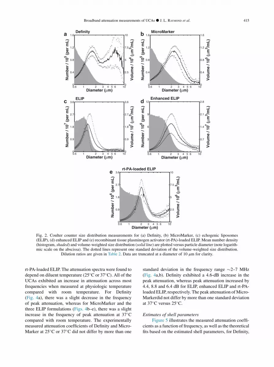
Fig. 2. Coulter counter size distribution measurements for (a) Definity, (b) MicroMarker, (c) echogenic liposomes(ELIP), (d) enhanced ELIP and (e) recombinant tissue plasminogen activator (rt-PA)-loaded ELIP. Mean number density(histogram, shaded) and volume-weighted size distribution (solid line) are plotted versus particle diameter (note logarith-mic scale on the abscissa). The dotted lines represent one standard deviation of the volume-weighted size distribution.
Dilution ratios are given in Table 2. Data are truncated at a diameter of 10 mm for clarity.
Broadband attenuation measurements of UCAs d J. L. RAYMOND et al. 415
rt-PA-loaded ELIP. The attenuation spectra were found todepend on diluent temperature (25�C or 37�C). All of theUCAs exhibited an increase in attenuation across mostfrequencies when measured at physiologic temperaturecompared with room temperature. For Definity(Fig. 4a), there was a slight decrease in the frequencyof peak attenuation, whereas for MicroMarker and thethree ELIP formulations (Figs. 4b–e), there was a slightincrease in the frequency of peak attenuation at 37�Ccompared with room temperature. The experimentallymeasured attenuation coefficients of Definity and Micro-Marker at 25�C or 37�C did not differ by more than one
standard deviation in the frequency range �2–7 MHz(Fig. 4a,b). Definity exhibited a 4.6-dB increase in thepeak attenuation, whereas peak attenuation increased by4.4, 8.8 and 6.4 dB for ELIP, enhanced ELIP and rt-PA-loaded ELIP, respectively. The peak attenuation ofMicro-Markerdid not differ by more than one standard deviationat 37�C versus 25�C.
Estimates of shell parametersFigure 5 illustrates the measured attenuation coeffi-
cients as a function of frequency, as well as the theoreticalfits based on the estimated shell parameters, for Definity,

Fig. 3. Measured attenuation spectra for enhanced echogenicliposomes in whole blood (solid line, black) and phosphate-buffered saline with 0.5% (w/v) albumin (dashed line, gray).
416 Ultrasound in Medicine and Biology Volume 40, Number 2, 2014
MicroMarker, ELIP, enhanced ELIP and rt-PA-loadedELIP at 37�C. The theoretical fits of the attenuation coef-ficients are in good agreement with the experimentallymeasured attenuation coefficients (R2 . 0.9). Shellparameters determined by the best fit to the measureddata are summarized in Table 3. The shell elasticityparameter (Sp) for the three ELIP formulations washigher at 3�C than at 25�C. It should be noted that a rela-tively small proportion of ELIP was found to contribute tothe attenuation spectra at either 25�C or 37�C (Table 3).However, the number of particles that contributed to theattenuation (and, therefore, are assumed to contain micro-bubbles) was two to three times higher at 37�C than at25�C for all ELIP formulations.
DISCUSSION
Characterization of UCAs using acoustic attenua-tion measurements obtained with methods similar tothose described here are well established (Chatterjeeet al. 2005; de Jong and Hoff, 1993; de Jong et al.1992; Hoff et al. 2000; Marsh et al. 1997; Sarkar et al.2005). The shell properties of phospholipid-based agentssuch as SonoVue (Gorce et al. 2000), Definity (Faez et al.2011; Goertz et al. 2007) and echogenic liposomes(Kopechek et al. 2011) have been characterized usingacoustic attenuation measurements conducted at roomtemperature. The attenuation results at 25�C that are re-ported here are in agreement with these previously pub-lished values. Other recent in vitro studies have focusedon the effect of temperature on the acoustic response ofcontrast agents. Using optical techniques, Vos et al.(2008) observed lower thresholds of acoustic activationand higher radial expansions of SonoVue and Definity
at body temperature (37�C) compared with room temper-ature. Mulvana et al. (2010) observed a concomitantincrease in acoustic attenuation and scattering for Sono-Vue at higher temperatures. The present study establishesa qualitatively similar result for the attenuation of Defi-nity, MicroMarker and ELIP.
Definity exhibited an increase in attenuation acrossmost frequencies when measured at 37�C versus 22�C(Fig. 4a). The frequency of peak attenuation for Definityalso shifted to a lower value at 37�C. Because the reso-nance frequency of a bubble decreases with increasingdiameter, this change suggests that the mean diameterof the microbubble population may be slightly largerat 37�C than at 25�C. The change in particle sizedistribution was not assessed in this study because theCoulter counter measurements were conducted only atroom temperature (25�C). Mulvana et al. (2010)optically observed a change in mean diameter for Sono-Vue. They found a reduction in the relative number ofbubbles with a diameter less than 2 mm and an increasein the mean bubble diameter at higher temperatures.Mulvana et al. hypothesized that gas expansion of thebubbles or gas diffusion into the bubbles resulted inan increase in bubble diameter with increasing tempera-ture. Furthermore, they hypothesized that the increasein diameter, accompanied by a reduction in bubblestability, resulted in a lower overall number density ofmicrobubbles at the higher temperature. The resultsof the present study also indicate that the number of mi-crobubbles contributing to the attenuation is reduced(by approximately 20%) for Definity at 37�C versusroom temperature, as outlined in Table 3. Thus, ourresults obtained for Definity at body temperature aresimilar to the previously reported findings for thephospholipid-shelled contrast agent SonoVue.Conversely, the attenuation peak and the estimatednumber of microbubbles increased for MicroMarkerand all three ELIP formulations.
Attenuation of MicroMarker (Fig. 4b) was notaffected by an increase in temperature for frequenciesfrom �2 to 7 MHz, but was higher for frequencies.7 MHz. Peak attenuation for MicroMarker (6 MHz at25�C) occurred at a slightly higher frequency (7 MHz)at 37�C. Both an increase in the shell stiffness parameterand a change in the relative size distribution of encapsu-lated microbubbles could be responsible for thisfrequency shift in peak attenuation. MicroMarkercontains a high-molecular-weight gaswith low solubilityin aqueous solution, which promotes thermodynamicstability. Therefore, the change in frequency-dependentattenuation for frequencies .7 MHz is likely due tochanges in the shell viscoelastic properties, whichalso depend on temperature. Although this agent wasdeveloped specifically for high-frequency imaging

Fig. 4. Measured attenuation coefficients (61 standard deviation) as a function of frequency at 25�C (solid lines) and37�C (dashed lines) for (a) Definity, (b) MicroMarker, (c) echogenic liposomes (ELIP), (d) enhanced ELIP and (e)
recombinant tissue plasminogen activator (rt-PA)-loaded ELIP.
Broadband attenuation measurements of UCAs d J. L. RAYMOND et al. 417
applications, the frequency of peak attenuation for thisagent was lower than for Definity and ELIP.
Echogenic liposomes exhibited higher attenuation atall frequencies at 37�C compared with room temperature(Figs. 4c–e). The increase in attenuation at 37�C occursover a broad frequency range, which is consistent withan increase in the overall number of microbubbles andnot simply a change in the relative size distribution.Our estimate of the number of microbubbles contributingto the attenuation (Nfit) supports this result (Table 3).However, the parameter Nfit was determined from theattenuation data, and therefore, a causal relationshipcannot be definitively established. It should be noted
that a relatively small proportion of ELIP was found tocontribute to the attenuation spectra, compared with Def-inity and MicroMarker, at either 25�C or 37�C. However,the number of particles that contribute to the attenuation(and, hence, are assumed to contain microbubbles) wastwo to three times higher at 37�C than at 25�C for eachof the three ELIP formulations. The increase in the over-all number of microbubbles could be due to the lowersolubility of air in aqueous solution at 37�C relative to25�C.
Sarkar et al. (2009) modeled the effects of encapsu-lation permeability, surface tension and diluent gassaturation on microbubble dissolution. In the model
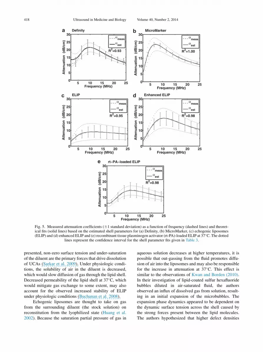
Fig. 5. Measured attenuation coefficients (61 standard deviation) as a function of frequency (dashed lines) and theoret-ical fits (solid lines) based on the estimated shell parameters for (a) Definity, (b) MicroMarker, (c) echogenic liposomes(ELIP) and (d) enhanced ELIP and (e) recombinant tissue plasminogen activator (rt-PA)-loaded ELIP at 37�C. The dotted
lines represent the confidence interval for the shell parameter fits given in Table 3.
418 Ultrasound in Medicine and Biology Volume 40, Number 2, 2014
presented, non-zero surface tension and under-saturationof the diluent are the primary forces that drive dissolutionof UCAs (Sarkar et al. 2009). Under physiologic condi-tions, the solubility of air in the diluent is decreased,which would slow diffusion of gas through the lipid shell.Decreased permeability of the lipid shell at 37�C, whichwould mitigate gas exchange to some extent, may alsoaccount for the observed increased stability of ELIPunder physiologic conditions (Buchanan et al. 2008).
Echogenic liposomes are thought to take on gasfrom the surrounding diluent (the stock solution) onreconstitution from the lyophilized state (Huang et al.2002). Because the saturation partial pressure of gas in
aqueous solution decreases at higher temperatures, it ispossible that out-gassing from the fluid promotes diffu-sion of air into the liposomes and may also be responsiblefor the increase in attenuation at 37�C. This effect issimilar to the observations of Kwan and Borden (2010).In their investigation of lipid-coated sulfur hexafluoridebubbles diluted in air-saturated fluid, the authorsobserved an influx of dissolved gas from solution, result-ing in an initial expansion of the microbubbles. Theexpansion phase dynamics appeared to be dependent onthe dynamic surface tension across the shell caused bythe strong forces present between the lipid molecules.The authors hypothesized that higher defect densities

Table 3. Estimated number density of microbubbles and shell parameters for the UCAs*
T
Number/mL
Sp (N/m) Sf/1026 (kg/s) R2Nmeas/10
6 Nfit/106
Definity 25�C 4.82 6 0.45 4.29 6 0.89 1.76 6 0.16 0.47 6 0.05 0.9337�C 3.48 6 1.26 1.10 6 0.15 0.20 6 0.04 0.93
MicroMarker 25�C 5.31 6 0.55 3.73 6 0.45 1.20 6 0.06 0.62 6 0.03 1.0037�C 4.97 6 0.41 1.90 6 0.05 0.87 6 0.02 1.00
ELIP 25�C 1220 6 320 2.07 6 0.61 1.13 6 0.13 0.82 6 0.04 0.9837�C 6.10 6 5.00 1.49 6 0.20 1.41 6 0.07 0.95
Enhanced ELIP 25�C 590 6 170 1.24 6 0.12 1.98 6 0.10 0.41 6 0.03 1.0037�C 3.84 6 1.06 3.10 6 0.25 1.01 6 0.07 0.98
rt-PA-loaded ELIP 25�C 166 6 40 2.05 6 0.99 3.69 6 0.76 1.88 6 0.23 0.9037�C 4.06 6 0.63 5.16 6 0.37 2.09 6 0.10 0.98
ELIP 5 echogenic liposomes; rt-PA 5 recombinant tissue plasminogen activator.* The values of the shell parameters that corresponded to a doubling of the error function value were taken to be the limits for the confidence interval
(Hoff 2001).
Broadband attenuation measurements of UCAs d J. L. RAYMOND et al. 419
within the lipid shell may reduce the pressure buildupnecessary for this expansion (Kwan and Borden 2010);importantly, defects in the lipid bilayer have been foundto be important in promoting the echogenicity of ELIP(Huang et al. 2002).
The use of broadband PVDF transducers in thequantitative assessment of attenuation in this study re-sulted in a much larger usable bandwidth comparedwith our previous measurements (Kopechek et al.2011). The frequency-dependent attenuation of ELIP(shown in Fig. 4c) agrees with our attenuation coeffi-cients previously measured using a pulse-echo technique(Kopechek et al. 2011). Enhanced ELIP (shown inFig. 4d) were more attenuative over all frequencies thanELIP. Furthermore, the enhanced ELIP formulation hasa more rigid shell (Sp 5 3.10 6 0.25, 37�C) than ELIP(Sp 5 1.49 6 0.20, 37�C). The increased rigidity isconsistent with Buchanan’s hypothesis that a more rigidshell is responsible for reducing the loss of air and extend-ing the duration of echogenic activity (Buchanan et al.2008). ELIP loaded with the thrombolytic drug rt-PA ex-hibited greater attenuation than ELIP (Smith et al. 2010),but were approximately 3 dB less attenuative than theenhanced ELIP formulation.
LimitationsFor the UCAs employed in this study, the use of low-
amplitude (,100 kPa) broadband pulses was observedto result in a linear attenuation response. Therefore, thelinearized theory proposed by de Jong et al. (1992; deJong and Hoff 1993) was used to model themicrobubbles in the linearly elastic regime. Theamplitude of oscillations is assumed to be small and themicrobubbles are assumed to respond in the linearlyelastic regime. In addition, this model assumes that theacoustic scattering cross section is independent of theincident intensity and does not take multiple scattering
effects into consideration. Higher-amplitude pulses,greater than approximately 100 kPa, led to non-linearoscillations that could be observed in the attenuationspectra. Our observation of the onset pressure thresholdfor non-linear effects in Definity was consistent withChatterjee et al. (2005). However, the onset pressure de-pended on the agent as well as the temperature. Properdescription of the microbubbles at these larger ampli-tudes would require the use of newer models that accu-rately capture the complex oscillatory dynamics ofphospholipid-coated microbubbles, such as the modelproposed by Marmottant et al. (2005).
The change in particle size distribution at 37�Cversus 25�C was not assessed in this study because theCoulter counter measurements were conducted only atroom temperature (25�C). It has recently been foundthat ELIP, when exposed to a decrease in temperature,lose their echogenicity (Kopechek et al. 2013;Radhakrishnan et al. 2012). The instrument used tomeasure the size distributions for this study was notequipped with a temperature control feature. To avoidexposure of ELIP and other UCAs to a reduction intemperature during the size measurement process andpotential inaccurate sizing of the particles, the sizedistributions were measured only at room temperature.
CONCLUSIONS
Frequency-dependent attenuation of severalphospholipid-shelled UCAs was measured at 25�C and37�C. The mean (volume-weighted) diameters of theUCAs measured were between 1.3 and 2.3 mm. Definityhad a larger number of particles .5 mm in diameterthan the other agents. As a diluent for the attenuationmeasurements, PBS with 0.5% (w/v) albumin was anadequate substitute for whole blood. Differences in shellparameters and estimated number density of the threetypes of UCAs were evident based on the attenuation
420 Ultrasound in Medicine and Biology Volume 40, Number 2, 2014
measurements over a broad frequency range, thus under-scoring the importance of conducting characterizationstudies at physiologic temperature. Echogenic liposomesexhibited a larger increase in attenuation at 37�C versus25�C than either Definity or MicroMarker. The enhancedELIP formulation yielded a higher attenuation than theoriginal formulation over the frequency band 2 to25 MHz.
Acknowledgments—The authors thank Robert Giulitto (HoxworthBlood Center) for assisting in the acquisition of human blood samples,Gail J. Pyne-Geithman (University of Cincinnati) for use of the bloodgas analyzer and Jack Rubinstein (University of Cincinnati) forproviding the MicroMarker contrast agent. The authors are also gratefulto Melvin E. Klegerman (University of Texas Health Science Center atHouston) and members of the Image-Guided Ultrasound TherapeuticsLaboratory (University of Cincinnati) for helpful discussions. Thiswork was supported in part by the National Institutes of Health (NIHR01 HL74002, NIH R01 HL059586, NIH R01 NS047603, NIH F32HL104916) and a grant-in-aid from the University of Cincinnati Chapterof Sigma Xi.
REFERENCES
Alkan-Onyuksel H, Demos SM, Lanza GM, Vonesh MJ,Klegerman ME, Kane BJ, Kuszak J, Mcpherson DD. Developmentof inherently echogenic liposomes as an ultrasonic contrast agent.J Pharm Sci 1996;85:486–490.
Altman PL. Handbook of respiration. Philadelphia: Saunders; 1958.AIUM Technical Standards Committee. Methods for specifying
acoustic properties of tissue mimicking phantoms and objects;1995, Laurel, MD: American Institute of Ultrasound in Medicine(AIUM).
Bekeredjian R, Grayburn PA, Shohet RV. Use of ultrasound contrastagents for gene or drug delivery in cardiovascular medicine. J AmColl Cardiol 2005;45:329–335.
Buchanan KD, Huang S, Kim H, MacDonald RC, McPherson DD.Echogenic liposome compositions for increased retention of ultra-sound reflectivity at physiologic temperature. J Pharm Sci 2008;97:2242–2249.
Chatterjee D, Sarkar K, Jain P, Schreppler NE. On the suitability ofbroadband attenuation measurement for characterizing contrast mi-crobubbles. Ultrasound Med Biol 2005;31:781–786.
Chen J, Zhu Z. Ultrasound attenuation in encapsulated microbubblesuspensions: The multiple scattering effects. Ultrasound Med Biol2006;32:961–969.
de Jong N, Hoff L. Ultrasound scattering properties of Albunex micro-spheres. Ultrasonics 1993;31:175–181.
de Jong N, Hoff L, Skotland T, BomN. Absorption and scatter of encap-sulated gas filledmicrospheres: Theoretical considerations and somemeasurements. Ultrasonics 1992;30:95–103.
Demos SM, Alkan-Onyuksel H, Kane BJ, Ramani K, Nagaraj A,Greene R, KlegermanM,McPherson DD. In vivo targeting of acous-tically reflective liposomes for intravascular and transvascular ultra-sonic enhancement. J Am Coll Cardiol 1999;33:867–875.
Elbayoumi TA, Torchilin VP. Liposomes for targeted delivery of antith-rombotic drugs. Expert Opin Drug Deliv 2008;5:1185–1198.
Faez T, Goertz D, de Jong N. Characterization of Definity� ultrasoundcontrast agent at frequency range of 5–15 MHz. Ultrasound MedBiol 2011;37:338–342.
Feinstein SB. Contrast ultrasound imaging of the carotid artery vasavasorum and atherosclerotic plaque neovascularization. J Am CollCardiol 2006;48:236–243.
Goertz DE, de Jong N, van der Steen AF. Attenuation and size distribu-tion measurements of Definity and manipulated Definity popula-tions. Ultrasound Med Biol 2007;33:1376–1388.
Gorce JM, Arditi M, Schneider M. Influence of bubble size distributionon the echogenicity of ultrasound contrast agents: A study of Sono-Vue. Invest Radiol 2000;35:661–671.
Helfield BL, Huo X, Williams R, Goertz DE. The effect of preactivationvial temperature on the acoustic properties of DefinityTM. Ultra-sound Med Biol 2012;38:1298–1305.
Hitchcock KE, Caudell DN, Sutton JT, Klegerman ME, Vela D,Pyne-Geithman GJ, Abruzzo T, Cyr PE, Geng YJ,McPherson DD, Holland CK. Ultrasound-enhanced delivery of tar-geted echogenic liposomes in a novel ex vivo mouse aorta model.J Control Release 2010;144:288–295.
Hoff L. Acoustic characterization of contrast agents for medical ultra-sound imaging. Berlin/Heidelberg: Springer; 2001.
Hoff L, Sontum PC, Hovem JM. Oscillations of polymeric microbub-bles: Effect of the encapsulating shell. J Acoust Soc Am 2000;107:2272–2280.
Huang S. Ultrasound-responsive liposomes. In: Weissig V, (ed). Lipo-somes: Methods and protocols. New York: Humana Press; 2010.
Huang SL. Liposomes in ultrasonic drug and gene delivery. Adv DrugDeliv Rev 2008;60:1167–1176.
Huang SL, Hamilton AJ, Nagaraj A, Tiukinhoy SD, Klegerman ME,Mcpherson DD, Macdonald RC. Improving ultrasound reflectivityand stability of echogenic liposomal dispersions for use as targetedultrasound contrast agents. J Pharm Sci 2001;90:1917–1926.
Huang SL, Hamilton AJ, Pozharski E, Nagaraj A, Klegerman ME,McPherson DD, MacDonald RC. Physical correlates of the ultra-sonic reflectivity of lipid dispersions suitable as diagnostic contrastagents. Ultrasound Med Biol 2002;28:339–348.
Huang SL, MacDonald RC. Acoustically active liposomes for drugencapsulation and ultrasound-triggered release. Biochim BiophysActa Biomembr 2004;1665:134–141.
Klegerman ME, Wassler M, Huang SL, Zou Y, Kim H, Shelat HS,Holland CK, Geng YJ, McPherson DD. Liposomal modularcomplexes for simultaneous targeted delivery of bioactive gasesand therapeutics. J Control Release 2010;142:326–331.
Klibanov AL. Microbubble contrast agents: Targeted ultrasoundimaging and ultrasound-assisted drug-delivery applications. InvestRadiol 2006;41:354–362.
Kopechek JA, Haworth KJ, Radhakrishnan K, Huang SL,Klegerman ME, McPherson DD, Holland CK. The impact ofbubbles on measurement of drug release from echogenic liposomes.Ultrason Sonochem 2013;20:1121–1130.
Kopechek JA, Haworth KJ, Raymond JL, Douglas Mast T, Perrin SR Jr,Klegerman ME, Huang S, Porter TM, McPherson DD, Holland CK.Acoustic characterization of echogenic liposomes: Frequency-dependent attenuation and backscatter. J Acoust Soc Am 2011;130:3472–3481.
Kornmann LM, Reesink KD, Reneman RS, Hoeks APG. Criticalappraisal of targeted ultrasound contrast agents for molecularimaging in large arteries. Ultrasound Med Biol 2010;36:181–191.
Kwan JJ, Borden MA. Microbubble dissolution in a multigas environ-ment. Langmuir 2010;26:6542–6548.
Laing ST, McPherson DD. Cardiovascular therapeutic uses of targetedultrasound contrast agents. Cardiovasc Res 2009;83:626–635.
Lindner JR. Molecular imaging with contrast ultrasound and targetedmicrobubbles. J Nucl Cardiol 2004;11:215–221.
Marmottant P, van der Meer S, Emmer M, Versluis M, de Jong N,Hilgenfeldt S, Lohse D. A model for large amplitude oscillationsof coated bubbles accounting for buckling and rupture. J AcoustSoc Am 2005;118:3499–3505.
Marsh JN, Hall CS, Hughes MS, Mobley J, Miller JG,Brandenburger GH. Broadband through-transmission signal lossmeasurements of Albunex� suspensions at concentrations ap-proaching in vivo doses. J Acoust Soc Am 1997;101:1155–1161.
Mulvana H, Stride E, Hajnal JV, Eckersley RJ. Temperature dependentbehavior of ultrasound contrast agents. Ultrasound Med Biol 2010;36:925–934.
Patil H, Main ML. The history of product label changes for DEFI-NITY� in the US. US Cardiol 2012;9:35–39.
Paul S, Russakow D, Nahire R, Nandy T, Ambre AH, Katti K, Mallik S,Sarkar K. In vitro measurement of attenuation and nonlinear scat-tering from echogenic liposomes. Ultrasonics 2012;52:962–969.
Qin S, Caskey CF, Ferrara KW. Ultrasound contrast microbubbles inimaging and therapy: Physical principles and engineering. PhysMed Biol 2009;54:R27–R57.
Broadband attenuation measurements of UCAs d J. L. RAYMOND et al. 421
Radhakrishnan K, Haworth KJ, Huang S-, Klegerman ME,McPherson DD, Holland CK. Stability of echogenic liposomes asa blood pool ultrasound contrast agent in a physiologic flowphantom. Ultrasound Med Biol 2012;38:1970–1981.
Sarkar K, Katiyar A, Jain P. Growth and dissolution of an encapsulatedcontrast microbubble: effects of encapsulation permeability. Ultra-sound Med Biol 2009;35:1385–1396.
Sarkar K, Shi WT, Chatterjee D, Forsberg F. Characterization of ultra-sound contrast microbubbles using in vitro experiments and viscousand viscoelastic interface models for encapsulation. J Acoust SocAm 2005;118:539–550.
Smith DA, Vaidya SS, Kopechek JA, Huang SL, Klegerman ME,McPherson DD, Holland CK. Ultrasound-triggered release of re-combinant tissue-type plasminogen activator from echogenic lipo-somes. Ultrasound Med Biol 2010;36:145–157.
Stapleton S, Goodman H, Zhou Y, Cherin E, Henkelman RM, Burns PN,Foster FS. Acoustic and kinetic behaviour of Definity in miceexposed to high frequency ultrasound. Ultrasound Med Biol 2009;35:296–307.
Stride E. Physical principles of microbubbles for ultrasound imagingand therapy. Cerebrovasc Dis 2009;27(Suppl 2):1–13.
Stride E, Saffari N. Investigating the significance of multiple scatteringin ultrasound contrast agent particle populations. IEEE Trans Ultra-son Ferroelectr Freq Control 2005;52:2332–2345.
Sutton JT, Haworth KJ, Pyne-Geithman G, Holland CK. Ultrasound-mediated drug delivery for cardiovascular disease. Expert OpinDrug Deliv 2013;10:573–592.
Tiukinhoy-Laing SD, Huang S, Klegerman M, Holland CK,McPherson DD. Ultrasound-facilitated thrombolysis using tissue-plasminogen activator-loaded echogenic liposomes. Thromb Res2007;119:777–784.
VisualSonics PN11691—Vevo MicroMarker� Non-Targeted ContrastAgent Kit: Protocol and Information Booklet Rev 1.4. Toronto.
Vos HJ, Emmer M, de Jong N. Oscillation of single microbubbles atroom versus body temperature. In: Proceedings, 2008 IEEE Interna-tional Ultrasonics Symposium, Beijing, China, November 2–5,2008. Beijing edition. New York: IEEE, 2008:982–984.
Weissig V. Liposomes: Methods and protocols. New York: HumanaPress; 2010.
Whittle P. Prediction and regulation by linear least-square methods.2nd rev. edition. Minneapolis, MN: University of Minnesota Press;1983.