brazilian pharmacopoeia
TRANSCRIPT
Brazilian Health Surveillance Agency - Anvisa
BRAZILIAN PHARMACOPOEIA
6th EDITION
Brazilian Pharmacopoeia, 6th edition
This translation does not replace the portuguese version.
Brazilian Health Surveillance Agency
Brazilian Pharmacopoeia,
6th edition
Volume II - Monographs
Medicinal Plants
Brasília 2019
Brazilian Pharmacopoeia, 6th edition
This translation does not replace the portuguese version.
MEDICINAL PLANTS AVOCADO, leaf PM001-00 ACONITE, root PM002-00 ARTICHOKE, leaf PM003-00 LICORICE, root PM004-00 GARLIC, bulb PM005-00 Aloe, dry exudate PM006-01 MARSHMALLOW, root PM007-00 PLUM, fruit PM008-00 ANGICO, bark PM009-00 ANISE, fruit PM010-00 STAR ANISE, fruit PM011-00 ARNICA, flower PM012-00 BRAZILIAN PEPPERTREE, bark PM013-00 ALOE, leaf PM014-00 TOLU BALSAM PM015-00 PERU BALSAM PM016-00 BARBATIMAO, bark PM017-00 VANILLA, fruit PM018-00 BELLADONNA, leaf PM019-00 BENZOIN PM020-00 BOLDO, leaf PM021-00 CALENDULA, flower PM022-01 CHAMOMILE, flower PM023-00 CHINESE CINNAMON, husk PM024-00 CEYLON CINNAMON, bark PM025-00 LEMON GRASS, leaf PM026-00 CARDAMON, seed PM027-00 CARQUEJA, winged stem PM028-00 CASCARA BUCKTHORN, bark PM029-00 HORSE CHESTNUT, seed PM030-00 ASIATIC PENNYWORT, leaf PM031-00 FRESHCUT, leaf PM032-00 BURHEAD, leaf PM033-00 CORIANDER, fruit PM034-00 HAWTHORN, leaf and flower PM035-01 CLOVE, flower bud PM036-00 TURMERIC, rhizome PM037-01 DILL, fruit PM038-00 ESPINHEIRA-SANTA, leaf PM039-00

Brazilian Pharmacopoeia, 6th edition
This translation does not replace the portuguese version.
CANDYLEAF, leaf PM040-00 JIMSONWEED, leaf PM041-00 BLUE GUM EUCALYPTUS, leaf PM042-00 FENNEL, fruit PM043-00 SWEET FENNEL, fruit PM044-00 DEVIL’S-CLAW, root PM045-00 YELLOW GENTIAN, rhizome and root PM046-00 GINGER, rhizome PM047-00 GUAVA, leaf PM048-00 GUACO, leaf PM049-00 GUARANA, seed PM050-00 WITCHHAZEL, leaf PM051-00 GOLDENSEAL, rhizome and root PM052-00 CORN MINT, aerial part PM053-00 PEPPERMINT, leaf PM054-00 BRAZILIAN JALAP, root PM055-00 BRAZILIAN IRONWOOD, bark PM056-00 BRAZILIAN IRONWOOD, oil PM057-00 BITTER ORANGE, exocarp PM058-00 MACELA, flower PM059-00 COMMON MALLOW, flower PM060-00 PASSION FRUIT, leaf PM061-01 GRANADILLA, leaf PM062-01 HENBANE, leaf PM063-00 LEMON BALM, leaf PM064-01 KOLA NUT, seed PM065-00 NUX-VOMICA, seed PM066-00 EUGENIA UNIFLORA, leaf PM067-01 PSYLLIUM, testa PM068-00 SENEGA ROOT, root PM069-00 GALE OF THE WIND, aerial part PM070-00 GALE OF THE WIND, aerial part PM071-00 SOAPBARK, bark PM072-00 QUININE, husk PM073-00 RHATANY, root PM074-00 SNAKEROOT, root PM075-00 RHUBARB, rhizome and root PM076-01 SAMBUCUS AUSTRALIS, flower PM077-01 ELDERBERRY, flower PM078-01 WHITE WILLOW, bark PM079-00 ALEXANDRIAN SENNA, leaf PM080-01 ALEXANDRIAN SENNA, fruit PM081-00
Brazilian Pharmacopoeia, 6th edition
This translation does not replace the portuguese version.
BEARBERRY, leaf PM082-00 VALERIAN, rhizome and root PM083-00
Brazilian Pharmacopoeia, 6th edition
This translation does not replace the portuguese version.
PLANT-BASED PREPARATIONS – TINCTURES ACONITE, tincture PM084-00 ANGICO, tincture PM085-00 STAR ANISE, tincture PM086-00 BRAZILIAN PEPPERTREE, tincture PM087-00 TOLU BALSAM, tincture PM088-00 VANILLA, tincture PM089-00 BENZOIN, tincture PM090-00 BOLDO, tincture PM091-00 CALENDULA, tincture PM092-00 CHAMOMILE, tincture PM093-00 CEYLON CINNAMON, tincture PM094-00 CASCARA BUCKTHORN, tincture PM095-00 HORSE CHESTNUT, tincture PM096-00 TURMERIC, tincture PM097-00 YELLOW GENTIAN, tincture PM098-00 GUARANA, tincture PM099-00 WITCHHAZEL, tincture PM100-00 JABORANDI, tincture PM101-00 BITTER ORANGE, tincture PM102-00 NUX-VOMICA, tincture PM103-00 RHATANY, tincture PM104-00 VALERIAN, tincture PM105-00
Brazilian Pharmacopoeia, 6th edition
This translation does not replace the portuguese version.
PLANT-BASED PREPARATIONS – FLUID EXTRACT ARTICHOKE, fluid extract PM106-00 LICORICE, fluid extract PM107-00 PLUM, fluid extract PM108-00 ANGICO, Fluid extract PM109-00 BRAZILIAN PEPPERTREE, fluid extract PM110-00 BOLDO, fluid extract PM111-00 CALENDULA, fluid extract PM112-00 CEYLON CINNAMON, fluid extract PM113-00 CASCARA BUCKTHORN, fluid extract PM114-00 HORSE CHESTNUT, fluid extract PM115-00 HAWTHORN, fluid extract PM116-00 YELLOW GENTIAN, fluid extract PM117-00 GUARANA, fluid extract PM118-00 WITCHHAZEL, fluid extract PM119-00 BITTER ORANGE, fluid extract PM120-00 KOLA NUT, fluid extract PM121-00 NUX-VOMICA, fluid extract PM122-00 RHATANY, fluid extract PM123-00 VALERIAN, fluid extract PM124-00
Brazilian Pharmacopoeia, 6th edition
This translation does not replace the portuguese version.
OILS, FATS AND WAXES ROSEMARY, oil PM125-00 COTTON, refined oil PM126-00 ANISE, oil PM127-00 CHAMOMILE, oil PM128-00 CHINESE CINNAMON, oil PM129-00 CEYLON CINNAMON, oil PM130-00 LEMON GRASS, oil PM131-00 CARNAUBA WAX PALM PM132-00 CORIANDER, oil PM133-00 CLOVE, oil PM134-00 EUCALIPTO, oil PM135-00 LEMON-SCENTED GUM, oil PM136-00 FENNEL, oil PM137-00 SUNFLOWER, refined oil PM138-00 CORN MINT, oil PM139-00 PEPPERMINT, oil PM140-00 BITTER ORANGE, oil PM141-00 SWEET ORANGE, oil PM142-00 LEMON, oil PM143-00 COCOA BUTTER PM144-00 TEA TREE, oil PM145-00 NUTMEG, oil PM146-00 OLIVE, virgin oil PM147-00 PALMAROSA, oil PM148-00 THYME, oil PM149-00
Brazilian Pharmacopoeia, 6th edition PM001-00
This translation does not replace the portuguese version.
AVOCADO, leaf Persea folium
The plant drug consists of dried leaves of Persea americana Mill. (syn. Persea gratissima Gaertn. f.) containing at least 0.4% of total flavonoid content expressed as apigenin (C15H10O5, 270.24) and 0.14% of volatile oil. IDENTIFICATION 1. Macroscopic description Simple leaves, elliptic, oblong or oval-acuminate, semi-coriaceous, entire margins, roughly wavy; lamina from 8.0 cm to 20.0 cm long and from 4.0 cm to 9.0 cm wide; petiole up to 5 cm long and from 3 mm to 4 mm wide at the base; when fresh, they are dark green on the adaxial surface, slightly bright and nearly flat, and the abaxial surface has lighter green color, and somewhat dull rough; light brown dry leaves. Midrib prominent on the abaxial surface, with secondary oblique ribs, also prominent, giving rise to tertiary ribs that anastomose in thin weft. 2. Microscopic description The leaf lamina has dorsiventral symmetry and is hypostomatic, with anomocytic stomata. In a front view, the epidermis exhibits granular cuticle and, on the adaxial surface, it is formed by polygonal cells with rare, thick-walled, unicellular tector trichomes; the abaxial surface is formed by rectangular or rounded cells. Tector trichomes are common in young leaves and rare in adult leaves. In cross-section, the epidermis is unistratified on both faces and covered by a thick cuticle. The mesophyll is formed by one or two layers of palisade cells, with many mucilage- and volatile oil-secreting idioblasts. The spongy parenchyma has about six layers of irregular cells, with large intercellular spaces. There may be a differentiated organization in the mesophyll, together with the secretory idioblasts, formed by elongated and tangentially flattened parenchyma cells with thick walls. Midrib shows a developed collateral vascular bundle, surrounded by an almost continuous sclerenchymatic sheath. Spindle-shaped calcium oxalate crystals occur in parenchyma cells close to the veins. At the base of the leaf lamina, two other small collateral bundles occur along the margin, oriented to the adaxial surface. 3. Microscopic description of powder The sample meets all requirements for the species, except the macroscopic characters. Characteristics: dark green color; fragments of adaxial epidermis with isodiametric polygonal cells, covered with a thick cuticle; fragments of the abaxial epidermis containing rectangular cells and anomocytic stomata; entire tector trichomes accompanied by epidermal or isolated cells; fragments of tector trichomes; fragments of the mesophyll with secretory idioblasts; vein fragments, as described, accompanied by cells containing spindle-shaped crystals. 4. Proceed as described in Thin-layer chromatography (5.2.17.1). Stationary phase: silica gel GF254 (0.25 mm).
Brazilian Pharmacopoeia, 6th edition PM001-00
This translation does not replace the portuguese version.
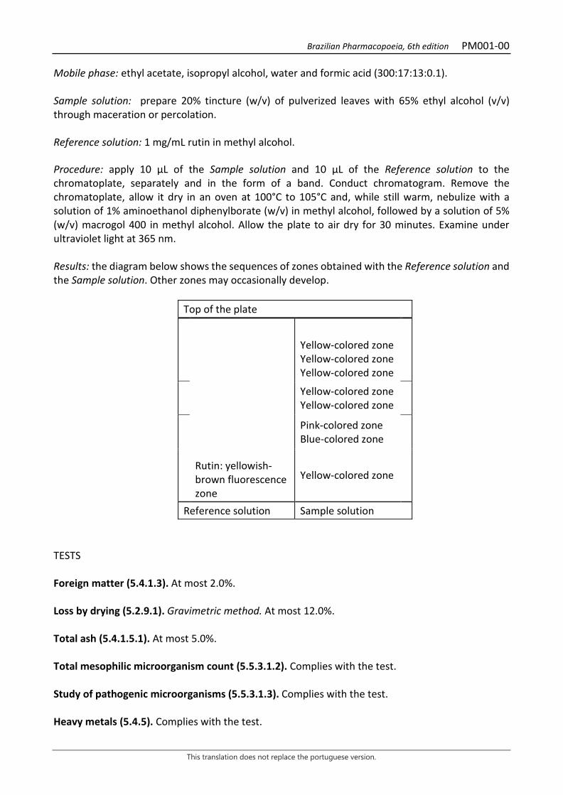
Mobile phase: ethyl acetate, isopropyl alcohol, water and formic acid (300:17:13:0.1). Sample solution: prepare 20% tincture (w/v) of pulverized leaves with 65% ethyl alcohol (v/v) through maceration or percolation. Reference solution: 1 mg/mL rutin in methyl alcohol. Procedure: apply 10 μL of the Sample solution and 10 μL of the Reference solution to the chromatoplate, separately and in the form of a band. Conduct chromatogram. Remove the chromatoplate, allow it dry in an oven at 100°C to 105°C and, while still warm, nebulize with a solution of 1% aminoethanol diphenylborate (w/v) in methyl alcohol, followed by a solution of 5% (w/v) macrogol 400 in methyl alcohol. Allow the plate to air dry for 30 minutes. Examine under ultraviolet light at 365 nm. Results: the diagram below shows the sequences of zones obtained with the Reference solution and the Sample solution. Other zones may occasionally develop.
Top of the plate
Yellow-colored zone Yellow-colored zone Yellow-colored zone
Yellow-colored zone Yellow-colored zone
Pink-colored zone Blue-colored zone
Rutin: yellowish-brown fluorescence zone
Yellow-colored zone
Reference solution Sample solution TESTS Foreign matter (5.4.1.3). At most 2.0%. Loss by drying (5.2.9.1). Gravimetric method. At most 12.0%. Total ash (5.4.1.5.1). At most 5.0%. Total mesophilic microorganism count (5.5.3.1.2). Complies with the test. Study of pathogenic microorganisms (5.5.3.1.3). Complies with the test. Heavy metals (5.4.5). Complies with the test.
Brazilian Pharmacopoeia, 6th edition PM001-00
This translation does not replace the portuguese version.
Agrochemical waste (5.4.3). Complies with the test. DOSAGE Volatile oils Proceed as described in Determining volatile oils in plant drugs (5.4.1.6). Use 1000-mL flask containing 500 mL of water as distillation liquid. Use dry, scratched and unbruised plant. Immediately proceed with the determination of the volatile oil, from 100 g of the chopped plant drug. Distill for four hours. Measure the volume and express the yield per 100 g of the plant drug (w/p). Total flavonoids To proceed as described in Visible absorption spectrophotometry (5.2.14). Prepare solutions as described below. Stock solution: accurately weigh approximately 0.5 g of the pulverized plant drug (800 µm) (5.2.11) and place in a 100 mL round-bottomed flask. Add 1 mL of 0.5% aqueous methenamine solution (w/v), 30 mL of 50% ethyl alcohol solution (v/v) and 2 mL of hydrochloric acid to the plant drug. Heat on a heating mantle for 30 minutes under reflux. Filter the mixture through absorbent cotton into a 100-mL volumetric flask. Return the plant drug residue and cotton to the round-bottomed flask, add an additional 30 mL of 50% (v/v) ethyl alcohol solution and re-heat again under reflux for 15 minutes. Filter through absorbent cotton into the same 100-mL volumetric flask. Repeat the operation, return the plant drug residue and the absorbent cotton to the round-bottomed flask, add 30 mL of 50% (v/v) ethyl alcohol solution, heat under reflux for 15 minutes, and filter into the same 100-mL volumetric flask. After cooling, top off the volume of the 100-mL volumetric flask with 50% (v/v) ethyl alcohol solution. Sample solution: add 10 mL of the Stock solution in a 25-mL volumetric flask with 2 mL 5% (w/v) aluminum chloride solution in 50% (v/v) ethyl alcohol solution and to off the volume with 50% (v/v) ethyl alcohol solution. Take the reading after 30 minutes. Blank solution: add 10 mL of the Stock solution in a 25-mL volumetric flask and top off to volume with a 50% (v/v) ethyl alcohol solution. Procedure: measure the absorbance of the Sample solution at 425 nm, using the Blank solution for zero adjustment. The total flavonoid content, expressed as percentage of apigenin, is calculated according to the following expression:
𝑇𝑇𝑇𝑇 =A × 250
m × 336,5
in which, TF = total flavonoid content expressed as apigenin % (w/w);

Brazilian Pharmacopoeia, 6th edition PM001-00
This translation does not replace the portuguese version.
A = absorbance measured for the Sample solution; 250 = dilution factor; m = mass in grams of the sample used, considering the loss by drying. 336.5 = specific apigenin absorption coefficient. PACKAGING AND STORAGE In tightly closed containers, protected from light and heat.
Figura 1 – Macroscopic and microscopic aspects in Persea americana Mill.
___________________________ The scales correspond in A to 5 mm; in B, C and D to 20 µm; and in E to 50 µm.
Brazilian Pharmacopoeia, 6th edition PM001-00
This translation does not replace the portuguese version.
A – front view of the leaf: leaf lamina (lf); petiole (pl). B – partial detail of the epidermis oriented to the abaxial surface, in cross-section: palisade parenchyma (pp); epidermis (ep); cuticle (cu); mucilage cell (cm); tector trichome (tt). C – partial detail of the epidermis oriented to the adaxial surface, in a front view. C – partial detail of the epidermis oriented to the abaxial surface, in a front view: stoma (es); tector thricome (tt). E – cross-section detail of a leaf lamina portion: cuticle (cu); epidermis (ep); palisade parenchyma (pp); secretory idioblast (is); spongy parenchyma (pj); stoma (es); idioblast with calcium oxalate crystals (ico); vascular bundle (vf).
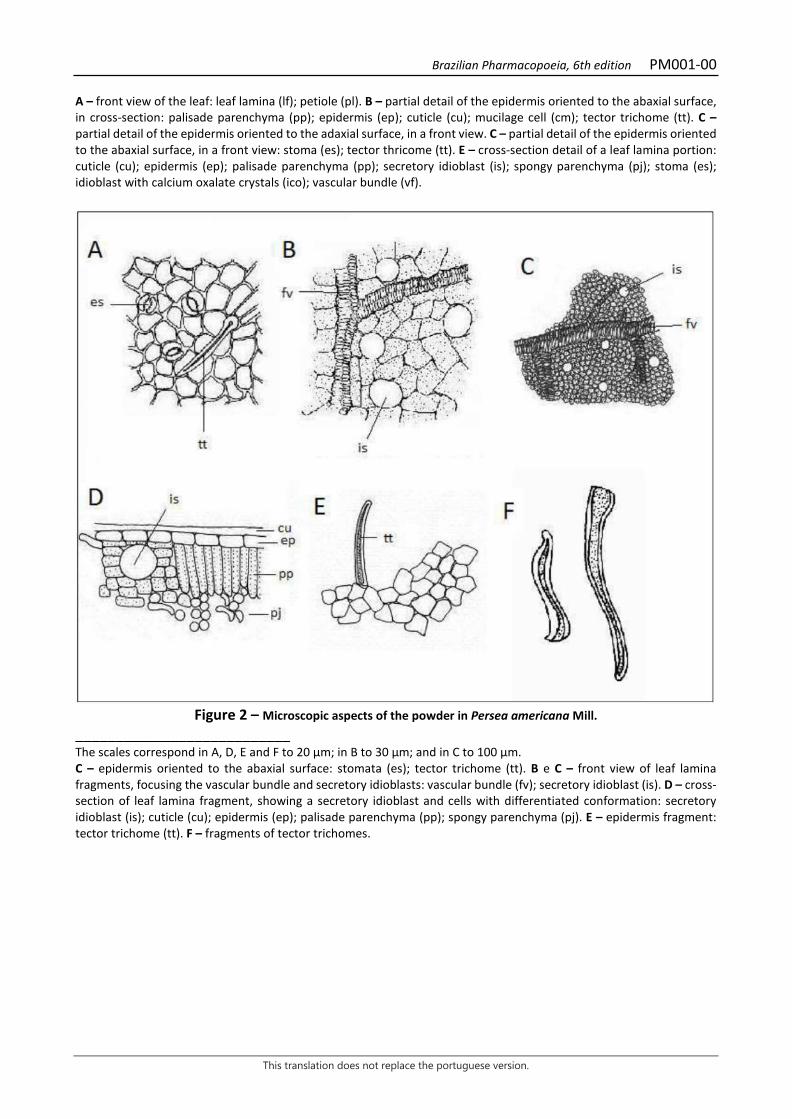
Figure 2 – Microscopic aspects of the powder in Persea americana Mill.
___________________________ The scales correspond in A, D, E and F to 20 µm; in B to 30 µm; and in C to 100 µm. C – epidermis oriented to the abaxial surface: stomata (es); tector trichome (tt). B e C – front view of leaf lamina fragments, focusing the vascular bundle and secretory idioblasts: vascular bundle (fv); secretory idioblast (is). D – cross-section of leaf lamina fragment, showing a secretory idioblast and cells with differentiated conformation: secretory idioblast (is); cuticle (cu); epidermis (ep); palisade parenchyma (pp); spongy parenchyma (pj). E – epidermis fragment: tector trichome (tt). F – fragments of tector trichomes.

Brazilian Pharmacopoeia, 6th edition PM002-00
This translation does not replace the portuguese version.
ACONITE, root Aconiti radix
The plant drug consists of tuberous roots of Aconitum napellus L., containing at least 0.5% total alkaloids expressed as aconitine (C34H47NO11, 645,74), calculated on dried material. IDENTIFICATION 1. Macroscopic description Tuberous, conical root, dark gray or brownish on the surface, 3 to 12 cm long and 1 to 3 cm wide in the upper portion, where remnants of the stem base may be seen; tuberous lateral roots, also conical, or their scars may occur near the upper portion of the main root, connected to each other by a thin pedicle, as well as numerous non-tuberous lateral roots, or their scars, distributed along the tuberous roots. The tuberous roots are internally grizzly or light brown. 2. Microscopic description In cross-section, the root has four or more layers of transversely elongated cells with suberized walls, relatively thick and orange colored, corresponding to the periderm, followed by parenchymatic cortical cells with some starch grains; endoderm constituted elongated crosswise cells with suberized walls orange-red colored when stained with Sudan III; the pericycle is formed by more than 20 layers of parenchyma cells filled with starch grains. The vascular cambium is star-shaped and the secondary phloem is not easily distinguishable among the parenchyma cells of the pericycle. The secondary xylem forms bundles interspersed with amyliferous parenchyma. The vessel elements of the secondary xylem are relatively narrow and have simple perforation plates and reticulate or pitted walls. 3. Microscopic description of powder The sample meets all requirements for the species, except the macroscopic characters. Characteristics are: light brown color; abundance of rounded starch grains with a less dense central region and, therefore, with lighter in color, isolated or grouped; fragments of suber in front view with polygonal-walled, brownish cells; fragments of parenchyma cells with starch grains; fragments of vessel elements with simple perforation plates and reticulated or pitted walls. 4. Proceed as described in Thin-layer chromatography (5.2.17.1). Stationary phase: 250 µm thick silica gel F254. Mobile phase: toluene, ethyl acetate and diethylamine (35:10:5). Sample solution: weigh 3 g of the pulverized plant drug and transfer to a round-bottomed flask, add a 2-mL 2 M hydrochloric acid and 40-mL water mixture. Heat the mixture in a water bath, under reflux, for 10 minutes. Filter and add the 6 M ammonium hydroxide solution until a pH 9.0 is obtained. Transfer the filtrate to a separating funnel and extract twice with 20 mL diethyl ether. Mix and dry ethereal extracts in a porcelain container, in water bath at 50°C. Suspend the residue in 1

Brazilian Pharmacopoeia, 6th edition PM002-00
This translation does not replace the portuguese version.
mL of methyl alcohol and filter through a 0.45 µm filtration unit and proceed with the chromatographic analysis. Reference solution: dissolve an accurately weighed amount of aconitine in methyl alcohol to obtain a concentration of 200 µg/mL. Procedure: apply 20 μL of the Sample solution and 20 μL of the Reference solution to the chromatoplate, separately and in the form of a band. Conduct chromatogram. Remove chromatoplate and allow it to air dry. Nebulize the plate with potassium iodide and bismuth subnitrate RS solution, allow it to dry and nebulize with sodium nitrite RS. Allow the plate to air dry for 30 minutes and examine under visible light. Results: the diagram below shows the sequences of zones obtained with the Reference solution and the Sample solution. Other zones may occasionally develop.
Top of the plate
Orange-colored zone
Aconitine: orange-colored zone
Orange-colored zone
Orange-colored zone
Reference solution Sample solution
TESTS Heavy metals (5.4.5). Complies with the test. Foreign matter (5.4.1.3). At most 2.0%. Loss by drying (5.2.9.1). Gravimetric method. At most 11.0%. Acid-insoluble ash (5.4.1.5.3). At most 0.7%. Total mesophilic microorganism count (5.5.3.1.2). Complies with the test. Study of pathogenic microorganisms (5.5.3.1.3). Complies with the test. Agrochemical waste (5.4.3). Complies with the test. DOSAGE
Brazilian Pharmacopoeia, 6th edition PM002-00
This translation does not replace the portuguese version.
Total alkaloid content expressed as aconitine Sample solution: weigh 2 g of the plant drug, transfer to an erlenmeyer flask and add 1.6 mL of 40% (v/v) ammonium hydroxide and 20 mL of diethyl ether. Leave on a magnetic stirrer for 30 minutes. Cover flask with aluminum foil. After the extraction process, separate the ether phase and add 0.8 mL of 40% (v/v) ammonium hydroxide and 20 mL of diethyl ether to the residue. Separate ether phase. Repeat the same procedure three more times. Mix and dry ethereal extracts in a porcelain container, in water bath at 50°C. Resume the residue in 5 mL absolute ethyl alcohol and add 30 mL of freshly boiled water, use at room temperature. Indicator solution: separately weigh 0.1 g of methyl red and 0.1 g of methylthioninium chloride, combine in a container and add 50 mL absolute ethyl alcohol. Transfer to a 100-mL volumetric flask and top off the volume with absolute ethyl alcohol. Procedure: titrate with 0.01 M hydrochloric acid until the color of the solution changes from light green to gray-blue. Use three drops of the Indicator solution. Each mL of 0.01 M hydrochloric acid is equivalent to 6.037 mg total alkaloids expressed as aconitine. Calculate total alkaloid content, in percent, according to the following expression:
𝑇𝑇𝑇𝑇 =V × 6,037 × 0,1
𝑚𝑚
in which, TA = Total alkaloid content expressed as aconitine % (w/w); V= volume of 0.01 M hydrochloric acid, in milliliters, consumed in the titration; m = mass in grams of the plant drug used, considering the loss by drying. PACKAGING AND STORAGE In tightly closed containers, protected from light and heat.

Brazilian Pharmacopoeia, 6th edition PM002-00
This translation does not replace the portuguese version.
Figure 1 – Macroscopic, microscopic and powder microscopic aspects in Aconitum napellus L.
___________________________ The scales correspond in A to 1 cm; in B to 100 μm; in C-E to 50 μm; in F-H to 20 μm. A – tuberous adventitious roots (rd) arising from the stem base (c) showing lateral roots (rlt) and scars (ci). B - illustration of the tuberous root in cross section: periderm (pe); cortical parenchyma (pc); endoderm (end); pericycle parenchyma (pr); primary phloem (fp) and secondary phloem (fs); vascular cambium (ca); secondary xylem (xs) and amyliferous parenchyma (pm). C - detail of the endoderm with thickened walls (end) and parenchyma cells with starch (a) in the cortex (pc) and pericycle (pr). D - fragment of the periderm (suber) with polygonal cells in front view. E - vessel elements with simple perforation plate and reticulate walls. F - parenchyma cell with starch grains. G - pitted vessel element. H - grouped and isolated starch grains.

Brazilian Pharmacopoeia, 6th edition PM003-00
This translation does not replace the portuguese version.
ARTICHOKE, leaf Cynarae folium
The plant drug consists of the dried, entire or fragmented leaves of Cynara scolymus L., containing at least 0.7% chlorogenic acid (C16H18O9, 354,31). IDENTIFICATION 1. Macroscopic description Simple, young and adult sessile leaves with short petiole. Fully developed leaves, 70 to 120 cm long and 30 to 55 cm wide. The lamina is pinatissect (or broken), with a dentate margin. The leaf is hairy, the indument being adpressed and pubescent on the adaxial surface and adpressed and velutinous on the abaxial surface. The adaxial surface is green and the abaxial surface is grayish or whitish. Veins are generally prominent and sinuous on the abaxial surface, in cross section, due to the development of vascular bundles. 2. Microscopic description The leaf is amphistomatic and dorsiventrally symmetrical. In cross section, the epidermis has a single layer of cells, the cells on the adaxial surface being larger than those on the abaxial surface. On both surfaces, the epidermal cells have a cuticle and thin walls. Guard cells have stomatal ridges and are generally located above the level of other epidermal cells, especially in the midrib region and on the abaxial surface of the sides. Uniseriate and multicellular tector trichomes are abundant, especially on the abaxial surface. One of the cells may be collapsed and usually appear bent or curled. Glandular trichomes occur on both sides of the epidermis, but are less abundant than tector trichomes. The mesophyll has two to three layers of palisade parenchyma and five to eight layers of spongy parenchyma, with evident intercellular spaces. The vascular bundles are collateral and surrounded by endoderm. Vascular tissue forms collateral bundles surrounded by sclerenchyma tissue. The midrib has a lobed shape on the abaxial surface. In the lobe region, just below the epidermis, there are two or more layers of parenchyma cells followed by angular collenchyma. These bundles are distributed around the medullary parenchyma, which is often fistulous. In front view, the stomata are anomocytic and the walls of common epidermal cells are straight; glandular trichomes are of the capitate type and have a unicellular secretory head with a unicellular or bicellular pedicel. 3. Microscopic description of powder The sample meets all requirements for the species, except the macroscopic characters. Characteristics are: gray-green color; fragments of the adaxial and abaxial surfaces of the epidermis, in front view, with straight-walled polygonal cells and the presence of stomata. Fragments of vessel elements with annular and reticulate walls. Fragments of bent and curled tector trichomes. Glandular trichomes. Large, greenish fragments and fragments of vessel elements and chlorophyll parenchyma. 4. Proceed as described in Thin-layer chromatography (5.2.17.1).
Brazilian Pharmacopoeia, 6th edition PM003-00
This translation does not replace the portuguese version.
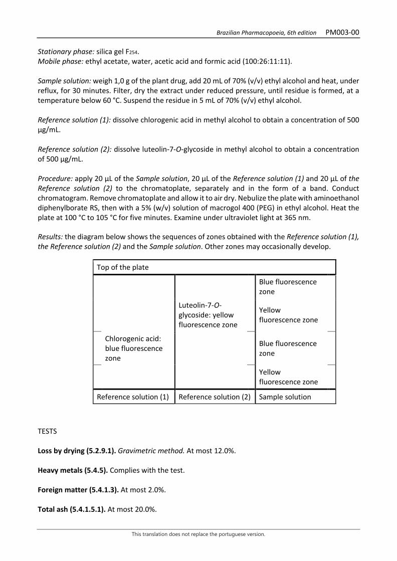
Stationary phase: silica gel F254. Mobile phase: ethyl acetate, water, acetic acid and formic acid (100:26:11:11). Sample solution: weigh 1,0 g of the plant drug, add 20 mL of 70% (v/v) ethyl alcohol and heat, under reflux, for 30 minutes. Filter, dry the extract under reduced pressure, until residue is formed, at a temperature below 60 °C. Suspend the residue in 5 mL of 70% (v/v) ethyl alcohol. Reference solution (1): dissolve chlorogenic acid in methyl alcohol to obtain a concentration of 500 µg/mL. Reference solution (2): dissolve luteolin-7-O-glycoside in methyl alcohol to obtain a concentration of 500 µg/mL. Procedure: apply 20 μL of the Sample solution, 20 μL of the Reference solution (1) and 20 μL of the Reference solution (2) to the chromatoplate, separately and in the form of a band. Conduct chromatogram. Remove chromatoplate and allow it to air dry. Nebulize the plate with aminoethanol diphenylborate RS, then with a 5% (w/v) solution of macrogol 400 (PEG) in ethyl alcohol. Heat the plate at 100 °C to 105 °C for five minutes. Examine under ultraviolet light at 365 nm. Results: the diagram below shows the sequences of zones obtained with the Reference solution (1), the Reference solution (2) and the Sample solution. Other zones may occasionally develop.
Top of the plate
Blue fluorescence zone
Luteolin-7-O-glycoside: yellow fluorescence zone
Yellow fluorescence zone
Chlorogenic acid: blue fluorescence zone
Blue fluorescence zone
Yellow fluorescence zone
Reference solution (1) Reference solution (2) Sample solution
TESTS Loss by drying (5.2.9.1). Gravimetric method. At most 12.0%. Heavy metals (5.4.5). Complies with the test. Foreign matter (5.4.1.3). At most 2.0%. Total ash (5.4.1.5.1). At most 20.0%.
Brazilian Pharmacopoeia, 6th edition PM003-00
This translation does not replace the portuguese version.
Acid-insoluble ash (5.4.1.5.3). At most 4.0% Total mesophilic microorganism count (5.5.3.1.2). Complies with the test. Study of pathogenic microorganisms (5.5.3.1.3). Complies with the test. Aflatoxins (5.4.4). Complies with the test. Agrochemical waste (5.4.3). Complies with the test. DOSAGE Chlorogenic acid derivatives Proceed as described in High Performance Liquid Chromatography (5.2.17.4). Use a chromatograph equipped with an ultraviolet detector at 330 nm; pre-column packed with octadecylsilane silica, 250 mm long, 4.6 mm internal diameter column, packed with silica chemically bonded to an octadecylsilane group (5 µm), kept at 40 °C; Mobile phase flow rate of 1.5 mL/minute. Eluent (B): water, acetonitrile and phosphoric acid (92,6:7:0.4). Eluent (B): acetonitrile and phosphoric acid (99.6:0.4).
Time (minutes) Eluent A (%) Eluent B (%) Elution
0 – 17 100 0 isocratic 17 – 50 100 → 80 0 → 20 linear gradient 50 – 51 80 → 0 20 → 100 linear gradient 51 – 61 0 100 isocratic 61 – 62 0 → 100 100 → 0 linear gradient 62 – 72 100 0 isocratic Sample solution: accurately weigh approximately 0.50 g of the pulverized plant drug and transfer to a 100mL round-bottomed flask. Add 50 mL methyl alcohol. Heat the solution, under reflux, at 70 °C for 60 minutes. Allow the sample to cool and filter the solution with absorbent cotton. Transfer the residue and cotton to the round-bottomed flask and add 50 mL methyl alcohol. Re-heat, under reflux, at 70 °C for 60 minutes. Filter the solution, combine filtrates, and transfer to a 200-mL volumetric flask. Top off the volume with water. Filter through a 0.45 µm filter unit. Stock solution: accurately weigh approximately 5.0 mg of chlorogenic acid, transfer to a 50-mL volumetric flask and top off the volume with methyl alcohol. Reference solution: transfer 5.0 mL of the Stock solution to a 20-mL volumetric flask, add 5 mL methyl alcohol and top off the volume with water. Filter through a 0.45 µm filter unit.

Brazilian Pharmacopoeia, 6th edition PM003-00
This translation does not replace the portuguese version.
Procedure: separately inject 10 μL of the Reference solution and 10 µL of the Sample solution. Record the chromatograms and measure the areas under the chlorogenic acid peak. Calculate chlorogenic acid content, in percent, according to the expression:
𝑇𝑇𝑇𝑇 =𝑇𝑇𝑎𝑎 × 𝑚𝑚𝑟𝑟
𝑇𝑇𝑟𝑟 × 𝑚𝑚𝑎𝑎 × 100
in which, TA = chlorogenic acid content % (w/w) Aa = area under the peak corresponding to chlorogenic acid in the Sample solution; Ar = area under the peak corresponding to the chlorogenic acid in the Reference solution; mr = mass in grams of chlorogenic acid, considering the purity of the blank substance; ma = mass in grams of the sample used, considering the loss by drying. PACKAGING AND STORAGE In tightly closed containers, protected from light and heat.
Brazilian Pharmacopoeia, 6th edition PM003-00
This translation does not replace the portuguese version.
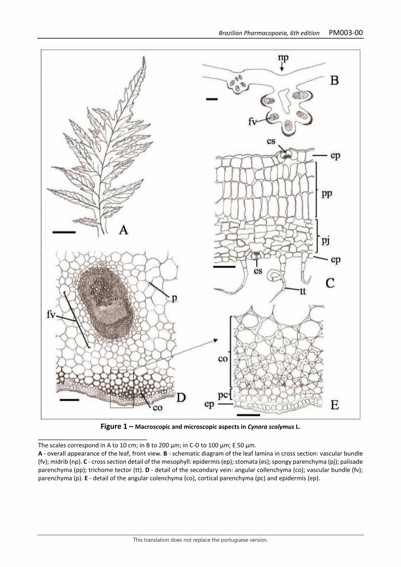
Figure 1 – Macroscopic and microscopic aspects in Cynara scolymus L.
___________________________ The scales correspond in A to 10 cm; in B to 200 µm; in C-D to 100 µm; E 50 µm. A - overall appearance of the leaf, front view. B - schematic diagram of the leaf lamina in cross section: vascular bundle (fv); midrib (np). C - cross section detail of the mesophyll: epidermis (ep); stomata (es); spongy parenchyma (pj); palisade parenchyma (pp); trichome tector (tt). D - detail of the secondary vein: angular collenchyma (co); vascular bundle (fv); parenchyma (p). E - detail of the angular colenchyma (co), cortical parenchyma (pc) and epidermis (ep).
Brazilian Pharmacopoeia, 6th edition PM003-00
This translation does not replace the portuguese version.
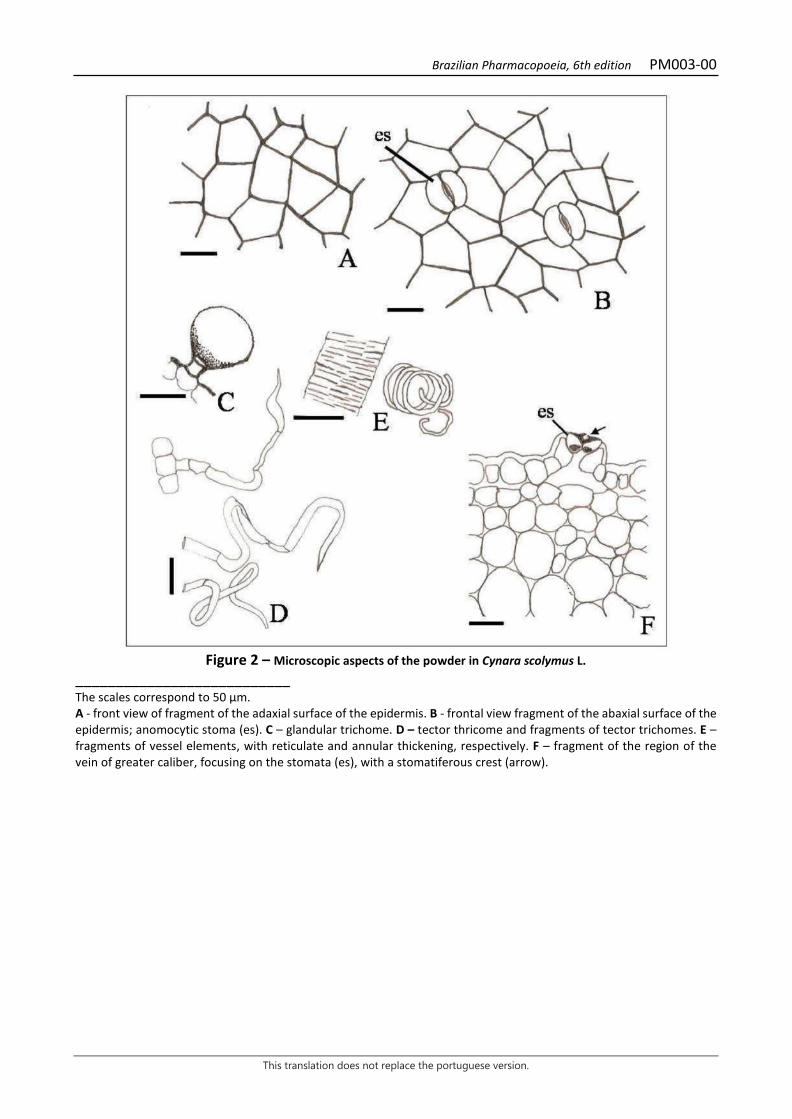
Figure 2 – Microscopic aspects of the powder in Cynara scolymus L.
___________________________ The scales correspond to 50 µm. A - front view of fragment of the adaxial surface of the epidermis. B - frontal view fragment of the abaxial surface of the epidermis; anomocytic stoma (es). C – glandular trichome. D – tector thricome and fragments of tector trichomes. E – fragments of vessel elements, with reticulate and annular thickening, respectively. F – fragment of the region of the vein of greater caliber, focusing on the stomata (es), with a stomatiferous crest (arrow).
Brazilian Pharmacopoeia, 6th edition PM004-00
This translation does not replace the portuguese version.
LICORICE, root Liquiritiae radix
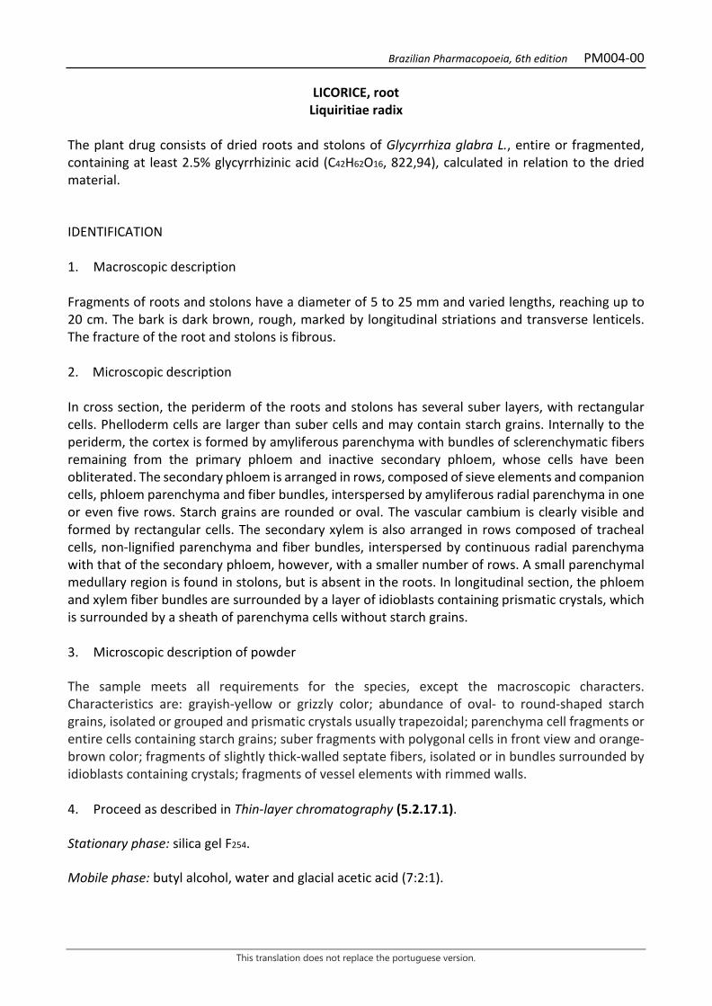
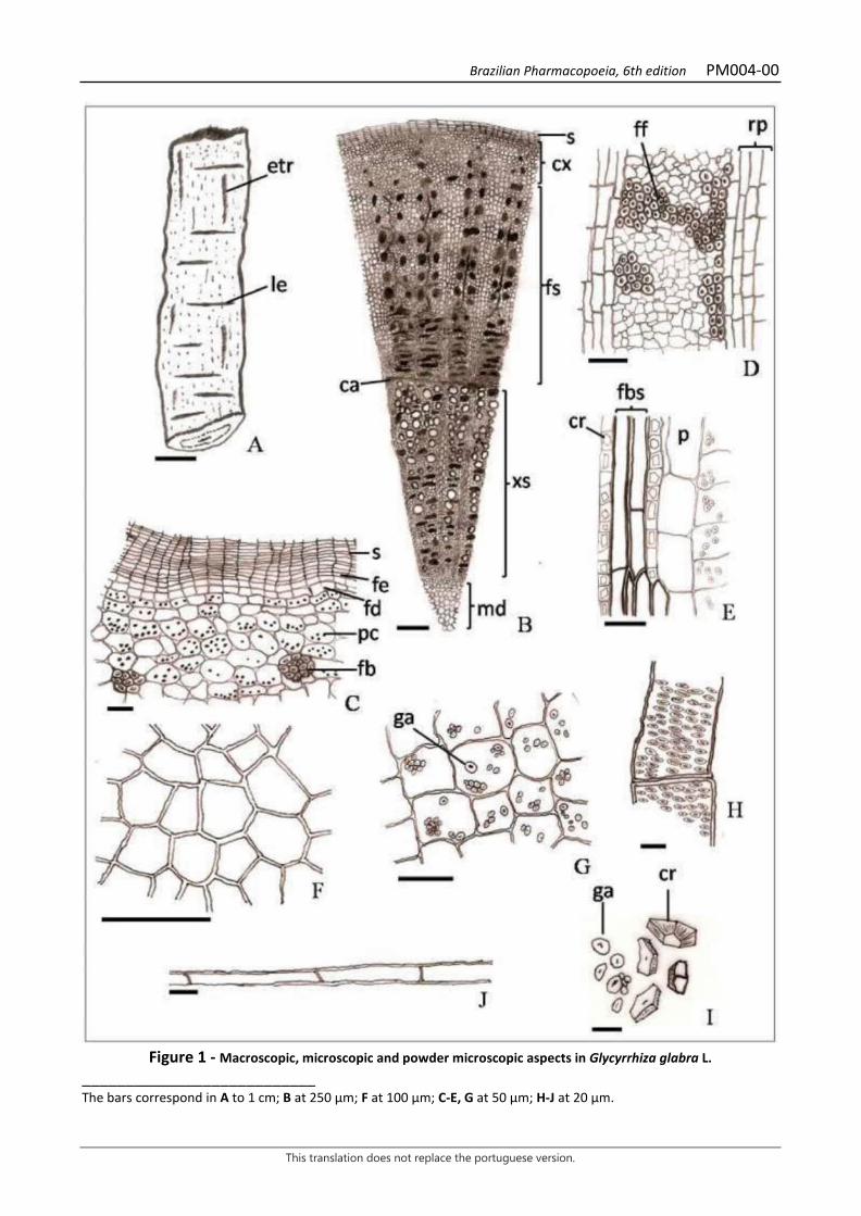
The plant drug consists of dried roots and stolons of Glycyrrhiza glabra L., entire or fragmented, containing at least 2.5% glycyrrhizinic acid (C42H62O16, 822,94), calculated in relation to the dried material. IDENTIFICATION 1. Macroscopic description Fragments of roots and stolons have a diameter of 5 to 25 mm and varied lengths, reaching up to 20 cm. The bark is dark brown, rough, marked by longitudinal striations and transverse lenticels. The fracture of the root and stolons is fibrous. 2. Microscopic description In cross section, the periderm of the roots and stolons has several suber layers, with rectangular cells. Phelloderm cells are larger than suber cells and may contain starch grains. Internally to the periderm, the cortex is formed by amyliferous parenchyma with bundles of sclerenchymatic fibers remaining from the primary phloem and inactive secondary phloem, whose cells have been obliterated. The secondary phloem is arranged in rows, composed of sieve elements and companion cells, phloem parenchyma and fiber bundles, interspersed by amyliferous radial parenchyma in one or even five rows. Starch grains are rounded or oval. The vascular cambium is clearly visible and formed by rectangular cells. The secondary xylem is also arranged in rows composed of tracheal cells, non-lignified parenchyma and fiber bundles, interspersed by continuous radial parenchyma with that of the secondary phloem, however, with a smaller number of rows. A small parenchymal medullary region is found in stolons, but is absent in the roots. In longitudinal section, the phloem and xylem fiber bundles are surrounded by a layer of idioblasts containing prismatic crystals, which is surrounded by a sheath of parenchyma cells without starch grains. 3. Microscopic description of powder The sample meets all requirements for the species, except the macroscopic characters. Characteristics are: grayish-yellow or grizzly color; abundance of oval- to round-shaped starch grains, isolated or grouped and prismatic crystals usually trapezoidal; parenchyma cell fragments or entire cells containing starch grains; suber fragments with polygonal cells in front view and orange-brown color; fragments of slightly thick-walled septate fibers, isolated or in bundles surrounded by idioblasts containing crystals; fragments of vessel elements with rimmed walls. 4. Proceed as described in Thin-layer chromatography (5.2.17.1). Stationary phase: silica gel F254. Mobile phase: butyl alcohol, water and glacial acetic acid (7:2:1).
Brazilian Pharmacopoeia, 6th edition PM004-00
This translation does not replace the portuguese version.
Sample solution: weigh 1,0 g of the plant drug, add 20 mL of 70% (v/v) methyl alcohol and place in a water bath for 15 minutes. Filter, dry the extract in a water bath until residue is formed, at a maximum temperature of 60 °C. Suspend the residue in 5 mL methyl alcohol and proceed to the chromatographic analysis. Reference solution: dissolve an accurately weighed amount of glycyrrhizinic acid in 70% (v/v) methyl alcohol to obtain a concentration of 500 µg/mL. Procedure: apply 20 µL of the Sample solution and 20 µL of the Reference solution to the chromatoplate, separately and in the form of a band. Conduct chromatogram. Remove chromatoplate and allow it to air dry. Nebulize the plate with anisaldehyde RS and heat at 100 °C to 105 °C for five minutes. Examine the plate under visible light. Result: the diagram below shows the sequences of zones obtained with the Reference solution and the Sample solution. Other zones may occasionally develop.
Top of the plate
Red-colored zone
Yellowish-colored zone
Yellowish-colored zone
Glycyrrhizinic acid: purple-colored zone
Purple-colored zone
Reference solution Sample solution
TESTS Loss by drying (5.2.9.1). Gravimetric method. At most 12.0%. Heavy metals (5.4.5). Complies with the test. Foreign matter (5.4.1.3). At most 2.0%. Total ash (5.4.1.5.1). At most 7.0% Acid-insoluble ash (5.4.1.5.3). At most 2.0%. Total mesophilic microorganism count (5.5.3.1.2). Complies with the test. Study of pathogenic microorganisms (5.5.3.1.3). Complies with the test. Aflatoxins (5.4.4). Complies with the test. Agrochemical waste (5.4.3). Complies with the test.

Brazilian Pharmacopoeia, 6th edition PM004-00
This translation does not replace the portuguese version.
DOSAGE Glycyrrhizinic acid Proceed as described in High Performance Liquid Chromatography (5.2.17.4). Use a chromatograph equipped with an ultraviolet detector at 254 nm; pre-column packed with octadecylsilane silica, 150 mm long, 4.6 mm internal diameter column, packed with octadecylsilane silica (5 µm), kept at 30 °C; Mobile phase flow rate of 1.5 mL/minute. Eluent (A): water and acetic acid (91.4:8.6). Eluent (B): acetonitrile. Apply the isocratic elution system with a constant ratio of 70% Eluent (A) and 30% Eluent (B). Diluent: transfer 28.57 mL 28% (v/v) ammonium hydroxide into a 1000-mL volumetric flask. Top off the volume with distilled water and homogenize. Stock solution: accurately weigh approximately 0.13 g of ammonium glycyrrhizate, transfer to a 100-mL volumetric flask and top off the volume with the Diluent. Reference solution: transfer 7 mL of the Stock solution to a 10-mL volumetric flask and top off the volume with the Diluent. Filter through a 0.45 µm filter unit. Sample solution: accurately weigh approximately 1 g of the pulverized plant drug and transfer to a 150mL Erlenmeyer flask. Add 100 mL of the Diluent. Place the solution on the ultrasound for 30 minutes, shaking the erlenmeyer flask every 10 minutes. Remove 10 mL of the solution and centrifuge for 10 minutes. Next, pipette 1 mL of the supernatant, transfer to a 5-mL volumetric flask, top off the volume with the Diluent and homogenize. Filter through a 0.45 µm filter unit. Procedure: separately inject 10 μL of the Reference solution and 10 µL of the Sample solution. Register the chromatograms and measure the areas under the peaks. Calculate glycyrrhizinic acid content, in percent, according to the following expression:
𝑇𝑇𝑇𝑇 =𝑇𝑇𝑎𝑎 × 𝐶𝐶𝑟𝑟 × 100 × 5 × 822,94
𝑇𝑇𝑟𝑟 × 𝑚𝑚𝑎𝑎 × 839,97
in which, TA = glycyrrhizinic acid content % (w/w); Aa = area under the peak corresponding to the glycyrrhizinic acid in the Sample solution; Ar = area under the peak corresponding to the glycyrrhizinic acid in the Reference solution; Cr = concentration of the Reference solution in g/mL, considering the purity of the reference substance; ma = mass in grams of the sample used, considering the loss by drying. 822.94 = glycyrrhizinic acid molecular mass;
Brazilian Pharmacopoeia, 6th edition PM004-00
This translation does not replace the portuguese version.
839.97 = ammonium glycyrrhizinate molecular mass. PACKAGING AND STORAGE In tightly closed containers, protected from light and heat.
Brazilian Pharmacopoeia, 6th edition PM004-00
This translation does not replace the portuguese version.
Figure 1 - Macroscopic, microscopic and powder microscopic aspects in Glycyrrhiza glabra L.
___________________________ The bars correspond in A to 1 cm; B at 250 µm; F at 100 µm; C-E, G at 50 µm; H-J at 20 µm.
Brazilian Pharmacopoeia, 6th edition PM004-00
This translation does not replace the portuguese version.
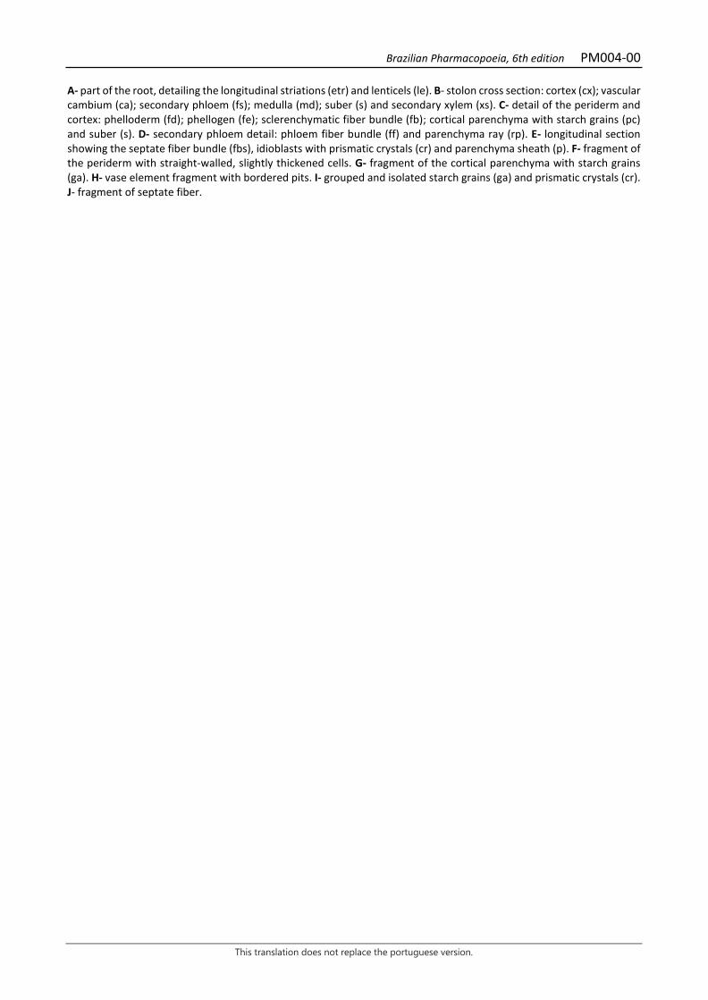
A- part of the root, detailing the longitudinal striations (etr) and lenticels (le). B- stolon cross section: cortex (cx); vascular cambium (ca); secondary phloem (fs); medulla (md); suber (s) and secondary xylem (xs). C- detail of the periderm and cortex: phelloderm (fd); phellogen (fe); sclerenchymatic fiber bundle (fb); cortical parenchyma with starch grains (pc) and suber (s). D- secondary phloem detail: phloem fiber bundle (ff) and parenchyma ray (rp). E- longitudinal section showing the septate fiber bundle (fbs), idioblasts with prismatic crystals (cr) and parenchyma sheath (p). F- fragment of the periderm with straight-walled, slightly thickened cells. G- fragment of the cortical parenchyma with starch grains (ga). H- vase element fragment with bordered pits. I- grouped and isolated starch grains (ga) and prismatic crystals (cr). J- fragment of septate fiber.
Brazilian Pharmacopoeia, 6th edition PM005-00
This translation does not replace the portuguese version.
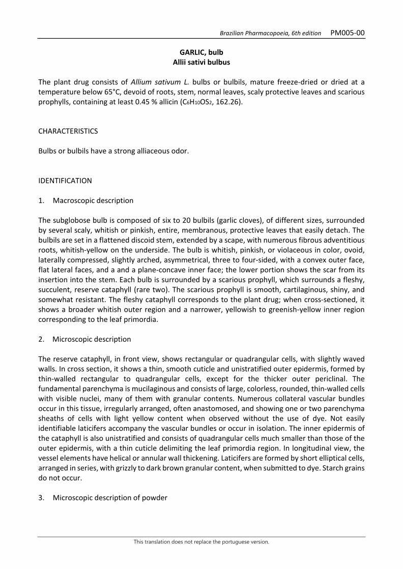
GARLIC, bulb Allii sativi bulbus
The plant drug consists of Allium sativum L. bulbs or bulbils, mature freeze-dried or dried at a temperature below 65°C, devoid of roots, stem, normal leaves, scaly protective leaves and scarious prophylls, containing at least 0.45 % allicin (C6H10OS2, 162.26). CHARACTERISTICS Bulbs or bulbils have a strong alliaceous odor. IDENTIFICATION 1. Macroscopic description The subglobose bulb is composed of six to 20 bulbils (garlic cloves), of different sizes, surrounded by several scaly, whitish or pinkish, entire, membranous, protective leaves that easily detach. The bulbils are set in a flattened discoid stem, extended by a scape, with numerous fibrous adventitious roots, whitish-yellow on the underside. The bulb is whitish, pinkish, or violaceous in color, ovoid, laterally compressed, slightly arched, asymmetrical, three to four-sided, with a convex outer face, flat lateral faces, and a and a plane-concave inner face; the lower portion shows the scar from its insertion into the stem. Each bulb is surrounded by a scarious prophyll, which surrounds a fleshy, succulent, reserve cataphyll (rare two). The scarious prophyll is smooth, cartilaginous, shiny, and somewhat resistant. The fleshy cataphyll corresponds to the plant drug; when cross-sectioned, it shows a broader whitish outer region and a narrower, yellowish to greenish-yellow inner region corresponding to the leaf primordia. 2. Microscopic description The reserve cataphyll, in front view, shows rectangular or quadrangular cells, with slightly waved walls. In cross section, it shows a thin, smooth cuticle and unistratified outer epidermis, formed by thin-walled rectangular to quadrangular cells, except for the thicker outer periclinal. The fundamental parenchyma is mucilaginous and consists of large, colorless, rounded, thin-walled cells with visible nuclei, many of them with granular contents. Numerous collateral vascular bundles occur in this tissue, irregularly arranged, often anastomosed, and showing one or two parenchyma sheaths of cells with light yellow content when observed without the use of dye. Not easily identifiable laticifers accompany the vascular bundles or occur in isolation. The inner epidermis of the cataphyll is also unistratified and consists of quadrangular cells much smaller than those of the outer epidermis, with a thin cuticle delimiting the leaf primordia region. In longitudinal view, the vessel elements have helical or annular wall thickening. Laticifers are formed by short elliptical cells, arranged in series, with grizzly to dark brown granular content, when submitted to dye. Starch grains do not occur. 3. Microscopic description of powder
Brazilian Pharmacopoeia, 6th edition PM005-00
This translation does not replace the portuguese version.
The sample meets all requirements for the species, except the macroscopic characters. Characteristics are: whitish, whitish-yellow to whitish-pink color; portions of the reserve cataphyll epidermis; numerous fragments with large thin-walled parenchyma cells and granular content; laticifers; lignified vessel elements, with helical or ringed wall thickening; vessel elements accompanied by thin-walled cells; as impurities, portions of the epidermis of the scarious prophylls in front view, portions of the epidermis of the prophylls in cross section and prismatic crystals of different shapes. 4. Microscopic description of impurities The scarious prophyll, if present as an impurity, in a front view, has elongated, straight-walled cells. In cross section, on the external or abaxial surface, a thin and smooth cuticle occurs, followed by a unistratified epidermis, formed by sclerified, rectangular to quadrangular, extremely thick-walled cells, with conspicuous pits and commonly with prismatic calcium oxalate crystals of different shapes. This is followed by one or two layers of tangentially flattened, thick-walled, non-sclerified hyaline cells with a large number of crystals and a parenchyma lacking chloroplasts, formed by elliptic cells in the tangential direction, interspersed with small, collateral vascular bundles. The epidermis facing the inner or adaxial surface has the same characteristics as the abaxial surface. 5. Proceed as described in Thin-layer chromatography (5.2.17.1). Stationary phase: silica gel GF254. Mobile phase: anhydrous ethyl alcohol, glacial acetic acid, propanol and water (4:2:2:2:). Sample solution: add 5 mL of methyl alcohol to 1 g of the pulverized plant drug (355 µm) (5.2.11) and shake for one minute. Filter, collect 1 mL and conduct chromatographic analysis. Reference solution: dilute 2.5 mg of alanine in 5 mL of water and top off to 10 mL with methyl alcohol. Procedure: apply 20 μL of the sample solution and 10 μL of the Reference solution to the chromatoplate, separately and in the form of a band. Conduct chromatogram. Remove chromatoplate and allow it to air dry for 15 minutes. Nebulize the plate with a ninhydrin RS solution and heat at 100 °C to 105 °C for 10 minutes. Results: the diagram below shows the sequences of zones obtained with the Reference solution and the Sample solution. Other zones may occasionally develop.
Brazilian Pharmacopoeia, 6th edition PM005-00
This translation does not replace the portuguese version.
Top of the plate
Alanine: blue-purple colored zone
Violaceous-blue colored zone
Red-colored zone
Reference solution Sample solution
TESTS Heavy metals (5.4.5). Complies with the test. Starch (5.4.1.2). Histochemical reactions. It should not develop a blue color. Foreign matter (5.4.1.3). At most 2.0%. Loss by drying (5.2.9.1). Gravimetric method. At most 7.0% Total ash (5.4.1.5.1). At most 5.0%. Total mesophilic microorganism count (5.5.3.1.2). Complies with the test. Study of pathogenic microorganisms (5.5.3.1.3). Complies with the test. Agrochemical waste (5.4.3). Complies with the test. DOSAGE Allicin Proceed as described in High Performance Liquid Chromatography (5.2.17.4). Use a chromatograph equipped with an ultraviolet detector at 254 nm; pre-column packed with octadecylsilane silica, 250 mm long, 4.6 mm internal diameter column, packed with octadecylsilane silica (5 μm), kept at (22 ± 2) °C; Mobile phase flow rate of 0.8 mL/minute. Isocratic system, analysis time should be 20 minutes. Mobile phase: Mobile phase: 1% (v/v) methyl alcohol and anhydrous formic acid (75:25). Internal standard solution: quantitatively transfer about 20 mg of accurately weighed butyl p-hydroxybenzoate to a 100-mL volumetric flask, top off the volume with a mixture of methyl alcohol and water (50:50) and homogenize. Filter through a 0.45 µm filter unit. Stock solution: accurately weigh about 0.8 g of the freeze-dried or dried garlic bulb powder (355 µm) (5.2.11) at a temperature below 65 °C, and add 20.0 mL of water. Leave in an ultrasonic bath at 4 °C, maintained by ice, for five minutes. Leave the solution at room temperature for 30 minutes. Centrifuge for 30 minutes. Transfer 10 mL of the supernatant to a 25-mL volumetric flask, top off
Brazilian Pharmacopoeia, 6th edition PM005-00
This translation does not replace the portuguese version.
the volume with a mixture of methyl alcohol and 1% anhydrous formic acid (60:40) and homogenize to obtain the Stock solution. Shake and centrifuge for five minutes. Sample solution: Transfer 0.5 mL of the Internal standard solution to a 10-mL volumetric flask, top off the volume with the Stock solution and homogenize. Filter through a 0.45 µm filter unit. Procedure: separately inject 1 μL of the Internal standard solution and 10 µL of the Sample solution. Register the chromatograms and measure the areas under the peaks. Calculate the butyl p-hydroxybenzoate content in the sample from the responses obtained for the Sample solution versus the Internal Standard Solution. Calculate allicin content, in percent, according to the following expression:
𝑇𝑇𝑇𝑇 =𝑇𝑇𝑎𝑎 × 𝑚𝑚𝑟𝑟 × 22,75
𝑇𝑇𝑟𝑟 × 𝑚𝑚𝑎𝑎
in which, TA = allicin content % (w/w); Aa = area under the peak corresponding to the allicin in the Sample solution; Ar = area under the peak corresponding to butyl p-hydroxybenzoate in the Sample solution; ma = mass in grams of the sample used, considering the loss by drying. mr = mass in grams of butyl p-hydroxybenzoate in 100 mL of the Internal Standard Solution. 1 mg butyl p-hydroxybenzoate corresponds to 8.65 mg allicin. PACKAGING AND STORAGE In tightly closed containers, protected from light and heat.
Brazilian Pharmacopoeia, 6th edition PM005-00
This translation does not replace the portuguese version.
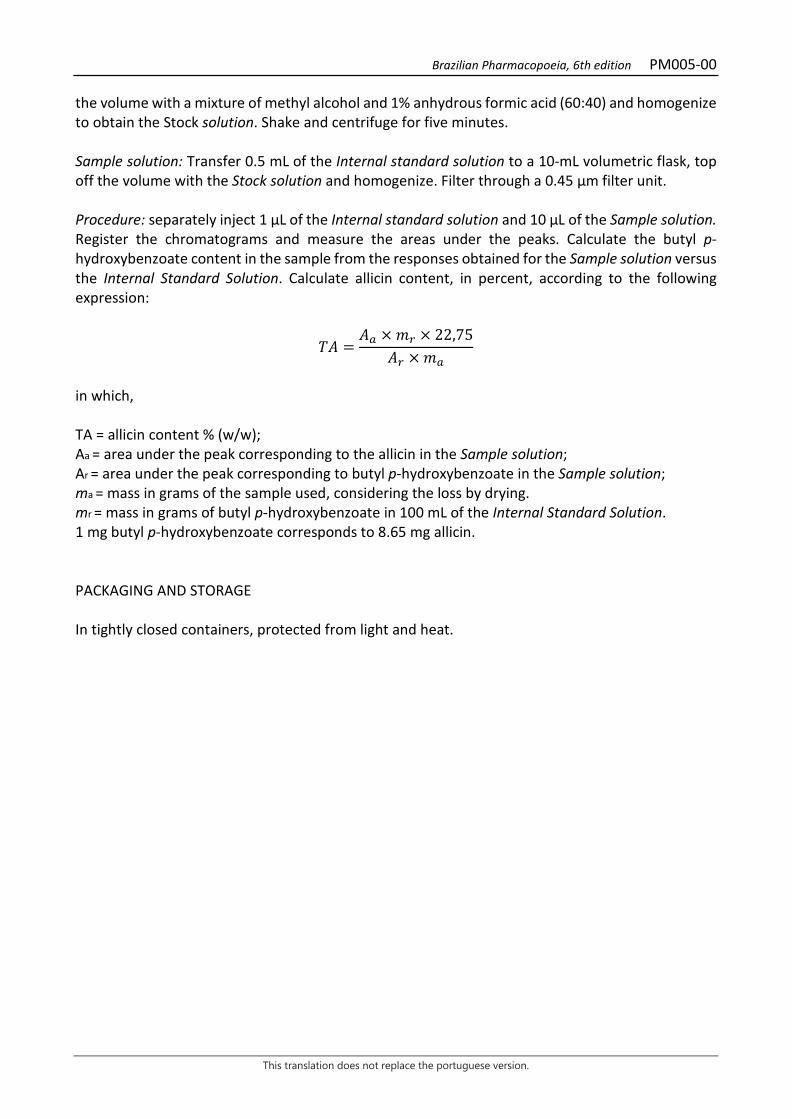
Figure 1 – Macroscopic, microscopic and powder microscopic aspects in Allium sativum L.
___________________________ The scales are: A and C (2 cm), B (1.5 cm), D and E (0.5 cm), F to N (100 µm). A - overall appearance of compound bulb; adventitious roots (rd); scape (esc). B1, B2 and B3 – appearance of the bulbils in dorsal, ventral and lateral view, respectively; scar of the bulbil insertion (ci). C - appearance of the stem portion of the bulb, after removal of the bulbs; discoid stem (c); scape (esc). D - longitudinal section of a bulbil; reserve cataphyll (cr); leaf primordium (pr); scaly prophyll (pro). E - cross section of a bulbil; primordium (pr). F1 and F2 - details of the epidermis portion of the scarious prophyll in abaxial and adaxial front view, respectively. G - cross section detail of the scarious prophyll; abaxial surface (ab); adaxial surface (ad); crystals (cr); cuticle (cu); sclerified epidermis (ep); parenchyma with thickened wall cells (ppe). H - front view detail of the outer portion of the reserve cataphyll; lipid drop (gl); wall thickening (epa). I - cross section detail of the outer portion of the reserve cataphyll; cuticle (cu); epidermis (ep); reserve parenchyma (pre). J - cross section detail of the inner portion of the reserve cataphyll, as marked in E;

Brazilian Pharmacopoeia, 6th edition PM005-00
This translation does not replace the portuguese version.
cuticle (cu); epidermis (ep); reserve parenchyma (pre). L – cross section of the vascular bundle; parenchymatic sheath (bp); phloem (f); xylem (x). M - longitudinal view of vessel elements showing helical or annular wall thickening. N - detail of reserve parenchyma cells.
Brazilian Pharmacopoeia, 6th edition PM006-01
This translation does not replace the portuguese version.
Aloe, dry exudate Aloe exudatum siccum
The plant drug consists of thick juice from leaves of Aloe vera (L.) Burm. f., Aloe ferox Mill., Aloe africana Mill. and Aloe spicata L. f. or their interspecific hybrids, or even their mixture, heat desiccated, containing at least 18% of hydroxyanthracene derivatives, expressed as barbaloin (C21H22O9, 418.39). CHARACTERISTICS The plant drug has a pungent, unpleasant and distinctive odor. IDENTIFICATION 1. Macroscopic description Irregular masses, dark brown in color, with greenish reflections, of smooth and vitreous fracture. Its fragments are translucent at the margins, very friable, creating a yellowish-brown powder. 2. Proceed as described in Thin-layer chromatography (5.2.17.1). Stationary phase: silica gel GF254 (0.25 mm). Mobile phase: ethyl acetate, methyl alcohol and water (100:17:13). Sample solution: add 20 mL of methyl alcohol to 0.25 g of the powder and heat until boiling. Shake for a few minutes, decant the solution, and keep at about 4 °C. This solution can be used up to 24 hours later. Reference solution: dissolve 2.5 mg barbaloin in 1 mL methyl alcohol. Procedure: apply to the plate, separately and in the form of a band, 10 µL of each of the freshly prepared solutions described below. Conduct chromatogram. Remove chromatoplate and allow it to air dry. Nebulize the plate with 10% (w/v) potassium hydroxide solution in methyl alcohol. Examine under ultraviolet light at 365 nm. Next, heat the plate in an oven at 110°C for five minutes. Results: the diagram below shows the sequences of zones obtained with the Reference solution and the Sample solution. Other zones may occasionally develop.
Brazilian Pharmacopoeia, 6th edition PM006-01
This translation does not replace the portuguese version.
Top of the plate
Barbaloin: orange fluorescence zone
Orange fluorescence zone Brown-colored
zone
Purple-colored zone
Light-blue fluorescence zone Light-blue fluorescence zone
Reference solution Sample solution Sample solution A. ferox
TESTS Solubility. Partially soluble in boiling water, soluble in hot ethyl alcohol and practically insoluble in diethyl ether. Alcohol-insoluble substances. Accurately weigh about 1 g of the plant drug and transfer to a flask containing 50 mL of ethyl alcohol. Heat the mixture and keep it moderately boiling for 15 minutes, refilling the evaporated ethyl alcohol. Allow to cool and occasionally shake the mixture for an hour. Filter on small, dried, tared filter paper and rinse the residue with ethyl alcohol until the washing liquids are colorless. Dry this residue at 105°C until it reaches constant weight. The found weight must be less than 10.0%. Water (5.4.1.4). At most 4.0% Total ash (5.4.1.5.1). At most 4.0% DOSAGE Hydroxyanthracene derivatives To proceed as described in Visible absorption spectrophotometry (5.2.14). Prepare solutions as described below. Stock solution: transfer 0.4 g of the pulverized sample into a 250-mL erlenmeyer flask. Moisten with 2 mL methyl alcohol, add 5 mL of water previously heated to about 60 °C and homogenize. Add 75 mL of water heated to about 60 °C and shake for 30 minutes. Cool and filter into a volumetric flask. Rinse both the erlenmeyer flask and the filter with 20 mL of water. Pour the wash water into a volumetric flask, to off to 1000 mL with water and homogenize. Introduce 10 mL of this solution into a 100 mL round-bottomed flask containing 1 mL of 60% (w/v) ferric chloride solution and 6 mL
Brazilian Pharmacopoeia, 6th edition PM006-01
This translation does not replace the portuguese version.
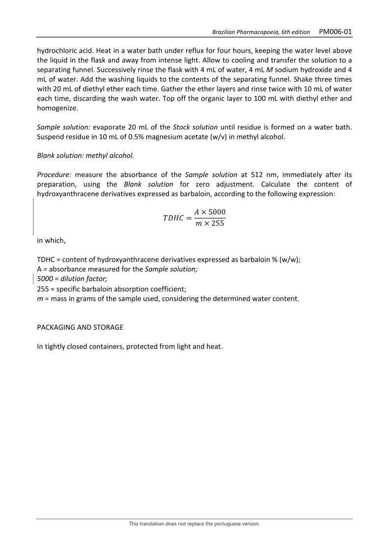
hydrochloric acid. Heat in a water bath under reflux for four hours, keeping the water level above the liquid in the flask and away from intense light. Allow to cooling and transfer the solution to a separating funnel. Successively rinse the flask with 4 mL of water, 4 mL M sodium hydroxide and 4 mL of water. Add the washing liquids to the contents of the separating funnel. Shake three times with 20 mL of diethyl ether each time. Gather the ether layers and rinse twice with 10 mL of water each time, discarding the wash water. Top off the organic layer to 100 mL with diethyl ether and homogenize. Sample solution: evaporate 20 mL of the Stock solution until residue is formed on a water bath. Suspend residue in 10 mL of 0.5% magnesium acetate (w/v) in methyl alcohol. Blank solution: methyl alcohol. Procedure: measure the absorbance of the Sample solution at 512 nm, immediately after its preparation, using the Blank solution for zero adjustment. Calculate the content of hydroxyanthracene derivatives expressed as barbaloin, according to the following expression:
𝑇𝑇𝑇𝑇𝑇𝑇𝐶𝐶 =𝑇𝑇 × 5000𝑚𝑚 × 255
in which, TDHC = content of hydroxyanthracene derivatives expressed as barbaloin % (w/w); A = absorbance measured for the Sample solution; 5000 = dilution factor; 255 = specific barbaloin absorption coefficient; m = mass in grams of the sample used, considering the determined water content. PACKAGING AND STORAGE In tightly closed containers, protected from light and heat.
Brazilian Pharmacopoeia, 6th edition PM007-00
This translation does not replace the portuguese version.
MARSHMALLOW, root Althaeae radix
The plant drug consists of dried root fragments of Althaea officinalis L. CHARACTERISTICS Mucilaginous consistency after hydration. IDENTIFICATION 1. Macroscopic description The unweeded root is cylindrical, slightly twisted and furrowed lengthwise, up to 20 cm long and up to 2 cm thick. The outer surface is grayish-grizzly and shows numerous scars from the lateral roots. The fracture is fibrous on the outer portion and irregular and granular internally. In cross section, concentric layers of the grizzly-colored cortex and its stratified structure are visible, separated by a well-marked, sinuous, yellowish cambial band, followed by the whitish central cylinder, showing xylem with a radial structure, especially after hydration in water and with the aid of a lens. The weeding root is almost cylindrical, and the outer side has dark scars originating from the lateral roots and has a whitish-yellow coloration. Often the plant drug is fragmented, with portions of fibers arranged lengthwise or detached from the remnants of the cortex being clearly visible. The three regions described can be seen in the fragments. 2. Microscopic description A front view of the unweeded root shows a suber with polyhedral, straight-walled cells. In cross section, the three regions mentioned in the macroscopic description are clearly visible. The cortex has an externally poorly developed suber, consisting of tabular and irregular cells, with thin and straight walls, arranged in rows and internally cortical parenchyma with cells generally polyhedral and bulky, with thin and straight walls. In this parenchyma, irregular clusters of phloem fibers occur, randomly arranged, with cells with thinly thickened walls and whose conducting cells are rarely visible. The parenchyma rays are distributed from the inner cortex to the central cylinder and usually consist of a few rows of cells. The cambium is made up of small cells, arranged in rows, most of them flattened lengthwise. The central cylinder is well-developed, formed by xylematic parenchyma with cells varying in shape and volume, with rectilinear and thin walls. The conducting elements form irregular clusters longitudinally aligned and often associated with small parenchyma cells; more internally they show a ring-like arrangement. Clusters of fibers or isolated fibers are found throughout the central cylinder, also occurring near the primary xylem, when present in roots with solid medulla, that is, filled by a parenchyma composed of cells of large volume. Simple starch grains of various shapes, often rounded, ovoid, or reniform, with hilum usually central and branched, rarely eccentric, or rarely compound grains, often showing lamellation, occur in large numbers in all tissues except the medullary parenchyma. Druse-type calcium oxalate crystals of different sizes are very common in the cortex and central cylinder. Mucilage-containing cells, oval or rounded, usually larger than other parenchyma, with dense, dark protoplast, also occur in the cortex and central cylinder. Weeded roots may lack suber and external cortical parenchyma. With the addition of toluidine blue, the vessel elements turn intense blue; the fibers light, blue; and the mucilage-containing cells,
Brazilian Pharmacopoeia, 6th edition PM007-00
This translation does not replace the portuguese version.
purple. The large amount of starch grains and mucilage-containing cells hinders the preparation of histological slides using hydrated material, and it is necessary to test the hydration time. 3. Microscopic description of powder The sample meets all requirements for the species, except the macroscopic characters. Microscopic observation of the powder becomes clearer when chloral hydrate is used. Characteristics are: white to yellowish-white color, when coming from weeded roots, or grayish-grizzly color when coming from unweeded roots; suber fragments in cross section: with rectangular and longitudinally flattened cells, idem with quadrangular cells, idem containing crystalliferous idioblasts, idem with rectangular and longitudinally flattened cells, containing crystalliferous idioblasts and starch grains, in front view, containing starch grains, in cross section, with rectangular cells and flattened longitudinally, full of starch grains; parenchyma fragments in front view: containing cells with mucilage and many starch grains, idem showing crystalliferous idioblasts and cells full of starch grains, parenchyma fragments in cross section, containing starch grains, idem containing crystalliferous idioblasts, idem with cells containing mucilage and large amounts of starch grains; parenchymal ray fragments, in longitudinal section, showing parenchymal cells and fibers; isolated parenchyma cells, full of starch grains and/or containing crystals; parenchyma ray fragments, in cross section, with cells containing starch grains; xylem fragments, in longitudinal section, showing vessel element with reticulated thickening associated with fibers and parenchyma, idem showing vessel elements with reticulated thickening, fibers and parenchyma with starch grains, idem showing vessel elements with reticulated thickening and pit thickening, associated with parenchyma cells filled with starch grains, portions of vessel elements with helical thickening, in longitudinal section, vessel elements in cross section, associated with parenchyma cells filled with starch grains, portions of vessel elements with reticulated thickening, in longitudinal section; fiber fragments, in longitudinal section associated with parenchymal cells of the xylem, fiber bundle fragments, in longitudinal section, containing starch grains, fiber bundle fragments, in longitudinal section, associated with parenchyma ray cells, fragments of clusters of fibers, in cross section; fibers or portions thereof, in longitudinal section, isolated and/or grouped; starch grains, in front view, single or compound, isolated or grouped in small numbers; clusters forming clumps of starch grains, in front view; mucilage released from cells; isolated cells containing mucilage; isolated druse-type calcium oxalate crystals. 4. Proceed as described in Thin-layer chromatography (5.2.17.1). Stationary phase: silica gel GF254 (0.25 mm). Mobile phase: ethyl acetate, methyl ethyl ketone, formic acid and water (50:30:10:10). Sample solution: weigh 1 g of the sample, add 10 mL of methyl alcohol, heat in a water bath for 15 minutes. Filter. Reference solution: dissolve 2.5 mg rutin and 1 mg chlorogenic acid in 10 mL methyl alcohol. Procedure: apply 20 μL of the sample solution and 20 μL of the Reference solution to the chromatoplate, separately and in the form of a band. Conduct chromatogram. Remove chromatoplate and allow it to air dry. Nebulize the plate with aminoethanol diphenylborate RS (Natural Reagent A) and examine under ultraviolet light at 365 nm.
Brazilian Pharmacopoeia, 6th edition PM007-00
This translation does not replace the portuguese version.
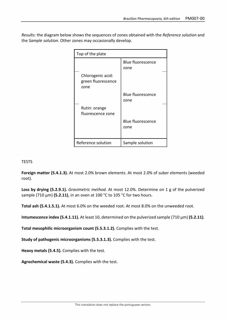
Results: the diagram below shows the sequences of zones obtained with the Reference solution and the Sample solution. Other zones may occasionally develop.
Top of the plate
Blue fluorescence zone
Chlorogenic acid: green fluorescence zone
Blue fluorescence zone
Rutin: orange fluorescence zone
Blue fluorescence zone
Reference solution Sample solution
TESTS Foreign matter (5.4.1.3). At most 2.0% brown elements. At most 2.0% of suber elements (weeded root). Loss by drying (5.2.9.1). Gravimetric method. At most 12.0%. Determine on 1 g of the pulverized sample (710 µm) (5.2.11), in an oven at 100 °C to 105 °C for two hours. Total ash (5.4.1.5.1). At most 6.0% on the weeded root. At most 8.0% on the unweeded root. Intumescence index (5.4.1.11). At least 10, determined on the pulverized sample (710 µm) (5.2.11). Total mesophilic microorganism count (5.5.3.1.2). Complies with the test. Study of pathogenic microorganisms (5.5.3.1.3). Complies with the test. Heavy metals (5.4.5). Complies with the test. Agrochemical waste (5.4.3). Complies with the test.
Brazilian Pharmacopoeia, 6th edition PM007-00
This translation does not replace the portuguese version.
PACKAGING AND STORAGE In tightly closed containers, protected from light and heat.
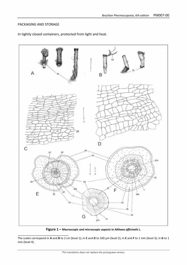
Figure 1 – Macroscopic and microscopic aspects in Althaea officinalis L.
___________________________ The scales correspond in A and B to 2 cm (level 1); in C and D to 100 µm (level 2); in E and F to 1 mm (level 3); in G to 1 mm (level 4).

Brazilian Pharmacopoeia, 6th edition PM007-00
This translation does not replace the portuguese version.
A - overall appearance of unweeded roots; fiber (fb). A - overall appearance of weeded roots; fiber (fb); side root (rzl). C - front view of the outer suber of an unweeded root; starch grain (ga). D - front view of the inner suber of a weeded root. E - schematic diagram of an unweeded root croos section; cambium (ca); center cylinder (cc); cortex (cx); tracheal element (el); tracheal elements arranged in an annular manner (ela); phloem (f); fiber (fb); parenchyma ray (rp); suber (su); primary xylem (xp); secondary xylem (xs). F - schematic diagram of an unweeded root croos section; cambium (ca); center cylinder (cc); cortex (cx); tracheal element (el); tracheal elements arranged in an annular manner (ela); phloem (f); fiber (fb); medullary parenchyma (pm); parenchyma ray (rp); suber (su); secondary xylem (xs). G - schematic diagram of an weeded root cross section; cambium (ca); center cylinder (cc); cortex (cx); tracheal element (el); tracheal elements arranged in an annular manner (ela); phloem (f); fiber (fb); medullary parenchyma (pm); parenchyma ray (rp); remnants of suber (rs); secondary xylem (xs).
Brazilian Pharmacopoeia, 6th edition PM007-00
This translation does not replace the portuguese version.
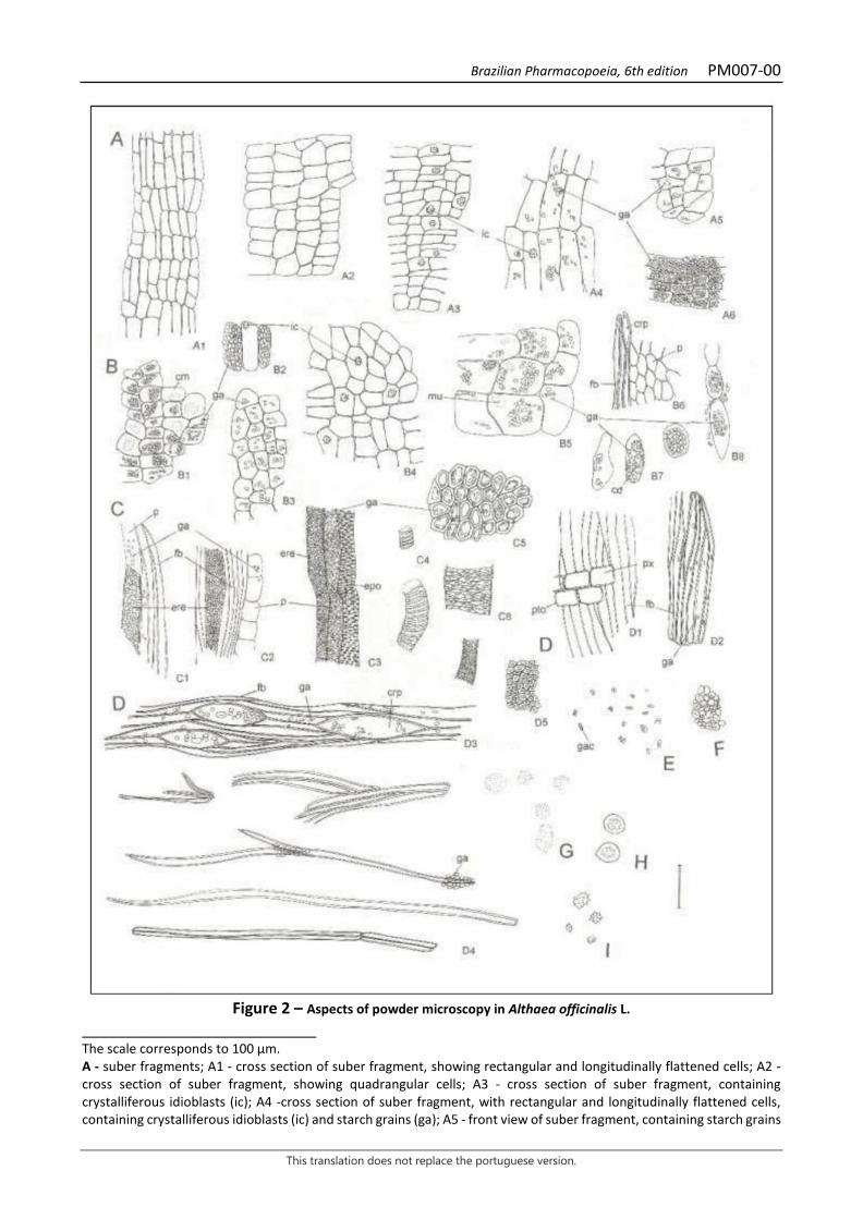
Figure 2 – Aspects of powder microscopy in Althaea officinalis L.
___________________________ The scale corresponds to 100 µm. A - suber fragments; A1 - cross section of suber fragment, showing rectangular and longitudinally flattened cells; A2 - cross section of suber fragment, showing quadrangular cells; A3 - cross section of suber fragment, containing crystalliferous idioblasts (ic); A4 -cross section of suber fragment, with rectangular and longitudinally flattened cells, containing crystalliferous idioblasts (ic) and starch grains (ga); A5 - front view of suber fragment, containing starch grains
Brazilian Pharmacopoeia, 6th edition PM007-00
This translation does not replace the portuguese version.
(ga); A6 - cross section of suber fragment, with rectangular and longitudinally flattened cells, filled with starch grains (ga). B - parenchyma fragments; B1 - front view of parenchyma fragment, containing mucilage-containing cells (mu) and many starch grains (ga); B2 - front view of parenchyma fragment, showing crystalliferous idioblasts (ic) and cells filled with starch grains (ga); B3 - cross section of parenchyma fragment, containing starch grains (ga); B4 - cross section of parenchyma fragment, containing crystalliferous idioblasts (ic); B5 - cross section of parenchyma fragment, with mucilage-containing cells (mu) and with many starch grains (ga); B6 - cross section of parenchymatic ray fragment (crp), showing parenchymatic cells (p) and fibers (fb); B7- isolated parenchyma cells, containing starch grains (ga) and druse-type calcium oxalate crystals (cd) or filled with starch grains; B8 - cross section of parenchymatic ray fragment, with cells containing starch grains (ga). C - xylem fragments; C1 - longitudinal section of xylem fragment, showing vessel element with reticulate thickening (ere) associated with fibers (fb) and parenchyma (p) with starch grains (ga); C2 - longitudinal section of xylem fragment, showing vessel element with reticulate thickening (ere), fibers (fb) and parenchyma (p) in cross section and with starch grains (ga); C3 - longitudinal section of xylem fragment, showing vessel elements with reticulate thickening (ere) and with pit thickening (epo), associated with parenchymatic cells (p) filled with starch grains (ga); C4 - portions of vessel element with helical thickening, in longitudinal section; C5 - cross section of vessel elements, with starch grains (ga); C6 - portions of vessel elements with reticulate thickening, in longitudinal section. D - fiber fragments; D1 - longitudinal section of fiber fragment (fb), associated with parenchymatic cells (px) of the pitted xylem (pto); D2 - longitudinal section of fiber fragment, containing starch grains (ga); D3 - longitudinal section of fiber bundle fragment (fb), associated with parenchymatic ray cells (crp) containing starch grains (ga); D4 - longitudinal section of fibers or portions of fibers, isolated or grouped; starch grain (ga); D5 - cross section of fragment of fiber grouping. E - front view of starch grains, simple or compound (gac), isolated or in small groups. F - front view of clusters forming clumps of starch grains. G - mucilage detached from the cells. H - isolated mucilage-containing cells. I - isolated, druse-like calcium oxalate crystals.
Brazilian Pharmacopoeia, 6th edition PM007-00
This translation does not replace the portuguese version.
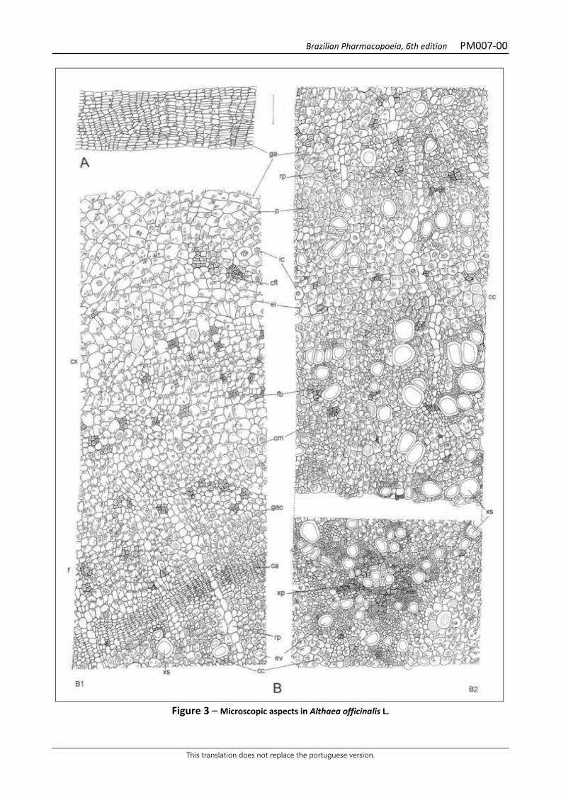
Figure 3 – Microscopic aspects in Althaea officinalis L.

Brazilian Pharmacopoeia, 6th edition PM007-00
This translation does not replace the portuguese version.
___________________________ The scale corresponds to 100 µm. A - partial cross-section detail of the suber in an unweeded root; starch grain (ga). B - partial cross-section detail of a weeding root; B1 partial detail of cortex, cambium and external portion of central cylinder; cambium (ca); central cylinder (cc); phloem-conducting cells (cfl); mucilage-containing cell (cm); cortex (cx); intercellular space (ei); vessel element (ev); phloem (f); fiber (fb); crystalliferous idioblast (ic); starch grain (ga); compound starch grain (gac); parenchymatic ray (rp); parenchyma (p); secondary xylem (xs); B2 continuity of B1 partial detail, showing inner portion of central cylinder; central cylinder (cc); mucilage-containing cell (cm); intercellular space (ei); vessel element (ev); fiber (fb); crystalliferous idioblast (ic); starch grain (ga); parenchymatic ray (rp); parenchyma (p); primary xylem (xp); secondary xylem (xs).
Brazilian Pharmacopoeia, 6th edition PM008-00
This translation does not replace the portuguese version.
PLUM, fruit Prunum fructus
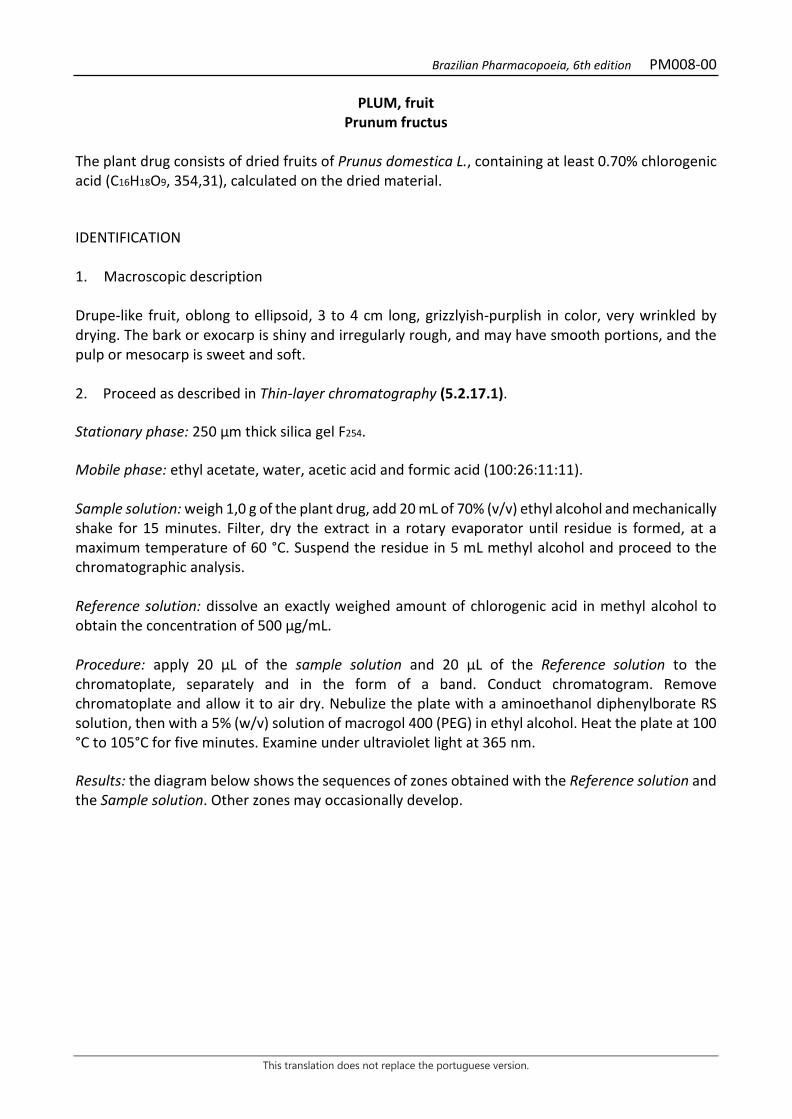
The plant drug consists of dried fruits of Prunus domestica L., containing at least 0.70% chlorogenic acid (C16H18O9, 354,31), calculated on the dried material. IDENTIFICATION 1. Macroscopic description Drupe-like fruit, oblong to ellipsoid, 3 to 4 cm long, grizzlyish-purplish in color, very wrinkled by drying. The bark or exocarp is shiny and irregularly rough, and may have smooth portions, and the pulp or mesocarp is sweet and soft. 2. Proceed as described in Thin-layer chromatography (5.2.17.1). Stationary phase: 250 µm thick silica gel F254. Mobile phase: ethyl acetate, water, acetic acid and formic acid (100:26:11:11). Sample solution: weigh 1,0 g of the plant drug, add 20 mL of 70% (v/v) ethyl alcohol and mechanically shake for 15 minutes. Filter, dry the extract in a rotary evaporator until residue is formed, at a maximum temperature of 60 °C. Suspend the residue in 5 mL methyl alcohol and proceed to the chromatographic analysis. Reference solution: dissolve an exactly weighed amount of chlorogenic acid in methyl alcohol to obtain the concentration of 500 µg/mL. Procedure: apply 20 μL of the sample solution and 20 μL of the Reference solution to the chromatoplate, separately and in the form of a band. Conduct chromatogram. Remove chromatoplate and allow it to air dry. Nebulize the plate with a aminoethanol diphenylborate RS solution, then with a 5% (w/v) solution of macrogol 400 (PEG) in ethyl alcohol. Heat the plate at 100 °C to 105°C for five minutes. Examine under ultraviolet light at 365 nm. Results: the diagram below shows the sequences of zones obtained with the Reference solution and the Sample solution. Other zones may occasionally develop.
Brazilian Pharmacopoeia, 6th edition PM008-00
This translation does not replace the portuguese version.
Top of the plate
Chlorogenic acid: blue color zone Blue-colored zone
Reference solution Sample solution TESTS Loss by drying (5.2.9.1). Gravimetric method. At most 54.0%. Heavy metals (5.4.5). Complies with the test. Foreign matter (5.4.1.3). At most 2.0%. Total ash (5.4.1.5.1). At most 0.6%. Total mesophilic microorganism count (5.5.3.1.2). Complies with the test. Study of pathogenic microorganisms (5.5.3.1.3). Complies with the test. Agrochemical waste (5.4.3). Complies with the test. DOSAGE Chlorogenic acid Proceed as described in High Performance Liquid Chromatography (5.2.17.4). Use a chromatograph equipped with an ultraviolet detector at 330 nm; pre-column packed with octadecylsilane silica, 250 mm long, 4.6 mm internal diameter column, packed with octadecylsilane silica (5 µm), kept at 40 °C; Mobile phase flow rate of 1.2 mL/minute. Eluent (A): water and phosphoric acid (99.5:0.5). Eluent (B): acetonitrile and phosphoric acid (99.6:0.4).
Time (minutes) Eluent (A) (%) Eluent (B) (%) Elution
0 – 1 92 8 isocratic 1 – 20 92 → 75 8 → 25 linear gradient 20 – 33 75 25 isocratic 33 – 35 75 → 0 25 → 100 linear gradient

Brazilian Pharmacopoeia, 6th edition PM008-00
This translation does not replace the portuguese version.
Time (minutes) Eluent (A) (%) Eluent (B) (%) Elution
35 – 36 75 → 92 100 → 8 linear gradient 36 – 40 92 8 isocratic Sample solution: accurately weigh approximately 1.00 g of the plant drug and transfer to a round-bottomed flask. Add 25 mL methyl alcohol and heat under reflux for 30 minutes. After cooling, filter through absorbent cotton into a 100 mL round-bottomed flask. Extract the plant drug residue two extra times in the round-bottomed flask and on the absorbent cotton with 20 mL methyl alcohol and heat, under reflux, for another 15 minutes. Filter, combine all filtrates in the round-bottomed flask and dry until residue is formed in the rotary evaporator at a maximum temperature of 60 °C. Dissolve the residue in 3 mL methyl alcohol and ultrasound for five minutes. Transfer solution to a 5-mL volumetric flask and top off the volume with methyl alcohol. Filter through a 0.45 µm filter unit. Stock solution: weigh 12.0 mg chlorogenic acid. Transfer to a 100-mL volumetric flask and top off the volume with methyl alcohol. Reference solution: transfer 1,2 mL of the Stock solution to a 10-mL volumetric flask and top off the volume with methyl alcohol. Filter in a 0.45 μm filter unit directly into the vial. Procedure: separately inject 20 μL of the Reference solution and 20 µL of the Sample solution. Register the chromatograms and measure the areas under the peaks. Calculate chlorogenic acid content, in percent, according to the expression:
𝑇𝑇𝑇𝑇 =𝑇𝑇𝑎𝑎 × 𝑚𝑚𝑇𝑇𝑟𝑟 × 𝑚𝑚𝑎𝑎
in which, TA = chlorogenic acid content % (w/w) Aa = area under the peak corresponding to chlorogenic acid in the Sample solution; Ar = area under the peak corresponding to the chlorogenic acid in the Reference solution; mr = mass in grams of the sample used, considering the loss by drying. ma = mass in grams of chlorogenic acid, considering the purity of the blank substance. PACKAGING AND STORAGE In tightly closed containers, protected from light and heat.
Brazilian Pharmacopoeia, 6th edition PM009-00
This translation does not replace the portuguese version.
ANGICO, bark Anadenantherae cortex
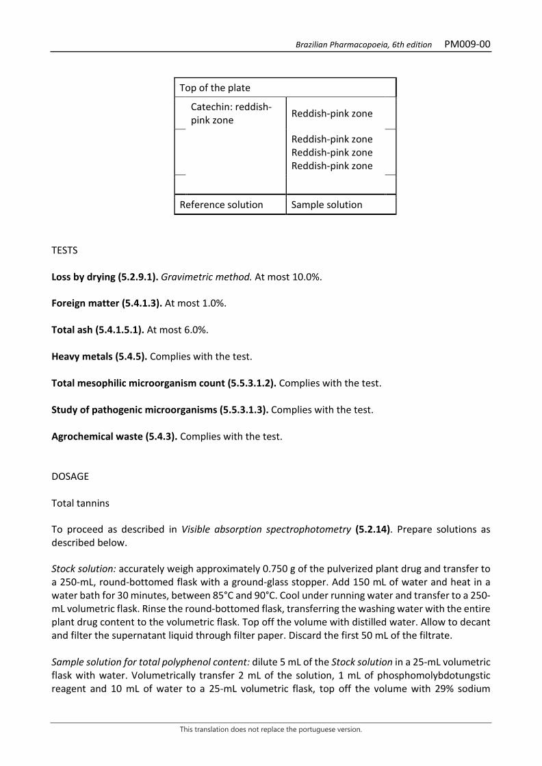
The plant drug consists of dried barks from the stems of Anadenanthera colubrina (Vell.) Brenan, containing not less than 6% total tannins and not less than 0.19% catechin (C15H14O6, 290.27). IDENTIFICATION 1. Macroscopic description The dried stem barks are slightly curved and very rigid, resinous fragments, 6 to 8 cm long, 0.5 to 2.5 cm wide, and 0.5 to 1.5 cm thick. The outer surface is rough, grizzlyish, and is usually covered with whitish to grayish plates, with sparse black spots. The inner surface is reddish-grizzly, with longitudinal striations due to the presence of thick, narrow fibers opposite each other. 2. Microscopic description A cross section of the bark shows a well-developed periderm, with 15 to 30 layers of tabular cells, radially lined. In the cortex, in cross section, there are 10 to 22 or more layers of radially flattened cortical parenchyma cells, alternating with bands of sclerenchyma fibers; past the parenchyma bands of pheloderm occur, characterized by flattened cells radially arranged in overlapping rows. Parenchymatic rays and strands are evident. Some parenchyma cells of the cortical parenchyma contain prismatic crystals in addition to starch grains. In the longitudinal section, there are layers of fibers alternating to the parenchymatic rays. 3. Microscopic description of powder The sample meets all requirements for the species, except the macroscopic characters. Characteristics are: light brown; portions of parenchyma cells; fragments of libriform sclerenchyma fibers; parenchyma cells with prismatic crystals and stone cells. 4. Proceed as described in Thin-layer chromatography (5.2.17.1). Stationary phase: 250 µm thick silica gel F254. Mobile phase: ethyl acetate, formic acid and water (90:5:5). Sample solution: weigh 1 g of the pulverized plant drug and place in a round-bottomed flask, adding 10 mL of methyl alcohol. Heat under reflux for 10 minutes. Filter through absorbent cotton. Reference solution: weigh approximately 1 mg catechin and dissolve in 1mL methyl alcohol. Procedure: apply 20 μL of the Sample solution and 20 μL of the Reference solution to the chromatoplate, separately and in the form of a band. Conduct chromatogram. Remove plate and allow it to air dry. Nebulize plate with 1% vanillin (w/v) in ethyl alcohol, then, with hydrochloric acid. Results: the diagram below shows the sequences of zones obtained with the Reference solution and the Sample solution. Other zones may occasionally develop.
Brazilian Pharmacopoeia, 6th edition PM009-00
This translation does not replace the portuguese version.
Top of the plate
Catechin: reddish-pink zone Reddish-pink zone
Reddish-pink zone Reddish-pink zone Reddish-pink zone
Reference solution Sample solution
TESTS Loss by drying (5.2.9.1). Gravimetric method. At most 10.0%. Foreign matter (5.4.1.3). At most 1.0%. Total ash (5.4.1.5.1). At most 6.0%. Heavy metals (5.4.5). Complies with the test. Total mesophilic microorganism count (5.5.3.1.2). Complies with the test. Study of pathogenic microorganisms (5.5.3.1.3). Complies with the test. Agrochemical waste (5.4.3). Complies with the test. DOSAGE Total tannins To proceed as described in Visible absorption spectrophotometry (5.2.14). Prepare solutions as described below. Stock solution: accurately weigh approximately 0.750 g of the pulverized plant drug and transfer to a 250-mL, round-bottomed flask with a ground-glass stopper. Add 150 mL of water and heat in a water bath for 30 minutes, between 85°C and 90°C. Cool under running water and transfer to a 250-mL volumetric flask. Rinse the round-bottomed flask, transferring the washing water with the entire plant drug content to the volumetric flask. Top off the volume with distilled water. Allow to decant and filter the supernatant liquid through filter paper. Discard the first 50 mL of the filtrate. Sample solution for total polyphenol content: dilute 5 mL of the Stock solution in a 25-mL volumetric flask with water. Volumetrically transfer 2 mL of the solution, 1 mL of phosphomolybdotungstic reagent and 10 mL of water to a 25-mL volumetric flask, top off the volume with 29% sodium
Brazilian Pharmacopoeia, 6th edition PM009-00
This translation does not replace the portuguese version.
carbonate solution (w/v). Determine absorbance at 760 nm (A1) after 30 minutes, using water for zero adjustment. Sample solution for polyphenols not adsorbed by hide powder: for 10 mL of the Stock solution, add 0.1 g of the hide powder and mechanically shake in a 125-mL Erlenmeyer flask for 60 minutes. Filter through paper filter. Dilute 5 mL of the filtrate in a 25-mL volumetric flask with water. Volumetrically transfer 2 mL of the solution, 1 mL of phosphomolybdotungstic reagent and 10 mL of water to a 25-mL volumetric flask, top off the volume with 29% sodium carbonate solution (w/v) and homogenize. Determine absorbance at 760 nm (A2) after 30 minutes, using water for zero adjustment. Reference solution: dissolve 50 mg pyrogallol in water immediately before use and transfer to a 100-mL volumetric flask, top off the volume with water and homogenize. Volumetrically transfer 5 mL of this solution to a 100-mL volumetric flask, top off the volume with water and homogenize. Volumetrically transfer 2 mL of the solution, 1 mL of phosphomolybdotungstic reagent and 10 mL of water to a 25-mL volumetric flask, top off the volume with 29% sodium carbonate solution (w/v) and homogenize. Determine absorbance at 760 nm (A3) after 30 minutes, using water for zero adjustment. Calculate tannin content expressed as a percentage of pyrogallol, according to the following expression:
𝑇𝑇𝑇𝑇 =(𝑇𝑇1 − 𝑇𝑇2) × 62,5
𝑇𝑇3 × 𝑚𝑚1
in which, TT = total tannin content expressed as pyrogallol % (w/w); A1 = absorbance measured for the Sample solution for total polyphenols; A2 = absorbance measured for the Sample solution for polyphenols not adsorbed by hide powder; A3 = absorbance measured for the Reference solution; m1 = mass in grams of the sample used, considering the loss by drying. m2 = mass in grams of pyrogallol, considering the purity of the reference substance. Catechin Proceed as described in High Performance Liquid Chromatography (5.2.17.4). Use a chromatograph equipped with an ultraviolet detector at 280 nm; pre-column packed with octadecylsilane silica, 250 mm long, 4.6 mm internal diameter column, packed with silica chemically bonded to an octadecylsilane group (5 µm), kept at 23 °C; Mobile phase flow rate of 0.8 mL/minute. Eluent (A): water and 85% phosphoric acid (v/v) (99:1).
Time (minutes) Eluent A (%) Eluent B (%) Elution
0 – 15 70 → 50 30 → 50 linear gradient 15 – 16 50 → 25 50 → 75 linear gradient 16 – 17 25 → 70 75 → 30 linear gradient

Brazilian Pharmacopoeia, 6th edition PM009-00
This translation does not replace the portuguese version.
Time (minutes) Eluent A (%) Eluent B (%) Elution
17 - 18 70 30 isocratic Sample solution: weigh 0.750 g of the pulverized plant drug and transfer to a 250-mL, round-bottomed flask with a ground-glass stopper. Add 150 mL of water. Heat in a water bath for 30 minutes, between 85°C and 90°C. Cool under running water and transfer to a 250-mL volumetric flask. Rinse the round-bottomed flask, transferring the washing water with the entire plant drug content to the volumetric flask. Top off the volume with water. Allow to decant and filter the supernatant liquid through filter paper. Discard the first 50 mL of the filtrate. Reference solution: dissolve an accurately weighed amount of catechin in water to obtain a solution at 4.05 µg/mL. Procedure: separately inject 20 μL of the Reference solution and 20 µL of the Sample solution. Register the chromatograms and measure the areas under the peaks. Catechin retention time in the sample is approximately 6.1 minutes. Calculate catechin content, in percent, according to the following expression:
𝑇𝑇𝐶𝐶 =𝐶𝐶𝑟𝑟 × 𝑇𝑇𝑎𝑎 × 250 × 100
𝑇𝑇𝑟𝑟 × 𝑚𝑚
in which, TC = catechin content % (w/w); Cr = concentration of the Reference solution in g/mL, considering the purity of the reference substance; Ar = area under the peak corresponding to the catechin acid in the Reference solution; Aa = area under the peak corresponding to the catechin acid in the Sample solution; m = mass in grams of the sample used, considering the loss by drying. 250 = dilution factor; PACKAGING AND STORAGE In tightly closed containers, protected from light and heat.
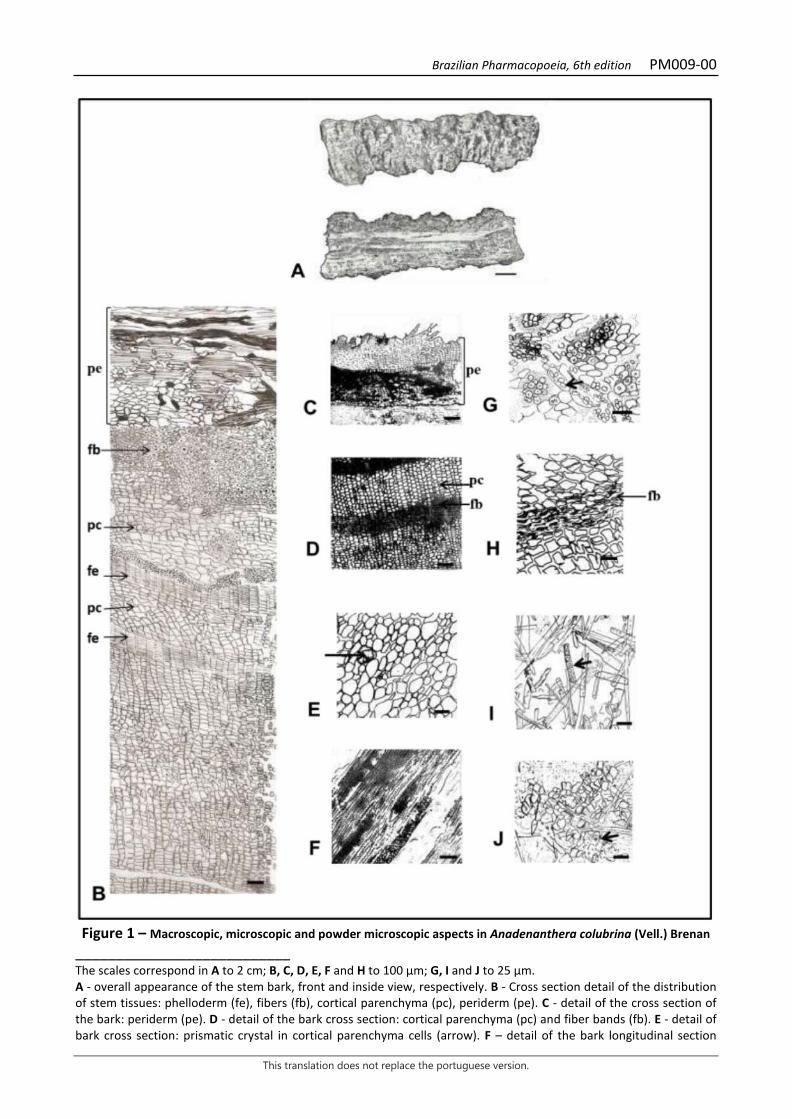
Brazilian Pharmacopoeia, 6th edition PM009-00
This translation does not replace the portuguese version.
Figure 1 – Macroscopic, microscopic and powder microscopic aspects in Anadenanthera colubrina (Vell.) Brenan
___________________________ The scales correspond in A to 2 cm; B, C, D, E, F and H to 100 µm; G, I and J to 25 µm. A - overall appearance of the stem bark, front and inside view, respectively. B - Cross section detail of the distribution of stem tissues: phelloderm (fe), fibers (fb), cortical parenchyma (pc), periderm (pe). C - detail of the cross section of the bark: periderm (pe). D - detail of the bark cross section: cortical parenchyma (pc) and fiber bands (fb). E - detail of bark cross section: prismatic crystal in cortical parenchyma cells (arrow). F – detail of the bark longitudinal section

Brazilian Pharmacopoeia, 6th edition PM009-00
This translation does not replace the portuguese version.
showing a parenchymal cord and a ray. G - detail of cross section in the region of the parenchymatic rays of the bark: starch grains in the cortical parenchyma cells (arrow). H - detail of the bark longitudinal section: fibers (fb). I and J - details noted in the powder. I - libriform fibers and parenchyma cells with prismatic crystals (arrow). J - stone cells (arrow).
Brazilian Pharmacopoeia, 6th edition PM010-00
This translation does not replace the portuguese version.
ANISE, fruit Anisi fructus
The plant drug consists of dried Pimpinella anisum L. fruits, containing at least 2% volatile oil, with at least 87% trans-anethole. COMMON NAMES Fennel. CHARACTERISTICS The plant drug has a pleasant, aniseed-like odor. IDENTIFICATION 1. Macroscopic description The fruit is a pyriform diachene, consisting of two laterally compressed mericarps, widened at the base and narrowed at the apex, 0.3 to 0.7 cm long and 0.2 to 0.3 cm wide; yellowish-brown or greenish-brown in color, the apex is crowned by a thick stylopodium, with two short diverging and reflective stylets, and the basal portion provided with a small fragment of the pedicel, slender, rigid and somewhat arched, which extends between the mericarps (achenes) of each fruit, through the carpophore (central filament), filiform and bifended. The mericarps (achenes), connected by the apex at the end of carpophore, feature a flat commissural face and a convex dorsal face, the latter covered with simple and short trichomes visible through a lens. Each mericarp is crossed lengthwise by five filiform, straight and smooth primary edges, three dorsal and two slightly prominent and lighter-toned commissural edges. In cross section, the two mericarps are almost always united by their commissural faces, leaving visible a continuous line of secretory ducts in the dorsal portion, and two larger secretory ducts on the commissural or ventral face. Vascular bundles occur at each dorsal edge. The endosperm is oily and slightly wavy on the commissural face. 2. Microscopic description In cross section, each mericarp shows an epicarp of one layer of cells, where numerous short, usually unicellular, conical, thick-walled tector trichomes with a verrucose cuticle are found. In front view, sparse stomata and a strongly striated cuticle are observed. The mesocarp is formed by a few layers of parenchyma, in which an almost continuous series of schizogenous secretory ducts can be distinguished, along the dorsal face; on the commissural face two wide secretory ducts occur. On the commissural face, narrow, longitudinally elongated sclereids with numerous pits are also found. Each edge contains a narrow vascular bundle surrounded by fibers. The endocarp is composed of a layer of thin-walled, tangentially elongated cells attached to the testa; the testa is formed by a layer of thicker, yellow or greenish-yellow, inner-walled cells. The endosperm has thick-walled polygonal cells, containing oil droplets, aleurone grains, and druse-like calcium oxalate microcrystals. The carpophore and pedicel are characterized by the presence of vessels and narrow fibers.
Brazilian Pharmacopoeia, 6th edition PM010-00
This translation does not replace the portuguese version.
3. Microscopic description of powder The sample meets all requirements for the species, except the macroscopic characters. Characteristics are: yellowish-brown or greenish-brown color; irregular pericarp fragments, showing portions of secretory ducts; single-celled, thick-walled, sometimes curved, entire or fragmented trichomes with attenuated tips and verrucous cuticle; epicarp fragments with striated cuticle and scarce stomata; vascular tissue fragments with spiral or annular vessel elements; thin-walled testa cells; endosperm fragments containing aleurone grains and calcium oxalate crystals; square, rectangular or elongated thick-walled, pitted sclereids; fiber strands of carpophore and pedicel. The powder contains no starch grains. 4. Proceed as described in Thin-layer chromatography (5.2.17.1). Stationary phase: silica gel GF254 (0.25 mm). Mobile phase: toluene Sample solution: use 0.1 g of pulverized dried fruits (1 400 µm), add 2 mL methyl chloride. Shake for 15 minutes. Filter. Concentrate the filtrate until dryness in a water bath at a temperature below 60 °C. Dissolve the residue in 0.5 mL toluene. Reference solution: dilute 3 μL trans-anethole and 40 μL olive oil in 1 mL toluene. Procedure: apply to the chromatoplate, separately, in the form of a band, 2 μL and 3 μL of the Sample solution and 1 μL, 2 μL and 3 μL of the Reference solution, with an 2 cm gap between each application. Conduct chromatogram. Remove chromatoplate and allow it to dry. Examine under ultraviolet light at 254 nm. Nebulize the plate with anisaldehyde RS and heat at 100 °C to 105 °C for five minutes. Results: the diagram below shows the sequences of zones obtained with the Reference solution and the Sample solution. Other zones may occasionally develop.
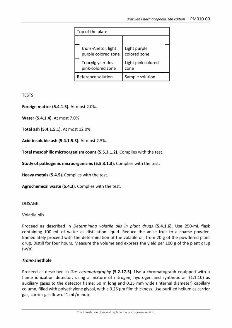
Brazilian Pharmacopoeia, 6th edition PM010-00
This translation does not replace the portuguese version.
Top of the plate
trans-Anetol: light purple colored zone
Light purple colored zone
Triacylglycerides: pink-colored zone
Light pink colored zone
Reference solution Sample solution TESTS Foreign matter (5.4.1.3). At most 2.0%. Water (5.4.1.4). At most 7.0% Total ash (5.4.1.5.1). At most 12.0%. Acid-insoluble ash (5.4.1.5.3). At most 2.5%. Total mesophilic microorganism count (5.5.3.1.2). Complies with the test. Study of pathogenic microorganisms (5.5.3.1.3). Complies with the test. Heavy metals (5.4.5). Complies with the test. Agrochemical waste (5.4.3). Complies with the test. DOSAGE Volatile oils Proceed as described in Determining volatile oils in plant drugs (5.4.1.6). Use 250-mL flask containing 100 mL of water as distillation liquid. Reduce the anise fruit to a coarse powder. Immediately proceed with the determination of the volatile oil, from 20 g of the powdered plant drug. Distill for four hours. Measure the volume and express the yield per 100 g of the plant drug (w/p). Trans-anethole Proceed as described in Gas chromatography (5.2.17.5). Use a chromatograph equipped with a flame ionization detector, using a mixture of nitrogen, hydrogen and synthetic air (1:1:10) as auxiliary gases to the detector flame; 60 m long and 0.25 mm wide (internal diameter) capillary column, filled with polyethylene glycol, with a 0.25 μm film thickness. Use purified helium as carrier gas; carrier gas flow of 1 mL/minute.
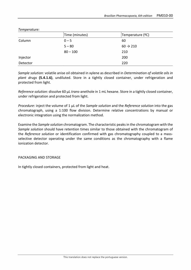
Brazilian Pharmacopoeia, 6th edition PM010-00
This translation does not replace the portuguese version.
Temperature: Time (minutes) Temperature (ºC) Column 0 – 5 60 5 – 80 60 → 210 80 – 100 210 Injector 200 Detector 220 Sample solution: volatile anise oil obtained in xylene as described in Determination of volatile oils in plant drugs (5.4.1.6), undiluted. Store in a tightly closed container, under refrigeration and protected from light. Reference solution: dissolve 60 μL trans-anethole in 1 mL hexane. Store in a tightly closed container, under refrigeration and protected from light. Procedure: inject the volume of 1 μL of the Sample solution and the Reference solution into the gas chromatograph, using a 1:100 flow division. Determine relative concentrations by manual or electronic integration using the normalization method. Examine the Sample solution chromatogram. The characteristic peaks in the chromatogram with the Sample solution should have retention times similar to those obtained with the chromatogram of the Reference solution or identification confirmed with gas chromatography coupled to a mass-selective detector operating under the same conditions as the chromatography with a flame ionization detector. PACKAGING AND STORAGE In tightly closed containers, protected from light and heat.

Brazilian Pharmacopoeia, 6th edition PM010-00
This translation does not replace the portuguese version.
Figure 1 – Macroscopic and microscopic aspects in Pimpinella anisum L.
___________________________ The scales correspond in A to 1 cm (level 1); in B to 2 cm (level 2); in C to 500 µm (level 4); in D and E to 100 µm (level 3). A – appearance of the diachene (fruit). B – schematic diagram of the cross section of the diachene as shown in A. C – schematic diagram of the cross section in one of the mericarps: schizogenous channel (e); hollow (o); seed (se). D – detail of the commissural region as marked in C; hollow (o); portion of the seed (s). E – detail of fruit and seed portion
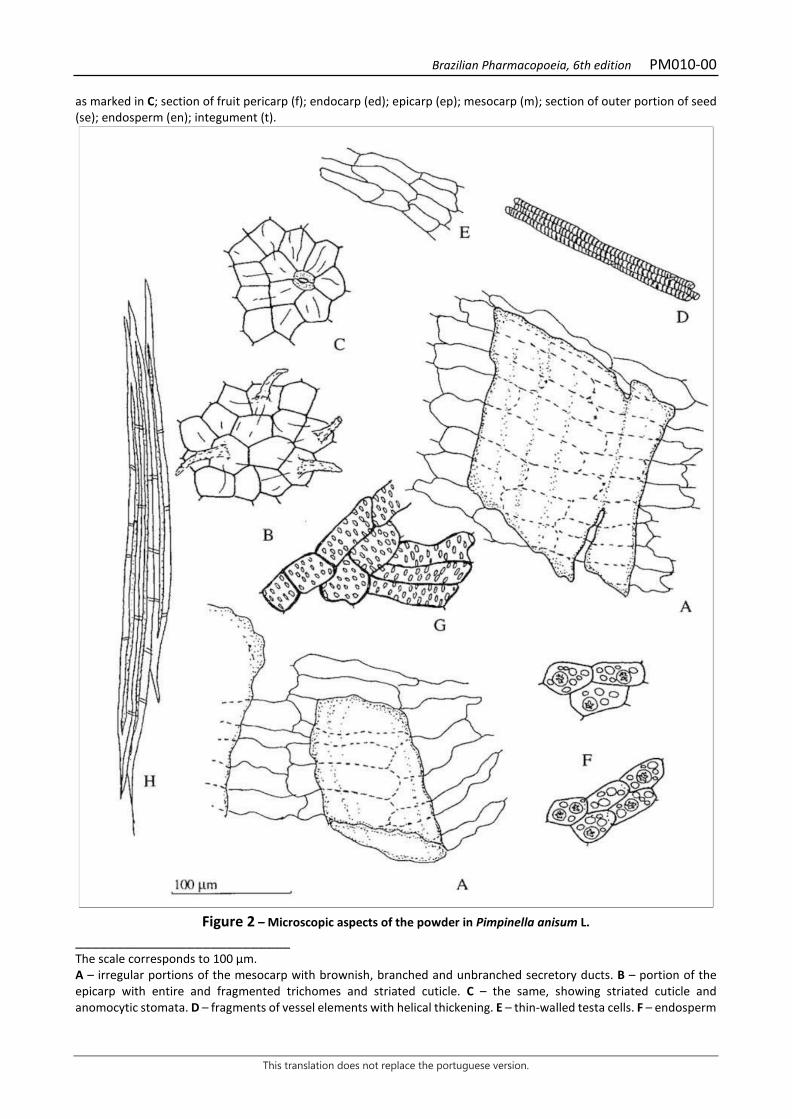
Brazilian Pharmacopoeia, 6th edition PM010-00
This translation does not replace the portuguese version.
as marked in C; section of fruit pericarp (f); endocarp (ed); epicarp (ep); mesocarp (m); section of outer portion of seed (se); endosperm (en); integument (t).
Figure 2 – Microscopic aspects of the powder in Pimpinella anisum L.
___________________________ The scale corresponds to 100 µm. A – irregular portions of the mesocarp with brownish, branched and unbranched secretory ducts. B – portion of the epicarp with entire and fragmented trichomes and striated cuticle. C – the same, showing striated cuticle and anomocytic stomata. D – fragments of vessel elements with helical thickening. E – thin-walled testa cells. F – endosperm

Brazilian Pharmacopoeia, 6th edition PM010-00
This translation does not replace the portuguese version.
fragments with polygonal cells containing oil drops and aleurone grains with 1-2 calcium oxalate druses. G – sclereids of the commissural face. H – fiber strands of the carpophore and pedicel.
Brazilian Pharmacopoeia, 6th edition PM011-00
This translation does not replace the portuguese version.
STAR ANISE, fruit Anisi stellati fructus
The plant drug consists of dried Illicium verum Hook. f. fruits, containing at least 7.0% volatile oil, with at least 80% trans-anethole. COMMON NAMES Badian, Chinese star anise. CHARACTERISTICS The pericarp of the plant drug has a pleasant aromatic odor and a sweet, aniseed-like flavor; the seed is odorless and has an unpleasant taste. IDENTIFICATION 1. Macroscopic description The fruit is multiple, composed of seven or eight follicles, sometimes up to 11, arranged horizontally in a star shape, around a central axis called the columella. The follicles are 1 to 2 cm long, grayish-brown in color, and are unevenly developed, woody, careniform, laterally flattened, ending in an obtuse, curved apex. The outer, lateral and lower face of each follicle is thick and rough, and the upper margin, called the ventral suture, is open in two thin, smooth lips on either side of the cleft of the carpel dehiscence, leaving its smooth, shiny, yellowish-brown inner face visible; the rough outer lateral faces have a smoother, lighter, semi-elliptical part near the base, through which the carpels are in contact with each other. Each follicle has a single oval seed, reddish-brown or yellowish-brown, hard and shiny, truncated at the base, where the hilum and the micropyle can be distinguished close to each other. The seed has a fragile integument and an oily endosperm, which surrounds a small embryo. 2. Microscopic description The epicarp, in s front view, shows polygonal, brown, irregular, thin-walled cells, with large, not very frequent, anomocytic stomata, and a cuticle with very pronounced irregular striations. In cross section, the mesocarp, just below the epicarp, shows a few layers of parenchyma of reddish-brown walled cells containing starch, and a few spherical, thin-walled oleiferous secretory idioblasts. Small intercellular spaces occur throughout the mesocarp. More internally, the mesocarp has parenchyma cells with reddish-brown walls, not lignified, but with numerous sclereids and astrosclereids, which also occur in the columella. The astrosclereids of the columella and mesocarp are very large and usually solitary; they can be irregularly branched or have shorter, tapered projections. Other mesocarp sclereids are found in groups, but are elongated, with thickened and pitted walls, and are called fibrosclereids. Also in the parenchyma of the mesocarp, numerous spherical oleiferous secretory idioblasts occur. The last layers of mesocarp cells are more compactly arranged, perpendicular to the rest of the mesocarp and also in relation to the endocarp. The endocarp is
Brazilian Pharmacopoeia, 6th edition PM011-00
This translation does not replace the portuguese version.
formed by a layer of radially elongated, palisade-arranged cells, 60 μm long on average. The seed’s integument is formed by distinct layers. The outer integument is represented by a hyaline tissue formed by two or three layers of cells, followed by a stratum of osteoskeletons, with radially elongated cells with thickened and pitted walls; this is followed by several layers of cells with lignified, thickened, and pitted walls, called macroskeletons, the inner layers being thin-walled; the inner integument is formed by a layer of cells with rhombohedral or rectangular calcium oxalate crystals. Brachysclereids occur in the micropylar zone. The endosperm consists of polygonal cells with aleurone grains with crystalloids and oil drops. The embryo is small. 3. Microscopic description of powder The sample meets all requirements for the species, except the macroscopic characters. Characteristics are: reddish-brown color; fragments formed by brown cells from the epicarp, with a strongly striated cuticle; fragments of parenchymatic cells from the mesocarp, with spherical, oleiferous secretory idioblasts; bulky, irregularly branched sclereids and astrosclereids from the mesocarp and columella; elongated fibrosclerids, arising from the mesocarp, with thickened and pitted walls; fragments formed by columnar cells from the endocarp, with slightly thickened walls, lignified, with pigments on the terminal walls; yellowish masses of small, very thick-walled, pitted cells from the ventral suture zone; isolated sclereids (osteosclereids, macrosclereids, and brachysclereids) from the seed integument; hyaline fragments from the outer seed integument; tabular or rhomboid calcium oxalate crystals; endosperm portions with aleurone grains containing crystalloids. 4. Adulterations It differs from Illicium anisatum L. (syn. Illicium religiosum Sieb. & Zucc.) in that the latter has smaller and more oval follicles, wider ventral suture and straight, non-claviform columella, and microscopically rare astrosclereids, which are not branched; the mesocarp sclereids are rounded, never elongated. 5. Proceed as described in Thin-layer chromatography (5.2.17.1). Stationary phase: silica gel GF254 (0.25 mm). Mobile phase: ethyl acetate, water, anhydrous formic acid and glacial acetic acid (100:26:11:11) Sample solution: heat, under reflux, 2 g of pulverized follicles (355 µm) (5.2.11), without seeds, with 10 mL methyl alcohol in a water bath at 60 °C for five minutes. Allow to cool and filter. Reference solution: dissolve 1 mg caffeic acid, 1 mg chlorogenic acid, 2.5 mg quercitrin, 2.5 mg rutin and 2.5 mg hyperoside, in 10 mL of methyl alcohol. Procedure: apply 5 μL of the sample solution and 5 μL of the Reference solution to the chromatoplate, separately and in the form of a band. Conduct chromatogram. Remove chromatoplate and allow it to air dry. Examine under ultraviolet light at 254 nm. Then nebulize the plate with 1% (w/v) SS aminoethanol diphenylborate (Natural Reagent A) in methyl alcohol. Examine under ultraviolet light at 365 nm.
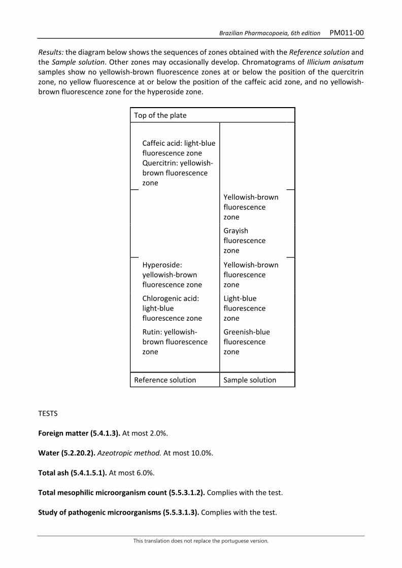
Brazilian Pharmacopoeia, 6th edition PM011-00
This translation does not replace the portuguese version.
Results: the diagram below shows the sequences of zones obtained with the Reference solution and the Sample solution. Other zones may occasionally develop. Chromatograms of Illicium anisatum samples show no yellowish-brown fluorescence zones at or below the position of the quercitrin zone, no yellow fluorescence at or below the position of the caffeic acid zone, and no yellowish-brown fluorescence zone for the hyperoside zone.
Top of the plate
Caffeic acid: light-blue fluorescence zone Quercitrin: yellowish-brown fluorescence zone
Yellowish-brown fluorescence zone
Grayish fluorescence zone
Hyperoside: yellowish-brown fluorescence zone
Yellowish-brown fluorescence zone
Chlorogenic acid: light-blue fluorescence zone
Light-blue fluorescence zone
Rutin: yellowish-brown fluorescence zone
Greenish-blue fluorescence zone
Reference solution Sample solution
TESTS Foreign matter (5.4.1.3). At most 2.0%. Water (5.2.20.2). Azeotropic method. At most 10.0%. Total ash (5.4.1.5.1). At most 6.0%. Total mesophilic microorganism count (5.5.3.1.2). Complies with the test. Study of pathogenic microorganisms (5.5.3.1.3). Complies with the test.
Brazilian Pharmacopoeia, 6th edition PM011-00
This translation does not replace the portuguese version.
Heavy metals (5.4.5). Complies with the test. Agrochemical waste (5.4.3). Complies with the test. DOSAGE Volatile oils Proceed as described in Determination of volatile oils in plant drugs (5.4.1.6). Use a 250-mL flask containing 100 mL of water as distillation liquid. Reduce the fruit to a coarse powder. Immediately proceed with the determination of the volatile oil, from 20 g of the pulverized plant drug. Distill for two hours. Measure the volume and express the yield per 100 g of the plant drug (w/p). Trans-anethole Proceed as described in Gas chromatography (5.2.17.5). Use a chromatograph equipped with a flame ionization detector, using a mixture of nitrogen, synthetic air, and hydrogen (1:1:10) as auxiliary gases to the detector flame; 30 m long and 250 μm wide (internal diameter) capillary column, coated with polydiphenyldimethylsiloxane, with a 0.25 µm film thickness. Use helium at a pressure of 80 kPa as carrier gas; carrier gas flow 1.0mL/minute. Temperature: Time (minutes) Temperature (ºC) Column 0 – 80 60→ 300 Injector 220 Detector 250
Sample solution: volatile oil and diethyl ether (2:100) mixture. Procedure: inject the volume of 1 μL of the Sample solution into the gas chromatograph, using a 1:50 flow division. Determine relative concentrations by manual or electronic integration. Trans-anethole has a linear retention time (Relative Retention Index) of 1277. PACKAGING AND STORAGE In tightly closed containers, protected from light and heat.
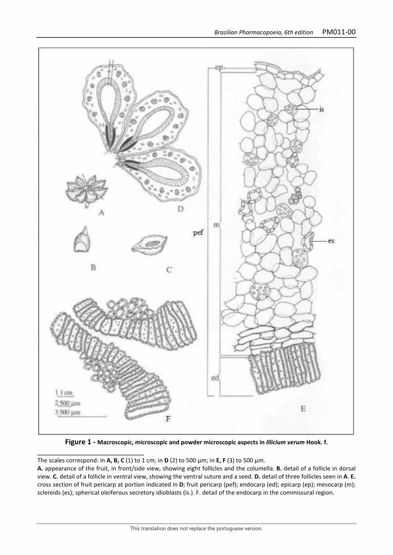
Brazilian Pharmacopoeia, 6th edition PM011-00
This translation does not replace the portuguese version.
Figure 1 - Macroscopic, microscopic and powder microscopic aspects in Illicium verum Hook. f.
___________________________ The scales correspond: in A, B, C (1) to 1 cm; in D (2) to 500 μm; in E, F (3) to 500 μm. A. appearance of the fruit, in front/side view, showing eight follicles and the columella. B. detail of a follicle in dorsal view. C. detail of a follicle in ventral view, showing the ventral suture and a seed. D. detail of three follicles seen in A. E. cross section of fruit pericarp at portion indicated in D; fruit pericarp (pef); endocarp (ed); epicarp (ep); mesocarp (m); sclereids (es); spherical oleiferous secretory idioblasts (is.). F. detail of the endocarp in the commissural region.
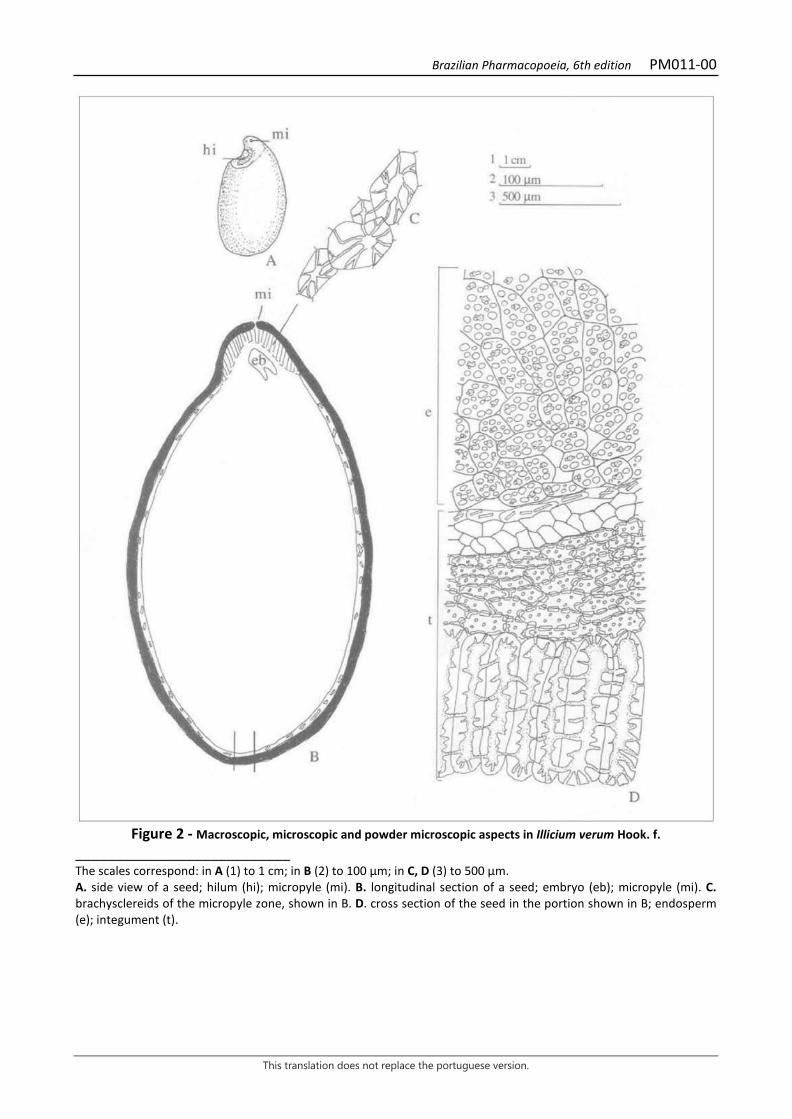
Brazilian Pharmacopoeia, 6th edition PM011-00
This translation does not replace the portuguese version.
Figure 2 - Macroscopic, microscopic and powder microscopic aspects in Illicium verum Hook. f.
___________________________ The scales correspond: in A (1) to 1 cm; in B (2) to 100 μm; in C, D (3) to 500 μm. A. side view of a seed; hilum (hi); micropyle (mi). B. longitudinal section of a seed; embryo (eb); micropyle (mi). C. brachysclereids of the micropyle zone, shown in B. D. cross section of the seed in the portion shown in B; endosperm (e); integument (t).
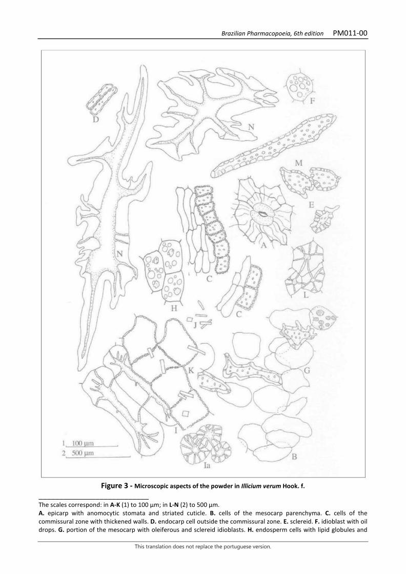
Brazilian Pharmacopoeia, 6th edition PM011-00
This translation does not replace the portuguese version.
Figure 3 - Microscopic aspects of the powder in Illicium verum Hook. f.
___________________________ The scales correspond: in A-K (1) to 100 μm; in L-N (2) to 500 μm. A. epicarp with anomocytic stomata and striated cuticle. B. cells of the mesocarp parenchyma. C. cells of the commissural zone with thickened walls. D. endocarp cell outside the commissural zone. E. sclereid. F. idioblast with oil drops. G. portion of the mesocarp with oleiferous and sclereid idioblasts. H. endosperm cells with lipid globules and
Brazilian Pharmacopoeia, 6th edition PM011-00
This translation does not replace the portuguese version.
aleurone grains. I. cross section of osteosclereids; the same in tangential section (Ia). J. prismatic calcium oxalate crystals. K. cells of the crystalliferous layer. L. brachysclereids of the commissural region. M. enlarged macrosclereid of the mesocarp, with thickened and pitted walls. N. bulky, branched sclereids of the pedicel.
Brazilian Pharmacopoeia, 6th edition PM012-00
This translation does not replace the portuguese version.
ARNICA, flower Arnicae flos
The plant drug consists of dried, entire or partially fragmented inflorescences of Arnica montana L., containing at least 0.4% (w/w) total lactone sesquiterpenes expressed as dihydrohelenalin tiglate (C20H26O5, 346,42). IDENTIFICATION 1. Macroscopic description The flowers are grouped in inflorescences of the heteromorphic capitulum type, yellow-orange in color. The capitulum consists of a peduncle, a receptacle, ligulate radial flowers, and tubular disc flowers. The closed capitulum measures about 2 cm in diameter and when with the radial flowers distended, it measures 5 to 6 cm in diameter. The peduncle, when present, is 2 to 3 cm long. The receptacle, when deprived of flowers, has a diameter of 6 to 10 mm and a depth of 15 mm and is slightly convex, honeycombed, and covered with short, hard, white trichomes. The receptacle is surrounded by 18 to 24 oval-lanceolate bracts. Each involucral bract has a sharp apex and a full, ciliated margin, measuring 8 to 10 mm, more rarely up to 15 mm; the inner bracts have a dull green coloration and are shorter; the outer bracts are green. The ligulate radial flowers are zygomorphic and female, 14 to 20 in number, and measure 20 to 30 mm. Each ligulate flower has a reduced calyx – papus – which is formed by a series of thick, stiff, whitish-yellow bristles, 4 to 8 mm long; the limb of the corolla is oblong, yellowish-orange and has seven to 10 parallel veins, culminating in three small and unequal lobes; the stamens are not fully developed and have free anthers and the ovary is infertile, narrow, grizzly-colored, 4 to 5 mm long. The tubular disk flowers are actinomorphic and perfect, much more numerous than the ligulate flowers, up to 15 mm long; each one has a reduced calyx – called papus – which is formed by a series of whitish-yellow rigid bristles, up to 8 mm long; the corolla is short, yellow-orange in color, about 8 mm long and has five triangular reflex lobes; the stamens are five, fertile and are welded by the anthers forming a tube; the thecae are ellipsoidal and the connective extends in a triangular scale, the ovary is infertile, narrow, grizzly-colored, 4 to 8 mm long, featuring four or five visible longitudinal edges. The fruits, when present, are grizzly achenes, crowned or not crowned by the papus. 2. Microscopic description The involucral bracts and flowers have tector and glandular trichomes on their abaxial surfaces, or rarely on the adaxial surfaces. Tector trichomes are unicellular or bicellular, with a longer, acute apical cell, or even multicellular, uniseriate, with three to 10 cells, of which one or some distal cells are longer, and the proximal cells are sometimes thick-walled. Glandular trichomes have a pluricellular, uni- or biseriate pedicel, with a globose, ovoid or claviform, uniseriate or biseriate, unicellular or pluricellular glandular head. Involucral bracts and flowers have anomocytic stomata on their abaxial surfaces. In front view, the abaxial surface epidermis of the involucral bracts has wavy anticlinal wall cells. The adaxial surface has polygonal to weakly wavy anticlinal wall cells and, in a cross section, a loose fundamental parenchyma, with vascular bundles corresponding to the veins of each bract. The papus-like bristles of the calyx are each composed of two to three rows of elongated, acute cells in the distal portion, and a greater number of rows of cells in the proximal portion. In a front view, the adaxial surface epidermis of the corolla of the ligulate flower has
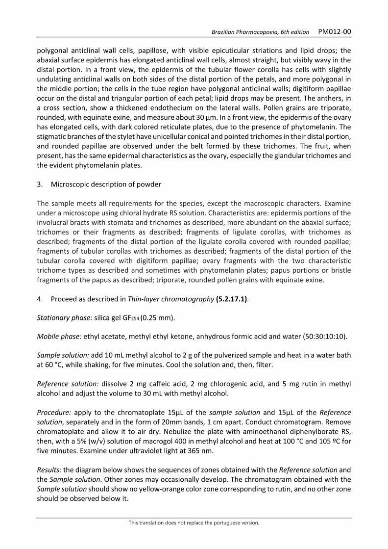
Brazilian Pharmacopoeia, 6th edition PM012-00
This translation does not replace the portuguese version.
polygonal anticlinal wall cells, papillose, with visible epicuticular striations and lipid drops; the abaxial surface epidermis has elongated anticlinal wall cells, almost straight, but visibly wavy in the distal portion. In a front view, the epidermis of the tubular flower corolla has cells with slightly undulating anticlinal walls on both sides of the distal portion of the petals, and more polygonal in the middle portion; the cells in the tube region have polygonal anticlinal walls; digitiform papillae occur on the distal and triangular portion of each petal; lipid drops may be present. The anthers, in a cross section, show a thickened endothecium on the lateral walls. Pollen grains are triporate, rounded, with equinate exine, and measure about 30 μm. In a front view, the epidermis of the ovary has elongated cells, with dark colored reticulate plates, due to the presence of phytomelanin. The stigmatic branches of the stylet have unicellular conical and pointed trichomes in their distal portion, and rounded papillae are observed under the belt formed by these trichomes. The fruit, when present, has the same epidermal characteristics as the ovary, especially the glandular trichomes and the evident phytomelanin plates. 3. Microscopic description of powder The sample meets all requirements for the species, except the macroscopic characters. Examine under a microscope using chloral hydrate RS solution. Characteristics are: epidermis portions of the involucral bracts with stomata and trichomes as described, more abundant on the abaxial surface; trichomes or their fragments as described; fragments of ligulate corollas, with trichomes as described; fragments of the distal portion of the ligulate corolla covered with rounded papillae; fragments of tubular corollas with trichomes as described; fragments of the distal portion of the tubular corolla covered with digitiform papillae; ovary fragments with the two characteristic trichome types as described and sometimes with phytomelanin plates; papus portions or bristle fragments of the papus as described; triporate, rounded pollen grains with equinate exine. 4. Proceed as described in Thin-layer chromatography (5.2.17.1). Stationary phase: silica gel GF254 (0.25 mm). Mobile phase: ethyl acetate, methyl ethyl ketone, anhydrous formic acid and water (50:30:10:10). Sample solution: add 10 mL methyl alcohol to 2 g of the pulverized sample and heat in a water bath at 60 °C, while shaking, for five minutes. Cool the solution and, then, filter. Reference solution: dissolve 2 mg caffeic acid, 2 mg chlorogenic acid, and 5 mg rutin in methyl alcohol and adjust the volume to 30 mL with methyl alcohol. Procedure: apply to the chromatoplate 15μL of the sample solution and 15μL of the Reference solution, separately and in the form of 20mm bands, 1 cm apart. Conduct chromatogram. Remove chromatoplate and allow it to air dry. Nebulize the plate with aminoethanol diphenylborate RS, then, with a 5% (w/v) solution of macrogol 400 in methyl alcohol and heat at 100 °C and 105 ºC for five minutes. Examine under ultraviolet light at 365 nm. Results: the diagram below shows the sequences of zones obtained with the Reference solution and the Sample solution. Other zones may occasionally develop. The chromatogram obtained with the Sample solution should show no yellow-orange color zone corresponding to rutin, and no other zone should be observed below it.
Brazilian Pharmacopoeia, 6th edition PM012-00
This translation does not replace the portuguese version.
Top of the plate
Caffeic acid: light-blue fluorescence zone
Greenish-blue fluorescence zone Yellowish-brown to orangish-yellow fluorescence zone
Yellowish-brown to orangish-yellow fluorescence zone Yellowish-brown to orangish-yellow fluorescence zone Yellowish-brown to orangish-yellow fluorescence zone
Chlorogenic acid: blueish fluorescence zone
Blueish fluorescence zone
Rutin: yellowish-orange fluorescence zone
Greenish-blue fluorescence zone
Reference solution Sample solution TESTS Loss by drying (5.2.9.1). Gravimetric method. At most 10.0%. Foreign matter (5.4.1.3). At most 5.0% stems with a diameter greater than 5 mm. Total ash (5.4.1.5.1). At most 10.0%. Total mesophilic microorganism count (5.5.3.1.2). Complies with the test.
Brazilian Pharmacopoeia, 6th edition PM012-00
This translation does not replace the portuguese version.
Study of pathogenic microorganisms (5.5.3.1.3). Complies with the test. Heavy metals (5.4.5). Complies with the test. Agrochemical waste (5.4.3). Complies with the test. DOSAGE Total lactone sesquiterpene content Proceed as described in High Performance Liquid Chromatography (5.2.17.4). Use a chromatograph equipped with an ultraviolet detector at 225 nm; 0.12 m long, 4 mm internal diameter column, packed with octadecylsilane silica (4 μm), Mobile phase flow rate of 1.2 mL/minute. Eluent (A): water. Eluent (B): methyl alcohol. Mobile phase gradient: adopt the gradient system described on the following table:
Time (minutes) Eluent A (%) Eluent B (%) Elution
0-3 62 38 isocratic 3-20 62 → 55 38 → 45 linear gradient 20-30 55 45 isocratic 30-55 55 → 45 45 → 55 linear gradient 55-57 45 → 0 55 → 100 linear gradient 57-70 0 → 62 100 → 38 linear gradient 70-90 62 38 isocratic
Internal standard solution: dissolve 0.01 g of exactly weighed santonin in 10 mL methyl alcohol, immediately before use. Sample solution: into a 250-mL round-bottomed flask, introduce 1 g of the pulverized sample. Add 50 mL of an equal volume mixture of methyl alcohol and water and heat, under reflux, in a water bath between 50 °C and 60 °C for 30 minutes, frequently shaking. Allow to cool and filter using a paper filter. Transfer the filter cut into large pieces and the residue to the round-bottomed flask, add 50 mL of an equal volume mixture of methyl alcohol and water, and heat under reflux in a water bath between 50 ºC and 60 ºC for 30 minutes, frequently shaking. Repeat the operation twice. Combine the filtrates, add 3 mL of the Internal standard solution and evaporate under reduced pressure to a 18-mL volume. Rinse the round-bottomed flask with water and top off 20 mL with the wash waters. Transfer the solution to a chromatography column about 0.15 m long and about 30 mm wide (internal diameter), containing 15 g of chromatography silica. Allow to stand for 15 minutes, then elute with 200 mL of an equal volume mixture of ethyl acetate and methyl chloride. Evaporate the eluate until it dries in a 250-mL round-bottomed flask. Dissolve the residue in 10 mL methyl alcohol, add 10 mL of water, and then 7 g of neutral aluminum oxide. Shake for two minutes,
Brazilian Pharmacopoeia, 6th edition PM012-00
This translation does not replace the portuguese version.
centrifuge for 10 minutes at 6,000 × g and filter using paper filter. Evaporate 10 mL of the filtrate until dried. Dissolve the residue in 3 mL of an equal volume mixture of methyl alcohol and water and filter. Filter through a 0.45 µm filter unit. Procedure: separately inject 20 μL of the Internal standard solution and 20 µL of the Sample solution. Calculate total lactone sesquiterpenes content, expressed as a percentage of helenalin tiglate, according to the expression:
𝑇𝑇𝑇𝑇𝑇𝑇𝑇𝑇 =𝑇𝑇𝑎𝑎 × 𝐶𝐶𝑟𝑟 × V × 1,187
𝑇𝑇𝑟𝑟 × 𝑚𝑚𝑎𝑎 × 10
in which, TSLT = total lactone sesquiterpene content expressed as helenalin tiglate % (w/w); Aa = total area under the peaks corresponding to the lactone sesquiterpenes that appear after the santonin peak in the Sample solution; Aa = area under the peak corresponding to the santonin acid in the Sample solution; m = mass in grams of the sample used, considering the loss by drying. Cr = concentration of santonin in the Internal standard solution in mg/mL, considering the purity of the reference substance; V = volume in milliliters of the Internal Standard Solution used in the Sample solution; 1.187 = correction factor between dihydrohelenaline tiglate and santonin PACKAGING AND STORAGE In tightly closed containers, protected from light and heat.
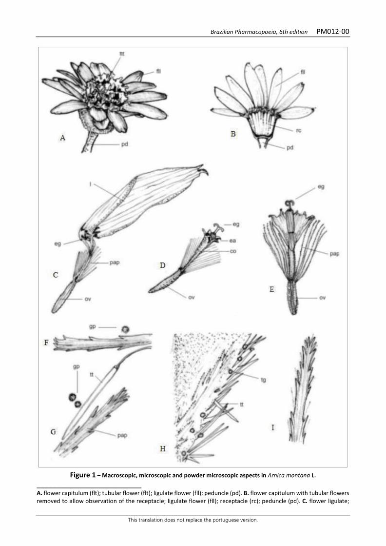
Brazilian Pharmacopoeia, 6th edition PM012-00
This translation does not replace the portuguese version.
Figure 1 – Macroscopic, microscopic and powder microscopic aspects in Arnica montana L.
___________________________ A. flower capitulum (flt); tubular flower (flt); ligulate flower (fll); peduncle (pd). B. flower capitulum with tubular flowers removed to allow observation of the receptacle; ligulate flower (fll); receptacle (rc); peduncle (pd). C. flower ligulate;
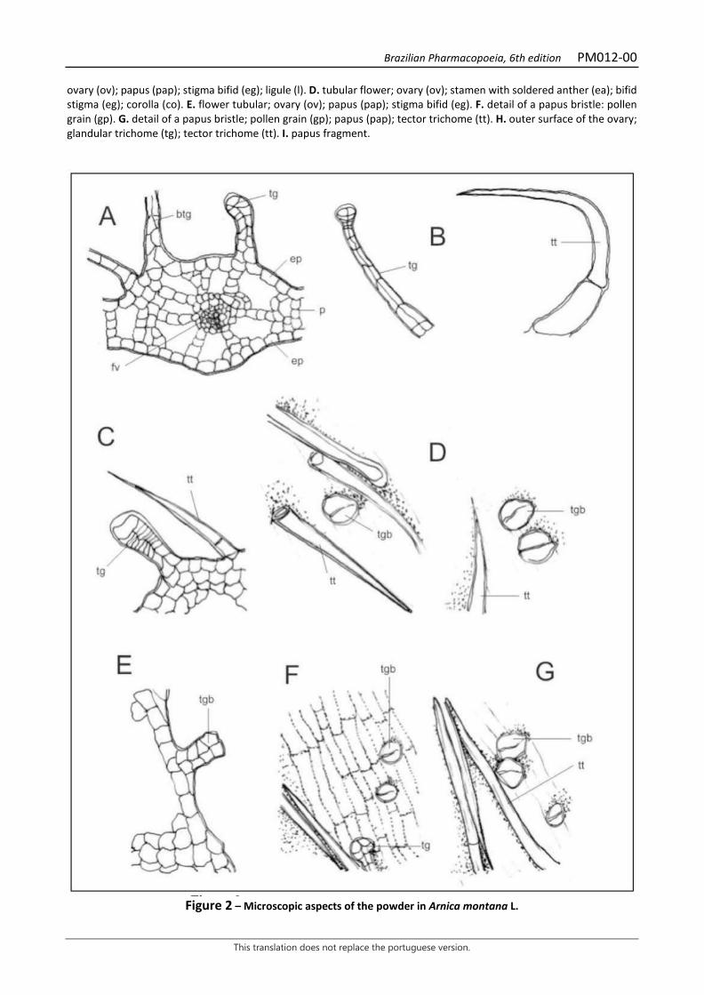
Brazilian Pharmacopoeia, 6th edition PM012-00
This translation does not replace the portuguese version.
ovary (ov); papus (pap); stigma bifid (eg); ligule (l). D. tubular flower; ovary (ov); stamen with soldered anther (ea); bifid stigma (eg); corolla (co). E. flower tubular; ovary (ov); papus (pap); stigma bifid (eg). F. detail of a papus bristle: pollen grain (gp). G. detail of a papus bristle; pollen grain (gp); papus (pap); tector trichome (tt). H. outer surface of the ovary; glandular trichome (tg); tector trichome (tt). I. papus fragment.
Figure 2 – Microscopic aspects of the powder in Arnica montana L.

Brazilian Pharmacopoeia, 6th edition PM012-00
This translation does not replace the portuguese version.
___________________________ A – bract cross section: epidermis (ep); parenchyma (p); vascular bundle (fv); glandular trichome (tg); base of glandular trichome (btg). B and C – details of glandular and vector trichomes: glandular trichome (tg); tector trichome (tt). D – outer surface of the ovary seen from above: glandular trichome with bicellular head (tgb), with a biseriate body. E – appearance of glandular thricomes. F and G – fragment of lower epidermis: glandular trichome (tg); glandular trichome with bicellular head (tgb).
Brazilian Pharmacopoeia, 6th edition PM013-00
This translation does not replace the portuguese version.
BRAZILIAN PEPPERTREE, bark Schinus terebinthifolii cortex
The plant drug consists of dried barks from the stems of Schinus terebinthifolia Raddi, containing at least 8% total tannins, at least 0.20% gallic acid (C7H6O5, 170.12), and at least 0.65% catechin (C15H14O6, 290,27). IDENTIFICATION 1. Macroscopic description The dried stem barks are slightly curved fragments, rigid and slightly brittle, 10 to 15 cm long, 2 to 2.5 cm wide, and 0.2 to 0.5 cm thick. Externally, the bark has a rough rhytidome, marked by irregular cracks, with a grayish-grizzly color and whitish spots, due to the presence of lichens. Internally the fragments are reddish-grizzly, resinous in appearance, and have longitudinal striations. 2. Microscopic description In cross section, the cortex shows a suber composed of approximately 15 layers of radially flattened cells, with visible lenticels, followed by the periderm of indistinct phellogen and phelloderm, in whose cortical parenchymatic cells several sclerenchymatic fiber caps and secretory ducts are visible. It is possible to observe the parenchyma rays crossing the entire thickness of the cortex. Cells containing starch grains occur Around the secretory ducts. In a radial longitudinal section, the suber is visible with radially flattened cells, cortical parenchyma cells are irregularly shaped and sclerenchyma fibers are present. Bands of parenchyma cells alternate with phloem bands, the latter with abundant fibers, which hinder spotting the phloem cells. The rays are heterocellular, consisting of procumbent parenchyma cells, which are elongated in the radial direction, and erect cells located on the upper and lower margins of the ray. The cells of the parenchyma bands are mostly elongated lengthwise and some are rounded. In the phloem it is possible to observe parenchyma cells containing prismatic crystals. In a tangential longitudinal section, rays are biseriate and the secretory ducts are branched. 3. Microscopic description of powder The sample meets all requirements for the species, except the macroscopic characters. Characteristics are: brown color; presence of stone cells; presence of suber fragments; prismatic crystals and parenchyma fragments. 4. Proceed as described in Thin-layer chromatography (5.2.17.1). Stationary phase: 250 µm thick silica gel F254. Mobile phase: ethyl acetate, formic acid and water (90:5:5). Sample solution: weigh 1 g of the pulverized plant drug and place in a round-bottomed flask, adding 10 mL of methyl alcohol. Heat under reflux for 10 minutes. Filter through absorbent cotton.
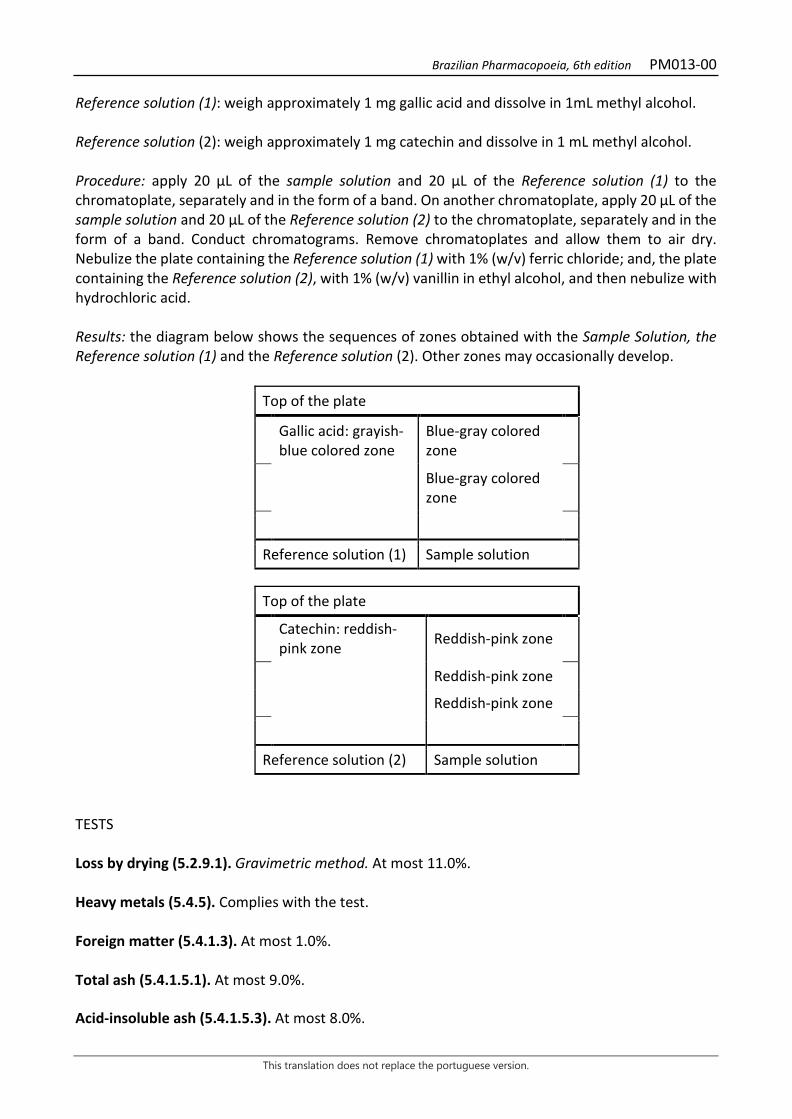
Brazilian Pharmacopoeia, 6th edition PM013-00
This translation does not replace the portuguese version.
Reference solution (1): weigh approximately 1 mg gallic acid and dissolve in 1mL methyl alcohol. Reference solution (2): weigh approximately 1 mg catechin and dissolve in 1 mL methyl alcohol. Procedure: apply 20 μL of the sample solution and 20 μL of the Reference solution (1) to the chromatoplate, separately and in the form of a band. On another chromatoplate, apply 20 μL of the sample solution and 20 μL of the Reference solution (2) to the chromatoplate, separately and in the form of a band. Conduct chromatograms. Remove chromatoplates and allow them to air dry. Nebulize the plate containing the Reference solution (1) with 1% (w/v) ferric chloride; and, the plate containing the Reference solution (2), with 1% (w/v) vanillin in ethyl alcohol, and then nebulize with hydrochloric acid. Results: the diagram below shows the sequences of zones obtained with the Sample Solution, the Reference solution (1) and the Reference solution (2). Other zones may occasionally develop.
Top of the plate
Gallic acid: grayish-blue colored zone
Blue-gray colored zone
Blue-gray colored zone
Reference solution (1) Sample solution
Top of the plate
Catechin: reddish-pink zone Reddish-pink zone
Reddish-pink zone
Reddish-pink zone
Reference solution (2) Sample solution TESTS Loss by drying (5.2.9.1). Gravimetric method. At most 11.0%. Heavy metals (5.4.5). Complies with the test. Foreign matter (5.4.1.3). At most 1.0%. Total ash (5.4.1.5.1). At most 9.0%. Acid-insoluble ash (5.4.1.5.3). At most 8.0%.
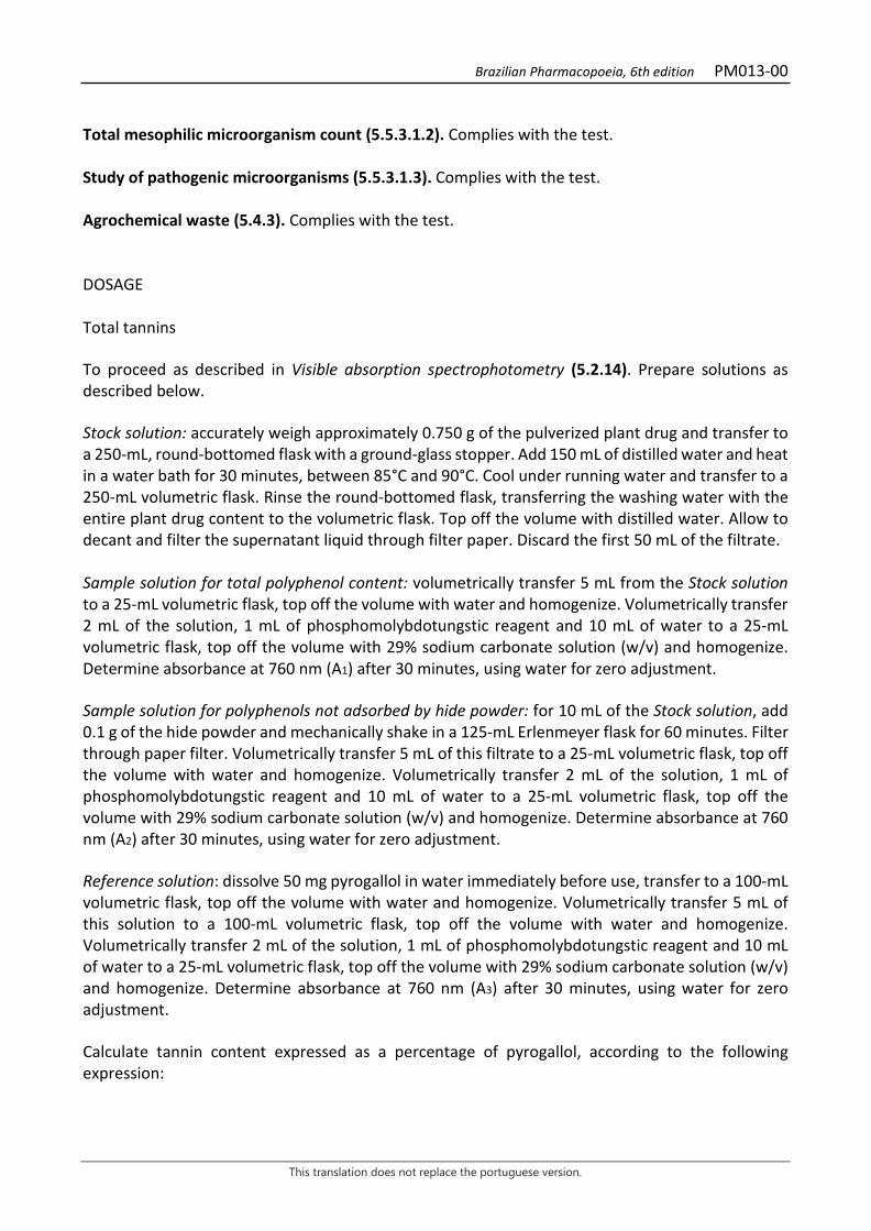
Brazilian Pharmacopoeia, 6th edition PM013-00
This translation does not replace the portuguese version.
Total mesophilic microorganism count (5.5.3.1.2). Complies with the test. Study of pathogenic microorganisms (5.5.3.1.3). Complies with the test. Agrochemical waste (5.4.3). Complies with the test. DOSAGE Total tannins To proceed as described in Visible absorption spectrophotometry (5.2.14). Prepare solutions as described below. Stock solution: accurately weigh approximately 0.750 g of the pulverized plant drug and transfer to a 250-mL, round-bottomed flask with a ground-glass stopper. Add 150 mL of distilled water and heat in a water bath for 30 minutes, between 85°C and 90°C. Cool under running water and transfer to a 250-mL volumetric flask. Rinse the round-bottomed flask, transferring the washing water with the entire plant drug content to the volumetric flask. Top off the volume with distilled water. Allow to decant and filter the supernatant liquid through filter paper. Discard the first 50 mL of the filtrate. Sample solution for total polyphenol content: volumetrically transfer 5 mL from the Stock solution to a 25-mL volumetric flask, top off the volume with water and homogenize. Volumetrically transfer 2 mL of the solution, 1 mL of phosphomolybdotungstic reagent and 10 mL of water to a 25-mL volumetric flask, top off the volume with 29% sodium carbonate solution (w/v) and homogenize. Determine absorbance at 760 nm (A1) after 30 minutes, using water for zero adjustment. Sample solution for polyphenols not adsorbed by hide powder: for 10 mL of the Stock solution, add 0.1 g of the hide powder and mechanically shake in a 125-mL Erlenmeyer flask for 60 minutes. Filter through paper filter. Volumetrically transfer 5 mL of this filtrate to a 25-mL volumetric flask, top off the volume with water and homogenize. Volumetrically transfer 2 mL of the solution, 1 mL of phosphomolybdotungstic reagent and 10 mL of water to a 25-mL volumetric flask, top off the volume with 29% sodium carbonate solution (w/v) and homogenize. Determine absorbance at 760 nm (A2) after 30 minutes, using water for zero adjustment. Reference solution: dissolve 50 mg pyrogallol in water immediately before use, transfer to a 100-mL volumetric flask, top off the volume with water and homogenize. Volumetrically transfer 5 mL of this solution to a 100-mL volumetric flask, top off the volume with water and homogenize. Volumetrically transfer 2 mL of the solution, 1 mL of phosphomolybdotungstic reagent and 10 mL of water to a 25-mL volumetric flask, top off the volume with 29% sodium carbonate solution (w/v) and homogenize. Determine absorbance at 760 nm (A3) after 30 minutes, using water for zero adjustment. Calculate tannin content expressed as a percentage of pyrogallol, according to the following expression:

Brazilian Pharmacopoeia, 6th edition PM013-00
This translation does not replace the portuguese version.
𝑇𝑇𝑇𝑇 =(𝑇𝑇1 − 𝑇𝑇2) × 𝑚𝑚2 × 62,5
𝑇𝑇3 × 𝑚𝑚1
in which, TT = total tannin content expressed as pyrogallol % (w/w); A1 = absorbance measured for the Sample solution for total polyphenols; A2 = absorbance measured for the Sample solution for polyphenols not adsorbed by hide powder; A3 = absorbance measured for the Reference solution; m1 = mass in grams of the sample used, considering the loss by drying. m2 = mass in grams of pyrogallol, considering the purity of the reference substance. Gallic acid and catechin Proceed as described in High Performance Liquid Chromatography (5.2.17.4). Use a chromatograph equipped with an ultraviolet detector at 280 mm; pre-column packed with octadecylsilane silica, 250 mm long, 4.6 mm internal diameter column, packed with silica chemically bonded to an octadecylsilane group (5 µm), kept at 24 °C; Mobile phase flow rate of 0.8 mL/minute. Eluent (A): 0.05% trifluoroacetic acid (v/v). Eluent (B): 0.05% trifluoroacetic acid in methyl alcohol (v/v).
Time (minutes) Eluent A (%) Eluent B (%) Elution
0-10 90 → 75 10 → 25 linear gradient 10-20 75 → 60 25 → 40 linear gradient 20-25 60 → 25 40 → 75 linear gradient 25-28 25 → 90 75 → 10 linear gradient 28-30 90 10 isocratic Sample solution: accurately weigh approximately 0.750 g of the pulverized plant drug and transfer to a 250-mL, round-bottomed flask with a ground-glass stopper. Add 150 mL of water. Heat in a water bath for 30 minutes, between 85°C and 90°C. Cool under running water and transfer to a 250-mL volumetric flask. Rinse the round-bottomed flask and transfer the washing water with the entire plant drug content to the volumetric flask. Top off the volume with water. Allow to decant and filter the supernatant liquid through filter paper. Discard the first 50 mL of the filtrate. Filter through a 0.45 µm filter unit. Reference solution (1): dissolve an accurately weighed amount of gallic acid in water to obtain a 6.48 µg/mL solution. Filter through a 0.45 µm filter unit. Reference solution (2): dissolve an accurately weighed amount of catechin in water to obtain a 18 µg/mL solution. Filter through a 0.45 µm filter unit. Procedure: separately inject 20µL of the Reference solution (1), 20µL of the Reference solution (2) and 20µL of the Sample Solution. Register the chromatograms and measure the areas under the

Brazilian Pharmacopoeia, 6th edition PM013-00
This translation does not replace the portuguese version.
peaks. Gallic acid and catechin retention time in the sample is approximately 9.9 and 17.7 minutes, respectively. Calculate the gallic acid and catechin contents, in percent, separately, according to the following expression:
𝑇𝑇𝐶𝐶 =𝐶𝐶𝑟𝑟 × 𝑇𝑇𝑎𝑎 × 250 × 100
𝑇𝑇𝑟𝑟 × 𝑚𝑚
in which, TC = gallic acid or catechin content % (w/w); Cr = concentration of the Reference solution (1) or (2) in g/mL, considering the purity of the reference substance; Ar = area under the peak corresponding to the gallic acid or the catechin in Reference Solution (1) or (2), respectively; Aa = area under the peak corresponding to the gallic acid or catechin acid in the Sample solution; m = mass in grams of the sample used, considering the loss by drying. 250 = dilution factor; PACKAGING AND STORAGE In tightly closed containers, protected from light and heat.
Brazilian Pharmacopoeia, 6th edition PM013-00
This translation does not replace the portuguese version.
Figure 1 – Macroscopic, microscopic and powder microscopic aspects in Schinus terebinthifolia Raddi
___________________________ The scales correspond in A to 4 cm; B to 25 µm, C and J to 100 µm; D, F, G and I to 200 µm; and E and H to 50 µm. A - overall appearance of the stem bark, front and inside view, respectively. B - cross section detail of the distribution of the stem bark tissues: lenticels (le); periderm (pe); cortical parenchyma cell (pc); secretory duct (cs); phloem (fl);
Brazilian Pharmacopoeia, 6th edition PM013-00
This translation does not replace the portuguese version.
crystalliferous idioblast (ic). C - detail of the bark cross section, showing the periderm; cortical parenchyma (pc). D - detail of the bark cross section, showing the phloem, the cortical parenchyma band, parenchymatic rays and the secretory duct. E - detail of the bark cross section, showing a secretory duct in the cortical region. F - detail of the bark longitudinal section, with bands of cortical and phloem parenchyma with abundant fibers. G - detail of the bark longitudinal section, with parenchymal bands showing elongated and some rounded cells. H - detail of the bark radial longitudinal section, showing the periderm (pe), secretory duct (cs) and parenchyma cells of the rays (rp). I - detail of the bark radial longitudinal section with prismatic crystals in phloem cells; crystalliferous idioblast (ic). J - stone cell (arrow).

Brazilian Pharmacopoeia, 6th edition PM014-00
This translation does not replace the portuguese version.
ALOE, leaf Aloe vera folium
The plant drug consists of the colorless, mucilaginous gel obtained from the parenchymal cells of fresh leaves of Aloe vera (L.) Burm. f. containing at least 0.3% total carbohydrates. CHARACTERISTICS The plant drug has a slightly bitter taste, and is colorless and odorless. IDENTIFICATION 1. Macroscopic description Succulent, lanceolate, acute, glaucous-green leaves, with whitish spots when young, 15 to 60 cm long and about 7 cm wide at the base on the adaxial surface and 10 cm on the abaxial surface when adult. The adaxial surface, seen in cross section, is concave and the abaxial surface is convex. The leaf margins are dentate and spiny, with small whitish aculeus perpendicular to the lamina. 2. Microscopic description The leaf, in a cross section, shows an isobilateral structure and is amphistomatic, with several stomata, of the tetracyclic type. It has a single epidermal layer, covered by a thick, wavy cuticle. The cross section of the leaf lamina shows two distinct zones: the outermost green zone, corresponding to the chlorenchyma; and the innermost, colorless and mucilaginous zone, corresponding to the aquiferous parenchyma. Below the epidermis a distinct first layer of palisading chlorenchymal cells may occur, followed by 10 to 18 layers of chlorenchymal cells, rich in starch, and idioblasts containing bundles of calcium oxalate raphids. In the contact zone between the chlorenchyma and the aquiferous parenchyma collateral vascular bundles occur, parallel to the epidermis, alternated with three to five chlorenchyma cells. The upper portion of each bundle is in contact with the chlorenchyma, and the middle and lower portions penetrate the aquiferous parenchyma. Vascular bundles are surrounded by a parenchymatic sheath containing starch. Internal to this layer and close to the phloem a cluster of three to five very large cells is found, as well as other smaller polyhedral cells, slightly elongated towards the leaf axis, and thin-walled – alloetic cells – filled with yellow, viscous latex, known as alloetic liquid or aloe juice. At the moment the leaf is cross-sectioned, the alloetic liquid from each bundle is released. The phloem is external and poorly developed, and the xylem consists of two to four tracheal elements with some fibers. The aquiferous parenchyma usually occupies 75% of the thickness of the lamina, and is formed by very large, colorless, thin-walled, mucilage-filled cells arranged perpendicular to the epidermis. Idioblasts containing calcium oxalate raphids also occur in this parenchyma. 3. Proceed as described in Thin-layer chromatography (5.2.17.1). Stationary phase: silica gel GF254 (0.25 mm). Mobile phase: toluene and ethyl acetate (90:10).
Brazilian Pharmacopoeia, 6th edition PM014-00
This translation does not replace the portuguese version.
Sample solution: transfer 2 mL of liquid aloe gel into a 5-mL volumetric flask, top off the volume with methyl alcohol, and heat in a water bath at 60 °C, while shaking, for 10 minutes. Reference solution: dissolve 2 mg -sitosterol in 1 mL methyl alcohol. Procedure: apply 20 μL of the sample solution and 10 μL of the Reference solution to the chromatoplate, separately and in the form of a band. Conduct chromatogram. Remove chromatoplate and allow it to air dry. Nebulize the plate with anisaldehyde RS in an oven at 100 °C to 105 °C for five to ten minutes. Results: the diagram below shows the sequences of zones obtained with the Reference solution and the Sample solution. Other zones may occasionally develop.
Top of the plate
Corn oil: blue-colored zone Blue-colored zone
Reference solution Sample solution
TESTS Total mesophilic microorganism count (5.5.3.1.2). Complies with the test. Study of pathogenic microorganisms (5.5.3.1.3). Complies with the test. Heavy metals (5.4.5). Complies with the test. Agrochemical waste (5.4.3). Complies with the test. DOSAGE Total carbohydrates To proceed as described in Visible absorption spectrophotometry (5.2.14). Prepare solutions as described below. Stock solution: transfer 3 mL of liquid aloe gel to a 100-mL volumetric flask, top off the volume with water. Turbolize for five minutes. Sample solution: transfer 0.2 mL of the Stock solution to a test tube, to off to 0.5 mL with water, and leave in an ice bath. Add 0.5 mL of 5% phenol solution (w/v) and 2 mL of concentrated sulfuric acid. Shake well and allow to stand at room temperature for 30 minutes.
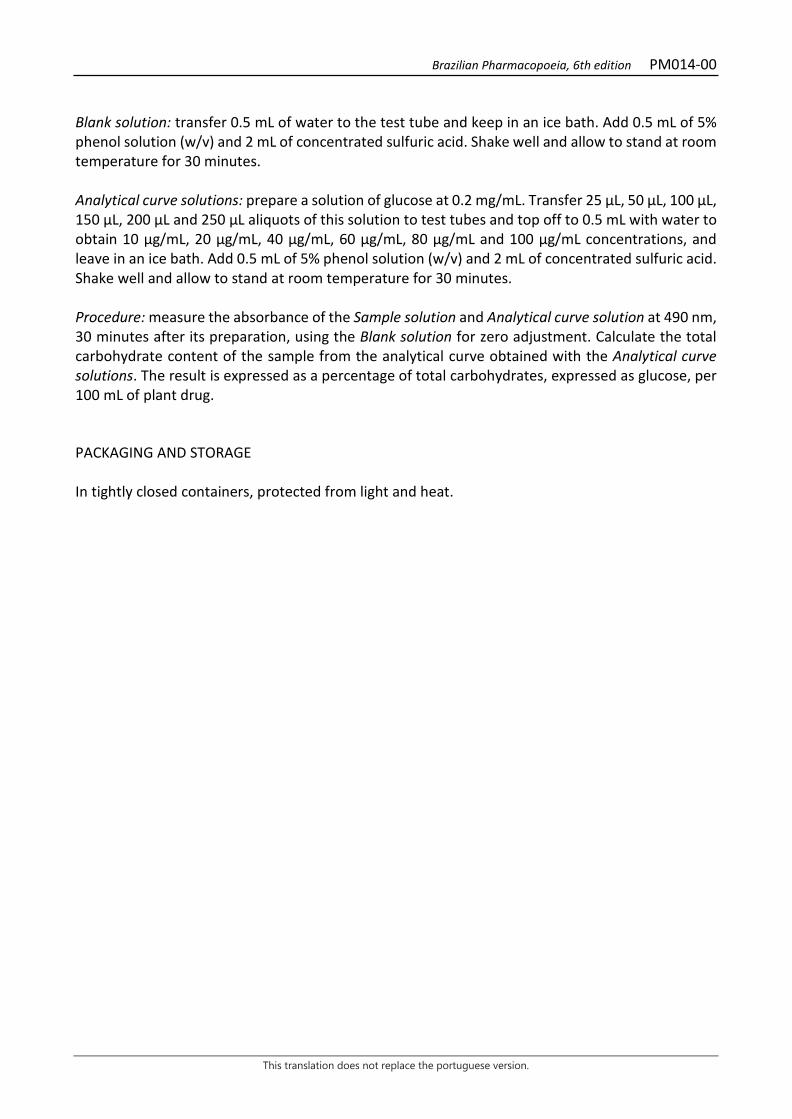
Brazilian Pharmacopoeia, 6th edition PM014-00
This translation does not replace the portuguese version.
Blank solution: transfer 0.5 mL of water to the test tube and keep in an ice bath. Add 0.5 mL of 5% phenol solution (w/v) and 2 mL of concentrated sulfuric acid. Shake well and allow to stand at room temperature for 30 minutes. Analytical curve solutions: prepare a solution of glucose at 0.2 mg/mL. Transfer 25 μL, 50 μL, 100 μL, 150 μL, 200 μL and 250 μL aliquots of this solution to test tubes and top off to 0.5 mL with water to obtain 10 µg/mL, 20 µg/mL, 40 µg/mL, 60 µg/mL, 80 µg/mL and 100 µg/mL concentrations, and leave in an ice bath. Add 0.5 mL of 5% phenol solution (w/v) and 2 mL of concentrated sulfuric acid. Shake well and allow to stand at room temperature for 30 minutes. Procedure: measure the absorbance of the Sample solution and Analytical curve solution at 490 nm, 30 minutes after its preparation, using the Blank solution for zero adjustment. Calculate the total carbohydrate content of the sample from the analytical curve obtained with the Analytical curve solutions. The result is expressed as a percentage of total carbohydrates, expressed as glucose, per 100 mL of plant drug. PACKAGING AND STORAGE In tightly closed containers, protected from light and heat.
Brazilian Pharmacopoeia, 6th edition PM014-00
This translation does not replace the portuguese version.
Figure 1 - Macroscopic and microscopic aspects in Aloe vera (L.) Burm. f.
___________________________ The scales correspond in A to 6 cm, in B to 2 cm, in C, D and F to 100 m and in E to 1 mm. A overall appearance of the plant without the inflorescence. B – overall appearance of a leaf. C - front view of the epidermis facing the adaxial surface; stomata (es). D - front view of the epidermis facing the abaxial surface; stomata (es). E - overall appearance of the leaf in a cross section; chlorenchyma (cl); cuticle (cu); epidermis (ep); aquiferous parenchyma (pa); vascular bundle (fv). F - detail of the portion marked in E; alloetic cell (cal); chlorenchyma (cl); cuticle (cu); epidermis (ep); stoma (es); phloem (f); vascular bundle (fv); starch grain (ga); aquiferous parenchyma (pa); raphids (r); xylem (x).
Brazilian Pharmacopoeia, 6th edition PM015-00
This translation does not replace the portuguese version.
TOLU BALSAM Balsamum tolutanum
Tolu balsam consists of an oleoresin obtained from the stem of Myroxylon balsamum (L.) Harms var. balsamum. It contains no less than 25% and no more than 50% free or combined acids, expressed as cinnamic acid (C9H8O2, M 148.16). CHARACTERISTICS A brownish to reddish-brown, hard, friable mass whose fine fragments are yellowish-brown through transparency. Vanilla-like odor. IDENTIFICATION Proceed as described in Thin-layer chromatography (5.2.17.1). Stationary phase: silica gel GF254 (0.25 mm). Mobile phase: toluene and petroleum ether (95:5). Sample solution: shake 0.4 g of the fragmented sample with 10 mL methyl chloride for five minutes. Filter on pleated filter paper. Reference solution: dissolve 50 mg benzyl cinnamate in 1 mL methyl chloride, add 50 µL benzyl benzoate, top off to 10 mL with methyl chloride and homogenize. Procedure: apply 20 μL of the sample solution and 10 μL of the Reference solution to the chromatoplate, separately and in the form of a band. Conduct chromatogram. Remove chromatoplate and allow it to air dry. Examine under ultraviolet light at 254 nm. Next, nebulize the plate with sulfur vanillin RS and heat in an oven at 100 °C to 105 °C for five minutes. Results: the diagram below shows the sequences of zones obtained with the Reference solution and the Sample solution. Other zones may occasionally develop.
Brazilian Pharmacopoeia, 6th edition PM015-00
This translation does not replace the portuguese version.
Top of the plate
Purple-colored zone Purple-colored zone
Benzyl benzoate: blue-colored zone Blue-colored zone
Benzyl cinnamate: blue-colored zone Blue-colored zone
Blue-colored zone Purple-colored zone Yellow-colored zone
Reference solution Sample solution TESTS Solubility. Practically insoluble in water and petroleum ether, very soluble in ethyl alcohol, soluble in acetone and chloroform. Acidity level (5.2.29.7). 100 to 160. Dissolve 1 g of the fragmented sample in 50 mL of neutralized ethyl alcohol. Add 1 mL phenolphthalein SI and titrate with 0.5 M ethyl potassium hydroxide SV. Saponification index (5.2.29.8). 154 to 220. Limit for alcohol-insoluble substances. At most 5.0%. Heat to boiling 2 g of the fragmented sample with 25 mL of 90% (v/v) ethyl alcohol. Filter through a previously weighed porous glass filter. Rinse the container and the residue in the funnel with hot 90% (v/v) ethyl alcohol until the extraction is completed. Heat the glass funnel and its contents in an oven at 105°C for two hours. Cool in desiccator and weigh. Colophony. Crush 1 g of the sample with 10 mL petroleum ether for one to two minutes. Filter to test tube and add 10 mL of freshly prepared 0.5% (w/v) copper acetate solution. Shake vigorously and allow the phases separate. The ether layer should not have a green color. Water (5.4.1.4). At most 5.0%. Spread 2 g of the fragmented sample on the surface of a 9-cm-wide flat crystallizer and allow it to dry at reduced pressure for four hours. Total ash (5.4.1.5.1). At most 0.3%.

Brazilian Pharmacopoeia, 6th edition PM015-00
This translation does not replace the portuguese version.
DOSAGE Free or combined acids expressed as cinnamic acid Heat 1.5 g of the sample with 25 mL 0.5 M potassium hydroxide in ethyl alcohol SV for one hour, under reflux, in a water bath. Evaporate the ethyl alcohol and heat the residue with 50 mL of water until the solution becomes homogeneous. After cooling to room temperature, add 80 mL of water and 50 mL of the 30 mg/mL magnesium sulfate solution. Mix and allow it to stand for 10 minutes. Filter on filter paper and rinse the residue with 20 mL of water. Combine the filtrate and washing water, acidify with concentrated hydrochloric acid and extract four times with 40 mL of diethyl ether. Discard the aqueous phase. Combine the organic extracts and extract twice 20 mL, each, and three times 10 mL, each, of 5% sodium bicarbonate solution (w/v). Discard the ether phase. Combine the aqueous extracts, acidify with concentrated hydrochloric acid and extract once 30 mL and twice 20 mL, each, and once 10 mL methyl chloride. Combine the methyl chloride extracts and dry with 10 g anhydrous sodium sulfate. Filter on filter paper and rinse the residue with 10 mL methyl chloride. Concentrate the combined extracts under reduced pressure to 10 mL and remove the rest of the methyl chloride, in an air current, in the hood. Hot dissolve the residue with 10 mL ethyl alcohol previously neutralized in the presence of phenol red SI solution. After cooling, titrate with 0.1 M sodium hydroxide SV, using the same indicator. Each mL of 0.1 M sodium hydroxide SV is equivalent to 14.816 mg cinnamic acid (C9H8O2). PACKAGING AND STORAGE In a tightly closed container protected from light and heat, do not store in a powder form.
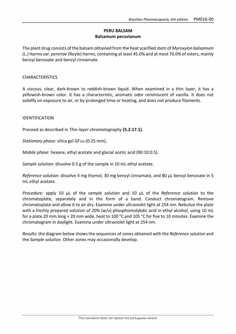
Brazilian Pharmacopoeia, 6th edition PM016-00
This translation does not replace the portuguese version.
PERU BALSAM Balsamum peruvianum
The plant drug consists of the balsam obtained from the heat scarified stem of Myroxylon balsamum (L.) Harms var. pereirae (Royle) Harms, containing at least 45.0% and at most 70.0% of esters, mainly benzyl benzoate and benzyl cinnamate. CHARACTERISTICS A viscous, clear, dark-brown to reddish-brown liquid. When examined in a thin layer, it has a yellowish-brown color. It has a characteristic, aromatic odor reminiscent of vanilla. It does not solidify on exposure to air, or by prolonged time or heating, and does not produce filaments. IDENTIFICATION Proceed as described in Thin-layer chromatography (5.2.17.1). Stationary phase: silica gel GF254 (0.25 mm). Mobile phase: hexane, ethyl acetate and glacial acetic acid (90:10:0.5). Sample solution: dissolve 0.5 g of the sample in 10 mL ethyl acetate. Reference solution: dissolve 4 mg thymol, 30 mg benzyl cinnamate, and 80 µL benzyl benzoate in 5 mL ethyl acetate. Procedure: apply 10 μL of the sample solution and 10 μL of the Reference solution to the chromatoplate, separately and in the form of a band. Conduct chromatogram. Remove chromatoplate and allow it to air dry. Examine under ultraviolet light at 254 nm. Nebulize the plate with a freshly prepared solution of 20% (w/v) phosphomolybdic acid in ethyl alcohol, using 10 mL for a plate 20 mm long × 20 mm wide, heat to 100 °C and 105 °C for five to 10 minutes. Examine the chromatogram in daylight. Examine under ultraviolet light at 254 nm. Results: the diagram below shows the sequences of zones obtained with the Reference solution and the Sample solution. Other zones may occasionally develop.
Brazilian Pharmacopoeia, 6th edition PM016-00
This translation does not replace the portuguese version.
Top of the plate
Blue-colored zone
Benzyl benzoate: blue-colored zone Blue-colored zone
Benzyl cinnamate: blue-colored zone Blue-colored zone
Thymol: gray-purple color zone
Blue-colored zone
Reference solution Sample solution TESTS Solubility. Practically insoluble in water, very soluble in ethyl alcohol, soluble in chloroform and acetic acid, slightly soluble in diethyl ether and petroleum ether, immiscible in fatty oils except castor oil. Relative density (5.2.5). 1.14 to 1.17. Acidity level (5.2.29.7). 56 to 84. Dissolve 1 g of the sample in 100 mL of neutralized ethyl alcohol, add 1 mL phenolphthalein SI and titrate with 0.5 M potassium hydroxide in ethyl alcohol SV. Saponification index (5.2.29.8). 230 to 255. Determine according to the residue obtained in Dosage. Artificial balms. Vigorously shake 0.2 g of the sample with 6 mL petroleum ether. The light petroleum solution should remain transparent and colorless, and all the insoluble parts of the balm will be adhered to the walls of the test tube. Turpentine. Evaporate 4 mL of the obtained solution in Artificial balms. The residue has no turpentine odor. Fatty oils. Shake 1 g of the sample with 3 mL of a 1000 g/L chloral hydrate solution. The solution obtained is transparent just like the chloral hydrate solution at 1000 g/L. DOSAGE Esters In a separating funnel, add 2.5 g of the sample, 7.5 mL of 8.5% (w/v) sodium hydroxide solution, and 40 mL of peroxide-free diethyl ether. Vigorously shake for 10 minutes. Separate the ether phase and shake the basic phase for one minute with three 15 mL portions of peroxide-free diethyl ether.
Brazilian Pharmacopoeia, 6th edition PM016-00
This translation does not replace the portuguese version.
Gather the ether phases, dry with 10 g of anhydrous sodium sulfate, and filter. Rinse the sodium sulfate residue twice with 10 mL of peroxide-free diethyl ether. Gather the ether phases and evaporate to dryness. Dry the residue (esters) between 100 °C and 105 °C for 30 minutes, cool in the desiccator, and weigh. PACKAGING AND STORAGE In tightly closed containers, protected from light and heat.
Brazilian Pharmacopoeia, 6th edition PM017-00
This translation does not replace the portuguese version.
BARBATIMAO, bark Barbadetimani cortex
The plant drug consists of dried stem bark of Stryphnodendron adstringens (Mart.) Coville [syn. Stryphnodendron barbatimam (Vell.) Mart.], containing at least 8% total tannins, expressed as pyrogallol (C6H6O3; 126,11), of which at least 0.2 mg/g is gallic acid (C7H6O5; 170,12) and 0.3 mg/g galocatechin (C15H14O7; 306,27), on the dried plant drug. Stem bark is the tissue located externally to the vascular cambium of the organ. CHARACTERISTICS Odorless dry barks. IDENTIFICATION 1. Macroscopic description The stem bark, when dry, appears in arched fragments, on varied sizes and shapes. In cross section, they are, on average, 6 mm thick when dry, and 10 to 12 mm thick when hydrated, with the phloem region, the innermost one, having a lighter brown coloring when compared to the suber, outermost region, with an intense reddish-brown coloring. In the young stems, a front view shows that the suber is dark in color and granular in appearance, homogeneous, with narrow and deep fissures crosswise. In the older stem portions, it is dark-brown or grayish-brown when lichens are present, always with deep cracks, predominantly crosswise, or with consecutive girdles, detaching themselves in irregular plates of varied sizes and shapes, leaving deep depressions in the area. The fracture of the bark is of the granular type in relation to the suber region and fibrous, striated lengthwise in the phloem region. 2. Microscopic description The suber in the outer portion of the bark has 20 to 30 layers of radially lined, thin-walled, tabular cells with brown contents, followed by many layers of isodiametric or slightly elongated periclinally-shaped parenchyma cells, also thin-walled. Most of these cells have a reddish-brown content, which does not discolor easily with 30% (w/v) sodium hypochlorite and does not change color in the presence of ferric chloride RS. In this parenchymatic portion stone cells (majority) and macro-sclereids occur, positioned in various planes, in groups of several elements or isolated, with very thickened walls with lignin, showing evident lamellations and simple, sometimes branched, pits. In the outermost portions of the suber, both parenchyma cells and sclereids can be seen, compacted and deformed by mechanical action on the inner tissues. In the phloem region, clusters of a few, relatively narrow, gelatinous fiber elements occur, always with adjoining idioblasts, containing a large, prismatic, calcium oxalate crystal with varying numbers of sides, entire or superficially eroded. The clusters of fibers, when observed in longitudinal sections, follow the parenchyma rays of the phloem, which are generally uniseriate, but become bi-multiseriate in the outermost portions. The sieve elements have compound sieve plates and are collapsed in the outermost regions of the phloem. Isolated stone cells, similar to those in the suber, and spherical starch grains are abundant in the parenchymatic tissue of the phloem. The cells around the parenchyma rays react positively
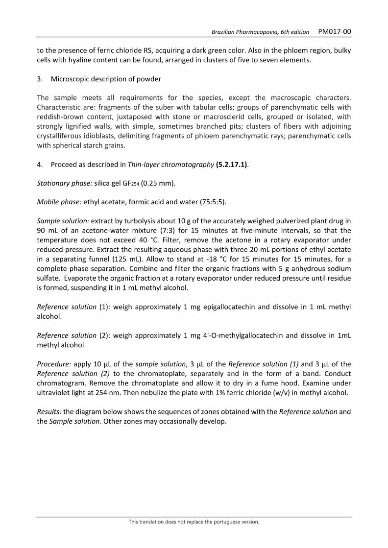
Brazilian Pharmacopoeia, 6th edition PM017-00
This translation does not replace the portuguese version.
to the presence of ferric chloride RS, acquiring a dark green color. Also in the phloem region, bulky cells with hyaline content can be found, arranged in clusters of five to seven elements. 3. Microscopic description of powder The sample meets all requirements for the species, except the macroscopic characters. Characteristic are: fragments of the suber with tabular cells; groups of parenchymatic cells with reddish-brown content, juxtaposed with stone or macrosclerid cells, grouped or isolated, with strongly lignified walls, with simple, sometimes branched pits; clusters of fibers with adjoining crystalliferous idioblasts, delimiting fragments of phloem parenchymatic rays; parenchymatic cells with spherical starch grains. 4. Proceed as described in Thin-layer chromatography (5.2.17.1). Stationary phase: silica gel GF254 (0.25 mm). Mobile phase: ethyl acetate, formic acid and water (75:5:5). Sample solution: extract by turbolysis about 10 g of the accurately weighed pulverized plant drug in 90 mL of an acetone-water mixture (7:3) for 15 minutes at five-minute intervals, so that the temperature does not exceed 40 °C. Filter, remove the acetone in a rotary evaporator under reduced pressure. Extract the resulting aqueous phase with three 20-mL portions of ethyl acetate in a separating funnel (125 mL). Allow to stand at -18 °C for 15 minutes for 15 minutes, for a complete phase separation. Combine and filter the organic fractions with 5 g anhydrous sodium sulfate. Evaporate the organic fraction at a rotary evaporator under reduced pressure until residue is formed, suspending it in 1 mL methyl alcohol. Reference solution (1): weigh approximately 1 mg epigallocatechin and dissolve in 1 mL methyl alcohol. Reference solution (2): weigh approximately 1 mg 4'-O-methylgallocatechin and dissolve in 1mL methyl alcohol. Procedure: apply 10 µL of the sample solution, 3 µL of the Reference solution (1) and 3 µL of the Reference solution (2) to the chromatoplate, separately and in the form of a band. Conduct chromatogram. Remove the chromatoplate and allow it to dry in a fume hood. Examine under ultraviolet light at 254 nm. Then nebulize the plate with 1% ferric chloride (w/v) in methyl alcohol. Results: the diagram below shows the sequences of zones obtained with the Reference solution and the Sample solution. Other zones may occasionally develop.
Brazilian Pharmacopoeia, 6th edition PM017-00
This translation does not replace the portuguese version.
Top of the plate
4'-O-Methylgalocatechin: fluorescence extinction zone
Fluorescence extinction zone
Epigallocatechin: fluorescence extinction zone
Fluorescence extinction zone
Reference solution Sample solution TESTS Foreign matter (5.4.1.3). At most 2.0%. Water (5.4.1.4). At most 14.0%. Total ash (5.4.1.5.1). At most 2.0%. Sulfated Ash (5.4.1.5.2). At most 3.0%. Total mesophilic microorganism count (5.5.3.1.2). Complies with the test. Study of pathogenic microorganisms (5.5.3.1.3). Complies with the test. Heavy metals (5.4.5). Complies with the test. Agrochemical waste (5.4.3). Complies with the test. DOSAGE Total tannins Note: protect samples from light during extraction and dilution. Use carbon dioxide-free water for all operations. To proceed as described in Visible absorption spectrophotometry (5.2.14). Prepare solutions as described below. Stock solution: accurately weigh approximately 0.750 g of the pulverized plant drug (250 μm) (5.2.11) and transfer to a 250-mL, erlenmeyer flask with a ground-glass stopper. Add 150 mL of
Brazilian Pharmacopoeia, 6th edition PM017-00
This translation does not replace the portuguese version.
water. Heat in a water bath for 30 minutes at 60°C. Cool under running water and transfer to a 250-mL volumetric flask. Rinse the erlenmeyer flask and transfer the washing water with the entire plant drug content to the same volumetric flask. Top off the volume with water and homogenize. Allow to decant and filter the supernatant liquid through filter paper. Discard the first 50 mL of the filtrate. Sample solution for total polyphenol content: volumetrically transfer 5 mL of the filtrate to a 25-mL volumetric flask, top off the volume with water and homogenize. Volumetrically transfer 2 mL of the solution, 1 mL of phosphomolybdotungstic reagent and 10 mL of water to a 25-mL volumetric flask, top off the volume with 29% sodium carbonate solution (w/v) and homogenize. Determine absorbance at 760 nm (A1), after 30 minutes, using water for zero adjustment. Sample solution for polyphenols not adsorbed by hide powder: for 10 mL of the filtrate, add 0.1 g of the hide powder CRS and mechanically shake in a 125-mL Erlenmeyer flask for 60 minutes. Filter through paper filter. Volumetrically transfer 5 mL of this filtrate to a 25-mL volumetric flask, top off the volume with water and homogenize. Volumetrically transfer 2 mL of the solution, 1 mL of phosphomolybdotungstic reagent and 10 mL of water to a 25-mL volumetric flask, top off the volume with 29% sodium carbonate solution (w/v) and homogenize. Determine absorbance at 760 nm (A2), after 30 minutes, using water for zero adjustment. Reference solution: Dissolve 50 mg of pyrogallol in water immediately before use, transfer quantitatively to a 100-mL volumetric flask, top off the volume with water and homogenize. Volumetrically transfer 5 mL of this solution to a 100-mL volumetric flask, top off the volume with water and homogenize. Volumetrically transfer 2 mL of the solution, 1 mL of phosphomolybdotungstic reagent and 10 mL of water to a 25-mL volumetric flask, top off the volume with 29% sodium carbonate solution (w/v) and homogenize. Determine absorbance at 760 nm (A3), after 30 minutes, using water for zero adjustment. Calculate tannin content, expressed as a percentage of pyrogallol, according to the following expression:
𝑇𝑇𝑇𝑇 =(𝑇𝑇1 × 𝑇𝑇2) × 𝑚𝑚2 × 62,5
𝑇𝑇3 × 𝑚𝑚1
in which, TT = total tannin content expressed as pyrogallol % (w/w); A1 = absorbance measured for the Sample solution for total polyphenols; A2 = absorbance measured for the Sample solution for polyphenols not adsorbed by hide powder; A3 = absorbance measured for the Reference solution; m1 = mass in grams of the sample used, considering the determined water content. m2 = mass in grams of pyrogallol, considering the purity of the reference substance. Gallic acid and gallocatechin Proceed as described in High Performance Liquid Chromatography (5.2.17.4). Use a chromatograph equipped with an ultraviolet detector set at a wavelength of 210 nm; pre-column packed with silica chemically bonded to an octadecylsilane group; 250 mm long, 4.6 mm internal diameter column, packed with silica chemically bonded to an octadecylsilane group (5 µm); Mobile phase flow rate of 0.8 mL/minute.
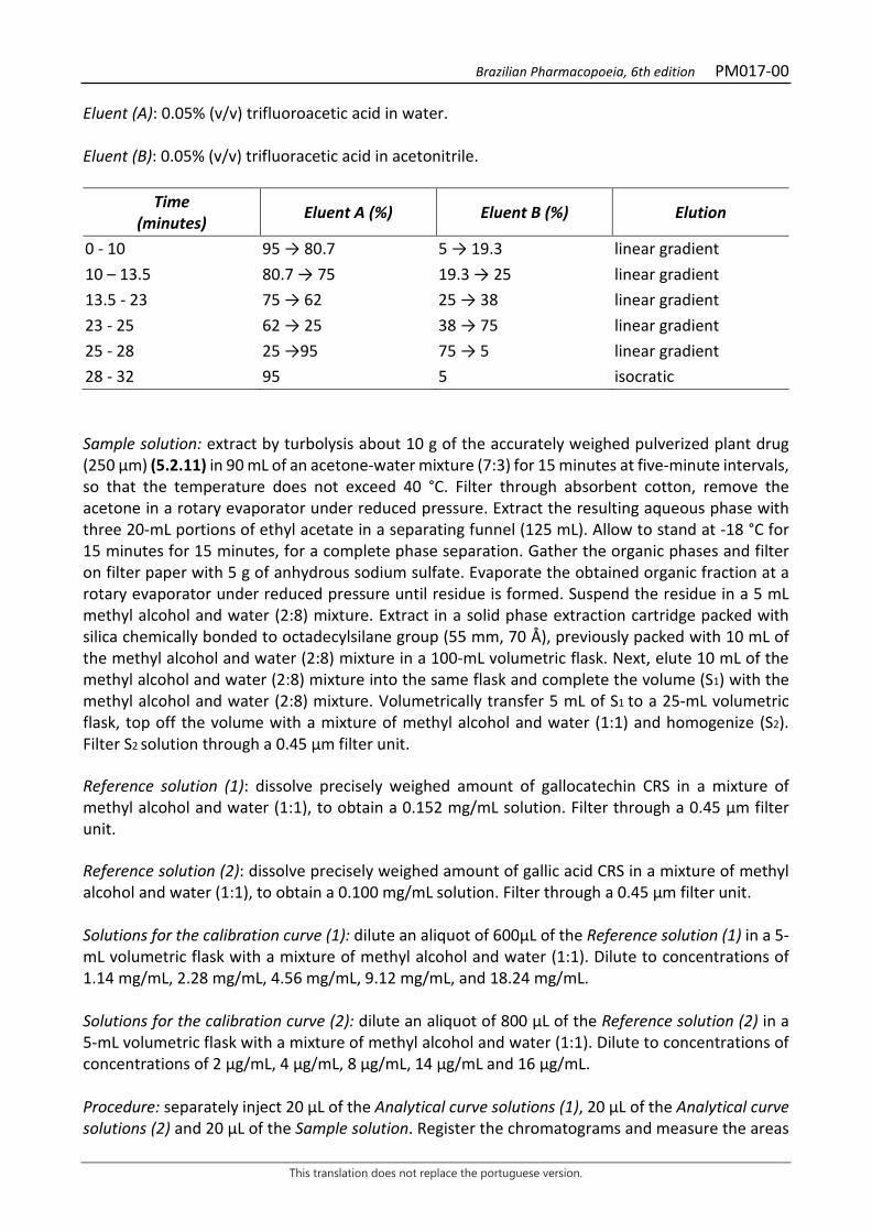
Brazilian Pharmacopoeia, 6th edition PM017-00
This translation does not replace the portuguese version.
Eluent (A): 0.05% (v/v) trifluoroacetic acid in water. Eluent (B): 0.05% (v/v) trifluoracetic acid in acetonitrile.
Time (minutes) Eluent A (%) Eluent B (%) Elution
0 - 10 95 → 80.7 5 → 19.3 linear gradient 10 – 13.5 80.7 → 75 19.3 → 25 linear gradient 13.5 - 23 75 → 62 25 → 38 linear gradient 23 - 25 62 → 25 38 → 75 linear gradient 25 - 28 25 →95 75 → 5 linear gradient 28 - 32 95 5 isocratic Sample solution: extract by turbolysis about 10 g of the accurately weighed pulverized plant drug (250 μm) (5.2.11) in 90 mL of an acetone-water mixture (7:3) for 15 minutes at five-minute intervals, so that the temperature does not exceed 40 °C. Filter through absorbent cotton, remove the acetone in a rotary evaporator under reduced pressure. Extract the resulting aqueous phase with three 20-mL portions of ethyl acetate in a separating funnel (125 mL). Allow to stand at -18 °C for 15 minutes for 15 minutes, for a complete phase separation. Gather the organic phases and filter on filter paper with 5 g of anhydrous sodium sulfate. Evaporate the obtained organic fraction at a rotary evaporator under reduced pressure until residue is formed. Suspend the residue in a 5 mL methyl alcohol and water (2:8) mixture. Extract in a solid phase extraction cartridge packed with silica chemically bonded to octadecylsilane group (55 mm, 70 Å), previously packed with 10 mL of the methyl alcohol and water (2:8) mixture in a 100-mL volumetric flask. Next, elute 10 mL of the methyl alcohol and water (2:8) mixture into the same flask and complete the volume (S1) with the methyl alcohol and water (2:8) mixture. Volumetrically transfer 5 mL of S1 to a 25-mL volumetric flask, top off the volume with a mixture of methyl alcohol and water (1:1) and homogenize (S2). Filter S2 solution through a 0.45 µm filter unit. Reference solution (1): dissolve precisely weighed amount of gallocatechin CRS in a mixture of methyl alcohol and water (1:1), to obtain a 0.152 mg/mL solution. Filter through a 0.45 µm filter unit. Reference solution (2): dissolve precisely weighed amount of gallic acid CRS in a mixture of methyl alcohol and water (1:1), to obtain a 0.100 mg/mL solution. Filter through a 0.45 µm filter unit. Solutions for the calibration curve (1): dilute an aliquot of 600μL of the Reference solution (1) in a 5-mL volumetric flask with a mixture of methyl alcohol and water (1:1). Dilute to concentrations of 1.14 mg/mL, 2.28 mg/mL, 4.56 mg/mL, 9.12 mg/mL, and 18.24 mg/mL. Solutions for the calibration curve (2): dilute an aliquot of 800 μL of the Reference solution (2) in a 5-mL volumetric flask with a mixture of methyl alcohol and water (1:1). Dilute to concentrations of concentrations of 2 μg/mL, 4 μg/mL, 8 μg/mL, 14 μg/mL and 16 μg/mL. Procedure: separately inject 20 μL of the Analytical curve solutions (1), 20 μL of the Analytical curve solutions (2) and 20 μL of the Sample solution. Register the chromatograms and measure the areas
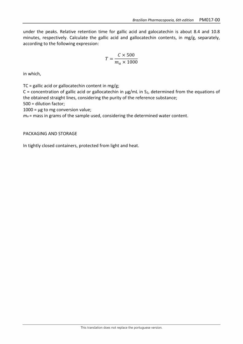
Brazilian Pharmacopoeia, 6th edition PM017-00
This translation does not replace the portuguese version.
under the peaks. Relative retention time for gallic acid and galocatechin is about 8.4 and 10.8 minutes, respectively. Calculate the gallic acid and gallocatechin contents, in mg/g, separately, according to the following expression:
𝑇𝑇 =𝐶𝐶 × 500𝑚𝑚𝑎𝑎 × 1000
in which, TC = gallic acid or gallocatechin content in mg/g; C = concentration of gallic acid or gallocatechin in μg/mL in S2, determined from the equations of the obtained straight lines, considering the purity of the reference substance; 500 = dilution factor; 1000 = μg to mg conversion value; ma = mass in grams of the sample used, considering the determined water content. PACKAGING AND STORAGE In tightly closed containers, protected from light and heat.
Brazilian Pharmacopoeia, 6th edition PM017-00
This translation does not replace the portuguese version.
Figure 1 – Macroscopic and microscopic aspects in Stryphnodendron adstringens (Mart.) Coville
___________________________ The scales correspond in A, B and C to 1 cm, in D to 2 mm, and in E, F, G and H to 100 μm. A and B – partial appearance of the outer and inner bark surface of the youngest branch, respectively: lichens (li). C – partial appearance of the outer surface of an older branch. D – diagram of bark tissue distribution: tabular cells (ct), stone cell (cp); parenchyma (pa); suber (su); phloem (f). E and F – partial details of the suber region in cross sections: tabular cells (ct); macro-sclereids (me); parenchyma (pa); stone cell (cp). G and H – partial details of the phloem region in cross sections: phloem fibers (ff); bulky cells (cv); sieve plate (pc); obliterated sieve element (et).

Brazilian Pharmacopoeia, 6th edition PM017-00
This translation does not replace the portuguese version.
Figure 2 – Microscopic and powder microscopic aspects in Stryphnodendron adstringens (Mart.) Coville
___________________________ The scales correspond in A, B and D to 100 μm; in C and E to 25 μm. A and B – partial details of phloem, in tangential longitudinal sections: parenchyma ray (ra); parenchyma cell (pa); crystalliferous idioblast (ic). C – partial detail of phloem parenchyma with starch grains: starch grains (am); sieve plate (pc). D – partial detail of phloem in radial longitudinal section: bulky cell (cv); crystalliferous idioblast (ic); phloem fibers (ff); parenchymatic ray (ra). E – detail of the phloem’s crystalliferous idioblasts: phloem fibers (ff); crystalliferous idioblast (ic).
Brazilian Pharmacopoeia, 6th edition PM018-00
This translation does not replace the portuguese version.
VANILLA, fruit Vanillae fructus
The plant drug consists of immature, dried fruits of Vanilla planifolia Jacks. ex Andrews, containing at least 12.0% dry hydroalcoholic extract. CHARACTERISTICS The drug has a pleasant, floral odor reminiscent of vanillin, which, however, is much more subtle and full-bodied than the substance alone. IDENTIFICATION 1. Macroscopic description The fruits are plurispermic capsules, derived from the superus, tricarpelar and unilocular ovaries. The shape of the fruit, in cross section, is variable depending on the storage mode; the uncompressed fruit has a triangular outline in cross section, is dark brown, has longitudinal striations, is flexible, 20 to 25 cm long and 1 to 1.5 cm wide in its median region. 2. Microscopic description The pericarp, in general, has an exocarp with a single cell layer, a multicellular mesocarp, and an endocarp with a single specialized cell layer. Idioblasts with raphids oriented longitudinally to the pericarp are common; prismatic crystals are also observed. The exocarp has tangentially elongated cells, whose outer and inner periclinal walls are thick, the outer ones cutinized; the cells usually accumulate phenolic compounds. The exocarp is stomatiferous and glabrous. The outer mesocarp has two to four layers similar to an angular collenchyma; the middle mesocarp has bulky cells, with a large accumulation of phenolic compounds. This tissue features collateral vascular bundles in groups of two or three, usually more calibrous than the individual bundles of small caliber, and bundles surrounded by a sclerenchyma sheath with two to five cell layers; the sclerenchyma is composed of bulky cells with lignified and thinly thickened walls; the inner mesocarp has flattened cells, containing phenolic compounds. The endocarp is differentiated into a densely hair stratum; the cells have thin, pectic walls, with dense cytoplasm and a secretory appearance. In cross section, it is possible to distinguish three placental regions, with profusely branched placenta, at which terminations the seeds are inserted. The seeds, black or dark-brown in color, have a sclerenchymatous testa; the seminal testa is composed of a single layer of brachysclereids. The inner integument is compressed and the endosperm has bulky cells with reserves. 3. Microscopic description of powder The sample meets all requirements for the species, except the macroscopic characters. Characteristic are: fragments in the form of lumps; prismatic crystals and sclereids; fragments with exocarp cells; fibers in groups of two or three elements or isolated; vessel elements grouped or isolated; lignin reinforcements and pits are observed. The seeds remain virtually intact.
Brazilian Pharmacopoeia, 6th edition PM018-00
This translation does not replace the portuguese version.
4. Proceed as described in Thin-layer chromatography (5.2.17.1). Stationary phase: silica gel GF254 (0.25 mm). Mobile phase: methyl chloride and acetone (95:5). Sample solution: use the hydroalcoholic extract obtained in Dosage. Reference solution: dissolve 1 mg vanillin in 10 mL ethyl alcohol. Procedure: apply 20 μL of the sample solution and 10 μL of the sample solution to the chromatoplate, separately and in the form of a band. Conduct chromatogram. Remove chromatoplate and allow it to air dry. Examine under ultraviolet light at 254 nm. Results: the diagram below shows the sequences of zones obtained with the Reference solution and the Sample solution. Other zones may occasionally develop.
Top of the plate
Vanillin: blue-purple fluorescence zone
Blue-violet fluorescence zone
Reference solution Sample solution
TESTS Sulfated Ash (5.4.1.5.2). At most 7.0% Total mesophilic microorganism count (5.5.3.1.2). Complies with the test. Study of pathogenic microorganisms (5.5.3.1.3). Complies with the test. Heavy metals (5.4.5). Complies with the test. Agrochemical waste (5.4.3). Complies with the test. DOSAGE Extractable substances Determine the content of extractable substances by calculating the yield of the hydroalcoholic extract. Weigh out exactly about 2 g of vanilla, previously cut into small pieces or ground to a coarse powder. Transfer the powder to an erlenmeyer flask with a ground-glass stopper and add 70 mL of diluted ethyl alcohol (solution prepared with 263 mL ethyl alcohol in 250 mL distilled water), tightly cap the container and shake for two hours on a mechanical stirrer, or leave overnight and shake frequently for a further eight hours. Decant the liquid layer and filter, collecting the filtrate in a 100-mL volumetric flask. Rinse the bottle and the residue four consecutive times with 8 mL portions of

Brazilian Pharmacopoeia, 6th edition PM018-00
This translation does not replace the portuguese version.
the diluted ethyl alcohol solution. Filter the washing liquids, on the same filter, and add to the previously obtained filtrate. Top off the volume to 100 mL with a sufficient quantity of diluted ethyl alcohol, homogenize and evaporate 50 mL, exactly measured, over a water bath in a tared porcelain capsule. Dry the residue in an oven at 105 °C for four hours. Cool the capsule in desiccator and weigh. The weight of the residue represents the dry hydroalcoholic extract of 1 g of the drug. PACKAGING AND STORAGE In tightly closed containers, protected from light and heat.
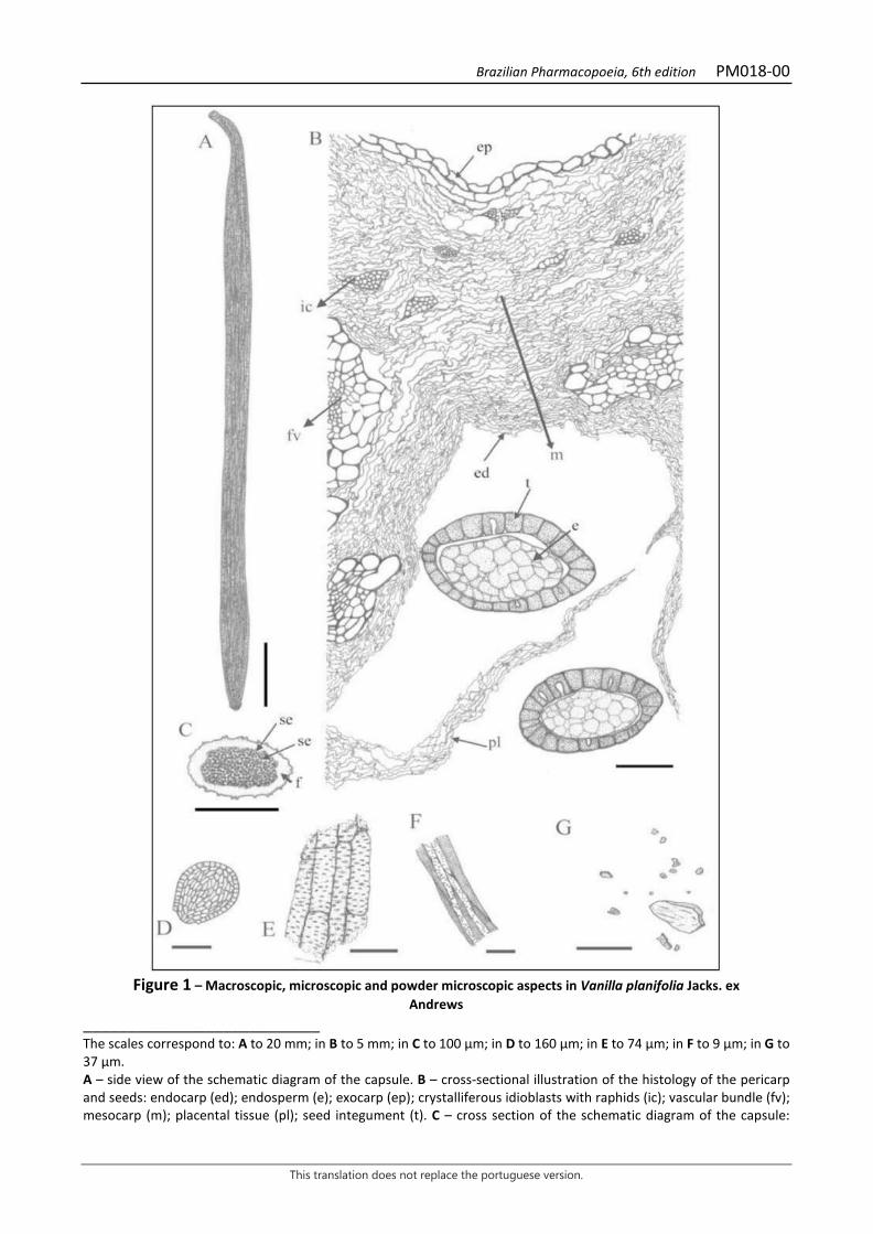
Brazilian Pharmacopoeia, 6th edition PM018-00
This translation does not replace the portuguese version.
Figure 1 – Macroscopic, microscopic and powder microscopic aspects in Vanilla planifolia Jacks. ex
Andrews ___________________________ The scales correspond to: A to 20 mm; in B to 5 mm; in C to 100 μm; in D to 160 μm; in E to 74 μm; in F to 9 μm; in G to 37 μm. A – side view of the schematic diagram of the capsule. B – cross-sectional illustration of the histology of the pericarp and seeds: endocarp (ed); endosperm (e); exocarp (ep); crystalliferous idioblasts with raphids (ic); vascular bundle (fv); mesocarp (m); placental tissue (pl); seed integument (t). C – cross section of the schematic diagram of the capsule:
Brazilian Pharmacopoeia, 6th edition PM018-00
This translation does not replace the portuguese version.
pericarp (f); seed (se). D – lateral view of the seed E – fragment of xylem vessel elements. F – fragment of a group of fibers from the vascular sheath. G – calcium oxalate crystals.
Brazilian Pharmacopoeia, 6th edition PM019-00
This translation does not replace the portuguese version.
BELLADONNA, leaf Belladonnae folium
The plant drug consists of dried leaves, entire or chopped, of Atropa belladonna L., containing at least 0.25% atropine (C17H23NO3, 289,37). IDENTIFICATION 1. Macroscopic description The leaves are elliptic, oval-lanceolate to broadly oval, entire, with an acuminate apex, an attenuate, symmetrical and somewhat decurved base, and an entire margin. They are 5 to 25 cm long and 3 to 12 cm wide, with petioles 0.5 to 4 cm long. It is green to greenish-brown, darker on the adaxial surface. The dried leaves are wrinkled, friable, and thin. The young leaves are more pubescent than the adult leaves along the veins and petiole. The venation is peninerve and the secondary ribs depart from the midrib at an angle of about 60° and anastomose near the margin. The surface of the lamina is dry and rough to the touch, due to the presence of cells with microcrystalline calcium oxalate content in the mesophyll. Such cells appear as tiny bright spots when the surface is illuminated and dark through transparency. 2. Microscopic description The leaf lamina is amphistomatic and dorsiventrally symmetrical. A front view of the epidermis shows cells with waved anticlinal walls and finely grooved cuticle on both sides. Tector and glandular trichomes are numerous throughout the lamina. Tector trichomes have two to five cells, are uniseriate and conical, with smooth and thin walls; the glandular trichomes have a pluricellular pedicel, composed of two to four cells, with a claviform terminal cell, or they have a pluricellular pedicel and a pluricellular head, formed by four to seven cells, with an ovoid to pyriform appearance. Anisocytic stomata are more frequent in the abaxial epidermis. In cross section, the epidermis is unistratified and the cuticle is thin. The mesophyll is composed of unistratified palisade parenchyma and spongy parenchyma with large idioblasts containing prismatic calcium oxalate crystals and microcrystalline sand. The midrib is prominent on both sides and has bicolateral vascular bundles in an open arch, and the intra-axillary phloem is discontinuous. Angular collenchyma occurs below the epidermis, on both faces. 3. Microscopic description of powder The sample meets all requirements for the species, except the macroscopic characters. Characteristics are: dark green; epidermis fragments with cells with waved, anticlinal walls and cuticle with striations; mesophyll fragments with unistratified palisade parenchyma; abaxial epidermis fragments, showing anisocytic stomata and rare tectors and glandular trichomes; fragments of parenchyma, containing crystalliferous idioblasts; isolated prismatic crystals, as described; glandular trichomes, as described, isolated, fragmented or with remnants of epidermis; isolated tector trichomes or their fragments. 4. Proceed as described in Thin-layer chromatography (5.2.17.1).
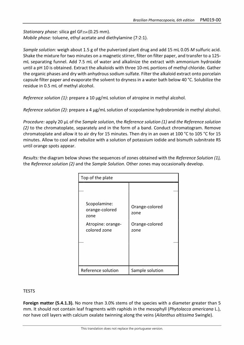
Brazilian Pharmacopoeia, 6th edition PM019-00
This translation does not replace the portuguese version.
Stationary phase: silica gel GF254 (0.25 mm). Mobile phase: toluene, ethyl acetate and diethylamine (7:2:1). Sample solution: weigh about 1.5 g of the pulverized plant drug and add 15 mL 0.05 M sulfuric acid. Shake the mixture for two minutes on a magnetic stirrer, filter on filter paper, and transfer to a 125-mL separating funnel. Add 7.5 mL of water and alkalinize the extract with ammonium hydroxide until a pH 10 is obtained. Extract the alkaloids with three 10-mL portions of methyl chloride. Gather the organic phases and dry with anhydrous sodium sulfate. Filter the alkaloid extract onto porcelain capsule filter paper and evaporate the solvent to dryness in a water bath below 40 °C. Solubilize the residue in 0.5 mL of methyl alcohol. Reference solution (1): prepare a 10 µg/mL solution of atropine in methyl alcohol. Reference solution (2): prepare a 4 µg/mL solution of scopolamine hydrobromide in methyl alcohol. Procedure: apply 20 μL of the Sample solution, the Reference solution (1) and the Reference solution (2) to the chromatoplate, separately and in the form of a band. Conduct chromatogram. Remove chromatoplate and allow it to air dry for 15 minutes. Then dry in an oven at 100 °C to 105 °C for 15 minutes. Allow to cool and nebulize with a solution of potassium iodide and bismuth subnitrate RS until orange spots appear. Results: the diagram below shows the sequences of zones obtained with the Reference Solution (1), the Reference solution (2) and the Sample Solution. Other zones may occasionally develop.
Top of the plate
Scopolamine: orange-colored zone
Orange-colored zone
Atropine: orange-colored zone
Orange-colored zone
Reference solution Sample solution
TESTS Foreign matter (5.4.1.3). No more than 3.0% stems of the species with a diameter greater than 5 mm. It should not contain leaf fragments with raphids in the mesophyll (Phytolacca americana L.), nor have cell layers with calcium oxalate twinning along the veins (Ailanthus altissima Swingle).

Brazilian Pharmacopoeia, 6th edition PM019-00
This translation does not replace the portuguese version.
Total ash (5.4.1.5.1). At most 16.0% Acid-insoluble ash (5.4.1.5.3). At most 4.0% Loss by drying (5.2.9.1). Gravimetric method. At most 10.0%. Total mesophilic microorganism count (5.5.3.1.2). Complies with the test. Study of pathogenic microorganisms (5.5.3.1.3). Complies with the test. Aflatoxins (5.4.4). Complies with the test. Heavy metals (5.4.5). Complies with the test. Agrochemical waste (5.4.3). Complies with the test. DOSAGE Atropine Proceed as described in High Performance Liquid Chromatography (5.2.17.4). Use a chromatograph equipped with an ultraviolet detector at 210 mm; pre-column packed with octadecylsilane silica, 150 mm long, 4.6 mm internal diameter column, packed with octadecylsilane silica (5 µm), kept at room temperature; mobile phase flow rate of 0.7 mL/minute. Eluent (A): water and trifluoroacetic acid (100:0.01). Eluent (B): acetonitrile and trifluoracetic acid (100:0.08).
Time (minutes) Eluent A (%) Eluent B (%) Elution
0-15 95→0 5→100 linear gradient 15-20 0 100 isocratic A 10-minute equilibration with the mixture of Eluent (A) and (2) (95:5) is required between runs. Such balancing must be performed between runs and not embedded into the described mobile phase gradient. Sample solution: accurately weigh approximately 0.25 g of the pulverized plant drug (180 µm) in an 50-mL erlenmeyer flask (5.2.11), and add 15 mL methyl alcohol. Cover the jar with plastic wrap and place in the ultrasonic bath for 60 minutes. Filter the crude methanolic extract on filter paper into a 50-mL round-bottomed flask. Rinse the erlenmeyer flask with two 1 mL portions of methyl alcohol. Remove the solvent in a rotary evaporator at a temperature below 40 ºC. Transfer the residue from the flask to a 250-mL separating funnel, using two 10-mL aliquots and a 5-mL aliquot of 5% (v/v) hydrochloric acid, with the aid of ultrasound to facilitate residue solubilization. Wash the acidic aqueous extract with three 20-mL portions of methyl chloride, using the first 20 mL to wash the
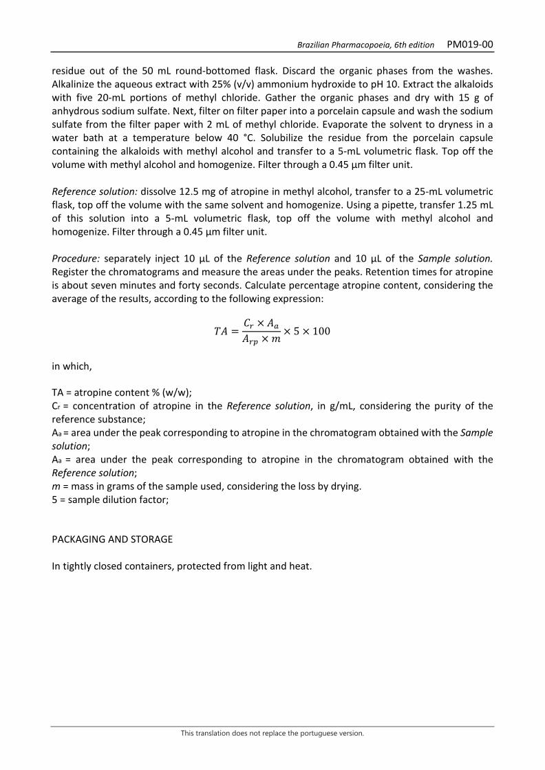
Brazilian Pharmacopoeia, 6th edition PM019-00
This translation does not replace the portuguese version.
residue out of the 50 mL round-bottomed flask. Discard the organic phases from the washes. Alkalinize the aqueous extract with 25% (v/v) ammonium hydroxide to pH 10. Extract the alkaloids with five 20-mL portions of methyl chloride. Gather the organic phases and dry with 15 g of anhydrous sodium sulfate. Next, filter on filter paper into a porcelain capsule and wash the sodium sulfate from the filter paper with 2 mL of methyl chloride. Evaporate the solvent to dryness in a water bath at a temperature below 40 °C. Solubilize the residue from the porcelain capsule containing the alkaloids with methyl alcohol and transfer to a 5-mL volumetric flask. Top off the volume with methyl alcohol and homogenize. Filter through a 0.45 µm filter unit. Reference solution: dissolve 12.5 mg of atropine in methyl alcohol, transfer to a 25-mL volumetric flask, top off the volume with the same solvent and homogenize. Using a pipette, transfer 1.25 mL of this solution into a 5-mL volumetric flask, top off the volume with methyl alcohol and homogenize. Filter through a 0.45 µm filter unit. Procedure: separately inject 10 μL of the Reference solution and 10 µL of the Sample solution. Register the chromatograms and measure the areas under the peaks. Retention times for atropine is about seven minutes and forty seconds. Calculate percentage atropine content, considering the average of the results, according to the following expression:
𝑇𝑇𝑇𝑇 =𝐶𝐶𝑟𝑟 × 𝑇𝑇𝑎𝑎𝑇𝑇𝑟𝑟𝑟𝑟 × 𝑚𝑚
× 5 × 100
in which, TA = atropine content % (w/w); Cr = concentration of atropine in the Reference solution, in g/mL, considering the purity of the reference substance; Aa = area under the peak corresponding to atropine in the chromatogram obtained with the Sample solution; Aa = area under the peak corresponding to atropine in the chromatogram obtained with the Reference solution; m = mass in grams of the sample used, considering the loss by drying. 5 = sample dilution factor; PACKAGING AND STORAGE In tightly closed containers, protected from light and heat.
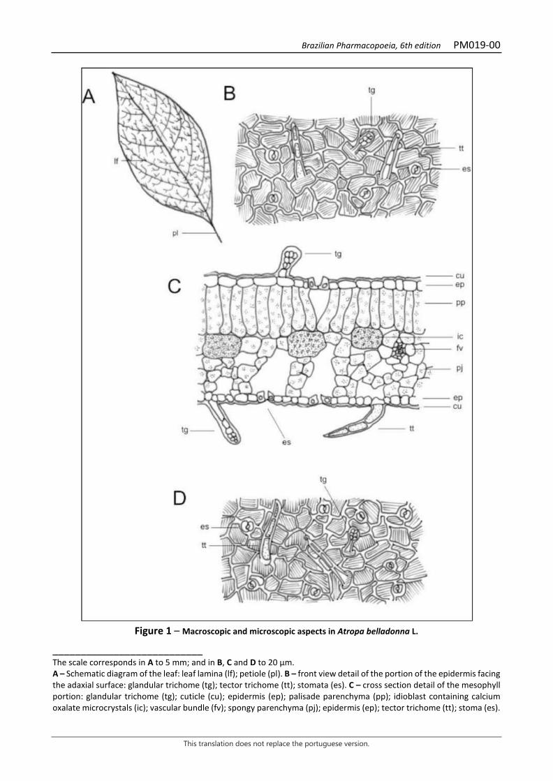
Brazilian Pharmacopoeia, 6th edition PM019-00
This translation does not replace the portuguese version.
Figure 1 – Macroscopic and microscopic aspects in Atropa belladonna L.
___________________________ The scale corresponds in A to 5 mm; and in B, C and D to 20 µm. A – Schematic diagram of the leaf: leaf lamina (lf); petiole (pl). B – front view detail of the portion of the epidermis facing the adaxial surface: glandular trichome (tg); tector trichome (tt); stomata (es). C – cross section detail of the mesophyll portion: glandular trichome (tg); cuticle (cu); epidermis (ep); palisade parenchyma (pp); idioblast containing calcium oxalate microcrystals (ic); vascular bundle (fv); spongy parenchyma (pj); epidermis (ep); tector trichome (tt); stoma (es).
Brazilian Pharmacopoeia, 6th edition PM019-00
This translation does not replace the portuguese version.
D – front view detail of the portion of the epidermis facing the abaxial surface: glandular trichome (tg); stoma (es); tector trichome (tt).
Figure 2 – Microscopic aspects of the powder in Atropa belladonna L.
___________________________ The scale corresponds in A, B and D to 30 µm; in C to 100 µm; and in E to 20 µm. A and C – front view of fragments of the epidermis facing the adaxial surface, showing crystalliferous idioblasts by transparency: crystalliferous idioblast (ic); palisade parenchyma (pp); vascular bundle (fv). B – front view of fragments of the epidermis facing the abaxial surface, showing crystalliferous idioblasts by transparency: crystalliferous idioblast (ic); stoma (es). D – cross section of leaf lamina fragment: cuticle (cu); epidermis (ep); palisade parenchyma (pp); crystalliferous idioblast (ic); spongy parenchyma (pj). E – trichomes or parts thereof, isolated: glandular trichome (tg); tector trichome (tt).

Brazilian Pharmacopoeia, 6th edition PM020-00
This translation does not replace the portuguese version.
BENZOIN Benzoe sumatranus
Benzoin consists of a balsamic resin, obtained by incision of the stem of Styrax benzoin Dryand. or Styrax paralleloneuron Perkins, containing not less than 25% and not more than 50% total acids, calculated as benzoic acid (C7H6O2, 122.12). CHARACTERISTICS It takes the form of rounded or ovoid, irregular, cream-white fragments that may be coated with a grayish-brown or reddish-brown resinous material. Fragments are hard and brittle, and the fracture surface is rough and irregular. Mild, balsamic odor. IDENTIFICATION Proceed as described in Thin-layer chromatography (5.2.17.1). Stationary phase: silica gel GF254 (0.25 mm). Mobile phase: hexane, isopropyl ether and glacial acetic acid (60:40:10). Sample solution: add 5 mL ethyl alcohol to 0.2 g of the finely pulverized sample and place in an ultrasonic bath for two minutes. Centrifuge and use the supernatant solution. Reference solution: dissolve 20 mg benzoic acid, 10 mg cinnamic acid, 4 mg vanillin, and 20 mg methyl cinnamate in 10 mL ethyl alcohol. Procedure: apply 10 μL of the sample solution and 10 μL of the Reference solution to the chromatoplate, separately and in the form of a band. Conduct chromatogram. Remove chromatoplate and allow it to air dry. Examine under ultraviolet light at 254 nm. Results: the diagram below shows the sequences of zones obtained with the Reference solution and the Sample solution. Other zones may occasionally develop.
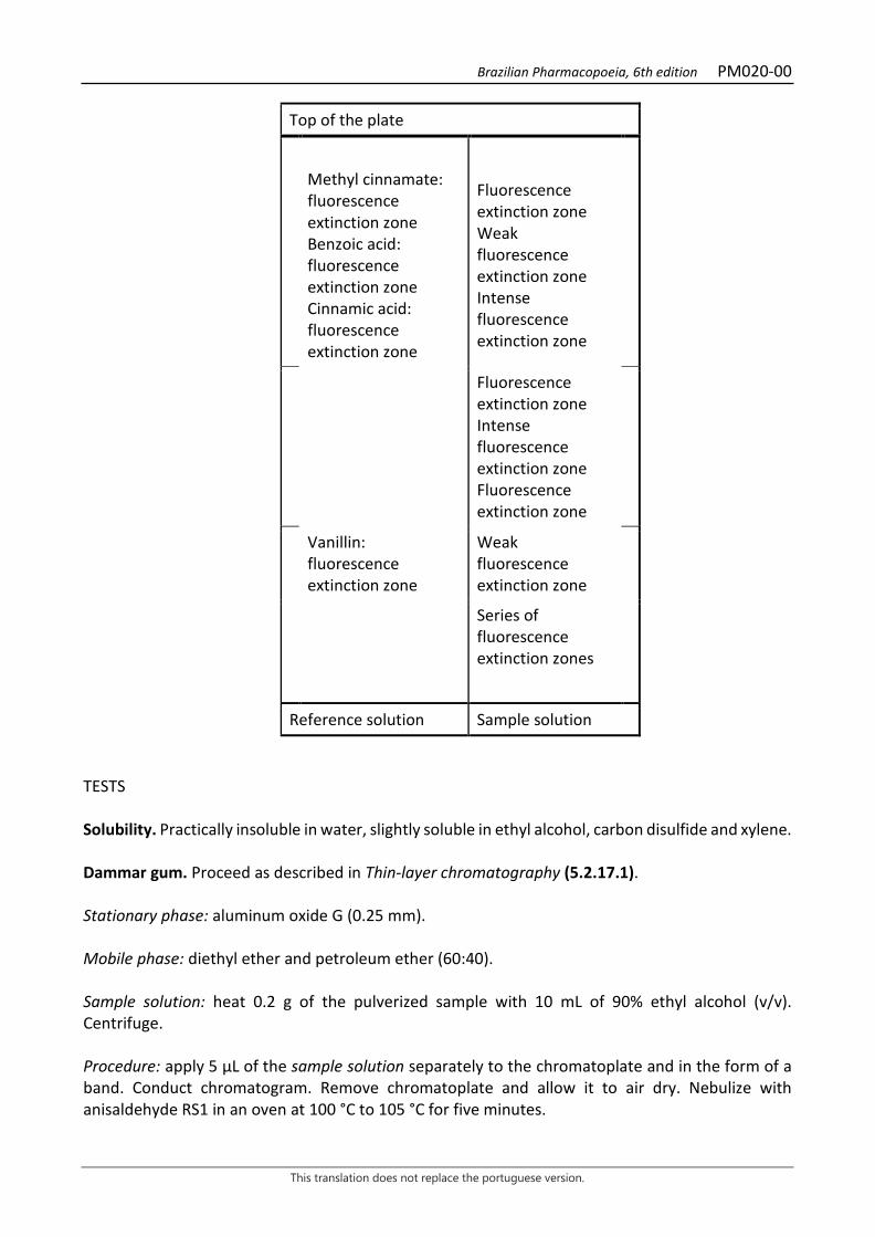
Brazilian Pharmacopoeia, 6th edition PM020-00
This translation does not replace the portuguese version.
Top of the plate
Methyl cinnamate: fluorescence extinction zone Benzoic acid: fluorescence extinction zone Cinnamic acid: fluorescence extinction zone
Fluorescence extinction zone Weak fluorescence extinction zone Intense fluorescence extinction zone
Fluorescence extinction zone Intense fluorescence extinction zone Fluorescence extinction zone
Vanillin: fluorescence extinction zone
Weak fluorescence extinction zone
Series of fluorescence extinction zones
Reference solution Sample solution TESTS Solubility. Practically insoluble in water, slightly soluble in ethyl alcohol, carbon disulfide and xylene. Dammar gum. Proceed as described in Thin-layer chromatography (5.2.17.1). Stationary phase: aluminum oxide G (0.25 mm). Mobile phase: diethyl ether and petroleum ether (60:40). Sample solution: heat 0.2 g of the pulverized sample with 10 mL of 90% ethyl alcohol (v/v). Centrifuge. Procedure: apply 5 μL of the sample solution separately to the chromatoplate and in the form of a band. Conduct chromatogram. Remove chromatoplate and allow it to air dry. Nebulize with anisaldehyde RS1 in an oven at 100 °C to 105 °C for five minutes.
Brazilian Pharmacopoeia, 6th edition PM020-00
This translation does not replace the portuguese version.
Results: the chromatogram should not show any clear spots with Rf between 0.4 and 1.0. Styrax tonkinensis. Proceed as described in test E. for Identification. The Sample solution shows two weakly intense spots and no intense spots, respectively, in the same position as the dark spots corresponding to benzoic acid and vanillin in the chromatogram obtained with the Reference solution. Colophony. Transfer 1 g of the sample, add 10 mL of xylene and ultrasound for one minute. Filter. Add 10 mL of 1% (w/v) copper acetate to the filtrate. Shake well and allow the phases separate. The xyleneether layer should not have a green color. Limit for ethyl alcohol-insoluble substances. Weigh 2.0 g of the pulverized sample and add 25 mL of 90% (v/v) ethyl alcohol. Heat to boiling until almost completely dissolved. Filter through a previously weighed porous glass filter, rinse three times with 5 mL of hot 90% (v/v) ethyl alcohol. Heat the glass funnel and its contents in an oven between 100 ºC and 105 ºC for two hours. Cool in desiccator and weigh. At most 25.0% Water (5.4.1.4). At most 5.0%. Determine on 2 g of the coarsely pulverized sample at reduced pressure for four hours. Total ash (5.4.1.5.1). At most 2.0%. DOSAGE Into a 250 mL ground-necked flask introduce 0.75 g of the finely pulverized sample and 15 mL 0.5 M potassium hydroxide in ethyl alcohol SV. Heat in a water bath, under reflux, for 30 minutes. Allow to cool, rinse the condenser with 20 mL ethyl alcohol. Titrate the excess potassium hydroxide with 0.5 M hydrochloric acid SV. Potentiometrically determine the end point. Conduct blank test and make the necessary corrections. Each mL of 0.5 M potassium hydroxide in ethyl alcohol SV is equivalent to 61.050 mg of benzoic acid (C7H6O2). PACKAGING AND STORAGE In tightly closed containers, protected from light and heat.
Brazilian Pharmacopoeia, 6th edition PM021-00
This translation does not replace the portuguese version.
BOLDO, leaf Boldus folium
The plant drug consists of dried leaves of Peumus boldus Molina, containing at least 0.1% total alkaloids expressed as boldine (C19H21NO4 327.37). COMMON NAMES Chilean boldo. CHARACTERISTICS The drug has a characteristic aromatic, camphoraceous and slightly acrid odor, which is highlighted upon crushing. IDENTIFICATION 1. Macroscopic description Simple, entire, elliptic, elliptic-oval, elliptic-obovate or obovate leaf, with obtuse, rectus or acute apex and rounded, obtuse or wedge-shaped base, symmetrical or asymmetrical apex and base, slightly upturned margin, leathery, brittle, grayish-green to silver-gray lamina, slightly translucent pits, corresponding to secretory cavities, visible with the naked eye or with a six times magnifying glass, 1.2 to 7 cm long and 0.6 to 5 cm wide; hairy lamina, with stellate trichomes visible with a magnifying glass, usually deciduous on the adaxial surface, this side being rough to the touch due to the prominences at the base of the trichomes; camptodrome-broquidodrome venation. Short, hairy petiole, 1 to 5 mm long and 1 to 2 mm wide, concave on the adaxial surface, with two small lateral ribs, and convex on the abaxial surface, with a greater density of trichomes on this side. 2. Microscopic description Dorsiventrally symmetrical leaf lamina, hypostomatic, with anomocytic stomata. In front view, it has a smooth cuticle and an epidermis with thick-walled, wavy, polygonal cells with stellate trichomes; secretory cells are visible through transparency. In cross section, on the adaxial surface, a thick cuticle is observed, unistratified epidermis, with elongated cells and thick walls, followed by hypodermis, also thick-walled, unistratified, rarely biostratified; followed by the uniostratified or biostratified palisade parenchyma, of columnar cells, with a looser second layer, of smaller cells and higher concentration of starch grains; followed by a spongy parenchyma with several layers of cells and large intercellular spaces; secondary collateral bundles are spread in the mesophyll. Midrib, in a cross section, with a thicker cuticle, especially on the abaxial surface, where the epidermal cells are small and the hypodermis generally has two layers of cells on both sides; the collenchyma is angular and more developed on the abaxial surface; the vascular system is formed by a single collateral bundle, involved by endoderm and a very sclerified fiber sheath; two other smaller bundles may occur, facing the adaxial surface, and the whole bundle is involved by a fiber sheath. Cells containing phenolic compounds occur throughout the lamina, in the hypodermis, collenchyma,
Brazilian Pharmacopoeia, 6th edition PM021-00
This translation does not replace the portuguese version.
and parenchyma; the parenchyma shows a higher concentration of starch grains and spherical, unicellular, large-volume, suberized-walled secretory cells are frequent; isolated prismatic calcium oxalate crystals are found in the epidermis and small, thin, clustered rods in the parenchyma; lipid drops occur in all tissues. The petiole, in front view, shows a slightly wavy cuticle, an epidermis formed by small, quadrangular cells with thick anticlinal walls, many containing phenolic compounds, and many stellate trichomes, similar to those on the lamina; several spherical, large secretory cells with suberized walls are visible through transparency. In cross section, the petiole has two lateral ribs, facing the adaxial surface; the vascular system is represented by an open, central collateral bundle. 3. Microscopic description of powder The sample meets all requirements for the species, except the macroscopic characters. Microscopic observation of the powder requires use of chloral hydrate RS. Characteristics are: yellow-green to yellow-grizzly; entire and isolated stellate trichomes or part of them, in front and/or lateral view; epidermis portions of the mesophyll region with thick-walled cells and with visible pit fields, in front view; epidermis portions with stomata, in front view; portions of epidermis with thick-walled cells, showing the base of the stellate trichome, front view; epidermis fragments with portions of veins, front view; portions of petiole epidermis with secretory cells visible through transparency, front view; portions of mesophyll with secretory cells, front view; mesophyll portions with crystalliferous idioblast and cell with phenolic compounds, front view; fiber clusters, longitudinal section; fragments of vascular system with fiber portions, tracheal elements, parenchyma with fiber portions, longitudinal section; lamina fragments with portions of epidermis, hypodermis, and palisade parenchyma, cross section; fragments of epidermis and hypodermis in transverse section; portions of palisade parenchyma with secreting cells and with cells containing rod-shaped crystals, cross section; fragments of mesophyll region, cross section. 4. Proceed as described in Thin-layer chromatography (5.2.17.1). Stationary phase: silica gel GF254. Mobile phase: toluene, methyl alcohol and diethylamine (80:10:10). Sample solution: transfer 0.5 g of the pulverized plant drug to a 50-mL flask, add a mixture of 1 mL of 2 M hydrochloric acid and 20 mL of water. Homogenize. Heat in a water bath, under reflux, for 10 minutes. Cool and filter. Add 2 mL of 6 M ammonium hydroxide to the filtrate. Extract the filtrate twice into a separating funnel with 20 mL diethyl ether each time, moderately shaking to avoid formation of an emulsion. Collect the organic phases and evaporate the solvent under reduced pressure. Dissolve the residue in 1 mL methyl alcohol. Reference solution: dissolve 2 mg boldine in 5 mL methyl alcohol. Procedure: apply 40 μL of the Sample solution and 20 μL of the Reference solution to the chromatoplate, separately and in the form of a band. Conduct chromatogram. Remove chromatoplate and allow it to air dry. Examine under ultraviolet light at 365 nm. Nebulize the plate with aqueous-acetic potassium iodobismuthate and allow it to air dry for five minutes. Nebulize plate with sodium nitrite RS. Examine under visible light after 30 minutes.

Brazilian Pharmacopoeia, 6th edition PM021-00
This translation does not replace the portuguese version.
Results: the diagram below shows the sequences of zones obtained with the Reference solution and the Sample solution. Other zones may occasionally develop.
Top of the plate
Yellowish-brown colored zone
Brown-colored zone Brown-colored zone Brown-colored zone
Boldine: brown-colored zone
Brown-colored zone
Reference solution Sample solution
TESTS Water (5.2.20.2). Azeotropic method. At most 10.0%. Foreign matter (5.4.1.3). At most 3.0%. Total ash (5.4.1.5.1). At most 10.0%. Acid-insoluble ash (5.4.1.5.3). At most 6.0%. Total mesophilic microorganism count (5.5.3.1.2). Complies with the test. Study of pathogenic microorganisms (5.5.3.1.3). Complies with the test. Heavy metals (5.4.5). Complies with the test. Agrochemical waste (5.4.3). Complies with the test. DOSAGE Total alkaloid content Proceed as described in High Performance Liquid Chromatography (5.2.17.4). Use a chromatograph equipped with an ultraviolet detector at 304 nm; 250 mm long, 4.6 mm internal diameter column,
Brazilian Pharmacopoeia, 6th edition PM021-00
This translation does not replace the portuguese version.
packed with silica chemically bonded to an octadecylsilane group (5 µm), kept at room temperature; Mobile phase flow rate of 1.5 mL/minute. Mobile phase: mixture of Eluent A and Eluent B (16:84), prepared as described below. Eluent (A): mixture of 0.2 mL diethylamine and 99.8 mL acetonitrile. Eluent (B): mixture of 0.2 mL diethylamine and 99.8 mL water, adjust the pH to 3.0 using anhydrous formic acid. Sample solution: accurately weigh about 1 g of the pulverized plant drug (355 µm) (5.2.11) into an erlenmeyer flask, add 50 mL of 2 M hydrochloric acid, and heat in a water bath at 80 °C for 30 min, while shaking. Filter and suspend the residue with 50 mL of 2 M hydrochloric acid and heat in a water bath at 80 °C for 30 minutes, while shaking. Filter and repeat the operation once more with the residue obtained. Filter. Combine the cooled filtrates in a separating funnel and shaking with 100 mL of a mixture of hexane and ethyl acetate (1:1). Discard the organic phase. Adjust the aqueous phase pH to 9.0 with 6 M ammonium hydroxide. Extract the aqueous phase with one 100 mL portion and two 50 mL portions of methyl chloride. Combine the organic phases and evaporate at a rotary evaporator to dryness. Transfer the residue to a 10-mL volumetric flask using the Mobile phase as diluent. Top off the volume with the Mobile phase and homogenize. Filter through a 0.45 µm filter unit. Reference solution: accurately weigh about 12 mg of boldine CRS. Dissolve the weighed amount in a 100-mL volumetric flask using the Mobile phase as diluent. Top off the volume with the Mobile phase and homogenize. Transfer 1 mL of the obtained solution, using a volumetric pipette, into a 10-mL volumetric flask. Top off the volume with the Mobile phase and homogenize. Filter through a 0.45 µm filter unit. System suitability Resolution between peaks: Sample solution, minimum 1.2 between peaks for isoboldine and boldine. Procedure: separately inject 20 μL of the Reference solution and 20 µL of the Sample solution. Record the chromatograms and measure the areas under the peaks for boldine and the six alkaloids described below. Retention times for boldine, whose retention time is about six minutes, are approximately 0.9 for isoboldine, 1.8 for isocoridine N-oxide, 2.2 for laurotetanine, 2.8 for isocoridine, and 3.2 for N-methyl laurotetanine. Calculate total alkaloid content expressed as a percentage of boldine, according to the following expression:
𝑇𝑇𝑇𝑇 =(∑𝑇𝑇1) × 𝑚𝑚1
𝑇𝑇𝑟𝑟 × 𝑚𝑚𝑎𝑎 × 100
in which, TA = Total alkaloid content expressed as boldine % (w/w); ma = mass in grams of the sample used, considering the determined water content; mr = mass in grams of the used boldine, considering the purity of the blank substance.

Brazilian Pharmacopoeia, 6th edition PM021-00
This translation does not replace the portuguese version.
ΣA1 = sum of the areas under the peaks corresponding to the six alkaloids identified in the chromatogram obtained with the Sample solution; Ar= area under the peak corresponding to the boldine in the Reference solution. Volatile oils Proceed as described in Determination of volatile oils in plant drugs (5.4.1.6). Use 1000-mL flask containing 500 mL of water as distillation liquid. Add 0.5 mL of xylene to the graduated tube. The previously crushed drug should be turbolized with 100 mL of water. Immediately transfer to the flask and proceed to hydrodistill from 50 g of the drug. Distill for four hours. Measure the volume and express the yield per 100 g of the plant drug (w/p). It should contain at least 1.5% and at most 4.0% volatile oil, considering the determined water content. PACKAGING AND STORAGE In tightly closed containers, protected from light and heat.
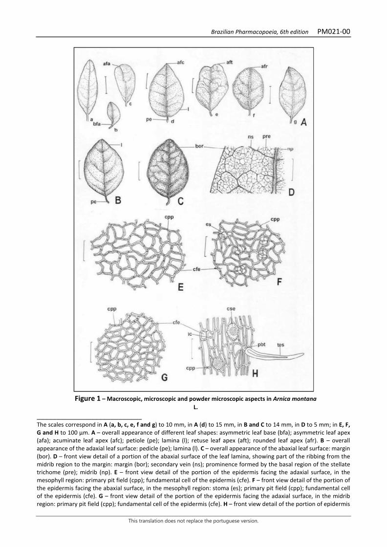
Brazilian Pharmacopoeia, 6th edition PM021-00
This translation does not replace the portuguese version.
Figure 1 – Macroscopic, microscopic and powder microscopic aspects in Arnica montana
L. ___________________________ The scales correspond in A (a, b, c, e, f and g) to 10 mm, in A (d) to 15 mm, in B and C to 14 mm, in D to 5 mm; in E, F, G and H to 100 μm. A – overall appearance of different leaf shapes: asymmetric leaf base (bfa); asymmetric leaf apex (afa); acuminate leaf apex (afc); petiole (pe); lamina (l); retuse leaf apex (aft); rounded leaf apex (afr). B – overall appearance of the adaxial leaf surface: pedicle (pe); lamina (l). C – overall appearance of the abaxial leaf surface: margin (bor). D – front view detail of a portion of the abaxial surface of the leaf lamina, showing part of the ribbing from the midrib region to the margin: margin (bor); secondary vein (ns); prominence formed by the basal region of the stellate trichome (pre); midrib (np). E – front view detail of the portion of the epidermis facing the adaxial surface, in the mesophyll region: primary pit field (cpp); fundamental cell of the epidermis (cfe). F – front view detail of the portion of the epidermis facing the abaxial surface, in the mesophyll region: stoma (es); primary pit field (cpp); fundamental cell of the epidermis (cfe). G – front view detail of the portion of the epidermis facing the adaxial surface, in the midrib region: primary pit field (cpp); fundamental cell of the epidermis (cfe). H – front view detail of the portion of epidermis
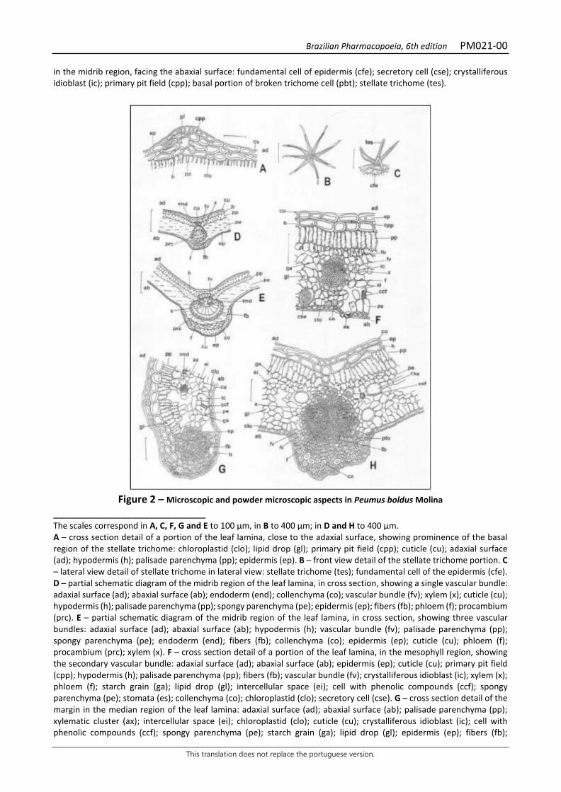
Brazilian Pharmacopoeia, 6th edition PM021-00
This translation does not replace the portuguese version.
in the midrib region, facing the abaxial surface: fundamental cell of epidermis (cfe); secretory cell (cse); crystalliferous idioblast (ic); primary pit field (cpp); basal portion of broken trichome cell (pbt); stellate trichome (tes).
Figure 2 – Microscopic and powder microscopic aspects in Peumus boldus Molina
___________________________ The scales correspond in A, C, F, G and E to 100 μm, in B to 400 μm; in D and H to 400 μm. A – cross section detail of a portion of the leaf lamina, close to the adaxial surface, showing prominence of the basal region of the stellate trichome: chloroplastid (clo); lipid drop (gl); primary pit field (cpp); cuticle (cu); adaxial surface (ad); hypodermis (h); palisade parenchyma (pp); epidermis (ep). B – front view detail of the stellate trichome portion. C – lateral view detail of stellate trichome in lateral view: stellate trichome (tes); fundamental cell of the epidermis (cfe). D – partial schematic diagram of the midrib region of the leaf lamina, in cross section, showing a single vascular bundle: adaxial surface (ad); abaxial surface (ab); endoderm (end); collenchyma (co); vascular bundle (fv); xylem (x); cuticle (cu); hypodermis (h); palisade parenchyma (pp); spongy parenchyma (pe); epidermis (ep); fibers (fb); phloem (f); procambium (prc). E – partial schematic diagram of the midrib region of the leaf lamina, in cross section, showing three vascular bundles: adaxial surface (ad); abaxial surface (ab); hypodermis (h); vascular bundle (fv); palisade parenchyma (pp); spongy parenchyma (pe); endoderm (end); fibers (fb); collenchyma (co); epidermis (ep); cuticle (cu); phloem (f); procambium (prc); xylem (x). F – cross section detail of a portion of the leaf lamina, in the mesophyll region, showing the secondary vascular bundle: adaxial surface (ad); abaxial surface (ab); epidermis (ep); cuticle (cu); primary pit field (cpp); hypodermis (h); palisade parenchyma (pp); fibers (fb); vascular bundle (fv); crystalliferous idioblast (ic); xylem (x); phloem (f); starch grain (ga); lipid drop (gl); intercellular space (ei); cell with phenolic compounds (ccf); spongy parenchyma (pe); stomata (es); collenchyma (co); chloroplastid (clo); secretory cell (cse). G – cross section detail of the margin in the median region of the leaf lamina: adaxial surface (ad); abaxial surface (ab); palisade parenchyma (pp); xylematic cluster (ax); intercellular space (ei); chloroplastid (clo); cuticle (cu); crystalliferous idioblast (ic); cell with phenolic compounds (ccf); spongy parenchyma (pe); starch grain (ga); lipid drop (gl); epidermis (ep); fibers (fb);
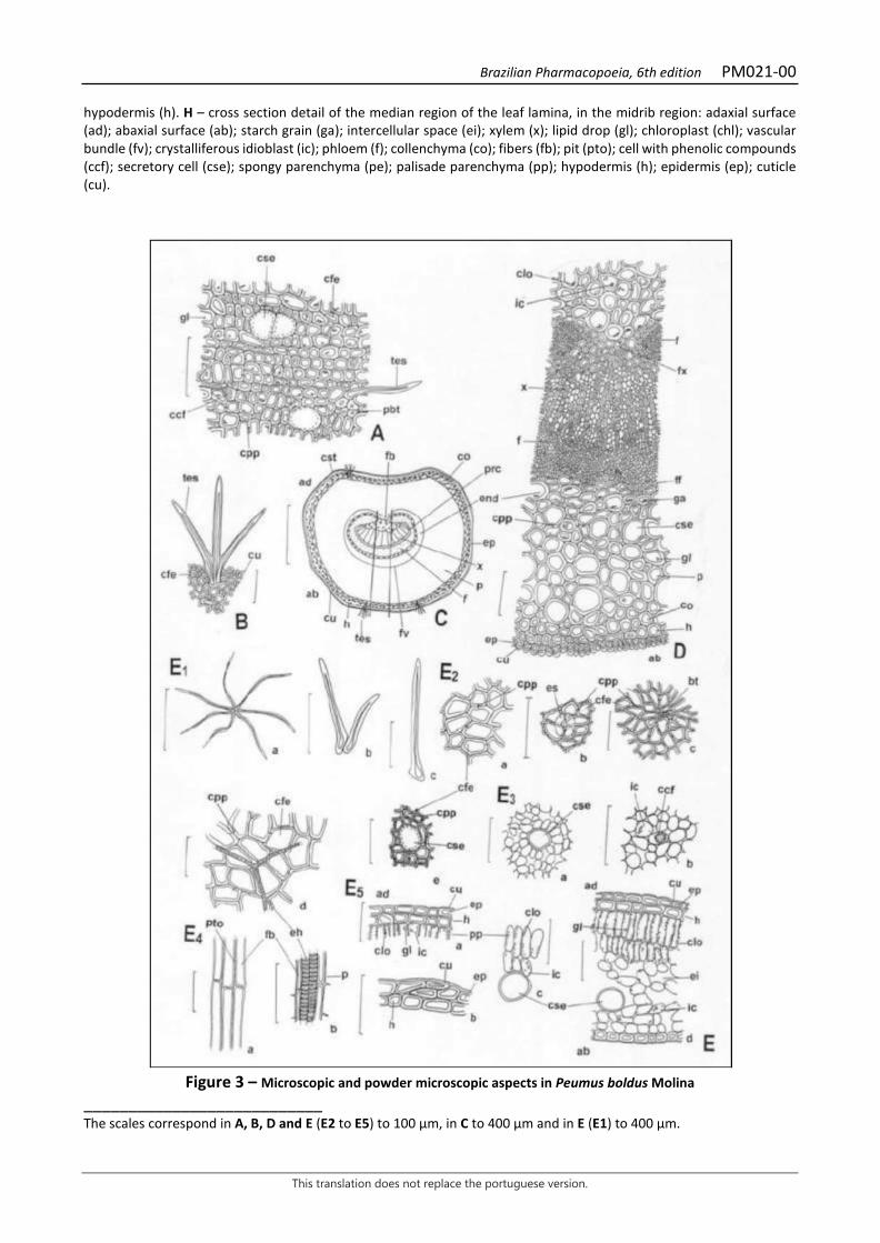
Brazilian Pharmacopoeia, 6th edition PM021-00
This translation does not replace the portuguese version.
hypodermis (h). H – cross section detail of the median region of the leaf lamina, in the midrib region: adaxial surface (ad); abaxial surface (ab); starch grain (ga); intercellular space (ei); xylem (x); lipid drop (gl); chloroplast (chl); vascular bundle (fv); crystalliferous idioblast (ic); phloem (f); collenchyma (co); fibers (fb); pit (pto); cell with phenolic compounds (ccf); secretory cell (cse); spongy parenchyma (pe); palisade parenchyma (pp); hypodermis (h); epidermis (ep); cuticle (cu).
Figure 3 – Microscopic and powder microscopic aspects in Peumus boldus Molina
___________________________ The scales correspond in A, B, D and E (E2 to E5) to 100 μm, in C to 400 μm and in E (E1) to 400 μm.
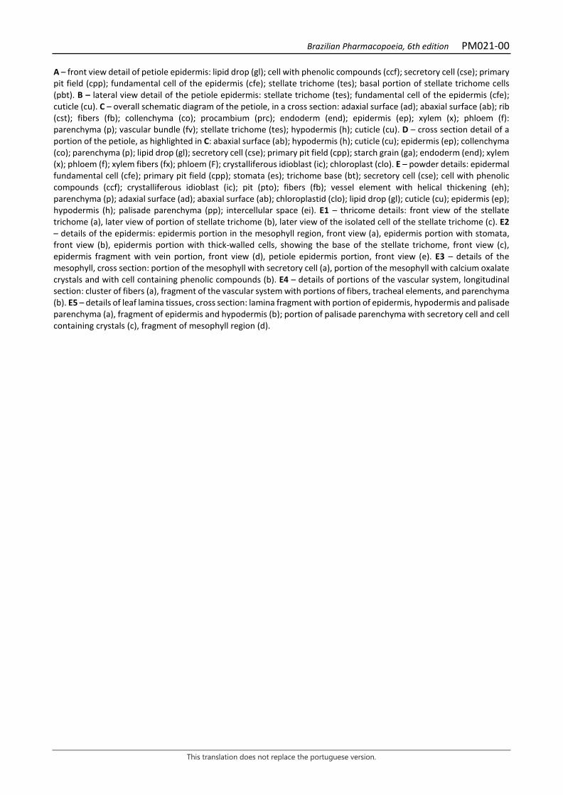
Brazilian Pharmacopoeia, 6th edition PM021-00
This translation does not replace the portuguese version.
A – front view detail of petiole epidermis: lipid drop (gl); cell with phenolic compounds (ccf); secretory cell (cse); primary pit field (cpp); fundamental cell of the epidermis (cfe); stellate trichome (tes); basal portion of stellate trichome cells (pbt). B – lateral view detail of the petiole epidermis: stellate trichome (tes); fundamental cell of the epidermis (cfe); cuticle (cu). C – overall schematic diagram of the petiole, in a cross section: adaxial surface (ad); abaxial surface (ab); rib (cst); fibers (fb); collenchyma (co); procambium (prc); endoderm (end); epidermis (ep); xylem (x); phloem (f): parenchyma (p); vascular bundle (fv); stellate trichome (tes); hypodermis (h); cuticle (cu). D – cross section detail of a portion of the petiole, as highlighted in C: abaxial surface (ab); hypodermis (h); cuticle (cu); epidermis (ep); collenchyma (co); parenchyma (p); lipid drop (gl); secretory cell (cse); primary pit field (cpp); starch grain (ga); endoderm (end); xylem (x); phloem (f); xylem fibers (fx); phloem (F); crystalliferous idioblast (ic); chloroplast (clo). E – powder details: epidermal fundamental cell (cfe); primary pit field (cpp); stomata (es); trichome base (bt); secretory cell (cse); cell with phenolic compounds (ccf); crystalliferous idioblast (ic); pit (pto); fibers (fb); vessel element with helical thickening (eh); parenchyma (p); adaxial surface (ad); abaxial surface (ab); chloroplastid (clo); lipid drop (gl); cuticle (cu); epidermis (ep); hypodermis (h); palisade parenchyma (pp); intercellular space (ei). E1 – thricome details: front view of the stellate trichome (a), later view of portion of stellate trichome (b), later view of the isolated cell of the stellate trichome (c). E2 – details of the epidermis: epidermis portion in the mesophyll region, front view (a), epidermis portion with stomata, front view (b), epidermis portion with thick-walled cells, showing the base of the stellate trichome, front view (c), epidermis fragment with vein portion, front view (d), petiole epidermis portion, front view (e). E3 – details of the mesophyll, cross section: portion of the mesophyll with secretory cell (a), portion of the mesophyll with calcium oxalate crystals and with cell containing phenolic compounds (b). E4 – details of portions of the vascular system, longitudinal section: cluster of fibers (a), fragment of the vascular system with portions of fibers, tracheal elements, and parenchyma (b). E5 – details of leaf lamina tissues, cross section: lamina fragment with portion of epidermis, hypodermis and palisade parenchyma (a), fragment of epidermis and hypodermis (b); portion of palisade parenchyma with secretory cell and cell containing crystals (c), fragment of mesophyll region (d).
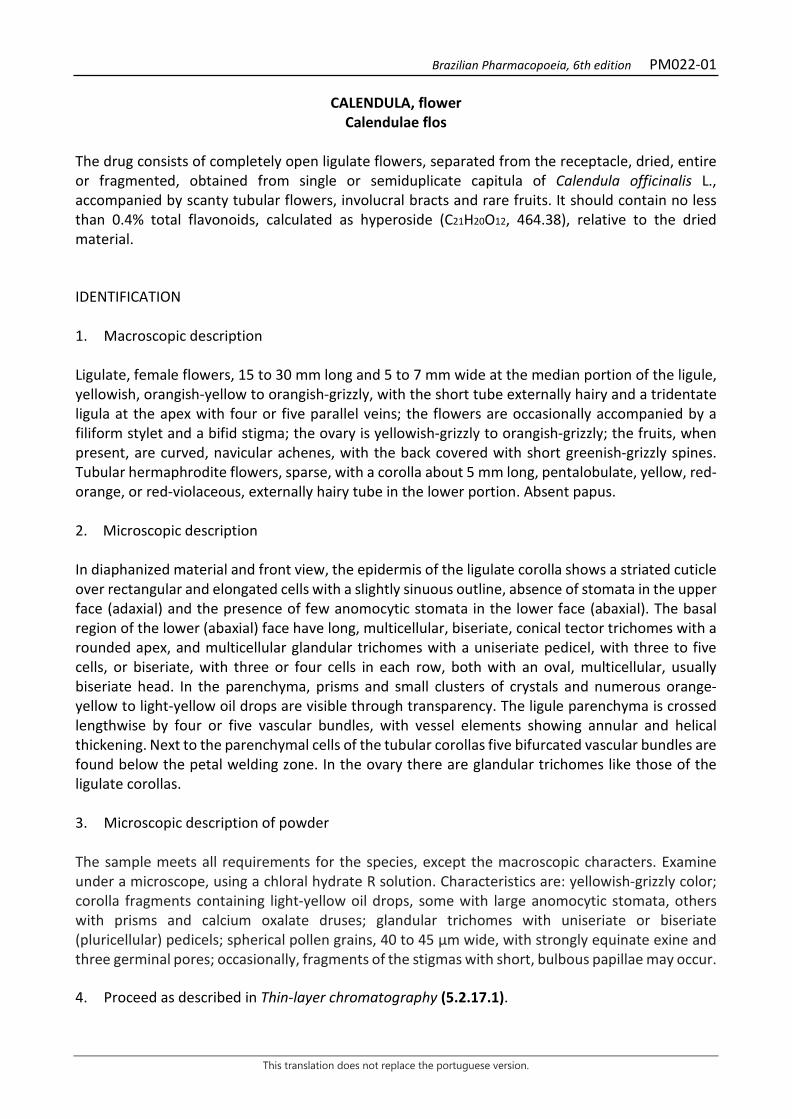
Brazilian Pharmacopoeia, 6th edition PM022-01
This translation does not replace the portuguese version.
CALENDULA, flower Calendulae flos
The drug consists of completely open ligulate flowers, separated from the receptacle, dried, entire or fragmented, obtained from single or semiduplicate capitula of Calendula officinalis L., accompanied by scanty tubular flowers, involucral bracts and rare fruits. It should contain no less than 0.4% total flavonoids, calculated as hyperoside (C21H20O12, 464.38), relative to the dried material. IDENTIFICATION 1. Macroscopic description Ligulate, female flowers, 15 to 30 mm long and 5 to 7 mm wide at the median portion of the ligule, yellowish, orangish-yellow to orangish-grizzly, with the short tube externally hairy and a tridentate ligula at the apex with four or five parallel veins; the flowers are occasionally accompanied by a filiform stylet and a bifid stigma; the ovary is yellowish-grizzly to orangish-grizzly; the fruits, when present, are curved, navicular achenes, with the back covered with short greenish-grizzly spines. Tubular hermaphrodite flowers, sparse, with a corolla about 5 mm long, pentalobulate, yellow, red-orange, or red-violaceous, externally hairy tube in the lower portion. Absent papus. 2. Microscopic description In diaphanized material and front view, the epidermis of the ligulate corolla shows a striated cuticle over rectangular and elongated cells with a slightly sinuous outline, absence of stomata in the upper face (adaxial) and the presence of few anomocytic stomata in the lower face (abaxial). The basal region of the lower (abaxial) face have long, multicellular, biseriate, conical tector trichomes with a rounded apex, and multicellular glandular trichomes with a uniseriate pedicel, with three to five cells, or biseriate, with three or four cells in each row, both with an oval, multicellular, usually biseriate head. In the parenchyma, prisms and small clusters of crystals and numerous orange-yellow to light-yellow oil drops are visible through transparency. The ligule parenchyma is crossed lengthwise by four or five vascular bundles, with vessel elements showing annular and helical thickening. Next to the parenchymal cells of the tubular corollas five bifurcated vascular bundles are found below the petal welding zone. In the ovary there are glandular trichomes like those of the ligulate corollas. 3. Microscopic description of powder The sample meets all requirements for the species, except the macroscopic characters. Examine under a microscope, using a chloral hydrate R solution. Characteristics are: yellowish-grizzly color; corolla fragments containing light-yellow oil drops, some with large anomocytic stomata, others with prisms and calcium oxalate druses; glandular trichomes with uniseriate or biseriate (pluricellular) pedicels; spherical pollen grains, 40 to 45 µm wide, with strongly equinate exine and three germinal pores; occasionally, fragments of the stigmas with short, bulbous papillae may occur. 4. Proceed as described in Thin-layer chromatography (5.2.17.1).
Brazilian Pharmacopoeia, 6th edition PM022-01
This translation does not replace the portuguese version.
Stationary phase: silica gel GF254 (0.25 mm). Mobile phase: ethyl acetate, anhydrous formic acid and water (80:10:10). Sample solution: boil, under reflux, 1 g of the pulverized plant drug with 10 mL of methyl alcohol for 10 minutes and filter. Reference solution: dissolve 2.5 mg rutin, 1 mg caffeic acid, and 1 mg chlorogenic acid in methyl alcohol, top off to 10 mL using the same solvent and homogenize. Procedure: apply 20 μL of the sample solution and 10 μL of the Reference solution to the chromatoplate, separately and in the form of a band. Conduct chromatogram. Remove the chromatoplate, allow it dry in an oven at 100°C to 105°C and, while still warm, nebulize with a solution of 1% aminoethanol diphenylborate (w/v) in methyl alcohol, followed by a solution of 5% (w/v) macrogol 400 in methyl alcohol. Allow the plate to air dry for 30 minutes. Examine under ultraviolet light at 365 nm. Results: the diagram below shows the sequences of zones obtained with the Reference solution and the Sample solution. Other zones may occasionally develop.
Top of the plate
Caffeic acid: blue fluorescence zone
Intense blue fluorescence zone
Chlorogenic acid: light-blue fluorescence zone
Light-blue fluorescence zone
Rutin: yellowish-brown fluorescence zone
Yellowish-brown fluorescence zone
Light-blue fluorescence zone
Reference solution Sample solution
TESTS Foreign matter (5.4.1.3). At most 3.0%. Loss by drying (5.2.9.1). Gravimetric method. At most 12.0%. Total ash (5.4.1.5.1). At most 10.0%. Total mesophilic microorganism count (5.5.3.1.2). Complies with the test.

Brazilian Pharmacopoeia, 6th edition PM022-01
This translation does not replace the portuguese version.
Study of pathogenic microorganisms (5.5.3.1.3). Complies with the test. Heavy metals (5.4.5). Complies with the test. Agrochemical waste (5.4.3). Complies with the test. DOSAGE Total flavonoids To proceed as described in Visible absorption spectrophotometry (5.2.14). Prepare solutions as described below. Stock solution: accurately weigh approximately 0.4 g of the pulverized plant drug (800 µm) (5.2.11) and transfer to a 100-mL round-bottomed flask. Add 1 mL of 0.5% aqueous methenamine solution (w/v), 20 mL acetone and 2 mL hydrochloric acid. Heat in a water bath, under reflux, for 30 minutes. Filter the cotton mixture into a 100-mL volumetric flask, return the drug residue and the cotton to the same round-bottomed flask, add 20 mL acetone. Keep under reflux for 10 minutes. After cooling to room temperature, filter the solution into the 100-mL volumetric flask. Repeat the operation. Next, top off the volume of the volumetric flask with acetone and homogenize. In a separating funnel, add 20 mL of this solution and 20 mL of water, then, extract with 15 mL ethyl acetate. Repeat the extraction three times, with 10 mL portions of ethyl acetate each time. Collect the ethyl acetate phases and rinse them in a separating funnel with two 50-mL portions of water. Transfer the ethyl acetate phase to a 50-mL volumetric flask, top off the volume with ethyl acetate and homogenize. Sample solution: to 10 mL of the Stock solution, add 1 mL of the 2% (w/v) aluminum chloride solution to 5% (v/v) glacial acetic acid solution in methyl alcohol. Dilute in a 25-mL volumetric flask with the 5% (v/v) glacial acetic acid solution in methyl alcohol and homogenize. Blank solution: transfer 10 mL of the Stock solution to a 25-mL volumetric flask, top off the volume with the 5% (v/v) glacial acetic acid solution in methyl alcohol and homogenize. Procedure: measure the absorbance of the Sample Solution at 425 nm in a 1 cm cuvette after precisely 30 minutes, using the Blank solution for zero adjustment. Calculate total flavonoid content, in percent, according to the following expression:
𝑇𝑇𝑇𝑇𝑇𝑇 =𝑇𝑇 × 625𝑚𝑚 × 500
in which, TFT = total flavonoid content expressed as hyperoside % (w/w); A = absorbance measured for the Sample solution; 625 = dilution factor; 500 = hyperoside specific absorption coefficient; m = mass in grams of the sample used, considering the loss by drying.
Brazilian Pharmacopoeia, 6th edition PM022-01
This translation does not replace the portuguese version.
PACKAGING AND STORAGE In tightly closed containers, protected from light and heat.
Figure 1 - Macroscopic, microscopic and powder microscopic aspects in Calendula officinalis L.
___________________________ The scales correspond in A to 1 mm; in B and C to 0,5 mm; in D to 100 µm; in E, F, G, H, I, J and K to 1 µm. A - ligulate pistillate flowers. B - tubular disk flower. C - anthers of the tubular flower, with pollen grains. D - multicellular biseriate trichome of the corolla tube of the ligulate flower. E - fragment of the ligule. F - detail of the edge of the ligule fragment as shown in E, with oil drops in the parenchyma. G - fragment of ligule epidermis with striated cuticle. H - fragment of ligule parenchyma containing oil drops. I - fruit appearance. J - tricolpate pollen grains.
Brazilian Pharmacopoeia, 6th edition PM023-00
This translation does not replace the portuguese version.
CHAMOMILE, flower Matricariae flos
The plant drug consists of dried floral capitula of Matricaria chamomilla L. [syn. Matricaria recutita L. and Chamomilla recutita (L.) Rausch.], containing at least 0.4% volatile oil, and at least 0.25% apigenin-7-O-glucoside (C21H20O10, 432,38). CHARACTERISTICS The inflorescences have a characteristic aromatic odor. IDENTIFICATION 1. Macroscopic description Capitula are 10 to 17 mm wide, consisting of a 3- to 10-mm-wide hemispherical or conical central portion, internally hollow and externally covered with tubular yellow flowers, without paleae, surrounded by 12 to 17 marginal, ligulate, white flowers. Mature, dry capitula with ligulate flowers visibly facing the pedicel. Green involucre, formed by two to three series of bracts oblong-lanceolate, glabrous or with biseriate glandular trichomes in the abaxial surface, imbricate, with obtuse apices and hyaline margin. Marginal pistillate flowers, arranged in a single series, with a short and straight corolla tube, slightly yellowish, up to 1.5 mm long, compressed at the ligule opening; well-developed, tridentate, long-ovate or oblong ligule, seven to 10 mm long by up to two to three mm wide, marked by four longitudinal veins, these rarely accompanied by one or two shorter parallel veins; stylet consisting of two papillose branches. Perfect central flowers, numerous, up to 2.5 mm long, with straight tube and pentalobate limb; acute, equal lobes, widening from a strong constriction, where there is a great density of glandular trichomes; five stamens, synanthers and epipetali; infertile ovary, stylet as in ligulate flowers. Ovoid achene fruit, with three to five longitudinal striations. 2. Microscopic description Bracts of the involucre, when diaphanized and in front view, have a scarious margin formed by elongated, thin-walled cells with a slightly striated cuticle; the epidermis has numerous anomocytic stomata and in the mesophyll, by transparency, conduction elements and many fibers are visible, with several pits. The epidermis of the corolla of ligulate and tubular flowers, in front view, shows a striated cuticle and cells with very thin and slightly sinuous periclinal walls; in cross section, the epidermis of ligulate flowers is strongly papillose both on the abaxial and the adaxial surfaces of the lobes of tubular flowers. Sparse glandular trichomes occur on the epidermis of the ligulate corolla, particularly numerous in the weak constriction that corresponds to the opening of the ligule and also on the abaxial surface and margin of the tubular corollas, where they are abundant. In cross section, small clusters of calcium oxalate crystals occur in the mesophyll of the corollas of both flowers. The cells at the apex of the stylets, in the stigmas, are distinctly papillose. The filaments of the stamens are cylindrical and the epidermis is composed of small cells with slightly thickened walls; the walls of the anthers show clusters of calcium oxalate crystals and, inside, spherical pollen grains with three germination pores and a spiny exine. At the base of the ovary of both types of
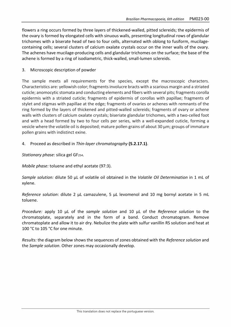
Brazilian Pharmacopoeia, 6th edition PM023-00
This translation does not replace the portuguese version.
flowers a ring occurs formed by three layers of thickened-walled, pitted sclereids; the epidermis of the ovary is formed by elongated cells with sinuous walls, presenting longitudinal rows of glandular trichomes with a biserate head of two to four cells, alternated with oblong to fusiform, mucilage-containing cells; several clusters of calcium oxalate crystals occur on the inner walls of the ovary. The achenes have mucilage-producing cells and glandular trichomes on the surface; the base of the achene is formed by a ring of isodiametric, thick-walled, small-lumen sclereids. 3. Microscopic description of powder The sample meets all requirements for the species, except the macroscopic characters. Characteristics are: yellowish color; fragments involucre bracts with a scarious margin and a striated cuticle; anomocytic stomata and conducting elements and fibers with several pits; fragments corolla epidermis with a striated cuticle; fragments of epidermis of corollas with papillae; fragments of stylet and stigmas with papillae at the edge; fragments of ovaries or achenes with remnants of the ring formed by the layers of thickened and pitted-walled sclereids; fragments of ovary or achene walls with clusters of calcium oxalate crystals; biseriate glandular trichomes, with a two-celled foot and with a head formed by two to four cells per series, with a well-expanded cuticle, forming a vesicle where the volatile oil is deposited; mature pollen grains of about 30 µm; groups of immature pollen grains with indistinct exine. 4. Proceed as described in Thin-layer chromatography (5.2.17.1). Stationary phase: silica gel GF254. Mobile phase: toluene and ethyl acetate (97:3). Sample solution: dilute 50 µL of volatile oil obtained in the Volatile Oil Determination in 1 mL of xylene. Reference solution: dilute 2 µL camazulene, 5 µL levomenol and 10 mg bornyl acetate in 5 mL toluene. Procedure: apply 10 μL of the sample solution and 10 μL of the Reference solution to the chromatoplate, separately and in the form of a band. Conduct chromatogram. Remove chromatoplate and allow it to air dry. Nebulize the plate with sulfur vanillin RS solution and heat at 100 °C to 105 °C for one minute. Results: the diagram below shows the sequences of zones obtained with the Reference solution and the Sample solution. Other zones may occasionally develop.
Brazilian Pharmacopoeia, 6th edition PM023-00
This translation does not replace the portuguese version.
Top of the plate
Chamazulene: pinkish-red colored zone
Pinkish-red colored zone
Bornyl acetate: violaceous-blue colored zone
Violaceous-blue colored zone
Violaceous-colored zone
Levomenol: violaceous color zone
Violaceous-colored zone
Blue-colored zone Greenish-yellow colored area
Reference solution Sample solution TESTS Loss by drying (5.2.9.1). Gravimetric method. At most 12.0%. Heavy metals (5.4.5). Complies with the test. Foreign matter (5.4.1.3). At most 5.0%. Total ash (5.4.1.5.1). At most 10.0%. Total mesophilic microorganism count (5.5.3.1.2). Complies with the test. Study of pathogenic microorganisms (5.5.3.1.3). Complies with the test. Fragmented material. Sieve 20 g of whole plant drug through a fine mesh strainer (710) (5.2.11). At most 25.0% Aflatoxins (5.4.4). Complies with the test. Agrochemical waste (5.4.3). Complies with the test. DOSAGE Volatile oils

Brazilian Pharmacopoeia, 6th edition PM023-00
This translation does not replace the portuguese version.
Proceed as described in Determination of volatile oils in plant drugs (5.4.1.6). Add 30 g of freshly pulverized plant drug into a 1000-mL round-bottomed flask containing 500 mL of water as distillation liquid and 0.5 mL xylene in the graduated tube. Distill at a rate of 3 to 4 mL per minute for four hours. At the end of the period, turn off the water in the condenser, but continue distilling until all the blue content adhering to the walls of the condenser joins the oil collected in the graduated tube. Restart water flow and distill for another 10 minutes. Measure the volume and express the yield per 100 g of the plant drug (w/p). Apigenin-7-O-glucoside Proceed as described in High Performance Liquid Chromatography (5.2.17.4). Use a chromatograph equipped with an ultraviolet detector at 340 mm; pre-column packed with octadecylsilane silica, 250mm long, 4mm internal diameter column, packed with silica chemically bonded to an octadecylsilane group (5 µm); Mobile phase flow rate of 0.6 mL/minute. Eluent (A): water and formic acid (99.5:0.5). Eluent (B): methyl alcohol and formic acid (100:0.08).
Time (minutes) Eluent A (%) Eluent B (%) Elution
0 – 3 75→50 25→50 linear gradient 3 – 20 50 50 isocratic 20 – 23 50→0 50→100 linear gradient 23 – 30 0 100 isocratic 30 – 31 0→75 100→25 linear gradient 31 – 40 75 25 isocratic
Diluent: Eluent (A) and Eluent (B) (75:25) Sample solution: reduce 4 g of the drug to powder (500) (5.2.11). Introduce 0.2 g of the pulverized plant drug into a 50-mL round-bottomed flask and add 20 mL 96% ethyl alcohol. Heat in a water bath, under reflux, for 15 minutes. Cool and filter through absorbent cotton. Rinse the cotton with 2 mL of 96% (v/v) ethyl alcohol. Add 1 mL of freshly prepared 8.5% (w/v) sodium hydroxide solution to the filtrate and heat, under reflux, in a water bath for 1 hour. Cool. Dilute to 25 mL with 96% (v/v) ethyl alcohol. Add 0.05 g of citric acid to 5 mL of this solution. Shake for five minutes. Dilute 500 µL of the filtrate obtained to 1 mL with the Diluent. Filter through a 0.45 µm filter unit. Reference solution: dissolve 1.0 mg of apigenin-7-O-glucoside in 10 mL methyl alcohol. Dilute 250 µL of this solution to 2 mL with the Diluent. Filter through a 0.45 µm filter unit. Procedure: separately inject 10 μL of the Reference solution and 10 µL of the Sample solution. Register the chromatograms and measure the areas under the peaks. Calculate the percentage content of total apigenin-7-O-glucoside according to the following expression:
𝑇𝑇𝑇𝑇 =𝑇𝑇𝑎𝑎 × 𝑚𝑚𝑟𝑟 × 0,625
𝑇𝑇𝑟𝑟 × 𝑚𝑚𝑎𝑎
Brazilian Pharmacopoeia, 6th edition PM023-00
This translation does not replace the portuguese version.
in which,
TA = apigenin-7-O-glucoside content % (w/w); Aa = area under the peak corresponding to apigenin-7-O-glucoside in the Sample solution; Ar = area under the peak corresponding to apigenin-7-O-glucoside in the Reference solution; ma = mass in grams of the sample used, considering the loss by drying. mr = mass in grams of apigenin-7-O-glucoside, considering the purity of the blank substance. System suitability: prepare a solution containing 50 µg/mL rutin in methyl alcohol. Mix 250 µL of the rutin solution to 250 µL of the apigenin-7-O-glucoside Reference solution described in this monograph. Top off the volume up to 1 mL. The chromatogram obtained should show a minimum resolution of two minutes between the apigenin-7-O-glucoside and rutin peaks. PACKAGING AND STORAGE In tightly closed containers, protected from light and heat.

Brazilian Pharmacopoeia, 6th edition PM023-00
This translation does not replace the portuguese version.
Figure 1 ̶ Macroscopic, microscopic and powder microscopic aspects in Matricaria chamomilla L.
___________________________ The scales correspond in A to 1 cm, in B and G to 1 mm, in C to F, H-I to 1 mm, in J-K to 100 µm, in L to Q to 500 µm. A – appearance of the longitudinal section of the capitulum. B - ligulate corolla in lateral view. C - fragment of ligulate corolla showing the stylet divided into two papillose branches. D - lateral view of flower with a tubular corolla. E - tubular flower in a side view, sectioned lengthwise, showing the synantheral stamens. E - tubular flower in a side view, sectioned lengthwise, showing the epipetal stamens. G - fragment of apical portion of the corolla showing the divided stylet. H - isolated tubular corolla. I - isolated fruit. J - lateral view of ring of thickened- and pitted-walled sclereids from the base of the ovary. K - lateral view of ring of thickened- and pitted-walled sclereids from the base of the ovary. L - bract fragment of the involucre with conduction elements and many fibers. M - fragment of corolla with evident papillae. N - lateral view of glandular trichome. O - front view of fragment of corolla epidermis with glandular trichomes. P - fragment of the bract epidermis with anomocytic stomata. Q - isolated pollen grain.
Brazilian Pharmacopoeia, 6th edition PM024-00
This translation does not replace the portuguese version.
CHINESE CINNAMON, husk Cinnamomi cassiae cortex
The plant drug consists of dried husks of Cinnamomum cassia (L.) J. Presl (syn. Cinnamomum aromaticum Nees), containing at least 1.0% volatile oil, consisting of 70.0% to 90.0% trans-cinnamaldehyde. CHARACTERISTICS It has a characteristic aromatic odor that is less pronounced than that of cinnamon. IDENTIFICATION 1. Macroscopic description Husk fragments 3 to 7 cm long, 1 to 2 cm wide, and 1 to 2 mm thick. The external surface, corresponding to the remains of the suber, has a grizzly, brown or grayish coloration, with spots or striations and lenticels; the texture is rugged and not rough. The inner surface, corresponding to the phloem region, is light brown to brown and has a smooth, homogenous texture. 2. Microscopic description The rhytidome is formed by two to three scaling layers, with suberized-walled cells, the outer periclinals being thick. Lenticels are common. The phellogen is formed by tangentially elongated cells, containing phenolic compounds. Internally the phellogen predominates parenchymatic tissue, where isolated or clustered stone cells and mucilaginous idioblasts occur. Oleiferous idioblasts and large numbers of cells containing predominantly simple, or compound, starch grains are observed. Phenolic idioblasts occur in the cortical region. In the innermost region of the cortical parenchyma, near the phloem, there is a continuous, irregular band of stone cells comprising two to ten layers. The phloem has seriated parenchyma and sparse or isolated libriform fibers. Parenchyma rays are formed by two wide cells, rarely three, and five to 18 high cells, where phenolic idioblasts with a high concentration of acicular calcium oxalate crystals similar to raphids, and prismatic crystals are usual. Oleiferous and mucilaginous idioblasts are observed. 3. Microscopic description of powder The sample meets all requirements for the species, except the macroscopic characters. Characteristics are: brown color; isolated and/or grouped, simple or compound starch grains; parenchymatic tissue fragments containing starch grains and lipid drops; large amount of dissociated, acicular and/or prismatic crystals with truncated apex; isolated and/or grouped stone cells, dissociated or inside parenchymatic tissue fragments; rare columnar sclereids, isolated; fibers 600 μm long and 35 μm wide, on average, with thick walls, narrow lumen, isolated or associated with parenchyma fragments. 4. Proceed as described in Thin-layer chromatography (5.2.17.1).

Brazilian Pharmacopoeia, 6th edition PM024-00
This translation does not replace the portuguese version.
Stationary phase: silica gel G (0.250 mm). Mobile phase: methyl chloride. Sample solution: use about 3 g of the powder and shake for 15 minutes with 15 mL methyl chloride. Filter and evaporate to near dryness in a water bath. Dissolve the residue with 0.4 mL toluene. Reference solution: dilute 10 μL eugenol and 50 µL trans-cinnamic aldehyde, in 10 mL toluene. Procedure: apply 10 μL of the Sample solution and 10 μL of the Reference solution to the chromatoplate, separately and in the form of a band. Conduct chromatogram. Remove chromatoplate and allow it to air dry for five minutes. View the fluorescence extinction zones under ultraviolet light at 254 nm. Nebulize the plate with sulfur vanillin RS and place in the oven at 100 °C to 105 °C for one minute. Results: the diagram below shows the sequences of zones obtained with the Reference Solution and the Sample Solution. Other zones may occasionally develop.
Top of the plate
Blue-colored zone Blue-colored zone
Eugenol: brownish-colored zone
Brownish-colored zone
Trans-cinnamic aldehyde: grayish-colored zone
Grayish-colored zone
Blue-colored zone Blue-colored zone
Reference solution Sample solution
TESTS Sulfated Ash (5.4.1.5.2). At most 5.0%. Total mesophilic microorganism count (5.5.3.1.2). Complies with the test. Study of pathogenic microorganisms (5.5.3.1.3). Complies with the test. Heavy metals (5.4.5). Complies with the test. Agrochemical waste (5.4.3). Complies with the test. DOSAGE
Brazilian Pharmacopoeia, 6th edition PM024-00
This translation does not replace the portuguese version.
Volatile oils Proceed as described in Determining volatile oils in plant drugs (5.4.1.6). Use a 1000-mL flask containing 500 mL of water as distillation liquid and 0.5 mL of xylene in the graduated tube. Use 50 g of the pulverized plant drug and distil at a rate of 3 mL to 4 mL per minute, for four hours. Measure the volume and express the yield per 100 g of the plant drug (w/p). Trans-cinnamic aldehyde Proceed as described in Gas chromatography (5.2.17.5). Use a chromatograph equipped with a flame ionization detector, using a mixture of nitrogen, synthetic air, and hydrogen (1:1:10) as auxiliary gases to the detector flame; 30 m long and 0.25 mm wide (internal diameter) capillary chromatography column, coated with polydiphenyldimethylsiloxane, with a 0.25 µm film thickness. Use purified helium at 80 kPa pressure as carrier gas (1 mL/minute). Temperature: Time (minutes) Temperature (ºC) Column 0 – 80 60 → 300 Injector 220 Detector 250 Sample solution: dilute the volatile oil in diethyl ether (2:100). Procedure: inject the volume of 1 μL of the Sample solution into the gas chromatograph, using a 1:50 flow division. Trans-cinnamaldehyde and cis-cinnamaldehyde have linear retention times (Relative Retention Index) of 1270 and 1219, respectively. Determine relative concentrations by manual or electronic integration using the normalization method. Calculate the Relative Retention Index (RRI), according to the following expression:
𝐼𝐼𝐼𝐼𝐼𝐼 = 100 × n +100 × (𝑡𝑡𝑡𝑡𝑥𝑥 − 𝑡𝑡𝑡𝑡𝑧𝑧)
(𝑡𝑡𝑡𝑡𝑥𝑥+1 − 𝑡𝑡𝑡𝑡𝑧𝑧)
in which, RRI = Relative Retention Index n = number of alkane carbon atoms of the lowest molecular mass; trx = retention time of compound "x" (intermediate to trz and trz+1); trz = retention time of the alkane with "n" carbons; trz+1 = retention time of alkane with “n+1” carbons. PACKAGING AND STORAGE In tightly closed containers, protected from light and heat.

Brazilian Pharmacopoeia, 6th edition PM024-00
This translation does not replace the portuguese version.
Figure 1 – Macroscopic, microscopic and powder microscopic aspects in
Cinnamomum cassia (L.) J. Presl ___________________________ The scales correspond in A to 5 mm; in B to 40 μm; in C to 10 μm; in D to 20 μm; in E to 17.5 μm; in F to 3.8 μm; in G to 24.5 μm; in H and I to 37.5 μm. A – overall appearance of the husk portion. B – histological aspect of the external portion of the husk through cross section: stone cells (cp); parenchyma ray (rp); fiber (fb); sieve element (etc). C – detail of an idioblast containing acicular calcium oxalate crystals: crystal (cr); intercellular space (ei). D – longitudinal section of partial detail of phloem portion:
Brazilian Pharmacopoeia, 6th edition PM024-00
This translation does not replace the portuguese version.
fiber (fb); parenchyma (par). E, F, G and H – powder details. E – starch grains. F – truncated and acicular crystals. J - stone cells. H – parenchyma cells with starch grains. I – parenchymal cells with lipid inclusion.
Brazilian Pharmacopoeia, 6th edition PM025-00
This translation does not replace the portuguese version.
CEYLON CINNAMON, bark Cinnamomi zeylanici cortex
The plant drug consists of dried barks of Cinnamomum verum J. Presl (syn. Cinnamomum zeylanicum Blume), free of periderm and outer cortical parenchyma, from the main stem and its branches, containing at least 1.2% volatile oil, containing at least 60% trans-cinnamaldehyde. CHARACTERISTICS The drug has a characteristic aroma of cinnamic aldehyde. IDENTIFICATION 1. Macroscopic description Portions of the bark are curled inward at both margins, forming tubes, averaging 30 cm long and 0.2 to 0.4 mm thick. Externally, the surface is smooth or has slightly darker longitudinal striations, which may or may not be parallel and with undulations that can be regular. It is externally brownish-grizzlyish, while the dark brown to almost vinaceous internally. 2. Microscopic description The bark portion is restricted to the phloem and adjacent tissues. The outer region, in cross section, has stone cells that occur in numerous groups in discontinuous bands in the parenchyma. Prismatic, small acicular, small acicular calcium oxalate crystals, with acute or truncated, rhombohedral apices, and oleiferous and phenolic idioblasts occur simultaneously in the parenchyma cells. Mucilaginous and oleiferous idioblasts occur close to the phloem. The parenchyma rays of the phloem are one to two cells wide and six to 14 cells high and have acicular crystals in large numbers. Simple starch grains occur in all tissues. Lengthwise, sparse, usually isolated, libriform fibers are observed. Branched columnar sclereids can be observed. 3. Microscopic description of powder The sample meets all requirements for the species, except the macroscopic characters. Characteristics are: brown color; abundant, isolated and/or grouped starch grains; isodiametric parenchymatous cells, containing abundant starch grains, as well as lipid drops; large amount of calcium oxalate crystals of prismatic, rhombohedral and/or acicular shape, with truncated or acute apices; numerous fibers, 600 μm long, on average, and 35 μm wide, on average, with thickened walls, narrow lumen, isolated or associated with parenchyma fragments; branched columnar sclereids and abundant stone cells, isolated and/or grouped, dissociated or inside parenchymal tissue fragments. 4. Proceed as described in Thin-layer chromatography (5.2.17.1). Stationary phase: silica gel G (0.250 mm). Mobile phase: methyl chloride.

Brazilian Pharmacopoeia, 6th edition PM025-00
This translation does not replace the portuguese version.
Sample solution: use about 3 g of the powder and shake for 15 minutes with 15 mL methyl chloride. Filter and evaporate to near dryness in a water bath. Dissolve the residue with 0.4 mL toluene. Reference solution: dissolve 10 μL eugenol and 50 µL trans-cinnamic aldehyde, in 10 mL toluene. Procedure: apply 10 μL of the Sample solution and 10 μL of the Reference solution to the chromatoplate, separately and in the form of a band. Conduct chromatogram. Remove chromatoplate and allow it to air dry for five minutes. View the fluorescence extinction zones at 254 nm and the fluorescence zone at 365 nm, under ultraviolet light. Nebulize the plate with sulfur vanillin RS and place in the oven at 100 °C to 105 °C for one minute. Results: the diagram below shows the sequences of zones obtained with the Reference Solution and the Sample Solution. Other zones may occasionally develop.
Top of the plate
Blue-colored zone Blue-colored zone
Eugenol: brownish-colored zone
Brownish-colored zone
Trans-cinnamic aldehyde: grayish-colored zone
Grayish-colored zone
Blue-colored zone Blue-colored zone
Reference solution Sample solution
TESTS Foreign matter (5.4.1.3). At most 2.0%. Total ash (5.4.1.5.1). At most 5.0%. Total mesophilic microorganism count (5.5.3.1.2). Complies with the test. Study of pathogenic microorganisms (5.5.3.1.3). Complies with the test. Heavy metals (5.4.5). Complies with the test.
Brazilian Pharmacopoeia, 6th edition PM025-00
This translation does not replace the portuguese version.
Agrochemical waste (5.4.3). Complies with the test. DOSAGE Volatile oils Proceed as described in Determining volatile oils in plant drugs (5.4.1.6). Use a 1000-mL flask containing 500 mL of water as distillation liquid. Reduce the sample to a coarse powder and immediately proceed with the determination of the volatile oil, from 50 g of the powdered drug (710 µm). Distill for four hours. Measure the volume and express the yield per 100 g of the plant drug (w/p). Trans-cinnamic aldehyde Proceed as described in Gas chromatography (5.2.17.5). Use a gas chromatograph equipped with a flame ionization detector, using a mixture of nitrogen, synthetic air, and hydrogen (1:1:10) as auxiliary gases to the detector flame; 30 m long and 0.25 mm wide (internal diameter) capillary chromatography column, coated with polydiphenyldimethylsiloxane, with a 0.25 µm film thickness. Use helium at 80 kPa pressure as carrier gas (1.0 mL/minute). Temperature: Time (minutes) Temperature (ºC) Column 0 – 80 60→ 300 Injector 220 Detector 250
Sample solution: dilute the volatile oil in diethyl ether (2:100). Procedure: inject the volume of 1 μL of the Sample solution into the gas chromatograph, using a 1:50 flow division. Cinnamic aldehyde has a relative linear retention time of 1266 (Z) and 1214 (E). Determine relative concentrations by manual or electronic integration using the normalization method. Calculate the Relative Retention Index (RRI), according to the following expression:
𝐼𝐼𝐼𝐼𝐼𝐼 = 100 × n +100 × (𝑡𝑡𝑡𝑡𝑥𝑥 − 𝑡𝑡𝑡𝑡𝑧𝑧)
(𝑡𝑡𝑡𝑡𝑥𝑥+1 − 𝑡𝑡𝑡𝑡𝑧𝑧)
in which, RRI = Relative Retention Index n = number of alkane carbon atoms of the lowest molecular mass; trx = retention time of compound "x" (intermediate to trz and trz+1); trz = retention time of alkane with “n” carbons; trz+1 = retention time of alkane with “n+1” carbons.

Brazilian Pharmacopoeia, 6th edition PM025-00
This translation does not replace the portuguese version.
PACKAGING AND STORAGE In tightly closed containers, protected from light and heat.
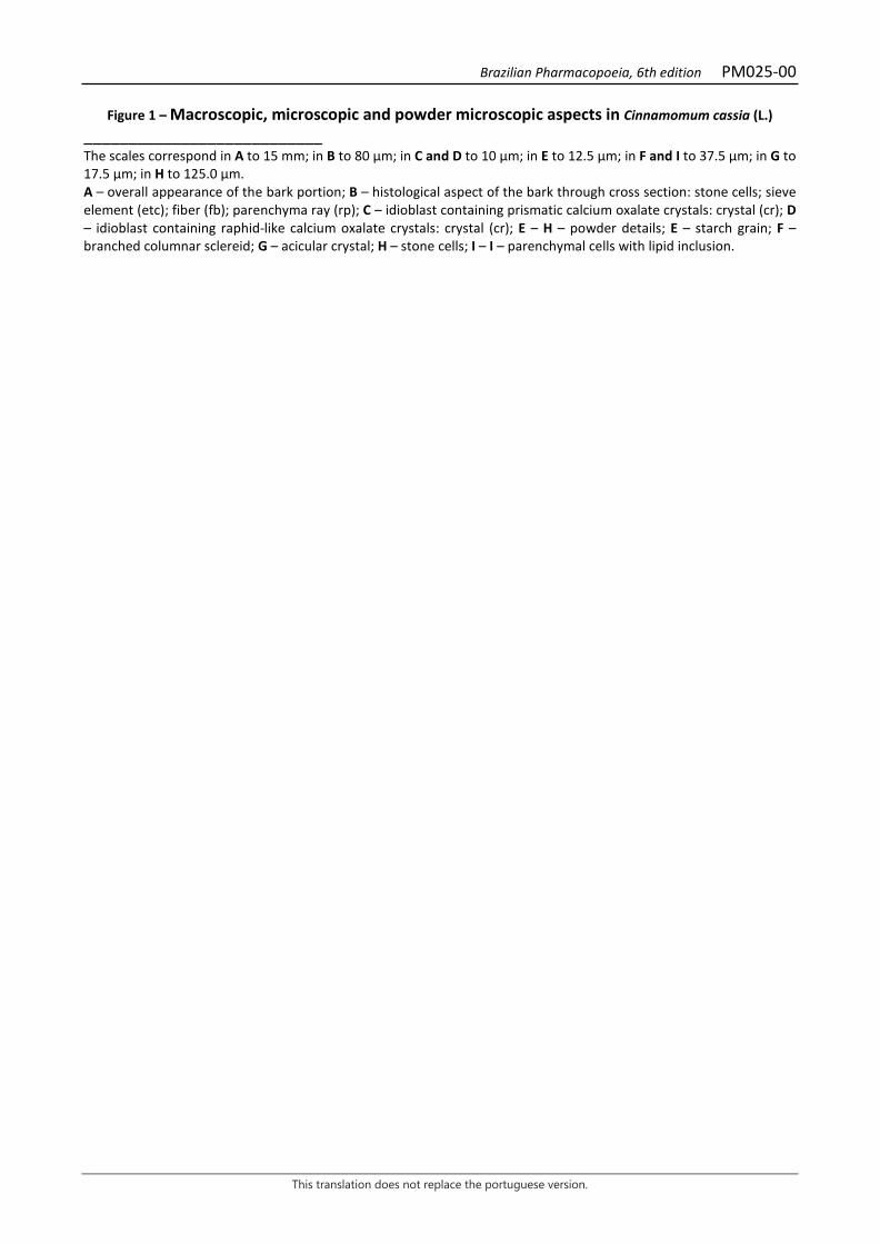
Brazilian Pharmacopoeia, 6th edition PM025-00
This translation does not replace the portuguese version.
Figure 1 – Macroscopic, microscopic and powder microscopic aspects in Cinnamomum cassia (L.) ___________________________ The scales correspond in A to 15 mm; in B to 80 μm; in C and D to 10 μm; in E to 12.5 μm; in F and I to 37.5 μm; in G to 17.5 μm; in H to 125.0 μm. A – overall appearance of the bark portion; B – histological aspect of the bark through cross section: stone cells; sieve element (etc); fiber (fb); parenchyma ray (rp); C – idioblast containing prismatic calcium oxalate crystals: crystal (cr); D – idioblast containing raphid-like calcium oxalate crystals: crystal (cr); E – H – powder details; E – starch grain; F – branched columnar sclereid; G – acicular crystal; H – stone cells; I – I – parenchymal cells with lipid inclusion.

Brazilian Pharmacopoeia, 6th edition PM026-00
This translation does not replace the portuguese version.
LEMON GRASS, leaf Cymbopogonis folium
The plant drug consists of dried leaves of Cymbopogon citratus (DC.) Stapf, containing at least 0.5% volatile oil. COMMON NAMES West Indian lemon grass. CHARACTERISTICS The dried leaves have a characteristic citral odor. IDENTIFICATION 1. Macroscopic description Leaves composed of convoluted sheath and lamina. Sheath enlarged towards the base, 4 to 26 cm long, 0.6 to 6.5 cm wide in the basal region, 1.0 to 3.5 cm in the median region and 0.9 to 2.1 cm in the apical region. Ligule 0.2 cm high, short and truncate, membranous, with simple trichomes at the base of the adaxial surface of the lamina. Lamina 60 to 85 cm long, 0.8 to 1.1 cm wide in the basal region and 1.4 to 1.8 cm in the median region, grayish-green when dry, linear-lanceolate, acuminate in the apex, flat in the expanded portion and canaliculate and narrowed in the basal portion, rough due to the short and siliceous trichomes; entire margin, with rigid and cutting trichomes in larger quantity than in the rest of the lamina; parallel veins, the median one more developed and pronounced in the abaxial surface. 2. Microscopic description A front view of the leaf sheath shows, on the adaxial surface, an epidermis with cells of rectilinear walls, while, on the abaxial surface, the walls are quite sinuous. Scanty unicellular siliceous trichomes and stomata, also arranged in rows, occur in the region between the veins, on both surfaces. In cross section, the fundamental parenchyma is formed by bulky cells that fill almost the entire section, accompanied by secretory cells. A chlorenchyma occurs near the abaxial surface. The vascular bundles are of the collateral type and subepidermal fiber clusters occur facing both surfaces. The leaf lamina, in front view, shows an epidermis of cells arranged in rows and composed of fundamental cells rich in lipid drops and specialized cells: tetracyclic stomata, buliform cells (the latter exclusive to the adaxial surface), suberous cells, and short, unicellular siliceous trichomes. The leaf lamina, in cross section, shows homogenous mesophyll and unistratified epidermis. On the adaxial surface, the fundamental cells in the region of the larger vascular bundles are much smaller than the buliform ones. The vascular bundles are collateral and of different sizes and have specialized kranz-type sheaths; in the more developed bundles, a mestomatic sheath occurs. Strands of fibers occur on both surfaces, opposite the vascular bundles, and on the adaxial surface, they accompany only the more developed vascular bundles. The chlorenchyma cells are radially
Brazilian Pharmacopoeia, 6th edition PM026-00
This translation does not replace the portuguese version.
distributed around the bundles. Secretory cells occur in the border region between the chlorenchyma and the fundamental parenchyma. 3. Histochemical reactions The secretory cells of the sheath and lamina are visualized in lugol reaction, in which the cell contents show dense brown or dense red coloration in fresh or dry material. When reacting with sulfur vanillin, the contents of the secretory cells are brown and dense, and may be collapsed and concentrated near the cell wall. For the reaction with sulfur vanillin, the sections should be immersed in ethyl alcohol, transferred to the vanillin, and flambéed, submerged in it, for two minutes. The slide for observation should be mounted in ethyl alcohol and the sections should not be passed through water. 4. Microscopic description of powder The sample meets all requirements for the species, except the macroscopic characters. Characteristics are: light-green to green-gray color; portions of the epidermis, according to the microscopic description of the leaf; large amounts of fragments of the veins, with siliceous trichomes; portions of the leaf mesophyll, portions of the margin with siliceous trichomes. 5. Proceed as described in Thin-layer chromatography (5.2.17.1). Stationary phase: silica gel GF254 (0.250 mm). Mobile phase: toluene and ethyl acetate (93:7). Sample solution (1): shake about 0.5 g of the pulverized plant drug with 10 mL of methyl chloride in a closed container for 10 minutes. Filter. Concentrate the filtrate to dryness in a water bath at a temperature not exceeding 60 °C. Suspend the residue in 10 mL toluene. Sample solution (2): dilute 2 μL of the volatile oil, obtained in Dosage of Volatile Oils, in 1 mL toluene. Reference solution: dilute 2 µL citral in 1 mL toluene. Procedure: apply 10 μL of the Sample solution (1), 10 μL of the Sample solution (2) and 10 μL of the Reference solution to the chromatoplate, separately and in the form of a band. Conduct chromatogram. Remove chromatoplate and allow it to air dry. Examine under ultraviolet light at 365 nm. Nebulize the plate with sulfur vanillin RS and keep in an oven at 100 °C to 105 °C for one minute. Results: the diagram below shows the sequences of zones obtained with the Reference solution and the Sample solution. Other zones may occasionally develop.
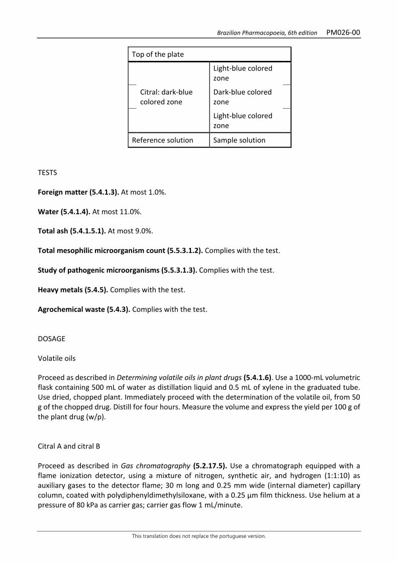
Brazilian Pharmacopoeia, 6th edition PM026-00
This translation does not replace the portuguese version.
Top of the plate
Light-blue colored zone
Citral: dark-blue colored zone
Dark-blue colored zone
Light-blue colored zone
Reference solution Sample solution
TESTS Foreign matter (5.4.1.3). At most 1.0%. Water (5.4.1.4). At most 11.0%. Total ash (5.4.1.5.1). At most 9.0%. Total mesophilic microorganism count (5.5.3.1.2). Complies with the test. Study of pathogenic microorganisms (5.5.3.1.3). Complies with the test. Heavy metals (5.4.5). Complies with the test. Agrochemical waste (5.4.3). Complies with the test. DOSAGE Volatile oils Proceed as described in Determining volatile oils in plant drugs (5.4.1.6). Use a 1000-mL volumetric flask containing 500 mL of water as distillation liquid and 0.5 mL of xylene in the graduated tube. Use dried, chopped plant. Immediately proceed with the determination of the volatile oil, from 50 g of the chopped drug. Distill for four hours. Measure the volume and express the yield per 100 g of the plant drug (w/p). Citral A and citral B Proceed as described in Gas chromatography (5.2.17.5). Use a chromatograph equipped with a flame ionization detector, using a mixture of nitrogen, synthetic air, and hydrogen (1:1:10) as auxiliary gases to the detector flame; 30 m long and 0.25 mm wide (internal diameter) capillary column, coated with polydiphenyldimethylsiloxane, with a 0.25 µm film thickness. Use helium at a pressure of 80 kPa as carrier gas; carrier gas flow 1 mL/minute.
Brazilian Pharmacopoeia, 6th edition PM026-00
This translation does not replace the portuguese version.
Temperature: Time (minutes) Temperature (ºC)
Column 0 – 80 60 → 300 Injector 220 Detector 250
Sample solution: dilute the volatile oil, obtained in Dosage of Volatile Oils, in diethyl ether, at ratio of 2:100. Procedure: inject the volume of 1 μL of the Sample solution into the gas chromatograph, using a 1:50 flow division. Citral A (trans-citral) shows a linear retention time (Relative Retention Index) of 1263 and citral B (cis-citral) of 1233. Determine relative concentrations by manual or electronic integration using the normalization method. Calculate the Relative Retention Index, according to the following expression:
𝐼𝐼𝐼𝐼𝐼𝐼 = 100 × n +100 × (𝑡𝑡𝑡𝑡𝑥𝑥 − 𝑡𝑡𝑡𝑡𝑧𝑧)
(𝑡𝑡𝑡𝑡𝑥𝑥+1 − 𝑡𝑡𝑡𝑡𝑧𝑧)
in which, RRI = Relative Retention Index; n = number of alkane carbon atoms of the lowest molecular mass; trx = retention time of compound "x" (intermediate to trz and trz+1); trz = retention time of alkane with “n” carbons; trz+1 = retention time of alkane with “n+1” carbons. PACKAGING AND STORAGE In tightly closed containers, protected from light and heat.
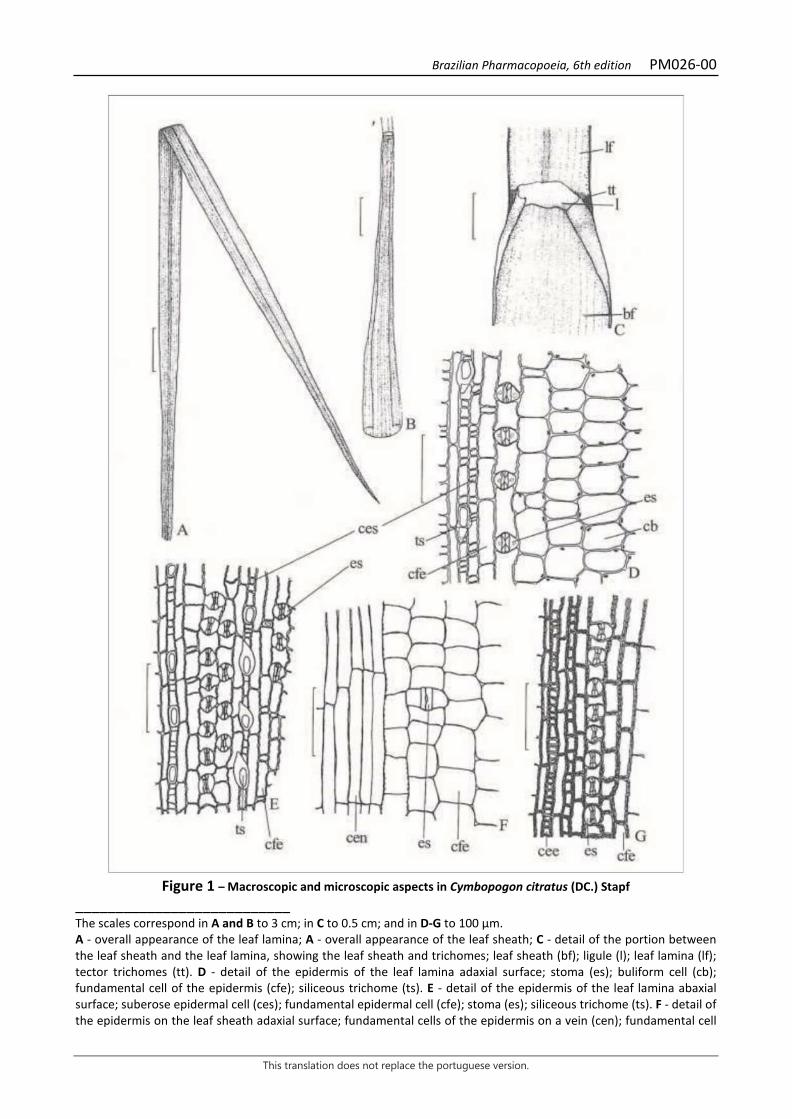
Brazilian Pharmacopoeia, 6th edition PM026-00
This translation does not replace the portuguese version.
Figure 1 – Macroscopic and microscopic aspects in Cymbopogon citratus (DC.) Stapf
___________________________ The scales correspond in A and B to 3 cm; in C to 0.5 cm; and in D-G to 100 µm. A - overall appearance of the leaf lamina; A - overall appearance of the leaf sheath; C - detail of the portion between the leaf sheath and the leaf lamina, showing the leaf sheath and trichomes; leaf sheath (bf); ligule (l); leaf lamina (lf); tector trichomes (tt). D - detail of the epidermis of the leaf lamina adaxial surface; stoma (es); buliform cell (cb); fundamental cell of the epidermis (cfe); siliceous trichome (ts). E - detail of the epidermis of the leaf lamina abaxial surface; suberose epidermal cell (ces); fundamental epidermal cell (cfe); stoma (es); siliceous trichome (ts). F - detail of the epidermis on the leaf sheath adaxial surface; fundamental cells of the epidermis on a vein (cen); fundamental cell
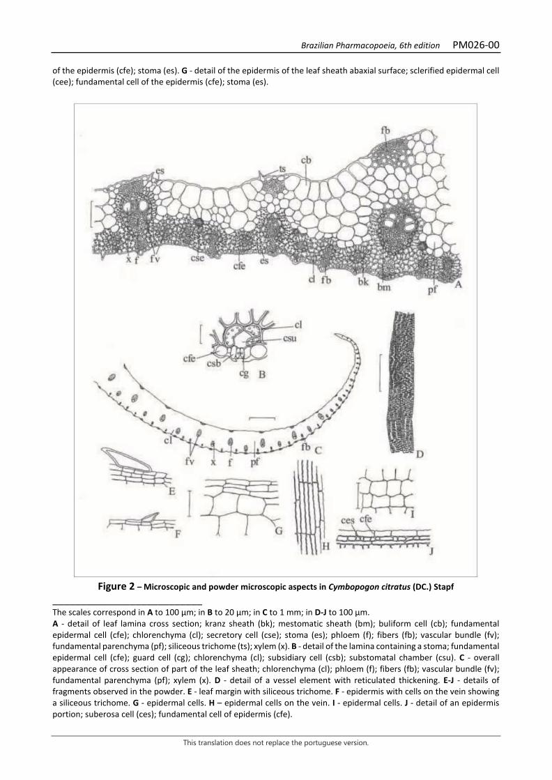
Brazilian Pharmacopoeia, 6th edition PM026-00
This translation does not replace the portuguese version.
of the epidermis (cfe); stoma (es). G - detail of the epidermis of the leaf sheath abaxial surface; sclerified epidermal cell (cee); fundamental cell of the epidermis (cfe); stoma (es).
Figure 2 – Microscopic and powder microscopic aspects in Cymbopogon citratus (DC.) Stapf
___________________________ The scales correspond in A to 100 µm; in B to 20 µm; in C to 1 mm; in D-J to 100 µm. A - detail of leaf lamina cross section; kranz sheath (bk); mestomatic sheath (bm); buliform cell (cb); fundamental epidermal cell (cfe); chlorenchyma (cl); secretory cell (cse); stoma (es); phloem (f); fibers (fb); vascular bundle (fv); fundamental parenchyma (pf); siliceous trichome (ts); xylem (x). B - detail of the lamina containing a stoma; fundamental epidermal cell (cfe); guard cell (cg); chlorenchyma (cl); subsidiary cell (csb); substomatal chamber (csu). C - overall appearance of cross section of part of the leaf sheath; chlorenchyma (cl); phloem (f); fibers (fb); vascular bundle (fv); fundamental parenchyma (pf); xylem (x). D - detail of a vessel element with reticulated thickening. E-J - details of fragments observed in the powder. E - leaf margin with siliceous trichome. F - epidermis with cells on the vein showing a siliceous trichome. G - epidermal cells. H – epidermal cells on the vein. I - epidermal cells. J - detail of an epidermis portion; suberosa cell (ces); fundamental cell of epidermis (cfe).
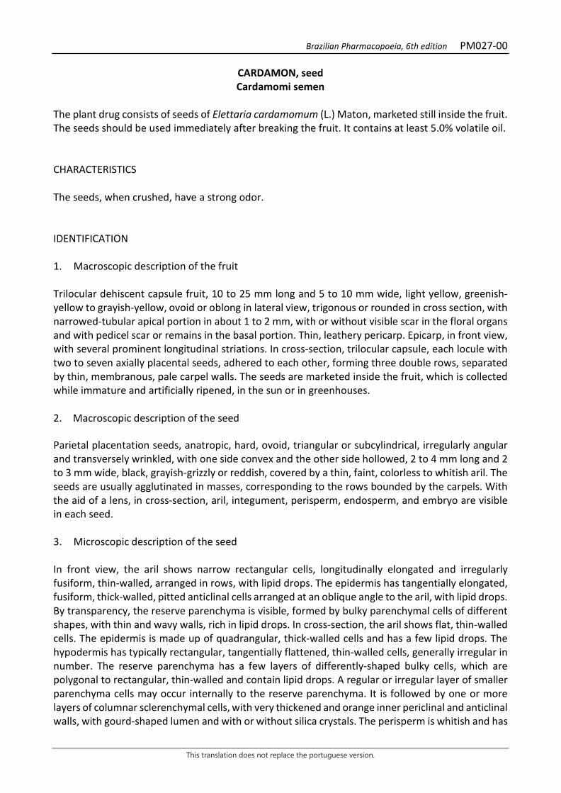
Brazilian Pharmacopoeia, 6th edition PM027-00
This translation does not replace the portuguese version.
CARDAMON, seed Cardamomi semen
The plant drug consists of seeds of Elettaria cardamomum (L.) Maton, marketed still inside the fruit. The seeds should be used immediately after breaking the fruit. It contains at least 5.0% volatile oil. CHARACTERISTICS The seeds, when crushed, have a strong odor. IDENTIFICATION 1. Macroscopic description of the fruit Trilocular dehiscent capsule fruit, 10 to 25 mm long and 5 to 10 mm wide, light yellow, greenish-yellow to grayish-yellow, ovoid or oblong in lateral view, trigonous or rounded in cross section, with narrowed-tubular apical portion in about 1 to 2 mm, with or without visible scar in the floral organs and with pedicel scar or remains in the basal portion. Thin, leathery pericarp. Epicarp, in front view, with several prominent longitudinal striations. In cross-section, trilocular capsule, each locule with two to seven axially placental seeds, adhered to each other, forming three double rows, separated by thin, membranous, pale carpel walls. The seeds are marketed inside the fruit, which is collected while immature and artificially ripened, in the sun or in greenhouses. 2. Macroscopic description of the seed Parietal placentation seeds, anatropic, hard, ovoid, triangular or subcylindrical, irregularly angular and transversely wrinkled, with one side convex and the other side hollowed, 2 to 4 mm long and 2 to 3 mm wide, black, grayish-grizzly or reddish, covered by a thin, faint, colorless to whitish aril. The seeds are usually agglutinated in masses, corresponding to the rows bounded by the carpels. With the aid of a lens, in cross-section, aril, integument, perisperm, endosperm, and embryo are visible in each seed. 3. Microscopic description of the seed In front view, the aril shows narrow rectangular cells, longitudinally elongated and irregularly fusiform, thin-walled, arranged in rows, with lipid drops. The epidermis has tangentially elongated, fusiform, thick-walled, pitted anticlinal cells arranged at an oblique angle to the aril, with lipid drops. By transparency, the reserve parenchyma is visible, formed by bulky parenchymal cells of different shapes, with thin and wavy walls, rich in lipid drops. In cross-section, the aril shows flat, thin-walled cells. The epidermis is made up of quadrangular, thick-walled cells and has a few lipid drops. The hypodermis has typically rectangular, tangentially flattened, thin-walled cells, generally irregular in number. The reserve parenchyma has a few layers of differently-shaped bulky cells, which are polygonal to rectangular, thin-walled and contain lipid drops. A regular or irregular layer of smaller parenchyma cells may occur internally to the reserve parenchyma. It is followed by one or more layers of columnar sclerenchymal cells, with very thickened and orange inner periclinal and anticlinal walls, with gourd-shaped lumen and with or without silica crystals. The perisperm is whitish and has
Brazilian Pharmacopoeia, 6th edition PM027-00
This translation does not replace the portuguese version.
the first layers of tangentially flattened, thin-walled parenchymal cells, filled with starch grains, followed by many layers of irregularly shaped, columnar or polygonal cells, which are bulky, with thin walls and wavy anticlines, containing a large amount of starch grains, lipid drops, and calcium oxalate crystals. The endosperm has elongated parenchymatous cells of several shapes, with thin walls, distributed in several parallel layers, completely surrounding the embryo, containing lipid drops and aleurone grains. The embryo is ovoid and dark-colored, and its cells are rounded to ovoid, thin-walled, with aleurone grains and lipid drops. 4. Microscopic description of the seed powder The sample meets all requirements for the species, except the macroscopic characters, and must not contain elements from the pericarp. Characteristic after the addition of chloral hydrate RS are: grayish-grizzlyish color; fragments of the aril, in front view; fragments of the epidermis, in front view; fragments of the aril with reserve parenchyma cells, visualized by transparency, in front view; fragments of the aril, the epidermis and the reserve parenchyma, visualized by transparency, in front view; fragments of epidermis and reserve parenchyma, visualized by transparency, in front view; fragments of epidermis, in cross-section; fragments of hypodermis, in front view; fragments of reserve parenchyma, in front view; fragments of reserve parenchyma, in cross-section; fragments of sclerenchyma, in front view; fragments of sclerenchyma layer, in cross-section; fragments of sclerenchyma layer and perisperm, in cross-section; fragments of perisperm, in front view or in cross-section; fragments of endosperm, in cross-section; isolated parenchymatous cells; isolated fibers, in longitudinal view; isolated and/or grouped starch grains; isolated calcium oxalate crystals. 5. Proceed as described in Thin-layer chromatography (5.2.17.1). Stationary phase: silica gel GF254 (0.250 mm). Mobile phase: toluene and ethyl acetate (93:7). Sample solution: add 2 mL methyl chloride to 0.1 g of the pulverized plant drug. Shake for 15 minutes, filter, and concentrate to dryness in a water bath at about 60°C. Suspend in 2 mL toluene. Reference solution: dissolve 10 µg terpenyl acetate, 10 µg 1,8-cineole and 10 μL linalool in 1 mL toluene. Procedure: apply 20 μL of the sample solution and 10 μL of the Reference solution to the chromatoplate, separately and in the form of a band. Conduct chromatogram. Remove chromatoplate and allow it to air dry. Next, nebulize the plate with sulfur vanillin RS solution and leave it in an oven between 100 °C and 105 °C for about five minutes. Results: the diagram below shows the sequences of zones obtained with the Reference solution and the Sample solution. Other zones may occasionally develop.
Top of the plate
Violaceous-colored zone Violaceous-colored zone
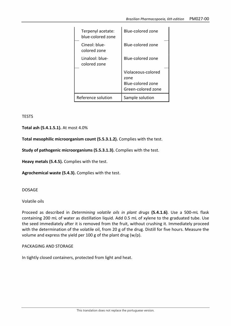
Brazilian Pharmacopoeia, 6th edition PM027-00
This translation does not replace the portuguese version.
Terpenyl acetate: blue-colored zone
Blue-colored zone
Cineol: blue-colored zone
Blue-colored zone
Linalool: blue-colored zone
Blue-colored zone
Violaceous-colored zone Blue-colored zone Green-colored zone
Reference solution Sample solution TESTS Total ash (5.4.1.5.1). At most 4.0% Total mesophilic microorganism count (5.5.3.1.2). Complies with the test. Study of pathogenic microorganisms (5.5.3.1.3). Complies with the test. Heavy metals (5.4.5). Complies with the test. Agrochemical waste (5.4.3). Complies with the test. DOSAGE Volatile oils Proceed as described in Determining volatile oils in plant drugs (5.4.1.6). Use a 500-mL flask containing 200 mL of water as distillation liquid. Add 0.5 mL of xylene to the graduated tube. Use the seed immediately after it is removed from the fruit, without crushing it. Immediately proceed with the determination of the volatile oil, from 20 g of the drug. Distill for five hours. Measure the volume and express the yield per 100 g of the plant drug (w/p). PACKAGING AND STORAGE In tightly closed containers, protected from light and heat.
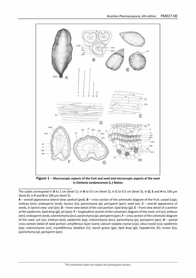
Brazilian Pharmacopoeia, 6th edition PM027-00
This translation does not replace the portuguese version.
Figure 1 – Macroscopic aspects of the fruit and seed and microscopic aspects of the seed
in Elettaria cardamomum (L.) Maton ___________________________ The scales correspond in A to 1 cm (level 1); in B to 0.5 cm (level 2); in C to 0.5 cm (level 3); in D, E and H to 100 μm (level 4); in F and G to 100 μm (level 5). A – overall appearance lateral view: pedicel (ped). B – cross section of the schematic diagram of the fruit: carpel (cap); embryo (em); endosperm (end); loculus (lo); parenchyma (p); perisperm (per); seed (se). C – overall appearance of seeds, in lateral view: aryl (air). D – front view detail of the aryl portion: lipid drop (gl). E – front view detail of a portion of the epidermis: lipid drop (gl); pit (pto). F – longitudinal section of the schematic diagram of the seed: aril (ar); embryo (em); endosperm (end); sclerenchyma (esc); parenchyma (p); perisperm (per). F – cross section of the schematic diagram of the seed: aril (ar); embryo (em); epidermis (ep); sclerenchyma (esc); parenchyma (p); perisperm (per). H – partial cross-section detail of seed portion: amyliferous layer (cam); calcium oxalate crystal (cox); silica crystal (csi); epidermis (ep); sclerenchyma (scl); crystalliferous idioblast (ic); starch grains (ga); lipid drop (gl); hypodermis (h); lumen (lu); parenchyma (p); perisperm (per).
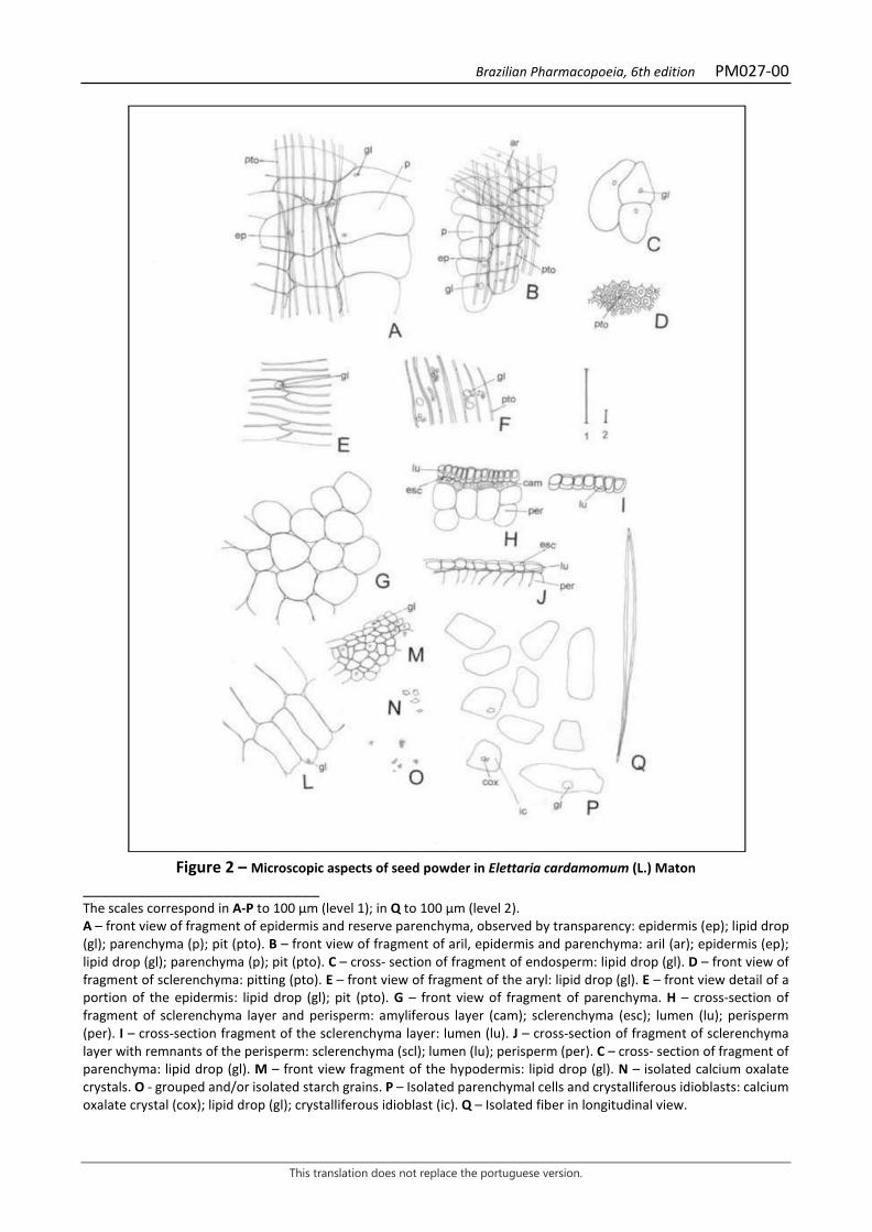
Brazilian Pharmacopoeia, 6th edition PM027-00
This translation does not replace the portuguese version.
Figure 2 – Microscopic aspects of seed powder in Elettaria cardamomum (L.) Maton
___________________________ The scales correspond in A-P to 100 μm (level 1); in Q to 100 μm (level 2). A – front view of fragment of epidermis and reserve parenchyma, observed by transparency: epidermis (ep); lipid drop (gl); parenchyma (p); pit (pto). B – front view of fragment of aril, epidermis and parenchyma: aril (ar); epidermis (ep); lipid drop (gl); parenchyma (p); pit (pto). C – cross- section of fragment of endosperm: lipid drop (gl). D – front view of fragment of sclerenchyma: pitting (pto). E – front view of fragment of the aryl: lipid drop (gl). E – front view detail of a portion of the epidermis: lipid drop (gl); pit (pto). G – front view of fragment of parenchyma. H – cross-section of fragment of sclerenchyma layer and perisperm: amyliferous layer (cam); sclerenchyma (esc); lumen (lu); perisperm (per). I – cross-section fragment of the sclerenchyma layer: lumen (lu). J – cross-section of fragment of sclerenchyma layer with remnants of the perisperm: sclerenchyma (scl); lumen (lu); perisperm (per). C – cross- section of fragment of parenchyma: lipid drop (gl). M – front view fragment of the hypodermis: lipid drop (gl). N – isolated calcium oxalate crystals. O - grouped and/or isolated starch grains. P – Isolated parenchymal cells and crystalliferous idioblasts: calcium oxalate crystal (cox); lipid drop (gl); crystalliferous idioblast (ic). Q – Isolated fiber in longitudinal view.
Brazilian Pharmacopoeia, 6th edition PM028-00
This translation does not replace the portuguese version.
CARQUEJA, winged stem Baccharis trimerae herbae
The plant drug consists of winged, dried and fragmented stems of Baccharis trimera (Less.) DC., containing at least 1.7% total caffeic acids, expressed as chlorogenic acid (C16H18O9, 354.31). COMMON NAMES Bitter carqueja. IDENTIFICATION 1. Macroscopic description Cylindrical, triangular branches, up to 1 m long, aphyllous or with rare sessile leaves reduced at the nodes. Green, membranous wings 0.5 to 1.5 cm wide; wings of floriferous branches narrower than others. Dioecious plant, so when flowering branches are present, they must be only pistillate or only staminate. Inflorescences, when present, of the capitulum type, yellowish-white, numerous, sessile, arranged along the upper branches, forming interrupted spikes, with a flat, non-paleaceous receptacle; flowers with hairy and white papus present. 2. Microscopic description The stem has three divergent wings, with pronounced ribs between each wing. The stem axis, in cross-section, has a unistratified epidermis, covered by a striated cuticle. Few stomata and glandular, biseriate trichomes formed by two basal cells and the head with two series of four cells each occur. The chlorenchyma is formed by three or four layers of cells interrupted in the collenchyma region. The secretory ducts, with epithelium formed of three to 14 cells and accompanied by angular collenchyma, are located externally to the endoderm. A continuous layer of endoderm with Caspary striations is internal to the chlorenchyma. The vascular system is of the collateral type. The fiber strands of the phloem are made up of up to seven cell layers. Internal to the xylem is an almost continuous band of fibers located near the medullary parenchyma. The medulla is formed by spherical or elliptical cells, containing prismatic and pyramidal calcium oxalate crystals, mainly arranged in the perimedullary zone. In cross-section, the wings exhibit an isobilateral structure. The epidermis is unistratified and covered by a striated cuticle. Anomocytic and anisocytic stomas occur, distributed on both surfaces. The tector and glandular trichomes occur predominantly in the region of the wing margins and at the joining of the wings with the stem axis. Tector trichomes rarely occur and are multicellular and uniseriate with about six cells extending towards the apex and with a tapering, T-shaped apical cell. The glandular trichomes are similar to those on the stem axis. Collateral vascular bundles are arranged linearly in the spongy parenchyma, accompanied by few fibers and surrounded by a parenchymatic sheath. Each bundle is accompanied by one or two secretory ducts with epithelium of four to 14 cells. The collenchyma is restricted to only one subepidermal layer near the wing margin vein; below it, a group of strongly thickened walled fibers occurs surrounding one to three secretory ducts of different sizes. 3. Microscopic description of powder
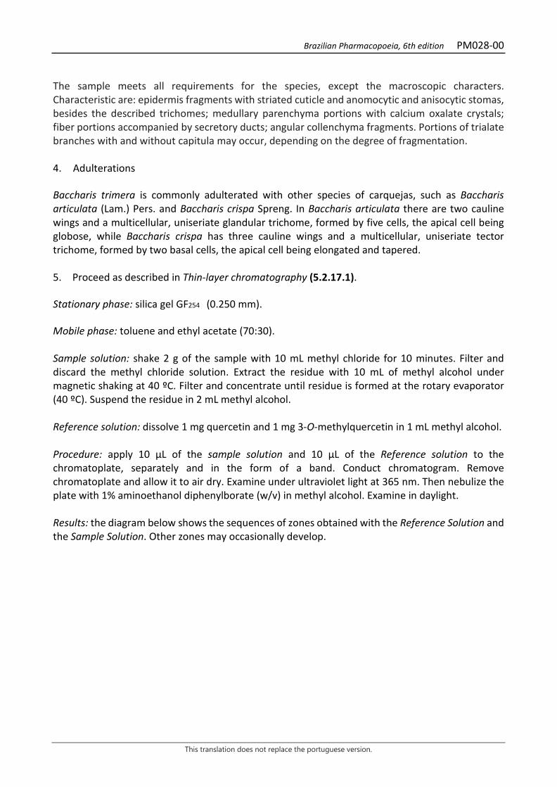
Brazilian Pharmacopoeia, 6th edition PM028-00
This translation does not replace the portuguese version.
The sample meets all requirements for the species, except the macroscopic characters. Characteristic are: epidermis fragments with striated cuticle and anomocytic and anisocytic stomas, besides the described trichomes; medullary parenchyma portions with calcium oxalate crystals; fiber portions accompanied by secretory ducts; angular collenchyma fragments. Portions of trialate branches with and without capitula may occur, depending on the degree of fragmentation. 4. Adulterations Baccharis trimera is commonly adulterated with other species of carquejas, such as Baccharis articulata (Lam.) Pers. and Baccharis crispa Spreng. In Baccharis articulata there are two cauline wings and a multicellular, uniseriate glandular trichome, formed by five cells, the apical cell being globose, while Baccharis crispa has three cauline wings and a multicellular, uniseriate tector trichome, formed by two basal cells, the apical cell being elongated and tapered. 5. Proceed as described in Thin-layer chromatography (5.2.17.1). Stationary phase: silica gel GF254 (0.250 mm). Mobile phase: toluene and ethyl acetate (70:30). Sample solution: shake 2 g of the sample with 10 mL methyl chloride for 10 minutes. Filter and discard the methyl chloride solution. Extract the residue with 10 mL of methyl alcohol under magnetic shaking at 40 ºC. Filter and concentrate until residue is formed at the rotary evaporator (40 ºC). Suspend the residue in 2 mL methyl alcohol. Reference solution: dissolve 1 mg quercetin and 1 mg 3-O-methylquercetin in 1 mL methyl alcohol. Procedure: apply 10 μL of the sample solution and 10 μL of the Reference solution to the chromatoplate, separately and in the form of a band. Conduct chromatogram. Remove chromatoplate and allow it to air dry. Examine under ultraviolet light at 365 nm. Then nebulize the plate with 1% aminoethanol diphenylborate (w/v) in methyl alcohol. Examine in daylight. Results: the diagram below shows the sequences of zones obtained with the Reference Solution and the Sample Solution. Other zones may occasionally develop.
Brazilian Pharmacopoeia, 6th edition PM028-00
This translation does not replace the portuguese version.
Top of the plate
Quercetin: orange fluorescence zone 3-O-Methyl-quercetin: orange fluorescence zone
Orange fluorescence zone Orange fluorescence zone
Reference solution Sample solution TESTS Foreign matter (5.4.1.3). At most 2.0%. Water (5.4.1.4). At most 12.0%. Total ash (5.4.1.5.1). At most 8.0%. Total mesophilic microorganism count (5.5.3.1.2). Complies with the test. Study of pathogenic microorganisms (5.5.3.1.3). Complies with the test. Heavy metals (5.4.5). Complies with the test. Agrochemical waste (5.4.3). Complies with the test. DOSAGE Total caffeic acids Proceed as described in High Performance Liquid Chromatography (5.2.17.4). Use a chromatograph equipped with an ultraviolet detector at 325 mm; pre-column packed with octadecylsilane silica, 75 mm long, 4.6 mm internal diameter column, packed with silica chemically bonded to an octadecylsilane group (4 µm), kept at room temperature; Mobile phase flow rate of 0.6 mL/minute. Eluent (A): water, acetonitrile and trifluoracetic acid (95:5:0.05). Eluent (B): acetonitrile.
Brazilian Pharmacopoeia, 6th edition PM028-00
This translation does not replace the portuguese version.
Time (minutes) Eluent A (%) Eluent B (%) Elution
0 – 30 100 → 57 0 → 43 linear gradient 30 – 35 57 → 0 43 → 100 linear gradient 35 – 36 0 → 100 100 → 0 linear gradient 36 – 42 100 0 isocratic
Sample solution: accurately weigh approximately 0.5 g of the dried and pulverized plant drug (250 µm) (5.2.11) in a 50-mL beaker. Add 10 mL of the mixture of ethyl alcohol and water (50:50) and place in a water bath (40 °C) for 10 minutes. Cool the extract to room temperature. Filter extract through absorbent cotton into a 25-mL volumetric flask. Re-extract the drug residue retained on the cotton with 10 mL of a mixture of ethyl alcohol and water (50:50), place in a water bath (40 °C) for 10 minutes. Cool and filter into the same 25-mL volumetric flask. Top off the volume with an ethyl alcohol and water (50:50) mixture and homogenize. Dilute 0.12 mL of the resulting solution in 1 mL of a mixture of water, acetonitrile and trifluoracetic acid (95:5:0.05). Filter through a 0.45 µm filter unit. Stock solution: dissolve 5.6 mg chlorogenic acid in 5 mL methyl alcohol. Solutions for the analytical curve: dilute a 0.2 mL aliquot of the Stock solution to 0.4 mL with a water-acetonitrile (95:5) mixture to a 0.56 mg/mL solution. Dilute the previous solution successively in a mixture of water and acetonitrile (95:5) to obtain concentrations of 0.28 mg/mL, 0.14 mg/mL, 0.07 mg/mL, 0.035 mg/mL, 0.017 mg/mL, and 0.0085 mg/mL. Filter through a 0.45 µm filter unit. Procedure: inject 10 µL of the Solutions for the analytical curve and 10 µL of the sample solution separately. Register the chromatograms and measure the areas under the peaks. The approximate relative retention times compared to chlorogenic acid are 1.69 for 3.4-dicaffeoylquinic acid; 1.76 for 3.5-dicaffeoylquinic acid, and 1.84 for 4.5-dicaffeoylquinic acid. Calculate the percentage chlorogenic acid content in the sample (w/w), considering the determined water content, from the straight line equation obtained with the Solutions for Analytical Curve. PACKAGING AND STORAGE In tightly closed containers, protected from light and heat.
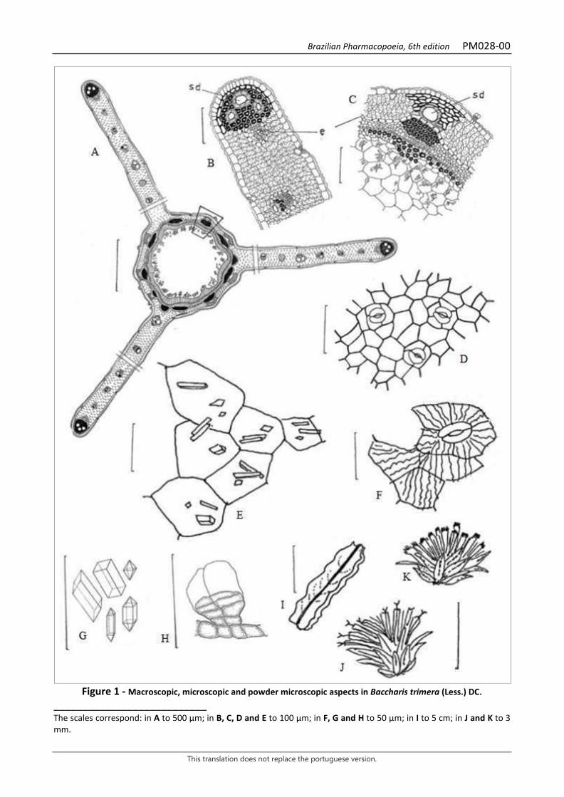
Brazilian Pharmacopoeia, 6th edition PM028-00
This translation does not replace the portuguese version.
Figure 1 - Macroscopic, microscopic and powder microscopic aspects in Baccharis trimera (Less.) DC.
___________________________ The scales correspond: in A to 500 µm; in B, C, D and E to 100 µm; in F, G and H to 50 µm; in I to 5 cm; in J and K to 3 mm.
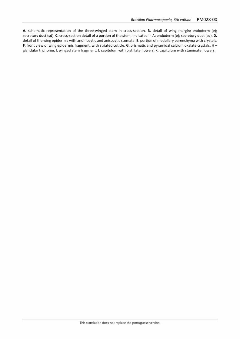
Brazilian Pharmacopoeia, 6th edition PM028-00
This translation does not replace the portuguese version.
A. schematic representation of the three-winged stem in cross-section. B. detail of wing margin; endoderm (e); secretory duct (sd). C. cross-section detail of a portion of the stem, indicated in A; endoderm (e); secretory duct (sd). D. detail of the wing epidermis with anomocytic and anisocytic stomata. E. portion of medullary parenchyma with crystals. F. front view of wing epidermis fragment, with striated cuticle. G. prismatic and pyramidal calcium oxalate crystals. H – glandular trichome. I. winged stem fragment. J. capitulum with pistillate flowers. K. capitulum with staminate flowers.
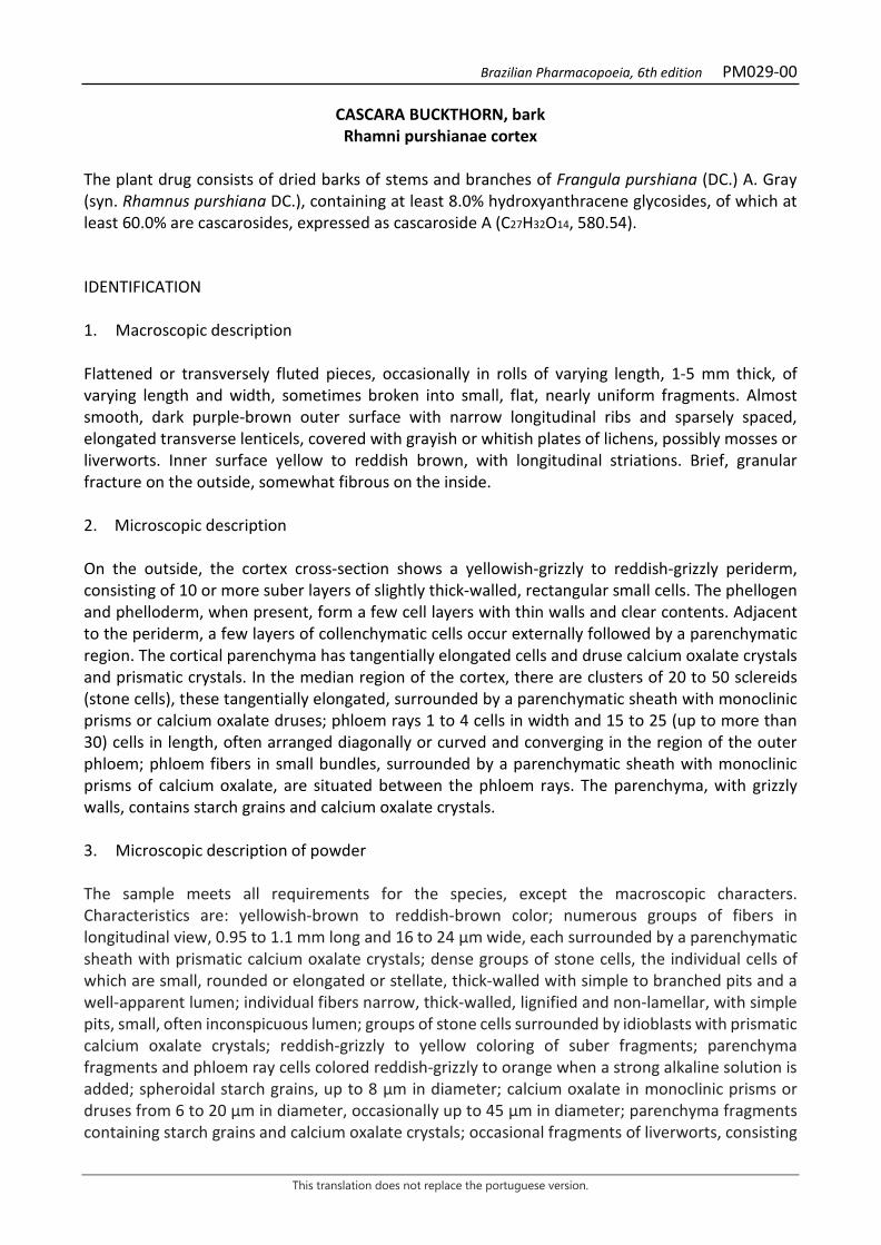
Brazilian Pharmacopoeia, 6th edition PM029-00
This translation does not replace the portuguese version.
CASCARA BUCKTHORN, bark Rhamni purshianae cortex
The plant drug consists of dried barks of stems and branches of Frangula purshiana (DC.) A. Gray (syn. Rhamnus purshiana DC.), containing at least 8.0% hydroxyanthracene glycosides, of which at least 60.0% are cascarosides, expressed as cascaroside A (C27H32O14, 580.54). IDENTIFICATION 1. Macroscopic description Flattened or transversely fluted pieces, occasionally in rolls of varying length, 1-5 mm thick, of varying length and width, sometimes broken into small, flat, nearly uniform fragments. Almost smooth, dark purple-brown outer surface with narrow longitudinal ribs and sparsely spaced, elongated transverse lenticels, covered with grayish or whitish plates of lichens, possibly mosses or liverworts. Inner surface yellow to reddish brown, with longitudinal striations. Brief, granular fracture on the outside, somewhat fibrous on the inside. 2. Microscopic description On the outside, the cortex cross-section shows a yellowish-grizzly to reddish-grizzly periderm, consisting of 10 or more suber layers of slightly thick-walled, rectangular small cells. The phellogen and phelloderm, when present, form a few cell layers with thin walls and clear contents. Adjacent to the periderm, a few layers of collenchymatic cells occur externally followed by a parenchymatic region. The cortical parenchyma has tangentially elongated cells and druse calcium oxalate crystals and prismatic crystals. In the median region of the cortex, there are clusters of 20 to 50 sclereids (stone cells), these tangentially elongated, surrounded by a parenchymatic sheath with monoclinic prisms or calcium oxalate druses; phloem rays 1 to 4 cells in width and 15 to 25 (up to more than 30) cells in length, often arranged diagonally or curved and converging in the region of the outer phloem; phloem fibers in small bundles, surrounded by a parenchymatic sheath with monoclinic prisms of calcium oxalate, are situated between the phloem rays. The parenchyma, with grizzly walls, contains starch grains and calcium oxalate crystals. 3. Microscopic description of powder The sample meets all requirements for the species, except the macroscopic characters. Characteristics are: yellowish-brown to reddish-brown color; numerous groups of fibers in longitudinal view, 0.95 to 1.1 mm long and 16 to 24 μm wide, each surrounded by a parenchymatic sheath with prismatic calcium oxalate crystals; dense groups of stone cells, the individual cells of which are small, rounded or elongated or stellate, thick-walled with simple to branched pits and a well-apparent lumen; individual fibers narrow, thick-walled, lignified and non-lamellar, with simple pits, small, often inconspicuous lumen; groups of stone cells surrounded by idioblasts with prismatic calcium oxalate crystals; reddish-grizzly to yellow coloring of suber fragments; parenchyma fragments and phloem ray cells colored reddish-grizzly to orange when a strong alkaline solution is added; spheroidal starch grains, up to 8 µm in diameter; calcium oxalate in monoclinic prisms or druses from 6 to 20 µm in diameter, occasionally up to 45 µm in diameter; parenchyma fragments containing starch grains and calcium oxalate crystals; occasional fragments of liverworts, consisting

Brazilian Pharmacopoeia, 6th edition PM029-00
This translation does not replace the portuguese version.
of rounded cells arranged in a single layer, with irregularly thickened cells, and fragments of mosses consisting of small elongated, narrow-walled cells, usually in a single layer or occasionally in two or three. 4. Proceed as described in Thin-layer chromatography (5.2.17.1). Stationary phase: 250 µm thick silica gel F254. Mobile phase: ethyl acetate, methyl alcohol and water (100:17:13). Sample solution: weigh 0.5 g of the plant drug and add 5 mL of 70% (v/v) ethyl alcohol. Heat in a water bath for 15 minutes. Filter the sample. Evaporate in a water bath to dryness, at maximum temperature of 60 °C. Suspend the residue in 2 mL methyl alcohol Reference solution (1): dissolve an accurately weighed amount of aloin in methyl alcohol to obtain a concentration of 1 µg/mL. Reference solution (2): dissolve an accurately weighed amount of emodin in methyl alcohol to obtain a concentration of 1 µg/mL. Procedure: apply 20 μL of the Sample Solution, 20 μL of the Reference Solution (1) and 20 μL of the Reference Solution (2) to the chromatoplate, separately and in the form of a band. Conduct chromatogram. Remove plate and allow it to air dry. Nebulize the plate with 5% (w/v) potassium hydroxide solution in ethyl alcohol. Examine under ultraviolet light at 365 nm. Heat the chromatoplate at 100 °C to 105 °C for five minutes. Examine under visible light. Results: the diagram below shows the sequences of zones obtained with the Reference Solution (1), the Reference solution (2) and the Sample Solution. Other zones may occasionally develop.
Top of the plate
Emodin: reddish-colored zone Red-colored zone
Aloin: yellowish-colored zone
Yellowish-colored zone
Yellow-colored zone
Reference solution Sample solution
Brazilian Pharmacopoeia, 6th edition PM029-00
This translation does not replace the portuguese version.
TESTS Heavy metals (5.4.5). Complies with the test. Foreign matter (5.4.1.3). At most 1.0%. Loss by drying (5.2.9.1). Gravimetric method. At most 10.0%. Total ash (5.4.1.5.1). At most 7.0% Total mesophilic microorganism count (5.5.3.1.2). Complies with the test. Study of pathogenic microorganisms (5.5.3.1.3). Complies with the test. Aflatoxins (5.4.4). Complies with the test. Agrochemical waste (5.4.3). Complies with the test. DOSAGE Stock solution: weigh, accurately, about 1.0 g of plant drug, add 100 mL of boiling water, and leave in contact for five minutes. Transfer the mixture into a 100-mL volumetric flask. Top off the volume with distilled water and homogenize. Filter the sample and discard the first 20 mL. Transfer 10 mL of the filtrate to a separating funnel and add 0.1 mL of M hydrochloric acid. Extract with 20 mL of an hexane and diethyl ether (3:1) mixture. Repeat the extraction twice. After separating the phases, set the aqueous phase aside. Rinse the organic phase with 5 mL of water. Discard the organic phase and combine the aqueous phase with the wash waters. Extract the aqueous phase with 30 mL of water-saturated ethyl acetate, prepared at the time of the analysis (150 mL ethyl acetate and 15 mL water, mixed for three minutes). Repeat the extraction four times. Combine the organic fractions obtained with ethyl acetate. Use the aqueous phase to determine cascaroside dosage and the organic phase to determine hydroxyanthracene glycoside dosage without cascarosides. Hydroxyanthracene glycosides without cascarosides To proceed as described in Visible absorption spectrophotometry (5.2.14). Prepare solutions as described below. Sample solution: transfer the organic phase from the Stock solution to a porcelain capsule. Evaporate in a water bath until residue is formed. Dissolve the residue in 0.3 to 0.5 mL methyl alcohol and transfer to a 50-mL volumetric flask. Rinse the capsule with hot water and transfer the residue to the 50-mL volumetric flask. Top off the volume with water and homogenize. Then transfer 20 mL of the solution to a 100mL round-bottomed flask, add 2 g of ferric chloride hexahydrate and 12 mL of hydrochloric acid. Heat the mixture under reflux for four hours. After cooling, transfer the solution to a separating funnel. Rinse the flask with 3 to 4 mL of M sodium hydroxide, then with 3 to 4 mL of water. Transfer the wash water to the separating funnel. Extract with 30 mL of a hexane and diethyl ether (3:1) mixture. Repeat the extraction three times. Transfer the organic phase to another separating funnel and rinse it twice, using 10 mL of water in each rinse. Discard the aqueous

Brazilian Pharmacopoeia, 6th edition PM029-00
This translation does not replace the portuguese version.
phase. After this procedure, dilute the organic phase to 100 mL with the hexane and diethyl ether (3:1) mixture. Then transfer 20 mL and evaporate until residue is formed in a water bath. Dissolve the residue with 10 mL of a 5 g/L magnesium acetate solution in methyl alcohol. Procedure: measure absorbance at 440 nm and 515 nm. Use methyl alcohol for zero adjustment. A ratio between the absorbance values at 515 nm and 440 nm that is smaller than 2.4 invalidates the assay. Calculate the percentage content of hydroxyanthracene glycosides without cascarosides (HAC), according to the following expression:
𝑇𝑇𝑇𝑇𝑇𝑇𝐶𝐶 =𝑇𝑇 × 6,95
𝑚𝑚
in which, THAC = hydroxyanthracene glycoside content without cascarosides % (w/w); A = absorbance measured at 515 nm for the Sample solution; m = mass in grams of the sample used, considering the loss by drying. Cascarosides Sample solution: dilute the aqueous phase in a 50-mL volumetric flask with water. Use 20 mL of the solution. Blank solution: methyl alcohol. Procedure: measure the absorbance of the Sample solution at 440 nm and 515 nm, using the Blank solution for zero adjustment. A ratio between the absorbance values at 515 nm and 440 nm that is smaller than 2.4 invalidates the assay. Calculate cascaroside content, in percent, according to the following expression:
𝑇𝑇𝑇𝑇𝑇𝑇𝐶𝐶 =𝑇𝑇 × 6,95
𝑚𝑚
in which, TC = cascaroside content % (w/w); A = absorbance measured at 515 nm for the Sample solution; m = mass in grams of the sample used, considering the loss by drying. PACKAGING AND STORAGE In tightly closed containers, protected from light and heat.
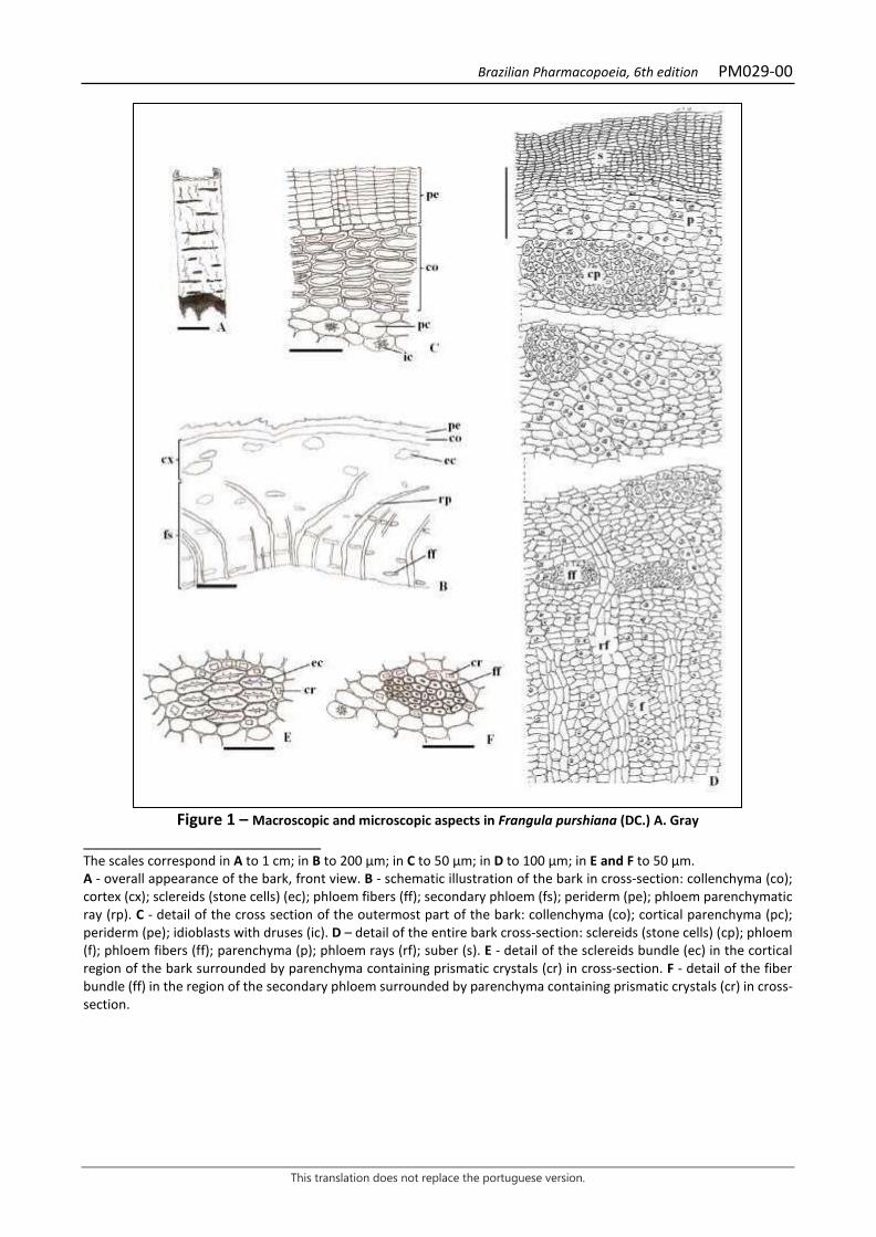
Brazilian Pharmacopoeia, 6th edition PM029-00
This translation does not replace the portuguese version.
Figure 1 – Macroscopic and microscopic aspects in Frangula purshiana (DC.) A. Gray
___________________________ The scales correspond in A to 1 cm; in B to 200 µm; in C to 50 µm; in D to 100 µm; in E and F to 50 µm. A - overall appearance of the bark, front view. B - schematic illustration of the bark in cross-section: collenchyma (co); cortex (cx); sclereids (stone cells) (ec); phloem fibers (ff); secondary phloem (fs); periderm (pe); phloem parenchymatic ray (rp). C - detail of the cross section of the outermost part of the bark: collenchyma (co); cortical parenchyma (pc); periderm (pe); idioblasts with druses (ic). D – detail of the entire bark cross-section: sclereids (stone cells) (cp); phloem (f); phloem fibers (ff); parenchyma (p); phloem rays (rf); suber (s). E - detail of the sclereids bundle (ec) in the cortical region of the bark surrounded by parenchyma containing prismatic crystals (cr) in cross-section. F - detail of the fiber bundle (ff) in the region of the secondary phloem surrounded by parenchyma containing prismatic crystals (cr) in cross-section.
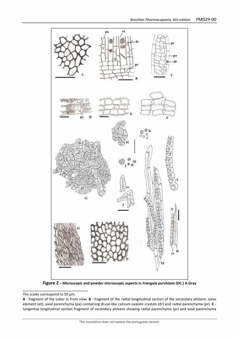
Brazilian Pharmacopoeia, 6th edition PM029-00
This translation does not replace the portuguese version.
Figure 2 – Microscopic and powder microscopic aspects in Frangula purshiana (DC.) A.Gray
___________________________ The scales correspond to 50 µm. A - fragment of the suber in front view. B - fragment of the radial longitudinal section of the secondary phloem: sieve element (et); axial parenchyma (pa) containing druse-like calcium oxalate crystals (dr) and radial parenchyma (pr). C - tangential longitudinal section fragment of secondary phloem showing radial parenchyma (pr) and axial parenchyma

Brazilian Pharmacopoeia, 6th edition PM029-00
This translation does not replace the portuguese version.
(pa) containing druse (dr). D - fragment of the cortical parenchyma with starch grains (ga). E - fragment of phloem parenchyma with bulging cell walls. F – fragment of parenchyma. G – fragment with sclereids (stone cells). H – grouped sclereids. I – druses. J – isolated sclereids (stone cells) (ec). K – isolated starch grains. L – isolated crystalliferous parenchymatic sheath. M – phloem fibers with crystalliferous sheaths. N – fragment of phloem fiber with crystalliferous sheath. O – moss fragment. P – liverwort fragment.
Brazilian Pharmacopoeia, 6th edition PM030-00
This translation does not replace the portuguese version.
HORSE CHESTNUT, seed Hippocastani semen
The plant drug consists of mature, dried seeds of Aesculus hippocastanum L., containing at least 3.0% triterpene glycosides, calculated as anhydrous aescin. IDENTIFICATION 1. Macroscopic description The seeds are hard, irregularly ovoid or subspherical, 2.5 to 4.0 cm wide, flattened at both poles or only at the hilum, or irregularly flattened by desiccation. The fractured seed has an integument 1.0 to 2.0 mm thick, smooth, leathery, brittle, reddish-brown or light brown, usually glossy, rarely opaque, and with a large light spot corresponding to the hilum at one pole. The embryo has a small radicle and two large corneous, starchy cotyledons, light brown externally and almost white at the fracture. 2. Microscopic description Externally, the seed integument shows a thick and smooth cuticle and a yellowish-brown uni-stratified epidermis, with thick-walled, polygonal cells in front view and columnar and compact cells, radially oriented, forming a palisade, in cross-section. Below it, up to four distinct zones can be observed: the first, outermost zone, is formed by a few layers of colenchymatic cells of yellowish-brown color; the second is formed by ten or more layers of sclerenchymatic cells, flattened tangentially and of yellowish-brown coloration; the third is formed by four to ten layers of parenchymatic cells, colorless, with a more polyhedral shape and thinner walls than those of the previous regions, presenting intercellular spaces; the fourth zone, which may be absent, is formed by some layers of tangentially flattened cells with thickened walls. Delicate vascular bundles occur in this parenchyma; the vessel elements are narrow and have helical wall thickening. The starch grains are simple, spherical, oval, or pyriform, with diameter of 2 µm to 80 µm. The smaller grains have a generally point-shaped hilum; the others, larger and more numerous, have a cross-shaped, branched, or star-shaped hilum. Few grains consisting of two to four units occur. 3. Microscopic description of powder The sample meets all requirements for the species, except the macroscopic characters. Characteristic are: irregular, golden-yellow testa fragments with irregularly contoured, tightly interconnected cells whose boundaries are not recognizable, with cell wall extensions appearing tubiform, with a narrow, fiber-like lumen in cross-section; testa fragments showing thickened-walled cells; testa epidermal fragments, in front view, with uniformly thickened periclinal walls, and, when in cross-section, with strongly thickened radial and outer periclinal walls, resembling a narrow palisade, with reddish-brown cells; reserve parenchyma fragments, with flattened to elliptical cells, containing starch grains and lipid drops; reserve parenchyma fragments with portions of vascular bundles; abundant starch grains, isolated or grouped, of different sizes and shapes, as described. When subjected to cold chloral hydrate, the starch swells immediately. In cotyledonary tissue fragments, subjected to long boiling, the starch does not lose its characteristic sticky nature. In such tissues, colorless lipid drops are observed both inside the cells and scattered around the fragments.
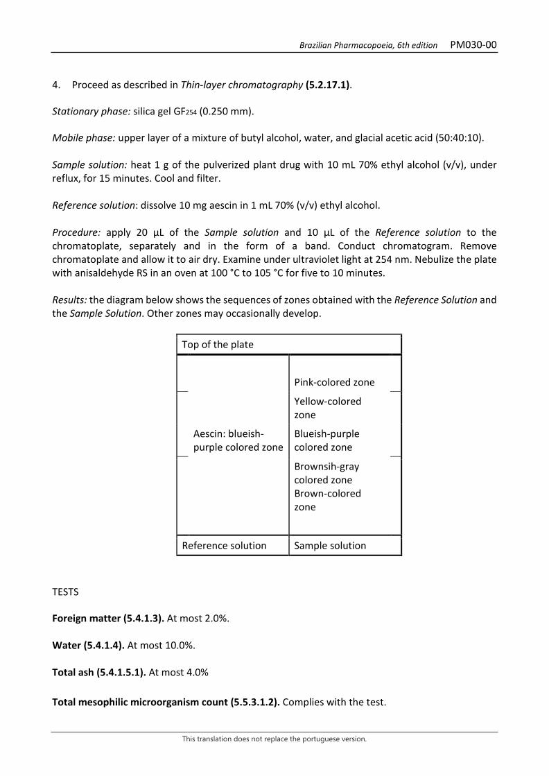
Brazilian Pharmacopoeia, 6th edition PM030-00
This translation does not replace the portuguese version.
4. Proceed as described in Thin-layer chromatography (5.2.17.1). Stationary phase: silica gel GF254 (0.250 mm). Mobile phase: upper layer of a mixture of butyl alcohol, water, and glacial acetic acid (50:40:10). Sample solution: heat 1 g of the pulverized plant drug with 10 mL 70% ethyl alcohol (v/v), under reflux, for 15 minutes. Cool and filter. Reference solution: dissolve 10 mg aescin in 1 mL 70% (v/v) ethyl alcohol. Procedure: apply 20 μL of the Sample solution and 10 μL of the Reference solution to the chromatoplate, separately and in the form of a band. Conduct chromatogram. Remove chromatoplate and allow it to air dry. Examine under ultraviolet light at 254 nm. Nebulize the plate with anisaldehyde RS in an oven at 100 °C to 105 °C for five to 10 minutes. Results: the diagram below shows the sequences of zones obtained with the Reference Solution and the Sample Solution. Other zones may occasionally develop.
Top of the plate
Pink-colored zone
Yellow-colored zone
Aescin: blueish-purple colored zone
Blueish-purple colored zone
Brownsih-gray colored zone Brown-colored zone
Reference solution Sample solution
TESTS Foreign matter (5.4.1.3). At most 2.0%. Water (5.4.1.4). At most 10.0%. Total ash (5.4.1.5.1). At most 4.0% Total mesophilic microorganism count (5.5.3.1.2). Complies with the test.
Brazilian Pharmacopoeia, 6th edition PM030-00
This translation does not replace the portuguese version.
Study of pathogenic microorganisms (5.5.3.1.3). Complies with the test. Heavy metals (5.4.5). Complies with the test. Agrochemical waste (5.4.3). Complies with the test. Aflatoxins (5.4.4). Complies with the test. DOSAGE Aescin To proceed as described in Visible absorption spectrophotometry (5.2.14). Prepare solutions as described below. Stock solution: transfer 1 g of pulverized plant drug to a 250-mL flask and add 100 mL of 65% methyl alcohol (v/v). Accurately weigh the assembly and heat it, under reflux, in a water bath for 30 minutes. Cool, top off the initial weight with 65% (v/v) methyl alcohol. Filter. Evaporate 30 mL of the filtrate to dryness in a 100-mL flask, under reduced pressure. Dissolve the residue in 20 mL 0.1 M hydrochloric acid, transfer to a 250-mL separating funnel, and wash the flask with two 5 mL portions of 0.1 M hydrochloric acid. Combine the acid phases. Extract with a mixture of 20 mL n-propyl alcohol and 50 mL chloroform, shake vigorously for two minutes. Separate the lower organic phase. Add 30 mL of 0.1 M hydrochloric acid to the remaining phase in the funnel and extract with a mixture of 20 mL n-propyl alcohol and 50 mL chloroform. Shake vigorously for two minutes. Separate the lower phase and recombine it to the lower phase of the previous extraction. Evaporate the combined solutions under reduced pressure to dryness. Rinse the residue with four 10 mL portions of peroxide-free diethyl ether. Filter the ether phase. Rinse the filter with 10 mL peroxide-free diethyl ether. Discard the filtrate. Discard the diethyl ether remaining on the filter and in the flask. Rinse the filter and the flask containing the residue with glacial acetic acid and transfer to a 50-mL volumetric flask. Top off the volume with glacial acetic acid and homogenize. Sample solution: transfer 2 mL of the Stock solution to a test tube and add 4 mL ferric acid chloride RS. Homogenize. Blank solution: transfer 2 mL glacial acetic acid to a test tube and add 4 mL ferric acid chloride RS. Homogenize. Procedure: heat the test tubes in a water bath at 60 °C for 25 minutes. Cool to room temperature and measure the absorbance at 540 nm, using the Blank solution for zero adjustment. Calculate aescin content, in percent, according to the following expression:
𝑇𝑇𝑇𝑇 =𝑇𝑇 × 8,333𝑚𝑚 × 60
in which, TE = aescin content % (w/w); A = absorbance measured for the Sample solution;
Brazilian Pharmacopoeia, 6th edition PM030-00
This translation does not replace the portuguese version.
m = mass in grams of the sample used, considering the determined water content; 60 = specific absorption coefficient.

Brazilian Pharmacopoeia, 6th edition PM030-00
This translation does not replace the portuguese version.
PACKAGING AND STORAGE In tightly closed containers, protected from light and heat.
Figure 1 – Macroscopic, microscopic and powder microscopic aspects in Aesculus hippocastanum L.
___________________________ The scales correspond in A and B to 0.5 cm, in C to 300 µm, in D-G to 100 µm, and in H to 50 µm. A. schematic Illustrations of the seed, abaxial and adaxial view, showing the hilum region; hilum (h). C – cross section of the schematic diagram of the seed. C. details of the seed in cross-section as shown in B; collenchyma (co); sclerenchyma (el); epidermis (ep); fundamental parenchyma (pf); inner testa parenchyma, with thickened cell walls (pit); cotyledon

Brazilian Pharmacopoeia, 6th edition PM030-00
This translation does not replace the portuguese version.
reserve parenchyma (pr). D. front view detail of the epidermis of the seed integument. E. detail of the testa epidermis, in cross-section. F. Sclerenchyma cells in cross-section. G. cotyledonary reserve parenchyma cells; starch grain (ga); lipid drop (gl). H. starch grains.
Brazilian Pharmacopoeia, 6th edition PM031-00
This translation does not replace the portuguese version.
ASIATIC PENNYWORT, leaf Centellae folium
The plant drug consists of dried leaves of Centella asiatica (L.) Urb., containing not less than 2.0% asiaticoside relative to the dried material (C48H78O19, 959.12). IDENTIFICATION 1. Macroscopic description The leaf laminae are membranous, rarely papyraceous, grayish-green on the adaxial surface and pale green on the abaxial surface, glabrous to tomentose on both sides, covered with hyaline trichomes of up to 2 mm, pluricellular, uniseriate, formed by two to five cells. The lower cell originates in a single basal cell. Oval to orbicular-reniform lamina, palmately palmate, with five to nine veins, cordate to truncate base, rounded, obtuse to truncate apex, slightly sinuate to crenate-dentate margin, 1.5 to 7 cm long and 1 to 6 cm wide. Venation is not very dense, actinodromous. The first-order veins are straight lengthwise. The second order veins show moderate divergence angle. The branches of the secondary and tertiary veins terminate in the epithema of the hydathodes. Areolas are pentagonal or polygonal, with single, curved or only-once branched venule and randomly arranged. Petiole up to 15 cm long, enlarged at the basal portion and canaliculate on the adaxial surface, villous-tomentose, greenish-brown to reddish-brown. 2. Microscopic description In front view, the epidermis shows polygonal cells with straight to curved walls, projected stomata, paracitic, rare anisocytic, striated cuticle, simple, uniseriate, twisted trichomes, formed by two to five cells, usually three, scarce on the adaxial surface. Hydathode occur on the leaf margin. In cross-section, the epidermis shows flattened rectangular cells, alternated with papillose quadrangular cells; the projection of stomata is more evident and the cuticle is thin. The mesophyll is dorsiventral, with one to three layers of loose palisade parenchyma and spongy parenchyma occupying more than half of the mesophyll, formed by oblong cells in the horizontal direction; calcium oxalate druses occur in these parenchymas. In the median vein, there are about two secretory ducts, arranged in the region of the fundamental parenchyma, one facing the adaxial and the other the abaxial surface, close to the vascular system; the collenchyma is lacunar and formed by one to three layers, more evident on the adaxial surface. The vascular system is of the collateral type, open arch. The petiole is fistulous and, in cross-section, shows a circular outline, with two opposite edges on the adaxial surface, separated by a small, slightly concave region. The epidermis is formed by quadrangular cells, somewhat papillose, with paracitic stomata and simple trichomes, like those on the leaf lamina; the cuticle is thin and striated. The collenchyma is angular, continuous, followed by a chlorenchyma, containing seven collateral vascular bundles, arranged in a circle, separated by wide bands of fundamental parenchyma. The phloem may have amyliferous cells, and is always accompanied by a fiber cap. Secretory ducts occur internally to the collenchyma, others approximately equidistant from the vascular bundles and the epidermis, two opposite each other in the same vascular bundle. In the fundamental parenchyma are calcium oxalate druses. 3. Microscopic description of powder

Brazilian Pharmacopoeia, 6th edition PM031-00
This translation does not replace the portuguese version.
The sample meets all requirements for the species, except the macroscopic characters. Characteristics are: epidermal fragments with uniseriate trichomes or portions; parenchyma fragments with calcium oxalate druses; isolated calcium oxalate druses; epidermal fragments with striated cuticle; epidermal fragments with paracitic stomata and striated cuticle; rare fragments of epidermal cells with anisocytic stomata; fragments with areoles and more rarely with hydathodes; lamina fragments, in cross-section, showing projected stomata; fragments of loose parenchyma; fragments with secretory duct; fragments of petiole parenchyma with collenchyma portions. 4. Proceed as described in Thin-layer chromatography (5.2.17.1). Stationary phase: silica gel GF254 (0.250 mm). Mobile phase: chloroform, glacial acetic acid, methyl alcohol and water (60:32:12:8). Sample solution: boil 3 g of the sample (355 µm) in 30 mL of a mixture of ethyl alcohol and water (1:1). Filter and concentrate to dryness. Resume in 0.5 mL methyl alcohol. Reference solution: dissolve 1 mg asiaticoside in 1 mL methyl alcohol. Procedure: apply 20 μL of the Sample solution and 5 μL of the Reference solution to the chromatoplate, separately and in the form of a band. Conduct chromatogram. Remove the chromatoplate and dry in a fume hood for five minutes. Nebulize with anisaldehyde RS in an oven at 100 °C to 105 °C for 10 minutes. Next, nebulize again with anisaldehyde RS in an oven at 100 °C to 105 °C for 10 minutes. Results: the diagram below shows the sequences of zones obtained with the Reference Solution and the Sample Solution. Other zones may occasionally develop.
Top of the plate
Purple-colored zone
Asiaticoside: brownish-clored zone
Brownish-colored zone
Reference solution Sample solution TESTS Foreign matter (5.4.1.3). At most 2.0%. Water (5.4.1.4). At most 12.0%. Total ash (5.4.1.5.1). At most 11.0%.
Brazilian Pharmacopoeia, 6th edition PM031-00
This translation does not replace the portuguese version.
Acid-insoluble ash (5.4.1.5.3). At most 2.0%. Foam level (5.4.1.8). Determine in 1 g of the pulverized plant drug, transfer to a test tube and boil for two minutes. Use 100 mL of distilled water. At most 100. Total mesophilic microorganism count (5.5.3.1.2). Complies with the test. Study of pathogenic microorganisms (5.5.3.1.3). Complies with the test. Heavy metals (5.4.5). Complies with the test. Agrochemical waste (5.4.3). Complies with the test. DOSAGE Asiaticoside Proceed as described in High Performance Liquid Chromatography (5.2.17.4). Use a chromatograph equipped with an ultraviolet detector at 200 mm; 250mm long, 4.6 mm internal diameter column, packed with silica chemically bonded to an octadecylsilane group (3 µm to 10 µm), kept at room temperature; Mobile phase flow rate of 0.5 mL/minute. Eluent (A): acetonitrile. Eluent (B): 0.5% phosphoric acid (v/v) (99:1). Mobile phase gradient: adopt the gradient system described on the following table:
Time (minutes) Eluent A (%) Eluent B (%) Elution
0 – 40 25 → 50 75 → 50 linear gradient Sample solution: extract 5.0 g of the dry powdered drug with 150 mL methyl alcohol in a Soxhlet apparatus for four hours. Evaporate the solvent in a water bath up to 50 mL. Filter into a sintered glass funnel (G4). Transfer filtrate to a 100-mL volumetric flask, top off the volume with methyl alcohol and homogenize. Reference solution (1): dissolve 30 mg asiaticoside in 5 mL methyl alcohol. Filter through a 0.45 µm filter unit. Reference solution (2): dilute the Reference Solution (1) in methyl alcohol in order to obtain an 80% (v/v) solution. Filter through a 0.45 µm filter unit. Reference solution (3): dilute the Reference Solution (1) in methyl alcohol in order to obtain an 60% (v/v) solution. Filter through a 0.45 µm filter unit.
Brazilian Pharmacopoeia, 6th edition PM031-00
This translation does not replace the portuguese version.
Reference solution (4): dilute the Reference Solution (1) in methyl alcohol in order to obtain an 40% (v/v) solution. Filter through a 0.45 µm filter unit. Reference solution (5): dilute the Reference Solution (1) in methyl alcohol in order to obtain an 20% (v/v) solution. Filter through a 0.45 µm filter unit. Procedure: separately inject 10 μL of the Sample Solution and 10 μL of the Reference Solution (1); 10 μL of the Reference Solution (2); 10 μL of the Reference Solution (3); 10 μL of the Reference Solution (4) and 10 μL of the Reference Solution (5). Register the chromatograms and measure the areas under the peaks. The retention time corresponding to asiaticoside is 30 to 40 minutes. Determine the equation of the analytical curve from the values obtained with the Reference Solution (1); the Reference Solution (2); the Reference Solution (3); the Reference Solution (4) and the Reference Solution (5). Calculate the asiaticoside content in the sample, from the determination, using the analytical curve equation, of the concentration of the Sample solution. PACKAGING AND STORAGE In tightly closed containers, protected from light and heat.
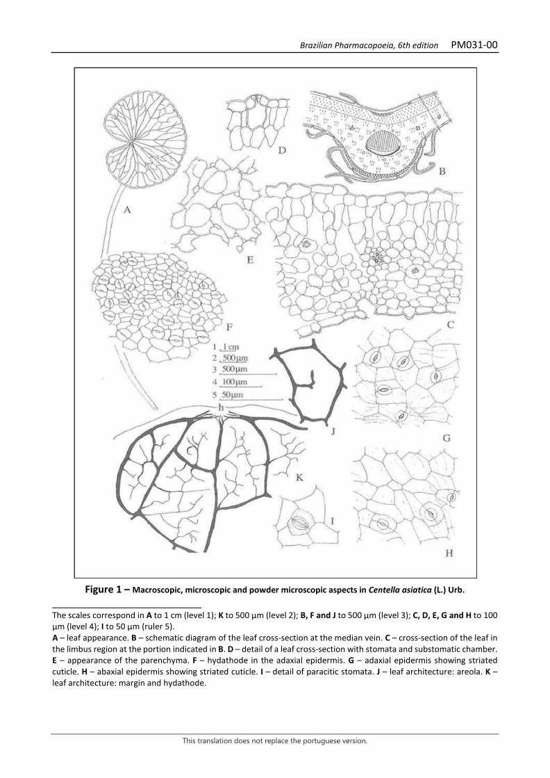
Brazilian Pharmacopoeia, 6th edition PM031-00
This translation does not replace the portuguese version.
Figure 1 – Macroscopic, microscopic and powder microscopic aspects in Centella asiatica (L.) Urb.
___________________________ The scales correspond in A to 1 cm (level 1); K to 500 μm (level 2); B, F and J to 500 μm (level 3); C, D, E, G and H to 100 μm (level 4); I to 50 μm (ruler 5). A – leaf appearance. B – schematic diagram of the leaf cross-section at the median vein. C – cross-section of the leaf in the limbus region at the portion indicated in B. D – detail of a leaf cross-section with stomata and substomatic chamber. E – appearance of the parenchyma. F – hydathode in the adaxial epidermis. G – adaxial epidermis showing striated cuticle. H – abaxial epidermis showing striated cuticle. I – detail of paracitic stomata. J – leaf architecture: areola. K – leaf architecture: margin and hydathode.
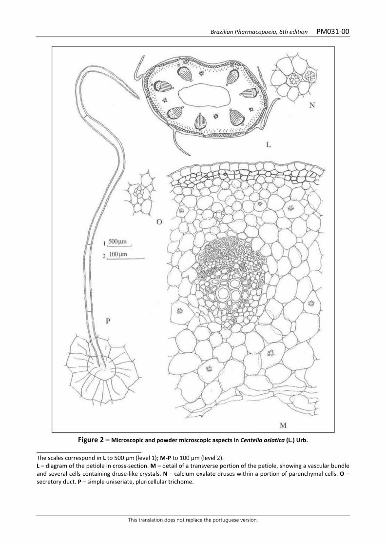
Brazilian Pharmacopoeia, 6th edition PM031-00
This translation does not replace the portuguese version.
Figure 2 – Microscopic and powder microscopic aspects in Centella asiatica (L.) Urb.
___________________________ The scales correspond in L to 500 μm (level 1); M-P to 100 μm (level 2). L – diagram of the petiole in cross-section. M – detail of a transverse portion of the petiole, showing a vascular bundle and several cells containing druse-like crystals. N – calcium oxalate druses within a portion of parenchymal cells. O – secretory duct. P – simple uniseriate, pluricellular trichome.
Brazilian Pharmacopoeia, 6th edition PM032-00
This translation does not replace the portuguese version.
FRESHCUT, leaf Justicia pectoralis folium
The plant drug consists of dried leaves of Justicia pectoralis Jacq., chopped or pulverized, containing at least 0.2% coumarin (C9H6O2, 146.15). IDENTIFICATION 1. Macroscopic description Entire, membranous, light green leaves. Lanceolate leaf lamina, with attenuated apex, acute base and entire margin, with trichomes on both sides, 2 to 6 cm long and 0.4 to 1.0 cm wide; petiole 0.2 to 0.5 mm long. 2. Microscopic description Dorsiventrally symmetrical leaf lamina, amphihypostomatic, with diacytic stomata. In a paradermal section, the adaxial surface of the epidermis shows cells with a sinuous outline and uniseriate pluricellular tector trichomes. The abaxial surface of the lamina shows epidermal cells with a wavy outline and glandular trichomes formed by a cell at the foot and a cell at the peduncle and by a head composed of four secretory cells, where the content is visualized in the form of drops. In cross-section, the lamina shows a uniseriate epidermis covered by a thick cuticle, with quadrangular-shaped cells, the abaxial cells being smaller. In some cells of the epidermis there are rod-shaped cystoliths that vary in size. The mesophyll region is covered by a thick cuticle, presenting a layer of palisade parenchyma and four layers of spongy parenchyma, with intercellular spaces. In the central vein region, two layers of collenchyma of the angular type were observed on both sides. The vascular bundle is collateral and is arranged in the form of a central arch, and at the ends it shows two small bundles also collateral, facing the adaxial surface. The petiole, in cross-section, is almost plano-convex in shape. The epidermis is uniseriate and covered by a thin cuticle, with tector trichomes similar to those found on the lamina. Below the epidermis there are five layers of angular collenchyma on the adaxial surface, including the lateral wings, and two layers on the abaxial surface. The vascular bundle is of the collateral type, with one major and main, arc-shaped, and three smaller bundles. Ten xylematic element rays occur in the main bundle. 3. Microscopic description of powder The sample fulfills all the characteristics for the species, except the macroscopic characters. Characteristics are: light green color; epidermis fragments showing diacritical stomata and rare tector trichomes; isolated cystoliths and their fragments. 4. Proceed as described in Thin-layer chromatography (5.2.17.1). Stationary phase: silica gel F254 (0.25 mm). Mobile phase: mixture of ether and toluene, saturated with 10% acetic acid (v/v) (50:50:50).
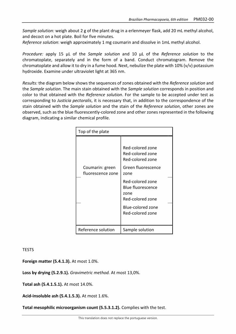
Brazilian Pharmacopoeia, 6th edition PM032-00
This translation does not replace the portuguese version.
Sample solution: weigh about 2 g of the plant drug in a erlenmeyer flask, add 20 mL methyl alcohol, and decoct on a hot plate. Boil for five minutes. Reference solution: weigh approximately 1 mg coumarin and dissolve in 1mL methyl alcohol. Procedure: apply 15 μL of the Sample solution and 10 μL of the Reference solution to the chromatoplate, separately and in the form of a band. Conduct chromatogram. Remove the chromatoplate and allow it to dry in a fume hood. Next, nebulize the plate with 10% (v/v) potassium hydroxide. Examine under ultraviolet light at 365 nm. Results: the diagram below shows the sequences of zones obtained with the Reference solution and the Sample solution. The main stain obtained with the Sample solution corresponds in position and color to that obtained with the Reference solution. For the sample to be accepted under test as corresponding to Justicia pectoralis, it is necessary that, in addition to the correspondence of the stain obtained with the Sample solution and the stain of the Reference solution, other zones are observed, such as the blue fluorescently-colored zone and other zones represented in the following diagram, indicating a similar chemical profile.
Top of the plate
Red-colored zone Red-colored zone Red-colored zone
Coumarin: green fluorescence zone
Green fluorescence zone
Red-colored zone Blue fluorescence zone Red-colored zone
Blue-colored zone Red-colored zone
Reference solution Sample solution
TESTS Foreign matter (5.4.1.3). At most 1.0%. Loss by drying (5.2.9.1). Gravimetric method. At most 13,0%. Total ash (5.4.1.5.1). At most 14.0%. Acid-insoluble ash (5.4.1.5.3). At most 1.6%. Total mesophilic microorganism count (5.5.3.1.2). Complies with the test.

Brazilian Pharmacopoeia, 6th edition PM032-00
This translation does not replace the portuguese version.
Study of pathogenic microorganisms (5.5.3.1.3). Complies with the test. Heavy metals (5.4.5). Complies with the test. Agrochemical waste (5.4.3). Complies with the test. DOSAGE Proceed as described in High Performance Liquid Chromatography (5.2.17.4). Use a chromatograph equipped with an ultraviolet detector at 277 nm; pre-column packed with silica chemically bonded to an octadecylsilane group (3.9 µm), 250mm long, 4.6 mm internal diameter column, packed with silica chemically bonded to an octadecylsilane group (5 µm), kept at room temperature (23 °C); Mobile phase flow rate of 0.8 mL/minute. Eluent (A): 0.05% trifluoroacetic acid (v/v). Eluent (B): 0.05% trifluoroacetic acid in methyl alcohol (v/v).
Time (minutes) Eluent A (%) Eluent B (%) Elution
0-4 80 → 75 20 → 25 linear gradient 4-8 75 → 60 25 → 40 linear gradient 8-23 60 → 25 40 → 75 linear gradient 23-26 25 75 isocratic 26-27 25 → 80 75 → 20 linear gradient Sample solution: accurately weigh 0.50 g of the pulverized plant drug and transfer to a 250 mL, round-bottomed, polished-necked flask. Add 100 mL of 40% (v/v) ethyl alcohol solution. Heat in a water bath, under reflux, for 30 minutes, between 85 °C and 90 °C. Cool under running water and filter the extract through absorbent cotton into a 100-mL volumetric flask. Top off the volume with the extracting liquid and homogenize. Transfer 5 mL of thus solution to a 25-mL volumetric flask, top off the volume with water and homogenize. Filter through a 0.45 µm filter unit. Reference solution: dissolve 25 mg of coumarin in a 50-mL volumetric flask with 40% (v/v) ethyl alcohol solution. Transfer 0.320 mL to a 10-mL volumetric flask and top off to the mark with the same solvent to obtain a solution with a concentration equal to 16 µg/mL. Filter through a 0.45 µm filter unit. Procedure: separately inject 20 μL of the Reference solution and 20 µL of the Sample solution. Register the chromatograms and measure the areas under the peaks. Coumarin retention time in the sample is approximately 17.7 minutes. Calculate coumarin content, in percent, according to the following expression:
𝑇𝑇𝐶𝐶 =𝐶𝐶𝑟𝑟 × 𝑇𝑇𝑎𝑎𝑇𝑇𝑟𝑟 × 𝑚𝑚
× 500 × 100
Brazilian Pharmacopoeia, 6th edition PM032-00
This translation does not replace the portuguese version.
in which,
TC = coumarin content in % (w/w); Cr = concentration of the Reference solution in g/mL, considering the purity of the reference substance; Ar = area under the peak corresponding to the coumarin in the Reference solution; Ar = area under the corresponding peak in the Sample solution; m = mass in grams of the sample used, considering the loss by drying. 500 = dilution factor. PACKAGING AND STORAGE In tightly closed containers, protected from light and heat.
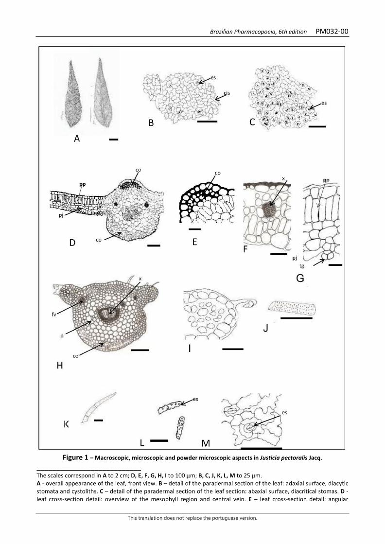
Brazilian Pharmacopoeia, 6th edition PM032-00
This translation does not replace the portuguese version.
Figure 1 – Macroscopic, microscopic and powder microscopic aspects in Justicia pectoralis Jacq.
___________________________ The scales correspond in A to 2 cm; D, E, F, G, H, I to 100 µm; B, C, J, K, L, M to 25 µm. A - overall appearance of the leaf, front view. B – detail of the paradermal section of the leaf: adaxial surface, diacytic stomata and cystoliths. C – detail of the paradermal section of the leaf section: abaxial surface, diacritical stomas. D - leaf cross-section detail: overview of the mesophyll region and central vein. E – leaf cross-section detail: angular
Brazilian Pharmacopoeia, 6th edition PM032-00
This translation does not replace the portuguese version.
collenchyma. F - leaf cross-section detail: mesophyll vascular bundle, xylem detail. G - leaf cross-section detail: palisade parenchyma, spongy parenchyma, and trichome. H - cross-section of petiole: overview. I – petiole cross-section detail: lateral wing region. J - leaf powder maceration: vessel element. K – leaf powder maceration: trichome. K – leaf powder maceration: cystolith. M – leaf powder maceration: stomata.
Brazilian Pharmacopoeia, 6th edition PM033-00
This translation does not replace the portuguese version.
BURHEAD, leaf Echinodorus folium
The plant drug consists of dried leaves of Echinodorus grandiflorus (Cham. & Schltdl.) Micheli containing not less than 2.8% hydroxycinnamic acid derivatives, expressed as verbascoside (C29H36O15, 624,59). IDENTIFICATION 1. Macroscopic description Simple, leathery, cordate leaves with a cordate base and acute to rounded apex. Leaf lamina varying in size, 10 to 35 cm long and 20 to 25 cm wide in the middle portion; long petiole with circular to oval cross-section, short winged expanses and longitudinal striations. Venation is campylodromous, with 12 to 14 veins of similar caliber, starting from a single point at the base of the limb, prominent on the abaxial surface, from which smaller, parallel veins emerge and originate the tertiary veins, culminating in the formation of closed areoles with sparsely branched endings. Both lamina and petiole are pubescent and relatively rough due to the presence of stellate trichomes. Translucent secretory ducts are abundant throughout the leaf lamina. 2. Microscopic description Dorsiventrally symmetrical leaf lamina, amphihypostomatic, with paralelocytic stomata. In front view, the anticlinal walls of the epidermal cells are straight to sinuous. Stellate, pluricellular tector trichomes occur on the veins. In cross-section, the epidermis is unistratified and covered by a thin cuticle, with inconspicuous papillae. The mesophyll is formed by one layer of palisade parenchyma and several layers of spongy parenchyma with braciform expansions. In the aerenchyma, trabeculae of braciform cells with thickened indentations occur, allowing the formation of triangular intercellular spaces. Secretory ducts are distributed throughout the aerenchyma. In the vascular bundles of the mesophyll, sclerenchymatic polar capping and, externally, a parenchymatic sheath occurs. Between eight and eleven vascular bundles occur in the midrib, accompanied by fibers and a parenchymatic sheath. The fundamental parenchyma is braciform. The petiole, in front view, has polyhedral epidermal cells, elongated longitudinally. In cross-section, vascular bundles occur in both wings. The aerenchyma is similar to that described for the midrib, however several cells are filled with starch grains. The most calibrous vascular bundles (one to two) are arranged in the central region of the petiole, and are less calibrous towards the periphery, similar to the midrib, but featuring a large protoxylema gap. The smaller-caliber bundles show sclerenchyma only near the phloem pole, while the larger ones show a continuous ring. 3. Microscopic description of powder The sample meets all requirements for the species, except the macroscopic characters. Characteristics are: epidermis fragments of the leaf lamina, with paralelocytic stomata and epidermal cells with a straight to sinuous outline, covered by a cuticle with papillae; stellate tector trichomes with or without portions of epidermis; cells with small braciform expansions of the spongy parenchyma; portions of aerenchyma containing secretory ducts; braciform cells with thickened indentations, which compose the trabeculae of the midrib and petiole.
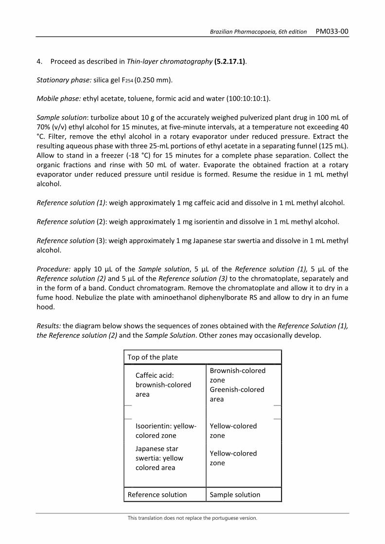
Brazilian Pharmacopoeia, 6th edition PM033-00
This translation does not replace the portuguese version.
4. Proceed as described in Thin-layer chromatography (5.2.17.1). Stationary phase: silica gel F254 (0.250 mm). Mobile phase: ethyl acetate, toluene, formic acid and water (100:10:10:1). Sample solution: turbolize about 10 g of the accurately weighed pulverized plant drug in 100 mL of 70% (v/v) ethyl alcohol for 15 minutes, at five-minute intervals, at a temperature not exceeding 40 °C. Filter, remove the ethyl alcohol in a rotary evaporator under reduced pressure. Extract the resulting aqueous phase with three 25-mL portions of ethyl acetate in a separating funnel (125 mL). Allow to stand in a freezer (-18 °C) for 15 minutes for a complete phase separation. Collect the organic fractions and rinse with 50 mL of water. Evaporate the obtained fraction at a rotary evaporator under reduced pressure until residue is formed. Resume the residue in 1 mL methyl alcohol. Reference solution (1): weigh approximately 1 mg caffeic acid and dissolve in 1 mL methyl alcohol. Reference solution (2): weigh approximately 1 mg isorientin and dissolve in 1 mL methyl alcohol. Reference solution (3): weigh approximately 1 mg Japanese star swertia and dissolve in 1 mL methyl alcohol. Procedure: apply 10 µL of the Sample solution, 5 µL of the Reference solution (1), 5 µL of the Reference solution (2) and 5 µL of the Reference solution (3) to the chromatoplate, separately and in the form of a band. Conduct chromatogram. Remove the chromatoplate and allow it to dry in a fume hood. Nebulize the plate with aminoethanol diphenylborate RS and allow to dry in an fume hood. Results: the diagram below shows the sequences of zones obtained with the Reference Solution (1), the Reference solution (2) and the Sample Solution. Other zones may occasionally develop.
Top of the plate
Caffeic acid: brownish-colored area
Brownish-colored zone Greenish-colored area
Isoorientin: yellow-colored zone
Yellow-colored zone
Japanese star swertia: yellow colored area
Yellow-colored zone
Reference solution Sample solution

Brazilian Pharmacopoeia, 6th edition PM033-00
This translation does not replace the portuguese version.
TESTS Foreign matter (5.4.1.3). At most 2.0%. Water (5.4.1.4). At most 9.0%. Total ash (5.4.1.5.1). At most 11.0%. Sulfated Ash (5.4.1.5.2). At most 13,0%. Total mesophilic microorganism count (5.5.3.1.2). Complies with the test. Study of pathogenic microorganisms (5.5.3.1.3). Complies with the test. Heavy metals (5.4.5). Complies with the test. Agrochemical waste (5.4.3). Complies with the test. DOSAGE O-hydroxycinnamic acid derivatives To proceed as described in Visible absorption spectrophotometry (5.2.14). Prepare solutions as described below. Stock solution: accurately weigh approximately 0.5 g of the pulverized plant drug (210 µm) (5.2.11) and add 90 mL of 50% ethyl alcohol (v/v) to a 250-mL round-bottomed flask. Heat under reflux for 30 minutes. Cool and filter into a 100-mL volumetric flask. Rinse the round-bottomed flask and the filter with 10 mL of 50% (v/v) ethyl alcohol into the same volumetric flask. Top off the volume with 50% (v/v) ethyl alcohol and homogenize. Sample solution: add, volumetrically, 1 mL of the Stock solution, 2 mL of 0.5 M hydrochloric acid, 2 mL of the mixture of 20% (w/v) sodium nitrite and 20% (w/v) sodium molybdate (1:1), in a 10-mL volumetric flask. Add 2 mL of 8% (w/v) sodium hydroxide solution, top off the volume with 50% (v/v) ethyl alcohol and homogenize. Blank solution: add, volumetrically, 1 mL of the Stock solution, 2 mL of 0.5 M hydrochloric acid, 2 mL of 8% (w/v) sodium hydroxide solution, in a 10-mL volumetric flask, top off the volume with 50% (v/v) ethyl alcohol and homogenize. Procedure: measure the absorbance of the Sample solution immediately after its preparation, at 525 nm, using the Blank solution for zero adjustment. Calculate the content pf hydroxycinnamic acid derivatives, expressed as a percentage of verbascoside, according to the following expression:
𝑇𝑇𝑇𝑇 =𝑇𝑇 × 1000𝑚𝑚 × 185
Brazilian Pharmacopoeia, 6th edition PM033-00
This translation does not replace the portuguese version.
in which, TA = content of hydroxycinnamic acid derivatives, expressed as verbascoside % (w/w); A = absorbance measured for the Sample solution; 185 = verbascoside specific absorption coefficient; m = mass in grams of the sample used un the assay, considering the determined water content. PACKAGING AND STORAGE In tightly closed containers, protected from light and heat.
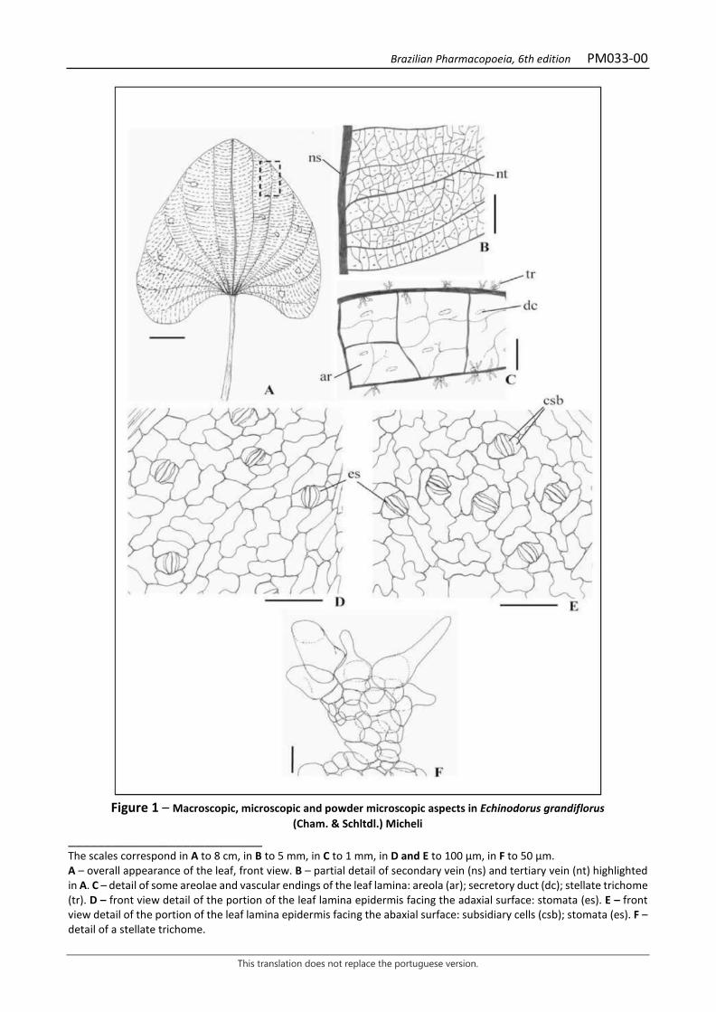
Brazilian Pharmacopoeia, 6th edition PM033-00
This translation does not replace the portuguese version.
Figure 1 – Macroscopic, microscopic and powder microscopic aspects in Echinodorus grandiflorus
(Cham. & Schltdl.) Micheli ___________________________ The scales correspond in A to 8 cm, in B to 5 mm, in C to 1 mm, in D and E to 100 μm, in F to 50 μm. A – overall appearance of the leaf, front view. B – partial detail of secondary vein (ns) and tertiary vein (nt) highlighted in A. C – detail of some areolae and vascular endings of the leaf lamina: areola (ar); secretory duct (dc); stellate trichome (tr). D – front view detail of the portion of the leaf lamina epidermis facing the adaxial surface: stomata (es). E – front view detail of the portion of the leaf lamina epidermis facing the abaxial surface: subsidiary cells (csb); stomata (es). F – detail of a stellate trichome.
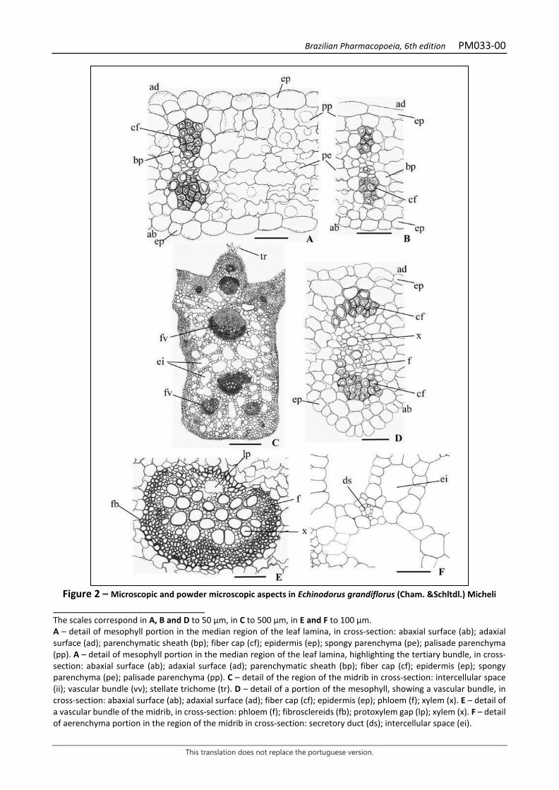
Brazilian Pharmacopoeia, 6th edition PM033-00
This translation does not replace the portuguese version.
Figure 2 – Microscopic and powder microscopic aspects in Echinodorus grandiflorus (Cham. &Schltdl.) Micheli
___________________________ The scales correspond in A, B and D to 50 μm, in C to 500 μm, in E and F to 100 μm. A – detail of mesophyll portion in the median region of the leaf lamina, in cross-section: abaxial surface (ab); adaxial surface (ad); parenchymatic sheath (bp); fiber cap (cf); epidermis (ep); spongy parenchyma (pe); palisade parenchyma (pp). A – detail of mesophyll portion in the median region of the leaf lamina, highlighting the tertiary bundle, in cross-section: abaxial surface (ab); adaxial surface (ad); parenchymatic sheath (bp); fiber cap (cf); epidermis (ep); spongy parenchyma (pe); palisade parenchyma (pp). C – detail of the region of the midrib in cross-section: intercellular space (ii); vascular bundle (vv); stellate trichome (tr). D – detail of a portion of the mesophyll, showing a vascular bundle, in cross-section: abaxial surface (ab); adaxial surface (ad); fiber cap (cf); epidermis (ep); phloem (f); xylem (x). E – detail of a vascular bundle of the midrib, in cross-section: phloem (f); fibrosclereids (fb); protoxylem gap (lp); xylem (x). F – detail of aerenchyma portion in the region of the midrib in cross-section: secretory duct (ds); intercellular space (ei).
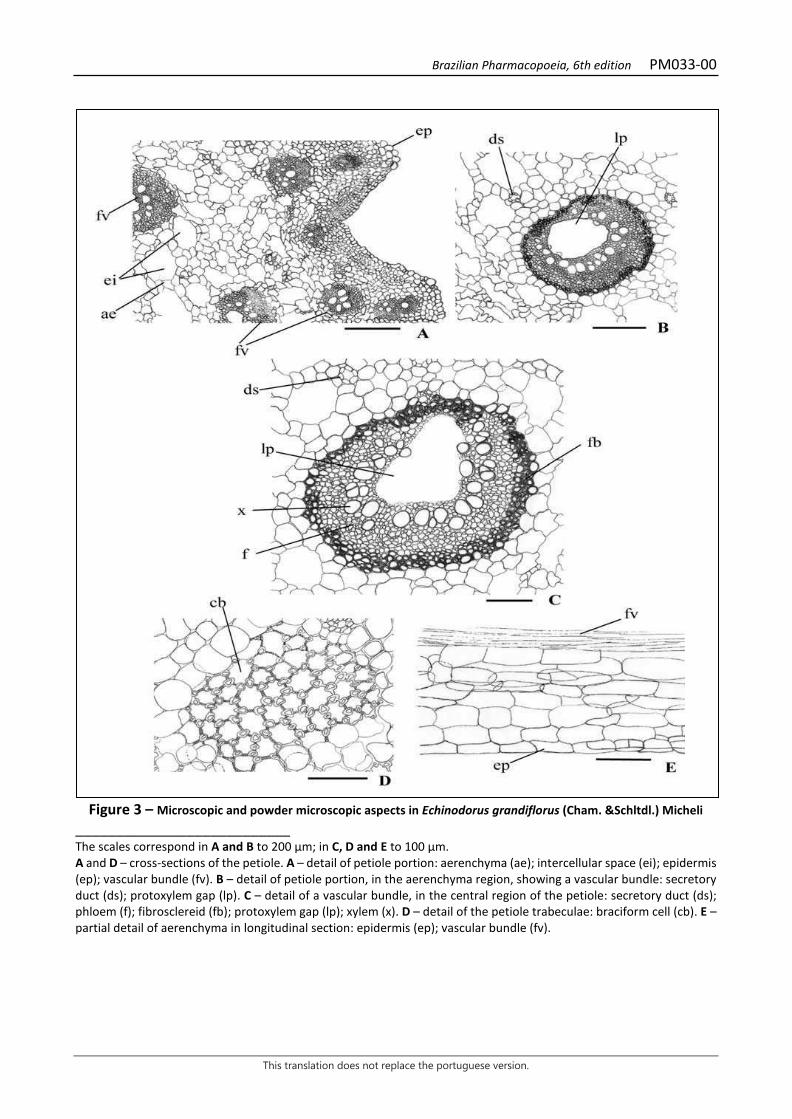
Brazilian Pharmacopoeia, 6th edition PM033-00
This translation does not replace the portuguese version.
Figure 3 – Microscopic and powder microscopic aspects in Echinodorus grandiflorus (Cham. &Schltdl.) Micheli
___________________________ The scales correspond in A and B to 200 μm; in C, D and E to 100 μm. A and D – cross-sections of the petiole. A – detail of petiole portion: aerenchyma (ae); intercellular space (ei); epidermis (ep); vascular bundle (fv). B – detail of petiole portion, in the aerenchyma region, showing a vascular bundle: secretory duct (ds); protoxylem gap (lp). C – detail of a vascular bundle, in the central region of the petiole: secretory duct (ds); phloem (f); fibrosclereid (fb); protoxylem gap (lp); xylem (x). D – detail of the petiole trabeculae: braciform cell (cb). E – partial detail of aerenchyma in longitudinal section: epidermis (ep); vascular bundle (fv).
Brazilian Pharmacopoeia, 6th edition PM034-00
This translation does not replace the portuguese version.
CORIANDER, fruit Coriandri fructus
The plant drug consists of dried fruits of Coriandrum sativum L., containing at least 0.3% volatile oil. CHARACTERISTICS The fruit has a characteristic aromatic odor. IDENTIFICATION 1. Macroscopic description The fruit is a diachene, formed of two mericarpas, subglobular and glabrous, approximately 0.2 to 0.5 cm in diameter, brown, yellowish-brown or reddish-brown; it has a short stylopod at the apex with two divergent stylets and the remains of five reflexed sepals. Each of the mericarps, usually attached by the margins, has five primary, wavy longitudinal edges alternating with four secondary, more prominent longitudinal edges. The fruit, in cross-section, displays a continuous band of lignified sclerenchyma on the dorsal portion of the pericarp and two, rarely more, large secretory ducts on the commissural or ventral side. The endosperm is oily and concave on the commissural side. 2. Microscopic description In cross-section, the diachene is circular, with 10 wavy primary edges, in each of which a vascular bundle is observed, and eight more prominent secondary edges. The epicarp consists of a colorless layer of thin-walled, smooth cuticle epidermal cells, which may occasionally contain one or two small, prismatic, calcium oxalate crystals. In front-view, the epicarp shows polygonal cells and anisocytic and/or anomocytic stomata, which are infrequent. The mesocarp is formed by three distinct zones: externally, a few layers of large, thin-walled cells occur, among which are remnants of rudimentary secretory ducts, facing the adaxial surface, and, on the commissural side, two large secretory ducts of elliptical shape are visible; the median portion is formed by a broad and continuous zone of fusiform, sinuous, thick-walled, pitted and narrow-lumen fibers, forming interwoven layers that externally oriented longitudinally and internally oriented tangentially, forming a right angle between them; then two or three layers of large, polygonal or rectangular, tangentially enlarged, thick-walled sclereids occur, with numerous conspicuous yellow spots, often attached to the endocarp, which is formed by one or two layers of thin-walled, lignified, elongated cells in front view, with a parquet-like appearance. The reniform-shape seed is covered by an integument formed by a layer of brown, thick-walled cells, except on the commissural surface; the endosperm consists of thick-walled, polygonal cells, containing colorless or slightly yellowish oil, aleurone grains, and small calcium oxalate druses, 3 to 10 µm in diameter. 3. Microscopic description of powder The sample meets all requirements for the species, except the macroscopic characters. Characteristics are: yellowish-brown color; fragments of endosperm and pericarp; fragments of
Brazilian Pharmacopoeia, 6th edition PM034-00
This translation does not replace the portuguese version.
fusiform fibers with thick, lignified walls; clustered sclereids; few brownish fragments of the secretory duct; numerous calcium oxalate crystals, mostly in clustered rosettes; numerous oil drops; epicarp fragments with polygonal cells; helical-like vessel elements and xylem parenchyma. 4. Proceed as described in Thin-layer chromatography (5.2.17.1). Stationary phase: silica gel G. Mobile phase: toluene and ethyl acetate (97:3). Sample solution: shake 0.5 g of the drug (500 µm) (5.2.11) in 5 mL hexane for three minutes. Filter on 2 g anhydrous sodium sulfate and collect 1 mL for chromatographic analysis. Reference solution: dilute 15 µL linalool and 25 µL olive oil in 5 mL hexane. Procedure: apply 20 μL of the Sample solution and 10 μL of the Reference solution to the chromatoplate, separately and in the form of a band. Conduct chromatogram. Remove chromatoplate and allow it to air dry. Nebulize the plate with an anisaldehyde solution and heat at 100 °C to 105 °C for one to ten minutes. Results: the diagram below shows the sequences of zones obtained with the Reference Solution and the Sample Solution. Other zones may occasionally develop.
Top of the plate
Olive oil: purple-colored zone
Purple-colored zone (triacylglycerides)
Linalool: intense purple-colored area
Purple-colored zone
Reference solution Sample solution
TESTS Loss by drying (5.2.9.1). Gravimetric method. At most 10.0%. Heavy metals (5.4.5). Complies with the test. Foreign matter (5.4.1.3). At most 5.0%. Total ash (5.4.1.5.1). At most 8.0%. Total mesophilic microorganism count (5.5.3.1.2). Complies with the test. Study of pathogenic microorganisms (5.5.3.1.3). Complies with the test.

Brazilian Pharmacopoeia, 6th edition PM034-00
This translation does not replace the portuguese version.
Aflatoxins (5.4.4). Complies with the test. Agrochemical waste (5.4.3). Complies with the test. DOSAGE Volatile oils Proceed as described in Determining volatile oils in plant drugs (5.4.1.6). Use a 500-mL flask containing 300 mL of water as distillation liquid and 0.5 mL of xylene in the graduated tube. Reduce the coriander fruit to a coarse powder. Immediately proceed with the determination of the volatile oil, from 30 g of the dried plant drug. Distill for two hours. Measure the volume and express the yield per 100 g of the plant drug (w/p). PACKAGING AND STORAGE In tightly closed containers, protected from light and heat.
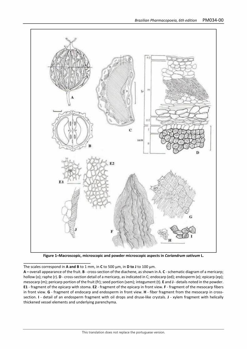
Brazilian Pharmacopoeia, 6th edition PM034-00
This translation does not replace the portuguese version.
Figure 1 ̶ Macroscopic, microscopic and powder microscopic aspects in Coriandrum sativum L.
___________________________ The scales correspond in A and B to 1 mm, in C to 500 µm, in D to J to 100 µm. A – overall appearance of the fruit. B - cross-section of the diachene, as shown in A. C - schematic diagram of a mericarp; hollow (o); raphe (r). D - cross-section detail of a mericarp, as indicated in C; endocarp (ed); endosperm (e); epicarp (ep); mesocarp (m); pericarp portion of the fruit (fr); seed portion (sem); integument (t). E and J - details noted in the powder. E1 - fragment of the epicarp with stoma. E2 - fragment of the epicarp in front view. F - fragment of the mesocarp fibers in front view. G - fragment of endocarp and endosperm in front view. H - fiber fragment from the mesocarp in cross-section. I - detail of an endosperm fragment with oil drops and druse-like crystals. J - xylem fragment with helically thickened vessel elements and underlying parenchyma.
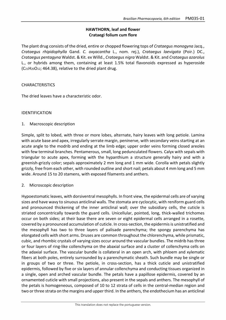
Brazilian Pharmacopoeia, 6th edition PM035-01
This translation does not replace the portuguese version.
HAWTHORN, leaf and flower Crataegi folium cum flore
The plant drug consists of the dried, entire or chopped flowering tops of Crataegus monogyna Jacq., Crataegus rhipidophylla Gand. C. oxyacantha L., nom. rej.), Crataegus laevigata (Poir.) DC., Crataegus pentagyna Waldst. & Kit. ex Willd., Crataegus nigra Waldst. & Kit. and Crataegus azarolus L., or hybrids among them, containing at least 1.5% total flavonoids expressed as hyperoside (C21H20O12; 464.38), relative to the dried plant drug. CHARACTERISTICS The dried leaves have a characteristic odor. IDENTIFICATION 1. Macroscopic description Simple, split to lobed, with three or more lobes, alternate, hairy leaves with long petiole. Lamina with acute base and apex, irregularly serrate margin, peninerve, with secondary veins starting at an acute angle to the modrib and ending at the limb edge; upper order veins forming closed areoles with few terminal branches. Pentamerous, small, long pedunculated flowers. Calyx with sepals with triangular to acute apex, forming with the hypanthium a structure generally hairy and with a greenish-grizzly color; sepals approximately 2 mm long and 1 mm wide. Corolla with petals slightly grizzly, free from each other, with rounded outline and short nail; petals about 4 mm long and 5 mm wide. Around 15 to 20 stamens, with exposed filaments and anthers. 2. Microscopic description Hypoestomatic leaves, with dorsiventral mesophylls. In front view, the epidermal cells are of varying sizes and have wavy to sinuous anticlinal walls. The stomata are cyclocytic, with reniform guard cells and pronounced thickening of the inner anticlinal wall; over the subsidiary cells, the cuticle is striated concentrically towards the guard cells. Unicellular, pointed, long, thick-walled trichomes occur on both sides; at their base there are seven or eight epidermal cells arranged in a rosette, covered by a pronounced accumulation of cuticle. In cross-section, the epidermis is unistratified and the mesophyll has two to three layers of palisade parenchyma; the spongy parenchyma has elongated cells with short arms. Druses are common throughout the chlorenchyma, while prismatic, cubic, and rhombic crystals of varying sizes occur around the vascular bundles. The midrib has three or four layers of ring-like collenchyma on the abaxial surface and a cluster of collenchyma cells on the adaxial surface. The vascular bundle is collateral in an open arch, with phloem and xylematic fibers at both poles, entirely surrounded by a parenchymatic sheath. Such bundle may be single or in groups of two or three. The petiole, in cross-section, has a thick cuticle and unistratified epidermis, followed by five or six layers of annular collenchyma and conducting tissues organized in a single, open and arched vascular bundle. The petals have a papillose epidermis, covered by an ornamented cuticle with small projections, also present in the sepals and anthers. The mesophyll of the petals is homogeneous, composed of 10 to 12 strata of cells in the central-median region and two or three strata on the margins and upper third. In the anthers, the endothecium has an anticlinal
Brazilian Pharmacopoeia, 6th edition PM035-01
This translation does not replace the portuguese version.
thickening parallel to each other, sometimes interlaced diagonally. Pollen grains are tricolpate and ornamented with small spherical papillae. On the inner side of the base of the sepals is the floral nectary, formed by cells with dense content, typical of secretory structures. 3. Microscopic description of powder The sample meets all requirements for the species, except the macroscopic characters. Characteristics are: grizzly color; fragments of leaf laminae showing areolae; fragments of leaf epidermis with cells with sinuous walls and without stomas; fragments of leaf epidermis with cells with slightly sinuous walls and cyclocytic stomas with subsidiary cells and striated cuticle; unicellular trichomes with thick walls, fragmented or entire; fragments of leaf epidermis with cells arranged in rosette at the base of the trichomes; fragments of parenchyma and epidermis, in cross-section, with bases of trichomes; dorsiventral mesophyll fragments with deformed druses and/or prismatic crystals accompanying the vascular bundles; petal fragments with papillose epidermis and sepals with secretory tissue; anther fragments with thickened cells (endothecium) and ornamented pollen grains; isolated crystals. 4. Proceed as described in Thin-layer chromatography (5.2.17.1). Stationary phase: silica gel GF254 (0.250 mm). Mobile phase: ethyl acetate, methyl ethyl ketone, anhydrous formic acid and water (50:30:10:10). Sample solution: accurately weigh about 1 g of the pulverized plant drug (355 μm) (5.2.11), add 10 mL methyl alcohol, heat under reflux for five minutes at 65 °C. After cooling down to room temperature, filter the obtained solution on filter paper. Reference solution (1): dissolve 1 mg chlorogenic acid in 10 mL methyl alcohol. Reference solution (2): dissolve 1 mg hyperoside in 5 mL methyl alcohol. Procedure: apply 20 µL of the Sample solution, 20 µL of the Reference solution (1) and 20 µL of the Reference solution (2) to the chromatoplate, separately and in the form of a band. Conduct chromatogram. Remove the chromatoplate and heat in an oven at 100°C to 105°C for 15 minutes and, while still warm, nebulize with aminoethanol diphenylborate SE followed by a solution of 5% (w/v) macrogol 400 in methyl alcohol. Allow the plate to air dry for 30 minutes and examine the plate under ultraviolet light at 365 nm. Results: the diagram below shows the sequences of zones obtained with the Sample solution, Reference solution (1) and Reference solution (2). Other zones may occasionally develop.
Brazilian Pharmacopoeia, 6th edition PM035-01
This translation does not replace the portuguese version.
Top of the plate
Yellowish-green fluorescence zone
Hyperoside: yellow fluorescence zone
Yellowish-orange fluorescence zone
Chlorogenic acid: light-blue fluorescence zone
Light-blue fluorescence zone
Yellowish-green fluorescence zone
Reference solution Sample solution
TESTS Foreign matter (5.4.1.3). At most 8.0% lignified branches and 2.0% other tinny material. Water (5.4.1.4). At most 11.0%. Total ash (5.4.1.5.1). At most 10.0%. Total mesophilic microorganism count (5.5.3.1.2). Complies with the test. Study of pathogenic microorganisms (5.5.3.1.3). Complies with the test. Heavy metals (5.4.5). Complies with the test. Agrochemical waste (5.4.3). Complies with the test. DOSAGE Total flavonoids To proceed as described in Visible absorption spectrophotometry (5.2.14). Prepare solutions as described below. Stock solution: accurately weigh approximately 0.4 g of the pulverized plant drug (250 µm) (5.2.11), transfer to a 200-mL erlenmeyer flask and add 40 mL of 60% ethyl alcohol (v/v). Heat in a water bath at 60 °C for 10 minutes while frequently shaking. Cool and filter through absorbent cotton into a 100-mL volumetric flask. Return the insoluble residue and the cotton to the same erlenmeyer flask, add 40 mL of 60% (v/v) ethyl alcohol, and return to the water bath for 10 minutes while frequently shaking. Filter the cotton solution into the volumetric flask as previously described. Top off the volume with 60% (v/v) ethyl alcohol and homogenize.
Brazilian Pharmacopoeia, 6th edition PM035-01
This translation does not replace the portuguese version.
Sample solution: transfer 5 mL of the Stock solution, volumetrically, to a 100-mL round-bottomed flask and evaporate to dryness at a rotary evaporator. Solubilize the residue in 8 mL of a mixture of methyl alcohol and glacial acetic acid solution (10:100) and transfer to a 25-mL volumetric flask. Rinse the round-bottomed flask with a 3mL mixture of methyl alcohol solution and glacial acetic acid (10:100) and transfer to the volumetric flask, as previously described. Add 10 mL of the Reagent Solution. Keep in an ice bath to cool for 10 minutes. Do not allow to freeze. Top off the volume with anhydrous acetic acid and homogenize. Place immediately in an ice bath. Remove 10 minutes before reading in the spectrophotometer. Blank solution: transfer 5 mL of the Stock solution, volumetrically, to a 100-mL round-bottomed flask and evaporate to dryness at a rotary evaporator. Solubilize the residue in 8 mL of the mixture of methyl alcohol and glacial acetic acid solution (10:100) and transfer to a 25-mL volumetric flask. Rinse the round-bottomed flask with a 3mL mixture of methyl alcohol solution and glacial acetic acid (10:100) and transfer to the volumetric flask, as previously described. Add 10 mL of anhydrous formic acid. Keep in an ice bath to cool for 10 minutes. Do not allow to freeze. Top off the volume with anhydrous acetic acid and homogenize. Place immediately in an ice bath. Remove 10 minutes before reading in the spectrophotometer. Reagent solution: 2.5% (w/v) boric acid and 2% (w/v) oxalic acid in anhydrous formic acid. Solubilize, while heating, in a fume hood. Procedure: measure the absorbance of the sample solution after 30 minutes at a wavelength of 410 nm. Calculate total flavonoid content expressed as hyperoside, in percent, according to the following expression:
𝑇𝑇𝑇𝑇 =A × 500m × 405
in which, TF = total flavonoid content expressed as hyperoside % (w/w); A = absorbance measured for the Sample solution; 500 = dilution factor; 405 = hyperoside specific absorption coefficient; m = mass in grams of the sample used, considering the determined water content. PACKAGING AND STORAGE In tightly closed containers, protected from light and heat.
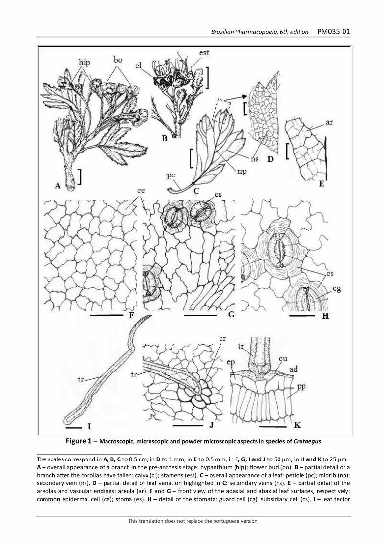
Brazilian Pharmacopoeia, 6th edition PM035-01
This translation does not replace the portuguese version.
Figure 1 – Macroscopic, microscopic and powder microscopic aspects in species of Crataegus
___________________________ The scales correspond in A, B, C to 0.5 cm; in D to 1 mm; in E to 0.5 mm; in F, G, I and J to 50 μm; in H and K to 25 μm. A – overall appearance of a branch in the pre-anthesis stage: hypanthium (hip); flower bud (bo). B – partial detail of a branch after the corollas have fallen: calyx (cl); stamens (est). C – overall appearance of a leaf: petiole (pc); midrib (np); secondary vein (ns). D – partial detail of leaf venation highlighted in C: secondary veins (ns). E – partial detail of the areolas and vascular endings: areola (ar). F and G – front view of the adaxial and abaxial leaf surfaces, respectively: common epidermal cell (ce); stoma (es). H – detail of the stomata: guard cell (cg); subsidiary cell (cs). I – leaf tector
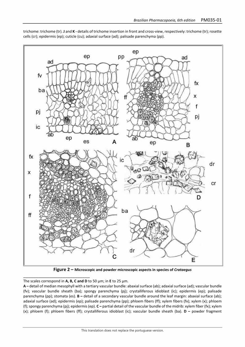
Brazilian Pharmacopoeia, 6th edition PM035-01
This translation does not replace the portuguese version.
trichome: trichome (tr). J and K - details of trichome insertion in front and cross-view, respectively: trichome (tr); rosette cells (cr); epidermis (ep); cuticle (cu); adaxial surface (ad); palisade parenchyma (pp).
Figure 2 – Microscopic and powder microscopic aspects in species of Crataegus
___________________________ The scales correspond in A, B, C and D to 50 μm; in E to 25 μm. A – detail of median mesophyll with a tertiary vascular bundle: abaxial surface (ab); adaxial surface (ad); vascular bundle (fv); vascular bundle sheath (ba); spongy parenchyma (pj); crystalliferous idioblast (ic); epidermis (ep); palisade parenchyma (pp); stomata (es). B – detail of a secondary vascular bundle around the leaf margin: abaxial surface (ab); adaxial surface (ad); epidermis (ep); palisade parenchyma (pp); phloem fibers (ff); xylem fibers (fx); xylem (x); phloem (f); spongy parenchyma (pj); epidermis (ep). C – partial detail of the vascular bundle of the midrib: xylem fiber (fx); xylem (x); phloem (f); phloem fibers (ff); crystalliferous idioblast (ic); vascular bundle sheath (ba). D – powder fragment
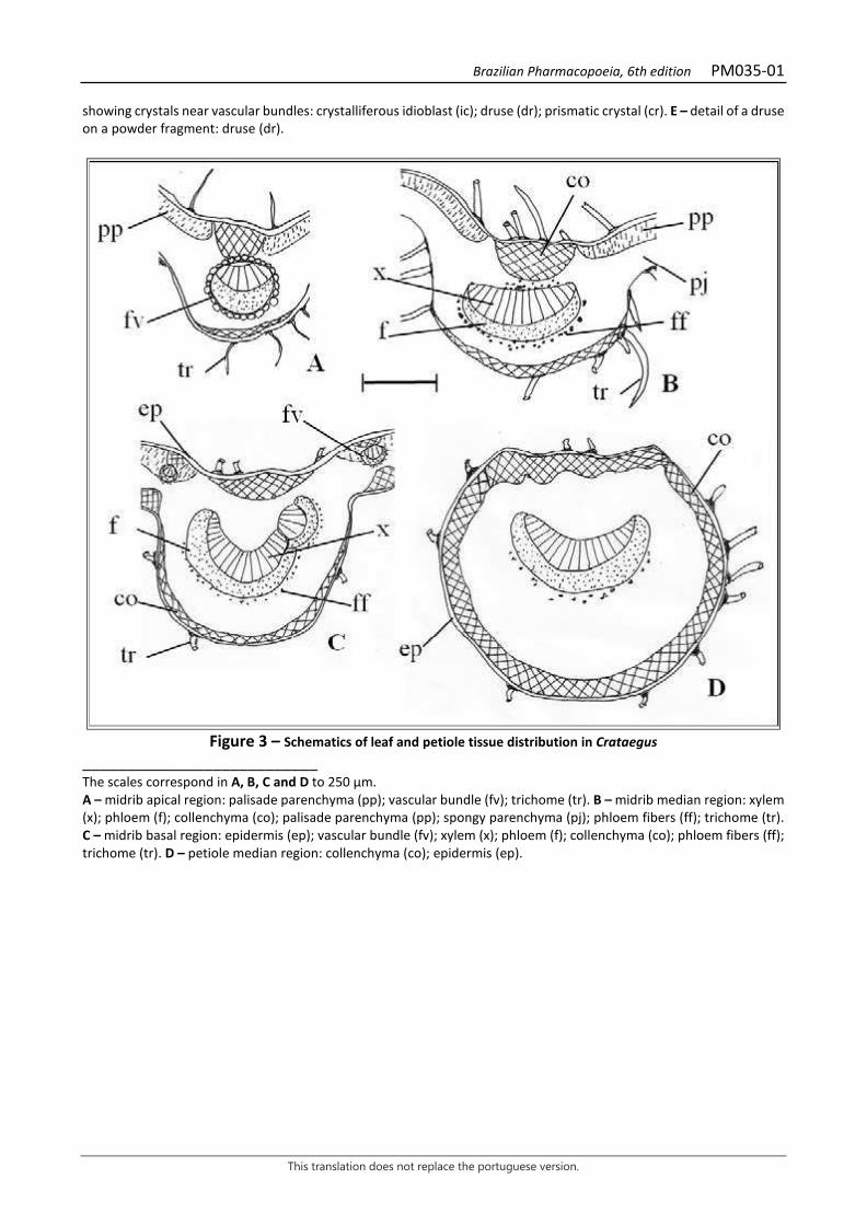
Brazilian Pharmacopoeia, 6th edition PM035-01
This translation does not replace the portuguese version.
showing crystals near vascular bundles: crystalliferous idioblast (ic); druse (dr); prismatic crystal (cr). E – detail of a druse on a powder fragment: druse (dr).
Figure 3 – Schematics of leaf and petiole tissue distribution in Crataegus
___________________________ The scales correspond in A, B, C and D to 250 μm. A – midrib apical region: palisade parenchyma (pp); vascular bundle (fv); trichome (tr). B – midrib median region: xylem (x); phloem (f); collenchyma (co); palisade parenchyma (pp); spongy parenchyma (pj); phloem fibers (ff); trichome (tr). C – midrib basal region: epidermis (ep); vascular bundle (fv); xylem (x); phloem (f); collenchyma (co); phloem fibers (ff); trichome (tr). D – petiole median region: collenchyma (co); epidermis (ep).
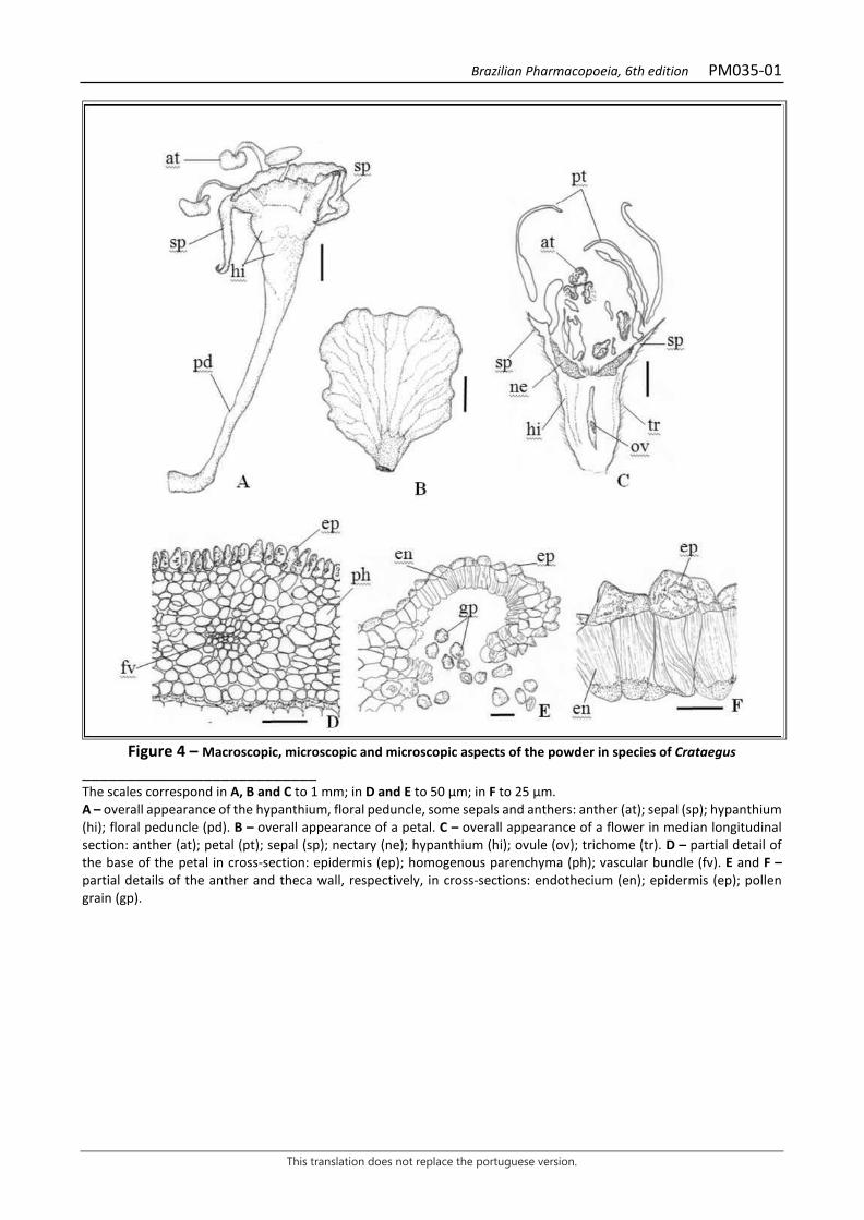
Brazilian Pharmacopoeia, 6th edition PM035-01
This translation does not replace the portuguese version.
Figure 4 – Macroscopic, microscopic and microscopic aspects of the powder in species of Crataegus
___________________________ The scales correspond in A, B and C to 1 mm; in D and E to 50 μm; in F to 25 μm. A – overall appearance of the hypanthium, floral peduncle, some sepals and anthers: anther (at); sepal (sp); hypanthium (hi); floral peduncle (pd). B – overall appearance of a petal. C – overall appearance of a flower in median longitudinal section: anther (at); petal (pt); sepal (sp); nectary (ne); hypanthium (hi); ovule (ov); trichome (tr). D – partial detail of the base of the petal in cross-section: epidermis (ep); homogenous parenchyma (ph); vascular bundle (fv). E and F – partial details of the anther and theca wall, respectively, in cross-sections: endothecium (en); epidermis (ep); pollen grain (gp).

Brazilian Pharmacopoeia, 6th edition PM036-00
This translation does not replace the portuguese version.
CLOVE, flower bud Caryophylli flos
The plant drug consists of dried flower buds of Syzygium aromaticum (L.) Merr. & L.M. Perry, containing at least 15.0% volatile oil. CHARACTERISTICS The flower buds have a strong, aromatic, characteristic odor; the buds exude oil when pressed. IDENTIFICATION 1. Macroscopic description The flower bud is blackish-brown in color, 1 to 2.1 cm long and 0.2 to 0.4 cm wide in the bud portion; in its lower portion, it has a somewhat flattened, four-sided, subcylindrical hypanthium, which contains in its inner and upper region an infertile ovary with two locules, showing several seminal rudiments attached to the axillary placenta. At the upper end of the hypanthium a calyx is found with four divergent sepals, pointed, thick, about 0.3 cm long, surrounding a globose region formed by four imbricate, membranous, lighter colored petals, arranged in the form of a dome, under which there are numerous stamens recurved inward, inserted in a concave nectariferous disk, surrounding a single erect, subulate stylet, about 0.3 cm long. 2. Microscopic description In front view, the hypanthium epidermis shows thick-walled polygonal cells and numerous anomocytic, nearly circular stomata, 30 to 35 m in diameter. Through transparency, schizolysigenous glands and clusters of calcium oxalate crystals, druse-like or prismatic crystals, can be seen. In cross-section, a thick and smooth cuticle, tubular epidermal cells, elevated stomata and a well-defined substomatal chamber are observed, followed by a parenchyma with distinct zones: external zone, yellowish-brown color, with ovoid schizolysigenous glands, long radial axis, measuring up to 200 µm long, distributed close to each other in two or three rows, accompanied by clusters of cells containing druses; medium zone formed by parenchymatous cells, colenchymatous in appearance, with a ring of rounded, bicollateral vascular bundles, surrounded by an incomplete ring of fibers, besides occasional isolated fibers or in groups of two or three cells and a lumen filled with brownish content; vascular bundles surrounded by parenchymatic cells containing prismatic crystals; below the bundles a loose, aerenchyma-like parenchymatic tissue occurs, followed by a ring of about 17 smaller bicollateral vascular bundles surrounded by a few fibers; central zone occupied by a filling parenchyma, with cells containing druse-like crystals. Occasionally oval to subrectangular, striated-walled, strongly thickened sclereids occur, showing numerous simple or branched pits and with a lumen often filled with brown contents. Cells with crystals similar to those already described and schizolysigenous glands also occur in the calyx, corolla, filament and stylet. The anther, in cross-section, shows a fibrous layer of tangentially elongated epidermal cells, with lignified thickening on the anticlinal walls; a schizolysigenous gland occurs at the apex of the connective. The pollen grains measure 15 to 20 m in diameter, are biconvex, with a rounded to triangular outline and smooth exine. Starch grains are absent.
Brazilian Pharmacopoeia, 6th edition PM036-00
This translation does not replace the portuguese version.
3. Microscopic description of powder The sample meets all requirements for the species, except the macroscopic characters. Characteristics are: blackish-brown to reddish-brown color; hypanthium parenchyma fragments with schizolysigenous glands; hypanthium epidermis fragments in front view, with large anomocytic stomata and underlying glands; hypanthium parenchyma fragments with cells containing druse-like crystals; hypanthium aerenchyma fragments; portion of hypanthium in cross-section showing thick cuticle, epidermis and underlying parenchyma with glands; isolated hypanthium sclerids; fragments of the anther fibrous layer, in front view; fragments of filament epidermis, in front view, with striated cuticle; filaments with central vascular cord and parenchyma cells with druses; pollen grains; pointed fibers with thick walls, associated to parenchyma cells. 4. Proceed as described in Thin-layer chromatography (5.2.17.1). Stationary phase: silica gel GF254. Mobile phase: toluene and ethyl acetate (93:7). Sample solution: dilute in 1 mL of toluene 10 µL of volatile clove oil obtained in the Determination of Volatile Oils in plant drugs (5.4.1.6). Reference solution: dilute in 1 mL toluene, 10 µL eugenol. Procedure: apply 5 μL of the Sample solution and 5 μL of the Reference solution to the chromatoplate, separately and in the form of a band. Conduct chromatogram. Remove chromatoplate and allow it to air dry. Nebulize the plate with an anisaldehyde solution and heat at 100 °C to 105 °C for three minutes. Results: the diagram below shows the sequences of zones obtained with the Reference Solution and the Sample Solution. Other zones may occasionally develop.
Brazilian Pharmacopoeia, 6th edition PM036-00
This translation does not replace the portuguese version.
Top of the plate
Pink-colored zone
Eugenol: violaceous color zone
Violaceous-colored zone
Reference solution Sample solution TESTS Water (5.2.20.2). Azeotropic method. At most 10.0%. Foreign matter (5.4.1.3). At most 4.0% of peduncles, petioles and fruits. At most 2.0% of flower buds altered. At most 0.5% of other foreign elements. The presence of 1.0% of the dry weight of inflorescence pedicels is allowed. Heavy metals (5.4.5). Complies with the test. Total ash (5.4.1.5.1). At most 7.0% Total mesophilic microorganism count (5.5.3.1.2). Complies with the test. Study of pathogenic microorganisms (5.5.3.1.3). Complies with the test. Agrochemical waste (5.4.3). Complies with the test. DOSAGE Volatile oils Proceed as described in Determining volatile oils in plant drugs (5.4.1.6). Use a 250-mL flask containing 100 mL of water as distillation liquid and 0.5 mL of xylene in the graduated tube. Grind 5 g of dried flower buds to powder together with 5 g diatomaceous earth. From the obtained powder, weigh 4 g and proceed immediately to the determination of the volatile oil. Distill for two hours. Measure the volume and express the yield per 100 g of the plant drug (w/p).
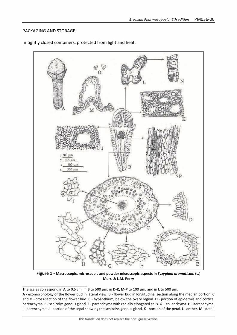
Brazilian Pharmacopoeia, 6th edition PM036-00
This translation does not replace the portuguese version.
PACKAGING AND STORAGE In tightly closed containers, protected from light and heat.
Figure 1 - Macroscopic, microscopic and powder microscopic aspects in Syzygium aromaticum (L.)
Merr. & L.M. Perry ___________________________ The scales correspond in A to 0.5 cm, in B to 500 µm, in D-K, M-P to 100 µm, and in L to 500 µm. A - exomorphology of the flower bud in lateral view. B - flower bud in longitudinal section along the median portion. C and O - cross-section of the flower bud: C - hypanthium, below the ovary region. D - portion of epidermis and cortical parenchyma. E - schizolysigenous gland. F - parenchyma with radially elongated cells. G – collenchyma. H - aerenchyma. I - parenchyma. J - portion of the sepal showing the schizolysigenous gland. K - portion of the petal. L - anther. M - detail
Brazilian Pharmacopoeia, 6th edition PM036-00
This translation does not replace the portuguese version.
of the schizolysigenous gland of the anther connective. N - detail of the fibrous layer of the anther. O - polen grains. P - vascular bundle in longitudinal section.

Brazilian Pharmacopoeia, 6th edition PM036-00
This translation does not replace the portuguese version.
Figure 2 - Macroscopic, microscopic and powder microscopic aspects in Syzygium aromaticum
(L.) Merr. & L.M. Perry ___________________________ The scales correspond in A to 0.5 cm, in B-J to 100 µm. A - peduncle and pedicels of the inflorescence. B - cross-section of the peduncle as marked in A. C - fibers in longitudinal section. D - fibers in cross-section. E - sclereids. F - isolated crystals. G - parenchyma containing druses. H - front view of the hypanthium epidermis showing the stomata. I - epidermis of the petal in front view. J - stamen in longitudinal section. L - front view of the filament epidermis showing striated cuticle.
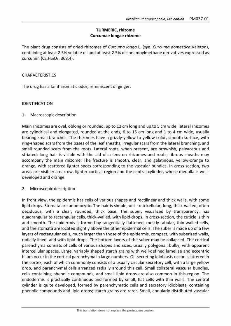
Brazilian Pharmacopoeia, 6th edition PM037-01
This translation does not replace the portuguese version.
TURMERIC, rhizome Curcumae longae rhizome
The plant drug consists of dried rhizomes of Curcuma longa L. (syn. Curcuma domestica Valeton), containing at least 2.5% volatile oil and at least 2.5% dicinnamoylmethane derivatives expressed as curcumin (C21H20O6, 368.4). CHARACTERISTICS The drug has a faint aromatic odor, reminiscent of ginger. IDENTIFICATION 1. Macroscopic description Main rhizomes are oval, oblong or rounded, up to 12 cm long and up to 5 cm wide; lateral rhizomes are cylindrical and elongated, rounded at the ends, 6 to 15 cm long and 1 to 4 cm wide, usually bearing small branches. The rhizomes have a grizzly-yellow to yellow color, smooth surface, with ring-shaped scars from the bases of the leaf sheaths, irregular scars from the lateral branching, and small rounded scars from the roots. Lateral roots, when present, are brownish, paleaceous and striated; long hair is visible with the aid of a lens on rhizomes and roots; fibrous sheaths may accompany the main rhizome. The fracture is smooth, clear, and gelatinous, yellow-orange to orange, with scattered lighter spots corresponding to the vascular bundles. In cross-section, two areas are visible: a narrow, lighter cortical region and the central cylinder, whose medulla is well-developed and orange. 2. Microscopic description In front view, the epidermis has cells of various shapes and rectilinear and thick walls, with some lipid drops. Stomata are anomocytic. The hair is simple, uni- to tricellular, long, thick-walled, often deciduous, with a clear, rounded, thick base. The suber, visualized by transparency, has quadrangular to rectangular cells, thick-walled, with lipid drops. In cross-section, the cuticle is thin and smooth. The epidermis is formed by tangentially flattened, mostly tabular, thin-walled cells, and the stomata are located slightly above the other epidermal cells. The suber is made up of a few layers of rectangular cells, much larger than those of the epidermis, compact, with suberized walls, radially lined, and with lipid drops. The bottom layers of the suber may be collapsed. The cortical parenchyma consists of cells of various shapes and sizes, usually polygonal, bulky, with apparent intercellular spaces. Large, variably shaped starch grains with well-defined lamellae and eccentric hilum occur in the cortical parenchyma in large numbers. Oil-secreting idioblasts occur, scattered in the cortex, each of which commonly consists of a usually circular secretory cell, with a large yellow drop, and parenchymal cells arranged radially around this cell. Small collateral vascular bundles, cells containing phenolic compounds, and small lipid drops are also common in this region. The endodermis is practically continuous and formed by small, flat cells with thin walls. The central cylinder is quite developed, formed by parenchymatic cells and secretory idioblasts, containing phenolic compounds and lipid drops; starch grains are rarer. Small, annularly-distributed vascular
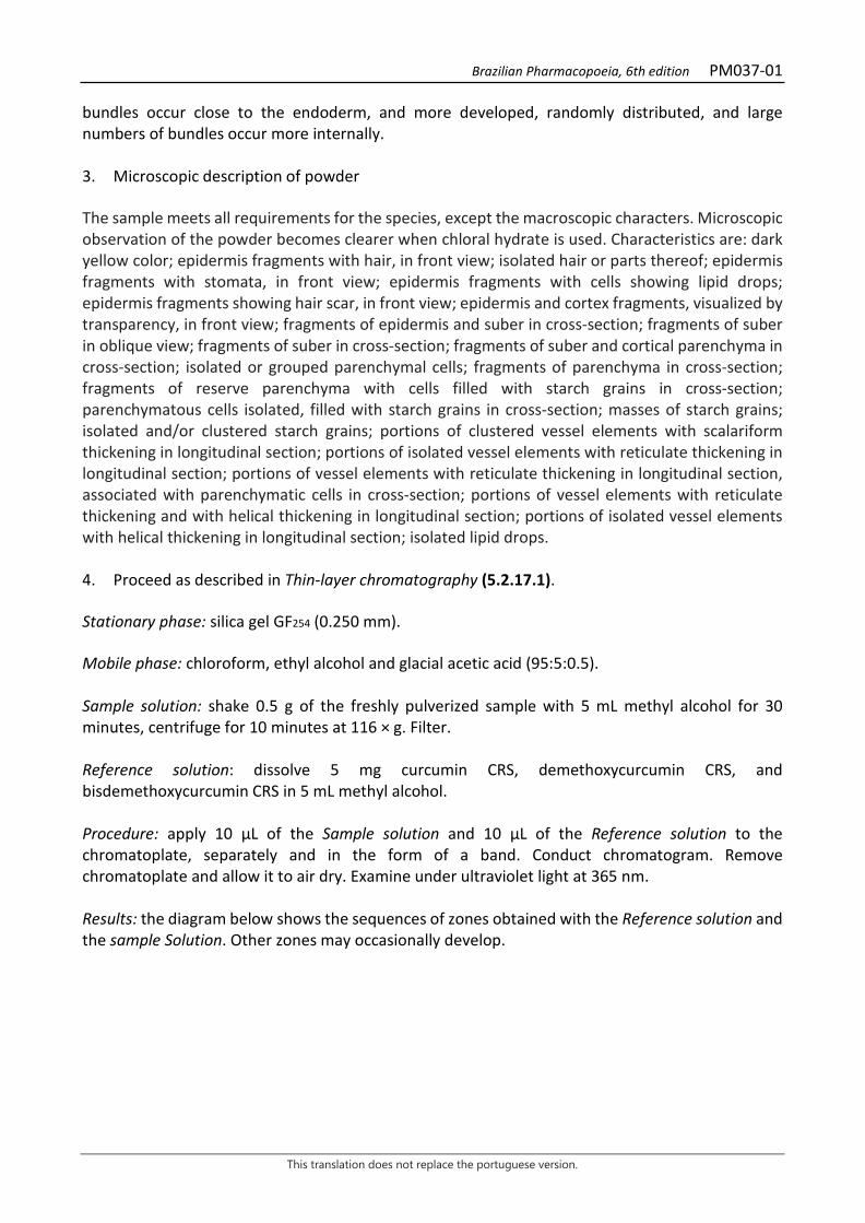
Brazilian Pharmacopoeia, 6th edition PM037-01
This translation does not replace the portuguese version.
bundles occur close to the endoderm, and more developed, randomly distributed, and large numbers of bundles occur more internally. 3. Microscopic description of powder The sample meets all requirements for the species, except the macroscopic characters. Microscopic observation of the powder becomes clearer when chloral hydrate is used. Characteristics are: dark yellow color; epidermis fragments with hair, in front view; isolated hair or parts thereof; epidermis fragments with stomata, in front view; epidermis fragments with cells showing lipid drops; epidermis fragments showing hair scar, in front view; epidermis and cortex fragments, visualized by transparency, in front view; fragments of epidermis and suber in cross-section; fragments of suber in oblique view; fragments of suber in cross-section; fragments of suber and cortical parenchyma in cross-section; isolated or grouped parenchymal cells; fragments of parenchyma in cross-section; fragments of reserve parenchyma with cells filled with starch grains in cross-section; parenchymatous cells isolated, filled with starch grains in cross-section; masses of starch grains; isolated and/or clustered starch grains; portions of clustered vessel elements with scalariform thickening in longitudinal section; portions of isolated vessel elements with reticulate thickening in longitudinal section; portions of vessel elements with reticulate thickening in longitudinal section, associated with parenchymatic cells in cross-section; portions of vessel elements with reticulate thickening and with helical thickening in longitudinal section; portions of isolated vessel elements with helical thickening in longitudinal section; isolated lipid drops. 4. Proceed as described in Thin-layer chromatography (5.2.17.1). Stationary phase: silica gel GF254 (0.250 mm). Mobile phase: chloroform, ethyl alcohol and glacial acetic acid (95:5:0.5). Sample solution: shake 0.5 g of the freshly pulverized sample with 5 mL methyl alcohol for 30 minutes, centrifuge for 10 minutes at 116 × g. Filter. Reference solution: dissolve 5 mg curcumin CRS, demethoxycurcumin CRS, and bisdemethoxycurcumin CRS in 5 mL methyl alcohol. Procedure: apply 10 μL of the Sample solution and 10 μL of the Reference solution to the chromatoplate, separately and in the form of a band. Conduct chromatogram. Remove chromatoplate and allow it to air dry. Examine under ultraviolet light at 365 nm. Results: the diagram below shows the sequences of zones obtained with the Reference solution and the sample Solution. Other zones may occasionally develop.
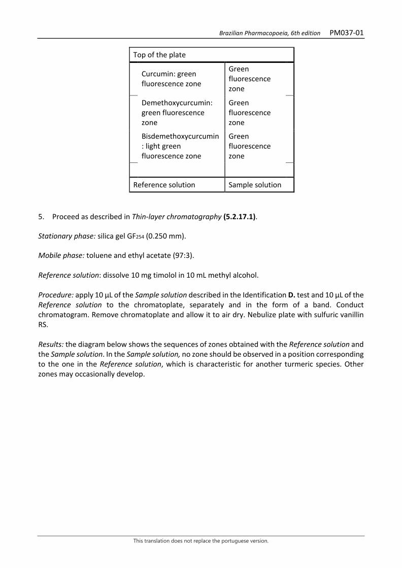
Brazilian Pharmacopoeia, 6th edition PM037-01
This translation does not replace the portuguese version.
Top of the plate
Curcumin: green fluorescence zone
Green fluorescence zone
Demethoxycurcumin: green fluorescence zone
Green fluorescence zone
Bisdemethoxycurcumin: light green fluorescence zone
Green fluorescence zone
Reference solution Sample solution 5. Proceed as described in Thin-layer chromatography (5.2.17.1). Stationary phase: silica gel GF254 (0.250 mm). Mobile phase: toluene and ethyl acetate (97:3). Reference solution: dissolve 10 mg timolol in 10 mL methyl alcohol. Procedure: apply 10 µL of the Sample solution described in the Identification D. test and 10 µL of the Reference solution to the chromatoplate, separately and in the form of a band. Conduct chromatogram. Remove chromatoplate and allow it to air dry. Nebulize plate with sulfuric vanillin RS. Results: the diagram below shows the sequences of zones obtained with the Reference solution and the Sample solution. In the Sample solution, no zone should be observed in a position corresponding to the one in the Reference solution, which is characteristic for another turmeric species. Other zones may occasionally develop.
Brazilian Pharmacopoeia, 6th edition PM037-01
This translation does not replace the portuguese version.
Top of the plate
Violaceous-colored zone
Thymol: reddish-colored zone
Violaceous-colored zone
Red-colored zone
Reference solution Sample solution TESTS Water (5.4.1.4). At most 12.0%. Determine in 15 g of pulverized plant drug (500 µm) (5.2.11). Total ash (5.4.1.5.1). At most 8.0%. Total mesophilic microorganism count (5.5.3.1.2). Complies with the test. Study of pathogenic microorganisms (5.5.3.1.3). Complies with the test. Heavy metals (5.4.5). Complies with the test. Agrochemical waste (5.4.3). Complies with the test. Aflatoxins (5.4.4). Complies with the test. DOSAGE Volatile oils Proceed as described in Determining volatile oils in plant drugs (5.4.1.6). Use a 500-mL round-bottomed flask containing 200 mL of water as distillation liquid and 0.5 mL of xylene in the graduated tube. Reduce the sample to powder (500 μm) (5.2.11) and immediately proceed to the determination on 5 g of the powdered drug. Distill for four hours. Measure the volume and express the yield per 100 g of the plant drug (w/p). Dicinnamoylmethane derivatives Sample solution: place 10 mg of the sample in a 50 mL beaker, add 6 mL glacial acetic acid, and heat in a water bath at 90 °C for 60 minutes. Add 0.2 g boric acid and 0.2 g oxalic acid, heat in a water bath at 90 °C for 10 minutes. Cool, dilute with glacial acetic acid in a 10-mL volumetric flask and homogenize. Transfer 1 mL of this solution to a 10-mL volumetric flask, top off the volume with glacial acetic acid and homogenize.
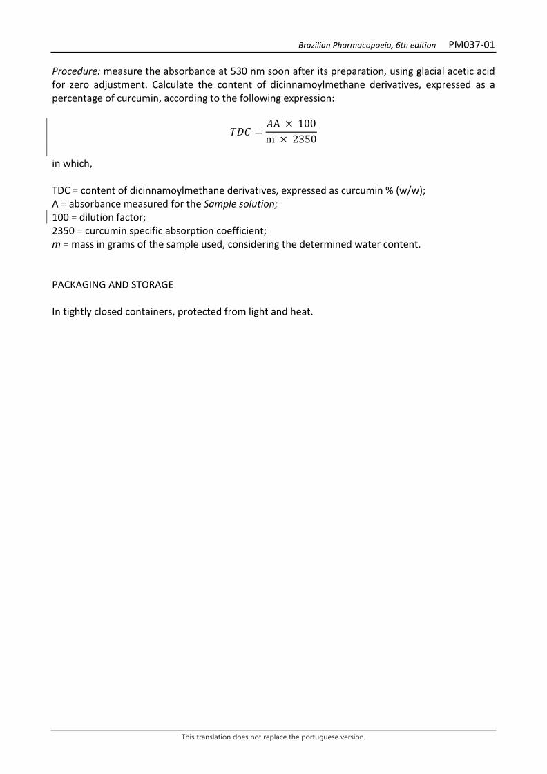
Brazilian Pharmacopoeia, 6th edition PM037-01
This translation does not replace the portuguese version.
Procedure: measure the absorbance at 530 nm soon after its preparation, using glacial acetic acid for zero adjustment. Calculate the content of dicinnamoylmethane derivatives, expressed as a percentage of curcumin, according to the following expression:
𝑇𝑇𝑇𝑇𝐶𝐶 =𝑇𝑇A × 100m × 2350
in which, TDC = content of dicinnamoylmethane derivatives, expressed as curcumin % (w/w); A = absorbance measured for the Sample solution; 100 = dilution factor; 2350 = curcumin specific absorption coefficient; m = mass in grams of the sample used, considering the determined water content. PACKAGING AND STORAGE In tightly closed containers, protected from light and heat.

Brazilian Pharmacopoeia, 6th edition PM037-01
This translation does not replace the portuguese version.
Figure 1 – Macroscopic and microscopic aspects in Curcuma longa L.
___________________________
Brazilian Pharmacopoeia, 6th edition PM037-01
This translation does not replace the portuguese version.
The scales correspond in A to 5 cm (level 1); in B, C and E to 100 μm (level 2); in D to 1.0 mm (level 3). A – overall appearance of rhizomes: leaf sheath (bf); ring-like scar coming from the base of the leaf sheath (cia); lateral branching scar (crl); root scar (cra); lateral rhizome (ril); main rhizome (rip); lateral root (rlt). B – detail of a portion of the epidermis, in front view: fundamental cell of the epidermis (cfe); stoma (es); hair (pel). C – front view detail of the suber portion: lipid drop (gl). D – schematic diagram of the rhizome in cross-section: central cylinder (cc); cuticle (cu); cortex (cx); endoderm (end); epidermis (ep); vascular bundle (fv); cortical parenchyma (pc); medullary parenchyma (pm); suber (s). E – detail of a rhizome portion in cross-section: central cylinder (cc); cell containing phenolic compound (ccf); cuticle (cu); cortex (cx); intercellular space (ei); endoderm (end); epidermis (ep); phloem (f); vascular bundle (fv); starch grain (ga); lipid drop (gl); secretory idioblast (is); nucleus (nu); hair (pel); cortical parenchyma (pc); medullary parenchyma (pm); suber (s); xylem (x).
Brazilian Pharmacopoeia, 6th edition PM037-01
This translation does not replace the portuguese version.

Brazilian Pharmacopoeia, 6th edition PM037-01
This translation does not replace the portuguese version.
Figure 2 – Microscopic aspects of the powder in Curcuma longa L. ___________________________ The scale corresponds to 100 µm. D – front view fragment of the epidermis: lipid drop (gl). B – isolated hair. C – front vire fragment of the epidermis with stoma: stoma (es); lipid drop (gl). D – front view fragment of the epidermis: lipid drop (gl). E – front view fragment of the epidermis with hair scars: hair scar (cpe). F – fragment of epidermis and cortex, visualized by transparency, in front view: epidermis (ep); suber (s). G – fragment of epidermis and suber, in cross-section: cuticle (cu); epidermis (ep); lipid drop (gl); suber (s). H – fragment of suber, oblique view: starch grain (ga); lipid drop (gl). I – cross- section of fragments of the suber: lipid drop (gl). J – isolated or grouped parenchyma cells: lipid drop (gl). L – fragment of suber and cortical parenchyma in cross-section: lipid drop (gl); cortical parenchyma (pc); suber (s). M – parenchyma fragment in cross-section: cell containing phenolic compound (ccf); lipid drop (gl). N – reserve parenchyma fragment with cells filled with starch grains, in cross-section: starch grain (ga). O – isolated parenchyma cells, filled with starch grains, in cross-section: starch grain (ga). P – starch grain mass. Q - grouped and/or isolated starch grains. R – portions of clustered, scalariformly thickened vessel elements in longitudinal section. S – portion of an isolated vessel element, with reticulate thickening, in longitudinal section. T - portion of vessel element with reticulate thickening in longitudinal section, associated with parenchyma cells, in cross-section: vessel element with reticulate thickening (ere); parenchyma (p). U – portions of vessel elements with reticulated and helical thickening, in longitudinal section: vessel element with helical thickening (eh); vessel element with reticulated thickening (ere). V – portion of an isolated vessel element, with helical thickening, in longitudinal section. X – isolated lipid drops.
Brazilian Pharmacopoeia, 6th edition PM038-00
This translation does not replace the portuguese version.
DILL, fruit Anethi fructus
The plant drug consists of dried fruits of Anethum graveolens L., containing at least 2.0% volatile oil, 30.0% carvone, and 30.0% dillapiole. CHARACTERISTICS It has an aromatic odor. IDENTIFICATION 1. Macroscopic description The fruit is an oval diachene, consisting of two dorsally compressed mericarpals, 0.3 to 0.6 cm long and 0.12 to 0.3 cm wide, brown to light brown in color, with two stylopods and retrorected stylets apex. Upon drying, the mericarp are usually separated and, as a rule, are not accompanied by the carpophores; remnants of the stylopod and calyx may occur. Each mericarp has two marginal edges extending into a surrounding, broad, membranaceous, lighter, yellowish wing and three dorsal, longitudinal, filiform, light brown to yellowish, barely elevated, all primary edges. The commissural face is flattened and somewhat concave by drying, clearly showing the carpophore line. The cuticle of each mericarp is covered by an epicuticular wax formed by short, randomly distributed filaments. These filaments are much denser on the commissural side, which makes it whitish. The mericarp, in cross-section, is plano-convex, leaving six elliptical secretory ducts visible, four of them large and narrow, distributed in the dorsal portion, and two of them, rarely larger on the commissural or ventral face. Vascular bundles occur at each dorsal edge. Those corresponding to the wings are slightly larger than the others. The endosperm is oily and concave on the commissural side. 2. Microscopic description In cross-section, the mericarp, dorsally flattened, shows three primary dorsal edges, which are narrow, and two primary lateral edges, which are elongated. The epicarp consists of a striated cuticle, formed by randomly arranged wax filaments, and a layer of flattened, thin-walled epidermal cells, except for the outer periclinal, which is thicker. The mesocarp is formed externally by a few layers of flattened parenchyma cells. In the mesocarp region, corresponding to the primary edges, both marginal and dorsal, there are strands of fibers, with some vascular elements, not always visible. In the region between the edges and in the commissural region, secretory ducts occur, which are schizogenous, elliptical in shape, and narrowly elongated in the tangential direction. The secretory epithelium is formed by thick-walled, tangentially flattened cells. In the portion of the secretory duct facing the epicarp and just below the epicarp, square or rectangular shaped sclereids occur. The innermost portion of the mesocarp is composed of two to three layers of yellow-brown cells, very flattened tangentially, with thick walls, when compared to those of the epicarp. The endocarp is composed of a layer of lignified cells. Between the pericarp and the seed, on the commissural side, there is a chamber next to the raphe. Usually associated with the endocarp is the testa, formed by a single layer of cells, usually collapsed, brown in color, and thin-walled. The endosperm is abundant, composed of thick-walled cells, the outermost are more elongated and
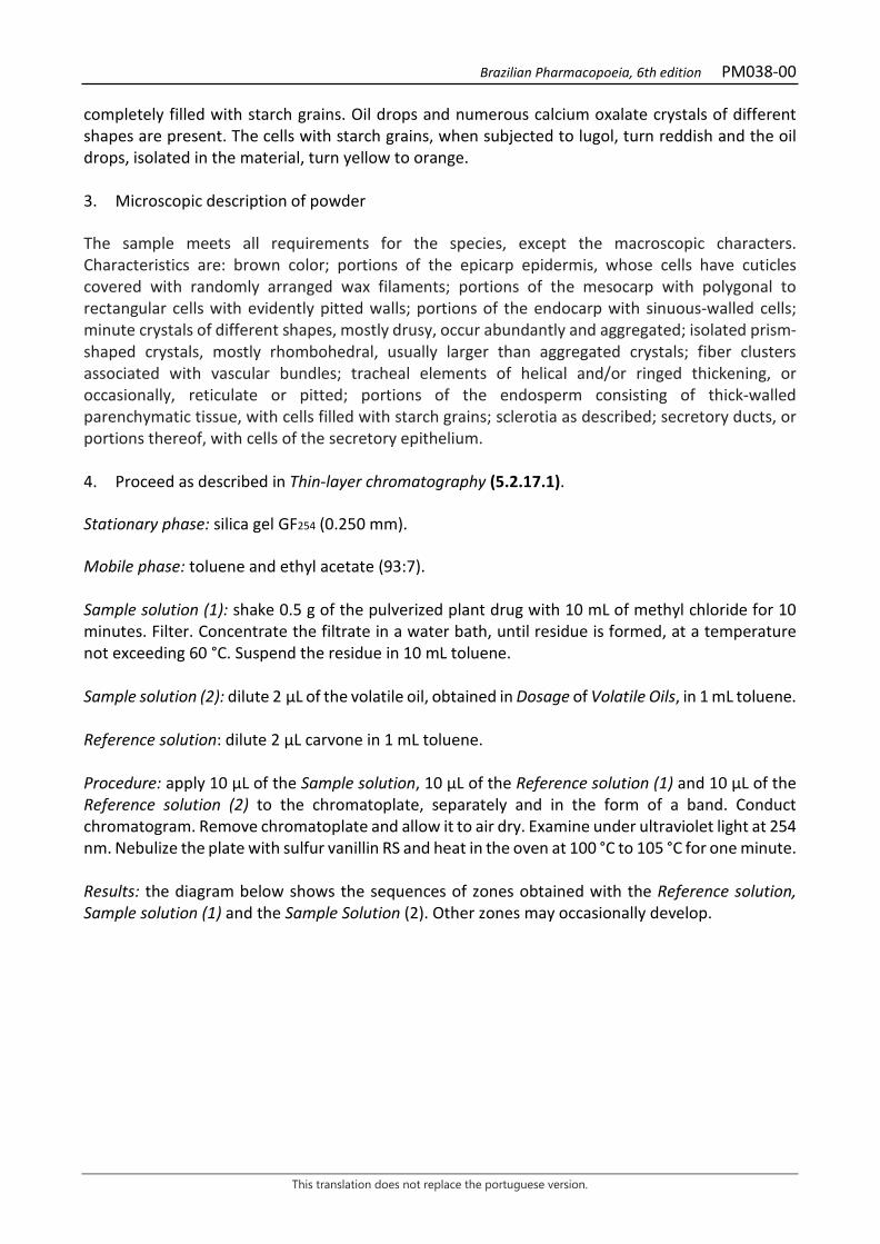
Brazilian Pharmacopoeia, 6th edition PM038-00
This translation does not replace the portuguese version.
completely filled with starch grains. Oil drops and numerous calcium oxalate crystals of different shapes are present. The cells with starch grains, when subjected to lugol, turn reddish and the oil drops, isolated in the material, turn yellow to orange. 3. Microscopic description of powder The sample meets all requirements for the species, except the macroscopic characters. Characteristics are: brown color; portions of the epicarp epidermis, whose cells have cuticles covered with randomly arranged wax filaments; portions of the mesocarp with polygonal to rectangular cells with evidently pitted walls; portions of the endocarp with sinuous-walled cells; minute crystals of different shapes, mostly drusy, occur abundantly and aggregated; isolated prism-shaped crystals, mostly rhombohedral, usually larger than aggregated crystals; fiber clusters associated with vascular bundles; tracheal elements of helical and/or ringed thickening, or occasionally, reticulate or pitted; portions of the endosperm consisting of thick-walled parenchymatic tissue, with cells filled with starch grains; sclerotia as described; secretory ducts, or portions thereof, with cells of the secretory epithelium. 4. Proceed as described in Thin-layer chromatography (5.2.17.1). Stationary phase: silica gel GF254 (0.250 mm). Mobile phase: toluene and ethyl acetate (93:7). Sample solution (1): shake 0.5 g of the pulverized plant drug with 10 mL of methyl chloride for 10 minutes. Filter. Concentrate the filtrate in a water bath, until residue is formed, at a temperature not exceeding 60 °C. Suspend the residue in 10 mL toluene. Sample solution (2): dilute 2 μL of the volatile oil, obtained in Dosage of Volatile Oils, in 1 mL toluene. Reference solution: dilute 2 µL carvone in 1 mL toluene. Procedure: apply 10 µL of the Sample solution, 10 µL of the Reference solution (1) and 10 µL of the Reference solution (2) to the chromatoplate, separately and in the form of a band. Conduct chromatogram. Remove chromatoplate and allow it to air dry. Examine under ultraviolet light at 254 nm. Nebulize the plate with sulfur vanillin RS and heat in the oven at 100 °C to 105 °C for one minute. Results: the diagram below shows the sequences of zones obtained with the Reference solution, Sample solution (1) and the Sample Solution (2). Other zones may occasionally develop.
Brazilian Pharmacopoeia, 6th edition PM038-00
This translation does not replace the portuguese version.
Top of the plate
Brown-colored zone
Carvone: light-pink colored zone
Pink-colored zone
Reference solution Sample solution TESTS Foreign matter (5.4.1.3). At most 2.0%. Water (5.4.1.4). At most 11.0%. Total ash (5.4.1.5.1). At most 7.0% Total mesophilic microorganism count (5.5.3.1.2). Complies with the test. Study of pathogenic microorganisms (5.5.3.1.3). Complies with the test. Heavy metals (5.4.5). Complies with the test. Agrochemical waste (5.4.3). Complies with the test. DOSAGE Volatile oils Proceed as described in Determining volatile oils in plant drugs (5.4.1.6). Use a 250-mL flask containing 100 mL of water as distillation liquid and 0.5 mL of xylene in the graduated tube. Reduce the dried fruits to a coarse powder. Immediately proceed with the determination of the volatile oil, from 25 g of the powdered drug. Distill for four hours. Measure the volume and express the yield per 100 g of the plant drug (w/p). Carvone and dillapiole Proceed as described in Gas chromatography (5.2.17.5). Use a chromatograph equipped with a flame ionization detector, using a mixture of nitrogen, synthetic air, and hydrogen (1:1:10) as auxiliary gases to the detector flame; 30 m long and 0.25 mm wide (internal diameter) capillary chromatography column, coated with polydiphenyldimethylsiloxane, with a 0.25 µm film thickness. Use helium at a pressure of 80 kPa as carrier gas; carrier gas flow 1 mL/minute. Temperature:
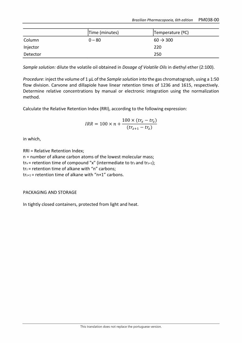
Brazilian Pharmacopoeia, 6th edition PM038-00
This translation does not replace the portuguese version.
Time (minutes) Temperature (ºC) Column 0 – 80 60 → 300 Injector 220 Detector 250 Sample solution: dilute the volatile oil obtained in Dosage of Volatile Oils in diethyl ether (2:100). Procedure: inject the volume of 1 μL of the Sample solution into the gas chromatograph, using a 1:50 flow division. Carvone and dillapiole have linear retention times of 1236 and 1615, respectively. Determine relative concentrations by manual or electronic integration using the normalization method. Calculate the Relative Retention Index (RRI), according to the following expression:
𝐼𝐼𝐼𝐼𝐼𝐼 = 100 × 𝑛𝑛 +100 × (𝑡𝑡𝑡𝑡𝑥𝑥 − 𝑡𝑡𝑡𝑡𝑧𝑧)
(𝑡𝑡𝑡𝑡𝑧𝑧+1 − 𝑡𝑡𝑡𝑡𝑧𝑧)
in which, RRI = Relative Retention Index; n = number of alkane carbon atoms of the lowest molecular mass; trx = retention time of compound "x" (intermediate to trz and trz+1); trz = retention time of alkane with “n” carbons; trz+1 = retention time of alkane with “n+1” carbons. PACKAGING AND STORAGE In tightly closed containers, protected from light and heat.
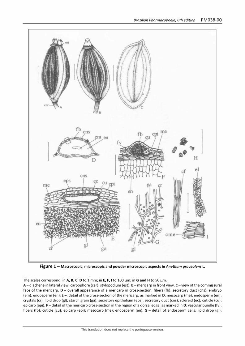
Brazilian Pharmacopoeia, 6th edition PM038-00
This translation does not replace the portuguese version.
Figure 1 – Macroscopic, microscopic and powder microscopic aspects in Anethum graveolens L.
___________________________ The scales correspond: in A, B, C, D to 1 mm; in E, F, I to 100 μm; in G and H to 50 μm. A – diachene in lateral view: carpophore (car); stylopodium (est). B – mericarp in front view. C – view of the commissural face of the mericarp. D – overall appearance of a mericarp in cross-section: fibers (fb); secretory duct (cns); embryo (em); endosperm (en). E –. detail of the cross-section of the mericarp, as marked in D: mesocarp (me); endosperm (en); crystals (cr); lipid drop (gl); starch grain (ga); secretory epithelium (eps); secretory duct (cns); sclereid (ec); cuticle (cu); epicarp (epi). F – detail of the mericarp cross-section in the region of a dorsal edge, as marked in D: vascular bundle (fv); fibers (fb); cuticle (cu); epicarp (epi); mesocarp (me); endosperm (en). G – detail of endosperm cells: lipid drop (gl);

Brazilian Pharmacopoeia, 6th edition PM038-00
This translation does not replace the portuguese version.
starch grain (ga); crystals (cr). H – crystals of different shapes. I – details of the powder: fiber strand (cf); detail of tracheal elements in longitudinal view (el); detail of mesocarp cells with thick walls (cme).
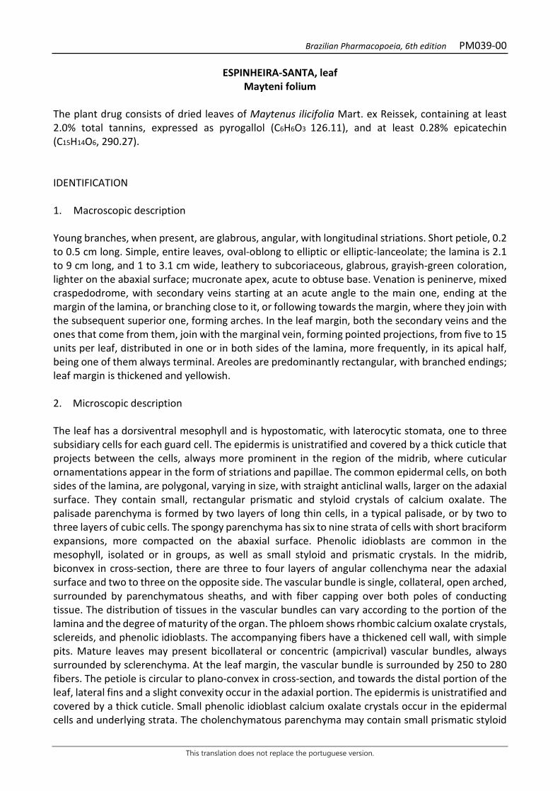
Brazilian Pharmacopoeia, 6th edition PM039-00
This translation does not replace the portuguese version.
ESPINHEIRA-SANTA, leaf Mayteni folium
The plant drug consists of dried leaves of Maytenus ilicifolia Mart. ex Reissek, containing at least 2.0% total tannins, expressed as pyrogallol (C6H6O3 126.11), and at least 0.28% epicatechin (C15H14O6, 290.27). IDENTIFICATION 1. Macroscopic description Young branches, when present, are glabrous, angular, with longitudinal striations. Short petiole, 0.2 to 0.5 cm long. Simple, entire leaves, oval-oblong to elliptic or elliptic-lanceolate; the lamina is 2.1 to 9 cm long, and 1 to 3.1 cm wide, leathery to subcoriaceous, glabrous, grayish-green coloration, lighter on the abaxial surface; mucronate apex, acute to obtuse base. Venation is peninerve, mixed craspedodrome, with secondary veins starting at an acute angle to the main one, ending at the margin of the lamina, or branching close to it, or following towards the margin, where they join with the subsequent superior one, forming arches. In the leaf margin, both the secondary veins and the ones that come from them, join with the marginal vein, forming pointed projections, from five to 15 units per leaf, distributed in one or in both sides of the lamina, more frequently, in its apical half, being one of them always terminal. Areoles are predominantly rectangular, with branched endings; leaf margin is thickened and yellowish. 2. Microscopic description The leaf has a dorsiventral mesophyll and is hypostomatic, with laterocytic stomata, one to three subsidiary cells for each guard cell. The epidermis is unistratified and covered by a thick cuticle that projects between the cells, always more prominent in the region of the midrib, where cuticular ornamentations appear in the form of striations and papillae. The common epidermal cells, on both sides of the lamina, are polygonal, varying in size, with straight anticlinal walls, larger on the adaxial surface. They contain small, rectangular prismatic and styloid crystals of calcium oxalate. The palisade parenchyma is formed by two layers of long thin cells, in a typical palisade, or by two to three layers of cubic cells. The spongy parenchyma has six to nine strata of cells with short braciform expansions, more compacted on the abaxial surface. Phenolic idioblasts are common in the mesophyll, isolated or in groups, as well as small styloid and prismatic crystals. In the midrib, biconvex in cross-section, there are three to four layers of angular collenchyma near the adaxial surface and two to three on the opposite side. The vascular bundle is single, collateral, open arched, surrounded by parenchymatous sheaths, and with fiber capping over both poles of conducting tissue. The distribution of tissues in the vascular bundles can vary according to the portion of the lamina and the degree of maturity of the organ. The phloem shows rhombic calcium oxalate crystals, sclereids, and phenolic idioblasts. The accompanying fibers have a thickened cell wall, with simple pits. Mature leaves may present bicollateral or concentric (ampicrival) vascular bundles, always surrounded by sclerenchyma. At the leaf margin, the vascular bundle is surrounded by 250 to 280 fibers. The petiole is circular to plano-convex in cross-section, and towards the distal portion of the leaf, lateral fins and a slight convexity occur in the adaxial portion. The epidermis is unistratified and covered by a thick cuticle. Small phenolic idioblast calcium oxalate crystals occur in the epidermal cells and underlying strata. The cholenchymatous parenchyma may contain small prismatic styloid
Brazilian Pharmacopoeia, 6th edition PM039-00
This translation does not replace the portuguese version.
crystals. Isolated brachysclereids occur randomly in the fundamental parenchyma of the petiole. The vascular bundle is concentric, cylindrical to slightly concave-convex, surrounded by a sclerenchymatic sheath composed of isolated fibers or in groups of two to many elements. 3. Microscopic description of powder The sample meets all requirements for the species, except the macroscopic characters. The characteristics are: a yellowish-green color; fragments of epidermis with straight periclinal walls, covered by thick cuticle and containing small styloid or small prismatic crystals; fragments of epidermis with laterocytic stomata; fragments of epidermis, in cross-section, with stomata evidencing the atrium; palisade parenchyma fragments with two or three layers; fragments of leaf borders with fiber portions; fragments of large caliber fibers with simple pits; parenchyma fragments with brachysclereids; parenchyma fragments containing phenolic idioblasts; isolated fibers or group of fibers in longitudinal view; isolated styloid or prismatic or rhombic crystals; isolated brachysclereids. 4. Proceed as described in Thin-layer chromatography (5.2.17.1). Stationary phase: silica gel F254 (0.250 mm). Mobile phase: ethyl acetate, formic acid and water (90:5:5). Sample solution: accurately weigh about 5 g of the pulverized plant drug, add 50 mL of water and heat under reflux for 15 minutes. After cooling to room temperature, filter the solution obtained through absorbent cotton under reduced pressure into a 50-mL volumetric flask, complete with distilled water, and homogenize. Reference solution: weigh approximately 1 mg epicatechin and dissolve in 1 mL methyl alcohol. Procedure: apply 10 μL of the Sample solution and 3 μL of the Reference solution to the chromatoplate, separately and in the form of a band. Conduct chromatogram. Remove the plate and allow it to dry in a fume hood. Examine under ultraviolet light at 254 nm. Next, nebulize the plate with sulfur vanillin RS and heat in an oven at 110 °C for 10 minutes. Results: the diagram below shows the sequences of zones obtained with the Reference solution and the sample Solution. Other zones may occasionally develop.
Brazilian Pharmacopoeia, 6th edition PM039-00
This translation does not replace the portuguese version.
Top of the plate
Epicatechin: burgundy-colored zone
Reddish-brown colored zone
Reddish-brown
colored zone
Reference solution Sample solution TESTS Foreign matter (5.4.1.3). At most 2.0%. Water (5.4.1.4). At most 12.0%. Total ash (5.4.1.5.1). At most 8.0%. Total mesophilic microorganism count (5.5.3.1.2). Complies with the test. Study of pathogenic microorganisms (5.5.3.1.3). Complies with the test. Heavy metals (5.4.5). Complies with the test. Agrochemical waste (5.4.3). Complies with the test. Foam level. Quantitatively transfer about 1 g of the pulverized plant drug (180 μm), weighed, accurately, to an Erlenmeyer flask containing 50 mL of boiling water. Keep at moderate boil for 15 minutes. Cool, filter through absorbent cotton into a 100-mL volumetric flask. Top off the volume through the filter and homogenize. Distribute the obtained decoction into 10 capped test tubes (16 mm wide by 16 cm high) in a successive series of 1 mL, 2 mL, 3 mL, up to 10 mL, and adjust the liquid volume in each tube to 10 mL with water. Cap the tubes and shake them vigorously with vertical movements for 15 seconds at two shakes per second. Allow it to stand for 15 minutes and measure the height of the foam. Then add 1 mL of 2 M hydrochloric acid to each tube, if the foam height of all tubes is less than 1 cm, the foam level is less than 100. If, in any of the tubes, the measured foam height remains equal to or greater than 1 cm, the dilution of the plant material in such tube (A) is the observed rate. Calculate the foam index, according to the following expression:
𝐼𝐼𝑇𝑇 =1000𝑇𝑇
Brazilian Pharmacopoeia, 6th edition PM039-00
This translation does not replace the portuguese version.
in which, IE = foam level; V = volume, in milliliters, of the decoction used to prepare the dilution in the tube foam was observed. The foam level is at least 250. DOSAGE Total tannins Note: protect samples from light during extraction and dilution. Use carbon dioxide-free water for all operations. To proceed as described in Visible absorption spectrophotometry (5.2.14). Prepare solutions as described below. Stock solution: weigh 0.750 g of the pulverized plant drug (250 μm) (5.2.11) and transfer to a 250-mL, polished-necked erlenmeyer flask. Add 150 mL of water. Heat in a water bath for 30 minutes at 60 °C. Cool under running water and transfer to a 250-mL volumetric flask. Rinse the erlenmeyer flask and transfer the washing water with the entire plant drug content to the same volumetric flask. Top off the volume with water and homogenize. Allow to decant and filter the supernatant liquid through filter paper. Discard the first 50 mL of the filtrate. Sample solution for total polyphenol content: transfer 5 mL of the filtrate from the Stock solution to a 25-mL volumetric flask, top off the volume with water and homogenize. Volumetrically transfer 2 mL of the solution, 1 mL of phosphomolybdotungstic reagent and 10 mL of water to a 25-mL volumetric flask, top off the volume with 29% sodium carbonate solution (w/v) and homogenize. Determine absorbance at 760 nm (A1) after 30 minutes, using water for zero adjustment. Sample solution for polyphenols not adsorbed by hide powder: for 10 mL of the filtrate from the Stock solution, add 0.1 g of the hide powder and mechanically shake in a 125-mL Erlenmeyer flask for 60 minutes. Filter through paper filter. Transfer 5 mL of this filtrate to a 25-mL volumetric flask, top off the volume with water, and homogenize. Volumetrically transfer 2 mL of the solution, 1 mL of phosphomolybdotungstic reagent and 10 mL of water to a 25-mL volumetric flask, top off the volume with 29% sodium carbonate solution (w/v) and homogenize. Determine absorbance at 760 nm (A2) after 30 minutes, using water for zero adjustment. Reference solution: dissolve 50 mg pyrogallol in water immediately before use, transfer to a 100-mL volumetric flask, top off the volume with water and homogenize. Volumetrically transfer 5 mL of the solution to a 100-mL volumetric flask, top off the volume with water and homogenize. Volumetrically transfer 2 mL of the solution, 1 mL of phosphomolybdotungstic reagent and 10 mL of water to a 25-mL volumetric flask, top off the volume with 29% sodium carbonate solution (w/v) and homogenize. Determine absorbance at 760 nm (A3) after 30 minutes, using water for zero adjustment.
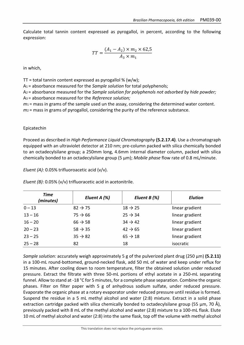
Brazilian Pharmacopoeia, 6th edition PM039-00
This translation does not replace the portuguese version.
Calculate total tannin content expressed as pyrogallol, in percent, according to the following expression:
𝑇𝑇𝑇𝑇 =(𝑇𝑇1 − 𝑇𝑇2) × 𝑚𝑚2 × 62,5
𝑇𝑇3 × 𝑚𝑚1
in which, TT = total tannin content expressed as pyrogallol % (w/w); A1 = absorbance measured for the Sample solution for total polyphenols; A2 = absorbance measured for the Sample solution for polyphenols not adsorbed by hide powder; A3 = absorbance measured for the Reference solution; m1 = mass in grams of the sample used un the assay, considering the determined water content. m2 = mass in grams of pyrogallol, considering the purity of the reference substance. Epicatechin Proceed as described in High Performance Liquid Chromatography (5.2.17.4). Use a chromatograph equipped with an ultraviolet detector at 210 nm; pre-column packed with silica chemically bonded to an octadecylsilane group; a 250mm long, 4.6mm internal diameter column, packed with silica chemically bonded to an octadecylsilane group (5 µm); Mobile phase flow rate of 0.8 mL/minute. Eluent (A): 0.05% trifluoroacetic acid (v/v). Eluent (B): 0.05% (v/v) trifluoracetic acid in acetonitrile.
Time (minutes) Eluent A (%) Eluent B (%) Elution
0 – 13 82 → 75 18 → 25 linear gradient 13 – 16 75 → 66 25 → 34 linear gradient 16 – 20 66 → 58 34 → 42 linear gradient 20 – 23 58 → 35 42 → 65 linear gradient 23 – 25 35 → 82 65 → 18 linear gradient 25 – 28 82 18 isocratic Sample solution: accurately weigh approximately 5 g of the pulverized plant drug (250 µm) (5.2.11) in a 100-mL round-bottomed, ground-necked flask, add 50 mL of water and keep under reflux for 15 minutes. After cooling down to room temperature, filter the obtained solution under reduced pressure. Extract the filtrate with three 50-mL portions of ethyl acetate in a 250-mL separating funnel. Allow to stand at -18 °C for 5 minutes, for a complete phase separation. Combine the organic phases. Filter on filter paper with 5 g of anhydrous sodium sulfate, under reduced pressure. Evaporate the organic phase at a rotary evaporator under reduced pressure until residue is formed. Suspend the residue in a 5 mL methyl alcohol and water (2:8) mixture. Extract in a solid phase extraction cartridge packed with silica chemically bonded to octadecylsilane group (55 µm, 70 Å), previously packed with 8 mL of the methyl alcohol and water (2:8) mixture to a 100-mL flask. Elute 10 mL of methyl alcohol and water (2:8) into the same flask, top off the volume with methyl alcohol
Brazilian Pharmacopoeia, 6th edition PM039-00
This translation does not replace the portuguese version.
and water (2:8) and homogenize (S1). Volumetrically transfer 5 mL of S1 to a 25-mL volumetric flask, top off the volume with methyl alcohol and water (1:1) and homogenize (S2). Filter S2 through a 0.45 µm filter unit. Reference solution: dissolve an accurately weighed amount of epicatechin in methyl alcohol and water (1:1) to obtain a 0.4 mg/mL solution. Solutions for analytical curve: dilute aliquots of 50 μL, 200 μL, 350 μL, 500 μL, and 600 μL of the Reference solution in a 2 mL volumetric flask with methyl alcohol and water (1:1) to obtain concentrations of 10 μg/mL; 40 μg/mL; 70 μg/mL; 100 μg/mL, and 120 μg/mL. Filter through a 0.45 µm filter unit. Procedure: inject 20 µL of the Solutions for the analytical curve and 20 µL of the Sample solution separately. Register the chromatograms and measure the areas under the peaks. Relative retention time is about eight minutes for epicatechin. Determine epicatechin content in the Sample solution from the linear equation of the straight line obtained with the analytical curve of the standard. Calculate the epicatechin content of the sample in mg/g according to the following expression:
𝑇𝑇𝑇𝑇𝐶𝐶 =𝐶𝐶𝑟𝑟 × 500𝑚𝑚𝑎𝑎 × 1000
in which, TEC = epicatechin content in mg/g; Cr = concentration of epicatechin/mL in the Sample solution in μg/mL, from the straight line equation; 500 = dilution factor; 1000 = μg to mg conversion value; ma = mass in grams of the sample used, considering the determined water content. PACKAGING AND STORAGE In tightly closed containers, protected from light and heat.
Brazilian Pharmacopoeia, 6th edition PM039-00
This translation does not replace the portuguese version.
Figure 1 – Macroscopic, microscopic and powder microscopic aspects in Maytenus ilicifolia Mart. ex Reissek
___________________________ The scales correspond in A and B to 5 mm; in C to 1 mm; in D, E and F to 30 μm. A – overall appearance of the leaf lamina; B – detail of the leaf venation on the adaxial surface, in front view. C – Detail of a portion of the leaf lamina, adaxial surface, in front view, showing the areolas and xylematic endings. D and E – partial detail of the epidermis facing the adaxial and abaxial surfaces, respectively, in front view: crystalliferous idioblast (ic); subsidiary cell (csb); stoma (es). F – partial detail of the leaf lamina, in cross section, showing a stoma: spongy parenchyma (pj); cuticle (cu); suprastomatic atrium (at); guard cell (cg); subsidiary cell (csb); crystalliferous idioblast (ic).

Brazilian Pharmacopoeia, 6th edition PM039-00
This translation does not replace the portuguese version.
Figure 2 – Microscopic and powder microscopic aspects in Maytenus ilicifolia Mart. ex Reissek
___________________________ The scales correspond in A and B to 50 μm; in C and D to 75 μm; in E to 35 μm; in F to 120 μm; in G to 180 μm; in H and I to 200 μm; in J to 50 μm. A and B – partial details of the mesophyll of distinct samples, in cross-sections: adaxial surface (ad); abaxial surface (ab); epidermis (ep); cuticle (cu); crystalliferous idioblast (ic); palisade parenchyma (pp); spongy parenchyma (pj); xylem (x); phloem (f); parenchymatic sheath (bp); fibers (fb); stoma (es). C and D – detail of a secondary vascular bundle in the basal and mid portion of the leaf lamina, respectively, in cross-section: fibers (fb); parenchymatic sheath (bp); spongy parenchyma (pj); palisade parenchyma (pp); xylem (x); phloem (f). E – detail of the leaf margin, in cross-section, showing the marginal vein: adaxial surface (ad); abaxial surface (ab); epidermis (ep); palisade parenchyma (pp); spongy parenchyma (pj); fibers (fb); stoma (es). F, G and H – diagrams of the overall appearance of the median portion of the midrib, in cross-sections, showing variations in the distribution of the phloem, xylem, and fibers: adaxial surface (ad); abaxial surface (ab); collenchyma (co); epidermis (ep); palisade parenchyma (pp); xylem (x); phloem (f); fibers (fb). I – diagram of the overall appearance of the petiole, in cross-section: epidermis (ep); fibers (fb); xylem (x); phloem (f). J – detail of a petiole brachiesclereid, in cross-section: brachysclereid (br).
Brazilian Pharmacopoeia, 6th edition PM040-00
This translation does not replace the portuguese version.
CANDYLEAF, leaf Steviae folium
The plant drug consists of dried leaves of Stevia rebaudiana (Bertoni) Bertoni, containing at least 12.0% total carbohydrates and 4.0% stevioside (C38H60O18, 804.87). IDENTIFICATION 1. Macroscopic description Simple leaves, up to 6 cm long and up to 2.5 cm wide, membranous and brittle, dark green on the adaxial surface and lighter on the abaxial surface, spatulate to lanceolate, sessile, with a sharp apex, attenuated base and serrated margin from the basal third towards the leaf apex, with three longitudinal veins, the main one more developed. Actinodromic venation. The leaf is covered with pluricellular, uniseriate tector trichomes on both surfaces; glandular trichomes are visible with a magnifying glass. 2. Microscopic description Dorsiventrally symmetrical leaf lamina, amphistomatic, with anomocytic stomata. In front view, the epidermis shows cells with sinuous walls, with a more accentuated sinuosity on the abaxial surface; in the region of the veins, the cells are elongated with rectilinear periclinal walls. The stomas occur in greater number on the abaxial surface. Uniseriate, pluricellular tector trichomes, of two types, are found on the entire surface of the leaf lamina, on both sides, the larger ones with an enlarged base and acute apex, and bulkier basal cells; the smaller ones have a uniform diameter from the base to the apex, and these are less tapered; glandular trichomes occur on both sides and are located in small depressions in the epidermis, with a pluricellular and uniseriate pedicel and a rounded and unicellular head. In some areas of the epidermis, epicuticular striations are visible. In a cross-section of the lamina, the stomata appear to be situated at the same level or slightly above the other epidermal cells and the inner periclinal and anticlinal walls that enclose the stomatal pore are thickened. The palisade parenchyma is formed by one or two layers of cells; when two, they cover half the thickness of the lamina. The spongy parenchyma has several strata, irregularly arranged. The secondary vascular bundles are collateral, surrounded by a chlorophyllated parenchymatic sheath. The midrib, in cross-section, is more prominent on the abaxial surface. The cells of the epidermis are isodiametric and the collenchyma is lacunar. The vascular system is represented by a collateral vascular bundle, partially surrounded by sclerenchymatic fibers capping close to the xylem and phloem. In cross-section, the leaf lamina shows an open semicircular shape, slightly concave on the adaxial surface and convex on the abaxial surface. The epidermis has polyhedral to quadrangular cells, with an ornamented cuticle. Stomata are located above the level of the other epidermal cells and occur only at the margins. The collenchyma is formed by one or two layers of cells, on both sides. The fundamental parenchyma fills most of this region and the chlorenchyma, the margins. The vascular system consists of five to seven collateral vascular bundles, the central one being the largest. 3. Microscopic description of powder
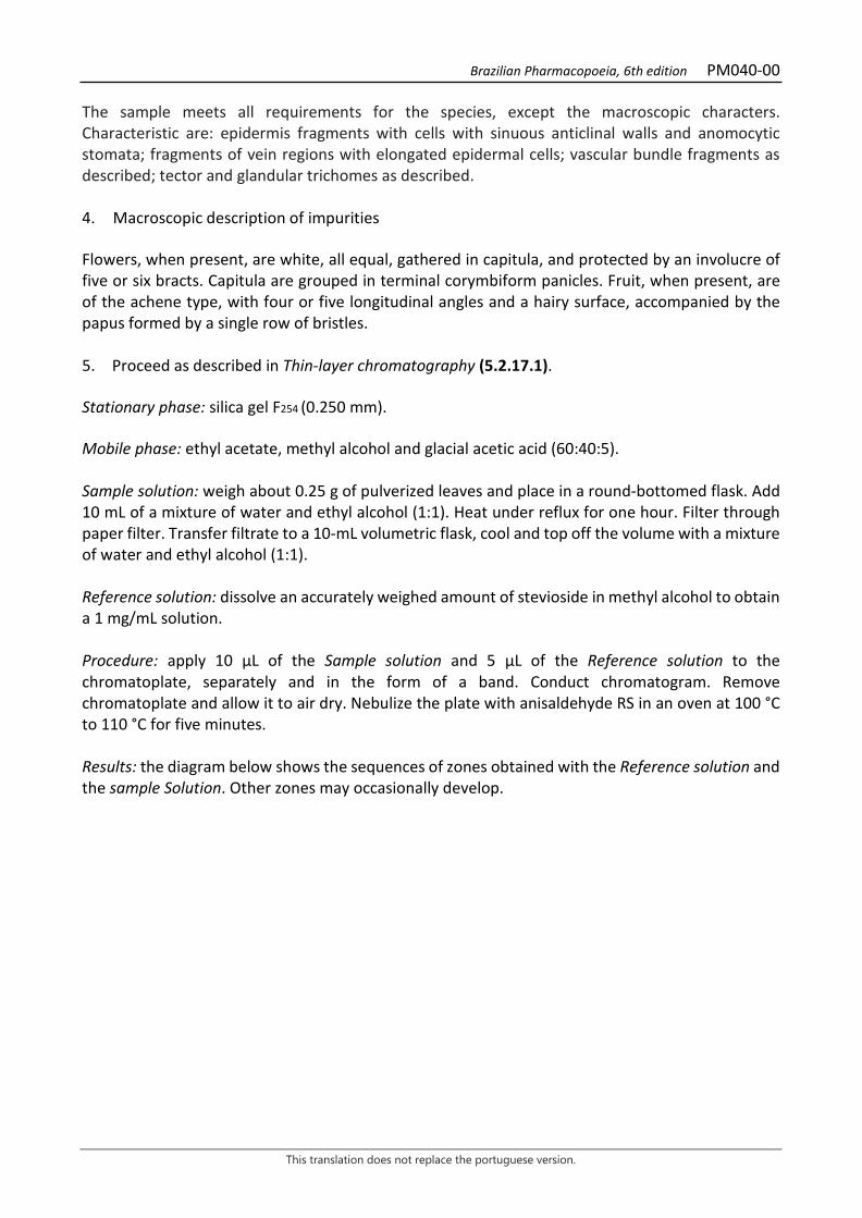
Brazilian Pharmacopoeia, 6th edition PM040-00
This translation does not replace the portuguese version.
The sample meets all requirements for the species, except the macroscopic characters. Characteristic are: epidermis fragments with cells with sinuous anticlinal walls and anomocytic stomata; fragments of vein regions with elongated epidermal cells; vascular bundle fragments as described; tector and glandular trichomes as described. 4. Macroscopic description of impurities Flowers, when present, are white, all equal, gathered in capitula, and protected by an involucre of five or six bracts. Capitula are grouped in terminal corymbiform panicles. Fruit, when present, are of the achene type, with four or five longitudinal angles and a hairy surface, accompanied by the papus formed by a single row of bristles. 5. Proceed as described in Thin-layer chromatography (5.2.17.1). Stationary phase: silica gel F254 (0.250 mm). Mobile phase: ethyl acetate, methyl alcohol and glacial acetic acid (60:40:5). Sample solution: weigh about 0.25 g of pulverized leaves and place in a round-bottomed flask. Add 10 mL of a mixture of water and ethyl alcohol (1:1). Heat under reflux for one hour. Filter through paper filter. Transfer filtrate to a 10-mL volumetric flask, cool and top off the volume with a mixture of water and ethyl alcohol (1:1). Reference solution: dissolve an accurately weighed amount of stevioside in methyl alcohol to obtain a 1 mg/mL solution. Procedure: apply 10 μL of the Sample solution and 5 μL of the Reference solution to the chromatoplate, separately and in the form of a band. Conduct chromatogram. Remove chromatoplate and allow it to air dry. Nebulize the plate with anisaldehyde RS in an oven at 100 °C to 110 °C for five minutes. Results: the diagram below shows the sequences of zones obtained with the Reference solution and the sample Solution. Other zones may occasionally develop.
Brazilian Pharmacopoeia, 6th edition PM040-00
This translation does not replace the portuguese version.
Top of the plate
Stevioside: fuzzy-green colored area
Fuzzy-green colored zone
Reference solution Sample solution TESTS Foreign matter (5.4.1.3). At most 2.0%. Water (5.4.1.4). At most 13,0%. Total ash (5.4.1.5.1). At most 9.5%. Total mesophilic microorganism count (5.5.3.1.2). Complies with the test. Study of pathogenic microorganisms (5.5.3.1.3). Complies with the test. Heavy metals (5.4.5). Complies with the test. Agrochemical waste (5.4.3). Complies with the test. DOSAGE Total carbohydrates To proceed as described in Visible absorption spectrophotometry (5.2.14). Prepare solutions as described below. Stock solution: weigh 2.0 g of pulverized candyleaf leaf. Extract by infusion, with 80 mL of hot water, three times, and filter. Combine the filtrates, top off to 250 mL and homogenize. Transfer 5 mL of thus solution to a 50-mL volumetric flask, top off the volume with water and homogenize. Sample solution: transfer 0.6 mL of the Stock solution to a test tube, add 0.6 mL of 5% (w/v) phenol and 3 mL sulfuric acid. Allow to stand for 10 minutes. Blank solution: transfer 0.6 mL of water to a test tube, add 0.6 mL of 5% (w/v) phenol and 3 mL sulfuric acid. Allow to stand for 10 minutes. Reference solution: transfer 0.6 mL of 0.01% (w/v) glucose in water to a test tube, add 0.6 mL of 5% (w/v) phenol and 3 mL sulfuric acid. Allow to stand for 10 minutes.
Brazilian Pharmacopoeia, 6th edition PM040-00
This translation does not replace the portuguese version.
Procedure: measure the absorbance of the Sample solution and the Reference solution at 490 nm, using the Blank solution for zero adjustment. Calculate total carbohydrates content, in percent, according to the following expression:
𝑇𝑇𝐶𝐶 =𝑇𝑇𝑎𝑎𝑇𝑇𝑟𝑟
× 1,125 × 10
in which, TC = carbohydrate content % (w/w); Aa = absorbance measured for the Sample solution; Ar = absorbance measured for the Reference solution; 10 = dilution factor. Stevioside Proceed as described in High Performance Liquid Chromatography (5.2.17.4). Use a chromatograph equipped with an ultraviolet detector at 206 nm; pre-column packed with silica bonded to an octadecylsilane group; 150mm long, 3.9 mm internal diameter column, packed with silica bonded to an octadecylsilane group (5 μm), kept at room temperature; Mobile phase flow rate of 1.0 mL/minute. Filter through a 0.45 µm filter unit. Eluent (A): water and acetonitrile (80:20). Eluent (B): acetonitrile.
Time (minutes) Eluent A (%) Eluent B (%) Elution
0 – 4 100→70 0→30 linear gradient 4 – 7 70→0 30→100 linear gradient Sample solution: transfer approximately 0.25 g, accurately weighed, of the powdered and pulverized plant drug to a round-bottomed flask. Add 10 mL of a mixture of water and ethyl alcohol (1:1), and heat at about 100 °C, under reflux, for 60 minutes. Cool the extract to room temperature with running cold water. Filter the extract on filter paper, under vacuum, rinsing the frame with a small volume of water. Transfer filtrate to a 10-mL volumetric flask and top off the volume with a mixture of water and ethyl alcohol (1:1). Dilute 50 μL of the resulting solution in 950 μL of an acetonitrile and water mixture (20:80). Reference solution: dissolve an accurately weighed amount of stevioside in methyl alcohol to obtain a 1 mg/mL solution. Softly heat, if necessary. Solutions for the analytical curve: dilute 500 μL of the Reference solution to obtain a solution at 0.50 mg/mL. Dilute the previous solution successively in methyl alcohol to concentrations of 0.25 mg/mL, 0.125 mg/mL, 0.0625 mg/mL, 0.032 mg/mL, and 0.016 mg/mL. Filter through a 0.45 µm filter unit.
Brazilian Pharmacopoeia, 6th edition PM040-00
This translation does not replace the portuguese version.
Procedure: inject 10 µL of the Solutions for the analytical curve and 10 µL of the Sample solution separately. Register the chromatograms and measure the areas under the peaks. Retention time is approximately four minutes and 40 seconds for stevioside. Calculate stevioside content in the sample from the straight line equation obtained from the analytical curve. PACKAGING AND STORAGE In tightly closed containers, protected from light and heat.

Brazilian Pharmacopoeia, 6th edition PM040-00
This translation does not replace the portuguese version.
Figure 1 - Macroscopic, microscopic and powder microscopic aspects in Stevia rebaudiana (Bertoni) Bertoni
___________________________ The scales correspond in A to 1 cm; in B, E and H to 250 μm; in C, D, G, F and I to 50 μm. A – overall appearance of the leaf; B – detail of leaf venation; C – detail of the adaxial surface epidermis, in front view, showing the stomata; D – detail of the abaxial surface epidermis, in front view, showing the stomata and cells with sinuous walls; E – schematic diagram of the leaf lamina cross-section in the midrib region, showing the collateral bundle: phloem (f); fibers (fi ); vascular bundle (fv); fundamental parenchyma (pf); palisade parenchyma (pp); spongy parenchyma (pj); xylem (x). F – detail of the vascular bundle of the midrib in cross-section, as shown in E: phloem (f); fibers (fi); fundamental parenchyma (pf); xylem (x). G – detail of the epidermis and collenchyma in the midrib region, facing the adaxial surface, and detail of the epidermis and collenchyma in the midrib region, facing the abaxial surface:
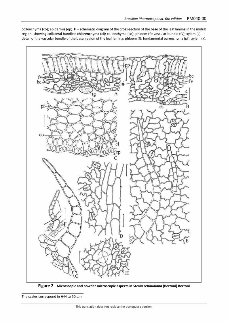
Brazilian Pharmacopoeia, 6th edition PM040-00
This translation does not replace the portuguese version.
collenchyma (co); epidermis (ep). H – schematic diagram of the cross-section of the base of the leaf lamina in the midrib region, showing collateral bundles: chlorenchyma (cl); collenchyma (co); phloem (f); vascular bundle (fv); xylem (x). I – detail of the vascular bundle of the basal region of the leaf lamina: phloem (f); fundamental parenchyma (pf); xylem (x).
Figure 2 - Microscopic and powder microscopic aspects in Stevia rebaudiana (Bertoni) Bertoni
___________________________ The scales correspond in A-H to 50 μm.
Brazilian Pharmacopoeia, 6th edition PM040-00
This translation does not replace the portuguese version.
A – detail of the leaf lamina, in cross-section: epidermis (ep); vascular sheath with chloroplastids (bc); vascular bundle (fv); spongy parenchyma (pj); palisade parenchyma (pp); glandular trichome (tg). B – detail of the leaf lamina, in cross-section: stoma (es); epidermis (ep); vascular sheath with chloroplastids (bc); vascular bundle (fv); spongy parenchyma (pj); palisade parenchyma (pp). C – fragment of the basal portion of the leaf lamina, in cross-section, at the midrib level, showing epicuticular striations: epidermis (ep); chlorenchyma (cl); collenchyma (co); fundamental parenchyma (pf); D – fragment of epidermis, in front view, showing stomata and the morphological variability of the cells; E – fragment of the epidermis, in front view, showing stomata and tector trichomes; F – tector trichome and fundamental epidermal cells, showing epicuticular striations; G – tector trichome with enlarged basal cells; H – fragment of the epidermis, in front view, showing stomata and glandular trichome.
Brazilian Pharmacopoeia, 6th edition PM041-00
This translation does not replace the portuguese version.
JIMSONWEED, leaf Stramonii folium
The plant drug consists of dried leaves of Datura stramonium L. and its varieties, containing at least 0.25% total alkaloids calculated as hyoscyamine (C17H23NO3, 289.37). CHARACTERISTICS The leaf has an unpleasant odor. IDENTIFICATION 1. Macroscopic description Oval or oval-triangular leaf lamina, lobed-sinuous, with an acuminate apex and asymmetrical base, dark brownish-green to dark grayish-green color, twisted and shriveled due to drying, thin and fragile, 15 to 20 cm long and 8 to 10 cm wide. Pinnate venation, with four to five alternate secondary veins, concave on the adaxial surface and prominent on the abaxial surface. Petiole up to 5 cm long. Young leaves pubescent on the veins. 2. Microscopic description The leaf lamina is dorsiventral and amphi-hypoestomatic, with anisocytic, rarely anomocytic stomata. The epidermis, in front view, has polygonal cells, with straight to slightly indented, thick anticlinal walls. Tector and glandular trichomes are more abundant on the abaxial surface and on the veins. Tector trichomes are pluricellular, uniseriate, conical, formed by two to five elongated cells with finely verrucous walls; the glandular trichomes are usually shortly pedicellate, with an ovoid or claviform apical gland formed by two to seven cells. The epidermis, in cross-section, is unistratified and covered by a smooth, thin cuticle. The mesophyll consists of palisade parenchyma composed of one layer of cells and spongy parenchyma. Between the two parenchyma, one or more layers of idioblasts are found, containing calcium oxalate druses. The vascular bundles are bicolateral. 3. Microscopic description of powder The sample meets all requirements for the species, except the macroscopic characters. Characteristics are: epidermal fragments with cells of straight to slightly waved anticlinal walls and with smooth cuticle; epidermal fragments with anisocytic and or anomocytic stomata more frequent in the abaxial epidermis; uniseriate unicellular conical tector trichomes and short claviform and/or ovoid glandular trichomes; fragments of palisade and/or spongy parenchyma in transverse section; fragments of ringed and spiral vessel elements; parenchyma fragments with numerous druse-containing idioblasts. 4. Proceed as described in Thin-layer chromatography (5.2.17.1). Stationary phase: silica gel GF254.
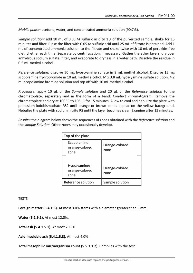
Brazilian Pharmacopoeia, 6th edition PM041-00
This translation does not replace the portuguese version.
Mobile phase: acetone, water, and concentrated ammonia solution (90:7:3). Sample solution: add 10 mL of 0.05 M sulfuric acid to 1 g of the pulverized sample, shake for 15 minutes and filter. Rinse the filter with 0.05 M sulfuric acid until 25 mL of filtrate is obtained. Add 1 mL of concentrated ammonia solution to the filtrate and shake twice with 10 mL of peroxide-free diethyl ether each time. Separate by centrifugation, if necessary. Gather the ether layers, dry over anhydrous sodium sulfate, filter, and evaporate to dryness in a water bath. Dissolve the residue in 0.5 mL methyl alcohol. Reference solution: dissolve 50 mg hyoscyamine sulfate in 9 mL methyl alcohol. Dissolve 15 mg scopolamine hydrobromide in 10 mL methyl alcohol. Mix 3.8 mL hyoscyamine sulfate solution, 4.2 mL scopolamine bromide solution and top off with 10 mL methyl alcohol. Procedure: apply 10 μL of the Sample solution and 20 μL of the Reference solution to the chromatoplate, separately and in the form of a band. Conduct chromatogram. Remove the chromatoplate and dry at 100 °C to 105 °C for 15 minutes. Allow to cool and nebulize the plate with potassium iodobismuthate RS2 until orange or brown bands appear on the yellow background. Nebulize the plate with sodium nitrite RS until the layer becomes clear. Examine after 15 minutes. Results: the diagram below shows the sequences of zones obtained with the Reference solution and the sample Solution. Other zones may occasionally develop.
Top of the plate
Scopolamine: orange-colored zone
Orange-colored zone
Hyoscyamine: orange-colored zone
Orange-colored zone
Reference solution Sample solution
TESTS Foreign matter (5.4.1.3). At most 3.0% stems with a diameter greater than 5 mm. Water (5.2.9.1). At most 12.0%. Total ash (5.4.1.5.1). At most 20.0%. Acid-insoluble ash (5.4.1.5.3). At most 4.0% Total mesophilic microorganism count (5.5.3.1.2). Complies with the test.
Brazilian Pharmacopoeia, 6th edition PM041-00
This translation does not replace the portuguese version.
Study of pathogenic microorganisms (5.5.3.1.3). Complies with the test. Heavy metals (5.4.5). Complies with the test. Agrochemical waste (5.4.3). Complies with the test. DOSAGE Total alkaloid content Weigh about 10 g of the pulverized sample (180 μm) (5.2.11) and moisten with 5 mL of a 6 M ammonium hydroxide solution. Add 10 mL of 96% (v/v) ethyl alcohol and 30 mL of peroxide-free diethyl ether, thoroughly mixed. Transfer the mixture to a percolator, if necessary, using the extraction solution. Macerate for four hours and percolate the mixture with chloroform and peroxide-free diethyl ether (1:3) until the alkaloids are completely extracted. Evaporate 1 mL of the percolate until it dries and dissolve the residue in 1 mL of 0.25 M sulfuric acid solution and check for the absence of alkaloids with mercuric potassium iodide RS. Reduce the volume of the percolate to 50 mL and transfer to a separating funnel with the aid of peroxide-free diethyl ether. Add peroxide-free diethyl ether to the liquid thus obtained, 2.5 times the volume of the percolator, until a liquid of density lower than that of water is obtained. Extract the solution at least three times with 20 mL of 0.25 M sulfuric acid solution each time. Separate the phases by centrifugation, if necessary, and transfer the acid phase to another separating funnel. Alkalinize the acid phase with 6 M ammonium hydroxide solution to pH 8.0 - 9.0 and extract three times with chloroform in 30 mL aliquots. Combine the chloroform phases and remove residual water by adding 4 g of anhydrous sodium sulfate, allowing it to stand for 30 minutes, with occasional shaking. Remove the chloroform phase and rinse the remaining sodium sulfate with three 10 mL aliquots of chloroform. Combine the chloroform extracts and evaporate to dryness in a water bath. Heat the residue in an oven at 100 °C to 105 °C for 15 minutes. Dissolve the residue in 5 mL chloroform, add 20 mL of 0.01 M sulfuric acid SV solution, and remove the chloroform by evaporation over a water bath. Titrate the excess acid with 0.02 M SV sodium hydroxide solution, using methyl red SI as an indicator. Calculate total alkaloid content expressed as hyoscyamine, in percent, according to the following expression:
𝑇𝑇𝑇𝑇 =57,88 × (20 − 𝑣𝑣)
𝑚𝑚
in which, TA = Total alkaloid content expressed as hyoscyamine % (w/w); 𝑣𝑣 = volume in milliliters of 0.02 M sodium hydroxide SV used; m = mass in grams of the sample used, considering the loss by drying. PACKAGING AND STORAGE In tightly closed containers, protected from light and heat.

Brazilian Pharmacopoeia, 6th edition PM041-00
This translation does not replace the portuguese version.
Figure 1 – Macroscopic, microscopic and powder microscopic aspects in Datura stramonium L.
___________________________ The scales correspond in A to 5 mm, in B-D to 20 µm.
Brazilian Pharmacopoeia, 6th edition PM041-00
This translation does not replace the portuguese version.
A. front view of the schematic diagram of the leaf; lamina (la); petiole (pe). B. detail of a portion of the leaf lamina, in cross-section; cuticle (cu); epidermis (ep); idioblasts containing calcium oxalate druses (ic); spongy parenchyma (pj); palisade parenchyma (pp); glandular trichome (tg); tectoral trichome (tt). C. trichomes or portions thereof, isolated: glandular trichome (tg); tector trichome (tt). D – detail of the portion of the epidermis oriented to the adaxial surface, in a front view: stomata (es); trichome tector (tt). E – detail of the portion of the epidermis oriented to the abaxial surface, in a front view: stomata (es); trichome tector (tt). F. fragment of the epidermis in frontal view, adaxial surface, showing crystals by transparency; druse-type crystal (cd). G. fragment of epidermis, in front view, abaxial surface, showing crystals and portions of vessel elements by transparency; druse-type crystal (cd); vascular bundle (fv).
Brazilian Pharmacopoeia, 6th edition PM042-00
This translation does not replace the portuguese version.
BLUE GUM EUCALYPTUS, leaf Eucalypti folia
The drug consists of mature, dried, entire or chopped leaves of Eucalyptus globulus Labill., containing at least 2.0% and 1.5% volatile oil, respectively. CHARACTERISTICS The leaves have a strong aromatic, pungent, characteristic odor. IDENTIFICATION 1. Macroscopic description Simple adult leaves, 8 to 30 cm long and 2 to 7 cm wide, with lanceolate, falciform, leathery or subcoriaceous, brittle, glabrous, pale green to grayish green laminae, somewhat glaucous, entire margin, acute-acuminate apex and unevenly obtuse or rounded base; well-marked midrib on the abaxial surface, with branches that anastomose each other and end up forming a parallel vein 1 or 2 mm from the margin of the lamina; the laminae have a great amount of translucent points, not always very evident, corresponding to internal schizolysigenous glands, besides, occasionally, small grizzly spots, prominent, formed by suberified cells; petiole from 1 to 3,5 cm long, light brown, slightly flattened, fluted, almost always twisted. 2. Microscopic description Isobilateral and amphistomatic leaf lamina, showing a greater number of stomata on the abaxial surface, with dense venation. Epidermis of both surfaces, in front view, with moderately thickened polygonal cells and periclinal walls. In cross-section, the epidermis on both faces is unistratified, with a smooth, thick cuticle and is formed by small polygonal cells; the stomata are usually deep. The palisade parenchyma, facing both surfaces, is formed by three to five layers of short cells, followed by a spongy parenchyma formed by two to four layers of small cells, which are very irregularly shaped. In the mesophyll, large schizolysigenous cavities containing volatile oil are observed, along with calcium oxalate druses and sparse twinning (prisms). The midrib is formed by a large plano-convex bicolateral vascular bundle, surrounded by a discontinuous sheath of fibers, accompanied at the endings facing the adaxial surface by two smaller vascular bundles; laminar collenchyma occurs below both epidermises. The grizzly, protruding spots visible on the leaf surface, when present, are formed by suberized-wall cells arranged in concentric circles. 3. Microscopic description of powder The sample fulfills all the characteristics for the species, except the macroscopic characters. Characteristics are: grayish-green color; epidermis fragments with anomocytic stomata; upper and lower epidermis fragments; rib fragments; parenchyma cells with calcium oxalate druses; druses and isolated twinning (prisms); mesophyll fragments with parts of schizolysigenous glands; bicollateral vascular bundle fragments; epidermis fragments with adjacent collenchyma; epidermis fragments with suberized wall cells arranged in concentric circles.
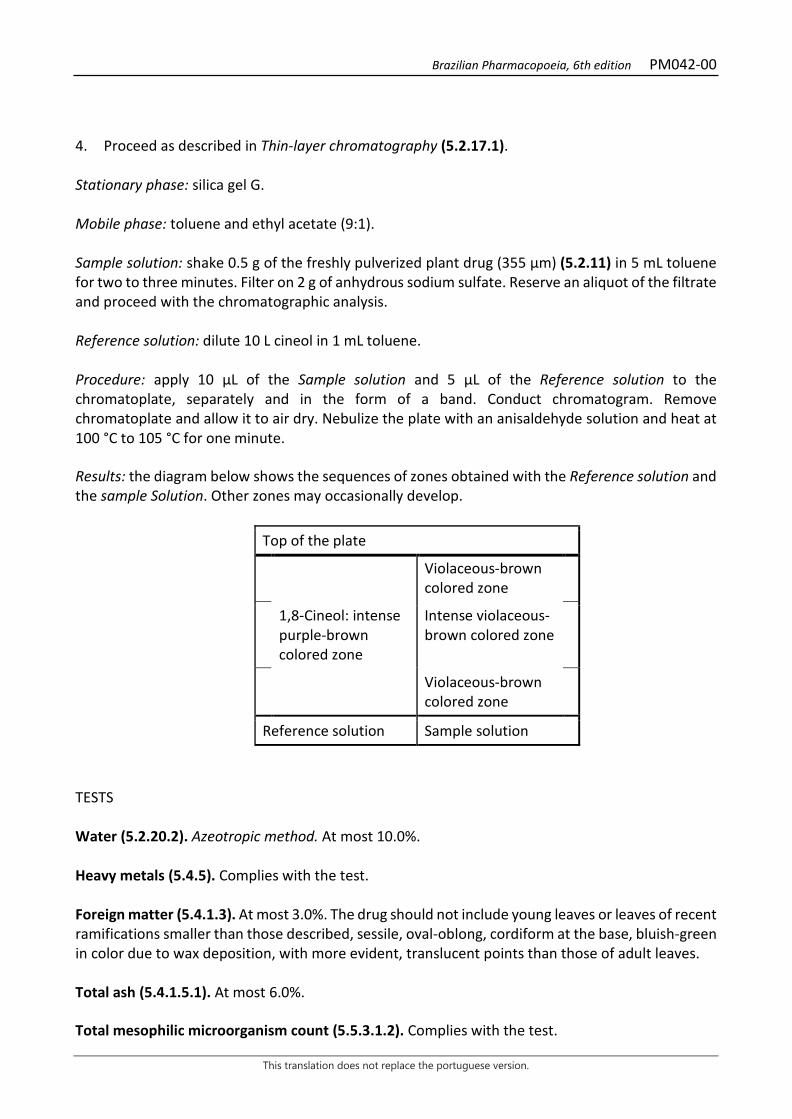
Brazilian Pharmacopoeia, 6th edition PM042-00
This translation does not replace the portuguese version.
4. Proceed as described in Thin-layer chromatography (5.2.17.1). Stationary phase: silica gel G. Mobile phase: toluene and ethyl acetate (9:1). Sample solution: shake 0.5 g of the freshly pulverized plant drug (355 µm) (5.2.11) in 5 mL toluene for two to three minutes. Filter on 2 g of anhydrous sodium sulfate. Reserve an aliquot of the filtrate and proceed with the chromatographic analysis. Reference solution: dilute 10 L cineol in 1 mL toluene. Procedure: apply 10 μL of the Sample solution and 5 μL of the Reference solution to the chromatoplate, separately and in the form of a band. Conduct chromatogram. Remove chromatoplate and allow it to air dry. Nebulize the plate with an anisaldehyde solution and heat at 100 °C to 105 °C for one minute. Results: the diagram below shows the sequences of zones obtained with the Reference solution and the sample Solution. Other zones may occasionally develop.
Top of the plate
Violaceous-brown colored zone
1,8-Cineol: intense purple-brown colored zone
Intense violaceous-brown colored zone
Violaceous-brown colored zone
Reference solution Sample solution
TESTS Water (5.2.20.2). Azeotropic method. At most 10.0%. Heavy metals (5.4.5). Complies with the test. Foreign matter (5.4.1.3). At most 3.0%. The drug should not include young leaves or leaves of recent ramifications smaller than those described, sessile, oval-oblong, cordiform at the base, bluish-green in color due to wax deposition, with more evident, translucent points than those of adult leaves. Total ash (5.4.1.5.1). At most 6.0%. Total mesophilic microorganism count (5.5.3.1.2). Complies with the test.
Brazilian Pharmacopoeia, 6th edition PM042-00
This translation does not replace the portuguese version.
Study of pathogenic microorganisms (5.5.3.1.3). Complies with the test. Agrochemical waste (5.4.3). Complies with the test. DOSAGE Volatile oils Proceed as described in Determining volatile oils in plant drugs (5.4.1.6). Use a 500-mL flask containing 200 mL of water as distillation liquid and 0.5 mL of xylene in the graduated tube. Reduce the drug to a coarse powder and immediately proceed with the determination of the volatile oil, from 10 g of the dried plant drug. Distill for two hours. Measure the volume and express the yield per 100 g of the plant drug (w/p). PACKAGING AND STORAGE In tightly closed containers, protected from light and heat.

Brazilian Pharmacopoeia, 6th edition PM042-00
This translation does not replace the portuguese version.
Figure 1 –Macroscopic, microscopic and powder microscopic aspects in Eucalyptus globulus Labill.
___________________________ The scales correspond in 1 to E; 2 to D and F-J; 3 to B and C. A – leaf morphology. B-D – cross-section of the leaf lamina. B – diagram of the midrib. C – outline of the mesophyll in the laminar region of the leaf. D – detail of the portion shown in B. E-H – details of leaf lamina fragments in front view. E – appearance of venation. F – fragment of the epidermis, on the adaxial surface, with a schizolysigenous gland visible by transparency. G – fragment of the epidermis, on the adaxial surface, with stomata. H – fragment of the epidermis, on the abaxial surface, with stomata. I - fibers in cross-section. J – parenchyma cells with druses. K – isolated twinning crystals
Brazilian Pharmacopoeia, 6th edition PM043-00
This translation does not replace the portuguese version.
FENNEL, fruit Foeniculi amarus fructus The plant drug consists of dried fruit of Foeniculum vulgare Mill. ssp. vulgare var. vulgare, containing at least 4.0% (w/w) volatile oil. CHARACTERISTICS The fruits have a strong and pleasant odor, similar to that of anethole. IDENTIFICATION 1. Macroscopic description The whole fruit is a dry, glabrous, oblong, nearly cylindrical, more rarely ovoid, diachene, 3 to 12 mm long and 3 to 4 mm wide, pale green to grayish-brown or yellowish-brown, with a rounded base and the apex is narrowed apex on a short, forked stylopodium. The diachene is usually separated into two mericarps; when these are still juxtaposed, they are weakly united and then the bases of the pedicels, extended by the bifenced filiform carpophores, are visible. Each mericarp has five very prominent longitudinal ribs, of which the two marginal ones are somewhat more developed than the others, alternating with four very narrow vallecula, which contain volatile oil-secreting ducts, elliptical in a cross-section. 2. Microscopic description A cross-section of each mericarp shows a pentagonal appearance, with four sides facing the dorsal region, almost equal and slightly concave, and a fifth ventral, longer and slightly undulated side, corresponding to the commissural face. The concave portions correspond to the valleculae and the sharper portions to the ribs. The epicarp is made up of a layer of polygonal cells, with a smooth cuticle and occasional anomocytic stomata. The mesocarp is formed by a parenchyma of irregular cells. Vascular bundles occur in the rib regions and externally and internally to these various thick-walled, reticulate, lignified cells, whether isolated or in groups. Secretory ducts occur in each of the four valleculae, with two more occurring on the commissural face; each channel is bordered by a layer of brown-walled polygonal secretory cells. The endocarp is made up of a layer of polygonal, brown, transversely elongated cells, except in the bundle region, where cells are organized in groups extended in different directions, showing, in a paradermal section, an arrangement of cells perpendicular and/or oblique to each other – the parquet arrangement. The seed’s integument is attached to the endocarp, formed by a unistratified epidermis. The endosperm, made up of polygonal cells, contains aleurone grains and small lipid drops. Each aleurone grain usually contains a calcium oxalate microrosette and one or two globoids. Carpophores, when present, are characterized by elongated fibers in a longitudinal view. 3. Microscopic description of powder The sample meets all requirements for the species, except the macroscopic characters. Characteristics are: yellowish-brown or grayish-brown color; epicarp fragments; mesocarp fragments with patches of brown secretory ducts; mesocarp fragments accompanied by more
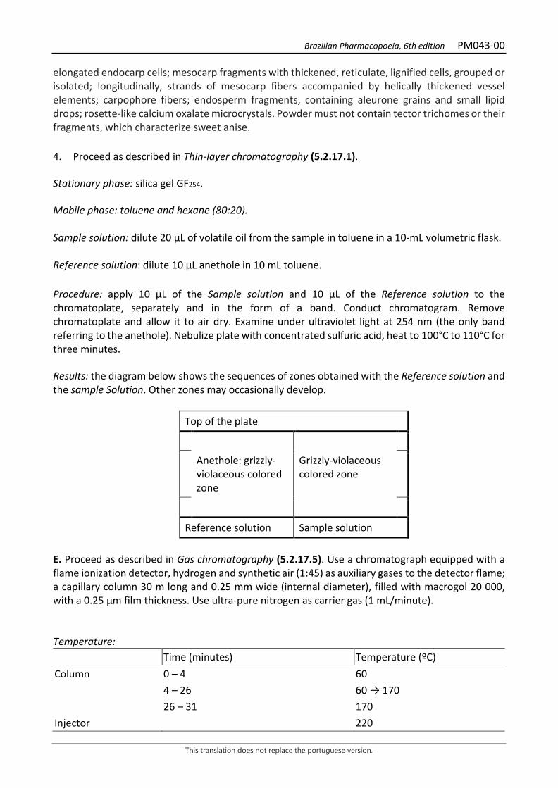
Brazilian Pharmacopoeia, 6th edition PM043-00
This translation does not replace the portuguese version.
elongated endocarp cells; mesocarp fragments with thickened, reticulate, lignified cells, grouped or isolated; longitudinally, strands of mesocarp fibers accompanied by helically thickened vessel elements; carpophore fibers; endosperm fragments, containing aleurone grains and small lipid drops; rosette-like calcium oxalate microcrystals. Powder must not contain tector trichomes or their fragments, which characterize sweet anise. 4. Proceed as described in Thin-layer chromatography (5.2.17.1). Stationary phase: silica gel GF254. Mobile phase: toluene and hexane (80:20). Sample solution: dilute 20 µL of volatile oil from the sample in toluene in a 10-mL volumetric flask. Reference solution: dilute 10 µL anethole in 10 mL toluene. Procedure: apply 10 μL of the Sample solution and 10 μL of the Reference solution to the chromatoplate, separately and in the form of a band. Conduct chromatogram. Remove chromatoplate and allow it to air dry. Examine under ultraviolet light at 254 nm (the only band referring to the anethole). Nebulize plate with concentrated sulfuric acid, heat to 100°C to 110°C for three minutes. Results: the diagram below shows the sequences of zones obtained with the Reference solution and the sample Solution. Other zones may occasionally develop.
Top of the plate
Anethole: grizzly-violaceous colored zone
Grizzly-violaceous colored zone
Reference solution Sample solution
E. Proceed as described in Gas chromatography (5.2.17.5). Use a chromatograph equipped with a flame ionization detector, hydrogen and synthetic air (1:45) as auxiliary gases to the detector flame; a capillary column 30 m long and 0.25 mm wide (internal diameter), filled with macrogol 20 000, with a 0.25 μm film thickness. Use ultra-pure nitrogen as carrier gas (1 mL/minute). Temperature: Time (minutes) Temperature (ºC) Column 0 – 4 60 4 – 26 60 → 170 26 – 31 170 Injector 220
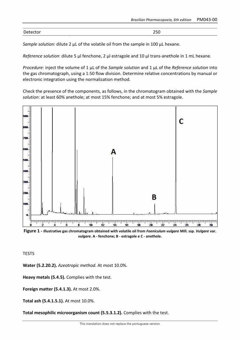
Brazilian Pharmacopoeia, 6th edition PM043-00
This translation does not replace the portuguese version.
Detector 250 Sample solution: dilute 2 µL of the volatile oil from the sample in 100 µL hexane. Reference solution: dilute 5 µl fenchone, 2 µl estragole and 10 µl trans-anethole in 1 mL hexane. Procedure: inject the volume of 1 μL of the Sample solution and 1 μL of the Reference solution into the gas chromatograph, using a 1:50 flow division. Determine relative concentrations by manual or electronic integration using the normalization method. Check the presence of the components, as follows, in the chromatogram obtained with the Sample solution: at least 60% anethole; at most 15% fenchone; and at most 5% estragole.
Figure 1 - Illustrative gas chromatogram obtained with volatile oil from Foeniculum vulgare Mill. ssp. Vulgare var.
vulgare. A - fenchone; B - estragole e C - anethole. TESTS Water (5.2.20.2). Azeotropic method. At most 10.0%. Heavy metals (5.4.5). Complies with the test. Foreign matter (5.4.1.3). At most 2.0%. Total ash (5.4.1.5.1). At most 10.0%. Total mesophilic microorganism count (5.5.3.1.2). Complies with the test.
Brazilian Pharmacopoeia, 6th edition PM043-00
This translation does not replace the portuguese version.
Study of pathogenic microorganisms (5.5.3.1.3). Complies with the test. Aflatoxins (5.4.4). Complies with the test. Agrochemical waste (5.4.3). Complies with the test. DOSAGE Volatile oils Proceed as described in Determining volatile oils in plant drugs (5.4.1.6). Use a 500-mL flask containing 300 mL of water as distillation liquid and 0.5 mL of xylene in the graduated tube of the clevenger apparatus. Reduce the fennel fruit to coarse powder (≤ 1400) (5.2.11). Immediately proceed with the determination of the volatile oil, from 10 g of the dried plant drug. Distill for two hours. Measure the volume and express the yield per 100 g of the plant drug (w/p). PACKAGING AND STORAGE In tightly closed containers, protected from light and heat.

Brazilian Pharmacopoeia, 6th edition PM043-00
This translation does not replace the portuguese version.
Figure 2 – Macroscopic, microscopic and powder microscopic aspects in Foeniculum vulgare Mill. ssp. vulgare
___________________________ The scales correspond in A to 2 mm; in B and C to 1 mm; in D to 1000 µm; in E, F, G, H, I, J and K to 100 µm. A - appearance of the fruit morphology. B - schematic cross section of the fruit in the portion indicated in A. C - schematic cross section of a mericarp; seed (sem). D - detail of the cross section of the mericarp in the portion indicated in C; endocarp (ed); endosperm (e); epicarp (ep); fruit (fr); mesocarp (m); seed (sem); integument (t). E to K - details noted in the powder. E - Strand of mesocarp fibers, in a longitudinal view, accompanied by helically thickened vessel elements. F - carpophore fibers. G - endocarp fragment accompanied by more elongated mesocarp cells. H - portion of the epicarp. I - mesocarp fragment with portion of brown secretory duct. J - Endosperm fragments, containing aleurone grains and small lipid drops, and rosette-like calcium oxalate microcrystals. K - fragments of the mesocarp formed by thickened, reticulate, and lignified cells, grouped or isolated.

Brazilian Pharmacopoeia, 6th edition PM044-00
This translation does not replace the portuguese version.
SWEET FENNEL, fruit Foeniculi dulcis fructus
The plant drug consists of dried fruits of Foeniculum vulgare Mill. ssp. vulgare var. dulce (Mill.) Thelung, containing at least 2.0% (w/p) volatile oil. CHARACTERISTICS The fruits have a strong and pleasant odor, similar to that of anethole. IDENTIFICATION 1. Macroscopic description The whole fruit is a dry, glabrous, oblong, nearly cylindrical, more rarely ovoid, diachene, 3 to 12 mm long and 3 to 4 mm wide, pale green to grayish-brown or yellowish-brown, with a rounded base and the apex is narrowed apex on a short, forked stylopodium. The diachene is usually separated into two mericarps; when these are still juxtaposed, they are weakly united and then the bases of the pedicels, extended by the bifenced filiform carpophores, are visible. Each mericarp has five very prominent longitudinal ribs, of which the two marginal ones are somewhat more developed than the others, alternating with four very narrow vallecula, which contain volatile oil-secreting ducts, elliptical in a cross-section. 2. Microscopic description A cross-section of each mericarp shows a pentagonal appearance, with four sides facing the dorsal region, almost equal and slightly concave, and a fifth ventral, longer and slightly undulated side, corresponding to the commissural face. The concave portions correspond to the valleculae and the sharper portions to the ribs. The epicarp is made up of a layer of polygonal cells, with a smooth cuticle and occasional anomocytic stomata. The mesocarp is formed by a parenchyma of irregular cells. Vascular bundles occur in the rib regions and externally and internally to these various thick-walled, reticulate, lignified cells, whether isolated or in groups. Secretory ducts occur in each of the four valleculae, with two more occurring on the commissural face; each channel is bordered by a layer of brown-walled polygonal secretory cells. The endocarp is made up of a layer of polygonal, brown, transversely elongated cells, except in the bundle region, where cells are organized in groups extended in different directions, showing, in a paradermal section, an arrangement of cells perpendicular and/or oblique to each other – the parquet arrangement. The seed’s integument is attached to the endocarp, formed by a unistratified epidermis. The endosperm, made up of polygonal cells, contains aleurone grains and small lipid drops. Each aleurone grain usually contains a calcium oxalate microrosette and one or two globoids. Carpophores, when present, are characterized by elongated fibers in a longitudinal view. 3. Microscopic description of powder The sample meets all requirements for the species, except the macroscopic characters. Characteristics are: yellowish-brown or grayish-brown color; epicarp fragments; mesocarp
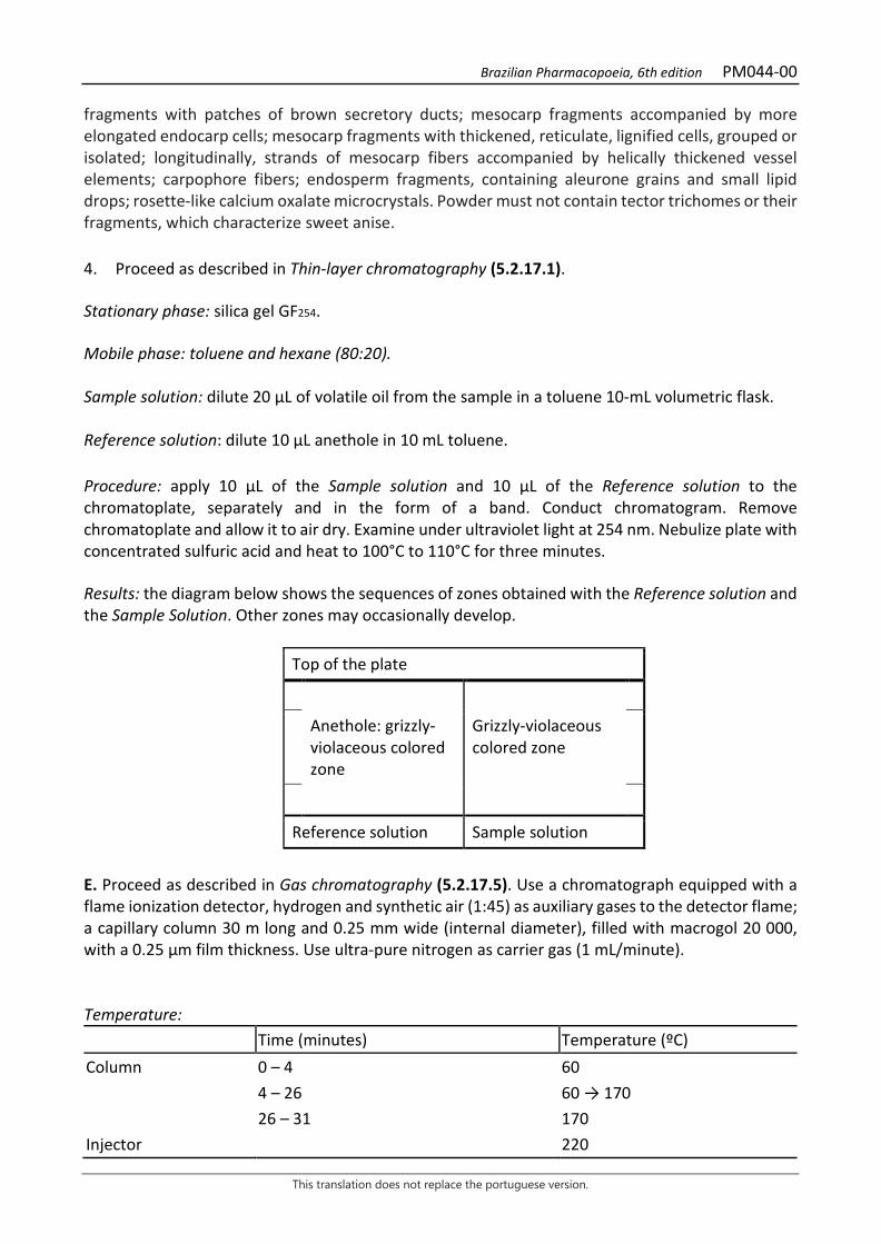
Brazilian Pharmacopoeia, 6th edition PM044-00
This translation does not replace the portuguese version.
fragments with patches of brown secretory ducts; mesocarp fragments accompanied by more elongated endocarp cells; mesocarp fragments with thickened, reticulate, lignified cells, grouped or isolated; longitudinally, strands of mesocarp fibers accompanied by helically thickened vessel elements; carpophore fibers; endosperm fragments, containing aleurone grains and small lipid drops; rosette-like calcium oxalate microcrystals. Powder must not contain tector trichomes or their fragments, which characterize sweet anise. 4. Proceed as described in Thin-layer chromatography (5.2.17.1). Stationary phase: silica gel GF254. Mobile phase: toluene and hexane (80:20). Sample solution: dilute 20 µL of volatile oil from the sample in a toluene 10-mL volumetric flask. Reference solution: dilute 10 µL anethole in 10 mL toluene. Procedure: apply 10 μL of the Sample solution and 10 μL of the Reference solution to the chromatoplate, separately and in the form of a band. Conduct chromatogram. Remove chromatoplate and allow it to air dry. Examine under ultraviolet light at 254 nm. Nebulize plate with concentrated sulfuric acid and heat to 100°C to 110°C for three minutes. Results: the diagram below shows the sequences of zones obtained with the Reference solution and the Sample Solution. Other zones may occasionally develop.
Top of the plate
Anethole: grizzly-violaceous colored zone
Grizzly-violaceous colored zone
Reference solution Sample solution
E. Proceed as described in Gas chromatography (5.2.17.5). Use a chromatograph equipped with a flame ionization detector, hydrogen and synthetic air (1:45) as auxiliary gases to the detector flame; a capillary column 30 m long and 0.25 mm wide (internal diameter), filled with macrogol 20 000, with a 0.25 μm film thickness. Use ultra-pure nitrogen as carrier gas (1 mL/minute). Temperature: Time (minutes) Temperature (ºC) Column 0 – 4 60 4 – 26 60 → 170 26 – 31 170 Injector 220

Brazilian Pharmacopoeia, 6th edition PM044-00
This translation does not replace the portuguese version.
Detector 250 Sample solution: dilute 2 µL of the volatile oil from the sample in 100 µL hexane. Reference solution: dilute 5 µl fenchone, 2 µl estragole and 10 µl trans-anethole in 1 mL hexane. Procedure: inject the volume of 1 μL of the Sample solution and 1 μL of the Reference solution into the gas chromatograph, using a 1:50 flow division. Determine relative concentrations by manual or electronic integration using the normalization method. Check the presence of the components, as follows, in the chromatogram obtained with the Sample solution: at least 80.0% and at most 10.0% estragole; and at most 7.5% fenchone.
Figure 1 - Illustrative chromatogram obtained with volatile oil from Foeniculum vulgare Mill. ssp. vulgare var.
dulce (Mill.) Thelung by gas chromatography. A - fenchone; B - estragole e C - anethole. TESTS Water (5.2.20.2). Azeotropic method. At most 10.0%. Heavy metals (5.4.5). Complies with the test. Foreign matter (5.4.1.3). At most 2.0%. Total ash (5.4.1.5.1). At most 10.0%. Total mesophilic microorganism count (5.5.3.1.2). Complies with the test. Study of pathogenic microorganisms (5.5.3.1.3). Complies with the test. Aflatoxins (5.4.4). Complies with the test. Agrochemical waste (5.4.3). Complies with the test.
Brazilian Pharmacopoeia, 6th edition PM044-00
This translation does not replace the portuguese version.
DOSAGE Volatile oils Proceed as described in Determining volatile oils in plant drugs (5.4.1.6). Use a 500-mL flask containing 300 mL of water as distillation liquid and 0.5 mL of xylene in the graduated tube. Reduce the fennel fruit to coarse powder (≤ 1400 µm) (5.2.11). Immediately proceed with the determination of the volatile oil, from 10 g of the dried plant drug. Distill for two hours. Measure the volume and express the yield per 100 g of the plant drug (w/p). PACKAGING AND STORAGE In tightly closed containers, protected from light and heat.
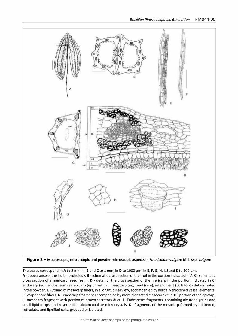
Brazilian Pharmacopoeia, 6th edition PM044-00
This translation does not replace the portuguese version.
Figure 2 – Macroscopic, microscopic and powder microscopic aspects in Foeniculum vulgare Mill. ssp. vulgare
___________________________ The scales correspond in A to 2 mm; in B and C to 1 mm; in D to 1000 µm; in E, F, G, H, I, J and K to 100 µm. A - appearance of the fruit morphology. B - schematic cross section of the fruit in the portion indicated in A. C - schematic cross section of a mericarp; seed (sem). D - detail of the cross section of the mericarp in the portion indicated in C; endocarp (ed); endosperm (e); epicarp (ep); fruit (fr); mesocarp (m); seed (sem); integument (t). E to K - details noted in the powder. E - Strand of mesocarp fibers, in a longitudinal view, accompanied by helically thickened vessel elements. F - carpophore fibers. G - endocarp fragment accompanied by more elongated mesocarp cells. H - portion of the epicarp. I - mesocarp fragment with portion of brown secretory duct. J - Endosperm fragments, containing aleurone grains and small lipid drops, and rosette-like calcium oxalate microcrystals. K - fragments of the mesocarp formed by thickened, reticulate, and lignified cells, grouped or isolated.
Brazilian Pharmacopoeia, 6th edition PM044-00
This translation does not replace the portuguese version.
Brazilian Pharmacopoeia, 6th edition PM045-00
This translation does not replace the portuguese version.
DEVIL’S-CLAW, root Harpagophyti radix
The plant drug consists of dried and fragmented or pulverized tuberous secondary roots of Harpagophytum procumbens DC. ex Meissn. containing at least 1.2% harpagoside (C24H30O11, 494.49). IDENTIFICATION 1. Macroscopic description Tuberous secondary roots, in irregular pieces or slices, generally circular, usually 2 to 4 cm in diameter, rarely 6 cm, and 2 to 6 mm thick; the fragments, when dehydrated, are brownish in color. The pieces have thin (0.2 to 0.5 mm), yellowish-gray to reddish-brown suberous bark; they are wrinkled lengthwise. The fracture is smooth and the surface is corneous, whitish to gray colored. 2. Microscopic description The periderm consists of up to 30 layers of cells in a radial arrangement. The suber is homogeneous and formed by about 25 layers of juxtaposed rectangular cells with thin walls, and the phelloderm consists of two to three layers of rectangular, flattened, thin-walled cells. Lenticels may occasionally be observed. The cortical parenchyma is composed of about 35 layers of bulky, thin-walled cells, with evident primary pit fields and small intercellular spaces; starch grains are absent (not evidenced by lugol’s reagent); sporadically, stone cells are found. The vascular system show a radial arrangement; the several radial series are composed of conducting elements and parenchyma cells with non-lignified walls, originating from the fascicular cambium, and alternate with narrow series of parenchyma cells with non-lignified walls, originating from the interfascicular cambium. The phloem has radial series with sieve elements and parenchyma cells, alternating with five to seven series of bulky parenchymatic cells; fibers and stone cells absent. The cambium region has two to four layers of rectangular cells. The secondary xylem also show a radial arrangement. Tracheal elements and fibers are arranged in generally uniseriate radial series and alternate with multiseriate parenchyma rays. The tracheal elements have simple perforation plates and pitted or scalariform walls. Calcium oxalate crystals in the form of small needles or cubes may be observed. The medulla is reduced, poorly differentiated, consisting of parenchyma cells. 3. Microscopic description of powder The sample meets all requirements for the species, except the macroscopic characters. Characteristics are: yellowish-brown color; suber fragments consisting of overlapping polyhedral cells with thin suberized walls; fragments of cortical parenchyma with thin-walled hexagonal cells with conspicuous pits, partly with yellow, droplet-shaped, or reddish-brown, granular inclusions, and sparsely needle- or cube-shaped calcium oxalate crystals; fragments of tracheal elements with scalariform or pitted walls; lignified walled parenchyma cells often associated with the conducting elements; rarely rectangular or polygonal sclereids with pitted walls and reddish-brown contents are observed. Starch grains are absent. 4. Adulterations
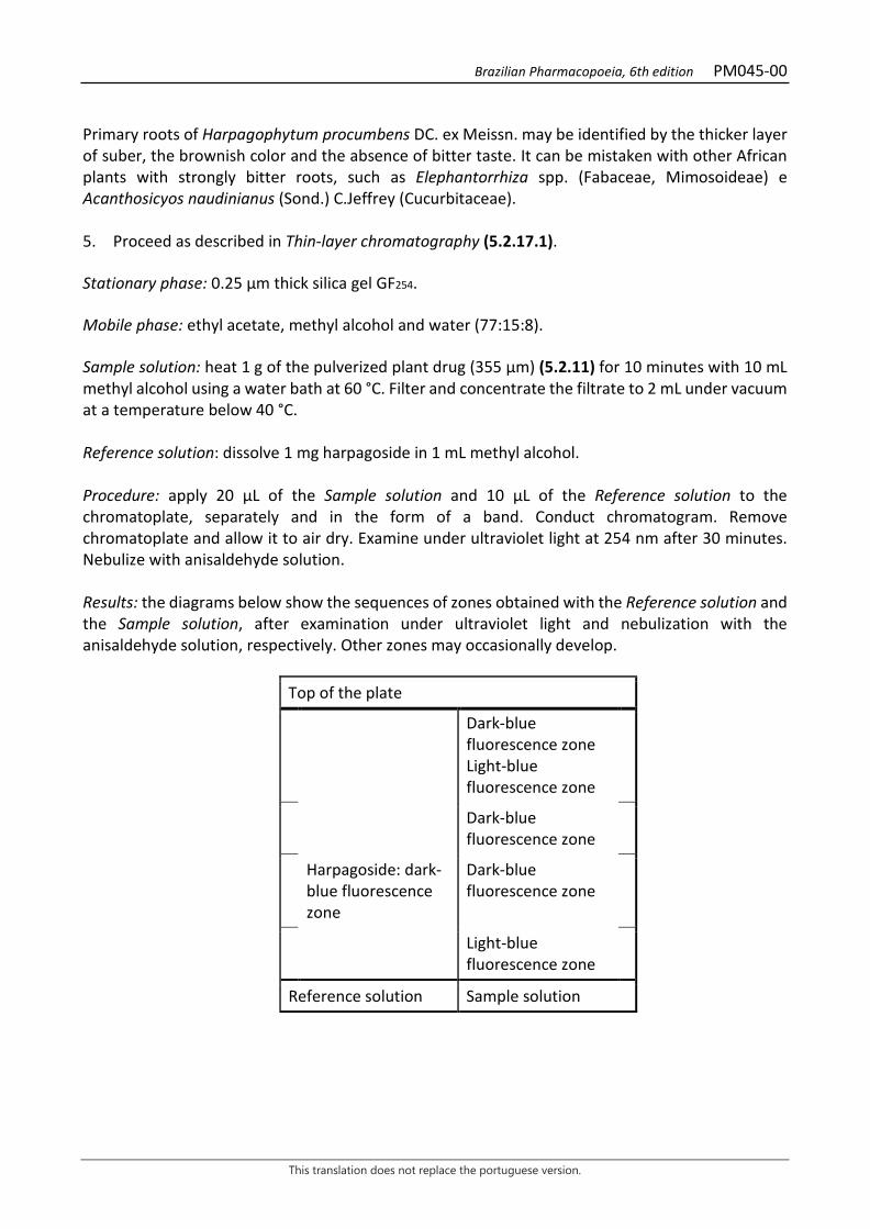
Brazilian Pharmacopoeia, 6th edition PM045-00
This translation does not replace the portuguese version.
Primary roots of Harpagophytum procumbens DC. ex Meissn. may be identified by the thicker layer of suber, the brownish color and the absence of bitter taste. It can be mistaken with other African plants with strongly bitter roots, such as Elephantorrhiza spp. (Fabaceae, Mimosoideae) e Acanthosicyos naudinianus (Sond.) C.Jeffrey (Cucurbitaceae). 5. Proceed as described in Thin-layer chromatography (5.2.17.1). Stationary phase: 0.25 µm thick silica gel GF254. Mobile phase: ethyl acetate, methyl alcohol and water (77:15:8). Sample solution: heat 1 g of the pulverized plant drug (355 µm) (5.2.11) for 10 minutes with 10 mL methyl alcohol using a water bath at 60 °C. Filter and concentrate the filtrate to 2 mL under vacuum at a temperature below 40 °C. Reference solution: dissolve 1 mg harpagoside in 1 mL methyl alcohol. Procedure: apply 20 μL of the Sample solution and 10 μL of the Reference solution to the chromatoplate, separately and in the form of a band. Conduct chromatogram. Remove chromatoplate and allow it to air dry. Examine under ultraviolet light at 254 nm after 30 minutes. Nebulize with anisaldehyde solution. Results: the diagrams below show the sequences of zones obtained with the Reference solution and the Sample solution, after examination under ultraviolet light and nebulization with the anisaldehyde solution, respectively. Other zones may occasionally develop.
Top of the plate
Dark-blue fluorescence zone Light-blue fluorescence zone
Dark-blue fluorescence zone
Harpagoside: dark-blue fluorescence zone
Dark-blue fluorescence zone
Light-blue fluorescence zone
Reference solution Sample solution
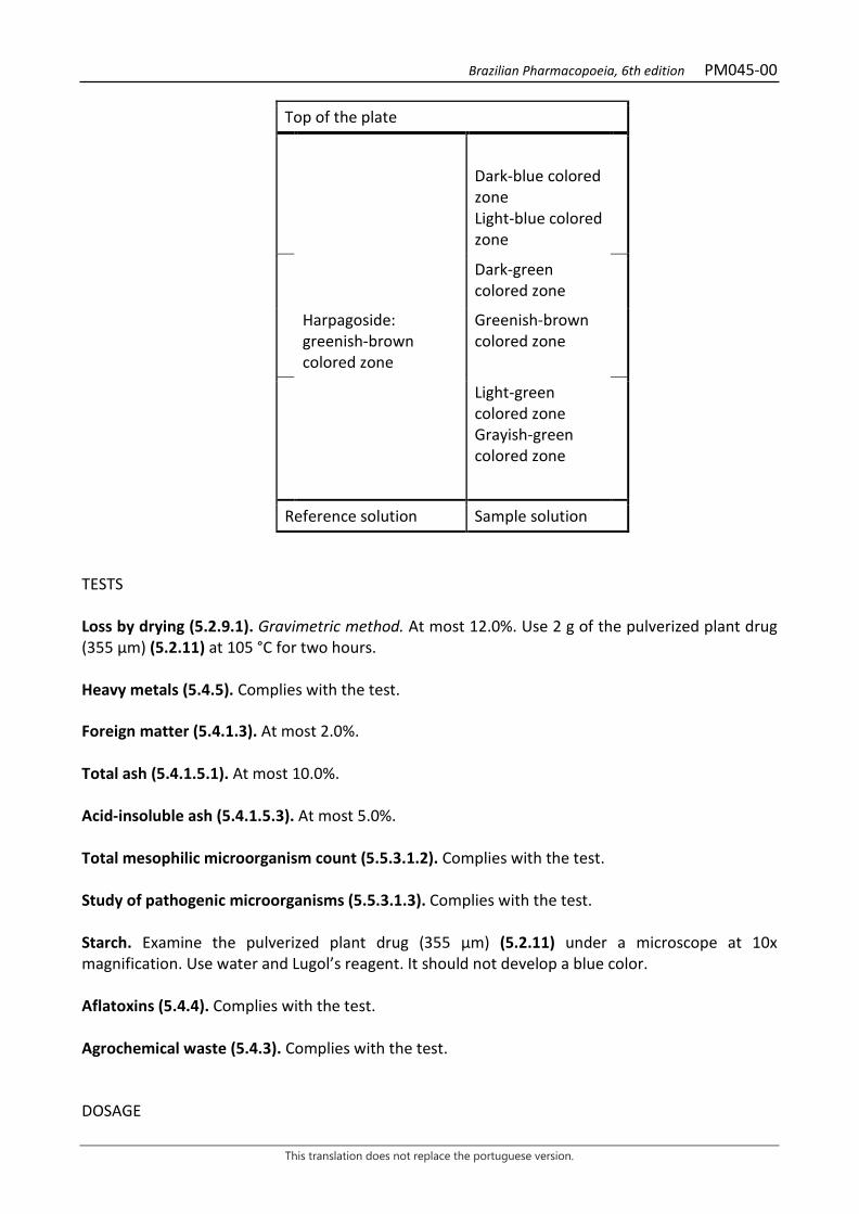
Brazilian Pharmacopoeia, 6th edition PM045-00
This translation does not replace the portuguese version.
Top of the plate
Dark-blue colored zone Light-blue colored zone
Dark-green colored zone
Harpagoside: greenish-brown colored zone
Greenish-brown colored zone
Light-green colored zone Grayish-green colored zone
Reference solution Sample solution TESTS Loss by drying (5.2.9.1). Gravimetric method. At most 12.0%. Use 2 g of the pulverized plant drug (355 µm) (5.2.11) at 105 °C for two hours. Heavy metals (5.4.5). Complies with the test. Foreign matter (5.4.1.3). At most 2.0%. Total ash (5.4.1.5.1). At most 10.0%. Acid-insoluble ash (5.4.1.5.3). At most 5.0%. Total mesophilic microorganism count (5.5.3.1.2). Complies with the test. Study of pathogenic microorganisms (5.5.3.1.3). Complies with the test. Starch. Examine the pulverized plant drug (355 µm) (5.2.11) under a microscope at 10x magnification. Use water and Lugol’s reagent. It should not develop a blue color. Aflatoxins (5.4.4). Complies with the test. Agrochemical waste (5.4.3). Complies with the test. DOSAGE
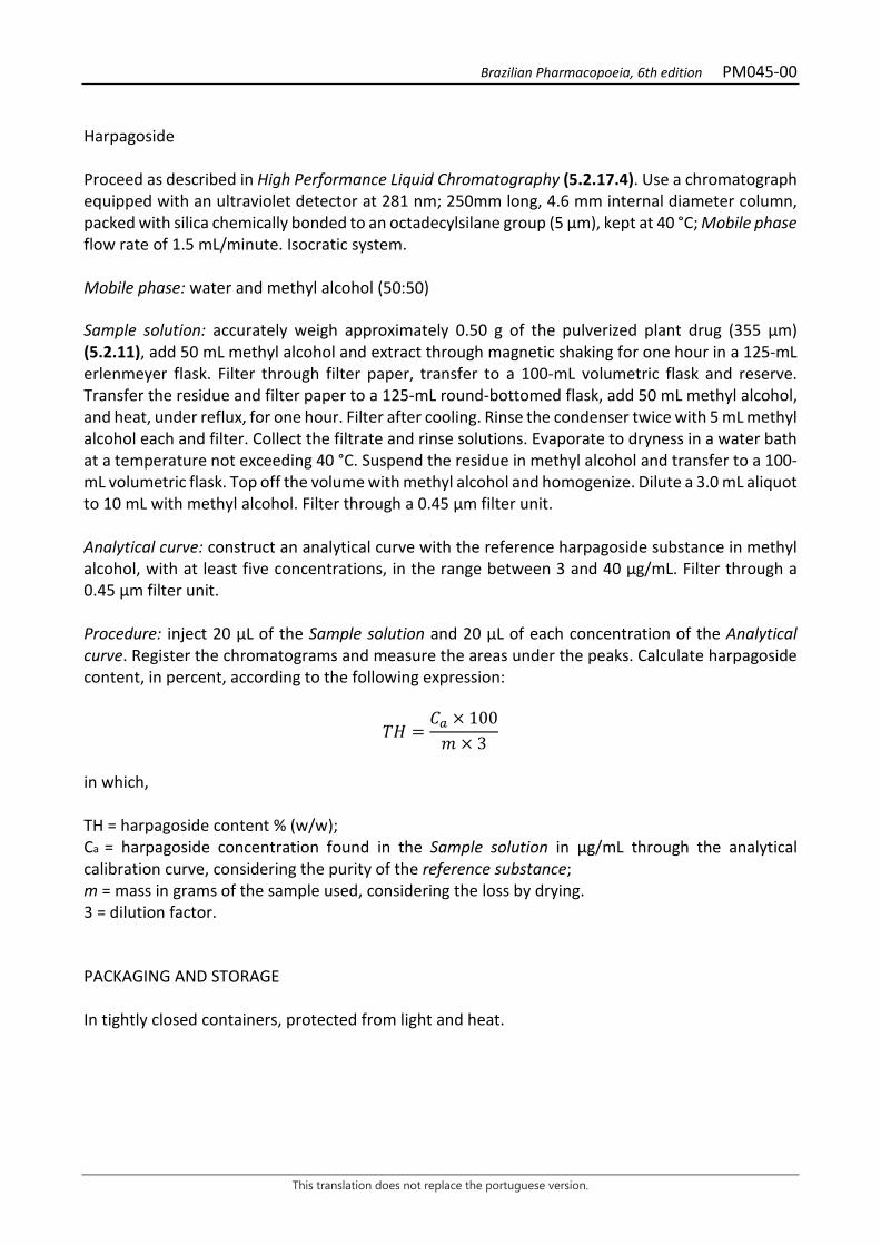
Brazilian Pharmacopoeia, 6th edition PM045-00
This translation does not replace the portuguese version.
Harpagoside Proceed as described in High Performance Liquid Chromatography (5.2.17.4). Use a chromatograph equipped with an ultraviolet detector at 281 nm; 250mm long, 4.6 mm internal diameter column, packed with silica chemically bonded to an octadecylsilane group (5 µm), kept at 40 °C; Mobile phase flow rate of 1.5 mL/minute. Isocratic system. Mobile phase: water and methyl alcohol (50:50) Sample solution: accurately weigh approximately 0.50 g of the pulverized plant drug (355 µm) (5.2.11), add 50 mL methyl alcohol and extract through magnetic shaking for one hour in a 125-mL erlenmeyer flask. Filter through filter paper, transfer to a 100-mL volumetric flask and reserve. Transfer the residue and filter paper to a 125-mL round-bottomed flask, add 50 mL methyl alcohol, and heat, under reflux, for one hour. Filter after cooling. Rinse the condenser twice with 5 mL methyl alcohol each and filter. Collect the filtrate and rinse solutions. Evaporate to dryness in a water bath at a temperature not exceeding 40 °C. Suspend the residue in methyl alcohol and transfer to a 100-mL volumetric flask. Top off the volume with methyl alcohol and homogenize. Dilute a 3.0 mL aliquot to 10 mL with methyl alcohol. Filter through a 0.45 µm filter unit. Analytical curve: construct an analytical curve with the reference harpagoside substance in methyl alcohol, with at least five concentrations, in the range between 3 and 40 µg/mL. Filter through a 0.45 µm filter unit. Procedure: inject 20 µL of the Sample solution and 20 µL of each concentration of the Analytical curve. Register the chromatograms and measure the areas under the peaks. Calculate harpagoside content, in percent, according to the following expression:
𝑇𝑇𝑇𝑇 =𝐶𝐶𝑎𝑎 × 100𝑚𝑚 × 3
in which, TH = harpagoside content % (w/w); Ca = harpagoside concentration found in the Sample solution in µg/mL through the analytical calibration curve, considering the purity of the reference substance; m = mass in grams of the sample used, considering the loss by drying. 3 = dilution factor. PACKAGING AND STORAGE In tightly closed containers, protected from light and heat.
Brazilian Pharmacopoeia, 6th edition PM045-00
This translation does not replace the portuguese version.
Figure 1 – Macroscopic, microscopic and powder microscopic aspects in Harpagophytum
procumbens DC. ex Meissn. ___________________________ The scales correspond: in B to 500 µm, in C-I to 200 µm, and in J to 100 µm. A- overall appearance of the drug; B- diagram of the root cross-section, showing the periderm (pe), cortical parenchyma (pc), primary and secondary phloem region (f), secondary xylem region (x). C- Periderm (pe) and cortical parenchyma (pc), in cross-section. D- Cross-section of the cambium region (ca), with secondary phloem, secondary xylem and multiseriate parenchymatic rays, fibers and vessels arranged in radial series. E- cross-section of the secondary xylem. F-J: powder detail; F- suber fragment; G- fragment of vessel element accompanied by radial parenchyma; H-J: fragments of vessel elements with different types of wall thickening.
pe pc
f x
A
pe B
pc ca
C D E
F G H I J
Brazilian Pharmacopoeia, 6th edition PM046-00
This translation does not replace the portuguese version.
YELLOW GENTIAN, rhizome and root Gentianae rhizoma et radix
The plant drug consists of dried, fragmented rhizomes and roots of Gentiana lutea L., containing at least 3% gentiopicroside (C16H20O9, 356.33). CHARACTERISTICS The roots and rhizomes have a characteristic odor. IDENTIFICATION 1. Macroscopic description Rhizomes and roots are cylindrical fragments of different sizes. As a rule, rhizomes are larger than roots, reaching up to 6 cm in diameter. Externally, the rhizomes are yellowish-brown to yellowish-gray in color and have longitudinal slits and numerous annular grooves, marked by rows of small scars. The roots are twisted or arched, with deep longitudinal striations and small, oval scars originating from secondary branching. Rhizomes and roots swell considerably in contact with moisture, becoming flexible. The fracture is neither farinaceous nor fibrous, and has a yellowish color with reddish spots. In cross-section, the rhizome has the cortical zone sharply demarcated by a suberous outer region, with darker lines, which occupies 1/3 of the section. The central cylinder, yellowish-brown colored, is porous, with fine radial striations. 2. Microscopic description In cross-section, the cells of the suber have thin, yellowish-brown walls and are arranged in four to eight layers. It is followed by several layers, collenchyma and parenchyma, externally and internally, respectively, of tangentially elongated cells, containing lipid drops and calcium oxalate crystals in the form of raphids. This region gradually blends with the cortical parenchyma. The vascular system is separated from the cortical zone by a well-developed cambium. In the phloem, small groups of sieve elements stand out, in addition to parenchyma cells. The xylem is predominantly parenchymatic and has scattered vessel elements, with walls showing ringed, helical, or reticulated thickening. Vessel elements occur singly or in small groups. The rhizome medulla is parenchymatic and well-developed. Lipid drops and acicular crystals or thin prisms of calcium oxalate are found in the parenchyma cells. Starch is almost completely absent. In a secondary structure, the anatomy of the root is similar to that of the rhizome. The secondary phloem and secondary xylem are separated by clear cambium and have a porous structure with few parenchymatic rays. 3. Microscopic description of powder The sample meets all requirements for the species, except the macroscopic characters. Characteristics are: yellowish to yellow-brown color; cell fragments with lipid drops; prismatic or raphid-like crystals and free lipid drops; fragments containing parenchyma cells; lignified vessel elements are rarely visible. Fibers and sclereids are absent.]

Brazilian Pharmacopoeia, 6th edition PM046-00
This translation does not replace the portuguese version.
4. Proceed as described in Thin-layer chromatography (5.2.17.1). Stationary phase: silica gel GF254. Mobile phase: ethyl acetate, methyl alcohol and water (77:15:8). Sample solution: add 10 mL of methyl alcohol to 1 g of the pulverized plant drug (355 µm) (5.2.11) and shake for 20 minutes. Filter and dry until residue is formed, at a temperature not exceeding 50 °C. Suspend the residue in 2.5 mL of methyl alcohol and proceed with the chromatographic analysis. Reference solution (1): prepare a 280 µg/mL solution of gentiopicroside in methyl alcohol. Reference solution (2): prepare a 1200 µg/mL solution of amarogentin in methyl alcohol. Procedure: apply 10 μL of the Sample solution, 20 μL of the Reference solution (1) and 20 µL of the Reference solution (2) to the chromatoplate, separately and in the form of a band. Conduct chromatogram. Remove chromatoplate and allow it to air dry. Examine under ultraviolet light at 254 nm. Nebulize the plate with an anisaldehyde solution and heat at 100 °C to 105 °C for one minute. Results: the diagram below shows the sequences of zones obtained with the Reference solution (1), the Referece solution (2) and the Sample solution. Other zones may occasionally develop.
Top of the plate
Amarogentin: brown-colored zone
Low intensity brown-colored zone
Gentiopicroside: brown-colored zone
Brown-colored zone
Reference solution Sample solution
TESTS Heavy metals (5.4.5). Complies with the test. Foreign matter (5.4.1.3). At most 2.0%. Loss by drying (5.2.9.1). Gravimetric method. At most 12.0%. Total ash (5.4.1.5.1). At most 4.0% Bitterness index (5.4.1.10). At least 10 000. Total mesophilic microorganism count (5.5.3.1.2). Complies with the test.
Brazilian Pharmacopoeia, 6th edition PM046-00
This translation does not replace the portuguese version.
Study of pathogenic microorganisms (5.5.3.1.3). Complies with the test. Agrochemical waste (5.4.3). Complies with the test. DOSAGE Gentiopicroside Proceed as described in High Performance Liquid Chromatography (5.2.17.4). Use a chromatograph equipped with an ultraviolet detector at 272 nm; 150mm long, 3.9 mm internal diameter column, packed with silica chemically bonded to an octadecylsilane group (4 µm), kept at a temperature of (22 ± 2) °C; Mobile phase flow rate of 0.7 mL/minute. Eluent (A): water and trifluoroacetic acid (100:0.006). Eluent (B): acetonitrile.
Time (minutes) Eluent A (%) Eluent B (%) Elution
0 - 10 95 → 72 5 → 28 linear gradient 10 - 14 72 → 80 28 → 20 linear gradient 14 - 16 80 → 95 20 → 5 linear gradient 16 - 20 95 5 isocratic Sample solution: accurately weigh approximately 0.100 g of the dried and pulverized plant drug (355 µm) (5.2.11) in a 50-mL centrifuge tube. Add 6 mL of 70% (v/v) ethyl alcohol, a magnetic bar, and shake for 30 minutes. Centrifuge the set for seven minutes at 2000 × g. Transfer the supernatant liquid quantitatively to a 10 mL volumetric flask, extract the drug residue again with 4 mL of 70% (v/v) ethyl alcohol and shake for 10 minutes. Centrifuge and transfer the supernatant liquid to the same 10-mL volumetric flask. Top off the volume to 10 mL with 70% (v/v) ethyl alcohol and homogenize. Filter through a 0.45 µm filter unit. Reference solution: dissolve an accurately weighed amount of gentiopicroside in methyl alcohol to obtain a 0.32 mg/mL solution. Filter through a 0.45 µm filter unit. Procedure: separately inject 5 μL of the Reference solution and 5 µL of the Sample solution. Record chromatograms and measure the area under the gentiopicroside peak. The average retention time is approximately eight minutes. Calculate gentiopicroside content, in percent, according to the following expression:
𝑇𝑇𝑇𝑇 =𝐶𝐶𝑟𝑟 × 𝑇𝑇𝑎𝑎 × 10 × 100
𝑇𝑇𝑟𝑟 × 𝑚𝑚𝑎𝑎
in which,

Brazilian Pharmacopoeia, 6th edition PM046-00
This translation does not replace the portuguese version.
TG = gentiopicroside content % (w/w); Cr = concentration of the Reference solution g/mL, considering the purity of the reference substance; Ar = area under the peak corresponding to the gentiopicroside in the Reference solution; Aa= area under the peak corresponding to the gentiopicroside in the Sample solution; ma = mass in grams of the sample used, considering the loss by drying. PACKAGING AND STORAGE In tightly closed containers, protected from light and heat.
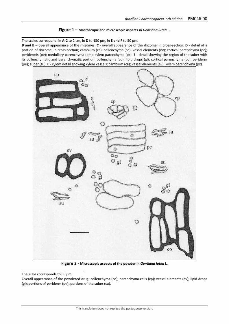
Brazilian Pharmacopoeia, 6th edition PM046-00
This translation does not replace the portuguese version.
Figure 1 – Macroscopic and microscopic aspects in Gentiana lutea L. ___________________________ The scales correspond: in A-C to 2 cm, in D to 150 μm, in E and F to 50 μm. B and B – overall appearance of the rhizomes. C - overall appearance of the rhizome, in cross-section. D - detail of a portion of rhizome, in cross-section; cambium (ca); collenchyma (co); vessel elements (ev); cortical parenchyma (pc); peridermis (pe); medullary parenchyma (pm); xylem parenchyma (px). E - detail showing the region of the suber with its collenchymatic and parenchymatic portion; collenchyma (co); lipid drops (gl); cortical parenchyma (pc); periderm (pe); suber (su). F - xylem detail showing xylem vessels; cambium (ca); vessel elements (ev); xylem parenchyma (px).
Figure 2 - Microscopic aspects of the powder in Gentiana lutea L.
___________________________ The scale corresponds to 50 µm. Overall appearance of the powdered drug: collenchyma (co); parenchyma cells (cp); vessel elements (ev); lipid drops (gl); portions of periderm (pe); portions of the suber (su).
Brazilian Pharmacopoeia, 6th edition PM047-00
This translation does not replace the portuguese version.
GINGER, rhizome Zingiberis rhizome
The plant drug consists of dried rhizomes of Zingiber officinale Roscoe, containing a minimum of 0.6% gingerols and a maximum of 0.4% shogaols. CHARACTERISTICS The rhizomes have a strong, pungent, characteristic odor. IDENTIFICATION 1. Macroscopic description Branched, irregularly shaped, laterally flattened rhizome, with branches arranged in a single plane, light brown to grizzlyish colored, marked by prominent transverse rings and narrow, clearly visible longitudinal and transverse striations. There commonly are grayish elliptical scars when young and light brown to whitish when older, rough and depressed, between the branches, with apparent fibers. The commercial rhizome measures 5.0 to 25.0 cm long, 1.0 to 5.0 cm thick. The fracture is short and starchy, with projecting fibers, and the suber tends to exfoliate. 2. Microscopic description In front view, the periderm shows cells with a quadrangular or elongated appearance, both with thin walls. In cross-section, the rhizome has an oval shape and three different regions, the periderm, the cortex, and the central cylinder. The periderm is made up of up to twenty-five layers or more, in two distinct zones. The external zone has suberized cells, irregular in shape and arrangement, and with a large amount of lipid drops; the internal zone is formed by a larger number of layers and by tabular-shaped cells, tangentially flattened and radially arranged. Starch grains are present in all tissues except vascular ones; they are simple, with eccentric hilum and very evident stratification, usually ellipsoid, oval and flattened, up to 50 µm long up to 35 µm wide and up to 10 µm thick. The cortical parenchyma is formed by bulky polygonal cells, with a large amount of starch grains. Isolated, sparse, not very thick-walled fibers without lignification occur between the parenchyma cells. Polygonal-shaped secretory cells are very common, containing large or small lipid drops, isolated or clustered, and yellowish in color. Vascular bundles are collateral, usually closed, with random distribution in the parenchyma, and poorly developed in the cortical parenchyma. In the parenchyma there is the endoderm, composed of quadrangular, thin-walled cells with rare starch grains. Externally, the vascular cylinder is delimited by a ring of parenchyma cells much smaller than the others, where small vascular clusters are distributed. Internally, it is filled with parenchymatous tissue, formed by several layers, similar to that which characterizes the cortical region. In the vascular cylinder there are also secretory cells and scattered vascular bundles. Calcium oxalate crystals and sclereids do not occur. In longitudinal section, a scalariform thickening is observed more frequently, in addition to the rarer annular, helical, and reticulate types. The fibers are very elongated, septate, and have oblique pores and evident pits. 3. Microscopic description of powder
Brazilian Pharmacopoeia, 6th edition PM047-00
This translation does not replace the portuguese version.
The sample meets all requirements for the species, except the macroscopic characters. Microscopic observation of the powder becomes clearer when chloral hydrate R is used. Characteristics are: light yellow to light brown coloration; fragments of periderm in frontal and transversal view; large amount of isolated or grouped starch grains, more evident when glycerinated water is used; large amount of parenchyma fragments with thin, colorless or rarely yellowish walls; parenchyma fragments containing starch grains; parenchyma fragments containing yellowish lipid drops; parenchyma fragments with portions of tracheal elements; portions of parietal thickening isolated from tracheal elements; portions of tracheal elements isolated or grouped; portions of fibers isolated or grouped; isolated and yellowish lipid drops. 4. Proceed as described in Thin-layer chromatography (5.2.17.1). Stationary phase: silica gel GF254. Mobile phase: toluene and ethyl acetate (30:10). Sample solution: pulverize 5 g of the drug (710 µm) (5.2.11). Transfer 1 g of the powder to centrifuge tubes and add 10 mL methyl alcohol. Shake the mixture on a magnetic stirrer for 30 minutes. Centrifuge the extract for 10 minutes at 166 × g. Use the supernatant liquid for application to the plate. Reference solution: prepare a 0.1 mg/mL solution of capsaicin in ethyl alcohol. TLC visualization reagent: 10% (v/v) sulfuric anisaldehyde solution followed by heating at temperature between 100º C and 105º C for three minutes. Procedure: apply 20 μL of the Sample solution and 20 μL of the Reference solution to the chromatoplate, separately and in the form of a band. Conduct the chromatogram along 10 cm. Remove chromatoplate and allow it to air dry for 15 minutes. Nebulize the plate with a 10% (v/v) sulfuric anisaldehyde and heat at 100 °C to 105 °C for five to ten minute. Examine the plate under visible light. Results: the diagram below shows the sequences of zones obtained with the Reference solution and the Sample Solution. Other zones may occasionally develop.
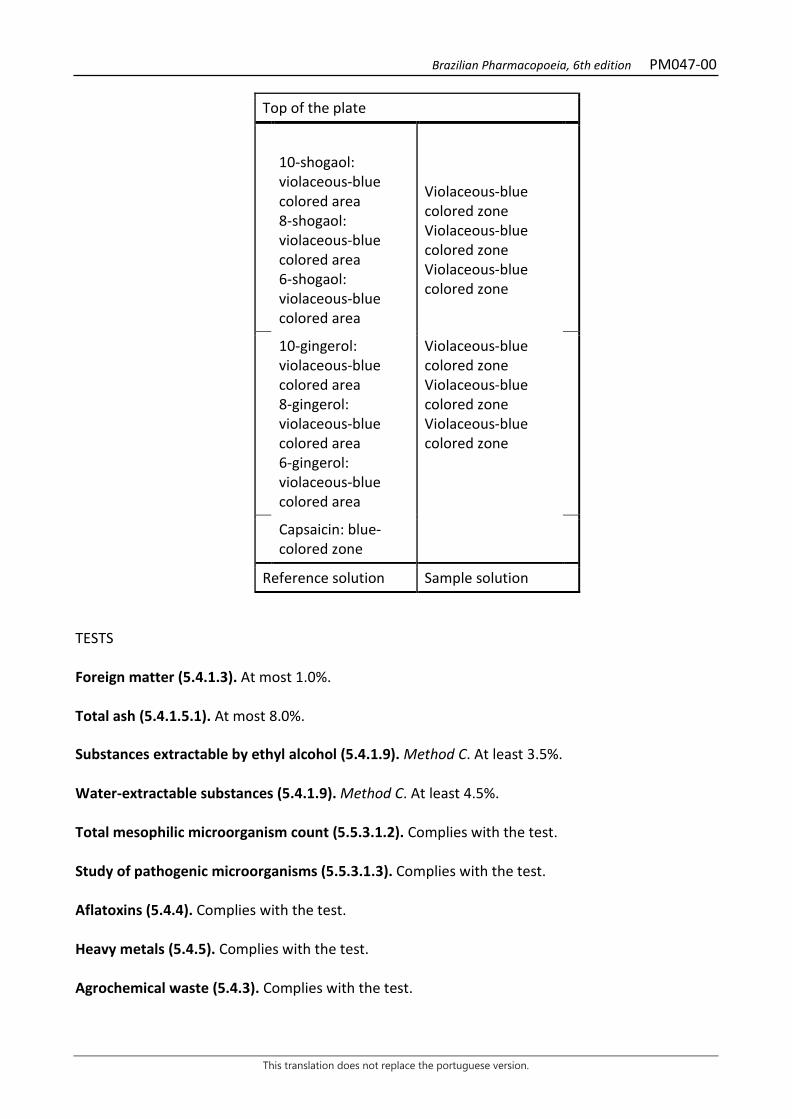
Brazilian Pharmacopoeia, 6th edition PM047-00
This translation does not replace the portuguese version.
Top of the plate
10-shogaol: violaceous-blue colored area 8-shogaol: violaceous-blue colored area 6-shogaol: violaceous-blue colored area
Violaceous-blue colored zone Violaceous-blue colored zone Violaceous-blue colored zone
10-gingerol: violaceous-blue colored area 8-gingerol: violaceous-blue colored area 6-gingerol: violaceous-blue colored area
Violaceous-blue colored zone Violaceous-blue colored zone Violaceous-blue colored zone
Capsaicin: blue-colored zone
Reference solution Sample solution TESTS Foreign matter (5.4.1.3). At most 1.0%. Total ash (5.4.1.5.1). At most 8.0%. Substances extractable by ethyl alcohol (5.4.1.9). Method C. At least 3.5%. Water-extractable substances (5.4.1.9). Method C. At least 4.5%. Total mesophilic microorganism count (5.5.3.1.2). Complies with the test. Study of pathogenic microorganisms (5.5.3.1.3). Complies with the test. Aflatoxins (5.4.4). Complies with the test. Heavy metals (5.4.5). Complies with the test. Agrochemical waste (5.4.3). Complies with the test.

Brazilian Pharmacopoeia, 6th edition PM047-00
This translation does not replace the portuguese version.
DOSAGE Gingerols Proceed as described in High Performance Liquid Chromatography (5.2.17.4). Use a chromatograph equipped with an ultraviolet detector at 282 nm; 250mm long, 4.6mm internal diameter column, packed with silica chemically bonded to an octadecylsilane group (5 µm), kept at room temperature (22 ± 2) °C; Mobile phase flow rate of 1.0 mL/minute. Minor flow rate and gradient adjustments may be necessary if the chromatographic apparatus and column are changed. Eluent (A): acetonitrile, 0.1% phosphoric acid (v/v) and methyl alcohol (55:44:1). Eluent (B): acetonitrile. Adopt Eluent (A) with a run time seven times longer than the capsaicin retention time. Column rinsing: after each run, wash the column using the system described in the following table.
Time (minutes) Eluent A (%) Eluent B (%) Elution
0 - 2 100 → 0 0 → 100 linear gradient 2 -12 0 100 isocratic 12 - 14 0 → 100 100 → 0 linear gradient 14 -29 100 0 isocratic Sample preparation: transfer 1 g of the freshly pulverized plant drug (710 µm) (5.2.11) to a 50-mL ground-necked flask and add 25 mL of absolute ethyl alcohol. Macerate the extract for 24 hours and repeatedly shake the solution during the first eight hours. Then filter the extract through paper filter into a 25-mL volumetric flask, top off the volume with ethyl alcohol and homogenize. Reference solution: dissolve capsaicin in methyl alcohol in the quantity and volume required to prepare a solution with a concentration of 0.5 mg/mL. Sample solution: into a 1mL volumetric flask, add 0.2 mL of the Reference solution and top off the volume with the Sample preparation. Filter through a 0.45 µm filter unit. Procedure: separately inject 25 μL of the Sample solution. Register the chromatograms and measure the areas under the peaks for gingerols and capsaicin. Calculate the gingerol content, in percentage, according to the following expression:
𝑇𝑇𝑇𝑇 =𝑇𝑇𝑎𝑎 × 𝐶𝐶𝑇𝑇𝑟𝑟 × 𝑚𝑚
× 100 × 31,25
in which, TG = gingerol content % (w/w);
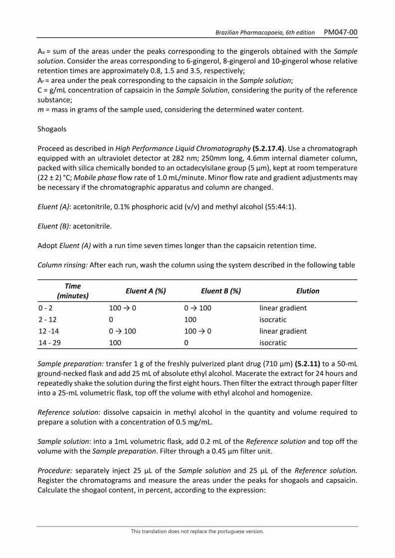
Brazilian Pharmacopoeia, 6th edition PM047-00
This translation does not replace the portuguese version.
Aa = sum of the areas under the peaks corresponding to the gingerols obtained with the Sample solution. Consider the areas corresponding to 6-gingerol, 8-gingerol and 10-gingerol whose relative retention times are approximately 0.8, 1.5 and 3.5, respectively; Ar = area under the peak corresponding to the capsaicin in the Sample solution; C = g/mL concentration of capsaicin in the Sample Solution, considering the purity of the reference substance; m = mass in grams of the sample used, considering the determined water content. Shogaols Proceed as described in High Performance Liquid Chromatography (5.2.17.4). Use a chromatograph equipped with an ultraviolet detector at 282 nm; 250mm long, 4.6mm internal diameter column, packed with silica chemically bonded to an octadecylsilane group (5 µm), kept at room temperature (22 ± 2) °C; Mobile phase flow rate of 1.0 mL/minute. Minor flow rate and gradient adjustments may be necessary if the chromatographic apparatus and column are changed. Eluent (A): acetonitrile, 0.1% phosphoric acid (v/v) and methyl alcohol (55:44:1). Eluent (B): acetonitrile. Adopt Eluent (A) with a run time seven times longer than the capsaicin retention time. Column rinsing: After each run, wash the column using the system described in the following table
Time (minutes) Eluent A (%) Eluent B (%) Elution
0 - 2 100 → 0 0 → 100 linear gradient 2 - 12 0 100 isocratic 12 -14 0 → 100 100 → 0 linear gradient 14 - 29 100 0 isocratic Sample preparation: transfer 1 g of the freshly pulverized plant drug (710 µm) (5.2.11) to a 50-mL ground-necked flask and add 25 mL of absolute ethyl alcohol. Macerate the extract for 24 hours and repeatedly shake the solution during the first eight hours. Then filter the extract through paper filter into a 25-mL volumetric flask, top off the volume with ethyl alcohol and homogenize. Reference solution: dissolve capsaicin in methyl alcohol in the quantity and volume required to prepare a solution with a concentration of 0.5 mg/mL. Sample solution: into a 1mL volumetric flask, add 0.2 mL of the Reference solution and top off the volume with the Sample preparation. Filter through a 0.45 µm filter unit. Procedure: separately inject 25 μL of the Sample solution and 25 µL of the Reference solution. Register the chromatograms and measure the areas under the peaks for shogaols and capsaicin. Calculate the shogaol content, in percent, according to the expression:
Brazilian Pharmacopoeia, 6th edition PM047-00
This translation does not replace the portuguese version.
𝑇𝑇𝑇𝑇 =𝑇𝑇𝑎𝑎 × 𝐶𝐶𝑇𝑇𝑟𝑟 × 𝑚𝑚
× 100 × 31,25
in which, TS = shogaol content % (w/w); Aa = sum of the areas under the peaks corresponding to the shogaols obtained with the Sample solution. Consider the areas corresponding to 6-shogaol, 8-shogaol and 10-shogaol whose relative retention times are approximately 1.9, 4.7 and 5.8, respectively; Ar = area under the peak corresponding to the capsaicin in the Sample solution; C = g/mL concentration of capsaicin in the Sample Solution, considering the purity of the reference substance; m = mass in grams of the sample used, considering the determined water content. System suitability: dissolve the 6-gingerol, 8-gingerol, 10-gingerol, 6-shogaol, 8- shogaol, and 10-shogaol standards in methyl alcohol in quantities and volumes necessary to prepare a solution of approximately 0.2 mg/mL. Mix equal parts of each solution in 1 mL of the Reference solution, and inject 25 µL of the mixture. Resolution between the peaks should be calculated using the formula shown below, with resolution between the 6-gingerol and capsaicin peaks being no less than 3.0; and between the capsaicin and 6-shogaol peaks, no less than 10.0.
𝐼𝐼 = 1,18 ×(𝑡𝑡𝑡𝑡2 − 𝑡𝑡𝑡𝑡1)𝑤𝑤𝑡𝑡1 + 𝑤𝑤𝑡𝑡2
In which, R = peak resolution; tr1 = retention time of the first peak; tr2 = retention time of the second peak; wr1 = width of the first peak measured at mid-height; wr2 = width of the second peak measured at mid-height;
Figure 1 – Illustrative chromatogram of Zingiber officinale ethanol extract at 282 nm. At retention
time (tr) of about seven minutes, the peak for 6-gingerol can be seen; at tr of nine minutes, for
Brazilian Pharmacopoeia, 6th edition PM047-00
This translation does not replace the portuguese version.
capsaicin; at tr of 14 minutes, for 8-gingerol; at tr of 17 minutes, for 6- shogaol; at tr of 32 minutes, for 10-gingerol; at tr of 41 minutes, for 8-shogaol; and at tr of 57 minutes, for 10-shogaol.
PACKAGING AND STORAGE In tightly closed containers, protected from light and heat.
Figure 2 - Macroscopic and microscopic aspects of the Zingiber
officinale Roscoe rhizome. ___________________________ The scales correspond in A to 2.8 cm, in B and C to 100 µm, in D to 5 cm, and in E to 200 µm. A - general aspect of the rhizome, al: ring; etr: striation; ral: lateral branching; B - periderm in front view with quadrangular cells; C - periderm in front view with elongated cells; D - overall appearance of the rhizome in cross-section; av: vascular cluster; cv: vascular cylinder; cx: cortex; end: endoderm; fv: vascular bundle; pe: periderm; ral: lateral branching; E - detail of rhizome in cross-section as marked in D; av: vascular cluster; cse: secretory cell; cv: vascular cylinder; cx: cortex; end: endoderm; f: phloem; fb: fiber; fv: vascular bundle; ga: starch grain; gl: lipid drop; gtl: lipid drop; p: parenchyma; pc: cortical parenchyma; pe: periderm; pq: small cell parenchyma; s: suber; x: xylem.

Brazilian Pharmacopoeia, 6th edition PM047-00
This translation does not replace the portuguese version.
Figure 3 - Microscopic aspects of the Zingiber officinale Roscoe rhizome.
___________________________ The scales correspond in A-G to 100 µm. A - detail of the periderm in cross-section; ga: starch grain; gl: lipid drop; gtl: lipid drops; s: suber; B - detail of cortical parenchyma with starch grains, in cross-section; fb: fiber; ga: starch grain; C - detail of cortical parenchyma with lipid drops, in cross-section; ga: starch grain; gl: lipid drop; gtl: lipid drops; D - detail of vascular bundle occurring in the cortex, in cross-section; f: phloem; fb: fiber; p: parenchyma; pc: cortical parenchyma; x: xylem; E - detail of the endoderm region and the region of the vascular clusters, in cross-section; av: vascular cluster; end: endoderm; ety: Caspary striation; ga: starch grain; gl: lipid drop; p: parenchyma; pc: cortical parenchyma; pq: small cell parenchyma; F - detail of cortex portion in longitudinal section; ee: vessel element with scalariform thickening; eh: vessel element with helical thickening; fb: fiber; ga: starch grain; gl: lipid drop; p: parenchyma; pc: cortical parenchyma; G - detail of vascular bundle occurring in the vascular cylinder, in cross-section; f: phloem; fb: fiber; p: parenchyma; x: xylem.

Brazilian Pharmacopoeia, 6th edition PM047-00
This translation does not replace the portuguese version.
Figure 4 - Microscopic aspects of the Zingiber officinale Roscoe rhizome.
___________________________ The scales correspond in A-P to 100 µm. A - portion of periderm in front view; B - portion of parenchyma with lipid drop, in cross-section; gl: lipid drop; C - portion of parenchyma with lipid drops, in cross-section; gtl: lipid drops; D - portion of parenchyma, in cross-section; E - portion of parenchyma with starch grains, in cross-section; ga. starch grain; F - grouped starch grains; G - isolated starch grains; H - portions of isolated fibers, in longitudinal section; I - portion of fiber grouping, in longitudinal section; J - xylem portion, in longitudinal section; ee: vessel element portion with scalariform thickening; eh: vessel element portion with helical thickening; eo: vessel element portion with ringed thickening; fb. fiber portion; K - xylem portion, in longitudinal section; ee: portion of vessel element with scalariform thickening; ere: portion of vessel element with reticulate thickening; fb: fiber portion; p. parenchyma portion; L - isolated fragments of parietal thickening; M - portion of tracheal element with ringed thickening, in longitudinal section; N - portion of tracheal element with reticulate thickening, in longitudinal section; O - portion of tracheal element with scalariform thickening, in longitudinal section; P - portion of parenchyma and vascular elements, in longitudinal section; p. parenchyma; ee: vessel element with scalariform thickening.
Brazilian Pharmacopoeia, 6th edition PM048-00
This translation does not replace the portuguese version.
GUAVA, leaf Guajavae folium
The plant drug consists of dried leaves of Psidium guajava L., containing at least 10.0% total tannins and at least 0.3% glycosylated quercetin derivatives calculated as quercetin (C15H10O7, 302,24). IDENTIFICATION 1. Macroscopic description Papyraceous-coriaceous, entire leaves, oblong-elliptic to oval, 7 to 15 cm long and 3 to 6 cm wide; obtuse or acuminate apex, obtuse base and entire margin; slightly apparent translucent areas; discolored laminae, bright green and glabrescent adaxial surface and pale green abaxial surface, with simple, unicellular or bicellular trichomes, more frequent on the midrib, up to 0.5 mm long, rarely distributed on the whole lamina. Venation is of the camptodrome-broquidodrome type; midrib and secondary veins evident on the abaxial surface, printed on the adaxial surface, the secondary ones in 11 to 20 pairs, somewhat parallel to each other, forming an angle of 45 to 60 with the midrib, ending in a vein parallel to the margin of the lamina, more evident on the abaxial surface. Petiole 0.5 to 0.7 cm long. 2. Microscopic description Dorsiventrally symmetrical lamina, hypostomatic; anomocytic stomata. The epidermis, in front view, has cells with rectilinear-polygonal periclinal walls. In cross section, the cuticle is thick and the epidermis facing the adaxial surface is pluristratified, with three to five layers, the outermost being formed by much smaller cells than the others; the epidermis facing the abaxial surface is unistratified, with a large number of stomata, generally projected in relation to the other cells and simple, unicellular or bicellular tector trichomes; the epidermal cells may contain crystals. Secretory cavities are found in the pluristratified epidermis, reaching as far as the first layers of the palisade parenchyma. The mesophyll is compact, formed by a palisade parenchyma with two to four layers of elongated cells and three to six layers of smaller cells; its cells contain lipid drops and starch grains; secretory cavities and idioblasts with calcium oxalate crystals also occur in this parenchyma. The midrib is of the bicolateral type with an open arch; the xylem is organized in rays with up to 10 elements; the external phloem is more developed and has more crystals than the internal phloem; small secretory cavities are found in the phloem; cells containing discrete grains of starch surround the external phloem; phenolic compounds occur in the parenchyma of the vascular bundle. The parenchyma is characterized by containing considerable intercellular spaces, secretory cavities, crystalliferous idioblasts (druses, prisms, monocrystals, rhombohedral crystals, small grouped crystals etc.), cells with lipid drops and/or chloroplasts, as well as a large amount of cells containing phenolic compounds. The collenchyma is of the angular type and is restricted to the abaxial surface, formed by three or usually four (rarely five) layers of cells; chloroplastids, crystals, phenolic compounds and lipid drop may be present. 3. Microscopic description of powder The sample meets all requirements for the species, except the macroscopic characters. Characteristics are: leaf laminae fragments with scattered translucent spots; leaf lamina fragments

Brazilian Pharmacopoeia, 6th edition PM048-00
This translation does not replace the portuguese version.
with remnants of unistratified epidermis and palisade parenchyma; fragments of pluristratified epidermis, with or without crystals, accompanied or not by palisade parenchyma; fragments of lamina with sparse secretory cavities; remains of secretory cavities; portions of lamina with epidermal cells of rectilinear outline, with or without lipid drops; fragments of epidermis accompanied by collenchyma; unicellular trichomes and their insertion; rare bicellular trichomes; epidermis with numerous anomocytic stomata, with evident projection or not; compact palisade parenchyma cells, with or without chloroplasts, generally grouped; portions of veins with rectangular and elongated epidermal cells; tracheal elements with helical thickening; crystals of different forms, according to microscopic description, isolated or inserted in different tissues. 4. Proceed as described in Thin-layer chromatography (5.2.17.1). Stationary phase: silica gel GF254. Mobile phase: ethyl acetate, methyl alcohol, water and formic acid (20:2.7:2:0.2). Sample solution: transfer 0,2 g of the pulverized plant drug (355) (5.2.11) with 10 mL methyl alcohol to a 50 mL, round-bottomed flask. Heat in a water bath, under reflux, for 15 minutes. Cool and filter. Evaporate the solvent in a water bath. Suspend the residue in 2 mL methyl alcohol. Reference solution (1): prepare a solution containing 500 µg/mL rutin. Reference solution (2): Prepare a solution containing 100 µg/mL quercetin. Procedure: apply 5 μL of the Sample solution, 5 μL of the Reference solution (1) and 5 μL of the Reference solution (2) to the chromatoplate, separately and in the form of a band. Conduct chromatogram. Remove chromatoplate and allow it to air dry for 15 minutes. Nebulize the plate with aminoethanol diphenylborate RS. Examine under ultraviolet light at 365 nm. Results: the diagram below shows the sequences of zones obtained with the Reference solution (1), the Referece solution (2) and the Sample solution. Other zones may occasionally develop.
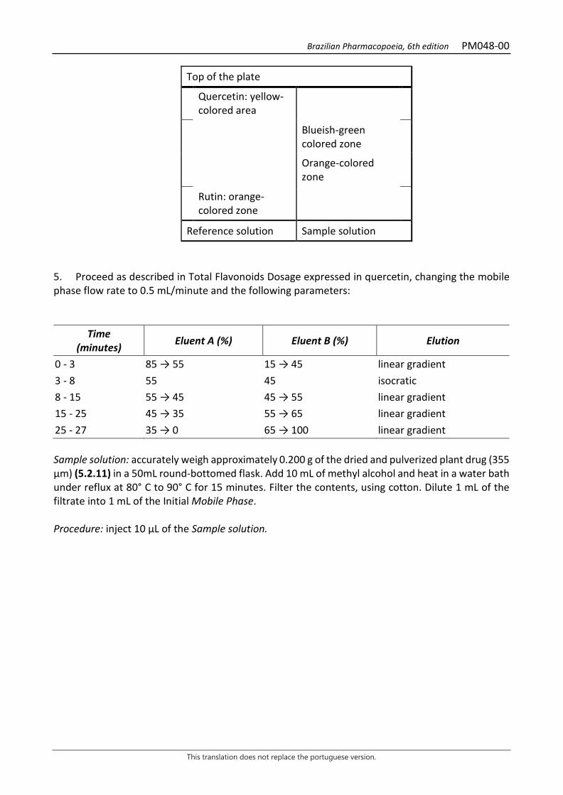
Brazilian Pharmacopoeia, 6th edition PM048-00
This translation does not replace the portuguese version.
Top of the plate
Quercetin: yellow-colored area
Blueish-green colored zone
Orange-colored zone
Rutin: orange-colored zone
Reference solution Sample solution 5. Proceed as described in Total Flavonoids Dosage expressed in quercetin, changing the mobile phase flow rate to 0.5 mL/minute and the following parameters:
Time (minutes) Eluent A (%) Eluent B (%) Elution
0 - 3 85 → 55 15 → 45 linear gradient 3 - 8 55 45 isocratic 8 - 15 55 → 45 45 → 55 linear gradient 15 - 25 45 → 35 55 → 65 linear gradient 25 - 27 35 → 0 65 → 100 linear gradient Sample solution: accurately weigh approximately 0.200 g of the dried and pulverized plant drug (355 µm) (5.2.11) in a 50mL round-bottomed flask. Add 10 mL of methyl alcohol and heat in a water bath under reflux at 80° C to 90° C for 15 minutes. Filter the contents, using cotton. Dilute 1 mL of the filtrate into 1 mL of the Initial Mobile Phase. Procedure: inject 10 μL of the Sample solution.
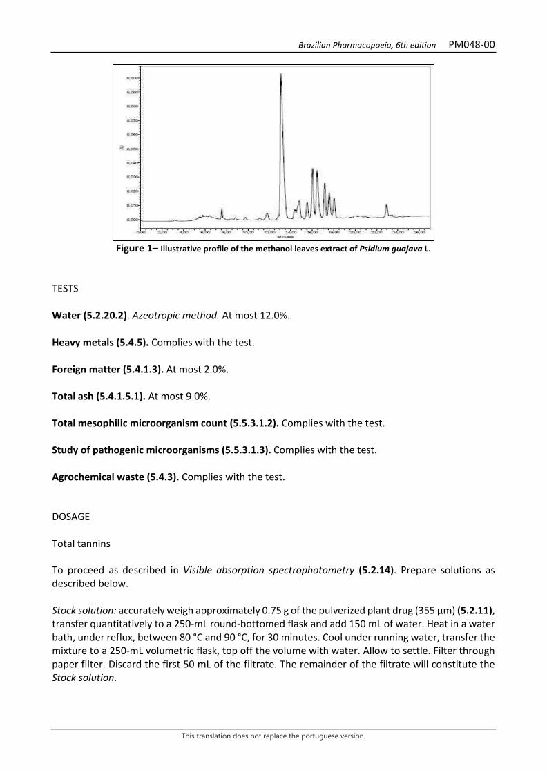
Brazilian Pharmacopoeia, 6th edition PM048-00
This translation does not replace the portuguese version.
Figure 1– Illustrative profile of the methanol leaves extract of Psidium guajava L.
TESTS Water (5.2.20.2). Azeotropic method. At most 12.0%. Heavy metals (5.4.5). Complies with the test. Foreign matter (5.4.1.3). At most 2.0%. Total ash (5.4.1.5.1). At most 9.0%. Total mesophilic microorganism count (5.5.3.1.2). Complies with the test. Study of pathogenic microorganisms (5.5.3.1.3). Complies with the test. Agrochemical waste (5.4.3). Complies with the test. DOSAGE Total tannins To proceed as described in Visible absorption spectrophotometry (5.2.14). Prepare solutions as described below. Stock solution: accurately weigh approximately 0.75 g of the pulverized plant drug (355 µm) (5.2.11), transfer quantitatively to a 250-mL round-bottomed flask and add 150 mL of water. Heat in a water bath, under reflux, between 80 °C and 90 °C, for 30 minutes. Cool under running water, transfer the mixture to a 250-mL volumetric flask, top off the volume with water. Allow to settle. Filter through paper filter. Discard the first 50 mL of the filtrate. The remainder of the filtrate will constitute the Stock solution.

Brazilian Pharmacopoeia, 6th edition PM048-00
This translation does not replace the portuguese version.
Sample solution for total polyphenol content: transfer 5 mL from the Stock solution to a 25-mL volumetric flask, top off the volume with water and homogenize. Mix 5 mL of this solution with 2 mL of phosphotungstic acid reagent RS, dilute in a 50-mL volumetric flask with sodium carbonate RS, and homogenize. Determine absorbance at 715 nm (A1), precisely three minutes after adding the last reagent, using water for zero adjustment. Sample solution for polyphenols not adsorbed by hide powder: for 0.2 mL of the hide powder to 20 mL of the Stock solution and mechanically shake for 60 minutes. Filter. Dilute 5 mL of the filtrate in a 25-mL volumetric flask with water. Mix 5 mL of this solution with 2 mL of phosphotungstic acid reagent RS, dilute in a 50-mL volumetric flask with sodium carbonate RS, and homogenize. Determine absorbance at 715 nm (A2), precisely three minutes after adding the last reagent, using water for zero adjustment. Reference solution: dissolve 50 mg pyrogallol in water, transfer to a 100 mL volumetric flask and top off the volume with water. Dilute 5 mL of this solution in a 100-mL volumetric flask with water. Mix 5 mL of this solution with 2 mL of phosphotungstic acid reagent RS and dilute in a 50-mL volumetric flask with sodium carbonate RS. Determine absorbance at 715 nm (A3), precisely three minutes after adding the last reagent, and within 15 minutes from the pyrogallol dissolution using water for zero adjustment. Calculate total tannin content expressed as pyrogallol, in percent, according to the following expression:
𝑇𝑇𝑇𝑇 =(𝑇𝑇1 − 𝑇𝑇2) × 13,12
𝑇𝑇3 × 𝑚𝑚
in which, TT = total tannin content expressed as pyrogallol % (w/w); A1 = absorbance measured for the Sample solution for total polyphenols; A2 = absorbance measured for the Sample solution for polyphenols not adsorbed by hide powder; A3 = absorbance measured for the Reference solution; m = mass in grams of the sample used, considering the determined water content. Total flavonoid content expressed as quercetin Proceed as described in High Performance Liquid Chromatography (5.2.17.4). Use a chromatograph equipped with an ultraviolet detector at 371 nm; 150mm long, 4.6 mm internal diameter column, packed with silica octadecylsilane silica (5 µm), kept at a temperature of (22 ± 2) °C; Mobile phase flow rate of 1.0 mL/minute. Eluent (A): water and formic acid (100:0.1). Eluent (B): methyl alcohol.
Time (minutes) Eluent A (%) Eluent B (%) Elution
0 – 3 60 40 isocratic

Brazilian Pharmacopoeia, 6th edition PM048-00
This translation does not replace the portuguese version.
3 – 15 60 → 0 40 → 100 linear gradient 15 – 16 0 → 60 100 → 40 linear gradient 16 – 21 60 40 isocratic Sample solution: accurately weigh approximately 0.100 g of the dried and pulverized plant drug (355 µm) (5.2.11) in a 100mL round-bottomed flask. Add 20 mL of methyl alcohol at 70% (v/v), 1 mL of hydrochloric acid and heat in a water bath under reflux at temperature between 80° C and 90° C for 30 minutes. Filter using absorbent cotton into another 100 mL round-bottomed flask. Return the insoluble residue, along with the cotton, to the first flask and add 20 mL of 70% methyl alcohol. Proceed with the heating under the same previous conditions, for 10 minutes. Filter again into the round-bottomed flask containing the first filtrate. Repeat this process once more. After reducing the volume under vacuum to 10 mL, transfer the volume to a separatory funnel. Perform the sample extraction with 10 mL ethyl acetate. Filter the organic phase on filter paper containing sodium sulfate anhydrous into a new 100 mL round-bottomed flask. Perform this extraction process four more times, totaling of 50 mL. Evaporate the organic phase obtained until reaching a completely dry residue. Suspend the residue with methyl alcohol and dilute in a 5 mL volumetric flask. Transfer, quantitatively, 2.5 mL of this solution to a 5mL volumetric flask and top off the volume with methyl alcohol at 50% (v/v). Filter through a 0.45 µm filter unit. Reference solution: dissolve an accurately weighed amount of quercetin in methyl alcohol to obtain a 1 mg/mL solution. Dilute the solution with 50% (v/v) methyl alcohol to obtain a solution with a concentration of 0.038 mg/mL. Filter through a 0.45 µm filter unit. Procedure: separately inject 10 μL of the Reference solution and 10 µL of the Sample solution. Record chromatograms and measure the area under the quercetin peak. The average retention time is approximately nine minutes. Calculate quercetin content, in percent, according to the following expression:
𝑇𝑇𝑇𝑇 =𝐶𝐶𝑟𝑟 × 𝑇𝑇𝑎𝑎 × 10 × 100
𝑇𝑇𝑟𝑟 × 𝑚𝑚
in which, TQ = quercetin content % (w/w); Cr = concentration of the Reference solution in g/mL, considering the purity of the reference substance; Ar = area under the peak corresponding to the quercetin in the Reference solution; Ar = area under the peak corresponding to the quercetin in the Sample solution; m = mass in grams of the sample used, considering the determined water content. PACKAGING AND STORAGE In tightly closed containers, protected from light and heat.
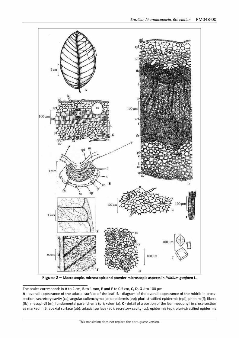
Brazilian Pharmacopoeia, 6th edition PM048-00
This translation does not replace the portuguese version.
Figure 2 – Macroscopic, microscopic and powder microscopic aspects in Psidium guajava L.
___________________________ The scales correspond: in A to 2 cm, B to 1 mm, E and F to 0.5 cm, C, D, G-J to 100 µm. A - overall appearance of the adaxial surface of the leaf. B - diagram of the overall appearance of the midrib in cross-section; secretory cavity (cs); angular collenchyma (co); epidermis (ep); pluri-stratified epidermis (epl); phloem (f); fibers (fb); mesophyll (m); fundamental parenchyma (pf); xylem (x). C - detail of a portion of the leaf mesophyll in cross-section as marked in B; abaxial surface (ab); adaxial surface (ad); secretory cavity (cs); epidermis (ep); pluri-stratified epidermis
Brazilian Pharmacopoeia, 6th edition PM048-00
This translation does not replace the portuguese version.
(epl); stomata (es); crystalliferous idioblast (ic); mesophyll (m); spongy parenchyma (pj); palisade parenchyma (pp); tector trichome (tt). D - detail of a portion of the midrib in cross-section as marked in B; abaxial surface (ab); adaxial surface (ad); cells containing phenolic compounds (ccf); collenchyma (co); pluri-stratified epidermis (epl); phloem (f); fibers (fb); crystalliferous idioblast (ic); lipid drop (gl); fundamental parenchyma (pf); xylem (x). E - detail of the venation on the adaxial surface of a leaf segment, in the median region, in front view (indicated in A); F - detail of the venation on the abaxial surface of a leaf segment, in the margin region, in front view (indicated in A). G-J. Details noted in the powder. G - front view of the adaxial surface of the leaf lamina epidermis; secretory cavity (cs); tector trichome (tt). H - front view of the abaxial surface of the leaf lamina epidermis; secretory cavity (cs); stomata (es). I - detail of tracheal elements with helical-type thickening. J - detail of some types of calcium oxalate crystals.

Brazilian Pharmacopoeia, 6th edition PM049-00
This translation does not replace the portuguese version.
GUACO, leaf Mikania laevigatae folium
The plant drug consists of dried leaves of Mikania laevigata Sch.Bip. ex Baker, containing at least 0.15% coumarin (C9H6O2, 146.15). CHARACTERISTICS The leaves have a strong coumarin odor. IDENTIFICATION 1. Macroscopic description Leaves glabrous to the naked eye, leathery, dark when dry. Leaf lamina 6 to 15 cm long and 4 to 6.5 cm wide, oval to oval-lanceolate, slightly asymmetrical; attenuated base, acuminate apex and margin entire to sinuous, with one or a few lateral teeth or without teeth; revolute margin. Actinodromous venation with three evident veins along the lamina, the lateral ones forming an arch and joining the main one in the apical portion; there could be two more veins the basal portion, following the margin of the lamina. Petiole 1.4 to 4.5 cm long, almost cylindrical, grooved on the adaxial surface. It differs from the Mikania glomerata Spreng. for its strong odor of coumarin and the shape of the leaves. The leaf lamina of the Mikania laevigata has greater length than width, the base is not hastate and the lateral teeth, when present, are not very evident, while in the Mikania glomerata, length and width are quite similar, the base of the lamina is hastate and the lateral teeth are very evident. 2. Microscopic description The leaf lamina is hypostomatic and dorsiventrally symmetrical. In front view, the anticlinal walls of the epidermal cells are sinuous and thick; the stomata are anisocytic and anomocytic; siliceous bodies and uniseriate, curved glandular trichomes, formed by about six cells, as well as capitate, pluricellular and biseriate glandular trichomes, occur in greater density on the abaxial surface. In cross-section, the cuticle is thin and smooth and the epidermis has one or two layers of cells; the stomata are located at the same level as the epidermal cells; the trichomes occur in epidermal depressions. The palisade parenchyma has one to four layers of cells with a large amount of lipid drops; the spongy parenchyma consists of six to twelve layers. Secretory ducts, of varying sizes and delimited by flattened cells, are arranged close to the vascular bundles. In the leaf margin region, where angular collenchyma formed by three or four layers occurs, the mesophyll is homogeneous and siliceous bodies occur. The midrib, in cross-section, is biconvex, with a cuneate prominence on the adaxial surface and rounded on the abaxial surface, with a thicker cuticle and smaller epidermal cells. Angular collenchyma occurs on both sides, and some of its cells have grizzly contents. The vascular system consists of three to eight free, open arched, collateral bundles; secretory ducts are visible; the phloem has a well-developed fiber cap, and the xylem consists of two to eight rows of tracheal elements. The parenchyma facing the abaxial surface has isolated sclereids. Lipid drops and starch grains occur in all parenchyma. The petiole, in cross-section, has a cuticle with the same characteristics as the region of the main midrib and unistratified epidermis, followed by angular

Brazilian Pharmacopoeia, 6th edition PM049-00
This translation does not replace the portuguese version.
collenchyma, formed by up to ten layers and parenchyma with a large amount of sclereids; secretory ducts occur close to the vascular bundles; starch grains and lipid drops occur in the parenchyma. The vascular system has a similar organization to the midrib, with a greater number of U-shaped bundles. 3. Proceed as described in Thin-layer chromatography (5.2.17.1). Stationary phase: silica gel F254. Mobile phase: toluene and diethyl ether (1:1). Sample solution: shake 0.1 g of the drug in 3 mL ethyl alcohol for 10 minutes. Filter extract. Reference solution: prepare a solution containing 25 µg/mL coumarin and 1 mg/mL o-coumaric acid in methyl alcohol. TLC visualization reagent: dissolve 10 g potassium hydroxide in 100 mL ethyl alcohol. Procedure: apply 10 μL of the Sample solution and 5 μL of the Reference solution to the chromatoplate, separately and in the form of a band. Saturate the tank with glacial acetic acid. Conduct chromatogram. Remove chromatoplate and allow it to air dry. Nebulize with the TLC visualization reagent. Examine under ultraviolet light at 365 nm. Results: the diagram below shows the sequences of zones obtained with the Reference solution and the Sample Solution. Other zones may occasionally develop.
Top of the plate
Coumarin: greenish-blue fluorescence zone
Greenish-blue fluorescence zone
O-Coumaric Acid: greenish-blue fluorescence zone
Greenish-blue fluorescence zone
Reference solution Sample solution
TESTS Loss by drying (5.2.9.1). Gravimetric method. At most 13,0%. Heavy metals (5.4.5). Complies with the test. Foreign matter (5.4.1.3). At most 2.0%. Total ash (5.4.1.5.1). At most 16.0%

Brazilian Pharmacopoeia, 6th edition PM049-00
This translation does not replace the portuguese version.
Total mesophilic microorganism count (5.5.3.1.2). Complies with the test. Study of pathogenic microorganisms (5.5.3.1.3). Complies with the test. Agrochemical waste (5.4.3). Complies with the test. DOSAGE Coumarin Proceed as described in High Performance Liquid Chromatography (5.2.17.4). Use a chromatograph equipped with an ultraviolet detector at 275 mm; pre-column packed with octadecylsilane silica, 150mm long, 3.9mm internal diameter column, packed with silica chemically bonded to an octadecylsilane group (4 µm); Mobile phase flow rate of 0.5 mL/minute. Isocratic system. Mobile phase: water and methyl alcohol (53:47). Sample solution: accurately weigh approximately 0.100 g of the dried and pulverized plant drug (500) (5.2.11) and transfer quantitatively to a round-bottomed flask. Add 10 mL of 50% (v/v) ethyl alcohol and heat in a water bath at 90 °C, under reflux, for 30 minutes. After cooling, filter extract through absorbent cotton into a 25-mL volumetric flask. Extract the drug residue in the flask and on the absorbent cotton with 10 mL of 50% (v/v) ethyl alcohol, and heat, under reflux, for 10 minutes. After cooling, gather all extracts in the 25 mL volumetric flask and top off the volume with 50% (v/v) ethyl alcohol. Filter through a 0.45 µm filter unit. Reference solution: dissolve an accurately weighed amount of coumarin in methyl alcohol to obtain a concentration of 10 µg/mL. Filter through a 0.45 µm filter unit. Procedure: separately inject 10 μL of the Reference solution and 10 μL of the Sample solution. Register the chromatograms and measure the areas under the peaks. Calculate coumarin content, in percent, according to the following expression:
𝑇𝑇𝐶𝐶 =𝐶𝐶𝑟𝑟 × 𝑇𝑇𝑎𝑎 × 25 × 100
𝑇𝑇𝑟𝑟 × 𝑚𝑚
in which, TC = coumarin content % (w/w); Cr = concentration of the Reference solution in g/mL, considering the purity of the reference substance; Ar= area under the peak corresponding to the coumarin in the Reference solution; Ar = area under the peak corresponding to the coumarin acid in the Sample solution; m = mass in grams of the sample used, considering the loss by drying.
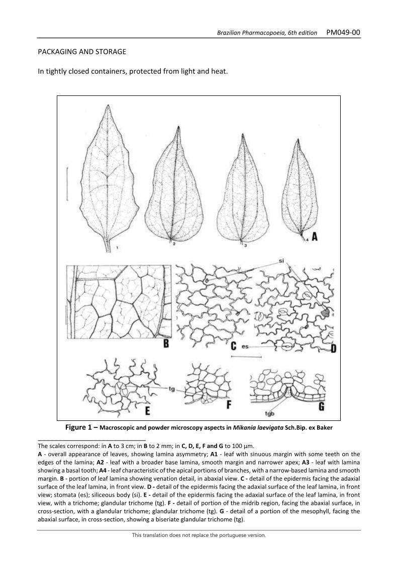
Brazilian Pharmacopoeia, 6th edition PM049-00
This translation does not replace the portuguese version.
PACKAGING AND STORAGE In tightly closed containers, protected from light and heat.
Figure 1 – Macroscopic and powder microscopy aspects in Mikania laevigata Sch.Bip. ex Baker
___________________________ The scales correspond: in A to 3 cm; in B to 2 mm; in C, D, E, F and G to 100 μm. A - overall appearance of leaves, showing lamina asymmetry; A1 - leaf with sinuous margin with some teeth on the edges of the lamina; A2 - leaf with a broader base lamina, smooth margin and narrower apex; A3 - leaf with lamina showing a basal tooth; A4 - leaf characteristic of the apical portions of branches, with a narrow-based lamina and smooth margin. B - portion of leaf lamina showing venation detail, in abaxial view. C - detail of the epidermis facing the adaxial surface of the leaf lamina, in front view. D - detail of the epidermis facing the adaxial surface of the leaf lamina, in front view; stomata (es); siliceous body (si). E - detail of the epidermis facing the adaxial surface of the leaf lamina, in front view, with a trichome; glandular trichome (tg). F - detail of portion of the midrib region, facing the abaxial surface, in cross-section, with a glandular trichome; glandular trichome (tg). G - detail of a portion of the mesophyll, facing the abaxial surface, in cross-section, showing a biseriate glandular trichome (tg).

Brazilian Pharmacopoeia, 6th edition PM049-00
This translation does not replace the portuguese version.
Figure 2 – Microscopic aspects in Mikania laevigata Sch.Bip. ex Baker
___________________________ The scales correspond in A, B and C to 300 mm; in D and E to 100 μm; in F to 1 μm. A - diagram of a portion of the leaf lamina, in the midrib region, in cross-section; abaxial surface (ab); adaxial surface (ad); chlorenchyma (cl); collenchyma (co); secretory duct (cns); epidermis (ep); sclereid (ec); phloem (f); phloem fibers (ff); vascular bundle (fv); xylem fibers (fx); fundamental parenchyma (pf); spongy parenchyma (pj); palisade parenchyma (pp); xylem (x). B - detail of the cross section of the midrib as indicated in A; chlorenchyma (cl); collenchyma (co); secretory duct (cns); cuticle (cu); sclereid (ec); epidermis (ep); phloem (f); phloem fibers (ff); xylem fibers (fx); lipid drop (gl); parenchyma (p); palisade parenchyma (pp); xylem (x). C - detail of the region of the leaf lamina margin, in cross-section; abaxial surface (ab); adaxial surface (ad); chlorenchyma (cl); secretory duct (cns); collenchyma (co); cuticle (cu);
Brazilian Pharmacopoeia, 6th edition PM049-00
This translation does not replace the portuguese version.
epidermis (ep); phloem (f); phloem fibers (ff); spongy parenchyma (pj); palisade parenchyma (pp); xylem (x). D - detail of mesophyll portion in cross-section; abaxial surface (ab); adaxial surface (ad); vascular sheath (bv); secretory duct (cns); cuticle (cu); epidermis (ep); phloem (f); phloem fibers (ff); lipid drop (gl); spongy parenchyma (pj); palisade parenchyma (pp); xylem (x). E - detail of mesophyll portion in cross-section, showing siliceous body; adaxial surface (ad); cuticle (cu); epidermis (ep); spongy parenchyma (pj); palisade parenchyma (pp); siliceous body (si). F - overall appearance of petiole in cross-section; fascicular cambium (ca); secretory duct (cns); collenchyma (co); sclereid (ec); epidermis (ep); phloem (f); phloem fibers (ff); vascular bundle (fv); xylem fibers (fx); fundamental parenchyma (pf); xylem (x).

Brazilian Pharmacopoeia, 6th edition PM050-00
This translation does not replace the portuguese version.
GUARANA, seed Paulliniae semen
The plant drug consists of the dried seeds, lacking aril and integument (husk) of Paullinia cupana Kunth, containing at least 4.0% total tannins, at least 5.0% methylxanthines and at least 3.5% caffeine (C8H10N4O2, 194.19). IDENTIFICATION 1. Macroscopic description The seed is globose, when a single seed in the fruit, or subglobose to ellipsoid and slightly laterally compressed, when two or three are in the fruit, unevenly convex on both sides, usually with a short apical projection. As a rule, it is 0.6 to 0.8 cm wide and is covered by an integument – husk – which must be discarded. The seed without the integument is exalbuminated and contains two large, thick, firm, uneven, plane-convex, dark-brown fleshy cotyledons. The aril scar remains on the cotyledons and is blackened. The embryo is poorly developed and has a short lower root-stem axis. 2. Microscopic description The cotyledons externally have a uniseriate epidermis, formed by tangentially elongated cells, followed by a cotyledonary parenchyma of rounded or rounded-polyhedral cells, 40 to 80 μm in diameter. They contain simple and compound starch grains of various shapes, globose, polygonal, oval, or elliptical, 10 to 25 μm in diameter. The hilum is central and sometimes branched. Most of the grains are agglutinated and deformed due to heating during drying. 3. Microscopic description of powder The sample meets all requirements for the species, except the macroscopic characters. Characteristics are: light brown to reddish-brown color; portions of cotyledonary parenchyma cells, generally isodiametric, yellowish, with or without masses of agglutinated starch grains; isolated starch grains, with central hilum; masses of agglutinated starch grains. Fragments of the integument, when present to the extent permitted, formed by very thick-walled, sparsely pitted palisade cells, sinuous in front view; stone cells grouped or isolated. 4. Macroscopic description of impurities The integument, if present as an impurity, called husk, has a shiny, glabrous, reddish-brown or grizzly-black testa, with a wide hilum garnished by a fleshy, membranous, whitish aril, which covers the seed up to, at most, its median portion. At the time of collection or drying, the aril is removed, leaving a dome-shaped, opaque, beige-grizzly scar that occupies up to 1/3 of the seed’s longitudinal axis. While drying, the integument becomes brittle and separates easily from the cotyledons. 5. Microscopic description of impurities The integument, if present as an impurity, has, in cross-section, an epidermis formed by large cells, arranged in palisade, with thick walls, with few pits. These have sinuous walls, in front view. Below
Brazilian Pharmacopoeia, 6th edition PM050-00
This translation does not replace the portuguese version.
the epidermis, several layers of a parenchyma with irregularly thickened cells, grizzly in appearance, are found. Numerous stone cells occur, with distinctly pitted walls. Characterization of the presence of tannins 6. Proceed as described in Thin-layer chromatography (5.2.17.1). Stationary phase: silica gel GF254. Mobile phase: ethyl acetate, toluene and formic acid (70:30:5). Sample solution: weigh 1 g of the pulverized plant drug and transfer to a round-bottomed flask. Add 20 mL of water and heat, under reflux, for 15 minutes. Filter on absorbent cotton and concentrate 4 mL of the filtrate to dryness in a water bath. Suspend the residue in 1 mL methyl alcohol. Sample solution: 1 mg/mL solution of catechin in methyl alcohol. Procedure: apply 10 μL of the Sample solution and 10 μL of the Reference solution to the chromatoplate, separately and in the form of a band. Conduct chromatogram. Remove chromatoplate and allow it to air dry. Nebulize the plate with sulfur vanillin RS and heat at 100 °C to 105 °C for three minutes. Results: the diagram below shows the sequences of zones obtained with the Reference solution and the Sample solution. Other zones may occasionally develop.
Top of the plate
Catechin: grizzly-brown colored zone
Brown-colored zone
Reference solution Sample solution
Characterization of the presence of methylxanthines 7. Proceed as described in Thin-layer chromatography (5.2.17.1). Stationary phase: silica gel GF254. Mobile phase: ethyl acetate, methyl alcohol and water (10:1.4:1). Sample solution: shake 1 g of the drug in 3 mL of 25% (v/v) ammonium hydroxide and 40 mL of methyl chloride for 15 minutes. Filter and dry a 5 mL aliquot in a water bath. Suspend the residue in 1 mL of methyl alcohol and proceed with the chromatographic analysis.

Brazilian Pharmacopoeia, 6th edition PM050-00
This translation does not replace the portuguese version.
Sample solution: 1 mg/mL solution of caffeine in methyl alcohol. TLC visualization reagent (1): 25% (v/v) hydrochloric acid solution in ethyl alcohol. TLC visualization reagent (2): iodine RS. Procedure: apply 5 to 10 μL of the Sample solution and 10 μL of the Reference solution to the chromatoplate, separately and in the form of a band. Conduct chromatogram. Remove chromatoplate and allow it to air dry. Examine under ultraviolet light at 254 nm. Nebulize the plate with a 25% (v/v) hydrochloric acid solution in ethyl alcohol, and then with an iodine RS solution. Results: the diagram below shows the sequences of zones obtained with the Reference solution and the Sample solution. Other zones may occasionally develop.
Top of the plate
Caffeine: grizzly-brown colored zone
Grizzly-brown colored zone
Reference solution Sample solution
TESTS Loss by drying (5.2.9.1). Gravimetric method. At most 12.0%. Heavy metals (5.4.5). Complies with the test. Foreign matter (5.4.1.3). At most 3.0%, including husk. Total ash (5.4.1.5.1). At most 2.0%, including husk. Total mesophilic microorganism count (5.5.3.1.2). Complies with the test. Study of pathogenic microorganisms (5.5.3.1.3). Complies with the test. Agrochemical waste (5.4.3). Complies with the test. DOSAGE Total tannins Note: protect samples from light during extraction and dilution. Use carbon dioxide-free water for all operations.
Brazilian Pharmacopoeia, 6th edition PM050-00
This translation does not replace the portuguese version.
To proceed as described in Visible absorption spectrophotometry (5.2.14). Prepare solutions as described below. Stock solution: accurately weigh approximately 0.75 g of the pulverized plant drug (500 µm) (5.2.11), transfer to an erlenmeyer flask and add 150 mL of water. Heat to boiling and keep in a water bath at 80°C to 90°C for 30 minutes. Cool under running water, transfer the mixture to a 250-mL volumetric flask, quantitatively, top off the volume with water. Allow the sediment to settle and filter through filter paper. Discard the first 50 mL of the filtrate. Sample solution for total polyphenol content: transfer 5 mL from the Stock solution to a 25-mL volumetric flask and top off the volume with water. Mix 5 mL of this solution with 2 mL of phosphotungstic acid reagent RS and dilute in a 50-mL volumetric flask with sodium carbonate RS. Determine absorbance at 691 nm (A1), precisely three minutes after adding the last reagent, using water for zero adjustment. Sample solution for polyphenols not adsorbed by hide powder: for 0.2 mL of the hide powder to 20 mL of the Stock solution and mechanically shake for 60 minutes. Filter. Dilute 5 mL of the filtrate in a 25mL volumetric flask with water and homogenize. Mix 5 mL of this solution with 2 mL of phosphotungstic acid reagent RS, dilute in a 50-mL volumetric flask with sodium carbonate RS, and homogenize. Determine solution absorbance at 691 nm (A2), precisely 3 minutes after adding the last reagent, using water for zero adjustment. Reference solution: dissolve 50 mg pyrogallol in water, transfer to a 100 mL volumetric flask, top off the volume with water and homogenize. Dilute 5 mL of the solution in a 100-mL volumetric flask with water and homogenize. Mix 5 mL of this solution with 2 mL of phosphotungstic acid reagent RS, dilute in a 50-mL volumetric flask with sodium carbonate RS, and homogenize. Determine solution absorbance at 691 nm (A3), precisely 3 minutes after adding the last reagent, and within 15 minutes from the pyrogallol dissolution using water for zero adjustment. Calculate total tannin content expressed as pyrogallol, in percent, according to the following expression:
𝑇𝑇𝑇𝑇 =(𝑇𝑇1 − 𝑇𝑇2) × 𝑚𝑚2 × 62,5
𝑇𝑇3 × 𝑚𝑚1
in which, TT = total tannin content expressed as pyrogallol % (w/w); A1 = absorbance measured for the Sample solution for total polyphenols; A2 = absorbance measured for the Sample solution for polyphenols not adsorbed by hide powder; A3 = absorbance measured for the Reference solution; m1 = mass in grams of the sample used, considering the loss by drying. m2 = mass in grams of pyrogallol, considering the purity of the reference substance. Methylxanthines Proceed as described in Ultraviolet absorption spectrophotometry (5.2.14). Prepare solutions as described below.
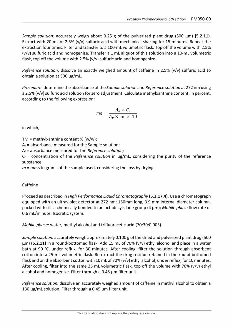
Brazilian Pharmacopoeia, 6th edition PM050-00
This translation does not replace the portuguese version.
Sample solution: accurately weigh about 0.25 g of the pulverized plant drug (500 µm) (5.2.11). Extract with 20 mL of 2.5% (v/v) sulfuric acid with mechanical shaking for 15 minutes. Repeat the extraction four times. Filter and transfer to a 100-mL volumetric flask. Top off the volume with 2.5% (v/v) sulfuric acid and homogenize. Transfer a 1 mL aliquot of this solution into a 10-mL volumetric flask, top off the volume with 2.5% (v/v) sulfuric acid and homogenize. Reference solution: dissolve an exactly weighed amount of caffeine in 2.5% (v/v) sulfuric acid to obtain a solution at 500 µg/mL. Procedure: determine the absorbance of the Sample solution and Reference solution at 272 nm using a 2.5% (v/v) sulfuric acid solution for zero adjustment. Calculate methylxanthine content, in percent, according to the following expression:
𝑇𝑇𝑇𝑇 =𝑇𝑇𝑎𝑎 × 𝐶𝐶𝑟𝑟
𝑇𝑇𝑟𝑟 × m × 10
in which, TM = methylxanthine content % (w/w); Aa = absorbance measured for the Sample solution; Ar = absorbance measured for the Reference solution; Cr = concentration of the Reference solution in µg/mL, considering the purity of the reference substance; m = mass in grams of the sample used, considering the loss by drying. Caffeine Proceed as described in High Performance Liquid Chromatography (5.2.17.4). Use a chromatograph equipped with an ultraviolet detector at 272 nm; 150mm long, 3.9 mm internal diameter column, packed with silica chemically bonded to an octadecylsilane group (4 µm); Mobile phase flow rate of 0.6 mL/minute. Isocratic system. Mobile phase: water, methyl alcohol and trifluoracetic acid (70:30:0.005). Sample solution: accurately weigh approximately 0.100 g of the dried and pulverized plant drug (500 µm) (5.2.11) in a round-bottomed flask. Add 15 mL of 70% (v/v) ethyl alcohol and place in a water bath at 90 °C, under reflux, for 30 minutes. After cooling, filter the solution through absorbent cotton into a 25-mL volumetric flask. Re-extract the drug residue retained in the round-bottomed flask and on the absorbent cotton with 10 mL of 70% (v/v) ethyl alcohol, under reflux, for 10 minutes. After cooling, filter into the same 25 mL volumetric flask, top off the volume with 70% (v/v) ethyl alcohol and homogenize. Filter through a 0.45 µm filter unit. Reference solution: dissolve an accurately weighed amount of caffeine in methyl alcohol to obtain a 130 µg/mL solution. Filter through a 0.45 µm filter unit.
Brazilian Pharmacopoeia, 6th edition PM050-00
This translation does not replace the portuguese version.
Procedure: separately inject 10 μL of the Reference solution and 10 μL of the Sample solution. Register the chromatograms and measure the areas under the peaks. The average retention time for the caffeine peak is 8 minutes and 30 seconds. Calculate caffeine content, in percent, according to the following expression:
𝑇𝑇𝐶𝐶 =𝐶𝐶𝑟𝑟 × 𝑇𝑇𝑎𝑎 × 25 × 100
𝑇𝑇𝑟𝑟 × 𝑚𝑚
in which, TC = caffeine content % (w/w); Cr = concentration of the Reference solution in g/mL, considering the purity of the reference substance; Ar = area under the peak corresponding to the caffeine in the Sample solution; Ar = area under the peak corresponding to the caffeine in the Reference solution; m = mass in grams of the sample used, considering the loss by drying. PACKAGING AND STORAGE In tightly closed containers, protected from light and heat.
Brazilian Pharmacopoeia, 6th edition PM050-00
This translation does not replace the portuguese version.
Figure 1 – Macroscopic, microscopic and powder microscopic aspects in Paullinia cupana Kunth ___________________________ The scales correspond: in A and B (4 mm), in C to H (100 μm). A – overall appearance of the seed. B – overall appearance of cotyledons. C – cross-section of the outer portion of a cotyledon; cotyledonary epidermis (epc); cell containing starch grains (ga); cotyledonary parenchyma (pct). D – detail of the starch grains. E – Epidermal cells of the cotyledons in front view. F – parenchyma cells of the cotyledons. G – detail of seed integument cross-section; petiole cells (cp); integument epidermis (ep); parenchyma (p). E – epidermal cells of the integument in front view.
Brazilian Pharmacopoeia, 6th edition PM051-00
This translation does not replace the portuguese version.
WITCHHAZEL, leaf Hamamelidis folium
The plant drug consists of dried, entire or fragmented leaves of Hamamelis virginiana L., containing at least 3.0% tannins, expressed as pyrogallol (C6H6O3, 126.11). IDENTIFICATION 1. Macroscopic description The leaf is shortly petiolate, with a petiole 1 to 1.5 cm long. The leaf lamina is rough, has a greenish-brown to grizzly-brownish color on the adaxial surface and light green on the abaxial surface, 7 to 15 cm long and 6 to 10 cm wide, oval or oval-rhomboid, with an asymmetrically cordate base, an acute apex, sometimes obtuse, and a sinuous margin, roughly crenate to dentate. The venation is peninerve, with the midrib protruding in the abaxial surface, alternate and rectilinear secondary veins, ending in the serrated teeth of the margins without joining each other, tertiary and quaternary veins are thinner and anastomosed, giving the limb a reticulate aspect. Young leaves with stellate trichomes, visible with a magnifying glass. 2. Microscopic description The leaf lamina is hypostomatic and dorsiventrally symmetrical. The epidermis, in front view, on both surfaces, presents cells with sinuous, periclinic walls, paracitic stomata, and thick-walled stellate trichomes. In cross-section, the epidermis is unistratified on both sides and the stomata are at the same level as the other epidermal cells; the cuticle is thin. The mesophyll is formed by one layer of palisade cells, facing the adaxial surface, followed by four to six layers of spongy parenchyma cells. Astro-sclereids cross the mesophyll, and may spread from one epidermis to the other. The smaller vascular bundles and the midrib are surrounded by a sheath with prismatic crystals and usually contain sclerenchyma fibers or sclereids associated with the vascular tissues. 3. Microscopic description of powder The sample meets all requirements for the species, except the macroscopic characters. Characteristics are: green-brownish color; trichomes with thick walls and visible lumen, grouped at the base forming tufts (stellate trichomes); fragments of the adaxial surface of the epidermis in front view with thick-walled cells and wavy to sinuous outline; fragments of the abaxial surface of the epidermis in front view with thick-walled cells with straight to wavy outline and stomata of the paracitic type, mainly; bundles of septate sclerenchymatous fibers, with slightly thickened walls and surrounded by a sheath of idioblasts with prismatic calcium oxalate crystals and sclerereids; xylematic vessels of the ringed or reticulate type, associated with lignified fibers, sheath of idioblasts with prismatic crystals and sclerereids, or isolated crystals and sclerereids; prismatic crystals of various shapes; parenchymatic cells with isolated chloroplastids; larger brownish-green fragments, in front view, showing the vein region with prismatic crystals and the spongy parenchyma with irregularly shaped cells and large intercellular spaces. 4. Proceed as described in Thin-layer chromatography (5.2.17.1).
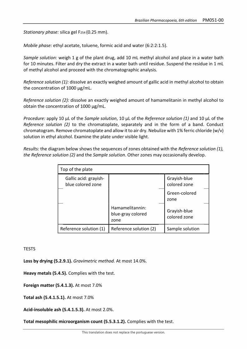
Brazilian Pharmacopoeia, 6th edition PM051-00
This translation does not replace the portuguese version.
Stationary phase: silica gel F254 (0.25 mm). Mobile phase: ethyl acetate, toluene, formic acid and water (6:2:2:1.5). Sample solution: weigh 1 g of the plant drug, add 10 mL methyl alcohol and place in a water bath for 10 minutes. Filter and dry the extract in a water bath until residue. Suspend the residue in 1 mL of methyl alcohol and proceed with the chromatographic analysis. Reference solution (1): dissolve an exactly weighed amount of gallic acid in methyl alcohol to obtain the concentration of 1000 µg/mL. Reference solution (2): dissolve an exactly weighed amount of hamamelitanin in methyl alcohol to obtain the concentration of 1000 µg/mL. Procedure: apply 10 μL of the Sample solution, 10 µL of the Reference solution (1) and 10 µL of the Reference solution (2) to the chromatoplate, separately and in the form of a band. Conduct chromatogram. Remove chromatoplate and allow it to air dry. Nebulize with 1% ferric chloride (w/v) solution in ethyl alcohol. Examine the plate under visible light. Results: the diagram below shows the sequences of zones obtained with the Reference solution (1), the Reference solution (2) and the Sample solution. Other zones may occasionally develop.
Top of the plate
Gallic acid: grayish-blue colored zone Grayish-blue
colored zone
Green-colored zone
Hamamelitannin: blue-gray colored zone
Grayish-blue colored zone
Reference solution (1) Reference solution (2) Sample solution
TESTS Loss by drying (5.2.9.1). Gravimetric method. At most 14.0%. Heavy metals (5.4.5). Complies with the test. Foreign matter (5.4.1.3). At most 7.0% Total ash (5.4.1.5.1). At most 7.0% Acid-insoluble ash (5.4.1.5.3). At most 2.0%. Total mesophilic microorganism count (5.5.3.1.2). Complies with the test.
Brazilian Pharmacopoeia, 6th edition PM051-00
This translation does not replace the portuguese version.
Study of pathogenic microorganisms (5.5.3.1.3). Complies with the test. Agrochemical waste (5.4.3). Complies with the test. DOSAGE Total tannins Note: protect samples from light during extraction and dilution. Use carbon dioxide-free water for all operations. To proceed as described in Visible absorption spectrophotometry (5.2.14). Prepare solutions as described below. Stock solution: accurately weigh approximately 0.750 g of the pulbverized plant drug and transfer to a 250-mL beaker. Add 150 mL of boiling water. Place in a water bath for 30 minutes. Cool under running water and transfer to a 250-mL volumetric flask. The sample residue should be rinsed off and quantitatively transferred to the volumetric flask. Top off the volume to 250 mL with water and homogenize. Allow to decant and filter on 125 mm diameter filter paper. Discard the first 50 mL of the filtrate. Sample solution for total polyphenol content: dilute 5 mL of the Stock solution in a 25-mL volumetric flask with water. Volumetrically transfer 2 mL of the solution, 1 mL of phosphomolybdotungstic reagent and 10 mL of water to a 25mL volumetric flask, top off the volume with sodium carbonate RS solution and homogenize. Determine absorbance at 760 nm (A1), after 30 minutes, protected from light, using water for zero adjustment. Sample solution for polyphenols not adsorbed by hide powder: for 10 mL of the Stock solution, add 0.10 g of hide powder and mechanically shake for 60 minutes. Filter through 125-mm diameter filter paper. Dilute 5 mL of the filtrate in a 25-mL volumetric flask with water and homogenize. Volumetrically transfer 2 mL of the solution, 1 mL of phosphomolybdotungstic reagent and 10 mL of water to a 25mL volumetric flask, top off the volume with sodium carbonate RS solution and homogenize. Determine absorbance at 760 nm (A2), after 30 minutes, protected from light, using water for zero adjustment. Reference solution: dissolve 50 mg pyrogallol in water immediately before use in a 100-mL volumetric flask, top off the volume with water. Volumetrically transfer 5 mL of this solution to a 100-mL volumetric flask, top off the volume with water and homogenize. Volumetrically transfer 2 mL of the solution, 1 mL of phosphomolybdotungstic reagent and 10 mL of water to a 25mL volumetric flask, top off the volume with sodium carbonate RS solution and homogenize. Determine absorbance at 760 nm (A3), after 30 minutes, protected from light, using water for zero adjustment. Calculate tannin content expressed as a percentage of pyrogallol, according to the following expression:
Brazilian Pharmacopoeia, 6th edition PM051-00
This translation does not replace the portuguese version.
𝑇𝑇𝑇𝑇 =(𝑇𝑇1 − 𝑇𝑇2) × 𝑚𝑚2 × 62,5
𝑇𝑇3 × 𝑚𝑚1
in which, TT = total tannin content expressed as pyrogallol % (w/w); A1 = absorbance measured for the Sample solution for total polyphenols; A2 = absorbance measured for the Sample solution for polyphenols not adsorbed by hide powder; A3 = absorbance measured for the Reference solution; m1 = mass in grams of the sample used, considering the loss by drying. m2 = mass in grams of pyrogallol, considering the purity of the reference substance. PACKAGING AND STORAGE In tightly closed containers, protected from light and heat.

Brazilian Pharmacopoeia, 6th edition PM051-00
This translation does not replace the portuguese version.
Figure 1 – Macroscopic, microscopic and powder microscopic aspects in Hamamelis virginiana L.
___________________________ The scales correspond in A to 1 cm; B and E to 100 μm; C, D, F and G to 50 μm; H-I to 20 μm. A - overall appearance of the leaf, front view. B - detail of leaf lamina portion in cross-section: epidermis (ep); stomata (es); sclereid (ec); vascular bundle (fv); palisade parenchyma (pp); spongy parenchyma (pj); stellate trichomes (tes). C - partial front view of the epidermis on the adaxial surface with thick-walled, sinuous cells. D - detail of the portion of the epidermis facing the abaxial surface with cells with straight to sinuous walls and thinner than those on the adaxial surface; paracitic stomata (es). E - fragment of septate fiber bundle (fbs) with crystalliferous idioblasts (ic) and sclereids (ec). F - front view of leaf fragment showing the venation with crystalliferous idioblasts (ic) and spongy parenchyma (pj)
Brazilian Pharmacopoeia, 6th edition PM051-00
This translation does not replace the portuguese version.
with intercellular spaces (ei). G - thick-walled stellate trichome. H - palisade parenchyma cell with chloroplasts. I - isolated prismatic crystal.
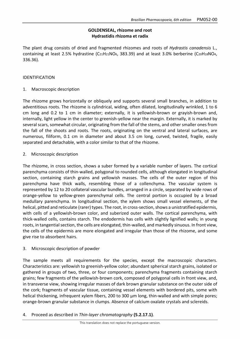
Brazilian Pharmacopoeia, 6th edition PM052-00
This translation does not replace the portuguese version.
GOLDENSEAL, rhizome and root Hydrastidis rhizoma et radix
The plant drug consists of dried and fragmented rhizomes and roots of Hydrastis canadensis L., containing at least 2.5% hydrastine (C21H21NO6, 383.39) and at least 3.0% berberine (C20H18NO4, 336.36). IDENTIFICATION 1. Macroscopic description The rhizome grows horizontally or obliquely and supports several small branches, in addition to adventitious roots. The rhizome is cylindrical, widing, often dilated, longitudinally wrinkled, 1 to 6 cm long and 0.2 to 1 cm in diameter; externally, it is yellowish-brown or grayish-brown and, internally, light yellow in the center to greenish-yellow near the margin. Externally, it is marked by several scars, somewhat circular, originating from the fall of the stems, and other smaller ones from the fall of the shoots and roots. The roots, originating on the ventral and lateral surfaces, are numerous, filiform, 0.1 cm in diameter and about 3.5 cm long, curved, twisted, fragile, easily separated and detachable, with a color similar to that of the rhizome. 2. Microscopic description The rhizome, in cross section, shows a suber formed by a variable number of layers. The cortical parenchyma consists of thin-walled, polygonal to rounded cells, although elongated in longitudinal section, containing starch grains and yellowish masses. The cells of the outer region of this parenchyma have thick walls, resembling those of a collenchyma. The vascular system is represented by 12 to 20 collateral vascular bundles, arranged in a circle, separated by wide rows of orange-yellow to yellow-green parenchymal cells. The central portion is occupied by a broad medullary parenchyma. In longitudinal section, the xylem shows small vessel elements, of the helical, pitted and reticulate (rarer) types. The root, in cross-section, shows a unistratified epidermis, with cells of a yellowish-brown color, and suberized outer walls. The cortical parenchyma, with thick-walled cells, contains starch. The endodermis has cells with slightly lignified walls; in young roots, in tangential section, the cells are elongated, thin-walled, and markedly sinuous. In front view, the cells of the epidermis are more elongated and irregular than those of the rhizome, and some give rise to absorbent hairs. 3. Microscopic description of powder The sample meets all requirements for the species, except the macroscopic characters. Characteristics are: yellowish to greenish-yellow color; abundant spherical starch grains, isolated or gathered in groups of two, three, or four components; parenchyma fragments containing starch grains; few fragments of the yellowish-brown cork, composed of polygonal cells in front view, and, in transverse view, showing irregular masses of dark brown granular substance on the outer side of the cork; fragments of vascular tissue, containing vessel elements with bordered pits, some with helical thickening, infrequent xylem fibers, 200 to 300 μm long, thin-walled and with simple pores; orange-brown granular substance in clumps. Absence of calcium oxalate crystals and sclereids. 4. Proceed as described in Thin-layer chromatography (5.2.17.1).
Brazilian Pharmacopoeia, 6th edition PM052-00
This translation does not replace the portuguese version.
Stationary phase: silica gel GF254 (0.250 mm). Mobile phase: ethyl acetate, water and anhydrous formic acid (80:10:10). Sample solution: extract 0.5 g of the pulverized plant drug with 10 mL of water and methyl alcohol (20:80). Sonicate for 10 minutes and filter. Rinse the residue twice with 2 mL of methyl alcohol. Combine the solutions and dilute to 20 mL. Reference solution: freshly prepared solution of 5 mg hydrastine hydrochloride and 5 mg berberine hydrochloride in 20 mL methyl alcohol. Procedure: apply 15 μL to 20 μL of the Sample solution and 3 μL to 5 μL of the Reference solution to the chromatoplate, separately and in the form of a band. Conduct chromatogram. Remove chromatoplate and allow it to air dry. Examine under ultraviolet light at 365 nm. Next, nebulize with a solution of potassium iodide and bismuth subnitrate RS and re-examine it. Results: the diagram below shows the sequences of zones obtained with the Reference solution and the Sample solution. Other zones may occasionally develop.
Top of the plate
Berberine: yellow fluorescence zone
Yellow fluorescence zone
Hydrastine: dark-blue fluorescence zone
Dark-blue fluorescence zone
Light-blue fluorescence zone
Dark-blue fluorescence zone
Reference solution Sample solution
TESTS Foreign matter (5.4.1.3). At most 2.0%. Water (5.4.1.4). At most 10.0%. Total ash (5.4.1.5.1). At most 8.0%. Total mesophilic microorganism count (5.5.3.1.2). Complies with the test. Study of pathogenic microorganisms (5.5.3.1.3). Complies with the test.
Brazilian Pharmacopoeia, 6th edition PM052-00
This translation does not replace the portuguese version.
Heavy metals (5.4.5). Complies with the test. Agrochemical waste (5.4.3). Complies with the test. Aflatoxins (5.4.4). Complies with the test. DOSAGE Proceed as described in High Performance Liquid Chromatography (5.2.17.4). Use a chromatograph equipped with an ultraviolet detector at 235 nm; pre-column packed with silica chemically bonded to an octadecylsilane group; a 75 mm long, 4.6 mm wide (internal diameter) column, packed with silica chemically bonded to an octadecylsilane group (3,5 µm); Mobile phase flow rate of 0.30 mL/minute. Mobile phase: 0.05 M monobasic potassium phosphate and acetonitrile (73:27). Sample solution: add 50 mL of 1% (v/v) ammonium hydroxide solution in ethyl alcohol to a 100-mL round-bottomed flask containing about 1.0 g of the accurately weighed pulverized sample. Heat to boiling, under reflux, for 30 minutes. Allow to cool to room temperature and filter through absorbent cotton, collecting the filtrate in a conical flask. Add the hydrophilic cotton to the residue contained in the round-bottomed flask and repeat the extraction twice with 30 mL of the 1% (v/v) ammonium hydroxide solution in ethyl alcohol and heat, under reflux, for 10 minutes. Filter through absorbent cotton into the same erlenmeyer flask used previously. Filter on filter paper, collect the filtrates in a 250-mL round-bottomed flask, rinse the flask and the filter paper with 20 mL of 1% (v/v) ammonium hydroxide solution in ethyl alcohol. Evaporate the filtrate to dryness, under reduced pressure, in a water bath at 55 °C. Dissolve the residue in a Mobile phase, in a 50mL volumetric flask, top off the volume and homogenize. Dilute 1 mL of the obtained solution to 50 mL, using the Mobile phase, and homogenize. Filter through a 0.45 µm filter unit. Reference solution (1): dissolve 10 mg hydrastine hydrochloride and 10 mg berberine chloride in methyl alcohol, top off the volume to 25 mL with the same solvent and homogenize. Reference Solution (2): dilute 1 mL of the Reference Solution (1) to 25 mL using methyl alcohol as solvent and homogenize. Solutions for the analytical curve: transfer 2.0 mL of the Reference solution (1) into a 25 mL volumetric flask, top off the volume with Mobile phase and homogenize. Dilute aliquots of 1 mL, 2 mL, 3 mL, 5 mL and 7 mL with the Mobile Phase to 10 mL, obtaining solutions with concentrations of 3.2 μg/mL, 6.4 μg/mL, 9.6 μg/mL, 16.0 μg/mL and 22.4 μg/mL for hydrastine hydrochloride and berberine chloride. Filter the solutions through a 0.45 µm filter unit. System suitability Resolution between peaks: Reference solution (2), inject 10 μL. Hydrastine has a shorter retention time than berberine. The resolution between the peaks corresponding to hydrastine and berberine is at least 1.5. Procedure: separately inject 10µL of the Reference solution (1), 10µL of the Reference solution (2) and 10µL of the Sample solution. Record the chromatograms and measure the areas under the peaks corresponding to hydrastine and berberine. The relative retention time is about 8.2 minutes for
Brazilian Pharmacopoeia, 6th edition PM052-00
This translation does not replace the portuguese version.
hydrastine and 10.8 minutes for berberine. Calculate the content of hydrastine and berberine in the sample from the straight line equation obtained from the analytical curves of hydrastine hydrochloride and berberine chloride. The result is expressed as the average of the hydrastine and berberine determinations, separately, in a percentage (w/w) of the drug, considering the determined water content. PACKAGING AND STORAGE In tightly closed containers, protected from light and heat.
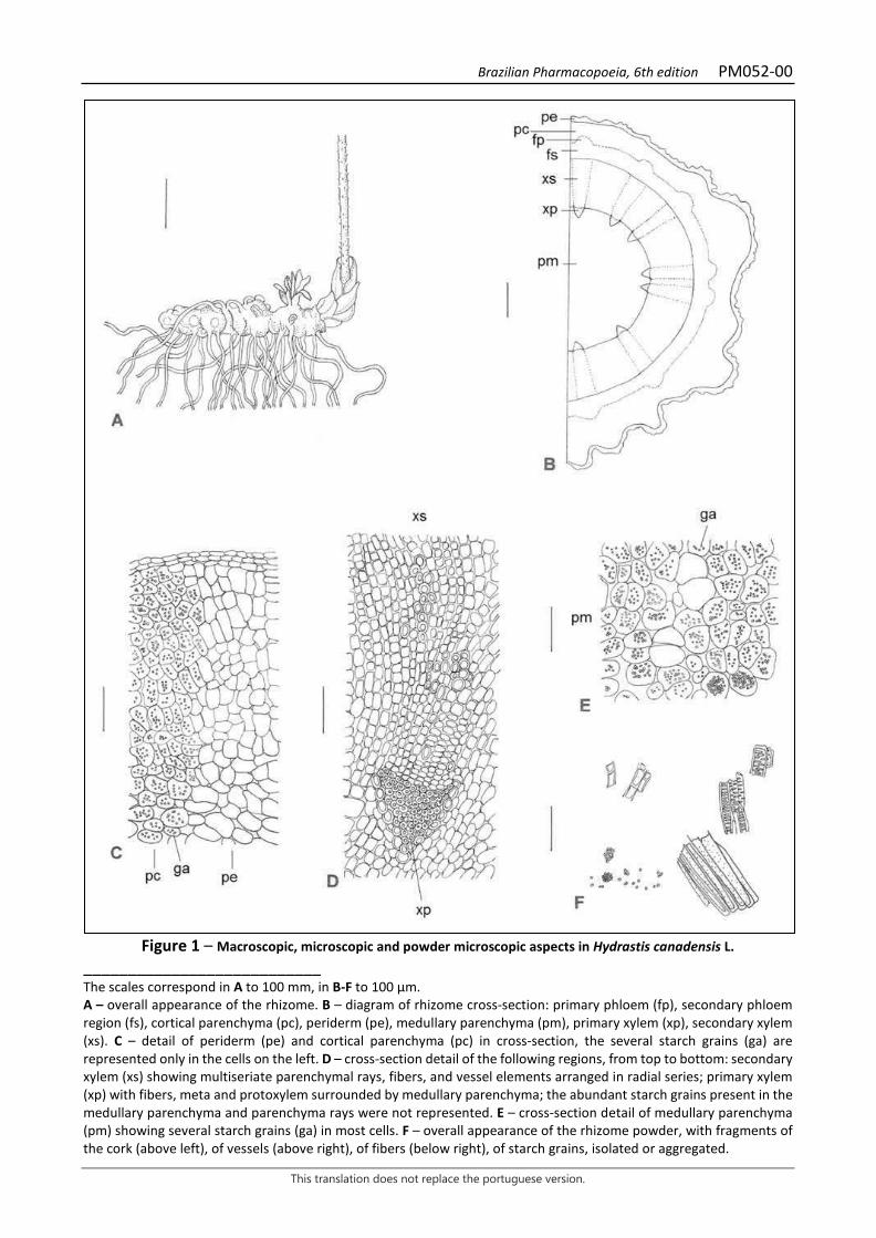
Brazilian Pharmacopoeia, 6th edition PM052-00
This translation does not replace the portuguese version.
Figure 1 – Macroscopic, microscopic and powder microscopic aspects in Hydrastis canadensis L.
___________________________ The scales correspond in A to 100 mm, in B-F to 100 μm. A – overall appearance of the rhizome. B – diagram of rhizome cross-section: primary phloem (fp), secondary phloem region (fs), cortical parenchyma (pc), periderm (pe), medullary parenchyma (pm), primary xylem (xp), secondary xylem (xs). C – detail of periderm (pe) and cortical parenchyma (pc) in cross-section, the several starch grains (ga) are represented only in the cells on the left. D – cross-section detail of the following regions, from top to bottom: secondary xylem (xs) showing multiseriate parenchymal rays, fibers, and vessel elements arranged in radial series; primary xylem (xp) with fibers, meta and protoxylem surrounded by medullary parenchyma; the abundant starch grains present in the medullary parenchyma and parenchyma rays were not represented. E – cross-section detail of medullary parenchyma (pm) showing several starch grains (ga) in most cells. F – overall appearance of the rhizome powder, with fragments of the cork (above left), of vessels (above right), of fibers (below right), of starch grains, isolated or aggregated.
Brazilian Pharmacopoeia, 6th edition PM053-00
This translation does not replace the portuguese version.
CORN MINT, aerial part Mentha arvensis herbae The plant drug consists of dried aerial parts of Mentha arvensis L., entire, broken, cut, or pulverized, containing at least 0.8% volatile oil in entire aerial parts and at least 0.6% volatile oil in chopped aerial parts. The percentage of stems should not exceed 20%. SCIENTIFIC SYNONYM Mentha arvensis var. canadensis (L.) Kuntze CHARACTERISTICS Aerial parts have a strong, aromatic, penetrating, menthol-like odor. IDENTIFICATION 1. Macroscopic description Erect, tetrangular stems up to 60 cm long, covered with whitish, multicellular, reflex trichomes. Simple, opposite crossed leaves, becoming smaller towards the apex of the branches, petiole 2 to 7 mm long, elliptic to oval-lanceolate lamina, 2 to 5 cm long and 0.8 to 2 cm wide, acute apex, cuneate to rounded base, serrate margin. Inflorescences, when present, are arranged in the apical nodes of the branches, globose, dense, with white flowers, gamosepalous calyx, campanulate, 2 to 3 mm long, external surface covered by trichomes, equal triangular lobes, acute; bilabated gammopetal corolla, 4 to 5 mm long and a four-lobed limbus, the superior being greater and bilobed and the others being subequal, with an obtuse apex; four stamens, adnate to the corolla tube, alternate to the petals, inserted in pistillate flowers (sterile anthers) or longer than the corolla in bisexual flowers; ovary pubescent, bicarpellate, tetralocular; long stylet, bifid stigma, exserted. Fruit, when present, consists of four dry, ellipsoid, brown mericarps. 2. Microscopic description In cross-section, the stem is quadrangular in shape, with four evident wings. The epidermis is unistratified and composed of rounded cells, with a thick cuticle and uni or pluricellular glandular and tector trichomes, followed by one to two layers of collenchyma with irregularly thickened walls forming a continuous ring and about ten layers of collenchyma cells in the wing region and a cortical parenchyma composed of three to four layers of bulky, thin-walled cells. The vascular tissue is more developed in the regions contiguous to the wings and has one to two diminutive collateral vascular bundles in the regions between them. In the wing region, the phloem has sieve elements and parenchyma cells and few parenchyma rays. The cambium region has three to four layers of rectangular cells with non-thickened walls. The xylem is formed by radially arranged parenchyma cells and conductive elements with thickened walls. The medullary parenchyma has bulky cells with a larger diameter toward the center and thin walls. The leaf lamina, in cross-section, has dorsiventral symmetry and is hypostomatic. The midrib region is more prominent on the abaxial surface and has the following tissues: epidermis composed of rounded cells with a thin cuticle, with uniseriate
Brazilian Pharmacopoeia, 6th edition PM053-00
This translation does not replace the portuguese version.
multicellular trichomes, followed by layers of collenchyma (three to four on the adaxial surface, one to two on the abaxial surface), and of bulky parenchyma cells, which are isodiametric, with non-thickened walls and wide intercellular spaces; the vascular bundle is collateral. The limb region has a unistratified epidermis composed of elliptical cells, with uni- or pluricellular glandular and tector trichomes, which are uniseriate on both faces, and diacritical stomata restricted to the abaxial surface; the palisade parenchyma is composed of a layer of tabular and juxtaposed cells and the spongy parenchyma is composed of rounded cells, with wide intercellular spaces. Collateral vascular bundles are scattered throughout the mesophyll. 3. Microscopic description of powder The sample meets all requirements for the species, except the macroscopic characters. Characteristics are: green color with some yellowish-green fragments; fragments of uniseriate pluricellular trichomes; fragments of parenchyma tissue with prismatic or rounded cells and intercellular spaces; epidermal fragments identified by cells with somewhat sinuous, anticlinal walls and by the presence of diacritical stomata with subsidiary cells similar to the epidermal ones; collenchyma fragments with elongated and densely juxtaposed cylindrical cells and conductive tissue fragments associated with chlorophyll parenchyma and epidermal remnants. 4. Proceed as described in Thin-layer chromatography (5.2.17.1). Stationary phase: 250 µm thick silica gel F254. Mobile phase: hexane and ethyl acetate (85:15). Sample solution: dilute 0.1 mL of the volatile oil in 1 mL ethyl acetate. Reference solution: dissolve 4 µL of carvone, 4 µL of pulegone, 10 µL of menthol acetate, 20 µL of cineol, and 50 mg of menthol in 5 mL ethyl acetate. Procedure: apply 10 μL of the Sample solution and 10 μL of the Reference solution to the chromatoplate, separately and in the form of a band. Conduct chromatogram. Remove chromatoplate and allow it to air dry. Examine under ultraviolet light at 254 nm. Nebulize with sulfur vanillin RS and heat at 100 °C to 105 °C for one minute. After visualization, heat for another three minutes. Results: the diagrams below show the sequences of zones, obtained with the Reference solution and the Sample solution, after examination under ultraviolet light, nebulization with the TLC visualization reagent and heating for one minute, and heating for a further three minutes, respectively. Other zones may occasionally develop.
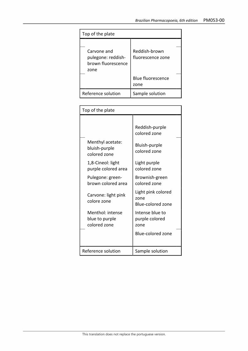
Brazilian Pharmacopoeia, 6th edition PM053-00
This translation does not replace the portuguese version.
Top of the plate
Carvone and pulegone: reddish-brown fluorescence zone
Reddish-brown fluorescence zone
Blue fluorescence zone
Reference solution Sample solution
Top of the plate
Reddish-purple colored zone
Menthyl acetate: bluish-purple colored zone
Bluish-purple colored zone
1,8-Cineol: light purple colored area
Light purple colored zone
Pulegone: green-brown colored area
Brownish-green colored zone
Carvone: light pink colore zone
Light pink colored zone Blue-colored zone
Menthol: intense blue to purple colored zone
Intense blue to purple colored zone
Blue-colored zone
Reference solution Sample solution
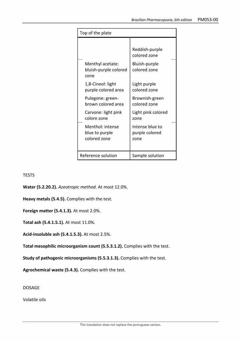
Brazilian Pharmacopoeia, 6th edition PM053-00
This translation does not replace the portuguese version.
Top of the plate
Reddish-purple colored zone
Menthyl acetate: bluish-purple colored zone
Bluish-purple colored zone
1,8-Cineol: light purple colored area
Light purple colored zone
Pulegone: green-brown colored area
Brownish-green colored zone
Carvone: light pink colore zone
Light pink colored zone
Menthol: intense blue to purple colored zone
Intense blue to purple colored zone
Reference solution Sample solution TESTS Water (5.2.20.2). Azeotropic method. At most 12.0%. Heavy metals (5.4.5). Complies with the test. Foreign matter (5.4.1.3). At most 2.0%. Total ash (5.4.1.5.1). At most 11.0%. Acid-insoluble ash (5.4.1.5.3). At most 2.5%. Total mesophilic microorganism count (5.5.3.1.2). Complies with the test. Study of pathogenic microorganisms (5.5.3.1.3). Complies with the test. Agrochemical waste (5.4.3). Complies with the test. DOSAGE Volatile oils
Brazilian Pharmacopoeia, 6th edition PM053-00
This translation does not replace the portuguese version.
Proceed as described in Determining volatile oils in plant drugs (5.4.1.6). Use a 500-mL flask containing 200 mL of water as distillation liquid and add 1.0 mL of xylene in the graduated tube. Immediately proceed with the determination of the volatile oil, from 50 g of the pulverized plant drug. Distill for four hours. Measure the volume and express the yield per 100 g of the plant drug (w/p).
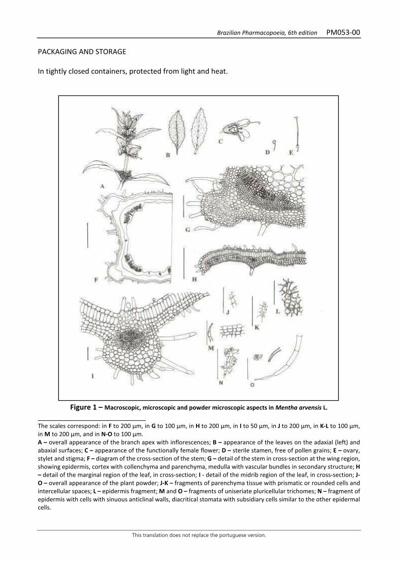
Brazilian Pharmacopoeia, 6th edition PM053-00
This translation does not replace the portuguese version.
PACKAGING AND STORAGE In tightly closed containers, protected from light and heat.
Figure 1 – Macroscopic, microscopic and powder microscopic aspects in Mentha arvensis L.
___________________________ The scales correspond: in F to 200 µm, in G to 100 µm, in H to 200 µm, in I to 50 µm, in J to 200 µm, in K-L to 100 µm, in M to 200 µm, and in N-O to 100 µm. A – overall appearance of the branch apex with inflorescences; B – appearance of the leaves on the adaxial (left) and abaxial surfaces; C – appearance of the functionally female flower; D – sterile stamen, free of pollen grains; E – ovary, stylet and stigma; F – diagram of the cross-section of the stem; G – detail of the stem in cross-section at the wing region, showing epidermis, cortex with collenchyma and parenchyma, medulla with vascular bundles in secondary structure; H – detail of the marginal region of the leaf, in cross-section; I - detail of the midrib region of the leaf, in cross-section; J-O – overall appearance of the plant powder; J-K – fragments of parenchyma tissue with prismatic or rounded cells and intercellular spaces; L – epidermis fragment; M and O – fragments of uniseriate pluricellular trichomes; N – fragment of epidermis with cells with sinuous anticlinal walls, diacritical stomata with subsidiary cells similar to the other epidermal cells.

Brazilian Pharmacopoeia, 6th edition PM054-00
This translation does not replace the portuguese version.
PEPPERMINT, leaf Menthae piperitae folium
The plant drug consists of dried, entire, broken, cut, or pulverized leaves of Mentha × piperita L. or its varieties, containing at least 1.2% volatile oil in entire leaves and at least 0.9% volatile oil in chopped leaves, relative to the dried material. CHARACTERISTICS The drug has a strong, aromatic, penetrating, menthol-like odor. IDENTIFICATION 1. Macroscopic description The leaf lamina is oval-oblong to oblong-lanceolate, measuring 1.5 to 9 cm long and 1 to 5 cm wide, with a sharp apex, an irregularly rounded and asymmetrical base, an irregularly serrate margin, with a light to dark green color, an almost smooth adaxial surface and a pubescent abaxial surface. The petiole is 0.4 to 1.5 cm long and is pubescent. 2. Microscopic description Dorsiventrally symmetrical leaf lamina, hypoanphistomatic, with diacytic stomata. In front view, the cuticle is smooth and the epidermal cells have anticlinal walls with a wavy outline in the region between the veins and rectilinear over the veins. Trichomes are tector or glandular: pluricellular tector trichome, uniseriate, with two to fourteen cells, thick and markedly striated cuticle, the basal cell being of greater length and the apical one of obtuse apex, and may have a crown of basal cells; pluricellular tector trichome, with two to six cells, biseriate at the base, also with thick and striated cuticle; glandular trichome with unicellular to tricellular pedicel, short and with unicellular head, elliptical or rounded, with thin cuticle; peltate glandular trichome, found in depressions of the epidermis, with short pedicel, formed by one or two cells in the basal portion and pluricellular head with eight cells in a radial disposition, generally with a dilated cuticle and grizzly color. In cross-section, the adaxial and abaxial epidermis consist of only one layer of cells, rich in oil drops; the palisade parenchyma is formed by one layer of cells and the spongy parenchyma is formed by three to four layers, oil drops are abundant in all tissues. The midrib, in cross-section, has a vascular system formed by a collateral bundle. 3. Microscopic description of powder The sample meets all requirements for the species, except the macroscopic characters. Examine under a microscope, using chloral hydrate R solution. Characteristics are: light to olive green color; epidermal fragments, mesophyll fragments, and vein fragments with the characteristics and elements mentioned under Microscopic Description. 4. Macroscopic description of impurities
Brazilian Pharmacopoeia, 6th edition PM054-00
This translation does not replace the portuguese version.
Stems, branches, flowers, fruit and seeds of the species itself, if present as impurity, are characterized by: quadrangular stem with well-defined veins up to the fourth node, branched, in most varieties, vinous when adult, light green when young and whitish at the basal nodes; trichomes not visible to the naked eye; flowers gathered in spike inflorescences; glabrous calyx, with five teeth; rosy-violaceous or white corolla, with four lobes, the upper one enlarged; four stamens, didinamous, included in the corolla; superus, tetralobed ovary, gynobasic stylet; rare, sterile seeds. 5. Microscopic description of the impurity corresponding to the stem The stems of the species itself, if present as impurity, have, as a secondary structure and in cross-section, thick and striated cuticle, unistratified epidermis, of polygonal cells, with or without idioblasts of crystalline sand; trichomes and rare stomata; angular collenchyma, formed by one to many layers in the region of the veins; chlorenchyma with up to ten layers, with isolated sclerotia and crystalline sand idioblasts; endoderm with evident Caspary striations and free of starch grains; phloem with or without isolated fibers or in small groups; evident cambium zone; xylem sclerified or not; oil drops in all tissues, except cambium and xylem; developed medullary parenchyma. 6. Adulterations Mentha crispa L., when present, is distinguished by glandular trichomes with a twelve-celled head and thin-walled, one- to six-celled tector trichomes. 7. Proceed as described in Thin-layer chromatography (5.2.17.1). Stationary phase: silica gel GF254 (0.250 mm). Mobile phase: toluene and ethyl acetate (95:5). Sample solution: shake 0.2 g of the freshly pulverized plant drug with 2 mL methyl chloride. Filter. Evaporate to dryness at 40 °C and dissolve the residue in 0.1 mL toluene. Reference solution: dissolve 50 mg menthol, 20 µL of 1,8-cineole, 10 mg thymol and 10 µL mentyl acetate in toluene and top off the volume to 10 mL with the same solvent. Procedure: apply 20 μL of the Sample solution and 10 μL of the Reference solution to the chromatoplate, separately and in the form of a band. Conduct chromatogram. Remove chromatoplate and allow it to air dry. Examine under ultraviolet light at 254 nm. Next, nebulize the plate with anisaldehyde RS in an oven at 100 °C to 105 °C for five to 10 minutes. Results: the diagram below shows the sequences of zones obtained with the Reference solution and the Sample solution. Other zones may occasionally develop.
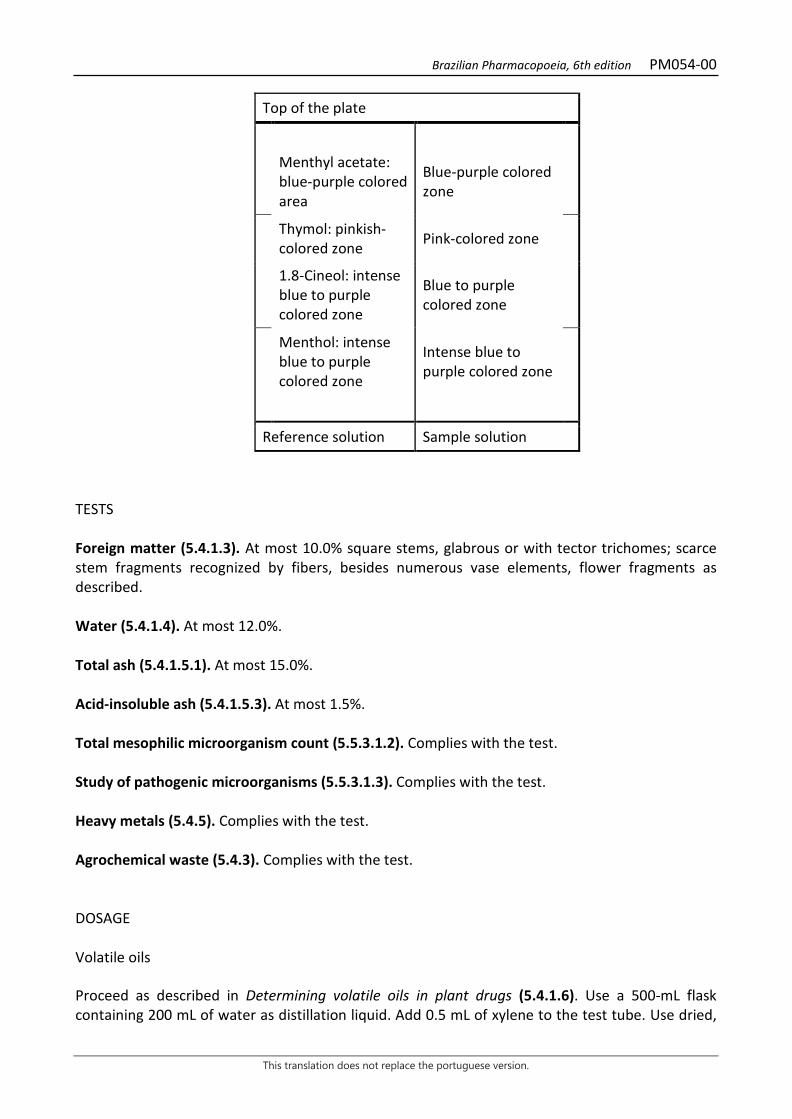
Brazilian Pharmacopoeia, 6th edition PM054-00
This translation does not replace the portuguese version.
Top of the plate
Menthyl acetate: blue-purple colored area
Blue-purple colored zone
Thymol: pinkish-colored zone Pink-colored zone
1.8-Cineol: intense blue to purple colored zone
Blue to purple colored zone
Menthol: intense blue to purple colored zone
Intense blue to purple colored zone
Reference solution Sample solution
TESTS Foreign matter (5.4.1.3). At most 10.0% square stems, glabrous or with tector trichomes; scarce stem fragments recognized by fibers, besides numerous vase elements, flower fragments as described. Water (5.4.1.4). At most 12.0%. Total ash (5.4.1.5.1). At most 15.0%. Acid-insoluble ash (5.4.1.5.3). At most 1.5%. Total mesophilic microorganism count (5.5.3.1.2). Complies with the test. Study of pathogenic microorganisms (5.5.3.1.3). Complies with the test. Heavy metals (5.4.5). Complies with the test. Agrochemical waste (5.4.3). Complies with the test. DOSAGE Volatile oils Proceed as described in Determining volatile oils in plant drugs (5.4.1.6). Use a 500-mL flask containing 200 mL of water as distillation liquid. Add 0.5 mL of xylene to the test tube. Use dried,
Brazilian Pharmacopoeia, 6th edition PM054-00
This translation does not replace the portuguese version.
chopped plant. Immediately proceed with the determination of the volatile oil, from 20 g of the drug. Distill for four hours. Measure the volume and express the yield per 100 g of the plant drug (w/p).
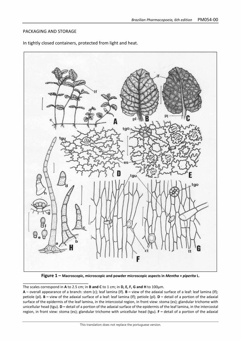
Brazilian Pharmacopoeia, 6th edition PM054-00
This translation does not replace the portuguese version.
PACKAGING AND STORAGE In tightly closed containers, protected from light and heat.
Figure 1 – Macroscopic, microscopic and powder microscopic aspects in Mentha × piperita L.
___________________________ The scales correspond in A to 2.5 cm; in B and C to 1 cm; in D, E, F, G and H to 100μm. A – overall appearance of a branch: stem (c); leaf lamina (lf). B – view of the adaxial surface of a leaf: leaf lamina (lf); petiole (pl). B – view of the adaxial surface of a leaf: leaf lamina (lf); petiole (pl). D – detail of a portion of the adaxial surface of the epidermis of the leaf lamina, in the intercostal region, in front view: stoma (es); glandular trichome with unicellular head (tgu). D – detail of a portion of the adaxial surface of the epidermis of the leaf lamina, in the intercostal region, in front view: stoma (es); glandular trichome with unicellular head (tgu). F – detail of a portion of the adaxial
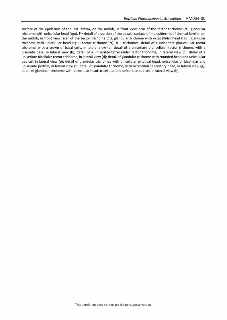
Brazilian Pharmacopoeia, 6th edition PM054-00
This translation does not replace the portuguese version.
surface of the epidermis of the leaf lamina, on the midrib, in front view: scar of the tector trichome (ct); glandular trichome with unicellular head (tgu). F – detail of a portion of the adaxial surface of the epidermis of the leaf lamina, on the midrib, in front view: scar of the tector trichome (ct); glandular trichome with octacellular head (tgo); glandular trichome with unicellular head (tgu); tector trichome (tt). H – trichomes: detail of a uniseriate pluricellular tector trichome, with a crown of basal cells, in lateral view (a); detail of a uniseriate pluricellular tector trichome, with a biseriate base, in lateral view (b); detail of a uniseriate tetracellular tector trichome, in lateral view (c); detail of a uniseriate bicellular tector trichome, in lateral view (d); detail of glandular trichome with rounded head and unicellular pedicel, in lateral view (e); detail of glandular trichomes with unicellular elliptical head, unicellular or bicellular and uniseriate pedicel, in lateral view (f); detail of glandular trichome, with octacellular secretory head, in lateral view (g); detail of glandular trichome with unicellular head, tricellular and uniseriate pedicel, in lateral view (h).

Brazilian Pharmacopoeia, 6th edition PM054-00
This translation does not replace the portuguese version.
Figure 2 – Microscopic aspects in Mentha × piperita L.
___________________________ The scales correspond in A to 400 μm; in B, C and E to 100μm; in D to 1000 μm. A – schematic illustration of the overall appearance of the midrib region and of a portion of the intercostal region, in cross-section: abaxial surface (ab); adaxial surface (ad); spongy parenchyma (pj); tector trichome (tt); collenchyma (co); palisade parenchyma (pp); xylem parenchyma (px); endoderm (end); epidermis (ep); xylem (x); phloem (f); fundamental parenchyma (pf). B – detail of the region of the midrib and a portion of the intercostal region, in cross-section: abaxial

Brazilian Pharmacopoeia, 6th edition PM054-00
This translation does not replace the portuguese version.
surface (ab); adaxial surface (ad); palisade parenchyma (pp); glandular trichomes with unicellular head (tgu); spongy parenchyma (pj); phloem (f); xylem (x); tector trichome (tt); stoma (es); xylem parenchyma (px); fundamental parenchyma (pf); endoderm (end); collenchyma (co); epidermis (ep); cuticle (cu); starch grain (ga). C – detail of the leaf lamina in the intercostal region, in cross-section: abaxial surface (ab); adaxial surface (ad); glandular trichomes with unicellular head (tgu); palisade parenchyma (pp); epidermis (ep); cuticle (cu); stoma (es); spongy parenchyma (pj); glandular trichome with octacellular head (tgo). D – schematic illustration of the overall appearance of the petiole, in cross-section: abaxial surface (ab); adaxial surface (ad); phloem (f); xylem (x); epidermis (ep); endoderm (end); collenchyma (co); vascular bundle (fv); chlorenchyma (cl); fundamental parenchyma (pf). E – detail of petiole portion in cross section: abaxial surface (ab); adaxial surface (ad); glandular trichomes with unicellular head (tgu); collenchyma (co); epidermis (ep); cuticle (cu); chlorenchyma (cl); fundamental parenchyma (pf); starch grain (ga); phloem (f); xylem (x); xylem parenchyma (px); endoderm (end).

Brazilian Pharmacopoeia, 6th edition PM055-00
This translation does not replace the portuguese version.
BRAZILIAN JALAP, root Operculina radix
The plant drug consists of dried roots of Operculina macrocarpa (L.) Urb., containing at least 6.4% total polysaccharides, expressed as D-maltose (C12H22O11, 342.30). IDENTIFICATION 1. Macroscopic description Tuberous root, pivotal, conic to napiform in shape and cylindrical in outline; externally it has a brown to black coloration, and in cross-section, internally, a yellowish color, exuding a resinous liquid when compressed. The drug is found entire or cut into disks 0.5 to 2 cm thick and 3 to 6 cm in diameter. The cross sections (disks) show white to grayish-yellow color from the margin to the center, with concentric sinuous darker lines. 2. Microscopic description In cross-section, the cortex shows externally lenticels and many layers of siber; phellogen difficult to observe and phelloderm consisting of parenchymatic cells containing tiny starch grains inside. The outer cortical parenchyma is formed by thin-walled cells, with many larger and smaller druses in the cells of the inner cortical parenchyma. The root is polyarchic. The central cylinder has collateral vascular bundles, with concentric rings of xylem and phloem produced by successive cambiums in the secondary structure, and wide bands of parenchyma between the bundles. The phloem-conducting cells form bands of periclinally flattened cells, with associated parenchyma cells containing several, tiny starch grains. Laticifers forming a network appear with thickened walls and a star shape, and are visible in cross-section in the region of the phloem closest to the cortex. The xylem shows tracheids and the vessel elements show reticulate and scalariform thickening, with contiguous, slit-like bordered pits. Tiny starch grains occur within the associated parenchyma cells. 3. Microscopic description of powder The sample meets all requirements for the species, except the macroscopic characters. Characteristics are: yellowish-brown to whitish color; libriform sclerenchyma fibers; parenchyma cell portions containing starch grains; vessel elements with scalariform thickening and oblique terminal walls; vessel elements with straight terminal walls; vessel elements with and without short and long extensions. The wall ornamentation is predominantly of the reticulate type, only small areas show a scalariform appearance. 4. Proceed as described in Thin-layer chromatography (5.2.17.1). Stationary phase: 250 µm thick silica gel F254. Mobile phase: ethyl acetate, water, formic acid, and acetic acid (100:27:11:11).
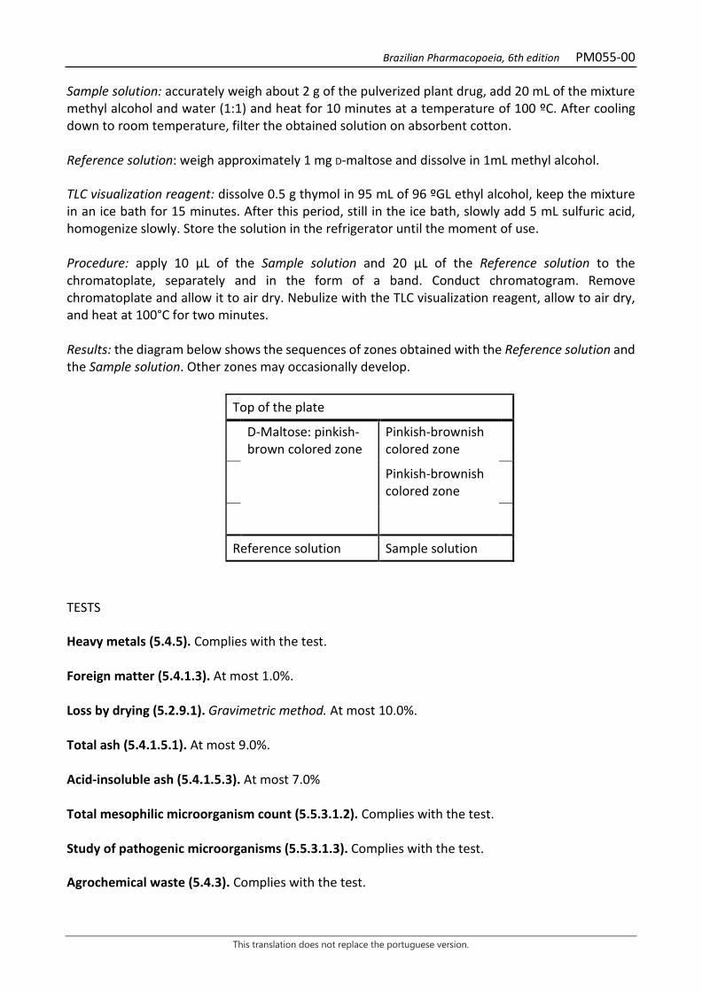
Brazilian Pharmacopoeia, 6th edition PM055-00
This translation does not replace the portuguese version.
Sample solution: accurately weigh about 2 g of the pulverized plant drug, add 20 mL of the mixture methyl alcohol and water (1:1) and heat for 10 minutes at a temperature of 100 ºC. After cooling down to room temperature, filter the obtained solution on absorbent cotton. Reference solution: weigh approximately 1 mg D-maltose and dissolve in 1mL methyl alcohol. TLC visualization reagent: dissolve 0.5 g thymol in 95 mL of 96 ºGL ethyl alcohol, keep the mixture in an ice bath for 15 minutes. After this period, still in the ice bath, slowly add 5 mL sulfuric acid, homogenize slowly. Store the solution in the refrigerator until the moment of use. Procedure: apply 10 μL of the Sample solution and 20 μL of the Reference solution to the chromatoplate, separately and in the form of a band. Conduct chromatogram. Remove chromatoplate and allow it to air dry. Nebulize with the TLC visualization reagent, allow to air dry, and heat at 100°C for two minutes. Results: the diagram below shows the sequences of zones obtained with the Reference solution and the Sample solution. Other zones may occasionally develop.
Top of the plate
D-Maltose: pinkish-brown colored zone
Pinkish-brownish colored zone
Pinkish-brownish colored zone
Reference solution Sample solution
TESTS Heavy metals (5.4.5). Complies with the test. Foreign matter (5.4.1.3). At most 1.0%. Loss by drying (5.2.9.1). Gravimetric method. At most 10.0%. Total ash (5.4.1.5.1). At most 9.0%. Acid-insoluble ash (5.4.1.5.3). At most 7.0% Total mesophilic microorganism count (5.5.3.1.2). Complies with the test. Study of pathogenic microorganisms (5.5.3.1.3). Complies with the test. Agrochemical waste (5.4.3). Complies with the test.
Brazilian Pharmacopoeia, 6th edition PM055-00
This translation does not replace the portuguese version.
DOSAGE To proceed as described in Visible absorption spectrophotometry (5.2.14). Prepare solutions as described below. Stock solution: weigh 2.0 g of the pulverized plant drug and transfer to a 250 mL, round-bottomed, polished-necked flask. Add 100 mL of water. Heat, under reflux, in a water bath, at 85 °C, for 30 minutes. Cool under running water, allow to decant. Filter the supernatant liquid on filter paper into a 100-mL volumetric flask Top off the volume with water. Sample solution for total polysaccharides: transfer 3 mL of the Stock solution to a 250-mL round-bottom flask with a ground-glass stopper, add 1 mL of 5% (w/v) phenol solution and 5 mL of concentrated sulfuric acid. Heat, under reflux, in a water bath, at 85 °C, for 30 minutes. Cool to room temperature. Volumetrically transfer 0.550 mL of this solution to a 10-mL volumetric flask, top off the volume with water and homogenize. Immediately determine the absorbance at 495 nm (A1), using water for zero adjustment. Reference solution: dissolve 50 mg D-maltose in water immediately before use, transfer to a 25-mL volumetric flask, top off the volume with water and homogenize. Transfer 3 mL of this solution volumetrically to a round-bottomed flask, add 1 mL of 5% (w/v) phenol solution, and 5 mL of concentrated sulfuric acid. Heat, under reflux, in a water bath, at 85 °C, for 30 minutes. Cool to room temperature. Volumetrically transfer 0.500 mL of this solution to a 10-mL volumetric flask, top off the volume with water and homogenize. Immediately determine the absorbance at 495 nm (A2), using water for zero adjustment. Calculate the total polysaccharide content expressed as a percentage of D-maltose, according to the following expression:
TPT =𝑇𝑇1 × 𝑚𝑚2 × 363,64
𝑚𝑚1 × 𝑇𝑇2
in which, TPT = total polysaccharide content expressed as D-maltose % (w/w); A1 = absorbance measured for the Sample solution for total polysaccharides; A2 = absorbance measured for the Reference solution; m1 = mass in grams of the sample used, considering the loss by drying. m2 = mass in grams of D-maltose, considering the purity of the reference substance.
Brazilian Pharmacopoeia, 6th edition PM055-00
This translation does not replace the portuguese version.
PACKAGING AND STORAGE In tightly closed containers, protected from light and heat.
Figure 1 – Macroscopic, microscopic and powder microscopic aspects in Operculina macrocarpa (L.) Urb.
___________________________ The scales correspond in A and B to 1 cm; C and D to 100 µm; E, F, G, H, I, J, K, L, M and N to 25 µm. A - overall appearance of the root. B - detail of the root cross-section, showing the successive changes. C and D - detail of root cross-section: suber and lenticels (arrows). E - detail of root cross-section, showing outer cortical parenchyma cells containing tiny starch grains (arrows). F - detail of root cross-section, showing outer cortical parenchyma cells

Brazilian Pharmacopoeia, 6th edition PM055-00
This translation does not replace the portuguese version.
containing calcium oxalate druses. G - detail of the root cross-section, indicating a laticiferous vessel (arrow). H - detail of longitudinal section of root showing reticulate thickened vessel element. I-N - details of fragments observed in the powder. I - libriform sclerenchymal fibers. J – portions of parenchyma cells. K - fragment of vessel element with scalariform thickening and oblique terminal walls. L - fragment of vessel element with straight end walls. M - fragment of vessel element with short extension. N - fragment of vessel element with long extension.
Brazilian Pharmacopoeia, 6th edition PM056-00
This translation does not replace the portuguese version.
BRAZILIAN IRONWOOD, bark Libidibiae cortex
The plant drug consists of dried barks from the stems of Libidibia ferrea (Mart.) L.P. Queiroz (syn. Caesalpinia ferrea Mart.), containing not less than 8.0% total tannins and not less than 0.02% gallic acid (C7H6O5, 170.12). IDENTIFICATION 1. Macroscopic description The stem barks, when dry, are slightly curved fragments, variable in size, 9 to 13 cm long, 1.5 to 4 cm wide, and 0.2 to 0.6 cm thick. Externally they have a grayish coloration, with whitish spots, are wrinkled and very hard; internally they are light brown. Smooth and lighter mottled barks may be present, as they annually renew themselves. 2. Microscopic description In cross-section, the outer portion of the bark has a sorb with eight to twelve layers of tabular cells lined with thin walls, followed by parenchymal cells of the cortex. The cortical parenchyma has polyhedral-shaped parenchyma cells, with sclerotia distributed throughout the cortical length, forming a wide band. Idioblasts containing druses and prismatic crystals are also visible in this parenchyma. The phloem has periclinally flattened cells, and the parenchyma cord cells of the phloem contain prismatic crystals. In longitudinal section, in the phloem region, multiseriate parenchyma ray cells with uniseriate margins, fibers, parenchymatic cords, and sclereids are evident. 3. Microscopic description of powder The sample meets all requirements for the species, except the macroscopic characters. Characteristics are: brownish color; predominantly grouped and also isolated sclereids; sclerenchymatic fibers; portions of parenchyma cells. 4. Proceed as described in Thin-layer chromatography (5.2.17.1). Stationary phase: 250 µm thick silica gel F254. Mobile phase: ethyl acetate, formic acid and water (90:5:5). Sample solution: weigh about 1 g of the drug and boil with 5 mL methyl alcohol for five minutes. Filter and conduct chromatographic analysis. Reference solution: weigh approximately 1 mg gallic acid and dissolve in 1mL methyl alcohol. Procedure: apply 20 μL of the Sample solution and 20 μL of the Reference solution to the chromatoplate, separately and in the form of a band. Conduct chromatogram. Remove chromatoplate and allow it to air dry. Nebulize the plate with a 1% (w/v) ferric chloride solution.
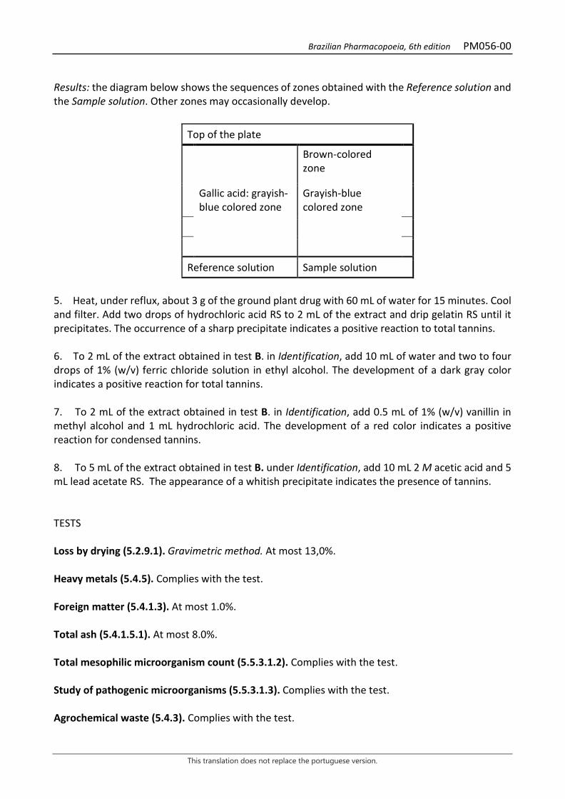
Brazilian Pharmacopoeia, 6th edition PM056-00
This translation does not replace the portuguese version.
Results: the diagram below shows the sequences of zones obtained with the Reference solution and the Sample solution. Other zones may occasionally develop.
Top of the plate
Brown-colored zone
Gallic acid: grayish-blue colored zone
Grayish-blue colored zone
Reference solution Sample solution
5. Heat, under reflux, about 3 g of the ground plant drug with 60 mL of water for 15 minutes. Cool and filter. Add two drops of hydrochloric acid RS to 2 mL of the extract and drip gelatin RS until it precipitates. The occurrence of a sharp precipitate indicates a positive reaction to total tannins. 6. To 2 mL of the extract obtained in test B. in Identification, add 10 mL of water and two to four drops of 1% (w/v) ferric chloride solution in ethyl alcohol. The development of a dark gray color indicates a positive reaction for total tannins. 7. To 2 mL of the extract obtained in test B. in Identification, add 0.5 mL of 1% (w/v) vanillin in methyl alcohol and 1 mL hydrochloric acid. The development of a red color indicates a positive reaction for condensed tannins. 8. To 5 mL of the extract obtained in test B. under Identification, add 10 mL 2 M acetic acid and 5 mL lead acetate RS. The appearance of a whitish precipitate indicates the presence of tannins. TESTS Loss by drying (5.2.9.1). Gravimetric method. At most 13,0%. Heavy metals (5.4.5). Complies with the test. Foreign matter (5.4.1.3). At most 1.0%. Total ash (5.4.1.5.1). At most 8.0%. Total mesophilic microorganism count (5.5.3.1.2). Complies with the test. Study of pathogenic microorganisms (5.5.3.1.3). Complies with the test. Agrochemical waste (5.4.3). Complies with the test.
Brazilian Pharmacopoeia, 6th edition PM056-00
This translation does not replace the portuguese version.
DOSAGE Total tannins To proceed as described in Visible absorption spectrophotometry (5.2.14). Prepare solutions as described below. Stock solution: accurately weigh approximately 0.500 g of the pulverized plant drug and transfer to a 250 mL, round-bottomed, polished-necked flask. Add 150 mL of water. Heat in a water bath, for 30 minutes, between 85 °C and 90 °C. Cool under running water and transfer to a 250-mL volumetric flask. Rinse the round-bottomed flask and transfer the washing water with the entire plant drug content to the volumetric flask. Top off the volume with water and homogenize. Allow to decant and filter the supernatant liquid through filter paper. Discard the first 50 mL of the filtrate. Sample solution for total polyphenol content: dilute 5 mL of the Stock solution in a 25-mL volumetric flask with water. Volumetrically transfer 2 mL of the solution, 1 mL of phosphomolybdotungstic reagent and 10 mL of water to a 25-mL volumetric flask, top off the volume with 29% sodium carbonate solution (w/v) and homogenize. Determine absorbance at 760 nm (A1) after 30 minutes, using water for zero adjustment. Sample solution for polyphenols not adsorbed by hide powder: for 10 mL of the Stock solution, add 0.1 g of the hide powder and mechanically shake in a 125-mL Erlenmeyer flask for 60 minutes. Filter through paper filter. Dilute 5 mL of the filtrate in a 25mL volumetric flask with water and homogenize. Volumetrically transfer 2 mL of the solution, 1 mL of phosphomolybdotungstic reagent and 10 mL of water to a 25-mL volumetric flask, top off the volume with 29% sodium carbonate solution (w/v) and homogenize. Determine absorbance at 760 nm (A2) after 30 minutes, using water for zero adjustment. Reference solution: dissolve 50 mg pyrogallol immediately before use in a 100-mL volumetric flask with water, top off the volume and homogenize. Volumetrically transfer 5 mL of this solution to a 100-mL volumetric flask, top off the volume with water and homogenize. Volumetrically transfer 2 mL of the solution, 1 mL of phosphomolybdotungstic reagent and 10 mL of distilled water to a 25mL volumetric flask, top off the volume with 29% sodium carbonate solution (w/v) and homogenize. Determine absorbance at 760 nm (A3) after 30 minutes, using water for zero adjustment. Calculate total tannin content expressed as pyrogallol, in percent, according to the following expression:
TT =(𝑇𝑇1 − 𝑇𝑇2) × 𝑚𝑚2 × 62,5
𝑇𝑇3 × 𝑚𝑚1
in which, TT = total tannin content expressed as pyrogallol % (w/w); A1 = absorbance measured for the Sample solution for total polyphenols; A2 = absorbance measured for the Sample solution for polyphenols not adsorbed by hide powder; A3 = absorbance measured for the Reference solution; m1 = mass in grams of the sample used, considering the loss by drying.
Brazilian Pharmacopoeia, 6th edition PM056-00
This translation does not replace the portuguese version.
m2 = mass in grams of pyrogallol, considering the purity of the reference substance. Gallic acid Proceed as described in High Performance Liquid Chromatography (5.2.17.4). Use a chromatograph equipped with an ultraviolet detector at 280 mm; pre-column packed with octadecylsilane silica, 250 mm long, 4.6 mm internal diameter column, packed with silica chemically bonded to an octadecylsilane group (5 µm), kept at 24 °C; Mobile phase flow rate of 0.8 mL/minute. Eluent (A): 0.05% trifluoroacetic acid (v/v). Eluent (B): 0.05% trifluoroacetic acid in methyl alcohol (v/v).
Time (minutes) Eluent A (%) Eluent B (%) Elution
0 - 10 80 → 77,5 20 → 22,5 linear gradient 10 - 20 77.5 → 60 22.5 → 40 linear gradient 20 - 25 60 → 25 40 → 75 linear gradient 25 - 28 25 → 80 75 → 20 linear gradient 28 - 32 80 20 isocratic Sample solution: accurately weigh approximately 4 g of the pulverized plant drug and transfer to a 250 mL, round-bottomed, polished-necked flask. Add 60 mL of water. Heat in a water bath, for 30 minutes, between 80 °C and 85 °C. Cool under running water and transfer to a 100-mL volumetric flask. Rinse the round-bottomed flask and transfer the washing water with the entire plant drug content to the volumetric flask. Top off the volume with water and homogenize. Allow to decant and filter the supernatant liquid through filter paper. Discard the first 20 mL of the filtrate. Then transfer 5,0 mL of the solution to a 10-mL volumetric flask, top off the volume with water and homogenize. Filter through a 0.45 µm filter unit. Reference solution: dissolve an accurately weighed amount of gallic acid in water to obtain a solution at 6.8 µg/mL. Filter through a 0.45 µm filter unit. Procedure: separately inject 20 μL of the Reference solution and 20 µL of the Sample solution. Register the chromatograms and measure the areas under the peaks. Calculate gallic acid content in percent, according to the expression:
𝑇𝑇𝑇𝑇 =𝐶𝐶𝑟𝑟 × 𝑇𝑇𝑎𝑎𝑇𝑇𝑟𝑟 × 𝑚𝑚
× 200 × 100
in which,
TA = gallic acid content % (w/w); Cr = concentration of gallic acid in the Reference solution in g/mL, considering the purity of the reference substance; Ar = area under the peak corresponding to the gallic acid in the Reference solution; Ar = area under the peak corresponding to the gallic acid in the Sample solution;
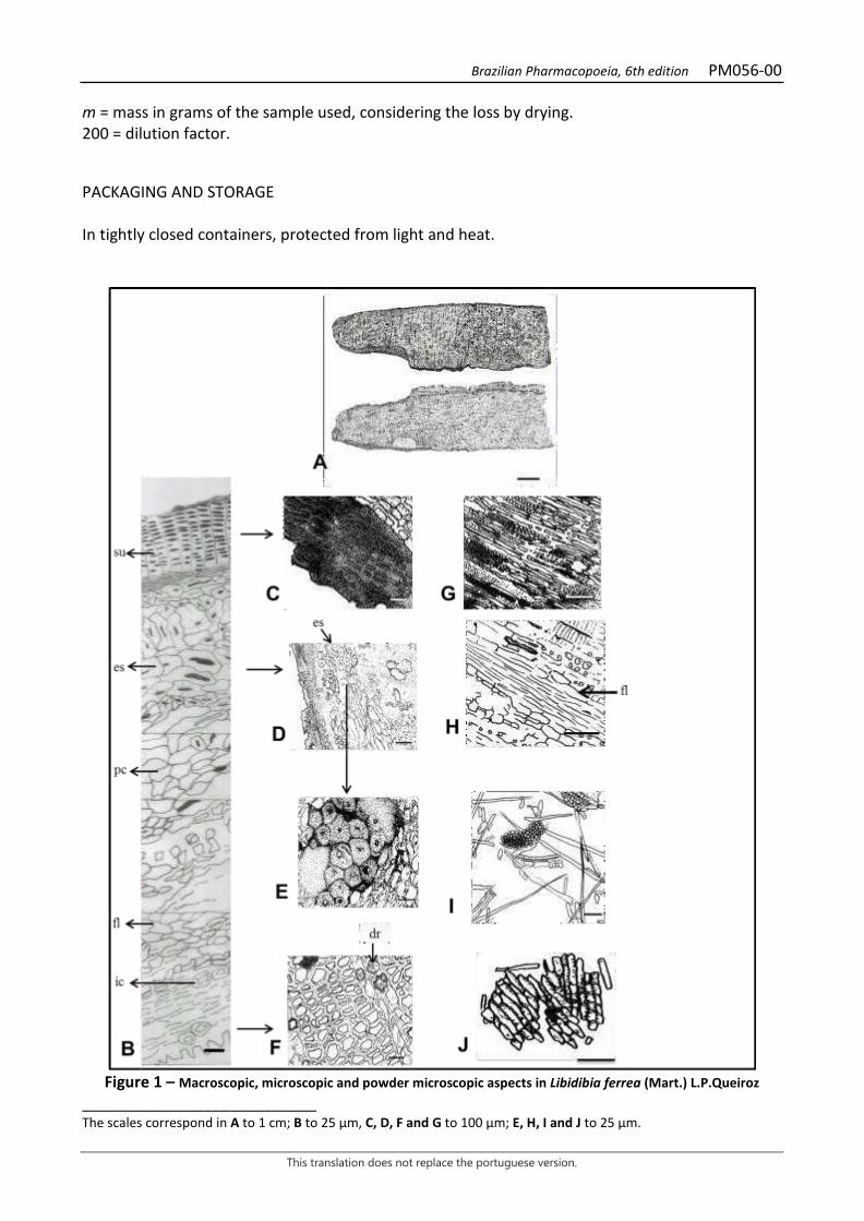
Brazilian Pharmacopoeia, 6th edition PM056-00
This translation does not replace the portuguese version.
m = mass in grams of the sample used, considering the loss by drying. 200 = dilution factor. PACKAGING AND STORAGE In tightly closed containers, protected from light and heat.
Figure 1 – Macroscopic, microscopic and powder microscopic aspects in Libidibia ferrea (Mart.) L.P.Queiroz
___________________________ The scales correspond in A to 1 cm; B to 25 µm, C, D, F and G to 100 µm; E, H, I and J to 25 µm.

Brazilian Pharmacopoeia, 6th edition PM056-00
This translation does not replace the portuguese version.
A - overall appearance of the stem bark, front and inside view, respectively. B - detail of the distribution of the tissues of the stem bark, in cross-ection: suber (su); cortical parenchyma (pc); sclereid (es); phloem (fl); idioblast containing prismatic crystal (ic). C - detail of the bark cross-section, showing the rhytidome with rowed tabular cells. D and E – appearance of sclereids distributed throughout the entire length of the cortical region. F - idioblasts containing druses (dr). G - in the phloem region, in longitudinal section, multiseriate ray cells, fibers and parenchymatic cord are apparent. H – detail of the phloem in longitudinal section: phloem (fl). I-J - details noted in the powder. I – fragments of fibers and parenchyma cells. J – fragments of parenchyma cells.
Brazilian Pharmacopoeia, 6th edition PM057-00
This translation does not replace the portuguese version.
BRAZILIAN IRONWOOD, oil Libidibiae fructus
The plant drug consists of dried fruit from the stems of Libidibia ferrea (Mart.) L.P. Queiroz (syn. Caesalpinia ferrea Mart.), containing not less than 9.0% total tannins and not less than 1.0% gallic acid (C7H6O5, 170.12). IDENTIFICATION 1. Macroscopic description Dried leguminous plants, dark brown in color, slightly reniform to oblong, flat, with slightly pointed endings, 6 to 8 cm long, 2 to 4 cm wide, and about 1 cm thick, containing one to three flat, hard, brown seeds. 2. Microscopic description In cross-section, the fruit pericarp shows a thick cuticle over a unistratified epidermis, with cells showing thickening in the outer periclinal wall, followed by a visible collenchyma, plus four to five layers of parenchyma and five to seven layers of sclerenchyma in the lower third of the fruit, close to the vascular bundles. Prismatic crystals were evident in the parenchyma. Rare macro-sclereids and branched sclereids were observed in the sclerenchyma layers. Several layers of parenchyma cells separate the vascular bundles, where a phloem zone is observed followed by xylem cells. In a paradermal section, in some regions of the epidermis a few simple, unicellular tector trichomes are visible; the cells of the epidermis have straight anticlinal walls and the stomata are of the cyclocytic type. 3. Microscopic description of powder The sample meets all requirements for the species, except the macroscopic characters. Characteristics are: brown color; enlarged macro-sclereids; bulky and branched sclereids; parenchymatic cell portions containing prismatic crystals; libriform sclerenchymatic fibers; vessel elements with scalariform and reticulate thickening, with simple, straight and oblique terminal walls, and with short and long extensions. 4. Proceed as described in Thin-layer chromatography (5.2.17.1). Stationary phase: 250 µm thick silica gel F254. Mobile phase: ethyl acetate, formic acid and water (90:5:5). Sample solution: weigh about 1 g of the drug and boil, under reflux, with 10 mL ethyl alcohol for 15 minutes. Filter after cooling through absorbent cotton. Reference solution: weigh approximately 1 mg gallic acid and dissolve in 1mL methyl alcohol.

Brazilian Pharmacopoeia, 6th edition PM057-00
This translation does not replace the portuguese version.
Procedure: apply 20 μL of the Sample solution and 20 μL of the Reference solution to the chromatoplate, separately and in the form of a band. Conduct chromatogram. Remove chromatoplate and allow it to air dry. Nebulize the plate with a 1% (w/v) ferric chloride solution. Results: the diagram below shows the sequences of zones obtained with the Reference solution and the Sample solution. Other zones may occasionally develop.
Top of the plate
Gallic acid: grayish-blue colored zone
Grayish-blue colored zone
Grayish-blue colored zone Grayish-blue colored zone
Grayish-blue colored zone
Reference solution Sample solution
5. Heat, under reflux, about 3 g of the ground plant drug with 60 mL of water for 15 minutes. Cool and filter. Add two drops of hydrochloric acid RS to 2 mL of the extract and drip gelatin RS until it precipitates. The occurrence of a sharp precipitate indicates a positive reaction to total tannins. 6. To 2 mL of the extract obtained in test B. in Identification, add 10 mL of water and two to four drops of 1% (w/v) ferric chloride solution in ethyl alcohol. The development of a dark gray color indicates a positive reaction for total tannins. 7. To 2 mL of the extract obtained in test B. in Identification, add 0.5 mL of 1% (w/v) vanillin in methyl alcohol and 1 mL hydrochloric acid. The development of a red color indicates a positive reaction for condensed tannins. 8. To 5 mL of the extract obtained in test B. under Identification, add 10 mL 2 M acetic acid and 5 mL lead acetate RS. The appearance of a whitish precipitate indicates the presence of tannins. TESTS Heavy metals (5.4.5). Complies with the test. Foreign matter (5.4.1.3). At most 1.0%. Loss by drying (5.2.9.1). Gravimetric method. At most 14.0%. Total ash (5.4.1.5.1). At most 4.0% Total mesophilic microorganism count (5.5.3.1.2). Complies with the test.
Brazilian Pharmacopoeia, 6th edition PM057-00
This translation does not replace the portuguese version.
Study of pathogenic microorganisms (5.5.3.1.3). Complies with the test. Agrochemical waste (5.4.3). Complies with the test. DOSAGE Total tannins To proceed as described in Visible absorption spectrophotometry (5.2.14). Prepare solutions as described below. Stock solution: accurately weigh approximately 0.50 g of the pulverized plant drug and transfer to a 250 mL, round-bottomed, polished-necked flask. Add 150 mL of water. Heat in a water bath, for 30 minutes, between 85 °C and 90 °C. Cool under running water and transfer to a 250-mL volumetric flask. Rinse the round-bottomed flask and transfer the washing water with the entire plant drug content to the volumetric flask. Top off the volume with water and homogenize. Allow to decant and filter the supernatant liquid through filter paper. Discard the first 50 mL of the filtrate. Sample solution for total polyphenol content: dilute 5 mL of the stock solution in a 25-mL volumetric flask with water and homogenize. Volumetrically transfer 2 mL of the solution, 1 mL of phosphomolybdotungstic reagent and 10 mL of water to a 25-mL volumetric flask, top off the volume with 29% sodium carbonate solution (w/v) and homogenize. Determine absorbance at 760 nm (A1) after 30 minutes, using water for zero adjustment. Sample solution for polyphenols not adsorbed by hide powder: for 10 mL of the Stock solution, add 0.1 g of the hide powder and mechanically shake in a 125-mL Erlenmeyer flask for 60 minutes. Filter through paper filter. Dilute 5 mL of the filtrate in a 25mL volumetric flask with water and homogenize. Volumetrically transfer 2 mL of the solution, 1 mL of phosphomolybdotungstic reagent and 10 mL of water to a 25-mL volumetric flask, top off the volume with 29% sodium carbonate solution (w/v) and homogenize. Determine absorbance at 760 nm (A2) after 30 minutes, using water for zero adjustment. Reference solution: dissolve 50 mg pyrogallol in water immediately before use, transfer to a 100-mL volumetric flask with water and homogenize. Volumetrically transfer 5 mL of this solution to a 100-mL volumetric flask, top off the volume with water and homogenize. Volumetrically transfer 2 mL of the solution, 1 mL of phosphomolybdotungstic reagent and 10 mL of water to a 25-mL volumetric flask, top off the volume with 29% sodium carbonate solution (w/v) and homogenize. Determine absorbance at 760 nm (A3) after 30 minutes, using water for zero adjustment. Calculate total tannin content expressed as pyrogallol, in percent, according to the following expression:
𝑇𝑇𝑇𝑇 =(𝑇𝑇1 − 𝑇𝑇2) × 𝑚𝑚2 × 62,5
𝑇𝑇3 × 𝑚𝑚1
in which,
Brazilian Pharmacopoeia, 6th edition PM057-00
This translation does not replace the portuguese version.
TT = total tannin content expressed as pyrogallol % (w/w); A1 = absorbance measured for the Sample solution for total polyphenols; A2 = absorbance measured for the Sample solution for polyphenols not adsorbed by hide powder; A3 = absorbance measured for the Reference solution; m1 = mass in grams of the sample used, considering the loss by drying. m2 = mass in grams of pyrogallol, considering the purity of the reference substance. Gallic acid Proceed as described in High Performance Liquid Chromatography (5.2.17.4). Use a chromatograph equipped with an ultraviolet detector at 280 mm; pre-column packed with octadecylsilane silica, 250 mm long, 4.6 mm internal diameter column, packed with silica chemically bonded to an octadecylsilane group (5 µm), kept at 24 °C; Mobile phase flow rate of 0.8 mL/minute. Eluent (A): 0.05% trifluoroacetic acid (v/v). Eluent (B): 0.05% trifluoroacetic acid in methyl alcohol (v/v).
Time (minutes) Eluent A (%) Eluent B (%) Elution
0 - 10 85 → 75 15 → 25 linear gradient 10 - 12.5 75 → 60 25 → 40 linear gradient 12.5 - 15 60 → 25 40 → 75 linear gradient 15 - 17.5 25 → 85 75 → 15 linear gradient 17.55 - 18 85 15 isocratic Sample solution: accurately weigh approximately 0,50 g of the pulverized plant drug and transfer to a 250 mL, round-bottomed, polished-necked flask. Add 150 mL of water. Heat in a water bath, for 30 minutes, between 85 °C and 90 °C. Cool under running water and transfer to a 250-mL volumetric flask. Rinse the round-bottomed flask and transfer the washing water with the entire plant drug content to the volumetric flask. Top off the volume with water and homogenize. Allow to decant and filter the supernatant liquid through filter paper. Discard the first 50 mL of the filtrate. Filter through a 0.45 µm filter unit. Reference solutions: dissolve an accurately weighed amount of gallic acid in water to obtain a solution at 25.0 µg/mL. Filter through a 0.45 µm filter unit. Procedure: separately inject 20 μL of the Reference solution and 20 µL of the Sample solution. Register the chromatograms and measure the areas under the peaks. Gallic acid retention time of in the sample is approximately 8 minutes and 30 seconds. Calculate gallic acid content in percent, according to the expression:
𝑇𝑇𝑇𝑇 =𝐶𝐶𝑟𝑟 × 𝑇𝑇𝑎𝑎𝑇𝑇𝑟𝑟 × 𝑚𝑚
× 250 × 100
in which,
Brazilian Pharmacopoeia, 6th edition PM057-00
This translation does not replace the portuguese version.
TA = gallic acid content % (w/w); Cr = concentration of gallic acid in the Reference solution in g/mL, considering the purity of the reference substance; Ar = area under the peak corresponding to the gallic acid in the Reference solution; Ar = area under the peak corresponding to the gallic acid in the Sample solution; m = mass in grams of the sample used, considering the loss by drying. 250 = dilution factor; PACKAGING AND STORAGE In tightly closed containers, protected from light and heat.
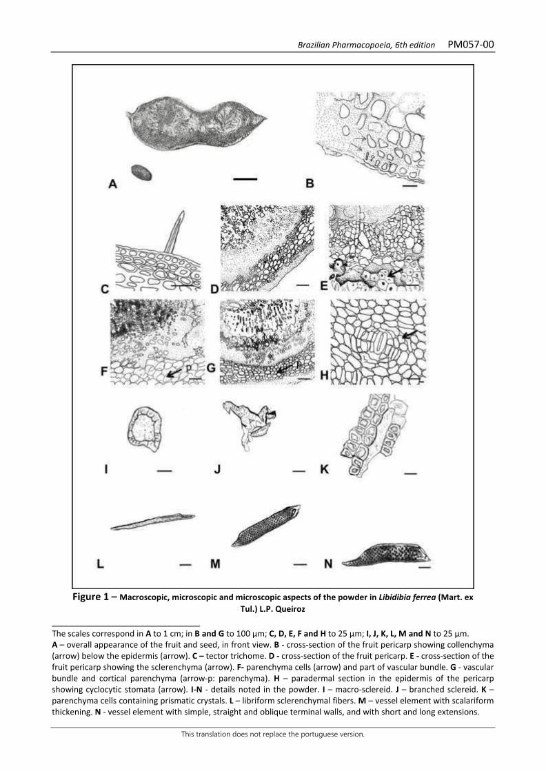
Brazilian Pharmacopoeia, 6th edition PM057-00
This translation does not replace the portuguese version.
Figure 1 – Macroscopic, microscopic and microscopic aspects of the powder in Libidibia ferrea (Mart. ex
Tul.) L.P. Queiroz ___________________________ The scales correspond in A to 1 cm; in B and G to 100 µm; C, D, E, F and H to 25 µm; I, J, K, L, M and N to 25 µm. A – overall appearance of the fruit and seed, in front view. B - cross-section of the fruit pericarp showing collenchyma (arrow) below the epidermis (arrow). C – tector trichome. D - cross-section of the fruit pericarp. E - cross-section of the fruit pericarp showing the sclerenchyma (arrow). F- parenchyma cells (arrow) and part of vascular bundle. G - vascular bundle and cortical parenchyma (arrow-p: parenchyma). H – paradermal section in the epidermis of the pericarp showing cyclocytic stomata (arrow). I-N - details noted in the powder. I – macro-sclereid. J – branched sclereid. K – parenchyma cells containing prismatic crystals. L – libriform sclerenchymal fibers. M – vessel element with scalariform thickening. N - vessel element with simple, straight and oblique terminal walls, and with short and long extensions.
Brazilian Pharmacopoeia, 6th edition PM058-00
This translation does not replace the portuguese version.
BITTER ORANGE, exocarp Aurantii amari exocarpium
The plant drug consists of dried portions of the exocarp of Citrus aurantium L. subsp. aurantium [syn. Citrus aurantium L. subsp. amara (L.) Engler], corresponding to the flavedo of the ripe fruit, free from most of the mesocarp, corresponding to the albedo, containing at least 2.0% volatile oil. CHARACTERISTICS The drug has a strong, aromatic, characteristic odor. IDENTIFICATION 1. Macroscopic description The exocarp consists of irregular portions up to 8 cm long and up to 4 cm wide. The external surface, in front view, is yellowish, yellowish-grizzly to yellowish-browm, roughly undulating and punctuated by numerous translucent secretory glands. The inner surface, in front view, is yellowish-white to whitish-grizzly, roughened, and spongy. In lateral view, the glands are visible in the form of cavities. 2. Microscopic description The flavedo is composed of the epidermis and adjacent parenchymatic tissues. The albedo is formed by the spongy parenchyma. The flavedo, in front view, has an epidermis with small cells of different shapes, with rectilinear anticlinal walls, containing lipid drops. Stomata are cyclocytic and situated slightly above the other cells. Secretory glands are visible by transparency. In cross-section, the cuticle is thick and smooth; the epidermis is formed by small, polygonal cells with dense protoplast, containing chromoplasts and lipid drops/subepidermically, there are four to five ocher-yellow, collenchymatous, compact layers, formed by small cells, with dense content, chromoplasts and lipid drops, below which there are larger parenchyma cells, with thinner walls, visible intercellular spaces, large amount of lipid drops and prismatic calcium oxalate mono-crystals, of different shapes and sizes. In the first layers of this parenchyma, schizolysigenous glands occur, circular to ovoid, up to 1.0 mm in diameter, at different stages of development and irregularly arranged. The parenchyma located laterally to the glands is formed by elongated, compact cells with a large amount of lipid drops and crystals. Small collateral vascular bundles are distributed in this tissue. Vessel elements with helical thickening are visible longitudinally. The innermost parenchyma is loose and consists of thin-walled hyaline cells of different shapes and sizes, containing mono-crystals. The parenchyma near the albedo has cells of greater volume, thicker walls, and fewer crystals. Hesperidin crystals are common in all parenchyma. The albedo consists of spongy parenchyma, with braciform cells, wide intercellular spaces, and few crystals and lipid drops. 3. Microscopic description of powder The sample meets all requirements for the subspecies, except the macroscopic characters. Ligh-grizzly colored powder. With the addition of chloral hydrate, the following characteristic arise: flavedo epidermis fragments with cells as described, in front view; flavedo epidermis fragments with
Brazilian Pharmacopoeia, 6th edition PM058-00
This translation does not replace the portuguese version.
stomata, in front view; flavedo fragments, in cross-section, showing epidermis and collenchymatous parenchyma; fragments of cholenchymatous parenchyma in cross-section; fragments of flavedo parenchyma with cells containing lipid drops in cross-section; fragments of flavedo parenchyma in cross-section containing lipid drops, calcium oxalate mono-crystals and hesperidin crystals; flavedo parenchyma fragments in cross-section with portions of vascular bundles, observed in longitudinal view; flavedo parenchyma fragments in cross-section containing lipid drops and hesperidin crystals; flavedo fragments with portions of secretory glands, in cross-section; flavedo parenchyma fragments in cross-section with portion of vascular bundle, observed in longitudinal view; parenchyma fragments with hesperidin crystals; crystalliferous flavedo idioblasts with calcium oxalate mono-crystals in cross-section; isolated calcium oxalate crystals; isolated hesperidin crystals, needle-shaped, only observed with the addition of lugol; portions of tracheal elements with helical thickening in longitudinal view; fragments of the albedo, in small quantity in cross- or longitudinal section. 4. Proceed as described in Thin-layer chromatography (5.2.17.1). Stationary phase: silica gel GF254 (0.250 mm). Mobile phase: ethyl acetate, formic acid and water (75:15:10). Sample solution: add 10 mL of methyl alcohol to 1 g of the pulverized plant drug (710 μm) (5.2.11). Heat in a water bath at about 60 °C for 10 minutes, while frequently shaking. Cool and filter. Reference solution: dissolve 1 μg naringin and 10.0 μg caffeic acid in 1 mL methyl alcohol. Procedure: apply 20 μL of the Sample solution and 10 μL of the Reference solution to the chromatoplate, separately and in the form of a band. Conduct chromatogram. Remove chromatoplate and allow it to air dry. Then nebulize the plate with 1% (w/v) SS aminoethanol diphenylborate (Natural Reagent A) in methyl alcohol. Examine under ultraviolet light at 365 nm. Results: the diagram below shows the sequences of zones obtained with the Reference solution and the Sample solution. Other zones may occasionally develop.

Brazilian Pharmacopoeia, 6th edition PM058-00
This translation does not replace the portuguese version.
Top of the plate
Light-blue fluorescence zone Light-blue fluorescence zone
Caffeic acid: light-blue fluorescence zone
Light-blue fluorescence zone Light-blue fluorescence zone
Naringin: intense-green fluorescence zone
Intense green fluorescence zone
Red fluorescence zone Orange fluorescence zone
Reference solution Sample solution TESTS Water (5.2.20.2). Azeotropic method. At most 10.0%. Determine in 20.0 g of the pulverized sample (355 μm) (5.2.11). Total ash (5.4.1.5.1). At most 7.0% Total mesophilic microorganism count (5.5.3.1.2). Complies with the test. Study of pathogenic microorganisms (5.5.3.1.3). Complies with the test. Heavy metals (5.4.5). Complies with the test. Agrochemical waste (5.4.3). Complies with the test. DOSAGE Volatile oils Proceed as described in Determining volatile oils in plant drugs (5.4.1.6). Use a 500-mL flask containing 200 mL of water as distillation liquid. Add 0.5 mL of xylene to the graduated tube. Use pulverized plant drug (710 µm) (5.2.11). Immediately proceed with the determination of the volatile

Brazilian Pharmacopoeia, 6th edition PM058-00
This translation does not replace the portuguese version.
oil, from 15 g of the powdered drug. Distill for 90 minutes. Measure the volume and express the yield per 100 g of the plant drug (w/p).

Brazilian Pharmacopoeia, 6th edition PM058-00
This translation does not replace the portuguese version.
PACKAGING AND STORAGE In tightly closed containers, protected from light and heat.
Figure 1 – Macroscopic and microscopic aspects in Citrus aurantium L. subsp. aurantium
___________________________ The scales correspond in A to 1 cm (level 1); in B to 0.5 cm (level 2); in C to 100 μm (level 3); in D to 100 μm (level 4). A – schematic illustration of the outer surface of the drug, in front view. B – schematic illustration of the drug, in cross-section: albedo (alb); flavedo (fl a); secretory gland (gla). C – detail of a portion of the flavedo epidermis, in front view: fundamental cell of the epidermis (cfe); stoma (es); lipid drop (gl). D – detail of a portion of the drug, in cross-section: albedo (alb); hesperidin crystal (ch); calcium oxalate crystal (cox); cuticle (cu); helical thickening vessel element (eh); intercellular space (ei); epidermis (ep); secretory epithelium (eps); stoma (es); phloem (f); flavedo (fl a); vascular bundle (fv); lipid drop (gl); secretory gland (gla); crystalliferous idioblast (ic); parenchyma (p); collenchymatous parenchyma (pco); spongy parenchyma (pj); xylem (x).
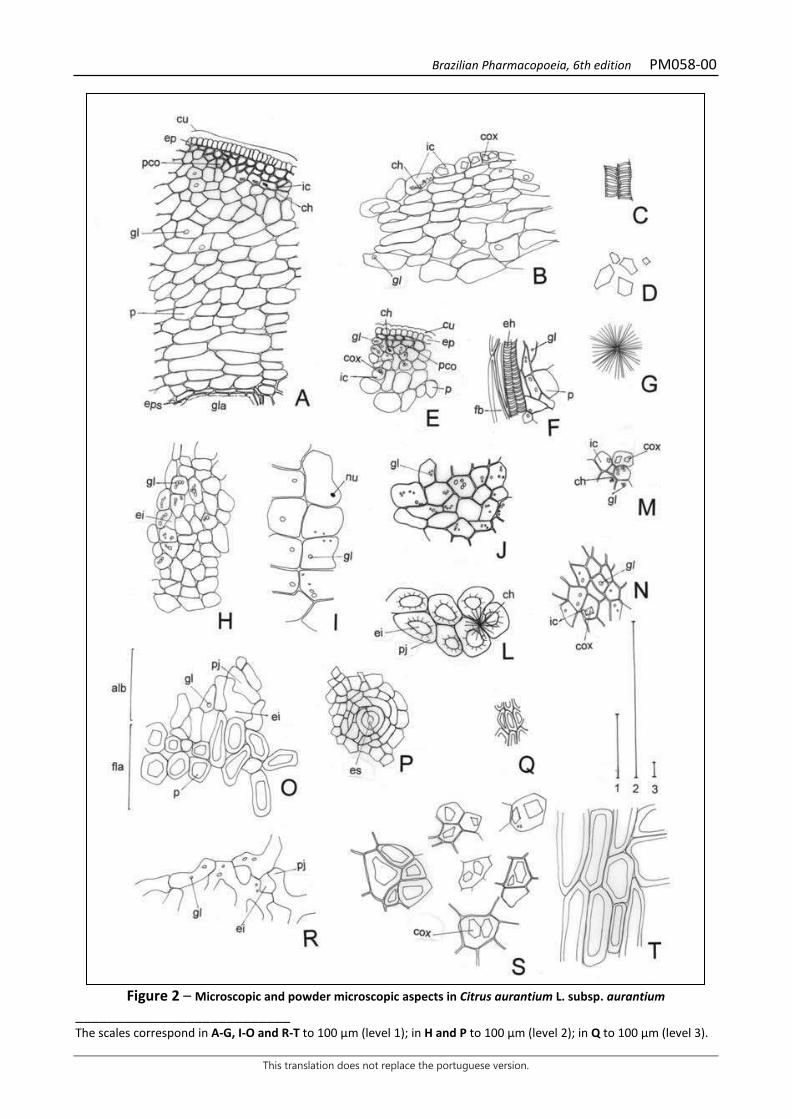
Brazilian Pharmacopoeia, 6th edition PM058-00
This translation does not replace the portuguese version.
Figure 2 – Microscopic and powder microscopic aspects in Citrus aurantium L. subsp. aurantium
___________________________ The scales correspond in A-G, I-O and R-T to 100 μm (level 1); in H and P to 100 μm (level 2); in Q to 100 μm (level 3).

Brazilian Pharmacopoeia, 6th edition PM058-00
This translation does not replace the portuguese version.
A – fragment of flavedo with epidermis, parenchyma and remnants of secretory epithelium, in cross-section: hesperidin crystal (ch); cuticle (cu); epidermis (ep); secretory epithelium (eps); lipid drop (gl); secretory gland (gla); crystalliferous idioblast (ic); parenchyma (p); collenchymatous parenchyma (pco). B – fragment of flavedo parenchyma in transverse section: hesperidin crystal (ch); calcium oxalate crystal (cox); lipid drop (gl); crystalliferous idioblast (ic). C – portion of tracheal elements with helical thickening, in longitudinal view. D – isolated calcium oxalate crystals. E – fragment of flavedo, in cross-section: hesperidin crystal (ch); calcium oxalate crystal (cox); cuticle (cu); epidermis (ep); lipid drop (gl); crystalliferous idioblast (ic); parenchyma (p); collenchymatous parenchyma (pco). F – flavedo parenchyma fragment in cross-section, with portion of vascular bundle, observed in longitudinal view: vessel element with helical thickening (eh); fiber (fb); lipid drop (gl); parenchyma (p). G – hesperidin crystal, only observed with the addition of lugol. H – flavedo parenchyma fragment in cross-section: intercellular space (ei); lipid drop (gl). I – fragment of flavedo parenchyma in cross-section, containing lipid drops: lipid drop (gl); nucleus (nu). J – front view fragment of the epidermis: lipid drop (gl). L – fragment of albedo, front view: hesperidin crystal (ch); intercellular space (ei); spongy parenchyma (pj). M – fragment of flavedo parenchyma in transverse section: hesperidin crystal (ch); calcium oxalate crystal (cox); lipid drop (gl); crystalliferous idioblast (ic). N – fragment of flavedo parenchyma in transverse section: calcium oxalate crystal (cox); lipid drop (gl); crystalliferous idioblast (ic). O – fragment of flavedo and albedo, in cross-section: albedo (alb); intercellular space (ei); flavedo (fl a); lipid drop (gl); parenchyma (p); spongy parenchyma (pj). P – fragment of the flavedo epidermis with stoma, in front view: stoma (es). Q – fragment of the cholenchymatous parenchyma, in cross-section. R – fragment of albedo, in cross-section: intercellular space (isp); lipid drop (gl); spongy parenchyma (pj). S – crystalliferous idioblasts of the flavedo, in cross-section: calcium oxalate crystal (cox). T – fragment of the flavedo, with thick-walled parenchyma cells, in cross-section.

Brazilian Pharmacopoeia, 6th edition PM059-00
This translation does not replace the portuguese version.
MACELA, flower Achyroclines flos
The plant drug consists of dried inflorescences of Achyrocline satureioides (Lam.) DC., containing not less than 3.0% total flavonoids calculated as quercetin, not less than 0.8% quercetin (C15H10O7, 302.24), and not less than 0.6% 3-O-methylquercetin (C16H12O7, 316.27). CHARACTERISTICS The inflorescences have a color ranging from pale yellow to intense golden yellow and a characteristic aromatic odor. IDENTIFICATION 1. Macroscopic description The drug consists of flowers gathered in capitula grouped into glomeruli, which in turn are organized into panicle-shaped tops. Each capitulum presents four to eight dimorphic flowers, protected by a subcylindrical involucre, 4 to 7 mm high, formed by nine to 14 scarious, hyaline, navicular, imbricate involucral bracts, arranged in three or four series, with yellow, light yellow, pale yellow to greenish, or even golden yellow, grizzly-yellow to reddish yellow color. Outer bracts 2.5 to 3 mm long; middle bracts 3.5 to 4.5 mm long; inner bracts 3 to 7 mm long, all with simple, woolly, 2 to 3 mm long tector trichomes and/or glandular trichomes only on their outer lower third. Three to six pistillate marginal flowers, with filiform corolla, 3 to 4.5 mm long, dentate or broken at the apex, with glandular trichomes in the outer apical portion; filiform, bifid, glabrous, dilated stylet near the base, with stigmatic branches usually exserted at maturity, truncate apex, papillose and with a crown of trichomes in the apical portion; infertile, bicarpellate and unilocular, monospermic ovary; uniseriate papus, with about 20 white, rough bristles, free from each other at the base, reaching almost the same height as the corolla, rarely more. One to three flowers from the disk, perfect, with narrow tubular corolla, 3 to 4.5 mm long; tube slightly dilated at the base and pentadentate limb, teeth with glandular trichomes in the external face; androceu with five epipetal stamens, inserted in the inferior half of the corolla, with synanther anthers, 1.5 to 2 mm long, with longitudinal and introrse dehiscence, sagittal at the base, with two lacinated tails, one on each side; connective prolonged in a triangular, slightly obtuse, hyaline apical appendage; ovary, stylet and papus similar to pistillate flowers. Light brown or grizzly watery fruit, 0.7 to 0.8 mm long, ellipsoidal to obovate, slightly compressed, glabrous, papillose surface. 2. Microscopic description The abaxial surface of the bracts has an epidermis formed by elongated, rectangular-shaped cells. In the lower third, pluricellular and uniseriate tector and/or glandular trichomes occur, formed by a two- to three-layered pedicel with three or four layers of cells and by two oval-long terminal cells, much larger than the previous ones. The glandular trichomes of the bracts measure 60 to 100 µm in total length and their head has a diameter of 30 to 40 µm. The papus is made up of long bristles, formed by elongated, hyaline, thin-walled cells, many of which project laterally. The epidermis of the corolla’s abaxial surface is formed by elongated cells with a polygonal outline. Five vascular
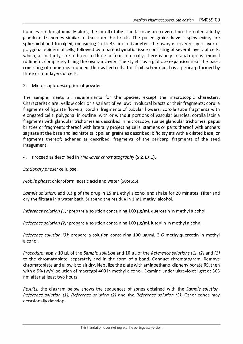
Brazilian Pharmacopoeia, 6th edition PM059-00
This translation does not replace the portuguese version.
bundles run longitudinally along the corolla tube. The laciniae are covered on the outer side by glandular trichomes similar to those on the bracts. The pollen grains have a spiny exine, are spheroidal and tricolped, measuring 17 to 35 µm in diameter. The ovary is covered by a layer of polygonal epidermal cells, followed by a parenchymatic tissue consisting of several layers of cells, which, at maturity, are reduced to three or four. Internally, there is only an anatropous seminal rudiment, completely filling the ovarian cavity. The stylet has a globose expansion near the base, consisting of numerous rounded, thin-walled cells. The fruit, when ripe, has a pericarp formed by three or four layers of cells. 3. Microscopic description of powder The sample meets all requirements for the species, except the macroscopic characters. Characteristic are: yellow color or a variant of yellow; involucral bracts or their fragments; corolla fragments of ligulate flowers; corolla fragments of tubular flowers; corolla tube fragments with elongated cells, polygonal in outline, with or without portions of vascular bundles; corolla lacinia fragments with glandular trichomes as described in microscopy; sparse glandular trichomes; papus bristles or fragments thereof with laterally projecting cells; stamens or parts thereof with anthers sagitate at the base and laciniate tail; pollen grains as described; bifid stylets with a dilated base, or fragments thereof; achenes as described; fragments of the pericarp; fragments of the seed integument. 4. Proceed as described in Thin-layer chromatography (5.2.17.1). Stationary phase: cellulose. Mobile phase: chloroform, acetic acid and water (50:45:5). Sample solution: add 0.3 g of the drug in 15 mL ethyl alcohol and shake for 20 minutes. Filter and dry the filtrate in a water bath. Suspend the residue in 1 mL methyl alcohol. Reference solution (1): prepare a solution containing 100 µg/mL quercetin in methyl alcohol. Reference solution (2): prepare a solution containing 100 µg/mL luteolin in methyl alcohol. Reference solution (3): prepare a solution containing 100 µg/mL 3-O-methylquercetin in methyl alcohol. Procedure: apply 10 μL of the Sample solution and 10 μL of the Reference solutions (1), (2) and (3) to the chromatoplate, separately and in the form of a band. Conduct chromatogram. Remove chromatoplate and allow it to air dry. Nebulize the plate with aminoethanol diphenylborate RS, then with a 5% (w/v) solution of macrogol 400 in methyl alcohol. Examine under ultraviolet light at 365 nm after at least two hours. Results: the diagram below shows the sequences of zones obtained with the Sample solution, Reference solution (1), Reference solution (2) and the Reference solution (3). Other zones may occasionally develop.

Brazilian Pharmacopoeia, 6th edition PM059-00
This translation does not replace the portuguese version.
Top of the plate
Luteolin: orange fluorescence zone
Orange fluorescence zone
3-O-Methylquercetin: yellow fluorescence zone
Yellow fluorescence zone
Quercetin: orange fluorescence zone
Orange fluorescence zone
Reference solution Sample solution 5. Proceed as described in Thin-layer chromatography (5.2.17.1). Stationary phase: silica gel GF254. Mobile phase: ethyl acetate, methyl alcohol, water (100: 17:10). Sample solution: shake 0.1 g of the drug in 15 mL ethyl alcohol for 20 minutes. Filter and dry the filtrate in a water bath. Suspend the residue in 1 mL methyl alcohol. Reference solution: prepare a solution containing 200 µg/mL chlorogenic acid in methyl alcohol. Procedure: apply 10 μL of the Sample solution and 10 μL of the Reference solution to the chromatoplate, separately and in the form of a band. Conduct chromatogram. Remove chromatoplate and allow it to dry. Nebulize the plate with aminoethanol diphenylborate RS, then with a 5% (w/v) solution of macrogol 400 in methyl alcohol. Examine under ultraviolet light at 365 nm. Results: the diagram below shows the sequences of zones obtained with the Reference solution and the Sample solution. Other zones may occasionally develop.

Brazilian Pharmacopoeia, 6th edition PM059-00
This translation does not replace the portuguese version.
Top of the plate
Brown-colored zone Intense yellow fluorescence zone Blue-colored zone
Yellow fluorescence zone Yellow fluorescence zone Yellow fluorescence zone Brown-colored zone
Chlorogenic acid: blueish fluorescence zone
Blueish fluorescence zone Yellow-colored zone Blueish fluorescence zone
Reference solution Sample solution TESTS Heavy metals (5.4.5). Complies with the test. Foreign matter (5.4.1.3). At most 2.0%. The presence of inflorescence peduncles and pedicels is allowed, in a length of up to 3 cm and corresponding to a maximum value of 1.0% of the dry weight of the ensemble. Loss by drying (5.2.9.1). Gravimetric method. At most 12.5%. Total ash (5.4.1.5.1). At most 6.0%. Total mesophilic microorganism count (5.5.3.1.2). Complies with the test. Study of pathogenic microorganisms (5.5.3.1.3). Complies with the test. Agrochemical waste (5.4.3). Complies with the test. DOSAGE Total flavonoids To proceed as described in Visible absorption spectrophotometry (5.2.14). Prepare solutions as described below.
Brazilian Pharmacopoeia, 6th edition PM059-00
This translation does not replace the portuguese version.
Stock solution: accurately weigh approximately 0.1 g of the pulverized plant drug (800 µm) (5.2.11) and place in a 100mL round-bottomed, polished-necked flask. Add 15 mL of 80% (v/v) ethyl alcohol and heat in a water bath at 90 °C, under reflux, for 15 minutes. After cooling, filter through a small portion of absorbent cotton into a 25-mL volumetric flask. Return the drug residue and cotton to the same round-bottomed flask, add 10 mL of 80% (v/v) ethyl alcohol. Heat, under reflux, for 15 minutes. Filter into the same 25-mL volumetric flask. After cooling down to room temperature, top off the volume to 25 mL with 80% (v/v) ethyl alcohol and homogenize. Dilute a 10-mL aliquot of this solution in a 25-mL volumetric flask and top off the volume with 80% (v/v) ethyl alcohol. Sample solution: transfer 10 mL of the Stock solution into a 25-mL volumetric flask, add 1 mL of 2% (w/v) aluminum chloride solution in 80% (v/v) ethyl alcohol, top off the volume with the same solvent and homogenize. Blank solution: transfer 10 mL of the stock solution into a 25-mL volumetric flask, top off the volume with the 80% (v/v) ethyl alcohol and homogenize. Procedure: measure the absorbance of the Sample solution at 420 nm 30 minutes after its preparation, using the Blank solution for zero adjustment. Calculate total flavonoid content expressed as hyperoside, in percent, according to the following expression:
𝑇𝑇𝑇𝑇 =𝑇𝑇 × 𝑇𝑇𝑇𝑇𝑚𝑚 × 561
in which, TF = total flavonoid content expressed as quercetin % (w/w); A = absorbance measured for the Sample solution; FD = dilution factor; 561 = quercetin specific absorption coefficient; m = mass in grams of the sample used, considering the loss by drying. Quercetin and 3-O-methylquercetin Proceed as described in High Performance Liquid Chromatography (5.2.17.4). Use a chromatograph equipped with an ultraviolet detector at 357 nm for 3-O-methylquercetin and 371 nm for quercetin, pre-column packed with octadecylsilane silica, a 250mm long, 4.6 mm internal diameter column, packed with octadecylsilane silica (5 µm), kept at 22 °C; Mobile phase flow rate of 0.6 mL/minute. Eluent (A): water and trifluoroacetic acid (100:0.006). Eluent (B): acetonitrile.
Time (minutes) Eluent A (%) Eluent B (%) Elution
0 - 5 72 → 65 28 → 35 linear gradient 5 - 13 65 35 isocratic

Brazilian Pharmacopoeia, 6th edition PM059-00
This translation does not replace the portuguese version.
13 - 18 65 → 40 35 → 60 linear gradient 18 - 20 40 → 30 60 → 70 linear gradient 20 - 25 30 → 72 70 → 28 linear gradient 25 - 30 72 28 isocratic Sample solution: accurately weigh approximately 0.15 g of the dried and pulverized plant drug (850 µm) (5.2.11) in a 100mL round-bottomed flask. Add 15 mL of 80% (v/v) ethyl alcohol and reflux in a 90°C water bath for 30 minutes. Allow to cool to room temperature. Filter extract through absorbent cotton into a 25-mL volumetric flask. Return the cotton and the drug residue to the same round-bottomed flask and extract again, under reflux, with another 10 mL of 80% (v/v) ethyl alcohol for 15 minutes. Cool and filter into the same 25 mL volumetric flask, top off the volume with 80% (v/v) ethyl alcohol and homogenize. Filter through a 0.45 µm filter unit. Reference solution (1): dissolve an accurately weighed amount of quercetin in methyl alcohol to obtain a 54 µg/mL solution. Filter through a 0.45 µm filter unit. Reference solution (2): dissolve exactly weighed quantity of 3-O-methylquercetin in methyl alcohol to obtain a 40 µg/mL solution. Filter through a 0.45 µm filter unit. Procedure: separately inject 10µL of the Reference solution (1), 10µL of the Reference solution (2) and 10µL of the Sample solution. Register the chromatograms and measure the areas under the peaks. The retention times for quercetin and 3-O-methylquercetin are about 18 and 19 minutes, respectively. Calculate the quercetin and 3-O-methylquercetin content separately, in percent, considering the respective Reference solutions, according to the following expression:
𝑇𝑇𝑇𝑇 =𝐶𝐶𝑟𝑟 × 𝑇𝑇𝑎𝑎 × 25 × 100
𝑇𝑇𝑟𝑟 × 𝑚𝑚
in which, TQ = quercetin or 3-O-methylquercetin content % (w/w); Cr = concentration of quercetin or 3-O-methylquercetin in the Reference solution in g/mL, taking into account the purity of the reference substance; Ar = area under the peak corresponding to quercetin or 3-O-methylquercetin in the Reference solution; Aa = area under the peak corresponding to quercetin or 3-O-methylquercetin in the Sample solution; m = mass in grams of the sample used, considering the loss by drying. PACKAGING AND STORAGE In tightly closed containers, protected from light and heat.

Brazilian Pharmacopoeia, 6th edition PM059-00
This translation does not replace the portuguese version.
Figure 1 – Macroscopic and microscopic aspects in Achyrocline satureioides (Lam.) DC.
___________________________ The scales correspond in A to 5 mm, B, C, D, E, F, H and I to 1 mm, G to 100 µm, J, K and L to 200 µm, M to 300 µm, N and O to 50 µm. A - overall appearance of an inflorescence. B - appearance of a capitulum in side view. C and D - pistillate flowers in lateral view. E - perfect flower with papus bristles in lateral view. F - appearance of the external bract of the capitulum. G - detail of the bract parenchyma, as indicated in F. H - appearance of the median bract of the capitulum. I - appearance of the internal bract of the capitulum. J to L - apical portion of the tubular corolla, showing the variability in number and size of the glandular trichomes. M - overall appearance of the corolla venation. N - detail of the venation on the apical portion of the corolla, as indicated in M. O - detail of the venation in the basal portion of the corolla, as indicated in M.
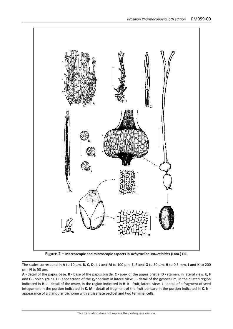
Brazilian Pharmacopoeia, 6th edition PM059-00
This translation does not replace the portuguese version.
Figure 2 – Macroscopic and microscopic aspects in Achyrocline satureioides (Lam.) DC.
___________________________ The scales correspond in A to 10 µm, B, C, D, I, L and M to 100 µm, E, F and G to 30 µm, H to 0.5 mm, J and K to 200 µm, N to 50 µm. A - detail of the papus base. B - base of the papus bristle. C - apex of the papus bristle. D - stamen, in lateral view. E, F and G - polen grains. H - appearance of the gynoecium in lateral view. I - detail of the gynoecium, in the dilated region indicated in H. J - detail of the ovary, in the region indicated in H. K - fruit, lateral view. L - detail of a fragment of seed integument in the portion indicated in K. M - detail of fragment of the fruit pericarp in the portion indicated in K. N - appearance of a glandular trichome with a triseriate pedicel and two terminal cells.
Brazilian Pharmacopoeia, 6th edition PM060-00
This translation does not replace the portuguese version.
COMMON MALLOW, flower Malvae flos
The plant drug consists of the dried, entire or fragmented flowers of Malva sylvestris L. or its cultivated varieties. IDENTIFICATION 1. Macroscopic description Actinomorph flowers, 3 to 6 cm wide when opened; calyx formed by three greenish bracts, pilous, elliptic, up to 7 mm long; gamosepalous calyx at the base, formed by five triangular, pilous, greenish sepals; corolla three to four times larger than the calyx, with five cuneiform petals, each petal with evident dark venation, violet or pinkish petals when fresh and dark violet when dry; several stamens, welded by the filaments forming a staminal tube attached to the base of the petals, densely covered with tector and glandular trichomes, the anthers are monothecate and free; ovary externally hairy, with several carpels, united style, surrounded by the staminal tube and free and capitate stigmas. Schizocarp fruit, rarely present. 2. Microscopic description In cross-section, the bracteoles, sepals and petals have uni-stratified epidermis, with simple, unicellular curved-tip and stellate tector trichomes with two to six cells with thickened walls, besides glandular trichomes, formed by a basal cell, two cells at the foot and a pluricellular secretory head, uniseriate; anomocytic stomata are found on the abaxial surface of the epidermis of the bracteoles and sepals; in the parenchyma of the sepals idioblasts containing calcium oxalate druses occur; mesophyll of the petals with large mucilage-containing idioblasts; anthers with papillose epidermis, globose pollen, with spiny exine, yellowish and with 110 to 160 µm in diameter; the parenchyma of the ovary has idioblasts with druses and mucilaginous cells. 3. Microscopic description of powder The sample meets all requirements for the species, except the macroscopic characters. Characteristics are: portions of epidermis of bracteoles with anomocytic stomata; portions of epidermis of sepals with anomocytic stomata; portions of epidermis of bracteoles, sepals and petals with different types of trichomes; parenchyma fragments with calcium oxalate druses; portions of petal tissue cells containing mucilaginous idioblasts; anther fragments; remains of anther dehiscence tissue; yellowish pollen grains with spiny exine. 4. Proceed as described in Thin-layer chromatography (5.2.17.1). Stationary phase: 250 µm thick silica gel F254. Mobile phase: butyl alcohol, water and acetic acid (60:30:15). Sample solution: weigh 1.0 g of the plant drug, add 10 mL of 60% (v/v) ethyl alcohol and mechanically shake for 15 minutes. Vaccum filter and dry the extract until residue is formed, at a maximum
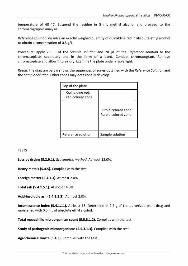
Brazilian Pharmacopoeia, 6th edition PM060-00
This translation does not replace the portuguese version.
temperature of 60 °C. Suspend the residue in 5 mL methyl alcohol and proceed to the chromatographic analysis. Reference solution: dissolve an exactly weighed quantity of quinaldine red in absolute ethyl alcohol to obtain a concentration of 0.5 g/L. Procedure: apply 20 μL of the Sample solution and 20 μL of the Reference solution to the chromatoplate, separately and in the form of a band. Conduct chromatogram. Remove chromatoplate and allow it to air dry. Examine the plate under visible light. Result: the diagram below shows the sequences of zones obtained with the Reference Solution and the Sample Solution. Other zones may occasionally develop.
Top of the plate
Quinaldine red: red-colored zone
Purple-colored zone Purple-colored zone
Reference solution Sample solution
TESTS Loss by drying (5.2.9.1). Gravimetric method. At most 12.0%. Heavy metals (5.4.5). Complies with the test. Foreign matter (5.4.1.3). At most 2.0%. Total ash (5.4.1.5.1). At most 14.0%. Acid-insoluble ash (5.4.1.5.3). At most 2.0%. Intumescence index (5.4.1.11). At least 15. Determine in 0.2 g of the pulverized plant drug and moistened with 0.5 mL of absolute ethyl alcohol. Total mesophilic microorganism count (5.5.3.1.2). Complies with the test. Study of pathogenic microorganisms (5.5.3.1.3). Complies with the test. Agrochemical waste (5.4.3). Complies with the test.
Brazilian Pharmacopoeia, 6th edition PM060-00
This translation does not replace the portuguese version.
PACKAGING AND STORAGE In tightly closed containers, protected from light and heat.
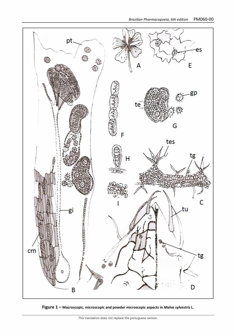
Brazilian Pharmacopoeia, 6th edition PM060-00
This translation does not replace the portuguese version.
Figure 1 – Macroscopic, microscopic and powder microscopic aspects in Malva sylvestris L.
Brazilian Pharmacopoeia, 6th edition PM060-00
This translation does not replace the portuguese version.
___________________________ A - overall appearance of the flower. B - fragment of flower, in longitudinal view, showing the mucilaginous cells (cm) in the epidermis of the petal (pt), gynoecium (gi) with united stylets and separated stigmas, anthers with the thecae and pollen grains. C-D - calyx; C - transverse section of the sepal with glandular trichomes (tg) and stellate trichome (tes); D – apical fragment of the sepal, in front view, with glandular trichomes (tg) and simple unicellular curved trichomes (tu); E - front view of fragment of the bract epidermis with anomocytic stomata (es); F - anther dehiscence mechanical tissue; G - monotheca anthers (te) and pollen grains (gp); H - uniseriate glandular trichome of the corolla; I – detail of a fragment of the parenchyma of sepals and bracteoles containing calcium oxalate druses.
Brazilian Pharmacopoeia, 6th edition PM061-01
This translation does not replace the portuguese version.
PASSION FRUIT, leaf Passiflorae acetum folium
The plant drug consists of dried leaves of Passiflora edulis Sims, containing at least 1.0% total flavonoid content expressed as apigenin (C15H10O5, 270,24). IDENTIFICATION 1. Macroscopic description Simple, glabrous, subcoriaceous, light-green leaves. Laminae deeply divided into three lobes, very rarely bilobed or lobeless, 7 to 16 cm long and 6 to 20 cm wide; base reentrant, acuminate apex and serrate margin. Palmatinerveal nerve, with tector trichomes on the midrib of the abaxial surface. Petiole 1 to 4 cm long, canaliculate on top, with a pair of extrafloral nectaries. Tendrils are common on the petiole. It differs from Passiflora alata in that it has an entire leaf, smooth margin, peninerve venation, and no tector trichomes in the midrib region. 2. Microscopic description Dorsiventrally symmetrical, hypoanphistomatic leaves. The epidermis, in front view, has polyhedral-shaped cells, with slightly indented anticlinal walls on both sides. The cuticle is smooth. Stomata are paracytic, anisocytic and anomocytic. Unicellular tector trichomes appear in the region of the midrib, on the abaxial surface. In cross-section, the cuticle is thick, the epidermis is unistratified, and the mesophyll consists of one to three layers of palisade parenchyma and several layers of spongy parenchyma. Calcium oxalate druses occur in the parenchyma. In the midrib, in cross-section, the adaxial surface presents a protuberance and the abaxial surface is convex. The epidermis, in the region of the protuberance, has unicellular tector trichomes. Under both epidermis, collenchyma cells interrupt the chlorophyll parenchyma. The vascular system consists of four centrally arranged vascular bundles. Druses occur in the inner portion of the phloem. The petiole, in cross-section, presents two slightly prominent lobes on the adaxial surface, and the abaxial surface is slightly convex in the central region. The internal portion of the epidermis is filled with collenchyma and the rest with parenchyma. The vascular system is formed by a vascular bundle in each lobe of the adaxial surface and by a group of central bundles, with an annular arrangement. Idioblasts with druses appear in the phloem, in smaller numbers, in the parenchyma and collenchyma. 3. Microscopic description of powder The sample meets all requirements for the species, except the macroscopic characters. Characteristics are: yellowish-green coloration; fragments of epidermis of the adaxial surface with cells as the ones described, without stomata; fragments of epidermis of the abaxial surface with cells as the ones described, with stomata as described; fragments of epidermis over the vein showing unicellular tector trichomes; fragments of vascular tissue in transversal or longitudinal sections, with idioblasts containing druses; isolated druses; fragments of palisade and spongy tissue with rare druses. 4. Proceed as described in Thin-layer chromatography (5.2.17.1).
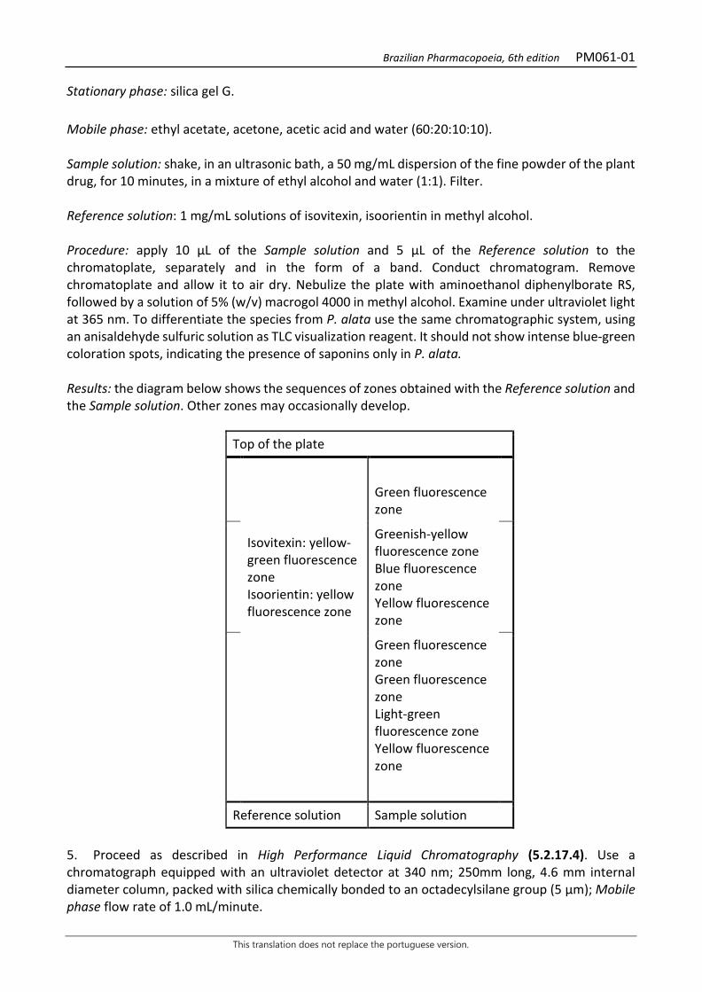
Brazilian Pharmacopoeia, 6th edition PM061-01
This translation does not replace the portuguese version.
Stationary phase: silica gel G. Mobile phase: ethyl acetate, acetone, acetic acid and water (60:20:10:10). Sample solution: shake, in an ultrasonic bath, a 50 mg/mL dispersion of the fine powder of the plant drug, for 10 minutes, in a mixture of ethyl alcohol and water (1:1). Filter. Reference solution: 1 mg/mL solutions of isovitexin, isoorientin in methyl alcohol. Procedure: apply 10 μL of the Sample solution and 5 μL of the Reference solution to the chromatoplate, separately and in the form of a band. Conduct chromatogram. Remove chromatoplate and allow it to air dry. Nebulize the plate with aminoethanol diphenylborate RS, followed by a solution of 5% (w/v) macrogol 4000 in methyl alcohol. Examine under ultraviolet light at 365 nm. To differentiate the species from P. alata use the same chromatographic system, using an anisaldehyde sulfuric solution as TLC visualization reagent. It should not show intense blue-green coloration spots, indicating the presence of saponins only in P. alata. Results: the diagram below shows the sequences of zones obtained with the Reference solution and the Sample solution. Other zones may occasionally develop.
Top of the plate
Green fluorescence zone
Isovitexin: yellow-green fluorescence zone Isoorientin: yellow fluorescence zone
Greenish-yellow fluorescence zone Blue fluorescence zone Yellow fluorescence zone
Green fluorescence zone Green fluorescence zone Light-green fluorescence zone Yellow fluorescence zone
Reference solution Sample solution
5. Proceed as described in High Performance Liquid Chromatography (5.2.17.4). Use a chromatograph equipped with an ultraviolet detector at 340 nm; 250mm long, 4.6 mm internal diameter column, packed with silica chemically bonded to an octadecylsilane group (5 µm); Mobile phase flow rate of 1.0 mL/minute.
Brazilian Pharmacopoeia, 6th edition PM061-01
This translation does not replace the portuguese version.
Eluent (A): 0.05% phosphoric acid (v/v). Eluent (B): acetonitrile.
Time (minutes) Eluent A (%) Eluent B (%) Elution
0 – 15 90 → 80 10 → 20 linear gradient 15 – 30 80 20 isocratic Sample solution: accurately weigh approximately 0.5 g of the dried and pulverized plant drug (180 µm) (5.2.11) and place in a 50-mL volumetric flask. Add approximately 30 mL of ethyl alcohol and water solution (1:1), shake ultrasonically for 10 minutes, and top off the volume with the same solvent. Homogenize and filter the extract on filter paper. Concentrate the extract under reduced pressure and suspend it in a methyl alcohol and water mixture (1:1) at a 2 mg/mL concentration. Filter through a 0.45 µm filter unit. Reference solution (1): quantitatively transfer 1 mg isovitexine accurately weighed to a 10mL volumetric flask and add approximately 7 mL ethyl alcohol and water solution (1:1). Homogenize in an ultrasonic bath for 10 minutes. Top off the volume with the same solution and homogenize. Filter through a 0.45 µm filter unit. Reference solution (2): quantitatively transfer 1 mg isoorientine accurately weighed to a 10mL volumetric flask and add approximately 7 mL ethyl alcohol and water solution (1:1). Homogenize in an ultrasonic bath for 10 minutes. Top off the volume with the same solution and homogenize. Filter through a 0.45 µm filter unit. Procedure: separately inject 20µL of the Reference solution (1), 20µL of the Reference solution and 20µL of the Sample solution. The chromatogram of the sample solution must show three major peaks of similar intensity, being identified those with relative retention times of 1 and 1.22 for isoorientin and isovitexin, respectively; it also shows an additional rather intense not identified peak at 0.80.
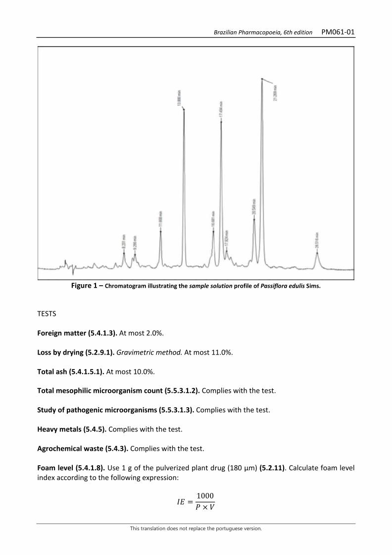
Brazilian Pharmacopoeia, 6th edition PM061-01
This translation does not replace the portuguese version.
Figure 1 – Chromatogram illustrating the sample solution profile of Passiflora edulis Sims.
TESTS Foreign matter (5.4.1.3). At most 2.0%. Loss by drying (5.2.9.1). Gravimetric method. At most 11.0%. Total ash (5.4.1.5.1). At most 10.0%. Total mesophilic microorganism count (5.5.3.1.2). Complies with the test. Study of pathogenic microorganisms (5.5.3.1.3). Complies with the test. Heavy metals (5.4.5). Complies with the test. Agrochemical waste (5.4.3). Complies with the test. Foam level (5.4.1.8). Use 1 g of the pulverized plant drug (180 μm) (5.2.11). Calculate foam level index according to the following expression:
𝐼𝐼𝑇𝑇 =1000𝑃𝑃 × 𝑉𝑉
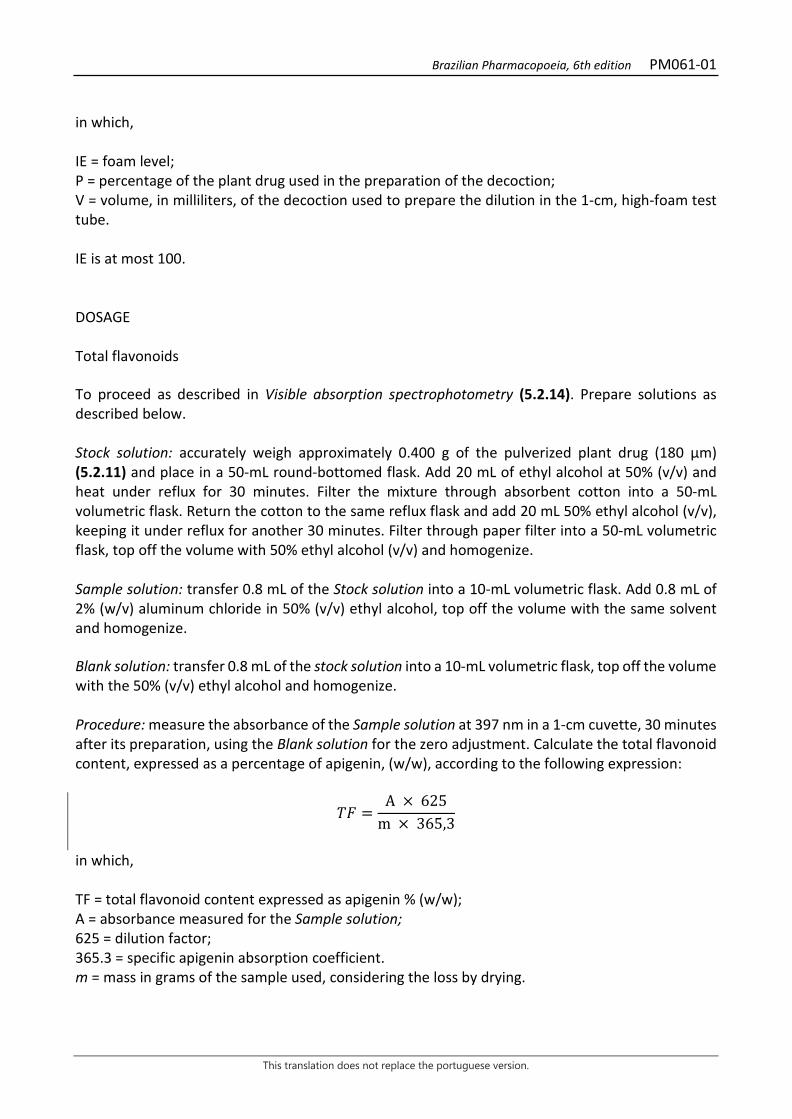
Brazilian Pharmacopoeia, 6th edition PM061-01
This translation does not replace the portuguese version.
in which, IE = foam level; P = percentage of the plant drug used in the preparation of the decoction; V = volume, in milliliters, of the decoction used to prepare the dilution in the 1-cm, high-foam test tube. IE is at most 100. DOSAGE Total flavonoids To proceed as described in Visible absorption spectrophotometry (5.2.14). Prepare solutions as described below. Stock solution: accurately weigh approximately 0.400 g of the pulverized plant drug (180 µm) (5.2.11) and place in a 50-mL round-bottomed flask. Add 20 mL of ethyl alcohol at 50% (v/v) and heat under reflux for 30 minutes. Filter the mixture through absorbent cotton into a 50-mL volumetric flask. Return the cotton to the same reflux flask and add 20 mL 50% ethyl alcohol (v/v), keeping it under reflux for another 30 minutes. Filter through paper filter into a 50-mL volumetric flask, top off the volume with 50% ethyl alcohol (v/v) and homogenize. Sample solution: transfer 0.8 mL of the Stock solution into a 10-mL volumetric flask. Add 0.8 mL of 2% (w/v) aluminum chloride in 50% (v/v) ethyl alcohol, top off the volume with the same solvent and homogenize. Blank solution: transfer 0.8 mL of the stock solution into a 10-mL volumetric flask, top off the volume with the 50% (v/v) ethyl alcohol and homogenize. Procedure: measure the absorbance of the Sample solution at 397 nm in a 1-cm cuvette, 30 minutes after its preparation, using the Blank solution for the zero adjustment. Calculate the total flavonoid content, expressed as a percentage of apigenin, (w/w), according to the following expression:
𝑇𝑇𝑇𝑇 =A × 625
m × 365,3
in which, TF = total flavonoid content expressed as apigenin % (w/w); A = absorbance measured for the Sample solution; 625 = dilution factor; 365.3 = specific apigenin absorption coefficient. m = mass in grams of the sample used, considering the loss by drying.
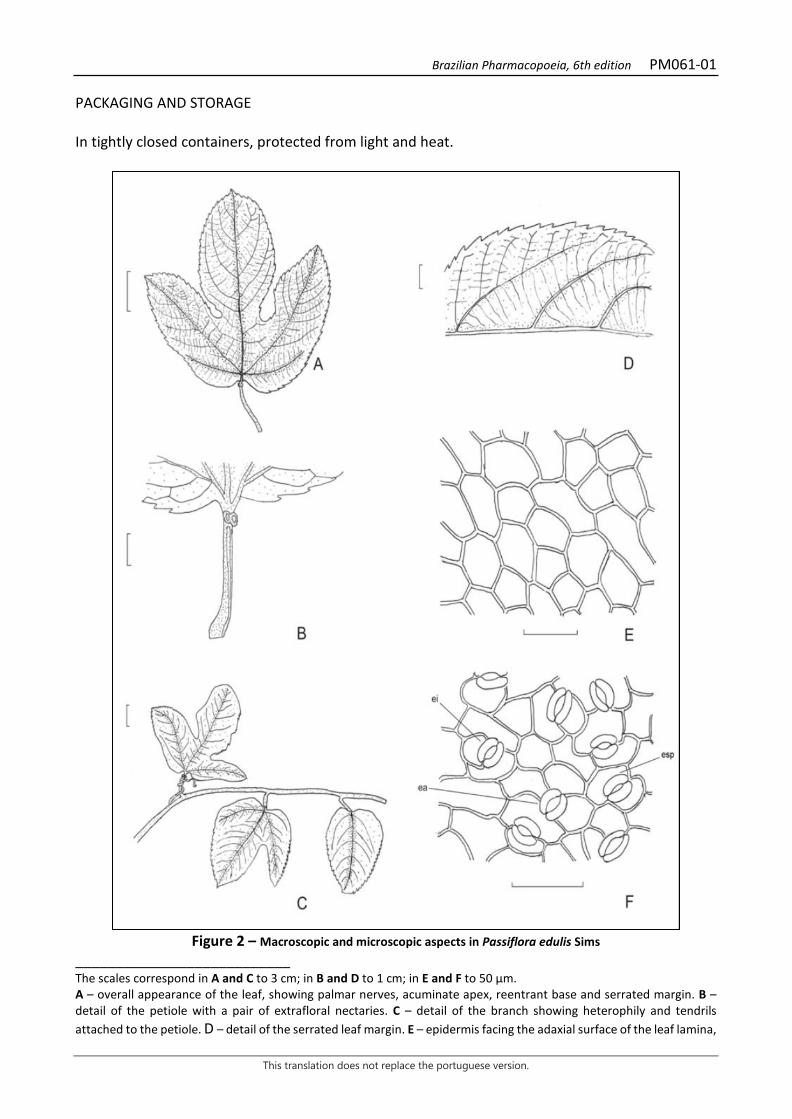
Brazilian Pharmacopoeia, 6th edition PM061-01
This translation does not replace the portuguese version.
PACKAGING AND STORAGE In tightly closed containers, protected from light and heat.
Figure 2 – Macroscopic and microscopic aspects in Passiflora edulis Sims
___________________________ The scales correspond in A and C to 3 cm; in B and D to 1 cm; in E and F to 50 μm. A – overall appearance of the leaf, showing palmar nerves, acuminate apex, reentrant base and serrated margin. B – detail of the petiole with a pair of extrafloral nectaries. C – detail of the branch showing heterophily and tendrils attached to the petiole. D – detail of the serrated leaf margin. E – epidermis facing the adaxial surface of the leaf lamina,

Brazilian Pharmacopoeia, 6th edition PM061-01
This translation does not replace the portuguese version.
in front view. F – epidermis facing the abaxial surface of the leaf lamina, in front view: anomocytic stomata (ea); anisocytic stomata (ei); paracitic stomata (esp).
Brazilian Pharmacopoeia, 6th edition PM061-01
This translation does not replace the portuguese version.
Figure 3 - Microscopic and powder microscopic aspects in Passiflora edulis Sims
___________________________ The scales correspond in A to 100 μm; in B, C and D to 500 μm; in E to 50 μm. A – mesophyll cross-section: abaxial surface (ab); adaxial surface (ad); cuticle (cu); druse (d); vascular bundle (fv); spongy parenchyma (pj); palisade parenchyma (pp). B – diagram of a portion of the leaf lamina on the midrib in cross-section: abaxial surface (ab); adaxial surface (ad); cambium (ca); collenchyma (co); epidermis (ep); phloem (f); vascular bundle (fv); cell inclusion (ic); parenchyma (p); spongy parenchyma (pj); palisade parenchyma (pp); tector trichome (tt); xylem (x). C – detail of petiole cross-section showing druses in the vascular bundle: cell inclusion (ic). D – detail of the adaxial surface of the portion of the leaf lamina on the midrib, in cross-section, showing the unicellular tector trichome: adaxial surface (ad); collenchyma (co); cuticle (cu); epidermis (ep); tector trichome (tt). E – diagram of the overall appearance of the petiole cross-section: abaxial surface (ab); adaxial surface (ad); collenchyma (co); epidermis (ep); phloem (f); vascular bundle (fv); cell inclusion (ic); parenchyma (p); xylem (x).
Brazilian Pharmacopoeia, 6th edition PM062-01
This translation does not replace the portuguese version.
GRANADILLA, leaf Passiflorae dulcis folium
The plant drug consists of dried leaves of Passiflora alata Curtis, containing at least 1.0% total flavonoid content expressed as apigenin (C15H10O5, 270,24). IDENTIFICATION 1. Macroscopic description Simple, glabrous, subcoriaceous, light-green leaves. Oval or oblong lamina, 7 to 20 cm long and 4 to 15 cm wide, with a rounded or slightly curved base, a sharp apex and a smooth margin. Peninerveal nerves. Petiole 2 to 7 cm long, deeply canaliculate in the upper part, with one or usually two pairs of extrafloral nectaries. Tendrils are common on the petiole. It differs from Passiflora edulis in that the latter has a trilobed leaf, serrated margin, palmar nerves, and tector trichomes on the midrib region. 2. Microscopic description Dorsiventrally symmetrical, hypoanphistomatic leaves. The epidermis, in front view, has polyhedral-shaped cells, with indented anticlinal walls on both sides. The cuticle is smooth. Stomata are paracytic, anisocytic and anomocytic. In cross-section, the cuticle is thick, the epidermis is unistratified, and the mesophyll consists of one to three layers of palisade parenchyma and several layers of spongy parenchyma. Calcium oxalate druses occur in the parenchyma and especially in the region of the veins. In the midrib region, in cross-section, the adaxial surface shows little convexity and the abaxial surface has a very angular convexity. Under both epidermis, collenchyma cells interrupt the chlorophyll parenchyma, and a central vascular ring surrounded by sclerenchyma cells or a continuous vascular ring occurs. Druses appear throughout the fundamental tissue, in the collenchyma, and in the phloem. The petiole, in cross-section, presents a concave adaxial surface, with two lateral projections. The abaxial surface is convex, with a single central projection. The internal portion of the epidermis is filled with collenchyma and the rest with parenchyma. The vascular system is formed by central bundles and two others, located in the lateral projections of the adaxial surface. Large numbers of idioblasts with druses appear throughout the collenchyma, parenchyma and vascular bundles 3. Microscopic description of powder The sample meets all requirements for the species, except the macroscopic characters. Characteristics are: yellowish-green color; fragments of epidermis of the adaxial surface with cells as the ones described, without stomata; fragments of epidermis of the abaxial surface with cells as the ones described, with stomata as described; fragments of mesophyll in cross-section, with idioblasts containing druses; isolated druses; fragments of vascular tissue. 4. Proceed as described in Thin-layer chromatography (5.2.17.1). Stationary phase: silica gel G.
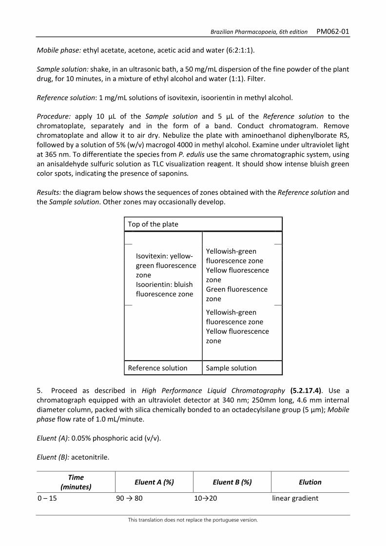
Brazilian Pharmacopoeia, 6th edition PM062-01
This translation does not replace the portuguese version.
Mobile phase: ethyl acetate, acetone, acetic acid and water (6:2:1:1). Sample solution: shake, in an ultrasonic bath, a 50 mg/mL dispersion of the fine powder of the plant drug, for 10 minutes, in a mixture of ethyl alcohol and water (1:1). Filter. Reference solution: 1 mg/mL solutions of isovitexin, isoorientin in methyl alcohol. Procedure: apply 10 μL of the Sample solution and 5 μL of the Reference solution to the chromatoplate, separately and in the form of a band. Conduct chromatogram. Remove chromatoplate and allow it to air dry. Nebulize the plate with aminoethanol diphenylborate RS, followed by a solution of 5% (w/v) macrogol 4000 in methyl alcohol. Examine under ultraviolet light at 365 nm. To differentiate the species from P. edulis use the same chromatographic system, using an anisaldehyde sulfuric solution as TLC visualization reagent. It should show intense bluish green color spots, indicating the presence of saponins. Results: the diagram below shows the sequences of zones obtained with the Reference solution and the Sample solution. Other zones may occasionally develop.
Top of the plate
Isovitexin: yellow-green fluorescence zone Isoorientin: bluish fluorescence zone
Yellowish-green fluorescence zone Yellow fluorescence zone Green fluorescence zone
Yellowish-green fluorescence zone Yellow fluorescence zone
Reference solution Sample solution
5. Proceed as described in High Performance Liquid Chromatography (5.2.17.4). Use a chromatograph equipped with an ultraviolet detector at 340 nm; 250mm long, 4.6 mm internal diameter column, packed with silica chemically bonded to an octadecylsilane group (5 µm); Mobile phase flow rate of 1.0 mL/minute. Eluent (A): 0.05% phosphoric acid (v/v). Eluent (B): acetonitrile.
Time (minutes) Eluent A (%) Eluent B (%) Elution
0 – 15 90 → 80 10→20 linear gradient

Brazilian Pharmacopoeia, 6th edition PM062-01
This translation does not replace the portuguese version.
15 – 30 80 20 isocratic Sample solution: accurately weigh approximately 0.5 g of the dried and pulverized plant drug (180 µm) (5.2.11) and place in a 50-mL volumetric flask. Add approximately 30 mL of ethyl alcohol and water (1:1) solution, shake in an ultrasonic bath for 10 minutes, and top off the volume with the same solvent. Homogenize and filter the extract on filter paper. Concentrate the extract under reduced pressure and suspend it in a methyl alcohol and water mixture (1:1) at a 2 mg/mL concentration. Filter through a 0.45 µm filter unit. Reference solution (1): quantitatively transfer 1 mg isovitexine accurately weighed to a 10mL volumetric flask and add approximately 7 mL ethyl alcohol and water solution (1:1). Homogenize in an ultrasonic bath for 10 minutes. Top off the volume with the same solution and homogenize. Filter through a 0.45 µm filter unit. Reference solution (2): quantitatively transfer 1 mg vitexin-2”-O-rhamnoside accurately weighed to a 10mL volumetric flask and add approximately 7 mL ethyl alcohol and water solution (1:1). Homogenize in an ultrasonic bath for 10 minutes. Top off the volume with the same solution and homogenize. Filter through a 0.45 µm filter unit. Reference solution (3): transfer 1 mg isoorientine accurately weighed to a 10mL volumetric flask and add approximately 7 mL ethyl alcohol and water solution (1:1). Homogenize in an ultrasonic bath for 10 minutes. Top off the volume with the same solution. Filter through a 0.45 µm filter unit. Procedure: separately inject 20µL of the Reference solution (1), 20µL of the Reference solution (2), 20µL of the Reference solution (3) and 20µL of the Sample solution. The chromatogram of the Sample solution should show three main peaks, the first and second with relative retention times of 0.88 and 1.0 for isoorientin and vitexin-2”-O-ramnoside, respectively. It differs from P. edulus by the presence of vitexin-2”-O-ramnoside.
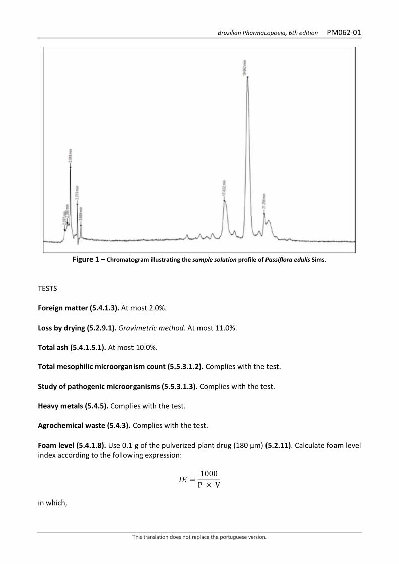
Brazilian Pharmacopoeia, 6th edition PM062-01
This translation does not replace the portuguese version.
Figure 1 – Chromatogram illustrating the sample solution profile of Passiflora edulis Sims.
TESTS Foreign matter (5.4.1.3). At most 2.0%. Loss by drying (5.2.9.1). Gravimetric method. At most 11.0%. Total ash (5.4.1.5.1). At most 10.0%. Total mesophilic microorganism count (5.5.3.1.2). Complies with the test. Study of pathogenic microorganisms (5.5.3.1.3). Complies with the test. Heavy metals (5.4.5). Complies with the test. Agrochemical waste (5.4.3). Complies with the test. Foam level (5.4.1.8). Use 0.1 g of the pulverized plant drug (180 μm) (5.2.11). Calculate foam level index according to the following expression:
𝐼𝐼𝑇𝑇 =1000
P × V
in which,
Brazilian Pharmacopoeia, 6th edition PM062-01
This translation does not replace the portuguese version.
IE = foam level; P = percentage of the drug used in the preparation of the decoction; V = volume, in milliliters, of the decoction used to prepare the dilution in the 1-cm, high-foam test tube. IE is at most 5000. DOSAGE Total flavonoids To proceed as described in Visible absorption spectrophotometry (5.2.14). Prepare solutions as described below. Stock solution: accurately weigh approximately 0.4 g of the pulverized plant drug (180 µm) (5.2.11) and place in a 50-mL round-bottomed flask. Add 20 mL of 50% (v/v) ethyl alcohol and heat, under reflux, for 30 minutes. Filter the mixture through absorbent cotton into a 50-mL volumetric flask. Return the cotton to the same reflux flask and add 20 mL 50% ethyl alcohol (v/v), keeping it under reflux for another 30 minutes. Filter through paper filter into a 50-mL volumetric flask, top off the volume with 50% ethyl alcohol (v/v) and homogenize. Sample solution: transfer 0.8 mL of the Stock solution into a 10-mL volumetric flask. Add 0.8 mL of 2% (w/v) aluminum chloride in 50% (v/v) ethyl alcohol, top off the volume with the same solvent and homogenize. Blank solution: transfer 0.8 mL of the Stock solution into a 10-mL volumetric flask, top off the volume with the 50% (v/v) ethyl alcohol and homogenize. Procedure: measure the absorbance of the Sample solution at 397 nm in a 1-cm cuvette, 30 minutes after its preparation, using the Blank solution for the zero adjustment. Calculate the total flavonoid content, calculated as a percentage of apigenin, according to the following expression:
𝑇𝑇𝑇𝑇 = A × 625
m × 365,3
in which, TF = total flavonoid content expressed as apigenin % (w/w); A = absorbance measured for the Sample solution; 625 = dilution factor; 365.3 = specific apigenin absorption coefficient; m = mass in grams of the sample used, considering the loss by drying. PACKAGING AND STORAGE In tightly closed containers, protected from light and heat.
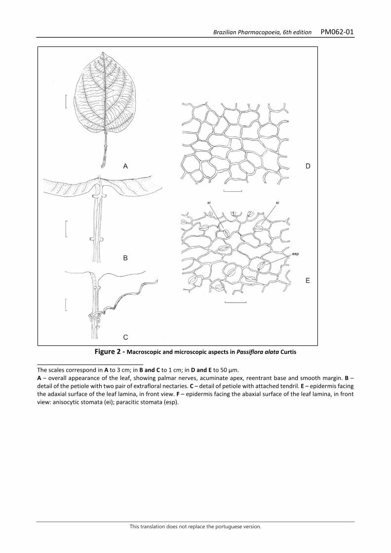
Brazilian Pharmacopoeia, 6th edition PM062-01
This translation does not replace the portuguese version.
Figure 2 - Macroscopic and microscopic aspects in Passiflora alata Curtis
___________________________ The scales correspond in A to 3 cm; in B and C to 1 cm; in D and E to 50 μm. A – overall appearance of the leaf, showing palmar nerves, acuminate apex, reentrant base and smooth margin. B – detail of the petiole with two pair of extrafloral nectaries. C – detail of petiole with attached tendril. E – epidermis facing the adaxial surface of the leaf lamina, in front view. F – epidermis facing the abaxial surface of the leaf lamina, in front view: anisocytic stomata (ei); paracitic stomata (esp).
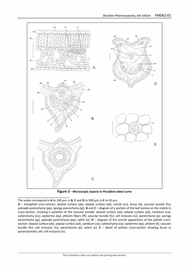
Brazilian Pharmacopoeia, 6th edition PM062-01
This translation does not replace the portuguese version.
Figure 3 - Microscopic aspects in Passiflora alata Curtis
___________________________ The scales correspond in A to 100 μm; in B, C and D to 500 μm; in E to 50 μm. A – mesophyll cross-section: abaxial surface (ab); adaxial surface (ad); cuticle (cu); druse (d); vascular bundle (fv); palisade parenchyma (pp); spongy parenchyma (pj). B and C – diagram of a portion of the leaf lamina on the midrib in cross-section, showing a variation of the vascular bundle: abaxial surface (ab); adaxial surface (ad); cambium (ca); collenchyma (co); epidermis (ep); phloem fibers (ff); vascular bundle (fv); cell inclusion (ic); parenchyma (p); spongy parenchyma (pj); palisade parenchyma (pp); xylem (x). D – diagram of the overall appearance of the petiole cross-section: abaxial surface (ab); adaxial surface (ad); cambium (ca); collenchyma (co); epidermis (ep); phloem (f); vascular bundle (fv); cell inclusion (ic); parenchyma (p); xylem (x). E – detail of petiole cross-section showing druse in parenchymatic cell: cell inclusion (ic).

Brazilian Pharmacopoeia, 6th edition PM063-00
This translation does not replace the portuguese version.
HENBANE, leaf Hyoscyami folium
The plant drug consists of dried leaves of Hyoscyamus niger L., containing at least 0.05% total alkaloids expressed as hyoscyamine (C17H23NO3; 289.37). The alkaloids are mainly hyoscyamine accompanied by scopolamine (hyoscine) in varying proportions. CHARACTERISTICS Slightly nauseating odor. IDENTIFICATION 1. Macroscopic description Whole leaves, up to 30 cm long and 10 cm wide, oval to oval-oblong, with a sharp apex and a cordate base in sessile leaves and reduced in petiolate leaves, with lobed margins, irregularly dentate; yellowish-green to brownish-green color; broad and very developed midrib, secondary veins forming a pronounced angle with the midrib, ending at the tip of the lobes. Leaf lamina strongly pubescent and slimy on both sides. Friable and often broken leaves. 2. Microscopic description Dorsiventrally symmetrical leaf, amphistomatic, with anomocytic stomata. The epidermis, in front view, has sinuous-walled cells, more evident on the abaxial surface. Tector trichomes are smooth, thick-walled, long, conical, and pluricellular, usually with two to four cells. Glandular trichomes may have a long, unicellular or pluricellular and uniseriate pedicel, with a small bicellular glandular head that exudes a viscous substance or with a large elliptical pluricellular glandular head; other times, they are very short and formed by a small pedicel that supports a large claviform and pluricellular gland. Stomata occur in greater number on the abaxial surface. The epidermis, in cross-section, is unistratified and covered by a smooth cuticle. The mesophyll is formed by a single layer of palisade parenchyma, followed by a spongy parenchyma where, especially in the region closest to the palisade parenchyma, idioblasts with prismatic calcium oxalate crystals occur. The midrib is biconvex and the main vascular bundle has bicolateral vascular bundles; the secondary bundles are also bicolateral and surrounded by a poorly lignified pericycle. 3. Microscopic description of powder The sample meets all requirements for the species, except the macroscopic characters. Characteristics are: grayish-green color; epidermal fragments showing cells with sinuous walls and smooth cuticle; anisocytic stomata more abundant on the abaxial surface; pluricellular, uniseriate tector trichomes and glandular trichomes as described; mesophyll fragments as described; a single layer of palisade cells and a spongy parenchyma containing idioblasts with single or double prisms of calcium oxalate; vessel elements with ringed or helical thickening. 4. Microscopic description of powder impurities
Brazilian Pharmacopoeia, 6th edition PM063-00
This translation does not replace the portuguese version.
The powder may also show reticulate stem fibers and vessel elements; subspherical pollen grains with a diameter of up to 60 μm, three germinating pores, three grooves, and a nearly smooth exine; corolla fragments of papillose epidermis; seed fragments containing thick-walled, sinuous, yellowish-brown integument sclereids and cuneiform crystals of calcium oxalate. 5. Proceed as described in Thin-layer chromatography (5.2.17.1). Stationary phase: silica gel GF254. Mobile phase: acetone, water, and concentrated ammonia solution (90:7:3). Sample solution: add 20 mL of 0.05 M sulfuric acid to 2 g of the pulverized sample. Shake for 15 minutes and filtrate. Rinse the filter with 0.05 M sulfuric acid until 25 mL of filtrate is obtained. Add 1 mL of concentrated ammonia solution to the filtrate and shake twice with 10 mL of peroxide-free diethyl ether each time. Separate by centrifugation. Gather the ether layers and dry with anhydrous sodium sulfate. Filter and evaporate the filtrate to near dryness in a water bath. Dissolve the residue in 0.5 mL methyl alcohol. Standard solution: dissolve 50 mg hyoscyamine sulfate in 9 mL methyl alcohol. Dissolve 15 mg scopolamine hydrobromide in 10mL methyl alcohol. Add 4.2 mL of the scopolamine bromide solution to 3.8 mL of the hyoscyamine sulfate solution, top off the volume to 10 mL with methyl alcohol and homogenize. Procedure: apply to the chromatoplate 10 μL of the sample solution and 20 μL of the standard solution, separately and in the form of a 20mm by 3mm band, 1 cm apart. Conduct chromatogram. Remove chromatoplate and allow it to air dry. Nebulize the plate with a solution of potassium iodide and bismuth subnitrate RS and check for orange spots. Next, mist the plate with 5% (w/v) sodium nitrite until the gel becomes transparent and examine after 15 minutes. Results: the zones obtained with the Reference solution and the Sample solution are represented in the diagram below. Other zones may occasionally be present.
Top of the plate
Scopolamine: orange-colored zone
Orange-colored zone
Hyoscyamine: orange-colored zone
Orange-colored zone
Reference solution Sample solution TESTS Foreign matter (5.4.1.3). At most 2.0% of stems over 7 mm in diameter.
Brazilian Pharmacopoeia, 6th edition PM063-00
This translation does not replace the portuguese version.
Loss by drying (5.2.9.1). Gravimetric method. At most 12.0%. Total ash (5.4.1.5.1). At most 13,0%. Acid-insoluble ash (5.4.1.5.3). At most 5.0%. Total mesophilic microorganism count (5.5.3.1.2). Complies with the test. Study of pathogenic microorganisms (5.5.3.1.3). Complies with the test. Heavy metals (5.4.5). Complies with the test. Agrochemical waste (5.4.3). Complies with the test. DOSAGE Total alkaloid content Weigh about 40 g of the pulverized sample (180 μm) (5.2.11) and moisten with 5 mL of a 6 M ammonium hydroxide solution. Add 10 mL of ethyl alcohol and 30 mL of peroxide-free diethyl ether, thoroughly mixed. Transfer the mixture to a percolator, if necessary, using the extraction solution. Macerate for four hours and percolate with the chloroform and peroxide-free diethyl ether (1:3) mixture until the alkaloids are completely extracted. Evaporate 1 mL of the percolate until it dries and dissolve the residue in 1 mL of 0.25 M sulfuric acid and check for the absence of alkaloids with mercuric potassium iodide RS. Reduce the volume of the percolate to 50 mL and transfer to a separating funnel with the aid of peroxide-free ether. Add peroxide-free diethyl ether to the liquid thus obtained, 2.5 times the volume of the percolator, until a liquid of density lower than that of water is obtained. Extract the solution at least three times with 20 mL of a 0.25 M sulfuric acid solution each time. Separate the phases by centrifugation, if necessary, and transfer the acid phase to another separating funnel. Alkalinize the acid phase with a 6 M ammonium hydroxide solution to reach pH 8-9 and extract three times with chloroform in 30 mL aliquots. Combine the chloroform phases and remove residual water by adding 4 g of anhydrous sodium sulfate, allowing it to stand for 30 minutes, with occasional shaking. Remove the chloroform phase and rinse the remaining sodium sulfate with three 10 mL aliquots of chloroform. Combine the chloroform extracts and evaporate to dryness in a water bath. Heat the residue in an oven at 100 °C to 105 °C for 15 minutes. Dissolve the residue in 5 mL chloroform, add 20 mL of 0.01 M sulfuric acid SV solution, and remove the chloroform by evaporation over a water bath. Titrate the excess acid with 0.02 M sodium hydroxide SV solution, using methyl red SI as an indicator. Calculate total alkaloid content expressed as a percentage of hyoscyamine, according to the following expression:
𝑇𝑇𝑇𝑇 =57,88 × (20 − v)
𝑚𝑚
in which, TA = Alkaloid content expressed as hyoscyamine % (w/w);

Brazilian Pharmacopoeia, 6th edition PM063-00
This translation does not replace the portuguese version.
v = volume in milliliters of 0.02 M sodium hydroxide used; m = mass in grams of the sample used, considering the loss by drying. PACKAGING AND STORAGE In tightly closed containers, protected from light and heat.
Brazilian Pharmacopoeia, 6th edition PM063-00
This translation does not replace the portuguese version.
Figure 1 – Macroscopic and microscopic aspects in Hyoscyamus niger L.
___________________________ The scales correspond in A to 5 mm, in B-D and H to 20 µm, in E-G to 30 µm.

Brazilian Pharmacopoeia, 6th edition PM063-00
This translation does not replace the portuguese version.
A. schematic illustration of the leaf. B. detail of the portion of the epidermis oriented to the adaxial surface, in a front view: stomata (es); tector trichome (tt). C. partial detail of the epidermis oriented to the abaxial surface, in a front view: stomata (es); tector trichome (tt). D. detail of the mesophyll portion in cross-section; glandular trichome (tg); tector trichome (tt); cuticle (cu); epidermis (ep); palisade parenchyma (pp); idioblast containing prismatic calcium oxalate crystals (ic); spongy parenchyma (pj); stoma (es). E. fragment of the epidermis in front view, on the abaxial surface; anisocytic stomata (es); tector trichome (tt). F. fragment of mesophyll portion in cross-section; cuticle (cu); epidermis (ep); crystalliferous idioblast (ic); spongy parenchyma (pj); palisade parenchyma (pp). G. fragment of epidermis showing crystals and portions of vessel elements by transparency; crystalliferous idioblast (ic); vascular bundle (fv). H. trichomes or portions thereof, isolated: glandular trichome (tg); tector trichome (tt).
Brazilian Pharmacopoeia, 6th edition PM064-01
This translation does not replace the portuguese version.
LEMON BALM, leaf Melissae folium
The plant drug consists of dried leaves of Melissa officinalis L., containing at least 4.0% total hydroxycinnamic derivatives and at least 2.0% rosmarinic acid (C18H16O8, 360.31) and at least 0.6% volatile oil. CHARACTERISTICS The crushed leaves have a strong, aromatic odor similar to citral. IDENTIFICATION 1. Macroscopic description Entire, membranous, rough, brittle, petiolate leaves, dark green and shiny on the adaxial surface and light green on the abaxial surface, sometimes vinaceous, mainly in the region near the petiole and on the veins of the abaxial surface, with tector and rare glandular trichomes on the adaxial surface and with numerous tector and glandular trichomes on the abaxial surface, the latter resembling small pits, visible with a magnifying glass of six times; camptodrome-reticulodrome venation. Oval to oval-cordiform lamina, with an oval, rounded or cordiform base, obtuse apex and irregularly crenate-serrated margin, finely ciliated, 4 to 8 cm long and 3 to 5 cm wide. Petiole 0.3 to 5.0 cm long, green or vinaceous, concave on the adaxial surface, convex on the abaxial surface and with two lateral veins; adaxial surface covered by long tector trichomes, those of the veins visible to the naked eye. 2. Microscopic description Dorsiventrally symmetrical leaf lamina, hypostomatic, with diacytic stomata. In front view, the epidermal cells have sinuous anticlinal walls. The leaf lamina has the following trichomes: (1) unicellular tector, rarely bicellular, conical to triangular, dentiform, short, with thick and verrucous cuticle; (2) uniseriate multicellular tector, from three to five cells, the apical one with acute apex, uncinate in appearance, with thick and verrucous cuticle; (2) uniseriate multicellular tector, from three to nine cells, very long, with thick and verrucous cuticle; (4) tectore, uniseriate multicellular, with three to nine cells, very long and broad base, formed by a crown of cells; (5) glandular with unicellular or bicellular head, rounded and unicellular to tricellular pedicel; (6) peltate glandular, with unicellular pedicel, located in a depression in the epidermis and with octocellular secretory head. In cross-section, the cuticle is slightly striated and the epidermis is unistratified. The palisade parenchyma is unistratified and the spongy parenchyma is bi- to tri-stratified; starch grains are present in all tissues. The midrib, in cross-section, has a smooth cuticle on the adaxial surface and striated on the abaxial surface, the epidermis is unistratified, the collenchyma is angular, unistratified on the abaxial surface, and with three to four layers on the adaxial surface. A single collateral bundle occurs, rarely two or three, surrounded by a continuous or non-continuous endodermis. The petiole, in cross-section, has a slightly striated cuticle and unistratified epidermis. Trichomes are the same as for the lamina. The collenchyma is angular and is distributed along the entire length of the petiole, uni- or biostratified on the adaxial side and tri-stratified on the abaxial
Brazilian Pharmacopoeia, 6th edition PM064-01
This translation does not replace the portuguese version.
surface; in the rib region up to seven layers occur. The vascular system is formed by three to five collateral bundles, each surrounded by endoderm; the phloem may have stone cells next to the fibers. 3. Microscopic description of powder The sample meets all requirements for the species, except the macroscopic characters. Characteristics are: greenish color; fragments of leaf epidermis with cells with sinuous anticlinal walls and diacritical stomata and with scars of dentiform-type tector trichomes; large amount of trichomes as described; mesophyll fragments as described; calcium oxalate crystals absent. 4. Macroscopic description of impurities The stems, branches, flowers and fruit of the species itself, if present as impurity, are characterized by: quadrangular stem, hairy when young; small, stipitate flowers, protected by foliaceous bracts, similar to other leaves; pubescent, tubular-campanulate, bilabiate calyx, tridentate upper lip and bifid lower lip; white to yellowish or pink corolla with recurved tube and limb with two unequal lobes, the upper one erect, bifid and the lower one extended, trilobed, with obtuse lobes, the median one is longer; four stamens, didinamous, connivent under the upper lip of the corolla, anthers with divergent thecae; pubescent, tetralobate ovary, with monospermic lobes; gynobasic stylet, bifid; tetraquenium fruit, brownish color. 5. Microscopic description of the impurity corresponding to the stem Stems of the species itself, if present as impurity, have, as primary structure, a thick and striated cuticle, unistratified epidermis with polyhedral cells, stomata distributed near the ribs and located far above the other epidermal cells, many trichomes, most commonly type 6, besides types 2 and 5, and those of type 4 are distributed in the ribs. The cortex has angular collenchyma distributed throughout and more developed in the ribs, chlorenchyma, and cortical parenchyma formed by isodiametric cells with large intercellular spaces. The endoderm has a large amount of starch grains and surrounds the four collateral bundles. The medullary parenchyma is formed by isodiametric cells of large volume and thin walls. As secondary structure, the epidermis and cortex maintain their characteristics, except for the clear decrease of trichomes and the common occurrence of stone cells in the cortical parenchyma. The phloem has a large amount of fibers, the vascular cambium is evident, and the xylem has a large amount of starch grains. Such grains occur in all tissues except the epidermis, and in greater quantity when as secondary structure. 6. Proceed as described in Thin-layer chromatography (5.2.17.1). Stationary phase: silica gel GF254 (0.250 mm). Mobile phase: hexane and ethyl acetate (90:10). Sample solution: transfer 2 g of the pulverized plant drug to a 250 mL, round-bottomed, polished-necked flask, add 100 mL of water. Add 0.5 mL xylene to the graduated tube and distill for one hour, as described in Determination of volatile oils in plant drugs (5.4.1.6). After distillation, transfer the organic phase into a 1-mL volumetric flask, rinse the graduated tube of the apparatus with a small amount of xylene, top off the volume with the same solvent and homogenize.
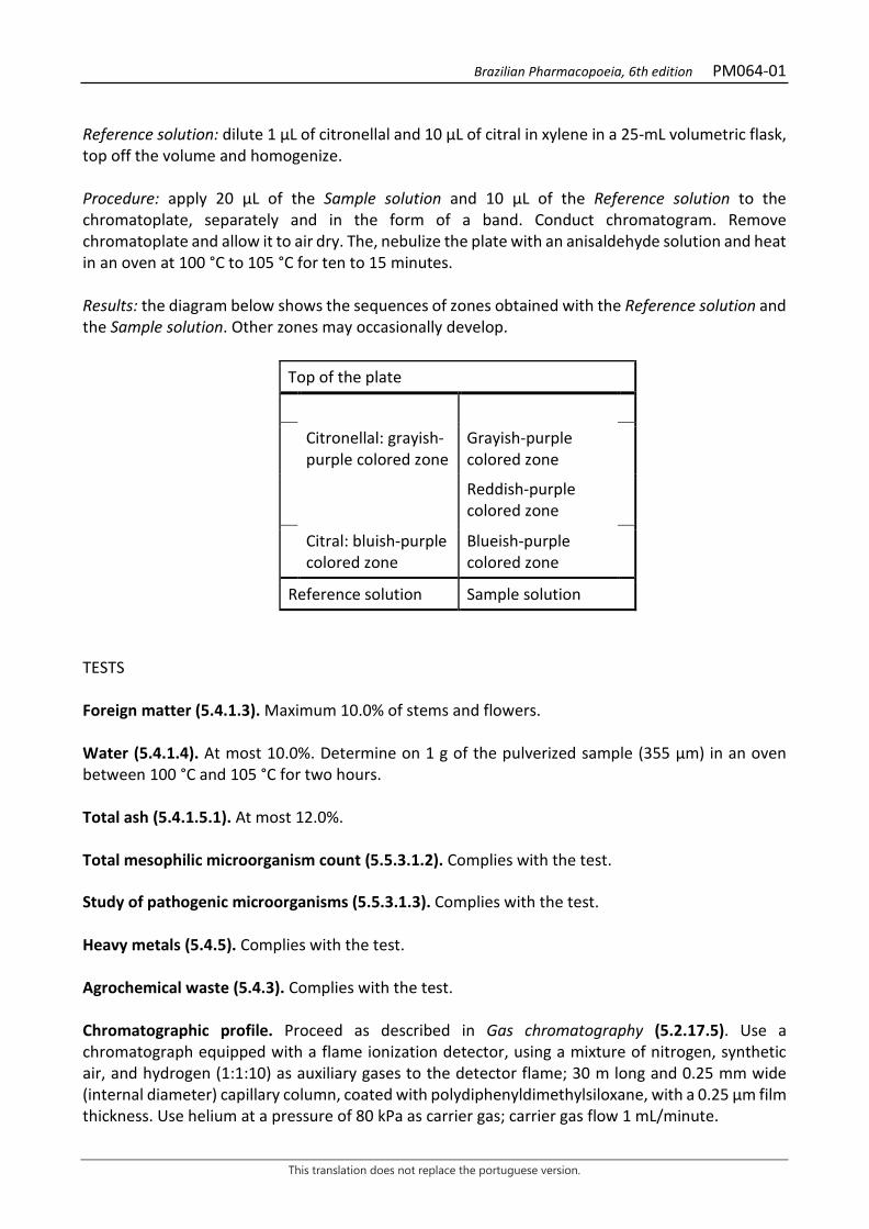
Brazilian Pharmacopoeia, 6th edition PM064-01
This translation does not replace the portuguese version.
Reference solution: dilute 1 µL of citronellal and 10 µL of citral in xylene in a 25-mL volumetric flask, top off the volume and homogenize. Procedure: apply 20 μL of the Sample solution and 10 μL of the Reference solution to the chromatoplate, separately and in the form of a band. Conduct chromatogram. Remove chromatoplate and allow it to air dry. The, nebulize the plate with an anisaldehyde solution and heat in an oven at 100 °C to 105 °C for ten to 15 minutes. Results: the diagram below shows the sequences of zones obtained with the Reference solution and the Sample solution. Other zones may occasionally develop.
Top of the plate
Citronellal: grayish-purple colored zone
Grayish-purple colored zone
Reddish-purple colored zone
Citral: bluish-purple colored zone
Blueish-purple colored zone
Reference solution Sample solution
TESTS Foreign matter (5.4.1.3). Maximum 10.0% of stems and flowers. Water (5.4.1.4). At most 10.0%. Determine on 1 g of the pulverized sample (355 µm) in an oven between 100 °C and 105 °C for two hours. Total ash (5.4.1.5.1). At most 12.0%. Total mesophilic microorganism count (5.5.3.1.2). Complies with the test. Study of pathogenic microorganisms (5.5.3.1.3). Complies with the test. Heavy metals (5.4.5). Complies with the test. Agrochemical waste (5.4.3). Complies with the test. Chromatographic profile. Proceed as described in Gas chromatography (5.2.17.5). Use a chromatograph equipped with a flame ionization detector, using a mixture of nitrogen, synthetic air, and hydrogen (1:1:10) as auxiliary gases to the detector flame; 30 m long and 0.25 mm wide (internal diameter) capillary column, coated with polydiphenyldimethylsiloxane, with a 0.25 µm film thickness. Use helium at a pressure of 80 kPa as carrier gas; carrier gas flow 1 mL/minute.

Brazilian Pharmacopoeia, 6th edition PM064-01
This translation does not replace the portuguese version.
Temperature: Time (minutes) Temperature (ºC) Column 0 – 80 60 → 300 Injector 220 Detector 250 Sample solution: dilute the volatile oil in diethyl ether at ratio of 2:100. Procedure: inject the volume of 1 μL of the Sample solution into the gas chromatograph, using a 1:50 flow division. The linear retention indices of the oil constituents are calculated with respect to a homologous series of hydrocarbons and compared with reference samples. Determine relative concentrations by manual or electronic integration using the normalization method. Calculate the Relative Retention Index, according to the following expression:
𝐼𝐼𝐼𝐼𝐼𝐼 = 100 × n +100 × (𝑡𝑡𝑡𝑡𝑥𝑥 − 𝑡𝑡𝑡𝑡𝑧𝑧)
(𝑡𝑡𝑡𝑡𝑧𝑧+1 − 𝑡𝑡𝑡𝑡𝑧𝑧)
in which, RRI = Relative Retention Index n = number of alkane carbon atoms of the lowest molecular mass; trx = retention time of the constituent “x” (intermediate to trz and trz+1); trz = retention time of the alkane with “n” carbons; trz+1 = retention time of alkane with “n+1” carbons.
Figure 1 – Illustrative chromatogram obtained with volatile oil from Melissa officinalis L. through gas chromatography coupled to a flame ionization detector.
Check the presence of the components, as follows, in the chromatogram obtained with the Sample solution:
Peak Retention Index Constituent Content (%) 1 1234 neral (citral B) 30.4 – 32.9 2 1265 geranial (citral A) 49.0 – 53.3 3 1404 beta-caryophyllene 2.6 – 3.1 4 1579 caryophyllene oxide 3.9- 6.4
Brazilian Pharmacopoeia, 6th edition PM064-01
This translation does not replace the portuguese version.
DOSAGE Total hydroxycinnamic derivatives To proceed as described in Visible absorption spectrophotometry (5.2.14). Prepare solutions as described below. Stock solution: accurately weigh approximately 0.2 g of the pulverized plant drug and transfer to a round-bottomed flask. Add 190 mL of 50% (v/v) ethyl alcohol and heat in a water bath, under reflux, for 30 minutes. Cool and filter. Rinse the filter with 10 mL of 50% (v/v) ethyl alcohol. Transfer the filtrate and the washing solution into a 200-mL volumetric flask, top off the volume with the 50% (v/v) ethyl alcohol and homogenize. Sample solution: in a 10-mL volumetric flask, transfer 1 mL of the Stock solution, 2 mL of 0.5 M hydrochloric acid, 2 mL of a solution prepared by dissolving 10 g of sodium nitrite and 10 g of sodium molybdate in 100 mL of water, then, 2 mL of 2 M sodium hydroxide, top off the volume to 10 mL with water and homogenize. Blank solution: in a 10-mL volumetric flask, transfer 1 mL of the Stock solution, 2 mL of 0.5 M hydrochloric acid, 2 mL of 2 M sodium hydroxide, top off the volume to 10 mL with water and homogenize. Procedure: measure the absorbance of the Sample solution at 505 nm after its preparation, using the Blank solution for zero adjustment. Calculate the content of total hydroxycinnamic derivatives, expressed as a percentage of rosmarinic acid, according to the following expression:
𝑇𝑇𝑇𝑇𝑇𝑇𝐶𝐶 =A × 2000m × 400
in which, TDHC = total hydroxycinnamic derivative content, expressed as rosmarinic acid % (w/w); A = absorbance measured for the Sample solution; 2000 = dilution factor; 400 = rosmarinic acid specific absorption coefficient; m = mass in grams of the sample used, considering the loss by drying. Rosmarinic acid Proceed as described in High Performance Liquid Chromatography (5.2.17.4). Use a chromatograph equipped with an ultraviolet detector at 332 mm; pre-column packed with octadecylsilane silica, 150 mm long, 3.9 mm internal diameter column, packed with octadecylsilane silica (4 µm), kept at room temperature; Mobile phase flow rate of 0.6 mL/minute. Eluent (A): water and trifluoroacetic acid (100:0.1).
Brazilian Pharmacopoeia, 6th edition PM064-01
This translation does not replace the portuguese version.
Eluent (B): acetonitrile and trifluoracetic acid (100:0.1).
Time (minutes) Eluent A (%) Eluent B (%) Elution
0 – 14 90→61 10→39 linear gradient 14 – 16 61→50 39→50 linear gradient 16 – 18 50→90 50→10 linear gradient 18 – 23 90 10 Isocratic Sample solution: accurately weigh approximately 0.1 g of the dried and pulverized plant drug(800 µm) (5.2.11) and place in a centrifuge tube. Add 5 mL of 40% (v/v) ethyl alcohol and place in the ultrasonic bath for 10 minutes. Centrifuge for five minutes at 42 × g. Separate and transfer the supernatant to a 10-mL volumetric flask. Extract the drug residue again with 4 mL of 40% (v/v) ethyl alcohol in an ultrasonic bath for five minutes. Centrifuge and transfer the supernatant into the same volumetric flask, top off the volume to 10 mL with 40% (v/v) ethyl alcohol and homogenize. Dilute 50 µL of the resulting solution in 0.3 mL of water. Filter through a 0.45 µm filter unit. Stock solution: dissolve 10 mg rosmarinic acid in methyl alcohol, transfer quantitatively to a 10-mL volumetric flask, top off the volume with methyl alcohol and homogenize. Solutions for the analytical curve: dilute an aliquot of 200 mL of the Stock solution, to obtain a 0.25 µg/mL solution. Dilute the previous solution in methyl alcohol to concentrations of 7.80 µg/mL, 15.60 µg/mL, 31.25 µg/mL, 62.50 µg/mL, 125 µg/mL, and 250 µg/mL. Filter through a 0.45 µm filter unit. Procedure: inject 10 µL of the Solutions for the analytical curve and 10 µL of the sample solution separately. Register the chromatograms and measure the areas under the peaks. Rosmarinic acid retention time is approximately 10.3 minutes. Calculate rosmarinic acid content in the sample from the straight line equation obtained from the calibration curve. The result is expressed as the average of the determinations in grams of rosmarinic acid, in percent, considering the loss by drying. Volatile oils Proceed as described in Determining volatile oils in plant drugs (5.4.1.6). Use 1000-mL flask containing 500 mL of water as distillation liquid. Add 0.5 mL of xylene to the graduated tube. Use dry, scratched and unbruised plant. Immediately proceed with the determination of the volatile oil, from 20 g of the chopped drug. Distill for four hours. Measure the volume and express the yield per 100 g of the plant drug (w/p). PACKAGING AND STORAGE In tightly closed containers, protected from light and heat.
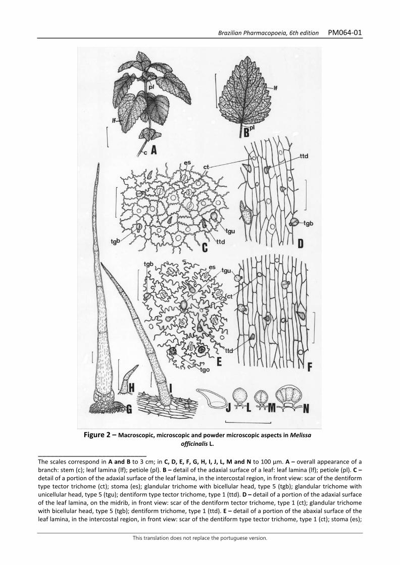
Brazilian Pharmacopoeia, 6th edition PM064-01
This translation does not replace the portuguese version.
Figure 2 – Macroscopic, microscopic and powder microscopic aspects in Melissa
officinalis L. ___________________________ The scales correspond in A and B to 3 cm; in C, D, E, F, G, H, I, J, L, M and N to 100 µm. A – overall appearance of a branch: stem (c); leaf lamina (lf); petiole (pl). B – detail of the adaxial surface of a leaf: leaf lamina (lf); petiole (pl). C – detail of a portion of the adaxial surface of the leaf lamina, in the intercostal region, in front view: scar of the dentiform type tector trichome (ct); stoma (es); glandular trichome with bicellular head, type 5 (tgb); glandular trichome with unicellular head, type 5 (tgu); dentiform type tector trichome, type 1 (ttd). D – detail of a portion of the adaxial surface of the leaf lamina, on the midrib, in front view: scar of the dentiform tector trichome, type 1 (ct); glandular trichome with bicellular head, type 5 (tgb); dentiform trichome, type 1 (ttd). E – detail of a portion of the abaxial surface of the leaf lamina, in the intercostal region, in front view: scar of the dentiform type tector trichome, type 1 (ct); stoma (es);
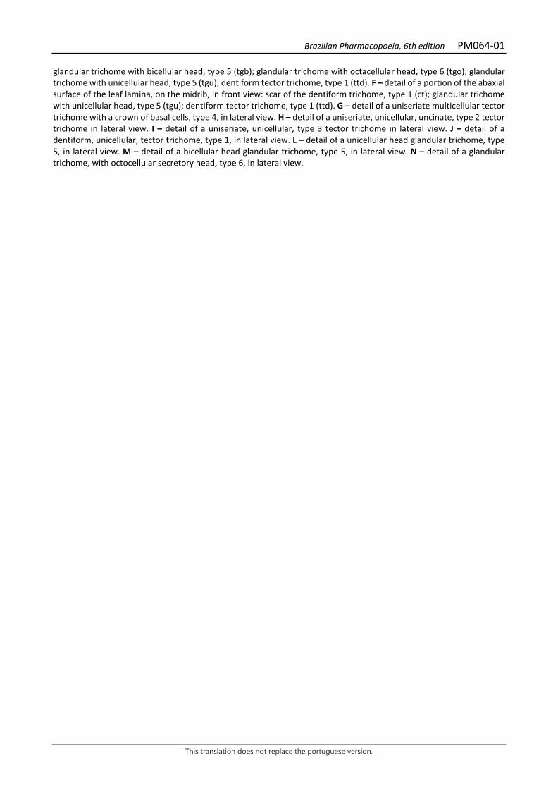
Brazilian Pharmacopoeia, 6th edition PM064-01
This translation does not replace the portuguese version.
glandular trichome with bicellular head, type 5 (tgb); glandular trichome with octacellular head, type 6 (tgo); glandular trichome with unicellular head, type 5 (tgu); dentiform tector trichome, type 1 (ttd). F – detail of a portion of the abaxial surface of the leaf lamina, on the midrib, in front view: scar of the dentiform trichome, type 1 (ct); glandular trichome with unicellular head, type 5 (tgu); dentiform tector trichome, type 1 (ttd). G – detail of a uniseriate multicellular tector trichome with a crown of basal cells, type 4, in lateral view. H – detail of a uniseriate, unicellular, uncinate, type 2 tector trichome in lateral view. I – detail of a uniseriate, unicellular, type 3 tector trichome in lateral view. J – detail of a dentiform, unicellular, tector trichome, type 1, in lateral view. L – detail of a unicellular head glandular trichome, type 5, in lateral view. M – detail of a bicellular head glandular trichome, type 5, in lateral view. N – detail of a glandular trichome, with octocellular secretory head, type 6, in lateral view.

Brazilian Pharmacopoeia, 6th edition PM064-01
This translation does not replace the portuguese version.
Figure 3 – Microscopic aspects in Melissa officinalis L.
___________________________ The scales correspond in A, B, C and E to 100 μm; in D to 400 µm. A – detail of a portion of the mesophyll region, in cross-section: abaxial surface (ab); adaxial surface (ad); cuticle (cu); epidermis (ep); stoma (es); spongy parenchyma (pj); palisade parenchyma (pp); dentiform tector trichome, type 1 (ttd); glandular trichome with octocellular head, type 6 (tgo). B – detail of the midrib region and portion of the mesophyll, in cross-section: abaxial surface (ab); adaxial surface (ad); chlorenchyma (cl); collenchyma (co); cuticle (cu); endoderm (end); epidermis (ep); stomata (es); phloem (f); spongy parenchyma (pj); parenchyma (p); palisade parenchyma (pp); xylem parenchyma (px); dentiform tector trichome, type 1 (ttd); xylem (x). C – detail of a portion of the leaf margin, in cross-section: abaxial surface (ab); adaxial surface (ad); cuticle (cu); epidermis (ep); stoma (es); spongy parenchyma (pj);
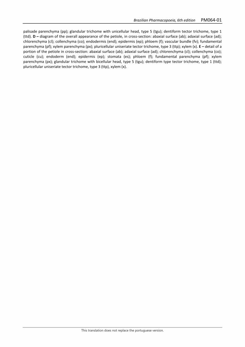
Brazilian Pharmacopoeia, 6th edition PM064-01
This translation does not replace the portuguese version.
palisade parenchyma (pp); glandular trichome with unicellular head, type 5 (tgu); dentiform tector trichome, type 1 (ttd). D – diagram of the overall appearance of the petiole, in cross-section: abaxial surface (ab); adaxial surface (ad); chlorenchyma (cl); collenchyma (co); endodermis (end); epidermis (ep); phloem (f); vascular bundle (fv); fundamental parenchyma (pf); xylem parenchyma (px); pluricellular uniseriate tector trichome, type 3 (ttp); xylem (x). E – detail of a portion of the petiole in cross-section: abaxial surface (ab); adaxial surface (ad); chlorenchyma (cl); collenchyma (co); cuticle (cu); endoderm (end); epidermis (ep); stomata (es); phloem (f); fundamental parenchyma (pf); xylem parenchyma (px); glandular trichome with bicellular head, type 5 (tgu); dentiform type tector trichome, type 1 (ttd); pluricellular uniseriate tector trichome, type 3 (ttp), xylem (x).

Brazilian Pharmacopoeia, 6th edition PM065-00
This translation does not replace the portuguese version.
KOLA NUT, seed Colae semen
The plant drug consists of cotyledons of Cola nitida (Vent.) Schott & Endl. (syn. Cola vera K. Schum.), containing not less than 1.7% total tannins expressed as pyrogallol (C6H6O3; 126.11) and 2.0% methylxanthines expressed as caffeine (C8H10N4O2, 194.19). IDENTIFICATION 1. Macroscopic description The seed contains two cotyledons, which are usually sold already separated. They are hardened and uneven, solid, irregular, reddish-brown in color, variable in size, 2 to 5 cm long by about 2 cm wide, and up to 1 cm thick. The apex of the cotyledon is wider than its base and both are rounded. The margin is entirely continued. The outer surface of each cotyledon is convex or slightly depressed, rough, and brown to reddish-brown in color. The inner surface is flat or depressed, somewhat smooth, generally irregular, with a small cavity at the base sometimes containing the radicle and plumule, or traces thereof. Fracture surface is uniform and bright brown. 2. Microscopic description Cotyledons are surrounded by an epidermis formed by rectangular cells, small or slightly elongated in the radial direction, and are composed of a homogeneous parenchyma of polygonal cells, sometimes of irregular outline. The innermost cells are larger, thick-walled and pitted, brown in color, containing phenolic compounds, fatty matter, and abundant starch grains. The latter are mainly distributed in the central cells and are unequal, spherical, oval, oval-rounded, oblong, reniform, ellipsoid, or pyriform, with a branched hilum, centralized or eccentric, almost always merged, star-shaped or cross-shaped, and their concentric striations are barely visible. The grain size varies from 5 to 35 µm, rarely 45 µm. 3. Microscopic description of powder The sample meets all requirements for the species, except the macroscopic characters. Characteristics are: reddish-brown to moderately yellowish-brown color; epidermis and parenchyma fragments with polygonal cells with brown or reddish-brown walls, containing several and varied starch grains, as described; sparse fragments of small fibrovascular bundles. The starch grains, when observed under polarized light, exhibit a cross in the hilum region. 4. Proceed as described in Thin-layer chromatography (5.2.17.1). Stationary phase: silica gel GF254 (0.25 mm). Mobile phase: ethyl acetate, methyl alcohol and water (100:13.5:10). Sample solution: extract the previously pulverized plant drug, under reflux, for 15 minutes, in a concentration equal to 2% (w/v), using ethyl alcohol as extracting liquid. Filter and apply to the chromatoplate.

Brazilian Pharmacopoeia, 6th edition PM065-00
This translation does not replace the portuguese version.
Reference solution: dissolve 10 mg caffeine in 2 mL absolute ethyl alcohol. Procedure: apply 5 μL to 10 μL of the Sample solution and 2 μL to 3 μL of the Reference solution to the chromatoplate, separately and in the form of a band. Conduct chromatogram. Remove chromatoplate and allow it to air dry for a few minutes. Examine under ultraviolet light at 254 nm. Nebulize with a solution of potassium iodide and bismuth subnitrate RS. Additionally, nebulize with 5% (w/v) sodium nitrite aqueous solution. Results: the diagram below shows the sequences of zones obtained with the Reference solution and the Sample solution. Other zones may occasionally develop.
Top of the plate
Caffeine: orange-colored zone Red-colored zone
Reference solution Sample solution
TESTS Foreign matter (5.4.1.3). At most 3.0%. Water (5.4.1.4). At most 15.0%. Total ash (5.4.1.5.1). At most 5.0%. Total mesophilic microorganism count (5.5.3.1.2). Complies with the test. Study of pathogenic microorganisms (5.5.3.1.3). Complies with the test. Heavy metals (5.4.5). Complies with the test. Agrochemical waste (5.4.3). Complies with the test. DOSAGE Methylxanthines Proceed as described in Ultraviolet absorption spectrophotometry (5.2.14). Prepare solutions as described below. Sample solution: accurately weigh about 0.25 g of the pulverized sample. Extract with 20 mL of 2.5% (v/v) sulfuric acid solution under magnetic shaking for 15 minutes four times. Filter the portions into a 100-mL volumetric flask. Top off the volume with 2.5% (v/v) sulfuric acid solution and homogenize. Transfer 10 mL of the solution into a 100-mL volumetric flask. Top off the volume with the same
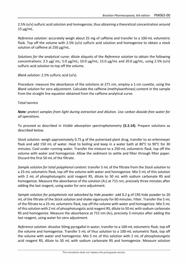
Brazilian Pharmacopoeia, 6th edition PM065-00
This translation does not replace the portuguese version.
2.5% (v/v) sulfuric acid solution and homogenize, thus obtaining a theoretical concentration around 15 µg/mL. Reference solution: accurately weigh about 25 mg of caffeine and transfer to a 100-mL volumetric flask. Top off the volume with 2.5% (v/v) sulfuric acid solution and homogenize to obtain a stock solution of caffeine at 250 μg/mL. Solutions for the analytical curve: dilute aliquots of the Reference solution to obtain the following concentrations: 2.5 μg/ mL; 5.0 μg/mL; 10.0 μg/mL; 15.0 μg/mL and 20.0 μg/mL, using 2.5% (v/v) sulfuric acid solution to top off the volume. Blank solution: 2.5% sulfuric acid (v/v). Procedure: measure the absorbance of the solutions at 271 nm, employ a 1-cm cuvette, using the Blank solution for zero adjustment. Calculate the caffeine (methylxanthines) content in the sample from the straight line equation obtained from the caffeine analytical curve. Total tannins Note: protect samples from light during extraction and dilution. Use carbon dioxide-free water for all operations. To proceed as described in Visible absorption spectrophotometry (5.2.14). Prepare solutions as described below. Stock solution: weigh approximately 0.75 g of the pulverized plant drug, transfer to an erlenmeyer flask and add 150 mL of water. Heat to boiling and keep in a water bath at 80°C to 90°C for 30 minutes. Cool under running water. Transfer the mixture to a 250-mL volumetric flask, top off the volume with water and homogenize. Allow the sediment to settle and filter through filter paper. Discard the first 50 mL of the filtrate. Sample solution for total polyphenol content: transfer 5 mL of the filtrate from the Stock solution to a 25-mL volumetric flask, top off the volume with water and homogenize. Mix 5 mL of this solution with 2 mL of phosphotungstic acid reagent RS, dilute to 50 mL with sodium carbonate RS and homogenize. Measure the absorbance of the solution (A1) at 715 nm, precisely three minutes after adding the last reagent, using water for zero adjustment. Sample solution for polyphenols not adsorbed by hide powder: add 0.2 g of CRS hide powder to 20 mL of the filtrate of the Stock solution and shake vigorously for 60 minutes. Filter. Transfer the 5 mL of the filtrate to a 25-mL volumetric flask, top off the volume with water and homogenize. Mix 5 mL of this solution with 2 mL of phosphotungstic acid reagent RS, dilute to 50 mL with sodium carbonate RS and homogenize. Measure the absorbance at 715 nm (A2), precisely 3 minutes after adding the last reagent, using water for zero adjustment. Reference solution: dissolve 50mg pyrogallol in water, transfer to a 100-mL volumetric flask, top off the volume and homogenize. Transfer 5 mL of thus solution to a 100-mL volumetric flask, top off the volume with water and homogenize. Mix 5 mL of this solution with 2 mL of phosphotungstic acid reagent RS, dilute to 50 mL with sodium carbonate RS and homogenize. Measure solution
Brazilian Pharmacopoeia, 6th edition PM065-00
This translation does not replace the portuguese version.
absorbance at 715 nm (A3), precisely three minutes after adding the last reagent, and within 15 minutes from the pyrogallol dissolution using water for zero adjustment. Calculate total tannin content, in percent, according to the following expression:
𝑇𝑇𝑇𝑇 =(𝑇𝑇1 − 𝑇𝑇2) × 13,12
𝑇𝑇3 × 𝑚𝑚
in which, TT = total tannin content expressed as pyrogallol % (w/w); m = mass in grams of the sample used, considering the determined water content; A1 = absorbance measured for the Sample solution for total polyphenols; A2 = absorbance measured for the Sample solution for polyphenols not adsorbed by hide powder; A3 = absorbance measured for the Reference solution. PACKAGING AND STORAGE In tightly closed containers, protected from light and heat.
Brazilian Pharmacopoeia, 6th edition PM065-00
This translation does not replace the portuguese version.
Figure 1 – Macroscopic, microscopic and powder microscopic aspects in Cola nitida (Vent.) Schott & Endl.
___________________________ The scales correspond in A, B and C to 2 cm; in D, E and F to 100 μm; in G to 50 μm. A – appearance of the external face of the cotyledon. B – appearance of the inner face of the cotyledon. C – cotyledon in equatorial view. D, E, F and G – powder details. D, E and F – parenchyma fragments, showing variable cell sizes and pitted walls. G – detail of starch grains, showing variability in shape, grain size, and hilum aspect.
Brazilian Pharmacopoeia, 6th edition PM066-00
This translation does not replace the portuguese version.
NUX-VOMICA, seed Strychni semen
The plant drug consists of dried seeds of Strychnos nux-vomica L., containing at least 0.5% strychnine (C21H22N2O2, 334.42). IDENTIFICATION 1. Macroscopic description The seeds are brownish-gray, discoid-shaped, with a slightly thickened margin, 10 to 30 mm wide and 4 to 6 mm thick. The protuberant hilum is found in the central region, from which a radial line runs to the micropyle located at a point on the margin. The integument is rigid and the surface has a satiny texture, densely covered by lignified tector trichomes, with a radial arrangement from the center to the margin. Internally to the integument, the endosperm is translucent, corneous, light gray, separated in two parts by a disk-shaped central cavity. The embryo is found adjacent to the micropyle, consisting of two small, 5- to 7- nerved, cordiform cotyledons and a radicle. 2. Microscopic description A cross section of the integument shows an epidermis formed by cells with thickened and lignified walls, with linear and oblique pits. Each cell has a dilated, polygonal base, similar to a stone cell, which extends externally in a sloping manner, forming a carpet of tector trichomes with the others. Internally to the epidermis, flattened and compressed parenchyma cells form a brownish band of indistinct cells. In the region of the hilum, small spiral xylematic vessels are found as components of a short vascular bundle. The endosperm is covered by an epidermal layer of cells with slightly thickened walls, followed by polyhedral endosperm cells with thickened hemicellulose walls, connected by plasmodesmata, containing lipid droplets and aleurone grains, the latter approximately 30 m in diameter. The embryo consists of parenchymal cells with small lipid droplets and aleurone grains. 3. Microscopic description of powder The sample meets all requirements for the species, except the macroscopic characters. Characteristics are: brownish-gray color, isolated or clustered lignified trichomes, entire or fragmented; endosperm fragments with parenchymatic tissue of thickened hemicellulose walls with amorphous content, and some visible aleurone grains. 4. Proceed as described in Thin-layer chromatography (5.2.17.1). Stationary phase: 250 µm thick silica gel F254. Mobile phase: butyl alcohol, water and glacial acetic acid (70:20:10). Sample solution: weigh 1 g of the pulverized plant drug and add 20 mL of 70% ethyl alcohol (v/v). Place in a water bath for 15 minutes. Filter and evaporate in a water bath to dryness, at a
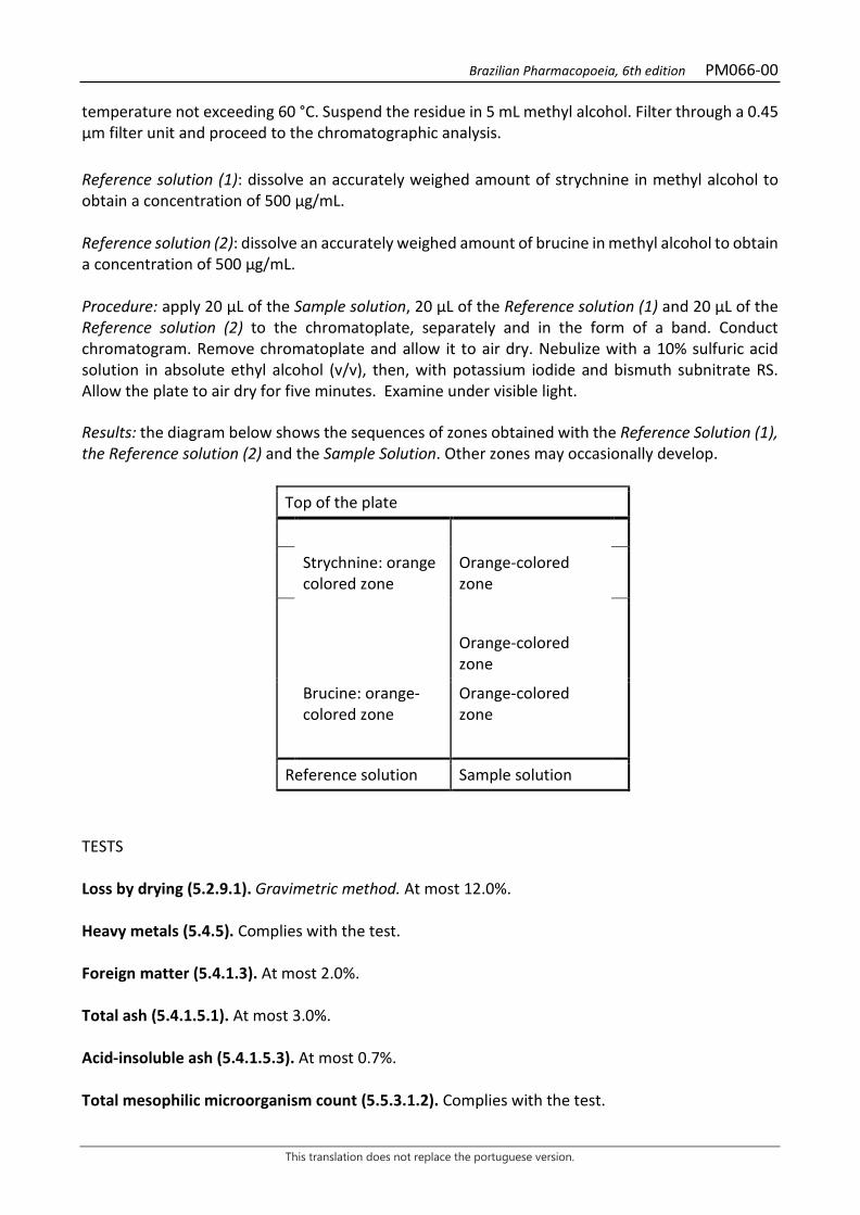
Brazilian Pharmacopoeia, 6th edition PM066-00
This translation does not replace the portuguese version.
temperature not exceeding 60 °C. Suspend the residue in 5 mL methyl alcohol. Filter through a 0.45 µm filter unit and proceed to the chromatographic analysis. Reference solution (1): dissolve an accurately weighed amount of strychnine in methyl alcohol to obtain a concentration of 500 µg/mL. Reference solution (2): dissolve an accurately weighed amount of brucine in methyl alcohol to obtain a concentration of 500 µg/mL. Procedure: apply 20 μL of the Sample solution, 20 μL of the Reference solution (1) and 20 µL of the Reference solution (2) to the chromatoplate, separately and in the form of a band. Conduct chromatogram. Remove chromatoplate and allow it to air dry. Nebulize with a 10% sulfuric acid solution in absolute ethyl alcohol (v/v), then, with potassium iodide and bismuth subnitrate RS. Allow the plate to air dry for five minutes. Examine under visible light. Results: the diagram below shows the sequences of zones obtained with the Reference Solution (1), the Reference solution (2) and the Sample Solution. Other zones may occasionally develop.
Top of the plate
Strychnine: orange colored zone
Orange-colored zone
Orange-colored zone
Brucine: orange-colored zone
Orange-colored zone
Reference solution Sample solution
TESTS Loss by drying (5.2.9.1). Gravimetric method. At most 12.0%. Heavy metals (5.4.5). Complies with the test. Foreign matter (5.4.1.3). At most 2.0%. Total ash (5.4.1.5.1). At most 3.0%. Acid-insoluble ash (5.4.1.5.3). At most 0.7%. Total mesophilic microorganism count (5.5.3.1.2). Complies with the test.
Brazilian Pharmacopoeia, 6th edition PM066-00
This translation does not replace the portuguese version.
Study of pathogenic microorganisms (5.5.3.1.3). Complies with the test. Aflatoxins (5.4.4). Complies with the test. Agrochemical waste (5.4.3). Complies with the test. DOSAGE Strychnine Proceed as described in High Performance Liquid Chromatography (5.2.17.4). Use a chromatograph equipped with an ultraviolet detector at 210 nm; pre-column packed with octadecylsilane silica, 150 mm long, 4.6 mm internal diameter column, packed with octadecylsilane silica (5 µm), kept at 30 °C; Mobile phase flow rate of 1.3 mL/minute. Isocratic system. Mobile phase: 7 g/L dibasic potassium phosphate buffer, pH 3 adjusted with phosphoric acid, acetonitrile and diethylamine (900:100:20). Sample solution: accurately weigh approximately 1.0 g of the pulverized plant drug and transfer to a round-bottomed flask. Add 20 mL of 0.1 M hydrochloric acid, and heat, under reflux, for 30 minutes. After cooling, filter through absorbent cotton into a 100 mL round-bottomed flask. Extract the drug residue on the filter and cotton with 20 mL of 0.1 M hydrochloric acid and heat, under reflux, for 15 minutes. Gather the aqueous phase and add 6 M ammonium hydroxide until pH 9.0 is reached. Transfer to a 250-mL separating funnel and extract three times with 30 mL diethyl ether. Gather the organic phases in a beaker and add 5 g of anhydrous sodium sulfate. Shake with a glass rod. Filter on porcelain capsule filter paper. Rinse the beaker with 20 mL diethyl ether. Combine the washing liquid with the organic phases. Evaporate to residue in a water bath, at a temperature no higher than 50 ºC. Suspend the residue in 1 mL methyl alcohol. Transfer to a 10-mL volumetric flask. Rinse the porcelain capsule with 5 mL of the mobile phase. Transfer the washing liquid into the 10-mL volumetric flask, top off the volume with the mobile phase and homogenize. Filter through a 0.45 µm filter unit. Reference solution: weigh 8.0 mg of strychnine, transfer to a 10-mL volumetric flask, and dilute with the Mobile phase. Filter through a 0.45 µm filter unit. Procedure: separately inject 10 μL of the Reference solution and 10 µL of the Sample solution. Register the chromatograms and measure the areas under the peaks. Calculate strychnine content, in percent, according to the following expression:
𝑇𝑇𝑇𝑇 =𝑇𝑇𝑎𝑎 × 𝐶𝐶𝑟𝑟𝑇𝑇𝑟𝑟 × 𝐶𝐶𝑎𝑎
in which, TE = strychnine content % (w/w); Ar= area under the peak corresponding to the strychnine in the Reference solution; Aa= area under the peak corresponding to the strychnine in the Sample solution;

Brazilian Pharmacopoeia, 6th edition PM066-00
This translation does not replace the portuguese version.
Cr = concentration of strychnine in the Reference solution in mg/mL, considering the purity of the reference substance; Ca = concentration of the plant drug in the Sample solution in mg/mL, considering loss by drying; PACKAGING AND STORAGE In tightly closed containers, protected from light and heat.

Brazilian Pharmacopoeia, 6th edition PM066-00
This translation does not replace the portuguese version.
Figure 1 – Macroscopic, microscopic and powder microscopic aspects in Strychnos nux-vomica L. ___________________________ The scales correspond in A and B to 1 cm; in C and D to 200 µm; in F and G to 100 µm; in E to 50 µm. A - front view of the entire seed showing hilum (hi) and micropyle (mi). B - frontal view of a longitudinally opened seed showing one of the cotyledons (cot), endosperm (en), micropyle (mi), embryo radicle (rad), and the integument (t). C - cross-section in the micropyle region: endosperm (en), epidermis of the endosperm (ep), epidermis of the integument (ept) showing the lignified tector trichomes, compressed parenchyma of the integument (p) and xylem vessels (x). D – Cross section outside the micropyle region with endosperm (en), endosperm epidermis (ep), integument epidermis (ept) and compressed parenchyma (p). E – endosperm tissue fragment highlighting aleurone grains (gal), lipid droplets (gtl) and cellulose cell wall thickening (pcl). F – fragment of the integument epidermis, highlighting the base of the trichome (bt) resembling a stone cell with thickened and lignified walls and simple pits. G – fragment of the extended trichome portion of the integument epidermis.
Brazilian Pharmacopoeia, 6th edition PM067-01
This translation does not replace the portuguese version.
EUGENIA UNIFLORA, leaf Eugeniae folium
The plant drug consists of dried leaves of Eugenia uniflora L., containing at least 5.0% tannins, 1.0% total flavonoids, expressed as quercetin; and, 0.8% volatile oils. The volatile oil consists of at least 27.0% curzerenes (cis and trans). CHARACTERISTICS Dried leaves have citrus odor. IDENTIFICATION 1. Macroscopic description Simple, oval to oval-lanceolate leaves, in general 4.5 to 6.2 cm long and 2 to 2.7 cm wide, glabrous, membranous to slightly coriaceous, dark green on the adaxial surface and lighter green on the abaxial surface; lamina with an acute to acuminate apex, sometimes slightly falcate, acute to obtuse base, entire, pubescent margin, with the midrib more prominent on the abaxial surface. Venation camptodrome-broquidodrome, each secondary vein departing at an acute angle to the midrib, anastomosing with its subsequent superior one, so as to form a series of arches around the leaf margin; the secondary and superior order veins determine incomplete areoles, with free vascular endings. Petiole 0.3 to 0.6 cm long. The glands, present in the lamina, are hardly visualized without the aid of a lens. 2. Microscopic description Dorsiventrally symmetrical, hypo-stomatic leaf lamina with paracitic stomata, whose guard cells show thickening of the inner face in the form of dumbbells. In front view, the anticlinal walls of the epidermal cells are sinuous on both sides. In cross-section, the lamina has a unistratified epidermis, covered by cuticle. The palisade parenchyma is uni-stratified and accompanied by collecting cells. The spongy parenchyma has seven to nine cell strata with relatively long braciform projections. In the mesophyll idioblasts with rhombic crystals and calcium oxalate druses are common. Secretory cavities, containing volatile oil, commonly underlie the epidermis, on both leaf faces, although more abundant on the adaxial surface. In the midrib, with a plano-convex outline, or rarely concave-convex or biconvex, one to three layers of annular collenchyma occur, subjacent to the epidermis. The main vascular bundle is of the bicolateral, open-arch type, surrounded by two to three strata of thick-walled parenchyma cells, and a fiber sheath, except at the ends of the arch. The phloem shows abundant small rhombic crystals. The secondary and smaller veins are accompanied by fiber caps at both poles. The petiole, with a concave-convex outline, has small lateral expansions. The epidermis is unistratified, containing brown-colored substances, also present in the cells of the underlying fundamental, collenchymatous parenchyma. Secretory cavities, similar to those in the lamina, are also present subepidermally. Starch grains, druses and crystals occur abundantly throughout the parenchyma. The vascular bundle is an open, bicolateral arch, with abundant rhombic crystals in the phloem, surrounded by four to eight layers of parenchymatous tissue with thickened walls, forming

Brazilian Pharmacopoeia, 6th edition PM067-01
This translation does not replace the portuguese version.
a perivascular sheath. Fibers, isolated or in groups of two to three elements, are rarely present around the vascular bundle. 3. Microscopic description of powder The sample meets all requirements for the species, except the macroscopic characters. Characteristics are: lamina epidermis fragments with sinuous anticlinal walls, free of stomata; lamina epidermis fragments with paracitic stomata, showing haltered thickening; lamina fragments showing unistratified palisade parenchyma and/or spongy parenchyma with relatively long braciform projections; lamina fragments containing rhombic crystals, abundant druses and shiny-looking secretory cavities due to the presence of volatile oil; isolated rhombic druses and crystals. 4. Proceed as described in Thin-layer chromatography (5.2.17.1). Stationary phase: silica gel F254 (0.250 mm). Mobile phase: ethyl acetate, formic acid and water (75:5:5). Sample solution: accurately weigh about 10 g of the pulverized plant drug, add 100 mL of water and heat under reflux for 15 minutes. After cooling down to room temperature, filter the obtained solution through absorbent cotton, under reduced pressure. Extract the resulting aqueous phase with three 25-mL portions of ethyl acetate in a 125-mL separating funnel. Allow to stand in a freezer at -18 °C for 15 minutes for a complete phase separation. Combine and filter the organic fractions with 5 g anhydrous sodium sulfate. Evaporate the organic fraction at a rotary evaporator under reduced pressure until residue is formed. Suspend the residue in 1 mL methyl alcohol. Reference solution (1): weigh approximately 1 mg epicatechin and dissolve in 2mL methyl alcohol. Reference solution (2): weigh approximately 1 mg 4'-O-methylgallocatechin and dissolve in 1mL methyl alcohol. Procedure: apply 20 µL of the Sample solution, 10 µL of the Reference solution (1) and 10 µL of the Reference solution (2) to the chromatoplate, separately and in the form of a band. Conduct chromatogram. Remove the chromatoplate and allow it to dry in a fume hood. Nebulize the plate with 1% ferric chloride (w/v) solution in methyl alcohol. Results: the diagram below shows the sequences of zones obtained with the Reference solution and the Sample solution. Other zones may occasionally develop.
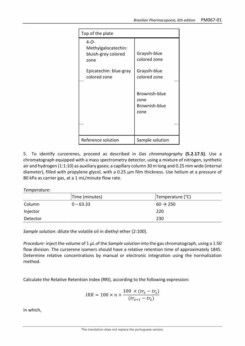
Brazilian Pharmacopoeia, 6th edition PM067-01
This translation does not replace the portuguese version.
Top of the plate
4-O-Methylgalocatechin: bluish-grey colored zone
Graysih-blue colored zone
Epicatechin: blue-gray colored zone
Graysih-blue colored zone
Brownish-blue zone Brownish-blue zone
Reference solution Sample solution 5. To identify curzerenes, proceed as described in Gas chromatography (5.2.17.5). Use a chromatograph equipped with a mass spectrometry detector, using a mixture of nitrogen, synthetic air and hydrogen (1:1:10) as auxiliary gases; a capillary column 30 m long and 0.25 mm wide (internal diameter), filled with propylene glycol, with a 0.25 µm film thickness. Use helium at a pressure of 80 kPa as carrier gas, at a 1 mL/minute flow rate. Temperature: Time (minutes) Temperature (°C) Column 0 – 63.33 60 → 250 Injector 220 Detector 230 Sample solution: dilute the volatile oil in diethyl ether (2:100). Procedure: inject the volume of 1 μL of the Sample solution into the gas chromatograph, using a 1:50 flow division. The curzerene isomers should have a relative retention time of approximately 1845. Determine relative concentrations by manual or electronic integration using the normalization method. Calculate the Relative Retention Index (RRI), according to the following expression:
𝐼𝐼𝐼𝐼𝐼𝐼 = 100 × 𝑛𝑛 +100 × (𝑡𝑡𝑡𝑡𝑥𝑥 − 𝑡𝑡𝑡𝑡𝑧𝑧)
(𝑡𝑡𝑡𝑡𝑧𝑧+1 − 𝑡𝑡𝑡𝑡𝑧𝑧)
in which,

Brazilian Pharmacopoeia, 6th edition PM067-01
This translation does not replace the portuguese version.
RRI = Relative Retention Index n = number of alkane carbon atoms of the lowest molecular mass; trx = retention time of compound "x" (intermediate to trz and trz+1); trz = retention time of alkane with “n” carbons; trz+1 = retention time of alkane with “n+1” carbons. TESTS Foreign matter (5.4.1.3). At most 2.0%. Loss by drying (5.2.9.1). Gravimetric method. At most 10.0%. Total ash (5.4.1.5.1). At most 11.0%. Total mesophilic microorganism count (5.5.3.1.2). Complies with the test. Study of pathogenic microorganisms (5.5.3.1.3). Complies with the test. Heavy metals (5.4.5). Complies with the test. Agrochemical waste (5.4.3). Complies with the test. Foam level. Quantitatively transfer about 1 g of the pulverized plant drug (180 μm), weighed, accurately, to an Erlenmeyer flask containing 50 mL of boiling water. Keep at moderate boil for 15 minutes. Cool, filter through absorbent cotton into a 100-mL volumetric flask. Top off the volume to 100 mL through the filter and homogenize. Distribute the obtained decoction into 10 capped test tubes (16 mm wide by 16 cm high) in a successive series of 1 mL, 2 mL, 3 mL, up to 10 mL, and adjust the liquid volume in each tube to 10 mL with water. Cap the tubes and shake vigorously with vertical movements for 15 seconds at two runs per second. Allow it to stand for 15 minutes and measure the height of the foam. Next, add 1 mL of 2 M hydrochloric acid to each tube. If the foam height of all tubes is less than 1 cm, the foam index is less than 100. If, in any of the tubes, the measured foam height remains equal to or greater than 1 cm, the dilution of the plant material in such tube (A) is the observed rate. Calculate the foam index (IE), according to the following expression:
𝐼𝐼𝑇𝑇 =1000𝑇𝑇
in which, IE = foam level; V = volume, in milliliters, of the decoction used to prepare the dilution in the tube foam was observed. The IE for the decoction must be at least 125. DOSAGE
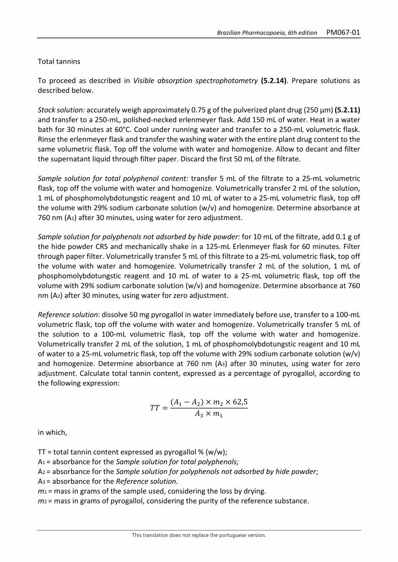
Brazilian Pharmacopoeia, 6th edition PM067-01
This translation does not replace the portuguese version.
Total tannins To proceed as described in Visible absorption spectrophotometry (5.2.14). Prepare solutions as described below. Stock solution: accurately weigh approximately 0.75 g of the pulverized plant drug (250 μm) (5.2.11) and transfer to a 250-mL, polished-necked erlenmeyer flask. Add 150 mL of water. Heat in a water bath for 30 minutes at 60°C. Cool under running water and transfer to a 250-mL volumetric flask. Rinse the erlenmeyer flask and transfer the washing water with the entire plant drug content to the same volumetric flask. Top off the volume with water and homogenize. Allow to decant and filter the supernatant liquid through filter paper. Discard the first 50 mL of the filtrate. Sample solution for total polyphenol content: transfer 5 mL of the filtrate to a 25-mL volumetric flask, top off the volume with water and homogenize. Volumetrically transfer 2 mL of the solution, 1 mL of phosphomolybdotungstic reagent and 10 mL of water to a 25-mL volumetric flask, top off the volume with 29% sodium carbonate solution (w/v) and homogenize. Determine absorbance at 760 nm (A1) after 30 minutes, using water for zero adjustment. Sample solution for polyphenols not adsorbed by hide powder: for 10 mL of the filtrate, add 0.1 g of the hide powder CRS and mechanically shake in a 125-mL Erlenmeyer flask for 60 minutes. Filter through paper filter. Volumetrically transfer 5 mL of this filtrate to a 25-mL volumetric flask, top off the volume with water and homogenize. Volumetrically transfer 2 mL of the solution, 1 mL of phosphomolybdotungstic reagent and 10 mL of water to a 25-mL volumetric flask, top off the volume with 29% sodium carbonate solution (w/v) and homogenize. Determine absorbance at 760 nm (A2) after 30 minutes, using water for zero adjustment. Reference solution: dissolve 50 mg pyrogallol in water immediately before use, transfer to a 100-mL volumetric flask, top off the volume with water and homogenize. Volumetrically transfer 5 mL of the solution to a 100-mL volumetric flask, top off the volume with water and homogenize. Volumetrically transfer 2 mL of the solution, 1 mL of phosphomolybdotungstic reagent and 10 mL of water to a 25-mL volumetric flask, top off the volume with 29% sodium carbonate solution (w/v) and homogenize. Determine absorbance at 760 nm (A3) after 30 minutes, using water for zero adjustment. Calculate total tannin content, expressed as a percentage of pyrogallol, according to the following expression:
𝑇𝑇𝑇𝑇 =(𝑇𝑇1 − 𝑇𝑇2) × 𝑚𝑚2 × 62,5
𝑇𝑇3 × 𝑚𝑚1
in which, TT = total tannin content expressed as pyrogallol % (w/w); A1 = absorbance for the Sample solution for total polyphenols; A2 = absorbance for the Sample solution for polyphenols not adsorbed by hide powder; A3 = absorbance for the Reference solution. m1 = mass in grams of the sample used, considering the loss by drying. m2 = mass in grams of pyrogallol, considering the purity of the reference substance.
Brazilian Pharmacopoeia, 6th edition PM067-01
This translation does not replace the portuguese version.
Total flavonoids To proceed as described in Visible absorption spectrophotometry (5.2.14). Prepare solutions as described below. Stock solution: accurately weigh approximately 0.4 g of the pulverized plant drug (250 µm) (5.2.11) and transfer to a 100mL round-bottomed flask. Add 1 mL of 0.5% methenamine solution (w/v), 20 mL acetone and 2 mL hydrochloric acid. Heat, on a heating mantle, keeping under reflux, for 30 minutes. Filter through a small amount of cotton into a 100-mL volumetric flask. Rinse the drug residue and the absorbent cotton in the same round-bottomed flask with 20 mL acetone. Hold under reflux for 10 minutes and filter through absorbent cotton into the same 10-mL volumetric flask. Repeat this operation once more. Cool to room temperature, top off the volume with acetone and homogenize. Transfer 20 mL of this acetone solution, into a separating funnel (125 mL), 20 mL of water, and extract with a 15 mL portion of ethyl acetate. Repeat the extraction three times, with 10 mL portions of ethyl acetate. Collect the ethyl acetate phases and rinse in a separating funnel with two 50-mL portions of water. Transfer the ethyl acetate phase to a 50-mL volumetric flask, top off the volume with ethyl acetate and homogenize. Sample solution: transfer 10 mL of the Stock solution volumetrically to a 25-mL volumetric flask, add 1 mL of a 2% (w/v) aluminum chloride solution in methyl alcohol, top off the volume with a 5% (v/v) acetic acid solution in methyl alcohol and homogenize. Blank solution: transfer 10 mL of the Stock Solution into a 25-mL volumetric flask, top off the volume with the 5% (v/v) acetic acid solution in methyl alcohol and homogenize. Procedure: measure the absorbance of the Sample solution at 425 nm in a 1-cm cuvette, 30 minutes after its preparation, using the Blank solution for zero adjustment. Calculate total flavonoid content expressed as quercetin, in percent, according to the following expression:
𝑇𝑇𝑇𝑇 =𝑇𝑇 × 625
m × 500
in which, TQ = total flavonoid content expressed as quercetin % (w/w); A = absorbance measured for the Sample solution; 625 = dilution factor; 500 = quercetin specific absorption coefficient; m = mass in grams of the sample used, considering the loss by drying. Volatile oils Proceed as described in Determining volatile oils in plant drugs (5.4.1.6). Use a 1000-mL flask containing 500 mL of distilled water as distillation liquid and 0.5 mL of xylene in the graduated tube. Use dry, scratched and unbruised plant. Proceed with the determination of the volatile oil, from 100 g of the chopped drug. Distill for four hours. Measure the volume and express the yield per 100 g of the plant drug (w/p).
Brazilian Pharmacopoeia, 6th edition PM067-01
This translation does not replace the portuguese version.
PACKAGING AND STORAGE In tightly closed containers, protected from light and heat.
Figure 1 – Macroscopic, microscopic and powder microscopic aspects in Eugenia uniflora L.
___________________________ The scales correspond in A to 1 cm; in B to 5 mm; in C to 1 mm; in D, E, F and G to 50 μm. A – schematic illustration of the leaf, in front view: midrib (np). B – schematic detail of a portion of the lamina showing the leaf vein: midrib (np); secondary vein (ns). C – schematic detail of areolae and vascular endings: secretory cavity (cs). D and E – partial details of the adaxial and abaxial surfaces of the leaf lamina, respectively, in front view: secretory cavity (cs); guard cell (cg); subsidiary cell (csb); stoma (es). F – partial detail of the adaxial surface of the leaf lamina, in front view, showing a secretory cavity visualized by transparency: stoma (es). G – partial detail of the lamina, in cross-section,

Brazilian Pharmacopoeia, 6th edition PM067-01
This translation does not replace the portuguese version.
showing twinned stomatal complexes: spongy parenchyma (pj); substomatic chamber (csu); abaxial surface (ab); subsidiary cell (csb); stomata (es); guard cell (cg); epidermis (ep).
Figure 2 – Microscopic and powder microscopic aspects in Eugenia uniflora L. ___________________________ The scales correspond in A and B to 100 μm; in C, D, E and F to 50 μm; in G, H and I to 100 μm; in J to 200 μm. A and B – partial details of the mesophyll of distinct samples, in cross-sections: abaxial surface (ab); adaxial surface (ad); epidermis (ep); cuticle (cu); secretory cavity (cs); crystalliferous idioblast (ic); spongy parenchyma (pj); palisade parenchyma (pp); intercellular space (ei). C and D – powder fragments showing details of the spongy parenchyma: spongy parenchyma (pj); intercellular space (ei); vascular bundle (fv); crystalliferous idioblast (ic). E and F – partial details, in cross-sections, of a secondary and a tertiary vein, respectively: palisade parenchyma (pp); xylem fibers (fx); xylem (x); phloem (f); phloem fibers (ff); spongy parenchyma (pj). G, H and I – diagrams of the midrib, in cross-sections,
D
G
E
F
J
C
pe fv
ff
fxxi
fl
col
H
I
cav
xi
ff
dr
pp
Aepb
cs
cutepd
pe
cpr
B
dr
pp
pe
fl

Brazilian Pharmacopoeia, 6th edition PM067-01
This translation does not replace the portuguese version.
in the median (G) and basal regions of different samples (H and I): abaxial surface (ab); adaxial surface (ad); epidermis (ep); xylem (x); collenchyma (co); phloem (f); palisade parenchyma (pp); phloem fibers (ff); crystalliferous idioblast (ic); secretory cavity (cs). J – diagram, in cross section, of the petiole: abaxial surface (ab); adaxial surface (ad); secretory cavity (cs); epidermis (ep); phloem (f); crystalliferous idioblast (ic); xylem (x).
Brazilian Pharmacopoeia, 6th edition PM068-00
This translation does not replace the portuguese version.
PSYLLIUM, testa Plantaginis ovatae seminis tegumentum
The plant drug consists of the testa of the seeds of Plantago ovata Forssk. (syn. Plantago ispaghula Roxb. ex Fleming), which swells and takes on a colloidal consistency when mixed with water. CHARACTERISTICS The testa of the seeds is odorless. IDENTIFICATION 1. Macroscopic description Fragments of the seeds’ testa, flat, oval or navicular, up to 2 mm long and 1 mm wide, beige-pink in color, some of them showing a light brown oval spot, corresponding to where the embryo was before it was removed. 2. Microscopic description In front view, the cells of the testa epidermis have a polygonal-prismatic shape of varying size. When water is added, the outer mucilage layers rapidly swell, breaking through the epidermal cell walls. In cross-section, the outer walls of the epidermal cells show thick mucilage layers, most evident in the marginal testa region. Internally to the mucilaginous epidermis, there is a thin layer of discolored and obliterated cells, which are not very resistant and allow the epidermis to separate easily from the rest of the seed. The innermost layer of the seed testa, when present, consists of a layer of often obliterated, yellowish-brown cells. Some starch grains are found especially in the epidermal cells of the seed margin region, emphasized with Lugol’s iodine solution, consisting of two to four elements. 3. Microscopic description of powder The sample meets all requirements for the species, except the macroscopic characters. Characteristics are: light brown to pale yellow color; fragments with polygonal epidermal cells containing mucilage; fragments of the inner testa layers with light brownish walls, sometimes associated with remnants of the outer endosperm layers; starch grains as described, inside cells or isolated. 4. Microscopic description of impurities Thick-walled endosperm cell fragments containing lipid drops and aleurone grains and fragments with thin-walled embryo cells occasionally occur. 5. Adulterations Seeds of other Plantago species, which have significantly less capacity to swell, and seeds of Salvia aegyptica L. are considered adulterations. A dangerous substitute drug is the seed of Lallemantia
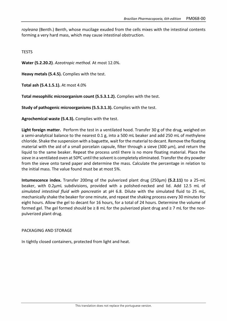
Brazilian Pharmacopoeia, 6th edition PM068-00
This translation does not replace the portuguese version.
royleana (Benth.) Benth, whose mucilage exuded from the cells mixes with the intestinal contents forming a very hard mass, which may cause intestinal obstruction. TESTS Water (5.2.20.2). Azeotropic method. At most 12.0%. Heavy metals (5.4.5). Complies with the test. Total ash (5.4.1.5.1). At most 4.0% Total mesophilic microorganism count (5.5.3.1.2). Complies with the test. Study of pathogenic microorganisms (5.5.3.1.3). Complies with the test. Agrochemical waste (5.4.3). Complies with the test. Light foreign matter. Perform the test in a ventilated hood. Transfer 30 g of the drug, weighed on a semi-analytical balance to the nearest 0.1 g, into a 500 mL beaker and add 250 mL of methylene chloride. Shake the suspension with a baguette, wait for the material to decant. Remove the floating material with the aid of a small porcelain capsule, filter through a sieve (300 µm), and return the liquid to the same beaker. Repeat the process until there is no more floating material. Place the sieve in a ventilated oven at 50ºC until the solvent is completely eliminated. Transfer the dry powder from the sieve onto tared paper and determine the mass. Calculate the percentage in relation to the initial mass. The value found must be at most 5%. Intumescence index. Transfer 200mg of the pulverized plant drug (250µm) (5.2.11) to a 25-mL beaker, with 0.2µmL subdivisions, provided with a polished-necked and lid. Add 12.5 mL of simulated intestinal fluid with pancreatin at pH 6.8. Dilute with the simulated fluid to 25 mL, mechanically shake the beaker for one minute, and repeat the shaking process every 30 minutes for eight hours. Allow the gel to decant for 16 hours, for a total of 24 hours. Determine the volume of formed gel. The gel formed should be ≥ 8 mL for the pulverized plant drug and ≥ 7 mL for the non-pulverized plant drug. PACKAGING AND STORAGE In tightly closed containers, protected from light and heat.
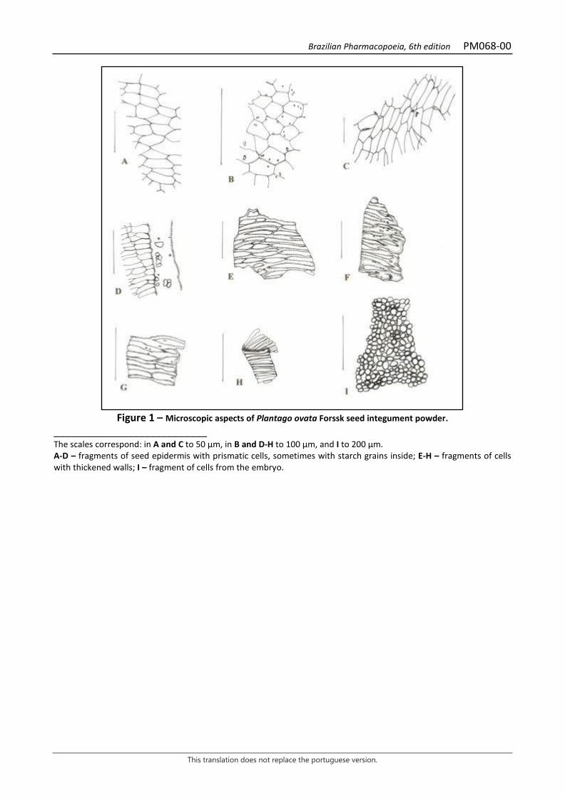
Brazilian Pharmacopoeia, 6th edition PM068-00
This translation does not replace the portuguese version.
Figure 1 – Microscopic aspects of Plantago ovata Forssk seed integument powder.
___________________________ The scales correspond: in A and C to 50 µm, in B and D-H to 100 µm, and I to 200 µm. A-D – fragments of seed epidermis with prismatic cells, sometimes with starch grains inside; E-H – fragments of cells with thickened walls; I – fragment of cells from the embryo.
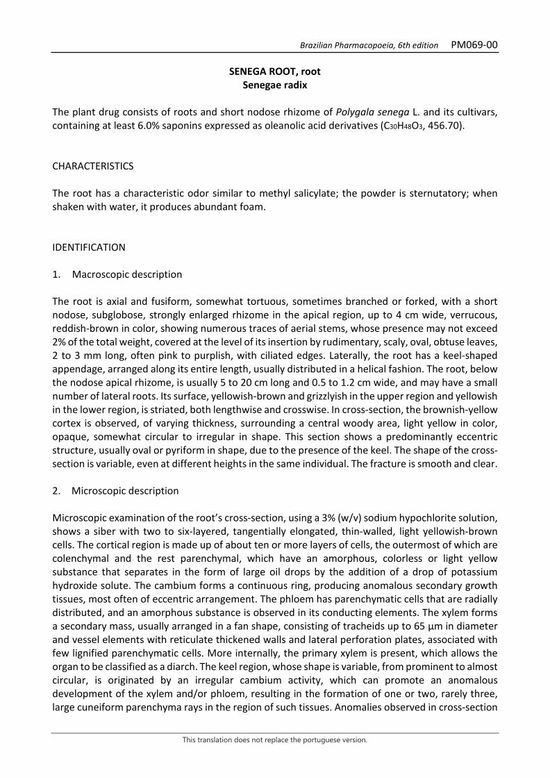
Brazilian Pharmacopoeia, 6th edition PM069-00
This translation does not replace the portuguese version.
SENEGA ROOT, root Senegae radix
The plant drug consists of roots and short nodose rhizome of Polygala senega L. and its cultivars, containing at least 6.0% saponins expressed as oleanolic acid derivatives (C30H48O3, 456.70). CHARACTERISTICS The root has a characteristic odor similar to methyl salicylate; the powder is sternutatory; when shaken with water, it produces abundant foam. IDENTIFICATION 1. Macroscopic description The root is axial and fusiform, somewhat tortuous, sometimes branched or forked, with a short nodose, subglobose, strongly enlarged rhizome in the apical region, up to 4 cm wide, verrucous, reddish-brown in color, showing numerous traces of aerial stems, whose presence may not exceed 2% of the total weight, covered at the level of its insertion by rudimentary, scaly, oval, obtuse leaves, 2 to 3 mm long, often pink to purplish, with ciliated edges. Laterally, the root has a keel-shaped appendage, arranged along its entire length, usually distributed in a helical fashion. The root, below the nodose apical rhizome, is usually 5 to 20 cm long and 0.5 to 1.2 cm wide, and may have a small number of lateral roots. Its surface, yellowish-brown and grizzlyish in the upper region and yellowish in the lower region, is striated, both lengthwise and crosswise. In cross-section, the brownish-yellow cortex is observed, of varying thickness, surrounding a central woody area, light yellow in color, opaque, somewhat circular to irregular in shape. This section shows a predominantly eccentric structure, usually oval or pyriform in shape, due to the presence of the keel. The shape of the cross-section is variable, even at different heights in the same individual. The fracture is smooth and clear. 2. Microscopic description Microscopic examination of the root’s cross-section, using a 3% (w/v) sodium hypochlorite solution, shows a siber with two to six-layered, tangentially elongated, thin-walled, light yellowish-brown cells. The cortical region is made up of about ten or more layers of cells, the outermost of which are colenchymal and the rest parenchymal, which have an amorphous, colorless or light yellow substance that separates in the form of large oil drops by the addition of a drop of potassium hydroxide solute. The cambium forms a continuous ring, producing anomalous secondary growth tissues, most often of eccentric arrangement. The phloem has parenchymatic cells that are radially distributed, and an amorphous substance is observed in its conducting elements. The xylem forms a secondary mass, usually arranged in a fan shape, consisting of tracheids up to 65 μm in diameter and vessel elements with reticulate thickened walls and lateral perforation plates, associated with few lignified parenchymatic cells. More internally, the primary xylem is present, which allows the organ to be classified as a diarch. The keel region, whose shape is variable, from prominent to almost circular, is originated by an irregular cambium activity, which can promote an anomalous development of the xylem and/or phloem, resulting in the formation of one or two, rarely three, large cuneiform parenchyma rays in the region of such tissues. Anomalies observed in cross-section

Brazilian Pharmacopoeia, 6th edition PM069-00
This translation does not replace the portuguese version.
correspond to profound modifications in the anatomical structure of the bark and secondary xylem, becoming very evident when processed with floroglucinol and hydrochloric acid. 3. Microscopic description of powder The sample meets all requirements for the species, except the macroscopic characters. Characteristics are: light brown color; fragments of suber; yellowish cortical parenchyma fragments, with oil drops; collenchyma cells with oil drops; short tracheid fragments; parenchyma ray cells lignified and with large simple pores. 4. Macroscopic and microscopic description of impurities. Stem remains, in cross-section, show epidermis with sub-rectangular and elongated cells, parenchymatous cortex, non-lignified pericyclic fiber sheath, phloem with small diameter elements, xylem formed by tracheids and vessel elements with walls of reticulate, helical, or pitted thickening, and parenchymatic medulla. Scaly leaves, when present, exhibit epidermis with sinuous anticlinal walls, rounded unicellular trichomes at the apex, and anomocytic stomata. 5. Proceed as described in Thin-layer chromatography (5.2.17.1). Stationary phase: silica gel G (0.250 mm). Mobile phase: use the upper phase of a mixture of butyl alcohol, water, and glacial acetic acid (50:40:10). Sample solution: weigh 1 g of the powdered drug, add 10 mL of 70% (v/v) ethyl alcohol, and boil for 15 minutes, under reflux. Filter and cool. Reference solution: prepare a solution of aescin at 1 mg/mL in 70% (v/v) ethyl alcohol. Procedimento: apply in two chromatoplates, in the form of a band, separately, 10 μL of the Sample Solution and 10 μL and 40 μL of the Reference Solution. Conduct chromatogram. Remove chromatoplate and allow it to air dry. Nebulize the first plate with anisaldehyde RS1 and heat in an oven at 100 ºC to 105 ºC, until red spots corresponding to the saponosides appear. Nebulize the second plate with 20% (w/v) phosphomolybdic acid in ethyl alcohol and heat in an oven at 100 °C to 105 °C until the spots corresponding to the saponosides turn blue. The intensity and size of the spots obtained in the chromatogram of the Sample solution are between the two spots corresponding to aescin, obtained by applying 10 μL and 40 μL of the Reference solution. Results: the diagram below shows the sequences of zones obtained with the Reference solution and the Sample solution. Other zones may occasionally develop.
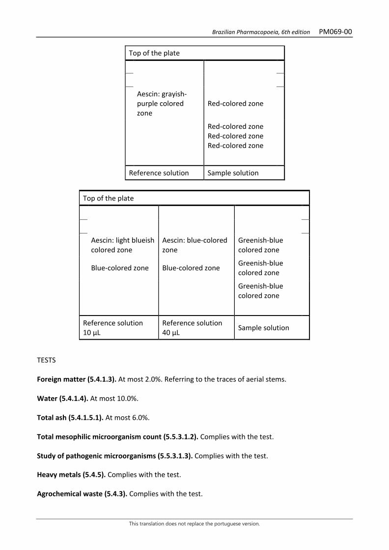
Brazilian Pharmacopoeia, 6th edition PM069-00
This translation does not replace the portuguese version.
Top of the plate
Aescin: grayish-purple colored zone
Red-colored zone
Red-colored zone Red-colored zone Red-colored zone
Reference solution Sample solution
Top of the plate
Aescin: light blueish colored zone
Aescin: blue-colored zone
Greenish-blue colored zone
Blue-colored zone Blue-colored zone Greenish-blue colored zone
Greenish-blue colored zone
Reference solution 10 μL
Reference solution 40 μL Sample solution
TESTS Foreign matter (5.4.1.3). At most 2.0%. Referring to the traces of aerial stems. Water (5.4.1.4). At most 10.0%. Total ash (5.4.1.5.1). At most 6.0%. Total mesophilic microorganism count (5.5.3.1.2). Complies with the test. Study of pathogenic microorganisms (5.5.3.1.3). Complies with the test. Heavy metals (5.4.5). Complies with the test. Agrochemical waste (5.4.3). Complies with the test.
Brazilian Pharmacopoeia, 6th edition PM069-00
This translation does not replace the portuguese version.
DOSAGE Saponins To proceed as described in Visible absorption spectrophotometry (5.2.14). Prepare solutions as described below. Coloring reagent: Dissolve 75 mg ferric chloride in 50 mL anhydrous acetic acid. Add, while shaking and cooling, 50 mL of sulfuric acid. Use immediately after preparation. Stock solution: accurately weigh about 1 g of the pulverized plant and transfer to a 250-mL round-bottomed flask. Add 70 g of 50% (v/v) ethyl alcohol, 0.1 mL silicone defoamer, and a few glass beads. Accurately weigh the ensemble and heat it, under reflux, in a water bath for 60 minutes. Cool, top off the initial weight with 50% (v/v) ethyl alcohol. Centrifuge, separate the residue and the decanted solution, which is weighed and reduced to residue in rotavapor at a maximum temperature of 60 °C. Suspend the residue in 10 mL 0.1 M hydrochloric acid, transfer to a 250-mL separating funnel, and wash the flask with two 5 mL portions of 0.1 M hydrochloric acid. Gather the acid phases and extract with three 70-mL portions of the upper phase of a mixture of butyl alcohol, 0.1 M hydrochloric acid, and chloroform (180:90:30). After shaking, the two phases should stand for at least 15 minutes before separation. The organic phases are gathered and rinsed with two portions of the lower phase of the mixture of butyl alcohol, 0.1 M hydrochloric acid, and chloroform (180:90:30). The lower phase is neglected. Evaporate the organic phase to residue at a rotary evaporator at a maximum temperature of 60 °C. Suspend the residue with 98% (v/v) glacial acetic acid and transfer to a 50-mL volumetric flask. Top off the volume with glacial acetic acid and homogenize. Filter the solution, discarding the first 20 mL of the filtrate. Sample solution: transfer 0.5 mL of the Stock solution to a test tube, add 4 mL of the Coloring Reagent. Homogenize the test tubes in a water bath at (60 ± 1) °C for 25 minutes. Cool in an ice bath for 30 seconds and take the reading immediately. Blank solution: transfer 0.5 mL of glacial acetic acid to a test tube and add 4 mL of the Coloring Reagent. Homogenize the test tubes in a water bath at (60 ± 1) °C for 25 minutes. Cool in an ice bath for 30 seconds and take the reading immediately. Procedure: measure the absorbance of the Sample solution at 520 nm, using the Blank solution for zero adjustment. Calculate the percentage content of saponins, as oleanolic acid derivatives, according to the following expression:
𝑇𝑇𝑇𝑇𝑇𝑇 =A × 463,2𝑚𝑚1 × 𝑚𝑚2
in which, TAO = saponin content expressed as oleanolic acid derivatives % (w/w); A = absorbance measured in the Sample solution; m1 = mass in grams of the solution after centrifugation; m2 = mass in grams of the sample, considering the determined water content;

Brazilian Pharmacopoeia, 6th edition PM069-00
This translation does not replace the portuguese version.
PACKAGING AND STORAGE In tightly closed containers, protected from light and heat.
Figure 1 – Macroscopic, microscopic and microscopic aspects of the powder in Polygala senega L.
___________________________ The scales correspond in A to 7 mm; in B, C, D, E and F to 1 mm; in G, H, I and J to 100 μm.

Brazilian Pharmacopoeia, 6th edition PM069-00
This translation does not replace the portuguese version.
A – overall appearance of the root with nodose apical rhizome. B, D, E and F – overall appearance of root cross-sections: anomaly of parenchyma rays (a); cortex (cx); suberized cortex (cxs); phloem (f); periderm (pe); xylem (x). C – overall appearance of the root cross-section with normal structure. G – cells of the cortical parenchyma of the innermost region. H – detail of vessel elements. I – cells of the cortical parenchyma of the outermost region. J – detail of a portion of the root in cross-section, as indicated in C: cortex (cx); suberized cortex (cxs); phloem (f); periderm (pe); xylem (x).
Brazilian Pharmacopoeia, 6th edition PM070-00
This translation does not replace the portuguese version.
GALE OF THE WIND, aerial part Phyllanthus niruriae herbae
The plant drug consists of the dried aerial parts of Phyllanthus niruri L. [syn. Phyllanthus niruri ssp. niruri L. and Phyllanthus niruri ssp. lathyroides (Kunth) G.L. Webster] containing not less than 6.5% total tannins and 0.15% gallic acid (C7H6O5, 170.12). IDENTIFICATION 1. Macroscopic description Herbaceous, glabrous stems up to 80 cm long, simple or branched, with filiform lateral branches, these bearing leaves. Simple, alternate, membranous, glabrous, oblong-elliptic leaves, with an attenuated, sometimes mucronate apex and asymmetrical base, smooth margin. Laminae 0.5 to 1.4 cm long and 0.3 to 0.6 cm wide, broquidodrome venation. Petiole up to 0.1 cm long. Stipule 0.1 cm to 0.2 cm long, triangular-lanceolate, with a long and narrowly acute to acicular apex and a full base. Female flowers up to 0.4 cm in diameter, with five elliptic tepals and entire disk; tricarpellate, trilocular ovary, each locule bispermal; three bifid stylets on the apical portion, globose stigmas; pedicel 0.1 to 0.4 cm long. Male flowers with five broad-ovate tepals, pentalobate disk and three stamens with conate filaments at the base; pedicels about 0.2 cm long. Schizocarpic fruits, of the tricoca type, 0.10 to 0.25 cm in diameter, depressed-globose, exposed towards the abaxial region of the branches, separating into carpids (cocas); two seeds per locule, triangular, with acute to rounded apex; pedicels about 0.4 to 0.5 cm long at maturity; persistent, membranous tepals, reaching two-thirds of the fruit height. The macroscopic features in Phyllanthus niruri and Phyllanthus tenellus are crucial to distinguish them, since both are very similar in terms of anatomical characteristics. In Phyllanthus niruri, the main macroscopic characteristics are leaves with an asymmetrical base, globose stigmas, and the presence of three stamens with conate filaments at the base. 2. Microscopic description The stem, in cross-section, shows unistratified epidermis. Subepidermically, there are one or more layers of collenchyma with angular thickening, followed by chlorenchyma formed by isodiametric cells, containing starch grains. More internally, one or more layers of cortical parenchyma occur. Externally, the phloem consists of clusters of fibers with very thick walls and reduced lumen. Xylem vessel elements alternate with rows of fibers and sclerified cells. The medullary parenchyma may have starch grains. Calcium oxalate druses occur in the parenchyma. In larger diameter stems, periderm may occur, followed by chlorenchyma with starch grains and crystals. Dorsiventrally symmetrical leaf lamina, usually hypo-stomatic, with paracitic, rarely anomocytic stomata. In a front view, the epidermal cells of the adaxial surface show irregular contours and wavy walls. The cuticle is thin, the epidermis is unistratified on both sides, containing flattened and some papillose cells. The palisade parenchyma is unistratified, occupying about two thirds of the thickness of the mesophyll, with idioblasts with calcium oxalate druses. Small crystals of different shapes are common and rhombohedral crystals rarely occur. The spongy parenchyma consists of two to three layers. Small aggregated and/or isolated crystals are common. The vascular system is of the collateral type.
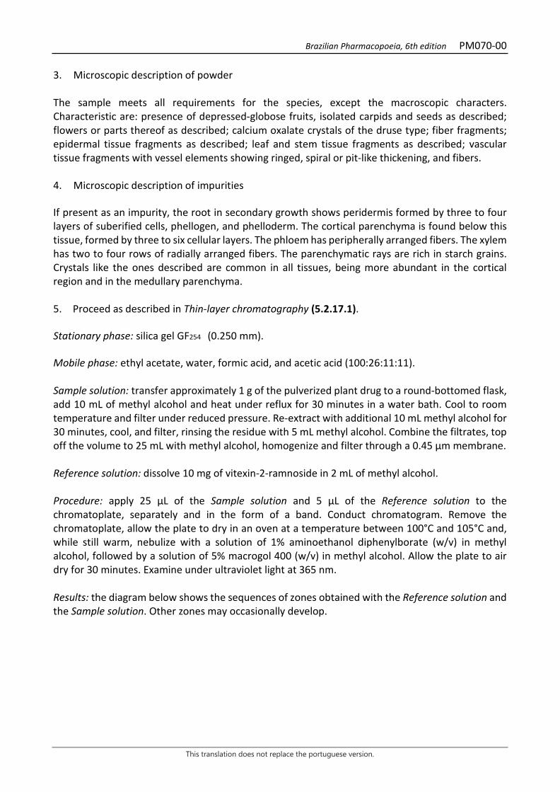
Brazilian Pharmacopoeia, 6th edition PM070-00
This translation does not replace the portuguese version.
3. Microscopic description of powder The sample meets all requirements for the species, except the macroscopic characters. Characteristic are: presence of depressed-globose fruits, isolated carpids and seeds as described; flowers or parts thereof as described; calcium oxalate crystals of the druse type; fiber fragments; epidermal tissue fragments as described; leaf and stem tissue fragments as described; vascular tissue fragments with vessel elements showing ringed, spiral or pit-like thickening, and fibers. 4. Microscopic description of impurities If present as an impurity, the root in secondary growth shows peridermis formed by three to four layers of suberified cells, phellogen, and phelloderm. The cortical parenchyma is found below this tissue, formed by three to six cellular layers. The phloem has peripherally arranged fibers. The xylem has two to four rows of radially arranged fibers. The parenchymatic rays are rich in starch grains. Crystals like the ones described are common in all tissues, being more abundant in the cortical region and in the medullary parenchyma. 5. Proceed as described in Thin-layer chromatography (5.2.17.1). Stationary phase: silica gel GF254 (0.250 mm). Mobile phase: ethyl acetate, water, formic acid, and acetic acid (100:26:11:11). Sample solution: transfer approximately 1 g of the pulverized plant drug to a round-bottomed flask, add 10 mL of methyl alcohol and heat under reflux for 30 minutes in a water bath. Cool to room temperature and filter under reduced pressure. Re-extract with additional 10 mL methyl alcohol for 30 minutes, cool, and filter, rinsing the residue with 5 mL methyl alcohol. Combine the filtrates, top off the volume to 25 mL with methyl alcohol, homogenize and filter through a 0.45 μm membrane. Reference solution: dissolve 10 mg of vitexin-2-ramnoside in 2 mL of methyl alcohol. Procedure: apply 25 μL of the Sample solution and 5 μL of the Reference solution to the chromatoplate, separately and in the form of a band. Conduct chromatogram. Remove the chromatoplate, allow the plate to dry in an oven at a temperature between 100°C and 105°C and, while still warm, nebulize with a solution of 1% aminoethanol diphenylborate (w/v) in methyl alcohol, followed by a solution of 5% macrogol 400 (w/v) in methyl alcohol. Allow the plate to air dry for 30 minutes. Examine under ultraviolet light at 365 nm. Results: the diagram below shows the sequences of zones obtained with the Reference solution and the Sample solution. Other zones may occasionally develop.
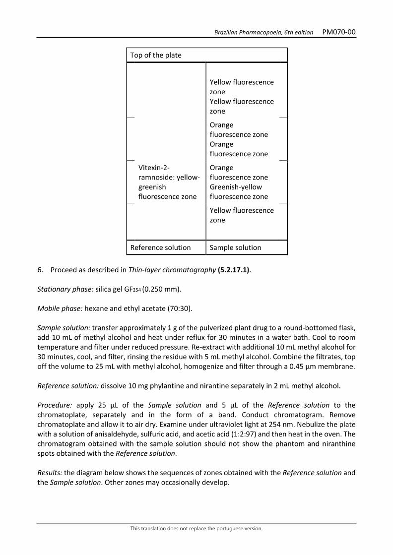
Brazilian Pharmacopoeia, 6th edition PM070-00
This translation does not replace the portuguese version.
Top of the plate
Yellow fluorescence zone Yellow fluorescence zone
Orange fluorescence zone Orange fluorescence zone
Vitexin-2-ramnoside: yellow-greenish fluorescence zone
Orange fluorescence zone Greenish-yellow fluorescence zone
Yellow fluorescence zone
Reference solution Sample solution
6. Proceed as described in Thin-layer chromatography (5.2.17.1). Stationary phase: silica gel GF254 (0.250 mm). Mobile phase: hexane and ethyl acetate (70:30). Sample solution: transfer approximately 1 g of the pulverized plant drug to a round-bottomed flask, add 10 mL of methyl alcohol and heat under reflux for 30 minutes in a water bath. Cool to room temperature and filter under reduced pressure. Re-extract with additional 10 mL methyl alcohol for 30 minutes, cool, and filter, rinsing the residue with 5 mL methyl alcohol. Combine the filtrates, top off the volume to 25 mL with methyl alcohol, homogenize and filter through a 0.45 μm membrane. Reference solution: dissolve 10 mg phylantine and nirantine separately in 2 mL methyl alcohol. Procedure: apply 25 μL of the Sample solution and 5 μL of the Reference solution to the chromatoplate, separately and in the form of a band. Conduct chromatogram. Remove chromatoplate and allow it to air dry. Examine under ultraviolet light at 254 nm. Nebulize the plate with a solution of anisaldehyde, sulfuric acid, and acetic acid (1:2:97) and then heat in the oven. The chromatogram obtained with the sample solution should not show the phantom and niranthine spots obtained with the Reference solution. Results: the diagram below shows the sequences of zones obtained with the Reference solution and the Sample solution. Other zones may occasionally develop.
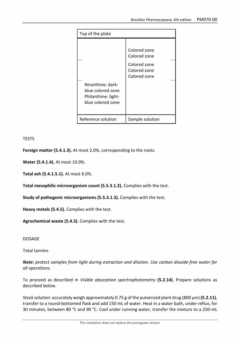
Brazilian Pharmacopoeia, 6th edition PM070-00
This translation does not replace the portuguese version.
Top of the plate
Colored zone Colored zone
Colored zone Colored zone Colored zone
Niranthine: dark-blue colored zone Philanthine: light-blue colored zone
Reference solution Sample solution
TESTS Foreign matter (5.4.1.3). At most 2.0%, corresponding to the roots. Water (5.4.1.4). At most 10.0%. Total ash (5.4.1.5.1). At most 6.0%. Total mesophilic microorganism count (5.5.3.1.2). Complies with the test. Study of pathogenic microorganisms (5.5.3.1.3). Complies with the test. Heavy metals (5.4.5). Complies with the test. Agrochemical waste (5.4.3). Complies with the test. DOSAGE Total tannins Note: protect samples from light during extraction and dilution. Use carbon dioxide-free water for all operations. To proceed as described in Visible absorption spectrophotometry (5.2.14). Prepare solutions as described below. Stock solution: accurately weigh approximately 0.75 g of the pulverized plant drug (800 µm) (5.2.11), transfer to a round-bottomed flask and add 150 mL of water. Heat in a water bath, under reflux, for 30 minutes, between 80 °C and 90 °C. Cool under running water, transfer the mixture to a 250-mL
Brazilian Pharmacopoeia, 6th edition PM070-00
This translation does not replace the portuguese version.
volumetric flask, top off the volume with water and homogenize. Allow the sediment to settle and filter, discarding the first 50 mL of the filtrate. Sample solution for total polyphenol content: transfer 5 mL from the Stock solution to a 25-mL volumetric flask, top off the volume with water and homogenize. Add 2 mL Folin-Denis reagent to 5 mL of this solution, dilute to 50 mL with sodium carbonate RS and homogenize. Measure the absorbance of the solution (A1) at 715 nm, precisely three minutes after adding the last reagent, using water for zero adjustment. Sample solution for polyphenols not adsorbed by hide powder: for 0.2 g of the hide powder CRS, add 20 mL of the Stock solution and mechanically shake for 60 minutes. Filter. Dilute 5 mL of the solution to 25 mL with water. Add 2 mL Folin-Denis reagent to 5 mL of this solution, dilute to 50 mL with sodium carbonate RS and homogenize. Measure the absorbance of the solution (A2) at 715 nm, precisely three minutes after adding the last reagent, using water for zero adjustment. Reference solution: dissolve 50 mg pyrogallol in water and dilute to 100 mL with the same solvent. Dilute 5 mL of the solution to 100 mL with water. Add 2 mL Folin-Denis reagent to 5 mL of this solution, dilute to 50 mL with sodium carbonate RS and homogenize. Measure solution absorbance at 715 nm (A3), precisely 3 minutes after adding the last reagent, and within 15 minutes from the pyrogallol dissolution using water for zero adjustment. Calculate total tannin content, expressed as a percentage of pyrogallol, according to the following expression:
𝑇𝑇𝑇𝑇 =(𝑇𝑇1 − 𝑇𝑇2) × 13,12
𝑇𝑇3 × 𝑚𝑚
in which, TT = total tannin content expressed as pyrogallol % (w/w); A1 = absorbance measured for the Sample solution for total polyphenols; A2 = absorbance measured for the Sample solution for polyphenols not adsorbed by hide powder; A3 = absorbance measured for the Reference solution; m = mass in grams of the sample used, considering the determined water content. Gallic acid Proceed as described in High Performance Liquid Chromatography (5.2.17.4). Use a chromatograph equipped with an ultraviolet detector at 275 nm; a pre-column containing a hydrophilic C18 reversed phase and a 100 mm long, 2.1mm internal diameter column packed with hydrophilic C18 (3 μm); Mobile phase flow rate of 0.25 mL/minute. Eluent (A): 0.05% trifluoroacetic acid. Eluent (B): 0.05% trifluoroacetic acid in methyl alcohol.

Brazilian Pharmacopoeia, 6th edition PM070-00
This translation does not replace the portuguese version.
Time (minutes) Eluent A (%) Eluent B (%) Elution
0 - 7 95 5 isocratic 10 - 14 95 → 0 5 → 100 linear gradient Sample solution: accurately weigh approximately 0.75 g of the pulverized plant drug (800 μm) (5.2.11) and transfer to a round-bottomed flask. Add 10 mL of water and heat, on a blanket, under reflux, to boiling for 15 minutes. Cool under running water and filter the extract under reduced pressure. Wash off residue with water. Transfer the filtrate to a 250-mL volumetric flask, top off the volume with water and homogenize. Filter through a 0.45 µm filter unit. Reference solution: dissolve an accurately weighed amount of gallic acid CRS in Eluent (A) to obtain a 1 mg/mL solution. Solutions for the analytical curve: transfer 1 mL of the Reference solution into a 50-mL volumetric flask, top off the volume with Eluent (A) and homogenize. Dilute aliquots of 1 mL, 2 mL, 3 mL, 5 mL and 7 mL of this solution to 10 mL using Eluent (A) thus obtaining solutions with concentrations of 2.0 μg/ mL, 4.0 μg/mL, 6.0 μg/mL, 10.0 μg/mL and 14.0 μg/mL. Filter the solutions through a 0.45 µm filter unit. Procedure: inject 5 µL of the Solutions for the analytical curve and 5 µL of the Sample solution separately. Record the chromatograms and measure the areas under the peak corresponding to gallic acid. The relative retention time is about 4.6 minutes for gallic acid. Calculate gallic acid content in the sample from the straight line equation obtained from the gallic acid analytical curve. The result is expressed as the average of the determinations in grams of gallic acid, in percent, considering the determined water content. PACKAGING AND STORAGE In tightly closed containers, protected from light and heat.
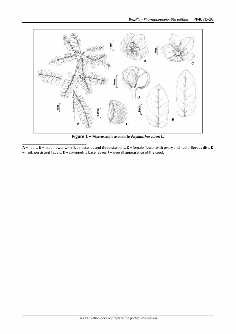
Brazilian Pharmacopoeia, 6th edition PM070-00
This translation does not replace the portuguese version.
Figure 1 – Macroscopic aspects in Phyllanthus niruri L.
___________________________ A – habit. B – male flower with five nectaries and three stamens. C – female flower with ovary and nectariferous disc. D – fruit, persistent tepals. E – asymmetric base leaves F – overall appearance of the seed.
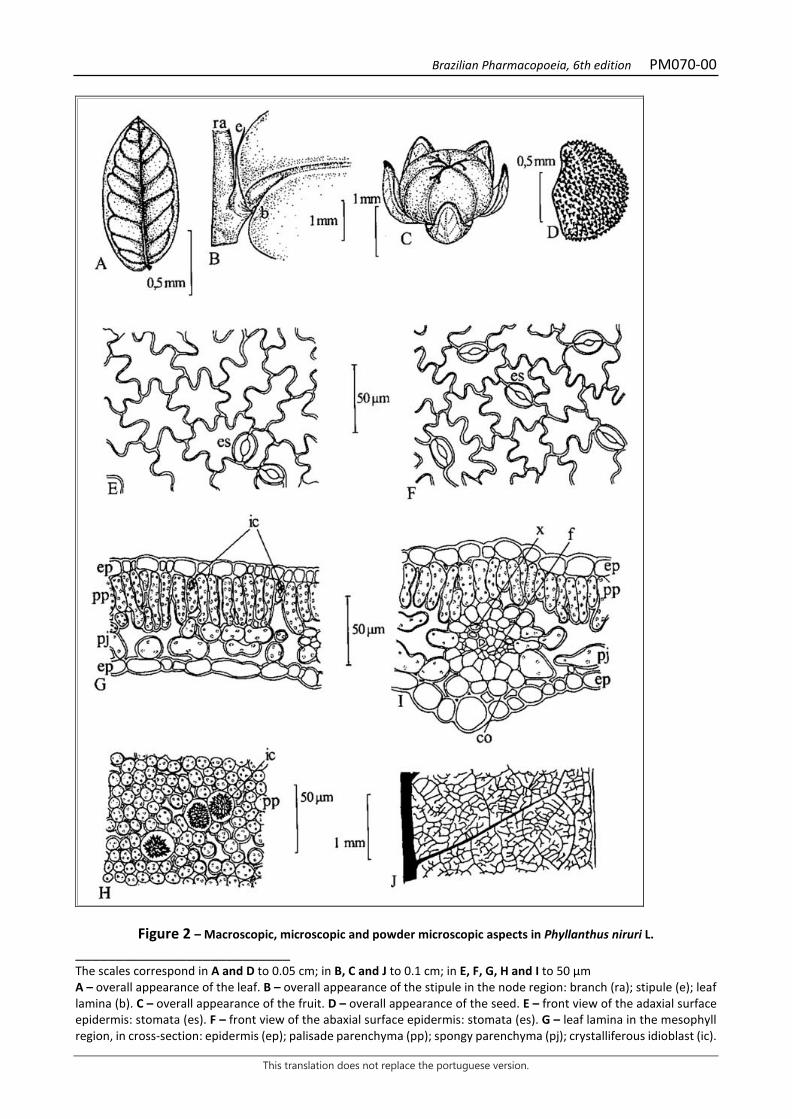
Brazilian Pharmacopoeia, 6th edition PM070-00
This translation does not replace the portuguese version.
Figure 2 – Macroscopic, microscopic and powder microscopic aspects in Phyllanthus niruri L. ___________________________ The scales correspond in A and D to 0.05 cm; in B, C and J to 0.1 cm; in E, F, G, H and I to 50 µm A – overall appearance of the leaf. B – overall appearance of the stipule in the node region: branch (ra); stipule (e); leaf lamina (b). C – overall appearance of the fruit. D – overall appearance of the seed. E – front view of the adaxial surface epidermis: stomata (es). F – front view of the abaxial surface epidermis: stomata (es). G – leaf lamina in the mesophyll region, in cross-section: epidermis (ep); palisade parenchyma (pp); spongy parenchyma (pj); crystalliferous idioblast (ic).
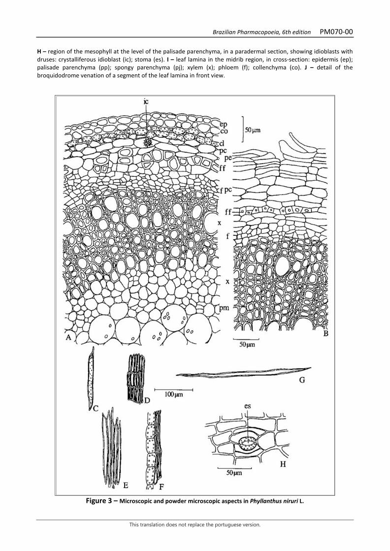
Brazilian Pharmacopoeia, 6th edition PM070-00
This translation does not replace the portuguese version.
H – region of the mesophyll at the level of the palisade parenchyma, in a paradermal section, showing idioblasts with druses: crystalliferous idioblast (ic); stoma (es). I – leaf lamina in the midrib region, in cross-section: epidermis (ep); palisade parenchyma (pp); spongy parenchyma (pj); xylem (x); phloem (f); collenchyma (co). J – detail of the broquidodrome venation of a segment of the leaf lamina in front view.
Figure 3 – Microscopic and powder microscopic aspects in Phyllanthus niruri L.

Brazilian Pharmacopoeia, 6th edition PM070-00
This translation does not replace the portuguese version.
___________________________ The scales correspond in A, B and H to 50 µm; in C, D, E, F and G to 100 µm. A – detail of the stem in cross-section: epidermis (ep); collenchyma (co); chlorenchyma (cl); cortical parenchyma (pc); phloem fibers (ff); phloem (f); xylem (x); medullary parenchyma (pm); crystalliferous idioblast (ic). B – detail of root in cross-section: periderm (pe); cortical parenchyma (pc); phloem fibers (ff); phloem (f); xylem (x). C – longitudinal view of vessel element with pit thickening. D – sclerified parenchyma cells. E – phloem fibers in longitudinal view. F – chlorenchyma cells next to the phloem fibers. G – fiber in longitudinal view. H – detail of a fragment of the stem epidermis in front view, showing the stoma (es).

Brazilian Pharmacopoeia, 6th edition PM071-00
This translation does not replace the portuguese version.
GALE OF THE WIND, aerial part Phyllanthus tenellae herbae
The plant drug consists of dried aerial parts of Phyllanthus tenellus Roxb. containing at least 9.0% total tannins and 0.12% gallic acid (C7H6O5, 170.12). IDENTIFICATION 1. Macroscopic description Herbaceous, glabrous stems up to 60 cm long, simple or branched, with filiform lateral branches, these bearing leaves. Simple, alternate, membranous, glabrous, elliptic to elliptic-ovate leaves, with obtuse apex and obtuse to acute and symmetrical base, smooth margin. Laminae 0.8 to 2.5 cm long and 0.5 to 1.2 cm wide, broquidodrome venation. Petiole up to 0.1 cm long. Stipule up to 0.15 cm long, narrow-triangular, with acute apex and entire base. Female flowers up to 0.3 cm in diameter, with five obovalate tepals and entire disk; tricarpellate, trilocular ovary, each locule bispermal; three bifid stylets on the apical portion, non-globose stigmas; pedicel 0.1 to 0.8 cm long. Male flowers with five suborbicular tepals, pentalobate disk and five stamens with free filaments between them; pedicels up to 0.15 cm long. Schizocarpic fruits, of the tricoca type, 0.1 to 0.2 cm in diameter, depressed-globose, exposed towards the adaxial region of the branches, separating into carpids (cocas); two seeds per locule, triangular, with rounded apex; pedicels up to 0.9 cm long at maturity; persistent, membranous tepals, reaching half of the fruit height. The macroscopic features in Phyllanthus tenellus and Phyllanthus niruri are crucial to distinguish them, since both are very similar in terms of anatomical characteristics. In Phyllanthus tenellus, the main macroscopic characteristics are leaves with an asymmetrical base, non-globose stigmas, and the presence of five stamens with free filaments. 2. Microscopic description The stem, in cross-section, shows unistratified epidermis. Subepidermically, there are one or two layers of collenchyma with angular thickening, followed by chlorenchyma formed by isodiametric cells, containing starch grains. More internally, one or more layers of cortical parenchyma occur. Externally, the phloem consists of clusters of fibers with very thick walls and reduced lumen. Xylem vessel elements alternate with rows of fibers and sclerified cells. The medullary parenchyma may have starch grains. Rhombohedral calcium oxalate crystals occur in the parenchyma. In larger diameter stems, periderm may occur, followed by chlorenchyma with starch grains and crystals. Dorsiventrally symmetrical leaf lamina, hypo-stomatic, with paracitic, rarely anomocytic stomata. In a front view, the epidermal cells of the adaxial surface show irregular contours and wavy walls. The cuticle is thin, the epidermis is unistratified on both sides, containing flattened and some papillose cells. The palisade parenchyma is unistratified, occupying about half of the thickness of the mesophyll, with idioblasts with rhombohedral calcium oxalate crystals. The spongy parenchyma consists of two to three layers of cells. The vascular system is of the collateral type. 3. Microscopic description of powder The sample meets all requirements for the species, except the macroscopic characters. Characteristic are: presence of depressed-globose fruits, isolated carpids and seeds as described;
Brazilian Pharmacopoeia, 6th edition PM071-00
This translation does not replace the portuguese version.
flowers or parts thereof as described; rhombohedral calcium oxalate crystals; fiber fragments; epidermal tissue fragments as described; leaf and stem tissue fragments as described; vascular tissue fragments with vessel elements showing ringed, spiral and, more frequently, pit-like thickening, and fibers. 4. Microscopic description of impurities If present as an impurity, the root in secondary growth shows peridermis formed by two or three suberified layers, phelloge, and phelloderm. The cortical parenchyma is found below this tissue, formed by three to four cellular layers. The phloem has peripherally arranged fibers. The xylem has fibers between the vessel elements or two to four rows of radially arranged fibers, and parenchyma rays formed by one or two rows of thick-walled cells containing starch grains. Crystals like the ones described are common in parenchymal tissues, including vascular systems. 5. Proceed as described in Thin-layer chromatography (5.2.17.1). Stationary phase: silica gel GF254 (0.250 mm). Mobile phase: ethyl acetate, water, formic acid, and acetic acid (100:26:11:11). Sample solution: transfer approximately 1 g of the pulverized plant drug to a round-bottomed flask, add 10 mL of methyl alcohol and heat under reflux for 30 minutes in a water bath. Cool to room temperature and filter under reduced pressure. Re-extract with additional 10 mL methyl alcohol for 30 minutes, cool, and filter, rinsing the residue with 5 mL methyl alcohol. Gather the filtrates. Top off the volume to 25 mL with methyl alcohol, homogenize and filter through a 0.45 μm membrane. Reference solution: dissolve 10 mg rutin in 2 mL methyl alcohol. Procedure: apply 25 μL of the Sample solution and 5 μL of the Reference solution to the chromatoplate, separately and in the form of a band. Conduct chromatogram. Remove the chromatoplate, allow the plate to dry in an oven at a temperature between 100°C and 105°C and, while still warm, nebulize with aminoethanol diphenylborate SE followed by a solution of 5% macrogol 400 (w/v) in methyl alcohol. Allow the plate to air dry for 30 minutes. Examine under ultraviolet light at 365 nm. Results: the diagram below shows the sequences of zones obtained with the Reference solution and the Sample solution. Other zones may occasionally develop.
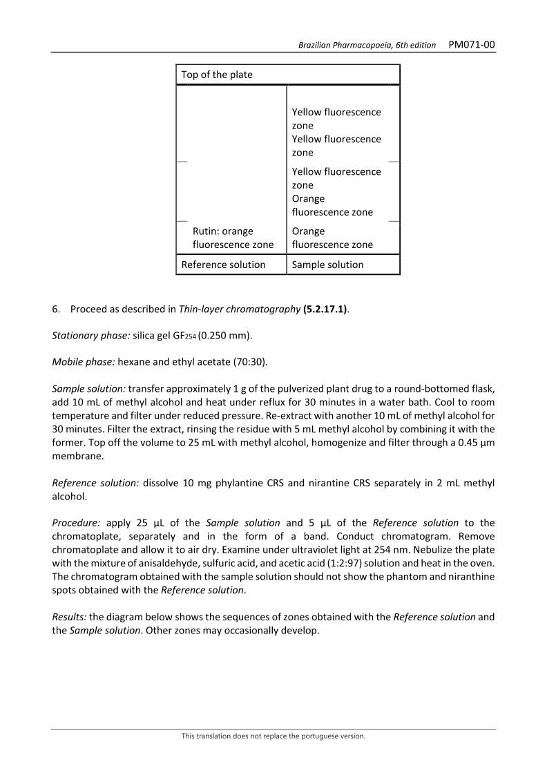
Brazilian Pharmacopoeia, 6th edition PM071-00
This translation does not replace the portuguese version.
Top of the plate
Yellow fluorescence zone Yellow fluorescence zone
Yellow fluorescence zone Orange fluorescence zone
Rutin: orange fluorescence zone
Orange fluorescence zone
Reference solution Sample solution
6. Proceed as described in Thin-layer chromatography (5.2.17.1). Stationary phase: silica gel GF254 (0.250 mm). Mobile phase: hexane and ethyl acetate (70:30). Sample solution: transfer approximately 1 g of the pulverized plant drug to a round-bottomed flask, add 10 mL of methyl alcohol and heat under reflux for 30 minutes in a water bath. Cool to room temperature and filter under reduced pressure. Re-extract with another 10 mL of methyl alcohol for 30 minutes. Filter the extract, rinsing the residue with 5 mL methyl alcohol by combining it with the former. Top off the volume to 25 mL with methyl alcohol, homogenize and filter through a 0.45 μm membrane. Reference solution: dissolve 10 mg phylantine CRS and nirantine CRS separately in 2 mL methyl alcohol. Procedure: apply 25 μL of the Sample solution and 5 μL of the Reference solution to the chromatoplate, separately and in the form of a band. Conduct chromatogram. Remove chromatoplate and allow it to air dry. Examine under ultraviolet light at 254 nm. Nebulize the plate with the mixture of anisaldehyde, sulfuric acid, and acetic acid (1:2:97) solution and heat in the oven. The chromatogram obtained with the sample solution should not show the phantom and niranthine spots obtained with the Reference solution. Results: the diagram below shows the sequences of zones obtained with the Reference solution and the Sample solution. Other zones may occasionally develop.
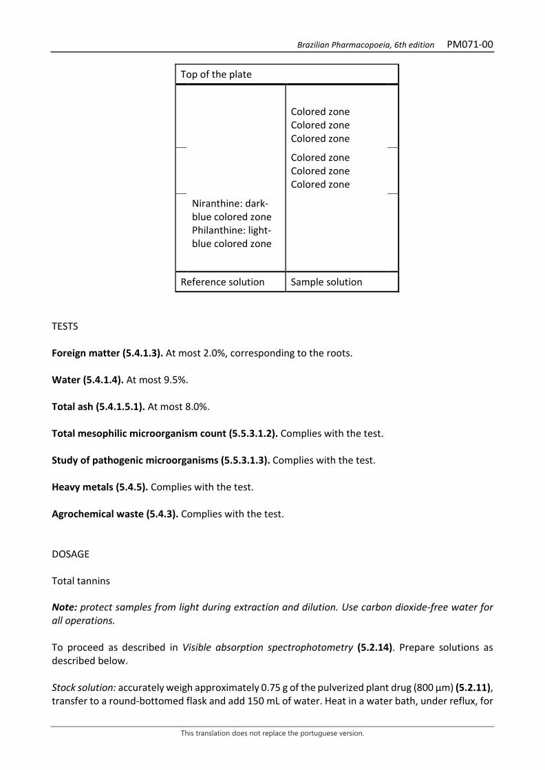
Brazilian Pharmacopoeia, 6th edition PM071-00
This translation does not replace the portuguese version.
Top of the plate
Colored zone Colored zone Colored zone
Colored zone Colored zone Colored zone
Niranthine: dark-blue colored zone Philanthine: light-blue colored zone
Reference solution Sample solution TESTS Foreign matter (5.4.1.3). At most 2.0%, corresponding to the roots. Water (5.4.1.4). At most 9.5%. Total ash (5.4.1.5.1). At most 8.0%. Total mesophilic microorganism count (5.5.3.1.2). Complies with the test. Study of pathogenic microorganisms (5.5.3.1.3). Complies with the test. Heavy metals (5.4.5). Complies with the test. Agrochemical waste (5.4.3). Complies with the test. DOSAGE Total tannins Note: protect samples from light during extraction and dilution. Use carbon dioxide-free water for all operations. To proceed as described in Visible absorption spectrophotometry (5.2.14). Prepare solutions as described below. Stock solution: accurately weigh approximately 0.75 g of the pulverized plant drug (800 µm) (5.2.11), transfer to a round-bottomed flask and add 150 mL of water. Heat in a water bath, under reflux, for
Brazilian Pharmacopoeia, 6th edition PM071-00
This translation does not replace the portuguese version.
30 minutes, between 80 °C and 90 °C. Cool under running water, transfer the mixture to a 250-mL volumetric flask, top off the volume with water and homogenize. Allow the sediment to settle and filter, discarding the first 50 mL of the filtrate. Sample solution for total polyphenol content: transfer 5 mL from the Stock solution to a 25-mL volumetric flask, top off the volume with water and homogenize. Add 2 mL Folin-Denis reagent to 5 mL of this solution, dilute to 50 mL with sodium carbonate RS and homogenize. Measure the absorbance of the solution (A1) at 715 nm, precisely three minutes after adding the last reagent, using water for zero adjustment. Sample solution for polyphenols not adsorbed by hide powder: for 0.2 g of the hide powder CRS, add 20 mL of the Stock solution and mechanically shake for 60 minutes. Filter. Dilute 5 mL of the solution to 25 mL with water and homogenize. Add 2 mL Folin-Denis reagent to 5 mL of this solution, dilute to 50 mL with sodium carbonate RS and homogenize. Measure the absorbance of the solution (A2) at 715 nm, precisely three minutes after adding the last reagent, using water for zero adjustment. Reference solution: dissolve 50 mg pyrogallol in water and dilute to 100 mL with the same solvent. Dilute 5 mL of the solution to 100 mL with water and homogenize. Add 2 mL Folin-Denis reagent to 5 mL of this solution, dilute to 50 mL with sodium carbonate RS and homogenize. Measure solution absorbance at 715 nm (A3), precisely three minutes after adding the last reagent, and within 15 minutes from the pyrogallol dissolution using water for zero adjustment. Calculate total tannin content, in percent, according to the following expression:
𝑇𝑇𝑇𝑇 =(𝑇𝑇1 − 𝑇𝑇2) × 13,12
𝑇𝑇3 × 𝑚𝑚
in which,
TT = total tannin content expressed as pyrogallol % (w/w); A1 = absorbance measured for the Sample solution for total polyphenols; A2 = absorbance measured for the Sample solution for polyphenols not adsorbed by hide powder; A3 = absorbance measured for the Reference solution; m = mass in grams of the sample used, considering the determined water content. Gallic acid Proceed as described in High Performance Liquid Chromatography (5.2.17.4). Use a chromatograph equipped with an ultraviolet detector at 275 nm; a pre-column containing a hydrophilic C18 reversed phase and a 10cm long, 2.1 mm internal diameter column packed with hydrophilic C18 (3 μm); Mobile phase flow rate of 0.25 mL/minute. Eluent (A): 0.05% trifluoroacetic acid (v/v). Eluent (B): 0.05% trifluoroacetic acid in methyl alcohol (v/v).
Brazilian Pharmacopoeia, 6th edition PM071-00
This translation does not replace the portuguese version.
Time (minutes) Eluent A (%) Eluent B (%) Elution
0 - 8 100 0 isocratic 8 - 15 100 → 50 0 → 50 linear gradient 15 - 20 50 50 isocratic Sample solution: accurately weigh approximately 0.75 g of the pulverized plant drug(800 μm) (5.2.11) and transfer to a round-bottomed flask. Add 30 mL of water and heat, on a blanket, under reflux, to boiling for 15 minutes, under magnetic shaking. Cool under running water and filter the extract under reduced pressure. Wash off residue with water. Transfer the filtrate to a 250-mL volumetric flask, top off the volume with water and homogenize. Filter through a 0.45 µm filter unit. Reference solution: dissolve precisely weighed amount of gallic acid in Eluent (A) to obtain a solution at 400 μg/mL. Solutions for the analytical curve: transfer 2 mL of the Reference solution into a 25-mL volumetric flask, top off the volume with Eluent (A) and homogenize. Dilute aliquots of 2 mL and 5 mL of this solution to 25 mL using Eluent (A) thus obtaining solutions with concentrations of 2.6 μg/mL and 6.4 μg/mL. Additionally, dilute aliquots of 3 mL, 5 mL and 7 mL to 10 mL using Eluent (A) to obtain solutions with concentrations of 9.6 μg/mL, 16.0 μg/mL and 22.4 μg/mL. Use the five prepared solutions (2.6 μg/mL; 6.4 μg/ mL; 9.6 μg/mL; 16.0 μg/mL and 22.4 μg/mL) in the construction of the analytical curve, after filtration on a 0.45 μm membrane. Filter through a 0.45 µm filter unit. Procedure: inject 5 µL of the Solutions for the analytical curve and 5 µL of the Sample solution separately. Record the chromatograms and measure the areas under the peak corresponding to gallic acid. The relative retention time is about 5.3 minutes for gallic acid. Calculate gallic acid content in the sample from the straight line equation obtained from the gallic acid analytical curve. The result is expressed as the average of the determinations in grams of gallic acid, in percent, considering the determined water content. PACKAGING AND STORAGE In tightly closed containers, protected from light and heat.

Brazilian Pharmacopoeia, 6th edition PM071-00
This translation does not replace the portuguese version.
Figure 1 – Macroscopic aspects in Phyllanthus tenellus Roxb.
___________________________ A – habit. B – male flower with five nectaries and five stamens. C – female flower with ovary and nectariferous disc. D – fruit, persistent tepals. E – symmetric base leaf. F – overall appearance of the seed.
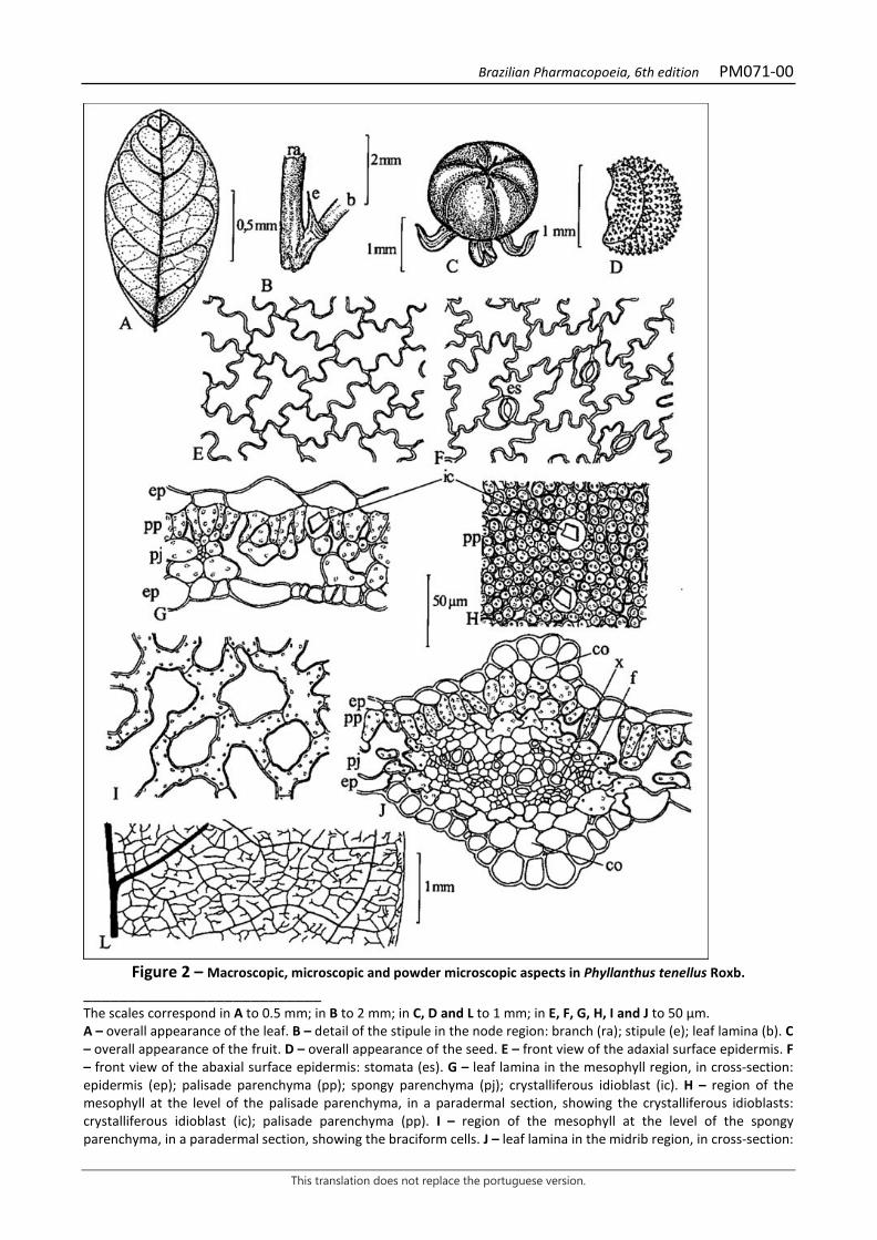
Brazilian Pharmacopoeia, 6th edition PM071-00
This translation does not replace the portuguese version.
Figure 2 – Macroscopic, microscopic and powder microscopic aspects in Phyllanthus tenellus Roxb.
___________________________ The scales correspond in A to 0.5 mm; in B to 2 mm; in C, D and L to 1 mm; in E, F, G, H, I and J to 50 μm. A – overall appearance of the leaf. B – detail of the stipule in the node region: branch (ra); stipule (e); leaf lamina (b). C – overall appearance of the fruit. D – overall appearance of the seed. E – front view of the adaxial surface epidermis. F – front view of the abaxial surface epidermis: stomata (es). G – leaf lamina in the mesophyll region, in cross-section: epidermis (ep); palisade parenchyma (pp); spongy parenchyma (pj); crystalliferous idioblast (ic). H – region of the mesophyll at the level of the palisade parenchyma, in a paradermal section, showing the crystalliferous idioblasts: crystalliferous idioblast (ic); palisade parenchyma (pp). I – region of the mesophyll at the level of the spongy parenchyma, in a paradermal section, showing the braciform cells. J – leaf lamina in the midrib region, in cross-section:
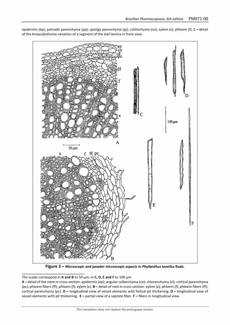
Brazilian Pharmacopoeia, 6th edition PM071-00
This translation does not replace the portuguese version.
epidermis (ep); palisade parenchyma (pp); spongy parenchyma (pj); collenchyma (co); xylem (x); phloem (f). L – detail of the broquidodrome venation of a segment of the leaf lamina in front view.
Figure 3 – Microscopic and powder microscopic aspects in Phyllanthus tenellus Roxb.
___________________________ The scales correspond in A and B to 50 μm; in C, D, E and F to 100 μm. A – detail of the stem in cross-section: epidermis (ep); angular collenchyma (co); chlorenchyma (cl); cortical parenchyma (pc); phloem fibers (ff); phloem (f); xylem (x). B – detail of root in cross-section: xylem (x); phloem (f); phloem fibers (ff); cortical parenchyma (pc). D – longitudinal view of vessel elements with helical pit thickening. D – longitudinal view of vessel elements with pit thickening. E – partial view of a septate fiber. F – fibers in longitudinal view.

Brazilian Pharmacopoeia, 6th edition PM072-00
This translation does not replace the portuguese version.
SOAPBARK, bark Quillaiae cortex
The plant drug consists of dried, fragmented barks from the branches of Quillaja saponaria Molina, devoid of periderm. CHARACTERISTICS The drug is sternutatory. IDENTIFICATION 1. Macroscopic description The drug is sold in flat or slightly curved pieces, forming sheets approximately 1 cm long, 10 cm wide, and 1 to 5 mm thick. The outer surface is whitish in color, with small brownish spots, usually smooth or finely striated lengthwise. Attached to this bark are often brownish to grizzly parts of the rhytidome. The inner surface is yellowish-white and smooth. The fracture is smooth to slightly fibrous. In cross-section, the fracture shows a regularly checkered structure with dark tangential bands and light radial lines. 2. Microscopic description The liber, which alone constitutes the thickness of commercial barks, characteristically exhibits a checkered appearance, which is due to the successive crossing of parenchymal rays with parenchyma zones, which alternate with fiber bundles. Throughout this region, cells are juxtaposed, forming meatus. Parenchyma rays consist of two to six layers of cells 60 to 100 μm long and approximately 20 μm wide. Liberian fibers are winding and are often accompanied by small groups of sclereids. The parenchyma contains cells 20 to 40 µm by 60 to 200 µm long, usually 90 µm wide. It has several mucilage cells, starchy cells with starch grains 5 to 20 μm in diameter and several cells containing calcium oxalate mono-crystals, which may reach 50 to 170 μm in length and up to 30 μm in width. 3. Microscopic description of powder The sample meets all requirements for the species, except the macroscopic characters. Characteristics are: very fine powder and a straw-yellow color, with fragments of the structures described above; sclerenchymatic fibers; large amount of prismatic calcium oxalate crystals in parenchyma fragments or free; parenchyma portions with starch grains; some elongated stone cells with oblique pores; sieve tube fragments; occasionally suberous fragments. 4. Proceed as described in Thin-layer chromatography (5.2.17.1). Stationary phase: silica gel GF254 (0.250 mm). Mobile phase: ethyl alcohol, chloroform and water (40:30:5).
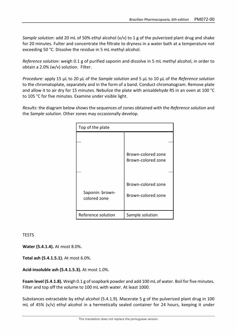
Brazilian Pharmacopoeia, 6th edition PM072-00
This translation does not replace the portuguese version.
Sample solution: add 20 mL of 50% ethyl alcohol (v/v) to 1 g of the pulverized plant drug and shake for 20 minutes. Fulter and concentrate the filtrate to dryness in a water bath at a temperature not exceeding 50 °C. Dissolve the residue in 5 mL methyl alcohol. Reference solution: weigh 0.1 g of purified saponin and dissolve in 5 mL methyl alcohol, in order to obtain a 2.0% (w/v) solution. Filter. Procedure: apply 15 μL to 20 μL of the Sample solution and 5 μL to 10 μL of the Reference solution to the chromatoplate, separately and in the form of a band. Conduct chromatogram. Remove plate and allow it to air dry for 15 minutes. Nebulize the plate with anisaldehyde RS in an oven at 100 °C to 105 °C for five minutes. Examine under visible light. Results: the diagram below shows the sequences of zones obtained with the Reference solution and the Sample solution. Other zones may occasionally develop.
Top of the plate
Brown-colored zone Brown-colored zone
Brown-colored zone
Saponin: brown-colored zone Brown-colored zone
Reference solution Sample solution
TESTS Water (5.4.1.4). At most 8.0%. Total ash (5.4.1.5.1). At most 6.0%. Acid-insoluble ash (5.4.1.5.3). At most 1.0%. Foam level (5.4.1.8). Weigh 0.1 g of soapbark powder and add 100 mL of water. Boil for five minutes. Filter and top off the volume to 100 mL with water. At least 1000. Substances extractable by ethyl alcohol (5.4.1.9). Macerate 5 g of the pulverized plant drug in 100 mL of 45% (v/v) ethyl alcohol in a hermetically sealed container for 24 hours, keeping it under
Brazilian Pharmacopoeia, 6th edition PM072-00
This translation does not replace the portuguese version.
constant shaking for the first six hours, and allowing it to stand for 18 hours. Filter and top off the volume to 100 mL with 45% (v/v) ethyl alcohol. Evaporate 20 mL of the filtrate to dryness, in a filter weighed previously at 105 °C, until constant weight is reached. Calculate the percentage of the extract soluble in ethyl alcohol with reference to the dry drug. At least 22.0%. Total mesophilic microorganism count (5.5.3.1.2). Complies with the test. Study of pathogenic microorganisms (5.5.3.1.3). Complies with the test. Heavy metals (5.4.5). Complies with the test. Agrochemical waste (5.4.3). Complies with the test. PACKAGING AND STORAGE In tightly closed containers, protected from light and heat.
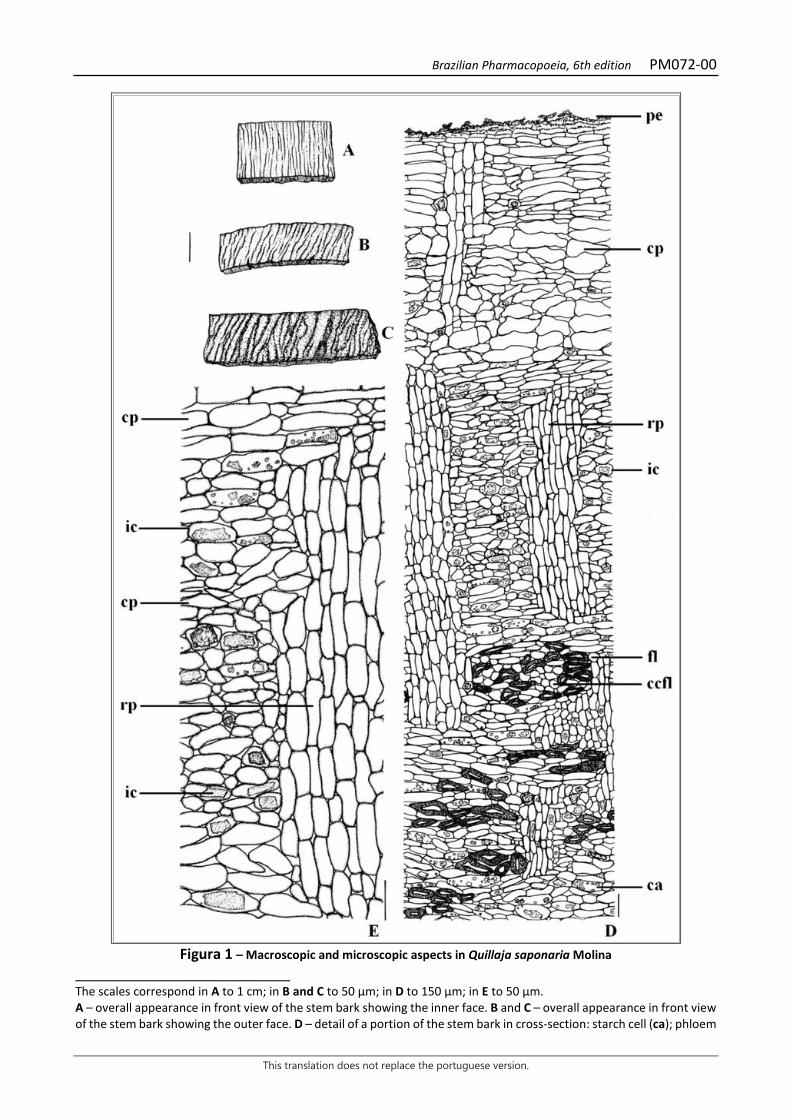
Brazilian Pharmacopoeia, 6th edition PM072-00
This translation does not replace the portuguese version.
Figura 1 – Macroscopic and microscopic aspects in Quillaja saponaria Molina
___________________________ The scales correspond in A to 1 cm; in B and C to 50 μm; in D to 150 µm; in E to 50 µm. A – overall appearance in front view of the stem bark showing the inner face. B and C – overall appearance in front view of the stem bark showing the outer face. D – detail of a portion of the stem bark in cross-section: starch cell (ca); phloem
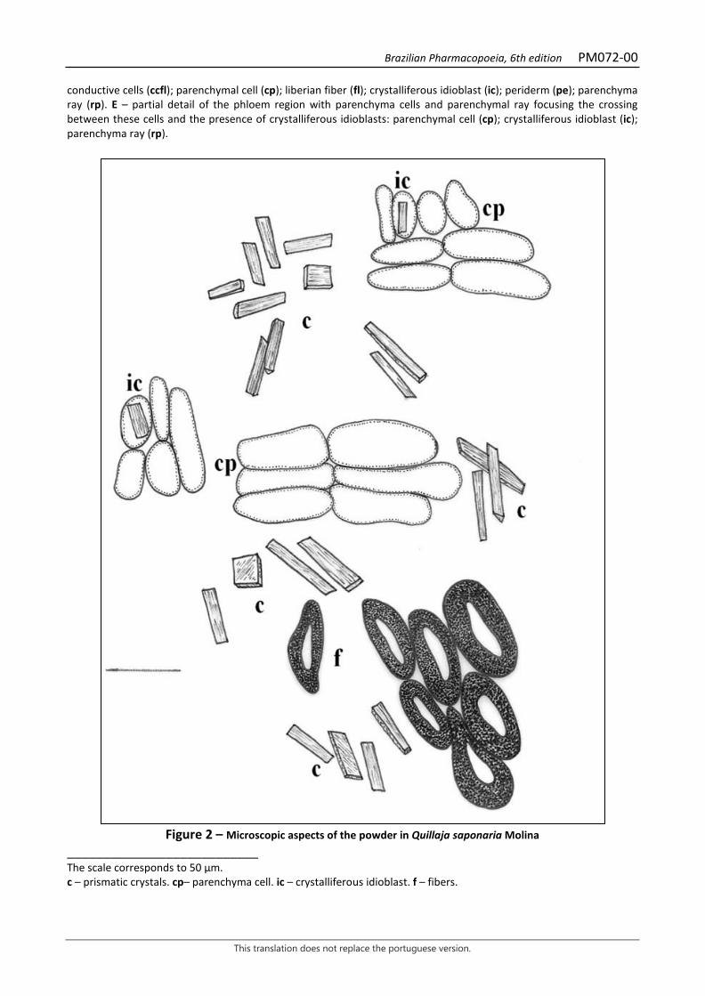
Brazilian Pharmacopoeia, 6th edition PM072-00
This translation does not replace the portuguese version.
conductive cells (ccfl); parenchymal cell (cp); liberian fiber (fl); crystalliferous idioblast (ic); periderm (pe); parenchyma ray (rp). E – partial detail of the phloem region with parenchyma cells and parenchymal ray focusing the crossing between these cells and the presence of crystalliferous idioblasts: parenchymal cell (cp); crystalliferous idioblast (ic); parenchyma ray (rp).
Figure 2 – Microscopic aspects of the powder in Quillaja saponaria Molina
___________________________ The scale corresponds to 50 µm. c – prismatic crystals. cp– parenchyma cell. ic – crystalliferous idioblast. f – fibers.
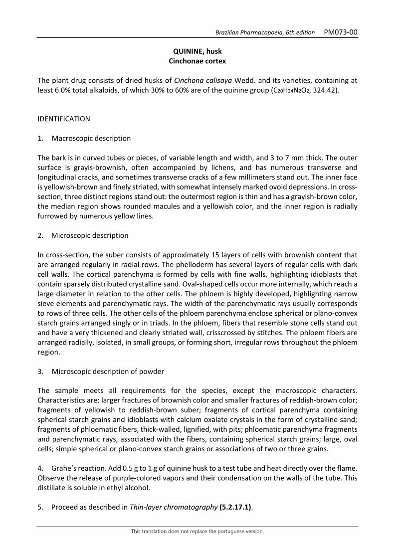
Brazilian Pharmacopoeia, 6th edition PM073-00
This translation does not replace the portuguese version.
QUININE, husk Cinchonae cortex
The plant drug consists of dried husks of Cinchona calisaya Wedd. and its varieties, containing at least 6.0% total alkaloids, of which 30% to 60% are of the quinine group (C20H24N2O2, 324.42). IDENTIFICATION 1. Macroscopic description The bark is in curved tubes or pieces, of variable length and width, and 3 to 7 mm thick. The outer surface is grayis-brownish, often accompanied by lichens, and has numerous transverse and longitudinal cracks, and sometimes transverse cracks of a few millimeters stand out. The inner face is yellowish-brown and finely striated, with somewhat intensely marked ovoid depressions. In cross-section, three distinct regions stand out: the outermost region is thin and has a grayish-brown color, the median region shows rounded macules and a yellowish color, and the inner region is radially furrowed by numerous yellow lines. 2. Microscopic description In cross-section, the suber consists of approximately 15 layers of cells with brownish content that are arranged regularly in radial rows. The phelloderm has several layers of regular cells with dark cell walls. The cortical parenchyma is formed by cells with fine walls, highlighting idioblasts that contain sparsely distributed crystalline sand. Oval-shaped cells occur more internally, which reach a large diameter in relation to the other cells. The phloem is highly developed, highlighting narrow sieve elements and parenchymatic rays. The width of the parenchymatic rays usually corresponds to rows of three cells. The other cells of the phloem parenchyma enclose spherical or plano-convex starch grains arranged singly or in triads. In the phloem, fibers that resemble stone cells stand out and have a very thickened and clearly striated wall, crisscrossed by stitches. The phloem fibers are arranged radially, isolated, in small groups, or forming short, irregular rows throughout the phloem region. 3. Microscopic description of powder The sample meets all requirements for the species, except the macroscopic characters. Characteristics are: larger fractures of brownish color and smaller fractures of reddish-brown color; fragments of yellowish to reddish-brown suber; fragments of cortical parenchyma containing spherical starch grains and idioblasts with calcium oxalate crystals in the form of crystalline sand; fragments of phloematic fibers, thick-walled, lignified, with pits; phloematic parenchyma fragments and parenchymatic rays, associated with the fibers, containing spherical starch grains; large, oval cells; simple spherical or plano-convex starch grains or associations of two or three grains. 4. Grahe’s reaction. Add 0.5 g to 1 g of quinine husk to a test tube and heat directly over the flame. Observe the release of purple-colored vapors and their condensation on the walls of the tube. This distillate is soluble in ethyl alcohol. 5. Proceed as described in Thin-layer chromatography (5.2.17.1).
Brazilian Pharmacopoeia, 6th edition PM073-00
This translation does not replace the portuguese version.
Stationary phase: silica gel GF254 (0.250 mm). Mobile phase: chloroform and diethylamine (90:10). Sample solution: add 0.1 mL of 25% ammonium hydroxide (w/v) and 5 mL of methyl chloride to 0.1 g of the pulverized plant drug. Allow it to stand for 30 minutes, occasionally shaking. Filter and evaporate the filtrate to near dryness in a water bath. Dissolve the residue in 1 mL absolute ethyl alcohol. Reference solution: dissolve separately 17.5 mg quinine, 0.5 mg quinidine, and 10 mg cinchonine in 5 mL of absolute ethyl alcohol. Procedure: apply 15 μL to 20 μL of the Sample solution and 3 μL to 5 μL of the Reference solution to the chromatoplate, separately and in the form of a band. Conduct chromatogram. Remove the plate and allow it to dry in an oven at 100 °C to 105 °C for about 10 minutes. Allow the plate to cool and nebulize the chromatoplate with a 50% (w/v) sulfuric acid solution in ethyl alcohol. Examine under ultraviolet light at 365 nm. Results: the diagram below shows the sequences of zones obtained with the Reference solution and the Sample solution. Other zones may occasionally develop.
Top of the plate
Blue fluorescence zone Light-blue fluorescence zone
Cinchonin: blue fluorescence zone
Blue fluorescence zone
Quinidine: bluish fluorescence zone
Blue fluorescence zone
Quinine: intense blue fluorescence zone
Intense blue fluorescence zone
Reference solution Sample solution
TESTS Foreign matter (5.4.1.3). At most 2.0%.

Brazilian Pharmacopoeia, 6th edition PM073-00
This translation does not replace the portuguese version.
Water (5.4.1.4). At most 8.0%. Total ash (5.4.1.5.1). At most 10.0%. Total mesophilic microorganism count (5.5.3.1.2). Complies with the test. Study of pathogenic microorganisms (5.5.3.1.3). Complies with the test. Heavy metals (5.4.5). Complies with the test. Agrochemical waste (5.4.3). Complies with the test. DOSAGE Alkaloids Proceed as per Ultraviolet absorption spectrophotometry (5.2.14). Prepare solutions as described below. Sample solution: accurately weigh approximately 1 g of the pulverized plant drug (180 µm) (5.2.11), transfer to a 250-mL erlenmeyer flask, add 10 mL of water and 7 mL 2 M hydrochloric acid. Heat in a water bath for 30 minutes. Allow to cool and add 25 mL methyl chloride, 50 mL diethyl ether, and 5 mL 20% (w/v) sodium hydroxide. Shake the mixture for 30 minutes. Add 3 g of tragacanth gum powder and shake until the preparation becomes clear. Filter on filter paper and rinse the erlenmeyer flask and filter paper with five 20 mL portions of the mixture of methyl chloride and diethyl ether (1:2). Collect the filtrates from the washes, evaporate to dryness, and dissolve the residue in 10 mL of absolute ethyl alcohol. Evaporate 5.0 mL of the solution to dryness. Dissolve the residue in 0.1 M hydrochloric acid, transfer to a volumetric flask, top off the volume to 1000 mL with the same solvent and homogenize. Reference solution (1): prepare solution by dissolving 30 mg of quinine in 0.1 M hydrochloric acid, top off the volume to 1000 mL and homogenize. Reference solution (2): prepare the solution by dissolving 30 mg cinchonine in 0.1 M hydrochloric acid, top off the volume to 1000 mL and homogenize. Blank solution: 0.1 M hydrochloric acid. Procedure: measure the absorbances of the Sample solution, Reference solution (1) and Reference solution (2) at 316 nm and 348 nm, using the Blank solution for zero adjustment. Calculate the alkaloid percentage of the quinine group (x) and the cinchonine group (y), according to the following expressions:
𝑦𝑦 =[𝑇𝑇316 × 𝑇𝑇𝐴𝐴348] − [𝑇𝑇𝐴𝐴316 × 𝑇𝑇348]
[𝑇𝑇𝐴𝐴316 × 𝑇𝑇𝐴𝐴348] − [𝑇𝑇𝐴𝐴316 × 𝑇𝑇𝐴𝐴348]×
100𝑚𝑚
×2
1000
Brazilian Pharmacopoeia, 6th edition PM073-00
This translation does not replace the portuguese version.
𝑦𝑦 =[𝑇𝑇316 × 𝑇𝑇𝐴𝐴348] − [𝑇𝑇𝐴𝐴316 × 𝑇𝑇348]
[𝑇𝑇𝐴𝐴316 × 𝑇𝑇𝐴𝐴348] − [𝑇𝑇𝐴𝐴316 × 𝑇𝑇𝐴𝐴348]×
100𝑚𝑚
×2
1000
in which, y = alkaloids of the cinchonine group %; x = alkaloids of the quinine group %; m = mass of sample in grams. A316 = absorbance measured at 316 nm for the Sample solution; A348 = absorbance measured at 348 nm for the Sample solution; Aq316 = absorbance measured for the Reference Solution (1) at 316 nm, corrected to a concentration of 1 mg in 1000 mL; Aq348 = absorbance measured for the Reference Solution (1) at 348 nm, corrected to a concentration of 1 mg in 1000 mL; Ac316 = absorbance measured for the Reference Solution (2) at 316 nm, corrected to a concentration of 1 mg in 1000 mL; Ac348 = absorbance measured for the Reference solution (2) at 348 nm, corrected to a concentration of 1 mg in 1000 mL; Calculate the total alkaloid content, (x + y) and determine the relative alkaloid content of the quinine group from the following equation 100𝑥𝑥
(𝑥𝑥+𝑦𝑦)
PACKAGING AND STORAGE In tightly closed containers, protected from light and heat.
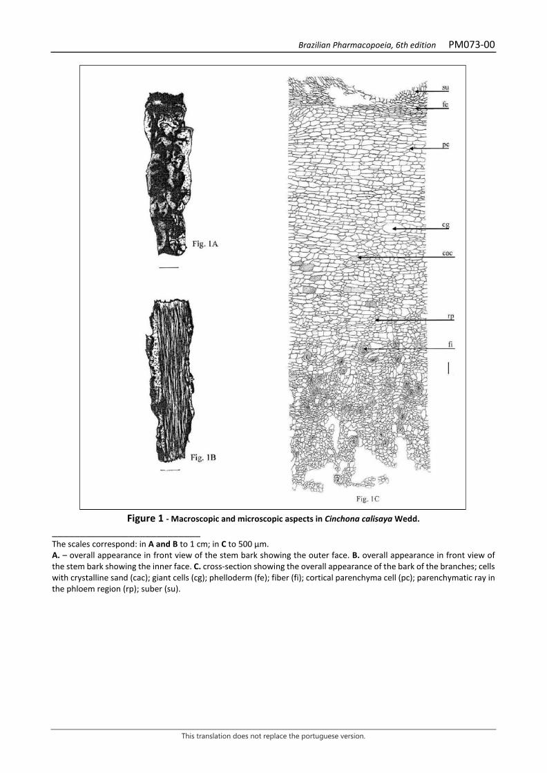
Brazilian Pharmacopoeia, 6th edition PM073-00
This translation does not replace the portuguese version.
Figure 1 - Macroscopic and microscopic aspects in Cinchona calisaya Wedd.
___________________________ The scales correspond: in A and B to 1 cm; in C to 500 µm. A. – overall appearance in front view of the stem bark showing the outer face. B. overall appearance in front view of the stem bark showing the inner face. C. cross-section showing the overall appearance of the bark of the branches; cells with crystalline sand (cac); giant cells (cg); phelloderm (fe); fiber (fi); cortical parenchyma cell (pc); parenchymatic ray in the phloem region (rp); suber (su).
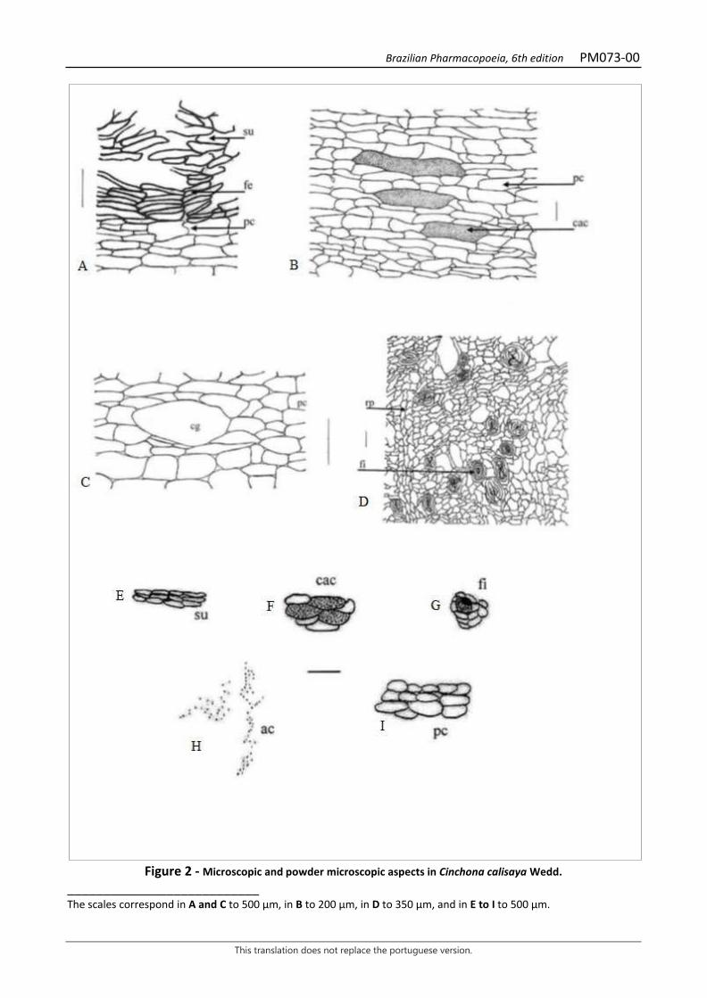
Brazilian Pharmacopoeia, 6th edition PM073-00
This translation does not replace the portuguese version.
Figure 2 - Microscopic and powder microscopic aspects in Cinchona calisaya Wedd.
___________________________ The scales correspond in A and C to 500 µm, in B to 200 µm, in D to 350 µm, and in E to I to 500 µm.
Brazilian Pharmacopoeia, 6th edition PM073-00
This translation does not replace the portuguese version.
A. cross-section showing a detail of the outer bark region near the lenticels; phelloderm (fe); cortical parenchyma (pc); suber (su). B. Detail cross section of cortical parenchyma with crystalline sand cells; crystalline sand cell (cac); cortical parenchyma (pc). C. cross-section detail of cortical parenchyma with giant cells; cortical parenchyma (pc); giant cell (cg). D. cross-section in detail of phloem region; fiber (fi); parenchymatic ray in phloem region (rp). E - I – powder details. E. suber. F. cells with crystalline sand. G. fiber surrounded by parenchyma. H. crystalline sand. I – fragment of cortical parenchyma.
Brazilian Pharmacopoeia, 6th edition PM074-00
This translation does not replace the portuguese version.
RHATANY, root Ratanhiae radix
The plant drig consists of dried roots of Krameria lappacea (Dombey) Burdet & B.B. Simpson (syn. Krameria triandra Ruiz & Pav.), containing at least 5.0% tannins, expressed in pyrogallol (C6H6O3,126.11). IDENTIFICATION 1. Macroscopic description The roots are covered by a reddish-brown, irregularly furrowed suber, which allows the formation of small, irregularly shaped plates called rhytidome, which detach easily. The starchy cortex and the phloem underlie them, composing a light brown layer. The bark, formed by the strata above, easily detaches from the central cylinder, which is woody and also light brown externally and reddish brown in the central portion. Fragmented samples show curved shapes of various lengths, twisted, sometimes fibrous. 2. Microscopic description In roots with established secondary growth, the suber is formed by several strata of cells with thinly thickened, tabular, rowed walls, and which react positively to polyphenols in the presence of 10% ferric chloride. More internally, a new periderm is formed, whose cells are deformed or ruptured. In the cortical region, the cells have varied dimensions and thickened walls, always elongated longitudinally in the portions closest to the phloem, and are filled with large, spherically shaped starch grains, simple or composed of two to three portions. The phloem has uniseriate parenchyma rays formed by bulky cells, abundant in idioblasts with prismatic crystals of various shapes and sizes and with starch grains, like those in the cortex. In both the starch cortex and the phloem there are isolated fibers or clusters of 5-15 elements, whose walls are relatively thin, deformed by mechanical compression in their outermost portions, forming a branched arrangement in relation to the adjacent cells. The xylem is formed by a large amount of relatively wide, lignified fibers with abundant bordered pits. This type of pitting is also present in the vessel elements, which are generally isolated, rarely in pairs, always associated with the axial, paratracheal parenchyma. The perforation plate is simple. The xylem rays are uniseriate. 3. Microscopic description of powder The sample meets all requirements for the species, except the macroscopic characters. Characteristics are: reddish-brown fragments of suber with typical uniformly shaped tabular cells; fragments of cortical tissue with large, simple or compound, spherical starch grains associated with branched fibers; fragments of wood with cells with bordered pits. 4. Proceed as described in Thin-layer chromatography (5.2.17.1). Stationary phase: silica gel F254 (0.250 mm). Mobile phase: ethyl acetate, toluene, formic acid and water (100:10:10:1).
Brazilian Pharmacopoeia, 6th edition PM074-00
This translation does not replace the portuguese version.
Sample solution: accurately weigh about 10 g of the pulverized plant drug, add 100 mL of 70% ethyl alcohol (v/v) and heat under reflux for 10 minutes. After cooling to room temperature, filter and remove the ethyl alcohol at a rotary evaporator under reduced pressure. Extract the resulting aqueous phase with three 25-mL portions of ethyl acetate in a separating funnel (125 mL). Allow to stand in a freezer (-18 °C) for 15 minutes for a complete phase separation. Gather the organic fractions and filter on filter paper with 2 g of anhydrous sodium sulfate. Evaporate the obtained fraction at a rotary evaporator under reduced pressure until residue is formed. Resume the residue in 5 mL methyl alcohol. Reference solution: weigh approximately 1 mg catechin and dissolve in 2 mL methyl alcohol. Procedure: apply 20 μL of the Sample solution and 10 μL of the Reference solution to the chromatoplate, separately and in the form of a band. Conduct chromatogram. Remove the chromatoplate and allow it to dry in a fume hood. Examine under ultraviolet light at 254 nm. Then nebulize the plate with 1% ferric chloride (w/v) in methyl alcohol. Results: the diagram below shows the sequences of zones obtained with the Reference solution and the Sample solution. Other zones may occasionally develop.
Top of the plate
Brownish-red zone
Catechin: brownish-blue zone
Brownish-blue zone
Brownish-red zone
Brownish-red zone
Reference solution Sample solution
TESTS Foreign matter (5.4.1.3). At most 2.0%. Water (5.4.1.4). At most 12.0%. Total ash (5.4.1.5.1). At most 5.5%. Acid-insoluble ash (5.4.1.5.3). At most 2.0%. Total mesophilic microorganism count (5.5.3.1.2). Complies with the test.
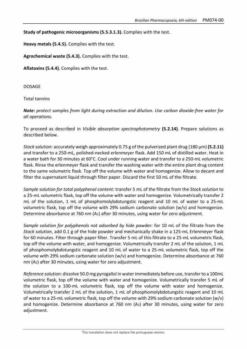
Brazilian Pharmacopoeia, 6th edition PM074-00
This translation does not replace the portuguese version.
Study of pathogenic microorganisms (5.5.3.1.3). Complies with the test. Heavy metals (5.4.5). Complies with the test. Agrochemical waste (5.4.3). Complies with the test. Aflatoxins (5.4.4). Complies with the test. DOSAGE Total tannins Note: protect samples from light during extraction and dilution. Use carbon dioxide-free water for all operations. To proceed as described in Visible absorption spectrophotometry (5.2.14). Prepare solutions as described below. Stock solution: accurately weigh approximately 0.75 g of the pulverized plant drug (180 μm) (5.2.11) and transfer to a 250-mL, polished-necked erlenmeyer flask. Add 150 mL of distilled water. Heat in a water bath for 30 minutes at 60°C. Cool under running water and transfer to a 250-mL volumetric flask. Rinse the erlenmeyer flask and transfer the washing water with the entire plant drug content to the same volumetric flask. Top off the volume with water and homogenize. Allow to decant and filter the supernatant liquid through filter paper. Discard the first 50 mL of the filtrate. Sample solution for total polyphenol content: transfer 5 mL of the filtrate from the Stock solution to a 25-mL volumetric flask, top off the volume with water and homogenize. Volumetrically transfer 2 mL of the solution, 1 mL of phosphomolybdotungstic reagent and 10 mL of water to a 25-mL volumetric flask, top off the volume with 29% sodium carbonate solution (w/v) and homogenize. Determine absorbance at 760 nm (A1) after 30 minutes, using water for zero adjustment. Sample solution for polyphenols not adsorbed by hide powder: for 10 mL of the filtrate from the Stock solution, add 0.1 g of the hide powder and mechanically shake in a 125-mL Erlenmeyer flask for 60 minutes. Filter through paper filter. Transfer 5 mL of this filtrate to a 25-mL volumetric flask, top off the volume with water, and homogenize. Volumetrically transfer 2 mL of the solution, 1 mL of phosphomolybdotungstic reagent and 10 mL of water to a 25-mL volumetric flask, top off the volume with 29% sodium carbonate solution (w/v) and homogenize. Determine absorbance at 760 nm (A2) after 30 minutes, using water for zero adjustment. Reference solution: dissolve 50.0 mg pyrogallol in water immediately before use, transfer to a 100mL volumetric flask, top off the volume with water and homogenize. Volumetrically transfer 5 mL of the solution to a 100-mL volumetric flask, top off the volume with water and homogenize. Volumetrically transfer 2 mL of the solution, 1 mL of phosphomolybdotungstic reagent and 10 mL of water to a 25-mL volumetric flask, top off the volume with 29% sodium carbonate solution (w/v) and homogenize. Determine absorbance at 760 nm (A3) after 30 minutes, using water for zero adjustment.
Brazilian Pharmacopoeia, 6th edition PM074-00
This translation does not replace the portuguese version.
Calculate total tannin content, expressed as a percentage of pyrogallol, according to the following expression:
𝑇𝑇𝑇𝑇 =(𝑇𝑇1 − 𝑇𝑇2) × 𝑚𝑚2 × 62,5
𝑇𝑇3 × 𝑚𝑚1
in which, TT = total tannin content expressed as pyrogallol % (w/w); A1 = absorbance measured for the Sample solution for total polyphenols; A2 = absorbance measured for the Sample solution for polyphenols not adsorbed by hide powder; A3 = absorbance measured for the Reference solution; m1 = mass in grams of the sample used un the assay, considering the determined water content. m2 = mass in grams of pyrogallol, considering the purity of the reference substance. PACKAGING AND STORAGE In tightly closed containers, protected from light and heat.

Brazilian Pharmacopoeia, 6th edition PM074-00
This translation does not replace the portuguese version.
Figure 1 – Microscopic and powder microscopic aspects in Krameria lappacea
(Dombey) Burdet & B.B. Simpson ___________________________ The scales correspond in A to 250 μm; in B to 100 μm; in C to 50 μm; in D to 25 μm. A - general aspect of the distribution of root tissues, in cros-ection: starchy cortex (ca); vessel element (ev); phloem (f); periderm (pe); rhytidome (ri); parenchyma ray; xylem (x). B - partial detail of the bark with newly formed periderm (pe) and starch cortex (am), in cross-section; C and D - partial detail of the starch cortex in transverse and longitudinal radial section, respectively: starch grains (am), intercellular space (ei); phloem fiber (ff).
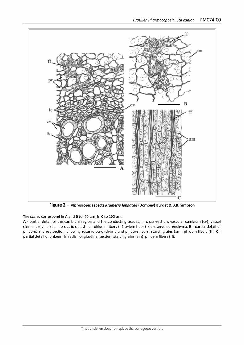
Brazilian Pharmacopoeia, 6th edition PM074-00
This translation does not replace the portuguese version.
Figure 2 – Microscopic aspects Krameria lappacea (Dombey) Burdet & B.B. Simpson
___________________________ The scales correspond in A and B to: 50 μm; in C to 100 μm. A - partial detail of the cambium region and the conducting tissues, in cross-section: vascular cambium (cv); vessel element (ev); crystalliferous idioblast (ic); phloem fibers (ff); xylem fiber (fx); reserve parenchyma. B - partial detail of phloem, in cross-section, showing reserve parenchyma and phloem fibers: starch grains (am); phloem fibers (ff). C - partial detail of phloem, in radial longitudinal section: starch grains (am); phloem fibers (ff).

Brazilian Pharmacopoeia, 6th edition PM075-00
This translation does not replace the portuguese version.
SNAKEROOT, raiz Rauvolfiae radix
The plant drug consists of dried roots of Rauvolfia serpentina (L.) Benth. ex Kurz, containing at least 0.15% reserpine group alkaloids (C33H40N2O9, 608.68) – rescinamine (C35H42N2O9, 634.72). IDENTIFICATION 1. Macroscopic description Cylindrical root, often tapering at the distal end, winding; entire root or its portions from 1 to 10 cm long and from 3 to 22 mm in diameter; outer surface longitudinally wrinkled to irregularly furrowed, light gray-brown in color; there may be remnants of the secondary roots or mainly rounded scars from their fall, from 0.5 to 1 mm in diameter; the bark may be partially missing and yellowish-brown inner layers are observed in these faults. Lenticels are often observed. The cortex is yellowish-brown and the central cylinder is light yellow, with two to eight concentric rings, exhibiting a fine radial striation. Rhizome remnants are rarely present. 2. Microscopic description The periderm has up to 20 layers of tangentially flattened cells with a radial arrangement. The suber is homogeneous and consists of about 15 layers of thin-walled, suberous cells. The phelloderm has up to four layers of thin-walled cells. The cortical parenchyma is amyliferous, with several layers of cells with non-lignified walls; the starch grains, highlighted by Lugol’s reagent, may be small and numerous or bulky, rounded or oval shaped. Branched, intrusively growing laticifers permeate the cortical parenchyma. The phloem consists only of sieve tube elements and parenchyma cells; fibers and sclereids are absent. The parenchyma rays are multiseriate, and may be narrow or wide; their cells have starch grains and/or crystals of various shapes. The secondary xylem also shows a radial arrangement. Tracheal elements and fibers are arranged in uniseriate or biseriate radial series and alternate with multiseriate parenchyma rays. The tracheal elements are narrow (about 40 μm in diameter), with a simple or scalariform perforation plate; the fibers are libriform and have thickened walls. The parenchymatic rays are multiseriate, their cells have lignified walls, and the starch grains are bulkier than those found in the phloem and cortical parenchyma. The primary xylem, with six to eight protoxylem poles, occupies a central position; the tracheal elements are also narrow and of similar caliber to the adjacent parenchyma cells. 3. Microscopic description of powder The sample meets all requirements for the species, except the macroscopic characters. Characteristics are: light grayish or light yellowish-brown color; thin-walled, yellowish, suberized fragments of the suber; fragments vessel thick-walled elements with bordered pits; fragments of parenchymal cells of the xylem with thickened walls and simple pits; fragments of parenchymal cells of the cortex with thin walls; several rounded, sometimes aggregated, starch grains with the central region in the shape of a y or star. 4. Adulterations
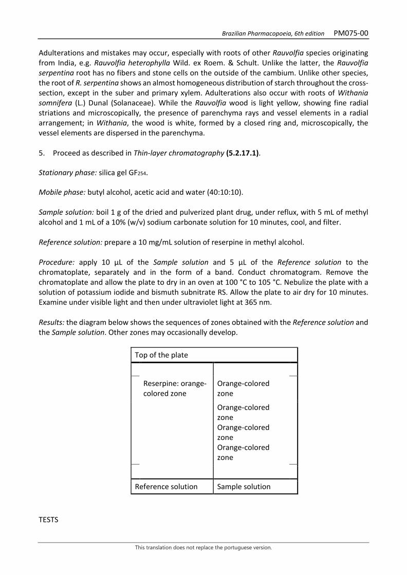
Brazilian Pharmacopoeia, 6th edition PM075-00
This translation does not replace the portuguese version.
Adulterations and mistakes may occur, especially with roots of other Rauvolfia species originating from India, e.g. Rauvolfia heterophylla Wild. ex Roem. & Schult. Unlike the latter, the Rauvolfia serpentina root has no fibers and stone cells on the outside of the cambium. Unlike other species, the root of R. serpentina shows an almost homogeneous distribution of starch throughout the cross-section, except in the suber and primary xylem. Adulterations also occur with roots of Withania somnifera (L.) Dunal (Solanaceae). While the Rauvolfia wood is light yellow, showing fine radial striations and microscopically, the presence of parenchyma rays and vessel elements in a radial arrangement; in Withania, the wood is white, formed by a closed ring and, microscopically, the vessel elements are dispersed in the parenchyma. 5. Proceed as described in Thin-layer chromatography (5.2.17.1). Stationary phase: silica gel GF254. Mobile phase: butyl alcohol, acetic acid and water (40:10:10). Sample solution: boil 1 g of the dried and pulverized plant drug, under reflux, with 5 mL of methyl alcohol and 1 mL of a 10% (w/v) sodium carbonate solution for 10 minutes, cool, and filter. Reference solution: prepare a 10 mg/mL solution of reserpine in methyl alcohol. Procedure: apply 10 μL of the Sample solution and 5 μL of the Reference solution to the chromatoplate, separately and in the form of a band. Conduct chromatogram. Remove the chromatoplate and allow the plate to dry in an oven at 100 °C to 105 °C. Nebulize the plate with a solution of potassium iodide and bismuth subnitrate RS. Allow the plate to air dry for 10 minutes. Examine under visible light and then under ultraviolet light at 365 nm. Results: the diagram below shows the sequences of zones obtained with the Reference solution and the Sample solution. Other zones may occasionally develop.
Top of the plate
Reserpine: orange-colored zone
Orange-colored zone
Orange-colored zone Orange-colored zone Orange-colored zone
Reference solution Sample solution
TESTS
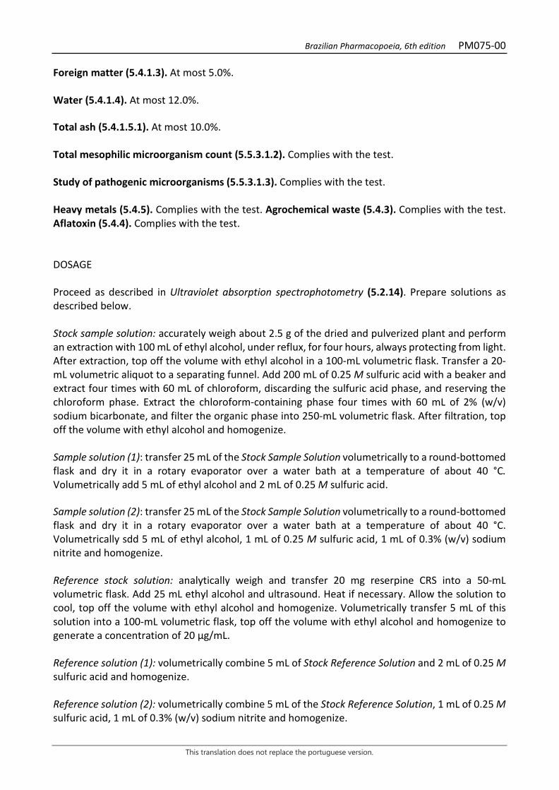
Brazilian Pharmacopoeia, 6th edition PM075-00
This translation does not replace the portuguese version.
Foreign matter (5.4.1.3). At most 5.0%. Water (5.4.1.4). At most 12.0%. Total ash (5.4.1.5.1). At most 10.0%. Total mesophilic microorganism count (5.5.3.1.2). Complies with the test. Study of pathogenic microorganisms (5.5.3.1.3). Complies with the test. Heavy metals (5.4.5). Complies with the test. Agrochemical waste (5.4.3). Complies with the test. Aflatoxin (5.4.4). Complies with the test. DOSAGE Proceed as described in Ultraviolet absorption spectrophotometry (5.2.14). Prepare solutions as described below. Stock sample solution: accurately weigh about 2.5 g of the dried and pulverized plant and perform an extraction with 100 mL of ethyl alcohol, under reflux, for four hours, always protecting from light. After extraction, top off the volume with ethyl alcohol in a 100-mL volumetric flask. Transfer a 20-mL volumetric aliquot to a separating funnel. Add 200 mL of 0.25 M sulfuric acid with a beaker and extract four times with 60 mL of chloroform, discarding the sulfuric acid phase, and reserving the chloroform phase. Extract the chloroform-containing phase four times with 60 mL of 2% (w/v) sodium bicarbonate, and filter the organic phase into 250-mL volumetric flask. After filtration, top off the volume with ethyl alcohol and homogenize. Sample solution (1): transfer 25 mL of the Stock Sample Solution volumetrically to a round-bottomed flask and dry it in a rotary evaporator over a water bath at a temperature of about 40 °C. Volumetrically add 5 mL of ethyl alcohol and 2 mL of 0.25 M sulfuric acid. Sample solution (2): transfer 25 mL of the Stock Sample Solution volumetrically to a round-bottomed flask and dry it in a rotary evaporator over a water bath at a temperature of about 40 °C. Volumetrically sdd 5 mL of ethyl alcohol, 1 mL of 0.25 M sulfuric acid, 1 mL of 0.3% (w/v) sodium nitrite and homogenize. Reference stock solution: analytically weigh and transfer 20 mg reserpine CRS into a 50-mL volumetric flask. Add 25 mL ethyl alcohol and ultrasound. Heat if necessary. Allow the solution to cool, top off the volume with ethyl alcohol and homogenize. Volumetrically transfer 5 mL of this solution into a 100-mL volumetric flask, top off the volume with ethyl alcohol and homogenize to generate a concentration of 20 μg/mL. Reference solution (1): volumetrically combine 5 mL of Stock Reference Solution and 2 mL of 0.25 M sulfuric acid and homogenize. Reference solution (2): volumetrically combine 5 mL of the Stock Reference Solution, 1 mL of 0.25 M sulfuric acid, 1 mL of 0.3% (w/v) sodium nitrite and homogenize.
Brazilian Pharmacopoeia, 6th edition PM075-00
This translation does not replace the portuguese version.
Blank solution: use ethyl alcohol and water (2:1). Note: do not use Reference Solution (1) and Reference Solution (2) from previous days. Procedure: heat the Sample solution (1), Sample solution (2), Reference solution (1) and Reference solution (2) together in a water bath at 50 °C to 60 °C for exactly 20 minutes. Cool the solutions to room temperature and add, volumetrically, 0.5 mL of 5% (w/v) sulfamic acid to each of them and wait for exactly 20 minutes. After the waiting time, measure the absorbance of the Sample solution (1) (A1), Sample solution (2) (A2), Reference solution (1) (S1) and Reference solution (2) (S2) at 390 nm using the Blank solution for zero adjustment. Calculate the mass of the alkaloids of the reserpine-resinamine group, as reserpine, in mg, according to the following expression:
𝑇𝑇𝑇𝑇𝑇𝑇 = 5 ×(𝑇𝑇1 − 𝑇𝑇2)(𝑇𝑇1 − 𝑇𝑇2)
in which, MAL = mass in milligrams of alkaloids; A1 = absorbance measured for the Sample solution (1); A2 = absorbance measured for the Sample solution (2); S1 = absorbance measured for the Reference solution (1). S2 = absorbance measured for the Reference solution (2); Calculate percentage alkaloid content as reserpine-rescinamine, on a dried basis, according to the following expression:
𝑇𝑇𝑇𝑇𝑇𝑇 =𝑇𝑇𝑇𝑇𝑇𝑇𝑚𝑚
× 100
in which, TAL = alkaloid content % (w/w); MAL = mass in milligrams of alkaloids; m = mass in grams of the sample used, considering the determined water content. PACKAGING AND STORAGE In tightly closed containers, protected from light and heat.
Brazilian Pharmacopoeia, 6th edition PM075-00
This translation does not replace the portuguese version.
Figure 1 - Macroscopic, microscopic and powder microscopic aspects in Rauvolfia serpentina (L.) Benth. ex
Kurz ___________________________ The scales correspond: in A to 100 mm, in B and G to 100 μm, and from C to F to 50 μm. A - overall appearance of the root; B - diagram of root cross-section; C - detail of the periderm and cortical parenchyma, in cross-section; D - detail of a portion of the cortical parenchyma in cross-section; E - detail of portion of secondary xylem showing multiseriate parenchyma rays with abundant starch grains, fibers, and vessels arranged in radial series, in cross-section; F - detail of a portion of the primary xylem in cross-section; G - overall appearance of the root powder, with fragments of the suber (top left), of fibers and vessels (top right and bottom central region), of parenchyma cells of the secondary xylem (bottpm left) and several starch grains, isolated or aggregated; primary and secondary phloem region (f); starch grain (ga); intrusively-growing branched laticifer (lt); cortical parenchyma (pc); periderm (pe); parenchyma ray (rp); primary xylem (xp); secondary xylem (xs).
Brazilian Pharmacopoeia, 6th edition PM076-01
This translation does not replace the portuguese version.
RHUBARB, rhizome and root Rhei rhizoma et radix
The plant drug consists of dried and fragmented rhizomes and roots of Rheum palmatum L. and/or Rheum officinale Baill. or their interspecific hybrids, containing at least 2.2% hydroxyanthracene derivatives, expressed as rhein (C15H8O6, 284.22). Rhizomes must be devoid of the bases of the leaf petioles. CHARACTERISTICS The drug has a characteristic, aromatic odor. IDENTIFICATION 1. Macroscopic description Irregular, discoid or cylindrical, rounded, flat or plano-convex rhizome fragments up to 15 cm in diameter and 1 to 5 cm thick, often lacking the cortical region and/or part of the outermost vascular region. The outer surface is smooth and usually coated with a layer of brownish-yellow powder, which, when removed, shows a pinkish color; the fragments, when moistened, show dark and light lines that intersect, with several diamond-shaped reticules interrupted by scars from the roots. In cross-section, a dark ring, corresponding to the cambium, stands out, followed by another narrow ring, regularly furrowed by orange radial striations that are parallel to each other. The inner side of the central cylinder is filled with a pinkish tissue, in which several star-shaped structures, corresponding to anomalous vascular bundles, stand out. The Rheum palmatum rhizomes are characterized by having small anomalous vascular bundles, on average 2.5 mm in diameter, and a cluster of bundles forming one continuous ring, sometimes two, while those of Rheum officinale have larger bundles, up to 4.1 mm in diameter, distributed randomly in cross section. The root fragments are cylindrical or conical, devoid of cortex, measuring 3 to 6 cm in diameter and 4 to 17 cm long, with a color similar to that of the rhizome. In cross-section, the radially arranged parenchymal rays are clear, from the central portion to the edges. The fracture of rhizomes and roots is granular. 2. Microscopic description In cross-section, the rhizome, when accompanied by the cortical region or its remains, has a poorly developed bulb and outer cortical parenchyma; the cells of the bulb have a radial arrangement and thin walls; the outer cortical parenchyma, as well as the other parenchyma, has rounded or occasionally polygonal cells, thin-walled, with several starch grains and druse-like crystals. Starch grains and calcium oxalate druses are observed. The vascular system presents itself in two distinct forms: the outermost, derived from the normal cambium, is continuous and somewhat circular; and the innermost has anomalous vascular bundles, with a star-like appearance, which are irregularly distributed in the medullary parenchyma or some of them form one or two rings. The external vascular system has an underdeveloped and obliterated secondary phloem. The secondary xylem has a radial arrangement and is formed by a few layers of vessel elements with a generally reticulate thickening. The parenchyma rays show brownish-yellow or intense yellow amorphous masses,
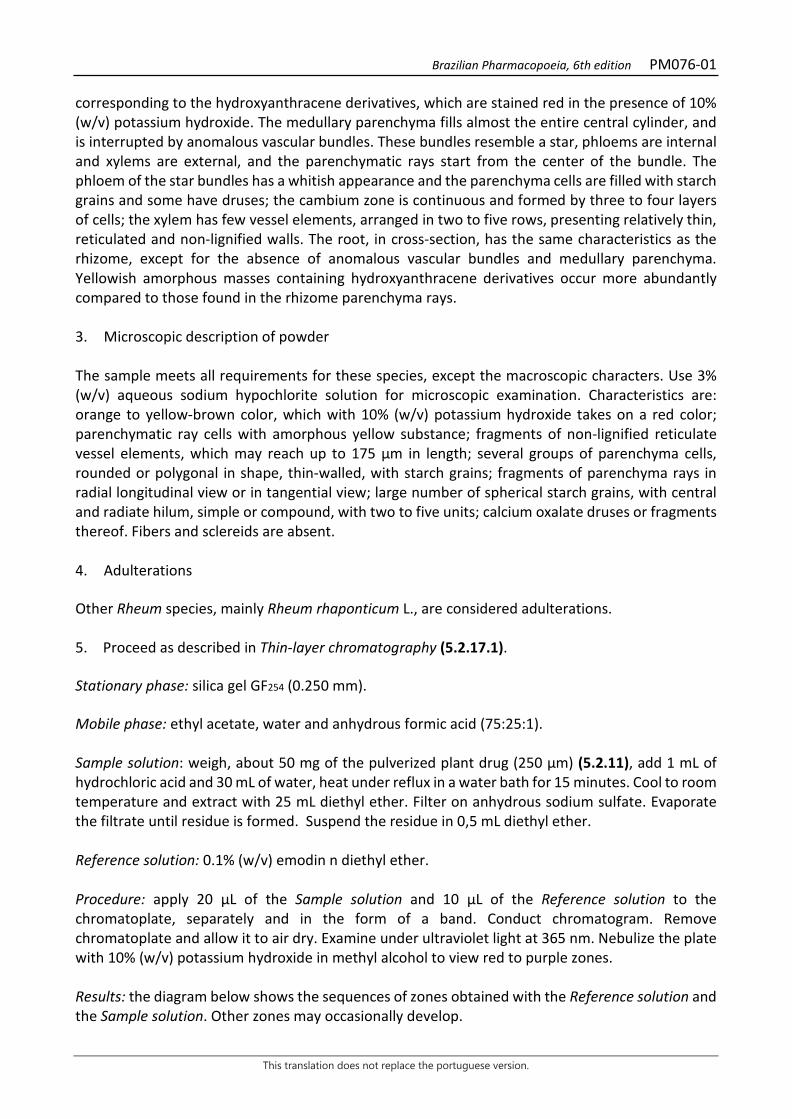
Brazilian Pharmacopoeia, 6th edition PM076-01
This translation does not replace the portuguese version.
corresponding to the hydroxyanthracene derivatives, which are stained red in the presence of 10% (w/v) potassium hydroxide. The medullary parenchyma fills almost the entire central cylinder, and is interrupted by anomalous vascular bundles. These bundles resemble a star, phloems are internal and xylems are external, and the parenchymatic rays start from the center of the bundle. The phloem of the star bundles has a whitish appearance and the parenchyma cells are filled with starch grains and some have druses; the cambium zone is continuous and formed by three to four layers of cells; the xylem has few vessel elements, arranged in two to five rows, presenting relatively thin, reticulated and non-lignified walls. The root, in cross-section, has the same characteristics as the rhizome, except for the absence of anomalous vascular bundles and medullary parenchyma. Yellowish amorphous masses containing hydroxyanthracene derivatives occur more abundantly compared to those found in the rhizome parenchyma rays. 3. Microscopic description of powder The sample meets all requirements for these species, except the macroscopic characters. Use 3% (w/v) aqueous sodium hypochlorite solution for microscopic examination. Characteristics are: orange to yellow-brown color, which with 10% (w/v) potassium hydroxide takes on a red color; parenchymatic ray cells with amorphous yellow substance; fragments of non-lignified reticulate vessel elements, which may reach up to 175 μm in length; several groups of parenchyma cells, rounded or polygonal in shape, thin-walled, with starch grains; fragments of parenchyma rays in radial longitudinal view or in tangential view; large number of spherical starch grains, with central and radiate hilum, simple or compound, with two to five units; calcium oxalate druses or fragments thereof. Fibers and sclereids are absent. 4. Adulterations Other Rheum species, mainly Rheum rhaponticum L., are considered adulterations. 5. Proceed as described in Thin-layer chromatography (5.2.17.1). Stationary phase: silica gel GF254 (0.250 mm). Mobile phase: ethyl acetate, water and anhydrous formic acid (75:25:1). Sample solution: weigh, about 50 mg of the pulverized plant drug (250 μm) (5.2.11), add 1 mL of hydrochloric acid and 30 mL of water, heat under reflux in a water bath for 15 minutes. Cool to room temperature and extract with 25 mL diethyl ether. Filter on anhydrous sodium sulfate. Evaporate the filtrate until residue is formed. Suspend the residue in 0,5 mL diethyl ether. Reference solution: 0.1% (w/v) emodin n diethyl ether. Procedure: apply 20 μL of the Sample solution and 10 μL of the Reference solution to the chromatoplate, separately and in the form of a band. Conduct chromatogram. Remove chromatoplate and allow it to air dry. Examine under ultraviolet light at 365 nm. Nebulize the plate with 10% (w/v) potassium hydroxide in methyl alcohol to view red to purple zones. Results: the diagram below shows the sequences of zones obtained with the Reference solution and the Sample solution. Other zones may occasionally develop.

Brazilian Pharmacopoeia, 6th edition PM076-01
This translation does not replace the portuguese version.
Top of the plate
Orange fluorescence zone Orange fluorescence zone
Emodin: orangish fluorescence zone
Orange fluorescence zone
Orange fluorescence zone Orange fluorescence zone
Reference solution Sample solution TESTS Raponticine. Proceed as described in Thin-layer chromatography (5.2.17.1). Stationary phase: silica gel GF254 (0.250 mm). Mobile phase: methyl chloride and acetone (80:20). Sample solution: weigh 0.2 g of the pulverized plant drug and add 2 mL methyl alcohol. Heat under reflux for 15 minutes. Cool and filter. Reference solution: 1 mg/mL rhaponticin solution in methyl alcohol. Procedure: apply 20 μL of the Sample solution and 5 μL of the Reference solution to the plate, separately and in the form of a band. Conduct chromatogram. Remove chromatoplate and allow it to air dry. Examine under ultraviolet light at 365 nm. The chromatogram region obtained with the Sample solution should not show any blue stain near the application point, indicating the presence of rhaponticin. Nebulize the plate with phosphomolybdic acid RS. The chromatogram region obtained with the Sample solution should not show any dark-blue stain near the application point, indicating the presence of rhaponticin. Foreign matter (5.4.1.3). At most 2.0%. Water (5.4.1.4). At most 12.0%. Total ash (5.4.1.5.1). At most 12.0%. Total mesophilic microorganism count (5.5.3.1.2). Complies with the test. Study of pathogenic microorganisms (5.5.3.1.3). Complies with the test.

Brazilian Pharmacopoeia, 6th edition PM076-01
This translation does not replace the portuguese version.
Heavy metals (5.4.5). Complies with the test. Agrochemical waste (5.4.3). Complies with the test. Aflatoxins (5.4.4). Complies with the test. DOSAGE Hydroxyanthracene derivatives To proceed as described in Visible absorption spectrophotometry (5.2.14). Prepare solutions as described below. Stock solution: in a 50 mL round-bottomed flask, accurately weigh about 0.1 g of the dried and pulverized plant drug. Add 30 mL of water, mix, and weigh the ensemble. Heat in a water bath, under reflux, for 15 minutes. Allow to cool and add 50 mg of sodium bicarbonate. Weigh and resume the original weight with water. Centrifuge and transfer 10 mL of the supernatant liquid into a 50-mL round-bottomed flask. Add 20 mL ferric chloride RS and shake. Heat the mixture under reflux for 20 minutes. Shake frequently. Add 1 mL hydrochloric acid and heat for another 20 minutes. Transfer to separating funnel. Extract with three successive portions of 25 mL diethyl ether, previously used to rinse the round-bottomed flask. Collect the ether extracts and rinse with two 20 mL portions of water, filter into a 100-mL volumetric flask, top off to volume with diethyl ether and homogenize. Sample solution: evaporate 10 mL of the Stock solution until residue is formed. Suspend residue in 10 mL of 0.5% magnesium acetate (w/v) in methyl alcohol. Blank solution: use methyl alcohol. Procedure: measure the absorbance of the Sample solution at 515 nm, immediately after its preparation, using the Blank solution for zero adjustment. Calculate the content of hydroxyanthracene derivatives expressed as a percentage of rhein, according to the following expression:
𝑇𝑇𝑇𝑇𝑇𝑇𝐶𝐶 =𝑇𝑇 × 300
m × 440
in which, TDHC = hydroxyanthracene derivative content expressed as rhein % (w/w); A = absorbance measured for the Sample solution; 300 = dilution factor; 440 = coefficient of specific absorption of rhein; m = mass in grams of the sample used, considering the determined water content. PACKAGING AND STORAGE
Brazilian Pharmacopoeia, 6th edition PM076-01
This translation does not replace the portuguese version.
In tightly closed containers, protected from light and heat.
Figure 1 - Macroscopic, microscopic and powder microscopic aspects in Rheum palmatum L. (A1) e Rheum
officinale Baill. (A2) ___________________________ The scales correspond in A, B, C, D and E to 500 μm; in F, G, H, I, J, L and M to 100 μm.
Brazilian Pharmacopoeia, 6th edition PM076-01
This translation does not replace the portuguese version.
A1 and A2 - partial schematic diagram of rhizomes in cross-section; cambium (ca); phloem (f); anomalous vascular bundle (fva); external cortical parenchyma (pce); internal cortical parenchyma (pci); medullary parenchyma (pm); suber (su). B - Cross-section detail of the outer region of the rhizome cortex; outer cortical parenchyma (pce); suber (su). C - cross-sectional detail of cortical region; crystal (cr); starch grain (ga); inner cortical parenchyma (pci). D - detail of the vascular region; cambium (ca); phloem (f); xylem (x). E - detail of the anomalous vascular system in cross-section; cambium (ca); crystal (cr); vessel element (ev); phloem (f); starch grain (ga); parenchymatic ray (rp); xylem (x). F - detail of parenchyma cells in cross-section containing starch grains. F - detail of parenchyma cells in longitudinal section; starch grains (ga). H – starch grain details. I - Detail of parenchyma ray cells in cross-section. F - detail of parenchyma cells in cross-section; crystal (cr). L - detail of radial parenchyma cells in cross-section, associated with other parenchymal cells in radial longitudinal section. M - detail of vessel element with reticulate thickening and parenchyma cells in longitudinal section.
Brazilian Pharmacopoeia, 6th edition PM077-01
This translation does not replace the portuguese version.
SAMBUCUS AUSTRALIS, flower Sambucus australis flos
The plant drug consists of dried flowers of Sambucus australis Cham. & Schltdl. containing at least 2.0% total flavonoids, expressed as quercetin, and at least 0.8% rutin. CHARACTERISTICS The dried flowers have a characteristic light, aromatic odor. IDENTIFICATION 1. Macroscopic description Flowers with a yellowish color acquired after drying, pentamerous or tetramerous, staminate, with long stamens, or pistillate, with short stamens, measuring 7 to 10 mm in diameter, each with three tiny green bracts, distributed on the pedicel and/or receptacle at different heights, visible with a magnifying glass. Papillose bracts with tector and glandular trichomes on the basal portion of the adaxial surface. Whitish or brownish, globose flower buds measuring 1 to 3 mm in diameter. Calyx with yellow-greenish, triangular-oval sepals, 1 to 1.5 mm long and 1 mm wide at the basal portion, slightly merged to each other at the base, with abundant tector and glandular trichomes on the basal portion of the adaxial surface and lacking marginal teeth. Yellowish, rotate corolla, with petals merged together at the base in a short tube. Oval to elliptic petals, with a retrorse apex, 2.5 to 5 mm long and 1.5 to 3 mm wide. The androecium is formed by five or four stamens, arranged alternately to the petals, with filaments attached to the corolla tube. Dithecate, extrorse, dorsifixed, oblong anthers, dehiscent in the staminate flowers and indehiscent in the pistillate flowers, yellowish, 1 mm long. The filaments are glabrous and cylindrical, short in the pistillate flowers, 1 to 2 mm long, and long in the staminate flowers, 3 to 4 mm long. Infertile ovary, merged to the calyceal tube, pentacarpellar or tetracarpellar, rarely tricarpellar, pentalocular or tetralocular, rarely trilocular, with well-demarcated carpels, with a rudimentary seminal per locule, axially placentate. Globose and papillose gynoecium, with a short stylet and pentalobate stigma. A prominent, ringed disk surrounds the base of the gynoecium. 2. Microscopic description Bracts, sepals, and petals are amphistomatic, hypoestomatic, and amphi-hypostomatic, respectively; stomas are of the anomocytic type; in front view, the cuticle exhibits striations; tector and glandular trichomes occur at the base of the adaxial surface; in cross-section, the cuticle is striated, the epidermis is unistratified, and the mesophyll is homogeneous; spherical lipid drops occur in all tissues except the xylem. Calcium oxalate idioblasts absent. Five, rarely four, parallel veins occur on the petals, the secondary ones originating from the main one, branched or not; the epidermis has papillose cells, less prominent in the margin regions; ellipsoid starch grains are present in the mesophyll. The filament, in front view, has a striated cuticle. The anther, in cross-section, has a papillose epidermis, the mat is unistratified, and the endothecium is formed by two to three layers of fibrous cells, with evident pitting. The pollen grain is prolate, tricolporate, 18 to 34 μm in diameter, with a reticulate surface. The gynoecium is formed by five, four or rarely three
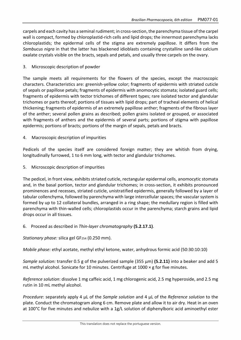
Brazilian Pharmacopoeia, 6th edition PM077-01
This translation does not replace the portuguese version.
carpels and each cavity has a seminal rudiment; in cross-section, the parenchyma tissue of the carpel wall is compact, formed by chloroplastid-rich cells and lipid drops; the innermost parenchyma lacks chloroplastids; the epidermal cells of the stigma are extremely papillose. It differs from the Sambucus nigra in that the latter has blackened idioblasts containing crystalline sand-like calcium oxalate crystals visible on the bracts, sepals and petals, and usually three carpels on the ovary. 3. Microscopic description of powder The sample meets all requirements for the flowers of the species, except the macroscopic characters. Characteristics are: greenish-yellow color; fragments of epidermis with striated cuticle of sepals or papillose petals; fragments of epidermis with anomocytic stomata; isolated guard cells; fragments of epidermis with tector trichomes of different types; rare isolated tector and glandular trichomes or parts thereof; portions of tissues with lipid drops; part of tracheal elements of helical thickening; fragments of epidermis of an extremely papillose anther; fragments of the fibrous layer of the anther; several pollen grains as described; pollen grains isolated or grouped, or associated with fragments of anthers and the epidermis of several parts; portions of stigma with papillose epidermis; portions of bracts; portions of the margin of sepals, petals and bracts. 4. Macroscopic description of impurities Pedicels of the species itself are considered foreign matter; they are whitish from drying, longitudinally furrowed, 1 to 6 mm long, with tector and glandular trichomes. 5. Microscopic description of impurities The pedicel, in front view, exhibits striated cuticle, rectangular epidermal cells, anomocytic stomata and, in the basal portion, tector and glandular trichomes; in cross-section, it exhibits pronounced prominences and recesses, striated cuticle, unistratified epidermis, generally followed by a layer of tabular collenchyma, followed by parenchyma with large intercellular spaces; the vascular system is formed by up to 12 collateral bundles, arranged in a ring shape; the medullary region is filled with parenchyma with thin-walled cells; chloroplastids occur in the parenchyma; starch grains and lipid drops occur in all tissues. 6. Proceed as described in Thin-layer chromatography (5.2.17.1). Stationary phase: silica gel GF254 (0.250 mm). Mobile phase: ethyl acetate, methyl ethyl ketone, water, anhydrous formic acid (50:30:10:10) Sample solution: transfer 0.5 g of the pulverized sample (355 µm) (5.2.11) into a beaker and add 5 mL methyl alcohol. Sonicate for 10 minutes. Centrifuge at 1000 × g for five minutes. Reference solution: dissolve 1 mg caffeic acid, 1 mg chlorogenic acid, 2.5 mg hyperoside, and 2.5 mg rutin in 10 mL methyl alcohol. Procedure: separately apply 4 µL of the Sample solution and 4 µL of the Reference solution to the plate. Conduct the chromatogram along 6 cm. Remove plate and allow it to air dry. Heat in an oven at 100°C for five minutes and nebulize with a 1g/L solution of diphenylboric acid aminoethyl ester

Brazilian Pharmacopoeia, 6th edition PM077-01
This translation does not replace the portuguese version.
in ethyl acetate, followed by a 5% solution of macrogol 400 in methyl chloride. Allow to dry for 30 minutes. Examine under daylight and then under ultraviolet light at 365 nm. Results: the diagrams below show the sequences of zones obtained with the Reference solution and the Sample solution, after examination under daylight and under ultraviolet light at 365 nm, in order. Other zones may occasionally develop.
Top of the plate
Hyperoside: dark-yellow colored area
Orange-colored zone
Hyperoside: dark-yellow colored area
Dark-yellow colored area
Reference solution Sample solution
Top of the plate
Caffeic acid: fluorescent-blue colored area
Intense fluorescent-blue colored zone Two intense fluorescent-blue colored zones
Hyperoside: dark-yellow colored area
Fluorescent-orange colored zone
Chlorogenic acid: fluorescent blue color zone
Intense fluorescent-blue colored zone
Hyperoside: dark-yellow colored area
Fluorescent-orange colored zone
Reference solution Sample solution

Brazilian Pharmacopoeia, 6th edition PM077-01
This translation does not replace the portuguese version.
7. Proceed as described in Dosage for Rutin. The majority peak in the chromatogram corresponds to rutin; there is one peak with a lower retention time with characteristics of caffeoylquinic acid and three peaks following rutin, all of which exhibit an ultraviolet absorption spectrum similar to rutin. TESTS Foreign matter (5.4.1.3). No more than 8.0% coarse pedicels and other foreign material. A maximum of 15% of the sample with altered color (blackened). Loss by drying (5.2.9.1). Gravimetric method. At most 10.0%. Total ash (5.4.1.5.1). At most 9.5%. Total mesophilic microorganism count (5.5.3.1.2). Complies with the test. Study of pathogenic microorganisms (5.5.3.1.3). Complies with the test. Heavy metals (5.4.5). Complies with the test. Agrochemical waste (5.4.3). Complies with the test. DOSAGE Total flavonoids To proceed as described in Visible absorption spectrophotometry (5.2.14). Prepare solutions as described below. Stock solution: accurately weigh approximately 0.1 g of the pulverized plant drug (800 µm) (5.2.11) and place in a 100mL round-bottomed flask. Add 0.25 mL of 0.5% aqueous methenamine solution (w/v), 10 mL acetone and 0.5 mL hydrochloric acid. Heat in a water bath, under reflux, for 30 minutes. Filter the mixture into a 25-mL volumetric flask. Resume the drug residue and cotton to the same round-bottomed flask, add 7 mL acetone. Heat under reflux for 10 minutes. Filter through absorbent cotton into the same 25-mL volumetric flask. Repeat the operation, returning the drug residue and the absorbent cotton to the round-bottomed flask, add 7 mL acetone and heat, under reflux, for 10 minutes. Filter into the same 25-mL volumetric flask. After cooling to room temperature, top off the volume with acetone and homogenize. Into a separating funnel, transfer 10 mL of this solution, 10 mL water, and 10 mL ethyl acetate, extract, and repeat the process two more times, but using 6 mL ethyl acetate. Gather the ethyl acetate phases, rinse them in a separating funnel with two 15 mL portions of water, and transfer them to a 25-mL volumetric flask. Top off the volume with ethyl acetate and homogenize. Sample solution: transfer 10 mL of the Stock solution to a 25-mL volumetric flask, add 1 mL of 2% (w/v) aluminum chloride solution, top off the volume with a 5% (v/v) acetic acid solution in methyl alcohol and homogenize.
Brazilian Pharmacopoeia, 6th edition PM077-01
This translation does not replace the portuguese version.
Blank solution: transfer 10 mL of the Stock Solution to a 25-mL volumetric flask and top off the volume with the 5% (w/v) acetic acid solution in methyl alcohol. Procedure: measure the absorbance of the Sample solution at 425 nm 30 minutes after its preparation, using the Blank solution for zero adjustment. Calculate total flavonoid content expressed as a percentage of quercetin, according to the following expression:
𝑇𝑇𝑇𝑇 =A × 156,25
m × 500
in which, TF = total flavonoid content expressed as quercetin % (w/w); A = absorbance measured for the Sample solution; 156,25 = dilution factor; 500 = quercetin specific absorption coefficient; m = mass in grams of the sample used, considering the loss by drying. Rutin Proceed as described in High Performance Liquid Chromatography (5.2.17.4). Use a chromatograph equipped with an ultraviolet detector at 356 nm; pre-column packed with silica chemically bonded to octadecylsilane group; 150 mm long and 3.9 mm wide (internal diameter) column, packed with silica chemically bonded to octadecylsilane group (4 µm), kept at room temperature; mobile phase flow rate of 0.7 mL/minute. Eluent (A): water, acetonitrile and trifluoracetic acid (95:5: 0.01). Eluent (B): acetonitrile and trifluoracetic acid (100:0.01).
Time (minutes) Eluent A (%) Eluent B (%) Elution
0 – 7 90 → 70 10 → 30 linear gradient 7 – 8 70 → 0 30 → 100 linear gradient 8 – 11 0 100 isocratic 11 – 12 0 → 90 100 → 10 linear gradient 12 – 18 90 10 isocratic Sample solution: accurately weigh about 0.25 g of the dried and pulverized plant drug (800 μm) (5.2.11), place in a glass vial, and shake by turbolysis at speed 3, for five minutes with 5 mL of 80% (v/v) ethyl alcohol. Filter through filter paper, under vacuum, into a 5-mL volumetric flask, top off the volume with the same solvent and homogenize. Dilute 50 µL in 0.95 mL of a mixture of acetonitrile and water (1:9). Homogenize. Filter through a 0.45 µm filter unit. Reference solution: dissolve 5 mg rutin CRS in 10 mL methyl alcohol.
Brazilian Pharmacopoeia, 6th edition PM077-01
This translation does not replace the portuguese version.
Solutions for the calibration curve: dilute an aliquot of 2.5 mL of the Reference Solution, in a 25-mL volumetric flask, in order to obtain a 50 µg/mL solution. Dilute aliquots of 1 mL, 1.5 mL, 2 mL, 2.5 mL, 3 mL, 3.5 mL, 4 mL and 4.5 mL in a 5-mL volumetric flask with methyl alcohol to concentrations of 10 µg/mL, 15 µg/mL, 20 µg/mL, 25 µg/mL, 30 µg/mL, 35 µg/mL, 40 µg/mL and 45 µg/mL. Filter through a 0.45 µm filter unit. Procedure: inject 10 µL of the Solutions for the analytical curve and 10 µL of the sample solution separately. Register the chromatograms and measure the areas under the peaks. Rutin retention time is approximately five minutes. Calculate rutin content in the sample from the straight line equation obtained from the analytical curve. The result is expressed as the average of the determinations in grams of rutin, in percent, considering the loss by drying. PACKAGING AND STORAGE In tightly closed containers, protected from light and heat.

Brazilian Pharmacopoeia, 6th edition PM077-01
This translation does not replace the portuguese version.
Figure 1 – Macroscopic, microscopic and powder microscopic aspects in Sambucus australis Cham. &
Schltdl. ___________________________ The levels correspond in A and E to 2.5 mm; in B to 5 mm; in C and D to 1.0 mm; in F, H, J and M to 100 µm; in G and L to 400 µm; in I to 30 µm. A – overall appearance of staminate flower, in front view: anther (an); stamen (ea); filament (fi); gynoecium (g); petal (pt). B – overall appearance of the detached corolla, in front view: petal (pt). C – overall appearance of part of the calyx, showing the adaxial surface of two sepals, in front view: sepal (sl); glandular trichome (tg); tector trichome (tt). D – overall appearance of the bracts adaxial surface, in front view, showing their distinct forms: triangular bract (a); elliptical bracts (b, c); oblong bracts (d, e); apical prominence (pro); glandular trichome (tg). E – overall appearance of the stamen in lateral position: anther (an); filament (fi). F – detail of a portion of the filament epidermis, in front view: epidermal fundamental cell (cfe); epicuticular striations (ese); lipid drop (gl); nucleus (nu). G – overall diagram of the filament, in cross-section: epidermis (ep); xylematic cluster (ax). H – detail of the filament in cross-section: xylematic cluster (ax); striated cuticle (cu); epidermis (ep); lipid drop (gl); parenchyma (p). I – overall diagram of a pollen grain: polar view (a);

Brazilian Pharmacopoeia, 6th edition PM077-01
This translation does not replace the portuguese version.
equatorial view (b). J – detail of receptacle epidermis portion in front view: epidermal fundamental cell (cfe); epicuticular striations (ese); lipid drop (gl). L – overall diagram of the receptacle and ovary, in cross-section: receptacle epidermis (er); ovary vascular bundle (fvo); receptacle vascular bundle (fvr); locule (lo); seminal rudiment (ru). M – detail of a portion of the receptacle and ovary, in cross-section, as highlighted in L: striated cuticle (cu); chloroplast (clo); receptacle epidermis (er); ovary vascular bundle (fvo); receptacle vascular bundle (fvr); lipid drop (gl); nucleus (nu); juxtaposed cell parenchyma (paj); ovary parenchyma (pvo); receptacle parenchyma (pre); locule lining (rl); xylem (x).
Figure 2 – Microscopic and powder microscopic aspects in Sambucus australis Cham. & Schltdl.
___________________________ Levels correspond in A, B, D, E, F, H, I, J and M to 100 µm; in C and G to 400 µm; in L to 800 µm. A – detail of a portion of the adaxial surface of the bract epidermis, in front view: fundamental cell of the epidermis (cfe); stoma (es); epicuticular striations (ese); lipid drop (gl); nucleus (nu). B – detail of a portion of the abaxial surface of the bract epidermis, in front view: fundamental cell of the epidermis (cfe); stoma (es); epicuticular striations (ese); lipid drop (gl); nucleus (nu). C – overall diagram of the bract, in cross-section: abaxial surface (ab); adaxial surface (ad); epidermis (ep); vascular bundle (fv); mesophyll (m). D – detail of the midrib region of the bract, in cross-section: abaxial surface (ab); adaxial surface (ad); chlorenchyma (cl); chloroplastid (clo); striated cuticle (cu); epidermis (ep); phloem (f); vascular bundle (fv); lipid drop (gl); xylem (x). B – detail of a portion of the adaxial surface of the sepal epidermis, in
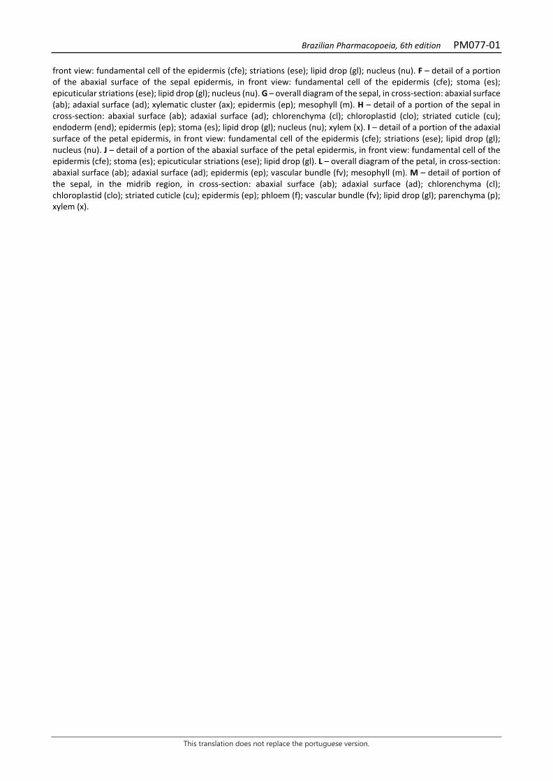
Brazilian Pharmacopoeia, 6th edition PM077-01
This translation does not replace the portuguese version.
front view: fundamental cell of the epidermis (cfe); striations (ese); lipid drop (gl); nucleus (nu). F – detail of a portion of the abaxial surface of the sepal epidermis, in front view: fundamental cell of the epidermis (cfe); stoma (es); epicuticular striations (ese); lipid drop (gl); nucleus (nu). G – overall diagram of the sepal, in cross-section: abaxial surface (ab); adaxial surface (ad); xylematic cluster (ax); epidermis (ep); mesophyll (m). H – detail of a portion of the sepal in cross-section: abaxial surface (ab); adaxial surface (ad); chlorenchyma (cl); chloroplastid (clo); striated cuticle (cu); endoderm (end); epidermis (ep); stoma (es); lipid drop (gl); nucleus (nu); xylem (x). I – detail of a portion of the adaxial surface of the petal epidermis, in front view: fundamental cell of the epidermis (cfe); striations (ese); lipid drop (gl); nucleus (nu). J – detail of a portion of the abaxial surface of the petal epidermis, in front view: fundamental cell of the epidermis (cfe); stoma (es); epicuticular striations (ese); lipid drop (gl). L – overall diagram of the petal, in cross-section: abaxial surface (ab); adaxial surface (ad); epidermis (ep); vascular bundle (fv); mesophyll (m). M – detail of portion of the sepal, in the midrib region, in cross-section: abaxial surface (ab); adaxial surface (ad); chlorenchyma (cl); chloroplastid (clo); striated cuticle (cu); epidermis (ep); phloem (f); vascular bundle (fv); lipid drop (gl); parenchyma (p); xylem (x).
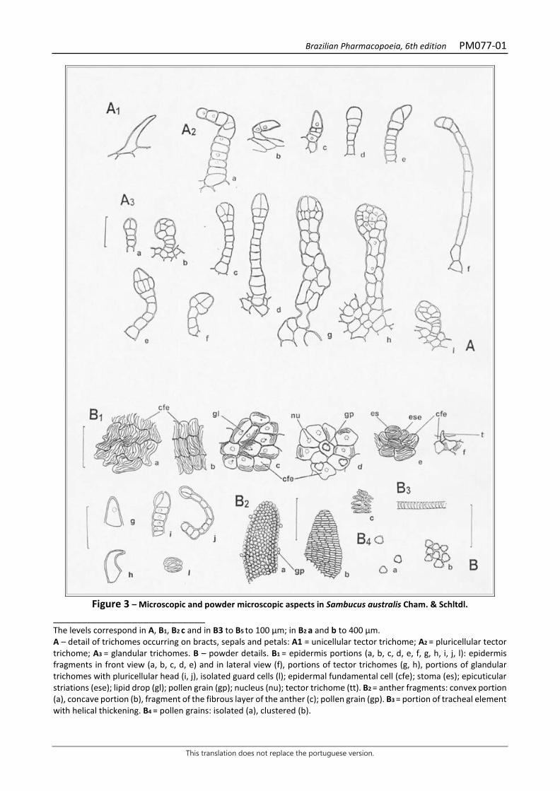
Brazilian Pharmacopoeia, 6th edition PM077-01
This translation does not replace the portuguese version.
Figure 3 – Microscopic and powder microscopic aspects in Sambucus australis Cham. & Schltdl.
___________________________ The levels correspond in A, B1, B2 c and in B3 to B5 to 100 µm; in B2 a and b to 400 µm. A – detail of trichomes occurring on bracts, sepals and petals: A1 = unicellular tector trichome; A2 = pluricellular tector trichome; A3 = glandular trichomes. B – powder details. B1 = epidermis portions (a, b, c, d, e, f, g, h, i, j, l): epidermis fragments in front view (a, b, c, d, e) and in lateral view (f), portions of tector trichomes (g, h), portions of glandular trichomes with pluricellular head (i, j), isolated guard cells (l); epidermal fundamental cell (cfe); stoma (es); epicuticular striations (ese); lipid drop (gl); pollen grain (gp); nucleus (nu); tector trichome (tt). B2 = anther fragments: convex portion (a), concave portion (b), fragment of the fibrous layer of the anther (c); pollen grain (gp). B3 = portion of tracheal element with helical thickening. B4 = pollen grains: isolated (a), clustered (b).
Brazilian Pharmacopoeia, 6th edition PM078-01
This translation does not replace the portuguese version.
ELDERBERRY, flower Sambucus nigra flos
The plant drug consists of dried flowers of Sambucus nigra L., containing at least 1.5% total flavonoids, expressed as quercetin, and at least 1.0% rutin. CHARACTERISTICS The dried flowers have a characteristic subtle, aromatic odor. IDENTIFICATION 1. Macroscopic description Flowers with a yellowish color acquired after drying, pentamerous or tetramerous, hermaphrodite measuring 3 to 5 mm in diameter, each with three tiny green bracts, distributed on the pedicel, receptacle and/or calyx base, at different heights, visible with a magnifying glass. Sparsely papillose bracts, with tector and glandular trichomes on the adaxial surface, with unicellular marginal teeth. Whitish or brownish, globose flower buds measuring 1.5 to 3 mm in diameter. Calyx with whitish-yellowish, greenish or brownish sepals, triangular, 0.5 to 1.2 mm long and 0.5 to 0.7 mm wide at the basal portion, slightly merged to each other at the base and with unicellular marginal teeth. Yellowish, rotate corolla, with petals merged together at the base in a short tube. Oval to elliptic petals, with a retrorse apex, 2 to 3.5 mm long and 2 to 3 mm wide. The androecium is formed by five or four stamens, arranged alternately to the petals, with filaments attached to the corolla tube. Dithecate, extrorse, dorsifixed, oblong anthers, dehiscent, yellow, 1 mm long. Glabrous and cylindrical filaments, 1 to 1.5 mm long. Infertile ovary, merged to the calyceal tube, tricarpellar, rarely tetracarpellar, trilocular, rarely tetralocular, with well-demarcated carpels, one seminal rudiment per locule, axially placentate. Globose and papillose gynoecium, with a short stylet and trilobed stigma. A prominent, ringed disk surrounds the base of the gynoecium. 2. Microscopic description Bracts, sepals, and petals are hypoestomatic, amphistomatic and amphi-hypostomatic, respectively; stomas are of the anomocytic type; in front view, the cuticle exhibits striations; tector and glandular trichomes occur mainly at the base of the adaxial surface; in cross-section, the cuticle is striated, the epidermis is unistratified, and the mesophyll is homogeneous; spherical lipid drops occur in all tissues except the xylem. Blackened-looking idioblasts containing crystalline sand-like calcium oxalate crystals are visible in all three pieces. Three, rarely four, parallel veins occur on the petals, the secondary ones originating from the main one, branched or not; the epidermis has papillose cells, less prominent in the margin regions; ellipsoid starch grains are present in the mesophyll. The filament, in front view, has a striated cuticle. The anther, in cross-section, has a papillose epidermis, the mat is unistratified, and the endothecium is formed by two to three layers of fibrous cells, with evident pitting. The pollen grain is prolate, tricolporate, 15 to 25 μm in diameter, with a reticulate surface. The gynoecium is formed by three, rarely four carpels and each cavity has a seminal rudiment; in cross-section, the parenchyma tissue of the carpel wall is compact, formed by chloroplastid-rich cells and lipid drops; the innermost parenchyma lacks chloroplastids; the
Brazilian Pharmacopoeia, 6th edition PM078-01
This translation does not replace the portuguese version.
epidermal cells of the stigma are extremely papillose. It differs from Sambucus australis in that it does not have idioblasts containing calcium oxalate crystals on the bracts, sepals and petals, and usually has five carpels on the ovary. 3. Macroscopic description of impurities Pedicels of the species itself are considered foreign matter; they are whitish from drying, longitudinally furrowed, 1 to 7 mm long, with tector and glandular trichomes. 4. Microscopic description of impurities The pedicel, in front view, exhibits striated cuticle, rectangular epidermal cells, anomocytic stomata and, in the basal portion, tector and glandular trichomes; in cross-section, it exhibits pronounced prominences and recesses, striated cuticle, unistratified epidermis, with tabular-shaped cells and thick inner periclinal walls; in the cortical region one to six layers of tabular collenchyma occur, followed by parenchyma with large intercellular spaces; the vascular system is formed by up to 16 collateral bundles, arranged in a ring shape; the medullary region is filled with parenchyma with thin-walled cells; chloroplastids occur in the parenchyma; lipid drops occur in the epidermis and cortical parenchyma; starch grains are observed in the endodermis and phloem. 5. Microscopic description of powder The sample meets all requirements for the flowers of the species, except the macroscopic characters. Characteristics are: greenish-yellow color; sepals fragments with isolated unicellular marginal teeth; epidermis fragments of sepals and petals papillose and with striated cuticle; fragments of epidermis with anomocytic stomata; isolated guard cells; fragments of epidermis with tector trichomes of different types; rare isolated tector and glandular trichomes or parts thereof; parenchyma fragments; portions of tissues with lipid drops; part of tracheal elements of helical thickening; fragments of epidermis of an extremely papillose anther; fragments of the fibrous layer of the anther; several pollen grains as described; pollen grains isolated or grouped, or associated with fragments of anthers and the epidermis of several parts; portions of stigma with papillose epidermis; portions of bracts; portions of the margin of sepals, petals and bracts. 6. Proceed as described in Thin-layer chromatography (5.2.17.1). Stationary phase: silica gel GF254 (0.250 mm). Mobile phase: ethyl acetate, methyl ethyl ketone, water, anhydrous formic acid (50:30:10:10). Sample solution: transfer 0.5 g of the pulverized sample (355 µm) (5.2.11) into a beaker and add 5 mL methyl alcohol. Sonicate for 10 minutes. Centrifuge at 1000 × g for five minutes. Reference solution: dissolve 1 mg caffeic acid, 1 mg chlorogenic acid, 2.5 mg hyperoside, and 2.5 mg rutin in 10 mL methyl alcohol. Procedure: separately apply 4 µL of the Sample solution and 4 µL of the Reference solution to the plate. Conduct the chromatogram along 6 cm. Remove plate and allow it to air dry. Heat in an oven at 100°C for five minutes and nebulize with a 1g/L solution of diphenylboric acid aminoethyl ester
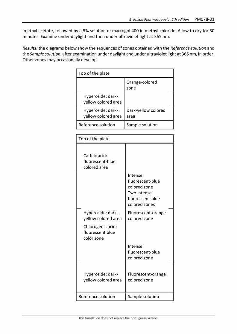
Brazilian Pharmacopoeia, 6th edition PM078-01
This translation does not replace the portuguese version.
in ethyl acetate, followed by a 5% solution of macrogol 400 in methyl chloride. Allow to dry for 30 minutes. Examine under daylight and then under ultraviolet light at 365 nm. Results: the diagrams below show the sequences of zones obtained with the Reference solution and the Sample solution, after examination under daylight and under ultraviolet light at 365 nm, in order. Other zones may occasionally develop.
Top of the plate
Orange-colored zone
Hyperoside: dark-yellow colored area
Hyperoside: dark-yellow colored area
Dark-yellow colored area
Reference solution Sample solution
Top of the plate
Caffeic acid: fluorescent-blue colored area
Intense fluorescent-blue colored zone Two intense fluorescent-blue colored zones
Hyperoside: dark-yellow colored area
Fluorescent-orange colored zone
Chlorogenic acid: fluorescent blue color zone
Intense
fluorescent-blue colored zone
Hyperoside: dark-yellow colored area
Fluorescent-orange colored zone
Reference solution Sample solution
Brazilian Pharmacopoeia, 6th edition PM078-01
This translation does not replace the portuguese version.
7. Proceed as described in High Performance Liquid Chromatography (5.2.17.4). Use the system as described in Dosage for Rutin. The majority peak in the chromatogram corresponds to rutin; there is one peak with a lower retention time with characteristics of caffeoylquinic acid and four peaks following rutin, the two immediately after it having an ultraviolet absorption spectrum similar to rutin, and the next two with an absorption spectrum characteristic of caffeoylquinic acid. TESTS Foreign matter (5.4.1.3). No more than 8.0% coarse pedicels and other foreign material, and no more than 15% of the sample with altered (blackened) color. Loss by drying (5.2.9.1). Gravimetric method. At most 11.0%. Total ash (5.4.1.5.1). At most 9.0%. Total mesophilic microorganism count (5.5.3.1.2). Complies with the test. Study of pathogenic microorganisms (5.5.3.1.3). Complies with the test. Heavy metals (5.4.5). Complies with the test. Agrochemical waste (5.4.3). Complies with the test. DOSAGE Total flavonoids To proceed as described in Visible absorption spectrophotometry (5.2.14). Prepare solutions as described below. Stock solution: accurately weigh approximately 0.1 g of the pulverized plant drug (800 µm) (5.2.11) and place in a 100mL round-bottomed flask. Add 0.25 mL of 0.5 g/L aqueous methenamine solution (v/v), 10 mL acetone and 0.5 mL hydrochloric acid. Heat in a water bath, under reflux, for 30 minutes. Filter the mixture through absorbent cotton into a 25-mL volumetric flask. Return the drug residue and cotton to the same round-bottomed flask, add 7 mL acetone. Heat under reflux for 10 minutes. Filter through absorbent cotton into the same 25-mL volumetric flask. Repeat the operation, returning the drug residue again and the absorbent cotton to the round-bottomed flask, add 7 mL acetone and heat, under reflux, for 10 minutes. Filter into the same 25-mL flask. After cooling to room temperature, top off the volume with acetone and homogenize. In a separating funnel, add 10 mL of this solution and 10 mL of water, then, extract with 10 mL ethyl acetate. Repeat the extraction twice more, with 6 mL portions of ethyl acetate each. Gather the ethyl acetate phases in a separating funnel and rinse them with two 15-mL portions of water. Transfer the organic phase to a 25-mL volumetric flask, top off the volume with ethyl acetate and homogenize.
Brazilian Pharmacopoeia, 6th edition PM078-01
This translation does not replace the portuguese version.
Sample solution: transfer 10 mL of the Stock solution to a 25-mL volumetric flask, add 1 mL of 2% (w/v) aluminum chloride in methyl alcohol, top off the volume with a 5% (v/v) acetic acid solution in methyl alcohol and homogenize. Blank solution: transfer 10 mL of the Stock solution into a 25-mL volumetric flask, top off the volume with the 5% (w/v) acetic acid solution in methyl alcohol and homogenize. Procedure: measure the absorbance of the Sample solution at 425 nm 30 minutes after its preparation, using the Blank solution for zero adjustment. Calculate total flavonoid content expressed as a percentage of quercetin, according to the following expression:
𝑇𝑇𝑇𝑇 =A × 156,25
m × 500
in which, TF = total flavonoid content expressed as quercetin % (w/w); A = absorbance measured for the Sample solution; 156.25 = dilution factor; 500 = quercetin specific absorption coefficient; m = mass in grams of the sample used, considering the loss by drying. Rutin Proceed as described in High Performance Liquid Chromatography (5.2.17.4). Use a chromatograph equipped with an ultraviolet detector at 356 nm; pre-column packed with silica chemically bonded to octadecylsilane group (3 to 10 µm), 150 mm long and 3.9 mm wide (internal diameter) column, packed with silica chemically bonded to octadecylsilane group (4 µm), kept at room temperature; mobile phase flow rate of 0.7 mL/ minute. Eluent (A): water, acetonitrile and trifluoracetic acid (95:5: 0.01). Eluent (B): acetonitrile and trifluoracetic acid (100:0.01).
Time (minutes) Eluent A (%) Eluent B (%) Elution
0 – 7 90 → 70 10 → 30 linear gradient 7 – 8 70 → 0 30 → 100 linear gradient 8 – 11 0 100 isocratic 11 – 12 0 → 90 100 → 10 linear gradient 12 – 18 90 10 isocratic Sample solution: accurately weigh approximately 0.25 g of the dried and pulverized plant drug (800 µm) (5.2.11) and place in glass vial. Shake by turbolysis at speed 3, for five minutes with 5 mL of 80% (v/v) ethyl alcohol. Filter through filter paper, under vacuum, into a 5-mL volumetric flask, top off the volume with the same solvent and homogenize. Dilute 50 µL in 950 µL of a mixture of acetonitrile and water (1:9) and homogenize. Filter through a 0.45 µm filter unit.
Brazilian Pharmacopoeia, 6th edition PM078-01
This translation does not replace the portuguese version.
Reference solution: dissolve 5 mg rutin CRS in 10 mL methyl alcohol. Solutions for the calibration curve: dilute an aliquot of 2.5 mL of the Reference Solution, in a 25mL volumetric flask, in order to obtain a 50 µg/mL solution. Dilute aliquots of 1 mL, 1.5 mL, 2 mL, 2.5 mL, 3 mL, 3.5 mL, 4 mL and 4.5 mL in a 5 mL volumetric flask with methyl alcohol to concentrations of 10 µg/mL, 15 µg/mL, 20 µg/mL, 25 µg/mL, 30 µg/mL, 35 µg/mL, 40 µg/mL and 45 µg/mL. Filter through a 0.45 µm filter unit. Procedure: inject 10 µL of the Solutions for the analytical curve and 10 µL of the sample solution separately. Register the chromatograms and measure the areas under the peaks. Rutin retention time is approximately five minutes. Calculate rutin content in the sample from the straight line equation obtained from the analytical curve. The result is expressed as the average of the determinations in grams of rutin, in percent, considering the loss by drying. PACKAGING AND STORAGE In tightly closed containers, protected from light and heat.
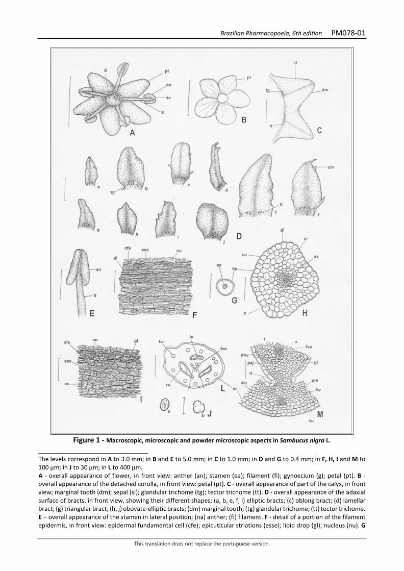
Brazilian Pharmacopoeia, 6th edition PM078-01
This translation does not replace the portuguese version.
Figure 1 - Macroscopic, microscopic and powder microscopic aspects in Sambucus nigra L.
___________________________ The levels correspond in A to 3.0 mm; in B and E to 5.0 mm; in C to 1.0 mm; in D and G to 0.4 mm; in F, H, I and M to 100 µm; in J to 30 µm; in L to 400 µm. A - overall appearance of flower, in front view: anther (an); stamen (ea); filament (fi); gynoecium (g); petal (pt). B - overall appearance of the detached corolla, in front view: petal (pt). C - overall appearance of part of the calyx, in front view; marginal tooth (dm); sepal (sl); glandular trichome (tg); tector trichome (tt). D - overall appearance of the adaxial surface of bracts, in front view, showing their different shapes: (a, b, e, f, i) elliptic bracts; (c) oblong bract; (d) lamellar bract; (g) triangular bract; (h, j) obovate-elliptic bracts; (dm) marginal tooth; (tg) glandular trichome; (tt) tector trichome. E – overall appearance of the stamen in lateral position; (na) anther; (fi) filament. F - detail of a portion of the filament epidermis, in front view: epidermal fundamental cell (cfe); epicuticular striations (esse); lipid drop (gl); nucleus (nu). G
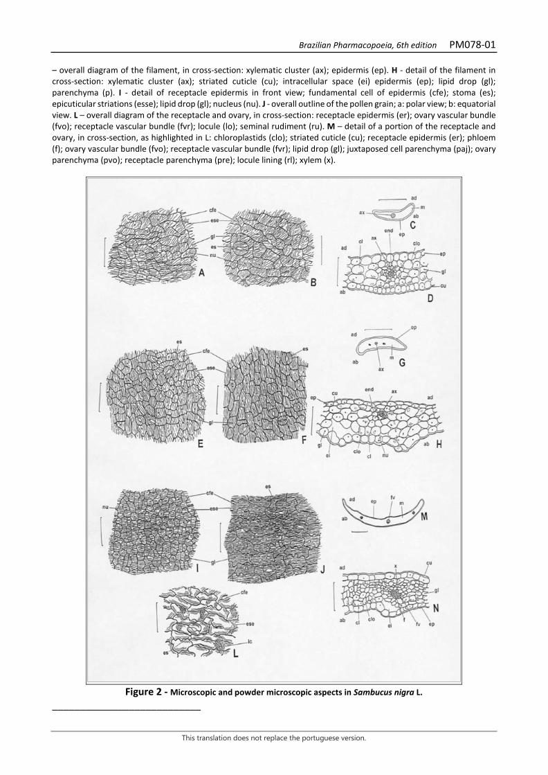
Brazilian Pharmacopoeia, 6th edition PM078-01
This translation does not replace the portuguese version.
– overall diagram of the filament, in cross-section: xylematic cluster (ax); epidermis (ep). H - detail of the filament in cross-section: xylematic cluster (ax); striated cuticle (cu); intracellular space (ei) epidermis (ep); lipid drop (gl); parenchyma (p). I - detail of receptacle epidermis in front view; fundamental cell of epidermis (cfe); stoma (es); epicuticular striations (esse); lipid drop (gl); nucleus (nu). J - overall outline of the pollen grain; a: polar view; b: equatorial view. L – overall diagram of the receptacle and ovary, in cross-section: receptacle epidermis (er); ovary vascular bundle (fvo); receptacle vascular bundle (fvr); locule (lo); seminal rudiment (ru). M – detail of a portion of the receptacle and ovary, in cross-section, as highlighted in L: chloroplastids (clo); striated cuticle (cu); receptacle epidermis (er); phloem (f); ovary vascular bundle (fvo); receptacle vascular bundle (fvr); lipid drop (gl); juxtaposed cell parenchyma (paj); ovary parenchyma (pvo); receptacle parenchyma (pre); locule lining (rl); xylem (x).
Figure 2 - Microscopic and powder microscopic aspects in Sambucus nigra L.
___________________________
Brazilian Pharmacopoeia, 6th edition PM078-01
This translation does not replace the portuguese version.
Levels correspond in A, B, D, E, F, H, I-L and N to 100 µm; in C, G and M to 0.4 mm. A – detail of a portion of the adaxial surface of the bract epidermis, in front view: fundamental cell of the epidermis (cfe); epicuticular striations (esse); lipid drop (gl); nucleus (nu). B – detail of a portion of the abaxial surface of the bract epidermis, in front view: fundamental cell of the epidermis (cfe); stoma (es); epicuticular striations (ese); lipid drop (gl); nucleus (nu). C – overall diagram of the bract, in cross-section: abaxial surface (ab); adaxial surface (ad); xylematic cluster (ax); epidermis (ep); mesophyll (m). D - detail of the midrib region of the bract, in cross-section; abaxial surface (ab); adaxial surface (ad); xylematic cluster (ax); chlorenchyma (cl); chloroplastid (clo); cuticle (cu); endoderm (end); epidermis (ep); lipid drop (gl). B – detail of a portion of the adaxial surface of the sepal epidermis, in front view: fundamental cell of the epidermis (cfe); stoma (es) striations (ese); lipid drop (gl). F – detail of a portion of the abaxial surface of the sepal epidermis, in front view: fundamental cell of the epidermis (cfe); stoma (es) striations (ese); lipid drop (gl). G – overall diagram of the sepal, in cross-section: abaxial surface (ab); adaxial surface (ad); xylematic cluster (ax); epidermis (ep); mesophyll (m). H - detail of sepal portion in the midrib region, in cross-section; abaxial surface (ab); adaxial surface (ad); xylematic cluster (ax); chlorenchyma (cl); chloroplastid (clo); striated cuticle (cu); intercellular space (ei); endoderm (end); epidermis (ep); lipid drop (gl); nucleus (nu). I – detail of a portion of the adaxial surface of the petal epidermis, in front view: fundamental cell of the epidermis (cfe); striations (ese); lipid drop (gl); nucleus (nu). J – detail of a portion of the abaxial surface of the petal epidermis, in front view: fundamental cell of the epidermis (cfe); stoma (es); epicuticular striations (ese); lipid drop (gl). J – detail of a portion of the abaxial surface of the petal epidermis, in front view: fundamental cell of the epidermis (cfe); stoma (es); epicuticular striations (ese); crystalliferous idioblast (ic). < – overall diagram of the petal, in cross-section: abaxial surface (ab); adaxial surface (ad); epidermis (ep); vascular bundle (fv); mesophyll (m). N – detail of portion of the sepal, in the midrib region, in cross-section: abaxial surface (ab); adaxial surface (ad); chlorenchyma (cl); chloroplastid (clo); striated cuticle (cu); intracellular space (ei) epidermis (ep); phloem (f); vascular bundle (fv); lipid drop (gl); xylem (x).
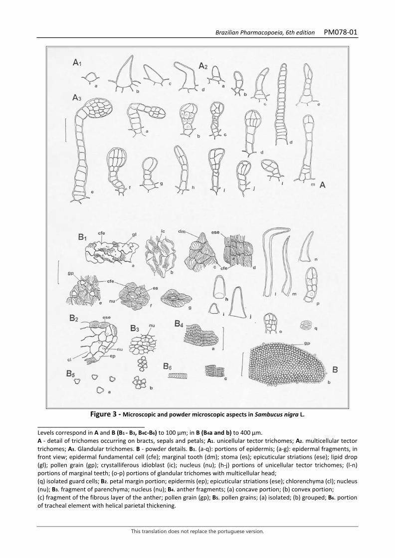
Brazilian Pharmacopoeia, 6th edition PM078-01
This translation does not replace the portuguese version.
Figure 3 - Microscopic and powder microscopic aspects in Sambucus nigra L.
___________________________ Levels correspond in A and B (B1 - B3, B4c-B6) to 100 µm; in B (B4a and b) to 400 µm. A - detail of trichomes occurring on bracts, sepals and petals; A1. unicellular tector trichomes; A2. multicellular tector trichomes; A3. Glandular trichomes. B - powder details. B1. (a-q): portions of epidermis; (a-g): epidermal fragments, in front view; epidermal fundamental cell (cfe); marginal tooth (dm); stoma (es); epicuticular striations (ese); lipid drop (gl); pollen grain (gp); crystalliferous idioblast (ic); nucleus (nu); (h-j) portions of unicellular tector trichomes; (l-n) portions of marginal teeth; (o-p) portions of glandular trichomes with multicellular head; (q) isolated guard cells; B2. petal margin portion; epidermis (ep); epicuticular striations (ese); chlorenchyma (cl); nucleus (nu); B3. fragment of parenchyma; nucleus (nu); B4. anther fragments; (a) concave portion; (b) convex portion; (c) fragment of the fibrous layer of the anther; pollen grain (gp); B5. pollen grains; (a) isolated; (b) grouped; B6. portion of tracheal element with helical parietal thickening.
Brazilian Pharmacopoeia, 6th edition PM079-00
This translation does not replace the portuguese version.
WHITE WILLOW, bark Salicis cortex
The plant drug consists of entire or fragmented dried barks from young branches of Salix alba L., containing at least 1.5% salicin derivatives expressed as salicin (C13H18O7, 286.28). IDENTIFICATION 1. Macroscopic description The bark, obtained from branches that are two to three years old, presents itself in irregular fragments that are leathery, flexible, elongated, and slightly fluted, varying in length, width, and thickness. The outer surface is shiny-glossy, smooth or longitudinally striated in young shells, dark brown. The inner surface is finely striated longitudinally, fibrous, brownish. The fracture is short in the outer portion and fibrous in the inner portion. 2. Microscopic description In front view, suber cells are generally polygonal and rectilinear-walled, with a dark brown, sometimes yellowish, color. In cross-section, the cortex has an extremely thick cuticle and the outer portion of the bark is variable: 1) first unistratified epidermal layer, with small quadrangular, thick-walled cells, followed by five to six layers of tabular collenchyma, with elongated cells, tangentially arranged and containing chloroplastids, followed by parenchyma with thick-walled cells of several shapes; or 2) outer epidermal layer, with the same characteristics as described above, followed by collenchyma and parenchyma, both with rounded, thick-walled cells; or 3) presence of periderm, with several layers of juxtaposed or almost juxtaposed cells, followed by parenchyma as described above. The outer cortical parenchyma is pluri-stratified, with thick-walled cells, exhibiting chloroplastids and calcium oxalate druses; prismatic crystals, isolated or grouped stone cells, or cells containing phenolic compounds rarely occur. The inner cortical parenchyma shows clusters of randomly distributed fibers, cells with less crystals and chloroplastids and, rarely, clusters of stone cells. The phloem is rich in phenolic compounds and is always accompanied by clusters of fibers. The cambium internally exhibits a tissue formed by cells with thin walls and reduced number of layers, with a large amount of starch grains and idioblasts containing phenolic compounds. In longitudinal section, the characteristics of the suber and outer cortical region are similar to those described for the cross-section. The inner cortical region has often enlarged parenchymal rays, with fibers and thick-walled parenchyma cells. 3. Microscopic description of powder The sample meets all requirements for the species, except the macroscopic characters. Examine under a microscope using 50% (w/v) chloral hydrate. Characteristics are: pale brown color; fragments of suber, in front view; fragments of parenchyma, with thick-walled cells, in front view; isolated fibers or portions of their clusters, in longitudinal section; fragments of cortical parenchyma with polygonal-shaped, thick-walled cells, with druses, in cross-section; portion of fibers associated with crystalliferous idioblasts, in longitudinal section; parenchyma fragments with thick-walled cells, with radial distribution and with portions of cambium; cambium fragments, in cross-section; prismatic crystals and calcium oxalate druses.
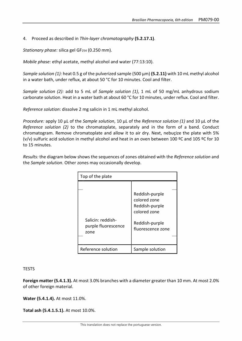
Brazilian Pharmacopoeia, 6th edition PM079-00
This translation does not replace the portuguese version.
4. Proceed as described in Thin-layer chromatography (5.2.17.1). Stationary phase: silica gel GF254 (0.250 mm). Mobile phase: ethyl acetate, methyl alcohol and water (77:13:10). Sample solution (1): heat 0.5 g of the pulverized sample (500 µm) (5.2.11) with 10 mL methyl alcohol in a water bath, under reflux, at about 50 °C for 10 minutes. Cool and filter. Sample solution (2): add to 5 mL of Sample solution (1), 1 mL of 50 mg/mL anhydrous sodium carbonate solution. Heat in a water bath at about 60 °C for 10 minutes, under reflux. Cool and filter. Reference solution: dissolve 2 mg salicin in 1 mL methyl alcohol. Procedure: apply 10 µL of the Sample solution, 10 µL of the Reference solution (1) and 10 µL of the Reference solution (2) to the chromatoplate, separately and in the form of a band. Conduct chromatogram. Remove chromatoplate and allow it to air dry. Next, nebuçize the plate with 5% (v/v) sulfuric acid solution in methyl alcohol and heat in an oven between 100 ºC and 105 ºC for 10 to 15 minutes. Results: the diagram below shows the sequences of zones obtained with the Reference solution and the Sample solution. Other zones may occasionally develop.
Top of the plate
Reddish-purple colored zone Reddish-purple colored zone
Salicin: reddish-purple fluorescence zone
Reddish-purple fluorescence zone
Reference solution Sample solution
TESTS Foreign matter (5.4.1.3). At most 3.0% branches with a diameter greater than 10 mm. At most 2.0% of other foreign material. Water (5.4.1.4). At most 11.0%. Total ash (5.4.1.5.1). At most 10.0%.
Brazilian Pharmacopoeia, 6th edition PM079-00
This translation does not replace the portuguese version.
Total mesophilic microorganism count (5.5.3.1.2). Complies with the test. Study of pathogenic microorganisms (5.5.3.1.3). Complies with the test. Heavy metals (5.4.5). Complies with the test. Agrochemical waste (5.4.3). Complies with the test. DOSAGE Salicin Proceed as described in High Performance Liquid Chromatography (5.2.17.4). Use a chromatograph equipped with an ultraviolet detector at 270 mm; pre-column packed with octadecylsilane silica, 150mm long, 3.9mm internal diameter column, packed with octadecylsilane silica (4 µm), kept at room temperature; Mobile phase flow rate of 1 mL/minute. Isocratic system. Mobile phase: water, acetonitrile and trifluoracetic acid (97:3:0.05). Sample solution: accurately weigh about 0.3 g of the dried and pulverized plant drug (355 µm) (5.2.11) add 25 mL methyl alcohol and heat, under reflux, for 30 minutes. Cool and filter. Resume the residue with 25 mL methyl alcohol and treat as described above. Gather the filtrates and evaporate under reduced pressure to dryness. Suspend the residue with 2 mL methyl alcohol, add 2 mL of 0.1 M sodium hydroxide, heat in a water bath, under reflux, for one hour at about 60 °C, while frequently shaking. Cool, add 0.25 mL of M hydrochloric acid, top off the volume to 5 mL with a mixture of methyl alcohol and water (1:1) and homogenize. Filter through a 0.45 µm filter unit. Reference solution: dissolve 10 mg salicin in 10 mL acetonitrile. Solutions for analytical curve: dilute aliquots of 40 µL, 45 µL, 50 µL, 55 µL and 60 µL of the Reference Solution to 100 µL with the Mobile phase to obtain concentrations of 0.40 mg/mL, 0.45 mg/mL, 0.50 mg/mL, 0.55 mg/mL and 0.60 mg/mL. Filter through a 0.45 µm filter unit. Procedure: inject 10 µL of the Solutions for the analytical curve and 10 µL of the sample solution separately. Register the chromatograms and measure the areas under the peaks. Salicin retention time is approximately six minutes. Calculate salicin content in the sample from the straight line equation obtained from the analytical curve. The result is expressed as the average of the determinations in grams of salicin, in percent, considering the determined water content. PACKAGING AND STORAGE In tightly closed containers, protected from light and heat.
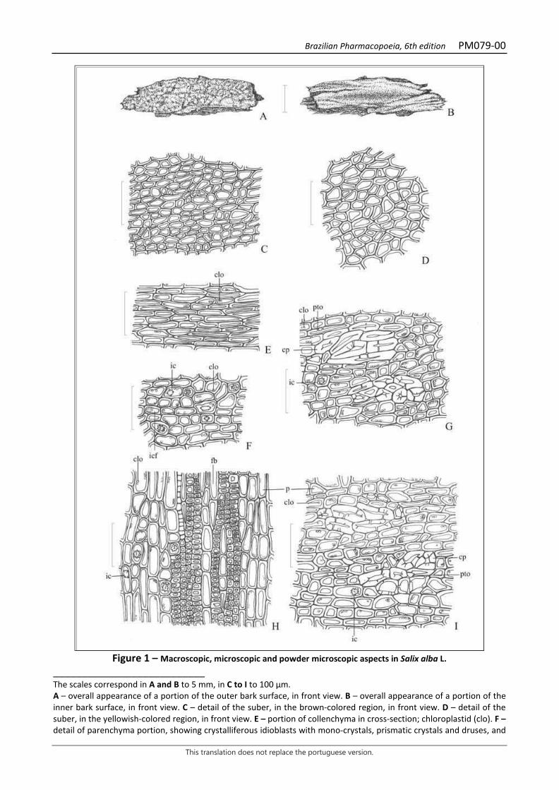
Brazilian Pharmacopoeia, 6th edition PM079-00
This translation does not replace the portuguese version.
Figure 1 – Macroscopic, microscopic and powder microscopic aspects in Salix alba L.
___________________________ The scales correspond in A and B to 5 mm, in C to I to 100 µm. A – overall appearance of a portion of the outer bark surface, in front view. B – overall appearance of a portion of the inner bark surface, in front view. C – detail of the suber, in the brown-colored region, in front view. D – detail of the suber, in the yellowish-colored region, in front view. E – portion of collenchyma in cross-section; chloroplastid (clo). F – detail of parenchyma portion, showing crystalliferous idioblasts with mono-crystals, prismatic crystals and druses, and
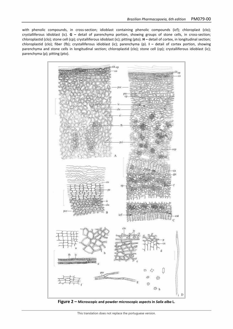
Brazilian Pharmacopoeia, 6th edition PM079-00
This translation does not replace the portuguese version.
with phenolic compounds, in cross-section; idioblast containing phenolic compounds (icf); chloroplast (clo); crystalliferous idioblast (ic). G – detail of parenchyma portion, showing groups of stone cells, in cross-section; chloroplastid (clo); stone cell (cp); crystalliferous idioblast (ic); pitting (pto). H – detail of cortex, in longitudinal section; chloroplastid (clo); fiber (fb); crystalliferous idioblast (ic); parenchyma (p). I – detail of cortex portion, showing parenchyma and stone cells in longitudinal section; chloroplastid (clo); stone cell (cp); crystalliferous idioblast (ic); parenchyma (p); pitting (pto).
Figure 2 – Microscopic and powder microscopic aspects in Salix alba L.

Brazilian Pharmacopoeia, 6th edition PM079-00
This translation does not replace the portuguese version.
___________________________ The scales correspond in A, B and D (a - h) to 100 µm, in C and D (i) to 200 µm. A – detail of the external portion of the cortex, in cross-section; chloroplastid (clo); collenchyma (co); cuticle (cu); epidermis (ep); intercellular space (ei); crystalliferous idioblast (ic); fiber (fb); external cortical parenchyma (pce); internal cortical parenchyma (pci). B – detail of cortex portion, showing the periderm lining, in cross-section; cuticle (cu); chloroplastid (clo); crystalliferous idioblast (ic); outer cortical parenchyma (pce); periderm (pe). C – detail of the cortex in cross-section; cambium (ca); internal cambium (cat); chloroplastid (clo); collenchyma (co); stone cell (cp); cuticle (cu); epidermis (ep); intercellular space (ei); phloem (f); fiber (fb); starch grains (ga); crystalliferous idioblast (ic); idioblast containing phenolic compounds (icf); parenchyma (p); outer cortical parenchyma (pce); inner cortical parenchyma (pci); pitting (pto); parenchyma ray (rp). D – details of the powder. suber portion, in front view (a); parenchyma portion, in front view (b); parenchyma portion, showing crystalliferous idioblasts, in cross-section (c); parenchyma and cambium portion, in longitudinal section (d); portion of fibers associated with crystalliferous idioblasts, in longitudinal section (e); cambium portion, in cross-section (f); bundled fiber portion, in longitudinal section (g); calcium oxalate crystals, isolated (h); isolated fiber, in longitudinal section (i); chloroplastid (clo); crystalliferous idioblast (ic); cambium (ca); parenchyma (p); fiber (fb); crystalliferous idioblast (ic); dotation (pto).
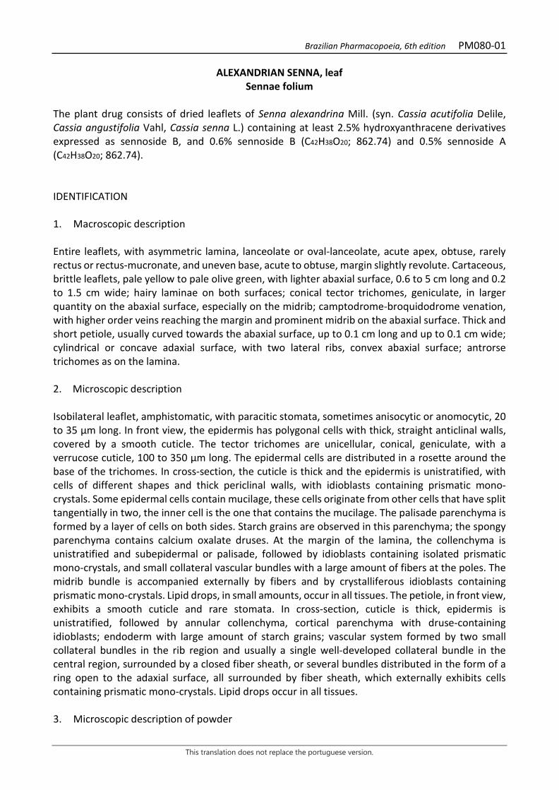
Brazilian Pharmacopoeia, 6th edition PM080-01
This translation does not replace the portuguese version.
ALEXANDRIAN SENNA, leaf Sennae folium
The plant drug consists of dried leaflets of Senna alexandrina Mill. (syn. Cassia acutifolia Delile, Cassia angustifolia Vahl, Cassia senna L.) containing at least 2.5% hydroxyanthracene derivatives expressed as sennoside B, and 0.6% sennoside B (C42H38O20; 862.74) and 0.5% sennoside A (C42H38O20; 862.74). IDENTIFICATION 1. Macroscopic description Entire leaflets, with asymmetric lamina, lanceolate or oval-lanceolate, acute apex, obtuse, rarely rectus or rectus-mucronate, and uneven base, acute to obtuse, margin slightly revolute. Cartaceous, brittle leaflets, pale yellow to pale olive green, with lighter abaxial surface, 0.6 to 5 cm long and 0.2 to 1.5 cm wide; hairy laminae on both surfaces; conical tector trichomes, geniculate, in larger quantity on the abaxial surface, especially on the midrib; camptodrome-broquidodrome venation, with higher order veins reaching the margin and prominent midrib on the abaxial surface. Thick and short petiole, usually curved towards the abaxial surface, up to 0.1 cm long and up to 0.1 cm wide; cylindrical or concave adaxial surface, with two lateral ribs, convex abaxial surface; antrorse trichomes as on the lamina. 2. Microscopic description Isobilateral leaflet, amphistomatic, with paracitic stomata, sometimes anisocytic or anomocytic, 20 to 35 µm long. In front view, the epidermis has polygonal cells with thick, straight anticlinal walls, covered by a smooth cuticle. The tector trichomes are unicellular, conical, geniculate, with a verrucose cuticle, 100 to 350 µm long. The epidermal cells are distributed in a rosette around the base of the trichomes. In cross-section, the cuticle is thick and the epidermis is unistratified, with cells of different shapes and thick periclinal walls, with idioblasts containing prismatic mono-crystals. Some epidermal cells contain mucilage, these cells originate from other cells that have split tangentially in two, the inner cell is the one that contains the mucilage. The palisade parenchyma is formed by a layer of cells on both sides. Starch grains are observed in this parenchyma; the spongy parenchyma contains calcium oxalate druses. At the margin of the lamina, the collenchyma is unistratified and subepidermal or palisade, followed by idioblasts containing isolated prismatic mono-crystals, and small collateral vascular bundles with a large amount of fibers at the poles. The midrib bundle is accompanied externally by fibers and by crystalliferous idioblasts containing prismatic mono-crystals. Lipid drops, in small amounts, occur in all tissues. The petiole, in front view, exhibits a smooth cuticle and rare stomata. In cross-section, cuticle is thick, epidermis is unistratified, followed by annular collenchyma, cortical parenchyma with druse-containing idioblasts; endoderm with large amount of starch grains; vascular system formed by two small collateral bundles in the rib region and usually a single well-developed collateral bundle in the central region, surrounded by a closed fiber sheath, or several bundles distributed in the form of a ring open to the adaxial surface, all surrounded by fiber sheath, which externally exhibits cells containing prismatic mono-crystals. Lipid drops occur in all tissues. 3. Microscopic description of powder
Brazilian Pharmacopoeia, 6th edition PM080-01
This translation does not replace the portuguese version.
The sample meets all requirements for the species, except the macroscopic characters. Characteristics are: grayish-green to yellowish-green color; portions of tector trichomes, in lateral view; epidermis fragments with stomata, in front view; epidermis fragments with stomata and trichomes, in front view; epidermis portions showing the region of trichome insertion, in front view; epidermis fragments over the midrib region, with stomata, in front view and druse-like crystals, visible by transparency; petiole epidermis fragments, in front view; epidermal cells in cross-section; crystalliferous idioblasts and fiber clusters in longitudinal section; portions of tracheal elements in longitudinal section; mesophyll portions as described in cross-section; portion of assimilation parenchyma in cross-section and of vascular bundle in longitudinal section; portion of vascular bundle in longitudinal section; isolated prismatic and druse-type crystals. 4. Description of impurities The rachis, if present as an impurity, is 2.5 to 13 cm long and up to 0.1 cm wide, cylindrical or concave on the adaxial surface with two well-developed ribs, and convex on the abaxial surface; leaflet insertion scars are well-defined. In cross-section, the vascular system is formed by three to eight collateral bundles and the ensemble surrounded by a continuous sheath of small-caliber fibers; a smaller vascular bundle occurs in each of the ribs, with a fiber cap external to the phloem. 5. Proceed as described in Thin-layer chromatography (5.2.17.1). Stationary phase: silica gel GF254 (0.25 mm). Mobile phase: ethyl acetate, n-propyl alcohol, water, and glacial acetic acid (40:40:30:1). Sample solution: add 0.5 g of the pulverized plant drug in 5 mL of the ethyl alcohol and water mixture (1:1). Heat to boiling. Filter. Reference solution: dissolve 2.5 mg senoside A and 2.5 mg senoside B separately in 1 mL methyl alcohol and 1 mL of water, heat slightly if necessary. Procedure: apply 10 μL of the Sample solution and 10 μL of the Reference solution to the chromatoplate, separately and in the form of a band. Conduct chromatogram. Remove plate and allow it to air dry. Nebulize with 25% nitric acid and heat at 120 °C for 10 minutes. Allow to cool and nebulize the plate with 5% (w/v) potassium hydroxide solution until spots appear. Results: the diagram below shows the sequences of zones obtained with the Reference solution and the Sample solution. Other zones may occasionally develop.

Brazilian Pharmacopoeia, 6th edition PM080-01
This translation does not replace the portuguese version.
Top of the plate
Brownish-red colored zone Brownish-red colored zone
Sinoside A: reddish-brown zone
Brownish-red colored zone
Sinoside B: reddish-brown zone
Brownish-red colored zone
Reference solution Sample solution
TESTS Foreign matter (5.4.1.3). At most 2.0%, corresponding to the leaf rachis. Water (5.4.1.4). At most 10.0%. Total ash (5.4.1.5.1). At most 12.0%. Total mesophilic microorganism count (5.5.3.1.2). Complies with the test. Study of pathogenic microorganisms (5.5.3.1.3). Complies with the test. Heavy metals (5.4.5). Complies with the test. Agrochemical waste (5.4.3). Complies with the test. DOSAGE Hydroxyanthracene derivatives To proceed as described in Visible absorption spectrophotometry (5.2.14). Prepare solutions as described below. Stock solution: accurately weigh approximately 0.15 g of the pulverized plant drug (180 µm) (5.2.11) in a round-bottomed, polished-necked flask, add 30mL of water, mix and weigh the entire ensemble. Heat on a heating mantle for 15 minutes under reflux. Allow to cool, weigh, reestablish the initial weight with water, and filter, discarding the initial 10 mL. Transfer 10 mL of the filtrate to a 50-mL separating funnel, add one drop of 2 M hydrochloric acid, and rinse with three 5 mL portions of chloroform. Dimiss the chloroform phase. Centrifuge the aqueous phase for 10 minutes at 700 × g. Transfer 4 mL of the supernatant liquid into a round-bottomed flask with a ground-glass stopper.
Brazilian Pharmacopoeia, 6th edition PM080-01
This translation does not replace the portuguese version.
Adjust solution pH to 7.0 to 8.0 with about 80 µL of 5% (w/v) sodium carbonate solution. Add 8 mL of 10.5% (w/v) ferric chloride solution. Mix and heat in a water bath, under reflux, for 20 minutes. Add 0.4 mL concentrated hydrochloric acid and keep warm for 20 minutes, frequently shaking, until the precipitate is dissolved. Cool the solution and transfer to a 50-mL separating funnel, extract with 10 mL and twice with 7 mL diethyl ether, previously used to rinse the round-bottomed flask. Gather the ether extracts and rinse with two 10 mL portions of water. Transfer the ether layer to a 25-mL volumetric flask, top off the volume with diethyl ether and homogenize. Sample solution: evaporate 5 mL of the Stock solution until residue is formed in a water bath. Suspend residue in 5 mL of 0.5% magnesium acetate (w/v) in methyl alcohol. Filter if necessary. Blank solution: methyl alcohol. Procedure: measure the absorbance of the Sample solution at 515 nm, immediately after its preparation, using the Blank solution for zero adjustment. Calculate the content of hydroxyanthracene derivatives, calculated as a percentage of senoside B, according to the following expression:
𝑇𝑇𝑇𝑇𝑇𝑇 =A × 187,5m × 240
in which, TSB = hydroxyanthracene derivative content expressed as sennoside B % (w/w); A = absorbance measured for the Sample solution; 187,5 = dilution factor; 240 = hyperoside specific absorption coefficient; m = mass in grams of the sample used, considering the determined water content. Sinoside B and Sinoside A Proceed as described in High Performance Liquid Chromatography (5.2.17.4). Use a chromatograph equipped with an ultraviolet detector at 270 mm; pre-column packed with octadecylsilane silica, 150mm long, 3.9mm internal diameter column, packed with octadecylsilane silica (4 µm); Mobile phase flow rate of 0,9 mL/minute. Eluent (A): water and trifluoroacetic acid (100:0.08). Eluent (B): acetonitrile.
Time (minutes) Eluent A (%) Eluent B (%) Elution
0 – 12 86 14 isocratic 12 – 19 86 → 77 14 → 23 linear gradient 19 – 28 77 → 70 23 → 30 linear gradient 28 – 31 70 → 0 30 → 100 linear gradient 31 – 33 0 100 isocratic

Brazilian Pharmacopoeia, 6th edition PM080-01
This translation does not replace the portuguese version.
Sample solution: accurately weigh approximately 0.2 g of the dried and pulverized plant drug (180 µm) (5.2.11) and place in a centrifuge tube. Add 5 mL of 0.05% (w/v) sodium bicarbonate solution and place in the ultrasonic bath for 10 minutes. Centrifuge for 20 minutes at 700 × g. Separate and transfer the supernatant to a 5-mL volumetric flask, top off the volume and homogenize. Filter the supernatant on a membrane. Dilute 50 µL of the resulting solution in 150 mL of water. Filter through a 0.45 µm filter unit. Reference solution: dissolve 10 mg of the mixture of senoside A CRS and senoside B CRS in 10 mL methyl alcohol. Solutions for the calibration curve: dilute an aliquot of 2.5 mL of the Reference Solution, in a 25mL volumetric flask, in order to obtain a 50 µg/mL solution. Dilute aliquots of 2 mL, 2.5 mL, 3 mL, 3.5 mL, 4 mL and 4.5 mL in 5-mL volumetric flasks with methyl alcohol to concentrations of 20 µg/mL, 25 µg/mL, 30 µg/mL, 35 µg/mL, 40 µg/mL and 45 µg/mL. Filter through a 0.45 µm filter unit. Procedure: inject 10 µL of the Solutions for the analytical curve and 10 µL of the sample solution separately. Register the chromatograms and measure the areas under the peaks. The retention time is approximately 18 minutes for sennoside B and 20.7 minutes for sennoside A. Calculate the content of senoside B and senoside A in the sample from the straight line equation obtained from the analytical curve. The result is expressed as the average of the determinations in grams of sinoside B and sinoside A, in percent, considering the determined water content. PACKAGING AND STORAGE In tightly closed containers, protected from light and heat.
Brazilian Pharmacopoeia, 6th edition PM080-01
This translation does not replace the portuguese version.
Figure 1 – Macroscopic, microscopic and powder microscopic aspects in Senna alexandrina Mill.
___________________________ The scales correspond in A (a, b and d) to 5 mm; in A (c) to 4 mm; in B to 1 mm; in C, D, E, F and G to 100 μm. A – overall appearance of different leaflet forms; a - adaxial surface of leaflet with acute apex: leaf lamina (lf); b - abaxial surface of the same leaflet: petiole (pll); c - abaxial surface of leaflet with rectus apex: asymmetrical leaf base (bfa); d - abaxial surface of leaflet with rectus-mucronate apex. B – partial detail of the leaflet venation in the region from midrib to the margin: margin (mg); midrib (np). C – detail of the epidermis facing the adaxial surface, in the intercostal region, in front view: tector trichome (tt); stoma (es); fundamental cell (cfe). D – detail of the epidermis facing the adaxial surface, in the midrib region, in front view: fundamental cell (cfe). E – detail of the epidermis facing the abaxial surface, in the intercostal region and in the midrib region, in front view: base of the trichome (bt); fundamental cell (cfe); stoma
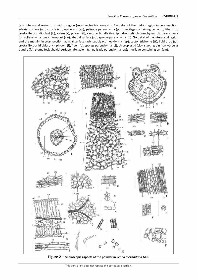
Brazilian Pharmacopoeia, 6th edition PM080-01
This translation does not replace the portuguese version.
(es); intercostal region (ri); midrib region (rnp); vector trichome (tt). F – detail of the midrib region in cross-section: adaxial surface (ad); cuticle (cu); epidermis (ep); palisade parenchyma (pp); mucilage-containing cell (cm); fiber (fb); crystalliferous idioblast (ic); xylem (x); phloem (f); vascular bundle (fv); lipid drop (gl); chlorenchyma (cl); parenchyma (p); collenchyma (co); chloroplast (clo); abaxial surface (ab); spongy parenchyma (pj). G – detail of the intercostal region and the margin, in cross-section: adaxial surface (ad); cuticle (cu); epidermis (ep); tector trichome (tt); lipid drop (gl); crystalliferous idioblast (ic); phloem (f); fiber (fb); spongy parenchyma (pj); chloroplastid (clo); starch grain (ga); vascular bundle (fv); stoma (es); abaxial surface (ab); xylem (x); palisade parenchyma (pp); mucilage-containing cell (cm).
Figure 2 – Microscopic aspects of the powder in Senna alexandrina Mill.
Brazilian Pharmacopoeia, 6th edition PM080-01
This translation does not replace the portuguese version.
___________________________ The scales correspond in A, B, D, F (a - i) and G (a - m) to 100 μm; in C and E to 400 μm. A – detail of petiole epidermis facing the adaxial surface, in front view: base of the tector trichome showing epidermal cells with radial distribution around its base (bt); stoma (es); fundamental cell of the epidermis (cfe). B – detail of the petiole epidermis facing the abaxial surface, in front view: fundamental cell of the epidermis (cfe); tector trichome (tt). C – schematic illustration of the petiole in cross-section: adaxial surface (ad); cortical parenchyma (pc); tector trichome (tt); cuticle (cu); epidermis (ep); phloem (f); xylem (x); endodermis (end); fiber (fb); collenchyma (co); cortex (cx); abaxial surface (ab). D – detail of the petiole, in cross-section, as highlighted in C: pitting (pto); xylem (x); phloem (f); fiber (fb); crystalliferous idioblast (ic); starch grain (ga); endoderm (end); chloroplast (clo); cortical parenchyma (pc); lipid drop (gl); epidermis (ep); abaxial surface (ab); cuticle (cu); collenchyma (co); cortex (cx). E – schematic illustration of the impurity, corresponding to the rachis, in cross-section: adaxial surface (ad); vascular bundle (fv); rib (CST); medullary parenchyma (pm); cortex (cx); cortical parenchyma (pc); phloem (f); xylem (x); fiber (fb); endoderm (end); collenchyma (co); epidermis (ep); cuticle (cu); abaxial surface (ab). F (a - f) – details of the powder of impurities corresponding to the rachis (a - detail of portion of epidermis with tector trichome, in front view): tector trichome (tt); fundamental cell of the epidermis (cfe); stoma (es); crystalliferous idioblast (ic); cortical parenchyma (pc); tracheal element, with helical thickening, in longitudinal section (eh); fiber (fb); medullary parenchyma (pm). G – leaflet powder details; a – detail of the epidermis portion of the lamina, under the midrib region, in front view: stoma (es), fundamental cell of the epidermis (cfe); b – detail of the epidermis portion of the lamina, with stomata and tector trichome, in front view: tector trichome (tt), stoma (es), fundamental cell of the epidermis (cfe); c – detail of a portion of the petiole epidermis, facing the abaxial surface, in front view: fundamental cell of the epidermis (cfe); d – detail of a portion of the lamina epidermis, showing the base of the tector trichome, in front view: fundamental cell of the epidermis (cfe), base of the trichome (bt); e – detail of a portion of the epidermis of the lamina, in cross-section: cuticle (cu); f – detail of a portion of intercostal region in cross-section: adaxial surface (ad), cuticle (cu), epidermis (ep), palisade parenchyma (pp), tracheal element, with helical thickening, in longitudinal section (eh); spongy parenchyma (pj), crystalliferous idioblast (ic), lipid drop (gl), chloroplast (clo), abaxial surface (ab); g - detail of epidermis fragment showing venation portion, stomata and crystalliferous idioblasts, by transparency, in front view: fundamental cell of epidermis (cfe), venation portion (pn), crystalliferous idioblast (ic), stoma (es); h – detail of an isolated portion of the tracheal element, with helical thickening, in longitudinal section; i – detail of a grouped portion of tracheal elements, with helical thickening, in longitudinal section; j – detail of a grouped portion of fibers associated to crystalliferous idioblasts, in longitudinal section: fiber (fb), crystalliferous idioblast (ic); l – portions of isolated tector trichomes, in lateral view; m – detail of isolated druse-type crystals and prismatic mono-crystals.
Brazilian Pharmacopoeia, 6th edition PM081-00
This translation does not replace the portuguese version.
ALEXANDRIAN SENNA, fruit Sennae fructus
The plant drug consists of dried fruit of Senna alexandrina Mill. (syn. Cassia acutifolia Delile, Cassia angustifolia Vahl, Cassia senna L.) containing at least 2.2% hydroxyanthracene derivatives expressed as sennoside B, and 0.92% sennoside B (C42H38O20, 862.75) and 0.49% sennoside A (C42H38O20, 862.75). It should not be used before one year after collection. IDENTIFICATION 1. Macroscopic description Dried legumes, green to greenish-brown on the margins and dark brown in the portions corresponding to the seeds, elliptical to oblong and slightly reniform, flat, rounded at the ends and slightly pointed at the apex, up to 7 cm long and up to 2.5 cm wide. Each legume contains five to eight flat, hard, light brown seeds. 2. Microscopic description The epicarp, in paradermal section, shows epidermis with polygonal cells with straight walls or forming a slight curvature, with sparse anomocytic stomata, and rare unicellular and conical tector trichomes, with verrucous walls, often curved near the base; in cross-section, the thick cuticle is visible over the unistratified epidermis. The epidermal cells are rich in starch grains. Below the epidermis is a layer of larger cells, corresponding to the hypodermis, followed by four layers of parenchyma, containing very sparse vascular bundles. Prominent druses are distributed in the parenchyma. This is followed by a layer of thin-walled cells, each containing a prismatic calcium oxalate crystal, followed by two fibrous, thick-walled cell layers, the innermost of which with cells perpendicular to the longitudinal axis of the fruit, and the outermost with cells at an oblique or parallel angle to the longitudinal axis of the fruit. The fibers in these layers have sparse stitching and a visible lumen. The inner epidermis is indistinct, with elongated and thin-walled cells, when observed in a paradermal section. In the region of the base and the margin of the fruit, the cuticle is thicker and has undulations, the epidermis is also unistratified, followed by four to five layers of parenchyma with dense clusters of sclereids, usually associated with the vascular bundles. These sclereids have thickened walls and distinctive pitting. The seeds have a thick-walled testa, formed by cells in palisade and narrow lumen, covered by a thick cuticle; the endosperm is formed by polyhedral cells, the outermost layer in palisade and spongy innermost layers, with mucilaginous walls. 3. Microscopic description of powder The sample meets all requirements for the species, except the macroscopic characters. Characteristics are: light greenish color; sclerenchymatic libriform fibers and isolated crystals in the powder; parenchymatic cell portions, vessel element fragments with scalariform thickening, simple, straight and oblique terminal walls, with short extensions; cross-layered fiber fragments; calcium oxalate crystals present in the cross-layered fibers. 4. Proceed as described in Thin-layer chromatography (5.2.17.1).
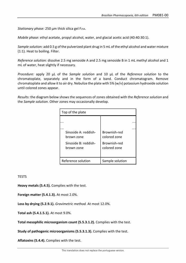
Brazilian Pharmacopoeia, 6th edition PM081-00
This translation does not replace the portuguese version.
Stationary phase: 250 µm thick silica gel F254. Mobile phase: ethyl acetate, propyl alcohol, water, and glacial acetic acid (40:40:30:1). Sample solution: add 0.5 g of the pulverized plant drug in 5 mL of the ethyl alcohol and water mixture (1:1). Heat to boiling. Filter. Reference solution: dissolve 2.5 mg senoside A and 2.5 mg senoside B in 1 mL methyl alcohol and 1 mL of water, heat slightly if necessary. Procedure: apply 20 μL of the Sample solution and 10 μL of the Reference solution to the chromatoplate, separately and in the form of a band. Conduct chromatogram. Remove chromatoplate and allow it to air dry. Nebulize the plate with 5% (w/v) potassium hydroxide solution until colored zones appear. Results: the diagram below shows the sequences of zones obtained with the Reference solution and the Sample solution. Other zones may occasionally develop.
Top of the plate
Sinoside A: reddish-brown zone
Brownish-red colored zone
Sinoside B: reddish-brown zone
Brownish-red colored zone
Reference solution Sample solution
TESTS Heavy metals (5.4.5). Complies with the test. Foreign matter (5.4.1.3). At most 2.0%. Loss by drying (5.2.9.1). Gravimetric method. At most 12.0%. Total ash (5.4.1.5.1). At most 9.0%. Total mesophilic microorganism count (5.5.3.1.2). Complies with the test. Study of pathogenic microorganisms (5.5.3.1.3). Complies with the test. Aflatoxins (5.4.4). Complies with the test.
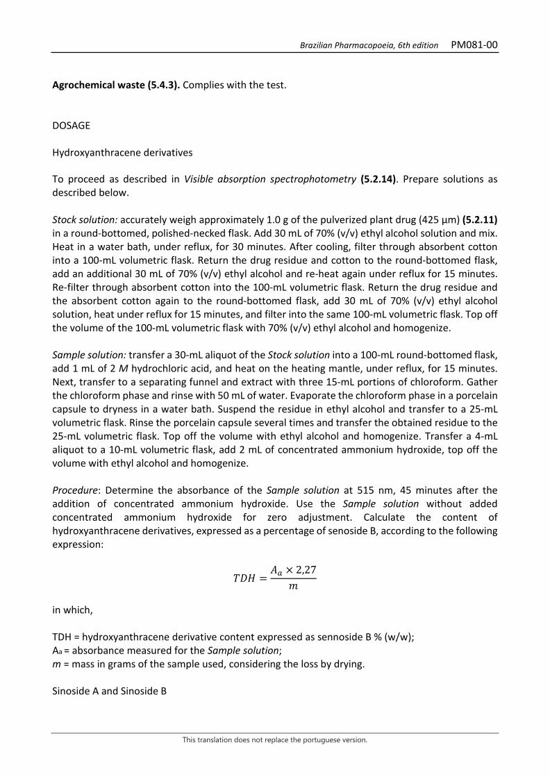
Brazilian Pharmacopoeia, 6th edition PM081-00
This translation does not replace the portuguese version.
Agrochemical waste (5.4.3). Complies with the test. DOSAGE Hydroxyanthracene derivatives To proceed as described in Visible absorption spectrophotometry (5.2.14). Prepare solutions as described below. Stock solution: accurately weigh approximately 1.0 g of the pulverized plant drug (425 µm) (5.2.11) in a round-bottomed, polished-necked flask. Add 30 mL of 70% (v/v) ethyl alcohol solution and mix. Heat in a water bath, under reflux, for 30 minutes. After cooling, filter through absorbent cotton into a 100-mL volumetric flask. Return the drug residue and cotton to the round-bottomed flask, add an additional 30 mL of 70% (v/v) ethyl alcohol and re-heat again under reflux for 15 minutes. Re-filter through absorbent cotton into the 100-mL volumetric flask. Return the drug residue and the absorbent cotton again to the round-bottomed flask, add 30 mL of 70% (v/v) ethyl alcohol solution, heat under reflux for 15 minutes, and filter into the same 100-mL volumetric flask. Top off the volume of the 100-mL volumetric flask with 70% (v/v) ethyl alcohol and homogenize. Sample solution: transfer a 30-mL aliquot of the Stock solution into a 100-mL round-bottomed flask, add 1 mL of 2 M hydrochloric acid, and heat on the heating mantle, under reflux, for 15 minutes. Next, transfer to a separating funnel and extract with three 15-mL portions of chloroform. Gather the chloroform phase and rinse with 50 mL of water. Evaporate the chloroform phase in a porcelain capsule to dryness in a water bath. Suspend the residue in ethyl alcohol and transfer to a 25-mL volumetric flask. Rinse the porcelain capsule several times and transfer the obtained residue to the 25-mL volumetric flask. Top off the volume with ethyl alcohol and homogenize. Transfer a 4-mL aliquot to a 10-mL volumetric flask, add 2 mL of concentrated ammonium hydroxide, top off the volume with ethyl alcohol and homogenize. Procedure: Determine the absorbance of the Sample solution at 515 nm, 45 minutes after the addition of concentrated ammonium hydroxide. Use the Sample solution without added concentrated ammonium hydroxide for zero adjustment. Calculate the content of hydroxyanthracene derivatives, expressed as a percentage of senoside B, according to the following expression:
𝑇𝑇𝑇𝑇𝑇𝑇 =𝑇𝑇𝑎𝑎 × 2,27
𝑚𝑚
in which, TDH = hydroxyanthracene derivative content expressed as sennoside B % (w/w); Aa = absorbance measured for the Sample solution; m = mass in grams of the sample used, considering the loss by drying. Sinoside A and Sinoside B
Brazilian Pharmacopoeia, 6th edition PM081-00
This translation does not replace the portuguese version.
Proceed as described in High Performance Liquid Chromatography (5.2.17.4). Use a chromatograph equipped with an ultraviolet detector at 270 mm; pre-column packed with octadecylsilane silica, 150mm long, 4.6mm internal diameter column, packed with octadecylsilane silica (5 µm); Mobile phase flow rate of 0.9 mL/minute. Eluent (A): water and trifluoroacetic acid (100:0.08). Eluent (B): acetonitrile.
Time (minutes) Eluent A (%) Eluent B (%) Elution
0 – 12 86 14 isocratic 12 – 19 86 → 77 14 → 23 linear gradient 19 – 28 77 → 70 23 → 30 linear gradient 28 – 31 70 → 0 30 → 100 linear gradient 31 – 33 0 100 isocratic Sample solution: accurately weigh approximately 0.2 g of the dried and pulverized plant drug in a centrifuge tube. Add 5 mL of 0.05% (w/v) sodium bicarbonate solution and place in the ultrasonic bath for 10 minutes. Centrifuge for 20 minutes at 45 × g. Transfer the supernatant, filtering it through absorbent cotton to a 5-mL volumetric flask. Top off the volume with 0.05% (w/v) sodium bicarbonate and homogenize. Filter the supernatant through a 0.45 µm filter unit. Dilute 50 µL of the resulting solution in 150 mL of water. Reference stock solution: dissolve 2 mg of the mixture of senoside A and senoside B (40:60) in a 5-mL volumetric flask with 50% (v/v) methyl alcohol. Analytical curve (1) (for senoside A): from the Stock Reference Solution, construct an analytical curve for senoside A in 50% (v/v) methyl alcohol, with at least five concentrations, in the range between 45 µg/mL and 85 µg/mL. Filter through a 0.45 µm filter unit. Analytical curve (2) (for senoside B): from the Stock Reference Solution, construct an analytical curve for senoside B in 50% (v/v) methyl alcohol, with at least five concentrations, in the range between 100 µg/mL and 150 µg/mL. Filter through a 0.45 µm filter unit. Procedure: separately inject 10 µL of each Analytical Curve (1) and Analytical Curve (2) solution, and 10 µL of the Sample solution. Register the chromatograms and measure the areas under the peaks. Calculate the content of senoside A and senoside B in mg/g of the plant drug according to the following expression:
𝑇𝑇𝑇𝑇 =𝐶𝐶𝑎𝑎 × 20
m × 1000
in which, TS = senoside A or B content (mg/g); Ca = concentration of senoside A or B (µg/mL) found in the Sample solution from the analytical curves, considering the purity of the reference substance;
Brazilian Pharmacopoeia, 6th edition PM081-00
This translation does not replace the portuguese version.
m = mass in grams of the sample used, considering the loss by drying. 20 = dilution factor.
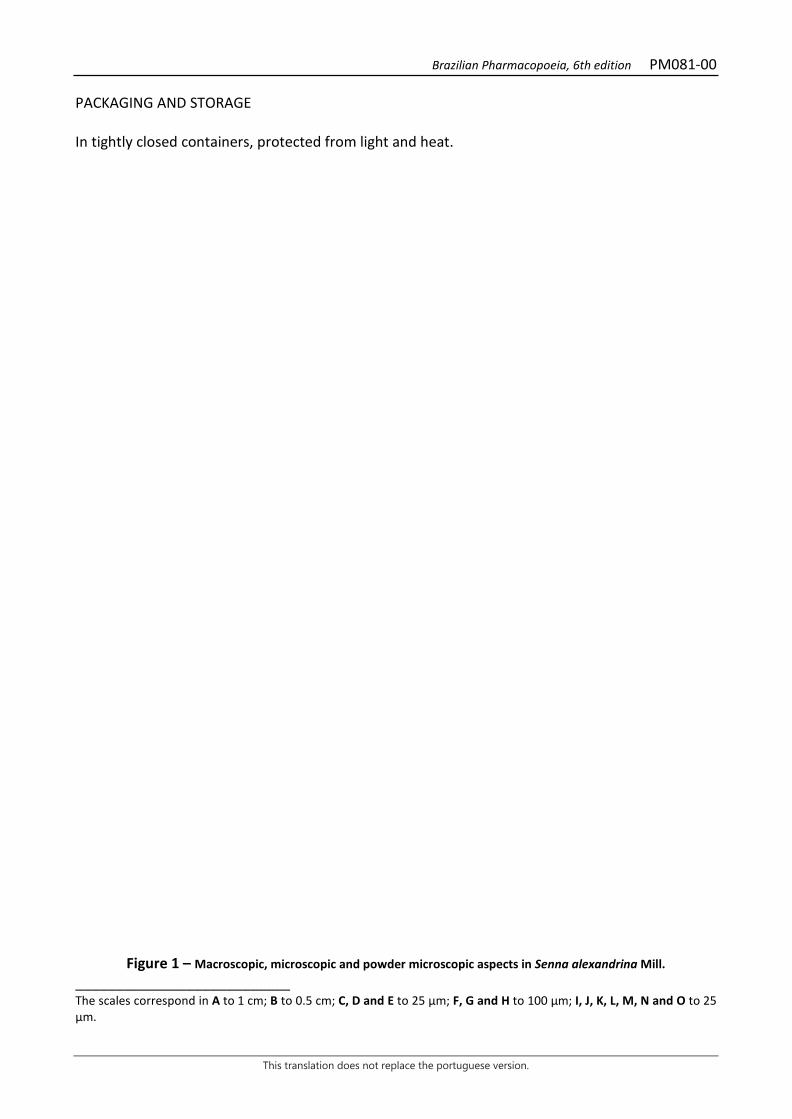
Brazilian Pharmacopoeia, 6th edition PM081-00
This translation does not replace the portuguese version.
PACKAGING AND STORAGE In tightly closed containers, protected from light and heat.
Figure 1 – Macroscopic, microscopic and powder microscopic aspects in Senna alexandrina Mill. ___________________________ The scales correspond in A to 1 cm; B to 0.5 cm; C, D and E to 25 µm; F, G and H to 100 µm; I, J, K, L, M, N and O to 25 µm.

Brazilian Pharmacopoeia, 6th edition PM081-00
This translation does not replace the portuguese version.
A - overall appearance of the fruit: dried, reniformly-shaped legumes, in internal and external view. B - overall detail of the seeds. C - detail of the fruit paradermal section showing an anomocytic stoma (arrow). D - detail of the fruit in the paradermal section, showing unicellular tector trichomes (arrows). E - detail of the fruit paradermal section, with starch grains in the epidermal cells (arrows). F - detail of the paradermal section of the fruit, with cross-layered fibers. G - detail of the fruit cross-section, showing the mesophyll with fundamental parenchyma and vascular bundle. H - details of fruit cross-section at the margin; cuticle (cu); sclereids (esc); phloem (fl); hypodermis (hi); cortical parenchyma (pc); xylem (xi). I - detail of the fruit cross-section showing prismatic calcium oxalate crystals (arrows). J - Fruit cross-section detail showing prismatic crystals (arrows). K - detail of the longitudinal section of the fruit, showing vessel elements (arrows). L - O - details noted in the powder. L - fragments of sclerenchymal fibers (arrows). M – fragments of parenchyma cells and vessel element. N - cross-layered fiber fragments. O - calcium oxalate crystals in the cross-layered fibers (arrows).
Brazilian Pharmacopoeia, 6th edition PM082-00
This translation does not replace the portuguese version.
BEARBERRY, leaf Uvae ursi folium
The plant drug consists of the dried, entire or chopped leaves of Arctostaphylos uva-ursi (L.) Spreng., containing not less than 7,0% anhydrous arbutin (C12H16O7, 272.25). IDENTIFICATION 1. Macroscopic description The leaves are entire, coriaceous, rigid and brittle, obovate, oblong-spatulate or elliptic, 1.2 to 3 cm long and 0.5 to 1.5 cm wide; apex obtuse or rounded, sometimes apparently emarginate, margins entire and slightly revolute, base cuneiform and attenuate on a short petiole, 0.3 to 0.5 cm long. Adaxial surface dark green or olive green to greenish brown, glabrous and shiny, waxy, finely reticulate, with strongly depressed veins. Yellowish green to pale grayish green abaxial surface, glabrous to slightly pubescent on young leaves, with small and simple trichomes on the petiole and on the midrib and secondary veins. Young leaves may have conical unicellular tector trichomes, often curved, on the abaxial surface. The veins are finely reticulated, more prominent on the abaxial than on the adaxial surface, the main ones being dark. Short fracture. 2. Microscopic description Hypoestomatic leaf lamina, with dorsiventral mesophyll. In front view, the cells of the adaxial surface of the epidermis are polygonal-rectilinear to rectangular and those of the abaxial surface are polygonal, with cyclocytic stomata formed by six to eleven subsidiary cells, whose guard cells are much larger in size (about 40 m to 50 m long). Lipid drops are visible. The cuticle, on the adaxial surface, is smooth and thick and may present irregular cracks that reach the outer periclinal wall of the epidermis cells; on the abaxial surface, it is interrupted by circular spaces corresponding to the pores of the stomata. In cross-section, the adaxial surface of the epidermis is formed by flattened cells, with outer periclinal walls thicker than the anticlinal ones, and the abaxial surface exhibits a thick cuticle, interrupted by the opening of the stomata. The mesophyll is very rich in prismatic calcium oxalate crystals and is formed by three to five layers of palisade cells, each layer having different thickness, giving the tissue an irregular aspect. The spongy parenchyma has loose, braciform cells, where secondary and tertiary vascular bundles are distributed, lined with fibers, accompanied by extension of the parenchymatic sheath to both sides. The midrib is plano-convex, and the vascular bundle is of the open-arch collateral type, exhibiting, on both sides, a well-developed angular collenchyma, with several prismatic crystals of calcium oxalate. The petiole, in cross-section, is plano-convex, with a thick cuticle, epidermis with simple trichomes and stomata. The fundamental parenchyma has cells with thickened walls and fills almost the entire region, except in the lateral portions of the vascular bundle, where an aerenchyma occurs. Phenolic compounds are found in collenchyma, parenchyma, aerenchyma, phloem, and, to a lesser extent, in xylem. 3. Microscopic description of powder The sample meets all requirements for the species, except the macroscopic characters. Characteristics are: yellow to light olive, rarely dark green color; cuticle fragments isolated and with
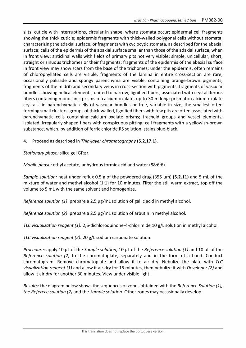
Brazilian Pharmacopoeia, 6th edition PM082-00
This translation does not replace the portuguese version.
slits; cuticle with interruptions, circular in shape, where stomata occur; epidermal cell fragments showing the thick cuticle; epidermis fragments with thick-walled polygonal cells without stomata, characterizing the adaxial surface, or fragments with cyclocytic stomata, as described for the abaxial surface; cells of the epidermis of the abaxial surface smaller than those of the adaxial surface, when in front view; anticlinal walls with fields of primary pits not very visible; simple, unicellular, short, straight or sinuous trichomes or their fragments; fragments of the epidermis of the abaxial surface in front view may show scars from the base of the trichomes; under the epidermis, often remains of chlorophyllated cells are visible; fragments of the lamina in entire cross-section are rare; occasionally palisade and spongy parenchyma are visible, containing orange-brown pigments; fragments of the midrib and secondary veins in cross-section with pigments; fragments of vascular bundles showing helical elements, united to narrow, lignified fibers, associated with crystalliferous fibers containing monoclinic prisms of calcium oxalate, up to 30 m long; prismatic calcium oxalate crystals, in parenchymatic cells of vascular bundles or free, variable in size, the smallest often forming small clusters; groups of thick-walled, lignified fibers with few pits are often associated with parenchymatic cells containing calcium oxalate prisms; tracheid groups and vessel elements; isolated, irregularly shaped fibers with conspicuous pitting; cell fragments with a yellowish-brown substance, which. by addition of ferric chloride RS solution, stains blue-black. 4. Proceed as described in Thin-layer chromatography (5.2.17.1). Stationary phase: silica gel GF254. Mobile phase: ethyl acetate, anhydrous formic acid and water (88:6:6). Sample solution: heat under reflux 0.5 g of the powdered drug (355 µm) (5.2.11) and 5 mL of the mixture of water and methyl alcohol (1:1) for 10 minutes. Filter the still warm extract, top off the volume to 5 mL with the same solvent and homogenize. Reference solution (1): prepare a 2,5 µg/mL solution of gallic acid in methyl alcohol. Reference solution (2): prepare a 2,5 µg/mL solution of arbutin in methyl alcohol. TLC visualization reagent (1): 2,6-dichloroquinone-4-chlorimide 10 g/L solution in methyl alcohol. TLC visualization reagent (2): 20 g/L sodium carbonate solution. Procedure: apply 10 μL of the Sample solution, 10 μL of the Reference solution (1) and 10 μL of the Reference solution (2) to the chromatoplate, separately and in the form of a band. Conduct chromatogram. Remove chromatoplate and allow it to air dry. Nebulize the plate with TLC visualization reagent (1) and allow it air dry for 15 minutes, then nebulize it with Developer (2) and allow it air dry for another 30 minutes. View under visible light. Results: the diagram below shows the sequences of zones obtained with the Reference Solution (1), the Referece solution (2) and the Sample solution. Other zones may occasionally develop.
Brazilian Pharmacopoeia, 6th edition PM082-00
This translation does not replace the portuguese version.
Top of the plate
Gallic acid: blackish-brown colored zone
Blackish-brown colored zone
Blackish-brown colored zone
Blue-purple colored zone
Blue-colored zones
Reference solution Sample solution TESTS Heavy metals (5.4.5). Complies with the test. Foreign matter (5.4.1.3). At most 2.0%. Loss by drying (5.2.9.1). Gravimetric method. At most 10.0%. Total ash (5.4.1.5.1). At most 5%. Total mesophilic microorganism count (5.5.3.1.2). Complies with the test. Study of pathogenic microorganisms (5.5.3.1.3). Complies with the test. Leaves of different colors. At most 10.0%. Agrochemical waste (5.4.3). Complies with the test. DOSAGE Arbutin Proceed as described in High Performance Liquid Chromatography (5.2.17.4). Use a chromatograph equipped with an ultraviolet detector at 280 nm; 250mm long, 4.6 mm internal diameter column, packed with silica chemically bonded to an octadecylsilane group (5 µm), kept at a temperature of (22 ± 2) °C; Mobile phase flow rate of 1.2 mL/minute. Isocratic system. Mobile phase: water and methyl alcohol (90:10). Sample solution: accurately weigh approximately 0.800 g of the dried and pulverized plant drug (250 µm) (5.2.11) in a 100mL round-bottomed flask. Add 20 mL of water and heat in a water bath, under reflux, for 30 minutes. Cool the contents of the flask and filter the liquid through a small piece of absorbent cotton. Transfer the residue and cotton to the round-bottomed flask and add 20 mL of water. Heat in a water bath, under reflux, for 30 minutes. Cool the contents of the flask and filter the liquid through a small piece of paper filter. Combine the filtrates and dilute in a 50-mL volumetric
Brazilian Pharmacopoeia, 6th edition PM082-00
This translation does not replace the portuguese version.
flask, top off the volume and homogenize. Filter on filter paper and discard the first 10 mL of the filtrate. Filter through a 0.45 µm filter unit. Reference solution (1): dissolve 50 mg of arbutin in the mobile phase and dilute in a 50-mL volumetric flask with the Mobile phase, top off the volume and homogenize. Filter through a 0.45 µm filter unit. Reference solution (2): dissolve 2.5 mg of hydroquinone in the Mobile phase, dilute in a 10-mL volumetric flask with the Mobile phase and homogenize. Add 2.5 mL of the Reference Solution (1) to 5 mL of this solution, dilute in a 10-mL volumetric flask with the Mobile phase, top off the volume and homogenize. Filter through a 0.45 µm filter unit. Procedure: separately inject 20µL of the Reference solution (1), 20µL of the Reference solution (2) and 20µL of the Sample solution. Register the chromatograms and measure the areas under the arbutin peaks. The average retention time is approximately four minutes. Calculate arbutin content, in percent, according to the following expression:
𝑇𝑇𝑇𝑇 =𝑇𝑇𝑎𝑎 × 𝑚𝑚𝑇𝑇𝑟𝑟 × 𝑚𝑚𝑎𝑎
in which, TA = arbutin content % (w/w); Aa = area under the peak corresponding to the arbutin in the Sample solution; Ar= area under the peak corresponding to the arbutin in the Reference solution (1); ma = mass in grams of the sample used, considering the loss by drying. mr = mass in grams of arbutin, used for preparing the Reference Solution (1), considering the purity of the reference substance. System suitability: a minimum resolution of 4.0 between the arbutin and hydroquinone equivalent peaks in the chromatogram obtained with the Reference solution (2). PACKAGING AND STORAGE In tightly closed containers, protected from light and heat.
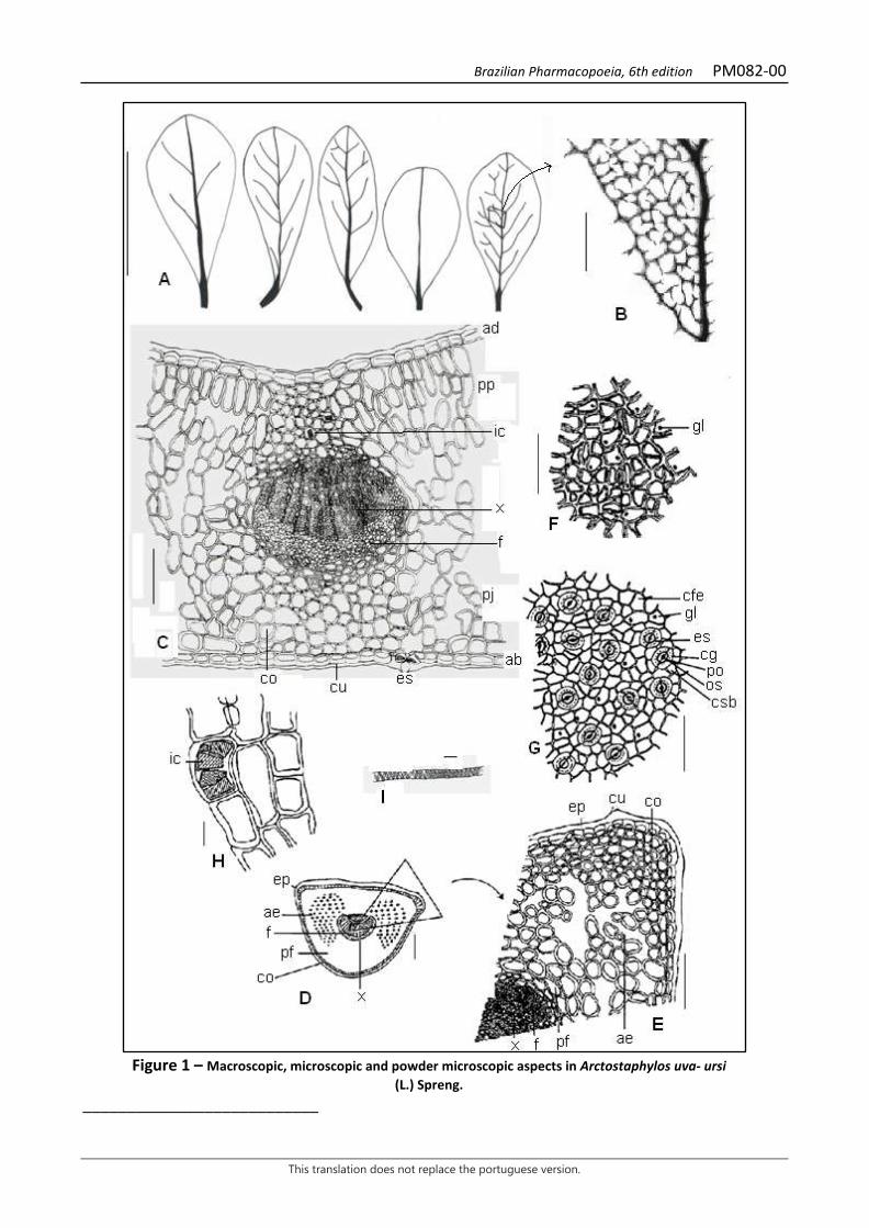
Brazilian Pharmacopoeia, 6th edition PM082-00
This translation does not replace the portuguese version.
Figure 1 – Macroscopic, microscopic and powder microscopic aspects in Arctostaphylos uva- ursi
(L.) Spreng. ___________________________
Brazilian Pharmacopoeia, 6th edition PM082-00
This translation does not replace the portuguese version.
The scales correspond in A to 2 cm, in B to 0.1 cm, in C, E, F, G to 100 µm, in D to 200 µm, in H to 20 µm, and in I to 10 µm. A – leaf blade variation: obovalate, oblong-spatulate or elliptical. B - detail of the leaf vein on the adaxial surface of a segment of the lamina, in front view, indicated in A. C - midrib region in cross-section; abaxial surface (ab); adaxial surface (ad); collenchyma (co); cuticle (cu); stoma (es); phloem (f); crystalliferous idioblast (ic); spongy parenchyma (pj); palisade parenchyma (pp); xylem (x). D - overall appearance of the petiole region, in cross-section; aerenchyma (ae); collenchyma (co); epidermis (ep); phloem (f); fundamental parenchyma (pf); xylem (x). E - detail of a portion of the petiole, in cross-section, marked in D; aerenchyma (ae); collenchyma (co); cuticle (cu); epidermis (ep); phloem (f); fundamental parenchyma (pf); xylem (x). F - epidermal cells of the adaxial surface of the leaf lamina, front view; lipid drop (gl). G - epidermal cells of the abaxial surface of the leaf lamina, in front view, with cyclocytic stomata; epidermal fundamental cell (cfe); guard cell (cg); subsidiary cell (csb); stoma (es); lipid drop (gl); ostiole (os); pore (po). H - detail of parenchymal cells and calcium oxalate prisms; ic: crystalliferous idioblast (ic). I - detail of a vessel element with helical thickening.
Brazilian Pharmacopoeia, 6th edition PM083-00
This translation does not replace the portuguese version.
VALERIAN, rhizome and root Valerianae rhizoma et radix
The plant drug consists of the dried, entire or fragmented underground organs (roots, rhizomes and stolons) of Valeriana officinalis L., containing at least 0.3% essential oils and at least 0.17% total sesquiterpene acids, expressed as valerenic acid (C15H22O2, 234.34). IDENTIFICATION 1. Macroscopic description The plant drug is composed of rhizomes and many fasciculated roots and underground stolons that emerge from the rhizome. The rhizome is grayish-brown to yellowish-brown, erect, conical, and may reach 5 cm in length and 3 cm in diameter; it usually has a scar, identifying the insertion site of the stem and basal leaves. The roots have a striated appearance and the same coloring as the rhizome, with a diameter of 1 to 3 mm and length that may exceed 10 cm; the lateral roots are thin, filiform, and fragile. The stolons are lighter in color than the rhizome and have nodes separated by striated internodes, about 2 to 5 cm long. 2. Microscopic description In cross-section, the adventitious root exhibits epidermal cells with thickened and cutinized outer periclinal walls, some with remnants of absorbent hairs. The exoderm is formed by one or two layers of larger cells, polygonal to quadrangular, with suberized walls, and may have oil droplets. The cortex is formed by parenchyma containing starch grains. Occasionally, it has an outermost layer with collenchyma cells and resinous content. The endoderm consists of a single layer of parenchyma cells with suberin thickening on the anticlinal walls. The pericycle has one or more layers of parenchyma cells, usually devoid of starch grains. The vascular bundles form an interrupted cylinder, interspersed with parenchyma cells, which surround a medulla filled with amyliferous parenchyma. Stolons have the same characterization as roots, however, the epidermis and exoderm may be replaced by a periderm with a few layers of suber, and the medulla may have stone cells with thickened walls and simple pitting. The rhizome shows irregular contour and a more complex tissue organization due to the distribution of vascular bundles towards the roots and stolons. The epidermis and exoderm are partially replaced by a poorly developed periderm. The cortical parenchyma is rich in starch and droplets of resinous substance and has stone cells. The endoderm is clear and contains essential oil droplets. The medullary parenchyma contains starch and has intercellular spaces of various sizes separated by transverse septa; stone cells are also present. 3. Microscopic description of powder The sample meets all requirements for the species, except the macroscopic characters. Characteristics are: light brown color; abundance of isolated rounded or rhomboid starch grains, measuring 5 to 15 µm in diameter, with a slit or star-shaped hilum, when aggregated they form groups of two to six components, reaching 20 µm in diameter; suber fragments with polygonal cells and orange contents; parenchyma fragments with starch grains; vascular tissue fragments with ringed, helical or reticulate, short or elongated vessel elements, with simple perforation plate and associated vascular parenchyma, rare pitted vessel elements.
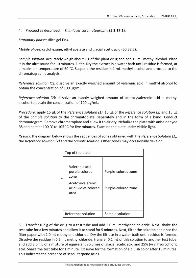
Brazilian Pharmacopoeia, 6th edition PM083-00
This translation does not replace the portuguese version.
4. Proceed as described in Thin-layer chromatography (5.2.17.1). Stationary phase: silica gel F254. Mobile phase: cyclohexane, ethyl acetate and glacial acetic acid (60:38:2). Sample solution: accurately weigh about 1 g of the plant drug and add 10 mL methyl alcohol. Place it in the ultrasound for 10 minutes. Filter. Dry the extract in a water bath until residue is formed, at a maximum temperature of 60 °C. Suspend the residue in 1 mL methyl alcohol and proceed to the chromatographic analysis. Reference solution (1): dissolve an exactly weighed amount of valerenic acid in methyl alcohol to obtain the concentration of 100 µg/mL Reference solution (2): dissolve an exactly weighed amount of acetoxyvalerenic acid in methyl alcohol to obtain the concentration of 100 µg/mL. Procedure: apply 15 μL of the Reference solution (1), 15 μL of the Reference solution (2) and 15 µL of the Sample solution to the chromatoplate, separately and in the form of a band. Conduct chromatogram. Remove chromatoplate and allow it to air dry. Nebulize the plate with anisaldehyde RS and heat at 100 °C to 105 °C for five minutes. Examine the plate under visible light. Results: the diagram below shows the sequences of zones obtained with the Reference Solution (1), the Reference solution (2) and the Sample solution. Other zones may occasionally develop.
Top of the plate
Valerenic acid: purple colored zone
Purple-colored zone
Acetoxyvalerenic acid: violet colored area
Purple-colored zone
Reference solution Sample solution
5. Transfer 0.2 g of the drug to a test tube and add 5.0 mL methylene chloride. Next, shake the test tube for a few minutes and allow it to stand for 5 minutes. Next, filter the solution and rinse the filter paper with 2.0 mL methylene chloride. Dry the filtrate in a water bath until residue is formed. Dissolve the residue in 0.2 mL methyl chloride, transfer 0.1 mL of this solution to another test tube, and add 3.0 mL of a mixture of equivalent volumes of glacial acetic acid and 25% (v/v) hydrochloric acid. Shake the test tube for 1 minute. Observe for the formation of a bluish color after 15 minutes. This indicates the presence of sesquiterpene acids.
Brazilian Pharmacopoeia, 6th edition PM083-00
This translation does not replace the portuguese version.
TESTS Loss by drying (5.2.9.1). Gravimetric method. At most 10.0%. Heavy metals (5.4.5). Complies with the test. Foreign matter (5.4.1.3). At most 5.0% stem base and 2.0% of other materials. Total ash (5.4.1.5.1). At most 12.0%. Acid-insoluble ash (5.4.1.5.3). At most 5.0%. Total mesophilic microorganism count (5.5.3.1.2). Complies with the test. Study of pathogenic microorganisms (5.5.3.1.3). Complies with the test. Aflatoxins (5.4.4). Complies with the test. Agrochemical waste (5.4.3). Complies with the test. DOSAGE Volatile oils Proceed as described in Determining volatile oils in plant drugs (5.4.1.6). Use a 1000-mL round-bottomed flask containing 500 mL of water as distillation liquid. Accurately weigh about 50.0 g of the pulverized plant drug immediately after grinding. Immediately proceed with the determination of the volatile oil. Distill for four hours. Measure the volume and express the yield per 100 g of the plant drug (w/p). Sesquiterpene acids Proceed as described in High Performance Liquid Chromatography (5.2.17.4). Use a chromatograph equipped with an ultraviolet detector at 215 nm; pre-column packed with octadecylsilane silica, 250 mm long, 4.6 mm wide (internal diameter) column, packed with octadecylsilane silica (5 µm), kept at 30 °C; Mobile phase flow rate of 1.5 mL/minute. Eluent (B): 5 mL/L phosphoric acid and acetonitrile (80:20). Eluent (B): acetonitrile and phosphoric acid at 5 mL/L (80:20).
Time (minutes) Eluent A (%) Eluent B (%) Elution
0 - 5 55 45 isocratic
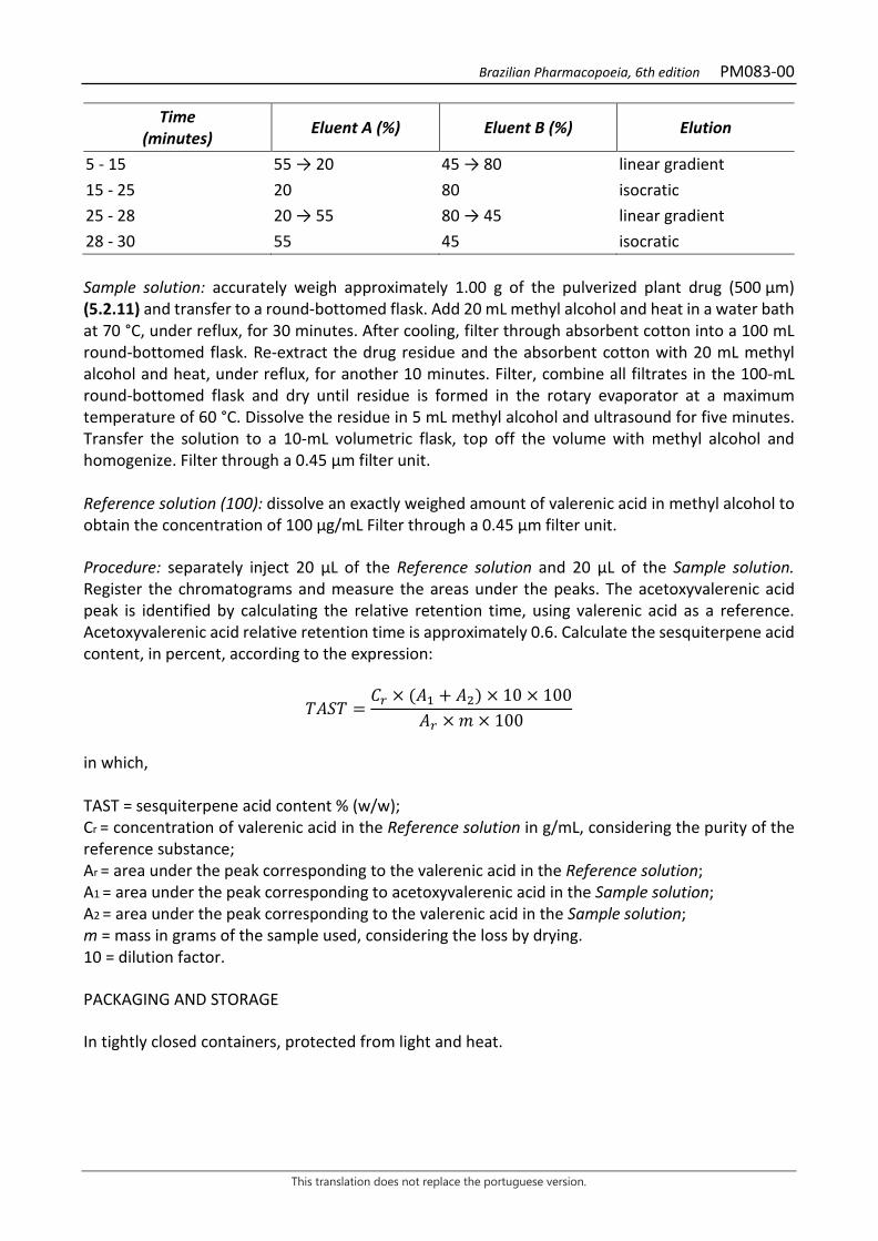
Brazilian Pharmacopoeia, 6th edition PM083-00
This translation does not replace the portuguese version.
Time (minutes) Eluent A (%) Eluent B (%) Elution
5 - 15 55 → 20 45 → 80 linear gradient 15 - 25 20 80 isocratic 25 - 28 20 → 55 80 → 45 linear gradient 28 - 30 55 45 isocratic Sample solution: accurately weigh approximately 1.00 g of the pulverized plant drug (500 µm) (5.2.11) and transfer to a round-bottomed flask. Add 20 mL methyl alcohol and heat in a water bath at 70 °C, under reflux, for 30 minutes. After cooling, filter through absorbent cotton into a 100 mL round-bottomed flask. Re-extract the drug residue and the absorbent cotton with 20 mL methyl alcohol and heat, under reflux, for another 10 minutes. Filter, combine all filtrates in the 100-mL round-bottomed flask and dry until residue is formed in the rotary evaporator at a maximum temperature of 60 °C. Dissolve the residue in 5 mL methyl alcohol and ultrasound for five minutes. Transfer the solution to a 10-mL volumetric flask, top off the volume with methyl alcohol and homogenize. Filter through a 0.45 µm filter unit. Reference solution (100): dissolve an exactly weighed amount of valerenic acid in methyl alcohol to obtain the concentration of 100 µg/mL Filter through a 0.45 µm filter unit. Procedure: separately inject 20 μL of the Reference solution and 20 µL of the Sample solution. Register the chromatograms and measure the areas under the peaks. The acetoxyvalerenic acid peak is identified by calculating the relative retention time, using valerenic acid as a reference. Acetoxyvalerenic acid relative retention time is approximately 0.6. Calculate the sesquiterpene acid content, in percent, according to the expression:
𝑇𝑇𝑇𝑇𝑇𝑇𝑇𝑇 =𝐶𝐶𝑟𝑟 × (𝑇𝑇1 + 𝑇𝑇2) × 10 × 100
𝑇𝑇𝑟𝑟 × 𝑚𝑚 × 100
in which, TAST = sesquiterpene acid content % (w/w); Cr = concentration of valerenic acid in the Reference solution in g/mL, considering the purity of the reference substance; Ar = area under the peak corresponding to the valerenic acid in the Reference solution; A1 = area under the peak corresponding to acetoxyvalerenic acid in the Sample solution; A2 = area under the peak corresponding to the valerenic acid in the Sample solution; m = mass in grams of the sample used, considering the loss by drying. 10 = dilution factor. PACKAGING AND STORAGE In tightly closed containers, protected from light and heat.

Brazilian Pharmacopoeia, 6th edition PM083-00
This translation does not replace the portuguese version.
Figure 1 – Macroscopic, microscopic and microscopic aspects of the powder in Valeriana officinalis L.
___________________________ The scales correspond in A to 1 μm; in B to 200 μm and in C to L to 50 μm.
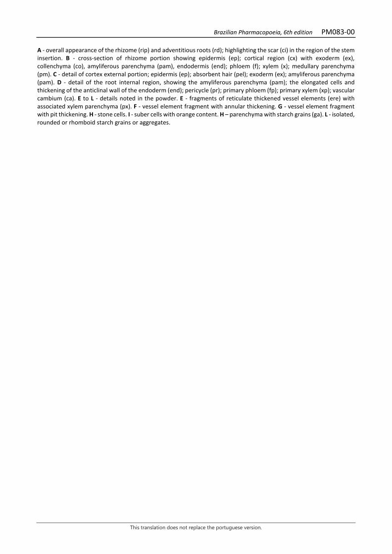
Brazilian Pharmacopoeia, 6th edition PM083-00
This translation does not replace the portuguese version.
A - overall appearance of the rhizome (rip) and adventitious roots (rd); highlighting the scar (ci) in the region of the stem insertion. B - cross-section of rhizome portion showing epidermis (ep); cortical region (cx) with exoderm (ex), collenchyma (co), amyliferous parenchyma (pam), endodermis (end); phloem (f); xylem (x); medullary parenchyma (pm). C - detail of cortex external portion; epidermis (ep); absorbent hair (pel); exoderm (ex); amyliferous parenchyma (pam). D - detail of the root internal region, showing the amyliferous parenchyma (pam); the elongated cells and thickening of the anticlinal wall of the endoderm (end); pericycle (pr); primary phloem (fp); primary xylem (xp); vascular cambium (ca). E to L - details noted in the powder. E - fragments of reticulate thickened vessel elements (ere) with associated xylem parenchyma (px). F - vessel element fragment with annular thickening. G - vessel element fragment with pit thickening. H - stone cells. I - suber cells with orange content. H – parenchyma with starch grains (ga). L - isolated, rounded or rhomboid starch grains or aggregates.
Brazilian Pharmacopoeia, 6th edition
This translation does not replace the portuguese version.
PLANT-BASED PREPARATIONS – TINCTURES
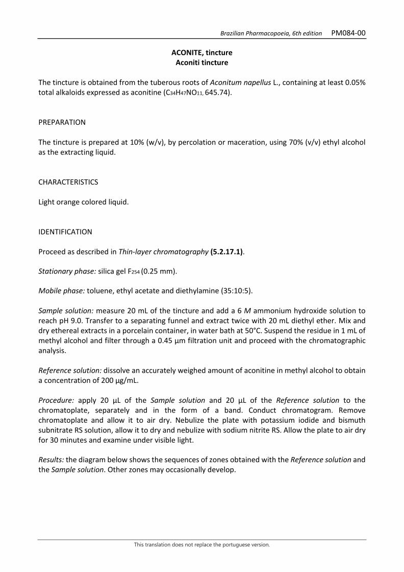
Brazilian Pharmacopoeia, 6th edition PM084-00
This translation does not replace the portuguese version.
ACONITE, tincture Aconiti tincture
The tincture is obtained from the tuberous roots of Aconitum napellus L., containing at least 0.05% total alkaloids expressed as aconitine (C34H47NO11, 645.74). PREPARATION The tincture is prepared at 10% (w/v), by percolation or maceration, using 70% (v/v) ethyl alcohol as the extracting liquid. CHARACTERISTICS Light orange colored liquid. IDENTIFICATION Proceed as described in Thin-layer chromatography (5.2.17.1). Stationary phase: silica gel F254 (0.25 mm). Mobile phase: toluene, ethyl acetate and diethylamine (35:10:5). Sample solution: measure 20 mL of the tincture and add a 6 M ammonium hydroxide solution to reach pH 9.0. Transfer to a separating funnel and extract twice with 20 mL diethyl ether. Mix and dry ethereal extracts in a porcelain container, in water bath at 50°C. Suspend the residue in 1 mL of methyl alcohol and filter through a 0.45 µm filtration unit and proceed with the chromatographic analysis. Reference solution: dissolve an accurately weighed amount of aconitine in methyl alcohol to obtain a concentration of 200 µg/mL. Procedure: apply 20 μL of the Sample solution and 20 μL of the Reference solution to the chromatoplate, separately and in the form of a band. Conduct chromatogram. Remove chromatoplate and allow it to air dry. Nebulize the plate with potassium iodide and bismuth subnitrate RS solution, allow it to dry and nebulize with sodium nitrite RS. Allow the plate to air dry for 30 minutes and examine under visible light. Results: the diagram below shows the sequences of zones obtained with the Reference solution and the Sample solution. Other zones may occasionally develop.
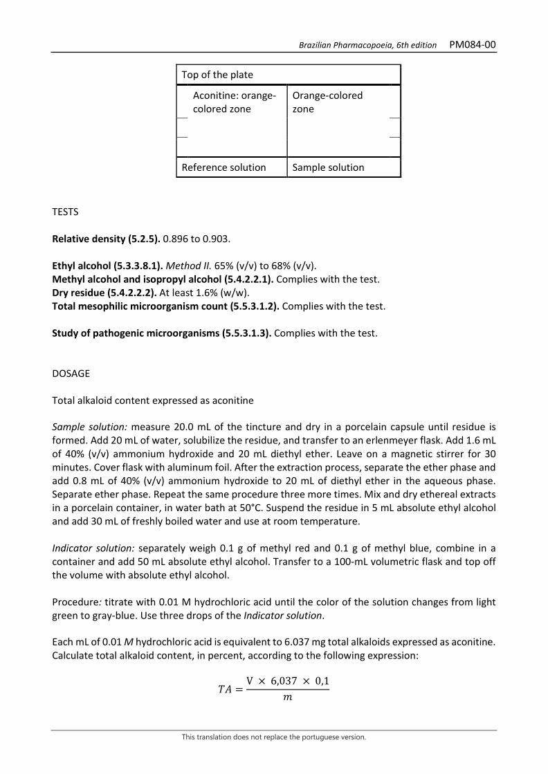
Brazilian Pharmacopoeia, 6th edition PM084-00
This translation does not replace the portuguese version.
Top of the plate
Aconitine: orange-colored zone
Orange-colored zone
Reference solution Sample solution
TESTS Relative density (5.2.5). 0.896 to 0.903. Ethyl alcohol (5.3.3.8.1). Method II. 65% (v/v) to 68% (v/v). Methyl alcohol and isopropyl alcohol (5.4.2.2.1). Complies with the test. Dry residue (5.4.2.2.2). At least 1.6% (w/w). Total mesophilic microorganism count (5.5.3.1.2). Complies with the test. Study of pathogenic microorganisms (5.5.3.1.3). Complies with the test. DOSAGE Total alkaloid content expressed as aconitine Sample solution: measure 20.0 mL of the tincture and dry in a porcelain capsule until residue is formed. Add 20 mL of water, solubilize the residue, and transfer to an erlenmeyer flask. Add 1.6 mL of 40% (v/v) ammonium hydroxide and 20 mL diethyl ether. Leave on a magnetic stirrer for 30 minutes. Cover flask with aluminum foil. After the extraction process, separate the ether phase and add 0.8 mL of 40% (v/v) ammonium hydroxide to 20 mL of diethyl ether in the aqueous phase. Separate ether phase. Repeat the same procedure three more times. Mix and dry ethereal extracts in a porcelain container, in water bath at 50°C. Suspend the residue in 5 mL absolute ethyl alcohol and add 30 mL of freshly boiled water and use at room temperature. Indicator solution: separately weigh 0.1 g of methyl red and 0.1 g of methyl blue, combine in a container and add 50 mL absolute ethyl alcohol. Transfer to a 100-mL volumetric flask and top off the volume with absolute ethyl alcohol. Procedure: titrate with 0.01 M hydrochloric acid until the color of the solution changes from light green to gray-blue. Use three drops of the Indicator solution. Each mL of 0.01 M hydrochloric acid is equivalent to 6.037 mg total alkaloids expressed as aconitine. Calculate total alkaloid content, in percent, according to the following expression:
𝑇𝑇𝑇𝑇 =V × 6,037 × 0,1
𝑚𝑚
Brazilian Pharmacopoeia, 6th edition PM084-00
This translation does not replace the portuguese version.
in which,
TA = Total alkaloid content expressed as aconitine % (w/w); V = volume of 0.01 M hydrochloric acid, in milliliters, consumed in the titration; m = mass in grams of the tincture used, determined from the density. PACKAGING AND STORAGE In tightly closed containers, protected from light and heat.
Brazilian Pharmacopoeia, 6th edition PM085-00
This translation does not replace the portuguese version.
ANGICO, tincture Anadenantherae tincture
The tincture is obtained from the dried bark of Anadenanthera colubrina (Vell.) Brenan, containing not less than 1.0% total tannins and not less than 0.020% catechin (C15H14O6, 290.27). PREPARATION The tincture is prepared at 1:10 (w/v), by percolation or maceration, using 70% (v/v) ethyl alcohol as the extracting liquid. CHARACTERISTICS Clear, dark-brown liquid. IDENTIFICATION Proceed as described in Thin-layer chromatography (5.2.17.1). Stationary phase: silica gel F254 (0.25 mm). Mobile phase: ethyl acetate, formic acid and water (90:5:5). Sample solution: dilute about 1 mL of the tincture with 5 mL methyl alcohol. Reference solution: weigh approximately 1 mg catechin and dissolve in 1mL methyl alcohol. Procedure: apply 20 μL of the Sample solution and 20 μL of the Reference solution to the chromatoplate, separately and in the form of a band. Conduct chromatogram. Remove the chromatoplate and allow it to dry in a fume hood. Nebulize plate with 1% vanillin (w/v) in ethyl alcohol, then, with hydrochloric acid. Results: the diagram below shows the sequences of zones obtained with the Reference solution and the Sample solution. Other zones may occasionally develop.
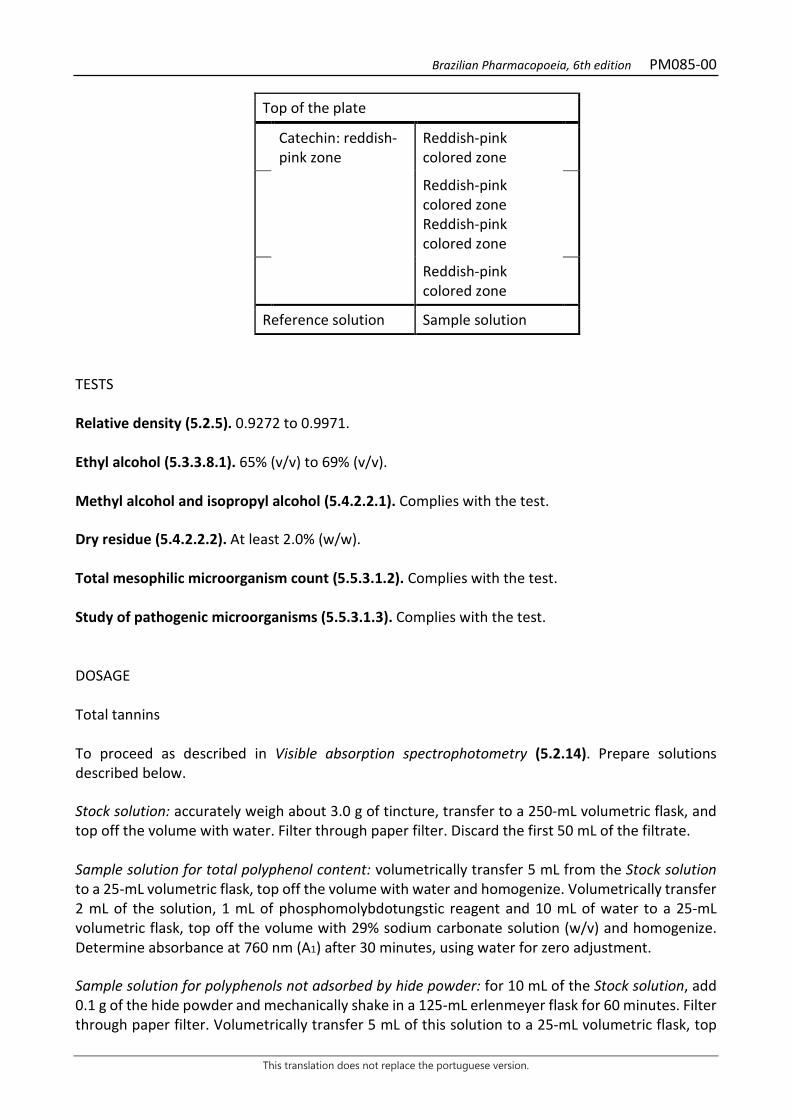
Brazilian Pharmacopoeia, 6th edition PM085-00
This translation does not replace the portuguese version.
Top of the plate
Catechin: reddish-pink zone
Reddish-pink colored zone
Reddish-pink colored zone Reddish-pink colored zone
Reddish-pink colored zone
Reference solution Sample solution
TESTS Relative density (5.2.5). 0.9272 to 0.9971. Ethyl alcohol (5.3.3.8.1). 65% (v/v) to 69% (v/v). Methyl alcohol and isopropyl alcohol (5.4.2.2.1). Complies with the test. Dry residue (5.4.2.2.2). At least 2.0% (w/w). Total mesophilic microorganism count (5.5.3.1.2). Complies with the test. Study of pathogenic microorganisms (5.5.3.1.3). Complies with the test. DOSAGE Total tannins To proceed as described in Visible absorption spectrophotometry (5.2.14). Prepare solutions described below. Stock solution: accurately weigh about 3.0 g of tincture, transfer to a 250-mL volumetric flask, and top off the volume with water. Filter through paper filter. Discard the first 50 mL of the filtrate. Sample solution for total polyphenol content: volumetrically transfer 5 mL from the Stock solution to a 25-mL volumetric flask, top off the volume with water and homogenize. Volumetrically transfer 2 mL of the solution, 1 mL of phosphomolybdotungstic reagent and 10 mL of water to a 25-mL volumetric flask, top off the volume with 29% sodium carbonate solution (w/v) and homogenize. Determine absorbance at 760 nm (A1) after 30 minutes, using water for zero adjustment. Sample solution for polyphenols not adsorbed by hide powder: for 10 mL of the Stock solution, add 0.1 g of the hide powder and mechanically shake in a 125-mL erlenmeyer flask for 60 minutes. Filter through paper filter. Volumetrically transfer 5 mL of this solution to a 25-mL volumetric flask, top

Brazilian Pharmacopoeia, 6th edition PM085-00
This translation does not replace the portuguese version.
off the volume with water and homogenize. Volumetrically transfer 2 mL of the solution, 1 mL of phosphomolybdotungstic reagent and 10 mL of water to a 25-mL volumetric flask, top off the volume with 29% sodium carbonate solution (w/v) and homogenize. Determine absorbance at 760 nm (A2) after 30 minutes, using water for zero adjustment. Reference solution: dissolve 50 mg pyrogallol in water immediately before use in a 100-mL volumetric flask, top off the volume with water and homogenize. Volumetrically transfer 5 mL of this solution to a 100-mL volumetric flask, top off the volume with water and homogenize. Volumetrically transfer 2 mL of the solution, 1 mL of phosphomolybdotungstic reagent and 10 mL of water to a 25-mL volumetric flask, top off the volume with 29% sodium carbonate solution (w/v) and homogenize. Determine absorbance at 760 nm (A3) after 30 minutes, using water for zero adjustment. Calculate total tannin content, expressed as a percentage of pyrogallol, according to the following expression:
𝑇𝑇𝑇𝑇 =(𝑇𝑇1 − 𝑇𝑇2) × 𝑚𝑚2 × 62,5
𝑇𝑇3 × 𝑚𝑚1
in which,
TT = total tannin content expressed as pyrogallol % (w/w); A1 = absorbance measured for the Sample solution for total polyphenols; A2 = absorbance measured for the Sample solution for polyphenols not adsorbed by hide powder; A3 = absorbance measured for the Reference solution; m1 = mass un grams of the tincture used; m2 = mass in grams of pyrogallol, considering the purity of the reference substance. Catechin Proceed as described in High Performance Liquid Chromatography (5.2.17.4). Use a chromatograph equipped with an ultraviolet detector at 280 nm; pre-column packed with silica chemically bonded to an octadecylsilane group; 250mm long, 4.6mm internal diameter column, packed with silica chemically bonded to an octadecylsilane group (5 µm), kept at room temperature of 24 °C); Mobile phase flow rate of 0.8 mL/minute. Eluent (A): water and 85% phosphoric acid (99:1). Eluent (B): methyl alcohol and 85% phosphoric acid (99:1).
Time (minutes) Eluent A (%) Eluent B (%) elution
0-15 70 → 50 30 → 50 linear gradient 15-16 50 → 25 50 → 75 linear gradient 16-17 25 → 70 75 → 30 linear gradient 17-18 70 30 isocratic

Brazilian Pharmacopoeia, 6th edition PM085-00
This translation does not replace the portuguese version.
Sample solution: pipette 50 µL of the tincture and transfer to a 10-mL volumetric flask, top off the volume with water. Filter through a 0.45 µm filter unit. Reference solution: dissolve an accurately weighed amount of catechin in water to obtain a solution at 7.56 µg/mL. Filter through a 0.45 µm filter unit. Procedure: separately inject 20 μL of the Reference solution and 20 µL of the Sample solution. Register the chromatograms and measure the areas under the peaks. Catechin retention time in the sample is approximately 6.1 minutes. Calculate catechin content, in percent, according to the following expression:
𝑇𝑇𝐶𝐶 =𝐶𝐶𝑟𝑟 × 𝑇𝑇𝑇𝑇𝑟𝑟 × 𝑚𝑚
× 10 × 100
in which,
TC = catechin content % (w/w); Cr = concentration of the Reference solution in g/mL, considering the purity of the reference substance; Ar = area under the peak corresponding to the catechin acid in the Reference solution; Aa = area under the peak corresponding to the catechin acid in the Sample solution; m = mass in grams of the tincture used, determined by density. 10 = dilution factor. PACKAGING AND STORAGE In tightly closed containers, protected from light and heat.
Brazilian Pharmacopoeia, 6th edition PM086-00
This translation does not replace the portuguese version.
STAR ANISE, tincture Anisi stellati fructus tincture
The tincture is obtained from dried fruits of Illicium verum Hook. f., containing at least 0.6% trans-anethole (C10H12O, 148.20). PREPARATION The tincture is prepared at 10% (w/v), by percolation or maceration, using 70% (v/v) ethyl alcohol as the extracting liquid. CHARACTERISTICS A yellowish-brown or reddish-brown liquid with a characteristic odor of anethole. IDENTIFICATION Proceed as described in Thin-layer chromatography (5.2.17.1). Stationary phase: silica gel GF254. Mobile phase: mixture of hexane and toluene (90:13). Sample solution: add 5 mL of the tincture in a 10-mL volumetric flask and top off the volume with ethyl alcohol. Reference solution: add 30 µL of trans-anethole to a 10-mL volumetric flask and top off the volume with toluene. Procedure: apply 5 μL of the Sample solution and 5 μL of the Reference solution to the chromatoplate, separately and in the form of a band. Conduct chromatogram. Remove chromatoplate and allow it to air dry. Nebulize the plate with sulfur vanillin RS and heat at 105 °C for three minutes. Results: the diagram below shows the sequences of zones obtained with the Reference solution and the Sample solution. Other zones may occasionally develop.

Brazilian Pharmacopoeia, 6th edition PM086-00
This translation does not replace the portuguese version.
Top of the plate
Anethole: blue-purple colored zone
Violaceous-blue colored zone
Reference solution Sample solution TESTS Relative density (5.2.5). 0.8900 to 0.9200. Ethyl alcohol (5.3.3.8.1). 70% (v/v) to 74% (v/v). Methyl alcohol and isopropyl alcohol (5.4.2.2.1). Complies with the test. Dry residue (5.4.2.2.2). At least 2.0% (w/w). Total mesophilic microorganism count (5.5.3.1.2). Complies with the test. Study of pathogenic microorganisms (5.5.3.1.3). Complies with the test. DOSAGE Trans-Anethole Proceed as described in High Performance Liquid Chromatography (5.2.17.4). Use a chromatograph equipped with an ultraviolet detector at 258 nm; 250mm long, 4.6 mm internal diameter column, packed with silica chemically bonded to an octadecylsilane group (5 µm), kept at 22 °C; Mobile phase flow rate of 0.8 mL/minute. Isocratic system. Mobile phase: mixture of methyl alcohol, water and trifluoroacetic acid (95:5:0.06). Reference solution: dissolve an exactly measured amount of trans-anethole in methyl alcohol to obtain a solution at 0.02 μL/mL. Filter through a 0.45 µm filter unit. Sample solution: dilute 130 µL of the tincture to 50 mL methyl alcohol. Filter through a 0.45 µm filter unit. Procedure: separately inject 10 μL of the Reference solution and 10 µL of the Sample solution. Register the chromatograms and measure the areas under the peaks. Trans-anethole retention time in the sample is approximately six minutes. Calculate percentage trans-anethole content, according to the following expression:
Brazilian Pharmacopoeia, 6th edition PM086-00
This translation does not replace the portuguese version.
𝑇𝑇𝑇𝑇 =𝐶𝐶𝑟𝑟 × 𝑇𝑇𝑎𝑎 × 50 × 100
𝑇𝑇𝑟𝑟 × 𝑚𝑚
in which,
TA = trans-anethole content % (w/w); Cr = concentration of the Reference solution in g/mL, considering the purity of the reference substance; Ar= area under the peak corresponding to the anethole in the Reference solution; Ar = area under the peak corresponding to anethole in the Sample solution; m = mass in grams of the tincture, determined from the density. PACKAGING AND STORAGE In tightly closed containers, protected from light and heat.
Brazilian Pharmacopoeia, 6th edition PM087-00
This translation does not replace the portuguese version.
BRAZILIAN PEPPERTREE, tincture Schinus terebinthifolii tincture
The tincture is obtained from dried bark of Schinus terebinthifolia Raddi, containing at least 1.0% total tannins, at least 0.01% gallic acid (C7H6O5, 170.12), and 0.05% catechin (C15H14O6, 290.27). PREPARATION The tincture is prepared at 10% (w/v), by maceration or percolation, using 70% (v/v) ethyl alcohol as the extracting liquid. CHARACTERISTICS Yellowish-brown or reddish-brown clear liquid. IDENTIFICATION Proceed as described in Thin-layer chromatography (5.2.17.1). Stationary phase: silica gel F254 (0.25 mm). Mobile phase: ethyl acetate, formic acid and water (90:5:5). Sample solution: dilute 0.2 mL of the tincture to 10 mL methyl alcohol. Reference solution (1): weigh approximately 1 mg gallic acid and dissolve in 1mL methyl alcohol. Reference solution (2): weigh approximately 1 mg catechin and dissolve in 1 mL methyl alcohol. Procedure: apply 20 μL of the Sample solution and 20 μL of the Standard solution (1) to the chromatoplate, separately and in the form of a band. On another chromatoplate, apply 20 μL of the sample solution and 20 μL of the Reference solution (2) to the chromatoplate, separately and in the form of a band. Conduct chromatograms. Remove chromatoplates and allow them to air dry. Nebulize the plate containing the Reference solution (1) with 1% (w/v) ferric chloride; and, the plate containing the Reference solution (2), with 1% (w/v) vanillin in ethyl alcohol, and then nebulize with hydrochloric acid. Results: the diagram below shows the sequences of zones obtained with the Sample Solution, the Reference solution (1) and the Reference solution (2). Other zones may occasionally develop.

Brazilian Pharmacopoeia, 6th edition PM087-00
This translation does not replace the portuguese version.
Top of the plate
Brown-colored zone
Gallic acid: grayish-blue colored zone
Blue-gray colored zone
Reference solution Sample solution
Top of the plate
Catechin: reddish-pink zone
Reddish-pink zone
Reddish-pink colored zone Reddish-pink colored zone
Reference solution Sample solution
TESTS Relative density (5.2.29.1). 0.9098 to 0.9147. Ethyl alcohol (5.3.3.8.1). 66% (v/v) to 70% (v/v). Methyl alcohol and isopropyl alcohol (5.4.2.2.1). Complies with the test. Dry residue (5.4.2.2.2). At least 2.0% (w/w). Total mesophilic microorganism count (5.5.3.1.2). Complies with the test. Study of pathogenic microorganisms (5.5.3.1.3). Complies with the test. DOSAGE Total tannins To proceed as described in Visible absorption spectrophotometry (5.2.14). Prepare solutions described below. Stock solution: accurately weigh about 3 g of Brazilian peppertree tincture, transfer to a 250-mL volumetric flask, and top off the volume with water. Filter the solution on filter paper and discard the first 50mL of the filtrate.

Brazilian Pharmacopoeia, 6th edition PM087-00
This translation does not replace the portuguese version.
Sample solution for total polyphenol content: volumetrically transfer 5 mL from the Stock solution to a 25-mL volumetric flask, top off the volume with water and homogenize. Volumetrically transfer 2 mL of the solution, 1 mL of phosphomolybdotungstic reagent and 10 mL of distilled water to a 25mL volumetric flask, top off the volume with 29% sodium carbonate solution (w/v) and homogenize. Determine absorbance at 760 nm (A1) after 30 minutes, using water for zero adjustment. Sample solution for polyphenols not adsorbed by hide powder: for 10 mL of the Stock solution, add 0.1 g of the hide powder and mechanically shake in a 125-mL Erlenmeyer flask for 60 minutes. Filter through paper filter. Volumetrically transfer 5 mL of this solution to a 25-mL volumetric flask, top off the volume with water and homogenize. Volumetrically transfer 2 mL of the solution, 1 mL of phosphomolybdotungstic reagent and 10 mL of distilled water to a 25mL volumetric flask, top off the volume with 29% sodium carbonate solution (w/v) and homogenize. Determine absorbance at 760 nm (A2) after 30 minutes, using water for zero adjustment. Reference solution: Dissolve 50 mg of pyrogallol in water immediately before use, transfer quantitatively to a 100-mL volumetric flask, top off the volume with water and homogenize. Volumetrically transfer 5 mL of this solution to a 100-mL volumetric flask, top off the volume with water and homogenize to obtain the Stock solution of the standard. Volumetrically transfer 2 mL of the solution, 1 mL of phosphomolybdotungstic reagent and 10 mL of distilled water to a 25mL volumetric flask, top off the volume with 29% sodium carbonate solution (w/v) and homogenize. Determine absorbance at 760 nm (A3) after 30 minutes, using water for zero adjustment. Calculate total tannin content, expressed as a percentage of pyrogallol, according to the following expression:
𝑇𝑇𝑇𝑇 =(𝑇𝑇1 − 𝑇𝑇2) × 𝑚𝑚2 × 62,5
𝑇𝑇3 × 𝑚𝑚1
in which, TT = total tannin content expressed as pyrogallol % (w/w); A1 = absorbance measured for the Sample solution for total polyphenols; A2 = absorbance measured for the Sample solution for polyphenols not adsorbed by hide powder; A3 = absorbance measured for the Reference solution; m1 = mass un grams of the tincture used; m2 = mass in grams of pyrogallol, considering the purity of the reference substance. Gallic acid and catechin Proceed as described in High Performance Liquid Chromatography (5.2.17.4). Use a chromatograph equipped with an ultraviolet detector at 280 nm; pre-column packed with silica chemically bonded to an octadecylsilane group; 250mm long, 4.6mm internal diameter column, packed with silica chemically bonded to an octadecylsilane group (5 µm), kept at room temperature of 24 °C); Mobile phase flow rate of 0.8 mL/minute.
Brazilian Pharmacopoeia, 6th edition PM087-00
This translation does not replace the portuguese version.
Eluent (A): 0.05% trifluoroacetic acid. Eluent (B): 0.05% trifluoroacetic acid in methyl alcohol.
Time (minutes) Eluent A (%) Eluent B (%) Elution
0-10 90 → 75 10 → 25 linear gradient 10-20 75 → 60 25 → 40 linear gradient 20-25 60 → 25 40 → 75 linear gradient 25-28 25 → 90 75 → 10 linear gradient 28-30 90 10 linear gradient Sample solution: using a pipette, transfer 0.500 mL of the tincture to a 10-mL volumetric flask, top off the volume with water and homogenize. Filter through a 0.45 µm filter unit. Reference solution (1): dissolve an accurately weighed amount of gallic acid in water to obtain a 6.4 µg/mL solution. Filter through a 0.45 µm filter unit. Reference solution (2): dissolve an accurately weighed amount of catechin in water to obtain a 26 µg/mL solution. Filter through a 0.45 µm filter unit. Procedure: separately inject 20µL of the Reference solution (1), 20µL of the Reference solution (2) and 20µL of the Sample Solution. Register the chromatograms and measure the areas under the peaks. Gallic acid and catechin retention time in the sample is approximately 12.0 and 21.0 minutes, respectively. Calculate the gallic acid and catechin contents, in percent, separately, according to the following expression:
𝑇𝑇𝐶𝐶 =𝐶𝐶𝑟𝑟 × 𝑇𝑇𝑎𝑎𝑇𝑇𝑟𝑟 × 𝑚𝑚
× 10 × 100
in which, TC = gallic acid or catechin content % (w/w); Cr = concentration of the Reference solution (1) or (2) in g/mL, considering the purity of the reference substance; Ar = area under the peak corresponding to the gallic acid or the catechin in Reference solution (1) or (2), respectively; Aa = area under the peak corresponding to the gallic acid or catechin acid in the Sample solution; m = mass in grams of the tincture, determined from the density; 10 = dilution factor. PACKAGING AND STORAGE In tightly closed containers, protected from light and heat.
Brazilian Pharmacopoeia, 6th edition PM088-00
This translation does not replace the portuguese version.
TOLU BALSAM, tincture Balsamum tolutanum tinctura
The tincture is obtained using oleoresin from Myroxylon balsamum (L.) Harms var. balsamum containing no less than 2.5% and no more than 5.0% free or combined acids, expressed as cinnamic acid (C9H8O2, 148.16). PREPARATION The tincture is prepared at 10 (w/v), by percolation or maceration, using 80% (v/v) ethyl alcohol as the extracting liquid. CHARACTERISTICS Yellowish-brown or reddish-brown liquid. IDENTIFICATION Proceed as described in Thin-layer chromatography (5.2.17.1). Stationary phase: silica gel GF254. Mobile phase: toluene and petroleum ether (95:5). Sample solution: dilute the tincture in ethyl alcohol at a ratio of 1:1 (v/v). Reference solution: dissolve 50 mg benzyl cinnamate in 1 mL methyl chloride, add 50 µL benzyl benzoate, top off to 10 mL with methyl chloride and mix. TLC visualization reagent: dissolve 1 g of vanillin in 100 mL of methyl alcohol. Add 4 mL hydrochloric acid and 5 mL sulfuric acid. Procedure: apply 10 μL of the Sample solution and 10 μL of the Reference solution to the chromatoplate, separately and in the form of a band. Conduct chromatogram. Remove chromatoplate and allow it to air dry. Examine under ultraviolet light at 254 nm. Nebulize the plate with sulfur vanillin RS and heat in the oven at 100 °C to 105 °C for one minute. Results: the diagram below shows the sequences of zones obtained with the Reference solution and the Sample solution. Other zones may occasionally develop.

Brazilian Pharmacopoeia, 6th edition PM088-00
This translation does not replace the portuguese version.
Top of the plate
Purple-colored zone
Benzyl benzoate: blue-colored zone Blue-colored zone
Benzyl cinnamate: blue-colored zone Blue-colored zone
Yellowish-colored zone
Reference solution Sample solution TESTS Relative density (5.2.5). 0.880 to 1.100. Ethyl alcohol (5.3.3.8.1). 76% to 84% (w/v). Methyl alcohol and isopropyl alcohol (5.4.2.2.1). Complies with the test. Dry residue (5.4.2.2.2). At least 7.0% (w/w). Total mesophilic microorganism count (5.5.3.1.2). Complies with the test. Study of pathogenic microorganisms (5.5.3.1.3). Complies with the test. DOSAGE Free or combined acids expressed as cinnamic acid Heat 3.5 g of the sample with 25 mL 0.5 M potassium hydroxide in ethyl alcohol SV for one hour, under reflux, in a water bath. Evaporate the ethyl alcohol and heat the residue with 50 mL of water until the solution becomes homogeneous. After cooling to room temperature, add 80 mL of water and 50 mL of the 30 mg/mL magnesium sulfate solution. Mix and allow it to stand for 10 minutes. Filter and rinse the residue with 20 mL of water. Combine the filtrate and washing water, acidify with concentrated hydrochloric acid and extract four times with 40 mL of diethyl ether. Discard the aqueous phase. Combine the organic extracts and extract twice 20 mL, each, and three times 10 mL, each, of 5% sodium bicarbonate solution (w/v). Discard the ether phase. Combine the aqueous extracts, acidify with concentrated hydrochloric acid and extract once 30 mL and twice 20 mL, each, and once 10 mL methyl chloride. Combine the organic extracts and dry with 10 g anhydrous sodium sulfate. Filter on filter paper and rinse the residue with 10 mL methyl chloride. Concentrate the combined extracts under reduced pressure to 10 mL and remove the rest of the methyl chloride, in an air current, in the hood. Hot dissolve the residue with 10 mL ethyl alcohol previously neutralized in the presence of phenol red SI solution. After cooling, titrate with 0.1 M sodium hydroxide SV,
Brazilian Pharmacopoeia, 6th edition PM088-00
This translation does not replace the portuguese version.
using the same indicator. Each mL of 0.1 M sodium hydroxide SV is equivalent to 14.816 mg cinnamic acid (C9H8O2). PACKAGING AND STORAGE In tightly closed containers, protected from light and heat.
Brazilian Pharmacopoeia, 6th edition PM089-00
This translation does not replace the portuguese version.
VANILLA, tincture Vanillae fructus tincture
The tincture is obtained from immature, dried fruits of Vanilla planifolia Jacks. ex Andrews, containing at least 0.20% vanillin (C8H8O3, 152.15). PREPARATION The tincture is prepared at 10% (w/v), by percolation or maceration, using 70% (v/v) ethyl alcohol as the extracting liquid. CHARACTERISTICS Clear, dark-brown liquid, with a characteristic vanillin odor. IDENTIFICATION Proceed as described in Thin-layer chromatography (5.2.17.1). Stationary phase: silica gel F254(0.20 mm). Mobile phase: anhydrous acetic acid, methyl alcohol, and methyl chloride (98.5:1:0.5). Sample solution: dilute 0.2 mL of the tincture to 10 mL methyl alcohol. Dilute 0.04 mL of the previous solution to 2 mL methyl alcohol. Reference solution: dissolve 1 mg vanillin in 1 mL methyl alcohol. Procedure: apply 10 μL of the Sample solution and 10 μL of the Reference solution to the chromatoplate, separately and in the form of a band. Conduct chromatogram. Remove the chromatoplate and allow it to dry in a fume hood. After drying, examine under ultraviolet light at 254 nm. Results: the zones obtained with the Reference solution and the Sample solution are represented in the diagram below. Other zones may occasionally be present.

Brazilian Pharmacopoeia, 6th edition PM089-00
This translation does not replace the portuguese version.
Top of the plate
Vanillin: blue-purple fluorescence zone
Blue-violet fluorescence zone
Blue-violet fluorescence zone
Blue-violet fluorescence zone Blue-violet fluorescence zone
Blue-violet fluorescence zone Blue-violet fluorescence zone
Reference solution Sample solution TESTS Relative density (5.2.5). 0.9081 to 0.9214. Ethyl alcohol (5.3.3.8.1). 64% (v/v) to 67% (v/v). Methyl alcohol and isopropyl alcohol (5.4.2.2.1). Complies with the test. Dry residue (5.4.2.2.2). At least 4.0% (w/w). Total mesophilic microorganism count (5.5.3.1.2). Complies with the test. Study of pathogenic microorganisms (5.5.3.1.3). Complies with the test. DOSAGE Proceed as described in High Performance Liquid Chromatography (5.2.17.4). Use a chromatograph equipped with an ultraviolet detector at 280 nm; 3.9 µm pre-column, 150 mm long, 4.6 mm internal diameter column, packed with silica chemically bonded to an octadecylsilane group (5 µm), kept at 25 °C; Mobile phase flow rate of 1.0 mL/minute. Eluent (A): water and glacial acetic acid (99.5:0.5). Eluent (B): methyl alcohol and glacial acetic acid (99.5:0.5).
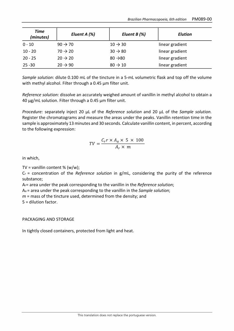
Brazilian Pharmacopoeia, 6th edition PM089-00
This translation does not replace the portuguese version.
Time (minutes) Eluent A (%) Eluent B (%) Elution
0 - 10 90 → 70 10 → 30 linear gradient 10 - 20 70 → 20 30 → 80 linear gradient 20 - 25 20 → 20 80 →80 linear gradient 25 -30 20 → 90 80 → 10 linear gradient Sample solution: dilute 0.100 mL of the tincture in a 5-mL volumetric flask and top off the volume with methyl alcohol. Filter through a 0.45 µm filter unit. Reference solution: dissolve an accurately weighed amount of vanillin in methyl alcohol to obtain a 40 µg/mL solution. Filter through a 0.45 µm filter unit. Procedure: separately inject 20 μL of the Reference solution and 20 µL of the Sample solution. Register the chromatograms and measure the areas under the peaks. Vanillin retention time in the sample is approximately 13 minutes and 30 seconds. Calculate vanillin content, in percent, according to the following expression:
𝑇𝑇𝑉𝑉 =𝐶𝐶𝑟𝑟𝑡𝑡 × 𝑇𝑇𝑎𝑎 × 5 × 100
𝑇𝑇𝑟𝑟 × 𝑚𝑚
in which,
TV = vanillin content % (w/w); Cr = concentration of the Reference solution in g/mL, considering the purity of the reference substance; Ar= area under the peak corresponding to the vanillin in the Reference solution; Aa = area under the peak corresponding to the vanillin in the Sample solution; m = mass of the tincture used, determined from the density; and 5 = dilution factor. PACKAGING AND STORAGE In tightly closed containers, protected from light and heat.
Brazilian Pharmacopoeia, 6th edition PM090-00
This translation does not replace the portuguese version.
BENZOIN, tincture Benzoe sumatranus tincture
The tincture is obtained from balsamic resin, obtained by incision of the stem of Styrax benzoin Dryand. containing no less than 4.0% (w/w) total acids, expressed as benzoic acid (C7H6O2, 122.12). PREPARATION The tincture is prepared at 20% (w/v), by percolation or maceration, using 80% (v/v) ethyl alcohol as the extracting liquid. CHARACTERISTICS Yellowish-brown or reddish-brown liquid. IDENTIFICATION Proceed as described in Thin-layer chromatography (5.2.17.1). Stationary phase: silica gel GF254. Mobile phase: hexane, isopropyl ether and glacial acetic acid (3:1:0.5). Sample solution: dilute 50 µL of the tincture in 950 mL ethyl alcohol. Reference solution (1): prepare an ethanolic solution containing 2.5 mg/mL of vanillin and 5 mg/mL of benzoic acid. Reference solution (2): prepare an ethanolic solution containing 2.5 mg/mL of cinnamic acid and 2.5 mg/mL of methyl cinnamate. Procedure: previously saturate the tank with 15x15 cm filter paper impregnated with the Mobile phase for 20 minutes. Procedure: apply 10 μL of the Sample solution, 10 μL of the Reference solution (1) and 10 μL of the Reference solution (2) to the chromatoplate, separately and in the form of a band. Conduct chromatogram. Remove chromatoplate and allow it to air dry. Examine under ultraviolet light at 365 nm. Results: the diagram below shows the sequences of zones obtained with the Reference solution (1), the Referece solution (2) and the Sample solution. Other zones may occasionally develop.
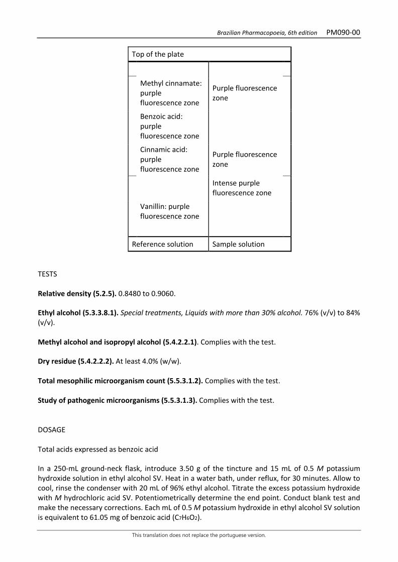
Brazilian Pharmacopoeia, 6th edition PM090-00
This translation does not replace the portuguese version.
Top of the plate
Methyl cinnamate: purple fluorescence zone
Purple fluorescence zone
Benzoic acid: purple fluorescence zone
Cinnamic acid: purple fluorescence zone
Purple fluorescence zone
Intense purple fluorescence zone
Vanillin: purple fluorescence zone
Reference solution Sample solution TESTS Relative density (5.2.5). 0.8480 to 0.9060. Ethyl alcohol (5.3.3.8.1). Special treatments, Liquids with more than 30% alcohol. 76% (v/v) to 84% (v/v). Methyl alcohol and isopropyl alcohol (5.4.2.2.1). Complies with the test. Dry residue (5.4.2.2.2). At least 4.0% (w/w). Total mesophilic microorganism count (5.5.3.1.2). Complies with the test. Study of pathogenic microorganisms (5.5.3.1.3). Complies with the test. DOSAGE Total acids expressed as benzoic acid In a 250-mL ground-neck flask, introduce 3.50 g of the tincture and 15 mL of 0.5 M potassium hydroxide solution in ethyl alcohol SV. Heat in a water bath, under reflux, for 30 minutes. Allow to cool, rinse the condenser with 20 mL of 96% ethyl alcohol. Titrate the excess potassium hydroxide with M hydrochloric acid SV. Potentiometrically determine the end point. Conduct blank test and make the necessary corrections. Each mL of 0.5 M potassium hydroxide in ethyl alcohol SV solution is equivalent to 61.05 mg of benzoic acid (C7H6O2).
Brazilian Pharmacopoeia, 6th edition PM090-00
This translation does not replace the portuguese version.
PACKAGING AND STORAGE In tightly closed containers, protected from light and heat.

Brazilian Pharmacopoeia, 6th edition PM091-00
This translation does not replace the portuguese version.
BOLDO, tincture Boldus tincture
The tincture is obtained from dried leaves of Peumus boldus Molina, containing at least 0.01% total alkaloids expressed as boldine. PREPARATION The tincture is prepared at 10% (w/v), by percolation or maceration, using 60% (v/v) ethyl alcohol as the extracting liquid. CHARACTERISTICS Clear, dark greenish-brown liquid. IDENTIFICATION Proceed as described in Thin-layer chromatography (5.2.17.1). Stationary phase: silica gel GF254. Mobile phase: toluene, methyl alcohol and diethylamine (80:10:10). Sample solution: evaporate 25mL of tincture in a water bath to a soft extract consistency. Use a glass rod to extract the residue while still hot, twice, with 10mL of 2 M hydrochloric acid each time. Filter and alkalinize the filtrate at pH 9.0 with 6 M ammonium hydroxide. Extract the filtrate twice into a separating funnel with 20 mL diethyl ether each time, moderately shaking to avoid formation of an emulsion. Combine the organic phases and evaporate the solvent in a water bath. Dissolve the residue in 0.5 mL methyl alcohol. Reference solution: dissolve 2 mg boldine CRS in 5 mL methyl alcohol. Procedure: apply 10 μL of the Sample solution and 5 μL of the Reference solution to the chromatoplate, separately and in the form of a band. Conduct chromatogram. Remove chromatoplate and allow it to air dry. Examine under ultraviolet light at 365 nm. Nebulize plate with aqueous acetic potassium iodobismuthate solution. Allow to air dry for five minutes. Nebulize plate with sodium nitrite RS. Examine under visible light after 30 minutes. Results: the diagram below shows the sequences of zones obtained with the Reference solution and the Sample solution. Other zones may occasionally develop.
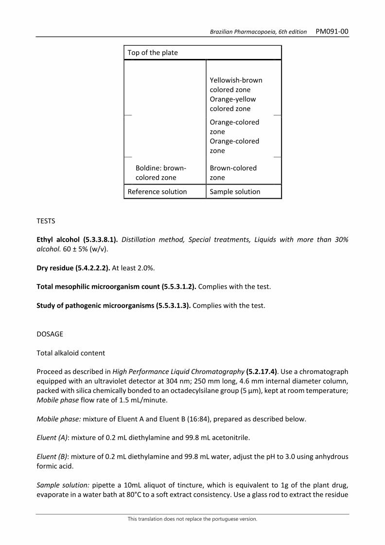
Brazilian Pharmacopoeia, 6th edition PM091-00
This translation does not replace the portuguese version.
Top of the plate
Yellowish-brown colored zone Orange-yellow colored zone
Orange-colored zone Orange-colored zone
Boldine: brown-colored zone
Brown-colored zone
Reference solution Sample solution TESTS Ethyl alcohol (5.3.3.8.1). Distillation method, Special treatments, Liquids with more than 30% alcohol. 60 ± 5% (w/v). Dry residue (5.4.2.2.2). At least 2.0%. Total mesophilic microorganism count (5.5.3.1.2). Complies with the test. Study of pathogenic microorganisms (5.5.3.1.3). Complies with the test. DOSAGE Total alkaloid content Proceed as described in High Performance Liquid Chromatography (5.2.17.4). Use a chromatograph equipped with an ultraviolet detector at 304 nm; 250 mm long, 4.6 mm internal diameter column, packed with silica chemically bonded to an octadecylsilane group (5 µm), kept at room temperature; Mobile phase flow rate of 1.5 mL/minute. Mobile phase: mixture of Eluent A and Eluent B (16:84), prepared as described below. Eluent (A): mixture of 0.2 mL diethylamine and 99.8 mL acetonitrile. Eluent (B): mixture of 0.2 mL diethylamine and 99.8 mL water, adjust the pH to 3.0 using anhydrous formic acid. Sample solution: pipette a 10mL aliquot of tincture, which is equivalent to 1g of the plant drug, evaporate in a water bath at 80°C to a soft extract consistency. Use a glass rod to extract the residue
Brazilian Pharmacopoeia, 6th edition PM091-00
This translation does not replace the portuguese version.
while still hot with 50mL of 2 M hydrochloric acid for five minutes. Filter and repeat the procedure once more with the residue obtained. Filter. Combine the cooled filtrates in a separating funnel and shaking with 100 mL of a mixture of hexane and ethyl acetate (1:1). Discard the organic phase. Adjust the aqueous phase pH to 9.0 using 6 M ammonium hydroxide. Extract the aqueous phase with 100 mL, 50 mL and 50 mL portions of methyl chloride. Combine the organic phases and evaporate in a rotary evaporator to dryness. Transfer the residue to a 10-mL volumetric flask using the Mobile phase as diluent. Top off the volume with the Mobile phase and homogenize. Filter through a 0.45 µm filter unit. Reference solution: accurately weigh about 12 mg of boldine CRS. Dissolve this weighed amount in a 100-mL volumetric flask using the Mobile phase as diluent. Top off the volume with the Mobile phase and homogenize. Transfer 1 mL of the obtained solution, using a volumetric pipette, into a 10-mL volumetric flask. Top off the volume with the Mobile phase and homogenize. Filter through a 0.45 µm filter unit. System suitability Resolution between peaks: Sample solution, minimum 1.0 between peaks for isoboldine and boldine. Procedure: separately inject 20 μL of the Reference solution and 20 µL of the Sample solution. Record the chromatograms and measure the areas under the peaks for boldine and the six alkaloids described below. Retention times for boldine, whose retention time is about six minutes, are approximately 0.9 for isoboldine, 1.0 for boldine, 1.8 for isocoridine N-oxide, 2.2 for laurotetanine, 2.8 for isocoridine, and 3.2 for N-methyl laurotetanine. Calculate total alkaloid content expressed as a percentage of boldine, according to the following expression:
𝑇𝑇𝑇𝑇 =(∑A) × 𝑚𝑚𝑟𝑟
𝑇𝑇𝑟𝑟
in which,
TA = Total alkaloid content expressed as boldine % (w/w); ΣA1 = sum of the areas under the peaks corresponding to the six alkaloids identified in the chromatogram obtained with the Sample solution; mr = mass in grams of boldine CRS in the Reference solution, considering the purity of the reference substance; Ar= area under the peak corresponding to the boldine in the Reference solution. PACKAGING AND STORAGE In tightly closed containers, protected from light and heat.

Brazilian Pharmacopoeia, 6th edition PM092-00
This translation does not replace the portuguese version.
CALENDULA, tincture Calendulae tincture
The tincture is obtained from dried flowers of Calendula officinalis L. containing at least 0.04% total flavonoids, expressed as hyperoside (C21H20O12, 464.38). PREPARATION The tincture is prepared at 10% (w/v), by percolation or maceration, using 70% (v/v) ethyl alcohol as the extracting liquid. CHARACTERISTICS Dark brown colored liquid. IDENTIFICATION Proceed as described in Thin-layer chromatography (5.2.17.1). Stationary phase: silica gel F254(0.25 mm). Mobile phase: ethyl acetate, anhydrous formic acid and water (80:10:10). Sample solution: dry 1mL of tincture until a residue is formed in a water bath at a maximum temperature of 60 ºC. Add 2mL methyl alcohol and filter through a 0.45µm filter unit. Reference solution (1): dissolve an accurately weighed amount of rutin in methyl alcohol to obtain a concentration of 250 µg/mL. Reference solution (2): dissolve an exactly weighed amount of chlorogenic acid in methyl alcohol to obtain the concentration of 100 µg/mL. Reference solution (3): dissolve an exactly weighed amount of caffeic acid in methyl alcohol to obtain the concentration of 100 µg/mL. Procedure: apply 20 μL of the Sample solution, 20 μL of the Reference solution (1), 20 μL of the Reference solution (2), and 20 μL of the Reference solution (3) to the chromatoplate, separately and in the form of a band. Conduct chromatogram. Remove chromatoplate and allow it to air dry. Nebulize plate with a 1% (w/v) aminoethanol diphenylborate in ethyl alcohol solution, followed by a 5% (w/v) macrogol 400 in ethyl alcohol solution, heat plate between 100°C and 105°C for approximately five minutes. Examine under ultraviolet light at 365 nm. Results: the diagram below shows the sequences of zones obtained with the Reference solution (1), Reference solution (2), Reference solution (3) and the Sample solution. Other zones may occasionally develop.

Brazilian Pharmacopoeia, 6th edition PM092-00
This translation does not replace the portuguese version.
Top of the plate
Caffeic acid: light-blue fluorescence zone
Light-blue fluorescence zone
Blue fluorescence zone
Greenish-yellow fluorescence zone
Chlorogenic acid: light-blue fluorescence zone
Light-blue fluorescence zone
Greenish-yellow fluorescence zone
Rutin: yellowish-brown fluorescence zone
Yellowish-brown fluorescence zone
Greenish-yellow fluorescence zone
Reference solution Sample solution TESTS Relative density (5.2.5). 0.9122 to 0.9500. Ethyl alcohol (5.3.3.8.1). Method II. 60% (v/v) to 64% (v/v). Methyl alcohol and isopropyl alcohol (5.4.2.2.1). Complies with the test. Dry residue (5.4.2.2.2). At least 7.0% (w/w). Total mesophilic microorganism count (5.5.3.1.2). Complies with the test. Study of pathogenic microorganisms (5.5.3.1.3). Complies with the test. DOSAGE Total flavonoids
Brazilian Pharmacopoeia, 6th edition PM092-00
This translation does not replace the portuguese version.
To proceed as described in Visible absorption spectrophotometry (5.2.14). Prepare solutions as described below. Stock solution: in a round-bottomed flask, add 8.0 mL of calendula tincture. Add 1 mL of 5 g/L aqueous methenamine solution (w/v), 20 mL acetone and 7 mL hydrochloric acid. Then reflux in a water bath for 30 minutes. Filter through filter paper, transfer to a 100-mL volumetric flask, top off the volume with acetone and homogenize. Transfer 20 mL of the solution to a separating funnel and add 20 mL of water. Extract with one 15-mL portion, and then with three 10-mL portions of ethyl acetate. After the extraction process, combine the ethyl acetate phases, transfer to another separating funnel and rinse with two 50-mL portions of water. Transfer the ethyl acetate phase to a 50-mL volumetric flask, top off the volume with ethyl acetate and homogenize. Sample solution: transfer 10 mL of the Stock solution into a 25-mL volumetric flask, add 1 mL of the 2% aluminum chloride solution (w/v) in (v/v) 5% acetic acid solution (v/v) in methyl alcohol. Top off the volume of the 25-mL volumetric flask with the 5% acetic acid solution (v/v) in methyl alcohol and homogenize. Blank solution: transfer 10 mL of the Stock solution into a 25-mL volumetric flask, top off the volume with the 5% acetic acid solution (v/v) in methyl alcohol and homogenize. Procedure: measure the absorbance of the Sample Solution at 425 nm after precisely 30 minutes, using the Blank solution for zero adjustment. Calculate total flavonoid content expressed as hyperoside, in percent, according to the following expression:
𝑇𝑇𝑇𝑇 =A × 1,25
𝑚𝑚
in which, TF = total flavonoid content expressed as hyperoside % (w/w); A = absorbance measured for the Sample solution; m = mass in grams of the tincture used, determined from the density. PACKAGING AND STORAGE In tightly closed containers, protected from light and heat.
Brazilian Pharmacopoeia, 6th edition PM093-00
This translation does not replace the portuguese version.
CHAMOMILE, tincture Matricariae flos tincture
The tincture is obtained from dried flower capitula of Matricaria chamomilla L. containing at least 0.025% apigenin-7-O-glucoside (C21H20O10, 432.38). PREPARATION The tincture is prepared at 10% (w/v), by percolation or maceration, using 70% (v/v) ethyl alcohol as the extracting liquid. CHARACTERISTICS Orange-yellow or greenish-brown liquid with a characteristic odor. IDENTIFICATION Proceed as described in Thin-layer chromatography (5.2.17.1). Stationary phase: silica gel GF254. Mobile phase: toluene and ethyl acetate (97:3). Sample solution: dilute 500 µL of the tincture in 500 mL ethyl alcohol. Reference solution: dilute 2 µL camazulene, 5 µL levomenol and 10 mg bornyl acetate in 5 mL toluene. TLC visualization reagent: dissolve 1 g of vanillin in 100 mL of methyl alcohol. Add 4 mL hydrochloric acid and 5 mL sulfuric acid. Procedure: apply 10 μL of the Sample solution and 10 μL of the Reference solution to the chromatoplate, separately and in the form of a band. Conduct chromatogram. Remove chromatoplate and allow it to air dry. Nebulize the plate with sulfur vanillin RS solution and heat at 105 °C for one minute. Results: the diagram below shows the sequences of zones obtained with the Reference solution and the Sample solution. Other zones may occasionally develop.

Brazilian Pharmacopoeia, 6th edition PM093-00
This translation does not replace the portuguese version.
Top of the plate
Chamazulene: pinkish-red colored zone
Pinkish-red colored zone
Bornyl acetate: violaceous-blue colored zone
Blue to purple colored zone
Levomenol: violaceous color zone
Violaceous-colored zone
Violaceous-colored zone
Greenish-yellow colored zone
Reference solution Sample solution TESTS Relative density (5.2.5). 0.9010 to 0.9500. Ethyl alcohol (5.3.3.8.1). 60% (v/v) to 70% (v/v). Methyl alcohol and isopropyl alcohol (5.4.2.2.1). Complies with the test. Dry residue (5.4.2.2.2). At least 2.5%. Total mesophilic microorganism count (5.5.3.1.2). Complies with the test. Study of pathogenic microorganisms (5.5.3.1.3). Complies with the test. DOSAGE Apigenin-7-O-glycoside Proceed as described in High Performance Liquid Chromatography (5.2.17.4). Use a chromatograph equipped with an ultraviolet detector at 340 nm; pre-column packed with silica chemically bonded to an octadecylsilane group; a 250mm long, 4.6mm internal diameter column, packed with silica chemically bonded to an octadecylsilane group (5 µm); Mobile phase flow rate of 0.6 mL/minute. Eluent (A): water and formic acid (99.5:0.5). Eluent (B): methyl alcohol and formic acid (100:0.08).
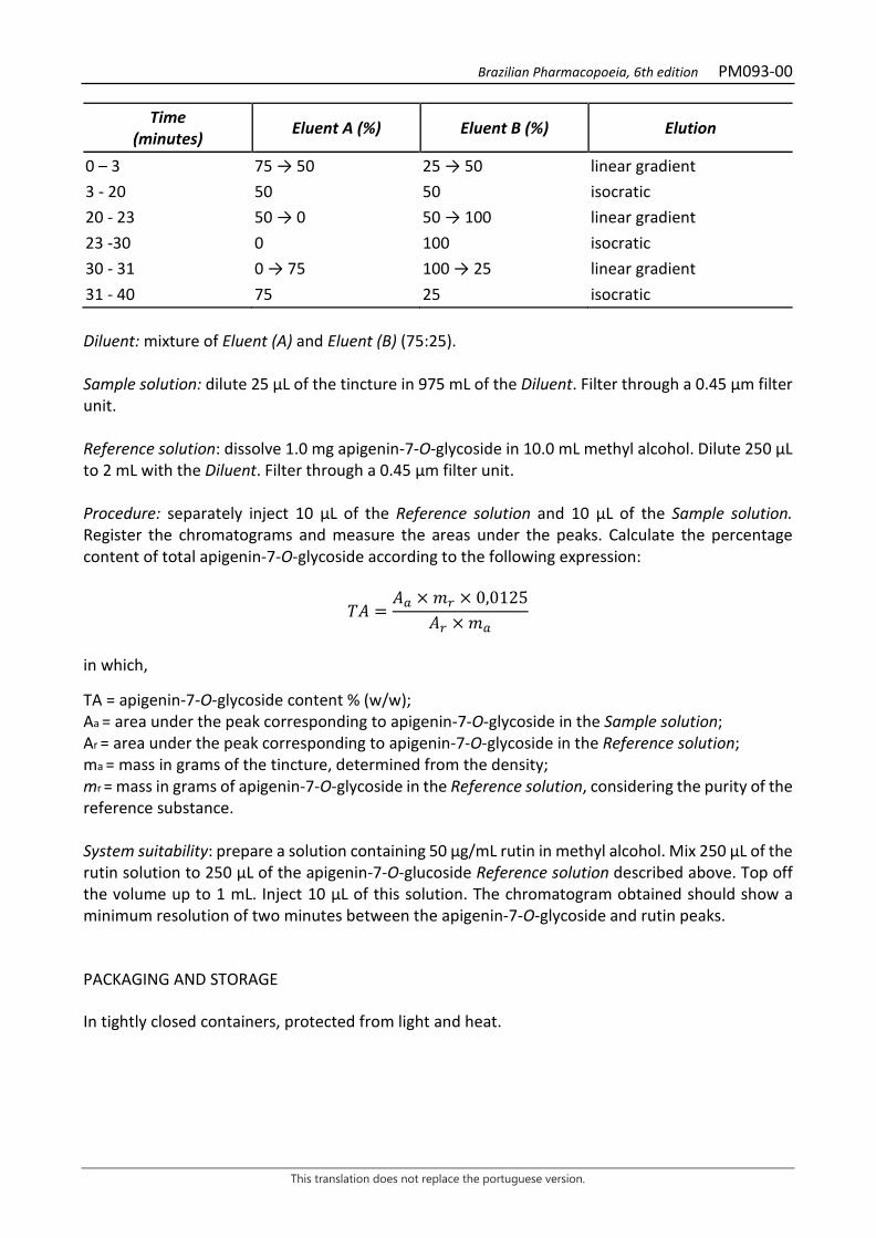
Brazilian Pharmacopoeia, 6th edition PM093-00
This translation does not replace the portuguese version.
Time (minutes) Eluent A (%) Eluent B (%) Elution
0 – 3 75 → 50 25 → 50 linear gradient 3 - 20 50 50 isocratic 20 - 23 50 → 0 50 → 100 linear gradient 23 -30 0 100 isocratic 30 - 31 0 → 75 100 → 25 linear gradient 31 - 40 75 25 isocratic Diluent: mixture of Eluent (A) and Eluent (B) (75:25). Sample solution: dilute 25 µL of the tincture in 975 mL of the Diluent. Filter through a 0.45 µm filter unit. Reference solution: dissolve 1.0 mg apigenin-7-O-glycoside in 10.0 mL methyl alcohol. Dilute 250 µL to 2 mL with the Diluent. Filter through a 0.45 µm filter unit. Procedure: separately inject 10 μL of the Reference solution and 10 µL of the Sample solution. Register the chromatograms and measure the areas under the peaks. Calculate the percentage content of total apigenin-7-O-glycoside according to the following expression:
𝑇𝑇𝑇𝑇 =𝑇𝑇𝑎𝑎 × 𝑚𝑚𝑟𝑟 × 0,0125
𝑇𝑇𝑟𝑟 × 𝑚𝑚𝑎𝑎
in which,
TA = apigenin-7-O-glycoside content % (w/w); Aa = area under the peak corresponding to apigenin-7-O-glycoside in the Sample solution; Ar = area under the peak corresponding to apigenin-7-O-glycoside in the Reference solution; ma = mass in grams of the tincture, determined from the density; mr = mass in grams of apigenin-7-O-glycoside in the Reference solution, considering the purity of the reference substance. System suitability: prepare a solution containing 50 µg/mL rutin in methyl alcohol. Mix 250 µL of the rutin solution to 250 µL of the apigenin-7-O-glucoside Reference solution described above. Top off the volume up to 1 mL. Inject 10 µL of this solution. The chromatogram obtained should show a minimum resolution of two minutes between the apigenin-7-O-glycoside and rutin peaks. PACKAGING AND STORAGE In tightly closed containers, protected from light and heat.
Brazilian Pharmacopoeia, 6th edition PM094-00
This translation does not replace the portuguese version.
CEYLON CINNAMON, tincture Cinnamomi corticis tincture
The tincture is obtained from dry bark of Cinnamomum verum J. Presl (syn. Cinnamomum zeylanicum Blume) containing at least 0.25% (w/w) of trans-cinnamic aldehyde. PREPARATION The tincture is prepared at 20% (w/v), by percolation, using 70% (v/v) ethyl alcohol as the extracting liquid. CHARACTERISTICS Clear, reddish-brown liquid. IDENTIFICATION Proceed as described in Thin-layer chromatography (5.2.17.1). Stationary phase: silica gel F254(0.25 mm). Mobile phase: methyl chloride. Sample solution: transfer 10 mL of the sample, 10-mL saturated sodium chloride solution, and 5 mL toluene to a glass tube with a ground-glass stopper. Mix for two minutes and centrifuge for 10 minutes. Use the organic layer. Reference solution: dilute 5 μL eugenol, 25 μL trans-cinnamic aldehyde and 5 μL trans-2-methoxy-cinnalamdehyde in toluene, to off the volume to 10 mL with the same solvent and homogenize. Procedure: apply 20 μL of the sample solution and 20 μL of the Reference solution to the chromatoplate, separately and in the form of a band. Conduct chromatogram. Remove chromatoplate and allow it to air dry for 15 minutes. Examine under ultraviolet light at 365 nm. Nebulize plate with phosphomolybdic acid solution at 200 g/L in ethyl alcohol, examine in daylight and heat between 100°C and 105°C for five to 10 minutes. Results: the diagrams below show the sequences of zones obtained with the Reference solution and the Sample solution, after examination under ultraviolet light and nebulization with the phosphomolybdic acid solution, in order. Other zones may occasionally develop.
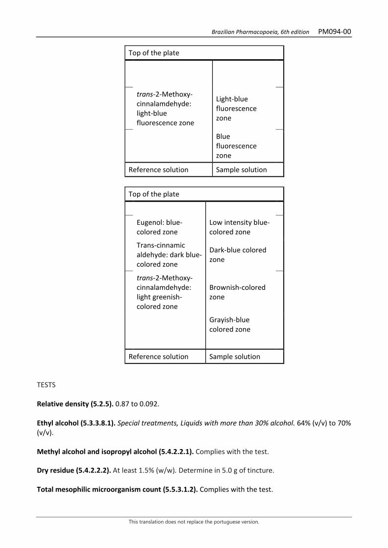
Brazilian Pharmacopoeia, 6th edition PM094-00
This translation does not replace the portuguese version.
Top of the plate
trans-2-Methoxy-cinnalamdehyde: light-blue fluorescence zone
Light-blue fluorescence zone
Blue fluorescence zone
Reference solution Sample solution
Top of the plate
Eugenol: blue-colored zone
Low intensity blue-colored zone
Trans-cinnamic aldehyde: dark blue-colored zone
Dark-blue colored zone
trans-2-Methoxy-cinnalamdehyde: light greenish-colored zone
Brownish-colored zone
Grayish-blue colored zone
Reference solution Sample solution TESTS Relative density (5.2.5). 0.87 to 0.092. Ethyl alcohol (5.3.3.8.1). Special treatments, Liquids with more than 30% alcohol. 64% (v/v) to 70% (v/v). Methyl alcohol and isopropyl alcohol (5.4.2.2.1). Complies with the test. Dry residue (5.4.2.2.2). At least 1.5% (w/w). Determine in 5.0 g of tincture. Total mesophilic microorganism count (5.5.3.1.2). Complies with the test.
Brazilian Pharmacopoeia, 6th edition PM094-00
This translation does not replace the portuguese version.
Study of pathogenic microorganisms (5.5.3.1.3). Complies with the test. DOSAGE Trans-cinnamic aldehyde Proceed as described in High Performance Liquid Chromatography (5.2.17.4). Use a chromatograph equipped with an ultraviolet detector at 292 nm; a 250 mm long, 4.6 mm internal diameter column packed with C-18 (5 µm); Mobile phase flow rate of 1.0 mL/minute. Mobile phase: water and methyl alcohol (1:1). Sample solution: analytically transfer 1.0 mL of the ceylon cinnamon tincture to a 25-mL volumetric flask, top off the volume with methyl alcohol and homogenize. Transfer 10 mL of the solution to a 10-mL volumetric flask, top off the volume with methyl alcohol and homogenize. Filter through a 0.45 µm filter unit. Reference solution: dissolve an accurately weighed amount of trans-cinnamic aldehyde (about 13 mg) in methyl alcohol to obtain a 0.520 mg/mL solution (in a 25 mL volumetric flask). Solutions for the analytical curve: dilute 2.0 mL of the Reference Solution in a 50-mL volumetric flask, topping off the volume with methyl alcohol in order to obtain a 20.8 µg/mL solution. Transfer 2.0, 3.0, 4.0, 5.0, 6.0, 7.0 and 8.0 mL of this solution to 10 mL volumetric flasks, top off the volumes with methyl alcohol and homogenize, obtaining concentrations of 4.16 µg/ mL, 6.24 µg/ mL, 8.32 µg/ mL, 10.4 µg/ mL, 12.48 µg/ mL, 14.56 µg/ mL and 16.64 µg/ mL. Filter the solutions through a 0.45 µm filter unit. Procedure: inject 20.0 µL of the Solutions for the analytical curve and 20.0 µL of the Sample solution separately. Record the chromatograms and measure the areas under the peaks corresponding to trans-cinnamic aldehyde. Calclate the trans-cinnamic aldehyde content using the straight line equation obtained from the analytical curve and, in percentage, according to the following expression:
𝑇𝑇𝑇𝑇 = 𝐶𝐶𝑎𝑎 ×25𝑚𝑚
in which, TA = trans-cinnamic aldehyde content % (w/w); Ca = trans-cinnamic aldehyde concentration in the Sample solution, determined using the analytical curve in µg/mL; m = mass in milligrams of the tincture used, determined from the density. PACKAGING AND STORAGE In tightly closed containers, protected from light and heat.
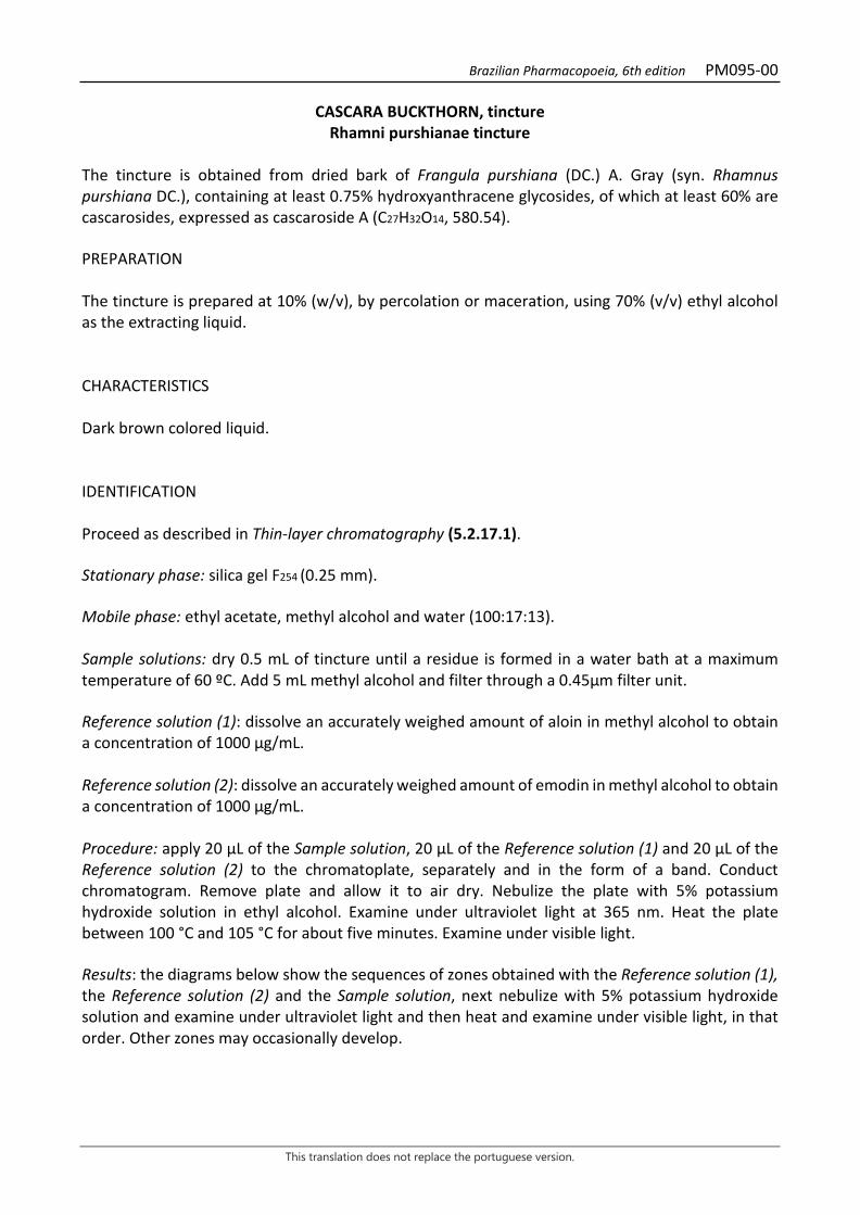
Brazilian Pharmacopoeia, 6th edition PM095-00
This translation does not replace the portuguese version.
CASCARA BUCKTHORN, tincture Rhamni purshianae tincture
The tincture is obtained from dried bark of Frangula purshiana (DC.) A. Gray (syn. Rhamnus purshiana DC.), containing at least 0.75% hydroxyanthracene glycosides, of which at least 60% are cascarosides, expressed as cascaroside A (C27H32O14, 580.54). PREPARATION The tincture is prepared at 10% (w/v), by percolation or maceration, using 70% (v/v) ethyl alcohol as the extracting liquid. CHARACTERISTICS Dark brown colored liquid. IDENTIFICATION Proceed as described in Thin-layer chromatography (5.2.17.1). Stationary phase: silica gel F254 (0.25 mm). Mobile phase: ethyl acetate, methyl alcohol and water (100:17:13). Sample solutions: dry 0.5 mL of tincture until a residue is formed in a water bath at a maximum temperature of 60 ºC. Add 5 mL methyl alcohol and filter through a 0.45µm filter unit. Reference solution (1): dissolve an accurately weighed amount of aloin in methyl alcohol to obtain a concentration of 1000 µg/mL. Reference solution (2): dissolve an accurately weighed amount of emodin in methyl alcohol to obtain a concentration of 1000 µg/mL. Procedure: apply 20 μL of the Sample solution, 20 μL of the Reference solution (1) and 20 μL of the Reference solution (2) to the chromatoplate, separately and in the form of a band. Conduct chromatogram. Remove plate and allow it to air dry. Nebulize the plate with 5% potassium hydroxide solution in ethyl alcohol. Examine under ultraviolet light at 365 nm. Heat the plate between 100 °C and 105 °C for about five minutes. Examine under visible light. Results: the diagrams below show the sequences of zones obtained with the Reference solution (1), the Reference solution (2) and the Sample solution, next nebulize with 5% potassium hydroxide solution and examine under ultraviolet light and then heat and examine under visible light, in that order. Other zones may occasionally develop.
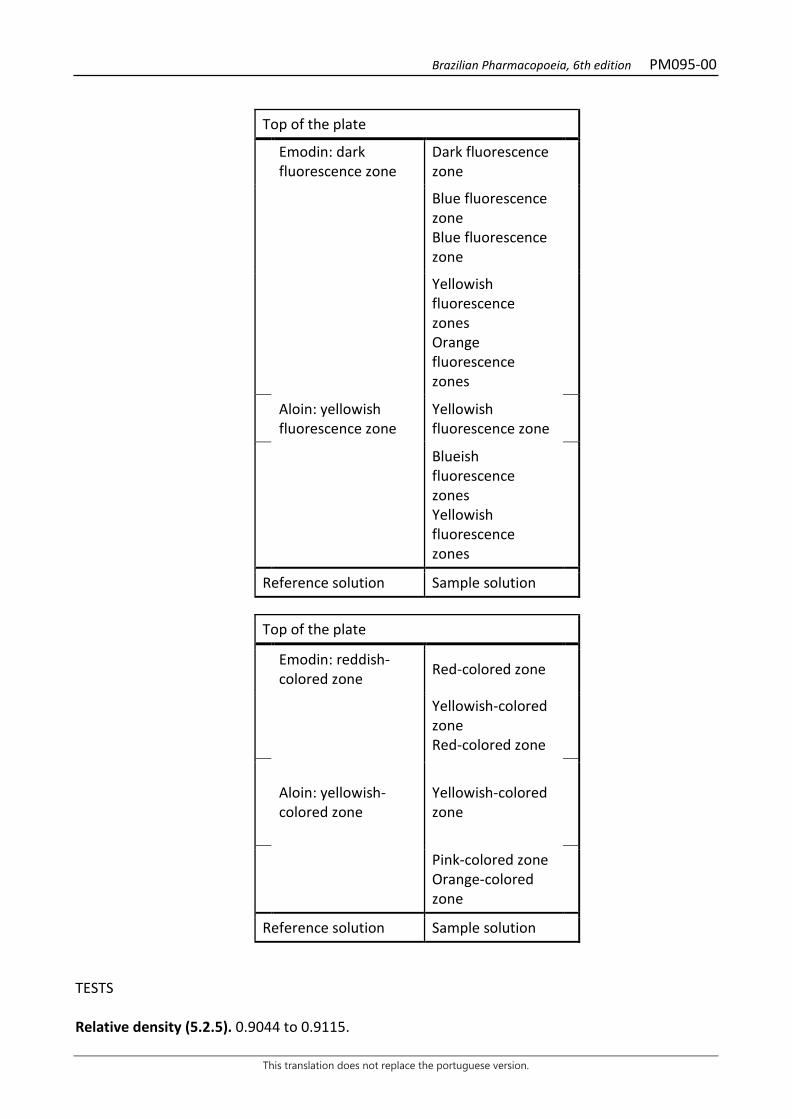
Brazilian Pharmacopoeia, 6th edition PM095-00
This translation does not replace the portuguese version.
Top of the plate
Emodin: dark fluorescence zone
Dark fluorescence zone
Blue fluorescence zone Blue fluorescence zone
Yellowish fluorescence zones Orange fluorescence zones
Aloin: yellowish fluorescence zone
Yellowish fluorescence zone
Blueish fluorescence zones Yellowish fluorescence zones
Reference solution Sample solution
Top of the plate
Emodin: reddish-colored zone Red-colored zone
Yellowish-colored zone Red-colored zone
Aloin: yellowish-colored zone
Yellowish-colored zone
Pink-colored zone Orange-colored zone
Reference solution Sample solution TESTS Relative density (5.2.5). 0.9044 to 0.9115.
Brazilian Pharmacopoeia, 6th edition PM095-00
This translation does not replace the portuguese version.
Ethyl alcohol (5.3.3.8.1). Method II. 60% (v/v) to 64% (v/v). Methyl alcohol and isopropyl alcohol (5.4.2.2.1). Complies with the test. Dry residue (5.4.2.2.2). At least 3.0% (w/w). Total mesophilic microorganism count (5.5.3.1.2). Complies with the test. Study of pathogenic microorganisms (5.5.3.1.3). Complies with the test. DOSAGE Stock solution: measure 10.0 mL of tincture and transfer to a 100 mL volumetric flask. Top off the volume with water and homogenize. Filter the sample, discard the first 20 mL. Transfer 10 mL of the filtrate to a separating funnel and add 0.1 mL of M hydrochloric acid. Extract with two quantities of 20 mL each, from a hexane and ether mixture (3:1). After separating the phases, set the aqueous phase aside. Rinse the organic phase with 5 mL of water. Discard the organic phase and combine the aqueous phases with the wash waters. Extract the aqueous phase with four quantities, 30 mL each, of water-saturated ethyl acetate, prepared at the time of the analysis (150 mL ethyl acetate and 15 mL water, mixed for three minutes). Combine the ethyl acetate fractions. Use the aqueous phase to determine cascaroside dosage and the organic phase to determine hydroxyanthracene glycoside dosage without cascarosides. Hydroxyanthracene glycosides without cascarosides To proceed as described in Visible absorption spectrophotometry (5.2.14). Prepare solutions as described below. Sample solution: transfer the organic phase from the Stock solution to a porcelain capsule. Evaporate the solvent in a water bath until residue is formed. Dissolve the residue in 0.3 mL to 0.5 mL methyl alcohol and transfer to a 50-mL volumetric flask. Rinse the capsule with hot water and transfer the residue to the 50-mL volumetric flask, dilute with water, top off the volume and homogenize. Then transfer 20 mL of the solution to a 100mL round-bottomed flask, add 2 g of ferric chloride hexahydrate and 12 mL of hydrochloric acid. Heat the mixture under reflux for four hours. After cooling, transfer the solution to a separating funnel. Rinse the flask with 3 to 4 mL of M sodium hydroxide, then with 3 to 4 mL of water. Transfer the wash water to the separating funnel. Extract with three portions, 30 mL each, of an hexane and ether (3:1) mixture. Transfer the organic phase to another separating funnel and rinse it twice, using 10 mL of water in each rinse. Discard the aqueous phase. After this procedure, dilute the organic phase to 100 mL with the hexane and ether (3:1) mixture. Then transfer 20 mL and evaporate until residue is formed in a water bath. Dissolve the residue with 10 mL of a 5 g/L magnesium acetate solution in methyl alcohol. Blank solution: methyl alcohol. Procedure: measure the absorbance of the Sample solution at 440 nm and 515 nm, using the Blank solution for zero adjustment. A ratio between the absorbance values at 515 nm and 440 nm that is

Brazilian Pharmacopoeia, 6th edition PM095-00
This translation does not replace the portuguese version.
smaller than 2.4 invalidates the assay. Calculate the content of hydroxyanthracene glycosides without the cascarosides (HAC), in percent, according to the expression:
𝑇𝑇𝑇𝑇𝑇𝑇𝐶𝐶 =A × 6,95
𝑚𝑚
in which, THAC = hydroxyanthracene glycoside content without cascarosides % (w/w); A = absorbance measured at 515 nm for the Sample solution; m = mass in grams of the tincture used, determined from the density. Cascarosides Sample solution: dilute the aqueous phase of the Stock solution in a 50-ml volumetric flask with water and homogenize. Use 20 mL of the solution. Blank solution: methyl alcohol. Procedure: measure the absorbance of the Sample solution at 440 nm and 515 nm, using the Blank solution for zero adjustment. A ratio between the absorbance values at 515 nm and 440 nm that is smaller than 2.4 invalidates the assay. Calculate cascaroside content, in percent, according to the following expression:
𝑇𝑇𝐶𝐶 =A × 6,95
𝑚𝑚
in which, TC = cascaroside content % (w/w); A = absorbance measured at 515 nm for the Sample solution; m = mass in grams of the tincture used, determined from the density. PACKAGING AND STORAGE In tightly closed containers, protected from light and heat.
Brazilian Pharmacopoeia, 6th edition PM096-00
This translation does not replace the portuguese version.
HORSE CHESTNUT, tincture Hippocastani tincture
The tincture is obtained from dried seeds of Aesculus hippocastanum L., containing at least 0.3% triterpene glycosides expressed as anhydrous aescin. PREPARATION The tincture is prepared at 10% (w/v), by percolation or maceration, using 70% ethyl alcohol as the extracting liquid. CHARACTERISTICS Dark brown colored liquid. IDENTIFICATION Proceed as described in Thin-layer chromatography (5.2.17.1). Stationary phase: silica gel GF254. Mobile phase: butyl alcohol, water and glacial acetic acid (50:10:40). Reference solution: dissolve an accurately weighed amount of aescin in methyl alcohol to obtain a concentration of 5000 µg/mL. Sample solution: dry 1 mL of the tincture until residue is formed, in a water bath, at a maximum temperature of 60 °C. Add 5 mL methyl alcohol and filter through a 0.45µm filter unit. Procedure: apply 20 μL of the Sample solution and 20 μL of the Reference solution to the chromatoplate, separately and in the form of a band. Conduct chromatogram. Remove chromatoplate and allow it to air dry. Nebulize with anisaldehyde RS, heat the plate at 100 °C to 105 °C for approximately five minutes. Examine under visible light. Results: the diagram below shows the sequences of zones obtained with the Reference solution and the Sample solution. Other zones may occasionally develop.
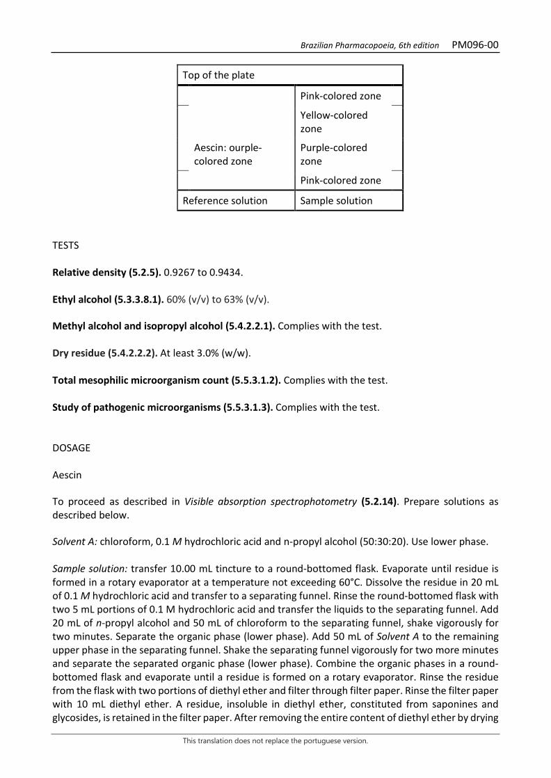
Brazilian Pharmacopoeia, 6th edition PM096-00
This translation does not replace the portuguese version.
Top of the plate
Pink-colored zone
Yellow-colored zone
Aescin: ourple-colored zone
Purple-colored zone
Pink-colored zone
Reference solution Sample solution
TESTS Relative density (5.2.5). 0.9267 to 0.9434. Ethyl alcohol (5.3.3.8.1). 60% (v/v) to 63% (v/v). Methyl alcohol and isopropyl alcohol (5.4.2.2.1). Complies with the test. Dry residue (5.4.2.2.2). At least 3.0% (w/w). Total mesophilic microorganism count (5.5.3.1.2). Complies with the test. Study of pathogenic microorganisms (5.5.3.1.3). Complies with the test. DOSAGE Aescin To proceed as described in Visible absorption spectrophotometry (5.2.14). Prepare solutions as described below. Solvent A: chloroform, 0.1 M hydrochloric acid and n-propyl alcohol (50:30:20). Use lower phase. Sample solution: transfer 10.00 mL tincture to a round-bottomed flask. Evaporate until residue is formed in a rotary evaporator at a temperature not exceeding 60°C. Dissolve the residue in 20 mL of 0.1 M hydrochloric acid and transfer to a separating funnel. Rinse the round-bottomed flask with two 5 mL portions of 0.1 M hydrochloric acid and transfer the liquids to the separating funnel. Add 20 mL of n-propyl alcohol and 50 mL of chloroform to the separating funnel, shake vigorously for two minutes. Separate the organic phase (lower phase). Add 50 mL of Solvent A to the remaining upper phase in the separating funnel. Shake the separating funnel vigorously for two more minutes and separate the separated organic phase (lower phase). Combine the organic phases in a round-bottomed flask and evaporate until a residue is formed on a rotary evaporator. Rinse the residue from the flask with two portions of diethyl ether and filter through filter paper. Rinse the filter paper with 10 mL diethyl ether. A residue, insoluble in diethyl ether, constituted from saponines and glycosides, is retained in the filter paper. After removing the entire content of diethyl ether by drying
Brazilian Pharmacopoeia, 6th edition PM096-00
This translation does not replace the portuguese version.
(both from the flask and the filter paper), add 10 mL of glacial acetic acid to the round-bottomed flask, which is then poured onto the filter paper containing the saponin residue. Transfer the solution into a 50-mL volumetric flask. Perform this procedure two more times with 10 mL of glacial acetic acid, topping off the volume to 50 mL with the same solvent. Color reagent: dissolve 75 mg ferric chloride hexahydrate in 50 mL of glacial acetic acid. Next add 50 mL of sulfuric acid on the solution. Transfer the solution into a 100-mL volumetric flask. Top off the volume and homogenize. The solution should be prepared for immediate use. Analytical curve solutions: weight 25 mg of aescin, transfer to a 25.0-mL volumetric flask and dilute with glacial acetic acid. Separately pipette 1 mL, 2 mL, 3 mL, 4 mL and 5 mL of this solution to five 10-mL volumetric flasks and dilute with glacial acetic acid. Blank solution: glacial acetic acid. Procedure: transfer 1 mL from each of the Analytical curve solutions, from the Sample solution and from the Blank solution, separately, to test tubes with a cap. Add 4 mL of Color reagent to each test tube. Next, heat the tubes in a water bath at a temperature of 60°C for 25 minutes, shaking the tubes occasionally. Measure the absorbances of the solutions at 540 nm. After reading, construct the aescin analytical curve in mg/mL and determine the aescin concentration in mg/mL in the Sample solution. Calculate triterpene glycoside content expressed as anhydrous aescin, in percent, according to the following expression:
𝑇𝑇𝑇𝑇 =𝐶𝐶 × 50𝑚𝑚 × 3
in which, TE = triterpene glycoside content expressed as anhydrous aescin % (w/w); C = concentration of aescin in mg/mL determined for the Sample solution using the analytical curve; considering the purity of the reference substance; m = mass in grams of the tincture used, determined from the density. PACKAGING AND STORAGE In tightly closed containers, protected from light and heat.

Brazilian Pharmacopoeia, 6th edition PM097-00
This translation does not replace the portuguese version.
TURMERIC, tincture Curcumae longae tincture
The tincture is obtained from dried rhizomes of Curcuma longa L. containing at least 0.25% of dicinnamoylmethane derivatives expressed in curcumin (C21H20O6, 368.39). PREPARATION The tincture is prepared at 10% (w/v), by maceration or percolation, using 70% (v/v) ethyl alcohol as the extracting liquid. CHARACTERISTICS Orange-yellow liquid. IDENTIFICATION Proceed as described in Thin-layer chromatography (5.2.17.1). Stationary phase: silica gel GF254. Mobile phase: mixture of chloroform, ethyl alcohol and glacial acetic acid (95:5:0.5). Sample solution: dilute 1 mL of the tumeric tincture to 1 mL methyl alcohol. Reference solution: dissolve 5 mg curcumin CRS, demethoxycurcumin CRS, and bisdemethoxycurcumin CRS in 5 mL methyl alcohol. Procedure: apply 10 μL of the Sample solution and 10 μL of the Reference solution to the chromatoplate, separately and in the form of a band. Conduct chromatogram. Remove chromatoplate and allow it to air dry. Examine under ultraviolet light at 365 nm and 254 nm. Results: the diagram below shows the sequences of zones obtained with the Reference solution and the Sample solution. Other zones may occasionally develop.
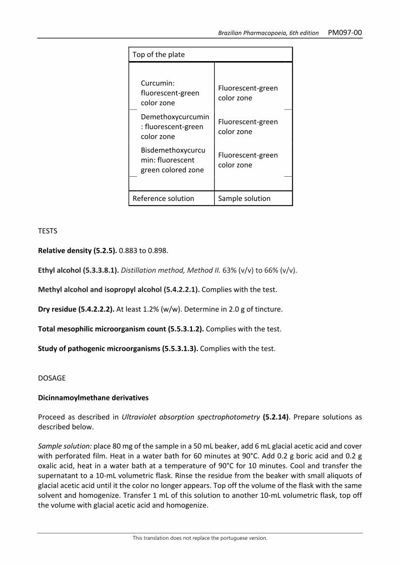
Brazilian Pharmacopoeia, 6th edition PM097-00
This translation does not replace the portuguese version.
Top of the plate
Curcumin: fluorescent-green color zone
Fluorescent-green color zone
Demethoxycurcumin: fluorescent-green color zone
Fluorescent-green color zone
Bisdemethoxycurcumin: fluorescent green colored zone
Fluorescent-green color zone
Reference solution Sample solution
TESTS Relative density (5.2.5). 0.883 to 0.898. Ethyl alcohol (5.3.3.8.1). Distillation method, Method II. 63% (v/v) to 66% (v/v). Methyl alcohol and isopropyl alcohol (5.4.2.2.1). Complies with the test. Dry residue (5.4.2.2.2). At least 1.2% (w/w). Determine in 2.0 g of tincture. Total mesophilic microorganism count (5.5.3.1.2). Complies with the test. Study of pathogenic microorganisms (5.5.3.1.3). Complies with the test. DOSAGE Dicinnamoylmethane derivatives Proceed as described in Ultraviolet absorption spectrophotometry (5.2.14). Prepare solutions as described below. Sample solution: place 80 mg of the sample in a 50 mL beaker, add 6 mL glacial acetic acid and cover with perforated film. Heat in a water bath for 60 minutes at 90°C. Add 0.2 g boric acid and 0.2 g oxalic acid, heat in a water bath at a temperature of 90°C for 10 minutes. Cool and transfer the supernatant to a 10-mL volumetric flask. Rinse the residue from the beaker with small aliquots of glacial acetic acid until it the color no longer appears. Top off the volume of the flask with the same solvent and homogenize. Transfer 1 mL of this solution to another 10-mL volumetric flask, top off the volume with glacial acetic acid and homogenize.
Brazilian Pharmacopoeia, 6th edition PM097-00
This translation does not replace the portuguese version.
Procedure: measure the absorbance of the Sample solution at 530 nm soon after its preparation, using glacial acetic acid for zero adjustment. Calculate the content of dicinnamoylmethane derivatives, expressed as a percentage of curcumin, according to the following expression:
𝑇𝑇𝐶𝐶 =0,0426 × A
𝑚𝑚
in which, TC = dicinnamoylmethane derivative content, expressed as curcumin % (w/w); A = absorbance measured for the Sample solution; m = mass in grams of the sample, determined from the density. PACKAGING AND STORAGE In tightly closed containers, protected from light and heat.

Brazilian Pharmacopoeia, 6th edition PM098-00
This translation does not replace the portuguese version.
YELLOW GENTIAN, tincture Gentianae tincture
The tincture is obtained from dried rhizomes and roots of Gentiana lutea L., containing at least 0.3% gentiopicroside (C16H20O9, 356.33). PREPARATION The tincture is prepared at 10% (w/v), by percolation or maceration, using 70% (v/v) ethyl alcohol as the extracting liquid. CHARACTERISTICS Yellowish-brown or reddish-brown liquid. IDENTIFICATION Proceed as described in Thin-layer chromatography (5.2.17.1). Stationary phase: silica gel GF254. Mobile phase: ethyl acetate, methyl alcohol and water (77:15:8). Sample solution: dilute 1 mL of the tincture in 1 mL methyl alcohol. Reference solution (1): prepare a 280 µg/mL solution of gentiopicroside in methyl alcohol. Reference solution (2): prepare a 1200 µg/mL solution of amarogentin in methyl alcohol. Procedure: apply 10 μL of the Sample solution, 20 μL of the Reference solution (1) and 20 μL of the Reference solution (2) to the chromatoplate, separately and in the form of a band. Conduct chromatogram. Remove chromatoplate and allow it to air dry for 15 minutes. Examine under ultraviolet light at 254 nm. Nebulize the chromatoplate with an anisaldehyde solution, heat at 100 °C to 105 °C for one minute and examine under visible light. Results: the diagram below shows the sequences of zones obtained with the Reference solution (1), the Referece solution (2) and the Sample solution. Other zones may occasionally develop.
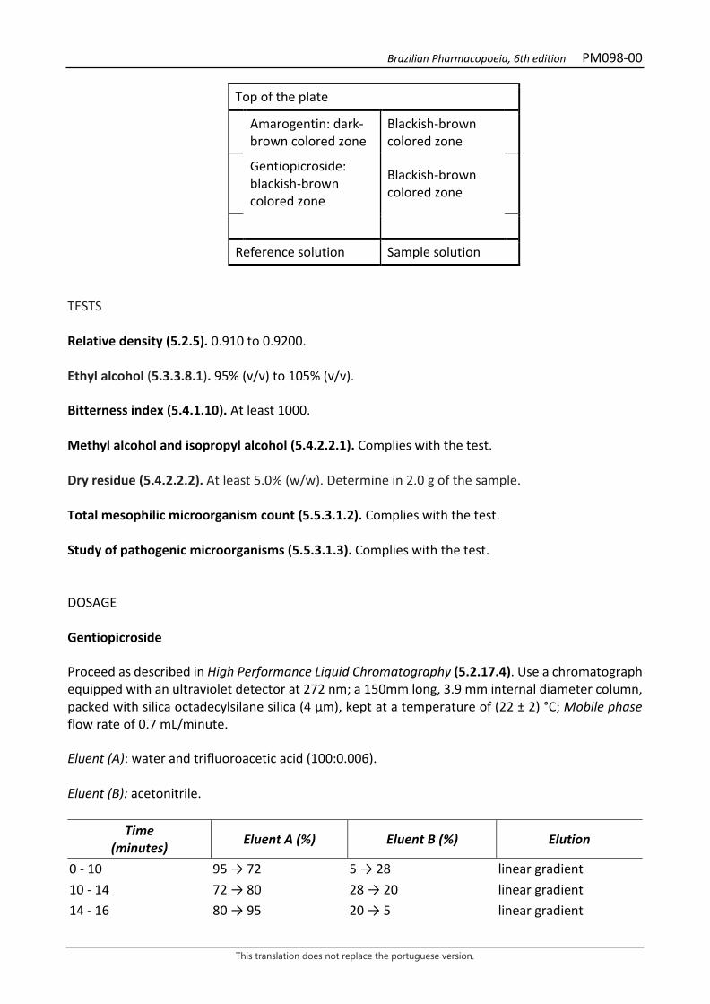
Brazilian Pharmacopoeia, 6th edition PM098-00
This translation does not replace the portuguese version.
Top of the plate
Amarogentin: dark-brown colored zone
Blackish-brown colored zone
Gentiopicroside: blackish-brown colored zone
Blackish-brown colored zone
Reference solution Sample solution TESTS Relative density (5.2.5). 0.910 to 0.9200. Ethyl alcohol (5.3.3.8.1). 95% (v/v) to 105% (v/v). Bitterness index (5.4.1.10). At least 1000. Methyl alcohol and isopropyl alcohol (5.4.2.2.1). Complies with the test. Dry residue (5.4.2.2.2). At least 5.0% (w/w). Determine in 2.0 g of the sample. Total mesophilic microorganism count (5.5.3.1.2). Complies with the test. Study of pathogenic microorganisms (5.5.3.1.3). Complies with the test. DOSAGE Gentiopicroside Proceed as described in High Performance Liquid Chromatography (5.2.17.4). Use a chromatograph equipped with an ultraviolet detector at 272 nm; a 150mm long, 3.9 mm internal diameter column, packed with silica octadecylsilane silica (4 µm), kept at a temperature of (22 ± 2) °C; Mobile phase flow rate of 0.7 mL/minute. Eluent (A): water and trifluoroacetic acid (100:0.006). Eluent (B): acetonitrile.
Time (minutes) Eluent A (%) Eluent B (%) Elution
0 - 10 95 → 72 5 → 28 linear gradient 10 - 14 72 → 80 28 → 20 linear gradient 14 - 16 80 → 95 20 → 5 linear gradient
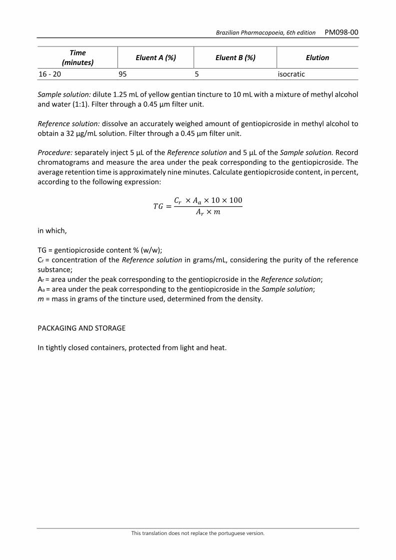
Brazilian Pharmacopoeia, 6th edition PM098-00
This translation does not replace the portuguese version.
Time (minutes) Eluent A (%) Eluent B (%) Elution
16 - 20 95 5 isocratic Sample solution: dilute 1.25 mL of yellow gentian tincture to 10 mL with a mixture of methyl alcohol and water (1:1). Filter through a 0.45 µm filter unit. Reference solution: dissolve an accurately weighed amount of gentiopicroside in methyl alcohol to obtain a 32 µg/mL solution. Filter through a 0.45 µm filter unit. Procedure: separately inject 5 μL of the Reference solution and 5 µL of the Sample solution. Record chromatograms and measure the area under the peak corresponding to the gentiopicroside. The average retention time is approximately nine minutes. Calculate gentiopicroside content, in percent, according to the following expression:
𝑇𝑇𝑇𝑇 =𝐶𝐶𝑟𝑟 × 𝑇𝑇𝑎𝑎 × 10 × 100
𝑇𝑇𝑟𝑟 × 𝑚𝑚
in which, TG = gentiopicroside content % (w/w); Cr = concentration of the Reference solution in grams/mL, considering the purity of the reference substance; Ar = area under the peak corresponding to the gentiopicroside in the Reference solution; Aa = area under the peak corresponding to the gentiopicroside in the Sample solution; m = mass in grams of the tincture used, determined from the density. PACKAGING AND STORAGE In tightly closed containers, protected from light and heat.

Brazilian Pharmacopoeia, 6th edition PM099-00
This translation does not replace the portuguese version.
GUARANA, tincture Paulliniae cupanae tincture
The tincture is obtained from dried seeds of Paullinia cupana Kunth, devoid of aril and integument (husk), containing at least 0.35% caffeine (C8H10N4O2, 194.19). PREPARATION The tincture is prepared at 10% (w/v), by maceration or percolation, using 70% (v/v) ethyl alcohol as the extracting liquid. CHARACTERISTICS Clear, reddish-brown liquid. Observe turbidity after dilution in three volumes of water. IDENTIFICATION Characterization of the presence of tannins 1. Proceed as described in Thin-layer chromatography (5.2.17.1). Stationary phase: silica gel GF254. Mobile phase: ethyl acetate, toluene and formic acid (70:30:5). Sample solution: guarana tincture. Sample solution: 1 mg/mL solution of catechin in methyl alcohol. TLC visualization reagent: dissolve 1 g of vanillin in 100 mL of methyl alcohol. Add 4 mL hydrochloric acid and 5 mL sulfuric acid. Procedure: apply 10 μL of the Sample solution and 10 μL of the Reference solution to the chromatoplate, separately and in the form of a band. Conduct chromatogram. Remove chromatoplate and allow it to air dry. Examine under ultraviolet light at 254 nm. Nebulize the plate with sulfur vanillin RS and heat at 105 °C for three minutes. Results: the diagram below shows the sequences of zones obtained with the Reference solution and the Sample solution. Other zones may occasionally develop.
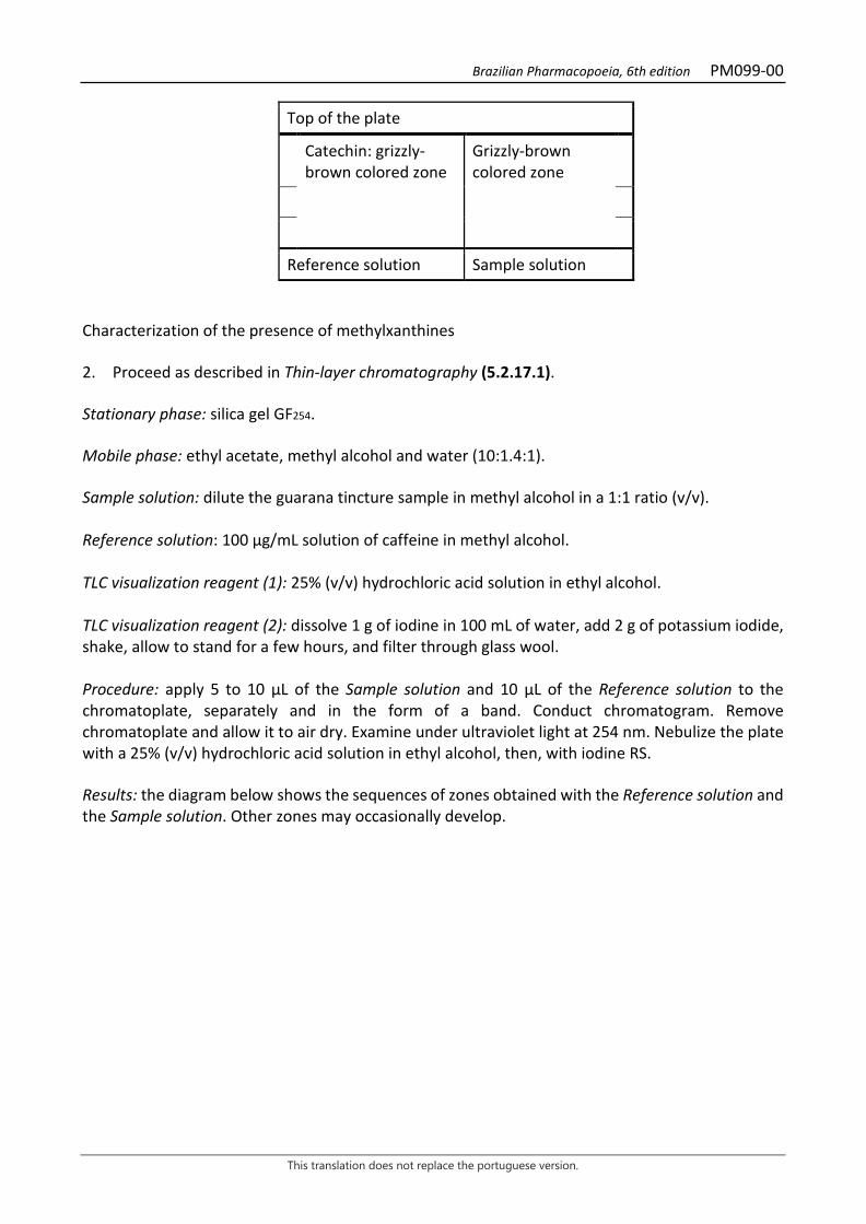
Brazilian Pharmacopoeia, 6th edition PM099-00
This translation does not replace the portuguese version.
Top of the plate
Catechin: grizzly-brown colored zone
Grizzly-brown colored zone
Reference solution Sample solution Characterization of the presence of methylxanthines 2. Proceed as described in Thin-layer chromatography (5.2.17.1). Stationary phase: silica gel GF254. Mobile phase: ethyl acetate, methyl alcohol and water (10:1.4:1). Sample solution: dilute the guarana tincture sample in methyl alcohol in a 1:1 ratio (v/v). Reference solution: 100 µg/mL solution of caffeine in methyl alcohol. TLC visualization reagent (1): 25% (v/v) hydrochloric acid solution in ethyl alcohol. TLC visualization reagent (2): dissolve 1 g of iodine in 100 mL of water, add 2 g of potassium iodide, shake, allow to stand for a few hours, and filter through glass wool. Procedure: apply 5 to 10 μL of the Sample solution and 10 μL of the Reference solution to the chromatoplate, separately and in the form of a band. Conduct chromatogram. Remove chromatoplate and allow it to air dry. Examine under ultraviolet light at 254 nm. Nebulize the plate with a 25% (v/v) hydrochloric acid solution in ethyl alcohol, then, with iodine RS. Results: the diagram below shows the sequences of zones obtained with the Reference solution and the Sample solution. Other zones may occasionally develop.

Brazilian Pharmacopoeia, 6th edition PM099-00
This translation does not replace the portuguese version.
Top of the plate
Caffeine: grizzly-brown colored zone
Grizzly-brown colored zone
Reference solution Sample solution TESTS Relative density (5.2.5). 0.8990 to 0.9150. Ethyl alcohol (5.3.3.8.1). Special treatments, Liquids with more than 30% alcohol. 66% (v/v) to 70% (v/v). Methyl alcohol and isopropyl alcohol (5.4.2.2.1). Complies with the test. Dry residue (5.4.2.2.2). At least 2.0% (w/w). Total mesophilic microorganism count (5.5.3.1.2). Complies with the test. Study of pathogenic microorganisms (5.5.3.1.3). Complies with the test. DOSAGE Caffeine Proceed as described in High Performance Liquid Chromatography (5.2.17.4). Use a chromatograph equipped with an ultraviolet detector at 272 nm; a 150mm long, 3.9 mm internal diameter column, packed with silica octadecylsilane silica (4 µm), kept at a temperature of (22 ± 2) °C; Mobile phase flow rate of 0.6 mL/minute. Isocratic system. Mobile phase: water, methyl alcohol and trifluoracetic acid (70:30:0.005). Reference solution: dissolve an accurately weighed amount of coumarin in methyl alcohol to obtain a concentration of 130 µg/mL. Filter through a 0.45 µm filter unit. Sample solution: dilute 0.3 mL of guarana tincture to 10 mL with a solution of methyl alcohol and water (1:1). Filter through a 0.45 µm filter unit. Procedure: separately inject 10 μL of the Reference solution and 10 μL of the Sample solution. Register the chromatograms and measure the areas under the peaks. The average retention time for the caffeine peak is about 8 minutes and 30 seconds. Calculate caffeine content, in percent, according to the following expression:

Brazilian Pharmacopoeia, 6th edition PM099-00
This translation does not replace the portuguese version.
𝑇𝑇𝐶𝐶 =𝐶𝐶𝑟𝑟 × 𝑇𝑇𝑎𝑎 × 10 × 100
𝑇𝑇𝑟𝑟 × 𝑚𝑚
in which, TC = caffeine content % (w/w); Cr = concentration of the Reference solution in grams/mL, considering the purity of the reference substance; Ar = area under the peak corresponding to the caffeine in the Reference solution; Aa= area under the peak corresponding to the caffeine in the Sample solution; m = mass in grams of the tincture used, determined from the density. PACKAGING AND STORAGE In tightly closed containers, protected from light and heat.

Brazilian Pharmacopoeia, 6th edition PM100-00
This translation does not replace the portuguese version.
WITCHHAZEL, tincture Hamamelidis tincture
The tincture is obtained from leaves of Hamamelis virginiana L., containing at least 0.6% total tannins, expressed in pyrogallol (C6H6O3, 126.11). PREPARATION The tincture is prepared at 10% (w/v), by maceration or percolation, using 65% (v/v) ethyl alcohol as the extracting liquid. CHARACTERISTICS Yellowish-brown colored zone IDENTIFICATION Proceed as described in Thin-layer chromatography (5.2.17.1). Stationary phase: silica gel F254 (0.25 mm). Mobile phase: ethyl acetate, toluene, formic acid and water (60:20:15:15). Sample solution: reduce 5 mL of the witch hazel tincture to dry residue in a water bath. Resume the residue in 10.0 mL of water. Extract the resulting aqueous phase with three 10-mL portions of ethyl acetate in a 125-mL separating funnel. Allow to stand in a freezer at -18 °C for 15 minutes for a complete phase separation. Collect the organic phases and rinse with 20 mL of water. Reference solution (1): weigh approximately 1 mg gallic acid and dissolve in 1mL methyl alcohol. Reference solution (2): weigh approximately 1 mg catechin and dissolve in 2 mL methyl alcohol. Procedure: apply 20 µL of the Sample solution, 10 µL of the Reference solution (1) and 10 µL of the Reference solution (2) to the chromatoplate, separately and in the form of a band. Conduct chromatogram. Remove the chromatoplate and allow it to dry in a fume hood. Then nebulize the plate with 1% ferric chloride (w/v) in methyl alcohol. Results: the diagram below shows the sequences of zones obtained with the Reference solution (1), the Reference solution (2) and the Sample solution. Other zones may occasionally develop.
Brazilian Pharmacopoeia, 6th edition PM100-00
This translation does not replace the portuguese version.
Top of the plate
Yellow-colored zone
Gallic acid: grayish-blue colored zone
Blue-gray colored zone
Catechin: grayish-blue colored zone
Blue-gray colored zone
Yellowish-brown colored zone
Yellowish-brown colored zone
Reference solution Sample solution TESTS Relative density (5.2.5). 0.902 to 0.914. Ethyl alcohol (5.3.3.8.1). 58% (v/v) to 62% (v/v). Dry residue (5.4.2.2.2). At least 1.2%. Total mesophilic microorganism count (5.5.3.1.2). Complies with the test. Study of pathogenic microorganisms (5.5.3.1.3). Complies with the test. DOSAGE Total tannins Note: protect samples from light during extraction and dilution. Use carbon dioxide-free water for all operations. To proceed as described in Visible absorption spectrophotometry (5.2.14). Prepare solutions as described below. Stock solution: accurately weigh about 1.5 g of witch hazel tincture in a 250-mL volumetric flask, top off the volume with distilled water and homogenize. Filter the mixture through paper filter. Discard the first 50 mL of the filtrate. Sample solution for total polyphenol content: volumetrically transfer 5 mL from the Stock solution to a 25-mL volumetric flask, top off the volume with water and homogenize. Volumetrically transfer 2 mL of the solution, 1 mL of phosphomolybdotungstic reagent and 10 mL of water to a 25-mL
Brazilian Pharmacopoeia, 6th edition PM100-00
This translation does not replace the portuguese version.
volumetric flask, top off the volume with 29% sodium carbonate solution (w/v) and homogenize. Determine absorbance at 760 nm (A1) after 30 minutes, using water for zero adjustment. Sample solution for polyphenols not adsorbed by hide powder: for 10 mL of the Stock solution, add 0.100 g of the hide powder and mechanically shake in a 125-mL erlenmeyer flask for 60 minutes. Filter through paper filter. Dilute 5 mL of the filtrate in a 25mL volumetric flask with water and homogenize. Volumetrically transfer 2 mL of the solution, 1 mL of phosphomolybdotungstic reagent and 10 mL of water to a 25-mL volumetric flask, top off the volume with 29% sodium carbonate solution (w/v) and homogenize. Determine absorbance at 760 nm (A2) after 30 minutes, using water for zero adjustment. Reference solution: dissolve 50 mg pyrogallol in water immediately before use, transfer to a 100-mL volumetric flask, top off the volume with water and homogenize. Volumetrically transfer 5 mL of the solution to a 100-mL volumetric flask, top off the volume with water and homogenize. Volumetrically transfer 2 mL of the solution, 1 mL of phosphomolybdotungstic reagent and 10 mL of water to a 25-mL volumetric flask, top off the volume with 29% sodium carbonate solution (w/v) and homogenize. Determine absorbance at 760 nm (A3) after 30 minutes, using water for zero adjustment. Calculate total tannin content, expressed as a percentage of pyrogallol, according to the following expression:
𝑇𝑇𝑇𝑇 =(𝑇𝑇1 − 𝑇𝑇2) × m2 × 62,5
𝑇𝑇3 × m1
in which, TT = total tannin content expressed as pyrogallol % (w/w); A1 = absorbance measured for the Sample solution for total polyphenols; A2 = absorbance measured for the Sample solution for polyphenols not adsorbed by hide powder; A3 = absorbance measured for the Reference solution; m1 = mass un grams of the tincture; m2 = mass in grams of pyrogallol, considering the purity of the reference substance. PACKAGING AND STORAGE In tightly closed containers, protected from light and heat.
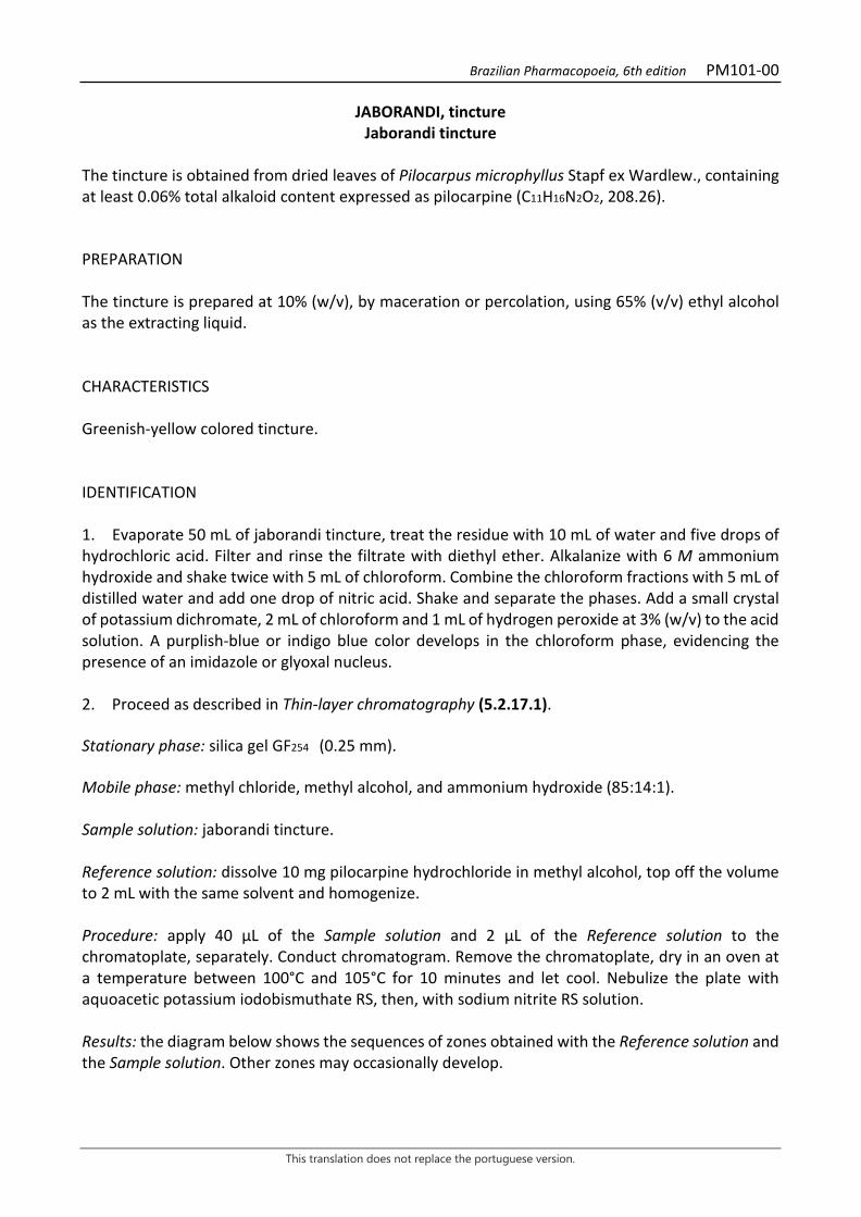
Brazilian Pharmacopoeia, 6th edition PM101-00
This translation does not replace the portuguese version.
JABORANDI, tincture Jaborandi tincture
The tincture is obtained from dried leaves of Pilocarpus microphyllus Stapf ex Wardlew., containing at least 0.06% total alkaloid content expressed as pilocarpine (C11H16N2O2, 208.26). PREPARATION The tincture is prepared at 10% (w/v), by maceration or percolation, using 65% (v/v) ethyl alcohol as the extracting liquid. CHARACTERISTICS Greenish-yellow colored tincture. IDENTIFICATION 1. Evaporate 50 mL of jaborandi tincture, treat the residue with 10 mL of water and five drops of hydrochloric acid. Filter and rinse the filtrate with diethyl ether. Alkalanize with 6 M ammonium hydroxide and shake twice with 5 mL of chloroform. Combine the chloroform fractions with 5 mL of distilled water and add one drop of nitric acid. Shake and separate the phases. Add a small crystal of potassium dichromate, 2 mL of chloroform and 1 mL of hydrogen peroxide at 3% (w/v) to the acid solution. A purplish-blue or indigo blue color develops in the chloroform phase, evidencing the presence of an imidazole or glyoxal nucleus. 2. Proceed as described in Thin-layer chromatography (5.2.17.1). Stationary phase: silica gel GF254 (0.25 mm). Mobile phase: methyl chloride, methyl alcohol, and ammonium hydroxide (85:14:1). Sample solution: jaborandi tincture. Reference solution: dissolve 10 mg pilocarpine hydrochloride in methyl alcohol, top off the volume to 2 mL with the same solvent and homogenize. Procedure: apply 40 μL of the Sample solution and 2 μL of the Reference solution to the chromatoplate, separately. Conduct chromatogram. Remove the chromatoplate, dry in an oven at a temperature between 100°C and 105°C for 10 minutes and let cool. Nebulize the plate with aquoacetic potassium iodobismuthate RS, then, with sodium nitrite RS solution. Results: the diagram below shows the sequences of zones obtained with the Reference solution and the Sample solution. Other zones may occasionally develop.
Brazilian Pharmacopoeia, 6th edition PM101-00
This translation does not replace the portuguese version.
Top of the plate
Pilocarpine: reddish-brown colored zone
Brownish-red zone
Reference solution Sample solution TESTS Ethyl alcohol (5.3.3.8.1). Distillation method, Liquids with more than 30% alcohol. (65 ± 5)% (v/v). Dry residue (5.4.2.2.2). At least 0.8%. Total mesophilic microorganism count (5.5.3.1.2). Complies with the test. Study of pathogenic microorganisms (5.5.3.1.3). Complies with the test. DOSAGE Total alkaloid content Evaporate, under vacuum, 100 g of the jaborandi tincture at low temperature, until it reduces to approximately 20 g. Quantitatively transfer the residue to a separating funnel using methyl chloride. Add 10 mL of 6 M ammonium hydroxide. Successively extract with 20 mL methyl chloride fractions until the alkaloids are completely extracted, i.e. when a few drops of the aqueous phase are no longer turbid by the addition of one drop of potassium iodide mercury RS solution. Add the organic layers and then extract several times using 0.05 M sulfuric acid. Slowly alkalinize using 6 M ammonium hydroxide to pH 9 and then extract with methyl chloride until the alkaloids are fully extracted. Rinse the combined organic solutions with 20 mL of water. Evaporate the organic fraction to about 5 mL. Dissolve the residue with 20 mL of 0.02 M hydrochloric acid SV and dry the remaining methyl chloride in a water bath at 40°C. Titrate excess hydrochloric acid with 0.02 M sodium hydroxide SV, using five drops of SI methyl red, until color changes from pink to yellow. Calculate total alkaloid content expressed as pilocarpine, in percent, according to the following expression:
𝑇𝑇𝑇𝑇 =(𝑉𝑉á𝑐𝑐𝑐𝑐𝑐𝑐𝑐𝑐 − 𝑣𝑣) × 0,4166
𝑚𝑚
in which, 𝑇𝑇𝑇𝑇 = total alkaloid content expressed as pilocarpine % (w/w); 𝑉𝑉acid = volume in milliliters of 0.02 M hydrochloric acid used; v = volume in milliliters of 0.02 M sodium hydroxide used; 𝑚𝑚 = mass in grams of the tincture used.

Brazilian Pharmacopoeia, 6th edition PM101-00
This translation does not replace the portuguese version.
PACKAGING AND STORAGE In tightly closed containers, protected from light and heat.

Brazilian Pharmacopoeia, 6th edition PM102-00
This translation does not replace the portuguese version.
BITTER ORANGE, tincture Aurantii amari exocarpium tincture
The tincture is obtained from exocarp, corresponding to the flavedo of the ripe fruit, free from most of the mesocarp, corresponding to the albedo, of Citrus aurantium L. subsp. aurantium [syn. Citrus aurantium L. subsp. amara (L.) Engler], containing at least 0.25% of naringin (C27H32O14, 580.54). PREPARATION The tincture is prepared at 10% (w/v), by percolation or maceration, using 70% (v/v) ethyl alcohol as the extracting liquid. CHARACTERISTICS Yellowish-brown or reddish-brown liquid with a citrus odor. IDENTIFICATION Proceed as described in Thin-layer chromatography (5.2.17.1). Stationary phase: silica gel G60. Mobile phase: ethyl acetate, water and formic acid (75:15:10). Sample solution: bitter orange tincture. Reference solution: prepare a 10mg/mL solution of naringin in methyl alcohol. TLC visualization reagent (1): dissolve 1 g of aminoethanol diphenylborate in methyl alcohol and top off to 100 mL with the same solvent. TLC visualization reagent (2): 5% macrogol 400 solution in methyl alcohol. Procedure: apply 10 μL of the Sample solution and 10 μL of the Reference solution to the chromatoplate, separately and in the form of a band. Conduct chromatogram. Remove chromatoplate and allow it to air dry for 15 minutes. Nebulize the plate with aminoethanol diphenylborate RS (natural reagent A), then with a solution of 5% macrogol 400 in methyl alcohol. Examine under ultraviolet light at 365 nm after at least two hours. Results: the zones obtained with the Reference solution and the Sample solution are represented in the diagram below. Other zones may occasionally be present.
Brazilian Pharmacopoeia, 6th edition PM102-00
This translation does not replace the portuguese version.
Top of the plate
Naringin: dark-green fluorescence zone
Dark-green fluorescence zone
Reference solution Sample solution
TESTS Relative density (5.2.5). 0.9030 to 0.9180. Ethyl alcohol (5.3.3.8.1). 95% (v/v) to 105% (v/v). Methyl alcohol and isopropyl alcohol (5.4.2.2.1). Complies with the test. Dry residue (5.4.2.2.2). At least 4.5% (w/w). Determine in 2.0 g of the sample. Total mesophilic microorganism count (5.5.3.1.2). Complies with the test. Study of pathogenic microorganisms (5.5.3.1.3). Complies with the test. DOSAGE Naringin Proceed as described in High Performance Liquid Chromatography (5.2.17.4). Use a chromatograph equipped with an ultraviolet detector at 284 nm; a 150mm long, 4.6 mm internal diameter column, packed with silica octadecylsilane silica (5 μm), kept at a temperature of (22 ± 2) °C; Mobile phase flow rate of 0.5 mL/minute. Eluent (A): water-formic acid (100:0.1). Eluent (B): methyl alcohol
Time (minutes) Eluent A (%) Eluent B (%) Elution
0 - 3 80 20 isocratic 3 - 33 80 → 0 20 → 100 linear gradient 33 - 34 0 → 80 100 → 20 linear gradient 34 - 40 80 20 isocratic
Brazilian Pharmacopoeia, 6th edition PM102-00
This translation does not replace the portuguese version.
Sample solution: dilute 0.3 mL of bitter yellow tincture in 5 mL with a mixture of methyl alcohol and water (1:1). Filter through a 0.45 µm filter unit. Reference solution: dissolve a precisely weighed amount of naringin in a solution of methyl alcohol and water (1:1), in order to obtain a 0.225 mg/mL solution. Filter through a 0.45 µm filter unit. Procedure: separately inject 10 μL of the Reference solution and 10 µL of the Sample solution. Record chromatograms and measure the area under the peak corresponding to the naringin. The average retention time is approximately 17.6 minutes. Calculate naringin content, in percent, according to the following expression:
𝑇𝑇𝑇𝑇 =𝐶𝐶𝑟𝑟 × 𝑇𝑇𝑎𝑎 × 5 × 100
𝑇𝑇𝑟𝑟 × 𝑚𝑚
in which, TN = naringin content % (w/w); Cr = concentration of the Reference solution in g/mL, considering the purity of the reference substance; Ar= area under the peak corresponding to naringin in the Reference solution. Aa = area corresponding to naringin in the Sample solution; m = mass in grams of the tincture, determined from the density. PACKAGING AND STORAGE In tightly closed containers, protected from light and heat.
Brazilian Pharmacopoeia, 6th edition PM103-00
This translation does not replace the portuguese version.
NUX-VOMICA, tincture Strychni tincture
The tincture is obtained from dried seeds of Strychnos nux-vomica L., containing at least 0.05% strychnine (C21H22 N2O2, 334.42). PREPARATION The tincture is prepared at 10% (w/v), by percolation or maceration, using 70% (v/v) ethyl alcohol as the extracting liquid. CHARACTERISTICS Dark brown colored liquid. IDENTIFICATION Proceed as described in Thin-layer chromatography (5.2.17.1). Stationary phase: silica gel F254 (0.25 mm). Mobile phase: butyl alcohol, water and glacial acetic acid (70:20:10). Sample solution: dry 1.0 mL of the tincture until residue is formed, in a water bath, at a maximum temperature of 60 °C. Suspend the residue in 5 mL methyl alcohol, filter through a 0.45 µm filter unit and proceed to chromatographic analysis. Reference solution (1): dissolve an accurately weighed amount of strychnine in methyl alcohol to obtain a concentration of 500 µg/mL. Reference solution (2): dissolve an accurately weighed amount of brucine in methyl alcohol to obtain a concentration of 500 µg/mL. Procedure: apply 20 μL of the Sample solution, 20 μL of the Reference solution (1) and 20 μL of the Reference solution (2) to the chromatoplate, separately and in the form of a band. Conduct chromatogram. Remove chromatoplate and allow it to air dry. Nebulize the plate with a 10% sulfuric acid solution in ethyl alcohol, then, with potassium iodide reagent and bismuth subnitrate RS. Remove the chromatoplate and allow to air dry for five minutes; examine under visible light. Results: the diagram below shows the sequences of zones obtained with the Sample Solution, Reference Solution (1) and Reference solution (2). Other zones may occasionally develop.

Brazilian Pharmacopoeia, 6th edition PM103-00
This translation does not replace the portuguese version.
Top of the plate
Strychnine: orange colored zone
Orange-colored zone
Orange-colored zone
Brucine: orange-colored zone
Orange-colored zone
Reference solution Sample solution TESTS Relative density (5.2.5). 0.900 to 0.9170. Ethyl alcohol (5.3.3.8.1). Method II. 57% (v/v) to 60% (v/v). Methyl alcohol and isopropyl alcohol (5.4.2.2.1). Complies with the test. Dry residue (5.4.2.2.2). At least 2.0% (w/w). Total mesophilic microorganism count (5.5.3.1.2). Complies with the test. Study of pathogenic microorganisms (5.5.3.1.3). Complies with the test. DOSAGE Strychnine Proceed as described in High Performance Liquid Chromatography (5.2.17.4). Use a chromatograph equipped with an ultraviolet detector at 210 nm; pre-column packed with octadecylsilane silica, 150 mm long, 4.6 mm internal diameter column, packed with octadecylsilane silica (5 µm), kept at 30 °C; Mobile phase flow rate of 1.3 mL/minute. Isocratic system. Mobile phase: dibasic potassium phosphate buffer (7 g/L), pH 3,00 adjusted with phosphoric acid, acetonitrile and diethylamine (90:10:2). Reference solution: weight 10.0 mg of strychnine. Transfer 10.0 mL to a volumetric flask, top off the volume with the Mobile phase and homogenize. Transfer 1.0 mL of this solution to a 5-mL volumetric flask, top off the volume with the Mobile phase and homogenize. Filter the solution through a 0.45 µm filter unit. Sample solution: homogenize the tincture in an ultrasonic bath for five minutes, pipette 1.0 mL of the tincture and transfer to a 5-mL volumetric flask. Rinse the micropipette tip at least twice with the Mobile Phase. Top off the volume with the Mobile phase and homogenize. Filter the solution through a 0.45 µm filter unit.

Brazilian Pharmacopoeia, 6th edition PM103-00
This translation does not replace the portuguese version.
Procedure: separately inject 10 μL of the Reference solution and 10 µL of the Sample solution. Register the chromatograms and measure the areas under the strychnine peaks. Calculate strychnine content, in percent, according to the following expression:
𝑇𝑇𝑇𝑇 =𝑇𝑇𝑎𝑎 × 𝐶𝐶𝑟𝑟𝑇𝑇𝑟𝑟 × 𝐶𝐶𝑎𝑎
in which, TE = strychnine content % (w/w); Ar= area under the peak corresponding to the strychnine in the Reference solution; Aa = area under the peak corresponding to the strychnine in the Sample solution; Cr = concentration of strychnine in the Reference solution in mg/mL, considering the purity of the reference substance; Ca = concentration of the plant drug in the Sample solution in mg/mL. PACKAGING AND STORAGE In tightly closed containers, protected from light and heat.

Brazilian Pharmacopoeia, 6th edition PM104-00
This translation does not replace the portuguese version.
RHATANY, tincture Ratanhiae tincture
The tincture is obtained from dried roots of Krameria lappacea (Dombey) Burdet & B.B. Simpson (syn. Krameria triandra Ruiz & Pav.), containing at least 0.5% tannins, expressed in pyrogallol (C6H6O3,126.11). PREPARATION The tincture is prepared at 10% (w/v), by maceration or percolation, using 70% (v/v) ethyl alcohol as the extracting liquid. CHARACTERISTICS The tincture has a reddish-brown color. IDENTIFICATION Proceed as described in Thin-layer chromatography (5.2.17.1). Stationary phase: silica gel F254 (0.250 mm). Mobile phase: ethyl acetate, toluene, formic acid and water (60:20:15:15). Sample solution: reduce 5,0 mL of the tincture to dry residue in a water bath. Resume the residue in 10.0 mL of water. Extract the resulting aqueous phase with three 10.0-mL portions of ethyl acetate in a separating funnel. Allow to stand in a freezer (-18 °C) for 15 minutes for a complete phase separation. Collect the organic fractions and rinse with 20.0 mL of water. Reference solution: weigh approximately 1 mg catechin and dissolve in 2 mL methyl alcohol. Procedure: apply 20 μL of the Sample solution and 10 μL of the Reference solution to the chromatoplate, separately and in the form of a band. Conduct chromatogram. Remove the chromatoplate and allow it to dry in a fume hood. Then nebulize the plate with 1% ferric chloride in methyl alcohol (w/v). Results: the diagram below shows the sequences of zones obtained with the Reference solution and the Sample solution. Other zones may occasionally develop.
Brazilian Pharmacopoeia, 6th edition PM104-00
This translation does not replace the portuguese version.
Top of the plate
Brownish-red zone
Catechin: brownish-blue zone
Brownish-blue zone
Brownish-red zone
Brownish-red zone
Reference solution Sample solution TESTS Relative density (5.2.5). 0.891 to 0.906. Ethyl alcohol (5.3.3.8.1). 63 to 67. Dry residue (5.4.2.2.2). At least 1.9%. Total mesophilic microorganism count (5.5.3.1.2). Complies with the test. Study of pathogenic microorganisms (5.5.3.1.3). Complies with the test. DOSAGE Total tannins Note: protect samples from light during extraction and dilution. Use carbon dioxide-free water for all operations. To proceed as described in Visible absorption spectrophotometry (5.2.14). Prepare solutions as described below. Stock solution: quantitatively transfer about 1.5 g of precisely weighed tincture to a 250-mL volumetric flask, top off the volume with water and homogenize. Filter the mixture through paper filter. Discard the first 50.0 mL of the filtrate. Sample solution for total polyphenol content: volumetrically transfer 5 mL of the filtrate from the Stock solution to a 25-mL volumetric flask, top off the volume with water and homogenize. Volumetrically transfer 2 mL of the solution, 1 mL of phosphomolybdotungstic reagent and 10 mL of water to a 25-mL volumetric flask, top off the volume with 29% sodium carbonate solution (w/v) and homogenize.

Brazilian Pharmacopoeia, 6th edition PM104-00
This translation does not replace the portuguese version.
Sample solution for polyphenols not adsorbed by hide powder: for 10 mL of the filtrate from the Stock solution, add 0.1 g of the hide powder and mechanically shake in a 125-mL Erlenmeyer flask for 60 minutes. Filter through paper filter. Volumetrically transfer 5 mL of this filtrate to a 25-mL volumetric flask, top off the volume with water and homogenize. Volumetrically transfer 2 mL of the solution, 1 mL of phosphomolybdotungstic reagent and 10 mL of water to a 25-mL volumetric flask, top off the volume with 29% sodium carbonate solution (w/v) and homogenize. Reference solution: Dissolve 50.0 mg of pyrogallol in water, transfer quantitatively to a 100-mL volumetric flask, top off the volume with water and homogenize. Volumetrically transfer 5 mL of this solution to a 100-mL volumetric flask, top off the volume with water and homogenize. Volumetrically transfer 2 mL of the solution, 1 mL of phosphomolybdotungstic reagent and 10 mL of water to a 25-mL volumetric flask, top off the volume with 29% sodium carbonate solution (w/v) and homogenize. Procedure: measure the absorbance of the Sample Solution for total polyphenols (A1), Sample Solution for polyphenols not adsorbed by hide powder (A2) and Reference Solution (A3) at 760 nm, 30 minutes after preparation, using water for zero adjustment. Calculate tannin content expressed as pyrogallol, in percent, according to the following expression:
𝑇𝑇𝑇𝑇 =(𝑇𝑇1 − 𝑇𝑇2) × 𝑚𝑚2 × 62,5
𝑇𝑇3 × 𝑚𝑚3
in which, TT = total tannin content expressed as pyrogallol % (w/w); A1 = absorbance measured for the Sample solution for total polyphenols; A2 = absorbance measured for the Sample solution for polyphenols not adsorbed by hide powder; A3 = absorbance measured for the Reference solution; m1 = mass un grams of the tincture used; m2 = mass in grams of pyrogallol, considering the purity of the reference substance. PACKAGING AND STORAGE In tightly closed containers, protected from light and heat.
Brazilian Pharmacopoeia, 6th edition PM105-00
This translation does not replace the portuguese version.
VALERIAN, tincture Valerianae tincture
The tincture is obtained from the underground organs (roots, rhizomes and stolons), dried, of Valeriana officinalis L., containing at least 0.015% (w/w) of total sesquiterpene acids, expressed in valerenic acid (C15H22O2, 234.34). PREPARATION The tincture is prepared at 20% (w/v), by maceration or percolation, using 70% (v/v) ethyl alcohol as the extracting liquid. CHARACTERISTICS Dark-brown liquid. IDENTIFICATION Proceed as described in Thin-layer chromatography (5.2.17.1). Stationary phase: silica gel F254. Mobile phase: cyclohexane, ethyl acetate and glacial acetic acid (60:38:2). Sample solution: measure 1 mL of tincture and dry until a residue is formed in a water bath, at a maximum temperature of 60 °C. Suspend the residue in 1 mL methyl alcohol and proceed to the chromatographic analysis. Reference solution (1): dissolve an exactly weighed amount of valerenic acid in methyl alcohol to obtain the concentration of 100 µg/mL. Reference solution (2): dissolve an exactly weighed amount of acetoxyvalerenic acid in methyl alcohol to obtain the concentration of 100 µg/mL. Procedure: apply 15 μL of the Sample solution, 15 μL of the Reference solution (1) and 15 μL of the Reference solution (2) to the chromatoplate, separately and in the form of a band. Conduct chromatogram. Remove chromatoplate and allow it to air dry. Nebulize the plate with anisaldehyde RS and heat at 100 °C to 105 °C for approximately five minutes. Examine under visible light. Results: the diagram below shows the sequences of zones obtained with the Reference Solution (1), the Reference solution (2) and the Sample solution. Other zones may occasionally develop.
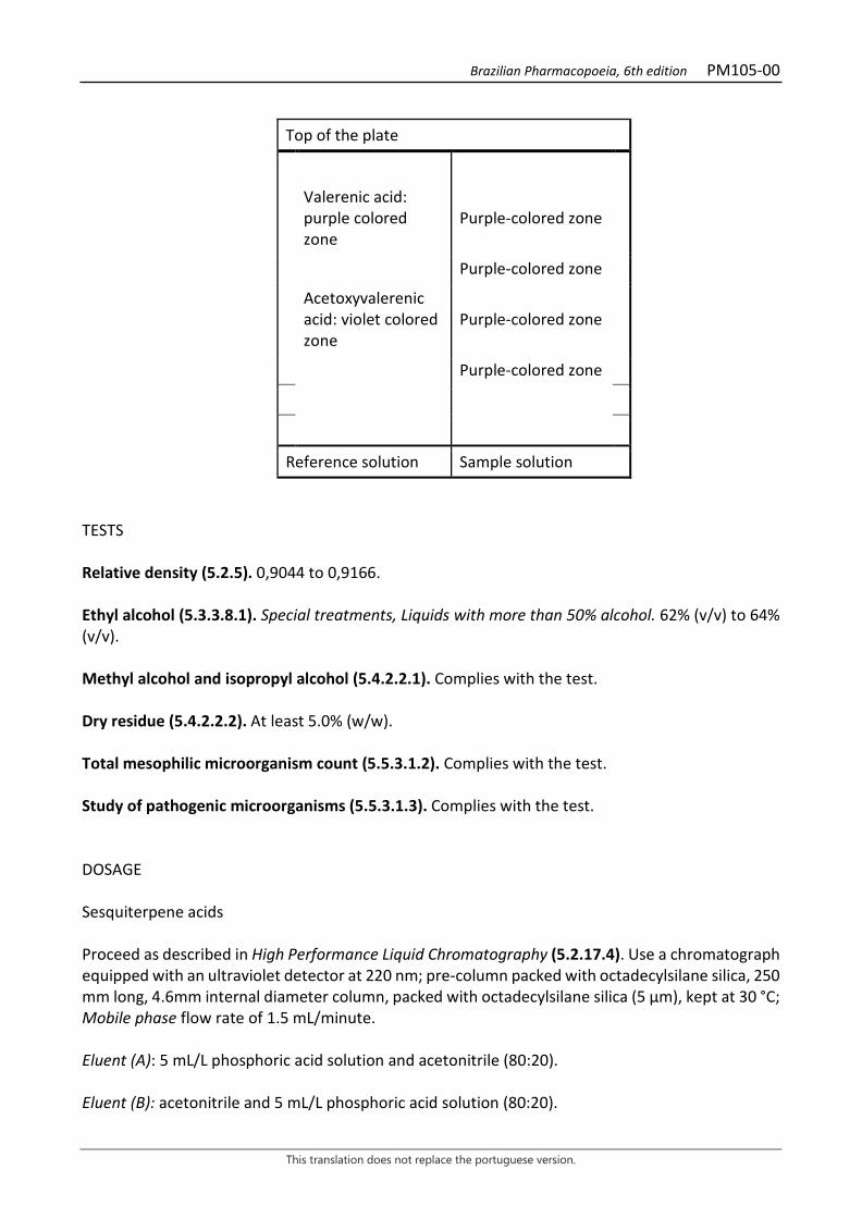
Brazilian Pharmacopoeia, 6th edition PM105-00
This translation does not replace the portuguese version.
Top of the plate
Valerenic acid: purple colored zone
Purple-colored zone
Purple-colored zone
Acetoxyvalerenic acid: violet colored zone
Purple-colored zone
Purple-colored zone
Reference solution Sample solution TESTS Relative density (5.2.5). 0,9044 to 0,9166. Ethyl alcohol (5.3.3.8.1). Special treatments, Liquids with more than 50% alcohol. 62% (v/v) to 64% (v/v). Methyl alcohol and isopropyl alcohol (5.4.2.2.1). Complies with the test. Dry residue (5.4.2.2.2). At least 5.0% (w/w). Total mesophilic microorganism count (5.5.3.1.2). Complies with the test. Study of pathogenic microorganisms (5.5.3.1.3). Complies with the test. DOSAGE Sesquiterpene acids Proceed as described in High Performance Liquid Chromatography (5.2.17.4). Use a chromatograph equipped with an ultraviolet detector at 220 nm; pre-column packed with octadecylsilane silica, 250 mm long, 4.6mm internal diameter column, packed with octadecylsilane silica (5 µm), kept at 30 °C; Mobile phase flow rate of 1.5 mL/minute. Eluent (A): 5 mL/L phosphoric acid solution and acetonitrile (80:20). Eluent (B): acetonitrile and 5 mL/L phosphoric acid solution (80:20).
Brazilian Pharmacopoeia, 6th edition PM105-00
This translation does not replace the portuguese version.
Time (minutes) Eluent A (%) Eluent B (%) Elution
0 - 5 55 45 isocratic 5 - 15 55 → 20 45 → 80 linear gradient 15 - 25 20 80 isocratic 25 - 28 20 → 55 80 → 45 linear gradient 28 - 30 55 45 isocratic Sample solution: transfer 5.0 mL of the tincture to a 10-mL volumetric flask, top off the volume with methyl alcohol and homogenize. Filter through a 0.45 µm filter unit. Reference solution (50): dissolve an exactly weighed amount of valerenic acid in methyl alcohol to obtain the concentration of 100 µg/mL. Filter through a 0.45 µm filter unit. Procedure: separately inject 20 μL of the Reference solution and 20 µL of the Sample solution. Register the chromatograms and measure the areas under the peaks. The acetoxyvalerenic acid peak is identified by calculating the relative retention time, using valerenic acid as a reference. Acetoxyvalerenic acid relative retention time is approximately 0.6. Calculate the sesquiterpene acid content, in percent, according to the expression:
𝑇𝑇𝑇𝑇𝑇𝑇𝑇𝑇 =𝐶𝐶𝑟𝑟 × (𝑇𝑇1 − 𝑇𝑇2) × 2 × 100
𝑇𝑇𝑟𝑟 × 𝑚𝑚 × 100
in which, TAST = sesquiterpene acid content % (w/w); Cr = concentration of valerenic acid in the Reference solution in g/mL, considering the purity of the reference substance; Ar = area under the peak corresponding to the valerenic acid in the Reference solution; A1 = area under the peak corresponding to acetoxyvalerenic acid in the Sample solution; A2 = area under the peak corresponding to the valerenic acid in the Sample solution; m = mass in grams of the tincture used, determined from the density. 2 = dilution factor. PACKAGING AND STORAGE In tightly closed containers, protected from light and heat.
Brazilian Pharmacopoeia, 6th edition
This translation does not replace the portuguese version.
PLANT-BASED PREPARATIONS – FLUID EXTRACT
Brazilian Pharmacopoeia, 6th edition PM106-00
This translation does not replace the portuguese version.
ARTICHOKE, fluid extract Cynarae extracta fluida
The fluid extract is obtained from dried leaves of Cynara scolymus L., containing at least 0.7% chlorogenic acid (C16H18O9, 354.31). PREPARATION The fluid extract is prepared in a 1:1 (w/v) drug:solvent ratio by percolation, using 70% (v/v) ethyl alcohol as the extracting liquid. CHARACTERISTICS Dark-green colored liquid. IDENTIFICATION Proceed as described in Thin-layer chromatography (5.2.17.1). Stationary phase: silica gel GF254 (0.25 mm). Mobile phase: ethyl acetate, water, acetic acid and formic acid (100:26:11:11). Sample solution: dry 1mL of fluid extract until residue is formed, in a water bath at a temperature not exceeding 60°C. Suspend the residue in 5 mL methyl alcohol and filter through a 0.45µm filter unit. Reference solution (1): dissolve chlorogenic acid in methyl alcohol to obtain a concentration of 500 µg/mL. Reference solution (2): dissolve luteolin-7-O-glycoside in methyl alcohol to obtain a concentration of 500 µg/mL. Procedure: apply 20 μL of the Sample solution, 20 μL of the Reference solution (1) and 20 μL of the Reference solution (2) to the chromatoplate, separately and in the form of a band. Conduct chromatogram. Remove chromatoplate and allow it to air dry. Nebulize the plate with aminoethanol diphenylborate RS, then with a solution of 5% (w/v) macrogol 400 in ethyl alcohol. Heat the plate between 100 °C and 105 °C for about five minutes. Examine under ultraviolet light at 365 nm. Results: the diagram below shows the sequences of zones obtained with the Reference Solution (1), the Reference solution (2) and the Sample solution. Other zones may occasionally develop.
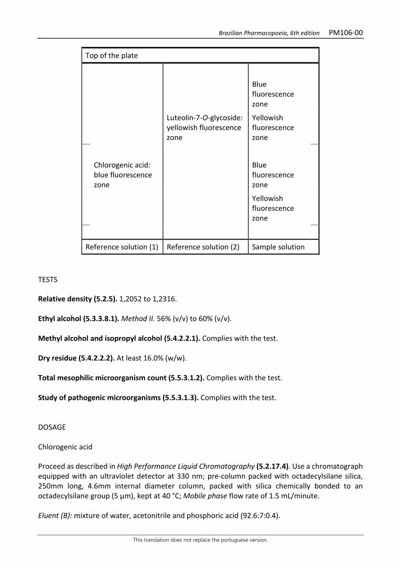
Brazilian Pharmacopoeia, 6th edition PM106-00
This translation does not replace the portuguese version.
Top of the plate
Blue fluorescence zone
Luteolin-7-O-glycoside: yellowish fluorescence zone
Yellowish fluorescence zone
Chlorogenic acid: blue fluorescence zone
Blue fluorescence zone
Yellowish fluorescence zone
Reference solution (1) Reference solution (2) Sample solution
TESTS Relative density (5.2.5). 1,2052 to 1,2316. Ethyl alcohol (5.3.3.8.1). Method II. 56% (v/v) to 60% (v/v). Methyl alcohol and isopropyl alcohol (5.4.2.2.1). Complies with the test. Dry residue (5.4.2.2.2). At least 16.0% (w/w). Total mesophilic microorganism count (5.5.3.1.2). Complies with the test. Study of pathogenic microorganisms (5.5.3.1.3). Complies with the test. DOSAGE Chlorogenic acid Proceed as described in High Performance Liquid Chromatography (5.2.17.4). Use a chromatograph equipped with an ultraviolet detector at 330 nm; pre-column packed with octadecylsilane silica, 250mm long, 4.6mm internal diameter column, packed with silica chemically bonded to an octadecylsilane group (5 µm), kept at 40 °C; Mobile phase flow rate of 1.5 mL/minute. Eluent (B): mixture of water, acetonitrile and phosphoric acid (92.6:7:0.4).

Brazilian Pharmacopoeia, 6th edition PM106-00
This translation does not replace the portuguese version.
Eluent (B): mixture of acetonitrile and phosphoric acid (99.6:0.4).
Time (minutes) Eluent A (%) Eluent B (%) Elution
0 – 17 100 0 Isocratic 17 – 50 100 → 80 0 → 20 linear gradient 50 – 51 80 → 0 20 → 100 linear gradient 51 – 61 0 100 Isocratic 61 – 62 0 → 100 100 → 0 linear gradient 62 – 72 100 0 Isocratic Sample solution: homogenize the sample in an ultrasonic bath for 10 minutes, pipette 0.5 mL of the fluid extract and transfer quantitatively to a 200-mL volumetric flask. Add 100 mL methyl alcohol and conduct ultrasound again for 10 minutes. Top off the volume with water and homogenize. Filter through a 0.45 µm filter unit. Stock solution: accurately weigh about 5.0 mg of chlorogenic acid. Transfer to a 50-mL volumetric flask and top off the volume with methyl alcohol. Reference solution: transfer 5.0 mL of the Stock solution to a 20-mL volumetric flask, add 5 mL methyl alcohol and top off the volume with water. Filter through a 0.45 µm filter unit. Procedure: separately inject 10 μL of the Reference solution and 10 µL of the Sample solution. Record the chromatograms and measure the areas under the chlorogenic acid, caffeic acid and cynarine peaks. Relative retention times are about 1.0 for chlorogenic acid, 1.21 for caffeic acid, 4.14 for cynarine, identified in the Sample solution. Calculate chlorogenic acid content in percent, according to the expression:
𝑇𝑇𝑇𝑇 =𝑇𝑇𝑎𝑎 − 𝑚𝑚𝑇𝑇𝑟𝑟 × 𝑚𝑚𝑎𝑎
in which, TA = chlorogenic acid content % (w/w) Aa = area under the peak corresponding to chlorogenic acid in the Sample solution; Ar = area under the peak corresponding to the chlorogenic acid in the Reference solution; mr = mass in grams of chlorogenic acid, considering the purity of the blank substance; ma = mass in grams of the fluid extract used, determined from the density. PACKAGING AND STORAGE In tightly closed containers, protected from light and heat.
Brazilian Pharmacopoeia, 6th edition PM107-00
This translation does not replace the portuguese version.
LICORICE, fluid extract Liquiritiae extracta fluida
The fluid extract is obtained from dried roots and stolons of Glycyrrhiza glabra L., containing at least 2.5% (w/w) glycyrrhizinic acid (C42H62O16, 822.94). PREPARATION The fluid extract is prepared in the drug: solvent 1:1 (w/v) ratio by employing a mixture of water and 90% (v/v) ethyl alcohol sufficient to obtain an extract with a final concentration of approximately 20% ethyl alcohol. CHARACTERISTICS Dark brown liquid. IDENTIFICATION Proceed as described in Thin-layer chromatography (5.2.17.1). Stationary phase: silica gel F254. Mobile phase: butyl alcohol, water and glacial acetic acid (70:20:10). Sample solution: dry 0.5 mL of the fluid extract until residue is formed, in a water bath, at a temperature not exceeding 60 °C. Add 5 mL methyl alcohol and filter through a 0.45 µm filter unit. Reference solution: dissolve an exactly weighed amount of glycyrrhizinic acid in 70% methyl alcohol to obtain the concentration of 1000 µg/mL. Procedure: apply 20 μL of the Sample solution and 20 μL of the Reference solution to the chromatoplate, separately and in the form of a band. Conduct chromatogram. Remove chromatoplate and allow it to air dry. Nebulize the plate with anisaldehyde RS and heat at 100 °C to 105 °C for approximately five minutes. Examine under visible light. Results: the diagram below shows the sequences of zones obtained with the Reference solution and the Sample solution. Other zones may occasionally develop.
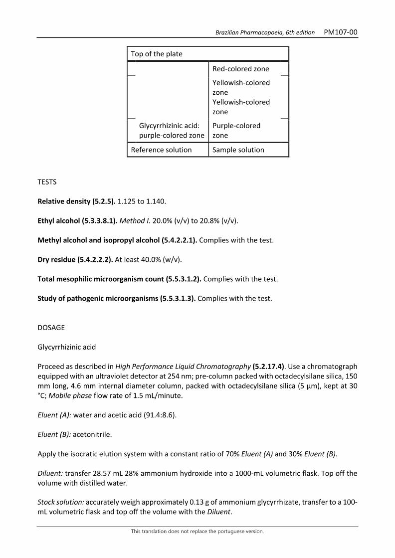
Brazilian Pharmacopoeia, 6th edition PM107-00
This translation does not replace the portuguese version.
Top of the plate
Red-colored zone
Yellowish-colored zone Yellowish-colored zone
Glycyrrhizinic acid: purple-colored zone
Purple-colored zone
Reference solution Sample solution
TESTS Relative density (5.2.5). 1.125 to 1.140. Ethyl alcohol (5.3.3.8.1). Method I. 20.0% (v/v) to 20.8% (v/v). Methyl alcohol and isopropyl alcohol (5.4.2.2.1). Complies with the test. Dry residue (5.4.2.2.2). At least 40.0% (w/v). Total mesophilic microorganism count (5.5.3.1.2). Complies with the test. Study of pathogenic microorganisms (5.5.3.1.3). Complies with the test. DOSAGE Glycyrrhizinic acid Proceed as described in High Performance Liquid Chromatography (5.2.17.4). Use a chromatograph equipped with an ultraviolet detector at 254 nm; pre-column packed with octadecylsilane silica, 150 mm long, 4.6 mm internal diameter column, packed with octadecylsilane silica (5 µm), kept at 30 °C; Mobile phase flow rate of 1.5 mL/minute. Eluent (A): water and acetic acid (91.4:8.6). Eluent (B): acetonitrile. Apply the isocratic elution system with a constant ratio of 70% Eluent (A) and 30% Eluent (B). Diluent: transfer 28.57 mL 28% ammonium hydroxide into a 1000-mL volumetric flask. Top off the volume with distilled water. Stock solution: accurately weigh approximately 0.13 g of ammonium glycyrrhizate, transfer to a 100-mL volumetric flask and top off the volume with the Diluent.
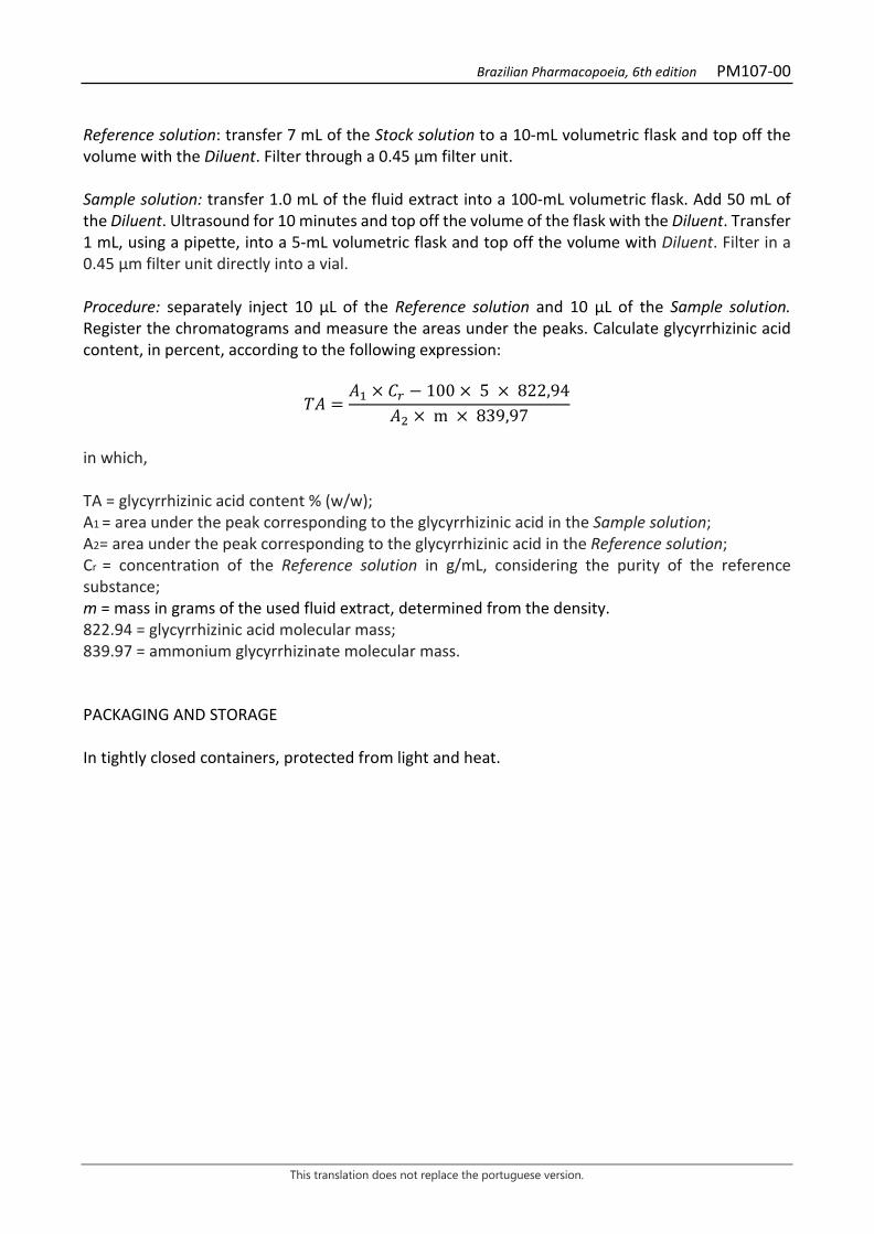
Brazilian Pharmacopoeia, 6th edition PM107-00
This translation does not replace the portuguese version.
Reference solution: transfer 7 mL of the Stock solution to a 10-mL volumetric flask and top off the volume with the Diluent. Filter through a 0.45 µm filter unit. Sample solution: transfer 1.0 mL of the fluid extract into a 100-mL volumetric flask. Add 50 mL of the Diluent. Ultrasound for 10 minutes and top off the volume of the flask with the Diluent. Transfer 1 mL, using a pipette, into a 5-mL volumetric flask and top off the volume with Diluent. Filter in a 0.45 μm filter unit directly into a vial. Procedure: separately inject 10 μL of the Reference solution and 10 µL of the Sample solution. Register the chromatograms and measure the areas under the peaks. Calculate glycyrrhizinic acid content, in percent, according to the following expression:
𝑇𝑇𝑇𝑇 =𝑇𝑇1 × 𝐶𝐶𝑟𝑟 − 100 × 5 × 822,94
𝑇𝑇2 × m × 839,97
in which, TA = glycyrrhizinic acid content % (w/w); A1 = area under the peak corresponding to the glycyrrhizinic acid in the Sample solution; A2= area under the peak corresponding to the glycyrrhizinic acid in the Reference solution; Cr = concentration of the Reference solution in g/mL, considering the purity of the reference substance; m = mass in grams of the used fluid extract, determined from the density. 822.94 = glycyrrhizinic acid molecular mass; 839.97 = ammonium glycyrrhizinate molecular mass. PACKAGING AND STORAGE In tightly closed containers, protected from light and heat.
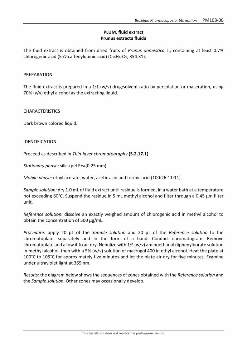
Brazilian Pharmacopoeia, 6th edition PM108-00
This translation does not replace the portuguese version.
PLUM, fluid extract Prunus extracta fluida
The fluid extract is obtained from dried fruits of Prunus domestica L., containing at least 0.7% chlorogenic acid (5-O-caffeoylquinic acid) (C16H18O9, 354.31). PREPARATION The fluid extract is prepared in a 1:1 (w/v) drug:solvent ratio by percolation or maceration, using 70% (v/v) ethyl alcohol as the extracting liquid. CHARACTERISTICS Dark brown colored liquid. IDENTIFICATION Proceed as described in Thin-layer chromatography (5.2.17.1). Stationary phase: silica gel F254(0.25 mm). Mobile phase: ethyl acetate, water, acetic acid and formic acid (100:26:11:11). Sample solution: dry 1.0 mL of fluid extract until residue is formed, in a water bath at a temperature not exceeding 60°C. Suspend the residue in 5 mL methyl alcohol and filter through a 0.45 µm filter unit. Reference solution: dissolve an exactly weighed amount of chlorogenic acid in methyl alcohol to obtain the concentration of 500 µg/mL. Procedure: apply 20 μL of the Sample solution and 20 μL of the Reference solution to the chromatoplate, separately and in the form of a band. Conduct chromatogram. Remove chromatoplate and allow it to air dry. Nebulize with 1% (w/v) aminoethanol diphenylborate solution in methyl alcohol, then with a 5% (w/v) solution of macrogol 400 in ethyl alcohol. Heat the plate at 100°C to 105°C for approximately five minutes and let the plate air dry for five minutes. Examine under ultraviolet light at 365 nm. Results: the diagram below shows the sequences of zones obtained with the Reference solution and the Sample solution. Other zones may occasionally develop.
Brazilian Pharmacopoeia, 6th edition PM108-00
This translation does not replace the portuguese version.
Top of the plate
Chlorogenic acid: blue color zone Blue-colored zone
Blue-colored zone
Reference solution Sample solution TESTS Relative density (5.2.5). 1.0966 to 1.1222. Ethyl alcohol (5.3.3.8.1). Method II. 36% (v/v) to 40% (v/v). Methyl alcohol and isopropyl alcohol (5.4.2.2.1). Complies with the test. Dry residue (5.4.2.2.2). At least 22.0% (w/w). Total mesophilic microorganism count (5.5.3.1.2). Complies with the test. Study of pathogenic microorganisms (5.5.3.1.3). Complies with the test. DOSAGE Chlorogenic acid dosage: Proceed as described in High Performance Liquid Chromatography (5.2.17.4). Use a chromatograph equipped with an ultraviolet detector at 330 nm; pre-column packed with octadecylsilane silica, 250 mm long, 4.6 mm internal diameter column, packed with octadecylsilane silica (5 µm), kept at 40 °C; Mobile phase flow rate of 1.2 mL/minute. Eluent (A): water and phosphoric acid (99.5:0.5). Eluent (B): acetonitrile and phosphoric acid (99.6:0.4).
Time (minutes) Eluent A (%) Eluent B (%) Elution
0 – 1 92 8 isocratic 1 – 20 92 → 75 8 → 25 linear gradient 20 – 33 75 25 isocratic 33 – 35 75 → 0 25 → 100 linear gradient 35 – 36 75 → 92 100 → 8 linear gradient 36 – 40 92 8 isocratic

Brazilian Pharmacopoeia, 6th edition PM108-00
This translation does not replace the portuguese version.
Sample solution: homogenize the sample in an ultrasonic bath for five minutes, pipette 1.0 mL of the fluid extract and transfer to a 5-mL volumetric flask. Rinse the micropipette tip at least twice with methyl alcohol. Top off the volume with methyl alcohol and homogenize. Filter through a 0.45 µm filter unit. Reference solution: accurately weigh about 12 mg of chlorogenic acid. Transfer to a 100-mL volumetric flask and dilute with methyl alcohol. Transfer 1.2 mL of this solution to a 10-mL volumetric flask and top off the volume with methyl alcohol. Filter through a 0.45 µm filter unit. Procedure: separately inject 20 μL of the Reference solution and 20 µL of the Sample solution. Register the chromatograms and measure the areas under the chlorogenic acid peaks. Calculate chlorogenic acid content in percent, according to the expression:
𝑇𝑇𝑇𝑇𝐶𝐶 =𝑇𝑇𝑎𝑎 × 𝑚𝑚𝑟𝑟
𝑇𝑇𝑟𝑟 × 𝑚𝑚𝑎𝑎
in which, TAC = chlorogenic acid content % (w/w) Ar = area under the peak corresponding to the chlorogenic acid in the Reference solution; Aa = area under the peak corresponding to chlorogenic acid in the Sample solution; ma = mass in grams of the fluid extract used, determined from the density. mr = mass in grams of chlorogenic acid, considering the purity of the blank substance. PACKAGING AND STORAGE In tightly closed containers, protected from light and heat.
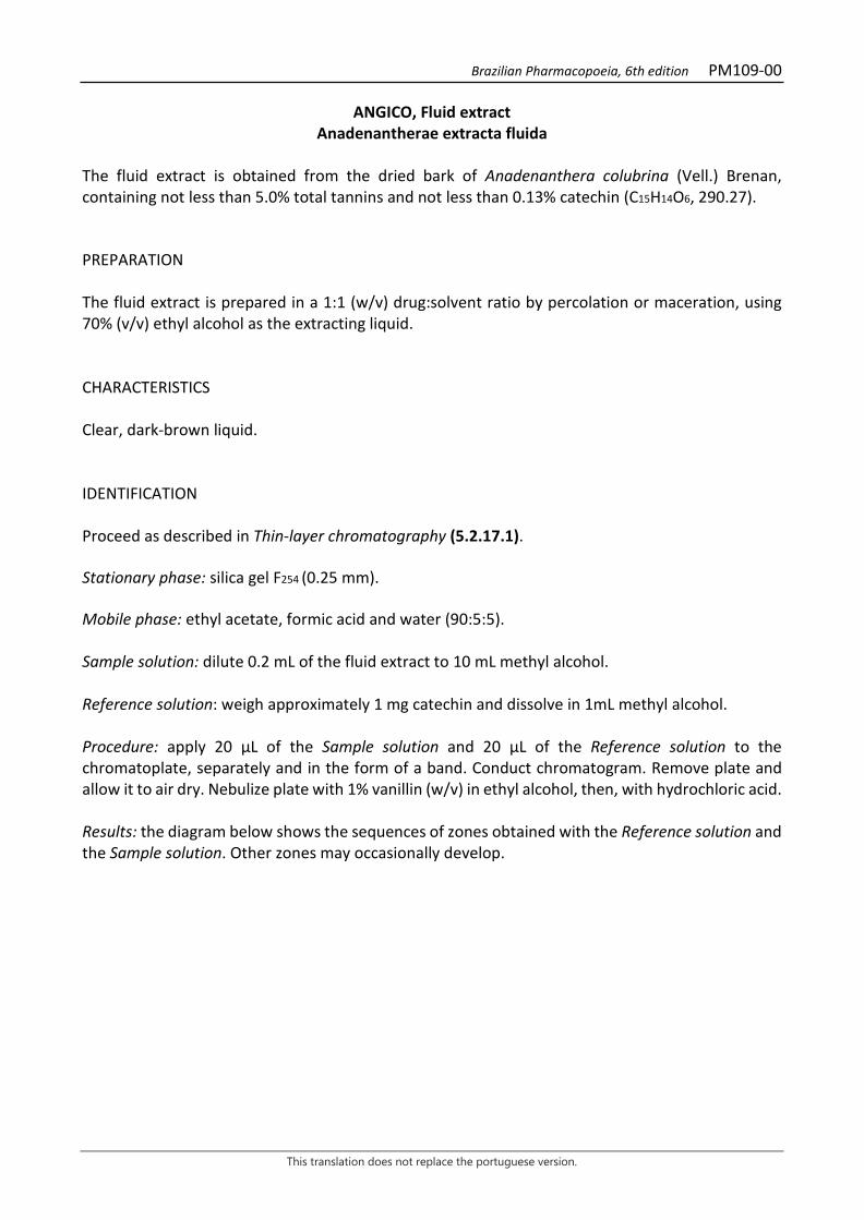
Brazilian Pharmacopoeia, 6th edition PM109-00
This translation does not replace the portuguese version.
ANGICO, Fluid extract Anadenantherae extracta fluida
The fluid extract is obtained from the dried bark of Anadenanthera colubrina (Vell.) Brenan, containing not less than 5.0% total tannins and not less than 0.13% catechin (C15H14O6, 290.27). PREPARATION The fluid extract is prepared in a 1:1 (w/v) drug:solvent ratio by percolation or maceration, using 70% (v/v) ethyl alcohol as the extracting liquid. CHARACTERISTICS Clear, dark-brown liquid. IDENTIFICATION Proceed as described in Thin-layer chromatography (5.2.17.1). Stationary phase: silica gel F254 (0.25 mm). Mobile phase: ethyl acetate, formic acid and water (90:5:5). Sample solution: dilute 0.2 mL of the fluid extract to 10 mL methyl alcohol. Reference solution: weigh approximately 1 mg catechin and dissolve in 1mL methyl alcohol. Procedure: apply 20 μL of the Sample solution and 20 μL of the Reference solution to the chromatoplate, separately and in the form of a band. Conduct chromatogram. Remove plate and allow it to air dry. Nebulize plate with 1% vanillin (w/v) in ethyl alcohol, then, with hydrochloric acid. Results: the diagram below shows the sequences of zones obtained with the Reference solution and the Sample solution. Other zones may occasionally develop.
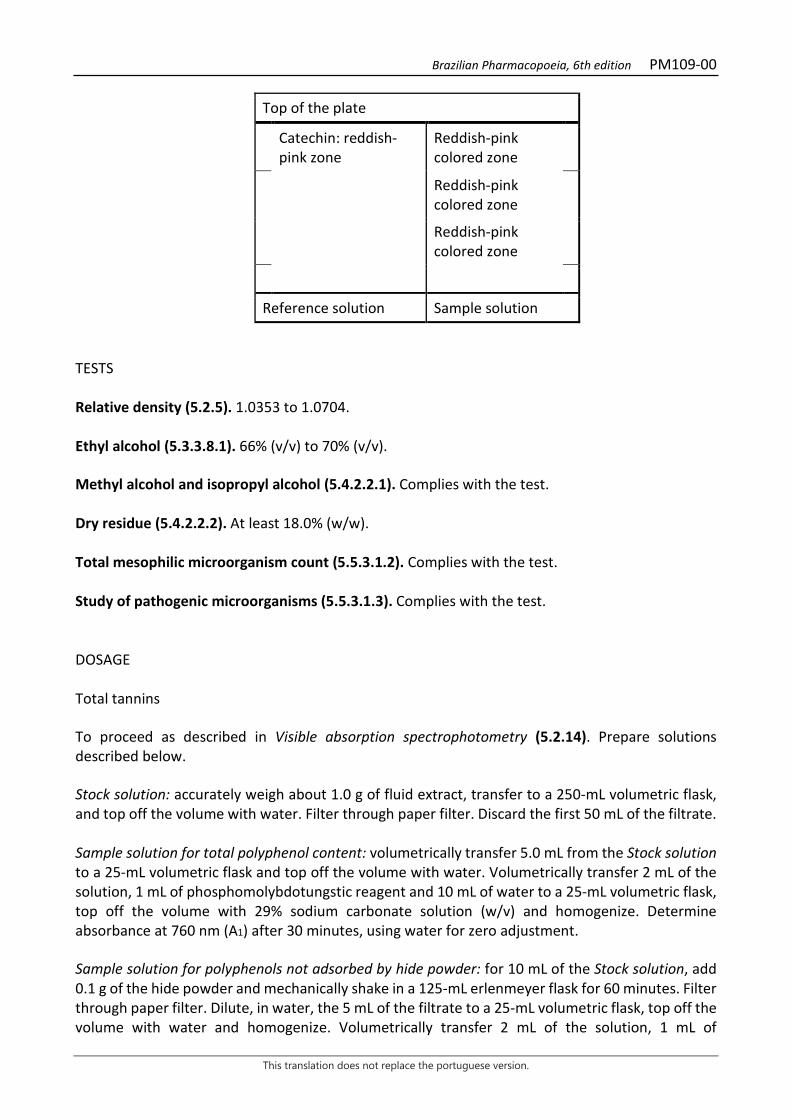
Brazilian Pharmacopoeia, 6th edition PM109-00
This translation does not replace the portuguese version.
Top of the plate
Catechin: reddish-pink zone
Reddish-pink colored zone
Reddish-pink colored zone
Reddish-pink colored zone
Reference solution Sample solution TESTS Relative density (5.2.5). 1.0353 to 1.0704. Ethyl alcohol (5.3.3.8.1). 66% (v/v) to 70% (v/v). Methyl alcohol and isopropyl alcohol (5.4.2.2.1). Complies with the test. Dry residue (5.4.2.2.2). At least 18.0% (w/w). Total mesophilic microorganism count (5.5.3.1.2). Complies with the test. Study of pathogenic microorganisms (5.5.3.1.3). Complies with the test. DOSAGE Total tannins To proceed as described in Visible absorption spectrophotometry (5.2.14). Prepare solutions described below. Stock solution: accurately weigh about 1.0 g of fluid extract, transfer to a 250-mL volumetric flask, and top off the volume with water. Filter through paper filter. Discard the first 50 mL of the filtrate. Sample solution for total polyphenol content: volumetrically transfer 5.0 mL from the Stock solution to a 25-mL volumetric flask and top off the volume with water. Volumetrically transfer 2 mL of the solution, 1 mL of phosphomolybdotungstic reagent and 10 mL of water to a 25-mL volumetric flask, top off the volume with 29% sodium carbonate solution (w/v) and homogenize. Determine absorbance at 760 nm (A1) after 30 minutes, using water for zero adjustment. Sample solution for polyphenols not adsorbed by hide powder: for 10 mL of the Stock solution, add 0.1 g of the hide powder and mechanically shake in a 125-mL erlenmeyer flask for 60 minutes. Filter through paper filter. Dilute, in water, the 5 mL of the filtrate to a 25-mL volumetric flask, top off the volume with water and homogenize. Volumetrically transfer 2 mL of the solution, 1 mL of
Brazilian Pharmacopoeia, 6th edition PM109-00
This translation does not replace the portuguese version.
phosphomolybdotungstic reagent and 10 mL of water to a 25mL volumetric flask, top off the volume with 29% anhydrous sodium carbonate solution (w/v) and homogenize. Determine absorbance at 760 nm (A2) after 30 minutes, using water for zero adjustment. Reference solution: dissolve 50 mg pyrogallol in water immediately before use, transfer to a 100-mL volumetric flask, top off the volume with water and homogenize. Volumetrically transfer 5 mL of this solution to a 100-mL volumetric flask, top off the volume with water and homogenize. Volumetrically transfer 2 mL of the solution, 1 mL of phosphomolybdotungstic reagent and 10 mL of water to a 25-mL volumetric flask, top off the volume with 29% sodium carbonate solution (w/v) and homogenize. Determine absorbance at 760 nm (A3) after 30 minutes, using water for zero adjustment. Calculate total tannin content, expressed as a percentage of pyrogallol, according to the following expression:
𝑇𝑇𝑇𝑇 =(𝑇𝑇1 − 𝑇𝑇2) × 𝑚𝑚2 × 62,5
𝑇𝑇3 × 𝑚𝑚1
in which, TT = total tannin content expressed as pyrogallol % (w/w); A1 = absorbance measured for the Sample solution for total polyphenols; A2 = absorbance measured for the Sample solution for polyphenols not adsorbed by hide powder; A3 = absorbance measured for the Reference solution; m1 = mass in grams of the fluid extract used; m2 = mass in grams of pyrogallol, considering the purity of the reference substance. Catechin Proceed as described in High Performance Liquid Chromatography (5.2.17.4). Use a chromatograph equipped with an ultraviolet detector at 280 nm; pre-column packed with silica chemically bonded to an octadecylsilane group; 250mm long, 4.6mm internal diameter column, packed with silica chemically bonded to an octadecylsilane group (5 µm), kept at room temperature of 23 °C; Mobile phase flow rate of 0.8 mL/minute. Eluent (A): water and 85% phosphoric acid (99:1). Eluent (B): methyl alcohol and 85% phosphoric acid (99:1).
Time (minutes) Eluent A (%) Eluent B (%) Elution
1 – 15 70 → 50 30 → 50 linear gradient 15 – 16 50 → 25 50 → 75 linear gradient 16 – 17 25 → 70 75 → 30 linear gradient 17 - 18 70 30 isocratic
Brazilian Pharmacopoeia, 6th edition PM109-00
This translation does not replace the portuguese version.
Sample solution: pipette 50 µL of the fluid extract, transfer to a 10-mL volumetric flask and top off the volume with water. Filter through a 0.45 µm filter unit. Reference solution: dissolve an accurately weighed amount of catechin in water to obtain a solution at 7.56 µg/mL. Filter through a 0.45 µm filter unit. Procedure: separately inject 20 μL of the Reference solution and 20 µL of the Sample solution. Register the chromatograms and measure the areas under the peaks. Catechin retention time in the sample is approximately 6.1 minutes. Calculate catechin content, in percent, according to the following expression:
TC =𝐶𝐶𝑟𝑟 × 𝑇𝑇𝑎𝑎 𝑇𝑇𝑟𝑟 × m
× 10 × 100
in which, TC = catechin content % (w/w); Cr = concentration of the Reference solution in g/mL, considering the purity of the reference substance; Ar = area under the peak corresponding to the catechin acid in the Reference solution; Aa = area under the peak corresponding to the catechin acid in the Sample solution; m = mass in grams of the fluid extract, determined from the density; 10 = dilution factor. PACKAGING AND STORAGE In tightly closed containers, protected from light and heat.
Brazilian Pharmacopoeia, 6th edition PM110-00
This translation does not replace the portuguese version.
BRAZILIAN PEPPERTREE, fluid extract Schinus terebinthifolii extracta fluida
The fluid extract is obtained from dried bark of Schinus terebinthifolia Raddi, containing at least 7.0% total tannins, at least 0.08% gallic acid (C7H6O5, 170.12), and 0.49% catechin (C15H14O6, 290.27). PREPARATION The fluid extract is prepared in a 1:1 (w/v) drug:solvent ratio by percolation or maceration, using 70% (v/v) ethyl alcohol as the extracting liquid. CHARACTERISTICS Clear, dark-brown liquid. IDENTIFICATION Proceed as described in Thin-layer chromatography (5.2.17.1). Stationary phase: silica gel F254 (0.25 mm). Mobile phase: ethyl acetate, formic acid and water (90:5:5). Sample solution: dilute 0.2 mL of the fluid extract to 10 mL in methyl alcohol. Reference solution (1): weigh approximately 1 mg gallic acid and dissolve in 1mL methyl alcohol. Reference solution (2): weigh approximately 1 mg catechin and dissolve in 1 mL methyl alcohol. Procedure: apply 20 μL of the sample solution and 20 μL of the Reference solution (1) to the chromatoplate, separately and in the form of a band. On another chromatoplate, apply 20 μL of the Sample solution and 20 μL of the Reference solution (2) to the chromatoplate, separately and in the form of a band. Conduct chromatograms. Remove chromatoplates and allow them to air dry. Nebulize the plate containing the Reference Solution (1) with 1% (w/v) ferric chloride; and, the plate containing the Reference Solution (2), with 1% (w/v) vanillin in ethyl alcohol, and then nebilize with hydrochloric acid. Results: the diagram below shows the sequences of zones obtained with the Sample Solution, the Reference solution (1) and the Reference solution (2). Other zones may occasionally develop.
Brazilian Pharmacopoeia, 6th edition PM110-00
This translation does not replace the portuguese version.
Top of the plate
Gallic acid: grayish-blue colored zone
Grayish-blue colored zone
Grayish-blue colored zone
Reference solution (1) Sample solution
Top of the plate
Catechin: reddish-pink zone
Reddish-pink colored zone
Reddish-pink colored zone Reddish-pink colored zone
Reference solution (2) Sample solution TESTS Relative density (5.2.5). 0.9305 to 1.0160. Ethyl alcohol (5.3.3.8.1). 68% (v/v) to 71% (v/v). Methyl alcohol and isopropyl alcohol (5.4.2.2.1). Complies with the test. Dry residue (5.4.2.2.2). At least 7.0% (w/w). Total mesophilic microorganism count (5.5.3.1.2). Complies with the test. Study of pathogenic microorganisms (5.5.3.1.3). Complies with the test. DOSAGE Total tannins To proceed as described in Visible absorption spectrophotometry (5.2.14). Prepare solutions described below. Stock solution: accurately weigh about 0.75 g of fluid extract in a 250-mL volumetric flask, add water to top off the volume and homogenize. Filter on filter paper and discard the first 50mL.
Brazilian Pharmacopoeia, 6th edition PM110-00
This translation does not replace the portuguese version.
Sample solution for total polyphenol content: volumetrically transfer 5 mL from the Stock solution to a 25-mL volumetric flask, top off the volume with water and homogenize. Volumetrically transfer 2 mL of the solution, 1 mL of phosphomolybdotungstic reagent and 10 mL of distilled water to a 25mL volumetric flask, top off the volume with 29% sodium carbonate solution (w/v) and homogenize. Determine absorbance at 760 nm (A1) after 30 minutes, using water for zero adjustment. Sample solution for polyphenols not adsorbed by hide powder: for 10 mL of the Stock solution, add 0.1 g of the hide powder and mechanically shake in a 125-mL Erlenmeyer flask for 60 minutes. Filter through paper filter. Volumetrically transfer 5 mL of this solution to a 25-mL volumetric flask, top off the volume with water and homogenize. Volumetrically transfer 2 mL of this solution, 1 mL of phosphomolybdotungstic reagent and 10 mL of water to a 25mL volumetric flask, top off the volume with 29% sodium carbonate solution (w/v) and homogenize. Determine absorbance at 760 nm (A2) after 30 minutes, using water for zero adjustment. Reference solution: dissolve 50 mg pyrogallol in water immediately before use, transfer to a 100-mL volumetric flask, top off the volume with water and homogenize. Volumetrically transfer 5 mL of this solution to a 100-mL volumetric flask, top off the volume with water and homogenize. Volumetrically transfer 2 mL of the solution, 1 mL of phosphomolybdotungstic reagent and 10 mL of water to a 25-mL volumetric flask, top off the volume with 29% sodium carbonate solution (w/v) and homogenize. Determine absorbance at 760 nm (A3) after 30 minutes, using water for zero adjustment. Calculate total tannin content, expressed as a percentage of pyrogallol, according to the following expression:
TT =(𝑇𝑇1 − 𝑇𝑇2 × 𝑚𝑚2 × 62,5
𝑇𝑇3 × 𝑚𝑚1
in which, TT = total tannin content expressed as pyrogallol % (w/w); A1 = absorbance measured for the Sample solution for total polyphenols; A2 = absorbance measured for the Sample solution for polyphenols not adsorbed by hide powder; A3 = absorbance measured for the Reference solution; m1 = mass in grams of the fluid extract; m2 = mass in grams of pyrogallol, considering the purity of the reference substance. Gallic acid and catechin Proceed as described in High Performance Liquid Chromatography (5.2.17.4). Use a chromatograph equipped with an ultraviolet detector at 280 nm; pre-column packed with silica chemically bonded to an octadecylsilane group; 250mm long, 4.6mm internal diameter column, packed with silica chemically bonded to an octadecylsilane group (5 µm), kept at room temperature of 24 °C; Mobile phase flow rate of 0.8 mL/minute.
Brazilian Pharmacopoeia, 6th edition PM110-00
This translation does not replace the portuguese version.
Eluent (A): 0.05% trifluoroacetic acid. Eluent (B): 0.05% trifluoroacetic acid in methyl alcohol.
Time (minutes) Eluent A (%) Eluent B (%) Elution
0-10 95 → 80.7 5 → 19.3 linear gradient 10-13.5 80.7 → 75 19.3 → 25 linear gradient 13.5-23 75 → 62 25 → 38 linear gradient 23-25 62 → 25 38 → 75 linear gradient 25-28 25 → 95 75 → 5 linear gradient 28-32 95 5 isocratic Sample solution: using a pipette, transfer 0.080 mL of the fluid extract to a 10-mL volumetric flask, top off the volume with water and homogenize. Filter through a 0.45 µm filter unit. Reference solution (1): dissolve an accurately weighed amount of gallic acid in water to obtain a 7.2 µg/mL solution. Filter through a 0.45 µm filter unit. Reference solution (2): dissolve an accurately weighed amount of catechin in water to obtain a 40 µg/mL solution. Filter through a 0.45 µm filter unit. Procedure: separately inject 20µL of the Reference Solution (1), 20µL of the Reference Solution (2) and 20µL of the Sample Solution. Register the chromatograms and measure the areas under the peaks. Gallic acid and catechin retention time in the sample is approximately 12 and 21 minutes, respectively. Calculate the gallic acid and catechin contents, in percent, separately, according to the following expression:
TC =𝐶𝐶𝑟𝑟 × 𝑇𝑇𝑎𝑎 𝑇𝑇𝑟𝑟 × m
× 10 × 100
in which, TC = gallic acid or catechin content % (w/w); Cr = concentration of the Reference solution (1) or (2) in g/mL, considering the purity of the reference substance; Ar = area under the peak corresponding to the gallic acid or the catechin in Reference solution (1) or (2), respectively; Aa = area under the peak corresponding to the gallic acid or catechin acid in the Sample solution; m = mass in grams of the fluid extract, determined from the density; 10 = dilution factor. PACKAGING AND STORAGE In tightly closed containers, protected from light and heat.
Brazilian Pharmacopoeia, 6th edition PM111-00
This translation does not replace the portuguese version.
BOLDO, fluid extract Boldus extracta fluida
The fluid extract is obtained of dried leaves of Peumus boldus Molina, containing at least 0.10% total alkaloids expressed as boldine (C19H21NO4, 327.38). PREPARATION The fluid extract is prepared in a 1:1 (w/v) drug:solvent ratio by maceration or percolation, using 70% (v/v) ethyl alcohol as the extracting liquid. CHARACTERISTICS Dark-green liquid. IDENTIFICATION Proceed as described in Thin-layer chromatography (5.2.17.1). Stationary phase: silica gel F254. Mobile phase: toluene, methyl alcohol and diethylamine (80:10:10). Sample solution: transfer 25 mL of extract and dry until residue is formed, in a water bath at a temperature not exceeding 60 ºC. Suspend the residue with two 10-mL portions of 2 M hydrochloric acid. Filter the solution on cotton and alkalinize with 6 M ammonium hydroxide to pH 9. Transfer the solution to a separating funnel. Extract twice with 20 mL of diethyl ether. Combine the organic phase and filter through filter paper. Dry the organic phase until residue is formed, in a water bath at a maximum temperature of 60°C. Suspend the residue in 1 mL methyl alcohol and proceed to the chromatographic analysis. Reference solution: dissolve an accurately weighed amount of boldine in methyl alcohol to obtain a concentration of 400 µg/mL. TLC visualization reagent: aqueo-acetic potassium iodobismuthate. Procedure: apply 20 μL of the Sample solution and 20 μL of the Reference solution to the chromatoplate, separately and in the form of a band. Conduct chromatogram. Remove chromatoplate and allow it to air dry. Examine under ultraviolet light at 365 nm. Nebulize plate with aqueous acetic potassium iodobismuthate solution. Allow the plate to air dry for five minutes. Nebulize plate with sodium nitrite RS. Examine under visible light after 30 minutes. Results: the diagrams below show the sequences of zones obtained with the Reference solution and the Sample solution, after examination under ultraviolet light at 365 nm and under visible light, in order. Other zones may occasionally develop.
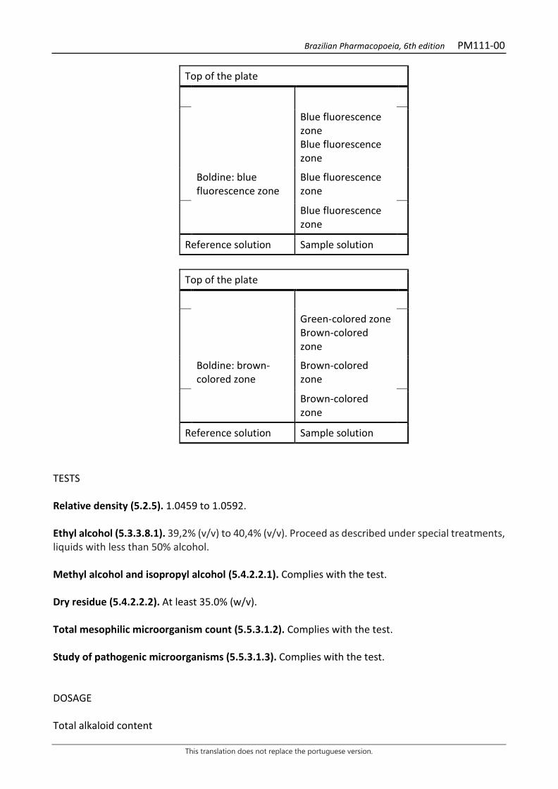
Brazilian Pharmacopoeia, 6th edition PM111-00
This translation does not replace the portuguese version.
Top of the plate
Blue fluorescence zone Blue fluorescence zone
Boldine: blue fluorescence zone
Blue fluorescence zone
Blue fluorescence zone
Reference solution Sample solution
Top of the plate
Green-colored zone Brown-colored zone
Boldine: brown-colored zone
Brown-colored zone
Brown-colored zone
Reference solution Sample solution
TESTS Relative density (5.2.5). 1.0459 to 1.0592. Ethyl alcohol (5.3.3.8.1). 39,2% (v/v) to 40,4% (v/v). Proceed as described under special treatments, liquids with less than 50% alcohol. Methyl alcohol and isopropyl alcohol (5.4.2.2.1). Complies with the test. Dry residue (5.4.2.2.2). At least 35.0% (w/v). Total mesophilic microorganism count (5.5.3.1.2). Complies with the test. Study of pathogenic microorganisms (5.5.3.1.3). Complies with the test. DOSAGE Total alkaloid content
Brazilian Pharmacopoeia, 6th edition PM111-00
This translation does not replace the portuguese version.
Proceed as described in High Performance Liquid Chromatography (5.2.17.4). Use a chromatograph with ultraviolet detector at 304 mm; pre-column packed with octadecylsilane silica, 250 mm long, 4.6 mm internal diameter column, packed with octadecylsilane silica (5 µm), kept at room temperature; Mobile phase flow rate of 1.5 mL/minute. Mobile phase: mixture of Solution A and Solution B (16:84). Solution A: mixture of 0.2 mL diethylamine and 99.8 mL acetonitrile. Solution B: mixture of 0.2 mL diethylamine and 99.8 mL water, adjust the pH to 3.0 with anhydrous formic acid. Sample solution: homogenize the fluid extract and volumetrically transfer 1 mL to a 250-mL beaker. Rinse the pipette with 3 mL of 5.5 M hydrochloric acid, transferring to the beaker. Add 50 mL of 5.5 M hydrochloric acid. Homogenize and check the pH, which should be between 2 and 3. Quantitatively transfer the solution into a 250 mL separating funnel and rinse the beaker with 10 mL of 5.5 M hydrochloric acid. Extract with 100 mL of a hexane and ethyl acetate (1:1) mixture. Shake vigorously. After phase separation, discard the organic phase. Transfer the aqueous phase to a beaker and add 6 M ammonium hydroxide, approximately 150 mL, until reaching a pH of 9.0. Transfer the sample to another 250-mL separating funnel and extract four times with 50 mL methyl chloride. Gather the organic phases and add 40 g of anhydrous sodium sulfate. Shake with a glass rod. Filter on filter paper into a 250-mL round-bottomed flask. Rinse the beaker with three 10-mL portions of methyl chloride. Filter and gather the organic solutions. Evaporate the solution until residue is formed in a rotary evaporator at a temperature not exceeding 50°C. Dissolve the residue in 5 mL of the Mobile phase. Take it to the ultrasound for five minutes. Transfer the solution quantitatively to a 10-mL volumetric flask, top off the volume with the Mobile phase and homogenize. Filter through a 0.45 µm filter unit. Reference solution: accurately weigh about 12 mg of boldine and transfer to a 100-mL volumetric flask. Add 50 mL of Mobile phase, perform ultrasound for two minutes, top off the volume with the Mobile phase and homogenize. Transfer 1 mL of the solution to a 10-mL volumetric flask, top off the volume with the Mobile phase and homogenize. Filter through a 0.45 µm filter unit. System suitability Resolution between peaks: Sample solution, minimum 1.0 between peaks for isoboldine and boldine. Procedure: separately inject 20 μL of the Reference solution and 20 µL of the Sample solution. Record the chromatograms and measure the areas under the peaks for boldine and the six alkaloids described below. Retention times for boldine are approximately 0.9 for isoboldine, 1.0 for boldine, 1.8 for isocoridine N-oxide, 2.2 for laurotetanine, 2.8 for isocoridine, and 3.2 for N-methyl laurotetanine. Calculate total alkaloid content expressed as boldine, in percent, according to the following expression:
TA =∑𝑇𝑇1 × 𝑚𝑚𝑟𝑟
𝑇𝑇𝑟𝑟 × 𝑚𝑚𝑎𝑎 × 100
in which,
Brazilian Pharmacopoeia, 6th edition PM111-00
This translation does not replace the portuguese version.
TA = Total alkaloid content expressed as boldine % (w/w); ΣA1 = sum of the areas under the peaks corresponding to the six alkaloids identified in the chromatogram obtained with the Sample solution; ma= mass in grams of the fluid extract, determined from the density; mr = mass in grams of boldine in the Reference solution, considering the purity of the reference substance; Ar= area under the peak corresponding to the boldine in the Reference solution. PACKAGING AND STORAGE In tightly closed containers, protected from light and heat.
Brazilian Pharmacopoeia, 6th edition PM112-00
This translation does not replace the portuguese version.
CALENDULA, fluid extract Calendulae extracta fluida
The fluid extract is obtained from dried flowers of Calendula officinalis L. containing at least 0.4% total flavonoids, expressed as hyperoside (C21H20O12, 464.38). PREPARATION The fluid extract is prepared in a 1:1 (w/v) drug:solvent ratio by percolation, using 70% (v/v) ethyl alcohol as the extracting liquid. CHARACTERISTICS Dark brown liquid with a characteristic odor. IDENTIFICATION Proceed as described in Thin-layer chromatography (5.2.17.1). Stationary phase: silica gel F254(0.25 mm). Mobile phase: ethyl acetate, anhydrous formic acid and water (80:10:10). Sample solution: dry 0.5 mL of fluid extract until residue is formed, in a water bath at a temperature not exceeding 60°C. Add 2mL methyl alcohol and filter through a 0.45 µm filter unit. Reference solution (1): dissolve an accurately weighed amount of rutin in methyl alcohol to obtain a concentration of 250 µg/mL. Reference solution (2): dissolve an exactly weighed amount of chlorogenic acid in methyl alcohol to obtain the concentration of 100 µg/mL. Reference solution (3): dissolve an exactly weighed amount of caffeic acid in methyl alcohol to obtain the concentration of 100 µg/mL. Procedure: apply 20 μL of the Sample solution, 20 μL of the Reference solution (1), 20 μL of the Reference solution (2), and 20 μL of the Reference solution (3) to the chromatoplate, separately and in the form of a band. Conduct chromatogram. Remove chromatoplate and allow it to air dry. Nebulize the plate with aminoethanol diphenylborate RS, then with a solution of 5% (w/v) macrogol 400 in ethyl alcohol. Heat between 100 °C and 105 °C for about five minutes. Examine under ultraviolet light at 365 nm. Results: the diagram below shows the sequences of zones obtained with the Sample solution, Reference solution (1), Reference solution (2) and the Reference solution (3). Other zones may occasionally develop.

Brazilian Pharmacopoeia, 6th edition PM112-00
This translation does not replace the portuguese version.
Top of the plate
Caffeic acid: light-blue fluorescence zone
Light-blue fluorescence zone
Blue fluorescence zone
Greenish-yellow fluorescence zone
Chlorogenic acid: light-blue fluorescence zone
Light-blue fluorescence zone
Greenish-yellow fluorescence zone
Rutin: yellowish-brown fluorescence zone
Yellowish-brown fluorescence zone
Greenish-yellow fluorescence zone
Reference solution Sample solution
TESTS Relative density (5.2.5). 0.9660 to 0.9970. Ethyl alcohol (5.3.3.8.1). Method II. 52,0% (v/v) to 56% (v/v). Dry residue (5.4.2.2.2). At least 18.0% (w/w). Methyl alcohol and isopropyl alcohol (5.4.2.2.1). Complies with the test. Total mesophilic microorganism count (5.5.3.1.2). Complies with the test. Study of pathogenic microorganisms (5.5.3.1.3). Complies with the test. DOSAGE Total flavonoids To proceed as described in Visible absorption spectrophotometry (5.2.14). Prepare solutions as described below.
Brazilian Pharmacopoeia, 6th edition PM112-00
This translation does not replace the portuguese version.
Stock solution: in a round-bottomed flask, add 0.8 mL of calendula fluid extract. Add 1 mL of 5 g/L methenamine solution (w/v), 20 mL acetone and 7 mL hydrochloric acid. Then reflux in a water bath for 30 minutes. Filter through filter paper, transfer to a 100-mL volumetric flask, top off the volume with acetone and homogenize. Transfer 20 mL of the solution to a separating funnel and add 20 mL of water. Extract with one 15-mL portion, and then with three 10-mL portions of ethyl acetate. After the extraction process, combine the ethyl acetate phases, transfer to another separating funnel and rinse with two 50-mL portions of water. Transfer the ethyl acetate phase to a 50-mL volumetric flask, top off the volume with ethyl acetate and homogenize. Sample solution: transfer 10 mL of the Stock solution into a 25-mL volumetric flask, add 1 mL of the 2% aluminum chloride solution (w/v) in (v/v) 5% acetic acid solution (v/v) in methyl alcohol. Top off the volume with the 5% (v/v) acetic acid solution in methyl alcohol and homogenize. Blank solution: transfer 10 mL of the Stock Solution into a 25-mL volumetric flask, top off the volume with the 5% (v/v) acetic acid solution in methyl alcohol and homogenize. Procedure: measure the absorbance of the Sample Solution at 425 nm after precisely 30 minutes, using the Blank solution for zero adjustment. Calculate total flavonoid content expressed as hyperoside, in percent, according to the following expression:
TF =A × 1,25
𝑚𝑚
in which, TF = total flavonoid content expressed as hyperoside % (w/w); A = absorbance measured for the Sample solution; m = mass in grams of the fluid extract used, determined from the density. PACKAGING AND STORAGE In tightly closed containers, protected from light and heat.
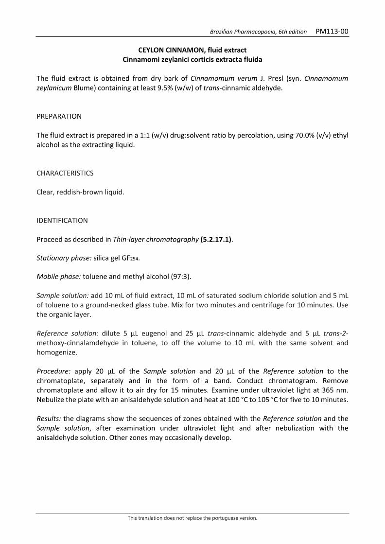
Brazilian Pharmacopoeia, 6th edition PM113-00
This translation does not replace the portuguese version.
CEYLON CINNAMON, fluid extract Cinnamomi zeylanici corticis extracta fluida
The fluid extract is obtained from dry bark of Cinnamomum verum J. Presl (syn. Cinnamomum zeylanicum Blume) containing at least 9.5% (w/w) of trans-cinnamic aldehyde. PREPARATION The fluid extract is prepared in a 1:1 (w/v) drug:solvent ratio by percolation, using 70.0% (v/v) ethyl alcohol as the extracting liquid. CHARACTERISTICS Clear, reddish-brown liquid. IDENTIFICATION Proceed as described in Thin-layer chromatography (5.2.17.1). Stationary phase: silica gel GF254. Mobile phase: toluene and methyl alcohol (97:3). Sample solution: add 10 mL of fluid extract, 10 mL of saturated sodium chloride solution and 5 mL of toluene to a ground-necked glass tube. Mix for two minutes and centrifuge for 10 minutes. Use the organic layer. Reference solution: dilute 5 μL eugenol and 25 μL trans-cinnamic aldehyde and 5 μL trans-2-methoxy-cinnalamdehyde in toluene, to off the volume to 10 mL with the same solvent and homogenize. Procedure: apply 20 μL of the Sample solution and 20 μL of the Reference solution to the chromatoplate, separately and in the form of a band. Conduct chromatogram. Remove chromatoplate and allow it to air dry for 15 minutes. Examine under ultraviolet light at 365 nm. Nebulize the plate with an anisaldehyde solution and heat at 100 °C to 105 °C for five to 10 minutes. Results: the diagrams show the sequences of zones obtained with the Reference solution and the Sample solution, after examination under ultraviolet light and after nebulization with the anisaldehyde solution. Other zones may occasionally develop.
Brazilian Pharmacopoeia, 6th edition PM113-00
This translation does not replace the portuguese version.
Top of the plate
trans-2-Methoxy-cinnalamdehyde: light-blue fluorescence zone
Light-blue colored zone
Light-blue colored zone
Reference solution Sample solution
Top of the plate
Eugenol: violaceous color zone
Violaceous-colored zone
Trans-cinnamic aldehyde: brownish-colored zone
Brownish-colored zone
trans-2-Methoxy-cinnalamdehyde: brownish-colored zone
Brownish-colored zone
Grayish-blue colored zone
Reference solution Sample solution TESTS Relative density (5.2.5). 0.89 to 0.94. Ethyl alcohol (5.3.3.8.1). Distillation method, Special treatments, Liquids with more than 30% alcohol. 65% (v/v) to 75% (v/v). Methyl alcohol and isopropyl alcohol (5.4.2.2.1). Complies with the test. Dry residue (5.4.2.2.2). At least 7.5% (w/w). Total mesophilic microorganism count (5.5.3.1.2). Complies with the test.
Brazilian Pharmacopoeia, 6th edition PM113-00
This translation does not replace the portuguese version.
Study of pathogenic microorganisms (5.5.3.1.3). Complies with the test. DOSAGE Trans-cinnamic aldehyde Proceed as described in High Performance Liquid Chromatography (5.2.17.4). Use a chromatograph equipped with an ultraviolet detector at 292 nm; a 250 mm long, 4.6 mm internal diameter column packed with C-18 (5 µm); Mobile phase flow rate of 1.0 mL/minute. One milliliter of the tincture should contain at least 0.3 mg trans-cinnamic aldehyde. Isocratic system. Mobile phase: water and methyl alcohol (1:1). Sample solution: analytically transfer 1.0 mL of the Ceylon cinnamon fluid extract to a 25-mL volumetric flask, top off the volume with methyl alcohol and homogenize. Transfer 0.20 mL of the solution to a 10-mL volumetric flask, top off the volume with methyl alcohol and homogenize. Filter through a 0.45 µm filter unit. Reference solution: dissolve an accurately weighed amount of trans-cinnamic aldehyde in methyl alcohol to obtain a 0.520 mg/mL solution. Solutions for the analytical curve: transfer 2.0 mL of the Reference solution to a 50-mL volumetric flask, top off the volume with methyl alcohol and homogenize, obtaining a 20.8 µg/mL solution. Transfer 2.0 mL, 3.0 mL, 4.0 mL, 5.0 mL, 6.0 mL, 7.0 mL and 8.0 mL of this solution into 10-mL volumetric flasks, top off the volume with methyl alcohol and homogenize, obtaining solutions at 4.16 µg/mL, 6.24 µg/mL, 8.32 µg/mL, 10.4 µg/mL, 12.48 µg/mL, 14.56 µg/mL and 16.64 µg/mL, respectively. Filter the solutions through a 0.45 µm filter unit. Filter through a 0.45 µm filter unit. Procedure: inject 20 µL of the Solutions for the analytical curve and 20 µL of the Sample solution separately. Record the chromatograms and measure the area under the trans-cinnamic aldehyde peak. Calclate the trans-cinnamic aldehyde content in the tincture using the straight line equation obtained from the analytical curve and, in percentage, according to the following expression:
TA = C𝑎𝑎 × 1.25 in which, TA = trans-cinnamic aldehyde content in mg/mL; Ca = trans-cinnamic aldehyde concentration in the Sample solution µg/mL, determined using the analytical curve. PACKAGING AND STORAGE In tightly closed containers, protected from light and heat.
Brazilian Pharmacopoeia, 6th edition PM114-00
This translation does not replace the portuguese version.
CASCARA BUCKTHORN, fluid extract Rhamni purshianae extracta fluida
The fluid extract is obtained from dried bark of Frangula purshiana (DC.) A. Gray (syn. Rhamnus purshiana DC.), containing at least 8.0% hydroxyanthracene glycosides, of which at least 60% are cascarosides, expressed as cascaroside A (C27H32O14, 580.54). PREPARATION The fluid extract is prepared in a 1:1 (w/v) drug:solvent ratio by percolation or maceration, using 70% (v/v) ethyl alcohol as the extracting liquid. CHARACTERISTICS Dark brown colored liquid. IDENTIFICATION Proceed as described in Thin-layer chromatography (5.2.17.1). Stationary phase: silica gel F254 (0.25 mm). Mobile phase: ethyl acetate, methyl alcohol and water (100:17:13). Sample solution: dry 0.5 mL of the fluid extract until residue is formed, in a water bath, at a maximum temperature of 60 °C. Add 5 mL methyl alcohol and filter through a 0.45 µm filter unit. Reference solution (1): dissolve an accurately weighed amount of aloin in methyl alcohol to obtain a concentration of 1000 µg/mL. Reference solution (2): dissolve an accurately weighed amount of emodin in methyl alcohol to obtain a concentration of 1000 µg/mL. Procedure: apply 20 μL of the Sample solution, 20 μL of the Reference solution (1) and 20 μL of the Reference solution (2) to the chromatoplate, separately and in the form of a band. Conduct chromatogram. Remove plate and allow it to air dry. Nebulize the plate with 5% potassium hydroxide solution in ethyl alcohol. Examine under ultraviolet light at 365 nm. Heat the plate between 100 °C and 105 °C for about five minutes. Examine under visible light. Results: the diagram below shows the sequences of zones obtained with the Reference Solution (1), the Reference solution (2) and the Sample solution. Other zones may occasionally develop.
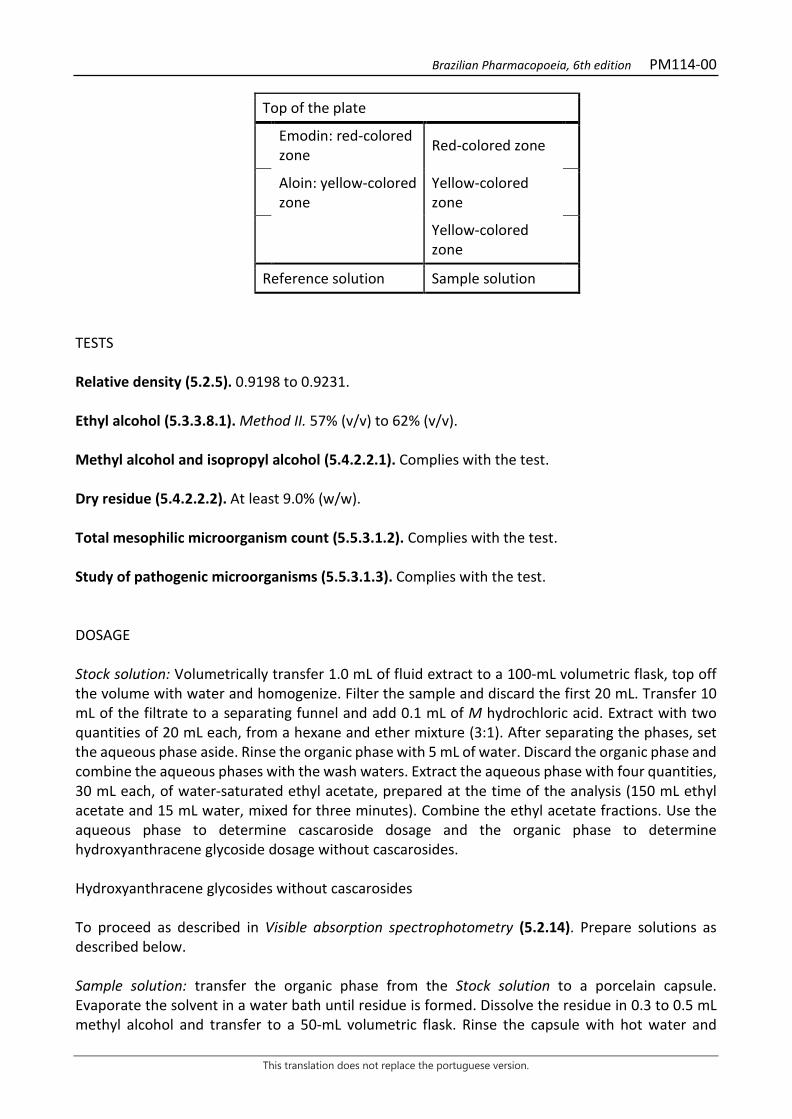
Brazilian Pharmacopoeia, 6th edition PM114-00
This translation does not replace the portuguese version.
Top of the plate
Emodin: red-colored zone Red-colored zone
Aloin: yellow-colored zone
Yellow-colored zone
Yellow-colored zone
Reference solution Sample solution
TESTS Relative density (5.2.5). 0.9198 to 0.9231. Ethyl alcohol (5.3.3.8.1). Method II. 57% (v/v) to 62% (v/v). Methyl alcohol and isopropyl alcohol (5.4.2.2.1). Complies with the test. Dry residue (5.4.2.2.2). At least 9.0% (w/w). Total mesophilic microorganism count (5.5.3.1.2). Complies with the test. Study of pathogenic microorganisms (5.5.3.1.3). Complies with the test. DOSAGE Stock solution: Volumetrically transfer 1.0 mL of fluid extract to a 100-mL volumetric flask, top off the volume with water and homogenize. Filter the sample and discard the first 20 mL. Transfer 10 mL of the filtrate to a separating funnel and add 0.1 mL of M hydrochloric acid. Extract with two quantities of 20 mL each, from a hexane and ether mixture (3:1). After separating the phases, set the aqueous phase aside. Rinse the organic phase with 5 mL of water. Discard the organic phase and combine the aqueous phases with the wash waters. Extract the aqueous phase with four quantities, 30 mL each, of water-saturated ethyl acetate, prepared at the time of the analysis (150 mL ethyl acetate and 15 mL water, mixed for three minutes). Combine the ethyl acetate fractions. Use the aqueous phase to determine cascaroside dosage and the organic phase to determine hydroxyanthracene glycoside dosage without cascarosides. Hydroxyanthracene glycosides without cascarosides To proceed as described in Visible absorption spectrophotometry (5.2.14). Prepare solutions as described below. Sample solution: transfer the organic phase from the Stock solution to a porcelain capsule. Evaporate the solvent in a water bath until residue is formed. Dissolve the residue in 0.3 to 0.5 mL methyl alcohol and transfer to a 50-mL volumetric flask. Rinse the capsule with hot water and
Brazilian Pharmacopoeia, 6th edition PM114-00
This translation does not replace the portuguese version.
transfer the residue to the 50-mL volumetric flask. Top off the volume with water and homogenize. Then transfer 20 mL of the solution to a 100mL round-bottomed flask, add 2 g of ferric chloride hexahydrate and 12 mL of hydrochloric acid. Heat the mixture under reflux for four hours. After cooling, transfer the solution to a separating funnel. Rinse the flask with 3 to 4 mL of M sodium hydroxide, then with 3 to 4 mL of water. Transfer the wash water to the separating funnel. Extract with three portions, 30 mL each, of a hexane and ether (3:1) mixture. Transfer the organic phase to another separating funnel and rinse it twice, using 10 mL of water in each rinse. Discard the aqueous phase. After this procedure, dilute the organic phase to 100 mL with the hexane and ether (3:1) mixture. Then transfer 20 mL and evaporate until residue is formed in a water bath. Dissolve the residue with 10 mL of a 5 g/L magnesium acetate solution in methyl alcohol. Blank solution: methyl alcohol. Procedure: measure the absorbance of the Sample solution at 440 nm and 515 nm, using the Blank solution for zero adjustment. A ratio between the absorbance values at 515 nm and 440 nm that is smaller than 2.4 invalidates the assay. Calculate the content of hydroxyanthracene glycosides without the cascarosides (HAC), in percent, according to the expression:
THAC =A × 6,95
𝑚𝑚
in which, THAC = hydroxyanthracene glycoside content without cascarosides % (w/w); A = absorbance measured at 515 nm for the Sample solution; m = mass in grams of the fluid extract used, determined from the density. Cascarosides Sample solution: dilute the aqueous phase with water in a 50-mL volumetric flask, top off the volume and homogenize. Use 20 mL of the solution. Procedure: measure absorbance at 440 nm and 515 nm. Use methyl alcohol for zero adjustment. A ratio between the absorbance values at 515 nm and 440 nm that is smaller than 2.4 invalidates the assay. Calculate cascaroside content, in percent, according to the following expression:
TC =A × 6,95
𝑚𝑚
in which, TC = cascaroside content % (w/w); A = absorbance measured at 515 nm for the Sample solution; and m = mass in grams of the cascara buckthorn fluid extract, determined from the density. PACKAGING AND STORAGE In tightly closed containers, protected from light and heat.
Brazilian Pharmacopoeia, 6th edition PM115-00
This translation does not replace the portuguese version.
HORSE CHESTNUT, fluid extract Hippocastani extracta fluida
The fluid extract is obtained from dried seeds of Aesculus hippocastanum L., containing at least 3.0% triterpene glycosides expressed as anhydrous aescin. PREPARATION The fluid extract is prepared in a 1:1 (w/v) drug:solvent ratio by percolation or maceration, using 70% (v/v) ethyl alcohol as the extracting liquid. CHARACTERISTICS Dark brown colored liquid. IDENTIFICATION Proceed as described in Thin-layer chromatography (5.2.17.1). Stationary phase: silica gel GF254. Mobile phase: butyl alcohol, water and glacial acetic acid (50:40:10). Sample solution: dry 0.5 mL of the fluid extract until residue is formed, in a water bath, at a maximum temperature of 60 °C. Add 5 mL methyl alcohol and filter through a 0.45 µm filter unit. Reference solution: dissolve an accurately weighed amount of aescin in methyl alcohol to obtain a concentration of 5000 µg/mL. Procedure: apply 20 μL of the Sample solution and 20 μL of the Reference solution to the chromatoplate, separately and in the form of a band. Conduct chromatogram. Remove chromatoplate and allow it to air dry. Nebulize the plate with anisaldehyde RS, heat at 100 °C to 105 °C for approximately five minutes. Examine under visible light. Results: the diagram below shows the sequences of zones obtained with the Reference solution and the Sample solution. Other zones may occasionally develop.
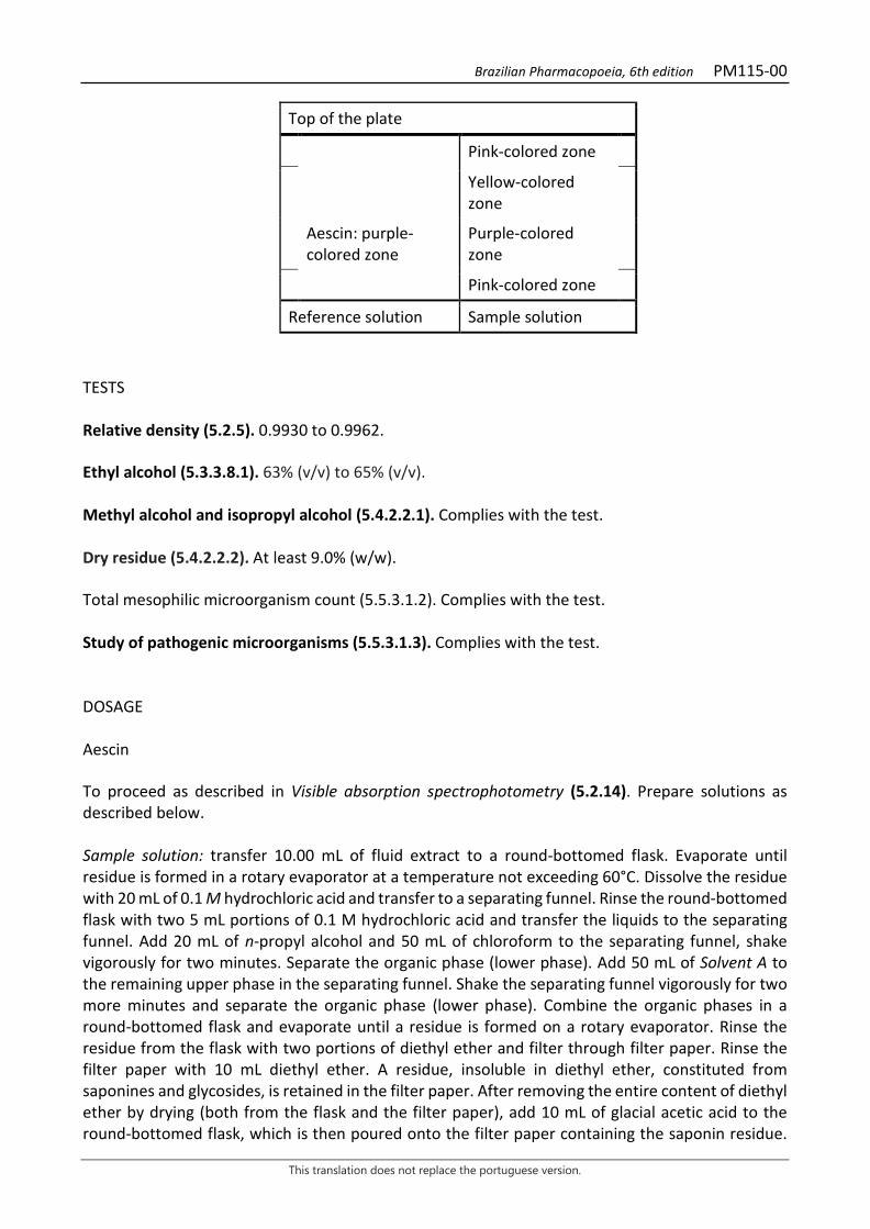
Brazilian Pharmacopoeia, 6th edition PM115-00
This translation does not replace the portuguese version.
Top of the plate
Pink-colored zone
Yellow-colored zone
Aescin: purple-colored zone
Purple-colored zone
Pink-colored zone
Reference solution Sample solution
TESTS Relative density (5.2.5). 0.9930 to 0.9962. Ethyl alcohol (5.3.3.8.1). 63% (v/v) to 65% (v/v). Methyl alcohol and isopropyl alcohol (5.4.2.2.1). Complies with the test. Dry residue (5.4.2.2.2). At least 9.0% (w/w). Total mesophilic microorganism count (5.5.3.1.2). Complies with the test. Study of pathogenic microorganisms (5.5.3.1.3). Complies with the test. DOSAGE Aescin To proceed as described in Visible absorption spectrophotometry (5.2.14). Prepare solutions as described below. Sample solution: transfer 10.00 mL of fluid extract to a round-bottomed flask. Evaporate until residue is formed in a rotary evaporator at a temperature not exceeding 60°C. Dissolve the residue with 20 mL of 0.1 M hydrochloric acid and transfer to a separating funnel. Rinse the round-bottomed flask with two 5 mL portions of 0.1 M hydrochloric acid and transfer the liquids to the separating funnel. Add 20 mL of n-propyl alcohol and 50 mL of chloroform to the separating funnel, shake vigorously for two minutes. Separate the organic phase (lower phase). Add 50 mL of Solvent A to the remaining upper phase in the separating funnel. Shake the separating funnel vigorously for two more minutes and separate the organic phase (lower phase). Combine the organic phases in a round-bottomed flask and evaporate until a residue is formed on a rotary evaporator. Rinse the residue from the flask with two portions of diethyl ether and filter through filter paper. Rinse the filter paper with 10 mL diethyl ether. A residue, insoluble in diethyl ether, constituted from saponines and glycosides, is retained in the filter paper. After removing the entire content of diethyl ether by drying (both from the flask and the filter paper), add 10 mL of glacial acetic acid to the round-bottomed flask, which is then poured onto the filter paper containing the saponin residue.
Brazilian Pharmacopoeia, 6th edition PM115-00
This translation does not replace the portuguese version.
Transfer the solution into a 50-mL volumetric flask. Perform this procedure two more times with 10 mL of glacial acetic acid, top off the volume with the same solvent and homogenize. Solvent A: chloroform, 0.1 M hydrochloric acid and n-propyl alcohol (50:30:20). Use lower phase. Color reagent: dissolve 75 mg ferric chloride hexahydrate in 50 mL of glacial acetic acid. Next add 40 mL of sulfuric acid on the solution. Transfer the solution to a 100-mL volumetric flask, top off the volume with sulfuric acid and homogenize. The solution should be prepared for immediate use. Solutions for the analytical curve: weigh 25 mg of aescin, transfer to a 25-mL volumetric flask and dissolve with glacial acetic acid, top off the volume with the same solvent and homogenize. Separately pipette 1 mL, 2 mL, 3 mL, 4 mL and 5 mL of this solution to five 10-mL volumetric flasks, dilute with glacial acetic acid to 10 mL and homogenize. Blank solution: glacial acetic acid. Procedure: transfer 1 mL from each of the Analytical curve solutions, from the Sample solution and from the Blank solution, separately, to test tubes with a cap. Add 4 mL of Color reagent to each test tube. Next, heat the tubes in a water bath at a temperature of 60°C for 25 minutes, shaking the tubes occasionally. Measure the absorbances of the solutions at 540 nm. After reading, construct the aescin analytical curve in mg/mL and determine the aescin concentration in mg/mL in the Sample solution. Calculate triterpene glycoside content expressed as anhydrous aescin, in percent, according to the following expression:
TE =C × 50𝑚𝑚 × 3
in which, TE = triterpene glycoside content expressed as anhydrous aescin % (w/w); C = concentration of aescin in mg/mL determined for the Sample solution using the analytical curve; m = mass in grams of the fluid extract, determined from the density. PACKAGING AND STORAGE In tightly closed containers, protected from light and heat.
Brazilian Pharmacopoeia, 6th edition PM116-00
This translation does not replace the portuguese version.
HAWTHORN, fluid extract Crataegi extracta fluida
The fluid extract is obtained from dried flowering branches of Crataegus monogyna Jacq., Crataegus rhipidophylla Gand. (syn. C. oxyacantha L., nom. rej.) Crataegus laevigata (Poir.) DC., Crataegus pentagyna Waldst. & Kit. ex Willd., Crataegus nigra Waldst. & Kit. and Crataegus azarolus L., or hybrids among them, containing at least 0.8% total flavonoids expressed as hyperoside (C21H20O12; 464.38). PREPARATION The fluid extract is prepared in a 1:1 (w/v) drug:solvent ratio by percolation or maceration, using 70% (v/v) ethyl alcohol as the extracting liquid. CHARACTERISTICS Dark brown liquid with a characteristic odor. IDENTIFICATION Proceed as described in Thin-layer chromatography (5.2.17.1). Stationary phase: silica gel F254 (0.25 mm). Mobile phase: ethyl acetate, water, acetic acid and formic acid (100:26:11:11). Sample solution: dry 1.0 mL of the fluid extract until residue is formed, in a water bath, at a temperature not exceeding 60 °C. Suspend the residue 2 mL methyl alcohol and filter through a 0.45 µm filter unit. Reference solution (1): dissolve an exactly weighed amount of chlorogenic acid in methyl alcohol to obtain the concentration of 100 µg/mL. Reference solution (2): dissolve an accurately weighed amount of hyperoside in methyl alcohol to obtain a concentration of 250 µg/mL. Procedure: apply 20 μL of the Sample solution, 20 μL of the Reference solution (1) and 20 μL of the Reference solution (2) to the chromatoplate, separately and in the form of a band. Conduct chromatogram. Remove chromatoplate and allow it to air dry. Nebulize the plate with aminoethanol diphenylborate RS, then with a solution of 5% (w/v) macrogol 400 in ethyl alcohol. Heat between 100 °C and 105 °C for about five minutes. Examine under ultraviolet light at 365 nm. Results: the diagram below shows the sequences of zones obtained with the Sample solution, Reference solution (1) and Reference solution (2). Other zones may occasionally develop.
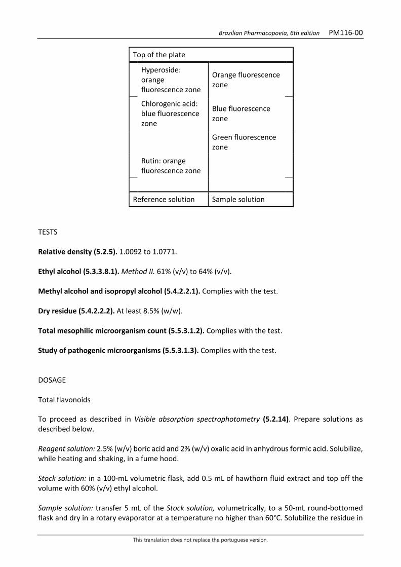
Brazilian Pharmacopoeia, 6th edition PM116-00
This translation does not replace the portuguese version.
Top of the plate
Hyperoside: orange fluorescence zone
Orange fluorescence zone
Chlorogenic acid: blue fluorescence zone
Blue fluorescence zone
Green fluorescence zone
Rutin: orange fluorescence zone
Reference solution Sample solution
TESTS Relative density (5.2.5). 1.0092 to 1.0771. Ethyl alcohol (5.3.3.8.1). Method II. 61% (v/v) to 64% (v/v). Methyl alcohol and isopropyl alcohol (5.4.2.2.1). Complies with the test. Dry residue (5.4.2.2.2). At least 8.5% (w/w). Total mesophilic microorganism count (5.5.3.1.2). Complies with the test. Study of pathogenic microorganisms (5.5.3.1.3). Complies with the test. DOSAGE Total flavonoids To proceed as described in Visible absorption spectrophotometry (5.2.14). Prepare solutions as described below. Reagent solution: 2.5% (w/v) boric acid and 2% (w/v) oxalic acid in anhydrous formic acid. Solubilize, while heating and shaking, in a fume hood. Stock solution: in a 100-mL volumetric flask, add 0.5 mL of hawthorn fluid extract and top off the volume with 60% (v/v) ethyl alcohol. Sample solution: transfer 5 mL of the Stock solution, volumetrically, to a 50-mL round-bottomed flask and dry in a rotary evaporator at a temperature no higher than 60°C. Solubilize the residue in

Brazilian Pharmacopoeia, 6th edition PM116-00
This translation does not replace the portuguese version.
8 mL of a mixture of methyl alcohol and acetic acid (10:100) and transfer to a 25-mL volumetric flask. Rinse the round-bottomed flask with a 3 mL mixture of methyl alcohol and acetic acid (10:100) and add to the 25mL volumetric flask. Add 10 mL of the Reagent solution. Place it to an ice bath for 10 minutes, not allowing the solution to freeze. Top off the volume with glacial acetic acid. Immediately place the flask in an ice bath and remove 10 minutes before reading the spectrophotometer. Blank solution: transfer 5 mL of the Stock solution to a 50-mL round-bottomed flask and dry in a rotary evaporator at a temperature no higher than 60°C. Solubilize the residue in 8 mL of the mixture of methyl alcohol and acetic acid (10:100) and transfer to a 25-mL volumetric flask. Rinse the round-bottomed flask with a 3 mL mixture of methyl alcohol and acetic acid (10:100) and add to the 25mL volumetric flask. Add 10 mL of anhydrous formic acid. Place it to an ice bath for 10 minutes, not allowing the solution to freeze. Top off the volume with glacial acetic acid. Immediately place the flask in an ice bath and remove 10 minutes before reading the spectrophotometer. Procedure: measure the absorbance of the Sample Solution at 410 nm after precisely 30 minutes, using the Blank Solution for zero adjustment. Calculate total flavonoid content expressed as hyperoside, in percent, according to the following expression:
TF =A × 1,235
𝑚𝑚
in which, TF = total flavonoid content expressed as hyperoside % (w/w); A = absorbance measured for the Sample solution; m = mass in grams of the used fluid extract, determined from the density. PACKAGING AND STORAGE In tightly closed containers, protected from light and heat.
Brazilian Pharmacopoeia, 6th edition PM117-00
This translation does not replace the portuguese version.
YELLOW GENTIAN, fluid extract Gentianae extracta fluida
The fluid extract is obtained from dried rhizomes and roots of Gentiana lutea L., containing at least 1.5% gentiopicroside (C16H20O9, 356.33). PREPARATION The fluid extract is prepared in a 1:1 (w/v) drug:solvent ratio by percolation or maceration, using 70% (v/v) ethyl alcohol as the extracting liquid. CHARACTERISTICS Dark yellowish-brown or dark reddish-brown liquid. IDENTIFICATION Proceed as described in Thin-layer chromatography (5.2.17.1). Stationary phase: silica gel GF254. Mobile phase: ethyl acetate, methyl alcohol and water (77:15:8). Sample solution: dilute 0.1 mL of the fluid extract in 1 mL methyl alcohol. Reference solution (1): prepare a 280 µg/mL solution of gentiopicroside in methyl alcohol. Reference solution (2): prepare a 1200 µg/mL solution of amarogentin in methyl alcohol. TLC visualization reagent: mix, in order, 0.5 mL anisaldehyde, 10 mL glacial acetic acid, 85 mL methyl alcohol, and 5 mL sulfuric acid. Procedure: apply 10 μL of the Sample solution, 20 μL of the Reference solution (1) and 20 μL of the Reference solution (2) to the chromatoplate, separately and in the form of a band. Conduct chromatogram. Remove chromatoplate and allow it to air dry for 15 minutes. Examine under ultraviolet light at 254 nm. Nebulize the chromatoplate with the TLC visualization reagent, heat at 100 °C to 105 °C for one minute and examine under visible light. Results: the diagram below shows the sequences of zones obtained with the Reference Solution (1), the Reference solution (2) and the Sample solution. Other zones may occasionally develop.
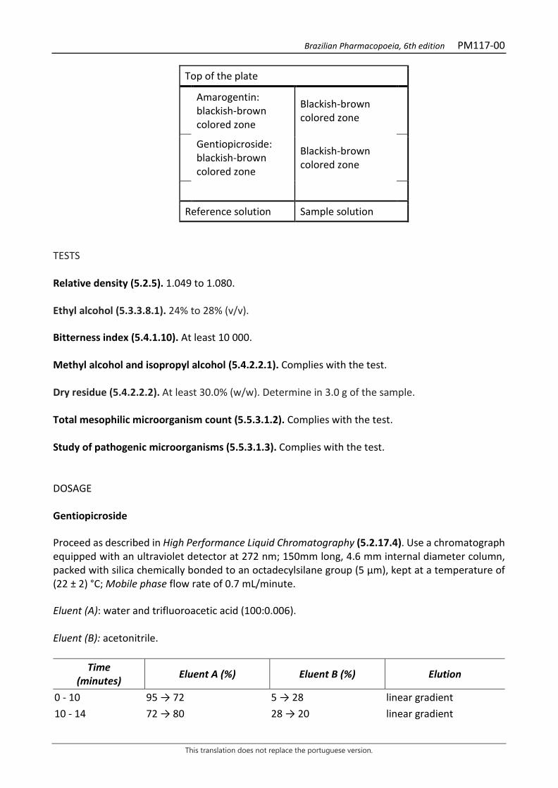
Brazilian Pharmacopoeia, 6th edition PM117-00
This translation does not replace the portuguese version.
Top of the plate
Amarogentin: blackish-brown colored zone
Blackish-brown colored zone
Gentiopicroside: blackish-brown colored zone
Blackish-brown colored zone
Reference solution Sample solution TESTS Relative density (5.2.5). 1.049 to 1.080. Ethyl alcohol (5.3.3.8.1). 24% to 28% (v/v). Bitterness index (5.4.1.10). At least 10 000. Methyl alcohol and isopropyl alcohol (5.4.2.2.1). Complies with the test. Dry residue (5.4.2.2.2). At least 30.0% (w/w). Determine in 3.0 g of the sample. Total mesophilic microorganism count (5.5.3.1.2). Complies with the test. Study of pathogenic microorganisms (5.5.3.1.3). Complies with the test. DOSAGE Gentiopicroside Proceed as described in High Performance Liquid Chromatography (5.2.17.4). Use a chromatograph equipped with an ultraviolet detector at 272 nm; 150mm long, 4.6 mm internal diameter column, packed with silica chemically bonded to an octadecylsilane group (5 µm), kept at a temperature of (22 ± 2) °C; Mobile phase flow rate of 0.7 mL/minute. Eluent (A): water and trifluoroacetic acid (100:0.006). Eluent (B): acetonitrile.
Time (minutes) Eluent A (%) Eluent B (%) Elution
0 - 10 95 → 72 5 → 28 linear gradient 10 - 14 72 → 80 28 → 20 linear gradient
Brazilian Pharmacopoeia, 6th edition PM117-00
This translation does not replace the portuguese version.
Time (minutes) Eluent A (%) Eluent B (%) Elution
14 - 16 80 → 95 20 → 5 linear gradient 16 - 20 95 5 isocratic Sample solution: dilute 150 µL of fluid extract in 10 mL of the methyl alcohol and water mixture (1:1). Filter through a 0.45 µm filter unit. Reference solution: dissolve exactly weighed amounts of gentiopicroside in methyl alcohol to obtain a solution with a concentration of 0.32 mg/mL. Filter through a 0.45 µm filter unit. Procedure: separately inject 5 μL of the Reference solution and 5 µL of the Sample solution. Record chromatograms and measure the area under the gentiopicroside peak. The average retention time is approximately nine minutes. Calculate gentiopicroside content, in percent, according to the following expression:
TG =𝐶𝐶𝑟𝑟 × 𝑇𝑇𝑎𝑎 × 10 × 100
𝑇𝑇𝑟𝑟 × 𝑚𝑚
in which, TG = gentiopicroside content % (w/w); Cr = concentration of the Reference solution in g/mL, considering the purity of the reference substance; Ar = area under the peak corresponding to the gentiopicroside in the Reference solution; Aa = area under the peak corresponding to the gentiopicroside in the Sample solution; m = mass in grams of the fluid extract, determined from the density. PACKAGING AND STORAGE In tightly closed containers, protected from light and heat.
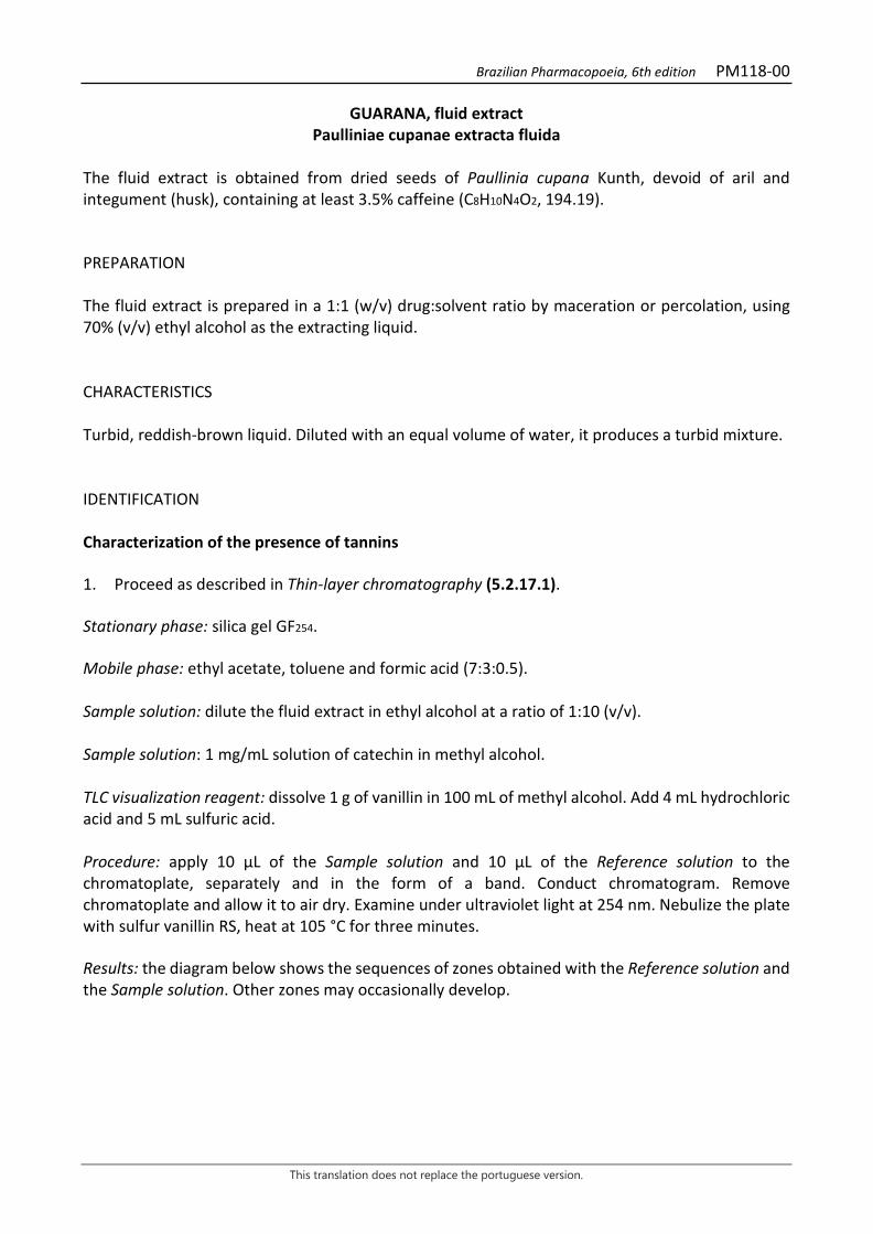
Brazilian Pharmacopoeia, 6th edition PM118-00
This translation does not replace the portuguese version.
GUARANA, fluid extract Paulliniae cupanae extracta fluida
The fluid extract is obtained from dried seeds of Paullinia cupana Kunth, devoid of aril and integument (husk), containing at least 3.5% caffeine (C8H10N4O2, 194.19). PREPARATION The fluid extract is prepared in a 1:1 (w/v) drug:solvent ratio by maceration or percolation, using 70% (v/v) ethyl alcohol as the extracting liquid. CHARACTERISTICS Turbid, reddish-brown liquid. Diluted with an equal volume of water, it produces a turbid mixture. IDENTIFICATION Characterization of the presence of tannins 1. Proceed as described in Thin-layer chromatography (5.2.17.1). Stationary phase: silica gel GF254. Mobile phase: ethyl acetate, toluene and formic acid (7:3:0.5). Sample solution: dilute the fluid extract in ethyl alcohol at a ratio of 1:10 (v/v). Sample solution: 1 mg/mL solution of catechin in methyl alcohol. TLC visualization reagent: dissolve 1 g of vanillin in 100 mL of methyl alcohol. Add 4 mL hydrochloric acid and 5 mL sulfuric acid. Procedure: apply 10 μL of the Sample solution and 10 μL of the Reference solution to the chromatoplate, separately and in the form of a band. Conduct chromatogram. Remove chromatoplate and allow it to air dry. Examine under ultraviolet light at 254 nm. Nebulize the plate with sulfur vanillin RS, heat at 105 °C for three minutes. Results: the diagram below shows the sequences of zones obtained with the Reference solution and the Sample solution. Other zones may occasionally develop.
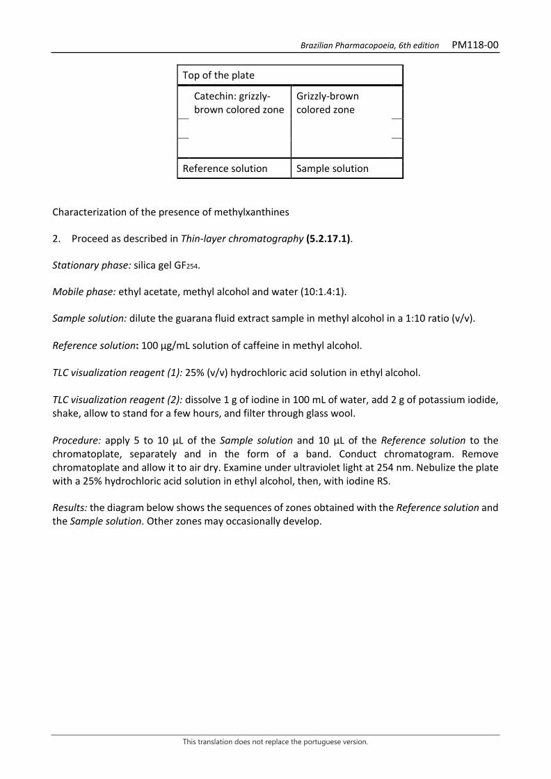
Brazilian Pharmacopoeia, 6th edition PM118-00
This translation does not replace the portuguese version.
Top of the plate
Catechin: grizzly-brown colored zone
Grizzly-brown colored zone
Reference solution Sample solution Characterization of the presence of methylxanthines 2. Proceed as described in Thin-layer chromatography (5.2.17.1). Stationary phase: silica gel GF254. Mobile phase: ethyl acetate, methyl alcohol and water (10:1.4:1). Sample solution: dilute the guarana fluid extract sample in methyl alcohol in a 1:10 ratio (v/v). Reference solution: 100 µg/mL solution of caffeine in methyl alcohol. TLC visualization reagent (1): 25% (v/v) hydrochloric acid solution in ethyl alcohol. TLC visualization reagent (2): dissolve 1 g of iodine in 100 mL of water, add 2 g of potassium iodide, shake, allow to stand for a few hours, and filter through glass wool. Procedure: apply 5 to 10 μL of the Sample solution and 10 μL of the Reference solution to the chromatoplate, separately and in the form of a band. Conduct chromatogram. Remove chromatoplate and allow it to air dry. Examine under ultraviolet light at 254 nm. Nebulize the plate with a 25% hydrochloric acid solution in ethyl alcohol, then, with iodine RS. Results: the diagram below shows the sequences of zones obtained with the Reference solution and the Sample solution. Other zones may occasionally develop.
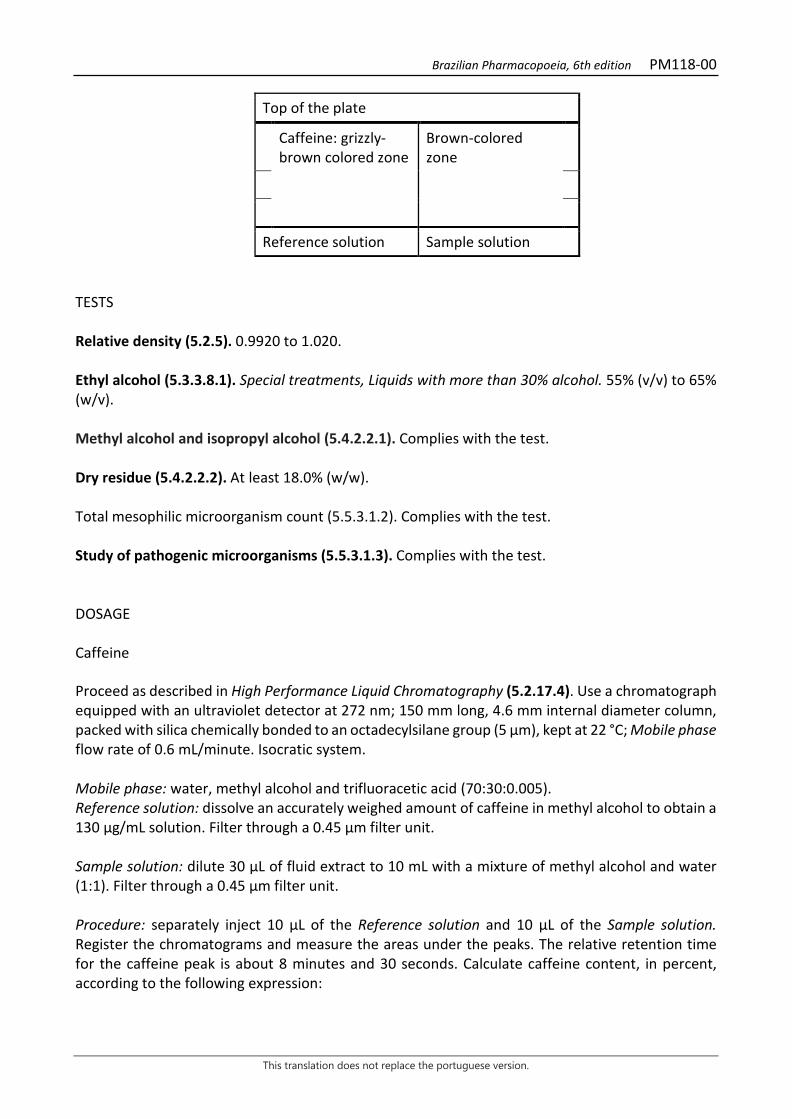
Brazilian Pharmacopoeia, 6th edition PM118-00
This translation does not replace the portuguese version.
Top of the plate
Caffeine: grizzly-brown colored zone
Brown-colored zone
Reference solution Sample solution TESTS Relative density (5.2.5). 0.9920 to 1.020. Ethyl alcohol (5.3.3.8.1). Special treatments, Liquids with more than 30% alcohol. 55% (v/v) to 65% (w/v). Methyl alcohol and isopropyl alcohol (5.4.2.2.1). Complies with the test. Dry residue (5.4.2.2.2). At least 18.0% (w/w). Total mesophilic microorganism count (5.5.3.1.2). Complies with the test. Study of pathogenic microorganisms (5.5.3.1.3). Complies with the test. DOSAGE Caffeine Proceed as described in High Performance Liquid Chromatography (5.2.17.4). Use a chromatograph equipped with an ultraviolet detector at 272 nm; 150 mm long, 4.6 mm internal diameter column, packed with silica chemically bonded to an octadecylsilane group (5 µm), kept at 22 °C; Mobile phase flow rate of 0.6 mL/minute. Isocratic system. Mobile phase: water, methyl alcohol and trifluoracetic acid (70:30:0.005). Reference solution: dissolve an accurately weighed amount of caffeine in methyl alcohol to obtain a 130 µg/mL solution. Filter through a 0.45 µm filter unit. Sample solution: dilute 30 µL of fluid extract to 10 mL with a mixture of methyl alcohol and water (1:1). Filter through a 0.45 µm filter unit. Procedure: separately inject 10 μL of the Reference solution and 10 µL of the Sample solution. Register the chromatograms and measure the areas under the peaks. The relative retention time for the caffeine peak is about 8 minutes and 30 seconds. Calculate caffeine content, in percent, according to the following expression:
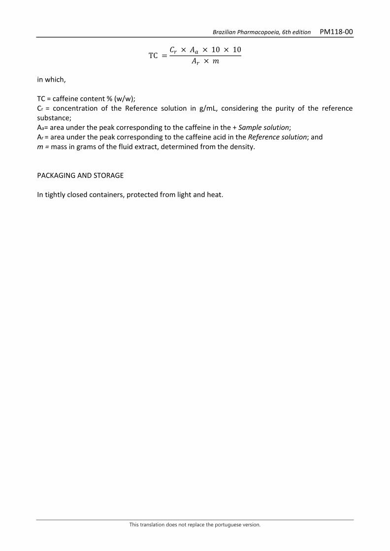
Brazilian Pharmacopoeia, 6th edition PM118-00
This translation does not replace the portuguese version.
TC =𝐶𝐶𝑟𝑟 × 𝑇𝑇𝑎𝑎 × 10 × 10
𝑇𝑇𝑟𝑟 × 𝑚𝑚
in which, TC = caffeine content % (w/w); Cr = concentration of the Reference solution in g/mL, considering the purity of the reference substance; Aa= area under the peak corresponding to the caffeine in the + Sample solution; Ar = area under the peak corresponding to the caffeine acid in the Reference solution; and m = mass in grams of the fluid extract, determined from the density. PACKAGING AND STORAGE In tightly closed containers, protected from light and heat.
Brazilian Pharmacopoeia, 6th edition PM119-00
This translation does not replace the portuguese version.
WITCHHAZEL, fluid extract Hamamelidis extracta fluida
The fluid extract is obtained from dried leaves of Hamamelis virginiana L., containing at least 3.0% tannins (w/w), expressed in pyrogallol (C6H6O3, 126.11). PREPARATION The fluid extract is prepared in a 1:1 (w/v) drug:solvent ratio by maceration or percolation, using 70% (v/v) ethyl alcohol as the extracting liquid. CHARACTERISTICS Dark-green colored liquid. IDENTIFICATION Proceed as described in Thin-layer chromatography (5.2.17.1). Stationary phase: silica gel F254. Mobile phase: ethyl acetate, toluene, formic acid and water (60:20:20:15). Sample solution: take 1 mL of extract and dry until residue is formed, in a water bath at a temperature not exceeding 60 ºC. Suspend the residue in 1 mL of methyl alcohol and proceed with the chromatographic analysis. Reference solution (1): dissolve an exactly weighed amount of gallic acid in methyl alcohol to obtain the concentration of 1000 µg/mL. Reference solution (2): dissolve an exactly weighed amount of hamamelitanin in methyl alcohol to obtain the concentration of 1000 µg/mL. TLC visualization reagent: ferric chloride at 1% (w/v). Procedure: apply 10 μL of the Sample solution, 10 μL of the Reference solution (1) and 10 μL of the Reference solution (2) to the chromatoplate, separately and in the form of a band. Conduct chromatogram. Remove chromatoplate and allow it to air dry. Nebulize the plate with 1% (w/v) ferric chloride, heat at 100°C to 105°C for approximately five minutes. Examine under visible light. Results: the diagram below shows the sequences of zones obtained with the reference Solution (1), the Reference solution (2) and the Sample Solution. Other zones may occasionally develop.

Brazilian Pharmacopoeia, 6th edition PM119-00
This translation does not replace the portuguese version.
Top of the plate
Gallic acid: purple-colored zone
Purple-colored zone
Hamamelitannin: purple-colored zone
Purple-colored zone
Reference solution Sample solution TESTS Relative density (5.2.5). 1.0394 to 1.0409. Ethyl alcohol (5.3.3.8.1). Method II, Liquids with more than 30% alcohol. 38% (v/v) to 44% (v/v). Methyl alcohol and isopropyl alcohol (5.4.2.2.1). Complies with the test. Dry residue (5.4.2.2.2). At least 30.0% (w/w). Total mesophilic microorganism count (5.5.3.1.2). Complies with the test. Study of pathogenic microorganisms (5.5.3.1.3). Complies with the test. DOSAGE Total tannins Note: protect samples from light during extraction and dilution. Use carbon dioxide-free water for all operations. To proceed as described in Visible absorption spectrophotometry (5.2.14). Prepare solutions as described below. Stock solution: pipette 750 μL of witch hazel fluid extract and transfer to a 250-mL volumetric flask. Rinse the pipette with distilled water, to off the volume to 250 mL with water and homogenize. Let the solution stand to allow the solids to precipitate. Filter through a 125-mm diameter filter paper. Discard the first 50 mL of the filtrate. Sample solution for total polyphenol content: dilute 5 mL of the Stock solution in a 25-mL volumetric flask with water and homogenize. Volumetrically transfer 2 mL of the solution, 1 mL of phosphomolybdotungstic reagent and 10 mL of water to a 25-mL volumetric flask, top off the volume with sodium carbonate RS solution and homogenize. Determine absorbance at 760 nm (A1), after 30 minutes, protected from light, using water for zero adjustment.
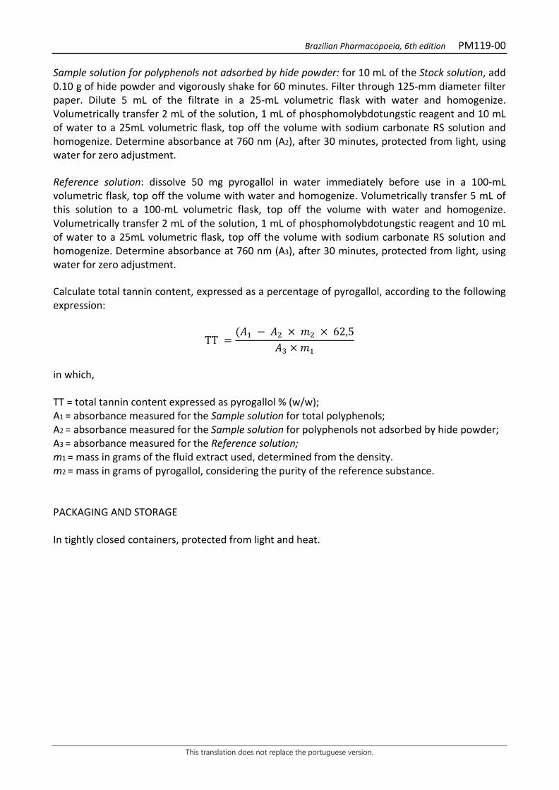
Brazilian Pharmacopoeia, 6th edition PM119-00
This translation does not replace the portuguese version.
Sample solution for polyphenols not adsorbed by hide powder: for 10 mL of the Stock solution, add 0.10 g of hide powder and vigorously shake for 60 minutes. Filter through 125-mm diameter filter paper. Dilute 5 mL of the filtrate in a 25-mL volumetric flask with water and homogenize. Volumetrically transfer 2 mL of the solution, 1 mL of phosphomolybdotungstic reagent and 10 mL of water to a 25mL volumetric flask, top off the volume with sodium carbonate RS solution and homogenize. Determine absorbance at 760 nm (A2), after 30 minutes, protected from light, using water for zero adjustment. Reference solution: dissolve 50 mg pyrogallol in water immediately before use in a 100-mL volumetric flask, top off the volume with water and homogenize. Volumetrically transfer 5 mL of this solution to a 100-mL volumetric flask, top off the volume with water and homogenize. Volumetrically transfer 2 mL of the solution, 1 mL of phosphomolybdotungstic reagent and 10 mL of water to a 25mL volumetric flask, top off the volume with sodium carbonate RS solution and homogenize. Determine absorbance at 760 nm (A3), after 30 minutes, protected from light, using water for zero adjustment. Calculate total tannin content, expressed as a percentage of pyrogallol, according to the following expression:
TT =(𝑇𝑇1 − 𝑇𝑇2 × 𝑚𝑚2 × 62,5
𝑇𝑇3 × 𝑚𝑚1
in which, TT = total tannin content expressed as pyrogallol % (w/w); A1 = absorbance measured for the Sample solution for total polyphenols; A2 = absorbance measured for the Sample solution for polyphenols not adsorbed by hide powder; A3 = absorbance measured for the Reference solution; m1 = mass in grams of the fluid extract used, determined from the density. m2 = mass in grams of pyrogallol, considering the purity of the reference substance. PACKAGING AND STORAGE In tightly closed containers, protected from light and heat.

Brazilian Pharmacopoeia, 6th edition PM120-00
This translation does not replace the portuguese version.
BITTER ORANGE, fluid extract Aurantii amari exocarpium extracta fluida
The fluid extract is obtained from dry portions of the exocarp, corresponding to the flavedo of the ripe fruit, free from most of the mesocarp, corresponding to the albedo of Citrus aurantium L. subsp. aurantium [syn. Citrus aurantium L. subsp. amara (L.) Engler], containing at least 2.0% of naringin (C27H32O14, 580.54). PREPARATION The fluid extract is prepared in a 1:1 (w/v) drug:solvent ratio by percolation or maceration, using 70% (v/v) ethyl alcohol as the extracting liquid. CHARACTERISTICS Yellowish-brown or reddish-brown liquid with a citrus odor. IDENTIFICATION Proceed as described in Thin-layer chromatography (5.2.17.1). Stationary phase: silica gel G60. Mobile phase: ethyl acetate, water and formic acid (75:15:10). Sample solution: dilute 0.3 mL of the bitter orange fluid extract in 0.7 mL ethyl alcohol. Reference solution: prepare a 10 mg/mL solution of naringin in methyl alcohol. TLC visualization reagent (1): dissolve 1 g of aminoethanol diphenylborate in methyl alcohol and top off to 100 mL with the same solvent. TLC visualization reagent (2): 5% macrogol 400 solution in methyl alcohol. Procedure: apply 10 μL of the Sample solution and 10 μL of the Reference solution to the chromatoplate, separately and in the form of a band. Conduct chromatogram. Remove chromatoplate and allow it to air dry for 15 minutes. Nebulize the plate with aminoethanol diphenylborate RS (natural reagent A), then with a solution of 5% macrogol 400 in methyl alcohol. Examine under ultraviolet light at 365 nm after at least two hours. Results: the diagram below shows the sequences of zones obtained with the Reference solution and the Sample solution. Other zones may occasionally develop.
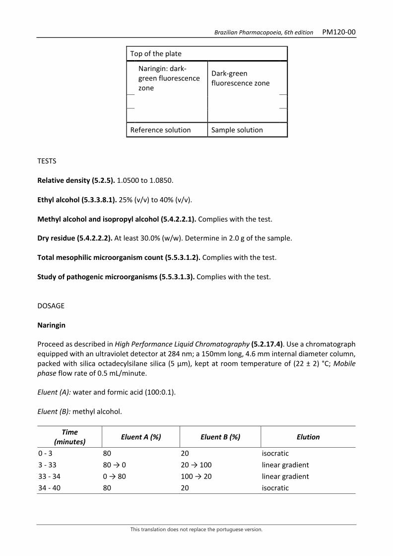
Brazilian Pharmacopoeia, 6th edition PM120-00
This translation does not replace the portuguese version.
Top of the plate
Naringin: dark-green fluorescence zone
Dark-green fluorescence zone
Reference solution Sample solution TESTS Relative density (5.2.5). 1.0500 to 1.0850. Ethyl alcohol (5.3.3.8.1). 25% (v/v) to 40% (v/v). Methyl alcohol and isopropyl alcohol (5.4.2.2.1). Complies with the test. Dry residue (5.4.2.2.2). At least 30.0% (w/w). Determine in 2.0 g of the sample. Total mesophilic microorganism count (5.5.3.1.2). Complies with the test. Study of pathogenic microorganisms (5.5.3.1.3). Complies with the test. DOSAGE Naringin Proceed as described in High Performance Liquid Chromatography (5.2.17.4). Use a chromatograph equipped with an ultraviolet detector at 284 nm; a 150mm long, 4.6 mm internal diameter column, packed with silica octadecylsilane silica (5 μm), kept at room temperature of (22 ± 2) °C; Mobile phase flow rate of 0.5 mL/minute. Eluent (A): water and formic acid (100:0.1). Eluent (B): methyl alcohol.
Time (minutes) Eluent A (%) Eluent B (%) Elution
0 - 3 80 20 isocratic 3 - 33 80 → 0 20 → 100 linear gradient 33 - 34 0 → 80 100 → 20 linear gradient 34 - 40 80 20 isocratic
Brazilian Pharmacopoeia, 6th edition PM120-00
This translation does not replace the portuguese version.
Sample solution: dilute 0.200 mL of bitter orange fluid extract to 25 mL with a mixture of methyl alcohol and water (1:1). Filter through a 0.45 µm filter unit. Reference solution: dissolve a precisely weighed amount of naringin in a solution of methyl alcohol and water (1:1), in order to obtain a solution with a concentration of 0.250 mg/mL. Filter through a 0.45 µm filter unit. Procedure: separately inject 10 μL of the Reference solution and 10 µL of the Sample solution. Record chromatograms and measure the area under the peak corresponding to the naringin. The average retention time is approximately 17.6 minutes. Calculate naringin content, in percent, according to the following expression:
TN =𝐶𝐶𝑟𝑟 × 𝑇𝑇𝑎𝑎 × 25 × 100
𝑇𝑇𝑟𝑟 × m
in which, TN = naringin content % (w/w); Cr = concentration of the Reference solution in g/mL, considering the purity of the reference substance; Ar= area under the peak corresponding to the naringin in the Reference solution. Aa = area under the peak corresponding to the naringin in the Sample solution; m = mass in grams of the fluid extract used, determined from the density. PACKAGING AND STORAGE In tightly closed containers, protected from light and heat.
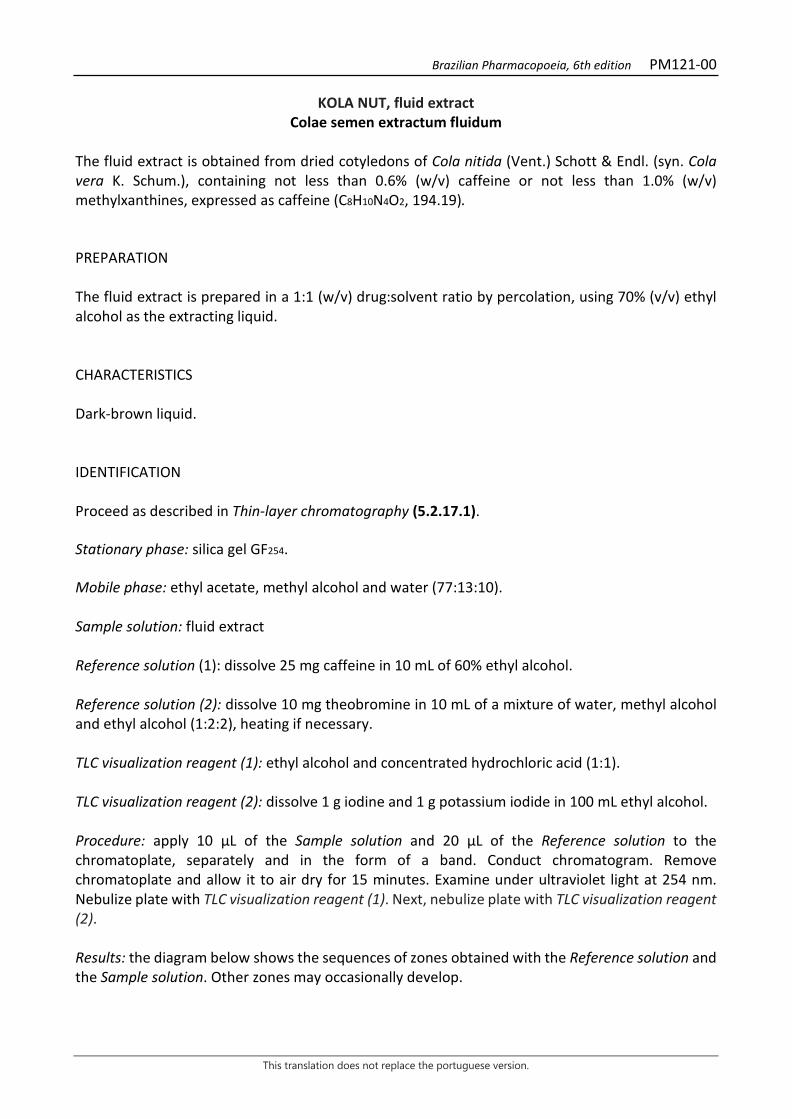
Brazilian Pharmacopoeia, 6th edition PM121-00
This translation does not replace the portuguese version.
KOLA NUT, fluid extract Colae semen extractum fluidum
The fluid extract is obtained from dried cotyledons of Cola nitida (Vent.) Schott & Endl. (syn. Cola vera K. Schum.), containing not less than 0.6% (w/v) caffeine or not less than 1.0% (w/v) methylxanthines, expressed as caffeine (C8H10N4O2, 194.19). PREPARATION The fluid extract is prepared in a 1:1 (w/v) drug:solvent ratio by percolation, using 70% (v/v) ethyl alcohol as the extracting liquid. CHARACTERISTICS Dark-brown liquid. IDENTIFICATION Proceed as described in Thin-layer chromatography (5.2.17.1). Stationary phase: silica gel GF254. Mobile phase: ethyl acetate, methyl alcohol and water (77:13:10). Sample solution: fluid extract Reference solution (1): dissolve 25 mg caffeine in 10 mL of 60% ethyl alcohol. Reference solution (2): dissolve 10 mg theobromine in 10 mL of a mixture of water, methyl alcohol and ethyl alcohol (1:2:2), heating if necessary. TLC visualization reagent (1): ethyl alcohol and concentrated hydrochloric acid (1:1). TLC visualization reagent (2): dissolve 1 g iodine and 1 g potassium iodide in 100 mL ethyl alcohol. Procedure: apply 10 μL of the Sample solution and 20 μL of the Reference solution to the chromatoplate, separately and in the form of a band. Conduct chromatogram. Remove chromatoplate and allow it to air dry for 15 minutes. Examine under ultraviolet light at 254 nm. Nebulize plate with TLC visualization reagent (1). Next, nebulize plate with TLC visualization reagent (2). Results: the diagram below shows the sequences of zones obtained with the Reference solution and the Sample solution. Other zones may occasionally develop.
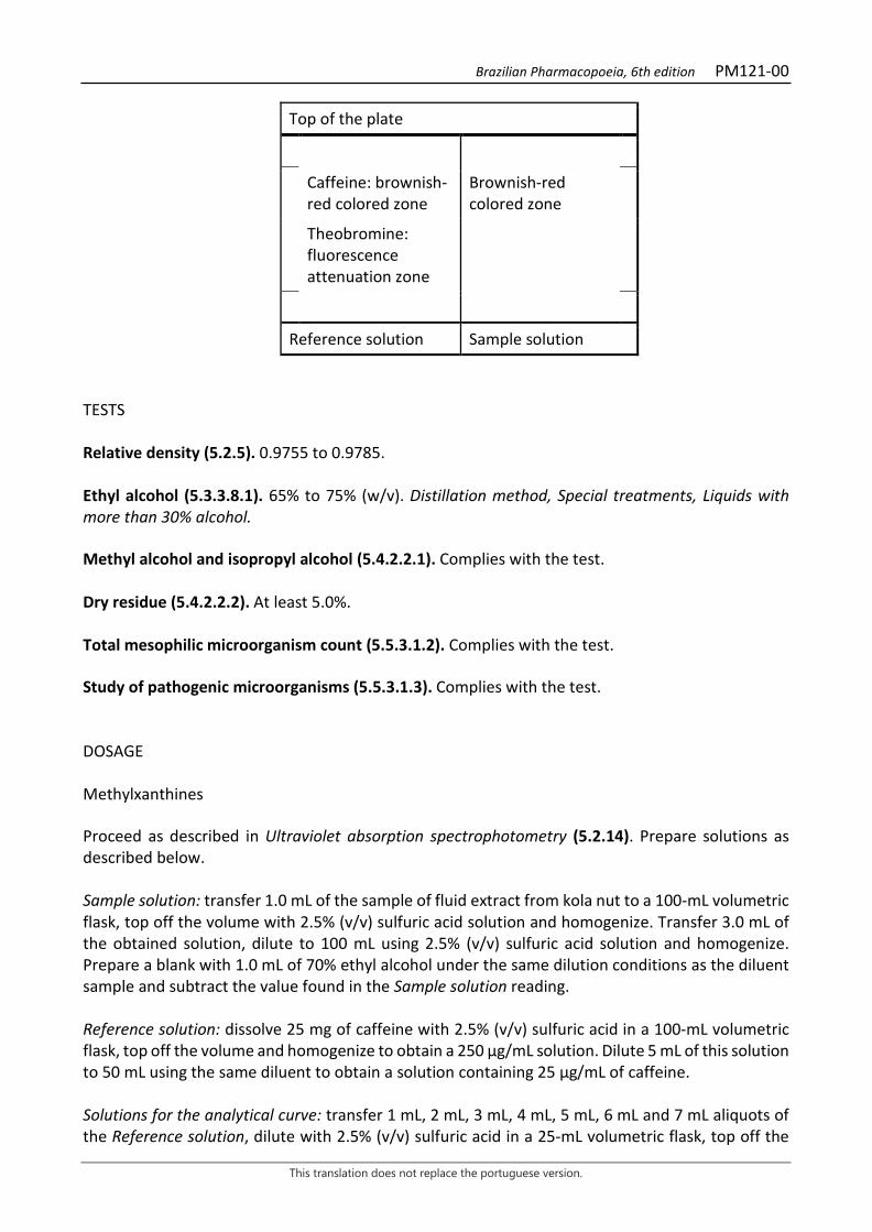
Brazilian Pharmacopoeia, 6th edition PM121-00
This translation does not replace the portuguese version.
Top of the plate
Caffeine: brownish-red colored zone
Brownish-red colored zone
Theobromine: fluorescence attenuation zone
Reference solution Sample solution
TESTS Relative density (5.2.5). 0.9755 to 0.9785. Ethyl alcohol (5.3.3.8.1). 65% to 75% (w/v). Distillation method, Special treatments, Liquids with more than 30% alcohol. Methyl alcohol and isopropyl alcohol (5.4.2.2.1). Complies with the test. Dry residue (5.4.2.2.2). At least 5.0%. Total mesophilic microorganism count (5.5.3.1.2). Complies with the test. Study of pathogenic microorganisms (5.5.3.1.3). Complies with the test. DOSAGE Methylxanthines Proceed as described in Ultraviolet absorption spectrophotometry (5.2.14). Prepare solutions as described below. Sample solution: transfer 1.0 mL of the sample of fluid extract from kola nut to a 100-mL volumetric flask, top off the volume with 2.5% (v/v) sulfuric acid solution and homogenize. Transfer 3.0 mL of the obtained solution, dilute to 100 mL using 2.5% (v/v) sulfuric acid solution and homogenize. Prepare a blank with 1.0 mL of 70% ethyl alcohol under the same dilution conditions as the diluent sample and subtract the value found in the Sample solution reading. Reference solution: dissolve 25 mg of caffeine with 2.5% (v/v) sulfuric acid in a 100-mL volumetric flask, top off the volume and homogenize to obtain a 250 µg/mL solution. Dilute 5 mL of this solution to 50 mL using the same diluent to obtain a solution containing 25 µg/mL of caffeine. Solutions for the analytical curve: transfer 1 mL, 2 mL, 3 mL, 4 mL, 5 mL, 6 mL and 7 mL aliquots of the Reference solution, dilute with 2.5% (v/v) sulfuric acid in a 25-mL volumetric flask, top off the
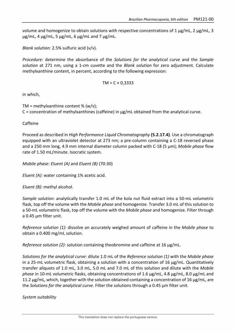
Brazilian Pharmacopoeia, 6th edition PM121-00
This translation does not replace the portuguese version.
volume and homogenize to obtain solutions with respective concentrations of 1 µg/mL, 2 µg/mL, 3 µg/mL, 4 µg/mL, 5 µg/mL, 6 µg/mL and 7 µg/mL. Blank solution: 2.5% sulfuric acid (v/v). Procedure: determine the absorbance of the Solutions for the analytical curve and the Sample solution at 271 nm, using a 1-cm cuvette and the Blank solution for zero adjustment. Calculate methylxanthine content, in percent, according to the following expression:
TM = C × 0,3333 in which, TM = methylxanthine content % (w/v); C = concentration of methylxanthines (caffeine) in µg/mL obtained from the analytical curve. Caffeine Proceed as described in High Performance Liquid Chromatography (5.2.17.4). Use a chromatograph equipped with an ultraviolet detector at 273 nm; a pre-column containing a C-18 reversed phase and a 250 mm long, 4.9 mm internal diameter column packed with C-18 (5 μm); Mobile phase flow rate of 1.50 mL/minute. Isocratic system. Mobile phase: Eluent (A) and Eluent (B) (70:30) Eluent (A): water containing 1% acetic acid. Eluent (B): methyl alcohol. Sample solution: analytically transfer 1.0 mL of the kola nut fluid extract into a 50-mL volumetric flask, top off the volume with the Mobile phase and homogenize. Transfer 3.0 mL of this solution to a 50-mL volumetric flask, top off the volume with the Mobile phase and homogenize. Filter through a 0.45 µm filter unit. Reference solution (1): dissolve an accurately weighed amount of caffeine in the Mobile phase to obtain a 0.400 mg/mL solution. Reference solution (2): solution containing theobromine and caffeine at 16 µg/mL. Solutions for the analytical curve: dilute 1.0 mL of the Reference solution (1) with the Mobile phase in a 25-mL volumetric flask, obtaining a solution with a concentration of 16 µg/mL. Quantitatively transfer aliquots of 1.0 mL, 3.0 mL, 5.0 mL and 7.0 mL of this solution and dilute with the Mobile phase in 10-mL volumetric flasks, obtaining concentrations of 1.6 µg/mL, 4.8 µg/mL, 8.0 µg/mL and 11.2 µg/mL, which, together with the solution obtained containing a concentration of 16 µg/mL, are the Solutions for the analytical curve. Filter the solutions through a 0.45 µm filter unit. System suitability
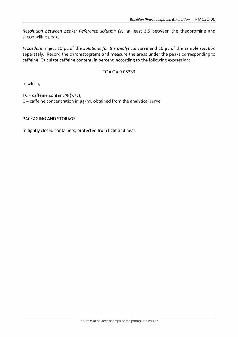
Brazilian Pharmacopoeia, 6th edition PM121-00
This translation does not replace the portuguese version.
Resolution between peaks: Reference solution (2), at least 2.5 between the theobromine and theophylline peaks. Procedure: inject 10 µL of the Solutions for the analytical curve and 10 µL of the sample solution separately. Record the chromatograms and measure the areas under the peaks corresponding to caffeine. Calculate caffeine content, in percent, according to the following expression:
TC = C × 0.08333 in which, TC = caffeine content % (w/v); C = caffeine concentration in µg/mL obtained from the analytical curve. PACKAGING AND STORAGE In tightly closed containers, protected from light and heat.

Brazilian Pharmacopoeia, 6th edition PM122-00
This translation does not replace the portuguese version.
NUX-VOMICA, fluid extract Strychni extracta fluid
The fluid extract is obtained from dried seeds of Strychnos nux-vomica L., containing at least 0.5% strychnine (C21H22 N2O2, 334.42). PREPARATION The fluid extract is prepared in a 1:1 (w/v) drug:solvent ratio by percolation or maceration, using 70% (v/v) ethyl alcohol as the extracting liquid. CHARACTERISTICS Dark brown colored liquid. IDENTIFICATION Proceed as described in Thin-layer chromatography (5.2.17.1). Stationary phase: silica gel F254 (0.25 mm). Mobile phase: butyl alcohol, water and glacial acetic acid (70:20:10). Sample solution: dry 1.0 mL of fluid extract until residue is formed, in a water bath, at a maximum temperature of 60 °C. Suspend the residue in 5 mL methyl alcohol, filter through a 0.45 µm filter unit and proceed to chromatographic analysis. Reference solution (1): dissolve an accurately weighed amount of strychnine in methyl alcohol to obtain a concentration of 500 µg/mL. Reference solution (2): dissolve an accurately weighed amount of brucine in methyl alcohol to obtain a concentration of 500 µg/mL. Procedure: apply 20 μL of the Sample solution, 20 μL of the Reference solution (1) and 20 μL of the Reference solution (2) to the chromatoplate, separately and in the form of a band. Conduct chromatogram. Remove chromatoplate and allow it to air dry. Nebulize the plate with 10% sulfuric acid solution in ethyl alcohol, and then with aqueous acetic potassium iodobismuthate solution. Remove the chromatoplate and allow to air dry for five minutes; examine under visible light. Results: the diagram below shows the sequences of zones obtained with the Reference solution (1), the Referece solution (2) and the Sample solution. Other zones may occasionally develop.
Brazilian Pharmacopoeia, 6th edition PM122-00
This translation does not replace the portuguese version.
Top of the plate
Strychnine: orange colored zone
Orange-colored zone
Orange-colored zone
Brucine: orange-colored zone
Orange-colored zone
Reference solution Sample solution
TESTS Relative density (5.2.5). 0.985 to 1.000. Ethyl alcohol (5.3.3.8.1). Method II. 52% (v/v) to 56% (v/v). Methyl alcohol and isopropyl alcohol (5.4.2.2.1). Complies with the test. Dry residue (5.4.2.2.2). At least 18.0% (w/w). Total mesophilic microorganism count (5.5.3.1.2). Complies with the test. Study of pathogenic microorganisms (5.5.3.1.3). Complies with the test. DOSAGE Strychnine Proceed as described in High Performance Liquid Chromatography (5.2.17.4). Use a chromatograph equipped with an ultraviolet detector at 210 nm; pre-column packed with octadecylsilane silica, 150 mm long, 4.6 mm internal diameter column, packed with octadecylsilane silica (5 µm), kept at 30 °C; Mobile phase flow rate of 1.3 mL/minute. Isocratic system. Mobile phase: dibasic potassium phosphate buffer (7 g/L), pH 3,0 adjusted with phosphoric acid, acetonitrile and diethylamine (900:100:20). Reference solution: weigh 15 mg of strychnine. Transfer 10 mL to a volumetric flask, dissolve the Mobile phase, top off the volume with the Mobile phase and homogenize. Filter the solution through a 0.45 µm filter unit. Sample solution: homogenize the sample in an ultrasonic bath for five minutes, pipette 1.0 mL of the fluid extract and transfer to a 5-mL volumetric flask. Clean the micropipette tip at least twice
Brazilian Pharmacopoeia, 6th edition PM122-00
This translation does not replace the portuguese version.
with the Mobile Phase. Top off the volume with the Mobile phase and homogenize. Filter the solution through a 0.45 µm filter unit. Procedure: separately inject 10 μL of the Reference solution and 10 µL of the Sample solution. Register the chromatograms and measure the areas under the strychnine peaks. Calculate strychnine content, in percent, according to the following expression:
TE =𝑇𝑇𝑎𝑎 × 𝐶𝐶𝑟𝑟 𝑇𝑇𝑟𝑟 × 𝐶𝐶𝑎𝑎
in which, TE = strychnine content % (w/w); Ar= area under the peak corresponding to the strychnine in the Reference solution; Aa= area under the peak corresponding to the strychnine in the Sample solution; Cr = concentration of strychnine in the Reference solution in mg/mL, considering the purity of the reference substance; Ca = concentration of the plant drug in the Sample solution in mg/mL. PACKAGING AND STORAGE In tightly closed containers, protected from light and heat.
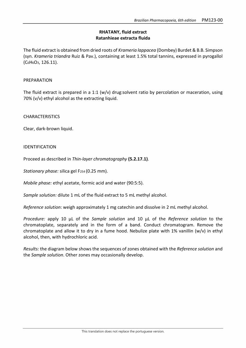
Brazilian Pharmacopoeia, 6th edition PM123-00
This translation does not replace the portuguese version.
RHATANY, fluid extract Ratanhieae extracta fluida
The fluid extract is obtained from dried roots of Krameria lappacea (Dombey) Burdet & B.B. Simpson (syn. Krameria triandra Ruiz & Pav.), containing at least 1.5% total tannins, expressed in pyrogallol (C6H6O3, 126.11). PREPARATION The fluid extract is prepared in a 1:1 (w/v) drug:solvent ratio by percolation or maceration, using 70% (v/v) ethyl alcohol as the extracting liquid. CHARACTERISTICS Clear, dark-brown liquid. IDENTIFICATION Proceed as described in Thin-layer chromatography (5.2.17.1). Stationary phase: silica gel F254 (0.25 mm). Mobile phase: ethyl acetate, formic acid and water (90:5:5). Sample solution: dilute 1 mL of the fluid extract to 5 mL methyl alcohol. Reference solution: weigh approximately 1 mg catechin and dissolve in 2 mL methyl alcohol. Procedure: apply 10 μL of the Sample solution and 10 μL of the Reference solution to the chromatoplate, separately and in the form of a band. Conduct chromatogram. Remove the chromatoplate and allow it to dry in a fume hood. Nebulize plate with 1% vanillin (w/v) in ethyl alcohol, then, with hydrochloric acid. Results: the diagram below shows the sequences of zones obtained with the Reference solution and the Sample solution. Other zones may occasionally develop.

Brazilian Pharmacopoeia, 6th edition PM123-00
This translation does not replace the portuguese version.
Top of the plate
Catechin: reddish-pink zone
Reddish-pink colored zone
Reddish-pink colored zone Reddish-pink colored zone Reddish-pink zone Reddish-pink colored zone
Reddish-pink colored zone
Reference solution Sample solution TESTS Relative density (5.2.5). 0.9687 to 0.9688. Ethyl alcohol (5.3.3.8.1). 63% (v/v) to 66% (v/v). Methyl alcohol and isopropyl alcohol (5.4.2.2.1). Complies with the test. Dry residue (5.4.2.2.2). At least 5.0% (w/w). Total mesophilic microorganism count (5.5.3.1.2). Complies with the test. Study of pathogenic microorganisms (5.5.3.1.3). Complies with the test. DOSAGE Total tannins To proceed as described in Visible absorption spectrophotometry (5.2.14). Prepare solutions described below. Stock solution: accurately weigh about 1.5 g of fluid extract, in a 250-mL volumetric flask, top off the volume with water and homogenize. Filter through paper filter. Discard the first 50 mL of the filtrate. Sample solution for total polyphenol content: volumetrically transfer 5 mL from the Stock solution to a 25-mL volumetric flask, top off the volume with water and homogenize. Volumetrically transfer 2 mL of the solution, 1 mL of phosphomolybdotungstic reagent and 10 mL of distilled water to a 25mL volumetric flask, top off the volume with 29% sodium carbonate solution (w/v) and homogenize. Determine absorbance at 760 nm (A1) after 30 minutes, using water for zero adjustment.
Brazilian Pharmacopoeia, 6th edition PM123-00
This translation does not replace the portuguese version.
Sample solution for polyphenols not adsorbed by hide powder: for 10 mL of the Stock solution, add 0.1 g of the hide powder and mechanically shake in a 125-mL erlenmeyer flask for 60 minutes. Filter through paper filter. Volumetrically transfer 5 mL of this solution to a 25-mL volumetric flask, top off the volume with water and homogenize. Volumetrically transfer 2 mL of the solution, 1 mL of phosphomolybdotungstic reagent and 10 mL of water to a 25-mL volumetric flask, top off the volume with 29% sodium carbonate solution (w/v) and homogenize. Determine absorbance at 760 nm (A2) after 30 minutes, using water for zero adjustment. Reference solution: dissolve 50 mg pyrogallol in water immediately before use, transfer to a 100-mL volumetric flask, top off the volume with water and homogenize. Volumetrically transfer 5 mL of this solution to a 100-mL volumetric flask, top off the volume with water and homogenize. Volumetrically transfer 2 mL of the solution, 1 mL of phosphomolybdotungstic reagent and 10 mL of water to a 25-mL volumetric flask, top off the volume with 29% sodium carbonate solution (w/v) and homogenize. Determine absorbance at 760 nm (A3) after 30 minutes, using water for zero adjustment. Calculate total tannin content, expressed as a percentage of pyrogallol, according to the following expression:
TT =(𝑇𝑇1 + 𝑇𝑇2) × 𝑚𝑚2 × 62,5
𝑇𝑇3 × 𝑚𝑚1
in which, TT = total tannin content expressed as pyrogallol % (w/w); A1 = absorbance measured for the Sample solution for total polyphenols; A2 = absorbance measured for the Sample solution for polyphenols not adsorbed by hide powder; A3 = absorbance measured for the Reference solution; m1 = mass in grams of the fluid extract used; m2 = mass in grams of pyrogallol, considering the purity of the reference substance. PACKAGING AND STORAGE In tightly closed containers, protected from light and heat.

Brazilian Pharmacopoeia, 6th edition PM124-00
This translation does not replace the portuguese version.
VALERIAN, fluid extract Valerianae extracta fluida
The fluid extract is obtained from the underground organs (roots, rhizomes and stolons), dried, of Valeriana officinalis L., containing at least 0.15% total sesquiterpene acids, expressed in valerenic acid (C15H22O2, 234.34). PREPARATION The fluid extract is prepared in a 1:1 (w/v) drug:solvent ratio by maceration or percolation, using 70% (v/v) ethyl alcohol as the extracting liquid. CHARACTERISTICS Dark brown liquid with a strong, persistent odor. IDENTIFICATION Proceed as described in Thin-layer chromatography (5.2.17.1). Stationary phase: silica gel GF254. Mobile phase: cyclohexane, ethyl acetate and glacial acetic acid (60:38:2). Sample solution: measure 1mL of fluid extract and dry until residue is formed, in a water bath at a temperature not exceeding 60 ºC. Suspend the residue in 1 mL of methyl alcohol and proceed with the chromatographic analysis. Reference solution (1): dissolve an exactly weighed amount of valerenic acid in methyl alcohol to obtain the concentration of 100 µg/mL. Reference solution (2): dissolve an exactly weighed amount of acetoxyvalerenic acid in methyl alcohol to obtain the concentration of 100 µg/mL. Procedure: apply 15 μL of the Sample solution, 15 μL of the Reference solution (1) and 15 μL of the reference solution (2) to the chromatoplate, separately and in the form of a band. Remove chromatoplate and allow it to air dry. Nebulize the plate with anisaldehyde RS, heat at 100 °C to 105 °C for approximately five minutes. Examine under visible light. Results: the diagram below shows the sequences of zones obtained with the Sample solution, Reference solution (1) and Reference solution (2). Other zones may occasionally develop.
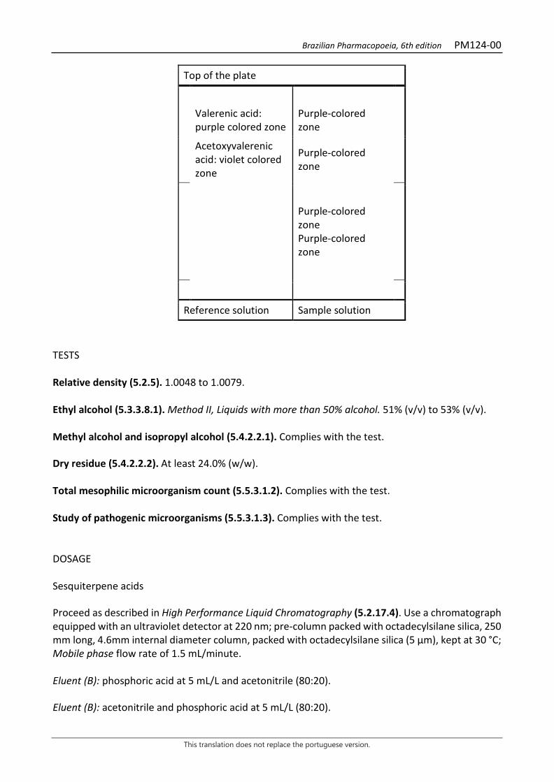
Brazilian Pharmacopoeia, 6th edition PM124-00
This translation does not replace the portuguese version.
Top of the plate
Valerenic acid: purple colored zone
Purple-colored zone
Acetoxyvalerenic acid: violet colored zone
Purple-colored zone
Purple-colored zone Purple-colored zone
Reference solution Sample solution
TESTS Relative density (5.2.5). 1.0048 to 1.0079. Ethyl alcohol (5.3.3.8.1). Method II, Liquids with more than 50% alcohol. 51% (v/v) to 53% (v/v). Methyl alcohol and isopropyl alcohol (5.4.2.2.1). Complies with the test. Dry residue (5.4.2.2.2). At least 24.0% (w/w). Total mesophilic microorganism count (5.5.3.1.2). Complies with the test. Study of pathogenic microorganisms (5.5.3.1.3). Complies with the test. DOSAGE Sesquiterpene acids Proceed as described in High Performance Liquid Chromatography (5.2.17.4). Use a chromatograph equipped with an ultraviolet detector at 220 nm; pre-column packed with octadecylsilane silica, 250 mm long, 4.6mm internal diameter column, packed with octadecylsilane silica (5 µm), kept at 30 °C; Mobile phase flow rate of 1.5 mL/minute. Eluent (B): phosphoric acid at 5 mL/L and acetonitrile (80:20). Eluent (B): acetonitrile and phosphoric acid at 5 mL/L (80:20).
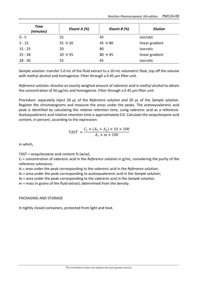
Brazilian Pharmacopoeia, 6th edition PM124-00
This translation does not replace the portuguese version.
Time (minutes) Eluent A (%) Eluent B (%) Elution
0 - 5 55 45 isocratic 5 - 15 55 → 20 45 → 80 linear gradient 15 - 25 20 80 isocratic 25 - 28 20 → 55 80 → 45 linear gradient 28 - 30 55 45 isocratic Sample solution: transfer 5.0 mL of the fluid extract to a 10-mL volumetric flask, top off the volume with methyl alcohol and homogenize. Filter through a 0.45 µm filter unit. Reference solution: dissolve an exactly weighed amount of valerenic acid in methyl alcohol to obtain the concentration of 50 µg/mL and homogenize. Filter through a 0.45 µm filter unit. Procedure: separately inject 20 μL of the Reference solution and 20 µL of the Sample solution. Register the chromatograms and measure the areas under the peaks. The acetoxyvalerenic acid peak is identified by calculating the relative retention time, using valerenic acid as a reference. Acetoxyvalerenic acid relative retention time is approximately 0.6. Calculate the sesquiterpene acid content, in percent, according to the expression:
TAST =𝐶𝐶𝑟𝑟 × (𝑇𝑇1 + 𝑇𝑇2) × 10 × 100
𝑇𝑇𝑟𝑟 × m × 100
in which, TAST = sesquiterpene acid content % (w/w); Cr = concentration of valerenic acid in the Reference solution in g/mL, considering the purity of the reference substance; Ar = area under the peak corresponding to the valerenic acid in the Reference solution; A1 = area under the peak corresponding to acetoxyvalerenic acid in the Sample solution; A2 = area under the peak corresponding to the valerenic acid in the Sample solution; m = mass in grams of the fluid extract, determined from the density. PACKAGING AND STORAGE In tightly closed containers, protected from light and heat.

Brazilian Pharmacopoeia, 6th edition
This translation does not replace the portuguese version.
OILS, FATS AND WAXES
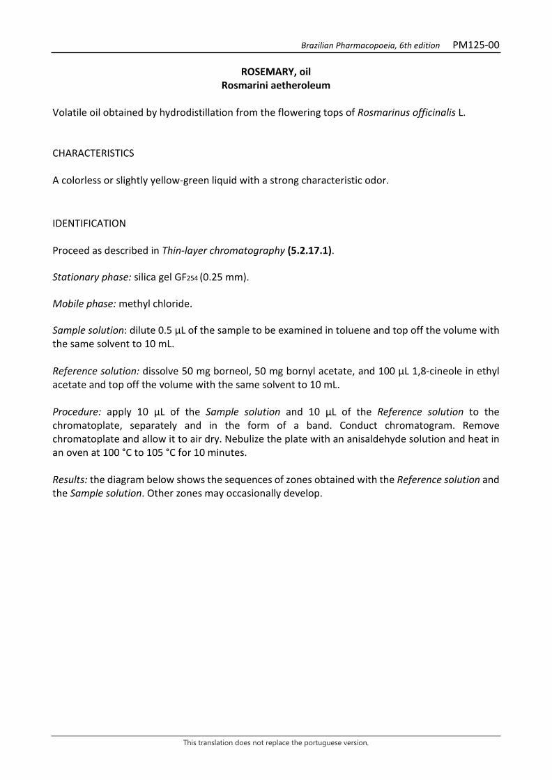
Brazilian Pharmacopoeia, 6th edition PM125-00
This translation does not replace the portuguese version.
ROSEMARY, oil Rosmarini aetheroleum
Volatile oil obtained by hydrodistillation from the flowering tops of Rosmarinus officinalis L. CHARACTERISTICS A colorless or slightly yellow-green liquid with a strong characteristic odor. IDENTIFICATION Proceed as described in Thin-layer chromatography (5.2.17.1). Stationary phase: silica gel GF254 (0.25 mm). Mobile phase: methyl chloride. Sample solution: dilute 0.5 µL of the sample to be examined in toluene and top off the volume with the same solvent to 10 mL. Reference solution: dissolve 50 mg borneol, 50 mg bornyl acetate, and 100 μL 1,8-cineole in ethyl acetate and top off the volume with the same solvent to 10 mL. Procedure: apply 10 μL of the Sample solution and 10 μL of the Reference solution to the chromatoplate, separately and in the form of a band. Conduct chromatogram. Remove chromatoplate and allow it to air dry. Nebulize the plate with an anisaldehyde solution and heat in an oven at 100 °C to 105 °C for 10 minutes. Results: the diagram below shows the sequences of zones obtained with the Reference solution and the Sample solution. Other zones may occasionally develop.
Brazilian Pharmacopoeia, 6th edition PM125-00
This translation does not replace the portuguese version.
Top of the plate
Intense red-colored zone
Bornyl acetate: greenish-yellow colored zone
Greenish-yellow colored zone
1,8-Cineole: purple-colored zone
Purple-colored zone
Red-colored zone
Borneol: green-colored zone with yellow border
Green-colored zone with yellow border
Purple-colored zone
Reference solution Sample solution TESTS Relative density (5.2.5). 0.894 to 0.912. Refractive index (5.2.29.4). 1.460 to 1.476. Optical rotation (5.2.8). -5° a +8°. Acidity level (5.2.29.7). At most 1.0%. Chromatographic profile. Proceed as described in Gas chromatography (5.2.17.5). Use a chromatograph equipped with a flame ionization detector, using a mixture of nitrogen, hydrogen and synthetic air (1:1:10) as auxiliary gases to the detector flame; 60 m long and 0.25 mm wide (internal diameter) capillary column, coated with polyethylene glycol, with a 0.25 μm film thickness. Use purified helium as carrier gas (1 mL/minute). Temperature: Time (minutes) Temperature (ºC) Column 0 – 10
10 – 85 85 – 110
50 50 → 200 200
Injector 200 Detector 240

Brazilian Pharmacopoeia, 6th edition PM125-00
This translation does not replace the portuguese version.
Sample solution: dilute 0.2 mL of the rosemary volatile oil in 10 mL hexane. Store under refrigeration, in a tightly closed bottle and protected from light. Reference solution: dissolve 20 µL of α-pinene, 10 mg of camphene, 20 µL of β-pinene, 10 µL of β-myrcene, 20 µL of limonene, 50 µL of cineole, 10 µL of p-cymene, 50 mg of camphor, 30 mg of bornyl acetate, 10 mg of α-terpinol, 10 mg of borneol, and 10 µL of verbenone in 10 mL of hexane. Procedure: inject the volume of 1 μL of the Sample solution and 1 µL of the Reference solution (1) and 1 µL of the Reference solution (2) into the gas chromatograph, using a 1:50 flow division. Determine relative concentrations by electronic integration using the normalization method. Examine the chromatographic profile of the Sample Solution. The characteristic peaks in the chromatogram obtained with the Sample solution should have retention times similar to those obtained with the chromatogram of the Reference solution (1) and (2) or identification confirmed with gas chromatography coupled to a mass-selective detector operating under the same conditions as the as chromatography with a flame ionization detector (Figure 1). The chromatogram may also show the following compounds: bornyl acetate, borneol, - pinene, -myrcene, limonene, p-cymene, -terpineol and verbenone. Check the presence of the components, as follows, in the chromatogram obtained with the Sample solution: α-pinene, at least 9%; camphene, at least 2.5%; cineole, at least 16%; and camphor, at least 5%. PACKAGING AND STORAGE In tightly closed containers, protected from light and heat.
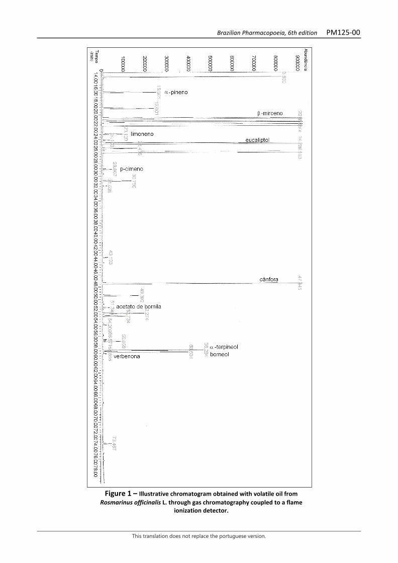
Brazilian Pharmacopoeia, 6th edition PM125-00
This translation does not replace the portuguese version.
Figure 1 – Illustrative chromatogram obtained with volatile oil from
Rosmarinus officinalis L. through gas chromatography coupled to a flame ionization detector.
Brazilian Pharmacopoeia, 6th edition PM126-00
This translation does not replace the portuguese version.
COTTON, refined oil Gossypii oleum raffinatum
Oil obtained from the seeds of Gossypium hirsutum L., subjected to a refining process. CHARACTERISTICS Pale yellow oily liquid. TESTS Water (5.2.20.1). Culombimetric method at most 0.1%. Determine in 1 g. Acidity level (5.2.29.7). At most 0.2. Peroxide Index (5.2.29.11). At most 10. Unsaponifiable substances (5.2.29.14). Method II. At most 1.5. Determine in 5 g. Alkaline impurities (5.2.29.15.2). Volume less than 0.1 mL of 0.01 M hydrochloric acid. Foreign oils in fixed oils by gas chromatography (5.2.29.15.4). Commercial mixtures of methyl esters can also be used as standard. The fraction of the cotton oil composed of fatty acids has the following composition: Fatty acids with a chain of less than 14 carbons, with up to one double bond: at most 0.2%. Myristic acid: 0.3% to 1.0%. Palmitic acid: 18.0% to 26.4%. Palmitoleic acid: at most 1.2%; Stearic acid: 2.1% to 3.3%; Oleic acid: 14.0% to 21.7%; Linoleic acid: 46.7% to 58.3%; Linolenic and γ-linolenic acid: at most 1.0%; Arachidonic acid: at most 1.0%; Eicosenoic acid: at most 0.5%; Behenic acid: at most 0.6%; Erucic acid: at most 0.5%; Lignoceric acid: at most 0.5%. PACKAGING AND STORAGE In tightly closed containers, protected from light and heat. CATEGORY

Brazilian Pharmacopoeia, 6th edition PM126-00
This translation does not replace the portuguese version.
Pharmacotechnical excipient.
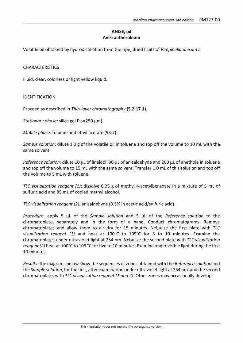
Brazilian Pharmacopoeia, 6th edition PM127-00
This translation does not replace the portuguese version.
ANISE, oil Anisi aetheroleum
Volatile oil obtained by hydrodistillation from the ripe, dried fruits of Pimpinella anisum L. CHARACTERISTICS Fluid, clear, colorless or light yellow liquid. IDENTIFICATION Proceed as described in Thin-layer chromatography (5.2.17.1). Stationary phase: silica gel F254(250 µm). Mobile phase: toluene and ethyl acetate (93:7). Sample solution: dilute 1.0 g of the volatile oil in toluene and top off the volume to 10 mL with the same solvent. Reference solution: dilute 10 µL of linalool, 30 µL of anisaldehyde and 200 µL of anethole in toluene and top off the volume to 15 mL with the same solvent. Transfer 1.0 mL of this solution and top off the volume to 5 mL with toluene. TLC visualization reagent (1): dissolve 0.25 g of methyl 4-acetylbenzoate in a mixture of 5 mL of sulfuric acid and 85 mL of cooled methyl alcohol. TLC visualization reagent (2): anisaldehyde (0.5% in acetic acid/sulfuric acid). Procedure: apply 5 μL of the Sample solution and 5 μL of the Reference solution to the chromatoplate, separately and in the form of a band. Conduct chromatograms. Remove chromatoplates and allow them to air dry for 15 minutes. Nebulize the first plate with TLC visualization reagent (1) and heat at 100°C to 105°C for 5 to 10 minutes. Examine the chromatoplates under ultraviolet light at 254 nm. Nebulize the second plate with TLC visualization reagent (2) heat at 100°C to 105 °C for five to 10 minutes. Examine under visible light during the first 10 minutes. Results: the diagrams below show the sequences of zones obtained with the Reference solution and the Sample solution, for the first, after examination under ultraviolet light at 254 nm, and the second chromatoplate, with TLC visualization reagent (1 and 2). Other zones may occasionally develop.
Brazilian Pharmacopoeia, 6th edition PM127-00
This translation does not replace the portuguese version.
Top of the plate
trans-Anethole: fluorescence attenuation zone
Very intense fluorescence attenuation zone
Fluorescence attenuation zone
Anisaldehyde: fluorescence attenuation zone
Fluorescence attenuation zone
Reference solution Sample solution
Top of the plate
trans-Anethole: brown colored zone
Brown-colored zone
Grayish-colored zone
Anisaldehyde: yellow-colored zone
Yellow-colored zone
Linalool: brown-colored zone*
Brown-colored zone*
Grayish-colored zone
Reference solution Sample solution * the linalool is visualized when TLC visualization reagent (2) is used.
TESTS Freezing temperature (5.2.4) 15 °C to 19 °C. Relative density (5.2.5). 0.980 to 0.999. Refractive index (5.2.29.4). 1.552 to 1.561. Fixed oils and resinified volatile oils. Place a drop of the sample on a piece of filter paper. The drop should evaporate completely within 24 hours without leaving a translucent or greasy stain. Fenchone. Proceed as described in Gas chromatography (5.2.17.5) using the conditions indicated in the Chromatographic profile with the following modifications:
Brazilian Pharmacopoeia, 6th edition PM127-00
This translation does not replace the portuguese version.
Sample solution: dilute 400 mL of the sample in 2 mL hexane. Reference solution (1): dilute 10 µL of fenchone in hexane and to off the volume with the same solvent to obtain 1.2 g. Reference solution (2): transfer 100 µL of the Reference solution (1) into a 25-mL volumetric flask, top off the volume with hexane and homogenize. System suitability Signal-to-noise ratio: Reference solution (2), at least 10 for the main peak. Limits: fenchone, at most 0.01%. Chromatographic profile. Proceed as described in Gas chromatography (5.2.17.5). Use a chromatograph equipped with a flame ionization detector, using a mixture of nitrogen, hydrogen and synthetic air in the 1:1:10 ratio as auxiliary gases to the detector flame; 30 m long and 0.25 mm wide (internal diameter) capillary column, coated with polyethylene glycol, with a 0.25 μm film thickness. Use purified helium as carrier gas (1 mL/minute). Temperature: Time (minutes) Temperature (ºC) Column 0 – 5
5 – 80 80 – 95
60 60 → 210 210
Injector 220 Detector 220 Sample solution: dilute 200 µL of the sample in 1.0 mL hexane. Reference solution: dilute 20 µL of linalool, 20 µL of estragole, 20 µL of α-terpineol, 60 µL of anethole, and 30 µL of anisaldehyde in 1 mL of hexane. Procedure: inject the volume of 0,2 μL of the Sample Solution and the reference Solution into the gas chromatograph, using a 1:100 flow division. Determine relative concentrations by electronic integration using the normalization method. Examine the chromatographic profile of the Sample Solution. The characteristic peaks in the chromatogram obtained with the Sample solution should have retention times similar to those obtained with the chromatogram of the Reference solution. Elution order: order of preparation of the Reference solution. Record retention times of substances. Adequacy of the system Resolution between peaks: Reference solution, at least 1.5 between the peaks due to estragole and α-terpineol.
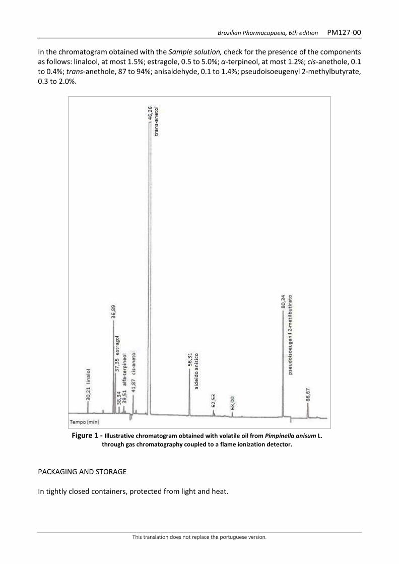
Brazilian Pharmacopoeia, 6th edition PM127-00
This translation does not replace the portuguese version.
In the chromatogram obtained with the Sample solution, check for the presence of the components as follows: linalool, at most 1.5%; estragole, 0.5 to 5.0%; α-terpineol, at most 1.2%; cis-anethole, 0.1 to 0.4%; trans-anethole, 87 to 94%; anisaldehyde, 0.1 to 1.4%; pseudoisoeugenyl 2-methylbutyrate, 0.3 to 2.0%.
Figure 1 - Illustrative chromatogram obtained with volatile oil from Pimpinella anisum L.
through gas chromatography coupled to a flame ionization detector. PACKAGING AND STORAGE In tightly closed containers, protected from light and heat.
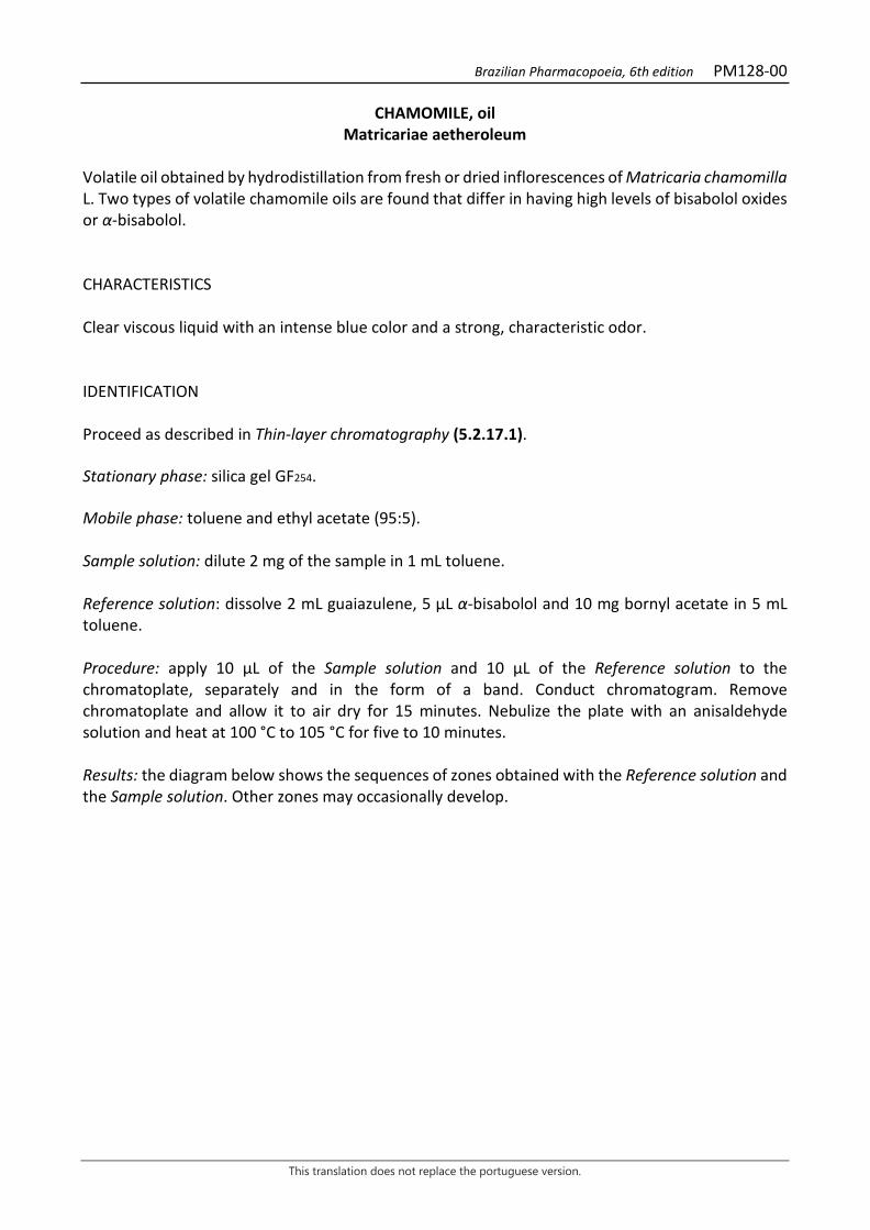
Brazilian Pharmacopoeia, 6th edition PM128-00
This translation does not replace the portuguese version.
CHAMOMILE, oil Matricariae aetheroleum
Volatile oil obtained by hydrodistillation from fresh or dried inflorescences of Matricaria chamomilla L. Two types of volatile chamomile oils are found that differ in having high levels of bisabolol oxides or α-bisabolol. CHARACTERISTICS Clear viscous liquid with an intense blue color and a strong, characteristic odor. IDENTIFICATION Proceed as described in Thin-layer chromatography (5.2.17.1). Stationary phase: silica gel GF254. Mobile phase: toluene and ethyl acetate (95:5). Sample solution: dilute 2 mg of the sample in 1 mL toluene. Reference solution: dissolve 2 mL guaiazulene, 5 µL α-bisabolol and 10 mg bornyl acetate in 5 mL toluene. Procedure: apply 10 μL of the Sample solution and 10 μL of the Reference solution to the chromatoplate, separately and in the form of a band. Conduct chromatogram. Remove chromatoplate and allow it to air dry for 15 minutes. Nebulize the plate with an anisaldehyde solution and heat at 100 °C to 105 °C for five to 10 minutes. Results: the diagram below shows the sequences of zones obtained with the Reference solution and the Sample solution. Other zones may occasionally develop.
Brazilian Pharmacopoeia, 6th edition PM128-00
This translation does not replace the portuguese version.
Top of the plate
Blue to violet-blue colored zones
Guaiazulene: red to reddish-purple colored zone
Red to reddish-purple colored zone
Bornyl acetate: yellowish-brown to grayish-green colored zone
Brownish-colored zone
-Bisabolol: reddish-purple to bluish-purple colored zone
Reddish-purple to bluish-purple colored zone
Brownish-colored zone
Reference solution Sample solution TESTS Chromatographic profile. Proceed as described in Gas chromatography (5.2.17.5). Use a chromatograph equipped with a flame ionization detector, using a mixture of hydrogen and synthetic air (1:45) as auxiliary gases to the detector flame; 30 m long and 0.25 mm wide (internal diameter) capillary column, coated with polyethylene glycol, with a 0.25 μm film thickness. Use ultra-pure nitrogen as carrier gas (1 mL/minute). Temperature: Time (minutes) Temperature (ºC) Column 0 – 40
40 – 50 70 → 230 230
Injector 250 Detector 250 Sample solution: dilute 20 µL of the sample in ethyl alcohol and top off the volume to 5 mL with the same solvent. Reference solution: dissolve 20 µL of α-bisabolol, 5 mg of chamazulene and 6 mg of guaiazulene in cyclohexane and top off the volume to 5 mL with the same solvent. Procedure: inject the volume of 1 μL of the Sample solution and the Reference solution into the gas chromatograph, using a 1:20 flow division. Determine relative concentrations by electronic integration using the normalization method.

Brazilian Pharmacopoeia, 6th edition PM128-00
This translation does not replace the portuguese version.
Elution order: order described in the preparation of the reference solution. Record retention times of substances. System suitability Resolution between peaks: Reference solution, minimum 1.5 between the chamazulene and guaiazulene peaks. Examine the chromatographic profile of the Sample Solution. The characteristic peaks in the chromatogram obtained with the Sample solution should have retention times similar to those obtained with the chromatogram with the Reference solution or identification confirmed with chromatography to locate the compounds in the chromatogram obtained with the sample solution. Disregard the cyclohexane peak. The chromatograms obtained should not show a peak at the guaiazulene retention time. Check the presence of the components, as follows, in the chromatogram obtained with the Sample solution:
volatile oil rich in bisabolol oxides (%)
volatile oil rich in α-bisabolol (%)
bisabolol oxides 29-81
α-bisabolol 10-65 chamazulene ≥1.0 ≥1.0 total bisabolol oxides and α-bisabolol ≥20
Figure 1 - Illustrative chromatogram obtained with volatile oil from Matricaria chamomilla L. through gas
chromatography coupled to a flame ionization detector. 1-(Z)-β-farnesene, 2- bisabolol oxide B, 3- bisabolone, 4- α-bisabolol, 5- chamazulene, 6- bisabolol oxide A.
0
100
200
300
400
500
600
700
800
900
0 5 10 15 20 25 30 35 40 45
1
2 3 4 5
6

Brazilian Pharmacopoeia, 6th edition PM128-00
This translation does not replace the portuguese version.
PACKAGING AND STORAGE In tightly closed containers, protected from light and heat.

Brazilian Pharmacopoeia, 6th edition PM129-00
This translation does not replace the portuguese version.
CHINESE CINNAMON, oil Cinnamomi cassiae aetheroleum
Volatile oil obtained by hydrodistillation from leaves and young branches of Cinnamomum cassia (L.) J. Presl (syn. Cinnamomum aromaticum Nees). CHARACTERISTICS Fluid, clear, colorless or light yellow liquid, with a characteristic odor. IDENTIFICATION Proceed as described in Thin-layer chromatography (5.2.17.1). Stationary phase: silica gel GF254 (0.25 mm). Mobile phase: toluene and methyl alcohol (90:10). Sample solution: dilute 0.5 mL of the volatile oil in 1.0 mL acetone and top off the volume to 10 mL with the same solvent. Reference solution: dissolve 50 μL of trans-cinnamic aldehyde, 10 μL of eugenol, 50 mg of coumarin in acetone and top off the volume to 5 mL with the same solvent. Procedure: apply 10 μL of the Reference solution and 2 μL of the Sample solution to two chromatoplates, separately and in the form of a band. Conduct chromatogram. Remove chromatoplate and allow it to air dry. Examine the first plate under ultraviolet light at 254 nm, nebulize the plate with anisaldehyde R and heat at 100°C to 105°C for five to 10 minutes. Nebulize the second plate with 10% potassium hydroxide solution in methyl alcohol, let dry at room temperature, and examine under ultraviolet light at 365 nm. Results: the following diagrams show the sequences of zones obtained with the Reference solution and the Sample solution, with the first plate after examination under ultraviolet light at 254 nm, nebulization with anisaldehyde R, and with the second plate after nebulization with 10% potassium hydroxide solution in methyl alcohol, in that order. The coumarin zone may be visible at 254 nm depending on the concentration in the sample. The eugenol zone in the chromatogram in the Sample solution is visualized only after revelation with anisaldehyde R and is of low intensity for authentic samples.

Brazilian Pharmacopoeia, 6th edition PM129-00
This translation does not replace the portuguese version.
Top of the plate
Trans-cinnamic aldehyde: dark-gray colored zone
Intense blue fluorescence zone
Coumarin: dark-gray colored zone
Reference solution Sample solution
Top of the plate
Trans-cinnamic aldehyde: bright blue-colored zone
Bright-blue colored zone
Eugenol: green-colored zone
Green-colored zone
Reference solution Sample solution
TESTS Optical rotation (5.2.8). -1° a +1°. Relative density (5.2.5). 1.052 to 1.070. Refractive index (5.2.29.4). 1.600 to 1.614. Chromatographic profile. Proceed as described in Gas chromatography (5.2.17.5). Use a chromatograph equipped with a flame ionization detector, using nitrogen, hydrogen and synthetic air (1:1:10) as auxiliary gases to the detector flame; 60 m long and 0.25 mm wide (internal diameter) capillary column, coated with polyethylene glycol, with a 0.20 μm film thickness. Use purified helium as carrier gas (1,5 mL/minute). Temperature: Time (minutes) Temperature (ºC)
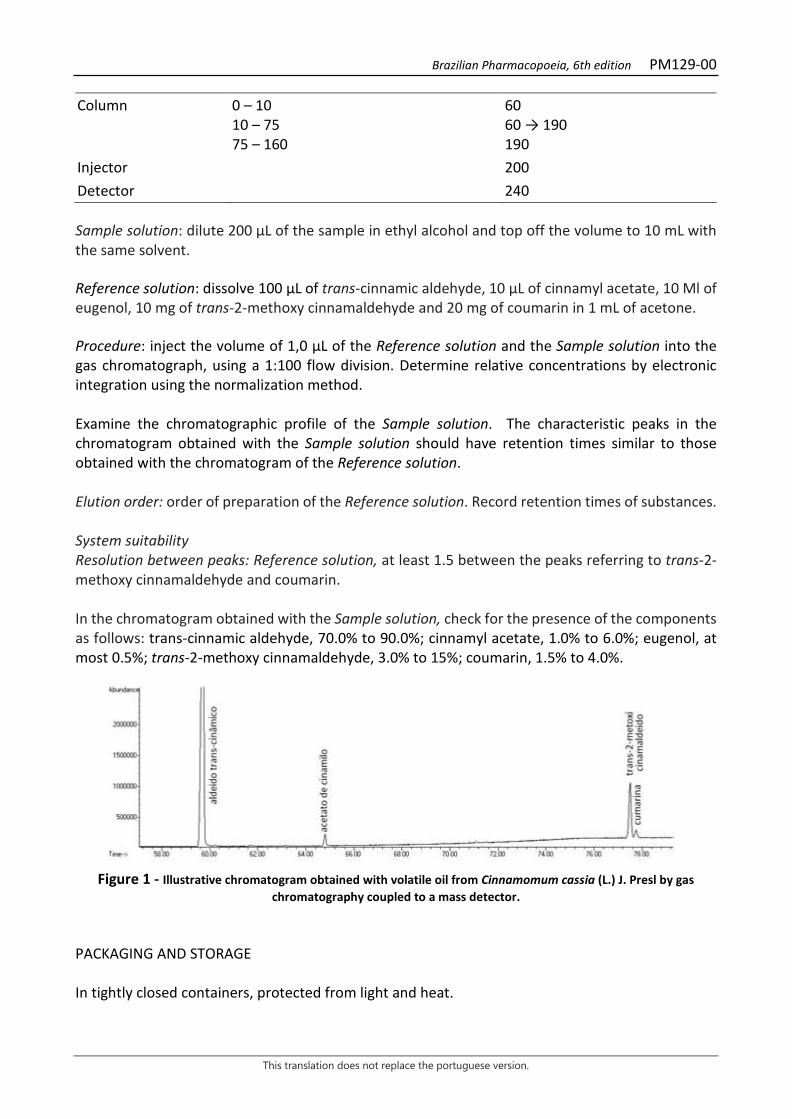
Brazilian Pharmacopoeia, 6th edition PM129-00
This translation does not replace the portuguese version.
Column 0 – 10 10 – 75 75 – 160
60 60 → 190 190
Injector 200 Detector 240 Sample solution: dilute 200 µL of the sample in ethyl alcohol and top off the volume to 10 mL with the same solvent. Reference solution: dissolve 100 μL of trans-cinnamic aldehyde, 10 μL of cinnamyl acetate, 10 Μl of eugenol, 10 mg of trans-2-methoxy cinnamaldehyde and 20 mg of coumarin in 1 mL of acetone. Procedure: inject the volume of 1,0 μL of the Reference solution and the Sample solution into the gas chromatograph, using a 1:100 flow division. Determine relative concentrations by electronic integration using the normalization method. Examine the chromatographic profile of the Sample solution. The characteristic peaks in the chromatogram obtained with the Sample solution should have retention times similar to those obtained with the chromatogram of the Reference solution. Elution order: order of preparation of the Reference solution. Record retention times of substances. System suitability Resolution between peaks: Reference solution, at least 1.5 between the peaks referring to trans-2-methoxy cinnamaldehyde and coumarin. In the chromatogram obtained with the Sample solution, check for the presence of the components as follows: trans-cinnamic aldehyde, 70.0% to 90.0%; cinnamyl acetate, 1.0% to 6.0%; eugenol, at most 0.5%; trans-2-methoxy cinnamaldehyde, 3.0% to 15%; coumarin, 1.5% to 4.0%.
Figure 1 - Illustrative chromatogram obtained with volatile oil from Cinnamomum cassia (L.) J. Presl by gas
chromatography coupled to a mass detector. PACKAGING AND STORAGE In tightly closed containers, protected from light and heat.
Brazilian Pharmacopoeia, 6th edition PM130-00
This translation does not replace the portuguese version.
CEYLON CINNAMON, oil Cinnamomi zeylanici folium aetheroleum
The volatile oil obtained by hydrodistillation from the leaves of Cinnamonum verum J.S. Presl (syn. Cinnamomum zeylanicum Blume). CHARACTERISTICS A clear, brownish-red to dark brown liquid with a characteristic odor reminiscent of eugenol. IDENTIFICATION Proceed as described in Thin-layer chromatography (5.2.17.1). Stationary phase: silica gel F254(250 µm). Mobile phase: toluene and methyl alcohol (90:10). Sample solution: dilute 1 mg of the sample in hexane, top off the volume to 10 mL with the same solvent and homogenize. Reference solution: dissolve 50 μL trans-cinnamic aldehyde, 10 μL eugenol, 10 μL linalool, 10 μL β-caryophyllene, 50 mg coumarin in acetone, top off the volume to 5 mL with the same solvent and homogenize. Procedure: apply 2 μL of the Sample solution and 10 μL of the Reference solution to the chromatoplate, separately and in the form of a band. Conduct chromatograms. Remove chromatoplates and allow them to air dry for 15 minutes. Nebulize the first plate with an anisaldehyde solution and heat at 100 °C to 105 °C for five to 10 minutes. Nebulize the second plate with 10% potassium hydroxide solution in methyl alcohol, dry at room temperature, and examine under UV light at 365 nm. Results: the diagram below shows the sequences of zones obtained with the Sample solution and the Reference solution. On the second chromatoplate, no fluorescent band for the Sample solution should be visualized. Other zones may occasionally develop.
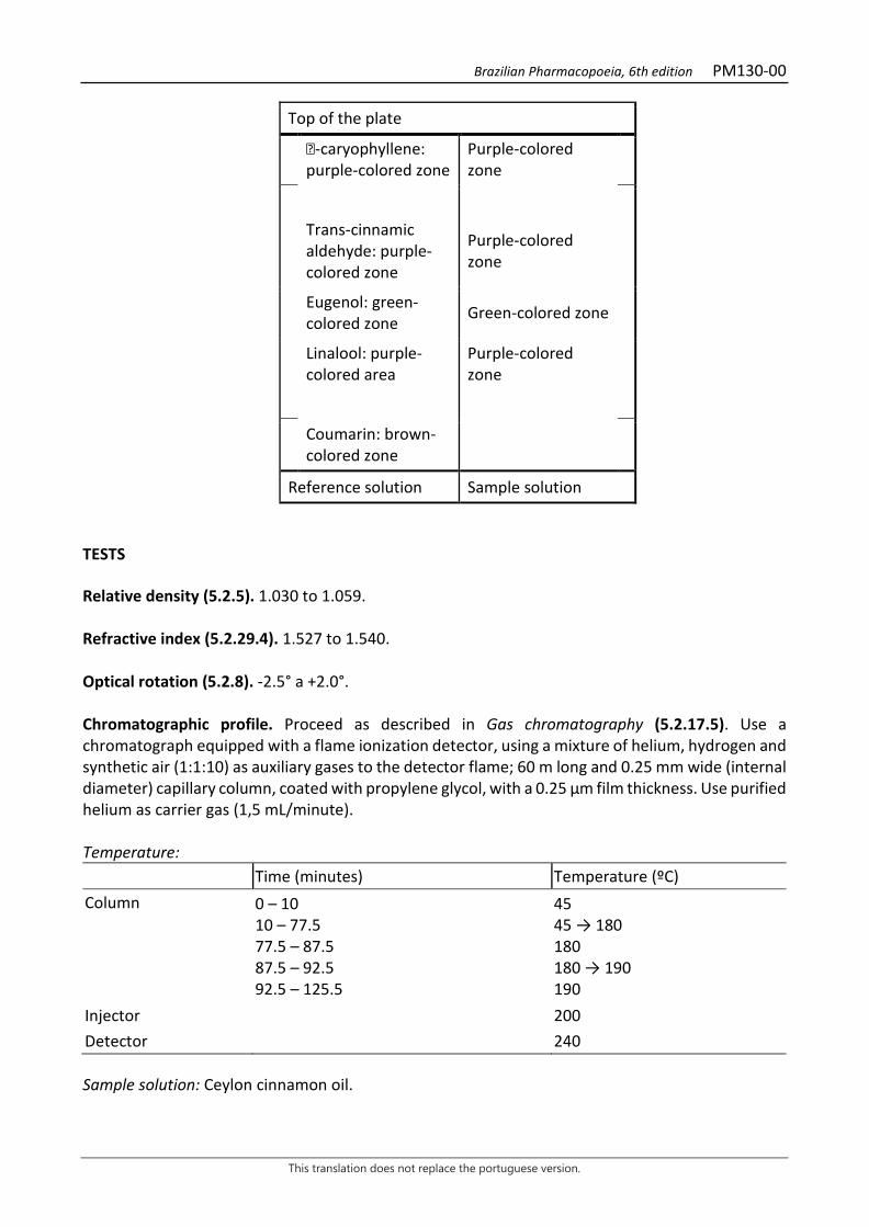
Brazilian Pharmacopoeia, 6th edition PM130-00
This translation does not replace the portuguese version.
Top of the plate
-caryophyllene: purple-colored zone
Purple-colored zone
Trans-cinnamic aldehyde: purple-colored zone
Purple-colored zone
Eugenol: green-colored zone Green-colored zone
Linalool: purple-colored area
Purple-colored zone
Coumarin: brown-colored zone
Reference solution Sample solution TESTS Relative density (5.2.5). 1.030 to 1.059. Refractive index (5.2.29.4). 1.527 to 1.540. Optical rotation (5.2.8). -2.5° a +2.0°. Chromatographic profile. Proceed as described in Gas chromatography (5.2.17.5). Use a chromatograph equipped with a flame ionization detector, using a mixture of helium, hydrogen and synthetic air (1:1:10) as auxiliary gases to the detector flame; 60 m long and 0.25 mm wide (internal diameter) capillary column, coated with propylene glycol, with a 0.25 μm film thickness. Use purified helium as carrier gas (1,5 mL/minute). Temperature: Time (minutes) Temperature (ºC) Column 0 – 10
10 – 77.5 77.5 – 87.5 87.5 – 92.5 92.5 – 125.5
45 45 → 180 180 180 → 190 190
Injector 200 Detector 240 Sample solution: Ceylon cinnamon oil.
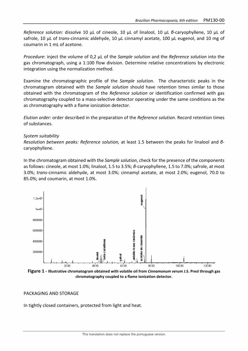
Brazilian Pharmacopoeia, 6th edition PM130-00
This translation does not replace the portuguese version.
Reference solution: dissolve 10 μL of cineole, 10 μL of linalool, 10 μL β-caryophyllene, 10 μL of safrole, 10 μL of trans-cinnamic aldehyde, 10 μL cinnamyl acetate, 100 μL eugenol, and 10 mg of coumarin in 1 mL of acetone. Procedure: inject the volume of 0,2 μL of the Sample solution and the Reference solution into the gas chromatograph, using a 1:100 flow division. Determine relative concentrations by electronic integration using the normalization method. Examine the chromatographic profile of the Sample solution. The characteristic peaks in the chromatogram obtained with the Sample solution should have retention times similar to those obtained with the chromatogram of the Reference solution or identification confirmed with gas chromatography coupled to a mass-selective detector operating under the same conditions as the as chromatography with a flame ionization detector. Elution order: order described in the preparation of the Reference solution. Record retention times of substances. System suitability Resolution between peaks: Reference solution, at least 1.5 between the peaks for linalool and β-caryophyllene. In the chromatogram obtained with the Sample solution, check for the presence of the components as follows: cineole, at most 1.0%; linalool, 1.5 to 3.5%; β-caryophyllene, 1.5 to 7.0%; safrole, at most 3.0%; trans-cinnamic aldehyde, at most 3.0%; cinnamyl acetate, at most 2.0%; eugenol, 70.0 to 85.0%; and coumarin, at most 1.0%.
Figure 1 - Illustrative chromatogram obtained with volatile oil from Cinnamonum verum J.S. Presl through gas
chromatography coupled to a flame ionization detector. PACKAGING AND STORAGE In tightly closed containers, protected from light and heat.
Brazilian Pharmacopoeia, 6th edition PM131-00
This translation does not replace the portuguese version.
LEMON GRASS, oil Cymbopogonis citrati aetheroleum
Volatile oil obtained by hydrodistillation from fresh leaves of Cymbopogon citratus (DC.) Stapf, containing at least 60.0% citral A (trans-citral or geranial) and citral B (cis-citral or neral). CHARACTERISTICS Pale yellow liquid with a citronella odor. IDENTIFICATION Proceed as described in Thin-layer chromatography (5.2.17.1). Stationary phase: silica gel GF254 chromatoplate. Mobile phase: toluene and ethyl acetate (93:7). Sample solution: dilute 2 µL of the sample to be examined in 1 mL toluene. Reference solution: dilute 2 µL citral in 1 mL toluene. TLC visualization reagent: dissolve 1 g of vanillin in 100 mL of methyl alcohol. Add 4 mL hydrochloric acid and 5 mL sulfuric acid and homogenize. Procedure: apply 10 μL of the Sample solution and 10 μL of the Reference solution to the chromatoplate, separately and in the form of a band. Conduct chromatogram. Remove chromatoplate and allow it to air dry for 15 minutes. Nebulize the plate with sulfur vanillin RS and heat at 100 °C to 105 °C for five to 10 minutes. Results: the diagram below shows the sequences of zones obtained with the Reference solution and the Sample solution. Other zones may occasionally develop.
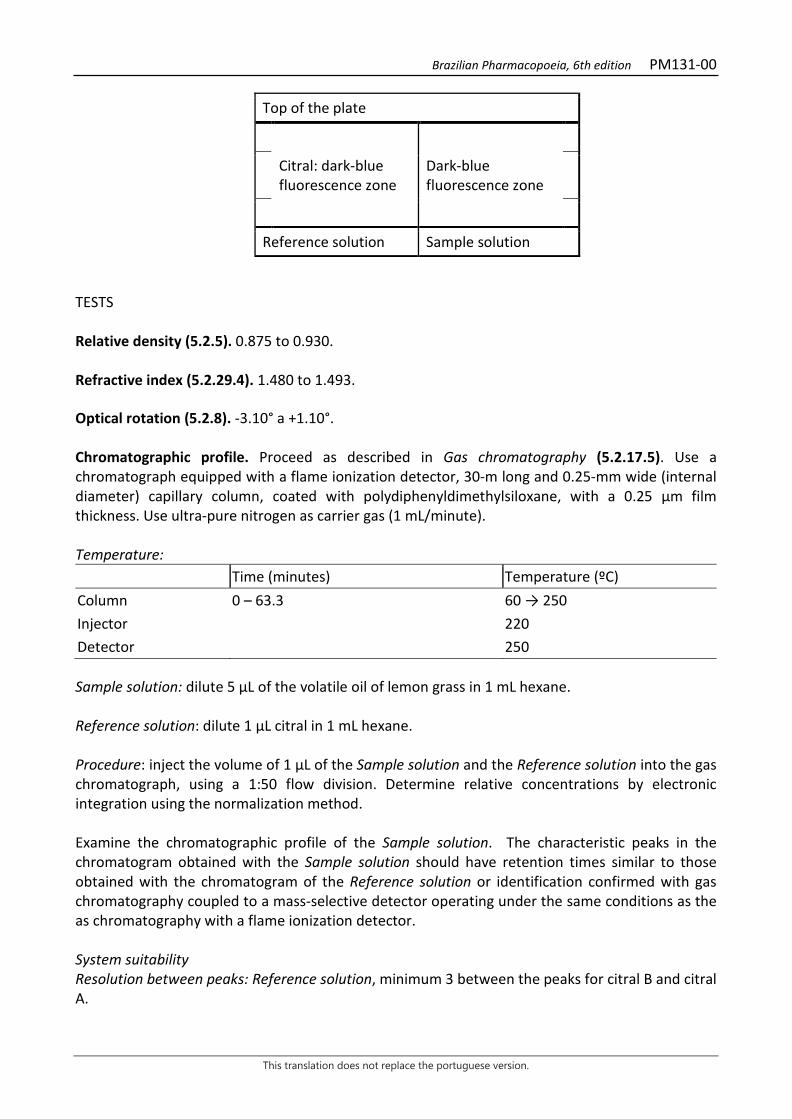
Brazilian Pharmacopoeia, 6th edition PM131-00
This translation does not replace the portuguese version.
Top of the plate
Citral: dark-blue fluorescence zone
Dark-blue fluorescence zone
Reference solution Sample solution TESTS Relative density (5.2.5). 0.875 to 0.930. Refractive index (5.2.29.4). 1.480 to 1.493. Optical rotation (5.2.8). -3.10° a +1.10°. Chromatographic profile. Proceed as described in Gas chromatography (5.2.17.5). Use a chromatograph equipped with a flame ionization detector, 30-m long and 0.25-mm wide (internal diameter) capillary column, coated with polydiphenyldimethylsiloxane, with a 0.25 μm film thickness. Use ultra-pure nitrogen as carrier gas (1 mL/minute). Temperature: Time (minutes) Temperature (ºC) Column 0 – 63.3 60 → 250 Injector 220 Detector 250 Sample solution: dilute 5 µL of the volatile oil of lemon grass in 1 mL hexane. Reference solution: dilute 1 µL citral in 1 mL hexane. Procedure: inject the volume of 1 μL of the Sample solution and the Reference solution into the gas chromatograph, using a 1:50 flow division. Determine relative concentrations by electronic integration using the normalization method. Examine the chromatographic profile of the Sample solution. The characteristic peaks in the chromatogram obtained with the Sample solution should have retention times similar to those obtained with the chromatogram of the Reference solution or identification confirmed with gas chromatography coupled to a mass-selective detector operating under the same conditions as the as chromatography with a flame ionization detector. System suitability Resolution between peaks: Reference solution, minimum 3 between the peaks for citral B and citral A.
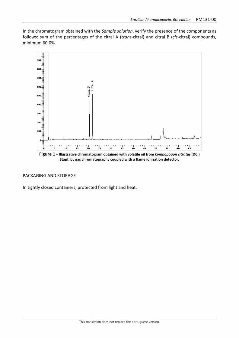
Brazilian Pharmacopoeia, 6th edition PM131-00
This translation does not replace the portuguese version.
In the chromatogram obtained with the Sample solution, verify the presence of the components as follows: sum of the percentages of the citral A (trans-citral) and citral B (cis-citral) compounds, minimum 60.0%.
Figure 1 - Illustrative chromatogram obtained with volatile oil from Cymbopogon citratus (DC.)
Stapf, by gas chromatography coupled with a flame ionization detector. PACKAGING AND STORAGE In tightly closed containers, protected from light and heat.
Brazilian Pharmacopoeia, 6th edition PM132-00
This translation does not replace the portuguese version.
CARNAUBA WAX PALM Carnaubae cera
Wax obtained from the leaves of Copernicia prunifera (Mill.) H.E. Moore [syn. Copernicia cerifera (Arruda) Mart.]. CHARACTERISTICS Solid, powdery, flaky, or solid mass with a pale yellow color. IDENTIFICATION Proceed as described in Thin-layer chromatography (5.2.17.1). Stationary phase: silica gel G (0.25 mm). Mobile phase: chloroform and ethyl acetate (98:2). Sample solution: hot dissolve 0.10 g of the sample in 5 mL of chloroform. Apply the solution while still hot. Reference solution: dissolve 5 mg menthol acetate, 5 mg menthol and 5 mg thymol in 10 mL toluene. TLC visualization reagent: 20% phosphomolybdic acid solution in ethyl alcohol. Procedure: apply 20 μL of the Sample solution and 10 μL of the Reference solution to the chromatoplate, separately and in the form of a band. Conduct the chromatogram along 10 cm. Remove chromatoplate and allow it to air dry for 15 minutes. Nebulize plate with TLC visualization reagent and heat at 100°C to 105°C for two to five minutes. Results: the diagram below shows the sequences of zones obtained with the Reference solution and the Sample solution. Other zones may occasionally develop.
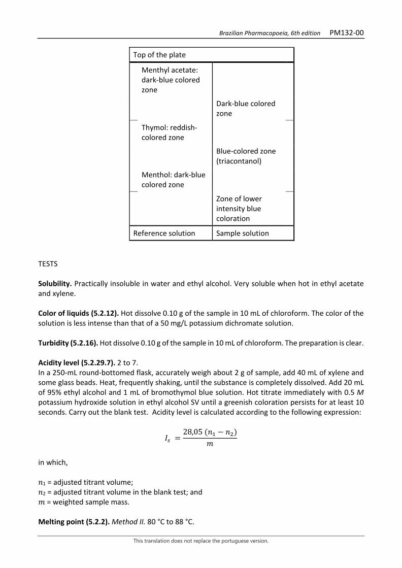
Brazilian Pharmacopoeia, 6th edition PM132-00
This translation does not replace the portuguese version.
Top of the plate
Menthyl acetate: dark-blue colored zone
Dark-blue colored zone
Thymol: reddish-colored zone
Blue-colored zone (triacontanol)
Menthol: dark-blue colored zone
Zone of lower intensity blue coloration
Reference solution Sample solution TESTS Solubility. Practically insoluble in water and ethyl alcohol. Very soluble when hot in ethyl acetate and xylene. Color of liquids (5.2.12). Hot dissolve 0.10 g of the sample in 10 mL of chloroform. The color of the solution is less intense than that of a 50 mg/L potassium dichromate solution. Turbidity (5.2.16). Hot dissolve 0.10 g of the sample in 10 mL of chloroform. The preparation is clear. Acidity level (5.2.29.7). 2 to 7. In a 250-mL round-bottomed flask, accurately weigh about 2 g of sample, add 40 mL of xylene and some glass beads. Heat, frequently shaking, until the substance is completely dissolved. Add 20 mL of 95% ethyl alcohol and 1 mL of bromothymol blue solution. Hot titrate immediately with 0.5 M potassium hydroxide solution in ethyl alcohol SV until a greenish coloration persists for at least 10 seconds. Carry out the blank test. Acidity level is calculated according to the following expression:
𝐼𝐼𝑠𝑠 =28,05 (𝑛𝑛1 − 𝑛𝑛2)
𝑚𝑚
in which, 𝑛𝑛1 = adjusted titrant volume; 𝑛𝑛2 = adjusted titrant volume in the blank test; and 𝑚𝑚 = weighted sample mass. Melting point (5.2.2). Method II. 80 °C to 88 °C.
Brazilian Pharmacopoeia, 6th edition PM132-00
This translation does not replace the portuguese version.
Saponification index (5.2.29.8). 78 to 95. In a 250-mL round-bottomed flask, accurately weigh about 2 g of sample, add 40 mL of xylene and some glass beads. Heat, frequently shaking, until the substance is completely dissolved. Add 20 mL of 95% ethyl alcohol and 20 mL of 0.5 M potassium hydroxide solution in ethyl alcohol. Fit the flask to the reflux condenser and heat in a water bath for three hours with frequent shaking. Add 1 mL of phenolphthalein solution and immediately titrate with 0.5 M hydrochloric acid SV solution until the red color disappears. Repeat heating and titration until no color restoration is observed under heating. Carry out the blank test. Saponification index is calculated according to the following expression:
𝐼𝐼𝑠𝑠 =28,05 (𝑛𝑛4 − 𝑛𝑛3)
𝑚𝑚
in which, 𝑛𝑛3 = adjusted titrant volume; 𝑛𝑛4 = adjusted titrant volume in the blank test; and 𝑚𝑚 = weighted sample mass. Total ash (5.4.1.5.1). At most 0.25%. Determine in 2.0 g. Total mesophilic microorganism count (5.5.3.1.2). Complies with the test. Study of pathogenic microorganisms (5.5.3.1.3). Complies with the test. PACKAGING AND STORAGE In tightly closed containers, protected from light and heat. CATEGORY Pharmacotechnical excipient.
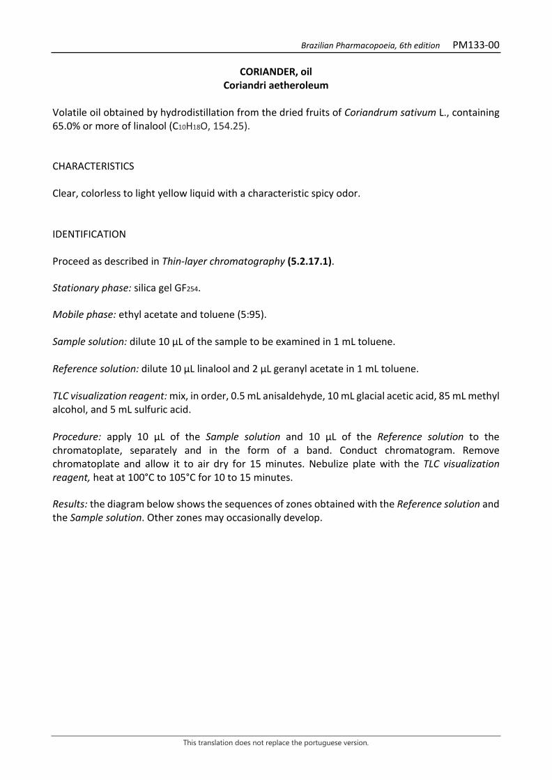
Brazilian Pharmacopoeia, 6th edition PM133-00
This translation does not replace the portuguese version.
CORIANDER, oil Coriandri aetheroleum
Volatile oil obtained by hydrodistillation from the dried fruits of Coriandrum sativum L., containing 65.0% or more of linalool (C10H18O, 154.25). CHARACTERISTICS Clear, colorless to light yellow liquid with a characteristic spicy odor. IDENTIFICATION Proceed as described in Thin-layer chromatography (5.2.17.1). Stationary phase: silica gel GF254. Mobile phase: ethyl acetate and toluene (5:95). Sample solution: dilute 10 µL of the sample to be examined in 1 mL toluene. Reference solution: dilute 10 µL linalool and 2 µL geranyl acetate in 1 mL toluene. TLC visualization reagent: mix, in order, 0.5 mL anisaldehyde, 10 mL glacial acetic acid, 85 mL methyl alcohol, and 5 mL sulfuric acid. Procedure: apply 10 μL of the Sample solution and 10 μL of the Reference solution to the chromatoplate, separately and in the form of a band. Conduct chromatogram. Remove chromatoplate and allow it to air dry for 15 minutes. Nebulize plate with the TLC visualization reagent, heat at 100°C to 105°C for 10 to 15 minutes. Results: the diagram below shows the sequences of zones obtained with the Reference solution and the Sample solution. Other zones may occasionally develop.

Brazilian Pharmacopoeia, 6th edition PM133-00
This translation does not replace the portuguese version.
Top of the plate
Geranyl acetate: violaceous-purple colored area
Violaceous-pink colored zone
Linalool: violaceous-pink colored zone
Violaceous-pink colored zone
Less intense violaceous-pink colored zone
Reference solution Sample solution TESTS Relative density (5.2.5). 0.860 to 0.880. Refractive index (5.2.29.4). 1.462 to 1.470. Optical rotation (5.2.8). -7° a +13°. Acidity level (5.2.29.7). At most 3. Determine in 5 g of the sample. Chromatographic profile. Proceed as described in Gas chromatography (5.2.17.5). Use a chromatograph equipped with a flame ionization detector, 60-m long and 0.25-mm wide (internal diameter) capillary column, coated with polyethylene glycol, with a 0.25 μm film thickness. Use ultra-pure nitrogen as carrier gas (1 mL/minute). Temperature: Time (minutes) Temperature (ºC) Column 0 – 10
10 – 75 75 – 120
60 60 → 190 190
Injector 220 Detector 240 Sample solution: coriander volatile oil. Reference solution (1): dissolve 10 μL of α-pinene, 10 μL of limonene, 10 μL of γ-terpinene, 10 μL of p-cymene, 10 mg of camphor, 20 μL of linalool, 10 μL of α-terpineol, 10 μL of geranyl acetate, and 10 μL of geraniol in 1 mL of hexane. Store under refrigeration, in a tightly closed bottle and protected from light. Reference solution (2): dilute 5 μL of geraniol in hexane and dilute to 10 mL with the same solvent.
Brazilian Pharmacopoeia, 6th edition PM133-00
This translation does not replace the portuguese version.
Procedure: inject the volume of 0,2 μL of the Sample solution and the Reference solution into the gas chromatograph, using a 1:65 flow division. Determine relative concentrations by electronic integration using the normalization method. Examine the chromatographic profile of the Sample solution. The characteristic peaks in the chromatogram obtained with the Sample solution should have retention times similar to those obtained with the chromatogram of the Reference solution or identification confirmed with gas chromatography coupled to a mass-selective detector operating under the same conditions as the as chromatography with a flame ionization detector. System suitability Resolution between peaks: Reference solution (1), minimum 1.5 between the peaks for linalool and camphor. Exclusion Limit: area under the peak of the chromatogram obtained with Reference solution (2) (0.05%). In the chromatogram obtained with the Sample solution, check for the presence of the components as follows: α-pinene, 3.0 to 7.0%; limonene, 1.5 to 5.0%; γ-terpinene, 1.5 to 8.0%; p-cymene, 0.5 to 4.0%; camphor, 3.0 to 6.0%; linalool, 65.0 to 78.0%; α-terpineol, 0.1 to 1.5%; geranyl acetate, 0.5 to 4.0%; e geraniol, 0.5 to 3.0%.
Figure 1 - Illustrative chromatogram obtained with volatile oil from Coriandrum sativum L. through gas
chromatography coupled to a flame ionization detector. 1- α-pinene, 2- limonene, 3- γ- terpinene, 4- p-cymene, 5- camphor, 6- linalool, 7- α-terpineol, 8- geranyl acetate, 9- geraniol.
0
100
200
300
400
500
600
700
800
900
0 10 20 30 40 50 60 70 80 90 100 110
1
2
3
4 5
6
7
8 9
Brazilian Pharmacopoeia, 6th edition PM133-00
This translation does not replace the portuguese version.
Determination of chiral purity. Proceed as described in Gas chromatography (5.2.17.5). Use a chromatograph equipped with a flame ionization detector, 30-m long and 0.25-mm wide (internal diameter) capillary column, coated with modified β-cyclodextrin, with a 0.25 μm film thickness. Use ultra-pure nitrogen as carrier gas (1.3 mL/minute). Temperature: Time (minutes) Temperature (ºC) Column 0 – 65 50 → 180 Injector 230 Detector 230
Sample solution: dissolve 0.02 g of the sample in pentane and top off the volume to 10 mL with the same solvent. Reference solution: dissolve 10 μL of linalool and 5 mg of borneol in pentane and top off the volume to 10 mL with the same solvent. Procedure: inject the volume of 1,0 μL of the Sample Solution and the Reference Solution into the gas chromatograph, using a 1:30 flow division. System suitability Resolution between peaks: Reference solution, at least 5.5 between the peaks for (R)-linalool (1st peak) and (S)-linalool (2nd peak) and at least 2.9 between the peaks for (S)-linalool and borneol (3rd peak). Limit: at most 14% (R)-linalool. Calculate (R)-linalool content, in percent, according to the following expression:
TRL =𝑇𝑇𝑅𝑅
𝑇𝑇𝑆𝑆 + 𝑇𝑇𝑅𝑅× 100
in which, TRL = (R)-linalool content; %; 𝑇𝑇𝑇𝑇 = area under the peak corresponding to (S)-linalool; 𝑇𝑇𝐼𝐼= area under the peak corresponding to (R)-linalool; PACKAGING AND STORAGE In tightly closed containers, protected from light and heat.
Brazilian Pharmacopoeia, 6th edition PM134-00
This translation does not replace the portuguese version.
CLOVE, oil Caryophylli flos aetheroleum
Volatile oil obtained by hydrodistillation from dried flower buds of Syzygium aromaticum (L.) Merr. & L.M. Perry, containing at least 75.0% eugenol (C10H12O2, 164.20). CHARACTERISTICS Bright yellow liquid that turns brown when exposed to air, with a eugenol odor. IDENTIFICATION Proceed as described in Thin-layer chromatography (5.2.17.1). Stationary phase: silica gel GF254. Mobile phase: toluene Sample solution: dilute 3 µL of the sample to be examined in 300 µL toluene. Reference solution: dissolve 1.5 µL eugenol and 2 mg eugenyl acetate in 200 µL toluene. Procedure: apply 20 μL of the Sample solution and 15 μL of the Reference solution to the chromatoplate, separately and in the form of a band. Conduct chromatogram. Allow to stand for five minutes. Re-conduct chromatogram. Remove chromatoplate and allow it to air dry for 15 minutes. Examine under ultraviolet light at 254 nm. Nebulize the plate with an anisaldehyde solution and heat at 100 °C to 105 °C for five to 10 minutes. Results: the diagram below shows the sequences of zones obtained with the Reference solution and the Sample solution. Other zones may occasionally develop.
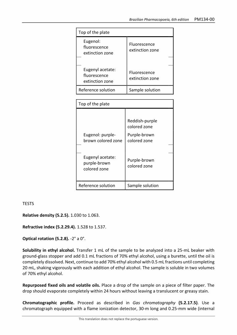
Brazilian Pharmacopoeia, 6th edition PM134-00
This translation does not replace the portuguese version.
Top of the plate
Eugenol: fluorescence extinction zone
Fluorescence extinction zone
Eugenyl acetate: fluorescence extinction zone
Fluorescence extinction zone
Reference solution Sample solution
Top of the plate
Reddish-purple colored zone
Eugenol: purple-brown colored zone
Purple-brown colored zone
Eugenyl acetate: purple-brown colored zone
Purple-brown colored zone
Reference solution Sample solution TESTS Relative density (5.2.5). 1.030 to 1.063. Refractive index (5.2.29.4). 1.528 to 1.537. Optical rotation (5.2.8). -2° a 0°. Solubility in ethyl alcohol. Transfer 1 mL of the sample to be analyzed into a 25-mL beaker with ground-glass stopper and add 0.1 mL fractions of 70% ethyl alcohol, using a burette, until the oil is completely dissolved. Next, continue to add 70% ethyl alcohol with 0.5 mL fractions until completing 20 mL, shaking vigorously with each addition of ethyl alcohol. The sample is soluble in two volumes of 70% ethyl alcohol. Repurposed fixed oils and volatile oils. Place a drop of the sample on a piece of filter paper. The drop should evaporate completely within 24 hours without leaving a translucent or greasy stain. Chromatographic profile. Proceed as described in Gas chromatography (5.2.17.5). Use a chromatograph equipped with a flame ionization detector, 30-m long and 0.25-mm wide (internal
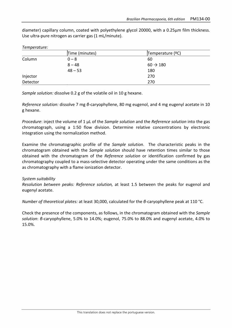
Brazilian Pharmacopoeia, 6th edition PM134-00
This translation does not replace the portuguese version.
diameter) capillary column, coated with polyethylene glycol 20000, with a 0.25μm film thickness. Use ultra-pure nitrogen as carrier gas (1 mL/minute). Temperature: Time (minutes) Temperature (ºC) Column 0 – 8
8 – 48 48 – 53
60 60 → 180 180
Injector 270 Detector 270 Sample solution: dissolve 0.2 g of the volatile oil in 10 g hexane. Reference solution: dissolve 7 mg β-caryophyllene, 80 mg eugenol, and 4 mg eugenyl acetate in 10 g hexane. Procedure: inject the volume of 1 μL of the Sample solution and the Reference solution into the gas chromatograph, using a 1:50 flow division. Determine relative concentrations by electronic integration using the normalization method. Examine the chromatographic profile of the Sample solution. The characteristic peaks in the chromatogram obtained with the Sample solution should have retention times similar to those obtained with the chromatogram of the Reference solution or identification confirmed by gas chromatography coupled to a mass-selective detector operating under the same conditions as the as chromatography with a flame ionization detector. System suitability Resolution between peaks: Reference solution, at least 1.5 between the peaks for eugenol and eugenyl acetate. Number of theoretical plates: at least 30,000, calculated for the β-caryophyllene peak at 110 °C. Check the presence of the components, as follows, in the chromatogram obtained with the Sample solution: β-caryophyllene, 5.0% to 14.0%; eugenol, 75.0% to 88.0% and eugenyl acetate, 4.0% to 15.0%.
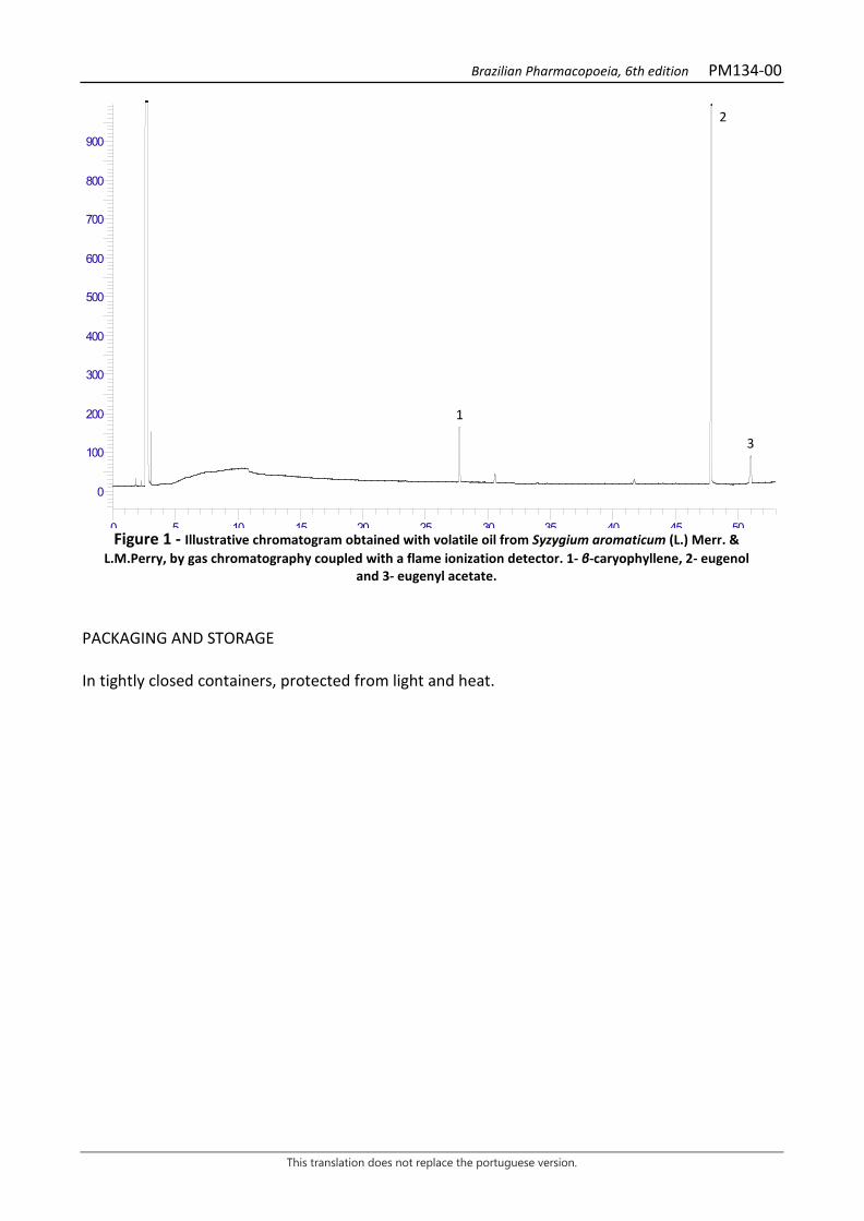
Brazilian Pharmacopoeia, 6th edition PM134-00
This translation does not replace the portuguese version.
Figure 1 - Illustrative chromatogram obtained with volatile oil from Syzygium aromaticum (L.) Merr. &
L.M.Perry, by gas chromatography coupled with a flame ionization detector. 1- β-caryophyllene, 2- eugenol and 3- eugenyl acetate.
PACKAGING AND STORAGE In tightly closed containers, protected from light and heat.
0
100
200
300
400
500
600
700
800
900
0 5 10 15 20 25 30 35 40 45 50
1
2
3

Brazilian Pharmacopoeia, 6th edition PM135-00
This translation does not replace the portuguese version.
EUCALIPTO, oil Eucalypti aetheroleum
Volatile oil obtained by hydrodistillation from fresh leaves or terminal branches of Eucalyptus globulus Labill., containing at least 70.0% 1,8-cineole (C10H18O, 154.25). CHARACTERISTICS Colorless to pale yellow liquid with a characteristic aromatic odor of 1,8-cineole. IDENTIFICATION Proceed as described in Thin-layer chromatography (5.2.17.1). Stationary phase: silica gel GF254. Mobile phase: toluene and ethyl acetate (90:10). Sample solution: dissolve 0.01 g of the sample to be examined in 1 mL toluene. Reference solution: dilute 3 µL of 1,8-cineole and 1.2 µL α-terpineol in 300 µL toluene. Procedure: apply 10 μL of the Sample solution and 10 μL of the Reference solution to the chromatoplate, separately and in the form of a band. Conduct chromatogram. Remove chromatoplate and allow it to air dry for 15 minutes. Nebulize the plate with an anisaldehyde solution and heat at 100 °C to 105 °C for five to 10 minutes. Results: the diagram below shows the sequences of zones obtained with the Reference solution and the Sample solution. Other zones may occasionally develop.
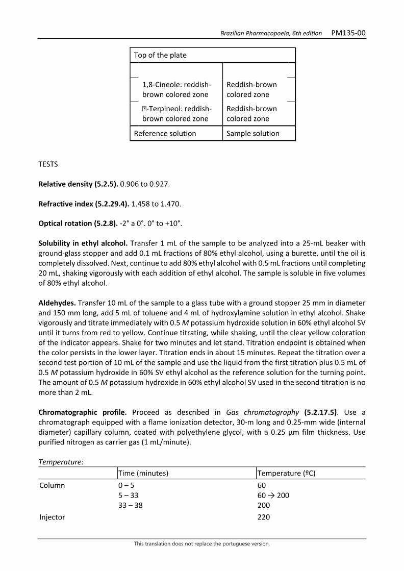
Brazilian Pharmacopoeia, 6th edition PM135-00
This translation does not replace the portuguese version.
Top of the plate
1,8-Cineole: reddish-brown colored zone
Reddish-brown colored zone
-Terpineol: reddish-brown colored zone
Reddish-brown colored zone
Reference solution Sample solution TESTS Relative density (5.2.5). 0.906 to 0.927. Refractive index (5.2.29.4). 1.458 to 1.470. Optical rotation (5.2.8). -2° a 0°. 0° to +10°. Solubility in ethyl alcohol. Transfer 1 mL of the sample to be analyzed into a 25-mL beaker with ground-glass stopper and add 0.1 mL fractions of 80% ethyl alcohol, using a burette, until the oil is completely dissolved. Next, continue to add 80% ethyl alcohol with 0.5 mL fractions until completing 20 mL, shaking vigorously with each addition of ethyl alcohol. The sample is soluble in five volumes of 80% ethyl alcohol. Aldehydes. Transfer 10 mL of the sample to a glass tube with a ground stopper 25 mm in diameter and 150 mm long, add 5 mL of toluene and 4 mL of hydroxylamine solution in ethyl alcohol. Shake vigorously and titrate immediately with 0.5 M potassium hydroxide solution in 60% ethyl alcohol SV until it turns from red to yellow. Continue titrating, while shaking, until the clear yellow coloration of the indicator appears. Shake for two minutes and let stand. Titration endpoint is obtained when the color persists in the lower layer. Titration ends in about 15 minutes. Repeat the titration over a second test portion of 10 mL of the sample and use the liquid from the first titration plus 0.5 mL of 0.5 M potassium hydroxide in 60% SV ethyl alcohol as the reference solution for the turning point. The amount of 0.5 M potassium hydroxide in 60% ethyl alcohol SV used in the second titration is no more than 2 mL. Chromatographic profile. Proceed as described in Gas chromatography (5.2.17.5). Use a chromatograph equipped with a flame ionization detector, 30-m long and 0.25-mm wide (internal diameter) capillary column, coated with polyethylene glycol, with a 0.25 μm film thickness. Use purified nitrogen as carrier gas (1 mL/minute). Temperature: Time (minutes) Temperature (ºC) Column 0 – 5
5 – 33 33 – 38
60 60 → 200 200
Injector 220

Brazilian Pharmacopoeia, 6th edition PM135-00
This translation does not replace the portuguese version.
Time (minutes) Temperature (ºC) Detector 250 Sample solution: dilute 4 µL of the volatile eucalyptus oil in 200 µL hexane. Reference solution: dissolve 0.5 mg camphor, 0.5 mg sabinene, 1 μL -pinene, 0.5 μL - pinene, 1 μL limonene, 0.5 μL -felandrene, and 5 μL 1,8-cineole in 1 mL hexane. Store under refrigeration, in a tightly closed bottle and protected from light. Procedure: inject the volume of 1 μL of the Sample solution and the Reference solution into the gas chromatograph, using a 1:50 flow division. Determine relative concentrations by electronic integration using the normalization method. Examine the chromatographic profile of the Sample solution. The characteristic peaks in the chromatogram obtained with the Sample solution should have retention times similar to those obtained with the chromatogram of the Reference solution or identification confirmed with gas chromatography coupled to a mass-selective detector operating under the same conditions as the as chromatography with a flame ionization detector. System suitability Resolution between peaks: Reference solution, minimum 1.5 between the peaks for limonene and 1,8-cineole. Check the presence of the components, as follows, in the chromatogram obtained with the Sample solution: α-pinene, 0.05 to 10.0%; β-pinene, 0.05 to 1.5%; sabinene, at most 0.3%; α-felandrene, 0.05 to 1.5%; limonene, 0.05 to 15.0%; 1,8-cineole, at most 70.0%; and camphor, at most 0.1%.
0
100
200
300
400
500
600
700
800
900
0 2 4 6 8 10 12 14 16 18 20 22 24 26 28 30 32 34 36
1 2
3
4

Brazilian Pharmacopoeia, 6th edition PM135-00
This translation does not replace the portuguese version.
Figure 1 - Illustrative chromatogram obtained with volatile oil from Eucalyptus globulus Labill. through gas chromatography coupled to a flame ionization detector. 1 - α-pinene, 2 - α-felandrene, 3 - limonene, 4 - 1,8-
cineole. PACKAGING AND STORAGE In tightly closed containers, protected from light and heat.
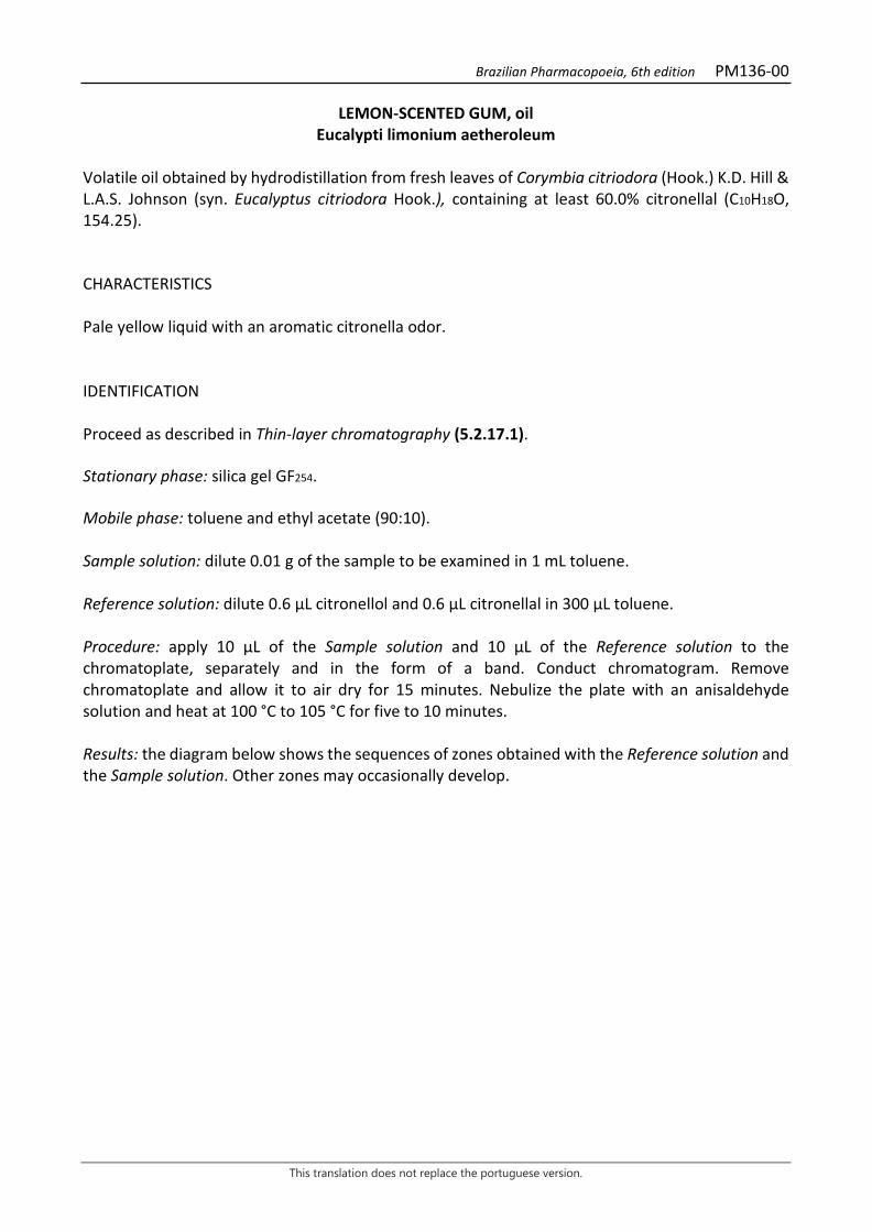
Brazilian Pharmacopoeia, 6th edition PM136-00
This translation does not replace the portuguese version.
LEMON-SCENTED GUM, oil Eucalypti limonium aetheroleum
Volatile oil obtained by hydrodistillation from fresh leaves of Corymbia citriodora (Hook.) K.D. Hill & L.A.S. Johnson (syn. Eucalyptus citriodora Hook.), containing at least 60.0% citronellal (C10H18O, 154.25). CHARACTERISTICS Pale yellow liquid with an aromatic citronella odor. IDENTIFICATION Proceed as described in Thin-layer chromatography (5.2.17.1). Stationary phase: silica gel GF254. Mobile phase: toluene and ethyl acetate (90:10). Sample solution: dilute 0.01 g of the sample to be examined in 1 mL toluene. Reference solution: dilute 0.6 µL citronellol and 0.6 µL citronellal in 300 µL toluene. Procedure: apply 10 μL of the Sample solution and 10 μL of the Reference solution to the chromatoplate, separately and in the form of a band. Conduct chromatogram. Remove chromatoplate and allow it to air dry for 15 minutes. Nebulize the plate with an anisaldehyde solution and heat at 100 °C to 105 °C for five to 10 minutes. Results: the diagram below shows the sequences of zones obtained with the Reference solution and the Sample solution. Other zones may occasionally develop.
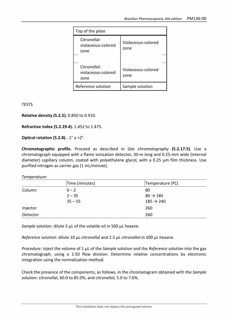
Brazilian Pharmacopoeia, 6th edition PM136-00
This translation does not replace the portuguese version.
Top of the plate
Citronellal: violaceous-colored zone
Violaceous-colored zone
Citronellol: violaceous-colored zone
Violaceous-colored zone
Reference solution Sample solution TESTS Relative density (5.2.5). 0.850 to 0.910. Refractive index (5.2.29.4). 1.452 to 1.475. Optical rotation (5.2.8). -1° a +2°. Chromatographic profile. Proceed as described in Gas chromatography (5.2.17.5). Use a chromatograph equipped with a flame ionization detector, 30-m long and 0.25-mm wide (internal diameter) capillary column, coated with polyethylene glycol, with a 0.25 μm film thickness. Use purified nitrogen as carrier gas (1 mL/minute). Temperature: Time (minutes) Temperature (ºC) Column 0 – 2
2 – 35 35 – 55
80 80 → 185 185 → 240
Injector 260 Detector 260 Sample solution: dilute 5 µL of the volatile oil in 500 µL hexane. Reference solution: dilute 10 µL citronellal and 2.5 µL citronellol in 500 µL hexane. Procedure: inject the volume of 1 μL of the Sample solution and the Reference solution into the gas chromatograph, using a 1:50 flow division. Determine relative concentrations by electronic integration using the normalization method. Check the presence of the components, as follows, in the chromatogram obtained with the Sample solution: citronellal, 60.0 to 85.0%; and citronellol, 5.0 to 7.6%.
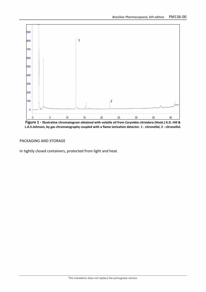
Brazilian Pharmacopoeia, 6th edition PM136-00
This translation does not replace the portuguese version.
Figure 1 - Illustrative chromatogram obtained with volatile oil from Corymbia citriodora (Hook.) K.D. Hill & L.A.S.Johnson, by gas chromatography coupled with a flame ionization detector. 1 - citronellal, 2 - citronellol.
PACKAGING AND STORAGE In tightly closed containers, protected from light and heat.
0
100
200
300
400
500
600
700
800
900
0 5 10 15 20 25 30 35 40
1
2
Brazilian Pharmacopoeia, 6th edition PM137-00
This translation does not replace the portuguese version.
FENNEL, oil Foeniculi fructus aetheroleum
Volatile oil obtained by hydrodistillation from the dried fruits of Foeniculum vulgare Mill. CHARACTERISTICS Colorless liquid. IDENTIFICATION Proceed as described in Thin-layer chromatography (5.2.17.1). Stationary phase: silica gel GF254. Mobile phase: toluene and ethyl acetate (93:7). Sample solution: dilute 5 µL of the sample to be examined in 500 µL toluene. Reference solution: dilute 6 µl trans- anethole and 6 µl fenchone in 500 µl toluene. TLC visualization reagent (1): dissolve 4 g of phosphomolybdic acid in 40 mL water while heating. Add 60 mL sulfuric acid after cooling. TLC visualization reagent (2): transfer 15 mL of sulfuric acid using a graduated pipette into a 50-mL beaker. Place the beaker with sulfuric acid in an ice bath and carefully add 0.5 g of potassium permanganate. Shake the solution with the help of a glass rod. Use to reveal the chromatographic plate. Dispose of the waste properly. Procedure: apply 5 μL of the Sample solution and 5 μL of the Reference solution to the chromatoplate, separately and in the form of a band. Conduct chromatogram. Remove chromatoplate and allow it to air dry for 15 minutes. Nebulize plate with TLC visualization reagent (1) and heat at 110°C for five minutes. Nebulize plate with TLC visualization reagent (2) and heat at 110°C in an oven for five minutes. Results: the diagram below shows the sequences of zones obtained with the Reference solution and the Sample solution. Other zones may occasionally develop.

Brazilian Pharmacopoeia, 6th edition PM137-00
This translation does not replace the portuguese version.
Top of the plate
trans-Anethole: dark-blue colored zone
Dark-blue colored zone
Fenchone: blue-colored zone Blue-colored zone
Reference solution Sample solution TESTS Relative density (5.2.5). 0.961 to 0.975. Refractive index (5.2.29.4). 1.528 to 1.539. Optical rotation (5.2.8). +10° a +24°. Chromatographic profile. Proceed as described in Gas chromatography (5.2.17.5). Use a chromatograph equipped with a flame ionization detector, 30-m long and 0.25-mm wide (internal diameter) capillary column, coated with polyethylene glycol, with a 0.25 μm film thickness. Use purified nitrogen as carrier gas (1 mL/minute). Temperature: Time (minutes) Temperature (ºC) Column 0 – 8
8 – 48 48 – 53
60 60 → 180 180
Injector 270 Detector 270 Sample solution: dilute 10 µL of the volatile oil in 500 µL hexane. Reference solution: dilute 2 µl α-pinene, 2 µL limonene, 2 µL de anisaldehyde, 5 µL fenchone, 2 µL de estragole and 10 µL trans-anethole in 1 mL hexane. Procedure: inject the volume of 1 μL of the Sample Solution and the Reference solution into the gas chromatograph, using a 1:50 flow division. Determine relative concentrations by electronic integration using the normalization method. Examine the chromatographic profile of the Sample solution. The characteristic peaks in the chromatogram obtained with the Sample solution should have retention times similar to those obtained with the chromatogram of the Reference solution or identification confirmed with gas
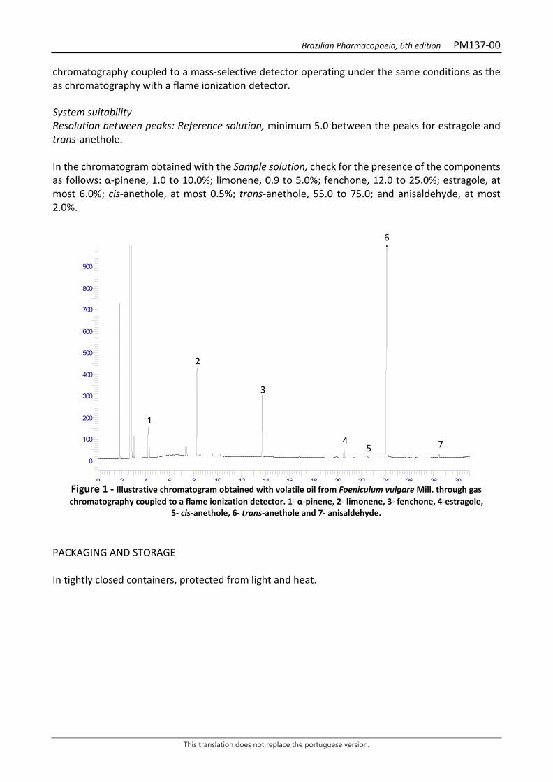
Brazilian Pharmacopoeia, 6th edition PM137-00
This translation does not replace the portuguese version.
chromatography coupled to a mass-selective detector operating under the same conditions as the as chromatography with a flame ionization detector. System suitability Resolution between peaks: Reference solution, minimum 5.0 between the peaks for estragole and trans-anethole. In the chromatogram obtained with the Sample solution, check for the presence of the components as follows: α-pinene, 1.0 to 10.0%; limonene, 0.9 to 5.0%; fenchone, 12.0 to 25.0%; estragole, at most 6.0%; cis-anethole, at most 0.5%; trans-anethole, 55.0 to 75.0; and anisaldehyde, at most 2.0%.
Figure 1 - Illustrative chromatogram obtained with volatile oil from Foeniculum vulgare Mill. through gas chromatography coupled to a flame ionization detector. 1- α-pinene, 2- limonene, 3- fenchone, 4-estragole,
5- cis-anethole, 6- trans-anethole and 7- anisaldehyde. PACKAGING AND STORAGE In tightly closed containers, protected from light and heat.
0
100
200
300
400
500
600
700
800
900
0 2 4 6 8 10 12 14 16 18 20 22 24 26 28 30
1
2
3
4
6
7 5
Brazilian Pharmacopoeia, 6th edition PM138-00
This translation does not replace the portuguese version.
SUNFLOWER, refined oil Helianthi annui oleum raffinatum
Oil obtained by expression or extraction from seeds of Helianthus annuus L., subjected to a refining process. CHARACTERISTICS Pale yellow oil with a clear appearance. IDENTIFICATION Proceed as described in Thin-layer chromatography (5.2.17.1). Stationary phase: octadecylsilanized silica gel (RP-18). Mobile phase (1): diethyl ether. Mobile phase (2): acetone, acetic acid and methyl chloride (50:40:20). Sample solution: dilute 20 µL of sunflower oil in 3 mL methyl chloride. Dilute 1 µL of this solution in 20 µL methyl chloride. Reference solution: dilute 20 µL of corn oil in 3 mL methyl chloride. Dilute 1 µL of this solution in 20 µL methyl chloride. TLC visualization reagent: dissolve 10 g of phosphomolybdic acid in 100 mL ethyl alcohol. Procedure: apply 20 μL of the Sample solution and 20 μL of the Reference solution to the chromatoplate, separately and in the form of a band. Develop the chromatogram using the Mobile phase (1) by 0.5 cm. Remove chromatoplate and allow it to air dry. Re-conduct chromatogram. Remove chromatoplate and allow it to air dry. Develop the chromatogram using the Mobile phase (2) by 8 cm. Remove chromatoplate and allow it to air dry. Re-conduct chromatogram. Remove chromatoplate and allow it to air dry. Nebulize plate with TLC visualization reagent and heat in an oven at 105°C for approximately three minutes. Results: the diagram below shows the sequences of zones obtained with the Reference solution and the Sample solution. Other zones may occasionally develop.

Brazilian Pharmacopoeia, 6th edition PM138-00
This translation does not replace the portuguese version.
Top of the plate
Corn oil: blue-colored zone Blue-colored zone
Corn oil: blue-colored zone Blue-colored zone
Corn oil: blue-colored zone Blue-colored zone
Reference solution Sample solution
TESTS Solubility. Very slightly soluble in 95% ethyl alcohol. Highly soluble in petroleum ether at a temperature between 40°C and 60°C. Practically insoluble in water. Relative density (5.2.5). 0.900 to 0.921. Water (5.2.20.1). Culombimetric method At most 0,1%. Determine in 1 g. Refractive index (5.2.29.4). 1.470 to 1.48. Acidity level (5.2.29.7). At most 0.5. Peroxide Index (5.2.29.11). At most 10. Unsaponifiable substances (5.2.29.14). Method II. At most 1.5%. Determine in 5 g. Alkaline impurities (5.2.29.15.2). Volume less than 0.1 mL of 0.01 M hydrochloric acid. Foreign oils in fixed oils by gas chromatography (5.2.29.15.4). Use mixture of substances for calibration in Table 3. Commercial mixtures of methyl esters can also be used. The fraction of the oil composed of fatty acids has the following composition: Palmitic acid: 4.0% to 9.0%. Stearic acid: 1.0% to 7.0%. Oleic acid: 14% to 40%. Linoleic acid: 48% to 74%. Total mesophilic microorganism count (5.5.3.1.2). Complies with the test. Study of pathogenic microorganisms (5.5.3.1.3). Complies with the test.
Brazilian Pharmacopoeia, 6th edition PM138-00
This translation does not replace the portuguese version.
PACKAGING AND STORAGE In tightly closed containers, protected from light and heat. CATEGORY Pharmacotechnical excipient.
Brazilian Pharmacopoeia, 6th edition PM139-00
This translation does not replace the portuguese version.
CORN MINT, oil Mentha arvensis aetheroleum
Volatile oil obtained by hydrodistillation from the dry aerial parts of Mentha arvensis L., containing at least 30.0% menthol (C10H20O, 156.27). CHARACTERISTICS Fluid, clear, colorless or light yellow liquid, with a characteristic menthol odor. IDENTIFICATION Proceed as described in Thin-layer chromatography (5.2.17.1). Stationary phase: silica gel F254(250 µm). Mobile phase: hexane and ethyl acetate (85:15). Sample solution: dilute 0.1 mL of the volatile oil in 1.0 mL ethyl acetate. Reference solution: dissolve 4 µL of carvone, 4 µL of pulegone, 10 µL of menthol acetate, 20 µL of cineol, and 50 mg of menthol in 5 mL ethyl acetate. Procedure: apply 10 μL of the Sample solution and 10 μL of the Reference solution to the chromatoplate, separately and in the form of a band. Conduct chromatogram. Remove chromatoplate and allow it to air dry. Examine under ultraviolet light at 254 nm. Nebulize plate liberally with a sulfuric vanillin solution. Heat between 100°C and 105°C for 1 minute to visualize the menthol in the lower third of the plate and heat for another 3 minutes to visualize the other components of the sample. Results: the diagrams below show the sequences of zones obtained with the Reference solution and the Sample solution, after examination under ultraviolet light, nebulization with the sulfuric vanillin solution and heating for one minute and then for another three minutes, in that order. Other zones may occasionally develop.
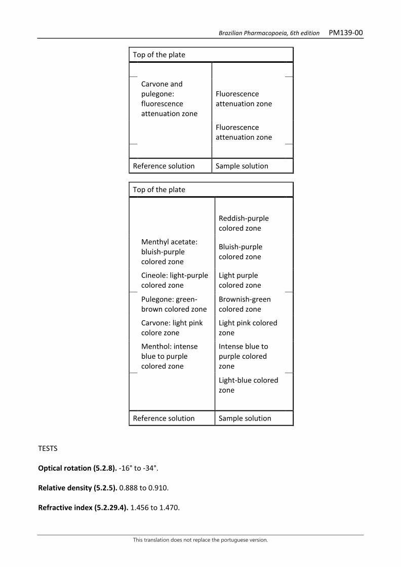
Brazilian Pharmacopoeia, 6th edition PM139-00
This translation does not replace the portuguese version.
Top of the plate
Carvone and pulegone: fluorescence attenuation zone
Fluorescence attenuation zone
Fluorescence attenuation zone
Reference solution Sample solution
Top of the plate
Reddish-purple colored zone
Menthyl acetate: bluish-purple colored zone
Bluish-purple colored zone
Cineole: light-purple colored zone
Light purple colored zone
Pulegone: green-brown colored zone
Brownish-green colored zone
Carvone: light pink colore zone
Light pink colored zone
Menthol: intense blue to purple colored zone
Intense blue to purple colored zone
Light-blue colored zone
Reference solution Sample solution TESTS Optical rotation (5.2.8). -16° to -34°. Relative density (5.2.5). 0.888 to 0.910. Refractive index (5.2.29.4). 1.456 to 1.470.
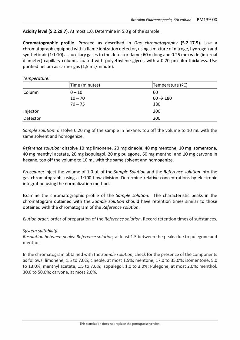
Brazilian Pharmacopoeia, 6th edition PM139-00
This translation does not replace the portuguese version.
Acidity level (5.2.29.7). At most 1.0. Determine in 5.0 g of the sample. Chromatographic profile. Proceed as described in Gas chromatography (5.2.17.5). Use a chromatograph equipped with a flame ionization detector, using a mixture of nitroge, hydrogen and synthetic air (1:1:10) as auxiliary gases to the detector flame; 60 m long and 0.25 mm wide (internal diameter) capillary column, coated with polyethylene glycol, with a 0.20 μm film thickness. Use purified helium as carrier gas (1,5 mL/minute). Temperature: Time (minutes) Temperature (ºC) Column 0 – 10
10 – 70 70 – 75
60 60 → 180 180
Injector 200 Detector 200 Sample solution: dissolve 0.20 mg of the sample in hexane, top off the volume to 10 mL with the same solvent and homogenize. Reference solution: dissolve 10 mg limonene, 20 mg cineole, 40 mg mentone, 10 mg isomentone, 40 mg menthyl acetate, 20 mg isopulegol, 20 mg pulegone, 60 mg menthol and 10 mg carvone in hexane, top off the volume to 10 mL with the same solvent and homogenize. Procedure: inject the volume of 1,0 μL of the Sample Solution and the Reference solution into the gas chromatograph, using a 1:100 flow division. Determine relative concentrations by electronic integration using the normalization method. Examine the chromatographic profile of the Sample solution. The characteristic peaks in the chromatogram obtained with the Sample solution should have retention times similar to those obtained with the chromatogram of the Reference solution. Elution order: order of preparation of the Reference solution. Record retention times of substances. System suitability Resolution between peaks: Reference solution, at least 1.5 between the peaks due to pulegone and menthol. In the chromatogram obtained with the Sample solution, check for the presence of the components as follows: limonene, 1.5 to 7.0%; cineole, at most 1.5%; mentone, 17.0 to 35.0%; isomentone, 5.0 to 13.0%; menthyl acetate, 1.5 to 7.0%; isopulegol, 1.0 to 3.0%; Pulegone, at most 2.0%; menthol, 30.0 to 50.0%; carvone, at most 2.0%.
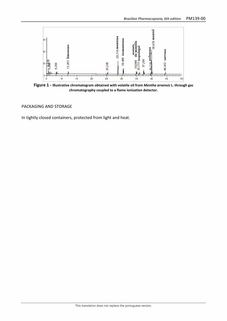
Brazilian Pharmacopoeia, 6th edition PM139-00
This translation does not replace the portuguese version.
Figure 1 - Illustrative chromatogram obtained with volatile oil from Mentha arvensis L. through gas
chromatography coupled to a flame ionization detector. PACKAGING AND STORAGE In tightly closed containers, protected from light and heat.
Brazilian Pharmacopoeia, 6th edition PM140-00
This translation does not replace the portuguese version.
PEPPERMINT, oil Menthae piperitae aetheroleum
Volatile oil obtained by hydrodistillation from the recently collected aerial parts of Mentha × piperita L., containing at least 35.0% menthol. CHARACTERISTICS Colorless, pale yellow or greenish-yellow liquid with a characteristic odor similar to menthol. IDENTIFICATION Proceed as described in Thin-layer chromatography (5.2.17.1). Stationary phase: silica gel GF254 (0.250 mm). Mobile phase: toluene and ethyl acetate (95:5). Sample solution: dilute 0.1 mL of the volatile oil in 10 mL toluene. Reference solution: dissolve 50 mg menthol CRS, 20 µL of 1,8-cineole, 10 mg thymol and 10 µL menthyl acetate in toluene, top off the volume to 10 mL with the same solvent and homogenize. Procedure: apply 20 μL of the Sample solution and 10 μL of the Reference solution to the chromatoplate, separately and in the form of a band. Conduct chromatogram. Remove chromatoplate and allow it to air dry. Examine under ultraviolet light at 254 nm. Next, nebulize the plate with anisaldehyde RS in an oven at 100 °C to 105 °C for five to 10 minutes. Immediately examine under visible light. Results: the diagram below shows the sequences of zones obtained with the Reference solution and the Sample solution. Other zones may occasionally develop.
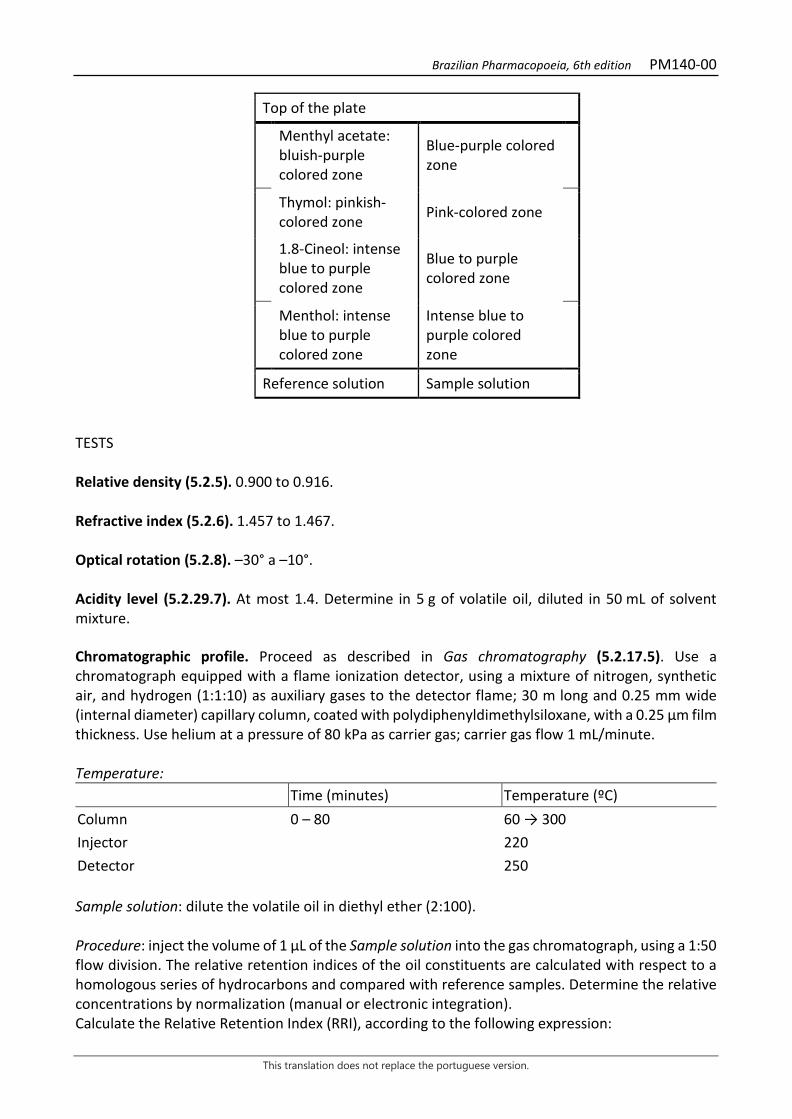
Brazilian Pharmacopoeia, 6th edition PM140-00
This translation does not replace the portuguese version.
Top of the plate
Menthyl acetate: bluish-purple colored zone
Blue-purple colored zone
Thymol: pinkish-colored zone Pink-colored zone
1.8-Cineol: intense blue to purple colored zone
Blue to purple colored zone
Menthol: intense blue to purple colored zone
Intense blue to purple colored zone
Reference solution Sample solution TESTS Relative density (5.2.5). 0.900 to 0.916. Refractive index (5.2.6). 1.457 to 1.467. Optical rotation (5.2.8). –30° a –10°. Acidity level (5.2.29.7). At most 1.4. Determine in 5 g of volatile oil, diluted in 50 mL of solvent mixture. Chromatographic profile. Proceed as described in Gas chromatography (5.2.17.5). Use a chromatograph equipped with a flame ionization detector, using a mixture of nitrogen, synthetic air, and hydrogen (1:1:10) as auxiliary gases to the detector flame; 30 m long and 0.25 mm wide (internal diameter) capillary column, coated with polydiphenyldimethylsiloxane, with a 0.25 µm film thickness. Use helium at a pressure of 80 kPa as carrier gas; carrier gas flow 1 mL/minute. Temperature: Time (minutes) Temperature (ºC) Column 0 – 80 60 → 300 Injector 220 Detector 250 Sample solution: dilute the volatile oil in diethyl ether (2:100). Procedure: inject the volume of 1 μL of the Sample solution into the gas chromatograph, using a 1:50 flow division. The relative retention indices of the oil constituents are calculated with respect to a homologous series of hydrocarbons and compared with reference samples. Determine the relative concentrations by normalization (manual or electronic integration). Calculate the Relative Retention Index (RRI), according to the following expression:
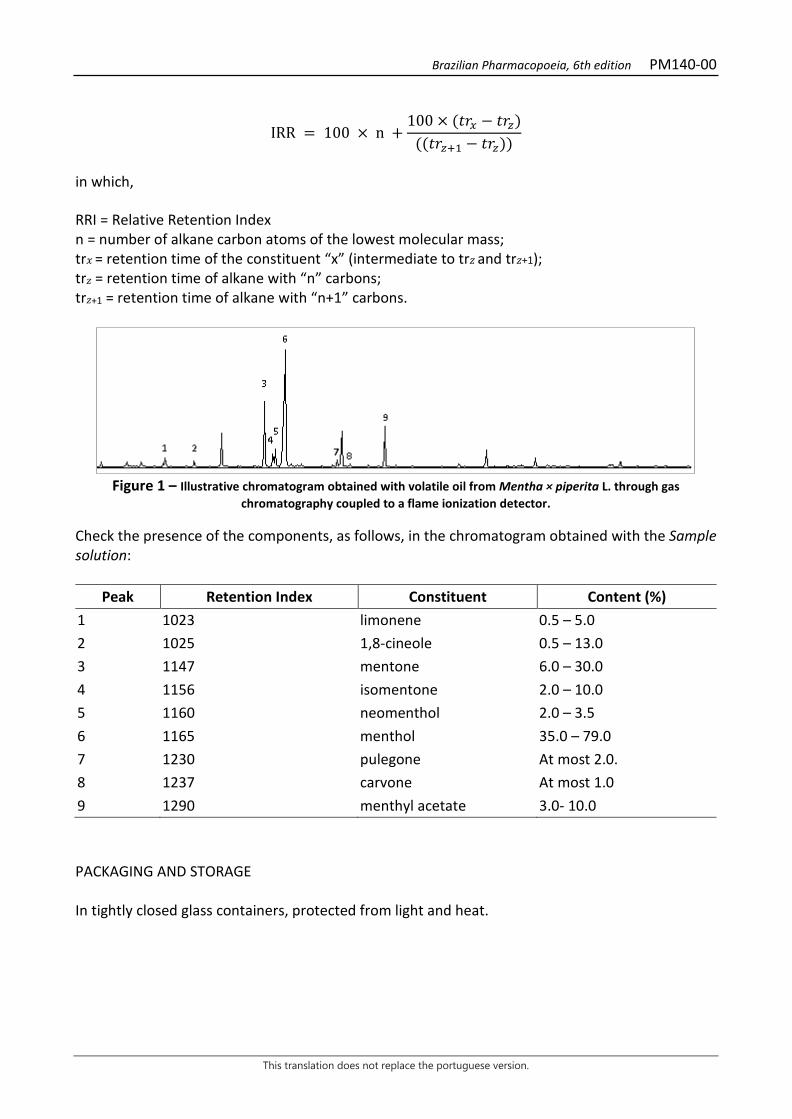
Brazilian Pharmacopoeia, 6th edition PM140-00
This translation does not replace the portuguese version.
IRR = 100 × n +100 × (𝑡𝑡𝑡𝑡𝑥𝑥 − 𝑡𝑡𝑡𝑡𝑧𝑧)
((𝑡𝑡𝑡𝑡𝑧𝑧+1 − 𝑡𝑡𝑡𝑡𝑧𝑧))
in which, RRI = Relative Retention Index n = number of alkane carbon atoms of the lowest molecular mass; tr𝑥𝑥 = retention time of the constituent “x” (intermediate to tr𝑧𝑧 and tr𝑧𝑧+1); tr𝑧𝑧 = retention time of alkane with “n” carbons; tr𝑧𝑧+1 = retention time of alkane with “n+1” carbons.
Figure 1 – Illustrative chromatogram obtained with volatile oil from Mentha × piperita L. through gas
chromatography coupled to a flame ionization detector. Check the presence of the components, as follows, in the chromatogram obtained with the Sample solution:
Peak Retention Index Constituent Content (%) 1 1023 limonene 0.5 – 5.0 2 1025 1,8-cineole 0.5 – 13.0 3 1147 mentone 6.0 – 30.0 4 1156 isomentone 2.0 – 10.0 5 1160 neomenthol 2.0 – 3.5 6 1165 menthol 35.0 – 79.0 7 1230 pulegone At most 2.0. 8 1237 carvone At most 1.0 9 1290 menthyl acetate 3.0- 10.0 PACKAGING AND STORAGE In tightly closed glass containers, protected from light and heat.
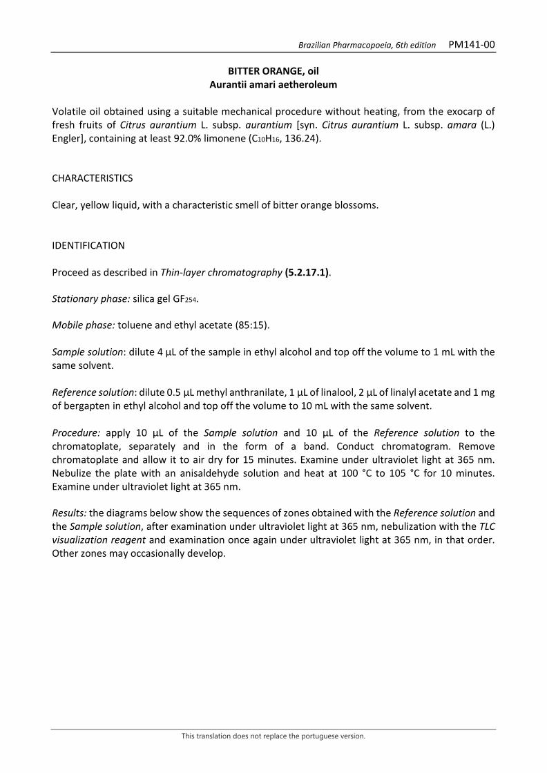
Brazilian Pharmacopoeia, 6th edition PM141-00
This translation does not replace the portuguese version.
BITTER ORANGE, oil Aurantii amari aetheroleum
Volatile oil obtained using a suitable mechanical procedure without heating, from the exocarp of fresh fruits of Citrus aurantium L. subsp. aurantium [syn. Citrus aurantium L. subsp. amara (L.) Engler], containing at least 92.0% limonene (C10H16, 136.24). CHARACTERISTICS Clear, yellow liquid, with a characteristic smell of bitter orange blossoms. IDENTIFICATION Proceed as described in Thin-layer chromatography (5.2.17.1). Stationary phase: silica gel GF254. Mobile phase: toluene and ethyl acetate (85:15). Sample solution: dilute 4 µL of the sample in ethyl alcohol and top off the volume to 1 mL with the same solvent. Reference solution: dilute 0.5 µL methyl anthranilate, 1 μL of linalool, 2 μL of linalyl acetate and 1 mg of bergapten in ethyl alcohol and top off the volume to 10 mL with the same solvent. Procedure: apply 10 μL of the Sample solution and 10 μL of the Reference solution to the chromatoplate, separately and in the form of a band. Conduct chromatogram. Remove chromatoplate and allow it to air dry for 15 minutes. Examine under ultraviolet light at 365 nm. Nebulize the plate with an anisaldehyde solution and heat at 100 °C to 105 °C for 10 minutes. Examine under ultraviolet light at 365 nm. Results: the diagrams below show the sequences of zones obtained with the Reference solution and the Sample solution, after examination under ultraviolet light at 365 nm, nebulization with the TLC visualization reagent and examination once again under ultraviolet light at 365 nm, in that order. Other zones may occasionally develop.
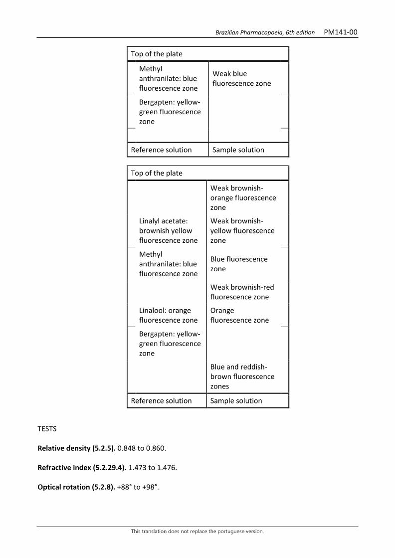
Brazilian Pharmacopoeia, 6th edition PM141-00
This translation does not replace the portuguese version.
Top of the plate
Methyl anthranilate: blue fluorescence zone
Weak blue fluorescence zone
Bergapten: yellow-green fluorescence zone
Reference solution Sample solution
Top of the plate
Weak brownish-orange fluorescence zone
Linalyl acetate: brownish yellow fluorescence zone
Weak brownish-yellow fluorescence zone
Methyl anthranilate: blue fluorescence zone
Blue fluorescence zone
Weak brownish-red fluorescence zone
Linalool: orange fluorescence zone
Orange fluorescence zone
Bergapten: yellow-green fluorescence zone
Blue and reddish-brown fluorescence zones
Reference solution Sample solution TESTS Relative density (5.2.5). 0.848 to 0.860. Refractive index (5.2.29.4). 1.473 to 1.476. Optical rotation (5.2.8). +88° to +98°.
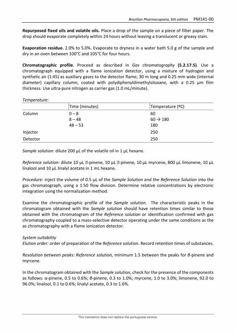
Brazilian Pharmacopoeia, 6th edition PM141-00
This translation does not replace the portuguese version.
Repurposed fixed oils and volatile oils. Place a drop of the sample on a piece of filter paper. The drop should evaporate completely within 24 hours without leaving a translucent or greasy stain. Evaporation residue. 2.0% to 5.0%. Evaporate to dryness in a water bath 5.0 g of the sample and dry in an oven between 100°C and 105°C for four hours. Chromatographic profile. Proceed as described in Gas chromatography (5.2.17.5). Use a chromatograph equipped with a flame ionization detector, using a mixture of hydrogen and synthetic air (1:45) as auxiliary gases to the detector flame; 30 m long and 0.25 mm wide (internal diameter) capillary column, coated with polydiphenyldimethylsiloxane, with a 0.25 μm film thickness. Use ultra-pure nitrogen as carrier gas (1.0 mL/minute). Temperature: Time (minutes) Temperature (ºC) Column 0 – 8
8 – 48 48 – 53
60 60 → 180 180
Injector 250 Detector 250 Sample solution: dilute 200 µL of the volatile oil in 1 µL hexane. Reference solution: dilute 10 μL -pinene, 10 μL -pinene, 10 µL myrcene, 800 μL limonene, 10 μL linalool and 10 μL linalyl acetate in 1 mL hexane. Procedure: inject the volume of 0.5 μL of the Sample Solution and the Reference Solution into the gas chromatograph, using a 1:50 flow division. Determine relative concentrations by electronic integration using the normalization method. Examine the chromatographic profile of the Sample solution. The characteristic peaks in the chromatogram obtained with the Sample solution should have retention times similar to those obtained with the chromatogram of the Reference solution or identification confirmed with gas chromatography coupled to a mass-selective detector operating under the same conditions as the as chromatography with a flame ionization detector. System suitability Elution order: order of preparation of the Reference solution. Record retention times of substances. Resolution between peaks: Reference solution, minimum 1.5 between the peaks for β-pinene and myrcene. In the chromatogram obtained with the Sample solution, check for the presence of the components as follows: α-pinene, 0.5 to 0.6%; β-pinene, 0.3 to 1.0%; myrcene, 1.0 to 3.0%; limonene, 92.0 to 96.0%; linalool, 0.1 to 0.6%; linalyl acetate, 0.3 to 1.6%.
Brazilian Pharmacopoeia, 6th edition PM141-00
This translation does not replace the portuguese version.
Figure 1 - Illustrative chromatogram obtained with volatile oil from Citrus aurantium L. subsp. aurantium through gas chromatography coupled to a flame ionization detector. 1- α-pinene, 2- β- pinene, 3- myrcene, 4- limonene, 5-
linalool, 6- linalyl acetate. PACKAGING AND STORAGE In tightly closed containers, protected from light and heat at a temperature below 25 ºC.
0
100
200
300
400
500
600
700
800
900
0 5 10 15 20 25 30 35 40 45 50
6 5
4
3 2 1
Brazilian Pharmacopoeia, 6th edition PM142-00
This translation does not replace the portuguese version.
SWEET ORANGE, oil Citrus aurantium dulcis aetheroleum
Volatile oil obtained by appropriate mechanical means without heating, from the epicarp of fresh fruits of Citrus sinensis (L.) Osbeck (syn. Citrus aurantium L. var. sinensis L.), containing at least 92.0% limonene (C10H16, 136.24). CHARACTERISTICS Clear, pale yellow to orange liquid that may become cloudy on cooling. IDENTIFICATION Proceed as described in Thin-layer chromatography (5.2.17.1). Stationary phase: silica gel GF254. Mobile phase: toluene and ethyl acetate (85:15). Sample solution: dilute 0.2 mL of the sample in 1 mL ethyl alcohol. Reference solution: dissolve 2 mg bergapten, 10 μL linalool and 20 μL linalyl acetate in 10 mL ethyl alcohol. Procedure: apply 10 μL of the Sample solution and 10 μL of the Reference solution to the chromatoplate, separately and in the form of a band. Remove chromatoplate and allow it to air dry for 15 minutes. Examine under ultraviolet light at 365 nm. Nebulize the plate with an anisaldehyde solution and heat at 100 °C to 105 °C for five to 10 minutes. Examine under ultraviolet light at 365 nm. Results: the diagrams below show the zones obtained with the Reference solution and the Sample solution, after examination under ultraviolet light at 365 nm, nebulization with anisaldehyde solution and examination once again under ultraviolet light at 365 nm, in that order. Other zones may occasionally be present.
Brazilian Pharmacopoeia, 6th edition PM142-00
This translation does not replace the portuguese version.
Top of the plate
Bergapten: yellow-green fluorescence zone
Other blue fluorescence zones
Reference solution Sample solution
Top of the plate
Brown fluorescence zones
Linalyl acetate: greenish-yellow fluorescence zone
Brownish-yellow fluorescence zones
Orange fluorescence zones
Linalool: orange fluorescence zone
Orange fluorescence zones
Bergapten: weak yellowish-green fluorescence zone
Brownish-orange fluorescence zones
Blue fluorescence zone
Reference solution Sample solution TESTS Relative density (5.2.5). 0.842 to 0.850. Refractive index (5.2.29.4). 1.470% to 1.476%. Optical rotation (5.2.8). +94° to +99°. Peroxide Index (5.2.29.11). At most 20. Total mesophilic microorganism count (5.5.3.1.2). Complies with the test. Study of pathogenic microorganisms (5.5.3.1.3). Complies with the test.

Brazilian Pharmacopoeia, 6th edition PM142-00
This translation does not replace the portuguese version.
Repurposed fixed oils and volatile oils. Complies with the test. On a 3×3 cm filter paper, on a 10 cm diameter watch glass, apply 5µL of the sample. Wait 24 hours, the time required for the oil to completely volatilize. Check for absence of residue. Conduct the test in triplicate. Evaporation residue. 1.0% to 5.0%. Evaporate to dryness in a water bath 5.0 g of the sample and dry between 100°C and 105°C for four hours. Chromatographic profile. Proceed as described in Gas chromatography (5.2.17.5). Use a chromatograph equipped with a flame ionization detector, using a mixture of hydrogen and synthetic air (1:45) as auxiliary gases to the detector flame; 30 m long and 0.25 mm wide (internal diameter) capillary column, coated with polyethylene glycol, with a 0.25 μm film thickness. Use ultra-pure nitrogen as carrier gas (1 mL/minute). Temperature: Time (minutes) Temperature (ºC) Column 0 – 6
6 – 31 31 – 41 41 – 55
50 50 → 150 150 → 180 180
Injector 250 Detector 250 Sample solution: dilute 300 µL of the sweet orange volatile oil in 1 mL acetone. Reference solution (1): dilute 10 µL α-pinene, 10 µL β-pinene, 10 µL sabinene, 20 µL β- myrcene, 800 µL limonene, 10 µL octanal, 10 µL decanal, 10 µL linalool, 10 µL citral, 10 µL valencene in 1 mL acetone and homogenize. Reference solution (2): dilute 5 µL β-pinene in 10 mL acetone and homogenize. Dilute 0,5 mL of this solution in 10 mL acetone and homogenize. Procedure: inject 0.5 μL of the Sample Solution and the Reference Solution (1) into the gas chromatograph, using a 1:100 flow division. Determine relative concentrations by electronic integration using the normalization method. System suitability Peak resolutions: Reference solution (1), minimum 3.9 between the peaks for β-pinene and sabinene, and at least 1.5 between the peaks for valencene and geranial. Exclusion Limit: area of the peak of the chromatogram obtained with Reference solution (2) (0.01%). In the chromatogram obtained with the Sample solution, check for the presence of the components as follows: α-pinene 0.4 to 0.6%; β-pinene 0.02 to 0.3%; sabinene 0.2 to 1.1%; β-myrcene 1.7 to 2.5%; limonene 92.0 to 97.0%; octanal 0.1 to 0.4%; decanal 0.1 to 0.4%; linalool 0.2 to 0.7%; neral 0.02 to 0.10%; valencene 0.02 to 0.5%; and geranial 0.03 to 0.20%.
Brazilian Pharmacopoeia, 6th edition PM142-00
This translation does not replace the portuguese version.
PACKAGING AND STORAGE In tightly closed containers, protected from light and heat.
Brazilian Pharmacopoeia, 6th edition PM143-00
This translation does not replace the portuguese version.
LEMON, oil Limonis aetheroleum
Volatile oil obtained by appropriate mechanical means without heating, from the fresh pericarp of Citrus × limon (L.) Osbeck, containing at least 56.0% limonene (C10H16, 136.24). CHARACTERISTICS Clear liquid, light yellow to greenish yellow in color, with a characteristic odor. May become turbid at low temperatures. IDENTIFICATION Proceed as described in Thin-layer chromatography (5.2.17.1). Stationary phase: silica gel F254 (0.25 mm). Mobile phase: toluene and ethyl acetate (85:15). Sample solution: dilute 1 mL of the sample in 1 mL toluene. Reference solution: dissolve 10 mg citropten and 50 μL citral in toluene, top off the volume to 10 mL with the same solvent and homogenize. Procedure: apply 10 μL of the Sample solution and 10 μL of the Reference solution to the chromatoplate, separately and in the form of a band. Conduct chromatogram. Remove chromatoplate and allow it to air dry for 15 minutes. Examine under ultraviolet light at 254 nm. Examine under ultraviolet light at 365 nm. Results: the diagrams below show the sequences of zones obtained with the Reference solution and the Sample solution, after examination under ultraviolet light at 254 nm and 365 nm, respectively. Other zones may occasionally develop.
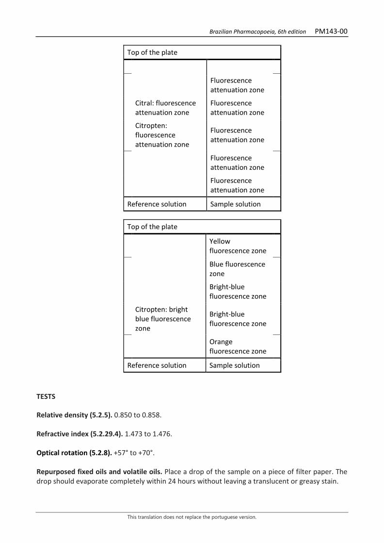
Brazilian Pharmacopoeia, 6th edition PM143-00
This translation does not replace the portuguese version.
Top of the plate
Fluorescence attenuation zone
Citral: fluorescence attenuation zone
Fluorescence attenuation zone
Citropten: fluorescence attenuation zone
Fluorescence attenuation zone
Fluorescence attenuation zone
Fluorescence attenuation zone
Reference solution Sample solution
Top of the plate
Yellow fluorescence zone
Blue fluorescence zone
Bright-blue fluorescence zone
Citropten: bright blue fluorescence zone
Bright-blue fluorescence zone
Orange fluorescence zone
Reference solution Sample solution TESTS Relative density (5.2.5). 0.850 to 0.858. Refractive index (5.2.29.4). 1.473 to 1.476. Optical rotation (5.2.8). +57° to +70°. Repurposed fixed oils and volatile oils. Place a drop of the sample on a piece of filter paper. The drop should evaporate completely within 24 hours without leaving a translucent or greasy stain.
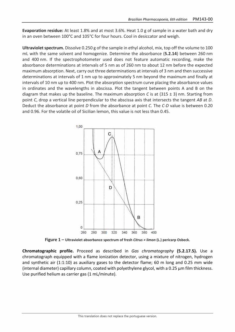
Brazilian Pharmacopoeia, 6th edition PM143-00
This translation does not replace the portuguese version.
Evaporation residue: At least 1.8% and at most 3.6%. Heat 1.0 g of sample in a water bath and dry in an oven between 100°C and 105°C for four hours. Cool in desiccator and weigh. Ultraviolet spectrum. Dissolve 0.250 g of the sample in ethyl alcohol, mix, top off the volume to 100 mL with the same solvent and homogenize. Determine the absorbance (5.2.14) between 260 nm and 400 nm. If the spectrophotometer used does not feature automatic recording, make the absorbance determinations at intervals of 5 nm as of 260 nm to about 12 nm before the expected maximum absorption. Next, carry out three determinations at intervals of 3 nm and then successive determinations at intervals of 1 nm up to approximately 5 nm beyond the maximum and finally at intervals of 10 nm up to 400 nm. Plot the absorption spectrum curve placing the absorbance values in ordinates and the wavelengths in abscissa. Plot the tangent between points A and B on the diagram that makes up the baseline. The maximum absorption C is at (315 ± 3) nm. Starting from point C, drop a vertical line perpendicular to the abscissa axis that intersects the tangent AB at D. Deduct the absorbance at point D from the absorbance at point C. The C-D value is between 0.20 and 0.96. For the volatile oil of Sicilian lemon, this value is not less than 0.45.
Figure 1 – Ultraviolet absorbance spectrum of fresh Citrus × limon (L.) pericarp Osbeck.
Chromatographic profile. Proceed as described in Gas chromatography (5.2.17.5). Use a chromatograph equipped with a flame ionization detector, using a mixture of nitrogen, hydrogen and synthetic air (1:1:10) as auxiliary gases to the detector flame; 60 m long and 0.25 mm wide (internal diameter) capillary column, coated with polyethylene glycol, with a 0.25 μm film thickness. Use purified helium as carrier gas (1 mL/minute).
Brazilian Pharmacopoeia, 6th edition PM143-00
This translation does not replace the portuguese version.
Temperature: Time (minutes) Temperature (ºC) Column 0 – 6 45 6 – 21 45 → 90 21 – 39 90 → 180 39 – 55 180 Injector 220 Detector 220 Sample solution: volatile lemon oil. Reference solution: dilute 20 μL β-pinene, 10 μL sabinene, 100 μL limonene, 10 μL γ- terpinene, 5 μL β-caryophyllene, 20 μL citral, 5 mg α-terpineol, 5 μL de neryl acetate and 5 μL de geranyl acetate in 1 mL acetone. Procedure: inject the volume of 0.5 μL of the Reference solution and 0.2 µL of the Sample solution into the gas chromatograph, using a 1:100 flow division. Determine relative concentrations by electronic integration using the normalization method. Examine the chromatographic profile of the Sample solution. The characteristic peaks in the chromatogram obtained with the Sample solution should have retention times similar to those obtained with the chromatogram of the Reference solution or identification confirmed with gas chromatography coupled to a mass-selective detector operating under the same conditions as the as chromatography with a flame ionization detector. Elution order: order of preparation of the Reference solution. Record retention times of substances. System suitability Peak resolutions: Reference solution. minimum 1.5 between the peaks for β-pinene and sabinene, minimum 5,0 between the peaks for neryl acetate and α-terpineol and minimum 1.5 between the peaks for geraniol and geranyl acetate. In the chromatogram obtained with the Sample solution, check for the presence of the components as follows: β-pinene, 7.0 to 17.0%; sabinene, 1.0 to 3.0%; limonene, 56.0 to 78.0%; γ-terpinene, 6.0 to 12.0%; β-caryophyllene, at most 0,5%; neral, 0.3 to 1.5%; α-terpineol, at most 0.6%; neryl acetate, 0.2-0.9%; geraniol, 0.5 to 2.3%; and geranyl acetate, 0.1 to 0.8%. PACKAGING AND STORAGE In tightly closed containers, protected from light and heat.
Brazilian Pharmacopoeia, 6th edition PM144-00
This translation does not replace the portuguese version.
COCOA BUTTER Cacao oleum
Solid fat obtained from the roasted seeds of Theobroma cacao L. CHARACTERISTICS Pale yellow, solid fat with a characteristic odor similar to cocoa. TESTS Solubility. Easily soluble in diethyl ether and petroleum ether with a boiling range of 40°C to 60°C. Slightly soluble in 96% ethyl alcohol. Acidity level (5.2.29.7). At most 4.0. Iodine level (5.2.29.10). 35 to 40. Melting point (5.2.2). Method II. 31 °C to 34 °C. Refractive index (5.2.6). 1.456 to 1.458, at 40 °C. Saponification index. 188 to 196. Dissolve 35 to 40 g of potassium hydroxide in 20 mL of water, top off the volume with 95% ethyl alcohol in a 1000-mL flask and homogenize. Allow the solution to stand for 12 hours and filter. In a 250-mL round-bottomed flask, accurately weigh about 2 g of sample and add 25 mL of potassium hydroxide solution in ethyl alcohol. Fit the flask to the reflux condenser and heat in a water bath for one hour with frequent shaking. Hot titrate with 0.5 M hydrochloric acid SV solution using 1 mL of phenolphthalein solution as indicator. Carry out the blank test. Saponification index is calculated according to the following expression:
𝐼𝐼𝑆𝑆 =28,05 (𝑛𝑛2 − 𝑛𝑛1)
𝑚𝑚
in which, 𝑛𝑛1 = adjusted titrant volume; 𝑛𝑛2 = adjusted titrant volume in the blank test; 𝑚𝑚 = weighted sample mass. Total mesophilic microorganism count (5.5.3.1.2). Complies with the test. Study of pathogenic microorganisms (5.5.3.1.3). Complies with the test. PACKAGING AND STORAGE
Brazilian Pharmacopoeia, 6th edition PM144-00
This translation does not replace the portuguese version.
In tightly closed containers, protected from light and heat. CATEGORY Pharmacotechnical excipient.
Brazilian Pharmacopoeia, 6th edition PM145-00
This translation does not replace the portuguese version.
TEA TREE, oil Melaleucae aetheroleum
Volatile oil obtained by hydrodistillation using leaves and terminal branches of Melaleuca alternifolia (Maiden & Betche) Cheel, containing at least 30.0% terpinen-4-ol (C10H18O, 154.25). CHARACTERISTICS Colorless to pale yellow liquid. IDENTIFICATION Proceed as described in Thin-layer chromatography (5.2.17.1). Stationary phase: silica gel GF254. Mobile phase: heptane and ethyl acetate (80:20). Sample solution: dilute 0.1 mL of the sample to be examined in 5 mL heptane. Reference solution: dissolve 30 µL 1,8-cineole, 60 µL terpinen-4-ol and 10 mg α-terpineol in 10 mL heptane. Procedure: apply 10 μL of the Sample solution and 10 μL of the Reference solution to the chromatoplate, separately and in the form of a band. Conduct chromatogram. Remove chromatoplate and allow it to air dry for 15 minutes. Nebulize the plate with an anisaldehyde solution and heat at 100 °C to 105 °C for five to 10 minutes. Examine under visible light. Results: the diagram below shows the sequences of zones obtained with the Reference solution and the Sample solution. Other zones may occasionally develop.

Brazilian Pharmacopoeia, 6th edition PM145-00
This translation does not replace the portuguese version.
Top of the plate
1,8-Cineole: pink-colored zone Pink-colored zones
Terpinen-4-ol: pink-colored zone Pink-colored zones
-Terpineol: pink-colored zone Pink-colored zone
Reference solution Sample solution TESTS Relative density (5.2.5). 0.885 to 0.906. Refractive index (5.2.29.4). 1.475 to 1.482. Optical rotation (5.2.8). +5° to +15°. Chromatographic profile. Proceed as described in Gas chromatography (5.2.17.5). Use a chromatograph equipped with a flame ionization detector, 30-m long and 0.25-mm wide (internal diameter) capillary column, coated with polyethylene glycol, with a 0.25 μm film thickness. Use purified nitrogen as carrier gas (1.3 mL/minute). Temperature: Time (minutes) Temperature (ºC) Column 0 – 1
1 – 37 37 – 45
50 50 → 230 230
Injector 240 Detector 240 Sample solution: dilute 4.5 µL of the volatile in 300 µL hexane. Reference solution: dissolve 5 µL of α-pinene, 5 µL of sabinene, 15 µL α-terpinene, 5 µL of limonene, 5 µL of 1,8-cineole, 30 µL of γ-terpinene, 5 µL of p-cymene, 5 µL of terpinolene, 60 µL of terpinen-4-ol, 5 µL of aromadendrene, and 5 mg of α-terpineol in 10 mL of hexane. Procedure: inject the volume of 1 μL of the Sample Solution and the Reference solution into the gas chromatograph, using a 1:50 flow division. Determine relative concentrations by electronic integration using the normalization method. Examine the chromatographic profile of the Sample solution. The characteristic peaks in the chromatogram obtained with the Sample solution should have retention times similar to those obtained with the chromatogram of the Reference solution or identification confirmed with gas chromatography coupled to a mass detector operating under the same conditions as the as chromatography with a flame ionization detector.
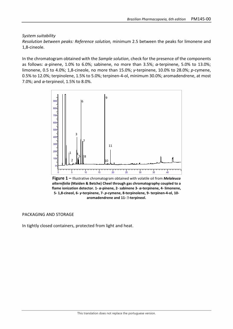
Brazilian Pharmacopoeia, 6th edition PM145-00
This translation does not replace the portuguese version.
System suitability Resolution between peaks: Reference solution, minimum 2.5 between the peaks for limonene and 1,8-cineole. In the chromatogram obtained with the Sample solution, check for the presence of the components as follows: α-pinene, 1.0% to 6.0%; sabinene, no more than 3.5%; α-terpinene, 5.0% to 13.0%; limonene, 0.5 to 4.0%; 1,8-cineole, no more than 15.0%; γ-terpinene, 10.0% to 28.0%; p-cymene, 0.5% to 12.0%; terpinolene, 1.5% to 5.0%; terpinen-4-ol, minimum 30.0%; aromadendrene, at most 7.0%; and α-terpineol, 1.5% to 8.0%.
Figure 1 – Illustrative chromatogram obtained with volatile oil from Melaleuca alternifolia (Maiden & Betche) Cheel through gas chromatography coupled to a flame ionization detector. 1- α-pinene, 2- sabinene 3- α-terpinene, 4- limonene,
5- 1,8-cineol, 6- γ-terpinene, 7- p-cymene, 8-terpinolene, 9- terpinen-4-ol, 10- aromadendrene and 11- -terpineol.
PACKAGING AND STORAGE In tightly closed containers, protected from light and heat.
0
100
200
300
400
500
600
700
800
900
0 5 10 15 20 25 30 35 40
1
2
3
45
6
7
8
9
10
11
Brazilian Pharmacopoeia, 6th edition PM146-00
This translation does not replace the portuguese version.
NUTMEG, oil Myristicae fragrans aetheroleum
Volatile oil obtained by hydrodistillation using dried and pulverized seeds devoid of aril and integument of Myristica fragrans Houtt. CHARACTERISTICS Colorless or light yellow liquid. IDENTIFICATION Proceed as described in Thin-layer chromatography (5.2.17.1). Stationary phase: silica gel GF254. Mobile phase: toluene and ethyl acetate (95:5). Sample solution: dilute 1 mL of the sample to be examined in 1 mL toluene, and top off the volume to 10 mL with the same solvent. Reference solution: dilute 20 µL of myristicin in 10 mL toluene. Procedure: apply 10 μL of the Sample solution and 10 μL of the Reference solution to the chromatoplate, separately and in the form of a band. Conduct chromatogram. Remove chromatoplate and allow it to air dry for 15 minutes. Nebulize the plate with sulfur vanillin RS and heat at 100 °C to 105 °C for 10 to 15 minutes. Results: the diagram below shows the sequences of zones obtained with the Reference solution and the Sample solution. Other zones may occasionally develop.
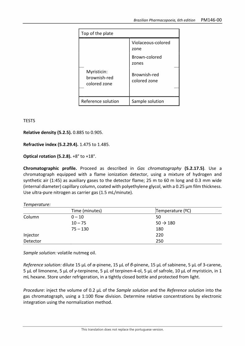
Brazilian Pharmacopoeia, 6th edition PM146-00
This translation does not replace the portuguese version.
Top of the plate
Violaceous-colored zone
Brown-colored zones
Myristicin: brownish-red colored zone
Brownish-red colored zone
Reference solution Sample solution TESTS Relative density (5.2.5). 0.885 to 0.905. Refractive index (5.2.29.4). 1.475 to 1.485. Optical rotation (5.2.8). +8° to +18°. Chromatographic profile. Proceed as described in Gas chromatography (5.2.17.5). Use a chromatograph equipped with a flame ionization detector, using a mixture of hydrogen and synthetic air (1:45) as auxiliary gases to the detector flame; 25 m to 60 m long and 0.3 mm wide (internal diameter) capillary column, coated with polyethylene glycol, with a 0.25 μm film thickness. Use ultra-pure nitrogen as carrier gas (1.5 mL/minute). Temperature: Time (minutes) Temperature (ºC) Column 0 – 10 50 10 – 75 50 → 180 75 – 130 180 Injector 220 Detector 250 Sample solution: volatile nutmeg oil. Reference solution: dilute 15 µL of α-pinene, 15 µL of β-pinene, 15 µL of sabinene, 5 µL of 3-carene, 5 µL of limonene, 5 µL of γ-terpinene, 5 µL of terpinen-4-ol, 5 µL of safrole, 10 µL of myristicin, in 1 mL hexane. Store under refrigeration, in a tightly closed bottle and protected from light. Procedure: inject the volume of 0.2 μL of the Sample solution and the Reference solution into the gas chromatograph, using a 1:100 flow division. Determine relative concentrations by electronic integration using the normalization method.
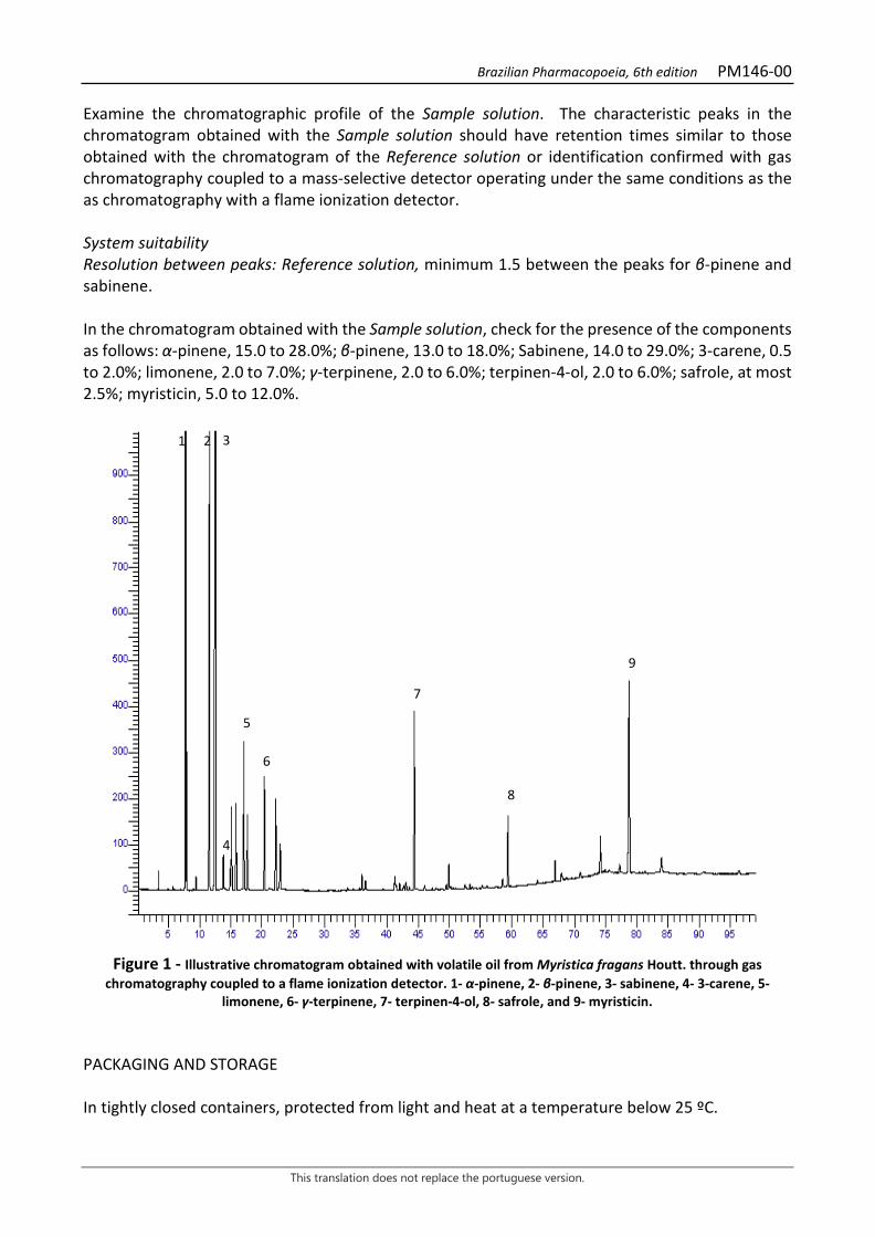
Brazilian Pharmacopoeia, 6th edition PM146-00
This translation does not replace the portuguese version.
Examine the chromatographic profile of the Sample solution. The characteristic peaks in the chromatogram obtained with the Sample solution should have retention times similar to those obtained with the chromatogram of the Reference solution or identification confirmed with gas chromatography coupled to a mass-selective detector operating under the same conditions as the as chromatography with a flame ionization detector. System suitability Resolution between peaks: Reference solution, minimum 1.5 between the peaks for β-pinene and sabinene. In the chromatogram obtained with the Sample solution, check for the presence of the components as follows: α-pinene, 15.0 to 28.0%; β-pinene, 13.0 to 18.0%; Sabinene, 14.0 to 29.0%; 3-carene, 0.5 to 2.0%; limonene, 2.0 to 7.0%; γ-terpinene, 2.0 to 6.0%; terpinen-4-ol, 2.0 to 6.0%; safrole, at most 2.5%; myristicin, 5.0 to 12.0%.
Figure 1 - Illustrative chromatogram obtained with volatile oil from Myristica fragans Houtt. through gas
chromatography coupled to a flame ionization detector. 1- α-pinene, 2- β-pinene, 3- sabinene, 4- 3-carene, 5- limonene, 6- γ-terpinene, 7- terpinen-4-ol, 8- safrole, and 9- myristicin.
PACKAGING AND STORAGE In tightly closed containers, protected from light and heat at a temperature below 25 ºC.
2 3
6
5
4
7
8
9
1
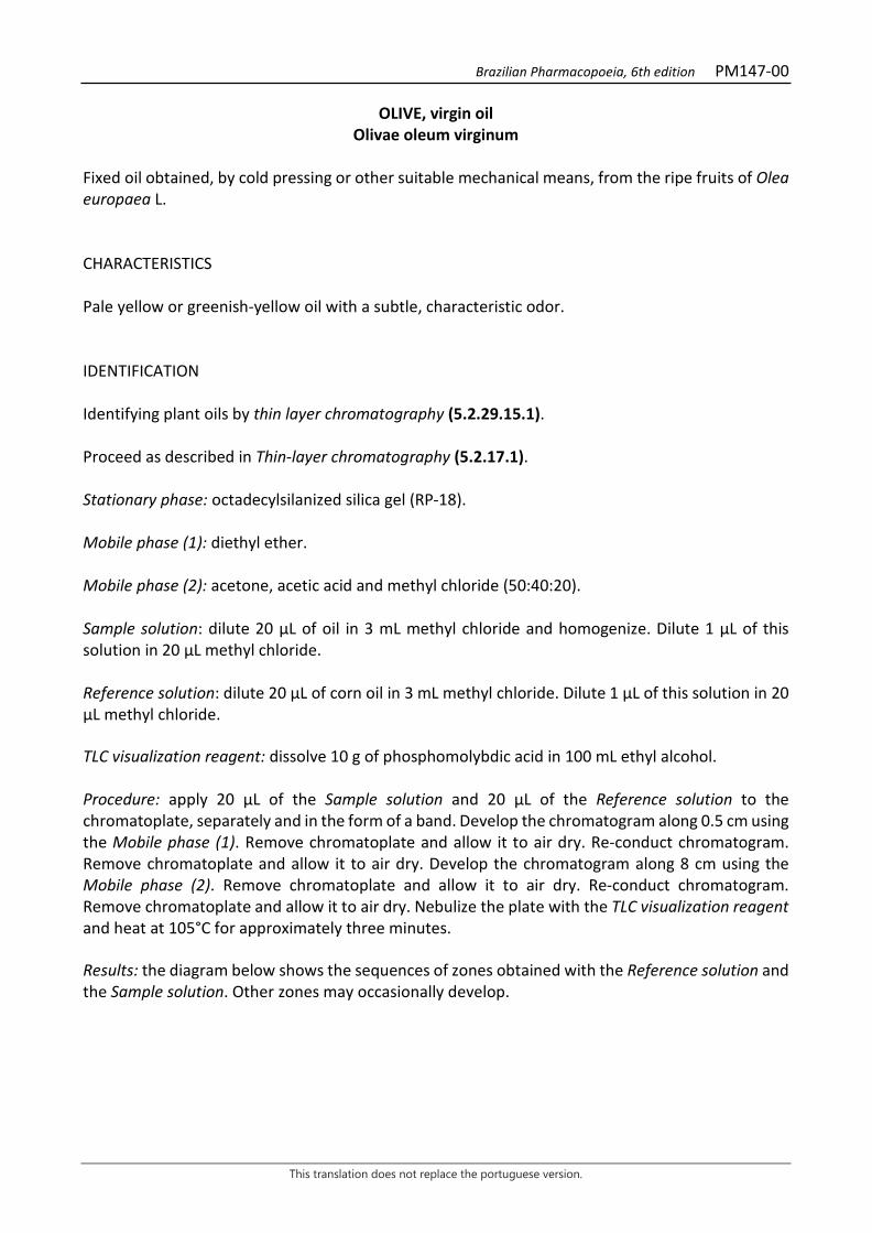
Brazilian Pharmacopoeia, 6th edition PM147-00
This translation does not replace the portuguese version.
OLIVE, virgin oil Olivae oleum virginum
Fixed oil obtained, by cold pressing or other suitable mechanical means, from the ripe fruits of Olea europaea L. CHARACTERISTICS Pale yellow or greenish-yellow oil with a subtle, characteristic odor. IDENTIFICATION Identifying plant oils by thin layer chromatography (5.2.29.15.1). Proceed as described in Thin-layer chromatography (5.2.17.1). Stationary phase: octadecylsilanized silica gel (RP-18). Mobile phase (1): diethyl ether. Mobile phase (2): acetone, acetic acid and methyl chloride (50:40:20). Sample solution: dilute 20 µL of oil in 3 mL methyl chloride and homogenize. Dilute 1 µL of this solution in 20 µL methyl chloride. Reference solution: dilute 20 µL of corn oil in 3 mL methyl chloride. Dilute 1 µL of this solution in 20 µL methyl chloride. TLC visualization reagent: dissolve 10 g of phosphomolybdic acid in 100 mL ethyl alcohol. Procedure: apply 20 μL of the Sample solution and 20 μL of the Reference solution to the chromatoplate, separately and in the form of a band. Develop the chromatogram along 0.5 cm using the Mobile phase (1). Remove chromatoplate and allow it to air dry. Re-conduct chromatogram. Remove chromatoplate and allow it to air dry. Develop the chromatogram along 8 cm using the Mobile phase (2). Remove chromatoplate and allow it to air dry. Re-conduct chromatogram. Remove chromatoplate and allow it to air dry. Nebulize the plate with the TLC visualization reagent and heat at 105°C for approximately three minutes. Results: the diagram below shows the sequences of zones obtained with the Reference solution and the Sample solution. Other zones may occasionally develop.
Brazilian Pharmacopoeia, 6th edition PM147-00
This translation does not replace the portuguese version.
Top of the plate
Corn oil: blue-colored zone
Bluish-colored zones
Corn oil: blue-colored zone
Bluish-colored zones
Corn oil: blue-colored zone
Bluish-colored zones
Reference solution Sample solution TESTS Solubility. Practically insoluble in 95% ethyl alcohol. Very soluble in hexane and diethyl ether. Relative density (5.2.5). 0.910 to 0.915. Acidity level (5.2.29.7). At most 2.0. Determine in 5 g. Peroxide Index (5.2.29.11). At most 20. Unsaponifiable matter (5.2.29.14). Method II. At most 1.5%. Determine in 5 g. Absorbance (5.2.14). Dissolve 0.5 g of oil in cyclohexane, dilute to 50 mL in a volumetric flask and homogenize. The absorbance measured at 270 nm is less than 0.20. The ratio of the absorbances measured at 232 nm and 270 nm is greater than 8. Fatty acid composition (5.2.29.15.4). The fraction of the oil composed of fatty acids has the following composition: Saturated fatty acids with chain less than 16 carbons: at most 0.1%; Palmitic acid: 7.5% to 20.0%. Palmitoleic acid: at most 3.5%; Stearic acid: 0.5% to 5.0%. Oleic acid: 56% to 85.0%. Linoleic acid: 3.5% to 20.0%. Linolenic acid: at most 1.2%; Arachidonic acid: at most 0.7%; Eicosenoic acid: at most 0.4%; Behenic acid: at most 0.2%; Lignoceric acid: at most 0.2%. Sesame oil. In a tube fitted with a stopper, shake for about one minute 10 mL of oil with a mixture of 0.5 mL of a 0.35% furfural solution in acetic anhydride and 4.5 mL acetic anhydride. Filter on
Brazilian Pharmacopoeia, 6th edition PM147-00
This translation does not replace the portuguese version.
paper impregnated with acetic anhydride. Add 0.2 mL sulfuric acid to the filtrate. There is no development of bluish-green color. Water (5.2.20.1). At most 0,1%. Determine in 1 g. PACKAGING AND STORAGE In tightly closed containers, protected from light and heat. CATEGORY Pharmacotechnical excipient.
Brazilian Pharmacopoeia, 6th edition PM148-00
This translation does not replace the portuguese version.
PALMAROSA, oil Palmae rosae aetheroleum
Volatile oil obtained by hydrodistillation from fresh leaves of Cymbopogon martini (Roxb.) W. Watson, containing at least 60.0% de geraniol (C10H18O, 154.25). CHARACTERISTICS A colorless to yellowish liquid with a pleasant, aromatic, rose-like odor. IDENTIFICATION Proceed as described in Thin-layer chromatography (5.2.17.1). Stationary phase: silica gel GF254. Mobile phase: toluene and ethyl acetate (90:10). Sample solution: dilute 2 µL of the sample to be examined in 300 µL toluene. Reference solution: dilute 2 µL geraniol and 2 µL geranyl acetate in 1 mL toluene. Procedure: apply 10 μL of the Sample solution and 10 μL of the Reference solution to the chromatoplate, separately and in the form of a band. Conduct chromatogram. Remove chromatoplate and allow it to air dry for 15 minutes. Nebulize the plate with sulfur vanillin RS and heat at 100 °C to 105 °C for 10 minutes. Results: the diagram below shows the sequences of zones obtained with the Reference solution and the Sample solution. Other zones may occasionally develop.
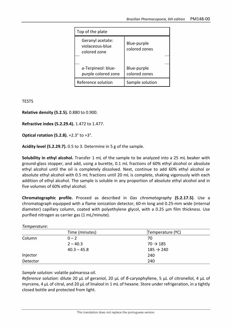
Brazilian Pharmacopoeia, 6th edition PM148-00
This translation does not replace the portuguese version.
Top of the plate
Geranyl acetate: violaceous-blue colored zone
Blue-purple colored zones
a-Terpineol: blue-purple colored zone
Blue-purple colored zones
Reference solution Sample solution
TESTS Relative density (5.2.5). 0.880 to 0.900. Refractive index (5.2.29.4). 1.472 to 1.477. Optical rotation (5.2.8). +2.3° to +3°. Acidity level (5.2.29.7). 0.5 to 3. Determine in 5 g of the sample. Solubility in ethyl alcohol. Transfer 1 mL of the sample to be analyzed into a 25 mL beaker with ground-glass stopper, and add, using a burette, 0.1 mL fractions of 60% ethyl alcohol or absolute ethyl alcohol until the oil is completely dissolved. Next, continue to add 60% ethyl alcohol or absolute ethyl alcohol with 0.5 mL fractions until 20 mL is complete, shaking vigorously with each addition of ethyl alcohol. The sample is soluble in any proportion of absolute ethyl alcohol and in five volumes of 60% ethyl alcohol. Chromatographic profile. Proceed as described in Gas chromatography (5.2.17.5). Use a chromatograph equipped with a flame ionization detector, 60-m long and 0.25-mm wide (internal diameter) capillary column, coated with polyethylene glycol, with a 0.25 μm film thickness. Use purified nitrogen as carrier gas (1 mL/minute). Temperature: Time (minutes) Temperature (ºC) Column Injector
0 – 2 2 – 40.3 40.3 – 45.8
70 70 → 185 185 → 240 240
Detector 240 Sample solution: volatile palmarosa oil. Reference solution: dilute 20 μL of geraniol, 20 μL of β-caryophyllene, 5 μL of citronellol, 4 μL of myrcene, 4 μL of citral, and 20 μL of linalool in 1 mL of hexane. Store under refrigeration, in a tightly closed bottle and protected from light.
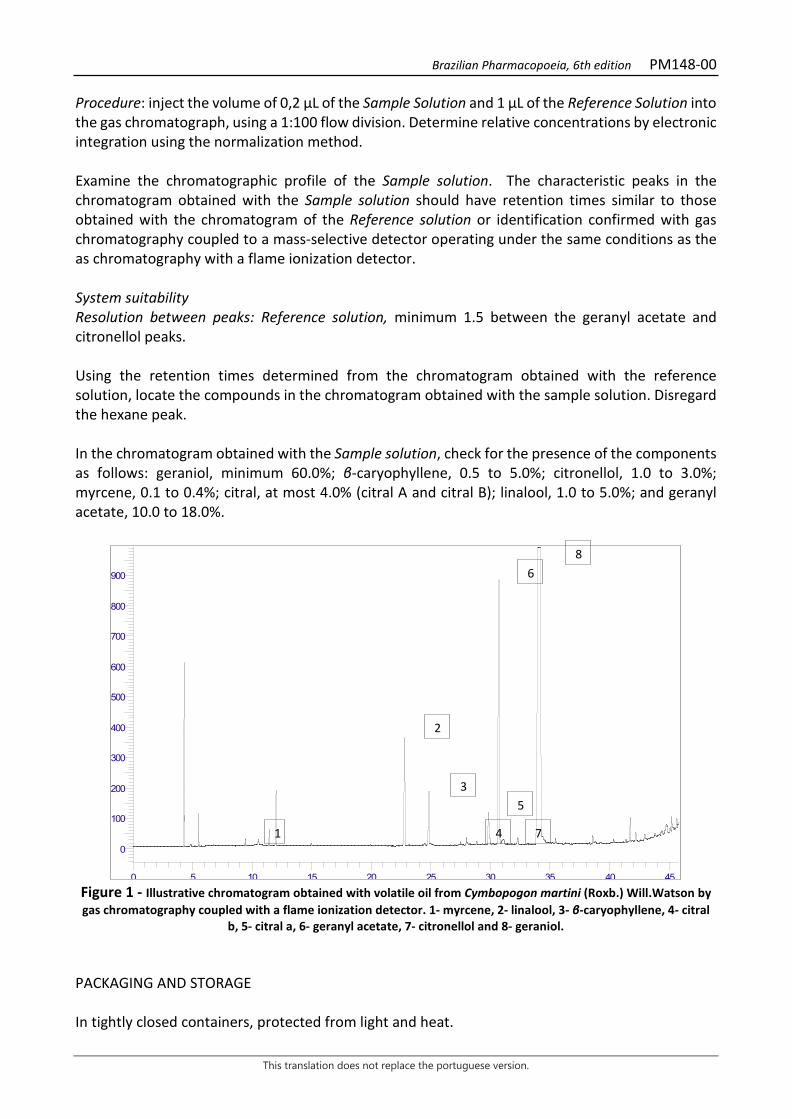
Brazilian Pharmacopoeia, 6th edition PM148-00
This translation does not replace the portuguese version.
Procedure: inject the volume of 0,2 μL of the Sample Solution and 1 μL of the Reference Solution into the gas chromatograph, using a 1:100 flow division. Determine relative concentrations by electronic integration using the normalization method. Examine the chromatographic profile of the Sample solution. The characteristic peaks in the chromatogram obtained with the Sample solution should have retention times similar to those obtained with the chromatogram of the Reference solution or identification confirmed with gas chromatography coupled to a mass-selective detector operating under the same conditions as the as chromatography with a flame ionization detector. System suitability Resolution between peaks: Reference solution, minimum 1.5 between the geranyl acetate and citronellol peaks. Using the retention times determined from the chromatogram obtained with the reference solution, locate the compounds in the chromatogram obtained with the sample solution. Disregard the hexane peak. In the chromatogram obtained with the Sample solution, check for the presence of the components as follows: geraniol, minimum 60.0%; β-caryophyllene, 0.5 to 5.0%; citronellol, 1.0 to 3.0%; myrcene, 0.1 to 0.4%; citral, at most 4.0% (citral A and citral B); linalool, 1.0 to 5.0%; and geranyl acetate, 10.0 to 18.0%.
Figure 1 - Illustrative chromatogram obtained with volatile oil from Cymbopogon martini (Roxb.) Will.Watson by gas chromatography coupled with a flame ionization detector. 1- myrcene, 2- linalool, 3- β-caryophyllene, 4- citral
b, 5- citral a, 6- geranyl acetate, 7- citronellol and 8- geraniol. PACKAGING AND STORAGE In tightly closed containers, protected from light and heat.
0
100
200
300
400
500
600
700
800
900
0 5 10 15 20 25 30 35 40 45
1
2
3
4
5
6
7
8
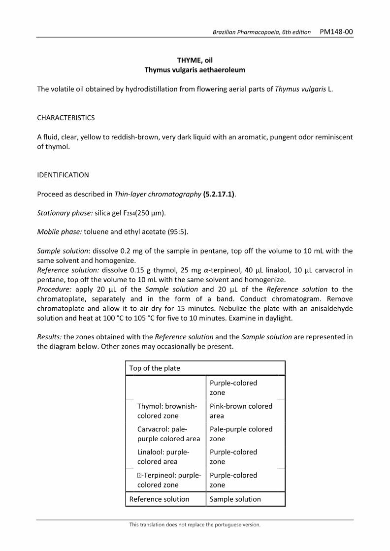
Brazilian Pharmacopoeia, 6th edition PM148-00
This translation does not replace the portuguese version.
THYME, oil
Thymus vulgaris aethaeroleum
The volatile oil obtained by hydrodistillation from flowering aerial parts of Thymus vulgaris L. CHARACTERISTICS A fluid, clear, yellow to reddish-brown, very dark liquid with an aromatic, pungent odor reminiscent of thymol. IDENTIFICATION Proceed as described in Thin-layer chromatography (5.2.17.1). Stationary phase: silica gel F254(250 µm). Mobile phase: toluene and ethyl acetate (95:5). Sample solution: dissolve 0.2 mg of the sample in pentane, top off the volume to 10 mL with the same solvent and homogenize. Reference solution: dissolve 0.15 g thymol, 25 mg α-terpineol, 40 µL linalool, 10 µL carvacrol in pentane, top off the volume to 10 mL with the same solvent and homogenize. Procedure: apply 20 μL of the Sample solution and 20 μL of the Reference solution to the chromatoplate, separately and in the form of a band. Conduct chromatogram. Remove chromatoplate and allow it to air dry for 15 minutes. Nebulize the plate with an anisaldehyde solution and heat at 100 °C to 105 °C for five to 10 minutes. Examine in daylight. Results: the zones obtained with the Reference solution and the Sample solution are represented in the diagram below. Other zones may occasionally be present.
Top of the plate
Purple-colored zone
Thymol: brownish-colored zone
Pink-brown colored area
Carvacrol: pale-purple colored area
Pale-purple colored zone
Linalool: purple-colored area
Purple-colored zone
-Terpineol: purple-colored zone
Purple-colored zone
Reference solution Sample solution
Brazilian Pharmacopoeia, 6th edition PM148-00
This translation does not replace the portuguese version.
TESTS Relative density (5.2.5). 0.915 to 0.935. Refractive index (5.2.29.4). 1.499 to 1.505. Chromatographic profile. Proceed as described in Gas chromatography (5.2.17.5). Use a chromatograph equipped with a flame ionization detector, using a mixture of helium, hydrogen and synthetic air (1:1:10) as auxiliary gases to the detector flame; 60 m long and 0.25 mm wide (internal diameter) capillary column, coated with propylene glycol, with a 0.25 μm film thickness. Use purified helium as carrier gas (1 mL/minute). Temperature: Time (minutes) Temperature (ºC) Column 0 – 15
15 – 61.7 61.7 – 91.7
60 60 → 200 200
Injector 200 Detector 220 Sample solution: dilute 0.1 mL of the thyme volatile oil in 10 mL hexane. Store under refrigeration, in a tightly closed bottle and protected from light. Reference solution: dissolve 0.15 g β-myrcene, 0.1 g -terpinene, 0.1 g p-cymene, 0.1 g linalool, 0.2 g terpinen-4-ol, 0.2 g thymol, and 50 mg carvacrol in 5 mL hexane. Procedure: inject the volume of 0.2 μL of the Reference solution and 0.5 µL of the Sample solution into the gas chromatograph, using a 1:100 flow division. Determine relative concentrations by electronic integration using the normalization method. Examine the chromatographic profile of the Sample solution. The characteristic peaks in the chromatogram obtained with the Sample solution should have retention times similar to those obtained with the chromatogram of the Reference solution or identification confirmed with gas chromatography coupled to a mass-selective detector operating under the same conditions as the as chromatography with a flame ionization detector. Elution order: order of preparation of the Reference solution. Record retention times of substances. System suitability Resolution between peaks: Reference solution, minimum 1.5 between the thymol and carvacrol peaks. Numbers is theoretical plates: at least 30,000, calculated for the p-cymene peak at 80°C.

Brazilian Pharmacopoeia, 6th edition PM148-00
This translation does not replace the portuguese version.
In the chromatogram obtained with the Sample solution, check for the presence of the components as follows: β-myrcene, 1.0 to 3.0%; -terpinene, 5.0 to 10.0%; p-cymene, 15.0 to 28.0%; linalool, 4.0 to 6.5%; terpinen-4-ol, 0.2 to 2.5%; thymol, 36.0 to 55.0% and carvacrol, 1.0 to 4.0%.

Brazilian Pharmacopoeia, 6th edition PM148-00
This translation does not replace the portuguese version.
Figure 1 - Illustrative chromatogram obtained with volatile oil from Thymus vulgaris L. through gas
chromatography coupled to a flame ionization detector. PACKAGING AND STORAGE In tightly closed containers, protected from light and heat.