bordetella pertussis expresses a functional type iii secretion system that subverts protective...
TRANSCRIPT
INFECTION AND IMMUNITY, Mar. 2008, p. 1257–1266 Vol. 76, No. 30019-9567/08/$08.00�0 doi:10.1128/IAI.00836-07Copyright © 2008, American Society for Microbiology. All Rights Reserved.
Bordetella pertussis Expresses a Functional Type III Secretion SystemThat Subverts Protective Innate and Adaptive Immune Responses�
Neil K. Fennelly,1 Federico Sisti,2 Sarah C. Higgins,1 Padraig J. Ross,1 Han van der Heide,3Frits R. Mooi,3 Aoife Boyd,4† and Kingston H. G. Mills1*†
Immune Regulation Research Group, School of Biochemistry and Immunology, Trinity College, Dublin, Ireland1; Instituto deBioquimica y Biologia Molecular, Universidad Nacional de La Plata, La Plata, Argentina2; Laboratory for Infectious Diseases andScreening, RIVM, Bilthoven, The Netherlands3; and Department of Microbiology, National University of Ireland Galway, Ireland4
Received 18 June 2007/Returned for modification 9 August 2007/Accepted 2 January 2008
Certain bacteria use a type III secretion system (TTSS) to deliver effector proteins that interfere with cellfunction into host cells. While transcription of genes encoding TTSS components has been demonstrated,studies to date have failed to identify TTSS effector proteins in Bordetella pertussis. Here we present the firstevidence of a functionally active TTSS in B. pertussis. Three known TTSS effectors, Bsp22, BopN, and BopD,were identified as TTSS substrates in B. pertussis 12743. We found expression of Bsp22 in a significantproportion of clinical isolates but not in common laboratory-adapted strains of B. pertussis. We generated aTTSS mutant of B. pertussis 12743 and showed that it induced significantly lower respiratory tract colonizationin mice than the wild-type bacteria. Respiratory infection of mice with the mutant bacteria induced signifi-cantly greater innate proinflammatory cytokine production in the lungs soon after challenge, and this corre-lated with significantly higher antigen-specific interleukin-17, gamma interferon, and immunoglobulin Gresponses later in infection. Our findings suggest that the TTSS subverts innate and adaptive immuneresponses during infection of the lungs and may be a functionally important virulence factor for B. pertussisinfection of humans.
Bordetella pertussis is the causative agent of whooping coughor pertussis, a respiratory disease that is most severe in infantsand young children. Although vaccination has significantly re-duced morbidity and mortality, pertussis remains an endemicdisease and is one of the major causes of vaccine-preventabledeaths today, with WHO estimates of 45 million cases and409,000 deaths each year. In recent years a resurgence ofpertussis was observed in a number of vaccinated populations(6, 29). Furthermore, it has become increasingly clear thatpertussis is not only a childhood disease but also is highlyprevalent among adults (21). This has called into question thelevel of protection provided by current pertussis vaccines andhighlighted the need for a better understanding of the molec-ular mechanisms underlying the pathogenesis of B. pertussisinfection.
Bacteria produce a complex array of virulence factors, in-cluding toxins and adhesins, which facilitate colonizationand/or suppress immune responses and allow the bacteria toestablish infection in the host. One of these virulence factors,the type III secretion system (TTSS), is a specialized secretoryapparatus that allows gram-negative bacteria to inject proteins,known as effectors, directly into the eukaryotic cell cytosol. Inlaboratory conditions bacteria can be induced to secrete TTSSsubstrates, which include effectors and proteins involved in thedelivery process, into the extracellular milieu in the absence ofeukaryotic cells. TTSSs have been shown to be important me-
diators of virulence of a range of animal pathogens, includingYersinia spp., Salmonella spp., Shigella spp., Escherichia coli,and Bordetella bronchiseptica (15, 39). Yuk and colleagues havereported that the TTSS of B. bronchiseptica modulates den-dritic cell (DC) maturation (31, 33), enhancing production ofthe anti-inflammatory cytokine interleukin-10 (IL-10) and pro-moting bacterial persistence (32).
Despite reports describing transcription of genes encodingcomponents of the Bordetella bsc TTSS machinery in B. per-tussis Tohama I (14, 22), the B. pertussis isolate chosen forgenome sequencing, studies to date have failed to demonstrateTTSS effector secretion by B. pertussis in vitro or in vivo (9, 22).The sequencing of the B. pertussis Tohama I genome has re-vealed an extraordinary high level of genetic flexibility (28),and this raises concerns about the adequacy of laboratory-adapted strains for the study of natural clinical pathogenesis.Differences in gene expression have been shown to affect vir-ulence characteristics of laboratory-adapted versus corre-sponding low-passage clinical isolates of E. coli, Staphylococcusaureus, and Pseudomonas aeruginosa (11, 34, 37).
Here we demonstrate secretion of the Bordetella TTSS ef-fector, Bsp22, by a significant portion of low-passage clinicalisolates of B. pertussis, but not by common laboratory-adaptedstrains, such as Tohama I and Wellcome 28. Mutation of bscN,which encodes an essential component for TTSS secretionacross bacterial membranes, abolished in vitro secretion ofTTSS substrates by a clinical isolate of B. pertussis, resulting ina reduced ability of the bacteria to colonize the respiratorytracts of mice, and this was associated with enhanced localinflammatory and antigen-specific cellular and humoral im-mune responses. Our data suggest that expression of a func-tional TTSS is a feature of natural infection of humans with B.
* Corresponding author. Mailing address: Immune Regulation Re-search Group, School of Biochemistry and Immunology, Trinity Col-lege, Dublin 2, Ireland. Phone: (353) 1 8963573. Fax: (353) 1 6772086.E-mail: [email protected].
† A.B. and K.H.G.M. contributed equally to this study.� Published ahead of print on 14 January 2008.
1257

pertussis and that this may confer virulence to the bacteria bysubverting the protective innate and adaptive immunity of thehost.
MATERIALS AND METHODS
Bacterial strains and growth media. Low-passage isolates B. pertussis ATCC12743 (5375 [3865]), ATCC 12742 (5374 [3747]), and ATCC 9340 (5 [17921]),hereafter referred to as B. pertussis 12743, 12742, and 9340, respectively, wereobtained from the ATCC. B. pertussis 12743 and 12742 were from cultures madeby E. K. Anderson and deposited in the ATCC by G. Eldering (8), and B.pertussis 9340 was from a culture made by P. Kenrick and deposited in the ATCCby M. Pittman in the 1950s. Sixteen clinical isolates were cultivated from thesputum, noses, nasopharynges, or throats of infants or adults with whoopingcough in The Netherlands between 1949 and 2005. Wild-type (WT) B. pertussisand B. bronchiseptica were grown at 37°C on Bordet-Gengou (BG) agar and inStainer-Scholte (SS) broth. Gentamicin-resistant �bscN derivatives of B. pertussis12743 and B. bronchiseptica RB50 were grown on BG agar or SS broth supple-mented with 10 �g/ml gentamicin (Gibco, United Kingdom). For allelicexchange WT B. pertussis 12743 was first rendered streptomycin resistant bysubculture in increasing sublethal concentrations of streptomycin (final concen-tration, 100 �g/ml). For routine cloning and conjugation, E. coli XL1-Blue andSM10�pir were grown at 37°C on Luria-Bertani (LB) agar or LB broth (BDDifco) supplemented with the appropriate antibiotics (ampicillin, 150 �g/ml;gentamicin, 10 �g/ml; kanamycin, 25 �g/ml).
Generation of �bscN bacteria. Gentamicin-resistant �bscN derivatives of B.pertussis 12743 and B. bronchiseptica RB50, in which a 0.5-kb internal portion ofthe bscN gene was removed and replaced with a 0.7-kb fragment containing agentamicin resistance cassette, were constructed as follows. Primers PAB20(5�-GCCCTGCGGATCCCGCG-3�) and NF5 (5�-TACTGACGCATGCCCCTATCC-3�), annealing to bp 65 to 81 of bscL (5� flanking gene to bscN) and bp 1to 12 of bscN, respectively, were used to amplify a 0.5-kb fragment from TohamaI. The resulting PCR product was digested with BamHI and SphI (recognitionsequences underlined in primer sequences) and inserted into the correspondingsites of the cloning vector pQE-80 (Qiagen, United Kingdom) to produce pNF2.Primers NF4 (5�-GCTGGGCATGCTGGTCAAGGGC-3�) and PAB21 (5�-GCCGGCTCGCGATGCATCG-3�), annealing to bp 560 to 582 of bscN and bp 470to 489 of bscO (3� flanking gene to bscN), respectively, were used to amplify a1.3-kb fragment from Tohama I. The resulting PCR product was digested withSphI and AgeI (recognition sequence underlined in primer sequence) to yield a0.5-kb fragment and inserted into the corresponding SphI and XmaI sites onpNF2 to create pNF7, containing a mutant bscN allele. The gentamicin resis-tance cassette was amplified from pSS1129 using primers Gmr_for_2 (5�-ATAGCATGCTGACGCACACCG-3�) and Gmr_rev (5�-GCATGCTTAGGTGGCGGTAC-3�) with SphI sites engineered at the 5� and 3� ends, respectfully, andinserted into the unique SphI site at the center of the mutant bscN allele onpNF7 to create pNF12. BamHI (bp 1 to 6 of the bscN::Gmr mutant allele) andNheI (138 bases downstream) sites flanking the bscN::Gmr mutant allele onpNF12 were then used to subclone the bscN::Gmr mutant allele into the allelicexchange vector pEGBR (1) to create pNF13. pNF13 was then introduced intostreptomycin-resistant B. pertussis 12743 and B. bronchiseptica RB50 by conju-gation, and gentamicin-resistant double recombinants of B. pertussis 12743 andB. bronchiseptica RB50 generated by allelic exchange were screened by PCR forthe presence of the bscN::Gmr mutant allele.
Recombinant His-Bsp22 and Ab production. Primers PAB37 (5�-CGGAAGCTTTTAGCGCATGTTGCTGGTG-3�), which binds bp 596 to 615 of theBsp22 gene, and PAB38 (5�-AGCGGATCCAGCATTGATCTCGGAGTTCAC-3�), which binds bp 4 to 25 of the Bsp22 gene, were used to amplify the Bsp22gene from B. pertussis 18323. The resulting PCR fragment was cloned in framewith the six-His tag located on pQE-80 (Qiagen, United Kingdom), creatingpAPB15, from which recombinant His-tagged Bsp22 was expressed in E. coliXL1-Blue. His-Bsp22 was purified by affinity chromatography on Ni2� columns.For antibody (Ab) production, a rabbit was immunized subcutaneously withrecombinant His-Bsp22 (230 �g) emulsified in complete Freund’s adjuvant(Difco) and boosted on days 14 and 28 with His-Bsp22 (115 �g) in incompleteFreund’s adjuvant. The rabbit was exsanguinated 12 days after the last immuni-zation, and serum was prepared and shown by Western blotting to contain an Abthat specifically recognized Bsp22.
Protein identification by MALDI-TOF mass spectrometry and immunoblot-ting. For matrix-assisted laser desorption-ionization–time of flight (MALDI-TOF) mass spectrometry analysis of protein samples, culture supernatants wereharvested from Bordetella spp. following 24 h of growth in liquid culture. Filtered
supernatants were precipitated with 30% ammonium chloride. Protein pelletswere analyzed by MALDI-TOF mass spectrometry, and identified peptide frag-ments were searched against the predicted proteins from the complete B. bron-chiseptica RB50 and B. pertussis Tohama I genomes (www.sanger.ac.uk/Projects/B_pertussis/). For Western blots, cultures were harvested for each bacterialstrain at the same stage of bacterial growth, as determined by measurements ofoptical density at 600 nm (OD600), and total protein from a 1-ml supernatantfraction was precipitated with 10% (wt/vol) trichloroacetic acid and resuspendedin sample buffer. Proteins were separated on 10% (wt/vol) sodium dodecylsulfate-polyacrylamide gel electrophoresis gels and transferred to nitrocellulosemembranes (Sigma) prior to Western blotting with a polyclonal Ab raised againstB. pertussis Bsp22, pertussis toxin (PT), or filamentous hemagglutinin (FHA),followed by the appropriate secondary Ab.
In vitro growth curves. B. pertussis 12743 and �bscN B. pertussis 12743 weregrown in SS medium in 100-ml cultures inoculated at a starting OD600 of 0.2.Samples (1 ml) were removed at 4, 8, 12, 23, 27, 37, and 49 h, OD600 wasdetermined, and CFU counts were performed by plating neat and diluted sam-ples on BG agar plates.
In vitro cytotoxicity and adherence assays. DC were expanded in vitro frombone marrow of BALB/c mice by culture for 10 days with granulocyte-macro-phage colony-stimulating factor as described previously (13). Murine DC, J774macrophages, and human HEK-293T epithelial cells (1 � 106/ml) were incu-bated in Dulbecco’s modified Eagle’s medium supplemented with 8% fetal calfserum in 24-well plates. Stationary-phase cultures of Bordetella spp. were har-vested, resuspended in 1% casein, and added to triplicate wells at a multiplicityof infection (MOI) of 20, 100, or 500. Cytotoxicity was determined after 4 h usingthe Cytotox 96 nonradioactive cytotoxicity assay (Promega), which measureslactate dehydrogenase release as a measure of cell lysis. For detection of bacte-rial adherence to macrophages and of internalization, J774 cells were incubatedwith Bordetella spp. at an MOI of 100. Following incubation at 37°C for 2 h, thecontents of each well were harvested, washed to remove unbound bacteria, andresuspended in either 1 ml of medium supplemented with kanamycin (100�g/ml) to kill extracellular bacteria or 1 ml medium alone. The cell suspensionswere incubated at 37°C for a further 1 h and then centrifuged at 1,200 rpm for5 min. The cells were washed and resuspended in 1 ml phosphate-buffered salinewith 1% (wt/vol) Triton X-100 and left at room temperature for 5 min. Onehundred-microliter samples of the appropriate dilutions of the lysed cells werethen plated in triplicate onto BG agar plates and incubated at 37°C for 2 days forB. bronchiseptica and 3 to 5 days for B. pertussis.
B. pertussis infection of mice. Mice were infected with WT or �bscN B. pertussis12743 by exposure to an aerosol of bacteria for 15 min as described previously(20). The standard inoculum was 2 � 1010 bacteria/ml (based on OD600), and thiswas reduced or increased by two- to threefold to achieve a lower or higher initialcolonization. Four mice from each experimental group were sacrificed 3 h and 3,7, 14, 21, and 28 days after challenge to assess the number of viable bacteria inthe lungs. The numbers of CFU in lung homogenates were estimated as de-scribed previously (20). Results are given as the mean numbers of B. pertussisCFU for individual lungs from four mice per experimental group. The sensitivityof the assay was 0.56 log10 CFU per lung.
Quantification of cytokine concentrations and neutrophil infiltration into thelungs. Lung homogenates were centrifuged at 13,000 rpm for 5 min, and theconcentrations of IL-10, IL-12p70, IL-1�, tumor necrosis factor alpha (TNF-�),macrophage inflammatory protein 1 (MIP-1) and MIP-2 (R&D Systems), IL-12p40, IL-6, and transforming growth factor � (BD Pharmingen) were deter-mined by enzyme-linked immunosorbent assay (ELISA) according to the man-ufacturer’s instructions. Neutrophil infiltration into the lungs was quantified byperforming differential counts on hematoxylin-and-eosin-stained cytospin prep-arations of cells recovered by bronchoalveolar lavage as described previously(23).
Detection of antigen-specific immune responses. Spleens and serum sampleswere recovered from mice 14, 21, and 28 days after bacterial challenge. Spleencells (2 � 106 cells/ml) were stimulated with sonicated B. pertussis, PT, FHA, ormedium alone and incubated at 37°C for 72 h. Supernatants were removed, andgamma interferon (IFN-) and IL-17 concentrations were determined byELISA. Lung T cells were purified on T-cell isolation columns as describedpreviously (23). Lung T cells were stimulated with heat-inactivated B. pertussisantigen and splenic antigen-presenting cells (2 � 106 irradiated spleen cells).After 5 days the surviving T cells were stimulated with phorbol myristate acetateand ionomycin for 1 h, and then brefeldin A was added for 4 h at 37°C. Cells werestained with fluorescently labeled Abs specific for CD4 (eBioscinces). Cells werethen fixed, permeabilized, and incubated with fluorescently labeled anti-IFN- oranti-IL-17 Abs (BD Pharmingen) according to the manufacturer’s instructions(Fix & Perm cell permeabilization kit; Invitrogen). Immunofluorescence was
1258 FENNELLY ET AL. INFECT. IMMUN.

analyzed using Summit software on a Cyan ADP flow cytometer (Dako). Anti-B.pertussis immunoglobulin G (IgG), IgG2a, and IgG1 Ab titers were determinedby ELISA as described previously (13). Results are expressed as log10 end pointAb titers, determined by extrapolation of the straight part of the dilution curve,versus the OD492 value of the control serum for naive mice.
Statistical analysis. Statistical analysis was performed using Graphpad Prism.Student’s t test was used to compare the mean values between two groups.Statistical differences in mean values between more than two experimentalgroups were determined by analysis of variance. Linear regression was used toexamine the correlation between bacterial growth estimated by CFU counts andOD600. A nonlinear regression analysis was performed on the growth curves forB. pertussis 12743 and �bscN B. pertussis 12743. An Akaike’s information crite-rion test with a confidence interval of 95% was applied to the data, with asupplemental t test, to determine if the growth curves of bacterial strains aredifferent.
RESULTS
Secretion of TTSS effector proteins by clinical but not lab-oratory-adapted isolates of B. pertussis. B. pertussis Tohama I iswell characterized and was chosen for genome sequencing (28)but has been through extensive laboratory passage since its isola-tion in Japan in 1954 (16). Since the TTSS expression and secre-tory function of Yersinia is regulated and activated in vivo byeukaryotic cell contact and can be altered by prolonged labora-tory passage (4, 36), we examined low-passage B. pertussis isolatesfrom the ATCC (B. pertussis 12743, 12742, and 9340) and com-pared these with the laboratory-adapted strains (Tohama I andWellcome 28) for expression of Bsp22. Bsp22, whose gene islocated in the TTSS operon in B. pertussis and B. bronchiseptica(www.sanger.ac.uk/Projects/B_pertussis/), is secreted by B. bron-chiseptica (39) and is a useful marker of TTSS secretory function.In the present study, His-tagged Bsp22 was cloned and expressedin E. coli and the purified protein was used to generate polyclonalanti-Bsp22 Abs. Western blotting with anti-Bsp22 revealed signif-icant quantities of secreted Bsp22 protein in culture supernatantsof B. pertussis 12743 and 9340, but not in 12742, Tohama I, orWellcome 28 (Fig. 1A). These data demonstrate that two of thethree early isolates from the ATCC that were examined expressedTTSS proteins in vitro, but this may have been lost in the commonlaboratory-adapted strains Tohama I and Wellcome 28.
We next examined supernatants from stationary-phase cul-tures of 16 clinical isolates, cultivated from infants or adultswith whooping cough in The Netherlands between 1949 and2005. Western blotting with anti-Bsp22 Abs revealed secretionof Bsp22 in 15 of 16 isolates (Fig. 1; results are shown for 8clinical isolates; an additional 8 [not shown] all expressedBsp22). Bsp22 was also detected in B. bronchiseptica RB50 butnot B. pertussis Tohama I or Wellcome 28. PT was detected inall strains except B. bronchiseptica RB50, confirming that theclinical isolates were B. pertussis and not B. bronchiseptica.These results demonstrate that a large proportion of the low-passage clinical isolates of B. pertussis examined have a func-tional TTSS.
In order to confirm these findings and to facilitate identifi-cation of additional Bordetella TTSS substrates, we carried outmass spectroscopy analysis on total secreted proteins fromculture supernatants. MALDI-TOF analysis revealed a num-ber of known Bordetella virulence factors, including FHA,adenylate cyclase toxin, and pertactin in B. pertussis Tohama I,B. pertussis 12743, and B. bronchiseptica RB50 (Table 1), aswell as other proteins not yet implicated in virulence (data not
shown). Tracheal colonization factor, a virulence factor of B.pertussis but not B. bronchiseptica (10), was detected in B.pertussis Tohama I and B. pertussis 12743 but not B. bronchi-septica RB50. Serum resistance protein (BrkA) was detected inB. pertussis Tohama I and B. pertussis 12743 but not B. bron-chiseptica RB50; BrkA expression has been demonstrated forsome strains of B. bronchiseptica, but not for strain RB50 (30).PT, a B. pertussis-specific virulence factor, was also detected inB. pertussis 12743; this required a separate analysis from thatshown in Table 1, using more concentrated culture supernatant(not shown). As well as Bsp22, the TTSS translocator protein,BopD, and the sensor/plug protein, BopN, were detected insupernatants from B. pertussis 12743 but not B. pertussis To-hama I. The Bordetella TTSS effector protein, BteA/BopC, wasdetected in B. bronchiseptica RB50 but not in B. pertussis12743. These results provide further evidence that B. pertussis12743 has a functional TTSS in vitro and demonstrate for thefirst time that BopD and BopN, in addition to Bsp22, aresubstrates of the B. pertussis TTSS.
Mutation of bscN abolishes expression of TTSS effector pro-teins by B. pertussis 12743 but does not affect bacterial growthin vitro. In order to assess the contribution of the TTSS to thevirulence of BP 12743, we constructed a strain carrying aninsertional mutation of the bscN gene. Mutation of bscN in B.bronchiseptica or its homologs, yscN in Yersinia and invC inSalmonella, has been shown to abolish in vitro TTSS secretionand in vivo TTSS-mediated effects on host cells (7, 38–40). Forcomparative purposes, we also mutated bscN in B. bronchisep-tica RB50. Bsp22 protein was detected by Western blotting inthe culture supernatants of WT B. pertussis 12743 and B. bron-chiseptica RB50 but not in �bscN derivatives (Fig. 2). Further-
FIG. 1. Clinical isolates but not common laboratory-adapted stainsof B. pertussis express Bsp22. Protein samples from supernatants ofstationary-phase cultures of B. pertussis (Bp) 9340, 12743, 12742, To-hama I (Toh-I), and Wellcome 28 (W28) (A) or B. bronchiseptica (Bb)RB50, B. pertussis W28 and Toh-I, and eight clinical isolates of B.pertussis recovered from respiratory tracts of patients with whoopingcough (B), prepared by precipitation with 10% trichloroacetic acid orpurified His-Bsp22 or PT (2 �g), were resolved on sodium dodecylsulfate-polyacrylamide gel electrophoresis gel and probed with Absspecific for Bsp22 or PT (S1 to S5 subunits) by Western blotting.
VOL. 76, 2008 BORDETELLA PERTUSSIS TYPE III SECRETION SYSTEM 1259

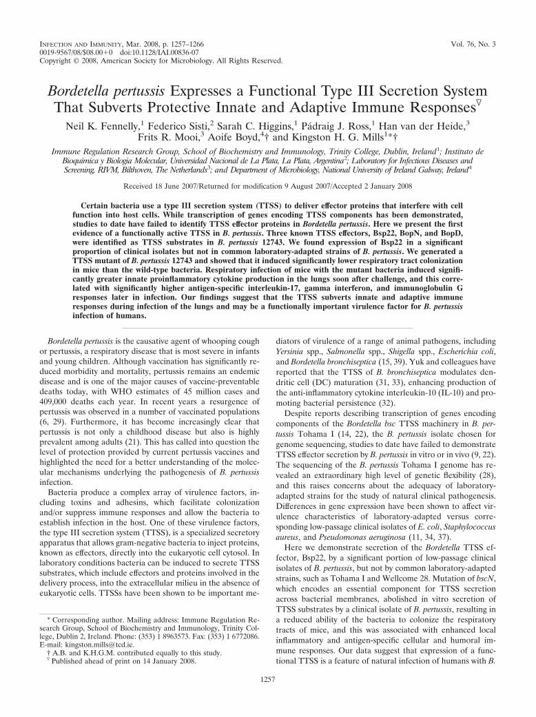
more, Bsp22, BopN, and BopD were detected by mass spec-trometry analyses of secreted proteins of WT but not �bscNderivatives (Table 1). Mutation of bscN did not affect secretionof other virulence factors, including FHA, adenylate cyclasetoxin, and pertactin (Table 1 and Fig. 2). These results dem-onstrate that mutation of the bscN gene abolishes secretion ofTTSS substrates by B. pertussis 12743 and B. bronchisepticaRB50 and demonstrate that BscN is an essential component ofthe B. pertussis TTSS. Furthermore, mutation of bscN did notsignificantly affect bacterial growth rate. The in vitro growthcurves based on OD600 (Fig. 3A) and CFU (Fig. 3B) werealmost superimposable for B. pertussis 12743 and �bscN B.pertussis 12743. Nonlinear regression analysis with Akaike’sinformation criterion test determined that there was a 93%probability that the curves for the two bacterial strains wereidentical. Furthermore linear regression analysis demonstrateda highly significant (P 0.001) correlation between OD600 andCFU for B. pertussis 12743 and �bscN B. pertussis 12743. Thesefindings demonstrate that OD600 can be used as an accurateestimate of the number of viable bacteria and suggest thatequivalent numbers of viable WT and TTSS mutant bacteriawere employed for experiments to compare the virulence levelsof and immune responses to B. pertussis 12743 and �bscN B.pertussis 12743 in vitro and in vivo.
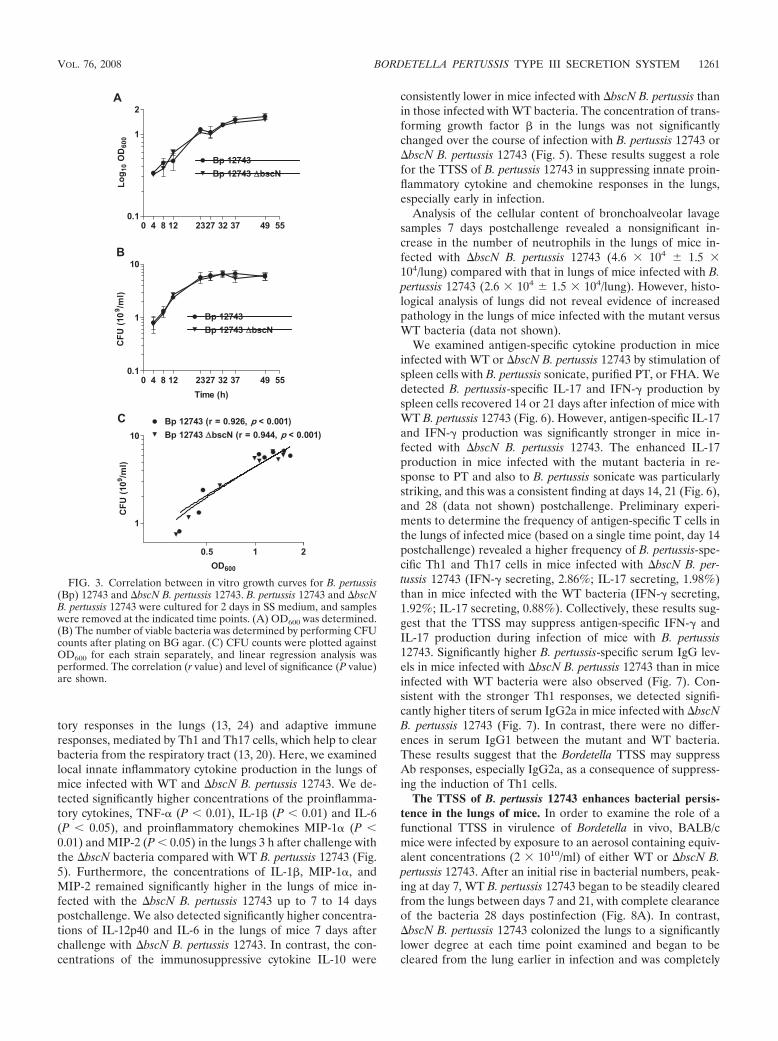
The TTSS of B. pertussis 12743 promotes adherence to macro-phages but does not mediate cytotoxicity in vitro. Consistentwith previous reports (39, 40), B. bronchiseptica RB50 wassignificantly cytotoxic to cultured epithelial cells, macrophages,and DC (Fig. 4A) and this activity was lost in the �bscNmutant. In contrast, no significant cell lysis was detected in anycell type incubated with WT or �bscN B. pertussis 12743. TheTTSS of Yersinia spp. plays a crucial role in preventing bacte-rial uptake during infection (4), while the TTSS of Salmonellaspp. mediates bacterial uptake to facilitate tissue invasion andintracellular replication (12). Here we investigated the role ofthe Bordetella TTSS in bacterial uptake. Significantly higherCFU were detected in kanamycin-treated macrophages cul-tured with �bscN B. bronchiseptica than in those cultured withWT B. bronchiseptica, indicative of a higher number of inter-nalized viable mutant bacteria (Fig. 4B). After correcting forcell death induced by B. bronschiseptica, there was still signif-icantly greater uptake of the mutant than the WT B. bronschi-septica (data not shown). In contrast, the numbers of CFUrecovered following treatment with medium only were similarfollowing culture with WT or �bscN B. bronchiseptica, suggest-ing that the TTSS of B. bronchiseptica did not facilitate bindingto the cell surface. The numbers of viable WT and �bscN B.pertussis 12743 CFU recovered from kanamycin-treated macro-phages were very similar, indicative of similar numbers of in-ternalized B. pertussis CFU. In contrast, the number of viableB. pertussis CFU recovered following treatment with mediumalone from cells cultured with �bscN B. pertussis 12743 wassignificantly lower than the number of WT bacteria, indicatinga lower number of cell-associated bacteria following incuba-tion with the mutant bacteria (Fig. 4B). These results demon-strate that the TTSS of B. bronchiseptica RB50 inhibits bacte-rial uptake by murine macrophages, whereas the TTSS of B.pertussis 12743 promotes adherence of the bacteria to macro-phages in vitro.
Enhanced innate and adaptive immune responses in miceinfected with �bscN B. pertussis 12743. Protective immunity toB. pertussis involves a combination of local innate inflamma-
TABLE 1. Identification of secreted TTSS substrates and other virulence factors from Bordetella spp. by MALDI-TOF mass spectrometrya
Mascotaccession no. Identified protein
Result for:
B. pertussisToh I
B. pertussis12743
�bscNB. pertussis
12743
B. bronchisepticaRB50
�bscNB. bronchiseptica
RB50
BP1879/BB2993 FHA � (141) � (8,869) � (3,776) � (567) � (434)BP0760/BB0324 Bifunctional hemolysin-adenylate
cyclase toxin (CyaA)� (116) � (2,423) � (1,074) � (2,551) � (2,571)
BP1054/BB1366 Pertactin (Prn) � (111) � (2,066) � (759) � (450) � (350)BP3494/BB0961 Serum resistance protein (BrkA) � (234) � (2,261) � (1,893) � �BP1201 BB3291 Tracheal colonization factor
(TcfA)� (412) � (1,530) � (3,117) � �
BP2256/BB1617 Putative secreted protein(Bsp22)
� � (470) � � (500) �
BP2253/BB1620 Putative outer protein D (BopD) � � (781) � � (193) �BP2257/BB1616 Putative outer protein N (BopN) � � (204) � � �BP0500/BB4228 Hypothetical protein
(BteA/BopC)� � � � (67) �
a Quantitative MALDI-TOF tandem mass spectrometry analysis was carried out on extracted proteins from culture supernatant of B. pertussis Tohama I (Toh I),B. pertussis 12743, �bscN B. pertussis 12743, B. bronchiseptica RB50, and �bscN B. bronchiseptica RB50. Identified peptides were searched against the complete B.bronchiseptica and B. pertussis genome sequences (www.sanger.ac.uk/Projects/B_pertussis/) using the Mascot search engine. Numbers in parentheses represent theindividual Mascot ion scores. Individual ion scores �33 indicate identity or extensive homology (P 0.05).
FIG. 2. Mutation of bscN in Bordetella spp. abolishes Bsp22 pro-duction but does not affect secretion of the virulence factor FHA.Proteins from culture supernatants of WT and �bscN B. pertussis (Bp)12743 and B. bronchiseptica (Bb) RB50 or B. pertussis Tohama I(Toh-I) were examined for the presence of Bsp22 and FHA by West-ern blotting.
1260 FENNELLY ET AL. INFECT. IMMUN.
tory responses in the lungs (13, 24) and adaptive immuneresponses, mediated by Th1 and Th17 cells, which help to clearbacteria from the respiratory tract (13, 20). Here, we examinedlocal innate inflammatory cytokine production in the lungs ofmice infected with WT and �bscN B. pertussis 12743. We de-tected significantly higher concentrations of the proinflamma-tory cytokines, TNF-� (P 0.01), IL-1� (P 0.01) and IL-6(P 0.05), and proinflammatory chemokines MIP-1� (P 0.01) and MIP-2 (P 0.05) in the lungs 3 h after challenge withthe �bscN bacteria compared with WT B. pertussis 12743 (Fig.5). Furthermore, the concentrations of IL-1�, MIP-1�, andMIP-2 remained significantly higher in the lungs of mice in-fected with the �bscN B. pertussis 12743 up to 7 to 14 dayspostchallenge. We also detected significantly higher concentra-tions of IL-12p40 and IL-6 in the lungs of mice 7 days afterchallenge with �bscN B. pertussis 12743. In contrast, the con-centrations of the immunosuppressive cytokine IL-10 were
consistently lower in mice infected with �bscN B. pertussis thanin those infected with WT bacteria. The concentration of trans-forming growth factor � in the lungs was not significantlychanged over the course of infection with B. pertussis 12743 or�bscN B. pertussis 12743 (Fig. 5). These results suggest a rolefor the TTSS of B. pertussis 12743 in suppressing innate proin-flammatory cytokine and chemokine responses in the lungs,especially early in infection.
Analysis of the cellular content of bronchoalveolar lavagesamples 7 days postchallenge revealed a nonsignificant in-crease in the number of neutrophils in the lungs of mice in-fected with �bscN B. pertussis 12743 (4.6 � 104 1.5 �104/lung) compared with that in lungs of mice infected with B.pertussis 12743 (2.6 � 104 1.5 � 104/lung). However, histo-logical analysis of lungs did not reveal evidence of increasedpathology in the lungs of mice infected with the mutant versusWT bacteria (data not shown).
We examined antigen-specific cytokine production in miceinfected with WT or �bscN B. pertussis 12743 by stimulation ofspleen cells with B. pertussis sonicate, purified PT, or FHA. Wedetected B. pertussis-specific IL-17 and IFN- production byspleen cells recovered 14 or 21 days after infection of mice withWT B. pertussis 12743 (Fig. 6). However, antigen-specific IL-17and IFN- production was significantly stronger in mice in-fected with �bscN B. pertussis 12743. The enhanced IL-17production in mice infected with the mutant bacteria in re-sponse to PT and also to B. pertussis sonicate was particularlystriking, and this was a consistent finding at days 14, 21 (Fig. 6),and 28 (data not shown) postchallenge. Preliminary experi-ments to determine the frequency of antigen-specific T cells inthe lungs of infected mice (based on a single time point, day 14postchallenge) revealed a higher frequency of B. pertussis-spe-cific Th1 and Th17 cells in mice infected with �bscN B. per-tussis 12743 (IFN- secreting, 2.86%; IL-17 secreting, 1.98%)than in mice infected with the WT bacteria (IFN- secreting,1.92%; IL-17 secreting, 0.88%). Collectively, these results sug-gest that the TTSS may suppress antigen-specific IFN- andIL-17 production during infection of mice with B. pertussis12743. Significantly higher B. pertussis-specific serum IgG lev-els in mice infected with �bscN B. pertussis 12743 than in miceinfected with WT bacteria were also observed (Fig. 7). Con-sistent with the stronger Th1 responses, we detected signifi-cantly higher titers of serum IgG2a in mice infected with �bscNB. pertussis 12743 (Fig. 7). In contrast, there were no differ-ences in serum IgG1 between the mutant and WT bacteria.These results suggest that the Bordetella TTSS may suppressAb responses, especially IgG2a, as a consequence of suppress-ing the induction of Th1 cells.
The TTSS of B. pertussis 12743 enhances bacterial persis-tence in the lungs of mice. In order to examine the role of afunctional TTSS in virulence of Bordetella in vivo, BALB/cmice were infected by exposure to an aerosol containing equiv-alent concentrations (2 � 1010/ml) of either WT or �bscN B.pertussis 12743. After an initial rise in bacterial numbers, peak-ing at day 7, WT B. pertussis 12743 began to be steadily clearedfrom the lungs between days 7 and 21, with complete clearanceof the bacteria 28 days postinfection (Fig. 8A). In contrast,�bscN B. pertussis 12743 colonized the lungs to a significantlylower degree at each time point examined and began to becleared from the lung earlier in infection and was completely
FIG. 3. Correlation between in vitro growth curves for B. pertussis(Bp) 12743 and �bscN B. pertussis 12743. B. pertussis 12743 and �bscNB. pertussis 12743 were cultured for 2 days in SS medium, and sampleswere removed at the indicated time points. (A) OD600 was determined.(B) The number of viable bacteria was determined by performing CFUcounts after plating on BG agar. (C) CFU counts were plotted againstOD600 for each strain separately, and linear regression analysis wasperformed. The correlation (r value) and level of significance (P value)are shown.
VOL. 76, 2008 BORDETELLA PERTUSSIS TYPE III SECRETION SYSTEM 1261
cleared by day 21. The lower number of mutant bacteria re-covered 3 h after challenge was a consistent finding in threeexperiments using our standard challenge inoculum. Evidencefrom over 100 challenge experiments with B. pertussis hasshown that this method of infection results in highly reproduc-ible lung colonization of mice within an experiment. Further-more, we have also shown a highly significant correlation be-tween protection in this model and vaccine efficacy in children(26). We used OD600 to estimate the number of bacteria in thechallenge inoculum, and, since there was a highly significantcorrelation between OD600 and CFU counts (Fig. 3), it isunlikely that the difference in colonization at 3 h reflects ex-posure to a lower number of viable bacteria. Furthermore, thein vitro growth curves, as determined by measuring eitherOD600 or CFU, for the TTSS mutant and WT bacteria werealmost identical (Fig. 3). Nevertheless, in order to providefurther evidence of the impact of the TTSS on virulence invivo, we examined the course of infection when differing con-centrations of the challenge inocula resulted in higher or lowerinitial lung colonization. In a challenge experiment where theinitial colonization was 3.8 log10 CFU per lung for the WT B.pertussis, �bscN B. pertussis 12743 was cleared within 7 daysand there were significantly greater numbers of CFU recov-ered at 3, 7, and 14 days postchallenge from the lungs of miceinfected with the WT bacteria than with mutant bacteria (Fig.8B). In contrast, when the initial colonization was 4.8 log10
CFU per lung for the WT B. pertussis, the effect of the muta-tion was less obvious. Although CFU recovered from the miceinfected with the TTSS mutant were 4- to 10-fold (0.6 to 1log10) lower than CFU of WT B. pertussis on days 3 and 7,
these differences were not statistically significant (Fig. 8C).The absence of the TTSS and the corresponding loss of its cellbinding and immunosuppressive effects may be less obviouswhen the lungs are overwhelmed with a high number of bac-teria in the aerosol. Nevertheless the dramatically reducedCFU recovered from mice infected with the TTSS mutant inmice with lower initial colonization (which may be more rele-vant to the mode of exposure in humans) suggests that theTTSS does play an important role both in colonization andpersistence of B. pertussis 12743 in the lungs following aerosolchallenge of mice. Despite the lower bacterial load, we de-tected significantly higher concentrations of innate proinflam-matory cytokine (Fig. 5) and antigen-specific Th1 and Th17cells (Fig. 6) in mice infected with the mutant than in miceinfected with WT B. pertussis (the experiments shown in Fig. 5to 7 correspond to a level of bacterial colonization shown inFig. 8A). This provides indirect evidence that the lower lungcolonization observed with the mutant bacteria is unlikely to bedue to exposure to a lower dose of viable bacteria but mayreflect reduced adherence and a stronger host immune re-sponse due to the absence of the immune-subversive proper-ties of the TTSS.
DISCUSSION
This study has demonstrated for the first time that B. per-tussis expresses a functionally active TTSS, which may be lostfollowing prolonged in vitro culture of the bacteria. We haveidentified several substrates of the B. pertussis TTSS and shownthat the TTSS of B. pertussis is an important virulence factor
FIG. 4. The TTSS of B. pertussis 12743 promotes bacterial adherence to macrophages but does not mediate cytotoxicity. (A) Epithelial cells,J774 macrophages, or bone marrow-derived DC were cultured with WT or �bscN B. pertussis (Bp) 12743 or B. bronchiseptica (Bb) RB50 at MOIsof 20, 100, and 500 for 4 h. Cytotoxicity was measured by a lactate dehydrogenase release assay. (B and C) J774 macrophages were cultured withWT or �bscN B. bronchiseptica RB50 (B) or B. pertussis 12743 (C) at an MOI of 100:1. Adherence was assessed by assessing CFU counts following2 h of incubation followed by 1 h of treatment with kanamycin or medium only. *, P 0.05; **, P 0.01 (�bscN versus WT). Results arerepresentative of three experiments.
1262 FENNELLY ET AL. INFECT. IMMUN.

and immunomodulatory apparatus for subverting protectiveimmune responses and prolonging survival in the host. It ap-pears that the TTSS may facilitate colonization and survival ofB. pertussis by promoting bacterial adherence and by inhibitinglocal innate inflammatory responses and the consequent induc-tion of antigen-specific Th1, Th17, and Ab responses that func-tion to clear the bacteria from the respiratory tract.
The closely related pathogens B. pertussis and B. bronchisep-tica produce a range of common virulence factors, includingadhesins and toxins, which function to establish and maintainthe infection, but have also evolved distinct strategies for sur-vival in the host. The relatively short-lived and acute severity ofB. pertussis infection of humans, which favors a high rate ofhost-to-host transmission, contrasts with the relatively asymp-tomatic and often lifelong persistence of B. bronchiseptica in-fection in animals. This was thought to be consistent with thefunctional activity of the TTSS in B. bronchiseptica, which sup-presses DC maturation and migration (32, 33), and the lack ofTTSS function in B. pertussis Tohama I. However, the presentstudy has revealed that clinical isolates of B. pertussis do ex-
press a functionally active TTSS, which plays a role in promot-ing more efficient bacterial colonization and persistencethrough immunosuppression following respiratory infection ofmice. We demonstrated significant secretion of Bsp22, a reli-able marker of a functional TTSS in Bordetella spp. (39), by 15of 16 clinical isolates of B. pertussis, in addition to 2 isolatesobtained from the ATCC, B. pertussis 12743 and 9340, whichhad not undergone extensive laboratory passage. We also dem-onstrated secretion of two additional Bordetella TTSS sub-strates, BopN and BopD, by B. pertussis 12743. In contrast,secretion of TTSS substrates could not be detected in twowell-studied laboratory-adapted strains of B. pertussis, TohamaI and Wellcome 28. While the TTSS loci are conserved andhave been shown to be transcribed in B. pertussis Tohama Iduring in vitro growth, it has been suggested that protein trans-lation is prevented by a posttranscriptional block (22). Thesequencing of the B. pertussis Tohama I genome revealed ex-tensive expansion of the insertion sequence element IS481,indicative of large-scale genome rearrangements and thus ahigh level of genome plasticity. Long-term laboratory passage
FIG. 5. Enhanced inflammatory cytokine and chemokine induction in the lungs of mice infected with �bscN B. pertussis (BP) 12743. BALB/cmice were challenged with an aerosol of WT or �bscN B. pertussis 12743. Cytokine and chemokine concentrations were determined by ELISA onlung homogenates from mice 3 h and 3, 7, and 14 days (d) after aerosol challenge and in uninfected control mice. Results are means standarddeviations for four mice per group at each time point. *, P 0.05; **, P 0.01; ***, P 0.001 (�bscN versus WT).
VOL. 76, 2008 BORDETELLA PERTUSSIS TYPE III SECRETION SYSTEM 1263

can lead to significant changes in gene expression and viru-lence factor production in E. coli and Pseudomonas and Staph-ylococcus spp. (11). Compared with that in laboratory-culturedstrains, transcription of genes encoding the TTSS and its ef-fector proteins was upregulated in a highly adherent P. aerugi-nosa strain isolated from the lung of a cystic fibrosis patient,and this correlated with increased cytotoxicity in vitro andenhanced virulence in the respiratory tracts of mice (37). Fur-thermore, it has been reported that the Yersinia YopT TTSSeffector protein is not expressed by serotype O3 strains of Y.pseudotuberculosis that have been extensively passaged in vitro(36). Significant changes in gene transcription by clinical iso-lates of B. pertussis were reported after as few as 12 laboratorypassages (2), indicating that B. pertussis, like other bacterialspecies, can alter gene expression when introduced into a newenvironment. It is therefore possible that the absence of afunctional TTSS in B. pertussis Tohama I and Wellcome 28 is
a consequence of long-term laboratory culture in the absenceof eukaryotic cell contact.
The present study demonstrated that the TTSS of B. bron-chiseptica may facilitate bacterial persistence through subver-sion of bacterial uptake by macrophages, a strategy in thepathogenesis of Yersinia spp. (5). We found that B. pertussis12743 did not induce cytotoxicity in macrophages, DC, andepithelial cells. However, consistent with previous reports (39,40), B. bronchiseptica was cytotoxic to a range of cultured cells.Although other TTSS effector proteins may also contribute tocytotoxicity, it has been demonstrated that BteA, also calledBopC, is required for the induction of necrotic cell death (19,27). Interestingly, BteA/BopC was not detected in secretedproteins from B. pertussis 12743. In contrast, we found evi-dence that the TTSS of B. pertussis facilitates bacterial bindingto macrophages in vitro. In addition, compared with the WTbacteria, the TTSS mutant of B. pertussis 12743 had signifi-cantly reduced colonization in the lungs 3 h after respiratorychallenge of mice. Taken together, these data suggest that theTTSS of B. pertussis may function as a host adherence factor, ashas been demonstrated for enteropathogenic E. coli (17).
Significantly, we found evidence that the TTSS facilitatespersistence of the bacteria in the respiratory tract by subvertinginnate and adaptive immune responses. Protective immunity toB. pertussis is mediated through recruitment of neutrophils andmacrophages to the lungs, local secretion of inflammatory cy-tokines, and the induction of B. pertussis-specific Th1 cells,Th17 cells, and Ab responses (18, 20, 24). IL-12-induced IFN-production by NK cells and Th1 cells prevents bacterial dis-semination from the respiratory tract and activates productionof opsonizing and complement-fixing IgG2a Abs in the mouse(3, 20). IL-23, IL-1, TNF-�, and IL-6 promote the differenti-ation and expansion of Th17 cells, and these cells have beenimplicated in inflammatory responses that mediate autoimmu-
FIG. 6. Enhanced antigen-specific IL-17 and IFN- production in mice infected with �bscN B. pertussis (BP) 12743. BALB/c mice werechallenged with an aerosol of WT or �bscN B. pertussis 12743. Spleen cells recovered 14 (A and C) and 21 (B and D) days postchallenge werestimulated with sonicated B. pertussis 12743 (Bp), FHA, PT, or medium (Med) only as a control. Supernatants were removed after 3 days, and IL-17(A and B) and IFN- (C and D) concentrations were determined by ELISA. *, P 0.05; **, P 0.01; ***, P 0.001 (�bscN versus WT).
FIG. 7. Enhanced B. pertussis-specific IgG and IgG2a responses inmice infected with �bscN B. pertussis (BP) 12743. BALB/c mice werechallenged with an aerosol of WT or �bscN B. pertussis 12743. Serumwas recovered from infected mice 28 days postchallenge or from un-infected control mice and assayed for anti-B. pertussis IgG, IgG1, andIgG2a by ELISA. Results are means standard deviations for fourmice per group. *, P 0.05; **, P 0.01 (�bscN versus WT).
1264 FENNELLY ET AL. INFECT. IMMUN.

nity and also function in the recruitment of neutrophils to thesite of infection, where they may help contain the pathogenuntil a subsequent clearing IFN--producing Th1 response canbe generated (25, 35). We have recently reported that Th17cells have a protective role in vaccine-induced immunity to B.pertussis by activating bacterial killing by macrophages (13). Inthe present study we found evidence that deletion of a func-tional TTSS from B. pertussis 12743 resulted in enhancementof local inflammatory responses in the lungs of infected mice.Despite the lower bacterial burden in the lungs, mice infectedwith the TTSS mutant had significantly greater local IL-1�,IL-12p40, MIP-1�, and MIP-2 production. We also observed amodest reduction in IL-10 in mice infected with the TTSSmutant, and this was significant at day 14. This is consistentwith the report on B. bronchiseptica where antigen-specificIL-10 production in mice infected with the TTSS mutant is
lower than that in mice infected with WT bacteria (32). Incontrast, we demonstrated significantly greater B. pertussis-specific IFN-, IL-17, and IgG2a responses of mice infectedwith the TTSS mutant than with WT B. pertussis 14 to 28 daysafter B. pertussis challenge. The augmentation of the IL-17response to PT in mice infected with the mutant bacteria wasparticularly striking. Although we do not know the precisemechanisms, this may reflect enhancement of innate IL-1,which together with IL-23 is known to promote the differen-tiation of Th17 cells (13). The enhanced cellular immune re-sponse in mice infected with the TTSS mutant correlated withearlier respiratory clearance of TTSS-defective bacteria thanof WT bacteria. Thus, it appears that the TTSS contributes topersistence of B. pertussis by suppressing innate inflammatoryresponses, which not only allows greater bacterial colonization,but also delays clearance due to significant suppression of Th1,Th17, and Ab responses.
Our study provides the first evidence that B. pertussis usesthe TTSS as a means of colonization and survival in the hostand may in particular target the innate immune system. Fur-thermore, we have demonstrated that one of the substrates,Bsp22, is secreted in significant quantities by clinical isolates ofB. pertussis and is immunogenic in animals.
ACKNOWLEDGMENT
This work was supported by The Irish Research Council for Science,Engineering & Technology and Science Foundation Ireland.
REFERENCES
1. Akerley, B. J., P. A. Cotter, and J. F. Miller. 1995. Ectopic expression of theflagellar regulon alters development of the Bordetella-host interaction. Cell80:611–620.
2. Brinig, M. M., C. A. Cummings, G. N. Sanden, P. Stefanelli, A. Lawrence,and D. A. Relman. 2006. Significant gene order and expression differences inBordetella pertussis despite limited gene content variation. J. Bacteriol. 188:2375–2382.
3. Byrne, P., P. McGuirk, S. Todryk, and K. H. Mills. 2004. Depletion of NKcells results in disseminating lethal infection with Bordetella pertussis associ-ated with a reduction of antigen-specific Th1 and enhancement of Th2, butnot Tr1 cells. Eur. J. Immunol. 34:2579–2588.
4. Cornelis, G. R. 2002. The Yersinia Ysc-Yop ‘type III’ weaponry. Nat. Rev.Mol. Cell Biol. 3:742–752.
5. Cornelis, G. R., A. Boland, A. P. Boyd, C. Geuijen, M. Iriarte, C. Neyt, M. P.Sory, and I. Stainier. 1998. The virulence plasmid of Yersinia, an antihostgenome. Microbiol. Mol. Biol. Rev. 62:1315–1352.
6. Crowcroft, N. S., C. Stein, P. Duclos, and M. Birmingham. 2003. How bestto estimate the global burden of pertussis? Lancet Infect. Dis. 3:413–418.
7. Eichelberg, K., C. C. Ginocchio, and J. E. Galan. 1994. Molecular andfunctional characterization of the Salmonella typhimurium invasion genesinvB and invC: homology of InvC to the F0F1 ATPase family of proteins. J.Bacteriol. 176:4501–4510.
8. Eldering, G., C. Hornbeck, and J. Baker. 1957. Serological study of Borde-tella pertussis and related species. J. Bacteriol. 74:133–136.
9. Fauconnier, A., A. Veithen, P. Gueirard, R. Antoine, L. Wacheul, C. Locht,A. Bollen, and E. Godfroid. 2001. Characterization of the type III secretionlocus of Bordetella pertussis. Int. J. Med. Microbiol. 290:693–705.
10. Finn, T. M., and L. A. Stevens. 1995. Tracheal colonization factor: a Borde-tella pertussis secreted virulence determinant. Mol. Microbiol. 16:625–634.
11. Fux, C. A., M. Shirtliff, P. Stoodley, and J. W. Costerton. 2005. Can.laboratory reference strains mirror “real-world” pathogenesis? Trends Mi-crobiol. 13:58–63.
12. Galan, J. E., and R. Curtiss III. 1989. Cloning and molecular characteriza-tion of genes whose products allow Salmonella typhimurium to penetratetissue culture cells. Proc. Natl. Acad. Sci. USA 86:6383–6387.
13. Higgins, S. C., A. G. Jarnicki, E. C. Lavelle, and K. H. Mills. 2006. TLR4mediates vaccine-induced protective cellular immunity to Bordetella pertussis:role of IL-17-producing T cells. J. Immunol. 177:7980–7989.
14. Hot, D., R. Antoine, G. Renauld-Mongenie, V. Caro, B. Hennuy, E. Levillain,L. Huot, G. Wittmann, D. Poncet, F. Jacob-Dubuisson, C. Guyard, F.Rimlinger, L. Aujame, E. Godfroid, N. Guiso, M. J. Quentin-Millet, Y.Lemoine, and C. Locht. 2003. Differential modulation of Bordetella pertussis
FIG. 8. �bscN B. pertussis 12743 has reduced ability to colonizelungs of mice. BALB/c mice were challenged with an aerosol of WT or�bscN B. pertussis (Bp) 12743, where the challenge inoculum resultedin intermediate (A), low (B), or high (C) initial colonization with theWT B. pertussis. Groups of four mice were sacrificed 3 h and 3, 7, 14,21, and 28 days later, and CFU counts were performed on lung ho-mogenates. The dashed line represents the limit of detection. **, P 0.01; ***, P 0.001 (�bscN versus WT). Results are representative ofthree experiments for panel A and one experiment each for panels Band C.
VOL. 76, 2008 BORDETELLA PERTUSSIS TYPE III SECRETION SYSTEM 1265

virulence genes as evidenced by DNA microarray analysis. Mol. Genet.Genomics 269:475–486.
15. Hueck, C. J. 1998. Type III protein secretion systems in bacterial pathogensof animals and plants. Microbiol. Mol. Biol. Rev. 62:379–433.
16. Kasuga, T., Y. Nakase, K. Ukishima, and K. Takatsu. 1954. Studies onHaemophilus pertussis. V. Relation between the phase of bacilli and theprogress of the whooping-cough. Kitasato Arch. Exp. Med. 27:57–62.
17. Kenny, B., R. DeVinney, M. Stein, D. J. Reinscheid, E. A. Frey, and B. B.Finlay. 1997. Enteropathogenic E. coli (EPEC) transfers its receptor forintimate adherence into mammalian cells. Cell 91:511–520.
18. Kirimanjeswara, G. S., P. B. Mann, M. Pilione, M. J. Kennett, and E. T.Harvill. 2005. The complex mechanism of antibody-mediated clearance ofBordetella from the lungs requires TLR4. J. Immunol. 175:7504–7511.
19. Kuwae, A., T. Matsuzawa, N. Ishikawa, H. Abe, T. Nonaka, H. Fukuda, S.Imajoh-Ohmi, and A. Abe. 2006. BopC is a novel type III effector secreted byBordetella bronchiseptica and has a critical role in type III-dependent necroticcell death. J. Biol. Chem. 281:6589–6600.
20. Mahon, B. P., B. J. Sheahan, F. Griffin, G. Murphy, and K. H. Mills. 1997.Atypical disease after Bordetella pertussis respiratory infection of mice withtargeted disruptions of interferon-gamma receptor or immunoglobulin muchain genes. J. Exp. Med. 186:1843–1851.
21. Mattoo, S., and J. D. Cherry. 2005. Molecular pathogenesis, epidemiology,and clinical manifestations of respiratory infections due to Bordetella pertus-sis and other Bordetella subspecies. Clin. Microbiol. Rev. 18:326–382.
22. Mattoo, S., M. H. Yuk, L. L. Huang, and J. F. Miller. 2004. Regulation oftype III secretion in Bordetella. Mol. Microbiol. 52:1201–1214.
23. McGuirk, P., B. P. Mahon, F. Griffin, and K. H. Mills. 1998. Compartmen-talization of T cell responses following respiratory infection with Bordetellapertussis: hyporesponsiveness of lung T cells is associated with modulatedexpression of the co-stimulatory molecule CD28. Eur. J. Immunol. 28:153–163.
24. McGuirk, P., and K. H. Mills. 2000. A regulatory role for interleukin 4 indifferential inflammatory responses in the lung following infection of miceprimed with Th1- or Th2-inducing pertussis vaccines. Infect. Immun. 68:1383–1390.
25. McKenzie, B. S., R. A. Kastelein, and D. J. Cua. 2006. Understanding theIL-23-IL-17 immune pathway. Trends Immunol. 27:17–23.
26. Mills, K. H., M. Ryan, E. Ryan, and B. P. Mahon. 1998. A murine model inwhich protection correlates with pertussis vaccine efficacy in children revealscomplementary roles for humoral and cell-mediated immunity in protectionagainst Bordetella pertussis. Infect. Immun. 66:594–602.
27. Panina, E. M., S. Mattoo, N. Griffith, N. A. Kozak, M. H. Yuk, and J. F.Miller. 2005. A genome-wide screen identifies a Bordetella type III secretioneffector and candidate effectors in other species. Mol. Microbiol. 58:267–279.
28. Parkhill, J., M. Sebaihia, A. Preston, L. D. Murphy, N. Thomson, D. E.Harris, M. T. Holden, C. M. Churcher, S. D. Bentley, K. L. Mungall, A. M.Cerdeno-Tarraga, L. Temple, K. James, B. Harris, M. A. Quail, M.Achtman, R. Atkin, S. Baker, D. Basham, N. Bason, I. Cherevach, T.
Chillingworth, M. Collins, A. Cronin, P. Davis, J. Doggett, T. Feltwell, A.Goble, N. Hamlin, H. Hauser, S. Holroyd, K. Jagels, S. Leather, S. Moule,H. Norberczak, S. O’Neil, D. Ormond, C. Price, E. Rabbinowitsch, S. Rutter,M. Sanders, D. Saunders, K. Seeger, S. Sharp, M. Simmonds, J. Skelton, R.Squares, S. Squares, K. Stevens, L. Unwin, S. Whitehead, B. G. Barrell, andD. J. Maskell. 2003. Comparative analysis of the genome sequences ofBordetella pertussis, Bordetella parapertussis and Bordetella bronchiseptica.Nat. Genet. 35:32–40.
29. Poynten, M., P. B. McIntyre, F. R. Mooi, K. J. Heuvelman, and G. L. Gilbert.2004. Temporal trends in circulating Bordetella pertussis strains in Australia.Epidemiol. Infect. 132:185–193.
30. Rambow, A. A., R. C. Fernandez, and A. A. Weiss. 1998. Characterization ofBrkA expression in Bordetella bronchiseptica. Infect. Immun. 66:3978–3980.
31. Reissinger, A., J. A. Skinner, and M. H. Yuk. 2005. Downregulation ofmitogen-activated protein kinases by the Bordetella bronchiseptica type IIIsecretion system leads to attenuated nonclassical macrophage activation.Infect. Immun. 73:308–316.
32. Skinner, J. A., M. R. Pilione, H. Shen, E. T. Harvill, and M. H. Yuk. 2005.Bordetella type III secretion modulates dendritic cell migration resulting inimmunosuppression and bacterial persistence. J. Immunol. 175:4647–4652.
33. Skinner, J. A., A. Reissinger, H. Shen, and M. H. Yuk. 2004. Bordetella typeIII secretion and adenylate cyclase toxin synergize to drive dendritic cellsinto a semimature state. J. Immunol. 173:1934–1940.
34. Somerville, G. A., S. B. Beres, J. R. Fitzgerald, F. R. DeLeo, R. L. Cole, J. S.Hoff, and J. M. Musser. 2002. In vitro serial passage of Staphylococcusaureus: changes in physiology, virulence factor production, and agr nucleo-tide sequence. J. Bacteriol. 184:1430–1437.
35. Sutton, C., C. Brereton, B. Keogh, K. H. Mills, and E. C. Lavelle. 2006. Acrucial role for interleukin (IL)-1 in the induction of IL-17-producing T cellsthat mediate autoimmune encephalomyelitis. J. Exp. Med. 203:1685–1691.
36. Viboud, G. I., and J. B. Bliska. 2005. Yersinia outer proteins: role in modu-lation of host cell signaling responses and pathogenesis. Annu. Rev. Micro-biol. 59:69–89.
37. von Gotz, F., S. Haussler, D. Jordan, S. S. Saravanamuthu, D. Wehmhoner,A. Strussmann, J. Lauber, I. Attree, J. Buer, B. Tummler, and I. Steinmetz.2004. Expression analysis of a highly adherent and cytotoxic small colonyvariant of Pseudomonas aeruginosa isolated from a lung of a patient withcystic fibrosis. J. Bacteriol. 186:3837–3847.
38. Woestyn, S., A. Allaoui, P. Wattiau, and G. R. Cornelis. 1994. YscN, theputative energizer of the Yersinia Yop secretion machinery. J. Bacteriol.176:1561–1569.
39. Yuk, M. H., E. T. Harvill, P. A. Cotter, and J. F. Miller. 2000. Modulation ofhost immune responses, induction of apoptosis and inhibition of NF-�Bactivation by the Bordetella type III secretion system. Mol. Microbiol. 35:991–1004.
40. Yuk, M. H., E. T. Harvill, and J. F. Miller. 1998. The BvgAS virulencecontrol system regulates type III secretion in Bordetella bronchiseptica. Mol.Microbiol. 28:945–959.
Editor: J. B. Bliska
1266 FENNELLY ET AL. INFECT. IMMUN.