bmc biochemistry
TRANSCRIPT
BioMed CentralBMC Biochemistry
ss
Open AcceResearch articleRegulated interaction between polypeptide chain elongation factor-1 complex with the 26S proteasome during Xenopus oocyte maturationToshinobu Tokumoto*1,2, Ayami Kondo1, Junko Miwa1, Ryo Horiguchi3,4, Mika Tokumoto1,2, Yoshitaka Nagahama2,3, Noriyuki Okida1 and Katsutoshi Ishikawa1Address: 1Department of Biology and Geosciences, Faculty of Science, Shizuoka University, Shizuoka 422-8529, Japan, 2CREST Research Project, Japan Science and Technology Corporation, Japan, 3Laboratory of Reproductive Biology, National Institute for Basic Biology, Okazaki 444-8585, Japan and 4Department of Molecular Biomechanics, The Graduate University for Advanced Studies, Okazaki 444-8585, Japan
Email: Toshinobu Tokumoto* - [email protected]; Ayami Kondo - [email protected]; Junko Miwa - [email protected]; Ryo Horiguchi - [email protected]; Mika Tokumoto - [email protected]; Yoshitaka Nagahama - [email protected]; Noriyuki Okida - [email protected]; Katsutoshi Ishikawa - [email protected]
* Corresponding author
AbstractBackground: During Xenopus oocyte maturation, the amount of a 48 kDa protein detected in the26S proteasome fraction (p48) decreased markedly during oocyte maturation to the low levelsseen in unfertilized eggs. The results indicate that the interaction of at least one protein with the26S proteasome changes during oocyte maturation and early development. An alteration inproteasome function may be important for the regulation of developmental events, such as therapid cell cycle, in the early embryo. In this study, we identified p48.
Results: p48 was purified by conventional column chromatography. The resulting purified fractioncontained two other proteins with molecular masses of 30 (p30) and 37 (p37) kDa. cDNAs encodeelongation factor-1γ and δ were obtained by an immuno-screening method using polyclonalantibodies against purified p48 complex, which recognized p48 and p37. N-terminal amino acidsequence analysis of p30 revealed that it was identical to EF-1β. To identify the p48 complex boundto the 26S proteasome as EF-1βγδ, antibodies were raised against the components of purified p48complex. Recombinant EF-1 β,γ and δ were expressed in Escherichia coli, and an antibody was raisedagainst purified recombinant EF-1γ. Cross-reactivity of the antibodies toward the p48 complex andrecombinant proteins showed it to be specific for each component. These results indicate that thep48 complex bound to the 26S proteasome is the EF-1 complex. MPF phosphorylated EF-1γ wasshown to bind to the 26S proteasome. When EF-1γ is phosphorylated by MPF, the association isstabilized.
Conclusion: p48 bound to the 26S proteasome is identified as the EF-1γ. EF-1 complex isassociated with the 26S proteasome in Xenopus oocytes and the interaction is stabilized by MPF-mediated phosphorylation.
Published: 16 July 2003
BMC Biochemistry 2003, 4:6
Received: 27 May 2003Accepted: 16 July 2003
This article is available from: http://www.biomedcentral.com/1471-2091/4/6
© 2003 Tokumoto et al; licensee BioMed Central Ltd. This is an Open Access article: verbatim copying and redistribution of this article are permitted in all media for any purpose, provided this notice is preserved along with the article's original URL.
Page 1 of 10(page number not for citation purposes)

BMC Biochemistry 2003, 4 http://www.biomedcentral.com/1471-2091/4/6
BackgroundIn vertebrates, fully-grown immature oocytes are arrestedin late G2 of meiosis I. Secretion of maturation-inducinghormone (MIH) induces the maturation of oocytes andprogression of the cell cycle [1]. The mature oocytes arrestat metaphase of meiosis II. Recent evidence indicates thatproteolysis plays an important role in regulation of themeiotic and mitotic cell cycles. Among the various com-ponents of the cells proteolytic machinery, the ubiquitin-dependent proteolytic system has attracted a great deal ofattention [2]. The 26S proteasome is a protease complexof this system [3]. It has been suggested that proteasomesare involved in the regulation of meiotic cell-cycle pro-gression during oocyte maturation [4]. Inhibitor studiessuggest that proteasomes may be involved in the earlysteps of meiotic maturation in animal oocytes corre-sponding to the G2-M transition [5,6]. Other evidence forthe involvement of proteasomes in meiotic maturationcomes from observations that showed modification ofsubunits in the 26S proteasome during oocyte maturationin fish and frogs [7–9]. The 26S proteasome was alsoimplicated in regulation of exit from meiotic metaphase[10–13]. Together, these results suggest that proteasomesplay a crucial role in the meiotic cell cycle of maturingoocytes. However, proteins that are targeted for proteas-ome-dependent degradation during oocyte maturationhave not been investigated in detail.
In a previous study, we examined changes in componentsof proteasomes during oocyte maturation and early devel-opment of Xenopus laevis [7].Xenopus oocytes are inducedto undergo maturation by MIH, which causes G2/M tran-sition. Although no significant changes in the proteinscommon to 20S and 26S proteasomes were observed dur-ing oocyte maturation, the amount of a unique 48 kDaprotein detected in the 26S proteasome fraction (p48)decreased markedly during oocyte maturation to the lowlevels seen in unfertilized eggs. These results indicate thatthe interaction of at least one protein with the 26S protea-some changes during oocyte maturation and early devel-opment. An alteration in proteasome function may beimportant for the regulation of developmental events,such as the rapid cell cycle, in the early embryo.
We demonstrated the interaction between p48 and the26S proteasome by several criteria. The p48 polypeptideco-purified with protease activity and with components ofthe 26S proteasome even using a protocol containing ahigh-salt treatment. When purified 26S proteasomes wereanalysed by non-denaturing electrophoresis, p48 wasclearly detected in the band corresponding to the 26S pro-teasome. Furthermore, p48 was immunoprecipitated withthe 26S proteasome using a monoclonal antibody raisedagainst the α2 subunit of the 20S proteasome. The results
strongly suggested that p48 is associated with the 26S pro-teasome [7].
In this study, we identify p48 as a component of eukaryo-tic polypeptide chain elongation factor-1 βγδ (EF-1βγδcomplex), EF-1γ, and demonstrate that the EF-1 complexis bound to the 26S proteasome in Xenopus oocytes. EF-1βγδ complex is involved in polypeptide chain elongationvia the GDP/GTP exchange activity of EF-1α [14]. Amongthe components of EF-1βγδ, EF-1γ has been reported to bea major substrate for maturation-promoting factor (MPF)during oocyte maturation in Xenopus laevis [15–17]. EF-1γis significantly phosphorylated by MPF during the firstand second meiotic metaphase, but its physiological rolehas not been investigated. In this paper we show thatphosphorylation of EF-1γ by MPF stabilizes the interac-tion with the 26S proteasome.
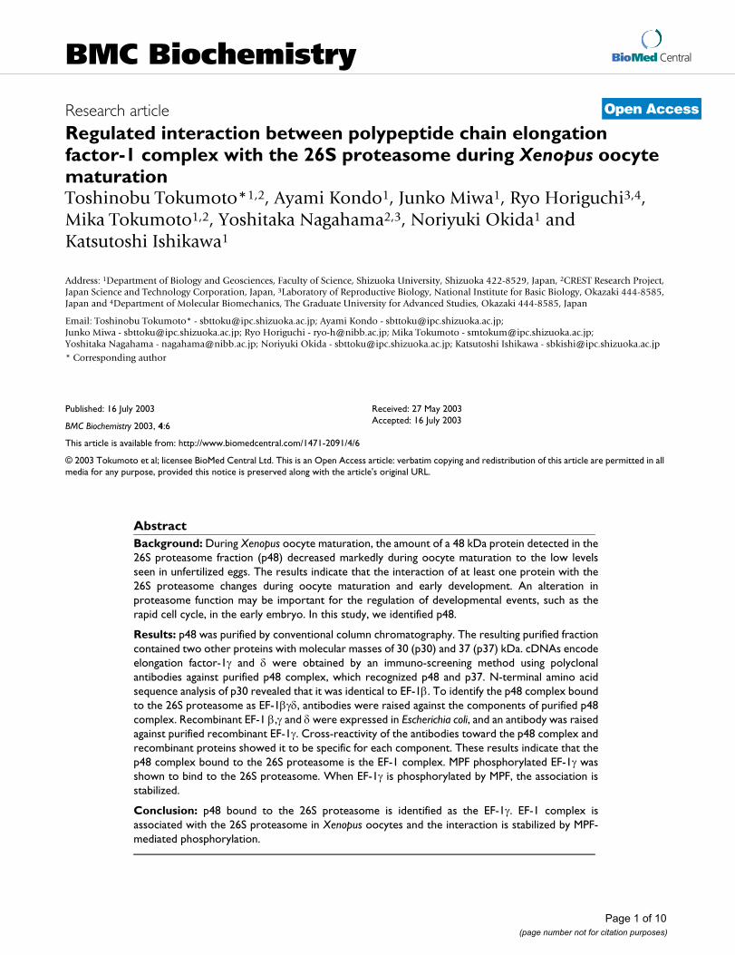
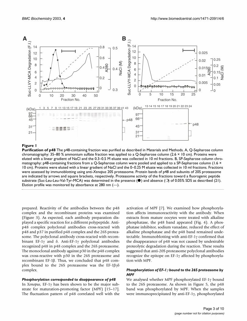
ResultsIdentification of p48 complex as EF-1βγδThe p48 complex, which was not associated with the 26Sproteasome, was purified from oocyte extracts by salt-extraction and column chromatography. Fractions wereassessed by immunoblotting using anti-20S proteasomepolyclonal antibodies. When the 35–80% ammoniumsulfate fraction from Xenopus oocyte extracts was appliedto a Q-Sepharose column, p48 was separated from protea-somes by linear gradient elution (Fig. 1A). The 26S protea-some is structurally labile and dissociates intosubcomplexes during column chromatography, especiallyon linear gradient elution [18,19]. Next, a homogeneousfraction was obtained by SP-Sepharose column chroma-tography (Fig. 1B). The resulting purified fraction con-tained two other proteins with molecular masses of 30(p30) and 37 (p37) kDa (Fig. 2). Of these two bands, p37was cross-reactive with anti-20S proteasome (Fig. 2). Anti-20S proteasome polyclonal antibody cross-reacted withp48 and p37. These results suggested that p48 is part of acomplex with p30 and p37.
To investigate this further we prepared polyclonal anti-bodies against the purified p48 fraction. A polyclonalantibody cross-reactive with p48 and p37 was obtained(Fig. 2). Immunoscreening a cDNA library using the anti-p48 complex identified two groups of cDNA clonesencoding elongation factor-1 (EF-1) γ and δ. N-terminalamino acid sequence analysis of p30 revealed the peptidesequence XFGDLKSPAGLKVL, which was identical to thatof Xenopus EF-1β.
These results suggested that the p48-containing proteincomplex bound to the 26S proteasome was EF-1βγδ. Toconfirm this, recombinant EF-1β,γ and δ were expressed inE. coli to examine cross-reactivity with antibodies, and anantibody against purified recombinant EF-1γ was
Page 2 of 10(page number not for citation purposes)
BMC Biochemistry 2003, 4 http://www.biomedcentral.com/1471-2091/4/6
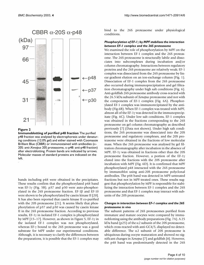
prepared. Reactivity of the antibodies between the p48complex and the recombinant proteins was examined(Figure 3). As expected, each antibody preparation dis-played a specific reaction for a different polypeptide. Anti-p48 complex polyclonal antibodies cross-reacted withp48 and p37 in purified p48 complex and the 26S protea-some. The polyclonal antibody cross-reacted with recom-binant EF-1γ and δ. Anti-EF-1γ polyclonal antibodiesrecognized p48 in p48 complex and the 26S proteasome.The monoclonal antibody against p30 in the p48 complexwas cross-reactive with p30 in the 26S proteasome andrecombinant EF-1β. Thus, we concluded that p48 com-plex bound to the 26S proteasome was the EF-1βγδcomplex.
Phosphorylation corresponded to disappearance of p48In Xenopus, EF-1γ has been shown to be the major sub-strate for maturation-promoting factor (MPF) [15–17].The fluctuation pattern of p48 correlated well with the
activation of MPF [7]. We examined how phosphoryla-tion affects immunoreactivity with the antibody. Whenextracts from mature oocytes were treated with alkalinephosphatase, the p48 band appeared (Fig. 4). A phos-phatase inhibitor, sodium vanadate, reduced the effect ofalkaline phosphatase and the p48 band remained unde-tectable. Immunoblotting with anti-EF-1γ confirmed thatthe disappearance of p48 was not caused by undesirableproteolytic degradation during the reaction. These resultssuggested that anti-20S proteasome polyclonal antibodiesrecognize the epitope on EF-1γ affected by phosphoryla-tion with MPF.
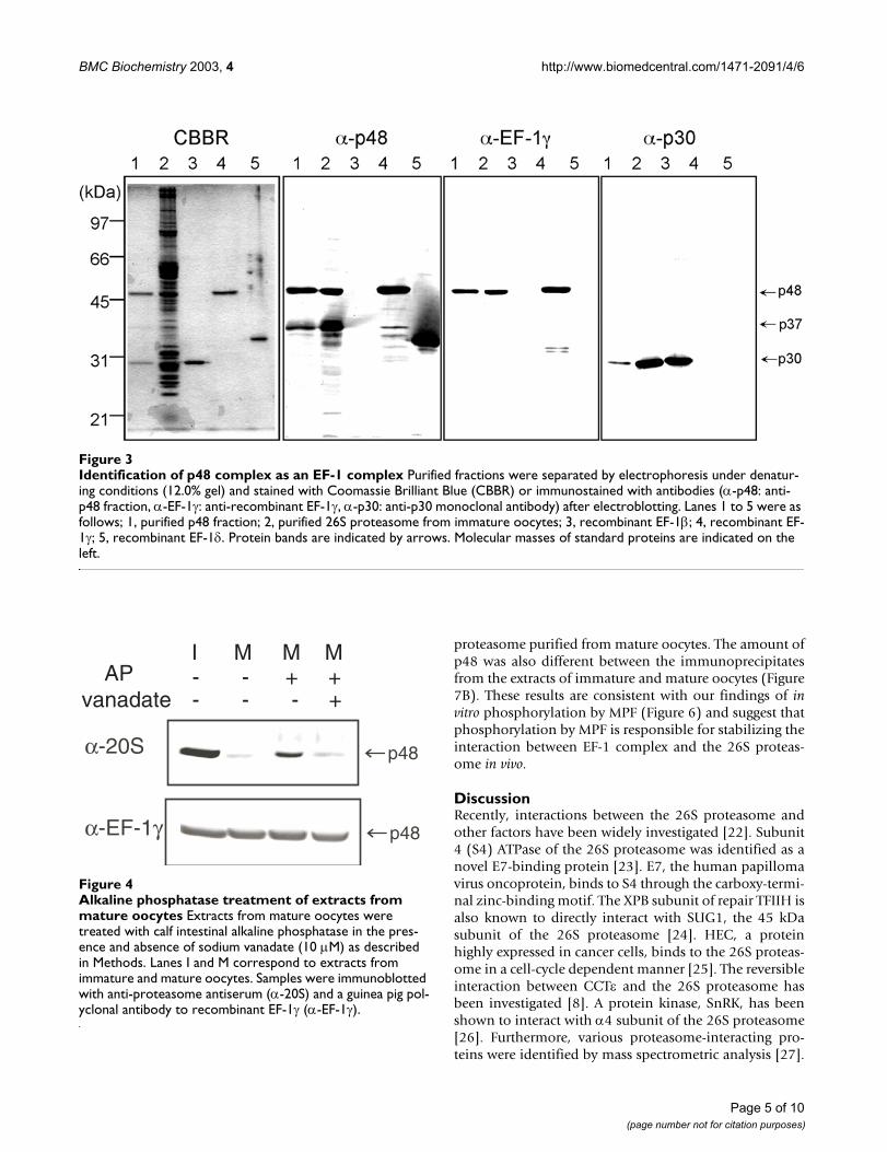
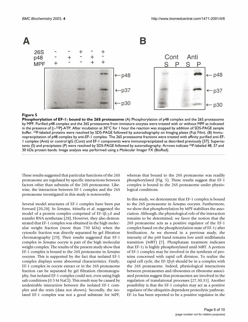
Phosphorylation of EF-1γ bound to the 26S proteasome by MPFWe analysed whether MPF phosphorylated EF-1γ boundto the 26S proteasome. As shown in Figure 5, the p48band was phosphorylated by MPF. When the sampleswere immunoprecipitated by anti-EF-1γ, phosphorylated
Purification of p48Figure 1Purification of p48 The p48-containing fraction was purified as described in Materials and Methods. A, Q-Sepharose column chromatography: 35–80 % ammonium sulfate fraction was applied to a Q-Sepharose column (2.6 × 10 cm). Proteins were eluted with a linear gradient of NaCl and the 0.3–0.5 M eluate was collected in 10 ml fractions. B, SP-Sepharose column chro-matography: p48-containing fractions from a Q-Sepharose column were pooled and applied to a SP-Sepharose column (1.6 × 10 cm). Proteins were eluted with a linear gradient of NaCl and the 0–0.25 M eluate was collected in 10 ml fractions. Fractions were assessed by immunoblotting using anti-Xenopus 20S proteasome. Protein bands of p48 and subunits of 20S proteasome are indicated by arrows and square brackets, respectively. Proteasome activity of the fractions toward a fluorogenic peptide substrate (Suc-Leu-Leu-Val-Tyr-MCA) was determined in the presence (●) and absence (❍) of 0.05% SDS as described (21). Elution profile was monitored by absorbance at 280 nm (—).
21
31
976645 p48
1 3 5 7 9 11 13 15 17 19 21 23 25 27 29 31 33 35 37 39 41 43(kDa)
0
0.2
0.4
0.6
0.8
0
2
4
6
8
10
12
14
0 10 20 30 40 50
0.3
0.4
0.5
Fraction No.
0
0.005
0.01
0.015
0.02
0.025
0
0.05
0.1
0.15
0.2
0.25
0 5 10 15 20 25
0
2
46
8
10
12
14
Fraction No.
2131
9766
45 p48
13 14 15 16 17 18 19 20 21 22 23 24(kDa)
Suc
-LLV
Y-M
CA
Deg
rada
tion
(F.I.
)
Suc
-LLV
Y-M
CA
Deg
rada
tion
(F.I.
)
A28
0
NaC
l (M
)
NaC
l (M
)
A28
0
A B
Page 3 of 10(page number not for citation purposes)
BMC Biochemistry 2003, 4 http://www.biomedcentral.com/1471-2091/4/6
bands including p48 were obtained in the precipitates.These results confirm that the phosphorylated p48 bandwas EF-1γ (Fig. 5B). p37 and p30 were auto-phosphor-ylated in the 26S proteasome fraction. EF-1β and EF-1δwere shown to be phosphorylated by casein kinase II [20].It has also been reported that casein kinase II co-purifiedwith the 20S proteasome [21]. It seems likely that phos-phorylation of p37 and p30 was caused by casein kinaseII in the 26S proteasome fraction. According to previousresults, EF-1γ in isolated EF-1 complex is phosphorylatedby MPF [15–17]. However, as shown in Figure 5, EF-1γ inthe isolated EF-1 complex was not phosphorylated,whereas EF-γ bound to the 26S proteasome was a goodsubstrate for MPF under our experimental conditions.Although, it is necessary to clarify the differences betweenthe preparations, it is possible that the EF-1 complex may
bind to the 26S proteasome under physiologicalconditions.
Phosphorylation of EF-1γ by MPF stabilizes the interaction between EF-1 complex and the 26S proteasomeWe examined the role of phosphorylation by MPF on theinteraction between EF-1 complex and the 26S proteas-ome. The 26S proteasome is structurally labile and disso-ciates into subcomplexes during incubation and/orcolumn chromatography. Interactions between regulatoryproteins and the 26S proteasome are relatively weak; EF-1complex was dissociated from the 26S proteasome by lin-ear gradient elution on an ion-exchange column (Fig. 1).Dissociation of EF-1 complex from the 26S proteasomealso occurred during immunoprecipitation and gel filtra-tion chromatography under high salt conditions (Fig. 6).Anti-goldfish 26S proteasome antibody cross-reacted withthe 26.5 kDa subunit of Xenopus proteasome and not withthe components of EF-1 complex (Fig. 6A). Phosphor-ylated EF-1 complex was immunorecipitated by the anti-body (Fig.6B). When EF-1 complex was treated with MPF,almost all of the EF-1γ was detected in the immunoprecip-itate (Fig. 6C). Under low salt conditions, EF-1 complexwas obtained in the fractions corresponding to the 26Sproteasome on gel column chromatography as describedpreviously [7] (Data not shown). Under high salt condi-tions, the 26S proteasome was dissociated into the 20Sproteasome and regulatory complexes, and the 20S pro-teasome were obtained in the fractions of low molecularmass. When the 26S proteasome was analysed by gel fil-tration chromatography after incubation in the absence ofMPF, EF-1γ was obtained in fractions other than the 20Sproteasome fraction. However, a portion of the EF-1γeluted into the fractions with the 20S proteasome afterincubation with MPF (Fig. 6D). It is comfirmed that MPFphosphorylated p48 interacted with the 26S proteasomeby immunoblot using anti-20S proteasome polyclonalantibodies. The p48 band was detected in MPF-untreatedfractions but not in MPF-treated ones. These results sug-gest that phosphorylation by MPF is responsible for stabi-lizing the interaction between EF-1 complex and the 26Sproteasome and that EF-1 complex may interact with sub-units of the 20S proteasome.
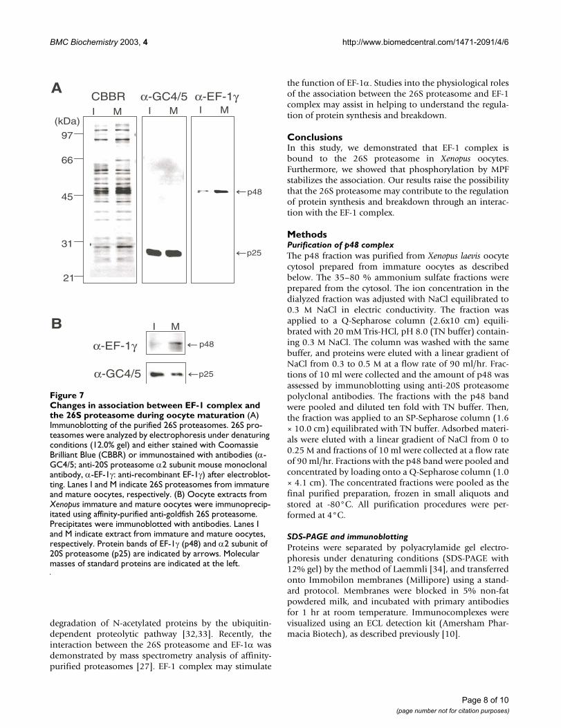
Changes in interaction between EF-1 complex and the 26S proteasome in vivoThe subunit patterns of 26S proteasomes purified fromimmature and mature oocytes were compared by immu-noblotting using the antibody preparations (Fig. 7A). A 25kDa band (p25) of the α2 subunit of the 20S proteasome,which cross-reacted with anti-GC4/5, displayed no detect-able difference. The α2 subunit of 20S proteasome isubiquitous during oocyte maturation and showed no sig-nificant changes in Xenopus [7] and goldfish [8]. However,the p48 band was predominantly detected in the 26S
Immunoblotting of purified p48 fractionFigure 2Immunoblotting of purified p48 fraction The purified p48 fraction was analyzed by electrophoresis under denatur-ing conditions (12.0% gel) and either stained with Coomassie Brilliant Blue (CBBR) or immunostained with antibodies (α-20S: anti-Xenopus 20S proteasome, α-p48: anti-p48 fraction) after electroblotting. Protein bands are indicated by arrows. Molecular masses of standard proteins are indicated on the left.
97
66
45
31
21
(kDa)CBBR α-20S α-p48
p48
p37
p30
Page 4 of 10(page number not for citation purposes)
BMC Biochemistry 2003, 4 http://www.biomedcentral.com/1471-2091/4/6
proteasome purified from mature oocytes. The amount ofp48 was also different between the immunoprecipitatesfrom the extracts of immature and mature oocytes (Figure7B). These results are consistent with our findings of invitro phosphorylation by MPF (Figure 6) and suggest thatphosphorylation by MPF is responsible for stabilizing theinteraction between EF-1 complex and the 26S proteas-ome in vivo.
DiscussionRecently, interactions between the 26S proteasome andother factors have been widely investigated [22]. Subunit4 (S4) ATPase of the 26S proteasome was identified as anovel E7-binding protein [23]. E7, the human papillomavirus oncoprotein, binds to S4 through the carboxy-termi-nal zinc-binding motif. The XPB subunit of repair TFIIH isalso known to directly interact with SUG1, the 45 kDasubunit of the 26S proteasome [24]. HEC, a proteinhighly expressed in cancer cells, binds to the 26S proteas-ome in a cell-cycle dependent manner [25]. The reversibleinteraction between CCTε and the 26S proteasome hasbeen investigated [8]. A protein kinase, SnRK, has beenshown to interact with α4 subunit of the 26S proteasome[26]. Furthermore, various proteasome-interacting pro-teins were identified by mass spectrometric analysis [27].
Identification of p48 complex as an EF-1 complexFigure 3Identification of p48 complex as an EF-1 complex Purified fractions were separated by electrophoresis under denatur-ing conditions (12.0% gel) and stained with Coomassie Brilliant Blue (CBBR) or immunostained with antibodies (α-p48: anti-p48 fraction, α-EF-1γ: anti-recombinant EF-1γ, α-p30: anti-p30 monoclonal antibody) after electroblotting. Lanes 1 to 5 were as follows; 1, purified p48 fraction; 2, purified 26S proteasome from immature oocytes; 3, recombinant EF-1β; 4, recombinant EF-1γ; 5, recombinant EF-1δ. Protein bands are indicated by arrows. Molecular masses of standard proteins are indicated on the left.
Alkaline phosphatase treatment of extracts from mature oocytesFigure 4Alkaline phosphatase treatment of extracts from mature oocytes Extracts from mature oocytes were treated with calf intestinal alkaline phosphatase in the pres-ence and absence of sodium vanadate (10 µM) as described in Methods. Lanes I and M correspond to extracts from immature and mature oocytes. Samples were immunoblotted with anti-proteasome antiserum (α-20S) and a guinea pig pol-yclonal antibody to recombinant EF-1γ (α-EF-1γ).
α-20S p48
p48α-EF-1γ
I M M MAP
vanadate- - + +- - - +
Page 5 of 10(page number not for citation purposes)
BMC Biochemistry 2003, 4 http://www.biomedcentral.com/1471-2091/4/6
These results suggested that particular functions of the 26Sproteasome are regulated by specific interactions betweenfactors other than subunits of the 26S proteasome. Like-wise, the interaction between EF-1 complex and the 26Sproteasome investigated in this study is noteworthy.
Several model structures of EF-1 complex have been putforward [20,28]. In Xenopus, Minella et al. suggested themodel of a protein complex comprised of EF-1β,γ,δ andtransfer RNA synthetase [28]. However, they also demon-strated that EF-1 complex was obtained in the high molec-ular weight fraction (more than 750 kDa) when thecytosolic fraction was directly separated by gel filtrationchromatography [29]. Their results suggested that EF-1complex in Xenopus oocyte is part of the high molecularweight complex. The results of the present study show thatEF-1 complex is bound to the 26S proteasome in Xenopusoocytes. This is supported by the fact that isolated EF-1complex displays some abnormal characteristics. Firstly,EF-1 complex in oocyte extract or in the 26S proteasomefraction can be separated by gel filtration chromatogra-phy, but isolated EF-1 complex could not, even using highsalt conditions (0.5 M NaCl). This result may be caused byundesirable interaction between the isolated EF-1 com-plex and the resin (data not shown). Secondly, the iso-lated EF-1 complex was not a good substrate for MPF,
whereas that bound to the 26S proteasome was readilyphosphorylated (Fig. 5). These results suggest that EF-1complex is bound to the 26S proteasome under physio-logical conditions.
In this study, we demonstrate that EF-1 complex is boundto the 26S proteasome in Xenopus oocytes. Furthermore,we show that phosphorylation by MPF stabilizes the asso-ciation. Although, the physiological role of the interactionremains to be determined, we favor the notion that the26S proteasome acts as a positive regulator of the EF-1complex based on the phosphorylation state of EF-1γ afterfertilization. As we showed in a previous study, theintensity of the p48 band remains low until midblastulatransition (MBT) [7]. Phosphatase treatment indicatesthat EF-1γ is highly phosphorylated until MBT. A portionof EF-1 complex may be involved in the synthesis of pro-teins concerned with rapid cell division. To realize therapid cell cycle, the EF-1βγδ should be in a complex withthe 26S proteasome. Indeed, physiological interactionsbetween proteasomes and ribosomes or ribosome-associ-ated proteins suggest that proteasomes are involved in theregulation of translational processes [27,30,31]. Anotherpossibility is that the EF-1 complex may act as a positiveregulator of the ubiquitin-dependent proteolytic pathway.EF-1α has been reported to be a positive regulator in the
Phosphorylation of EF-1γ bound to the 26S proteasomeFigure 5Phosphorylation of EF-1γ bound to the 26S proteasome (A) Phosphorylation of p48 complex and the 26S proteasome by MPF. Purified p48 complex and the 26S proteasome from immature oocytes were treated with or without MPF as indicated in the presence of [γ-32P]-ATP. After incubation at 30°C for 1 hour the reaction was stopped by addition of SDS-PAGE sample buffer. 32P-labeled proteins were resolved by SDS-PAGE followed by autoradiography on Imaging plates (Fuji Film). (B) Immu-noprecipitation of p48 complex by anti-EF-1 complex. The 26S proteasome fractions were treated with affinity purified anti-EF-1 complex (Anti) or control IgG (Cont) and EF-1 components were immunoprecipitated as described previously [37]. Superna-tants (S) and precipitates (P) were resolved by SDS-PAGE followed by autoradiography. Arrows indicate 32P-labeled 48, 37 and 30 kDa protein bands. Image analysis was performed using a Molecular Imager FX (BioRad).
p48
p37
p30
A B
p48
p37
p30
Cont AntiS P S P
26Sp48MPF
++
--
--
- --
-+
+
++
+
Page 6 of 10(page number not for citation purposes)

BMC Biochemistry 2003, 4 http://www.biomedcentral.com/1471-2091/4/6
Stabilization of association between EF-1 complex and the 26S proteasome by phosphorylation with MPFFigure 6Stabilization of association between EF-1 complex and the 26S proteasome by phosphorylation with MPF (A) The 26S proteasome fraction from Xenopus immature oocytes was resolved by electrophoresis under denaturing conditions (12 % gel) and immunostained with anti-goldfish 26S proteasome polyclonal antibody. (B) The 26S proteasome from immature oocytes was treated with MPF for 1 hour at 30°C. Samples were treated with affinity purified anti-goldfish 26S proteasome (Anti) or control IgG (Cont) and the 26S proteasomes were immunoprecipitated. Untreated sample (Total) and precipitates were resolved by SDS-PAGE followed by autoradiography. Arrows indicate 32P-labeled 48, 37 and 30 kDa protein bands. (C) The 26S proteasome from immature oocytes was treated with (MPF+) or without (MPF-) MPF for 1 hour at 30°C. Samples were immunoprecipitated using affinity-purified anti-goldfish 26S proteasome. Supernatants (S) and precipitates (P) were immunoblotted with antibodies (α-EF-1γ; anti-recombinant EF-1γ: α-GC4/5; anti-20S proteasome α2 subunit mouse mono-clonal antibody). Protein bands of EF-1γ (p48) and α2 subunit of 20S proteasome (p25) are indicated by arrows. (D) Samples were analyzed by chromatography on a G4000SWXL column equilibrated with 50 mM Tris-HCl buffer, pH 7.5, containing 20% glycerol, 10 mM 2-mercaptoethanol and 0.1 mM ATP as described previously [36] in the presence of 0.5 M NaCl. Fractions were assessed by immunoblotting using a mixture of anti-Xenopus 20S proteasome and anti-EF-1γ polyclonal antibodies (α-20S+α-EF-1γ) or anti-Xenopus 20S proteasome (α-20S). Protein bands of EF-1γ (p48) and subunits of 20S proteasome are indi-cated by the arrow and square brackets, respectively.
p48
p37
p30
Tot
al
Con
t
Ant
i
α-GC4/5
S P S PMPF- MPF+
α-EF-1γ p48
p25
A B97
66
45
31
21
(kDa)
DMPF-
MPF+
MPF-
MPF+
p48
p48
15 16 17 18 19 20 21 22 23 24 25
α-20S
α-20S+
α-EF-1γ
p48
p48
15 16 17 18 19 20 21 22 23 24 25
C
Page 7 of 10(page number not for citation purposes)
BMC Biochemistry 2003, 4 http://www.biomedcentral.com/1471-2091/4/6
degradation of N-acetylated proteins by the ubiquitin-dependent proteolytic pathway [32,33]. Recently, theinteraction between the 26S proteasome and EF-1α wasdemonstrated by mass spectrometry analysis of affinity-purified proteasomes [27]. EF-1 complex may stimulate
the function of EF-1α. Studies into the physiological rolesof the association between the 26S proteasome and EF-1complex may assist in helping to understand the regula-tion of protein synthesis and breakdown.
ConclusionsIn this study, we demonstrated that EF-1 complex isbound to the 26S proteasome in Xenopus oocytes.Furthermore, we showed that phosphorylation by MPFstabilizes the association. Our results raise the possibilitythat the 26S proteasome may contribute to the regulationof protein synthesis and breakdown through an interac-tion with the EF-1 complex.
MethodsPurification of p48 complexThe p48 fraction was purified from Xenopus laevis oocytecytosol prepared from immature oocytes as describedbelow. The 35–80 % ammonium sulfate fractions wereprepared from the cytosol. The ion concentration in thedialyzed fraction was adjusted with NaCl equilibrated to0.3 M NaCl in electric conductivity. The fraction wasapplied to a Q-Sepharose column (2.6x10 cm) equili-brated with 20 mM Tris-HCl, pH 8.0 (TN buffer) contain-ing 0.3 M NaCl. The column was washed with the samebuffer, and proteins were eluted with a linear gradient ofNaCl from 0.3 to 0.5 M at a flow rate of 90 ml/hr. Frac-tions of 10 ml were collected and the amount of p48 wasassessed by immunoblotting using anti-20S proteasomepolyclonal antibodies. The fractions with the p48 bandwere pooled and diluted ten fold with TN buffer. Then,the fraction was applied to an SP-Sepharose column (1.6× 10.0 cm) equilibrated with TN buffer. Adsorbed materi-als were eluted with a linear gradient of NaCl from 0 to0.25 M and fractions of 10 ml were collected at a flow rateof 90 ml/hr. Fractions with the p48 band were pooled andconcentrated by loading onto a Q-Sepharose column (1.0× 4.1 cm). The concentrated fractions were pooled as thefinal purified preparation, frozen in small aliquots andstored at -80°C. All purification procedures were per-formed at 4°C.
SDS-PAGE and immunoblottingProteins were separated by polyacrylamide gel electro-phoresis under denaturing conditions (SDS-PAGE with12% gel) by the method of Laemmli [34], and transferredonto Immobilon membranes (Millipore) using a stand-ard protocol. Membranes were blocked in 5% non-fatpowdered milk, and incubated with primary antibodiesfor 1 hr at room temperature. Immunocomplexes werevisualized using an ECL detection kit (Amersham Phar-macia Biotech), as described previously [10].
Changes in association between EF-1 complex and the 26S proteasome during oocyte maturationFigure 7Changes in association between EF-1 complex and the 26S proteasome during oocyte maturation (A) Immunoblotting of the purified 26S proteasomes. 26S pro-teasomes were analyzed by electrophoresis under denaturing conditions (12.0% gel) and either stained with Coomassie Brilliant Blue (CBBR) or immunostained with antibodies (α-GC4/5; anti-20S proteasome α2 subunit mouse monoclonal antibody, α-EF-1γ: anti-recombinant EF-1γ) after electroblot-ting. Lanes I and M indicate 26S proteasomes from immature and mature oocytes, respectively. (B) Oocyte extracts from Xenopus immature and mature oocytes were immunoprecip-itated using affinity-purified anti-goldfish 26S proteasome. Precipitates were immunoblotted with antibodies. Lanes I and M indicate extract from immature and mature oocytes, respectively. Protein bands of EF-1γ (p48) and α2 subunit of 20S proteasome (p25) are indicated by arrows. Molecular masses of standard proteins are indicated at the left.
α-GC4/5
p25
α-EF-1γI MI M
97
66
45
31
21
(kDa)I M
CBBRA
B
α-GC4/5
α-EF-1γ
I M
p48
p25
p48
Page 8 of 10(page number not for citation purposes)
BMC Biochemistry 2003, 4 http://www.biomedcentral.com/1471-2091/4/6
cDNA cloning by immunoscreening and production of recombinant proteinsXenopus ovary cDNA library was constructed in the Uni-ZAP XR vector (Stratagene). Using polyclonal antibodiesagainst the purified p48 fraction prepared in this study,immunoscreening was carried out as described previously[35]. From the isolated plaques, plasmid DNA wasprepared by the in vivo excision protocol using the ExAs-sist/SOLR system (Stratagene). DNA sequencing was per-formed using a 377A DNA sequencer (Perkin Elmer ABI)with a Dye Terminator Cycle Sequencing Kit (PerkinElmer ABI).
The full-length ORFs of Xenopus EF-1β,γ and δ were clonedinto the pET21b expression vector (Novagen) at the NdeIand XhoI sites. These were constructed from each cDNA bythe PCR method. The recombinant proteins were pro-duced in E. coli BL21(DE3)pLysE and purified by affinitychromatography on Ni-NTA Agarose (Qiagen) columnaccording to the manufacturer's instructions [35].
Purification of the 26S proteasome and Peptidase assayThe 26S proteasome was purified from immature Xenopusoocytes as described [36]. Peptidase activity with or with-out 0.05% SDS was assessed using the fluorogenic peptidesubstrate, Suc-LLVY-MCA, as described previously [37].
Antibody production and ImmunoprecipitationAntibodies against purified p48 complex were prepared asdescribed previously [38]. Polyclonal antiserum cross-reactive with p48 and p37 and a monoclonal antibodycross-reactive with p30 (EF-1β) were obtained. A polyclo-nal antibody against recombinant EF-1γ was prepared asfollows. Affinity-purified recombinant EF-1γ (20 µg ofprotein) in complete Freund's adjuvant was injected intoguinea pigs at ten-day intervals until sufficient titer wasobtained. The antiserum to Xenopus 20S proteasome anda monoclonal antibody to goldfish 20S proteasome α2subunit were prepared and used as previously described[7,38].
Immunoprecipitation was performed with IgG fractionsthat had been purified from serum on a protein A-Sepha-rose CL-4B column (Ammersham Pharmacia) asdescribed previously [37].
Preparation of oocyte and egg extractsExtracts from Stage VI oocytes (immature oocytes) andovulated eggs (mature oocytes) were prepared asdescribed previously [7]. Stage VI oocytes were manuallyisolated from Xenopus ovarian fragments. Ovulated eggswere dejellied with 2% cysteine solution. Groups oftwenty oocytes or eggs were washed in MPF extractionbuffer (80 mM β-glycerophosphate, 50 mM NaF, 20 mMEGTA, 15 mM MgCl2, 20 mM HEPES, pH 7.5) and trans-
ferred to 1.5 ml Eppendorf microcentrifuge tubes. Theexcess buffer was removed, and 100 µl of fresh buffer wasadded. The samples were crushed with 5 strokes of a plas-tic pestle and centrifuged for 10 min at 13,500 rpm at 4°Cin a fixed angle rotor (TOMY Model MX-160 microcentri-fuge). The clear supernatant (100 µl) was collected forelectrophoresis and immunoblotting.
Phosphatase treatmentEgg extracts were treated with calf intestine alkaline phos-phatase (Boehringer Mannheim, 60 U) for 60 min at30°C in the presence of protease inhibitors (chymostatin,leupeptin and pepstatin, 500 µg/ml each). Phosphatase-treated egg extracts were assessed by immunoblottingusing anti-20S proteasome polyclonal antibodies.
Phosphorylation by MPFAliquots (4 µl) of purified p48 complex or the 26S protea-some were incubated in a total volume of 20 µl. The reac-tion mixture contained 10 mM Tris-HCl, pH 7.5, 1 mMMgCl2, 4 mM 2-mercaptoethanol, 40 µM ATP and 10 µM[γ-32P]-ATP (0.37 Tbq/mmol) with or without 2 units ofrecombinant human MPF (BIOMOL). Reactions were per-formed at 30°C for 1 hour and terminated with SDS-PAGE sample buffer. 32P-labeled proteins were resolvedby SDS-PAGE and visualized by autoradiography onImaging plates (Fuji Film).
Authors' contributionsTT carried out the immunoassays, phosphorylation anal-ysis and also participated in the design of the study anddrafted the manuscript. AK, JM, RH and NO participatedfor protein purification and antibody production. MT par-ticipated in cDNA cloning. YN and KI participated in coor-dination in the design of the study.
AcknowledgementsWe are grateful to the staff of the Radiochemistry Research Laboratory and Institute for Genetic Research and Biotechnology of Shizuoka University for the use of equipment. We also thank Y. Makino of the Center for Ana-lytical Instruments of the National Institute for Basic Biology for providing technical assistance in amino acid sequence analysis. This work was sup-ported by Grants-in-Aid for Scientific Research on Priority Areas from the Ministry of Education, Culture, Sports, Science and Technology of Japan and the CREST Research Project of the Japan Science and Technology Corpo-ration to YN. Part of this study was performed as the National Institute for Basic Biology Cooperative Research Program (00-121 to TT).
References1. Masui Yand Clarke HJ: Oocyte maturation Int Rev Cytol 1979,
57:185-282.2. Hershko A and Ciechanover A: The ubiquitin system Annu Rev
Biochem 1998, 67:425-479.3. Coux O, Tanaka K and Goldberg AL: Structure and functions of
the 20S and 26S proteasomes Annu Rev Biochem 1996, 65:801-847.
4. Tokumoto T: Nature and role of proteasomes in maturationof fish oocytes Int Rev Cytol 1999, 186:261-294.
Page 9 of 10(page number not for citation purposes)
BMC Biochemistry 2003, 4 http://www.biomedcentral.com/1471-2091/4/6
Publish with BioMed Central and every scientist can read your work free of charge
"BioMed Central will be the most significant development for disseminating the results of biomedical research in our lifetime."
Sir Paul Nurse, Cancer Research UK
Your research papers will be:
available free of charge to the entire biomedical community
peer reviewed and published immediately upon acceptance
cited in PubMed and archived on PubMed Central
yours — you keep the copyright
Submit your manuscript here:http://www.biomedcentral.com/info/publishing_adv.asp
BioMedcentral
5. Sawada MT, Someno T, Hoshi M and Sawada H: Participation of650-kDa protease (20 S proteasome) in starfish oocytematuration Dev Biol 1992, 150:414-418.
6. Takahashi M, Tokumoto T and Ishikawa K: DFP-sensitive multi-catalytic protease complexes (proteasomes) involved in thecontrol of oocyte maturation in the toad, Bufo japonicus MolReprod Dev 1994, 38:310-317.
7. Tokumoto T, Tokumoto M, Seto K, Horiguchi R, Nagahama Y,Yamada S, Ishikawa K and Lohka MJ: Disappearance of a novelprotein component of the 26S proteasome during Xenopusoocyte maturation Exp Cell Res 1999, 247:313-319.
8. Tokumoto M, Horiguchi R, Nagahama Y, Ishikawa K and TokumotoT: Two proteins, a goldfish 20S proteasome subunit and theprotein interacting with 26S proteasome, change in the mei-otic cell cycle Eur J Biochem 2000, 267:97-103.
9. Tokumoto M, Horiguchi R, Nagahama Y and Tokumoto T: Identifi-cation of the Xenopus 20S proteasome α4 subunit which ismodified in the meiotic cell cycle Gene 1999, 239:301-308.
10. Tokumoto T, Yamashita M, Tokumoto M, Katsu Y, Horiguchi R,Kajiura H and Nagahama Y: Initiation of cyclin B degradation bythe 26S proteasome upon egg activation J Cell Biol 1997,138:1313-1322.
11. Nishiyama A, Tachibana K, Igarashi Y, Yasuda H, Tanahashi N, TanakaK, Ohsumi K and Kishimoto T: A nonproteolytic function of theproteasome is required for the dissociation of Cdc2 and cyc-lin B at the end of M phase Genes Dev 2000, 14:2344-2357.
12. Josefsberg LB, Galiani D, Dantes A, Amsterdam A and Dekel N: Theproteasome is involved in the first metaphase-to-anaphasetransition of meiosis in rat oocytes Biol Reprod 2000, 62:1270-1277.
13. Josefsberg LB, Kaufman O, Galiani D, Kovo M and Dekel N: Inacti-vation of M-phase promoting factor at exit from first embry-onic mitosis in the rat is independent of cyclin B1degradation Biol Reprod 2001, 64:871-878.
14. Moldave K: Eukaryotic protein synthesis Annu Rev Biochem 1985,54:1109-1149.
15. Bellé R, Derancourt J, Poulhe R, Capony JP, Ozon R and Mulner-Lorillon O: A purified complex from Xenopus oocytes containsa p47 protein, an in vivo substrate of MPF, and a p30 proteinrespectively homologous to elongation factors EF-1γ and EF-1β FEBS Lett 1989, 255:101-104.
16. Janssen GM, Morales J, Schipper A, Labbe JC, Mulner-Lorillon O, BelléR and Moller W: A major substrate of maturation promotingfactor identified as elongation factor 1βγδ in Xenopus laevis JBiol Chem 1991, 266:14885-14888.
17. Bellé R, Minella O, Cormier P, Morales J, Poulhe R and Mulner-Lorillon O: Phosphorylation of elongation factor-1 (EF-1) bycdc2 kinase Prog Cell Cycle Res 1995, 1:265-270.
18. Hoffman L, Pratt G and Rechsteiner M: Multiple forms of the 20 Smulticatalytic and the 26 S ubiquitin/ATP-dependent pro-teases from rabbit reticulocyte lysate J Biol Chem 1992,267:22362-22368.
19. Tokumoto T, Yoshikuni M, Yamashita M, Kajiura H and Nagahama Y:Purification and characterization of active proteasome (26Sproteasome) from goldfish ovaries Biomed Res 1995, 16:207-218.
20. Sheu GT and Traugh JA: A structural model for elongation fac-tor 1 (EF-1) and phosphorylation by protein kinase CKII MolCell Biochem 1999, 191:181-186.
21. Ludemann R, Lerea KM and Etlinger JD: Copurification of caseinkinase II with 20 S proteasomes and phosphorylation of a 30-kDa proteasome subunit J Biol Chem 1993, 268:17413-17417.
22. Ferrell K, Wilkinson CR, Dubiel W and Gordon C: Regulatory sub-unit interactions of the 26S proteasome, a complex problemTrends Biochem Sci 2000, 25:83-88.
23. Berezutskaya E and Bagchi S: The human papillomavirus E7oncoprotein functionally interacts with the S4 subunit of the26 S proteasome J Biol Chem 1997, 272:30135-30140.
24. Weeda G, Rossignol M, Fraser RA, Winkler GS, Vermeulen W, van'tVeer LJ, Ma L, Hoeijmakers JH and Egly JM: The XPB subunit ofrepair/transcription factor TFIIH directly interacts withSUG1, a subunit of the 26S proteasome and putative tran-scription factor Nucleic Acids Res 1997, 25:2274-2283.
25. Chen Y, Sharp ZD and Lee WH: HEC binds to the seventh reg-ulatory subunit of the 26 S proteasome and modulates theproteolysis of mitotic cyclins J Biol Chem 1997, 272:24081-24087.
26. Farras R, Ferrando A, Jasik J, Kleinow T, Okresz L, Tiburcio A, Sal-chert K, del Pozo C, Schell J and Koncz C: SKP1-SnRK proteinkinase interactions mediate proteasomal binding of a plantSCF ubiquitin ligase Embo J 2001, 20:2742-2756.
27. Verma R, Chen S, Feldman R, Schieltz D, Yates J, Dohmen J andDeshaies RJ: Proteasomal proteomics: identification of nucle-otide-sensitive proteasome-interacting proteins by massspectrometric analysis of affinity-purified proteasomes MolBiol Cell 2000, 11:3425-3439.
28. Minella O, Mulner-Lorillon O, Bec G, Cormier P and Bellé R: Multi-ple phosphorylation sites and quaternary organization ofguanine-nucleotide exchange complex of elongation factor-1(EF-1βγδ/ValRS) control the various functions of EF-1α BiosciRep 1998, 18:119-127.
29. Minella O, Mulner-Lorillon O, Poulhe R, Bellé R and Cormier P: Theguanine-nucleotide-exchange complex (EF-1βγδ) of elonga-tion factor-1 contains two similar leucine-zipper proteins EF-1δ, p34 encoded by EF-1δ1 and p36 encoded by EF-1δ2 Eur JBiochem 1996, 237:685-690.
30. Lin L, DeMartino GN and Greene WC: Cotranslational biogen-esis of NF-kappaB p50 by the 26S proteasome Cell 1998,92:819-828.
31. Schubert U, Anton LC, Gibbs J, Norbury CC, Yewdell JW and Ben-nink JR: Rapid degradation of a large fraction of newly synthe-sized proteins by proteasomes Nature 2000, 404:770-774.
32. Gonen H, Schwartz AL and Ciechanover A: Purification and char-acterization of a novel protein that is required for degrada-tion of N-α-acetylated proteins by the ubiquitin system J BiolChem 1991, 266:19221-19231.
33. Gonen H, Smith CE, Siegel NR, Kahana C, Merrick WC, ChakraburttyK, Schwartz AL and Ciechanover A: Protein synthesis elongationfactor EF-1α is essential for ubiquitin-dependent degrada-tion of certain Nα-acetylated proteins and may be substi-tuted for by the bacterial elongation factor EF-Tu Proc NatlAcad Sci U S A 1994, 91:7648-7652.
34. Laemmli UK: Cleavage of structural proteins during theassembly of the head of bacteriophage T4 Nature 1970,227:680-685.
35. Horiguchi R, Tokumoto M, Yoshiura Y, Aida K, Nagahama Y andTokumoto T: Molecular cloning of cDNA encoding a 20S pro-teasome α2 subunit from goldfish (Carassius auratus) and itsexpression analysis Zoolog Sci 1998, 15:773-777.
36. Tokumoto T and Ishikawa K: Characterization of active protea-some (26S proteasome) from Xenopus oocytes Biomed Res1995, 16:295-302.
37. Tokumoto T and Ishikawa K: A novel "active" form of proteas-omes from Xenopus laevis ovary cytosol Biochem Biophys ResCommun 1993, 192:1106-1114.
38. Tokumoto T, Yamashita M, Yoshikuni M, Kajiura H and Nagahama Y:Purification of latent proteasome (20S proteasome) anddemonstration of active proteasome in goldfish (Carassiusauratus) oocyte cytosol Biomed Res 1995, 16:173-186.
Page 10 of 10(page number not for citation purposes)