biotinylation is a natural, albeit rare, modification of human histones
TRANSCRIPT
Biotinylation is a natural, albeit rare, modification of humanhistones
Toshinobu Kuroishi, Luisa Rios-Avila, Valerie Pestinger, Subhashinee S. K. Wijeratne, andJanos Zempleni*1Department of Nutrition and Health Sciences, University of Nebraska at Lincoln, Lincoln, NE68583-0806, USA
AbstractPrevious studies suggest that histones H3 and H4 are posttranslationally modified by binding ofthe vitamin biotin, catalyzed by holocarboxylase synthetase (HCS). Albeit a rare epigenetic mark,biotinylated histones were repeatedly shown to be enriched in repeat regions and repressed loci,participating in the maintenance of genome stability and gene regulation. Recently, a team ofinvestigators failed to detect biotinylated histones and proposed that biotinylation is not a naturalmodification of histones, but rather an assay artifact. Here, we describe the results of experiments,including the comparison of various analytical protocols, antibodies, cell lines, classes of histones,and radiotracers. These studies provide unambiguous evidence that biotinylation is a natural, albeitrare, histone modification. Less than 0.001% of human histones H3 and H4 are biotinylated,raising concerns that the abundance might too low to elicit biological effects in vivo. Weintegrated information from this study, previous studies, and ongoing research efforts to present anew working model in which biological effects are caused by a role of HCS in multiproteincomplexes in chromatin. In this model, docking of HCS in chromatin causes the occasionalbinding of biotin to histones as a tracer for HCS binding sites.
KeywordsBiotin; histones; holocarboxylase synthetase; post-translational modifications
1. BackgroundChromatin comprises DNA, histones, and other chromatin proteins [1]. In each nucleosomalcore particle, about 146 basepairs of DNA are wrapped around an octamer of core histones(one H3/H3/H4/H4 tetramer and two H2A/H2B dimers). Amino acids in the N-terminal tailsof core histones and, to a lesser extent, amino acids in globular domains and C-termini, areexposed at the nucleosomal surface [2]. The N-terminal tails are subject to a multitude ofposttranslational modifications, including acetylation and methylation [3]. Thesemodifications play crucial roles in gene regulation. For example, acetylation of lysine (K)-9in histone H3 (H3K9ac) is associated with transcriptionally active chromatin, whereasdimethylation and trimethylation of K9 (H3K9me2, H3K9me3) are associated with
© 2011 Elsevier Inc. All rights reserved.*To whom correspondence should be addressed: [email protected]'s Disclaimer: This is a PDF file of an unedited manuscript that has been accepted for publication. As a service to ourcustomers we are providing this early version of the manuscript. The manuscript will undergo copyediting, typesetting, and review ofthe resulting proof before it is published in its final citable form. Please note that during the production process errors may bediscovered which could affect the content, and all legal disclaimers that apply to the journal pertain.
NIH Public AccessAuthor ManuscriptMol Genet Metab. Author manuscript; available in PMC 2012 December 1.
Published in final edited form as:Mol Genet Metab. 2011 December ; 104(4): 537–545. doi:10.1016/j.ymgme.2011.08.030.
NIH
-PA Author Manuscript
NIH
-PA Author Manuscript
NIH
-PA Author Manuscript
transcriptionally repressed chromatin [3]. Other, less abundant, modifications such asphosphorylation of serine-14 in histone H2B play critical roles in events such asprogrammed cell death [4].
Hymes et al. suggested that histones are also modified by covalent attachment of the vitaminbiotin, mediated by the enzyme biotinidase [5]. While the original report was based on invitro studies, evidence soon emerged that histone biotinylation is a natural phenomenon,based on probing and tracing biotinylation with streptavidin, anti-biotin, and [3H]biotin inprimary human lymphoid cells [6]. While biotinidase undoubtedly has histone biotinylligase activity in vitro [5, 7], it is now believed that holocarboxylase synthetase (HCS) is theenzyme responsible for biotinylation of histones in vivo. This conclusion is based onobservations that HCS is a nuclear, chromatin-associated protein [8–11]; that phenotypes ofHCS knockdown include shortened life span and decreased heat tolerance in Drosophilamelanogaster, while phenotypes of biotinidase knockdown are relatively minor [9]; thatboth human recombinant HCS (rHCS) and its microbial ortholog BirA have enzymaticactivity to biotinylate histones and histone-based peptides [12, 13]; and that HCSknockdown causes aberrant gene regulation in human Jurkat lymphoblastoma cells andDrosophila melanogaster [9, 10].
Over the past several years, we have identified the following histone biotinylation sites: K4,K9, K18, and perhaps K23 in histone H3 [13, 14] and K8 and K12 in histone H4 [7, 15]. Weprovided evidence that K9, K13, K125, K127, and K129 in histone H2A also are targets forbiotinylation albeit only in trace amounts [16]. We raised target-specific antibodies to manyof these marks and to HCS [7, 14–16]. Using these antibodies, new roles of biotin in generegulation and genome stability have been discovered. In those studies, K12-biotinylatedhistone H4 (H4K12bio) was the most prominently featured mark, primarily due to theavailability of an antibody of exceptional quality [7]. The anti-H4K12bio used in thosestudies does not cross-react with non-biotinylated histones, does not cross-react withbiotinylated histones other than histone H4, and does not cross-react with biotinylation sitesother than K12. Evidence was provided that H4K12bio is enriched at repressed loci and atrepeat regions in the human genome [10, 17, 18], and, further, that a low abundance ofH4K12bio coincides with de-repression of retrotransposons and increased frequency ofchromosomal abnormalities in biotin- and HCS deficient humans, human and murine celllines, and Drosophila melanogaster [11].
In the early stages of these investigations, we proposed that biotinylation of histones is a rareevent; binding of biotin to histones is in the order of only attomoles of biotin incorporatedinto histones isolated from 106 human lymphocytes [6]. Subsequent studies by independentinvestigators confirmed that <0.03% of histones are biotinylated in human cell cultures [19].In a recent report, yet another laboratory failed to detect histone biotinylation marks byusing streptavidin, antibodies, and mass spectrometry (MS); based on their negative resultsthose investigators proposed that histone biotinylation is an artifact caused by non-specificprobes and purification procedures [20]. Here, we conducted a thorough examination ofprobe specificity and purification procedures to provide an unambiguous answer to thequestion of whether biotinylation of histones is a rare event or simply an artifact. In view ofour findings that biotin is a natural although rare histone modification, we propose amechanism to explain the rarity as well as the biological significance.
2. Methods2.1. Cell culture
Jurkat human lymphoblastoma cells, IMR-90 human fibroblasts, HepG2 humanhepatocellular carcinoma cells, and U-937 human monocytic cells were obtained from
Kuroishi et al. Page 2
Mol Genet Metab. Author manuscript; available in PMC 2012 December 1.
NIH
-PA Author Manuscript
NIH
-PA Author Manuscript
NIH
-PA Author Manuscript
American Type Culture Collection (Manassas, VA). HeLa human cervical cancer cells andMCF-7 human breast cancer cells were gifts by Drs. Jennifer Wood and Angela Pannier atthe University of Nebraska-Lincoln. Cells were cultured using media and conditions asrecommended by the American Type Culture Collection. In some experiments, cells werecultured in biotin-defined media (0.025 nM, 0.25 nM, or 10 nM), representing biotinconcentrations in plasma from biotin-deficient, biotin-normal, and biotin-supplementedindividuals [21, 22]. Biotin-defined media were prepared using customized culture mediaand biotin-depleted fetal bovine serum as previously described [23]. Where indicated,[3H]biotin (specific activity = 2.0535 TBq/mmol; Perkin Elmer, Boston, MA) or custom-made [14C]biotin (specific activity = 2.2052 GBq/mmol; Moravek, Inc., Brea, CA) weresubstituted for unlabeled biotin in culture media.
2.2. Purification of histonesCell nuclei were released by treatment with detergent and collected by gradientcentrifugation [6]; histones were extracted overnight with 1 M HCl at 4°C and the pH in thesupernatant was adjusted to ~7.0 with 10 M NaOH. For comparison, we used the H2SO4-based extraction protocol by Healy et al. [20]. For some experiments, histones were furtherpurified by high-performance liquid chromatography (HPLC). Briefly, histones weredesalted by using PD MidiTrap G-10 columns (GE Healthcare, Piscataway, NJ) and furtherpurified by using a C8 HPLC column (250 mm × 4.6-mm inner diameter, 10 μm particlesize; Grace Vydac, Hesperia, CA). Proteins were eluted using the following binary gradient(flow rate = 0.7 mL/min) at room temperature (solvent A = 0.1% trifluoroacetic acid inwater; solvent B = 0.1% trifluoroacetic acid in acetonitrile): 0% solvent B for 10 min; linearincrease to 30.5% solvent B over 10 min; linear increase to 39% solvent B over 10 min;39% solvent B held for 5 min; linear increase to 46.7% solvent B over 65 min; linearincrease to 100% solvent B over 1 min; 100% solvent B held for 5 min; linear decrease to0% solvent B over 1 min; and re-equilibration of the column with 0% solvent B for 6 min.Elution of proteins was monitored at 214 nm using a UV/Vis spectrometer, and HPLCfractions were collected at 1-min intervals. The identities of histones in HPLC fractionswere confirmed by MS (data not shown) [7].
2.3. Streptavidin blots and western blots of carboxylases and histonesThe following biotinylated carboxylases are markers for cellular biotin status [24]: acetyl-CoA carboxylases (ACC) 1 and 2; 3-methylcrotonyl-CoA carboxylase (MCC); propionyl-CoA carboxylase (PCC); and pyruvate carboxylase (PC). Whole cell extracts were preparedas previously described [23], using a buffer that contains protease inhibitors and DNase. Ifthe goal of an experiment was to analyze biotinylated carboxylases (80 – 250 kDa) andhistones (11 – 20 kDa) on one single gel, whole cell extracts were resolved using 4–12%Bis-Tris gels (Invitrogen, Carlsbad, CA); if the goal of an experiment was to analyze onlycarboxylases, 3–8% Tris Acetate gels (Invitrogen) were used to improve resolution ofindividual carboxylases. Transblots of gels were probed with Immuno Pure StreptavidinHorseradish Peroxidase Conjugate (Thermo Scientific, Waltham, MA), diluted 4000-fold inphosphate-buffered saline (PBS) containing 0.05% Tween-20 (TPBS) to detect protein-bound biotin [23]. Total (apo+holo) PCC and PC were probed using an “in-house” rabbitanti-human PCC antibody (antigen = keyhole limpet hemocyanin-conjugatedKAGDTVGEGDLLVELE) diluted 250-fold in 0.637 M NaCl/TPBS, and a commercialrabbit anti-human PC antibody (Santa Cruz, Inc., Santa Cruz, CA) diluted 200-fold in 0.537M NaCl/TPBS before use.
Histones in nuclear extracts were typically resolved using 18% Tris-Glycine gels(Invitrogen) [7]; histones in HPLC fractions were lyophilized and dissolved in distilledwater prior to gel electrophoresis. HPLC fractions were resolved using Triton-Acid-Urea gel
Kuroishi et al. Page 3
Mol Genet Metab. Author manuscript; available in PMC 2012 December 1.
NIH
-PA Author Manuscript
NIH
-PA Author Manuscript
NIH
-PA Author Manuscript
electrophoresis (TAU-PAGE) and electroblotted using N-cyclohexyl-3-aminopropanesulfonic acid (CAPS) transfer buffer (25 mM CAPS, pH 10.0, and 20%methanol). Transblots were probed with streptavidin peroxidase; polyclonal goat anti-biotinperoxidase conjugate (30,000- to 200,000-fold dilution in TPBS, Sigma); polyclonal rabbitanti-biotin (1000-fold dilution in 0.637 M NaCl/TPBS, Abcam); polyclonal rabbit antibodiesto K9-biotinylated histone H3 (H3K9bio), K18-biotinylated histone H3 (H3K18bio) [14],K12-biotinylated histone H4 (H4K12bio) (250-fold dilution in 0.637 M NaCl/TPBS) [7],and commercial rabbit anti-H4K8bio (500-fold dilution in 0.637 M NaCl/TPBS, Abcam);commercial antibodies to the C-termini in histones H3 (17,000-fold dilution in 0.637 MNaCl/TPBS, Santa Cruz, Inc; Santa Cruz, CA) and H4 (5000 fold dilution in 0.637 M NaCl/TPBS, Abcam); anti-acetyl lysine (250-fold dilution in 0.637 M NaCl/TPBS, Abcam); andanti-pan methyl lysine (250-fold dilution in 0.637 M NaCl/TPBS, Abcam). In addition, anew polyclonal antibody to H4K8bio was raised in rabbits as described before [7]. Thesecondary antibodies used were goat anti-rabbit IgG horseradish peroxidase conjugate(100,000-fold dilution; Sigma), donkey anti-goat IgG horseradish peroxidase conjugate(100,000-fold dilution; Sigma), goat anti-rabbit IgG IRDye800CW conjugate (100,000-folddilution; LI-COR, Lincoln, NE), and donkey anti-goat IgG IRDye800CW conjugate(100,000-fold dilution; LI-COR) in 0.237M NaCl/TPBS. Controls included pre-immuneserum, horseradish peroxidase-conjugated streptavidin (Thermo Scientific), and IRDye800CW Streptavidin (LI-COR). Bands were visualized using an Odyssey infrared imagingsystem (LI-COR) or a chemiluminescence-based film developer, depending on theantibodies used.
The target specificities of anti-H3K4bio, anti-H3K9bio, anti-H3K18bio, and anti-H4K8biowere tested as described before [7], using the following antigens: (i) synthetic peptidesH3K4bio (denoted N1–13bioK4), H3K9bio (N1–13bioK9), H3K18bio (N13–25bioK18), and anon-biotinylated peptide spanning amino acids 1–25 in histone H3 (N1–25) [14]; (ii)synthetic peptides H4K8bio (N6–15bioK8), H4K12bio (N6–15bioK12), and a non-biotinylated peptide spanning amino acids 1–19 in histone H4 (N1–19) [7]; (iii) syntheticpeptides as described before in which acetylation or methylation were substituted forbiotinylation (denoted N1–13H3K9ac, N5–14H3K9me2, N13–25 H3K18ac, N6–15H4K8ac, andN6–15H4K12ac); (iv) bulk extracts of nuclear histones from human Jurkat cells [7]; and (v)bulk extracts of nuclear histones from which the biotinylated fraction was removed by usingavidin agarose [7]. In addition, competition studies were conducted in which peptidesH3K4bio, H3K9bio, H3K18bio, H4K8bio, and H4K12bio were preincubated with anti-sera(0.3 – 12 μg peptide/μL serum) at 4°C for 1 h in 0.637 M NaCl/TPBS prior to probingtransblots. Anti-H4K12bio passed these stringent tests in previous studies [7] and was notre-tested, except for testing its reactivity toward acetylation marks. Biotinylated peptideswere titrated by using streptavidin [7] and equal loading of all peptides was confirmed withPonceau stain.
Note that it is absolutely critical to adhere to the following steps in the various antibodyprotocols to ensure target recognition, while, at the same time, minimize non-specificbinding. We advise against using modifications proposed elsewhere [20]. First, theconcentration of NaCl in regular TPBS is 0.137 M and should not be altered if transblots areprobed with streptavidin, anti-biotin peroxidase conjugate, or anti-H4K8ac; the saltconcentration should be increased to 0.537 M NaCl for incubations with anti-PC, and to0.637 M for all other primary antibodies. Second, the salt concentration should be 0.237 MNaCl in TPBS for diluting secondary antibodies. Third, membranes should be incubatedwith streptavidin and anti-biotin peroxidase conjugate at room temperature for 1 h, and withother primary probes at 4°C overnight; membranes should be incubated with secondaryantibodies at room temperature for 1 h. Fourth, membranes should be washed using thesame buffers that were used for incubations with primary and secondary antibodies with the
Kuroishi et al. Page 4
Mol Genet Metab. Author manuscript; available in PMC 2012 December 1.
NIH
-PA Author Manuscript
NIH
-PA Author Manuscript
NIH
-PA Author Manuscript
following exception. For those primary antibodies that are used at 0.537 M or 0.637 MNaCl, washing steps after secondary antibodies should be conducted in TPBS containing0.637 M NaCl. Fifth, after transblots are incubated with primary and secondary antibodies,the membranes should be washed three times each for 5 min.
2.4. Radiotracer studiesHealy et al. cultured biotin-depleted HeLa cells in the presence of 8 nM [3H]biotin to tracebiotinylation of histones; they observed no meaningful binding of biotin to histones and aweak, yet detectable, binding to carboxylases by using gel electrophoresis and radioimaging[20]. We re-examined their observations using a similar protocol with the followingimportant modifications. First, studies were conducted in both HeLa and Jurkat cells,because we suspected that HeLa cells degrade [3H]biotin by β-oxidation, which would causea substantial decrease in the amount of tritiated biotin (Online Supplemental Fig. 1). Second,in some experiments custom-made [14C]biotin (Moravek, Inc., Brea, CA) was substitutedfor [3H]biotin. [14C]Biotin was synthesized following the protocol by Brady et al. [25],yielding a molecule in which the carbonyl-carbon is radiolabeled as opposed to the carboxylcarbon in commercial [14C]biotin; also, the specific radioactivity of the custom-made[14C]biotin (2.2052 GBq/mmol) is substantially greater than what is achieved in non-customized [14C]biotin. Third, gel electrophoresis studies were supplemented with studiesin which radiotracer binding was quantified by liquid scintillation counting in order toincrease the signal size and to provide a quantitative estimate of the percentage of histonesthat are biotinylated. Streptavidin, anti-biotin, anti-PCC, and anti-PC were used todemonstrate that the radioactivity in histone extracts is not due to a contamination withcarboxylases, contrary to previous claims [20]. Fourth, the percent distribution of biotinbetween histones and carboxylases was quantified by streptavidin and gel densitometry inhuman whole cell extracts. Fifth, the concentration of radiotracers was increased 8 nM to 20nM.
3. Results and Discussion3.1. Various extents of histone biotinylation in distinct human tissues
Commonly used probes for biotinylated proteins include streptavidin, avidin, and anti-biotin[6, 8, 20, 26]. It has long been recognized that the glycoprotein avidin from egg white doesnot have the same level of specificity for biotin as the non-glycosylated streptavidin fromStreptomyces avidinii [26]. Not surprisingly, recent reports suggest that avidin crossreactswith non-biotinylated histones [19], whereas streptavidin and anti-biotin are specific forbiotinylated histones [20]. While none of these three probes can discriminate among histonebiotinylation sites (e.g., H4K8bio vs. H4K12bio), both streptavidin and anti-biotin are robustprobes for the overall biotinylation status of carboxylases and histones [20]. Note that Baileyet al. report that even streptavidin shows some cross-reactivity with non-biotinylatedhistones [19].
In a first series of experiments, streptavidin and anti-biotin were used to compare theefficiency of HCl- and H2SO4-based procedures for extracting biotinylated histones, and tocompare the levels of histone biotinylation in cells from various tissues. When histones fromJurkat and HeLa cells were extracted with HCl or H2SO4, yield and purity of histones werecomparable for the two protocols (Fig. 1A), as judged by staining with coomassie blue andprobing with anti-H3 and anti-H4. Unless noted otherwise, subsequent experiments wereconducted using the widely accepted HCl protocol, which does not require the sequentialwashing steps (TCA, acetone/HCl, acetone, acetone) of the H2SO4 protocol.
Kuroishi et al. Page 5
Mol Genet Metab. Author manuscript; available in PMC 2012 December 1.
NIH
-PA Author Manuscript
NIH
-PA Author Manuscript
NIH
-PA Author Manuscript
When histones from Jurkat cells were probed with streptavidin and anti-biotin, biotinylationsignals were detected for histones H3 and H4 (Fig. 1B, lanes 1 and 7); the signals weresubstantially decreased if probes were pre-incubated with 5 mM biotin to block biotin-binding site (lanes 4 and 10), suggesting specificity. We did not use recombinant histones asnegative controls because of our previous observation that microbial BirA catalyzesbiotinylation of histones and that recombinant histones contain detectable amounts of biotin[13]. This observation was confirmed here, using streptavidin and anti-biotin as probes forbiotin in recombinant histone H4 (Fig. 1B, lanes 2, 5, 8 and 11). Chemically biotinylatedhistone H4 was used as positive control; biotinylation signals were detected with bothstreptavidin and anti-biotin (lanes 3 and 9), and substantially decreased in the presence of 5mM biotin (lanes 6 and 12). Equal loading of histones from Jurkat cells and recombinanthuman histone H4 was confirmed by coomassie blue staining (lanes 13 and 14). Chemicallybiotinylated H4 produced a very strong signal in western blots; hence, we loaded ~1000times less chemically biotinylated histone H4 than histone extracts from cells (lane 15).
Healy et al. did not detect biotinylated histone H2A in extracts from HeLa cells by usingstreptavidin and anti-biotin as probes [20]. We attribute the absence of signal in their studiesto modifications in analytical protocols as described below.
In a second series of experiments, histone extracts from various cell lineages were probedwith streptavidin and anti-biotin. Biotinylation of histones H3 and H4 was detectable in allcells tested, i.e., Jurkat cells, U937 cells, HeLa cells, HepG2 cells, MCF-7 cells, and IMR-90fibroblasts (Fig. 2A and B). Although the degrees of H3 and H4 biotinylation weremoderately different among cell lines, clear biotinylation signals were detectable in histonesfrom each cell. Histone integrity, identity, and equal loading and transfer were confirmed byusing coomassie blue (Fig. 2C) and antibodies to the C-termini in histones H3 (Fig. 2D) andH4 (Fig. 2E). In contrast to histones H3 and H4, histones H2A and H2B produced only weakbiotinylation signals, suggesting that histone H2A is not a good model to investigate histonebiotinylation. This observation is consistent with our previous observations using anti-biotinas a probe [6]. The findings in the Healy et al. paper are largely based on studies withhistone H2A [20].
Previous studies suggest that HCS interacts directly with histones H3 [12] and H4, but notwith histones H2A and H2B (Bao et al., unpublished observation). Evidence suggests thatbiotinylation of histone H2A depends on the diffusion of the energy-rich intermediate inHCS catalysis, biotinyl-AMP, to histone H2A [27]. Taken together, the proximity of HCS totarget histones might explain why histones H3 and H4 are better targets for biotinylationthan H2A and H2B. Consistent with this theory, no biotinylation sites have been reported forhistone H2B as of today. Based on these observations, we focused our subsequent studies onhistones H3 and H4 in this paper.
Next, we isolated and probed histones following the protocol by Healy et al. [20]. Thatprotocol differs from our protocol by the use of H2SO4 for histone extraction, the use ofBSA in blocking and washing buffers, and washing membranes for up to 6 h while exposingautoradiography films for only 5–30 s [20]. HeLa cell histones prepared by using the Healyprotocol produced a substantially lower biotinylation signal (Fig. 2F) compared to ourprotocol (Figs. 2A and B). We propose that the extensive wash times of membranes incombination with short exposure of films produced false negative findings in the study byHealy et al. [20]. An independent laboratory also succeeded with detecting biotinylatedhistones in human embryonic palatal mesenchymal cells by using streptavidin [28].
Kuroishi et al. Page 6
Mol Genet Metab. Author manuscript; available in PMC 2012 December 1.
NIH
-PA Author Manuscript
NIH
-PA Author Manuscript
NIH
-PA Author Manuscript
3.2. Biotinylation site-specific antibodiesIn previous studies we generated polyclonal rabbit anti-human H3K4bio, H3K9bio,H3K18bio, H4K8bio, and H4K12bio [7, 14]. Anti-H4K12bio passed a series of stringentspecificity tests (see below) and was shown to be target specific; anti-H4K12bio was notretested here, with the exception of re-examining a possible cross-reaction with acetylatedH4. The latter test was conducted based on observations by Healy et al. that commercialanti-H4K5bio and anti-H4K8bio (Abcam, Inc.) cross-react with acetylation marks [20].Because previous batches of antibodies were exhausted, new batches of anti-H3K4bio, anti-H3K9bio, anti-H3K18bio, and anti-H4K8bio were raised in rabbits as described before [7,14]; their target specificities were tested as follows.
First, we examined the specificities of antibodies to biotinylated histone H3. Syntheticpeptides (antigens) H3K4bio, H3K9bio, and H3K18bio [14] were titrated with streptavidin(Fig. 3A) to ensure equal masses of the various synthetic peptides in subsequent specificitytests. Titration of peptide N1–25 (negative control) was not possible due to the absence ofbiotin [14]; thus, N1–25 was quantified gravimetrically to normalize for loading. Please notethat only H4K8bio, but not H4K8ac, produced a signal with streptavidin and anti-biotin(Online Supplemental Figure 2). In a first series of tests, peptides H3K4bio, H3K9bio,H3K18bio, and N1–25 were probed with anti-H3K4bio, anti-H3K9bio, and anti-H3K18bio inall possible combinations; pre-immune sera were included as negative controls. Anti-H3K4bio cross-reacted consistently with peptides H3K9bio and H3K18bio (but not N1–25),and this pan-H3bio antibody was excluded from further testing (Fig. 3B, left panel). Anti-H3K9bio produced a strong signal with peptide H3K9bio; weak signals were produced withpeptides H3K4bio and H3K18bio, but not with N1–25 (Fig. 3B, middle panel). Anti-H3K18bio was specific for peptide H3K18bio (Fig. 3B, right panel). None of the pre-immune sera produced a signal with any of the peptides (not shown). Both anti-H3K9bioand anti-H3K18bio were included in additional specificity tests. In a second series of testing,HCl extracts of histones from Jurkat cells were probed with anti-H3K9bio and anti-H3K18bio. Histone extracts contained all five major classes of biotinylated histones, asjudged by probing with streptavidin (Fig. 3C). Integrity of proteins was further confirmed bystaining with Coomassie blue (not shown). Both anti-H3K9bio and anti-H3K18bio reactedonly with histone H3 and did not cross-react with any of the other classes of histones presentin the sample (Fig. 3C). When the biotinylated fraction of histones was removed by usingavidin beads, streptavidin, anti-H3K9bio, and anti-H3K18bio produced no signal with theremaining fraction of non-biotinylated histones (Fig. 3C). Recombinant histones maycontain some biotin [13], and are not good negative controls (see 3.1.). In a third series oftesting, extracts of biotinylated human histones were probed with anti-H3K9bio and anti-H3K18bio in the presence of increasing amounts of peptide competitors (Fig. 3D). For anti-H3K9bio, peptide H3K9bio competed with histone H3 for binding to the antibody, whereascompetition by peptides H3K4bio and H3K18bio was quantitatively minor. For anti-H3K18bio, only peptide H3K18bio, but not H3K4bio and H3K9bio, competed with histoneH3 for binding to the antibody. In a fourth series of testing, we expanded our original arrayof tests by including acetylated peptides and methylated peptides as potential targets. Wefocused on modifications that are known to target the same lysine residues as biotinylation,i.e., acetylation of K9 and K18, and dimethylation of K9 [3]. Anti-H3K9bio produced asignal only with peptide H3K9bio, but not with H3K9ac or H3K9me2 (Fig. 3E, lanes 3, 4,11 and 12); likewise anti-H3K9ac (lanes 5 and 6) and anti-H3K9me2 (lanes 13 and 14)bound only to acetylated and methylated peptides, respectively. Equal loading and transferof peptides was confirmed by using Ponceau stain (lanes 7, 8, 15, 16). Anti-H3K18bioproduced a signal only with peptide H3K18bio, but not with H3K18ac (Fig. 3F, lanes 3 and4); likewise anti-H3K18ac bound only to H3K18ac (lanes 5 and 6). Equal loading andtransfer of peptides was confirmed by using Ponceau stain (lanes 7 and 8). Collectively,
Kuroishi et al. Page 7
Mol Genet Metab. Author manuscript; available in PMC 2012 December 1.
NIH
-PA Author Manuscript
NIH
-PA Author Manuscript
NIH
-PA Author Manuscript
these validation experiments suggest that both anti-H3K9bio and anti-H3K18bio are specificfor their designated targets, and that both biotinylation marks exist in human cells.
Next, we examined the specificity of the new preparation of anti-H4K8bio raised in thisstudy. The new preparation of anti-H4K8bio was target specific. In a first series of testing,peptides H4K8bio, H4K12bio, and N1–19 were probed with anti-H4K8bio; pre-immune serawere included as negative controls. Anti-H4K8bio produced a strong signal with peptideH4K8bio, although peptide H4K12bio also produced a weak signal; no signal was detectablewith peptide N1–19 (Fig. 4A). None of the pre-immune sera produced a signal with any ofthe peptides (Fig. 4A). In a second series of testing, HCl extracts of histones from Jurkatcells were probed with anti-H4K8bio. Histone extracts contained all five major classes ofbiotinylated histones, as judged by probing with streptavidin (Fig. 4B). Integrity of proteinswas further confirmed by staining with Coomassie blue (not shown). Anti-H4K8bio reactedonly with histone H4 (Fig. 4B). If the biotinylated fraction of histones was removed by usingavidin beads prior to western blot analysis, anti-H4K8bio produced no signal with theremaining fraction of non-biotinylated histones (Fig. 4B). In a third series of testing, HClextracts of histones from Jurkat cells were probed with anti-H4K8bio in the presence ofincreasing amounts of peptide competitors. For anti-H4K8bio, peptide H4K8bio competedwith histone H4 for binding to the antibody, whereas competition by peptide H4K12bio wasquantitatively minor (Fig. 4C). Finally, we determined whether H4K8bio and H4K12bio arespecific for the biotin mark or if they cross-react with acetylation marks at K8 and K12.Anti-H4K8bio produced a signal only with peptide H4K8bio, but not with H4K8ac (Fig. 4D,lanes 3 and 4); likewise, anti-H4K8ac bound only to H4K8ac (lanes 5 and 6). Equal loadingand transfer of peptides was confirmed by using Ponceau stain (lanes 7 and 8). Anti-H4K12bio produced a signal only with peptide H4K12bio, but not with H4K12ac (Fig. 4E,lanes 3 and 4); likewise, anti-H4K12ac bound only to H4K12ac (lanes 5 and 6). Equalloading and transfer of peptides was confirmed by using Ponceau stain (lanes 7 and 8).Collectively, these validation experiments and previous studies [7] suggest that both anti-H4K8bio and anti-H4K12bio are specific for their designated targets, and that bothbiotinylation marks exist in human cells. Finally, we re-examined whether commercial anti-H4K8bio (Abcam) cross-reacts with acetylated histones, as suggested by previous studies[20]. We could not reproduce the findings from those previous studies and observed that thecommercial anti-H4K8bio is specific for biotin (data not shown). We attribute theseapparently conflicting results to the procedures used for preparing targets for specificitytesting. Healy et al. conducted acetylation and biotinylation of recombinant histone H4 byusing recombinant histone acetyl transferase and recombinant BirA, respectively, to producetargets for anti-H4K8bio [20]. In contrast, in this study we used chemically definedacetylated and biotinylated peptides to test for possible cross-reactivities of antibodies tobiotinylated histones with acetylated histones. Only the use of synthetic peptides permits tocontrol for the extent and site of acetylation and biotinylation, whereas the enzymaticmodification of recombinant histones by Healy et al. produces a poorly characterized mix ofvarious compounds that are acetylated or biotinylated at unidentified lysine residues. Inaddition, in previous studies we reported that the biotinylation of histones by recombinantBirA in vitro is an extremely slow process [13] whereas it is conceivable that acetylation byrecombinant acetyl transferase proceeds at a much faster rate.
Additional experiments were conducted using HPLC-purified histone H4 and TAU-PAGEto further corroborate that anti-H4K8bio and anti-H4K12bio are specific for biotin and donot cross-react with acetyl and methyl residues. When H4 was probed with an antibody to itsC-terminus, four distinct bands were detectable in addition to a few faint bands (Fig. 5).When the same extract was probed with anti-H4K8bio and anti-H4K12bio, two and fourbands, respectively, were detectable that were distinct from those observed with anti-acetyl
Kuroishi et al. Page 8
Mol Genet Metab. Author manuscript; available in PMC 2012 December 1.
NIH
-PA Author Manuscript
NIH
-PA Author Manuscript
NIH
-PA Author Manuscript
lysine. There was some overlap among bands probed with anti-H4K12bio and anti-panmethyl lysine.
Finally, we probed histone extracts from various cells with anti-H3K9bio, anti-H3K18bio,anti-H4K8bio, and anti-H4K12bio. All four biotinylation marks were detectable in all celllines, although the abundance of individual biotinylation marks varied among cell lines (Fig.6). Samples were also probed with coomassie blue, and antibodies to the C-termini inhistones H3 and H4 to permit evaluation of sample integrity, loading and transfer, andpurity.
3.3. Radiotracer studiesIn previous studies, radiolabeled biotin was used to trace histone biotinylation; these studiesproduced controversial results. In one study, we cultured human lymphocytes ex vivo with[3H]biotin and detected a signal that clearly exceeded background noise [6]. In a secondstudy, Healy did not detect binding of [3H]biotin to histones in biotin-depleted HeLa cells inculture [20]. In a third study, commercial carboxyl-labeled [14C]biotin was used in anattempt at quantifying histone biotinylation; the binding of [14C]biotin to histones was low[19]. Commercial carboxyl-labeled [14C]biotin (as opposed to carbonyl-labeled[14C]biotin) is not an appropriate radiotracer for histone biotinylation due to the possibleloss of radiolabel in the β-oxidation of biotin in some cell lines (see below).
A series of radiotracer studies was conducted to determine whether radiolabeled biotin isbound to histones in Jurkat lymphoid cells and HeLa adenocarcinoma cells. In a first step,both cell lines were biotin-depleted using a protocol similar to that by Healy [20] with thefollowing modifications. For depletion, cells were maintained in media containing 0.025 nMbiotin rather than the biotin-free medium. Our rationale was that we did not want to abolishessential metabolic pathways by using biotin-free medium, and we intended to maintain anear-normal expression of HCS, which is known to depend on biotin [10, 28, 29]. Also, weextended the depletion period to two weeks to account for a possible slow turnover ofbiotinylated histones. After the 2-wk depletion period, the abundance of biotinylatedcarboxylases was substantially lower in Jurkat cells compared with cells cultured in mediacontaining a physiological concentration (0.25 nM) of biotin (Fig. 7, lane 1 vs. 2); when cellcultures were continued in medium containing 10 nM biotin for a 1-wk repletion period, theabundance of biotinylated carboxylases increased to levels similar to those observed inphysiological medium (lane 1 vs. 3). The changes in holocarboxylase abundance were due todecreased availability of biotin, not to decreased expression of carboxylases, as judged byprobing with anti-PC and anti-PCC. Results were similar in HeLa cells (Fig. 7, lanes 4–6),confirming the observations by Healy et al. [20].
We then asked why Healy et al. detected a faint [3H]biotinylation signal in carboxylases butno signal in histones [20]. We hypothesized that biotinylated carboxylases are moreabundant than biotinylated histones in human cells, and that there was enough intact[3H]biotin to trace carboxylases but not histones. To test this theory, cells were cultured inmedia containing 10 nM of unlabeled biotin; whole cell extracts of proteins were preparedusing our routine protocol [23] or, where indicated, by following the protocol by Healy et al.[20]. Carboxylases and histones have very different molecular weights (80 – 250 kDa and 11– 20 kDa, respectively) and do not separate well if run on a single electrophoresis gel.However, it is very clear in Jurkat cells that the amount of carboxylase-bound biotin exceedshistone-bound biotin substantially, using streptavidin as probe for whole cell extracts (Fig.8A, lane 1); the total carboxylase signal was about 14 times the total histone signal, asjudged by gel densitometry. If whole cell proteins were extracted following the protocol byHealy et al. [20], the histone biotinylation signal was lost (lane 2). Importantly, acid extractsof Jurkat cell histones revealed no meaningful contamination with carboxylases, if run on 4–
Kuroishi et al. Page 9
Mol Genet Metab. Author manuscript; available in PMC 2012 December 1.
NIH
-PA Author Manuscript
NIH
-PA Author Manuscript
NIH
-PA Author Manuscript
12% Bis-Tris gels and using streptavidin (lane 3) and anti-PC (lane 6) as probes. Theidentities of carboxylases and histones in these blots were confirmed using antibodies to PC,histone H3, and histone H4 (lanes 4–12). Taken together, we conclude that biotinylatedcarboxylases are more abundant than biotinylated histones and that acid extracts of Jurkatcell histones are free of contamination with carboxylases. This is not consistent with theproposal by Healy et al. that binding of radiolabeled biotin to histones and quantitation byliquid scintillation counting is an artifact due to contamination with carboxylases [20].
Next, we tested the hypothesis that the cell model used by Healy et al. (HeLa cells) containsuntypically high concentrations of carboxylases, and that contamination of histone extractswith carboxylases are specific for this cell line. Our experiments in HeLa cells producedresults similar to those reported by Healy et al., i.e., a faint carboxylase signal wasdetectable in acid extracts (Fig. 8B, lane 3), while no histone signal was detectable in wholecell extracts (lane 1) and carboxylase extracts (lane 2). Identities of proteins were confirmedas described for above (Fig. 8B, lanes 4–12). Importantly, the abundance of PC in HeLacells greatly exceeds that in Jurkat cells (compare lanes 4 and 5 in panel A to panel B). Thisobservation is consistent with our hypothesis that the great abundance of holo-carboxylasesin HeLa cells is specific for this cell type but does not apply to studies conducted in Jurkatcells.
Given that acid histone extracts from Jurkat cells are essentially free of carboxylases, weproceeded with the quantitation of radiolabeled histones by liquid scintillation counting.Biotin-depleted Jurkat and HeLa cells were repleted with 20 nM [3H]biotin and histoneswere collected by HCl extraction. To exclude firmly the contamination of carboxylases,histones (11–20 kDa) were purified by gel electrophoresis and electroelution. Elutedproteins were lyophilized and dissolved with 1 mL pure water, mixed with 5 mL liquidscintillation fluid and counted for 5 min. In Jurkat cells, 5.7 ± 0.4 Bq was bound per 1 mgprotein, suggesting that 2.6 ± 0.2 fmoles of biotin were bound per mg of histones. In the caseof HeLa cells, 230 ± 27 Bq was bound per 1 mg of proteins, suggesting that 104 ± 12 fmolesof biotin were bound per mg of histones.
We used the following calculation to estimate the percentage of histones that is biotinylatedin cells. Each nucleosome contains one molecule of H1 (20 kDa), and two molecules each ofH2A (14 kDa), H2B (14 kDa), H3 (15 kDa), and H4 (11 kDa). Thus, 1 mg of total histonesequals about 7.8 nmoles of histones. We made the following assumptions: (i) Jurkat cellscontain ~2.6 fmol [3H]biotin/1 mg (7.8 nmol) histone; (ii) [3H]biotin binds to H3 and H4,but not to H1, H2A, H2B; and (iii) the molar ratio of H3 and H4 equals 1 in histone extracts.Based on these assumptions, about 0.0000082% of histones H3 and H4 are biotinylated. Incase of HeLa cells, we estimate that 0.0003% of histones H3 and H4 are biotinylated.Clearly, this is consistent with previous reports that histone biotinylation is a rare epigeneticmark [6, 19].
Previous studies suggest that cell from the lymphoid lineage, e.g., Jurkat cells, do notcatabolize biotin by β-oxidation in meaningful quantities [30]; no information is availablefor HeLa cells. Hence, in some studies, we substituted [14C]biotin for [3H]biotin. Thebinding of [14C]biotin to histones was similar to that described for [3H]biotin, judged byliquid scintillation counting (data not shown). This observation suggests that biotincatabolism is not a meaningful confounder.
The abundance of an epigenetic mark cannot be directly correlated with importance.Epigenetic marks may be locally enriched in distinct loci. For example, serine-14phosphorylation in histone H2B and histone poly(ADP-ribosylation) are detectable onlyafter induction of apoptosis and major DNA damage, respectively, but the role of these
Kuroishi et al. Page 10
Mol Genet Metab. Author manuscript; available in PMC 2012 December 1.
NIH
-PA Author Manuscript
NIH
-PA Author Manuscript
NIH
-PA Author Manuscript
epigenetic marks in cell death is clear [4, 31, 32]. Evidence suggests that about one out ofthree histone H4 molecules might be biotinylated at K12 in telomeric repeats [18].Importantly, biotinylation of K12 in histone H4 causes chromatin condensation [33],consistent with previous reports that H4K12bio is enriched in repressed loci [10, 11, 15, 17].Also, please note that a recent Ph.D. thesis in an independent laboratory provides evidencethat up to 50% of histone H4 is biotinylated in Candida albicans [34]. These observationssuggest that further studies with other model organisms is needed to clarify the biologicalroles of histone biotinylation.
We agree with Healy et al. that biotinylated histones are difficult to detect by using MS [20],as described in a companion paper (T. Kuroishi et al., in preparation). In about one out ofthree analytical runs, we observed H4K79bio by using LC/MS/MS, but the signal was barelyabove background noise and not reproducible in all sample preparations. This observationsuggests that histone biotinylation is a modification mark that is at the detection limits ofMS analysis. Please note that K79 in histone H4 is a known histone acetylation site [35],consistent with previous observations that biotinylation and acetylation compete for thesame residues in histones [7, 14, 15].
Even if biotinylation is a natural histone modification, questions remain as to how such arare event can cause gene repression and participate in the maintenance genome stability.We devised the following working hypothesis that is consistent with previous reports andongoing research in our laboratory. In this model, the effects of biotin and HCS in epigeneticpathways of gene regulation are mediated by physical interactions of HCS with otherchromatin proteins rather than by biotinylated histones. For example, we have demonstratedthat HCS physically interacts with histone H3 [12] and we have generated evidence thatHCS interacts with the methylated cytosine binding protein MeCP2 and the histone H3 K9-methyl transferase EHMT-1 by using yeast-two hybrid assays, co-immunoprecipitation, andlimited proteolysis assays {[36]; Yong et al., unpublished; Liu et al., unpublished}. Wepropose that HCS is an integral part of a gene repression complex that may also includehistone deacetylases and the nuclear co-repressor N-CoR. Marks and patterns such as DNAmethylation, H3K9me2, and histone deacetylation are associated with gene repression [3].In this model, histone biotinylation marks are created occasionally at HCS docking sites.
Supplementary MaterialRefer to Web version on PubMed Central for supplementary material.
AcknowledgmentsA contribution of the University of Nebraska Agricultural Research Division, supported in part by funds providedthrough the Hatch Act. Additional support was provided by NIH grants DK063945, DK077816, DK082476 andES015206, USDA CSREES grant 2006-35200-17138, and by NSF grants MCB 0615831 and EPS 0701892.
Abbreviations
ACC acetyl-CoA carboxylase
HCS holocarboxylase synthetase
H3K9ac histone H3, acetylated at lysine-9
H3K4bio histone H3, biotinylated at lysine-4
H3K9bio biotinylated at lysine-9
H3K18bio biotinylated at lysine-18
Kuroishi et al. Page 11
Mol Genet Metab. Author manuscript; available in PMC 2012 December 1.
NIH
-PA Author Manuscript
NIH
-PA Author Manuscript
NIH
-PA Author Manuscript
H3K4me3 histone H3, trimethylated at lysine-4
H3K9me2 histone H3, dimethylated at lysine-9
H3K9me3 histone H3, trimethylated at lysine-9
H4K12bio histone H4, biotinylated at lysine-12
HPLC high-performance liquid chromatography
K lysine
MCC 3-methylcrotonyl-CoA carboxylase
MS mass spectrometry
PBS phosphate-buffered saline
PC pyruvate carboxylase
PCC propionyl-CoA carboxylase
TAU-PAGE Triton-Acid-Urea gel electrophoresis
TPBS 0.05% Tween-20 in PBS
References1. Wolffe, A. Chromatin. 3. Academic Press; San Diego, CA: 1998.2. Luger K, Mader AW, Richmond RK, Sargent DF, Richmond TJ. Crystal structure of the
nucleosome core particle at 2.8 A resolution. Nature. 1997; 389:251–260. [PubMed: 9305837]3. Kouzarides, T.; Berger, SL. Chromatin modifications and their mechanism of action. In: Allis, CD.;
Jenuwein, T.; Reinberg, D., editors. Epigenetics. Cold Spring Harbor Press; Cold Spring Harbor,NY: 2007. p. 191-209.
4. Cheung WL, Ajiro K, Samejima K, Kloc M, Cheung P, Mizzen CA, Beeser A, Etkin LD, ChernoffJ, Earnshaw WC, Allis CD. Apoptotic phosphorylation of histone H2B is mediated by mammaliansterile twenty kinase. Cell. 2003; 113:507–517. [PubMed: 12757711]
5. Hymes J, Fleischhauer K, Wolf B. Biotinylation of histones by human serum biotinidase:assessment of biotinyl-transferase activity in sera from normal individuals and children withbiotinidase deficiency. Biochem Mol Med. 1995; 56:76–83. [PubMed: 8593541]
6. Stanley JS, Griffin JB, Zempleni J. Biotinylation of histones in human cells: effects of cellproliferation. Eur J Biochem. 2001; 268:5424–5429. [PubMed: 11606205]
7. Camporeale G, Shubert EE, Sarath G, Cerny R, Zempleni J. K8 and K12 are biotinylated in humanhistone H4. Eur J Biochem. 2004; 271:2257–2263. [PubMed: 15153116]
8. Narang MA, Dumas R, Ayer LM, Gravel RA. Reduced histone biotinylation in multiple carboxylasedeficiency patients: a nuclear role for holocarboxylase synthetase. Hum Mol Genet. 2004; 13:15–23. [PubMed: 14613969]
9. Camporeale G, Giordano E, Rendina R, Zempleni J, Eissenberg JC. Drosophila holocarboxylasesynthetase is a chromosomal protein required for normal histone biotinylation, gene transcriptionpatterns, lifespan and heat tolerance. J Nutr. 2006; 136:2735–2742. [PubMed: 17056793]
10. Gralla M, Camporeale G, Zempleni J. Holocarboxylase synthetase regulates expression of biotintransporters by chromatin remodeling events at the SMVT locus. J Nutr Biochem. 2008; 19:400–408. [PubMed: 17904341]
11. Chew YC, West JT, Kratzer SJ, Ilvarsonn AM, Eissenberg JC, Dave BJ, Klinkebiel D, ChristmanJK, Zempleni J. Biotinylation of histones represses transposable elements in human and mousecells and cell lines, and in Drosophila melanogaster. J Nutr. 2008; 138:2316–2322. [PubMed:19022951]
Kuroishi et al. Page 12
Mol Genet Metab. Author manuscript; available in PMC 2012 December 1.
NIH
-PA Author Manuscript
NIH
-PA Author Manuscript
NIH
-PA Author Manuscript
12. Bao B, Pestinger V, HYI, Borgstahl GEO, Kolar C, Zempleni J. Holocarboxylase synthetase is achromatin protein and interacts directly with histone H3 to mediate biotinylation of K9 and K18. JNutr Biochem. 2011; 22:470–475. [PubMed: 20688500]
13. Kobza K, Sarath G, Zempleni J. Prokaryotic BirA ligase biotinylates K4, K9, K18 and K23 inhistone H3. BMB Reports. 2008; 41:310–315. [PubMed: 18452652]
14. Kobza K, Camporeale G, Rueckert B, Kueh A, Griffin JB, Sarath G, Zempleni J. K4, K9, and K18in human histone H3 are targets for biotinylation by biotinidase. FEBS J. 2005; 272:4249–4259.[PubMed: 16098205]
15. Pestinger V, Wijeratne SSK, Rodriguez-Melendez R, Zempleni J. Novel histone biotinylationmarks are enriched in repeat regions and participate in repression of transcriptionally competentgenes. J Nutr Biochem. 2011; 22:328–333. [PubMed: 20691578]
16. Chew YC, Camporeale G, Kothapalli N, Sarath G, Zempleni J. Lysine residues in N- and C-terminal regions of human histone H2A are targets for biotinylation by biotinidase. J NutrBiochem. 2006; 17:225–233. [PubMed: 16109483]
17. Camporeale G, Oommen AM, Griffin JB, Sarath G, Zempleni J. K12-biotinylated histone H4marks heterochromatin in human lymphoblastoma cells. J Nutr Biochem. 2007; 18:760–768.[PubMed: 17434721]
18. Wijeratne SS, Camporeale G, Zempleni J. K12-biotinylated histone H4 is enriched in telomericrepeats from human lung IMR-90 fibroblasts. J Nutr Biochem. 2010; 21:310–316. [PubMed:19369050]
19. Bailey LM, Ivanov RA, Wallace JC, Polyak SW. Artifactual detection of biotin on histones bystreptavidin. Anal Biochem. 2008; 373:71–77. [PubMed: 17920026]
20. Healy S, Perez-Cadahia B, Jia D, McDonald MK, Davie JR, Gravel RA. Biotin is not a naturalhistone modification. Biochim Biophys Acta. 2009; 1789:719–733. [PubMed: 19770080]
21. Mock DM, Lankford GL, Mock NI. Biotin accounts for only half of the total avidin-bindingsubstances in human serum. J Nutr. 1995; 125:941–946. [PubMed: 7722698]
22. Zempleni J, Helm RM, Mock DM. In vivo biotin supplementation at a pharmacologic dosedecreases proliferation rates of human peripheral blood mononuclear cells and cytokine release. JNutr. 2001; 131:1479–1484. [PubMed: 11340103]
23. Manthey KC, Griffin JB, Zempleni J. Biotin supply affects expression of biotin transporters,biotinylation of carboxylases, and metabolism of interleukin-2 in Jurkat cells. J Nutr. 2002;132:887–892. [PubMed: 11983808]
24. Camporeale, G.; Zempleni, J. Biotin. In: Bowman, BA.; Russell, RM., editors. Present Knowledgein Nutrition. Vol. 1. International Life Sciences Institute; Washington, D.C: 2006. p. 314-326.
25. Brady RN, Ruis H, McCormick DB, Wright LD. Bacterial degradation of biotin. Catabolism of14C-biotin and its sulfoxides. J Biol Chem. 1966; 241:4715–4721.
26. Green NM. Avidin, Adv Protein Chem. 1975; 29:85–133.27. Healy S, Heightman TD, Hohmann L, Schriemer D, Gravel RA. Nonenzymatic biotinylation of
histone H2A. Protein Sci. 2009; 18:314–328. [PubMed: 19160459]28. Takechi R, Taniguchi A, Ebara S, Fukui T, Watanabe T. Biotin deficiency affects the proliferation
of human embryonic palatal mesenchymal cells in culture. J Nutr. 2008; 138:680–684. [PubMed:18356320]
29. Rodriguez-Melendez R, Cano S, Mendez ST, Velazquez A. Biotin regulates the genetic expressionof holocarboxylase synthetase and mitochondrial carboxylases in rats. J Nutr. 2001; 131:1909–1913. [PubMed: 11435506]
30. Zempleni J, Mock DM. Uptake and metabolism of biotin by human peripheral blood mononuclearcells. Am J Physiol Cell Physiol. 1998; 275:C382–C388.
31. Boulikas T. At least 60 ADP-ribosylated variant histones are present in nuclei fromdimethylsulfate-treated and untreated cells. EMBO J. 1988; 7:57–67. [PubMed: 3359995]
32. Boulikas T. DNA strand breaks alter histone ADP-ribosylation. Proc Natl Acad Sci USA. 1989;86:3499–3503. [PubMed: 2726732]
33. Filenko NA, Kolar C, West JT, Hassan YI, Borgstahl GEO, Zempleni J, Lyubchenko YL. The roleof histone H4 biotinylation in the structure and dynamics of nucleosomes. PLoS ONE. 2011;6:e16299. [PubMed: 21298003]
Kuroishi et al. Page 13
Mol Genet Metab. Author manuscript; available in PMC 2012 December 1.
NIH
-PA Author Manuscript
NIH
-PA Author Manuscript
NIH
-PA Author Manuscript
34. Ghosh, S. Physiology, regulation, and pathogenesis of nitrogen metabolism in opportunistic fungalpathogen Candida albicans. In: Nickerson, K., editor. PhD thesis. School of Biological Sciences,University of Nebraska-Lincoln; Lincoln, NE: 2009. advisor
35. Zhang L, Eugeni EE, Parthun MR, Freitas MA. Identification of novel histone post-translationalmodifications by peptide mass fingerprinting. Chromosoma. 2003; 112:77–86. [PubMed:12937907]
36. Xue, J.; Zempleni, J. Experimental Biology 2011. Washington, DC: 2011. Epigenetic synergiesbetween methylation of cytosines and biotinylation of histones in gene repression.
Kuroishi et al. Page 14
Mol Genet Metab. Author manuscript; available in PMC 2012 December 1.
NIH
-PA Author Manuscript
NIH
-PA Author Manuscript
NIH
-PA Author Manuscript
Highlights
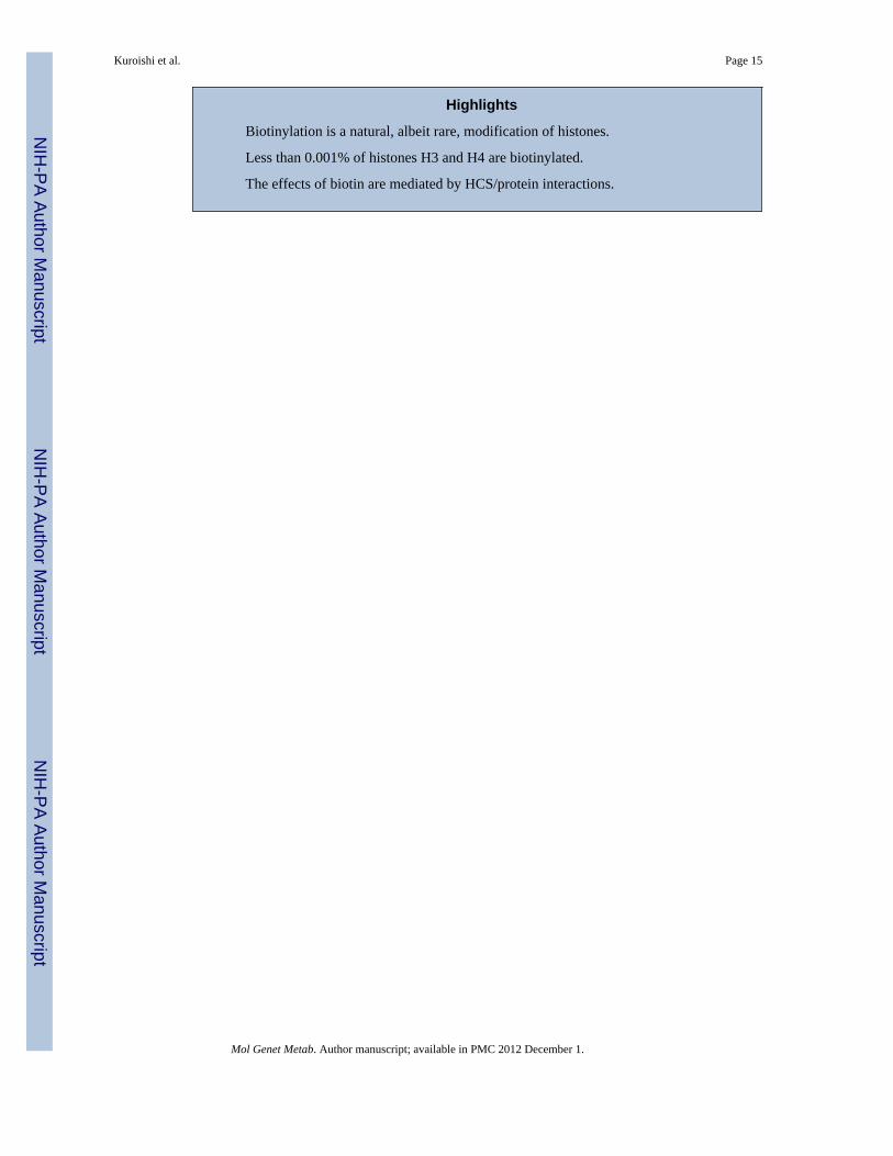
Biotinylation is a natural, albeit rare, modification of histones.
Less than 0.001% of histones H3 and H4 are biotinylated.
The effects of biotin are mediated by HCS/protein interactions.
Kuroishi et al. Page 15
Mol Genet Metab. Author manuscript; available in PMC 2012 December 1.
NIH
-PA Author Manuscript
NIH
-PA Author Manuscript
NIH
-PA Author Manuscript
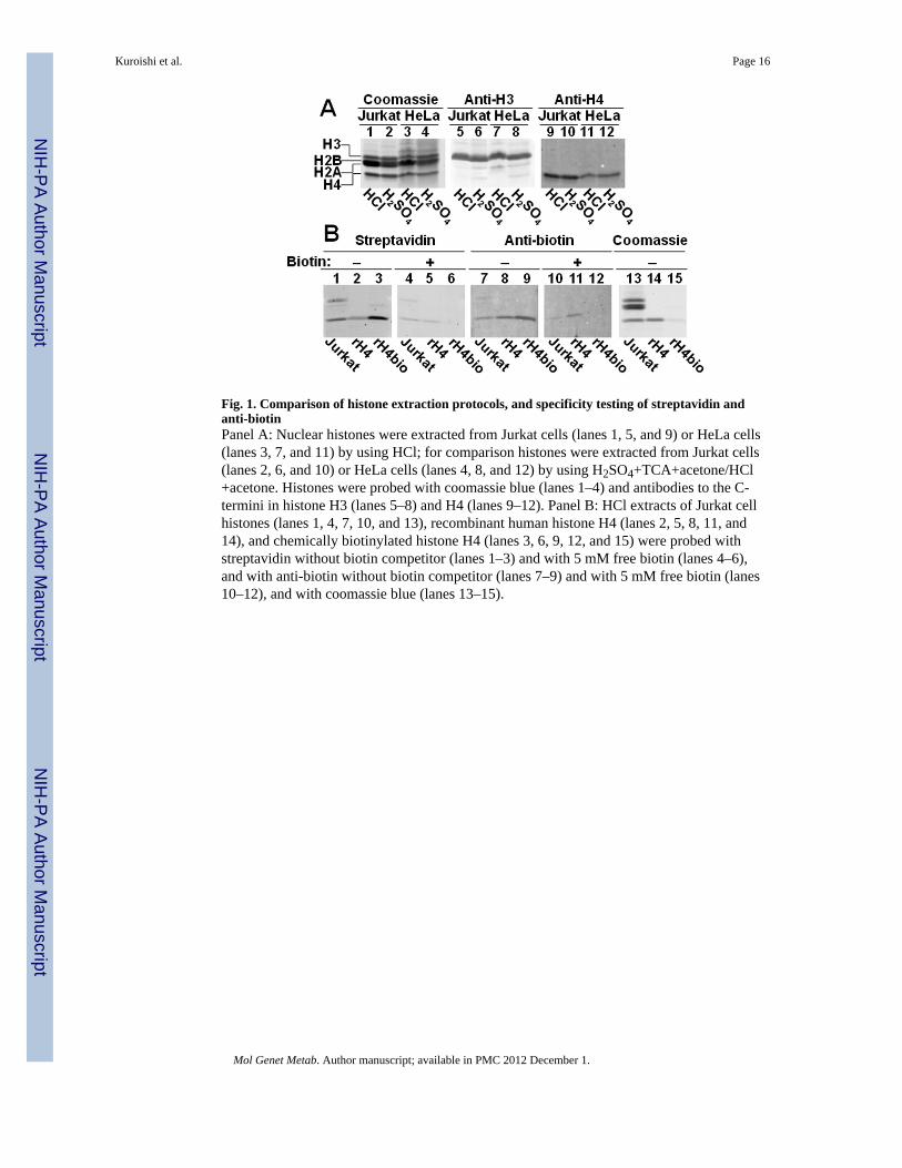
Fig. 1. Comparison of histone extraction protocols, and specificity testing of streptavidin andanti-biotinPanel A: Nuclear histones were extracted from Jurkat cells (lanes 1, 5, and 9) or HeLa cells(lanes 3, 7, and 11) by using HCl; for comparison histones were extracted from Jurkat cells(lanes 2, 6, and 10) or HeLa cells (lanes 4, 8, and 12) by using H2SO4+TCA+acetone/HCl+acetone. Histones were probed with coomassie blue (lanes 1–4) and antibodies to the C-termini in histone H3 (lanes 5–8) and H4 (lanes 9–12). Panel B: HCl extracts of Jurkat cellhistones (lanes 1, 4, 7, 10, and 13), recombinant human histone H4 (lanes 2, 5, 8, 11, and14), and chemically biotinylated histone H4 (lanes 3, 6, 9, 12, and 15) were probed withstreptavidin without biotin competitor (lanes 1–3) and with 5 mM free biotin (lanes 4–6),and with anti-biotin without biotin competitor (lanes 7–9) and with 5 mM free biotin (lanes10–12), and with coomassie blue (lanes 13–15).
Kuroishi et al. Page 16
Mol Genet Metab. Author manuscript; available in PMC 2012 December 1.
NIH
-PA Author Manuscript
NIH
-PA Author Manuscript
NIH
-PA Author Manuscript
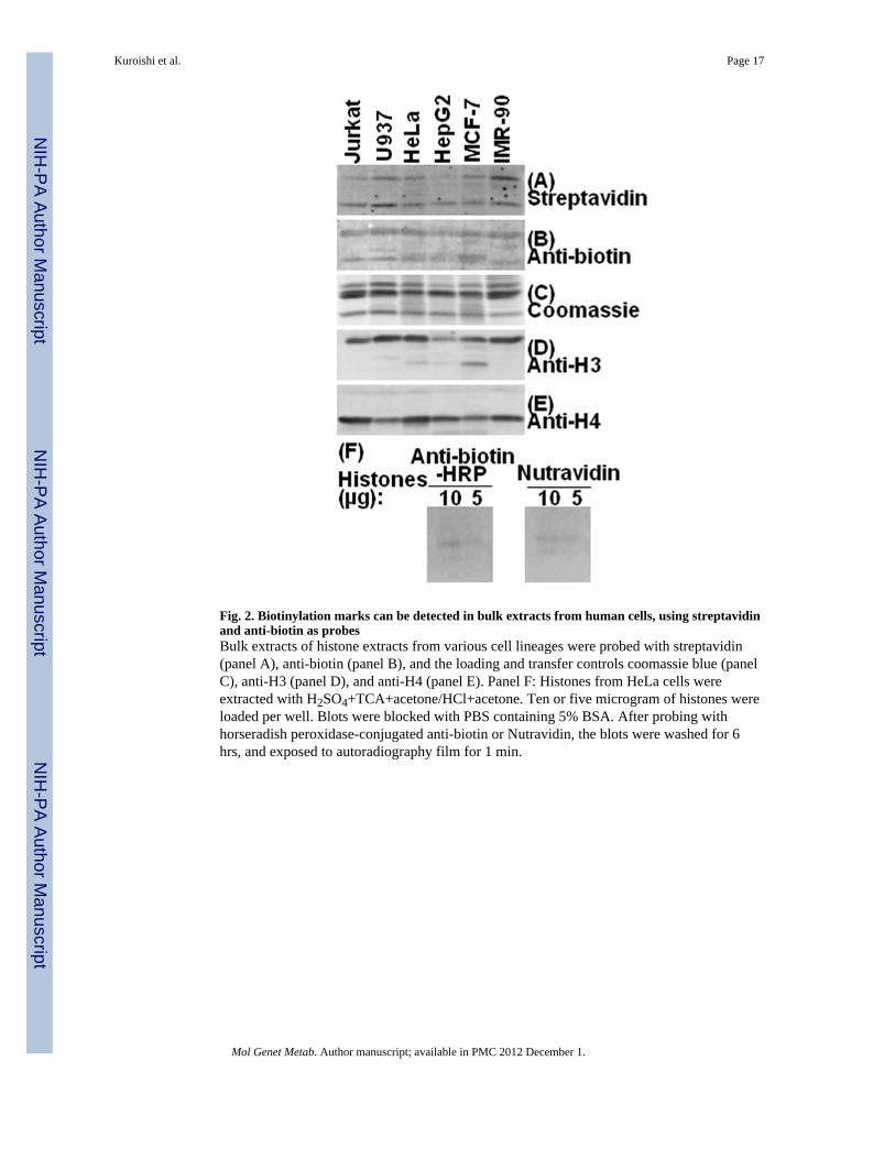
Fig. 2. Biotinylation marks can be detected in bulk extracts from human cells, using streptavidinand anti-biotin as probesBulk extracts of histone extracts from various cell lineages were probed with streptavidin(panel A), anti-biotin (panel B), and the loading and transfer controls coomassie blue (panelC), anti-H3 (panel D), and anti-H4 (panel E). Panel F: Histones from HeLa cells wereextracted with H2SO4+TCA+acetone/HCl+acetone. Ten or five microgram of histones wereloaded per well. Blots were blocked with PBS containing 5% BSA. After probing withhorseradish peroxidase-conjugated anti-biotin or Nutravidin, the blots were washed for 6hrs, and exposed to autoradiography film for 1 min.
Kuroishi et al. Page 17
Mol Genet Metab. Author manuscript; available in PMC 2012 December 1.
NIH
-PA Author Manuscript
NIH
-PA Author Manuscript
NIH
-PA Author Manuscript
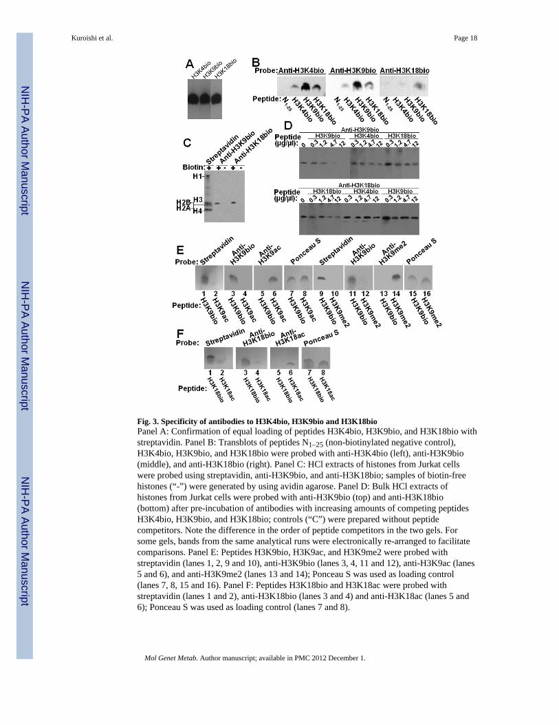
Fig. 3. Specificity of antibodies to H3K4bio, H3K9bio and H3K18bioPanel A: Confirmation of equal loading of peptides H3K4bio, H3K9bio, and H3K18bio withstreptavidin. Panel B: Transblots of peptides N1–25 (non-biotinylated negative control),H3K4bio, H3K9bio, and H3K18bio were probed with anti-H3K4bio (left), anti-H3K9bio(middle), and anti-H3K18bio (right). Panel C: HCl extracts of histones from Jurkat cellswere probed using streptavidin, anti-H3K9bio, and anti-H3K18bio; samples of biotin-freehistones (“-”) were generated by using avidin agarose. Panel D: Bulk HCl extracts ofhistones from Jurkat cells were probed with anti-H3K9bio (top) and anti-H3K18bio(bottom) after pre-incubation of antibodies with increasing amounts of competing peptidesH3K4bio, H3K9bio, and H3K18bio; controls (“C”) were prepared without peptidecompetitors. Note the difference in the order of peptide competitors in the two gels. Forsome gels, bands from the same analytical runs were electronically re-arranged to facilitatecomparisons. Panel E: Peptides H3K9bio, H3K9ac, and H3K9me2 were probed withstreptavidin (lanes 1, 2, 9 and 10), anti-H3K9bio (lanes 3, 4, 11 and 12), anti-H3K9ac (lanes5 and 6), and anti-H3K9me2 (lanes 13 and 14); Ponceau S was used as loading control(lanes 7, 8, 15 and 16). Panel F: Peptides H3K18bio and H3K18ac were probed withstreptavidin (lanes 1 and 2), anti-H3K18bio (lanes 3 and 4) and anti-H3K18ac (lanes 5 and6); Ponceau S was used as loading control (lanes 7 and 8).
Kuroishi et al. Page 18
Mol Genet Metab. Author manuscript; available in PMC 2012 December 1.
NIH
-PA Author Manuscript
NIH
-PA Author Manuscript
NIH
-PA Author Manuscript
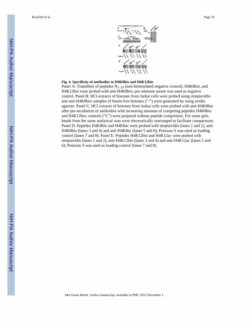
Fig. 4. Specificity of antibodies to H4K8bio and H4K12bioPanel A: Transblots of peptides N1–19 (non-biotinylated negative control), H4K8bio, andH4K12bio were probed with anti-H4K8bio; pre-immune serum was used as negativecontrol. Panel B: HCl extracts of histones from Jurkat cells were probed using streptavidinand anti-H4K8bio; samples of biotin-free histones (“-”) were generated by using avidinagarose. Panel C: HCl extracts of histones from Jurkat cells were probed with anti-H4K8bioafter pre-incubation of antibodies with increasing amounts of competing peptides H4K8bioand H4K12bio; controls (“C”) were prepared without peptide competitors. For some gels,bands from the same analytical runs were electronically rearranged to facilitate comparisons.Panel D: Peptides H4K8bio and H4K8ac were probed with streptavidin (lanes 1 and 2), anti-H4K8bio (lanes 3 and 4) and anti-H4K8ac (lanes 5 and 6); Ponceau S was used as loadingcontrol (lanes 7 and 8). Panel E: Peptides H4K12bio and H4K12ac were probed withstreptavidin (lanes 1 and 2), anti-H4K12bio (lanes 3 and 4) and anti-H4K12ac (lanes 5 and6); Ponceau S was used as loading control (lanes 7 and 8).
Kuroishi et al. Page 19
Mol Genet Metab. Author manuscript; available in PMC 2012 December 1.
NIH
-PA Author Manuscript
NIH
-PA Author Manuscript
NIH
-PA Author Manuscript
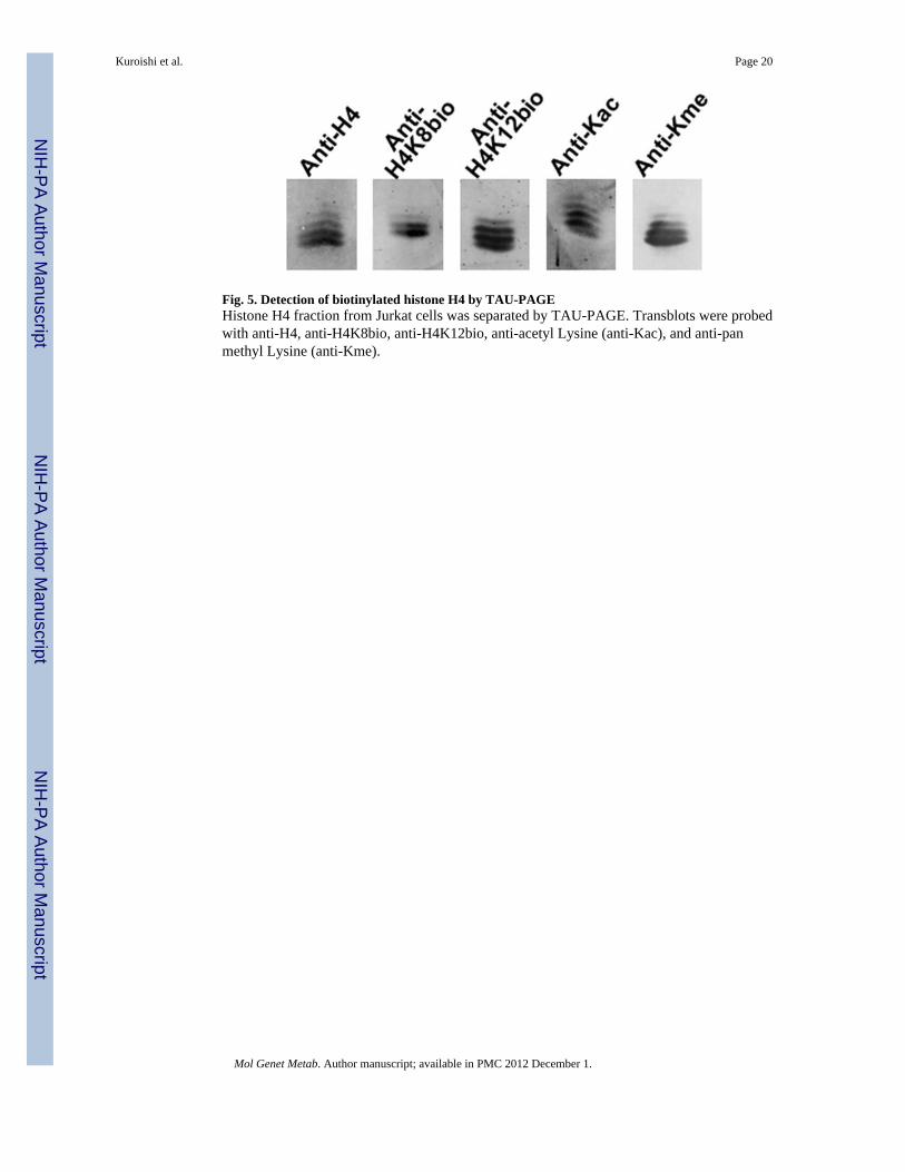
Fig. 5. Detection of biotinylated histone H4 by TAU-PAGEHistone H4 fraction from Jurkat cells was separated by TAU-PAGE. Transblots were probedwith anti-H4, anti-H4K8bio, anti-H4K12bio, anti-acetyl Lysine (anti-Kac), and anti-panmethyl Lysine (anti-Kme).
Kuroishi et al. Page 20
Mol Genet Metab. Author manuscript; available in PMC 2012 December 1.
NIH
-PA Author Manuscript
NIH
-PA Author Manuscript
NIH
-PA Author Manuscript
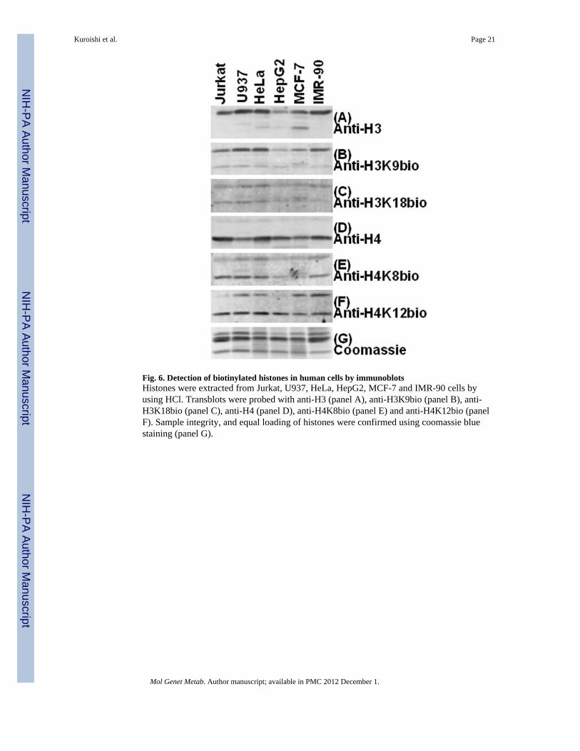
Fig. 6. Detection of biotinylated histones in human cells by immunoblotsHistones were extracted from Jurkat, U937, HeLa, HepG2, MCF-7 and IMR-90 cells byusing HCl. Transblots were probed with anti-H3 (panel A), anti-H3K9bio (panel B), anti-H3K18bio (panel C), anti-H4 (panel D), anti-H4K8bio (panel E) and anti-H4K12bio (panelF). Sample integrity, and equal loading of histones were confirmed using coomassie bluestaining (panel G).
Kuroishi et al. Page 21
Mol Genet Metab. Author manuscript; available in PMC 2012 December 1.
NIH
-PA Author Manuscript
NIH
-PA Author Manuscript
NIH
-PA Author Manuscript
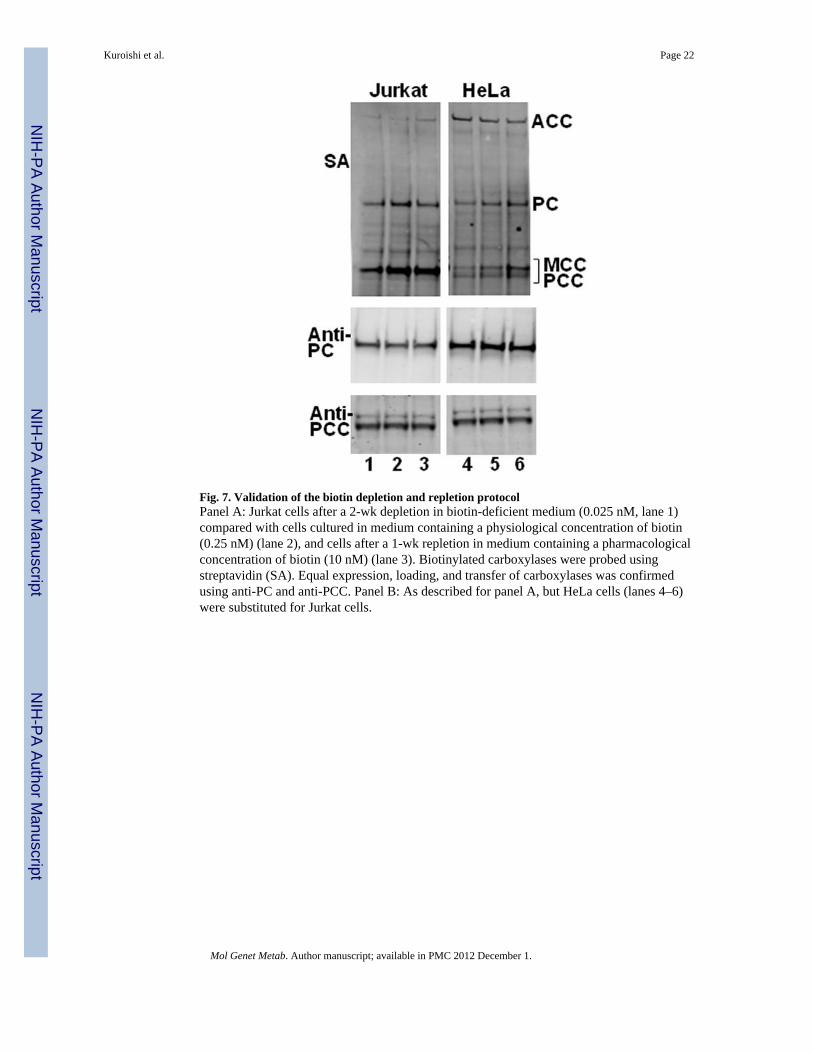
Fig. 7. Validation of the biotin depletion and repletion protocolPanel A: Jurkat cells after a 2-wk depletion in biotin-deficient medium (0.025 nM, lane 1)compared with cells cultured in medium containing a physiological concentration of biotin(0.25 nM) (lane 2), and cells after a 1-wk repletion in medium containing a pharmacologicalconcentration of biotin (10 nM) (lane 3). Biotinylated carboxylases were probed usingstreptavidin (SA). Equal expression, loading, and transfer of carboxylases was confirmedusing anti-PC and anti-PCC. Panel B: As described for panel A, but HeLa cells (lanes 4–6)were substituted for Jurkat cells.
Kuroishi et al. Page 22
Mol Genet Metab. Author manuscript; available in PMC 2012 December 1.
NIH
-PA Author Manuscript
NIH
-PA Author Manuscript
NIH
-PA Author Manuscript
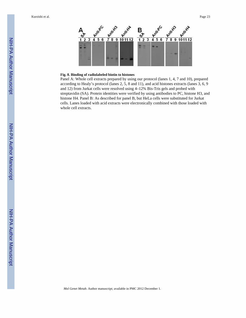
Fig. 8. Binding of radiolabeled biotin to histonesPanel A: Whole cell extracts prepared by using our protocol (lanes 1, 4, 7 and 10), preparedaccording to Healy’s protocol (lanes 2, 5, 8 and 11), and acid histones extracts (lanes 3, 6, 9and 12) from Jurkat cells were resolved using 4–12% Bis-Tris gels and probed withstreptavidin (SA). Protein identities were verified by using antibodies to PC, histone H3, andhistone H4. Panel B: As described for panel B, but HeLa cells were substituted for Jurkatcells. Lanes loaded with acid extracts were electronically combined with those loaded withwhole cell extracts.
Kuroishi et al. Page 23
Mol Genet Metab. Author manuscript; available in PMC 2012 December 1.
NIH
-PA Author Manuscript
NIH
-PA Author Manuscript
NIH
-PA Author Manuscript