biology 1 cover - cga technologies
TRANSCRIPT
S o u t h S u d a n
Student’s Book
All the courses in this secondary series were developed by the Ministry of General Education and Instruction, Republic of South Sudan. The books have been designed to meet the secondary school syllabus, and at the same time equiping the students with skills to fit in the modernday global society.
Each year comprises of a Student’s Book and Teacher’s Guide
The Student’s Books provide: Full coverage of the national syllabus. A strong grounding in the basics of Biology. Clear presentation and explanation of learning points. A wide variety of practice exercises, often showing how Biology can be applied to
real-life situations.It provides opportunities for collaboration through group work activities.Stimulating illustrations.
S o u t h S u d a n
Student’s Book
SecondarySecondarySecondary
Funded by:This Book is the Property of the Ministry of General Education and Instruction.
This Book is not for sale.Any book found on sale, either in print or electronicform, will be confiscated and the seller prosecuted.
This Book is the Property of the Ministry of General Education and Instruction.This Book is not for sale.
Funded by:
BiologyBiology1 BiologyBiology1
Secondary Biology has been written and developed by Ministry of General Education and Instruction, Government of South Sudan in conjunction with Subjects experts. This course book provides a fun and practical approach to the subject of Biology, and at the same time imparting life long skills to the students.
The book comprehensively covers the Secondary 1 syllabus as developed by Ministry of General Education and Instruction.
Do’s 1. 2. 3. 4. 5. 6.
7.
How to take care of your books.
Please cover with plastic or paper. (old newspaper or magazines) Please make sure you have clean hands before you use your book. Always use a book marker do not fold the pages. If the book is damaged please repair it as quickly as possible. Be careful who you lend your schoolbook to. Please keep the book in a dry place. When you lose your book please report it immediately to your teache
Don’ts 1. Do not write on the book cover or inside pages. 2. Do not cut pictures out of the book. 3. Do not tear pages out of the book. 4. Do not leave the book open and face down. 5. Do not use pens, pencils or something thick as a book mark. 6. Do not force your book into your schoolbag when it is full. 7. Do not use your book as an umbrella for the sun or rain. 8. Do not use your book as a seat.
South Sudan
This book is the property of the Ministry of General Education and Instruction.
THIS BOOK IS NOT FOR SALE
Funded by:
BiologyStudent’s Book 1
1SECONDARY
© 2018, THE REPUBLIC OF SOUTH SUDAN, MINISTRY OF GENERAL EDUCATION AND INSTRUCTION. All rights reserved. No part of this book may be reproduced by any means graphic, electronic, mechanical, photocopying, taping, storage and retrieval system without prior written permission of the Copyright Holder.Pictures, illustrations and links to third party websites are provided in good faith, for information and education purposes only
ii
. FOREWORD
I am delighted to present to you this textbook, which is developed by the Ministry of General Education and Instruction based on the new South Sudan National Curriculum. The National Curriculum is a learner-centered curriculum that aims to meet the needs and aspirations of the new nation. In particular, it aims to develop (a) Good citizens; (b) successful lifelong learners; (c) creative, active and productive individuals; and (d) Environmentally responsible members of our society. This textbook, like many others, has been designed to contribute to achievement of these noble aims. It has been revised thoroughly by our Subject Panels, is deemed to be fit for the purpose and has been recommended to me for approval. Therefore, I hereby grant my approval. This textbook shall be used to facilitate learning for learners in all schools of the Republic of South Sudan, except international schools, with effect from 4th February, 2019.
I am deeply grateful to the staff of the Ministry of General Education and Instruction, especially Mr Michael Lopuke Lotyam Longolio, the Undersecretary of the Ministry, the staff of the Curriculum Development Centre, under the supervision of Mr Omot Okony Olok, the Director General for Quality Assurance and Standards, the Subject Panelists, the Curriculum Foundation (UK), under the able leadership of Dr Brian Male, for providing professional guidance throughout the process of the development of National Curriculum and school textbooks for the Republic of South Sudan since 2013. I wish to thank UNICEF South Sudan for managing the project funded by the Global Partnership in Education so well and funding the development of the National Curriculum and the new textbooks. I am equally grateful for the support provided by Mr Tony Calderbank, the former Country Director of the British Council, South Sudan; Sir Richard Arden, Senior Education Advisor of DfID, South Sudan. I thank Longhorn and Mountain Top publishers in Kenya for working closely with the Ministry, the Subject Panels, UNICEF and the Curriculum Foundation UK to write the new textbooks. Finally, I thank the former Ministers of Education, Hon. Joseph Ukel Abango and Hon. Dr John Gai Nyuot Yoh, for supporting me, in my previous role as the Undersecretary of the Ministry, to lead the Technical Committee to develop and complete the consultations on the new National Curriculum Framework by 29 November 2013.
The Ministry of General Education and Instruction, Republic of South Sudan, is most grateful to all these key stakeholders for their overwhelming support to the design and development of this historic South Sudan National Curriculum. This historic reform in South Sudan’s education system is intended to benefit the people of South Sudan, especially the children and youth and the future generations. It shall enhance the quality of education in the country to promote peace, justice, liberty and prosperity for all. I urge all Teachers to put this textbook to good use.
May God bless South Sudan. May He help our Teachers to inspire, educate and transform the lives of all the children and youth of South Sudan.
Deng Deng Hoc Yai, (Hon.)
Minister of General Education and Instruction, Republic of South Sudan
iii
Table of Contents
Unit 1: Diversity of Living Things ......................................................... 1
1.1 Definition of Biology and its branches ........................................2
1.2 Importance of studying Biology ....................................................3
1.3 Characteristics of living things .......................................................5
1.4 Various life forms of organisms ......................................................7
1.5 Classification of organisms and its importance ...................... 10
1.6 Taxonomy hierachy of classification ..................................................13
1.7 The binomial system ................................................................................16
1.8 Dichotomous key ............................................................................ 19
Unit 2: The Cell .................................................................................... 25
2.1 Definition of the cell ...................................................................... 26
2.2 Magnifying instruments ................................................................ 27
2.3 Parts and functions of a light microscope ................................ 29
2.4 Structure of plant and animal cells............................................. 38
2.5 Functions of parts of plant cell and animal cell ...................... 42
2.6 Specialised plant cells ..................................................................... 48
2.7 Levels of organisation in multicellular organisms .................. 53
2.8 Organ and organ systems ............................................................ 55
Unit 3: Movement of substances into and out of cells ..................... 63
3.1 Structure and properties of the cell membrane ...................... 63
3.2 Diffusion ............................................................................................ 64
3.3 Osmosis ............................................................................................. 69
3.4 Solute, Solution and Solvents ....................................................... 70
3.5 Water relations in plants and animal cells .............................. 74
3.6 Active transport .............................................................................. 80
Glossary ........................................................................................................................... 86
Appendix I .......................................................................................................................... 91
Appendix II ......................................................................................................................... 93

iv
1
1Unit Diversity of Living Things
Learning outcomes
Knowledge and understanding
Skills Attitudes
• Understand the diversity of living things.
• Investigate how living things can be grouped according to their similarities and differences.
• Identify different kinds of organisms using taxonomic keys.
• Collect insects using nets (sweep net), jam jar / sunk into soil, pooters and plankton net.
• Develop simple keys to classify and investigate living things according to their similarities (Dichotomous key).
• Critical observation and recording skills.
• Appreciate the variety of organisms.
• Show curiosity about the existence of organisms and the importance of sustaining diversity (conservation).
• Thinking critically.
• Sharing views and opinions.
• Cooperating with others.
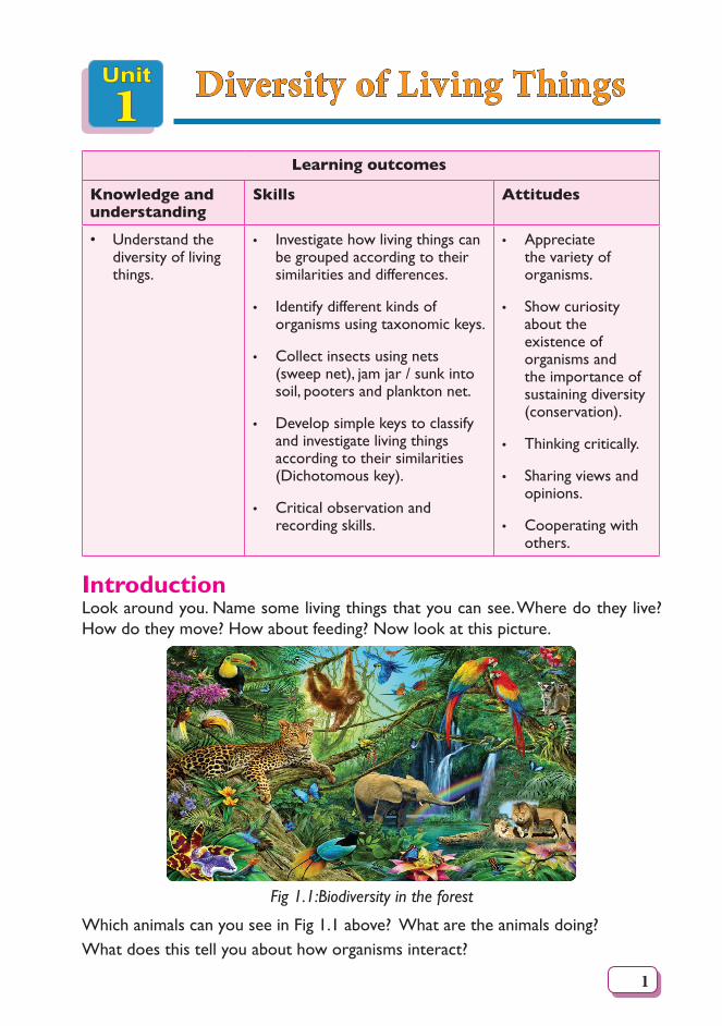
IntroductionLook around you. Name some living things that you can see. Where do they live? How do they move? How about feeding? Now look at this picture.
Fig 1.1:Biodiversity in the forest
Which animals can you see in Fig 1.1 above? What are the animals doing?What does this tell you about how organisms interact?
2
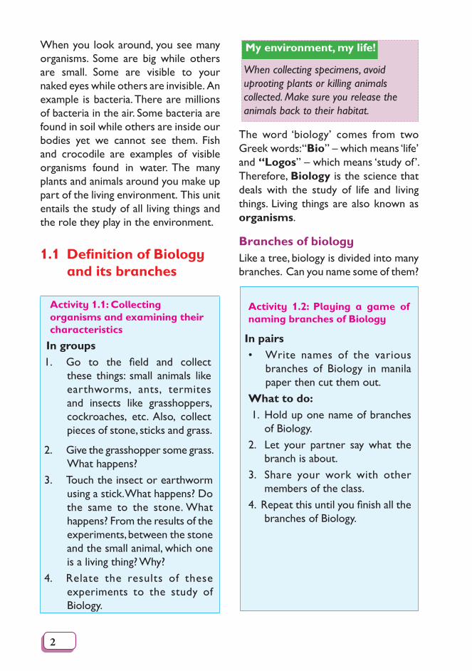
When you look around, you see many organisms. Some are big while others are small. Some are visible to your naked eyes while others are invisible. An example is bacteria. There are millions of bacteria in the air. Some bacteria are found in soil while others are inside our bodies yet we cannot see them. Fish and crocodile are examples of visible organisms found in water. The many plants and animals around you make up part of the living environment. This unit entails the study of all living things and the role they play in the environment.
1.1 Definition of Biology and its branches
Activity 1.1: Collecting organisms and examining their characteristics
In groups1. Go to the field and collect
these things: small animals like earthworms, ants, termites and insects like grasshoppers, cockroaches, etc. Also, collect pieces of stone, sticks and grass.
2. Give the grasshopper some grass. What happens?
3. Touch the insect or earthworm using a stick. What happens? Do the same to the stone. What happens? From the results of the experiments, between the stone and the small animal, which one is a living thing? Why?
4. Relate the results of these experiments to the study of Biology.
My environment, my life!
When collecting specimens, avoid uprooting plants or killing animals collected. Make sure you release the animals back to their habitat.
The word ‘biology’ comes from two Greek words: “Bio” – which means ‘life’ and “Logos” – which means ‘study of’. Therefore, Biology is the science that deals with the study of life and living things. Living things are also known as organisms.
Branches of biologyLike a tree, biology is divided into many branches. Can you name some of them?
Activity 1.2: Playing a game of naming branches of Biology
In pairs• Write names of the various
branches of Biology in manila paper then cut them out.
What to do: 1. Hold up one name of branches
of Biology.2. Let your partner say what the
branch is about.3. Share your work with other
members of the class.4.Repeatthisuntilyoufinishallthe
branches of Biology.
3
Th e facts
The two main branches of Biology are:• Zoology - the study of animals.• Botany - the study of plants. Other common branches of Biology
include:a) Genetics- the study of genes and
inheritance.b) Ecology - the study of the
interaction of organisms with each other and with their environment.
c) Anatomy - the study of organisms and their structure.
d) Microbiology - the study of micro-organisms.
e) Physiology - the study of how cells function.
f) Biochemistry – the study of the chemical processes that take place in the body of a living thing.
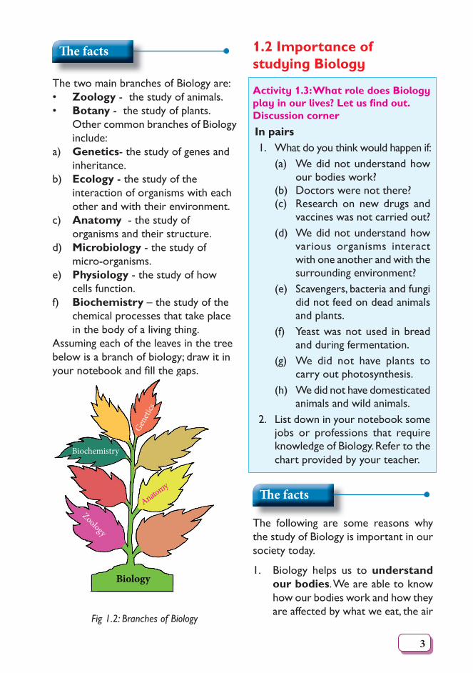
Assuming each of the leaves in the tree below is a branch of biology; draw it in yournotebookandfillthegaps.
Anatomy
Geneti
cs
Biochemistry
Zoology
Biology
Fig 1.2: Branches of Biology
1.2 Importance of studying Biology
Activity 1.3: What role does Biology play in our lives? Let us fi nd out.Discussion corner
In pairs1. What do you think would happen if:
(a) We did not understand how our bodies work?
(b) Doctors were not there?(c) Research on new drugs and
vaccines was not carried out?(d) We did not understand how
various organisms interact with one another and with the surrounding environment?
(e) Scavengers, bacteria and fungi did not feed on dead animals and plants.
(f) Yeast was not used in bread and during fermentation.
(g) We did not have plants to carry out photosynthesis.
(h) We did not have domesticated animals and wild animals.
2. List down in your notebook some jobs or professions that require knowledge of Biology. Refer to the chart provided by your teacher.
Th e facts
The following are some reasons why the study of Biology is important in our society today.
1. Biology helps us to understand our bodies. We are able to know how our bodies work and how they are affected by what we eat, the air
4
we breathe, and our surrounding environment. This can help prevent, cure and even eliminate diseases.
2. The study of Biology helps us in treating and preventing diseases.
Here, research is done to invent new drugs and to even come up with better vaccines.
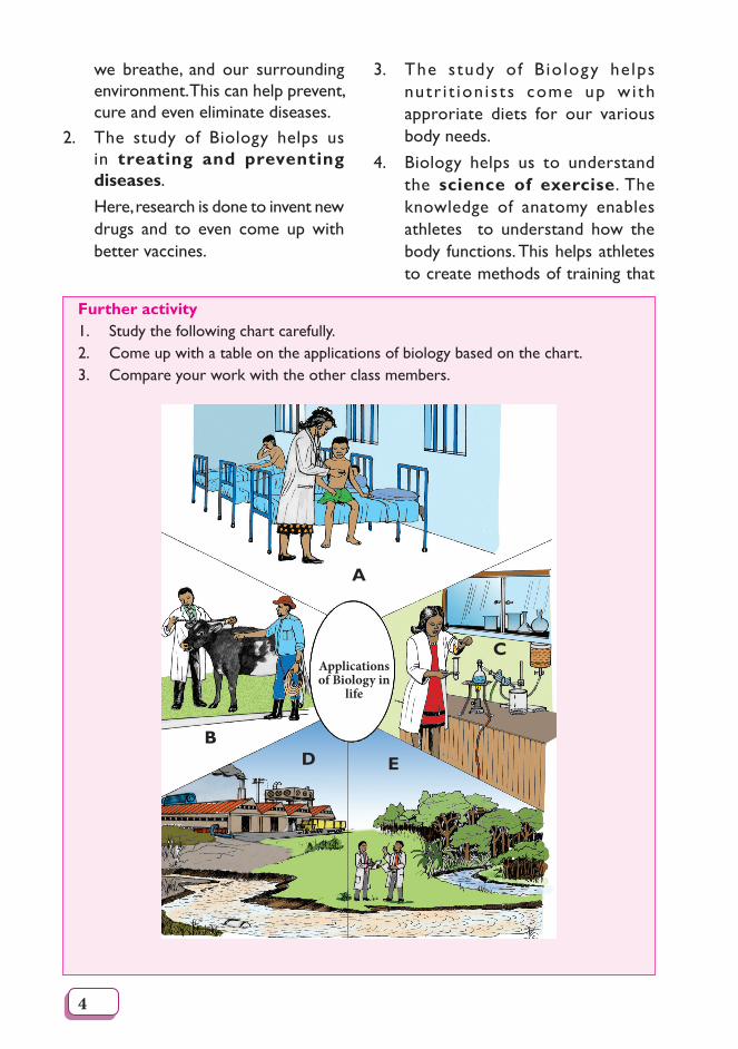
3. The study of B io logy he lps nutr i t ion i s ts come up wi th approriate diets for our various body needs.
4. Biology helps us to understand the science of exercise. The knowledge of anatomy enables athletes to understand how the body functions. This helps athletes to create methods of training that
A
CApplications of Biology in
life
D EB
Further activity 1. Study the following chart carefully.2. Come up with a table on the applications of biology based on the chart.3. Compare your work with the other class members.
5
enable them to become more proficient.
5. Biology helps us to understand our environment. Through Biology, we are able to appreciate the wide range of organisms around us. These organisms affect us and we affect them as well. This knowledge helps us to realise the need to conserve the environment.
6. As a subject, Biology guides us on the best farming practices and thereby ensuring food security. Through biotechnology, we are able to develop high yielding crops and animals. Disease and drought resistant plants and animals are also developed.
7. Biology is a career subject. Some careers linked to Biology include medicine, pharmacy and veterinary among others. With these careers, we are able to earn a living and take care of our families.
My environment, my life!
We should always strive to conserve the environment in whatever we do. We should avoid polluting the environment, destroying plants and killing animals!
1.3 Characteristics of living things
What would you look out for if you wanted to distinguish between things that are living and things that are not living?
Activity 1.4
In groups1. Gotothefieldforanaturewalk.2. Collect a few things from the
environment. Such things may include stones, pieces of wood, insects such as grasshoppers, butterflies, ants, termites and branches of plants, leaves or roots.
3. Bring the things you have collected to class.
4. Observe them in detail using a hand lens noting the presence of:a) Breathing structures.b) Movement structures such as
legs, wings, among others.c) Feeding structures, such as
mouth parts among others.5. Discuss with your partner, how the
animals use the above structures for.
6. How about the things that do not have these structures? How do they survive? Are they living?
My environment, my life!
When collecting specimen, avoid uprooting whole plants or killing the animals collected. Always use a pair of forceps when handling stinging insects.
Th e facts
For an organism to be described as living, it must be able to carry out some processes, which are essential for life. The processes constitute the
6
characteristics of living things. They include:
1. Movement - this is the ability of organisms to change position of the whole body (like in animals) or even parts of the body (like in plants) where leaves or branches that move.
2. Reproduction - this is the ability of organisms to make new individuals of their kind. It can be through sexual or asexual means.
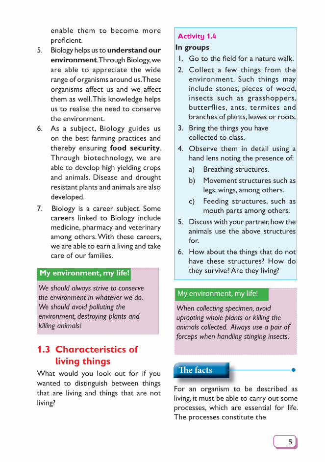
3. Growth - this is the irreversible increase in size and dry mass of a living organism. Growth can occur in three forms: cells of the organism can become bigger, they can increase their number or both.
Fig 1.3 Growth of a chick
Note: Plants grow throughout their lives while animals stop growing at some point.
4. Sensitivity (or Irritability) - this is the ability of an organism to detect or sense changes in its surrounding environment and then respond to them. These changes are known as stimuli (singular – stimulus). For example, plants respond to sunlight by growing its leaves towards it. Animals respond to touch, sound and chemicals among others.
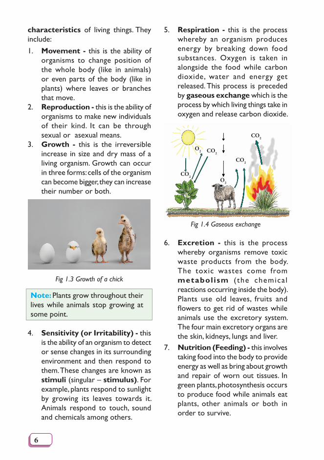
5. Respiration - this is the process whereby an organism produces energy by breaking down food substances. Oxygen is taken in alongside the food while carbon dioxide, water and energy get released. This process is preceded by gaseous exchange which is the process by which living things take in oxygen and release carbon dioxide.
CO2
CO2
CO2
CO2
O2
O2
Fig 1.4 Gaseous exchange
6. Excretion - this is the process whereby organisms remove toxic waste products from the body. The toxic wastes come from metabol ism ( the chemica l reactions occurring inside the body). Plants use old leaves, fruits and flowerstogetridofwasteswhileanimals use the excretory system. The four main excretory organs are the skin, kidneys, lungs and liver.
7. Nutrition (Feeding) - this involves taking food into the body to provide energy as well as bring about growth and repair of worn out tissues. In green plants, photosynthesis occurs to produce food while animals eat plants, other animals or both in order to survive.
7
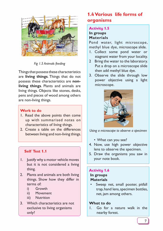
Fig 1.5 Animals feeding
Things that possess these characteristics are living things. Things that do not possess these characteristics are non-living things. Plants and animals are living things. Objects like stones, desks, pens and pieces of wood among others are non-living things.
Work to do1. Read the above points then come
up with summarised notes on characteristics of living things.
2. Create a table on the differences between living and non-living things.
Self Test 1.1
1. Justify why a motor vehicle moves but it is not considered a living thing.
2. Plants and animals are both living things. Show how they differ in terms of:i) Growth ii) Movement iii) Nutrition
3. Which characteristics are not exclusive to living organisms only?
1.4 Various life forms of organisms
Activity 1.5In groupsMaterialsPond water, light microscope, methyl blue dye, microscope slide.1. Collect some pond water or
stagnant water from your locality.2 Bring the water to the laboratory.
Put a drop on a microscope slide then add methyl blue dye.
3. Observe the slide through low power objective using a light microscope.
Using a microscope to observe a specimen
• What can you see?4. Now, use high power objective
lens to observe the specimen.5. Draw the organisms you saw in
your note book.
Activity 1.6 In groupsMaterials• Sweep net, small pooter, pitfall
trap, hand lens, specimen bottles, net, jam among others.
What to do1. Go for a nature walk in the
nearby forest.
8
2. During the nature walk, use the pooter and the sweep net to collect various insects such as butterflies, grasshopper, cockroaches, pond snails among others.
3. Use a small pitfall trap to trap insects and other arthropods as these may bite and cause injury or infection.
4. Put all the collected animals in specimen bottles.
5. Observe other big animals in the forest such as zebras, ostriches, giraffes among others.
6. Back in class, observe the specimens you collected using a hand lens as shown below.
Draw the animals in your note book.
Using a hand lens to observe a specimen
7. Also, make a sketch of some of the big animals you observed during the nature walk.
Study Questions1. Can you see any similarities and
differences among the animals you drew? List them down in a table.
2. What does this tell you about living organisms in general?
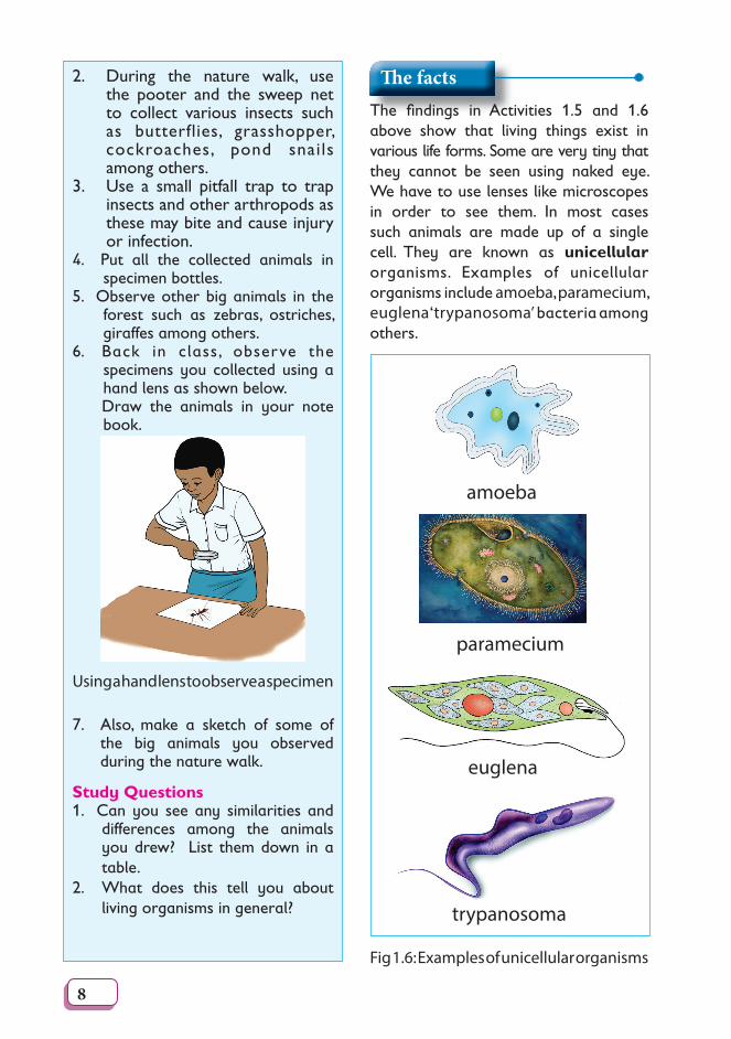
Th e factsThe fi ndings in Activities 1.5 and 1.6 above show that living things exist in various life forms. Some are very tiny that they cannot be seen using naked eye. We have to use lenses like microscopes in order to see them. In most cases such animals are made up of a single cell. They are known as unicellular organisms. Examples of unicellular organisms include amoeba, paramecium, euglena ‘trypanosoma’ bacteria among others.
amoeba
euglena
trypanosoma
paramecium
Fig 1.6: Examples of unicellular organisms
9
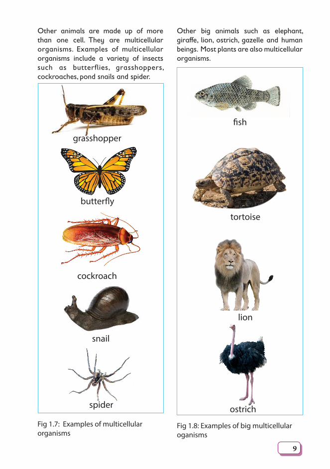
Other animals are made up of more than one cell. They are multicellular organisms. Examples of multicellular organisms include a variety of insects such as butterflies, grasshoppers, cockroaches, pond snails and spider.
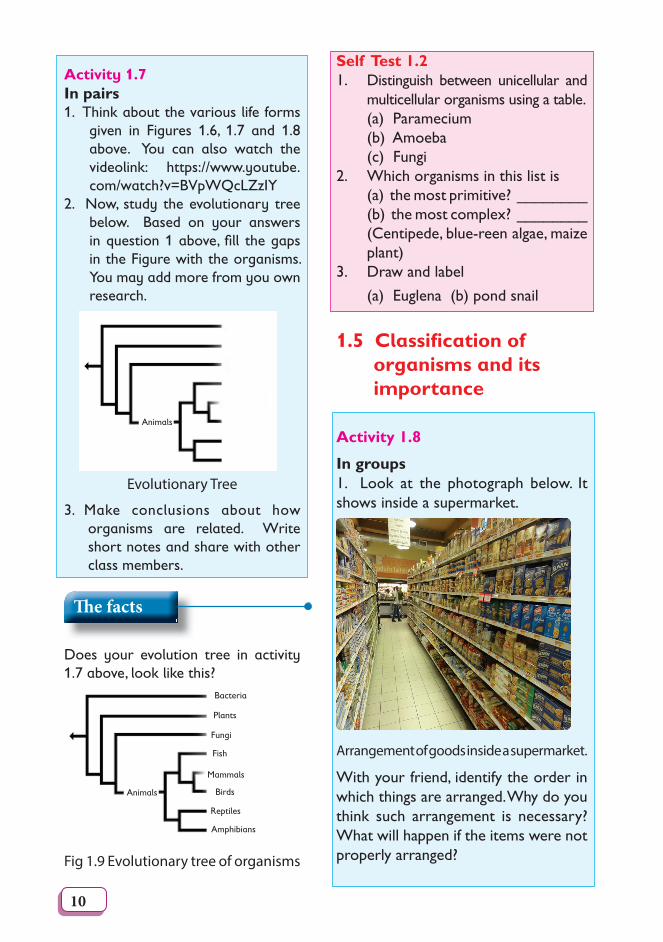
Other big animals such as elephant, giraffe, lion, ostrich, gazelle and human beings. Most plants are also multicellular organisms.
Fig 1.7: Examples of multicellular organisms
grasshopper
butterfl y
cockroach
snail
spider
Fig 1.8: Examples of big multicellular oganisms
fi sh
tortoise
lion
ostrich
10
Activity 1.7In pairs1. Think about the various life forms
given in Figures 1.6, 1.7 and 1.8 above. You can also watch the videolink: https://www.youtube.com/watch?v=BVpWQcLZzIY
2. Now, study the evolutionary tree below. Based on your answers in question 1 above, fi ll the gaps in the Figure with the organisms. You may add more from you own research.
Animals
Evolutionary Tree
3. Make conclusions about how organisms are related. Write short notes and share with other class members.
Th e facts
Does your evolution tree in activity 1.7 above, look like this?
Bacteria
Fungi
Mammals
Animals Birds
Plants
Fish
Reptiles
Amphibians
Fig 1.9 Evolutionary tree of organisms
Self Test 1.21. Distinguish between unicellular and
multicellular organisms using a table. (a) Paramecium (b) Amoeba (c) Fungi2. Which organisms in this list is (a) the most primitive? ________ (b) the most complex? ________ (Centipede, blue-reen algae, maize
plant)3. Draw and label (a) Euglena (b) pond snail
1.5 Classifi cation of organisms and its importance
Activity 1.8
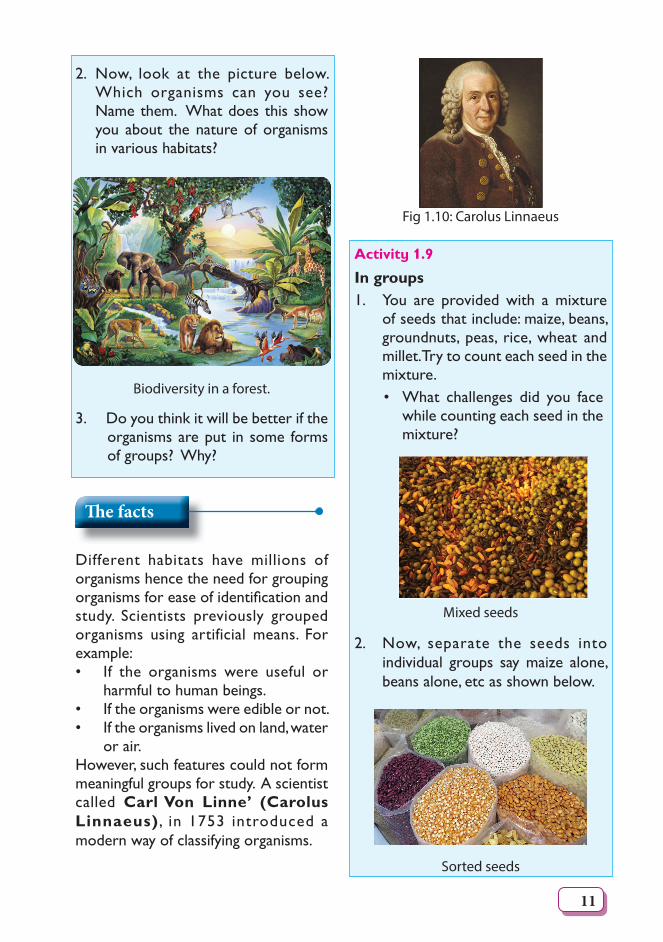
In groups1. Look at the photograph below. It shows inside a supermarket.
Arrangement of goods inside a supermarket.
With your friend, identify the order in which things are arranged. Why do you think such arrangement is necessary? What will happen if the items were not properly arranged?
11
2. Now, look at the picture below. Which organisms can you see? Name them. What does this show you about the nature of organisms in various habitats?
Biodiversity in a forest.
3. Do you think it will be better if the organisms are put in some forms of groups? Why?
Th e facts
Different habitats have millions of organisms hence the need for grouping organismsforeaseofidentificationandstudy. Scientists previously grouped organisms using artificial means. For example:• If the organisms were useful or
harmful to human beings.• If the organisms were edible or not. • If the organisms lived on land, water
or air.However, such features could not form meaningful groups for study. A scientist called Carl Von Linne’ (Carolus Linnaeus) , in 1753 introduced a modern way of classifying organisms.
Fig 1.10: Carolus Linnaeus
Activity 1.9
In groups1. You are provided with a mixture
of seeds that include: maize, beans, groundnuts, peas, rice, wheat and millet. Try to count each seed in the mixture.• What challenges did you face
while counting each seed in the mixture?
Mixed seeds
2. Now, separate the seeds into individual groups say maize alone, beans alone, etc as shown below.
Sorted seeds
12
Provides plenty of useful information
Provides only limited information
Activity 1.10
In groups1. Take a walk outside the classroom
and collect different types of living organisms.Caution! Some organisms can sting! Others can bite! Therefore do not use your bare hands to handle such organisms. Use protective gloves.
2. Take the collected organisms back to class for study.
3. Put the organisms into various groups.
You can use these features; • Number of legs• Presence of wings• Presence of antennae• What covers the body, etc4. Shareyourfindingswiththerestof
the class.
Importance of classifi cation
Th e facts
There are about 1.8 million species of known organisms. It is also believed that there could be many more undiscovered species in the forest ecosystems and in deep seas.
1. Classificationputsinformationinanorganised manner to avoid chaos and confusion among scientists.
2. It enables scientists to place organisms in their correct groups for ease of study.
3. It allows scientists to better understand the phylogenetic relationships among organisms i.e how organisms are interrelated.
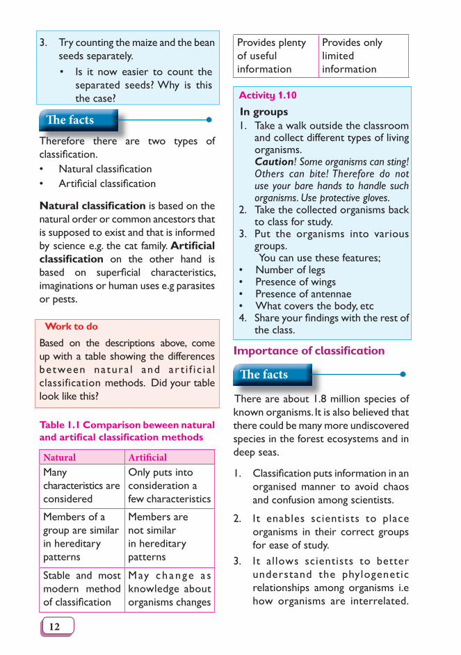
3. Try counting the maize and the bean seeds separately.• Is it now easier to count the
separated seeds? Why is this the case?
Th e factsTherefore there are two types of classification.• Naturalclassification• Artificialclassification
Natural classifi cation is based on the natural order or common ancestors that is supposed to exist and that is informed by science e.g. the cat family. Artifi cial classifi cation on the other hand is based on superficial characteristics,imaginations or human uses e.g parasites or pests.
Work to do
Based on the descriptions above, come up with a table showing the differences between natura l and ar t i f i c i a l classification methods. Did your table look like this?
Table 1.1 Comparison beween natural and artifi cal classifi cation methods
Natural Artifi cial Many characteristics are considered
Only puts into consideration a few characteristics
Members of a group are similar in hereditary patterns
Members are not similar in hereditary patterns
Stable and most modern method ofclassification
May change a s knowledge about organisms changes
13
(Organisms that have more common characteristics are more closely related).
4. Classification allows scientists toidentify, group and properly name a newly discovered organism.
Self Test 1.31. Whatisclassification?2. Distinguish between artificial and
naturalclassification.3. Describe classification as proposed
by carolus Linnaeus.4. State the importance of classifying
organisms.
1.6 Taxonomy hierachy of classifi cation
Activity 1.11: Discussion corner
In groups1. Give the names of the animals
below in your native language. (i) Cow (ii) Cat (iii) Elephant (iv) Lion2. Do you think people from other
parts of the world can recognise the names you gave in 1 above?
3. What impact do you think this will have on the study of living organisms?
Th e facts
Itissometimesdifficulttoidentifylivingthings using local names. This is because of the existence of several local languages. To assist scientists from different parts of the world to communicate, one scientificnameisgiven.• This creates no confusion as to
which organism is being referred to.• Scientificnamesrarelychange.• Scientificnamesarewritteninthe
same language around the world.
My heritage my pride!Our language refl ects who we are as a people. It is our nation’s identity. Always be proud of your language.
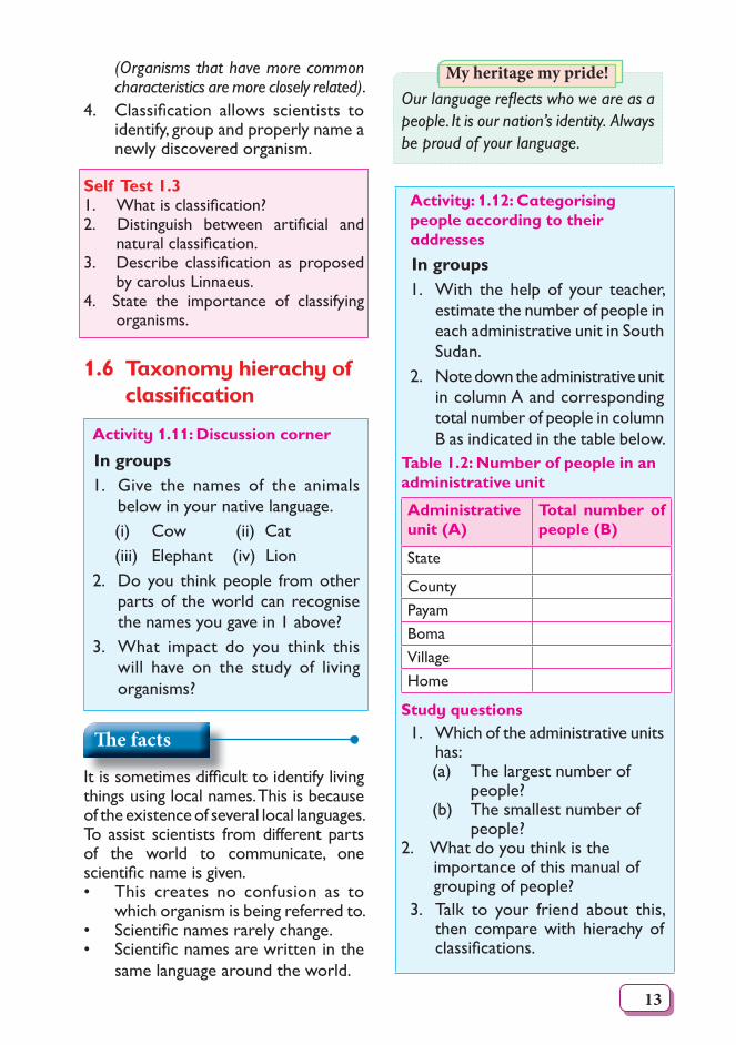
Activity: 1.12: Categorising people according to their addresses
In groups1. With the help of your teacher,
estimate the number of people in each administrative unit in South Sudan.
2. Note down the administrative unit in column A and corresponding total number of people in column B as indicated in the table below.
Table 1.2: Number of people in an administrative unit
Administrative unit (A)
Total number of people (B)
State
CountyPayamBomaVillageHome
Study questions1. Which of the administrative units
has: (a) The largest number of
people? (b) The smallest number of
people?2. What do you think is the
importance of this manual of grouping of people?
3. Talk to your friend about this, then compare with hierachy of classifications.
14
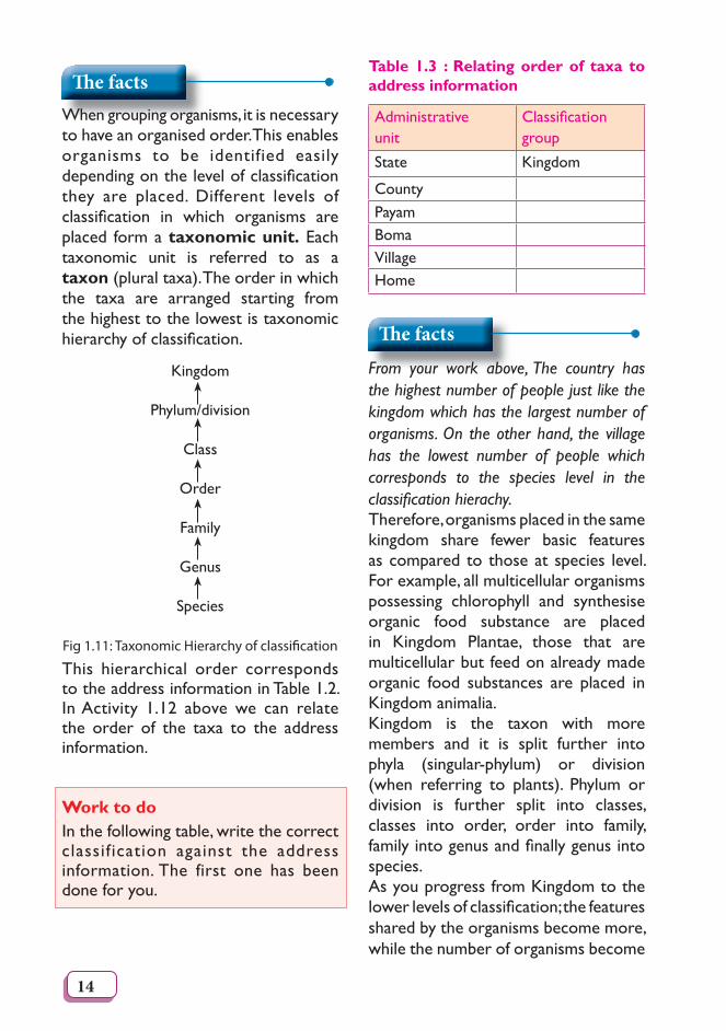
Th e factsWhen grouping organisms, it is necessary to have an organised order. This enables organisms to be identified easily dependingonthelevelofclassificationthey are placed. Different levels of classification in which organisms areplaced form a taxonomic unit. Each taxonomic unit is referred to as a taxon (plural taxa). The order in which the taxa are arranged starting from the highest to the lowest is taxonomic hierarchyofclassification.
Kingdom
Phylum/division
Class
Order
Family
Genus
Species
Fig 1.11: Taxonomic Hierarchy of classifi cation
This hierarchical order corresponds to the address information in Table 1.2. In Activity 1.12 above we can relate the order of the taxa to the address information.
Work to doIn the following table, write the correct classification against the address information. The first one has been done for you.
Table 1.3 : Relating order of taxa to address information
Administrative unit
Classificationgroup
State Kingdom
CountyPayamBomaVillageHome
Th e facts
From your work above, The country has the highest number of people just like the kingdom which has the largest number of organisms. On the other hand, the village has the lowest number of people which corresponds to the species level in the classifi cation hierachy.Therefore, organisms placed in the same kingdom share fewer basic features as compared to those at species level. For example, all multicellular organisms possessing chlorophyll and synthesise organic food substance are placed in Kingdom Plantae, those that are multicellular but feed on already made organic food substances are placed in Kingdom animalia.Kingdom is the taxon with more members and it is split further into phyla (singular-phylum) or division (when referring to plants). Phylum or division is further split into classes, classes into order, order into family, familyintogenusandfinallygenusintospecies.As you progress from Kingdom to the lowerlevelsofclassification;thefeaturesshared by the organisms become more, while the number of organisms become
15
fewer. As such, species being the lowest level of classification comprises of closely related organisms that share many characteristics.A species can therefore be definedas a group of organisms which can naturally interbreed to give rise to a viable offspring.
Activity 1.13: How can organisms be placed into various taxa?
In groupsYour teacher will provide you with the following: • Laboratory rat or rabbit• Housefly,butterfly,grasshopper• Grass, bean plant, maize plant
among others.1. Try to group these organisms into
their respective kingdoms. • Cite the features that you used
to place the above organisms in the various groups.
2. Observe keenly the roots and leaves of the plants. The features of these parts will enable you to place the two plants either in the same group or different groups. In your opinion, do you think grass, maize and bean plants can be placed in the same group or taxa. Explain why. Hint: Consider the types of roots below.Consider the types of roots below.
Grass Bean plant
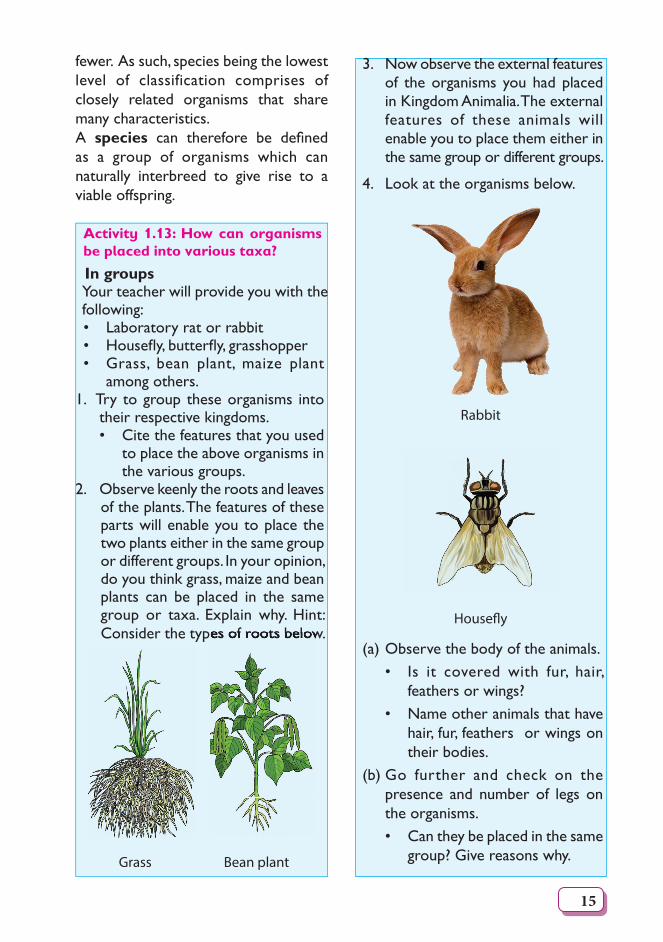
3. Now observe the external features of the organisms you had placed in Kingdom Animalia. The external features of these animals will enable you to place them either in the same group or different groups.
4. Look at the organisms below.
Rabbit
Housefl y
(a) Observe the body of the animals. • Is it covered with fur, hair,
feathers or wings?• Name other animals that have
hair, fur, feathers or wings on their bodies.
(b) Go further and check on the presence and number of legs on the organisms. • Can they be placed in the same
group? Give reasons why.
16
My environment, my life!
Collect only specimen you need. Do not harm the organisms nor pollute the environment.
Th e facts
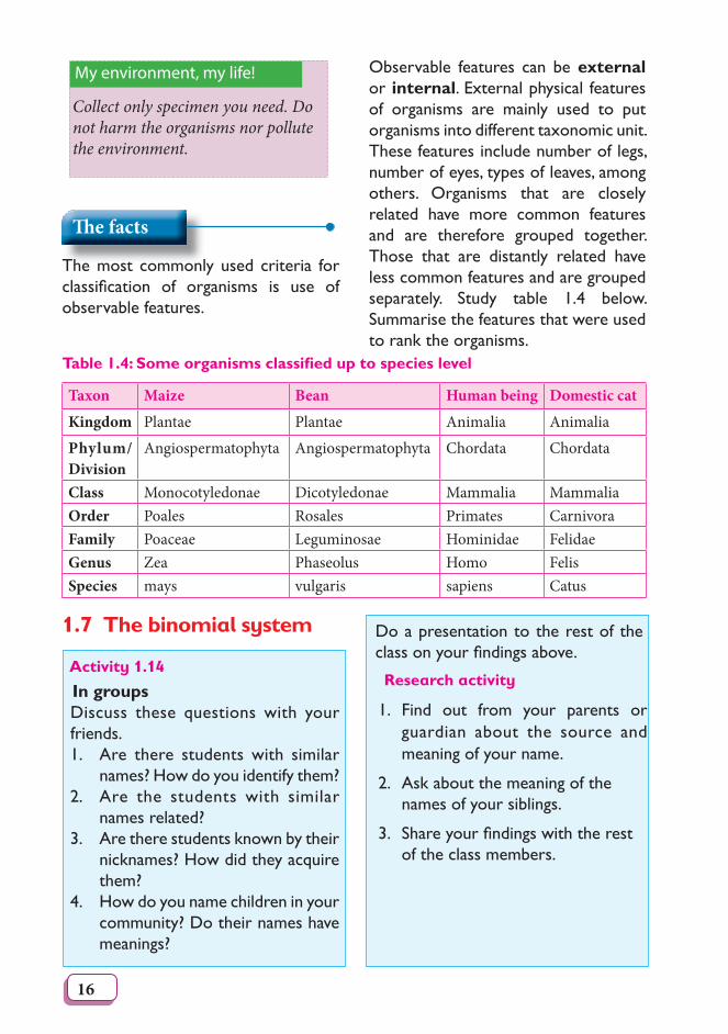
The most commonly used criteria for classification of organisms is use ofobservable features.
Table 1.4: Some organisms classifi ed up to species level
Taxon Maize Bean Human being Domestic cat
Kingdom Plantae Plantae Animalia Animalia
Phylum/Division
Angiospermatophyta Angiospermatophyta Chordata Chordata
Class Monocotyledonae Dicotyledonae Mammalia MammaliaOrder Poales Rosales Primates CarnivoraFamily Poaceae Leguminosae Hominidae FelidaeGenus Zea Phaseolus Homo FelisSpecies mays vulgaris sapiens Catus
Observable features can be external or internal. External physical features of organisms are mainly used to put organisms into different taxonomic unit. These features include number of legs, number of eyes, types of leaves, among others. Organisms that are closely related have more common features and are therefore grouped together. Those that are distantly related have less common features and are grouped separately. Study table 1.4 below. Summarise the features that were used to rank the organisms.
1.7 The binomial system
Activity 1.14
In groupsDiscuss these questions with your friends.1. Are there students with similar
names? How do you identify them?2. Are the students with similar
names related? 3. Are there students known by their
nicknames? How did they acquire them?
4. How do you name children in your community? Do their names have meanings?
Do a presentation to the rest of the classonyourfindingsabove.
Research activity
1. Find out from your parents or guardian about the source and meaning of your name.
2. Ask about the meaning of the names of your siblings.
3. Shareyourfindingswiththerestof the class members.
17
Activity 1.15: Reading
In pairsRead the following story then discuss the study question.Do you remember the long lost cousin you were looking for? You havefinallylocatedthehousewherethis cousin lives. However, you have been told that your cousin is at school.Toassistyoufindyourcousinthe teacher tells you that there are fivelearnerswiththesamenameasyourcousin’sfirstname.The teacher assisting you asks you if your cousin is tall or short. You say tall.Youaretoldthatofthefivelearners, who share your cousin’s firstname,threearetall.The teacher asks you if your cousin has a light skin complexion or dark skin complexion. You say dark skin complexion. You are told that of the three tall learners, two are dark-skinned. The teacher then asks you if your cousin has straight or curly hair. You say that your cousin has curly hair. The teacher then says that your cousin is in Secondary 3 East. The teacher then goes to fetch your cousin of whom you are pleased to see again after a long time.
Study Question
What characteristics did you follow to find your lost cousin? Writethem down.
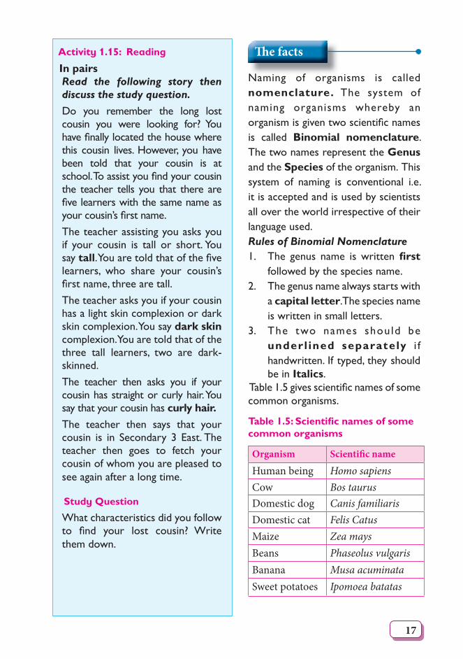
Th e facts
Naming of organisms is called nomenclature. The system of naming organisms whereby an organismisgiventwoscientificnamesis called Binomial nomenclature. The two names represent the Genus and the Species of the organism. This system of naming is conventional i.e. it is accepted and is used by scientists all over the world irrespective of their language used. Rules of Binomial Nomenclature1. The genus name is written fi rst
followed by the species name.2. The genus name always starts with
a capital letter. The species name is written in small letters.
3. The two names should be underl ined separately i f handwritten. If typed, they should be in Italics.
Table1.5givesscientificnamesofsomecommon organisms.
Table 1.5: Scientifi c names of some common organisms
Organism Scientifi c name
Human being Homo sapiensCow Bos taurusDomestic dog Canis familiarisDomestic cat Felis CatusMaize Zea maysBeans Phaseolus vulgarisBanana Musa acuminataSweet potatoes Ipomoea batatas
18
Work to doCarryoutresearchonscientificcommonplantsandanimalsinyourcommunity.Come up with a table like the one above.
Self Test 1.4
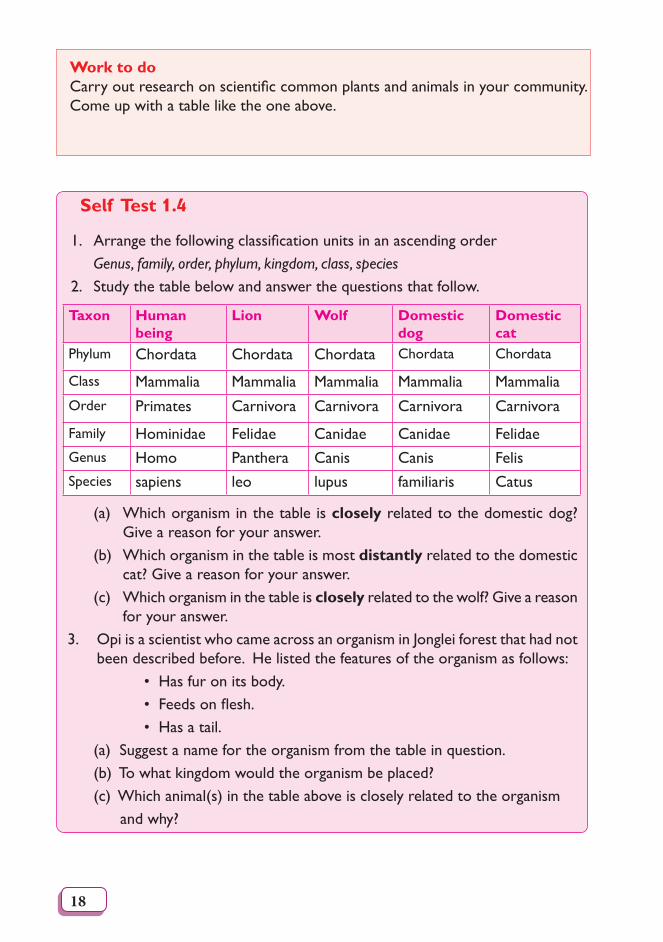
1. Arrangethefollowingclassificationunitsinanascendingorder Genus, family, order, phylum, kingdom, class, species2. Study the table below and answer the questions that follow.
Taxon Human being
Lion Wolf Domestic dog
Domestic cat
Phylum Chordata Chordata Chordata Chordata Chordata
Class Mammalia Mammalia Mammalia Mammalia MammaliaOrder Primates Carnivora Carnivora Carnivora Carnivora
Family Hominidae Felidae Canidae Canidae FelidaeGenus Homo Panthera Canis Canis FelisSpecies sapiens leo lupus familiaris Catus
(a) Which organism in the table is closely related to the domestic dog? Give a reason for your answer.
(b) Which organism in the table is most distantly related to the domestic cat? Give a reason for your answer.
(c) Which organism in the table is closely related to the wolf? Give a reason for your answer.
3. Opi is a scientist who came across an organism in Jonglei forest that had not been described before. He listed the features of the organism as follows:
• Has fur on its body. • Feedsonflesh. • Has a tail.
(a) Suggest a name for the organism from the table in question.(b) To what kingdom would the organism be placed?(c) Which animal(s) in the table above is closely related to the organism and why?
19
1.8 Dichotomous key
The term dichotomous comes from the word “dichotomy” which means divided into two parts. A dichotomous key is a set of instructions used to identify unknown organisms. To be able to identify organisms, observable features are used. This key uses two variations in description of a characteristic or feature for identification.The key is used toidentify and place new or unknown organismsintospecifictaxonomicunitsor groups. The organisms can then be named.
Activity 1.16
In pairsMaterials:
• Hand lens• Fish (in a bottle containing
water)• Grasshoppers (in a specimen
bottle) • Snails • Millipedes • Rabbits (caged) • Chicken (caged) • Pair of forcepsWhat to do
1. Observe each of the specimens provided carefully. Note down the unique structural observable features in a table like the one shown below.
(Use a hand lens if you cannot see the features clearly.)
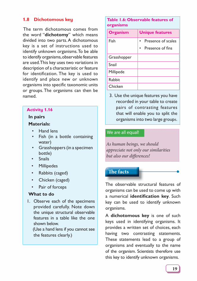
Table 1.6: Observable features of organisms
Organism Unique features
Fish • Presence of scales
•Presenceoffins
Grasshopper
Snail
Millipede
Rabbit
Chicken
3. Use the unique features you have recorded in your table to create pairs of contrasting features that will enable you to split the organisms into two large groups.
We are all equal!
As human beings, we should appreciate not only our similarities but also our di� erences!
Th e facts
The observable structural features of organisms can be used to come up with a numerical identifi cation key. Such key can be used to identify unknown organisms.
A dichotomous key is one of such keys used in identifying organisms. It provides a written set of choices, each having two contrasting statements. These statements lead to a group of organisms and eventually to the name of the organism. Scientists therefore use this key to identify unknown organisms.
20
the organism.
If an organism falls into one category indicated in the dichotomous key, you go to the next step indicated. You do this until you arrive at a step that identifies your specimen at thecorrectly indicated taxonomic level.
The key normally begins with general characteristics and lead to more specific characteristics. You are therefore expected to compare the characteristics you see in the unknown organism against an appropriate statement of the dichotomous key in order to identify
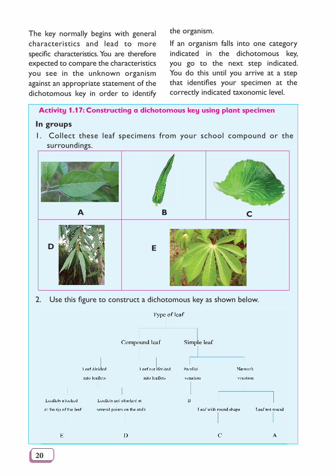
Activity 1.17: Constructing a dichotomous key using plant specimen
In groups1. Collect these leaf specimens from your school compound or the
surroundings.
A B C
D E
2.Usethisfiguretoconstructadichotomouskeyasshownbelow.
21
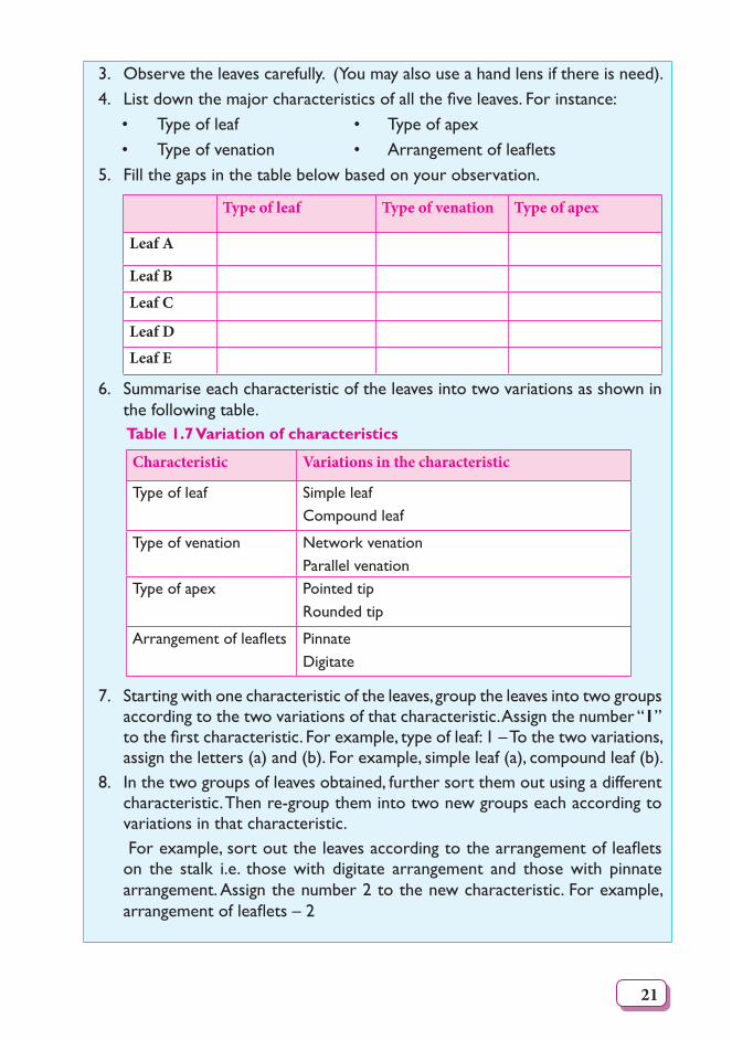
3. Observe the leaves carefully. (You may also use a hand lens if there is need).4. Listdownthemajorcharacteristicsofallthefiveleaves.Forinstance: • Type of leaf • Type of apex • Typeofvenation • Arrangementofleaflets5. Fill the gaps in the table below based on your observation.
Type of leaf Type of venation Type of apex
Leaf A
Leaf B
Leaf C
Leaf D
Leaf E
6. Summarise each characteristic of the leaves into two variations as shown in the following table.Table 1.7 Variation of characteristics
Characteristic Variations in the characteristic
Type of leaf Simple leafCompound leaf
Type of venation Network venationParallel venation
Type of apex Pointed tipRounded tip
Arrangementofleaflets Pinnate Digitate
7. Starting with one characteristic of the leaves, group the leaves into two groups according to the two variations of that characteristic. Assign the number “1” tothefirstcharacteristic.Forexample,typeofleaf:1–Tothetwovariations,assign the letters (a) and (b). For example, simple leaf (a), compound leaf (b).
8. In the two groups of leaves obtained, further sort them out using a different characteristic. Then re-group them into two new groups each according to variations in that characteristic.
Forexample,sortouttheleavesaccordingtothearrangementofleafletson the stalk i.e. those with digitate arrangement and those with pinnate arrangement. Assign the number 2 to the new characteristic. For example, arrangementofleaflets–2
22
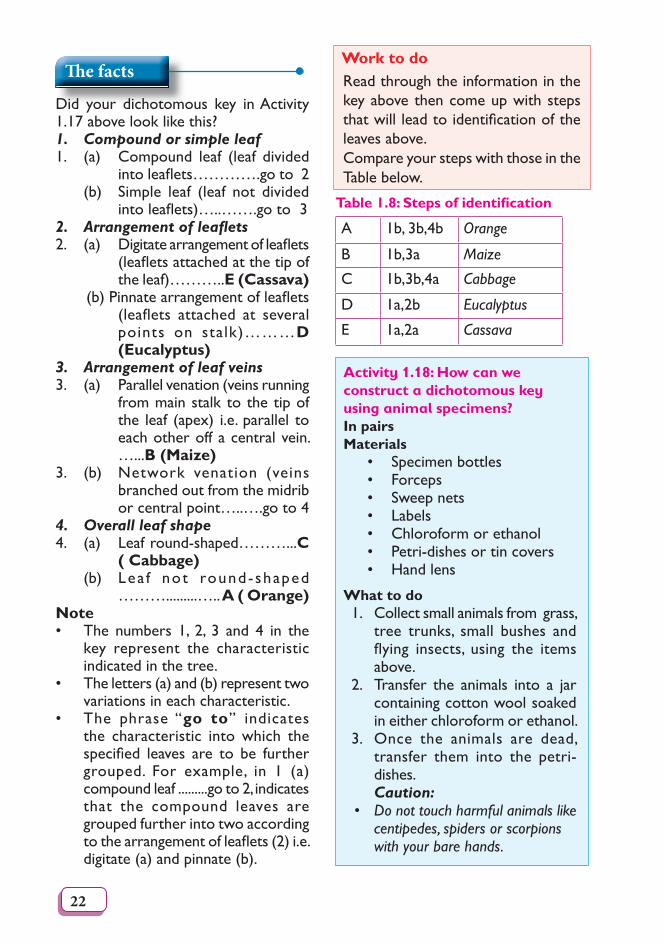
Th e facts
Did your dichotomous key in Activity 1.17 above look like this?1. Compound or simple leaf1. (a) Compound leaf (leaf divided
intoleaflets………….goto2 (b) Simple leaf (leaf not divided
intoleaflets)…..…….goto32. Arrangement of leafl ets2. (a) Digitatearrangementofleaflets
(leafletsattachedatthetipoftheleaf)………..E (Cassava)
(b)Pinnatearrangementofleaflets(leaflets attached at severalpoints on stalk)………D (Eucalyptus)
3. Arrangement of leaf veins3. (a) Parallel venation (veins running
from main stalk to the tip of the leaf (apex) i.e. parallel to each other off a central vein. …...B (Maize)
3. (b) Network venation (veins branched out from the midrib orcentralpoint…..….goto4
4. Overall leaf shape4. (a) Leafround-shaped………...C
( Cabbage) (b) Lea f not round-shaped
……….........…..A ( Orange)Note • The numbers 1, 2, 3 and 4 in the
key represent the characteristic indicated in the tree.
• The letters (a) and (b) represent two variations in each characteristic.
• The phrase “go to” indicates the characteristic into which the specified leaves are to be furthergrouped. For example, in 1 (a) compound leaf .........go to 2, indicates that the compound leaves are grouped further into two according tothearrangementofleaflets(2)i.e.digitate (a) and pinnate (b).
Work to doRead through the information in the key above then come up with steps thatwillleadtoidentificationoftheleaves above.Compare your steps with those in the Table below.
Table 1.8: Steps of identifi cation
A 1b, 3b,4b Orange
B 1b,3a Maize
C 1b,3b,4a Cabbage
D 1a,2b Eucalyptus
E 1a,2a Cassava
Activity 1.18: How can we construct a dichotomous key using animal specimens?In pairsMaterials
• Specimen bottles • Forceps • Sweep nets • Labels • Chloroform or ethanol • Petri-dishes or tin covers • Hand lens
What to do1. Collect small animals from grass,
tree trunks, small bushes and flying insects, using the itemsabove.
2. Transfer the animals into a jar containing cotton wool soaked in either chloroform or ethanol.
3. Once the animals are dead, transfer them into the petri-dishes.
Caution: • Do not touch harmful animals like centipedes, spiders or scorpions with your bare hands.
23
Check Your Progress 1
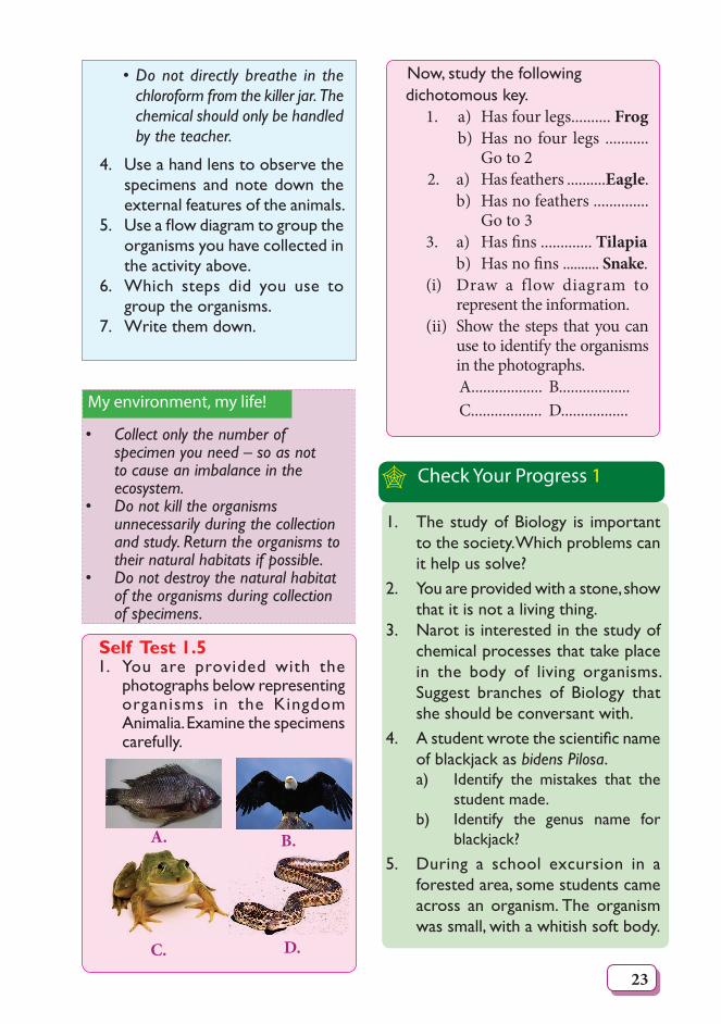
Self Test 1.51. You are provided with the
photographs below representing organisms in the Kingdom Animalia. Examine the specimens carefully.
A. B.
C. D.
• Do not directly breathe in the chloroform from the killer jar. The chemical should only be handled by the teacher.
4. Use a hand lens to observe the specimens and note down the external features of the animals.
5. Useaflowdiagramtogrouptheorganisms you have collected in the activity above.
6. Which steps did you use to group the organisms.
7. Write them down.
My environment, my life!
• Collect only the number of specimen you need – so as not to cause an imbalance in the ecosystem.
• Do not kill the organisms unnecessarily during the collection and study. Return the organisms to their natural habitats if possible.
• Do not destroy the natural habitat of the organisms during collection of specimens.
Now, study the following dichotomous key.
1. a) Has four legs.......... Frog b) Has no four legs ...........
Go to 22. a) Has feathers ..........Eagle. b) Has no feathers ..............
Go to 33. a) Has � ns ............. Tilapia b) Has no � ns .......... Snake. (i) Draw a flow diagram to
represent the information.(ii) Show the steps that you can
use to identify the organisms in the photographs.
A.................. B.................. C.................. D.................
1. The study of Biology is important to the society. Which problems can it help us solve?
2. You are provided with a stone, show that it is not a living thing.
3. Narot is interested in the study of chemical processes that take place in the body of living organisms. Suggest branches of Biology that she should be conversant with.
4. Astudentwrotethescientificnameof blackjack as bidens Pilosa.a) Identify the mistakes that the
student made.b) Identify the genus name for
blackjack?5. During a school excursion in a
forested area, some students came across an organism. The organism was small, with a whitish soft body.
24
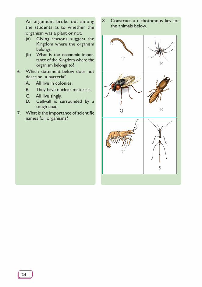
8. Construct a dichotomous key for the animals below.
TP
Q R
U
S
An argument broke out among the students as to whether the organism was a plant or not. (a) Giving reasons, suggest the
Kingdom where the organism belongs.
(b) What is the economic impor-tance of the Kingdom where the organism belongs to?
6. Which statement below does not describe a bacteria?
A. All live in colonies. B. They have nuclear materials. C. All live singly.
D. Cellwall is surrounded by a tough coat.
7. Whatistheimportanceofscientificnames for organisms?
25
2Unit The Cell
Learning outcomes
Knowledge and understanding
Skills Attitudes
• Understand structures of the cell, organization and functions.
• Able to observe the shape of a cell under a microscope.
• Prepare slides and perform simple experiments with plant tissues for example.
• Use a microscope.
• Appreciate the structure of the cell.
• Show curiosity and wonder about the existence of microscopic units of life.
• Appreciate the microscope.
• Think creatively about the cell as unit of life.
• Co-operating with others.
• Accuracy, systematic, ethical and patient.
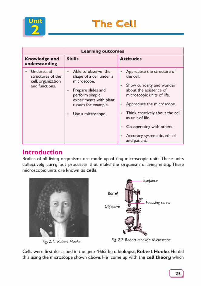
IntroductionBodies of all living organisms are made up of tiny microscopic units. These units collectively carry out processes that make the organism a living entity. These microscopic units are known as cells.
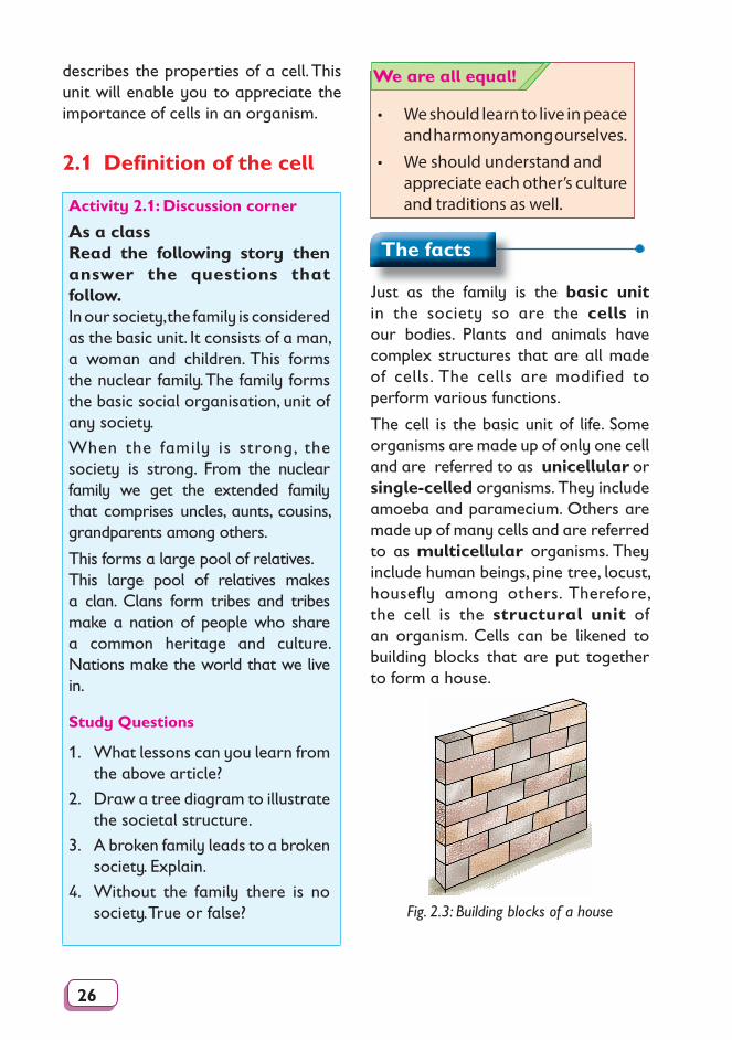
Fig. 2.1: Robert Hooke Fig. 2.2: Robert Hooke's Microscope
Eyepiece
Barrel
ObjectiveFocusing screw
Cells were first described in the year 1665 by a biologist, Robert Hooke. He did this using the microscope shown above. He came up with the cell theory which
26
describes the properties of a cell. This unit will enable you to appreciate the importance of cells in an organism.
2.1 Defi nition of the cell
Activity 2.1: Discussion corner
As a classRead the following story then answer the questions that follow.In our society, the family is considered as the basic unit. It consists of a man, a woman and children. This forms the nuclear family. The family forms the basic social organisation, unit of any society.
When the family is strong, the society is strong. From the nuclear family we get the extended family that comprises uncles, aunts, cousins, grandparents among others.
This forms a large pool of relatives. This large pool of relatives makes a clan. Clans form tribes and tribes make a nation of people who share a common heritage and culture. Nations make the world that we live in.
Study Questions
1. What lessons can you learn from the above article?
2. Draw a tree diagram to illustrate the societal structure.
3. A broken family leads to a broken society. Explain.
4. Without the family there is no society. True or false?
• We should learn to live in peace and harmony among ourselves.
• We should understand and appreciate each other’s culture and traditions as well.
We are all equal!
The facts
Just as the family is the basic unit in the society so are the cells in our bodies. Plants and animals have complex structures that are all made of cells. The cells are modified to perform various functions.
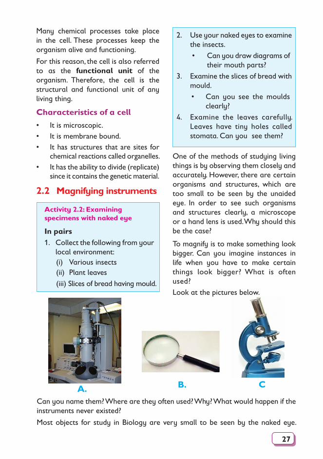
The cell is the basic unit of life. Some organisms are made up of only one cell and are referred to as unicellular or single-celled organisms. They include amoeba and paramecium. Others are made up of many cells and are referred to as multicellular organisms. They include human beings, pine tree, locust, housefly among others. Therefore, the cell is the structural unit of an organism. Cells can be likened to building blocks that are put together to form a house.
Fig. 2.3: Building blocks of a house
27
One of the methods of studying living things is by observing them closely and accurately. However, there are certain organisms and structures, which are too small to be seen by the unaided eye. In order to see such organisms and structures clearly, a microscope or a hand lens is used. Why should this be the case?
To magnify is to make something look bigger. Can you imagine instances in life when you have to make certain things look bigger? What is often used?
Look at the pictures below.
Many chemical processes take place in the cell. These processes keep the organism alive and functioning.
For this reason, the cell is also referred to as the functional unit of the organism. Therefore, the cell is the structural and functional unit of any living thing.
Characteristics of a cell
• It is microscopic.• It is membrane bound.• It has structures that are sites for
chemical reactions called organelles.• It has the ability to divide (replicate)
since it contains the genetic material.
2.2 Magnifying instruments
2. Use your naked eyes to examine the insects.
• Can you draw diagrams of their mouth parts?
3. Examine the slices of bread with mould.
• Can you see the moulds clearly?
4. Examine the leaves carefully. Leaves have tiny holes called stomata. Can you see them?
Activity 2.2: Examining specimens with naked eye
In pairs1. Collect the following from your
local environment:(i) Various insects(ii) Plant leaves
(iii) Slices of bread having mould.
B. CA.Can you name them? Where are they often used? Why? What would happen if the instruments never existed?
Most objects for study in Biology are very small to be seen by the naked eye.
28
Therefore magnifying instruments are needed.
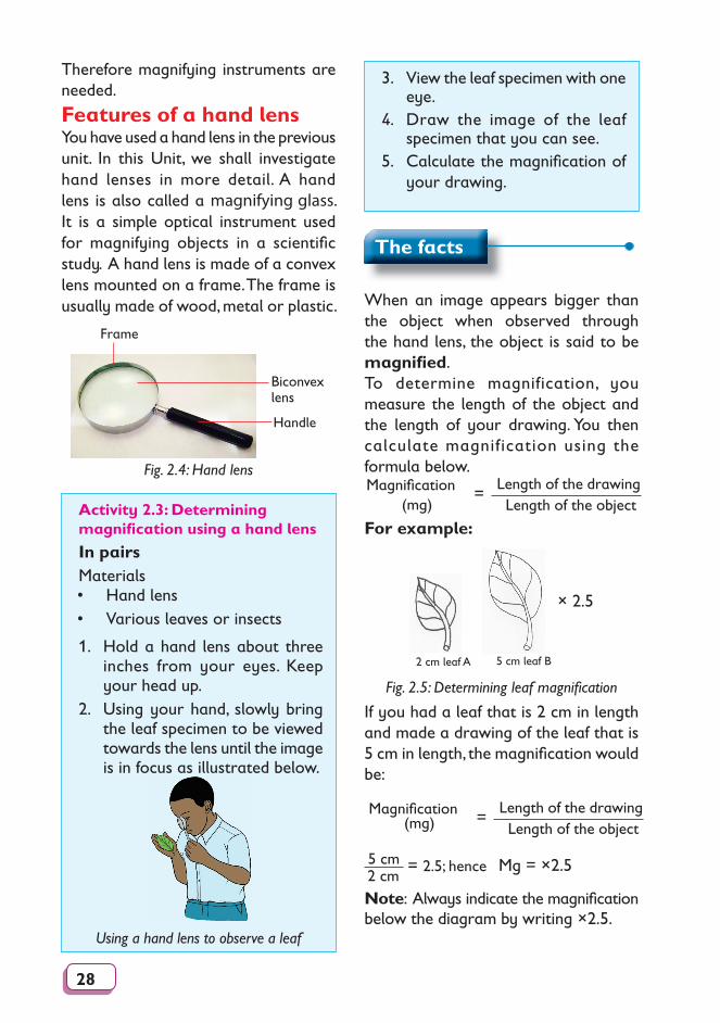
Features of a hand lens You have used a hand lens in the previous unit. In this Unit, we shall investigate hand lenses in more detail. A hand lens is also called a magnifying glass. It is a simple optical instrument used for magnifying objects in a scientifi c study. A hand lens is made of a convex lens mounted on a frame. The frame is usually made of wood, metal or plastic.
Frame
Biconvex lens
Handle
Fig. 2.4: Hand lens
3. View the leaf specimen with one eye.
4. Draw the image of the leaf specimen that you can see.
5. Calculate the magnifi cation of your drawing.
Activity 2.3: Determining magnifi cation using a hand lens
In pairsMaterials• Hand lens • Various leaves or insects
1. Hold a hand lens about three inches from your eyes. Keep your head up.
2. Using your hand, slowly bring the leaf specimen to be viewed towards the lens until the image is in focus as illustrated below.
Using a hand lens to observe a leaf
The facts
When an image appears bigger than the object when observed through the hand lens, the object is said to be magnifi ed. To determine magnification, you measure the length of the object and the length of your drawing. You then calculate magnification using the formula below. Magnifi cation (mg)
Length of the drawingLength of the object
=
For example:
2 cm leaf A 5 cm leaf B
× 2.5
Fig. 2.5: Determining leaf magnifi cation
If you had a leaf that is 2 cm in length and made a drawing of the leaf that is 5 cm in length, the magnifi cation would be:
Magnifi cation (mg)
Length of the drawingLength of the object
=
Mg = ×2.55 cm 2.5; hence2 cm
=
Note: Always indicate the magnifi cation below the diagram by writing ×2.5.
29
Note: Some hand lenses have their magnifi cation written on their frame. Share with your friend why is it important to calculate magnifi cation.
Care of the hand lensFor the hand lens to last for long, it has to be cared for. Hand lens is made of a glass part which can gather dust or break if it falls down.
Activity 2.4: Discussion corner
In groups1. In your study group, discuss
ways of caring for a hand lens. 2. Why is this important?3. Compare your fi ndings with the
facts below.
2.3 Parts and functions of a light microscopeA light microscope uses light rays and a system of lenses to magnify images of small objects. Have you ever come across a microscope? How does it look like?
The facts
• The hand lens should be stored in a dry place where the lens cannot break or get scratched.
• The hand lens should be cleaned by use of a special soft tissue.
• Handle the hand lens with care to avoid breaking the lens.
Self Test 2.11. A student used a hand lens
to observe an ant. The actual length of the ant was 0.6 cm. On observing, the ant was 6.8 cm long. Calculate the magnifi cation.
2. Identify circumstances where a hand lens can be used to observe features of a big organism.
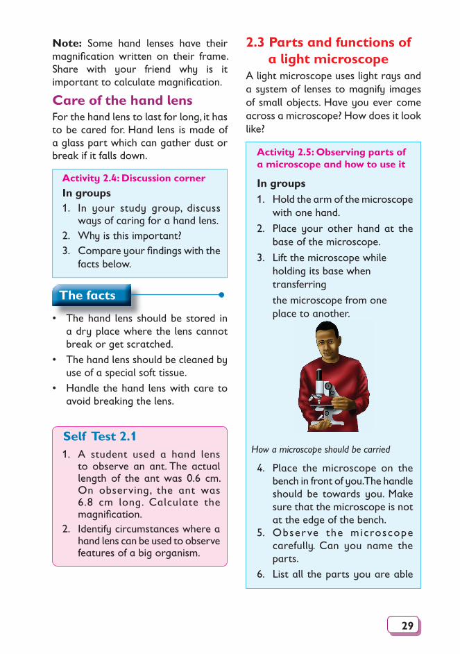
Activity 2.5: Observing parts of a microscope and how to use it
In groups1. Hold the arm of the microscope
with one hand.
2. Place your other hand at the base of the microscope.
3. Lift the microscope while holding its base when transferring
the microscope from one place to another.
How a microscope should be carried
4. Place the microscope on the bench in front of you. The handle should be towards you. Make sure that the microscope is not at the edge of the bench.
5. Obser ve the microscope carefully. Can you name the parts.
6. List all the parts you are able
30
to identify. Take note of the following:• The eye piece • Mirror• Stage • Knobs• Lenses
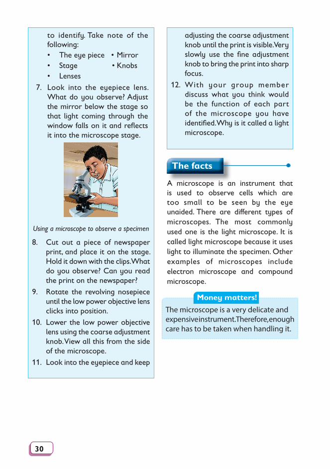
7. Look into the eyepiece lens. What do you observe? Adjust the mirror below the stage so that light coming through the window falls on it and refl ects it into the microscope stage.
Using a microscope to observe a specimen
8. Cut out a piece of newspaper print, and place it on the stage. Hold it down with the clips. What do you observe? Can you read the print on the newspaper?
9. Rotate the revolving nosepiece until the low power objective lens clicks into position.
10. Lower the low power objective lens using the coarse adjustment knob. View all this from the side of the microscope.
11. Look into the eyepiece and keep
adjusting the coarse adjustment knob until the print is visible. Very slowly use the fi ne adjustment knob to bring the print into sharp focus.
12. With your group member discuss what you think would be the function of each part of the microscope you have identifi ed. Why is it called a light microscope.
The facts
A microscope is an instrument that is used to observe cells which are too small to be seen by the eye unaided. There are different types of microscopes. The most commonly used one is the light microscope. It is called light microscope because it uses light to illuminate the specimen. Other examples of microscopes include electron microscope and compound microscope.
Money matters!
The microscope is a very delicate and expensive instrument. Therefore, enough care has to be taken when handling it.
31
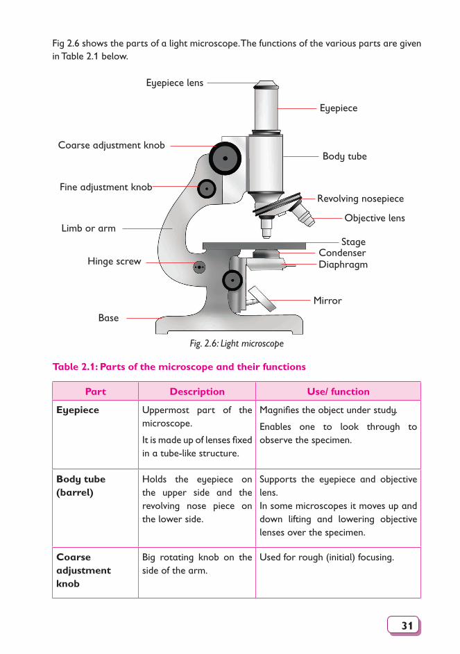
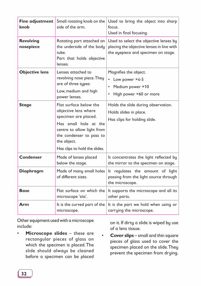
Fig 2.6 shows the parts of a light microscope. The functions of the various parts are given in Table 2.1 below.
Eyepiece lens
Eyepiece
Hinge screw
Coarse adjustment knob
Fine adjustment knob
Base
Body tube
Revolving nosepiece
Objective lens
StageCondenserDiaphragm
Mirror
Limb or arm
Fig. 2.6: Light microscope
Table 2.1: Parts of the microscope and their functions
Part Description Use/ function
Eyepiece Uppermost part of the microscope.
It is made up of lenses fixed in a tube-like structure.
Magnifies the object under study.
Enables one to look through to observe the specimen.
Body tube (barrel)
Holds the eyepiece on the upper side and the revolving nose piece on the lower side.
Supports the eyepiece and objective lens.In some microscopes it moves up and down lifting and lowering objective lenses over the specimen.
Coarse adjustment knob
Big rotating knob on the side of the arm.
Used for rough (initial) focusing.
32
Fine adjustment knob
Small rotating knob on the side of the arm.
Used to bring the object into sharp focus.Used in final focusing.
Revolving nosepiece
Rotating part attached on the underside of the body tube.Part that holds objective lenses.
Used to select the objective lenses by placing the objective lenses in line with the eyepiece and specimen on stage.
Objective lens Lenses attached to revolving nose piece. They are of three types:
Low, medium and high power lenses.
Magnifies the object.
• Low power ×4-5
• Medium power ×10
• High power ×60 or more
Stage Flat surface below the objective lens where specimen are placed.
Has small hole at the centre to allow light from the condenser to pass to the object.
Has clips to hold the slides.
Holds the slide during observation.
Holds slides in place.
Has clips for holding slide.
Condenser Made of lenses placed below the stage.
It concentrates the light reflected by the mirror to the specimen on stage.
Diaphragm Made of many small holes of different sizes.
It regulates the amount of light passing from the light source through the microscope.
Base Flat surface on which the microscope 'sits'.
It supports the microscope and all its other parts.
Arm It is the curved part of the microscope.
It is the part we hold when using or carrying the microscope.
Other equipment used with a microscope include:
• Microscope slides – these are rectangular pieces of glass on which the specimen is placed. The slide should always be cleaned before a specimen can be placed
on it. If dirty a slide is wiped by use of a lens tissue.
• Cover slips – small and thin square pieces of glass used to cover the specimen placed on the slide. They prevent the specimen from drying.
33
Mounting needle
Cover Slide
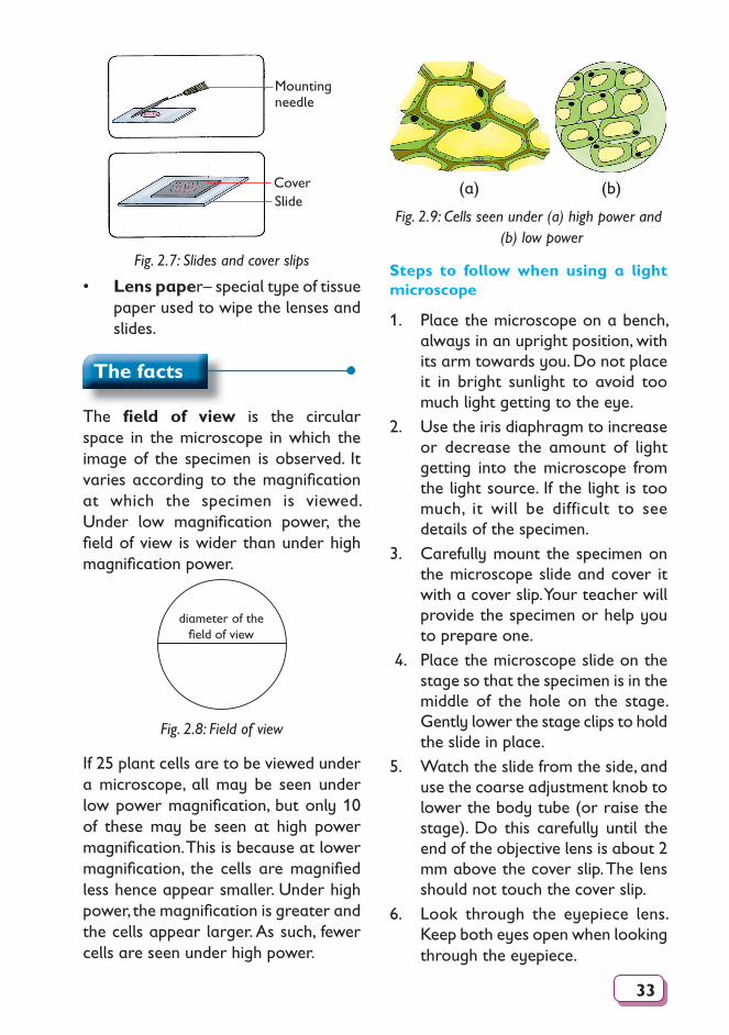
Fig. 2.7: Slides and cover slips
• Lens paper– special type of tissue paper used to wipe the lenses and slides.
The facts
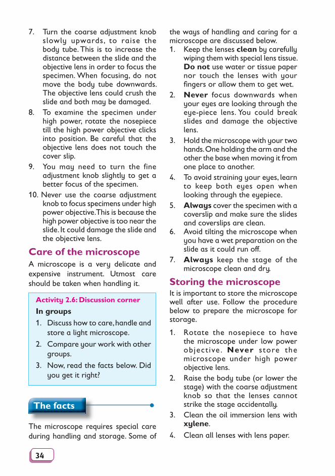
The fi eld of view is the circular space in the microscope in which the image of the specimen is observed. It varies according to the magnifi cation at which the specimen is viewed. Under low magnifi cation power, the fi eld of view is wider than under high magnifi cation power.
diameter of the fi eld of view
Fig. 2.8: Field of view
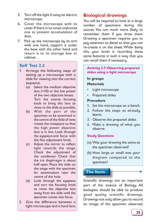
If 25 plant cells are to be viewed under a microscope, all may be seen under low power magnifi cation, but only 10 of these may be seen at high power magnifi cation. This is because at lower magnifi cation, the cells are magnifi ed less hence appear smaller. Under high power, the magnifi cation is greater and the cells appear larger. As such, fewer cells are seen under high power.
(a) (b)
Fig. 2.9: Cells seen under (a) high power and (b) low power
Steps to follow when using a light microscope
1. Place the microscope on a bench, always in an upright position, with its arm towards you. Do not place it in bright sunlight to avoid too much light getting to the eye.
2. Use the iris diaphragm to increase or decrease the amount of light getting into the microscope from the light source. If the light is too much, it will be difficult to see details of the specimen.
3. Carefully mount the specimen on the microscope slide and cover it with a cover slip. Your teacher will provide the specimen or help you to prepare one.
4. Place the microscope slide on the stage so that the specimen is in the middle of the hole on the stage. Gently lower the stage clips to hold the slide in place.
5. Watch the slide from the side, and use the coarse adjustment knob to lower the body tube (or raise the stage). Do this carefully until the end of the objective lens is about 2 mm above the cover slip. The lens should not touch the cover slip.
6. Look through the eyepiece lens. Keep both eyes open when looking through the eyepiece.
34
7. Turn the coarse adjustment knob slowly upwards, to raise the body tube. This is to increase the distance between the slide and the objective lens in order to focus the specimen. When focusing, do not move the body tube downwards. The objective lens could crush the slide and both may be damaged.
8. To examine the specimen under high power, rotate the nosepiece till the high power objective clicks into position. Be careful that the objective lens does not touch the cover slip.
9. You may need to turn the fine adjustment knob slightly to get a better focus of the specimen.
10. Never use the coarse adjustment knob to focus specimens under high power objective. This is because the high power objective is too near the slide. It could damage the slide and the objective lens.
Care of the microscopeA microscope is a very delicate and expensive instrument. Utmost care should be taken when handling it.
Activity 2.6: Discussion corner
In groups1. Discuss how to care, handle and
store a light microscope.
2. Compare your work with other groups.
3. Now, read the facts below. Did you get it right?
The facts
The microscope requires special care during handling and storage. Some of
the ways of handling and caring for a microscope are discussed below.1. Keep the lenses clean by carefully
wiping them with special lens tissue. Do not use water or tissue paper nor touch the lenses with your fi ngers or allow them to get wet.
2. Never focus downwards when your eyes are looking through the eye-piece lens. You could break slides and damage the objective lens.
3. Hold the microscope with your two hands. One holding the arm and the other the base when moving it from one place to another.
4. To avoid straining your eyes, learn to keep both eyes open when looking through the eyepiece.
5. Always cover the specimen with a coverslip and make sure the slides and coverslips are clean.
6. Avoid tilting the microscope when you have a wet preparation on the slide as it could run off.
7. Always keep the stage of the microscope clean and dry.
Storing the microscope It is important to store the microscope well after use. Follow the procedure below to prepare the microscope for storage.
1. Rotate the nosepiece to have the microscope under low power object ive . Never s tore the microscope under high power objective lens.
2. Raise the body tube (or lower the stage) with the coarse adjustment knob so that the lenses cannot strike the stage accidentally.
3. Clean the oil immersion lens with xylene.
4. Clean all lenses with lens paper.
35
5. Turn off the light if using an electric microscope.
6. Cover the microscope with its cover. If there is no cover, improvise one to prevent accumulation of dust.
7. Pick up the microscope by its arm with one hand, support it under the base with the other hand and return it to its storage box or cabinet.
Self Test 2.21. Arrange the following steps of
setting up a microscope with a slide for viewing into the correct sequence. (a) Select the medium objective
lens (×10) or the low power of the two objective lenses.
Turn the coarse focusing knob to bring this lens as close to the slide as possible.
(b) With the part of the specimen to be examined in the centre of the fi eld of view, rotate the nosepiece so that the high power objective lens is in line. Look through the eyepiece and focus with the fi ne adjustment knob.
(c) Adjust the mirror to refl ect light towards the stage. Check the adjustment of the condenser. Check that the iris diaphragm is about half open. Place the slide on the stage with the specimen for examination near the centre of the hole.
(d) Look through the eyepiece and turn the focusing knob to move the objective lens away from the slide until the specimen comes into focus.
2. Give the difference between a light microscope and a hand lens.
Biological drawings You will be required to look at a largenumber of specimens during this course. You are much more likely to remember them if you draw them. Drawing a specimen requires you to pay attention to detail so that you can re-create it on the sheet. While doing this, your brain is recording these same features in such a way that you can recall them if necessary.
Activity 2.7: Observing prepared slides using a light microscope
In groupsMaterials
• Light microscope
• Prepared slides
Procedure 1. Set the microscope on a bench.2. Follow the steps as already
learnt.3. Observe the prepared slides.4. Make a drawing of what you
observe.
Study Questions
(a) Was your drawing the same as the specimen observed?
(b) How large or small was your diagram compared to the specimen?
The facts
Scientifi c drawings are an important part of the science of Biology. All biologists should be able to produce good quality scientific drawings. Drawings not only allow you to record an image of the specimen observed,
36
but more importantly, they help you to remember the specimen as well as the important features of the specimen.
The following are some guidelines that you should follow when drawing Biological diagrams:
1. Look at the specimen carefully. Examine the significant features that can be included in the drawing.
2. Draw only what you see. Do not include what you think you should see.
3. All drawings must be done in pencil only and should be done on plain papers.
4. Drawings must be large and clear so that features can be easily distinguished. They should be large enough to show all parts without crowding.
5. Keep your drawings to the left of the page. Save the right-hand side of the page for labels.
6. Never use arrows to label diagrams. Lines should be drawn with a ruler and parallel to each other. The lettering of the words should be horizontal and in pencil.
7. Always use distinct, single lines when drawing. Avoid broken lines.
8. Do not shade your drawings. Dark areas in a drawing to be indicated using dots.
9. All drawings must have the following indicated: • Title – give a full, clear and
concise title that explains
what is being illustrated.• Magnification – indicate the
magnification at which the specimen was observed.
• Labels – always include labels of the important features of the specimen. Each label line must be straight and should not overlap with other label lines; all labels must be to one side left or right.
• Scale – always include a scale bar indicating the length or width of the specimen drawn.
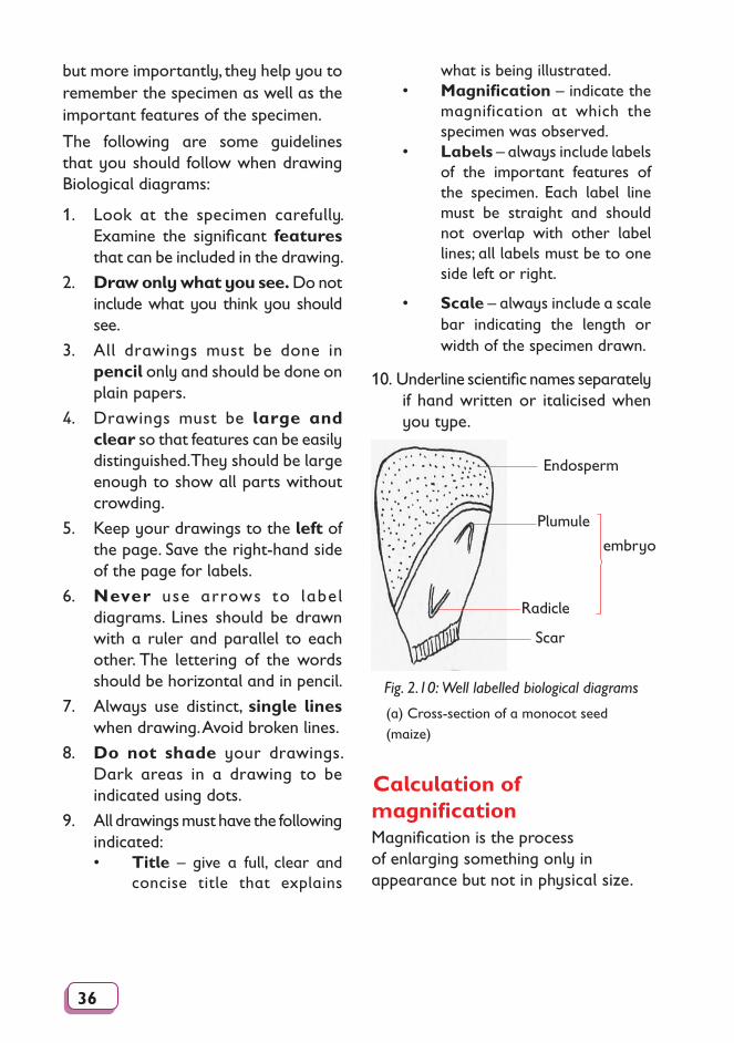
10. Underline scientific names separately if hand written or italicised when you type.
(a) Cross-section of a monocot seed (maize)
Endosperm
Plumule
Radicle
Scar
embryo
Fig. 2.10: Well labelled biological diagrams
Calculation of magnification Magnification is the process of enlarging something only in appearance but not in physical size.
37
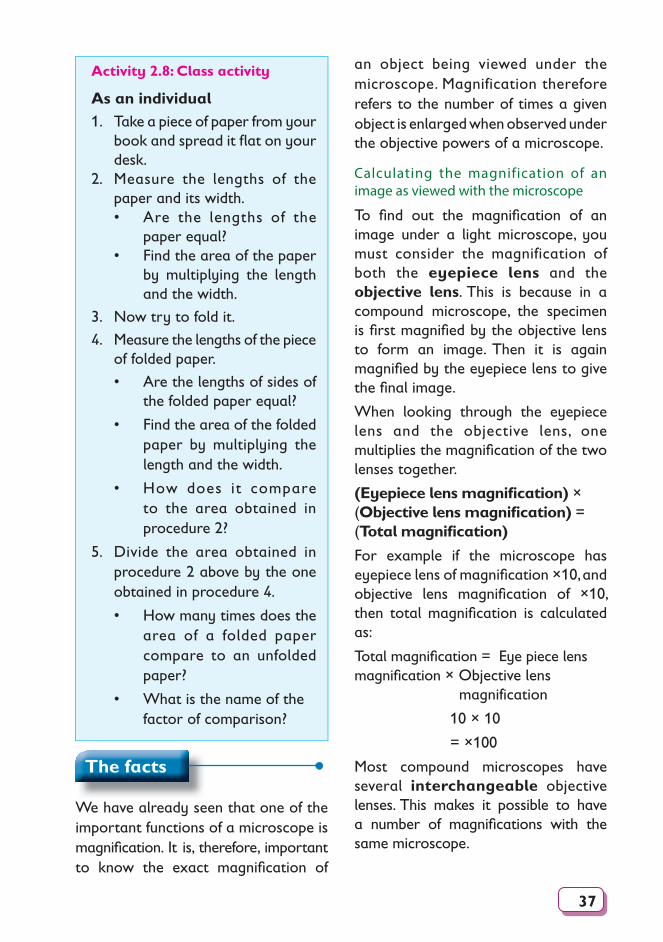
Activity 2.8: Class activity
As an individual1. Take a piece of paper from your
book and spread it fl at on your desk.
2. Measure the lengths of the paper and its width.• Are the lengths of the
paper equal?• Find the area of the paper
by multiplying the length and the width.
3. Now try to fold it.
4. Measure the lengths of the piece of folded paper.
• Are the lengths of sides of the folded paper equal?
• Find the area of the folded paper by multiplying the length and the width.
• How does it compare to the area obtained in procedure 2?
5. Divide the area obtained in procedure 2 above by the one obtained in procedure 4.
• How many times does the area of a folded paper compare to an unfolded paper?
• What is the name of the factor of comparison?
an object being viewed under the microscope. Magnification therefore refers to the number of times a given object is enlarged when observed under the objective powers of a microscope.
Calculating the magnification of an image as viewed with the microscope
To fi nd out the magnifi cation of an image under a light microscope, you must consider the magnification of both the eyepiece lens and the objective lens. This is because in a compound microscope, the specimen is fi rst magnifi ed by the objective lens to form an image. Then it is again magnifi ed by the eyepiece lens to give the fi nal image.
When looking through the eyepiece lens and the objective lens, one multiplies the magnifi cation of the two lenses together.
(Eyepiece lens magnifi cation) × (Objective lens magnifi cation) = (Total magnifi cation)
For example if the microscope has eyepiece lens of magnifi cation ×10, and objective lens magnifi cation of ×10, then total magnifi cation is calculated as:
Total magnifi cation = Eye piece lens magnifi cation × Objective lens magnification
10 × 10
= ×100
Most compound microscopes have several interchangeable objective lenses. This makes it possible to have a number of magnifi cations with the same microscope.
The facts
We have already seen that one of the important functions of a microscope is magnifi cation. It is, therefore, important to know the exact magnifi cation of
38
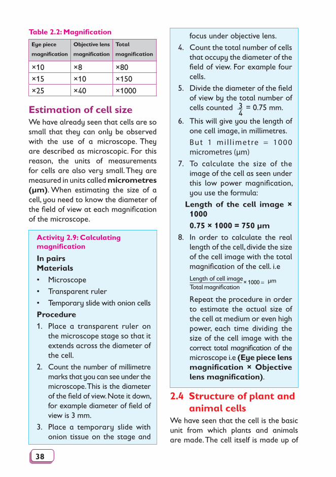
Table 2.2: Magnification
Eye piece
magnification
Objective lens
magnification
Total
magnification
×10 ×8 ×80
×15 ×10 ×150
×25 ×40 ×1000
Estimation of cell sizeWe have already seen that cells are so small that they can only be observed with the use of a microscope. They are described as microscopic. For this reason, the units of measurements for cells are also very small. They are measured in units called micrometres (µm). When estimating the size of a cell, you need to know the diameter of the field of view at each magnification of the microscope.
focus under objective lens.
4. Count the total number of cells that occupy the diameter of the field of view. For example four cells.
5. Divide the diameter of the field of view by the total number of cells counted 3
4 = 0.75 mm.
6. This will give you the length of one cell image, in millimetres.
But 1 mi l l imetre = 1000 micrometres (µm)
7. To calculate the size of the image of the cell as seen under this low power magnification, you use the formula:
Length of the cell image × 1000
0.75 × 1000 = 750 µm
8. In order to calculate the real length of the cell, divide the size of the cell image with the total magnification of the cell. i.e
Length of cell imageTotal magnification
× 1000 = µm
Repeat the procedure in order to estimate the actual size of the cell at medium or even high power, each time dividing the size of the cell image with the correct total magnification of the microscope i.e (Eye piece lens magnification × Objective lens magnification).
Activity 2.9: Calculating magnification
In pairsMaterials• Microscope
• Transparent ruler
• Temporary slide with onion cells
Procedure
1. Place a transparent ruler on the microscope stage so that it extends across the diameter of the cell.
2. Count the number of millimetre marks that you can see under the microscope. This is the diameter of the field of view. Note it down, for example diameter of field of view is 3 mm.
3. Place a temporary slide with onion tissue on the stage and
2.4 Structure of plant and animal cells
We have seen that the cell is the basic unit from which plants and animals are made. The cell itself is made up of
39
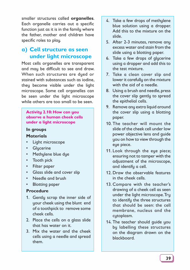
4. Take a few drops of methylene blue solution using a dropper. Add this to the mixture on the slide.
5. After 2-3 minutes, remove any excess water and stain from the slide using a blotting paper.
6. Take a few drops of glycerine using a dropper and add this to the test mixture.
7. Take a clean cover slip and lower it carefully on the mixture with the aid of a needle.
8. Using a brush and needle, press the cover slip gently to spread the epithelial cells.
9. Remove any extra liquid around the cover slip using a blotting paper.
10. The teacher will mount the slide of the cheek cell under low power objective lens and guide you on how to view through the eye piece.
11. Look through the eye piece; ensuring not to tamper with the adjustment of the microscope, and identify a cell.
12. Draw the observable features in the cheek cells.
13. Compare with the teacher’s drawing of a cheek cell as seen under the light microscope. Try to identify the three structures that should be seen: the cell membrane, nucleus and the cytoplasm.
14. The teacher should guide you by labelling these structures on the diagram drawn on the blackboard.
smaller structures called organelles. Each organelle carries out a specific function just as it is in the family where the father, mother and children have specific roles to play.
a) Cell structure as seen under light microscope
Most cells organelles are transparent and may be difficult to see and draw. When such structures are dyed or stained with substances such as iodine, they become visible under the light microscope. Some cell organelles can be seen under the light microscope while others are too small to be seen.
Activity 2.10: How can you observe a human cheek cells under a light microscope
In groupsMaterials• Light microscope• Glycerine• Methylene blue dye• Tooth pick• Filter paper• Glass slide and cover slip• Needle and brush• Blotting paper
Procedure1. Gently scrap the inner side of
your cheek using the blunt end of a toothpick to remove some cheek cells.
2. Place the cells on a glass slide that has water on it.
3. Mix the water and the cheek cells using a needle and spread them.
40
Cut a small strip of the thin epidermis
(ii)
Put a drop of water/ iodine on a slide
(iii)
Lower a clean cover slip on the strip
(iv)
Gently mount the specimen on the stage
(v)
(i)
Peel off a thin piece of the epidermis
3. Observe through a microscope and make a well labelled diagram.
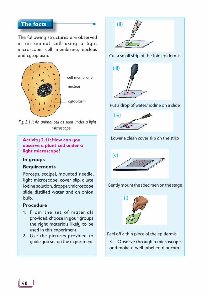
The facts
The following structures are observed in an animal cell using a light microscope: cell membrane, nucleus and cytoplasm.
nucleus
cytoplasm
cell membrane
Fig. 2.11: An animal cell as seen under a light microscope
Activity 2.11: How can you observe a plant cell under a light microscope?
In groupsRequirementsForceps, scalpel, mounted needle, light microscope, cover slip, dilute iodine solution, dropper, microscope slide, distilled water and an onion bulb.
Procedure1. From the set of materials
provided, choose in your groups the right materials likely to be used in this experiment.
2. Use the pictures provided to guide you set up the experiment.
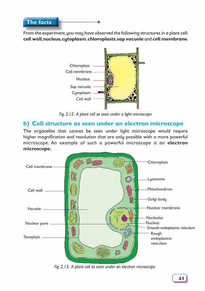
41
The facts
From the experiment, you may have observed the following structures in a plant cell: cell wall, nucleus, cytoplasm, chloroplasts, sap vacuole and cell membrane.
Nucleus
Sap vacuole
Cytoplasm
Cell membrane
Chloroplast
Cell wall
Fig. 2.12: A plant cell as seen under a light microscope
b) Cell structure as seen under an electron microscopeThe organelles that cannot be seen under light microscope would require higher magnifi cation and resolution that are only possible with a more powerful microscope. An example of such a powerful microscope is an electron microscope.
Fig. 2.13: A plant cell as seen under an electron microscope
Chloroplast
Lysosome
Mitochondrion
Golgi body
Nucleolus
Nuclear membrane
Cell membrane
Cell wall
Vacuole
Nuclear pore
Tonoplast
NucleusSmooth endoplasmic reticulum
Rough endoplasmic reticulum
• •
•• • • ••••
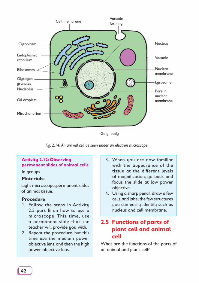
42
Fig. 2.14: An animal cell as seen under an electron microscope
Cytoplasm
Ribosomes
Glycogen granulesNucleolus
Oil droplets
Mitochondrion
Cell membrane
Endoplasmicreticulum
Nuclear membrane
Vacuole
Vacuole forming
Golgi body
Nucleus
Lysosome
Pore in nuclear membrane
Activity 2.12: Observing permanent slides of animal cells
In groups
Materials:Light microscope, permanent slides of animal tissue.
Procedure1. Follow the steps in Activity
2.5 part B on how to use a microscope. This time, use a permanent slide that the teacher will provide you with.
2. Repeat the procedure, but this time use the medium power objective lens, and then the high power objective lens.
3. When you are now familiar with the appearance of the tissue at the different levels of magnification, go back and focus the slide at low power objective.
4. Using a sharp pencil, draw a few cells, and label the few structures you can easily identify such as nucleus and cell membrane.
2.5 Functions of parts of plant cell and animal cell
What are the functions of the parts of an animal and plant cell?
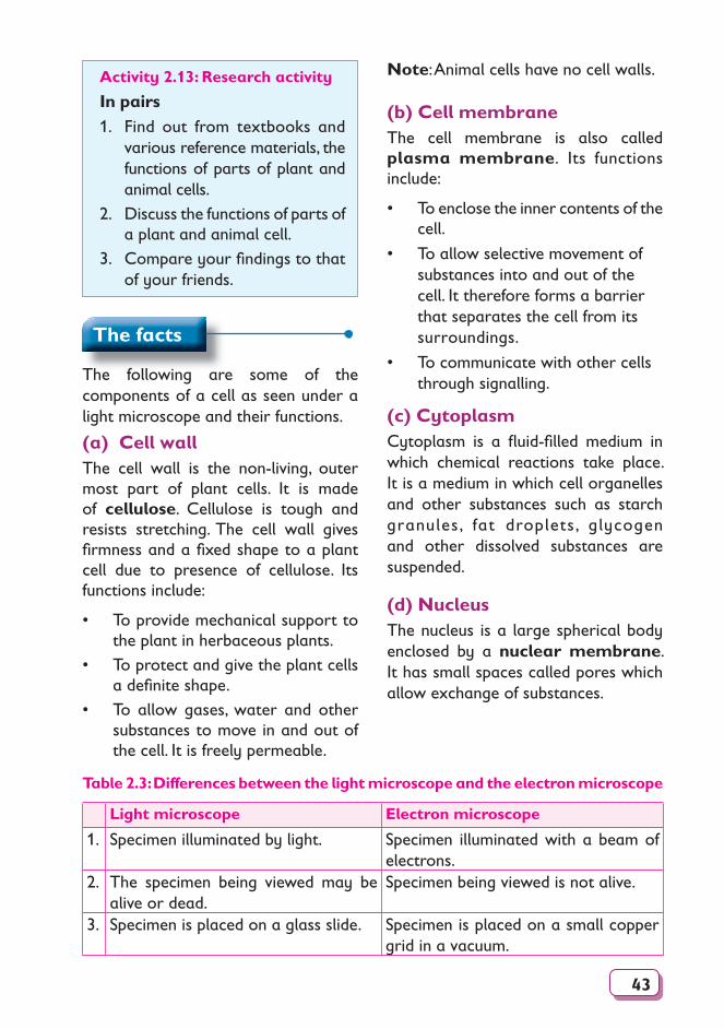
43
Activity 2.13: Research activity
In pairs1. Find out from textbooks and
various reference materials, the functions of parts of plant and animal cells.
2. Discuss the functions of parts of a plant and animal cell.
3. Compare your fi ndings to that of your friends.
Note: Animal cells have no cell walls.
(b) Cell membrane The cell membrane is also called plasma membrane. Its functions include:
• To enclose the inner contents of the cell.
• To allow selective movement of substances into and out of the cell. It therefore forms a barrier that separates the cell from its surroundings.
• To communicate with other cells through signalling.
(c) CytoplasmCytoplasm is a fl uid-fi lled medium in which chemical reactions take place. It is a medium in which cell organelles and other substances such as starch granules, fat droplets, glycogen and other dissolved substances are suspended.
(d) NucleusThe nucleus is a large spherical body enclosed by a nuclear membrane. It has small spaces called pores which allow exchange of substances.
The facts
The following are some of the components of a cell as seen under a light microscope and their functions.
(a) Cell wallThe cell wall is the non-living, outer most part of plant cells. It is made of cellulose. Cellulose is tough and resists stretching. The cell wall gives fi rmness and a fi xed shape to a plant cell due to presence of cellulose. Its functions include:
• To provide mechanical support to the plant in herbaceous plants.
• To protect and give the plant cells a defi nite shape.
• To allow gases, water and other substances to move in and out of the cell. It is freely permeable.
Table 2.3: Differences between the light microscope and the electron microscope
Light microscope Electron microscope
1. Specimen illuminated by light. Specimen illuminated with a beam of elec trons.
2. The specimen being viewed may be alive or dead.
Specimen being viewed is not alive.
3. Specimen is placed on a glass slide. Specimen is placed on a small copper grid in a vacuum.
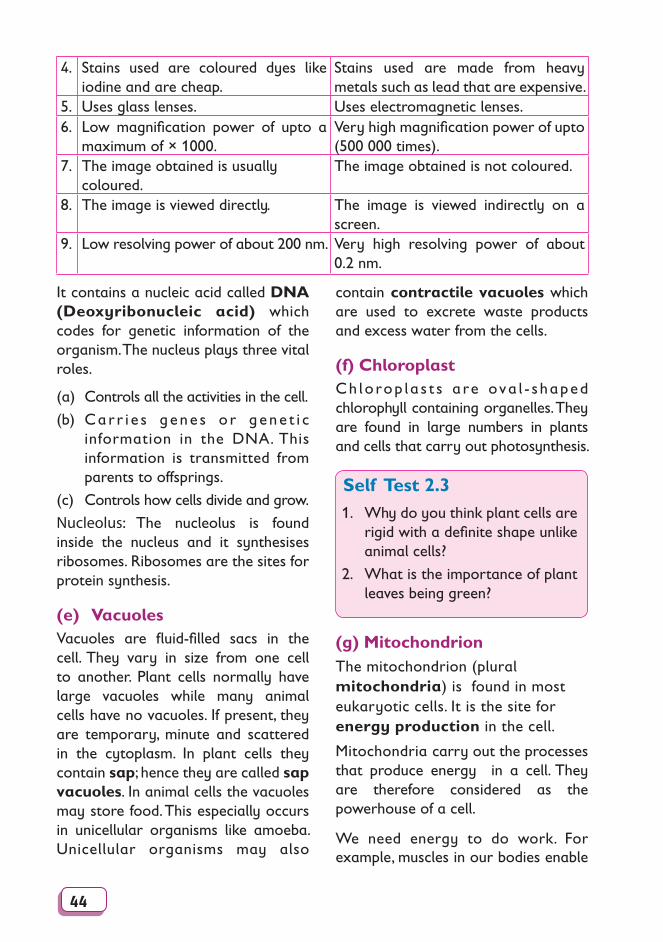
44
4. Stains used are coloured dyes like iodine and are cheap.
Stains used are made from heavy metals such as lead that are expensive.
5. Uses glass lenses. Uses electromagnetic lenses.6. Low magnification power of upto a
maximum of × 1000.Very high magnification power of upto (500 000 times).
7. The image obtained is usually coloured.
The image obtained is not coloured.
8. The image is viewed directly. The image is viewed indirectly on a screen.
9. Low resolving power of about 200 nm. Very high resolving power of about 0.2 nm.
It contains a nucleic acid called DNA (Deoxyribonucleic acid) which codes for genetic information of the organism. The nucleus plays three vital roles.
(a) Controls all the activities in the cell.
(b) Carr i e s gene s o r gene t i c information in the DNA. This information is transmitted from parents to offsprings.
(c) Controls how cells divide and grow.
Nucleolus: The nucleolus is found inside the nucleus and it synthesises ribosomes. Ribosomes are the sites for protein synthesis.
(e) VacuolesVacuoles are fluid-filled sacs in the cell. They vary in size from one cell to another. Plant cells normally have large vacuoles while many animal cells have no vacuoles. If present, they are temporary, minute and scattered in the cytoplasm. In plant cells they contain sap; hence they are called sap vacuoles. In animal cells the vacuoles may store food. This especially occurs in unicellular organisms like amoeba. Unicellular organisms may also
contain contractile vacuoles which are used to excrete waste products and excess water from the cells.
(f) Chloroplast Ch lorop la s t s are ova l - s haped chlorophyll containing organelles. They are found in large numbers in plants and cells that carry out photosynthesis.
Self Test 2.31. Why do you think plant cells are
rigid with a definite shape unlike animal cells?
2. What is the importance of plant leaves being green?
(g) MitochondrionThe mitochondrion (plural mitochondria) is found in most eukaryotic cells. It is the site for energy production in the cell.
Mitochondria carry out the processes that produce energy in a cell. They are therefore considered as the powerhouse of a cell.
We need energy to do work. For example, muscles in our bodies enable
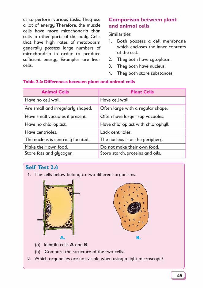
45
us to perform various tasks. They use a lot of energy. Therefore, the muscle cells have more mitochondria than cells in other parts of the body. Cells that have high rates of metabolism generally possess large numbers of mitochondria in order to produce suffi cient energy. Examples are liver cells.
Comparison between plant and animal cells
Similarities1. Both possess a cell membrane
which encloses the inner contents of the cell.
2. They both have cytoplasm.3. They both have nucleus.
4. They both store substances.
Table 2.4: Differences between plant and animal cells
Animal Cells Plant Cells
Have no cell wall. Have cell wall.
Are small and irregularly shaped. Often large with a regular shape.
Have small vacuoles if present. Often have larger sap vacuoles.
Have no chloroplast. Have chloroplast with chlorophyll.
Have centrioles. Lack centrioles.
The nucleus is centrally located. The nucleus is at the periphery.
Make their own food. Do not make their own food.Store fats and glycogen. Store starch, proteins and oils.
Self Test 2.41. The cells below belong to two different organisms.
A. B.(a) Identify cells A and B.
(b) Compare the structure of the two cells.
2. Which organelles are not visible when using a light microscope?
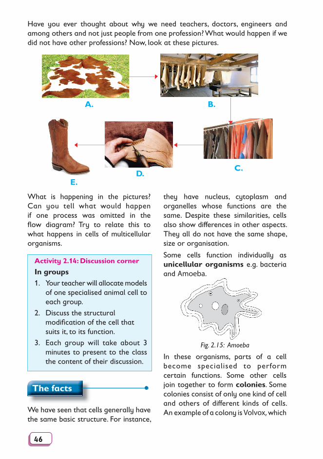
46
Have you ever thought about why we need teachers, doctors, engineers and among others and not just people from one profession? What would happen if we did not have other professions? Now, look at these pictures.
A. B.
C.
E.D.
What is happening in the pictures? Can you tell what would happen if one process was omitted in the fl ow diagram? Try to relate this to what happens in cells of multicellular organisms.
Activity 2.14: Discussion corner
In groups1. Your teacher will allocate models
of one specialised animal cell to each group.
2. Discuss the structural modifi cation of the cell that suits it, to its function.
3. Each group will take about 3 minutes to present to the class the content of their discussion.
The facts
We have seen that cells generally have the same basic structure. For instance,
they have nucleus, cytoplasm and organelles whose functions are the same. Despite these sim i lar i ties, cells also show differences in oth er aspects. They all do not have the same shape, size or organisation.
Some cells function individually as unicellular organisms e.g. bacteria and Amoeba.
Fig. 2.15: Amoeba
In these organisms, parts of a cell become specialised to perform cer tain functions. Some other cells join together to form colonies. Some colonies consist of only one kind of cell and others of different kinds of cells. An example of a colony is Volvox, which
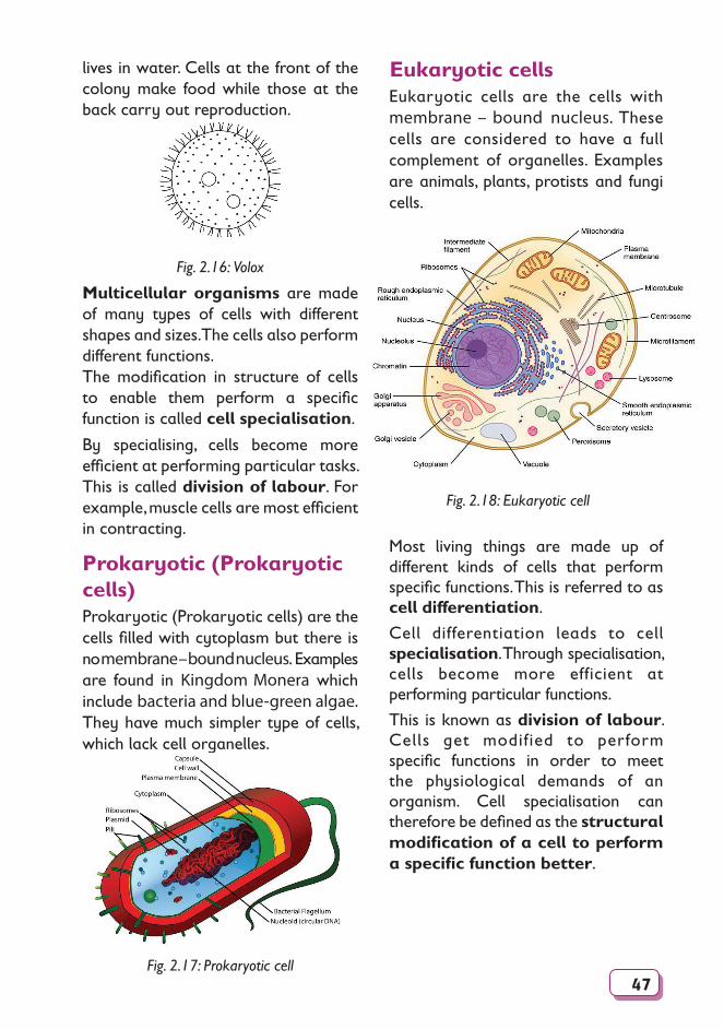
47
lives in water. Cells at the front of the colony make food while those at the back carry out reproduction.
Fig. 2.16: Volox
Multicellular organisms are made of many types of cells with different shapes and sizes. The cells also perform different functions.The modification in structure of cells to enable them perform a specific function is called cell specialisation.
By specialising, cells become more efficient at performing particular tasks. This is called division of labour. For example, muscle cells are most efficient in contracting.
Prokaryotic (Prokaryotic cells)Prokaryotic (Prokaryotic cells) are the cells filled with cytoplasm but there is no membrane – bound nucleus. Examples are found in Kingdom Monera which include bacteria and blue-green algae. They have much simpler type of cells, which lack cell organelles.
Fig. 2.17: Prokaryotic cell
Eukaryotic cellsEukaryotic cells are the cells with membrane – bound nucleus. These cells are considered to have a full complement of organelles. Examples are animals, plants, protists and fungi cells.
Fig. 2.18: Eukaryotic cell
Most living things are made up of different kinds of cells that perform specific functions. This is referred to as cell differentiation.
Cell differentiation leads to cell specialisation. Through specialisation, cells become more efficient at performing particular functions.
This is known as division of labour. Cells get modified to perform specific functions in order to meet the physiological demands of an organism. Cell specialisation can therefore be defined as the structural modification of a cell to perform a specific function better.
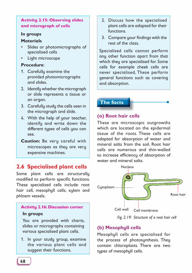
48
Activity 2.15: Observing slides and micrograph of cells
In groups
Materials• Slides or photomicrographs of
specialised cells • Light microscope
Procedure: 1. Carefully examine the
provided photomicrographs and slides.
2. Identify whether the micrograph or slide represents a tissue or an organ.
3. Carefully study the cells seen in the micrograph and slide.
4. With the help of your teacher, identify and write down the different types of cells you can see.
Caution: Be very careful with microscopes as they are very expensive machines.
2.6 Specialised plant cellsSome plant cells are structurally modifi ed to perform specifi c functions. These specialised cells include: root hair cell, mesophyll cells, xylem and phloem vessels.
2. Discuss how the specialised plant cells are adapted for their functions.
3. Compare your fi ndings with the rest of the class.
Specialised cells cannot perform any other function apart from that which they are specialised for. Some cells for example cheek cells are never specialised, These perform general functions such as covering and absorption.
The facts
(a) Root hair cellsThese are microscopic outgrowths which are located on the epidermal tissue of the roots. These cells are adapted for absorption of water and mineral salts from the soil. Root hair cells are numerous and thin-walled to increase effi ciency of absorption of water and mineral salts.
Root hair
Cytoplasm
Cell wall Cell membrane
Nucleus
Fig. 2.19: Structure of a root hair cell
(b) Mesophyll cellsMesophyll cells are specialised for the process of photosynthesis. They contain chloroplasts. There are two types of mesophyll cells.
Activity 2.16: Discussion corner
In groups
You are provided with charts, slides or micrographs containing various specialised plant cells.
1. In your study group, examine the various plant cells and suggest their functions.
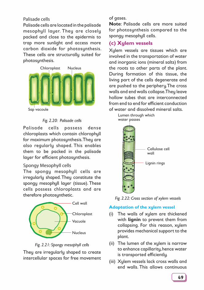
49
of gases.Note: Palisade cells are more suited for photosynthesis compared to the spongy mesophyll cells.
(c) Xylem vesselsXylem vessels are tissues which are involved in the transportation of water and inorganic ions (mineral salts) from the roots to other parts of the plant. During formation of this tissue, the living part of the cells degenerate and are pushed to the periphery. The cross walls and end walls collapse. They leave hollow tubes that are interconnected from end to end for efficient conduction of water and dissolved mineral salts.
Lumen through which water passes
Cellulose cell wall
Lignin rings
Fig. 2.22: Cross section of xylem vessels
Adaptation of the xylem vessel
(i) The walls of xylem are thickened with lignin to prevent them from collapsing. For this reason, xylem provides mechanical support to the plant.
(ii) The lumen of the xylem is narrow to enhance capillarity, hence water is transported efficiently.
(iii) Xylem vessels lack cross walls and end walls. This allows continuous
Palisade cellsPalisade cells are located in the palisade mesophyll layer. They are closely packed and close to the epidermis to trap more sunlight and access more carbon dioxide for photosynthesis. These cells are structurally suited for photosynthesis.
Chloroplast Nucleus
Sap vacoule
Fig. 2.20: Palisade cells
Palisade cells possess dense chloroplasts which contain chlorophyll for maximum photosynthesis. They are also regularly shaped. This enables them to be packed in the palisade layer for efficient photosynthesis.
Spongy Mesophyll cellsThe spongy mesophyll cells are irregularly shaped. They constitute the spongy mesophyll layer (tissue). These cells possess chloroplasts and are therefore photosynthetic.
Nucleus
Vacuole
Chloroplast
Cell wall
Fig. 2.21: Spongy mesophyll cells
They are irregularly shaped to create intercellular spaces for free movement
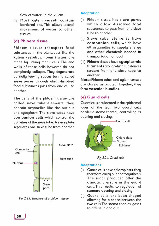
50
flow of water up the xylem.(iv) Most xylem vessels contain
bordered pits. This allows lateral movement of water to other tissues.
(d) Phloem tissue
Phloem t issues transpor t food substances in the plant. Just like the xylem vessels, phloem tissues are made by linking many cells. The end walls of these cells however, do not completely collapse. They degenerate partially leaving spaces behind called sieve pores, through which dissolved food substances pass from one cell to another.
The cells of the phloem tissue are called sieve tube elements; they contain organelles like the nucleus and cytoplasm. The sieve tubes have companion cells which control the activities of the sieve tube. A sieve plate separates one sieve tube from another.
Sieve plate
Sieve tube
Companion cell
Nucleus
Sieve pores
Fig. 2.23: Structure of a phloem tissue
Adaptation
(i) Phloem tissue has sieve pores which al low dissolved food substances to pass from one sieve tube to another.
(ii) S i eve t ube e l emen t s have companion cells, which have all organelles to supply energy and other chemicals needed in transportation of food.
(iii) Phloem tissues have cytoplasmic filaments along which substances stream from one sieve tube to another.
Note: Phloem tubes and xylem vessels are closely associated. Together, they form vascular bundles.
(e) Guard cellsGuard cells are located in the epidermal layer of the leaf. Two guard cells border a stoma thereby controlling its opening and closing.
Epidermis
Guard cell
ChloroplastStoma
Fig. 2.24: Guard cells
Adaptations(i) Guard cells have chloroplasts, they
therefore carry out photosynthesis. The sugar produced offer the osmotic pressure in the guard cells. This results to regulation of stomata opening and closing.
(ii) Guard cells are bean-shaped allowing for a space between the two cells. The stoma enables gases to diffuse in and out.
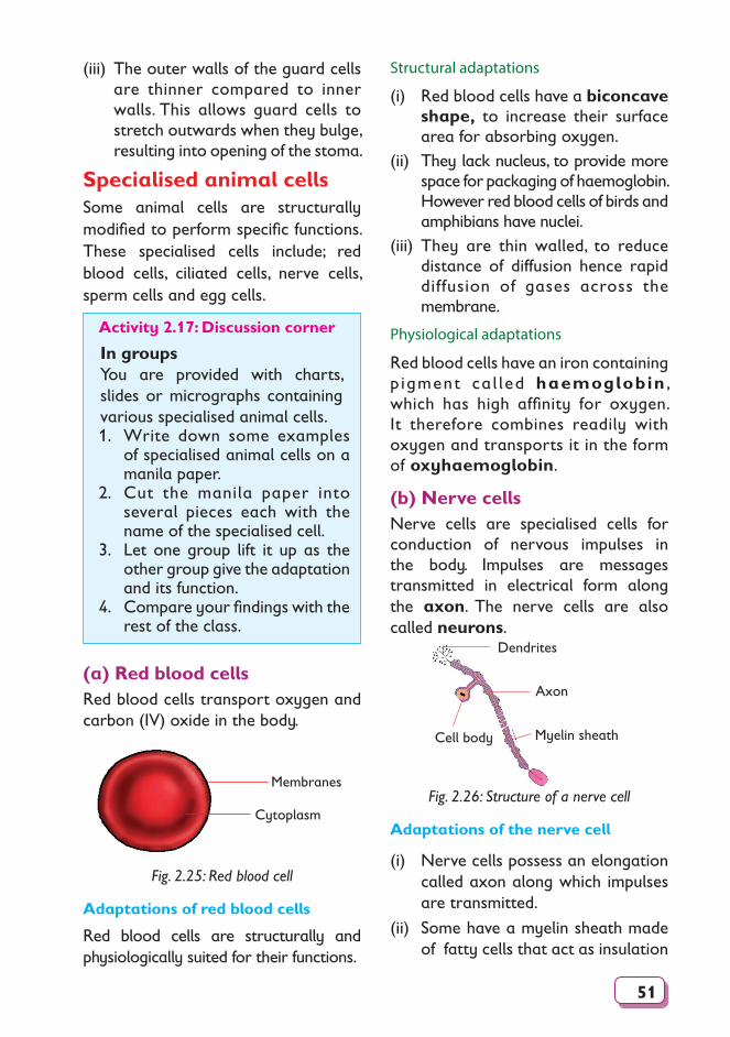
51
(iii) The outer walls of the guard cells are thinner compared to inner walls. This allows guard cells to stretch outwards when they bulge, resulting into opening of the stoma.
Specialised animal cellsSome animal cells are structurally modified to perform specific functions. These specialised cells include; red blood cells, ciliated cells, nerve cells, sperm cells and egg cells.
Activity 2.17: Discussion corner
In groupsYou are provided with charts, slides or micrographs containing various specialised animal cells.1. Write down some examples
of specialised animal cells on a manila paper.
2. Cut the manila paper into several pieces each with the name of the specialised cell.
3. Let one group lift it up as the other group give the adaptation and its function.
4. Compare your findings with the rest of the class.
(a) Red blood cellsRed blood cells transport oxygen and carbon (IV) oxide in the body.
Membranes
Cytoplasm
Fig. 2.25: Red blood cell
Adaptations of red blood cells
Red blood cells are structurally and physiologically suited for their functions.
Structural adaptations
(i) Red blood cells have a biconcave shape, to increase their surface area for absorbing oxygen.
(ii) They lack nucleus, to provide more space for packaging of haemoglobin. However red blood cells of birds and amphibians have nuclei.
(iii) They are thin walled, to reduce distance of diffusion hence rapid diffusion of gases across the membrane.
Physiological adaptations
Red blood cells have an iron containing p igment ca l led haemoglobin , which has high affinity for oxygen. It therefore combines readily with oxygen and transports it in the form of oxyhaemoglobin.
(b) Nerve cellsNerve cells are specialised cells for conduction of nervous impulses in the body. Impulses are messages transmitted in electrical form along the axon. The nerve cells are also called neurons.
Dendrites
Cell body
Axon
Myelin sheath
Fig. 2.26: Structure of a nerve cell
Adaptations of the nerve cell
(i) Nerve cells possess an elongation called axon along which impulses are transmitted.
(ii) Some have a myelin sheath made of fatty cells that act as insulation
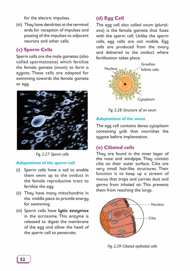
52
for the electric impulses.
(iii) They have dendrites at the terminal ends for reception of impulses and passing of the impulses to adjacent neurons and other cells.
(c) Sperm CellsSperm cells are the male gametes (also called spermatozoa) which fertilise the female gamete (ovum) to form a zygote. These cells are adapted for swimming towards the female gamete or egg.
Fig. 2.27: Sperm cells
Adaptations of the sperm cell
(i) Sperm cells have a tail to enable them swim up to the oviduct in the female reproductive tract to fertilise the egg.
(ii) They have many mitochondria in the middle piece to provide energy for swimming.
(iii) Sperm cells have lytic enzymes in the acrosome. This enzyme is released to digest the membrane of the egg and allow the head of the sperm cell to penetrate.
(d) Egg CellThe egg cell also called ovum (plural-ova) is the female gamete that fuses with the sperm cell. Unlike the sperm cells, egg cells are not mobile. Egg cells are produced from the ovary and delivered to the oviduct where fertilisation takes place.
Nucleus
Cytoplasm
Graafian follicle cells
Fig. 2.28: Structure of an ovum
Adaptations of the ovum
The egg cell contains dense cytoplasm containing yolk that nourishes the zygote before implantation.
(e) Ciliated cellsThey are found in the inner layer of the nose and windpipe. They contain cilia on their outer surface. Cilia are very small hair-like structures. Their function is to keep up a stream of mucus that traps and carries dust and germs from inhaled air. This prevents them from reaching the lungs.
Cilia
Nucleus
Fig. 2.29: Ciliated epithelial cells
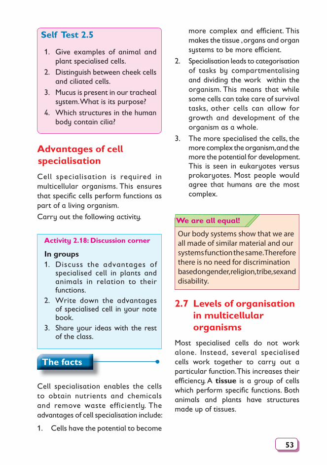
53
Self Test 2.5
1. Give examples of animal and plant specialised cells.
2. Distinguish between cheek cells and ciliated cells.
3. Mucus is present in our tracheal system. What is its purpose?
4. Which structures in the human body contain cilia?
Advantages of cell specialisation
Cell specialisation is required in multicellular organisms. This ensures that specifi c cells perform functions as part of a living organism.
Carry out the following activity.
Activity 2.18: Discussion corner
In groups1. Discuss the advantages of
specialised cell in plants and animals in relation to their functions.
2. Write down the advantages of specialised cell in your note book.
3. Share your ideas with the rest of the class.
The facts
Cell specialisation enables the cells to obtain nutrients and chemicals and remove waste efficiently. The advantages of cell specialisation include:
1. Cells have the potential to become
more complex and effi cient. This makes the tissue , organs and organ systems to be more effi cient.
2. Specialisation leads to categorisation of tasks by compartmentalising and dividing the work within the organism. This means that while some cells can take care of survival tasks, other cells can allow for growth and development of the organism as a whole.
3. The more specialised the cells, the more complex the organism, and the more the potential for development. This is seen in eukaryotes versus prokaryotes. Most people would agree that humans are the most complex.
Our body systems show that we are all made of similar material and our systems function the same. Therefore there is no need for discrimination based on gender, religion, tribe, sex and disability.
We are all equal!
2.7 Levels of organisation in multicellular organisms
Most specialised cells do not work alone. Instead, several specialised cells work together to carry out a particular function. This increases their effi ciency. A tissue is a group of cells which perform specifi c functions. Both animals and plants have structures made up of tissues.
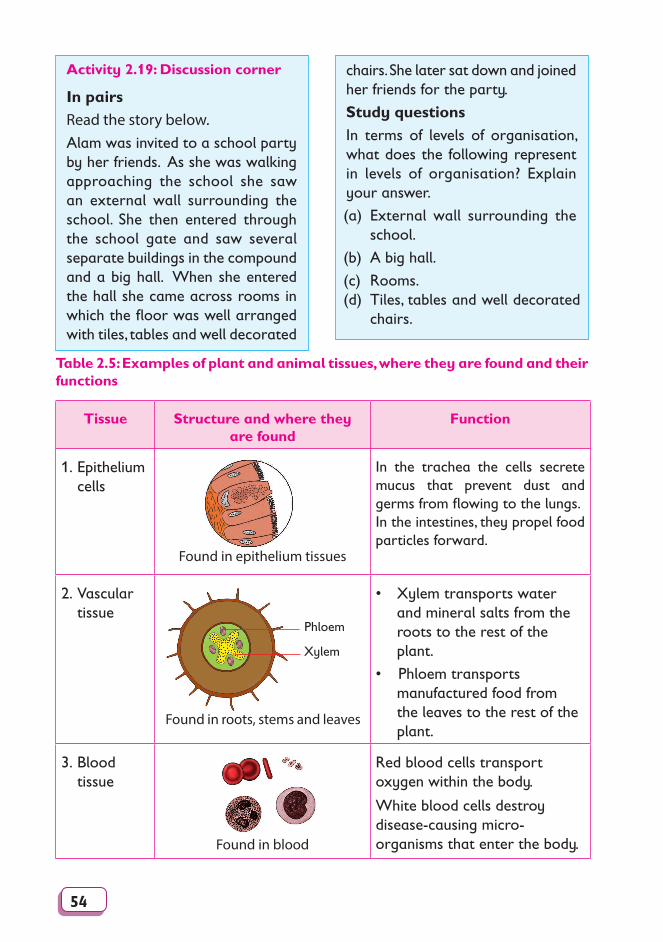
54
Activity 2.19: Discussion corner
In pairsRead the story below.Alam was invited to a school party by her friends. As she was walking approaching the school she saw an external wall surrounding the school. She then entered through the school gate and saw several separate buildings in the compound and a big hall. When she entered the hall she came across rooms in which the floor was well arranged with tiles, tables and well decorated
chairs. She later sat down and joined her friends for the party.
Study questionsIn terms of levels of organisation, what does the following represent in levels of organisation? Explain your answer.
(a) External wall surrounding the school.
(b) A big hall.
(c) Rooms.(d) Tiles, tables and well decorated
chairs.
Table 2.5: Examples of plant and animal tissues, where they are found and their functions
Tissue Structure and where they are found
Function
1. Epithelium cells
Found in epithelium tissues
In the trachea the cells secrete mucus that prevent dust and germs from flowing to the lungs.In the intestines, they propel food particles forward.
2. Vascular tissue
Found in roots, stems and leaves
Phloem
Xylem
• Xylem transports water and mineral salts from the roots to the rest of the plant.
• Phloem transports manufactured food from the leaves to the rest of the plant.
3. Blood tissue
Found in blood
Red blood cells transport oxygen within the body.
White blood cells destroy disease-causing micro-organisms that enter the body.
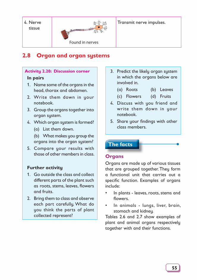
55
4. Nerve tissue
Found in nerves
Transmit nerve impulses.
2.8 Organ and organ systems
3. Predict the likely organ system in which the organs below are involved in.
(a) Roots (b) Leaves
(c) Flowers (d) Fruits
4. Discuss with you friend and write them down in your notebook.
5. Share your fi ndings with other class members.
Activity 2.20: Discussion corner
In pairs1. Name some of the organs in the
head, thorax and abdomen.
2. Write them down in your notebook.
3. Group the organs together into organ system.
4. Which organ system is formed?
(a) List them down.
(b) What makes you group the organs into the organ system?
5. Compare your results with those of other members in class.
Further activity1. Go outside the class and collect
different parts of the plant such as roots, stems, leaves, fl owers and fruits.
2. Bring them to class and observe each part carefully. What do you think the parts of plant collected represent?
The facts
Organs Organs are made up of various tissues that are grouped together. They form a functional unit that carries out a specifi c function. Examples of organs include:
• In plants - leaves, roots, stems and fl owers.
• In animals - lungs, liver, brain, stomach and kidney.
Tables 2.6 and 2.7 show examples of plant and animal organs respectively together with and their functions.
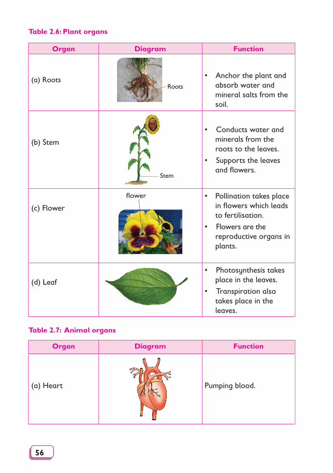
56
Table 2.6: Plant organs
Organ Diagram Function
(a) RootsRoots
• Anchor the plant and absorb water and mineral salts from the soil.
(b) Stem
Stem
• Conducts water and minerals from the roots to the leaves.
• Supports the leaves and flowers.
(c) Flower
flower • Pollination takes place in flowers which leads to fertilisation.
• Flowers are the reproductive organs in plants.
(d) Leaf
• Photosynthesis takes place in the leaves.
• Transpiration also takes place in the leaves.
Table 2.7: Animal organs
Organ Diagram Function
(a) Heart Pumping blood.
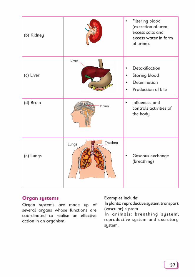
57
(b) Kidney
• Filtering blood (excretion of urea, excess salts and excess water in form of urine).
(c) Liver
Liver
• Detoxifi cation
• Storing blood
• Deamination
• Production of bile
(d) BrainBrain
• Infl uences and controls activities of the body.
(e) Lungs
TracheaLungs
• Gaseous exchange (breathing)
Organ systemsOrgan systems are made up of several organs whose functions are coordinated to realise an effective action in an organism.
Examples include:In plants: reproductive system, transport (vascular) system.In an ima l s : breath ing sys tem, reproductive system and excretory system.
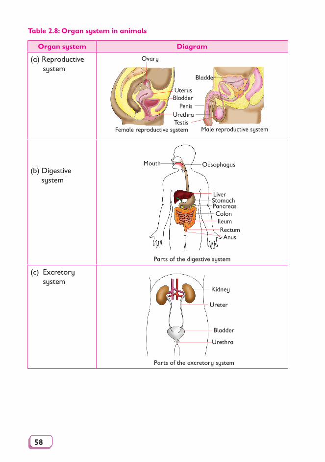
58
Table 2.8: Organ system in animals
Organ system Diagram
(a) Reproductive system
Urethra
BladderUterus
Ovary
Female reproductive system Male reproductive system
Penis
Bladder
Testis
(b) Digestive system
Mouth Oesophagus
LiverStomachPancreasColonIleum
AnusRectum
Parts of the digestive system
(c) Excretory system
Kidney
Ureter
Bladder
Urethra
Parts of the excretory system
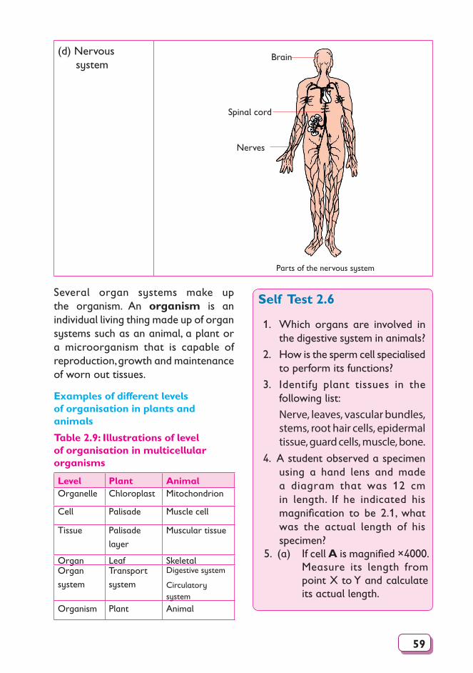
59
(d) Nervous system
Brain
Spinal cord
Nerves
Parts of the nervous system
Several organ systems make up the organism. An organism is an individual living thing made up of organ systems such as an animal, a plant or a microorganism that is capable of reproduction, growth and maintenance of worn out tissues.
Examples of different levels of organisation in plants and animals
Table 2.9: Illustrations of level of organisation in multicellular organisms
Level Plant Animal Organelle Chloroplast Mitochondrion
Cell Palisade Muscle cell
Tissue Palisade
layer
Muscular tissue
Organ Leaf Skeletal Organ
system
Transport
system
Digestive system
Circulatory system
Organism Plant Animal
Self Test 2.6
1. Which organs are involved in the digestive system in animals?
2. How is the sperm cell specialised to perform its functions?
3. Identify plant tissues in the following list:
Nerve, leaves, vascular bundles, stems, root hair cells, epidermal tissue, guard cells, muscle, bone.
4. A student observed a specimen using a hand lens and made a diagram that was 12 cm in length. If he indicated his magnification to be 2.1, what was the actual length of his specimen?
5. (a) If cell A is magnified ×4000. Measure its length from point X to Y and calculate its actual length.

60
Check Your Progress 2
X A Y
(b) If the actual length of a cell B from X to Y is 0.0045 µm. Calculate its magnifi cation. Show your working.
X YB
1. (a) Why is a hand lens sometimes referred to as a simple microscope?
(b) Why is the microscope commonly used today called light microscope?
2. The following is a diagram of a light microscope.
1
10
11
12
13
14
9
2
34
76
5
8
(a) Identify the parts of the microscope represented by the numbers 8, 13 and 14.
(b) Give the functions of the parts represented by the numbers 4, 5, 11 and 12.
(c) Describe briefl y how you would lift a microscope from your bench and take it to the teacher’s bench.
(d) At which objective lens is the microscope stored?
(e) What is the name given to the circular area of light seen when you look into a light microscope?
(f) How would you work out the magnification of the microscope?
(g) How do you keep the lenses in the microscope clean?
3. Name the specialised cell that is concerned with each of the following functions.(a) A plant collapsing due to
weak stem.(b) Stomata failing to open.(c) An ovum not getting
fertilised.(d) Transmission of nerve
impulses.4. Arrange the following from the
simplest to the most complex. Palisade, mesophyll layer, leaf
palisade cell, chloroplast, maize plant.
5. Gesambi got a cut on his left leg. A doctor realised that his blood could not clot. Which of these cells in his blood was having a problem?
AAB
C DD
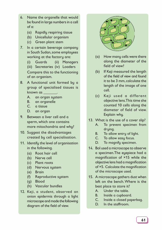
61
6. Name the organelle that would be found in large numbers in a cell of a:
(a) Rapidly respiring tissue (b) Unicellular organism (c) Green plant stem
7. In a certain beverage company in South Sudan, some employees working at the factory are:
(i) Guards (ii) Managers (iii) Secretaries (iv) Loaders Compare this to the functioning
of an organism.
8. A functional unit formed by a group of specialised tissues is known as _______.
A. an organ system B. an organelle C. a tissue D. an organ
9. Between a liver cell and a sperm, which one contains more mitochondria and why?
10. Suggest the disadvantages created by cell specialisation.
11. Identify the level of organisation in the following.
(a) Root hair cell (b) Nerve cell (c) Plant roots (d) Nervous system (e) Brain (f) Reproductive system (g) Blood (h) Vascular bundles
12. Keji, a student, observed an onion epidermis through a light microscope and made the following diagram of the fi eld of view.
(a) How many cells were there along the diameter of the fi eld of view?
(b) If Keji measured the length of the fi eld of view and found it to be 3 mm, calculate the length of the image of one cell.
(c) Ke j i used a d i f ferent objective lens. This time she counted 10 cells along the diameter of fi eld of view. Explain why.
13. What is the use of a cover slip?A. To prevent specimen from
drying.B. To allow entry of light.C. To allow easy focus.D. To magnify specimen.
14. Bol used a microscope to observe a specimen.The eyepiece had a magnifi cation of ×15 while the objective lens had a magnifi cation of ×5. Calculate the magnifi cation of the microscope used.
15. A microscope gathers dust when left on the bench. Where is the best place to store it?A. Under the table.B. Inside a cupboard.C. Inside a closed paperbag.D. In the staffroom.
62
16. Suppose you are provided with:(a) Onion leaf(b) Fish(c) Mushroom (d) Human skin piece(e) Microorganisms
Suggest which instrument you will use to view details of each of the specimen above.
17. If the microscope clips are broken, how would you set a prepared slide on stage?
63
3Unit Movement of substances
into and out of cells
Learning outcomes
Knowledge and understanding
Skills Attitudes
• Describe the
movement of
substances
across the cells.
• Analyse the structure of
cell membrane.
• Design simple investigations
about the process of
diffusion (osmosis) in plants
and animals using sugar
solutions.
• Gain skill of the safe
assembly and use of
apparatus for experiments.
• Appreciate the simple
techniques used to accomplish
tasks (e.g. diffusion, osmosis).
• Adopt the habit of being
systematic, resilient and
accurate.
• Importance of being systematic,
accurate, ethical, and meticulous
in carrying out experiments.
• Share ideas when carrying out
the investigations.
IntroductionIn order to carry out life processes, that were mentioned in Unit 1, a cell needs and takes in various substances. In carrying out these processes, the cell produces certain substances as well. Some of these are needed by the cell, some are waste products and others are substances that the cell does not need but are useful to other cells. Some useful substances are taken out of the cell and transported to other cells that need them. Substances are constantly moving into and out of cells.
3.1 Structure and properties of the cell membrane
Structure of the cell membrane
Cell membranes are very thin. They are made up of proteins and lipids. The lipid material is arranged in two thin outer layers. The protein part is found scattered and embedded inside this double layer of lipid, See Fig. 3.1. Cell membranes are important because they separate and enclose the cell contents from its sorrounding.
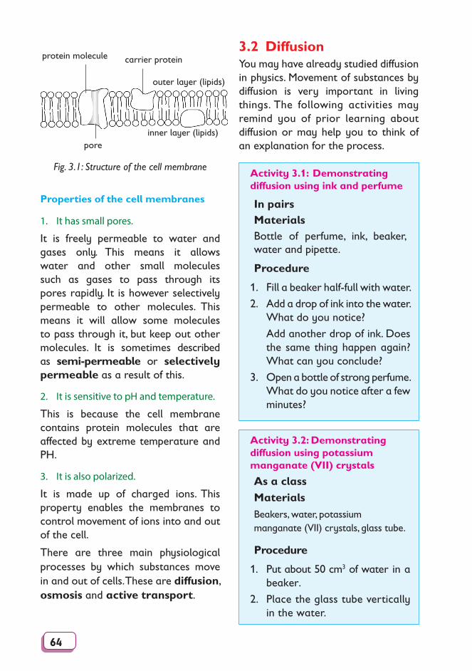
64
carrier proteinprotein molecule
outer layer (lipids)
inner layer (lipids)pore
Fig. 3.1: Structure of the cell membrane
Properties of the cell membranes
1. It has small pores.
It is freely permeable to water and gases only. This means it allows water and other small molecules such as gases to pass through its pores rapidly. It is however selectively permeable to other molecules. This means it will allow some molecules to pass through it, but keep out other molecules. It is sometimes described as semi-permeable or selectively permeable as a result of this.
2. It is sensitive to pH and temperature.
This is because the cell membrane contains protein molecules that are affected by extreme temperature and PH.
3. It is also polarized.
It is made up of charged ions. This property enables the membranes to control movement of ions into and out of the cell.
There are three main physiological processes by which substances move in and out of cells. These are diffusion, osmosis and active transport.
3.2 DiffusionYou may have already studied diffusion in physics. Movement of substances by diffusion is very important in living things. The following activities may remind you of prior learning about diffusion or may help you to think of an explanation for the process.
Activity 3.1: Demonstrating diffusion using ink and perfume
In pairsMaterialsBottle of perfume, ink, beaker, water and pipette.
Procedure
1. Fill a beaker half-full with water.
2. Add a drop of ink into the water. What do you notice?
Add another drop of ink. Does the same thing happen again? What can you conclude?
3. Open a bottle of strong perfume. What do you notice after a few minutes?
Activity 3.2: Demonstrating diffusion using potassium manganate (VII) crystals
As a classMaterialsBeakers, water, potassium manganate (VII) crystals, glass tube.
Procedure
1. Put about 50 cm3 of water in a beaker.
2. Place the glass tube vertically in the water.
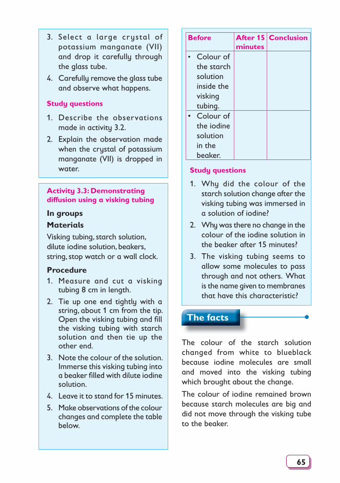
65
3. Select a large crystal of potassium manganate (VII) and drop it carefully through the glass tube.
4. Carefully remove the glass tube and observe what happens.
Study questions
1. Describe the observations made in activity 3.2.
2. Explain the observation made when the crystal of potassium manganate (VII) is dropped in water.
Before After 15 minutes
Conclusion
• Colour of the starch solution in side the visking tubing.
• Colour of the iodine solution in the beaker.
Study questions
1. Why did the colour of the starch solution change after the visking tubing was immersed in a solution of iodine?
2. Why was there no change in the colour of the iodine solution in the beaker after 15 minutes?
3. The visking tubing seems to allow some molecules to pass through and not others. What is the name given to membranes that have this characteristic?
Activity 3.3: Demonstrating diffusion using a visking tubing
In groupsMaterialsVisking tubing, starch solution, dilute iodine solution, beakers, string, stop watch or a wall clock.
Procedure1. Measure and cut a visking
tubing 8 cm in length.
2. Tie up one end tightly with a string, about 1 cm from the tip. Open the visking tubing and fi ll the visking tubing with starch solution and then tie up the other end.
3. Note the colour of the solution. Immerse this visking tubing into a beaker fi lled with dilute iodine solution.
4. Leave it to stand for 15 minutes.
5. Make observations of the colour changes and complete the table below.
The facts
The colour of the starch solution changed from white to blueblack because iodine molecules are small and moved into the visking tubing which brought about the change.
The colour of iodine remained brown because starch molecules are big and did not move through the visking tube to the beaker.
66
The visking tubing has small pores that can only allow small permeable molecules to pass.
Therefore diffusion is the movement of particles of a substance from a region of high concentration of the particles to a region of low concentration of the particles. The difference in the concentration of particles in the two regions is called a concentration gradient or diffusion gradient.
Diffusion occurs in liquids and gases. Diffusion does not take place in solids. Diffusion of certain particles also takes place through the cell membrane, cell walls and cell cytoplasm.
When particles move from a region of high concentration to a region of low concentration, they are said to diffuse along a concentration gradient. As long as a concentration gradient is maintained, the movement of particles continues until they are evenly distributed in the available space; thus an equilibrium is reached.
Factors affecting the rate of diffusion
The rate of diffusion of particles is the time taken for the particles to diffuse within an available (fixed) space until they are evenly distributed. Several factors affect the rate of diffusion. They include.
• Temperature
• Surface area
• Concentration gradient
• Surface area to volume ratio• Distance the particles have to travel
(a) Temperature
When the temperature of particles is
increased, their kinetic energy content of particles also increases and the particles move faster. Therefore, the higher the temperature, the faster the particles will diffuse. The lower the temperature the lower the rate of diffusion.
(b) Surface area
When the surface area is large, more of the substance diffuses across it than when it is small as long as the concentration and temperature of the diffusing molecules remain the same.
The importance of a large surface area for diffusion is seen in absorption surfaces of plants and animals.
These surfaces are modified to create a large surface area so that larger quantities of substances can be absorbed across them by diffusion.
The surfaces in the small intestine, the lungs and the gill filaments of fish are examples of absorption surfaces with a large surface area.
Note: A larger suface area does not increase the speed of diffusion of the particles. It simply enables more particles to diffuse across it in a given time.
• More particles diffuse across the membrane in a given time.
• It takes a shorter time for the same (fixed) number of particles to diffuse across it.
• Fewer particles will diffuse across the surface area in a given time i.e. 3 - particles.
• It takes a longer time for the same (fixed) number of particles to diffuse across it.
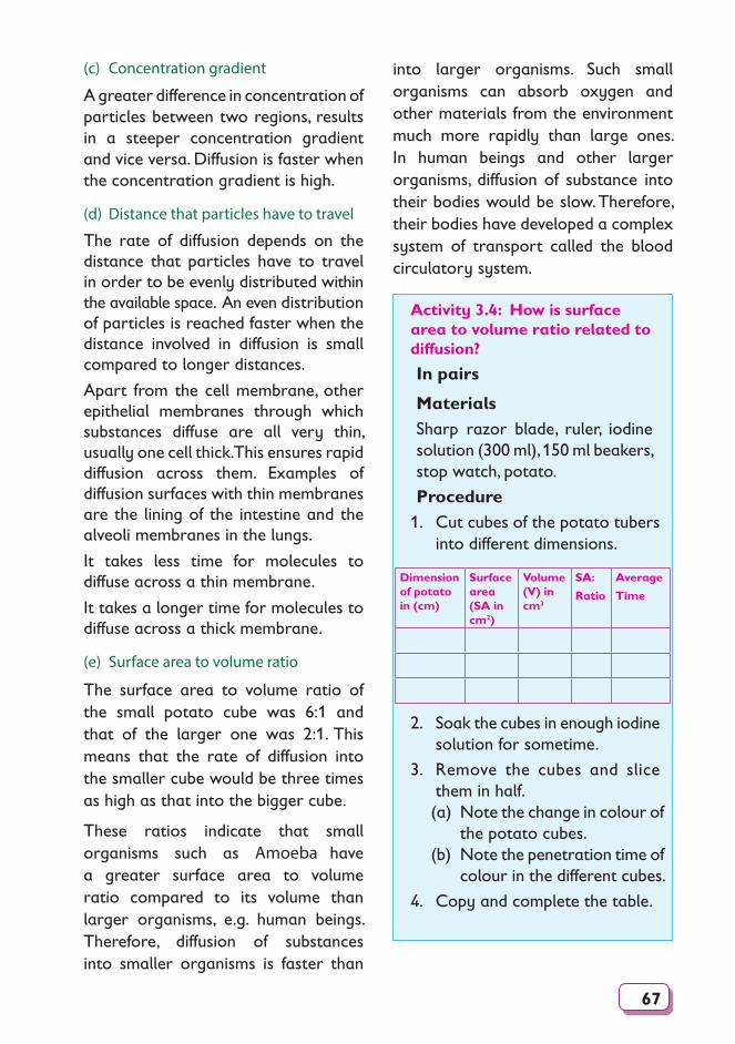
67
(c) Concentration gradient
A greater difference in concentration of particles between two regions, results in a steeper concentration gradient and vice versa. Diffusion is faster when the concentration gradient is high.
(d) Distance that particles have to travel
The rate of diffusion depends on the distance that particles have to travel in order to be evenly distributed within the available space. An even distribution of particles is reached faster when the distance involved in diffusion is small compared to longer distances.
Apart from the cell membrane, other epithelial membranes through which substances diffuse are all very thin, usually one cell thick. This ensures rapid diffusion across them. Examples of diffusion surfaces with thin membranes are the lining of the intestine and the alveoli membranes in the lungs.
It takes less time for molecules to diffuse across a thin membrane.
It takes a longer time for molecules to diffuse across a thick membrane.
(e) Surface area to volume ratio
The surface area to volume ratio of the small potato cube was 6:1 and that of the larger one was 2:1. This means that the rate of diffusion into the smaller cube would be three times as high as that into the bigger cube.
These ratios indicate that small organisms such as Amoeba have a greater surface area to volume ratio compared to its volume than larger organisms, e.g. human beings. Therefore, diffusion of substances into smaller organisms is faster than
into larger organisms. Such small organisms can absorb oxygen and other materials from the environment much more rapidly than large ones. In human beings and other larger organisms, diffusion of substance into their bodies would be slow. Therefore, their bodies have developed a complex system of transport called the blood circulatory system.
Activity 3.4: How is surface area to volume ratio related to diffusion?
In pairs
MaterialsSharp razor blade, ruler, iodine solution (300 ml), 150 ml beakers, stop watch, potato.
Procedure1. Cut cubes of the potato tubers
into different dimensions.
Dimension of potato in (cm)
Surface area (SA in cm2)
Volume (V) in cm3
SA:
Ratio
Average
Time
2. Soak the cubes in enough iodine solution for sometime.
3. Remove the cubes and slice them in half.(a) Note the change in colour of
the potato cubes.(b) Note the penetration time of
colour in the different cubes.
4. Copy and complete the table.
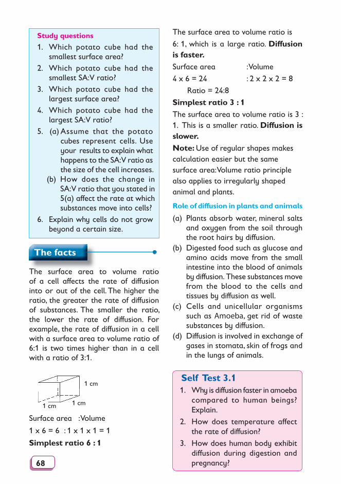
68
Study questions
1. Which potato cube had the smallest surface area?
2. Which potato cube had the smallest SA:V ratio?
3. Which potato cube had the largest surface area?
4. Which potato cube had the largest SA:V ratio?
5. (a) Assume that the potato cubes represent cells. Use your results to explain what happens to the SA:V ratio as the size of the cell increases.
(b) How does the change in SA:V ratio that you stated in 5(a) affect the rate at which substances move into cells?
6. Explain why cells do not grow beyond a certain size.
The surface area to volume ratio is
6: 1, which is a large ratio. Diffusion is faster.
Surface area : Volume
4 x 6 = 24 : 2 x 2 x 2 = 8
Ratio = 24:8
Simplest ratio 3 : 1
The surface area to volume ratio is 3 : 1. This is a smaller ratio. Diffusion is slower.
Note: Use of regular shapes makes calculation easier but the same surface area: Volume ratio principle also applies to irregularly shaped animal and plants.
Role of diffusion in plants and animals
(a) Plants absorb water, mineral salts and oxygen from the soil through the root hairs by diffusion.
(b) Digested food such as glucose and amino acids move from the small intestine into the blood of animals by dif fu sion. These substances move from the blood to the cells and tissues by dif fu sion as well.
(c) Cells and uni cel lu lar organisms such as Amoeba, get rid of waste substances by diffusion.
(d) Diffusion is involved in exchange of gases in stomata, skin of frogs and in the lungs of animals.
Self Test 3.11. Why is diffusion faster in amoeba
compared to human beings? Explain.
2. How does temperature affect the rate of diffusion?
3. How does human body exhibit diffusion during digestion and pregnancy?
The facts
The surface area to volume ratio of a cell affects the rate of diffusion into or out of the cell. The higher the ratio, the greater the rate of diffusion of substances. The smaller the ratio, the lower the rate of dif fu sion. For example, the rate of diffusion in a cell with a surface area to vol ume ratio of 6:1 is two times higher than in a cell with a ratio of 3:1.
1 cm
1 cm1 cm
Surface area : Volume
1 x 6 = 6 : 1 x 1 x 1 = 1
Simplest ratio 6 : 1
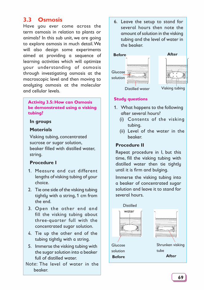
69
3.3 OsmosisHave you ever come across the term osmosis in relation to plants or animals? In this sub unit, we are going to explore osmosis in much detail. We will also design some experiments aimed at providing a sequence of learning activities which will optimize your understanding of osmosis through investigating osmosis at the macroscopic level and then moving to analyzing osmosis at the molecular and cellular levels.
6. Leave the setup to stand for several hours then note the amount of solution in the visking tubing and the level of water in the beaker.
Visking tubing
B
Glucosesolution
Before After
Distilled water
Study questions
1. What happens to the following after several hours?(i) Contents of the visking
tubing.(ii) Level of the water in the
beaker.
Procedure IIRepeat procedure in I, but this time, fill the visking tubing with distilled water then tie tightly until it is firm and bulging.
Immerse the visking tubing into a beaker of concentrated sugar solution and leave it to stand for several hours.
Shrunken visking tube
Distilled water B
Glucose solution
Before After
Activity 3.5: How can Osmosis be demonstrated using a visking tubing?
In groups
MaterialsVisking tubing, concentrated sucrose or sugar solution, beaker filled with distilled water, string.
Procedure I
1. Measure and cut different lengths of visking tubing of your choice.
2. Tie one side of the visking tubing tightly with a string, 1 cm from the end.
3. Open the other end and fill the visking tubing about three-quarter full with the concentrated sugar solution.
4. Tie up the other end of the tubing tightly with a string.
5. Immerse the visking tubing with the sugar solution into a beaker full of distilled water.
Note: The level of water in the beaker.
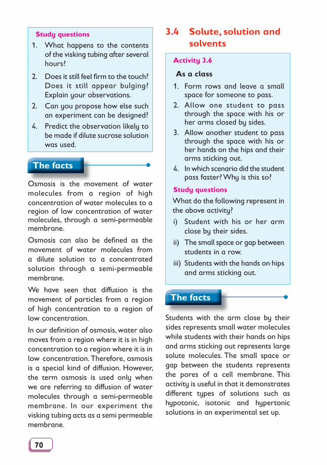
70
3.4 Solute, solution and solvents
Study questions
1. What happens to the contents of the visking tubing after several hours?
2. Does it still feel fi rm to the touch? Does it still appear bulging? Explain your observations.
2. Can you propose how else such an experiment can be designed?
4. Predict the observation likely to be made if dilute sucrose solution was used.
Activity 3.6
As a class
1. Form rows and leave a small space for someone to pass.
2. Allow one student to pass through the space with his or her arms closed by sides.
3. Allow another student to pass through the space with his or her hands on the hips and their arms sticking out.
4. In which scenario did the student pass faster? Why is this so?
Study questions
What do the following represent in the above activity?
i) Student with his or her arm close by their sides.
ii) The small space or gap between students in a row.
iii) Students with the hands on hips and arms sticking out.
The facts
Osmosis is the movement of water molecules from a region of high con cen tra tion of water molecules to a region of low concentration of water molecules, through a semi-permeable membrane.
Osmosis can also be defi ned as the move ment of water molecules from a dilute solution to a concentrated solution through a semi-per me a ble membrane.
We have seen that diffusion is the move ment of particles from a region of high con cen tra tion to a region of low con cen tra tion.
In our defi nition of osmosis, water also moves from a region where it is in high concentration to a region where it is in low concentration. Therefore, osmosis is a special kind of diffusion. However, the term osmosis is used only when we are re fer ring to diffusion of water mol e cules through a semi-permeable membrane. In our experiment the visking tubing acts as a semi permeable membrane.
The facts
Students with the arm close by their sides represents small water molecules while students with their hands on hips and arms sticking out represents large solute molecules. The small space or gap between the students represents the pores of a cell membrane. This activity is useful in that it demonstrates different types of solutions such as hypotonic, isotonic and hypertonic solutions in an experimental set up.
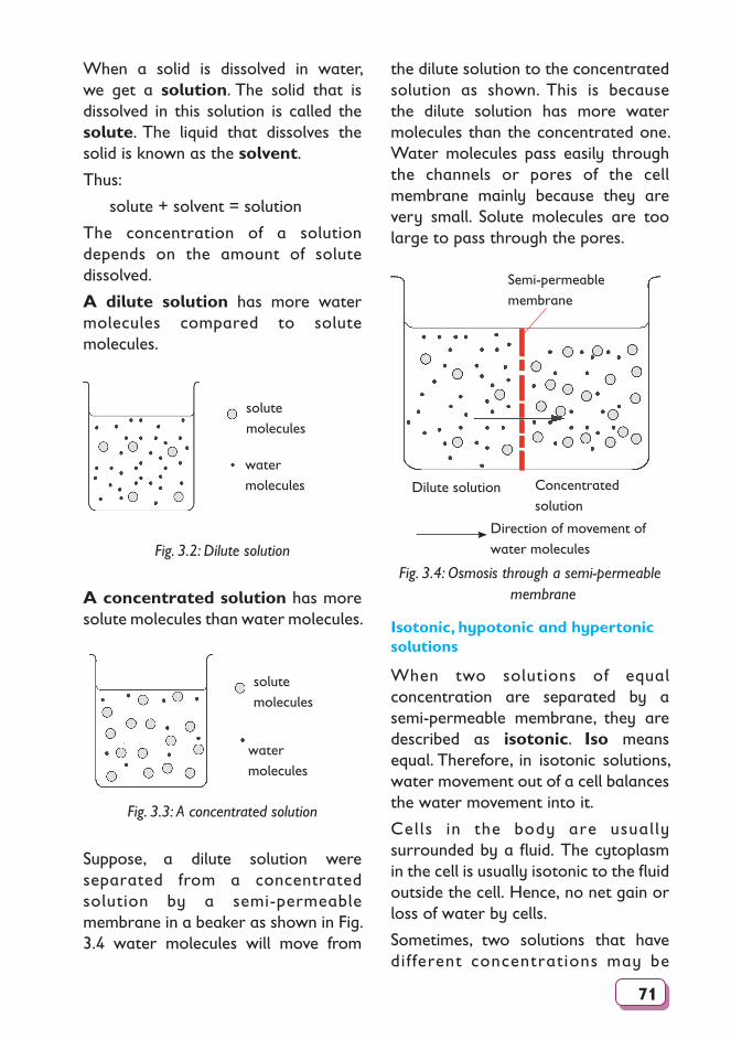
71
When a solid is dissolved in water, we get a solution. The solid that is dissolved in this solution is called the solute. The liquid that dissolves the solid is known as the solvent.
Thus:
solute + solvent = solution
The concentration of a solution depends on the amount of solute dissolved.
A dilute solution has more water molecules compared to solute molecules.
solutemolecules
watermolecules
Fig. 3.2: Dilute solution
A concentrated solution has more solute molecules than water molecules.
solutemolecules
watermolecules
Fig. 3.3: A concentrated solution
Suppose, a dilute solution were separated from a concentrated solution by a semi-permeable membrane in a beaker as shown in Fig. 3.4 water molecules will move from
the dilute solution to the concentrated solution as shown. This is because the dilute solution has more water molecules than the concentrated one. Water molecules pass easily through the channels or pores of the cell membrane mainly because they are very small. Solute molecules are too large to pass through the pores.
Direction of movement of water molecules
Semi-permeable membrane
Dilute solution Concentrated solution
Fig. 3.4: Osmosis through a semi-permeable membrane
Isotonic, hypotonic and hypertonic solutions
When two solutions of equal concentration are separated by a semi-permeable membrane, they are described as isotonic. Iso means equal. Therefore, in isotonic solutions, water movement out of a cell balances the water movement into it.
Cells in the body are usually surrounded by a fluid. The cytoplasm in the cell is usually isotonic to the fluid outside the cell. Hence, no net gain or loss of water by cells.
Sometimes, two solutions that have different concentrations may be
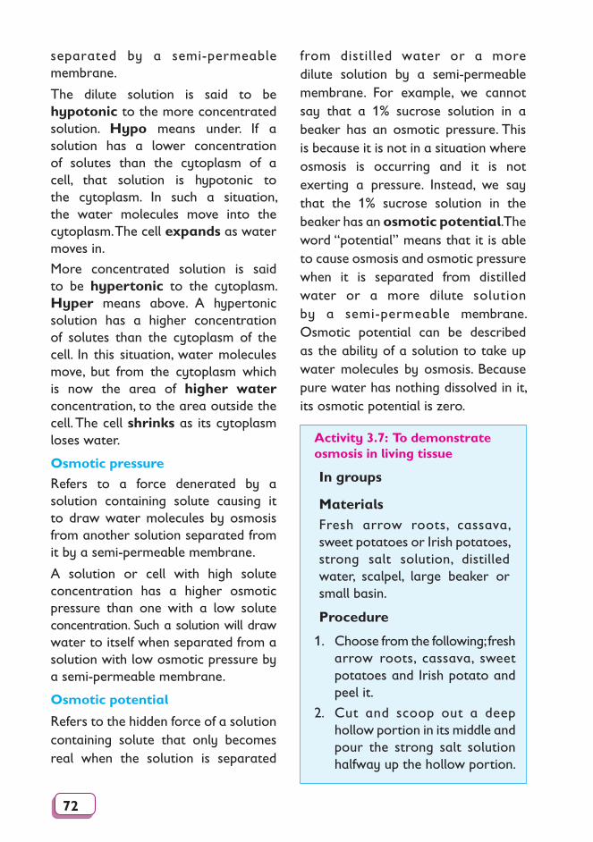
72
separated by a semi-permeable membrane.
The dilute solution is said to be hypotonic to the more concentrated solution. Hypo means under. If a solution has a lower concentration of solutes than the cytoplasm of a cell, that solution is hypotonic to the cytoplasm. In such a situation, the water molecules move into the cytoplasm. The cell expands as water moves in.
More concentrated solution is said to be hypertonic to the cytoplasm. Hyper means above. A hypertonic solution has a higher concentration of solutes than the cytoplasm of the cell. In this situation, water molecules move, but from the cytoplasm which is now the area of higher water concentration, to the area outside the cell. The cell shrinks as its cytoplasm loses water.
Osmotic pressure
Refers to a force denerated by a solution containing solute causing it to draw water molecules by osmosis from another solution separated from it by a semi-permeable membrane.
A solution or cell with high solute concentration has a higher osmotic pressure than one with a low solute concentration. Such a solution will draw water to itself when separated from a solution with low osmotic pressure by a semi-permeable membrane.
Osmotic potential
Refers to the hidden force of a solution containing solute that only becomes real when the solution is separated
from distilled water or a more dilute solution by a semi-permeable membrane. For example, we cannot say that a 1% sucrose solution in a beaker has an osmotic pressure. This is because it is not in a situation where osmosis is occurring and it is not exerting a pressure. Instead, we say that the 1% sucrose solution in the beaker has an osmotic potential. The word “potential” means that it is able to cause osmosis and osmotic pressure when it is separated from distilled water or a more dilute solution by a semi-permeable membrane. Osmotic potential can be described as the ability of a solution to take up water molecules by osmosis. Because pure water has nothing dissolved in it, its osmotic potential is zero.
Activity 3.7: To demonstrate osmosis in living tissue
In groups
MaterialsFresh arrow roots, cassava, sweet potatoes or Irish potatoes, strong salt solution, distilled water, scalpel, large beaker or small basin.
Procedure
1. Choose from the following; fresh arrow roots, cassava, sweet potatoes and Irish potato and peel it.
2. Cut and scoop out a deep hollow portion in its middle and pour the strong salt solution halfway up the hollow portion.
73
3. Mark the level of the salt solution using a scalpel.
4. Place it in a beaker or basin containing distilled water and let it stand for several hours then note the level of solution.
6. Repeat the experiment with boiled pieces of Irish potato, cassava, fresh arrow root or sweet potato.
Study questions
1. Is the level of the strong salt solution still the same at the end of the experiment?
2. Explain what happens to cause the change in level of the salt solution.
3. Compare these results with those when boiled Irish potato is used.
4. Discuss your results.
obtained on the table below and compare your results with the rest of class members.
Are your results the same or different? Why is this so?
Test tube contents
Sucrose Distilled water
Empty
Length of cylinder
before experiment
Length of
cylinder
after
experiment
Activity 3.8: How is osmosis determined using potato tubes or cylinders?
In groups
You are provided with the following:
MaterialsIrish potato, cork borer, ruler marked in millimetres, scalpel or razor blade, 50 ml beakers, cutting board, labels for beakers, concentrated sucrose solution, distilled water, test tube.
Use the materials provided above and design an experiment on your own. Fill the results
Activity 3.9: To demonstrate osmosis using leaf petioles
In pairs
MaterialsLeaf petioles, salt solution, distilled water, petri-dishes, labels, scalpel.
Procedure1. Cut petioles from three leaves.
2. Split each petiole lengthwise and place the two strips from the same leaf in water, salt solution and an empty petri-dish respectively.
3. Leave them for 30 minutes.
4. Draw the appearance of the petioles after 30 minutes in each solution.
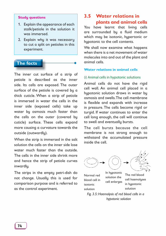
74
Study questions
1. Explain the appearance of each stalk/petiole in the solution it was immersed.
2. Explain why it was necessary to cut a split on petioles in this experiment.
3.5 Water relations in plants and animal cellsYou have learnt that living cells are surrounded by a fl uid medium which may be isotonic, hyper ton ic or hypotonic to the cell con tents.
We shall now examine what happens when there is a net movement of water molecules into and out of the plant and animal cells.
Water relations in animal cells
(i) Animal cells in hypotonic solutions
Animal cells do not have the rigid cell wall. An animal cell placed in a hypotonic solution draws in water by osmosis and swells. The cell membrane is fl exible and expands with increase in pres sure. The cells become rigid or turgid. If water continues to enter the cell long enough, the cell will continue to swell and even tu al ly bursts.
The cell bursts be cause the cell mem brane is not strong enough to with stand the accumulated pres sure inside the cell.
Normal redblood cell inisotonic solution
In hypotonic solution the cell enlarges
The red bloodcell haemo lys es in hypotonic solution
Fig. 3.5: Haemolysis of red blood cells in a hypotonic solution
The facts
The inner cut surface of a strip of petiole is described as the inner side. Its cells are exposed. The outer surface of the petiole is covered by a thick cuticle. When a strip of petiole is immersed in water the cells in the inner side (exposed cells) take up water by osmosis much faster than the cells on the outer (covered by cuticle) surface. These cells expand more causing a curvature towards the outside (outwardly).
When the strip is immersed in the salt solution the cells on the inner side lose water much faster than the outside. The cells in the inner side shrink more and hence the strip of petiole curves inwardly.
The strips in the empty petri-dish do not change. Usually this is used for comparison purpose and is referred to as the control experiment.
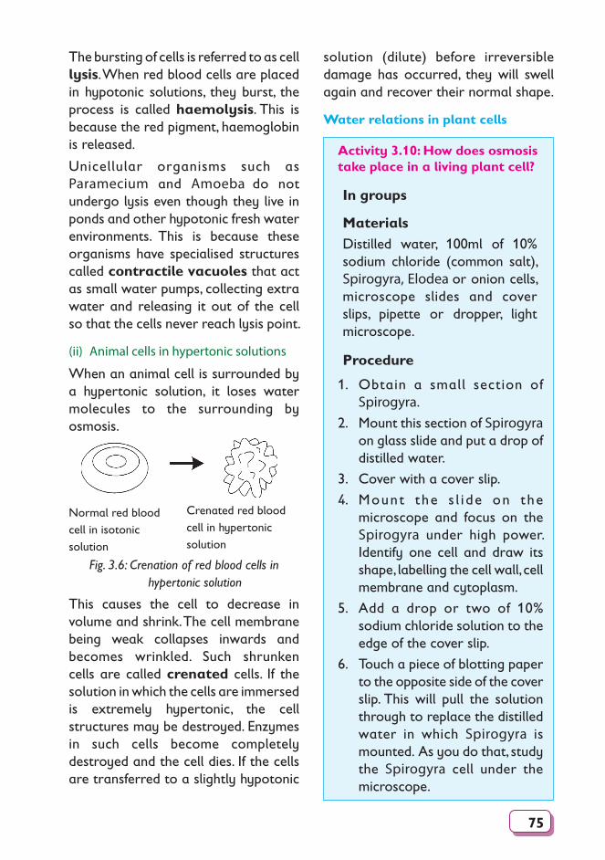
75
The bursting of cells is referred to as cell lysis. When red blood cells are placed in hypotonic solutions, they burst, the process is called haemolysis. This is because the red pigment, haemoglobin is released.
Unicellular organisms such as Paramecium and Amoeba do not undergo lysis even though they live in ponds and other hypotonic fresh water environments. This is because these organisms have specialised structures called contractile vacuoles that act as small water pumps, collecting extra water and releasing it out of the cell so that the cells never reach lysis point.
(ii) Animal cells in hypertonic solutions
When an animal cell is surrounded by a hypertonic solution, it loses water molecules to the surrounding by osmosis.
Normal red blood cell in isotonic solution
Crenated red blood cell in hypertonic solution
Fig. 3.6: Crenation of red blood cells in hypertonic solution
This causes the cell to decrease in volume and shrink. The cell membrane being weak collapses inwards and becomes wrinkled. Such shrunken cells are called crenated cells. If the solution in which the cells are immersed is extremely hypertonic, the cell structures may be destroyed. Enzymes in such cells become completely destroyed and the cell dies. If the cells are transferred to a slightly hypotonic
solution (dilute) before irreversible damage has occurred, they will swell again and recover their normal shape.
Water relations in plant cells
Activity 3.10: How does osmosis take place in a living plant cell?
In groups
MaterialsDistilled water, 100ml of 10% sodium chloride (common salt), Spirogyra, Elodea or onion cells, microscope slides and cover slips, pipette or dropper, light microscope.
Procedure
1. Obtain a small section of Spirogyra.
2. Mount this section of Spirogyra on glass slide and put a drop of distilled water.
3. Cover with a cover slip.
4. Mount the s l i de on the microscope and focus on the Spirogyra under high power. Identify one cell and draw its shape, labelling the cell wall, cell membrane and cytoplasm.
5. Add a drop or two of 10% sodium chloride solution to the edge of the cover slip.
6. Touch a piece of blotting paper to the opposite side of the cover slip. This will pull the solution through to replace the distilled water in which Spirogyra is mounted. As you do that, study the Spirogyra cell under the microscope.
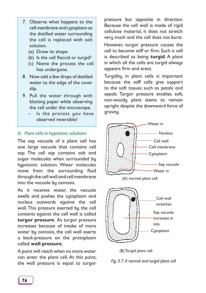
76
7. Observe what happens to the cell membrane and cytoplasm as the distilled water surrounding the cell is replaced with salt solution.(a) Draw its shape.(b) Is the cell flaccid or turgid?(c) Name the process the cell
has undergone.
8. Now add a few drops of distilled water to the edge of the cover slip.
9. Pull the water through with blotting paper while observing the cell under the microscope.– Is the process you have
observed reversible?
pressure but opposite in direction. Because the cell wall is made of rigid cellulose material, it does not stretch very much and the cell does not burst.
However, turgor pressure causes the cell to become stiff or firm. Such a cell is described as being turgid. A plant in which all the cells are turgid always appears firm and erect.
Turgidity in plant cells is important because the stiff cells give support to the soft tissues such as petals and sepals. Turgor pressure enables soft, non-woody plant stems to remain upright despite the downward force of gravity.
(A) normal plant cell
Nucleus
Cell wall
Cell membrane
Sap vacuole
Water in
Water in
Cytoplasm
Cell wall stretches
Sap vacuoleincreases in size
Cytoplasm
(B) Turgid plant cell
Fig. 3.7: A normal and turgid plant cell
(i) Plant cells in hypotonic solutions
The sap vacuole of a plant cell has one large vacuole that contains cell sap. The cell sap contains salt and sugar molecules when surrounded by hypotonic solution. Water molecules move from the surrounding fluid through the cell wall and cell membrane into the vacuole by osmosis.
As it receives water, the vacuole swells and pushes the cytoplasm and nucleus outwards against the cell wall. This pressure exerted by the cell contents against the cell wall is called turgor pressure. As turgor pressure increases because of intake of more water by osmosis, the cell wall exerts a back-pressure on the protoplasm called wall pressure.
A point will reach when no more water can enter the plant cell. At this point, the wall pressure is equal to turgor
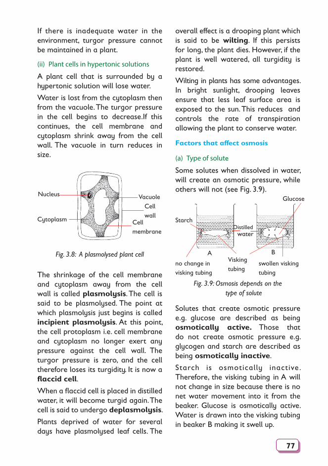
77
If there is inadequate water in the environment, turgor pressure cannot be maintained in a plant.
(ii) Plant cells in hypertonic solutions
A plant cell that is surrounded by a hypertonic solution will lose water.
Water is lost from the cytoplasm then from the vacuole. The turgor pressure in the cell begins to decrease.If this continues, the cell membrane and cytoplasm shrink away from the cell wall. The vacuole in turn reduces in size.
Nucleus
Cytoplasm
VacuoleCell wall
Cell membrane
Fig. 3.8: A plasmolysed plant cell
The shrinkage of the cell membrane and cytoplasm away from the cell wall is called plasmolysis. The cell is said to be plasmolysed. The point at which plasmolysis just begins is called incipient plasmolysis. At this point, the cell protoplasm i.e. cell membrane and cytoplasm no longer exert any pressure against the cell wall. The turgor pressure is zero, and the cell therefore loses its turgidity. It is now a flaccid cell.
When a flaccid cell is placed in distilled water, it will become turgid again. The cell is said to undergo deplasmolysis.
Plants deprived of water for several days have plasmolysed leaf cells. The
overall effect is a drooping plant which is said to be wilting. If this persists for long, the plant dies. However, if the plant is well watered, all turgidity is restored.
Wilting in plants has some advantages. In bright sunlight, drooping leaves ensure that less leaf surface area is exposed to the sun. This reduces and controls the rate of transpiration allowing the plant to conserve water.
Factors that affect osmosis
(a) Type of solute
Some solutes when dissolved in water, will create an osmotic pressure, while others will not (see Fig. 3.9).
Visking tubing
Distilled water
A B
Starch
Glucose
no change in visking tubing
swollen visking tubing
Fig. 3.9: Osmosis depends on the type of solute
Solutes that create osmotic pressure e.g. glucose are described as being osmotically active. Those that do not create osmotic pressure e.g. glycogen and starch are described as being osmotically inactive.
Starch is osmotically inactive . Therefore, the visking tubing in A will not change in size because there is no net water movement into it from the beaker. Glucose is osmotically active. Water is drawn into the visking tubing in beaker B making it swell up.
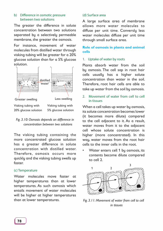
78
b) Difference in osmotic pressure between two solutions
The greater the difference in solute concentration between two solutions separated by a selectively permeable membrane, the greater the osmosis.
For instance, movement of water molecules from distilled water through visking tubing will be greater for a 20% glucose solution than for a 5% glucose solution.
Visking tubing with 20% glucose solution
distilled water
Visking tubing with 5% glucose solution
Greater swelling Less swelling
Fig. 3.10: Osmosis depends on difference in concentration between two solutions
The visking tubing containing the more concentrated glucose solution has a greater difference in solute concentration with distilled water. Therefore, osmosis occurs more quickly and the visking tubing swells up faster.
(c) Temperature
Water molecules move faster at higher temperatures than at lower temperatures. As such osmosis which entails movement of water molecules will be higher at higher temperatures than at lower temperatures.
(d) Surface area
A large surface area of membrane allows more water molecules to diffuse per unit time. Conversly less water molecules diffuse per unit time through small surface area.
Role of osmosis in plants and animal cells
1. Uptake of water by roots
Plants absorb water from the soil by osmosis. The cell sap in root hair cells usually has a higher solute concentration than water in the soil. Therefore, root hair cells are able to take up water from the soil by osmosis.
2. Movement of water from cell to cell in tissues
When a cell takes up water by osmosis, its solute concentration becomes lower (it becomes more dilute) compared to the cell adjacent to it. As a result, water moves from it to the adjacent cell whose solute concentration is higher (more concentrated). In this way, water moves from the root hair cells to the inner cells in the root.
• Water enters cell 1 by osmosis, its contents become dilute compared to cell 2.
1 23
Fig. 3.11: Movement of water from cell to cell in tissues
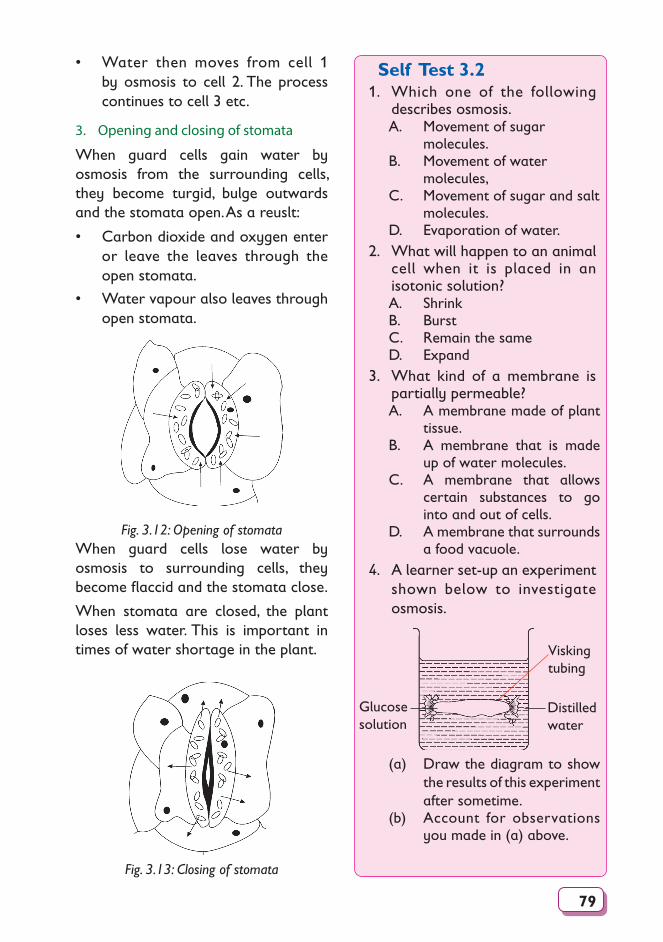
79
• Water then moves from cell 1 by osmosis to cell 2. The process con tin ues to cell 3 etc.
3. Opening and closing of stomata
When guard cells gain water by osmosis from the surrounding cells, they become turgid, bulge outwards and the stomata open. As a reuslt:
• Carbon dioxide and oxygen enter or leave the leaves through the open sto ma ta.
• Water vapour also leaves through open stomata.
Fig. 3.12: Opening of stomataWhen guard cells lose water by osmosis to surrounding cells, they become fl accid and the stomata close.
When stomata are closed, the plant loses less water. This is important in times of water shortage in the plant.
Fig. 3.13: Closing of stomata
Self Test 3.21. Which one of the following
describes osmosis.A. Movement of sugar
molecules.B. Movement of water
molecules,C. Movement of sugar and salt
molecules.D. Evaporation of water.
2. What will happen to an animal cell when it is placed in an isotonic solution?A. ShrinkB. BurstC. Remain the sameD. Expand
3. What kind of a membrane is partially permeable?A. A membrane made of plant
tissue.B. A membrane that is made
up of water molecules.C. A membrane that allows
certain substances to go into and out of cells.
D. A membrane that surrounds a food vacuole.
4. A learner set-up an experiment shown below to investigate osmosis.
Visking tubing
Glucose solution
Distilled water
(a) Draw the diagram to show the results of this experiment after sometime.
(b) Account for observations you made in (a) above.
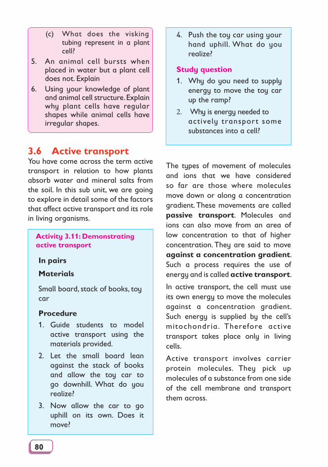
80
(c) What does the visking tubing represent in a plant cell?
5. An animal cell bursts when placed in water but a plant cell does not. Explain
6. Using your knowledge of plant and animal cell structure. Explain why plant cells have regular shapes while animal cells have irregular shapes.
3.6 Active transportYou have come across the term active transport in relation to how plants absorb water and mineral salts from the soil. In this sub unit, we are going to explore in detail some of the factors that affect active transport and its role in living organisms.
4. Push the toy car using your hand uphill. What do you realize?
Study question 1. Why do you need to supply
energy to move the toy car up the ramp?
2. Why is energy needed to actively transport some substances into a cell?
Activity 3.11: Demonstrating active transport
In pairs
Materials
Small board, stack of books, toy car
Procedure1. Guide students to model
active transport using the materials provided.
2. Let the small board lean against the stack of books and allow the toy car to go downhill. What do you realize?
3. Now allow the car to go uphill on its own. Does it move?
The types of movement of molecules and ions that we have considered so far are those where molecules move down or along a concentration gradient. These movements are called passive transport. Molecules and ions can also move from an area of low concentration to that of higher concentration. They are said to move against a concentration gradient. Such a process requires the use of energy and is called active transport.
In active transport, the cell must use its own energy to move the molecules against a concentration gradient. Such energy is supplied by the cell’s mitochondria. Therefore active transport takes place only in living cells.
Active transport involves carrier protein molecules. They pick up molecules of a substance from one side of the cell membrane and transport them across.
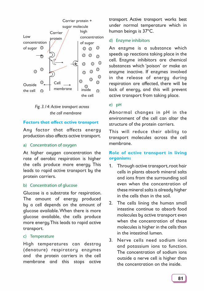
81
high concentration of sugar
Low concentration of sugar
Cell membrane
Carrierprotein
Outsidethe cell inside
the cell
Carrier protein + sugar molecule
Fig. 3.14: Active transport across the cell membrane
Factors that affect active transport
Any factor that affects energy production also affects active transport.
a) Concentration of oxygen
At higher oxygen concentration the rate of aerobic respiration is higher the cells produce more energy. This leads to rapid active transport by the protein carriers.
b) Concentration of glucose
Glucose is a substrate for respiration. The amount of energy produced by a cell depends on the amount of glucose available. When there is more glucose available, the cells produce more energy. This leads to rapid active transport.
c) Temperature
High temperatures can destroy (denature) respiratory enzymes and the protein carriers in the cell membrane and this stops active
transport. Active transport works best under normal temperature which in human beings is 37ºC.
d) Enzyme inhibitors
An enzyme is a substance which speeds up reactions taking place in the cell. Enzyme inhibitors are chemical substances which ‘poison’ or make an enzyme inactive. If enzymes involved in the release of energy during respiration are affected, there will be lack of energy, and this will prevent active transport from taking place.
e) pH
Abnormal changes in pH in the environment of the cell can alter the structure of the protein carriers.
This will reduce their ability to transport molecules across the cell membrane.
Role of active transport in living organisms
1. Through active transport, root hair cells in plants absorb mineral salts and ions from the surrounding soil even when the concentration of these mineral salts is already higher in the cells than in the soil.
2. The cells lining the human small intestine continue to absorb food molecules by active transport even when the concentration of these molecules is higher in the cells than in the intestinal lumen.
3. Nerve cells need sodium ions and potassium ions to function. The concentration of sodium ions outside a nerve cell is higher than the concentration on the inside.
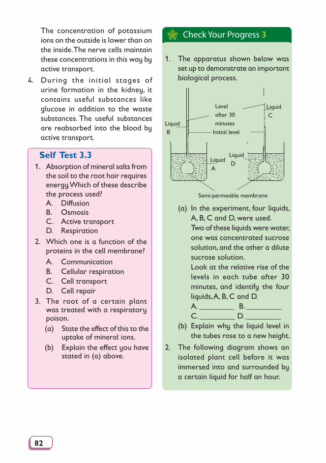
82
The concentration of potassium ions on the outside is lower than on the inside. The nerve cells maintain these concentrations in this way by active transport.
4. During the init ial stages of urine formation in the kidney, it contains useful substances like glucose in addition to the waste substances. The useful substances are reabsorbed into the blood by active transport.
Self Test 3.31. Absorption of mineral salts from
the soil to the root hair requires energy. Which of these describe the process used?
A. Diffusion B. Osmosis C. Active transport D. Respiration2. Which one is a function of the
proteins in the cell membrane? A. Communication B. Cellular respiration C. Cell transport D. Cell repair3. The root of a certain plant
was treated with a respiratory poison.
(a) State the effect of this to the uptake of mineral ions.
(b) Explain the effect you have stated in (a) above.
Check Your Progress 3
1. The apparatus shown below was set up to demonstrate an important biological process.
Level after 30 minutes
Initial level
Liquid C
Liquid B
Liquid A
Liquid D
Semi-permeable membrane
(a) In the experiment, four liquids, A, B, C and D, were used.
Two of these liquids were water, one was concentrated sucrose solution, and the other a dilute sucrose solution.
Look at the relative rise of the levels in each tube after 30 minutes, and identify the four liquids, A, B, C and D.
A. _________ B. _________ C. _________ D. _________(b) Explain why the liquid level in
the tubes rose to a new height.
2. The following diagram shows an isolated plant cell before it was immersed into and surrounded by a certain liquid for half an hour.
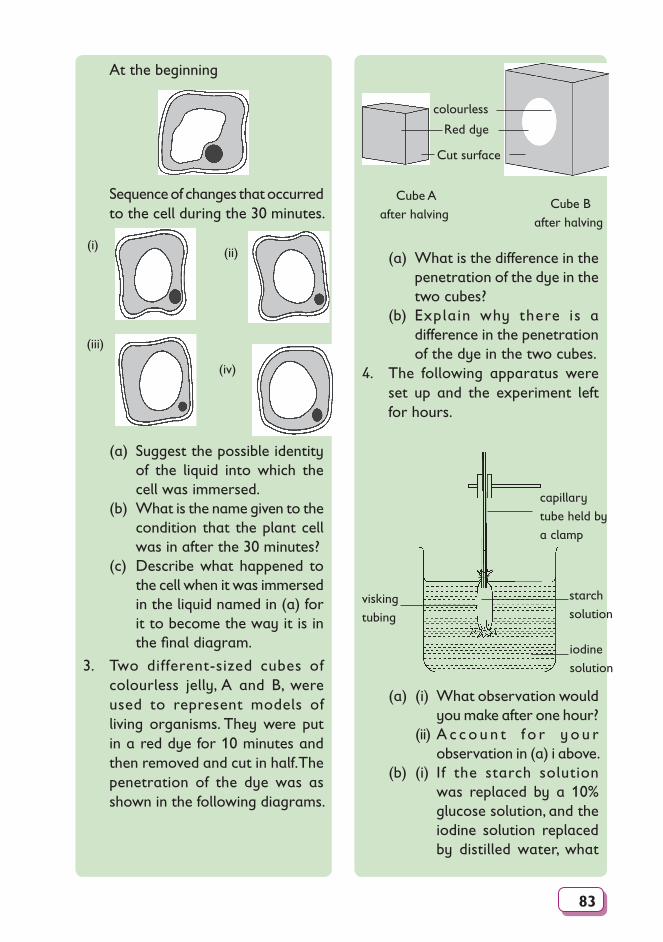
83
At the beginning
Sequence of changes that occurred to the cell during the 30 minutes.
(i) (ii)
(iii)
(iv)
(a) Suggest the possible identity of the liquid into which the cell was immersed.
(b) What is the name given to the condition that the plant cell was in after the 30 minutes?
(c) Describe what happened to the cell when it was immersed in the liquid named in (a) for it to become the way it is in the final diagram.
3. Two different-sized cubes of colourless jelly, A and B, were used to represent models of living organisms. They were put in a red dye for 10 minutes and then removed and cut in half. The penetration of the dye was as shown in the following diagrams.
colourless
Red dye
Cut surface
Cube A after halving
Cube B after halving
(a) What is the difference in the penetration of the dye in the two cubes?
(b) Explain why there is a difference in the penetration of the dye in the two cubes.
4. The following apparatus were set up and the experiment left for hours.
capillary tube held by a clamp
iodine solution
visking tubing
starchsolution
(a) (i) What observation would you make after one hour?
(ii) A c c o u n t f o r y o u r observation in (a) i above.
(b) (i) If the starch solution was replaced by a 10% glucose solution, and the iodine solution replaced by distilled water, what
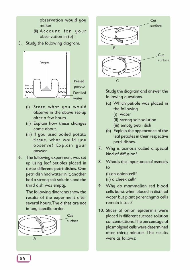
84
observation would you make?
(ii) A c c o u n t f o r y o u r observation in (b) i.
5. Study the following diagram.
Distilled water
Sugar
Peeled potato
(i) State what you would observe in the above set-up after a few hours.
(ii) Explain how these changes come about.
(iii) If you used boiled potato tissue, what would you obser ve? Expla in your answer.
6. The following experiment was set up using leaf petioles placed in three different petri-dishes. One petri dish had water in it, another had a strong salt solution and the third dish was empty.
The following diagrams show the results of the experiment after several hours. The dishes are not in any specific order.
A
Cut surface
B
Cut surface
C
Cut surface
Study the diagram and answer the following questions.
(a) Which petiole was placed in the following
(i) water (ii) strong salt solution (iii) empty petri dish (b) Explain the appearance of the
leaf petioles in their respective petri dishes.
7. Why is osmosis called a special kind of diffusion?
8. What is the importance of osmosis to
(i) an onion cell?(ii) a cheek cell?
9. Why do mammalian red blood cells burst when placed in distilled water but plant parenchyma cells remain intact?
10. Slices of onion epidermis were placed in different sucrose solution concentrations. The percentage of plasmolysed cells were determined after thirty minutes. The results were as follows:
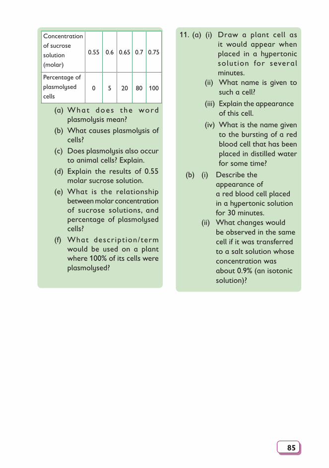
85
Concentration
of sucrose
solution
(molar)
0.55 0.6 0.65 0.7 0.75
Percentage of
plasmolysed
cells0 5 20 80 100
(a) What does t he word plasmolysis mean?
(b) What causes plasmolysis of cells?
(c) Does plasmolysis also occur to animal cells? Explain.
(d) Explain the results of 0.55 molar sucrose solution.
(e) What is the relationship between molar concentration of sucrose solutions, and percentage of plasmolysed cells?
(f) What descript ion/term would be used on a plant where 100% of its cells were plasmolysed?
11. (a) (i) Draw a plant cell as it would appear when placed in a hypertonic solut ion for several minutes.
(ii) What name is given to such a cell?
(iii) Explain the appearance of this cell.
(iv) What is the name given to the bursting of a red blood cell that has been placed in distilled water for some time?
(b) (i) Describe the appearance of a red blood cell placed in a hypertonic solution for 30 minutes.
(ii) What changes would be observed in the same cell if it was transferred to a salt solution whose concentration was about 0.9% (an isotonic solution)?
86
Glossary
Absorption: (1) the movement of substances across a cell membrane. (2) the process by which usable substances or materials are taken
into an organism.
Active transport: a process in which the movement of substances across the cell membrane against a concentration gradient and requires the use of energy by the cell.
Animalia: (1) one of the five kingdoms composed of multicellular and usually motile, heterotrophic organisms.
(2) the animals.
Artificial classification: a method of classification which involves use of few features to put organisms into groups.
Binomial nomenclature: the two-word universal system of naming organisms.
Biochemistry: the study of the chemical changes in cells of living organisms.
Biogeography: the study of the distribution of living organisms.
Biology: the study of living organisms.
Botany: the branch of biology concerned with the study of plants.
Cell: (1) the basic unit of structure and function in living organisms.
(2) the smallest unit in living organisms.
(3) the smallest unit in living organisms that shows the characteristics of life.
Cell differentiation: modification of cell to perform different functions.
Cell membrane: the structure that encloses the interior parts of a cell and controls the movement of substances into and out of the cell. It is also called the plasma membrane.
Cell wall: the rigid part that encloses the cells of plants and various micro-organisms. It is often composed of cellulose.
Centriole: cylindrical organelle found near the nucleus in animal cells, that is involved in cell division.
87
Chloroplast: organelle found in a plant cell that contains green pigment called chlorophyll.
Chromosomes: rod-like structures in cells that undergo division and that contain hereditary information of the organisms.
Class: (1) a taxonomic rank in classification of higher rank than order but of lower rank than phylum or division.
(2) a group of related orders.
Classification: process of grouping things according to similarities.
Concentration gradient: the difference in concentration between a region of high concentration and one of low concentration.
Cytology: study of cells particularly by use of the microscope.
Cytoplasm: the water-like material between the nucleus and the cell membrane.
Division: a group of plants made of classes with similar feature.
Diffusion: the movement of molecules or particles from an area of high concentration to an area of low concentration.
Electron microscope: miscroscope that uses electron beam to illuminate objects and electromagnets to focus the object.
Endoplasmic reticulum: a system of canals enclosed by membranes and filled with a fluid.
Eukaryotic: a cell or an organism whose cells contain a nucleus.
Family: (1) a group of related genera.
(2) a taxonomic category below order and above genus. Family names of plants end with -aceae and those of the animals end with -edae.
Genus: (1) a group of closely related species.
(2) taxonomic category between family and species.
Golgi apparatus: the organelle consisting of stacks of membranes forming flattened sacs in the cytoplasm, which serve as storage centres of proteins synthesised by the cell.
Guard cells: a pair of specialised cells surrounding each stoma in the epidermis of plants, functioning in regulating the size of the stoma.
88
Habitat: the place or the home where an organism lives.
Hypertonic solution: a solution whose concentration of solutes is higher than that of a cell placed in it.
Hypotonic solution: a solution that contains a lower concentration of dissolved substances than that of a cell placed in it.
Isotonic solution: a solution that contains the same concentration of dissolved substances as does a living cell placed in it.
Kingdom: (1) a group of related phyla or division. (2) the largest category in a classification system.
Laboratory: a building or a room with special equipment for carrying out experimental work.
Laser: a device that produces powerful beams of light.
Leucoplast: the colourless plastids in which starch or other plant nutrients are stored.
Light microscope: a microscope that uses light to illuminate objects and lenses to focus the object.
Lipids: organic compounds other than carbohydrates, consisting of carbon, hydrogen and oxygen. These compounds include fats, oils and waxes.
Living thing/organism: anything that has life.
Lysosomes: the small, sac-like structures surrounded by single membranes and containing strong digestive or hydrolytic enzymes.
Magnification: the ratio of the image size to the object size.
Microbiology: the science and study of micro-organisms, including protozoa, algae, fungi viruses and bacteria.
Microscope: equipment used to magnify and observe objects.
Microscope slide: rectangular piece of glass on which specimens are placed during observation by use of a microscope.
Mitochondria: organelles that produce energy in the cell.
Mitochondrion: an oval organelle enclosed by a membrane in which most of the reactions of cellular respiration occur.
Multicellular organism: organism whose body is made up of many cells.
Monera: the kingdom that includes the simplest one-celled prokaryotic
89
organisms, that is, the bacteria.
Natural classification: a method of classification which involves using many features and history of organisms to put them into groups.
Nucleolus: a granular structure that is found in the nucleus of cells and is the site of RNA synthesis.
Nucleoplasm: the protoplasm of the nucleus.
Nucleus: a large organelle surrounded by a membrane and which contains the cell’s DNA.
Order: (1) a group of related families. (2) a taxonomic category ranked below the class and above the
family.
Organ: a group of tissues that work together to perform a specific function.
Organ system: a group of organs that work together to perform a specific function.
Organelle: a specialised structure in the cytoplasm that carries out a specific function.
Osmosis: the movement of water molecules across a semipermeable membrane from a region of high concentration of water molecules to a region of low concentration of water molecules.
Osmotic pressure:the pressure generated in a solution containing solute dissolved in it.
Phylum: (1) One of the largest groups within a kingdom. (2) a major taxonomic category in classifying animals composed of
groups of related classes.
Plantae: (1) One of the five kingdoms of living organisms. (2) Plants, the members of plantae are mostly multicellular and
photosynthetic.
Plastids: organelles surrounded by a membrane found in cells of some members of protoctista and almost all plants. They include chloroplasts, chromoplasts and leucoplasts.
Prokaryotic cells: (1) cells lacking structures surrounded by membranes/ cells lacking a true nucleus.
(2) Moneras. (3) Bacteria and blue-green algae.
90
Rank: a level within the hierachy in classification.
Specialization: modification in structure of cells to suit them to specific functions.
Specialization: a cell whose structure is modified to suit it to a given function.
Species: a group of organisms with same features and origin and that can interbreed to produce a fertile offspring.
Specimen: a whole body or a part of a body that is taken to represent whole group of organism or an organism in scientific study.
Stomata: the openings in the epidermis of leaves that allow the exchange of respiratory gases between the internal tissues of the leaf and the atmosphere.
Submucosa: the layer of fibrous connective tissue that attaches a mucous membrane to its adjacent parts.
Substrate: the substance that an enzyme acts on.
Taxon: a group name used to put organisms with a similar characteristic.
Taxonomy: the branch of biology that deals with the classification and naming of living things.
Taxonomists: scientists who study classification.
Tissue: a group of cells more or less similar morphologically and functionally.
Tonoplast: membrane that surrounds the vacuole.
Vacuole: a cavity in a cell enclosed by a membrane and filled with a fluid.
Unicellular organism: organism whose whole body is made up of one cell only.
Zoology: the science that deals with knowledge of animal life.
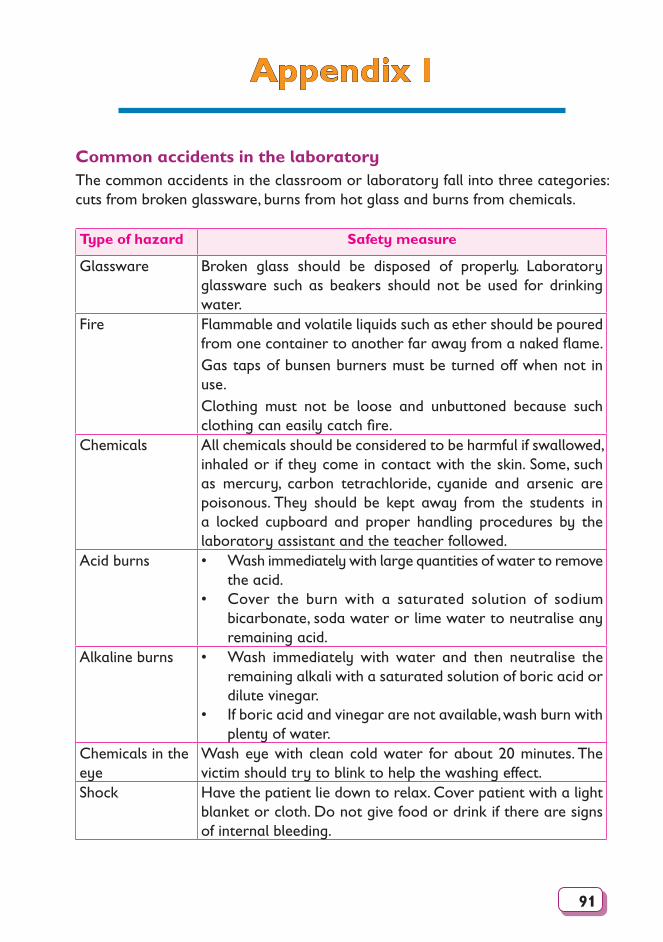
91
Appendix I
Common accidents in the laboratoryThe common accidents in the classroom or laboratory fall into three categories: cuts from broken glassware, burns from hot glass and burns from chemicals.
Type of hazard Safety measure
Glassware Broken glass should be disposed of properly. Laboratory glassware such as beakers should not be used for drinking water.
Fire Flammable and volatile liquids such as ether should be poured from one container to another far away from a naked flame.Gas taps of bunsen burners must be turned off when not in use.Clothing must not be loose and unbuttoned because such clothing can easily catch fire.
Chemicals All chemicals should be considered to be harmful if swallowed, inhaled or if they come in contact with the skin. Some, such as mercury, carbon tetrachloride, cyanide and arsenic are poisonous. They should be kept away from the students in a locked cupboard and proper handling procedures by the laboratory assistant and the teacher followed.
Acid burns • Wash immediately with large quantities of water to remove the acid.
• Cover the burn with a saturated solution of sodium bicarbonate, soda water or lime water to neutralise any remaining acid.
Alkaline burns • Wash immediately with water and then neutralise the remaining alkali with a saturated solution of boric acid or dilute vinegar.
• If boric acid and vinegar are not available, wash burn with plenty of water.
Chemicals in the eye
Wash eye with clean cold water for about 20 minutes. The victim should try to blink to help the washing effect.
Shock Have the patient lie down to relax. Cover patient with a light blanket or cloth. Do not give food or drink if there are signs of internal bleeding.
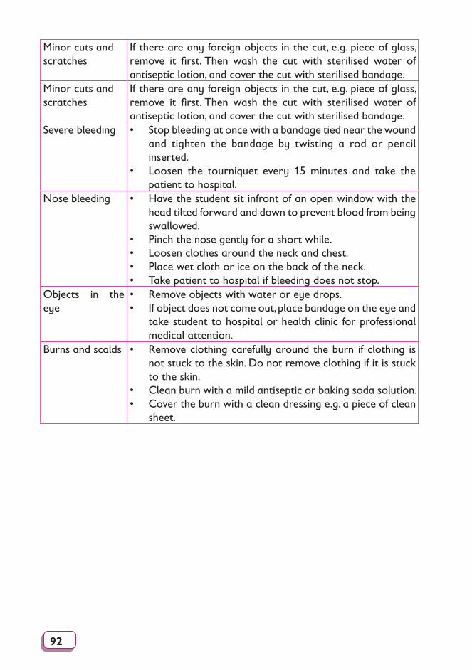
92
Minor cuts and scratches
If there are any foreign objects in the cut, e.g. piece of glass, remove it first. Then wash the cut with sterilised water of antiseptic lotion, and cover the cut with sterilised bandage.
Minor cuts and scratches
If there are any foreign objects in the cut, e.g. piece of glass, remove it first. Then wash the cut with sterilised water of antiseptic lotion, and cover the cut with sterilised bandage.
Severe bleeding • Stop bleeding at once with a bandage tied near the wound and tighten the bandage by twisting a rod or pencil inserted.
• Loosen the tourniquet every 15 minutes and take the patient to hospital.
Nose bleeding • Have the student sit infront of an open window with the head tilted forward and down to prevent blood from being swallowed.
• Pinch the nose gently for a short while.• Loosen clothes around the neck and chest.• Place wet cloth or ice on the back of the neck.• Take patient to hospital if bleeding does not stop.
Objects in the eye
• Remove objects with water or eye drops.• If object does not come out, place bandage on the eye and
take student to hospital or health clinic for professional medical attention.
Burns and scalds • Remove clothing carefully around the burn if clothing is not stuck to the skin. Do not remove clothing if it is stuck to the skin.
• Clean burn with a mild antiseptic or baking soda solution.• Cover the burn with a clean dressing e.g. a piece of clean
sheet.
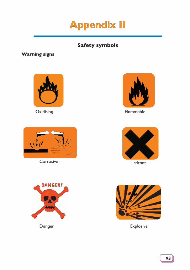
93
Appendix II
Safety symbolsWarning signs
Oxidising Flammable
Explosive
Corrosive Irritant
Danger