biological activity of reducing-end-derivatized oligogalacturonides in tobacco tissue cultures
TRANSCRIPT

Biological Activity of Reducing-End-DerivatizedOligogalacturonides in Tobacco Tissue Cultures1
Mark D. Spiro2,3, Brent L. Ridley3, Stefan Eberhard, Keith A. Kates, Yves Mathieu, Malcolm A. O’Neill,Debra Mohnen, Jean Guern, Alan Darvill, and Peter Albersheim*
Complex Carbohydrate Research Center and Department of Biochemistry and Molecular Biology, The Universityof Georgia, 220 Riverbend Road, Athens, Georgia 30602–4712 (B.L.R., S.E., K.A.K., M.A.O., D.M., A.D., P.A.,
M.D.S.); and Institut des Sciences Vegetales, Centre National de la Recherche Scientifique, Batiment 22–23,Avenue de la Terrasse, F-91198 Gif-sur-Yvette cedex, France (Y.M., J.G.)
The biological activity of reducing-end-modified oligogalactu-ronides was quantified in four tobacco (Nicotiana tabacum) tissueculture bioassays. The derivatives used were oligogalacturonideswith the C-1 of their reducing end (a) covalently linked to a biotinhydrazide, (b) covalently linked to tyramine, (c) chemically reducedto a primary alcohol, or (d) enzymatically oxidized to a carboxylicacid. These derivatives were tested for their ability to (a) altermorphogenesis of N. tabacum cv Samsun thin cell-layer explants,(b) elicit extracellular alkalinization by suspension-cultured cv Sam-sun cells, (c) elicit extracellular alkalinization by suspension-cultured N. tabacum cv Xanthi cells, and (d) elicit H2O2 accumu-lation in the cv Xanthi cells. In all four bioassays, each of thederivatives had reduced biological activity compared with the cor-responding underivatized oligogalacturonides, demonstrating thatthe reducing end is a key element for the recognition of oligogalac-turonides in these systems. However, the degree of reduction inbiological activity depends on the tissue culture system used and onthe nature of the specific reducing-end modification. These resultssuggest that oligogalacturonides are perceived differently in eachtissue culture system.
Carbohydrates that act as signal molecules in plants(oligosaccharins) have been isolated from plant and fungalcell wall polysaccharides, from the cell walls of bacterialsymbionts of plants, and from fungal glycopeptides (forreview, see Ryan and Farmer, 1991; Darvill et al., 1992; Coteand Hahn, 1994). We are interested in determining themechanism by which one type of the plant cell wall-derived oligosaccharins, the oligogalacturonides, elicit bio-logical responses in plants.
The biological effects elicited in plants by oligosaccharinsare diverse (for review, see Ryan and Farmer, 1991; Darvillet al., 1992; Cote and Hahn, 1994) but can generally beplaced in two groups: delayed responses and rapid re-
sponses. Delayed responses are usually observed hours ordays after oligosaccharin treatment and are often directlyinvolved in adaptation to environmental conditions,whereas rapid responses generally occur at the plant cellsurface and are observed within minutes after addition ofoligosaccharins. The delayed responses elicited by oligoga-lacturonides can be broadly divided into those in whichdefense responses are induced and those in which growthand development are modified. The defense-related re-sponses, depending on the plant species, include phyto-alexin accumulation (Hahn et al., 1981), lignification of cellwalls (Robertsen, 1986), and proteinase inhibitor accumu-lation (Bishop et al., 1984). The responses involving growthand development include induction of ethylene in tomatofruit (Brecht and Huber, 1988), inhibition of auxin-inducedpea stem elongation (Branca et al., 1988), and regulation oftobacco (Nicotiana tabacum) TCL explant morphogenesis(Eberhard et al., 1989). The rapid responses induced byoligogalacturonides include enhanced protein phosphory-lation in tomato and potato membranes (Farmer et al.,1989), transmembrane ion flux accompanied by membranedepolarization in tobacco suspension cells (Mathieu et al.,1991) and carrot protoplasts (Messiaen et al., 1993), andH2O2 accumulation in suspension-cultured soybean cells(Apostol et al., 1989).
Rapid responses elicited by oligosaccharins may act assignal transduction events in the initiation of the delayedresponses; however, this has not been directly demon-strated. Isolation and characterization of oligosaccharin re-ceptors would facilitate the identification of the oligo-saccharin-induced signal-transduction events. High-affinity binding sites for three oligosaccharins, the oligo-b-glucosides, the oligochitins, and a yeast N-glycan, have
1 This research was supported in part by the U.S. Department ofEnergy (DOE)-funded Center for Plant and Microbial ComplexCarbohydrates (grant no. DE-FG05-93-ER20097) and by DOE grantno. DE-FG02-96ER20221 (to P.A.).
2 Present address: Department of Plant Pathology, The Pennsyl-vania State University, University Park, PA 16802.
3 Both of these authors contributed equally to this manuscript.* Corresponding author; e-mail [email protected]; fax
1–706 –542– 4412.
Abbreviations: DP, degree of polymerization; EC50, oligogalac-turonide concentration that includes a half-maximal response; G12,dodecagalacturonide; G13, tridecagalacturonide; G13-B, biotiny-lated tridecagalacturonide; G13-O, oxidized tridecagalacturonide(dodecagalacturonide-d-galactaric acid); G13-R, reduced tridecaga-lacturonide (dodecagalacturonide-l-galactonic acid); G13-T, ty-raminated tridecagalacturonide; HPAEC-PAD, high performanceanion-exchange chromatography with pulsed amperometric detec-tion; PCV, packed cell volume; R, change in free proton concen-tration; Rmax, change in free proton concentration elicited by asaturating concentration of G12 or G13; TCL, thin cell layer.
Plant Physiol. (1998) 116: 1289–1298
1289 www.plant.org on January 3, 2016 - Published by www.plantphysiol.orgDownloaded from
Copyright © 1998 American Society of Plant Biologists. All rights reserved.

been identified using biologically active, radiolabeled de-rivatives (Cosio et al., 1990; Cheong and Hahn, 1991; Basseet al., 1993; Shibuya et al., 1993; Baureithel et al., 1994). Thespecificity of these binding sites has been characterized bymeasuring the ability of structural analogs having a rangeof biological activities to displace labeled derivatives incompetitive binding studies. A direct correlation betweenthe competitive binding ability of the analogs and theirbiological activity has been observed for each binding site,providing evidence that the sites identified are physiolog-ical receptors of the oligosaccharins. Characterizing thespecificity of binding proteins in this manner requires ho-mogeneous oligosaccharins, labeled oligosaccharin deriva-tives, and oligosaccharin analogs.
With the ultimate goal of identifying and characterizingoligogalacturonide receptors, we have quantified thebiological activities of discernibly homogeneous reducing-end-derivatized oligogalacturonides in four tobacco tissue-culture bioassays. The oligogalacturonide derivatives as-sayed include G13-T, G13-B, G13-O, and G13-R (Fig. 1).Biotinylated and 125I-labeled tyraminated oligogalactu-ronides could be used to detect low-abundance oligo-galacturonide-binding proteins. Unlabeled oligogalactu-ronides, including reduced and oxidized oligogalac-turonides, could be used to characterize the specificity ofoligogalacturonide-binding proteins.
MATERIALS AND METHODS
Purified orange fruit uronic acid oxidase was a gift fromRussell Pressey (U.S. Department of Agriculture, Athens,GA). All reagents were obtained from Sigma.
Preparation of Homogeneous Oligogalacturonides andTheir Reducing-End Derivatives
In this paper the term oligogalacturonide refers to nativeor unmodified, as well as reducing-end-derivatized oli-gogalacturonides, such as G13-T, G13-B, G13-R, and G13-O.Oligogalacturonides, generated by partial endopolygalactur-onase digestion of polygalacturonic acid, were purified tohomogeneity by a three-step protocol (Spiro et al., 1993)consisting of size-selective precipitation with ethanol, fol-lowed by Q-Sepharose anion-exchange chromatography,and then semipreparative HPAEC-PAD. All oligogalactur-onides used in this study were homogeneous and chemi-cally stable at 220°C, as determined by analytical HPAEC-PAD and by determination of their Mr by electrospray MS.
Oligogalacturonides derivatized with tyramine or biotin-x-hydrazide (6-[biotinoylamino] caproic acid hydrazide) atC-1 of the reducing-end galacturonic acid were synthesizedand purified to homogeneity as described previously(Spiro et al., 1996; Ridley et al., 1997).
G13-R was generated by treating the G13 (5 mg in 1 mL of1 m NH4OH) for 16 h at 4°C with NaBH4 (5 mg). Thereaction was terminated by the addition of 5 volumes of10% acetic acid in methanol, which destroyed any remain-ing NaBH4. This solution was kept for 2 h at 220°C and theresulting precipitate was collected by centrifugation. Thesupernatant was discarded. The pellet was washed twicewith methanol (5 mL) to remove residual salts. The finalpellet was dried under a stream of air at 25°C. The residuewas dissolved in water (0.5 mL) and stored at 220°C.
G13-O was generated by treating G13 (5 mg) in 10 mmTris-HCl, pH 8.5 (1 mL), with 3 units of uronic acid oxidase(Pressey, 1993) for 23 h at 37°C. This solution was sterilizedby centrifugation through a 0.2-mm nylon filter prior toincubation.
Neither the chemical reduction nor the enzymatic oxida-tion of G13 proceeded to completion; therefore, G13-R andG13-O were individually purified to homogeneity by semi-preparative HPAEC-PAD (Spiro et al., 1993). ElectrosprayMS analysis confirmed that the expected product had beensynthesized.
TCL Bioassay
Tobacco (Nicotiana tabacum cv Samsun) plants weregrown as described by Mohnen et al. (1990). The TCLexplant bioassay was performed as described by Eberhardet al. (1989). Briefly, thin strips of tissue, approximately 1mm wide and composed of 6 to 10 cell layers, were cutfrom surface-sterilized tobacco floral branches. TCL ex-plants, approximately 10 mm long, were cut from the tissuestrips. The explants were placed in individual wells of12-well culture dishes (ICN) containing 2 mL of Linsmaierand Skoog (1965) medium, pH 5.8, supplemented with 167
Figure 1. Structures of G13 and the four reducing-end derivatives ofG13 used in this study.
1290 Spiro et al. Plant Physiol. Vol. 116, 1998
www.plant.org on January 3, 2016 - Published by www.plantphysiol.orgDownloaded from Copyright © 1998 American Society of Plant Biologists. All rights reserved.

mm Glc, 3 mm IAA, and 0.3 mm kinetin. The oligogalactu-ronides to be assayed were sterilized by filtration through0.2-mm nylon membrane syringe filters (Nalgene, Roches-ter, NY). The TCL explants were incubated at 24°C undercontinuous, cool-white fluorescent light. The overall TCLexplant morphology and the number of flowers formed oneach TCL explant after 23 or 24 d of culture were deter-mined by examination with a dissecting microscope.
Maintenance of Suspension-Cultured Tobacco Cells
Suspension-cultured N. tabacum cv Samsun cells, initi-ated from pith parenchyma callus, were grown in Lins-maier and Skoog (1965) medium, pH 6.0, supplementedwith 4.5 mm 2,4-D and 3% Suc. The cells were culturedunder continuous light at 26°C on a rotary shaker at 110rpm. The PCV obtained after centrifugation (1000g for 5min) was used as a measure of growth. An aliquot of thecell suspension giving an initial PCV of 2.7% was trans-ferred to 100 mL of fresh medium every 7 d. The PCV after7 d of culture was between 26 and 33%. The culture of theN. tabacum cv Samsun cells and the alkalinization bioassaysusing these cells were carried out at the Complex Carbo-hydrate Research Center (Athens, GA).
Suspension-cultured N. tabacum cv Xanthi cells weregrown in Gamborg B-5 medium, pH 5.7, supplementedwith 1 mm 2,4-D, 40 nm kinetin, and 2% Suc. The cells weregrown under continuous light on a rotary shaker at 160rpm and were subcultured every 7 d by transferring 10 mLof the culture into 100 mL of fresh medium. The PCV after7 d of culture was between 18 and 22%. The culture of theN. tabacum cv Xanthi cells and the alkalinization and H2O2
accumulation bioassays using these cells were carried outat the Centre National de la Recherche Scientifique labora-tories (Gif-sur-Yvette, France).
Analysis of Oligogalacturonide-Induced ExtracellularAlkalinization by Suspension-Cultured Tobacco Cells
Aliquots of 7-d-old exponentially growing N. tabacum cvSamsun suspension-cultured cells (4 mL) in 25-mL Erlen-meyer flasks were equilibrated for 2 h on a rotary shaker(110 rpm). The pH after this period was between 4.4 and4.6. The pH of the oligogalacturonide solutions (50 mL inwater) was adjusted to that of the equilibrated cells. Theaddition of oligogalacturonides to each successive flaskwas staggered by 60 s so that pH measurements could bemade at fixed times after sample addition. The extracellularpH was measured immediately upon addition of the oli-gogalacturonides and subsequently at 14-min intervalsthrough at least 56 min using a Micro-Combination pHelectrode (Microelectrodes, Inc., Londonderry, NH) con-nected to an Accumet 950 pH meter (Fisher Scientific).
The analysis of oligogalacturonide-induced extracellularalkalinization by cv Xanthi cells was carried out in thesame manner as for the cv Samsun cells with the followingexceptions. Aliquots (3 mL) of the cell suspensions wereplaced in 12-mL glass vials, adjusted to 1 mm Mes-Tris (pH5.2), and equilibrated for 2 h, at which time the pH wasbetween 5.1 and 5.5. The pH was measured using a BRV4H
glass electrode (Heito, Paris, France) connected to a PSD 11pH meter (Heito).
Analysis of Oligogalacturonide-Induced Oxidative Burst
The accumulation of H2O2 in the growth medium ofsuspension-cultured cv Xanthi cells was determined in thesame experiments as the measurement of extracellular al-kalinization. Aliquots (10–40 mL) of the growth mediumwere removed at intervals and added to a series of cuvettescontaining 8 mm scopoletin (7-OH-6-methoxycoumarin)and 5 mg of horseradish peroxidase (P 6782, Sigma) in 2 mLof 50 mm Mes-Tris, pH 5.2. The amount of H2O2 present inthe culture medium was determined by the decrease influorescence of scopoletin and compared with the effect ofa standard concentration series of H2O2 on scopoletin flu-orescence (Root et al., 1975). Fluorescence measurementswere made with a spectrofluorimeter (Photon TechnologyInternational, South Brunswick, NJ) using an excitationwavelength of 350 nm and an emission wavelength of460 nm.
RESULTS
Reducing-End-Derivatized G13 at SubmicromolarConcentrations Alter the Morphogenesis of N. tabacum cvSamsun TCL Explants
Oligogalacturonides induce flower formation, inhibitroot formation, and reproducibly alter the overall morphol-ogy of tobacco TCL explants cultured on indolebutyric acidand kinetin-containing medium (Eberhard et al., 1989;Marfa et al., 1991; Bellincampi et al., 1993). Oligogalactu-ronides of DP 12 to 14, which have approximately equalactivity, induce the alteration of TCL explant morphogen-esis at submicromolar concentrations, whereas higher con-centrations of shorter and longer oligomers are required toinduce an equivalent response. The mechanism by whicholigogalacturonides initiate these responses is unknown.
We determined the effect of derivatized and underiva-tized oligogalacturonides on TCL explants cultured in tran-sition medium containing 3 mm IAA and 0.3 mm kinetin.A transition medium containing IAA rather than indolebu-tyric acid (Mohnen et al., 1990) was used, since the add-ition of oligogalacturonides to TCL explants cultured onIAA-containing transition medium causes a distinctconcentration-dependent change in the overall morphol-ogy of the TCL explants. TCL explants, in the absence ofadded oligogalacturonides or at low concentrations of bio-active oligogalacturonides, grow more or less uniformlyover their entire length, resulting in rectangular explants.In the presence of higher concentrations of bioactive oli-gogalacturonides, the TCL explants were significantly in-hibited in their growth and had limited tissue enlargementonly at one end, yielding mushroom-shaped explants(Fig. 2). In the absence of oligogalacturonides, the rectan-gular explants produced only roots, whereas they pro-duced roots and occasionally flowers when exposed tolow concentrations of bioactive oligogalacturonides. Themushroom-shaped explants that resulted from exposure to
Activity of Modified Oligogalacturonides in Tobacco Cultures 1291
www.plant.org on January 3, 2016 - Published by www.plantphysiol.orgDownloaded from Copyright © 1998 American Society of Plant Biologists. All rights reserved.

higher concentrations of bioactive oligogalacturonides fre-quently formed flowers but never roots.
The ability of each oligogalacturonide to alter TCL-explant morphogenesis was tested at nine concentrationsand compared with a similar concentration series of G12.Each oligogalacturonide was assayed in two independentexperiments using six replicate TCL explants at each con-centration. G12, G13-T, G13-B, and G13-R each induced aconcentration-dependent increase in the number of flowers
formed on TCL explants. The maximum of the averagenumber of flowers induced by G12 (1.5 mm, 2.8 flowers/explant) was nearly identical to the maximum of the aver-age number of flowers induced by the three reducing-endderivatives (2.6 mm, 3.1 flowers/explant). However, asshown in Figure 3 for G12, the number of flowers inducedon TCL explants by a given oligogalacturonide concentra-tion varied considerably, and thus the flowering data werenot useful for a quantitative comparison of the biologicalactivities of the various oligogalacturonides.
However, the oligogalacturonide concentration at whichthe overall morphology of the TCL explants changes fromrectangular to mushroom-shaped (the morphogeneticswitch concentration) could be used to quantify the activ-ities of the oligogalacturonides since it reproducibly fellwithin a less-than 4-fold concentration range (Fig. 2). Theaverage morphogenetic switch concentration of G12 in 12experiments was 0.058 6 0.033 mm (Table I). Comparedwith G12, the morphogenesis switch concentration forG13-T was 2.5-fold higher; G13-B was 3.4-fold higher, andG13-R was 6.3-fold higher. The difference between the ac-tivity of G12 and each G13 derivative was statistically sig-nificant (Wilcoxon’s Rank-Sum test, confidence level 598.5%). There was no discernible difference in the activityof G12 when assayed in the presence of excess free biotin orfree tyramine. These results indicate that derivatization ofthe reducing end of G13 results in a reduction in its abilityto alter TCL explant morphogenesis.
We compared the morphogenetic switch concentrationof various homogeneously sized biotinylated and underi-vatized oligogalacturonides to determine whether re-ducing-end derivatization alters the size requirement for
Figure 2. The TCL explants from a representative tobacco morpho-genesis bioassay comparing the activities of G12 (top), G13-B (mid-dle), and G13-T (bottom). The explants were incubated in growthmedium containing the indicated oligogalacturonide concentrations,with six replicate TCL explants per concentration. The arrows indi-cate the concentrations at which oligogalacturonides induce a dis-tinct change in the overall morphology of the TCL explants (themorphogenetic switch concentrations).
Figure 3. Plot of the log of G12 concentration versus the averagenumber of flowers induced per TCL explant. The data from 12experiments are shown, and each data point represents the averagenumber of flowers produced by the six replicate TCL explants usedfor each treatment in a given experiment. In the absence of G12, noflowers were formed. The line represents the dose-response curve forG12 as determined by least-squares analysis using log concentration.
1292 Spiro et al. Plant Physiol. Vol. 116, 1998
www.plant.org on January 3, 2016 - Published by www.plantphysiol.orgDownloaded from Copyright © 1998 American Society of Plant Biologists. All rights reserved.

activity. G16-B and G10-B were both reduced in activitycompared with their corresponding underivatized oligoga-lacturonides, and the highest concentration of G8-B tested(2.6 mm) did not induce an alteration in the overall explantmorphology (Table I). G13-B was significantly more activethan longer or shorter biotinylated oligogalacturonides,which is consistent with the size requirements found usingunderivatized oligogalacturonides in this bioassay (Marfaet al., 1991; Bellincampi et al., 1993).
All Four Derivatives of G13 Were More Than 30-FoldReduced in Their Ability to Induce ExtracellularAlkalinization by Suspension-Cultured cv Samsun Cells
Transient membrane depolarization accompanied byrapid K1 ion efflux and extracellular alkalinization wasinduced when suspension-cultured tobacco or carrot cellswere exposed to micromolar concentrations of oligogalac-turonides having DPs of 10 to 15 (Mathieu et al., 1991;Messiaen and Van Cutsem, 1994). These rapid responseshave been hypothesized to act as signaling events leadingto delayed responses, such as the alteration of TCL-explantmorphogenesis (Mathieu et al., 1991; Messiaen and VanCutsem, 1994). We have characterized the ability of varioussizes of reducing-end-derivatized and underivatized oli-gogalacturonides to induce extracellular alkalinization incv Samsun suspension cultures, the same cultivar used forthe tobacco morphogenesis bioassay.
The ability of each oligogalacturonide to induce extracel-lular alkalinization by cv Samsun cells was assayed usingseven concentrations of oligogalacturonides in at least twoindependent experiments. A standard concentration seriesof G12 (Fig. 4) was assayed in each experiment. Each bio-active oligogalacturonide elicited a rapid, concentration-dependent, and saturable extracellular alkalinization re-sponse that exhibited three characteristic phases: a periodof rapid alkalinization beginning 30 s after oligogalactur-onide addition, a plateau period during which the pHremained near its maximum level, and a period of gradualreacidification during which the pH of the medium re-turned to approximately its original value.
The alkalinization response measured 14 min after oli-gogalacturonide addition was used to compare the activi-
ties of the derivatized and underivatized oligogalactu-ronides, since this time consistently fell within the periodof maximum alkalinization. The change in pH was con-verted to the R to obtain a linear measure of the response.Rmax was always found to be essentially the same betweenreplicates within the same experiment; however, Rmax var-ied significantly between experiments. The alkalinizationresponse elicited by saturating concentrations of each bio-active oligogalacturonide within an experiment was essen-tially the same between replicates within the same experi-ment; however, Rmax varied significantly between ex-periments. To correct for variability of the responses be-tween experiments, the response elicited by each samplewas expressed as the fraction of the Rmax for that experi-ment (R/Rmax).
The R/Rmax values were used to generate dose-responsecurves (Fig. 5) and the EC50 were determined from thesecurves. G12 and G13 were the most active of the compoundstested (Fig. 5a) and the difference between their activitieswas not statistically significant (paired t test, level of con-fidence 5 95%); therefore, the data from G12 and G13 werepooled to generate a single dose-response curve (EC50 50.068 mm). G15 was approximately 5-fold less active(EC50 5 0.36 mm) and G9 was 50-fold less active (EC50 5 3.4
Table I. Comparison of the ability of reducing-end-derivatized and underivatizedoligogalacturonides to alter TCL explant morphogenesis
The concentration of each oligogalacturonide that induces a distinct change in the overall TCLexplant morphology is reported as the morphogenetic switch concentration (as defined in the text). Thevalues given for each oligogalacturonide are the averages of two independent bioassays, with theexception of the value for G12, which is the average of 12 independent experiments (SD 5 0.033 mM).
OligogalacturonideMorphogenetic Switch
ConcentrationDerivatized
OligogalacturonideMorphogenetic Switch
Concentration
mM mM
G16 0.26 G16-B 0.65G15 0.15 G13-B 0.21G12 0.058 G13-T 0.15G10 0.83 G13-R 0.37G9 2.6 G10-B 1.7G7 .2.6 G8-B .2.6
Figure 4. Representative time courses of the extracellular alkaliniza-tion by suspension-cultured cv Samsun cells treated with the indi-cated concentrations of G12 at time 0.
Activity of Modified Oligogalacturonides in Tobacco Cultures 1293
www.plant.org on January 3, 2016 - Published by www.plantphysiol.orgDownloaded from Copyright © 1998 American Society of Plant Biologists. All rights reserved.
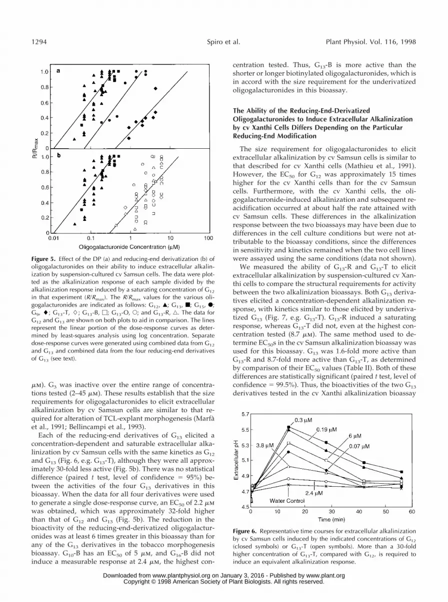
mm). G3 was inactive over the entire range of concentra-tions tested (2–45 mm). These results establish that the sizerequirements for oligogalacturonides to elicit extracellularalkalinization by cv Samsun cells are similar to that re-quired for alteration of TCL-explant morphogenesis (Marfaet al., 1991; Bellincampi et al., 1993).
Each of the reducing-end derivatives of G13 elicited aconcentration-dependent and saturable extracellular alka-linization by cv Samsun cells with the same kinetics as G12
and G13 (Fig. 6, e.g. G13-T), although they were all approx-imately 30-fold less active (Fig. 5b). There was no statisticaldifference (paired t test, level of confidence 5 95%) be-tween the activities of the four G13 derivatives in thisbioassay. When the data for all four derivatives were usedto generate a single dose-response curve, an EC50 of 2.2 mmwas obtained, which was approximately 32-fold higherthan that of G12 and G13 (Fig. 5b). The reduction in thebioactivity of the reducing-end-derivatized oligogalactur-onides was at least 6 times greater in this bioassay than forany of the G13 derivatives in the tobacco morphogenesisbioassay. G10-B has an EC50 of 5 mm, and G16-B did notinduce a measurable response at 2.4 mm, the highest con-
centration tested. Thus, G13-B is more active than theshorter or longer biotinylated oligogalacturonides, which isin accord with the size requirement for the underivatizedoligogalacturonides in this bioassay.
The Ability of the Reducing-End-DerivatizedOligogalacturonides to Induce Extracellular Alkalinizationby cv Xanthi Cells Differs Depending on the ParticularReducing-End Modification
The size requirement for oligogalacturonides to elicitextracellular alkalinization by cv Samsun cells is similar tothat described for cv Xanthi cells (Mathieu et al., 1991).However, the EC50 for G12 was approximately 15 timeshigher for the cv Xanthi cells than for the cv Samsuncells. Furthermore, with the cv Xanthi cells, the oli-gogalacturonide-induced alkalinization and subsequent re-acidification occurred at about half the rate attained withcv Samsun cells. These differences in the alkalinizationresponse between the two bioassays may have been due todifferences in the cell culture conditions but were not at-tributable to the bioassay conditions, since the differencesin sensitivity and kinetics remained when the two cell lineswere assayed using the same conditions (data not shown).
We measured the ability of G13-R and G13-T to elicitextracellular alkalinization by suspension-cultured cv Xan-thi cells to compare the structural requirements for activitybetween the two alkalinization bioassays. Both G13 deriva-tives elicited a concentration-dependent alkalinization re-sponse, with kinetics similar to those elicited by underiva-tized G13 (Fig. 7, e.g. G13-T). G13-R induced a saturatingresponse, whereas G13-T did not, even at the highest con-centration tested (8.7 mm). The same method used to de-termine EC50s in the cv Samsun alkalinization bioassay wasused for this bioassay. G13 was 1.6-fold more active thanG13-R and 8.7-fold more active than G13-T, as determinedby comparison of their EC50 values (Table II). Both of thesedifferences are statistically significant (paired t test, level ofconfidence 5 99.5%). Thus, the bioactivities of the two G13
derivatives tested in the cv Xanthi alkalinization bioassay
Figure 5. Effect of the DP (a) and reducing-end derivatization (b) ofoligogalacturonides on their ability to induce extracellular alkalin-ization by suspension-cultured cv Samsun cells. The data were plot-ted as the alkalinization response of each sample divided by thealkalinization response induced by a saturating concentration of G12
in that experiment (R/Rmax). The R/Rmax values for the various oli-gogalacturonides are indicated as follows: G12, Œ; G13, f; G15, F;G9, l; G13-T, L; G13-B, M; G13-O, E; and G13-R, ‚. The data forG12 and G13 are shown on both plots to aid in comparison. The linesrepresent the linear portion of the dose-response curves as deter-mined by least-squares analysis using log concentration. Separatedose-response curves were generated using combined data from G12
and G13 and combined data from the four reducing-end derivativesof G13 (see text).
Figure 6. Representative time courses for extracellular alkalinizationby cv Samsun cells induced by the indicated concentrations of G12
(closed symbols) or G13-T (open symbols). More than a 30-foldhigher concentration of G13-T, compared with G12, is required toinduce an equivalent alkalinization response.
1294 Spiro et al. Plant Physiol. Vol. 116, 1998
www.plant.org on January 3, 2016 - Published by www.plantphysiol.orgDownloaded from Copyright © 1998 American Society of Plant Biologists. All rights reserved.

depend on the specific reducing-end modification, whereaseach of the G13 derivatives have approximately equal ac-tivity in the cv Samsun alkalinization bioassay.
The Structural Requirements of the Reducing-End-Derivatized Oligogalacturonides for Induction of H2O2
Accumulation and for Induction of ExtracellularAlkalinization using cv Xanthi Cells AreApproximately the Same
In the experiments with cv Xanthi described above, inwhich we measured extracellular alkalinization, we alsomeasured the ability of G13, G13-R, and G13-T to induce theaccumulation of extracellular H2O2. H2O2 accumulation isof interest since it is a rapid signaling event involved in theinitiation of defense responses (Tenhaken et al., 1995). G13
and G13-R induced a rapid, concentration-dependent, andsaturable accumulation of extracellular H2O2 by cv Xanthicells. In contrast, G13-T induced a weak response (Fig. 8);only 20% of the maximum response induced by G13 wasinduced at the highest concentration of G13-T tested (8.7mm). The kinetics of H2O2 accumulation and extracellularalkalinization were at first very similar. However, the H2O2
concentration returned to its original level in about half thetime required for the alkalinization response to return tonormal (compare Figs. 7 and 8). Approximately 1.5-fold
higher concentrations were required for half-maximum in-duction of H2O2 accumulation than for half-maximum in-duction of extracellular alkalinization in the cv Xanthisuspension-cultured system. The H2O2 induction activityof both derivatives, relative to G13, was similar to theiractivity in the extracellular alkalinization bioassays (TableII). G13 was 1.3-fold more active than G13-R and in excess of6-fold more active than G13-T in the H2O2 induction assay.These results indicate that, in the cv Xanthi tissue culturesystem, the structural requirements for activity of thereducing-end-derivatized oligogalacturonides are essen-tially the same for induction of H2O2 accumulation andextracellular alkalinization.
DISCUSSION
Derivatization or modification of the reducing end ofoligogalacturonides significantly reduced their biologicalactivity in all four tobacco tissue culture bioassays studied.However, the degree of reduction in activity depends onthe specific modification as well as the tissue-culture sys-tem used. Each of the three tissue-culture systems weinvestigated exhibits a unique capacity to respond to thedifferent reducing-end-derivatized oligogalacturonides,suggesting that there is diversity in the way oligogalactur-onides are perceived by these systems. For example, thebiological activities of the reducing-end derivatives of G13
relative to the activities of the corresponding underivatizedoligogalacturonides (relative activities) of each derivativetested in the cv Samsun tobacco morphogenesis bioassay(G13-T, 38%; G13-B, 29%; G13-R, 16%) are different fromtheir relative activities in the cv Samsun extracellular alka-linization bioassay, where all four derivatives have approx-
Figure 7. Representative time courses of extracellular alkalinizationby cv Xanthi cells elicited by G13 and of G13-T. All treatments wereat 8.7 mM.
Table II. Comparison of the abilities of G13, G13-R, and G13-T toelicit extracellular alkalinization and H2O2 accumulation bysuspension-cultured cv Xanthi cells
Each of the oligogalacturonides was tested at five concentrationsand the data were used to calculate the EC50 values.
CompoundEC50
Extracellular alkalinization H2O2 accumulation
toCTR mM
G13 1.0 1.4G13-R 1.6 1.9G13-T 8.7a .8.7
a The EC50 is determined based on the maximum response ob-tained with G13.
Figure 8. Representative time courses of H2O2 accumulation in thegrowth medium of cv Xanthi cells elicited by the addition of G13 andG13-T. These measurements were carried out simultaneously usingthe same suspension cells as used for the measurement of extracel-lular alkalinization (Fig. 7). All treatments were at 8.7 mM.
Activity of Modified Oligogalacturonides in Tobacco Cultures 1295
www.plant.org on January 3, 2016 - Published by www.plantphysiol.orgDownloaded from Copyright © 1998 American Society of Plant Biologists. All rights reserved.

imately the same relative activity (3%). On the other hand,the relative activities of the derivatives tested in the cvXanthi extracellular alkalinization bioassay (G13-R, 63%;G13-T, 12%) are similar to their relative activities in the cvXanthi H2O2 accumulation bioassay (G13-R, 75%; G13-T,,17%). The similarity of the results with these two bioas-says suggests that oligogalacturonide induction of extra-cellular alkalinization and H2O2 accumulation use signal-transduction pathways with common elements.
It is clear from our results that the reducing-end C-1 ofthe oligogalacturonides is a key recognition element in thebioassays tested. In a previous study, the reducing-end C-1of di- and trigalacturonides was shown to be required forinduction of proteinase inhibitors in tomato plants (Mo-loshok et al., 1992). Induction of tomato proteinase inhibi-tors likely involves a different receptor than the responseswe have investigated here, since the proteinase inhibitorsare induced by oligogalacturonides as small as DP 2 (Bish-op et al., 1984). Derivatization of the reducing end of glu-can or chitin oligosaccharins does not affect their biologicalactivity regardless of the modification used (Sharp et al.,1984a, 1984b; Cheong et al., 1991; Shibuya et al., 1993;Baureithel et al., 1994). However, the nature of the modi-fication of the reducing end of a chitosan-derived octasac-charide does effect its ability to induce phytoalexin accu-mulation in pea; the methyl glycoside derivative retainsfull activity and the methoxyphenyl glycoside derivative isinactive (Hadwiger et al., 1994). We have shown that thespecific reducing-end modification differentially reducesthe biological activity of oligogalacturonide derivatives inthe tobacco morphogenesis bioassay and in both of the cvXanthi suspension-culture bioassays, whereas the specificmodification reduces the activity of the derivatives to anequal extent in the cv Samsun extracellular alkalinizationbioassay.
The nonreducing end of oligogalacturonides can alsoplay a role in the recognition of biologically active oligoga-lacturonides by plant cells. Pectate-lyase-catalyzed intro-duction of a 4,5-unsaturated bond in the nonreducing ter-minal residue of oligogalacturonides and lowered the mostactive size from DP 12 to 10 (Davis et al., 1986).
Radiolabeled reducing-end derivatives of an elicitor-active hepta-b-glucoside from Phytophthora sojae and ofelicitor-active chitin-derived oligosaccharides have beenused to identify high-affinity, specific binding sites in plantmembrane fractions (Cosio et al., 1990; Cheong and Hahn,1991; Shibuya et al., 1993; Baureithel et al., 1994). Each ofthese derivatives elicits biological responses (EC50 of # 10nm) that are indistinguishable from those elicited by theircorresponding underivatized oligosaccharins, indicatingthat these derivatives bind to their physiological receptorswith high affinity. Furthermore, the EC50 of these deriva-tives and a series of elicitor analogs are closely correlatedwith the concentration of each compound required forhalf-maximum binding to membrane proteins in vitro, sug-gesting that the binding proteins identified are the physi-ological receptors (Cheong et al., 1991).
We have characterized the bioactivities of G13-T andG13-B to assess their usefulness for the identification ofoligogalacturonide receptors in the tobacco tissue-culture
system. The G13 derivatives elicit responses that are qual-itatively similar to those elicited by the underivatized oli-gogalacturonides, suggesting that in each system the deri-vatized oligogalacturonides bind to the physiologicallyrelevant oligogalacturonide receptor. However, the EC50 ofthe labeled G13 derivatives are higher than those of theunderivatized oligogalacturonides, suggesting that the de-rivatives have lower affinity for the receptors.
Suspension-cultured cells have been useful for identify-ing oligosaccharin-binding proteins because oligosaccha-rins elicit rapid, easily measured responses in suspension-cultured cells, and suspension-cultured cells provide alarge amount of homogeneous tissue for the isolation ofmembranes (Cosio et al., 1990; Shibuya et al., 1993; Baurei-thel et al., 1994). However, the EC50s for the labeled G13
derivatives in our suspension-culture bioassays are 200- to900-fold higher than the EC50 of the labeled oligosaccharinderivatives that have been used to identify binding pro-teins in other systems (Cosio et al., 1990; Cheong and Hahn,1991; Shibuya et al., 1993; Baureithel et al., 1994). The highconcentrations of G13-T and G13-B that would be requiredto identify binding sites in the suspension-culture systemscould result in a high degree of nonspecific binding, whichcould be compounded by the polyanionic nature of theoligogalacturonides. This is likely to make it difficult todetect the low-abundance, specific, saturable binding thatis characteristic of a receptor protein.
The labeled G13 derivatives have much lower EC50 in thetobacco morphogenesis bioassay (150–200 nm) than in thesuspension culture bioassays (1.9 to .8.7 mm). The oligoga-lacturonide concentrations required in the tobacco mor-phogenesis bioassay are approximately 10-fold higher thanthe EC50 of the labeled oligosaccharin derivatives that havebeen successfully used to identify binding proteins (Cosioet al., 1990; Cheong and Hahn, 1991; Shibuya et al., 1993;Baureithel et al., 1994). Thus, the labeled G13 derivativescould be used to identify oligogalacturonide-binding pro-teins in membranes from TCL explants, although it wouldbe challenging to obtain sufficient tissue from such smallexplants (approximately 10 mg each). The labeled deriva-tives could also be used to identify clones encodingoligogalacturonide-binding proteins from a cDNA expres-sion library constructed with TCL-explant mRNA. Such anapproach has been used to identify membrane-bound re-ceptors (Marullo et al., 1989; Nakayama et al., 1992).
The differences in the way the three tissue culture sys-tems respond to the reducing-end-derivatized oligogalac-turonides could be due to different receptors. It is alsopossible that the reducing-end-derivatized oligogalactur-onides are differentially degraded or sequestered in thedifferent tissue culture systems. We have not directlytested this possibility; however, we have found thatsuspension-cultured tobacco cells are able to rapidly se-quester a portion of reducing-end-reduced oligogalactur-onides and to cleave the remaining reducing-end-labeledoligogalacturonide (Y. Mathieu, J. Guern, M.D. Spiro, M.A.O’Neill, K. Kates, A.G. Darvill and P. Albersheim, unpub-lished data). Alternatively, differences in pH, ionicstrength, or ionic composition of the media used in thetissue-culture systems may affect the binding of oligoga-
1296 Spiro et al. Plant Physiol. Vol. 116, 1998
www.plant.org on January 3, 2016 - Published by www.plantphysiol.orgDownloaded from Copyright © 1998 American Society of Plant Biologists. All rights reserved.

lacturonides to their receptors either directly or indirectlyby altering the conformation of the oligogalacturonides.
Oligogalacturonides of DP $ 10 have been hypothesizedto form a multimeric “egg-box” conformation through co-operative intermolecular binding of Ca21 ions (Kohn, 1975,1987; Powell et al., 1982). Monoclonal antibodies have beenidentified that are believed to recognize this Ca21-requiring egg-box conformation. These antibodies can bindto oligogalacturonides of DP $ 9 only in the presence ofmillimolar Ca21 (Liners et al., 1992). It is possible thatoligogalacturonide receptors may specifically recognize theegg-box conformation in a manner similar to these antibod-ies. Indirect evidence has been presented that the lowerlimit of the size requirement for oligogalacturonides toelicit most responses is due to the inability of oligomersshorter than DP 9 to form the egg box. For example, induc-tion of ion flux in carrot and tobacco cells and phytoalexinaccumulation in carrot cells requires a minimum DP of 9 or10 and the presence of millimolar Ca21 (Mathieu et al.,1991; Messiaen et al., 1993; Messiaen and Van Cutsem,1994). It is possible that the reduction in biological activityof reducing-end-derivatized oligogalacturonides reportedin this paper is due to a shift in the equilibrium of theoligogalacturonides away from a physiologically activeconformation, such as the egg box.
Received October 15, 1997; accepted January 20, 1998.Copyright Clearance Center: 0032–0889/98/116/1289/10.
LITERATURE CITED
Apostol I, Heinstein PF, Low, PS (1989) Rapid stimulation of anoxidative burst during elicitation of cultured plant cells. Role indefense and signal transduction. Plant Physiol 90: 109–116
Basse CW, Fath A, Boller T (1993) High affinity binding of aglycopeptide elicitor to tomato cells and microsomal membranesand displacement by specific glycan suppressors. J Biol Chem268: 14724–14731
Baureithel K, Felix G, Boller T (1994) Specific, high affinity bind-ing of chitin fragments to tomato cells and membranes. Com-petitive inhibition of binding by derivatives of chitin fragmentsand a nod factor of Rhizobium. J Biol Chem 269: 17931–17938
Bellincampi D, Salvi G, De Lorenzo G, Cervone F, Marfa V,Eberhard S, Darvill A, Albersheim P (1993) Oligogalactu-ronides inhibit the formation of roots on tobacco explants. PlantJ 4: 207–213
Bishop PD, Pearce G, Bryant JE, Ryan CA (1984) Isolation andcharacterization of the proteinase inhibitor-inducing factor fromtomato leaves. Identity and activity of poly- and oligogalactu-ronide fragments. J Biol Chem 259: 13172–13177
Branca C, De Lorenzo G, Cervone F (1988) Competitive inhibitionof the auxin induced elongation by a-d-oligogalacturonides inpea stem segments. Physiol Plant 72: 499–504
Brecht JK, Huber DJ (1988) Products released from enzymaticallyactive cell wall stimulate ethylene production and ripening inpreclimacteric tomato (Lycopersicon esculentum Mill.) fruit. PlantPhysiol 88: 1037–1041
Cheong, J-J, Birberg W, Fugedi P, Pilotti Å, Garegg PJ, Hong N,Ogawa T, Hahn MG (1991) Structure-activity relationships ofoligo-b-glucoside elicitors of phytoalexin accumulation in soy-bean. Plant Cell 3: 127–136
Cheong J-J, Hahn MG (1991) A specific, high-affinity binding sitefor the hepta-b-glucoside elicitor exists in soybean membranes.Plant Cell 3: 137–147
Cosio EG, Frey T, Verduyn R, Van Boom J, Ebel J (1990) High-affinity binding of a synthetic heptaglucoside and fungal glucan
phytoalexin elicitors to soybean membranes. FEBS Lett 271:223–226
Cote F, Hahn MG (1994) Oligosaccharins: structures and signaltransduction. Plant Mol Biol 26: 1375–1411
Darvill A, Augur C, Bergmann C, Carlson RW, Cheong J-J,Eberhard S, Hahn MG, Lo V-M, Marfa V, Meyer B, and others(1992) Oligosaccharins-oligosaccharides that regulate growth,development and defense responses in plants. Glycobiology 2:181–198
Davis KR, Darvill AG, Albersheim P, Dell A (1986) Host-pathogen interactions. XXX. Characterization of elicitors of phy-toalexin accumulation in soybean released from soybean cellwalls by endopolygalacturonic acid lyase. Z Naturforsch 41c:39–48
Eberhard S, Doubrava N, Marfa V, Mohnen D, Southwick A,Darvill A, Albersheim P (1989) Pectic cell wall fragments regu-late tobacco thin-cell-layer explant morphogenesis. Plant Cell 1:747–755
Farmer EE, Moloshok TD, Saxton MJ, Ryan, CA (1989) In vitrophosphorylation of plant plasma membrane proteins in re-sponse to the proteinase inhibitor inducing factor. Proc NatlAcad Sci USA 86: 1539–1542
Hahn MG, Darvill AG, Albersheim P (1981) Host-pathogen in-teractions. XIX. The endogenous elicitor, a fragment of a plantcell wall polysaccharide that elicits phytoalexin accumulation insoybeans. Plant Physiol 68: 1161–1169
Hadwiger LA, Ogawa T, Kuyama H (1994) Chitosan polymersizes effective in inducing phytoalexin accumulation and fungalsuppression are verified with synthetic oligomers. Mol Plant-Microbe Interact 7: 531–553
Kohn R (1975) Ion binding on polyuronates-alginate and pectin.Pure Appl Chem 42: 371–397
Kohn R (1987) Binding of divalent cations to oligomeric fragmentsof pectin. Carbohydr Res 160: 343–353
Liners F, Thibault J-F, Van Cutsem P (1992) Influence of thedegree of polymerization of oligogalacturonates and of esterifi-cation pattern of pectin on their recognition by monoclonalantibodies. Physiol Plant 99: 1099–1104
Linsmaier EM, Skoog F (1965) Organic growth factor requirementof tobacco tissue cultures. Physiol Plant 18: 100–127
Marfa V, Gollin DJ, Eberhard S, Mohnen D, Darvill A, Alber-sheim P (1991) Oligogalacturonides are able to induce flowers toform on tobacco explants. Plant J 1: 217–225
Marullo S, Delavier-Klutchko C, Guillet JG, Charbit A, Stros-berg AD, Emorine LJ (1989) Expression of human b1 and b2adrenergic receptors in E. coli as a new tool for ligand screening.Biotechnology 7: 923–927
Mathieu Y, Kurkdijan A, Xia H, Guern J, Koller A, Spiro M,O’Neill M, Albersheim P, Darvill A (1991) Membrane re-sponses induced by oligogalacturonides in suspension-culturedtobacco cells. Plant J 1: 333–343
Messiaen J, Read ND, Van Cutsem P, Trewavas AJ (1993) Cellwall oligogalacturonides increase cytosolic free calcium in carrotprotoplasts. J Cell Sci 104: 365–371
Messiaen J, Van Cutsem P (1994) Pectic signal transduction incarrot cells: membrane, cytosolic and nuclear responses inducedby oligogalacturonides. Plant Cell Physiol 35: 677–689
Mohnen D, Eberhard S, Marfa V, Doubrava N, Toubart P, GollinDJ, Gruber TA, Nuri W, Albersheim P, Darvill A (1990) Thecontrol of root, vegetative shoot and flower morphogenesis intobacco thin cell-layer explants (TCLs). Development 108:191–201
Moloshok T, Pearce G, Ryan CA (1992) Oligouronide signaling ofproteinase inhibitor genes in plants: structure-activity relation-ships of di and trigalacturonic acids and their derivatives. ArchBiochem Biophys 294: 731–734
Nakayama N, Yakota T, Arai K (1992) Use of mammalian cellexpression cloning systems to identify genes for cytokines, re-ceptors, and regulatory proteins. Curr Opin Biotechnol 3:497–505
Powell DA, Morris ER, Gidley MJ, Rees DA (1982) Conforma-tions and interactions of pectins. II. Influence of residue se-
Activity of Modified Oligogalacturonides in Tobacco Cultures 1297
www.plant.org on January 3, 2016 - Published by www.plantphysiol.orgDownloaded from Copyright © 1998 American Society of Plant Biologists. All rights reserved.

quence on chain association in calcium pectate gels. J Mol Biol155: 517–531
Pressey R (1993) Uronic acid oxidase in orange fruit and otherplant tissues. Phytochemistry 32: 1375–1379
Ridley BL, Spiro MD, Glushka J, Albersheim P, Darvill AG,Mohnen D (1997) A method for biotin labeling of biologicallyactive oligogalacturonides using a chemically stable hydrazidelinkage. Anal Biochem 249: 10–19
Robertsen B (1986) Elicitors of the production of lignin-like com-pounds in cucumber hypocotyls. Physiol Mol Plant Pathol 28:137–148
Root RK, Metcalf J, Oshino N, Chance B (1975) H2O2 release fromhuman granulocytes during phagocytosis. I. Documentation,quantitation, and some regulating factors. J Clin Invest 55:945–955
Ryan CA, Farmer EE (1991) Oligosaccharide signals in plants: acurrent assessment. Annu Rev Plant Physiol Plant Mol Biol 42:651–674
Sharp JK, McNeil M, Albersheim P (1984a) The primary struc-tures of one elicitor-active and seven elicitor-inactive hexa(b-d-glucopyranosyl)-d-glucitols isolated from the mycelial walls of
Phytophthora megasperma f. sp. glycinea. J Biol Chem 259: 11321–11336
Sharp JK, Valent B, Albersheim P (1984b) Purification and partialcharacterization of a b-glucan fragment that elicits phytoalexinaccumulation in soybean. J Biol Chem 259: 11312–11320
Shibuya N, Kaku H, Kuchitsu K, Maliarik MJ (1993) Identifica-tion of a novel high-affinity binding site for N-acetylchitooligo-saccharide elicitor in the membrane fraction from suspension-cultured rice cells. FEBS Lett 329: 75–78
Spiro MD, Kates KA, Koller AL, O’Neill MA, Albersheim P,Darvill AG (1993) Purification and characterization of biologi-cally active (134)-linked a-d-oligogalacturonides after partialdigestion of polygalacturonic acid with endopolygalacturonase.Carbohydr Res 247: 9–20
Spiro MD, Ridley BL, Glushka J, Darvill AG, Albersheim P(1996) Synthesis and characterization of tyramine-derivatized(134)-linked a-d-oligogalacturonides. Carbohydr Res 290:147–157
Tenhaken R, Levine A, Brisson LF, Dixon RA, Lamb C (1995)Function of the oxidative burst in hypersensitive disease resis-tance. Proc Natl Acad Sci USA 92: 4158–4163
1298 Spiro et al. Plant Physiol. Vol. 116, 1998
www.plant.org on January 3, 2016 - Published by www.plantphysiol.orgDownloaded from Copyright © 1998 American Society of Plant Biologists. All rights reserved.