biochemical investigations into the alcoholic delirium: alterations of biogenic amines
TRANSCRIPT

Arch. Psychiat. Nervenkr. 224, 129--140 (1977) Archiv ffir Psychiatrie und Nervenkrankheiten Archives of Psychiatry and Neurological Sciences �9 by Springer-Verlag 1977
Biochemical Investigations into the Alcoholic Delirium: Alterations of Biogenic Amines*
D. Athen, H. Beckmann, M. Ackenheil, and M. Markianos
Psychiatrische Klinik der Universit~it M~inchen (Direktor: Prof. Dr. H. Hippius), Nugbaumstr. 7, D-8000 Mt~nchen 2, Federal Republic of Germany
Summary. In eight male patients with alcoholic delirium concentrations of 3- methoxy-4-hydroxyphenylglycol (MHPG) and homovannilic acid (HVA) in CSF, activity of dopamine-fl-hydroxylase (DBH), and urinary excretion of noradrenaline (NA), adrenaline (A), and dopamine (DA) were measured during the delirium and a drug-free control period.
M H P G concentration in CSF, excretion of NA and A as well as activity of serum DBH were significantly elevated during the delirium phase as com- pared to the control period. Urinary DA excretion and HVA in CSF did not show any constant changes. There was a positive correlation ( r = 0.64) between DBH activity and the intensity of the delirium (as measured on the delirium rating scale).
It is hypothesized that there is a relationship between alcoholic delirium and increased central noradrenergic activity.
Key words: Alcoholic delirium - 3-Methoxy-4-hydroxyphenylglycol - Homo- vanillic acid - Dopamine-fl-hydroxylase - Noradrenaline - Adrenaline - Dopamine.
Zusammenfassung. Bei 8 m~innlichen Patienten mit einem Alkoholdelir wurde w~ihrend des Delirs und einer medikationsfreien Kontrollperiode die Kon- zentration von 3-Methoxy-4-hydroxyphenylglycol (MHPG) und Homovanil- lins/~ure (HVA) im Liquor cerebrospinalis, die Aktivit~it der Dopamin-fl- hydroxylase (DBH) im Serum sowie die H6he der Noradrenalin(NA)-, Adrenalin(A)- und Dopamin(DA)-Ausscheidung gemessen.
W~ihrend des Delirs waren die MHPG-Konzentra t ionen im Liquor, die NA- und A-Ausscheidung im Urin und die DBH-AktivitM im Serum signi- fikant h6her als w/~hrend der Kontrollperiode. Die DA-Ausscheidung sowie die Konzentrat ion von HVA im Liquor zeigten keine konstanten Ver~inde-
* Parts of this study were presented at the Sixth International Institute on the Prevention and treatment of Drug Dependence (Hamburg, June 28--July 2, 1976)
For offprints contact: H. Beckmann
130 D. Athen et al.
rungen. Zwischen der H6he der DBH-Aktivitfit und der Intensitfit des Delirs (gemessen auf einer Delir-Einschatzungsskala) bestand eine positive Korrela- tion (r = 0.64).
Es wird die Hypothese vertreten, dab zwischen der Symptomatik des Delirium tremens und zentraler noradrenerger Erregung kausale Beziehung besteht.
Schliisselwiirter: Delirium tremens - 3-Methoxy-4-hydroxyphenylglycol - Homovanillins~iure - Dopamin-]?-hydroxylase - Noradrenalin - Adrenalin - Dopamin.
Introduction
Withdrawal symptoms are considered a reflex hyperactivity of those functions in the central nervous system which were previously inhibited by alcohol (Kala, nt, 1961). This hypothesis was used by Wieser (1965) to explain the ethiopatho- genesis of the alcoholic delirium. He called this the ' theory of adaption and of disturbed homoeostasis.' According to this theory, homoeostasis is achieved by adaption to a pharmacologic agent and can be disturbed by contraregulatory mechanisms when the agent is withdrawn. This hypothesis has been supported by several findings in electrophysiology. For instance, Ritter and Duensing (1970) found in 30 alcoholics that there was an unequivocal disinhibition in the function of polysynaptic reflexes prior to the alcoholic delirium.
These observations add to the clinical symptomatology of the delirium which besides disorientation and hallucinations is characterized by gross psychomotor excitation. Symptomatology in conjunction with physiologic findings suggest that the catecholamines and their metabolites which can be measured in body fluids such as blood, cerebrospinal fluid (CSF) and urine may reflect the hyperactivity in the central noradrenergic and dopaminergic nervous system.
Giacobini and his associates (1960) found that noradrenaline (NA) and adrenaline (A) were excreted in high amounts during the alcoholic delirium. These were clearly higher than in alcoholic intoxication or during the usual with- drawal symptoms. The peak of the A and NA excretion was correlated with the intensity of clinical symptomatology. Increased excretion of A and NA dis- appeared after 3 to 9 days after the end of delirium.
The purpose of this study was to investigate whether there are changes of the metabolites of NA and dopamine (DA), i.e., 3-methoxy-4-hydroxyphenylglycol (MHPG) and homovanillic acid (HVA) in the CSF during the alcoholic delirium and a drug free control period. In addition, urinary DA, A, and NA excretion and the activity of dopamine-fl-hydroxylase (DBH) in serum were measured at identical times.
Methods
Eight male patients with an alcoholic delirium were studied in the Psychiatric Hospital of the University of Munich. Patients with a serious somatic disease, head injuries or known other psychiatric illnesses prior to the alcoholism were excluded from the study. All patients were
Biochemical Inves t iga t ions into the Alcohol ic Del i r ium
Table 1. De l i r ium t remens ra t ing scale
131
1. Consc iousness : clear = 0, c louded = 1, somno l en t = 2, unconsc ious = 3
2. Or iented to person: yes = 0, no = 3
3. Or iented to year: yes = 0, no = 3
4. Or iented to mon t h : yes = 0, no = 3
5. Or iented to day or date o f the mon th : yes = 0, no = 1
6. Or iented to place (town): yes = 0, no = 3
7. Or iented to place (hospital , psychiatr ic hospital): yes = 0, no = 3
8. U n d e r s t a n d s his s i tuat ion: yes = 0, no = 3
9. Visual hal lucinat ions: no = 0, ques t ionable = 1, yes = 2
10. Aud i to ry hal lucinat ions: no = 0, ques t ionable = 1, yes = 2
11. Shor t t e rm memory : as described unde r a
12. Feel ings o f one ' s ac t ions be ing influenced: no = 0, ques t ionable = 1, yes = 2
13. Suggestibili ty: no = 0, yes = 3
14. Anxiety: no = 0, mild = 1, modera t e = 2, m a r k e d = 3
15. M o t o r agi ta t ion, picking h a n d movemen t s : no = 0, mild = 1, modera te = 2, m a r k e d = 3
16. Tremor : no = 0, mi ld = 1, modera te = 2, m a r k e d = 3
17. He igh tened superficial reflexes: no = 0, present = I, ma rked = 2 18. T r e m u l o u s tongue , lips or face- t rembling: no = 0, present = 1, m a r k e d = 2
19. Shaking: no = 0, p resen t = 1, m a r k e d = 2
20. Sweating: no = 0, present = 1, m a r k e d = 2
21. Nausea : no = 0, yes = 1
22. Vomit ing: no = 0, yes = 1
a Ask for longest series o f n u m b e r s the pat ient is able to recall. Scoring: 7 n u m b e r s = 0, 5 n u m b e r s = 1, 3 n u m b e r s = 2.0, 2 n u m b e r s = 2.5
d rug free for at least two weeks pr ior to the hospi ta l izat ion. The beginning o f the del i r ium t remens had to be wi thin the 5 h preceding the investigation.
Rou t ine l abora tory checks were pe r fo rmed in the beginning of the study. Ages o f the pat ients were between 30 and 62 years (mean 40.5 years). D iagnoses were es tabl i shed by two experienced psychiat r is ts on the basis o f an assured
alcoholic anamnes i s when disor ienta t ion, visual ha l luc inat ions and t r emor were present . Dis- or ien ta t ion and ha l luc ina t ions were the criteria for def ining the beginning and the end o f the del i r ium t remens ,
A special ra t ing scale was used (Table 1) where each clinical variable was expressed by a n u m b e r which indicates the weight o f its presence for establ ishing the diagnosis . Addi t ion o f these scores yielded an individual global clinical ra t ing score for each pat ient in regard to intensi ty and du ra t ion of the delir ium. Clinical ra t ings took place every 6 h s tar t ing at the beg inn ing of the s tudy and last ing unti l 24 h after the del i r ium was te rminated .
Collect ion o f b lood, ur ine and C S F samples was pe r fo rmed according to a fixed schedule which is depicted in Table 2:
Per iod A signifies the beginning o f the s tudy (patient wi thout any medicat ion) . Per iod B / C / D signifies the three consecut ive days dur ing the delir ium. The control per iod took place after at least 15 days or later, provided tha t the pat ient was
wi thou t any medica t ion for at least 10 days. Per iod E / F / G signifies the three consecut ive days at the beg inn ing o f the cont ro l period. Ur ine was t aken at per iod A if possible. Dur i ng all o ther per iods 24-h collections were
pe r fo rmed (0700 to 0700). Ur ine was s tored cool unde r addi t ion o f sod ium metabisuIf i te and then f rozen and kept at - 4 0 ~ unt i l analysis was done.
132 D. Athen et al.
Table 2. Schematic view of the different study phases, during which collections of the different body fluids were performed. Number of patients and course of the chlormethiazol treatment are indicated
Periods a A B C D interval E F G
Urine b - - dopamine + + + + + + + - - noradrenaline + + + + + + + - - adrenaline + + + + + + +
Serum c - - DBH + + + - + + +
CSF a - - MHPG + - - - + - - - - HVA + - - - + - -
Chlormethiazol treatment - + + + - - -
Number of patients in delirium 9 7 4 2 - - -
Comments:
a period A: first day of admittance of the patient into the study; period B, C, D: 2nd to 4th consecutive days after admittance of the patient into the study; period E, F, G: control period. 3 consecutive days after the end of the delirium and at least 10 days without any medication b urine: collection of the first voiding before medication (period A); 24 hours urine collections through period B - - G c serum: in period A before medication; in period B, C, and E - - G taken at 0800 before breakfast
cerebrospinal fluid: lumbar puncture in a sitting position; during period A at irregular times of the day; during period G at 0800 before breakfast
Blood was taken by venupuncture into Heparin-Monovetten (Sarstedt), centrifuged and kept frozen at - 4 0 ~ until analysis.
The first lumbar puncture was performed in period A in a sitting position, the second one was performed during the beginning of the control period at 0800. CSF collection was done accord- ing to a fixed fraction schedule: the first 3 ml for routine laboratory checks, 4 ml for HVA and 4 ml for MHPG estimation. CSF was collected without any preservative and immediately frozen to - 20 ~
The biochemical estimations of DA, NA, and A in urine were performed according to a modified method of Weil-Malherbe (1971). Activity of DBH in serum was estimated according to a method of Markianos and Nystr6m (1975). MHPG was measured according to the method of Dekirmenjian and Maas (1970). HVA was measured by gas chromatography using an electrone capture detector (Markianos and Beckmann, 1976).
In addition blood pressure, body temperature and pulse rate were measured hourly during the delirium and twice daily during the control period.
Chlormethiazol was used for the treatment of the delirium after the period A, in doses of 3.5--10g/day, average 6.1g. Statistical tests were done by Student's t-test for paired and grouped data and Pearson's r.
R e s u l t s
Clinical Findings
D u r a t i o n o f t he de l i r ious s t a t e s r a n g e d f r o m 14 to 240 h ( ave rage 62 h). M a x i m u m
i n t e n s i t y o f t he d e l i r i u m w a s p r e s e n t a l r e a d y in t he b e g i n n i n g o f the s t u d y
( P e r i o d A) in 7 p a t i e n t s , w h e r e a s t w o p a t i e n t s s h o w e d h i g h es t i n t ens i ty 1 8 - - 2 4 h

Biochemical Investigations into the Alcoholic Delirium 133
after the initial phase. Blood pressure did not show any substantial changes during the delirium as compared to the control period (mean 135/85: 125/80). Pulse rate was only insignificantly elevated during the delirium in comparison to the control period (104:96). Body temperature was elevated in all patients over 38~ during the delirium. Inspection of the routine laboratory data revealed moderate anemia in most of the patients (10.0--15.2 g%, mean 13.2 g%), slightly increased bilirubin (mean 1.35mg%; normal 0.2--1.1mg%), elevated trans- aminases (GOT 54, normal < 18; GPT 57, normal < 22) and reduced potassium levels (mean 3.3 mval/1, normal 3.8--5.0reval/I).
NA, A, DA in Urine
Urinary NA excretion was significantly elevated during the delirium as compared to the control period (period A versus period E): 6.08 _+ 2.3 versus 2.28 _+ 0.97 ng/ 0.1 g creatinine (P < 0.01 Student's t-test). NA excretion showed decreasing values during periods B through D under the medication of chlormethiazol (Fig. 1).
Similar findings were obtained by measuring the A excretion during the delirium and the control period (2.41 _+ 0.33 in period A versus 0.90 + 0.48 ng/ 0.1 g creatinine in period E). This difference is statistically significant (P< 0.02 Student's paired t-test).
Consecutive excretion values for NA, A and DA for all periods are graphi- cally depicted in Figure 2.
As can be seen from this figure DA excretion was elevated only during periodA as compared to the consecutive periods B, C, D (23.6_+ 10.9 versus 15.9 _+ 6.7 ng/0.1 g creatinine; P < 0.02 Student's paired t-test).The complete DA
1 0 NA(6 ) D A ( 6 )
5 - (J
o
30-
.S.
20-
+4-
10-
A E A E
A(4)
L
+
E
+ p < 0,02
+ + p < 0,01 p a i r e d S t u d e n t t - t e s t
( ) n u m b e r o f p a t i e n t s
Fig. 1. Average values of the catecholamines noradrenaline (NA), dopamine (DA) and adrena- line (A) excreted in urine during the delirium (unmedicated phase period A) and the drug free control day (period E)

134 D. Athen et al.
. T +
2_ I T T
' I I
= T T+
~3
6
A B C
I D E
Adrena l ine (4)
Noradrenal ine (6)
[ ~ Dopamine (6)
25
F G
+ p < 0,1 paired Student t - test ( ) number of pat ients
~- + p ~ 0,02 Refe rence : d a y A
+ * + p < 0,01
Fig. 2. Urinary excretion of the catecholamines during delirium and consecutive days (period A through D) and during the control period (period E through G). For explanation of the dif- ferent study periods see Table 2
Table 3. DBH activity in nmol /ml serum/h in eight patients (nine delirious states). Body fluids which were collected during the delirium are indicated by '* '
Patient A B C E F G
1 143.9" 144.0" 155.5" 131.1 131.8 146.6
2 150.5" 138.1' 149,5 143.5 139.0 133.1
3 52.2* 38.3* 34.9* 21.9 21.9 29.1
3 a 44.8* 45.0* 30.2 27.5 24.6 24.6
4 128.9" 114.7 131.4 128.9 130.0 127.2
5 89.4* 98.1" 88.1 62.9 72.4 60.3
6 124.3" 119.0" 125.0" 102.2 93.1 95.9
7 39.6* 42.3 34.5 33.3 37.6 38.7
8 78.7* 68.4* 66.8* 45.6 48.6 41.2
Average values
n = 9 94.7_+43.6 89.7_+42.1 90.6_+51.2 77.4 _+49.1 77.7_+47.5 77.4___48.6 n = 8 100.93 _+ 42.08 83.67 _+ 48.47
Statistical significance (Student's t-test for paired data)
P < ref. n.s. n.s. 0.002 0.002 0.002
Biochemical Investigations into the Alcoholic Delirium 135
150 -
5_ E
t.0
o E l E
100 -
5 0 -
D B H in Se rum (9 )
X -
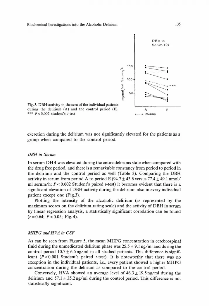
Fig. 3. DBH-activity in the sera of the individual patients during the delirium (A) and the control period (E). A E *** P<0.002 student's t-test x---x means
excretion during the delirium was not significantly elevated for the patients as a group when compared to the control period.
DBH in Serum
In serum DHB was elevated during the entire delirious state when compared with the drug free period, and there is a remarkable constancy from period to period in the delirium and the control period as well (Table 3). Comparing the DBH activity in serum from period A to period E (94.7 + 43.6 versus 77.4 + 49.1 nmol / ml serum/h; P < 0.002 Student's paired t-test) it becomes evident that there is a significant elevation of DBH activity during the delirium also in every individual patient except one (Fig.3).
Plotting the intensity of the alcoholic delirium (as represented by the maximum scores on the delirium rating scale) and the activity of DBH in serum by linear regression analysis, a statistically significant correlation can be found (r=0.64; P<0.05; Fig. 4).
MHPG and HVA in CSF
As can be seen from Figure 5, the mean MHP G concentration in cerebrospinal fluid during the unmedicated delirium phase was 25.5 + 9.1 ng/ml and during the control period 10.7 + 6.5 ng/ml in all studied patients. This difference is signif- icant (P<0.001 Student's paired t-test). It is noteworthy that there was no exception in the individual patients, i.e., every patient showed a higher MH P G concentration during the delirium as compared to the control period.
Conversely, HVA showed an average level of 46.3 + 19.5 ng/ml during the delirium and 57.1 + 35.2 ng/ml during the control period. This difference is not statistically significant.

136 D. Athen et al.
3 0 -
2 0 -
1 0 -
e-,
e ~ e �9
D = I I 410
10 20 30 r= 0,6449 m~mol/ml Serum/h
p < 0 , 0 5 A D B H (day A minus d a y E )
Fig. 4. Correlation between the intensity of alcoholic delirium (as measured on the delirium tremens rating scale) and the DBH-activity in the sera of the patients by linear regression analysis
3 0 -
2 0 -
loi
r~
o J :
c
MHPG HVA CSF ( 9 )
+ -I- -I-
A E
S .
A E
e-
- 6 0
- 4 0
- 2 0 Fig. 5. Average concentrations of MHPG and HVA in cerebrospinal fluid of the pa- tients as a group during delirium (period A) and a control day (period E). *** P< 0.001 Student's t-test
Longitudinal Study of One Patient with Two Deliriums Within 4 Months
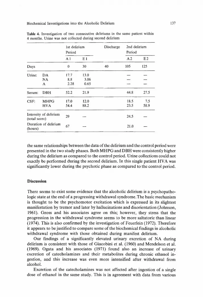
One of our pat ients had to be treated twice for del ir ium within 4 months . The biochemical data of the two pathologic states is shown in Table 4. As can be seen,
Biochemical Investigations into the Alcoholic Delirium
Table 4. Investigation of two consecutive deliriums in the same patient within 4 months. Urine was not collected during second delirium
137
1st delirium Discharge 2nd delirium Period Period
A1 E1 A2 E2
Days 0 30 40 105 125
Urine: DA 17.7 13.0 - - - - NA 8.8 3.06 - - - - A 2.28 0.65 - - - -
Serum: DBH 52.2 21.9 44.8 27.5
CSF: MHPG 17.0 12.0 18.5 7.5 HVA 54.4 88.2 23.5 58.9
Intensity of delirium 29 - - 24.5 - - (total score) Duration of delirium 67 - - 21.0 - - (hours)
the same relationships between the data of the delirium and the control period were presented in the two study phases. Both M H P G and D B H were consistently higher during the delirium as compared to the control period. Urine collections could not exactly be per formed during the second delirium. In this single patient H V A was significantly lower during the psychotic phase as compared to the control period.
Discussion
There seems to exist some evidence that the alcoholic delirium is a psychopa tho- logic state at the end of a progressing withdrawal syndrome. The basic mechanism is thought to be the psychomoto r excitation which is expressed in its slightest manifestat ion by t remor and later by hallucinations and disorientation (Johnson, 1961). Gross and his associates agree on this; however, they stress that the progression in the withdrawal syndrome seems to be more saltatoric than linear (1974). This is also confirmed by the investigation o f Feuerlein (1972). Therefore it appears to be justified to compare some of the biochemical findings in alcoholic withdrawal syndrome with those obtained during manifest delirium.
Our findings of a significantly elevated urinary excretion o f N A during delirium is consistent with those of Giacobini et al. (1960) and Mendelson et al. (1969). Ogata and his associates (1971) found also an increase o f ur inary excretion o f catecholamines and their metabolites during chronic ethanol in- gestion, and this increase was even more intensified after withdrawal f rom alcohol.
Excret ion o f the catecholamines was not affected after ingestion of a single dose of ethanol in the same study. This is in agreement with data f rom various
138 D. Athen et al.
animal studies, where increases of both NA turnover and NA release were found (Ahtee and Svartstr6m-Fraser, 1975; Pohorecky, 1974; Hunt and Majchrowicz, 1974). Similarly an increased plasma level of noradrenaline was demonstrated during abstinence syndrome by Carlsson and H~iggendahl (1967).
It is remarkable that the activity of DHB in serum parallels the increase of catecholamine excretion. This enzyme has been investigated in patients with various diseases, including torsion dystonia (where it is increased, Wooten et al., 1973) and familial dysautonomia and mongolism (where it is decreased, Freed- man and Goldstein, 1974; Weinshilboum and Axelrod, 1971). In addition, an enhancement was found after maximum work load (Planz and Palm, 1973), and it is thought that DBH activity may reflect the tone of the sympathetic nervous system. Inspection of the individual data reveals that all patients (except patient No. 4) showed an elevated DBH activity during the delirium as compared to the control period. Of special interest is the positive correlation between the height of of the DBH activity in serum and the intensity of the alcoholic delirium, as measured on the delirium tremens rating scale. From this it may be concluded that there is a correlation between the increased sympathetic activity and alcoholic delirium provided that one can conclude from peripheral to central noradrenergic activity.
MHP G which best may reflect the central noradrenergic metabolism was elevated 137% in comparison to the control period in CSF. In this regard the findings of Ogota et al. (1971), who found a significant decrease in urinary VMA excretion and a concomitant increased MHP G excretion during the alcoholic withdrawal syndrome, are of interest. Because of this, a shift from metabolic oxidation to reduction in the final pathway of noradrenaline metabolism is indicated. In CSF of healthy control persons the MHPG : VMA ratio is very high (Jimerson et al., 1975). Therefore it seems not to be conceivable that the increase of MHP G concentration in lumbar CSF is exclusively due to a metabolic shift from oxidation to reduction. Rather it may be considered an indicator of strong central noradrenergic hyperactivity during alcoholic withdrawal syndrom, i.e., delirium.
The urinary excretion of A parallels the findings with NA excretion with a broader interindividual variation. Nevertheless, the patients as a group, showed a clearly significant elevation during the delirium as compared to the control period. This is in agreement with the findings of other investigators (Giacobini et al., 1960; Carlsson and H~iggendal, 1967).
Estimation of urinary DA excretion was difficult in that there were large inter- individual variations both in the delirium and the control period. In addition, there were large intraindividual variations from day to day without any cor- relation with clinical variables, such as height of blood pressure, pulse rate and body temperature. There was no significant alteration in urinary DA excretion in the patients as a group; this is in line with various findings of other investigators (Ogota et al., 1971) and is consistent with pharmacological studies in brains of laboratory animals (Ahtee and Svartstr6m-Frazer, 1975), though not all studies agree (Griffith et al., 1974; Hunt and Majchrowicz, 1974).
HVA in CSF, which best may reflect the activity of the central dopaminergic system did not show a significant elevation during the delirium as compared to

Biochemical Investigations into the Alcoholic Delirium 139
the control period. Rather there was a slight and insignificant reduction of HVA during the withdrawal syndrome. This is in line with the results obtained by Roos and his associates (1973) who also found low values during abstinence syndrome in patients suffering from chronic alcoholic ataxia as well. Again, this is in agree- ment with laboratory studies in alcohol dependent rats, which did not show any alteration of HVA concentration in the brains of the animals (Ahtee and Svartstr6m-Frazer, 1975).
It remains somewhat problematical whether or not the increased central and peripheral noradrenergic activity may be more state dependent, i.e., due to the concomitant stress and the psychomotor activity during the disease. It has long been known that increase in psychic stress, physical activity and changes in posture all affect the excretion of urinary catecholamines and their metabolites (Euler, 1971; Karki, 1956; Sudin, 1958) and the level of M H P G in CSF (Ebert et al., 1973), though not all studies agree (Goode et al., 1973).
However, it has been shown by Post and Goodwin (1973) that simulation of mania and increased physical activity lead to an increase of all three metabolites in CSF, i.e., HVA, 5-HIAA and MHPG. Conversely, in this study only M H P G was elevated. Thus it can be hypothesized that hyperactivity of the central and peripheral noradrenergic system as expressed in the increase of catecholamine excretion, DBH activity in serum and M H P G in CSF might be of significance for the alcoholic withdrawal syndrome.
The increased activity of the noradrenergic system may have a protective function in ethanol withdrawal. By inhibiting the synthesis of catecholamines by a-methyl-p-tyrosine (aMPT) withdrawal symptoms were aggravated in mice (Blum and Wallace, 1974; Griffiths et al., 1974; Goldstein, 1973). In addition, Collier et al. (1974) showed that the injection of NA into the cerebral ventricles as well as amphetamine administration markedly inhibited ethanol withdrawal head twitches in mice. Further animal and human studies should be done to clarify the significance of the noradrenergic hyperactivity in order to get a more specific treatment for the ethanol withdrawal syndrome, particularly delirium tremens.
References
Ahtee, L., Svartstr6m-Frazer, M.: Effect of ethanol dependence and withdrawal on the catechol- amines in rat brain and heart. Acta pharmacol, et toxicol. 36, 289--298 (1975)
Blum, K., Wallace, I. E.: Effects of catecholamine synthesis inhibition on ethanol-induced with- drawal symptoms in mice. Br. J. Pharmac. 51, 109--111 (1974)
Carlsson, C., H/iggendal, J.: Arterial noradrenaline levels after ethanol withdrawal. Lancet 1967II, 889
D ekirmenjian, H., Maas, J. W.: An improved procedure of 3-methoxy-4-hydroxcyphenylethyl- eneglycol determination by gasliquid chromatography. Anal. Biochem. 35, 113--122 (1970)
Ebert, M. H., Post, R. M., Goodwin, F. K.: Effect of physical activity on urinary MHPG excretion in depressed patients. Lancet 130, 67--72 (1973)
Euler, O. S. v.: Adrenergic transmitter functions. Science 172, 202--206 (1971) Feuerlein, W.: Zur Frage des Alkohol-Entzugssyndroms. Der Nervenarzt 43, 247--253 (1972) Freedman, L. S., Goldstein, M.: Serum-beta-hydroxylase in Down's syndrome: A familial study.
Res. Comm. Chem. Pharmac. 8, 543--549 (1974) Giacobini, E., Izikowitz, S., Wegmann, A.: Urinary norepinephrine and epinephrine excretion
in delirium tremens. Arch. gen. Psychiat. 3, 289--296 (1960)
140 D. Athen et al.
Goldstein, D. B.: Alcohol withdrawal reactions in mice: Effects of drugs that modify neuro- transmission. J. Pharmacol. Exp. Therap. 186, 1--9 (1973)
Goode, D. J., Dekirmenjian, H., Meltzer, H. Y., Maas, J. W.: Relation of exercise to MHPG excretion in normal subjects. Arch. Gen. Psychiat. 29, 391--396 (1973)
Griffith, P. J., Littleton, J. M., Ortiz, A.: Changes in monoamine concentrations in mouse brain associated with ethanol dependence and withdrawal. Br. J. Pharmac. 50, 489--498 (1974)
Gross, M. M., Lewis, E., Hastey, J.: Acute alcohol withdrawal syndrome. In: Kissin, B. and H. Begleiter (Eds.): The Biology of Alcoholism. Vol. III.New York-London: Plenum Press 1974
Hunt, W. A., Majchrowicz, E.: Alterations in the turnover of brain norepinephrine and dopamine in alcohol-dependent rats. J. Neurochem. 23, 549--552 (1974)
Jimerson, D., Gordon, E. K., Post, R. M., Goodwin, F. K.: Central norepinephrine function in man: VMA in the CSF. Sci. Proc. Am. Psych. Ass. 128, 94--96 (1975)
Johnson, R. B.: The alcohol withdrawal syndromes. Quart. J. Stud. Alcohol Suppl. 1, 66--76 (1961)
Kalant, H.: The pharmacology of alcohol intoxication. Quart. J. Stud. Alcohol Suppl. 1, 1--23 (1961)
Karki, N. T.: The urinary excretion of noradrenaline and adrenaline in different age groups, its diurnal variations and the effect of muscular work on it. Acta Physiol. Seand. Suppl. 132, 1--96 (1956)
Maas, J. W., Dekirmenjian, H., Garver, D., Redmond, D. E., Jr., Landis, D. H.: Excretion of catecholamine metabolites following intraventricular injection of 6-hydroxydopamine in the macaca speciosa. Europ. J. Pharmacol. 23, 121--130 (1973)
Markianos, E. S., Nystr6m, I.: Serum dopamine-fl-hydroxylase. Z. Klin. Chem. Klin. Biochem. 13, 273--276 (1975)
Markianos, E. S., Beckmann, H.: Diurnal changes in dopamine-fl-hydroxylase, homovanillic acid and 3-methoxy-4-hydroxyphenylglycol in serum of man. J. Neut. Transm. 39, 79--93
Ogata, M., Mendelson, J. H., Mello, N. K., Majchrowicz, E.: Adrenal function and alcoholism: II. Cateeholamines. Psychosom. Med. 33, 159--180 (1971)
Planz, G., Palm, D.: Acute enhancement of dopamine-fl-hydroxylase activity in human plasma after maximum work load. Europ. J. Clin. Pharmacol. 5, 255--258 (1973)
Pohorecky, L. A.: Effects of ethanol on central and peripheral noradrenergic neurons. J. Pharmac. Exp. Therap. 189, 380--391 (1974)
Post, R. M., Goodwin, F. K.: Simulated behaviour states. Biological Psychiatry 7, 237--254 (1973)
Ritter, G., Duensing, F.: Exterozeptive Reflexenthemmung beim Delirium tremens. Der Nerven- arzt 41, 439--444 (1970)
Roos, B. E., Silfverski61d, B. P.: Homovanillic acid in cerebrospinal fluid of alcoholics. The New England Journal of Medicine 1358 (1953)
Sudin, T.: The effect of body posture on the urinary excretion of adrenaline and noradrenaline. Acta Med. Scand. 161, Suppl. 336 (1958)
Weil-Malherbe, H.: Analysis of biogenic amines and their related enzymes. In: Methods of Bio- chemical Analysis. David Glick (Ed.) New York: John Wiley & Sons 1971
Weinshilboum, R. M., Axelrod, J.: Reduced plasma dopamine-beta-hydroxylase activity in familial dysautonomia. N. Eng. J. Med. 285, 928--942 (1971)
Wieser, S.: Alkoholismus II: Psychiatrische und neurologische Komplikationen. Fortsehr. Neurol. Psychiat. 33, 349--409 (1965)
Wooten, F. G., Eldridge, R., Axelrod, J., Stern, R. S.: Elevated plasma dopamine-beta-hydroxy- lase in autosomal dominant torsion dystonia. N. Eng. J. Med. 288, 284--287 (1973)
Received February 28, 1977