biochemical and functional studies on the burkholderia cepacia complex bcen gene, encoding a...
TRANSCRIPT
Biochemical and Functional Studies on the Burkholderiacepacia Complex bceN Gene, Encoding a GDP-D-Mannose 4,6-Dehydratase
Sılvia A. Sousa1., Joana R. Feliciano1., Pedro F. Pinheiro1, Jorge H. Leitao1,2*
1 Institute for Biotechnology and Bioengineering, Centre for Biological and Chemical Engineering, Instituto Superior Tecnico, Lisboa, Portugal, 2Department of
Bioenginneering, Instituto Superior Tecnico, Universidade Tecnica de Lisboa, Lisboa, Portugal
Abstract
This work reports the biochemical and functional analysis of the Burkholderia cenocepacia J2315 bceN gene, encoding aprotein with GDP-D-mannose 4,6-dehydratase enzyme activity (E.C.4.2.1.47). Data presented indicate that the protein isactive when in the tetrameric form, catalyzing the conversion of GDP-D-mannose into GDP-4-keto-6-deoxy-D-mannose. Thissugar nucleotide is the intermediary necessary for the biosynthesis of GDP-D-rhamnose, one of the sugar residues ofcepacian, the major exopolysaccharide produced by environmental and human, animal and plant pathogenic isolates of theBurkholderia cepacia complex species. Vmax and Km values of 1.560.2 mmol.min21.mg21 and 10246123 mM, respectively,were obtained from the kinetic characterization of the B. cenocepacia J2315 BceN protein by NMR spectroscopy, at 25uC andin the presence of 1 mol MgCl2 per mol of protein. The enzyme activity was strongly inhibited by the substrate, with anestimated Ki of 29136350 mM. The lack of a functional bceN gene in a mutant derived from B. cepacia IST408 slightlyreduced cepacian production. However, in the B. multivorans ATCC17616 with bceN as the single gene in its genome withpredicted GMD activity, a bceN mutant did not produce cepacian, indicating that this gene product is required for cepacianbiosynthesis.
Citation: Sousa SA, Feliciano JR, Pinheiro PF, Leitao JH (2013) Biochemical and Functional Studies on the Burkholderia cepacia Complex bceN Gene, Encoding aGDP-D-Mannose 4,6-Dehydratase. PLoS ONE 8(2): e56902. doi:10.1371/journal.pone.0056902
Editor: Daniela Flavia Hozbor, Universidad Nacional de La Plata, Argentina
Received October 31, 2012; Accepted January 15, 2013; Published February 27, 2013
Copyright: � 2013 Sousa et al. This is an open-access article distributed under the terms of the Creative Commons Attribution License, which permitsunrestricted use, distribution, and reproduction in any medium, provided the original author and source are credited.
Funding: This work was partially funded by FEDER and FCT, Portugal (contracts PTDC/EBB-BIO/098352/2008 and PTDC/BIA-MIC/119091/2010, and a doctoral andpost-doctoral grant to J.R.F. and S.A.S., respectively). The funders had no role in study design, data collection and analysis, decision to publish, or preparation ofthe manuscript.
Competing Interests: The authors have declared that no competing interests exist.
* E-mail: [email protected]
. These authors contributed equally to this work.
Introduction
The rare 6-deoxysugar D-rhamnose is often a component of
bacterial cell surface glycans, such as lipopolysaccharides (LPS),
extracellular polysaccharides (EPS), and other glycoconjugate-
containing bacterial cell components [1–4]. So far, and with the
exception of Paramecium bursaria chlorella virus 1 where D-
rhamnose is believed to be a component of the major viral capsid
glycoprotein, the occurrence of this rare deoxysugar seems to be
restricted to bacteria [5]. This is the case of the opportunistic
pathogens of the Burkholderia cepacia complex (Bcc), a group of 17
closely related species that can cause infections in several hosts,
including the human host [6]. These bacteria emerged in the
1980s as important pathogens, in particular among cystic fibrosis
patients. In Bcc bacteria, D-rhamnose is a component of the LPS
[7] and of the major extracellular polysaccharide cepacian,
produced by a large percentage of clinical isolates [8,9].
Cepacian has been pointed out as contributing to the overall
pathogenicity of Bcc bacteria. For example, cepacian interferes
with human neutrophils phagocytosis by facilitating the bacterial
persistence in a mice model of infection, inhibits the production of
oxygen reaction species (ROS) by neutrophils, and scavenges
ROS, playing a role in the survival of cepacian-producing strains
in different environments [10–14]. In addition, mutants defective
in cepacian production were found as less virulent or completely
avirulent in a mice model of infection [15].
Besides D-rhamnose, cepacian also contains residues of D-
glucose, D-mannose, D-galactose, and D-glucuronic acid as sugar
components of its heptasaccharide repeat unit [16].
Cepacian was previously identified as the sole EPS produced by
the clinical isolate B. cepacia IST408 [8]. In addition, FTIR analysis
of the EPSs produced by B. cepacia IST408 and B. multivorans
ATCC17616 showed that both strains produced EPSs with FTIR
spectra identical to that of cepacian [17].
Previous work allowed the identification of genes involved in
cepacian biosynthesis in two clusters, named bceI and bceII [17,18].
Cepacian gene clusters are widespread within the genomes of all
strains of the Burkholderia genus sequenced so far, except of B. mallei
strains [17]. The bceII gene cluster comprises several genes
putatively involved in the synthesis of cepacian, including the
bceM and bceN genes, presumably encoding proteins with putative
GDP-4-keto-6-deoxy-D-mannose reductase (RMD) and GDP-D-
mannose-4,6-dehydratase (GMD) activities, respectively [17].
In the present work, we report the cloning, purification, and
kinetic characterization of the bceN gene product with predicted
GMD activity, from the highly epidemic clinical isolate B.
cenocepacia J2315, a member of the ET12 lineage that caused
PLOS ONE | www.plosone.org 1 February 2013 | Volume 8 | Issue 2 | e56902
several fatalities among CF patients from both sides of the Atlantic
(reviewed in [6]). Our results, obtained using NMR spectroscopy,
confirm that the protein has GDP-D-mannose dehydratase activity
(EC.4.2.1.47), catalysing the conversion of GDP-D-mannose into
the labile product GDP-4-keto-6-deoxy-D-mannose. The B.
cenocepacia J2315 GMD protein was found to be active only when
in the tetrameric form, requiring the presence of magnesium ions
for activity. GMD activity was detected in the absence of NAD(P),
suggesting that the cofactor is tightly bound to the enzyme.
The comparison of cepacian production ability of wild-type and
mutant strains derived from the cepacian producers B. cepacia
IST408 and B. multivorans ATCC17616 confirmed the requirement
of this enzyme activity for its synthesis.
Materials and Methods
Bacterial strains, plasmids and culture conditionsBacterial strains and plasmids used in this study are described in
Table S1. When in use, Bcc strains were maintained on PIA
(Pseudomonas Isolation Agar, BD) plates, supplemented with
150 mg/ml trimethoprim or 600 mg/ml kanamycin in the case of
mutant strains. Escherichia coli strains were maintained on Lennox
broth (LB; containing in g/l, tryptone 10, yeast extract 5, NaCl 5)
agar plates, supplemented with 100 mg/ml trimethoprim, 150 mg/
ml ampicillin, or 50 mg/ml kanamycin, when appropriate. Unless
otherwise stated, liquid cultures were carried out in LB liquid
medium supplemented with the appropriate antibiotics with
orbital agitation (250 rpm), at 30uC for Bcc strains or 37uC for
E. coli strains. Bacterial growth was assessed by measuring the
cultures optical density at 640 nm (OD640).
Molecular biology techniquesGenomic DNA was extracted from exponentially-growing
liquid cultures of Bcc strains using the High Pure PCR Template
Preparation Kit (Roche). Plasmid isolation and purification, DNA
amplification, restriction and ligation were carried out, respec-
tively, using kits from Macherey-Nagel, and enzymes from
Finnzymes and Fermentas. Agarose gel electrophoresis and E. coli
transformation were carried out using standard procedures [19].
Electrotransformation of Bcc strains was performed as previously
described [20]. Amplification of the B. cenocepacia J2315 bceN gene
(BCAM1004) was performed using the oligonucleotides UP-GMD
(59-TTGCTAGCATGAGCCAAA CTC-39) and LW-GMD (59-
AAAAAGCTTGAACGTGTCATG-39) containing, respectively,
the NheI and HindIII restriction sites (underlined) at their 59 ends.
For the amplification of the bceN gene and the neighbour region,
oligonucleotides UP-GMDmut (59-AAAGGTACCGTCGGA-
GAAATC-39) and LW-GMDmut (59-TTTAAGCTTC
GATTCGTTCTG-39) containing at their 59 end, respectively,
the KpnI and HindIII restriction sites (underlined). These primers
were designed based on the genome sequence of B. cenocepacia
J2315, available at the Sanger Institute Homepage (http://www.
sanger.ac.uk/Projects/ B_cenocepacia).
Nucleotide and amino acid sequence analysis andstructure predictionNucleotide and amino acid sequences were analysed using
bioinformatic tools resident at the National Center for Biotech-
nology Information (NCBI) or the ExPASy-Prosite websites.
Searches for homology within the genome sequences of B.
cenocepacia J2315 and other Burkholderia strains were carried out
using the Integrated Microbial Genomes system [21]. Protein
secondary and tertiary structure predictions were performed using
the Protein Structure Prediction Server (PSIPRED) [22] and I-
Tasser [23], respectively. Graphics were generated using the
RasWin Molecular Graphics (Windows Version 2.7.5). The
phylogenetic tree of BceN homologues was constructed with
CLUSTAL X2, using the neighbour-joining method with a
minimum of 100 bootstraps [24].
Cloning and overexpression of B. cenocepacia J231566His-tagged BceNThe B. cenocepacia J2315 bceN gene sequence (BCAM1004)
lacking the stop codon was amplified using B. cenocepacia J2315
genomic DNA as template and primers UP-GMD and LW-GMD.
The 1061 bp amplicon was digested with NheI and HindIII and
was subsequently cloned into the NheI/HindIII cloning sites of
pET23a+, yielding plasmid pJFR4. pJFR4 allows the control of
protein expression by the T7 promoter, expressing the BceN
protein with a 66His-tag at its C-terminus. Plasmid pJFR4 was
sequenced to confirm the correct insertion of the bceN gene, and
transformed into E. coli BL21 (DE3). Overexpression of the C-
terminally 66His-tagged BceN protein was performed by growing
E. coli BL21 (pJFR4) in 100 ml of LB liquid medium (supple-
mented with 150 mg/ml ampicillin) at 37uC and with orbital
agitation (250 rpm). When the culture OD640 reached 0.6,
isopropylthiogalactoside (IPTG) was added (final concentration
0.4 mM) and incubation was prolonged for 2 hours. Bacteria were
harvested by centrifugation at 70006 g for 5 min at 4uC, and
ressuspended in 8 ml of start buffer (20 mM sodium phosphate,
500 mM NaCl, pH 7.4) containing 20 mM imidazole. Cell
suspensions were kept at 280uC until further processing. Aliquots
of these cell suspensions were processed and protein overproduc-
tion was assessed by 12.5% sodium dodecyl sulfate polyacrylamide
gel electrophoresis (SDS-PAGE). To confirm the overproduction
of the 66His-tagged BceN protein, the samples were transferred
onto a nitrocellulose membrane. The membrane was incubated
with a monoclonal anti-polyhistidine antibody conjugated with
peroxidase (Sigma-Aldrich) diluted 1:2000, followed by the
addition of the peroxidase substrate 3,39,5,59 tetramethylbenzidine
(TMB, Sigma).
Purification of the 66His-tagged BceNCell suspensions prepared as described above were lysed by
ultrasonic vibration with a Branson sonifier 250 (Branson). Cell
debris were removed by centrifugation at 12,5006 g for 1 hour at
4uC. The 66His-tagged BceN protein was purified by affinity
chromatography using a Hi-Trap chelating column (GE Health-
care), following the suppliers instructions. Purified fractions were
analysed by SDS-PAGE followed by staining with Coomassie
brilliant blue R-250. The eluted fractions containing the purified
66His-tagged protein were dialysed overnight at 4uC in a 10 kDa
cutoff Slide-A-Lyzer Dialysis Cassette (Pierce) against 25 mM
sodium phosphate buffer (pH 7.4) supplemented with 50 mM
NaCl and 20% (v/v) glycerol. Protein concentration was estimated
by the method of Bradford [25], with bovine serum albumin
fraction V (Nzytech) as standard.
Discontinuous native protein gel electrophoresisNative protein gel electrophoresis of the purified 66His-tagged
BceN was performed based on previously described methods [26]
with minor modifications to estimate the molecular mass of the
66His-tagged BceN oligomer. Briefly, 20 ml aliquots of protein
samples (6 mg) were added to 5 ml of loading buffer (100 mM Tris-
Cl pH 8.0, 40% glycerol, 0.5% Brilliant Blue G) and incubated for
10 minutes at room temperature. Samples containing 20 mg of
catalase (232 kDa), aldolase (157 kDa), bovine serum albumin (66
Burkholderia cenocepacia GDP-Mannose Dehydratase
PLOS ONE | www.plosone.org 2 February 2013 | Volume 8 | Issue 2 | e56902

and 132 kDa), and ovalbumin (44 kDa) were used as molecular
mass standards. The samples were applied to an 8% (wt/vol)
polyacrylamide gel containing 200 mM Tris-Cl (pH 8.8). The
cathode buffer was composed of 100 mM histidine adjusted to
pH 8.0 using Tris base (without chloride) and 0.002% Brilliant
Blue G. The anode buffer contained 100 mM Tris-Cl (pH 8.8).
The gel was run at 4uC and 100 V for 2 hours.
Assessment of GDP-D-mannose 4,6-dehydratase (GMD)activity by NMRThe ability of the B. cenocepacia J2315 66His-tagged BceN
protein to convert GDP-D-mannose into GDP-4-keto-6-deoxy-D-
mannose was assessed using GDP-D-mannose as substrate, and
analysing the reaction products by Nuclear Magnetic Resonance
(NMR) spectroscopy, based on the methods described by King et
al. [5]. NMR experiments were performed using a Bruker
AVANCE II+ 400 MHz (1H) spectrometer equipped with a
5 mm BBO probe. The reacting mixture [containing 25 mM
sodium phosphate buffer, 50 mM NaCl, 20% (v/v) glycerol, 90%
H2O/10% D2O, pH 7.4, in a total volume of 500 ml], was placed
in a 5 mm NMR tube (Norrel 100 MHz) with concentrations of
GDP-D-mannose (Sigma) ranging from 1 to 5 mM, in the absence
or presence of 5 mM NADP+ (Sigma), and MgCl2 (1 mol MgCl2per 1 mol of purified 66His-tagged BceN protein). Experiments
were conducted at 25uC. The reaction was started by the addition
of 90 mg of the purified 66His-tagged BceN protein to the reaction
buffer, and the proton spectrum was recorded at fixed periods of
time. Spectral data was collected over time and treated with
TopSpin software v2.1. Resonance of the acetone methyl group
was used as internal reference for all spectra (CH3dH=2.225 ppm,
CH3dC=30.89 ppm). Different concentrations of MgCl2 (0 mM,
25 mM and 1 mol:1 mol protein) and NADP+ (0 or 5 mM;
Sigma), and different reaction temperatures (25uC and 37uC) were
tested for the reaction conditions optimization. Reaction products
were characterized by 1D (1H and 13C) and 2D (HSQC) NMR
experiments. Protein stability was only achieved when using 20%
(v/v) glycerol in all experiments, which interfered with the
substrate and product peaks in the range of 3.5 to 3.9 ppm.
Therefore, kinetic measurements were based on the proton signals
corresponding to the anomeric protons in GDP-D-mannose and
the two reaction products as described by King and coworkers [5].
Integral values of the 5.51 ppm signal were normalized with the
internal standard (CH3dH=2.225 ppm) and plotted versus time
[27]. Vo was obtained from the slope of the exponential part of the
curve obtained with the increase of products concentration,
calculated using the equation [P]t= [S] (1-SNAt), where [P]t is the
concentration of products on time t, [S] is the initial substrate
concentration, and SNAt is the substrate normalized integral signal
area at time t. NMR experimental data was further analyzed using
Graphpad Prism v6.
The hypothesis that the 66His-tagged BceN protein can
catalyse both the GDP-D-mannose 4,6-dehydratation and the
subsequent reduction reaction to produce GDP-D-rhamnose was
investigated based on the two following NMR assays: a) addition of
10 mM NADPH to the reaction mixture [containing 90 mg of the
purified BceN protein, 5 mM GDP-D-mannose, MgCl2 at the
ratio of 1 mol MgCl2 per 1 mol protein, 25 mM sodium
phosphate buffer, 50 mM NaCl, 20% (v/v) glycerol, 90% H2O/
10% D2O, pH 7.4]; b) addition of 10 mM NADPH, after
incubation of the reaction mixture for 30 minutes at 25uC.
Generation of mutants and PCR confirmation ofinsertional inactivationThe bceN (BCAM1004) gene and the neighbour region was
amplified from B. cenocepacia J2315 genomic DNA using primers
UP-GMDmut and LW-GMDmut. The 2272 pb amplimer was
digested with KpnI and HindIII and subsequently cloned into the
KpnI/HindIII cloning sites of pDrive, generating pACM2. The
911 bp EcoRI/SphI fragment from pUC-Tp containing the Tp
cassette was ligated into the EcoRI/SphI sites within the bceN gene
cloned in pACM2, creating pACM4. This last plasmid was
introduced in E. coli SSC110 and extracted from this strain to
obtain non-methylated plasmid DNA for transformations, to
escape the Bcc restriction system [28]. The extracted plasmid was
then introduced into B. cepacia IST408 and B. multivorans
ATCC17616 by electrotransformation, as previously described
[20]. In order to confirm the insertional inactivation of the bceN
gene and the double cross-over recombination (expected size of
3183 bp), colony-PCR experiments were performed, using the
primers UP-GMDmut and LW-GMDmut.
The 1061 bp XbaI/HindIII fragment from pJRF4 containing
the bceN gene was subcloned into the XbaI/HindIII restriction
sites of pDrive, creating pJFR5. The 1125 bp KpnI/XbaI
fragment from pJFR5 was then inserted into pMLBAD, yielding
pJRF6. The resulting plasmid (pJRF6) expresses an antisense
RNA, under the control of the PBAD promoter, which is
complementary to the bceN mRNA, thus allowing the silencing
of bceN upon induction with arabinose. The plasmid was
introduced into B. multivorans ATCC17616 by triparental conju-
gation as previously described [17].
Quantification of exopolysaccharide productionEPS production was quantified by determining the dry weight
of the ethanol-precipitated polysaccharide present in 30 ml of cell-
free culture samples of the Bcc strains as described before [20].
The culture samples were obtained from Bcc cultures grown at
30uC or 37uC in SARA liquid medium (containing, in g/l,
arabinose 20, Na2HPO4. 2H2O 12.5, KH2PO4 3, K2SO4 1,
NaCl 1, yeast extract 1, casamino acids 1, MgSO4. 7H2O 0.2,
CaCl2. 2H2O 0.01, FeSO4. 7H2O 0.001), for 24, 48, and
72 hours, with orbital agitation (250 rpm), as described previously
[8]. EPS production was quantified as EPS dry weight (g) per litre
of growth medium, and values given are the mean values of at least
three independent determinations.
Statistical analysisThe data obtained from the quantification of the ability of the
Burkholderia strains and their respective mutant strains to produce
EPS was tested for statistical significance by calculating P-values
using the Unpaired t test with Welch correction. A P-value,0.05
was considered statistically significant. Statistical analyses were
done with GraphPad InStat v3.
Nucleotide sequence accession numberThe bceN gene nucleotide sequence of B. cepacia IST408 was
deposited in GenBank under the Accession No. JN987864.
Results
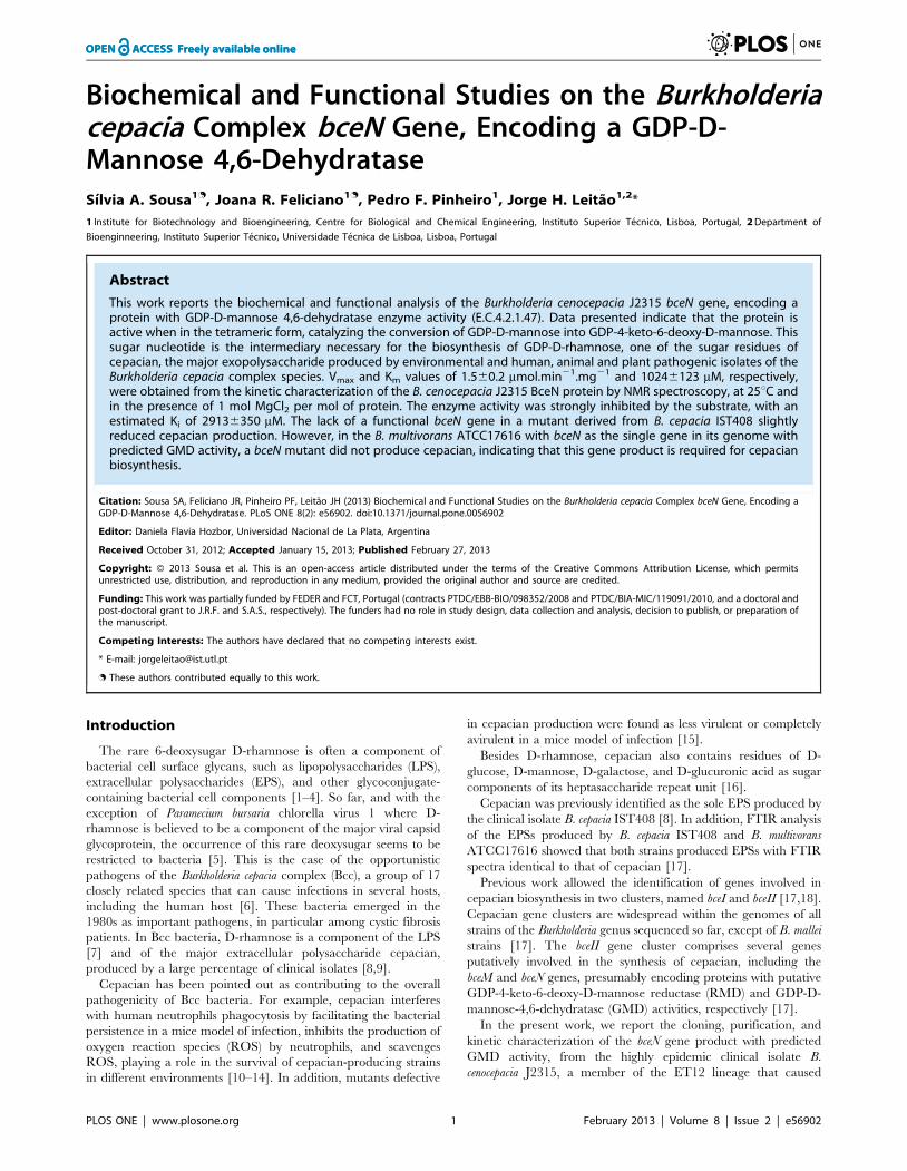
Sequence analysis of B. cenocepacia J2315 bceNThe B. cenocepacia J2315 bceN gene is located in the bce-II gene
cluster of chromosome 2, spanning nucleotides 1112176 to
1113222 (Fig. 1). The bce-II cluster was previously identified as
being involved in the biosynthesis of the exopolysaccharide
Burkholderia cenocepacia GDP-Mannose Dehydratase
PLOS ONE | www.plosone.org 3 February 2013 | Volume 8 | Issue 2 | e56902

cepacian [17]. The translated sequence of bceN (1061 bp) was
found to have a similarity higher than 66% with proteins with
GMD activity from other bacterial species such as B. cepacia,
Pseudomonas aeruginosa, Aneurinibacillus thermoaerophilus, and E. coli
(Fig. 1). A search for BceN homologues within available genome
sequences of Burkholderia strains allowed the identification of 23
BceN orthologues in 14 strains from 9 Burkholderia species. One,
two or three putative GMDs were found in the genomes sequences
examined. The GMD-encoding genes with highest degree of
identity/similarity with BceN all belong to gene clusters similar to
the bce-II gene cluster (Fig. S1, left upper panel) that is involved in
cepacian production [Clade A in Fig. S1 panel B; 17]. The BceN
homologues ABF75312, ABK07633, EAY62635 and EAY62633,
EAY69559, ACB63266, ABI86318 and ABC38232 identified in
the genomes of B. cenocepacia AU1054, B. cenocepacia HI2424, B.
cenocepacia PC184, B. dolosa AU0158, B. ambifaria MC40-6, B.
ambifaria AMMD and B. thailandensis E264, respectively, are all
located on chromosome 1, within clusters of genes encoding
putative glycosyltransferases and other proteins and enzymes
presumably involved in lipopolysaccharide synthesis (Fig. S1, right
upper panel). These proteins share an identity higher than 80% to
each other, forming a separate clade (Clade B in Fig. S1 panel B).
A GMD-encoding gene located on chromosome 3 was identified
in B. vietnamiensis G4 (protein ABO59420) (Fig. S1 panel B). In this
case, the encoding gene is flanked by proteins putatively involved
in polysaccharide and lipopolysaccharide biosynthesis, and
exopolysaccharide transport. This was the only sequenced strain
of the Burkholderia genus putatively encoding on chromosome 3 a
protein with GMD enzyme activity.
GMDs are members of the NDP-sugar modifying subfamily of
the short-chain dehydrogenases/reductases (SDR) family [29].
These proteins catalyse the irreversible conversion of GDP-D-
mannose into the unstable compound GDP-4-keto-6-deoxy-D-
mannose [30], which can be further converted into GDP-D-
rhamnose, GDP-L-fucose, GDP-6-deoxy-D-talose, or GDP-D-
perosamine [31].
Analysis of the amino acid sequences of the translated bceN gene
of the cepacian high producer clinical isolate B. cepacia IST408 and
comparison with the bceN sequence of B. cenocepacia J2315 revealed
that these sequences share 97% identity and 99% similarity (Fig. 1).
Both proteins have a predicted molecular mass of approximately
39 kDa and a pI of 6.35. The analysis of the deduced amino acid
sequences of the BceN proteins from B. cenocepacia J2315 and B.
cepacia IST408 also revealed the presence in both proteins of six
conserved domains typical of GMD enzymes (Fig. 1), namely: (i)
the glycine-rich Wierenga motif, 12GXXGXXG18, a conserved
feature of proteins of the SDR family [32], present in the BceN N-
terminal domain and thought to be involved in the binding of the
cofactor NAD(P)+; (ii) the 34GXXRR38 motif, conserved in
enzymes with GMD activity, with the two positively charged
arginine residues at the beginning of the RR loop [31]; (iii) the RR
loop, spanning amino acid residues Arg37 to Arg46, thought to be
involved in protein-protein interactions and protein-cofactor
interactions to the neighbouring monomer, proposed as essential
for the typical multimeric structure of GMD proteins [31]; (iv) the
conserved catalytic triad S/T131 and 155YXXXK159, essential for
catalysis. The tyrosine residue is responsible for the deprotonation
of the C4 hydroxyl group of the sugar moiety, while the lysine
residue is proposed to lower the pKa of the tyrosine to enable the
catalysis reaction [29,30]; (v) the glutamic acid residue at position
133 that is conserved in dehydratases and play a role in the
deprotonation/reprotonation of the C5 during catalysis, acting as
an additional active-site base that abstracts the C5 proton in the
dehydration reaction [30]; (vi) the conserved arginine residue in
position 190 that is fundamental for the correct orientation of the
substrate GDP-D-mannose and the NADP+ cofactor in the active
site for the dehydratase reaction [5].
The secondary structures of BceN proteins from both B.
cenocepacia J2315 and B. cepacia IST408 were predicted by
PSIPRED. The predicted structures exhibit the N-terminal
Rossman fold domain, typical of the sugar nucleotide-modifying
SDR family (Fig. 1). This motif is composed of an a/b folding
pattern, with 6–7 b-strands flanked by 3–4 a-helices on each side,
proposed to be involved in cofactor binding [29].
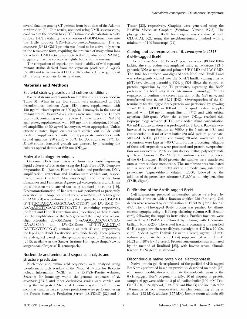
The B. cenocepacia J2315 BceN forms tetramers in itsactive formIn order to assess the predicted GMD activity of the B.
cenocepacia BceN protein, the bceN gene was cloned in the
expression plasmid pET23a+, as described in the Materials and
Methods section. The resulting plasmid allows the expression of
the BceN protein with a 66His-tag at its C-terminus. The C-
terminus location of the 66His-tag was selected to prevent a
possible enzymatic inactivation of BceN, which was previously
reported for the E. coli K12 GMD enzyme, due to structural
changes in the dinucleotide binding domain of the N-terminal
region [33].
The ability of the purified 66His-tagged BceN protein to form
multimers was investigated using size-exclusion chromatography
(data not shown). However, a very low resolution was obtained for
the purified BceN protein and the protein standards, most
probably due to its high content in glycerol. Therefore, we have
used discontinuous native PAGE analysis to investigate the ability
of BceN to form multimers. For this purpose, two aliquots from
purified 66His-tagged BceN were used, one with GMD activity
and the other without GMD activity, as detected by NMR
spectroscopy analysis. Several bands of higher molecular mass
than that of the band that correspond to the monomeric form of
BceN could be detected (Fig. 2B). However, a band with
approximately 162 kDa which corresponds to four times the
molecular mass of the monomeric BceN was only detected in the
sample with GMD activity, suggesting that in its active form BceN
is a tetramer (Fig. 2A and 2B). Interestingly, the crystal structure
determination of the P. aeruginosa PAO1 GMD protein (PDB ID:
1RPN) revealed that the active form of this protein is a tetramer
[31]. Although the B. cenocepacia BceN and the P. aeruginosa GMD
share only 73% similarity (Fig. 1), the comparison of the predicted
3D structures of the BceN monomer with the P. aeruginosa PAO1
GMD (PDB ID: 1RPN) revealed a high level of structural
similarity (Fig. 2C). One of the features observed was the
conserved spatial location of the RR loop that it is thought to
play a role on the interactions with the adjacent monomer in the
tetrameric structures of GMD proteins, as well as with their
cofactor when in the tetramer arrangement.
BceN exhibits GDP-D-mannose 4,6 dehydratase (GMD)activityThe enzyme activity of the 66His-tagged BceN protein of B.
cenocepacia J2315 was investigated using the protein purified to
homogeneity by affinity chromatography, followed by dialysis
overnight at 4uC against dialysis buffer (25 mM sodium phosphate
buffer, pH 7.4; 20% (v/v) glycerol; 50 mM NaCl). SDS-PAGE
analysis of the purified 66His-tagged BceN protein showed an
apparent molecular mass of approximately 40 kDa (Fig. 2A),
consistent with its predicted molecular mass of 39 kDa.
The GMD activity of the B. cenocepacia J2315 66His-tagged
BceN protein was assessed by analysing the reaction products by
Burkholderia cenocepacia GDP-Mannose Dehydratase
PLOS ONE | www.plosone.org 4 February 2013 | Volume 8 | Issue 2 | e56902
Figure 1. Genetic organization of the Burkholderia cenocepacia J2315 bce-II gene cluster, and alignment of amino acid sequences ofthe B. cenocepacia J2315 BceN protein (CAR54861) with GMDs from B. cepacia IST408 (JN987864), P. aeruginosa (AAG08838), A.thermoaerophilus (AAS55711) and E. coli (AAC77842). Upper part: Schematic representation of the bceII gene cluster evidencing the bceN genein black. Lower part: Sequence alignment of the indicated GMDs, evidencing the Wierenga motif and the catalytic triad typical of the short-chaindehydrogenases/reductases (SDR) family of proteins (highlighted in light grey). The specific GMD motifs are highlighted in dark grey. Asterisksindicate identical amino acid residues. One or two dots indicate semi-conserved or conserved substitutions, respectively. The predicted secondarystructure of B. cenocepacia J2315 and B. cepacia IST408 BceN proteins is shown above the alignment segments, where cylinders represent a-helices
Burkholderia cenocepacia GDP-Mannose Dehydratase
PLOS ONE | www.plosone.org 5 February 2013 | Volume 8 | Issue 2 | e56902
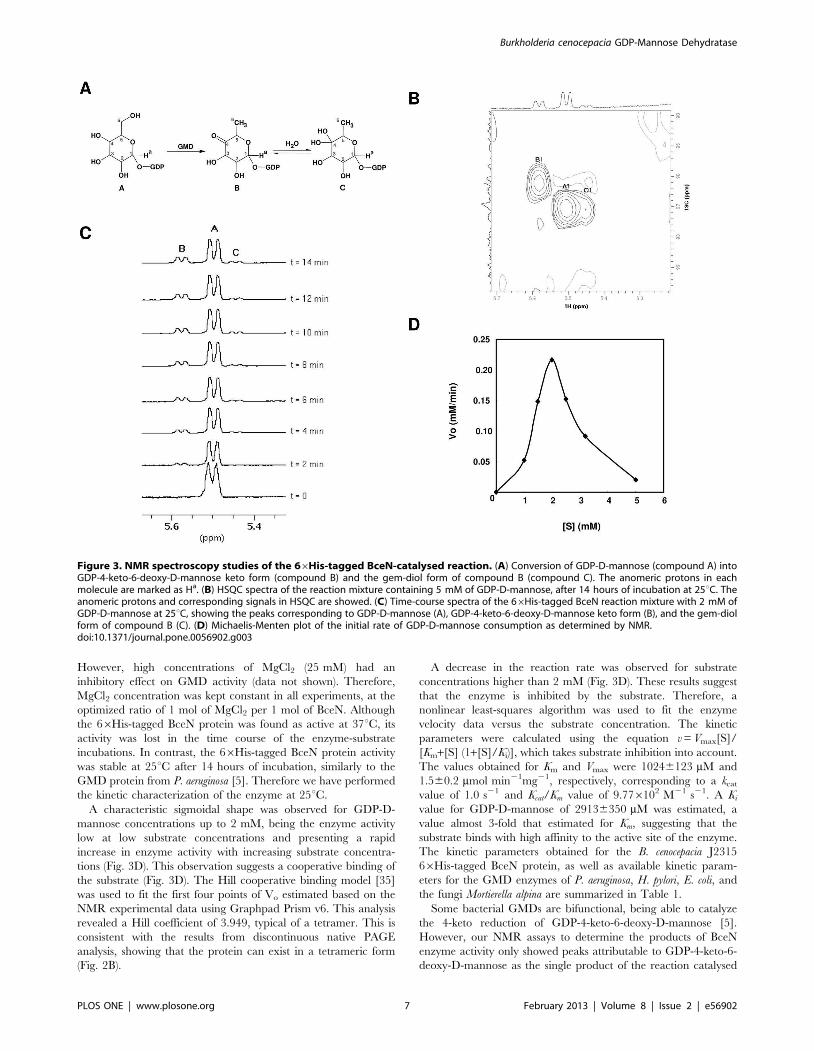
NMR spectroscopy (Fig. 3). The data obtained is summarized in
Table S2. Proton and carbon chemical shifts for the reaction
mixture products in the presence of GDP-D-mannose were in
good agreement with the data obtained for the compounds
identified for the GMD enzyme activity of P. aeruginosa [5]. A peak
corresponding to the C4 of compound B (Fig. 3A) was observed at
dC=208.60 ppm, indicating a carbonyl group, as expected to
occur in the keto form of GDP-4-keto-6-deoxy-D-mannose [5].
Although low resolution and glycerol interference hindered the
detection of the chemical shift of the C4 of compound C, the H1
and H6 proton and C1 and C6 carbon chemical shifts are in
accordance with the gem-diol form of compound B (Fig. 3A), as
previously described by King and colleagues [5].
The monitorization of the anomeric region of the 1D-1H-
spectrum showed a progressive depletion of signals corresponding
to GDP-D-mannose (compound A) and an increase of the intensity
of the peaks corresponding to the resonances of the 4-keto
(compound B) and gem-diol (compound C) anomeric forms of
GDP-4-keto-6-deoxy-D-mannose (Fig. 3B and 3C). Spectra
obtained indicate that the 66His-tagged BceN protein catalyzes
the dehydration of GDP-D-mannose to GDP-4-keto-6-deoxy-D-
mannose, thus having GDP-D-mannose 4,6-dehydratase activity.
Results also indicate that the protein only catalyzes the dehydra-
tion reaction, since no peaks corresponding to GDP-D-rhamnose
could be detected.
The requirement of a dinucleotide cofactor for the GMD
activity of proteins from other bacterial species has been reported
[34]. However, the GMD activity of the B. cenocepacia 66His-
tagged BceN protein was unaffected by the addition of exogenous
NAD+ or NADP+ (data not shown). We hypothesize that the
cofactor was co-purified with the protein, as previously observed
for the P. aeruginosa GMD protein [5]. The GMD activity of the
BceN protein was also found to be dependent on the addition of
Mg2+, being the enzyme inactive in its absence (data not shown).
and arrows represent b-sheets. Alignments and the secondary structure predictions were performed with CLUSTAL X2 and the PSIPRED software,respectively. Genes bceM to bceU putatively encode GDP-D-rhamnose reductase (bceM), GDP-D-mannose dehydratase (bceN), acyltransferase (bceO),unknown (bceP), repeat unit flippase (bceQ), glycosyltransferase (bceR), acyltransferase (bceS), UDP-glucose pyrophosphorylase (bceT), acyltransferase(bceU).doi:10.1371/journal.pone.0056902.g001
Figure 2. BceN is a tetramer. (A) SDS-PAGE analysis of the 66His-tagged BceN from B. cenocepacia J2315 by E. coli BL21 (DE3). Lanes from leftpanel: t0, total soluble proteins from E. coli BL21 (DE3) with plasmid pJRF4 before induction with IPTG; t2, total soluble proteins from E. coli BL21 (DE3)with plasmid pJRF4 after 2 hours of induction with IPTG; A–D, 66His-tagged BceN protein eluted from Ni2+-NTA affinity chromatography columnwith increasing concentrations of imidazole. Lanes from right panel (Western-blot): 1, BSA used as negative control; 2, purified 66His-tagged BceNprotein from B. cenocepacia J2315. The monoclonal anti-polyhistidine antibody conjugated with peroxidase (Sigma-Aldrich) was used in the Western-blot. (B) Discontinuous native PAGE analysis of 66His-tagged BceN. Lanes: M1, BSA; M2, ovalbumin; M3, aldolase; M4, catalase; A–B, purified 66His-tagged BceN from B. cenocepacia J2315 with no GMD activity; C, purified 66His-tagged BceN from B. cenocepacia J2315 with GMD activity. Theanalysis of the discontinuous native PAGE showed a predominant band with approximately 162 kDa compatible with a tetrameric form of theprotein. Trimeric, dimeric and monomeric forms are also visible. (C) Structural models of the BceN of B. cenocepacia J2315 and GMD from P.aeruginosa PAO1 (PDB ID: 1RPN). Graphics were generated using the RasWin Molecular Graphics (Windows Version 2.7.5).doi:10.1371/journal.pone.0056902.g002
Burkholderia cenocepacia GDP-Mannose Dehydratase
PLOS ONE | www.plosone.org 6 February 2013 | Volume 8 | Issue 2 | e56902
However, high concentrations of MgCl2 (25 mM) had an
inhibitory effect on GMD activity (data not shown). Therefore,
MgCl2 concentration was kept constant in all experiments, at the
optimized ratio of 1 mol of MgCl2 per 1 mol of BceN. Although
the 66His-tagged BceN protein was found as active at 37uC, its
activity was lost in the time course of the enzyme-substrate
incubations. In contrast, the 66His-tagged BceN protein activity
was stable at 25uC after 14 hours of incubation, similarly to the
GMD protein from P. aeruginosa [5]. Therefore we have performed
the kinetic characterization of the enzyme at 25uC.
A characteristic sigmoidal shape was observed for GDP-D-
mannose concentrations up to 2 mM, being the enzyme activity
low at low substrate concentrations and presenting a rapid
increase in enzyme activity with increasing substrate concentra-
tions (Fig. 3D). This observation suggests a cooperative binding of
the substrate (Fig. 3D). The Hill cooperative binding model [35]
was used to fit the first four points of Vo estimated based on the
NMR experimental data using Graphpad Prism v6. This analysis
revealed a Hill coefficient of 3.949, typical of a tetramer. This is
consistent with the results from discontinuous native PAGE
analysis, showing that the protein can exist in a tetrameric form
(Fig. 2B).
A decrease in the reaction rate was observed for substrate
concentrations higher than 2 mM (Fig. 3D). These results suggest
that the enzyme is inhibited by the substrate. Therefore, a
nonlinear least-squares algorithm was used to fit the enzyme
velocity data versus the substrate concentration. The kinetic
parameters were calculated using the equation v=Vmax[S]/
[Km+[S] (1+[S]/Ki)], which takes substrate inhibition into account.
The values obtained for Km and Vmax were 10246123 mM and
1.560.2 mmol min21mg21, respectively, corresponding to a kcatvalue of 1.0 s21 and Kcat/Km value of 9.776102 M21 s21. A Kivalue for GDP-D-mannose of 29136350 mM was estimated, a
value almost 3-fold that estimated for Km, suggesting that the
substrate binds with high affinity to the active site of the enzyme.
The kinetic parameters obtained for the B. cenocepacia J2315
66His-tagged BceN protein, as well as available kinetic param-
eters for the GMD enzymes of P. aeruginosa, H. pylori, E. coli, and
the fungi Mortierella alpina are summarized in Table 1.
Some bacterial GMDs are bifunctional, being able to catalyze
the 4-keto reduction of GDP-4-keto-6-deoxy-D-mannose [5].
However, our NMR assays to determine the products of BceN
enzyme activity only showed peaks attributable to GDP-4-keto-6-
deoxy-D-mannose as the single product of the reaction catalysed
Figure 3. NMR spectroscopy studies of the 66His-tagged BceN-catalysed reaction. (A) Conversion of GDP-D-mannose (compound A) intoGDP-4-keto-6-deoxy-D-mannose keto form (compound B) and the gem-diol form of compound B (compound C). The anomeric protons in eachmolecule are marked as Ha. (B) HSQC spectra of the reaction mixture containing 5 mM of GDP-D-mannose, after 14 hours of incubation at 25uC. Theanomeric protons and corresponding signals in HSQC are showed. (C) Time-course spectra of the 66His-tagged BceN reaction mixture with 2 mM ofGDP-D-mannose at 25uC, showing the peaks corresponding to GDP-D-mannose (A), GDP-4-keto-6-deoxy-D-mannose keto form (B), and the gem-diolform of compound B (C). (D) Michaelis-Menten plot of the initial rate of GDP-D-mannose consumption as determined by NMR.doi:10.1371/journal.pone.0056902.g003
Burkholderia cenocepacia GDP-Mannose Dehydratase
PLOS ONE | www.plosone.org 7 February 2013 | Volume 8 | Issue 2 | e56902

by GMD. These results indicate that the B. cenocepacia J2315
66His-tagged BceN protein is a monofunctional enzyme, at least
under the assay conditions used in this work.
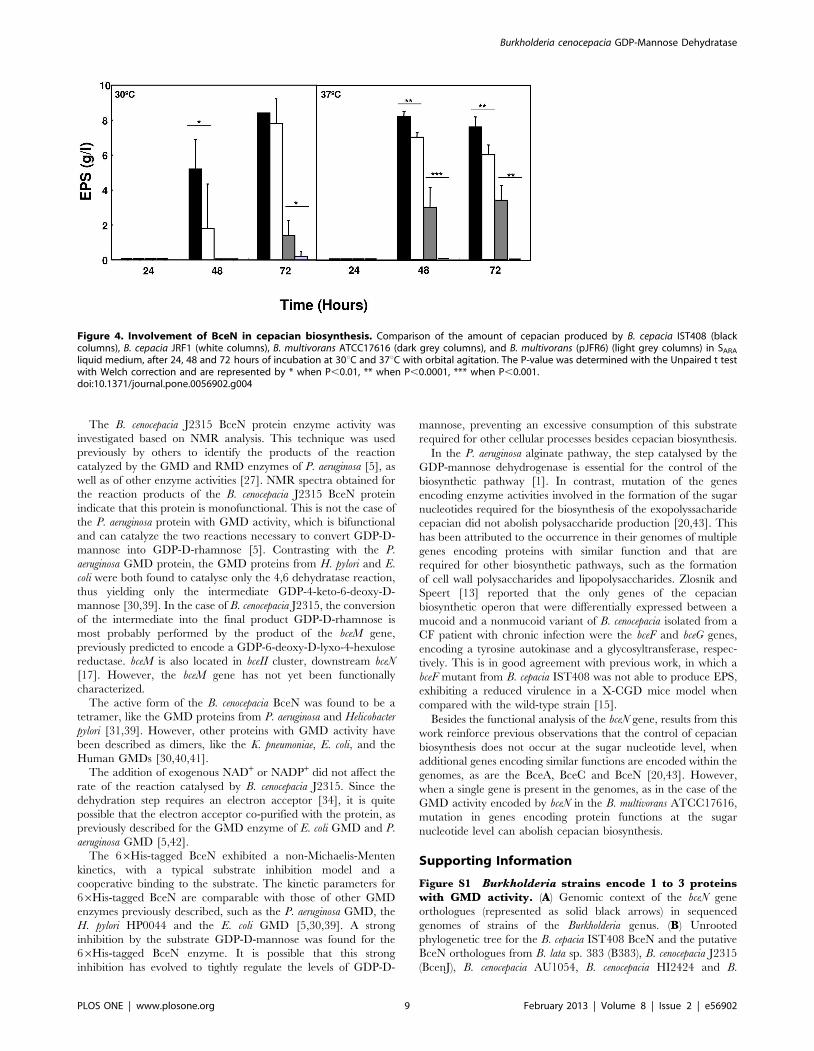
Construction and characterization of a bceN mutantSince GDP-D-rhamnose is one of the sugar nucleotides required
for the synthesis of the extracellular polysaccharide cepacian, the
mutant strain B. cepacia JFR1 with the bceN gene insertionally
inactivated was constructed from B. cepacia IST408, previously
described as a high EPS producer [8]. B. cenocepacia J2315 was
previously reported as a cepacian non-producer due to a
frameshift mutation in the bceB gene [18]. Due to difficulties in
constructing a bceN mutant in the EPS producer B. multivorans
ATCC 17616, a strategy based on the interference of an antisense
RNA targeting the bceN mRNA was employed to construct a
conditional mutant. This approach has been used successfully in
Staphylococcus aureus and Mycobacteria to analyse specific gene
essentiality, and more recently in B. cenocepacia J2315 [36–38].
With this purpose, the pMLBAD vector harbouring a DNA
fragment of the bceN gene in the antisense orientation was inserted
downstream of the pBAD promoter, resulting in the antisense
plasmid pJRF6. This plasmid was introduced into B. multivorans
ATCC 17616 cells by triparental conjugation, yielding B. multi-
vorans (pJRF6), which, upon induction with arabinose, expresses
the antisense transcript of bceN, effectively blocking the translation
of the mRNAs corresponding to bceN (results not shown).
Similar growth curves were observed for the wild-type strain B.
cepacia IST408, the mutant B. cepacia JFR1, B. multivorans
ATCC17616 and the B. multivorans (pJFR6) strain in SARA liquid
medium (data not shown). The ability of the wild-type strain B.
cepacia IST408, the mutant strain B. cepacia JFR1, B. multivorans
ATCC17616 and B. multivorans (pJRF6) to produce EPS was
compared in SARA liquid medium at 30uC and 37uC with orbital
agitation. Although the B. cepacia JRF1 mutant was still able to
produce EPS in SARA liquid medium, a significant reduced yield
was observed when compared to the IST408 wild-type strain after
48 hours of growth (Fig. 4). Since GMD activity is required for the
biosynthesis of the activated sugar precursor GDP-D-rhamnose of
the cepacian EPS, we expected that the loss of GMD would have a
stronger effect on EPS production. The ability to produce EPS by
the bceNmutant strain suggested that additional genes encoding for
proteins with GMD activity might be present within the genome of
B. cepacia IST408. A similar situation was previously reported by
our research group for the bceA gene, encoding the bifunctional
protein with both phosphomannose isomerase and GDP-D-
mannose pyrophosphorylase activities [20]. Results from southern
hybridization experiments with total DNA from B. cepacia IST408
and a probe based on bceN gene suggest that more than one GMD-
encoding gene is present in this genome, similarly to B. cenocepacia
PC184 (results not shown). Therefore, we have conducted
homology searches for putative GMD encoding genes within
publicly available genome sequences of strains of Bcc. One, two or
three putative GMD-encoding genes were found in sequenced
strains of the Burkholderia genus (Fig. S1 panel A). Some Bcc strains
contained only the bceN homologous gene, such as the B. multivorans
ATCC 17616. Therefore, we have also compared the EPS
production by B. multivorans ATCC 17616 and its derivative strain
B. multivorans (pJFR6) induced with arabinose. Induction of the
antisense bceN mRNA in B. multivorans (pJFR6) strain completely
abolished the EPS production (Fig. 4). Taken together, our results
strongly suggest that the BceN activity is required for the
biosynthesis of cepacian. In fact, in the B. multivorans, with a
unique gene putatively encoding a protein with GMD activity, no
cepacian was detected when an antisense targeting the bceN
mRNA was expressed. In the case of B. cepacia IST408 encoding
more than one protein with GMD activity, the inactivation of the
bceN gene did not led to the abolishment of cepacian production.
This is most probably due to the expression of additional gene(s)
encoding protein(s) with GMD activity, thus compensating the
inactivation of the bceN gene.
Discussion
In this work we report the cloning, purification, biochemical
and functional characterization of the bceN gene from the
opportunistic pathogens B. cenocepacia J2315 and B. cepacia
IST408, which are 97% identical. This last strain is a clinical
isolate previously reported as a high producer of cepacian [8], and
was used in this study since B. cenocepacia J2315 does not produce
cepacian due to a frameshift mutation on the bceB gene [18].
The bceN gene is part of the bceII cluster of genes found in all the
genomes of Burkholderia strains sequenced so far [17]. Amino acid
sequence analysis, together with the predicted secondary and
tertiary structures of the BceN proteins revealed conserved
domains typical of GDP-D-mannose dehydratases (GMD). These
proteins belong to the short-chain dehydrogenases/reductases
(SDR) superfamily. The SDR protein superfamily has raised
special interest due to their molecular evolution, enzymology and
biotechnological applications [29].
BceN homologue proteins are involved in the synthesis of the
rare deoxysugar nucleotide GDP-D-rhamnose, as is the case of the
P. aeruginosa GMD [5], the H. pylori HP0044 [39], or the E. coli
GMD [30]. GDP-D-rhamnose biosynthesis involves two steps
[5,31]. The first step is the elimination of a water molecule from
GDP-D-mannose by the GMD enzyme activity, yielding GDP-4-
keto-6-deoxy-D-mannose. This labile intermediary is further
converted into GDP-D-rhamnose by the enzyme activity GDP-
D-mannose reductase (RMD) that catalyses the reduction of the 4-
keto group, being the NAD(P)H cofactor the electron donor.
Table 1. Kinetics parameters for different bacterial GMD enzymes using GDP-D-mannose as substrate.
Protein, Bacteria
Multimeric
form
pH,
Temperature Km (mM)
Vmax
(mmol min21 mg21) Ki (mM)
Kcat
(s21)
Kcat/Km
(M21 s21) Reference
BceN, B. cenocepacia J2315 Tetramer 7.4, 25uC 10246123 1.560.2 29136350 1 9.776102 This study
GMD, P. aeruginosa Tetramer 7.5, 37uC 14.0266.05 3.6461.37 2.85961.31 8.82 6.36105 [5]
HP0044, H. pylori Tetramer 6.5, 37uC 117.361.38 ND ND 0.27 2.36103 [39]
GMD, E. coli Dimer 7.5, 37uC 280612 ND ND 5 1.86105 [30]
GMD, Mortierella alpina Trimer 9.0, 37uC 77067 ND ND 5.96 7.746103 [44]
doi:10.1371/journal.pone.0056902.t001
Burkholderia cenocepacia GDP-Mannose Dehydratase
PLOS ONE | www.plosone.org 8 February 2013 | Volume 8 | Issue 2 | e56902
The B. cenocepacia J2315 BceN protein enzyme activity was
investigated based on NMR analysis. This technique was used
previously by others to identify the products of the reaction
catalyzed by the GMD and RMD enzymes of P. aeruginosa [5], as
well as of other enzyme activities [27]. NMR spectra obtained for
the reaction products of the B. cenocepacia J2315 BceN protein
indicate that this protein is monofunctional. This is not the case of
the P. aeruginosa protein with GMD activity, which is bifunctional
and can catalyze the two reactions necessary to convert GDP-D-
mannose into GDP-D-rhamnose [5]. Contrasting with the P.
aeruginosa GMD protein, the GMD proteins from H. pylori and E.
coli were both found to catalyse only the 4,6 dehydratase reaction,
thus yielding only the intermediate GDP-4-keto-6-deoxy-D-
mannose [30,39]. In the case of B. cenocepacia J2315, the conversion
of the intermediate into the final product GDP-D-rhamnose is
most probably performed by the product of the bceM gene,
previously predicted to encode a GDP-6-deoxy-D-lyxo-4-hexulose
reductase. bceM is also located in bceII cluster, downstream bceN
[17]. However, the bceM gene has not yet been functionally
characterized.
The active form of the B. cenocepacia BceN was found to be a
tetramer, like the GMD proteins from P. aeruginosa and Helicobacter
pylori [31,39]. However, other proteins with GMD activity have
been described as dimers, like the K. pneumoniae, E. coli, and the
Human GMDs [30,40,41].
The addition of exogenous NAD+ or NADP+ did not affect the
rate of the reaction catalysed by B. cenocepacia J2315. Since the
dehydration step requires an electron acceptor [34], it is quite
possible that the electron acceptor co-purified with the protein, as
previously described for the GMD enzyme of E. coli GMD and P.
aeruginosa GMD [5,42].
The 66His-tagged BceN exhibited a non-Michaelis-Menten
kinetics, with a typical substrate inhibition model and a
cooperative binding to the substrate. The kinetic parameters for
66His-tagged BceN are comparable with those of other GMD
enzymes previously described, such as the P. aeruginosa GMD, the
H. pylori HP0044 and the E. coli GMD [5,30,39]. A strong
inhibition by the substrate GDP-D-mannose was found for the
66His-tagged BceN enzyme. It is possible that this strong
inhibition has evolved to tightly regulate the levels of GDP-D-
mannose, preventing an excessive consumption of this substrate
required for other cellular processes besides cepacian biosynthesis.
In the P. aeruginosa alginate pathway, the step catalysed by the
GDP-mannose dehydrogenase is essential for the control of the
biosynthetic pathway [1]. In contrast, mutation of the genes
encoding enzyme activities involved in the formation of the sugar
nucleotides required for the biosynthesis of the exopolyssacharide
cepacian did not abolish polysaccharide production [20,43]. This
has been attributed to the occurrence in their genomes of multiple
genes encoding proteins with similar function and that are
required for other biosynthetic pathways, such as the formation
of cell wall polysaccharides and lipopolysaccharides. Zlosnik and
Speert [13] reported that the only genes of the cepacian
biosynthetic operon that were differentially expressed between a
mucoid and a nonmucoid variant of B. cenocepacia isolated from a
CF patient with chronic infection were the bceF and bceG genes,
encoding a tyrosine autokinase and a glycosyltransferase, respec-
tively. This is in good agreement with previous work, in which a
bceF mutant from B. cepacia IST408 was not able to produce EPS,
exhibiting a reduced virulence in a X-CGD mice model when
compared with the wild-type strain [15].
Besides the functional analysis of the bceN gene, results from this
work reinforce previous observations that the control of cepacian
biosynthesis does not occur at the sugar nucleotide level, when
additional genes encoding similar functions are encoded within the
genomes, as are the BceA, BceC and BceN [20,43]. However,
when a single gene is present in the genomes, as in the case of the
GMD activity encoded by bceN in the B. multivorans ATCC17616,
mutation in genes encoding protein functions at the sugar
nucleotide level can abolish cepacian biosynthesis.
Supporting Information
Figure S1 Burkholderia strains encode 1 to 3 proteins
with GMD activity. (A) Genomic context of the bceN gene
orthologues (represented as solid black arrows) in sequenced
genomes of strains of the Burkholderia genus. (B) Unrooted
phylogenetic tree for the B. cepacia IST408 BceN and the putative
BceN orthologues from B. lata sp. 383 (B383), B. cenocepacia J2315
(BcenJ), B. cenocepacia AU1054, B. cenocepacia HI2424 and B.
Figure 4. Involvement of BceN in cepacian biosynthesis. Comparison of the amount of cepacian produced by B. cepacia IST408 (blackcolumns), B. cepacia JRF1 (white columns), B. multivorans ATCC17616 (dark grey columns), and B. multivorans (pJFR6) (light grey columns) in SARAliquid medium, after 24, 48 and 72 hours of incubation at 30uC and 37uC with orbital agitation. The P-value was determined with the Unpaired t testwith Welch correction and are represented by * when P,0.01, ** when P,0.0001, *** when P,0.001.doi:10.1371/journal.pone.0056902.g004
Burkholderia cenocepacia GDP-Mannose Dehydratase
PLOS ONE | www.plosone.org 9 February 2013 | Volume 8 | Issue 2 | e56902
cenocepacia MC0-3 (Bcen), B. cenocepacia PC184 (BcenP), B. ambifaria
AMMD (Bamb), B. dolosa AUO158 (Bdol), B. vietnamiensis G4
(Bviet), B. thailandensis E264 (Bthai), B. multivorans ATCC17616
(Bmul), B. mallei ATCC23344 (Bmal) and B. pseudomallei K96243
(Bpseud). The phylogenetic tree was constructed based on the
alignment of amino acid sequences with CLUSTAL X2 using the
neighbor-joining method with a minimum of 100 bootstraps.
Clade A includes putative GMDs located in gene clusters similar
to bce-II of chromosome 2 that is involved in Cepacian biosynthesis
[17]. Clade B includes the Burkholderia putative GMD homologies
located on chromosome 1, and the surrounding genes, arranged in
clusters most probably involved in LPS biosynthesis. The B.
vietnamiensis G4 BceN ortologue is not included in clade A or B,
and is the only GMD homologue from a Burkholderia strain
encoded on chromosome 3. Genes: a, b – ABC transporters; c, j,
m, o – glycosiltransferases; d – type II mannose-6-phosphate
isomerase; e – transposase; f – GDP-D-mannose 4,6-desydratase;
g, k – NAD-dependent epimerases/dehydratases; h – methyl-
transferase FkbM; i – hypothetical protein; l – methyltransferase
type 11; n – wcbA; p – wcbC; q – bexA; r – bexB; s – wcbD; t –
transferase hexapeptide repeat containing protein; u – polysac-
charide biosynthesis protein; v – exopolysacharide transport
protein; x – tyrosine phosphatase; w – polysaccharide export
protein; y – sugar transferase.
(DOC)
Table S1 Bacterial strains and plasmids used in this
work.
(DOC)
Table S213C and 1H NMR data for GDP-D-mannose
(A), GDP-4-keto-6-deoxy-D-mannose (B), and the gem-
diol form of compound B (C). Experiment with 5 mM GDP-
D-mannose, 14 hours of incubation with 90 mg BceN protein and
10 mM MgCl2 in H2O 90%/D2O 10% buffered saline solution.
Resonances were referenced to an internal acetone standard at
dH=2.225 p.p.m and dC=30.89 p.p.m. For compounds A, B and
C, the coupling constants (JHH) for the anomeric proton were
2.08 Hz, 1.56 Hz and 1.56 Hz, respectively. * Low resolution or
signals obscured by glycerol peaks.
(DOC)
Acknowledgments
The authors thank the contribution of A.C. Milho for the construction of
pACM2 and pACM4. The use of NMR facilities of the NMR network
(IST-UTL Center) is gratefully acknowledged.
Author Contributions
Conceived and designed the experiments: SAS PFP JHL. Performed the
experiments: SAS JRF PFP. Analyzed the data: SAS JRF PFP JHL.
Contributed reagents/materials/analysis tools: JHL. Wrote the paper: JHL
SAS.
References
1. Richau JA, Leitao JH, Sa-Correia I (2000) Enzymes leading to the nucleotidesugar precursors for exopolysaccharide synthesis in Burkholderia cepacia. BiochemBiophys Res Commun 276: 71–76.
2. Kocharova NA, Knirel YA, Widmalm G, Jansson PE, Moran AP (2000)Structure of an atypical O-antigen polysaccharide of Helicobacter pylori containinga novel monosaccharide 3-C-methyl-D-mannose. Biochemistry 39: 4755–4760.
3. Senchenkova SN, Shashkov AS, Knirel YA, McGovern JJ, Moran AP (1996)The O-specific polysaccharide chain of Campylobacter fetus serotype B lipopoly-saccharide is a D-rhamnan terminated with 3-O-methyl-D-rhamnose (D-acofriose). Eur J Biochem 239: 434–438.
4. Molinaro A, Silipo A, Lanzetta R, Newman MA, Dow JM, et al. (2003)Structural elucidation of the O-chain of the lipopolysaccharide from Xanthomonascampestris strain 8004. Carbohydr Res 338: 277–281.
5. King JD, Poon KK, Webb NA, Anderson EM, McNally DJ, et al. (2009) Thestructural basis for catalytic function of GMD and RMD, two closely relatedenzymes from the GDP-D-rhamnose biosynthesis pathway. FEBS J 276: 2686–2700.
6. Leitao JH, Sousa SA, Ferreira AS, Ramos CG, Silva IN, et al. (2010)Pathogenicity, virulence factors, and strategies to fight against Burkholderia cepaciacomplex pathogens and related species. Appl Microbiol Biotechnol 87: 31–40.
7. Vinion-Dubiel AD, Goldberg JB (2003) Lipopolysaccharide of Burkholderia cepaciacomplex. J Endotoxin Res 9: 201–213.
8. Richau JA, Leitao JH, Correia M, Lito L, Salgado MJet al. (2000) Moleculartyping and exopolysaccharide biosynthesis of Burkholderia cepacia isolates from aPortuguese cystic fibrosis center. J Clin Microbiol 38: 1651–1655.
9. Cunha MV, Sousa SA, Leitao JH, Moreira LM, Videira PA et al. (2004) Studieson the involvement of the exopolysaccharide produced by cystic fibrosis-associated isolates of the Burkholderia cepacia complex in biofilm formation and inpersistence of respiratory infections. J Clin Microbiol 42: 3052–3058.
10. Bylund J, Burgess LA, Cescutti P, Ernst RK, Speert DP (2006) Exopolysacchar-ides from Burkholderia cenocepacia inhibit neutrophil chemotaxis and scavengereactive oxygen species. J Biol Chem 281: 2526–2532.
11. Conway BA, Chu KK, Bylund J, Altman E, Speert DP (2004) Production ofexopolysaccharide by Burkholderia cenocepacia results in altered cell-surfaceinteractions and altered bacterial clearance in mice. J Infect Dis 190: 957–966.
12. Zlosnik JE, Hird TJ, Fraenkel MC, Moreira LM, Henry DA, et al. (2008)Differential mucoid exopolysaccharide production by members of the Burk-holderia cepacia complex. J Clin Microbiol 46: 1470–1473.
13. Zlosnik JE, Speert DP (2010) The role of mucoidy in virulence of bacteria fromthe Burkholderia cepacia complex: a systematic proteomic and transcriptomicanalysis. J Infect Dis 202: 770–781.
14. Cuzzi B, Cescutti P, Furlanis L, Lagatolla C, Sturiale L, et al. (2012)Investigation of bacterial resistance to the immune system response: cepaciandepolymerisation by reactive oxygen species. Innate Immun 18: 661–671.
15. Sousa SA, Ulrich M, Bragonzi A, Burke M, Worlitzsch D, et al. (2007) Virulenceof Burkholderia cepacia complex strains in gp91phox2/2 mice. Cell Microbiol 9:2817–2825.
16. Cescutti P, Bosco M, Picotti F, Impallomeni G, Leitao JH, et al. (2000)
Structural study of the exopolysaccharide produced by a clinical isolate of
Burkholderia cepacia. Biochem Biophys Res Commun 273: 1088–1094.
17. Ferreira AS, Leitao JH, Silva IN, Pinheiro PF, Sousa SA, et al. (2010)
Distribution of cepacian biosynthesis genes among environmental and clinical
Burkholderia strains and role of cepacian exopolysaccharide in resistance to stress
conditions. Appl Environ Microbiol 76: 441–450.
18. Moreira LM, Videira PA, Sousa SA, Leitao JH, Cunha MV, et al (2003)
Identification and physical organization of the gene cluster involved in the
biosynthesis of Burkholderia cepacia complex exopolysaccharide. Biochem Biophys
Res Commun 312: 323–333.
19. Sambrook J, Russel D (2001) Molecular Cloning: A Laboratory Manual. Cold
Spring Arbor NY: Cold Spring Arbor Laboratory Press.
20. Sousa SA, Moreira LM, Wopperer J, Eberl L, Sa-Correia I, et al. (2007) The
Burkholderia cepacia bceA gene encodes a protein with phosphomannose isomerase
and GDP-D-mannose pyrophosphorylase activities. Biochem Biophys Res
Commun 353: 200–206.
21. Markowitz VM, Szeto E, Palaniappan K, Grechkin Y, Chu K, et al. (2008) The
integrated microbial genomes (IMG) system in 2007: data content and analysis
tool extensions. Nucleic Acids Res 36: D528–533.
22. McGuffin LJ, Bryson K, Jones DT (2000) The PSIPRED protein structure
prediction server. Bioinformatics 16: 404–405.
23. Yang Z (2009) I-TASSER: Fully automated protein structure prediction in
CASP8. Proteins 77: 100–113.
24. Thompson JD, Gibson TJ, Higgins DG (2002) Multiple sequence alignment
using ClustalW and ClustalX. Curr Protoc Bioinformatics. Chapter 2: Unit 2.3.
25. Bradford MM (1976) A rapid and sensitive method for the quantification of
microgram quantities of protein utilizing the principle of protein-dye binding.
Anal Biochem 72: 248–254.
26. Niepmann M, Zheng J (2006) Discontinuous native protein gel electrophoresis.
Electrophoresis 27: 3949–3951.
27. Exnowitz F, Meyer B, Hackl T (2012) NMR for direct determination of K(m)
and V(max) of enzyme reactions based on the Lambert W function-analysis of
progress curves. Biochim Biophys Acta 1824: 443–449.
28. Sousa SA, Ramos CG, Leitao JH (2011) Burkholderia cepacia complex: emerging
multihost pathogens equipped with a wide range of virulence factors and
determinants. Int J Microbiol Pii:607575.
29. Kavanagh KL, Jornvall H, Persson B, Oppermann U (2008) Medium- and
short-chain dehydrogenase/reductase gene and protein families: the SDR
superfamily: functional and structural diversity within a family of metabolic and
regulatory enzymes. Cell Mol Life Sci 65: 3895–3906.
30. Somoza JR, Menon S, Schmidt H, Joseph-McCarthy D, Dessen A, et al. (2000)
Structural and kinetic analysis of Escherichia coli GDP-mannose 4,6 dehydratase
provides insights into the enzyme’s catalytic mechanism and regulation by GDP-
fucose. Structure 8: 123–135.
Burkholderia cenocepacia GDP-Mannose Dehydratase
PLOS ONE | www.plosone.org 10 February 2013 | Volume 8 | Issue 2 | e56902
31. Webb NA, Mulichak AM, Lam JS, Rocchetta HL, Garavito RM (2004) Crystalstructure of a tetrameric GDP-D-mannose 4,6-dehydratase from a bacterialGDP-D-rhamnose biosynthetic pathway. Protein Sci 13: 529–539.
32. Jornvall H, Persson B, Krook M, Atrian S, Gonzalez-Duarte R, et al. (1995)Short-chain dehydrogenases/reductases (SDR). Biochemistry 34: 6003–6013.
33. Albermann C, Distler J, Piepersberg W (2000) Preparative synthesis of GDP-beta-L-fucose by recombinant enzymes from enterobacterial sources. Glycobiol-ogy 10: 875–881.
34. Oths PJ, Mayer RM, Floss HG (1990) Stereochemistry and mechanism of theGDP-mannose dehydratase reaction. Carbohydr Res 198: 91–100.
35. Heck HD (1971) Statistical theory of cooperative binding to proteins. The Hillequation and the binding potential. J Am Chem Soc 93: 23–29.
36. Wang B, Kuramitsu HK (2003) Assessment of the utilization of the antisenseRNA strategy to identify essential genes in heterologous bacteria. FEMSMicrobiol Lett 220: 171–176.
37. Ramos CG, Sousa SA, Grilo AM, Feliciano JR, Leitao JH (2011) The secondRNA chaperone, Hfq2, is also required for survival under stress and fullvirulence of Burkholderia cenocepacia J2315. J Bacteriol 193: 1515–1526.
38. Parish T, Stoker NG (1997) Development and use of a conditional antisensemutagenesis system in mycobacteria. FEMS Microbiol Lett 154: 151–157.
39. Wu B, Zhang Y, Wang PG (2001) Identification and characterization of GDP-D-mannose 4,6-dehydratase and GDP-L-fucose snthetase in a GDP-L-fucosebiosynthetic gene cluster from Helicobacter pylori. Biochem Biophys Res Commun285: 364–371.
40. Bisso A, Sturla L, Zanardi D, De Flora A, Tonetti M (1999) Structural andenzymatic characterization of human recombinant GDP-D-mannose-4,6-dehydratase. FEBS Lett 456: 370–374.
41. Yamamoto K, Katayama I, Onoda Y, Inami M, Kumagai H, et al. (1993)Evidence that the enzyme catalyzing the conversion of guanosine diphosphateD-mannose to a 4-keto sugar nucleotide intermediate requires nicotinamideadenine dinucleotide phosphate. Arch Biochem Biophys 300: 694–698.
42. Sturla L, Bisso A, Zanardi D, Benatti U, De Flora A, et al. (1997) Expression,purification and characterization of GDP-D-mannose 4,6-dehydratse fromEscherichia coli. FEBS Lett 412: 126–130.
43. Loutet SA, Bartholdson SJ, Govan JR, Campopiano DJ, Valvano MA (2009)Contributions of two UDP-glucose dehydrogenases to viability and polymyxin Bresistance of Burkholderia cenocepacia. Microbiology 155: 2029–2039.
44. Ren Y, Perepelov AV, Wang H, Zhang H, Knirel YA, et al. (2010) Biochemicalcharacterization of GDP-L-fucose de novo synthesis pathway in fungusMortierella alpina. Biochem Biophys Res Commun 391: 1663–1669.
Burkholderia cenocepacia GDP-Mannose Dehydratase
PLOS ONE | www.plosone.org 11 February 2013 | Volume 8 | Issue 2 | e56902