bilayer lipid membranes supported on teflon filters: a functional environment for ion channels
TRANSCRIPT
BA
TA
a
ARRAA
KPTBIVB
1
dptnmdbootAa(
V
0d
Biosensors and Bioelectronics 26 (2011) 3127–3135
Contents lists available at ScienceDirect
Biosensors and Bioelectronics
journa l homepage: www.e lsev ier .com/ locate /b ios
ilayer lipid membranes supported on Teflon filters:functional environment for ion channels
hai Phung, Yanli Zhang, James Dunlop1, Julie Dalziel ∗
gResearch, Grasslands Research Centre, Private Bag 11008, Palmerston North 4442, New Zealand
r t i c l e i n f o
rticle history:eceived 9 September 2010eceived in revised form 5 December 2010ccepted 7 December 2010vailable online 16 December 2010
eywords:olytetrafluoroethylene (PTFE)eflonilayer lipid membrane
mpedance spectroscopyoltage-gated sodium channeliosensor
a b s t r a c t
Many ion channel proteins have binding sites for toxins and pharmaceutical drugs and therefore havemuch promise as the sensing entity in high throughput technologies and biosensor devices. Measure-ment of ionic conductance changes through ion channels requires a robust biological membrane withsufficient longevity for practical applications. The conventional planar BLM is 100–300 �m in diameterand typically contains fewer than a dozen channels whereas pharmaceutical screening methods in cellsuse current recordings for many ion channels. We present a new, simple method for the fabricationof a disposable porous-supported bilayer lipid membrane (BLM) ion channel biosensor using hydratedTeflon (polytetrafluoroethylene, PTFE) filter material (pore size 5 �m, filter diameter = 1 mm). The lipidlayer was monitored for its thickness and mechanical stability by electrical impedance spectroscopy.The results showed membrane capacitances of 1.8 ± 0.2 nF and membrane resistances of 25.9 ± 4.1 G�,indicating the formation of lipid bilayers. The current level increased upon addition of the pore-forming
peptide gramicidin. Following addition of liposomes containing voltage-gated sodium channels, smallmacroscopic sodium currents (1–80 pA) could be recorded. By preloading the porous Teflon with sodiumchannel proteoliposomes, prior to BLM formation, currents of 1–10 nA could be recorded in the pres-ence of the activator veratridine that increased with time, and were inhibited by tetrodotoxin. A lack ofrectification suggests that the channels incorporated in both orientations. This work demonstrates thatPTFE filters can support BLMs that provide an environment in which ion channels can maintain theirnt for
functional activity releva. Introduction
Biological membranes are fundamental to life. Proteins embed-ed in them carry out a wide range of physiological and biochemicalrocesses including the capture and transformation of energy,ransport and exchange of nutrients and metabolites, and recog-ition and signaling. Mutations in membrane proteins can causealfunction which may result in disease, such as muscle function
isorders (Ashcroft, 2006). Substances which interact with mem-rane proteins can affect their function with either toxic resultsr therapeutic benefits. It is estimated that approximately a thirdf pharmaceutical drugs and many toxins act on membrane pro-
eins (Meunier et al., 2009; Mouhat et al., 2004; Wang, 2008).mong these, ion channels are well recognised as important ther-peutic targets for treating a range of pathophysiological diseasesKaczorowski et al., 2008).∗ Corresponding author. Tel.: +64 6 3518098; fax: +64 6 3518032.E-mail address: [email protected] (J. Dalziel).
1 Address: MacDiarmid Institute for Advanced Materials and Nanotechnology,ictoria University of Wellington, PO Box 600, Wellington 6140, New Zealand.
956-5663/$ – see front matter © 2010 Elsevier B.V. All rights reserved.oi:10.1016/j.bios.2010.12.013
applications in drug discovery, toxin detection, and odour sensing.© 2010 Elsevier B.V. All rights reserved.
The sensitivity and specificity of many membrane proteins toligands, together with their important roles in physiology, hasstimulated considerable interest in the development of sensorsand analytical devices in which membrane proteins are the cen-tral detection element. Potential applications for such technologiesinclude the screening of compounds for pharmaceutical action andpotential side effects (Fang et al., 2006; Matsuno et al., 2004),sensors for detecting biowarfare agents (Cox et al., 2006), envi-ronmental monitoring of marine biotoxins and sensors for odourdetection (Rossini, 2005; Spehr and Munger, 2009; Vélez et al.,2001). Practical devices will require a reliable source of functionalprotein and a robust analog of the biological membrane. This paperfocuses on the latter of these two requirements.
Research on biological membrane integrity has greatly bene-fited from the development of the planar bilayer lipid membrane(BLM) technique (Mueller et al., 1962) and the incorporation of ionchannels into bilayers (Tien and Ottova, 2001). However the origi-
nal planar BLM can be difficult to work with. They are thin fragilemeta-stable structures, two lipid monolayers thick, under tensionfrom the surrounding ring of solvent, and subjected to substantialelectrical fields imposed by experimental conditions. Consequentlyplanar BLM are short-lived as they frequently rupture before the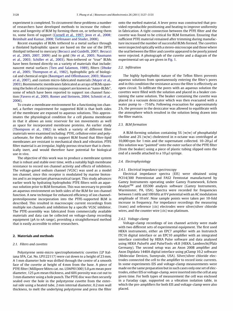
3 Bioele
eontR
ateehnece2nst2
noiia(mcmfiiu
trTiieoafpdmTmet
2
2
iAfPd3snt
128 T. Phung et al. / Biosensors and
xperiment is completed. To circumvent these problems a numberf researchers have developed methods to increase the robust-ess and longevity of BLM by forming them on, or tethering themo, some form of support (Cornell et al., 1997; Jeon et al., 2008;eimhult and Kumar, 2008; Tiefenauer and Studer, 2008).
Recent examples of BLMs tethered to a metal surface throughthiolated hydrophilic spacer are based on the use of the DPTL
hiolipid tethered to mercury (Becucci and Guidelli, 2007; Becuccit al., 2005, 2007, 2009) and to gold (He et al., 2005; Naumannt al., 2003; Schiller et al., 2003). Non-tethered or “true” BLMsave been formed directly on a variety of materials that include:ascent metal surfaces (Tien and Salamon, 1989), filters (Dhoket al., 2005; Thompson et al., 1982), hydrophilic gels of biologi-al and chemical origin (Baumgart and Offenhauser, 2003; Maurert al., 2007), and custom micro-fabricated materials (Mayer et al.,003). Biomimmetic membranes fabricated as arrays of BLMs span-ing the holes of a microporous support are known as “nano-BLMs”,ome of which have been reported to support ion channel func-ion (Favero et al., 2005; Romer and Steinem, 2004; Schmitt et al.,006).
To create a membrane environment for a functioning ion chan-el, a further requirement for supported BLM is that both sidesf the membrane are exposed to aqueous solutions. This approx-mates the physiological condition for a cell plasma membranen that it allows an ionic reservoir for ion movements as wells space for incorporated membrane proteins. An earlier studyThompson et al., 1982) in which a variety of different filter
aterials were examined including: PTFE, cellulose ester and poly-arbonate, for their ability to support BLM found that filter-lipidembranes are resistant to mechanical shock and vibration. PTFE
lter material is an irregular, highly porous structure that is chem-cally inert, and would therefore have potential for biologicalse in vivo.
The objective of this work was to produce a membrane systemhat is robust and stable over time, with a suitably high membraneesistance to record ion channel activity and effects of inhibitors.he voltage-gated sodium channel (VGSC) was used as a modelon channel, since this receptor is modulated by marine biotox-ns and is an important pharmaceutical target. This study advancesarlier work by hydrating hydrophobic PTFE filters with an aque-us solution prior to BLM formation. This was necessary to providen aqueous environment on both sides of the BLM for ion channelunction. A new technique for enhanced efficiency of ion channel-roteoliposome incorporation into the PTFE-supported BLM isescribed. This resulted in macroscopic current recordings fromultiple ion channels and inhibition by a specific VGSC inhibitor.
he PTFE-assembly was fabricated from commercially availableaterials and data can be collected on voltage-clamp recording
quipment (pA to nA range), providing a straightforward methodhat is easily accessible to other researchers.
. Materials and methods
.1. Filters and cuvettes
Polystyrene semi-micro spectrophotometric cuvettes (LP Ital-ana SPA, Cat. No. LPI122117) were cut down to a height of 23 mm.
1 mm diameter hole was drilled through the centre of a smoothace of the cuvette at height of 4 mm from the base. A piece ofTFE filter (Millipore Mitex cat. no. LSWP01300) 5.0 �m mean pore
iameter, 125 �m mean thickness, and 60% porosity was cut out tomm diameter using a hole punch. The PTFE disc was then securelyealed over the hole in the polystyrene cuvette from the exter-al side using a heated tube, 2 mm internal diameter, 0.2 mm wallhickness, to melt the underlying polystyrene and press the filter
ctronics 26 (2011) 3127–3135
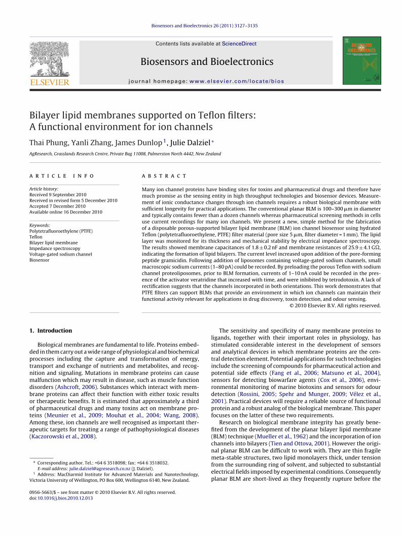
onto the melted material. A lever press was constructed that pro-vided reproducible positioning and heating to improve uniformityin fabrication. A tight connection between the PTFE filter and thecuvette was found to be critical for BLM formation. Ensuring thatsufficient PTFE material remained after trimming during manufac-ture increased the number of successful BLMs formed. The cuvetteswere inspected optically with a stereo-microscope and those wherethe seal between the filter and cuvette appeared to be poorly joinedwere rejected. A photograph of the cuvette and a diagram of theexperimental set-up are given in Fig. 1.
2.2. Infiltration
The highly hydrophobic nature of the Teflon filters preventsaqueous solutions from spontaneously entering the filter’s poresand in this condition the resistance across the filter is effectively anopen circuit. To infiltrate the pores with an aqueous solution thecuvettes were filled with the solution and placed in a beaker con-taining the same solution. The beaker containing the cuvette wasplaced in a vacuum desiccator which was then evacuated with awater pump to −75 kPa. Following evacuation for approximately2 h, the pressure in the desiccator was allowed to equilibrate withthe atmosphere which resulted in the solution being drawn intothe filter matrix.
2.3. BLM formation
A BLM-forming solution containing 5% (w/w) of phosphatidylcholine and 2% (w/w) cholesterol in n-octane was centrifuged at10,000 rpm for 1 min and the supernatant collected. 10–20 �l ofthis solution was “painted” onto the outer surface of the PTFE filter(from the beaker) using a piece of plastic tubing slipped onto theend of a needle attached to a 10 �l syringe.
2.4. Electrophysiology
2.4.1. Electrical impedance spectroscopyElectrical impedance spectra (EIS) were obtained using
PCI14/300 Potentiostat and FAS2 Femtostat manufactured byGamry Instruments operating under Gamry Framework, EchemAnalystTM and EIS300 analysis software (Gamry Instruments,Warminster, PA, USA). Spectra were recorded for frequenciesbetween 1 mHz and 100 kHz at 0 V potential with an AC modulationamplitude of 10 mV. Nine sample points were taken per 10-foldincrease in frequency. For impedance recordings the measuring(trans) and reference (cis) electrodes were silver/silver chloridewires, and the counter wire (cis) was platinum.
2.4.2. Voltage-clampVoltage-clamp recordings of ion channel activity were made
with two different sets of experimental equipment. The first usedHEKA instruments, either an EPC7 amplifier with an InstrutechITC16 digital interface or an EPC10 amplifier with an integratedinterface controlled by HEKA Pulse software and data analysedusing HEKA PulseFit and PulseTools v8.8 (HEKA, Lambrecht/PfalzGermany). The second setup was an Axon 200B amplifier andAxon Digidata 1440A digital interface using pClamp 10.2 software(Molecular Devices, Sunnyvale, USA). Silver/silver chloride elec-trodes connected the cell to the amplifier to record ionic currents.In some experiments EIS and voltage-clamp measurements weremade on the same preparation but in such cases only one set of elec-
trodes, either EIS or voltage-clamp, were inserted into the cell at anygiven time. For both types of measurement the cell was enclosedin a Faraday cage, supported on a vibration isolation table, inwhich the pre-amplifiers for both EIS and voltage-clamp were alsoplaced.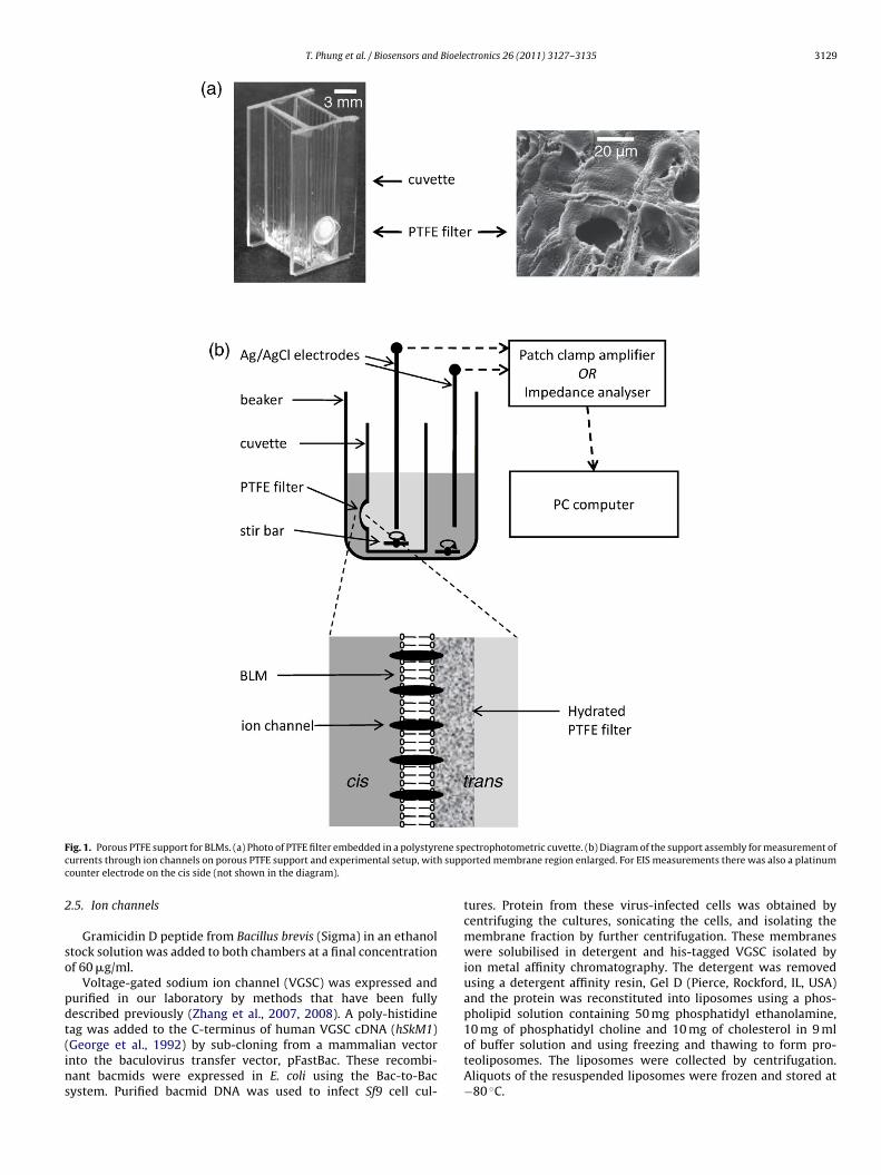
T. Phung et al. / Biosensors and Bioelectronics 26 (2011) 3127–3135 3129
F ene spc suppc
2
so
pdt(ins
ig. 1. Porous PTFE support for BLMs. (a) Photo of PTFE filter embedded in a polystyrurrents through ion channels on porous PTFE support and experimental setup, withounter electrode on the cis side (not shown in the diagram).
.5. Ion channels
Gramicidin D peptide from Bacillus brevis (Sigma) in an ethanoltock solution was added to both chambers at a final concentrationf 60 �g/ml.
Voltage-gated sodium ion channel (VGSC) was expressed andurified in our laboratory by methods that have been fullyescribed previously (Zhang et al., 2007, 2008). A poly-histidine
ag was added to the C-terminus of human VGSC cDNA (hSkM1)George et al., 1992) by sub-cloning from a mammalian vectornto the baculovirus transfer vector, pFastBac. These recombi-ant bacmids were expressed in E. coli using the Bac-to-Bacystem. Purified bacmid DNA was used to infect Sf9 cell cul-ectrophotometric cuvette. (b) Diagram of the support assembly for measurement oforted membrane region enlarged. For EIS measurements there was also a platinum
tures. Protein from these virus-infected cells was obtained bycentrifuging the cultures, sonicating the cells, and isolating themembrane fraction by further centrifugation. These membraneswere solubilised in detergent and his-tagged VGSC isolated byion metal affinity chromatography. The detergent was removedusing a detergent affinity resin, Gel D (Pierce, Rockford, IL, USA)and the protein was reconstituted into liposomes using a phos-pholipid solution containing 50 mg phosphatidyl ethanolamine,
10 mg of phosphatidyl choline and 10 mg of cholesterol in 9 mlof buffer solution and using freezing and thawing to form pro-teoliposomes. The liposomes were collected by centrifugation.Aliquots of the resuspended liposomes were frozen and stored at−80 ◦C.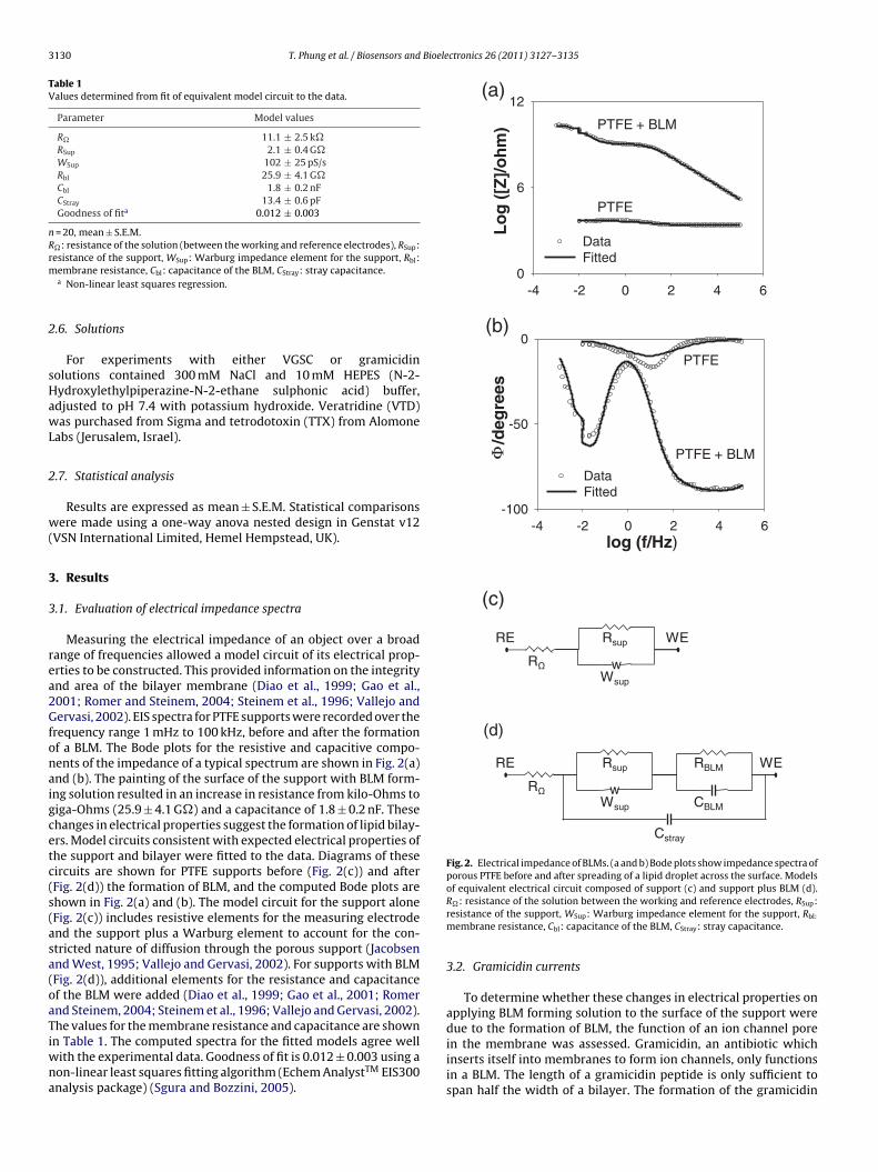
3130 T. Phung et al. / Biosensors and Bioelectronics 26 (2011) 3127–3135
Table 1Values determined from fit of equivalent model circuit to the data.
Parameter Model values
R� 11.1 ± 2.5 k�RSup 2.1 ± 0.4 G�WSup 102 ± 25 pS/sRbl 25.9 ± 4.1 G�Cbl 1.8 ± 0.2 nFCStray 13.4 ± 0.6 pFGoodness of fita 0.012 ± 0.003
n = 20, mean ± S.E.M.R�: resistance of the solution (between the working and reference electrodes), RSup:rm
2
sHawL
2
w(
3
3
rea2Gfonaigcetc(s(asa(oaTiwna
-4 -2 0 2 4 6-100
-50
0
DataFitted
PTFE + BLM
PTFE
log (f/Hz)
/deg
rees
Φ
w
RE WE
RΩWsup
Rsup
w
RE WERsup RBLM
CBLM
Cstray
Wsup
RΩ
-4 -2 0 2 4 60
6
12
DataFitted
PTFE
PTFE + BLM
Lo
g (
[Z]/
oh
m)
(a)
(b)
(c)
(d)
Fig. 2. Electrical impedance of BLMs. (a and b) Bode plots show impedance spectra ofporous PTFE before and after spreading of a lipid droplet across the surface. Modelsof equivalent electrical circuit composed of support (c) and support plus BLM (d).
esistance of the support, WSup: Warburg impedance element for the support, Rbl:embrane resistance, Cbl: capacitance of the BLM, CStray: stray capacitance.a Non-linear least squares regression.
.6. Solutions
For experiments with either VGSC or gramicidinolutions contained 300 mM NaCl and 10 mM HEPES (N-2-ydroxylethylpiperazine-N-2-ethane sulphonic acid) buffer,djusted to pH 7.4 with potassium hydroxide. Veratridine (VTD)as purchased from Sigma and tetrodotoxin (TTX) from Alomone
abs (Jerusalem, Israel).
.7. Statistical analysis
Results are expressed as mean ± S.E.M. Statistical comparisonsere made using a one-way anova nested design in Genstat v12
VSN International Limited, Hemel Hempstead, UK).
. Results
.1. Evaluation of electrical impedance spectra
Measuring the electrical impedance of an object over a broadange of frequencies allowed a model circuit of its electrical prop-rties to be constructed. This provided information on the integritynd area of the bilayer membrane (Diao et al., 1999; Gao et al.,001; Romer and Steinem, 2004; Steinem et al., 1996; Vallejo andervasi, 2002). EIS spectra for PTFE supports were recorded over the
requency range 1 mHz to 100 kHz, before and after the formationf a BLM. The Bode plots for the resistive and capacitive compo-ents of the impedance of a typical spectrum are shown in Fig. 2(a)nd (b). The painting of the surface of the support with BLM form-ng solution resulted in an increase in resistance from kilo-Ohms toiga-Ohms (25.9 ± 4.1 G�) and a capacitance of 1.8 ± 0.2 nF. Thesehanges in electrical properties suggest the formation of lipid bilay-rs. Model circuits consistent with expected electrical properties ofhe support and bilayer were fitted to the data. Diagrams of theseircuits are shown for PTFE supports before (Fig. 2(c)) and afterFig. 2(d)) the formation of BLM, and the computed Bode plots arehown in Fig. 2(a) and (b). The model circuit for the support aloneFig. 2(c)) includes resistive elements for the measuring electrodend the support plus a Warburg element to account for the con-tricted nature of diffusion through the porous support (Jacobsennd West, 1995; Vallejo and Gervasi, 2002). For supports with BLMFig. 2(d)), additional elements for the resistance and capacitancef the BLM were added (Diao et al., 1999; Gao et al., 2001; Romernd Steinem, 2004; Steinem et al., 1996; Vallejo and Gervasi, 2002).he values for the membrane resistance and capacitance are shown
n Table 1. The computed spectra for the fitted models agree wellith the experimental data. Goodness of fit is 0.012 ± 0.003 using aon-linear least squares fitting algorithm (Echem AnalystTM EIS300nalysis package) (Sgura and Bozzini, 2005).
R�: resistance of the solution between the working and reference electrodes, RSup:resistance of the support, WSup: Warburg impedance element for the support, Rbl:
membrane resistance, Cbl: capacitance of the BLM, CStray: stray capacitance.
3.2. Gramicidin currents
To determine whether these changes in electrical properties onapplying BLM forming solution to the surface of the support weredue to the formation of BLM, the function of an ion channel pore
in the membrane was assessed. Gramicidin, an antibiotic whichinserts itself into membranes to form ion channels, only functionsin a BLM. The length of a gramicidin peptide is only sufficient tospan half the width of a bilayer. The formation of the gramicidin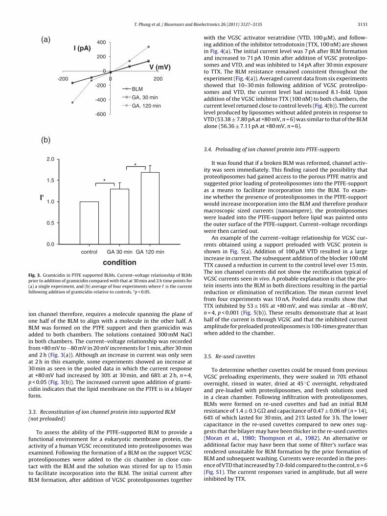
T. Phung et al. / Biosensors and Bioele
(a)
-600
-400
-200
0
200
400
-200 0 200
BLM
GA, 30 min
GA, 120 min
I (pA)
V (mV)
(b)
condition
control GA 30 min GA 120 min
I'
0.0
0.5
1.0
1.5
2.0
*
*
Fig. 3. Gramicidin in PTFE supported BLMs. Current–voltage relationship of BLMsp(f
ioBaifaa3apcf
3(
faepttB
rendered unsuitable for BLM formation by the prior formation of
rior to addition of gramicidin compared with that at 30 min and 2 h time points fora) a single experiment, and (b) average of four experiments where I′ is the currentollowing addition of gramicidin relative to controls, *p < 0.05.
on channel therefore, requires a molecule spanning the plane ofne half of the BLM to align with a molecule in the other half. ALM was formed on the PTFE support and then gramicidin wasdded to both chambers. The solutions contained 300 mM NaCln both chambers. The current–voltage relationship was recordedrom +80 mV to −80 mV in 20 mV increments for 1 min, after 30 minnd 2 h (Fig. 3(a)). Although an increase in current was only seent 2 h in this example, some experiments showed an increase at0 min as seen in the pooled data in which the current responset +80 mV had increased by 30% at 30 min, and 68% at 2 h, n = 4,< 0.05 (Fig. 3(b)). The increased current upon addition of grami-idin indicates that the lipid membrane on the PTFE is in a bilayerorm.
.3. Reconstitution of ion channel protein into supported BLMnot preloaded)
To assess the ability of the PTFE-supported BLM to provide aunctional environment for a eukaryotic membrane protein, thectivity of a human VGSC reconstituted into proteoliposomes wasxamined. Following the formation of a BLM on the support VGSC
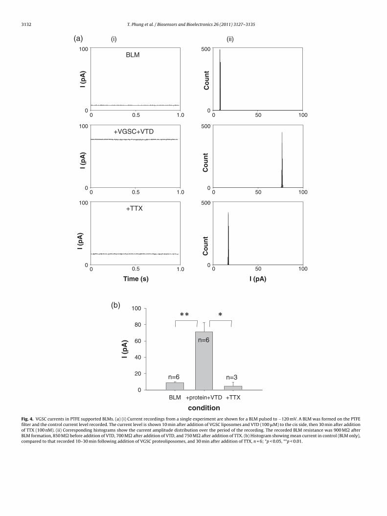
roteoliposomes were added to the cis chamber in close con-act with the BLM and the solution was stirred for up to 15 mino facilitate incorporation into the BLM. The initial current afterLM formation, after addition of VGSC proteoliposomes togetherctronics 26 (2011) 3127–3135 3131
with the VGSC activator veratridine (VTD, 100 �M), and follow-ing addition of the inhibitor tetrodotoxin (TTX, 100 nM) are shownin Fig. 4(a). The initial current level was 7 pA after BLM formationand increased to 71 pA 10 min after addition of VGSC proteolipo-somes and VTD, and was inhibited to 14 pA after 30 min exposureto TTX. The BLM resistance remained consistent throughout theexperiment (Fig. 4(a)). Averaged current data from six experimentsshowed that 10–30 min following addition of VGSC proteolipo-somes and VTD, the current level had increased 8.1-fold. Uponaddition of the VGSC inhibitor TTX (100 nM) to both chambers, thecurrent level returned close to control levels (Fig. 4(b)). The currentlevel produced by liposomes without added protein in response toVTD (53.38 ± 7.80 pA at +80 mV, n = 6) was similar to that of the BLMalone (56.36 ± 7.11 pA at +80 mV, n = 6).
3.4. Preloading of ion channel protein into PTFE-supports
It was found that if a broken BLM was reformed, channel activ-ity was seen immediately. This finding raised the possibility thatproteoliposomes had gained access to the porous PTFE matrix andsuggested prior loading of proteoliposomes into the PTFE-supportas a means to facilitate incorporation into the BLM. To exam-ine whether the presence of proteoliposomes in the PTFE-supportwould increase incorporation into the BLM and therefore producemacroscopic sized currents (nanoampere), the proteoliposomeswere loaded into the PTFE-support before lipid was painted ontothe outer surface of the PTFE-support. Current–voltage recordingswere then carried out.
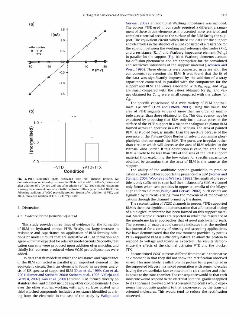
An example of the current–voltage relationship for VGSC cur-rents obtained using a support preloaded with VGSC protein isshown in Fig. 5(a). Addition of 100 �M VTD resulted in a largeincrease in current. The subsequent addition of the blocker 100 nMTTX caused a reduction in current to the control level over 15 min.The ion channel currents did not show the rectification typical ofVGSC currents seen in vivo. A probable explanation is that the pro-tein inserts into the BLM in both directions resulting in the partialreduction or elimination of rectification. The mean current levelfrom four experiments was 10 nA. Pooled data results show thatTTX inhibited by 53 ± 16% at +80 mV, and was similar at −80 mV,n = 4, p < 0.001 (Fig. 5(b)). These results demonstrate that at leasthalf of the current is through VGSC and that the inhibited currentamplitude for preloaded proteoliposomes is 100-times greater thanwhen added to the chamber.
3.5. Re-used cuvettes
To determine whether cuvettes could be reused from previousVGSC preloading experiments, they were soaked in 70% ethanolovernight, rinsed in water, dried at 45 ◦C overnight, rehydratedand pre-loaded with proteoliposomes, and fresh solutions usedin a clean chamber. Following infiltration with proteoliposomes,BLMs were formed on re-used cuvettes and had an initial BLMresistance of 1.4 ± 0.3 G� and capacitance of 0.47 ± 0.06 nF (n = 14),64% of which lasted for 30 min, and 21% lasted for 3 h. The lowercapacitance in the re-used cuvettes compared to new ones sug-gests that the bilayer may have been thicker in the re-used cuvettes(Moran et al., 1980; Thompson et al., 1982). An alternative oradditional factor may have been that some of filter’s surface was
BLM and subsequent washing. Currents were recorded in the pres-ence of VTD that increased by 7.0-fold compared to the control, n = 6(Fig. S1). The current responses varied in amplitude, but all wereinhibited by TTX.
3132 T. Phung et al. / Biosensors and Bioelectronics 26 (2011) 3127–3135
Fig. 4. VGSC currents in PTFE supported BLMs. (a) (i) Current recordings from a single experiment are shown for a BLM pulsed to −120 mV. A BLM was formed on the PTFEfilter and the control current level recorded. The current level is shown 10 min after addition of VGSC liposomes and VTD (100 �M) to the cis side, then 30 min after additionof TTX (100 nM). (ii) Corresponding histograms show the current amplitude distribution over the period of the recording. The recorded BLM resistance was 900 M� afterBLM formation, 850 M� before addition of VTD, 700 M� after addition of VTD, and 750 M� after addition of TTX. (b) Histogram showing mean current in control (BLM only),compared to that recorded 10–30 min following addition of VGSC proteoliposomes, and 30 min after addition of TTX, n = 6; *p < 0.05, **p < 0.01.
T. Phung et al. / Biosensors and Bioele
(b)
0.0
0.5
1.0
+VTD +VTD+TTX
I (n
orm
alis
ed)
Condition
***
V (mV)-80 -40 0 40 80
I (nA)
-1.5
-1.0
-0.5
0.0
0.5
1.0
1.5
control VTD TTX
(a)
Fig. 5. PTFE supported BLMs preloaded with Na+ channel protein. (a)Current–voltage relationship is shown for BLMs held at −80 to +80 mV, before andafter addition of VTD (100 �M) and after addition of TTX (100 nM). (b) Histogramsf2
4
4
ortacta
oes2Gseti
molecule would respond to the electrical potential gradient applied
howing mean current normalised to the control at +80 mV (ii) recorded 10–30 minollowing addition of VGSC proteoliposomes, 30 min after addition of VTD, and0–30 min after addition of TTX, n = 4; ***p < 0.001.
. Discussion
.1. Evidence for the formation of a BLM
This study provides three lines of evidence for the formationf BLM on hydrated porous PTFE. Firstly, the large increase inesistance and capacitance on application of BLM-forming solu-ions fit model circuits that are indicative of BLM formation andgree with that expected for relevant model circuits. Secondly, thatation currents were produced upon addition of gramicidin, andhirdly Na+ currents produced when VGSC proteoliposomes weredded.
EIS data that fit models in which the resistance and capacitancef the BLM connected in parallel is an important element in thequivalent circuit. Such an element is found in previous analy-is of EIS spectra of supported BLM (Diao et al., 1999; Gao et al.,001; Romer and Steinem, 2004; Steinem et al., 1996; Vallejo andervasi, 2002). Gao et al. (2001) studied BLM formed directly on
tainless steel and did not include any other circuit elements. How-ver the other studies, working with gold surfaces coated withhiol attached compounds, included a resistance identified as aris-ng from the electrode. In the case of the study by Vallejo andctronics 26 (2011) 3127–3135 3133
Gervasi (2002), an additional Warburg impedance was included.The porous PTFE used in our study required a different arrange-ment of these circuit elements as it presented more restricted andcomplex electrical access to the surface of the BLM facing the sup-port. The equivalent circuit which fitted the data for the supportand electrodes in the absence of a BLM consisted of a resistance forthe solution between the working and reference electrodes (R�),and a resistance (Rsup) and Warburg impedance element (Wsup)in parallel for the support (Fig. 1(b)). Warburg elements accountfor diffusion phenomena and are appropriate for the convolutedand restrictive interstices of the support material (Jacobsen andWest, 1995). These elements were connected in series with thecomponents representing the BLM. It was found that the fit ofthe data was significantly improved by the addition of a straycapacitance connected in parallel with the components for thesupport and BLM. The values associated with R�, Rsup, and Wsup
are small compared with the values obtained for Rbl, and val-ues obtained for Cstray were small compared with the values forCbl.
The specific capacitance of a wide variety of BLM approxi-mate 1 �F cm−2 (Tien and Ottova, 2003). Using this value, thearea of PTFE suggests values of more than an order of magni-tude greater than those obtained for Cbl. This discrepancy may beexplained by proposing that BLM only form across pores at thesurface of the PTFE support in a manner analogous to planar BLMformed across an aperture in a PTFE septum. The area of paintedBLM, as studied here, is smaller than the aperture because of thepresence of the Plateau-Gibbs Border of solvent containing phos-pholipids that surrounds the BLM. The pores are irregular ratherthan circular which will decrease the area of BLM relative to thePlateau-Gibbs Border. If this description is valid, the area of theBLM is likely to be less than 10% of the area of the PTFE supportmaterial thus explaining the low values for specific capacitanceobtained by assuming that the area of BLM is the same as thesupport.
The ability of the antibiotic peptide gramicidin to producecation currents further supports the presence of a BLM (Romer andSteinem, 2004; Woolley and Wallace, 1992). The length of one pep-tide is only sufficient to span half the thickness of a BLM. A channelonly forms when two peptides in opposite lamella of the bilayeralign to form a dimer (Vallejo and Gervasi, 2002). Such events aresignalled by currents arising from the movement of monovalentcations through the channel formed by the dimer.
The reconstitution of VGSC channels in porous PTFE-supportedBLM is the most significant demonstration that a functional analogof a biological membrane has been formed on this support mate-rial. Macroscopic currents are reported in which the resistance ofthe membrane layer approaches that of good patch-clamp seals.The presence of macroscopic currents indicates that the methodhas potential for a variety of sensing and screening applications.We have demonstrated that the environment provided by porousPTFE-supported BLM is sufficiently natural to allow the protein torespond to voltage and toxins as expected. The results demon-strate the effects of the channel activator VTD and the blockerTTX.
Reconstituted VGSC currents differed from those in their nativeenvironment in that they did not show the rectification observedin vivo. This most likely results from the protein being positioned inthe supported bilayers in a mixed orientation with some moleculeshaving the extracellular face exposed to the cis chamber and otherexposed to the trans chamber. The consequence would be that each
to it as normal. However cis-trans oriented molecules would expe-rience the opposite gradient to that experienced by the trans-cisoriented molecules. This would tend to reduce the rectificationobserved.
3 Bioele
4
spsteekpbphsspttfifbb
4
em((Soi(amporttwiPrcTmpt
4
atpfca
A
s
134 T. Phung et al. / Biosensors and
.2. Preloading of ion channel protein
The preloading of the supports by adding proteoliposomes to theolution used to infiltrate the porous PTFE prior to BLM formationroduced currents that were greater in amplitude that those mea-ured when BLM were formed and proteoliposomes then addedo the cis chamber. From this we infer that liposome-BLM fusionfficiency was enhanced under infiltrated conditions. The phenom-na that occur when protein is preloaded into the support are notnown. It is possible that some liposomes form a monolayer ofhospholipids on the internal walls of the PTFE with the hydropho-ic tails oriented towards the PTFE walls of the interstices and theolar heads facing the interstitial spaces. This would tend to create aydrophilic environment with the possibility that other proteolipo-omes would be adsorbed to the hydrophilic surface with sufficienttrength to retain them against the force of diffusion. The process ofreloading would force some proteoliposomes through small aper-ures in the interstices of the PTFE resulting in them being resizedo smaller dimensions. Another possibility is that interstices of thelter remain hydrophobic until the phospholipid containing BLM
orming solution is painted onto it whereupon a monolayer woulde formed. This work does not differentiate between these possi-ilities.
.3. Usefulness of PTFE compared to other supported BLM
In the past decade there has been a rapid increase in the vari-ty of supported BLM that provide a suitable environment forembrane protein function. These include solid supported BLM
Castellana and Cremer, 2006), nano- and microporous supportsReimhult and Kumar, 2008), polymer supported BLM (Tanaka andackmann, 2005), membrane arrays (Groves, 2002), BLM formedn silicon nanowires (Misra et al., 2009), BLM formed by microflu-dic circuits (Suzuki et al., 2007), and droplet interface bilayersHwang et al., 2008). Each of these membranes has useful char-cteristics in terms of resistance, stability, longevity and exposedembrane area, and ability to incorporate membrane proteins. The
orous PTFE-supported BLM described in this study shares manyf these characteristics – they are of high resistance, sufficientlyobust over several hours to survive being bathed in stirred solu-ions and being moved. The support material is readily available andhe construction of the cuvette-PTFE assembly requires only basicorkshop equipment. As with some other types of supported BLM,
on channel proteins functioned and responded to toxins in porousTFE-supported BLM, and both single channel and macroscopic cur-ents could be observed. However the possibility of preloading ionhannel proteoliposomes into the support is a unique advantage.he nanoampere ion channel currents produced by the preloadingethod, together with the possibility of reusing the PTFE-support,
rovides a convenient and practical platform for the next genera-ion of HTS ion channel technologies.
.4. Concluding perspective
This work demonstrates that PTFE filters can support BLM thatre sufficiently long-lived and robust for research purposes. Theechnology involved is simple and readily accessible. These BLMrovide an environment in which ion channels can maintain theirunctional activity. This makes a wide range of new biotechnologiesonceivable with applications in drug discovery, toxin detection,nd odour sensing.
cknowledgements
We thank B. Atkins for building the lever press for cuvette-upport assembly manufacture and Z. Park for statistical advice.
ctronics 26 (2011) 3127–3135
This research was funded by the New Economy Research Fundfrom the Foundation for Research, Science and Technology of NewZealand, and the AgResearch PreSeed Fund.
Appendix A. Supplementary data
Supplementary data associated with this article can be found, inthe online version, at doi:10.1016/j.bios.2010.12.013.
References
Ashcroft, F.M., 2006. Nature 440 (7083), 440–447.Baumgart, T., Offenhauser, A., 2003. Langmuir 19 (5), 1730–1737.Becucci, L., Guidelli, R., 2007. Langmuir 23 (10), 5601–5608.Becucci, L., Guidelli, R., Karim, C.B., Thomas, D.D., Veglia, G., 2009. Biophys. J. 97 (10),
2693–2699.Becucci, L., Moncelli, M.R., Naumann, R., Guidelli, R., 2005. J. Am. Chem. Soc. 127 (38),
13316–13323.Becucci, L., Santucci, A., Guidelli, R., 2007. J. Phys. Chem. B 111 (33), 9814–
9820.Castellana, E.T., Cremer, P.S., 2006. Surf. Sci. Rep. 61 (10), 429–444.Cornell, B.A., Braach-Maksvytis, V.L.B., King, L.G., Osman, P.D.J., Raguse, B., Wiec-
zorek, L., Pace, R.J., 1997. Nature 387 (6633), 580–583.Cox, T., Tattersall, J., Geddes, N., Evans, S., Nelson, A., 2006. J. Defence Sci. 10, 115–
121.Dhoke, M.A., Ladha, P.J., Boerio, F.J., Lessard, L.B., Malinowska, D.H., Cuppoletti,
J., Wieczorek, D.S., 2005. Biochim. Biophys. Acta Biomembr. 1716 (2), 117–125.
Diao, P., Jiang, D., Cui, X., Gu, D., Tong, R., Zhong, B., 1999. Bioelectrochem. Bioenergy48 (2), 469–475.
Fang, Y., Hong, Y., Webb, B., Lahiri, J., 2006. M. R. S. Bull. 31 (7), 541–545.Favero, G., Campanella, L., Cavallo, S., D’Annibale, A., Perrella, M., Mattei, E., Ferri, T.,
2005. J. Am. Chem. Soc. 127 (22), 8103–8111.Gao, H., Feng, J., Luo, G.A., Ottova, A.L., Tien, H.T., 2001. Electroanalysis 13 (1), 49–
53.George, A.L., Komisarof, J., Kallen, R.G., Barchi, R.L., 1992. Ann. Neurol. 31 (2),
131–137.Groves, J.T., 2002. Curr. Opin. Drug Discov. Dev. 5 (4), 606–612.He, L., Robertson, J.W., Li, J., Kärcher, I., Schiller, S.M., Knoll, W., Naumann, R., 2005.
Langmuir 21 (25), 11666–11672.Hwang, W.L., Chen, M., Cronin, B., Holden, M.A., Bayley, H., 2008. J. Am. Chem. Soc.
130 (18), 5878–5879.Jacobsen, T., West, K., 1995. Electrochim. Acta 40 (2), 255–262.Jeon, T.J., Malmstadt, N., Poulos, J.L., Schmidt, J.J., 2008. Biointerphases 3 (2),
FA96.Kaczorowski, G.J., McManus, O.B., Priest, B.T., Garcia, M.L., 2008. J. Gen. Physiol. 131
(5), 399–405.Matsuno, N., Murawsky, M., Ridgeway, J., Cuppoletti, J., 2004. Biochim. Biophys. Acta
Biomembr. 1665 (1–2), 184–190.Maurer, J.A., White, V.E., Dougherty, D.A., Nadeau, J.L., 2007. Biosens. Bioelectron. 22
(11), 2577–2584.Mayer, M., Kriebel, J.K., Tosteson, M.T., Whitesides, G.M., 2003. Biophys. J. 85 (4),
2684–2695.Meunier, F.A., Mattei, C., Molgo, J., 2009. Prog. Mol. Subcell. Biol. 46, 159–
186.Misra, N., Martineza, J.A., Huanga, S.C.J., Wanga, Y., Stroevec, P., Grigoropoulosb, C.P.,
Noya, A., 2009. Proc. Natl. Acad. Sci. U.S.A. 106 (33), 13780–13784.Moran, A., Tal, E., Eytan, E., Nelson, N., 1980. FEBS Lett. 110 (1), 62–64.Mouhat, S., Jouirou, B., Mosbah, A., De Waard, M., Sabatier, J.M., 2004. Biochem. J.
378 (3), 717–726.Mueller, P., Rudin, D.O., Ti Tien, H., Wescott, W.C., 1962. Nature 194 (4832), 979–
980.Naumann, R., Schiller, S.M., Giess, F., Grohe, B., Hartman, K.B., Kärcher, I., Köper, I.,
Lübben, J., Vasilev, K., Knoll, W., 2003. Langmuir 19 (13), 5435–5443.Reimhult, E., Kumar, K., 2008. Trends Biotechnol. 26 (2), 82–89.Romer, W., Steinem, C., 2004. Biophys. J. 86 (2), 955–965.Rossini, G.P., 2005. Toxicology 207 (3), 451–462.Schiller, S.M., Naumann, R., Lovejoy, K., Kunz, H., Knoll, W., 2003. Angew. Chem. Int.
Ed. Engl. 42 (2), 208–211.Schmitt, E.K., Vrouenraets, M., Steinem, C., 2006. Biophys. J. 91 (6), 2163–2171.Sgura, I., Bozzini, B., 2005. Int. J. Non Linear Mech. 40 (4), 557–570.Spehr, M., Munger, S.D., 2009. J. Neurochem. 109 (6), 1570–1583.Steinem, C., Janshoff, A., Ulrich, W.P., Sieber, M., Galla, H.J., 1996. Biochim. Biophys.
Acta Biomembr. 1279 (2), 169–180.Suzuki, H., Tabata, K.V., Noji, H., Takeuchi, S., 2007. Biosens. Bioelectron. 22 (6),
1111–1115.Tanaka, M., Sackmann, E., 2005. Nature 437 (7059), 656–663.
Thompson, M., Lennox, R.B., McClelland, R.A., 1982. Anal. Chem. 54 (1), 76–81.Tiefenauer, L.X., Studer, A., 2008. Biointerphases 3 (2), FA74.Tien, H.T., Ottova, A., 2003. In: Tien, H.T., Ottova-Leitmannova, A. (Eds.), Planar LipidBilayers and their Applications. Elsevier, Amsterdam, pp. 1–73.Tien, H.T., Ottova, A.L., 2001. J. Membr. Sci. 189 (1), 83–117.Tien, H.T., Salamon, Z., 1989. Bioelectrochem. Bioenergy 22 (3), 211–218.

Bioele
VV
WW
T. Phung et al. / Biosensors and
allejo, A.E., Gervasi, C.A., 2002. Bioelectrochemistry 57 (1), 1–7.élez, P., Sierralta, J., Alcayaga, C., Fonseca, M., Loyola, H., Johns, D.C., Tomaselli, G.F.,
Marban, E., Suarez-Isla, B.A., 2001. Toxicon 39 (7), 929–935.ang, D.Z., 2008. Drugs 6 (March (2)), 349–371.oolley, G.A., Wallace, B.A., 1992. J. Membr. Biol. 129 (2), 109–136.
ctronics 26 (2011) 3127–3135 3135
Zhang, Y.L., Dalziel, J.E., Dunlop, J., Leitmannova Liu, A., 2008. In: Leitmannova Liu,A. (Ed.), Advances in Planar Lipid Bilayers and Liposomes. Elsevier, Amsterdam,pp. 27–47.
Zhang, Y.L., Dunlop, J., Dalziel, J.E., 2007. Biosens. Bioelectron. 22 (6), 1006–1012.