bacterial capture efficiency and antimicrobial activity of
TRANSCRIPT

Published: March 31, 2011
r 2011 American Chemical Society 5472 dx.doi.org/10.1021/la200102z | Langmuir 2011, 27, 5472–5480
ARTICLE
pubs.acs.org/Langmuir
Bacterial Capture Efficiency and Antimicrobial Activity ofPhage-Functionalized Model SurfacesZeinab Hosseinidoust,† Theo G. M. Van de Ven,‡ and Nathalie Tufenkji*,†
†Department of Chemical Engineering, McGill University, Montreal, Quebec H3A 2B2, Canada‡Department of Chemistry, McGill University, Montreal, Quebec H3A 2K6, Canada
bS Supporting Information
1. INTRODUCTION
Surfaces can act as reservoirs for microbes. In healthcareenvironments, microbial transmission is facilitated upon contactwith surfaces contaminated by microbes.1 Several studies haveshown how the hands of healthcare workers may becomecontaminated by touching surfaces in the immediate vicinity ofan infected patient.1,2 Additionally, indwelling medical devicessuch as catheters, stents, and implants can act as substrates forfilms of pathogens to develop inside the body.3,4 The widespreadresearch and development of antimicrobial surfaces is of greatimportance in the fight to stop the spread of nosocomialinfections.5 Several antimicrobial coating technologies are eithercurrently available as marketed products or in research stages.Some of these coatings induce resistance in the microbes, andsome are known to be cytotoxic.6�9 These are issues of concernespecially for antimicrobial coated medical devices or treatmentstrategies coming into contact with human cells and tissues.
Bacteriophages are emerging alternative candidates for anti-microbial surfaces. Bacteriophages are viruses that exclusivelyinfect prokaryotes and are thus innocuous to eukaryotic cells.10
In general, bacteriophages, or phages, exhibit specific hostreceptor binding capability; therefore, they can be selected todestroy one species or even strain of bacteria, leaving the otherstrains unharmed.10 After binding to the host bacterium, thephage injects its genetic material inside the cell and uses the cell’s
machinery to replicate. Lytic phages destruct the bacterial host bylysing the cell and causing release of the newly fabricated phageparticles. Their replication is self-propagating, and theoretically,only one phage particle is needed to initiate a cascade of phageproduction and bacterial host destruction. The use of lytic phagesas a treatment to suppress infection dates back to the early days ofphage discovery in the late 20th century.11 However, thisapproach was rapidly overlooked by the emergence of antibiotics.Recently, the emerging crises of antibiotic-resistant bacteria hasrekindled the interest in the use of phages.12 A phage-modifiedsurface is a relevant antimicrobial approach, particularly because,to date, organisms that have been shown to be resistant toantibiotics have not shown resistance toward phages.5 Moreover,phages have a much lower production cost in comparison toantibiotics.
Many studies have shown that lytic phages can be used tocapture and/or signal the presence of bacteria.13�24 A fewreports have been published on rendering surfaces antimicrobialby the use of phages.25�29 For example, Curtin et al.28 usedbacteriophages to coat a model catheter, and Stone29 detailed therecent development of a phage-containing wound dressing.
Received: January 9, 2011Revised: February 14, 2011
ABSTRACT: The rise of antibiotic-resistant bacteria has direc-ted substantial attention toward the use of bacteriophages as ameans to control bacterial populations. It has been proposedthat bacteriophages can be applied as a coating on surfaces inhealthcare settings or on indwelling medical devices to create anantimicrobial surface. In this study, antimicrobial model sur-faces functionalized with five different types of bacteriophagewere prepared and characterized with X-ray photoelectronspectroscopy and atomic force microscopy. The bacterialcapture efficiency of these functionalized surfaces was studiedfor two common bacteria, Escherichia coli and Salmonellatyphimurium. Binding of the phages to a solid surface affectedtheir biofunctionality as expressed by the capture efficiency andrate of host membrane disruption. Moreover, the size and shape of the bacteriophage and positioning of its specific binding proteinssignificantly affected its bacterial capture capability in the immobilized state. Symmetric bacteriophages were found to be a betterchoice for antibacterial surfaces compared to more asymmetric tailed bacteriophages. Immobilized phages were found to disrupt themembranes of attached bacteria and are thus proposed as a candidate for antimicrobial surfaces.

5473 dx.doi.org/10.1021/la200102z |Langmuir 2011, 27, 5472–5480
Langmuir ARTICLE
These studies demonstrate the potential in using phages forfunctionalization of invasive biomedical devices such as venousand renal catheters, stents, and replacement joints. Despite therecent attention toward application of phages, there is a lack offundamental studies in this field. One of the areas of concern isthe performance of immobilized phage. If phages are to be usedas antimicrobial coatings, they must be immobilized on asubstrate and remain infective. The main challenge to immobiliz-ing phages is their final orientation. If a phage is bound to asurface such that its receptors are inaccessible to the hostbacteria, it will not be able to capture and kill the cell. A numberof studies have found unfavorable orientation to be the mainobstacle in improving the capture efficiency of immobilizedphage.30 In one of the first reported studies using immobilizedphage, Bennet et al.30 used a tailed phage (sapphire) for theisolation of Salmonella. The low separation efficiencies observedwere attributed to unfavorable phage orientation.30 Only a fewstudies have investigated the different parameters affecting theefficiency of the phage when immobilized.31�35 To overcome thephage orientation problem, genetic phage modification has beenemployed by a number of researchers;22�24,36,37 however, genet-ic modification is laborious and costly and can only be performedfor well-characterized phages.
The principal goal of this study was to investigate the bacterialcapture efficiency and rate of bacterial cell membrane disruptionof phages when immobilized on a substratum. Five phages withdifferent structures and genetic material were chosen (Figure S1,Supporting Information, and Table 1) and covalently immobi-lized on model substrates. The phage-functionalized substrateswere subsequently tested for bacterial capture efficiency towardtheir host bacteria: Salmonella typhimurium and Escherichia coli.Furthermore, the rate of cell membrane disruption by phagesbound to the model surface was studied using fluorescencemicroscopy. The five selected phages (PRD1, P22, PR772,MS2, and T4) are representative of the possible structuralvariations for phages and allow us to evaluate the behavior ofphages having completely different physical structures andmodes of infection.
2. MATERIALS AND METHODS
2.1. Reagents and Materials. Bacteriophages PRD1 and PR772as well as the host S. typhimurium LT2 were kindly provided by M. B.Emelko (University of Waterloo, Waterloo, Canada). BacteriophageP22 (HER161) was obtained fromThe Felix d’Herelle Reference Centerfor Bacterial Viruses (Laval University, Quebec City, Quebec, Canada).
Bacteriophage MS2 (ATCC 15597-B1) and its host E. coliATCC 15597were obtained from ATCC (Manassas, VA). Bacteriophage T4 wasprovided by M. Griffiths (University of Guelph, Guelph, Canada). Thephages are described inmore detail in the theoretical background sectionin the Supporting Information.
The following chemicals were purchased and used without furtherpurification: hydrochloric acid, magnesium sulfate anhydrous, Tris base,methanol, amyl acetate, gluteraldehyde, ethanol, agar, and sodium chloride(all from Fisher), Tween20, albumin from bovine serum (BSA), sulfuric acid,(3-aminopropyl)triethoxysilane (APTES), N-[3-(dimethylamino)propyl]-N0-ethylcarbodiimide hydrochloride (EDC), N-hydroxysuccinimide (NHS),and poly-L-lysine (all from Sigma-Aldrich), polyethylene glycol (PEG8000,Fluka), BBLTrypticase soy broth (TSB, BD), BBLTrypticase soy agar (TSA,BD), and DNase and RNase (Worthington Biomedical Corp.).
Circular glass cover slides (Fisher) were used for the preparation ofmodel surfaces. SYTO9 and propidium iodide (PI) dyes were purchasedas part of a BacLight kit from Invitrogen. Ultrapure deionized (DI) water(Biolab) was used to prepare all solutions.2.2. Bacterial Strains and Culture Conditions. S. typhimurium
and E. coli were prepared for bacterial capture assays and phagepropagation and enumeration as described below. An inoculum fromfrozen glycerol stock (kept at �80 �C) was streaked onto a TSA plate,supplemented with 3%NaCl, and incubated overnight at 37 �C. A singlecolony from a plate was inoculated into 10 mL of TSB and incubatedovernight at 37 �C with shaking at 120 rpm. A 200 μL aliquot of theovernight culture was inoculated into 75 mL of fresh TSB and grown for6 h to the late logarithmic phase. The concentration of bacteria at thisstage was 1 � 109 CFU/mL.2.3. Phage Propagation. The soft agar overlay technique was
used to propagate all phages in this study.38 Briefly, 100 μL of bacterialhost and 100 μL of phage stock (1 � 1010 PFU/mL) were added tomolten soft agar (TSB containing 0.35% agar and 5 g/L NaCl), pouredonto fresh TSA plates, and incubated overnight. The soft agar layer wasthen scraped off using SM buffer (5.8 g/L NaCl, 120 mg/L MgSO4,50 mL of 1 M Tris�HCl, pH 7.5). The lysate was subsequentlycentrifuged at 10000g (RC-6, Sorvall) for 10 min to remove agar andbacterial debris and purified using the PEG8000/NaCl aqueous two-phase method.38 Finally, unassembled proteins were removed usingUltracel 100K (Amicon Ultra centrifugal filters, Millipore). This methodresulted in a phage titer of 1010�1012 PFU/mL (PFU = plaque-formingunits). To determine the phage titer, 10-fold serial dilutions wereprepared and plated by adding 100 μL of each dilution and 100 μL ofhost bacteria to molten soft agar (TSB containing 0.7% agar and 5 g/LNaCl) which was spread over TSA plates. The concentration of all phagesuspensions used in this study was adjusted to 1010 PFU/mL.2.4. Preparation of Model Surfaces. 2.4.1. Aminosilane Coat-
ing.Glass disks (1 cm diam.) were cleaned using aHCl wash followed by
Table 1. Bacteriophages Used in This Study
bacteriophage host bacteria family name type shape receptor position
PRD1 S. typhimurium LT2 Tectiviridae somatic, circular
ds-DNA
cubic capsid (isosahedral),
no tail
symmetric on all vertices
P22 S. typhimurium LT2 Podoviridae somatic, linear
ds-DNA
cubic capsid (isosahedral),
short tail
on tail spikes
PR772 S. typhimurium LT2 and
E. coli ATCC 15597
Tectiviridae F-specific, linear
ds-DNA
cubic capsid (isosahedral),
no tail
symmetric on all vertices
MS2 E. coli ATCC 15597 Leviviridae F-specific, linear
ss-RNA
cubic capsid (isosahedral),
no tail
on one vertex only
T4 E. coli ATCC 15597 Myoviridae somatic, linear
ds-DNA
cubic capsid
(isosahedral elongated),
long contractile tail
on tail spikes
5474 dx.doi.org/10.1021/la200102z |Langmuir 2011, 27, 5472–5480
Langmuir ARTICLE
a bath in concentrated H2SO4 (30 min each) to remove contaminantsfrom the surface and expose the hydroxyl groups on the glass surface.The cleaned glass disks were dipped in a 2% (v/v) solution of APTES inanhydrous ethanol for 20 min. The disks were subsequently sonicated inethanol and DI water (15 min each) and cured at 120 �C for 1 h.Silanized disks were used immediately. The silanized surfaces used inthis study are model substrates and do not necessarily exhibit the surfaceproperties of commonly used biomaterials.2.4.2. Phage Immobilization. A variety of phage immobilization
methods were tested for this work, namely, physisorption, polyelec-trolyte adsorption using poly-L-lysine, and various covalent bondingmethods (e.g., gluteraldehyde cross-linking). Phage detachment fromthe surface was minimal when they were immobilized by covalentmethods (results not shown); therefore, for successive experiments,phages were immobilized using the following covalent binding techni-que. NHS was used along with EDC to cross-link the phage particles tothe silanized surface. The glass disks were incubated for 1 h with EDC(5 mg/mL) and NHS (6 mg/mL). Direct EDC�NHS activation of thephage, although used by other researchers,15,22 was avoided as it isbelieved to block a portion of the phage receptors responsible forbacterial capture (results not shown). The disks were washed with SMbuffer�Tween20 (0.05%) and SM buffer to remove excess cross-linkingagents and incubated with phage particles overnight in sealed Petridishes at room temperature. The functionalized surface was subse-quently washed, blocked with 1 mg/mL BSA, and used immediately.The phage-functionalized surface was kept hydrated throughout thesubstrate preparation process. APTES-coated disks with no phage,blocked with BSA, were also prepared and used as a negative control.2.5. Immobilized Phage Infectivity. The infectivity of the
immobilized phage was verified using the disk diffusion technique.39
Briefly, 100 μL of host bacteria was spread on a TSA plate and incubatedfor 10 min at 37 �C to dry. The disk with the immobilized phage wasplaced on the plate facing down, and the plate was incubated overnight at37 �C. The plates were inspected the subsequent day for lysis ringsaround the disks. APTES-coated disks with no phage were used as anegative control.2.6. Antimicrobial Surface Capture Efficiency. Host bacteria
were cultured as described previously and washed three times with SMbuffer to remove any traces of growth medium. Bacterial suspensionswere then stained with SYTO9, a green fluorescent nucleic acid stain,according to themanufacturer's instructions. Excess dye was removed bywashing the stained bacteria with SM buffer, and the stained bacteriawere used immediately. A 500 μL aliquot of host bacteria was contactedwith the appropriate model surface for 10 min. The disks were thengently washed to remove loosely attached bacteria, placed on rectangularcover slides, and viewed at a magnification of 40� (Olympus IX71inverted fluorescence microscope, Tokyo, Japan) with a filter set capableof illuminating SYTO9-stained bacteria (excitation (EX)/emission(EM) 490 nm/520 nm). Images were recorded with an Evolution VFcooled monochrome CCD camera (1392� 1040 resolution with 4� 4binning) and analyzed using Image-Pro Plus, version 6.0. Five disks were
analyzed for each phage�bacteria combination, and at least 30 imageswere recorded for each disk. Differences in the number of attached cellswere analyzed using two-tailed t tests (STATISTICA 8.0; StatSoft Inc.,Tulsa, OK). P values of <0.05 were considered significant.2.7. Bacterial Cell Membrane Disruption by an Antimicro-
bial Surface.Host bacteria were prepared as explained in the previoussection. They were then stained with SYTO9 and PI, a red fluorescentnucleic acid dye with EX/EM maxima of 490 nm/635 nm, according tothe manufacturer's instructions. The SYTO 9 stain labels all bacteria in apopulation, whereas PI penetrates only bacteria with damaged mem-branes. Thus, an appropriate mixture of the SYTO 9 and PI will labelbacteria with intact cell membranes fluorescent green and bacteria withdamaged membranes fluorescent red. Following contact with thebacterial suspension, the disks were monitored for 1 h with a fluores-cence microscope to observe cell membrane disruption. Care was takento prevent photobleaching of the sample during this time period. Imageswere processed by counting each green dot as a live bacterium and eachred dot as a bacterium with a compromised cell membrane. It should benoted that the phage remained hydrated throughout the preparation andtesting of the phage-functionalized substrates (sections 2.4, 2.5, 2.6, and2.7).2.8. Characterization of Surfaces, Bacteria, and Phages.
Surfaces were characterized with atomic force microscopy (AFM), X-rayphotoelectron spectroscopy (XPS), water contact angle measurements,and streaming potential measurements. Phages and bacteria werecharacterized with transmission electron microscopy (TEM), scanningelectron microscopy (SEM), dynamic light scattering (DLS), andelectrophoretic mobility measurements. Details are provided in theSupporting Information methods.
3. RESULTS
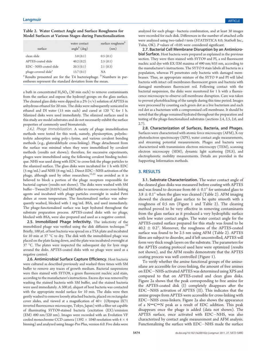
3.1. Substrate Characterization. The water contact angle ofthe cleaned glass disks was measured before coating with APTESand was found to decrease from 66( 0.1� for untreated glass to3.8 ( 0.1� when the glass was cleaned (Table 2). AFM imagingshowed the cleaned glass surface to be quite smooth with aroughness of 0.5 nm (Figure 1 and Table 2). The cleaningmethod proved to be very effective in removing contaminantsfrom the glass surface as it produced a very hydrophilic surfacewith low water contact angles. The water contact angle for theAPTES-coated surface prepared for this study was found to be40.2 ( 0.2�. Moreover, the roughness of the APTES-coatedsurface was found to be 2.5 nm using AFM (Table 2). APTESfilms are subject to disorder, and if left uncontrolled, APTES willform very thick rough layers on the substrate. The parameters forthe APTES coating protocol used here were optimized (resultsnot shown), and the AFM results demonstrate that the APTEScoating process was well controlled (Figure 1).To verify whether the amine functional groups of the amino-
silane are accessible for cross-linking, the amount of free amineson EDC�NHS-activated APTES was determined using XPS andcompared to that on APTES-coated and clean glass disks.Figure 2a shows that the peak corresponding to free amine forthe APTES-coated disk (I) completely disappears after theEDC�NHS activation of APTES (II). This indicates that theamine groups from APTES were accessible for cross-linking withEDC�NHS cross-linkers. Figure 2a also shows the appearanceof a NdCdN peak as a result of EDC addition. This peakdisappears once the phage is added (data not shown). TheAPTES surface, once activated with EDC�NHS, was alsocharacterized by contact angle measurement and AFM analysis.Functionalizing the surface with EDC�NHS made the surface
Table 2. Water Contact Angle and Surface Roughness forModel Surfaces at Various Stages during Functionalization
surface
water contact
angleb (deg)
surface roughnessb
(nm)
clean slide 3.8 (0.1) 0.5 (0.1)
APTES-coated slide 40.2 (0.2) 2.5 (0.1)
EDC�NHS-coated slide 20.3 (0.1) 2.1 (0.3)
phage-covered slidea 13.7 (0.1) NAaResults presented are for the T4 bacteriophage. bNumbers in par-entheses represent the standard deviation from the mean.

5475 dx.doi.org/10.1021/la200102z |Langmuir 2011, 27, 5472–5480
Langmuir ARTICLE
more hydrophilic (20.3�) and slightly decreased the surfaceroughness (2.1 nm) (Table 2).The binding of the phage to the substrate was confirmed using
several methods. First, XPS analysis on disks with the immobi-lized phage showed a marked increase in the N1s peak comparedto that for other disks at different stages of preparation(Figure 2b). The phage particles are rich in nitrogen, and hence,an increase in the N1s peak can be a marker for the presence ofthe phage on the surface. Moreover, SEM images clearly showthe phage particles on the substrate (Figure S2, Supporting
Information). Due to the small size of the phage, high-resolutionSEM images of the phage bound to the activated disks could notbe obtained and the SEM images were used to calculate thedensity of the phage on the disks. The phage density on the diskswas found to be in the same range for all the phages: 4.5 ( 0.7particles/μm2. AFM images of the disks also clearly showedintact phage particles to be present on the surface; results areshown for the T4 phage for which the capsid and tail are clearlyvisible (Figure 1d; compare with Figure S1d, SupportingInformation).
Figure 1. Atomic force microscopy images of the (a) clean disk, (b) APTES-coated disk, (c) EDC�NHS-activated APTES disk, and (d) disk with T4phages.
Figure 2. (a) XPS N1s spectra for the APTES-coated disk (I) and EDC�NHS-activated APTES (II). (b) Overlay of XPS wide scans for the APTES-coated disk, EDC�NHS-activated APTES, and the phage-functionalized disk (PR772). The dashed line indicates the location of the nitrogen peak.

5476 dx.doi.org/10.1021/la200102z |Langmuir 2011, 27, 5472–5480
Langmuir ARTICLE
APTES-coated slides were further characterized by determin-ing their ζ potential, which was found to be 3.5( 0.4 mV at pH7.5. The surfaces were thus blocked with BSA to prevent anyundesirable electrostatic interaction between the positive sub-strate and the negatively charged bacteria.3.2. Phage and Bacteria Characterization. TEM images of
the phages used in this study and SEM images of their hostbacteria are shown in Figure 3. Both bacteria are rod shaped andapproximately the same size. The pili for E. coli are clearly visible(Figure 3a), whereas S. typhimurium has no pili or flagella. TheTEM images for the phages are descriptive of the size and shapeof the five phages used in this study (Figure 3 c�g). PRD1,PR772, and MS2 are cubic phages, whereas P22 and T4 havetails, with the T4 tail being longer than that of P22. The size of allparticles, as determined from the electron micrographs, is pre-sented in Table 3. The hydrodynamic size and ζ potential ofbacteria and phages are also presented in Table 3. All bacteria andphages were negatively charged under the experimental condi-tions. S. typhimurium and E. coli, although both negative incharge, have significantly different ζ potentials, with E. coli beingmuch more negative (�47.1 mV compared to �3.0 mV for
S. typhimurium). The hydrodynamic diameter of the particles asdetermined by DLS measurements is slightly larger than the sizedetermined from electron micrographs; this is not only becausethe hydrodynamic diameter accounts for the hydration thicknessbut also a result of the sample preparation protocol for electronmicroscopy that is known to shrink the bacteria and phages. Thehydrodynamic diameter for the T4 phage is only an equivalentspherical diameter and has no other physical significance becauseof the elongated shape of the particle (Figure 3g).3.3. Immobilized Phage Biofunctionality. Immobilized
phages were verified for infectivity using the disk diffusionmethod. Disks functionalized with phages were incubated on alawn of dried bacteria on an agar plate. Figure S3 (SupportingInformation) shows the lysis ring around the disk with immobi-lized phages, which indicates that the phages on the disk wereable to lyse the bacteria around the disk and were thus infective.To measure immobilized phage capture efficiencies, suspen-
sions of host bacteria (109 cells/mL) stained with SYTO9 werecontacted with the phage-functionalized disks for 10 min so thatthe bacteria could diffuse and attach to the phages on the disk.The disks were subsequently washed to remove any loosely
Figure 3. Scanning electron micrographs of (a) E. coli ATCC 15597 and (b) S. typhimurium. Transmission electron micrographs of (c) PRD1, (d) P22,(e) PR772, (f) MS2, and (g) T4. The scale bars in images c�g are 50 nm.

5477 dx.doi.org/10.1021/la200102z |Langmuir 2011, 27, 5472–5480
Langmuir ARTICLE
attached bacteria. The capture efficiencies of three E. coli phagesand three S. typhimurium phages are presented in Figure 4. Thecapture efficiency of phages was measured by analyzing theimages obtained from fluorescence microscopy (Figure 5). It isimportant to note that the phage density on the substrate isalmost the same for all the phages used (4.5( 0.7 particles/μm2)as pointed out earlier. It can be seen that PRD1 is more effectivein capturing S. typhimurium than PR772 or P22 (P < 0.05).Among E. coli phages, MS2 is more effective than T4 and PR772(P < 0.05). PR772, which infects both of the bacteria, is moreeffective in capturing S. typhimurium than E. coli. It is interestingto note that results from an experiment with free phages (FigureS5, Supporting Information) showed both tailed phages (T4 andP22) to be more effective in adsorption to and lysis of the hostcells than the other phages in this study. PRD1, being the mostefficient phage when bound to the glass slide, was chosen tofurther investigate immobilized phage biofunctionality. Thecapture experiment was repeated for PRD1 disks, but this timethe disks were functionalized with four serial dilutions of PRD1,from 1010 to 107 PFU/mL. The capture efficiency decreased
almost 1 log between the disks for consecutive serial dilutions(Figure 6), and the number of bacterial cells captured was foundto be proportional to the concentration of phages used tofunctionalize the surface and thus to the density of phages onthe surface. PRD1 was also used for the bacterial inactivationexperiment in which the rate of cell membrane disruption by thebound phage was monitored via the BacLight viability kit.Figure 7 presents selected fluorescence microscopy images. Ascan be seen in Figure 7, the number of live bacteria decreasedmarkedly from time zero (this is 10 min after the initial contact ofbacteria with the surface) to the final frame, which was taken after60 min. The negative control does not reveal any detectablecompromised bacteria during this period (results not shown).The first red cells were visible after 35 min; however, all of thecells did not lyse at the same time. Image analysis showed 50% ofthe cells to be lysed after 45 min. It should be noted that the lyticcycle for free PRD1 phages has been reported to be 35 min.40
Figure S4a (Supporting Information) presents SEM images of S.typhimurium on a PRD1-functionalized disk; a higher magnifica-tion of the same image (Figure S4b) shows the lysed bacteria asindicated by the compromised membrane integrity.
4. DISCUSSION
4.1. Immobilized Phage Capture Capability. This studyinvestigated the capture efficiency of a number of different phageparticles when immobilized on a model substrate. Our resultsshow that there is a difference between the ability of differentphages to capture the same host bacteria when they are im-mobilized on a substrate. Free phage particles are able to diffuseeasily, come into contact with host bacteria, and orient them-selves in a manner that their receptors are able to attach to thespecific sites on the bacteria.10 However, for bound phage, theprocess is reversed because the host bacteria must come incontact with the phage receptors; therefore, the accessibility ofthese receptors controls the ability of the immobilized phage tocapture the bacterial cell. S. typhimurium was best captured by asurface functionalized with PRD1 or PR772 (Figures 4 and 5).These phages are both symmetric and have receptors on allvertices; therefore, when immobilizing PRD1 or PR772, orienta-tion is not an issue. P22 seemed to be less effective, which couldbe a result of unfavorable orientation of the phage particle on thesurface. Tailed phages must be immobilized on a substrate suchthat their tail is pointing away from the surface and their tailreceptors are capable of contacting the bacteria. If they are boundto the surface by their tail or on their side, they lose their ability toinfect bacteria. Controlling the immobilized orientation of a wild-type tailed phage on a substrate is very challenging; therefore, thisgreatly compromises their biofunctionality.PR772 is significantly more effective at capturing S. typhimur-
ium than E. coli. The number of S. typhimurium cells captured byPR772 was more than twice the number of E. coli cells (Figure 4).This could be attributed to the difference in the way PR772infects S. typhimurium and E. coli. PR772 attaches to the receptorson the surface of S. typhimurium, but to infect E. coli, it mustattach to the tip of its F-pili.10 MS2, which is also an F-specificphage,41 performs well for capturing E. coli, although it is highlyasymmetric in terms of the location of its capture protein. MS2and PR772 both attach to the F-pili of E. coli, but PR772 attachesto the tip of the pili, while MS2 attaches laterally along the pili.41
Thus, MS2 has more sites to attach to, whereas PR772 has onlyone site of attachment per pili. This may have been a significant
Table 3. Size and ζ Potential of Bacteria and Phagesa
hydrodynamic
diameter (nm)
dry diameterb
(nm)
ζ potential
(mV)
T4 98 (1.4) 90 (0.5), 200 (2.0)c �25.9 (0.2)
MS2 43 (1.9) 30 (1.8) �33.7 (3.4)
PR772 85 (0.8) 70 (1.0) �12.4 (2.2)
PRD1 104 (3.2) 80 (0.8) �21.5 (0.9)
P22 69 (1.4) 49 (3.0) �15.1 (1.2)
E. coli ATCC 15597 1742 (71) 580 (0.8), 1720 (2.5) �47.1 (0.7)
S. typhimurium LT2 1636 (63) 700 (0.6), 1550 (1.4) �3.0 (0.2)aNumbers in parentheses represent the standard deviation from themean. The data for the hydrodynamic diameter and ζ potential are themean of the values measured for three samples. The data for the drydiameter are the mean of the values for five different images of the samesample. bAs measured from electron micrographs. cCapsid length, totallength.
Figure 4. Number of host bacteria attached to phage-functionalizeddisks. Each column represents the mean of five experiments with 30frames analyzed for each experiment. Control 1 represents the APTES-coated disk, and control 2 represents the blank disk (both blocked withBSA). Controls are the average for both bacterial hosts. Data are means( 95% confidence intervals (n = 150).

5478 dx.doi.org/10.1021/la200102z |Langmuir 2011, 27, 5472–5480
Langmuir ARTICLE
asset for MS2 because it was able to increase its immobilizedbiofunctionality despite its asymmetric shape. T4, also highlyasymmetric, is expected to be immobilized on the surface in amixture of favorable and unfavorable orientations. Moreover, T4is a somatic phage rather than F-specific; thus, the tail fibers mustcome into contact with the cell wall. This can be very difficult ifthe phage is bound to the surface in any other configuration thanfrom the top of its capsid. The capture efficiency of T4 isapproximately half that of MS2 (Figure 4). Although both T4and MS2 are limited by orientation when immobilized, the factthat MS2 can bind along the pili of its host may be a determiningfactor for increasing its capture efficiency to about twice that ofPR772 and T4.Further experiments with PRD1 showed this phage performed
well even at concentrations of less than 1010 PFU/mL. Thecapture efficiency for disks functionalized with serial dilutions ofPRD1 decreased about 1 log between the disks (Figure 6). Thisindicates that the number of bacteria captured was proportional
to the number of phages on the surface. However, the number ofbacteria on the surface was lower than theoretically expected.This observation may be explained by the fact that the number ofbacteria that actually reach the surface is limited by diffusion.Additionally, the bacteria themselves are charged and thus exertelectrostatic repulsion on each other. Hence, close packing ofbacteria on the surface should not be expected. However,increasing the coverage of bacteria and phages on the surface isan issue that needs improvement and can be the subject of futurestudies.4.2. Bacterial Cell Membrane Disruption by Immobilized
Phages. Another interesting observation was the rate of cellmembrane disruption by immobilized phage (Figure 7). Theimmobilized PRD1 effectively disturbs the integrity of the cellmembrane (Figure 7 and Figure S4, Supporting Information).The first compromised cells were observed after 35min; this timelag can be explained by the latent period inherent in the phagelytic cycle, which is the time required for the phage to propagateinside the bacteria and lyse the cell. All the bacterial cells thatremained on the surface were attached to a phage; otherwise,they would have been removed during the washing stage as wasthe case for the negative control disks; however, not all cells arecompromised at the same time. This could be attributed in partto the presence of ghost phages; these are phage particles withouta genetic material.42 The bacteria captured by ghost phages areonly infected after the release of free progeny phages from thelysed bacteria. Another reason could be the deficiency innutrients. Phages are known to have longer latent periods undernutrient-limited conditions.43 Moreover, being immobilized mayhave affected the cell lysing ability of the phage perhaps byaffecting the initial infection process. Additional studies areneeded to better understand the role of immobilization in thephage lysing ability; however, these results clearly indicate thepotential of phages for developing surfaces that are resistant tobiofilm formation. The advantage of using phages can be realized
Figure 6. Number of captured S. typhimurium cells versus the concen-tration of PRD1 used to functionalize the substrate. Data are means (95% confidence intervals (n = 150).
Figure 5. Representative fluorescence microscopy images of S. typhimurium attached to phage-immobilized slides with (a) PRD1, (b) PR772, and (c)P22 and E. coli ATCC 15597 attached to phage-immobilized slides with (d) T4, (e) PR772, and (f) MS2. The fluorescence images have been convertedto black and white for clarity. The bacteria are shown in white over a black background.

5479 dx.doi.org/10.1021/la200102z |Langmuir 2011, 27, 5472–5480
Langmuir ARTICLE
further if we note that the lysed cells in turn producemore phagesso the surface retains its activity even for high bacterial loads asopposed to surfaces with antibacterial chemicals for which thefixed dose of antibacterial agent must be adjusted according tothe anticipated bacterial load.4.3. Challenges. There are a few notable drawbacks for using
phages on antimicrobial surfaces. The first is the specificity of thephage toward its host. The spectrum of activity of the surfacemight be increased by using a cocktail of different phages or byusing broad host range phages.44 Another limitation is thepossibility of phage or host mutation, which might result in thebacteria becoming resistant to the phage. Generally, it is believedto be easier to deal with resistance of bacteria to phages,compared to antibiotic resistance, by selecting new phages fromcultures that maintain virulence.11 Moreover, bacteria and theircorresponding lytic phages have been shown to coevolve andestablish equilibrium between their respective populations.45,46
However, there is still a concern that phage-treated surfaces orproducts would require constant monitoring and their formula-tion modified to remain efficacious. Another question thatremains to be addressed is the efficacy of the phage-functiona-lized substrates at normal body temperature and over extendedtime periods. It can be argued that the advantages of phages overantibacterial substances already in use justify more research inthis field to find approaches to overcome such obstacles.
5. CONCLUSIONS
This study was aimed at providing fundamental knowledgetoward an improved understanding of phage-based antimicrobialsurfaces. We investigated the biofunctionality of antibacterialsurfaces functionalized with bacteriophages. Our results empha-size that the biology and surface properties of the phage and thebacterial target greatly influence bacterial capture. Of the phagesinvestigated, the symmetric phage PRD1 (host S. typhimurium)and asymmetric phage MS2 (host E. coli) exhibited superiorcapture efficiencies for their host bacteria. Surfaces with immo-bilized PRD1 were shown to actively compromise all of thecaptured cells within an hour of contact.
’ASSOCIATED CONTENT
bS Supporting Information. Details regarding the structureof the phage used, characterization methods, and certain figures.This material is available free of charge via the Internet at http://pubs.acs.org.
’AUTHOR INFORMATION
Corresponding Author*Phone: (514) 398-2999. Fax: (514) 398-6678. E-mail: [email protected].
’ACKNOWLEDGMENT
This work was supported by the Natural Sciences andEngineering Research Council of Canada (NSERC StrategicResearch Network on Bioactive Paper—SENTINEL) and theCanada Research Chairs (CRC) program. We acknowledge M.Griffiths (University of Guelph) for kindly providing the T4phage, M. Mesquite and M. B. Emelko (University of Waterloo)for guidance regarding phage propagation and for kindly provid-ing the PRD1 and PR772 phage as well as the Salmonella host, S.Poulin (�Ecole Polytechnique de Montreal) for compiling theXPS data, J. Fatisson for help with AFM imaging, and G. Hidalgoand C. O’May (McGill University) for assistance in editing themanuscript.
’REFERENCES
(1) Boyce, J. M.; Potter-Bynoe, G.; Chenevert, C.; King, T. Infect.Control Hosp. Epidemiol. 1997, 18 (9), 622–627.
(2) Bhalla, A.; Pultz, N. J.; Gries, D. M.; Ray, A. J.; Eckstein, E. C.;Aron, D. C.; Donskey, C. J. Infect. Control Hosp. Epidemiol. 2004, 25 (2),164–167.
(3) Maki, D. G.; Tambyah, P. A. Emerging Infect. Dis. 2001, 7 (2),342–347.
(4) Emerson Iv, R. J.; Camesano, T. A. Appl. Environ. Microbiol.2004, 70 (10), 6012–6022.
(5) Page, K.; Wilson, M.; Parkin, I. P. J. Mater. Chem. 2009, 19 (23),3818–3831.
(6) Chen, Z.; Cao, G.; Song, Q. Environ. Chem. Lett. 2010, 8 (1),33–37.
(7) Fang, J. L.; Stingley, R. L.; Beland, F. A.; Harrouk, W.; Lumpkins,D. L.; Howard, P. J. Environ. Sci. Health, Part C: Environ. Carcinog.Ecotoxicol. Rev. 2010, 28 (3), 147–171.
(8) Silver, S. FEMS Microbiol. Rev. 2003, 27 (2�3), 341–353.(9) Liu, W.; Wu, Y.; Wang, C.; Li, H. C.; Wang, T.; Liao, C. Y.; Cui,
L.; Zhou, Q. F.; Yan, B.; Jiang, G. B. Nanotoxicology 2010, 4 (3),319–330.
(10) Calendar, R. L. The Bacteriophages; Oxford University Press:New York, 2005.
(11) Curtin, R. Science 2002, 298 (5594), 728–731.(12) Sulakvelidze, A.; Alavidze, Z.; Morris, J. G., Jr. Antimicrob.
Agents Chemother. 2001, 45 (3), 649–659.(13) Chang, T. C.; Ding, H. C.; Chen, S. W. J. Food Prot. 2002, 65
(1), 12–17.
Figure 7. Representative images of cell lysis monitored by fluorescence microscopy using the BacLight bacteria viability kit. Green dots represent livebacteria, and red dots represent lysed bacteria.

5480 dx.doi.org/10.1021/la200102z |Langmuir 2011, 27, 5472–5480
Langmuir ARTICLE
(14) Neufeld, T.; Mittelman, A. S.; Buchner, V.; Rishpon, J. Anal.Chem. 2005, 77 (2), 652–657.(15) Handa, H.; Gurczynski, S.; Jackson,M. P.; Auner, G.;Walker, J.;
Mao, G. Surf. Sci. 2008, 602 (7), 1392–1400.(16) Shabani, A.; Zourob,M.; Allain, B.; Marquette, C. A.; Lawrence,
M. F.; Mandeville, R. Anal. Chem. 2008, 80 (24), 9475–9482.(17) Huang, S.; Yang, H.; Lakshmanan, R. S.; Johnson, M. L.; Wan,
J.; Chen, I. H.; Wikle Iii, H. C.; Petrenko, V. A.; Barbaree, J. M.; Chin,B. A. Biosens. Bioelectron. 2009, 24 (6), 1730–1736.(18) Guntupalli, R.; Sorokulova, I.; Krumnow, A.; Pustovyy, O.;
Olsen, E.; Vodyanoy, V. Biosens. Bioelectron. 2008, 24 (1), 151–154.(19) Zhu, H.; White, I. M.; Suter, J. D.; Fan, X. Biosens. Bioelectron.
2008, 24 (3), 461–466.(20) Nanduri, V.; Sorokulova, I. B.; Samoylov, A. M.; Simonian, A. L.;
Petrenko, V. A.; Vodyanoy, V. Biosens. Bioelectron. 2007, 22 (6), 986–992.(21) Balasubramanian, S.; Sorokulova, I. B.; Vodyanoy, V. J.; Simonian,
A. L. Biosens. Bioelectron. 2007, 22 (6), 948–955.(22) Singh, A.; Arya, S. K.; Glass, N.; Hanifi-Moghaddam, P.;
Naidoo, R.; Szymanski, C. M.; Tanha, J.; Evoy, S. Biosens. Bioelectron.2010, 26 (1), 131–138.(23) Minikh, O.; Tolba, M.; Brovko, L. Y.; Ugarova, N. N.; Griffiths,
M. W. Luminescence 2010, 25 (2), 177–177.(24) Tolba, M.; Minikh, O.; Brovko, L. Y.; Evoy, S.; Griffiths, M. W.
Appl. Environ. Microbiol. 2010, 76 (2), 528–535.(25) Fu, W.; Forster, T.; Mayer, O.; Curtin, J. J.; Lehman, S. M.;
Donlan, R. M. Antimicrob. Agents Chemother. 2010, 54 (1), 397–404.(26) Carson, L.; Gorman, S. P.; Gilmore, B. F. FEMS Immunol. Med.
Microbiol. 2010, 59 (3), 447–455.(27) Jikia, D.; Chkhaidze, N.; Imedashvili, E.; Mgaloblishvili, I.;
Tsitlanadze, G.; Katsarava, R.; Morris, J. G., Jr; Sulakvelidze, A. Clin.Exp. Dermatol. 2005, 30 (1), 23–26.(28) Curtin, J. J.; Donlan, R. M. Antimicrob. Agents Chemother. 2006,
50 (4), 1268–1275.(29) Stone, R. Science 2002, 298 (5594), 728–731.(30) Bennett, A. R.; Davids, F. G. C.; Vlahodimou, S.; Banks, J. G.;
Betts, R. P. J. Appl. Microbiol. 1997, 83 (2), 259–265.(31) Archer, M. J.; Liu, J. L. Sensors 2009, 9 (8), 6298–6311.(32) Singh, A.; Glass, N.; Tolba, M.; Brovko, L.; Griffiths, M.; Evoy,
S. Biosens. Bioelectron. 2009, 24 (12), 3645–3651.(33) Huang, S.; Yang, H.; Johnson, M. L.; Lakshinanan, R. S.; Chen,
I.; Petrenko, V. A.; Barbaree, J. M.; Chin, B. A. (Institute of Electrical andElectronics Engineers, IEEE). The effect of phage solution chemistry onthe spore binding affinity of magnetoelastic biosensors. IEEE Sensors2007, Atlanta, GA, Oct 28�31, 2007; IEEE: Piscataway, NJ, 2007; Vols.1�3, pp 1093�1096.(34) Cademartiri, R.; Anany, H.; Gross, I.; Bhayani, R.; Griffiths, M.;
Brook, M. A. Biomaterials 2010, 31 (7), 1904–1910.(35) Jabrane, T.; Dub�e, M.; Mangin, P. J. Bacteriophage activity on
paper surface: Effect of paper moisture. Presented at the 8th WorldCongress of Chemical Engineering, Montreal, Quebec, Canada, Aug23�27, 2009.(36) Gervais, L.; Gel, M.; Allain, B.; Tolba, M.; Brovko, L.; Zourob,
M.; Mandeville, R.; Griffiths, M.; Evoy, S. Sens. Actuators, B 2007, 125(2), 615–621.(37) Minikh, O.; Tolba, M.; Brovko, L. Y.; Griffiths, M. W.
J. Microbiol. Methods 2010, 82 (2), 177–183.(38) Sambrook, J.; Russell, D. W. Molecular Cloning: A Laboratory
Manual, 3rd ed.; Cold Spring Harbor Laboratory Press: Cold SpringHarbor, NY, 2001.(39) Andrews, J.M. J. Antimicrob. Chemother. 2009, 64 (3), 454–489.(40) Olsen, R. H.; Siak, J. S.; Gray, R. H. J. Virol. 1974, 14 (3),
689–699.(41) Van Duin, J.; Tsareva, N. Single-stranded RNA phages. In The
Bacteriophages; Calendar, R. L., Ed.; Oxford University Press: New York,2005.(42) Grahn, A. M.; Butcher, S. J.; Bamford, J. K. H.; Bamford, D. H.
PRD1: Dissecting the genome, structure, and entry. In The Bacterio-phages; Calendar, R. L., Ed.; Oxford University Press: New York, 2005.
(43) Donlan, R. M. Trends Microbiol. 2009, 17 (2), 66–72.(44) Cerca, N.; Oliveira, R.; Azeredo, J. Lett. Appl. Microbiol. 2007,
45 (3), 313–317.(45) Lenski, R. E.; Levin, B. R. Am. Nat. 1985, 125 (4), 585–602.(46) Mizoguchi, K.; Morita, M.; Fischer, C. R.; Yoichi, M.; Tanji, Y.;
Unno, H. Appl. Environ. Microbiol. 2003, 69 (1), 170–176.