automated quantitative analysis of ki-67 staining and he
TRANSCRIPT
RESEARCH Open Access
Automated quantitative analysis of Ki-67staining and HE images recognition andregistration based on whole tissue sectionsin breast carcinomaMin Feng1,2,3†, Yang Deng1†, Libo Yang1,3, Qiuyang Jing2, Zhang Zhang3, Lian Xu2,3, Xiaoxia Wei1,3,4, Yanyan Zhou1,Diwei Wu1, Fei Xiang5, Yizhe Wang5, Ji Bao1* and Hong Bu1,3*
Abstract
Background: The scoring of Ki-67 is highly relevant for the diagnosis, classification, prognosis, and treatmentin breast invasive ductal carcinoma (IDC). Traditional scoring method of Ki-67 staining followed by manualcounting, is time-consumption and inter−/intra observer variability, which may limit its clinical value. Althoughmore and more algorithms and individual platforms have been developed for the assessment of Ki-67 stainedimages to improve its accuracy level, most of them lack of accurate registration of immunohistochemical(IHC) images and their matched hematoxylin-eosin (HE) images, or did not accurately labelled each positiveand negative cell with Ki-67 staining based on whole tissue sections (WTS). In view of this, we introduce anaccurate image registration method and an automatic identification and counting software of Ki-67 based onWTS by deep learning.
Methods: We marked 1017 breast IDC whole slide imaging (WSI), established a research workflow based onthe (i) identification of IDC area, (ii) registration of HE and IHC slides from the same anatomical region, and(iii) counting of positive Ki-67 staining.
Results: The accuracy, sensitivity, and specificity levels of identifying breast IDC regions were 89.44, 85.05, and95.23%, respectively, and the contiguous HE and Ki-67 stained slides perfectly registered. We counted andlabelled each cell of 10 Ki-67 slides as standard for testing on WTS, the accuracy by automatic calculation ofKi-67 positive rate in attained IDC was 90.2%. In the human-machine competition of Ki-67 scoring, theaverage time of 1 slide was 2.3 min with 1 GPU by using this software, and the accuracy was 99.4%, whichwas over 90% of the results provided by participating doctors.
(Continued on next page)
© The Author(s). 2020 Open Access This article is licensed under a Creative Commons Attribution 4.0 International License,which permits use, sharing, adaptation, distribution and reproduction in any medium or format, as long as you giveappropriate credit to the original author(s) and the source, provide a link to the Creative Commons licence, and indicate ifchanges were made. The images or other third party material in this article are included in the article's Creative Commonslicence, unless indicated otherwise in a credit line to the material. If material is not included in the article's Creative Commonslicence and your intended use is not permitted by statutory regulation or exceeds the permitted use, you will need to obtainpermission directly from the copyright holder. To view a copy of this licence, visit http://creativecommons.org/licenses/by/4.0/.The Creative Commons Public Domain Dedication waiver (http://creativecommons.org/publicdomain/zero/1.0/) applies to thedata made available in this article, unless otherwise stated in a credit line to the data.
* Correspondence: [email protected]; [email protected]†Min Feng and Yang Deng contributed equally to this work.1Laboratory of Pathology, West China Hospital, Sichuan University, Chengdu610041, ChinaFull list of author information is available at the end of the article
Feng et al. Diagnostic Pathology (2020) 15:65 https://doi.org/10.1186/s13000-020-00957-5
(Continued from previous page)
Conclusions: Our study demonstrates the enormous potential of automated quantitative analysis of Ki-67staining and HE images recognition and registration based on WTS, and the automated scoring of Ki67 canthus successfully address issues of consistency, reproducibility and accuracy. We will provide those labelledimages as an open-free platform for researchers to assess the performance of computer algorithms forautomated Ki-67 scoring on IHC stained slides.
Keywords: Convolutional neural network, Whole tissue sections, Breast invasive ductal carcinoma, Automaticrecognition, Ki-67 counting
IntroductionBreast invasive ductal carcinoma (IDC) is the most com-mon malignant tumor in women worldwide, with atrend of younger at diagnosis [1, 2]. In 2018, there weremore than 266,000 new cases of breast cancer in womenin the United States, accounting for 30% of all malignanttumors in women and far exceeding the second lungcancer (13%) [3]. In both developed and developingcountries, the disease ranks as third in the mortality rateamong females [2, 3]. Ki-67 protein, as well as ER, PR,and HER-2 protein, have been recognized as main bio-logical indicators to guide the molecular typing, treat-ment plan, and prognosis evaluation of breast cancer [4].Ki-67 is a cell cycle related nucleoprotein, which hasbeen served as an accurate marker to infer the prolifera-tive status of tumor cells, since it only reacts with theproliferating cells and shows no tissue specificity [5].Interestingly, a number of studies have reported that Ki-67 staining can be used as a reference index for theprognosis and personalized treatment of breast cancerpatients, it is also closely related to the clinicopathologi-cal features and molecular typing of breast cancerpatients [5–7]. Moreover, Ki-67 scoring can be used todistinguish luminal breast cancer subtypes (A/B) and, asa result, it certainly helps to define the best treatmentstrategy for each particular condition [8, 9]. In triplenegative breast cancer (TNBC), patients high Ki-67scores seem to benefit more from the treatment [10].Nevertheless, the traditional scoring method of Ki-67
staining by IHC, can be frequently time-consuming,labor-intensive, and poorly reproducible for many pa-thologists, and later provide limited reproducibility andquantification of respective markers. These commonproblems can seriously hinder the establishment andmanagement of patient treatment, especially during latephases. Fortunately, with the emergence of whole slidedigital scanning technology, it is now feasible to combinehistopathological image information with artificialintelligence (AI) technology. This combination meetsthe standards of high definition, high speed, and highthroughput screening [11], which could lay a good foun-dation for the development and application of digitalpathology. Using whole slide imaging (WSI) as the basis,
combined with a series of technical equipment including(i) a image analysis system and (ii) an information man-agement system, via deep learning of the computer, AIcan effectively simulate a pathologist’s brain for effectivethinking and further assist in broader applications in themedical and health areas, such as disease intelligenceanalysis, tumour-assisted diagnosis, gene data detection,and disease drug development [12–15]. Generally, WSIsare gigapixel images stored in a multi-resolution pyra-mid structure where the highest resolution is × 40.Moreover, a model training based on convolutionalneural networks (CNN) may provide doctors with effect-ive and accurate information, such as pathological dis-ease typing, cancer histology-assisted diagnosis, mitoticcell counts, epithelium–stroma classification, lymphnode metastasis assessment and others [16–21]. CNNtechniques are guided by structural and statistical infor-mation derived from respective images. There are severaldeep learning models described so far, in particular forCNN, such as LeNet, AlexNet, and GoogleNet [22].Hence, the question arises whether AI could be used tosolve the problem of accurate counting of Ki-67 onimmunohistochemically stained sections. Existing re-search has revealed that the development of countingsoftwares, focusing on Ki-67 staining in a variety of tu-mors, still have many limitations, including the lack ofautomated location for areas of interest, or accurateregistration of IHC images and their HE images. To at-tempt providing stronger assessment, reliable compari-sons, and more reproducible results, here we utilizedsimulated data to compare analytical performanceamong different algorithms, and we further selected anunsupervised domain adaptation for counting, based onfew simple and easily-implemented CNN models, namedas GoogLeNet Inception V1, this model could help uslocated the IDC area automatically. And then, we regis-tered the labelled HE and Ki-67 stained sections using aSimple Elastix toolbox, which was developed by our en-gineer teams to handle medical image registration issuesspecifically. Finally, we used an algorithm provided byImage J to automatically extract the structure, morph-ology, color, and other characteristics of positive/nega-tive cells, and train the random forest classifier that
Feng et al. Diagnostic Pathology (2020) 15:65 Page 2 of 12
could identify Ki-67 positive/negative cells. In addition,we marked 10 standard Ki-67 Counting slides for testingon whole tissue sections, these slides were labelled byten pathologists, who circled each cell in the tumor re-gion of these slides and determined whether it was posi-tive or negative.
Materials and methodsExperimental designResearch process was divided into three stages: identifi-cation of IDC, registration, and enumerating of Ki-67staining (See Fig. 1 for the flow chart of Ki-67 AutomaticCounting Software in breast IDC on WSI). To enhancethe classification performance of IDC and ductal carcin-oma in situ (DCIS), and, simultaneously, to reduce thenetwork training time, our method was designed withunsupervised domain adaptation for counting, based onGoogLeNet Inception V1.
Case selectionA total of 1074 IDC slides from 672 cases diagnosed bythe Department of Pathology of West China Hospital(Sichuan University, China) were collected. From these,57 unqualified sections were removed after primaryscreening due to (i) quality issues with the sections, (ii)insufficient scanning clarity, and/or (iii) poor identifica-tion of IDC area. The remaining 1017 sections wereprocessed for this study at last. We randomly selected
677 of these sections as training sets, 153 as verificationsets, and 187 as test sets. Both H&E and Ki-67 stainedslides were collated into a complete digital scanning sec-tion WSI by digital section scanner (Hamamatsu Optics’NanoZoomer 2.0HT), with a magnification of 40 × .
ImmunohistochemistryFor immunohistochemical staining of Ki-67, 4 um thin tissuesections were dewaxed in xylene, acetone and Tris-bufferedsaline, followed by heat induced epitope retrieval in pH 6.0in a microwave oven (750W). Ventana was used for antigenretrieval. Sections were subsequently stained using Ki67 anti-body (clone mAb, ready-to-use formulation), purchasingfrom Roche. All the steps were carried out according to theinstructions and stained by Bench Mark ULTRA automaticimmunohistochemical staining machine.
Image acquisitionAt this stage, we have included 1017 breast IDC diag-nosed slides marked with the IDC regions, followed bythe removal of features related to these labelled digitalslides. The classification network model was furthertrained by GoogleNet Concept V1, which could be usedto automatically identify IDC regions.
LabellingA team of 36 pathologists from West China ClinicalMedical College (Sichuan University, China) was
Fig. 1 The flow chart of Ki-67 Automatic Counting Software in breast IDC on whole tissue sections
Feng et al. Diagnostic Pathology (2020) 15:65 Page 3 of 12
organized in order to label the IDC area on the WSI.Both workflow and number of pathologists involvedwere divided into four categories: (i) WSI labelling(28 professionals), (ii) labelling review (3 profes-sionals), (iii) labelling quality control (2 professionals),and (iv) training experts (3 professionals). Firstly,three experts from the Breast Diseases Group, Chin-ese Medical Association Pathology Branch, conductedmultiple training sessions to appropriately distinguishthe IDC regions in the WSI. Next, 28 labelling staffmembers were divided into other three groups tocomplete the labelling of IDC regions in all WSI.Simultaneously, a pathologist with intermediate orabove titles was assigned as the team leader for eachgroup, to review the labelled regions and to providefeedback on the results to the labelling staff in atimely manner. Meanwhile, two attending pathologistswere appointed as quality control physicians to con-duct random checks on WSI after reviewing (randomrate of 5% or above), with an accuracy rate of morethan 95% for proper qualification. For labelling, weused different colors to distinguish various tissueregions.
TrainingAfter labelling by the pathologists, software engineerused the computer image processing algorithm to seg-ment and extract the labelled information, classifyingand extracting the positive and negative regions accord-ingly (128 pixel × 128 pixel patch), and then used Goo-gleNet Incubation V1 for featured extraction andclassification training to obtain a network model. At thisstage, 677 training sets of WSI were used to fit the pa-rameters of the model, while 153 verification sets ofWSI were used to tune the model hyperparameters dur-ing training procedures (Table 1). A total of 2000 posi-tive patches and 2000 negative patches were selected fortraining in each WSI, whereas the redundant patcheswere not included in the training set.
TestingAfter training the classification model, 187 test sets’ ofWSIs were used to provide an unbiased evaluation of afinal model fit on the training dataset. We took eachpatch as a unit, and then considered the IDC area pre-marked by pathologists as a “gold standard”. Next, wecompared it with the analytical results of AI systems to
retrieve performance indicators such as sensitivity, speci-ficity, and accuracy. Labelling was strictly confidentialbefore testing, to meet the requirements of “a single-blind” study.
RegistrationWe randomly selected 100 cases with both HE slidesand their corresponding Ki-67 stained slides, which werecreated by serial sectioning technique. Next, we regis-tered the labelled HE and Ki-67 stained sections using aSimple Elastix toolbox, which developed by our engineerteams, could handle medical image registration issues.Slides were initially superimposed by this toolbox, andthen automatically modified into a rigid transformationsuch as translation and rotation via the registration func-tion of the tool, thereby achieving a good registration ef-fect. Eventually, labelling of each HE slides was migratedto respective Ki-67 images, and the IDC area on eachKi-67 slice was selected accordingly.
Counting of Ki-67 stained sections on whole tissuesectionThe registered Ki-67 stained sections were labelled toidentify the positive and negative tumour cells. Accord-ing to the labelling information, we used an algorithmprovided by Image J (an open source software for digitalpathology image analysis) to automatically extract thestructure, morphology, colour, and other characteristicsof positive/negative cells, and train the random forestclassifier that could identify Ki-67 positive/negative cells.This procedure allowed the automatic counting of Ki-67positive and negative cells in the IDC region and, as re-quired, Ki-67 positive rate. At this stage, we circled tenROI (region of interest) on each Ki-67 slice, where eachROI included at least 100 cells. More than 100,000 cellsin all were labelled, in which positive and negative cellswere marked, respectively, in red and blue colours. TheKi-67 positive rate calculated from these artificially la-belled cells is considered to be the “gold standard”.These labelled cells were also used to tune the Ki-67counting model at the verification sets.
Testing of the total accuracyAfter the aforementioned stages, we acquired an inte-grated WSI-based model for Ki-67 Automatic Countingin breast invasive ductal carcinoma. Thereafter, wetested the accuracy rate of this Ki-67 counting model. Inaddition, we organized a competition test, featuring tenclinical pathologists, to verify the modelling efficiency.
Labelling of standard Ki-67 countingTen HE and Ki-67 co-stained IDC sections (excludingintraductal carcinoma tissues), originated from differentpatients, were used as standard provided by the
Table 1 The results of segmentation and extraction based on1017 HE slices labelled information
Type of sets training sets verification sets test sets Sum
WSI number 677 153 187 1017
Patch number 11,628,208 2,973,384 2,419,032 17,020,624
Feng et al. Diagnostic Pathology (2020) 15:65 Page 4 of 12
Department of Pathology of the Sichuan Cancer Hos-pital. These sections were labelled by ten pathologists ofWest China Hospital (Sichuan University, China). Patho-logical staff also participated in the labelling of Ki-67automatic analysis system in our IDC study by determin-ing the number and positive rate of Ki-67 staining in re-spective areas. Results were classified as standard for thistesting.
Testing and competitionThe Ki-67 Artificial Intelligence Counting System de-veloped by our institute was presented at the “2017Pathological Image Diagnosis Human-Machine Chal-lenge” (seventh China Pathology Annual Meeting).Contestants competed with ten senior pathologists tovalidate the modelling efficiency. Competition wasbased on the independent completion of Ki-67 posi-tive counting in IDC areas of ten breast cancer WSIwithin 30 min. For this, results were required to beaccurate to 1%; the completion time of each contest-ant was recorded by auxiliary personnel. Completiontime and accuracy of each contestant were compre-hensively evaluated.
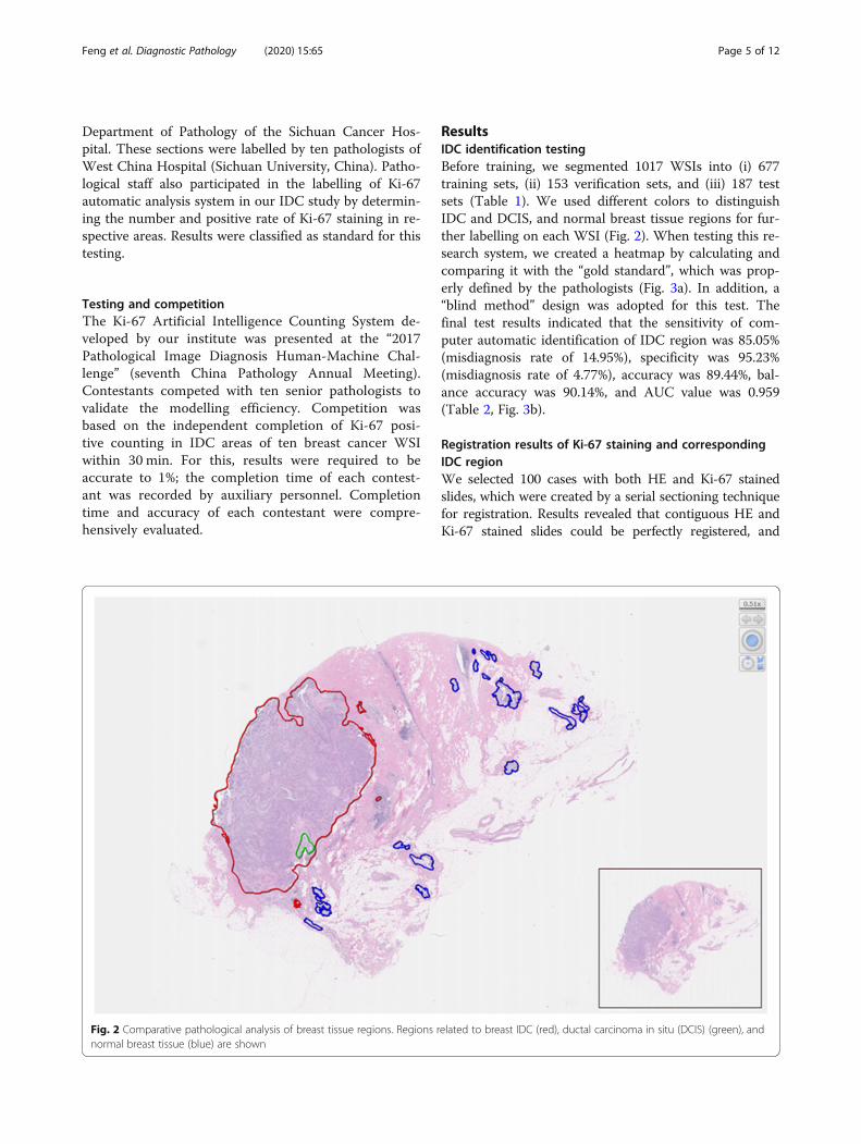
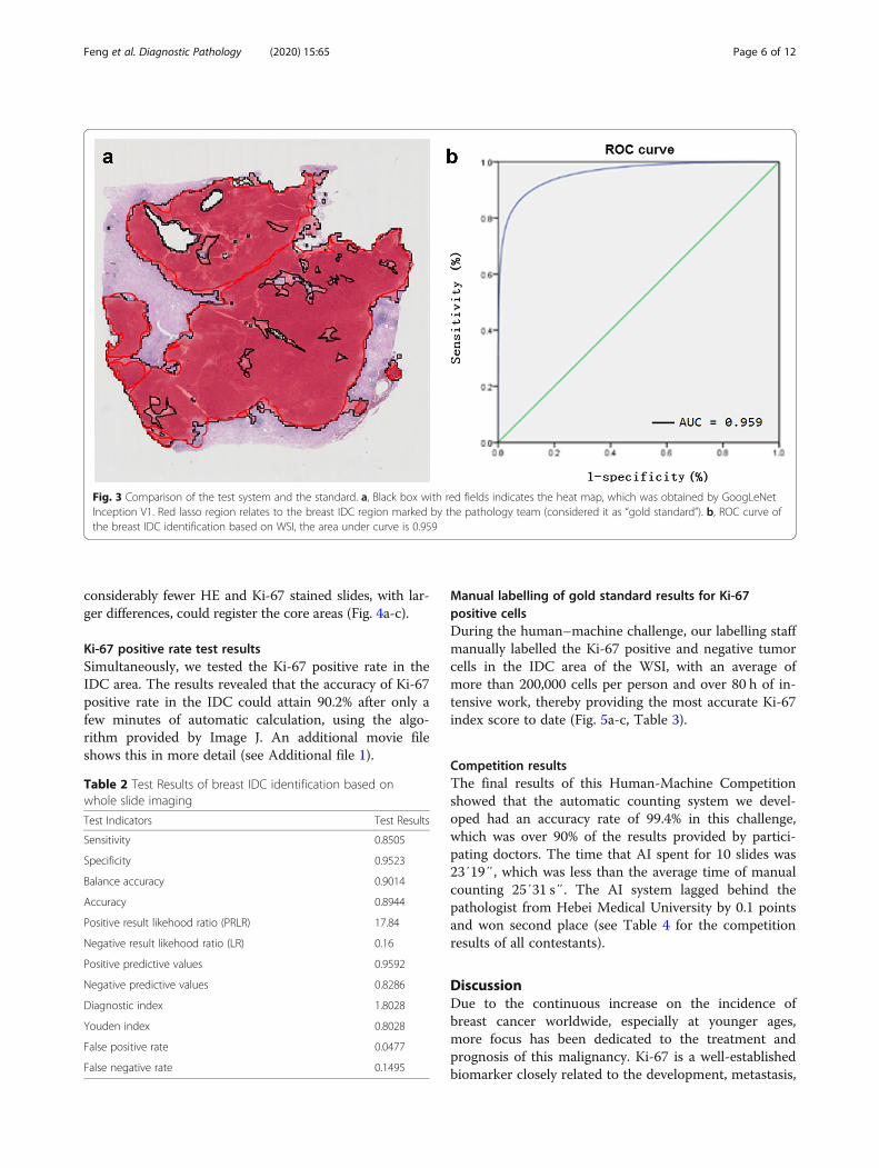
ResultsIDC identification testingBefore training, we segmented 1017 WSIs into (i) 677training sets, (ii) 153 verification sets, and (iii) 187 testsets (Table 1). We used different colors to distinguishIDC and DCIS, and normal breast tissue regions for fur-ther labelling on each WSI (Fig. 2). When testing this re-search system, we created a heatmap by calculating andcomparing it with the “gold standard”, which was prop-erly defined by the pathologists (Fig. 3a). In addition, a“blind method” design was adopted for this test. Thefinal test results indicated that the sensitivity of com-puter automatic identification of IDC region was 85.05%(misdiagnosis rate of 14.95%), specificity was 95.23%(misdiagnosis rate of 4.77%), accuracy was 89.44%, bal-ance accuracy was 90.14%, and AUC value was 0.959(Table 2, Fig. 3b).
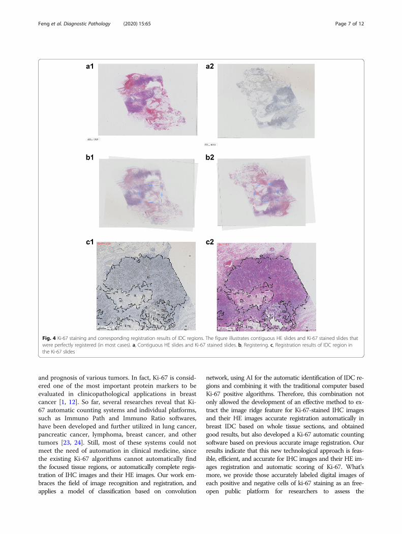
Registration results of Ki-67 staining and correspondingIDC regionWe selected 100 cases with both HE and Ki-67 stainedslides, which were created by a serial sectioning techniquefor registration. Results revealed that contiguous HE andKi-67 stained slides could be perfectly registered, and
Fig. 2 Comparative pathological analysis of breast tissue regions. Regions related to breast IDC (red), ductal carcinoma in situ (DCIS) (green), andnormal breast tissue (blue) are shown
Feng et al. Diagnostic Pathology (2020) 15:65 Page 5 of 12
considerably fewer HE and Ki-67 stained slides, with lar-ger differences, could register the core areas (Fig. 4a-c).
Ki-67 positive rate test resultsSimultaneously, we tested the Ki-67 positive rate in theIDC area. The results revealed that the accuracy of Ki-67positive rate in the IDC could attain 90.2% after only afew minutes of automatic calculation, using the algo-rithm provided by Image J. An additional movie fileshows this in more detail (see Additional file 1).
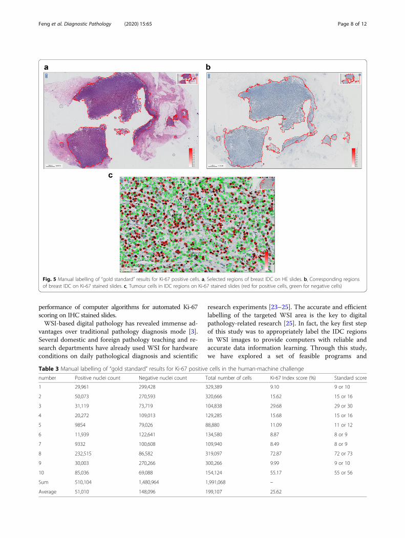
Manual labelling of gold standard results for Ki-67positive cellsDuring the human–machine challenge, our labelling staffmanually labelled the Ki-67 positive and negative tumorcells in the IDC area of the WSI, with an average ofmore than 200,000 cells per person and over 80 h of in-tensive work, thereby providing the most accurate Ki-67index score to date (Fig. 5a-c, Table 3).
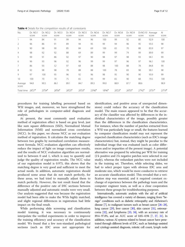
Competition resultsThe final results of this Human-Machine Competitionshowed that the automatic counting system we devel-oped had an accuracy rate of 99.4% in this challenge,which was over 90% of the results provided by partici-pating doctors. The time that AI spent for 10 slides was23′19″, which was less than the average time of manualcounting 25′31 s″. The AI system lagged behind thepathologist from Hebei Medical University by 0.1 pointsand won second place (see Table 4 for the competitionresults of all contestants).
DiscussionDue to the continuous increase on the incidence ofbreast cancer worldwide, especially at younger ages,more focus has been dedicated to the treatment andprognosis of this malignancy. Ki-67 is a well-establishedbiomarker closely related to the development, metastasis,
Fig. 3 Comparison of the test system and the standard. a, Black box with red fields indicates the heat map, which was obtained by GoogLeNetInception V1. Red lasso region relates to the breast IDC region marked by the pathology team (considered it as “gold standard”). b, ROC curve ofthe breast IDC identification based on WSI, the area under curve is 0.959
Table 2 Test Results of breast IDC identification based onwhole slide imaging
Test Indicators Test Results
Sensitivity 0.8505
Specificity 0.9523
Balance accuracy 0.9014
Accuracy 0.8944
Positive result likehood ratio (PRLR) 17.84
Negative result likehood ratio (LR) 0.16
Positive predictive values 0.9592
Negative predictive values 0.8286
Diagnostic index 1.8028
Youden index 0.8028
False positive rate 0.0477
False negative rate 0.1495
Feng et al. Diagnostic Pathology (2020) 15:65 Page 6 of 12
and prognosis of various tumors. In fact, Ki-67 is consid-ered one of the most important protein markers to beevaluated in clinicopathological applications in breastcancer [1, 12]. So far, several researches reveal that Ki-67 automatic counting systems and individual platforms,such as Immuno Path and Immuno Ratio softwares,have been developed and further utilized in lung cancer,pancreatic cancer, lymphoma, breast cancer, and othertumors [23, 24]. Still, most of these systems could notmeet the need of automation in clinical medicine, sincethe existing Ki-67 algorithms cannot automatically findthe focused tissue regions, or automatically complete regis-tration of IHC images and their HE images. Our work em-braces the field of image recognition and registration, andapplies a model of classification based on convolution
network, using AI for the automatic identification of IDC re-gions and combining it with the traditional computer basedKi-67 positive algorithms. Therefore, this combination notonly allowed the development of an effective method to ex-tract the image ridge feature for Ki-67-stained IHC imagesand their HE images accurate registration automatically inbreast IDC based on whole tissue sections, and obtainedgood results, but also developed a Ki-67 automatic countingsoftware based on previous accurate image registration. Ourresults indicate that this new technological approach is feas-ible, efficient, and accurate for IHC images and their HE im-ages registration and automatic scoring of Ki-67. What’smore, we provide those accurately labeled digital images ofeach positive and negative cells of ki-67 staining as an free-open public platform for researchers to assess the
Fig. 4 Ki-67 staining and corresponding registration results of IDC regions. The figure illustrates contiguous HE slides and Ki-67 stained slides thatwere perfectly registered (in most cases). a, Contiguous HE slides and Ki-67 stained slides. b, Registering. c, Registration results of IDC region inthe Ki-67 slides
Feng et al. Diagnostic Pathology (2020) 15:65 Page 7 of 12
performance of computer algorithms for automated Ki-67scoring on IHC stained slides.WSI-based digital pathology has revealed immense ad-
vantages over traditional pathology diagnosis mode [3].Several domestic and foreign pathology teaching and re-search departments have already used WSI for hardwareconditions on daily pathological diagnosis and scientific
research experiments [23–25]. The accurate and efficientlabelling of the targeted WSI area is the key to digitalpathology-related research [25]. In fact, the key first stepof this study was to appropriately label the IDC regionsin WSI images to provide computers with reliable andaccurate data information learning. Through this study,we have explored a set of feasible programs and
Fig. 5 Manual labelling of “gold standard” results for Ki-67 positive cells. a, Selected regions of breast IDC on HE slides. b, Corresponding regionsof breast IDC on Ki-67 stained slides. c, Tumour cells in IDC regions on Ki-67 stained slides (red for positive cells, green for negative cells)
Table 3 Manual labelling of “gold standard” results for Ki-67 positive cells in the human-machine challenge
number Positive nuclei count Negative nuclei count Total number of cells Ki-67 Index score (%) Standard score
1 29,961 299,428 329,389 9.10 9 or 10
2 50,073 270,593 320,666 15.62 15 or 16
3 31,119 73,719 104,838 29.68 29 or 30
4 20,272 109,013 129,285 15.68 15 or 16
5 9854 79,026 88,880 11.09 11 or 12
6 11,939 122,641 134,580 8.87 8 or 9
7 9332 100,608 109,940 8.49 8 or 9
8 232,515 86,582 319,097 72.87 72 or 73
9 30,003 270,266 300,266 9.99 9 or 10
10 85,036 69,088 154,124 55.17 55 or 56
Sum 510,104 1,480,964 1,991,068 –
Average 51,010 148,096 199,107 25.62
Feng et al. Diagnostic Pathology (2020) 15:65 Page 8 of 12
procedures for training labelling personnel based onWSI images, and, moreover, we have strengthened therole of pathologists in computer-aided diagnosis andanalysis.At present, the most commonly used evaluation
method of registration effect is based on gray level, justlike sum square differences (SSD), Normalized MutualInformation (NMI) and normalized cross correlation(NCC). In this paper, we choose NCC as our evaluationmethod of registration. It calculates the matching degreebetween two graphs by normalized correlation measure-ment formula. NCC evaluation algorithm can effectivelyreduce the impact of light on image comparison results,and the results of NCC evaluation algorithm are normal-ized to between 0 and 1, which is easy to quantify andjudge the quality of registration results. The NCC valueof our registration model is 0.975, this shows that thematching degree is very good and sufficient to meet theactual needs. In addition, automatic registration shouldproduced some areas that do not match perfectly, forthese areas, we had tried to manually adjust them tomatch perfectly. However, the test result found that thedifference of the positive rate of IHC sections betweenmanually adjusted and automatic results were very small.Our analysis suggested that was because the registrationmodel had been able to make the WSIs highly matched,and slight regional differences in registration had littleimpact on the final result.While performing slide screening and classification
model training, it was necessary to continuouslyinterpolate the verified experiments in order to improvethe training efficiency and accuracy of the classificationmodel. We found that a few non-standard pathologicalsections (such as IDC areas not appropriate for
identification, and positive areas of unexpected dimen-sions) could reduce the accuracy of the classificationmodel. The main reason appeared to be that the accur-acy of the classifier was affected by differences in the in-dividual characteristics of the image, possibly greaterthan the differences in the classification characteristics.For instance, when the number of patches extracted froma WSI was particularly large or small, the features learnedby computer classification model may not represent theexpected classification characteristics (such as IDC’s imagecharacteristics) but, instead, they might be peculiar to theindividual image that was evaluated (such as color differ-ences and/or impurities of the present image). A potentialalternative was prepared by selecting per WSI for training(2 k positive and 2 k negative patches were selected in ourstudy), whereas the redundant patches were not includedin the training set. Therefore, while selecting slides, wehad to select proper types with obvious IDC area andmoderate size, which would be more conducive to retrievean accurate classification model. This revealed that a veri-fication step was essential, and it required constant ex-change of experience between the pathology team and thecomputer engineer team, as well as a close cooperationbetween these groups for troubleshooting purposes.Internationally, automatic analysis with the aid of artificial
intelligence has covered a variety of diseases, ranging from “be-nign” conditions such as diabetic retinopathy and Alzheimer’sdisease [7], to malignant tumors such as breast cancer [26–28],lung cancer [29], liver cancer [30], skin cancer [31], osteosar-coma [32], and lymphoma [33, 34], with an accuracy rate of89.4–97.8%, and an AUC score of 0.85–0.94 [7, 27, 31]. Inaddition, various AI systems related to breast cancer have pene-trated through different levels of IDC, such as histology-assistedand cytology-assisted diagnosis, mitotic cell count, lymph node
Table 4 Details for the competition results of all contestants
No. Dr. NO.1score
Dr. NO.2score
Dr. NO.3score
Dr. NO.4score
Dr. NO.5score
Dr. NO.6score
Dr. NO.7score
Dr. NO.8score
Dr. NO.9score
Dr.NO.10score
Averagescore
AIscore
1 94 84 91 99 94 96 100 99 92 80 92.9 100
2 96 86 91 89 94 95 99 94 96 95 93.5 98
3 90 98 90 85 84 69 100 65 70 88 83.9 97
4 98 92 92 88 69 89 99 99 74 99 89.9 99
5 99 91 86 92 97 90 98 97 98 93 94.1 99
6 96 93 96 92 96 99 99 97 96 97 96.1 100
7 86 93 52 97 68 88 98 100 88 78 84.8 99
8 92 98 98 78 88 88 100 78 83 98 90.1 98
9 97 100 95 96 92 96 98 95 90 90 93.9 99
10 100 70 70 75 65 93 99 65 90 68 79.5 100
Averagescore
94.8 90.5 86.1 89.1 84.7 89.3 99.0 88.9 87.7 88.6 98.9
Total time 28′27″ 25′54″ 28′56″ 28′25″ 20′33″ 23′40″ 18′46″ 20′23″ 26′55″ 27′42″ 25′31″ 23′19″
Feng et al. Diagnostic Pathology (2020) 15:65 Page 9 of 12
metastasis assessment [9, 10, 18, 22], breast cancer drug devel-opment and others [8], with an accuracy rate of 82.7–92.4%and an AUC score of 0.97 [27, 28]. This also indicated that,with the help of AI, pathological diagnosis and index count-ing was safe, effective, and feasible [35]. Notably, comparedwith our IDC identification system, accuracy levels followedthe advanced international standards, and this model was aprerequisite to further match the IDC regions with corre-sponding Ki-67 staining, and to further develop a Ki-67automatic counting system. However, as far as we know,there are very few such whole-slide-marked ki-67 standardswhich have accurately labelled each positive and negativecell of ki-67 staining image in public databases, and we willpublish these digital Ki-67 images that have been accuratelylabelled each positive and negative cell by pathologists dur-ing the course of this study as an open public databases forother interested researchers.Factors that lead to poor reproducibility of Ki-67 scoring
results may include type of biopsy, time to fixative, type ofantibody, method of reading and area of reading [36–39].To decrease this variability and improve the evaluation ofKi-67, many research institutions including the Inter-national Ki-67 Working Group have conducted a series ofstudies [36–38, 40]. According to the guidelines for theanalysis, reporting, and use of Ki-67 proposed by theInternational Ki-67 in Breast Cancer Working Group, Ki-67 score was defined as the percentage of invasive cancercells positively stained in the examined region, while stain-ing intensity is not relevant; For type of biopsy, both core-cut biopsies and whole section tissues are suitable, butwhole section may give higher Ki-67 scores than core bi-opsy; For antibody clones, like MIB-1, MM-1, Ki-S5, SP6and Ventana 30–9, most of the aforementioned studieshave been demonstrated that the most widely used andvalidated antibody is the MIB-1 clone [36–38]. Althoughsome factors like type of biopsy, antibody clones as men-tioned above may be correctable, others may be difficultto standardize. The inconsistency in the selection of read-ing area of slide is generally considered to be one of theimportant reasons for the poor reproducibility of Ki-67immunohistochemistry scoring. Due to the heterogeneityof breast cancer, most Ki-67 positive tumour cells areoften unevenly distributed, and there are hot spots andcold areas [37, 41]. Many published studies showed thatthe Ki-67 score obtained by evaluating only the hotspotarea or marginal area is significantly higher than the aver-age area, cold area and intermediate proliferation area,and the Ki-67 score in the hotspot area had a greater cor-relation with breast cancer prognosis [37, 39, 42]. TheInternational Ki-67 Working Group currently recommendthat at least three high power fields (HPFs) should be se-lected to represent the spectrum of staining seen on theinitial overview of the entire section, and the invasive edgeof the tumour should be counted, and using the average
score across the section for the present because of itsgreater reproducibility [36, 37, 39]. On the other hand, thenumber of cells counted is also one of the factors affectingthe reproducibility of Ki-67 scoring in breast cancer. Obvi-ously, the Ki-67 score obtained by counting 100 tumourcells must be different with 1000 tumour cells on the sameimmunohistochemistry section. Although there is cur-rently no uniform requirement for the total number ofcells in the Ki-67 scoring assessment, many research insti-tutions including the International Ki-67 Working Grouphave recommend that at least 1000 cells should be scoredand that 500 cells be accepted as the absolute minimumto achieve adequate precision [36, 39]. In our presentstudy, Ki-67 was scored by the average method and morethan 1000 cells on each Ki-67 slice were counted whetherin manual counting or AI stage, which to achieve a har-monized methodology, create greater between-laboratoryand between-study comparability of Ki-67 marker inbreast cancer.
ConclusionOur current study was able to provide computer-based indeep learning by extracting large sample size data informa-tion, resulting in the development of automated quantita-tive analysis of Ki-67 staining and HE images recognitionand registration on whole tissue sections in breast carcin-oma. We also explored a set of feasible programs and pro-cedures for labelling staff training based on WSI, whichfurther demonstrated that Ki-67 automatic counting systemcould finish the enumeration with considerably high effi-ciency and accuracy. In addition, we provide these digitalimages of Ki-67 staining which have been accurately la-belled by pathologists in this study as free-open source. Westrongly believe that, with the AI support, pathologists cangreatly improve the efficiency and accuracy of Ki-67 count-ing in breast invasive ductal carcinoma, and efficientlypresent a more precise and efficient clinical diagnosis. Inthe near future, we expect to improve more the accuracyand sensitivity of the software by upscaling data and/oralgorithms, and then combine it with more immunohisto-chemical quantitative analysis to develop auxiliary soft-ware(s), which could meet the requirements of clinicaldiagnosis and further pathological applications.
Supplementary informationSupplementary information accompanies this paper at https://doi.org/10.1186/s13000-020-00957-5.
Additional file 1. Procedures and results of Ki-67 Automatic Counting inbreast IDC based on whole tissue sections. The video shows how ourautomatic counting software can well identify breast IDC regions, andhow the identified breast IDC regions (contiguous HE and Ki-67 stainedslices) could be properly registered. The software labelled and auto-counted Ki-67 positive and negative tumor cells in respective IDC areas
Feng et al. Diagnostic Pathology (2020) 15:65 Page 10 of 12
(red for positive cells, green for negative cells) and provided accurate Ki-67 index scores.
AbbreviationsIDC: Invasive ductal carcinoma; DCIS: Ductal carcinoma in situ; ML: Machinelearning; WTS: Whole Tissue Section; WSI: Whole Slide Image;CNN: Convolutional neural networks; HE: Hematoxylin-Eosin;IHC: Immunohistochemical
AcknowledgmentsWe thank all the pathologists and software engineers for participating in thisstudy, including the team of 36 pathologists who were from West ChinaClinical Medical College of Sichuan University organized to label the IDC areaon the WSI.
Authors’ contributionsJi Bao, Hong Bu designed the study. Yang Deng, Ji Bao, Fei Xiang collectedand analyzed all the data. Zhang Zhang, Lian Xu, Xiaoxia Wei performed HEand KI-67 staining. Min Feng, Yang Deng, Yanyan Zhou, Diwei Wu, Fei Xiang,Yizhe Wangmade microscopy and conducted the IDC area labeling and theKI-67 immunological analyses. Libo Yang, Qiuyang Jing, Yanyan Zhou, DiweiWu, Fei Xiang, Yizhe Wang, developed and optimized the method forcounting. Min Feng and Yang Deng wrote the manuscript. The authors readand approved the final manuscript.
FundingThis work was supported by the National Key Research and DevelopmentProgram (2017YFC0113908), the Technological Innovation Project ofChengdu New Industrial Technology Research Institute (2017-CY02–00026-GX), the 135 project for disciplines of excellence, West China Hospital(ZYGD18012) and Sichuan International Science and TechnologyCooperation and Exchange Research and Development Project(2017HH0070).
Availability of data and materialsThe datasets used during the current study are available from thecorresponding author on reasonable request.
Ethics approval and consent to participateData used in this study was collected as part of medical records. Institutionaland/or national research ethic committee has approved the data collectionand management process.
Consent for publicationNot applicable.
Competing interestsThe authors declare no competing financial interests.
Author details1Laboratory of Pathology, West China Hospital, Sichuan University, Chengdu610041, China. 2Department of Pathology, West China Second UniversityHospital, Sichuan University & key Laboratory of Birth Defects and RelatedDiseases of Women and Children (Sichuan University), Ministry of Education,Chengdu 610041, China. 3Department of Pathology, West China Hospital,Sichuan University, Chengdu 610041, China. 4Department of Pathology,Chengfei Hospital, Chengdu, China. 5Chengdu Knowledge Vision Scienceand Technology Co., Ltd, Chengdu, China.
Received: 4 December 2019 Accepted: 8 April 2020
References1. Barisoni L, Hodgin JB. Digital pathology in nephrology clinical trials, research,
and pathology practice. Curr Opin Nephrol Hypertens. 2017;26(6):450–9.2. Pilleron S, Sarfati D, Janssen-Heijnen M, Vignat J, Ferlay J, Bray F, et al. Global
cancer incidence in older adults, 2012 and 2035: a population-based study.Int J Cancer. 2019;144:49–58.
3. Siegel RL, Miller KD, Jemal A. Cancer statistics, 2018. CA Cancer J Clin. 2018;68(1):7–30.
4. Arima N, Nishimura R, Osako T, Nishiyama Y, Fujisue M, Okumura Y, et al.The importance of tissue handling of surgically removed breast cancer foran accurate assessment of the KI-67 index. J Clin Pathol. 2016;69(3):255–9.
5. Yuan P, Xu B, Wang C, Zhang C, Sun M, Yuan L. Ki-67 expression in luminaltype breast cancer and its association with the clinicopathology of thecancer. Oncol Lett. 2016;11(3):2101–5.
6. Miller HC, Drymousis P, Flora R, Goldin R, Spalding D, Frilling A. Role of KI-67proliferation index in the assessment of patients with neuroendocrineneoplasias regarding the stage of disease. World J Surg. 2014;38(6):1353–61.
7. Rademakers SE, Hoogsteen IJ, Rijken PF, Terhaard CH, Doornaert PA,Langendijk JA, et al. Prognostic value of the proliferation marker KI-67 inlaryngeal carcinoma: results of the accelerated radiotherapy with carbogenbreathing and nicotinamide phase III randomized trial. Head Neck. 2015;37(2):171–6.
8. Mungle T, Tewary S, Arun I, Basak B, Agarwal S, Ahmed R, et al. Automatedcharacterization and counting of KI-67 protein for breast cancer prognosis: aquantitative immunohistochemistry approach. Comput Methods ProgBiomed. 2017;139:149–61.
9. Coates AS, Winer EP, Goldhirsch A, Gelber RD, Gnant M, Piccart-Gebhart MJ,et al. Tailoring therapies—improving the management of early breastcancer: St Gallen international expert consensus on the primary therapy ofearly breast cancer 2015. Ann Oncol. 2015;26(8):1533–46.
10. Wang W, Wu JY, Zhang PF, Fei XC, Zong Y, Chen XS, et al. Prognostic andpredictive value of KI-67 in triple-negative breast cancer. Oncotarget. 2016;7(21):31079–87.
11. Irshad H, Veillard A, Roux L, Racoceanu D. Methods for nuclei detection,segmentation, and classification in digital histopathology: a review—currentstatus and future potential. IEEE Rev Biomed Eng. 2014;7:97–114.
12. Lloyd MC, Johnson JO, Kasprzak A, Bui MM. Image analysis of the tumormicroenvironment. Adv Exp Med Biol. 2016;936:1–10.
13. Uppu S, Krishna A. A deep hybrid model to detect multi-locus interactingSNPs in the presence of noise. Int J Med Inform. 2018;119:134–51.
14. Ching T, Himmelstein DS, Beaulieu-Jones BK, Kalinin AA, Do BT, Way GP,et al. Opportunities and obstacles for deep learning in biology andmedicine. J R Soc Interface. 2018;15:141.
15. Kermany DS, Goldbaum M, Cai W, Valentim CCS, Liang HY, Baxter SL, et al.Identifying medical diagnoses and treatable diseases by image-based deeplearning. Cell. 2018;172(5):1122–31.
16. Zhu C, Song F, Wang Y, Dong H, Guo Y, Liu J. Breast cancer histopathologyimage classification through assembling multiple compact CNNs. BMC MedInform Decis Mak. 2019;19(1):198–214.
17. Albarqouni S, Baur C, Achilles F, Belagiannis V, Demirci S, Navab N. AggNet:deep learning from crowds for mitosis detection in breast cancer histologyimages. IEEE Trans Med Imaging. 2016;35(5):1313–21.
18. Li C, Wang X, Liu W, Latecki LJ. Deep mitosis: mitosis detection via deepdetection, verification and segmentation networks. Med Image Anal. 2018;45:121–33.
19. Huang Y, Zheng H, Liu C, Latecki LJ. Epithelium-stroma classification viaconvolutional neural networks and unsupervised domain adaptation inhistopathological images. IEEE J Biomed Health Inform. 2017;21(6):1625–32.
20. Bejnordi BE, Veta M, Johannes van Diest P, Ginneken BV, Karssemeijer N,Litjens G, et al. Diagnostic assessment of deep learning algorithms fordetection of lymph node metastases in women with breast cancer. JAMA.2017;318(22):2199–10.
21. Steiner DF, MacDonald R, Liu Y, Truszkowski P, Hipp JD, Gammage C,et al. Impact of deep learning assistance on the histopathologic reviewof lymph nodes for metastatic breast cancer. Am J Surg Pathol. 2018;42(12):1636–46.
22. Kearnes S, McCloskey K, Berndl M, Pande V, Riley P. Molecular graphconvolutions: moving beyond fingerprints. J Comput Aided Mol Des. 2016;30(8):595–608.
23. Xing F, Su H, Neltner J, Yang L. Automatic Ki-67 counting using robust celldetection and online dictionary learning. IEEE Trans Biomed Eng. 2014;61(3):859–70.
24. Jin M, Roth R, Gayetsky V, Niederberger N, Lehman A, Wakely PE. Gradingpancreatic neuroendocrine neoplasms by Ki-67 staining on cytology cellblocks: manual count and digital image analysis of 58 cases. J Am SocCytop. 2016;5(5):286–95.
25. Grabe N, Roth W, Foersch S. Digital pathology in immuno-oncology-currentopportunities and challenges: overview of the analysis of immune cellinfiltrates using whole slide imaging. Pathologe. 2018;39(6):539–45.
Feng et al. Diagnostic Pathology (2020) 15:65 Page 11 of 12
26. Qiao J, Lv Y, Cao C, Wang ZS, Li A. Multivariate deep learning classificationof Alzheimer's disease based on hierarchical partner matching independentcomponent analysis. Front Aging Neurosci. 2018;10:417.
27. Niazi MK, Senaras C, Arole V. Relationship between the Ki67 index and itsarea based approximation in breast cancer. BMC Cancer. 2018;18(1):867–75.
28. Ma Y, Jiang Z, Zhang H, Xie F, Zheng Y, Shi H, et al. Breast histopathologicalimage retrieval based on latent Dirichlet allocation. IEEE J Biomed HealthInform. 2017;21(4):1114–23.
29. Yu KH, Zhang C, Berry GJ, Altman RB, Ré C, Rubin DL, et al. Predicting non-small cell lung cancer prognosis by fully automated microscopic pathologyimage features. Nat Commun. 2016;7:12474.
30. Chlebus G, Schenk A, Moltz JH, van Ginneken B, Hahn HK, Meine H.Automatic liver tumor segmentation in CT with fully convolutional neuralnetworks and object-based postprocessing. Sci Rep. 2018;8(1):15497–504.
31. Brinker TJ, Hekler A, Enk AH, Klode J, Hauschild A, Berking C, et al. Aconvolutional neural network trained with dermoscopic images performedon par with 145 dermatologists in a clinical melanoma image classificationtask. Eur J Cancer. 2019;111:148–54.
32. Mishra R, Daescu O, Leavey P, Rakheja D, Sengupta A. Convolutional neuralnetwork for histopathological analysis of osteosarcoma. J Comput Biol.2018;25(3):313–25.
33. Senaras C, Niazi MKK, Sahiner B, Pennell MP, Tozbikian G, Lozanski G, et al.Optimized generation of high-resolution phantom images using cGAN:application to quantification of Ki67 breast cancer images. PLoS One. 2018;13:5.
34. Ko BS, Wang YF, Li JL, Li CC, Weng PF, Hsu SC, et al. Clinically validatedmachine learning algorithm for detecting residual diseases with multicolorflow cytometry analysis in acute myeloid leukemia and myelodysplasticsyndrome. E Bio Medicine. 2018;37:91–100.
35. Araújo ALD, Arboleda LPA, Palmier NR, Fonsêca J, de Pauli PM, da Silva WG,et al. The performance of digital microscopy for primary diagnosis inhuman pathology: a systematic review. Virchows Arch. 2019;474(3):269–87.
36. Dowsett M, Nielsen TO, A'Hern R, Bartlett J, Coombes RC, Cuzick J, et al.Assessment of Ki67 in breast cancer: recommendations from theinternational Ki67 in breast cancer working group. J Natl Cancer Inst. 2011;103(22):1656–64.
37. Leung SCY, Nielsen TO, Zabaglo L, Arun I, Badve SS, Bane AL, et al.Analytical validation of a standardized scoring protocol for Ki67: phase 3 ofan international multicenter collaboration. NPJ Breast Cancer. 2016;2:16014.
38. Viale G, Hanlon Newell AE, Walker EJ, Harlow G, Bai I, Russo L, et al. Ki-67 (30–9)scoring and differentiation of luminal A-and luminal B-like breast cacancersubtypes. Breast Cancer Res. 2019. https://doi.org/10.1007/s10549-019-05402-w.
39. Jang MH, Kim HJ, Chung YR, Lee YK, Park SY. A comparison of Ki-67counting methods in luminal breast cancer: the average method vs. the hotspot method. PLoS One. 2017;12(2):e0172031.
40. Polley MY, Leung SC, Gao D, Mastropasqua MG, Zabaglo LA, Bartlett JM,et al. An international study to increase concordance in Ki67 scoring. ModPathol. 2015;28(6):778–86.
41. Focke CM, van Diest PJ. DeckerT. St Gallen 2015 subtyping of luminal breastcancers: impact of different Ki67-based proliferation assessment methods.Breast Cancer Res Treat. 2016;159(2):257–63.
42. Gudlaugsson E, Skaland I, Janssen EA, Smaaland R, Shao Z, Malpica A, et al.Comparison of the effect of different techniques for measurement of Ki67proliferation on reproducibility and prognosis prediction accuracy in breastcancer. Histopathology. 2012;61(6):1134–44.
Publisher’s NoteSpringer Nature remains neutral with regard to jurisdictional claims inpublished maps and institutional affiliations.
Feng et al. Diagnostic Pathology (2020) 15:65 Page 12 of 12