association of genetic variants in complement factor h and factor h-related genes with systemic...
TRANSCRIPT
Association of Genetic Variants in Complement Factor Hand Factor H-Related Genes with Systemic LupusErythematosus SusceptibilityJian Zhao1, Hui Wu1, Melanie Khosravi1, Huijuan Cui2, Xiaoxia Qian2, Jennifer A. Kelly3, Kenneth M.
Kaufman3,4, Carl D. Langefeld5, Adrienne H. Williams5, Mary E. Comeau5, Julie T. Ziegler5, Miranda C.
Marion5, Adam Adler3, Stuart B. Glenn3, Marta E. Alarcon-Riquelme3,6, BIOLUPUS Network", GENLES
Network", Bernardo A. Pons-Estel7, John B. Harley8,9, Sang-Cheol Bae10, So-Young Bang10, Soo-Kyung
Cho10, Chaim O. Jacob11, Timothy J. Vyse12, Timothy B. Niewold13, Patrick M. Gaffney3, Kathy L. Moser3,
Robert P. Kimberly14, Jeffrey C. Edberg14, Elizabeth E. Brown14,15, Graciela S. Alarcon14, Michelle A.
Petri16, Rosalind Ramsey-Goldman17, Luis M. Vila18, John D. Reveille19, Judith A. James3,20, Gary S.
Gilkeson21, Diane L. Kamen21, Barry I. Freedman22, Juan-Manuel Anaya23, Joan T. Merrill24, Lindsey A.
Criswell25, R. Hal Scofield3,4,20, Anne M. Stevens26,27, Joel M. Guthridge3, Deh-Ming Chang28, Yeong
Wook Song29, Ji Ah Park29, Eun Young Lee29, Susan A. Boackle30, Jennifer M. Grossman1, Bevra H.
Hahn1, Timothy H. J. Goodship31, Rita M. Cantor32, Chack-Yung Yu33, Nan Shen2, Betty P. Tsao1*
1 Division of Rheumatology, Department of Medicine, David Geffen School of Medicine, University of California Los Angeles, Los Angeles, California, United States of
America, 2 Joint Molecular Rheumatology Laboratory of Institute of Health Sciences and Shanghai Renji Hospital, Shanghai Jiao Tong University School of Medicine,
Shanghai Institutes for Biological Sciences, and Chinese Academy of Sciences, Shanghai, China, 3 Arthritis and Clinical Immunology Research Program, Oklahoma Medical
Research Foundation, Oklahoma City, Oklahoma, United States of America, 4 United States Department of Veterans Affairs Medical Center, Oklahoma City, Oklahoma,
United States of America, 5 Department of Biostatistical Sciences, Wake Forest University Health Sciences, Wake Forest, North Carolina, United States of America, 6 Center
for Genomics and Oncological Research, Pfizer-University of Granada-Junta de Andalucia, Granada, Spain, 7 Sanatorio Parque, Rosario, Argentina, 8 Cincinnati Children’s
Hospital Medical Center, Cincinnati, Ohio, United States of America, 9 United States Department of Veterans Affairs Medical Center, Cincinnati, Ohio, United States of
America, 10 Department of Rheumatology, Hanyang University Hospital for Rheumatic Diseases, Seoul, Korea, 11 Department of Medicine, Keck School of Medicine,
University of Southern California, Los Angeles, California, United States of America, 12 Divisions of Genetics and Molecular Medicine and Immunology, King’s College
London, London, United Kingdom, 13 Section of Rheumatology and Gwen Knapp Center for Lupus and Immunology Research, University of Chicago, Chicago, Illinois,
United States of America, 14 Department of Medicine, University of Alabama at Birmingham, Birmingham, Alabama, United States of America, 15 Department of
Epidemiology, University of Alabama at Birmingham, Birmingham, Alabama, United States of America, 16 Department of Medicine, Johns Hopkins University School of
Medicine, Baltimore, Maryland, United States of America, 17 Division of Rheumatology, Feinberg School of Medicine, Northwestern University, Chicago, Illinois, United
States of America, 18 Division of Rheumatology, Department of Medicine, University of Puerto Rico Medical Sciences Campus, San Juan, Puerto Rico, 19 Rheumatology
and Clinical Immunogenetics, University of Texas Health Science Center at Houston, Houston, Texas, United States of America, 20 Department of Medicine, University of
Oklahoma Health Sciences Center, Oklahoma City, Oklahoma, United States of America, 21 Division of Rheumatology, Medical University of South Carolina, Charleston,
South Carolina, United States of America, 22 Department of Internal Medicine, Wake Forest University Health Sciences, Winston-Salem, North Carolina, United States of
America, 23 Center for Autoimmune Disease Research, Universidad del Rosario, Bogota, Colombia, 24 Clinical Pharmacology, Oklahoma Medical Research Foundation,
Oklahoma City, Oklahoma, United States of America, 25 Rosalind Russell Medical Research Center for Arthritis, Department of Medicine, University of California San
Francisco, San Francisco, California, United States of America, 26 Division of Rheumatology, Department of Pediatrics, University of Washington, Seattle, Washington,
United States of America, 27 Center for Immunity and Immunotherapies, Seattle Children’s Research Institute, Seattle, Washington, United States of America, 28 National
Defense Medical Center, Taipei, Taiwan, 29 Division of Rheumatology, Seoul National University, Seoul, Korea, 30 Division of Rheumatology, School of Medicine, University
of Colorado Denver, Aurora, Colorado, United States of America, 31 Institute of Human Genetics, Newcastle University, Newcastle upon Tyne, United Kingdom,
32 Department of Human Genetics, University of California Los Angeles, Los Angeles, California, United States of America, 33 Department of Pediatrics, The Ohio State
University, Columbus, Ohio, United States of America
PLoS Genetics | www.plosgenetics.org 1 May 2011 | Volume 7 | Issue 5 | e1002079
Abstract
Systemic lupus erythematosus (SLE), a complex polygenic autoimmune disease, is associated with increased complementactivation. Variants of genes encoding complement regulator factor H (CFH) and five CFH-related proteins (CFHR1-CFHR5)within the chromosome 1q32 locus linked to SLE, have been associated with multiple human diseases and may contributeto dysregulated complement activation predisposing to SLE. We assessed 60 SNPs covering the CFH-CFHRs region forassociation with SLE in 15,864 case-control subjects derived from four ethnic groups. Significant allelic associations with SLEwere detected in European Americans (EA) and African Americans (AA), which could be attributed to an intronic CFH SNP(rs6677604, in intron 11, Pmeta = 6.661028, OR = 1.18) and an intergenic SNP between CFHR1 and CFHR4 (rs16840639,Pmeta = 2.961027, OR = 1.17) rather than to previously identified disease-associated CFH exonic SNPs, including I62V, Y402H,A474A, and D936E. In addition, allelic association of rs6677604 with SLE was subsequently confirmed in Asians (AS).Haplotype analysis revealed that the underlying causal variant, tagged by rs6677604 and rs16840639, was localized to a,146 kb block extending from intron 9 of CFH to downstream of CFHR1. Within this block, the deletion of CFHR3 and CFHR1(CFHR3-1D), a likely causal variant measured using multiplex ligation-dependent probe amplification, was tagged byrs6677604 in EA and AS and rs16840639 in AA, respectively. Deduced from genotypic associations of tag SNPs in EA, AA, andAS, homozygous deletion of CFHR3-1D (Pmeta = 3.261027, OR = 1.47) conferred a higher risk of SLE than heterozygousdeletion (Pmeta = 3.561024, OR = 1.14). These results suggested that the CFHR3-1D deletion within the SLE-associated block,but not the previously described exonic SNPs of CFH, might contribute to the development of SLE in EA, AA, and AS,providing new insights into the role of complement regulators in the pathogenesis of SLE.
Citation: Zhao J, Wu H, Khosravi M, Cui H, Qian X, et al. (2011) Association of Genetic Variants in Complement Factor H and Factor H-Related Genes with SystemicLupus Erythematosus Susceptibility. PLoS Genet 7(5): e1002079. doi:10.1371/journal.pgen.1002079
Editor: Michel Georges, University of Liege, Belgium
Received January 18, 2011; Accepted March 28, 2011; Published May 26, 2011
This is an open-access article, free of all copyright, and may be freely reproduced, distributed, transmitted, modified, built upon, or otherwise used by anyone forany lawful purpose. The work is made available under the Creative Commons CC0 public domain dedication.
Funding: Support for this work was obtained from the US National Institutes of Health grants: R01AR043814 (BP Tsao), R01AR043274 (KL Moser), R01AI063274(PM Gaffney), N01AR62277 (JB Harley), R37AI024717 (JB Harley), R01AR042460 (JB Harley), P01AI083194 (JB Harley), P20RR020143 (JB Harley), P01AR049084 (RPKimberly), R01AR33062 (RP Kimberly, EE Brown), K08AI083790 (TB Niewold), LRPAI071651 (TB Niewold), R01CA141700 (ME Alarcon-Riquelme), RC1AR058621 (MEAlarcon-Riquelme) and UL1RR024999 (TB Niewold), R01AR051545-01A2 (AM Stevens), P30AR053483 (JA James and JM Guthridge), AR43727 (MA Petri),UL1RR025005 (MA Petri), K24AR002138 (R Ramsey-Goldman), P602AR30692 (R Ramsey-Goldman), P01AR49084 (R Ramsey-Goldman), UL1RR025741 (R Ramsey-Goldman), P20RR015577 (JA James), RC1AR058554 (JA James), U19AI082714 (JA James), N01AI50026 (JA James and JM Guthridge), R21AI070304 (SA Boackle),P60AR053308 (LA Criswell), M01RR00079 (LA Criswell), UL1RR029882 (GS Gilkeson and DL Kamen), P60AR049459 (GS Gilkeson and DL Kamen), and R01AR054459(C-Y Yu). The first author (J Zhao) is an Eng Tan Scholar supported by the Arthritis National Research Foundation. This study was also supported by a grant fromthe Korea Healthcare Technology R&D Project, Ministry for Health and Welfare, Republic of Korea (A080588; S-C Bae), Korean R&D Program of MKE/KEIT(10035615; YW Song), the Merit Award from the US Department of Veterans Affairs (JB Harley and GS Gilkeson), the US Department of Defense PR094002 (JBHarley), Lupus Research Institute (BP Tsao, AM Stevens, and TB Niewold), The Alliance for Lupus Research (KL Moser, TB Niewold, LA Criswell, and CO Jacob), theArthritis National Research Foundation Eng Tan Scholar Award (TB Niewold), the Arthritis Foundation (AM Stevens and PM Gaffney), and the Lupus Foundation(AM Stevens). Additional funding awarded from the Swedish Research Council, Swedish Association Against Rheumatism, and the King Gustaf Vth 80th Jubilee.Foundation and the Fundacion Instituto de Salud Carlos III PS0900129 and the Consejerıa de Salud de Andalucıa PI-0012 (ME Alarcon-Riquelme), the WelcomeTrust (TJ Vyse), Arthritis Research UK (TJ Vyse), UK Medical Research Council grant (G0701325; THJ Goodship), CTSA Grant Number I ULI RR025014-02 (AM Stevens)from the National Center for Research Resources (NCRR), Kirkland Scholar Award (LA Criswell and JA James), and Federico Wilhelm Agricola Foundation ResearchGrant (BA Pons-Estel). The work reported on in this publication has been in part financially supported by the ESF, in the framework of the Research NetworkingProgrammers European Science Foundation – The Identification of Novel Genes and Biomarkers for Systemic Lupus Erythematosus (BIOLUPUS)-RNP-083. Thefunders had no role in study design, data collection and analysis, decision to publish, or preparation of the manuscript.
Competing Interests: The authors have declared that no competing interests exist.
* E-mail: [email protected]
" Memberships of the consortia are available in the Acknowledgments.
Introduction
SLE (OMIM 152700) is a debilitating autoimmune disease with
strong genetic and environmental components, characterized by
the production of autoantibodies resulting in tissue injury of
multiple organs [1]. In SLE patients, aberrant complement
activation leads to inflammatory injury [2], and fluctuation of
serum C3 is a commonly used clinical biomarker of SLE disease
activity [3]. In addition, a hereditary deficiency of C1q, C1r, C1s,
C4 or C2 of the classical complement pathway impairs the
clearance of immune complexes and debris from apoptotic cells,
which strongly predisposes to SLE susceptibility [2]. Common
variants of C3 and C4 have also been associated with risk of SLE
[4,5,6]. Collectively, these findings indicate the important role of
complement in the development of SLE.
Complement factor H (CFH), a key regulator of the alternative
complement pathway, modulates the innate immune responses to
microorganisms, controls C3 activation and prevents inflammato-
ry injury to self tissue [7,8]. CFH inhibits complement activation
by preventing the formation and accelerating the decay of C3
convertase and acting as a cofactor for factor I-mediated
degradation of C3b, both in plasma and on cell surfaces.
Structurally, CFH contains 20 short consensus repeats (SCRs).
SCR1-4 in the N-terminus mediate the cofactor/decay accelerat-
ing activity and SCR19-20 in the C-terminus are essential for cell
surface regulation of CFH. In addition, CFH contains specific
binding sites for polyanion (heparin or sialic acid), C-reactive
protein (CRP) and microorganisms. CFH has five related proteins
(CFHR1-5), all of which are also composed of SCRs [9]. SCRs in
the N-terminus and C-terminus of CFHRs are highly homologous
to SCR6-9 and SCR19-20 of CFH, respectively, suggesting that
CFHRs and CFH may compete for binding to ligands. CFHRs
lack SCRs homologous to SCR1-4 of CFH, and consequently do
not exhibit cofactor/decay accelerating activity. Distinct from
CFH, CFHR1 can inhibit C5 convertase activity and the
formation of terminal membrane attack complex (MAC) [10]. A
Variants in CFH and CFHRs Are Associated with SLE
PLoS Genetics | www.plosgenetics.org 2 May 2011 | Volume 7 | Issue 5 | e1002079
recent study has shown that CFH deficiency accelerates the
development of lupus nephritis in lupus-prone mice MRL-lpr [11].
However, the role of CFHRs in the pathogenesis of SLE is still
unknown.
CFH, CFHR3, CFHR1, CFHR4, CFHR2 and CFHR5, that
present in tandem as a gene cluster located in human chromosome
1q32, are positional candidate genes within the 1q31-32 genomic
region linked to SLE [12,13]. In recent years, multiple exonic
SNPs in CFH, such as I62V, Y402H, D936E and A473A, have
been specifically associated with various human diseases including
age-related macular degeneration (AMD) [14,15], atypical hemo-
lytic uremic syndrome (aHUS) [16] and membranoproliferative
glomerulonephritis type II (MPGN II) [16,17] as well as host
susceptibility to meningococcal disease [18]. In addition, a
common deletion of CFHR3 and CFHR1 (CFHR3-1D) has been
associated with increased risk of aHUS [19] and decreased risk of
AMD [20]. Taken together, these data prompted us to test
whether genetic variants in CFH and CFHRs predisposed to SLE
susceptibility.
Although recent genome wide association studies (GWAS) have
b‘n successfully used to identify SLE susceptibility genes [21], they
still may be underpowered for specific genomic regions due to
many factors such as sample size, marker density, ethnicity of
subjects and over-stringent significance threshold. In these cases, a
well-designed candidate gene-based association study can be used
as a complementary approach to GWAS to identify genetic
variants with modest effect size.
In this study, we fine mapped the CFH-CFHRs region using 60
SNPs and assessed their association with SLE susceptibility in a
collection of 15,864 subjects (8,372 cases vs. 7,492 controls) from
four ethnic groups. In addition, we assessed the association of
CFHR3-1D with SLE by using tag SNPs.
Results
SNPs in the CFH-CFHRs region were associated with SLEsusceptibility in European Americans and AfricanAmericans
To assess the association of CFH and CFHRs genes with SLE,
we genotyped 60 tag SNPs covering the ,360 kb CFH-CFHRs
region in unrelated case-control subjects derived from four ethnic
groups including European Americans (EA), African Americans
(AA), Asians (AS), and Hispanics enriched for the Amerindian-
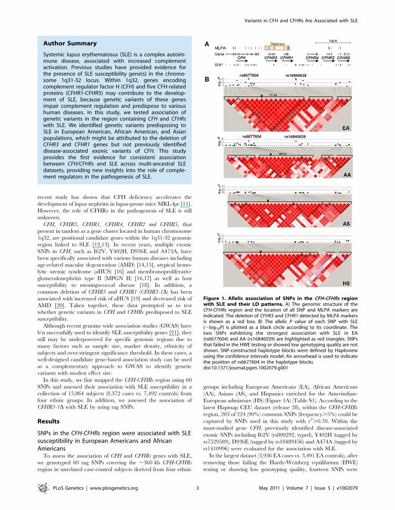
European admixture (HS) (Figure 1A) (Table S1). According to the
latest Hapmap CEU dataset (release 28), within the CFH-CFHRs
region, 203 of 224 (90%) common SNPs (frequency.5%) could be
captured by SNPs used in this study with r2.0.70. Within the
most-studied gene CFH, previously identified disease-associated
exonic SNPs including I62V (rs800292, typed), Y402H (tagged by
rs7529589), D936E (tagged by rs10489456) and A474A (tagged by
rs1410996) were evaluated for the association with SLE.
In the largest dataset (3,936 EA cases vs. 3,491 EA controls), after
removing those failing the Hardy-Weinberg equilibrium (HWE)
testing or showing low genotyping quality, fourteen SNPs were
Figure 1. Allelic association of SNPs in the CFH-CFHRs regionwith SLE and their LD patterns. A) The genomic structure of theCFH-CFHRs region and the location of all SNP and MLPA markers areindicated. The deletion of CFHR3 and CFHR1 detected by MLPA markersis shown as a red box. B) The allelic P value of each SNP with SLE(2log10P) is plotted as a black circle according to its coordinate. Thetwo SNPs exhibiting the strongest association with SLE in EA(rs6677604) and AA (rs16846039) are highlighted as red triangles. SNPsthat failed in the HWE testing or showed low genotyping quality are notshown. SNP-constructed haplotype blocks were defined by Haploviewusing the confidence intervals model. An arrowhead is used to indicatethe position of rs6677604 in the haplotype blocks.doi:10.1371/journal.pgen.1002079.g001
Author Summary
Systemic lupus erythematosus (SLE) is a complex autoim-mune disease, associated with increased complementactivation. Previous studies have provided evidence forthe presence of SLE susceptibility gene(s) in the chromo-some 1q31-32 locus. Within 1q32, genes encodingcomplement regulator factor H (CFH) and five CFH-relatedproteins (CFHR1-CFHR5) may contribute to the develop-ment of SLE, because genetic variants of these genesimpair complement regulation and predispose to varioushuman diseases. In this study, we tested association ofgenetic variants in the region containing CFH and CFHRswith SLE. We identified genetic variants predisposing toSLE in European American, African American, and Asianpopulations, which might be attributed to the deletion ofCFHR3 and CFHR1 genes but not previously identifieddisease-associated exonic variants of CFH. This studyprovides the first evidence for consistent associationbetween CFH/CFHRs and SLE across multi-ancestral SLEdatasets, providing new insights into the role of comple-ment regulators in the pathogenesis of SLE.
Variants in CFH and CFHRs Are Associated with SLE
PLoS Genetics | www.plosgenetics.org 3 May 2011 | Volume 7 | Issue 5 | e1002079
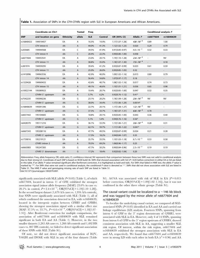
significantly associated with SLE (allelic P,0.05) (Table 1), of which
rs6677604, located in intron 11 of CFH, exhibited the strongest
association signal (minor allele frequency [MAF]: 23.0% in case vs.
20.1% in control, P = 2.461025, OR[95%CI] = 1.19[1.10–1.28]).
In the second largest dataset (1,679 AA cases vs. 1,934 AA controls),
four SNPs were significantly associated with SLE (Table 1), all of
which confirmed the association detected in EA, with rs16840639,
located in the intergenic region between CFHR1 and CFHR4,
showing the strongest association signal with a similar effect size
(MAF: 37.5% vs. 33.7%, P = 6.661024, OR[95%CI] = 1.18[1.07–
1.31]). After Bonferroni correction for multiple comparisons, the
association of rs6677604 and rs16840639 with SLE remained
significant in both EA and AA (Table 1). However, in the two
smaller datasets (1,265 AS cases vs. 1,260 AS controls and 1,492 HS
cases vs. 807 HS controls), we failed to detect significant association
of these SNPs with SLE (Table S1).
Of note, we did not detect significant association of I62V,
Y402H and D936E with SLE in any of the four datasets (Table
S1). A474A was associated with risk of SLE in EA (P = 0.015
before correction, OR[95%CI] = 1.09[1.02–1.16]), but it was not
confirmed in the other three ethnic groups (Table S1).
The causal variant could be localized to a ,146 kb blockand was tagged by the minor allele of rs6677604 andrs16840639
To localize the underlying causal variant, we compared all SLE-
associated SNPs (P,0.05) identified in EA and AA and carried out
linkage equilibrium (LD) analysis. Fourteen SNPs, spanning from
intron 6 of CFH to the 39 region downstream of CFHR5, were
associated with SLE in EA. However, only 4 of 14 SNPs, spanning
from intron 6 of CFH to the 59 region upstream of CFHR4, showed
consistent association with SLE in AA, suggesting a smaller SLE
risk region. Of interest, within the risk region, rs6677604 and
rs16840639 exhibited the strongest association with SLE in EA
and AA, respectively. We found that rs6677604 and rs16840639
were in strong LD with each other in both EA (r2 = 0.96) and AA
Table 1. Association of SNPs in the CFH-CFHRs region with SLE in European Americans and African Americans.
Coordinate on Chr1 Tested Freq Conditional analysis P
SNP and location on gene Ethnicity allele SLE Control OR [95% CI] Allelic P rs6677604 rs16840639
rs16840422 194919457 EA A 16.5% 14.4% 1.17[1.07–1.28] 4.861024 0.89 1.00
CFH intron 6 AA A 44.0% 41.3% 1.12[1.02–1.23] 0.020 0.29 0.79
rs203685 194944568 EA C 39.5% 41.9% 0.91[0.85–0.97] 4.061023 0.32 0.33
CFH intron 9 AA C 20.4% 22.3% 0.90[0.80–1.00] 0.058 - -
rs6677604 194953541 EA A 23.0% 20.1% 1.19[1.10–1.28] 2.461025 - NV
CFH intron 11 AA A 38.8% 35.0% 1.18[1.07–1.30] 7.561024 - 0.18
rs381974 194959295 EA A 39.4% 41.2% 0.93[0.87–0.99] 0.025 0.61 0.59
CFH intron 11 AA A 25.0% 26.0% 0.95[0.85–1.05] 0.32 - -
rs1410996 194963556 EA A 42.9% 40.9% 1.09[1.02–1.16] 0.015 0.80 0.79
CFH intron 14 AA A 56.4% 54.8% 1.07[0.97–1.17] 0.18 - -
rs1329428 194969433 EA A 42.6% 40.7% 1.08[1.02–1.16] 0.017 0.74 0.73
CFH intron 15 AA A 49.1% 46.6% 1.10[1.01–1.21] 0.036 0.65 0.98
rs10922144 195089923 EA A 19.4% 20.7% 0.92[0.85–1.00] 0.047 0.32 0.35
CFHR4 59 upstream AA A 5.7% 6.2% 0.92[0.76–1.12] 0.41* - -
rs7542235 195090236 EA G 23.1% 20.2% 1.18[1.09–1.28] 2.661025 NV NV
CFHR4 59 upstream AA G 38.0% 34.4% 1.17[1.06–1.29] 0.0014* - -
rs16840639 195091396 EA G 22.7% 20.1% 1.17[1.08–1.27] 1.261024 NV -
CFHR4 59 upstream AA G 37.5% 33.7% 1.18[1.07–1.31] 6.661024 0.78 -
rs6657442 195104683 EA G 18.8% 20.1% 0.92[0.85–1.00] 0.043 0.36 0.40
CFHR4 59 upstream AA G 5.1% 5.4% 0.94[0.76–1.16] 0.56* - -
rs6428370 195111216 EA G 36.1% 33.3% 1.13[1.06–1.21] 3.661024 0.28 0.31
CFHR4 59 upstream AA G 62.1% 59.7% 1.11[1.00–1.22] 0.041* - -
rs6667243 195208116 EA A 47.7% 49.5% 0.93[0.87–0.99] 0.034 0.21 0.28
CFHR5 59 upstream AA A 17.3% 18.2% 0.94[0.84–1.07] 0.35 - -
rs1759016 195219121 EA A 35.7% 33.5% 1.10[1.03–1.18] 6.761023 0.53 0.58
CFHR5 intron 2 AA A 70.5% 69.2% 1.06[0.96–1.17] 0.25 - -
rs6663083 195247283 EA G 47.7% 50.2% 0.90[0.84–0.96] 2.361023 0.19 0.19
CFHR5 39 downstream AA G 17.2% 18.4% 0.92[0.82–1.04] 0.20 - -
Abbreviation: Freq, allele frequency; OR, odds ratio; CI, confidence interval; NV represents that comparison between these two SNPs was not valid in conditional analysisdue to their strong LD. Coordinate of each SNP is based on NCBI build 36. SNPs that showed association with SLE (P,0.05 before correction) in either EA or AA are listedin this table. If an allelic P value remained significant after Bonferroni correction, it is highlighted as bold and italic. For SNPs that failed in HWE test, the allelic P value ismarked by ‘‘*’’. For SNPs that were not used in conditional analysis, the conditional P value is denoted as ‘‘-’’. SNPs that did not show association with SLE are listed inTable S1. The HWE P value and genotyping missing rate of each SNP are listed in Table S1.doi:10.1371/journal.pgen.1002079.t001
Variants in CFH and CFHRs Are Associated with SLE
PLoS Genetics | www.plosgenetics.org 4 May 2011 | Volume 7 | Issue 5 | e1002079
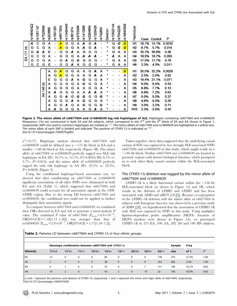
(r2 = 0.77). Haplotype analysis showed that rs6677604 and
rs16840639 could be defined into a ,171 kb block in EA and a
smaller ,146 kb block in AA, respectively (Figure 1B). The minor
allele of rs6677604 or rs16840639 perfectly tagged two SLE risk
haplotypes in EA (H1: 16.1% vs. 14.1%, P = 0.0010; H2: 6.7% vs.
5.7%, P = 0.014), and the minor allele of rs16840639 perfectly
tagged the only risk haplotype in AA (H1: 35.5% vs. 32.2%,
P = 0.0028) (Figure 2).
Using the conditional haplotype-based association test, we
showed that after conditioning on rs6677604 or rs16840639
significant associations of all other SNPs were eliminated in both
EA and AA (Table 1), which suggested that rs6677604 and
rs16840639 could account for all association signals in the CFH-
CFHRs region. Due to the strong LD between rs6677604 and
rs16840639, the conditional test could not be applied to further
distinguish their association signals.
To compare between rs6677604 and rs16840639, we combined
their ORs detected in EA and AA to generate a meta-analysis P
value. The combined P value of rs6677604 (Pmeta = 6.661028,
OR[95%CI] = 1.18[1.11–1.26]) was stronger than that of
rs16840639 (Pmeta = 2.961027, OR[95%CI] = 1.17[1.10–1.2]).
Taken together, these data suggested that the underlying causal
variant of SLE was captured by two strongly SLE-associated SNPs
rs6677604 and rs16840639 in this study, which might reside in a
,146 kb block. Neither rs6677604 nor rs16840639 are located in
genomic regions with known biological function, which prompted
us to seek other likely causal variants within the SLE-associated
block.
The CFHR3-1D deletion was tagged by the minor allele ofrs6677604 and rs16840639
CFHR3-1D is a likely functional variant within the ,146 kb
SLE-associated block (as shown in Figure 1A and 1B), which
results in the deletion of CFHR3 and CFHR1 and has been
associated with AMD and aHUS [19,20]. Because co-segregation
of the CFHR3-1D deletion with the minor allele of rs6677604 in
subjects with European Ancestry was observed in a previous study
of AMD [20], we hypothesized that the association of CFHR3-1Dwith SLE was captured by SNPs in this study. Using multiplex
ligation-dependent probe amplification (MLPA) (location of
MLPA markers were shown in Figure 1A), we genotyped
CFHR3-1D in 275 EA, 106 AA, 282 AS and 196 HS subjects,
Figure 2. The minor allele of rs6677604 and rs16846039 tag risk haplotypes of SLE. Haplotypes containing rs6677604 and rs16846039(frequency.1%) are constructed in both EA and AA subjects, which correspond to the 2nd and the 4th block of EA and AA shown in Figure 1,respectively. SNPs not used to construct haplotypes are marked as ‘‘-’’. The minor alleles of rs6677604 and rs16840639 are highlighted in a yellow box.The minor allele of each SNP is bolded and italicized. The position of CFHR3-1D is indicated as ‘‘*’’.doi:10.1371/journal.pgen.1002079.g002
Table 2. Pairwise LD between rs6677604 and CFHR3-1D in four ethnic groups.
Genotype combination between rs6677604 and CFHR3-1D Sample Freq
Ethnicity 11/DD 11/D+ 11/++ 12/DD 12/D+ 12/++ 22/DD 22/D+ 22/++ size of D r2
EA 12 0 0 0 85 0 0 0 178 275 21.2% 1.00
AS 2 0 0 0 28 0 0 0 252 282 5.3% 1.00
HS 5 1 0 0 52 0 0 7 131 196 16.1% 0.85
AA 10 2 0 7 43 2 0 10 32 106 42.0% 0.60
D and + represent the presence and absence of CFHR3-1D, respectively. 1 and 2 represent the minor and major allele of rs6677604, respectively.doi:10.1371/journal.pgen.1002079.t002
Variants in CFH and CFHRs Are Associated with SLE
PLoS Genetics | www.plosgenetics.org 5 May 2011 | Volume 7 | Issue 5 | e1002079
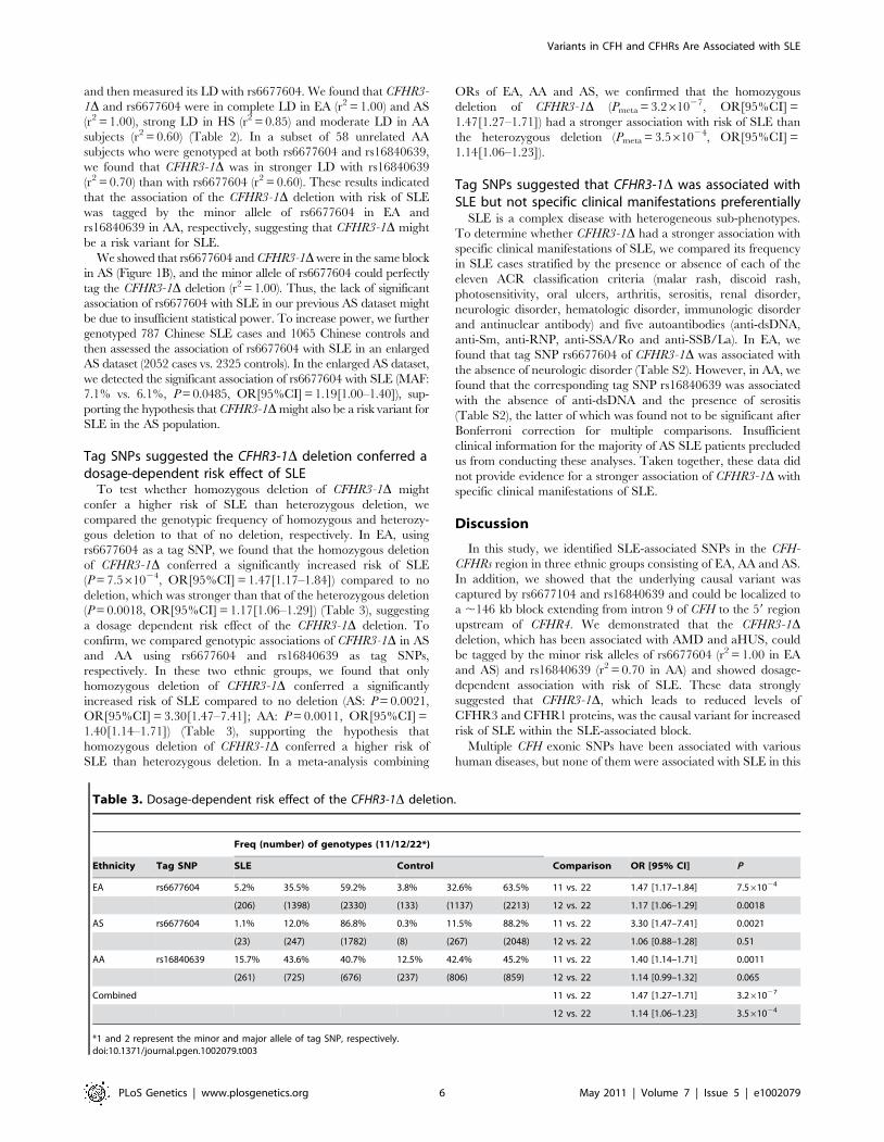
and then measured its LD with rs6677604. We found that CFHR3-
1D and rs6677604 were in complete LD in EA (r2 = 1.00) and AS
(r2 = 1.00), strong LD in HS (r2 = 0.85) and moderate LD in AA
subjects (r2 = 0.60) (Table 2). In a subset of 58 unrelated AA
subjects who were genotyped at both rs6677604 and rs16840639,
we found that CFHR3-1D was in stronger LD with rs16840639
(r2 = 0.70) than with rs6677604 (r2 = 0.60). These results indicated
that the association of the CFHR3-1D deletion with risk of SLE
was tagged by the minor allele of rs6677604 in EA and
rs16840639 in AA, respectively, suggesting that CFHR3-1D might
be a risk variant for SLE.
We showed that rs6677604 and CFHR3-1D were in the same block
in AS (Figure 1B), and the minor allele of rs6677604 could perfectly
tag the CFHR3-1D deletion (r2 = 1.00). Thus, the lack of significant
association of rs6677604 with SLE in our previous AS dataset might
be due to insufficient statistical power. To increase power, we further
genotyped 787 Chinese SLE cases and 1065 Chinese controls and
then assessed the association of rs6677604 with SLE in an enlarged
AS dataset (2052 cases vs. 2325 controls). In the enlarged AS dataset,
we detected the significant association of rs6677604 with SLE (MAF:
7.1% vs. 6.1%, P = 0.0485, OR[95%CI] = 1.19[1.00–1.40]), sup-
porting the hypothesis that CFHR3-1D might also be a risk variant for
SLE in the AS population.
Tag SNPs suggested the CFHR3-1D deletion conferred adosage-dependent risk effect of SLE
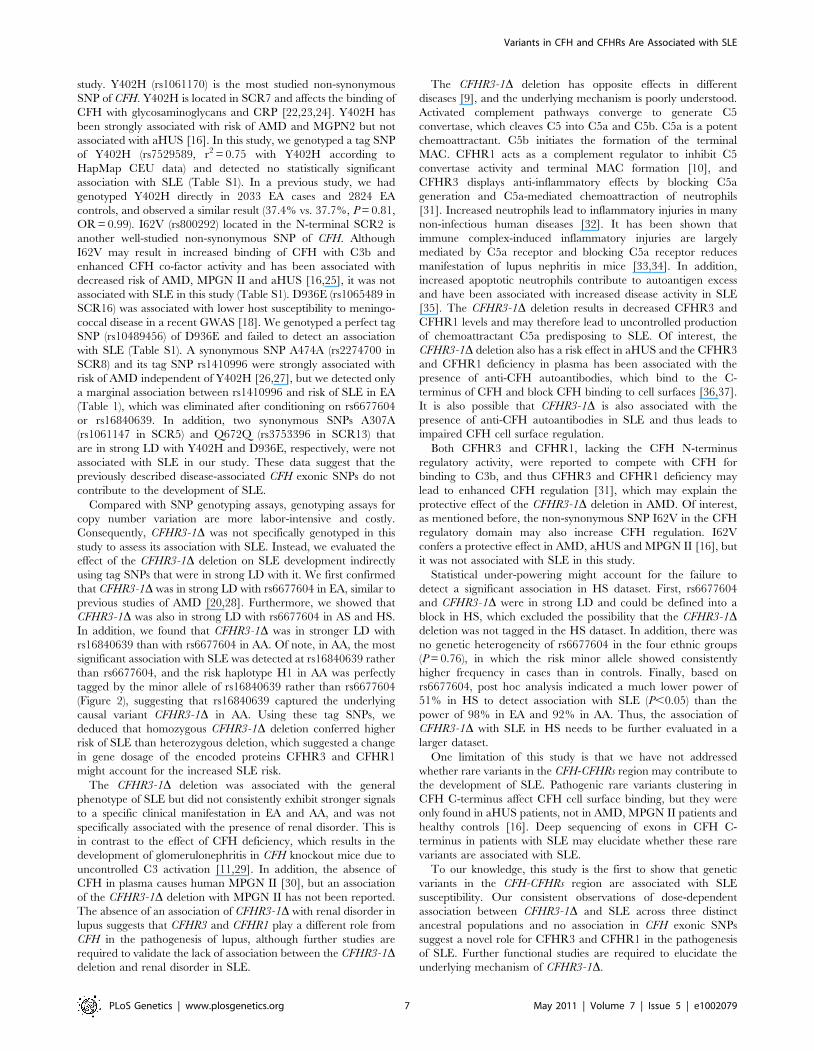
To test whether homozygous deletion of CFHR3-1D might
confer a higher risk of SLE than heterozygous deletion, we
compared the genotypic frequency of homozygous and heterozy-
gous deletion to that of no deletion, respectively. In EA, using
rs6677604 as a tag SNP, we found that the homozygous deletion
of CFHR3-1D conferred a significantly increased risk of SLE
(P = 7.561024, OR[95%CI] = 1.47[1.17–1.84]) compared to no
deletion, which was stronger than that of the heterozygous deletion
(P = 0.0018, OR[95%CI] = 1.17[1.06–1.29]) (Table 3), suggesting
a dosage dependent risk effect of the CFHR3-1D deletion. To
confirm, we compared genotypic associations of CFHR3-1D in AS
and AA using rs6677604 and rs16840639 as tag SNPs,
respectively. In these two ethnic groups, we found that only
homozygous deletion of CFHR3-1D conferred a significantly
increased risk of SLE compared to no deletion (AS: P = 0.0021,
OR[95%CI] = 3.30[1.47–7.41]; AA: P = 0.0011, OR[95%CI] =
1.40[1.14–1.71]) (Table 3), supporting the hypothesis that
homozygous deletion of CFHR3-1D conferred a higher risk of
SLE than heterozygous deletion. In a meta-analysis combining
ORs of EA, AA and AS, we confirmed that the homozygous
deletion of CFHR3-1D (Pmeta = 3.261027, OR[95%CI] =
1.47[1.27–1.71]) had a stronger association with risk of SLE than
the heterozygous deletion (Pmeta = 3.561024, OR[95%CI] =
1.14[1.06–1.23]).
Tag SNPs suggested that CFHR3-1D was associated withSLE but not specific clinical manifestations preferentially
SLE is a complex disease with heterogeneous sub-phenotypes.
To determine whether CFHR3-1D had a stronger association with
specific clinical manifestations of SLE, we compared its frequency
in SLE cases stratified by the presence or absence of each of the
eleven ACR classification criteria (malar rash, discoid rash,
photosensitivity, oral ulcers, arthritis, serositis, renal disorder,
neurologic disorder, hematologic disorder, immunologic disorder
and antinuclear antibody) and five autoantibodies (anti-dsDNA,
anti-Sm, anti-RNP, anti-SSA/Ro and anti-SSB/La). In EA, we
found that tag SNP rs6677604 of CFHR3-1D was associated with
the absence of neurologic disorder (Table S2). However, in AA, we
found that the corresponding tag SNP rs16840639 was associated
with the absence of anti-dsDNA and the presence of serositis
(Table S2), the latter of which was found not to be significant after
Bonferroni correction for multiple comparisons. Insufficient
clinical information for the majority of AS SLE patients precluded
us from conducting these analyses. Taken together, these data did
not provide evidence for a stronger association of CFHR3-1D with
specific clinical manifestations of SLE.
Discussion
In this study, we identified SLE-associated SNPs in the CFH-
CFHRs region in three ethnic groups consisting of EA, AA and AS.
In addition, we showed that the underlying causal variant was
captured by rs6677104 and rs16840639 and could be localized to
a ,146 kb block extending from intron 9 of CFH to the 59 region
upstream of CFHR4. We demonstrated that the CFHR3-1Ddeletion, which has been associated with AMD and aHUS, could
be tagged by the minor risk alleles of rs6677604 (r2 = 1.00 in EA
and AS) and rs16840639 (r2 = 0.70 in AA) and showed dosage-
dependent association with risk of SLE. These data strongly
suggested that CFHR3-1D, which leads to reduced levels of
CFHR3 and CFHR1 proteins, was the causal variant for increased
risk of SLE within the SLE-associated block.
Multiple CFH exonic SNPs have been associated with various
human diseases, but none of them were associated with SLE in this
Table 3. Dosage-dependent risk effect of the CFHR3-1D deletion.
Freq (number) of genotypes (11/12/22*)
Ethnicity Tag SNP SLE Control Comparison OR [95% CI] P
EA rs6677604 5.2% 35.5% 59.2% 3.8% 32.6% 63.5% 11 vs. 22 1.47 [1.17–1.84] 7.561024
(206) (1398) (2330) (133) (1137) (2213) 12 vs. 22 1.17 [1.06–1.29] 0.0018
AS rs6677604 1.1% 12.0% 86.8% 0.3% 11.5% 88.2% 11 vs. 22 3.30 [1.47–7.41] 0.0021
(23) (247) (1782) (8) (267) (2048) 12 vs. 22 1.06 [0.88–1.28] 0.51
AA rs16840639 15.7% 43.6% 40.7% 12.5% 42.4% 45.2% 11 vs. 22 1.40 [1.14–1.71] 0.0011
(261) (725) (676) (237) (806) (859) 12 vs. 22 1.14 [0.99–1.32] 0.065
Combined 11 vs. 22 1.47 [1.27–1.71] 3.261027
12 vs. 22 1.14 [1.06–1.23] 3.561024
*1 and 2 represent the minor and major allele of tag SNP, respectively.doi:10.1371/journal.pgen.1002079.t003
Variants in CFH and CFHRs Are Associated with SLE
PLoS Genetics | www.plosgenetics.org 6 May 2011 | Volume 7 | Issue 5 | e1002079
study. Y402H (rs1061170) is the most studied non-synonymous
SNP of CFH. Y402H is located in SCR7 and affects the binding of
CFH with glycosaminoglycans and CRP [22,23,24]. Y402H has
been strongly associated with risk of AMD and MGPN2 but not
associated with aHUS [16]. In this study, we genotyped a tag SNP
of Y402H (rs7529589, r2 = 0.75 with Y402H according to
HapMap CEU data) and detected no statistically significant
association with SLE (Table S1). In a previous study, we had
genotyped Y402H directly in 2033 EA cases and 2824 EA
controls, and observed a similar result (37.4% vs. 37.7%, P = 0.81,
OR = 0.99). I62V (rs800292) located in the N-terminal SCR2 is
another well-studied non-synonymous SNP of CFH. Although
I62V may result in increased binding of CFH with C3b and
enhanced CFH co-factor activity and has been associated with
decreased risk of AMD, MPGN II and aHUS [16,25], it was not
associated with SLE in this study (Table S1). D936E (rs1065489 in
SCR16) was associated with lower host susceptibility to meningo-
coccal disease in a recent GWAS [18]. We genotyped a perfect tag
SNP (rs10489456) of D936E and failed to detect an association
with SLE (Table S1). A synonymous SNP A474A (rs2274700 in
SCR8) and its tag SNP rs1410996 were strongly associated with
risk of AMD independent of Y402H [26,27], but we detected only
a marginal association between rs1410996 and risk of SLE in EA
(Table 1), which was eliminated after conditioning on rs6677604
or rs16840639. In addition, two synonymous SNPs A307A
(rs1061147 in SCR5) and Q672Q (rs3753396 in SCR13) that
are in strong LD with Y402H and D936E, respectively, were not
associated with SLE in our study. These data suggest that the
previously described disease-associated CFH exonic SNPs do not
contribute to the development of SLE.
Compared with SNP genotyping assays, genotyping assays for
copy number variation are more labor-intensive and costly.
Consequently, CFHR3-1D was not specifically genotyped in this
study to assess its association with SLE. Instead, we evaluated the
effect of the CFHR3-1D deletion on SLE development indirectly
using tag SNPs that were in strong LD with it. We first confirmed
that CFHR3-1D was in strong LD with rs6677604 in EA, similar to
previous studies of AMD [20,28]. Furthermore, we showed that
CFHR3-1D was also in strong LD with rs6677604 in AS and HS.
In addition, we found that CFHR3-1D was in stronger LD with
rs16840639 than with rs6677604 in AA. Of note, in AA, the most
significant association with SLE was detected at rs16840639 rather
than rs6677604, and the risk haplotype H1 in AA was perfectly
tagged by the minor allele of rs16840639 rather than rs6677604
(Figure 2), suggesting that rs16840639 captured the underlying
causal variant CFHR3-1D in AA. Using these tag SNPs, we
deduced that homozygous CFHR3-1D deletion conferred higher
risk of SLE than heterozygous deletion, which suggested a change
in gene dosage of the encoded proteins CFHR3 and CFHR1
might account for the increased SLE risk.
The CFHR3-1D deletion was associated with the general
phenotype of SLE but did not consistently exhibit stronger signals
to a specific clinical manifestation in EA and AA, and was not
specifically associated with the presence of renal disorder. This is
in contrast to the effect of CFH deficiency, which results in the
development of glomerulonephritis in CFH knockout mice due to
uncontrolled C3 activation [11,29]. In addition, the absence of
CFH in plasma causes human MPGN II [30], but an association
of the CFHR3-1D deletion with MPGN II has not been reported.
The absence of an association of CFHR3-1D with renal disorder in
lupus suggests that CFHR3 and CFHR1 play a different role from
CFH in the pathogenesis of lupus, although further studies are
required to validate the lack of association between the CFHR3-1Ddeletion and renal disorder in SLE.
The CFHR3-1D deletion has opposite effects in different
diseases [9], and the underlying mechanism is poorly understood.
Activated complement pathways converge to generate C5
convertase, which cleaves C5 into C5a and C5b. C5a is a potent
chemoattractant. C5b initiates the formation of the terminal
MAC. CFHR1 acts as a complement regulator to inhibit C5
convertase activity and terminal MAC formation [10], and
CFHR3 displays anti-inflammatory effects by blocking C5a
generation and C5a-mediated chemoattraction of neutrophils
[31]. Increased neutrophils lead to inflammatory injuries in many
non-infectious human diseases [32]. It has been shown that
immune complex-induced inflammatory injuries are largely
mediated by C5a receptor and blocking C5a receptor reduces
manifestation of lupus nephritis in mice [33,34]. In addition,
increased apoptotic neutrophils contribute to autoantigen excess
and have been associated with increased disease activity in SLE
[35]. The CFHR3-1D deletion results in decreased CFHR3 and
CFHR1 levels and may therefore lead to uncontrolled production
of chemoattractant C5a predisposing to SLE. Of interest, the
CFHR3-1D deletion also has a risk effect in aHUS and the CFHR3
and CFHR1 deficiency in plasma has been associated with the
presence of anti-CFH autoantibodies, which bind to the C-
terminus of CFH and block CFH binding to cell surfaces [36,37].
It is also possible that CFHR3-1D is also associated with the
presence of anti-CFH autoantibodies in SLE and thus leads to
impaired CFH cell surface regulation.
Both CFHR3 and CFHR1, lacking the CFH N-terminus
regulatory activity, were reported to compete with CFH for
binding to C3b, and thus CFHR3 and CFHR1 deficiency may
lead to enhanced CFH regulation [31], which may explain the
protective effect of the CFHR3-1D deletion in AMD. Of interest,
as mentioned before, the non-synonymous SNP I62V in the CFH
regulatory domain may also increase CFH regulation. I62V
confers a protective effect in AMD, aHUS and MPGN II [16], but
it was not associated with SLE in this study.
Statistical under-powering might account for the failure to
detect a significant association in HS dataset. First, rs6677604
and CFHR3-1D were in strong LD and could be defined into a
block in HS, which excluded the possibility that the CFHR3-1Ddeletion was not tagged in the HS dataset. In addition, there was
no genetic heterogeneity of rs6677604 in the four ethnic groups
(P = 0.76), in which the risk minor allele showed consistently
higher frequency in cases than in controls. Finally, based on
rs6677604, post hoc analysis indicated a much lower power of
51% in HS to detect association with SLE (P,0.05) than the
power of 98% in EA and 92% in AA. Thus, the association of
CFHR3-1D with SLE in HS needs to be further evaluated in a
larger dataset.
One limitation of this study is that we have not addressed
whether rare variants in the CFH-CFHRs region may contribute to
the development of SLE. Pathogenic rare variants clustering in
CFH C-terminus affect CFH cell surface binding, but they were
only found in aHUS patients, not in AMD, MPGN II patients and
healthy controls [16]. Deep sequencing of exons in CFH C-
terminus in patients with SLE may elucidate whether these rare
variants are associated with SLE.
To our knowledge, this study is the first to show that genetic
variants in the CFH-CFHRs region are associated with SLE
susceptibility. Our consistent observations of dose-dependent
association between CFHR3-1D and SLE across three distinct
ancestral populations and no association in CFH exonic SNPs
suggest a novel role for CFHR3 and CFHR1 in the pathogenesis
of SLE. Further functional studies are required to elucidate the
underlying mechanism of CFHR3-1D.
Variants in CFH and CFHRs Are Associated with SLE
PLoS Genetics | www.plosgenetics.org 7 May 2011 | Volume 7 | Issue 5 | e1002079
Materials and Methods
Ethics statementThe study was approved by the Human Subject Institutional
Review Boards or the ethnic committees of each institution. All
subjects were enrolled after informed consent had been obtained.
Subject collectionTo test the association of CFH and CFHRs with SLE, we used a
large collection of samples from case-control subjects from
multiple ethnic groups. These samples were from the collabora-
tive Large Lupus Association Study 2 (LLAS2) and were
contributed by participating institutions in the United States,
Asia and Europe. According to genetic ancestry, subjects were
grouped into four ethnic groups including European American
(3,936 cases vs. 3,491 controls), African American (1,679 cases vs.
1,934 controls), Asian (1,265 cases vs. 1,260 controls) and
Hispanic enriched for the Amerindian-European admixture
(1,492 cases vs. 807 controls). Asians were comprised of Koreans
(884 cases vs. 994 controls), Chinese (200 cases vs. 205 controls)
and subjects from other East Asian countries such as Japan and
Singapore (181 cases vs. 61 controls). African Americans included
275 Gullahs (152 cases vs. 123 controls), who are subjects with
African Ancestry.
To test LD between CFHR3-1D and SLE-associated SNPs, we
used 275 unrelated European Americans (187 cases vs. 88
controls), 106 African Americans (88 unrelated subjects [58 cases
vs. 30 controls] and 18 subjects from 6 SLE trios families), 282
unrelated Chinese (218 cases vs. 64 controls) and 196 Hispanics
(157 unrelated subjects [91 cases vs. 66 controls] and 39 subjects
from 13 SLE trios families). All of these subjects were enrolled
from UCLA.
To enlarge the sample size of Asians for association test, we used
1,852 Chinese case-control subjects (787 vs. 1065) recruited from
Shanghai Renji Hospital, Shanghai Jiao Tong University School
of Medicine.
All SLE patients met the American College of Rheumatology
(ACR) criteria for the classification of SLE [38].
SNP genotyping and data cleaningLLAS2 samples were processed at the Lupus Genetics Studies
Unit of the Oklahoma Medical Research Foundation (OMRF).
SNP genotyping was carried out on the Illumina iSelect platform.
Subjects with individual genotyping call rate ,0.90 were removed
because of low data quality. Subjects that were duplicated or first
degree related were also removed. Both principal component
analysis and global ancestry estimation based on 347 ancestry
informative markers were used to detect population stratification
and admixture, as described in another LLAS2 report [39]. After
removing genetic outliers, a final dataset of 15,864 unrelated
subjects (8,372 cases vs. 7,492 controls) was obtained.
Taqman SNP genotyping assay (Applied Biosystems, California,
USA) was used to genotype rs6677604 for subjects who were not
recruited into LLAS2.
MLPA genotypingMLPA kit ‘‘SALSA MLPA KIT P236-A1 ARMD mix-1’’ was
used to genotype the CFH-CFHRs region according to the
manufacture’s instruction (MRC-Holland, Amsterdam, The
Netherlands). ABI 3730 Genetic Analyzer (Applied Biosystems)
was used to run gel electrophoresis. Software Peak Scanner v1.0
(Applied Biosystems) was used to extract peaks generated in
electrophoresis. Coffalyser v9.4 (MRC-Holland) was used to
readout copy number of target region.
Statistical analysisThe HWE test threshold was set at P.0.01 for controls and
P.0.0001 for cases. SNPs failing the HWE test were excluded
from association test. SNPs showing genotyping missing rate.5%
or showing significantly different genotyping missing rate between
cases and controls (missing rate.2% and Pmissing,0.05) were also
excluded from association test. In allelic association test (Pearson’s
x2–test), the significance level was set at P,0.05. Haploview 4.2
was used to estimate pairwise LD values between SNPs, define
haplotypes blocks and calculate haplotypic association with SLE.
Haplotype-based conditional association analysis was carried out
by Plink v1.07. Mantel-Haenszel analysis was performed to
generate the meta-analysis P value. CaTS was used to calculate
statistical power.
Supporting Information
Table S1 Allelic association between 60 tested SNPs and SLE in
all four ethnic groups.
(XLS)
Table S2 Association of CFHR3-1D with clinical manifestations
of SLE.
(XLS)
Acknowledgments
We thank the study participants and physicians who provided samples
(Peter K. Gregersen, BIOLUPUS Network and GENLES Network). The
members of BIOLUPUS Network are Sandra D’Alfonso in Italy; Bernard
R. Lauwerys in Belgium; Emoke Endreffy and Laszlo Kovacs in Hungary;
Carlos Vasconcelos and Berta Martins da Silva in Portugal; Inigo Rua
Figueroa and Javier Martin in Spain. The members of GENLES Network
are Hugo R. Scherbarth, Pilar C. Marino, Estela L. Motta, Susana
Gamron, Cristina Drenkard, Emilia Menso, Alberto Allievi, Guillermo A.
Tate, Jose L. Presas, Simon A. Palatnik, Marcelo Abdala, Mariela
Bearzotti, Alejandro Alvarellos, Francisco Caeiro, Ana Bertoli, Sergio
Paira, Susana Roverano, Cesar E. Graf, Estela Bertero, Cesar Caprarulo,
Griselda Buchanan, Carolina Guilleron, Sebastian Grimaudo, Jorge
Manni, Luis J. Catoggio, Enrique R. Soriano, Carlos D. Santos, Cristina
Prigione, Fernando A. Ramos, Sandra M. Navarro, Guillermo A.
Berbotto, Marisa Jorfen, Elisa J. Romero, Mercedes A. Garcia, Juan C
Marcos, Ana I. Marcos, Carlos E. Perandones, Alicia Eimon, Sanatorio
Parque and Cristina G. Battagliotti in Argentina; Eduardo Acevedo and
Mariano Cucho in Peru; Ignacio Garcıa de la Torre, Mario Cardiel Rıos,
Jose Francisco Moctezuma and Marco Maradiaga Cecena in Mexico.
Author Contributions
Conceived and designed the experiments: BP Tsao, J Zhao, H Wu, C-Y
Yu, N Shen. Performed the experiments: J Zhao, H Wu, M Khosravi, H
Cui, X Qian, A Adler. Analyzed the data: J Zhao, H Wu, M Khosravi, H
Cui, X Qian, JA Kelly, KM Kaufman, CD Langefeld, AH Williams, ME
Comeau, JT Ziegler, MC Marion, SB Glenn. Contributed reagents/
materials/analysis tools: BP Tsao, N Shen, C-Y Yu, ME Alarcon-
Riquelme, BA Pons-Estel, JB Harley, S-C Bae, S-Y Bang, S-K Cho, CO
Jacob, TJ Vyse, TB Niewold, PM Gaffney, KL Moser, RP Kimberly, JC
Edberg, EE Brown, GS Alarcon, MA Petri, R Ramsey-Goldman, LM Vila,
JD Reveille, JA James, GS Gilkeson, DL Kamen, BI Freedman, J-M
Anaya, JT Merrill, LA Criswell, RH Scofield, AM Stevens, JM Guthridge,
D-M Chang, YW Song, JA Park EY Lee, SA Boackle, JM Grossman BH
Hahn, THJ Goodship, RM Cantor. Wrote the paper: J Zhao. Revised the
manuscript: BP Tsao, SA Boackle, BH Hahn, JM Grossman, C-Y Yu, EE
Brown, JD Reveille, JA James, RM Cantor.
Variants in CFH and CFHRs Are Associated with SLE
PLoS Genetics | www.plosgenetics.org 8 May 2011 | Volume 7 | Issue 5 | e1002079
References
1. Rahman A, Isenberg DA (2008) Systemic lupus erythematosus. N Engl J Med
358: 929–939.
2. Manderson AP, Botto M, Walport MJ (2004) The role of complement in the
development of systemic lupus erythematosus. Annu Rev Immunol 22: 431–456.
3. Birmingham DJ, Irshaid F, Nagaraja HN, Zou X, Tsao BP, et al. (2010) The
complex nature of serum C3 and C4 as biomarkers of lupus renal flare. Lupus
19: 1272–1280.
4. Miyagawa H, Yamai M, Sakaguchi D, Kiyohara C, Tsukamoto H, et al. (2008)
Association of polymorphisms in complement component C3 gene with
susceptibility to systemic lupus erythematosus. Rheumatology (Oxford) 47:
158–164.
5. Rhodes B, Hunnangkul S, Morris DL, Hsaio LC, Graham DS, et al. (2009) The
heritability and genetics of complement C3 expression in UK SLE families.
Genes Immun 10: 525–530.
6. Yang Y, Chung EK, Wu YL, Savelli SL, Nagaraja HN, et al. (2007) Gene copy-
number variation and associated polymorphisms of complement component C4
in human systemic lupus erythematosus (SLE): low copy number is a risk factor
for and high copy number is a protective factor against SLE susceptibility in
European Americans. Am J Hum Genet 80: 1037–1054.
7. de Cordoba SR, de Jorge EG (2008) Translational mini-review series on
complement factor H: genetics and disease associations of human complement
factor H. Clin Exp Immunol 151: 1–13.
8. Rodriguez de Cordoba S, Esparza-Gordillo J, Goicoechea de Jorge E, Lopez-
Trascasa M, Sanchez-Corral P (2004) The human complement factor H:
functional roles, genetic variations and disease associations. Mol Immunol 41:
355–367.
9. Jozsi M, Zipfel PF (2008) Factor H family proteins and human diseases. Trends
Immunol 29: 380–387.
10. Heinen S, Hartmann A, Lauer N, Wiehl U, Dahse HM, et al. (2009) Factor H-
related protein 1 (CFHR-1) inhibits complement C5 convertase activity and
terminal complex formation. Blood 114: 2439–2447.
11. Bao L, Haas M, Quigg RJ (2011) Complement factor H deficiency accelerates
development of lupus nephritis. J Am Soc Nephrol 22: 285–295.
12. Johanneson B, Lima G, von Salome J, Alarcon-Segovia D, Alarcon-
Riquelme ME (2002) A major susceptibility locus for systemic lupus
erythemathosus maps to chromosome 1q31. Am J Hum Genet 71: 1060–1071.
13. Wu H, Boackle SA, Hanvivadhanakul P, Ulgiati D, Grossman JM, et al. (2007)
Association of a common complement receptor 2 haplotype with increased risk
of systemic lupus erythematosus. Proc Natl Acad Sci U S A 104: 3961–3966.
14. Hageman GS, Anderson DH, Johnson LV, Hancox LS, Taiber AJ, et al. (2005)
A common haplotype in the complement regulatory gene factor H (HF1/CFH)
predisposes individuals to age-related macular degeneration. Proc Natl Acad
Sci U S A 102: 7227–7232.
15. Klein RJ, Zeiss C, Chew EY, Tsai JY, Sackler RS, et al. (2005) Complement
factor H polymorphism in age-related macular degeneration. Science 308:
385–389.
16. Pickering MC, de Jorge EG, Martinez-Barricarte R, Recalde S, Garcia-
Layana A, et al. (2007) Spontaneous hemolytic uremic syndrome triggered by
complement factor H lacking surface recognition domains. J Exp Med 204:
1249–1256.
17. Abrera-Abeleda MA, Nishimura C, Smith JL, Sethi S, McRae JL, et al. (2006)
Variations in the complement regulatory genes factor H (CFH) and factor H
related 5 (CFHR5) are associated with membranoproliferative glomerulone-
phritis type II (dense deposit disease). J Med Genet 43: 582–589.
18. Davila S, Wright VJ, Khor CC, Sim KS, Binder A, et al. (2010) Genome-wide
association study identifies variants in the CFH region associated with host
susceptibility to meningococcal disease. Nat Genet 42: 772–776.
19. Zipfel PF, Edey M, Heinen S, Jozsi M, Richter H, et al. (2007) Deletion of
complement factor H-related genes CFHR1 and CFHR3 is associated with
atypical hemolytic uremic syndrome. PLoS Genet 3: e41. doi:10.1371/
journal.pgen.0030041.
20. Hughes AE, Orr N, Esfandiary H, Diaz-Torres M, Goodship T, et al. (2006) Acommon CFH haplotype, with deletion of CFHR1 and CFHR3, is associated
with lower risk of age-related macular degeneration. Nat Genet 38: 1173–1177.21. Deng Y, Tsao BP (2010) Genetic susceptibility to systemic lupus erythematosus
in the genomic era. Nat Rev Rheumatol 6: 683–692.22. Clark SJ, Higman VA, Mulloy B, Perkins SJ, Lea SM, et al. (2006) His-384
allotypic variant of factor H associated with age-related macular degeneration
has different heparin binding properties from the non-disease-associated form.J Biol Chem 281: 24713–24720.
23. Prosser BE, Johnson S, Roversi P, Herbert AP, Blaum BS, et al. (2007)Structural basis for complement factor H linked age-related macular
degeneration. J Exp Med 204: 2277–2283.
24. Skerka C, Lauer N, Weinberger AA, Keilhauer CN, Suhnel J, et al. (2007)Defective complement control of factor H (Y402H) and FHL-1 in age-related
macular degeneration. Mol Immunol 44: 3398–3406.25. Tortajada A, Montes T, Martinez-Barricarte R, Morgan BP, Harris CL, et al.
(2009) The disease-protective complement factor H allotypic variant Ile62 shows
increased binding affinity for C3b and enhanced cofactor activity. Hum MolGenet 18: 3452–3461.
26. Li M, Atmaca-Sonmez P, Othman M, Branham KE, Khanna R, et al. (2006)CFH haplotypes without the Y402H coding variant show strong association with
susceptibility to age-related macular degeneration. Nat Genet 38: 1049–1054.27. Maller J, George S, Purcell S, Fagerness J, Altshuler D, et al. (2006) Common
variation in three genes, including a noncoding variant in CFH, strongly
influences risk of age-related macular degeneration. Nat Genet 38: 1055–1059.28. Spencer KL, Hauser MA, Olson LM, Schmidt S, Scott WK, et al. (2008)
Deletion of CFHR3 and CFHR1 genes in age-related macular degeneration.Hum Mol Genet 17: 971–977.
29. Pickering MC, Cook HT, Warren J, Bygrave AE, Moss J, et al. (2002)
Uncontrolled C3 activation causes membranoproliferative glomerulonephritis inmice deficient in complement factor H. Nat Genet 31: 424–428.
30. Appel GB, Cook HT, Hageman G, Jennette JC, Kashgarian M, et al. (2005)Membranoproliferative glomerulonephritis type II (dense deposit disease): an
update. J Am Soc Nephrol 16: 1392–1403.31. Fritsche LG, Lauer N, Hartmann A, Stippa S, Keilhauer CN, et al. (2010) An
imbalance of human complement regulatory proteins CFHR1, CFHR3 and
factor H influences risk for age-related macular degeneration (AMD). Hum MolGenet 19: 4694–4704.
32. Dallegri F, Ottonello L (1997) Tissue injury in neutrophilic inflammation.Inflamm Res 46: 382–391.
33. Bao L, Osawe I, Puri T, Lambris JD, Haas M, et al. (2005) C5a promotes
development of experimental lupus nephritis which can be blocked with aspecific receptor antagonist. Eur J Immunol 35: 2496–2506.
34. Baumann U, Chouchakova N, Gewecke B, Kohl J, Carroll MC, et al. (2001)Distinct tissue site-specific requirements of mast cells and complement
components C3/C5a receptor in IgG immune complex-induced injury of skinand lung. J Immunol 167: 1022–1027.
35. Courtney PA, Crockard AD, Williamson K, Irvine AE, Kennedy RJ, et al.
(1999) Increased apoptotic peripheral blood neutrophils in systemic lupuserythematosus: relations with disease activity, antibodies to double stranded
DNA, and neutropenia. Ann Rheum Dis 58: 309–314.36. Dragon-Durey MA, Loirat C, Cloarec S, Macher MA, Blouin J, et al. (2005)
Anti-Factor H autoantibodies associated with atypical hemolytic uremic
syndrome. J Am Soc Nephrol 16: 555–563.37. Jozsi M, Strobel S, Dahse HM, Liu WS, Hoyer PF, et al. (2007) Anti factor H
autoantibodies block C-terminal recognition function of factor H in hemolyticuremic syndrome. Blood 110: 1516–1518.
38. Hochberg MC (1997) Updating the American College of Rheumatology revisedcriteria for the classification of systemic lupus erythematosus. Arthritis Rheum
40: 1725.
39. Lessard CJ, Adrianto I, Kelly JA, Kaufman KM, Grundahl KM, et al. (2011)Identification of a systemic lupus erythematosus susceptibility locus at 11p13
between PDHX and CD44 in a multiethnic study. Am J Hum Genet 88: 83–91.
Variants in CFH and CFHRs Are Associated with SLE
PLoS Genetics | www.plosgenetics.org 9 May 2011 | Volume 7 | Issue 5 | e1002079