aromatic interactions define the binding of the alphavirus spike to its nucleocapsid
TRANSCRIPT
Aromatic interactions define the binding of the alphavirus spiketo its nucleocapsidUlrica Skoging, Mauno Vihinen†, Lennart Nilsson and Peter Liljeström*
Background: Most enveloped viruses bud from infected cells by a process inwhich viral intracellular core components interact with cytoplasmic domains oftransmembrane spike glycoproteins. We have demonstrated previously that atyrosine motif in the cytoplasmic domain of the Semliki Forest virus (SFV) spikeglycoprotein E2 is absolutely essential for budding. In contrast, hardly anything isknown regarding which region of the capsid protein is involved in spike binding.Therefore, the mechanism by which spikes are selectively sorted into the viralbud or by which energy is provided for envelopment, remains unclear.
Results: Molecular models of the SFV capsid protein (SFCP) and thecytoplasmic domain of the spike protein were fitted as a basis for a reversegenetics approach to characterizing the interaction between these two proteins.Biochemical analysis of mutants defined a hydrophobic pocket of the capsidprotein that is involved both in spike binding and nucleocapsid assembly.
Conclusions: We suggest that aromatic residues in the capsid protein serve tobind the side chain of the essential E2 tyrosine providing both specificity forspike incorporation and energy for budding. The same hydrophobic pocket alsoappears to play a role in capsid assembly. Furthermore, the results suggest thatbudding may occur in the absence of preformed nucleocapsids. This is the firstdemonstration of the molecular mechanisms of spike–nucleocapsid interactionsduring virus budding.
IntroductionMost enveloped viruses are released from infected cells bybudding at the plasma membrane by a process in whichviral intracellular core components are thought to interactwith cytoplasmic domains of transmembrane spike glyco-proteins [1]. Although retroviruses may form virus-likeparticles in the absence of viral spikes [2,3], both core andspike are believed to be required for budding of all otherviruses. However, only in the cases of alphaviruses [4] andhepadnaviruses [5] has this been directly shown. Forinfluenza virus, the neuraminidase protein [6] or the cyto-plasmic tail of hemagglutinin [7] (both are glycoproteinsassociated with the viral envelope) are dispensable forbudding, although the process is elevated in the presenceof hemagglutinin [7]. For vesicular stomatitis virus, it hasbeen suggested that the cytoplasmic domain of the G glyco-protein is required for budding [8,9], although furtherobservations have indicated that budding may occur bythe sole action of the membrane-associated M protein[10–12]. Inclusion of spike proteins in the viral envelope isessential for making any virus infectious, but virtuallynothing is known about how spikes are sorted into theviral bud or how they may bind to core proteins.
The assembly of alphaviruses has been extensivelystudied, and their biology has recently been reviewed
[13]. The alphavirus particle is composed of a nucleo-capsid (NC) surrounded by an envelope containing twotransmembrane spike glycoproteins, E1 and E2. The viralstructural proteins are made from a polyprotein precursorin the order C, E2, 6K and E1. The capsid protein (C)contains a serine protease activity which is responsible forthe autoproteolytical cleavage of the protein from thenascent chain. The remaining polypeptide is translocatedinto the endoplasmic reticulum where signal peptidasecleavages generate the individual transmembrane pro-teins. These are subsequently transported to the cellsurface where budding occurs. The E1 protein, whichcarries the fusion function of the spike, has a very shortcytoplasmic domain of two arginine residues, which can beremoved without affecting budding [14]. The 6K proteinis dispensable for budding and virus maturation, but itsremoval slows down virus release [15,16]. It has been sug-gested that the E2 protein interacts with the capsidprotein during budding via its 31-residue-long cytoplasmictail [17,18]. While the distal half of the tail functions as a signal peptide for the membrane translocation of thefollowing 6K polypeptide [19], mutations in the proxi-mal part disturb budding [20–23]. It has been proposedthat a pentapeptide repeat in this region is important for budding, with a single conserved tyrosine residue(Tyr399) within this repeat being essential [23]. It has
Address: Department of Biosciences, KarolinskaInstitute, S-141 57 Huddinge, Sweden.
†Present address: Department of Biosciences,Division of Biochemistry, PO Box 56, Viikinkaari 5,FIN-00014 University of Helsinki, Finland.
*Corresponding author.
Key words: alphavirus, Semliki Forest virus,tyrosine signal, virus assembly, virus budding
Received: 26 Jan 1996Revisions requested: 20 Feb 1996Revisions received: 29 Feb 1996Accepted: 29 Feb 1996
Structure 15 May 1996, 4:519–529
© Current Biology Ltd ISSN 0969-2126
Research Article 519

been suggested that palmitoylation of cysteine residues onboth sides of the tyrosine motif anchor the E2 tail to themembrane [20,21], which might help to orient the tail foroptimal binding to the NC.
Very little is known about which region of the capsidprotein is involved in spike recognition. The Sindbis virus(SINV) capsid protein (SCP) has been crystallized and itsC-terminal half was found to have a chymotrypsin-likestructure [24,25]. The serine protease activity of thisdomain is independent of the N-terminal half of theprotein [26]. This N-terminal half is believed to bind thegenomic RNA [26–28], but its 3D structure is unknown. Inthe mature virion, 240 copies of the spike protein bind to240 copies of the capsid protein. In these T=4 structures,E1E2 heterodimers are grouped into 80 trimeric complexessuch that each complex binds capsid monomers from threeseparate capsomeres (morphological units) [29–33].
In this study, the structures of the Semliki Forest virus(SFV) capsid protein (SFCP) and the E2 cytoplasmic tailwere studied with the aid of molecular modelling. Themodels formed a basis by which the spike–capsid interac-tions could be characterized by reverse genetics. We showthat the E2 tail binds to a hydrophobic cavity of the capsidprotein and that the side chain of the essential E2 tail
tyrosine residue penetrates into this cavity where aromaticinteractions constitute the main binding force. The resultsalso indicate that budding may occur in the absence ofpreformed NCs.
ResultsModelling the SFV E2 tail pentapeptide repeatWe previously suggested that the pentapeptide repeatLeu–Thr–Pro–Tyr–Ala–Leu–Thr–Pro–Gly–Ala in the31-residue cytoplasmic tail of the SFV E2 spike proteinis involved in NC binding, and that Tyr399 is essentialfor budding [23]. The repeat sequence, with its twoinvariant proline residues, is highly conserved amongalphaviruses (Fig. 1a), suggesting that this stretch mayadopt a preferred fold. To find examples of such confor-mations, the sequence was used as a query to search thesequences in the Protein Data Bank. This searchresulted in five peptides that showed high sequence sim-ilarity, four of which (80%) had the same fold. This isnoteworthy as statistical analysis of sequence-similarpentapeptides in unrelated proteins showed that main-chain folds were conserved in only 20% of cases [34].The three showing the highest sequence similarity withthe SFV peptide were influenza B neuraminidase [35], immunoglobulin Fab fragment [36] and Mengoencephalomyocarditis virus coat protein [37]. Modelling
520 Structure 1996, Vol 4 No 5
Figure 1
Molecular modelling. (a) The E2 tail tyrosinepentapeptide motif of SFV compared with thatof other alphaviruses ([13]; AUR, Aura virus;WEE, Western Equine Encephalitis virus;EEE, Eastern Equine Encephalitis virus; SIN,Sindbis virus; VEE, Venezuelan EquineEncephalitis virus; ONN, O’Nyong-Nyongvirus; RRV, Ross River virus) and the threesequences found in the search (influenza Bneuraminidase [FLU], human immunoglobulinFab fragment [FAB] and Mengoencephalomyocarditis virus coat protein[MEV]). In the superimposition of thepeptides, FLU is yellow, FAB is blue, MEV is purple and SFV is red. (b) The modelled C-terminal (protease) domain of SFCP.Residues mutated in this study are marked inyellow. The N-terminal residue 119 and theC-terminal residue 267 are indicated. (c) TheE2 tail motif (red) docked into the cavity of thecapsid protein. (d) The change in Trp251orientation in the Tyr184→Ala mutant. Yellowside chains denote the wild type situation andred residues depict that of the mutant.

of the SFV peptide was based on superimposition ofthese known folds (Fig. 1a). The folds of the C-terminalpart of the superimposed peptides were quite variable,particularly in the region of the proline and glycineresidues near the C termini. These residues cause a strong bend of each peptide chain. Therefore, in sub-sequent analysis only the first half of the repeat,Leu–Thr–Pro–Tyr–Ala, was tested for capsid interaction.
Modelling the protease domain of the SFCPTo find the capsid protein region that could bind the mod-elled tail peptide, the known SINV capsid protein crystalstructure, spanning the C-terminal residues 114–264 (theprotease domain) [24,25], was first used as a template to model the corresponding region of SFCP. The SCP and SFCP polypeptides have almost identical polypep-tide lengths, sharing 80% sequence similarity and 68%identity, suggesting that they would adopt similar overallstructures. The residues forming the hydrophobic coredomains of the SINV protease are fully conserved exceptfor His148 and Leu169 which in SFV are substituted byasparagine and isoleucine, respectively. The catalytic triadand the residues forming the specificity pocket that isoccupied by the last tryptophan residue (Trp267) in themature protein are invariant. Unpublished data by Chengand coworkers (referred to in [30]) recently reported thatthe atomic structures of the SCP and SFCP are similarwith a root mean square (rms) deviation of 0.7 Å in Caatoms, further suggesting that a molecular modellingapproach would be feasible. In practice, the modellingturned out to be quite straightforward. The amino acidssubstitutions were performed based on information pro-vided by a side chain rotamer library, and deletions weremodeled by searching a database for structural fragmentsof required length and end-to-end distance. Fragmentswere selected that gave a low rms deviation from thetarget molecule and interfered least with the core region.The energy minimized structure (Fig. 1b) was tested interms of stereochemistry, polarity features and 3D pro-files. The test results, together with the structural andfunctional conservation of the molecule, suggest that themodel is valid.
Docking the tail to the capsidFinding the region of the capsid protein structure whichcould bind the E2 tail motif would have been laboriouswithout two previous observations. Firstly, of the fourtyrosine surface residues present in intact SINV NCs, onlyTyr180 (Tyr184 in SFV) could be iodinated [38]. Sec-ondly, the recent cryo-electronmicroscopy (cryo-EM)structure of the Ross River virus (RRV) provided an orien-tation of the capsid protein in the capsomeres which wasconsistent with the iodination data [30]. Together, thesefindings indicated which side of the molecule facesoutward towards the surrounding membrane and the pen-etrating spike complex, and thus restricted the area of
search. Although the Tyr399 residue in E2 is necessary forbudding, other hydrophobic residues with bulky sidechains (tryptophan, phenylalanine, leucine and methion-ine) are transiently tolerated [23]. Therefore, first welooked for cavities of the SFCP which could easily accom-modate the Tyr399 side chain. Two such cavities wereidentified, one which contained Tyr132 and Cys134, andthe other included Val136, Tyr184 and Trp251 (Fig. 1b).However, only the latter appeared to have reasonableinteractions and structural complementarity with the mod-elled flanking pentapeptide residues. The tail peptide wasdocked into the latter cavity by taking into account spacialand electrostatic aspects and complementarity. The com-plete complex was energy minimized and subjected tomolecular dynamics (MD) simulation to explore the con-formational space. The MD showed a stable overall con-formation of the bound peptide and changes duringsimulation were mainly fluctuations, suggesting a stablepositioning of the tail peptide onto the capsid protein.According to the model, the side chain of the E2 tyrosineresidue penetrates into the cavity of the core proteinwhere the main interactions are defined by the aromaticresidues Tyr184 and Trp251 of the capsid protein(Fig. 1c). Although the Tyr399 side chain appeared to bequite stably anchored, the cavity seemed to allow for someflexibility. On the surface, the N-terminal leucine residueof the peptide was in close proximity or contact withresidue Asp138 of the core.
Reverse geneticsThe docked model formed the basis for designing muta-tions that should perturb virus budding. Although the firstcavity did not appear to be a serious candidate for theE2–capsid interaction, we nevertheless constructed threemutations, Tyr132→Trp, Cys134→Ile and Cys134→Met,which according to our model would disturb the binding ofan inserted Tyr399 side chain. Subsequent assays showedthat none of these mutants affected budding (data notshown), supporting our assumption that this cavity wasirrelevant for this process. In the other candidate cavity,two mutations were designed to sterically block binding:Asp138→Arg on the surface to impair recognition andpositioning of the pentapeptide, and Val136→Arg at theentrance to the cavity to prevent the Tyr399 side chain fromentering (Fig. 1c). Mutations inside the cavity, Tyr184→Ala,Tyr184→Phe, Trp251→Ala, and Tyr184→Ala/Trp251→Ala(double mutant), were designed to directly affect thebinding of the Tyr399 side chain (Fig. 1).
As mutation of the C gene might also affect its proteaseactivity thus epistatically prohibiting the eventualbudding by blocking spike production, plasmid deriva-tives of the full-length infectious cDNA clone of SFV [15]were used so that the capsid protein was expressed fromone construct and the spike proteins from another. Previ-ous studies using these constructs had shown that NC
Research Article Alphavirus spike–nucleocapsid interaction Skoging et al. 521

assembly, spike protein synthesis and cell surface expres-sion were as for the wild type [4]. Expression of the capsidprotein from each individual construct was first analyzedby transfecting the corresponding RNAs into BHK cells,followed by pulse-labelling. Subsequent gel analysis ofthe NP-40 cell lysates revealed that although the wildtype and Tyr184→Phe mutant capsid proteins were stablefor several hours, all other mutants resulted in apparentlyunstable capsid proteins (Fig. 2). However, further analy-sis found these capsid protein species in a sodium dodecylsulphate (SDS)-soluble fraction. Quantitation showed thatmost of the initially synthesized capsid protein could berecovered (Table 1), suggesting that the disappearance ofthe mutant capsid proteins from the NP-40 lysates wasmostly due to aggregation rather than to degradation.
NC assembly and virus releaseNext, we tested whether the various mutants were able toform NCs. Cell lysates of pulse-labelled BHK cells trans-fected with wild type or mutant RNAs were analyzed bysucrose density gradient centrifugation under conditionsin which the wild type NC bands in fractions 10–15(Fig. 3). However, except for Tyr184→Phe, none of themutant constructs formed any NCs (Fig. 3a). Instead,these species migrated slightly into the gradient, forming ashoulder in fractions 18–20. We suspected that this couldbe due to co-migration with the 60S ribosomal subunit,which is known to bind newly synthesized capsid protein[39,40]. To verify that this was the case, and that themigration was not due to multimeric forms of the capsidprotein, we treated the lysates with RNase before loadingon the gradient (Fig. 3b). This treatment removed this(60S) shoulder, suggesting that the capsid protein in thisfraction was indeed monomeric.
The lack of detectable NC in the cytoplasm could eitherbe due to lack of assembly, or due to disassembly duringpreparation of the lysate. We therefore performed electronmicroscopy of transfected cells which revealed that,except for Tyr184→Phe, none of the mutants showed NCformation in the cytoplasm (Fig. 4). Wild type (Fig. 4a),Tyr184→Phe (Fig. 4b), and Tyr184→Ala (Fig. 4c) parti-cles were seen budding at the plasma membrane, whereassuch intermediates were absent from all the other mutants(Fig. 4d). However, CPVI (cytoplasmic vacuole type I)
522 Structure 1996, Vol 4 No 5
Figure 2
Expression of wild type and mutated capsid (C) protein analyzed by SDS-PAGE. BHK cells co-transfected with RNAs encoding the C protein or the spike proteins were pulse-labelled for 15 min andchased for 1, 3 or 5 hours. Cells were first lysed in non-ionic detergent (NP-40). Remaining detergent-insoluble material wasdissolved in SDS and analyzed separately (SDS). The same resultswere obtained when the capsid protein was expressed alone ortogether with the spike proteins.
NP-40
SDS
NP-40
SDS
1 3 5
Y184A
1 3 5
Y184F
1 3 5
W251A
1 3 5
Y184A/W251A
1 3 5
Wt
1 3 5
V136R
1 3 5
D138R
Figure 3
NC analysis by sucrose density gradient centrifugation. (a) Lysate loaded directly on the gradient. (b) Lysates pretreated with RNase before loading on the gradient. (c) Budded virus(nucleocapsid/virion) treated with NP-40 before loading on thegradient. In (c), TCA (trichloroacetic acid) precipitation of the fractionsshowed that all capsid protein had entered the gradient, and thatvirtually none of it was in the top fractions 25–30 (data not shown).Also in (c), the scale on the left Y axis denotes CPM (counts perminute) for wild type, and that on the right Y axis denotes Tyr184→Alaand Tyr184→Phe samples.
(a)
(b)
0
2500
5000
7500
Y184 F
Y184 A
Wt
CP
M
Lysate+RNase
Fraction
0
200
600
1000
1400
0 5 10 15 20 25 30
Y184 AWt
Y184 F
CP
M
NC / Virion
0
1000
3000
5000
Y184F
Y184A/W 251A
W251A
Y184A
D138R
V136RWt
CP
M
Lysate
100
200
300
400(c)

structures stemming from RNA replication complexes [41]were present in all cases, indicating that transfection hadbeen successful (see Fig. 4d).
To assay whether the different mutants were able toproduce virus, BHK cells were co-transfected with capsidand spike RNAs, pulse-labelled and the virus particles pro-duced were collected from the medium by centrifugation.Virus production could be detected only in the wild type,the Tyr184→Phe, and the Tyr184→Ala constructs (datanot shown), thus confirming our EM results. Quantitationshowed that both Tyr184→Phe and Tyr184→Ala buddedwith about 20% efficiency compared with wild type. Noneof the other mutants appeared to produce virus, or the pro-duction was too low to be detected by this method. Thebudding of Tyr184→Ala was particularly interesting as thismutant did not appear to produce any NC (Figs 3,4), andas the intracellular capsid protein had a tendency to aggre-gate (Fig. 2). We therefore treated budded Tyr184→Pheand Tyr184→Ala virus with detergent (NP-40) to strip theparticles from their spikes and membrane, and assayed thepreparation by sucrose density centrifugation (Fig. 3c).The Tyr184→Phe NC banded at the wild-type position inthe gradient, although some aggregated forms may havebeen present. In contrast, for Tyr184→Ala most capsidprotein appeared at the bottom of the gradient, indicatingthat the NC structure had disassembled and aggregated.
The results with the Tyr184→Ala mutant suggested thatvirus budding can occur in the absence of preformed NCs,and thus it was of interest to analyze the budding of thedifferent mutants in greater detail. For this, we applied a more sensitive procedure which enabled very smallamounts of virus to be monitored. BHK cells were co-transfected with capsid and spike RNAs, and after exten-sive washes at seven hours post transfection, release of newvirions into fresh growth medium was monitored for thefollowing two hours. This one-step transfection protocolcombined with short incubation times was used to assurethat the assay was measuring mutant rather than revertantphenotypes [23]. The 2 h medium was titrated on BHKcell monolayers and infection was monitored by indirectimmunofluorescence (Table 1). This analysis showed thatTyr184→Phe and Tyr184→Ala budded with 26% and 20%efficiencies, respectively, compared with wild type.Val136→Arg did not produce any virus at all, and all otherconstructs produced small amounts of virus, withAsp138→Arg being the least proficient of these (Table 1).
The most striking budding result was that forTyr184→Phe, in which removal of a single hydroxylgroup reduced budding fourfold, without affecting NCformation or protein stability to any great extent. Of spe-cific interest was the budding ability of the spike mutantsin which Tyr399 was substituted for either phenylalanine
Research Article Alphavirus spike–nucleocapsid interaction Skoging et al. 523
Figure 4
Electron microscopy of transfected BHK cells.Thin sections of cells transfected with: (a) wild type spike and wild type capsid, (b) wild type spike and Tyr184→Phe, (c) Tyr184→Ala or (d) Trp251→Ala capsid.Budding virions at the plasma membrane ofwild type transfected cells are indicated byarrows, and cytoplasmic NCs by arrowheads.A CPV I structure is indicated by a star in (d).The scale bar represents 0.2 mm.

or tryptophan. Specifically, budding occurred in thesemutants although at efficiencies significantly lower thanthe wild type [23]. This prompted us to further character-ize the spike–NC interaction by testing the Tyr184→Phe,Tyr184→Ala and Trp251→Ala capsid mutants in combi-nation with the Phe399 and Trp399 spike variants. Inthese combinations, budding was reduced to 2–3% whenthe capsid was of wild type or Tyr184→Phe, but muchlarger reductions were obtained for the Tyr184→Ala(reduced to ≈0.1%) or Trp251→Ala (≤0.1%) capsid vari-ants (Table 1 and Fig. 5).
DiscussionBudding can occur in the absence of preformed NCsOur reverse genetics analysis clearly defined the region ofthe SFCP that interacts with the Tyr399 during virusassembly. At the onset of our study it appeared importantto show that the mutants were able to efficiently formNCs, so that mutational effects on budding could be con-fined to envelopment rather than to NC formation or sta-bility. The Tyr184→Phe mutant was, therefore, central inshowing that it was possible to significantly affect buddingby the removal of a single hydroxyl group from the pre-dicted binding cavity, while maintaining the stability of
the protein monomer and corresponding NC. All othermutations resulted in an unstable capsid protein thataggregated in the cell. Although the aggregated forms are
524 Structure 1996, Vol 4 No 5
Table 1
Virus particle formation.
Construct Virus Intracellular release* C protein†
Capsid Spike (E2) % of wt NP-40‡ SDS‡ Total§(%)
Cam (wt)# Tyr399 (wt) 100 96/97 4/3 91Val136→Arg Tyr399 0 86/37 14/63 54Asp138→Arg Tyr399 0.05 92/36 8/64 41Tyr184→Ala Tyr399 20.0 92/85 8/15 88Tyr184→Ala Tyr399→Phe 0.11 nd nd ndTyr184→Ala Tyr399→Trp 0.09 nd nd ndTyr184→Phe Tyr399 26.0 96/96 4/4 103Tyr184→Phe Tyr399→Phe 1.81 nd nd ndTyr184→Phe Tyr399→Trp 2.40 nd nd ndTrp251→Ala Tyr399 1.63 91/58 7/42 74Trp251→Ala Tyr399→Phe 0.02 nd nd ndTrp251→Ala Tyr399→Trp 0.10 nd nd ndTyr184→Ala/Trp251→Ala Tyr399 0.71 90/30 10/70 64Tyr184→Ala/Trp251→Ala Tyr399→Phe 0.01 nd nd ndTyr184→Ala/Trp251→Ala Tyr399→Trp 0.12 nd nd ndCam (wt)# Tyr399→Phe 1.76 nd nd ndCam (wt)# Tyr399→Trp 2.60 nd nd nd
*2 h medium titrated by immunofluorescence of BHK infected cells usingeither anti-capsid or anti-E2 antibodies. (As both RNA species can bepackaged, infected cells will express either capsid or spike proteins. Therewere no significant differences between individual experiments using eitherantibody type.) †RNA encoding the capsid protein was transfected intoBHK cells which were pulse-labelled at 7 h for 10 min, then chased for10 mins or 130 min. NP-40 or SDS lysates were analyzed by SDS-PAGEand the amount of capsid protein quantitated. ‡Amount (%) of capsidprotein in the 10 min/130 min chase samples. §Amount of capsid proteinpresent in the 130 min chase sample as a % of that found in the 10 minchase sample. #Wild type capsid gene followed by a double amber stopcodon. Any results not determined are marked nd.
Figure 5
Summary of aromatic interactions in the spike-binding site of thecapsid protein. Only side chains are depicted. The Tyr399 residue ofthe E2 spike cytoplasmic domain is highlighted in black. The otherresidues depict Tyr184 and Trp251 of the capsid protein. Numbersunder the various groups indicate budding efficiencies compared withwild type, which is given as 100% (compare with Table 1). (a) wild-type capsid protein; (b) Tyr251→Phe capsid mutant; (c) Tyr184→Alacapsid mutant; (d) Trp251→Ala capsid mutant; (e) Tyr184→Ala andTrp251→Ala capsid double mutant. I, wild type (Tyr399) E2 tailresidue; II, Try399→Phe and III, Tyr399→Trp.
OH OH OH OH
100 1.76 2.60
(a)
(d)OH OH OH OH
1.63 0.02 0.10
(c) OH
20.0 0.11 0.09
(e) OH
0.71 0.01 0.12
(b) OH
1.8126.0 2.40
I II III

unlikely to function as precursors for NC formation, theaggregation was nevertheless sufficiently slow in all casesto allow significant amounts of soluble capsid protein to bepresent during the 2 h budding assay (Table 1). Thus,aggregation itself should not a priori have had such a pro-found effect on budding, although the possibility thatsome mutations may have resulted in incompletely foldedproteins cannot be ruled out. As the Tyr184→Ala muta-tion showed that NC formation was not required forbudding, it is highly likely that the different mutationsaffected the budding process directly. For Tyr184→Ala, acapsid protein resulted that slowly aggregated in the cellwithout forming any NCs. Nevertheless, infectious virusparticles were formed with an efficiency of 20% comparedwith wild type, and budding intermediates assembling atthe plasma membrane of infected cells were readily seenby EM. These observations suggest that alphavirusbudding can occur without preformed NCs. This is remi-niscent of C-type retrovirus budding in which gag coreparticles form at the plasma membrane during budding.
An aromatic network defines the spike–NC interactionThe model of E2–capsid protein binding predicts thatseveral specific interactions are required. This wasexpected as earlier attempts to isolate capsid suppressorsfor E2 tail mutations had failed [23]. Based on our bindingmodel, the capsid Asp138 residue was predicted to be insuch close contact with the pentapeptide that its substitu-tion for an arginine residue should severely affect the posi-tioning of the peptide on the capsid protein surface andmay completely abolish budding. Val136 is situated at theentrance of the predicted binding cavity and its substitu-tion for an argine residue should severely affect the pene-tration of the Tyr399 side chain. Our biochemical datafully support these predictions. It was suggested thatwithin the cavity the hydroxyl group of E2 Tyr399 inter-acts with the edge of the aromatic ring of the capsidTrp251; the hydroxyl oxygen would be attracted by thepositively charged aromatic ring hydrogens and repelledby the electron rich p-cloud, to which it would donate ahydrogen bond [42]. Similar interactions have been foundto be preferred in numerous cases involving phenylalanineresidues [43], and for interactions between sulphur andaromatic rings [44]. Such an interaction is also energeti-cally favorable according to our MD simulations. Ouranalyses suggest that the Tyr399 side chain also interactswith Tyr184 in the cavity. The side chains of these tworesidues form a parallel sandwich so that Tyr184 is locatedfurthest from the cavity entrance. A majority of knownaromatic interactions have angles within 30° to 90° [45],but near parallel organization, such as could be the casehere, is also rather frequent [46]. Thus, we suggest thatTyr399 forms an energetically favorable aromatic networkwith residues Tyr184 and Trp251. Such networks areknown to stabilize protein structures, and a buried aro-matic interaction can contribute, by a free energy of –0.6
to –1.3 kcal mol–1, to stability at the physiological temper-ature [45]. The overall binding of Tyr399 is also favored asthis excludes water from the hydrophobic cavity.
We tested the suggested interactions by a nested set ofmutations (Fig. 5). When Tyr184 was substituted forphenylalanine, budding was reduced to 26% of wild type,suggesting that the hydroxyl group of Tyr184 plays a rolein the docking of Tyr399 (Fig. 5b[I]). When the wholearomatic side chain of Tyr184 was removed, the sameresult was obtained (Fig. 5c[I]). This can be interpreted intwo ways. The most simple explanation is that Tyr184acts via its hydroxyl group and hence there should be nodifference between Figure 5b[I] and c[I]. However, whenthe Tyr184→Ala mutation (Fig. 5c[I]) was analyzed bymolecular modelling and MD simulation, we observedthat the Trp251 changes its position in the cavity by rotat-ing nearly 90° counterclockwise (shown in Fig. 1d and Fig.5c). Thus, removal of the complete side chain of Tyr184seems to have a more dramatic effect than removal of thehydroxyl group alone, although this change may be com-pensated for by the altered orientation of Trp251. Thefact that the Tyr184→Ala mutation resulted in a capsidprotein structure unable to form stable NCs supports thenotion of an altered orientation of Trp251 and that this hasa major suppressive effect on budding. When the sidechain of Trp251 was removed (Fig. 5d[I]), buddingdropped dramatically, to 1%, suggesting that this residueis essential to the main binding interaction. Indeed, whenboth Tyr184 and Trp251 side chains were removed (Fig.5e[I]), the same result was obtained, indicating that asTrp251 was the main binding partner, removal of Tyr184did not have any additional effect. Tyr184 could thusfunction in guiding the incoming Tyr399 side chain intothe correct position for interaction with Trp251.
The notion that the Tyr399 side chain interacts withTrp251 is further supported by the results using the spikemutant variant in which the tyrosine residue was changedto phenylalanine, thus removing the hydroxyl group whileotherwise maintaining the overall structure of this residue.In this case (Fig. 5a[II]), budding also dropped dramati-cally. When the hydroxyl group of Tyr184 was alsoremoved (Fig. 5b[II]), there was no further effect, whichcan be explained by the lack of interaction betweenTyr399 and Trp251. Thus, a simple sandwich effectbetween Tyr399 and Tyr184 would result in 1–2%budding. The drop in budding efficiency that is illustatedin Figure 5b[I,II] would suggest that the hydroxyl groupof Tyr184 has a role in the guiding of the tail Tyr399 toTrp251 but no effect on sandwiching. That this hydroxylgroup may have a minor role in budding is also suggestedby the fact that this residue is phenylalanine in a closelyrelated alphavirus, O’Nyong-nyong (ONN) [25]. It couldbe that when the hydroxyl group of Tyr184 is removed itsability to guide the tail Tyr399 is diminished due to loss of
Research Article Alphavirus spike–nucleocapsid interaction Skoging et al. 525
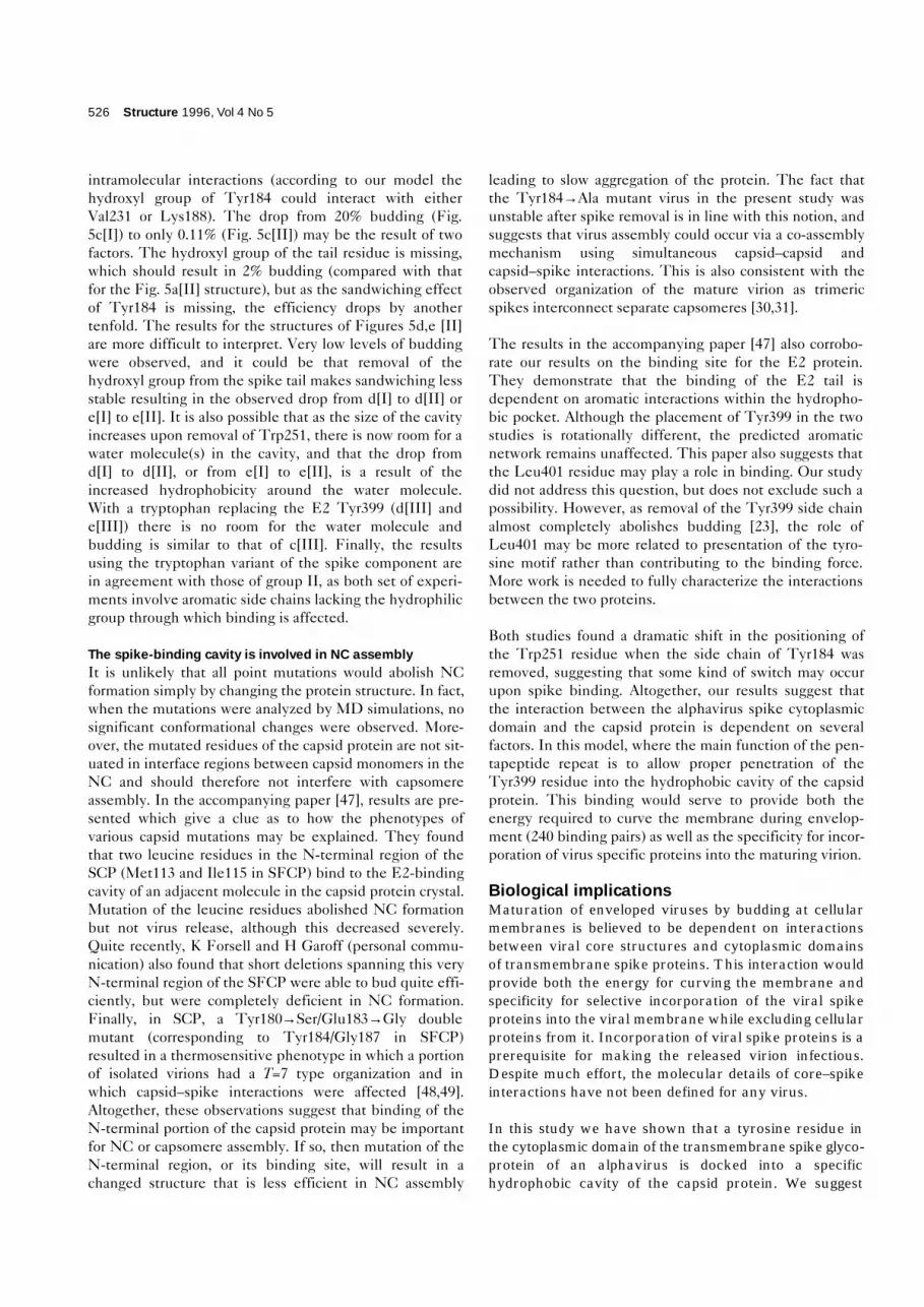
intramolecular interactions (according to our model thehydroxyl group of Tyr184 could interact with eitherVal231 or Lys188). The drop from 20% budding (Fig.5c[I]) to only 0.11% (Fig. 5c[II]) may be the result of twofactors. The hydroxyl group of the tail residue is missing,which should result in 2% budding (compared with thatfor the Fig. 5a[II] structure), but as the sandwiching effectof Tyr184 is missing, the efficiency drops by anothertenfold. The results for the structures of Figures 5d,e [II]are more difficult to interpret. Very low levels of buddingwere observed, and it could be that removal of thehydroxyl group from the spike tail makes sandwiching lessstable resulting in the observed drop from d[I] to d[II] ore[I] to e[II]. It is also possible that as the size of the cavityincreases upon removal of Trp251, there is now room for awater molecule(s) in the cavity, and that the drop fromd[I] to d[II], or from e[I] to e[II], is a result of theincreased hydrophobicity around the water molecule.With a tryptophan replacing the E2 Tyr399 (d[III] ande[III]) there is no room for the water molecule andbudding is similar to that of c[III]. Finally, the resultsusing the tryptophan variant of the spike component arein agreement with those of group II, as both set of experi-ments involve aromatic side chains lacking the hydrophilicgroup through which binding is affected.
The spike-binding cavity is involved in NC assemblyIt is unlikely that all point mutations would abolish NCformation simply by changing the protein structure. In fact,when the mutations were analyzed by MD simulations, nosignificant conformational changes were observed. More-over, the mutated residues of the capsid protein are not sit-uated in interface regions between capsid monomers in theNC and should therefore not interfere with capsomereassembly. In the accompanying paper [47], results are pre-sented which give a clue as to how the phenotypes ofvarious capsid mutations may be explained. They foundthat two leucine residues in the N-terminal region of theSCP (Met113 and Ile115 in SFCP) bind to the E2-bindingcavity of an adjacent molecule in the capsid protein crystal.Mutation of the leucine residues abolished NC formationbut not virus release, although this decreased severely.Quite recently, K Forsell and H Garoff (personal commu-nication) also found that short deletions spanning this veryN-terminal region of the SFCP were able to bud quite effi-ciently, but were completely deficient in NC formation.Finally, in SCP, a Tyr180→Ser/Glu183→Gly doublemutant (corresponding to Tyr184/Gly187 in SFCP)resulted in a thermosensitive phenotype in which a portionof isolated virions had a T=7 type organization and inwhich capsid–spike interactions were affected [48,49].Altogether, these observations suggest that binding of theN-terminal portion of the capsid protein may be importantfor NC or capsomere assembly. If so, then mutation of theN-terminal region, or its binding site, will result in achanged structure that is less efficient in NC assembly
leading to slow aggregation of the protein. The fact thatthe Tyr184→Ala mutant virus in the present study wasunstable after spike removal is in line with this notion, andsuggests that virus assembly could occur via a co-assemblymechanism using simultaneous capsid–capsid andcapsid–spike interactions. This is also consistent with theobserved organization of the mature virion as trimericspikes interconnect separate capsomeres [30,31].
The results in the accompanying paper [47] also corrobo-rate our results on the binding site for the E2 protein.They demonstrate that the binding of the E2 tail isdependent on aromatic interactions within the hydropho-bic pocket. Although the placement of Tyr399 in the twostudies is rotationally different, the predicted aromaticnetwork remains unaffected. This paper also suggests thatthe Leu401 residue may play a role in binding. Our studydid not address this question, but does not exclude such apossibility. However, as removal of the Tyr399 side chainalmost completely abolishes budding [23], the role ofLeu401 may be more related to presentation of the tyro-sine motif rather than contributing to the binding force.More work is needed to fully characterize the interactionsbetween the two proteins.
Both studies found a dramatic shift in the positioning ofthe Trp251 residue when the side chain of Tyr184 wasremoved, suggesting that some kind of switch may occurupon spike binding. Altogether, our results suggest thatthe interaction between the alphavirus spike cytoplasmicdomain and the capsid protein is dependent on severalfactors. In this model, where the main function of the pen-tapeptide repeat is to allow proper penetration of theTyr399 residue into the hydrophobic cavity of the capsidprotein. This binding would serve to provide both theenergy required to curve the membrane during envelop-ment (240 binding pairs) as well as the specificity for incor-poration of virus specific proteins into the maturing virion.
Biological implicationsMaturation of enveloped viruses by budding at cellularmembranes is believed to be dependent on interactionsbetween viral core structures and cytoplasmic domainsof transmembrane spike proteins. This interaction wouldprovide both the energy for curving the membrane andspecificity for selective incorporation of the viral spikeproteins into the viral membrane while excluding cellularproteins from it. Incorporation of viral spike proteins is aprerequisite for making the released virion infectious.Despite much effort, the molecular details of core–spikeinteractions have not been defined for any virus.
In this study we have shown that a tyrosine residue inthe cytoplasmic domain of the transmembrane spike glyco-protein of an alphavirus is docked into a specifichydrophobic cavity of the capsid protein. We suggest
526 Structure 1996, Vol 4 No 5

that the binding of the tyrosine residue is dependent onan interaction with two hydrophobic residues within thecapsid protein pocket, Tyr184 and Trp251. In this modelthe three side chains form an energetically favorablearomatic network that stabilizes the interaction andthus provides the driving force for budding and therequired specificity for incorporation of correct spikeproteins into the maturing virion.
Certain mutations of the hydrophobic pocket also pro-hibited nucleocapsid assembly without affecting virusbudding to any great extent. This suggests thatalphaviruses may, contrary to earlier belief, bud in theabsence of preformed nucleocapsids, reminiscent ofC-type retrovirus budding. Results presented in theaccompanying paper [47] support and extend our find-ings, suggesting that nucleocapsid assembly and virusbudding (spike binding) may be connected processesregulated by a molecular switch mechanism.
In a broader sense, the characterization of the moleculardetails of virus budding may also provide a first model toexplain how protein interactions may be utilized to curvea membrane leading to the formation of a lipid vesiclecarrying a defined protein cargo. Vesicle protein trafficwithin the cell is a central theme in cell function andnumerous studies have shown that tyrosine signalslocated in cytoplasmic domains of transmembrane pro-teins are important for their incorporation into suchtransport vesicles.
Materials and methodsComputer modellingSequence alignment was carried out using program packages GCG[50] and MULTICOMP [51]. The template for molecular modelling wascrystallographically determined SCP at 2.8 Å resolution [25], takenfrom Brookhaven Protein Data Bank [52]. Modelling was performedwith Insight II (Biosym Technologies Ltd., San Diego, CA) and the mini-mization and molecular dynamics with CHARMM [53]. Minimizationwas started with 500 steps, using the steepest descent method, andfinished with an adopted basis Newton-Raphson method. The pre-dicted model was tested with programs POLDIAG [54] and VERIFY-3D [55]. The stereochemistry was checked with program PROCHECK[56]. Although only part of the protein was studied (residues119–267), this portion of the molecule is known to form a functionalentity of its own, being a serine-protease-type enzyme [24,26]. Therefore, in practice, the analysis concerned a complete protein, thestructure of which should not be affected by the N-terminal part the structure. Secondary structures of the SCP were analyzed with program DSSP [57]. Standard CHARMM with parameter setPARAM22 was used in the MD simulations of core protein–E2 inter-action and for studying the conformational changes in the mutantTyr184→Ala. Program QUANTA (Molecular Simulations, Inc., Burling-ton, MA) was used to search for sequences related to the E2 motif inthe Brookhaven Protein Data Bank.
Nucleic acid manipulationsThe plasmids expressing either the capsid protein alone or the spikeproteins and the mutant derivatives have been described [4,23]. Invitro mutagenesis [58] and in vitro transcription [15] were carried outas described previously.
Cell manipulationsBHK-21 cells (ATCC CL13) were grown to late log phase in 75 cm2
bottles with 10–15 ml complete BHK medium (G-MEM [Gibco], 5%fetal calf serum, 10% tryptose phosphate broth, 20 mM HEPES[N-2-hydroxyethyl-piperazine-N′-2-etanesulphonic acid], 2 mM gluta-mine, 0.1 U ml–1 penicillin, 0.1 mg ml–1 streptomycin). Transfectionswere performed as described previously [15].
For metabolic labelling, cells were washed twice with prewarmed PBS,overlaid with starvation medium (methionine free MEM [Gibco], 2 mMglutamine, 20 mM HEPES) and incubated for 30 min. The medium wasreplaced with the same containing 100 mCi ml–1 of 35S-methionine(Amersham) and incubated for 15 min for stability studies or 30 min forNC and virus formation studies. The medium was aspirated and thecells washed twice with chase medium (E-MEM [Gibco/Life Technolo-gies Ltd, Paisley, Scotland], 2 mM glutamine, 20 mM HEPES,150 µg ml–1 unlabelled methionine), overlaid with the same and incu-bated for different chase time periods. After the chase the cells werewashed with cold PBS before NP-40 lysis buffer (1% NP-40, 50 mMTris-HCl pH 7.6, 150 mM NaCl, 2 mM EDTA, 1 mg ml–1 PMSF, 10 mMiodoacetamide) was added to the dishes which were then placed onice for 10 min. The lysed cells were scraped off and the dish wasrinsed with additional NP-40 lysis buffer. The lysates were centrifugedat 6000 rpm for 5 min. SDS lysis buffer (1% SDS, 50 mM Tris-HClpH 7.6, 150 mM NaCl, 2 mM EDTA, 1mg ml–1 PMSF, 10 mM iodo-acetamide) was added to the NP-40 insoluble pellet and the lysateswas homogenized by pulling through a 23G needle. To assay forreleased virus particles, the 5 h chase medium was centrifuged using aBeckman JA 18.1 rotor (Palo Alto, CA) at 17 000 rpm, for 2 h. Thepellet was resuspended in TNE (50 mM TRIS-HCl pH 7.5, 100 mMNaCl, 1 mM EDTA) or sample buffer and analyzed by SDS-PAGE.
For immunofluorescence studies the cells were washed extensivelywith PBS 7 h after electroporation and incubated in MEM-BSA(minimal essential medium, 1 % bovine albumin fraction V [Gibco]) foran additional 2 h. The medium was collected and centrifuged at6000 rpm for 5 min before it was used to infect BHK cells grown oncoverslips. After a 1 h infection the medium was changed to completeBHK medium and the cells were incubated overnight. Indirect immuno-fluorescence was then carried out as described previously [59].
Electron microscopyBHK cell were co-transfected with RNAs encoding capsid or spikeproteins, incubated for 7 h, and then fixed in 2% glutaraldehyde in0.1 M sodium cacodylate buffer, pH 7.4 at room temperature. Fixedcells were scraped off the plate and pelleted at 14 000 rpm for 2 min inan Eppendorf centrifuge and washed in 0.15 M sodium cacodylatebuffer, pH 7.4. Specimens were post-fixed in 1% osmium tetroxide in0.15 M sodium cacodylate buffer for 1 h at 4°C, and dehydrated inethanol and acetone, and finally embedded in LX-112 (Ladd, Burling-ton, Vermont, USA). Thin sections were cut with a LKB (Bromma,Sweden) ultramicrotome and examined in a Phillips 420 electronmicroscope (Eindhoven, The Netherlands) operating at 60 kV.
Assay of NC formationNC formation was analyzed by incubating 100 ml lysate (with or withoutpretreatment of RNase A 100 mg ml–1 for 10 min on ice) for 15 min onice with 25 mM EDTA, before loading on a 15–30% (w/w) sucrose gra-dient in TNE NP-40 buffer (50 mM Tris-HCl pH 7.4, 100 mM NaCl,1 mM EDTA, 0.1% NP-40). Centrifugation was performed in a BeckmanSW 41 rotor at 40 000 rpm for 2 h at 4°C. The gradients were fraction-ated from the bottom using a Gilson microfraction collector. 40 ml fromeach fraction was added to 2 ml lsc-cocktail (Packard, Groningen, TheNetherlands) and radioactivity was measured using a liquid scintillationcounter (LKB Wallac 1214 Rackbeta, Sollentuna, Sweden). NC stabilitywas analyzed by incubation of 9 ml medium from a 5 h chase with 1 ml10% NP-40 to strip the spike proteins and membrane from the NCs.The medium was then concentrated by using a Centriprep 100 (Amicon,Beverly, MA) and incubated with 10 mM IAA (iodoacetamid) and 2 mMEDTA before loading it on top of the gradient and centrifuges, as above.
Research Article Alphavirus spike–nucleocapsid interaction Skoging et al. 527
AcknowledgementsWe thank Birgitta Lindqvist and Kjell Hultenby for expert technical assis-tance. We are indebted to Richard Kuhn, Michael Rossmann and HenrikGaroff for sharing their data prior to publication. This work was supportedby the Swedish Natural Science Foundation Council to LN and by theSwedish Medical Research Council and Swedish Research Council for theEngineering Sciences to PL. US is the recipient of a Karolinska Institutegraduate student scholarship.
References1. Simons, K. & Garoff, H. (1980). The budding mechanism of enveloped
animal viruses. J. Gen. Virol. 50, 1–21.2. Delchambre, M., et al., & Bex, F. (1989). The GAG precursor of
Simian Immunodeficiency virus assembles into virus-like particles.EMBO J. 8, 2653–2660.
3. Gheysen, D., et al., & De Wilde, M. (1989). Assembly and release ofHIV-1 precursor Pr55gag virus-like particles from recombinantBaculovirus-infected cells. Cell 59, 103–112.
4. Suomalainen, M., Liljeström, P. & Garoff, H. (1992). Spikeprotein–nucleocapsid interactions drive the budding of alphaviruses. J. Virol. 66, 4737–4747.
5. Bruss, V. & Ganem, D. (1991). The role of envelope proteins in hepatitisB virus assembly. Proc. Natl. Acad. Sci. USA 88, 1059–1063.
6. Liu, C., Eichelberger, M.C., Compans, R.W. & Air, G.M. (1995).Influenza type A virus neuraminidase does not play a role in viral entry,replication, assembly, or budding. J. Virol. 69, 1099–1106.
7. Jin, H., Leser, G.P. & Lamb, R.A. (1994). The influenza virushemagglutinin cytoplasmic tail is not essential for virus assembly orinfectivity. EMBO J. 13, 5504–5515.
8. Metsikkö, K. & Simons, K. (1986). The budding mechanism of spikeless vesicular stomatitis virus particles. EMBO J. 5, 1913–1920.
9. Whitt, M.A., Chong, L. & Rose, J.K. (1989). Glycoprotein cytoplasmicdomain sequences required for rescue of a Vesicular Stomatitis virusglycoprotein mutant. J. Virol. 63, 3569–3578.
10. Chong, L.D. & Rose, J.K. (1994). Interactions of normal and mutantVesicular Stomatitis virus matrix proteins with the plasma membraneand nucleocapsids. J. Virol. 68, 441–447.
11. Li, Y., Luo, L., Schubert, M., Wagner, R.R. & Kang, C.Y. (1993). Viralliposomes released from insect cells infected with recombinantbaculovirus expressing the matrix protein of Vesicular Stomatitis virus.J. Virol. 67, 4415–4420.
12. Justice, P.A., Sun, W., Li, Y., Grigera, P.R. & Wagner, R.R. (1995).Membrane vesiculation function and exocytosis of wild type and mutantmatrix proteins of Vesicular Stomatitis virus. J. Virol. 69, 3156–3160.
13. Strauss, J.H. & Strauss, E.G. (1994). The alphaviruses: geneexpression, replication, and evolution. Microbiol. Rev. 58, 491–562.
14. Barth, B.U., Suomalainen, M., Liljeström, P. & Garoff, H. (1992).Alphavirus assembly and entry: the role of the cytoplasmic tail of theE1 spike subunit. J. Virol. 66, 7560–7564.
15. Liljeström, P., Lusa, S., Huylebroeck, D. & Garoff, H. (1991). In vitromutagenesis of a full-length cDNA clone of Semliki Forest virus: the6000-molecular-weight membrane protein modulates virus release.J. Virol. 65, 4107–4113.
16. Loewy, A., Smyth, J., von Bonsdorff, C.-H., Liljeström, P. & Schlesinger,M.J. (1995). The 6-kilodalton membrane protein of Semliki Forest virus isinvolved in the budding process. J. Virol. 69, 469–475.
17. Metsikkö, K. & Garoff, H. (1990). Oligomers of the cytoplasmicdomain of the p62/E2 membrane protein of Semliki Forest virus bindto the nucleocapsid in vitro. J. Virol. 64, 4678–4683.
18. Collier, N.C., Adams, S.P., Weingarten, H. & Schlesinger, M.J. (1992).Inhibition of enveloped RNA virus formation by peptidescorresponding to glycoprotein sequences. Antiviral Chem.Chemother. 3, 31–36.
19. Liljeström, P. & Garoff, H. (1991). Internally located cleavable signalsequences direct the formation of Semliki Forest virus membraneproteins from a polyprotein precursor. J. Virol. 65, 147–154.
20. Gaedigk-Nitschko, K. & Schlesinger, M.J. (1991). Site-directedmutations in Sindbis virus E2 glycoprotein’s cytoplasmic domain andthe 6K protein lead to similar defects in virus assembly and budding.Virology 183, 206–214.
21. Ivanova, L. & Schlesinger, M.J. (1993). Site-directed mutations in theSindbis virus E2 glycoprotein identify palmitoylation sites and affectvirus budding. J. Virol. 67, 2546–2551.
22. Lopez, S., Yao, J.-S., Kuhn, R.J., Strauss, E.G. & Strauss, J.H. (1994).Nucleocapsid–glycoprotein interactions required for assembly ofalphaviruses. J. Virol. 68, 1316–1323.
23. Zhao, H., Lindqvist, B., Garoff, H., von Bondsdorff, C.-H. & Liljeström,P. (1994). A tyrosine-based motif in the cytoplasmic domain of thealphavirus envelope protein is essential for budding. EMBO J. 13,4204–4211.
24. Choi, H.-K., et al., & Wengler, G. (1991). The crystal structure ofSindbis virus core protein and a proposed structure of the capsid.Nature 354, 37–43.
25. Tong, L., Wengler, G. & Rossmann, M.G. (1993). Refined structure ofSindbis virus core protein and comparison with other chymotrypsin-like serine proteinase structures. J. Mol. Biol. 230, 228–247.
26. Forsell, K., Suomalainen, M. & Garoff, H. (1995). Structure-functionrelation of the NH2-terminal domain of the Semliki Forest virus capsidprotein. J. Virol. 69, 1556–1563.
27. Geigenmüller-Gnirke, U., Nitschko, H. & Schlesinger, S. (1993).Deletion analysis of the capsid protein of Sindbis virus: identificationof the RNA binding region. J. Virol. 67, 1620–1626.
28. Weiss, B., Geigenmüller-Gnirke, U. & Schlesinger, S. (1994).Interaction between Sindbis virus RNAs and a 68 amino acidderivative of the viral capsid protein further defines the capsid bindingsite. Nucleic Acids Res. 22, 780–786.
29. Paredes, A., et al., & Prasad, B.V.V. (1993). Three-dimensionalstructure of a membrane-containing virus. Proc. Natl. Acad. Sci. USA90, 9095–9099.
30. Cheng, R.H., et al., & Baker, T.S. (1995). Nucleocapsid andglycoprotein organization in an enveloped virus. Cell 80, 621–630.
31. Fuller, S.D., Berriman, J.A., Butcher, S.J. & Gowen, B.E. (1995). LowpH induces swiveling of the glycoprotein heterodimers in the SemlikiForest virus spike complex. Cell 81, 715–725.
32. Fuller, S.D. (1987). The T=4 envelope of Sindbis virus is organized byinteractions with a complementary T=3 capsid. Cell 48, 923–934.
33. Vogel, R.H., Provencher, S.W., von Bonsdorff, C.-H., Adrian, M. &Dubochet, J. (1986). Envelope structure of Semliki Forest virusreconstructed from cryo-electron micrographs. Nature 320, 533–535.
34. Argos, P. (1987). Analysis of sequence-similar pentapeptides inunrelated protein tertiary structures. Strategies for protein foldingand a guide for site-directed mutagenesis. J. Mol. Biol. 197,331–348.
35. Burmeister, W.P., Ruigrok, R.W.H. & Cusack, S. (1992). A resolutioncrystal structure of influenza B neuraminidase and its complex withsialic acid. EMBO J. 11, 49–56.
36. Rini, J.M., Schulze-Gahmen, U. & Wilson, I.A. (1992). Structuralevidence for induced fit as a mechanism for antibody–antigenrecognition. Science 255, 959–965.
37. Krishnaswamy, S. & Rossmann, M.G. (1990). Structural refinementand analysis of Mengo virus. J. Mol. Biol. 211, 803–844.
38. Coombs, K. & Brown, D.T. (1987). Topological organization of Sindbisvirus capsid protein in isolated nucleocapsids. Virus Res. 7, 131–149.
39. Ulmanen, I., Söderlund, H. & Kääriäinen, L. (1976). Semliki Forest viruscapsid protein associates with the 60S ribosomal subunit in infectedcells. J. Virol. 20, 203–210.
40. Wengler, G. & Wengler, G. (1984). Identification of transfer of viralcore protein to cellular ribosomes during the early stages of alphavirusinfection. Virology 134, 435–442.
41. Froshauer, S., Kartenbeck, J. & Helenius, A. (1988). Alphavirus RNAreplicase is located on the cytoplasmic surface of endosomes andlysosomes. J. Cell Biol. 107, 2075–2086.
42. Levitt, M. & Perutz, M. F. (1988). Aromatic rings act as hydrogen bondacceptors. J. Mol. Biol. 201, 751–754.
43. Thomas, K.A., Smith, G.M., Thomas, T.B. & Feldmann, R.J. (1982).Electronic distributions within protein phenylalanine aromatic rings arereflected by the three-dimensional oxygen atom environments. Proc.Natl. Acad. Sci. USA 79, 4843–4847.
44. Reid, K.S.C., Lindley, P.F. & Thornton, J.M. (1985). Sulphur–aromaticinteractions in proteins. FEBS Lett. 190, 209–213.
45. Burley, S.K. & Petsko, G.A. (1985). Aromatic–aromatic interactions: amechanism of protein structure stabilization. Science 229, 23–28.
46. Singh, J. & Thornton, J.M. (1992). Atlas of Protein Side-ChainInteractions. IRL Press, Oxford, UK.
47. Lee, S., et al., & Kuhn, R. (1996). Identification of a protein bindingsite on the surface of the alphavirus nucleocapsid and its implicationin virus assembly. Structure 4, 531–541.
48. Lee, H. & Brown, D.T. (1994). Mutations in an exposed domain ofSindbis virus capsid protein results in the production of noninfectiousvirions and morphological variants. Virology 202, 390–400.
49. Lee, H., Ricker, P.D. & Brown, D.T. (1994). The configuration ofSindbis virus envelope proteins is stabilized by the nucleocapsidprotein. Virology 204, 471–474.
528 Structure 1996, Vol 4 No 5

50. Devereux, J., Haeberli, P. & Smithies, O. (1984). A comprehensive setof sequence analysis programs for the VAX. Nucleic Acids Res. 12,387–395.
51. Vihinen, M., Euranto, A., Luostarinen, P. & Nevalainen, O. (1992).MULTICOMP: a program package for multiple sequence comparison.CABIOS 8, 35–38.
52. Bernstein, F.C., et al., & Tasumi, M. (1977). The Protein Data Bank: acomputer-based archival file for macromolecular structures. J. Mol.Biol. 112, 535–542.
53. Brooks, B.R., Bruccoleri, R.E., Olafsen, B.D., States, D.J.,Swaminathan, S. & Karplus, M. (1983). CHARMM: a program formacromolecular energy, minimization, and dynamics calculations.J. Comput. Chem. 4, 187–217.
54. Baumann, G., Frömmel, C. & Sander, C. (1989). Polarity as a criterionin protein design. Protein Eng. 2, 329–334.
55. Lüthy, R., Bowie, J.U. & Eisenberg, D. (1992). Assessment of proteinmodels with three-dimensional profiles. Nature 356, 83–85.
56. Laskowski, R.A., MacArthur, M.W., Moss, D.S. & Thornton, J.M.(1993). PROCHECK: a program to check the stereochemical qualityof protein structures. J. Appl. Cryst. 26, 283–291.
57. Kabsch, W. & Sander, C. (1983). Dictionary of protein secondarystructure: pattern recognition of hydrogen-bonded and geometricfeatures. Biopolymers 22, 2577–2637.
58. Kunkel, T.A., Roberts, J.D. & Zakour, R.A. (1987). Rapid and efficientsite-specific mutagenesis without phenotypic selection. MethodsEnzymol. 154, 367–382.
59. Salminen, A., Wahlberg, J.M., Lobigs, M., Liljeström, P. & Garoff, H.(1992). Membrane fusion process of Semliki Forest virus II: cleavagedependent reorganization of the spike protein complex controls virusentry. J. Cell Biol. 116, 349–357.
Research Article Alphavirus spike–nucleocapsid interaction Skoging et al. 529