apr-jun 2015 - impacto de la pandemia por covid-19 en el
TRANSCRIPT
Ophthalmology Update Vol. 13. No. 2, April-June 2015ii
Contents
� EDITORIAL
� Meibomian Gland Dysfunction (MGD) Sameera Irfan -------------------------------------------------------------------------------------------------------------------------------------- 49
� OPHTHALMIC SECTION / ORIGINAL ARTICLES
� Neuro-imaging Patterns of Isolated Ocular Motor Nerve Palsies in a Pakistani Cohort Tayyaba Gul Malik et al ------------------------------------------------------------------------------------------------------------------------------ 52
� A Study of Prevalence of Risk Factors in Patients with Non-ArteriticAnterior Ischemic Optic Neuropathy (Na- Aion)
Akhunzada Muhammad Aftab et al --------------------------------------------------------------------------------------------------------------- 56
� Dacryocystorhinostomy - is Endonasal Endoscopic Approach A Viable Option? Khawaja Khalid Shoaib et al ------------------------------------------------------------------------------------------------------------------------ 59
� Ocular and systemic Complications of Intravitreal Bevacizumab (Avastin) therapy Akhunzada Muhammad Aftab et al --------------------------------------------------------------------------------------------------------------- 62
� Incidence of Intraocular Foreign Body in Penetrating Trauma presented to a Tertiary Care Hospital of Khyber Pakhtun Khwa and its Visual Outcome
Mohammad Idris et al -------------------------------------------------------------------------------------------------------------------------------- 66
� To Determine the Efficacy of Tattoo Ink in Changing the Color of Rabbit’s Iris Mehdi Soltanifar et al -------------------------------------------------------------------------------------------------------------------------------- 69
� Incidence of Hepatitis B & C among Admitted Eye Patients in Tertiary Care Hospital of Peshawar Bilal Bashir et al --------------------------------------------------------------------------------------------------------------------------------------- 72
� Visual Outcome & Complications of Scleral-fixation Posterior Chamber Intraocular Lenses Mir Ali Shah Aftab et al ---------------------------------------------------------------------------------------------------------------------------- 75
� Prevalence and Density of Amblyopia in Strabismic Patients of School Age Children Mohammad Alam et al ------------------------------------------------------------------------------------------------------------------------------- 79
� Tuberous Sclerosis Complex Hussain Ahmad Khaqan et al ----------------------------------------------------------------------------------------------------------------------- 82
� Association of Anemia with Diabetic Retinopathy in Patients with Type II Diabtese Mellitus Mohammad Kashif et al ----------------------------------------------------------------------------------------------------------------------------- 85
� Intraocular Pressure Control after Cataract Extraction with Posterior Chamber Intraocular Lens Implantation in Phacomorphic Glaucoma
Prof. Laal Mohammad et al ------------------------------------------------------------------------------------------------------------------------- 90
iiiOphthalmology Update Vol. 13. No. 2, April-June 2015
� Causes of Low vision and Quality of Life after Rehabilitation in Children & Adults Mohammad Kashif et al ------------------------------------------------------------------------------------------------------------------------------ 93
� Intraocular Pressure Control after The Efficacy of Limbal Based Conjunctival Flap in Patients Undergoing Trabeculectomy with Intra-operative Mitomycin C
Hasan Yaqoob et al ---------------------------------------------------------------------------------------------------------------------------------- 100
� Normal Tension Glaucoma & Cerebral Ischemia / Brain Atrophy Akhunzada Muhammad Aftab et al ------------------------------------------------------------------------------------------------------------- 104
� Complications & Results of External Dacryocystorhinostomy in Chronic Dacryocystitis without Intubation (Review of 107 Cases.)
Mohammad Alam et al ----------------------------------------------------------------------------------------------------------------------------- 107
� Recurrence of Retinal Detachment after Silicone Oil Removal Bilal Khan et al -------------------------------------------------------------------------------------------------------------------------------------- 110
� Choroidal Melanoma in a Young Patient Hussain Ahmad Khaqan et al ---------------------------------------------------------------------------------------------------------------------- 113
� GENERAL SECTION / ORIGINAL ARTICLES
� Frequency of High Glasgow Blatchford Score & its One Month Mortality in Patients presenting with Non-variceal Upper Gastrointestinal Bleeding
Imran Yahaya et al ---------------------------------------------------------------------------------------------------------------------------------- 115
� Meatal Mobilization Technique for Childhood Hypospadias Repair, an Early Experience at Lady Reading Hospital, Peshawar
Muhammad Ayub Khan et al ---------------------------------------------------------------------------------------------------------------------- 120
� OPHTHALMOLOGY NOTEBOOK
� Obituary- Forever Loved - Forever Missed ---------------------------------------------------------------------------------------- 123
� Murree: The Queen of Mountains - A Shining Pearl of Pakistan (Malika-e-Kohsaar) ------------------------ 124
49Ophthalmology Update Vol. 13. No. 2, April-June 2015
Meibomian Gland Dysfunction, also referred to as the posterior blepharitis, is a very common cause of a myriad of symptoms in the general population, par-ticularly after the age of 45 years which is often neglect-ed and under-diagnosed by the ophthalmic fraternity.1 Many ocular disorders, including evaporative dry eye, blepharitis, sties, chalazia and ocular rosacea have been linked to abnormal function of the meibomian glands2. Health professionals in the USA have now been alert-ed that MGD is a major contributing factor in ocular surface disease in at least 50 - 75% cases. According to the International Workshop on Meibomian Gland Dysfunction in 2011, sponsored by the Tear Film and Ocular Surface Society, USA,2 there is a paradigm shift in the treatment of dry eyes. As a result of this report, ophthalmologists are now evaluating the lids more carefully, and more often when seeing patients with dry-eye complaints. MGD has also been known to be an important cause contact lens intolerance.3
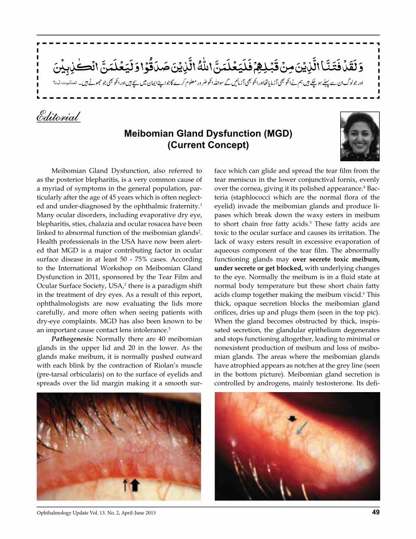
Pathogenesis: Normally there are 40 meibomian glands in the upper lid and 20 in the lower. As the glands make meibum, it is normally pushed outward with each blink by the contraction of Riolan’s muscle (pre-tarsal orbicularis) on to the surface of eyelids and spreads over the lid margin making it a smooth sur-
face which can glide and spread the tear film from the tear meniscus in the lower conjunctival fornix, evenly over the cornea, giving it its polished appearance.4 Bac-teria (staphlococci which are the normal flora of the eyelid) invade the meibomian glands and produce li-pases which break down the waxy esters in meibum to short chain free fatty acids.5 These fatty acids are toxic to the ocular surface and causes its irritation. The lack of waxy esters result in excessive evaporation of aqueous component of the tear film. The abnormally functioning glands may over secrete toxic meibum, under secrete or get blocked, with underlying changes to the eye. Normally the meibum is in a fluid state at normal body temperature but these short chain fatty acids clump together making the meibum viscid.6 This thick, opaque secretion blocks the meibomian gland orifices, dries up and plugs them (seen in the top pic). When the gland becomes obstructed by thick, inspis-sated secretion, the glandular epithelium degenerates and stops functioning altogether, leading to minimal or nonexistent production of meibum and loss of meibo-mian glands. The areas where the meibomian glands have atrophied appears as notches at the grey line (seen in the bottom picture). Meibomian gland secretion is controlled by androgens, mainly testosterone. Its defi-
Meibomian Gland Dysfunction (MGD)(Current Concept)

50 Ophthalmology Update Vol. 13. No. 2, April-June 2015
ciency is particularly seen as a part of normal ageing process. Hence, dry eye syndrome and MGD is more commonly seen in post-menopausal women.7
MGD causes two problems: Firstly, eyelid inflamma-tion and secondly, excessive evaporation of tears and consequently dry eyes. The tears become hyperosmoler which then stimulate corneal nerves resulting in ocular irritation, dryness, tearing, redness, a foreign body sen-sation or intermittent blurring of vision. Examination: In every adult patient who has come to you with any eye complaint, try to assess for MGD and look for the following first:i) The lids may look normal but the lid margin has to be everted a little bit and the meibomian gland orifices ex-amined; normally the meibum is a clear secretion that flows easily out of the orifices with a tiny pressure at the lid margin with a cotton-tip applicator. However, an opaque secretion is abnormal. Or, the glands could be completely blocked / plugged with thick white se-cretion which cannot be expressed with pressure on the lid margin. Scarred and notched grey line indicates loss of glands. Hence, there are different stages of meibo-mian gland disease.ii) Grades of MGD: Grade 0: Normal, no MGD: clear, thin secretion at the gland orifices, squirts out of orifices with a little pres-sure on the lid margin. Grade 1: a viscid secretion flows out easily with mini-mum pressure. Grade 2: an opaque secretion flows after exerting a lot of pressure. Grade 3: gland orifices are plugged/capped and no se-cretion flows or it comes out like a tooth-paste or a froth is present at the lid margins (due to saponification of fatty acids by bacterial lipases). Grade 4: atrophic/scarred gland orifices.NOTE: Toxic secretions cause an inferior conj / corneal staining. If the ducts are blocked with thick meibum plugs, or have atrophied, then there will be no toxic se-cretions; however, if few ducts are open, then a little bit of corneal staining will be there. Hence seeing cor-neal staining with open ducts is Grade 2 disease. See-ing corneal staining + majority of ducts being capped/blocked is grade 3 disease. If grey line shows notching, then trans-illumination confirms atrophic glands at the site of a notch (Grade 4 disease).iii) Oily debris floating in tear film or foam present at the lid margins indicate hyper-secretion; the fatty acids undergo saponification by bacteria and produce toxic foam. iv) Look for Rosacea / recurrent chlazia which indicate MGD.
v) Note the tear-film break up time: this gets reduced with worsening of the disease. Normal being >10 mm.vi) Punctate keratopathy at the inferior limbus and in-ferior conjunctival staining due to irritation by toxic meibum at the lid margin.vii) Transilluminate the tarsal plate by a pen-torch held on the skin side of a fully everted lid to look for evi-dence of atrophy, loss or degeneration of the meibomi-an glands.viii) Check for aqueous deficiency of tear film with Schirmer’s 2 test.ix) Check the tear osmolarity if possible.x) In severe MGD, check lipid profile/ Blood Sugar.Don’t assume patients will voluntarily mention their symptoms. Be proactive, and ask every adult patient about ocular irritation and whether it is worse in the morning which points to MGD. A dry eye due to aqueous deficiency is worse in the evening.Treatment:a) Highest on the list is getting the patient to play an active role by scrubbing the lid margins with a baby shampoo twice a day to remove excess oil. b) Mobilize the oils 8 out of the lids onto the eye where you do want them. Achieve this through the use of lid compresses, which are believed to melt plugs com-posed of dried secretions blocking the gland orifice; Apply hot fomentation to the lids with a hot towel to melt the thick secretions/plugs and then expressing meibomian glands on a daily basis by massaging the lower lid upwards and upper lid downwards with a finger or a Q-tip. this should be done 2-3 x per day. This will not work in Grade 4 disease in whom there are no secretions at all due to atrophic glands. c) Addressing the source of any inflammation; avoid aminoglycocides topically as they worsen MGD. Find out and treat any allergies. Topical tetracycline eye ointment massaged into the id margins twice per day. Systemic doxycycline9 can interfere with the lipases produced by Staphlococci that break down the fatty components to free fatty acids- a common regimen is doxycycline 100 mg od or b.i.d. for four to six weeks, in severe cases. An alternative is Azithromycin 500 mg bid or 1 Gm od per week for 3 consecutive weeks. Simi-larly, cyclosporin10 eye drops 0.5% - 0.75% twice a day have the same anti-inflammatory affect.d) Neutralize toxic secretions with artificial tears; drops during day and lubricating ointment at night.e) Some patients are beyond the point of no return. They don’t have any glands left, or the ones they have aren’t functioning. For them, heating and massaging won’t do anything. They can be given Lipid-based ar-tificial tears.
EDITORIAL
51Ophthalmology Update Vol. 13. No. 2, April-June 2015
f) Oral Omega 3 Fatty acids11 to restore the balance be-tween good and bad lipids.g) Intra-ductal probing12 of blocked meibomian glands has been found to be effective in removing dried secre-tion. Recommendation: MGD is a very common eye prob-lem; try to look for it in every adult who presents at the ophthalmic clinic. Every patient should be specifically asked for symptoms of ocular irritation. An eye exami-nation should commence from the lids.It is important to familiarize with the normal meibo-mian secretion by examining the lids of teenagers first and trying to squirt out meibum with a gentle squeeze on the lid margin. REFERENCES1. Bron AJ, Tiffany JM. The contribution of meibomian disease to
dry eye. Ocul Surf. 2004;2(2):149–165.2. The definition and classification of dry eye disease: report of
the Definition and Classification Subcommittee of the Interna-tional Dry Eye WorkShop (2007) Ocul Surf. 2007;5(2):75–92.
3. Korb DR, Henriquez AS. Meibomian gland dysfunction and contact lens intolerance. J Am Optom Assoc. 1980;51(3):243–251.
4. McCulley JP, Shine WE. The lipid layer of tears: dependent on meibomian gland function. Exp Eye Res 2004;78:361-5.
5. Dougherty JM, McCulley JP. Bacterial lipases and chronic
blepharitis. Invest Ophthalmol Vis Sci.1986;27(4):486–491.6. Foulks GN. The correlation between the tear film lipid layer
and dry eye disease. Surv Ophthalmol.2007;52(4):369–374.7. Sullivan DA, Sullivan BD, Evans JE. Androgen deficiency, mei-
bomian gland dysfunction, and evaporative dry eye. Ann New York Academy Sciences 2002;966:211-222
8. Olson MC, Korb DR, Greiner JV. Increase in tear film lipid layer thickness following treatment with warm compresses in patients with meibomian gland dysfunction. Eye Contact Lens. 2003;29(2):96–99.
9. Dougherty JM, McCulley JP, Silvany RE, Meyer DR. The role of tetracycline in chronic blepharitis. Inhibition of lipase production in staphylococci. Invest Ophthalmol Vis Sci. 1991;32(11):2970–2975.
10. Rubin M, Rao SN. Efficacy of topical cyclosporine 0.05% in the treatment of posterior blepharitis. J Ocul Pharmacol Ther. 2006;22(1):47–53.
11. Macsai MS. The role of omega-3 dietary supplementation in blepharitis and meibomian gland dysfunction (an AOS the-sis) Trans Am Ophthalmol Soc. 2008;106:336–356.
12. Maskin SL. Intraductal meibomian gland probing relieves symptoms of obstructive meibomian gland dysfunction. Cor-nea. 2010;29(10):1145–1152
Dr. Sameera Irfan, FRCSConsultant Oculoplastic Surgeon & StrabismologistMughal Eye Trust Hospital, Lahore, PakistanWebsite: www.sameerairfan.com Cell: 03364500901
ELECTION RESULT
OPHTHALMOLOGICAL SOCIETY OF PAKISTAN(Federal Branch, Islamabad)
Following members have been elected as the office bearers of the Ophthalmological Society of Pakistan, Federal Branch, in a recent election held in Islamabad for the year 2015-16.
President Dr. Waheed AfzalPresident Elect Prof Farooq AfzalGeneral Secretary Dr Shahzad SaeedTreasurer Prof Nadeem QureshiJoint Secretary Prof B. A. Naeem
Executive Council:
Prof. Jahangir Akhter, Dr. Izzat Ali Khan, Prof. Brig. Amer Yaqub, Prof. Imran Azam Butt Prof. Mazhar Ishaq, Prof. Syed Imtiaz Ali, Prof. Wajid Ali Khan, Prof. Naqaish Sadiq Prof. Shakaib Anwar, Dr. Tariq Mirza, Dr. Amir Israr, Dr Intisar-Ul-Haq, Lt. Gen (R) M K Akbar, Dr. Naeem Qadir, Dr. Shahzad Iftikhar, Dr. Ali Raza, Dr. Javed Malik, Dr. Mazhar Qayyum
EDITORIAL
52 Ophthalmology Update Vol. 13. No. 2, April-June 2015
Tayyaba Gul Malik FCPS1, Prof. Khalid Farooq FCPS (Diagnostic Radiology)2
Muhammad Khalil FCPS3
ABSTRACT:Objective: To determine neuro-imaging patterns of ocular motor nerve palsies in a Pakistani cohort and to compare with other populations.Study Design: Descriptive, retrospective study.Study period: 2010 to 2014Subjects and settings: 50 Patients of ocular motor nerve palsies from two different centers of Lahore were included in the study. History charts and neuro-imaging reports were reviewed. The data considered for the study was age, sex, ocular manifestations, neuro-ophthalmological findings and imaging reports (CT scans, MRI, MRA and MRV). Results: Female to male ratio was 1.6:1. Age ranged from 13 years to 74 years (average 44.18). 66% (n=33) patients had isolated sixth nerve palsy and 34% (n= 17) had isolated third nerve palsy. None of our patients had fourth nerve palsy. 42% patients had normal neuro-imaging. Sinusitis and brain infarcts were commonest cause of third nerve palsy while demyelination was more common in patients with sixth nerve palsy. Other neuro-etiologies were space-occupying lesions, parasellar tumours, multiple sclerosis, aneurysm and meningitis. Conclusion: Third nerve palsy is the commonest ocular motor nerve palsy. There are certain cases where neuro-imaging shows normal scans and the cause of palsy remains undetermined. Key words: Ocular motor nerve palsy, trochlear palsy, oculomotor palsy, abducent palsy, Parasellar tumours, neuro-imaging.
INTRODUCTION Ocular motor nerves are comprised of Oculomo-tor (supplying Medial Rectus, Superior Rectus, Inferior Rectus, Inferior Oblique), Trochlear (innervating Supe-rior Oblique) and Abducent (nerve to the Lateral Rec-tus). Ocular motor nerve palsies are either supra nucle-ar or infra nuclear. Associated neurological signs and symptoms help us determine the site of lesion. Fascicu-lar palsies of third nerve are associated with different syndromes (Benedikt, Weber, Nothnagel and Claude). Similarly, fascicular lesions of sixth nerve are associat-ed with Foville and Millard-Gubler syndromes.1 Fourth nerve palsies are usually congenital in nature. Different causes of isolated nerve palsies are men-tioned in literature. These include vascular diseases like Diabetes and Hypertension. In Oculomotor palsy associated with Diabetes and Hypertension, pupils are usually spared. Aneurysms and trauma are other important causes of isolated nerve palsies. Tumours, neurosyphilis and Giant cell arteritis are rare causes.
Very interestingly, idiopathic palsies constitute a large percentage in clinical practice. Acoustic neuroma, basal skull fractures, naso-pharyngeal tumours and raised intracranial pressures are culprits of sixth nerve pathol-ogies2. Cavernous sinus pathologies give rise to multi-ple cranial nerve palsies (oculomotor, trochlear, abdu-cent, ophthalmic and maxillary divisions of trigeminal nerves). This study reviews the neuro-imaging patterns of ocular motor nerve palsies in a selected group of pa-tients from two tertiary care hospitals of Pakistan.SUBJECTS AND METHODS It was a descriptive retrospective study. 50 pa-tients with acquired isolated Ocular motor nerve (Oc-ulomotor, Trochlear and Abducent) palsies were se-lected (from two centers of Lahore City). Study period spanned over 2010 to 2014. Inclusion criteria: • Patients with acquired isolated third, fourth or
sixth cranial nerve palsies• Patients whose, complete clinical and radiological
data was available.Exclusion criteria:• Patients with multiple cranial nerve palsies• Patients with incomplete clinical and imaging data We reviewed clinical and imaging charts of se-lected patients and medical records were analyzed. Clinical data included history, visual acuity, color vi-sion and slit lamp examination. Special attention was
ORIGINAL ARTICLE
Tayyaba Gul
1Associate Professor of Ophthalmology, 2Professor, Department of Radiology, 3Associate Professor of Ophthalmology, Lahore Medical and Dental College, Tulspura, North Canal Bank,Canal Road, Lahore
Correspondence: Dr. Tayyaba Gul Malik FCPS, Associate Professor of Ophthalmology, Lahore Medical and Dental College, Lahore E.mail: [email protected], Mob: 0300-4217998
Received: January 2015 Accepted: February 2015
Neuro-imaging Patterns of Isolated Ocular Motor Nerve Palsies in a Pakistani Cohort
OPHTHALMIC SECTION
Neuro-imaging Patterns of Isolated Ocular Motor Nerve Palsies in a Pakistani Cohort
53Ophthalmology Update Vol. 13. No. 2, April-June 2015
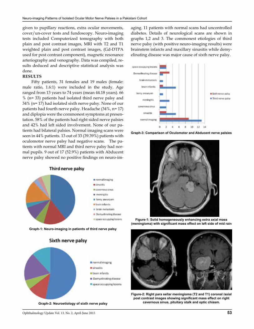
given to pupillary reactions, extra ocular movements, cover/un-cover tests and fundoscopy. Neuro-imaging tests included Computerized tomography with both plain and post contrast images, MRI with T2 and T1 weighted plain and post contrast images, (Gd-DTPA used for post contrast component), magnetic resonance arteriography and venography. Data was compiled, re-sults deduced and descriptive statistical analysis was done.RESULTS Fifty patients, 31 females and 19 males (female: male ratio, 1.6:1) were included in the study. Age ranged from 13 years to 74 years (mean 44.18 years). 66 % (n= 33) patients had isolated third nerve palsy and 34% (n= 17) had isolated sixth nerve palsy. None of our patients had fourth nerve palsy. Headache (34%, n= 17) and diplopia were the commonest symptoms at presen-tation. 58% of the patients had right sided nerve palsies and 42% had left sided involvement. None of our pa-tients had bilateral palsies. Normal imaging scans were seen in 44% patients. 13 out of 33 (39.39%) patients with oculomotor nerve palsy had negative scans. The pa-tients with normal MRI and third nerve palsy had nor-mal pupils. 9 out of 17 (52.9%) patients with Abducent nerve palsy showed no positive findings on neuro-im-
aging. 11 patients with normal scans had uncontrolled diabetes. Details of neurological scans are shown in graphs 1,2 and 3. The commonest etiologies of third nerve palsy (with positive neuro-imaging results) were brainstem infarcts and maxillary sinusitis while demy-elinating disease was major cause of sixth nerve palsy.
Graph-1: Neuro-imaging in patients of third nerve palsy
Graph-2: Neuroetiology of sixth nerve palsy
Graph-3: Comparison of Oculomotor and Abducent nerve palsies
Figure-1: Solid homogeneously enhancing extra axial mass (meningioma) with significant mass effect on left side of mid rain
Figure-2: Right para sellar meningioma (T2 and T1) coronal /axial post contrast images showing significant mass effect on right
cavernous sinus, pituitary stalk and optic chiasm.
Neuro-imaging Patterns of Isolated Ocular Motor Nerve Palsies in a Pakistani Cohort
54 Ophthalmology Update Vol. 13. No. 2, April-June 2015
Parasellar meningioma pressing on the cavernous sinus was the commonest space occupying lesion leading to oculomotor ( 6%, n= 2/33) and abducent nerve palsies (5.88, n= 1/17). Only one case of Acoustic neuroma had sixth nerve palsy. The patient had developed palsy as a complication of neurosurgery for Acoustic neuroma. One of our patients with third nerve palsy had multiple tuberculomas in parasellar region.
DISCUSSION Out of twelve pairs of cranial nerves, three pairs supply extra ocular muscles of eyeball. Diabetes, Hy-pertension, aneurysms, trauma and brain tumours are the most commonest causes of these nerve palsies. There are certain cases where cause cannot be found and they are considered under the heading of idiopathic. In this particular study third nerve palsy was the common-est among all ocular motor palsies. It was consistent with the findings of Kim et al,3 Chih-Hsien Hung4 and Chiu EK5 Contrary to that, many other researchers had preponderance of sixth nerve palsy in their studies.4,5,6 Male to female ratio was 1.6:1 in a study by Shawn C in his cohort with an average age of 66.9 years.4 The ratio was reverse in our study (1:1.6) In this particular study, 22% patients (n=11) were idiopathic. It was very much similar to the figures given by Kumar9, Rucker et al.10 And Krishna et al.11 While this percentage was quite high by Berlit P12. The incidence of ocular palsy associated with pituitary tu-mors is reported to be between 4.6 and 32%.11 We had parasellar meningiomas leading to ocular motor palsy but none of our patients had pituitary adenoma. Later-ality of palsies is also interesting. 52% of our patients had right sided palsy which was very much consistent with an earlier study.12 Headache and diplopia were the commonest presenting complaints of ocular motor palsies in our study similar to earlier researchers.13 There are many cases where MRI or other arterial and venous scans show negative results. Controversy still exists whether to perform scans in every patient with isolated ocular motor nerve palsy. One school of thought in the absence of other neurological signs is to have a close follow up of the patient. If neurological findings develop, neuro-imaging should be performed 16,17. Others have suggested to perform neurological im-aging in all patients even if there is evidence of vascu-lopathy18. In fact, every patient should be thoroughly investigated and neuro-imaging should be performed depending upon history, age and examination find-ings. This study has certain limitations. Small sample size could be the cause of absent fourth nerve palsy cas-
es. Our ability to collect detailed information was lim-ited by the retrospective study and we had to rely on the available data. But this study can provide grounds on which prospective follow-up studies can be done. CONCLUSION Third nerve palsy is the commonest ocular motor nerve palsy. There are certain cases where neuro-imag-ing shows normal scans and the cause of palsy remains undetermined. REFERENCES1. Kim SH, Lee KC, Kim SH. Cranial nerve palsies accompanying
pituitary tumour. J Clin Neurosci. 2007;14(12):1158-62. 2. Hung CH, Chang KH, Chu CC,et al. Painful ophthalmoplegia
with normal cranial imaging. BMC Neurol. 2014; 14: 7
3. Chiu EK, Nicholas JW: Sellar lesions and visual loss: key concepts in neuro-ophthalmology. Expert Rev Anticancer Ther 2006; 6(9):23-29
4. Wilker SC, Rucker JC, Newman NJ, et al. Pain in Ischemic Ocu-lar Motor Cranial Nerve Palsies. Br J Ophthalmol. Dec 2009; 93(12): 1657–1659.
5. Rowe F and VIS group. Prevalence of ocular motor cranial nerve palsy and associations following stroke. Eye (Lond). Jul 2011; 25(7): 881–887.
6. Zafar A, Irfan M. Lateral rectus palsy: An important sign in di-agnosing tuberculous meningitis. KUST Med J 2011; 3(1): 10-14.
7. Kumar MP, Vivekanand U, Umakanth S, Yashodhara BM. A study of etiology and prognosis of oculomotor nerve paralysis. Edorium J Neurol 2014;1:1–8.
8. Rucker CW. The causes of paralysis of the third, fourth and sixth cranial nerves. Am J Ophthalmol 1966;61(5 Pt 2):1293–8.
9. Krishna AG, Mehkri MB. India Neurol 1973 Suppl. IV. Vol 20: 584).
10. Berlit PJ. Isolated and combined pareses of cranial nerves III, IV and VI. A retrospective study of 412 patients..Neurol Sci. 1991 May;103(1):10-5.
11. Greenman Y, Stern N. Non-functioning pituitary adenomas. Best Practice & Research Clinical Endocrinology & Metabo-lism 2009, 23:625-638.
12. Nair AG, Ambika S, Noronha VO, Gandhi RA. The diagnostic yield of neuroimaging in sixth nerve palsy - Sankara Nethra-laya Abducens Palsy Study (SNAPS): Report1. Indian J Oph-thalmol. Oct 2014; 62(10): 1008–1012.
13. Tamhankar MA, Biousse V, Ying GS. Isolated Third, Fourth and Sixth Cranial Nerve Palsies From Presumed Microvascular Versus Other Causes: A Prospective Study. Ophthalmology. Nov 2013; 120(11): 10.
14. Patel SV, Mutyala S, Leske DA, Hodge DO, Holmes JM. Inci-dence, associations, and evaluation of sixth nerve palsy using a population-based method. Ophthalmology. 2004;111:369–75.
15. Murchison AP, Gilbert ME, Savino PJ. Neuroimaging and acute ocular motor mononeuropathies: a prospective study.. Arch Ophthalmol. 2011;129(3):301-5.
16. Bendszus M, Beck A, Koltzenburg M, et al. MRI in isolated sixth nerve palsies. Neuroradiology. 2001 Sep;43(9):742-5.
17. Kanski JJ. Neuro-ophthalmology. In: Clinical Ophthalmology: a systematic approach. 7th Edi. Elsevier Butterworth Heine-mann; 2011. p 1055
18. Kanski JJ. Neuro-ophthalmology. In: Clinical Ophthalmology: a systematic approach. 7th Edi. Elsevier Butterworth Heine-mann; 2011. p 1063
19. Kim SH, Lee KC, Kim SH. Cranial nerve palsies accompanying pituitary tumour. J Clin Neurosci. 2007;14(12):1158-62.
20. Hung CH, Chang KH, Chu CC,et al. Painful ophthalmoplegia with normal cranial imaging. BMC Neurol. 2014; 14: 7
21. Chiu EK, Nicholas JW: Sellar lesions and visual loss: key
Neuro-imaging Patterns of Isolated Ocular Motor Nerve Palsies in a Pakistani Cohort
55Ophthalmology Update Vol. 13. No. 2, April-June 2015
concepts in neuro-ophthalmology. Expert Rev Anticancer Ther 2006; 6(9):23-29
22. Wilker SC, Rucker JC, Newman NJ, et al. Pain in Ischemic Ocu-lar Motor Cranial Nerve Palsies. Br J Ophthalmol. Dec 2009; 93(12): 1657–1659.
23. Rowe F and VIS group. Prevalence of ocular motor cranial nerve palsy and associations following stroke. Eye (Lond). Jul 2011; 25(7): 881–887.
24. Zafar A, Irfan M. Lateral rectus palsy: An important sign in di-agnosing tuberculous meningitis. KUST Med J 2011; 3(1): 10-14.
25. Kumar MP, Vivekanand U, Umakanth S, Yashodhara BM. A study of etiology and prognosis of oculomotor nerve paralysis. Edorium J Neurol 2014;1:1–8.
26. Rucker CW. The causes of paralysis of the third, fourth and sixth cranial nerves. Am J Ophthalmol 1966;61(5 Pt 2):1293–8.
27. Krishna AG, Mehkri MB. India Neurol 1973 Suppl. IV. Vol 20: 584).
28. Berlit PJ. Isolated and combined pareses of cranial nerves III, IV and VI. A retrospective study of 412 patients..Neurol Sci. 1991 May;103(1):10-5.
29. Greenman Y, Stern N. Non-functioning pituitary adenomas. Best Practice & Research Clinical Endocrinology & Metabo-lism 2009, 23:625-638.
30. Nair AG, Ambika S, Noronha VO, Gandhi RA. The diagnostic yield of neuroimaging in sixth nerve palsy - Sankara Nethra-laya Abducens Palsy Study (SNAPS): Report1. Indian J Oph-thalmol. Oct 2014; 62(10): 1008–1012.
31. Tamhankar MA, Biousse V, Ying GS. Isolated Third, Fourth and Sixth Cranial Nerve Palsies From Presumed Microvascular Versus Other Causes: A Prospective Study. Ophthalmology. Nov 2013; 120(11): 10.
32. Patel SV, Mutyala S, Leske DA, Hodge DO, Holmes JM. Inci-dence, associations, and evaluation of sixth nerve palsy using a population-based method. Ophthalmology. 2004;111:369–75.
33. Murchison AP, Gilbert ME, Savino PJ. Neuroimaging and acute ocular motor mononeuropathies: a prospective study. Arch Ophthalmol. 2011;129(3):301-5.
34. Bendszus M, Beck A, Koltzenburg M, et al. MRI in isolated sixth nerve palsies. Neuroradiology. 2001 Sep;43(9):742-5.
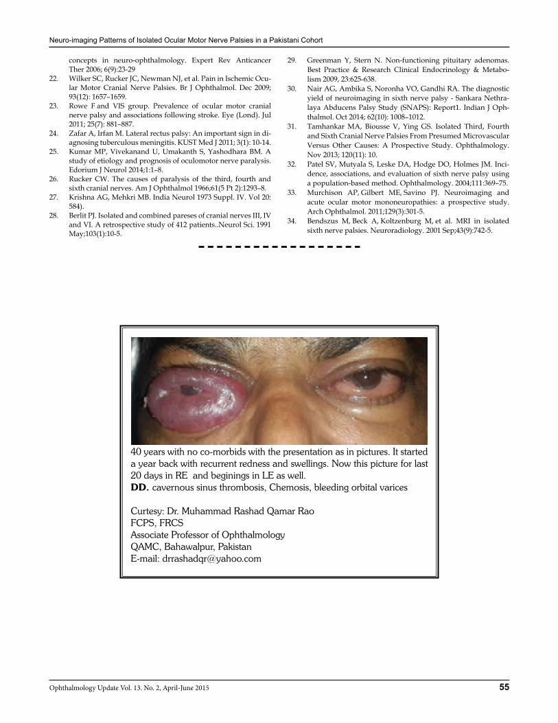
40 years with no co-morbids with the presentation as in pictures. It started a year back with recurrent redness and swellings. Now this picture for last 20 days in RE and beginings in LE as well. DD. cavernous sinus thrombosis, Chemosis, bleeding orbital varices
Curtesy: Dr. Muhammad Rashad Qamar RaoFCPS, FRCSAssociate Professor of OphthalmologyQAMC, Bahawalpur, PakistanE-mail: [email protected]
56 Ophthalmology Update Vol. 13. No. 2, April-June 2015
ORIGINAL ARTICLE
Akhunzada M. Aftab
INRODUCTION Anterior ischemic optic neuropathy is of two types; Arteritic and Non Arteritic. Arteritic type is as-sociated with giant cell arteritis. It is associated with raised inflammatory markers like erythrocyte sedimen-tation rate (ESR) and C - reactive protein levels. Non Arteritic Anterior Ischemic Optic Neuropathy is a mul-ti- factorial, acute optic neuropathy. It is the most com-mon optic neuropathy in patients aged over 50 years and is the second most common cause of optic nerve related permanent visual loss in adults after glaucoma.1 The pathogenesis of NA- AION involves acute ischem-ic infarction of the optic nerve head, which is supplied by the short posterior cilliary arteries (SPAC).2 Despite the controversies regarding the distributary variations and characteristics of SPCA anastomoses around the ON head, it has been proved, that, this circle provides
segmental supply to the optic nerve head and physi-ologically acts as end arteries.3
Patients usually present with sudden painless loss of vision in one eye, commonly noticed after waken-ing from sleep. Some patients may present with slight blurring of vision and a near normal visual acuity is recorded in them. Several studies have shown high prevalence of multiple risk factors. These may be con-sidered as local or systemic factors• Hypertension• Diabetes Mellitus• Hyperlipidemia• Ischemic heart disease• Nocturnal hypotension• Sleep Apnea• Absent or small cup in optic disc, and many
others.4
Some studies have shown intrinsic disorders of regulation of coagulation as an additional risk factor.2, 5 The purpose of this study was to evaluate the incidence of these risk factors in patients admitted to Eye A Unit, Khyber Teaching Hospital, diagnosed with NA-AION.METHODS This retrospective chart review study was con-ducted on patients previously admitted in Eye A Unit of Khyber Teaching Hospital. Diagnosis of NA- AION was made considering the following criteria:• Positive clinical history of sudden painless visual
loss/ blurring of vision.
Akhunzada Muhammad Aftab FCPS1, Misbah Durrani FCPS2, Asif MBBS3
Awais Rauf MBBS4, Farzana MBBS5, Prof. Mustafaf Iqbal FRCS, FRCOphth6
ABSTRACTPurpose: To estimate prevalence of risk factors in patients diagnosed with Non- Arteritic Anterior Ischemic Optic Neuropath (NA- AION). Methods: This was a retrospective chart review of patients admitted and diagnosed as NA- AION. Patients with other optic nerve diseases like Diabetic Papillitis and patients with signs of Arteritic Anterior Ischemic Optic Neuropathy (like raised ESR and CRP) were excluded from the study. Hematological investigations, clinical data like fundus photos and radiological investigations were evaluated to detect associated risk factors. Results: A total of 24 subjects were included in the study. Total number of males was 15 (62.5%), while 9 (37.5%) were females. Mean age at presentation was 57 years (Range 19- 60 years). Total number of diabetics alone were 2 (8.3%), Hypertensive were only 3 patients(12.5%) while 14 (58.3%) suffered both from diabetes and hypertension. 5 (20.8%) were neither diabetics nor hypertensive. Patients with hyper lipidemia were 10 (41.6 %). Echocardiography revealed abnormali-ties including diastolic dysfunction (DF) in 15 (62.5%), mitral regurgitation (MR) in 3 (12.5%), aortic regurgitation (AR) in 2 (8.3%), mitral valve prolapse (MVP) in 1 (4%), while 8 (34%) patients had a normal study. One (4%) patient was found to be Protein C deficient, 1 (4%) was Protein S deficient and 1 (4%) patient had both Protein C and S deficiency. Small optic discs were seen in 18 (75%) patients. Conclusion: Diabetes, Hypertension and a small disc size are the most common risk factors associated with NA-AION in our setup.Key Words: Non Arteritic Anterior Ischemic Optic Neuropathy, Diabetes Mellitus, Hypertension, Sleep Apnea
1Registrar Eye A Unit Department of Ophthalmology, Khyber Teaching Hospital, Peshawar, 2Assistant Professor of Radiology, Bacha Khan Medical College & Mardan Medical Complex, 3,4,5,Traniee Medical Officer. A Unit Department of Ophthalmology, Khyber Teaching Hospital, Peshawar, 6Prof. & Head of Ophthalmology, Khyber Teaching Hospital, Peshawar.
Correspondence: Dr. Akhunzada Muhammad Aftab c/o Prof. Dr. Muhammad Ibrar, Department of Botany, University of Peshawar, Peshawar. Cell: 03339106060 E-Mail: [email protected]
Received: November 2014 Accepted: December 2014
Financial Disclosure: There has been no financial interest involved in this study
A Study of Prevalence of Risk Factors in Patients with Non-Arteritic Anterior Ischemic
Optic Neuropathy (Na- Aion)

A Study of Prevalence of Risk Factors in Patients with Non-Arteritic Anterior Ischemic Optic Neuropathy (Na- Aion)
57Ophthalmology Update Vol. 13. No. 2, April-June 2015
• Presence of risk factors.• Reduced/ near normal visual acuity.• Presence of relative afferent pupillary defect
(RAPD).• Diffuse or sectorial optic nerve head edema. • Central and (or) altitudinal field defect on Hum-
phrey’s visual field.• Normal ESR and CRP All the available records including history sheets, hematological investigations, ophthalmic examination, fundus photos, visual fields and radiological investiga-tions were reviewed. We evaluated history of onset and duration of loss of vision. Duration of systemic disease like diabetes and (or) hypertension was considered. Also inquiry from the patient and (or) partner regard-ing noticing symptoms of sleep apnea was also evalu-ated. Hematological tests reviewed included complete blood count (CBC), ESR, CRP levels, glycosylated hemoglobin (HbA1c) levels, fasting lipid profile, renal function tests (RFT), prothrombin time (PT) and acti-vated partial thromboplastin time (APTT), homocyst-eine levels, protein -C and -S levels. Fundus photos were evaluated for size of the disc and size of the cup. Radiological investigations which were analyzed in-cluded carotid doppler, echocardiography and ECG.RESULTS In this study, 24 patients were included.Total number of males was 15 (62.5%), while 9 (37.5%) were females. Mean age at presentation was 57 years (Rang-ing from 19- 60 years). All (100%) had a positive clini-cal history of sudden painless loss/ blurring of vision in the affected eye and presented within 10 weeks of onset of symptoms (Range 3 days to 10 weeks). Almost half the study patients (13), reported to have noticed vision loss upon wakening up in the morning. Only one (4%) patient’s history was positive for sleep ap-nea. 14 (58.3%) patients suffered both from diabetes and hypertension. 2 patients (8.3%) were having only diabetes and 3 patients (12.5%) were diagnosed hyper-tensive patients. 5 (20.8%) patients were neither diabet-ics nor hypertensive. Mean duration of diabetes was 7 years (Range 6 months to 15 years). Mean duration of hyper tension was 5 years (Ranging from 2 months to 20 years). All (100%) patients had normal CBC, ESR and CRP levels. All diabetics had raised HbA1c levels (mean = 8.7%). 10 out of 17 hypertensive patients in to-tal had raised blood pressure recordings. Patients with hyper lipidemia were 10 (41.6%).3 patients had raised cholesterol, 3 had raised triglycerides while 4 patients had both cholesterol and triglyceride levels above nor-mal. All 24 (100%) patients had renal function tests, PT
and APTT levels within normal range. One (4%) patient was found to be protein C defi-cient, 1 (4%) was Protein S deficient and 1 (4%) patient had both protein C and S deficiency in our study. One patient was suffering from hepatitis C and was taking interferon treatment. Fundus photographs revealed 18 (75%) patients in our study to have small discs with little or no cup. These discs are commonly referred to as “disc at risk”. Carotid doppler imaging revealed 7 (29%) patients to be having atheromatous plaques in the carotid arteries. None of these patients had more than 70% stenosis. Echocardiography revealed ab-normalities including diastolic dysfunction (DF) in 15 (62.5%), mitral regurg (MR) in 3 (12.5%), aortic regurg (AR) in 2 (8.3%), mitral valve prolapse (MVP) in 1 (4%), while 8 (34%) patients had a normal study.
DISCUSSION NA- AION is the most common type of ischemic optic neuropathy. It may or may not be present with decrease in visual acuity and is usually associated with visual field defects, respecting the horizontal mid line. The characteristic clinical features and the associated risk factors are important in making the diagnosis as NA-AION is often misdiagnosed as optic neuritis or in case of diabetics as diabetic papillopathy or even prolif-erative diabetic retinopathy. It must also be emphasized that two large studies have looked into the natural his-tory of NA- AION and have reported a spontaneous improvement in 41%- 43% of eyes.6, 7
The most common presenting feature of NA- AION is noticing sudden loss of vision upon awaken-ing. This finding has been reported by 62% patients in our study. Similar incidence of discovering loss of vision upon awakening was reported in 73% cases [8].
Regarding visual field defects, a large study has shown inferior nasal defects to be the most common types of defects.9 In this study, we concluded that the most com-mon risk factors in our study population for NA-AION were hypertension (70%) and diabetes (66%) followed
A Study of Prevalence of Risk Factors in Patients with Non-Arteritic Anterior Ischemic Optic Neuropathy (Na- Aion)
58 Ophthalmology Update Vol. 13. No. 2, April-June 2015
by hyperlipidemia (42%). Another study conducted at Singapore,10 the most common risk factor were found to be hypertension (60%) followed by hyperlipidemia (51%) and diabetes (49%). In the Ischemic Optic Neu-ropathy Decompression Trial,11 conducted at 25 US clinical centers, hypertension (47%) was the most com-mon risk factor, followed by diabetes (24%). Another study conducted in Malaysia by a. Bawa-zir et al on 18 patients (20 eyes) at the Hospital Univer-sity Sains Malaysia from January 2005 until December 2009 revealed hypertension (55%) and diabetes in 44% patients of NA-AION.12 These studies conducted on Asian populations are parallel with our findings. Regarding treatment of NA- AION, multiple ther-apies have been tried including management of risk factors, optic nerve sheath decompression, systemic steroids, Aspirin, Intravitreal triamcinolone and intra-vitreal bevacizumab.CONCLUSION In this study we concluded that the most common risk factors for NA- AION in our population are hyper-tension followed by diabetes.REFERENCES1. Arnold AC. Ischemic optic neuropathy. In: Miller NR, New-
man NJ, Biousse V, Kerrison JB. Walsh & Hoyt’s Clinical Neu-ro-Ophthalmology, 6th ed, Vol 1. Baltimore : Lippincott Wil-liams & Wilkins,2005:349-84
2. Felekis T1, Kolaitis NI, Kitsos G, Vartholomatos G, Bourantas KL, Asproudis I.Thrombophilic risk factors in the pathogenesis of non-arteritic anterior ischemic optic neuropathy patients.Graefes Arch ClinExpOphthalmol. 2010 Jun;248(6):877-84.
3. Hiraoka M, Inoue K, Ninomiya T, et al. Ischaemia in the Zinn–Haller circle and glaucomatous optic neuropathy in ma-caque monkeys. Br J Ophthalmol.2012. doi:10.1136/bjophthal-mol-2011-300831
4. Hayreh SS.Ischemic optic neuropathies - where are we now?Graefes Arch ClinExpOphthalmol. 2013 Aug;251(8):1873-84.
5. Acheson F J, Sanders M D. Coagulation Abnormalities in Is-chemic Optic Neuropathies. Eye. 1994;8:89-92
6. Cullen JF, Chung SHR. Non-arteritic anterior ischaemic optic neuropathy (NA-AION): Outcome for visual acuity and visual fielddefects, the Singapore scene 2. Singapore Med J 2012; 53(2) : 88-90
7. Ischemic Optic Neuropathy Decompression Trial Research Group. Characteristics of patients with nonarteritic anterior ischemic optic neuropathy eligible for the Ischemic Optic Neuropathy DecompressionTrial. Arch Ophthalmol. 1996; 114:1366-74.
8. Bawazir A, Gharebaghi R, Hussein A, Wan Hitam WH. Non-arteritic anterior ischaemic optic neuropathy in Malaysia: a 5 years review.Int J Ophthalmol. 2011;4(3):272-274
9. Hayreh SS, Zimmerman B. Visual field abnormalities in non-arteritic anterior ischemic optic neuropathy: Their pattern and prevalence at initial examination. Arch Ophthalmol. 2005;123:1554–62
10. Cullen JF, Chung SHR. Non-arteritic anterior ischaemic optic neuropathy (NA-AION): Outcome for visual acuity and visual field defects, the Singapore scene 2. Singapore Med J 2012; 53(2) : 88-90
11. Ischemic Optic Neuropathy Decompression Trial Research Group. Characteristics of patients with nonarteritic anterior ischemic optic neuropathy eligible for the Ischemic Optic Neuropathy Decompression Trial. Arch Ophthalmol. 1996; 114:1366-74.
12. Bawazir A, Gharebaghi R, Hussein A, Wan Hitam WH. Non-arteritic anteriorischaemic optic neuropathy in Malaysia: a 5 years review. Int J Ophthalmol. 2011;4(3):272-274
The World Glaucoma Congressis being held from 6-9 June 2015 in Hong Kong
While the preparations for 2nd IGCP are in full swing, Pakistan Glaucoma Society like to share with a good news. Two symposia on Glaucoma Diagnosis and Management that we had submitted in the scientific programme of World Glaucoma Congress have been accepted. Prof Nadeem Hafeez Butt and Prof Syed Imtiaz Ali have received invitations as speakers and to chair a session. It is hoped that it will strengthen our relationship with World Glaucoma Association and in future to hold World Glaucoma Congress in Pakistan, as these events are landmarks and turning points for the development of subspecialty in the country and region.
Prof. Nadeem Hafeez Butt, FRCSExecutive Vice President Ophthalmological Society of Pakistan &President Elect, SAARC Academy of Ophthalmology
59Ophthalmology Update Vol. 13. No. 2, April-June 2015
ORIGINAL ARTICLE
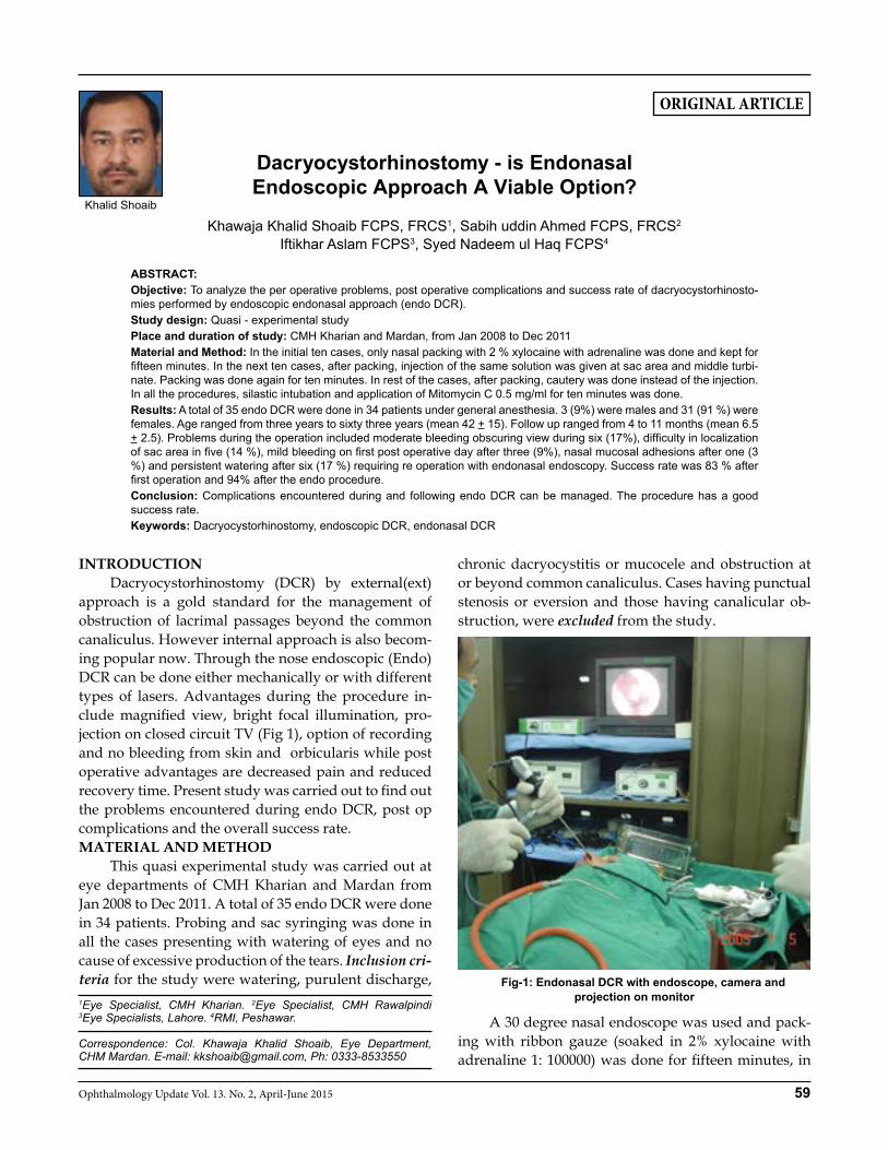
INTRODUCTION Dacryocystorhinostomy (DCR) by external(ext) approach is a gold standard for the management of obstruction of lacrimal passages beyond the common canaliculus. However internal approach is also becom-ing popular now. Through the nose endoscopic (Endo) DCR can be done either mechanically or with different types of lasers. Advantages during the procedure in-clude magnified view, bright focal illumination, pro-jection on closed circuit TV (Fig 1), option of recording and no bleeding from skin and orbicularis while post operative advantages are decreased pain and reduced recovery time. Present study was carried out to find out the problems encountered during endo DCR, post op complications and the overall success rate.MATERIAL AND METHOD This quasi experimental study was carried out at eye departments of CMH Kharian and Mardan from Jan 2008 to Dec 2011. A total of 35 endo DCR were done in 34 patients. Probing and sac syringing was done in all the cases presenting with watering of eyes and no cause of excessive production of the tears. Inclusion cri-teria for the study were watering, purulent discharge,
chronic dacryocystitis or mucocele and obstruction at or beyond common canaliculus. Cases having punctual stenosis or eversion and those having canalicular ob-struction, were excluded from the study.
Fig-1: Endonasal DCR with endoscope, camera and projection on monitor
A 30 degree nasal endoscope was used and pack-ing with ribbon gauze (soaked in 2% xylocaine with adrenaline 1: 100000) was done for fifteen minutes, in
Khawaja Khalid Shoaib FCPS, FRCS1, Sabih uddin Ahmed FCPS, FRCS2
Iftikhar Aslam FCPS3, Syed Nadeem ul Haq FCPS4
ABSTRACT: Objective: To analyze the per operative problems, post operative complications and success rate of dacryocystorhinosto-mies performed by endoscopic endonasal approach (endo DCR). Study design: Quasi - experimental studyPlace and duration of study: CMH Kharian and Mardan, from Jan 2008 to Dec 2011 Material and Method: In the initial ten cases, only nasal packing with 2 % xylocaine with adrenaline was done and kept for fifteen minutes. In the next ten cases, after packing, injection of the same solution was given at sac area and middle turbi-nate. Packing was done again for ten minutes. In rest of the cases, after packing, cautery was done instead of the injection. In all the procedures, silastic intubation and application of Mitomycin C 0.5 mg/ml for ten minutes was done. Results: A total of 35 endo DCR were done in 34 patients under general anesthesia. 3 (9%) were males and 31 (91 %) were females. Age ranged from three years to sixty three years (mean 42 + 15). Follow up ranged from 4 to 11 months (mean 6.5 + 2.5). Problems during the operation included moderate bleeding obscuring view during six (17%), difficulty in localization of sac area in five (14 %), mild bleeding on first post operative day after three (9%), nasal mucosal adhesions after one (3 %) and persistent watering after six (17 %) requiring re operation with endonasal endoscopy. Success rate was 83 % after first operation and 94% after the endo procedure. Conclusion: Complications encountered during and following endo DCR can be managed. The procedure has a good success rate.Keywords: Dacryocystorhinostomy, endoscopic DCR, endonasal DCR
1Eye Specialist, CMH Kharian. 2Eye Specialist, CMH Rawalpindi 3Eye Specialists, Lahore. 4RMI, Peshawar.
Correspondence: Col. Khawaja Khalid Shoaib, Eye Department, CHM Mardan. E-mail: [email protected], Ph: 0333-8533550
Dacryocystorhinostomy - is Endonasal Endoscopic Approach A Viable Option?
Khalid Shoaib
Dacryocystorhinostomy - is Endonasal Endoscopic Approach A Viable Option
60 Ophthalmology Update Vol. 13. No. 2, April-June 2015
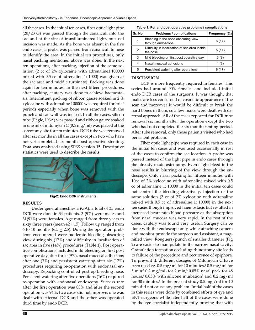
all the cases. In the initial ten cases, fiber optic light pipe (20/23 G) was passed through the canaliculi into the sac and at the site of transilluminated light, mucosal incision was made. As the bone was absent in the five endo cases, a probe was passed from canaliculi to nose to identify the area. In the initial ten procedures, only nasal packing mentioned above was done. In the next ten operations, after packing, injection of the same so-lution (2 cc of 2% xylocaine with adrenaline1:100000 mixed with 0.5 cc of adrenaline 1: 1000) was given at the sac area and middle turbinate). Packing was done again for ten minutes. In the next fifteen procedures, after packing, cautery was done to achieve haemosta-sis. Intermittent packing of ribbon gauze soaked in 2 % xylocaine with adrenaline 100000 was required for brief periods especially when bone was removed with the punch and sac wall was incised. In all the cases, silicon tube (Eagle, USA) was passed and ribbon gauze soaked in one ml of mitomycin C (0.5 mg/ml) was placed at the osteotomy site for ten minutes. DCR tube was removed after six months in all the cases except in two who have not yet completed six month post operative stenting. Data was analyzed using SPSS version 15. Descriptive statistics were used to describe the results.
Fig-2: Endo DCR instruments
RESULTS Under general anesthesia (GA), a total of 35 endo DCR were done in 34 patients. 3 (9%) were males and 31(91%) were females. Age ranged from three years to sixty three years (mean 42 + 15). Follow up ranged from 6 to 10 months (6.5 + 2.5). During the operation prob-lems encountered were moderate bleeding obscuring view during six (17%) and difficulty in localization of sac area in five (14%) procedures (Table 1). Post opera-tive complications included mild bleeding on first post operative day after three (9%), nasal mucosal adhesions after one (3%) and persistent watering after six (17%) procedures requiring re-operation with endonasal en-doscopy. Repacking controlled post op bleeding nose. Persistent watering after five operations (16%) required re-operation with endonasal endoscopy. Success rate after the first operation was 83% and after the second operation was 94%, two cases did not improve, one was dealt with external DCR and the other was operated third time by endo DCR.
Table-1: Per and post operative problems / complications
Sr. No Problems / complications Frequency (%)
1 Bleeding in the nose obscuring view through endoscope 6 (17)
2 Difficulty in localization of sac area inside the nose 5 (14)
3 Mild bleeding on first post operative day 3 (9)
4 Nasal mucosal adhesions 1 (3)
5 Persistent watering after operations 6 (17)
DISCUSSION DCR is more frequently required in females. This series had around 90% females and included initial endo DCR cases of the surgeons. It was thought that males are less concerned of cosmetic appearance of the scar and moreover it would be difficult to break the hard bones in them, so a few males were dealt with ex-ternal approach. All of the cases reported for DCR tube removal six months after the operation except the two who had not completed the six month stenting period. After tube removal, only those patients visited who had persistent problem. Fiber optic light pipe was required in each case in the initial ten cases and was used occasionally in rest of the cases to confirm the sac location. A probe was passed instead of the light pipe in endo cases through the already made osteotomy. Even slight bleed in the nose results in blurring of the view through the en-doscope. Only nasal packing for fifteen minutes with 10cc of 2% xylocaine with adrenaline mixed with 0.5 cc of adrenaline 1: 10000 in the initial ten cases could not control the bleeding effectively. Injection of the same solution (2 cc of 2% xylocaine with adrenaline mixed with 0.5 cc of adrenaline 1: 10000) in the next ten cases though improved haemostasis but resulted in increased heart rate/blood pressure as the absorption from nasal mucosa was very rapid. In the rest of the cases, cautery was found very useful. Surgery can be done with the endoscope only while attaching camera and monitor provide the surgeon and assistant, a mag-nified view. Ronguers/punch of smaller diameter (Fig 2) are easier to manipulate in the narrow nasal cavity. Granulation formation occluding rhinostomy site leads to failure of the procedure and recurrence of epiphora. To prevent it, different dosages of Mitomycin C have been used eg. 0.5 mg/ml for 10 minutes,1 0.5 mg/ml for 5 min,2 0.2 mg/mL for 2 min,3 0.05% nasal pack for 48 hours,4 0.03% with silicone intubation5 and 0.2 mg/ml for 30 minutes.6 In the present study 0.5 mg /ml for 10 min did not cause any problem. Initial half of the cases in this series were done by combined efforts of eye and ENT surgeons while later half of the cases were done by the eye specialist independently proving that with
Dacryocystorhinostomy - is Endonasal Endoscopic Approach A Viable Option
61Ophthalmology Update Vol. 13. No. 2, April-June 2015
learning either of the two can do the procedure. Endo DCR has been done for dacryocystocoele in a 4 month old infant7 and in adults8,9 It has been found to be safe and effective procedures for the management of persistent epiphora in children10 and for adults.11 The common insertion of the upper and lower canaliculus of the lacrimal sac has been repaired with endoscopic DCR followed by silicone stenting.12 Formation of mu-cosal flaps at the end of the operations has been claimed to improve success rate13, 14 and has been termed pow-ered endonasal DCR by some while many used the term mechanical endonasal dacryocystorhinostomy (MENDCR)15 when there is large rhinostomy and mu-cosal flaps. Success rates of MENDCR 92%14 and 93.5%16
were found to compare favorably with that of standard external DCR 95.8%.17 In a few studies, success was in-ferior (86% endo - 94% ext)17 with endo DCR18 while in other studies, success rates after endo DCR have been found to be equal to that of external DCR.19 Many think that best endo DCR results are achieved by stenting or removal of the medial wall of the lacrimal sac.20 while a few recommend no intubation because of similar sur-gical success rates, and granulation formation, patient discomfort, and increased cost with intubation.21 Nasal endoscopy has been recommended before and after ex-ternal DCR2 and to treat a failed external DCR.23 CONCLUSION Problems/complications encountered during Endo DCR can be managed as the procedure has good success rate.REFERENCES1. Angela M. Dolmetsch. Nonlaser Endoscopic Endonasal Dacry-
ocystorhinostomy with Adjunctive Mitomycin C in Nasolacri-mal Duct Obstruction in Adults. Ophthalmology Volume 117, Issue 5, Pages 1037-1040 (May 2010)
2. Dolmetsch AM, Gallon MA, Holds JB. Nonlaser endoscopic endonasal dacryocystorhinostomy with adjunctive mito-mycin C in children. Ophthal Plast Reconstr Surg. 2008 Sep-Oct;24(5):390-3.
3. Tabatabaie SZ, Heirati A, Rajabi MT, Kasaee A. Silicone intuba-tion with intraoperative mitomycin C for nasolacrimal duct ob-struction in adults: a prospective, randomized, double-masked study. Ophthal Plast Reconstr Surg. 2007 Nov-Dec;23(6):455-8
4. Rathore PK, Kumari Sodhi P, Pandey RM. Topical mitomycin C as a postoperative adjunct to endonasal dacryocystorhinos-tomy in patients with anatomical endonasal variants. Orbit. 2009;28(5):297-302.
5. Nemet AY, Wilcsek G, Francis IC. Endoscopic dacryocystorhi-nostomy with adjunctive mitomycin C for canalicular obstruc-tion. Orbit. 2007 Jun;26(2):97-100
6. Liao S, Kao S, Tseng J, Chen M, Hou P. Results of intraopera-
tive mitomycin C application in dacryocystorhinostomy. Br J Ophthalmol. 2000 August; 84(8): 903–906.
7. Mladina R., Stiglmayer N., Dawidowsky K., Jukic T., Jurlina M., Trupkovic-Fotivec B. Endonasal endoscopic dacryocyst-orhinostomy for dacryocystocoele in a 4 month old infant. Br J Ophthalmol. 2001 January; 85(1): 110.
8. Eloy P, Martinez A, Leruth E, Levecq L, Bertrand B. Endonasal endoscopic dacryocystorhinostomy for a primary dacryocyst-ocele in an adult. B-ENT. 2009;5(3):179-82.
9. Plaza G, Nogueira A, González R, Ferrando J, Toledano N. Sur-gical treatment of familial dacryocystocele and lacrimal puncta agenesis. Ophthal Plast Reconstr Surg. 2009 Jan-Feb;25(1):52-3.
10. Marr J E, Drake-Lee A, Willshaw H E. Management of child-hood epiphora. Br J Ophthalmol. 2005 September; 89(9): 1123–1126.
11. Shiraz Aslam, Abdul Hamid Awan, Mohammad Tayyab. En-doscopic Dacrocystorhinostomy: A Pakistani Experience. Pak J Ophthalmol 2010; 26 (1):2-6
12. Khan H A, Bayat A, De Carpentier JP. Endoscopic Dacrocyst-orhinostomy in Lacrimal Canalicular Trauma. Ann R Coll Surg Engl. 2007 January; 89(1): 43.
13. Trimarchi M, Giordano Resti A, Bellini C, Forti M, Bussi M. Anastomosis of nasal mucosal and lacrimal sac flaps in endo-scopic dacryocystorhinostomy. Eur Arch Otorhinolaryngol. 2009 Nov;266(11):1747-52.
14. Sonkhya N, Mishra P. Endoscopic transnasal dacryocystorhi-nostomy with nasal mucosal and posterior lacrimal sac flap. J Laryngol Otol. 2009 Mar;123(3):320-6.
15. Tan NC, Rajapaksa SP, Gaynor J, Nair SB. Mechanical endona-sal dacryocystorhinostomy--a reproducible technique. Rhinol-ogy. 2009 Sep;47(3):310-5.
16. Tsirbas A, Davis G, Wormald PJ. Mechanical endonasal dacry-ocystorhinostomy versus external dacryocystorhinostomy. Ophthal Plast Reconstr Surg. 2004 Jan;20(1):50-6.
17. Leong SC, Karkos PD, Burgess P, Halliwell M, Hampal S. A comparison of outcomes between nonlaser endoscopic endo-nasal and external dacryocystorhinostomy: single-center expe-rience and a review of British trends. Am J Otolaryngol. 2010 Jan-Feb;31(1):32-7.
18. Zílelíog˘lu G, Tekeli O, Ug˘urbas SH, Akiner M, Aktürk T, An-adolu Y. Results of endoscopic endonasal non-laser Dacryocys-torhinostomy. Documenta Ophthalmologica 105: 57–62, 2002
19. Leong SC, Macewen CJ, White PS. A systematic review of out-comes after dacryocystorhinostomy in adults. Am J Rhinol Al-lergy. 2010 Jan-Feb;24(1):81-90.
20. de Souza C, Nissar J. Experience with endoscopic dacryocys-torhinostomy using four methods.Otolaryngol Head Neck Surg. 2010 Mar;142(3):389-93.
21. Unlu HH, MD, Gunhan K, Baser EF, Songu M. Long-term re-sults in endoscopic dacryocystorhinostomy: Is intubation re-ally required?Otolaryngology–Head and Neck Surgery (2009) 140, 589-595
22. Elmorsy SM, Fayk HM. Nasal endoscopic assessment of failure after external dacryocystorhinostomy. Orbit. 2010 Aug;29(4):197-201.
23. Choussy O, Retout A, Marie JP, Cozlean A, Dehesdin D. En-doscopic revision of external dacryocystorhinostomy failure. Rhinology. 2010 Mar 2;48(1):104-107.
62 Ophthalmology Update Vol. 13. No. 2, April-June 2015
ORIGINAL ARTICLE
INTRODUCTION Vascular Endothelial Growth Factors (VEGF) plays an important role in many ocular pathologies, both of the anterior and the posterior segment, lead-ing mainly to complications like neo-vascularization and macular edema. Since the introduction of anti- vas-cular endothelial growth factor therapy in 2005, it has gained wide spread popularity among ophthalmolo-gists worldwide.1-6 Although not FDA approved, ‘off label’ use of Bevacizumab has been in practice since June 2005.7 It has been used with promising results in conditions like Age related macular degeneration, pro-liferative diabetic retinopathy, neovascular glaucoma, clinically significant diabetic macular edema and mac-ular edema due to vascular occlusions.1,3-6,8-10
Commercially available bevacizumab (Avastin®;
Genetech, San Francisco USA is available in preserva-tive-free 100 mg/4 ml vials, and is intended for use at relatively high concentrations on a single colon cancer patient. In this era of tremendous emphasis on health care cost containment in both developed and develop-ing countries, it is a common practice among hospitals, clinics, and compounding pharmacies to divide the large volume of bevacizumab into smaller units that are suitable for single-use intravitreal doses for individual eyes.11
MATERIALS AND METHODS Avastin® service is being provided in Department of Ophthalmology, Khyber Teaching Hospital, Pesha-war since August 2011. The duration of this report is from 1st January 2012 to 31st December 2012. A retro-spective analysis of all the Avastin patients admitted in the department during the period was done as part of the annual audit of the service. After elaborating detailed history of decreased vision, all the patients underwent complete ophthalmological examination including visual acuity using the Snellen’s visual acu-ity charts, best corrected visual acuity, intraocular pres-sure (IOP) measurement using the Goldman appla-nation Tonometer, pupils, slit- lamp examination for anterior and posterior segment evaluation and dilated fundus examination using the 90D lens (Volk, USA). All intravitreal Avastin were advised by consultant ophthalmologists. All the patients were admitted and
Akhunzada Muhammad Aftab FCPS1, Awais Rauf MBBS2, Farooq Khan MBBS3
Syed Bilal Hassan Zaidi MBBS4, Prof. Mustafa Iqbal FRCS, MRCOphth5
Syeda Ghazala Shahnawaz MBBS, D.O6
ABSTRACTIntroduction: Since the introduction of Anti-VEGF therapy in 2005, it has been extensively used in treating ophthalmic conditions like proliferative diabetic retinopathy, age related macular degeneration and macular edema. However intravitreal route of administration predisposes to ophthalmic complications along with few systemic adverse events too. Materials and Methods: A retrospective analysis of the records of all patients admitted for intravitreal bevacizumab therapy was performed during 1st January 2012 to 31st December 2012. All patients under went complete ophthalmic and systemic evaluation especially to evaluate the cardiovascular risk factors. Multiple doses of 2.5mg/ 0.1ml of bevacizumab were given from a single vial in multiple settings in a sterile environment. Ocular and systemic complications were analyzed on 1stpost operative day, 7th day and after 4 weeks. Results: Ocular complications included sub conjunctival hemorrhage (2.19%), crystalline lens trauma (0.69%), transient rise of IOP (0.3%), endophthalmitis (0.11%), mild uveitis (0.2%), conjunctival injection with punctate erosions (0.11%) and regurgitation of drug (0.4%). No systemic side effects of therapy were seen during the study period. Conclusion: Services provided at our institute meet the international standards and all the adverse effects and (or) complications are within inter-national standards despite use of single vial for multiple doses and multiple settings.Key words: Bevacizumab, Intravitreal, Neovascularization, Proliferative Diabetic Retinopathy.
1Registrar Eye A Unit Department of Ophthalmology, Khyber Teaching Hospital, Peshawar, 2,3,4Traniee Medical Officers, A Unit Department of Ophthalmology, Khyber Teaching Hospital, Peshawar, 5Prof. & Head of Ophthalmology Department, Khyber Teaching Hospital, Peshawar, 6Registrar, Ophthalmology Department, Khyber Teaching Hospital, Peshawar.
Correspondence: Dr. Akhunzada Muhammad Aftab c/o Prof. Dr. Muhammad Ibrar, Department of Botany, University of Peshawar, Peshawar. Cell: 03339106060, E-Mail: [email protected]
Received: January 2015 Accepted: February 2015
Financial disclosure: There has been no financial interest involved in this study
Ocular and systemic Complications of Intravitreal Bevacizumab (Avastin) therapy
(12 months audit report)Akhunzada M. Aftab
Ocular and systemic Complications of Intravitreal Bevacizumab (Avastin) therapy
63Ophthalmology Update Vol. 13. No. 2, April-June 2015
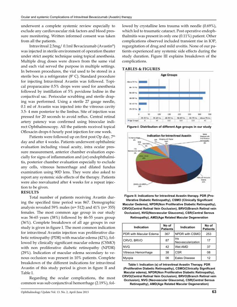
underwent a complete systemic review especially to exclude any cardiovascular risk factors and blood pres-sure monitoring. Written informed consent was taken from all the patients. Intravitreal 2.5mg/ 0.1ml Bevacizumab (Avastin®) was injected in sterile environment of operation theater under strict aseptic technique using topical anesthesia. Multiple drug doses were drawn from the same vial and each vial served the purpose in multiple settings. In between procedures, the vial used to be stored in a sterile box in a refrigerator (80 C). Standard procedure for injecting Intravitreal Avastin was followed. Topi-cal proparacaine 0.5% drops were used for anesthesia followed by instillation of 5% povidone Iodine in the conjuctival sac. Periocular scrubbing and sterile drap-ing was performed. Using a sterile 27 gauge needle, 0.1 ml of Avastin was injected into the vitreous cavity 3.5- 4 mm posterior to the limbus. Site of injection was pressed for 20 seconds to avoid reflux. Central retinal artery patency was confirmed using binocular indi-rect Ophthalmoscopy. All the patients received topical Ofloxacin drops 6 hourly post injection for one week. Patients were followed up on first post Op day, 7th day and after 4 weeks. Patients underwent ophthalmic evaluation including visual acuity, intra ocular pres-sure measurement, anterior chamber evaluation espe-cially for signs of inflammation and (or) endophthalmi-tis, posterior chamber evaluation especially to exclude any cells, vitreous hemorrhage and dilated fundus examination using 90D lens. They were also asked to report any systemic side effects of the therapy. Patients were also reevaluated after 4 weeks for a repeat injec-tion to be given. RESULTS Total number of patients receiving Avastin dur-ing the specified time period was 867. Demographic analysis revealed 59% males (n= 512) and 41% (n= 355) females. The most common age group in our study was 56-65 years (38%) followed by 46-55 years group (36%). Complete breakdown of all age groups in our study is given in figure I. The most common indication for intravitreal Avastin injection was proliferative dia-betic retinopathy (PDR) with macular edema (42%), fol-lowed by clinically significant macular edema (CSMO) with non proliferative diabetic retinopathy (NPDR)(29%). Indication of macular edema secondary to ve-nous occlusion was present in 10% patients. Complete breakdown of the different indications for intravitreal Avastin of this study period is given in figure II and Table I. Regarding the ocular complications, the most common was sub conjunctival hemorrhage (2.19%), fol-
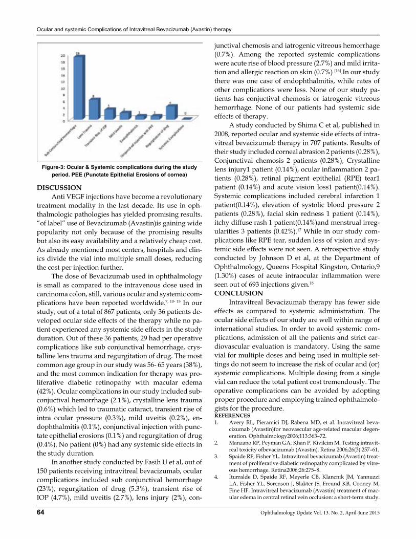
lowed by crystalline lens trauma with needle (0.69%), which led to traumatic cataract. Post operative endoph-thalmitis was present in only one (0.11%) patient. Other complications observed included transient rise in IOP, regurgitation of drug and mild uveitis. None of our pa-tients experienced any systemic side effects during the study duration. Figure III explains breakdown of the complications.
TABLES & FIGURES
Figure-I: Distribution of different Age groups in our study
Figure II: Indications for Intravitreal Avastin therapy. PDR (Pro-liferative Diabetic Retinopathy), CSMO (Clinically Significant
Macular Oedema), NPDR(Non Proliferative Diabetic Retinopathy), CRVO(Central Retinal Vein Occlusion), BRVO(Branch Retinal vein
Occlusion), NVG(Neovascular Glaucoma), CSR(Central Serous Retinopathy), AMD(Age Related Macular Degeneration
Table I: Indication (s) of Intravitreal Avastin Therapy. PDR (Proliferative Diabetic Retinopathy), CSMO(Clinically Significant Macular edema), NPDR(Non Proliferative Diabetic Retinopathy),
CRVO(Central Retinal Vein Occlusion), BRVO(Branch Retinal vein Occlusion), NVG (Neovascular Glaucoma), CSR(Central Serous
Retinopathy), AMD(Age Related Macular Degeneration)
Indication No of Patients Indication No of
PatientsPDR with Macular Edema 367 NPDR with CSMO 253
CRVO, BRVO 87 IrisNeovascularization 17
NVG 42 Wet AMD 37
Vitreous Hemorrhage 38 CSR 08
Myopia 06 Eales Disease 12
Ocular and systemic Complications of Intravitreal Bevacizumab (Avastin) therapy
64 Ophthalmology Update Vol. 13. No. 2, April-June 2015
DISCUSSION Anti VEGF injections have become a revolutionary treatment modality in the last decade. Its use in oph-thalmologic pathologies has yielded promising results. “of label” use of Bevacizumab (Avastin)is gaining wide popularity not only because of the promising results but also its easy availability and a relatively cheap cost. As already mentioned most centers, hospitals and clin-ics divide the vial into multiple small doses, reducing the cost per injection further. The dose of Bevacizumab used in ophthalmology is small as compared to the intravenous dose used in carcinoma colon, still, various ocular and systemic com-plications have been reported worldwide.7, 10- 15 In our study, out of a total of 867 patients, only 36 patients de-veloped ocular side effects of the therapy while no pa-tient experienced any systemic side effects in the study duration. Out of these 36 patients, 29 had per operative complications like sub conjunctival hemorrhage, crys-talline lens trauma and regurgitation of drug. The most common age group in our study was 56- 65 years (38%), and the most common indication for therapy was pro-liferative diabetic retinopathy with macular edema (42%). Ocular complications in our study included sub-conjuctival hemorrhage (2.1%), crystalline lens trauma (0.6%) which led to traumatic cataract, transient rise of intra ocular pressure (0.3%), mild uveitis (0.2%), en-dophthalmitis (0.1%), conjunctival injection with punc-tate epithelial erosions (0.1%) and regurgitation of drug (0.4%). No patient (0%) had any systemic side effects in the study duration. In another study conducted by Fasih U et al, out of 150 patients receiving intravitreal bevacizumab, ocular complications included sub conjunctival hemorrhage (23%), regurgitation of drug (5.3%), transient rise of IOP (4.7%), mild uveitis (2.7%), lens injury (2%), con-
junctival chemosis and iatrogenic vitreous hemorrhage (0.7%). Among the reported systemic complications were acute rise of blood pressure (2.7%) and mild irrita-tion and allergic reaction on skin (0.7%) [16].In our study there was one case of endophthalmitis, while rates of other complications were less. None of our study pa-tients has conjuctival chemosis or iatrogenic vitreous hemorrhage. None of our patients had systemic side effects of therapy. A study conducted by Shima C et al, published in 2008, reported ocular and systemic side effects of intra-vitreal bevacizumab therapy in 707 patients. Results of their study included corneal abrasion 2 patients (0.28%), Conjunctival chemosis 2 patients (0.28%), Crystalline lens injury1 patient (0.14%), ocular inflammation 2 pa-tients (0.28%), retinal pigment epithelial (RPE) tear1 patient (0.14%) and acute vision loss1 patient(0.14%). Systemic complications included cerebral infarction 1 patient(0.14%), elevation of systolic blood pressure 2 patients (0.28%), facial skin redness 1 patient (0.14%), itchy diffuse rash 1 patient(0.14%)and menstrual irreg-ularities 3 patients (0.42%).17 While in our study com-plications like RPE tear, sudden loss of vision and sys-temic side effects were not seen. A retrospective study conducted by Johnson D et al, at the Department of Ophthalmology, Queens Hospital Kingston, Ontario,9 (1.30%) cases of acute intraocular inflammation were seen out of 693 injections given.18
CONCLUSION Intravitreal Bevacizumab therapy has fewer side effects as compared to systemic administration. The ocular side effects of our study are well within range of international studies. In order to avoid systemic com-plications, admission of all the patients and strict car-diovascular evaluation is mandatory. Using the same vial for multiple doses and being used in multiple set-tings do not seem to increase the risk of ocular and (or) systemic complications. Multiple dosing from a single vial can reduce the total patient cost tremendously. The operative complications can be avoided by adopting proper procedure and employing trained ophthalmolo-gists for the procedure.REFERENCES1. Avery RL, Pieramici DJ, Rabena MD, et al. Intravitreal beva-
cizumab (Avastin)for neovascular age-related macular degen-eration. Ophthalmology2006;113:363–72.
2. Manzano RP, Peyman GA, Khan P, Kivilcim M. Testing intravit-real toxicity ofbevacizumab (Avastin). Retina 2006;26(3):257–61.
3. Spaide RF, Fisher YL. Intravitreal bevacizumab (Avastin) treat-ment of proliferative diabetic retinopathy complicated by vitre-ous hemorrhage. Retina2006;26:275–8.
4. Iturralde D, Spaide RF, Meyerle CB, Klancnik JM, Yannuzzi LA, Fisher YL, Sorenson J, Slakter JS, Freund KB, Cooney M, Fine HF. Intravitreal bevacizumab (Avastin) treatment of mac-ular edema in central retinal vein occlusion: a short-term study.
Figure-3: Ocular & Systemic complications during the study period. PEE (Punctate Epithelial Erosions of cornea)
Ocular and systemic Complications of Intravitreal Bevacizumab (Avastin) therapy
65Ophthalmology Update Vol. 13. No. 2, April-June 2015
Retina 2006;26:279–84.5. Avery RL. Regression of retinal and iris neovascularization
after Intravitreal bevacizumab (Avastin) treatment. Retina 2006;26:352–4.
6. Mason JO III, Albert MA Jr, Vail R. Intravitreal bevacizumab (Avastin) for
7. Refractoryp seudophakic cystoid macular edema. Retina 2006;26:356–7.
8. FungA E, RosenfeldP J, ReichelE. The International Intravitreal Bevacizumab SafetySurvey: using the internet to assess drug safety worldwide. Br J Ophthalmol 2006;90:1344-49.
9. Kahook MY, Schuman JS, Noecker RJ. Intravitreal bevacizum-ab in a patient withneovascular glaucoma. Ophthalmic Surg Lasers Imaging 2006;37:144-6.
10. Spaide RF, Laud K, Fine HF,Klancnik JM Jr, Meyerle CB, Yan-nuzzi LA, Sorenson J, Slakter J, Fisher YL, Cooney MJ. Intra-vitreal bevacizumab treatment ofchoroidal neovasculariza-tion secondary to age-related macular Degeneration. Retina 2006;26(4):383–90.
11. Rich RM, Rosenfeld PJ, Puliafito CA,Dubovy SR, Davis JL, Flynn HW Jr, Gonzalez S, Feuer WJ, Lin RC, Lalwani GA, Nguyen JK, Kumar G. Short-term safety and efficacy of in-travitreal bevacizumab (avastin) for neovascular age-related macular degeneration. Retina 2006;26(5):495–511.
12. Danny S N, Alvin KK, Clement WC, Walton WT. Intravitreal bevacizumab: safety of multiple doses from a single vial for consecutive patients. Hong Kong Med J 2012;18:488-95.
13. Ladas ID, Karagiannis DA, Rouvas AA, Kotsolis AI, Liotsou
A, Vergados I. Safety of repeat intravitreal injections of beva-cizumab versus ranibizumab: our experience after 2,000 injec-tions. Retina. 2009; 29(3):313-18.
14. Fintak DR, Shah GK, Blinder KJ, et al. Incidence of endoph-thalmitis related to intravitreal injection of bevacizumab and ranibizumab. Retina. 2008; 28(10):1395– 99.
15. Diago T, McCannel CA, Bakri SJ, Pulido JS, Edwards AO, Pach JM. Infectious endophthalmitis after intravitreal injection of an-tiangiogenic agents. Retina. 2009; 29(5):601– 05.
16. Wu L, Martinez- Castellanos MA, Quiroz-Mercado H, Areva-lo JF, Berrocal MH, Farah ME, Maia M, Roca JA, Rodriguez FJ; Pan American Collaborative Retina Group (PACORES). Twelve-month safety of intravitreal injections of bevacizumab (Avastin): results of the Pan-American Collaborative Retina Study Group (PACORES). Graefes Arch Clin Exp Ophthalmol. 2008; 246(1):81–87.
17. Fasih U, Shaikh N, Rahman A, Sultan S, Fahimi MS, Shaikh A. Ocular and systemic complications of intravitreal injection bevacizumab( avastin ) in one year follow up(a study of 150 cases). J Pak Med Assoc. 2013;63(6):707-10.
18. Shima C, Sakaguchi H, Gomi F, Kamei M, Ikuno Y, Osh, Sawa M, Tsujikawa M, Kusaka S, Tano Y. Complications in patients after intravitreal injection ofbevacizumab. Acta Oph-thalmol. 2008; 86:372–76.
19. Johnson D, Hollands H, Hollands S, Sharma S. Incidence and characteristics of acute intraocular inflammation after intravit-real injection of bevacizumab: A retrospective cohort study. Can J Ophthalmol. 2010;45(3):239-42.
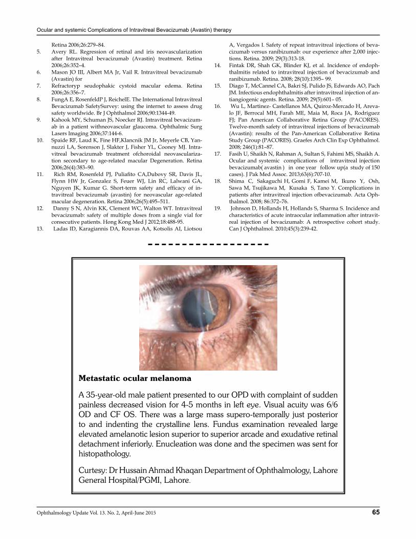
Metastatic ocular melanoma
A 35-year-old male patient presented to our OPD with complaint of sudden painless decreased vision for 4-5 months in left eye. Visual acuity was 6/6 OD and CF OS. There was a large mass supero-temporally just posterior to and indenting the crystalline lens. Fundus examination revealed large elevated amelanotic lesion superior to superior arcade and exudative retinal detachment inferiorly. Enucleation was done and the specimen was sent for histopathology.
Curtesy: Dr Hussain Ahmad Khaqan Department of Ophthalmology, Lahore General Hospital/PGMI, Lahore.
66 Ophthalmology Update Vol. 13. No. 2, April-June 2015
ORIGINAL ARTICLE
M. Idris
INTRODUCTION Penetrating ocular trauma is a serious type of in-jury to the globe. Intraocular foreign bodies (IOFBs) are the major cause of penetrating ocular trauma and the most serious problem is the resulting impairment of visual function. Special attention should be paid on primary and secondary complications, which include mechanical lesions of the ocular tissues, metallosis and endophthalmitis.1 Ocular trauma associated with intraocular foreign bodies (IOFBs) is one of the ma-jor causes of visual impairment in young individuals. Various reports indicate that 18-41% of all open globe injuries involve at least one IOFB.2
In a study, the intraocular foreign body in open globe injury was found in 45 eyes (38%). In our study it was seen in 37% cases.3 Most of the victims are male
and young patients working on fields which are ex-posed because of their occupations. In this regard, lack of awareness regarding protective goggles and early referral to eye specialist for urgent management is lack-ing.4, 5 Most ocular injuries in this rural population oc-curred at the workplace, suggesting the need to explore workplace strategies to minimize ocular trauma as a priority. Eye care programs targeting high-risk ocular trauma groups may need to consider ocular trauma as a priority in eye health awareness strategies in order to reduce its incidence.6
METHODOLOGY The study was carried out at Department of Oph-thalmology, Govt Lady Reading Hospital, Peshawar from July 2010 to Jan 2013. We received 100 cases from outdoor department and were admitted for manage-ment. This was a prospective, interventional case series of consecutive patients with IOFBs. Patients were exam-ined after detailed history and important finding were noted. The following variables were recorded for the purpose of the study: age, gender, cause of trauma, oc-cupation, complications, presenting best-corrected vis-ual acuity (BCVA), slit lamp and fundus examination, ultrasound examination when ophthalmoscopy was not possible, foreign body localization based on orbital
Mohammad Idris FCPS1, Zubairullah Khan FCPS2, Hasan Yaqoob FRCS3
Asim Ali Shah FCPS4
ABSTRACTObjective: to determine the Frequency of Intraocular foreign body in penetrating injury presented to a tertiary care centre of Khyber Pakhtunkhwa for management and its visual outcome.Study design: prospective, interventional case seriesMaterial and Methods: The study was carried out at Department of Ophthalmology, Govt Lady Reading Hospital, Pesha-war from July 2010 to Jan 2013. We received 100 cases from outdoor department for management. Patients were examined after detailed history and important findings noted. Data was collected on special proforma and was analyzed with the help of SPSS Version16.Results: The study comprised of 100 cases. In 37 (37%) patients with penetrating ocular injury, IOFB was found. 73 (73%) patients were male and majority was young patients. Students and children were in majority, 38 (38%) patients were stu-dents, 35 (35%) patients were labors, and 15 (15%) patients were related to sports and defense. Commonest reason of penetrating injury was toys, stone and metal and glass pieces. Main reason for poor visual acuity was late presentation and BBI (bomb blast injuries).Conclusion: Occupation like labor, sports, defense and children are persons who are constantly prone to penetrating trauma and IOFB. In case of school children, teachers can play a vital role in prevention and timely referral to a tertiary care centre. Commonly male and young people are risk group people and should be advised to wear protective goggles during outdoor work. Visual progression was poor in majority of the eyes; delayed presentation and BBI were the top reasons. Most serious cause of penetrating trauma was BBI. Key words: penetrating trauma, intraocular foreign body, ocular trauma, visual outcome.
1Medical Officer, Ophthalmology UNIT, PGMI, LRH Peshawar, KPK, 2Senior Medical Officer, Ophthalmology UNIT, PGMI, LRH Peshawar, KPK, 3Consultant, Ophthalmology UNIT, North West General, Hos-pital, Peshawar, KPK, 4Medical Officer, Ophthalmology UNIT, PGMI, LRH Peshawar, KPK.
Correspondence: Dr. Mohammad Idris, Medical Officer, Ophthalmol-ogy UNIT, PGMI, Lady Reading Hospital, Peshawar.Cell No.: +92-333-9182595, Email: [email protected] Address: Ophthalmology UNIT, PGMI, LRH Peshawar, KPK
Received: August 2014 Accepted: November 2014
Incidence of Intraocular Foreign Body in Penetrating Trauma presented to a Tertiary
Care Hospital of Khyber Pakhtun Khwa and its Visual Outcome
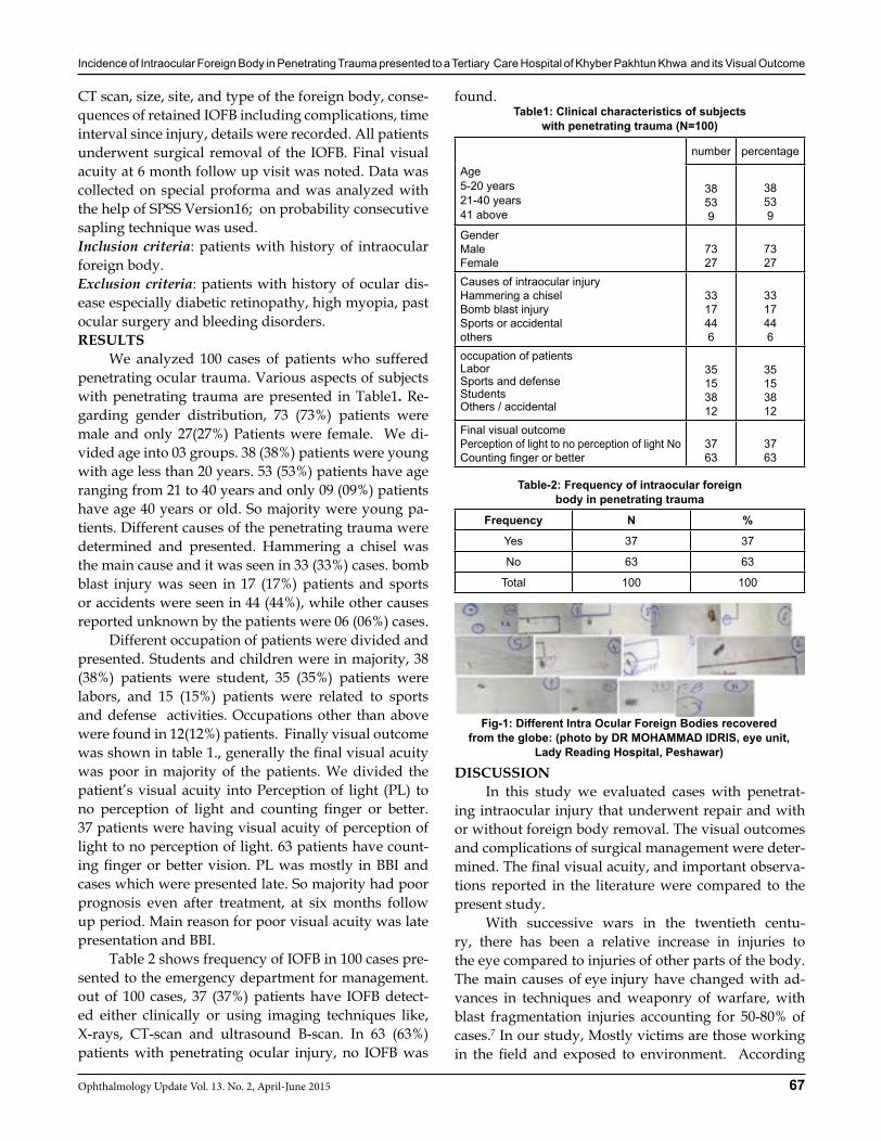
Incidence of Intraocular Foreign Body in Penetrating Trauma presented to a Tertiary Care Hospital of Khyber Pakhtun Khwa and its Visual Outcome
67Ophthalmology Update Vol. 13. No. 2, April-June 2015
CT scan, size, site, and type of the foreign body, conse-quences of retained IOFB including complications, time interval since injury, details were recorded. All patients underwent surgical removal of the IOFB. Final visual acuity at 6 month follow up visit was noted. Data was collected on special proforma and was analyzed with the help of SPSS Version16; on probability consecutive sapling technique was used.Inclusion criteria: patients with history of intraocular foreign body.Exclusion criteria: patients with history of ocular dis-ease especially diabetic retinopathy, high myopia, past ocular surgery and bleeding disorders.RESULTS We analyzed 100 cases of patients who suffered penetrating ocular trauma. Various aspects of subjects with penetrating trauma are presented in Table1. Re-garding gender distribution, 73 (73%) patients were male and only 27(27%) Patients were female. We di-vided age into 03 groups. 38 (38%) patients were young with age less than 20 years. 53 (53%) patients have age ranging from 21 to 40 years and only 09 (09%) patients have age 40 years or old. So majority were young pa-tients. Different causes of the penetrating trauma were determined and presented. Hammering a chisel was the main cause and it was seen in 33 (33%) cases. bomb blast injury was seen in 17 (17%) patients and sports or accidents were seen in 44 (44%), while other causes reported unknown by the patients were 06 (06%) cases. Different occupation of patients were divided and presented. Students and children were in majority, 38 (38%) patients were student, 35 (35%) patients were labors, and 15 (15%) patients were related to sports and defense activities. Occupations other than above were found in 12(12%) patients. Finally visual outcome was shown in table 1., generally the final visual acuity was poor in majority of the patients. We divided the patient’s visual acuity into Perception of light (PL) to no perception of light and counting finger or better. 37 patients were having visual acuity of perception of light to no perception of light. 63 patients have count-ing finger or better vision. PL was mostly in BBI and cases which were presented late. So majority had poor prognosis even after treatment, at six months follow up period. Main reason for poor visual acuity was late presentation and BBI. Table 2 shows frequency of IOFB in 100 cases pre-sented to the emergency department for management. out of 100 cases, 37 (37%) patients have IOFB detect-ed either clinically or using imaging techniques like, X-rays, CT-scan and ultrasound B-scan. In 63 (63%) patients with penetrating ocular injury, no IOFB was
found.Table1: Clinical characteristics of subjects
with penetrating trauma (N=100)
DISCUSSION In this study we evaluated cases with penetrat-ing intraocular injury that underwent repair and with or without foreign body removal. The visual outcomes and complications of surgical management were deter-mined. The final visual acuity, and important observa-tions reported in the literature were compared to the present study. With successive wars in the twentieth centu-ry, there has been a relative increase in injuries to the eye compared to injuries of other parts of the body. The main causes of eye injury have changed with ad-vances in techniques and weaponry of warfare, with blast fragmentation injuries accounting for 50-80% of cases.7 In our study, Mostly victims are those working in the field and exposed to environment. According
Age5-20 years21-40 years41 above
number percentage
38539
38539
GenderMaleFemale
7327
7327
Causes of intraocular injuryHammering a chiselBomb blast injurySports or accidentalothers
3317446
3317446
occupation of patientsLaborSports and defenseStudentsOthers / accidental
35153812
35153812
Final visual outcome Perception of light to no perception of light NoCounting finger or better
3763
3763
Table-2: Frequency of intraocular foreign body in penetrating trauma
Frequency N %
Yes 37 37
No 63 63
Total 100 100
Fig-1: Different Intra Ocular Foreign Bodies recovered from the globe: (photo by DR MOHAMMAD IDRIS, eye unit,
Lady Reading Hospital, Peshawar)
Incidence of Intraocular Foreign Body in Penetrating Trauma presented to a Tertiary Care Hospital of Khyber Pakhtun Khwa and its Visual Outcome
68 Ophthalmology Update Vol. 13. No. 2, April-June 2015
to different studies7, 8, 9 despite early referral, BBI were having worse prognosis and despite proper manage-ment and early intervention, results and final visual outcome were poor and disappointing. It was mainly because of multiple and complex type of injuries and severe ocular damage. The most common causes of open globe injury are domestic accidents and occupational injuries. Sig-nificant prognostic factors for final visual outcome in patients with open globe injury are initial visual acuity, posterior extent and length of wound, presence of hy-phaema and presence of vitreous prolapse. Awareness of the factors predicting a poor visual outcome may be helpful during counseling of patients with open globe injuries.10
Several studies confirm that trauma of any type is common in male11 in our study males were in ma-jority also. Similarly young to middle age people are the common group of people exposed to both acciden-tal as well as occupational trauma.4, 5 Most of our pa-tients were less than 40 years age. Penetrating ocular injuries with retained posterior segment foreign bod-ies are challenging cases requiring urgent attention by vitreoretinal surgeons. Posteriorly located injuries can result in serious immediate and delayed vitreoretinal sequelae, such as retinal detachment and endophthal-mitis. De Souza S et al, reported the rates of retinal de-tachment and endophthalmitis were 41% (17/41) and 17% (7/41) respectively.12 Several studies have shown that the visual prognosis is poor. In a study, Visual acu-ity on admission between 6/60 to PL comprises highest number (64%) and also on discharge between 6/60 to PL comprises highest number of cases (50%) of IOFB.3
In eye injury patients, the nature of the foreign body determines the clinical behavior; inert objects such as steel and glass may not cause significant inflamma-tion to warrant their removal. Removal of organic for-eign bodies, however, is mandatory since these objects usually lead to secondary infection, like endophthalmi-tis.13
CONCLUSION Occupation like labor, sports, defense and chil-dren are persons who are constantly prone to pen-etrating trauma and IOFB. In case of school children, teachers can play a vital role in prevention and timely referral to a tertiary care centre. Commonly male and young people are risk group people and should be ad-vised to wear protective goggles during outdoor work.
Visual progression was poor in majority of the eyes due to delayed presentation and BBI were the top reasons. Most serious cause of penetrating trauma was BBI. FBs like wood and stone were strongly associated with en-dophthalmitis which needs local and systemic antibi-otics should be advocated in any trauma particularly contaminated ones. Recommendations: Awareness regarding early re-ferred to the tertiary care hospital, when facilities of vit-rectomy is available for IOFB removal with out delay. Any FB can enter the eye and cause damage so the need of imaging is stressed in every suspected case.Majority of patients with IOFB were male, laborer and workers, so the incidence can easily be reduced with adopting simple measures like safety goggles use during work because in most of the cases prognosis is poor and pre-vention is better.REFERENCES1 Obuchowska I, Napora KJ, Sidorowicz A, Mariak Z. [Clinical
characteristics of penetrating ocular injuries with intraocular foreign body. Part I. Pathogenesis and clinical features]. Klin Oczna. 2010; 112:70-6.
2 Kuhn F, Mester V, Morris R. Intraocular foreign bodies. In: Kuhn F, Pieramici D, editors. Ocular Trauma: Principles and Practice. USA: Thieme Medical Publishers; 2002.
3 Hossain MM, Mohiuddin AA, Akhanda AH, Hossain MI, Is-lam MF, Akonjee AR, Ali M.Pattern of ocular trauma. My-mensingh Med J. 2011 ;20:377-80.
4 Han SB, Yu HG. Visual outcome after open globe injury and its predictive factors in Korea. J Trauma. 2010 ;69:E66-72.
5 Yalcin Tök O, Tok L, Eraslan E, Ozkaya D, Ornek F, Bardak Y. Prognostic factors influencing final visual acuity in open globe injuries. J Trauma. 2011 ;71:1794-800.
6 Krishnaiah S, Nirmalan PK, Shamanna BR, Srinivas M, Rao GN, Thomas R. Ocular trauma in a rural population of south-ern India: the Andhra Pradesh Eye Disease Study. Ophthal-mology. 2006 ;113:1159-64.
7 Wong TY, Seet MB, Ang CL. Eye injuries in twentieth century warfare: a historical perspective. Surv Ophthalmol. 1997;41:433-59.
8 Barak A, Verssano D, Halpern P, Lowenstein A. Ophthalmolo-gists, suicide bombings and getting it right in the emergency department. Graefes Arch Clin Exp Ophthalmol. 2008 ;246:199-203.
9 Wightman JM Gladish SL. Explosions and blast injuries. Ann Emerg Med. 2001; 37:664-78.
10 Madhusudhan AL, Evelyn-Tai LM, Zamri N, Adil H, Wan-Haz-abbah WH. Open globe injury in Hospital Universiti Sains Ma-laysia - A 10-year review. Int J Ophthalmol. 2014 18;7:486-490.
11 Kinderan YV, Shrestha E, Maharjan IM, Karmacharya S. Pat-tern of ocular trauma in the western region of Nepal. Nepal J Ophthalmol. 2012 ;4:5-9.
12 De Souza S, Howcroft MJ. Management of posterior seg-ment intraocular foreign bodies: 14 years’ experience. Can J Ophthalmol. 1999 ;34:23-9.
13 Karcioglu ZA, Nasr AM. Diagnosis and management of orbital inflammation and infections secondary to foreign bodies: a clinical review. Orbit. 1998 ; 17:247-69.
69Ophthalmology Update Vol. 13. No. 2, April-June 2015
ORIGINAL ARTICLE
Mehdi Soltanifar
INTRODUCTION The color of the iris has important implications in the cosmetic appearance of a person’s eyes. Different optical aids have been developed to change the color of the iris and hence the appearance of the eyes. These de-vices include colored contact lenses and anterior cham-ber intra ocular lenses. Both these aids have their ad-vantages and associated complications. Tattoo ink can also be used to change the color of the iris. It has been used in humans on the skin for a long time. The tat-too ink is ingested by the fibroblasts and permanently changes their color. The aim of this research is to use tattoo ink to change the color of the iris in animal mod-els. The research will be carried out in rabbit eyes be-cause of their close anatomical resemblance to human eyes. The eyes will then be monitored for a change in the color of the iris as well as any possible side effects.OPERATIONAL DEFINITION The safety and efficacy of the ink will be judged according to the following parameters;1-Intraoccular pressure (IOP) A difference of more
than 4 mmHg before and after the procedure will be considered significant.2-Anterior chamber reaction: A cell count of more than fifteen and a flare of more than +2 will be considered significant.3-Iris atrophy: The pupillary size and excursion to light will be compared with the other eye to determine dam-age to the iris muscles. Iris atrophy will be recorded as being either present or absent.4-iris colour: The color of the iris before the use of tat-too ink will be compared to its color after its use.MATERIAL AND METHODSSetting: Department of Ophthalmology Pakistan Insti-tute of Medical Sciences, Islamabad.Duration of Study: 6 months after approval of synopsis.Sample: Size twenty eyes of ten rabbits with one eye of each animal being used as control.Sampling Technique: Non probability (convenience) sampling.Inclusion Criteria:1. Healthy adult rabbits with normal eyes.2. Age from 2 to 6 monthsExclusion criteria:1. Any ocular pathology 2. Age less than 2 months and greater than 6 monthsData Collection Procedure: The dye that will be used to change the color of the iris is standard tattoo ink. The
Mehdi Soltanifar MBBS1, Jahanzeb Durrani, DOMS, FICO2
ABSTRACTAim: To determine the efficacy of tattoo ink in changing the color of rabbits iris.Methods: The research was carried out on rabbit eyes. The dye used to change the color of the iris was the standard tattoo ink. Five different colors of tattoo ink were used and these include red, yellow, green, blue and brown. All the eyes underwent intra-ocular pressure measurement, The anterior chamber reaction and iris atrophy was assessed. A record of iris color was kept by serial photographs. After anesthetizing the rabbit a port was made at the limbus at the 12 o’clock position preformed tattoo ink was injected in the anterior chamber. The Anterior chamber was washed after ten minutes in group (A) of rabbits and after twenty four hours in group (B) of rabbits with balanced salt solution. The wound was sealed via stromal hydration. Topical antibiotic-steroid drops were used to post operatively.Results: Our study included 20 eyes from 10 rabbits. The right eye of each rabbit was used as a control. The IOP, AC cell count and AC flare stayed constant in right eye over one month follow up. The left eye however had significantly decrease in IOP at 1week and 1 moth; p=0.00. The AC cell count and AC flare was significantly high in left eye as compared to the baseline; p< 0.05. All rabbits had round regular pupil responding to light at the baseline. Iris atrophy was not seen in any rabbit at the baseline and also not at 1 week or 1 month. In all rabbits of group A no color change in iris occurred. However in group B all rabbits showed change of color of iris. The color could be seen in the form of membranes of color formed in front of the iris. However, the cornea and the lens did not take up any color and did not show any staining. In group A there was some pigment in the epithelial cells but no color could be demonstrated in the stroma. In group B the color deposits can be seen in the epithelial cells, in the macrophages of the stroma and also in the extracellular matrix of the stroma.Conclusion: Tattoo ink changed the color of iris if retained in the anterior chamber for 24 hours. It caused complications of decreased intraocular pressure and increased anterior chamber cell count and flare. No iris atrophy occurred. The change in color of iris was patchy for most colors but for blue ink the whole iris color change occurred.
1,2Postgraduate Trainees for M.S, Ophthalmology PIMS, Islamabad
Correspondence: Dr. Mehdi Soltanifar MBBS, Postgraduate Trainee for M.S. Ophthalmology, PIMS, Islamabad. Email: [email protected], Ph: 00989155410462
Received: October 2014 Accepted: November 2014
To Determine the Efficacy of Tattoo Ink in Changing the Color of Rabbit’s Iris
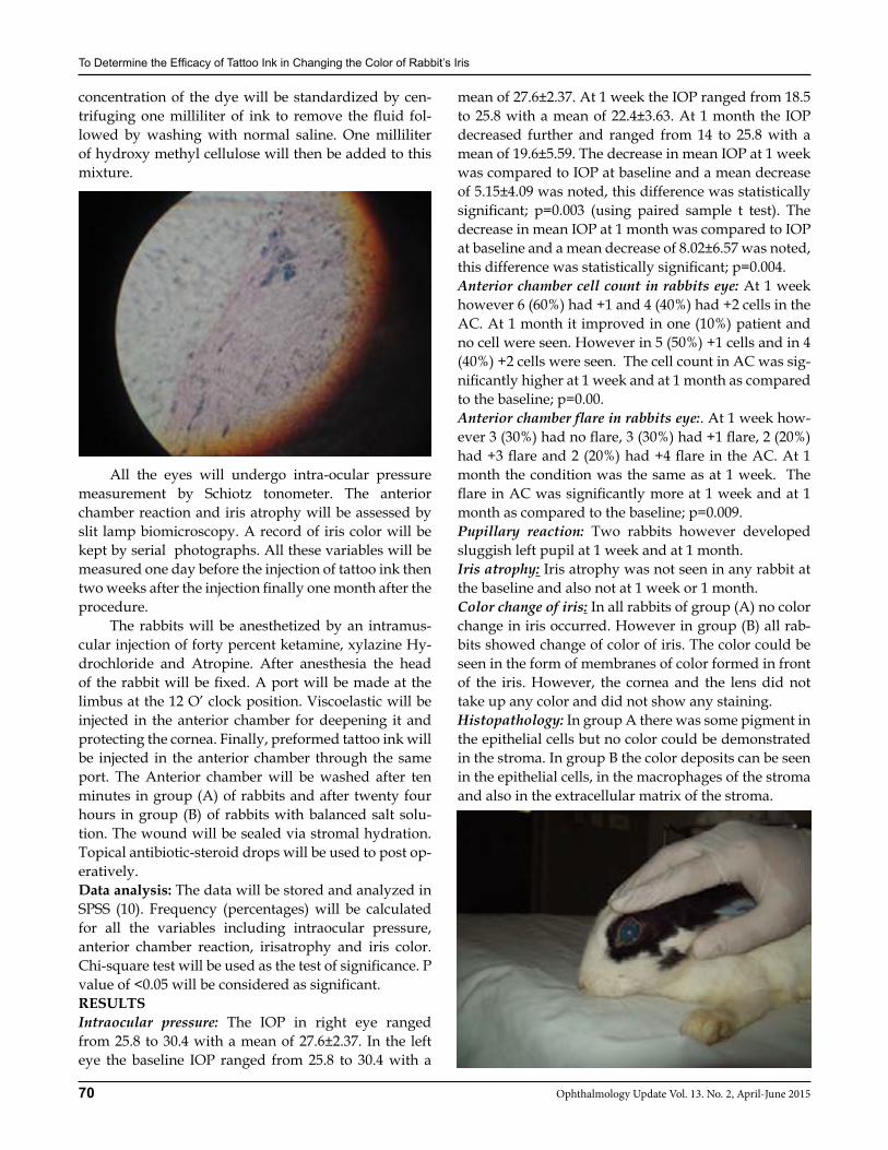
To Determine the Efficacy of Tattoo Ink in Changing the Color of Rabbitts Iris
70 Ophthalmology Update Vol. 13. No. 2, April-June 2015
concentration of the dye will be standardized by cen-trifuging one milliliter of ink to remove the fluid fol-lowed by washing with normal saline. One milliliter of hydroxy methyl cellulose will then be added to this mixture.
All the eyes will undergo intra-ocular pressure measurement by Schiotz tonometer. The anterior chamber reaction and iris atrophy will be assessed by slit lamp biomicroscopy. A record of iris color will be kept by serial photographs. All these variables will be measured one day before the injection of tattoo ink then two weeks after the injection finally one month after the procedure. The rabbits will be anesthetized by an intramus-cular injection of forty percent ketamine, xylazine Hy-drochloride and Atropine. After anesthesia the head of the rabbit will be fixed. A port will be made at the limbus at the 12 O’ clock position. Viscoelastic will be injected in the anterior chamber for deepening it and protecting the cornea. Finally, preformed tattoo ink will be injected in the anterior chamber through the same port. The Anterior chamber will be washed after ten minutes in group (A) of rabbits and after twenty four hours in group (B) of rabbits with balanced salt solu-tion. The wound will be sealed via stromal hydration. Topical antibiotic-steroid drops will be used to post op-eratively.Data analysis: The data will be stored and analyzed in SPSS (10). Frequency (percentages) will be calculated for all the variables including intraocular pressure, anterior chamber reaction, irisatrophy and iris color. Chi-square test will be used as the test of significance. P value of <0.05 will be considered as significant.RESULTSIntraocular pressure: The IOP in right eye ranged from 25.8 to 30.4 with a mean of 27.6±2.37. In the left eye the baseline IOP ranged from 25.8 to 30.4 with a
mean of 27.6±2.37. At 1 week the IOP ranged from 18.5 to 25.8 with a mean of 22.4±3.63. At 1 month the IOP decreased further and ranged from 14 to 25.8 with a mean of 19.6±5.59. The decrease in mean IOP at 1 week was compared to IOP at baseline and a mean decrease of 5.15±4.09 was noted, this difference was statistically significant; p=0.003 (using paired sample t test). The decrease in mean IOP at 1 month was compared to IOP at baseline and a mean decrease of 8.02±6.57 was noted, this difference was statistically significant; p=0.004. Anterior chamber cell count in rabbits eye: At 1 week however 6 (60%) had +1 and 4 (40%) had +2 cells in the AC. At 1 month it improved in one (10%) patient and no cell were seen. However in 5 (50%) +1 cells and in 4 (40%) +2 cells were seen. The cell count in AC was sig-nificantly higher at 1 week and at 1 month as compared to the baseline; p=0.00.Anterior chamber flare in rabbits eye:. At 1 week how-ever 3 (30%) had no flare, 3 (30%) had +1 flare, 2 (20%) had +3 flare and 2 (20%) had +4 flare in the AC. At 1 month the condition was the same as at 1 week. The flare in AC was significantly more at 1 week and at 1 month as compared to the baseline; p=0.009.Pupillary reaction: Two rabbits however developed sluggish left pupil at 1 week and at 1 month. Iris atrophy: Iris atrophy was not seen in any rabbit at the baseline and also not at 1 week or 1 month. Color change of iris: In all rabbits of group (A) no color change in iris occurred. However in group (B) all rab-bits showed change of color of iris. The color could be seen in the form of membranes of color formed in front of the iris. However, the cornea and the lens did not take up any color and did not show any staining. Histopathology: In group A there was some pigment in the epithelial cells but no color could be demonstrated in the stroma. In group B the color deposits can be seen in the epithelial cells, in the macrophages of the stroma and also in the extracellular matrix of the stroma.
To Determine the Efficacy of Tattoo Ink in Changing the Color of Rabbitts Iris
71Ophthalmology Update Vol. 13. No. 2, April-June 2015
Differences between different colors: The response ob-served with different colors was varied. For example with blue and yellow color the change in color was ho-mogenous however with brown, green and red color the color change was patchy and inhomogeneous. DISCUSSION Different optical aids have been developed to change the color of the iris and hence the appearance of the eyes. These devices include colored contact lenses and anterior chamber intra ocular lenses. Both these aids have their advantages and associated complica-tions. Anterior chamber intra ocular lenses have been used to mask the original color of the iris and improve the appearance of patients with iris colobomas. The complications associated with these include uveitis, iris atrophy, glaucoma, endophthalmitis, pupillary abnor-malities, haloes and reduced vision at night. Colored contact lenses are easy to use and are widely employed to change the color of the iris for cosmetic reasons. Con-tact lens use has been associated with uveitis, epithelial keratopathy and allergic reactions. Tattoo ink can also be used to change the color of the iris. The immediate uptake of the ink by the fibroblasts, together with the scarcity of these cells in the endothelial layer of the cor-nea and anterior capsule of the lens ensures that these structures in the anterior chamber do not change their color and react minimally to the presence of the ink. We carried out a study at PIMS hospital to see the efficacy of tattoo ink in changing color of iris. The study was carried out at Department of Ophthalmology, PIMS Islamabad. The research was carried out on rab-bit eyes. The response observed with different colors was varied. For example with blue and yellow color the change in color was homogenous however with brown and red color the color change was patchy and inho-mogeneous. Each tattoo color has a different chemical formula which is a secret of the company manufactur-ing it. The differences observed in the homogeneity of color change may be due to differences in the chemical nature of the different colors. From our study it is evident that the color change occurs if the dye is retained in the anterior chamber for 24 hours. Complications included features of inflam-mation which settled by the end of one month in some eyes. It caused complications of decreased intraocular pressure and increased anterior chamber cell count and flare. No iris atrophy occurred. The reduction in the
intraocular pressure in rabbits´ eyes may be due to re-duced fluid production by the ciliary body in response to the dye. However, reduced pressure does signify the fact that the tattoo ink did not block the outflow tracts of the anterior chamber. Tattoo inks have their own hazards even when used on the skin, In our study the injection of tattoo ink was associated with development of inflammation as evidenced by significant increase in cell count and development of flare in the anterior chamber. How-ever, tattoo ink was quite safe in our study since no case of iris atrophy was observed. Since the previous methods had their own limitations attempts at invent-ing a new method that is successful in changing the eye color without side effect will continue. Our study was one such effort. Further studies are required in the field with slight changes in the tattoo chemistry and its sol-vent that can improve the results in future. CONCLUSION Tattoo ink changed the color of iris if retained in the anterior chamber for 24 hours. It caused complica-tions of decreased intraocular pressure and increased anterior chamber cell count and flare. No iris atrophy occurred. The change in color of iris was patchy for most colors but for blue and yellow ink are homog-enous and whole iris color change occurred.REFERENCES1. Druianova IUS, Verigo EN, A rational method of cosmetic eye
prosthesis. Vestn Oftalmol1990;106:53-4. 2005;18:454-64.2. Mamalis N: Complications of foldable intraocular lenses
requiring explantation or secondary intervention. J Cataract Refract Surg 2002;28: 2193-201.
3. Sorbara L., Jones, L. Williams-lyn D. Contact lens induced papillary conjunctivitis with silicone hydrogel lenses. Cont Lens Anterior Eye 2009;32:93-6
4. Möhrenschlager M, Worret WI, Köhn FM. Tattoos and permanent make-up: background and complications. MMW Fortschr Med 2006;148:34-6.
5. Bron AJ, Tripathi RC, Tripathi BJ. Wolff’s Anatomy of the Eye and Orbit.8th edn. Chapman & Hall Medical: London, 1997.
6. Imesch PD, Wallow IH, Albert DM. The colour of the human eye: a review of morphologic correlates and of some conditions that affect iridial pigmentation. SurvOphthalmol1997;41:117–23.
7. Wilkerson CL, Syed NA, Fisher MR, Robinson NL, Wallow IH, Albert DM. Melanocytes and iris colour. Light microscopic findings. Arch Ophthalmol 1996;114:437–42.
8. Prota G, Hu DN, Vincensi MR, McCormick SA, Napolitano A. Characterization of melanins in human irides and cultured uveal melanocytes from eyes of different colours. Exp Eye Res 1998;67:293–99.
9. Fuchs E. Normal pigmentierte und albinotische iris. Graefes Arch ClinExpOphthalmol1913;84/85:521.
10. Dieterich CE. [The fine structure of melanocytes in the human iris].Graefes Arch ClinExpOphthalmol1972;183:317–33.
72 Ophthalmology Update Vol. 13. No. 2, April-June 2015
ORIGINAL ARTICLE
BACKGROUND Hepatitis is described as an infection with swell-ing and inflammation of the liver that if progresses, may lead to cirrhosis or cancer. Sometimes people con-tract hepatitis with limited or no symptoms but often it leads to jaundice, anorexia (poor appetite) and diar-rhea. Hepatitis is caused by a wide variety of causatives like alcohol, poison and autoimmunity but most cases of hepatitis are reported by viruses. Pakistan has large number of both diagnosed and un-diagnosed patients of hepatitis B and C. The prevalence among general public of HBV and HCV infection in Pakistan is 10%2,3
and 4–10%4,5 respectively. Hepatitis B virus (HBV) in-fection is endemic worldwide and is responsible for an estimated 1-2 million deaths worldwide every year. About 350 million (5- 15 % of the total cases) are carriers of the virus, out of which around 80% reside in Asia.6 According to WHO estimates, HCV prevalence is 3% of world population with 170 million cases. Almost 50% of all cases become chronic carriers at risk of liver cir-rhosis and liver cancer.7
HBV can be transmitted through blood, semen, vaginal fluids and other bodily fluids of the infected individual.8 HCV however, can only be contracted through blood to blood contact. The transmission risk
of these diseases is more among patients receiving blood transfusions or injection drug users.9,10 Patients presenting to different public and private hospitals are not routinely screened for hepatitis B and C. There-fore there is high risk of transmission of infection from asymptomatic carrier patients. Keeping in view the dreadful complications of hepatitis and its high infec-tivity we cannot take the risk of operating on patients without hepatitis screening. This study was carried out to discover the frequency of hepatitis B and C in our surgical patients to get an idea about the number of the patients we are operating on them without knowing that whether they are hepatitis B or C positive.MATERIAL AND METHODS This prospective observational study was con-ducted at Eye Unit, Lady Reading Hospital Peshawar from July 2013 to June 2014. A total of 1147 patients un-der going eye surgery, who were unaware of hepatitis infection were included in this study. After taking ethi-cal approval from the department, patients informed consent was taken. Rapid chromatography immunoas-say for qualitative detection of hepatitis B and C was the screening method. Those found positive on screen-ing test are confirmed by ELISA. A special proforma is made for this study and results were analyzed by statistical methods.RESULTS Total number of patients screened were 1147. Male subjects were 54.31% (623/1147) and female subjects were 45.68% (524/1147). The frequency of hepatitis B and C (combined) was found to be 5.66% (65/1147); out of which 2.5% (29/1147) were HBsAg positive and 3.13%(36/1130) anti-HCV positive. The frequency of
Bilal Bashir FCPS1, Muhammad Zubair Masud FCPS2, Muhammad Nazim FCPS3
Bilal Khan FCPS4, Mahfooz Hussain FRCS5
ABSTRACT:Purpose: To determine the frequency of sero-positive cases of Hepatitis B & C viral infection in the admitted patients un-dergoing elective eye surgery in tertiary care hospital.Material and Method: It was a descriptive study based on survey in which all patients above 2 years of age admitted for eye surgery in Eye unit of Lady Reading Hospital Peshawar were screened for Hepatitis B and C infections, from 1st July 2013 to 30th June 2014. Those found positive on screening test were confirmed by Enzyme Linked Immunosorbant Assay (ELISA).Result: Total number of patients screened was 1147. Male patients were 54.31% (623/1147) and female patients were 45.68% (524/1147). The frequency of hepatitis B and C (combined) was found to be 5.66% (65/1147); out of which 2.5% (29/1147) were HBsAg positive and 3.13%(36/1130) anti-HCV positiveConclusion: Screening of blood borne viral infections has important role in minimizing the transmission of the virus to doc-tors, paramedics and other patients.
Incidence of Hepatitis B & C among Admitted Eye Patients in Tertiary Care Hospital of Peshawar
Bilal Bashir
1,4Medical Officers Lady Reading Hospital Peshawar. 2Assistant Professor Naseer Teaching Hospital Peshawar. 3Medical Officer Al Khidmat Hospital Peshawar. 5Assistant Professor Lady Reading Hospital Peshawar
Correspondence: Dr. Bilal Bashir, House no 103 New Defence Officers Colony Shami Road Peshawar Cantt Ph: 03339115764 / 091-5270869 Email: [email protected]
Received: November 2014 Accepted. January 2015
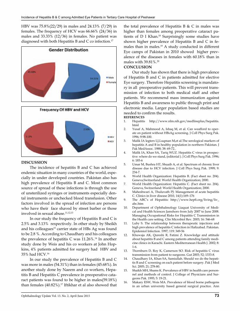
Incidence of Hepatitis B & C among Admitted Eye Patients in Tertiary Care Hospital of Peshawar
73Ophthalmology Update Vol. 13. No. 2, April-June 2015
HBV was 75.8%(22/29) in males and 24.13% (7/29) in females. The frequency of HCV was 66.66% (24/36) in males and 33.33% (12/36) in females. No patient was diagnosed with both Hepatitis B and C co infection.17
DISCUSSION The incidence of hepatitis B and C has achieved endemic situation in many countries of the world, espe-cially in under developed countries. Pakistan also has high prevalence of Hepatitis B and C. Most common source of spread of these infections is through the use of unsterilized syringes or instruments especially den-tal instruments or unchecked blood transfusion. Other factors involved in the spread of infection are persons who have their face shaved by street barber or those involved in sexual abuse.11,12,13
In our study the frequency of Hepatitis B and C is 2.5% and 3.13% respectively. In other study by Sheikh and his colleagues15 carrier state of HBs Ag was found to be 2.8 %. According to Chaudhary and his colleagues the prevalence of hepatitis C was 11.26%.14 In another study done by Weis and his co workers at John Hop-kins, 4% patients admitted for surgery had HBV and 35% had HCV.16
In our study the prevalence of Hepatitis B and C was more in males (54.31%) than in females (45.68%). In another study done by Naeem and co workers, Hepa-titis B and Hepatitis C prevalence in preoperative cata-ract patients was found to be higher in males(59.18%) than females (40.82%).17 Iftikhar et al also showed that
the total prevalence of Hepatitis B & C in males was higher than females among preoperative cataract pa-tients of D I Khan.18 Surprisingly some studies have shown higher prevalence of Hepatitis B and C in fe-males than in males.19 A study conducted in different Eye camps of Pakistan in 2010 showed higher prev-alence of the diseases in females with 60.18% than in males with 39.81%.20
CONCLUSION Our study has shown that there is high prevalence of Hepatitis B and C in patients admitted for elective Eye surgery. Therefore Hepatitis screening is mandato-ry in all preoperative patients. This will prevent trans-mission of infection to both medical staff and other patients. We recommend mass immunization against Hepatitis B and awareness to public through print and electronic media. Larger population based studies are needed to confirm the results.REFERENCES1. Hepatitis http://www.nlm.nih.gov/medlineplus/hepatitis.
html2. Yusaf A, Mahmood A, Ishaq M, et al. Can weafford to oper-
ate on patient without HBsAg screening. J Coll Phys Surg Pak. 1996; 9:98-100.
3. Malik IA legters LJ,Luqman M,et al.The serological markers of hepatitis A and B in healthy population in northern Pakistan. J Pak MedAssoc. 1988; 38: 69-72..
4. Malik IA, Khan SA, Tariq WUZ. Hepatitis C virus in prospec-tive: where do we stand, (editorial ). J Coll Phys Surg Pak. 1996; 6: 185-6
5. Umar M, Bushra HT, Shuaib A, et al. Spectrum of chronic liver disease due to HCV infection. J Coll Phys Surg Pak. 1999; 9: 234-7.
6. World Health Organization: Hepatitis B. (Fact sheet no. 204). Geneva,Switzerland: World Health Organization; 2000.
7. World Health Organization: Hepatitis C. (Fact sheet no. 204). Geneva, Switzerland: World Health Organization; 2000.
8. Maheshwari A, Thuluvath PJ: Management of acute hepatitis C. Clinics in liver disease 2010, 14(1):169–176
9. The ABC’s of Hepatitis: http://www.hepfi.org/living/liv_abc.htm
10. Department of Ophthalmology Liaquat University of Medi-cal and Health Sciences Jamshoro from July 2007 to June 2008. Managing Occupational Risks for Hepatitis C Transmission in the Health care setting. Clin Microbiol Rev. 2003; 16: 546-68
11. Luby S. The relationship between therapeutic injections and high prevalence of hepatitis C infection in Hafizabad. Pakistan. Epidemiol Infection. 1997; 119: 349-56
12. Khuwaja AK, Qureshi R, Fatimi Z. Knowledge and attitude about hepatitis B and C among patients attending family medi-cine clinics in Karachi. Eastern Mediterranean Health J. 2002; 8: 1-6.
13. Thornburn D, Roy K, Camerson SO. Risk of hepatitis C virus transmission from patient to surgeons. Gut 2003; 52; 1333-8.
14. Chaudhary IA, Khan SA, Samiullah. Should we do the hepati-tis B and C screening on each patient before surgery. Pak J Med Sci. 2005; 21; 278-80.
15. Shaikh MH, Shams K. Prevalence of HBV in health care person-nel and methods of control. J College of Physicians and Sur-geons Pak. 1995; 5: 19-21.
16. Makary ESW, Weis MA. Prevalence of blood borne pathogens in an urban university based general surgical practice. Ann
Incidence of Hepatitis B & C among Admitted Eye Patients in Tertiary Care Hospital of Peshawar
74 Ophthalmology Update Vol. 13. No. 2, April-June 2015
Surg. 2005; 24: 803-917. Syed Saad Naeem, Efaza Umar Siddiqui, Abdul Nafey Kazi,
Sumaiyatauseeq Khan, Farhan E Abdullah, Idrees Adhi. Preva-lence of hepatitis ‘B’ and hepatitis ‘C’ among preoperative cata-ract patients in Karachi.. BMC research notes 2012; 5(492).
18. Ahmad I, Khan SB, Rehman HU, Khan MH, Anwar S: Fre-quency of Hepatitis B and Hepatitis C among cataract patients.
Gomal Journal of Medical Sciences 2006, 4:2.19. Farooqi JI, Farooqi RJ: Relative Frequency of Hepatitis B and
Hepatitis Cvirus infections in patients of cirrhosis in NWFP, Pakistan. J Coll Phys Surg Pak 2000, 10:217–219.
20. Nangrejo KM, Qureshi MA, Sahto AA, Siddiqui SJ: Prevalence of Hepatitis B and C in the patients Undergoing Cataract Sur-gery at Eye Camps. Pak J Ophthalmol 2011, 27:1
SUBSCRIPTION TO ‘OPHTHALMOLOGY UPDATE’
Dear Doctor, Your Quarterly ‘Ophthalmology Update’ is being regularly published from Islamabad since 1998. The subscription of the paid members expires by 31st December every year and needs to be renewed immediately, while the new subscribers are welcomed to join the esteemed coterie of our regular members. Since the journal is fully indexed with Pakistan Medical & Dental Council (PMDC) and Higher Education Commission of Pakistan (HEC) as a standard scientific journal entirely devoted to promotion of Medical Sciences especially to the welfare of visually handicapped. It highlights the most current research, scientific articles, reviews and interesting case reports in all fields of Medicine including Ophthalmology with updated information around the world. Currently, the journal is being subscribed by the doctors practicing in every field of medicine, postgraduates, health professionals including libraries of medical colleges and are making full use of the scientific material. Doctors serving in your institutions can also contribute their research papers, thesis, articles and interesting case reports to the journal. The journal is being printed by a high-class printer of the country on a beautiful English Matt paper and is being published on quarterly basis on the 1st of every ensuing month of the year. Moreover, the journal is freely distributed to the participants of various conferences being held from time to time.Fresh and renewed annual subscription of the journal is Rs.800/- which is very nominal and can be remitted through money order/cheque/bank draft and on line to A/C: 145-20620-714-126749 maintained at Summit Bank (Code: 145), Markaz F-10, Islamabad in the name of Ophthalmology Update, or to the managing editor at 267-A, St: 53, F-10/4, Islamabad. Single copy of the journal is supplied freely to the principal author on complimentary basis, while the additional copies can be had on payment of Rs.200/- per copy. Members are ensured regular and uninterrupted supply of the journal at their doorstep. Our sole motto is SERVICE TO MEDICINE and we assure you our fullest cooperation with highest considerations for regular and well-in-time supply of the journal. Wishing you good health, happiness and a prosperous professional life. With profound regards.
Yours sincerely,Prof. Dr. M. Yasin Khan DurraniMBBS., DO., MD., FICO(UK)., FRCOphth(Lond)Editor in Chief
75Ophthalmology Update Vol. 13. No. 2, April-June 2015
Visual Outcome & Complications of Scleral-fixation Posterior Chamber Intraocular Lenses
Mir Ali Shah FCPS. Fellow Vitreo-Retina1, Bilal Khan2, Bilal Khan3 Bilal Bashir4, Sher Akbar Khan5, Mohd Jawad6, Muhammad Idris7
ABSTRACT:Purpose: To determine the visual outcome and complications of posterior chamber scleral fixation intraocular lenses (PCSF IOL).Material and Methods: This retrospective study was carried out in the Department of Ophthalmology, Lady Reading Hospi-tal, Peshawar from July 2011 to July 2013. A total of 17 patients were included in the study. Details of the patients like age, gender, pre- and postoperative best spectacle corrected visual acuity (BSCVA), indication for surgery and detailed slit-lamp and fundus examination were recorded on a designed proforma. The main outcome measures were postoperative visual acuity (VA) and complications. Patients were followed for one year regarding vision and any complications. All the data was entered and analyzed using SPSS version17. The data was expressed in the form of tables and charts. Results: A total of 17 eyes of 17 patients were included in this study. 14(82.35%) were males and 3(17.65%) were females with a male to female ratio 4.6:1. The age ranged from 4 to 15 years and were followed over a period of one year after place-ment of posterior chamber scleral fixation intraocular lenses (PC SF-IOL).14 (82.35%) of the eyes had a gain in VA from CF to 6/9 while 3(17.65%) had no change in the VA. Postoperative complications were observed in 5(29.40%) and included IOL dislocation in one case(5.88%), exposed suture with pyogenic granuloma in 2(11.76%), increased IOP in 1 case (5.88%).and iris capture in 1(5.88%) eye.Conclusion: Posterior chamber scleral fixation IOL appear to be a safe technique with minimal complications when there is no capsular support.
INTRODUCTION While crystalline lens subluxation can occur in any patient, these three profiles are most prone: significant blunt trauma to the eye or head; systemic conditions such as Marfan’s syndrome, homocystinuria, familial ectopia lentis, Weill-Marchesani syndrome, aniridia and Ehlers-Danlos syndrome, hypermature cataract in which zonular support has been lost. Symptoms of lens subluxation includes visual disturbance from extreme hyperopic or myopic shift, astigmatism or acquired aphakia. Acute secondary angle closure glaucoma can occur due to subluxated lens.1 Children with monocular aphakia who become contact lens intolerant require an intraocular lens (IOL) for visual rehabilitation. When there is inadequate support from the posterior lens cap-sule, use of an anterior chamber IOL(AC IOL) or PCS-FIOL may be considered. The authors reported their ex-perience with scleral fixation of posterior chamber IOLs in children. Implantation of a PCSF IOL for the surgical management of aphakia in the absence of capsular sup-
port is a safe procedure with a low risk of complica-tions in the early postoperative period.2 Suture related complications are unique to PCSF IOL. To avoid ero-sion of the knots through conjunctiva, scleral flaps can be used to cover the knots.3 The partial thickness scle-ral flaps can atrophy over time and expose the proline knot. Endophthalmitis has been reported and remains a real risk in patients undergoing SFIOL.4 The possible causes of dislocation of these IOLs include suture deg-radation,5 suture breakage,3 slippage of the haptic from the suture,3 or erosion of the suture through the tissue.6
Recent studies have shown that the implantation of scleral fixation posterior chamber intraocular lenses is feasible and renders more favorable results in children over 2 years of age if non-compliant with spectacles or contact lenses.7,8 Implantation of IOLs in children less than 2 years is still controversially discussed.9,10 There-fore, we have performed a study to compare the out-comes of secondary intraocular lens implantation in aphakic eyes of children older than 2 years previously operated for traumatic and congenital cataracts. Anterior-chamber lenses were used for many years because of relatively easy implantation technique, even in the total absence of capsular support. However, the fixation in the anterior-chamber angle may cause glau-coma and chronic irritation to the iris. Furthermore, long-term endothelial cell loss with corneal decompen-sation is reported for angle-fixated intraocular lenses,
ORIGINAL ARTICLE
Mir Ali Shah
1Associate Professor, Department of Ophthalmology, PGMI Lady Reading Hospital Peshawar. 2,4,5Resident,Vitreo-Retina. 3Resident Neuro Surgery. 6Postgraduate Trainee Department of Ophthalmology, Lady Reading Hospital, Peshawar. 7Medical Officer Ophthalmology, Lady Reading Hospital Peshawar.
Correspondence: Dr. Mir Ali Shah, Associate Professor, Department Ophthalmology, Lady Reading Hospital, Peshawar. Email: [email protected] Cell: 03005948091
Received: December 2014 Accepted: January 2015
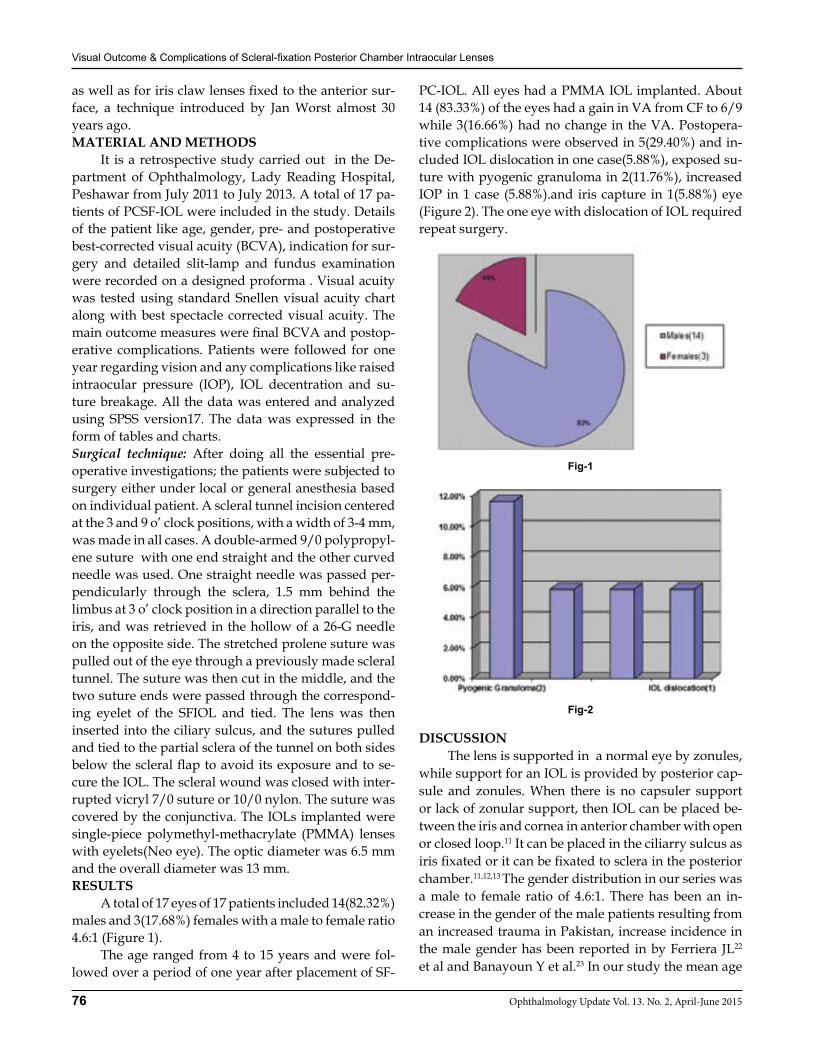
Visual Outcome & Complications of Scleral-fixation Posterior Chamber Intraocular Lenses
76 Ophthalmology Update Vol. 13. No. 2, April-June 2015
as well as for iris claw lenses fixed to the anterior sur-face, a technique introduced by Jan Worst almost 30 years ago.MATERIAL AND METHODS It is a retrospective study carried out in the De-partment of Ophthalmology, Lady Reading Hospital, Peshawar from July 2011 to July 2013. A total of 17 pa-tients of PCSF-IOL were included in the study. Details of the patient like age, gender, pre- and postoperative best-corrected visual acuity (BCVA), indication for sur-gery and detailed slit-lamp and fundus examination were recorded on a designed proforma . Visual acuity was tested using standard Snellen visual acuity chart along with best spectacle corrected visual acuity. The main outcome measures were final BCVA and postop-erative complications. Patients were followed for one year regarding vision and any complications like raised intraocular pressure (IOP), IOL decentration and su-ture breakage. All the data was entered and analyzed using SPSS version17. The data was expressed in the form of tables and charts. Surgical technique: After doing all the essential pre-operative investigations; the patients were subjected to surgery either under local or general anesthesia based on individual patient. A scleral tunnel incision centered at the 3 and 9 o’ clock positions, with a width of 3-4 mm, was made in all cases. A double-armed 9/0 polypropyl-ene suture with one end straight and the other curved needle was used. One straight needle was passed per-pendicularly through the sclera, 1.5 mm behind the limbus at 3 o’ clock position in a direction parallel to the iris, and was retrieved in the hollow of a 26-G needle on the opposite side. The stretched prolene suture was pulled out of the eye through a previously made scleral tunnel. The suture was then cut in the middle, and the two suture ends were passed through the correspond-ing eyelet of the SFIOL and tied. The lens was then inserted into the ciliary sulcus, and the sutures pulled and tied to the partial sclera of the tunnel on both sides below the scleral flap to avoid its exposure and to se-cure the IOL. The scleral wound was closed with inter-rupted vicryl 7/0 suture or 10/0 nylon. The suture was covered by the conjunctiva. The IOLs implanted were single-piece polymethyl-methacrylate (PMMA) lenses with eyelets(Neo eye). The optic diameter was 6.5 mm and the overall diameter was 13 mm.RESULTS A total of 17 eyes of 17 patients included 14(82.32%) males and 3(17.68%) females with a male to female ratio 4.6:1 (Figure 1). The age ranged from 4 to 15 years and were fol-lowed over a period of one year after placement of SF-
PC-IOL. All eyes had a PMMA IOL implanted. About 14 (83.33%) of the eyes had a gain in VA from CF to 6/9 while 3(16.66%) had no change in the VA. Postopera-tive complications were observed in 5(29.40%) and in-cluded IOL dislocation in one case(5.88%), exposed su-ture with pyogenic granuloma in 2(11.76%), increased IOP in 1 case (5.88%).and iris capture in 1(5.88%) eye (Figure 2). The one eye with dislocation of IOL required repeat surgery.
Fig-1
Fig-2
DISCUSSION The lens is supported in a normal eye by zonules, while support for an IOL is provided by posterior cap-sule and zonules. When there is no capsuler support or lack of zonular support, then IOL can be placed be-tween the iris and cornea in anterior chamber with open or closed loop.11 It can be placed in the ciliarry sulcus as iris fixated or it can be fixated to sclera in the posterior chamber.11,12,13 The gender distribution in our series was a male to female ratio of 4.6:1. There has been an in-crease in the gender of the male patients resulting from an increased trauma in Pakistan, increase incidence in the male gender has been reported in by Ferriera JL22 et al and Banayoun Y et al.23 In our study the mean age
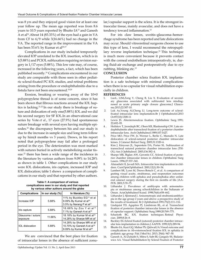
Visual Outcome & Complications of Scleral-fixation Posterior Chamber Intraocular Lenses
77Ophthalmology Update Vol. 13. No. 2, April-June 2015
was 8 yrs and they enjoyed good vision for at least one year follow up. The mean age reported was from 8.6 years to 10.5 years reported by Bhutto IA15 and Ganesh A et al16.About 14 (83.33%) of the eyes had a gain in VA from CF to 6/9 while 3(16.66%) had no change in the VA. The reported range in the improvement in the VA has been 53.6% by Kumar et al20,17
Complications in our study included temporarily elevated IOP unrelated to the IOL insertion, which is in 1(5.88%) and PCIOL subluxation requiring revision sur-gery in 1/17 eyes (5.88%). This low rate may, of course, increased in the following years, a fact, which has been published recently.14 Complications encountered in our study are comparable with those seen in other pediat-ric scleral-fixated PC IOL studies, and retinal problems arising from the procedure or endophthalmitis due to a fistula have not been encountered.19,20
Erosion, breaking or wearing away of the 10×0 polypropylene thread is of some concern, since it has been shown that fibrous reactions around the IOL hap-tics is lacking.21,22 In our study there is breakage of su-ture and dislocation of only one(5.88%) IOL and we did his second surgery for SF IOL.In an observational case series by Vote et al., 17 eyes (27.9%) had spontaneous suture breakage with several eyes having multiple epi-sodes.23 the discrepancy between his and our study is due to the increase in sample size and long term follow up by him(4 months vs 3 years). Drews, in his report noted that polypropylene may fail after a prolonged period in the eye. The deterioration was most marked with sutures buried in actively metabolizing ocular tis-sue.22 there has been a wide range of the incidence in the literature by various authors from 9.09% to 14.28% as shown in table 1. Other complications in our study were IOL dislocations, iris capture, increased IOP and IOL dislocation; table 1 shows a comparison of compli-cations in our study and that reported by other authors.
Table-1: A comparison of various complications seen in our study and that reported
by various other authors around the globe
Complications In our study (%) In literature (%)
Increase IOP 5.88%11% by Buckley EG14 et al9.09% By Kumar et al20
3.5% by Narang P et al21
Iris capture 5.88% 16.66% by Zou Y et al19
7.1% by Ganesh A et al16
Glaucoma / suture exposure 11.66% 18.18% by Kumar M et al20
14.28% by Sharpe MR et al7
IOL dislocation 5.88%14.28% by Sharpe MR et al710.70% by Ganesh A et al16
9.09% by Kumar M et al20
We are convinced that the best place for fixation of intraocular lenses in the absence of sufficient zonu-
lar/capsular support is the sclera. It is the strongest in-traocular tissue, mainly avascular, and does not have a tendency toward inflammation.13
For iris claw lenses, uveitis–glaucoma–hemor-rhage syndrome has been reported and late dislocations may occur. Should vitreoretinal surgeons choose to use this type of lens, I would recommend the retropupil-lary reverse implantation technique.21 This technique is much more convenient because it prevents contact with the corneal endothelium intraoperatively, ie, dur-ing fluid-air exchange and postoperatively due to eye rubbing, blinking etc.13
CONCLUSION Posterior chamber sclera fixation IOL implanta-tion is a safe technique with minimal complications when there is no capsular for visual rehabilitation espe-cially in children.REFERENCES1. LuoL, LIM,Zong Y, Cheng B, Liu X. Evaluation of second-
ary glaucoma associated with subluxated lens misdiag-nosed as acute primary angle closure glaucoma.J Glauco-ma.2013;22:307-10.
2. Luk As,Young Al,Cheng Ll. Long-term outcome of scleral-fixated intraocular lens implantation.Br J Ophthalmolol.2013 Oct97(10):1308-11
3. Lewis JS. Abexternosulcus fixation. Ophthalmic Surg. 1991; 22:692–95.
4. . Heilskov T, Joondeph BC, Olsen KR, Blankenship GW. Late en-dophthalmitis after transscleral fixation of a posterior chamber intraocular lens. Arch Ophthalmol.1989;107:1427.
5. Price MO, Price FW, Jr, Werner L, Berlie C, Mamalis N. Late dislocation of scleral-sutured posterior chamber intraocular lenses. J Cataract Refract Surg. 2005;31:1320–6.
6. Kim J, Kinyoun JL, Saperstein DA, Porter SL. Subluxation of transscleral sutured posterior chamber intraocular lens (TSI-OL) Am J Ophthalmol. 2003;136:382–4.
7. Sharpe MR, Biglan AW, Gerontis CC. Scleral fixation of poste-rior chamber intraocular lenses in children. Ophthalmic Surg Lasers. 1996;27:337–341.
8. Ahmadieh H, Javadi MA. Intraocular lens implantation in chil-dren. CurrOpinOphthalmol. 2001;12(1):30–34.
9. Lambert SR, Lynn M, Drews-Botsch C, et al. A comparison of grating visual acuity, strabismus, and reoperation outcomes among children with aphakia and pseudophakia after unilat-eral cataract surgery during the first six months of life. JAA-POS. 2001;5:70–75.
10. Lithander J. Prevalence of amblyopia with anisometro-pia or strabismus among schoolchildren in the Sultanate of Oman. ActaOphthalmol Scand. 1998;76(6):658–662.
11. Lithander J, Sjóstrand J. Anisometropic and strabismicamblyo-pia in the age group 2 years and above: a prospective study of the results of treatment. Br J Ophthalmol.1991;75(2):111–116.
12. Lindquist TD, Agapitos PJ, Lindstrom RL, et al. Transscleral fixation of posterior chamber intraocular lenses in the absence of capsular support. Ophthalmic Surg.1989;20:769–775.
13. Scharioth BG. IOL fixation techniques. Retinal Physi-cian. 2009;8:26–8.
14. Buckley EG.Scleral fixated (sutured) posterior chamber intraoc-ular lens implantation in children.J AAPOS. 1999;3(5):289-94.
15. Bhutto IA, Kazi GQ, Mahar PS, QidwaiUA.Visual outcome and complications in Ab-externoscleral fixation IOL in aphakia in pediatric age group. Pak J Med Sci. 2013; 29(4): 947–50.
16. Ganesh A, Al-Zuhaibi S, Mitra S, Sabt BI, Ganguly SS, Bialasie-wicz AA. Visual Rehabilitation by Scleral Fixation of Posterior
Visual Outcome & Complications of Scleral-fixation Posterior Chamber Intraocular Lenses
78 Ophthalmology Update Vol. 13. No. 2, April-June 2015
Chamber Intraocular Lenses in Amblyopic Aphakic Children. Middle East Afr J Ophthalmol. 2008; 15(2): 61–5.
17. Rehman A,Bhutto IA,Bkhari S,Hassan M,Bhatti MN.Pak J Oph-thalmol 2011;27(2):73-7.
18. Lithander J. Prevalence of amblyopia with anisometro-pia or strabismus among schoolchildren in the Sultanate of Oman. ActaOphthalmol Scand. 1998;76(6):658–62.
19. Drews RC. Quality control, and changing indications for lens implantation. The Seventh Binkhorst Medal Lecture-1982. Oph-thalmology. 1983;90:301–10.
20. Kumar M, Arora R, Sanga L, Sota LD. Scleral-fixated intraocu-lar lens implantation in unilateral aphakic children. Ophthal-
mology. 1999;106(11):2184-9.21. Narang P, Narang S. Glue-assisted intrascleral fixation of pos-
terior chamber intraocular lens. Indian J Ophthalmol 2013; 61(4): 163–7.
22. FerreiraJL, F. VeginiF, MaliskaCR. Clove hitch knot for scleral fixation of dislocated IOL – with temporary externalization of the haptics through a clear cornea incision. Invest Ophthalmol Vis Sci 2004;45: E-Abstract 330.
23. Benayoun Y, Petitpas S, Turki K, Adenis J, Robert P. Suture-less scleral intraocular lens fixation: Report of nine cases and literature review; ; (Jul 2013).J Françaisd’Ophtalmologie 2013; online.
First Announcement
KAROPHTH 20156th, 7th, 8th March 2015
Pearl Continental Hotel, Karachi
Last Date for Abstract Submission31st Jan 2015
For Further Information, Registration, Abstract submission Quiz competition and video session
Please contact: Mr. Muhammad Usman Tariq 0306-7484544OSP Office, PMA House,Agha Khan III Road Karachi.
E mail: [email protected]
79Ophthalmology Update Vol. 13. No. 2, April-June 2015
Mohammad Alam FCPS1, Misbah Durrani FCPS2
Prof. Lal Mohammad FCPS3, Irfan Ullah Khan FCPS4
ABSTRACTObjective: To find out the prevalence and density of amblyopia in strabismic patients of school age children.Materials and methods: This prospective study was conducted in eye care centre Karak and Jan eye clinic Kohat from June 2013 to June 2014 with the objective to know the prevalence and density of amblyopia in school age children with strabismus. School age children with strabismus from age range of 5-15 years were included in the study. Visual acuity was checked with Snellen chart. Anterior segment and posterior segment examination was done with direct and indirect ophthalmoscope and slit lamp. Retinoscopy with cycloplegia was done to find out refractive error. Strabismus was assessed with Hirschberg and cover-un cover test. Amblyopia was recorded as mild of 2 lines difference, moderate 3 lines difference while more than 3 lines was graded as severe amblyopia. Total 106 school age children with strabismus were included in the study. Children with co.ocular morbidity except strabismus and refractive error were excluded from the study.Results: All patients were from age range of 5-15 years with mean age of 7.8 years. Out of 106 patients 68 (64.15%) were male and 38 (35.84%) were female. 81 (76.41 %) patients had esotropia out of which 63 (77.77%) had uniocular while 18 (22.22%) had alternating esotropia. 25 (23.58%) patients had exotropia out of which 19 (76%) had uniocular while 6 (24%) had alternating exotropia. In uniocular esotropic 63 patients, 60 (95.23%) patients had amblyopia. The density of amblyopia was mild in 34 (56.66%). moderate in 21 (35%) and severe in 5 (8.33%) of uniocular esotropic group. In alternating esotropic group out of 18 patients 7 (38.88%) had amblyopia in which mild was present in 6 (85.71) and moderate in 1(14.28%). 25 patients were exotropic out of which 19 patients had uniocular exotropia while 6 patients had alternating exotropia. In 19 uniocular exotropic patients, 14(73.68%) patients had amblyopia in which 9 (64.28%) patients had mild,3(21.42%) had moderate while 2 (14.28%) had severe amblyopia. In alternating exotropic group out of 6 patients 5(83.33%) had mild while 1(20%) had moderate amblyopia. Most of the amblyopic patients had hypermetropia .Conclusion: Strabismus is a common cause of amblyopia in children. Early screening and management of school age children is necessary to prevent amblyopia.Key ward: Esotropia, Exotropia, Amblyopia
INTRODUCTION Strabismus and amblyopia are two most common pediatric ocular disorders with cosmetic and functional sequale. Amblyopia is associated with suboptimal vi-sion despite best correction with refraction. In the ab-sence of any other ocular or neural morbidity strabis-mus is the misalignment of the two eyes which in case of failure of treatment may result in loss of binocularity and depth perception1 Failure to diagnose and manage amblyopia in early age may result in lifelong visual im-pairment.2 Strabismic amblyopia is a serious blinding condition affecting the patients in early life. Popula-tion-based prevalence estimates in children range from 0.3% to 4.4% strabismic amblyopia.3 Hispanic / Latino children age of 5 -14 years assessing study in Colombia
has shown a strabismus prevalence of 3% and amblyo-pia of 1.3%.4
In developed countries policies are being formu-lated for early detection of strabismus and amblyopia.5 In Japan children are assessed for strabismic amblyopia in early age primarily by pediatrician and after 6 years by ophthalmologist.6 Early detection and treatment of strabismus and amblyopia are very important. But in developing countries like Pakistan, children with stra-bismus present later. According to a local study most of children with squinting eyes presented after 5 years of age.7 In comparison to this in developed countries presentation is early. Some studies have revealed pres-entation of squinting patients at the age of 2 -5 years.8,9
No reliable data is available in our country and again there is many differences in data in various stud-ies due to demographic, geographical, social, educa-tion and cultural influences over the community. This study was done to find out the prevalence and density of amblyopia in strabismic patients of school age chil-dren. Then it will be possible for the early detection and management as well as making the people aware of this grave problem.
ORIGINAL ARTICLE
Mohammad Alam
1Assistant Professor Ophthalmology KMU Institute of Medical Sci-ences, K.D.A, Kohat. 2Assistant Professor, Radiology, Bacha Khan Medical College, Mardan. 3Professor of Ophthalmology, KMU Insti-tute Of Medical Sciences, K.D.A, Kohat. 4Refractionist, K.D.A Teach-ing Hospital, Kohat.
Correspondence: Dr. Mohammad Alam1 Assistant Professor Ophthal-mology KMU Institute of Medical Sciences K.D.A Kohat. [email protected], Cell:
Received: December 2014 Accepted: February 2015
Prevalence and Density of Amblyopia in Strabismic Patients of School Age Children
(A study of 106 cases)
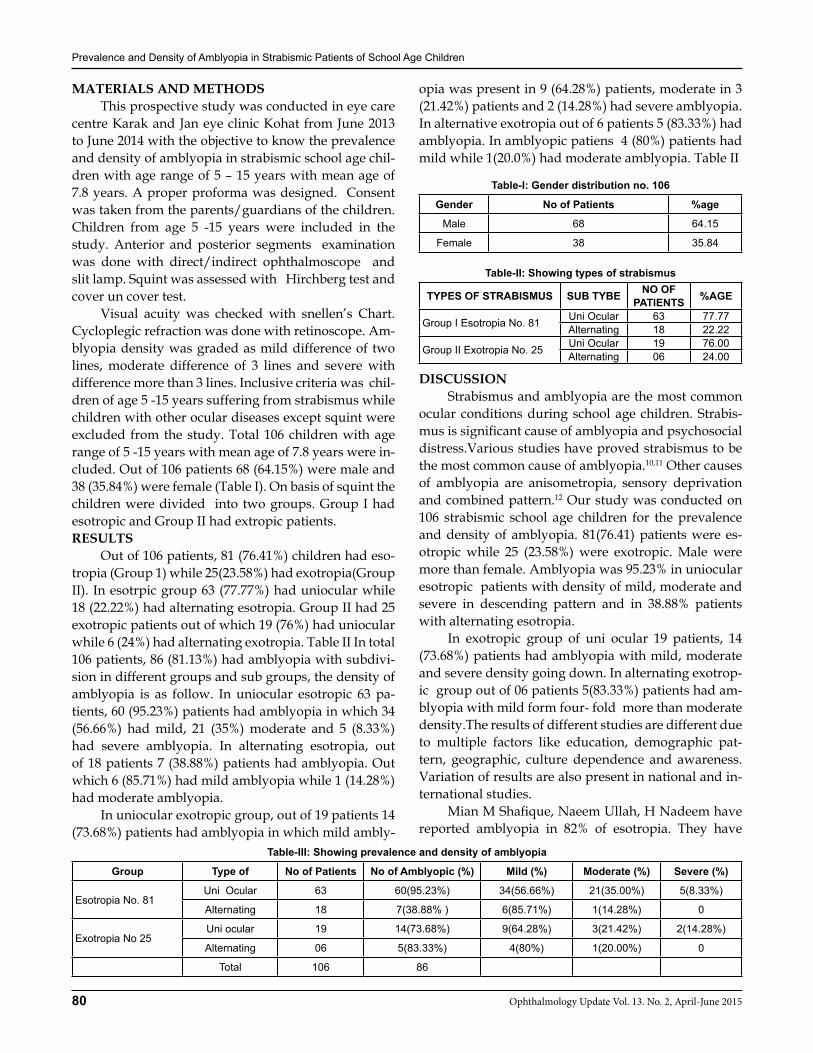
Prevalence and Density of Amblyopia in Strabismic Patients of School Age Children
80 Ophthalmology Update Vol. 13. No. 2, April-June 2015
MATERIALS AND METHODS This prospective study was conducted in eye care centre Karak and Jan eye clinic Kohat from June 2013 to June 2014 with the objective to know the prevalence and density of amblyopia in strabismic school age chil-dren with age range of 5 – 15 years with mean age of 7.8 years. A proper proforma was designed. Consent was taken from the parents/guardians of the children. Children from age 5 -15 years were included in the study. Anterior and posterior segments examination was done with direct/indirect ophthalmoscope and slit lamp. Squint was assessed with Hirchberg test and cover un cover test. Visual acuity was checked with snellen’s Chart. Cycloplegic refraction was done with retinoscope. Am-blyopia density was graded as mild difference of two lines, moderate difference of 3 lines and severe with difference more than 3 lines. Inclusive criteria was chil-dren of age 5 -15 years suffering from strabismus while children with other ocular diseases except squint were excluded from the study. Total 106 children with age range of 5 -15 years with mean age of 7.8 years were in-cluded. Out of 106 patients 68 (64.15%) were male and 38 (35.84%) were female (Table I). On basis of squint the children were divided into two groups. Group I had esotropic and Group II had extropic patients.RESULTS Out of 106 patients, 81 (76.41%) children had eso-tropia (Group 1) while 25(23.58%) had exotropia(Group II). In esotrpic group 63 (77.77%) had uniocular while 18 (22.22%) had alternating esotropia. Group II had 25 exotropic patients out of which 19 (76%) had uniocular while 6 (24%) had alternating exotropia. Table II In total 106 patients, 86 (81.13%) had amblyopia with subdivi-sion in different groups and sub groups, the density of amblyopia is as follow. In uniocular esotropic 63 pa-tients, 60 (95.23%) patients had amblyopia in which 34 (56.66%) had mild, 21 (35%) moderate and 5 (8.33%) had severe amblyopia. In alternating esotropia, out of 18 patients 7 (38.88%) patients had amblyopia. Out which 6 (85.71%) had mild amblyopia while 1 (14.28%) had moderate amblyopia. In uniocular exotropic group, out of 19 patients 14 (73.68%) patients had amblyopia in which mild ambly-
opia was present in 9 (64.28%) patients, moderate in 3 (21.42%) patients and 2 (14.28%) had severe amblyopia. In alternative exotropia out of 6 patients 5 (83.33%) had amblyopia. In amblyopic patiens 4 (80%) patients had mild while 1(20.0%) had moderate amblyopia. Table II
Table-I: Gender distribution no. 106
Gender No of Patients %age
Male 68 64.15
Female 38 35.84
Table-II: Showing types of strabismus
TYPES OF STRABISMUS SUB TYBE NO OF PATIENTS %AGE
Group I Esotropia No. 81 Uni Ocular 63 77.77Alternating 18 22.22
Group II Exotropia No. 25 Uni Ocular 19 76.00Alternating 06 24.00
DISCUSSION Strabismus and amblyopia are the most common ocular conditions during school age children. Strabis-mus is significant cause of amblyopia and psychosocial distress.Various studies have proved strabismus to be the most common cause of amblyopia.10,11 Other causes of amblyopia are anisometropia, sensory deprivation and combined pattern.12 Our study was conducted on 106 strabismic school age children for the prevalence and density of amblyopia. 81(76.41) patients were es-otropic while 25 (23.58%) were exotropic. Male were more than female. Amblyopia was 95.23% in uniocular esotropic patients with density of mild, moderate and severe in descending pattern and in 38.88% patients with alternating esotropia. In exotropic group of uni ocular 19 patients, 14 (73.68%) patients had amblyopia with mild, moderate and severe density going down. In alternating exotrop-ic group out of 06 patients 5(83.33%) patients had am-blyopia with mild form four- fold more than moderate density.The results of different studies are different due to multiple factors like education, demographic pat-tern, geographic, culture dependence and awareness. Variation of results are also present in national and in-ternational studies. Mian M Shafique, Naeem Ullah, H Nadeem have reported amblyopia in 82% of esotropia. They have
Table-III: Showing prevalence and density of amblyopia
Group Type of No of Patients No of Amblyopic (%) Mild (%) Moderate (%) Severe (%)
Esotropia No. 81Uni Ocular 63 60(95.23%) 34(56.66%) 21(35.00%) 5(8.33%)
Alternating 18 7(38.88% ) 6(85.71%) 1(14.28%) 0
Exotropia No 25Uni ocular 19 14(73.68%) 9(64.28%) 3(21.42%) 2(14.28%)
Alternating 06 5(83.33%) 4(80%) 1(20.00%) 0
Total 106 86
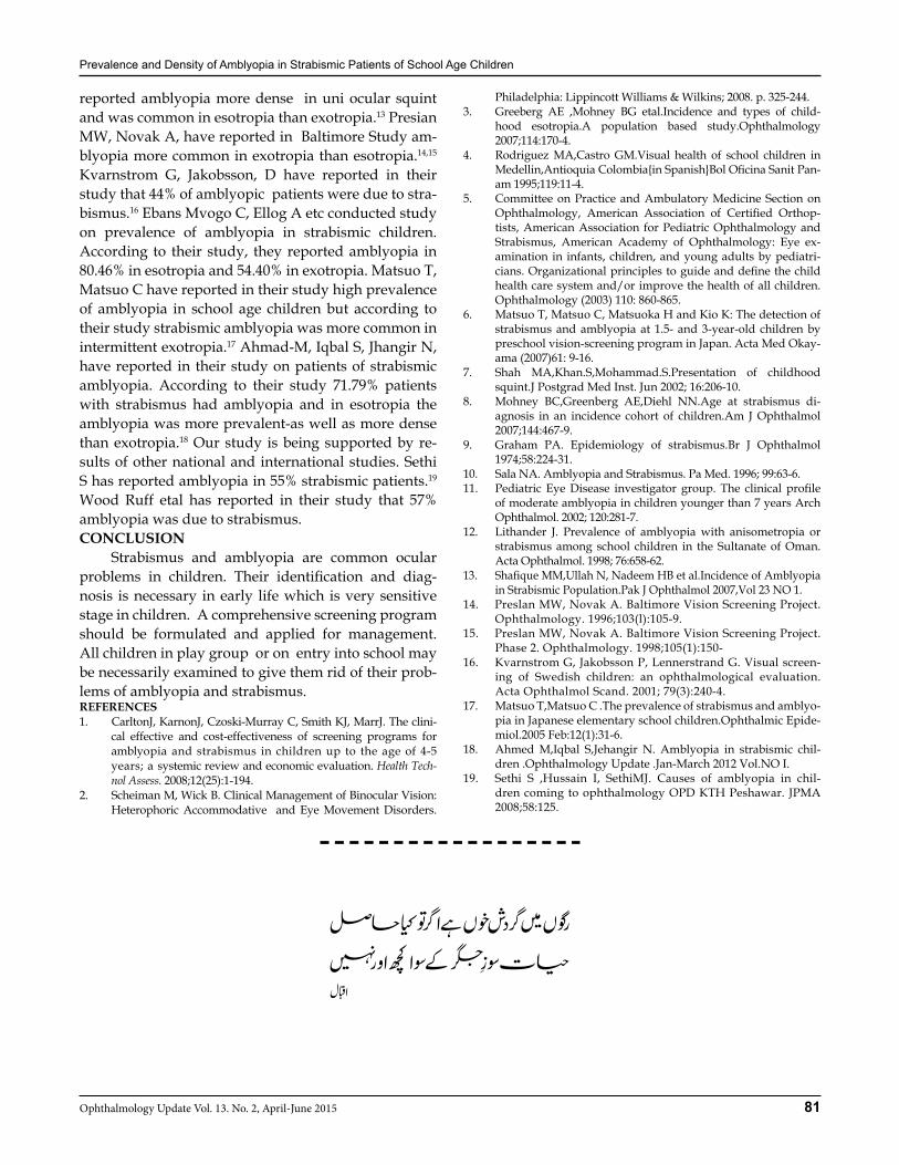
Prevalence and Density of Amblyopia in Strabismic Patients of School Age Children
81Ophthalmology Update Vol. 13. No. 2, April-June 2015
reported amblyopia more dense in uni ocular squint and was common in esotropia than exotropia.13 Presian MW, Novak A, have reported in Baltimore Study am-blyopia more common in exotropia than esotropia.14,15 Kvarnstrom G, Jakobsson, D have reported in their study that 44% of amblyopic patients were due to stra-bismus.16 Ebans Mvogo C, Ellog A etc conducted study on prevalence of amblyopia in strabismic children. According to their study, they reported amblyopia in 80.46% in esotropia and 54.40% in exotropia. Matsuo T, Matsuo C have reported in their study high prevalence of amblyopia in school age children but according to their study strabismic amblyopia was more common in intermittent exotropia.17 Ahmad-M, Iqbal S, Jhangir N, have reported in their study on patients of strabismic amblyopia. According to their study 71.79% patients with strabismus had amblyopia and in esotropia the amblyopia was more prevalent-as well as more dense than exotropia.18 Our study is being supported by re-sults of other national and international studies. Sethi S has reported amblyopia in 55% strabismic patients.19
Wood Ruff etal has reported in their study that 57% amblyopia was due to strabismus.CONCLUSION Strabismus and amblyopia are common ocular problems in children. Their identification and diag-nosis is necessary in early life which is very sensitive stage in children. A comprehensive screening program should be formulated and applied for management. All children in play group or on entry into school may be necessarily examined to give them rid of their prob-lems of amblyopia and strabismus.REFERENCES1. CarltonJ, KarnonJ, Czoski-Murray C, Smith KJ, MarrJ. The clini-
cal effective and cost-effectiveness of screening programs for ambly opia and strabismus in children up to the age of 4-5 years; a systemic review and economic evaluation. Health Tech-nol Assess. 2008;12(25):1-194.
2. Scheiman M, Wick B. Clinical Management of Binocular Vision: Heterophoric Accommodative and Eye Movement Disorders.
Philadelphia: Lippincott Williams & Wilkins; 2008. p. 325-244.3. Greeberg AE ,Mohney BG etal.Incidence and types of child-
hood esotropia.A population based study.Ophthalmology 2007;114:170-4.
4. Rodriguez MA,Castro GM.Visual health of school children in Medellin,Antioquia Colombia{in Spanish}Bol Oficina Sanit Pan-am 1995;119:11-4.
5. Committee on Practice and Ambulatory Medicine Section on Ophthalmology, American Association of Certified Orthop-tists, American Association for Pediatric Ophthalmology and Strabismus, American Academy of Ophthalmology: Eye ex-amination in infants, children, and young adults by pediatri-cians. Organizational princi ples to guide and define the child health care system and/or improve the health of all children. Ophthalmology (2003) 110: 860-865.
6. Matsuo T, Matsuo C, Matsuoka H and Kio K: The detection of strabismus and amblyopia at 1.5- and 3-year-old children by pre school vision-screening program in Japan. Acta Med Okay-ama (2007)61: 9-16.
7. Shah MA,Khan.S,Mohammad.S.Presentation of childhood squint.J Postgrad Med Inst. Jun 2002; 16:206-10.
8. Mohney BC,Greenberg AE,Diehl NN.Age at strabismus di-agnosis in an incidence cohort of children.Am J Ophthalmol 2007;144:467-9.
9. Graham PA. Epidemiology of strabismus.Br J Ophthalmol 1974;58:224-31.
10. Sala NA. Amblyopia and Strabismus. Pa Med. 1996; 99:63-6.11. Pediatric Eye Disease investigator group. The clinical profile
of moderate amblyopia in children younger than 7 years Arch Ophthalmol. 2002; 120:281-7.
12. Lithander J. Prevalence of amblyopia with anisometropia or strabismus among school children in the Sultanate of Oman. Acta Ophthalmol. 1998; 76:658-62.
13. Shafique MM,Ullah N, Nadeem HB et al.Incidence of Amblyopia in Strabismic Population.Pak J Ophthalmol 2007,Vol 23 NO 1.
14. Preslan MW, Novak A. Baltimore Vision Screening Project. Ophthalmology. 1996;103(l):105-9.
15. Preslan MW, Novak A. Baltimore Vision Screening Project. Phase 2. Ophthalmology. 1998;105(1):150-
16. Kvarnstrom G, Jakobsson P, Lennerstrand G. Visual screen-ing of Swedish children: an ophthalmological evaluation. Acta Ophthalmol Scand. 2001; 79(3):240-4.
17. Matsuo T,Matsuo C .The prevalence of strabismus and amblyo-pia in Japanese elementary school children.Ophthalmic Epide-miol.2005 Feb:12(1):31-6.
18. Ahmed M,Iqbal S,Jehangir N. Amblyopia in strabismic chil-dren .Ophthalmology Update .Jan-March 2012 Vol.NO I.
19. Sethi S ,Hussain I, SethiMJ. Causes of amblyopia in chil-dren coming to ophthalmology OPD KTH Peshawar. JPMA 2008;58:125.
82 Ophthalmology Update Vol. 13. No. 2, April-June 2015
Hussain Ahmad Khaqan FCPS, FRCS1, Farrukh Jameel MBBS2 Hadia Jabeen MBBS3, Muhammad MBBS4, Usman Imtiaz MBBS5
INTRODUCTION Tuberous sclerosis complex (TSC) is an autoso-mal dominant neuro-cutaneous disease (phacomatosis) with variable clinical manifestations.1 The incidence of the disease is approximately 1/6000- 1/10000.2,3 Diag-nosis is based on clinical and para-clinical criteria de-fined by the tuberous sclerosis consensus conference in 1998. There are two groups of symptoms including major and minor criteria. The major criteria consist of: Facial angio-fibromas or forehead plaques, Non-trau-matic ungula or periungual fibroma, Hypo-pigmented macules (more than 3), Shagreen patch, Cortical tubers, Sub-epandymal nodules, Sub-epandymal giant cell as-trocytoma, Multiple retinal nodular hamartomas, Car-diac rhabdomyoma, Lymphangio-myomatosis and re-nal angio-myolipoma.
The minor criteria include: Dental Pits (more than 14), Hamartomatous rectal polyps, Bone cysts, Cerebral white matter radial migration lines, Non-renal hamar-tomas, Retinal achromatic patch, Confetti skin lesions, Multiple renal cysts. When there are two major criteria or one major and two minor criteria the diagnosis is es-tablished as definite TSC. The term probable TS is used
when one major and one minor criteria are detected (2). Only one major feature or two or more minor criteria without any major feature mentions the possibility of tuberous sclerosis.2
Ocular manifestations of TSC including retinal hamartomas occur in less than 50% of the patients and are bilateral in one third of the cases. There is no cor-relation between age and ocular manifestations.2
Case Series: Case series identified 5 eyes of three patients over the period of 3 months from May 2013 to July 2013 (age range 8 years to 42 years). 1 of the patients was referred from neurosurgery department of Lahore General Hospital, after complaining of seizures and decrease vision, that patient had retinal Astrocytomas and sys-temic findings sebaceous adenomas, ash leaf spots, sub
ungal hemartoma, small angio-lipomas over both kid-neys, multiple calcified foci in sub-ependymal region on initial examination. 1 patient was the sibling of the patient who also had retinal Astrocytomas. 3rd patient was the mother of the patients who had right retinal astrocytomas Table 1 summarizes the case of all three patients with their age, visual acuity on presentation, initial investi-gations, examination findings, fundus findings
Patient “ A” A child 8 years/male, presented in Eye-OPD on 1st July, 2013 for the assessment of fundus, referred by some neuro-physician. He has history of fits for 2years and spontaneous muscular spasm over Left arm, for which he was given medication by the neurophysician.
ORIGINAL ARTICLE
Hussain Ahmad
1Consultant Ophthalmologist & Retinal Surgeon, Lahore General Hos-pital / PGMI, Lahore. 2,3,4Postgraduate Trainees, Lahore General Hos-pital / PGMI, Lahore. 5Resident Alshifa Trust Eye Hospital, Rawalpindi
Correspondence: Dr. Hussain Ahmad Khaqan Department of Oph-thalmology, Lahore General Hospital / PGMI, Lahore. House No. 87, Eden Canal Villas, Canal Bank Road, Thokar Niaz Baig, Lahore.E-Mail:[email protected],Tel.+92-42-37498314Cell: +92-300-4270233, Fax:+92-42-7223039
Received: December 2014 Accepted: January 2015
Tuberous Sclerosis Complex
Patient A B C
Age/sex 8/M 42/F 12Y/M
V/A 6/12 , 6/6 6/6, 6/6 6/6, 6/6
Anterior Segment Unremarkable Unremarkable Unremarkable
Posterior Segment OU Retinal Astrocytomas OD Retinal Astrocytomas OD Retinal Astrocytomas
Systemic manifestations
• Ash leaf spots• Sub ungal hemartoma• Small angio-lipomas over both kidneys • Multiple calcified foci in sub-
ependymal region• Sebaceous adenomas
Un-remarkable Un-remarkable
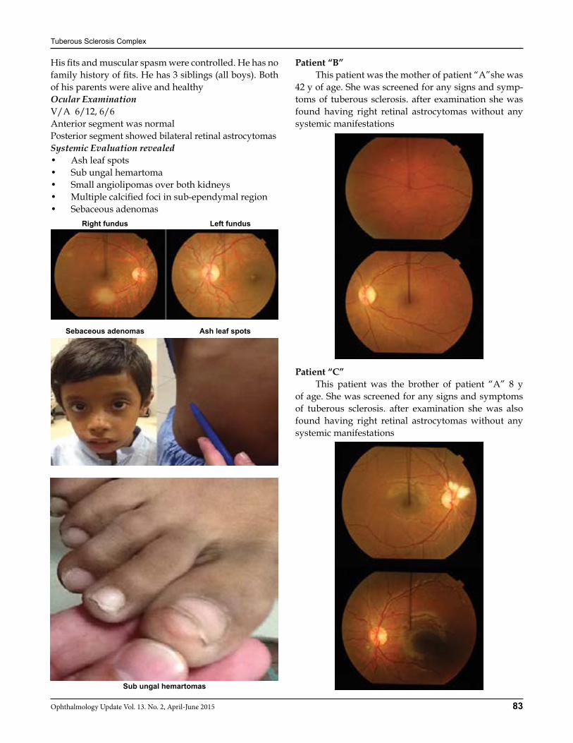
Tuberous Sclerosis Complex
83Ophthalmology Update Vol. 13. No. 2, April-June 2015
His fits and muscular spasm were controlled. He has no family history of fits. He has 3 siblings (all boys). Both of his parents were alive and healthyOcular ExaminationV/A 6/12, 6/6Anterior segment was normalPosterior segment showed bilateral retinal astrocytomas Systemic Evaluation revealed • Ash leaf spots• Sub ungal hemartoma• Small angiolipomas over both kidneys • Multiple calcified foci in sub-ependymal region• Sebaceous adenomas
Patient “B” This patient was the mother of patient “A”she was 42 y of age. She was screened for any signs and symp-toms of tuberous sclerosis. after examination she was found having right retinal astrocytomas without any systemic manifestations
Patient “C” This patient was the brother of patient “A” 8 y of age. She was screened for any signs and symptoms of tuberous sclerosis. after examination she was also found having right retinal astrocytomas without any systemic manifestations
Right fundus Left fundus
Sebaceous adenomas Ash leaf spots
Sub ungal hemartomas

Tuberous Sclerosis Complex
84 Ophthalmology Update Vol. 13. No. 2, April-June 2015
DISCUSSION 3 patients were evaluated for Tuberous Sclerosis manifestations(ocular and systemic). 2 patients were male siblings and 1 was the mother. only 1 patient showed both ocular and systemic features. 2 patients showed only Ocular features. this shows strong inherit-ance pattern and that the Tuberous Sclerosis complex does not necessarily shows systemic manifestations.CONCLUSION Tuberous Sclerosis although is a rare having vari-
able ocular and systemic manifestationsREFERENCES 1. Seyyed Hassan TONEKABONI, MD,1 Seyyed Hassan Toneka-
boni, MD,1 ParvizTousi, MD,2 Ahmad Ebrahimi. Clinical and Para clinical Manifestations of Tuberous Sclerosis ran J Child Neurol. 2012 Summer; 6(3): 25–31
2. Staley BA, Vail EA, Thiele EA. Tuberous sclerosis complex: diagnostic challenges, presenting symptoms, and commonly missed signs. Pediatrics. 2011 Jan;127(1):e117–25
3. Thiele EA, Korf BR. Phakomatoses and allied conditions. In: Swaiman KF, Ashwal S, Ferriero DM, editors. Swaimans pedi-atric neurology. 5th ed. China: Elsevier Saunders; 2012. pp. 504–9.
IMPORTANT NOTE
Authors of articles and the subsribers are requested to collect the copies of Ophthalmology Update from representatives of the concerned area according to the following:
Sr. No Names Designation Area Mobile
1 Amir Zaib Divisional Manager Peshawar Div. 0302-5551659
2 Waqas Majeed Divisional Manager Rawalpindi Div. 0302-5551817
3 Waseem Ejaz Regional Sales Manager Lahore Div. 0300-8494327
4 S. Arshad Ali Sr. Divisional Manager Faisalabad Div. 0302-5552024
5 Niaz Shaikh Sr. Divisional Manager Multan Div. 0302-5552041
6 Khalid Mehmood Sr. Divisional Manager Karachi Div. 0303-7770670
85Ophthalmology Update Vol. 13. No. 2, April-June 2015
Mohammad Kashif BVS, MPH1,Nazia Sultan BVS2
Mohammad Arshad Raza FCPS3
ABSTRACT:Aims/Objectives: (1) To evaluate the association between anemia and diabetic retinopathy (DR) including non proliferative DR (NPDR), proliferative DR (PDR) and diabetic macular edema (DME) in Type II Diabetes Mellitus (T2DM).(2) To identify anemia as an independent risk factor for DR in diabetic patients without significant renal dysfunction.(3) To correlate the severity of anemia with the severity of DR.Materials and Methods: In this case control study 170 DM patients (85 cases and 85 controls) above 40 years of age were included. All patients underwent stereoscopic fundus photography and if present the severity of DR was classified according to International Clinical Diabetic Retinopathy and Disease Severity Scale, cases were divided into 3 groups, NPDR, PDR and DME, while patients with normal fundus were included in control group. All patients underwent complete blood count (CBC) for hemoglobin estimation for detection of anemia. The statistical analysis was done using SPSS version 20. T-test and chi-square test were used for odd ratios and comparisons.Results: In the present study anemia was seen 38.8% in cases and 11.1% in controls (p<0.0001). 66.6% patients with severe NPDR and 45.8% with PDR had anemia (P < 0.0001). Odd ratio for anemia in cases and controls was 3.86, and for NPDR, PDR and DME was 3.6, 5.1, 3.0 respectively at 95% of Confidence Interval. The mean hemoglobin level in cases and controls was 10.3+3.2 and 13.6+1.35 g/dl (p<0.0001 ). Conclusion: The results showed that T2DM patients with DR had lower level of Hb and severity of anemia was positively co-related with severity of DR. It is suggested that the level Hb should be evaluated periodically in diabetic patients.Key words: Anemia, diabetic retinopathy (DR), Type 2 diabetes mellitus (T2DM).
INTRODUCTION Diabetes Mellitus (DM) is one of the leading caus-es of morbidity and mortality around the globe and is responsible for 3.8 million deaths per year.1 Its preva-lence has shown an exponential rise worldwide in the last two decades from 30 million cases in 1985 to 177 million in 2000.2 The estimated number of patients with DM worldwide for 2010 was 285 million which is pro-jected to increase to 439 million by 2030.3 The Interna-tional Diabetes Federation (IDF) ranks Pakistan 7th in the list of prevalence of DM.1 At least 171 million peo-ple worldwide have DM and this figure is likely to be doubled by the year 2030. About 50% of persons with DM are unaware of the condition and about 2 million deaths every year are attributable to this complication of DM.2 Diabetic mellitus type 2 (T2DM) is character-ized by peripheral insulin resistance, impaired regula-tion of hepatic glucose production and declining ß-cell
function, eventually leading to ß-cell failure.Diabetic Retinopathy (DR) is the most common micro vascular complication of DM and it remains a leading cause of legal blindness and visual impairment in the working-age population in the developed world. There has been a surge in the T2D-related Diabetic Retinopa-thy in the last 2 decades, especially in Asian popula-tion. Studies using retinal photography consistently suggested that the prevalence of DR is close to 40%, and sight-threatening DR (STDR) accounts for 6-8% of all diagnosed cases. Diabetic Retinopathy: It is the characteristic group of lesions found in the retina of individuals who have DM for several years. It is considered to be the result of vascular changes in the retinal circulation, a micro-angiopathy that exhibits features of both micro vascu-lar occlusion and leakage.14 DR is a progressive condi-tion with micro vascular alterations that lead to retinal ischemia, retinal permeability, retinal neo-vasculariza-tion and macular edema. If left untreated patients with DR can suffer severe visual loss. DR is asymptomatic in early stages of the disease, but as the disease progresses symptoms may include blurred vision, floaters, fluctu-ating vision, distorted vision, dark areas in the vision, poor night vision, impaired color vision, partial or total loss of vision. The risk factor that results in develop-ment and severity of DR include duration of DM, poor metabolic control, hypertension, hyperlipidemia, preg-
ORIGINAL ARTICLE
Mohammad Kashif
1Senior Optometrist, Pakistan Institute of Community Ophthalmol-ogy Hayatabad Medical Complex, Peshawar. 2Trainee Optometrist Pakistan Institute of Community Ophthalmology Hayatabad Medical Complex,Peshawar. 3Eye Specialist, District Head Quarter Hospital, Nowshera, KPK.
Correspondence: Mohammad Kashif MPH. Senior Optometrist-Faculty Member, Pakistan Institute of Community Ophthalmol-ogy Hayatabad Medical Complex, Email: [email protected] Cell: 03339100610
Received: December 2014 Accepted: February 2015
Association of Anemia with Diabetic Retinopathy in Patients with Type II Diabtese Mellitus
Association of Anemia with Diabetic Retinopathy in Patients with Type II Diabtese Mellitus
86 Ophthalmology Update Vol. 13. No. 2, April-June 2015
nancy, obesity, smoking, cataract surgery and anemia.16
Anemia in type 2 diabetes mellitus. Anemia, the most common blood disorder, is more prevalent in persons with DM than in persons without diabetes. Anemia is a below normal level hemoglobin in the blood.21 WHO defines anemia as Hb less than 12g/dl in women and less than 13g/dl in men. Using this definition, nearly 1 in 4 (23%) patients with Type2DM are anemic. The prevalence of anemia in DM patients is reported as 14-48%.22
Etiology of Anemia in Type 2DM• Diabetic neuropathy affects the central nervous
systems anemia response.• Nutritional deficiencies (low levels of iron or low
levels of certain vitamins that body needs to pro-duce Hb and make healthy red blood cells).
• Medications for DM and related conditions. Anemia is an independent risk factor for the de-velopment and progression of cardiovascular compli-cations and heart failure, chronic renal disease and DR in DM patients.25 Anemia has been associated with the development and progression of both micro vascular (i.e.; DR) and macro vascular complications of DM. Anemia can lead to falsely low HbA1c levels, which may result in under treatment of hyperglycemia, which in turn will contribute to the progression of both micro vascular and macro vascular complications.21 Individu-als with anemia were more likely to develop DR than individuals without anemia, perhaps because of ane-mia-induced retinal hypoxia. Hypoxia may alter angio-genesis, capillary permeability, vasomotor response, and retinal cells survival.26
MATERIALS AND METHODStudy Design: Analytical Observational Case Control study.Study Settings: Department of Endocrinology, Diabe-tes and Metabolic Disease at Hayatabad Medical Com-plex Peshawar.Study Duration: Study duration was 4 months (from 1st September to 30th December 2014). And total data collection time was 2 months from first October to 30 November.Sample Size Total of 170 Patients. 84 cases and 84 con-trol.Inclusion Criteria: • Cases: All Type 2 diabetic patients above age 40,
both male and female, having diabetic retinopa-thy.
• Controls: All type 2 diabetic patients with the same age, sex, demographic location to the case group, and having no DR were included.
Exclusion criteria:In both cases and controls the following exclusion cri-
teria was applied:• Patients with history of malignancy, blood loss
during the past three months.• Hypertension: systolic pressure > 140 mmHg and
diastolic pressure > 90 mmHg.• Hyperlipidemia: low density lipoprotein LDL >
130 g/dl.• Poor Diabetic control: Fasting blood sugar FBS >
200mg/dl and Random blood sugar RBS > 250 mg/dl.
• Obesity: body mass index > 30 kg/m2.• Renal failure: creatinine >1.5 mg/dl.• Cataract surgery.RESULTS In this case control study, 85 diabetic retinopathy patients (cases) and 85 normal retinal subjects (con-trols), were analyzed to study the association of anemia with diabetic retinopathy in type 2 diabetes mellitus. In the control group there were 34 (40%) males and 51 (60%) females, with a mean age of 53.5± years, duration of DM 9.98± years, Creatinine 0.93± mg/dl. In the case group NPDR was present in 43 (50.6%) patients, with a mean age of 53.4 + 9.0 years,(44.2%) were males and 24 (55.8%) females. PDR was present in 24 (28.2%) pa-tients with a mean age of 54.9+7.4 years, 9 (37.5%) males and 15 (62.5%) females. DME was present in 18 (21.2%) patients with a mean age of 51.8+8.0 years, 11 (61.1%) males and 7 (38.9%) females. By sub categorizing NPDR group, 14 (32.6%) patients had Mild NPDR, 17(39.5%) had moderate NPDR and 12 (27.9%) had severe NPDR. In patients with DME, 3 (16.7%) was had mild DME, 7 (38.9%) moderate DME, 8 (44.4%) severe DME. In the case group mean duration of DM was 10.7± years and Creatinine level 0.89 mg/dl. Odd ratio for case and con-trol was calculated by applying Chi-Square test which was 3.86 at 95% of confidence interval (CI) with a P value of 0.0001. (p-value of < 0.05 level of significance). Similarly the Odd ratio for NPDR in case and control was 3.60 at 95% of CI with a P value of 0.001. Odd ratio for PDR in case and control was 5.14 at 95% of CI with a P value of 0.001. Odd ratio for DME in case and control was 3.04 at 95% of CI with a P value of 0.001. Anemia was seen in 33 (38.8%) in the case group and 12 (11.1%) in the control group. In the case group anemia was seen in 11 (32.3%) males and 22 (43.1%) in females. And 3 (21.4%) in mild NPDR, 5 (29.4%) mod-erate NPDR, 8 (66.6%) in severe NPDR, 11 (45.8%) in PDR. In DME anemia was present in 1 (33.3%) mild DME, 2 (28.5%) moderate DME, 3(37.5%) severe DME. The mean hemoglobin level in patients with PDR was 9.3+0.3, lower than mild to moderate NPDR (10.8+1.3), 9.7+ 0.2 in Severe NPDR. In DME hemoglobin level

Association of Anemia with Diabetic Retinopathy in Patients with Type II Diabtese Mellitus
87Ophthalmology Update Vol. 13. No. 2, April-June 2015
was 11.1+0.6 in mild DME, 11.0+1.4 in moderate DME, 11.2+ 2.1 in severe DM.
Table- : Anemia in Patients with Diabetic Retinopathy and Normal Retinal Subjects
Anemia Cases Control Total
Present 33 12 45
Absent 52 73 125
Total 85 85 170
Odd Ratio at 95% of CI=3.8 P value = 0.0001
Table- : Anemia in Patients with NPDR and Normal Retinal Subjects
Anemia Case Control Total
Present 16 12 28
Absent 27 73 100
Total 43 85 128
Odd Ratio at 95% of CI = 3.60, P value = 0.001
Table- : Anemia in Patients with PDR and Normal Retinal Subjects
Anemia Case Control Total
Present 11 12 23
Absent 13 73 86
Total 24 85 109
Odd Ratio at 95% of CI = 5.14, P = 0.001
Table- : Anemia in Patients with DME and Normal Retinal Subjects
Anemia Case Control Total
Present 6 12 18
Absent 12 73 85
Total 18 85 103
Odd Ratio at 95% of CI= 3.04 P = 0.001
Table- : Anemia and Type of Diabetic Retinopathy
Type of Diabetic Retinopathy Anemia Total
NPDR 16 (37.2%) 43
PDR 11 (45.5%) 24
DME 6 (33.3%) 18
Total 33 85
DISCUSSION Diabetic Retinopathy is a major cause of blindness among the working age group. Because DM and its complications are a public health problem, data on the association of anemia with DR will help in formulating prevention from DM or at least delaying the onset. In this case control study there was a significant association of anemia with DR in Type 2 DM with odd ratio of 3.86. Anemia was higher in patients with se-vere DR like Severe NPDR and PDR in which anemia was 66.6% and 45.8%. So severity of DR was associated with the severity of anemia. Hb level was also lower in patients having DR 10.3 g/dl, than those with no DR 13.6 g/dl and was much lower in patients with severe NPDR having Hb level of 9.7 g/dl and PDR 9.3 g/dl. In 2013 Bahar et al. conducted a similar study in Sari, Iran. In their study total 1100 diabetic patients in which 159 subjects with DR (cases) and 318 normal reti-nal subjects (controls). DM patients with anemia were 2.4 times more likely to develop DR. Anemia was ob-served 45.9% in cases and 26.1% in controls and was 43 % in mild to moderate NPDR, 53% in severe NPDR and PDR. The mean hemoglobin level in controls was high-er (12.73+1.38g/dl) than patients with mild and mod-erate NPDR (12.25+1.38 g/dl) and severe NPDR and PDR (11.89+1.76 g/dl) with a P value of 0.001 respec-tively. Similarly, in our study DM patients with anemia were 3.8 times more likely to develop DR. Anemia was 38.8% in cases and 11.1% in controls and 21.4% in mild NPDR, 29.4% moderate NPDR, 66.6% in severe NPDR, 45.8% in PDR. The mean hemoglobin level in controls was higher (13.6+1.35 g/dl) than patients with mild to moderate NPDR (10.8+1.3g/dl), (9.7+ 0.2g/dl) severe NPDR, and PDR (9.3+0.3g/dl) with a P value of 0.0001 receptively. As in the study conducted in Iran, diabetic macu-lopathy (DME) was not included, no association was found between anemia and DME. While in our study there was a significant association between anemia and DME with an odd ratio of 3.04. The Hb level was low-er in patients with DME (11.1+2.1g/dl) than controls (13.6+1.35 g/dl). There was no association of treatment of DM like oral hypoglycemia medication and insulin in cases and controls (P value= 0.07). Administration of insulin was higher 7 (8.2%) in cases than controls 6
Association of Anemia with Diabetic Retinopathy in Patients with Type II Diabtese Mellitus
88 Ophthalmology Update Vol. 13. No. 2, April-June 2015
(7.1%) which was similar to the Iran’s study in which there was also no association of treatment of DM and insulin was higher (20.5%) in the case group compared to the controls (11%).29
Qiao et al. in Finland found that the DM patients with Hb level lower than 12 g/dl had a two-fold higher prevalence of retinopathy after other known factors were controlled. It was also found that in patients with retinopathy, the severity correlated with the severity of anemia. Among patients who had DR odd ratio of ane-mia was 5.3 (95% CI,) for severe DR.32 Similarly in our study it was found that in DM patients anemia was 3.8 times more likely to develop DR and the severity of DR was correlated with the severity of anemia as odd ratio for severe retinopathy (PDR) was 5.1 at 95% of CI. David et al. 1997 in Early Treatment of Diabetic Retinopathy Study evaluated a progressive increase in risk for high risk PDR with decreasing Hb and sup-porting the importance of anemia as a risk factor for the progression and severity of DR. The etiology and pathogenesis of anemia in DM patients is multi facto-rial. Decreased erythropoietin production is an impor-tant cause of development of anemia in DM patients. Chronic hyperglycemia is involved in the pathogenesis of anemia by means of creating abnormalities in RBCs, oxidative stress, autonomic neuropathy and renal sym-pathetic denervation. These conditions put the renal inerstitium in a hypoxic state and consequently, the production of erythropoietin by peritubular fibroblasts is impaired.34
A well-accepted cut-point definition of anemia was selected in our study for our, namely a hemoglobin <13 g/dl in men and <12 g/dl in women. Our results dem-onstrated that the presence of anemia is an independ-ent risk factor for DR in the case control study. Sub-group analysis suggested that the prevalence of anemia in DR patients (cases) in females was higher (43.1%) than males (32.3%). A study conducted in 2010 by Ranil PK et al. in India in which same definition was used to define anemia, Individuals with anemia were 1.80 times more likely to develop diabetic retinopathy than individuals with no anemia. The prevalence of anemia was higher in women (26.4%) than in men (10.3%). Men with anemia, and not women, had 2 times the risk of developing diabetic retinopathy. While in our study al-though the prevalence of anemia was higher in females, but both males and females in cases with anemia were 3.8 times more likely to develop DR.30
In a case control study done by Francisco J et al. in 2012 there were total 106 T2DM patients in which Hb was having a significant association with PDR with odd ratio of 2.43 at 95% of confidence interval (P value
of 0.001) and anemia was an important finding in di-abetic patients which was a relevant factor related to the progression of proliferative diabetic retinopathy (PDR), which can be treated with photocoagulation.31
This study was having similar results to our study in which hemoglobin level was also having a significant association with Proliferative DR with an Odd ratio of 5.1 at 95% of CI (P value of 0.0001). In a study done in 2012 by JO Chung “Associa-tions between hemoglobin concentration and the clini-cal characteristics of patients with Type 2 diabetes” the patients with lower Hb concentrations had a longer du-ration of diabetes, a lower body mass index, and lower concentrations of total cholesterol, triglycerides, and low-density lipoprotein cholesterol. They had a higher prevalence of diabetic retinopathy (DR) and nephropa-thy. The increased prevalence of diabetic retinopathy was associated with lower Hb concentrations. These findings suggested that lower Hb concentrations might not only be a consequence of diabetes but may also ac-celerate micro-vascular damage in diabetes mellitus.[36]
While in our study there was no association of dura-tion of DM with hemoglobin level and patients with a body mass index (obesity) > 30 kg/m2, LDL > 130mg/dl were excluded in the study. As they were risk fac-tors for DR so were controlled. Patients with creatinine < 1.5 mg/dl were included in both cases and controls proving that anemia in DM patients was unrelated to anemia due to diabetic nephropathy. Detection of anemia and its treatment is important in the management of diabetic retinopathy. In those patients who had both anemia and diabetes mellitus, Friedman and associates reported that treatment with erythropoietin was correlated with substantial reso-lution of macular hard exudates. The improved Hb concentration with therapy of anemia improves tissue oxygenation and may result in reduced VEGF pro-duction, which improves the hyper permeability and reduces the stimulus for neovascularization.41 These observations suggest that anemia evaluation should be considered in the routine management of persons with diabetes and should be treated to minimize the risk of microvascular complications such as nephropathy and DR. CONCLUSION Diabetic retinopathy is emerging a big public health problem, affecting working age groups. Our finding suggests that there was a significant association between anemia and diabetic retinopathy in Type 2 diabetes mellitus. Anemia was associated with NPDR, PDR and DME. So it is concluded that anemia is an inde-pendent risk factor for the development of DR. Severity
Association of Anemia with Diabetic Retinopathy in Patients with Type II Diabtese Mellitus
89Ophthalmology Update Vol. 13. No. 2, April-June 2015
of anemia is co-related with severity of DR. Prevalence of anemia was higher in females than males having DR., and was also higher in patients with DR who were using both oral hypoglycemic medications and insulin together. Evaluation and treatment of anemia should be a part of the follow up visits of DM patients. Further studies about the effect of anemia treatment on the se-verity of diabetic retinopathy are recommended.REFRENCES1. International Diabetes Federation: The Diabetes Epidemic Is
Out of Control [online]. October 20, 2009 [cited 30/12/2014]. Available at http://asweetlife.org/feature/idf-the-diabetes-epidemic-is-out-of-control/.
2. Wild S, Roglic G, Green A, Sicree R, King H. Global prevalence of diabetes: estimates for the year 2000 and projections for 2030. Diabetes Care. 2004; 27: 1047–1053.
3. Sivaprasad S, Gupta B, Evans J et al. Prevalence of Diabetic Retinopathy in Various Ethnic Groups: A Worldwide Perspec-tive. SurvOphthalmol. 2012; 57(4): 347 70.
4. JB Brown, KL Pedula, KH Summers. Diabetic Retinopathy Contemporary Prevalence in a Well-Controlled Population. Diabetes Care, 2003; 26: 2637-2942.
5. Thomas, M., Tsalamandris, C et al. Anaemia in Diabetes: An Emerging Complication of Micro vascular Disease. Current Diabetes Reviews. 2005; 1: 107-126.
6. Diabetic Retinopathy - Asia Pacific - American Academy of Ophthalmology [online]. November 2013 [cited on 21/9/2014]. Available from http://one.aao.org/topic-detail/diabetic-retinopathy--asia-pacific.
7. Vision 2020 – World Health Organization [online]. December 2006 [cited on 20/12/2014]. Available at www.who.int/blind-ness/Vision2020_report.pdf.
8. Mahar PS, Awan MZ, Manzar N, Memon MS. Prevalence of Type-II Diabetes Mellitus and Diabetic Retinopathy [online]. 2010 [cited on 30/12/2014]. Available at http://www.atlas.idf.org/ index1397.html.
9. Jadoon MZ, Dineen B, et al. Prevalence of blindness and visual impairment in Pakistan: the Pakistan national blindness and visual impairment survey. Invest Opthalmol Vis Sci. 2006; 47:4749-55.
10. Qayyum AL, Amir Babar AM, Das G. Prevalence of diabetic retinopathy in Quetta, Balochistan. Pak J Ophthalmol. 2010; 26(4): 187–192.
11. Shaikh MA, Gillani S, Yakta D. Frequency of diabetic retinopa-thy in patients after ten years of diagnosis of type 2 diabetes mellitus. Journal of Ayub Medical College Abbottabad. 2010; 22(3): 158–160.
12. Khan AJ. Prevalence of Diabetic Retinopathy in Pakistan sub-jects. A pilot study. J Pak Med Assoc.1991; 41: 49.
13. Diabetic Statistics in Pakistan [online]. May 8, 2013 [cited 30/12/2014]. Available at http://diabetespakistan.com/treat-ment/2013/05/08/diabetes-statistics-in-pakistan.
14. Diabetic Retinopathy Guidelines- International Council of Ophthalmology. [online]. 2012 [30/12/2014].
15. Available on www.icoph.org/dynamic/attachments/diabetic-retinopathy.pdf.
16. Wu L. Loaiza PF et al.Classification of diabetic retinopathy and diabetic macular edema. World Journal of Diabetes. December 15, 2013; 4(6): 290-294.
17. Kanski JJ, Bowling B. Clinical Ophthalmology and Systematic Approach. Edition 7th. Elsevier limited China: Saunders. 28 April 2011; 536.
18. Kanski JJ, Bowling B. Synopsis of Clinical Ophthalmology. Third edition. Elsevier limited China: 2013; 244-246.
19. Gupta V, Gupta A, Dogra MR, Singh R. DIABETIC RETIN-OPATHY atlas and text. First edition. New delli: 2007; 31-107.
20. ICO Guidelines for Diabetic Eye Care – International Council of Ophthalmology [online]. February 2014 [cited on 30/12/2014].
21. Available at http://www.icoph.org/downloads/ICOGuide-linesforDiabeticEyeCare.pdf.
22. International Clinical Diabetic Retinopathy Disease Severity Scale – American Academy Of Ophthalmology [online]. Octo-ber 2002 [cited on 30/12/214]. Available at one.aao.org/asset.axd?id=4b0447c9-24c6-411e-a888-3081553693d2.
23. Thomas MC, MacIsaac RJ, Tsalamandris C, et al. Unrecognized anemia in patients with diabetes: a cross-sectional survey. Dia-betes Care. 2003; 26(4): 1164-9.
24. WHO - Iron deficiency anaemia: assessment, prevention, and control [online]. 2001 [cited 30/12/2014].
25. Available at www.who.int/nutrition/publications/micronu-trients/anaemia
26. Recognizing Anemia in People with Diabetes [online] .March 11, 2009 [cited 24/9/2014].
27. Available at http://www.anemia.org/patients/feature-arti-cles/content.php?contentid=000367.
28. Conway BN, Miller RG, Klein R, Orchard TJ. Prediction of proliferative diabetic retinopathy with hemoglobin level. Arch Ophthalmol. 2009;127:1494–9.
29. Deray G, Heurtier A, Grimaldi A, Launay Vacher V, Isnard Bagnis C. Anaemia and diabetes. Am J Nephrol. 2004;24:522–6.
30. Irace C, Scarinci F, Scorcia V, et al. Association among low whole blood viscosity, haematocrit, haemoglobin and diabet-ic retinopathy in subjects with type 2 diabetes. Br J Ophthal-mol.2011; 95: 94–8.
31. Maccuish AC. Early detection and screening for diabetic retin-opathy. Eye. 1993; 7: 254-259.
32. Congdon NG, Friedman DS, Lietman T. Important causes of visual impairment in the world today. Journal Of The Ameri-can Medical Association. 2003; 290 (15): 2057–2060.
33. Bahar A, Kashi Z, Ahmadzadeh Amiri A et al. Association between diabetic retinopathy and hemoglobin level. Caspian Journal of Internal Medicine.2013; 4(4): 759-762.
34. Rani PK, Raman R, Rachepalli SR. Anemia and Diabetic Retin-opathy in Type 2 Diabetes Mellitus. J Assoc Physicians India. FEBRUARY 2010; 58: 91-4.
35. diabetic microcirculatory disease? Lancet. 1977; 2: 789–91.
90 Ophthalmology Update Vol. 13. No. 2, April-June 2015
Prof. Laal Mohammad FCPS1, Mohammad Alam FCPS2, Arshad Farzooq FCPS3
ABSTRACT:Objective: To find out control of intra ocular pressure after cataract extraction with posterior chamber intraocular lens im-plantation in Phacomorphic glaucoma.Materials and Methods: This retrospective study was conducted in KDA Teaching Hospital Kohat from January 2009 to December, 2013 with the objective of finding intraocular pressure control after cataract extraction with posterior chamber intra ocular lens implantation in phacomorphic glaucoma. 48 patients with phacomorphic glaucoma were selected. Informed consent was taken from the patients. Preoperative Intraocular pressure was checked with Perkin Tonometer. All patients were examined with slit lamp. Patients were put on mannitol, systemic carbonic anhydrase inhibitor, and topical antiglau-coma drugs. Topical steroid / antibiotic eye drops were given for five days to one week to control inflammation. After control of IOP and inflammation, patients were operated by conventional extracapsular cataract extraction with posterior chamber Intraocular lens implantation. IOP was checked after one week and one month without anti glaucoma drugs. Total 48 pa-tients comprising of 22(45.83%) male and 26 (54.16%) female were included in the study.Results: On presentation preoperative intraocular pressure of all the patients was in the range of 31 to 48 mmHg with mean intraocular pressure of 38.8 mmHg. After surgery no patients was put on antiglaucoma medication. All the patients were put on steroid and antibiotic topical drops for three weeks and systemic pain killer for five days. After one month post operative Intraocular pressure was in the range of 12 – 20 mmHg with mean intraocular pressure of 15.52mm Hg. Conclusion: There is a significant control of intraocular pressure with normal range in phacomorphic glaucoma after extra-capsular cataract extraction with posterior chamber intraocular lens implantation.Key Words: Intraocular pressure, Phacomorphic glaucoma, Extracapsular cataract extraction.Abbreviations: Intra ocular pressure (IOP), Intra ocular lens(IOL), Extracapsular cataract extraction (ECCE). Posterior Chamber(PC).
INTRODUCTION Cataract is considered to be the most significant cause of blindness globally as well as territorially.1,2 Gifford described phacomorphic glaucoma as a sepa-rate entity for the first time in 1900.3 He attributed it to hypermature cataract. In phacomorphic glaucoma, the lens blocks the forward flow of aqueous humor through the pupil resulting in rise of IOP. This classi-cally occurs in large intumescent cataract which is then named as phacomorphic glaucoma. This lens induced glaucoma is a preventable and a treatable disease if managed at proper time. This condition still exists in the world. Phacomorphic glaucoma is due to lack of awareness of cataract and delayed surgical interven-tional removal. It is normally due to wrong concept that cataract should be mature at the time of surgery, lack of need for better vision, concurrent systemic dis-eases , old age, ignorance and economic constraints are
other reasons that the patients avoid treatment.4 Differ-ent studies have reported that in subcontinent countries like India, the incidence of intumescent cataract leading to phacomorphic glaucoma is more in comparison to western world.5 Cataract extraction is the only treat-ment of phacomorphic glaucoma. But before surgery IOP is being lowered down to a safe level with medica-tion to prevent glaucoma related problems. This study was done to find out the IOP control after cataract ex-traction with PC- IOL.MATERIALS AND METHODS This retrospective study was conducted in KDA Teaching Hospital Kohat from January 2009 to Decem-ber 2013 with the objective to find out IOP control after cataract extraction with PC-IOL in patients of phaco-morphic glaucoma. Diagnosis of phacomorphic was made when patients presented with symptoms of pain, redness of involved eyes, headache and above normal IOP, shallow anterior chamber and intumescent cata-ract. Proper proforma was designed for documenta-tion of clinical findings of patients, time and duration of presentation on arrival. IOP was checked with Perkin’s Tonometer and visual acuity was recorded. Total 48 patients were selected out of which 22 (45.83%) were male and 26 (54.16%) were female. (Table-I) Age was
ORIGINAL ARTICLE
Laal Mohammad
1Prof. of Ophthalmology, 2Associate Prof. of Ophthalmology, 3Oph-thalmologist, KMU Institute of Medical Sciences, Kohat
Correspondence: Professor Lal Mohammad Department of Ophthal-mology, KMU Institute Of Medical Sciences, K.D.A KohatCell: 0300-5961577, E-Mail: [email protected]
Received: January 2015 Accepted: February 2015
Intraocular Pressure Control after Cataract Extraction with Posterior Chamber Intraocular
Lens Implantation in Phacomorphic Glaucoma
Intraocular Pressure Control after Cataract Extraction with Posterior Chamber Intraocular Lens Implantation in Phacomorphic Glaucoma
91Ophthalmology Update Vol. 13. No. 2, April-June 2015
ranging from 59 years to 73 years with mean age of 66.3 years. Duration of the symptoms were recorded. (Ta-ble-II) All the patients IOP were controlled with man-nitol, systemic carbonic anhydrase inhibitor and topi-cal antiglaucoma medicine. Steroid/antibiotic topical drops were also given to the patients to control inflam-mation for 5-7 days preoperatively. All the patients were operated by conventional extra capsular cataract extraction with PC- IOL implantation. Those patients with preoperative visual acuity of no perception of light were excluded from the study. After surgery pa-tients were put on steroid/antibiotic topical drops for three weeks and pain killer for 5 days. No antiglauco-ma medications were prescribed postoperatively. IOP was checked after one week and one month.RESULTS On presentation, preoperative IOP of all patients was in range of 31 mmHg to 48 mmHg with mean IOP of 38.8 mmHg. Post operative IOP after one week was in range of 12 to 22 mmHg with mean IOP of 15.91 mmHg. After one month post operative IOP was in the range of 12-20 mmHg with mean IOP of 15.52 mmHg.(Table III)
Table-I: Showing gender distribution.
Gender No of Patients %age
Male 22 45.83
Female 26 54.16
Table-II: Duration of presentation
Duration No of patients %age
0 -3 days 27 56.25
4 -7 days 15 31.25
8 -14 days 06 12.50
Table-III: Preoperative and postoprtative IOP
IOP Range in mmHg Mean in mmHg
Preoperative 31—48 38.8
Postoperative One week 12—22 15.91
Postoperative One month 12—20 15.52
DISCUSSION Intumescent cataract is the main cause of pupil-lary block phacomorphic glaucoma resulting in high IOP with damage to the Optic nerve. After control of preoperative IOP the patients are operated for cataract extraction, the obstruction to the out flow of aquous humor is removed and there is drastic fall in IOP and the patients do not need any antiglaucoma therapy. The same statement is true as observed in our study in which preoperative mean IOP 38.8 mmHg dropped
down to postoperative mean IOP 15.52 mmHg. There are many national and international studies showing similar results. Mandal AK,Gothwal UK have reported IOP control in normal level in all patients operated for phacomorphic glaucoma.6 Rajal AP, Karki DB report-ed IOP control after cataract surgery in phacomorphic glaucoma to be from 14 – 22 mmHg in all patients. In their study female patients were more than male as in our study.7 Payal Gupta study demonstrates post op-erative IOP to be lower than 20 mmHg in all phaco-morophic glaucoma patients without postoperative antiglaucoma medicine.8 Sing G and Vankatesh et al studies also reported post operative IOP control of 20 mmHg or less in all patients.9,10 Mohinder Singh, Has-san Al Arrayyed studies reveal IOP control of below 21 mmHg in all patients like in our study. However, the age of patients with phacomorphic glaucoma were dif-ferent.11
R Ramekrishanan, Davendra Maheshwari et al conducted a study of IOP control in phacomorophic glaucoma in 74 patients. Postoperative IOP was con-trol and below 20 mmHg in all patients with out an-tiglaucoma therapy.12 They used sutureless surgery technique so it is clear that IOP control in phacomor-phic glaucoma does not depend upon the method used. Nithisha TM, Mallikarjun, Salagar reported that 28% of phacomorophic glaucoma patients had postoperative IOP of more than 20 mmHg which is contradictory to our study. Probably this variation may be due opera-tion complication.13 PS Mazhar, M Amin Shahzad have reported in their study 3.6% of all glaucoma was phaco-morphic needed urgent control of IOP and removal of lens.14 This ratio is less as compared to other study.CONCLUSION Phacomorphic Glaucoma is a devastating ocular condition with high IOP. If treated early with cata-ract extraction and implantation of PC-IOL, pupillary block will be removed and there is significant fall in IOP within normal range. The patients do not need an-tiglaucoma therapy post operatively. Therefore public awareness programs may be carried out through print and electronic media and public gatherings to get rid of this problem.REFERENCES1. Thulasiraj RD, Rahamathulla R, Saraswati A, Selvaraj S, Ell-
wein LB. The Sivaganga eye survey: I. Blindness and cataract surgery. Ophthalmic Epidemiol. 2002;9:299-312.
2. Thylefors B, Negrel AD, Pararajasegaram R, Dadzie KY. Global data on blindness. Bull World Health Organ.1995;73;115-21.
3. Duke-Eder S. System of Ophthalmology. Vol. XI: Diseases of the lens and Vitreous; Glaucoma and Hypotony. St. Louis: CV Mosby 1969; 662-3.
4. Tomey KF, al-Rajhi AA. Neodymium: YAG laser iridotomy in the initial management of phacomorphic glaucoma. Ophthal-mology. 1992;99:660-5.
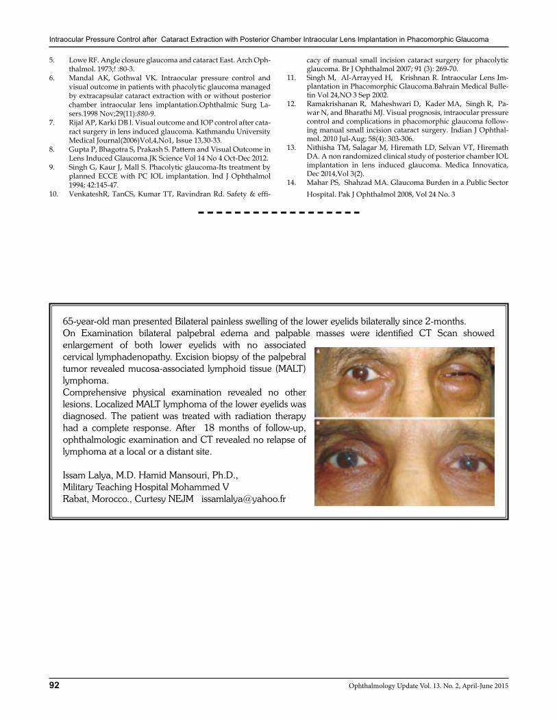
Intraocular Pressure Control after Cataract Extraction with Posterior Chamber Intraocular Lens Implantation in Phacomorphic Glaucoma
92 Ophthalmology Update Vol. 13. No. 2, April-June 2015
5. Lowe RF. Angle closure glaucoma and cataract East. Arch Oph-thalmol. 1973;! :80-3.
6. Mandal AK, Gothwal VK. Intraocular pressure control and visual outcome in patients with phacolytic glaucoma managed by extracapsular cataract extraction with or without posterior chamber intraocular lens implantation.Ophthalmic Surg La-sers.1998 Nov;29(11):880-9.
7. Rijal AP, Karki DB l. Visual outcome and IOP control after cata-ract surgery in lens induced glaucoma. Kathmandu University Medical Journal(2006)Vol,4,No1, Issue 13,30-33.
8. Gupta P, Bhagotra S, Prakash S. Pattern and Visual Outcome in Lens Induced Glaucoma.JK Science Vol 14 No 4 Oct-Dec 2012.
9. Singh G, Kaur J, Mall S. Phacolytic glaucoma-Its treatment by planned ECCE with PC IOL implantation. Ind J Ophthalmol 1994; 42:145-47.
10. VenkateshR, TanCS, Kumar TT, Ravindran Rd. Safety & effi-
cacy of manual small incision cataract surgery for phacolytic glaucoma. Br J Ophthalmol 2007; 91 (3): 269-70.
11. Singh M, Al-Arrayyed H, Krishnan R. Intraocular Lens Im-plantation in Phacomorphic Glaucoma.Bahrain Medical Bulle-tin Vol 24,NO 3 Sep 2002.
12. Ramakrishanan R, Maheshwari D, Kader MA, Singh R, Pa-war N, and Bharathi MJ. Visual prognosis, intraocular pressure control and complications in phacomorphic glaucoma follow-ing manual small incision cataract surgery. Indian J Ophthal-mol. 2010 Jul-Aug; 58(4): 303-306.
13. Nithisha TM, Salagar M, Hiremath LD, Selvan VT, Hiremath DA. A non randomized clinical study of posterior chamber IOL implantation in lens induced glaucoma. Medica Innovatica, Dec 2014,Vol 3(2).
14. Mahar PS, Shahzad MA. Glaucoma Burden in a Public Sector Hospital. Pak J Ophthalmol 2008, Vol 24 No. 3
65-year-old man presented Bilateral painless swelling of the lower eyelids bilaterally since 2-months. On Examination bilateral palpebral edema and palpable masses were identified CT Scan showed enlargement of both lower eyelids with no associated cervical lymphadenopathy. Excision biopsy of the palpebral tumor revealed mucosa-associated lymphoid tissue (MALT) lymphoma.Comprehensive physical examination revealed no other lesions. Localized MALT lymphoma of the lower eyelids was diagnosed. The patient was treated with radiation therapy had a complete response. After 18 months of follow-up, ophthalmologic examination and CT revealed no relapse of lymphoma at a local or a distant site.
Issam Lalya, M.D. Hamid Mansouri, Ph.D., Military Teaching Hospital Mohammed VRabat, Morocco., Curtesy NEJM [email protected]
93Ophthalmology Update Vol. 13. No. 2, April-June 2015
Mohammad Kashif BVS, MPH1, Mohammad Arshad Raza FCPS2
Siraj Safi BVS, DBO3, Fahim Marwat BVS4, Samiuddin BVS5
ABSTRACTObjectives 1. To determine the and causes of low vision in adult and children. 2. To evaluate quality of life before and after using low vision devicesMaterials & Methods: This Cross-sectional study conducted at Low Vision Clinic, Department of Ophthalmology Hayat-abad Medical Complex during a period of six month from June 2014 to Dec 2014. A total of one hundred and sixty five sub-jects were assessed and referred by Ophthalmologist. The magnitude of etiology for low vision were recorded and analyzed. The patients having best corrected visual acuity < 6/18 in the better eye were consulted for low vision re-assessment with the help of LVDs.. Quality of life questionnaire (LVQOL) was administered to every patient on first and follow up visit after using LVDs in order to determine the impact of LVDs on quality of life of the selected subjects. Data was analyzed with SPSS 16. Frequencies of responses to different questions were calculated.Results: Total of one hindered and sixty five patients were include in study having adults were 102 (61.8%), Children 63 (38.18%).The main causes of low vision in children includes stargardt’s disease 22.2%, nystagmus 17.4%, Retinitis pigmentosa 14.28% albinism with nystagmus 12.69%, Aphakia 12.69%, Myopia 11.1%, Cong. Cataract 3.17%, corne-al opacity 3.17%, cone dystrophy 1.59%. Among adult group the main causes were age related macular degeneration 21.50%, corneal opacity 15.68%,Retinitis pigmentosa 13.72%, aphakia%, high myopia 8.82%, congenital cataract 5.58%, glucoma5.88%,nystagmus 4.70%, oculo-cutaneous albinism 3.92%, Stargadt,s maculopathy 3.92%, cone dystrophy 3.92%. After using low vision devices the population with group of great problem reduced to only 10% while the moderate category reduced to 20% respectively. Similarly the problem with activities of daily living reduced after using LVD,s from 65% to 35% so the reduction was almost half and those who were having no problem increased from 13% to 49 %. Although the score of the population in the psychological adjustments was less as compare to other aspect e.g. reading, distance equity etc but still significant amount of population gain a reasonable score after using LVD,s. Conclusion : Efforts should be done to reduce the low vision burden of the diseases which are treatable, . Visually impaired patients due to different etiologies do benefit from low vision services which facilitate vision having dramatic impact on the quality of life of those suffering subjects.Key Words: Visual impairment, Low vision devices, quality of life
INTRODUCTION Low Vision: a person with low vision is one who has impairment of visual functioning even after treat-ment and/or standard refractive correction, and has a visual acuity of less than 6/18 to light perception, or a visual field less than 10 degrees from the point of fixa-tion, but who uses, or is potentially able to use, vision for the planning and/or execution of a task for which vision is essential.1 Or Low vision is visual acuity less than 6/18 and equal to or better than 3/60 in the bet-ter eye with best correction.1 (WHO)” Functionally, low
vision is characterized by irreversible visual loss and a reduced ability to perform many daily activities, It is an important public health problem and provision of low vision services is one of the priorities in the global initiative, VISION 2020—The Right to Sight.Low Vision and Quality of Life: The quality of life of a person with low vision is always compromised. The presence of low vision affects functional and social life of an individual and has a negative effect on physical and emotional well being and increased emotional dis-tress.2 The provision of low vision services and use of low vision devices allows people with visual impairment to use their limited residual vision as opti-mally as possible. 1.3 ICD-10 Classification of Visual Impairment: “The world Health Organization ICD-10 (International Clas-sification of Diseases) categories the visual impairment in to three categories 1: Moderate visual impairment from all causes visual acuity of 6/18 to 6/60. 2: Severe visual impairment from all causes 6/60 to 3/60 in the better eye and 3: Blindness from all causes 3/60 in the better eye.3”
ORIGINAL ARTICLE
Mohammad Kashif
1Senior Optometrist, Pakistan Institute of Community Ophthalmol-ogy Hayatabad Medical Complex, Peshawar. 2Eye Specialist, Dis-trict Head Quarter Hospital, Nowshera, KPK. 3Lecturer Optometry, Pakistan Institute of Community Ophthalmology Hayatabad Medical Complex. 4Orthoptist, Pakistan Institute of Community, Ophthalmol-ogy Hayatabad Medical Complex. 5Optometrist, Pakistan Institute of Community, Ophthalmology Hayatabad Medical Complex peshawar.
Correspondence: Mohammad Kashif MPH, Senior Optometrist-Fac-ulty Member, Pakistan Institute of Community Ophthalmology Hayat-abad Medical Complex. Email: [email protected]: 03339100610
Received: December 2014 Accepted: February 2015
Causes of Low vision and Quality of Life after Rehabilitation in Children & Adults
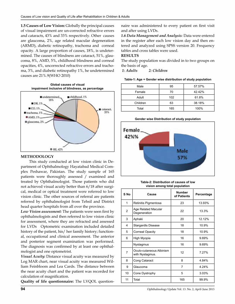
Causes of Low vision and Quality of Life after Rehabilitation in Children & Adults
94 Ophthalmology Update Vol. 13. No. 2, April-June 2015
1.5 Causes of Low Vision: Globally the principal causes of visual impairment are un-corrected refractive errors and cataracts, 43% and 33% respectively. Other causes are glaucoma, 2%, age related macular degeneration (ARMD), diabetic retinopathy, trachoma and corneal opacity. A large proportion of causes, 18%, is undeter-mined. The causes of blindness are cataract, 51%, glau-coma, 8%, AMD, 5%, childhood blindness and corneal opacities, 4%, uncorrected refractive errors and tracho-ma, 3%, and diabetic retinopathy 1%, he undetermined causes are 21%.9(WHO 2010)
Global causes of visual impairment inclusive of blindness, as percentage
METHODOLOGY This study conducted at low vision clinic in De-partment of Ophthalmology Hayatabad Medical Com-plex Peshawar, Pakistan. The study sample of 165 patients were thoroughly assessed / examined and treated by Ophthalmologist. Those patients who did not achieved visual acuity better than 6/18 after surgi-cal, medical or optical treatment were referred to low vision clinic. The other sources of referral are patients referred by ophthalmologist from Tehsil and District head quarter hospitals from all over the province.Low Vision assessment: The patients were seen first by ophthalmologists and then referred to low vision clinic for assessment, where they are refracted and assessed for LVDs Optometric examination included detailed history of the patient, his/ her family history; function-al, occupational and clinical assessment. The anterior and posterior segment examination was performed. The diagnosis was confirmed by at least one ophthal-mologist and one optometrist. Visual Acuity: Distance visual acuity was measured by Log MAR chart, near visual acuity was measured Wil-liam Feinbloom and Lea Cards. The distance between the near acuity chart and the patient was recorded for calculation of magnification.Quality of life questionnaire: The LVQOL question-
naire was administered to every patient on first visit and after using LVDs.1.6 Data Management and Analysis: Data were entered in the register after each low vision day and then en-tered and analyzed using SPSS version 20. Frequency tables and cross tables were used. RESULTSThe study population was divided in to two groups on the basis of age. 1: Adults 2: Children
Table-1: Age + Gender wise distribution of study population
Male 95 57.57%
Female 70 42.42%
Adult 102 61.8%
Children 63 38.18%
Total 165 100%
Gender wise Distribution of study population
Table-2: Distribution of causes of low vision among total population
S No Cause Number of Patients Percentage
1 Retinitis Pigmentosa 23 13.93%
2 Age Related Macular Degeneration 22 13.3%
3 Aphaki 20 12.12%
4 Stargardts Disease 18 10.9%
5 Corneal Opacity 18 10.9%
6 High Myopia 16 9.69%
Nystagmus 16 9.69%
7 Oculo-cutaneous Albinism with Nystagmus. 12 7.27%
8 Cong Cataract 8 4.84%
9 Glaucoma 7 4.24%
10 Cone Dystrophy 5 3.03%
11 Total 165 99.9%
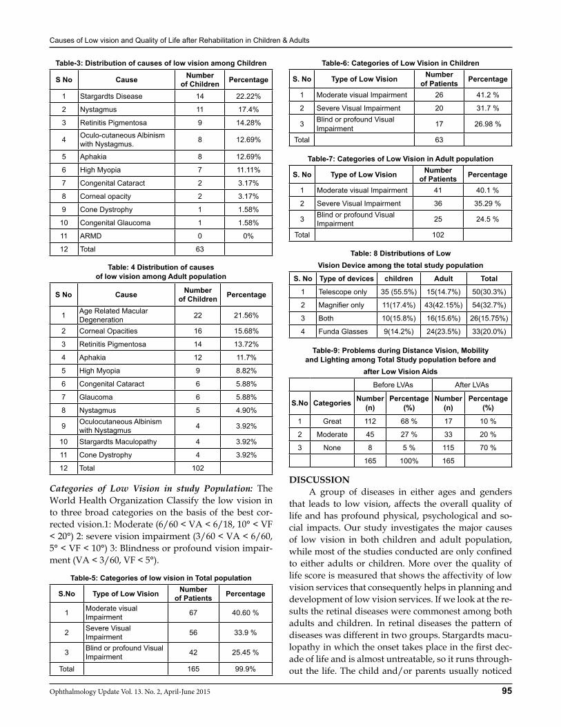
Causes of Low vision and Quality of Life after Rehabilitation in Children & Adults
95Ophthalmology Update Vol. 13. No. 2, April-June 2015
Table-3: Distribution of causes of low vision among Children
S No Cause Number of Children Percentage
1 Stargardts Disease 14 22.22%
2 Nystagmus 11 17.4%
3 Retinitis Pigmentosa 9 14.28%
4 Oculo-cutaneous Albinism with Nystagmus. 8 12.69%
5 Aphakia 8 12.69%
6 High Myopia 7 11.11%
7 Congenital Cataract 2 3.17%
8 Corneal opacity 2 3.17%
9 Cone Dystrophy 1 1.58%
10 Congenital Glaucoma 1 1.58%
11 ARMD 0 0%
12 Total 63
Table: 4 Distribution of causes of low vision among Adult population
S No Cause Number of Children Percentage
1 Age Related Macular Degeneration 22 21.56%
2 Corneal Opacities 16 15.68%
3 Retinitis Pigmentosa 14 13.72%
4 Aphakia 12 11.7%
5 High Myopia 9 8.82%
6 Congenital Cataract 6 5.88%
7 Glaucoma 6 5.88%
8 Nystagmus 5 4.90%
9 Oculocutaneous Albinism with Nystagmus 4 3.92%
10 Stargardts Maculopathy 4 3.92%
11 Cone Dystrophy 4 3.92%
12 Total 102
Categories of Low Vision in study Population: The World Health Organization Classify the low vision in to three broad categories on the basis of the best cor-rected vision.1: Moderate (6/60 < VA < 6/18, 10° < VF < 20°) 2: severe vision impairment (3/60 < VA < 6/60, 5° < VF < 10°) 3: Blindness or profound vision impair-ment (VA < 3/60, VF < 5°).
Table-5: Categories of low vision in Total population
S.No Type of Low Vision Number of Patients Percentage
1 Moderate visual Impairment 67 40.60 %
2 Severe Visual Impairment 56 33.9 %
3 Blind or profound Visual Impairment 42 25.45 %
Total 165 99.9%
Table-6: Categories of Low Vision in Children
S. No Type of Low Vision Number of Patients Percentage
1 Moderate visual Impairment 26 41.2 %
2 Severe Visual Impairment 20 31.7 %
3 Blind or profound Visual Impairment 17 26.98 %
Total 63
Table-7: Categories of Low Vision in Adult population
S. No Type of Low Vision Number of Patients Percentage
1 Moderate visual Impairment 41 40.1 %
2 Severe Visual Impairment 36 35.29 %
3 Blind or profound Visual Impairment 25 24.5 %
Total 102
Table: 8 Distributions of Low Vision Device among the total study population
S. No Type of devices children Adult Total
1 Telescope only 35 (55.5%) 15(14.7%) 50(30.3%)
2 Magnifier only 11(17.4%) 43(42.15%) 54(32.7%)
3 Both 10(15.8%) 16(15.6%) 26(15.75%)
4 Funda Glasses 9(14.2%) 24(23.5%) 33(20.0%)
Table-9: Problems during Distance Vision, Mobility and Lighting among Total Study population before and
after Low Vision Aids
Before LVAs After LVAs
S.No Categories Number (n)
Percentage (%)
Number (n)
Percentage (%)
1 Great 112 68 % 17 10 %
2 Moderate 45 27 % 33 20 %
3 None 8 5 % 115 70 %
165 100% 165
DISCUSSION A group of diseases in either ages and genders that leads to low vision, affects the overall quality of life and has profound physical, psychological and so-cial impacts. Our study investigates the major causes of low vision in both children and adult population, while most of the studies conducted are only confined to either adults or children. More over the quality of life score is measured that shows the affectivity of low vision services that consequently helps in planning and development of low vision services. If we look at the re-sults the retinal diseases were commonest among both adults and children. In retinal diseases the pattern of diseases was different in two groups. Stargardts macu-lopathy in which the onset takes place in the first dec-ade of life and is almost untreatable, so it runs through-out the life. The child and/or parents usually noticed
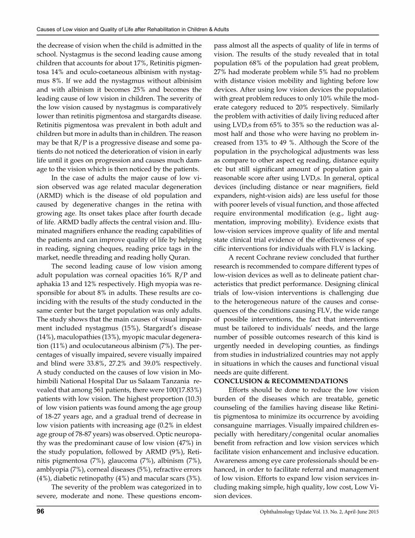
Causes of Low vision and Quality of Life after Rehabilitation in Children & Adults
96 Ophthalmology Update Vol. 13. No. 2, April-June 2015
the decrease of vision when the child is admitted in the school. Nystagmus is the second leading cause among children that accounts for about 17%, Retinitis pigmen-tosa 14% and oculo-coetaneous albinism with nystag-mus 8%. If we add the nystagmus without albinisim and with albinism it becomes 25% and becomes the leading cause of low vision in children. The severity of the low vision caused by nystagmus is comparatively lower than retinitis pigmentosa and stargardts disease. Retinitis pigmentosa was prevalent in both adult and children but more in adults than in children. The reason may be that R/P is a progressive disease and some pa-tients do not noticed the deterioration of vision in early life until it goes on progression and causes much dam-age to the vision which is then noticed by the patients. In the case of adults the major cause of low vi-sion observed was age related macular degeneration (ARMD) which is the disease of old population and caused by degenerative changes in the retina with growing age. Its onset takes place after fourth decade of life. ARMD badly affects the central vision and. Illu-minated magnifiers enhance the reading capabilities of the patients and can improve quality of life by helping in reading, signing cheques, reading price tags in the market, needle threading and reading holly Quran. The second leading cause of low vision among adult population was corneal opacities 16% R/P and aphakia 13 and 12% respectively. High myopia was re-sponsible for about 8% in adults. These results are co-inciding with the results of the study conducted in the same center but the target population was only adults. The study shows that the main causes of visual impair-ment included nystagmus (15%), Stargardt’s disease (14%), maculopathies (13%), myopic macular degenera-tion (11%) and oculocutaneous albinism (7%). The per-centages of visually impaired, severe visually impaired and blind were 33.8%, 27.2% and 39.0% respectively. A study conducted on the causes of low vision in Mo-himbili National Hospital Dar us Salaam Tanzania re-vealed that among 561 patients, there were 100(17.83%) patients with low vision. The highest proportion (10.3) of low vision patients was found among the age group of 18-27 years age, and a gradual trend of decrease in low vision patients with increasing age (0.2% in eldest age group of 78-87 years) was observed. Optic neuropa-thy was the predominant cause of low vision (47%) in the study population, followed by ARMD (9%), Reti-nitis pigmentosa (7%), glaucoma (7%), albinism (7%), amblyopia (7%), corneal diseases (5%), refractive errors (4%), diabetic retinopathy (4%) and macular scars (3%). The severity of the problem was categorized in to severe, moderate and none. These questions encom-
pass almost all the aspects of quality of life in terms of vision. The results of the study revealed that in total population 68% of the population had great problem, 27% had moderate problem while 5% had no problem with distance vision mobility and lighting before low devices. After using low vision devices the population with great problem reduces to only 10% while the mod-erate category reduced to 20% respectively. Similarly the problem with activities of daily living reduced after using LVD,s from 65% to 35% so the reduction was al-most half and those who were having no problem in-creased from 13% to 49 %. Although the Score of the population in the psychological adjustments was less as compare to other aspect eg reading, distance equity etc but still significant amount of population gain a reasonable score after using LVD,s. In general, optical devices (including distance or near magnifiers, field expanders, night-vision aids) are less useful for those with poorer levels of visual function, and those affected require environmental modification (e.g., light aug-mentation, improving mobility). Evidence exists that low-vision services improve quality of life and mental state clinical trial evidence of the effectiveness of spe-cific interventions for individuals with FLV is lacking. A recent Cochrane review concluded that further research is recommended to compare different types of low-vision devices as well as to delineate patient char-acteristics that predict performance. Designing clinical trials of low-vision interventions is challenging due to the heterogeneous nature of the causes and conse-quences of the conditions causing FLV, the wide range of possible interventions, the fact that interventions must be tailored to individuals’ needs, and the large number of possible outcomes research of this kind is urgently needed in developing counties, as findings from studies in industrialized countries may not apply in situations in which the causes and functional visual needs are quite different.CONCLUSION & RECOMMENDATIONS Efforts should be done to reduce the low vision burden of the diseases which are treatable, genetic counseling of the families having disease like Retini-tis pigmentosa to minimize its occurrence by avoiding consanguine marriages. Visually impaired children es-pecially with hereditary/congenital ocular anomalies benefit from refraction and low vision services which facilitate vision enhancement and inclusive education. Awareness among eye care professionals should be en-hanced, in order to facilitate referral and management of low vision. Efforts to expand low vision services in-cluding making simple, high quality, low cost, Low Vi-sion devices.

Causes of Low vision and Quality of Life after Rehabilitation in Children & Adults
97Ophthalmology Update Vol. 13. No. 2, April-June 2015
THE LOW VISION QUALITY-OF-LIFE QUESTIONNAIRE (LVQOL)
Distance Vision, Mobility and Lighting GRADING
How much of a problem do you have: None Moderate Great
With your vision in general 5 4 3 2 1 x n/a
With your eyes getting tired (e.g only being able to do a task 5 4 3 2 1 x n/a
for a short period of time)
With your vision at night inside the house 5 4 3 2 1 x n/a
Getting the right amount of light to be able to see 5 4 3 2 1 x n/a
With glare (e.g dazzled by car lights or the sun) 5 4 3 2 1 x n/a
Seeing street signs 5 4 3 2 1 x n/a
Seeing the television (appreciating the pictures) 5 4 3 2 1 x n/a
Seeing moving objects (e.g. cars on the road) 5 4 3 2 1 x n/a
With judging the depth or distance of items (e.g. reaching 5 4 3 2 1 x n/a
for a glass)
Seeing steps or curbs 5 4 3 2 1 x n/a
Getting around outdoors (e.g. on uneven pavements) 5 4 3 2 1 x n/a
because of your vision
Crossing a road with traffic because of your vision 5 4 3 2 1 x n/a
Adjustment
Because of your vision, are you: No Moderately Greatly
Unhappy at your situation in life 5 4 3 2 1 x n/a
Frustrated at not being able to do certain tasks 5 4 3 2 1 x n/a
Restricted in visiting friends or family 5 4 3 2 1 x n/a
Well Poorly Not explained
How well has your eye condition been explained to you 5 4 3 2 1 x
Reading and Fine WorkWith your reading aids / glasses, if used, how
much of a problem do you have: None Moderate Great
Reading large print (e.g. newspaper headlines) 5 4 3 2 1 x n/a
Reading newspaper text and books 5 4 3 2 1 x n/a
Reading labels (e.g. on medicine bottles) 5 4 3 2 1 x n/a
Reading your letters and mail 5 4 3 2 1 x n/a
Having problems using tools (e.g. threading a needle or 5 4 3 2 1 x n/a
cutting)
Activities of Daily Living
With your reading aids / glasses, if used, how
much of a problem do you have: None Moderate Great
Finding out the time for yourself 5 4 3 2 1 x n/a
Writing (e.g. cheques or cards) 5 4 3 2 1 x n/a
Reading your own hand writing 5 4 3 2 1 x n/a
With your every day activities (e.g. house-hold chores) 5 4 3 2 1 x n/a

Causes of Low vision and Quality of Life after Rehabilitation in Children & Adults
98 Ophthalmology Update Vol. 13. No. 2, April-June 2015
Annexure I: Data Collection Instrument
NAME: AGE: SEX: SERIAL NO: Occupation: Address:
Ophthalmology Findings
Right Eye
Left Eye
Cause of Low Vision /Blindness
Distance Visual Acuity(Without Glasses) Right Eye Left Eye Binocular Test Used
Distance Visual Acuity(With Glasses) Right Eye Left Eye Binocular Test Used
Near Visual Acuity Right Eye Left Eye Test Used
Visual Fields
Refraction Right Eye Left Eye Near Add Test Used
Low Vision Assessment
Eye Selected Right Eye Left Eye Type of Telescope Type of Magnifier
Visual Acuity With LVD Right Eye Left Eye Near Distance
Non-Optical Aid Right Eye Left Eye
Action/ Comments
REFRRANCES1. World Health Organization /International Agency for the Pre-
vention of Blindness. State of the World’s Sight Vision 2020: The Right to Sight 1999–2005. Geneva, Switzerland: WHO; 2005.
2. Scott IU, Smiddy WE, Schiffman J, Feuer WJ, Pappas CJ. Qual-ity of life of low-vision patients and the impact of low-vision services. Am J Ophthalmol. 1999;128:54---62.
3. World Health Organization. ICD-10 International Statistical Classification of Diseases and Related Health Problems, 10th Revision In: Johnson GJ, Minassian DC, Weale R, Eds. The Epi-demiology of Eye Disease. London: Chapman & Hall Medical; 1998:8–30.
4. Dineen B, Bourne RR, Jadoon Z, et al. Causes of blindness and visual impairment in Pakistan. The Pakistan national Blindness andVisual Impairment Survey.Br J Ophthalmol.2007; 01:1005–
1010.5. World Health Organization/Global Data on Visual impair-
ment 2010.6. Pararajasegaram R. VISION 2020 - The Right to Sight: from
strategies to action (editorial). Am J Ophthalmol. 1999; 128: 359-36.
7. Resnikoff S. Pacolini D. Etya’ale D, et al. Global data on visual impairment in the year 2002. Bull World Health Organ. 2004; 82: 844-851
6. Pollard.T, Simpson.J,Lamoureux.E and Keeffe.J. Barriers to ac-cessing low vision services. Ophthal Physio Opt 2003;23: 321-327.
9. World Health Organization/Global Data on Visual impair-ment 2010.
10. mshah et al causes of low vision in children.11. World Health Organization. Consultation on Development of
Causes of Low vision and Quality of Life after Rehabilitation in Children & Adults
99Ophthalmology Update Vol. 13. No. 2, April-June 2015
Standards for Characterization of Visual Loss and Visual Function-ing. 2003; WHO Geneva, Switzerland. PBL/03.91
12. ‐ Resnikoff S, Pascolini D, Etya’ale D, et al. Global data on visual impairment in the year 2002. Bull World Health Organ
13. World Bank. World Bank list of economies (July 2009) h p://siteresources.worldbank.org/DATASTATISTICS/ Resourc-es/ClASS.xls. date accessed March 15, 2010.
14. ‐Organisa on for Economic Co‐opera on and Develop‐ ment. Policy Briefs available at h p://www.oecd.org/ publica ons/Policybriefs. date accessed May 1, 2010.
15. Dineen, Bourne, Ali, et al, Prevalence and causes of blindness and visual impairment in Bangladeshi adults: results of the Na-tional Blindness and Low Vision Survey of Bangladesh. Br J
Ophthalmol 2003;87:820–828. 16. YemaneBernahe et al. prevalence and causes of blindness and
low vision in Ethiopia, Ethiop. J. Health Dev. 2007; 21(3).17. Keeffe J. Vision Assessment and Prescription of Low Vision De-
vices. J Comm Eye Health 2004;17: 3-4.18. Best AB. The management of low vision in Children. Br J Oph-
thalmol. 1995;13: 65-68. 19. Hornby SJ, Adolph S, Gothwal VK, et al. Evaluation of children
in six blind schools of Andhra Pradesh. Indian J Ophthalmol. 2000;48: 195-200.
20. Rahi JS, Sripathi S, Gilbert CE, et al. The importance of prenatal factors in childhood blindness in India. Dev Med Child Neurol. 1997;39 : 449-55.
18th Annual IslamabadCongress of Ophthalmology
Will be held at Bhurban (Murree) from
24-26 April 2015
Please Contact: Dr. Waheed Afzal, President OSP Federal Secretariat
E.mail: [email protected] Phone: 03335153266
100 Ophthalmology Update Vol. 13. No. 2, April-June 2015
ORIGINAL ARTICLE
Hasan Yaqoob
The Efficacy of Limbal Based Conjunctival Flap in Patients Undergoing Trabeculectomy
with Intra-operative Mitomycin C
Hasan Yaqoob FCPS, FRCS1, Mohammad Idris FCPS2
Zubairullah Khan FCPS3, Zubairullah Khan FCPS4, Mudasser Hussain Turi FCPS5
Naseer Ahmad DOMS5, Bilqees Hassan MBBS6
ABSTRACTObjective: To determine the efficacy of limbus based conjunctival flap in patients undergoing trabeculectomy with intraop-erative Mitomycin C.Design: interventional case series Setting: Department of Ophthalmology, Khyber Institute of Ophthalmic Medical Sciences (KIOMS), Post Graduate Medical Institute, Lady Reading Hospital, Peshawar.Duration: 18 months, from 1st January2012 to30th June 2013.Subjects: Eighty eyes of 80 patients diagnosed as having glaucoma.Main outcome measure:1. Intraocular pressure, effective rate of fornix based trabeculectomy with mitomycin C and limbal based trabeculectomy with mitomycin C in lowering intraocular pressure.2. Bleb formation Results: 80 eyes underwent limbal based trabeculectomy with MMC. 56.3% of patients were male and 43.8% were female. The mean age was 54.1 years. Preoperative visual acuity ranges from 6/6 to counting fingers (CF). The mean intraocular pressure at the end of follow-up was 12.12 mmHg with standard deviation + 0.68 in group 2. IOP >21 mmHg was not found in any patient. The effective rate of limbal based trabeculectomy with MMC was 85% in formation of bleb on 1st postopera-tive day .Conclusion: limbal based trabeculectomy with intraoperative MMC is an alternative and effective method in glaucoma treatment surgically. Key Words: Glaucoma, Trabeculectomy; intraocular pressure, Mitomycin C.
INTRODUCTION Glaucoma is characterized by progressive loss of retinal ganglion cells leading to characteristic visual fields defects and optic nerve head cupping and pal-lor1. It is an optic neuropathy secondary to various risk factors including increased IOP. Glaucoma may be (a) congenital or (b) acquired. Further sub-classification into open-angle and angle-closure type is based on the mechanism by which aque-ous outflow is impaired. The glaucoma may also be (a) primary or (b) secondary depending on the presence or absence of associated risk factors. In primary glaucoma there is no associated ocular disorder while in second-ary glaucoma a recognizable ocular or non-ocular dis-
order alters aqueous outflow. Secondary glaucoma may be acquired or developmental and of the open-angle or angle-closure type.Secondary open-angle glaucoma may be:1. Pre-trabecular like neovascular glaucoma.2. Trabecular like pigmentary glaucomas, red cell
glaucomas, ghost cell glaucomas, phacolytic glau-comas, pseudoexfoliative glaucomas and post-traumatic angle recessive glaucoma etc.
3. Post-trabecular in which aqueous outflow is im-paired by elevated episcleral venous pressure due to carotid-cavernous fistula, Sturge-Weber syn-drome and obstruction of superior vena cava.
Secondary angle-closure glaucoma may be due to posterior forces which push the peripheral iris against the trabeculum (iris bombe due to seclusion-pupillae) or anterior forces which pull the iris over the trabecu-lum by contraction of inflammatory or fibrovascular membrane (e.g. late neovascular glaucoma). Patients present with a variety of signs and symp-toms like pain, watering, dimness of vision, headache, nausea and vomiting depending on the nature of glau-coma. Therefore, slit-lamp biomicroscopy, fundoscopy, tonometry, gonioscopy and perimetry is mandatory for management of these patients to see for ciliary injec-
1Consultant, Ophthalmology Unit, North West General Hospital, Peshawar, KPK. 2Medical Officer, Ophthalmology UNIT, PGMI, LRH Peshawar. 3Consultant, Ophthalmology, Mission Hospital, Peshawar, KPK. 4Associate Ophthalmologist, LRBT Free Eye Hospital, Mandra, Rawalpindi. 5Trainee Medical Officer, Hayatabad Medical Complex, Peshawar. 6Medical Officer Pakistan Institute of Ophthalmology,PICO, Peshawar
Correspondence: Dr. Hasan Yaqoob FCPS, FRCS Consultant, Ophthalmology UNIT, North West General Hospital, Phase V, Hayatabad, Peshawar, KPK. Pakistan. Cell: 00992-0345-2565959, Email [email protected]
Received: December 2014 Accepted: January 2015
The Efficacy of Limbal Based ConjunctivalFlap in Patients ndergoing Trabeculectomy with Intra-operative Mitomycin C
101Ophthalmology Update Vol. 13. No. 2, April-June 2015
tion and corneal oedema, optic disc cupping, intraoc-ular pressure, angle details and visual field defects. Glaucoma is a highly prevalent and vision threatening condition affecting approximately 66 million people worldwide.2 In a recent study conducted in Pakistan, it was showed that glaucoma accounted for 8.1% of all eye admissions. Open-angle glaucoma was responsible for 37.6% or 731 glaucoma admissions followed by sec-ondary glaucoma (35.0%) and angle-closure glaucoma (18.2%).3
In our set-up, people present with advance disease due to poverty, illiteracy and lack of district-based eye care. Different types of treatment options are available like anti-glaucoma drugs, laser treatment and surgi-cal interventions. Treatment of choice in our setting is surgical intervention due to poverty, poor drug com-pliance, late presentation and high failure rate of laser trabeculoplasty.4 Trabeculectomy alone introduced by Cainrs in 1968 and modified by Watson in 1970,5, 6 or with antimetabolite (Mitomycin-C, 5-Fluoro-urocil) has been the surgical method of choice.7 ,8 9
Objective; To determine the efficacy of Limbus based conjunctival flap in patients undergoing trabeculecto-my with intraoperative Mitomycin C.Efficacy: it will be measured on the basis of conjuncti-val bleb formation and normal intraocular pressure (11-21mmHg) and thus affectivity of the procedure will be assessed.MATERIAL AND METHODSSetting: Department of Ophthalmology, Khyber Insti-tute of Ophthalmic Medical Sciences (KIOMS), Post Graduate Medical Institute, Lady Reading Hospital, Peshawar.Duration of Study: 18 months, from 1st January2012 to 30th June 2013.Sample Size: Using WHO sample size calculator, where
Confidence level=95,Absolute precision=0.03, Population proportion (P) =10%.The sample size=80
Sampling Technique: non probability: consecutive SAMPLING SELECTIONa. Inclusion criteria; i. patients of 30 to 60 years, both male and female.ii. Patients of primary open angle glaucoma,angle
closure glaucoma pseudoexfoliative glaucoma and induced glaucoma with raised intra ocular pressure not controlled by maximum treatment or poor compliance.
b. Exclusion criteria; Patients with previously failed trabeculectomy.
Patients with history of previous intra ocular sur-
gery or trauma.Patients with congenital or normal tension glaucoma. Patients with secondary glau-coma like uveitic, neovascular or pseudophakic
Study design: prospective, interventional case series.Data collection procedure: The study was conducted at Out Patient Department, Eye Unit of Lady Reading Hospital, Peshawar. Before we start the study, permis-sion from the hospital ethical committee was obtained. An informed written consent was obtained from the patient. The patients were evaluated for inclusion and exclusion criteria. Patients for trabeculectomy will be admitted to eye unit of Lady Reading Hospital, Pesha-war, through an eye OPD waiting list. A detailed histo-ry regarding dimness of vision (DV) (whether sudden or gradual, painless or painful), previous ocular trauma and intraocular surgery will be taken. Pre-op ocular ex-amination including best corrected visual acuity, rela-tive afferent papillary defect( RAPD) and slit lamp ex-amination of optic disc with 90 D lens noting optic disc cupping and cup-disc ratio( c/d ratio), gonioscopic ex-amination of angle structure by Goldmann single mir-ror goniolens, intraocular pressure measurement(IOP) by Goldmann applanation tonometer and visual field testing using Humphery perimeter. Laboratory investi-gations like Hb %, HBA1C, Ag, anti-HCV, blood sugar etc will be done in Pathology Department, Lady Read-ing Hospital, Peshawar. Radiological investigations like chest X-Ray will be done in Radiology Department, Lady Reading Hospital, Peshawar. The surgery will be done both under local and general anesthesia. On first day after surgery and on follow up the patients will be assessed for visual acuity, conjunctival bleb, and intraocular pressure. Patients will be examined on first post-op day and will be discharged after being fol-lowed up on 10th postoperative day and 1 month. Nom-inal data of the outcome of surgery for all the patients will be recorded on a data collection proforma on each follow up visit.Surgical Procedure: Limbal based trabeculectomy was performed.Data analysis procedure: After completion of data col-lection, the data will be analyzed using SPSS version 13.0. All categorical variables including gender and operative outcome will be given in frequencies and percentages; mean and standard deviation will be cal-culated for numerical variables for example age and intraocular pressure on day 1, 10, and 30. Operative outcome in the form of intraocular pressure and bleb formation was documented and presented in the form of tables.RESULTS Eighty patients were diagnosed as “Glaucoma”
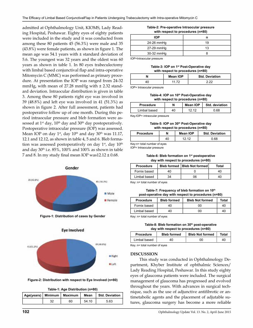
The Efficacy of Limbal Based ConjunctivalFlap in Patients ndergoing Trabeculectomy with Intra-operative Mitomycin C
102 Ophthalmology Update Vol. 13. No. 2, April-June 2015
admitted at Ophthalmology Unit, KIOMS, Lady Read-ing Hospital, Peshawar. Eighty eyes of eighty patients were included in the study and it was conducted from among these 80 patients 45 (56.3%) were male and 35 (43.8%) were female patients, as shown in figure 1. The mean age was 54.1 years with ± standard deviation of 5.6. The youngest was 32 years and the oldest was 60 years as shown in table 1. In 80 eyes trabeculectomy with limbal based conjunctival flap and intra-operative Mitomycin C (MMC) was performed as primary proce-dure. At presentation the IOP was ranged from 24-32 mmHg, with mean of 27.28 mmHg with ± 2.32 stand-ard deviation. Intraocular distribution is given in table 3. Among these 80 patients right eye was involved in 39 (48.8%) and left eye was involved in 41 (51.3%) as shown in figure 2. After full assessment, patients had postoperative follow up of one month. During this pe-riod intraocular pressure and bleb formation were as-sessed at 1st day, 10th day and 30th day postoperatively. Postoperative intraocular pressure (IOP) was assessed. Mean IOP on day 1st, day 10th and day 30th was 11.17, 12.1 and 12.12, as shown in table 4, 5 and 6. Bleb forma-tion was assessed postoperatively on day 1st, day 10th and day 30th i.e. 85%, 100% and 100% as shown in table 7 and 8. In my study final mean IOP was12.12 ± 0.68.
Figure-1: Distribution of cases by Gender
Figure-2: Distribution with respect to Eye Involved (n=80)
Table-1: Age Distribution (n=80)
Age(years) Minimum Maximum Mean Std. Deviation
32 60 54.10 5.63
Table-2: Pre-operative Intraocular pressure with respect to procedures (n=80)IOP n
24-26 mmHg 1927-29 mmHg 1330-32 mmHg 8
IOP=Intraocular pressure
Table-3: IOP on 1st Post-Operative day with respect to procedures (n=80)
N Mean IOP Std. Deviation40 11.72 2.22
IOP= Intraocular pressure
Table-4: IOP on 10th Post-Operative day with respect to procedures (n=80)
Procedure N Mean IOP Std. deviationLimbal based 40 12.12 0.68
Key.IOP= intraocular pressure
Table-5: IOP on 30th Post-Operative day with respect to procedures (n=80)
Procedure N Mean IOP Std. Deviation40 12.12 0.68
Key:n= total number of eyesIOP= Intraocular pressure
Table-6: Bleb formation on 1st postoperative day with respect to procedures (n=80)
Procedure Bleb formed Bleb Not formed TotalFornix based 40 0 40Limbal based 34 06 40
Key: n= total number of eyes
Table-7: Frequency of bleb formation on 10th post-operative day with respect to procedures (n=80)
Procedure Bleb formed Bleb Not formed TotalFornix based 40 00 40Limbal based 40 00 40
Key: n= total number of eyes
Table-8: Bleb formation on 30th post-operative day with respect to procedures (n=80)
Procedure Bleb formed Bleb Not formed TotalLimbal based 40 00 40
Key. n= total number of eyes
DISCUSSION This study was conducted in Ophthalmology De-partment, Khyber Institute of ophthalmic Sciences/Lady Reading Hospital, Peshawar. In this study eighty eyes of glaucoma patients were included. The surgical management of glaucoma has progressed and evolved throughout the years. With advances in surgical tech-nique, such as the use of adjunctive antifibrotic or an-timetabolic agents and the placement of adjutable su-tures, glaucoma surgery has become a more reliable
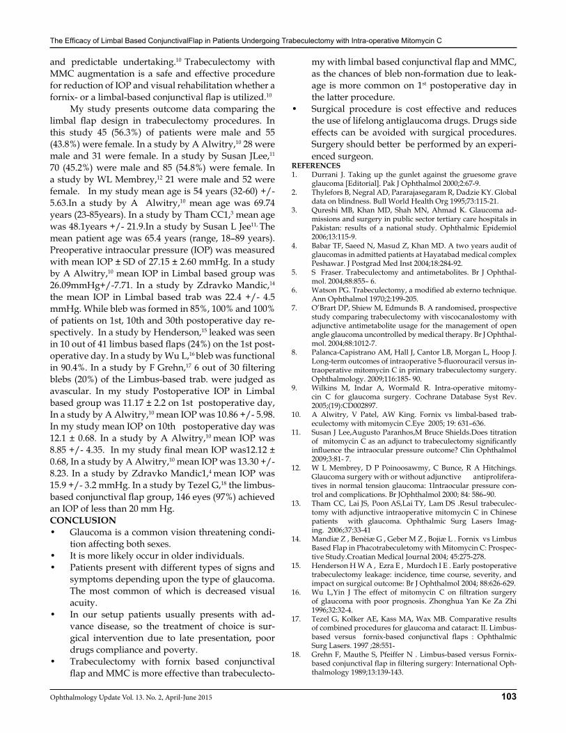
The Efficacy of Limbal Based ConjunctivalFlap in Patients ndergoing Trabeculectomy with Intra-operative Mitomycin C
103Ophthalmology Update Vol. 13. No. 2, April-June 2015
and predictable undertaking.10 Trabeculectomy with MMC augmentation is a safe and effective procedure for reduction of IOP and visual rehabilitation whether a fornix- or a limbal-based conjunctival flap is utilized.10
My study presents outcome data comparing the limbal flap design in trabeculectomy procedures. In this study 45 (56.3%) of patients were male and 55 (43.8%) were female. In a study by A Alwitry,10 28 were male and 31 were female. In a study by Susan JLee,11 70 (45.2%) were male and 85 (54.8%) were female. In a study by WL Membrey,12 21 were male and 52 were female. In my study mean age is 54 years (32-60) +/- 5.63.In a study by A Alwitry,10 mean age was 69.74 years (23-85years). In a study by Tham CC1,3 mean age was 48.1years +/- 21.9.In a study by Susan L Jee11, The mean patient age was 65.4 years (range, 18–89 years). Preoperative intraocular pressure (IOP) was measured with mean IOP ± SD of 27.15 ± 2.60 mmHg. In a study by A Alwitry,10 mean IOP in Limbal based group was 26.09mmHg+/-7.71. In a study by Zdravko Mandic,14 the mean IOP in Limbal based trab was 22.4 +/- 4.5 mmHg. While bleb was formed in 85%, 100% and 100% of patients on 1st, 10th and 30th postoperative day re-spectively. In a study by Henderson,15 leaked was seen in 10 out of 41 limbus based flaps (24%) on the 1st post-operative day. In a study by Wu L,16 bleb was functional in 90.4%. In a study by F Grehn,17 6 out of 30 filtering blebs (20%) of the Limbus-based trab. were judged as avascular. In my study Postoperative IOP in Limbal based group was 11.17 ± 2.2 on 1st postoperative day, In a study by A Alwitry,10 mean IOP was 10.86 +/- 5.98.In my study mean IOP on 10th postoperative day was 12.1 ± 0.68. In a study by A Alwitry,10 mean IOP was 8.85 +/- 4.35. In my study final mean IOP was12.12 ± 0.68, In a study by A Alwitry,10 mean IOP was 13.30 +/- 8.23. In a study by Zdravko Mandic1,4 mean IOP was 15.9 +/- 3.2 mmHg. In a study by Tezel G,18 the limbus-based conjunctival flap group, 146 eyes (97%) achieved an IOP of less than 20 mm Hg.CONCLUSION• Glaucoma is a common vision threatening condi-
tion affecting both sexes. • It is more likely occur in older individuals.• Patients present with different types of signs and
symptoms depending upon the type of glaucoma. The most common of which is decreased visual acuity.
• In our setup patients usually presents with ad-vance disease, so the treatment of choice is sur-gical intervention due to late presentation, poor drugs compliance and poverty.
• Trabeculectomy with fornix based conjunctival flap and MMC is more effective than trabeculecto-
my with limbal based conjunctival flap and MMC, as the chances of bleb non-formation due to leak-age is more common on 1st postoperative day in the latter procedure.
• Surgical procedure is cost effective and reduces the use of lifelong antiglaucoma drugs. Drugs side effects can be avoided with surgical procedures. Surgery should better be performed by an experi-enced surgeon.
REFERENCES1. Durrani J. Taking up the gunlet against the gruesome grave
glaucoma [Editorial]. Pak J Ophthalmol 2000;2:67-9.2. Thylefors B, Negral AD, Pararajasegaram R, Dadzie KY. Global
data on blindness. Bull World Health Org 1995;73:115-21.3. Qureshi MB, Khan MD, Shah MN, Ahmad K. Glaucoma ad-
missions and surgery in public sector tertiary care hospitals in Pakistan: results of a national study. Ophthalmic Epidemiol 2006;13:115-9.
4. Babar TF, Saeed N, Masud Z, Khan MD. A two years audit of glaucomas in admitted patients at Hayatabad medical complex Peshawar. J Postgrad Med Inst 2004;18:284-92.
5. S Fraser. Trabeculectomy and antimetabolites. Br J Ophthal-mol. 2004;88:855– 6.
6. Watson PG. Trabeculectomy, a modified ab externo technique. Ann Ophthalmol 1970;2:199-205.
7. O’Brart DP, Shiew M, Edmunds B. A randomised, prospective study comparing trabeculectomy with viscocanalostomy with adjunctive antimetabolite usage for the management of open angle glaucoma uncontrolled by medical therapy. Br J Ophthal-mol. 2004;88:1012-7.
8. Palanca-Capistrano AM, Hall J, Cantor LB, Morgan L, Hoop J. Long-term outcomes of intraoperative 5-fluorouracil versus in-traoperative mitomycin C in primary trabeculectomy surgery. Ophthalmology. 2009;116:185- 90.
9. Wilkins M, Indar A, Wormald R. Intra-operative mitomy-cin C for glaucoma surgery. Cochrane Database Syst Rev. 2005;(19):CD002897.
10. A Alwitry, V Patel, AW King. Fornix vs limbal-based trab-eculectomy with mitomycin C.Eye 2005; 19: 631–636.
11. Susan J Lee,Augusto Paranhos,M Bruce Shields.Does titration of mitomycin C as an adjunct to trabeculectomy significantly influence the intraocular pressure outcome? Clin Ophthalmol 2009;3:81- 7.
12. W L Membrey, D P Poinoosawmy, C Bunce, R A Hitchings. Glaucoma surgery with or without adjunctive antiprolifera-tives in normal tension glaucoma: 1Intraocular pressure con-trol and complications. Br JOphthalmol 2000; 84: 586–90.
13. Tham CC, Lai JS, Poon AS,Lai TY, Lam DS .Resul trabeculec-tomy with adjunctive intraoperative mitomycin C in Chinese patients with glaucoma. Ophthalmic Surg Lasers Imag-ing. 2006;37:33-41
14. Mandiæ Z , Benèiæ G , Geber M Z , Bojiæ L . Fornix vs Limbus Based Flap in Phacotrabeculetomy with Mitomycin C: Prospec-tive Study.Croatian Medical Journal 2004; 45:275-278.
15. Henderson H W A , Ezra E , Murdoch I E . Early postoperative trabeculectomy leakage: incidence, time course, severity, and impact on surgical outcome: Br J Ophthalmol 2004; 88:626-629.
16. Wu L,Yin J The effect of mitomycin C on filtration surgery of glaucoma with poor prognosis. Zhonghua Yan Ke Za Zhi 1996;32:32-4.
17. Tezel G, Kolker AE, Kass MA, Wax MB. Comparative results of combined procedures for glaucoma and cataract: II. Limbus-based versus fornix-based conjunctival flaps : Ophthalmic Surg Lasers. 1997 ;28:551-
18. Grehn F, Mauthe S, Pfeiffer N . Limbus-based versus Fornix-based conjunctival flap in filtering surgery: International Oph-thalmology 1989;13:139-143.
104 Ophthalmology Update Vol. 13. No. 2, April-June 2015
ORIGINAL ARTICLE
INTRODUCTION Glaucoma encompasses a heterogeneous group of conditions, resulting in optic nerve damage and charac-teristic visual field changes. It is usually but not always associated with raised intraocular pressure (IOP). Nor-mal-tension glaucoma (NTG) is a chronic optic neu-ropathy with features similar to primary open-angle glaucoma (POAG), with the exception of a consistently normal IOP, i.e. less than 22 mm Hg.1 The disease usu-ally presents in old age and has a female predilection. The exact etiology of NTG is uncertain and various risk factors have been postulated. These include:• Generalized peripheral vascular endothelial dys-
function.2
• Ocular circulation insufficiency (lower ocular pulse amplitude)2
• Increased resistance index in the central retinal ar-tery (role in progression of visual field defect)3
• Impaired vascular auto regulation (prolonged arteriovenous venous passage time in relation to ocular perfusion)4
• Migraine• Peripheral vasospasm, Raynaud syndrome• Autoimmune disorders• Systemic vascular disease (i.e. atherosclerotic dis-
ease, cerebrovascular insufficiency)5,6
• Systemic nocturnal hypotension• Sleep apnea (decreases oxygen saturation)7
Cerebral ischemia and (or) brain atrophy is a rec-ognized risk factorand researchers are taking keen in-terest in this hypothesis. Optic nerve being a part of the central nervous system is affected in the same way as the brain. This theory is supported by some studies which have found a significant portion of patients diag-nosed as NTG to have cerebral ischemia and (or) brain atrophy. In our department, we investigated patients diagnosed as normal tension glaucoma for occurrence of concurrent cerebral ischemia and (or) brain atrophy using magnetic resonance imaging. MATERIAL AND METHODS Patients diagnosed with normal tension glaucoma were included in the study. All patients were admitted and underwent 2 hourly IOP phasing for 24 hours to exclude unrecognized IOP spikes. Detailed ophthalmo-logical examination including visual acuities, best cor-rected visual acuity, pupils examination, IOP measure-ment, gonioscopy, optic disc and fundus examination
Akhunzada Muhammad Aftab FCPS1, Awais Rauf MBBS2, Asif MBBS3
Prof. Mustafa Iqbal FRCS, FRCOphth4, Sobia Sabir Ali FCPS5, Atif Rana FCPS6
ABSTRACTPurpose: To find incidence of cerebral ischemia and (or) brain atrophy in patients diagnosed as normal tension glaucoma using magnetic resonance imaging. Materials and Methods: Patients diagnosed with Normal Tension Glaucoma were admitted and underwent 2 hourly IOP phasing for 24 hours to exclude unrecognized IOP spikes. Detailed ophthalmological examination including visual acuities, best corrected visual acuity, pupils examination, IOP measurement, gonioscopy, optic disc and fundus examination was carried out on all patients. Humphrey Visual Field analysis and Central Corneal Thickness (CCT) was done and correction factors were applied to all IOP readings. All patients underwent an MRI Brain and Orbits without contrast. T1, T2 and FLAIR images were obtained and reported by a consultant radiologist. Hematological and other radiological investigations were done to exclude other causes of cerebral ischemia.Results: Total number of patients in this study was 19. Most patients included in our study were females(63%). Mean age was 60 years (range 42- 75 years). None of the study patients recorded a corrected IOP reading of more than 21 mmHg on phasing. MRI imaging revealed 15 (79%) patients having cerebral ischemic changes and (or) brain atrophy. In these patients, 10 had cerebral ischemic changes including small lacunar infarcts, while 5 patients were reported as having gross brain atrophy with small vessel ischemic changes in brain.Conclusion: The study suggests a greater incidence of cerebral ischemia and (or) brain atrophy in patients with normal-tension glaucoma.
1Registrar Eye A Unit Department of Ophthalmology, Khyber Teaching Hospital, Peshawar. 2,3Trainee Medical Officers. A Unit Department of Ophthalmology, Khyber Teaching Hospital, Peshawar. 4Prof. & Head of Ophthalmology, 5Head of Endocrinology Department. 6Consultant Radiologist, Shifa International Hospital
Correspondence: Dr. Akhunzada Muhammad Aftab c/o Prof. Dr. Muhammad Ibrar, Department of Botany, University of Peshawar, Peshawar. Cell: 03339106060, E-Mail: [email protected]
Received: February 2015 Accepted: February 2015
Financial Disclosure: There has been no financial interest involved in this study
Normal Tension Glaucoma & Cerebral Ischemia / Brain Atrophy
Akhunzada M. Aftab
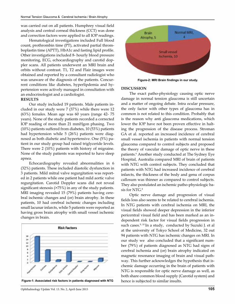
Normal Tension Glaucoma & Cerebral Ischemia / Brain Atrophy
105Ophthalmology Update Vol. 13. No. 2, April-June 2015
was carried out on all patients. Humphrey visual field analysis and central corneal thickness (CCT) was done and correction factors were applied to all IOP readings. Hematological investigations included Full blood count, prothrombin time (PT), activated partial throm-boplastin time (APTT), HbA1c and fasting lipid profile. Other investigations included 8- hourly blood pressure monitoring, ECG, echocardiography and carotid dop-pler scans. All patients underwent an MRI brain and orbits without contrast. T1, T2 and Flair images were obtained and reported by a consultant radiologist who was unaware of the diagnosis of the patients. Concur-rent conditions like diabetes, hyperlipdemia and hy-pertension were actively managed in consultation with an endocrinologist and a cardiologist.RESULTS Our study included 19 patients. Male patients in-cluded in our study were 7 (37%) while there were 12 (63%) females. Mean age was 60 years (range 42- 75 years). None of the study patients recorded a corrected IOP reading of more than 21 mmHgon phasing. Two (10%) patients suffered from diabetes, 10 (53%) patients had hypertension while 5 (26%) patients were diag-nosed as both diabetic and hypertensive. One (5%) pa-tient in our study group had raised triglyceride levels.There were 2 (10%) patients with history of migraine. None of the study patients was reported to have sleep apnea. Echocardiography revealed abnormalities in 6 (32%) patients. These included diastolic dysfunction in 3 patients. Mild mitral valve regurgitation was report-ed in 2 patients while one patient had mild aortic valve regurgitation. Carotid Doppler scans did not reveal significant stenosis (>75%) in any of the study patients. MRI imaging revealed 15 (79%) patients having cere-bral ischemic changes and (or) brain atrophy. In these patients, 10 had cerebral ischemic changes including small lacunar infarcts, while 5 patients were reported as having gross brain atrophy with small vessel ischemic changes in brain.
Figure-1: Associated risk factors in patients diagnosed with NTG
Figure-2: MRI Brain findings in our study.
DISCUSSION The exact patho-physiology causing optic nerve damage in normal tension glaucoma is still uncertain and a matter of ongoing debate. Intra ocular pressure, the only factor with other types of glaucoma has in common is not related to this condition. Probably that is the reason why anti glaucoma medications, which lower the IOP have not been proven effective in halt-ing the progression of the disease process. Stroman GA et al. reported an increased incidence of cerebral small vessel ischemia in patients with normal tension glaucoma compared to control subjects and proposed the theory of vascular damage of optic nerve in these patients.6 Another study conducted at The Sydney Eye Hospital, Australia compared MRI of brain of patients with NTG with control subjects. They concluded that patients with NTG had increased incidence of cerebral infarcts, the thickness of the body and genu of corpus callosum was thinner as compared to control subjects. They also postulated an ischemic patho-physiologic ba-sis for NTG.8
Optic nerve damage and progression of visual fields loss also seems to be related to cerebral ischemia. In NTG patients with cerebral ischemia on MRI, the visual fields showed deeper depression in the inferior pericentral visual field and has been marked as an in-dependent risk factor for visual fields progression in such cases.9, 10 In a study, conducted by Suzuki J. et al at the university of Tokyo School of Medicine, 32 out 94 patients with NTG has ischemic changes on MRI. In our study we also concluded that a significant num-ber (79%) of patients diagnosed as NTG had signs of cerebral ischemia and (or) brain atrophy indicated on magnetic resonance imaging of brain and visual path-way. This further acknowledges the hypothesis that is-chemic damage occurring in the brain of patients with NTG is responsible for optic nerve damage as well, as both share common blood supply (Carotid system) and hence is subjected to similar insults.
Normal Tension Glaucoma & Cerebral Ischemia / Brain Atrophy
106 Ophthalmology Update Vol. 13. No. 2, April-June 2015
CONCLUSION The study suggests a greater incidence of cerebral ischemia and (or) brain atrophy in patients with nor-mal-tension glaucoma. Optic nerve damage in such pa-tients may be an extension of the same disease process.REFERENCES 1. Kim M, Kim TW, Park KH, Kim JM. Risk factors for primary
open-angle glaucoma in South Korea: the Namil study. Jpn J Ophthalmol. 2012 Jul;56(4):324-9
2. Su WW, Cheng ST, Hsu TS, Ho WJ. Abnormal flow-mediated vasodilation in normal-tension glaucoma using a noninvasive determination for peripheral endothelial dysfunction. Invest Ophthalmol Vis Sci. 2006 Aug;47(8):3390-4
3. Delaney Y, Walshe TE, O’Brien C. Vasospasm in glaucoma: clin-ical and laboratory aspects. Optom Vis Sci. 2006 Jul;83(7):406-14
4. Plange N, Kaup M, Remky A, Arend KO. Prolonged retinal arteriovenous passage time is correlated to ocular perfusion pressure in normal tension glaucoma. Graefes Arch Clin Exp Ophthalmol. 2008 Aug;246(8):1147-52
5. Harris A, Siesky B, Zarfati D, et al. Relationship of cerebral
blood flow and central visual function in primary open-angle glaucoma. J Glaucoma. 2007 Jan;16(1):159-63
6. Stroman GA, Stewart WC, Golnik KC, Curé JK, Olinger RE. Magnetic resonance imaging in patients with low-tension glau-coma. Arch Ophthalmol. 1995 Feb;113(2):168-72
7. Bilgin. Normal-tension glaucoma and obstructive sleep ap-nea syndrome: a prospective study. BMC Ophthalmol. 2014 March;(10):14-27
8. Ong K, Farinelli A, Billson F, Houang M, Stern M. Comparative study of brain magnetic resonance imaging findings in patients with low-tension glaucoma and control subjects. Ophthalmol-ogy. 1995 Nov;102(11):1632-8.
9. Suzuki J, Tomidokoro A, Araie M, Tomita G, Yamagami J, Okubo T, Masumoto T. Visual field damage in normal-tension glaucoma patients with or without ischemic changes in cere-bral magnetic resonance imaging. Jpn J Ophthalmol. 2004 July-Aug; 48(4): 340-44
10. Leung DY, Tham CC, Li FC, Kwong YY, Chi SC, Lam DS. Si-lent cerebral infarct and visual field progression in newly diag-nosed normal-tension glaucoma: a cohort study. Ophthalmol-ogy. 2009 Jul;116(7):1250-6.
Tuberous sclerosis complex (TSC) is an autosomal dominant neuro-cutaneous disease (phacomatosis) with variable clinical manifestations (see the main article) Curtesy: Dr Hussain Ahmad Khaqan Department
of Ophthalmology, Lahore General Hospital/PGMI, Lahore.
107Ophthalmology Update Vol. 13. No. 2, April-June 2015
Mohammad Alam FCPS1, Misbah Durrani FCPS2, Prof. Lal Mohammad FCPS3
Arshad Farooq FCPS4
ABSTRACT:Objective: To find out complications and results of external dacryocystorhinostomy without tube intubation in chronic dacryocystitis (CDC).Materials and methods: This prospective study was conducted in eye care centre Karak and K.D.A Teaching Hospital Kohat from March, 2008 to March, 2014. 107 patients suffering from chronic dacryocystitis were selected with age range from 31 – 63 years with mean age 44.7 years. Out of 107 patients, 63 (58.87%) were male and 44(41.12%) were female. Indications for dacryocystorhinosyomy was epiphora and chronic dacryocystitis. Diagnosis was done on regurgitation test and syringing of nasolacrimal duct system. All patients were operated under local anesthesia. External approach was done and only anterior flap was made. Dacryocystorhinostomy was done without silicone tube intubation. Postoperative syring-ing was done on the table, 10 days, 3 months and 6 months. Successful outcome was defined as relief from epiphora after dacryocystorhinostomy and patent nasolacrimal duct on syringing.Results: After six months 95 (88.78%) was the success rate. Epiphora was present in 7(6.54%) patients and epiphora with discharge was present in 5(4.67%). 11(10.28%) patients had nasal mucosal bleeding, 9(8.41%) had nasal bone bleeding and tear in nasal septum was observed in 2(1.86%)patients peroperatively. Postoperatively 9(8.41%) patients had wound infection with cellulitis, 4 (3.73%) patients had bleeding from nose and 13(12.14%) patients had periorbital ecchymosis. All these complications resolved within 10 days.Conclusion: External dacryocystorhinostomy is a safe procedure under local anesthesia. This technique has high success rate. Complications are minimal and can be easily managed.Key word: chronic Dacryocystitis, Epiphora, External Dacryocystorhinostomy. Abbreviations: Chronic Dacryocystitis (C.D.C), Dacryocystorhinosyomy (D.C.R) Nasalacrimal Duct (N.L.D)
INTRODUCTION NLD blockade results in watering of disturbed tears called Epiphora which is the most bothersome problem of lacrimal system obstruction. Management of Epiphora has evolutionary history. Adeo Toti was the first who introduced dacryocystorhinostomy for the treatment of epiphora.1 He created an external ap-proach to lacrimal sac by creating a window in nasal lateral wall. The results of Adeo Toti were not success-ful in many patients. This procedure was modified by Bourguet and Dupuy-Dutemps. They introduced anas-tomosis of mucosa with suturing of the mucosal flaps.2 Ohm added suturing of anterior and posterior flaps of nasal mucosa with lacrimal sac.3 External DCR is the mostly practiced operation for NLD obstruction. Usu-ally other methods of surgery for CDC are compared
with this gold standard procedure.4 Various studies have reported external DCR success rate more than 80% which depends upon the surgeon experience. There are other methods of surgical procedure to treat NLD obstruction. These include endoscopic DCR, endoscopic laser nasal DCR, endoscopic radio frequen-cy assisted DCR.5,6,7 Endoscopic DCR is the favored sur-gical procedure of ENT surgeon and thus ophthalmolo-gists and ENT surgeons share their clinical skill and experience in care and treatment of NLD obstruction patients.8 There are numerous modifications in various surgical steps that has been introduced in DCR over years to get better results. Various national and inter-national studies have reported low complications rate in external DCR. We present our experience in external DCR with only anterior flaps suturing.MATERIALS AND METHODS This prospective study was conducted in Eye Care Centre Karak and KDA Teaching Hospital Kohat from March, 2008 to March, 2014 with the objective to know the success rate and complications of external DCR without silicone tube intubation. Total 107 pa-tients with age range of 31 to 63 years with mean age of 44.7 years were selected table I. Out of 107 patients 63 (58.87%) were male and 44 (41.12%) were female (Ta-
ORIGINAL ARTICLE
Mohammad Alam
1.Assistant Professor Ophthalmology KMU Institute Of Medical Sciences, K.D.A Kohat. 2Assistant Professor, Radiology, Bacha Khan Medical College, Mardan. 3Professor of Ophthalmology KMU Institute of Medical Sciences, K.D.A Kohat. 4Assistant Professor ENT KMU-Institute of Medical Sciences, KDA Kohat
Correspondence: Dr. Mohammad Alam1 Assistant Professor Ophthalmology KMU Institute of Medical Sciences K.D.A Kohat. [email protected], Cell:
Received: December 2014 Accepted: February 2015
Complications & Results of External Dacryocystorhinostomy in Chronic
Dacryocystitis without Intubation(Review of 107 Cases.)
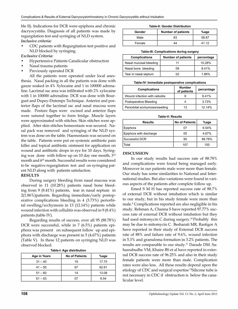
Complications & Results of External Dacryocystorhinostomy in Chronic Dacryocystitis without Intubation
108 Ophthalmology Update Vol. 13. No. 2, April-June 2015
ble II). Indications for DCR were epiphora and chronic dacryocystitis. Diagnosis of all patients was made by regurgitation test and syringing of NLD system.Inclusive criteria:• CDC patients with Regurgitation test positive and
NLD blocked by syringing.Exclusive Criteria:• Hypertensive Patients Canalicular obstruction• Nasal trauma patients• Previously operated DCR All the patients were operated under local anes-thesia. Nasal packing in all the patients was done with gauze soaked in 4% Xylocaine and 1 in 100000 adrena-line. Lacrimal sac area was infiltrated with 2% xylocaine with 1 in 100000 adrenaline. DCR was done with Bour-guet and Dupey-Dutemps Technique. Anterior and pos-terior flaps of the lacrimal sac and nasal mucosa were made. Posture flaps were excised and anterior flaps were sutured together to form bridge. Muscle layers were approximated with stitches. Skin stitches were ap-plied. After skin stitches homeostasis was secured. Na-sal pack was removed and syringing of the NLD sys-tem was done on the table. Haemostasis was secured on the table. Patients were put on systemic antibiotic pain killer and topical antibiotic ointment for application on wound and antibiotic drops in eye for 10 days. Syring-ing was done with follow up on 10 day one month, 3rd month and 6th month. Successful results were considered to be negative regurgitation test and on syringing pat-ent NLD along with patients satisfaction. RESULTS During surgery bleeding from nasal mucosa was observed in 11 (10.28%) patients nasal bone bleed-ing from 9 (8.41%) patients, tear in nasal septum in 2(1.86%)patients. Regarding immediate/early postop-erative complications bleeding in 4 (3.73%) periorbi-tal swelling/ecchymosis in 13 (12.14%) patients while wound infection with cellulitis was observed in 9 (8.4%)patients.(table IV). Regarding results of success, over all 95 (88.78%) DCR were successful, while in 7 (6.5%) patients epi-phora was present on subsequent follow up and epi-phora with discharge was present in 5 (4.67%) patients (Table V). In these 12 patients on syringing NLD was observed blocked.
Table-I: Age distribution
Age in Years No of Patients %age
31 – 40 19 17.75
41 – 50 67 62.61
51 – 60 14 13.08
61 – 63 07 6.54
Table-II: Gender Distribution
Gender Number of patients %age
Male 63 58.87
Female 44 41.12
Table-III: Complications during surgery
Complications Number of patients percentage
Nasal mucosal bleeding 11 10.28%
Nasal bone bleeding 09 8.41%
Tear in nasal septum 02 1.86%
Table-IV: Immediate postoperative complications
Complications Number of patients percentage
Wound infection with cellulitis 9 8.41%
Postoperative Bleeding 4 3.73%
Periorbital ecchymosis/swelling 13 12.14%
Table-V: Results
Results No of Patients %age
Epiphora 07 6.54%
Epiphora with discharge 05 4.67%
Successful DCR 95 88.78%
Total 107 100
DISCUSSION In our study results had success rate of 88.78% and complications were found being managed early. Moreover in our patients male were more than female. Our study has some similarities to National and Inter-national studies. But also variations were found in vari-ous aspects of the patients after complete follow up. Emed S M H has reported success rate of 88.7% of external DCR without intubation which is similar to our study, but in his study female were more than male.9 Complications reported are also negligible in his study. Rehman A, Channa S have reported 97.77% suc-cess rate of external DCR without intubation but they had used mitomycin C during surgery.10 Probably this may be due to mitomycin C.
Besharati MR, Rastigor A have reported in their study of External DCR success rate of 88% and failure rate of 9.6%, wound infection in 5.3% and granuloma formation in 3.2% patients. The results are comparable to our study.11 Darade DM. Sa-hasrabudhe VM, Khaire BS et al have reported in exter-nal DCR success rate of 96.25% and also in their study female patients were more than male. Complication rates were also less. All these results depend upon the etiology of CDC and surgical expertise 12Silicone tube is not necessary in CDC if obstruction is below the cana-licular level.
Complications & Results of External Dacryocystorhinostomy in Chronic Dacryocystitis without Intubation
109Ophthalmology Update Vol. 13. No. 2, April-June 2015
HO Kyung Choung and Sang In Khwarg study re-veals 100% anatomic patency results in external DCR. However, epiphora was present in 6.7% patients de-spite of anatomic patency of NLD.13 Muhammad Al Droos study on external DCR reveals female to be more than male and the complications reported are more than our study like transient lagophthalmos, wound dehis-cence and Transient orbicularis hypotony.14 Tsirbas A, Mc Nab had demonstrated secondry haemorhage in external DCR in 10 patients out of 293 DCR, which has not been found in our study. They have also shown a failure rate of 8.5%.15 Now-a-days endoscopic DCR is also in practice but the results of success rate is not as in external DCR. Whitaker JKH, Hall AB, Dhalla KH have reported success rate in discharge and epiphora resolution to be 90.9% and 84.4% patients with external DCR.16 Mekonnen W, Adanbu Y have shown 93% suc-cess rate of external DCR.17 Zaman M, Babar TF, Saeed N have reported over all success rate of 98.33% in ex-ternal DCR. The success rate of this study is high than our study. However the complications mentioned in the study are comparable to our study.18
CONCLUSION External DCR is the most popular and fruitful sur-gical procedure with high success rate and less com-plications. This procedure has short learning curve. It can be performed on local anesthesia and in most cases intubation is not necessary.REFERENCES1. Toti A. Nuovo metodo conseravative di cura radicalle delle
supporazioni chronicle del sacco lacrimale Clin Mod Firenze 1904;10: 385-9.
2. Dupuy-Dutemps L, Bourguet J. Procede plastique de dacryo-cystorhinostomie et ses resultants. Ann Ocul J 1921; 158: 241-61.
3. Ohm J. Nerbesserungen an meinen Nystagmographen. Klin Monatsble Augenheilk 1926; 1:791-4.
4. Seppa H, Grenman R, Hartikainen J. Endonasal Co2-Nd: YAG laser dacryocystorhinostomy. Acta-ophthalmol Copenh. 1994;
72 (6): 703-6.5. Yazici Z Yazici B, Paarlak M, Ertirk H, Savi G. Treatment of
obstructive epiphora in adult by balloon dacryocystoplasty. Br J Ophthalmol 1999; 83 (6): 692-6.
6. Moore WM, Bentley Cr, Olver JM. Functional & anatomic re-sults after two types of endoscopic endonasal dacryocystorhi-nostomy: surgical and holmium laser. Ophthalmology 2002; 109 (8): 1575-82.
7. Unlu HH, Toprak B, Aslan A, Guler C. Comparison of surgical outcomes in primary endoscopic dacryocystorhinostomy with and without intubation. Ann Otol Rhinol Laryngol 2002; 111 (8); 704-9.
8. Al.shaikh S,Javed F,et al.UK Survey of the present role of ear, nose and throat surgeons in lacrimal surgery.Ann R Coll Surg Engl.Oct 2010;92(7):583-586.
9. Emad SMH.Comparison of results and complications of ex-ternal dacryocystorhinostomy with and without silicone tube.JBUMS 2008 ,10(5):62-67.
10. Rehman A,Channa S,Niazi JH et al.Dacryocystorhinostomy without intubation with inraoperative mitomycin-C. jour-nal of the college of Physician and Surgeon Pakistan: JCPSP .2006;16(7):476-8.
11. Besharati MR,Rastegar A. Results and complications of exter-nal dacryocystorhinostomy surgery at a teaching hospital in Iran.Saudi Med J.2005.Dec;26(12):1940-4.
12. Darade DM,Khaire BS,et al. Outcome of Modified Anterior Flaps Anastomosis Technique of External Dacryocystorhinos-tomy.Medical Science Vol 4 issue 11.Nov 2014.
13. Choung HK,Khwarg SI.Selective non-intubation of a silicone tube in external dacryocystorhinostomy. Acta Ophthalmol.Scand.2007;85:329-332.
14. Aldroos M.Postoperative external dacryocystorhinostomy complications. Int J Biol Med Res. 2013; 4(2): 3066 – 3069.
15. Tsirbas A.and McNab,A.A(2000),Secondary haemorrhage after DCR. Clinical & experimentalOphthalmology. Volume 28, Is-sue 1,(/doi/10.1111/ceo.2000.28.issue-/issuetoc)pages 22 – 25, February 2000.
16. Whitaker JKH, Hall AB Dhalla KH. Outcomes and reasons for DCR at KCMC,aTanzanian referral Hospital,2001-2006.African Health Sciences,Vol.11,NO.2,April-June,2011,-252-254.
17. Mekonnen W, Adamu Y. Outcome of external dacryocystorhi-nostomy in Ethopian patients Ethiop Med J 2009; 47:221-6.
18. Zaman M,Babar TF ,Saeed N. A review of 120 cases of dacryo-cystorhinostomies (Dupuy Dutemps and Bourguet Technique.J Ayub Med Coll Abbottabad.2003 Oct-Dec;15(4):10-2.
110 Ophthalmology Update Vol. 13. No. 2, April-June 2015
Bilal Khan FCPS1, Mumtaz Alam FCPS2, Bilal Bashir FCPS3, Adnan Alam MBBS4
Mir Ali Shah FCPS5, Mehfooz Husssain FRCS6
ABSTRACT:Objective: To find out the frequency of recurrence of retinal detachment after silicone oil removal Materials and Methods: This is a retrospective review of 100 patients. The study was conducted in the Department of Oph-thalmology, Lady Reading Hospital Peshawar. All patients who underwent removal of silicone oil from the eye between June 2012 and January 2014 were included in the study. Silicone oil was removed via pars plana sclerotomies in all patients un-der peribulbar anesthesia. All surgeries were performed by the same surgeon. The mean follow up period after the removal of silicone oil was 6 months.Results: 65 of the patients were male (65%) and 35 were female (35%). Age of the patients was ranging from 16 to 70 years, with a mean of 37 years. The duration of intraocular silicone oil tamponade ranged from 3 to 6 months. Retina was attached in all cases before the removal of silicone oil. 6 months after the removal of silicone oil, retina remained attached in 75 of the 100 (75%) patients included in the study. In 25 (25%) patients, the retina detached after removal of silicone oil.Conclusion: Re-detachment of the retina can occur after removal of silicone oil in eyes having stable attached retina after successful pars plana vitrectomy with silicone oil tamponade. Detail assessment of the patient is important to identify the eyes at risk of re-detachment.Keywords: Proliferative diabetic retinopathy, Retinal Detachment, Silicon oil, Sulpher hexafluoride.
INTRODUCTION The retina is an extremely thinnest tissue of the eye. Most retinal detachments are a result of a retinal break, hole, or a tear. Retinal breaks, holes, or tears are not the result of trauma, but are due to pre-existing fac-tors such as high levels of myopia and prior ocular sur-gery. Early diagnosis and repair of retinal detachments is urgent since visual improvement is much greater when the retina is repaired before the macula or central area is detached. Silicone compounds are unique materials both in terms of the chemistry and in their wide range of useful applications. Silicone in combination with organic com-pounds provides unique properties that function over a wide temperature range, making the silicone based products less temperature sensitive than most organic surfactants.1
The use of silicone oil in retinal re-attachment surgery was introduced by Paul Cibis2 in early 1960s,
based on the experimental work of silicone.3 Silicone oil is used in vitreoretinal surgery to provide long-term internal tamponade in cases of rhegmatogenous reti-nal detachment complicated by severe proliferative vit-reoretinopathy (PVR) and giant retinal tears,4,5 severe proliferative diabetic retinopathy (PDR), chronic uvei-tis with profound hypotony, selected cases of macular hole, infectious retinitis, and vitreous hemorrhage after penetrating ocular trauma. Silicone oil is generally removed after 6 months if the retina is attached and must be removed upon the development of oil emulsification, keratopathy, sec-ondary glaucoma or cataract.6 Compared with sulphur hexafluoride gas (SF6) as an intraocular tamponade for the management of retinal detachment, eyes treated with silicone oil are more likely to be successfully re-attached, to achieve a better visual acuity, and to have fewer postoperative complications, particularly cata-ract, glaucoma, and keratopathy.7 The purpose of our study was to find out the recurrence rate of retinal de-tachment after silicone oil removal. MATERIAL AND METHODS This was a retrospective review of 100 patients. The study was conducted in the Department of Oph-thalmology, Lady Reading Hospital Peshawar. All pa-tients who underwent removal of silicone oil from the eye between June 2012 to January 2014 were included in the study. All patients included in the study had pre-viously undergone pars plana vitrectomy using a three
ORIGINAL ARTICLE
Bilal Khan
1,3Vitreo-Retina Trainee, Lady Reading Hospital Peshawar. 2Assistant Professor, Ophthalmology Department Peshawar Medical College Peshawar. 4Trainee Medical Officer, Lady Reading Hospital Pesha-war. 5Associate Prof. Lady Reading Hospital Peshawar. 6Assistant Prof. Lady Reading Hospital Peshawar
Correspondence: Dr. Bilal Khan Department of Ophthalmology, Lady Reading Hospital Peshawar. Mob No: 0300-5981806 E-mail: [email protected]
Reeived: January 2015 Accepted: February 2015
Sponsoring organization: None
Recurrence of Retinal Detachment after Silicone Oil Removal
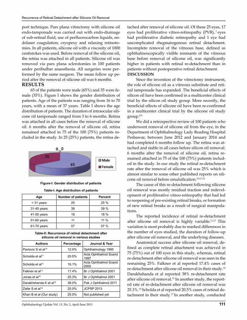
Recurrence of Retinal Detachment after Silicone Oil Removal
111Ophthalmology Update Vol. 13. No. 2, April-June 2015
port technique. Pars plana vitrectomy with silicone oil endo-tamponade was carried out with endo-drainage of sub-retinal fluid, use of perfluorocarbon liquids, en-dolaser coagulation, cryopexy and relaxing retinoto-mies. In all patients, silicone oil with a viscosity of 1000 centistokes was used. Before removal of the silicone oil, the retina was attached in all patients. Silicone oil was removed via pars plana sclerotomies in 100 patients under peribulbar anaesthesia. All surgeries were per-formed by the same surgeon. The mean follow up pe-riod after the removal of silicone oil was 6 months.RESULTS 65 of the patients were male (65%) and 35 were fe-male (35%). Figure I shows the gender distribution of patients. Age of the patients was ranging from 16 to 70 years, with a mean of 37 years. Table I shows the age distribution of patients. The duration of intraocular sili-cone oil tamponade ranged from 3 to 6 months. Retina was attached in all cases before the removal of silicone oil. 6 months after the removal of silicone oil, retina remained attached in 75 of the 100 (75%) patients in-cluded in the study. In 25 (25%) patients, the retina de-
tached after removal of silicone oil. Of these 25 eyes, 17 eyes had proliferative vitreo-retinopathy (PVR), 7 eyes had proliferative diabetic retinopathy and 1 eye had uncomplicated rhegmatogenous retinal detachment. Incomplete removal of the vitreous base, defined as ophthalmoscopically visible remnants of the vitreous base before removal of silicone oil, was significantly higher in patients with retinal re-detachment than in patients without postoperative retinal detachment.DISCUSSION Since the invention of the vitrectomy instrument, the role of silicone oil as a vitreous substitute and reti-nal tamponade has expanded. The beneficial effects of silicon oil have been confirmed in a multicenter clinical trial by the silicon oil study group. More recently, the beneficial effects of silicone oil have been re-confirmed in a multicenter clinical trial by the silicone oil study group.8,9
We did a retrospective review of 100 patients who underwent removal of silicone oil from the eye, in the Department of Ophthalmology Lady Reading Hospital Peshawar, between June 2012 and January 2014 and had completed 6 months follow up. The retina was at-tached and stable in all cases before silicon oil removal. 6 months after the removal of silicone oil, retina re-mained attached in 75 of the 100 (75%) patients includ-ed in the study. In our study the retinal re-detachment rate after the removal of silicone oil was 25% which is almost similar to some other published reports on sili-cone oil removal before emulsification.10,11,12 The cause of this re-detachment following silicone oil removal was mostly residual traction and redevel-opment of proliferative vitreo-retinopathy that had led to reopening of pre-existing retinal breaks, or formation of new retinal breaks as a result of surgical manipula-tions. The reported incidence of retinal re-detachment after silicone oil removal is highly variable.11,13 This variation is most probably due to marked differences in the number of eyes studied, the duration of follow-up after silicone oil removal, and the underlying diseases. Anatomical success after silicone oil removal, de-fined as complete retinal attachment was achieved in 75 (75%) out of 100 eyes in this study, whereas, retinal re-detachment after silicone oil removal was seen in the remaining 25%. Falkner et al reported 17.4% cases of re-detachment after silicone oil removal in their study.14 Darakhshanda et al reported 38% re-detachment rate after silicone oil removal.15 In another study, the report-ed rate of re-detachment after silicone oil removal was 25.3%.16 Scholda et al reported 20.5% cases of retinal de-tachment in their study.17 In another study, conducted
Figure-I: Gender distribution of patients
Age Number of patients Percent
< 31 years 25 25 %
31-40 years 39 39 %
41-50 years 18 18 %
51-60 years 11 11 %
61-70 years 07 07 %
Authors Percentage Journal & Year
Pavlovic S et al19 12.0% Ophthalmology 1995
Scholda et al17 20.5% Acta Ophthalmol Scand 1997
Scholda et al13 16.1% Acta Ophthalmol Scand 2000
Falkner et al14 17.4% Br J Ophthalmol 2001
Jonas et al16 25.3% Br J Ophthalmol 2001
Darakhshanda K et al15 38.0% Pak J Ophthalmol 2011
Zafar S et al18 20.0% JCPSP 2013
Khan B et al (Our study) 25.0% Not published yet
Table-II: Recurrence of retinal detachment after silicone oil removal in various studies
Table-I: Age distribution of patients
Recurrence of Retinal Detachment after Silicone Oil Removal
112 Ophthalmology Update Vol. 13. No. 2, April-June 2015
by Zafar S et al, the rate of re-detachment after silicone oil removal was 20%.18 Pavlovic et al commented that eyes with completely attached retinas, the risk of com-plication and re-detachment after silicone oil removal is relatively low.19
This was a retrospective study and the different risk factors for re-detachment after removal of silicone oil were not studied in detail due to lack of data. Fur-ther prospective studies with larger sample size and longer follow up needs to be done to identify the risk factors for re-detachment.CONCLUSION Re-detachment of the retina can occur after remov-al of silicone oil in eyes having stable attached retina after successful pars plana vitrectomy with silicone oil tamponade. Detail assessment of the patient is impor-tant to identify the eyes at risk of re-detachment.REFERENCES1. 1O’Lenick AJ. Basic silicone chemistry: a review [Internet].1999.
Available from: http://www.siliconespectator.com/articles/ Silicone_Spectator_January_2009.
2. Cibis PA, Becker B, Okun E, Canaan S. The use of liquid silicone in retinal detachment surgery. Arch Opthalmol 1962; 68:590-9.
3. Stone W Jr. Alloplasty in surgery of the eye. N Engl J Med 1958; 258:486-90.
4. McCuen BW, Landers MB, Machemer R. The use of silicon oil following failed vitrectomy for retinal detachment with ad-vanced proliferative vitreoretinopathy. Graefes Arch Clin Exp Ophthalmol 1986; 224(1);38-9.
5. Cians JD, Campbell WG. Vitrectomy techniques in the treat-ment of giant retinal tear: a flexible approach. Clin Exp Oph-thalmol 1988; 16:209-14.
6. Nagpal M, Videkar R, Mehrotra N. Hybrid technique for sili-cone oil removal. Retina Today 2011; 36-40.
7. Lucke K, Strobel B, Foerster M, Laqua H. Secondary glauco-ma after silicone oil surgery. Klin Monbl Augenheilkd 1990; 196:205-9.
8. Parrel JM. Silicone oil: physiochemical properties. In: Reti-na. Volume 3. Edited by Glaser BM, Michels RG. St. Louis: CV Mosby. 1989;261-77.
9. Silicone study group. Vitrectomy with silicone oil or sulfur hexafluoride gas in eyes with severe proliferative vitreoretin-opathy: Results of a randomized clinical trial- Silicone Study Report No 1. Arch Ophthalmol 1992; 110:770-9.
10. Kampik A, Hoing C, Heidenkummer HP. Problems and timing in the removal of silicone oil. Retina 1992; 12(3):S11-6.
11. Azen SP, Scott IU, Flynn HW Jr, Lai MY, Topping TM, Benati L, et al. Silicone oil in the repair of complex retinal detachments. A prospective observational multicentre study. Ophthalmol-ogy 1998; 105(9):1587-97.
12. Lewis H, Burke JM, Abrams GN, Aaberg TM. Perisilicone pro-liferation after vitrectomy for proliferative vitreoretinopathy. Ophthalmology 1988; 95:5583-91.
13. Scholda C, Egger S, Lakits A, Walch K, von Eckardstein E, Biowski R. Retinal detachment after silicone oil removal. Acta Ophthalmol Scand 2000; 78:182-6.
14. Falkner CI, Binder S, Kruger A. Outcome after silicone oil re-moval. Br J Ophthalmol 2001; 85:1324-7.
15. Darakhshanda K, Ghayoor I. Outcome of silicone oil removal in eyes undergoing 3-port pars plana vitrectomy. Pak J Oph-thalmol 2011; 27:17-20.
16. Jonas JB, Knorr HL, Rank RM, Budde WM. Retinal redetach-ment after removal of intraocular silicone oil tamponade. Br J Ophthalmol 2001; 85:1203-7.
17. Scholda C, Egger S, Lakits A, Haddad R. Silicone oil removal: results, risks and complications. Acta Ophthalmol Scand 1997; 75:695-9.
18. Zafar S, Bokhari SA, Kamil Z, Shakir M, Rizvi SF and Memon GM. Outcomes of Silicone Oil Removal. JCPSP 2013; 23(7):476-9.
19. Pavlovic S, Dick B, Schmidt KG, Tomic Z, Latinovic S. Long term outcome after silicone oil removal. Ophthalmology 1995; 92:672-6.
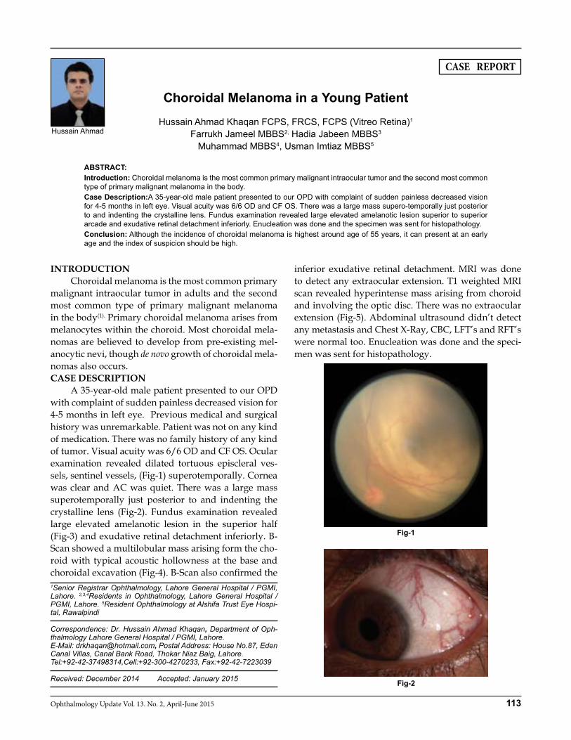
113Ophthalmology Update Vol. 13. No. 2, April-June 2015
CASE REPORT
Hussain Ahmad Hussain Ahmad Khaqan FCPS, FRCS, FCPS (Vitreo Retina)1
Farrukh Jameel MBBS2, Hadia Jabeen MBBS3 Muhammad MBBS4, Usman Imtiaz MBBS5
ABSTRACT:Introduction: Choroidal melanoma is the most common primary malignant intraocular tumor and the second most common type of primary malignant melanoma in the body.Case Description:A 35-year-old male patient presented to our OPD with complaint of sudden painless decreased vision for 4-5 months in left eye. Visual acuity was 6/6 OD and CF OS. There was a large mass supero-temporally just posterior to and indenting the crystalline lens. Fundus examination revealed large elevated amelanotic lesion superior to superior arcade and exudative retinal detachment inferiorly. Enucleation was done and the specimen was sent for histopathology. Conclusion: Although the incidence of choroidal melanoma is highest around age of 55 years, it can present at an early age and the index of suspicion should be high.
INTRODUCTION Choroidal melanoma is the most common primary malignant intraocular tumor in adults and the second most common type of primary malignant melanoma in the body(1). Primary choroidal melanoma arises from melanocytes within the choroid. Most choroidal mela-nomas are believed to develop from pre-existing mel-anocytic nevi, though de novo growth of choroidal mela-nomas also occurs. CASE DESCRIPTION A 35-year-old male patient presented to our OPD with complaint of sudden painless decreased vision for 4-5 months in left eye. Previous medical and surgical history was unremarkable. Patient was not on any kind of medication. There was no family history of any kind of tumor. Visual acuity was 6/6 OD and CF OS. Ocular examination revealed dilated tortuous episcleral ves-sels, sentinel vessels, (Fig-1) superotemporally. Cornea was clear and AC was quiet. There was a large mass superotemporally just posterior to and indenting the crystalline lens (Fig-2). Fundus examination revealed large elevated amelanotic lesion in the superior half (Fig-3) and exudative retinal detachment inferiorly. B-Scan showed a multilobular mass arising form the cho-roid with typical acoustic hollowness at the base and choroidal excavation (Fig-4). B-Scan also confirmed the
inferior exudative retinal detachment. MRI was done to detect any extraocular extension. T1 weighted MRI scan revealed hyperintense mass arising from choroid and involving the optic disc. There was no extraocular extension (Fig-5). Abdominal ultrasound didn’t detect any metastasis and Chest X-Ray, CBC, LFT’s and RFT’s were normal too. Enucleation was done and the speci-men was sent for histopathology.
1Senior Registrar Ophthalmology, Lahore General Hospital / PGMI, Lahore. 2,3,4Residents in Ophthalmology, Lahore General Hospital / PGMI, Lahore. 5Resident Ophthalmology at Alshifa Trust Eye Hospi-tal, Rawalpindi
Correspondence: Dr. Hussain Ahmad Khaqan, Department of Oph-thalmology Lahore General Hospital / PGMI, Lahore. E-Mail: [email protected], Postal Address: House No.87, Eden Canal Villas, Canal Bank Road, Thokar Niaz Baig, Lahore.Tel:+92-42-37498314,Cell:+92-300-4270233, Fax:+92-42-7223039
Received: December 2014 Accepted: January 2015
Choroidal Melanoma in a Young Patient
Fig-1
Fig-2
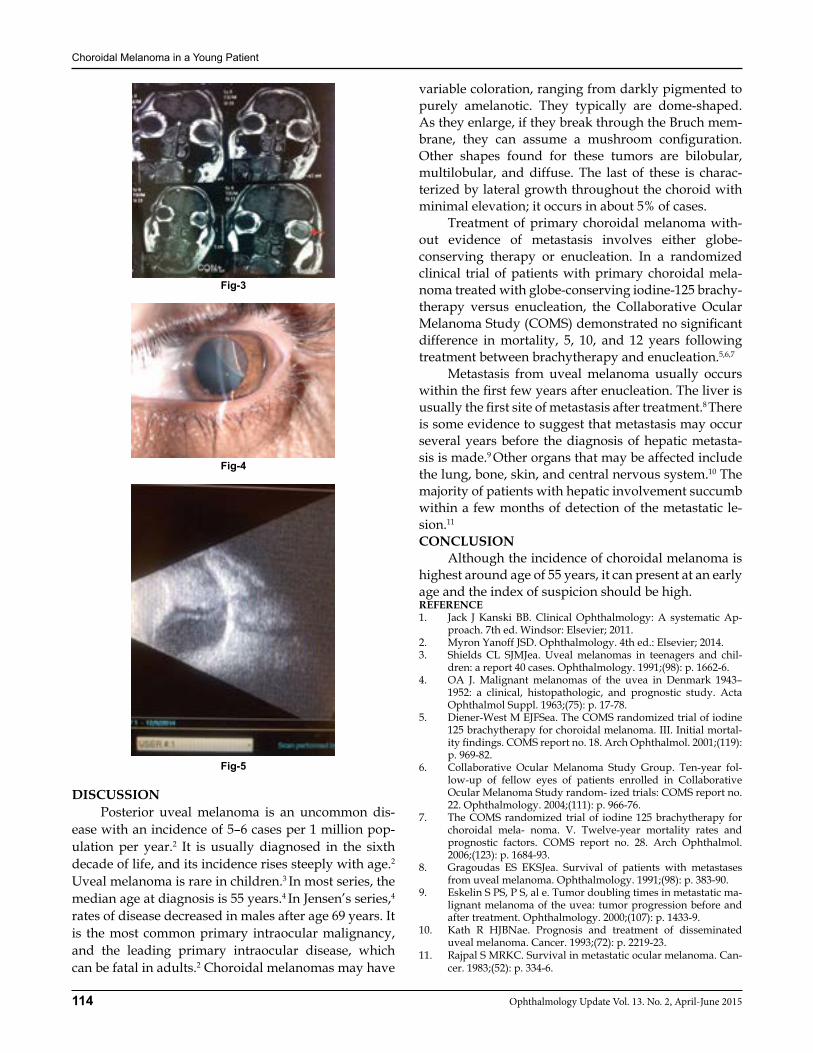
Choroidal Melanoma in a Young Patient
114 Ophthalmology Update Vol. 13. No. 2, April-June 2015
DISCUSSION Posterior uveal melanoma is an uncommon dis-ease with an incidence of 5–6 cases per 1 million pop-ulation per year.2 It is usually diagnosed in the sixth decade of life, and its incidence rises steeply with age.2 Uveal melanoma is rare in children.3 In most series, the median age at diagnosis is 55 years.4 In Jensen’s series,4 rates of disease decreased in males after age 69 years. It is the most common primary intraocular malignancy, and the leading primary intraocular disease, which can be fatal in adults.2 Choroidal melanomas may have
variable coloration, ranging from darkly pigmented to purely amelanotic. They typically are dome-shaped. As they enlarge, if they break through the Bruch mem-brane, they can assume a mushroom configuration. Other shapes found for these tumors are bilobular, multilobular, and diffuse. The last of these is charac-terized by lateral growth throughout the choroid with minimal elevation; it occurs in about 5% of cases. Treatment of primary choroidal melanoma with-out evidence of metastasis involves either globe-conserving therapy or enucleation. In a randomized clinical trial of patients with primary choroidal mela-noma treated with globe-conserving iodine-125 brachy-therapy versus enucleation, the Collaborative Ocular Melanoma Study (COMS) demonstrated no significant difference in mortality, 5, 10, and 12 years following treatment between brachytherapy and enucleation.5,6,7
Metastasis from uveal melanoma usually occurs within the first few years after enucleation. The liver is usually the first site of metastasis after treatment.8 There is some evidence to suggest that metastasis may occur several years before the diagnosis of hepatic metasta-sis is made.9 Other organs that may be affected include the lung, bone, skin, and central nervous system.10 The majority of patients with hepatic involvement succumb within a few months of detection of the metastatic le-sion.11
CONCLUSION Although the incidence of choroidal melanoma is highest around age of 55 years, it can present at an early age and the index of suspicion should be high.REFERENCE1. Jack J Kanski BB. Clinical Ophthalmology: A systematic Ap-
proach. 7th ed. Windsor: Elsevier; 2011.2. Myron Yanoff JSD. Ophthalmology. 4th ed.: Elsevier; 2014.3. Shields CL SJMJea. Uveal melanomas in teenagers and chil-
dren: a report 40 cases. Ophthalmology. 1991;(98): p. 1662-6.4. OA J. Malignant melanomas of the uvea in Denmark 1943–
1952: a clinical, histopathologic, and prognostic study. Acta Ophthalmol Suppl. 1963;(75): p. 17-78.
5. Diener-West M EJFSea. The COMS randomized trial of iodine 125 brachytherapy for choroidal melanoma. III. Initial mortal-ity findings. COMS report no. 18. Arch Ophthalmol. 2001;(119): p. 969-82.
6. Collaborative Ocular Melanoma Study Group. Ten-year fol-low-up of fellow eyes of patients enrolled in Collaborative Ocular Melanoma Study random- ized trials: COMS report no. 22. Ophthalmology. 2004;(111): p. 966-76.
7. The COMS randomized trial of iodine 125 brachytherapy for choroidal mela- noma. V. Twelve-year mortality rates and prognostic factors. COMS report no. 28. Arch Ophthalmol. 2006;(123): p. 1684-93.
8. Gragoudas ES EKSJea. Survival of patients with metastases from uveal melanoma. Ophthalmology. 1991;(98): p. 383-90.
9. Eskelin S PS, P S, al e. Tumor doubling times in metastatic ma-lignant melanoma of the uvea: tumor progression before and after treatment. Ophthalmology. 2000;(107): p. 1433-9.
10. Kath R HJBNae. Prognosis and treatment of disseminated uveal melanoma. Cancer. 1993;(72): p. 2219-23.
11. Rajpal S MRKC. Survival in metastatic ocular melanoma. Can-cer. 1983;(52): p. 334-6.
Fig-3
Fig-4
Fig-5

Frequency of High Glasgow Blatchford Score & its One Month Mortality in Patients presenting
115Ophthalmology Update Vol. 13. No. 2, April-June 2015
ORIGINAL ARTICLE
INTRODUCTION Upper gastrointestinal bleeding (UGIB) is defined as bleeding derived from a source proximal to the liga-ment of Treitz and Bleeding from the upper GI tract is approximately 4 times as common as bleeding from the lower GI tract.1 Acute gastrointestinal hemorrhage is a common medical emergency. The mortality of patients admitted to hospital for acute gastrointestinal bleeding is about 10%, rising to more than 30% in patients who bleed as inpatients.2 The incidence rates of UGIB dem-onstrate a large geo graphic variation ranging from 48 to 160 cases per 100,000 population, with consistent re-ports of higher incidences among men and elderly peo-ple.3,4 Patients with acute upper gastrointestinal (GI) bleeding commonly present with hematemesis and/or melena.5 The mortality has decreased only minimally
during the last 30 years, despite the introduction of en-doscopic therapy that reduces the rate of rebleeding.1
Causes of non variceal upper GI bleeding include peptic ulcer, Mallory-weiss tear, erosive gastritis/du-odenitis, esophagitis/esophageal ulcer, malignancy, angio-dysplasia/ vascular malformations and other causes having incidence of 20-50%, 15-20%, 10-15%, 5-10%, 1-2%, 5% and 5% respectively.6,7 Currently, OGD is the standard investigation of choice for UGIB. Endoscopic therapy has revolutionized the treatment of UGIB, with a recently greatly expanded therapeutic armamentarium.8 In the place where urgent esophago-gastro-duodenoscopy (EGD) is unavailable, empirical pharmacological therapy with proton pump inhibitors for non-variceal bleeding is recommended.9 Endoscopy within 24 hours is recommended for most patients with acute UGI bleeding.10, 11
The Blatchford score use only clinical and labora-tory data (before endoscopy) to identify patients who require intervention and predict future mortality. The Blatchford score includes hemoglobin level, blood urea level, pulse, systolic blood pressure, the presence of syncope or melena, urea, syncope and evidence of he-
Imran Yahaya1, Waheedullah FCPS (Gastro)2, Jawad MBBS3 Muhammad Daud MBBS4, Muhammad Iltaf (FCPS)5
ABSTRACTObjective: To determine the frequency of high Glasgow Blatchford scoring system and its one month mortality in patients presenting with non variceal upper gastrointestinal bleeding. Patients with liver cirrhosis may develop upper gastrointestinal hemorrhage from a variety of lesions, which include those that arise by virtue of portal hypertension, namely gastro-esoph-ageal varices and portal hypertensive gastropathy and other lesions seen in the general population.Study design: Descriptive case series. Duration: The duration of study was six months after approval of synopsis.Settings: Department of Gastroenterology and Hepatology Hayatabad Medical Complex Peshawar.Material & Methods: This study was conducted at Gastroenterology and Hepatology Department, Hayatabad Medical Complex, Peshawar. Duration of the study was six months in which a total of 140 at margin of error 5%, confidence interval 95% and 10%2 proportion of mortality among patients with high GB score at admission (non-variceal bleeding) using WHO sample size calculations. Results: In this study 3% patients were in age range 20-30 years, 18% patients were in age range 31-40 years, 34% patients were in age range 41-50 years, 35% patients were in age range 51-60 years,10% patients were above 60 years. Mean age was 30 years with SD ± 2.21. Fifty five percent patients were male and 45% patients were female. Twenty five percent patients had Glasgow Blatchford score < 12 and 75% patients had Glasgow Blatchford score more than 12. Mean Glasgow Blatchford score was 11 with SD ± 2.88. Among 140 patients mortality rate was 16%. Conclusion: In conclusion, GBS is a scoring system that allows calculation of the scores using only clinical and laboratory variables, without a need for endoscopy, and thereby, it can be easily used in the risk analysis of patients under emergency conditions. To support the results obtained from this study, future studies that contain more patients, are multi-centered, and that follow the patients after discharge from the ED are warranted.Keywords: Glasgow Blatchford Scoring System, mortality, non variceal upper gastrointestinal bleed.
Frequency of High Glasgow Blatchford Score & its One Month Mortality in Patients presenting
with Non-variceal Upper Gastrointestinal BleedingImran Yahaya
1,2,3,4 Medical Officers Gastro Unit 3Senior Registrar, Gastroenterology Unit, Hayatabad Medical Complex, Peshawar.
Correspondence: Dr. Waheed Ullah, FCPS., Medical Officer Gastro Unit, Hayatabad Medical Complex, Peshawar. District Specialist, DHQ Hospital,Village & P.O. Samarbagh, District lower Dir, KPK. Cell: 0307 5040633 E-Mail: [email protected]
Received: Jan’2015 Accepted: Feb’2015
GENERAL SECTION

Frequency of High Glasgow Blatchford Score & its One Month Mortality in Patients presenting
116 Ophthalmology Update Vol. 13. No. 2, April-June 2015
patic disease or cardiac failure and accurately identifies patients at low risk for clinical intervention like early endoscopy by using the Blatchford score.12,13,14,15 In one study, the frequency of High GBS was found to be 78-84%2,16 and its 28 day mortality was reported to be up to 10%.2,16
This study is carried out to determine frequency of high Glasgow Blatchford scoring system and its one month mortality in patients presenting with non-variceal UGIB so that we can categorize the patients in high risk and low risk. GBS scoring system has been traditionally used for UGIB bleed in terms of need of blood transfusion, intervention and mortality; it has very good value in categorizing the patients into low risk and high risk. The results of this study will be compared with already available international data as the literature suggested variable frequency of mortality of patients having high GBS scores, on the basis of results we can draw conclusion and recommendations for future re-search work into it and also the same results will be shared with other health professionals and guidelines and suggestions will be given for required changes in routine management of patients of UGIB. This will help our already compromised population with UGIB in re-ducing the burden of morbidity related to it.MATERIALS AND METHODSSettings: Department of Gastroenterology and Hepa-tology, Hayatabad Medical Complex, Peshawar.Study duration: Six monthsStudy Design: Descriptive case series.Sample Size: Total sample size was 140 while taking margin of error 5%, confidence interval 95% and 10%2 proportion of mortality among patients with high GB score at admission (non- variceal bleeding) using WHO sample size calculations.Sampling technique: Consecutive, non probability sampling. The above mentioned conditions act as con-founders and if included will introduce bias in the study results. SAMPLE SELECTIONInclusion criteria:• Age more than 15 years.• Upper gastrointestinal bleeding on initial assess-
ment.• Either gender.Exclusion criteria:• Acute myocardial infarction, trauma, stroke and
other conditions in association with upper gastro-intestinal bleed.
• Variceal bleeding on endoscopy.• Cause not identified on endoscopy.
• Developed bleeding during hospital stay for some other diagnosis.
RESULTS This study was conducted at Gastroenterology Department Hayatabad Medical Complex, Peshawar in which a total of 140 patients were observed to de-termine the high Glasgow Blatchford scoring system and its one month mortality in patients presenting with non variceal upper gastrointestinal bleeding and the results were analyzed as age distribution among 140 patients, was analyzed as 4(3%) patients were in age ranging 20-30 years, 25 (18%) patients were in age rang-ing 31-40 years, 48 (34%) patients were in age ranging 41-50 years, 49 (35%) patients were in age ranging 51-60 years,14 (10%) patients were above 60 years. Mean age was 30 years with SD ± 2.21 (as shown in Table No 1) Gender distribution among 140 patients was analyzed as 77(55%) patients were male and 63(45%) patients were female. (shown in Table No 2) Status of Glasgow Blatchford score among 140 patients was analyzed as 35(25%) patients had Glasgow Blatchford score < 12 and 105(75%) patients had Glasgow Blatchford score more than 12. Mean Glasgow Blatchford score was 11 with SD ± 2.88 (as shown in Table No 3) Status of mor-tality among 140 patients was analyzed as mortality rate was 22(16%) while 118(84%) patients were normal. (as shown in Table No 4)Stratification of Glasgow Blatchford score with age dis-tribution was analyzed as in 35cases of Glasgow Blatch-ford score <12, 9 patients were in age range 31-40 years, 11 patients were in age ranging 41-50 years, 12 patients were in age ranging 51-60 years and 3 patients were more than 60 years. Where as in 105cases of Glasgow Blatchford score> 12, 4 patients were in age ranging 20-30 years, 6 patients were in age ranging 31-40 years, 37 patients were in age ranging 41-50 years, 37 patients were in age ranging 51-60 years and 11 patients were more than 60 years. (as shown in Table No 5) Stratification of Glasgow Blatchford score with gender distribution was analyzed as in 35 cases of Glasgow Blatchford score <12, 20 patients were male and 15 patients were female. Where as in 105 cases of Glasgow Blatchford score> 12, 57 patients were male and 48 patients were female. (as shown in Table No 6) Stratification of mortality with age distribution was analyzed as out of 22 cases of mortality 10 patients died in age range 51-60 years and 12 patients died in age ranging more than 60 years. (as shown in Table No 7) Stratification of mortality with gender distribu-tion was analyzed as out of 22 cases of mortality 13 patients were male and 9 patients were female. (as shown in Table No 8)
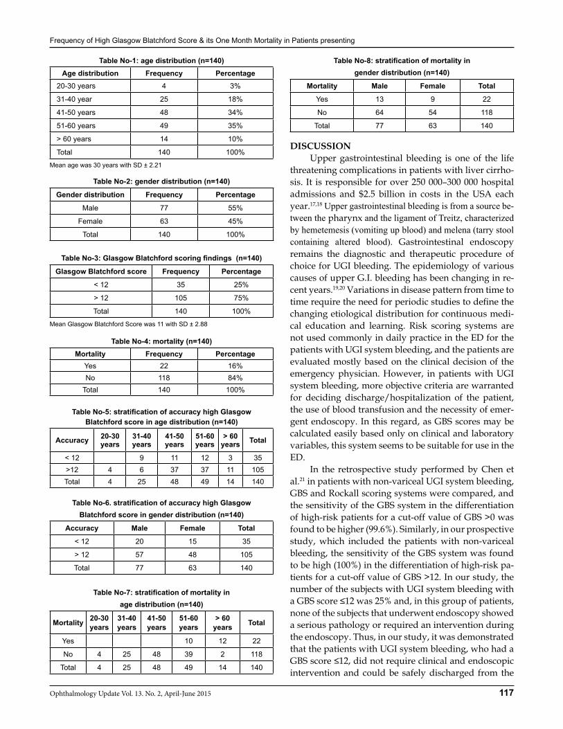
Frequency of High Glasgow Blatchford Score & its One Month Mortality in Patients presenting
117Ophthalmology Update Vol. 13. No. 2, April-June 2015
Table No-1: age distribution (n=140)Age distribution Frequency Percentage
20-30 years 4 3%
31-40 year 25 18%
41-50 years 48 34%
51-60 years 49 35%
> 60 years 14 10%
Total 140 100%
Mean age was 30 years with SD ± 2.21
Table No-2: gender distribution (n=140)
Gender distribution Frequency Percentage
Male 77 55%
Female 63 45%
Total 140 100%
Table No-3: Glasgow Blatchford scoring findings (n=140)
Glasgow Blatchford score Frequency Percentage
< 12 35 25%
> 12 105 75%
Total 140 100%
Mean Glasgow Blatchford Score was 11 with SD ± 2.88
Table No-4: mortality (n=140)Mortality Frequency Percentage
Yes 22 16%No 118 84%
Total 140 100%
Table No-5: stratification of accuracy high Glasgow Blatchford score in age distribution (n=140)
Accuracy 20-30 years
31-40 years
41-50 years
51-60 years
> 60 years Total
< 12 9 11 12 3 35>12 4 6 37 37 11 105Total 4 25 48 49 14 140
Table No-6. stratification of accuracy high Glasgow Blatchford score in gender distribution (n=140)
Accuracy Male Female Total
< 12 20 15 35
> 12 57 48 105
Total 77 63 140
Table No-7: stratification of mortality in age distribution (n=140)
Mortality 20-30 years
31-40 years
41-50 years
51-60 years
> 60 years Total
Yes 10 12 22
No 4 25 48 39 2 118
Total 4 25 48 49 14 140
Table No-8: stratification of mortality in gender distribution (n=140)
Mortality Male Female Total
Yes 13 9 22
No 64 54 118
Total 77 63 140
DISCUSSION Upper gastrointestinal bleeding is one of the life threatening complications in patients with liver cirrho-sis. It is responsible for over 250 000–300 000 hospital admissions and $2.5 billion in costs in the USA each year.17,18 Upper gastrointestinal bleeding is from a source be-tween the pharynx and the ligament of Treitz, characterized by hemetemesis (vomiting up blood) and melena (tarry stool containing altered blood). Gastrointestinal endoscopy remains the diagnostic and therapeutic procedure of choice for UGI bleeding. The epidemiology of various causes of upper G.I. bleeding has been changing in re-cent years.19,20 Variations in disease pattern from time to time require the need for periodic studies to define the changing etiological distribution for continuous medi-cal education and learning. Risk scoring systems are not used commonly in daily practice in the ED for the patients with UGI system bleeding, and the patients are evaluated mostly based on the clinical decision of the emergency physician. However, in patients with UGI system bleeding, more objective criteria are warranted for deciding discharge/hospitalization of the patient, the use of blood transfusion and the necessity of emer-gent endoscopy. In this regard, as GBS scores may be calculated easily based only on clinical and laboratory variables, this system seems to be suitable for use in the ED. In the retrospective study performed by Chen et al.21 in patients with non-variceal UGI system bleeding, GBS and Rockall scoring systems were compared, and the sensitivity of the GBS system in the differentiation of high-risk patients for a cut-off value of GBS >0 was found to be higher (99.6%). Similarly, in our prospective study, which included the patients with non-variceal bleeding, the sensitivity of the GBS system was found to be high (100%) in the differentiation of high-risk pa-tients for a cut-off value of GBS >12. In our study, the number of the subjects with UGI system bleeding with a GBS score ≤12 was 25% and, in this group of patients, none of the subjects that underwent endoscopy showed a serious pathology or required an intervention during the endoscopy. Thus, in our study, it was demonstrated that the patients with UGI system bleeding, who had a GBS score ≤12, did not require clinical and endoscopic intervention and could be safely discharged from the
Frequency of High Glasgow Blatchford Score & its One Month Mortality in Patients presenting
118 Ophthalmology Update Vol. 13. No. 2, April-June 2015
ED. While the retrospective study performed by Sriraj Askanthan et al.22 revealed a cut-off value of GBS ≤12 in the differentiation of low-risk patients among the pa-tients with UGI system bleeding, other studies23,24 used GBS=0 in the differentiation of the low-risk patients. An ideal scoring system should have both a good sensitivity and high specificity. However, in the stud-ies conducted, the sensitivity and specificity of the GBS system vary among high-risk patients with UGI system bleeding.25,26 In our study, the sensitivity and specificity were 100% and 1.41% for a cut-off value of GBS >12, 100% and 16.9% for a cut-off value of GBS >3, 96.63% and 36.62% for a cut-off value of GBS >5, and 86.52% and 69.01% for a cut-off value of GBS >8. In the study performed by Chen et al.27 positive predictive value (PPV) and negative predictive value (NPV) for GBS >0 were 75.2% and 96.4%, respectively. In the study conducted by Farooq et al.28 PPV and NPV were 37% and 100%, respectively for GBS >0 and 42% and 82% for GBS >5. In our study, PPV and NPV were 56% and 100% for GBS >0 and 65.6% and 89.7% for GBS >5, re-spectively. In our study the mortality rate was 16% and the those patients were more than 50 year of age similar results were found in other studies in which the mortal-ity rate was 11% and 13%,29,30
In the study performed by Hsu et al.29 in cirrhotic patients with UGI system bleeding, six prognostic fac-tors were determined: male gender, hepatocellular carcinoma, non-hepatocellular carcinoma, hypoxemia, serum bilirubin level, and PT. The investigators re-ported that these six clinical parameters might be easily obtained in the ED and be valuable in the early risk de-termination in cirrhotic patients with acute UGI system bleeding. For the model used in the aforementioned study, the sensitivity was 22.73% and the specificity was 99.8%. In our study, which did not enroll only cir-rhotic patients with UGI system bleeding, but instead all patients with UGI system bleeding and in whom the GBS system was used, the sensitivity and the specificity were 86.52% and 69.0%, respectively, for GBS>8, and thereby, GBS seemed more sensitive.31
Although in the literature, there has been no consen-sus on the best scoring system in various studies per-formed using the Rockall scoring system and/or the GBS system,19 the GBS system seems to be more use-ful, especially in patients with non-variceal UGI system bleeding. In our study, which included all the patients with non-variceal UGI system bleeding, we used the GBS system, and found it useful in the differentiation of high-risk patients. The limitations of this study include the single centered design, small number of patients, the fact that all the patients did not undergo an endos-
copy, and the enrollment of the patients to the follow-up based only on their conclusions in the ED.In light of the data obtained from this study, we can state that the patients with UGI system bleeding who have a GBS score ≤3 may be safely discharged from the ED and referred to the polyclinic to undergo an endoscopy. CONCLUSION In conclusion, GBS is a scoring system that allows calculation of the scores using only clinical and labo-ratory variables, without a need for endoscopy, and thereby, it can be easily used in the risk analysis of pa-tients under emergency conditions. To support the re-sults obtained from this study, future studies that con-tain more patients, are multi-centered, and that follow the patients after discharge from the ED are warranted.REFERENCES1. Mitchell S. Cappell, Friedel D. Initial management of acute up-
per gastrointestinal bleeding: from initial evaluation to gastro-intestinal endoscopy. Med Clin N Am 2008:491-509.
2. Ferguson CB, Mitchall MB. Non-variceal upper gastrointestinal bleeding. Ulster Med J 2006;75:32-9.
3. Manguso F, Riccio E, Bennato R, Picascia S, Fiorito R, Martino R et al. In-hospital mortality in non-variceal upper gastrointes-tinal bleeding Forrest I patients. Scandinavian J Gastroentol 2008;43:1432-41.
4. Thomas F, Imperiale, Jason A, Dominitz, Dawn T, Provenzale et al. Predicting poor outcome from acute upper gastrointesti-nal hemorrhage. Arch Inter Med 2007;167:1291-6.
5. Celinski K, Cichoz-Lach H, Madro A, M. Slomka M, Kasztelan-Szczerbinska B, T.Dworzanski T. Non-variceal upper gastroin-testinal bleeding guidelines on management. J Physiol Pharma-col 2008;59:215-29.
6. Van leerdam ME. Epidemiology of acute upper gastrointestinal bleeding. Best Pract Res Clin Gastrointerol 2008:22:209-24.
7. Bardou M. .Management of acute non-variceal upper gastroin-testinal bleed. Indian J Gastroenterol 2006;25:22-4.
8. Talafeeh A, Eweis S, Holy M. Endoscopic finding in upper gastrointestinal bleeding patients at Prince Hashim Hospital. J Rawal Med Sci 2004;1:71-3.
9. Adam T, Javed F, Khan S. Upper gastrointestinal bleeding: An etiological study of 552 cases. J Pak Inst Med Sci 2004;15:845-8.
10. Chaudhry AW, Tabassum HM, Chaudhry MA. Pattern of up-per gastrointestinal bleeding at Rahim Yar Khan. Ann King Ed-ward med Coll 2005;11:282-3.
11. Selinger CP, Ang YS. Gastric antral vascular ectesia (GAVE): an update on clinical presentation, pathophysiology and treat-ment. Digestion 2008;77:131-7.
12. Stojakov D, Velickovic D, Sabljak P, Bjelovic M, Ebrahimi K, Spica B et al. Diciulafoy’s lesion: rare cause of massive upper gastrointestinal bleeding. Acta Chirr Lugosol 2007;54:125-9.
13. John G Lee. What is the value of early endoscopy in upper gas-trointestinal bleeding? Nat Clin Pract Gastroenterol hepatol 2006;3:534-5.
14. Donner MW, Bosma JF, Robertson DL. Anatomy and physiolo-gy of the pharynx. Gastrointest Radiol 1985;10:196.
15. Ekberg O, Lindstrom C.The upper esophageal sphincter area. Acta Radiol 1987;28:173.
16. Boyce H. Endoscopic definitions of esophagogastric junction regional anatomy. Gastrointest Endosc 2000;51:586.
17. Kim T, Shijo H, Kokawa H. Risk factors for hemorrhage from gastric fundal varices. J Hepatol 1997;25:307-12
18. Toyonaga A, Iwao T. Portal hypertensive gastropathy. J Gas-troenterol Hepatol 1998;13:865–77.

Frequency of High Glasgow Blatchford Score & its One Month Mortality in Patients presenting
119Ophthalmology Update Vol. 13. No. 2, April-June 2015
19. Primignani M, Carpinelli L, Preatoni P. Natural history of por-tal hypertensive gastropathy in patients with liver cirrhosis.The new Italian endoscopic club for the study and treatment of esophageal varices (NIEC). J Gastroenterol 2000;119:181-7.
20. Beck PL, McKnight W, Lee SS. Prostaglandin modulation of the gastric vasculature and mucosal integrity in cirrhotic rats. Am J Physiol 1993;265:453-8.
21. Nasir N, Nadeem MA, Imran M, Hussain I, Chaudhry NU. Oe-sophageal varices Vs peptic ulcer: A Study of 100 Patients Pre-senting in Mayo Hospital with Upper Gastrointestinal Bleed-ing. Pak J Gastroenterol 1998; 2:0-.
22. Shahin WA, Abdel-Baset EZ, Nassar AK, Atta MM, Kabil SM, Murray JA, Low incidence of Helicobacter pylori infection in patients with duodenal ulcer and chronic liver disease. Scand J Gastroenterol. 2001;36:479-84.
23. Gravante G, Delogue D, Vanditti D. Upper and lower gastro-intestinal diseases in liver transplant candidates. Int J Colorectal Dis 2008;32:201-6
24. Srirajaskanthan R, Conn R, Bulwer C, Irving P. The Glasgow Blatchford scoring system enables accurate risk stratification of patients with upper gastrointestinal haemorrhage. Int J Clin Pract 2010;64:868-74.
25. Masaoka T, Suzuki H, Hori S. Blatchford scoring system is a
useful scoring system for detecting patients with upper gastro-intestinal bleeding who do not need endoscopic intervention. J Gastroenterol Hepatol 2007;22:1404-8.
26. Chen IC, Hung MS, Chiu TF. Risk scoring systems to predict need for clinical intervention for patients with nonvariceal upper gastrointestinal tract bleeding. Am J Emerg Med 2007; 25:774-9.
27. Farooq FT, Lee MH, Das A. Clinical triage decision vs risk scores in predicting the need for endotherapy in upper gastro-intestinal bleeding. Am J Emerg Med 2012;30:12934.
28. Hsu YC, Liou JM, Chung CS. Early risk stratification with sim-ple clinical parameters for cirrhotic patients with acute upper gastrointestinal bleeding. Am J Emerg Med 2010;28:884-90.
29. Gralnek IM, Dulai GS. Incremental value of upper endoscopy for triage of patients with acute non-variceal upper-GI bleed-ing. Gastrointest Endosc 2004;60:9-14.
30. Pang SH, Ching JY, Lau JY. Comparing the Blatchford and pre-endoscopic Rockall score in predicting the need for endoscopic therapy in patients with upper GI bleeding. Gastrointest En-dosc 2010;71:1134-40.
31. Tham TC, James C, Kelly M. Predicting outcome of acute non-variceal upper gastrointestinal haemorrhage without endos-copy using the Rockall Score. Postgrad Med J 2006;82:757-9.
Where all Ophthalmologists Join to:
DICOVER NEW IDEAS LATEST TRENDS & HOTTEST TOPICS
PARTICIPATE AT One Place:
where experts and thought leaders shape our profession
BE ENERGIZED & GET INSPIRED From: OPHTHALMOLGY UPDATE
120 Ophthalmology Update Vol. 13. No. 2, April-June 2015
Meatal Mobilization Technique for Childhood Hypospadias Repair, an Early
Experience at Lady Reading Hospital, Peshawar
Muhammad Ayub Khan, FCPS (Paeds Surgery)1
Muhammad Uzair FCPS (Paeds Surgery)2, Munir Ahmad FCPS3
Younas Khan FCPS (Paeds Surgery)4, Arshad Kamal FCPS (Paeds Surgery)5 Muhammad Fayaz MBBS6, Mussarat Hussain FCPS7, Asif Ahmad MBBS8
ABSTRACTObjective: To determine the effectiveness of meatal mobilization (MEMO) by distal urethral preparation as an improved surgical technique for distal hypospadias repair, including glanular, coronal and subcoronal location of the meatus, with or without chordee in children.Material and Methods: A total number of 60 patients with distal penile hypospadias with or without chordee were operated by using MEMO hypospadias repair technique from April 2013 to April 2014. After penile degloving, mobility of the meatus is evaluated and after urethral preparation the meatus is fixed to the tip of the glans. Glanuloplasty covers the neo-urethra providing a barrier layer. Shaft skin reconstruction completes the procedure Patients were evaluated regarding operative time, peri- or postoperative complications, hospital stay as well as functional and cosmetic outcome.Results: Mean duration of surgery was 43 minutes. There was no repair breakdown, new-onset chordee, or meatal steno-sis. Primary success rate was 95%. Three patients developed urethral fistula which responded well with short regimen of weekly meatal dilatation for 4 weeks. preputial edema occurred in 3 in non-circumcised patients. In one patient a mild ventral penile deviation without a need for correction was noted leaving a success rate of 96%.Conclusion: The MEMO-technique is a valid and reliable method for the correction of distal hypospadias.This method istechnically simple, less time consuming giving the best cosmetic results with least complications. Key Words: Hypospadias ,Meatal Mobilization, Urethral Reconstruction.
INTRODUCTION Distal Hypospadias accounts for 50-60% of all forms of hypospadias. with an increasing incidence be-ing present in 1 out of 125 male newborns Of those, 15% are glanular, 50% coronal, 30% subcoronal,and 5% are of the megameatus intact prepuce (MIP) variant.1 The goal of modern hypospadias repair is to achieve func-tionally as well as cosmetically normal looking glans, meatus and phallus.2 Generally, the surgical technique of choice is decided upon meatal location, the appear-ance of the meatus relative to the glans, the presence or absence of chordee, and the quality of the preputial hood. the meatal mobilization (MEMO) technique was developed to correct coronal and subcoronal hypospa-dias. According to the technique of the meatus mobi-lization, this operation Method referred to as MEMO, based on the work by Beck 1898.3 This approach com-
bines meatal mobilization by distal urethral dissection with steps of previously established techniques, e.g. a rotational flap for fashioning the Firlit preputial collar and straight-forward glanuloplasty.4,5 The aim of this study was to determine the short term results of memo hypospadias repair for distal penile hypospadias in children.Objective: To determine the effectiveness of meatal mo-bilization (MEMO) by distal urethral preparation as an improved surgical technique for distal hypospadias re-pair, including glanular, coronal and subcoronal loca-tion of the meatus, with or without chordee in children.MATERIALS AND METHODS This descriptive study was conducted in the pae-diatric surgery unit, Lady Reading Hospital, Pesha-war over a period of eight months from June 2009 to February 2010. Non probability sampling techniques was used. Children between 2 and 14 years, with dis-tal penile hypospadias with or without chordee were included and those who had proximal hypospadias i.e midshaft penoscrotal and perineal hypospadias were excluded from the study .All those patients ful-filling inclusion criteria of this study whose parents gave informed consent after explanation of the whole protocol, benefits versus risks of surgery were admit-ted through the outpatient department of the hospital. Each patient was thoroughly re-examined by by taking
ORIGINAL ARTICLE
Ayub Khan
1Associate Professor, Paeds Surgery, 2Medical Officer, Paeds Surgery unit Govt. PGMI LRH Peshawar. 3Senior Registrar Surgical C unit, Khyber Teaching Hospital, Peshawar. 4Assistant Professor, Paeds Surgery, 5Senior Registrar, Paeds Surgery, 6Junior Registrar, Paeds Surgery, 7Medical Officer, Paeds Surgery, 8Resident Paeds surgery, Postgraduate Medical Institute, Lady Reading Hospital Peshawar-Pakistan.
Correspondence: Dr. Muhammad Ayub Khan, FCPS(Paeds Surgery) Associate Professor, Paeds Surgery, PGMI LRH Peshawar. Cell: 0300-5979070, E-mail: [email protected]
Received: January 2015 Accepted February 2015
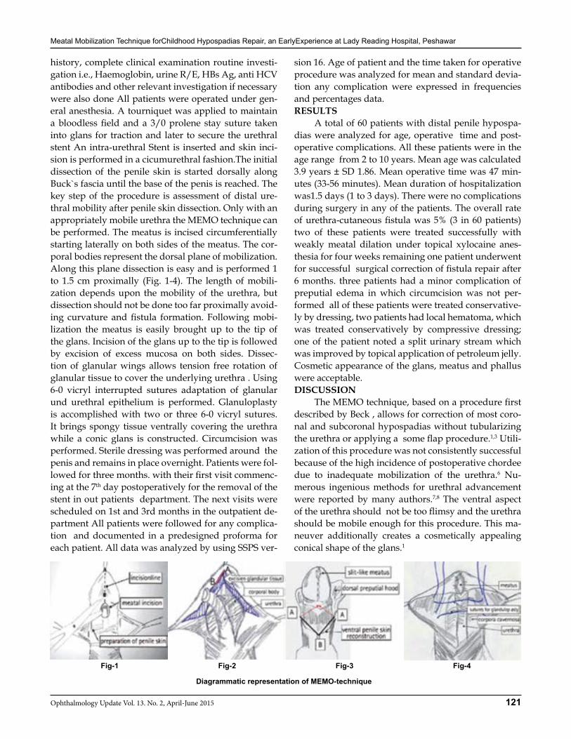
Meatal Mobilization Technique forChildhood Hypospadias Repair, an EarlyExperience at Lady Reading Hospital, Peshawar
121Ophthalmology Update Vol. 13. No. 2, April-June 2015
history, complete clinical examination routine investi-gation i.e., Haemoglobin, urine R/E, HBs Ag, anti HCV antibodies and other relevant investigation if necessary were also done All patients were operated under gen-eral anesthesia. A tourniquet was applied to maintain a bloodless field and a 3/0 prolene stay suture taken into glans for traction and later to secure the urethral stent An intra-urethral Stent is inserted and skin inci-sion is performed in a cicumurethral fashion.The initial dissection of the penile skin is started dorsally along Buck`s fascia until the base of the penis is reached. The key step of the procedure is assessment of distal ure-thral mobility after penile skin dissection. Only with an appropriately mobile urethra the MEMO technique can be performed. The meatus is incised circumferentially starting laterally on both sides of the meatus. The cor-poral bodies represent the dorsal plane of mobilization. Along this plane dissection is easy and is performed 1 to 1.5 cm proximally (Fig. 1-4). The length of mobili-zation depends upon the mobility of the urethra, but dissection should not be done too far proximally avoid-ing curvature and fistula formation. Following mobi-lization the meatus is easily brought up to the tip of the glans. Incision of the glans up to the tip is followed by excision of excess mucosa on both sides. Dissec-tion of glanular wings allows tension free rotation of glanular tissue to cover the underlying urethra . Using 6-0 vicryl interrupted sutures adaptation of glanular und urethral epithelium is performed. Glanuloplasty is accomplished with two or three 6-0 vicryl sutures. It brings spongy tissue ventrally covering the urethra while a conic glans is constructed. Circumcision was performed. Sterile dressing was performed around the penis and remains in place overnight. Patients were fol-lowed for three months. with their first visit commenc-ing at the 7th day postoperatively for the removal of the stent in out patients department. The next visits were scheduled on 1st and 3rd months in the outpatient de-partment All patients were followed for any complica-tion and documented in a predesigned proforma for each patient. All data was analyzed by using SSPS ver-
sion 16. Age of patient and the time taken for operative procedure was analyzed for mean and standard devia-tion any complication were expressed in frequencies and percentages data.RESULTS A total of 60 patients with distal penile hypospa-dias were analyzed for age, operative time and post-operative complications. All these patients were in the age range from 2 to 10 years. Mean age was calculated 3.9 years ± SD 1.86. Mean operative time was 47 min-utes (33-56 minutes). Mean duration of hospitalization was1.5 days (1 to 3 days). There were no complications during surgery in any of the patients. The overall rate of urethra-cutaneous fistula was 5% (3 in 60 patients) two of these patients were treated successfully with weakly meatal dilation under topical xylocaine anes-thesia for four weeks remaining one patient underwent for successful surgical correction of fistula repair after 6 months. three patients had a minor complication of preputial edema in which circumcision was not per-formed all of these patients were treated conservative-ly by dressing, two patients had local hematoma, which was treated conservatively by compressive dressing; one of the patient noted a split urinary stream which was improved by topical application of petroleum jelly.Cosmetic appearance of the glans, meatus and phallus were acceptable.DISCUSSION The MEMO technique, based on a procedure first described by Beck , allows for correction of most coro-nal and subcoronal hypospadias without tubularizing the urethra or applying a some flap procedure.1,3 Utili-zation of this procedure was not consistently successful because of the high incidence of postoperative chordee due to inadequate mobilization of the urethra.6 Nu-merous ingenious methods for urethral advancement were reported by many authors.7,8 The ventral aspect of the urethra should not be too flimsy and the urethra should be mobile enough for this procedure. This ma-neuver additionally creates a cosmetically appealing conical shape of the glans.1
Fig-1 Fig-2 Fig-3 Fig-4
Diagrammatic representation of MEMO-technique
Meatal Mobilization Technique forChildhood Hypospadias Repair, an EarlyExperience at Lady Reading Hospital, Peshawar
122 Ophthalmology Update Vol. 13. No. 2, April-June 2015
The fact that more than 300 different operations are described in the literature reflects the wide spec-trum of the anomaly, and proves that the treatment has not been perfected.6 In the presented study, 60 boys underwent surgery using the MEMO technique. No urethral stricture noted in all operated patients, three patients developed urethrocutaneuous fistula, two of these patients responded well with weakly meatal dila-tation under topical anesthesia and hence not requir-ing any major surgical correction, the remaining one patient was subjected to successful operative repair of fistula after failure of conservative management after 6 months of initial repair. Overall rate of urethra-cutaneous fistula post MEMO repair was 5% in our series compared to Sei-bold etal who reported a fistula rate of 1% .1 This high rate of urethra-cutaneous fistula post MEMO repair in our series compared to Western studies might be the early learning curve of authors ,as a thorough literature search was made to compare our results with the local studies.No satisfactory local studies were available to compare our results with local results.CONCLUSION Over 80 percent of boys with this condition have
distal hypospadias, and the successful repair of distal hypospadias can be easilly achieved by meatal mobili-zation technique which is a single stage procedure hav-ing low complication rate,good cosmetic results and is technically simple to learn.REFRENCES1. Seibold J, Amend B, Saladin , Alloussi H S, Colleselli D,
Tode hoefer T, Gakis G, Merseburger A, Sievert DK, Stenzl A, Schwentner C. Meatal mobilization (MEMO) technique for distal hypospadias repair: Technique, results and long-term follow-up. CEJU March 2010; 63 :125-28.
2. Uzair M, Ahmad M, Hussain M, Younus M, Khan K. Frequen-cy of urethrocutaneous fistula following snodgrass hypospa-dias repair in children. JPMI 2013; 27(1):74-77.
3. Beck C. A new operation for balanic hypospadias. NY Med J jan 1898; 29: 147-148.
4. Seibold J, Boehmer A, Verger A et al. The meatal mobilization technique for coronal/subcoronal hypospadias repair. BJU Int 2007; 100: 164-167.
5. Redman JF. A favorable experience with rotational flap tech-niques for fashioning the Firlit preputial collar. J Urol 2006; 176: 715-717.
6. Elemen L, Tugay M. Limited Urethral Mobilization Technique in Distal Hypospadias Repair with Satisfactory Results, Balkan Med J 2012; 29: 21-5
7. Hamdy H, Awadhi MA, Rasromani KH. Urethral mobilization andmeatal advancement: a surgical principle in hypospadias repair.Pediatr Surg Int 1999;15:240-2.
8. Atala A. Urethral mobilization and advancement for midshaft todistal hypospadias. J Urol 2002;168:1738-41.
Ophthalmological Society of Pakistan, KPK Branch is holding the next Ophthalmic Symposium at
Nathiagali, KPKFrom 7-9 August 2015
Please contact:Dr. Mir Ali Shah, Associate Professor
Department of Ophthalmology, Lady Reading Hospital, Peshawar.Cell : 03005948091, Email: [email protected]
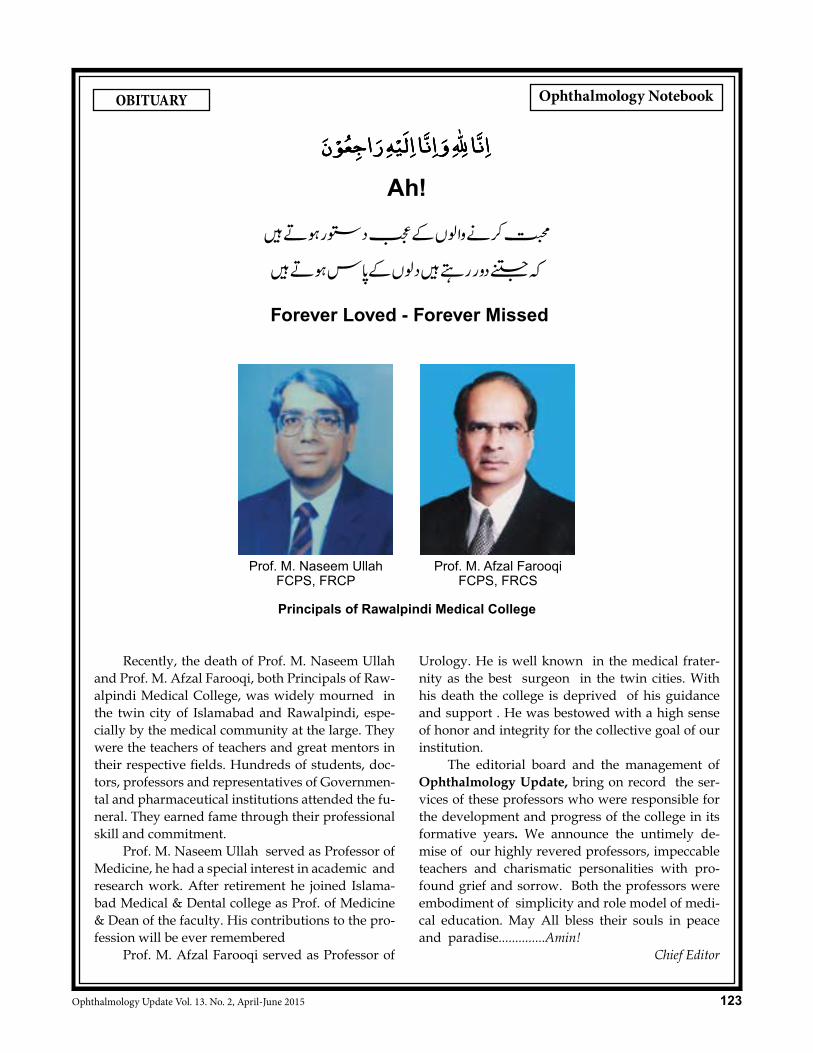
123Ophthalmology Update Vol. 13. No. 2, April-June 2015
Recently, the death of Prof. M. Naseem Ullah and Prof. M. Afzal Farooqi, both Principals of Raw-alpindi Medical College, was widely mourned in the twin city of Islamabad and Rawalpindi, espe-cially by the medical community at the large. They were the teachers of teachers and great mentors in their respective fields. Hundreds of students, doc-tors, professors and representatives of Governmen-tal and pharmaceutical institutions attended the fu-neral. They earned fame through their professional skill and commitment. Prof. M. Naseem Ullah served as Professor of Medicine, he had a special interest in academic and research work. After retirement he joined Islama-bad Medical & Dental college as Prof. of Medicine & Dean of the faculty. His contributions to the pro-fession will be ever remembered Prof. M. Afzal Farooqi served as Professor of
Urology. He is well known in the medical frater-nity as the best surgeon in the twin cities. With his death the college is deprived of his guidance and support . He was bestowed with a high sense of honor and integrity for the collective goal of our institution. The editorial board and the management of Ophthalmology Update, bring on record the ser-vices of these professors who were responsible for the development and progress of the college in its formative years. We announce the untimely de-mise of our highly revered professors, impeccable teachers and charismatic personalities with pro-found grief and sorrow. Both the professors were embodiment of simplicity and role model of medi-cal education. May All bless their souls in peace and paradise..............Amin!
Chief Editor
OBITUARY
Ah!
Forever Loved - Forever Missed
Prof. M. Afzal FarooqiFCPS, FRCS
Prof. M. Naseem UllahFCPS, FRCP
Principals of Rawalpindi Medical College
ن لايراجعوء ه اليء واهنا هللا ناهنا لايراجعوء ه اليء واهنا هللا اهنا
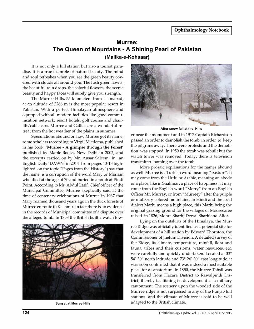
124 Ophthalmology Update Vol. 13. No. 2, April-June 2015
It is not only a hill station but also a tourist para-dise. It is a true example of natural beauty. The mind and soul refreshes when you see the green beauty cov-ered with clouds all around you. The lush green lawns, the beautiful rain drops, the colorful flowers, the scenic beauty and happy faces will surely give you strength. The Murree Hills, 55 kilometers from Islamabad, at an altitude of 2286 m is the most popular resort in Pakistan. With a perfect Himalayan atmosphere and equipped with all modern facilities like good commu-nication network, resort hotels, golf course and chair-lift/cable cars. Murree and Gallies are a wonderful re-treat from the hot weather of the plains in summer. Speculations abound on how Murree got its name, some scholars (according to Virgil Miedema, published in his book: ‘Murree - A glimpse through the Forest’ published by Maple-Books, New Deihi in 2002, and the excerpts carried on by Mr. Ansar Saleem in an English Daily ‘DAWN’ in 2014 from pages 13-18 high-lighted on the topic “Pages from the History”) say that the name is a corruption of the word Mary or Mariam who died at the age of 70 and buried in a tomb at Pindi Point. According to Mr. Abdul Latif, Chief officer of the Municipal Committee, Murree skeptically said at the time of centenary celebrations of Murree in 1967 that Mary roamed thousand years ago in the thick forests of Murree en route to Kashmir. In fact there is an evidence in the records of Municipal committee of a dispute over the alleged tomb. In 1858 the British built a watch tow-
er near the monument and in 1917 Captain Richardson passed an order to demolish the tomb in order to keep the pilgrims away. There were protests and the demoli-tion was stopped. In 1950 the tomb was rebuilt but the watch tower was removed. Today, there is television transmitter looming over the tomb. More prosaic explanations for the names abound as well. Murree is a Turkish word meaning “pasture”. It may come from the Urdu or Arabic, meaning an abode or a place, like in Shalimar, a place of happiness, it may come from the English word “Merry” from an English Officer Mr. Murray, or from “Murreey” after the purple or mulberry-colored mountains. In Hindi and the local dialect Marhi means a high place, this Marhi being the original grazing ground for the villages of Mooseearee raised in 1826, Mohra Sharif, Dewal Sharif and Aliot. Lying on the outskirts of the Himalaya, the Mur-ree Ridge was officially identified as a potential site for development of a hill station by Edward Thornton, the Commissioner of Jhelum Division. A detailed survey of the Ridge, its climate, temperature, rainfall, flora and fauna, tribes and their customs, water resources, etc. were carefully and quickly undertaken. Located at 33° 54’ 30” north latitude and 73° 26’ 30” east longitude. it was soon confirmed that it was indeed a most suitable place for a sanatorium. In 1850, the Murree Tahsil was transferred from Hazara District to Rawalpindi Dis-trict, thereby facilitating its development as a military cantonment. The scenery upon the wooded side of the Murree ridge is not surpassed in any of the Punjab hill stations and the climate of Murree is said to be well adapted to the British climate.Sunset at Murree Hills
After snow fall at the Hills
Murree: The Queen of Mountains - A Shining Pearl of Pakistan
(Malika-e-Kohsaar)

Murree: The Queen of Mountains - A Shining Pearl of Pakistan
125Ophthalmology Update Vol. 13. No. 2, April-June 2015
In March of 1849, the British decided to establish Murree as a sanatorium. In 1851, Murree was selected as the summer headquarters of Punjab. The Mall was established in 1850, is the centre of shopping area, where most people congregate. Good buys in Murree are Kashmiri shawls, furs, walking sticks, fruits and nuts. Murree’s pistachio nuts are reputed to be the best in Pakistan. The Imperial summer capital was popularized by Rudyard Kipling- a noble laureate from Lahore and Chief Editor of an English daily “Civil & Military Ga-zette an English Daily” being published from Lahore. Dane Kennedy commented: ‘Located on peaks that loom like sentinels over heat-shimmering plains. Mur-ree remains among the most curious monuments to the British colonial presence in the area. The Saumly Sana-torium was primarily established for the European in-valids, but hill stations soon assumed an importance that far exceeded the therapeutic attraction. To these cloud-enshrouded sanctuaries the British expatriate elite but also to familiarize with the alien culture with the locals living in areas like Aliot, a village 2 kilom-eter down the Bhurban. Here they established political headquarters and military cantonments and centers where from they issued executive orders. In 1845 Murree was brought under the control of East India Company rule and after the Treaty of Lahore (March 9, 1846), the first resident in the Punjab was Henry Lawrence, later founder of the famous Lawrence schools in Ghora Gali (Murree), Abbotabad & Law-rence gardens in Lahore came to Murree. The “Rules
for the Administration of Murree Town” were framed in 1851, which allowed for a Murree Sanatorium Com-mittee to be established at Saumly. A total reduction in land revenue of Rs.114.40 and cash compensation of Rs.1,935 was proposed for Ilyot and Mooseeareeareas and adjoining areas. This was approved by the Chief Commissioner of the Punjab. A special grant was pro-posed in the form of a cash lease payment of Rs.50 per annum in perpetuity. Upon the conclusion of protracted negotiations with the villagers, the proposal went to Calcutta for final approval. Finally, this annual payment was approved by the Governor-General, Lord Dalhou-sie, by an order dated November 23, 1855. The story goes that the original bargain with the people of that area was actually for Rs.60 per annum as lease payment, instead of Rs.50. Since the Maim Sahiba (Lord Dalhousie’s wife) being a British national, was poorly attired according to the Muslim customs of the area, the villagers offered Rs. 10/- for the purchase of Shalwar, Kameez and a shawl for the Maim Sahiba to dress her properly. By 1850, more than 50 bungalows had been con-structed towards Kashmir Point, Pindi Point and along Kuldannah Road. In 1851, troops were first quartered in Murree, and permanent barracks were erected in 1853. Holy Trinity Church on the Mall (Jinnah Road) was opened on May 17, 1857., and the first Sunday Mass service was served.REFERENCES:1. Mr. Salim Ansar ‘Pages from the history’, The News, Lahore. 2. Mr. Arshad Mahmood Abbasi, former: Nazim of Union Coun-
cil, Murree., 3. Murree — A Glimpse Through the Forest by Virgil Miedema
126 Ophthalmology Update Vol. 13. No. 2, April-June 2015
All materials submitted for publication should be sent to the journal ‘Ophthalmology Update’. Articles/research papers which have already been published or accepted elsewhere for publication should not be submitted. A paper that has been presented at a scientific meeting, if not published in full in proceeding or similar publication may be submitted. Press reports of meetings will not be considered as breach of this rule. Ethical Aspects: If articles, tables, illustrations or photographs, which have already been published, are included, a letter of permission for republication (or its excerpts) should be obtained from the author(s) as well as the editor of the journal where it was previously published. Material for Publication: The material submitted for publication may be in the form of original research, a review article, short communications, a case report, recent advances, new techniques, review on clinical/medical/ophthalmic education, a letter to the editor, medical quiz, Ophthalmic highlights/update, news and views related to the field of medical sciences. Editorials are written by invitation. Report on Ophthalmic obituaries should be concise. Author should keep one copy of the manuscript for reference, and send three copies (laser or inkjet) to the Managing Editor, Ophthalmology Update through E-mail/CD or by post in MS word. Photocopies are not accepted. Any illustrations or photographs should also be sent in duplicate. Authors from outside Pakistan can also e-mail their manuscript. It should include a title page, E-mail address, fax and phone numbers of the corresponding author. There should be no more than 40 references in an original/review article. If prepared on computer, a CD should be sent with the manuscript. Dissertation/Thesis Based Article: An article based on dissertation submitted as part of the requirement for a Fellowship can be sent for publication after it has been approved by the relevant institution. Dissertation based article should be re-written in accordance with the instructions to authors. References: References should be numbered in the order in which they are called in the text. At the end of the article, the full list of references should give the names and initials of all authors in Vancouver style based on the format used by the NLM in Index Medicus. It verify the references against the original documents before submitting the article. Peer Review: Every paper will be read by at least two staff editors of the editorial board. The paper selected will then be sent to one or more external viewers.
Abstract: Abstract of original article should be in structured format with the following sub-headings:Objective, Design, Place and duration of Study, Patients & Methods, Result and Conclusion. Introduction: This should include the purpose of the article. The rationale for the study or observation should be summarized. Methods: Study design and sampling methods should be mentioned. The selection of the observational or experimental subjects (patients or experimental animals, including controls) should be described clearly. The methods and the apparatus used should be identified and procedures described in sufficient details to allow other workers to reproduce the results and references to established methods. All drugs and chemicals used should be identified precisely, including generic names, doses, routes of administration. Results: These should be presented in a logical sequence in the text, tables and illustrations. Only important observations should be emphasized or summarized. Discussion: The author’s comments on the result, supported with contemporary references, including arguments and analysis of identical work done by others. Brief acknowledgement may be made at the end. Conclusion: Conclusion should be provided under separate heading and highlighting new aspects arising from the study. It should be in accordance with the study. Copyright: Material printed in this journal is the copyright of the publisher of Ophthalmic Newsnet/Ophthalmology Update and may not be reproduced without the permission of the editor/publisher. The publisher only accepts the original material for publication with the understanding that except for abstracts, no part of the data has yet been published or will be submitted for publication elsewhere before appearing in the journal. The Editorial Board makes every effort to ensure the accuracy and authenticity of the material printed in the journal. However, conclusions and statements expressed are the views of the authors and do not necessarily reflect the opinions of the Editorial Board. Publishing of advertising material does not imply an endorsement by the Ophthlmic Newsnet /Ophthalmology Update. Address for Correspondence: The Chief Editor, Ophthalmology Update, 267-A, St: 53, F-10/4, Islamabad, Pakistan. E-mail: [email protected]
Instructions to the Authors
Instructions to the authors